PROTOZOO- LOGICA
126
PL ISSN 0065-1583 POLISH ACADEMY OF SCIENCES NENCKI INSTITUTE OF EXPERIMENTAL BIOLOGY PROTOZOO- LOGICA VOLUME 25 Number 4 P A Ń S T W O W E W Y D A W N I C T W O N A U K O W E W A R S Z A W A 1 9 8 6 W R O C Ł A W http://rcin.org.pl
-
Upload
khangminh22 -
Category
Documents
-
view
0 -
download
0
Transcript of PROTOZOO- LOGICA
PL ISSN 0065-1583
P O L I S H A C A D E M Y O F S C I E N C E S
N E N C K I I N S T I T U T E O F E X P E R I M E N T A L B I O L O G Y
PROTOZOO-LOGICA
VOLUME 25 Number 4
P A Ń S T W O W E W Y D A W N I C T W O N A U K O W E
W A R S Z A W A 1 9 8 6 W R O C Ł A W
http://rcin.org.pl

P O L I S H A C A D E M Y O F S C I E N C E S
N E N C K I I N S T I T U T E O F E X P E R I M E N T A L B I O L O G Y
ACTA PROTOZOOLOGICA International Journal of Protozoology
Editors
Stanisław DRYL and Stanisław L. KAZUBSKI
Editorial Board
Chairman: Leszek KUŹNICKI
Vice-chairman: Andrzej GRĘBECKI
Stanisław DRYL Vassil GOLEMANSKY Witold KASPRZAK Stanisław L. KAZUBSKI
Managing Editor
Members
Jiri LOM
Georg Ivanovic POLJANSKY Igor Borysovic RAIKOV Ksenia Mironovna SUKHANOVA
and Editorial Board Secretary
litta PLOSZAJ
Manuscripts may be submitted to the Editorial Office: Acta Protozoologica, M. Nencki Insti-tute of Experimental Biology, 02-093 Warszawa, 3 Pasteur Street, Poland, or to each member of the Editorial Board.
Subscription orders for all the magazines published in Poland available through the local press distributors or directly through the
Foreign Trade Enterprise
ARS POLONA
00-068 Warszawa, Krakowskie Przedmieście 7, Poland
Our bankers: BANK HANDLOWY WARSZAWA S.A.
ACTA PROTOZOOLOGICA appears quarterly. The indexes of the previous volume appear in No. 1 of the next volume.
Indexed in Current Contents and in Protozoological Abstracts.
http://rcin.org.pl

ACTA PROTOZOOLOGICA Vol. 25, No. 4, pp. 365-374 (1986)
Seasonal Modifications in the Life Cycle of Parastasia fennica (Michajlow, 1966)
I r e n a W I T A and K s e n i a M. S U K H A N O V A
W. Stefanski Institute of Parasitology, Polish Academy of Sciences, 3 Pasteur Str., P.O. Box 153, 00-973 Warsaw, Poland and A. I. Hercen State Pedagogical Institute, Leningrad, USSR
Received on 15 April 1986
Synopsis. Annual life cycle of endoparasitic euglenid flagellate Parastasia fennica (Michajlow, 1966) has been studied with the use of morphometric, morphological and histochemical methods. Spontaneously infected copepods were collected from small ponds in the environs of Leningrad. Some modifications in the life cycle of P. fennica in various seasons of the year were observed as well as morphological variation and storage of great amount of paramylon in cell cytoplasma during late autumn. It has been also found that low temperature of water in natural reservoirs during late autumn and winter inhibits palintomic division and formation of infec-tive flagellate cells. In spring the reproduction of P. fennica is the most intensive, leading to increase of parasite populations. Seasonal modifications in P. fennica life cycle are considered as adaptative features, they ought to be taken into account when other species of the genus Parastasia Michajlow, 1972 are determined and described.
The life cycle of Parastasia fennica (Michajlow, 1966, 1972), an en-doparasitic euglenid flagellate inhabiting alimentary tract of fresh water Copepoda, has been already studied in detail in natural as well as in laboratory conditions ( M i c h a j l o w 1972, 1978, W i t a and S u k h a n o v a 1983). It has been found that the complex life cycle of this flagellate includes three obligatory alternating phases: (1) parasitic phase — trophozoite living in host intestine, (2) reproductive phase — mature trophozoites undergo palintomic division in outer environment after leaving host intestine, (3) infective phase — flagellate cells formed as a result of palintomic division, live in water up to ingestion by copepode host. The life cycle, from the moment of infection of a copepode by flagellate cells up to the death of flagellates in the outer environment, when contact with copepodes is failing, lasts 12-15 days in spring, summer and early autumn, i.e., in warmer seasons of the year. In reser-voirs in the north-west of USSR the life cycle of P. fennica may be repeated several times during the year.
Modifications of P. fennica life cycle in late autumn, winter and early spring
http://rcin.org.pl

366 I. WITA A N D K. M. SUKHANOVA
have not been known till now and no information about seasonality of this species were recorded. So the aim of the present study was to investigate the life cycle of P. fennica during the whole year with special attention paid to the stages occurring in nature in cool seasons of the year.
M a t e r i a l a n d M e t h o d s
Copepode hosts of P. fennica — Cyclopidae, were collected from small ponds in the environs of Leningrad. Samples of the material were collected in all seasons during 1980-1984. The material was used for investigation on morphology and trophozoite physiology as well as for observation of palintomic division and formation of infective flagellate individuals.
In laboratory conditions the cultures of copepods infected with Parastasia were maintained the whole year round according to the method described by M i c h a j t o w (1966, 1972). They were kept at room temperature and at 1-4°C.
Except living Parastasia the investigation was carried on the material fixed in 4% and 10% neutral formaldehyde, Carnoy's fluid and 100% methanol. Flagellate morphology was described on the base of preparations stained with Mayer's acid hemalaun, hematoxylin after Carazzi and azur-eosin after Giemsa-Romanovsky. Lysosomes were revealed due to vital staining with 0.1 % neutral red in aqueous solution (Bulychev et al. 1978).
Aggregations of paramylon in trophozoite cytoplasm were observed in living Parastasia as well as in those fixed in 4% formaldehyde without further staining, because paramylon does not stain with any dyes for polysaccharides. Glycogen type polysaccharides were revealed with Lu-gol's iodine and PAS reaction. Acid mucopolysaccharides were stained after S t e e d m a n (Pearse 1968), also 0.1% toluidine blue in 30% and 1% ethanol was used. Mucopolysaccharides on cell surface were intravitally stained with 0.01% aqueous solution of accian blue ( K r y l e n k o v et al. 1979).
Neutral lipids were revealed due to staining with saturated solution of Sudan red III in 70% ethanol and in fettrot (Pea r se 1968, Beyer et al. 1977), a dye specific for these biopolymeres. For phospholipids detection saturated solution of Sudan black B in 70% ethanol was used.
All drawings were made with the aid of camera lucida PA-4 with objectives 40 x and 90 x .
R e s u l t s
In the North-West of USSR the following copepods are hosts of P. fennica: Eucyclops serrulatus (Fisch.), Ac ant ho cy clops viridis (Jur.), A. bicuspidatus {Claus), A. bisetosus (Rehb.), A. vernalis (Fisch.), Macrocyclops fuscus (Jur.). Crustaceans of the genus Cyclops are only rarely infected by Parastasia, although they commonly live in small ponds. During spring, summer and early autumn the populations of P. fennica are morphologically not uniform — in the host gut tropliozoites of va-rious size, i.e., various age, may be found. Their localization in the host gut is also variable. The smallest young trophozoites (30-40 jxm long) inhabit the anterior part of the gut, while mature ones (180-220 ^m long) occur in the further part of the intestine. Thus, during growth the trophozoites pass along the gut towards the end and the largest, mature ones leave the gut together with faeces.
http://rcin.org.pl

LIFE CYCLE OF PAR AST ASI A FENNICA 367
This diversity of body dimensions in trophozoites living in copepode gut proves that infection of hosts with flagellate forms takes place at various times (asynchro-nically). During the whole warm period of the year Parastasia, in the course of pa-lintomic division, and flagellate cells — infective stage of the cycle (Fig. 2) are con-stantly present in the bottom layer of water in coastal parts of ponds. In the warm season of the year the life cycle stages succeed each other and copepode hosts become infected swallowing flagellate cells together with other food organisms.
Our continuous investigations have shown that from the beginning of late au-tumn (second half of October, November) the regularity of succession of the life cycle stages is disturbed. Low temperature inhibits palintomic division and flagellate cell formation. Thus, during late autumn, winter and early spring, when water tem-perature is not higher than 4°C, infection of hosts does not occur. This phenomenon occurs also in laboratory conditions when the aquarium with copepods is kept in low temperature (1-4°C).
In late autumn and winter trophozoites occur in host intestine. Out of copepods active in winter, P. fennica occurs most frequently in Acanthocyclops bisetosus and A. bicuspidatus. The trophozoites are localized in the anterior portion of the gut. They do not leave the gut spontaneously. Their populations are not homogenous as to body dimensions, being composed of individuals of various size. However, the smallest protozoans, originating from fresh infection, are absent (Fig. 1).
By the morphological characters the smallest trophozoites observed in late au-tumn and winter do not differ from those found in summer. But fully mature ones undergo some morphological changes and differ from summer forms (Fig. 3) in some respects. They attain great body dimensions, up to 900 [xm in length and 60-80 ;j.m in width. The posterior body end becomes thicker and the protozoan attains sausage-like shape (Fig. 3). The increase of body dimensions and the change of shape is due to a great amount of paramylon grains stored in the cell, producing the most characteristic feature of its morphology and metabolism. Except para-mylon, in the endoplasm of mature trophozoites in autumn glycogen-type poly-saccharides accumulate in the form of compact granules 0.5-0.8 [i.m in diameter (Fig. 5). There are also more acid mucopolysaccharides than in summer; they pro-duce [3 and y metachromasia when stained with toluidine blue (Fig. 6). The glyco-calyx becomes slightly thickei — it is visible due to staining with alcian blue, which reveals hyaluronid acid (Fig. 7).
At the beginning of the cool season, in the endoplasm of mature trophozoites a great amount of neutral lipids accumulates. This substance is stored in the form of small droplets situated among paramylon granules (Fig. 8). Autumn is a period of intensive feeding of P. fennica, resulting in an accumulation of glycogen, para-mylon and neutral lipids. This is proved by a great quantity of lysosomes (primary and secondary) present in the trophozoite cytoplasm (Fig. 4).
Mature trophozoites, fulfilled with paramylon granules, are almost motionless. After the removal from the host gut and placing in water they do not move inten-
http://rcin.org.pl

368 I. WITA A N D K. M. SUKHANOVA
http://rcin.org.pl

LIFE CYCLE OF PAR AST ASI A FENNICA 369
Fig. 6. Acid mucopolysac-charides in a winter tropho-zoite cell. Stained with to-
luidine blue
40 jjm
Fig. 4. Lysosomes of Para- Fig. 5. Glycogen type polysaccha-stasia fennica, stained with ride in the endoplasm of winter
0.1 % neutral red trophozoites of Parastasia fennica. PAS reaction
4 0 / j m
Fig. 7. Surface of winter trophozoite of Pa-rastasia fennica (glycocalyx). Stained with al-
cian blue
4 0 p m
Fig. 8. Neutral lipids inclusions in the cyto-plasm of winter trophozoite of Parastasia
fennica. Fettrot staining
Fig. 9. Character of metabolic movements of Fig. 10. Palintomic division of winter tro-winter trophozoite of Parastasia fennica. Living phozoites of Parastasia fennica maintained in
specimens r o o m temperature. Living specimens
http://rcin.org.pl

370 I. WITA A N D K. M. SUKHANOVA
sively (Fig. 9), in contrast to mature trophozoites in spring and summer, not over-loaded with paramylon granules. Observations made on various individuals have shown that the more paramylon granules in the cytoplasm are present, the less mobile is the trophozoite.
Observations on the development of mature trophozoites, removed from the gut of wintering hosts, were carried in the laboratory from November to February. This study aimed in recognition of palintomic division of Parastasia during the winter season. Under natural conditions, low winter temperature (1-4°C) inhibits palintomic division during the whole cool season. In cultures kept at room tempera-ture the trophozoites removed from the host gut become rounded and form a mu-cous envelope around themselves. However, only few specimens begin palintomic di-vision, irrespective of the optimum temperature maintained in the laboratory. Moreover, many abnormalities occur during this process, e.g., many winter tro-phozoites with great paramylon burden live 5-6 days or more in culture, do not undergo division and die. Many specimens undergo division induced by increasing temperature, but much more slowly than in spring and summer. Besides, in many cases the process of division is not accomplished and the protozoon die. In cases when the process of palintomic division runs its course to the end, most infective flagellate forms do not produce flagella. Sometimes a short, reduced flagellum is formed, which cannot be used for swimming. Such cells can move only on the substrate due to metabolic contractions, and most of them soon die (Fig. 10).
In natural reservoirs, already in the second half of March and in April, the large trophozoites overloaded with paramylon granules, occur rarely and usually as single individuals. Simultaneously with them small trophozoites originating from new in-fection may be observed in copepode gut.
During winter, when the feeding of copepods is rather limited, the Parastasia live at the cost of paramylon, reducing its stock many times in comparison to that in late autumn.
In spring, when ponds become free of ice, the period of intensive reproduction of the Parastasia begins and its natural populations quickly increase in size. A study on palintomic division in laboratory conditions showed that in spring not only ma-ture trophozoites divide, but also small, not yet fully mature individuals. Palintomic division begins within a few hours of leaving the host gut and all trophozoites un-dergo division (100%). The number of infective flagellate cells, descendants of im-mature trophozoites is smaller than of large, mature ones. The number of descen-dants depends on the size of trophozoite and the quantity of paramylon granules (Fig. 11a). Most frequently 2-32 flagellate cells are produced, while mature tropho-zoites give rise to 32-128 cells.
It has been also observed that young trophozoites (about 30-35 [xm long), not yet ready for division, produce flagellum when placed in water and renew the in-fective stage (Fig. l ib) .
Intensive reproduction, induced in nature by a change of temperature, sunlight
http://rcin.org.pl

LIFE CYCLE OF PAR AST ASI A FENNICA 3 7 1
Fig. 11. Palintomic division of young trophozoites of Parastasia fennica in spring (a), formation of flagellum by young trophozoite taken out of the host gut (b). Living specimens
i
and increasing host activity, represents a characteristic peculiarity of spring season in the life cycle of P. fennica. In summer the lifecycle of this species runs regularly and may be repeated many times ( M i c h a j l o w 1972, 1978, W i t a and S u k h a n o v a 1983).
D i s c u s s i o n
In climatic conditions of the North-West of USSR, with clearly cut seasons of the year, the life cycle of Parastasia fennica shows seasonal modifications concerning its morphology and physiology as well. Also the longevity of particular life cycle stages varies depending on the season.
Low winter temperatures P. fennica spends in the trophozoite stage in its host organism and this phase of the life cycle is strongly elongated (up to 5 months) in comparison with spring and summer trophozoites ( M i c h a j l o w 1966, 1972, 1978, W i t a and S u k h a n o v a 1983). Besides, during late autumn, winter and early spring, due to the inhibitory influence of low temperature, palintomic division is completely arrested, as well as the formation of infective flagellate cells.
All these peculiarities in the life cycle of P. fennica are considered as adaptative modifications of the parasites for survival in spite of unfavourable conditions in their habitat. Morphological variation observed in trophozoites is also adaptative in character, being connected with cool seasons of the year. This variation is mani-fested by an increase of body dimensions of mature trophozoites (Fig. 3) due to accumulation of reserve substances, especially of paramylon. Such accumulation of reserve substances is a well known phenomenon in many free-living and parasitic protozoans. It is known that glycogen and neutral lipids are accumulated by opa-linids and ciliates in the autumn when the temperature of outer environment falls
http://rcin.org.pl

372 I. WITA AND K. M. SUKHANOVA
down. The same phenomenon may be also observed in laboratory cultures main-tained in low temperature ( Z h i n k i n 1930, M a n u s o v a 1939, S u k h a n o v a 1953, P o l j a n s k i j 1963).
As in other species of Euglenida, the paramylon in P. fennica is the most im-portant source of energy, so its reserve in the cytoplasm of these flagellates is al-ways high. The great store of this substance in P. fennica trophozoites in autumn and winter proves that it is the main nutritive substance, necessary to uphold all processes of cellular metabolism during the long cool season, when many of its copepode hosts undergo diapausa. Autumnal and winter mature trophozoites of large dimensions, filled with paramylon granules, show only little possibility to perform metabolic movements, in contrast to spring and summer trophozoites characterized by smaller dimensions. Characteristic "wave" constrictions are not produced by large trophozoites kept in water, outside the host body. So the number of "waves" observed during metabolic movement cannot be regarded as a taxo-nomic character of Parastasia ( P a l i e n k o 1980).
Trophozoites taken out of the host gut and placed in the culture at 4°C do not undergo division and die. It is barely possible to evoke palintomic division by placing the trophozoites, taken out of a wintering copepode, in room temperature. Most of them die without any attempt to divide, in others the division is disturbed. The offspring produced as a result of such aberrant palintomy is also abnormal (Figs 2, 10). So, such characters as a short, reduced flagellum cannot be regarded as a spe-cific taxonomic character for the discrimination of a new species, especially if the material is collected in autumn and winter ( P a l i e n k o 1980).
The most intensive and quick reproduction of P. fennica in nature takes place in spring. In this season palintomic division is performed not only by mature tro-phozoites, but also by young ones leaving the host gut. There are also some other peculiarities in the life cycle of P. fennica, e.g., secondary transformation of young trophozoite into infective flagellate cell. Thus, spring is the period of quick increase of Parastasia population, remaining later, in other seasons of the year, at a defined level.
Particularities of the annual life cycle of P. fennica ought to be taken into ac-count when this or other new species of endoparasitic Euglenida are to be described (Fize 1969, Miche l and F ize 1972, W i t a 1978, P a l i e n k o 1980).
ACKNOWLEDGEMENTS
The authors wish to express their gratitude to the Director of the Laboratory of Cytology of Unicellular Organisms of the Institute of Cytology, Academy of Sciences of the USSR, Professor Ju. I. Poljanskij, corresponding member of the Academy of Sciences of the USSR, for providing facilities and for his friendly attitude to this study.
http://rcin.org.pl

LIFE CYCLE OF PAR AST ASI A FENNICA 373
REFERENCES
Beyer T. V., S i im J. C. and H u t c h i s o n W. M. 1977: Citohimićeskoje issledovanije raznyh stadij żiznennogo cikla Toxoplasma gondii. II. Amilopektiny i lipidy v endozoitah. Citologija, 19, 681-685.
B u l y c h e v A., T r o u e t A. and T u l k e n s P. 1978: Uptake and intracellular distribution of neutral red in cultured fibroblasts. Exp. Cell Res., 115, 343-355.
F i ze A. 1969: Sur un Eugleninen du genre Astasia parasite de quelques copepodes d'eau douce des environs de Montpellier en France. Bull. Soc. Zool. Fr., 94, 457-463.
K r y l e n k o v V. A., Lev in S. V. and S a m o j l o v a K. A. 1979: Priżiznennoe vyjavlenie vnesnih primembrannyh sloev (glikokaliksa) a pomoscju alcianovogo sinego. Obosnovanie i opisa-nie metoda. Citologija, 21, 157-163.
M a n u s o v a M. V. 1939: Sezonnyje izmenenija żira i glikogena v prirodnyh uslovijah u Condy-lostoma vorticella, Frontonia leucas i Loxcdes striatus. Zool. Zh., 18, 451-460.
M i c h a j l o w W. 1966: Astasia fennica sp. n. (Euglenoidina) a parasite of some copepod species from the environs of Helsinki (Finland). Bull. Acad. Pol. Sei., Cl. II, 14, 439-442.
M i c h a j l o w W. 1972: Euglenoidina Parasitic in Copepoda. An Outline Monograph. PWN, War-szawa, 224 p.
M i c h a j l o w W. 1978: Biologia Pasożytniczych Euglenoidina. PWN, Warszawa, 179 p. M i c h e l M. R. and F i ze A. 1972: Astasia diaptomi n. sp. (Euglenoidina) parasite de Diaptomus
cyaneus intermedins Dussart (Copepoda, Calanoidea). C. R. Acad. Sei., Paris, Ser. D. 904-905. P a l i e n k o L. P. 1980: Parastasia breviflagellata sp. n. — novyj vid evglenoidid (Protozoa, Eugle-
noididae) iz kisećnika ciklopa Eucy clops serrulatus. Vest. Zool., 6, 78-80. P e a r s e A. G. E. 1968: Histochemistry. Theoretical and Applied. Boston, 962 p. P o l j a n s k i j Ju. I. 1963: Zavisimost' soderzanija glikogena i żira vcitoplazme Paramecium cauda-
tum ot temperatury. V Kn: Morfologija i Fiziologija Prostejsih. Izd. AN SSSR, Moskva-Leningrad, 102-110.
S u k h a n o v a K. M. 1953: Vlijanie faktorov vnesnej sredy na żiznennyj cikl Opalina ranarum. Uch. Zap. Leningr. Gos. Pedagog. Inst. Im. A. I. Gertsena, 91, 31—69.
W i t a I. 1978: Parastasia kieviensis sp. n. (Euglenoidina) — parasite of some Copepoda from the Ukraine. Bull. Acad. Pol. Sei., Cl. II, 26, 555-560.
W i t a 1. and S u k h a n o v a K. M. 1983: Studies on the biology and cytology of Parastasia fennica (Michajlow) (Flagellata, Euglenidu), a parasite of the intestine of Cyclopidae (Copepoda). Acta Protozool., 22, 55-70.
Z h i n k i n L. N. 1930: Zur Frage der Reservestcffe bei Infusorien. Z. Morphol. Oekol. Tiere, 18, 199-207.
http://rcin.org.pl

http://rcin.org.pl

ACTA PROTOZOOLOGICA Vol. 25, No. 4, pp. 375-378 (1986)
Thécamoebiens des plages de la Mer du Nord en Angleterre
D i d i e r C H A R D E Z
Laboratoire de Zoologie Générale et de Faunistique, Faculté des Sciences Agronomiques de l'Etat, Gembloux, Belgique
Received on 9 December 1985, revised on 6 February 1986
Synopsis. Les résultats concernant les Rhizopodes Testacés de quelques plages des côtes anglaises, ont donné une liste de 17 espèces.
Depuis la découverte des premières espèces de Thécamoebiens psammobiontes et h création de la Famille des Psammonobiontidae ( G o l e m a n s k y 1974), leur nonbre n'a cessé d'augmenter au fur et à mesure des recherches dans ce biotope particulier.
Toutes les espèces strictes vivant dans ce milieu, possèdent plusieurs particula-rités spécifiques qui les caractérisent et les distinguent des autres Thecamoebiens d'eau douce, des Mousses ou du sol ( G o l e m a n s k y 1978).
Les recherches sur les Thécamoebiens interstitiels supralittorals des Mers et Océins entreprises jusqu'à présent, semblent démontrer que la majorité des es-pèces connues ont une répartition cosmopolite.
M a t é r i e l et m é t h o d e
^es espèces reprises dans cette étude, proviennent d'une série de prélèvements exécutés sur les pages de la région de Douvre (Angleterre).
L'eau interstitielle des plages de la côte anglaise, possède des caractères physico-chimiques procies de ceux des côtes belges ( C h a r d e z 1977).
^es prélèvements ont été Fait à marée haute; distance variant de 2 à 4 m du bord de mer, profondeur dans le sable de 20 à 40 cm pH = 7.50 à 7.60, salinité 31.600 NaCl/1. Température de l'eau 17°C (août 1984).
Vu laboratoire, les prélèvements ont été placés en cristallisoires, l'évaporation compensée réguièrement par apport d'eau stérile.
http://rcin.org.pl

376 D. CHARDEZ
Liste faunistique
Centropyxiella arenaria Valkanov Centropyxiella gibbula Valkanov Centropyxiella oopyxiformis Chardez Centropyxiella platystoma Golemansky Ogdeniella maxima Golemansky PI. I 1-3 Ogdeniella elegans Golemansky Ogdeniella lucida Golemansky Ogdeniella septentrionalis (Chardez) Golemansky Ogdeniella taschevi Golemansky PI. 1 4 Psammonobiotus communis Golemansky Psammonobiotus plana Chardez Pseudocorythion acutum (Wailes) Valkanov Pseudocorythion undulacollis Chardez et Thomas Centropyxis constrict a (Penard) Deflandre PI. I 5
Incertea sedis
Lagenidiopsis elegans (Grubber) Golemansky Lagenidiopsis valkanovi Golemansky Volutella hemispiralis Chardez
Ces trois espèces, n'ont peut-être pas leur place parmi les Thécamoebiens, elles appartiennent actuellement à un groupe mal défini, probablement proche des Fora-minifères.
L'étude des pseudopodes granuleux et la profonde invagination tubulaire du pseudostome chez les Lagenidiopsis cont comparables à certains Foraminifères. Sacamminidae.
Note sur Ogdeniella maxima Golemansky, 1982
Les spécimens observés, présentaient quelques variantes morphologiques, la thè-que est plus fortement comprimée que chez le type décrit par G o l e m a n s k y et le col est plus largement évasé.
Ces variations peuvent être considérées comme intra-spécifiques (Pl. I 1) à 39.
C o m m e n t a i r e s
L'ensemble des taxons inventoriés appartient à la faune caractéristique du mé-sopsammon.
Ogdeniella taschevi et Pseudocorythion acutum représentent ici les espèces les plus abondantes.
Il est bien connu que plusieurs espèces venues des eaux douces ou des Mousses
http://rcin.org.pl

THÉCAMOEBIENS DES PLAGES 377
du continent, ont déjà été observées dans le psammon, mais le plus souvent sous forme de thèques vides, ces espèces peuvent être considérées comme erratiques.
Quelques espèces toutefois, ont été observées vivantes et semblent pouvoir s'adapter en milieux salés ou saumâtres.
Cela semble notamment le cas pour les espèces suivantes: Cenîropyxis conxtricta signalée par V a l k a n o v (1936), G o l e m a n s k y (1970) et C h a r d e z (1972). Hyalos-phenia cuneata signalée par W a i l e s (1927), G o l e m a n s k y (1970, 1973, 1983). Hyalosphenia minuta signalée par G o u r v i t c h (1934), C h a r d e z (1972). Antarcella atava signalée par D e c l o i t r e (1972). Cochliopodium granulatum signalée par Val-k a n o v (1970), D e c l o i t r e (1972). Irinema lineare signalée par G o l e m a n s k y (1971), S u d z u k i (1979) et C h a r d e z (1972).
Bien que nous n'ayons pas observé Centropyxis constricta actif dans ces pré-lèvements, le grand nombre de thèques présentes nous incite à considérer cette espèce comme psammophile.
Un des caractères important qui semble le démontrer réside dans la nature des xénosomes entrant dans la structure de la thèque, en effet, on y reconnaît souvent des fragments de Diatomées marines mélangés à des micromorpholites toujours abondants dans les sables marins, or la construction d'une thèque représente in-contestablement une manifestation vitale.
SUMMARY
The results about the Testate Amoebae from English coast beaches, produced the list conta-ining seventeen species.
BIBLIOGRAPHIE
C h a r d e z D. 1972: Etudes sur les Protozoaires psammophiles littoraux. Trav. Lab. Gembloux, 2, 13 p.
C h a r d e z D. 1977: Thécamoebiens du mésopsammon des plages de la Mer du Nord. Rev. Ver-viétoise Hist. Nat. , 4, 9-19.
C h a r d e z D. 1984: Etude sur les Thécamoebiens du mésopsammon. Acta Protozool., 23, 247-253. D e c l o i t r e L. 1972: Thécamoebiens du Var. Ann. Soc. Sci. Nat. Arch. Toulon et Var., 165-171. G o l e m a n s k y V. 1970: Thécamoebiens (Rhizopoda, Testacea) des eaux souterraines littorales de
quelques plages de Cuba. Bull. Inst. Zool. Mus., 32, 151-157. G o l e m a n s k y V. 1973: Deuxième contribution à la connaissance des thécamoebiens (Rhizopoda,
Testacea) du psammal littoral de la Mer Baltique. Bull. Inst. Zool. Mus., 38, 49-60. G o l e m a n s k y V. 1974: Psammonobiotidae fam. nov. Une nouvelle famille de thécamoebiens
(Rhizopoda, Testacea) du psammal supralittoral des mers. Acta Protozool., 13, 137-141. G o l e m a n s k y V. 1978: Adaptation morphologiques des thécamoebiens psammobiontes du psarr-
mal supralittoral des mers. Acta Protozool., 17, 141-152. G o l e m a n s k y V. 1983: Interstitial thécamoebas and Gromia of the Soviet Finn Bay Coast. Acta
Zool. Bulg., 23, 49-60. G o u r v i t c h V. F. 1934: To the question of the influence of the salinity degree of the water basin
on the consistance of its protozoan fauna. Acta Univ. Asiae Mediae. Ser. B, Zool., 12, 3-24. S u d z u k i M. 1979: Marine testacea of Japan. Ses. Mar. Sci. Lab. Tech. Rep., 6, 51-66.
http://rcin.org.pl

378 D. CHARDEZ
V a l k a n o v A. 1936: Notizen liber die brackwasser Bulgariens. Ann. Univ. Sofia. 3, Sei. Nat., 32, 209-341.
V a l k a n o v A. 1970: Beitrag zur kenntnis des Protozoen des Schwarzen Meeres. Zool. Anz., 184, 241-290.
W a i l e s G. H. 1927: Rhizopoda and Heliozoa from British Columbia. Ann. Mag. Nat. Hist., 20, 153-156.
EXPLICATIONS DE PLANCHE I
1 : Ogdeniella maxima Golemansky (Variante face, profil et plan) 2: O. maxima (X400) 3: O. maxima ( x400 contraste de phase) 4: Ogdeniella teschevi ( x400 contraste de phase) 5: Centropyxis conslricta (x400)
http://rcin.org.pl

ACTA PROTOZOOL. Vol. 25, No. 4 PLANCHE I
D. Chardez auctor phot.
http://rcin.org.pl

http://rcin.org.pl

ACTA PROTOZOOLOGICA Vol. 25, No. 4, pp. 379-396 (1986)
Ciliaten aus dem Interstitial des Ontario Sees
N o r b e r t W I L B E R T
Zoologisches Institut der Universität, Poppelsdorfer Schloss, D 5300 Bonn, Bundesrepublik Deutschland
Received on 18 March 1986, revised on 21 April 1986
Synopsis. Die Ciliaten im Interstitial des Ontario Sees bei Burlington, Ontario (Ka-nada) wurden nach der Methode von U h l i g (1964) qualitativ erfaßt. Insgesamt wur-den 12 verschiedene Arten nachgewiesen. Es sind einesteils Ciliaten, deren Habitat das Kapillarwasser ist: Cyclidium plouneouri, Loxodes magnus, Loxodes rostrum, Sathrophilus muscorum sowie sapropelische, biotopunspezifische Arten: Loxocepha-lus luridus, Dexiotricha plagia, Cristigera media, Histiobalantium marinum. Die In-fraciliatur von 10 Arten wurde genauer untersucht und abgebildet. Drei neue Arten wurden gefunden: Cyclidium setiger, Cristigera hammeri, Histiobalantium minor.
Die von R e m a n e (1933) entdeckte Mikrofauna im Lückensystem des Meeres-sandes ist seitdem Gegenstand zahlreicher, besonders zoologischer Untersuchun-gen gewesen, die zur Entdeckung speziell an diesen Lebensraum angepaßter Metazoen und Protozoen geführt haben.
Im marinen Interstitial sind Ciliaten unter allen in Frage kommenden Besied-lern die dominierende Gruppe. Nach Bock (1953), F a u r e - F r e m i e t (1948), D r a -gesco (1960) und H a r t w i g (1974) leben allein im Sandlückensystem der euro-päischen Meere über 250 verschiedene Arten.
Im Vergleich mit der marinen Sandlückenfauna ist die Interstialfauna des Süß-wassers deutlich artenärmer. Dies trifft sowohl für Metazoen als auch für Proto-zoen zu, wie Untersuchungen von P e n n a k (1940, 1951), G o u l d e r (1971) und R e i n n a r t h (1979) zeigen. Nach R u t t n e r - K o l i s k o (1956) stehen auch im Limno-psammon die Ciliaten vor den Nematoden, den Rotatorien und Oligochaeten an erster Stelle. D r a g e s c o (1960) traf im Interstitial des Genfer Sees "nur" 50 ver-schiedene Ciliaten-Arten an.
Verantwortlich für die relative Artenarmut des Limnopsammons ist die Besied-lungsfähigkeit des Porenwassers, die unter der Voraussetzung, daß Sauerstoff aus-reichend vorhanden ist, in erster Linie von seinem Nährstoffgehalt bestimmt wird.
http://rcin.org.pl

100 _
• 3
I
125
K * r n | r ë s s t
250
" I 1
500 1000 2000 „m
Abb. 1. Korngrößenverteilung. Trockengewicht in % gegen Korngröße. Probestelle Burlington, Ontario See
100 _
75
50
25
Q , = 2 2 7
p M D = 170
= 13 5
So = i ,3
Korn g r ö s s 8
63 125 250 500 1000 2000 jjm Abb. 2. Korngrößenverteilung. Summenkurve der Daten aus Abb. 1. MD — Zentralwert oder
Median, Q i , Q 3 — Quartile 1 und 3, S0 — Sortierungskoeffizient — ^Q3 /Qx
http://rcin.org.pl

CILIATEN AUS DEM INTERSTITIAL 381"
In den Kapillarräumen können Tiere nur dann existieren, wenn deren Zufuhr und Erneuerung ständig gewährleistet ist. Die hierzu erforderliche Wasserventilation hängt vom Wellenschlag ab. Sie wird außerdem beeinflußt von der Korngrößen-verteilung im Sand, da diese über das Porenvolumen auf den Wasserdurchsatz einwirkt. In den marinen Sandbiotopen ist allein schon durch den ständigen Gezei-tenwechsel die Wassererneuerung regelmäßiger und intensiver. Aus diesem Grunde sind hier die Lebensbedingungen günstiger und ist die Artenvielfalt größer als im Limnopsammal.
Die nordamerikanischen Großen Seen sind von ihren Dimensionen her Meere. Die Küsten mit ihren Sandstränden, die Brandungszonen und der Wellengang sind denen der Ozeane durchaus vergleichbar. Besonders an der Wasserlinie trifft man die Bedingungen an, die im marinen Bereich erfahrungsgemäß auf eine arten-reiche Interstitialfauna schließen lassen. Hier, an der Westseite des Ontario Sees, habe ich im September 1981 eine Bestandsaufnahme der Ciliaten im Interstitial durchgeführt.
M a t e r i a l u n d M e t h o d e
Untersucht wurde das Interstitial vor dem Canada Centre of Inlandwaters, Burlington, On-tario. Das litorale Benthal gliedert sich hier in einen mäßig steil zum Wasser hin abfallenden Sand-hang, an dem sich die Brandungswellen brechen, und in einen ständig mit Wasser bedeckten Teil. In diesem wurde ungefähr 1 m von der Wasserlinie entfernt die Ciliatenfauna untersucht. Die Se-dimentproben habe ich mit Plexiglasröhren (Länge 50 cm, Durchmesser 3 cm), die an den Enden zu verschließen waren, entnommen, indem 10 cm lange Sandzylinder ausgestochen wurden. Um Vergleiche mit marinen Sandbiotopen anstellen zu können, wurde die Korngröße des Materials analysiert. Zur Methode siehe S c h m i d t (1968). Die Analyse (Abb. 1, 2) zeigt, daß die Proben vorwiegend aus Feinsand und nur zu einem geringen Teil aus grobkörnigen Material bestehen. Nach F a u r e - F r e m i e t (1950), D r a g e s c o (1960) und H a r t w i g (1973) beherbergen vergleichbare marine Sande eine artenreiche Ciliatenfauna.
Die Extraktion der Ciliaten erfolgte nach der Methode von U h l i g (1964) mit dem Unter-schied, daß die Ciliaten nicht mit Seewasser- sondern mit Süßwassereis des Standortes ausgetrie-ben wurden. Da dieses Verfahren im limnischen Bereich noch nicht angewandt wurde, habe ich seine Leistungsfähigkeit überprüft, indem ich es mit dem Extrakt ionsverfahren der "Klimaverschlech-terung" S c h l i e p e r (1968) verglichen habe. Dieser Vergleich gab aber bezüglich der qualitativen Zusammensetzung der Infauna keinen Unterschied. Aus praktischen Gründen habe ich dann aus-schließlich nach der Uhlig-Methode extrahiert.
Die Ciliaten habe ich nach K a h l (1930-35) bestimmt. In zweifelhaften Fällen wurde die In-fraciliatur durch Imprägnation mit Silbernitrat nach C h a t t o n - L w o f f (1930) und mit Protargol nach W i l b e r t (1975) untersucht. Neue und wenig bekannte Arten sind abgebildet. Die systema-tische Einordnung der angetroffenen Arten basiert auf C o r l i s s (1979).
Der Maßstab in den Zeichnungen entspricht 10|xm.
Folgende Abkürzungen gelten für die Biometrie: M — Median, n — Anzahl der untersuchten Individuen, Sx — Standardabweichung, Sx — mittlerer Fehler des Mittelwertes, x — Mittelwert.
2 — Acta Protozol. 25/4
http://rcin.org.pl

382 N. WFLBERT
E r g e b n i s s e
Unterklasse: Hypostomata Schewiakoff Ordnung: Cyrtophorida Faure-Fremiet Familie: Lynchellidae Jankowski
Gastronauta clatratus Deroux, 1976 (Abb. 3)
Länge 45-65 ij.m. Die Art gleicht der Beschreibung von J u t r c z e n k i (1978). Mit der von D e r o u x (1976) gefundenen Population stimmt sie in Gestalt, Kern-verhältnissen und Lage der kontraktilen Vakuolen überein. Sie unterscheidet sich aber in der Gliederung der von Deroux so bezeichneten "cinetie droite externe". Diese bezeichne ich hier als "periphere Kinete". Zu ihr gehören ein ventrales (vpK) und mehrere dorsale Fragmente (dpK 1-3). Deroux gibt für seine Art 3 präorale periphere Kineten an. Meine Population hat aber präoral nur 2. Eine dritte (dpK 3) steht postoral, caudal. Diese hat Deroux nach eigener Bekundung übersehen.
Abb. 3. Gastronauta clatratus Deroux, 1976. (a) Ventrales Kinetom. (b) Dorsalansicht. Nach Le-bendbeobachtung und Protargolimprägnation. CV — kontraktile Vakuole, vpK — ventraler und
dpk 1-3 — dorsale Anteile der peripheren Kinete. Ma — Makronucleus. Mi — Mikronucleus
http://rcin.org.pl

383
Faßt man die Merkmale meiner Population, der von Deroux und von Jutrczenki zusammen dann lautet die Artdiagnose: gattungstypische ventrale Bewimperung, dorsale periphere Kinete (dpK) 3-4 teilig, davon eine postoral, caudal gelegen.
a
%5
Ii . r 'T J
t t u >
r (•;.": îitT 10 (im .
UM. ' . ' " " » / « T . -v - > v .. . . . •
V ' * ° ? > • • * • •
•• o »o
-Et
• »
10 H m
Abb. 4. Loxocephalus luridus Eberhard, 1862. Nach Lebendbeobachtung (a) und Chatton-Lwoff Präparationen gezeichnet, (b) Infraciliatur des Mundes und Teilaspekt des Silberliniensvstems. (c, d) ventrales und dorsales Kinetom. (Tier durch die Präparation kontrahiert). aM — additionai membrane, CVP — Exkretionsporus, CYP - Cytopyge, Et — Extrusome (Trichocysten), di — direkt verbindende Silberlinien, id — indirekt verbindende Silberlinien, M1-3 — Membranellen
UM — undulierende Membran
http://rcin.org.pl

N. W1LBERT
G. clatratus wurde nur vereinzelt angetroffen. Die Art lebt als Weidegänger von Bakterien und Algen.
Vorkommen: marin ( D e r o u x 1976), im Aufwuchs von Bächen ( J u t r c z e n k i 1978), Limnopsammal.
Unterklasse: Hymenostomata Delage et Herouard Ordnung: Scuticociliatida Small Familie: Loxocephalidae Jankowski
Loxocephalus luridus Eberhard, 1862 (Abb. 4)
Länge 200-250 (j,m. Wurmförmig. Vorne im Querschnitt rund, mit schwarzer Granulierung. Hinten durchsichtig und abgeflacht. Bewimperte Stirnplatte, deutlich von einer adoralen Rinne abgesetzt. Ektoplasma mit zahlreichen Trichocysten. In der vorderen Hälfte ein ovaler bis hanteiförmiger Makronucleus, 3-4 Mikro-nuclei. In Körpermitte eine kontraktile Vakuole mit einem ventralen Exkretions-porus. Körper metabol und wenig kontraktil. Cilien in über 100 Kineten.
Der Mund am Ende einer spiral verlaufenden Wimperrinne ist mit 8 fxm Länge im Vergleich zur Körpergröße des Tieres ausgesprochen klein. Der Oralapparat ist tetrahymenal. Bemerkenswert ist ein zusätzliches Strudelorganell, eine mem-branartige Bildung rechts über der undulierenden Membran, die "additional mem-brane" (aM) J a n k o w s k i (1964). Die Infraciliatur dieser Population stimmt mit der von J a n k o w s k i (1964) und F a u r e - F r e m i e t (1968) untersuchten überein. Nach K a h l (1930-35), J a n k o w s k i (1964) und F a u r e - F r e m i e t (1968) ist der Körper aber nicht wurmförmig, sondern gedrungen spindelförmig. Der Ciliat frißt Bakterien und kam regelmäßig vor.
Vorkommen: Sapropel, Limnopsammal.
Dexiotricha plagia Stokes, 1885 (Abb. 5)
Nach J a n k o w s k i (1964) ist die Art synonym mit Loxocephalus annulatus Kahl, Loxocephalus luridus Smith, Loxocephalus simplex (Penard) Kahl, Uronema simplex Penard und Colpidium pannonicum Gelei.
Größe 65-80 x 25-35 [xm. Schlank oval, mit flacher, unbewimperter Frontal-platte. Unter den Cilienreihen liegen in Streifen angeordnete hell erscheinende Schollen. Entoplasma vorne meist schwarz granuliert. Kontraktile Vakuole und Kernapparat zentral. Die Cilien in 30-35 Reihen, etwa 10 jjt.m lang und spreitzbar. Mit ihnen heftet sich das Tier in den Ruhephasen während der Nahrungsaufnahme Substratpartikeln an. Die "additional membrane" ist deutlich über dem Peristom zu erkennen, als eine Verdichtung von Kinetosomen der 1. Kinete. D. plagia ist ein Bakterienfresser und wurde regelmäßig aber nicht zahlreich angetroffen.
Vorkommen: Sapropel ( J a n k o w s k i 1964), Limnopsammal.
http://rcin.org.pl

CILIATEN AUS DEM INTERSTITIAL 385"
Abb. 5. Dexiotricha plagia Stokes, 1885. (a) Nach Lebendbeobachtung, (b) Infraciliatur ventral im Bereich des Mundes. Indirektes Silberliniensystem (id) rechts nicht eingezeichnet, Chatton-Lwoff-Präparation, (c) Ventrales- und (d) Dorsales Kinetom, Protargolimprägnation. aM — ad-ditional membrane, CB — Basalkörper der Schwanzcilie, CVP — Exkretionsporus, CYP — Cyto-pyge, Et — Extrusome, di — direkt und id — indirekt verbindende Silberlinien. M1-3 — Mem-
branellen, UM — undulierende Membran
Familie: Cinetochilidae Perty
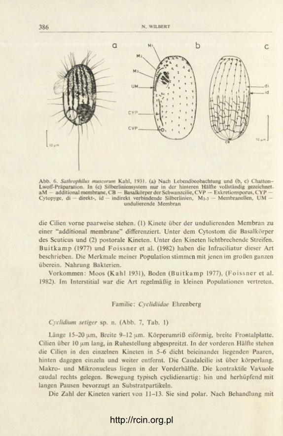
Sathrophilus muscorum Kahl, 1931 (Abb. 6)
Länge 25-35 [j.m, Breite um 20 [i.m, dorsoventral abgeflacht. Mund vor der Mitte, dem rechten Körperrand genähert. Vierzehntens seiten 16 Kineten, in denen
/ http://rcin.org.pl
386 N. WILBERT
Abb. 6. Sathrophilus muscorum Kahl, 1931. (a) Nach Lebendbeobachtung und (b, c) Chatton-Lwoff-Präparation. In (c) Silberliniensystem nur in der hinteren Hälfte vollständig gezeichnet. aM — additional membrane, CB — Basalkörper der Schwanzcilie, CVP — Exkretionsporus, CYP — Cytopyge, di — direkt-, id — indirekt verbindende Silberlinien, M1-3 — Membranellen, UM —
undulierende Membran
die Cilien vorne paarweise stehen. (1) Kinete über der undulierenden Membran zu einer "additional membrane" differenziert. Unter dem Cytostom die Basalkörper des Scuticus und (2) postorale Kineten. Unter den Kineten lichtbrechende Streifen. B u i t k a m p (1977) und F o i s s n e r et al. (1982) haben die Infraciliatur dieser Art beschrieben. Die Merkmale meiner Population stimmen mit jenen im großen ganzen überein. Nahrung Bakterien.
Vorkommen: Moos (Kah l 1931), Boden ( B u i t k a m p 1977), ( F o i s s n e r et al. 1982). Im Interstitial war die Art regelmäßig in kleinen Populationen vertreten.
Familie: Cyclidiidae Ehrenberg
Cyclidium setiger sp. n. (Abb. 7, Tab. 1)
Länge 15-20 [xm, Breite 9-12 (xm. Körperumriß eiförmig, breite Frontalplatte. Cilien über 10 (j.m lang, in Ruhestellung abgespreitzt. In der vorderen Hälfte stehen die Cilien in den einzelnen Kineten in 5-6 dicht beieinandei liegenden Paaren, hinten dagegen einzeln und weiter entfernt. Die Caudalcilie ist über körperlang. Makro- und Mikronucleus liegen in der Vorderhälfte. Die kontraktile Vakuole caudal rechts gelegen. Bewegung typisch cyclidienartig: hin und herhüpfend mit langen Pausen bevorzugt an Substratpartikeln.
Die Zahl der Kineten variert von 11-13. Sie sind polar. Nach Behandlung mit
http://rcin.org.pl

387
CVP — Exkretionsporus, CYP — Cytopyge, di — direkt verbindende Silberlinie, K — kommis-surale Silberlinie, M1-3 — Membranellen, UM — undulierende Membran
Tabelle 1
Biometrische Charakteristik von Cyclidium setiger sp. n.
Merkmal X M Sx Sx Extremwerte n
Länge 17.6 17 1.6 0.3 15-20 19 Breite 10.7 11 1.7 0.4 9-12 19
Länge UM in der Längsachse 9.7 9 1.5 0.3 9-11 7 Länge UM zur Körperlänge 0.55 Zahl der Kineten 12 12 0.7 0.3 11-13 10 Kinetoscmen in der 1. Kinete 19.3 21 1.0 0.7 17-23 8 Kinetosomen in der N. Kinete 10.6 10 0.9 0.4 10-12 8 Kinetosomen in der N-l Kinete 17.3 18 0.8 0.4 15-19 8 Kinetosomen einer dorsalen Kinete 13 13 1.2 0.3 12-14 8 Kinetosomenpaare der UM 31.2 32 1.9 0.9 28-34 7
Maße in [xtn von AgNOj-iniprägnierten Tieren (Chatton-Lwoff-Technik)
http://rcin.org.pl
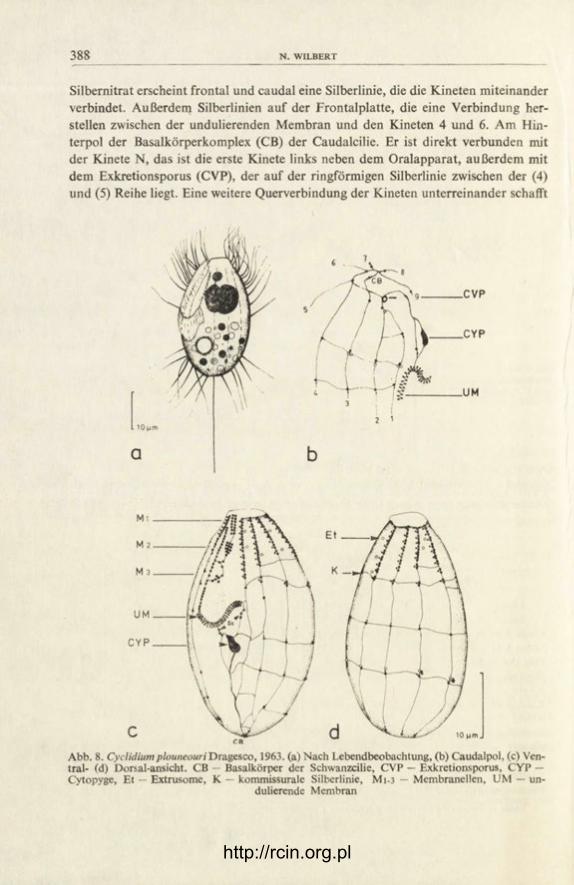
388
Silbernitrat erscheint frontal und caudal eine Silberlinie, die die Kineten miteinander verbindet. Außerdem Silberlinien auf der Frontalplatte, die eine Verbindung her-stellen zwischen der undulierenden Membran und den Kineten 4 und 6. Am Hin-terpol der Basalkörperkomplex (CB) der Caudalcilie. Er ist direkt verbunden mit der Kinete N, das ist die erste Kinete links neben dem Oralapparat, außerdem mit dem Exkretionsporus (CVP), der auf der ringförmigen Silberlinie zwischen der (4) und (5) Reihe liegt. Eine weitere Querverbindung der Kineten unterreinander schafft
10 (am
a 2 1
C V P
C Y P
Abb. 8. Cyclidiumplouneouri*Dra.geszo, 1963. (a) Nach Lebendbeobachtung, (b) Caudalpol, (c) Ven-tral* (d) Dorsal-ansicht. CB — Basalkörper der Schwanzcilie, CVP — Exkretionsporus, CYP — Cytopyge, Et — Extrusome, K — kommissurale Silberlinie, Mi-3 — Membranellen, UM — un-
dulierende Membran
http://rcin.org.pl

CILIATEN AUS DEM INTERSTITIAL 389"
eine kommissurale Fibrille (K). Das Peristom ist über halbkörperlang. Scuticus (Sc) unter der undulierenden Membran in zwei Portionen aufgeteilt. Als ein besonderes Artmerkmal kann die Kinete N angesehen werden. Sie ist sehr kurz und endet in Höhe der 3. Membranelle (M 3). Arttypisch ist auch die Lage des Exkretions-porus am Ende der (4) und (5) Kinete. Bei allen mir bekannten Cyclidium-Arten liegt er auf der caudalen Ringfibrille zwischen (1) und (2), meistens aber vor der 2. Kinete. Nach G r o l i e r e (1973) liegt der Exkretionsporus von C. sphagnetorum im Peristom. Das beruht auf einer Fehlbeobachtung. Nach eigenen Untersuchungen liegt der Exkretionsporus dieser Art am Ende einer stark verkürzten 2. Kinete in Höhe des Peristoms.
Vorkommen: ich habe diese Art außer im Ontario See auch im Interstitial des Biwa-Lake (Japan) und im Preßwasser aus Sphagnum eines Tümpel der Wah-ner-Heide bei Bonn gefunden.
Cyclidium plouneouri Dragesco, 1963 (Abb. 8)
Länge 25-37 [xm, ovoid. Mit 11, in seltenen Fällen auch 12 polaren Kineten. Kinetosomen praeoral paarweise und dichter gestellt als postoral. Den Paaren aber auch den einzelnen Kinetosomen sind ein Parabasalsack zugeordnet. Das Silberliniensystem kennzeichnet 3 komissurale Fibrillen (K), die das die Kineto-somen direkt verbindende System verknüpfen. Für diese Art charakteristisch ist die orale Infraciliatur, hier besonders das eigenartig geschwungene Ende der un-dulierenden Membran. Der Kernapparat aus Makro-und Mikronucleus liegt vorne. Der Makronucleus meiner Population bestand meist aus zwei Teilen.
Neben der Originalbeschreibung und dieser gibt es noch eine weitere von B o r r o r (1965). Seine Population kennzeichnet ein linksspiraliger Verlauf der Kineten 1 bis 4. Dies ist der Hauptunterschied zu den anderen Populationen mit ausnahmslos meridionalen Kineten. Weitere Unterschiede zwischen den drei Populationen habe ich unten gegenübergestellt.
Vorkommen: Sapropel, Brackwasser ( D r a g e s c o 1963), marin ( B o r r o r 1965), Limnopsammon.
Vergleich von C. plouneouri — Populationen
Merkmal | D r a g e s c o (1963) ! B o r r o r (1965) j Ontario See
Länge in [xm 30-40 25-30 25-37 Anzahl der Kineten 14-16 12-13 11-12 Kinetosomen in der (1). Kinete 25 17 20 Kinetosomen in der N. Kinete ? (nur präoral) 15 21 (nur präoral) Zahl der Makronuclei 1 1 1-2 Zahl der Mikronuclei 1 1 1 Länge UM zur Körperlänge 0.79 0.54 ' 0.63
http://rcin.org.pl

N. WILBERT
Abb. 9. Cristigera media Kahl, 1928. (a) Nach Lebendbeobachtung, (b) Ventral- und (c) Dorsalan-sicht nach Protargolimprägnation. CB — Basalkörper der Schwanzcilie, CVP — Exkretionsporus,
M1-3 — Membranellen, UM — undulierende Membran
Cristigera media Kahl, 1928 (Abb. 9, Tab. 2)
Länge 40-50 [j.m, von ovaler Gestalt (2:1), hinten breit gerundet, der Vorder-pol gerade abgestutzt und als Frontalplatte ausgebildet. Gattungstypisch sind die dorsoventrale Abflachung und die postorale rinnenartige Vertiefung. In den Kineten sind die sonst einzeln stehenden Kinetosomen nach vorne hin in bis zu 9 Kineto-somenpaaren verdichtet. Aus diesem Grunde ist auch die Bewimperung in der hinteren Hälfte wesentlich lockerer als vorne. Die kontraktile Vakuole liegt hinten rechts. Sie entleert über einen Exkretionsporus in der postoralen Rinne. Kernapparat zentral gelegen. Abweichend von Kahl's Beschreibung hat der linke Peristomrand meiner Population keine zahnartigen Höcker.
Vorkommen: Sapropel, halobiont (Kahl 1935), Limnopsammal.
Cristigera hammeri sp. n. (Abb. 10)
Länge 45-51 [xm, Breite 23-29 ;j.m. Körper schwach abgeflacht, im Umriß rechtsseitig fast gerade, linke Seite konvex. Der Voiderpol wird von einer breiten, deutlich abgesetzten Frontalplatte gebildet. Die rechte und linke Konturlinie stoßen caudal versetzt aufeinander. Dadurch entsteht ein Vorsprung, an dessen Basis die körperlange Caudalcilie steht. Ein wenig rechts der Mitte die gattungstypische Depression. Sie teilt sich in die vorne liegende etwa 25 [im lange Mundhöhlung und in die postorale Rinne, in der der Exkretionsporus (CVP) und die Cytopyge
http://rcin.org.pl

CILIATEN A U S DEM INTERSTITIAL 391"
Tabelle 2
Biometrische Charakteristik von Cristigera media Kahl
Merkmal X M Sx Sx Extremwerte n
Länge 47.8 47 3.8 1.5 43-54 10 Breite 23.8 23 1.7 0.8 21-25 10
Länge UM iu der Längsachse 25.5 26 1.4 0.6 24-28 10 Länge UM zur Körper länge 0.53 Distanz Vorderpol Makronucleus 21.7 23 3.5 1.5 16-25 10 Länge Makronucleus 10.4 10 1.2 0.6 9.8-11.2 7 Breite Makronucleus 7.2 8 1.3 0.6 7-9 7 Durchmesser Mikronucleus 2.2 2.2 0.3 0.1 2.0-2.8 6 Zahl der Kineten 16.0 15 1.8 0.8 14-18 10 Kinetosomen in der 1. Kinete 29.2 — — — 27-32 5 Kinetosomen einer dorsalen Kinete 25 — — — 24-28 4 Kinetosomenpaare der UM 61.8 - - — 60-65 4
Maße in (im von Protargol-imprägnierten Tieren
Abb. 10. Cristigera hammerisp. n. Nach Lebendbeobachtung (a) und Chatton-Lwoff- Präparation <c, b), (b) ventrale, (c) dorsale Ansicht. CB — Basalkörper der Schwanzcilie, CVP — Exkretionspo-rus, CYP — Cytopyge, Et — Extrusome, K — kommissurale Silberlinie, Mi-3 — Membranellen,
Sc — Scuticus, UM — undulierende Membran
(CYP) liegen. Ein ovaler Makronucleus (9x6[xm) liegt zentral, in geringer Ent-fernung von ihm ein Mikronucleus, 2 [xm im Durchmesser. Wie in Abb. 10a dar-gestellt, kommen mitunter auch Individuen mit zwei Makronucleusteilen vor.
Die Bewimperung und das Kinetom zeigen die Abbildungen. Bemerkenswert ist die Gliederung der Kineten: eine erste, apikale Verdichtung von Kinetosomen,
http://rcin.org.pl

392 N WILBERT
die sukzessiv von ventral nach dorsal abnimmt. Dann caudal ein zweiter Kineto-somengürtel. Im Unterschied zum ersten, mit paarweise gestellten Kinetosomen, stehen sie hier zig zag. Dazwischen nur zwei Kinetosomenpaare.
Das Silberliniensystem gehört zum streifenförmigen Typ. Das direkt verbin-dende System wird durch 5 kommissurale Fibrillen (K) quer verbunden. Ein indi-rektes Silberliniensystem konnte ich nicht feststellen. Es is möglich, daß es der Gat-tung fehlt. Untersuchungen an Cr. minor von F o i s s n e r et al. (1982) lassen das vermuten. Cristigera hammeri bewegt sich hüpfend. In den Ruhepausen werden die Cilien gespreizt. Nahrung Bakterien.
Ich widme diese Art meinem Freund Herrn Prof. Dr. U. T. Hammer, Limnologe an der University of Saskatchewan, Saskatoon, Kanada.
Familie: Histiobalantiidae de Puytorac et Corliss
Histiobalantium marinum Kahl, 1933 (Abb. 11)
Länge 60-102 (i.m, x = 75.3, M = 70, Sx = 15.0, Sx X 5.0, n = 10. Breite 30-42 [xm, x - 36.0, M = 35, Sx = 4.7, Sx = 1.7, n = 10. Maße von Protargol — imprägnierten Tieren. Kahl (1933) hat diese Art ausführlich beschrieben. Meine Beobachtungen deckcn sich mit seinen Angaben, die hier durch die Darstellung der Infraciliatur des Buccalapparates weiter vervollständigt werden. Die Cilien
Abb. 11. Histiobalantium marinum Kah!, 1933. (a) Nach Lebendbeobachtung und Protargolimpräg-nation, (b) Oralapparat, (c) Kernapparat und Infraciliatur des dorsalen Hinterendes. CYP — Cy-topyge, Ma — Makronucleus, Mi — Mikronucleus, M1-3 — Membranellen, UM — undulierende
Membran
http://rcin.org.pl

CILIATEN AUS DEM INTERSTITIAL 393"
stehen in bis zu 120 Kineten. Erwähnenswert zwischen den kurzen Körpercilien zahlreiche verlängerte Tastcilien, im Ektoplasma darunter dicht an dicht stehende Trichocysten und immer findet sich frontal eine Anhäufung schwarzer Granula. Abweichend von Kahl ist der Makronucleus nicht zwei- sondern einteilig.
Vorkommen: Sandgrund der Kieler Bucht ( K a h l 1933), Limnopsammal.
Histiobalantium minor sp. n. (Abb. 12, Tab. 3)
Länge 40-55 pim. Bewegung: ein lang anhaltendes Schwimmen unterbrochen von Ruhepausen, die immer in Kontakt mit Substratpartikeln gehalten werden. Dies und die Gestalt erinnern sehr an Pleuronema-Arten. Das Plasma ist farblos und fron-tal meist mit schwarzen Granula und kristallin aussehenden Körnern angefüllt. Drei bis vier kontraktile Vakuolen. Die Cytopyge (CYP) dorsal im hinteren Drittel.
Abb. 12. Histiobalantium minor sp. n. (a) Nach Lebendbeobachtung, (b) Intraciliatur des Oralappa-rates, der Ventral- und Dorsalseite (c,d). CYP — Cytopyge, M1-3 — Membranellen, UM — un-
dulierende Membran
Tabelle 3
Biometrische Charakteristik von Histiobalantium minor sp. n.
Merkmal X M Sx Sx Extremwerte n
Länge 48.4 45 4.6 1.3 42-57 12 Breite 22.9 22 3.3 0.95 19-26 12
Länge UM in der Längsachse 26.8 25 4.4 1.5 23-36 10 Länge von M 1 6.00 6 0.85 0.28 4.5-7.0 10 Länge von M 2 9.8 9.6 1.52 0.44 8.5-13 10 Länge von M 3 8.3 8.4 1.00 0.27 8.0-9.0 10 Länge Makronucleus 23.7 21.6 3.5 1.7 19.5-25.5 10 Zahl der Kineten 42.4 43 2.2 0.84 41-45 12
Maße in [xm von Protargol-imprägnierten Tieren
http://rcin.org.pl

394 N. WILBERT
Im Ektoplasma etwa 3 [j.m lange Trichocysten. Die Mundhöhle ist einhalb körper-lang. An ihrer rechten Seite die undulierende Membran, die außerdem die Öffnung hinten weit umgreift. Diagonal über die Mitte des Munddaches zieht die Mem-branelle 3, zu ihr fast im rechten Winkel die anderen.
Der Makronucleus, immer vorne gelegen, hat keine konstante Form. Meist ist er tief gebuchtet. Zwei runde Mikronuclei (2 {j.m Durchmesser) liegen ihm an. Die Wimpern, um 8 [i.m lang, stehen in 41-45 meridionalen Reihen. Verlängerte Tastcilien, die eigentlich für die Gattung typisch sind, habe ich nicht gesehen. Nah-rung Bakterien. H. minor kam in allen Proben regelmäßig und zahlreich vor.
Beschreibungen der oralen Infraciliatur liegen von den Arten H. natans D r a -gesco et I f t o d e (1972) und H. majus D r a g e s c o (1968), G r o l i e r e (1973) vor. Im Vergleich zeigt sich, daß die Lage der Membranellen zueinander und ihre Grö-ßenverhältnisse arttypisch sind.
D i s k u s s i o n
Ziel meiner Untersuchungen war festzustellen, ob das Interstitial von Süßwas-serseen der Größe der Nordamerikanischen Seen eine spezielle Ciliatenfauna be-herbergt. Diese Frage war bislang offen, da bei Untersuchungen, die P e n n a k (1940, 1951) hier im Interstitial durchführte, die Ciliaten nicht berücksichtigt wur-den. Ich habe 12 Arten im Interstitial festgestellt, von denen ich 10 taxonomisch genauer untersuchte. Mit den Arten Loxodes magnus und Loxodes rostrum habe ich mich nicht weiter auseinandergesetzt. Sie kamen in allen Proben regelmäßig vor und sind nach D r a g e s c o (1960) und eigenen Beobachtungen typische Vertreter des Limnopsammons. Ihre Infraciliatur haben D r a g e s c o (1960) und F o i s s n e r (1983) erarbeitet.
Ein Teil der angetroffenen Ciliaten ist zu charakterisieren als euryök und ste-noplastisch in bezug auf wenige Faktoren. Das sind die Arten Cyclidium plouneouri, Cyclidium setiger, Loxodes magnus, Loxodes rostrum, Sathrophilus muscorum. Ihr Lebensraum sind die verschiedenartigsten Kapillarwasser z.B. im Moos und Boden. Auch Gastronauta clatratus gehört in diese Gruppe. Die Art ist haptisch und saugt sich bei ankommenden Wasserturbulenzen fest. So vor dem Verdriften geschützt, ist sie in besonderer Weise an diesen Lebensraum angepaßt. Die restlichen Arten sind nicht biotopgebunden und unspezifisch für diesen Lebensraum. Es sind dies ausschließlich sapropelische Tiere wie Loxocephalus luridus, die Cristigera- und His-tiobalantium- Arten, die auf eine geeignete Bakteriennahrung angewiesen sind, die sich hier vorfindet.
Die Artenarmut und die verhältnismäßig hohe Präsens sapropslischer Arten lassen den Verdacht aufkommen, daß unbestimmbare Gifte oder auch bakterielle Abbauprozesse die Milieubedingungen so verändert haben, daß viele Arten von einer Besiedlung des Kapillarwassers ausgeschlossen sind. Die Sandproben, die
http://rcin.org.pl

CILIATEN AUS DEM INTERSTITIAL 395"
ich genommen habe, waren immer oxydiert, was an ihrer grauen bis weißen Färbung zu erkennen war. Demnach war Sauerstoff ausreichend vorhanden und fällt damit als ein limitierender Faktor aus. Nach den Unteisuchungen von Wei le r (1973) sind die abiotischen Bedingungen des untersuchten Limnopsammals von keinem Parameter her gesehen auffällig. Dies trifft insbesondere auf den Sauerstoff zu, der ausreichend vorhanden ist, und auf das Fehlen von Zellgiften z.B. Ammonium. Obwohl alle Bedingungen scheinbar optimal sind, ist hier eine Infauna, die man eine Biozönose nennen könnte, nicht existent.
DANKSAGUNG
Meine Untersuchungen wurden durch ein Stipendium der Heinrich Hertz-Stiftung des Lan, des Nordrhein-Westfalen ermöglicht. Für diese Unterstützung danke ich herzlich. Herrn Dr. J. Ba-rica, Chief der Aquatic Ecology Division am Canada Centre for Inlandwaters, Burlington, Ontario-danke ich für einen Arbeitsplatz in seinem Institut.
SUMMARY
Qualitative measurements of ciliates in the interstitial of Lake Ontario near Burlington, On-tario (Canada) were made by the method of U h l i g (1964). During this study 12 species were de-tected. The infraciliature of 10 of these was studied by means of the silver nitrate or protargol methods. Three new species are described: Cyclidium setiger, Cristigero hammeri, Histiobalantium minor.
LITERATUR
Bock K. J. 1953: Zur Ökologie der Ciliaten des marinen Sandgrundes der Kieler Bucht (II). Kiel Meeresforsch., 9, 252-256.
B o r r or A. C. 1965: New and little-known tidal marsh ciliates. Trans. Am. Microsc. Soc., 84, 550-565.
B u i t k a m p U. 1977: Die Ciliatenfauna der Savanne von Lamto (Elfenbeinküste) Acta Protozool., 16, 249-276.
C h a t t o n E. et L w o f f A. 1930: Imprégnation par diffusion argentique de l'infraciliature des ciliés marins et d'eau douce après fixation cytologique et sans dessication. C. R. Soc. Biol. Paris 104, 834-836.
C o r l i s s J. O. 1979: The Ciliated Protozoa. 2 nd revised ed. Pergamon Press, London and New York.
D e r o u x G. 1976: Le plan cortical des cyrtophorida, unité d'expression et marges de variabilité. II. Cyrtophorida à thigmotactisme ventral généralisé. Protistologica, XII, 483-500.
D r a g e s c o J. 1960: Les ciliés mésopsammiques littoraux. Trav. Stn. Biol. Roscoff, N. S. 12, 1-356. D r a g e s c o J. 1963: Compléments à la connaissance des ciliés mésopsammiques de Roscoff. I. Ho-
lotriches. Cah. Biol. Mar., 4, 91-119. D r a g e s c o J. 1968: Les genresPleuronema Dujardin. Schizocalyptru nov. gen. et Histiobalantium Sto-
kes (ciliés holotriches hymenostomes) Protistologica, IV, 85-106. D r a g e s c o J. et I f t o d e F. 1972: Histiobalantium natans (Clap, et Lachm., 1858) Morphologie,,
infraciliature, morphogenèse (Holotriche Hymenostomatida) Protistologica, VIII, 347-352. F a u r é - F r e m i e t E. 1948 : The ecology of some infusorian communities of intertidal pools. J. Anim.
Ecol., 17, 127-130. F a u r é - F r e m i e t E. 1950: Ecologie des ciliés psammophiles littoraux. Bull. Biol. Fr. Belg., 84,
35-75.
http://rcin.org.pl

N. WILBERT
F a u r é - F r e m i e t E. 1968: Les genres Dexiotricha Stokes et Loxocephalus Eberhard dans leurs re-lations auxomorphiques. Protistologica IV, 115-125.
F o i s s n e r W. et al. 1982: Morphologie, Infraciliatur und Silberliniensystem einiger wenig bekann-ter Scuticociliatida (Protozoa: Ciliophora). Zool. Jb. Syst. 109, 443-468.
F o i s s n e r W. 1983: Licht- und rasterelektronenmikroskopische Untersuchungen über die In-fraciliatur von Loxodes striatus Engelmann, 1862 und Loxodes magnus Stokes, 1887 (Pro-tozoa: Ciliophora). Zool. Anz. Jena 210, 3-13.
G o u l d e r R. 1971: Vertical distribution of some ciliated protozoa in two freshwater sediments. Oikos, 22, 199-203.
G r o l i è r e C. A. 1973: Description de quelques espèces de ciliés hyménostomes des genres Sathro-philus Corliss, 1960, Cyclidium O. F. Müller, 1786, Histiobalantidium Stokes, 1886. J. Pro-tozool., 20, 368-376.
H a r t w i g E. 1973: Die Ciliaten des Gezeiten-Sandstrandes der Nordseeinsel Sylt. I. Systematik. Mikrofauna Meeresbod., 18, 1-69.
H a r t w i g E. 1974: Verzeichnis der im Bereich der deutschen Meeresküste angetroffenen inter-stitiellen Ciliaten. Mitt., Hamb. Zool. Mus. Inst., 71, 7-21.
J a n k o w s k i A. 1964: Morphology and evolution of Ciliophora: IV Sapropelebionts of the fa-mily Loxocephalidae fam. nov. their taxonomy and evolutionary history. Acta Protozool., 2, 33-58.
von J u t r c z e n k i J. 1978: Ökologische Untersuchung der Ciliatenfauna von Waldbächen. Diplo-marbeit der math. nat. Fakultät der Rheinischen Friedrich Wilhelms Universität Bonn, pp. 137.
K a h l A. 1930-1935: Wimpertiere oder Ciliata. In: Dahl, F. : Die Tierwelt Deutschlands, Teil 18, 21, 25, 30. Gustav Fischer, Jena, pp. 886.
P e n n a k R. W. 1940: Ecology of the microscopic metazoa inhabiting the sandy beaches of some Wisconsin Lakes. Ecol. Monogr., 10, 538-614.
P e n n a k R. W. 1951: Comparative ecology of the interstitial fauna of freshwater and marine be-aches. Ann. Biol., 3, 449-480.
R e i n n a r t h G. 1979: Ökologie und Vertikalverteilung von Ciliaten in der Kontaktzone verschie-dener Süßwasserbiotope. Dissertation der math. nat. Fakultät der Rheinischen Friedrich Wilhelms Universität Bonn, pp. 113.
R e m a n e A. 1933: Verteilung und Organisation der benthischen Mikrofauna der Kieler Bucht. Wiss. Meeresunters., N.F. 21, 161-221.
R u t t n e r - K o l l i s k o A. 1956: Der Lebensraum des Limnopsammals. Verh. Dtsch. Zool. Ges., Hamburg, 421-427.
S c h l i e p e r C. 1968: Methoden der Meeresbiologischen Forschung. VEB Gustav Fischer Verlag Jena, pp. 322.
S c h m i d t P. 1968: Die quantitative Verteilung und Populationsdynamik des Mesopsammons am Gezeiten-Sandstrand der Nordseeinsel Sylt. Int. Rev. Gesamten Hydrobiol., 53, 723-779.
U h l i g G. 1964: Eine einfache Methode zur Extraktion der vagilen, mesopsammalen Mikrofauna. Helgoländer wiss. Meeresunters., 11, 178-185.
We i l e r R. R. 1973: Ths interstitial water composition in the sediments of the Great Lakes. I. Wes-tern Lake Ontario. Limnol. Oceanogr., 18, 918-931.
'Wi lbe r t N. 1975: Eine verbesserte Technik der Protargolimprägnation für Ciliaten. Mikrokos-mos, 6, 171-179.
http://rcin.org.pl

A C T A PROTOZOOLOGICA Vol. 25, No . 4, pp. 397—410 (1986)
Inhibition of Macromolecule Syntheses in a Ciliate Protozoan, Tetrahymena pyriformis
by Hexachlorocyclohexane (HCH) Isomers
R. M A T H U R and D. M. S A X E N A 1
Department of Zoology, University of Delhi, Delhi-110007, India
Received on 16 October 1985
Synopsis. The syntheses of macromolecules in Tetrahymena treated with various iso-mers of H C H was studied. The peak incorporation of 3H-thymidine in Tetrahymena cultures treated with 8, (3 and a -HCH occurred on the 4th day of treatment as com-pared with the control and lindane treated cells where maximum incorporation was observed on the 3rd day. The range of inhibition for D N A synthesis was 1-89%. & and P-HCH shifted the peak incorporation of 3H-uridine to the 4th day while in the case of a and y-HCH its maximum incorporation was observed on the 3rd day of treatment. The range of inhibition for R N A synthesis was 1-86%. The maxi-mum incorporation of 3H-lysine in treated and control cells occurred on the 3rd day, the percentage inhibition, however, ranged from 1-81.
HCH is one of the most extensively used insecticides for agriculture and public health in India ( K r i s h n a m u r t i et al. 1982). Although only the y-isomer of HCH is the isomer with insecticidal properties, nevertheless, other isomers of HCH viz., a, (3, S and s do occur in different proportions in the technical grade of HCH mar-keted and used (Brooks 1974). Besides, there have been reports of the isomeriza-tion of y-HCH to a-HCH (Beneze t and M a t s u m u r a 1973, M a t s u m u r a et al. 1976 and V o n k and Q u i r j n s 1979) and thus along with lindane, other isomers of HCH also would be present in the environment and can affect the organisms. y-HCH and its isomers have been known to lower the convulsive threshold for pen-tylenetetrazol in mice ( H u l t h et al. 1976), to cause extensive myofilament degrada-tion in rats ( P u b l i c o v e r et al 1979), to cause severe hypoplastic anemia (Mor-gan et al. 1980), to increase the values of hepatic microsomal enzyme, O-demethyl-ase in albino rats (Chand and R a m a c h a n d r a n 1980), to inhibit somatic and
1 To whom all correspondence should be addressed.
3 — Acta Protozol. 25/4
http://rcin.org.pl

398 R. MATHUR A N D D. M. SAXENA
meiotic divisions in male swiss mice (Babu et al. 1981) and to affect some enzymes of liver and kidney in Heteropneustus fossilis ( S e n g u p t a et al. 1984).
The target site of HCH has been very clearly established in higher organisms but it is not so in microorganisms where one or more sites are affected. BHC lindane has been reported to interfere with many basic biochemical processes like photosyn-thesis (Kopecek et al. 1976), enzyme activity (Gray 1954) and oxidation of am-monium to nitrite and conversion of nitrite to nitrate (Ray 1983).
Besides the target organisms, HCH isomers affect the non-target organisms as well. Among these, ciliates are important as they constitute important link in the food chain and food webs.
Our earlier studies have shown that HCH isomers inhibit the cell population growth of a ciliate protozoan, Tetrahymena pyriformis ( M a t h u r et al. 1984). In the present paper, the effect of sublethal concentrations of HCH isomers on the syn-theses of macromolecules viz., DNA, RNA and protein in T. pyriformis are described.
M a t e r i a l s a n d M e t h o d s
Stock cultures of T. pyriformis were grown in 15 ml tubes containing 5 ml of 1% proteose peptone supplemented with 0.5% NaCl and 0.3% yeast extract at 27±1°C in a B.O.D. incubator.
Lindane, the gamma isomer of 1, 2, 3, 4, 5, 6 Hexachlorocyclohexane (HCH) was obtained from ICN Pharmaceuticals Inc., Life Sciences Group, Cleveland, Ohio. The other isomers of HCH viz., a, (3 and 8 were obtained through the courtesy of Dr. Gunter Zweig, Environmental Pro-tection Agency, U.S.A.
The stock solutions of the insecticide isomers were prepared in acetone and then diluted with the culture medium to obtain the desired concentration of the insecticide. The acetone concentra-tion of the insecticide treated cultures was kept at 0.1% as our earlier studies showed that concen-trations of acetone upto 0.5% did not have any adverse effect on the organisms.
Our earlier studies ( M a t h u r et al. 1984) also showed that 2.5 and 5.0 ppm of a and y-HCH and 1.0 and 2.5 ppm of (3 and S-HCH were the sublethal concentrations for the growth of T. pyri-formis. Hence, these concentrations of HCH isomers were used to study their effects on the syntheses of DNA, RNA and proteins.
A volume of 2 ml of 48 h old culture of Tetrahymena was transferred to 48 ml of sterile medium in 100 ml conical flasks and allowed to grow for 24 h. Insecticide solutions were added directly to the culture medium so as to get the required concentration of the insecticide and the cultures were allowed to grow for another 24 h. The incorporation of radioactive thymidine (Sp. Act 18.8 Ci/mM), uridine (Sp. Act 13.8 Ci/mM) and lysine (Sp. Act 5.6 Ci/mM) in the presence of sublethal concen-trations of HCH isomers has been used as an index of DNA, RNA and protein synthesis respec-tively in T. pyriformis treated with these insecticides. The method adopted was the same as de-scribed by F r e e m a n and M o n e r (1976). After every 24 h a volume of 2 ml was pipetted out asep tically from both the treated and the control flasks. Out of these, three aliquots of 0.5 ml each were incubated with the respective radioisotope i.e., 3H-thymidine (for DNA synthesis), 3H-uridine (for RNA synthesis) and 3H-lysine (for protein synthesis) at 27±1°C, at a specific activity of 5 [xCi/ml. After 30 min of incubation, 0.5 ml of ice cold trichloroacetic acid (TCA) was added to each of these. The precipitated material was pipetted onto Whatman GF/C glass fiber discs, washed twice with 10 per cent ice cold TCA and finally dried. The dried discs were counted for radio-activity in the Packard tricarb liquid scintillation spectrophotometer. The scintillation fluiid consisted
http://rcin.org.pl

INHIBITION OF MOLECULE SYNTHESES BY HCH 3 9 9
of 5 gms PPO and 50 mgs POPOP dissolved in 1 1 of Toluene. The remaining 0.5 ml of the culture medium from both the control and the treated flasks was fixed with 0.5 ml of 10 per cent neutral formalin and cell number was determined with the help of a haemocytometer. A minimum of six replicates were taken for each treatment and the experiments were repeated twice.
R e s u l t s
The incorporation of radioactive precursors in controls followed a pattern similar to the normal cell cycle of Tetrahymena. During the log phase of growth of Tetrahy-mena the cell number increases till 72 h and then the cells enter into stationary phase during which the cell number remains more or less constant. The incorporation of radioactive precursors (3H-thymidine, 3H-uridine, 3H-lysine) increased grad-ually from the 1st to the 3rd day and thereafter declined on the 4th and the 5th day.
Duration of treatment ( d a y s )
Fig. 1. 3H-thymidine incorporation into DNA of Tetrahymena pyriformis treated with 2.5 and 5 .0 p p m Y - H C H
http://rcin.org.pl

4 0 0 R. MATHUR AND D. M. SAXENA
DNA Synthesis
y-HCH: The peak incorporation of 3H-thymidine in cultures of T. pyrijormis tieated with 2.5 and 5.0 ppm lindane was obtained on the 3rd day of treatmtnt as in the case of control (Fig. 1). In the cultures treated with 2.5 ppm lindane ibout 44% reduction in 3H-thymidine incorporation was observed within 24 h wi h re-spect to the control. Maximum reduction (upto 85%) occurred on the 3rd cay of treatment. With 5.0 ppm lindane treatment the reduction at the end of 24 I was 55% which increased to 89% on the 3rd day of treatment and at the end of 5th day of treatment it decreased to 73%.
The peak incorporation of 3H-thymidine in Tetrahymena cultures treatec with sublethal concentrations of S, and a-HCH was observed on the 4th day of treat-ment as against the control where maximum incorporation was observed cn the 3rd day. The percentage inhibition ranged from 2-74 for S-HCH (Fig. 2) from 10-60 for (3-HCH (Fig. 3) and from 1-84 for a-HCH (Fig. 4).
RNA Synthesis
The incorporation pattern of 3H-uridine into RNA of the control cultures of T. pyriformis is shown in Fig. 5. 3H-uridine incorporation into RNA increased initially reaching a peak incorporation on the 3rd day after which it declined on the 4th and the 5th day.
The peak incorporation of 3H-uridine into RNA of T. pyriformis treated with 1.0 and 2.5 ppm concentrations each of 8 and [3-HCH was observed on the 4h day of treatment unlike the case in control where the peak incorporation was ob;erved on the 3rd day. Within 24 h of treatment, 16 and 22% inhibition occurred in RNA synthesis in 1 and 2.5 ppm S-HCH treated cultures respectively with respect to control which increased to a maximum value of 69 and 72% respectively with 1 and 2.5 ppm S-HCH after 72 h of treatment (Fig. 5). In the cultures treated with 1 ppm (3-HCH about 8% reduction in 3H-uridine incorporation was observed within 24 h. Maximum reduction (25%) occurred on the 3rd day of treatment. With 2.5 ppm (3-HCH the reduction at the end of 24 h was 13% which increased to 29% :>n the 3rd day (Fig. 6).
The peak incorporation of 3H-uridine in a and y-HCH treated cultures of Te-trahymena occurred on the 3rd day of treatment as in the case of control. The per-centage reduction in incorporation for a-HCH ranged from 6-69 (Fig. 7) and for y-HCH it ranged from 39-86 (Fig. 8).
Protein Synthesis
The pattern of incorporation of 3H-lysine into proteins in the control cultures of T. pyriformis is shown in Fig. 9. The ciliates showed a gradual increase in the incorporation of the precursor reaching its maximum after 72 h. Subsequently, the rate of incorporation declined on the 4th and 5th day.
http://rcin.org.pl

401
http://rcin.org.pl

http://rcin.org.pl

INHIBITION OF MOLECULE SYNTHESES BY HCH 403
http://rcin.org.pl

404 R. MATHUR A N D D. M. SAXENA
http://rcin.org.pl

5 -
p - HCH: PROTEIN SYNTHESIS
Contro l ,
£ £ :
O *
c o 2 O
.1 T \ O PP™
r
1 2 3 4 5
Durat ion of treatment ( d a y s )
Fig. 10. 3H-lysine incorporation into proteins of Tetrahymena pyriformis treated with 1.0 and 2.5 ppm (3-HCH
o 4
c E
oi J O
o o
oC --HCH: PROTEIN SYNTHESIS -
T Control [
-
11 1 1» v \
\ x »x \r
- \ \ -
v \ , ! \ \
»
-
1 1 • 1 i 1
\
i 0 1 2 3 4 5
Duration of treatment ( d a y s )
1 1. 3H-lysine incorporation into proteins of Tetrahymena pyriformis treated with 2.5 and 5.0 ppm a -HCH
http://rcin.org.pl

406 R. MATHUR AND D. M. SAXENA
The maximum incorporation of the radioactive precursor into proteins of asyn-chronous culture of T. pyriformis treated with sublethal concentrations of each of the HCH isomers viz., 8, (3, a and y as well as in the control occurred on the 3rd day. The extent of maximum incorporation for 1 and 2.5 ppm 8-HCH treated cells as compared to the control was 61 and 54% (Fig. 9); it was 43 and 42% for 1 and 2.5 ppm (3-HCH treated cells (Fig. 10); for a-HCH treated cells it was 99 and 98% for 2.5 and 5.0 ppm respectively (Fig. 11). In case of 2.5 and 5.0 ppm y-HCH treated cells the range of incorporation was 83 and 42% respectively (Fig. 12).
c Ë 3
S 2 <J>
V - HCH: PROTEIN SYNTHESIS T
- * \ ' s \
' * \ ' 4 \
-
' \ ' \
<*/ »
-
/ \ \
-
. 1
K ' -• Y i > • • -
J 1 ! 1 _ 1 1 1 1 U 0 1 2 3 4 5
Duration of t r e a t m e n t (days )
Fig. 12. 3H-lysine incorporation into proteins of Tetrahymena pyriformis treated with 2.5 and 5.0 ppm y-HCH
D i s c u s s i o n
Our results show that sublethal concentrations of all HCH isomers inhibited the synthesis of DNA, RNA and proteins in Tetrahymena. For instance, lindane at a concentration of 2.5 ppm produced 40% inhibition in DNA synthesis within 48 h and 89% inhibition in DNA synthesis was observed with 5.0 ppm concentration on the 3rd day of treatment. The range of percentage inhibition varied from 40-89. A concentration of 5.0 ppm lindane produced 39% inhibition in RNA synthesis within 24 h of treatment and 86% inhibition occurred on the 4th day. y-HCH at
http://rcin.org.pl

INHIBITION OF MOLECULE SYNTHESES BY HCH 4 0 7
2.5 ppm concentration caused 78% inhibition in protein synthesis within 24 h. However, on the 3rd day only 17% inhibition in protein synthesis was noticed.
Similarly other isomers of HCH also inhibit macromolecule syntheses to differ-ent extents. The inhibition of macromolecule syntheses in this sensitive ciliate by HCH isomers could explain the observed inhibitory effects of HCH isomers on its growth ( M a t h u r et al. 1984).
Although reports on the effects of lindane on macromolecule syntheses in micro-organisms are scanty, lindane and other isomers of HCH have been reported to inhibit DNA, RNA and protein synthesis in higher organisms.
B o r g h i et al. (1973) reported that lindane inhibited DNA, RNA and protein synthesis in the alga, Acetabularia mediterranea. J e a n n e (1979) found that in Du-naliella bioculata DNA synthesis was strongly inhibited by 10 ppm lindane and de-pressed by 5 ppm in the first cell cycle. The synthesis of DNA was not resumed during the second cell cycle. The RNA and the protein content increased in the treated cells during the first cell cycle but the increase was slow. However, in the second cell cycle the RNA and protein contents increased rapidly.
M i l l e r et al. (1980) while studying the effect of 250 ppm concentration of each of the HCH isomers on the nuclear and mitochondrial DNA synthesis in the mouse liver, however, reported that the treated animals showed increased nuclear DNA specific activities as well as an increase in the radioactivity incorporated into mito-chondrial DNA. R o u x et al. (1980) studied the effects of lindane in vitro on the mouse peritoneal macrophages and reported a pronounced inhibition of uridine incorporation correlated with the reduction of intracellular uridine pool. R o u x et al. (1978) studied the effects of lindane on mouse peritoneal macrophages and their results revealed that the rate of leucine incorporation associated with protein syn-thesis rate remained uniform during the exposure of macrophages to lindane sug-gesting that leucine transport was not affected.
On comparison of the concentrations of lindane which have been reported to inhibit macromolecule synthesis in other systems like Dunaliella ( J e a n n e 1979) and mouse liver (Mi l l e r et al. 1980) with those we have found effective in Tetra-hymena it is evident that this ciliate is sensitive to lower concentration of lindane.
The exact mechanism of interaction of HCH isomers with macromolecule syn-theses is not known. However, the work of P u i s e u x - D a o et al. (1977) revealed that plasma membrane could be the possible target site of y-HCH in Acetabularia mediterranea. Such interactions besides interfering with the essential transport mechanisms like NaT , K + and Mg2 + ATPase systems which are associated with the membranes, may also affect various other biochemical processes within the cell. S r in ivasan and R a d h a k r i s h n a m u r t y (1980) found that albino rats fed 800 ppm of (3 and y-HCH each for two weeks showed decreased levels of hepatic mitochon-drial DN A/Mg 2 + /Ca 2 + activated ATPase and microsomal Na+ , K + activated ATPase. The levels of SDH were elevated while there was a reduction in the activities of hepatic MDH and LDH. Such interferences of HCH isomers with the normal
http://rcin.org.pl

408 R. MATHUR AND D. M. SAXENA
activities of important enzymes might lead to uncoupling of oxidative phosphory-lation and since protein synthesis is an energy consuming process, inactivation or inhibition of oxidative enzymes by HCH isomers could, therefore, alter the rate of oxidative phosphorylation and this in turn could inhibit protein synthesis.
S e n g u p t a et al. (1984) studied the effect of 1.0 and 5.0 ppm concentrations of BHC on some enzymes of liver and kidney of the fish, Heteropneustus fossilis. The y-amylase activity increased in the treated fishes and glucose was released directly from glycogen to meet the energy demand of BHC stressed fish. The GOT and GPT activities were found to increase and were responsible for the process of transami-nation to gluconeogenesis. The alkaline phosphatase activity decreased while that of acid phosphatase increased indicating hepatotoxicity produced by BHC.
Organochlorine insecticides other than HCH isomers such as DDT, aldrin and dieldrin have been reported to inhibit/stimulate DNA, RNA and protein synthesis in other systems such as HeLa cells ( C h u n g et al. 1967 and L i t t e r s t et al. 1969), Dictyostelium discoideum (Bushway and H a n k s 1976), Euglena gracilis (Ewald et al. 1976), Crithidia fasciculata ( F r e n c h 1976), Stylonychia notophora (Lai and Saxena 1980 and Saxena and Lai 1981), Blepharisma intermedium (Lai et al. 1981) and Tetrahymena pyriformis (Saxena et al. 1981).
ACKNOWLEGDEMENT
The award of a Senior Research Fellowship, C.S.I.R., New Delhi to Ranjana Mathur is gratefully acknowledged.
REFERENCES
B a b u K. A., N i g a m S. K., L a k k a d B. C., K a r n i k A. B., T h a k o r e K. N.. K a s h y a p S. K. and C h a t t e r j e e S. K. 1981: Effect of hexachlorocyclohexane on somatic and meiotic di-visions in male swiss mice. Bull. Environ. Contam. Toxicol., 26, 508-512.
Beneze t H. J. and M a t s u m u r a F. 1973: lsomerization of y-BHC to a-BHC in the environment. Nature, 243, 480-481.
B o r g h i H., P u i s e u x - D a o S., B o n o t t o S. and H o u r s i a n g a u - N e u b r u n D. 1973: The effects of lindane on Acetabularia mediterranea. Protoplasma, 78, 99-112.
B r o o k s G. T. 1974: Y-l,2,3,4,5,6-Hexachlorocyclohexane. In: Chlorinated Insecticides Vol. I, (ed. G u n t e r Zweig) CRC Press, United States. P. 185-203.
B u s h w a y R. J. and H a n k s A. R. 1976: Pesticide inhibition of growth and macromolecular syn-thesis in the cellular slime mold, Dictyostelium discoideum. Pestic. Biochem. Physiol., 6, 254-260.
C h a n d B. and R a m a c h a n d r a n M. 1980: Effect of dietary hexachlorocyclohexane on certain biochemical changes in albino rat. Indian J. Exp. Biol., 18, 735-736.
C h u n g R. A., H u a n g I. L. and B r o w n R. W. 1967: Studies on DNA, RNA and protein synthesis in HeLa cells exposed to DDT and dieldrin. J. Agric. Food Chem., 15, 497-500.
E w a l d W. G., F r e n c h J. E. and C h a m p M. A. 1976: Toxicity of polychlorinated biphenyls (PCB's) to Euglena gracilis: Cell population growth, carbon fixation, chlorophyll level, oxygen consumption and protein and nucleic acid synthesis. Bull. Environ. Contam. Toxi-col., 16, 71-80.
F r e e m a n H. and M o n e r J. G. 1976: The uptake of pyrimidine nucleosides in Tetrahymena 1. Uri-dine. J. Protozool., 23, 465-472.
http://rcin.org.pl

INHIBITION OF MOLECULE SYNTHESES BY HCH 409
F r e n c h J. E. 1976: The effccts of the chlorinated polycyclic hydrocarbons, p-p ' DDT and poly-chlorinated biphenyls (PCB's) on the flagel'ated protozoan, Crithidia fasciculata. Diss. Absti. Int. B. 37, 96-97.
G r a y P. H. H. 1954: Effect of benzenehexachloride on soil microorganisms. II. Experiments with urea hydrolyzing bacteria. Can. J. Bot., 32, 10-15.
H u l t h L., L a r s s o n M., C a r l s s o n R. and K i h l s t r o m J. E. 1976: Convulsive action of small single oral doses of the insecticide-lindane. Bull. Environ. Contam. Toxicol., 16, 133-137.
J e a n n e N. 1979: Effects of lindane on the division cell cycle and biosynthesis of two unicellular algae. Can. J. Bot., 57, 1464-1472.
K o p e c e k K., Fu l l e r F., R a t z m a n n W. and S i m o n i s W. 1976: The effect of light dependency of insecticides on unicellular algae. Ber. Dtsch. Bot. Ges., 88, 269-281.
K r i s h n a m u r t i C. R., D i k s h i t T. S. S. and S u r y a P r a k a s h 1982: Status of research on bio-degradable pesticides in India. In: Proceedings of Tndo-US Workshop on biodegradable pesticides (eds. A t t r i B. S., R a m a n a t h a n N. L. and K r i s h n a m u r t i C. R.) Department of Environment, Government of India, New Delhi, p. 1-70.
Lai R. and S a x e n a D. M. 1980: Effect of DDT on cell population growth, cell division and DNA synthesis in Stylonychia notophora (Stokes). Arch. Environ. Contam. Toxicol., 9, 163-170.
L a i R., R e d d y B. V. P. and Saxena D. M. 1981: Effect of DDT on cell division and DNA, RNA and protein synthesis in a ciliate protozoan, Blepharisma intermedium. Acta Protozool., 20, 421-427.
L i t t e r s t C. L., L i c h t e n s t e i n E. P. and K a j i w a r a K. 1969: Effects of insecticides on growth of HeLa cells. J. Agric. Food Chem., 17, 1199-1203.
M a t h u r R., S a x e n a D. M. and A g a r w a l H. C. 1984: Growth of a ciliate protozoan, Tetra-hymena pyriformis in the presence of different isomers of Hexachlorocyclohexane (HCH). Acta Protozool., 23, 165-174.
M a t s u m u r a F., Beneze t H. J. and Pa t i l K. C. 1976: Factors affecting microbial metabolism of y-BHC. J. Pestic. Sci., 1, 3-8.
Mi l l e r C. T., L o k E., T r y p h o m a s L., R o g e r s C. G. and 1 v e r s o n F. 1980: Effects of hexachlo-rocyclohexane isomers on nuclear and mitochondrial DNA synthesis in mouse liver. Can. J. Genet. Cytol., 22, 671.
M o r g a n D. P., S t o c k d a l e E. M., R o b e r t s R. J. and W a l t e r H. W. 1980: Anemia associated with exposure to lindane. Arch. Environ. Health, 35, 307-310.
P u b l i c o v e r S. J., D u n c a n C. J. and Smi th J. L. 1979: The action of lindane in causing ultra-structural damage in frog skeletal muscle. Comp. Biochem. Physiol., 64C, 237-241.
P u i s e u x - D a o S., J e a n n e - L e v a i n N., R o u x F., R ib i e r J., B o r g h i H. and B r u n C. 1977: Ana-lysis of the effects of the organochlorine insecticide lindane at the cellular level. Protoplasma, 91, 325-341.
Ra y R. C. 1983: Toxicity of the pesticidcs Hexachlorocyclohexane and Benomyl to nitrifying bacteria in flooded autoclaved soil and in culture media. Environmental pollution series A: Ecological and Biological, 32, 147-155.
R o u x F., P u i s e u x - D a o S., T r e i c h I. and F o u r n i e r E. 1978: Effect of lindane on mouse peri-toneal macrophages. Toxicology, 11, 259-269.
R o u x F., T re i ch 1. and F o u r n i e r E. 1980: Different levels of changes induced by insecticide lindane in cultured C-6 glioma cells. Toxicology, 17, 261-264.
S a x e n a D. M. and Lai R. 1981: Effect of DDT on RNA and protein synthesis in Stylonychia notophora. Arch. Protistenkd., 124, 103-109.
Saxena D. M., S h i v a j i S. and Lai R. 1981: Effect of DDT on DNA, RNA and protein synthesis in Tetrahymena pyriformis. Indian J. Exp. Biol., 19, 568-570.
S e n g u p t a S., D a s M. and S h a r m a D. K. 1984: Effect of BHC on some enzymes of liver of H.fos-silis (Bloch) (y-Amylase activity as an alternative pathway to carbohydrate metabolism). In: Abstracts of VI11 All India Cell Biology Conference and Symposium on regulation of cell proliferation, held in School of Biological Sciences, Madurai Kamaraj University, Ma-durai 625021. from 27th-29th Dec. 1984.
S r i n i v a s a n K. and R a d h a k r i s h n a m u r t y R. 1980: Effect of [3 and y-isomers of hexachloro-cyclohexane on some enzyme systems of albino rats. Indian J. Biochem. Biophys., 17, 111.
Vonk J. W. and Q u i r j n s J. K. 1979: Anaerobic formation of a-hexachlorocyclohexane in soil and by Escherichia coli. Pestic. Biochem. Physiol., 12, 68-74.
http://rcin.org.pl

http://rcin.org.pl

ACTA PROTOZOOLOGICA Vol. 25, No. 4, pp. 411-418 (1986)
Biochemical Changes of Acanthamoeba Following Attenuation and the Role
of Cysts in Retaining the Characteristics of Strains1
W i t o l d K A S P R Z A K , T a d e u s z M A Z U R and E d w a r d H A D A Ś
Department of Biology and Medical Parasitology, Marcinkowski Academy of Medicine, 10 Fredry Street, 61-701 Poznari, Poland
Received on 30 January 1986
Synopsis. We compared the virulence of the environmental Acanthamoeba strains, maintained paralelly in prolonged axenic culture at 37°C and in cyst form stored at 4°C beginning at the original isolation. The changes in the lytic enzymes activity and isoenzyme patterns, concomitant with the strain attenua-tion, were also studied. After 31/2 to 4 years the strains remained virulent and lost their primary virulence only after numerous passages (8 years). Still, the amoebae retained the virulence when stored in cyst form for 9 years. The attenuation of the strains is related to the decrease in trypsin activity. However, the isoenzyme patterns of non-specific esterases and acid phosphatase remained stable throughout the time.
The amphizoic Acanthamoeba species, widespread in nature, are the etiological agents of granulomatous amoebic encephalitis and other diseases in humans. The cysts of Acanthamoeba stored at 4°C are able to survive moths (De J o n c k h e e r e and Van D e V o o r d e 1976, B idd i ck et al. 1984) or even years ( M a z u r 1984). However, the main method for laboratory continuous maintenance of Acanthamoeba strains in axenic culture without passage through host tissue affects the amoeba behavioural characteristics, e.g., the loss of encystation potential, alteration in drug sensitivity and, most important, the loss of pathogenicity (S tevens and O'Del l 1974, W o n g et al. 1977). The changes in virulence concern mainly the population of amoeba isolated from environment. C l o u d (1969) explained the attenuation by the loss of enzyme systeme which enabled the amoebae to invade the host tissue.
1 This study was supported by a grant MR. II. 4. of the Stefański Institute of Parasitology Polish Academy of Sciences, Warsaw.
http://rcin.org.pl

412 W. KASPRZAK ET AL.
The aim of our observation was to compare the virulence of Acanthamoeba strains maintained paralelly in long-term axenic culture in fluid medium at 37°C, and in cyst form stored at 4°C since early passage level close to original isolation. We also tested the changes in lytic enzymes activity and isoenzyme patterns, which were concomitant with the strain attenuation.
M a t e r i a l s a n d M e t h o d s
C u l t i v a t i o n of a m o e b a s t r a i n s a n d s t o r a g e of cys ts . Two strains of Acanthamoeba castellanii (309 and 319) and one strain of A. astronyxis (190), primarily mouse-pathogenic, were isolated in 1972 from the environment ( K a s p r z a k and M a z u r 1972). The isolates were maintain-ed on Bacto-Casitone medium (Cerva 1966, 1969) supplemented with horse serum in axenic cul-ture without animal passage through the total time of over nine years. The strains were grown at 37°C in 50 ml Erlenmeyer flasks containing approximately 30 ml of medium and transferred weekly by inoculation of three drops of vigorously shaken amoeba cultures. The cultures were periodically tested on thioglycollate broth for inapparent contamination.
The sterile tap-water-suspension of cysts of these strains, obtained at early passage level close to the original isolation from the environment, were stored at 4°C in 2 ml screw-capped tubes. To establish fresh cultures the amoebic cyst suspension was plated on 1.5% non-nutrient agar previously spread with Enterobacter aerogens', after 48 to 72 h the trophozoites were washed off the agar plates and, after Cerva , inoculated with antibiotics (penicillin 100 units and'streptomycin 50 ;xg per one ml of medium) on fluid medium. After axenization, the cultures were tested for inapparent contamination on thioglycollate broth.
As control for biochemical study, non-pathogenic strains were employed: A. rhysodes (Chang strain), A. castellanii (Neff strain), and a strain 23S isolated from a molusc (Mazur 1975), ini-tially recognized as A. castellanii.
Viru l ence assays . Weanling Swiss mice, lightly anaesthetized with ether, were instilled intrana'.ally by administering one drop (about 0.03 ml) of amoebae (2x 10⁴) into the nostrils. Au-thopsis were performed immediately after death and pieces of forebrain and lung were placed on non-nutrient agar spread with living E. uerogenes for culturing amoebae. Mice which survived over 2 months were etherized, dissected, and their brains and lungs were also examined.
P r e p a r a t i o n of cell e x t r a c t s . Amoebae were harvested from 4-day-old culture (late lo-garithmical phase) by centrifugation for 5 min at 900xg, and the pellet was washed thrice in PBS. The amoeba pellet was resuspended in extraction buffer (PBS pH 7.4) 1:4 v/v and homogenized at 4°C. The homogenates were centrifuged for 30 min at 15 000 x g and the resulting supernatant fraction was decanted; both fractions were stored in ice until investigation. Protein was determined by the method of L o w r y et al. (1951).
E n z y m e a c t i v i t y a s says . The activity of lytic enzymes in homogenate and supernatant was estimated colorimetrically (520 nm) by using Azocoll (Calbiochem) as substrate; the activity of pepsin-like enzymes was determined at pH 2.5 (50 nM glycin buffer), and the activity of trypsin-like enzymes at pH 8.2 (50 nM Tris-HCl buffer). The activity was defined in international units (IU) as specific activity per 1 mg of protein.
P o l y a c r y l a m i d e gel e l e c t r o p h o r e s i s . The disc electrophoresis was performed on 7.5% polyacrylamide gel in a Quickfit apparatus according to the method of D a v i s and O r n s t e i n (1959) as modified by H a das et al. (1977) at 2.5 mA for each gel, using bromophenol blue as a mark-er. The gel and electrode buffer was a Tris-glycin pH 8.5 (3 g Tris-hydroxymethyl-amino-methane and 14.4 g glycin in 1000 ml distilled water). The electrophoresis was stopped when the marker was at the end of the gel. The isoenzyme patterns of non-specific esterases were visualized by se-
http://rcin.org.pl

CHANGES OF ACANTHAMOEBA FOLLOWING ATTENUATION 413
4 — Acta Protozol. 25/4
Res
ult
s of
in
tran
asal
in
ocu
lati
on
s in
mic
e w
ith
Aca
ntha
moe
ba a
stro
nyx
is (
stra
in 1
90)
and
Aco
ntha
nweh
a ca
stel
lani
i (s
trai
n 30
9 an
d 31
9) a
fter
co
nti
-
nu
ou
s su
bcu
ltu
rin
g of
tro
ph
ozo
ites
in
B
acto
-Cas
ito
ne
Med
ium
at
37
°C
or c
yst
s st
orag
e at
4°
C
Str
ain
Nu
mb
er o
f P
erio
d of
M
ean
surv
ival
su
bcu
ltu
re _
cy
st
stor
age
Tim
e of
dea
ths
(day
s)
tim
e (d
ays)
190
2 3+
, 7+
9+
, 12
+,
12+
14
+
10
52
13+
14
+
15+
16
+
18+
20
+
22+
24
+
26+
26
+
25
29+
, 34
+
43+
45
+,
S+
, S
+,
S+
, S"
, S
~,
S",
s—, s
, s
9 s
,
s 83
13
+,
13+
, 17
+,
18+
, 18
+,
18+
, 20
+
20+
, 69
+,
S"
22
102
5+,
6+,
7+,
9+,
9+,
10+
, 12
+,
S~
, S"
, S
~
9 20
8 8+
, 9+
, 9+
, 10
+,
15+
, 23
+,
25+
, 25
+,
30+
17
41
6 S
+,
S+
, s-
, s-
, s-
, s-
, s-
, s-
s-
, s-
(8
yea
rs)
S~
, S"
, S"
9 yea
rs
8+,
14+
, 19
+,
19+
, 47
+,
55+
, S"
, S"
, S"
, S~
27
309
2 15
+
15+
, Ï6
+
Ï7+
T~
20+
~
~17
52
6+,
8+,
9+,
10+
, 10
+,
12+
, 12
+,
12+
, 12
+,
13+
, 17
16+
, 16
+,
17+
, 17
+,
17+
, 21
+,
21+
, 22
+,
22+
, 29
+,
33+
, 43
+,
S+
, S
+,
S-
83
21+
25
+,
28+
, 29
+,
29+
, 30
+,
67+
, 75
+,
S"
38
102
12+
, 14
+,
19+
, 19
+,
33+
, 56
+,
61+
, S
+,
S+
, S
" 31
18
0 18
+
19+
23
+,
S+
, S
+,
S+
, S"
, S"
, S"
, S
" 20
41
6 s-
, s-
, s-
, s-
, s-
, s-
, s-
, s-
, s-
, s-
(8
yea
rs)
S~
, S
~
9 yea
rs
4+,
4+,
5+,
5+,
7+,
7+,
14+
, 14
+,
S+
, S"
, 8
319
2 4
+
T+
, 9+
, 12
+,
6 ~
~
52
3+,
3+,
6+,
8+,
12+
13
+,
13+
, 15
+,
15+
, 18
+,
18
19+
, 20
+,
21+
, 21
+,
22+
, 27
+,
43
+
47+
, S
+,
S",
s-,
s-83
22
+
24+
, 25
+,
28+
, 36
+,
40+
, 4
4+
, S
~,
S~
, S"
31
18
0 34
+,
35+
, 35
+,
39+
, S
+,
S+
, S
+,
S+
, S
~,
S"
36
416
S+
, S"
, S
-,
S-,
S
-,
S-
(8
year
s)
9 yea
rs
5+,
13+
, 14
+,
18+
, 20
+,
34+
, 41
+,
S+
, S"
, S"
21
S
—
surv
ivor
, +
—
br
ain
cult
ure
pos
itiv
e fo
r am
oeb
ae,
—
—
brai
n c
ultu
re n
egat
ive
for
amoe
bae
http://rcin.org.pl

414
lective staining of the enzymes after their electrophoresis in a mixture which in 50 ml contained 200 mM phosphate buffer (pH 7.9), 50 mg Fast Red Violet, and 15 mg naphtol-acetate as a sub-strate. The bands of acid phosphatase were visualized using a mixture whose 50 ml contained 200 mM acetate buffer (pH 4.0), 10 mg Fast Blue TR, and 50 mg naphtol-phosphate as a substrate. After incubation the gels were fixed with 7% acetic acid and 5% ethanol.
R e s u l t s
The virulence of strains tested in mice is summarized in Table 1. In 1972, at the time close to the original isolation, the examined Acanthamoeba strains show-ed high virulence with many deaths when instilled intranasally to mice. After 31/2 to 4 years the strains remained virulent but the survival time and the number
Table 2
Activity of lytic enzymes from cultures of virulent, attenuated and non-pathogenic Acanthamoeba spp. Results are presented in international units (IU) as means ± SD of seven separate experiments
Specification Trypsin-like enzymes activity
(pH 8.2) Pepsin-like enzymes activity
(pH 2.5)
homogenate | supernatant homogenate supernatant
A. astronyxis strain 190 virulent* attenuated**
25.5±1.8 18.6±1.0
8.5±0.4 7.4±0.4
61.0±1.0 50.5±1.5
48.6±1.6 41.7±1.4
A. castellanii strain 309 virulent attenuated
22.6±1.7 18.2±0.7
8.8±0.8 7 .4±1.1
58.4±2.2 56.1±1.9
55.8±2.3 52.3±2.1
A. castellanii strain 319 virulent attenuated
30.1 ± 2 . 3 17.4±0.8
9.8±0.9 5.3±0.3
68.4±2.6 60.2±1.9
58.5±2.8 51.1 ± 2 . 8
A. rhysodes strain Chang, non-pathogenic
14.9±1.8 2.0±0.2 55.1 ±2 .6 23.3±2.9
A. castellanii strain 23S, non-pathogenic
14.4±1.8 2.0±0.4 68.5±2.1 37.7±1.8
A. castellanii strain Neff, non-pathogenic
4.0±0.3 0.2±0.2 60.1 ±2 .6 32.5±2.3
* — isolated recently from cysts stored at 4°C beginning at the original isolation (1972), ** — isolated in 1972 from the environment and maintained in long-term axenic culture in Bacto-Casitone Medium at 37°C.
http://rcin.org.pl

CHANGES OF ACANTHAMOEBA FOLLOWING ATTENUATION 4 1 5
of dead mice varied. All the strains were attenuated after eight years of serial pas-sages in axenic cultures at 37°C. On the other hand, the strains isolated recently from cysts kept at 4°C retained, after nine years, their virulence, though lower than that initially observed, with similar survival time.
Table 2 quantitates the lytic enzyme activity associated with strains isolated primarily in 1972 from the environment and recently from cysts. Maximal activi-ties were found in homogenate at pH 2.5, however, the activity of pepsin-like enzyme of all the strains tested (virulent, attenuated, and primarily non-pathogenic) seemed to be similar. On the other hand, the activity of trypsin-like enzymes in homogenate at pH 8.2, differentiated the virulent and attenuated strains. The virulent strains ob-tained recently from cysts kept at 4°C since the early passage level contained 24-73% more activity than did the attenuated strains. The control strains, primarily non-pathogenic, particularly the A. castellanii Neff strain isolated nearly thirty years ago, showed the lowest trypsin-like activity.
Figures 1 and 2 are diagrammatic presentation of the isoenzyme bands position of all the amoebae tested. The species of Accinthamoeba were not clearly distinguished by the isoenzyme patterns of non-specific esterases and acid phosphatase, nor the
1A 1B 2 A 2- 3 A 3 s 4 5 6
Fig. 1. Diagrammatic presentation of the isoenzyme bands positions in non-specific esterases of Acanthamoebci strains. 1 — A. astronyxis strain 190, 2 — A. castellanii strain 309, 3 — A. castellanii strain 319, 4 — A. rhysodes strain Chang, 5 — A. castellanii strain 23S, 6 — A. castellanii strain
Neff. A — attenuated strains, B — virulent strains, 1-11 — band numbers
1 A 1 B 2 A 2 B 3 A 3 B A 5 6
Fig. 2. Diagrammatic presentation of the isoenzyme bands positions in acid phosphates of Acan-thamoeba strains. 1 — A. astronyxis strain 190, 2 — A. castellanii strain 309, 3 — A. castellanii strain 319, 4 — A. rhysodes strain Chang, 5 — A. castellanii strain 23S, 6 — A. castellanii strain
Neff. A — attenuated strains, B — virulent strains, 1-5 — band numbers
http://rcin.org.pl

416 W. KASPRZAK ET AL.
virulent and attenuated strains could be differentiated. All primarily pathogenic strains, irrespective of the amoeba species and actual virulence, had common bands (1), (3), (9) and (11) in non-specific esterases, whereas the non-pathogenic strains had a common, diffuse, slow-running (2) band. However, pathogenic A. castellanii had a faint (5) band of non-specific esterases, which was not seen in A. astronyxis, and did not have band (7) which appeared in A. astronyxis. In acid phosphatase the pathogenic strains, irrespective of their taxonomic position and actual virulence, had two common bands (2) and (4), while the non-pathogenic strains had a common, diffuse cathodic band in the (1) position.
D i s c u s s i o n
The decrease in virulence of N. fowleri, which occurs after prolonged axenic culture, was observed by several authors (V i svesva ra and C a l l a w a y 1974, W o n g et al. 1977, De J o n c k h e e r e 1979, D e m p e et al. 1982). The gradual decrease in virulence with the subculture number was described by De J o n c k h e e r e (1978) and the author found differences in the virulence decrease between N. fowleri isolated from humans and from the environment. All the environmental strains were at-tenuated in axenic serum-casein-glucose-yeast extract medium after many transfers (53-106) with high survival range. D e m p e et al. (1982) found that the attenuated strains of N. fowleri might have produced a chronic disease in mice.
On the other hand, there is little information on the decrease in virulence of Acanthamoeba strains grown in axenic culture. S t e v e n s and O'Del l (1974) did not observe any gradual decrease in virulence of A. culbertsoni strains A-i. However, the virulence was far lower than that observed initially by C u l b e r t s o n et al. (1965) and varied considerably. Our study indicated that the environmental strains of Acanthamoeba lost their primary virulence in axenic suspension culture only after much greater number of transfers (180-208) than that defined by De J o n c k h e e r e (1978) in N. fowleri. Instead, like in S t e v e n s and O 'Del l study (1974), the sur-vival time and survival rate of mice were not stable. It is interesting to observe that the environmental strains of Acanthamoeba retained the virulence when stored in cyst form. Therefore we may assume that the completing of the whole life cycle, i.e., the cyclical processes of encystation and excystation, and thereby undergoing alternately the vegetative stage and resting cyst stage in the natural environment, conditioned the potential of amoeba pathogenicity.
C l o u d (1969) suggested that one possible explanation of the virulence attenuation of A. castellanii is the loss of the enzyme systems which enable the penetration of the host tissues. The results of the studies conducted by M a t t e r n and K e i s t e r (1977), Bos (1979), and recently presented by G a d a s i and K o b i l e r (1983), de-monstrated a correlation between the virulence of E. histolytica strains and the activity of proteolytic enzymes, and raised the possibility of the involvement of
http://rcin.org.pl

CHANGES OF ACANTHAMOEBA FOLLOWING ATTENUATION 417
these enzymes in the penetration of amoebae into the tissues of the host. Likewise, our study shows that the attenuation of Acanthamoeba strains — as a consequence of numerous subcultures — is related to the decrease in trypsin activity.
The isoenzyme patterns of non-specific esterases and acid phosphatase, which characterized the potentially pathogenic Acanthamoeba strains, did not change in the course of the prolonged axenic cultivation. The band position weie very sim-ilar in Acanthamoeba strains, irrespective of culture age and their actual degree of virulence, showing only minor changes. Similarly, S a r g e a u n t et al. (1980) reported a stability of isoenzymes having single band in some strains of E. histolytica which lost their virulence to laboratory animals. These results disagree with those of G o d f r e y (1975), K i i g o u r (1976), and of R o m a n h a e t al. (1979), which found, after frequent serial passages of Trypanosoma cruzi strains, a partial change in their isoenzymes with multiple bands. The authors presumed that if the multiple isoen-zyme bands were related to the alternative metabolic pathway, the changes in the bands might be related to the lengthy dissociation from the host concomitant with frequent sub-passing. The stability in the isoenzyme having multiple bands, observed in our experiments after numerous subcultures, indicated that the Acanthamoeba strains, being amphizoic organisms, may not be submitted to such strong differen-tiation as true parasites.
REFERENCES
B i d d i c k C. J., R o g e r s L. H. and Brown T. J. 1984: Viability of pathogenic and nonpathogenic free-living amoebae in long-term storage at a range of temperatures. Appl. Environ. Micro-biol., 48, 859-860.
Bos H. J. 1979: Entamoeba histolytica: Cytopathogeneity of intact amoebae and cell-free extracts; Isolation and characterization of an intracellular toxin. Exp. Parasitol., 47, 368-377.
C e r v a L. 1966: Use of fluorescent antibody technique to identify pathogenic Hartmannelae in tissue of experimental animals. Folia Parasitol., 13, 328-331.
C e r v a L. 1969: Amoebic meningoencephalitis: axenic culture of Naegleria. Science, 163, 576. C l o u d C. L. 1969: The effect of several cultural conditions on the virulence of Acanthamoeba
castellanii, Douglas 1930. Master's thesis, Tulane University — after: W o n g M. M., K a r r S. L. and B a l a m u t h W. B. 1975: J. Parasit., 61, 682-690.
C u l b e r t s o n C. G., H o l m e s D. H. and O v e r t o n W. M. 1965: Hartmannella castellanii (Acan-thamoeba sp.). Premilinary report on experimental chemotherapy. Amer. J. Clin. Pathol., 43, 361-364.
D a v i s B. J. and O r n s t e i n L. 1959: A new high resolution electrophoresis method. Materials deliv. at Soc. for Study of Blood at N. Y. Acad, of Med., March 24.
H a d a s E., K a s p r z a k W. and M a z u r T. 1977: Electrophoretic characterization of small free-living amoebae. Tropenmed. Parasitol., 28, 35-43.
De J o n c k h e e r e J. F. 1978: The changes of virulence of different N. fowleri isolates in axenic cul-tures. Materials presented at ICOPA IV, 6 pp.
D e J o n c k h e e r e J. 1979: Differences in virulence of Naegleria fowleri. Pathol. Biol., 27, 453-458. D e J o n c k h e e r e J. and Van D e V o o r d e H. 1976: Differences in destruction of cysts of patho-
genic and nonpathogenic Naegleria and Acanthamoeba by chlorine. Appl. Environ. Micro-biol., 31, 294-297.
D e m p e S., M a r t i n e z A. J. and J a n i t s c h k e K. 1982: Subacute and chronic meningoencephali-tis in mice after experimental infection with a strain of Naegleria fowleri originally isolated from a patient. Infection, 10, 5-8.
http://rcin.org.pl

4 1 8 W. KASPRZAK ET AL.
G a d a s i H. and K o b i l e r D. 1983: Entamoeba histolytica: Correlation between virulence and con-tent of proteolytic enzymes. Exp. Parasitol., 55, 105-110.
G o d f r e y D. G. 1975: Biochemical strain characterization of trypanosomes. Proc. Int. Symp. Pan. Am. Hlth Org. Sci. Publ., 318, 91-96.
K a s p r z a k W. and M a z u r T. 1972: Free-living amoebae isolated from waters frequented by people in the vicinity of Poznań, Poland. Experimental studies in mice on the pathogenicity of the isolates. Z. Tropenmed. Parasitol., 23, 391-398.
K i l g o u r V. 1976: The identification of trypanosomes by electrophoresis of two soluble amino-transferases. Ph. D. thesis, London University — after R o m a n h a A. J., P e r e i r a A. S. and K i l g o u r V. 1979: Comp. Biochem. Physiol., 62B, 139-142.
L o w r y O., R o s e n b r o u g h N. J., F a r r A. L. and R a n d a l l R. J. 1951: Protein measurement with the Folin phenol reagent. J. Biol. Chem., 193, 265-275.
M a t t e r n C. F. T. and K e i s t e r D. B. 1977: Experimental amoebiasis. II. Hepatic amebiases in the newborn hamster. Am. J. Trop. Med. Hyg. 26, 402-411.
M a z u r T. 1975: Rola mięczaków jako żywicieli kształtujących naturalną wirulencję pełzaków pier-wotnie wolnożyjących. Roczn. AM w Poznaniu, 9, 220-221.
M a z u r T. 1984: Występowanie Naegleria fowler i w środowisku wolnym i właściwości biologiczne izolowanych szczepów. Wiad. Parazytol., 30, 3-35.
R o m a n h a A. J., Si lva Pe re i r a A. A., C h i a r i E. and K i l g o u r V. 1979: Isoenzyme patterns of cultured Trypanosoma cruzi: changes after prolonged subculture. Comp. Biochem. Phy-siol., 62B, 139-142.
S a r g e a u n t P. G., W i l l i a m s J. E. and N e a l R. A. 1980: A comparative study of Entamoeba histolytica (NIH: 200, HK9, etc.), histolytica-like" and other morphologically identical amoebae using isoenzyme electrophoresis. Trans. R. Soc. Trop. Med. Hyg., 74, 469-474.
S t e v e n s A. R. and O 'Del l W. D. 1974: In vitro growth and virulence of Acanthamoeba. J. Para-sitol., 60, 884-885.
Visvesvara G. S. and C a l l a w a y C. S. 1974: Light and electron microscopic observations on the pathogenesis of Naegleria fowleri in mouse brain and tissue culture. J. Protozool., 21, 239-250.
W o n g M. M., K a r r S. L. Jr. and C h o w C. K. 1977: Changes in virulence of Naegleria fowleri maintained in vitro. J. Parasitol. 63, 872-878.
http://rcin.org.pl

ACTA PROTOZOOLOGICA Vol. 25, No. 4, pp. 419-426 (1986)
The Nutritive Factors Affecting the Growth of the Rumen Ciliate
Diploplastron affine in vitro
T. M I C H A Ł O W S K I , P. S Z C Z E P K O W S K I and P. M U S Z Y Ń S K I
Department of Vertebrate Animal Physiology, Zoological Institute, University of Warsaw, Żwirki i Wigury 93, 02-089 Warszawa, Poland
Received on 21 February 1986
Synopsis. The rumen ciliate Diploplastron affine has been cultured in a phosphate medium for over 2 years with or without rumen fluid. The supplementation of the medium consisting of phosphates, hay and wheat gluten with cellulose supported the growth of protozoa, while the starch and pectins diminished their number and glucose caused a disappearance of ciliates. There were no changes in population density in relation to the proportion of hay and barley flour in the diet. The elimi-nation of wheat gluten from the diet caused a decrease or disappearance of ciliates. A negative correlation between protozoa number and food protein solubility was observed, while there was no correlation between bacteria and protozoa number.
There is a lot of papers about the cultivation of rumen ciliates, but only few of them are concerned with the protozoa from the genus Diplodinium ( C o l e m a n et al. 1972, 1976, S u g d e n 1953). The results presented here concern the cultivation of Diploplastron affine. Our own observations showed that the concentration of these protozoa in the rumen often reached 3 x l 0 4 cells/ml. Since their cells are over 100 [xm long (Dogiel 1927), they form a large part of the ciliate biomass in the rumen and hence they can play an important role in the rumen metabolism.
M a t e r i a l and M e t h o d s
The protozoa were taken from the rumen of sheep. The single-species population was obtained by picking up the cells that showed the typical features of Diploplastron affine (Dog ie l 1927) and introducing them into flasks containing the culture medium and bacterial flora. Between 50 and 100 cells were put in a flask containing 40 ml of culture medium and cultured as described else-where ( M i c h a ł o w s k i 1975). When the population density reached about 1000 cells/ml, the pro-tozoa were transferred into continuous culture system ( M i c h a ł o w s k i 1980). The samples taken from this system were used as inocula for particular experiments. Four different salt solutions were used for the cultivation of ciliates (Table 1). The sheep rumen fluid was temporarily used as
http://rcin.org.pl

•420 T. MICHALOWSKI ET AL.
Table 1
The chemical composition of salt solutions used for cultivation of Diploplastron affine (g/1)
ingredient A B C D
K2HPO4 6.3 4.9 0.0 0.0 K H 2 P 0 4 5.0 3.8 1.0 0.0 N a H 2 P 0 4 x 12H 2 0 0.0 0.0 0.0 9.3 N a H C 0 3 0.0 6.6 5.0 9.8 KCl 0.0 0.0 0.0 0.57 NaCl 0.65 0.49 6.0 0.47 CaCl2 x 6 H 2 0 0.09 0.07 0.2 0.08 MgS0 4 x 7 H 2 0 0.09 0.07 0.2 0.0 MgCl2 0.0 0.0 0.0 0.06 CH3COONa 0.75 0.0 0.0 0.0 pH 6.84 7.53 7.76 8.52
A — "caudatum type" salt solution, B — "simplex type" salt solution (both according to C o l e m a n et al. 1972), C — "Hungate type" salt solution (according to H u n g a t e 1942), D — artificial saliva (after M c D o u g a l 1948)
the supplementation of culture medium. It was taken 4 h after feeding, filtered through filter paper and kept frozen. The rumen fluid was added to salt solution on the days of transferring the ciliates into fresh medium (see below) in the amount of 10% of the culture medium volume. Ground hay, barley flour, barley starch, cellulose powder (Koch Light Lab.), pectins, glucose (P.O.Ch. Gliwice), wheat gluten, glycynin, phaseolin, legumin and casein (BDH) were used for preparing a food. Pure barley starch was obtained according to W h e l a n (1955). After purification it contained about 0.5% protein. Wheat gluten was obtained from commercial wheat flour, while glycynin, phaseo-lin and legumin from soya, beans and peas respectively. They were purified according to K l e i n (1933) and Pace (1955). The solubility of proteins in the "caudatum type" salt solution was 15, 37, 57, 78 and 100% for wheat gluten, glycynin, phaseolin, legumin and casein respectively.
The cultures were initiated by introducing 20 ml of Diploplastron affine suspension into Erlen-meyer flasks containing 20 ml of culture medium with appropriate food. Three cultures were run simultaneously in relation to any factor studied. The ciliates were fed every day and the content of flasks was gassed with C 0 2 according to M i c h a ł o w s k i (1975). Every fourth day a half of the culture volume was transferred after thorough mixing into another flask containing fresh medium.
Samples for ciliate and bacteria counts were taken every fourth day from the material remaining after ciliate transfer (see above) and were fixed with 4% formalin solution. Occasionally the popu-lation density was also determined on consecutive days after transferring the protozoa to the fresh medium. The ciliate number was estimated by counting all the cells present in 0.1 ml of the sample, and the number of bacteria — in Thoma counting chamber. Each sample was analysed three times. All analyses were made using light microscope. The concentration obtained immediately after trans-ferring the ciliates to fresh medium and that one observed before the next transfer were used for growth rate calculation. Standard deviations of the mean values were also calculated. The signifi-cance of differences between the mean values was determined by Student-t test.
R e s u l t s
The changes in the number of ciliates cultured in four culture media are shown in Table 2. All the cultures received daily 40 mg of food consisting of 30% ground hay, 47% barley flour and 23% wheat gluten. The experiment lasted until the death
http://rcin.org.pl

FACTORS AFFECTING THE GROWTH OF DIPLOPLASTRON 421
of ciliates in the particular media. In the "caudatum type" salt solution they survived up to date, i.e., over 2 years. The mean number of these ciliates in continuous cul-ture system was about 8 .5xl0 3 /ml . The culture volume was there about 1 1, the daily ration — 2 g and the turnover rate about 0.6/24 h.
The elimination of wheat gluten from the diet caused a decrease in ciliate num-
Table 2
The concentration of Diploplastron affine in relation to the chemical composition of the culture medium ( x l 0 2 / m l )
Days A B C D
! 12.6 4 30.6±4.47 25.7±3.39 19.1 ±6.91 11.0±2.48 8 26.0±4.87 22.3±0.91 14.8±1.13 8.8±1.36
12 21.0±3.76 14.6±2.49 9.5±1.12 2 .0±1.74 16 22.6±9.40 4 .8±0 .61 6 .6±1.62 0 .2±0.1 20 22.6±7.96 0 .5±0 .30 2 .5±0.77 0.0 24 17.7±1.84 0.0 0.0 0.0
A, B, C, D — the culture media (see Table 1)
ber by about two times {P < 0.01), whereas the rumen fluid addition did not affect the population density (Table 3).
It was impossible to maintain the ciliates on wheat gluten alone, on a mixture of pure barley starch with wheat gluten and on pectins with wheat gluten, whereas
Table 3
The concentration of Diploplastron affine in relation to diet and the presence of rumen fluid in the medium ( x l 0 2 / m l )
Diet Concentration
Diet A B
(1) Hay (12 mg), barley flour (19 mg), wheat gluten (9 mg) 10.6±2.44 10.7±2.71
(2) Hay (32 mg), wheat gluten (8 mg) 10.9±3.47 10.9±3.45 (3) Hay (40 mg) 3.8±1.71 4.4 ±1 .74
A — control cultures without rumen fluid, B — experimental cultures with 10% of rumen fluid in the medium
they grew about 2 months on a mixture of cellulose with wheat gluten (Fig. 1). The number of Diploplastron affine in the cultures receiving hay and wheat
gluten decreased after an addition of pure barley starch and pectins. The addition of glucose or elimination of wheat gluten caused a disappearance of ciliates, while supplementation of the medium with cellulose increased the population den-
http://rcin.org.pl
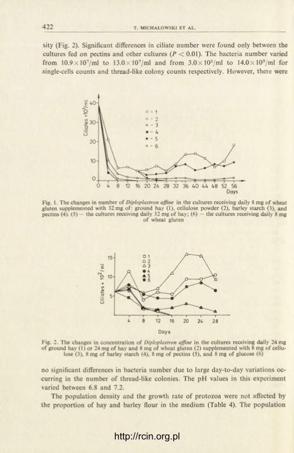
•422 T. MICHALOWSKI ET AL.
sity (Fig. 2). Significant differences in ciliate number were found only between the cultures fed on pectins and other cultures (P < 0.01). The bacteria number varied from 10.9x 10⁷/ml to 13.0xl07 /ml and from 3 .0x l0 5 /ml to 14.0xl05 /ml for single-cells counts and thread-like colony counts respectively. However, there were
Fig. 1. The changes in number of Diploplastron affine in the cultures receiving daily 8 mg of wheat gluten supplemented with 32 mg of: ground hay (1), cellulose powder (2), barley starch (3), and pectins (4). (5) — the cultures receiving daily 32 mg of hay; (6) — the cultures receiving daily 8 mg
of wheat gluten
Days
Fig. 2. The changes in concentration of Diploplastron affine in the cultures receiving daily 24 mg of ground hay (1) or 24 mg of hay and 8 mg of wheat gluten (2) supplemented with 8 mg of cellu-
lose (3), 8 mg of barley starch (4), 8 mg of pectins (5), and 8 mg of glucose (6)
no significant differences in bacteria number due to large day-to-day variations oc-curring in the number of thread-like colonies. The pH values in this experiment varied between 6.8 and 7.2.
The population density and the growth rate of protozoa were not affected by the proportion of hay and barley flour in the medium (Table 4). The population
http://rcin.org.pl

423
Table 4
The concentration of Diploplastron affine ( x 102/ml) and its growth rate ( x 102ml//day) in the cultures receiving different proportions of hay and barley flour in the diet. The
wheat gluten content of the diets used, was always 10% of daily ration
Diet A B
(1) Hay (36 mg) 9.0±2.57 1.1 ±0 .37 (2) Hay (28 mg), barley flour (8 mg) 12.0±3.83 1.5±0.44 (3) Hay (20 mg), barley flour (16 mg) 8.9±2.87 1.7±0.56 (4) Hay (12 mg), barley flour (24 mg) 11.8±6.48 1.4±0.55 (5) Hay (4 mg), barley flour (32 mg) 9 .7±3.44 1.2±0.66
A — concentration, B — growth rate
Table 5
The concentration of Diploplastron affine{x 102/ml), single-cells of bacteria ( x 107/ml), and thread-like colonies of bacteria ( x 105/ml) in the cultures receiving daily 36 mg of ground hay and 4 mg of
proteins of different solubility. Solubility of proteins in %
Proteins Solubility Ciliates Singlecells Colonies
Gluten 15 16.7±5.49 10.2±1.93 5.0±3.02 Glycinin 35 9 .3±1.79 • 9 .7±2.08 4 .0±3.21 Phaseolin 57 8.1 ±2 .57 10.0±1.63 5.0±2.13 Legumin 78 7.1 ± 2 . 3 0 9.6±1.83 7.0±5.61 Casein 100 6.7±1.89 8 .9±2.34 4 .0±3.12
usually showed a permanent increase on the days succeeding the culture transfer. It was often the highest within the first 24 h.
The ciliate number visibly tended to decrease with the increase of protein solu-bility (Table 5), but only the cultures fed on wheat gluten differed significantly from the others (P < 0.01). There was no significant differences in bacteria counts (P > 0.05).
D i s c u s s i o n
Among the four culture media studied, only "caudatum type" salt solution ( C o l e m a n et al. 1972) provided appropriate conditions for the development of Diploplastron affine. This solution contains a high amount of phosphates and its pH is about 6.8. Such conditions are preferred by many species of rumen ciliates ( C o l e m a n et al. 1972, 1976, M i c h a ł o w s k i et al. 1985).
The addition of rumen fluid to culture medium is considered as a factor stimu-lating the growth of protozoa in vitro ( C o l e m a n 1969). In the experiments descri-bed here the positive influence of rumen fluid addition was not observed, which
http://rcin.org.pl

•424 T. MICHALOWSKI ET AL.
might be connected with the fact that the diet consisted largely of unpurified plant material.
An addition of pure barley starch and especially of pectins decreased the num-ber of protozoa, and glucose caused the disappearance of ciliates. The reasons of this reaction are unknown. It was certainly not caused by the acidifying of the en-vironment, since the fall in pH values was not observed. The similar reaction was noted, however, in the case of Entodinium exiguum ( M i c h a ł o w s k i et al. 1985). Perhaps such substances may cause some unfavourable changes in the culture me-dium.
In contrast to Entodinium exiguum (Micha lowskie ta l .1985) , Diploplastron affine did not leact by increasing its number to the increase of the amount of starch-rich component in the diet. This fact may be connected with the ability of these pro-tozoa to utilize the carbohydrates from the plant cell walls. This supposition can be supported by microscopic observations showing that Diploplastron affine readily ingested plant fragments and by the fact that they survived for two months on the mixture of pure cellulose and wheat gluten (Fig. 1). The literature data also reported the occurrence of a-D-glucosidase and (3-D-glucosidase in the cells of these pro-tozoa (Wi l l i ams et al. 1984).
The tendency to a negative correlation between the food protein solubility and population density of Diploplastron affine was observed. A similar correlation was found also in Entodinium exiguum ( M i c h a ł o w s k i et al. 1985) and Entodinium caudatum ( M u s z y ń s k i et al. 1985). We suppose that reason for that is perhaps a small ability of these ciliates to uptake high-molecular compounds soluble in the culture medium. From the other hand the bacteria number was too low to cover the nutritive requirements of the numerous ciliate population if there was no in-soluble (i.e., readily ingestible by ciliates) protein in the diet.
In the present experiment wheat gluten allowed the population of Diploplastron affine to maintain a higher density that the other pioteins did. Our other experi-ments showed that wheat gluten enabled the maintenance of high population den-sity of other rumen ciliates, although it is relatively poorly degradable by both protozoa and bacteria (unpublished). Perhaps, apart from the fact that the solu-bility of wheat gluten is much lower than of other proteins used, the presence of components like lipids and carbohydrates in the gluten molecule (Mi f l in et al. 1983) had a positive effect on ciliate growth. This problem needs further investiga-tions.
REFERENCES
C o l e m a n G. S. 1962: The preparation and survival of almost bacteria-free suspensions of Ento-dinium caudatum. J. Gen. Microbiol., 28, 271-281.
C o l e m a n G. S., D a v i e s J. I. and C a s h M. A. 1972: The cultivation of the rumen ciliates Epidi-nium ecaudatum caudatum and Polyplastron multivesiculatum in vitro. J. Gen. Microbiol., 73, 509-521.
http://rcin.org.pl
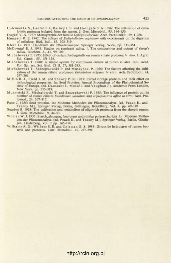
FACTORS AFFECTING THE GROWTH OF DIPLOPLASTRON 425
C o l e m a n G. S., L a u r i e J. I., Ba i l i ey J. E. and H o l d g a t e S. A. 1976: The cultivation of cellu-lolytic protozoa isolated from the rumen. J. Gen. Microbiol., 95, 144-150.
D o g i e l V. A. 1927: Monographie der familie Ophryoscolecidae. Arch. Protistenkd., 59, 1-288. H u n g a t e R. E. 1942: The culture of Eudiplodinium ceglectum with experiments on the digestion
of cellulose. Biol. Bull., 83, 303-319. K l e i n G. 1933: Handbuch der Pflanzenanalyse. Springer Verlag, Wien, pp. 333-354. M c D o u g a l E. J. 1948: Studies on ruminant saliva. 1. The composition and output of sheep's
saliva. Biochem. J., 43, 99-109. M i c h a ł o w s k i T. 1975: Effect of certain feedingstuffs on rumen ciliate protozoa in vitro. J. Agric.
Sei. Camb., 85, 151-158. M i c h a ł o w s k i T. 1980: A simple system for continuous culture of rumen ciliates. Bull. Acad.
Pol. Sei. ser. Sei. Biol. Cl II, 27, 581-583. M i c h a ł o w s k i T., S z c z e p k o w s k i P. and M u s z y ń s k i P. 1985: The factors affecting the culti-
vation of the rumen ciliate protozoon Entodinium exiguum in vitro. Acta Protozool., 24, 297-305.
M i f l i n B. J., F i e ld J. M. and Shewry P. R. 1983: Cereal storage proteins and their effect on technological properties. In: Seed Proteins; Annual Proceedings of the Phytochemical So-ciety of Europa, (ed. D a u s s a n t J., Mosse J. and V a u g h a n J.), Academic Press London, New York. pp. 255-319.
M u s z y ń s k i P., M i c h a ł o w s k i T. and S z c z e p k o w s k i P. 1985: The influence of protein on the number of rumen ciliates Entodinium caudatum and Diploplastron affine in vitro. Acta Pro-tozool., 24, 307-317.
Pace J. 1955: Seed proteins. In: Moderne Methoden der Pflanzenanalyse, (ed. P e a c h K. and T r a c e y M.), Springer Verlag, Berlin, Göttingen, Heidelberg, Vol. 4, pp. 69-105.
S u g d e n B. 1953: The cultivation and metabolism of oligotrich protozoa from the sheep's rumen. J. Gen. Microbiol., 9, 44-53.
W h e l a n W. J. 1955: Starch, glycogen, fructosans and similar polysaccharides. In: Moderne Metho-den der Pflanzenanalyse, (ed. Peach K. and T r a c e y M.), Springer Verlag, Berlin, Göttin-gen, Heidelberg, Vol. 2 pp. 145-196.
W i l l i a m s A. G., W i t h e r s S. E. and C o l e m a n G. S. 1984: Glycoside hydrolases of rumen bac-teria and protozoa. Curr. Microbiol., 10, 287-294.
http://rcin.org.pl

http://rcin.org.pl

ACTA PROTOZOOLOGICA Vol. 25, No. 4, pp. 427-432 (1986)
Sur une nouvelle espèce de cilié Paranophrys Carnivora sp. n. (Scuticociliatida)
A n n a C Z A P I K et N o r b e r t W I L B E R T
Institut Zoologique de l'Université Jagellone, 30-063 Krakôw, Oleandry 2a, Pologne et Institut de Zoologie, Université de Bonn, Poppelsdorfer Schloss, D.5300 Bonn, RFA
Received on 21 March 1986
Synopsis. Une nouvelle espèce du genre Paranophrys a été trouve dans l'eau de mer. Elle se distingue des autres représentants de ce genre par l'ensemble des caractères suivants: la taille moyenne mesure 40x20[jim, le nombre des cinéties somatiques est 21-22, la parorale en descendant s'incurve vis-à-vis M 3 , en formant un petit sinus. Ce cilié se nourrit des tissus d'animaux. Au cours de la division ce sont la parorale et le scuticus qui forment l'appareil buccal de l'opisthe.
Depuis quand T h o m p s o n et Berger (1965) ont créé le nouveau genre Para-nophrys et décrit la première espèce Paranophrys marina vivant dans la cavité gastrale de Plumularia, trois autres espèces ont été trouvées: deux dans l'eau marine et une dans l'eau douce. Ici nous présentons une nouvelle espèce différente des précédantes.
M a t é r i e l et t e c h n i q u e s
Ce cilié a été trouvé par Wi lbe r t dans un prélèvement provenant de la côte d'Israël et presque en même temps par Czap ik dans une mare rocheuse sur la côte de l'ile Malta. C'est une espèce histophage, qui se nourrit des tissus frais des animaux. On la cultivait dans l'eau marine, en lui don-nant comme nourriture les vers coupés en morceaux (.Enchytraeidae).
La forme du corps est variable, suivant les stades trophiques. Si la nourriture est abondante les ciliés se multiplient très rapidement. Privés de la nourriture pendant deux ou trois semaines les ciliés deviennent très minces et petits et la plupart d'eux disparaissent. On n'a pas observé la for-mation des kystes.
La morphologie et la stomatogenèse ont été étudié sur les préparations imprégnées au proté-inate d'argent d'après la méthode de Wi lbe r t et aussi au nitrate d'argent d'après C h a t t o n afin de mettre en evidence les fibres argentophiles.
http://rcin.org.pl

428 A. CZAPIK ET N. WILBERT
Morphologie générale de Paranophrys Carnivora sp. n.
Le cilié est fusiforme, sa partie antérieure un peu amincie, le bout pcstérieur arrondi (Pl. I). Le corps du théronte mesure 36-40 (j.m de long et 18-20 ce large, celui du trophonte 38-35 [i.m de long et 19-28 de large. La vacuole contra:tile est
A B
Fig. 1. Morphologie de Paranophrys Carnivora. A, B — animal imprégné au protéinat< d'argent: A — côté ventral, B — côté dorsal. C, D — animal imprégné au nitrate d'argent: C - système
argentophil, D — pôle postérieur
http://rcin.org.pl

PARANOPHRYS CARNIVORA SP. N. 429
située au bout du corps. La ciliature somatique consiste en 21-22 cinéties. Les ex-trémités des cinéties n'atteignent pas le pôle postérieur mais se terminent sur un fibre argentophile qui l'entoure. Sur le pôle postérieur il y a un long cil caudal.
Au-dessous du péristome un petit groupe des cinétosomes (3-5) forment le scu-ticus. Plus bas une ligne argentophile désigne le lieu où se trouve le cytopyge.
L'appareil buccal s'étend sur la partie antérieure du corps, presque jusqu' à son équateur (Fig. 1 A). Les trois membranelles sont disposées en file méridienne. Les deux premières membranelles sont allongées, presque de la même longueur. Mi me-sure 8.2 [a.m de long et contient deux rangées des cinétosomes, M2 (9.2 [xm de long) est plus large — elle contient quatre rangées des cinétosomes dont une partie est disposée d'une façon irrégulière. M 3 , très petite, a la forme d'un triangle irrégulier. La parorale commence au lieu, où se termine M l 5 en descendant elle s'incurve vis-à-vis de la M 3 et plus bas elle entoure la partie droite de la cavité buccale.
Stomatogenèse
Le début de la stomatogenèse chez Paranophrys Carnivora se manifeste comme chez Paranophrys carcini, où elle a été décrite minutieusement par G r o l i è r e e t L e g -lise (1977): la partie antérieure de la parorale se dédouble (Fig. 2 B) et les cinéto-somes de sa partie postérieure ainsi que ceux du scuticus se multiplient et forment d'abord trois et plus tard quatre champs (Fig. 2 C). A l'étape suivant la parorale et le scuticus du proter se reconstruit (Fig. 2 B). Les membranelles restent intactes et ne participent pas à ce processus. Ensuite trois petits champs du côté droit disparaissent et à leur place la parorale de l'opisthe se forme en entourant un grand champ en forme de V renversé et le scuticus au-dessous de lui (Fig. 2 E). Le grand champ donne l'origine aux membranelles de l'opisthe. Au moment où la division commence et les cinéties somatiques s'interrompent, les structures orales chez le proter subissent une désintégration afin de se reconstituer plus tard (Fig. 2 F).
D i s c u s s i o n
Par sa morphologie, structures buccales et le type de stomatogenèse l'espèce trouvé par nous appartient sans doute au genre Paranophrys. Jusqu'ici on a décrit 4 espèces de ce genre, qui se distinguent parmi eux par le nombre des cinéties, par les détails dans la structure du péristome et par le milieu, dans lequel ils vivent (Table 1), les différences dans les dimensions du corps étant trop petites pour être significatif. S'il s'agit des cinéties somatiques on voit que chez Paranophrys magna ainsi que chez l'espèce décrite ici elles sont plus nombreuses, les autres en possè-dent beaucoup moins. Finalement c'est surtout d'après les différences dans la struc-ture de la bouche qu'on peut distinguer les espèces. Figure 3 présente schemati-
5 — A c t a Protozo l . 25/4
http://rcin.org.pl

70 A. CZAPIK ET N. WILBERT
http://rcin.org.pl

PARANOPHRYS CARNIVORA SP. N. 431
Table 1
Comparaison des espèces du genre Paranophrys Thompson et Berger, 1965
Espece Dimensions en pim Cinéties Nourriture Milieu
Paranophrys marina Thompson et Berger, 1965
39-44 de long 19 de large
10 tissus mer
Paranophrys magna Borror, 1972
45-75 de long 17-25 de large
21 bacteries? mer
Paranophrys thompsoni Didier et Wilbert, 1976
40-45 de long 15-17 de large
13 bacteries eau douce
Paranophrys carcini Groliere et Leglise
35-55 de long 12-14 de large
11 tissus mer
Paranophrys Carnivora
sp. n. 36-56 de long 18-35 de large
21-22 tissus mer
/ \ / /
Ii \
1. - . y
Fig. 3. Appareil buccal chez différentes espèces de Paranophrys (schematiquement): A — P. magna Borror, B — P. carcini Grolière et Leglise, C — P. marina Thompson et Berger, D — P. thompsoni
Didier et Wilbert, E — P. Carnivora
quement les peristomes de toutes les espèces de Paranophrys. Mi est toujours al-longée et mince, M 3 toujours petite. C'est M2 qui est la plus variable — chez P. magna allongée mais plutôt mince, chez P. Carnivora sp. n. allongée et large, chez trois autres espèces courte et large. Un autre caractère, qui distingue bien P. Carnivora c'est la forme de sa parorale. Chez toutes les espèces connues jusqu'ici la parorale en descendant vers le cytostome suit la ligne droite, seulment chez P. marina elle est légèrement courbée. Chez l'espèce que nous avons trouvée la parorale s'incurve vers la M 3 , en formant un petit sinus.
Fig. 2. Morphogenèse chez Paranophrys Carnivora : A — appareil buccal de l'animal, B — dédouble-ment de la parorale, multiplication des cinétosomes de sa partie postérieure et ceux du scuticus, C — formation des champs, D — reconstitution de la parorale du proter, E — formation de la
parorale de l'opisthe, F — formation des membranelles de l'opisthe
http://rcin.org.pl

432 A. CZAPIK ET N. WILBERT
On a constaté, que trois espèces appartenant au genre Paranophrys se nourissent des tissus des animaux (P. carcini, P. marina et P. carnivora). En ce qui concerne la Paranophrya magna, B o r r o r ne définit pas la nourriture de cet animal. Il dit seule-ment que "the animal feeds by rubbing the buccal apparatus against the surface scum". Ce cilié absorbe probablement les bactéries présentes dans l'écume. Parano-phrys thompsoni trouvée dans les pontes des Chironomides se nourrit, d'après les auteurs, des bactéries et flagellés qui se développent dans la gelée, mais mise en culture dans les conditions pareilles elle ne se developpe pas bien. 11 est possible, que ces dernières espèces sont histophages facultatifs et comme les autres espèces de ce genre ont besoin des tissus des animaux.
BIBLIOGRAPHIE
B o r r o r A. C. 1972: Tidal marsh ciliates (Protozoa), morphology, ecology, systematics. Acta Pro-tozool., 10, 29-72.
D i d i e r P. et W i l b e r t N. 1976: Sur une espèce de cilié hymenostome Paranophrys thompsoni sp. n. cohabitant avec l'infusoire Espeioia mucicola dans les pontes de Chironomies. Protistolo-gica, 12, 335-340.
G r o l i è r e C. A. et Legl i se M. 1977: Paranophrys carcini n. sp. cilié Philasterina récolté dans l'hé-molymphe du crabe Cancer pagurus Linné. Protistologica, 13, 503-507.
T h o m p s o n J. C. and B e r g e r J. 1965: Paranophrys marina n. g. n. sp., a new ciliate associated with a hydroid from the north east Pacific (Ciliata, Hymenostomatida). J. Protozool., 12, 527
EXPLICATION DE PLANCHE I
1 : Paranophrys Carnivora: côté ventral et dorsal
http://rcin.org.pl

A C T A PROTOZOOL. Vol . 25, N o . 4 P L A N C H E I
A. Czapik et N. Wilbert auctores phot.
http://rcin.org.pl

http://rcin.org.pl

ACTA PROTOZOOLOGICA Vol. 25, No. 4, pp. 433-432 (1986)
The Ciliate Paratrichodina africana sp. n. (Peritricha, Trichodinidae) from Tilapia fish
(Cichlidae) from Africa
S. L. K A Z U B S K I and S. A. M. E L - T A N T A W Y 1
Laboratory of Parasitic Protozoa, W. Stefański Instutite of Parasitology, Polish Academy of Sciences, Pasteura 3, P.O. Box 153, 00-973 Warszawa, Poland
Received on 15 May 1986
Synopsis. A new species of trichodina, Paratrichodina africana sp. n. is described from fishes of the genus Tilapia from Lake Victoria (Kenya) and the Nile Delta (Egypt). The new species is characterized by an elongate outer fragment of the cen-tral part of the denticle forming a prominent spine-like processus. Metric and meris-tic data concerning the new species are given jointly and separately for both locali-ties. Affinities of the new species with other representatives of the genus Paratricho-dina Lom, 1963 are discussed.
In the material collected by the senior author from Lake Victoria in Kenya ( K a z u b s k i 1985, 1986) and by the junior author in the Nile Delta (E l -Tan tawy and K a z u b s k i 1985, 1986) a new species of trichodina, parasitizing fish of the genus Tilapia Smith, was found. A precise analysis showed that the trichodinas from both localities were identical. By the body structure and the adhesive disc and den-ticle shape these trichodinas appeared to be the most closely related with the genus Paratrichodina Lom, 1963, but they represented the new species. Due to origin of the new species from the African continent, the name Paratrichodina africana sp. n. is proposed for it.
M a t e r i a l a n d M e t h o d s
The trichodinas were collected in the following localities: (1) Lake Victoria in Kenya. The ciliates were found on gills of all 5 examined fishes Tilapia sp.,
caught by anglers near the molo in Homa Bay, South Nyanza Distr., on 3rd August 1983. The para-sites were not numerous.
1 Permanent address: Mansoura University, Faculty of Sciences, Egypt
http://rcin.org.pl

Tab
le 1
Par
atri
chod
ina
afri
cana
sp.
n.,
met
ric
and
mer
istic
dat
a (d
imen
sion
s ar
e gi
ven
in p
irn)
Tri
chod
inas
fro
m t
he
Tri
chod
inas
fro
m
Cha
ract
ers
Nile
Del
ta
Lak
e V
icto
ria
Whole
m
ate
rial
rang
e |
M
SD
|
n
rang
e |
M
| S
D~
| n
ra
nge
j M
| S
D
Bod
y di
amet
er
24-4
5 32.7
2.9
228
26
-42
33.5
4
.7
21
24
-45
32.8
3.9
249
Adh
esiv
e di
sc d
iam
eter
wit
h bo
rder
mem
bran
e 18-2
9
23
.8
2.4
108
19-2
7
22.9
2.6
8
18-2
9
23.7
2.4
116
Adh
esiv
e di
sc d
iam
eter
wit
hout
bo
rder
mem
bran
e 15-2
5
20.5
1.3
106
15-2
3
19.1
2
.7
9
15-2
5
20.4
1.4
115
Den
ticul
ate
ring
dia
met
er
8.5-
16.5
12.2
1.0
81
9-1
6
12.0
2
.2 8
8.
5-16
.5
12.2
1.1
89
Num
ber
of d
entic
les
19-2
7
23.9
1.5
66
20
-27
22.5
2.4
6
19-2
7
23.8
1.7
72
Hei
ght
of d
entic
le
4-7
6*
- 53
5-7
6.5
* -
6
4-7
6*
- 59
• m
ode
http://rcin.org.pl

PARATRICHODINA AFRICANA SP. N. 435
(2) The Nile Delta in Egypt. The ciliates were found in February 1984 on 9 out of 17 examined fishes Tilapia nilotica, originating from a small branch of the river near El-Mansoura. The ciliates were localized mainly on gills, in small amount also on body surface. In 3 fishes the ciliates were abundant.
Smears were made from freshly caught fish. They were dried and silver impregnated with A g N 0 3 after the method of Klein. Examination of preparations and photographs were made under the light microscope Amplival C. Zeiss Jena.
R e s u l t s
Description of Paratrichodina africana sp. n.
Fairly small trichodinas with the adoral spiral forming a circle of about 270°. Height of the body, evaluated on the base of dried and silver impregnated prepara-tions, is equal to 3/4 of the body diameter. Dimensions of the body and of the ad-hesive disc and the number of denticles in the ciliates from both localities are given in Table 1.
The denticles have a pronounced centrifugal blade, well formed central part with characteristic spine and straight centripetal ray. The shape of the blade is similar to equilateral triangle with the top directed towards the adhesive disc centre. The right edge of the blade, when seen from the adhesive disc centre, is slightly con-cave and the left edge is convex. The outer edge of the blade, opposite to the top of the triangle, is also convex. The blades of the denticles have usually rounded tips, which give them a slightly irregular, semilunar shape. The central part of the den-
Fig. 1. Paratrichodina africana sp. n., 1-3 fragments of adhesive disc and denticulate ring; all spe-cimens from Tilapia nilotica f rom the Nile Delta
http://rcin.org.pl

436 S. L. KAZUBSKI A N D S. A. M. EL-TANTAWY
tide has an elongate outer fragment with the blade settled near its middle and the final part forming a characteristic spine-like processus. This element lies parallel to the corresponding central part of the neighbouring denticle, forming thus a ring 2-3 [i,m wide in the central part of the denticulate ring. This is probably a supporting structure. Centripetal rays are straight, finger shaped, fairly short and narrow. The bases of the blade and the ray are in a visibly shifted position. For each denticle there are five to six radial pins (Fig. 1, PI. I).
The border membrane surrounding the adhesive disc is fairly wide; it measures about 2.5 fi.m in width.
Horse-shoe shaped macronucleus measures 20-25 [xm in diameter and 7-9 pim in width. Micronucleus was not visible in preparations.
H o s t s : Tilapia nilotica in the Nile Delta and Tilapia sp. in Lake Victoria. L o c a l i z a t i o n : gills, rarely body surface of fish. D i s t r i b u t i o n : The Nile Delta, environs of El-Mansoura, Egypt and Lake Victoria, Homa
Bay, South Nyanza Distr., Kenya.
As the type host and type locality, the fish Tilapia nilotica from the branch of the Nile near El-Mansoura are indicated.
D i s c u s s i o n
Taking into account structure of the cell, length of the adoral spiral and shape of the denticles, the described species is the most closely related to the genus Para-trichodina Lorn, 1963. The denticles are composed of three elements: outer cen-trifugal blade, central part and centripatal ray. The blade is straight, in line with the radius of the adhesive disc. At its base there is no incision into which the spine-like processus of the neighbouring denticle could enter. The two last characters: the position of blade and the lack of incision, do not allow to allocate this trichodina to the genus Tripartiella Lom, 1959.
To the genus Paratrichodina Lom, 1963 belong several species characterized by small dimensions and bell-like body shape (Table 2). The considered species, P. africana sp. n., is the most similar to P. incisa (Lom, 1959), the type of the genus. However, it differs from the latter by smaller dimensions of the body, of the ad-hesive disc and of the denticulate ring, as well as by the number of denticles. The most important difference, however, concerns the structure of denticles. In P. afri-cana sp. n. the outer processus of the central part is longer and forms the spine. These elements of successive denticles adhere one to the other forming a wide ring in the central part of the denticulate ring, probably performing a supporting func-tion.
All other species of the genus Paratrichodina show greater differences in com-parison with the described new one, so it is not necessary to consider them in this place.
http://rcin.org.pl

Tab
le 2
Mai
n m
orph
omet
ric
data
of
the
spec
ies
allo
cate
d to
the
gen
us P
arat
rich
odin
a L
om,
1963
(di
men
sion
s ar
e gi
ven
in |
xm)
Bod
di
amet
er
Adh
esiv
e di
sc
Den
ticul
ate
ring
N
umbe
r of
Sp
ecie
s °
Y
iam
C
diam
eter
di
amet
er
dent
icle
s D
istr
ibut
ion
Ref
eren
ces
_ ra
nge
M
rang
e j
M
rang
e M
ra
nge
| M
Para
tric
hodi
na a
fric
ana
sp.
n.
24.0
-45.
0 32
.8
15.0
-25.
0 20
.4
8.5-
16.5
12
.2
19-2
7 23
.8
Afr
ica
pres
ent
pape
r P.
inc
isa
(Lom
, 19
59)
14.0
-54.
5 11
.0-3
6.0
7.0-
20.0
17
-31
Eur
ope,
Asi
a S
tein
in S
hulm
an
P. u
rale
nsis
Kas
chko
wsk
y et
Lom
, (1
984)
19
79
30.0
-44.
0 20
.0-2
6.0
12.0
-16.
0 18
-25
Asi
a (r
iver
Ob)
P.
pho
xini
Lom
, 19
63
30.0
-44.
0 30
.0-4
3.0
19.0
-30.
0 30
-39
Eur
opie
, A
sia
P. c
orlis
si L
om e
t H
alda
r, 19
77
27.0
-48.
0 17
.4-2
6.0
9.0-
16.0
17
-25
Eur
ope,
Asi
a P.
alb
urni
(V
oite
k, 1
957)
32
.2-6
3.2
25.3
-44.
2 18
.1-3
2.6
23-2
9 E
urop
e
http://rcin.org.pl

4 3 8 S. L. KAZUBSKI A N D S. A. M. EL-TANTAWY
The described species is the first representative of the genus on the Arican continent and in fishes of the family Cichlidae. All arguments presented ii this paper show that the described species is new. Due to its occurrence in Africi, the name P. africana sp. n. is proposed for it.
P. africana sp. n. has been primarily described under the name Tripartiela sp. in short communications at the VHth International Congress of Protozoolcgy in Nairobi ( K a z u b s k i 1985, E l - T a n t a w y and K a z u b s k i 1985). As it has been discussed above, this species shows closer affnities with the genus Paratriclodina Lom, 1963. This taxon was originally regarded as the subgenus within the ;enus Tripartiella (Lom 1963) and mentioned under this name by E r g e n s and Lom (1970). It was recently established as an independent taxon (Cor l i s s 1979, I te in in S h u l m a n 1984).
ACKNOWLEDGEMENTS
The authors wish to thank their Egyptian, Kenyan and Polish colleagues for helpful assist-ance in this investigations.
REFERENCES
C o r l i s s J. O. 1979: The Ciliated Protozoa. Characterization, Classification and Guide to the Li-terature, 2nded., Pergamon Press, 455 pp.
E l -Tan t awy S. A. M. and K a z u b s k i S. L. 1985: Trichodinid ciliates from Tilapia nilotiai from the Nile Delta (Egypt), Abstr. VII Int. Congr. Protozool., Nairobi, Kenya 1985, absr. 192.
E l -Tan tawy S. A. M. and K a z u b s k i S. L. 1986: The Trichodinid ciiiates from fish, Tilipia ni-lotica from the Nile Delta (Egypt), Acta Protozool., 25, 439-444.
E r g e n s R. and Lom J. 1970: Puvodci parasitarnich nemoci ryb, Nakl. Ceskoslov. Akad Ved., Praha, 383 pp.
K a z u b s k i S. L. 1985: Trichodinid ciliates from a fish Tilapia sp. from Victoria Lake (Lenya), Abstr. VII Int. Congr. Protozool., Nairobi, Kenya 1985, abstr. 119.
K a z u b s k i S. L. 1986: The Trichodinid ciliates from fish Tilapia sp. from Lake Victoria (Kenya) and description of Trichodina equatorialis nom. nov., Acta Protozool., 25, 445-418.
L o m J. 1959: On the systematics of the genus Trichodinella Sramek-Husek ( = Brachyspira laabe). Acta Parasitol. Pol., 7, 573-590.
L o m J. 1963: The ciliates of the family Urceolariidae inhabiting gills of fishes (Trichodinella jroup). Acta Soc. Zool. Bohemoslav., 27, 7-19.
S t e i n G. A. in S h u l m a n S. S. 1984: ParazitiCeskie prostejsie, Opredelitel' parazitiv premovod-nyh ryb fauny SSSR, vol. 1, (red. O. N. Bauer) , Izd. "Nauka" Leningrad, 322-389
EXPLANATION OF PLATE I
Paratrichodina africana sp. n.
1-3 — adhesive disc (3000 x ) 4-5 — fragments of denticulate ring (5000 x ) All specimens from Tilapia nilotica from the Nile Delta
http://rcin.org.pl

ACTA PROTOZOOL. Vol 25, No. 4 PLATE I
S. L. Kazubski et S. A. M. El-Tantawy auctores phot.
http://rcin.org.pl

http://rcin.org.pl

ACTA PROTOZOOLOGICA Vol. 25, No. 4, pp. 439-432 (1986)
The Trichodinid Ciliates from Fish, Tilapia nilotica from the Nile Delta (Egypt)
S. A. M. E L - T A N T A W Y 1 and S. L. K A Z U B S K I 2
Laboratory of Parasitic Protozoa, W. Stefański Institute of Parasitology, Polish Academy of Sciences, Pasteura 3, P.O. Box 15, 00-973 Warszawa, Poland
Received on 15 May 1986
Synopsis. Four species of trichodinas were found on the fish Tilapia nilotica from the Nile Delta (near El-Mansoura), namely Trichodina centrostrigeata Basson, Van As et Paperna, 1983 Paratrichodina africana Kazubski et El-Tantawy, 1986 and two species of the genus Trichodina, whose specific determination was not possible. Tri-chodina sp. I seems to be identical with the species determined by Basson et al. (1983) as T. nigra Lom, 1961 occurring in Europe. Trichodina sp. II is probably a new species, not recorded till now in fish from Africa. Descriptions of collected trichodina species are given and their systematic status discussed. Some problems connected with parasitism and geographic distribution of the species are also dis-cussed.
Trichodinas parasitizing fish in Africa are rather poorly known. The only se-rious publication on this subject concerns the parasites of cichlid and cyprinid fish is South Africa (Basson et al. 1983). In this paper 8 species representing the genus Trichodina, one species of Trichodinella and one Tripartiella, are recorded. A small amount of material from the eastern shore of Lake Victoria was also collected ( K a z u b s k i 1985, 1986). The trichodinas from the Nile have not been investigated till now, although in Isreal the parasites of fish were thoroughly studied. The aim of the present paper was to pay attention to this group of fish parasites on the African continent.
1 Permanent address: Mansoura University, Faculty of Sciences, Egypt. 2 To whom requests for reprints should be directed.
http://rcin.org.pl
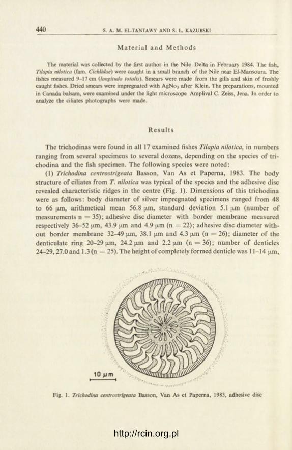
440 S. A. M. EL-TANTAWY A N D S. L. KA.ZUBSKI
M a t e r i a l a n d M e t h o d s
The material was collected by the first author in the Nile Delta in February 1984. The fish, Tilapia nilotica (fam. Cichlidae) were caught in a small branch of the Nile near El-Mansoura. The fishes measured 9-17 cm (longitudo totalis). Smears were made from the gills and skin of freshly caught fishes. Dried smears were impregnated with AgNo3 after Klein. The preparations, mounted in Canada balsam, were examined under the light microscope Amplival C. Zeiss, Jena. In order to analyze the ciliates photographs were made.
R e s u l t s
The trichodinas were found in all 17 examined fishes Tilapia nilotica, in numbers ranging from several specimens to several dozens, depending on the species of tri-chodina and the fish specimen. The following species were noted:
(1) Trichodina centrostrigeata Basson, Van As et Paperna, 1983. The body structure of ciliates from T. nilotica was typical of the species and the adhesive disc revealed characteristic ridges in the centre (Fig. 1). Dimensions of this trichodina were as follows: body diameter of silver impregnated specimens ranged from 48 to 66 [xm, arithmetical mean 56.8 jxm, standard deviation 5.1 am (number of measurements n = 35); adhesive disc diameter with border membrane measured respectively 36-52 [j.m, 43.9 [xm and 4.9 (j.m (n = 22); adhesive disc diameter with-out border membrane 32-49 jj.m, 38.1 jjt.m and 4.3 [j.m (n = 26); diameter of the denticulate ring 20-29 [xm, 24.2 [j,m and 2.2 pun (n = 36); number of denticles 24-29,27.0 and 1.3 (n = 25). The height of completely formed denticle was 11-14 u.m,
Fig. 1. Trichodina centrostrigeata Basson, Van As et Paperna, 1983, adhesive disc
http://rcin.org.pl

TRICHODINID CIHATES FROM NILE DELTA 4 4 1
most frequently (mode) 12 pun (n = 12). The border membrane was fairly wide, measuring up to 4.5 ;xm, usually about 3 [xm.
T. centrostrigeata was found on the gills of 4 specimens of Tilapia nilotica. Some-times, the ciliates occurred numerously.
(2) Paratrichodina africana Kazubski et El-Tantawy, 1986. Small-sized ciliates with adoral spiral of about 270°. Body dimensions: diameter of silver impregnated specimens ranged from 24 to 45 [xm, arithmetical mean 32.7 [j,m, standard devia-tion 2.9 [xm (number of specimens measured n = 228); diameter of adhesive disc with border membrane respectively 18-29 ;xm, 23.8 (xm and 2.4 [xm (n = 108); diameter of adhesive disc without border membrane 15-25 [xm, 20.5 |xm and 1.3 [xm (n = 106); diameter of denticulate ring 8.5-16.5 fxm, 12.2 (xm and 1.0 fxm (n = 81), number of denticles 19-27, 23.9 and 1.5 (n = 6 6 ) . Height of denticles 4-7 |xm, most frequently (mode) 6 fxm. The structure of denticles was typical of the genus having a characteristic spine-like processus of the outer fragment of the central part. Detailed description of the species is given by K a z u b s k i and E l -Tan tawy (1986).
P. africana was found in 9 out of 17 fishes originating from the Nile. The ciliates occurred mainly on gills, rarely on the body surface. In 3 fishes the ciliates were abundant — dozens of specimens were found on preparations.
Besides, two other species of Trichodina were found on gills and skin of exam-ined fish. The condition of these ciliates in the preparations was poor, which made species identification impossible. However, some elements of their structure, e.g. adhesive discs, fairly well preserved in preparations, allowed us to made the following description:
Trichodina sp. I. Ciliates somewhat smaller then T. centrostrigeata. Silver im-pregnated specimens measured 47-52 (xm in diameter, mean 50.5 (xm, standard deviation 2.4 jxm (n = 4 ) ; diameter of adhesive disc with border membrane meas-ured respectively 40-45 [xm, 41.7 [xm and 1.7 fxm (n = 6); diameter of adhesive disc without border membrane 31-41 fxm, 36.5 [xm and 3.0 x̂m (n = 13); diameter of denticulate ring 19-30 [xm, 24.3 [xm and 2.5 [xm (n = 32); number of denticles 17-21, 18.9 and 0.9 (n = 32). Height of denticles 9-12 fxm, most freqently (mode) 9 and 10 [xm. The denticles had strongly curved, sickle-like blades, massive central part and fairly short, thick rays, slightly narrowing towards the adhesive disc centre (Fig. 2). The border membrane was 2.0-2.5 [xm wide; there were 8-9 radial pins to one denticle.
Trichodina sp. I was found on the skin, rarely on the gills of 11 examined fishes. Infections was rather mild.
Trichodina sp. II. Body dimensions larger than in all other trichodinas mentioned in this paper. Body diameter of silver impregnated specimens ranged from 70 to 118 ;xm, mean 94.1 [xm, standard deviation 18.6 (xm (n = 9 ) ; diameter of adhesive disc with border membrane measured respectively 55-80 jxm, 65.5 [xm and 8.3 fxm
http://rcin.org.pl
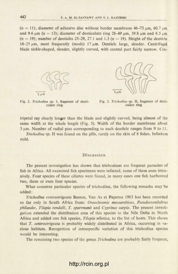
442 S. A. M. EL-TANTAWY AND S. L. KA.ZUBSKI
(n = 11); diameter of adhesive disc without border membrane 46-73 fxm, 60.7 pn and 9.6 [xm (n = 13); diameter of denticulate ring 28-49 [xm, 39.8 fxm and 6.5 j.m (n = 19); number of denticles 25-29, 27.1 and 1.3 (n = 19). Height of the denti;!e 16-25 |i.m, most frequently (mode) 17 [i.m. Denticle large, slender. Centrifugal blade sickle-shaped, slender, slightly curved, with central part fairly narrow. Ctn-
Fig. 2. Trichodina sp. I, fragment of denti- Fig. 3. Trichodina sp. II, fragment of denti-culate ring culate ring
tripetal ray clearly longer than the blade and slightly curved, being almost of he same width at the whole length (Fig. 3). Width of the border membrane about 3 (j.m. Number of radial pins corresponding to each denticle ranges from 9 to 11.
Trichodina sp. II was found on the gills, rarely on the skin of 8 fishes. Infect on mild.
D i s c u s s i o n
The present investigation has shown that trichodinas are frequent parasites of fish in Africa. All examined fish specimens were infected, some of them even inten-sively. Four species of these ciliates were found, in many cases one fish harboured two, three or even four species.
What concerns particular species of trichodina, the following remarks may be added:
Trichodina centrostrigeata Basson, Van As et Paperna 1983 has been recorded so far only in South Africa from: Oreochromis mossambicus, Pseudocrenilabrus philander, Tilapia rendalli, T. sparrmanii and Cyprinus carpio. The present investi-gation extended the distribution area of this species to the Nile Delta in North Africa and added one fish species, Tilapia nilotica, to the list of hosts. This shows that T. centrostrigeata is probably widely distributed in Africa, occurring in va-rious habitats. Recognition of intraspecific variation of this trichodina species would be interesting.
The remaining two species of the genus Trichodina are probably fairly frequent,
http://rcin.org.pl

TRICHOD1NID CILIATES FROM NILE DELTA 443
but less numerous in comparison with the former one. Unfortunately, the collected material did not make their specific identification possible.
The species described in the present paper as Trichodina sp. I is probably iden-tical with that described by Bass on et al. (1983) under the name T. nigra. Dimensions of the body, of the adhesive disc and of the denticulate ring of this trichodina are very close to those of Trichodina sp. I in the present material, although the latter has slightly smaller number of denticles. However, the ranges of this character overlap in both groups of ciliates. The shape of denticles is also very similar. Un-fortunately, the lack of well-preserved specimens in the present material does not allow to identify these trichodinas. An additional difficulty is connected with the fact that Trichodina sp. I and probably the species described by B a s s o n et al. (1983) are not identical with Trichodina nigra Lom, 1961, widely distributed in Europe. The differences concern body and adhesive disc dimensions, number of denticles and, above all, the shape of denticles. However, the final decision in this matter requires further comparative studies of specimens from Europe, Egypt and South Africa, as well as the recognition of morphological variation of the considered spe-cies.
The trichodina described in the present paper as Trichodina sp. II is probably a new species, although it shows affinities with the species described by B a s s o n et al. (1983) as population A of Trichodina mutabilis from Carassius carassius. Dif-ferences concern body dimensions (our specimens are larger and have larger adhe-sive disc) and the shape of centripetal rays. As in the case of the former species, further studies are needed, including an evaluation of morphological variation, in order to resolve the problem of species identity.
Paratrichodina africana Kazubski et El-Tantawy, 1986 is a new species, found for the first time in Lake Victoria and in the Nile Delta. It is described in a separate paper ( K a z u b s k i and E l - T a n t a w y 1986). Taking into account its prevalence — 64.7% of Tilapia nilotica from the Nile Delta and 100% of Tilapia sp. from Lake Victoria ( K a z u b s k i 1986), it is a common parasite of fish in Africa. This species was recorded in a preliminary note for the International Congress of Protozoology in Nairobi (E l -Tantawy and K a z u b s k i 1985, K a z u b s k i 1985) under the name Tripartiella sp. More precise investigation on its morphology showed that it represents the genus Paratrichodina Lom, 1963 ( K a z u b s k i et E l - T a n t a w y 1986).
Summing up, it can be stated that trichodinas play an important role as parasites of cichlid fish in Africa, but further investigations are necessary to recognize better this group of parasitic ciliates in African continent. Such studies are also necessary to solve some taxonomic problems concerning the species discussed above.
ACKNOWLEDGEMENTS
The authors wish to thank their Egyptian colleagues for helpful assistance in this investigations.
http://rcin.org.pl

444 S. A. M. EL-TANTAWY AND S. L. KA.ZUBSKI
REFERENCES
B a s s o n L., Van As J. G. and P a p e r n a I. 1983: Trichodinid ectoparasites of cichlid and cyprinid fishes in South Africa and Israel, Syst. Parasitol., 5, 245-257.
E l -Tan t awy S. A. M. and K a z u b s k i S. L. 1985: Trichodinid ciliates from Tilapia nilotica from the Nile Delta (Egypt), Abstr. VII Int. Congr. Protozool., Nairobi, Kenya 1985. abstr. 192.
K a z u b s k i S. L. 1985: Trichodinid ciliates from a fish Tilapia sp. from Victoria Lake (Kenya), Abstr. VII Int. Congr. Protozool., Nairobi, Kenya 1985, abstr. 119.
K a z u b s k i S. L. 1986: The trichodinid ciliates from fish, Tilapia sp. from Lake Victoria (Kenya) and description of Trichodina equatorialis nom. nov., Acta Protozool., 25, 445-448.
K a z u b s k i S. L. and E l -Tan tawy S. A. M. 1986: The ciliate Paratrichodina africana sp. n. (Peri-tricha, Trichodinidae) from Tilapia fish (Cichlidae) from Africa, Acta Protozool., 25, 433-438.
http://rcin.org.pl

ACTA PROTOZOOLOGICA Vol. 25, No. 4, pp. 445-432 (1986)
The Trichodinid Ciliates from Fish, Tilapia sp. from Lake Victoria (Kenya) and Description
of Trichodina equatorialis nom. nov.
S t a n i s ł a w L. K A Z U B S K I
Laboratory of Parasitic Protozoa, W. Stefański Institute of Parasitology, Polish Academy of Sciences, Pasteura 3, P.O. Box 153, 00-973 Warszawa, Poland
Received on 15 May 1986
Synopsis. Two species of trichodina from fish, Tilapia sp. from Lake Victoria (Ke-nya) are described. The new name, Trichodina equatorialis nom. nov. is given to ciliates already described by Basson et al. (1983) from cichlid and cyprinid fish from South Africa and Israel under the name T. heterodentata Duncan, 1977. The differences between trichodinas from Africa and the Philippines are discussed and the appropriateness of creating a separate species for African trichodinas is justi-fied. The second species, Paratrichodina africana Kazubski et El-Tantawy, 1986 was described partly from the material for this paper. Attention is paid to the dis-tinctness of the fauna of trichodinas in Africa and in Europe.
Trichodinas parasitizing on fish in equatorial Africa have never been the object of investigation. Some information about this group of parasites originate from the peripheries of the African continent — from South Africa (Basson et al. 1983) and from Egypt (E l -Tan tawy and K a z u b s k i 1985, 1986). The task of the present paper is to fill this gap.
M a t e r i a l a n d M e t h o d s
The trichodinas were collected from five fishes, Tilapia sp. originating from the eastern part of Lake Victoria. The fishes were caught on the 3rd August 1983 by anglers near the molo in Homa Bay, South Nyanza Distr., Kenya. The fishes measured several centimeters in length. Smears made from gills, just after taking the fish out of the water, were dried and impregnated with silver nitrate after Klein. The preparations were examined under a light microscope Amplival C. Zeiss, Jena. In order to morphological analysis of the ciliates numerous photographs were made.
6 — Acta Protozol. 25/4
http://rcin.org.pl

446 S. L. KAZUBSKI
R e s u l t s
In all five examined specimens of Tilapia sp. two species of trichodinas were present.
(1) Trichodina equatorialis nom. nov. Dimensions of silver impregnated spec-imens from Tilapia sp. from Lake Victoria were as follows: diameter of the body ranged from 72 to 84 [xm, diameter of adhesive disc with border membrane 50-61 [xm, diameter of adhesive disc without border membrane 45-52 jxm, diameter of den-ticulate ring 27-39 fx.m, number od denticles 24-27, height of denticles 15-18 [xm, width of the border membrane 2-5 fxm. As the number of this trichodina was small, only the ranges of dimensions are given, mean values have not been calculated. The blades of the denticles were sickle-shaped and the centripetal rays straight.
Fig. 1. Trichodina equatorialis nom. nov., adhesive disc
The central part of the adhesive disc was not uniformly dark, but gave an appearance of marble pattern (Fig. 1).
This species of trichodina were found on 3 out of 5 examined fishes. The collected trichodinas, determined here as T. equatorialis nom. nov., are iden-
tical with ciliates described by Basson et al. (1983) from cichlid and cyprinid fish in South Africa and Israel, and especially with the specimen represented in Fig. 5 of the quoted paper (p. 247), and classified to the species T. heterodentata Duncan, 1977. The present finding confirms the differences observed by the mentioned authors between trichodinas from Africa and Israel and those described by D u n c a n (1977) from the Philippines. These differences concern the structure and shape of denticles, especially of their centripetal rays. The rays of trichodinas from Africa are much longer than the blades, slender and gradually narrowing, while the rays in T. hete-
http://rcin.org.pl

TRICHODINID CILIATES FROM LAKE VICTORIA 447
rodentata from the Philippines are short and wide, abruptly tapering at the end. These differences prove that the piocess of formation of the centripetal rays differs between the both species of trichodinas. The described differences enable the author to recognize these trichodinas as distinct species: T. heterodentata described by D u n -c a n (1977) from the Philippines and the trichodinas from Africa and Israel recorded by B a s s o n et al. (1983) and in the present paper. For the latter the name T. equato-rialis nom. nov. is proposed (syn. T. heterodentata sensu Basson, Van As et Pa-perna, 1983).
(2) Paratrichodina africana Kazubski et El-Tantawy, 1986. Small trichodinas with adoral spiral of about 270°. Specimens from Tilapia sp. from Lake Victoria had the following dimensions: diameter of the body of silver impregnated speci-mens — mean 33.5 {xm, standard deviation 4.7 [xm, diameter of adhesive disc with border membrane respectively 22.9 [xm and 2.6 [xm, diameter of adhesive disc without border membrane 19.1 [xm and 2.7 jxm, diameter of denticulate ring 12.0 |xm and 2.2 [xm, number of denticles 22.5 and 2.4, height of denticle 5-7 [xm, most frequently (mode) 6.5 ;xm. The structure of denticles corresponded with that in other representatives of the genus Paratrichodina Lom, 1963, with the characteristic outer piocessus of the central part of the denticle. A detailed description of the species is given by K a z u b s k i and E l - T a n t a w y (1986). This species has been mentioned as Tripartiel/a sp. in a communication for the Vllth International Con-gress of Protozoology in Nairobi ( K a z u b s k i 1985).
P. africana was found on the gills of all 5 examined fishes. In some cases the intensity of infection was high. Finding of the same species on gills and skin of T. ni-lotica from the Nile Delta ( E l - T a n t a w y and K a z u b s k i 1986) suggests that the species is a common parasite of cichlid fish.
D i s c u s s i o n
Investigations on trichodinas suggest that these ciliates are common parasites of fish in Africa. In publications by Basson et al. (1983), E l - T a n t a w y and K a z u b -ski (1985, 1986) and K a z u b s k i (1985) as many as 11 species have been recorded, including some new species. This number is likely to increase after new investigations are performed. However, great care must be taken in identifying the African spe-cies of trichodina with the species occurring in other regions of the world, living in different climatic conditions and on other host species. As an example, the dif-ferences in specific characters observed between trichodinas formerly identified as T. heterodentata from the Philippines and those from Africa and Israel may be indicated. A detailed analysis of the material resulted in creating a new species, T. equatorialis, for trichodinas from Africa and Israel. Another example may be the species identified by B a s s o n et al. (1983) as Trichodina pediculus (specimen represented in Fig. 14 of the quoted paper). By the shape of denticles, especially of
http://rcin.org.pl

448 S. L. KAZUBSKl
their centripetal rays, this species greatly differs from T. pediculus, a common par-asite of hydras in Europe, occurring also on tadpodes and small fish. These example underline the necessity of serious studies on morphological variation in this group of parasitic ciliates.
ACKNOWLEDGEMENTS
The author is greatly indebted to his colleagues in Kenya — professor Thomas R. Odhiambo and dr Leonard H. Otieno, of ICIPE in Nairobi, for organization of the trip to Lake Victoria, during which the material for this study was collected.
REFERENCES
B a s s o n L., Van As J. G. and P a p e r n a I. 1983: Trichodinid ectoparasites of cichlid and cyprinid fishes in South Africa and Israel, Syst. Parasitol., 5, 245-257.
D u n c a n B. L. 1977: Urceolariid ciliates, including three new species from cultured Philippine fish-es. Trans. Am. Microsc. Soc., 96, 76-81.
E l -Tan tawy S. A. M. and K a z u b s k i S. L. 1985: Trichodinid ciliates from Tilapia nilotica from the Nile Delta (Egypt), Abstr. VII Int. Congr. Protozool., Nairobi, Kenya 1985, abstr. 192.
E l -Tan tawy S. A. M. and K a z u b s k i S. L. 1986: The trichodinid ciliates from fish, Tilapia ni-lotica from the Nile Delta (Egypt), Acta Protozool., 25, 439-444.
K a z u b s k i S. L. 1985: Trichodinid ciliates from a fish Tilapia sp. from Victoria Lake (Kenya), Abstr. VII Int. Congr. Protozool., Nairobi, Kenya 1985, abstr. 119.
K a z u b s k i S. L. and E l -Tan tawy S. A. M. 1986: The ciliate Paratrichodina africana sp. n. (Peri-tricha, Trichodinidae) from Tilapia fish (Cichlidae) from Africa, Acta Protozool., 25, 433-438.
http://rcin.org.pl

ACTA PROTOZOOLOGICA Vol. 25, No. 4, pp. 449-452 (1986)
Occurrence of Trypanosoma mukasai Hoare, 1932 in Tilapia mossambica (Peters) from India
C. K. S I N H A
Zoology Department, Jhargram Raj College, Jhargram, Midnapur 721507, West Bengal, India
Received on 12 November 1985
Synopsis. Trypanosomes (Trypanosomatidae) are described from the peripheral blood of Tilapia mossambica (Peters) collected from Bongaon, 24-Parganas, West Bengal, India and identified as Trypanosoma mukasai Hoare, 1932 as redescribed by Bake r (1960). Trypanosoma choudhuryi Mandal, 1977 is compared with the present parasite and considered a synonym of Trypanosoma mukasai Hoare, 1932.
This is the first instalment of the series dealing with the trypanosomes of fresh water fishes in West Bengal, India. This communication records the occurrence of Trypanosoma mukasai Hoare, 1932 as redescribed by Baker (1960) from the dif-ferent species of Tilapia of East Africa, found in the peripheral blood of American Koi, Tilapia mossambica (Peters) collected from the ponds of Bongaon, 24-Parga-nas, West Bengal, India. This host fish popularly known as "American Koi" belongs to family Cichlidae (Perciformes).
M a t e r i a l a n d M e t h o d s
In a survey (Sinha 1984) 25 specimens of this host fish were examined and two were found positive for Trypanosoma infection. Blood was obtained from branchial blood vessels or caudal veins and stained with Giemsa's or Leishman's stains. Measurements of parasites were made from fixed and stained materials and Camera lucida drawings were made.
R e s u l t s
Trypanosoma mukasai Hoare
Trypanosoma mukasai Hoare, 1932: Parasitology, 24: 210, B a k e r (1960), Parasitology, 50: 515.
Trypanosoma choudhuryi Mandal, 1977: Acta Protozool., 16, 1-4
http://rcin.org.pl

450 C. K. SINHA
Description
Trypomastigofes are monomorphic, small, slender with bluntly pointed extrem-ities (Fig. 1 7-/2). These measure 21.4 |xm (18.9-24.1 p.m) in length including free flagellum and 1.5 |xm (1.2-2.2 jxm) in width. The cytoplasm is light purple in colour and contains a few volutin granules and vacuoles. The nucleus is oval, sub-central and stains deep purple measuring 1.9 x 1.0 [xm. It lies towards the anterior part. A karyosome is not found. The nuclear index is 1.3 fxm (1.1-1.35 fxm). The kinetoplast is small, sub-terminal, dot-like and deep purple in colour, measuring 0.9x0.3 [xm. The kinetoplastic index measures 1.04 [xm (1.02-1.12 [xm). The fla-
http://rcin.org.pl

451
gellum arises from the base of kinetoplast forming an undulating membrane with 2-4 folds. The free part of the flagellum is short at the anterior tip and measures 5.1 (xm (4.4-5.8 p.m).
Dividing forms of these trypomastigotes are not seen except for one individual (Fig. 1 12), where the cell body contains two nuclei but the kinetoplast remains un-divided.
H o s t : Tilapia mossambica (Peters) L o c a l i t y : Bongaon, 24-Parganas, West Bengal, Bagmari 24-Parganas, West Bengal, India S i te of i n f e c t i o n : Blood plasma
D i s c u s s i o n
Trypanosomes have been recorded in different species of Tilapia from Africa and India. W e n y o n (1908) described an unnamed species of Trypanosoma in Tila-pia zilli of the Nile river in Sudan. Leger and Leger (1914) found trypanosomes in Tilapia lata from the river Niger in the then French West Africa, but assigned no specific name to the parasite. These authors believed that the trypanosomes were identical with those described by W e n y o n (1908) from T. zilli. After a lapse of many years, D i a s (1955) described three species of trypanosomes viz., T. napo-lesi, T. rebeloi (in which were included the trypanosomes described by W e n y o n , 1908 in T. zilli) and T. serranoi (in which were included the trypanosomes which Neave , 1906 reported from Synodontis schall) from T. mossambica in Mozambique.
Bake r (1960) reviewed the trypanosomes of African fresh water fishes and considered Dias's three species as variants of a single species and regarded them as synonyms of T. mukasai Hoare, 1932. He also reported T. mukasai from the dif-ferent species of Tilapia (T. nilotica, T. esculenta, T. variabilis and T. leucosticta) of George and Victoria lakes of East Africa. M a n d a l (1977) described T. choudhuryi in T. mossambica collected from Bagmari, 24-Parganas, West Bengal, India.
The present parasite under discussion is identified as T mukasai Hoare, 1932 as redescribed by Bake r (1960) from different species of Tilupia on the basis of the morphological characters described above.
T. choudhuryi Mandal, 1977 resembles very much the present parasite as well as the small forms of T. mukasai in its morphology and dimensions. Therefore, T. choud-huryi and the present parasite are both identified as T. mukasai Hoare, 1932. The only difference is that the large forms of T. mukasai were not found in the present study, nor were these reported in T. choudhuryi.
A comparative study on the morphometric measurements of these trypano-somes is given in Table 1, which reveals that differences in dimensions, which al-most overlap each other, are minor in nature and not sufficient for considering T. choudhuryi and the present parasite as valid species distinct from T. mukasai Hoare, 1932.
http://rcin.org.pl

452 C. K. S1NHA
Table 1
Comparative measurements (in (im) of T. mukasai Hoare, 1932 as described by B a k e r (1960), T. choudhuryi Mandal, 1977 and the present parasite
Specification
Total length (including free flagellum)
Body length (excluding free flagellum)
Body width Free flagellum Posterior end to
kinetoplast Nucleus length Nucleus width Kinetoplast to nucleus Nucleus to anterior end Kinetoplast length Kinetoplast width
T. mukasai T. choudhuryi
Range
Small forms
22-35
16-27 1-2.7 5-12
0 - 2 1.7-3.5
0.75-1.7 5-17
4.5-11.5 0.25-1.5 0.25-1
Range
Large forms i Small forms
45-62
33-54 2-6 7-17
0.5-2 3-5.5
1.5-5.7 14-29 13-19
0.5-1.5 0.5-1
23-37.8
16.5-25.3 1.5-1.8 6.5-12.5
0.8-2.5 3.5-5.2
1-2.5 5.5-9.5 4.5-8.5
1-1.5 1-1.05
The present parasite
Range
Small forms
18.9-24.1
14.3-18.8 1.2-2.2 4.4-8.2
0.01-0.5 1.7-2.4 0.6-1.2 5.6-7.9 4.4-8.2 0.7-1.1 0.2-0.6
A C K N O W L E D G E M E N T S
The author is very much grateful to Dr. B. Dasgupta, Principal, Government College, Dar-jeeling for his valuable guidance. Best thanks are due to Dr. J. R. Baker, Institute of Terrestrial Ecology, Culture centre of Algae and Protozoa, Cambridge, England for going through the ma-nuscript and valuable comments.
R E F E R E N C E S
B a k e r J. R. 1960: Trypanosomes and dactylosomes f rom the blood of fresh water fish in East Africa. Parasitology, 50, 515-526.
D i a s J. A. T. S. 1955: Estudos sobre os hematozoairos dos peixes de aqua doce de Mocambique, de tripanossomas parasites de Tilapia mossambica (Peters, 1852). Mocambique, 82, 47-63.
H o a r e C. A. 1932: On protozoal blood parasites collected in Uganda. Parasitology, 24, 210-224. L e g e r M. and L e g e r A. 1914: Haemogregarine et trypanosome d'un poisson du Niger, Tilapia
lata. C. R. Seanc. Soc. Biol., 77, 183. M a n d a l A. K. 1977: Trypanosoma choudhuryi sp. nov. f rom Tilapia mossambica (Peters). Acta
Protozool., 16, 1-4. N e a v e S. 1906: Report of travelling pathologist and naturalist. Second. Rep. Wellcome Res.
Lab. Khartoum, 183-204. S i n h a C. K. 1984: Observations on certain protozoan parasites in the blood of some vertebrates.
Doctoral Thesis, University of North Bengal, Raja Rammohunpur, Darjeeling. W e n y o n C. M. 1908: Report of travelling pathologist and protozoologist. Third. Rep. Wellcome
Res. Lab., 121-168.
http://rcin.org.pl

ACTA PROTOZOOLOGICA Vol. 25, No. 4, pp. 453-432 (1986)
Neohirmocystidae fam. n., a New Family of Septate Gregarines
(Apicomplexa: Sporozoea) from Insects
S. G H O S E , S. K. R A Y and D. P. H A L D A R
Protozoology Laboratory, Department of Zoology, University of Kalyani, Kalyani 741 235,. W.B., India
Received on 12 November 1985, revised on 23 April 1986
Synopsis. A new family, Neohirmocystidae fam. n., is proposed in this communi-cation to accommodate the new genus Neohirmocystis gen. n. for the two septate gregarines from insects. These are Neohirmocystis grassei and N. dercetini from Tribolium castaneum and Dercetina sp. respectively. The affinity of the new family with the closely related families is discussed.
In the course of our study on septate gregarines {Apicomplexa:Sporozoea) in insects we obtained some parasites lacking an epimerite, having solitary as well as biassociative sporadins, spherical gametocyst dehiscing by simple rupture and double-walled spherical spores. The combination of such characters is unique among the group necessitating the proposition of a new genus to accommodate them.
In 1953 G r a s s e separated from the family Gregarinidae Labbe the genera Hir-mocystis, Hyalospora, Tettigonospora, Euspora, Caulocephalus, Sphaerocytis and Didymophyes and placed them in a new family Hirmocystidae. C h a k r a v a r t y (1959), however, regarded the family Hirmocystidae Grasse as a synonym of Didymophyidae Leger as in his opinion, "yet according to the laws of priority in nomenclature the first family name shall stand as it is formed from a valid generic name. The name Didymophyidae, therefore, should stand". K u d o (1971) accepted the family Didy-mophyidae to include the genus Didymophyes Stein only and regarded features like two to three sporadins in association and satellite without septum to be the characters of the family as did K a m m (1922). In a recent revision of the septate gregarines Le v ine (1979) accepted the family Hirmocystidae Grasse for those having epimerite ordinarily papilla-like or simple knob-like; gametocysts dehisce by sim-ple rupture; oocyst ellipsoidal, prismatic, fusiform, ovoid or even spherical. It is apparent from the above discussion that the gregarines obtained by us do not fit
http://rcin.org.pl
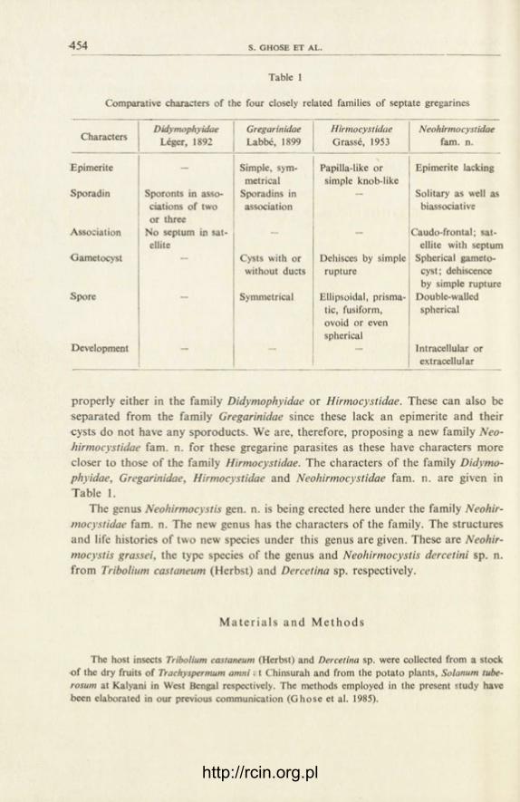
454 S. GHOSE ET AL.
Table 1
Comparative characters of the four closely related families of septate gregarines
Characters Didymophyidae
Leger, 1892 Gregarinidae Labbe, 1899
Hirmocystidae Grasse, 1953
Neohirmocystidae fam. n.
Epimerite — Simple, sym- Papilla-like or Epimerite lacking metrical simple knob-like
Sporadin Sporonts in asso- Sporadins in — Solitary as well as ciations of two association biassociative or three
Association No septum in sat- — — Caudo-frontal; sat-ellite ellite with septum
Gametocyst - Cysts with or Dehisces by simple Spherical gameto-without ducts rupture cyst; dehiscence
by simple rupture Spore - Symmetrical Ellipsoidal, prisma- Double-walled
tic, fusiform, spherical ovoid or even spherical
Development — — — Intracellular or extracellul ar
properly either in the family Didymophyidae or Hirmocystidae. These can also be separated from the family Gregarinidae since these lack an epimerite and their cysts do not have any sporoducts. We are, therefore, proposing a new family Neo-hirmocystidae fam. n. for these gregarine parasites as these have characters more closer to those of the family Hirmocystidae. The characters of the family Didymo-phyidae, Gregarinidae, Hirmocystidae and Neohirmocystidae fam. n. are given in Table 1.
The genus Neohirmocystis gen. n. is being erected here under the family Neohir-mocystidae fam. n. The new genus has the characters of the family. The structures and life histories of two new species under this genus are given. These are Neohir-mocystis grassei, the type species of the genus and Neohirmocystis dercetini sp. n. from Tribolium castaneum (Herbst) and Dercetina sp. respectively.
M a t e r i a l s and M e t h o d s
The host insects Tribolium castaneum (Herbst) and Dercetina sp. were collected from a stock of the dry fruits of Traehyspermum amni; t Chinsurah and from the potato plants, Solanum tube-rosum at Kalyani in West Bengal respectively. The methods employed in the present study have been elaborated in our previous communication (Ghose et al. 1985).
http://rcin.org.pl

NEOHI RMOCYSTIDAE FAM. N. 455
The following abbreviations have been used: TL — Total length; LE — Length of epimerite; LP — Length of protomerite; LD — Length of deutomerite; LN — Length of nucleus; WE — width of epimerite; WP — Width of protomerite; WD — Width of deutomerite; WN — Width of nuc-leus. The ratios used in this paper are those of length of protomerite to total length (LP:TL) and the width of protomerite to width of deutomerite (WP: WD). The holotype and the paratype ma-terials are deposited at the Department of Zoology, University of Kalyani.
Fig. 1. 1-9 — Camera lucida drawings of Neohirmocystis grassei gen. n., sp. n . 1 , 2 — Young spo-radins, 3-5 — Mature sporadins with various shapes and sizes, 6, 7 — Sporadins in syzygy, 8 — Gametocyst with prominent ectocyst, 9 — Double-walled spherical spore with eight sporozoites
http://rcin.org.pl

456 S. GHOSE ET AL.
O b s e r v a t i o n s
Type species: Neohirmocystis grassei gen. n., sp. n.
H o s t : Tribolium castaneum from the dry fruits of Trachyspermum amni.
I n c i d e n c e : 91 out of 284 hosts (32%) examined are infected with this gregarine, with the peak of infection in the month of August (54.5%).
D e v e l o p m e n t : Early development of this gregarine takes place inside the epi-thelial cells of the midgut of the insect. The earliest stage has an ovoidal body me-asuring 6.52x8.15 fxm. The nucleus is spherical and lies at the posterior portion of the developing parasite. The infected cell shows a clear area around the parasite. S p o r a d i n : Biassociative as well as solitary. Young solitary sporadins (Fig. 1
Fig. 2. 10-16 — Camera lucida drawings of Neohirmocystis dercetini sp. n. 10-12 — Sporadins of various shapes and sizes, 13, 14 — Sporadins in syzygy, 15 — Gametocyst with prominent ecto-
cyst, 16 — Double-walled spherical spores
http://rcin.org.pl

NEOHI RMOCYSTIDAE FAM. N. 101
7, 2) are elongated with globular protomerite while mature sporadins (Fig. 1 3-5) are elongated cylindrical in shape with a hat-like, tongue-like or globular proto-merite. A distinct septum separates protomerite from deutomerite, the latter being elongated with rounded posterior end. Cytoplasm is uniformly granulated. Nucleus is rounded and variously located.
A s s o c i a t i o n : This is always caudo-frontal in nature (Fig. 1 6). Primite and sat-ellite are morphologically different. Protomerite of primite is fan-shaped containing high amount of cytoplasm while protomerite of satellite is dome-shaped and its cytoplasm is hyaline in nature (Fig. 1 7).
G a m e t o c y s t a n d s p o r e : Gametocysts are collected from the midgut of the in-fected hosts. These are blackish white in colour and rounded in shape with prominent ectocyst (Fig. 1 8) and measure 58.05 ^.m in dimension. After 35 h of development in the moist chamber cysts dehisce by simple rupture and release the spores.
Double-walled spherical spores (Fig. 2 9) measure 10.8 in diameter. For-mation of eight small rod-like sporozoites is completed at 90 h of development.
Measurements (in microns): The summary of measurements of 20 specimens with the mean within parentheses is given below:
S p o r a d i n TL = 25.35-199.8 (74.1); LP = 6.75-24.3 (13.3); WP = 8.1-27.0 (14.8); LD = 14.85-175.5 (60.8); WD = 10.8-^10.5 (19.44); LN = 4.05-13.5 (7.3); WN = 4.05-10.8 (6.65); LP:TL = 1:3.07-12.3 (5.9);
WP: WD = 1:0.8-2.7 (1.4)
Details of measurements of individual specimens are given in Table 2.
Material: (a) Holotype: on slide No. M1/3 prepared from the contents of the midgut of Tribolium casta-
neum (Herbst) collected from the fruits of Trachyspermum amni at Chinsurah, Hooghly, West Bengal, India by S. Ghose on August 12, 1983.
(b) Paratype: Many, on the above-numbered slide and on other slides; other particulars are the same as for holotype material.
Neohirmocystis dercetini sp. n.
H o s t : Dercetina sp. from the potato plants, Solarium tuberosum.
I n c i d e n c e : fourteen out of 76 hosts (18%) examined are infected with this gregarine during the months of November and December.
D e v e l o p m e n t : Extracellular. S p o r a d i n : Biassociative as well as solitary sporadins are commonly encoun-
tered. Solitary sporadins are obese in shape (Fig. 2 10-12). Protomerite is conical and distinctly greater in length than breadth. Deutomerite is elongated, sometimes ovoidal to elliptical, broadest near the posterior third and with rounded posterior
http://rcin.org.pl

458 S. GHOSE ET AL.
Table 2
Showing details of measurements (in microns) of different parts of 20 individuals of Neohirmocystir grassei gen. n., sp. n.
SI. No. TL LP LD LN WP WD WN LP:TL WP: WD
(1) 29.7 8.1 21.6 5.4 8.1 10.8 5.4 1:3.6 1 1.3 (2) 25.35 10.5 14.85 4.05 8.1 10.8 5.4 1:3.6 1 1.3 (3) 45.9 6.75 39.15 8.1 9.45 12.15 5.94 1:6.8 1 1.2 (4) 97.2 8.1 89.1 9.45 21.6 35.1 8.1 1:12.0 1 1.6 (5) 99.9 8.1 91.8 8.1 14.85 40.5 6.75 1:12.3 1 2.7 (6) 91.8 16.2 75.6 5.4 8.1 21.6 5.4 1:5.6 1 2.6 (7) 72.9 9.45 63.45 6.75 8.1 13.5 5.4 1:7.7 1 1.6 (8) 62.1 16.2 45.9 5.94 20.25 20.26 5.94 1:3.8 1 1.0 (9) 81.0 13.5 67.5 6.75 12.15 29.7 6.75 1:6.0 1 2.4
(10) 49.95 8.1 41.85 5.94 8.1 10.8 5.94 1:6.16 1 1.3 OD 199.8 24.3 175.5 10.8 27.0 24.3 10.8 1:8.2 1 0.9 (12) 64.8 16.2 48.6 6.75 18.9 16.2 6.75 1:4.0 1 0.85 (13) 67.5 18.9 48.6 6.75 16.2 18.9 8.1 1:3.6 1 1.2 (14) 54.0 13.5 40.5 6.75 18.9 16.2 6.75 1:4.0 1 0.85 05 ) 91.8 10.8 81.0 7.29 13.5 27.0 8.1 1:8.5 1 2.0 (16) 59.4 18.9 40.5 8.1 18.9 16.2 6.75 1:3.14 1 0.85 (17) 54.0 17.55 36.45 6.75 20.25 21.6 6.75 1:3.07 1 1.06 (18) 40.5 8.1 32.4 5.4 9.45 14.85 5.94 1:5.0 1 1.57 (19) 132.3 18.9 113.4 13.5 21.6 17.55 8.1 1:7.0 1 0.81 (20) 62.1 13.5 48.6 8.1 13.5 10.8 5.4 1:4.6 1 0.8
extremity. Cytoplasm is almost uniformly granulated throughout proto- and deu~ tomerite. Nucleus is more or less spherical in shape with a distinct nuclear mem-brane and a single endosome inside it. Pellicle is moderately thick and epicyteal striations are not observable.
A s s o c i a t i o n : Always caudo-frontal in nature (Fig. 2 13, 14) and of the asso-ciated partners, primite and satellite are almost equal in size. Association appears to be very firm as it is observed that deutomerite of primite pushes into protomerite of satellite.
G a m e t o c y s t a n d s p o r e : Cysts are collected from the hindgut of the hosts and are almost spherical in shape (Fig. 2 15) measuring 79.0 [xmx77.0 [j.m in the aver-age. When freshly viewed these appear white in colour. Cysts dehisce by simple rupture at about 48 h. The double-walled spherical spores (Fig. 2 16) measure 4.1 fxm in diameter. Formation of eight sporozoites is completed at 72 h. The small, ovoidal sporozoites are arranged in a circular fashion inside the spore.
Measurements (in microns): The summary of measurements of 20 specimens with the mean within parentheses is given below:
S p o r a d i n TL = 44.8-106.4 (66.0);
http://rcin.org.pl

NEOHI RMOCYSTIDAE FAM. N. 4 5 9
LP = 8.4-15.4 (10.9): WP = 12.6-28.0 (17.6); LD = 33.6-92.4 (55.2); WD = 14.0-56.0 (30.0); LN = 2.8-16.8 (8.6); WN = 2.8-16.8 (7.6); LP:TL = 1:4.2-10.2 (6.1); WP: W D = 1:1.0-2.9 (1.7).
The details of measurements of different parts of 20 specimens have been shown in Table 3.
Table 3
Showing details of measurements (in microns) of the different parts of 20 specimens of Neohirmo-cystis dercetini sp. n.
SI. No. TL LP LD LN WP WD WN LP: TL WP : WD
(1) 56.0 9.8 46.2 8.4 14.0 16.8 6.3 1:5.7 1 1.2 (2) 44.8 8.4 36.4 11.2 14.0 14.0 2.8 1:5.3 1 1.0 (3) 53.2 12.6 40.6 7.0 12.6 23.8 5.6 1:4.2 1 1.8 (4) 64.4 14.0 50.4 2.8 16.8 28.0 11.2 1:4.6 1 1.6 (5) 58.8 11.2 47.6 8.4 16.8 36.4 8.4 1:5.3 1 2.2 (6) 50.4 8.4 42.0 5.6 14.0 16.8 7.0 1:6.0 1 1.2 (7) 61.6 11.2 50.4 5.6 16.8 19.6 5.6 1:5.5 1 1.2 (8) 95.2 11.2 84.0 11.2 21.0 61.6 8.4 1:8.5 1 2.9 (9) 64.4 11.2 53.2 7.0 16.8 33.6 5.6 1:5.8 1 2.0
(10) 85.4 8.4 77.0 14.0 22.4 42.0 14.0 1:10.2 1 1.9 (11) 89.6 15.4 74.2 11.2 14.0 37.8 9.8 1:5.8 1 2.7 (12) 75.5 14.0 61.6 9.8 16.8 25.2 5.6 1:5.4 1 1.5 (13) 42.0 8.4 33.6 8.4 14.0 22.4 5.6 1:5.0 1 1.6 (14) 67.2 12.6 54.6 7.0 16.8 25.2 5.6 1:5.3 1 1.5 05) 106.4 14.0 92.4 16.8 28.0 56.0 16.8 1:7.6 1 2.0 06 ) 72.8 9.8 63.0 5.6 22.4 30.8 4.2 1:7.4 1 1.4 (17) 68.6 12.6 56.0 7.0 16.8 30.8 5.6 1:5.4 1 1.8 (18) 47.6 8.4 39.2 7.0 16.8 22.8 9.8 1:5.6 1 1.4 (19) 67.2 8.4 58.8 10.5 23.8 33.6 9.1 1:8.0 1 1.4 (20) 50.4 8.4 42.0 8.4 16.8 22.8 5.6 1:6.0 1 1.4
M a t e r i a l : (a) Holotype: On slide No. T j / i , prepared from the contents of the midgut of the insect,.
Dercetina sp. collected from the horticulture garden of Bidhan Chandra Agricultural University, Kalyani, West Bengal by S. K. Ray on November 2, 1982.
(b) Paratvpe: Many, on the above-numbered slide and on other slides; other particulars are the same as for holotype material.
R e m a r k s : This gregarine differs from the type species of the genus in the shape of the sporadin, colour of the gametocyst, development, measurements as well as ratios of different body parts (Table 4). As such it has been assigned a new spe-cies status and named Neohirmocystis dercetini sp. n., after the generic name of its host.
http://rcin.org.pl

460 S. GHOSE ET AL.
Table 4
Comparative characters of N. grassei and N. dercetini
Characters Neohirmocystis grassei
Type species Neohirmocystis dercetini
sp. n.
Epimerite lacking lacking Sporadin Solitary as well as biassociative: young Solitary as well as biassociative;
sporadins are elongated with globular solitary sporadins are obese in protomerite while mature sporadins shape with conical protome-with hat-like, tongue-like or globular rite protomerite
Association Caudo-frontal syzygy; primite with Caudo-frontal syzygy; associated fan-shaped protomerite while it is partners are equal in size dome-shaped in satellite
Gametocyst Blackish white in colour and rounded White in colour and spherical in shape with prominent ectocyst in shape
Spore Double-walled, spherical Double-walled, spherical Development Intracellular Extracellular LP :TL 1:3.07-12.3 (5.9) 1:4.2-10.2 (6.1) WP: WD 1:0.8-2.7 (1.4) 1:1.0-2.9 (1.7) Host Tribolium castaneum (Herbst) Dercetina sp.
D i a g n o s i s
Family Neohirmocystidae fam. n.
Epimerite lacking; sporadins solitary and biassociative; satellite with septum during association; gametocyst dehisces by simple rupture releasing spherical spores; development intracellular or extracellular.
Genus Neohirmocystis gen. n.
Characters same as in the family.
Type species Neohirmocystis grassei
Mature sporadins with hat-like, tongue-like or globular protomerite; cyst black-ish white in colour; spores spherical; development intracellular; LP:TL = 1:5.9; WP: WD = 1:1.4; from the gut of Tribolium castaneum (Herbst).
http://rcin.org.pl

NEOHI RMOCYSTIDAE FAM. N. 461
Neohirmocystis dercetini sp. n.
Sporadins with conical protomerite; gametocyst white in colour; development extracellular; LP:TL = 1:6.1; WP: WD = 1:1.7; from the gut of Dercetina sp.
D i s c u s s i o n
The importance of epimerite and sporoduct is immense in the taxonomy of eugregarines. New genera and families are often established on the basis of presence or absence of these two features. In course of the present investigation we examined 284 hosts of Tribolium castaneum and 76 hosts of Dercetina sp. of which 91 and 14 res-pectively were infected by eugregarines and these never showed any epimerite. In some gregarines the epimerite is fragile and very alimentary giving a false impres-sion that it is altogether lacking. However, we examined more than one thousand individual gregarines from the infected hosts with utmost care under the phase contrast microscope in both living and stained conditions and could never find any trace of this structure in them. The obvious conclusion that can be drawn from this is that the epimerite is altogether lacking in the present study. Chances of its non-observance due to fragility are, therefore, remote. The gregarines, as such, cannot be fitted well in the family Hirmocystidae to which they have a superficial resemblance only.
Species of Tribolium have been found to be infected by the Gregarine, Gregarina minuta in different parts of the world. G. minuta has earlier been reported from these hosts in USSR (Wel lmer 1911), Japan ( I sh i i 1914), France ( T h e o d o r i d e s 1955), Canada (La i rd 1959), Poland ( L i p a 1967) and Germany (Geus 1969). In a recent communication G h o s e et al. (1986) have reported two other species namely G. basiconstrictonea and Hirmocystis oxeata from T. castaneum in India infesting ground nuts and walnuts respectively. In another communication Hal-d a r and S e n g u p t a (1986) have shown that species of Tribolium infesting different food items have their own distinctive eugregarine fauna. In the present study the hosts were collected from fruits of Trychyspermum amni, a common spice in India. The gregaiines from this host are altogether different from G. minuta as will be ap-parent from Table 5.
Characters of G. minuta as given by I sh i i (1914), L i p a (1967) and G e u s (1969) have been compiled in Table 5 and have been compared with Neohirmocystis grassei, the type species of the new genus proposed under the new family. The eugregarine under report is distinctive in the structure of the sporadin, general shape of the associating partners, appearance of the gametocysts and ratios of different body parts. Information on the mode of development, structure of epimerite, nature of dehiscence and details of spore morphology of G. minuta are also not available. It is,
7 — Acta Protozol. 25/4
http://rcin.org.pl

Tab
le 5
Sum
mar
izin
g di
ffer
ence
s be
twee
n G
. m
inut
a as
giv
en b
y va
riou
s au
thor
s an
d N
. gr
asse
i ge
n. n
., sp
. n.
as
reve
aled
in
the
pres
ent
stud
y
Gre
gari
na m
inut
a Is
hii
Neo
hirm
ocys
tisgr
asse
i gen
. n.,
sp.n
.
j Is
hii
(19
14)
Li p
a (1
967)
G
eus
(196
9)
j Pr
esen
t st
udy
(1)
Dev
elop
men
t -
- -
Intr
acel
lula
r (2
) E
pim
erite
-
- —
(3
) Sp
orad
in
Lar
ger
spor
onts
are
usu
ally
M
axim
um l
engt
h 16
5 [x
m;
Solit
ary
spor
onts
110
[xm
Y
oung
sol
itar
y sp
orad
ins
in a
ssoc
iatio
n, t
he s
mal
ler
wid
th 5
0 jx
m
long
ar
e el
onga
ted
with
glo
bula
r on
es o
ften
sol
itary
pr
otom
erite
whi
le m
atur
e L
engt
h of
pro
tom
erit
e —
sp
orad
ins
are
elon
gate
d 5-
8 [x
m
cyli
ndri
cal
in s
hape
wit
h L
engt
h of
deu
tom
erit
e -
a ha
t-lik
e, t
ongu
e-lik
e 22
-112
[xm
or
glo
bula
r pr
otom
erit
e.
Bre
adth
of
the
body
—
Deu
tom
erit
e is
elo
ngat
ed
6-28
{xm
w
ith
roun
ded
post
erio
r en
d.
Tot
al l
engt
h of
the
T
otal
len
gth
- 25
.35-
body
-
27-1
20 (
xm
199.
8 [x
m L
engt
h of
Pro
tom
erite
—
6.75
-24.
3 [x
m
Len
gth
of D
euto
mer
ite
—
14.8
5-17
5.5
jxm
B
read
th o
f th
e bo
dy —
10
.8-4
0.5
jxm
(4
) A
ssoc
iatio
n Pr
otom
erite
ovo
id,
som
e-
Prim
ite:
Pro
tom
erit
e ov
al
Prim
ite:
Pro
tom
erite
of
the
The
ass
ocia
tion
is a
lway
s ca
udo-
tim
es m
ore
or l
ess
half
- w
ith f
latt
ened
post
erio
r pr
imite
is
egg
or s
mal
l fr
onta
l in
nat
ure.
Pri
mit
e an
d m
oon-
shap
ed,
broa
der
than
en
d, w
ider
tha
n lo
ng.
Deu
- ca
p-lik
e. S
atel
lite:
Pro
to-
sate
llite
are
m
orph
olog
ical
ly
long
. It
is
not
larg
e, e
spe-
to
mer
ite
cylin
dric
ally
elo
n-
mer
ite c
lose
to
the
sept
um.
diff
eren
t. Pr
otom
erit
e of
pri
mit
e da
ily
so i
n th
e sa
tell
ite,
ga
ted
wit
h ov
al e
nd.
Nuc
- D
euto
mer
ite
is l
ong
wit
h is
fan
-sha
ped
cont
aini
ng
in w
hich
it
is n
ot i
nfre
- le
us h
as o
ne k
aryo
som
e.
roun
ded
end.
Nuc
leus
ro-
hi
gh a
mou
nt o
f cy
topl
asm
qu
entl
y hi
dden
fro
m v
iew
, Sa
tell
ite:
Pro
tom
erit
e tw
ice
unde
d si
tuat
ed a
t th
e m
iddl
e w
hile
pro
tom
erit
e of
sat
ellit
e be
ing
enti
rely
im
bedd
ed i
n w
ider
tha
n lo
ng.
Deu
tom
e- o
r in
the
hin
der
half
of
the
| is
dom
e-sh
aped
and
its
cyt
o-
http://rcin.org.pl

the
deut
omer
ite o
f th
e pr
i-
rite
elo
ngat
e. N
ucle
us l
oca-
de
utom
erite
with
cen
tral
pl
asm
is
nyai
ine
in n
atur
e m
ite.
Deu
tom
erite
elo
ngat
e,
ted
in t
he a
nter
ior
end
of
roun
ded
kary
osom
e. M
axi-
cylin
dric
al,
roun
ded
post
e-
deut
omer
ite
mum
len
gth
of a
ssoc
iatio
n ri
orly
. N
ucle
us l
arge
, sp
he-
180
(xm
ri
cal,
usua
lly s
ituat
ed n
ear
the
mid
dle
of t
he d
euto
-m
erite
of
both
pri
mite
and
sa
telli
te
Lar
ger
asso
ciat
ion
Tot
al l
engt
h -
188
{xm
Sm
alle
r as
soci
atio
n T
otal
len
gth
—
118
[xm
R
ound
ed 3
6-48
[xm
R
ound
ed b
lack
ish
whi
te i
n (5
) G
amet
ocys
t Sm
all,
sphe
rica
l, 36
-48
jxm
O
val
upto
160
(xm
co
lour
with
pro
min
ent
I ec
tocy
st a
nd m
easu
re
58.0
5 jx
m
(6)
Deh
isce
nce
—
—
—
Sim
ple
rupt
ure
(7)
Spor
e —
—
—
D
oubl
e-w
alle
d sp
heri
cal
spor
es m
easu
re 1
0.8
[xm
in
dia
met
er.
The
eigh
t ro
d-lik
e sp
oroz
oite
s ar
e ar
rang
ed i
n ci
rcul
ar
fash
ion
(8)
LP
:TL
-
1:5-
16
1:3.
07-1
2.3(
5.9)
(9
) W
P: W
D
- 1:
1.7-
2.7
- 1:
0.8-
2.7(
1.4)
(1
0) H
ost
Trib
oliu
m c
asta
neum
Tr
ibol
ium
con
fusu
m
Trib
oHum
cas
tane
um
Trib
oliu
m c
asta
neum
(=
ferr
ugin
eum
) (=
ferr
ugin
eum
) and
Tr
ibol
ium
con
fusu
m
(11)
Loc
ality
Ja
pan
Pola
nd
! G
erm
any
Indi
a
http://rcin.org.pl
464 S. GHOSE ET AL.
thus, difficult to identify the present form as G. minuta under the family Gregarinidae. As pointed out by G h o s e et al. (1986) climatic variations and food habit of the host might have caused variations in the structures of this gregarine.
ACKNOWLEDGEMENTS
Part of the financial assistance has been received from Department of Environment, New Delhi and also from Indian National Science Academy, New Delhi for which sincere acknowledge-ments are made. Thanks are also expressed to the Editorial Board, Acta Protozoologica, for helpful criticisms.
REFERENCES
C h a k r a v a r t y M. M. 1959: Systematic position of some genera and classification of the Suborde Cephcilino Delage and Herouard. Proc. Zool. Soc., 12, 71-81.
G e u s A. 1969: Sporentierchen (Sporozoa), Die Gregarinides in Tierwelt Deutschlands, Gustav Fischer Verlag, Jena, Teil 9, 608 p.
G h o s e S., S e n g u p t a T. and H a l d a r D. P. 1986: Two new septate gregarines (Apicomplexa : Spo-rozoeä), Gregarina basiconstrictonea sp. n. and Hirmocystis oxeata sp. n. from Tribolium castaneum (Herbst). Acta Protozool., 25, 93-108.
H a l d a r D. P. and S e n g u p t a T. 1986: Does food habit of an insect host play any role in deter-mining its protozoan parasitic fauna? V. Nat. Zool. Conference, Bangla Desh.
I s h i i S. 1914: On four polycystid gregarines from the intestine of Tribolium ferrugineum F. Annot. Zool. Jap., 8, 436-441.
K a m m M. W. 1922: Studies on gregarines. II. Synopsis of the polycystid gregarines of the world, excluding those from the Myriapoda, Orthoptera and Coleoptera. 111. Bio). Monogr., 7, 1-104.
K u d o R. R. 1971: Protozoology, Charles C. Thomas, Illinois, USA. L a i r d M. 1959: Gregarines from laboratory colonies of flour beetles Tribolium castaneum Herbst
and T. confusum Duval at Montreal. Can. J. Zool., 37, 378-381. L e v i n e N. D. 1979: New genera and higher taxa of septate gregarines (Protozoa, Apicomplexa).
J. Protozool., 26, 532-536. L i pa J. J. 1967: Studies on gregarines (Gregarinomorpha) of arthropods in Poland. Acta Protozool.,
5, 97-179. T h e o d o r i d e s J. 1955: Gregarines parasites de coleopteres tenebrionides d'lsrael. Ann. Para-
sitol. Hum. Comp., 30, 161-173. W e l l m e r J. 1911: Sporozoen ostprenoischer Arthropoden. Sehr, physik. Ökonom. Ges. Königs-
berg, 52, 103-164.
http://rcin.org.pl

ACTA PROTOZOOLOGICA Vol. 25, No. 4, pp. 465-432 (1986)
Leidyana bimaculata a New Cephaline Gregarine Parasite of a Cricket,
Gryllus bimaculatus De Geer
V. N. H O O G A R and S. D. A M O J I 1
S. B. College of Science, Gulbarga, India and Department of P. G. Studies and Research in Zoology, Gulbarga University, Gulbarga-585 106, Karnataka, India
Received on 19 November 1985
Synopsis. A new species of Cephaline gregarine Leidyana bimaculata from the mid-gut of a gryllid, Gryllus bimaculatus De Geer is described. The gregarine has a simple, sessile globular knob-like epimerite. Adult sporonts measure up to 315 [zm in length and their various ratios are: PL:TL, 1:3-7 and PW:DW, 1:1-2. Oval (330x350 jxm in dimension) gametocysts after 72 h release doliform (3.5 x 5.0 am) oocysts through 10 sporoducts each 50-90 ;i.m in length.
W a t s o n (1915), while working on polycystid gregarines, created the genus Leidyana under the family Gregarinidae L a b be (1899) and transferred some spe-cies to this genus which were previously described by C u e n o t (1897) and C r a w l e y (1903 a, b, 1907) under different genera. The generic characters of Leidyana are: epimerite simple sessile globular knob, sporonts solitary, cyst with ducts and spo-rocysts doliform. Later, K e i l i n (1918) described a species, L. tinei from a lepido-pteran larva and D a v i a u l t (1929) reported L. ephestiae from Ephestia kuhniella Zeller. K u d o (1954) based on the specific characteristic nature of the solitary spo-radins, created a separate family Leidyanidae to include the genus Leidyana alone.
Subsequent contributions to this genus have come from H o s h i d e (1957), Cor -bel (1967 a, b), H o s h i d e (1973, 1978), H a l d a r and S a r k a r (1979), P a t i l and A m o j i (1979) and L i p a and M a r t i g n o n i (1984).
During the course of studies on cephaline gregarines of insects, a gregarine species has been obtained from the mid-gut of a gryllid, Gryllus bimaculatus De Geer, which possesses features agreeing with the genus Leidyana Watson.
To whom all the correspondence should be made.
http://rcin.org.pl

466 V. N. HOOG AR A N D S. D. AMOJI
M a t e r i a l a n d M e t h o d s
The host insects were brought alive to the laboratory and their alimentary canals dissected out in physiological solution. Smears of infected mid-gut contents were made on clean microscope slides and fixed in Carnoy's fixative. The smears were stained subsequently in iron alum haemato-xylin.
The development of gametocysts was observed after placing them in cavity blocks with few drops of insect Ringer's solution and incubating at 25°C-37°C. The oocysts were examined with Lugol ' s iodine solution. The Figures have been drawn with the help of Camera lucida.
O b s e r v a t i o n s
C e p h a l o n t : The youngest cephalont (Fig. 1 A) observed measures 13-25 [xm in length. The body is divided by septa into epimerite, protomerite and deuto-merite. The epimerite is sessile and varies from subspherical to papilla-like structure (Fig. 1 B) measuring 3-7 fim in length and 2.5-6.0 (j,m in breadth. The protomerite is dome-shaped and the deutomerite is ovoidal with broad posterior end and con-
Fig. 1. Leidyana bimaculata sp. n., explanations see text
http://rcin.org.pl

LEID Y ANA BIMACULATA SP. N . 467
Table 1
Measurements (in microns) of different parts of 20 specimens of Leidyana bimaculata sp. n.
No. TL LE LP LD WE WP WD N LP: TL WP: WD
1 13.5 3.0 3.0 7.5 4.0 5.0 7.5 3.0 4.5 1.5 2 14.5 3.0 4.0 7.5 6.0 5.0 6.0 — 3.625 1.2 3 17.0 4.0 5.0 8.0 6.0 6.0 7.0 4.0 3.4 1.166 4 15.5 5.0 3.0 7.5 3.0 5.0 5.0 — 5.166 1.0 5 14.0 4.0 2.5 7.5 3.0 5.0 6.0 3.0 5.6 1.2 6 25.0 7.0 5.0 13.0 5.0 6.0 7.5 5.0 5.0 1.25 7 15.5 2.5 5.0 7.5 2.5 5.0 7.5 2.5 3.1 1.5 8 180.0 — 40.0 240.0 — 60.0 100.0 30.0 7.0 1.666 9 315.0 — 45.0 270.0 — 70.0 110.0 30.0 7.0 1.571
10 250.0 — 40.0 210.0 - 50.0 100.0 40.0 6.25 2.071 11 160.0 — 40.0 120.0 - 50.0 70.0 — 4.0 1.4 12 270.0 — 50.0 220.0 — 60.0 110.0 50.0 5.4 1.833 13 60.0 — 15.0 45.0 - 20.0 20.0 10.0 4.0 1.0 14 270.0 — 60.0 210.0 — 90.0 15.0 30.0 4.5 1.666 15 42.5 — 12.5 30.0 — 12.5 17.5 7.5 3.4 1.4 16 60.0 — 10.0 50.0 - 20.0 25.0 — 6.0 1.25 17 95.0 — 25.0 70.0 — 40.0 35.0 15.0 3.8 0.875 18 60.0 — 20.0 40.0 — 22.5 25.0 — 3.0 1.111 19 170.0 — 40.0 130.0 — 50.0 90.0 25.0 4.25 1.8 20 220.0 - 50.0 170.0 - 50.0 100.0 30.0 4.4 2.00
Abbreviations: TL — total length, LP — length of protomerite, WE — width of epimerite, WD — width of deutomerite, LE — length of epimerite, LD — length of deutomerite, WP — width of protomerite, N — diameter of the nucleus.
tains a spherical nucleus. The septum between epimerite and protomerite is dis-tinct and convex.
S p o r o n t : The sporadins (Fig. 1 C, D) are solitary, elongated cylindrical in shape, consisting of protomerite and deutomerite. These measure 42.5-315 [xm in length and 17.5-150 [xm in breadth. The protomerite is conical in shape, measuring 10-60 fxm in width. The deutomerite is an elongated and cylindrical structure measuring 30-240 fxm in length and 17.5-150.0 fxm in width. Average width of the deutomerite is always greater than the protomerite. The breath near the anterior l/4th is maximum and tapers gradually, terminating in a blunt posterior end. The cytoplasm is uniform and finely granulated in protomerite, whereas the deutomerite is filled up with a fine and coarse granules. The nucleus is almost in the middle of the deutomerite and stains deeply with iron-alum haematoxylin. Its shape is usually spherical, but various forms like kidney-shaped, spindle-like and oval form are also observed. The spherical nucleus measures 7.5-40 fxm in diameter.
The sporadins at any stage do not show syzygy. Hence, these are solitary in nature.
http://rcin.org.pl

468 V. N. HOOG AR AND S. D. AMOJI
The ratios PL:TL and PW:DW range between 1:3-7 and 1:1-2 respectively. The details of measurements are given in Table 1.
G a m e t o c y s t : The gametocysts (Fig. 1 E, F, G) are oval measuring 330 X 35) ^m in dimension. The cyst wall is thin. There is a thick (40 [xm) hyaline ectocyst. Vfter 48 h of incubation the gametocyst develops 10 sporoducts, each measuring 50-9) [xm in length.
O o c y s t s : The oocysts (Fig. 1 H) are doliform measuring 5.0x3.5 [xm and are extruded in chains after 72 h of gametocyst development.
Table 2
Comparative characters of Leidyana spp. to show distinctiveness of Leidyana bimaculata ;p. n.
Specification L. erratica
Crawley, 1903
L. suzumushi Hoshide,
1973
L. saigonensis Corbel,
1967
L. bimaculaa sp. n.
Body shape and Elongated Elongated Elongated Elongated size cylindrical, cylindrical, cylindrical, cylindrical,
300-500 [xm 380 jxm 350 [xm 42.5-315 [xm Epimerite Spherical knob Simple sessile - Varies from sib-
like set on knob spherical to pa-short cylin- pilla like drical stalk
Protomerite Conical (broad- Conical Spherical Conical ly cone shaped)
Nucleus — Spherical, Spherical, 120 jxm Spherical, 7.5 tm 25 (xm dia- diameter to 50 [xm dia meter meter
PL:TL ratio 1:5-1:7 1:6.1 — 1:3-7 P W : D W ratio 1:1.3-1:1.7 1:1.4 — 1:1-2 Gametocyst Spherical, Spherical, - Oval,
350 (xm with 200 [j.m 330 jxm X 350 xm 30 [xm thick with thick (4^ [xm) cyst wall ectocyst
Sporoducts 1-12, each 4-6, each — 10, each 1200 {xm long 70-75 ;xm long 50-90 jxm
long Oocyst Barrel shape Barrel shape, Doliform, Doliform,
6 [xm x 3 [xm 5.5 j imx 3 jxm 7.5 [xm x 3.5 £xm 5.0 jxmx3.5 |xn Host Gryllus abre- Homocogryllus Gryllus bimaculatus Gryllus bimacuutus
vialus; G. pen- japonica De Geer Gryllo- De Geer silvanicus de Haan des sigallatus
. | (Walter) Locality America Japan Vietnam India
http://rcin.org.pl

LEIDYANA B1MACULATA SP. N. 469
Seasonal Intensity and Site of Infection
Infection commences from October, attains maximum during November and comes to an end during December. On an average 30% of the insects dissected are found to be parasitized. The site of infection is mid-gut.
D i s c u s s i o n
The described gregarine is assigned to the genus Leidyana Watson on account of its solitary sporadins, simple epimerite, cyst with sporoducts and doliform oocysts.
The epimerite of this species varies from subspherical to papilla-like structure which is not found in any of the previously described species under Leidyana. How-ever, it possesses conical epimerite like L. erratica (Crawley 1903), and L. suzu-mushi ( H o s h i d e 1973) and infects Gryllus bimaculatus like L. saigonensis ( C o r b e l 1967). But, these three species differ from the gregarine in question in various features. The comparative list of characters (Table 2) indicates its distinctiveness. Therefore, it is considered a new taxon for which the name Leidyana bimaculata sp. n. is proposed. The specific name signifies that of the host Gryllus bimaculatus De Geer.
ACKNOWLEDGEMENTS
One of the authors (V.N.H.) is grateful to the President and Members of Shri Sharanaba^a-veshwar Vidya Vardhak Sangha for their encouragement and to the authorities of the U.G.C. for granting a minor research project for college teachers. Sincere thanks are also due to Dr. B C. Townsend, Taxonomist, Commonwealth Institute of Entomology, London, U.K. for identifica-tion of the host insect.
REFERENCES
C o r b e l J. 1967a: Gregarines nouvelles parasites d'Orthopteres. Bull. Mus. Natl. Hist. Nat. , 39, 992-996.
C o r b e l J. 1967b: Revision, Genera Leidyana Watson, 1915 (Eugregarines, Gregarinidae). Bull. Mus. Natl. Hist. Nat. , 39, 997-1000.
Crawley H. 1903a: List of polycvstid gregarines of the United States. Proc. Acad. Nat. Sei. Phila., 55, 41-58.
Crawley H. 1903b: List of polycystid gregarines of the United States. Proc. Acad. Nat. Sei. Phila., 55, 632-644.
Crawley H. 1907: The polycystid gregarines of the United States. Proc. Acad. Nat. Sei. Phila., 59, 220-228.
http://rcin.org.pl

470 V. N. HOOG AR A N D S. D. AMOJI
C u e n o t L. 1897: Evolution des gregarines coelomiques du Grillon domestiques. C. R. Ac. Fr., 125 p.
D a v i a u l t L. 1929: Leidyana ephestiae n. sp., gregarine parasite de Ephestia kuhniella Zeller. Bull. Soc. Zool. Fr., 54, 271-275.
H a l d a r D. P. and S a r k a r N. K. 1979: Cephaline gregarine Leidyana linguata sp. n. parasite of a gryllid Pteronemobius concolor Walker from India. Acta Protozool., 18, 435-440.
H o s h i d e H. 1957: Studies on the cephaline gregarines of Japan II. Description of the members belonging to the family Gregarinidae. Bull. Fac. Educ. Yamaguchi Univ., I, 3.
H o s h i d e K. 1973: Notes on the gregarines in Japan 5. A neweugregarine Leidyana suzumushi n. sp. from Homoeogryllus japonicus de Haan. Bull. Fac. Educ. Yamaguchi Univ., 23, 69-75.
H o s h i d e K. 1978: Notes on the gregarines in Japan 9. Three new eugregarines and nine other gre-garines from Orthopters and Dictyoptera. Bull. Fac. Educ. Yamaguchi Univ., 28, 91-94.
K e i l i n D. 1918: On the occurrence of a cephaline gregarine, Leidyana tinei sp. n. on lepidopterous larva. Parasitology, 10, 406-410.
K u d o R. R. 1954: Protozoology, IV ed., Charles C. Thomas, Springfield, Illinois, USA. L a b b e A. 1899: Sporozoa in "Das Tierreich". Friedlander Berlin Leif, 5, 1-180. L i p a J. J. and M a r t i g n o n i M. E. 1984: Leidyana phryganidiae and Leidyana berkelei. Two new
species of gregarines from Pharyganidio californica. J. Invertebr. Pathol., 43, 69-76. P a t i l C. C. and A m o j i S. D. 1979: Eugregarine parasite of Coleoptera from North-East Region
of Karnataka II. Leidyana gnvanagangai n. sp. found in the gut of Rhytinota impolita Fairm. Curr. Sci., 48, 736-737.
W a t s o n M. E. 1915: Some new gregarine parasites from arthropods. J. Parasitol., 2, 25-36.
http://rcin.org.pl

ACTA PROTOZOOLOGICA Vol. 25, No. 4, pp. 471-432 (1986)
Hepatozoon mucosus sp. n. from Indian Rat Snakes, Ptyas mucosus (Linnaeus)
C. K. S I N H A
Zoology Department, Jhargram Raj College, Jhargram, Midnapur-721507, West Bengal, India
Received on 12 September 1985
Synopsis. Hepatozoon mucosus sp. n. is described from the peripheral blood of rat snakes, Ptyas mucosus (Linnaeus) (,Squamata: Colubridae). Gametocytes are intra-ery-throcytic, their sex could not however, be conclusively distinguished. Erythrocytic schizonts were not found. Two types of tissue schizonts (macro and microschizonts) were found in liver and lung sections. Mature macromerozoites are also observed. The present species is compared with the allied species and is considered to be a new species described for the first time from the Indian subcontinent.
Ophidian Hepatozoon were reported for the first time by G a r n h a m (1950) from an African swamp snake. Since then, many investigators like H u l l and C a m i n (1960), M a r q u a r d t (1966), M k r t c h y a n (1967), Ball et al. (1967, 1969), C l a r k and B r a d f o r d (1969), B o o d e n et al. (1970), O d a et al. (1971), P e s s o a and Biasi (1973) and S i n h a (1980, 1981, 1984) encountered Ophidian Hepatozoon from various parts of the world.
The present paper records a new species of Hepatozoon found in the blood of rat snakes collected from Bongaon, 24-Parganas, West Bengal, India.
M a t e r i a l a n d M e t h o d s
Out of five rat snakes, two were positive for Hepatozoon in their peripheral blood. Blood was collected from the facial vein or by clipping the tail of the snakes. Small pieces of tissues (liver, lung, heart, spleen and kidney) were fixed in Bourn's or Carnoy's fixatives and sectioned. No ecto-para-sites were found on the snakes. Blood films were stained with Giemsa's or Leishman's stains. Tissue sections were stained in haematoxylin-eosin and Heidenhain's iron-haematoxylin stains. Measure-ments of parasites were made from fixed and stained materials and Camera lucida drawings were made.
http://rcin.org.pl

472 C. K. SINHA
R e s u l t s
Hepatozoon mucosus sp. n.
T y p e h o s t : Ptyas mucosus (Linnaeus). T y p e l o c a l i t y : Bongaon. 24-Parganas, West Bengal, India. S i te of i n f e c t i o n : Peripheral blood, liver and lung tissues.
M o r p h o l o g y . In the circulating blood, parasites occurred in the form of intra-erythrocytic gametocytes. Two distinct morphological types were found, depend-ing on their size, shape and staining properties. The sex of these gametocytes could not, however, be conclusively distinguished.
G a m e t o c y t e s type I. Young individuals (Fig. 1 1-2) were oval or broad and measured 5.5 [xm-9.2 [i.mx 1.6 [xm-3.8 [xm (average 7.2 pun X 2.5 [xm). Cytoplasm was homogeneous and stained faintly. Nucleus was central or sub-central, deep pink and the average measurements were 3.3 [Am X 2.3 [Am. Mature forms (Fig. 1 3-4) were encapsulated measuring 11 [i.m-13.2 [Am X 2.7 [xm-33 [Am (average 12 [Amx X 3 [Am). Polar caps were seen at both ends: they stained deep blue and fitted over the parasite like a cup. Polar caps were very prominent with a maximum length of 2.6 [Am. Cytoplasm was basophilic and the nucleus stained deep pink measuring 3.3 [imx2.6 [xm (average).
G a m e t o c y t e s type II. Young forms (Fig. 1 5-6) were thin and narrow, broad and tapering ends and measured 9.7 fxm-11.2 [Amx 1.4 [j.m-1.6 [Am (average 10.5 |xmx 1.5 [xm). Cytoplasm was homogeneous and the nucleus was oval. Ma-ture vermicular forms (Fig. 1 7-10) soon developed a capsule. One end of the par-asite was sharply bent, forming a hook-like structure (Fig. 1 9-10); the other end was broad or tapered slightly. These measured 12 [xm-16 [xmx2 fxm-2.5 ;xm(aver-age 14.2 [Jim x 2.2 [xm). Nucleus stained deep pink measuring 4.3 [imx2.4 [Am (average).
T i s sue phase . No divisional stages were encountered in peripheral blood smears. Schizogony occurred in the livers and lungs of snakes and was studied in sections.
T r o p h o z o i t e s (Fig. 1 13-14) were ovoid to sub-spherical, measuring 6.4 fxm-7.7 [xmX4.8 [xm-6.5 [xm (average 6.8 [Am X 5.5 [Am). Cytoplasm was homogeneous, non-granular and light pink in colour with eosin stain. Nucleus was large, oval, deep blue measuring 3.3 [xm X 1.6 [xm (average). A schizont with two nuclei (Fig. 1 75) measured 6.8 [xmx 5.6 [jim. Cytoplasm took up eosin stain and nuclei were deep blue in haematoxylin and blue-black in iron-haematoxylin stains measuring 2.3 ;j,m-2.5 jxmx 1.4 [xm-1.7 [Am. As the development proceeded, two kinds of schizonts formed. Mature macroschizonts (Fig. 1 21-25) containing 4-8 merozoites were found in the sections of liver and lung tissues. They measured 10 [xm-13 [xm X X8[xm-ll |xm (average 11.5 [xmx9 jxm). Microschizonts (Fig. 1 16-20) in lung tissues were sub-spherical to oval measuring 7.7 [j.m-11 [xmx6.5 u.m-8.8 [jim
http://rcin.org.pl

HEPATOZOON MUCOSUS SP. N. 473
(average 9.5 jj.mx7.2 fi.m) with thirteen (Fig. 1 18), sixteen (Fig. 1 17), twenty four (Fig. 1 19) and thirty two (Fig. 1 20) nuclei. Cytoplasm contained a reticular net-work of fibres with dark blue nuclei scattered throughout the cytoplasm. Nuclei
5jum
Fig. 1. 1-26. Camera lucida drawings of Hepatozoon mucosus sp. n. 1-4 — Gametocytes type I, 5-10 — Gametocytes type II, 11-12 — Normal erythrocytes, 13-14 — Trophozoites in liver sec-tions, 15 — Binucleate schizont in liver, 16-20 — Microschizonts in lung sections, 21-24 — Im-mature and mature macromerozoites in lung, 25 — Macromerozoites in liver, 26 — Macrome-
rozoites in lung smear
http://rcin.org.pl

474 C. K. SINHA
were arranged around the peripheral margins of the cytoplasm as dense spherical masses about 1.2 [xm in diameter. Mature micromerozoites were not seen. Matire macromerozoites (Fig. 1 21-26) had a pointed anterior end, and a broadly rounded posterior end, measuring 3.5 fxm-12 [Am X 1.6 [xm-4 [Am (average 5.7 [Am X 2.4 pin). Cytoplasm resembled a loose net-work with a terminal, spherical nucleus measur.ng 1.2 [xm in diameter (average). In one individual (Fig. 1 23) four elongate macro-merozoites were found lying beside a large residual body. In lung smears (Fig. 1 16), two macromerozoites lay on two sides of the periphery of macroschizont and meas-ured 10 (j,m-12 [xmx3.3 fxm-4 [Am (average 11.5 [Amx3.3 {Am).
Invasion of this parasite caused hypertrophy of the infected erythrocytes meas-uring 16.3 [Am-20.9 [Amx 7 [Am-12.2 [Am while normal erythrocytes were 12 pm-16.5 [Am x 7.5-11 [Am. The nucleus of the infected erythrocyte measured 5.2 pm-10 [Amx 1.6 [Am-3.3 [Am and normal erythrocyte nucleus measured 3.3 [Am-6.6 [Am x 5.2 (AIN-10 [Am. The host cell nucleus was displaced, but not seen in a fragmented condition.
Diagnosis of Hepatozoon mucosus sp. n.
Gametocytes exhibiting two morphological types; type I is broad or oval meas-uring 5.5 [AM-11 [AMXL.6 [Am-3.8 [Am; type II is thin, narrow with a broad and tapering ends and measures 9.7 [AM-16 [AM x 1.4 [Am-2.5 [Am. In tissues (liver and lung) parasite is found to develop as trophozoites (6.4 p.m-7.7 [AMX4.8 [Am-6.5 pm), and schizonts (macroschizonts measuring 10 [Am-13 [AMX8 [AM-11 [Am with mature macromerozoites 3.5 [Am-12 [AMX 1.6 [AITI-4 [Am and microschizonts measure 7.7 pm-11 [Am X 6.5 [Am-8.8 [Am).
D i s c u s s i o n
Occurrence of schizogony in cells of the liver and lung of the snake host identifies the parasite as a member of the genus Hepatozoon (fam. Hepatozoidae, Adeleina, Sporozoea) ( M i l l e r 1908, G a r n h a m 1950, M o h a m m e d and M a n s o u r 1959, B r a y 1964). The parasite under discussion resembles Hepatozoon minchini G a r n -h a m (1950), H. rarefaciens Bal l et al. (1967) and H.fusifex Ba l l et al. (1969) in the general pattern of schizogony in tissues of the snake, but differs in other morphologi-cal features as well as in the types of tissue schizonts and merozoites. In H. min-chini, gametocytes in peripheral blood and schizonts and merozoites in lung sections exhibit only a single morphological type. In H. rarefaciens and in H.fusifex gameto-cytes are of one type and three types (depending on the nature of infected erythro-cytes) respectively. Ba l l et al. (1969) considered that more then one species of haemogregarine occurred in Boa constrictor. A comparative study of these species of Hepatozoon with the present parasite is given in Table 1, which reveals that the present parasite does not completely resemble any other known species of the genus,
http://rcin.org.pl
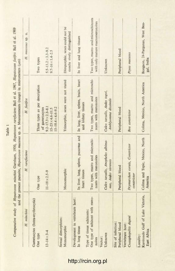
Tab
le 1
Com
para
tive
stud
y of
Hep
atoz
oon
miti
chin
i G
arnh
am,
1950
, H
epat
ozoo
n ra
refa
cien
s B
all
et a
l. 19
67,
Hap
atoz
oon
fusi
fex
Bal
l et
al.
1969
an
d th
e pr
esen
t pa
rasi
te,
Hep
atoz
oon
muc
osus
sp.
n.
Mea
sure
men
ts (
rang
e) i
n m
icro
met
ers
(fi.m
)
H.
min
chin
i H
. ra
refa
cien
s H
. fu
sife
x
| H
. m
ucos
us s
p. n
.
Gam
etoc
ytes
(In
tra-
eryt
hroc
ytic
) O
ne t
ype
One
typ
e T
hree
typ
es a
s pe
r de
scri
ptio
n T
wo
type
s of
gam
etoc
ytes
13
-14x
3-4
11-1
8x2.
5-9
9.2-
17.3
x2.3
-8.1
5.
5-13
.2x3
.3-9
.2
15-2
3x4.
6-11
.5
9.7-
16 x
1.4
-2.5
15
-18.
4x4.
6-9.
2 Se
xual
dim
orph
ism
: M
onom
orph
ic
Mon
omor
phic
T
rim
orph
ic,
sexe
s w
ere
not
stat
ed
Dim
porp
hic,
sex
es c
ould
not
be
conc
lu s
ivel
y di
stin
guis
hed
Dev
elop
men
t in
ver
tebr
ate
host
: In
lun
g tis
sue
In l
iver
, lu
ng,
sple
en,
panc
reas
and
In
lun
g, l
iver
, sp
leen
, br
ain,
hea
rt
In l
iver
and
lun
g tis
sues
he
art
tissu
es
and
kidn
ey t
issu
es
Typ
e of
tis
sue
schi
zont
s:
One
typ
e of
sch
izon
t w
ith m
ero-
Tw
o ty
pes,
mac
ro a
nd m
icro
schi
- T
wo
type
s, m
acro
- an
d m
icro
schi
- T
wo
type
s, m
acro
-and
mic
rosc
hizo
nts
zoite
s zo
nts
wit
h m
eroz
oite
s zo
nts
with
mer
ozoi
tes
with
onl
y m
atur
e m
acro
mer
ozoi
tes
Vec
tor:
U
nkno
wn
Cul
ex t
arsa
l is,
Anop
hele
s al
bim
a-
Cul
ex t
arsa
l is,
Aede
s to
goi,
Unk
now
n nu
s, A
edes
sin
ensi
s Am
blyo
mm
a di
ssim
ile
Site
of
infe
ctio
n:
Peri
pher
al b
lood
Pe
riph
eral
blo
od
Peri
pher
al b
lood
Pe
riph
eral
blo
od
Ver
tebr
ate
host
: C
rota
phop
eltis
deg
eni
Dry
mar
chon
cor
ais,
Con
stri
ctor
Bo
a co
nstr
icto
r P
tyas
muc
osus
co
nstr
icto
r
Loc
alit
y:
Kav
iron
do G
ulf
of L
ake
Vic
tori
a,
Col
ima
and
Tepi
c, M
exic
o, N
orth
C
olim
a, M
exic
o, N
orth
Am
eric
a B
onga
on,
24-P
arga
nas,
Wes
t B
en-
Eas
t A
fric
a A
mer
ica
gal,
Indi
a
http://rcin.org.pl

476 C. K. SINHA
and it is therefore considered as a new species. The specific name of the parasite is given after the name of the species host. The holotype and paratypes will be de-posited in the National Zoological Collection of Zoological Survey of India, Calcutta.
ACKNOWLEDGEMENTS
The author is indebted to Dr. B. Dasgupta, Principal, Government College, Darjeeling for his valuable guidance and wise suggestions during the course of investigation. Best thanks are due to Dr. J. R. Baker, Institute of Terrestrial Ecology, Culture centre of Algae and Protozoa, Cam-bridge, England for going through the manuscript and valuable comments.
REFERENCES
B a l l G. H., C h a o J. and T e l f o r d S. R. Jr. 1967: The life history of Hepatozoon rarefaciens (Sam-bon and Seligmann, 1907) from Drymarchon corais (Colubridae) and its experimental trans-fer to Constrictor constrictor (Boidae). J. Parasitol., 53, 897-909.
Ba l l G. H., C h a o J. and T e l f o r d S. R. Jr. 1969: Hepatozoon fusifex sp. n. a haemogregarine from Boa constrictor producing marked morphological changes in infected erythrocytes. J. Parasitol., 55, 800-813.
B o o d e n T., C h a o J. and Bal l G. H. 1970: Transfer of Hepatozoon sp. from Boa constrictor to a lizard, Anolis carolinensis by mosquito vectors. J. Parasitol., 56, 832-833.
B r a y R. S. 1964: A check list of the parasitic protozoa of West Africa with some notes on their classification. Bull. Inst. Fr. Afr. Noire., 26, 238-315.
C l a r k G. W. and B r a d f o r d J. 1969: Blood parasites of some reptiles of the Pacific North-West, J. Protozool., 16, 478-481.
G a r n h a m P. C. C. 1950: Blood parasites of East African vertebrates with a brief description of exo-erythrocytic schizogony in Plasmodium pitmani. Parasitology, 40, 328-337.
H u l l R. W. and Cam in J. H. 1960: Haemogregarines in snakes: the incidence and identity of the erythrocytic stages. J. Parasitol., 46, 515-523.
M a r q u a r d t W. C. 1966: Haemogregarines and Haemoproteus in some reptiles in Southern Illi-nois. J. Parasitol., 52, 823-824.
M k r t c h y a n Z. A. 1967: New data on blood parasites of reptiles. Biol. Zh. Armen., 20, 93-97. M i l ler W. W. 1908: Hepatozoon perniciosum (n. g., n. sp.) a haemogregarine pathogenic for white
rats with a description of the sexual cycle in the intermediate host, a mite (Lelaps echidninus). Bull. Hyg. Lab. Washington, 46, 1-48.
M o h a m m e d A. H. H. and M a n s o u r N. S. 1959: The haemogregarine complex (an analytical systematical review). Bull. Fac. Sci. Cairo Univ., 35, 39-51.
O d a S. N., C h a o J. and Ball G. H. 1971: Additional instances of transfer of reptile haemogre-garines to foreign hosts. J. Parasitol., 57, 1377-1378.
P e s s o a S. B. and Bias i P. de. 1973: Notas taxonomica sobrex esporogoni cos de algumas especies de Hepatozoon (Sporozoa: Haemogregarinidae) parasites de serpentes brasileiras. Mem. Inst. Butantan (Sao Paulo), 37, 299-307.
S i n h a C. K. 1980: An ophidian Hepatozoon and its experimental transmission to leech. Proc. Symp. The Host as an Environment, Z. S. I., Calcutta, p. 14 (Abstr.).
S i n h a C. K. 1981: Occurrence of a Hepatozoon in rat snakes. Dept. Zool. First reunion (Souvenir) Govt. College Darjeeling, p. 34 (Abstr.).
S i n h a C. K. 1984: Observations on certain protozoan parasites in the blood of some vertebrates. Ph. D. Thesis, University of North Bengal, Rajarammohunpur, Darjeeling.
http://rcin.org.pl

ACTA PROTOZOOLOGICA Vol. 25, No. 4, pp. 477-432 (1986)
A New Myxosporidan Myxidium sciaenae sp. n. (Myxozoa : Myxididae) from the Gall Bladder of a Marine
Teleost of West Bengal, India
N. K. S A R K A R
Department of Zoology, Rishi Bankim Chandra College, Naihati 743165, West Bengal, India
Received on 10 January 1986
Synopsis. A new myxosporidan Myxidium sciaenae sp. n. (Myxozoa: Myxididae) has been described from the gall bladder of a marine teleost Sciaena bleekeri Day, caught from the Digha coast of Bay of Bengal, West Bengal, India. The character-istic mensural data of the myxosporidan are as follows: spore — 19.36 n m x 5.32 fxm, polar capsule — 5.26 ji.mx 2.5 [i.m. It is the eighth myxosporidan species reported from the coastal water of Bay of Bengal, West Bengal, India.
During the investigation about the parasitic protozoa of the marine fishes of the coastal water of Bay of Bengal, West Bengal, India, a new myxosporidan par-asite has been recovered from the gall bladder of a teleost Sciaena bleekeri Day. It has been described as a new species for some of its distinctive characters given later.
M a t e r i a l a n d M e t h o d s
All autopsies were performed on frozen fish collected from Digha coast of Bay of Bengal, West Bengal, India. The parasite has been studied in fresh treated with Lugol's iodine solution and from dry smears stained with Giemsa after fixation in absolute Methanol. The polar filament could not be extruded even after repeated attempts with various concentrations of KOH and Urea solutions have been made. The measurements are given in micrometers ([xm). The Figures have been drawn with a camera lucida (Prism type).
O b s e r v a t i o n s
Myxidium sciaenae sp. n.
D e s c r i p t i o n : No trophozoite or other vegetative stages were found. The spores were coelozoic, fusiform with rounded ends in valvular view (Fig. 1 A) while in sutural view the spores appeared more fusiform (Fig. 1 B) than the spores in
8 — Acta Protozol. 25/4
http://rcin.org.pl

478
valvular view. The shell valves were two — each with five to six longitudinal stria-tions arranged almost parallel to each other. The suture was distinct and finely curved — nearly S-shaped. The polar capsules were two — one on either end of the spore. These polar capsules were small, pyriform to elongately pyriform with six to seven coils of polar filament. In a few spores, one side appeared more convex
C , D 2 0 j j m
Fig. 1. A-D. Spores of Myxidium sciaenae sp. n. A — A spore in valvular view — Lugol's iodine treatment, B — A spore in sutural view — Lugol's iodine treatment, C — A spore having convex
upper surface — Giemsa stained, D — A spore having its breadth shifted — Giemsa stained
than the other side (Fig. 1 C) and in a few others, the maximum width of the spore was shifted from the middle of the extracapsular region to the capsular region (Fig. 1 D). The extracapsular biconcave cavity was filled with granular sporoplasm containing two round nuclei. No iodinophilous vacuole was detected in the sporo-plasm.
M e a s u r e m e n t s : The mensural data of 25 fresh spores and 44 polar capsules with sample standard deviation are given.
L e n g t h of t he s p o r e - 19.36±0.47 B r e a d t h of t h e s p o r e - 5.32±0.28 L e n g t h of t h e p o l a r c a p s u l e — 5.26±0.13 B r e a d t h of t h e p o l a r c a p s u l e — 2.50±0.12 I n f e c t i o n l o c u s : Gall bladder I n d i d e n c e : Two infected out of 15 examined P a t h o g e n i c i t y : Not apparent H o s t : Sciaena bleekeri Day L o c a l i t y : Digha coast of Bay of Bengal, West Bengal, India D a t e of c o l l e c t i o n : 2 January, 1985
D i s c u s s i o n
J a y a s r i and H o f f m a n (1982) reviewed the genus Myxidium Butschli, 1882 and listed 116 species. In mensural data the present myxosporidan resembles closely with Myxidium lentiforme Fujita, 1927 reported from the kidney of Anguila japo-
http://rcin.org.pl

MYXIDIUM SCIAENAE SP. N. 479
nica and M. melanocetum Noble, 1966 reported from the gall bladder of Melanocetus johnsoni. The myxosporidan in study differs from the former species in having longitudinally striated shell valves (shell valves thin-walled and smooth in M. len-tiforme). It also differs from M. melanocetum by lesser number of longitudinal striation in shell valves (shell valves with 8-9 longitudinal striations in M. mela-nocetum), distinct suture (indistinct in M. melanocetum) and smaller polar capsule (6.7 ;xm long polar capsule in M. melanocetum). Moreover, in general shape of the spore, the present parasite also resembles M. bajacalifornium Noble, 1966 reported from the gall bladder of Rajacalifornia burragei, but differs from the latter by its distinct mensural data (the mensural data of M. bajacalifornium are: spore — 22.1 [xmx4.0 [xm, polar capsule — 7.4 [xm long with 12-14 coils of polar filament). The present myxosporidan is, therefore, considered to be a new species and the name Myxidium sciaenae sp. n. is given after the name of its host.
ACKNOWLEDGEMENTS
Grateful acknowledgements are made to Prof. A. Pramanik, Head of the Dept. of Zoology for laboratory facilities and to Sri A. Majumder for statistics.
REFERENCES
F u j i t a T. 1927: Studies on Myxosporidia of Japan. J. Coll. Agri. Hokkaido Imperial Univ., 16 229-247.
J a y a s r i M. and H o f f m a n G. L. 1982: Review of Myxidium (Protozoa: Myxozoa: Myxosporea). Protozool. Abstr. Commonwealth Inst. Parasitol., 6, 61-91.
N o b l e E. R. 1966: Myxosporidia in deep water fishes. J. Parasitol., 52, 685-690.
http://rcin.org.pl

NOTICE TO AUTHORS
ACTA PROTOZOOLOGICA is intended as a journal serving for the publication of original papers embodying the results of experimental or theoretical research in all fields of protozoology with the exception of faunistic notices of the local character and purely clinical reports. The papers should be concise and will not be accepted if they have been previously published elsewhere. After acceptance by the Editors papers will be printed in the possibly shortest time.
Papers are accepted in English, French, German and Russian. Every paper should begin with the name and postal address of the laboratory, name and the surname of the author and title in the language of the text. The titles in German and French should be translated into English ac-cording to the demands of Current Contents. The paper should be accompanied by synopsis in the language of the text not exceeding 100 words and by short summary in one of 4 languages accepted in the Journal. In the Russian text also the name and the postal address of the laboratory, legends of tables, plates and text illustrations must be translated, the translation of the summary may be somewhat more extensive, and the name of the author should be given additionally also in the Latin characters.
Manuscripts should be doublespaced typescript (30 lines on side of a sheet) with a normal margin. No elements of the text should be fully typed in capitals nor in spaced set (only under-lining with pencil is admittable). In decimal fractions points not commas should be used. The ge-nerally accepted abbreviations and symbols are recommended. Nomenclature must agree with the International Code of Zoological Nomenclature, London 1961. The original and one carbon copy of the whole text material should be supplied.
References must be cited in the text indicating only the author and year thus: " K i n o s i t a (1954) found that, etc." Only all references cited in the text should be listed. The list must be arranged as follows: E h r e t C. F. and P o w e r s , E. L. 1959: The cell surface of Paramecium. Int. Rev. Cytol., 8,
97-133. Ge le i J. von 1939: Das aussere Stutzgerustsystem des Parameciumkorpers. Arch. Protistenk.,
92, 245-272. Titles of references are given in their original language (not translated). In papers written in
English, French or German, the Cyrillic type of the Russian is transliterated according to the inter-national system (ISO Recommendation R 9 September 1954). This regulation is not applied to names if there exists in their traditional spelling. Also author may freely choose in transliteration of his own name. In Russian papers, the Russian references are cited in Cyrillic, the others in the Latin characters, but they must be listed all together in the Latin alphabetical order.
The following material should be supplied on separate sheets: (1) the running title for the page headlines, (2) tables, (3) legends for text figures, (4) legends for plates. Line-drawings will be pu-blished in the text, photographs and raster figures on separate plates. No coloured photographs can be published presently. Lettering on photographs and drawings should be marked in pencil. With no regard to the language of the text, only the Latin lettering, arabic numerals or generally accepted symbols are admittable for marking on illustrations. Numbering of text-figures, plates and tables must be also marked in pencil, as well in the legends as in the text.
Galley proofs are sent to the authors. Authors receive 75 reprints without covers.
http://rcin.org.pl

In p r e p a r a t i o n :
E. W i r n s b e r g e r und W. F o i s s n e r : Morphologie von Holosticha xantichroma sp. n. und die Variabilität der Infraciliatur der Gattung Holosticha (Ciliophora, Hypotricha) — M. A. Jareno: Some Ultrastructural Changes Produced by Vaccinia Virus in Onychodromus acurninatus (Ciliata, Hypotrichida) — D. C h a r d e z : Enkystement externe chez Difflugia elegans (Rhizopoda, Testa-cea) — J. Drzewi r i ska and K. G o l i h s k a : Relationship between the Size of Cell and the Number of Its Ciliary Rows in the Ciliate Dileptus — L. G r ç b e c k a , A. G r ç b e c k i and B. H r e b e n d a : Motor Behaviour of Amoeba proteus during Glutaraldehyde Fixation — I. J a h n and G. Csaba : The Influence of Arginine Vasopressin (AVP) on Phagocytosis in the Unicellular Tetrahymena — K. B o c z o n : Comparative Investigations on Metabolism of Pathogenic and Non-pathogenic Strains of Free-living Amoeba — D. C h a r d e z : Contribution a la connaissance des Thecamoebiens des dépôts lacustres — R. R a y : Trypanosoma balithaensis sp. n. from a Pond Water Turtle, Lissemys punctata punctata (Bonnaterre) and Its Development in the Leech Vector Helobdella nociva Hard-ing — N. K. S a r k a r : Chilogregarina bhatiae sp. n. (Apicomplexa: Eugregarinida) a New Cephaline Gregarine from a Chilopod Geophilus sp. — C. K. S inha and B. D a s g u p t a : Hepatozoon dar-jeelingensis sp. n. from the Himalayan Flying Squirrels — J. W i e r z b i c k a : Zschokkella stettinensis sp. n. (Myxospora, Bivalvulida) a Parasite of Eel, Anguilla anguilla (L.)
Warunki prenumeraty
Prenumeratę na kraj przyjmują i informacji o cenach udzielają urzędy pocztowe i doręczyciele na wsi oraz Oddziały RSW „Prasa-Książka-Ruch" w miastach. Prenumeratę ze zleceniem wysyłki za granicę przyjmuje RSW „Prasa-Książka-Ruch", Centrala Kolportażu Prasy i Wydawnictw, ul. Towarowa 28, 00-958 Warszawa, konto NBP XV Oddział w Warszawie Nr 1153-201045-139-11. Wysyłka za granicę pocztą zwykłą jest droższa od prenume-raty krajowej o 50% dla zleceniodawców indywidualnych i o 100% dla zlecających instytucji i za-kładów pracy.
Terminy przyjmowania prenumerat na kraj i za granicę:
— do dnia 10 listopada na I półrocze roku następnego i na cały rok następny, — do dnia 1 czerwca na II półrocze roku bieżącego.
Bieżące i archiwalne numery można nabyć lub zamówić we Wzorcowni Ośrodka Rozpowszechnia-nia Wydawnictw Naukowych PAN, Pałac Kultury i Nauki, 00-901 Warszawa.
http://rcin.org.pl

Cena 140.—
CONTENTS
I. W i t a and K. M. S u k h a n o v a : Seasonal Modifications in the Life Cycle of Parastasia fennica (Michajlow, 1966) 365
D. C h a r d e z : Thécamoebiens des plages de la Mer du Nord en Angleterre [Testate Amoebae from England Beaches of the North Sea] 375
N. W i l b e r t : Ciliaten aus dem Interstitial des Ontario Sees [Ciliates in the Interstitial of Lake Ontario] 379
R. M a t h u r and D. M. S a x e n a : Inhibition of Macromolecule Syntheses in a Ciliate Pro-tozoan, Tetrahymena pyriformis by Hexachlorocyclohexane (HCH) Isomers 397
W. K a s p r z a k , T. M a z u r and E. H a d a s : Biochemical Changes of Acanthamoeba Fol-lowing Attenuation and the Role of Cysts in Retaining the Characteristics of Strains 411
T. M i c h a l o w s k i , P. S z c z e p k o w s k i and P. M u s z y n s k i : The Nutritive Factors Affecting the Growth of the Rumen Ciliate Diploplastron affine in vitro 419
A. C z a p i k et N . W i l b e r t : Sur une nouvelle espèce de cilié Paranophrys Carnivora sp. n. (Scuticociliatida) [New Species of Ciliate Paranophrys Carnivora sp. n. (Scuticociliatida)] 427
S. L. K a z u b s k i and S. A. M. E l - T a n t a w y : The Ciliate Paratrichodina africana sp. n. (Peritricha, Trichodinidaé) f rom Tilapia Fish (Cichlidae) f rom Africa 433
S. A. M. E l - T a n t a w y and S. L. K a z u b s k i : The Trichodinid Ciliates f rom Fish Tilapia nilotica f rom the Nile Delta (Egypt) 439
S. L. K a z u b s k i : The Trichodinid Ciliates f rom Fish, Tilapia sp. f rom Lake Victoria (Kenya) and Description of Trichodina equatorialis nom. nov 445
C. K. S i n h a : Occurrence of Trypanosoma mukasai Hoare, 1932 in Tilapia mossambica (Peters) f rom India 449
S. G h o s e , S. K . R a y and D. P. H a l d a r : Neohirmocystidae fam. n., a New Family of Sep-tate Gregarines (Apicomplexa : Sporozoea) f rom Insects 453
V. N. H o o g a r and S. D. A m o j i : Leidyana bimaculata a New Cephaline Gregarine Para-site of a Cricket, Gryllus bimaculatus De Geer 465
C. K. S i n h a : Hepatozoon mucosus sp. n. f rom Indian Rat Snakes, Ptyas mucosus (Linnaeus) 471 N. K. S a r k a r : A New Myxosporidan Myxidium sciaenae sp. n. (Myxozoa : Myxididae)
f rom the Gall Bladder of a Marine Teleost of West Bengal, India 477
Państwowe Wydawnictwo Naukowe — Oddział we Wrocławiu
Wrocławska Drukarnia Naukowa
Indeks 351334
http://rcin.org.pl