Endoplasmic reticulum: nutrient sensor in physiology and pathology
MOLECULAR AND CELLULAR BIOLOGY, Nov. 2004, p. 9695–9704 Vol. 24, No. 220270-7306/04/$08.00�0 DOI: 10.1128/MCB.24.22.9695–9704.2004Copyright © 2004, American Society for Microbiology. All Rights Reserved.
Proteasome Inhibitor PS-341 Induces Apoptosis through Induction ofEndoplasmic Reticulum Stress-Reactive Oxygen Species in
Head and Neck Squamous Cell Carcinoma CellsAndrew Fribley,1,2 Qinghua Zeng,1 and Cun-Yu Wang1,2*
Laboratory of Molecular Signaling and Apoptosis, Department of Biologic and Materials Sciences,1 and Program inOral Health Sciences, School of Dentistry,2 University of Michigan, Ann Arbor, Michigan
Received 25 April 2004/Returned for modification 26 May 2004/Accepted 28 July 2004
PS-341, also known as Velcade or Bortezomib, represents a new class of anticancer drugs which has beenshown to potently inhibit the growth and/or progression of human cancers, including head and neck squamouscell carcinoma (HNSCC). Although it has been logically hypothesized that NF-�B is a major target of PS-341,the underlying mechanism by which PS-341 inhibits tumor cell growth is unclear. Here we found that PS-341potently activated the caspase cascade and induced apoptosis in human HNSCC cell lines. Although PS-341could inhibit NF-�B activation, the inhibition of NF-�B was not sufficient to initiate apoptosis in HNSCC cells.Using biochemical and microarray approaches, we found that proteasome inhibition by PS-341 inducedendoplasmic reticulum (ER) stress and reactive oxygen species (ROS) in HNSCC cells. The inhibition of ROSsignificantly suppressed caspase activation and apoptosis induced by PS-341. Consistently, PS-341 could notinduce the ER stress-ROS in PS-341-resistant HNSCC cells. Taken together, our results suggest that inaddition to the abolishment of the prosurvival NF-�B, PS-341 might directly induce apoptosis by activatingproapoptotic ER stress-ROS signaling cascades in HNSCC cells, providing novel insights into the PS-341-mediated antitumor activity.
PS-341, also known as Velcade or Bortezomib, represents anew class of anticancer drugs which mainly inhibit 26S protea-some activity (1, 2). The 26S proteasome is a large multisub-unit protein complex found in the cytoplasms and nuclei of alleukaryotic cells; its principle function is to degrade proteins viathe ubiquitin pathway (26). It is composed of a 20S core cyl-inder, with tryptic and chymotryptic activity, and is capped ateach end by a 19S regulatory particle. In most cases, proteinsdestined for proteasomal degradation are flagged on lysineresidues with polymeric chains of a small protein known asubiquitin (26). The ubiquitin chains are recognized by the 19Scap. The 19S cap also contains enzymatic activities that removeubiquitin from the targeted proteins and chaperone-like activ-ities that unfold the deubiquitinated substrates and feed theminto the internal chamber of the 20S core particle. The pepti-dase activities (chymotryptic, tryptic, and postglutamyl hydro-lyzing) then generate small peptides (1, 26). PS-341 is a smalldipeptidyl boronic acid derivative that binds reversibly to thecatalytic threonine and selectively blocks the chymotryptic ac-tivity of the proteasome (1, 5). The ability of PS-341 to specif-ically inhibit the 26S proteasome distinguishes it from otherproteasome inhibitors, such as the peptide aldehydes, like MG-132, which also inhibit thiol proteases like cathepsin B andcalpains. Preclinical and clinical trials have found that PS-341is a promising chemotherapeutic adjuvant which has antitumoreffects on several human cancers (7, 12, 13, 24, 33).
Most cytosolic and short-lived proteins, including many tran-
scription factors, meet their demise at the 26S proteasome (1,26). Therefore, it is not surprising that a myriad of cell re-sponses and pathways are perturbed as a result of proteasomeinhibition. In vitro and in vivo studies have demonstrated thatthe proteasome inhibitor inhibits tumor growth by inducingapoptosis in several human cancers, including myeloma, pros-tate cancer, and head and neck squamous cell carcinoma(HNSCC) (12, 13, 22, 37, 38, 43). PS-341-induced apoptosiswas preceded by the mitochondrial release of cytochrome c,SMAC, and apoptosis-inducing factor, and cleavage ofcaspases has been demonstrated in cells undergoing PS-341-induced apoptosis (13, 45).
Activation of the transcription factor nuclear factor kappa B(NF-�B), a key survival factor, has been shown to be depen-dent on the 26S proteasome (3, 8, 23, 26, 27). Because PS-341specifically inhibits the 26S proteasome, NF-�B has been log-ically considered to be a key target for PS-341-induced apo-ptosis (1, 27). NF-�B is a transcription activator that controlsexpression of genes associated with inflammation and cell pro-liferation and survival (3, 8). Classical NF-�B is a heterodimercomposed of p50 and p65/RelA, which is retained in the cyto-plasm by the I�B group of inhibitory proteins. In response tostressful stimuli, such as cancer chemotherapy, I�B is phos-phorylated, ubiquitinated, and subsequently degraded by the26S proteasome. I�B degradation results in the nuclear trans-location of NF-�B and the activation of NF-�B-dependentgene transcription (3, 8). Many studies have found that PS-341potently sensitized myeloma cells and colorectal cancer cells toDNA-damaging chemotherapeutic agents, such as doxorubicinand melphalan, by inhibiting NF-�B activation. These studieshave provided a framework for clinical use of PS-341 in com-bination with conventional chemotherapy (12, 22, 37, 38, 43).Recently, Sunwoo et al. (37) reported that PS-341 inhibited
* Corresponding author. Mailing address: Laboratory of MolecularSignaling and Apoptosis, Department of Biologic and Materials Sci-ences, University of Michigan, 1011 N. University Ave., Ann Arbor,MI 48109-1078. Phone: (734) 615-4386. Fax: (734) 647-2110. E-mail:[email protected].
9695
on August 10, 2015 by guest
http://mcb.asm
.org/D
ownloaded from

activation of NF-�B DNA binding and functional reporteractivity at physiological concentrations in HNSCC cells. Im-portantly, they also found that PS-341 induced apoptosis inthese cells, suggesting that PS-341 may be used as a mono-therapy for HNSCC.
HNSCC is the most common cancer of the head and neckregion (6, 31, 36). Approximately 30,000 Americans are diag-nosed with cancers affecting the head and neck and the oralcavity each year. While significant advances have been made insurgery and radiotherapy for patients with HNSCC, the 5-yearsurvival rate is the lowest for any major cancer (6, 31, 36).Therefore, there is an urgent need to develop innovative ther-apies for HNSCC. To further understand the molecular mech-anisms by which PS-341 induces apoptosis in HNSCC cells, wesystemically dissected the signaling pathways involved in PS-341-mediated apoptosis by using biochemical and genetic ap-proaches. We found that inhibition of the NF-�B antiapoptoticpathway was not sufficient to induce apoptosis in HNSCC cells,suggesting that PS-341 might directly activate proapoptoticsignaling pathways. Interestingly, using microarray analysis, wefound that a large number of genes associated with endoplas-mic reticulum (ER) stress were rapidly induced following PS-341 stimulation in HNSCC cells. Consistently, caspase 12, anessential caspase in ER stress-mediated apoptosis (30), wasfound to be activated by PS-341 stimulation in mouse embry-onic fibroblasts (MEFs). Moreover, we found that reactiveoxygen species (ROS), which are frequently induced by ERstress, played a critical role in PS-341-mediated apoptosis. Theinhibition of ROS significantly suppressed PS-341-inducedapoptosis in HNSCC as well as expression of ER stress-asso-ciated proapoptotic genes. In support of the role of ER stress-ROS in PS-341-induced apoptosis, we found that ER stress-ROS were weakly induced in PS-341-resistant cells. Thesefindings provide novel insight into the molecular mechanismsof PS-341-mediated apoptosis in HNSCC cells.
MATERIALS AND METHODS
Reagents and cell cultures. PS-341 was kindly provided by Millennium Phar-maceuticals, Inc. (Cambridge, Mass.). Tiron was purchased from Sigma-Aldrich(St. Louis, Mo.). H2DCFDA, Indo-1, and ionomycin were purchased from Mo-lecular Probes (Eugene, Ore.). HNSCC cell lines were obtained from ThomasCarey at the University of Michigan and were cultured in Dulbecco’s modifiedEagle’s medium supplemented with 10% fetal bovine serum and penicillin-streptomycin from Invitrogen (Grand Island, N.Y.).
Western blot analysis. Cells (2 � 106) were plated in 10-cm tissue culturedishes the day before PS-341 or vehicle treatment. Cells were treated with PS-341(0.05 to 0.5 �M) for various times. Whole-cell extracts were prepared in modifiedradioimmunoprecipitation assay (RIPA) buffer containing phenylmethylsulfonylfluoride and protease inhibitors (Sigma-Aldrich). Fifty micrograms of lysateswere resolved on sodium dodecyl sulfate (SDS)–8 or 12% polyacrylamide gelelectrophoresis (PAGE) and transferred to a polyvinylidene difluoride mem-brane with a Bio-Rad (Hercules, Calif.) semidry transfer apparatus. The mem-branes were blotted with 5% milk overnight and probed with primary antibodies.Antibodies were acquired from the following sources: phospho-PERK, caspase9, and caspase 12 from Cell Signaling (Beverly, Mass.); ATF-4, GADD-34,GADD-153/CHOP, GRP-78, and caspase 3 from Santa Cruz (Santa Cruz, Cal-if.); and caspase 2 from BD PharMingen (San Diego, Calif.). After incubationwith horseradish peroxidase-conjugated secondary antibody, membranes werevisualized using an enhanced chemiluminescence reagent from Pierce (Rock-ford, Ill.).
Northern blot analysis. Total RNAs were extracted from cells with the Trizolreagent (Invitrogen) according to the manufacturer’s protocol. Five to ten mi-crograms of total RNAs were resolved on 1.5% agarose formaldehyde gels andtransferred to nitrocellulose membranes overnight. The membranes were hybrid-
ized with 32P-labeled cDNA probes and exposed to autoradiographic film asdescribed previously (42). cDNA probes for human GRP-78 and ATF-4 wereprepared by using reverse transcription PCR with the specific primers (GRP-78[forward, 5�-ATGTACGCCCTGCTGGGAGT-�3; reverse, 5�-CCATGTAGTTGCGAAGCTCC-�3] and ATF-4 [forward, 5�-AGATGACCTTCTGACCACG-�3; reverse, 5�-CACCCTTTACTTTTGCTGC-�3]). cDNA products were puri-fied from agarose gels, subcloned into the pT-Easy vector from Promegaaccording to the manufacturer’s protocol, and confirmed by DNA sequencing.
Microarray. UMSCC-23 cells were treated with PS-341 for 0, 1, 4, and 8 h.After treatment, cells were harvested, and total RNAs were extracted with Trizolreagents according to the manufacturer’s protocol. To remove contaminatedgenomic DNA, total RNAs were passed through an RNeasy column from QIA-GEN (Valencia, Calif.). Ten micrograms of total RNAs were quantitativelyamplified and biotin labeled according to the manufacturer’s instructions.Briefly, RNAs were converted to double-stranded cDNA, using SuperScript IIreverse transcriptase (Invitrogen) with an oligo(dT) primer that has a T7 RNApolymerase site on the 5� end. Then, the cDNAs were used in an in vitrotranscription reaction in the presence of biotin-modified ribonucleotides to pro-duce large amounts of single-stranded RNA. The biotin-labeled RNAs werefragmented. Hybridization to an Affymetrix (Santa Clara, Calif.) human U133gene chip was performed at 45°C for 16 h in a mix that included 10 �g offragmented RNA, 6� SSPE (1� SSPE is 0.18 M NaCl, 10 mM NaH2PO4, and1 mM EDTA [pH 7.7]), 0.005% Triton X-100, and 100 �g of herring spermDNA/ml in a total volume of 200 �l. Labeled bacterial RNAs were spiked intothe hybridization mix to generate an internal standard and to allow normaliza-tion between chips. Chips were washed and stained with streptavidin R-phyco-erythrin (Molecular Probes). The arrays were scanned with the GeneArrayscanner (Affymetrix). Signal intensity was calculated by using the one-step Tukeybiweight estimate. When computing change indices (n-fold), normalized inten-sities less than 20 were replaced by 20 before forming ratios in order to avoidspuriously large n-fold change values. We performed multivariate analyses tocompare gene expression using analysis of variance. Genes with q values less than5% were considered to be significantly expressed (where the q value is themultiple-comparisons equivalent of the normal P value). Because there wereonly two samples per group, our estimate of the error of the mean might bebiased, resulting in a higher false-positive rate than expected. To account for thispossibility, we imposed an additional n-fold change criterion (at least twofold)for significance.
Flow cytometry. Cells (2 � 106) were plated in 10-cm tissue culture dishes theday before treatment with PS-341 or vehicle. After treatment, plates werewashed once with phosphate-buffered saline (PBS) and cells were harvested with1� trypsin-EDTA. Cells were subsequently washed again with PBS and thenresuspended in 1 ml of PBS. Cells were incubated with 2�,7�-dichlorofluoresceindiacetate (H2DCFDA; 0.5 to 1.0 �M) in the dark at 37°C for 30 min. Cells werethen washed twice with PBS and analyzed at the University of Michigan CancerCenter Flow Cytometry Core Facility.
DNA laddering. PS-341-treated cells were lysed and treated with 100 mg ofproteinase K/ml for 2 h at 50°C. DNA was extracted twice with phenol-chloro-form mixed 1:1 and twice with chloroform alone. Precipitated DNA was washedwith 70% ethanol, and 20 to 30 �g of each sample was resolved on a 1.5%agarose gel.
Pulse-chase 35S protein labeling. UMSCC-23 cells (105) were plated the daybefore treatment in 6-cm dishes. Cells were treated with PS-341, vehicle orthapsigargin (TG) as a positive control. The cells were rinsed with PBS andincubated with 35S-labeled cysteine and methionine in cysteine- and methionine-free Dulbecco’s modified Eagle’s medium for 15 min at 37°C. Medium was thenremoved, and the cells were rinsed with cold PBS. Cells were lysed with 200 �lof RIPA. Ten-microgram aliquots of each sample were resolved on an SDS–10%PAGE gel, dried, and exposed to radiographic film.
RESULTS
PS-341 activates caspase cascades and induces apoptosis inHNSCC cells. To elucidate the molecular mechanism by whichPS-341 induced apoptosis, a panel of HNSCC cell lines, in-cluding UMSCC-1, -9, -11B, -14A, and -23, was treated withPS-341 for 0, 8, 16, or 24 h. Trypan blue exclusion assay foundthat cell death was induced in these cells, with the exception ofUMSCC-14A, between 8 and 16 h following PS-341 stimula-tion (Fig. 1A). A DNA fragmentation assay found that PS-341
9696 FRIBLEY ET AL. MOL. CELL. BIOL.
on August 10, 2015 by guest
http://mcb.asm
.org/D
ownloaded from

induced DNA laddering in HNSCC cells, signifying an apopto-tic mechanism (Fig. 1B). Western blot analysis revealed thatPS-341 sequentially induced the processing of caspases 9, 2,and 3, suggesting that caspase 9 might be an initiating caspasein HNSCC cells (Fig. 1C). While caspase 9 is considered aninitiating caspase for chemotherapeutic drug-mediated apo-ptosis, recent studies have suggested that caspase 2 may be theapical caspase in chemotherapy-mediated apoptosis (18). Toexplore the role of caspase 2 in PS-341-mediated apoptosis, weutilized a small interfering RNA (siRNA) strategy to knockdown caspase 2 expression as described previously (18). Wefound that the deletion of caspase 2 did not provide protectionagainst cell death induced by PS-341 (Fig. 1D and E), indicat-ing that caspase 2 was dispensable for PS-341-mediated apo-ptosis.
The inhibition of NF-�B by PS-341 is not sufficient to induceapoptosis in HNSCC cells. NF-�B has been found to be con-stitutively activated in several human cancer cell lines; the
inhibition of NF-�B by biochemical and chemical inhibitorsinduced apoptosis in these cells. Due to its potent inhibition ofNF-�B, PS-341 has been proposed to induce apoptosis inHNSCC cells by inhibiting NF-�B. Since UMSCC-14A cellswere very resistant to PS-341-induced apoptosis (Fig. 1A), itwas possible that PS-341 failed to inhibit NF-�B in these cells.To rule out this possibility, we utilized a biological NF-�Binhibitor, SR-I�B�. SR-I�B� is a dominant-negative mutant ofI�B� in which serines 32 and 36 are mutated to alanines so thatit cannot be phosphorylated and degraded by a variety ofstimuli (39, 40, 43). Overexpression of SR-I�B� has beenshown to specifically inhibit NF-�B activation induced by avariety of stimuli. As shown in Fig. 2A, we were able to obtainUMSCC-14A cells stably expressing SR-I�B�. UMSCC-14Acells expressing SR-I�B� were sensitive to tumor necrosis fac-tor (TNF) killing compared with control cells, validating thatSR-I�B� functioned in the inhibition of NF-�B in these cells(Fig. 2B). These results suggested that the inability of PS-341
FIG. 1. PS-341 induces caspase activation and apoptosis in HNSCC cells. (A) HNSCC cell lines UMSCC-1, -9, -11B, -14A, and -23 were treatedwith PS-341 (0.5 �M) for 0, 8, 16, or 24 h. Cell viability was determined by trypan blue exclusion assay. The assays were performed with triplicatesamples, and the results are representative of three independent experiments. Error bars depict standard deviations. (B) UMSCC-1 andUMSCC-23 cells were treated with PS-341 for 0, 16 or 24 h. The detached and attached cells were collected, and genomic DNA was extracted withphenol-chloroform. Genomic DNA was separated on a 1.2% agarose gel. (C) UMSCC-23 cells were treated with PS-341 for 0, 8, 16 or 24 h. Thewhole-cell extracts were prepared with RIPA buffer, and 50-�g aliquots of protein extracts were resolved on a SDS–12% PAGE. The membranewas probed with antibodies against caspases 9, 2, and 3 (1:500). For loading control, the membrane was stripped and reprobed with anti-�-tubulinmonoclonal antibodies (1:5,000). (D) UMSCC-23 cells were transfected with caspase 2 siRNA (SiCasp-2), using oligofectamine. Twenty-four hoursafter transfection, cells were harvested and probed with monoclonal antibodies against caspase 2. For loading control, the membrane was strippedand reprobed with �-tubulin. (E) UMSCC-23/SiCasp-2 cells and control cells were treated with PS-341 for the indicated times, and cell death wasdetermined.
VOL. 24, 2004 INDUCTION OF APOPTOSIS IN HNSCC CELLS BY PS-341 9697
on August 10, 2015 by guest
http://mcb.asm
.org/D
ownloaded from
to induce apoptosis in UMSCC-14A cells was not due to afailure of NF-�B inhibition. Moreover, like their parental cells,both UMSCC-14A cells expressing SR-I�B� and control cellswere also resistant to PS-341-induced apoptosis (Fig. 2C). Incontrast to UMSCC-14A cells, UMSCC-1 cells were sensitiveto PS-341-induced apoptosis. We were previously able to es-tablish SR-I�B�-expressing UMSCC-1 cells in which the inhi-bition of NF-�B also did not induce apoptosis in these cells(47). Since PS-341 strongly induced apoptosis in UMSCC-1cells, it was likely that PS-341 predominantly modulated otherapoptotic signaling pathways to induce apoptosis in these cells.Moreover, unlike the case with UMSCC-14A cells, PS-341potently induced apoptosis in both UMSCC-1 cells expressingSR-I�B� (UMSCC-1/SR-I�B�) and control cells (UMSCC1/V)(Fig. 2C). Additionally, we found that the inhibition of NF-�Bby the adenovirus-mediated delivery of SR-I�B� alone did notinduce apoptosis in several HNSCC cell lines (3, 47). Takentogether, these results suggest that the inhibition of NF-�Balone could not provide an explanation for PS-341-mediatedapoptosis in HNSCC cells. PS-341 might trigger apoptosis inHNSCC cells by directly modulating other proapoptotic orantiapoptotic pathways.
PS-341-induced apoptosis is involved in ER stress. To fur-ther understand how PS-341 induced apoptosis in HNSCC
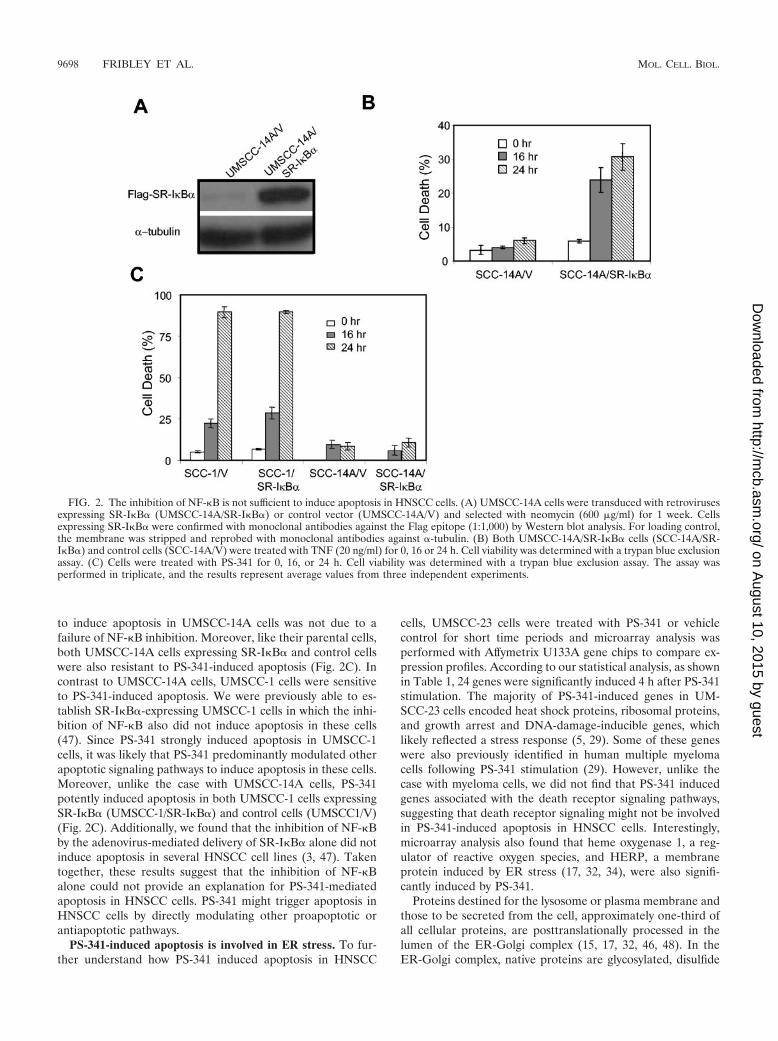
cells, UMSCC-23 cells were treated with PS-341 or vehiclecontrol for short time periods and microarray analysis wasperformed with Affymetrix U133A gene chips to compare ex-pression profiles. According to our statistical analysis, as shownin Table 1, 24 genes were significantly induced 4 h after PS-341stimulation. The majority of PS-341-induced genes in UM-SCC-23 cells encoded heat shock proteins, ribosomal proteins,and growth arrest and DNA-damage-inducible genes, whichlikely reflected a stress response (5, 29). Some of these geneswere also previously identified in human multiple myelomacells following PS-341 stimulation (29). However, unlike thecase with myeloma cells, we did not find that PS-341 inducedgenes associated with the death receptor signaling pathways,suggesting that death receptor signaling might not be involvedin PS-341-induced apoptosis in HNSCC cells. Interestingly,microarray analysis also found that heme oxygenase 1, a reg-ulator of reactive oxygen species, and HERP, a membraneprotein induced by ER stress (17, 32, 34), were also signifi-cantly induced by PS-341.
Proteins destined for the lysosome or plasma membrane andthose to be secreted from the cell, approximately one-third ofall cellular proteins, are posttranslationally processed in thelumen of the ER-Golgi complex (15, 17, 32, 46, 48). In theER-Golgi complex, native proteins are glycosylated, disulfide
FIG. 2. The inhibition of NF-�B is not sufficient to induce apoptosis in HNSCC cells. (A) UMSCC-14A cells were transduced with retrovirusesexpressing SR-I�B� (UMSCC-14A/SR-I�B�) or control vector (UMSCC-14A/V) and selected with neomycin (600 �g/ml) for 1 week. Cellsexpressing SR-I�B� were confirmed with monoclonal antibodies against the Flag epitope (1:1,000) by Western blot analysis. For loading control,the membrane was stripped and reprobed with monoclonal antibodies against �-tubulin. (B) Both UMSCC-14A/SR-I�B� cells (SCC-14A/SR-I�B�) and control cells (SCC-14A/V) were treated with TNF (20 ng/ml) for 0, 16 or 24 h. Cell viability was determined with a trypan blue exclusionassay. (C) Cells were treated with PS-341 for 0, 16, or 24 h. Cell viability was determined with a trypan blue exclusion assay. The assay wasperformed in triplicate, and the results represent average values from three independent experiments.
9698 FRIBLEY ET AL. MOL. CELL. BIOL.
on August 10, 2015 by guest
http://mcb.asm
.org/D
ownloaded from
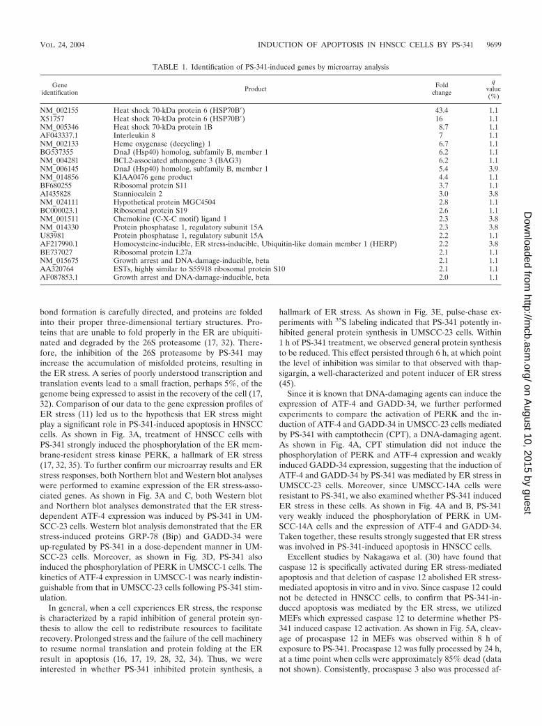
bond formation is carefully directed, and proteins are foldedinto their proper three-dimensional tertiary structures. Pro-teins that are unable to fold properly in the ER are ubiquiti-nated and degraded by the 26S proteasome (17, 32). There-fore, the inhibition of the 26S proteasome by PS-341 mayincrease the accumulation of misfolded proteins, resulting inthe ER stress. A series of poorly understood transcription andtranslation events lead to a small fraction, perhaps 5%, of thegenome being expressed to assist in the recovery of the cell (17,32). Comparison of our data to the gene expression profiles ofER stress (11) led us to the hypothesis that ER stress mightplay a significant role in PS-341-induced apoptosis in HNSCCcells. As shown in Fig. 3A, treatment of HNSCC cells withPS-341 strongly induced the phosphorylation of the ER mem-brane-resident stress kinase PERK, a hallmark of ER stress(17, 32, 35). To further confirm our microarray results and ERstress responses, both Northern blot and Western blot analyseswere performed to examine expression of the ER stress-asso-ciated genes. As shown in Fig. 3A and C, both Western blotand Northern blot analyses demonstrated that the ER stress-dependent ATF-4 expression was induced by PS-341 in UM-SCC-23 cells. Western blot analysis demonstrated that the ERstress-induced proteins GRP-78 (Bip) and GADD-34 wereup-regulated by PS-341 in a dose-dependent manner in UM-SCC-23 cells. Moreover, as shown in Fig. 3D, PS-341 alsoinduced the phosphorylation of PERK in UMSCC-1 cells. Thekinetics of ATF-4 expression in UMSCC-1 was nearly indistin-guishable from that in UMSCC-23 cells following PS-341 stim-ulation.
In general, when a cell experiences ER stress, the responseis characterized by a rapid inhibition of general protein syn-thesis to allow the cell to redistribute resources to facilitaterecovery. Prolonged stress and the failure of the cell machineryto resume normal translation and protein folding at the ERresult in apoptosis (16, 17, 19, 28, 32, 34). Thus, we wereinterested in whether PS-341 inhibited protein synthesis, a
hallmark of ER stress. As shown in Fig. 3E, pulse-chase ex-periments with 35S labeling indicated that PS-341 potently in-hibited general protein synthesis in UMSCC-23 cells. Within1 h of PS-341 treatment, we observed general protein synthesisto be reduced. This effect persisted through 6 h, at which pointthe level of inhibition was similar to that observed with thap-sigargin, a well-characterized and potent inducer of ER stress(45).
Since it is known that DNA-damaging agents can induce theexpression of ATF-4 and GADD-34, we further performedexperiments to compare the activation of PERK and the in-duction of ATF-4 and GADD-34 in UMSCC-23 cells mediatedby PS-341 with camptothecin (CPT), a DNA-damaging agent.As shown in Fig. 4A, CPT stimulation did not induce thephosphorylation of PERK and ATF-4 expression and weaklyinduced GADD-34 expression, suggesting that the induction ofATF-4 and GADD-34 by PS-341 was mediated by ER stress inUMSCC-23 cells. Moreover, since UMSCC-14A cells wereresistant to PS-341, we also examined whether PS-341 inducedER stress in these cells. As shown in Fig. 4A and B, PS-341very weakly induced the phosphorylation of PERK in UM-SCC-14A cells and the expression of ATF-4 and GADD-34.Taken together, these results strongly suggested that ER stresswas involved in PS-341-induced apoptosis in HNSCC cells.
Excellent studies by Nakagawa et al. (30) have found thatcaspase 12 is specifically activated during ER stress-mediatedapoptosis and that deletion of caspase 12 abolished ER stress-mediated apoptosis in vitro and in vivo. Since caspase 12 couldnot be detected in HNSCC cells, to confirm that PS-341-in-duced apoptosis was mediated by the ER stress, we utilizedMEFs which expressed caspase 12 to determine whether PS-341 induced caspase 12 activation. As shown in Fig. 5A, cleav-age of procaspase 12 in MEFs was observed within 8 h ofexposure to PS-341. Procaspase 12 was fully processed by 24 h,at a time point when cells were approximately 85% dead (datanot shown). Consistently, procaspase 3 also was processed af-
TABLE 1. Identification of PS-341-induced genes by microarray analysis
Geneidentification Product Fold
change
qvalue(%)
NM_002155 Heat shock 70-kDa protein 6 (HSP70B�) 43.4 1.1X51757 Heat shock 70-kDa protein 6 (HSP70B�) 16 1.1NM_005346 Heat shock 70-kDa protein 1B 8.7 1.1AF043337.1 Interleukin 8 7 1.1NM_002133 Heme oxygenase (decycling) 1 6.7 1.1BG537355 DnaJ (Hsp40) homolog, subfamily B, member 1 6.2 1.1NM_004281 BCL2-associated athanogene 3 (BAG3) 6.2 1.1NM_006145 DnaJ (Hsp40) homolog, subfamily B, member 1 5.4 3.9NM_014856 KIAA0476 gene product 4.4 1.1BF680255 Ribosomal protein S11 3.7 1.1AI435828 Stanniocalcin 2 3.0 3.8NM_024111 Hypothetical protein MGC4504 2.8 1.1BC000023.1 Ribosomal protein S19 2.6 1.1NM_001511 Chemokine (C-X-C motif) ligand 1 2.3 3.8NM_014330 Protein phosphatase 1, regulatory subunit 15A 2.3 3.8U83981 Protein phosphatase 1, regulatory subunit 15A 2.2 1.1AF217990.1 Homocysteine-inducible, ER stress-inducible, Ubiquitin-like domain member 1 (HERP) 2.2 3.8BE737027 Ribosomal protein L27a 2.1 1.1NM_015675 Growth arrest and DNA-damage-inducible, beta 2.1 1.1AA320764 ESTs, highly similar to S55918 ribosomal protein S10 2.1 1.1AF087853.1 Growth arrest and DNA-damage-inducible, beta 2.0 1.1
VOL. 24, 2004 INDUCTION OF APOPTOSIS IN HNSCC CELLS BY PS-341 9699
on August 10, 2015 by guest
http://mcb.asm
.org/D
ownloaded from
ter PS-341 treatment. Similarly, as shown in Fig. 5B, PS-341induced the phosphorylation of PERK. Western blot analysisdemonstrated that PS-341 induced the expression of the ERstress-dependent ATF-4 and CHOP/GADD-153 proteins. Theaccumulation of ATF-4, CHOP/GADD-153, and GRP-78mRNA was also observed after PS-341 stimulation, as detectedby Northern blot analysis (Fig. 5C, D, and E).
PS-341-induced apoptosis in HNSCC cells is associatedwith the induction of ROS. As described above, we have foundthat PS-341 induced heme oxygenase 1 by microarray analysis.Although heme oxygenase 1 has an antioxidant function, manystudies have reported that the induction of heme oxygenase 1is mediated by ROS, which may serve as a feedback mecha-nism to balance the intracellular level of ROS (9, 34). Addi-tionally, it is well known that ER stress induces ROS (17, 32).Therefore, we were interested to know whether ROS played acritical role in PS-341-mediated apoptosis in HNSCC cells.First, we sought to determine whether PS-341 induced ROS inHNSCC cells, using fluorescence-activated cell sorting (FACS)analysis with a cell-permeable dye, H2DCFDA. In the pres-
ence of ROS, H2DCFDA is specifically cleaved to emit light ata fluorescent wavelength (44). As shown in Fig. 6A, ROS weresignificantly induced in HNSCC cells following PS-341 treat-ment. Also, PS-341 strongly induced ROS in MEFs (Fig. 6B).Interestingly, PS-341 could not induce ROS in UMSCC-14Acells, which were resistant to killing, suggesting that the induc-tion of ROS might be associated with cell sensitivity (Fig. 6C).
To determine whether ROS played a role in PS-341-inducedapoptosis, the cell-permeable superoxide scavenger Tiron wasutilized. As shown in Fig. 7A, Tiron significantly suppressedPS-341-mediated cell killing in HNSCC cells. FACS analysisalso confirmed that Tiron significantly inhibited PS-341-in-duced ROS in HNSCC cells (Fig. 7B). In contrast, the nitricoxide synthase mRNA inhibitor pyrrolidine dithiocarbamate,also an antioxidant, did not protect cells from death (data notshown), suggesting an important role for superoxide radicals inPS-341-induced cell death. Consistent with the attenuation ofcell death, Tiron greatly delayed the cleavage of procaspases 9and 3 in HNSCC cells (Fig. 7C). Moreover, we examined theability of the antioxidant to modulate the accumulation of the
FIG. 3. PS-341 induces ER stress in HNSCC cells. (A and B) UMSCC-23 cells were treated with PS-341 for the indicated time periods andconcentrations. Whole-cell extracts were prepared, and 50-�g aliquots of proteins were probed with polyclonal antibodies against phospho-PERK,ATF-4, GRP-78, and GADD-34. For loading control, the membranes were stripped and reprobed with monoclonal antibodies against �-tubulin.(C) UMSCC-23 cells were treated with PS-341 and thapsigargin (TG) for the indicated time periods. The total RNAs were extracted, and 10-�galiquots of RNAs were resolved on 1.5% agarose formaldehyde gels. The membrane was probed with 32P-labeled ATF cDNA probes. For loadingcontrol, the membrane was stripped and reprobed with 32P-labeled glyceraldehyde-3-phosphate dehydrogenase (GAPDH) probe. (D) UMSCC-1cells were treated with PS-341 for the indicated time periods, and Western blot analysis was performed as described in (A). (E) UMSCC-23 cellswere treated with PS-341 or TG as a positive control for the indicated time periods. Cells were labeled with 35S for 15 min. Ten micrograms of35S-labeled proteins were resolved by SDS–10% PAGE and exposed to autoradiographic film. C, vehicle control; PI, PS-341; Tg, TG.
9700 FRIBLEY ET AL. MOL. CELL. BIOL.
on August 10, 2015 by guest
http://mcb.asm
.org/D
ownloaded from

FIG. 4. The DNA-damaging agent CPT does not induce ER stress in HNSCC cells. (A) UMSCC-23 or UMSCC-14A cells were treated withCPT (10 �M) or PS-341 for the indicated time periods. The phosphorylation of PERK was detected by Western blot analysis as described in thelegend to Fig. 3A. (B) Cells were treated with PS-341 or CPT for the indicated time periods. The expression of ATF-4 and GADD-34 was examinedby Western blot analysis as described in the legend to Fig. 3A.
FIG. 5. PS-341 induces ER stress and activates caspase 12 in MEFs. (A) MEFs were treated with PS-341 for the indicated time periods, andthe whole-cell extracts were prepared. Fifty-microgram aliquots of proteins were probed with polyclonal antibodies against caspase 12 and caspase3. For loading control, the membrane was stripped and reprobed with monoclonal antibodies against �-tubulin. (B) MEFs were treated with PS-341for the indicated time periods, and the whole-cell extracts were prepared. Fifty-microgram aliquots of proteins were probed with polyclonalantibodies against ATF-4 and CHOP. For loading control, the membrane was stripped and reprobed with monoclonal antibodies against �-tubulin.(C, D, and E) MEFs were treated with PS-341 for the indicated time periods, and the total RNAs were extracted with Trizol. Ten-microgramaliquots of total RNAs were probed with 32P-labeled ATF, CHOP, and GRP-78 cDNA probes. For loading control, the membranes were strippedand reprobed with 32P-labeled glyceraldehyde-3-phosphate dehydrogenase (GAPDH) cDNA probes.
VOL. 24, 2004 INDUCTION OF APOPTOSIS IN HNSCC CELLS BY PS-341 9701
on August 10, 2015 by guest
http://mcb.asm
.org/D
ownloaded from

proapoptotic factors ATF-4 and CHOP/GADD-153. Unex-pectedly, the addition of Tiron also abrogated the accumula-tion of ATF-4 and/or CHOP in HNSCC cells and MEFs fol-lowing PS-341 stimulation (Fig. 7D), suggesting that ROSmight play a role in ER stress-mediated gene expression.Taken together, these data suggested that ROS, probably in-
duced by ER stress, played an important role in PS-341-in-duced apoptosis in HNSCC cells.
DISCUSSION
In this study we have shown that a new chemotherapeuticdrug, the proteasome inhibitor PS-341, induced the caspase
FIG. 6. PS-341 induces the production of reactive oxygen species in HNSCC cells and MEFs. (A, B, and C) UMSCC-23 cells, MEFs, andUMSCC-14A cells were treated with PS-341 or vehicle control for 4 h, respectively. After treatment, cells were harvested and incubated with thecell-permeable dye H2DCFDA. The reaction was analyzed by FACS.
FIG. 7. The ROS inhibitor Tiron inhibits PS-341-mediated apoptosis and gene expression. (A) UMSCC-23 cells were pretreated with orwithout Tiron for 30 min and then treated with PS-341 for 24 h. Cell viability was determined with a trypan blue exclusion assay. The assay wasperformed in triplicate, and results represent the average values from three independent experiments. (B) UMSCC-23 cells were pretreated withor without Tiron for 30 min and then treated with PS-341 for 24 h. After treatment, cells were harvested and incubated with the cell-permeabledye H2DCFDA. The reaction was analyzed by FACS. (C) UMSCC-23 cells were pretreated with or without Tiron for 30 min and then treated withPS-341 for the indicated time periods. The whole-cell proteins were probed with polyclonal antibodies against caspases 9 and 3. For loading control,the membrane was stripped and reprobed with monoclonal antibodies against �-tubulin. (D) UMSCC-23 cells or MEFs were treated with PS-341with or without Tiron for the indicated time periods, and whole-cell extracts were prepared. Fifty-microgram aliquots were resolved on SDS–8%PAGE and probed with polyclonal antibodies for ATF-4 and CHOP.
9702 FRIBLEY ET AL. MOL. CELL. BIOL.
on August 10, 2015 by guest
http://mcb.asm
.org/D
ownloaded from

cascade and apoptosis in human HNSCC cells. Although PS-341 could potently inhibit NF-�B activity, it was unlikely thatPS-341-induced apoptosis was fully mediated by inactivation ofNF-�B in HNSCC cells. Our results demonstrate for the firsttime that ER stress and the generation of ROS played a criticalrole in PS-341-induced apoptosis in HNSCC cells. In preclin-ical and clinical trials, PS-341, in combination with severalchemotherapeutic drugs, has displayed strong antitumor activ-ities against multiple cancers (1). Based on our results, it islikely that the antitumor activity of PS-341 is mediated not onlyby inhibition of the prosurvival NF-�B pathway but also byactivation of the proapoptotic ER stress-ROS cascade. Thesenovel findings have important implications for improving theefficacy of chemotherapy of head and neck cancer and fordeveloping new chemotherapeutic drugs.
Our gene array analyses indicated that a group of stress-related genes was rapidly induced by PS-341 stimulation inHNSCC cells that was similar to the gene expression profileinduced by ER stress. Consistently, we found that PS-341 ac-tivated the ER membrane resident stress kinase PERK. More-over, coupled with the phosphorylation of PERK, increases insteady-state levels of the ER protein folding chaperoneGRP-78 and the proapoptotic proteins ATF-4 and CHOP anda simultaneous decrease in general protein synthesis stronglyindicate that the induction of ER stress might be involved inPS-341-mediated apoptosis. However, currently, it is not clearwhich caspase is directly activated by ER stress in HNSCCcells. Caspase 12 has been identified in ER and is consideredto be specifically activated in response to ER stress. Usingknockout mice, Nakagawa et al. (30) found that caspase 12played an essential role in ER stress-mediated apoptosis.HNSCC cells, like most other human cancer cells, did notexpress caspase 12 (4), so we could not use the HNSCC modelsystem to demonstrate that PS-341-mediated ER stress directlyactivated apoptosis. As a complementary experiment, we dem-onstrated that caspase 12 was activated in MEFs followingPS-341 stimulation, supporting the notion that ER stressplayed a critical role in PS-341-mediated caspase activationand apoptosis. In the future, it will be interesting to identify thekey caspase associated with the ER stress in HNSCC cells.
Upon PS-341 stimulation, caspases 9, 2, and 3 were activatedin HNSCC cells. Although caspase 9 is considered an initiatingcaspase in the intrinsic apoptotic pathway (20), recent studiesby Lassus et al. (18) have demonstrated that caspase 2 may beinvolved in activation of Bax/Bak and caspase 9. However,using the siRNA strategy to deplete caspase 2, we found thatcaspase 2 was dispensable from PS-341-mediated apoptosis.The activation of caspase 9 is dependent on mitochondrialdamage and cytochrome c release (9, 10). According to ourbiochemical and functional analyses, PS-341-induced ROSwere likely to play a critical role in caspase 9 activation throughmitochondrial damage in PS-341-mediated apoptosis. Consis-tently, we found that the inhibition of ROS abolished PS-341-mediated caspase 9 activation. Caspase 9 was not activated inPS-341-resistant cells in which ROS were not generated uponstimulation. Interestingly, we also found that the inhibition ofROS also abolished the expression of ATF-4 and CHOP/GADD-153, suggesting that ROS may play a role in ER stress-dependent gene expression.
The transcription factor NF-�B has been found to be in-
volved in cancer therapy resistance (14, 21, 25, 39, 40). Severalantiapoptotic genes, including those encoding Bcl-2 family pro-teins, inhibitors of apoptosis (c-IAPs), and A-20, have beenfound to be transcriptionally regulated by NF-�B (27, 41, 42).Previously, members of our group and others demonstratedthat several chemotherapeutic agents activated NF-�B andthat PS-341 synergized with chemotherapeutic drugs to inhibittumor cell growth in vitro and in vivo (24, 33, 43). Given thefact that PS-341 potently inhibited NF-�B activation inducedby multiple stimuli, including chemotherapeutic drugs, it waslogical to propose that PS-341 enhanced chemotherapeuticdrug-mediated apoptosis by inhibiting NF-�B. Because PS-341treatment alone potently induced apoptosis in HNSCC cells(37), it raised the possibility that PS-341 may be used as amonotherapy for HNSCC. In order to provide a molecularbasis for this hypothesis, we systemically explored how PS-341induced apoptosis in HNSCC cells. Although NF-�B is abnor-mally activated in HNSCC cells, we found that the inhibition ofNF-�B by the biological inhibitor SR-I�B� did not induceapoptosis in HNSCC cells. It suggests that NF-�B was notessential for survival of HNSCC cells. In other words, theinhibition of NF-�B by PS-341 was not sufficient to induceapoptosis in HNSCC cells. Moreover, due to the functionalredundancy between SR-I�B� and PS-341 in NF-�B inhibi-tion, we found that the inhibition of NF-�B by SR-I�B� alsodid not significantly potentiate PS-341-mediated apoptosis.However, it should be mentioned that NF-�B has been foundto be essential for cell survival for several other human cancercell lines. Our results did not implicate that the induction ofapoptosis in those cells by PS-341 was independent of theinhibition of NF-�B. Nevertheless, we found that in addition tothe inhibition of prosurvival NF-�B, PS-341 activated the pro-apoptotic ER stress-ROS pathway to induce apoptosis inHNSCC cells. In regard to the synergistic antitumor effectsbetween PS-341 and chemotherapeutic drugs, our findings pre-sented here suggest that in addition to targeting NF-�B, PS-341 may enhance the efficacy of chemotherapeutic drugsthrough the activation of the proapoptotic ER stress-ROSpathway.
ACKNOWLEDGMENTS
We gratefully acknowledge the technical expertise of David Adams,Tim Hale, and Anne Marie DesLauriers at the Flow Cytometry CoreFacility at the University of Michigan Medical School Cancer Centerfor their professional data collection and analysis and Mark Rolfe atMillennium Pharmaceuticals for comments. Microarray data collectionwas performed by Taocong Jin at the University of Michigan School ofDentistry DNA Microarray Core Facility, and statistical analysis ofgene chip data was performed by Jim McDonald at the University ofMichigan Medical School Cancer Center.
This work was supported by NICDR grants DE015964, DE13848,and DE13788 to C.-Y.W. and T32-DE0757 to A.F.
REFERENCES
1. Adams, J. 2003. The proteasome: structure, function, and role in the cell.Cancer Treat. Rev. 29(Suppl. 1):3–9.
2. Adams, J., V. J. Palombella, E. A. Sausville, J. Johnson, A. Destree, D. D.Lazarus, J. Mass, C. S. Pien, S. Prakash, and P. J. Elliot. 1999. Proteasomeinhibitors: a novel class of potent and effective antitumor agents. Cancer Res.59:2615–2622.
3. Chen, S., A. Fribley, and C.-Y. Wang. 2002. Potentiation of tumor necrosisfactor-mediated apoptosis of oral squamous cell carcinoma cells by adeno-virus-mediated gene transfer of NF-�B inhibitor. J. Dent. Res. 81:98–102.
4. Fischer, H., U. Koenig, L. Eckhart, and E. Tschachler. 2002. Human caspase
VOL. 24, 2004 INDUCTION OF APOPTOSIS IN HNSCC CELLS BY PS-341 9703
on August 10, 2015 by guest
http://mcb.asm
.org/D
ownloaded from
12 has acquired deleterious mutations. Biochem. Biophys. Res. Commun.293:722–726.
5. Fleming, J. A., E. S. Lightcap, S. Sadis, V. Thoroddsen, C. E. Bulawa, andR. K. Blackman. 2002. Complementary whole-genome technologies revealthe cellular response to proteasome inhibition by PS-341. Proc. Natl. Acad.Sci. USA 99:1461–1466.
6. Forastiere, A., W. Koch, A. Trotti, and D. Sidransky. 2001. Head and neckcancer. N. Engl. J. Med. 345:1890–1900.
7. Frankel, A., S. Man, P. Elliott, J. Adams, and R. S. Kerbel. 2000. Lack ofmulticellular drug resistance observed in human ovarian and prostate carci-noma treated with the proteasome inhibitor PS-341. Clin. Cancer Res.6:3719–3728.
8. Ghosh, S., and M. Karin. 2002. Missing pieces in the NF-kappaB puzzle. Cell109:S81–S96.
9. Gottlieb, E., M. G. Vander Heiden, and C. B. Thompson. 2000. Bcl-xLprevents the initial decrease in mitochondrial membrane potential and sub-sequent reactive oxygen species production during tumor necrosis factoralpha-induced apoptosis. Mol. Cell. Biol. 20:5680–5689.
10. Green, D., and J. Reed. 1998. Mitochondria and apoptosis. Science 281:1309–1312.
11. Harding, H. P., Y. Zhang, H. Zeng, I. Novoa, P. D. Lu, M. Calfon, N. Sadri,C. Yun, P. Popko, R. Paules, D. F. Stojdl, J. C. Bell, T. Hettmann, J. M.Leiden, and D. Ron. 2003. An integrated stress response regulates aminoacid metabolism and resistance to oxidative stress. Mol. Cell 11:619–633.
12. Hideshima, T., P. Richardson, D. Chauhan, V. J. Palombella, P. J. Elliott, J.Adams, and K. C., Anderson. 2001. The proteasome inhibitor PS-341 inhibitsgrowth, induces apoptosis, and overcomes drug resistance in human multiplemyeloma cells. Cancer Res. 61:3071–3076.
13. Hideshima, T., C. Mitsiades, M. Akiyama, T. Hayashi, D. Chauhan, P.Richardson, R. Schlossman, K. Podar, N. C. Munshi, N. Mitsiades, andK. C. Anderson. 2003. Molecular mechanisms mediating antimyeloma activ-ity of proteasome inhibitor PS-341. Blood 101:1530–1534.
14. Huang, T. T., S. M. Wuerzberger-Davis, Z. Wu, and S. Miyamoto. 2003.Sequential modification of NEMO/IKK� by SUMO-1 and ubiquitin medi-ates NF-�B activation by genotoxic stress. Cell 115:565–576.
15. Jiang, H., S. A. Wek, B. C. McGrath, D. Lu, T. Hai, H. P. Harding, X. Wang,D. Ron, D. R. Cavener, and R. C. Wek. 2004. Activating transcription factor3 is integral to the eukaryotic initiation factor 2 kinase stress response. Mol.Cell. Biol. 24:1365–1377.
16. Jimbo, A., E. Fujita, Y. Kouroku, J. Ohnishi, N. Inohara, K. Kuida, K.Sakamaki, S. Yonehara, and T. Momoi. 2003. ER stress induces caspase-8activation, stimulating cytochrome c release and caspase-9 activation. Exp.Cell Res. 283:156–166.
17. Kaufman, R. J. 2002. Orchestrating the unfolded protein response in healthand disease. J. Clin. Investig. 110:1389–1398.
18. Lassus, P., X. Opitz-Araya, and Y. Lazebnik. 2002. Requirement forcaspase-2 in stress-induced apoptosis before mitochondrial permeabiliza-tion. Science 297:1352–1354.
19. Lee, A., N. N. Iwakoshi, and L. H. Glimcher. 2003. XBP-1 regulates a subsetof endoplasmic reticulum resident chaperone genes in the unfolded proteinresponse. Mol. Cell. Biol. 23:7448–7459.
20. Li, P., D. Nijhawan, I. Budihardjo, S. Srinivasula, M. Ahmad, E. Alnemri,and X. Wang. 1996. Cytochrome c and dATP-dependent formation of Apaf-1/caspase-9 complex initiates an apoptotic protease cascade. Cell 91:479–489.
21. Lin, Y., A. Devin, Y. Rodriguez, and Z. G. Liu. 1999. Cleavage of the deathdomain kinase RIP by caspase-8 prompts TNF-induced apoptosis. GenesDev. 13:2514–2526.
22. Lin, A., and M. Karin. 2003. NF-kappaB in cancer: a marked target. Semin.Cancer Biol. 13:107–114.
23. Liu, Z. G., H. Hsu, D. V. Goeddel, and M. Karin. 1996. Dissection of TNFreceptor 1 effector functions: JNK activation is not linked to apoptosis whileNF-�B activation prevents cell death. Cell 87:565–576.
24. Ma, M. H., H. H. Yang, K. Parker, S. Manyak, J. M. Friedman, C. Alta-mirano, Z. Q. Wu, M. J. Borad, M. Frantzen, E. Roussos, J. Neeser, A.Mikail, J. Adams, N. Sjak-Shie, R. A. Vescio, and J. R. Berenson. 2003. Theproteasome inhibitor PS-341 markedly enhances sensitivity of multiple my-eloma tumor cells to chemotherapeutic agents. Clin. Cancer Res. 9:1136–1144.
25. Madrid, L. V., C.-Y. Wang, D. C. Guttridge, A. J. Schottelius, A. S. Baldwin,and M. W. Mayo. 2000. Akt suppresses apoptosis by stimulating the trans-activation potential of the RelA/p65 subunit of NF-�B. Mol. Cell. Biol.20:1626–1638.
26. Maniatis, T. 1999. A ubiquitin ligase complex essential for the NF-�B,Wnt/Wingless, and Hedgehog signaling pathways. Genes Dev. 13:505–510.
27. Mayo, M. W., and A. S. Baldwin. 2000. The transcription factor NF-�B:control of oncogenesis and cancer therapy resistance. Biochim. Biophys.Acta 1470:M55–M62.
28. McCullough, K. D., J. L. Martindale, L. Klotz, T. Aw, and N. J. Holbrook.2001. Gadd153 sensitizes cells to endoplasmic reticulum stress by down-regulating Bcl2 and perturbing the cellular redox state. Mol. Cell. Biol.21:1249–1259.
29. Mitsiades, N., C. S. Mitsiades, V. Poulaki, D. Chauhan, G. Fanourakis, X.Gu, C. Bailey, M. Joseph, T. A. Libermann, S. P. Treon, et al. 2002. Molec-ular sequelae of proteasome inhibition in human multiple myeloma cells.Proc. Natl. Acad. Sci. USA 99:14374–14379.
30. Nakagawa, T., H. Zhu, N. Morishima, E. Li, J. Xu, B. Yanker, and J. Yuan.2000. Caspase-12 mediates endoplasmic-reticulum-specific apoptosis and cy-totoxicity by amyloid-beta. Nature 403:98–103.
31. Patel, V., C. Leethanakul, and J. S. Gutkind. 2001. New approaches to theunderstanding of the molecular basis of oral cancer. Crit. Rev. Oral Biol.Med. 12:55–63.
32. Ron, D. 2002. Translational control in the endoplasmic reticulum stressresponse. J. Clin. Investig. 110:1383–1388.
33. Russo, S. M., J. E. Tepper, A. S. Baldwin, Jr., R. Liu, J. Adams, P. Elliott,and J. C. Cusack. 2001. Enhancement of radiosensitivity by proteasomeinhibition: implications for a role of NF-�B. Int. J. Radiat. Oncol. Biol. Phys.50:183–193.
34. Ryter, S. W., and R. M. Tyrrell. 2000. The heme synthesis and degradationpathways: role in oxidant sensitivity. Free Rad. Biol. Med. 28:289–309.
35. Scheuner, D., B. Song, E. McEwen, C. Liu, R. Laybutt, P. Gillespie, T.Saunders, S. Bonner-Weir, and R. J. Kaufman. 2001. Translational controlis required for the unfolded protein response and in vivo glucose homeosta-sis. Mol. Cell 7:1165–1176.
36. Shintani, S., M. Mihara, Y. Ueyama, T. Matsumura, and D. T. Wong. 2001.Cyclin D1 overexpression associates with radiosensitivity in oral squamouscell carcinoma. Int. J. Cancer 96:159–165.
37. Sunwoo, J. B., Z. Chen, G. Dong, N. Yeh, C. Crowl Bancroft, E. Sausville, J.Adams, P. Elliott, and C. Van Waes. 2001. Novel proteasome inhibitorPS-341 inhibits activation of nuclear factor-kappa B, cell survival, tumorgrowth, and angiogenesis in squamous cell carcinoma. Clin. Cancer Res.7:1419–1428.
38. Tergaonkar, V., M. Pando, O. Vafa, G. Wahl, and I. Verma. 2002. p53stabilization is decreased upon NF�B activation: a role for NFkappaB inacquisition of resistance to chemotherapy. Cancer Cell 1:493–503.
39. Van Antwerp, D. J., S. J. Martin, T. Kafri, D. R. Green, and I. M. Verma.1996. Suppression of TNF-alpha-induced apoptosis by NF-�B. Science 274:787–789.
40. Wang, C.-Y., M. Mayo, and A. S. Baldwin. 1996. TNF- and cancer therapy-induced apoptosis potentiation by inhibition of NF-�B. Science 274:784–787.
41. Wang, C.-Y., M. W. Mayo, R. C. Korneluk, D. V. Goeddel, and A. S. Baldwin.1998. NF-�B antiapoptosis: induction of TRAF1 and TRAF2 and c-IAP1and c-IAP2 to suppress caspase-8 activation. Science 281:1680–1683.
42. Wang, C.-Y., D. C. Guttridge, M. W. Mayo, and A. S. Baldwin. 1999. NF-�Bexpression of the Bcl-2 homologue A1/Bfl-1 to preferentially suppress che-motherapy-induced apoptosis. Mol. Cell. Biol. 19:5923–5929.
43. Wang, C.-Y., J. Cusack, R. Liu, and A. S. Baldwin. 1999. Control of induciblechemoresistance: enhanced anti-tumor therapy via increased apoptosisthrough inhibition of NF-�B. Nat. Med. 5:412–417.
44. Yamada, J., S. Yoshimura, H. Yamakawa, M. Sawada, M. Nakagawa, S.Hara Y. Kaku, T. Iwama, T. Naganawa, Y. Banno, S. Nakashima, and N.Sakai. 2003. Cell permeable ROS scavengers, Tiron and Tempol, rescuePC12 cell death caused by pyrogallol or hypoxia/reoxygenation. Neurosci.Res. 45:1–8.
45. Yamaguchi, H., K. Bhalla, and H. G. Wang. 2003. Bax plays a pivotal role inthapsigargin-induced apoptosis of human colon cancer HCT116 cells bycontrolling Smac/Diablo and Omi/HtrA2 release from mitochondria. CancerRes. 63:1483–1489.
46. Yoshida, H., T. Matsui, A. Yamamoto, T. Okada, and K. Mori. 2001. XBP1mRNA is induced by ATF6 and spliced by IRE1 in response to ER stress toproduce a highly active transcription factor. Cell 107:881–891.
47. Zeng, Q., S. Chen, Z. You, F. Yang, T. E. Carey, D. Saims, and C.-Y. Wang.2002. Hepatocyte growth factor inhibits anoikis in head and neck squamouscell carcinoma cells by activation of ERK and Akt signaling independent ofNF�B. J. Biol. Chem. 277:25203–25208.
48. Zong, W. X., C. Li, G. Hatzivassiliou, T. Lindsten, Q. C. Yu, J. Yuan, andC. B. Thompson. 2003. Bax and Bak can localize to the endoplasmic retic-ulum to initiate apoptosis. J. Cell Biol. 162:59–69.
9704 FRIBLEY ET AL. MOL. CELL. BIOL.
on August 10, 2015 by guest
http://mcb.asm
.org/D
ownloaded from
Copyright © 2022 FDOKUMEN