Encoding of Time-varying Stimuli in Populations of Cultured Neurons
Upload
independentCategory
view
1download
0
Proinflammatory Stimuli InduceIKKa-Mediated Phosphorylation of PIAS1to Restrict Inflammation and ImmunityBin Liu,1,8 Yonghui Yang,1,8 Vasili Chernishof,1 Rachel R. Ogorzalek Loo,2,3,4 Hyunduk Jang,1 Samuel Tahk,3
Randy Yang,2 Sheldon Mink,2 David Shultz,6 Clifford J. Bellone,7 Joseph A. Loo,2,3,4,5 and Ke Shuai1,2,3,*1Division of Hematology-Oncology, Department of Medicine2Department of Biological Chemistry3Molecular Biology Institute4UCLA-DOE Institute of Genomics and Proteomics5Department of Chemistry and Biochemistry
University of California Los Angeles, Los Angeles, CA 90095, USA6Department of Molecular Genetics, Lerner Research Institute, The Cleveland Clinic Foundation, Cleveland, OH 44195, USA7Department of Molecular Microbiology and Immunology, Saint Louis University School of Medicine, St. Louis, MO 63104, USA8These authors contributed equally to this manuscript.
*Correspondence: [email protected] 10.1016/j.cell.2007.03.056
SUMMARY
How inflammatory stimuli signal to the nucleusto restrict inflammation is poorly understood.Protein inhibitor of activated STAT1 (PIAS1),a transcriptional regulator that possesses smallubiquitin-related modifier (SUMO) E3 ligase ac-tivity, inhibits immune responses by selectivelyblocking the binding of NF-kB and STAT1to gene promoters. We report here that PIAS1becomes rapidly phosphorylated on Ser90 res-idue in response to various inflammatory stim-uli. Mutational studies indicate that Ser90 phos-phorylation is required for PIAS1 to represstranscription. Upon TNF treatment, wild-typePIAS1, but not the Ser90A mutant, becomesrapidly associated with the promoters ofNF-kB target genes. Furthermore, IKKa, but notIKKb, interacts with PIAS1 in vivo and mediatesPIAS1 Ser90 phosphorylation, a process thatrequires the SUMO ligase activity of PIAS1.Our results identify a signaling pathway inwhich proinflammatory stimuli activate theIKKa-mediated sumoylation-dependent phos-phorylation of PIAS1 for the immediate repres-sion of inflammatory gene activation.
INTRODUCTION
Proinflammatory stimuli trigger immune responses by
activating gene transcription. NF-kB and STATs are two
important families of transcription factors that are acti-
vated by a variety of cytokines and inflammatory stimuli
in the cytoplasm and then translocate into the nucleus to
induce the expression of immune-responsive genes (Dar-
nell, 1997; Ghosh and Karin, 2002; Li and Verma, 2002;
Shuai and Liu, 2003). The activity of NF-kB and STATs
is tightly regulated, and unrestricted NF-kB and STAT
signaling is associated with human cancers and immune
disorders. Although great progress has been achieved in
the understanding of the molecular mechanisms for the
activation of proinflammatory responses, the signaling
events that restrict inflammatory gene activation in the
nucleus remain to be elucidated.
One of the known mechanisms to limit the activity of
NF-kB and STAT is through the induction of negative feed-
back loops. For example, SOCS (suppressor of cytokine
signaling) proteins are induced by inflammatory stimuli,
which in return inhibit NF-kB and STAT signaling (Naka
et al., 2005; Shuai and Liu, 2003). Such negative feedback
loops serve as important checkpoints to control the dura-
tion of NF-kB- and STAT-mediated transcriptional re-
sponses, but their induction per se is dependent on the
activity of NF-kB and STATs. Very little is known about
how the activity of NK-kB and STATs can be directly in-
hibited in the nucleus, immediately following the exposure
to inflammatory stimuli. Of particular importance is the
identification and characterization of the sensor mole-
cules in the nucleus that can receive signals from the ex-
tracellular stimuli to limit the activity of NF-kB and STATs.
PIAS (protein inhibitor of activated STAT) proteins are
a family of transcriptional regulators that possess SUMO
E3 ligase activity (Johnson and Gupta, 2001; Shuai and
Liu, 2005). The mammalian PIAS protein family consists
of four members, PIAS1, PIAS3, PIASx, and PIASy (Chung
et al., 1997; Liu et al., 1998). Although PIAS proteins were
initially identified as inhibitors of STATs, subsequent
Cell 129, 903–914, June 1, 2007 ª2007 Elsevier Inc. 903
studies have suggested the involvement of PIAS proteins
in the regulation of a variety of transcription factors
through distinct mechanisms (Sharrocks, 2006; Shuai
and Liu, 2005).
Biochemical and genetic studies have demonstrated
a critical role of PIAS1 in the negative regulation of NF-kB/
STAT1 signaling and innate immunity (Liu et al., 2004,
2005). Gene activation analyses show that PIAS1 selec-
tively regulates a subset of STAT1- or NF-kB-dependent
genes, with a notable preference for proinflammatory cy-
tokines and chemokines. PIAS1 acts by selectively inhib-
iting the recruitment of NF-kB/STAT1 to the endogenous
gene promoters (Liu et al., 2004, 2005). Consistently,
Pias1 null mice show increased protection against patho-
genic infections and are hypersensitive to LPS-induced
septic shock. Furthermore, it has been shown recently
that PIAS1 participates in the PPAR-g-mediated anti-
inflammatory responses by promoting the sumoylation
of PPAR-g (Pascual et al., 2005). Despite the well-docu-
mented role of PIAS1 in the inhibition of inflammation
and immunity, how the activity of PIAS1 is regulated by
inflammatory stimuli is unknown.
Proinflammatory stimuli such as TNF-a activate NF-kB
through the classical pathway, in which the activation of
the IKK complex leads to the degradation of IkB, thereby
releasing NF-kB for nuclear translocation where it induces
the expression of inflammatory genes (Li and Verma,
2002). The IKK complex consists of two catalytic subunits
IKKa/IKK1 and IKKb/IKK2, together with two regulatory
subunits IKKg/NEMO and ELKS (Bottero et al., 2006).
IKKa and IKKb have several distinct functions. For exam-
ple, IKKa can translocate into the nucleus, where it is sug-
gested to participate in the activation of certain NF-kB-
dependent genes (Anest et al., 2003; Hoberg et al.,
2004; Yamamoto et al., 2003). While IKKb is essential for
the phosphorylation and degradation of IkB, IKKa is
largely dispensable in this process (Bottero et al., 2006).
In fact, IKKa disruption results in an elevated induction
of NF-kB-dependent genes in macrophages, and IKKa-ki-
nase-defective (IKKa-AA) knockin mice are hypersensitive
to LPS-induced septic shock, suggesting a role of IKKa
in the negative regulation of inflammatory responses
(Lawrence et al., 2005; Li et al., 2005).
Here, we report that in response to a variety of inflam-
matory stimuli, PIAS1 becomes rapidly phosphorylated
on S90 residue, which is required for the ability of PIAS1
to block the promoter binding of p65. In addition, we
show that IKKa is associated with PIAS1 and mediates
the S90 phosphorylation of PIAS1. Our studies have
uncovered a novel nuclear signaling pathway to limit
inflammation.
RESULTS
Identification of Ligand-Induced Phosphorylation
of PIAS1
To explore the possibility that PIAS1 may be regulated
by inflammatory stimuli, Western blot analysis was per-
904 Cell 129, 903–914, June 1, 2007 ª2007 Elsevier Inc.
formed to examine PIAS1 protein in extracts prepared
from bone marrow-derived macrophages (BMMs) un-
treated or treated with TNF-a or LPS. Interestingly,
PIAS1 appears to be modified upon TNF or LPS stimula-
tion, as evident by the rapid formation of a more slowly mi-
grating band (PIAS1*) (Figure 1A, top). Indeed, the majority
of PIAS1 was modified after TNF or LPS treatment for 10
or 30 min, respectively. In addition, UV treatment, which
is known to cause the activation of multiple receptors,
can also trigger PIAS1 modification in human 293T cells
(Figure 1A, bottom). Through deletional mutagenesis and
in vitro limited trypsin digestion analyses, we estimated
that the ligand-induced PIAS1 modification(s) is located
approximately between 80 to 120 amino acid residues of
PIAS1 (Figure S1 available with this article online).
To identify the exact site(s) and the nature of PIAS1
modification, we performed liquid chromatography-
tandem mass spectrometry (LC-MS/MS) analyses using
Flag-PIAS1(1–415) expressed in 293T cells with or without
UV treatment. The LC-MS/MS analysis indicates that
Ser90 residue of PIAS1 becomes phosphorylated in re-
sponse to UV treatment (Figure 1B). To confirm that UV-
induced PIAS1 modification is Ser90 phosphorylation,
we performed 32P-labeling experiments. Consistent with
the LC-MS/MS results, the 32P-incorporation in wild-
type (WT) PIAS1(1–415) was strongly enhanced in re-
sponse to UV as compared to the untreated sample,
indicating that PIAS1 modification is phosphorylation. In
contrast, the PIAS1(1–415) S90A mutant (S90A), in which
Ser90 is replaced with Ala, was not labeled by 32P-ortho-
phosphate, suggesting that Ser90 is the only site within
the 1–415 region of PIAS1 that becomes phosphorylated
in response to UV (Figure 1C). To further confirm that the
endogenous PIAS1 is modified by phosphorylation, we
performed 32P-labeling experiments with 293T cells un-
treated or treated with UV. An increased 32P-incorporation
in the endogenous PIAS1 protein was indeed observed
upon UV treatment (Figure 1D).
The Induction of Ser90 Phosphorylation of PIAS1
by a Variety of Immunoregulatory Stimuli
To further characterize PIAS1 phosphorylation, we gener-
ated a polyclonal antibody specific for the Ser90-phos-
phorylated PIAS1 (anti-p-PIAS1). To validate the specific-
ity of the antibody, western blot analyses were performed
with 293T cell lysates expressing Flag-tagged WT or S90A
PIAS1(1–415) with or without UV treatment. Consistent
with the 32P-labeling results (Figure 1C), anti-p-PIAS1 an-
tibody detected an increased phosphorylation of the WT
but not the mutant PIAS1 in response to UV, indicating
that this antibody specifically recognizes the Ser90-phos-
phorylated but not unphosphorylated PIAS1 (Figure 2A).
We next examined Ser90 phosphorylation of PIAS1 in
response to a variety of stimuli in different cells. While
a basal level of Ser90-phosphorylated PIAS1 was detect-
able, Ser90 phosphorylation of PIAS1 was significantly en-
hanced in response to TNF, LPS, or IFN-g in Raw264.7
(Figure 2B), and in mouse embryonic fibroblasts (MEFs)

Figure 1. Identification of the PIAS1 Phosphorylation Site
(A) Top: BMMs were either untreated or treated with TNF (20 ng/ml) or LPS (10 ng/ml). Whole-cell extracts were subjected to western blot analyses
using anti-PIAS1. Bottom: Same as the top panel except that whole-cell lysates from 293T cells untreated or treated with UV (100 J/m2) were
analyzed. PIAS1*, modified PIAS1.
(B) LC-tandem mass spectrometry analysis identifies UV-induced modification of PIAS1 to be serine 90 (S90) phosphorylation. Left: Plots of 717 m/z
ion signal during the chromatographic elution reveal a species unique to the UV-treated sample (indicated by arrow). Right: MS/MS of this 2+ ion
(716.84 m/z) displays productions indicative of phosphorylated VHSSPMPPTLpSPS. The site of phosphorylation is revealed by the 98 Da losses
characteristic of phospho-ser/thr and by the phosphorylated y7 production.
(C) 32P-labeling assays were performed with 293T cells expressing Flag-PIAS1(1–415) WT or the S90A mutant PIAS1. Whole-cell lysates were
subjected to immunoprecipitation with anti-Flag, followed by autoradiography (top panel), or western blotting with anti-Flag (bottom panel).
(D) Same as in (C) except that the endogenous PIAS1 was 32P-labeled and immunoprecipitation and western blotting were performed with anti-PIAS1.
treated with TNF or UV (Figure 2C). Ser90 phosphorylation
of PIAS1 was also induced in BMMs treated with LPS,
TNF, or polyI-C (Figure 2D) and in splenocytes challenged
with anti-CD3/CD28 (Figure 2E). These results indicate
that a variety of immunoregulatory stimuli can induce the
phosphorylation of PIAS1 on Ser90.
An Essential Role of Ser90 Phosphorylation
in PIAS1-Mediated Gene Repression
To address the biological function of PIAS1 Ser90 phos-
phorylation, Pias1�/� MEFs were complemented with ei-
ther WT or S90A PIAS1 via retroviral infections. Individual
clones that express levels of WT or S90A PIAS1 that are
similar and comparable to the level of the endogenous
PIAS1 in the matched WT MEF cells (+/+) were selected
for further analysis (Figure 3A). As expected, WT PIAS1
but not S90A became phosphorylated upon TNF stimula-
tion (Figure 3B). In addition, Ser90 phosphorylation did not
affect the nuclear translocation of NF-kB-p65 in response
to TNF stimulation (Figure S2A).
We next examined if Ser90 phosphorylation affects the
ability of PIAS1 to repress transcription. Q-PCR analyses
were performed with Pias1 null cells reconstituted with
an empty vector, WT, or S90A PIAS1 untreated or treated
with TNF for various time periods. Previous studies have
shown that PIAS1 selectively inhibits a subset of NF-kB-
target genes (PIAS1-sensitive genes) (Liu et al., 2004,
2005). Consistently, WT PIAS1 inhibited TNF-induced
expression of NF-kB target genes, including Tnfa, Mip2,
and Irf1, but not Junb (Figure 3C). Thus, this reconstitution
strategy using Pias1 null MEFs faithfully recapitulates the
in vivo specificity of PIAS1 observed in primary cells.
More importantly, the reconstitution with S90A PIAS1 mu-
tant had no significant effect on TNF-induced gene activa-
tion, indicating that Ser90 phosphorylation is crucial for
the transcriptional repressor function of PIAS1 in TNF sig-
naling (Figure 3C). We also analyzed the effect of PIAS1
S90A mutation on the regulation of STAT1 signaling. Con-
sistent with the specificity of PIAS1 in the inhibition of a
selective group of STAT1-target genes as reported previ-
ously (Liu et al., 2004), WT PIAS1 reconstitution inhibited
IFN-induced expression of STAT1-target genes Mig and
Ip10 but not Irf1. In contrast, reconstitution with S90A
PIAS1 mutant failed to repress STAT1-dependent gene
Cell 129, 903–914, June 1, 2007 ª2007 Elsevier Inc. 905
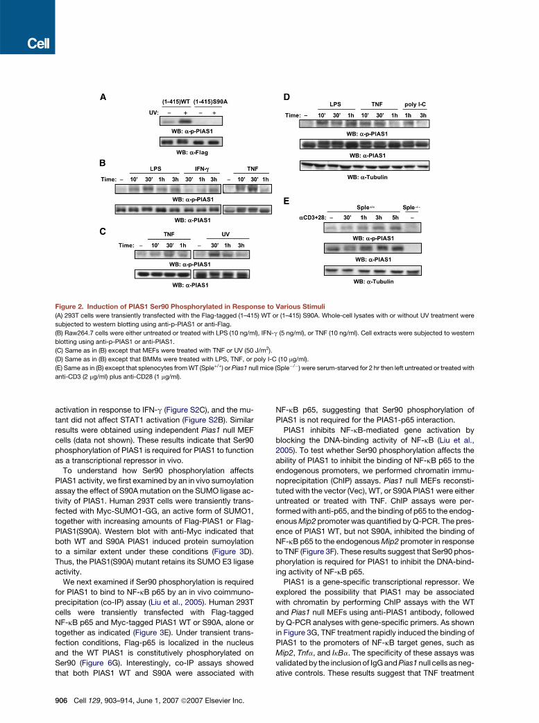
Figure 2. Induction of PIAS1 Ser90 Phosphorylated in Response to Various Stimuli
(A) 293T cells were transiently transfected with the Flag-tagged (1–415) WT or (1–415) S90A. Whole-cell lysates with or without UV treatment were
subjected to western blotting using anti-p-PIAS1 or anti-Flag.
(B) Raw264.7 cells were either untreated or treated with LPS (10 ng/ml), IFN-g (5 ng/ml), or TNF (10 ng/ml). Cell extracts were subjected to western
blotting using anti-p-PIAS1 or anti-PIAS1.
(C) Same as in (B) except that MEFs were treated with TNF or UV (50 J/m2).
(D) Same as in (B) except that BMMs were treated with LPS, TNF, or poly I-C (10 mg/ml).
(E) Same as in (B) except that splenocytes from WT (Sple+/+) or Pias1 null mice (Sple�/�) were serum-starved for 2 hr then left untreated or treated with
anti-CD3 (2 mg/ml) plus anti-CD28 (1 mg/ml).
activation in response to IFN-g (Figure S2C), and the mu-
tant did not affect STAT1 activation (Figure S2B). Similar
results were obtained using independent Pias1 null MEF
cells (data not shown). These results indicate that Ser90
phosphorylation of PIAS1 is required for PIAS1 to function
as a transcriptional repressor in vivo.
To understand how Ser90 phosphorylation affects
PIAS1 activity, we first examined by an in vivo sumoylation
assay the effect of S90A mutation on the SUMO ligase ac-
tivity of PIAS1. Human 293T cells were transiently trans-
fected with Myc-SUMO1-GG, an active form of SUMO1,
together with increasing amounts of Flag-PIAS1 or Flag-
PIAS1(S90A). Western blot with anti-Myc indicated that
both WT and S90A PIAS1 induced protein sumoylation
to a similar extent under these conditions (Figure 3D).
Thus, the PIAS1(S90A) mutant retains its SUMO E3 ligase
activity.
We next examined if Ser90 phosphorylation is required
for PIAS1 to bind to NF-kB p65 by an in vivo coimmuno-
precipitation (co-IP) assay (Liu et al., 2005). Human 293T
cells were transiently transfected with Flag-tagged
NF-kB p65 and Myc-tagged PIAS1 WT or S90A, alone or
together as indicated (Figure 3E). Under transient trans-
fection conditions, Flag-p65 is localized in the nucleus
and the WT PIAS1 is constitutively phosphorylated on
Ser90 (Figure 6G). Interestingly, co-IP assays showed
that both PIAS1 WT and S90A were associated with
906 Cell 129, 903–914, June 1, 2007 ª2007 Elsevier Inc.
NF-kB p65, suggesting that Ser90 phosphorylation of
PIAS1 is not required for the PIAS1-p65 interaction.
PIAS1 inhibits NF-kB-mediated gene activation by
blocking the DNA-binding activity of NF-kB (Liu et al.,
2005). To test whether Ser90 phosphorylation affects the
ability of PIAS1 to inhibit the binding of NF-kB p65 to the
endogenous promoters, we performed chromatin immu-
noprecipitation (ChIP) assays. Pias1 null MEFs reconsti-
tuted with the vector (Vec), WT, or S90A PIAS1 were either
untreated or treated with TNF. ChIP assays were per-
formed with anti-p65, and the binding of p65 to the endog-
enous Mip2 promoter was quantified by Q-PCR. The pres-
ence of PIAS1 WT, but not S90A, inhibited the binding of
NF-kB p65 to the endogenous Mip2 promoter in response
to TNF (Figure 3F). These results suggest that Ser90 phos-
phorylation is required for PIAS1 to inhibit the DNA-bind-
ing activity of NF-kB p65.
PIAS1 is a gene-specific transcriptional repressor. We
explored the possibility that PIAS1 may be associated
with chromatin by performing ChIP assays with the WT
and Pias1 null MEFs using anti-PIAS1 antibody, followed
by Q-PCR analyses with gene-specific primers. As shown
in Figure 3G, TNF treatment rapidly induced the binding of
PIAS1 to the promoters of NF-kB target genes, such as
Mip2, Tnfa, and IkBa. The specificity of these assays was
validated by the inclusion of IgG and Pias1 null cells as neg-
ative controls. These results suggest that TNF treatment

Figure 3. PIAS1 Ser90 Phosphorylation Is Required for PIAS1 to Repress Transcription In Vivo
(A) Western blot analyses with whole-cell extracts from Pias1 null MEFs compensated with the empty vector (Vec), the wild-type (WT), or the S90A
mutant PIAS1 (S90A). +/+: WT MEFs.
(B) Western blot analyses with whole-cell extracts from WT and S90A cells untreated or treated with TNF.
(C) Total RNA from Vec, WT, or S90A cells with or without TNF (20 ng/ml) treatment was isolated and subjected to Q-PCR analyses. Shown are
averages ± SEM of triplicates.
(D) In vivo sumoylation assay. 293T cells were transiently transfected with Myc-tagged SUMO1-GG, together with the increasing amounts of
Flag-tagged WT or S90A mutant PIAS1, followed by western blot analyses.
(E) PIAS1 S90A mutant retains the ability to interact with NF-kB p65. 293T cells were transiently transfected with Flag-NF-kB p65 and Myc-tagged WT
or S90A mutant PIAS1, alone or together as indicated. Whole-cell lysates were subjected to coimmunoprecipitation assays using anti-Flag, followed
by western blotting with anti-Myc or anti-Flag. Whole-cell lysates were immunoblotted with anti-Myc to reveal the expression of PIAS1 WT and S90A
proteins.
(F) Chromatin immunoprecipitation (ChIP) assays were performed with lysates from Vec, WT, or S90A cells untreated or treated with TNF, using anti-
NF-kB p65 or anti-IgG as a negative control. NF-kB p65-bound DNA was quantified by Q-PCR using primers specific for the promoter region adjacent
to the kB site in the murine Mip2 gene. Shown are averages ± SEM of triplicates.
(G) Same as (F) except that ChIP assays were performed with WT and Pias1 null MEFs, using anti-PIAS1 antibody to examine the binding of PIAS1 to
the promoters of Mip2, Tnfa, and IkBa genes. Shown are averages ± SEM of triplicates.
(H) Same as in (F) except that anti-PIAS1 was used for the ChIP assays. Shown are averages ± SEM of triplicates.
triggers the rapid association of PIAS1 to the promoters of
NF-kB-responsive genes for gene repression.
To examine a potential role of Ser90 phosphorylation in
the binding of PIAS1 to the endogenous gene promoters,
similar ChIP assays were performed with Pias1 null MEFs
reconstituted with the Vec, WT, or S90A PIAS1, untreated
or treated with TNF, using anti-PIAS1 antibody. Interest-
ingly, TNF treatment caused a rapid recruitment of the
WT but not the S90A PIAS1 mutant to the Mip2 and
Tnfa gene promoters (Figure 3H). Immunofluorescence
analysis indicated that the S90A was present in the
nucleus (Figure S3). These results suggest that Ser90
Cell 129, 903–914, June 1, 2007 ª2007 Elsevier Inc. 907

Figure 4. PIAS1 S90D and S90E Retain
the Ability to Repress Transcription
(A) Luciferase reporter assays. 293T cells were
transiently transfected with a luciferase re-
porter construct that contains �4000 to +45
of the murine IL1b promoter, together with
Flag-tagged WT or S90A, S90D, S90E mutant
PIAS1 at the indicated doses, in the presence
or absence of TLR4/MD2/CD14 coexpression.
The same extracts were analyzed by western
blot using a-Flag (bottom panel).
(B) Western blot analyses with whole-cell ly-
sates from Pias1 null MEFs reconstituted with
an empty vector (Vec), wild-type PIAS1 (WT),
or S90E mutant PIAS1 (S90E).
(C) Total RNA from Vec, WT, or S90E cells with
or without TNF (20 ng/ml) treatments was iso-
lated and subjected to Q-PCR analyses.
Shown are averages ± SEM of triplicates.
(D) Same as in (C) except that cells were treated
with IFN-g (5 ng/ml) as indicated. Shown are
averages ± SEM of triplicates.
phosphorylation of PIAS1 is required for the TNF-induced
recruitment of PIAS1 to the promoters of the endogenous
NF-kB target genes, consistent with the essential role
of Ser90 phosphorylation in PIAS1-mediated inhibition of
the promoter-binding activity of NF-kB (Figure 3F).
Negatively Charged Amino Acid Residues Can Mimic
the Effect of Ser90 Phosphorylation
Studies described above indicate that PIAS1 S90A is de-
fective in gene repression. One potential concern is that
these results may support the importance of the Ser90
residue per se but not necessarily the phosphoylation of
Ser90 in the regulation of PIAS1 activity. To address this
question, we used the phosphomimic approach. It is
known that negatively charged amino acid residue Glu
or Asp could mimic the effect of serine phosphorylation,
either partially or completely. To explore the possibility
that Glu or Asp might mimic the phosphorylated Ser90
of PIAS1, we analyzed the ability of PIAS1 S90E or S90D
(Ser90 residue is replaced with Glu or Asp, respectively)
mutants to repress transcription. Human 293T cells were
transiently transfected with a luciferase reporter construct
that contains an approximately 4 kb (�4000 to +45) pro-
moter region of the murine IL1b (Godambe et al., 1995),
which is a potent PIAS1-sensitive gene (Liu et al., 2005),
together with Flag-tagged WT PIAS1 (Flag-WT) or S90A,
S90D, and S90E mutants in the presence or absence of
Toll-like receptor 4 (TLR4) coexpression. Consistently,
WT PIAS1, but not S90A, repressed TLR4-induced IL1b
promoter activity in a dose-dependent manner (Figure 4A).
Interestingly, PIAS1 S90D and S90E mutants repressed
TLR4-induced IL1b promoter activity in a similar fashion
as the WT PIAS1 (Figure 4A). These results suggest that
908 Cell 129, 903–914, June 1, 2007 ª2007 Elsevier Inc.
Ser90 phosphorylation, but not the Ser90 residue per se,
is required for PIAS1 to repress transcription.
To further validate the importance of Ser90 phosphory-
lation, Pias1 null MEFs were reconstituted with either the
WT or the S90E mutant PIAS1 to a level similar to the en-
dogenous PIAS1 expression in the WT cells (Figure 4B),
followed by Q-PCR analysis of the induction of NF-kB-
or STAT1-dependent endogenous genes. Consistently,
PIAS1 S90E mutant repressed the TNF-induced Mip2
gene and the IFN-induced Mig gene to a similar extent
as it did the WT PIAS1, while it had no effect on PIAS1-
insensitive genes, such as Junb and Irf1 (Figures 4C and
4D). These results further support that Ser90 phosphoryla-
tion, rather than the Ser90 residue per se, is required for
PIAS1 to repress transcription.
The PIAS1 SUMO Ligase Activity Is Required
for PIAS1 to Repress Transcription
The RING finger-like domain of PIAS1 is important for
its SUMO E3 ligase activity. Mutating a conserved Trp
residue in the RING finger domain does not affect the
structure of E3, but it effectively eliminates the activity of
PIAS SUMO E3 ligase (Kotaja et al., 2002). To address
whether the SUMO ligase activity of PIAS1 is required
for the transcriptional repressor function of PIAS1, we
generated a PIAS1 W372A mutant containing a Trp372
to Ala substitution. The in vivo sumoylation assays con-
firmed that the PIAS1 W372A mutant is defective in pro-
moting protein sumoylation (Figure 5A).
To address whether PIAS1 W372A can repress tran-
scription, we reconstituted Pias1 null MEFs with either
PIAS1 WT or W372A mutant to the similar expression
levels as the endogenous PIAS1 in WT cells (Figure 5B).
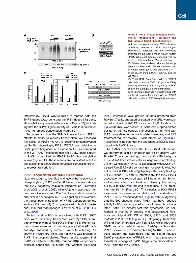
Figure 5. PIAS1 W372A Mutant Is Defec-
tive in Transcriptional Repression and
TNF-Induced Ser90 Phosphorylation
(A) In vivo sumoylation assay. 293T cells were
transiently transfected with Myc-tagged
SUMO1-GG, together with the increasing
amounts of Flag-tagged WT or W372A mutant
PIAS1. Whole-cell lysates were analyzed by
western blotting with anti-Myc or anti-Flag.
(B) Western blot analyses with whole-cell ly-
sates from Pias1 null MEFs reconstituted with
an empty vector (Vec), wild-type PIAS1 (WT),
or the W372A mutant PIAS1 (W372A) and the
WT MEFs (+/+).
(C) Total RNA from Vec, WT, or W372A
cells with or without TNF (20 ng/ml) or IFN-g
(5 ng/ml) treatments was analyzed by Q-PCR.
Shown are averages ± SEM of triplicates.
(D) Western blot analyses were performed with
whole-cell lysates from Vec, WT, or W372A
cells with or without TNF (20 ng/ml) treatments.
Interestingly, PIAS1 W372A failed to repress both the
TNF-induced Mip2 gene and the IFN-induced Mig gene,
although it was present in the nucleus (Figure S3), indicat-
ing that the SUMO ligase activity of PIAS1 is required for
PIAS1 to repress transcription (Figure 5C).
To understand how the SUMO ligase activity of PIAS1
affects its ability to repress transcription, we assessed
the ability of PIAS1 W372A to become phosphorylated
on Ser90. Interestingly, PIAS1 W372A was defective in
Ser90 phosphorylation in response to TNF as compared
to the WT PIAS1, indicating that the SUMO ligase activity
of PIAS1 is required for PIAS1 Ser90 phosphorylation
in vivo (Figure 5D). These results are consistent with the
conclusion that Ser90 phosphorylation is crucial for PIAS1
to repress transcription.
PIAS1 Is Associated with IKKa but not IKKb
Next, we sought to identify the kinase(s) that is involved in
phosphorylating PIAS1 on Ser90. Recent studies indicate
that IKKa negatively regulates inflammation (Lawrence
et al., 2005; Li et al., 2005). IKKa-AA (the kinase-dead mu-
tant) knockin mice and Pias1 null mice show remark-
ably similar phenotypes in NF-kB signaling. For example,
the transcriptional induction of NF-kB-dependent genes,
such as Tnfa and Mip2, is upregulated in both IKKa-AA
and Pias1 null macrophages (Lawrence et al., 2005; Liu
et al., 2005).
To test whether IKKa is associated with PIAS1, 293T
cells were transiently transfected with Myc-PIAS1, to-
gether with or without Flag-IKKa or Flag-IKKb. Protein ex-
tracts were subjected to immunoprecipitation (IP) with
anti-Myc, followed by western blot with anti-Flag. As
shown in Figure 6A, IKKa, but not IKKb, was present in
PIAS1 immunoprecipitates. These results suggest that
PIAS1 can interact with IKKa, but not IKKb, under coex-
pression conditions. To further test whether IKKa and
PIAS1 interact in vivo, protein extracts prepared from
Raw264.7 cells, untreated or treated with LPS, were sub-
jected to IP with anti-PIAS1 or a control IgG. As shown in
Figure 6B, IKKa was present in PIAS1 immunoprecipitates
but not in the IgG control. The association of IKKa with
PIAS1 was detected in unstimulated samples, and LPS
treatment reduced the IKKa-PIAS1 interaction (Figure 6B).
These results indicate that the endogenous IKKa is asso-
ciated with PIAS1 in vivo.
To further characterize the IKKa-PIAS1 interaction,
we performed similar endogenous co-IP assays with
Raw264.7 cells containing a nonspecific siRNA oligo or
IKKa siRNA knockdown cells as negative controls (Fig-
ure 7F). Consistently, PIAS1 is associated with IKKa in un-
treated Raw264.7 cells containing a nonspecific oligo but
not in IKKa siRNA cells or IgG-precipitated samples (Fig-
ure 6C, lanes 1, 3, and 8). Interestingly, the IKKa-PIAS1
association was reduced upon LPS treatment for 30 min
and recurred after 1 hr of treatment. Similarly, the binding
of PIAS1 to IKKa was reduced in response to TNF treat-
ment for 30 min (Figure 6C). The kinetics of IKKa-PIAS1
association is in reverse correlation with the kinetics of
PIAS1 Ser90 phosphorylation (Figure 2B), suggesting
that the S90-phosphorylated PIAS1 may have reduced
affinity for IKKa as compared to that of the unphosphory-
lated PIAS1. To directly test this hypothesis, we per-
formed in vivo co-IP studies with coexpressed Flag-
IKKa and Myc-PIAS1 WT or S90A, S90D, and S90E
mutants in 293T cells (Figure 6D). Intriguingly, both PIAS
WT and S90A interacted with Flag-IKKa, while S90D and
S90E mutants, which mimic Ser90-phosphorylated
PIAS1, showed much reduced binding to IKKa. These re-
sults support the hypothesis that the ligand-induced
Ser90 phosphorylation of PIAS1, which may cause a con-
formational change of PIAS1, triggers the dissociation of
PIAS1 from the IKKa kinase.
Cell 129, 903–914, June 1, 2007 ª2007 Elsevier Inc. 909
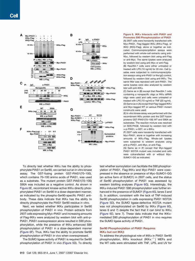
Figure 6. IKKa Interacts with PIAS1 and
Promotes S90 Phosphorylation of PIAS1
(A) 293T cells were transiently transfected with
Myc-PIAS1, Flag-tagged IKKa (IKKa-Flag), or
IKKb (IKKb-Flag), alone or together as indi-
cated. Coimmunoprecipitation assays were
performed with whole-cell extracts using anti-
Myc, followed by western blot using anti-Flag
or anti-Myc. The same lysates were analyzed
by western blot using anti-Myc or anti-Flag.
(B) Raw264.7 cells were either untreated or
treated with LPS (10 ng/ml) for 20 min. Cell ly-
sates were subjected to coimmunoprecipita-
tion assays using anti-PIAS1 or the IgG control,
followed by western blot using anti-IKKa. The
same filter was reprobed with anit-PIAS1. The
same lysates were also analyzed by western
blot with anti-IKKa.
(C) Same as in (B) except that Raw264.7 cells
containing a nonspecific oligo or IKKa siRNA
oligo were used and cells were untreated or
treated with LPS (10 ng/ml) or TNF (20 ng/ml).
(D) Same as in (A) except that Flag-tagged IKKa
and Myc-tagged WT or various PIAS1 mutant
constructs were used.
(E) In vitro kinase assay was performed with the
recombinant IKKa protein and the GST-fusion
proteins GST-PIAS1(70–100) WT and S90A as
substrates. The reaction mixture was resolved
on SDS-PAGE, followed by western blot with
a-p-PIAS1, a-GST, or a-IKKa.
(F) 293T cells were transiently transfected with
Myc-PIAS1, alone or together with increasing
amounts of IKKa-Flag. Whole-cell lysates
were subjected to western analyses using
anti-p-PIAS1, anti-Myc, or anti-Flag.
(G) Same as in (F) except that Myc-tagged
PIAS1 W372A mutant was included and cells
were cotransfected with or without Myc-
SUMO1-GG as indicated.
To directly test whether IKKa has the ability to phos-
phorylate PIAS1 on Ser90, we carried out an in vitro kinase
assay. The GST-fusing protein GST-PIAS1(70–100),
which contains 70–100 animo acids of PIAS1, was used
as a substrate. The mutant protein GST-PIAS1(70–100)
S90A was included as a negative control. As shown in
Figure 6E, recombinant kinase-active IKKa directly phos-
phorylated PIAS1 on Ser90 in a dose-dependent manner,
as indicated by the phospho-Ser90-specific PIAS1 anti-
body. These data indicate that IKKa has the ability to
directly phosphorylate the PIAS1 Ser90 residue in vitro.
Next, we tested whether IKKa participates in Ser90
phosphorylation of PIAS1 in vivo. Protein extracts from
293T cells expressing Myc-PIAS1 and increasing amounts
of Flag-IKKa were analyzed by western blot with anti-p-
PIAS1. PIAS1 overexpression alone resulted in S90 phos-
phorylation, while the presence of IKKa enhanced S90
phosphorylation of PIAS1 in a dose-dependent manner
(Figure 6F). Thus, IKKa has the ability to promote Ser90
phosphorylation of PIAS1 in vivo when overexpressed.
The SUMO ligase activity of PIAS1 is required for Ser90
phosphorylation of PIAS1 in vivo (Figure 5D). To directly
910 Cell 129, 903–914, June 1, 2007 ª2007 Elsevier Inc.
test whether sumoylation can facilitate the S90 phosphor-
ylation of PIAS1, Flag-IKKa and Myc-PIAS1 were coex-
pressed in the absence or presence of Myc-SUMO1-GG
(an active form of SUMO1) in 293T cells, and the status
of Ser90 phosphorylation of PIAS1 was assessed by
western blotting analyses (Figure 6G). Interestingly, the
IKKa-induced PIAS1 S90 phosphorylation was further en-
hanced in the presence of SUMO1 (Figure 6G, lanes 3 and
5). In addition, consistent with the lack of TNF-induced
Ser90 phosphorylation in cells expressing PIAS1 W372A
(Figure 5D), the SUMO ligase-defective W372A mutant
was not phosphorylated on Ser90 by IKKa (Figure 6G,
lanes 6 and 7) despite the fact that it can bind to IKKa
(Figure 6D, lane 7). These data indicate that the IKKa-
mediated S90 phosphorylation of PIAS1 in vivo requires
the SUMO ligase activity of PIAS1.
Ser90 Phosphorylation of PIAS1 Requires
IKKa but not IKKb
To address the physiological role of IKKa in PIAS1 Ser90
phosphorylation, IKKa knockout (IKKa�/�) MEFs and
the WT cells were stimulated with TNF, LPS, and UV for

Figure 7. IKKa Is Required for Ligand-
induced Ser90 Phosphorylation of PIAS1
In Vivo
(A) WT and IKKa�/� MEF cells were either un-
treated or treated with TNF (20 ng/ml). Whole-
cell extracts were subjected to western blot
analyses.
(B) Same as in (A) except that cells were un-
treated or treated with LPS (0.5 mg/ml).
(C) Same as in (A) except that cells were un-
treated or treated with UV (50 J/m2).
(D) Cell extracts from WT MEFs (+/+) or IKKa�/�
MEFs compensated with an empty Vec, the WT
IKKa, or the kinase-inactive mutant IKKa (AA)
were analyzed by western blot.
(E) Same as in (A) except that IKKa�/� MEFs
compensated with an empty vector (Vec), the
wild-type IKKa (WT), or the kinase-inactive
mutant IKKa (AA) were used.
(F) Same as in (A) except that Raw264.7 cells
containing a nonspecific oligo (Nonspecific) or
IKKa siRNA oligo were used.
(G) Same as in (A) except that WT and IKKb�/�
MEFs were used.
(H) Same as in (A) except that Raw264.7 cells
containing a nonspecific oligo or IKKb siRNA
oligo were used.
various time periods. Although the basal level of PIAS1
phosphorylation was not significantly affected in the
absence of IKKa, the ligand-induced PIAS1 Ser90 phos-
phorylation in response to TNF, LPS, or UV treatment
was defective in IKKa�/� MEFs (Figures 7A, 7B, and 7C).
These results support the conclusion that IKKa is required
for the ligand-induced Ser90 phosphorylation of PIAS1.
To validate the role of IKKa in mediating Ser90 phos-
phorylation of PIAS1, IKKa�/� cells were reconstituted
with an empty Vec, WT IKKa, or kinase-inactive IKKa mu-
tant (AA), which carries a Ser176Ser180 to AA substitu-
tion, to a level comparable to the endogenous IKKa in
WT MEFs (+/+) (Figure 7D). Consistently, TNF-induced
Ser90 phosphorylation of PIAS1 was restored in cells
rescued with the WT IKKa but not the empty Vec or the
kinase-inactive AA mutant (Figure 7E). These data indicate
that the kinase activity of IKKa is required for TNF-induced
Ser90 phosphorylation of PIAS1.
To further demonstrate the role of IKKa in PIAS1 S90
phosphorylation, the expression of the endogenous
IKKa in Raw264.7 cells was inhibited by siRNA (Figure 7F).
Consistently, PIAS1 Ser90 phosphorylation in response to
TNF was defective in IKKa knockdown cells as compared
to that in the control cells (Figure 7F). These results, to-
gether with the data described in Figure 6, indicate that
IKKa mediates Ser90 phosphorylation of PIAS1.
IKKa, but not IKKb, interacts with PIAS1 (Figure 6A). To
address whether IKKb is required for TNF-induced S90
phosphorylation of PIAS1, IKKb�/�MEFs and the WT cells
were treated with TNF. Consistently, the TNF-induced
PIAS1 S90 phosphorylation was not defective in IKKb�/�
cells as compared to that of the control cells (Figure 7G).
In addition, S90 phosphorylation of PIAS1 in response to
TNF was normal in IKKb siRNA knockdown Raw264.7
cells as compared to that of the nonspecific siRNA control
cells (Figure 7H). Taken together, our data indicate that
IKKa, but not IKKb, is required for TNF-induced Ser90
phosphorylation of PIAS1.
DISCUSSION
The most significant finding described in this paper is the
discovery of a novel anti-inflammatory signaling pathway
commonly utilized by a wide variety of immunoregulatory
stimuli (Figure S4). In this pathway, pro-inflammatory stim-
uli signal to the nucleus to induce the recruitment of a pro-
tein SUMO ligase to the chromatin for immediate gene
repression. Our studies suggest that PIAS1 functions as
a nuclear sensor for extracellular inflammatory stimuli.
The PIAS1-mediated anti-inflammatory pathway may
serve as an important Yin-Yang regulatory mechanism
to maintain a delicate balance in immune responses.
The PIAS1 anti-inflammatory signaling pathway has
several unique features. Although Ser90 phosphorylation
of PIAS1 does not affect the intrinsic SUMO ligase activity
of PIAS1, it is required for PIAS1 to bind to the endoge-
nous gene promoters, where it inhibits the DNA-binding
Cell 129, 903–914, June 1, 2007 ª2007 Elsevier Inc. 911
activity of NF-kB/STAT1. Consistent with this model, the
effect of Ser90 phosphorylation on PIAS1-mediated tran-
scriptional repression was not evident when a reporter
containing only the minimum NF-kB-binding site was
used in luciferase assays (our unpublished data). In ad-
dition, IKKa-mediated Ser90 phosphorylation of PIAS1
in vivo is dependent on the SUMO ligase activity of
PIAS1. These results reveal a novel molecular mechanism
for the regulation of inflammatory gene expression, which
involves crossregulation between protein sumoylation
and protein phosphorylation events. Consistent with this
model, both Ser90 phosphorylation and SUMO ligase ac-
tivity of PIAS1 are required for PIAS1 to repress transcrip-
tion. The PIAS1 signaling pathway is distinct from other
known negative regulatory mechanisms such as the in-
duction of IkBa, A20, and SOCSs, which are transcription
dependent and serve as secondary negative feedback
loops to restrict NF–kB or STAT responses (Li and Verma,
2002; Naka et al., 2005; Shuai and Liu, 2005). Another in-
teresting feature of PIAS1 signaling is that it selectively
regulates a subset of NF-kB/STAT1-dependent genes,
with a notable preference for pro-inflammatory cytokines
and chemokines. Targeting gene-specific regulators
such as PIAS1, rather than general regulators that affect
the overall responses of NF-kB or STAT, may prove to
be a valuable therapeutic strategy for the treatment of
human immune disorders.
The anti-inflammatory function of PIAS1 is achieved by
regulating several transcription factors, including STAT1,
NF-kB, and PPAR-g, known to be important in immune
regulation (Liu et al., 2004, 2005; Pascual et al., 2005).
We noticed that compared to TNF or LPS treatment,
IFN-g appears to induce a much weaker Ser90 phos-
phorylation on PIAS1 (Figure 2B). However, PIAS1 Ser90
phosphorylation is clearly required for PIAS1 to repress
STAT1-depdendent genes (Figure S2). Under physiologi-
cal conditions, immune cells are frequently exposed to
both TNF and IFN-g, which are known to have a synergistic
effect on the induction of many inflammatory genes. In
addition, by acting as a key transcription factor in LPS-
induced IFN-b signaling, STAT1 plays an important role
in the regulation of TLR response. Thus, Ser90 phos-
phorylation, which is required for PIAS1 to function as a
transcriptional repressor, is important in the regulation of
multiple transcriptional responses.
Recent studies have uncovered a surprising role of IKKa
in the negative regulation of NF-kB-mediated immune and
inflammatory gene activation in macrophages. Two differ-
ent mechanisms have been reported that may account for
the inhibitory effect of IKKa on NF-kB-mediated gene
activation (Lawrence et al., 2005; Li et al., 2005). Data
presented in this paper suggest an alternative mechanism
through which IKKa may act to limit inflammation. Since
PIAS1 also regulates other immunoregulatory transcrip-
tion factors such as STAT1 and PPAR-g (Pascual et al.,
2005; Shuai and Liu, 2005), our studies suggest that the
IKKa-mediated anti-inflammatory effect is not restricted
to NF-kB. The IKKa-PIAS1 link may mediate crosstalk be-
912 Cell 129, 903–914, June 1, 2007 ª2007 Elsevier Inc.
tween various signaling pathways to coordinately regulate
inflammation and immunity.
There are important differences between the IKK-medi-
ated phosphorylation of IkBa and the IKKa-mediated
phosphorylation of PIAS1. In most published studies, the
activity of IKK, which contains IKKa and IKKb, is measured
by using IkBa as the substrate in both in vitro and in vivo
assays. Although IKKa is a component of the IKK complex
responsible for the phosphorylation and degradation of
IkB, genetic studies indicate that IKKa is dispensable in
this process, suggesting that IKKb is largely responsible
for phosphorylating IkBa in vivo. In contrast, IKKa, but
not IKKb, is responsible for PIAS1 Ser90 phosphorylation.
Thus, there are important differences between the IKK-
IkBa and the IKKa-PIAS1 phosphorylation events: sub-
strates are different (IkBa versus PIAS1) and kinases are
different (IKKb in the IKK complex versus IKKa itself). In
addition, we showed that the IKKa-mediated phosphory-
lation of PIAS1 in vivo requires the SUMO ligase activity of
PIAS1. Although the activity of IKK, when assayed by
using IkBa as the substrate, is not activated by UV treat-
ment (Kato et al., 2003; Li and Karin, 1998), IKKa is re-
quired for PIAS1 Ser90 phosphorylation in response to
UV (Figure 7C).
Although recombinant IKKa has the ability to directly
phosphorylate the Ser90 residue in GST-PIAS1(70–100)
in vitro, TNF-induced IKKa-mediated PIAS1 Ser90 phos-
phorylation in vivo is dependent on the SUMO ligase activ-
ity of PIAS1. Consistently, the increased sumoylation
caused by SUMO-1 overexpression enhanced the IKKa-
mediated PIAS1 S90 phosphorylation. PIAS1 is known
to be self-sumoylated (Kotaja et al., 2002). It is possible
that the self-sumoylation of PIAS1 is required for the expo-
sure of the region containing the Ser90 residue for IKKa.
Alternatively, PIAS1 may be involved in the sumoylation
of IKKa or other unknown regulatory factors that are in-
volved in PIAS1 phosphorylation in vivo. Further studies
are required to understand the molecular mechanism of
PIAS1 sumoylation in the regulation of Ser90 phosphory-
lation.
A basal level of PIAS1 Ser90 phosphorylation is detect-
able in untreated cells, suggesting that a portion of PIAS1
is constitutively activated. Our results indicate that IKKa is
responsible for the ligand-induced but not the basal level
of PIAS1 phosphorylation. This is perhaps not surprising
given the previous findings that IKKa is largely localized
in the cytoplasm in untreated cells, and it translocates
into the nucleus only after ligand stimulation (Anest
et al., 2003; Yamamoto et al., 2003). Although IKKa was
present in the PIAS1 immunoprecipitates of untreated
cells (Figure 6B), a significant portion of this association
is most likely due to the presence of PIAS1 and IKKa in
the same protein exacts under cell-free conditions. The ki-
nase(s) responsible for the basal PIAS1 phosphorylation
remains to be identified. It is possible that a kinase(s) other
than IKKa may also be involved in Ser90 phosphorylation
of PIAS1 in response to certain stimuli or in certain cell
types. In addition, PIAS1 may also be regulated by other
posttranslational modifications, including phosphoryla-
tion on sites other than Ser90. Indeed, in IKKa�/� cells, al-
though PIAS1 was not phosphorylated on Ser90 upon UV
treatment, a small mobility shift of PIAS1 was observed
(Figure 7C). It is possible that in the absence of PIAS1
Ser90 phosphorylation, UV treatment, which is known to
activate multiple protein kinases, may cause PIAS1 phos-
phorylation on other sites. The biological significance of
such phosphorylation remains to be determined.
EXPERIMENTAL PROCEDURES
Cell Lines and Reagents
Murine macrophage-like Raw264.7 cells and murine splenocytes were
cultured in RPMI plus 10% FBS and 1% penicillin/streptomycin. MEFs
and BMMs were derived and cultured as described previously (Liu
et al., 2004).
The polyclonal anti-phosphorylated Ser90-specific antibody was
generated using the peptide antigen HSSPMPPTL-pS-PSTIPQLT,
which corresponds to the amino acids 81–98 of PIAS1 and bears
a phosphorylated Ser90 (-pS-). The following antibodies were pur-
chased: anti-Flag (M2ab; Sigma), anti-Myc (9B11; Cell signaling),
anti-Tubulin (Sigma), anti-PARP (Santa Cruz), anti-NF-kB p65 (C20;
Santa Cruz), anti-phospho-Stat1 (Cell Signaling), anti-Stat1 (E23;
Santa Cruz), anti-IKKa (Cell Signaling), and anti-IKKb (Cell Signaling).
The following reagents were purchased: Trypsin (Promega), murine
recombinant TNF-a and IFN-g (PeproTech), LPS (from Escherichia
coli serotype O55:B5; Sigma), poly I-C (Sigma), anti-CD3 (145-2C11;
Pharmingen), and anti-CD28 (37.51; Pharmingen).
Plasmid Constructs
Flag-tagged PIAS1 deletional or single point mutants were generated
by the insertion of PCR fragments containing various regions of PIAS1
cDNA into the BamHI and SalI sites of pCMV-Flag. The retroviral
expression constructs encoding the WT or S90A or S90E mutant
PIAS1 were generated by the insertion of the corresponding PCR frag-
ments into the BglII and XhoI sites of pMSCVpuro (Clontech). GST-
PIAS1(70–100) WT and GST-PIAS1(70–100) S90A were constructed
by insertion of the cDNA encoding 70–100 amino acids of the WT or
S90A PIAS1 into the EcoRI and NotI cloning sites of pGEX4T-1 (Phar-
macia). TLR4/MD2/CD14 constructs were obtained from R. Modlin.
LC-MS/MS Analysis
293T cells were transiently transfected with Flag-PIAS1(1–415). Thirty
hours post-transfection, cells were either untreated or treated with UV
(100 J/m2) and harvested 30 min post UV treatment. Immunoprecipita-
tions were performed with cell lysates using anti-Flag, followed by
SDS-PAGE. The protein gel was stained with Sypro Ruby (Molecular
Probes), and the corresponding PIAS1(1–415) protein fragments
were excised and subjected to endoproteinase Asp-N digestion. The
peptide fragments were then extracted according to established pro-
tocols (Shevchenko et al., 1996) and analyzed by LC-MS/MS with
a quadruple time-of-flight mass spectrometer (QSTAR Pulsar-XL,
Applied Biosystems/MDS Sciex, Concord, ON, Canada) equipped
with an LC Packings/Dionex nano-HPLC system (Sunnyvale, CA).
32P-Labeling
293T cells overexpressing either Flag-PIAS1(1–415) WT or S90A were
labeled with 32P-orthophosphate (0.4 mCi/ml) for 3 hr (Shuai et al.,
1992). Cells were then either untreated or treated with UV (100 J/m2),
and cell lysates were prepared 30 min post UV treatment and used
for immunoprecipitations with anti-Flag. The 32P-labeling of endoge-
nous PIAS1 experiment was performed similarly, except that anti-
PIAS1 was used for immunoprecipitations.
Coimmunoprecipitation and Immunoblotting
Coimmunoprecipitation and immunoblotting experiments were essen-
tially performed as described previously (Liu et al., 1998, 2001).
Quantitative Real-Time PCR Analysis
Cells were either untreated or treated as indicated. Total RNA was
isolated and subjected to quantitative real-time PCR analysis as
described (Liu et al., 2004).
ChIP Assay
ChIP assays were performed as described (Liu et al., 2004, 2005). The
following primers were used to amplify the promoter region adjacent to
the kB sites: Mip2 gene: forward, 50-AGCGCAGACATCACTTCCTT;
reverse, 50-CTAGCTGCCTGCCTCATTCT. Tnfa gene: forward, 50-
GCAGGTTCTGTCCCTTTCAC; reverse, 50-AGTGCCTCTTCTGCCAG
TTC. The primers for the IkBa gene have been described (Liu et al.,
2005).
In Vitro Kinase Assay
Kinase-active recombinant IKKa protein was purchased from Upstate
(Lake Placid, NY). Bacterially purified glutathione S-transferase (GST)
fusing proteins GST-PIAS1(70–100) WT and GST-PIAS1(70–100)
S90A were used as substrates. In vitro kinase assay was performed
as instructed by the manufacturer with modifications. Briefly, GST-
PIAS1 fusion proteins were preheated at 95�C for 3 min and then
chilled on ice for 5 min before being mixed with IKKa in the kinase re-
action buffer. PIAS1 Ser90 phosphorylation was measured by western
blot with anti-p-PIAS1.
IKKa and IKKb siRNA Cells
Specific inhibition of the murine IKKa (mIKKa) or IKKb (mIKKb) expres-
sion was achieved by the small RNA interference technique (siRNA).
mIKKa siRNA (50-AAATCTGAACTGTCTTGGCCATT-30) and the non-
specific siRNA (50-AAGGAAGACTACAGCAGGCTCTT-30) were cloned
into pSIREN-RetroQ vector (BD Biosciences Clontech). The sequence
for mIKKb siRNA is 50-AACTTGAAGCTGGTTCATGTCTT-30; and the
sequence of the nonspecific control for mIKKb is 50-AAGACAAACG
AGGGCCTCACATT-30. Raw 264.7 cells at 50%–60% confluence
were transfected with the indicated siRNA plasmids using Lipofect-
amine 2000 (Invitrogen). The puromycin-resistant colonies were iso-
lated following selection with 2.5 mg/ml puromycin and screened by
western blotting analyses for efficient inhibitions of endogenous
IKKa or IKKb expression.
Supplemental Data
Supplemental Data include four figures and can be found with this
article online at http://www.cell.com/cgi/content/full/129/5/903/DC1/.
ACKNOWLEDGMENTS
We thank M. Karin for IKKa and IKKb null MEFs, R. Modlin for TLR4
constructs, G. Stark for helpful discussions, and Y. Xie for assistance
with the LC-MS/MS analysis. The UCLA Mass Spectrometry and
Proteomics Technology Center was established with a grant from
the W.M. Keck Foundation, with additional support from the UCLA
Jonsson Comprehensive Cancer Center. This work was supported
by grants from the NIH and the UCLA Jonsson Comprehensive Cancer
Center (K.S.). B.L. is supported by a Research Scientist Development
Award from the NIH (K01 AR52717-01).
Received: October 16, 2006
Revised: February 23, 2007
Accepted: March 29, 2007
Published: May 31, 2007
Cell 129, 903–914, June 1, 2007 ª2007 Elsevier Inc. 913

REFERENCES
Anest, V., Hanson, J.L., Cogswell, P.C., Steinbrecher, K.A., Strahl,
B.D., and Baldwin, A.S. (2003). A nucleosomal function for IkappaB ki-
nase-alpha in NF-kappaB-dependent gene expression. Nature 423,
659–663.
Bottero, V., Withoff, S., and Verma, I.M. (2006). NF-kappaB and the
regulation of hematopoiesis. Cell Death Differ. 13, 785–797.
Chung, C.D., Liao, J., Liu, B., Rao, X., Jay, P., Berta, P., and Shuai, K.
(1997). Specific inhibition of Stat3 signal transduction by PIAS3. Sci-
ence 278, 1803–1805.
Darnell, J.E., Jr. (1997). STATs and gene regulation. Science 277,
1630–1635.
Ghosh, S., and Karin, M. (2002). Missing pieces in the NF-kappaB puz-
zle. Cell Suppl. 109, S81–S96.
Godambe, S.A., Chaplin, D.D., Takova, T., and Bellone, C.J. (1995).
Molecular dissection of the murine IL-1beta promoter. Am. J. Ther.
2, 677–686.
Hoberg, J.E., Yeung, F., and Mayo, M.W. (2004). SMRT derepression
by the IkappaB kinase alpha: a prerequisite to NF-kappaB transcrip-
tion and survival. Mol. Cell 16, 245–255.
Johnson, E.S., and Gupta, A.A. (2001). An E3-like factor that promotes
SUMO conjugation to the yeast septins. Cell 106, 735–744.
Kato, T., Jr., Delhase, M., Hoffmann, A., and Karin, M. (2003). CK2 is
a C-terminal IkappaB kinase responsible for NF-kappaB activation
during the UV response. Mol. Cell 12, 829–839.
Kotaja, N., Karvonen, U., Janne, O.A., and Palvimo, J.J. (2002). PIAS
proteins modulate transcription factors by functioning as SUMO-1
ligases. Mol. Cell. Biol. 22, 5222–5234.
Lawrence, T., Bebien, M., Liu, G.Y., Nizet, V., and Karin, M. (2005).
IKKalpha limits macrophage NF-kappaB activation and contributes
to the resolution of inflammation. Nature 434, 1138–1143.
Li, N., and Karin, M. (1998). Ionizing radiation and short wavelength UV
activate NF-kappaB through two distinct mechanisms. Proc. Natl.
Acad. Sci. USA 95, 13012–13017.
Li, Q., and Verma, I.M. (2002). NF-kappaB regulation in the immune
system. Nat. Rev. Immunol. 2, 725–734.
Li, Q., Lu, Q., Bottero, V., Estepa, G., Morrison, L., Mercurio, F., and
Verma, I.M. (2005). Enhanced NF-kappaB activation and cellular func-
914 Cell 129, 903–914, June 1, 2007 ª2007 Elsevier Inc.
tion in macrophages lacking IkappaB kinase 1 (IKK1). Proc. Natl. Acad.
Sci. USA 102, 12425–12430.
Liu, B., Liao, J., Rao, X., Kushner, S.A., Chung, C.D., Chang, D.D., and
Shuai, K. (1998). Inhibition of Stat1-mediated gene activation by
PIAS1. Proc. Natl. Acad. Sci. USA 95, 10626–10631.
Liu, B., Gross, M., ten Hoeve, J., and Shuai, K. (2001). A transcriptional
corepressor of Stat1 with an essential LXXLL signature motif. Proc.
Natl. Acad. Sci. USA 98, 3203–3207.
Liu, B., Mink, S., Wong, K.A., Stein, N., Getman, C., Dempsey, P.W.,
Wu, H., and Shuai, K. (2004). PIAS1 selectively inhibits interferon-
inducible genes and is important in innate immunity. Nat. Immunol.
5, 891–898.
Liu, B., Yang, R., Wong, K.A., Getman, C., Stein, N., Teitell, M.A.,
Cheng, G., Wu, H., and Shuai, K. (2005). Negative regulation of
NF-kappaB signaling by PIAS1. Mol. Cell. Biol. 25, 1113–1123.
Naka, T., Fujimoto, M., Tsutsui, H., and Yoshimura, A. (2005). Negative
regulation of cytokine and TLR signalings by SOCS and others.
Adv. Immunol. 87, 61–122.
Pascual, G., Fong, A.L., Ogawa, S., Gamliel, A., Li, A.C., Perissi, V.,
Rose, D.W., Willson, T.M., Rosenfeld, M.G., and Glass, C.K. (2005).
A SUMOylation-dependent pathway mediates transrepression of
inflammatory response genes by PPAR-gamma. Nature 437, 759–763.
Sharrocks, A.D. (2006). PIAS proteins and transcriptional regulation–
more than just SUMO E3 ligases? Genes Dev. 20, 754–758.
Shevchenko, A., Wilm, M., Vorm, O., and Mann, M. (1996). Mass spec-
trometric sequencing of proteins silver-stained polyacrylamide gels.
Anal. Chem. 68, 850–858.
Shuai, K., and Liu, B. (2003). Regulation of JAK-STAT signalling in the
immune system. Nat. Rev. Immunol. 3, 900–911.
Shuai, K., and Liu, B. (2005). Regulation of gene-activation pathways
by PIAS proteins in the immune system. Nat. Rev. Immunol. 5, 593–
605.
Shuai, K., Schindler, C., Prezioso, V.R., and Darnell, J.E., Jr. (1992).
Activation of transcription by IFN-gamma: tyrosine phosphorylation
of a 91-kD DNA binding protein. Science 258, 1808–1812.
Yamamoto, Y., Verma, U.N., Prajapati, S., Kwak, Y.T., and Gaynor,
R.B. (2003). Histone H3 phosphorylation by IKK-alpha is critical for
cytokine-induced gene expression. Nature 423, 655–659.
Copyright © 2022 FDOKUMEN