Drug Delivery from Stimuli-Responsive Poly(N - MDPI
15
Citation: Ortega-García, A.; Martínez-Bernal, B.G.; Ceja, I.; Mendizábal, E.; Puig-Arévalo, J.E.; Pérez-Carrillo, L.A. Drug Delivery from Stimuli-Responsive Poly(N-isopropylacrylamide-co-N- isopropylmethacrylamide)/Chitosan Core/Shell Nanohydrogels. Polymers 2022, 14, 522. https://doi.org/ 10.3390/polym14030522 Academic Editor: Barbara Vigani Received: 8 January 2022 Accepted: 25 January 2022 Published: 27 January 2022 Publisher’s Note: MDPI stays neutral with regard to jurisdictional claims in published maps and institutional affil- iations. Copyright: © 2022 by the authors. Licensee MDPI, Basel, Switzerland. This article is an open access article distributed under the terms and conditions of the Creative Commons Attribution (CC BY) license (https:// creativecommons.org/licenses/by/ 4.0/). polymers Article Drug Delivery from Stimuli-Responsive Poly(N- isopropylacrylamide-co-N-isopropylmethacrylamide)/Chitosan Core/Shell Nanohydrogels Andrés Ortega-García 1 , Bryan Giovanny Martínez-Bernal 1 , Israel Ceja 2 , Eduardo Mendizábal 3 , Jorge Emilio Puig-Arévalo 1 and Lourdes Adriana Pérez-Carrillo 1, * 1 Chemical Engineering Department, University Center of Exact Sciences and Engineering (CUCEI), University of Guadalajara (UdG), Guadalajara 44100, Jalisco, Mexico; [email protected] (A.O.-G.); [email protected] (B.G.M.-B.); [email protected] (J.E.P.-A.) 2 Physics Department, University Center of Exact Sciences and Engineering (CUCEI), University of Guadalajara, Guadalajara (UdG), Guadalajara 44100, Jalisco, Mexico; [email protected] 3 Chemistry Department, University Center of Exact Sciences and Engineering (CUCEI), University of Guadalajara (UdG), Guadalajara 44100, Jalisco, Mexico; [email protected] * Correspondence: [email protected] or [email protected] Abstract: The synthesis of stimulus-responsive poly(N-isopropylacrylamide-co-N-isopropylmethacry- lamide)/chitosan core/shell nanohydrogels made by batch emulsion polymerization in the presence of chitosan (CS) micelles is reported. The ratio of monomers required to obtain copolymers with a volume phase transition temperature (T VPT ) in the range of the temperatures observed in the human body in response to an infection (38 to 40 ◦ C) was estimated with the Fox equation. The conversion was determined by gravimetry; mean particle size, size distribution, and thermal response were measured by quasi-elastic light scattering (QLS). The core/shell structure was confirmed by TEM, and FTIR showed the presence of N-isopropyl acrilamide (NIPA), N-isopropyl methacrylamide (NIPMA), and CS in the nanohydrogels. The nanohydrogels were loaded with the drug doxycycline hyclate, and their release kinetic profile was determined at pH = 2.0 and 7.4 at their volume phase transition temperatures (T VPT ). A higher amount of drug was released at acidic pH. Some mathematical models described in the literature were used to fit the experimental drug release data. Keywords: nanohydrogels; stimuli-responsive polymers; core-shell polymers; drug delivery 1. Introduction Stimuli-responsive polymers are materials that have the ability to significantly change a property (or properties) in response to external environmental conditions [1,2]. Typical stimuli are temperature, pH, ionic strength, magnetic and electrical fields, and light and redox potential [3]. Stimuli-responsive polymers can be designed to act as biosensors [4] in ion-exchange chromatography [5], in micromechanical materials [6], in chemotherapy [7,8], in drug delivery [9,10], in photo-responsive coating materials [11] and in photonics [12]. Stimuli-responsive nanoparticles have been used recently as drug carriers to deliver active ingredients such as drugs, peptides, and genes [13]. Because there are specific condi- tions (temperature and pH, among others) to which stimuli-responsive nanoparticles loaded with specific drugs can respond, the importance of these nanoparticles is evident, particu- larly when they respond to more than one stimulus simultaneously [14]. Hyperthermia commonly occurs in diseased tissues compared to normal tissues, so several temperature- sensitive nanohydrogels have been developed for drug delivery purposes [15,16]. The most used thermosensitive polymer is poly(N-isopropylacrylamide) (PNIPA) which has a lower critical solution temperature (LCST) or volume phase transition temper- ature (T VPT ) at 32 ◦ C[17]. Below 32 ◦ C, PNIPA is water soluble, but above this temperature Polymers 2022, 14, 522. https://doi.org/10.3390/polym14030522 https://www.mdpi.com/journal/polymers
-
Upload
khangminh22 -
Category
Documents
-
view
0 -
download
0
Transcript of Drug Delivery from Stimuli-Responsive Poly(N - MDPI

�����������������
Citation: Ortega-García, A.;
Martínez-Bernal, B.G.; Ceja, I.;
Mendizábal, E.; Puig-Arévalo, J.E.;
Pérez-Carrillo, L.A. Drug Delivery
from Stimuli-Responsive
Poly(N-isopropylacrylamide-co-N-
isopropylmethacrylamide)/Chitosan
Core/Shell Nanohydrogels. Polymers
2022, 14, 522. https://doi.org/
10.3390/polym14030522
Academic Editor: Barbara Vigani
Received: 8 January 2022
Accepted: 25 January 2022
Published: 27 January 2022
Publisher’s Note: MDPI stays neutral
with regard to jurisdictional claims in
published maps and institutional affil-
iations.
Copyright: © 2022 by the authors.
Licensee MDPI, Basel, Switzerland.
This article is an open access article
distributed under the terms and
conditions of the Creative Commons
Attribution (CC BY) license (https://
creativecommons.org/licenses/by/
4.0/).
polymers
Article
Drug Delivery from Stimuli-Responsive Poly(N-isopropylacrylamide-co-N-isopropylmethacrylamide)/ChitosanCore/Shell NanohydrogelsAndrés Ortega-García 1, Bryan Giovanny Martínez-Bernal 1 , Israel Ceja 2, Eduardo Mendizábal 3 ,Jorge Emilio Puig-Arévalo 1 and Lourdes Adriana Pérez-Carrillo 1,*
1 Chemical Engineering Department, University Center of Exact Sciences and Engineering (CUCEI),University of Guadalajara (UdG), Guadalajara 44100, Jalisco, Mexico; [email protected] (A.O.-G.);[email protected] (B.G.M.-B.); [email protected] (J.E.P.-A.)
2 Physics Department, University Center of Exact Sciences and Engineering (CUCEI),University of Guadalajara, Guadalajara (UdG), Guadalajara 44100, Jalisco, Mexico;[email protected]
3 Chemistry Department, University Center of Exact Sciences and Engineering (CUCEI),University of Guadalajara (UdG), Guadalajara 44100, Jalisco, Mexico; [email protected]
* Correspondence: [email protected] or [email protected]
Abstract: The synthesis of stimulus-responsive poly(N-isopropylacrylamide-co-N-isopropylmethacry-lamide)/chitosan core/shell nanohydrogels made by batch emulsion polymerization in the presenceof chitosan (CS) micelles is reported. The ratio of monomers required to obtain copolymers with avolume phase transition temperature (TVPT) in the range of the temperatures observed in the humanbody in response to an infection (38 to 40 ◦C) was estimated with the Fox equation. The conversionwas determined by gravimetry; mean particle size, size distribution, and thermal response weremeasured by quasi-elastic light scattering (QLS). The core/shell structure was confirmed by TEM, andFTIR showed the presence of N-isopropyl acrilamide (NIPA), N-isopropyl methacrylamide (NIPMA),and CS in the nanohydrogels. The nanohydrogels were loaded with the drug doxycycline hyclate,and their release kinetic profile was determined at pH = 2.0 and 7.4 at their volume phase transitiontemperatures (TVPT). A higher amount of drug was released at acidic pH. Some mathematical modelsdescribed in the literature were used to fit the experimental drug release data.
Keywords: nanohydrogels; stimuli-responsive polymers; core-shell polymers; drug delivery
1. Introduction
Stimuli-responsive polymers are materials that have the ability to significantly changea property (or properties) in response to external environmental conditions [1,2]. Typicalstimuli are temperature, pH, ionic strength, magnetic and electrical fields, and light andredox potential [3]. Stimuli-responsive polymers can be designed to act as biosensors [4] inion-exchange chromatography [5], in micromechanical materials [6], in chemotherapy [7,8],in drug delivery [9,10], in photo-responsive coating materials [11] and in photonics [12].
Stimuli-responsive nanoparticles have been used recently as drug carriers to deliveractive ingredients such as drugs, peptides, and genes [13]. Because there are specific condi-tions (temperature and pH, among others) to which stimuli-responsive nanoparticles loadedwith specific drugs can respond, the importance of these nanoparticles is evident, particu-larly when they respond to more than one stimulus simultaneously [14]. Hyperthermiacommonly occurs in diseased tissues compared to normal tissues, so several temperature-sensitive nanohydrogels have been developed for drug delivery purposes [15,16].
The most used thermosensitive polymer is poly(N-isopropylacrylamide) (PNIPA)which has a lower critical solution temperature (LCST) or volume phase transition temper-ature (TVPT) at 32 ◦C [17]. Below 32 ◦C, PNIPA is water soluble, but above this temperature
Polymers 2022, 14, 522. https://doi.org/10.3390/polym14030522 https://www.mdpi.com/journal/polymers

Polymers 2022, 14, 522 2 of 15
it becomes insoluble [18]. This property is due to the polymer’s ability to change fromhydrophilic to hydrophobic due to conformation changes from globules to coils wherethe surrounding temperature exceeds its LCST [14]. Poly(n-isopropylmethacrylamide)(PNIPMA) is also a thermosensitive polymer and has a TVPT of 46 ◦C [19–21]. Therefore,polymerization of a mixture of these two monomers should produce a polymer with TVPTbetween 32 and 46 ◦C. M. Kokufuta, S. Sato and E. Kokufuta [22] reported the TVPT behaviorof copolymers of NIPA and NIPMA employing a combination of turbidity measurementsand Differential Scanning Calorimetry (DSC) [22]. These authors found that the temper-ature at which these copolymers exhibit TVPT’S increased linearly with the increase ofNIPMA mole fraction in the copolymer.
CS is a pH-responsive material that can form micelles in an acetic acid aqueoussolution [23] and because of its good biocompatibility, biodegradability and non-toxicityis a promising material for drug delivery and other pharmaceutical applications [24].Huang et al. reported obtaining pH- and temperature-sensitive polyNIPA/CS core/shellnanoparticles which they used for drug release tests [25].
This work reports the synthesis of temperature- and pH-responsive poly(NIPA-co-NIPMA)/CS core/shell nanohydrogels. The ratio of monomers required to obtain copoly-mers with a TVPT in the range of 38 to 40 ◦C was estimated with the Fox equation. Core/shellnanohydrogels were prepared by emulsion polymerization in the presence of CS micelles.The nanohydrogels were loaded with the drug doxycycline hyclate, and their releasekinetic profile was determined at pH = 2.0 and 7.4 at their volume phase transition tem-peratures (TVPT) and compared with the predictions of models commonly used in drugrelease studies.
2. Materials and Methods
NIPA, with a purity of 99%, was from Acros Organics (Waltham, MA, USA). Chitosan(CS) with a degree of de-acetylation of 68% [26]; NIPMA, 97% pure; the crosslinker N,N′-methylenbisacrylamide (NMBA), 99% pure; Brij 58, employed as the surfactant, andglutaraldehyde ((GA), 50% in water) were all from Sigma-Aldrich. The initiator, potassiumpersulfate (KPS), was ACS grade from Fermont. N2 gas was from Infra de Occidente(Guadalajara, Mexico), and the double-distilled water came from Productos Selectropura(Mexico). The model drug selected for the delivery studies from the core/shell nanoparticleswas doxycycline hyclate from Sigma-Aldrich (St. Louis, MO, USA).
First, poly(NIPA-co-NIPMA) hydrogels with various weight ratios of NIPA/NIPMAand NMBA as crosslinking agent were synthesized by batch emulsion polymerizationusing Brij 58 as the surfactant. To reduce the number of experiments to determine theweight ratio of NIPA and NIPMA that would allow obtaining the required collapsingtemperatures (38, 39 and 40 ◦C), an estimate of the weight ratio was obtained with the Foxequation employing a TVPT value of 32 ◦C for the PNIPA and 46 ◦C for the PNIPMA. TheFox equation has the form [27]:
1TVPTc
=X1
TVPT1+
X2
TVPT2
where TVPT1 and TVPT2 refer to PNIPA and PNIPMA, respectively, while TVPTc is the soughtafter temperature (38, 39, or 40 ◦C), and X1 and X2 are the mole fractions of NIPA andNIPMA. Table 1 shows the weight ratios of NIPA and NIPMA required to produce thedesired TVPT and the moles of monomer added to the reactor.

Polymers 2022, 14, 522 3 of 15
Table 1. NIPMA/NIPA ratio and moles of monomers added to the reactor.
TVPTc (◦C) NIPAMoles
NIPMAMoles
NIPMA/NIPARatio
NMBAMoles
38 0.014 0.011 0.785 0.12
39 0.012 0.013 1.083 0.12
40 0.010 0.014 1.400 0.12
The poly(NIPA-co-NIPMA) core/shell nanohydrogels were synthesized by batchemulsion polymerization. First, in a 250 mL glass reactor, 0.667 g of CS was dissolved in48.114 g of 1 wt % acetic acid solution. The reactor was kept with continuous agitationunder continuous N2 bubbling. Then the moles of NIPA and NIPMA determined by theFox equation to produce the TVPT’S (38, 39, or 40 ◦C), and the NMBA were added (Table 1).To initiate the reaction, 0.099 g of KPS dissolved in 3.0 g of water were added. The reactionwas carried out at 70 ◦C for 120 min. Then the temperature was decreased to 40 ◦C, and0.40 g of GA aqueous solution was added to crosslink the chitosan shell. The reactionwas allowed to continue for one hour. A sample of the final latex was cooled to 25 ◦C,placed in a dialysis bag (Mn = 11.2 kg/mol exclusion size), and immersed in distilled waterunder sink conditions for 72 h to remove residual monomers initiator and unreacted GA.Distilled water was replaced every 24 h. The dialyzed sample was dried in a convectionoven at 60 ◦C to obtain constant weight, and the final conversion was determined with thefollowing formula [15]:
X =Wdry −WCS −WGA
WNIPA + WNMBA
where Wdry, WCS, WGA, WNIPA, and WNMBA are the masses of the dry sample, chitosan,glutaraldehyde, NIPA, and NMBA, respectively.
To measure particle size and size distribution, samples were taken at the end of thecopolymerization reaction and after the glutaraldehyde was crosslinked. The particle sizeand size distribution were determined with a Nanosize S-90 Quasielastic Light Scatter-ing (QLS) apparatus from Malvern instruments. For the measurements, 0.05 g of eachcopolymer/CS latex was dispersed in 10 g of water. From these dispersions, 3 mL wasplaced in a QLS quartz cell for the measurements. To determine the TVPT of the copolymersobtained, they were heated from 25 to 45 ◦C with increments of 5 ◦C, allowing 15 minbefore doing the next temperature increment. Measurements were made at least three timesfor each sample.
The core-shell nanohydrogels were examined in a Nicolet iS5 FTIR spectrophotometerto verify the presence of poly(NIPA-co-NIPMA) and CS. The size, shape, and morphologyof the poly(NIPA-co-NIPMA)/CS core/shell nanoparticles were determined in a JEOL1200 EXII Transmission Electron Microscope (TEM). To perform this, one drop of the latexwas diluted 20 times with water, deposited in a grid, which was dried overnight beforeobservation in the TEM.
For the drug loading, 0.201 g of poly(NIPA-co-NIPMA)/CS core/shell nanoparticleswas dispersed in 80 g of water containing 0.021 g of doxycycline hyclate. This dispersionwas maintained at constant agitation at pH of 4.5 and 25 ◦C during 72 h. The pH of thedispersion was then adjusted to a pH of 9.0, and the drug-loaded nanoparticles wereseparated by centrifugation at 13,500 rpm for 5 min. Then the nanoparticles were driedby lyophilization and weighed. Three samples of the supernatant layer were taken todetermine their drug content with a Thermoelectron UV-vis spectrophotometer (Genesis10 UV) at a wavelength of 346 nm and a doxycycline hyclate calibration curve at pH = 9.0.The drug in the nanoparticles was obtained by subtracting the drug in the supernatantlayer from the amount of drug loaded.
For the drug release tests, solutions were prepared by mixing 5.4 g of NaCl and 600 mLof a buffer solution at pH = 2.0 or 7.4. Then 50 g of this saline solution was placed in a glass

Polymers 2022, 14, 522 4 of 15
bottle and put in a water bath at the desired temperature (38, 39, or 40 ◦C). The flask wasleft for 5 min to ensure that the solution attained the desired temperature. Then, 25 mg ofdrug-loaded nanoparticles was put in a dialysis bag containing 1 mL of the saline solutionat the desired pH (2.0 or 7.4), and the bag was placed inside the glass bottle. For eachmeasurement, 0.5 mL of saline solution was withdrawn from the bottle for analysis, and0.5 mL of saline solution of the same pH of the sample taken was replaced in the bottle tomaintain sink conditions. To determine drug content, the withdrawn solutions were placedin a quartz cell, weighed (WML), diluted with a saline solution at the pH of the sampletaken, and then re-weighed (WTL). The diluted samples were analyzed using the UV-visspectrometer at a wavelength of 346 nm. With the absorbance values (A), the concentrationof the drug in solution (ML) was determined using the calibration curve at the system’s pH.The amount of drug released, F(mg), and its percentage, %FL, were calculated according tothe following formulas:
(a) F(mg) =ML ×WTL
WML×WMSS (b) %FL =
F(mg)FNP
× 10
3. Results
Table 2 shows that high conversions were obtained and that the average particle sizeof the nanogels increased after de chitosan was crosslinked.
Table 2. Conversion and particle size of core/shell nanohydrogels with TVPT of 38, 39, and 40 ◦C.Sample 1 before crosslinking, Sample 2 after crosslinking.
TVPT (◦C) Sample Average Size(nm) Conversion
381 296.5 96.1
2 312.9
391 300.8 97.7
2 315.3
401 300.1 96.4
2 316.2
Figure 1 shows QLS-intensity particle size distributions at 25 ◦C of the three poly(NIPA-NIPMA)/CS core/shell nanohydrogels with TVPT of 38, 39, and 40 ◦C. The distributionsare unimodal and similar.

Polymers 2022, 14, 522 5 of 15
Polymers 2022, 14, x FOR PEER REVIEW 4 of 14
glass bottle and put in a water bath at the desired temperature (38, 39, or 40 °C). The flask was left for 5 min to ensure that the solution attained the desired temperature. Then, 25 mg of drug-loaded nanoparticles was put in a dialysis bag containing 1 mL of the saline solution at the desired pH (2.0 or 7.4), and the bag was placed inside the glass bottle. For each measurement, 0.5 mL of saline solution was withdrawn from the bottle for analysis, and 0.5 mL of saline solution of the same pH of the sample taken was replaced in the bottle to maintain sink conditions. To determine drug content, the withdrawn solutions were placed in a quartz cell, weighed (WML), diluted with a saline solution at the pH of the sample taken, and then re-weighed (WTL). The diluted samples were analyzed using the UV-vis spectrometer at a wavelength of 346 nm. With the absorbance values (A), the con-centration of the drug in solution (ML) was determined using the calibration curve at the system’s pH. The amount of drug released, F(mg), and its percentage, %FL, were calculated according to the following formulas: (𝑎) 𝐹(𝑚𝑔) = 𝑀 × 𝑊𝑊 × 𝑊 (𝑏) %𝐹 = 𝐹(𝑚𝑔)𝐹 × 10
3. Results Table 2 shows that high conversions were obtained and that the average particle size
of the nanogels increased after de chitosan was crosslinked.
Table 2. Conversion and particle size of core/shell nanohydrogels with TVPT of 38, 39, and 40 °C. Sample 1 before crosslinking, Sample 2 after crosslinking.
TVPT (°C) Sample Average Size (nm)
Conversion
38 1 296.5 96.1 2 312.9
39 1 300.8 97.7 2 315.3
40 1 300.1 96.4 2 316.2
Figure 1 shows QLS-intensity particle size distributions at 25 °C of the three poly(NIPA-NIPMA)/CS core/shell nanohydrogels with TVPT of 38, 39, and 40 °C. The dis-tributions are unimodal and similar.
Figure 1. QLS-intensity of the particle size distributions at 25 °C. TVPT of 38 °C (―); TVPT of 39 °C (– –); TVPT of 40 °C (⋯). Figure 1. QLS-intensity of the particle size distributions at 25 ◦C. TVPT of 38 ◦C (—); TVPT of39 ◦C (– –); TVPT of 40 ◦C (· · · ).
Figure 2 shows the plots of QLS intensity particle diameter as a function of temper-ature for the three poly(NIPA-co-NIPMA) nanohydrogels fabricated by batch emulsionpolymerization with TVPT of 38, 39, and 40 ◦C. In this Figure it can be observed that particlesize of the poly(NIPA-co-NIPMA) hydrogels decreases with increasing temperature upto their respective TVPT; the particle diameter remains almost constant at a temperatureabove the collapse temperature. When the NIPMA/NIPA ratio increases, the particlesize decreases.
Polymers 2022, 14, x FOR PEER REVIEW 5 of 14
Figure 2 shows the plots of QLS intensity particle diameter as a function of tempera-ture for the three poly(NIPA-co-NIPMA) nanohydrogels fabricated by batch emulsion polymerization with TVPT of 38, 39, and 40 °C. In this Figure it can be observed that particle size of the poly(NIPA-co-NIPMA) hydrogels decreases with increasing temperature up to their respective TVPT; the particle diameter remains almost constant at a temperature above the collapse temperature. When the NIPMA/NIPA ratio increases, the particle size de-creases.
Figure 2. QLS-intensity particles diameter versus temperature for the poly(NIPA-co-NIPMA) hy-drogels made by emulsion polymerization with TVPT of 38 °C (■); TVPT of 39 °C (●); TVPT of 40 °C (▲).
Figure 3 shows a linear increase in TVPT with increasing NIMPA mole fraction. This result is similar to that reported in the copolymerization of NIPA and NIPMA in water [22].
Figure 3. Effect of PNIPMA mole fraction on the TVPT of the poly(NIPA-NIPMA) hydrogels.
Figure 4 displays plots of particle diameter as a function of temperature for the poly(NIPA-co-NIPMA)/CS core/shell nanohydrogels. Particle size decreases as tempera-ture increases up to their respective TVPT; above the collapse temperature, particle diame-ter decreases slightly.
Figure 2. QLS-intensity particles diameter versus temperature for the poly(NIPA-co-NIPMA) hydro-gels made by emulsion polymerization with TVPT of 38 ◦C (�); TVPT of 39 ◦C (•); TVPT of 40 ◦C (N).
Figure 3 shows a linear increase in TVPT with increasing NIMPA mole fraction. Thisresult is similar to that reported in the copolymerization of NIPA and NIPMA in water [22].

Polymers 2022, 14, 522 6 of 15
Polymers 2022, 14, x FOR PEER REVIEW 5 of 14
Figure 2 shows the plots of QLS intensity particle diameter as a function of tempera-ture for the three poly(NIPA-co-NIPMA) nanohydrogels fabricated by batch emulsion polymerization with TVPT of 38, 39, and 40 °C. In this Figure it can be observed that particle size of the poly(NIPA-co-NIPMA) hydrogels decreases with increasing temperature up to their respective TVPT; the particle diameter remains almost constant at a temperature above the collapse temperature. When the NIPMA/NIPA ratio increases, the particle size de-creases.
Figure 2. QLS-intensity particles diameter versus temperature for the poly(NIPA-co-NIPMA) hy-drogels made by emulsion polymerization with TVPT of 38 °C (■); TVPT of 39 °C (●); TVPT of 40 °C (▲).
Figure 3 shows a linear increase in TVPT with increasing NIMPA mole fraction. This result is similar to that reported in the copolymerization of NIPA and NIPMA in water [22].
Figure 3. Effect of PNIPMA mole fraction on the TVPT of the poly(NIPA-NIPMA) hydrogels.
Figure 4 displays plots of particle diameter as a function of temperature for the poly(NIPA-co-NIPMA)/CS core/shell nanohydrogels. Particle size decreases as tempera-ture increases up to their respective TVPT; above the collapse temperature, particle diame-ter decreases slightly.
Figure 3. Effect of PNIPMA mole fraction on the TVPT of the poly(NIPA-NIPMA) hydrogels.
Figure 4 displays plots of particle diameter as a function of temperature for thepoly(NIPA-co-NIPMA)/CS core/shell nanohydrogels. Particle size decreases as tempera-ture increases up to their respective TVPT; above the collapse temperature, particle diameterdecreases slightly.
Polymers 2022, 14, x FOR PEER REVIEW 6 of 14
Figure 4. QLS-intensity nanohydrogels diameter versus temperature for the three poly(NIPA-co-NIPMA)/CS nanohydrogels made by batch emulsion polymerization: TVPT of 38 °C (■); TVPT of 39 °C (●); TVPT of 40 °C (▲).
Figure 5 shows the FTIR spectrum of the poly(NIPA-NIPMA)/CS core/shell nanohy-drogels. A broad peak around 3400 cm−1 is due to the N–H stretching vibration of the amide groups of poly(NIPA-NIPMA) and the amino group of CS. The bands at 1360 and 1380 cm−1 are due to the isopropyl group. The band at 1527 cm−1 is due to the bending vibration of the amide groups, and the peak from the stretching vibration at 1633 cm−1 of the C=O group corresponds to the nanohydrogel [28]. This Figure also shows the pyranose ring bands at 1082 and 1032 cm−1 of the CS six-membered ring and the peak at 1450 cm−1 of the C–O–C asymmetric stretching band [29].
Figure 5. FTIR spectrum of the poly(NIPA-co-NIPMA)/CS core/shell nanohydrogels with a TVPT of 40 °C.
Figure 4. QLS-intensity nanohydrogels diameter versus temperature for the three poly(NIPA-co-NIPMA)/CS nanohydrogels made by batch emulsion polymerization: TVPT of 38 ◦C (�); TVPT of39 ◦C (•); TVPT of 40 ◦C (N).
Figure 5 shows the FTIR spectrum of the poly(NIPA-NIPMA)/CS core/shell nanohy-drogels. A broad peak around 3400 cm−1 is due to the N–H stretching vibration of theamide groups of poly(NIPA-NIPMA) and the amino group of CS. The bands at 1360 and1380 cm−1 are due to the isopropyl group. The band at 1527 cm−1 is due to the bendingvibration of the amide groups, and the peak from the stretching vibration at 1633 cm−1 ofthe C=O group corresponds to the nanohydrogel [28]. This Figure also shows the pyranose
Polymers 2022, 14, 522 7 of 15
ring bands at 1082 and 1032 cm−1 of the CS six-membered ring and the peak at 1450 cm−1
of the C–O–C asymmetric stretching band [29].
Polymers 2022, 14, x FOR PEER REVIEW 6 of 14
Figure 4. QLS-intensity nanohydrogels diameter versus temperature for the three poly(NIPA-co-NIPMA)/CS nanohydrogels made by batch emulsion polymerization: TVPT of 38 °C (■); TVPT of 39 °C (●); TVPT of 40 °C (▲).
Figure 5 shows the FTIR spectrum of the poly(NIPA-NIPMA)/CS core/shell nanohy-drogels. A broad peak around 3400 cm−1 is due to the N–H stretching vibration of the amide groups of poly(NIPA-NIPMA) and the amino group of CS. The bands at 1360 and 1380 cm−1 are due to the isopropyl group. The band at 1527 cm−1 is due to the bending vibration of the amide groups, and the peak from the stretching vibration at 1633 cm−1 of the C=O group corresponds to the nanohydrogel [28]. This Figure also shows the pyranose ring bands at 1082 and 1032 cm−1 of the CS six-membered ring and the peak at 1450 cm−1 of the C–O–C asymmetric stretching band [29].
Figure 5. FTIR spectrum of the poly(NIPA-co-NIPMA)/CS core/shell nanohydrogels with a TVPT of 40 °C. Figure 5. FTIR spectrum of the poly(NIPA-co-NIPMA)/CS core/shell nanohydrogels with a TVPT of40 ◦C.
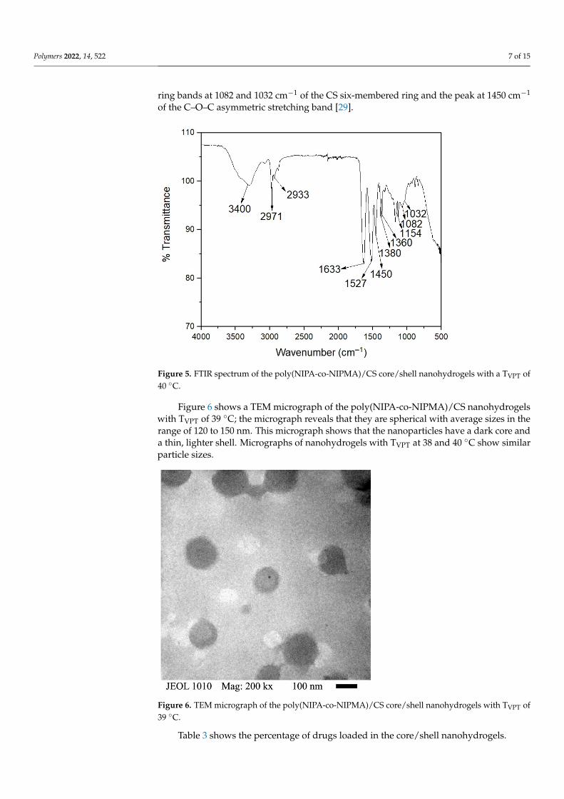
Figure 6 shows a TEM micrograph of the poly(NIPA-co-NIPMA)/CS nanohydrogelswith TVPT of 39 ◦C; the micrograph reveals that they are spherical with average sizes in therange of 120 to 150 nm. This micrograph shows that the nanoparticles have a dark core anda thin, lighter shell. Micrographs of nanohydrogels with TVPT at 38 and 40 ◦C show similarparticle sizes.
Polymers 2022, 14, x FOR PEER REVIEW 7 of 14
Figure 6 shows a TEM micrograph of the poly(NIPA-co-NIPMA)/CS nanohydrogels with TVPT of 39 °C; the micrograph reveals that they are spherical with average sizes in the range of 120 to 150 nm. This micrograph shows that the nanoparticles have a dark core and a thin, lighter shell. Micrographs of nanohydrogels with TVPT at 38 and 40 °C show similar particle sizes.
Figure 6. TEM micrograph of the poly(NIPA-co-NIPMA)/CS core/shell nanohydrogels with TVPT of 39 °C.
Table 3 shows the percentage of drugs loaded in the core/shell nanohydrogels.
Table 3. Drug content in nanohydrogels. TVPT of the Nanohydrogels (°C)
TVPT 38 39 40 % of drug content 5.00 5.20 5.41
Figure 7 shows the drug release profiles from the nanohydrogels at pH = 2.0. During the first three hours, release rates are fast, then start to decrease; after 50 h, the amount of drug released was approximately 77% to 87%. A slighter higher amount of drug is re-leased as the NIPMA/NIPA ratio increased.
Figure 7. Experimental drug release data for poly(NIPA-co-NIPMA)/Chitosan core/shell nanohy-drogels at pH = 2. TVPT of 38 °C (■); TVPT of 39 °C (ο); TVPT of 40 °C (▲).
Figure 6. TEM micrograph of the poly(NIPA-co-NIPMA)/CS core/shell nanohydrogels with TVPT of39 ◦C.
Table 3 shows the percentage of drugs loaded in the core/shell nanohydrogels.

Polymers 2022, 14, 522 8 of 15
Table 3. Drug content in nanohydrogels.
TVPT of the Nanohydrogels (◦C)
TVPT 38 39 40
% of drug content 5.00 5.20 5.41
Figure 7 shows the drug release profiles from the nanohydrogels at pH = 2.0. Duringthe first three hours, release rates are fast, then start to decrease; after 50 h, the amount ofdrug released was approximately 77% to 87%. A slighter higher amount of drug is releasedas the NIPMA/NIPA ratio increased.
Polymers 2022, 14, x FOR PEER REVIEW 7 of 14
Figure 6 shows a TEM micrograph of the poly(NIPA-co-NIPMA)/CS nanohydrogels with TVPT of 39 °C; the micrograph reveals that they are spherical with average sizes in the range of 120 to 150 nm. This micrograph shows that the nanoparticles have a dark core and a thin, lighter shell. Micrographs of nanohydrogels with TVPT at 38 and 40 °C show similar particle sizes.
Figure 6. TEM micrograph of the poly(NIPA-co-NIPMA)/CS core/shell nanohydrogels with TVPT of 39 °C.
Table 3 shows the percentage of drugs loaded in the core/shell nanohydrogels.
Table 3. Drug content in nanohydrogels. TVPT of the Nanohydrogels (°C)
TVPT 38 39 40 % of drug content 5.00 5.20 5.41
Figure 7 shows the drug release profiles from the nanohydrogels at pH = 2.0. During the first three hours, release rates are fast, then start to decrease; after 50 h, the amount of drug released was approximately 77% to 87%. A slighter higher amount of drug is re-leased as the NIPMA/NIPA ratio increased.
Figure 7. Experimental drug release data for poly(NIPA-co-NIPMA)/Chitosan core/shell nanohy-drogels at pH = 2. TVPT of 38 °C (■); TVPT of 39 °C (ο); TVPT of 40 °C (▲). Figure 7. Experimental drug release data for poly(NIPA-co-NIPMA)/Chitosan core/shell nanohy-drogels at pH = 2. TVPT of 38 ◦C (�); TVPT of 39 ◦C (o); TVPT of 40 ◦C (N).
Figure 8 depicts the drug release profiles at pH = 7.4 of the three nanohydrogels. Again,the amount released increases rapidly during the first 3 h, then the release rate decreases;after 50 h, the amount of drug released is ca. 53% to 55%. However, the amounts releasedare much lower than those obtained at pH = 2 (see Figure 7).
Polymers 2022, 14, x FOR PEER REVIEW 8 of 14
Figure 8 depicts the drug release profiles at pH = 7.4 of the three nanohydrogels. Again, the amount released increases rapidly during the first 3 h, then the release rate decreases; after 50 h, the amount of drug released is ca. 53% to 55%. However, the amounts released are much lower than those obtained at pH = 2 (see Figure 7).
Figure 8. Experimental drug release data for poly(NIPA-co-NIPMA)/Chitosan core/shell nanohy-drogels at pH = 7.4: TVPT of 38 °C (■); TVPT of 39 °C (ο); TVPT of 40 °C (▲).
Several mathematical models published in the literature were used to fit the experi-mental release data to determine the drug release mechanism [30].
The Korsmeyer–Peppas model is the most widely used for fitting drug release data. It is based on a diffusion mechanism and is a modification of the Higuchi model [31,32]. 𝑀𝑀 = 𝑘 × 𝑡
Mi is the amount of drug released at time t, MT is the total amount of drug loaded, k is the Korsmeyer rate constant, and n is the release exponent indicating the release mech-anism.
The extended Korsmeyer–Peppas model is used when initially there is a rapid drug release “the burst effect” and is obtained by adding a constant term “b” to the Korsmeyer–Peppas model [33,34]. 𝑀𝑀 = 𝑏 + 𝑘 × 𝑡
The Weibull equation can be applied to almost all types of dissolution curves. Here kw is the Weibull constant related to the inverse of the time required to release 63.2% of the drug, and n indicates the shape of the curve [35,36]. 𝑀𝑀 = 1 − 𝑒( )
The first-order model, derived by Noyes–Whitney, predicts a first-order dissolution process. However, when the asymptotic value released from the drug is less than one, an additional parameter “k” is added to obtain a better fit for the release data [34,35]. 𝑀𝑀 = 𝑘(1 − 𝑒 )
Figure 8. Experimental drug release data for poly(NIPA-co-NIPMA)/Chitosan core/shell nanohy-drogels at pH = 7.4: TVPT of 38 ◦C (�); TVPT of 39 ◦C (o); TVPT of 40 ◦C (N).

Polymers 2022, 14, 522 9 of 15
Several mathematical models published in the literature were used to fit the experi-mental release data to determine the drug release mechanism [30].
The Korsmeyer–Peppas model is the most widely used for fitting drug release data. Itis based on a diffusion mechanism and is a modification of the Higuchi model [31,32].
MiMT
= k× tn
Mi is the amount of drug released at time t, MT is the total amount of drug loaded, k isthe Korsmeyer rate constant, and n is the release exponent indicating the release mechanism.
The extended Korsmeyer–Peppas model is used when initially there is a rapid drugrelease “the burst effect” and is obtained by adding a constant term “b” to the Korsmeyer–Peppas model [33,34].
MiMT
= b + k× tn
The Weibull equation can be applied to almost all types of dissolution curves. Here kwis the Weibull constant related to the inverse of the time required to release 63.2% of thedrug, and n indicates the shape of the curve [35,36].
MiMT
= 1− e(−t
kw )n
The first-order model, derived by Noyes–Whitney, predicts a first-order dissolutionprocess. However, when the asymptotic value released from the drug is less than one, anadditional parameter “k” is added to obtain a better fit for the release data [34,35].
MiMT
= k(1− e−nt)
where n is the kinetic constant.The Hixson–Crowell model assumes the dissolution rate is a function of the cube root
of the particle volume and that the particle radius is not constant [35].
M1/3T −M1/3
R = kH × t
MR is the remaining amount of drug in the particles at time t, and kH is the Hixson–Crowell constant.
The drug release kinetic profile was determined at pH = 2.0 and 7.4 at their volumephase transition temperatures (TVPT). Figures 9 and 10 show the fit of the models to theexperimental data of drug release from the poly(NIPA-co-NIPMA)/CS nanohydrogels witha TVPT of 40 ◦C, at pH = 2.0 and 7.4, respectively.

Polymers 2022, 14, 522 10 of 15
Polymers 2022, 14, x FOR PEER REVIEW 9 of 14
where n is the kinetic constant. The Hixson–Crowell model assumes the dissolution rate is a function of the cube root
of the particle volume and that the particle radius is not constant [35]. 𝑀 / − 𝑀 / = 𝑘 × 𝑡
MR is the remaining amount of drug in the particles at time t, and kH is the Hixson–Crowell constant.
The drug release kinetic profile was determined at pH = 2.0 and 7.4 at their volume phase transition temperatures (TVPT). Figures 9 and 10 show the fit of the models to the experimental data of drug release from the poly(NIPA-co-NIPMA)/CS nanohydrogels with a TVPT of 40 °C, at pH = 2.0 and 7.4, respectively.
Figure 9. Comparison of the models with the experimental drug release data of using nanohydro-gels with TVPT of 40 °C at pH = 2.0: Experimental (■); Weibull (∆); Korsmeyer–Peppas and extended Korsmeyer–Peppas (○); first order (▬); Hixson–Crowell model (─ ─).
Figure 10. Comparison of the models with the experimental drug release data of nanohydrogels with TVPT of 40 °C at pH = 7.4: Experimental (■); Weibull (∆); Korsmeyer–Peppas and extended Korsmeyer–Peppas (○); first order (▬); Hixson-Crowell model (─ ─).
Figure 9. Comparison of the models with the experimental drug release data of using nanohydrogelswith TVPT of 40 ◦C at pH = 2.0: Experimental (�); Weibull (∆); Korsmeyer–Peppas and extendedKorsmeyer–Peppas (o); first order (—); Hixson–Crowell model (– –).
Polymers 2022, 14, x FOR PEER REVIEW 9 of 14
where n is the kinetic constant. The Hixson–Crowell model assumes the dissolution rate is a function of the cube root
of the particle volume and that the particle radius is not constant [35]. 𝑀 / − 𝑀 / = 𝑘 × 𝑡
MR is the remaining amount of drug in the particles at time t, and kH is the Hixson–Crowell constant.
The drug release kinetic profile was determined at pH = 2.0 and 7.4 at their volume phase transition temperatures (TVPT). Figures 9 and 10 show the fit of the models to the experimental data of drug release from the poly(NIPA-co-NIPMA)/CS nanohydrogels with a TVPT of 40 °C, at pH = 2.0 and 7.4, respectively.
Figure 9. Comparison of the models with the experimental drug release data of using nanohydro-gels with TVPT of 40 °C at pH = 2.0: Experimental (■); Weibull (∆); Korsmeyer–Peppas and extended Korsmeyer–Peppas (○); first order (▬); Hixson–Crowell model (─ ─).
Figure 10. Comparison of the models with the experimental drug release data of nanohydrogels with TVPT of 40 °C at pH = 7.4: Experimental (■); Weibull (∆); Korsmeyer–Peppas and extended Korsmeyer–Peppas (○); first order (▬); Hixson-Crowell model (─ ─).
Figure 10. Comparison of the models with the experimental drug release data of nanohydrogelswith TVPT of 40 ◦C at pH = 7.4: Experimental (�); Weibull (∆); Korsmeyer–Peppas and extendedKorsmeyer–Peppas (o); first order (—); Hixson-Crowell model (– –).
Table 4 shows the fit parameters and the determination coefficient, R2, of the models used.

Polymers 2022, 14, 522 11 of 15
Table 4. Fit parameters and determination coefficient, R2.
pH = 2.0 pH = 7.4
Model K n R2 b k n R2
MiMT
= ktn 0.632 0.098 0.9647 0.444 0.0742 0.943
MiMT
= b + ktn 0.632 0.098 0.9647 −0.001 0.445 0.7407 0.943
MiMT
= 1− e(−t
kw )n
16.9 7.009 0.7872 26.16 4.242 0.00
MiMT
= k(1− e−nt) 0.814 0.887 0.9801 0.541 0.998 0.988
M1/3T −M1/3
R = kH × t 0.028 0.005 0.033 0.008
4. Discussion and Conclusions
Drug delivery from stimuli-responsive nanoparticles, particularly those that respond si-multaneously to temperature and pH, have received increased attention in recent years [14,37].Fundenau et al. [38] reported that the poly(NIPA-co-NIPMA) with a 51/49 weight ratioNIPA/NIPAM displays an LCST of 36.8 ◦C at pH = 7.4 employing a phosphate buffersolution. The microgels were loaded with dexamethasone, and their release at temperaturesbelow and above LCST was investigated. Huang et al. obtained pH- and temperature-sensitive PNIPA/CS core/shell nanoparticles by emulsion polymerization with a TVPT of33.4 ◦C [25]. Alvarado et al. reported the synthesis of PNIPA/CS core/shell nanoparticlesby semi-continuous heterophase polymerization (SHP) with a TVPT of 34 ◦C [26]. How-ever, the TVPT of these nanoparticles is lower than the temperatures in the human body inresponse to infection.
Here we report the synthesis and characterization of emulsion polymerization ofcore/shell nanoparticles composed of poly(NIPA-co-NIPMA) in the core and CS formingthe shell with TVPTs of 38, 39, and 40 ◦C, for efficient drug delivery upon infection.
First, copolymers of NIPA and NIPMA were synthesized by emulsion polymerizationusing Brij 58 as a surfactant. The weight ratios of NIPA to NIPMA to be used in thesynthesis were estimated with the Fox equation. The weight ratio predicted with thisequation produced copolymers with TVPT close to the desired values as shown in Figure 2.This result can be explained because the Fox equation [27] was developed for moleculeswith similar structures and solubility parameters (similar cohesive energy density), whichis the case for NIPA and NIPMA molecules. Figure 2 shows that the diameter of thenanohydrogels determined by QLS decreased with increasing temperature due to waterexpulsion and copolymer collapse. When the temperature was higher than its TVPT, thediameter remained almost constant.
After the NIPA/NIPMA ratios needed to obtain the required TVPT were determined,the core/shell particles were synthesized using these ratios. Figure 3 shows a linear increaseof TVPT with increasing NIMPA concentration in the copolymer. M. Kokufuta, S. Sato, andE. Kokufuta [22] determined the LCST of NIPA-NIPMA copolymers as a function ofcomposition and found that it increased linearly with increasing mole fraction of NIPAMin the copolymer. These authors used the values of 32 ◦C and 42 ◦C for the LCST of PNIPAand PNIPMA, respectively.
Figure 4 shows that the size of poly(NIPA-co-NIPMA)/CS core/shell nanoparti-cles made with monomer ratios similar to those used in the emulsion polymerizationof NIPA/NIPMA turned out to be much smaller (compare Figures 2 and 4). The smallercore/shell particles size is explained by the use of CS as the surfactant instead of Brij 58. CScan form micelles in acetic acid aqueous solutions above a critical concentration [23]. CMCvalues of CS were reported to range from 0.718 to 1.815 y g/L (the higher the pH, the largerthe CMC [23]). In this work, we used a CS concentration of 10 g/L, a value that is well abovethe CMC at any pH. It is proposed that the polymerization occurs in the aqueous phase,and once the poly(NIPA-co-NIPAM) chains exceed their critical length, they introduce intothe CS micelles. The absence of other size distributions in the QLS measurements (Figure 1)

Polymers 2022, 14, 522 12 of 15
demonstrates that all the copolymer radicals formed in the aqueous phase penetrated themicelles’ interior. Figure 4 also shows that decreasing the NIPA/NIPMA ratio increasesthe TVPT and shifts to slightly higher temperatures than those shown in Figure 3. The useof CS and acetic acid could cause the concentration of the monomers inside the micellesto be different from that obtained when Brij 58 was used and would explain the slightlyhigher TVTP compared to those in Figure 2. However, since high conversions (above 95%)were obtained, the PNIPA/PNIPMA ratio will differ only slightly based on the monomerratio used. Funduenau et al. [38], in the copolymerization of NIPA and NIPMA in solution,observed that the PNIPA/PNIPMA ratio in the copolymer was lower than in the feed,suggesting that NIPMA is more reactive than NIPA.
The characteristic bands of PNIPA, PNIMPA, and CS were detected by FTIR, confirm-ing their presence in the nanoparticles (Figure 5).
The core/shell structure was proven by the increase in the size of the particles whenthe CS was crosslinked (Table 2) and by TEM (Figure 6) where a well-defined dark core anda thin, clearer shell can be observed in the particles. Because of the drying of the samplesfor TEM analysis, the particle size was smaller than that determined by QLS.
Figures 7 and 8 show that at the two pHs studied, a higher amount of drug releasedand a higher release rate were obtained with the nanoparticles with TVPT of 40 ◦C. Thisresult is explained because the release experiments were performed at the TVPT of thenanoparticles, so the higher the temperature, the higher the mobility of the drug molecules.Less drug was released at pH = 7.4 (compare Figures 7 and 8); the lower amount of drugreleased is because chitosan is pH-sensitive and becomes hydrophobic at a pH above6.4, causing its collapse, which hinders drug release. Moreover, at acidic pH, the drug isfound as its salt (doxycycline hyclate), which is soluble in water; at alkaline pH, the saltis converted into doxycycline, which has low water solubility. Huang et al. reported alower amount of drug released from NIPA/CS core/shell nanoparticles at alkaline pH thanat acidic pH [23]. Alvarado et al. on the release of doxycycline hyclate from NIPA/CScore/shell nanocomposites also reported that at alkaline pH, the amount released wasmuch lower than at acidic pH [26].
Figures 9 and 10 and the coefficient of determination, R2, reported in Table 4, indicatethat the first-order model best fits the experimental data and suggests that the releasemechanism is diffusion-controlled by the concentration gradient. The Weibull and Hixson-Crowell models gave the worst results. The Korsmeyer–Peppas and extended Korsmeyer–Peppas models fitted the experimental data adequately up to 75% drug release. Theparameter “b” is small and negative (Table 4), indicating that the bursting effect is notimportant so that there is no drug on the surface but that it is all inside the nanohydrogels.The fits of the Korsmeyer–Peppas and extended Korsmeyer–Peppas models are identicalbecause the value of “b” is insignificant.
Figures 11 and 12 show good agreement between the first-order model and exper-imental drug release data by nanohydrogels with TVPT of 38, 39, and 40 ◦C at pH = 2.0and 7.4.
In conclusion, we have synthesized pH- and temperature-sensitive poly(NIPA-co-NIPAM)/CS core/shell nanohydrogels by batch emulsion polymerization. These core/shellnanoparticles have a TVPT of 38, 39, or 40 ◦C, the temperature present in the human bodyin response to infection, making them good candidates for use as drug delivery agents.At an alkaline pH, the drug has low water solubility, and the CS shell collapses makingdrug release difficult, which explains the lower amount released at this pH. The first-ordermodel best fits the experimental drug release data, suggesting that the release mechanismis diffusion-controlled by the concentration gradient.

Polymers 2022, 14, 522 13 of 15Polymers 2022, 14, x FOR PEER REVIEW 12 of 14
Figure 11. Comparison of experimental drug release data vs. first-order model at pH = 2.0; TVPT of: 38 °C (■); 39 °C (●); 40 °C (▲).
Figure 12. Comparison of experimental drug release data vs. first-order model at pH = 7.4; TVPT of: 38 °C (■); 39 °C (●); 40 °C (▲).
In conclusion, we have synthesized pH- and temperature-sensitive poly(NIPA-co-NIPAM)/CS core/shell nanohydrogels by batch emulsion polymerization. These core/shell nanoparticles have a TVPT of 38, 39, or 40 °C, the temperature present in the human body in response to infection, making them good candidates for use as drug delivery agents. At an alkaline pH, the drug has low water solubility, and the CS shell collapses making drug release difficult, which explains the lower amount released at this pH. The first-order model best fits the experimental drug release data, suggesting that the release mechanism is diffusion-controlled by the concentration gradient.
Figure 11. Comparison of experimental drug release data vs. first-order model at pH = 2.0; TVPT of:38 ◦C (�); 39 ◦C (•); 40 ◦C (N).
Polymers 2022, 14, x FOR PEER REVIEW 12 of 14
Figure 11. Comparison of experimental drug release data vs. first-order model at pH = 2.0; TVPT of: 38 °C (■); 39 °C (●); 40 °C (▲).
Figure 12. Comparison of experimental drug release data vs. first-order model at pH = 7.4; TVPT of: 38 °C (■); 39 °C (●); 40 °C (▲).
In conclusion, we have synthesized pH- and temperature-sensitive poly(NIPA-co-NIPAM)/CS core/shell nanohydrogels by batch emulsion polymerization. These core/shell nanoparticles have a TVPT of 38, 39, or 40 °C, the temperature present in the human body in response to infection, making them good candidates for use as drug delivery agents. At an alkaline pH, the drug has low water solubility, and the CS shell collapses making drug release difficult, which explains the lower amount released at this pH. The first-order model best fits the experimental drug release data, suggesting that the release mechanism is diffusion-controlled by the concentration gradient.
Figure 12. Comparison of experimental drug release data vs. first-order model at pH = 7.4; TVPT of:38 ◦C (�); 39 ◦C (•); 40 ◦C (N).
Author Contributions: Conceptualization, E.M., J.E.P.-A. and L.A.P.-C.; methodology, A.O.-G.,B.G.M.-B., I.C., E.M., J.E.P.-A. and L.A.P.-C.; software, A.O.-G., B.G.M.-B. and E.M.; validation,A.O.-G., B.G.M.-B., E.M., J.E.P.-A. and L.A.P.-C.; formal analysis, A.O.-G., B.G.M.-B., I.C., E.M.,J.E.P.-A. and L.A.P.-C.; investigation, A.O.-G., B.G.M.-B. and I.C.; resources, I.C., E.M., J.E.P.-A. andL.A.P.-C.; data curation, A.O.-G., B.G.M.-B., E.M. and L.A.P.-C. writing—original draft preparation,A.O.-G., B.G.M.-B., E.M., J.E.P.-A. and L.A.P.-C.; writing—review and editing, B.G.M.-B., E.M., J.E.P.-A. and L.A.P.-C.; visualization, E.M., J.E.P.-A.; supervision, E.M., J.E.P.-A. and L.A.P.-C.; projectadministration, A.O.-G., B.G.M.-B., E.M., J.E.P.-A. and L.A.P.-C.; funding acquisition, E.M., J.E.P.-A.and L.A.P.-C. All authors have read and agreed to the published version of the manuscript.

Polymers 2022, 14, 522 14 of 15
Funding: This research received no external funding.
Institutional Review Board Statement: Not applicable.
Informed Consent Statement: Not applicable.
Data Availability Statement: Not applicable.
Acknowledgments: Two of the authors (Andres Ortega-García and Bryan Giovanny Martínez-Bernal)acknowledge the scholarship from CONACyT.
Conflicts of Interest: The authors declare no conflict of interest.
References1. Kuckling, D.; Doering, A.; Krahl, F.; Arndt, K.-F. Stimuli-Responsive Polymer Systems. Polym. Sci. Compr. Ref. 2012, 8, 377–413.
[CrossRef]2. Gore, S.A.; Gholve, S.B.; Savalsure, S.M.; Ghodake, K.B.; Bhusnure, O.G.; Thakare, V.M. Smart polymer and their applications:
A review. Int. J. Curr. Pharm. Rev. Res. 2017, 8, 298–310. [CrossRef]3. Schmaljohann, D. Thermo-and pH-responsive polymers in drug delivery. Adv. Drug Deliv. Rev. 2006, 58, 1655–1670. [CrossRef]
[PubMed]4. Wang, C.; Javadi, A.; Ghaffari, M.; Shaoquin, G. A pH-sensitive molecularly imprinted nanospheres/hydrogel composite as
coating for implantable biosensors. Biomaterials 2010, 31, 4944–4951. [CrossRef]5. Terefe, N.S.; Glagovskaia, O.; de Silva, K.; Stockmann, R. Applications of stimuli responsive polymers for sustainable ion exchange
chromatography. Food Bioprod. Process. 2014, 92, 208–225. [CrossRef]6. Fernández-Barbero, A.; Suárez, I.J.; Sierra-Martín, B.; Fernández-Nieves, A.; de las Nieves, F.J.; Marquez, M.; Rubio-Retama,
J.; López-Cabarcos, E. Gels and microgels for nanotechnological applications. Adv. Colloid Interface Sci. 2009, 147–148, 88–108.[CrossRef]
7. Alvarez-Bautista, A.; Duarte, C.M.M.; Mendizábal, E.; Katime, I. Controlled delivery of drugs through smart pH-sensitivenanohydrogels for anti-cancer therapies: Synthesis, drug release and cellular studies. Des. Monomers Polym. 2016, 19, 319–329.[CrossRef]
8. Peters, J.T.; Hutchinson, S.S.; Lizana, N.; Verma, I.; Peppas, N.A. Synthesis and characterization of poly(N-isopropyl methacry-lamide) sore/shell nanogels for controlled release of chemotherapeutics. Chem. Eng. J. 2018, 340, 58–65. [CrossRef]
9. Bajpai, A.K.; Shukla, S.K.; Bhanu, S.; Kankane, S. Responsive polymers in controlled drug delivery. Prog. Polym. Sci.2008, 33, 1088–1108. [CrossRef]
10. Bera, R.; Dey, A.; Chakrabarty, D. Synthesis, characterization, and drug release study of acrylamide-co-itaconic acid based smarthydrogel. Polym. Eng. Sci. 2015, 55, 113–122. [CrossRef]
11. Yang, P.; Zhu, F.; Cheng, Y.; Wang, Z.; Li, Y. Stimuli-responsive polydopamine-based smart materials. Chem. Soc. Rev.2021, 50, 8319–8343. [CrossRef] [PubMed]
12. Jung, S.; MacConaghy, K.I.; Kaar, J.L.; Stoykovich, M.P. Enhance Optical Sensitivity in Thermoresponsive Photonic CrystalHydrogels by Operating Near the Phase Transition. ACS Appl. Mater. Interfaces 2017, 9, 27927–27935. [CrossRef] [PubMed]
13. Rajasekhar, A.; Gimi, B.; Hu, W. Applications of semiconductor fabrication methods to nanomedicine: A review of recentinventions and techniques. Recent Pat. Nanomed. 2013, 3, 9–20. [CrossRef] [PubMed]
14. Liu, M.; Du, H.; Zhang, W.; Zhai, G. Internal stimuli-responsive nanocarriers for drug delivery: Design strategies and applications.Mater. Sci. Eng. C 2017, 71, 1267–1280. [CrossRef] [PubMed]
15. Cuggino, J.C.; Alvarez, I.C.I.; Strumia, M.C.; Welker, P.; Licha, K.; Steinhilbert, D.; Mutihac, R.-C.; Calderón, M. Thermosensitivenanogels based on dendritic polyglycerol and N-isopropylacrylamide for biomedical applications. Soft Matter 2011, 7, 11259–11266.[CrossRef]
16. O’Neil, H.S.; Herron, C.C.; Hastings, C.L.; Deckers, R.; López-Noriega, A.; Kelly, H.M.; Hennink, W.E.; McDonnell, C.O.; O’Brien,F.J.; Ruiz-Hernández, E.; et al. A stimuli responsive liposome loaded hydrogel provides flexible on-demand release of therapeuticagents. Acta Biomater. 2016, 48, 110–119. [CrossRef]
17. Chiantore, O.; Guaita, M.; Trossarelli, L. Solution properties of poly(N-isopropylacrylamide). Die Makromol. Chem.1979, 180, 969–973. [CrossRef]
18. Binkert, T.; Oberreich, J.; Meewes, M.; Nyffenegger, R.; Ricka, J. Coil-globule transition of poly(N-isopropylacrylamide): A studyof segment mobility fluorescence depolarization. Macromolecules 1991, 24, 5806–5810. [CrossRef]
19. Kubota, K.; Hamano, K.; Kuwahara, N.; Fujishige, S.; Ando, I. Characterization of poly(N-isopropylmethacrylamide) in wáter.Polym. J. 1990, 22, 1051–1057. [CrossRef]
20. Djokpé, E.; Vogt, W. N-isopropylacrylamide and N-isopropylmethacryl-amide: Cloud points of mixtures and copolymers.Macromol. Chem. Phys. 2001, 202, 750–757. [CrossRef]
21. Salmerón Sánchez, M.; Hanyková, L.; Ilavský, M.; Monleón Pradas, M. Thermal transitions of poly(N-isopropylmethacrylamide)in aqueous solutions. Polymer 2004, 45, 4087–4094. [CrossRef]

Polymers 2022, 14, 522 15 of 15
22. Kokufuta, M.K.; Sato, S.; Kokufuta, E. LCST behavior of copolymers of N-isopropylacrylamide and N-isopropylmethacrylamidein water. Colloid Polym. Sci. 2012, 290, 1671–1681. [CrossRef]
23. López-León, T.; Carvalho, E.L.S.; Seijo, B.; Ortega-Vinuesa, J.L.; Bastos-González, D. Physicochemical characterization of chitosannanoparticles: Electrokinetic and stability behavior. J. Colloid Interface Sci. 2005, 283, 344–351. [CrossRef] [PubMed]
24. Aguilar, M.R.; Elvira, C.; Gallardo, A.; Vázquez, B.; Román, J.S. Smart polymer and their applications as biomaterials. In Topics inTissue Engineering; Ashammakhi, N., Reis, R., Chiellini, E., Eds.; University of Oulu: Oulu, Finland, 2007; Volume 3, pp. 1–27.
25. Huang, C.-H.; Wang, C.-F.; Don, T.-M.; Chiu, W.-Y. Preparation of pH- and thermo-sensitive chitosan-PNIPAAm core-shellnanoparticles and evaluation as drug carriers. Cellulose 2013, 20, 1791–1805. [CrossRef]
26. Alvarado, A.G.; Ortega, A.; Pérez-Carrillo, L.A.; Ceja, I.; Arellano, M.; López, R.G.; Puig, J.E. Synthesis, characterization, anddrug delivery from pH- and thermoresponsive poly(N-isopropylacrylamide)/chitosan core/shell nanocomposites made bysemicontinuous heterophase polymerization. J. Nanomater. 2017, 2017, 6796412. [CrossRef]
27. Fox, T.G. Influence of diluent and copolymer composition on the glass temperature of a polymer system. Bull. Am. Phys. Soc.1956, 1, 123–125.
28. Silverstein, R.M.; Webster, F.X.; Kiemle, D.J.; Bryce, D.L. Spectrometric Identification of Organic Compounds, 8th ed.; John Wiley &Sons: Hoboken, NJ, USA, 2014.
29. Ríos-Donato, N.; Peña-Flores, A.M.; Katime, I.; Leyva-Ramos, R.; Mendizábal, E. Kinetics and thermodynamics of adsorption ofred dye 40 from acidic aqueous solutions onto a novel chitosan sulfate. Afinidad LXXIV 2017, 74, 214–220.
30. Singhvi, G.; Singh, M. Review: In-vitro drug release characterization models. Int. J. Pharm. Stud. Res. 2011, 2, 77–84.31. Korsmeyer, R.W.; Gurny, R.; Doelker, E.; Buri, P.; Peppas, N.A. Mechanism of solute release from porous hydrophilic polymers.
Int. J. Pharm. 1983, 15, 25–35. [CrossRef]32. Gouda, R.; Baishya, H.; Qing, Z. Applications of mathematical models in drug release kinetics of carbidopa and levodopa ER
tablets. J. Dev. Drugs 2017, 6, 1–8. [CrossRef]33. Lindner, W.D.; Lippold, B.C. Drug release from hydrocolloid embeddings with high or low susceptibility to hydrodynamics
stress. Pharm. Res. 1995, 12, 1781–1785. [CrossRef] [PubMed]34. Costa, F.O.; Sousa, J.J.S.; Pais, A.A.C.C.; Formosinho, S.J. Comparison of dissolution profiles of ibuprofen pellets. J. Control. Release
2003, 89, 199–212. [CrossRef]35. Ramteke, K.H.; Dighe, P.A.; Kharat, A.R.; Patil, S.V. Mathematical models of drug dissolution: A review. Sch. Acad. J. Pharm.
2014, 3, 388–396. [CrossRef]36. Balleño, J.A.; Mendizábal-Ruiz, A.P.; Saade, H.; Díaz de León-Gómez, R.; Mendizábal, E.; Rios-Donato, N.; López, R.G. Ibuprofen
release from poly(ethyl cyanoacrylate) nanoparticles prepared by semicontinuous heterophase polymerization. Int. J. Polym. Sci.2018, 2018, 4527203. [CrossRef]
37. Serrano-Medina, A.; Cornejo-Bravo, J.M.; Licea-Claveríe, A. Synthesis of pH and temperature sensitive, core-shell nano/microgels,by one pot, soap-free emulsion polymerization. J. Colloid Interface Sci. 2012, 369, 82–90. [CrossRef]
38. Fundueanu, G.; Constantin, M.; Bucatariu, S.; Ascenzi, P. Poly(N-isopropylacrylamide-co-N-isopropylmethacrylamide) thermo-responsive microgels as self-regulated drug delivery system. Macromol. Chem. Phys. 2016, 271, 2525–2533. [CrossRef]