
Are Females More Responsive to Emotional Stimuli? A Neurophysiological Study Across Arousal and...
14
ORIGINAL PAPER Are Females More Responsive to Emotional Stimuli? A Neurophysiological Study Across Arousal and Valence Dimensions C. Lithari • C. A. Frantzidis • C. Papadelis • Ana B. Vivas • M. A. Klados • C. Kourtidou-Papadeli • C. Pappas • A. A. Ioannides • P. D. Bamidis Received: 9 February 2009 / Accepted: 14 December 2009 / Published online: 31 December 2009 Ó The Author(s) 2009. This article is published with open access at Springerlink.com Abstract Men and women seem to process emotions and react to them differently. Yet, few neurophysiological studies have systematically investigated gender differences in emotional processing. Here, we studied gender differ- ences using Event Related Potentials (ERPs) and Skin Conductance Responses (SCR) recorded from participants who passively viewed emotional pictures selected from the International Affective Picture System (IAPS). The arousal and valence dimension of the stimuli were manipulated orthogonally. The peak amplitude and peak latency of ERP components and SCR were analyzed separately, and the scalp topographies of significant ERP differences were documented. Females responded with enhanced negative components (N100 and N200), in comparison to males, especially to the unpleasant visual stimuli, whereas both genders responded faster to high arousing or unpleasant stimuli. Scalp topographies revealed more pronounced gender differences on central and left hemisphere areas. Our results suggest a difference in the way emotional stimuli are processed by genders: unpleasant and high arousing stimuli evoke greater ERP amplitudes in women relatively to men. It also seems that unpleasant or high arousing stimuli are temporally prioritized during visual processing by both genders. Keywords Emotions Á Gender differences Á Event related potentials Á Skin conductance Introduction Over the last few years there has been a renewed interest about sex differences in brain and cognition. This interest is partly due to the increasing amount of literature showing structural and chemical differences between the brain of males and females in both animals (Zhang et al. 2008; Xiong et al. 2007) and humans (Canli et al. 2002; Cahill and McGaugh 1998; Lang 1968, 1984). Sex differences have also been documented in cognitive processes such as memory, emotion, and vision (see Cahill 2006 for a review). Within this field, the topic of sex differences in emotional processing has attracted particular interest due to its potential application in understanding psychopathology. For instance, gender differences in the prevalence of mood disorders such as depression and anxiety may be related to the differential response of females and males to stress (Cahill 2006). C. Lithari Á C. A. Frantzidis Á M. A. Klados Á C. Pappas Á P. D. Bamidis (&) Laboratory of Medical Informatics, Medical School, Aristotle University of Thessaloniki, P.O. Box 323, 54124 Thessaloniki, Greece e-mail: [email protected] C. Papadelis Laboratory of Functional Neuroimaging, Center for Mind/Brain Sciences, Matarello, Italy A. B. Vivas Psychology Department, City Liberal Studies (Affiliated Institution of the University of Sheffield), Thessaloniki, Greece C. Kourtidou-Papadeli Greek Aerospace Medical Association and Space Research, Thessaloniki, Greece A. A. Ioannides Laboratory for Human Brain Dynamics, Brain Science Institute (BSI), RIKEN, Wako, Saitama, Japan A. A. Ioannides Laboratory for Human Brain Dynamics, AAI Scientific and Cultural Services Ltd., Galaxias Center, Nicosia, Cyprus 123 Brain Topogr (2010) 23:27–40 DOI 10.1007/s10548-009-0130-5
Transcript of Are Females More Responsive to Emotional Stimuli? A Neurophysiological Study Across Arousal and...
ORIGINAL PAPER
Are Females More Responsive to Emotional Stimuli?A Neurophysiological Study Across Arousal and ValenceDimensions
C. Lithari • C. A. Frantzidis • C. Papadelis •
Ana B. Vivas • M. A. Klados • C. Kourtidou-Papadeli •
C. Pappas • A. A. Ioannides • P. D. Bamidis
Received: 9 February 2009 / Accepted: 14 December 2009 / Published online: 31 December 2009
� The Author(s) 2009. This article is published with open access at Springerlink.com
Abstract Men and women seem to process emotions and
react to them differently. Yet, few neurophysiological
studies have systematically investigated gender differences
in emotional processing. Here, we studied gender differ-
ences using Event Related Potentials (ERPs) and Skin
Conductance Responses (SCR) recorded from participants
who passively viewed emotional pictures selected from the
International Affective Picture System (IAPS). The arousal
and valence dimension of the stimuli were manipulated
orthogonally. The peak amplitude and peak latency of ERP
components and SCR were analyzed separately, and the
scalp topographies of significant ERP differences were
documented. Females responded with enhanced negative
components (N100 and N200), in comparison to males,
especially to the unpleasant visual stimuli, whereas both
genders responded faster to high arousing or unpleasant
stimuli. Scalp topographies revealed more pronounced
gender differences on central and left hemisphere areas.
Our results suggest a difference in the way emotional
stimuli are processed by genders: unpleasant and high
arousing stimuli evoke greater ERP amplitudes in women
relatively to men. It also seems that unpleasant or high
arousing stimuli are temporally prioritized during visual
processing by both genders.
Keywords Emotions � Gender differences � Event related
potentials � Skin conductance
Introduction
Over the last few years there has been a renewed interest
about sex differences in brain and cognition. This interest
is partly due to the increasing amount of literature showing
structural and chemical differences between the brain of
males and females in both animals (Zhang et al. 2008;
Xiong et al. 2007) and humans (Canli et al. 2002; Cahill
and McGaugh 1998; Lang 1968, 1984). Sex differences
have also been documented in cognitive processes such as
memory, emotion, and vision (see Cahill 2006 for a
review). Within this field, the topic of sex differences in
emotional processing has attracted particular interest due to
its potential application in understanding psychopathology.
For instance, gender differences in the prevalence of mood
disorders such as depression and anxiety may be related to
the differential response of females and males to stress
(Cahill 2006).
C. Lithari � C. A. Frantzidis � M. A. Klados � C. Pappas �P. D. Bamidis (&)
Laboratory of Medical Informatics, Medical School, Aristotle
University of Thessaloniki, P.O. Box 323, 54124 Thessaloniki,
Greece
e-mail: [email protected]
C. Papadelis
Laboratory of Functional Neuroimaging, Center for Mind/Brain
Sciences, Matarello, Italy
A. B. Vivas
Psychology Department, City Liberal Studies (Affiliated
Institution of the University of Sheffield), Thessaloniki, Greece
C. Kourtidou-Papadeli
Greek Aerospace Medical Association and Space Research,
Thessaloniki, Greece
A. A. Ioannides
Laboratory for Human Brain Dynamics, Brain Science Institute
(BSI), RIKEN, Wako, Saitama, Japan
A. A. Ioannides
Laboratory for Human Brain Dynamics, AAI Scientific and
Cultural Services Ltd., Galaxias Center, Nicosia, Cyprus
123
Brain Topogr (2010) 23:27–40
DOI 10.1007/s10548-009-0130-5

Only few studies have so far investigated gender dif-
ferences in emotions, and most of them report significant
differences. Thus, for instance, unpleasant pictures from
International Affective Picture System (IAPS, Lang et al.
1997) elicited more robust P300 effects in terms of both
amplitude and latency in the left hemispheres of females,
while they elicited a stronger P300 component in the right
hemisphere of males (Gasbarri et al. 2007). Females
responded with a much larger N200 component to IAPS
(Lang et al. 1997) emotional pictures depicting humans
than to those containing scenes (Proverbio et al. 2008).
Similarly, Han et al. (2008) found that the amplitude of the
P300 was generally larger for painful relative to neutral
stimuli in both sexes, but this effect was stronger for
females. These findings agree with the hypothesis that
women are generally more responsive to emotional stimuli
and more particularly to danger-related stimuli (Williams
and Gordon 2007).
The findings from event related potentials (ERPs)
studies appear to be also supported from neuroimaging
studies employing fMRI (Canli et al. 2002; Cahill 2003;
George et al. 1996; Pardo et al. 1993; Schneider et al.
2000; Wrase et al. 2003). Stimuli of different valence (film
clips) produced a differential time course and intensity of
prefrontal cortex activation, with overshoot and undershoot
being more pronounced in males in an fMRI study (Leon-
Carrion et al. 2006). Kemp et al. (2004) used steady-state
probe topography and reported that the processing of
unpleasant images is associated with widespread frontal
latency reductions in females, but not in males. These
findings support the notion that males and females seem to
perceive, process, and respond differently to emotional
stimuli.
Studies with auditory stimuli also report gender differ-
ences with emotional tones of voice indicating the signif-
icance of a spoken utterance. Schirmer et al. (2005)
showed that even though both sexes appear to detect pre-
attentively changes in voice, only women recruit additional
processing resources when the change in voice is of emo-
tional valence. Also, females are found to use earlier the
emotional prosody during word processing as compared to
men (Schirmer et al. 2002). Thus in general, these findings
appear to support the claim of women’s superior emotion
recognition proposed by Hall (1978).
Although these studies converge to some general
conclusion, the findings are not conclusive yet, partially
due to large discrepancies among the studies in terms of
aspects of emotion measured and the type of stimuli
employed. For instance, some studies have investigated
the expression of emotions in terms of facial movements
and self-reports of experienced emotion (Kring and
Gordon 1998; Wild 2003), whereas others have looked at
neurophysiological correlates of emotions with autonomic
responses (e.g., skin conductance) (Bradley et al. 2001;
Kring and Gordon 1998). Similarly, researchers have used
a variety of stimuli to induce emotional experiences;
emotional films (Aftanas et al. 1997; Kring and Gordon
1998; Cahill 2003), pictures (Codispoti et al. 2006; Cahill
2003; Cuthbert et al. 2000; Schupp et al. 2006; for a
review see Oloffson et al. 2008), pictures of specific
content (e.g., emotional faces or social stimuli) (Gerber
et al. 2008; Proverbio et al. 2008), emotional words
(Alfano and Cimino 2008), music and sounds (Schirmer
et al. 2002, 2005; Krumhansl 2002; Bachorowski and
Owren 2003; Panksepp and Bernatzky 2002). While
emotional films seem to be quite effective, they are dif-
ficult to standardize in terms of the basic visual processes,
because responses elicited by different frames overlap. In
addition, during exposure to both emotional films and
music, the effect of each frame or note is embedded
within the context established by its predecessors in a
highly idiosyncratic way for each participant. On the
other hand, emotional words include the verbal informa-
tion and its perception, which may affect the study of
emotional perception itself. In this sense, the IAPS col-
lection (Lang et al. 1997), which contains standardized
images rated for emotional valence and arousal may
provide a convenient alternative for the investigation of
emotional processing.
There is no agreement in literature about how valence
and arousal may interact to induce emotional experiences.
The bi-dimensionality theory of emotions proposes that the
nature of emotional experience, or at least its ‘affective
core’, is primarily determined by two main dimensions:
pleasure and arousal (Russell 1980, 1989; Reinsenzein
1994; Barrett 1998). Valence ranges from attraction and
pleasure to aversion and displeasure, whereas arousal is a
more general property of the stimulus and refers to the
level of activation, regardless of the direction (whether
pleasant or unpleasant) and it should not be confused with
affective intensity (Barrett and Russell 1999). This pro-
posal would be consistent with an underlying bi-motiva-
tional system: the appetitive (approach) and the aversive
(avoidance) motivational system that vary along the arou-
sal dimension (Cacioppo and Bernston 1994; Lang et al.
1998). It also derives from theory, that valence and arousal
are two orthogonal, independent dimensions of the emo-
tional stimulus. Other theories propose that these two
dimensions are relatively dependent. Williams and Gordon
(2007) have proposed that potentially dangerous stimuli are
typically high arousing, what they called a ‘‘mismatch’’.
On the other hand, reward-related stimuli are typically
predictable, what they termed as a ‘‘match’’. Although, it is
true that valence and arousal co-vary in the real world
(Lang et al. 1997), recent studies suggest that these two
dimensions interact and need to be taken into account when
28 Brain Topogr (2010) 23:27–40
123
studying emotional processing (Cuthbert et al. 2000;
Robinson et al. 2004).
In line with this, there are at least four previous reports
considering the possible interaction of arousal and valence
during selecting IAPS stimuli in the experimental design,
as in the present one. Most of these studies (Keil et al.
2002; Dolcos and Cabeza 2002; Delplanque et al. 2006)
used emotional versus non emotional (neutral) stimuli to
study arousal effects, whereas Gianotti et al. (2008)
manipulated valence and arousal orthogonally getting to a
different experimental design. The latter study assumed
different neural assemblies for high and low arousal, as
well as, for pleasant and unpleasant stimuli. Their results
also revealed that valence information was extracted before
that of arousal. These studies were mainly focused on the
temporal analysis of emotional perception, whereas none of
them reported gender differences.
Another aspect that has been addressed in the literature
of emotional processing is the autonomic responses.
Emotions elicited by stimuli often lead to autonomic
responses that survive even when the stimulus is masked
(Ekman et al. 2007). In this sense, simultaneous record-
ings of central and autonomic nervous responses are more
likely to advance our understanding of emotional pro-
cessing. Recent studies have attempted to integrate brain
activity measures with more than one simultaneous
peripheral physiological measurements, such as Skin
Conductance Response (SCR), heart rate, and facial
electromyography (EMG) (Bernat et al. 2006; Cuthbert
et al. 2000; Kring and Gordon 1998). The electrodermal
activity has been found to be more responsive to pleasant
and unpleasant pictures than neutral ones, while the high
arousing pictures evoked greater SCR amplitudes as
compared to low arousing ones (Bernat et al. 2006). In
terms of sex differences, women were found to elicit
greater physiological reactivity (i.e., SCR, facial EMG)
than men to emotional material, especially if the material
was of negative valence (Bradley et al. 2001; Chentsova-
Dutton and Tsai 2007).
In the present study, we investigated potential differ-
ences in emotional processing between genders by mea-
suring ERPs and SCRs when participants were passively
exposed to emotional pictures selected from the IAPS
collection. The valence and arousal dimensions of the
pictures were orthogonally manipulated, since recent
research suggests that these two dimensions may interact
(Cuthbert et al. 2000). Based on findings from previous
studies, we hypothesized that emotional stimuli will reveal
gender differences in ERPs and SCRs. In this sense,
females may appear more responsive to emotional stimuli
at ERP components relative to males. This difference is
presumed to be greater for unpleasant stimuli.
Materials and Methods
Participants
Twenty-eight healthy, right-handed volunteers (14 females
and 14 males) participated in the study with a mean age of
28.2 ± 7.5 for males, and 27.1 ± 5.2 for females (mean ±
SD). Participants with a history of psychiatric or neuro-
logical illness or under medication were excluded from the
study. All participants had normal (10/10) or corrected to
normal vision. Participants were kindly asked to refrain
from any alcohol and caffeine consumption the day before
and the day of the experiment; they were also asked to
sleep as adequately and comfortably as possibly achievable
the night before the recordings. Each participant signed an
informed consent form prior to his/her participation and
completed a short questionnaire. All participants were paid
for their participation. An approval from the local Ethical
Committee was granted for this study.
Experimental Procedure
Participants were seated on an armchair and a PC screen
was placed in front of their eyes in a distance of 80 cm.
The room temperature was constantly controlled during the
experiment. The experiment started with a 30-s recording,
during which participants were asked to keep their eyes
open and fixate to a cross appearing in the center of the
screen; this was followed by similar measurement with
eyes closed. The aim of both recordings was to identify and
correct any possible technical problems before the actual
recordings (see below), as well as, to provide test data for
performing the artifact rejection algorithms used later in
the analysis. One hundred and sixty pictures were selected
for the experiment. Pictures were categorized in four
groups (4 9 40 pictures) according to arousal and pleasure
ratings, as explained below.
The emotion evocative pictures were presented to par-
ticipants in a random order. Stimulus delivery was con-
trolled by the presentation software (Neurobehavioral
systems, Albany, CA). Each single epoch consisted of a
500 ms pre-stimulus and a 2 s post-stimulus period. In the
pre-stimulus period, a white fixation cross was presented
on the screen in a black background. The stimulus
appeared on the screen for 1 s after which the cross came
up again. The total duration of the experiment was less than
5 min. The procedure was concluded with a repetition of
the initial 30 s recordings, with eyes open and then closed.
Most of the participants (13/14 males, 13/14 females) rated
the presented pictures in terms of valence and arousal
following the IAPS rating scales through a web-based
online questionnaire.
Brain Topogr (2010) 23:27–40 29
123
Stimuli
Visual stimuli were pictures from the IAPS collection;
these were selected along with their emotional content
defined in terms of their pleasure and arousal ratings. The
selected stimuli divided the pleasure-arousal 2D space by
naturally forming four groups of pictures: pleasant and
high arousing (PHA),1 pleasant and low arousing (PLA),2
unpleasant and high arousing (UHA),3 and unpleasant and
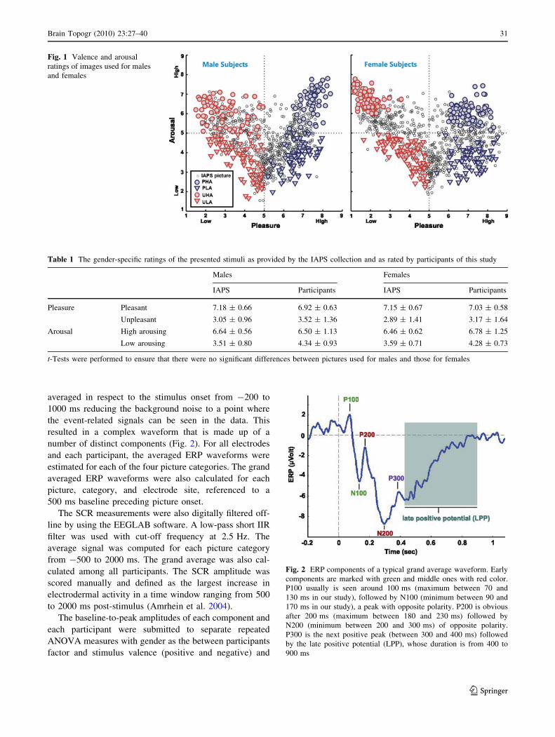
low arousing (ULA).4 Figure 1 shows how the performed
selection populated the four corners of the pleasure-arousal
plane so that arousal and pleasure effects were well-sepa-
rated (Bradley and Lang 1994; Lang et al. 1997). Since
males and females have rated IAPS pictures differently
across pleasure and arousal (Lang et al. 1997), we tried to
maintain similar affective content level for both genders.
To this direction, we considered as many common pictures
as possible. However, few of the pictures for males and
females were different, but of similar affective content
level. In order to test that the two sets of pictures were
equal, we conducted t- tests for both arousal and valence.
The gender-specific ratings of the presented pictures, as
provided by the IAPS collection, are presented in Table 1,
accompanied by our participants’ group ratings. None of
the comparisons between stimuli used for males and those
used for females reached statistical significance (P [ 0.05).
That is, although some of the pictures were different for
males and females, they were equal in terms of valence and
arousal dimensions. The pictures were further tested for
complexity (picture’s histogram entropy), overall Apparent
Contrast (AC = standard deviation of luminance matrix/
mean of luminance matrix, Delplanque et al. 2007), as well
as for AC for each color level, and there were no significant
differences between stimuli for males and females for each
of the picture categories (P [ 0.05).
Electrophysiological Recordings
EEG/ERP
EEG/ERP measurements were recorded from 19 active
sites distributed across the scalp according to the Interna-
tional system 10–20 (Jasper 1958) and with reference
electrodes positioned at the mastoids. The recordings, were
performed with a Nihon Kohden 911 (Nihon Kohden,
Japan) EEG recording device. All electrode impedances
were maintained at less than 5 kX. Electrooculographic
(EOG) activity was also recorded via four Ag–AgCl elec-
trodes placed one above, and one below the left eye and
two at the outer canthus of both eyes. The sampling rate for
all measurements was 500 Hz.
SCR
The skin conductance was recorded during the entire
experiment by an in-house device using a pair of Ag–AgCl
electrodes. The electrode pairs were supplied with constant
current and the voltage changes, representing the inverse
conductance value, were recorded using a DC amplifier
(SCRmin *0.01 lSiemens and SCRmax *1 lSiemens).
The sampling rate for the recording of digitized data was
set at 500 Hz.
Data Analysis
All EEG/ERP recordings were initially band-pass filtered
(low-pass IIR filter with cut-off frequency 40 Hz, high-pass
IIR filter with cut-off frequency: 0.5 Hz). The INFOMAX
Independent Component Analysis (ICA) algorithm was
applied to the filtered EEG data for removing the biological
artifacts (i.e., eye movements, eye blinks, cardiac signal,
and muscle artifacts) (Jung et al. 2000). The analysis and
extraction of the artifacts was performed off-line on a PC
by means of the EEGLAB software (version 5.03) coded in
MATLAB (Delorme and Makeig 2004). Two independent
and experienced electrophysiologists reviewed the com-
ponents and decided for their rejection. The ERPs were
1 PHA for males: 1720, 1811, 4001, 4006, 4141, 4142, 4150, 4180,
4210, 4220, 4225, 4232, 4240, 4250, 4255, 4290, 4300, 4310, 4311,
4320, 4607, 4608, 4651, 4652, 4658, 4659, 4660, 4664.1, 4670, 4681,
4683, 8080, 8185, 8186, 8190, 8340, 8400, 8499, 8501. PHA forfemales: 2150, 2216, 2303, 2345, 2389, 2550, 4460, 4470, 4490,
4503, 4510, 4520, 4532, 4533, 4537, 4538, 4542, 4561, 4572, 4598,
4599, 4607, 4608, 4609, 4611, 4626, 4656, 4659, 4660, 4670, 4680,
4681, 4687, 4689, 4695, 5621, 5629, 7502, 8030, 8034.2 PLA for males: 1450, 1601, 1610, 1620, 1750, 1812, 1900, 1920,
2050, 2070, 2235, 2260, 2299, 2303, 2360, 2370, 2388, 2501, 2530,
2550, 2650, 2660, 4614, 5000, 5001, 5010, 5020, 5030, 5200, 5831,
5891, 7080, 7325, 7340, 7545, 7900, 8330, 8497. PLA for females:2000, 2010, 2037, 2152, 2260, 2299, 2304, 2311, 2360, 2370, 2388,
2395, 2398, 2501, 2510, 2530, 2540, 2598, 2620, 5000, 5010, 5020,
5030, 5200, 5520, 5551, 5611, 5631, 5711, 5720, 5750, 5760, 5764,
5779, 5800, 5811, 5891, 7039, 7340, 7545.3 UHA for males: 1525, 2681, 2683, 2688, 2703, 2811, 3000, 3010,
3015, 3030, 3053, 3060, 3068, 3069, 3071, 3080, 3100, 3102, 3110,
3120, 3130, 3150, 6230, 6250.1, 6300, 6313, 6350, 6510, 6540, 6550,
6560, 6570, 8485, 9040, 9252, 9410, 9630, 9635.1, 9810, 9902. UHAfor females: 2683, 2691, 2730, 2981, 3000, 3010, 3015, 3030, 3051,
3053, 3063, 3064, 3068, 3069, 3071, 3080, 3100, 3102, 3110, 3120,
3140, 3150, 3168, 3170, 3191, 3225, 3266, 3400, 3500, 3530, 5971,
6021, 6022, 6190, 6200, 6210, 6212, 6230, 6243, 6250.4 ULA for males: 2095, 2100, 2141, 2200, 2205, 2206, 2210, 2214,
2375.1, 2393, 2399, 2440, 2490, 2570, 2700, 2715, 2722, 2750, 2753,
3017, 3301, 4490, 4510, 4550, 4561, 7006, 7025, 7031, 7150, 7170,
7187, 9220, 9265, 9280, 9290, 9331, 9360, 9421, 9571. ULA forfemales: 2399, 2490, 2491, 2590, 2722, 2750, 4001, 4210, 4230,
4233, 4240, 4290, 4635, 5120, 5130, 5534, 6010, 6241, 6800, 6930,
7031, 7036, 7044, 7046, 7054, 7060, 7130, 7150, 7180, 7184, 7211,
7224, 7234, 7484, 7491, 7700, 7705, 7920, 8496, 9000.
30 Brain Topogr (2010) 23:27–40
123
averaged in respect to the stimulus onset from -200 to
1000 ms reducing the background noise to a point where
the event-related signals can be seen in the data. This
resulted in a complex waveform that is made up of a
number of distinct components (Fig. 2). For all electrodes
and each participant, the averaged ERP waveforms were
estimated for each of the four picture categories. The grand
averaged ERP waveforms were also calculated for each
picture, category, and electrode site, referenced to a
500 ms baseline preceding picture onset.
The SCR measurements were also digitally filtered off-
line by using the EEGLAB software. A low-pass short IIR
filter was used with cut-off frequency at 2.5 Hz. The
average signal was computed for each picture category
from -500 to 2000 ms. The grand average was also cal-
culated among all participants. The SCR amplitude was
scored manually and defined as the largest increase in
electrodermal activity in a time window ranging from 500
to 2000 ms post-stimulus (Amrhein et al. 2004).
The baseline-to-peak amplitudes of each component and
each participant were submitted to separate repeated
ANOVA measures with gender as the between participants
factor and stimulus valence (positive and negative) and
Fig. 1 Valence and arousal
ratings of images used for males
and females
Table 1 The gender-specific ratings of the presented stimuli as provided by the IAPS collection and as rated by participants of this study
Males Females
IAPS Participants IAPS Participants
Pleasure Pleasant 7.18 ± 0.66 6.92 ± 0.63 7.15 ± 0.67 7.03 ± 0.58
Unpleasant 3.05 ± 0.96 3.52 ± 1.36 2.89 ± 1.41 3.17 ± 1.64
Arousal High arousing 6.64 ± 0.56 6.50 ± 1.13 6.46 ± 0.62 6.78 ± 1.25
Low arousing 3.51 ± 0.80 4.34 ± 0.93 3.59 ± 0.71 4.28 ± 0.73
t-Tests were performed to ensure that there were no significant differences between pictures used for males and those for females
Fig. 2 ERP components of a typical grand average waveform. Early
components are marked with green and middle ones with red color.
P100 usually is seen around 100 ms (maximum between 70 and
130 ms in our study), followed by N100 (minimum between 90 and
170 ms in our study), a peak with opposite polarity. P200 is obvious
after 200 ms (maximum between 180 and 230 ms) followed by
N200 (minimum between 200 and 300 ms) of opposite polarity.
P300 is the next positive peak (between 300 and 400 ms) followed
by the late positive potential (LPP), whose duration is from 400 to
900 ms
Brain Topogr (2010) 23:27–40 31
123
arousal (high and low) as the within participants factors.
The statistical analysis was performed for all EEG elec-
trodes. For simplicity reasons as well because of the
absence of laterality, only the statistic results from the
central electrodes (Fz, Cz, and Pz) are presented here in
detail. The F-values from the repeated measures of
ANOVA statistical analysis for the main effects were used
to form the topographies of ERP differences illustrating the
gross underlying brain regions modulated by gender,
arousal, and valence effects for each component. Fifteen
topographies (five components 9 three effects) were
finally calculated. In summary, the ERP data are repre-
sented in terms of components’ amplitude, latency, and
F-topography.
Results
Two early (P100 and N100), two middle (P200 and N200)
and one late components (P300), were recognized on the
average ERPs for each participant and electrode. The grant
average among subjects is presented in Fig. 2. The mean
latencies for each component and each of the central
electrodes are presented in Table 2. As for SCRs, one peak
was recognized on the average waveform that was at
1.03 ± 0.18 s.
The early and middle ERP peaks elicited significant
differences between males and females with respect to peak
amplitude, with females showing greater absolute respon-
ses, as shown below. The ERP waveforms for the electrodes
studied are shown in Fig. 3 and the P-values are shown in
Table 3. The SCR recordings revealed a main effect of
arousal replicating previous studies. Topographies of ERP
differences reveal the gross underlying brain regions where
gender, arousal, and valence effects are prominent.
Table 2 Mean latencies and standard deviation of each component on
electrodes Fz, Cz, Pz averaged across participants and picture
categories
Mean latencies of components
Latency (ms) Fz Cz Pz
P100 (max in 70–130 ms) 90.9 ± 1.5 91.7 ± 1.9 95.6 ± 1.8
N100 (min in 90–170 ms) 140.9 ± 1.6 137.7 ± 1.7 131 ± 3.8
P200 (max in 180–230 ms) 195.6 ± 4.6 200.4 ± 5.1 208.6 ± 2.8
N200 (min in 200–300 ms) 275.2 ± 8.5 272.9 ± 8.1 264.5 ± 8.5
P300 (max in 300–400 ms) 372.6 ± 4.7 368.59 ± 5.2 364.2 ± 6.3
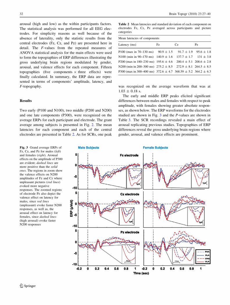
Fig. 3 Grand average ERPs of
Fz, Cz, and Pz for males (left)and females (right). Arousal
effects on the amplitude of P300
are evident; dashed lines are
more positive than the solidones. The regions in zoom show
the valence effects on N200
amplitudes of Fz and Cz where
unpleasant pictures (red lines)
evoked more negative
responses. The zoomed regions
of electrode Pz also depict the
valence effect on latency for
males, since red lines(unpleasant) evoke faster N200
responses, as well as, the
arousal effect on latency for
females, since dashed lines(high arousal) evoke faster
N200 responses
32 Brain Topogr (2010) 23:27–40
123
Amplitudes of ERP Components
The analysis of peak amplitudes yielded significant main
effects of gender, arousal and valence on early, middle, and
late components (Fig. 3). Table 3 shows the significant
P-values for the main effects of each component.
The gender effect was prominent on the negative com-
ponents for the Cz and Pz electrodes, revealing a greater
reaction to emotional stimuli for females. Women showed
greater negativity (-7.48 lV) as compared to males
(-5.07 lV) on the early N100 component on Pz. The same
effect was also revealed for Cz. As for middle compo-
nents, female responses on N200 of the Pz electrode
were again more negative (-5.78 lV) than those of males
(-3.29 lV).
There was a strong main effect of arousal on the early
component P100 on all three central electrodes. Low
arousing pictures elicited higher positivity than high
arousing stimuli did. The N100 component was also
affected by arousal on electrodes Fz and Pz, with high
arousing stimuli eliciting greater negativity than low
arousing pictures. For example, on Fz, the N100 amplitude
was more negative (-7.54 lV) for high arousing stimuli,
than for low arousing (-5.91 lV). Finally, P300 revealed
arousal effects on Cz and Pz; i.e., on Pz, P300 amplitudes
were higher for high arousing pictures (2.86 lV), than for
low arousing ones (1.18 lV).
Valence effects are also prominent on central channels,
not only for middle peaks, but also for early and late ones.
On negative components (N100 and N200) the unpleasant
Table 3 P-values of the ANOVA show significant main effects on peak amplitudes
Electrode Peak Effect
Gender Arousal Valence
Fz P100 F(1,26) = 26.13, P \ 0.001
N100 F(1,26) = 25.3, P = 0.0001
P200
N200 F(1,26) = 15.2, P = 0.0001
P300 F(1,26) = 4.11, P = 0.05
Cz P100 F(1,26) = 32.66, P \ 0.0001
N100 F(1,26) = 4.35, P = 0.047 F(1,26) = 4.35, P = 0.047
P200 F(1,25) = 5.61, P = 0.025
N200 F(1,26) = 9.83, P = 0.0042
P300 F(1,26) = 7.38, P = 0.012
Pz P100 F(1,26) = 25.91, P \ 0.0001
N100 F(1,26) = 5.66, P = 0.025 F(1,26) = 26.1, P \ 0.0001 F(1,26) = 8.24, P = 0.008
P200
N200 F(1,26) = 4.54, P = 0.043
P300 F(1,26) = 37.34, P \ 0.0001
The gender effect is dominant on N100 and N200, with females eliciting more negative responses than males, whereas arousal affects early and
late components. Valence affects early, middle, and late components, with unpleasant pictures eliciting greater negativity than positive ones
Fig. 4 Gender by arousal
interaction is evident as the
difference between high
arousing (HA) and low arousing
(LA) is greater in females than
in males on N100 on Fz and Cz
electrode
Brain Topogr (2010) 23:27–40 33
123
emotional stimuli elicit responses with greater negativity
on all central electrodes as compared to pleasant pictures;
that is, unpleasant pictures evoked greater negativity on
N200 of Fz (-9.6 lV) than pleasant ones (-8.46 lV). The
same effect was observed on N100 of Cz and Pz and N200
of Cz. On the other hand, on positive components (P200 of
Cz and P300 of Fz), pleasant pictures provoke responses
with greater positivity than unpleasant ones.
Interactions were also evident on the early component
N100. On frontal and central electrodes (Fz, Cz) there were
significant gender by arousal interactions. In order to
analyze the gender by arousal interactions further, we
collapsed the data for valence and the results showed sig-
nificant differences between high and low arousal condi-
tions on both channels; i.e., on Fz, high arousing pictures
evoked more negative N100 responses, than low arousing,
for both males (t(13) = -2.48, P = 0.028) and females
(t(13) = -4.38, P = 0.0007), with the difference being
clearly greater for females (Fig. 4).
Finally, there was a significant gender by valence inter-
action on the N100 of Pz. In order to analyze this further
again, we collapsed the data for arousal. Results showed
significant higher negativity for females (-8.31 lV) as
compared to males (-5.21 lV), but only for the unpleasant
pictures (t(26) = 2.61, P = 0.015). Furthermore, ampli-
tudes were significantly higher for the unpleasant pictures
relatively to pleasant ones only for the female group
(t(13) = 2.73, P = 0.017) (Fig. 5).
Latencies of ERP Components
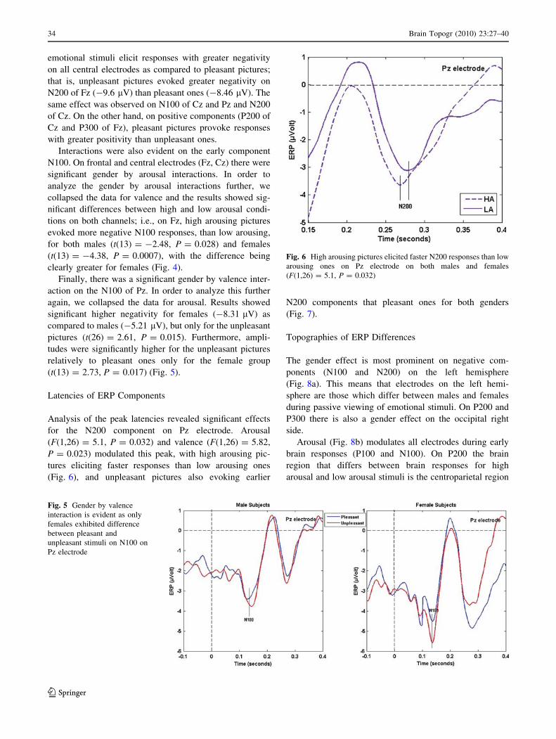
Analysis of the peak latencies revealed significant effects
for the N200 component on Pz electrode. Arousal
(F(1,26) = 5.1, P = 0.032) and valence (F(1,26) = 5.82,
P = 0.023) modulated this peak, with high arousing pic-
tures eliciting faster responses than low arousing ones
(Fig. 6), and unpleasant pictures also evoking earlier
N200 components that pleasant ones for both genders
(Fig. 7).
Topographies of ERP Differences
The gender effect is most prominent on negative com-
ponents (N100 and N200) on the left hemisphere
(Fig. 8a). This means that electrodes on the left hemi-
sphere are those which differ between males and females
during passive viewing of emotional stimuli. On P200 and
P300 there is also a gender effect on the occipital right
side.
Arousal (Fig. 8b) modulates all electrodes during early
brain responses (P100 and N100). On P200 the brain
region that differs between brain responses for high
arousal and low arousal stimuli is the centroparietal region
Fig. 5 Gender by valence
interaction is evident as only
females exhibited difference
between pleasant and
unpleasant stimuli on N100 on
Pz electrode
Fig. 6 High arousing pictures elicited faster N200 responses than low
arousing ones on Pz electrode on both males and females
(F(1,26) = 5.1, P = 0.032)
34 Brain Topogr (2010) 23:27–40
123
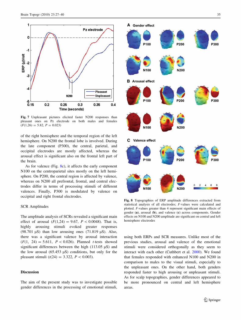
of the right hemisphere and the temporal region of the left
hemisphere. On N200 the frontal lobe is involved. During
the late component (P300), the central, parietal, and
occipital electrodes are mostly affected, whereas the
arousal effect is significant also on the frontal left part of
the brain.
As for valence (Fig. 8c), it affects the early component
N100 on the centroparietal sites mostly on the left hemi-
sphere. On P200, the central region is affected by valence,
whereas on N200 all prefrontal, frontal, and central elec-
trodes differ in terms of processing stimuli of different
valences. Finally, P300 is modulated by valence on
occipital and right frontal electrodes.
SCR Amplitudes
The amplitude analysis of SCRs revealed a significant main
effect of arousal (F(1,24) = 9.67, P \ 0.0048). That is,
highly arousing stimuli evoked greater responses
(98.701 lS) than low arousing ones (71.819 lS). Also,
there was a significant valence by arousal interaction
(F(1, 24) = 5.611, P \ 0.026). Planned t-tests showed
significant differences between the high (113.05 lS) and
the low arousal (65.453 lS) conditions, but only for the
pleasant stimuli (t(24) = 3.322, P \ 0.003).
Discussion
The aim of the present study was to investigate possible
gender differences in the processing of emotional stimuli,
using both ERPs and SCR measures. Unlike most of the
previous studies, arousal and valence of the emotional
stimuli were considered orthogonally as they seem to
interact with each other (Cuthbert et al. 2000). We found
that females responded with enhanced N100 and N200 in
comparison to males to the visual stimuli, especially to
the unpleasant ones. On the other hand, both genders
responded faster to high arousing or unpleasant stimuli.
As for scalp topographies, gender differences appeared to
be more pronounced on central and left hemisphere
areas.
Fig. 7 Unpleasant pictures elicited faster N200 responses than
pleasant ones on Pz electrode on both males and females
(F(1,26) = 5.82, P = 0.023)
Fig. 8 Topographies of ERP amplitude differences extracted from
statistical analysis of all electrodes; F-values were calculated and
plotted. F-values greater than 4 represent significant main effects of
gender (a), arousal (b), and valence (c) across components. Gender
effects on N100 and N200 amplitude are significant on central and left
hemisphere electrodes
Brain Topogr (2010) 23:27–40 35
123
Main Effects and Interactions Regarding Gender
Females showed greater negativity than males on negative
components (N100 and N200) upon viewing emotional
stimuli. This effect was further modulated by a gender by
valence interaction on the N100 of Pz electrode; females
exhibited greater negativity than males but only for the
unpleasant stimuli. Similar findings were found for the
arousal dimension and this is a relatively new finding in
the field of gender differences. We observed arousal effects
early on in the processing (N100) on frontal and central
electrodes, and these effects were also modulated by gen-
der; high arousing pictures evoked more negative response
in females as compared to males. Thus, the present study
adds to the existing literature in gender differences (Han
et al. 2008; Chentsova-Dutton and Tsai 2007; Guillem
and Mograss 2005; Kemp et al. 2004; Bradley et al. 2001;
George et al. 1996; Pardo et al. 1993), showing that
females respond more strongly not only to emotional or
negative faces, pictures with human and sounds, but also to
unpleasant emotional complex scenes of no specific con-
tent. More importantly, it shows that females respond with
more negative early components to high arousing stimuli as
well. This finding adds the electroencephalographic aspect
to other previous electrophysiological findings regarding
stronger autonomic responses, such as EMG, SCR, and
heart rate, reported for women when viewing unpleasant
pictures (Chentsova-Dutton and Tsai 2007; Kemp et al.
2004; Bradley et al. 2001; George et al. 1996; Pardo et al.
1993). It replicates and, in a way, generalizes the fact that
women exhibited enhanced reactions to painful images
(Han et al. 2008). More specifically, gender differences
were identified on central and left hemisphere electrodes,
as it is shown on the F-topographies, replicating previous
EEG (Gasbarri et al. 2007) and fMRI studies (Canli et al.
2002; Cahill 2003) which indicated the over-activation
of left hemisphere by females when viewing emotional
stimuli.
It has been proposed that the amplitude of the middle
component N200 reflects biased selective attention to task-
relevant stimulus properties (e.g., color, shape, etc.) or
biologically relevant stimuli such as those that are poten-
tially dangerous (Schupp et al. 2006). We therefore suggest
that selective attention in women appears to be more
strongly tuned to biologically relevant stimuli. Since such
differences can be mainly traced to a complex interaction
or a feedback loop between underlying biological pro-
cesses and social and cultural responses to values regarding
gender (Brody 1997; Brody et al. 1993), one can speculate
that gender differences may have been also influenced by
evolutionary pressures. As a result, females, having a
slighter physique, appear to exhibit a more rapid and
stronger response to potentially dangerous stimuli (e.g.,
unpleasant and high arousing stimuli) which may have
been useful to effectively nurture their offspring.
Despite the rather convincing evidence for gender dif-
ferences in emotional processing, proposed not only by this
study, but also by previous ones, there exist studies
encompassing different interpretations. It has been pro-
posed that gender differences on brain oscillations occur
even during simple visual stimulation (Gutenkin and Basar
2007), and the gender differences in the emotional pro-
cessing could be just an epiphenomenon of these intrinsic
gender differences. These differences, found in the delta
frequency band measured on parietal and occipital regions,
are attributed to the basic sensory circuits which are dif-
ferentiated by the ‘‘phyletic memory’’ (Fuster 1995; Basar
2004). However, the delta oscillations are found to affect
mainly the P300 potential and the forthcoming slow wave
activity (Oloffson et al. 2008). Consequently, if we had to
do with an epiphenomenon, we should have observed
gender differences on P300 on parietal and occipital
regions as well, whereas we reported gender differences
during early ERPs measured mainly in anterior areas. In
addition, the fact that in our study valence and arousal
interacted with gender, suggests that the parameters of the
emotional stimuli affect the intrinsic gender differences to
visual stimuli. Therefore, our findings can be interpreted as
an enhancement of intrinsic gender differences by the
arousal and valence of the stimuli, resulting to gender
differences in terms of emotional processing.
Unlike other studies (Bradley et al. 2001; Chentsova-
Dutton and Tsai 2007), autonomic responses (SCRs) were
not modulated by gender. However, the validity of this
conclusion may be compromised by a procedural limita-
tion. It turned out that although, the ISI was long enough
for a recognizable rise in the SCR to be seen in response to
a stimulus; it was not long enough to separate different
SCR components, i.e., for the SCR signal to return to its
baseline level. To deal with this limitation, we employed
the amplitude of SCR, a rather weak measure that may
have obscured gender differences.
There was no significant three-way interaction of gender
by valence by arousal. It seems that gender differences are
not greater for stimuli that are both unpleasant and high
arousing. This fact adds evidence to the independency of
valence and arousal in the way both genders perceive and
process emotional stimuli in terms of ERP responses.
To sum up, our data support the notion that females
respond more in terms of ERP amplitudes to unpleasant or
high arousing stimuli relative to males. The brain mecha-
nisms responsible for these mechanisms are most likely
localized in the central and left brain regions, as suggested
by the brain topography analysis. The robust gender effect
on negative components suggests that brain regions that
recognize the valence of stimuli early on in the stream of
36 Brain Topogr (2010) 23:27–40
123

processing are more finely tuned. This conclusion is con-
sistent with the evolutionary hypothesis of sex differences
in emotional processing; the brain of men and women
differ not only anatomically, but also functionally (Cahill
2005). Furthermore, these findings are relevant within the
context of gender differences in the prevalence of mood
disorders such as depression, and the post-traumatic stress
disorder (PTSD). Both disorders are twice as likely to
occur in women as in men (Kessler 2003; Kendler et al.
2001; Breslau et al. 1997), presumably because of the
differential response of women to stressful stimuli of
everyday life.
Main Effects and Interactions Regarding Valence
(Pleasure) and Arousal
The analyses of the peak latency data yielded significant
results, not reported in previous literature to our knowl-
edge. The unpleasant and the high arousing stimuli appear
to elicit faster responses in both genders. These findings
suggest that the prioritization of emotional, unpleasant, and
high arousing, stimuli is not only reflected in stronger brain
activity (increased amplitude), but also in the temporal
course of this activity, since it occurs faster than the pro-
cessing of pleasant or low arousing stimuli. The faster
response to unpleasant or high arousing stimuli can be seen
in an evolutionary context, underlying the necessity for
immediate reaction to potentially life-threatening stimuli
and situations.
The effects of arousal and valence on ERP amplitudes
are also in agreement with previous findings. Studies have
shown that the early N100 component is associated with
attentional process modulated by the valence dimension
(for a review see Oloffson et al. 2008). The valence effect
observed in our study was most prominent on N200 of Cz
and Pz electrodes, with unpleasant pictures eliciting greater
responses than pleasant ones. The amplitude modulations
of early ERP components were greater for unpleasant
stimuli, consistent with the suggestion that unpleasant
stimuli preferentially attract attention early in the infor-
mation processing system (Oloffson et al. 2008). Recent
studies proposed that pleasant stimuli yielded more posi-
tive components, such as P300 (Cuthbert et al. 2000;
Flaisch et al. 2008). Our study shows an enhancement of
both positive (P300) and negative (N100 and N200) com-
ponents for unpleasant stimuli.
Amplitudes of early components were strongly modu-
lated by arousal on almost all electrodes as shown in
Fig. 8b. Particularly, on the N100 component of Pz, high
arousing stimuli evoked enhanced responses than low
arousing ones. This finding adds to the study of Keil et al.
(2003), where affectively arousing pictures were found to
be associated with enhanced reactions at parieto-occipital
sites. An unexpected result was the greater positivity
elicited by low arousing stimuli on the P100 component.
Although it is not clear which factor contributed most for
this effect, the early modulation of the ERP by the emo-
tional and arousing properties of the stimulus support the
hypothesis of preferential processing of affective stimuli.
As for the late, P300, component, which indexes atten-
tional processes and initial memory storage of events
(Polich 2007), arousal effects were significant on centro-
parietal sites on both hemispheres, as it is shown in the
topographies, in terms of amplitude. High arousing pictures
evoked greater positivity than low arousing ones to both
males and females. This is in line with previous results
(Bradley et al. 2007; Cuthbert et al. 2000; Schupp et al.
2006; Schupp et al. 2007; Delplanque et al. 2006). Valence
also seems to modulate P300 when arousal level is
controlled (Cano et al. 2008; Conroy and Polich 2007;
Cuthbert et al. 2000). Thus, studies have shown that
pleasant pictures elicit greater positivity on P300 (Cano
et al. 2008; Conroy and Polich 2007; Cuthbert et al. 2000).
This finding was also supported by our study where the
valence effect on P300 is prominent on occipital, temporal,
and frontal regions as shown in the topographies (Figs. 8c).
Last but not least, although the skin conductance
responses have lower temporal sensitivity than ERPs, they
nevertheless produced clear results. High arousing pictures
elicited higher SCR than low arousing did. This was
expected and it is in agreement with past literature
(Anonkin et al. 2006; Bradley et al. 2001).
Finally, we did not find a significant two-way valence by
arousal interaction concerning ERPs, whereas such an
interaction was present in the SCR data. That is, although
both factors affected brain components, ERP amplitudes
were not greater for unpleasant-high-arousing stimuli and
pleasant-low-arousing stimuli as suggested by previous
behavioral studies (Robinson et al. 2004) and the Contin-
uum Model of Significance Processing (Williams and
Gordon 2007). However, as regards autonomic responses,
valence interacted with arousal in electrodermal activity
measurements. As a result, we cannot clearly confirm or
reject the hypothesis that valence and arousal are inde-
pendently processed by human brain.
Limitations
We list here some limitations of our study that could in
principle allow for modifications in the interpretation of
our results.
The performed analysis may be limited as the side of
differences on brain activation was considered in a rather
gross way (left/right, frontal/central/posterior); in addition
a relatively low number of electrodes (19) was used and
Brain Topogr (2010) 23:27–40 37
123
our choice of filtering (0.5–40 Hz) resulted to the elimi-
nation of high frequency EEG activity. Future EEG studies
with high density electrodes may confirm and expand these
results. Also, Magnetoencephalographic (MEG) studies
could further examine emotional processing, as MEG
provides better localization accuracy (Leahy et al. 1998) of
a few mm (Papadelis et al. 2009).
Unlike most of the previous studies we manipulated
orthogonally the valence and arousal dimensions of the
stimuli, as they appear to interact (Cuthbert et al. 2000).
We decided to select stimuli of similar affective content
level for males and females, as the two genders perceive
emotional context in a different way. However, IAPS
collection in its current form does not provide enough
pictures rated with similar ratings by both genders to
perform such a study. In order to achieve similar affective
content level of the stimuli for males and females, we
used the gender-specific ratings provided by the IAPS
collection. This limitation of IAPS collection has been
discussed by previous studies. Cuthbert et al. (2000)
report that ‘unpleasant pictures show a high positive
correlation between valence and arousal that is particu-
larly pronounced for female subjects.’ Rozenkrants and
Polich (2008) selected images of similar affective content
level; those whose mean ratings of males and females did
not differ significantly (only 16 pictures of each cate-
gory). We should take also into account that in previous
studies involving male or female participants only, the
gender-specific ratings and not the overall ones were
selected for the experiment (Bernat et al. 2006; Anonkin
et al. 2006).
In conclusion, our study provides evidence for gender
differences in the way stimuli with different valence and
arousal are processed. The subjective nature, however, of
valence and arousal allows the possibility that the observed
gender differences may be due, at least partly, to the pre-
sentation of different stimuli to the participants.
Acknowledgments This work has been benefited from a grant
by the Greek General Secretariat for Research and Technology.
Assistance with the experiment preparation and recordings from
Mr. S. Tsemberlidis and Ms. G. Germanidou is also gratefully
acknowledged.
Open Access This article is distributed under the terms of the
Creative Commons Attribution Noncommercial License which per-
mits any noncommercial use, distribution, and reproduction in any
medium, provided the original author(s) and source are credited.
References
Aftanas LI, Lotova NV, Koshkarov VI, Popov SA, Makhnev VP
(1997) Non-linear forecasting measurements of the human EEG
during evoked emotions. Brain Topogr 10:155–162
Alfano KM, Cimino C (2008) Alteration of expected hemispheric
asymmetries: valence and arousal effects in neurophysiological
models of emotion. Brain Cogn 66:213–220
Amrhein C, Muhlberger A, Pauli P (2004) Modulation of event-
related brain potentials during affective picture processing: a
complement to startle reflex and skin conductance response? Int
J Psychophysiol 54:231–240
Anonkin AP, Golosheykin S, Sirevaag E, Kristjansson S, Rohrbaugh
JW, Heath AC (2006) Rapid discrimination of visual scene
content in the human brain. Brain Res 1093:167–177
Bachorowski J, Owren MJ (2003) Sounds of emotion production and
perception of affect-related vocal acoustics. Ann N Y Acad Sci
1000:244–265
Barrett LF (1998) Discrete emotions or dimensions? The role of
valence focus and arousal focus. Cogn Emot 12:579–599
Barrett LF, Russell JA (1999) The structure of current affect:
controversies and emerging consensus. Am Psychol Soc Bull
8:10–14
Basar E (2004) Memory and brain dynamics: oscillations integrating
attention, perception, learning and memory. CRC Press, Boca
Raton
Bernat E, Patrick CJ, Benning SD, Tellengen A (2006) Effects of
picture content and intensity on affective physiological response.
Psychophysiology 43:93–103
Bradley MM, Lang PJ (1994) Measuring emotion: the self-assessment
manikin and the semantic differential. J Behav Ther Exp
Psychiatry 25:49–59
Bradley MM, Codispoti M, Sabatinelli D, Lang PJ (2001) Emotion
and motivation II: sex differences in picture processing. Emotion
1:300–319
Bradley MM, Hamby S, Low A, Lang PJ (2007) Brain potentials in
perception: picture complexity and emotional arousal. Psycho-
physiology 44:364–373
Breslau N, Davis GC, Andreski P, Peterson EL, Schultz LR (1997)
Sex differences in posttraumatic stress disorder. Arch Gen
Psychiatry 54:1044–1048
Brody LR (1997) Gender and emotion: beyond stereotypes. J Soc
Issues 53:369–394
Brody LR, Hall JA, Lewis M, Haviland JM (1993) Gender and
emotion. Guilford Press, New York
Cacioppo JT, Bernston GG (1994) Relationships between attitudes
and evaluative space: a critical review with emphasis on the
separability of positive and negative substrates. Psychol Bull
115:401–423
Cahill L (2003) Sex and hemisphere-related influences on the
neurobiology of emotionally influenced memory. Ann N Y
Acad Sci 985:163–173
Cahill L (2005) His brain, her brain. Sci Am 292:40–47
Cahill L (2006) Why sex matters for neuroscience. Nat Rev Neurosci
7:477–484
Cahill L, McGaugh JL (1998) Mechanisms of emotional arousal and
lasting declarative memory. Trends Neurosci 21:294–299
Canli T, Desmond JE, Zhao Z, Gabrieli JDE (2002) Sex difference in
the neural basis of emotional memories. Proc Natl Acad Sci
99:10789–10794
Cano ME, Class QA, Polich J (2009) Affective valence, stimulus
attributes and P300: colors vs. black/white and normal vs.
scrambled images. Int J Psychophysiol 71:17–24
Chentsova-Dutton YE, Tsai JL (2007) Gender differences in
emotional response among European Americans and among
Americans. Cogn Emot 21:162–181
Codispoti M, Ferrari V, Bradley MM (2006) Repetitive picture
processing: autonomic and cortical correlates. Cogn Brain Res
1068:213–220
Conroy MA, Polich J (2007) Affective valence and P300 when
stimulus arousal level is controlled. Cogn Emot 21:891–901
38 Brain Topogr (2010) 23:27–40
123
Cuthbert BN, Schupp HT, Bradley MM, Birbaumer N, Lang PJ
(2000) Brain potentials in affective picture processing: covari-
ation with autonomic arousal and affective report. Biol Psychol
52:95–111
Delorme A, Makeig S (2004) EEGLAB: an open source toolbox for
analysis of single-trial EEG dymanics including independent
component analysis. J Neurosci Methods 134:9–21
Delplanque S, Silvert L, Hot P, Rigoulot S, Sequeira H (2006)
Arousal and valence effects on event-related P3a and P3b during
emotional categorization. Int J Psychophysiol 60:315–322
Delplanque S, N’diaye K, Scherer K, Grandjean D (2007) Spatial
frequencies or emotional effects? A systematic measure of
spatial frequencies for IAPS pictures by a discrete wavelet
analysis. J Neurosci Methods 165:144–150
Dolcos F, Cabeza R (2002) Event-related potentials of emotional
memory: encoding pleasant, unpleasant, and neutral pictures.
Cogn Affect Behav Neurosci 2:252–263
Ekman A, Flink R, Sundman E, Eriksson LI, Brudin L, Sandin R
(2007) Neuromuscular block and the electroencephalogram
during sevoflurane anaesthesia. Neuroreport 18(17):1817–1820
Flaisch T, Stockburger J, Schupp HT (2008) Affective prime and
target picture processing: an ERP analysis of early and late
interference effects. Brain Topogr 20:183–191
Fuster JM (1995) Memory in the cerebral cortex—an empirical
approach to neural networks in the human and non-human
primate. MIT Press, Cambridge, MA
Gasbarri A, Arnone B, Pompili A, Pacitti F, Pacitti C, Cahill L (2007)
Sex related hemispheric lateralization of electrical potentials
evoked by arousing negative stimuli. Brain Res 1138:178–186
George MS, Ketter TA, Parekh PI, Herscovitch P, Post RM (1996)
Gender differences in regional cerebral blood flow during
transient self-induced sadness or happiness. Biol Psychiatry
40:859–871
Gerber AJ, Posner J, Gorman D, Colibazzi T, Yu S, Wang Z,
Kangarlu A, Zhu H, Russell J, Peterson BS (2008) An affective
circumplex model of neural systems subserving valence, arousal
and cognitive overlay during the appraisal of emotional faces.
Neurophysiologia 46:2129–2139
Gianotti LRR, Faber PL, Schuler M, Pasqual-Marqui RD, Kochi K,
Lehmann D (2008) First valence, then arousal: the temporal
dynamics of brain electric activity evoked by emotional stimuli.
Brain Topogr 20:143–156
Guillem F, Mograss M (2005) Gender differences in memory
processing: evidence from event-related potentials to faces.
Brain Cogn 57:84–92
Gutenkin B, Basar E (2007) Brain oscillations are highly influenced
by gender differences. Int J Psychophysiol 65:294–299
Hall JA (1978) Gender effects in decoding nonverbal cues. Psychol
Bull 85:845–857
Han S, Fan Y, Mao L (2008) Gender difference in empathy for pain:
an electrophysiological investigation. Brain Res 1196:85–93
Jasper H (1958) Report of committee on methods of clinical exam in
EEG. Electroencephalogr Clin Neurophysiol 10:370–375
Jung TP, Makeig S, Humphries C, Lee TW, Mckeown MJ, Iraqui V,
Sejnowski TJ (2000) Removing electroencephalographic arti-
facts by blind source separation. Psychophysiology 37:163–178
Keil A, Bradley MM, Hauk O, Rockstroh B, Elbert T, Lang P (2002)
Large-scale neural correlates of affective picture processing.
Psychophysiology 39:641–649
Keil A, Gruber T, Muller MM, Moratti S, Stolarova M, Bradley MM,
Lang PJ (2003) Early modulation of visual perception by
emotional arousal: evidence from steady-state visual evoked
brain potentials. Cogn Affect Behav Neurosci 3:195–206
Kemp AH, Silberstein RB, Armstrong SM, Nathan PJ (2004) Gender
differences in the cortical electrophysiological processing of
visual emotional stimuli. NeuroImage 16:632–646
Kendler KS, Thornton LM, Prescott CA (2001) Gender differences in
the rates of exposure to stressful life events and sensitivity to
their depressogenic effects. Am J Psychiatry 158:587–593
Kessler RC (2003) Epidemiology of women and depression. J Affect
Disord 74:5–13
Kring AM, Gordon AH (1998) Sex differences in emotion:
expression, experience and physiology. J Pers Soc Psychol 74:
686–703
Krumhansl CL (2002) Music: a link between cognition and emotion.
Curr Dir Psychol Sci 11:45–50
Lang PJ (1968) Fear reduction and fear behaviour: problems in
treating a construct. In: Schlien J (ed) Research in psychother-
apy, vol 3. American Psychological Association, Washington,
DC, pp 90–103
Lang PJ (1984) Cognition in emotion: concept and action. In: Izard CE,
Kagan J, Zajonc RB (eds) Emotions, cognition, and behaviour.
Cambridge University Press, New York, pp 192–226
Lang PJ, Bradley MM, Cuthbert BN (1997) Motivated attention:
affect, activation, and action. In: Lang PJ, Simons RF, Balaban
M (eds) Attention and orienting: sensory and motivational
processes. Erlbaum Associates, Hillsdale, NJ
Lang PJ, Bradley MM, Cuthbert BN (1998) Emotion motivation and
anxiety: brain mechanisms and psychophysiology. Biol Psychi-
atry 44:1248–1263
Leahy RM, Mosher JC, Spencer ME, Huang MX, Lewine JD (1998)
A study of dipole localisation accuracy for MEG and EEG using
a human skull phantom. Electroencephalogr Clin Neurophysiol
107:159–173
Leon-Carrion J, Damasb J, Izzetoglu K, Pourrezai K, Martin-
Rodriguez JF, Barroso y Martin JM, Dominguez-Morales MR
(2006) Differential time course and intensity of PFC activation
for men and women in response to emotional stimuli: a
functional near-infrared spectroscopy (fNIRS) study. Neurosci
Lett 403:90–95
Oloffson JK, Nordin S, Sequeira H, Polich J (2008) Affective picture
processing: an integrative review of ERP findings. Biol Psychol
77:247–265
Panksepp J, Bernatzky G (2002) Emotional sounds and the brain: the
neuro-affective foundations of musical appreciation. Behav
Processes 60:133–155
Papadelis C, Poghosyan V, Fenwick PBC, Ioannides AA (2009)
MEG’s ability to localise accurately weak transient neural
sources. Clin Neurophysiol 120:1958–1970
Pardo JV, Pardo PJ, Raichle ME (1993) Neural correlates of self-
induced dysphoria. Am J Psychiatry 150:713–719
Polich J (2007) Updating P300: an integrative theory of P3a and P3b.
Clin Neurophysiol 118:2128–2148
Proverbio AM, Zani A, Adorni R (2008) Neural markers of
greater female responsiveness to social stimuli. BMC Neurosci
9:56
Reinsenzein R (1994) Pleasure-arousal theory and the intensity of
emotions. J Pers Soc Psychol 67:525–539
Robinson MR, Storbeck J, Meier BP, Kirkeby BS (2004) Watch out!That could be dangerous: valence-arousal interactions in eval-
uative processing. Pers Soc Psychol Bull 30:1472–1484
Rozenkrants B, Polich J (2008) Affective ERP processing in a visual
oddball task: arousal, valence, and gender. Clin Neurophysiol
119:2260–2265
Russell JA (1980) A circumplex model of affect. J Pers Soc Psychol
39:1161–1178
Russell JA (1989) Measures of emotion. In: Plutchik R, Kellerman H
(eds) Emotion: theory, research and experience, vol 4. Academic
Press, San Diego, CA, pp 83–111
Schirmer A, Kotz SA, Friederici AD (2002) Sex differentiates the role
of emotional prosody during word processing. Cogn Brain Res
14:228–233
Brain Topogr (2010) 23:27–40 39
123

Schirmer A, Striano T, Friederici AD (2005) Sex differences in the
preattentive processing of vocal emotional expressions. Neuro-
report 16:635–639
Schneider F, Habel U, Kessler C, Salloum JB, Posse S (2000) Gender
differences in regional cerebral activity during sadness. Hum
Brain Mapp 9:226–238
Schupp HT, Stockburger J, Codispoti M, Junghofer M, Weike AI, Hamm
AO (2006) Stimulus novelty and emotion perception: the mean
absence of habituation in the visual cortex. Neuroreport 17:365–369
Schupp HT, Stockburger J, Junghofer M, Weike AI, Hamm AO
(2007) Selective visual attention to emotion. J Neurosci
27:1082–1089
Wild B (2003) Are emotions contagious? Evoked emotions while
viewing emotionally expressive faces: quality, quantity, time
course and gender differences. Psychiatry Res 102:109–124
Williams LM, Gordon E (2007) Dynamic organization of the
emotional brain: responsivity, stability and instability. Neuro-
scientist 13:349–370
Wrase J, Klein S, Gruesser SM, Hermann D, Flor H, Mann K, Braus
DF, Heinz A (2003) Gender differences in the processing of
standardized emotional visual stimuli in humans: a functional
magnetic resonance imaging study. Neurosci Lett 348:41–45
Xiong Y, Mahmood A, Lu D, Gu C, Goussev A, Schallert T, Chopp
M (2007) The role of gender in outcome after traumatic brain
injury and therapeutic effect of erythropoietin in mice. Brain Res
1185:301–312
Zhang XM, Zhu SW, Duan RS, Mohammed AH, Windblad B, Zhu J
(2008) Gender differences in susceptibility to kainic acid-
induced neurodegeneration in aged C57BL/6 mice. Neurotoxi-
cology 29:406–412
40 Brain Topogr (2010) 23:27–40
123