Stimuli-Responsive Drug Release from Smart Polymers - MDPI
20
Journal of Functional Biomaterials Review Stimuli-Responsive Drug Release from Smart Polymers Carlos M. Wells * , Michael Harris, Landon Choi, Vishnu Priya Murali, Fernanda Delbuque Guerra and J. Amber Jennings Department of Biomedical Engineering, The University of Memphis, Memphis, TN 38152, USA * Correspondence: [email protected]; Tel.: +1-1901-289-9827 Received: 28 May 2019; Accepted: 26 July 2019; Published: 31 July 2019 Abstract: Over the past 10 years, stimuli-responsive polymeric biomaterials have emerged as effective systems for the delivery of therapeutics. Persistent with ongoing efforts to minimize adverse effects, stimuli-responsive biomaterials are designed to release in response to either chemical, physical, or biological triggers. The stimuli-responsiveness of smart biomaterials may improve spatiotemporal specificity of release. The material design may be used to tailor smart polymers to release a drug when particular stimuli are present. Smart biomaterials may use internal or external stimuli as triggering mechanisms. Internal stimuli-responsive smart biomaterials include those that respond to specific enzymes or changes in microenvironment pH; external stimuli can consist of electromagnetic, light, or acoustic energy; with some smart biomaterials responding to multiple stimuli. This review looks at current and evolving stimuli-responsive polymeric biomaterials in their proposed applications. Keywords: stimuli-responsiveness; drug release; drug delivery; thermo-responsive materials; enzyme-responsive materials; pH-responsive materials; shape-memory materials 1. Introduction Researchers throughout different disciplines continue to explore improved and safer ways to locally deliver drugs to specific sites of action, attempting to increase specificity and efficacy. Over the years, numerous investigations on biomaterials have seen successes in the development of controlled delivery systems [1,2]. These achievements have been the results of interdisciplinary contributions across chemistry, biology, physics, and pharmacology, and with inputs from clinicians. Biomaterials have evolved to cover vast libraries, with diverse chemical structures, varied morphologies, and numerous physiological functions. Synthetic polymers, biomacromolecules, nano-/micro-particles, biocompatibility, biodegradability, specific targeting, cargo protection, and bulk materials are some of the developments that have improved or enhanced biomaterials, their applications, or the combination [3]. Drug carriers and their subsequent on-demand release have been the benefactors from flexible designs that evolved to address the perplexing and diverse physiological environment. Smart biomaterials are the next evolutionary step in optimizing patient-centric care while potentially providing treatment options previously not available. Smart biomaterials possess tailor-designed stimuli-responsiveness that attempts to address many issues where current drug delivery systems are lacking [4]. The incorporation of different internal stimuli from the physiological microenvironment, such as pH, redox, temperature, enzyme, and mechanical force, or some readily available and relatively easily-controlled external stimuli, e.g., light, ultrasound, electric, and magnetic, has led to highly controlled smart systems [3]. The spatiotemporal control that many smart systems possess increases efficacy, and minimizes potential side effects and off-target toxicity. The emphasis of this mini-review is to present and analyze progress in the development of polymeric smart drug delivery biomaterials within the past decade. We will focus on three primary J. Funct. Biomater. 2019, 10, 34; doi:10.3390/jfb10030034 www.mdpi.com/journal/jfb
-
Upload
khangminh22 -
Category
Documents
-
view
5 -
download
0
Transcript of Stimuli-Responsive Drug Release from Smart Polymers - MDPI

Journal of
Functional
Biomaterials
Review
Stimuli-Responsive Drug Release fromSmart Polymers
Carlos M. Wells * , Michael Harris, Landon Choi, Vishnu Priya Murali,Fernanda Delbuque Guerra and J. Amber Jennings
Department of Biomedical Engineering, The University of Memphis, Memphis, TN 38152, USA* Correspondence: [email protected]; Tel.: +1-1901-289-9827
Received: 28 May 2019; Accepted: 26 July 2019; Published: 31 July 2019�����������������
Abstract: Over the past 10 years, stimuli-responsive polymeric biomaterials have emerged as effectivesystems for the delivery of therapeutics. Persistent with ongoing efforts to minimize adverse effects,stimuli-responsive biomaterials are designed to release in response to either chemical, physical,or biological triggers. The stimuli-responsiveness of smart biomaterials may improve spatiotemporalspecificity of release. The material design may be used to tailor smart polymers to release a drug whenparticular stimuli are present. Smart biomaterials may use internal or external stimuli as triggeringmechanisms. Internal stimuli-responsive smart biomaterials include those that respond to specificenzymes or changes in microenvironment pH; external stimuli can consist of electromagnetic, light,or acoustic energy; with some smart biomaterials responding to multiple stimuli. This review looksat current and evolving stimuli-responsive polymeric biomaterials in their proposed applications.
Keywords: stimuli-responsiveness; drug release; drug delivery; thermo-responsive materials;enzyme-responsive materials; pH-responsive materials; shape-memory materials
1. Introduction
Researchers throughout different disciplines continue to explore improved and safer ways tolocally deliver drugs to specific sites of action, attempting to increase specificity and efficacy. Over theyears, numerous investigations on biomaterials have seen successes in the development of controlleddelivery systems [1,2]. These achievements have been the results of interdisciplinary contributionsacross chemistry, biology, physics, and pharmacology, and with inputs from clinicians. Biomaterialshave evolved to cover vast libraries, with diverse chemical structures, varied morphologies, andnumerous physiological functions. Synthetic polymers, biomacromolecules, nano-/micro-particles,biocompatibility, biodegradability, specific targeting, cargo protection, and bulk materials are someof the developments that have improved or enhanced biomaterials, their applications, or thecombination [3]. Drug carriers and their subsequent on-demand release have been the benefactorsfrom flexible designs that evolved to address the perplexing and diverse physiological environment.
Smart biomaterials are the next evolutionary step in optimizing patient-centric care whilepotentially providing treatment options previously not available. Smart biomaterials possesstailor-designed stimuli-responsiveness that attempts to address many issues where current drugdelivery systems are lacking [4]. The incorporation of different internal stimuli from the physiologicalmicroenvironment, such as pH, redox, temperature, enzyme, and mechanical force, or some readilyavailable and relatively easily-controlled external stimuli, e.g., light, ultrasound, electric, and magnetic,has led to highly controlled smart systems [3]. The spatiotemporal control that many smart systemspossess increases efficacy, and minimizes potential side effects and off-target toxicity.
The emphasis of this mini-review is to present and analyze progress in the development ofpolymeric smart drug delivery biomaterials within the past decade. We will focus on three primary
J. Funct. Biomater. 2019, 10, 34; doi:10.3390/jfb10030034 www.mdpi.com/journal/jfb
J. Funct. Biomater. 2019, 10, 34 2 of 20
categories for stimuli-responsive polymeric systems, chemical, physical, and biological, with systemscontaining multiple or overlapping stimuli-responsiveness discussed separately. Within each group,this mini-review will discuss pH-, acoustic-, photo-, magnetic-, electric-, and enzyme-responsivenessas distinct subcategories; a summary of the reviewed studies in tabulated form is present at theconclusion (Table 1). We will survey representative samples for each of the subcategory stimuli typeswith appropriate advantages, disadvantages, challenges, and future directions.
2. Chemical Stimuli-Responsive Systems
pH-Responsiveness
Smart biomaterials can be designed to respond to alterations in environmental parameters,promoting the delivery of therapeutics locally when the environmental pH is either acidic or basic, andallowing for a release in particular organ systems or pathological conditions in which the pH changes.For the delivery of highly toxic chemotherapeutics, pH-controlled release systems can limit systemicconcentrations of drugs [2,5–9]. The acidic extracellular environment of solid tumors makes it possibleto develop pH-sensitive drug delivery systems that undergo physical/chemical changes or both whenexposed to acidic pH. Apart from cancer therapy, smart delivery systems explore other diseases, suchas gastric ulcers [10], osteomyelitis [11], and diabetes [12]. Generally, polyelectrolyte polymeric systemscontain weak acidic or basic groups in the polymer backbone. Changes in environmental pH triggerthe acceptance or release of protons, promoting cleavage of bonds, solubility, and or structure.
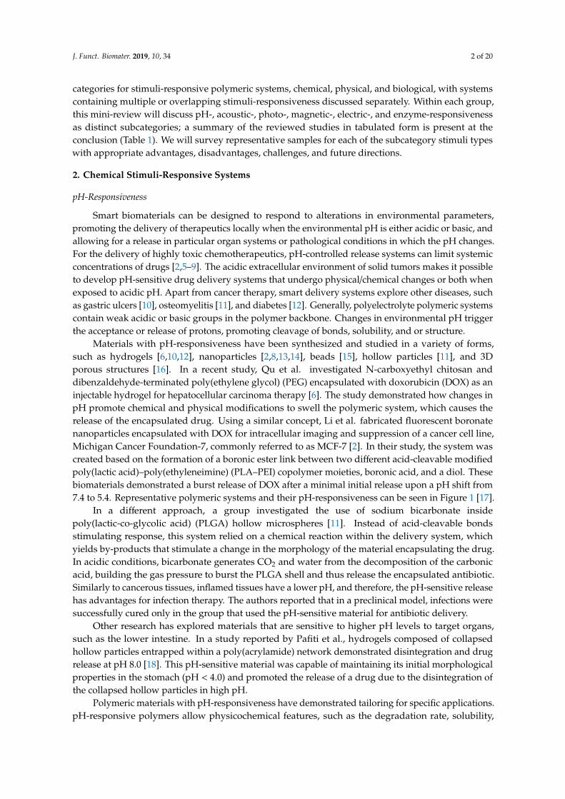
Materials with pH-responsiveness have been synthesized and studied in a variety of forms,such as hydrogels [6,10,12], nanoparticles [2,8,13,14], beads [15], hollow particles [11], and 3Dporous structures [16]. In a recent study, Qu et al. investigated N-carboxyethyl chitosan anddibenzaldehyde-terminated poly(ethylene glycol) (PEG) encapsulated with doxorubicin (DOX) as aninjectable hydrogel for hepatocellular carcinoma therapy [6]. The study demonstrated how changes inpH promote chemical and physical modifications to swell the polymeric system, which causes therelease of the encapsulated drug. Using a similar concept, Li et al. fabricated fluorescent boronatenanoparticles encapsulated with DOX for intracellular imaging and suppression of a cancer cell line,Michigan Cancer Foundation-7, commonly referred to as MCF-7 [2]. In their study, the system wascreated based on the formation of a boronic ester link between two different acid-cleavable modifiedpoly(lactic acid)–poly(ethyleneimine) (PLA–PEI) copolymer moieties, boronic acid, and a diol. Thesebiomaterials demonstrated a burst release of DOX after a minimal initial release upon a pH shift from7.4 to 5.4. Representative polymeric systems and their pH-responsiveness can be seen in Figure 1 [17].
In a different approach, a group investigated the use of sodium bicarbonate insidepoly(lactic-co-glycolic acid) (PLGA) hollow microspheres [11]. Instead of acid-cleavable bondsstimulating response, this system relied on a chemical reaction within the delivery system, whichyields by-products that stimulate a change in the morphology of the material encapsulating the drug.In acidic conditions, bicarbonate generates CO2 and water from the decomposition of the carbonicacid, building the gas pressure to burst the PLGA shell and thus release the encapsulated antibiotic.Similarly to cancerous tissues, inflamed tissues have a lower pH, and therefore, the pH-sensitive releasehas advantages for infection therapy. The authors reported that in a preclinical model, infections weresuccessfully cured only in the group that used the pH-sensitive material for antibiotic delivery.
Other research has explored materials that are sensitive to higher pH levels to target organs,such as the lower intestine. In a study reported by Pafiti et al., hydrogels composed of collapsedhollow particles entrapped within a poly(acrylamide) network demonstrated disintegration and drugrelease at pH 8.0 [18]. This pH-sensitive material was capable of maintaining its initial morphologicalproperties in the stomach (pH < 4.0) and promoted the release of a drug due to the disintegration ofthe collapsed hollow particles in high pH.
Polymeric materials with pH-responsiveness have demonstrated tailoring for specific applications.pH-responsive polymers allow physicochemical features, such as the degradation rate, solubility,
J. Funct. Biomater. 2019, 10, 34 3 of 20
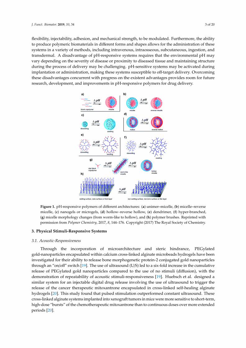
flexibility, injectability, adhesion, and mechanical strength, to be modulated. Furthermore, the abilityto produce polymeric biomaterials in different forms and shapes allows for the administration of thesesystems in a variety of methods, including intravenous, intraosseous, subcutaneous, ingestion, andtransdermal. A disadvantage of pH-responsive systems requires that the environmental pH mayvary depending on the severity of disease or proximity to diseased tissue and maintaining structureduring the process of delivery may be challenging. pH-sensitive systems may be activated duringimplantation or administration, making these systems susceptible to off-target delivery. Overcomingthese disadvantages concurrent with progress on the existent advantages provides room for futureresearch, development, and improvements in pH-responsive polymers for drug delivery.J. Funct. Biomater. 2019, 10, x FOR PEER REVIEW 3 of 20
Figure 1. pH-responsive polymers of different architectures: (a) unimer–micelle, (b) micelle–reverse micelle, (c) nanogels or microgels, (d) hollow–reverse hollow, (e) dendrimer, (f) hyper-branched, (g) micelle morphology changes (from worm-like to hollow), and (h) polymer brushes. Reprinted with permission from Polymer Chemistry, 2017, 8, 144–176. Copyright (2017) The Royal Society of Chemistry.
In a different approach, a group investigated the use of sodium bicarbonate inside poly(lactic-co-glycolic acid) (PLGA) hollow microspheres [11]. Instead of acid-cleavable bonds stimulating response, this system relied on a chemical reaction within the delivery system, which yields by-products that stimulate a change in the morphology of the material encapsulating the drug. In acidic conditions, bicarbonate generates CO2 and water from the decomposition of the carbonic acid, building the gas pressure to burst the PLGA shell and thus release the encapsulated antibiotic. Similarly to cancerous tissues, inflamed tissues have a lower pH, and therefore, the pH-sensitive release has advantages for infection therapy. The authors reported that in a preclinical model, infections were successfully cured only in the group that used the pH-sensitive material for antibiotic delivery.
Other research has explored materials that are sensitive to higher pH levels to target organs, such as the lower intestine. In a study reported by Pafiti et al., hydrogels composed of collapsed hollow particles entrapped within a poly(acrylamide) network demonstrated disintegration and drug release at pH 8.0 [18]. This pH-sensitive material was capable of maintaining its initial morphological properties in the stomach (pH < 4.0) and promoted the release of a drug due to the disintegration of the collapsed hollow particles in high pH.
Polymeric materials with pH-responsiveness have demonstrated tailoring for specific applications. pH-responsive polymers allow physicochemical features, such as the degradation rate, solubility, flexibility, injectability, adhesion, and mechanical strength, to be modulated. Furthermore, the ability to produce polymeric biomaterials in different forms and shapes allows for the administration of these systems in a variety of methods, including intravenous, intraosseous, subcutaneous, ingestion, and transdermal. A disadvantage of pH-responsive systems requires that the environmental pH may vary depending on the severity of disease or proximity to diseased tissue and maintaining structure during the process of delivery may be challenging. pH-sensitive systems may be activated during implantation or administration, making these systems susceptible to off-target delivery. Overcoming these disadvantages concurrent with progress on the existent advantages provides room for future research, development, and improvements in pH-responsive polymers for drug delivery. 3. Physical Stimuli-Responsive Systems
Figure 1. pH-responsive polymers of different architectures: (a) unimer–micelle, (b) micelle–reversemicelle, (c) nanogels or microgels, (d) hollow–reverse hollow, (e) dendrimer, (f) hyper-branched,(g) micelle morphology changes (from worm-like to hollow), and (h) polymer brushes. Reprinted withpermission from Polymer Chemistry, 2017, 8, 144–176. Copyright (2017) The Royal Society of Chemistry.
3. Physical Stimuli-Responsive Systems
3.1. Acoustic-Responsiveness
Through the incorporation of microarchitecture and steric hindrance, PEGylatedgold-nanoparticles encapsulated within calcium cross-linked alginate microbeads hydrogels have beeninvestigated for their ability to release bone morphogenetic protein-2 conjugated gold nanoparticlesthrough an “on/off” switch [19]. The use of ultrasound (U/S) led to a six-fold increase in the cumulativerelease of PEGylated gold nanoparticles compared to the use of no stimuli (diffusion), with thedemonstration of repeatability of acoustic stimuli-responsiveness [19]. Huebsch et al. designed asimilar system for an injectable digital drug release involving the use of ultrasound to trigger therelease of the cancer therapeutic mitoxantrone encapsulated in cross-linked self-healing alginatehydrogels [20]. This study found that pulsed stimulation outperformed constant ultrasound. Thesecross-linked alginate systems implanted into xenograft tumors in mice were more sensitive to short-term,high-dose “bursts” of the chemotherapeutic mitoxantrone than to continuous doses over more extendedperiods [20].

J. Funct. Biomater. 2019, 10, 34 4 of 20
Several systems make use of capsules or bubbles to enable acoustic responses due to cavitation.Zhou et al. investigated biocompatible chitosan nanobubbles (NBs) suitable for U/S-targeted DOXdelivery, showing significant increases in DOX release after ultrasound stimulation for its noninvasiverelease profile. The in vitro release profile of the DOX-NBs in the U/S group had released a substantialamount of the encapsulated DOX compared to the non-U/S group, and nearly twice as much after24 h [21]. Yang et al. designed uniform biodegradable nanocapsules to form a three-in-one theranosticnanoplatform. In this system, perfluorohexane was encapsulated by DOX-loaded poly(methacrylicacid) (PMAA) shells with disulfide cross-linkage for ultrasound readings and drug release (Figure 2) [22].A combination of acidic pH exposure to glutathione and ultrasound stimulation resulted in an increasedrelease [22]. Nguyen et al. produced stable nanodroplets that release simvastatin for degenerative discdisease that can undergo multiple exposures of high-intensity focused ultrasound (HIFU) for possiblelong-term treatment. Compared to the control exposure (sham), the U/S-treatment doubled the drugrelease and exhibited a consistent drug release with each U/S-exposure [23].
J. Funct. Biomater. 2019, 10, x FOR PEER REVIEW 4 of 20
3.1. Acoustic-Responsiveness
Through the incorporation of microarchitecture and steric hindrance, PEGylated gold-nanoparticles encapsulated within calcium cross-linked alginate microbeads hydrogels have been investigated for their ability to release bone morphogenetic protein-2 conjugated gold nanoparticles through an “on/off” switch [19]. The use of ultrasound (U/S) led to a six-fold increase in the cumulative release of PEGylated gold nanoparticles compared to the use of no stimuli (diffusion), with the demonstration of repeatability of acoustic stimuli-responsiveness [19]. Huebsch et al. designed a similar system for an injectable digital drug release involving the use of ultrasound to trigger the release of the cancer therapeutic mitoxantrone encapsulated in cross-linked self-healing alginate hydrogels [20]. This study found that pulsed stimulation outperformed constant ultrasound. These cross-linked alginate systems implanted into xenograft tumors in mice were more sensitive to short-term, high-dose “bursts” of the chemotherapeutic mitoxantrone than to continuous doses over more extended periods [20].
Several systems make use of capsules or bubbles to enable acoustic responses due to cavitation. Zhou et al. investigated biocompatible chitosan nanobubbles (NBs) suitable for U/S-targeted DOX delivery, showing significant increases in DOX release after ultrasound stimulation for its noninvasive release profile. The in vitro release profile of the DOX-NBs in the U/S group had released a substantial amount of the encapsulated DOX compared to the non-U/S group, and nearly twice as much after 24 h [21]. Yang et al. designed uniform biodegradable nanocapsules to form a three-in-one theranostic nanoplatform. In this system, perfluorohexane was encapsulated by DOX-loaded poly(methacrylic acid) (PMAA) shells with disulfide cross-linkage for ultrasound readings and drug release (Figure 2) [22]. A combination of acidic pH exposure to glutathione and ultrasound stimulation resulted in an increased release [22]. Nguyen et al. produced stable nanodroplets that release simvastatin for degenerative disc disease that can undergo multiple exposures of high-intensity focused ultrasound (HIFU) for possible long-term treatment. Compared to the control exposure (sham), the U/S-treatment doubled the drug release and exhibited a consistent drug release with each U/S-exposure [23].
Figure 2. (A) Schematic representation of the preparation of doxorubicin loaded poly(methacylate acid)-perfluorohexane (PMAA-PFH) nanocapsules. (B) Schematic procedure for imaging-guided ultrasound triggered drug delivery to tumors using biodegradable PMAA-PFH nanocapsules. Reprinted with permission from Biomaterials, 2014, 35(6), 2079–2088. Copyright (2014) Elsevier Ltd.
Cao et al. devised phase-changeable materials composed of lipid-base and PLGA nanodroplets that release DOX and perfluoropentane when triggered by low-intensity focused ultrasound (LIFU) to improve anticancer drug delivery [24]. The nanodroplets demonstrated improved inhibition of tumor proliferation over materials not treated with LIFU, leading to enhanced animal survival [24]. Increasing the number of pulses improved the animal survival rate compared to a single burst of LIFU; an increase
Figure 2. (A) Schematic representation of the preparation of doxorubicin loaded poly(methacylateacid)-perfluorohexane (PMAA-PFH) nanocapsules. (B) Schematic procedure for imaging-guidedultrasound triggered drug delivery to tumors using biodegradable PMAA-PFH nanocapsules. Reprintedwith permission from Biomaterials, 2014, 35(6), 2079–2088. Copyright (2014) Elsevier Ltd.
Cao et al. devised phase-changeable materials composed of lipid-base and PLGA nanodropletsthat release DOX and perfluoropentane when triggered by low-intensity focused ultrasound (LIFU)to improve anticancer drug delivery [24]. The nanodroplets demonstrated improved inhibition oftumor proliferation over materials not treated with LIFU, leading to enhanced animal survival [24].Increasing the number of pulses improved the animal survival rate compared to a single burst ofLIFU; an increase in vascular permeability of tumors was additionally observed [24]. Salgarella et al.investigated the synthesis and evaluation of five different forms of poly(2-oxazoline) micelles for apossible carrier of a drug delivery system triggered with ultrasound [25]. Micelles were tailored bycontrolling the ratio of hydrophilic and hydrophobic block copolymers and exhibited a significantrelease of dexamethasone. Gai et al. developed free-standing biocompatible polylactic acid (PLA) nano-and micro-chamber arrays using two different methods for encapsulation, one-step dip-coating andmicrocontact printing of air, NaCl, and rhodamine B dye [26]. This work showed that the formation ofmicrochambers can provide long-term encapsulation of small hydrophilic molecules and the releaseprofiles of the microchambers with and without the use of HIFU [26]. With the use of HIFU treatment,the microchambers release around four times the amount of the group without the HIFU treatment [26].
The utilization of acoustic-responsive systems provides efficient drug release and controlledelution based upon the composition of the carriers before and after the use of high- or low-frequency

J. Funct. Biomater. 2019, 10, 34 5 of 20
ultrasound. Ultrasound as a stimulus for drug delivery may use readily available equipment andis relatively noninvasive. There may be limitations in how deep within tissue the ultrasound canpenetrate; with an ongoing need to investigate how to deliver the materials to targeted tissues beforestimulation. Exploration of pulsed vs. constant stimulation could enable further tailoring of targetedtherapies with new diagnostic qualities.
3.2. Photo-Responsiveness
Light-responsive biomaterials have been an attractive option for the controlled release of drugsand other therapeutic molecules, as they can be induced non-invasively with high spatial and temporalprecision [27,28]. Such biomaterials reduce the overall systemic dosage of the drug, thereby reducingthe side effects and providing prolonged action at the target site [29,30]. Most commonly usedlight-responsive agents include spiropyrans (SP) and azobenzenes (Azo) [28,31]. Hydrophobic SPupon ultraviolet (UV) irradiation reversibly changes from its nonionic form to a hydrophilic polarisomer called merocyanine, which reverts to SP upon exposure to visible light [32–34]. Azo is anotherphoto-responsive agent, which switches reversibly from its more stable and apolar trans-state to a morepolar cis state upon UV irradiation [28,35]. Reversion induction occurs by more prolonged wavelengthexposure or thermal relaxation [35].
Ultraviolet light has been commonly used to induce drug release in light-responsive biomaterialssince these biomaterials mostly respond to shorter wavelengths of light. Kim et al. used SP tosynthesize hyperbranched polyglycerol micelles, which could be used to load and release therapeuticsin a site-specific and time-controlled manner (Figure 3) [34]. Upon UV exposure, SP photo-isomerizedto hydrophilic merocyanine, causing the disassembly of the micelle and stimulating the controlledrelease of model hydrophobic drugs. Merocyanine reverted to SP upon visible light exposure [34].
J. Funct. Biomater. 2019, 10, x FOR PEER REVIEW 5 of 20
in vascular permeability of tumors was additionally observed [24]. Salgarella et al. investigated the synthesis and evaluation of five different forms of poly(2-oxazoline) micelles for a possible carrier of a drug delivery system triggered with ultrasound [25]. Micelles were tailored by controlling the ratio of hydrophilic and hydrophobic block copolymers and exhibited a significant release of dexamethasone. Gai et al. developed free-standing biocompatible polylactic acid (PLA) nano- and micro-chamber arrays using two different methods for encapsulation, one-step dip-coating and microcontact printing of air, NaCl, and rhodamine B dye [26]. This work showed that the formation of microchambers can provide long-term encapsulation of small hydrophilic molecules and the release profiles of the microchambers with and without the use of HIFU [26]. With the use of HIFU treatment, the microchambers release around four times the amount of the group without the HIFU treatment [26].
The utilization of acoustic-responsive systems provides efficient drug release and controlled elution based upon the composition of the carriers before and after the use of high- or low-frequency ultrasound. Ultrasound as a stimulus for drug delivery may use readily available equipment and is relatively noninvasive. There may be limitations in how deep within tissue the ultrasound can penetrate; with an ongoing need to investigate how to deliver the materials to targeted tissues before stimulation. Exploration of pulsed vs. constant stimulation could enable further tailoring of targeted therapies with new diagnostic qualities.
3.2. Photo-Responsiveness
Light-responsive biomaterials have been an attractive option for the controlled release of drugs and other therapeutic molecules, as they can be induced non-invasively with high spatial and temporal precision [27,28]. Such biomaterials reduce the overall systemic dosage of the drug, thereby reducing the side effects and providing prolonged action at the target site [29,30]. Most commonly used light-responsive agents include spiropyrans (SP) and azobenzenes (Azo) [28,31]. Hydrophobic SP upon ultraviolet (UV) irradiation reversibly changes from its nonionic form to a hydrophilic polar isomer called merocyanine, which reverts to SP upon exposure to visible light [32−34]. Azo is another photo-responsive agent, which switches reversibly from its more stable and apolar trans-state to a more polar cis state upon UV irradiation [28,35]. Reversion induction occurs by more prolonged wavelength exposure or thermal relaxation [35].
Ultraviolet light has been commonly used to induce drug release in light-responsive biomaterials since these biomaterials mostly respond to shorter wavelengths of light. Kim et al. used SP to synthesize hyperbranched polyglycerol micelles, which could be used to load and release therapeutics in a site-specific and time-controlled manner (Figure 3) [34]. Upon UV exposure, SP photo-isomerized to hydrophilic merocyanine, causing the disassembly of the micelle and stimulating the controlled release of model hydrophobic drugs. Merocyanine reverted to SP upon visible light exposure [34].
Figure 3. Illustration of model drug (green spheres) release upon 254 nm UV irradiation and re-encapsulation upon 620 nm visible irradiation of spiropyrans-hyperbranched polyglycerol micelles. Reprinted with permission from Biomacromolecules 2014, 15, 628–634. Copyright (2014) American Chemical Society.
To develop targeted cancer drug delivery systems, Pearson et al. synthesized light-responsive glycopolymer micelles made of Azo and β-galactose units [36]. The galactose units were intended to target the galectin-3-receptors, overexpressed on melanoma cells. The Azo groups controlled the release of the hydrophobic model drug, attributed to copolymer disassembly. Hardy et al. developed a
Figure 3. Illustration of model drug (green spheres) release upon 254 nm UV irradiation andre-encapsulation upon 620 nm visible irradiation of spiropyrans-hyperbranched polyglycerol micelles.Reprinted with permission from Biomacromolecules 2014, 15, 628–634. Copyright (2014) AmericanChemical Society.
To develop targeted cancer drug delivery systems, Pearson et al. synthesized light-responsiveglycopolymer micelles made of Azo and β-galactose units [36]. The galactose units were intendedto target the galectin-3-receptors, overexpressed on melanoma cells. The Azo groups controlledthe release of the hydrophobic model drug, attributed to copolymer disassembly. Hardy et al.developed a hydrogel-based “on-demand” micro-needle array transdermal drug delivery system,made from 2-hydroxyethyl methacrylate and ethylene glycol dimethacrylate as well as ibuprofen-loaded3,5-dimethoxybenzene conjugate [37]. These microneedles containing the conjugate and drug wereinserted in the skin and irradiated with UV light, stimulating cleavage of the conjugate and releasingibuprofen as the microneedle array hydrogel swelled. In vitro, this system remained intact anddelivered multiple doses of the drug upon application of an optical trigger [37]. To overcome themultidrug resistance (MDR) responsible for the low effectiveness of chemotherapeutics, Chen et al.developed light-responsive mPEG-PLGA nanoparticles that induced nitric oxide (NO) release whenexposed to UV, which reversed the MDR of tumor cells, in addition to breaking open nanoparticleshells to release DOX [38].

J. Funct. Biomater. 2019, 10, 34 6 of 20
Primary limitations of UV-stimulated release are poor tissue penetration and its damagingeffects on healthy tissues [39]. To avoid them, scientists are shifting the focus towards developingnear-infrared (NIR) or visible light-responsive material. Tian et al. developed a diselenide cross-linkedpolymethacrylic acid system loaded with DOX and indocyanine green [40]. Upon NIR irradiation,indocyanine green released reactive oxygen species, which cleaved the diselenide bond, disruptingthe nanogel and releasing DOX. In the absence of NIR, meager amounts of DOX from the nanogelswere released, contrasted with the maximum release and toxicity to cancer cells when irradiatedwith NIR [40]. Photodynamic therapy/photothermal therapy or both provides the basis for manyNIR light-responsive systems. Liang et al. developed a hybrid nanoparticle system made up of anNIR light-responsive chromophore (DEACM), incorporated to β-cyclodextrins (β-CD) with cRGDfunctionalized PEG and coordinated with DOX-loaded AuNRs [41]. On NIR light exposure, Auenhanced the photosolvolysis of DEACM and triggered the release of DOX. The system showedimproved anti-cancer effect in vitro as well as in vivo [41]. An “on-demand” drug delivery system wasdeveloped based on polymer-nanostructure composite microneedles of polycaprolactone and NIRabsorbing LaB6@SiO2 reported by Chen et al. [42]. Upon NIR exposure, the LaB6@SiO2 mediated alight to heat transduction, causing the melting of the microneedles, and releasing the model drug,rhodamine 6G dye [42]. Wang et al. developed tetra-ortho-methoxy substituted azobenzene (mAzo),which responded to red light instead of UV light [43,44]. Upon red light exposure, the compoundunderwent disassembly and a gel-to-sol transition, releasing model proteins. The drug release stoppedwhen the red light was switched to blue light [44].
Photo-responsive biomaterials are practical options for the controlled release of therapeutics asthey can be induced non-invasively with high precision, thereby increasing their action at the targetsite and reducing systemic toxicity. Current light sources in use and investigated include UV, visible,NIR, and lasers of various wavelengths. Despite recent progress, many light-responsive polymers haveincredibly complicated and technique-sensitive synthesis processes, which limits their ability to beproduced in bulk. Though NIR-responsive agents are gaining popularity over UV-responsive agentsbecause of the inadequate tissue-penetrating ability of UV, there are no studies that have investigatedNIR-responsive agents in deep tissues. The few studies which have tested these agents in vivo haveused only superficial disease models. Also, NIR light-responsive systems have lower efficiency andthereby would require a prolonged exposure time to produce a practical therapeutic effect, whichmay cause damage to the surrounding healthy tissue due to unwanted excessive heating. The focusis shifting toward biodegradable systems, with improved cytocompatibility and those that naturallydegrade in the body once the loaded drug is released. Most of these studies have been carried out onlyin vitro. To better understand their translational potential, it is essential to continue development andconfirm results in vivo.
3.3. Magnetic-Responsiveness
Magnetic stimulation is unique in that it can be used to target the drug delivery system, monitor theconcentration and distribution of the system, and influence the rate drug release, enabling precise controlover the location and rate of drug delivery. These materials typically contain Fe3O4 superparamagneticiron oxide nanoparticles (SPIONs), which offer a favorable combination of biocompatibility andmagnetic responsiveness. At sufficiently small sizes, typically 100 nm or less, iron oxide nanoparticlesexhibit superparamagnetic behavior in which thermal fluctuations randomize the magnetic moment ofthe particles and eliminate residual magnetization that might trigger particle agglomeration in vivo [45].Magnetic guidance can be accomplished using a series of permanent magnets placed around theexterior of the subject to create a magnetic field gradient that retains and removes SPIONS fromthe bloodstream. SPION stimulation with an alternating-current magnetic field (AMF) induces heatgeneration through Brownian losses, Neel relaxation, or both. This heat can be utilized to increasedrug diffusion from the drug delivery system, induce conformational changes or pore formation in

J. Funct. Biomater. 2019, 10, 34 7 of 20
polymers surrounding the SPION, or break heat-labile covalent bonds to increase the rate of drugrelease on demand [45].
Clinical efficacy and safety of SPIONS have been demonstrated in vivo, with several clinical trialsshowing that magnetic stimulation of SPIONS was well-tolerated in vivo and could reliably maintaintemperatures between 40 and 45 ◦C in glioblastoma and prostate tumors [46,47]. SPIONS generatehigh local temperatures that decay exponentially with distance from the surface [48]; at sufficientlylow concentrations, SPION hyperthermia can be used to drive drug release without significantlyraising the temperature of the surrounding tissue. Recent studies have effectively measured thelocal temperature surrounding AMF-stimulated SPIONS and shown temperature increases of up to50 ◦C at the surface (<1 nm) without significantly increasing the temperature of the surroundingmedia [49,50]. These findings suggest that SPION-based hyperthermia can be useful with heat-labilebonds or thermosensitive materials with critical temperatures above the threshold for hyperthermictissue damage. For example, AMF can be used to induce a phase transition in SPION-loaded PLGAnanoparticles with a glass transition temperature (Tg) of 42 ◦C, thereby doubling the amount of DOXreleased compared to particles with higher Tg (Figure 4) [51]. Riedinger et al. used varying lengthsof polyethylene glycol to space a thermolabile azo linker at various distances from the surface of theSPIONs, achieving up to threefold increases in DOX elution when stimulated with AMF by tailoringthe spacer length [48]. The use of varying spacer lengths in an SPION could theoretically be usedto provide further fine-tuning of stimuli-responsiveness or create a different release profile for dualdrug systems.
J. Funct. Biomater. 2019, 10, x FOR PEER REVIEW 7 of 20
3.3. Magnetic-Responsiveness
Magnetic stimulation is unique in that it can be used to target the drug delivery system, monitor the concentration and distribution of the system, and influence the rate drug release, enabling precise control over the location and rate of drug delivery. These materials typically contain Fe3O4 superparamagnetic iron oxide nanoparticles (SPIONs), which offer a favorable combination of biocompatibility and magnetic responsiveness. At sufficiently small sizes, typically 100 nm or less, iron oxide nanoparticles exhibit superparamagnetic behavior in which thermal fluctuations randomize the magnetic moment of the particles and eliminate residual magnetization that might trigger particle agglomeration in vivo [45]. Magnetic guidance can be accomplished using a series of permanent magnets placed around the exterior of the subject to create a magnetic field gradient that retains and removes SPIONS from the bloodstream. SPION stimulation with an alternating-current magnetic field (AMF) induces heat generation through Brownian losses, Neel relaxation, or both. This heat can be utilized to increase drug diffusion from the drug delivery system, induce conformational changes or pore formation in polymers surrounding the SPION, or break heat-labile covalent bonds to increase the rate of drug release on demand [45].
Clinical efficacy and safety of SPIONS have been demonstrated in vivo, with several clinical trials showing that magnetic stimulation of SPIONS was well-tolerated in vivo and could reliably maintain temperatures between 40 and 45 °C in glioblastoma and prostate tumors [46,47]. SPIONS generate high local temperatures that decay exponentially with distance from the surface [48]; at sufficiently low concentrations, SPION hyperthermia can be used to drive drug release without significantly raising the temperature of the surrounding tissue. Recent studies have effectively measured the local temperature surrounding AMF-stimulated SPIONS and shown temperature increases of up to 50 °C at the surface (<1 nm) without significantly increasing the temperature of the surrounding media [49,50]. These findings suggest that SPION-based hyperthermia can be useful with heat-labile bonds or thermosensitive materials with critical temperatures above the threshold for hyperthermic tissue damage. For example, AMF can be used to induce a phase transition in SPION-loaded PLGA nanoparticles with a glass transition temperature (Tg) of 42 °C, thereby doubling the amount of DOX released compared to particles with higher Tg (Figure 4) [51]. Riedinger et al. used varying lengths of polyethylene glycol to space a thermolabile azo linker at various distances from the surface of the SPIONs, achieving up to threefold increases in DOX elution when stimulated with AMF by tailoring the spacer length [48]. The use of varying spacer lengths in an SPION could theoretically be used to provide further fine-tuning of stimuli-responsiveness or create a different release profile for dual drug systems.
Figure 4. Schematic illustration showing the application of alternating-current magnetic field to induce a phase transition in poly(lactic-co-glycolic acid) nanoparticles and increase the release of a chemotherapeutic. Reprinted with permission from Biomaterials 2018, 180, 240–252. Copyright (2018) Elsevier Ltd.
The ability to externally modulate drug release is particularly advantageous as magnetic field generators can be used to increase the drug concentration for acute symptom flares. One particularly appealing application is the delivery of nonsteroidal anti-inflammatory drugs (NSAIDs), as patients
Figure 4. Schematic illustration showing the application of alternating-current magnetic field toinduce a phase transition in poly(lactic-co-glycolic acid) nanoparticles and increase the release of achemotherapeutic. Reprinted with permission from Biomaterials 2018, 180, 240–252. Copyright (2018)Elsevier Ltd.
The ability to externally modulate drug release is particularly advantageous as magnetic fieldgenerators can be used to increase the drug concentration for acute symptom flares. One particularlyappealing application is the delivery of nonsteroidal anti-inflammatory drugs (NSAIDs), as patientswould be able to self-treat acute flares of chronic musculoskeletal pain through SPION-mediated drugdelivery, thereby reducing the need for opioid pain medications and repeat doctor visits [52,53]. In amurine model of analgesia, magnetic stimulation of ketorolac-loaded SPIONs provided a 50% increasein the duration of clinically assessed pain relief compared to non-stimulated particles, while bothprovided greater magnitude and duration of pain relief compared to ketorolac alone [52]. Anotherstudy by Duan et al. demonstrated that SPION-PEI nanoparticles loaded with siRNAs for Interleukin-2and Interleukin-15 could be used to treat rheumatoid arthritis [54]. Stimulation with a neodymiummagnet for two hours caused a significant increase in particle accumulation at the target joint, increasedparticle uptake into macrophages and T lymphocytes, and reduced cartilage destruction compared tounstimulated controls.
Furthermore, magnetic stimulation can be used to maintain drug elution rates as the carriernears depletion. Mohapatra et al. demonstrated that brief magnetic stimulation of SPION-loadedchitosan-polyethylene glycol dimethacrylate microbeads could increase vancomycin elution to

J. Funct. Biomater. 2019, 10, 34 8 of 20
therapeutic levels (>2 mcg/mL) after drug release had dropped to negligible levels for three days [55,56].This approach may be valuable as a means to reduce the dosing frequency.
3.4. Electric-Responsiveness
Electric stimuli-responsive systems are composed of electroactive polymers (EAPs), includingpolyaniline, polypyrrole, polythiophene, ethylene vinyl acetate, and polyethylene, that change shape,or volume upon stimulation with an electric current or a combination of both [57]. Alternating singleand double bonds in the backbone of EAPs create a delocalized source of pi-bond electrons thatcan easily travel along the polymer chain, enabling the conduction of an electric charge [58]. Uponapplication of an electrical potential, EAPs will undergo reversible oxidation/reduction reactions thatalter polymer charge, induce conformational changes, or both. The redox reactions are typicallyreversible, and many electro-responsive systems respond to repeat stimulation in a pulsatile on-off
switch [59]. Ionic dopants are added during the polymerization process to control the initial redoxstate of the polymer and serve as ion carriers as the polymer charge changes during stimulation.Oxidation or reduction of the polymer may directly repel the drug payload, as with anionic ibuprofen,thereby increasing elution in response to stimulation [60,61]. Alternatively, the drug may bind todopant molecules that carry it out of the polymer matrix upon stimulation, or the polymer undergoesconformation changes that allow increased diffusion [62].
EAPs typically suffer from inferior mechanical properties and are not biodegradable; therefore,many recent studies have incorporated EAPs into natural hydrogels or scaffolds to impartelectro-responsive properties. For example, Atoufi et al. grafted aniline tetramers to alginateand combined the resulting EAP into agarose gels [59]. The resulting hydrogel had mechanicaland biocompatibility properties comparable to typical agarose hydrogels but reproducibly releasedapproximately 1.2% of the overall dexamethasone payload in response to three-minute stimulationswith −1 V. Nano-scale conductive polymers have received increased interest due to their high drugbinding efficiency and improved responsiveness to stimulation. Lee et al. created polypyrrole nanowirearrays from sacrificial alumina oxide templates. The resulting collection was found to adsorb 10×moreDOX as bulk polypyrrole due to the increased surface area to volume ratio and exhibited pulsatiledrug release when stimulated with −1 V [62]. Additionally, Wang et al. have used redox-induced EAPconformation changes to create functionalized β-cyclodextrin electro-responsive gates for mesoporoussilica nanoparticles [63]. The restraining of gemcitabine release to periods of stimulation with −1.5 V,demonstrated active holding of the gates in the open or closed position, achieving true on-off elution.
In addition to directly increasing drug release from conductive systems, electric stimulation canbe used to enhance cellular uptake of drugs or nanoparticles in a process known as electroporation.Application of DC or AC electrical pulses can create transient pores (lasting <15 min) in the cellularmembranes, blood vessels, and skin, facilitating the delivery of drugs, nanoparticles, or both [64,65].These membrane pores allow drugs or nanoparticles to bypass acidic endosomal compartmentsaltogether, improving the efficacy of drug delivery to the cytosol and alleviating requirements of thecarrier to withstand/escape the acidic endosomal chamber [66]. By optimizing the properties of theelectric stimulus, it is possible to create pores in the cellular membrane and liposomal drug carrierssimultaneously, thereby using one stimulus to facilitate cellular entry and release the drug payload [67].
Electric stimulation provides a simple and inexpensive method of modulating drug release asclinicians deem necessary. EAPs can be used by themselves or incorporated into biocompatiblepolymers to create novel stimuli-responsive, biodegradable systems. The redox reactions drivingstimuli responsiveness are reversible and reproducible, enabling reliable control over drug releasefrom the system. The primary disadvantage of electric stimulation is the need to place electrodes inthe polymer matrix, limiting use to topical or subdermal implants. However, the lack of specializedequipment and ease of use make electric stimulation well suited for use in these areas.

J. Funct. Biomater. 2019, 10, 34 9 of 20
4. Biological Stimuli-Responsive Systems
Enzyme-Responsiveness
Due to their outstanding ability to bio-recognize and catalyze physicochemical material changes,enzymes are useful in the design of smart biomaterials. Enzyme-based smart delivery systems areoptimal in certain applications when there is an overexpression of specific enzymes in the tissueenvironment, a concentration gradient difference related to a diseased condition, or the combination.Some cancerous tissues have been shown to possess elevated levels of matrix metalloproteinases(MMPs) [68], which possess the ability to selectively cleave peptide bonds between nonterminalamino acids [69]. Enzyme-responsive materials possess advantages due to their specificities incell regulation and activities related to the variety of their biological and metabolic roles [70–73].Enzymes secreted with spatiotemporal control possess some structural features that increase substratespecificity [74]. Enzyme-responsive systems can protect their cargo from degradation during transportto the target and release with selectivity at the target site (Figure 5) [75]. Enzymatic substrates covalentlylinked to amphiphilic copolymers is a common strategy for fabricating enzyme-responsive polymerassemblies [76,77]. Nanomaterials made from enzyme-responsive materials have the added advantageof increased permeability and retention (EPR) effects and site-specific delivery [78,79].
J. Funct. Biomater. 2019, 10, x FOR PEER REVIEW 9 of 20
carrier to withstand/escape the acidic endosomal chamber [66]. By optimizing the properties of the electric stimulus, it is possible to create pores in the cellular membrane and liposomal drug carriers simultaneously, thereby using one stimulus to facilitate cellular entry and release the drug payload [67].
Electric stimulation provides a simple and inexpensive method of modulating drug release as clinicians deem necessary. EAPs can be used by themselves or incorporated into biocompatible polymers to create novel stimuli-responsive, biodegradable systems. The redox reactions driving stimuli responsiveness are reversible and reproducible, enabling reliable control over drug release from the system. The primary disadvantage of electric stimulation is the need to place electrodes in the polymer matrix, limiting use to topical or subdermal implants. However, the lack of specialized equipment and ease of use make electric stimulation well suited for use in these areas.
4. Biological Stimuli-Responsive Systems
Enzyme-Responsiveness
Due to their outstanding ability to bio-recognize and catalyze physicochemical material changes, enzymes are useful in the design of smart biomaterials. Enzyme-based smart delivery systems are optimal in certain applications when there is an overexpression of specific enzymes in the tissue environment, a concentration gradient difference related to a diseased condition, or the combination. Some cancerous tissues have been shown to possess elevated levels of matrix metalloproteinases (MMPs) [68], which possess the ability to selectively cleave peptide bonds between nonterminal amino acids [69]. Enzyme-responsive materials possess advantages due to their specificities in cell regulation and activities related to the variety of their biological and metabolic roles [70−73]. Enzymes secreted with spatiotemporal control possess some structural features that increase substrate specificity [74]. Enzyme-responsive systems can protect their cargo from degradation during transport to the target and release with selectivity at the target site (Figure 5) [75]. Enzymatic substrates covalently linked to amphiphilic copolymers is a common strategy for fabricating enzyme-responsive polymer assemblies [76,77]. Nanomaterials made from enzyme-responsive materials have the added advantage of increased permeability and retention (EPR) effects and site-specific delivery [78,79].
Figure 5. The scheme of preparing of lysine peptide dendrimer-glycly phenylalanyl leucyl glycine tetra-peptide-gemcitabine conjugate (Dendrimer-gemcitabine). The conjugate-based nanoparticles accumulate into the tumor via the EPR effect and enzyme-responsively release drugs. Reprinted with permission from Acta Biomaterialia, 2017, 55, 153–162. Copyright (2017) Elsevier Ltd.
Figure 5. The scheme of preparing of lysine peptide dendrimer-glycly phenylalanyl leucyl glycinetetra-peptide-gemcitabine conjugate (Dendrimer-gemcitabine). The conjugate-based nanoparticlesaccumulate into the tumor via the EPR effect and enzyme-responsively release drugs. Reprinted withpermission from Acta Biomaterialia, 2017, 55, 153–162. Copyright (2017) Elsevier Ltd.
The amount of DOX released from an enzyme-responsive pillararene-based polymer substitutedmacrocyclic amphiphile system significantly increased in the presence of L-asparaginase [80]. Anothergroup investigated a pillararene-based polymer-substituted macrocyclic amphiphile PPMA to releaseDOX in response to L-asparaginase [80]. Nanoparticles composed of poly(D, L-lactic-co-glycolicacid)-block-polyethylene glycol copolymer, blended with a tumor-activated prodrug, composed of anMMP2-sensitive peptide conjugating DOX to PLGA, released higher amounts of DOX when incubatedwith MMP2 [81]. The targeting of enzymatic substrates allows for improved specificity while mitigatingundesired off-target effects. Wang et al. worked to combine montmorillonite (MMT) and hyaluronicacid (HA) on the surface of biomaterials within a multilayer film for long-term biofilm inhibition [82].The films released higher levels of gentamicin sulfate in the presence of HAS compared to PBS, withrelease levels being directly correspondent to the HAS concentration [82]. When exposed to Escherichia

J. Funct. Biomater. 2019, 10, 34 10 of 20
coli and Staphylococcus aureus infection microenvironments, the films’ responsiveness to E. coli washigher [82].
Zhang et al. built an enzyme-responsive PEGylated system that was stimulated to releasegemcitabine when exposed to Cathepsin B, which is expressed in the tumor microenvironment [75].Cathepsin B presence or absence greatly influenced the amount of gemcitabine released [75]. Usinglayer-by-layer (LBL) composition, iron oxide nanoparticles coated with milk protein casein (CN) andloaded with DOX were investigated for oral delivery (Figure 6) [83]. Huang et al. found that DOXrelease from the nanoparticles increased as their exposure changed from simulated gastric to simulatedintestinal juice, supporting their protective and enzyme-responsive properties [83]. Van Hove etal. developed an enzymatically-responsive PEG hydrogel containing pro-angiogenic peptides thatdemonstrated significant release in the presence of MMP2 as either cancer treatment or to reduceinflammation [84]. Peptide cleaving occurred at desired sites with no non-specific degradationtranspiring when released from hydrogels [84]. Lee et al. investigated how to stabilize insulin atelevated temperatures by developing a novel trehalose-based hydrogel with the glucose-triggeredrelease of insulin [85]. Data showed that the glucose-responsive trehalose hydrogel is effective againstheating stress, retaining more than 50% of the loaded cargo at elevated temperatures [85]. The trehaloseglycopolymers are active stabilizers for proteins, including insulin [86–90].
J. Funct. Biomater. 2019, 10, x FOR PEER REVIEW 10 of 20
The amount of DOX released from an enzyme-responsive pillararene-based polymer substituted macrocyclic amphiphile system significantly increased in the presence of L-asparaginase [80]. Another group investigated a pillararene-based polymer-substituted macrocyclic amphiphile PPMA to release DOX in response to L-asparaginase [80]. Nanoparticles composed of poly(D, L-lactic-co-glycolic acid)-block-polyethylene glycol copolymer, blended with a tumor-activated prodrug, composed of an MMP2-sensitive peptide conjugating DOX to PLGA, released higher amounts of DOX when incubated with MMP2 [81]. The targeting of enzymatic substrates allows for improved specificity while mitigating undesired off-target effects. Wang et al. worked to combine montmorillonite (MMT) and hyaluronic acid (HA) on the surface of biomaterials within a multilayer film for long-term biofilm inhibition [82]. The films released higher levels of gentamicin sulfate in the presence of HAS compared to PBS, with release levels being directly correspondent to the HAS concentration [82]. When exposed to Escherichia coli and Staphylococcus aureus infection microenvironments, the films’ responsiveness to E. coli was higher [82].
Zhang et al. built an enzyme-responsive PEGylated system that was stimulated to release gemcitabine when exposed to Cathepsin B, which is expressed in the tumor microenvironment [75]. Cathepsin B presence or absence greatly influenced the amount of gemcitabine released [75]. Using layer-by-layer (LBL) composition, iron oxide nanoparticles coated with milk protein casein (CN) and loaded with DOX were investigated for oral delivery (Figure 6) [83]. Huang et al. found that DOX release from the nanoparticles increased as their exposure changed from simulated gastric to simulated intestinal juice, supporting their protective and enzyme-responsive properties [83]. Van Hove et al. developed an enzymatically-responsive PEG hydrogel containing pro-angiogenic peptides that demonstrated significant release in the presence of MMP2 as either cancer treatment or to reduce inflammation [84]. Peptide cleaving occurred at desired sites with no non-specific degradation transpiring when released from hydrogels [84]. Lee et al. investigated how to stabilize insulin at elevated temperatures by developing a novel trehalose-based hydrogel with the glucose-triggered release of insulin [85]. Data showed that the glucose-responsive trehalose hydrogel is effective against heating stress, retaining more than 50% of the loaded cargo at elevated temperatures [85]. The trehalose glycopolymers are active stabilizers for proteins, including insulin [86−90].
Figure 6. Illustration of layer-by-layer assembled casein coated iron oxide nanoparticles loaded with drug (DOX/Indocyanine green). Reprinted with permission from Biomaterials, 2015, 39, 105–113. Copyright (2015) Elsevier Ltd.
Enzyme-responsive systems have multiple applications across a myriad of diseases or ailments. An advantage of enzyme-responsive systems is the selectivity and built-in internally-stimulated mechanism, taking advantage of the pathological or physiological microenvironment, thereby reducing the potential for toxicity in healthy cells and tissues. Disadvantages include the release of the drug before reaching the intended target. For this reason, enzyme-responsive biomaterials possess other stimuli-responsive properties, i.e., pH, to protect the cargo until reaching the destination. Additionally, exposure of the biomaterial to its enzyme trigger or a closely related enzyme could release the load prematurely.
Figure 6. Illustration of layer-by-layer assembled casein coated iron oxide nanoparticles loaded withdrug (DOX/Indocyanine green). Reprinted with permission from Biomaterials, 2015, 39, 105–113.Copyright (2015) Elsevier Ltd.
Enzyme-responsive systems have multiple applications across a myriad of diseases or ailments.An advantage of enzyme-responsive systems is the selectivity and built-in internally-stimulatedmechanism, taking advantage of the pathological or physiological microenvironment, thereby reducingthe potential for toxicity in healthy cells and tissues. Disadvantages include the release of the drugbefore reaching the intended target. For this reason, enzyme-responsive biomaterials possess otherstimuli-responsive properties, i.e., pH, to protect the cargo until reaching the destination. Additionally,exposure of the biomaterial to its enzyme trigger or a closely related enzyme could release theload prematurely.
5. Multi Stimuli-Responsive Systems
The integration of multiple stimuli offers the opportunity to increase the fine-tuning of responsesfor each stimulus with the possibility of regulating the release profile. Multi-responsive systems providethe ability to preserve the primary drug until the intended target is reached. Recent studies haveexplored carriers to enhance drug delivery and simultaneously offer additional modes of treatment.Multi-response systems are addressing previous issues, such as low drug loading capacities, undesireddrug release while in circulation, and any potential non-biodegradable properties, among many.Improving the goals of noninvasiveness and pinpointing intracellular release are some benefits ofrecent investigations. There remains a plethora of work to be done when it comes to meeting thechallenges that any stimuli-responsive system faces but combining as many stimuli as needed offerssolutions that were once not seen as possible.
Gold nanocages are investigated for therapeutic applications due to their porous walls, hollowinteriors, and tunable localized plasma resonance peaks (LSPRs) that reside in the NIR region [91,92].

J. Funct. Biomater. 2019, 10, 34 11 of 20
Wang et al. designed a multi-stimuli responsive nanosystem based on drug-loaded gold nanocageswith hyaluronic acid with pinpointed intracellular drug release in conjunction with the synergisticcombination of chemotherapy and phototherapy [73]. When no hyaluronidase was present,the release of encapsulated DOX was negligible in contrast to a significant burst release whenexposed to hyaluronidase, with the addition of NIR irradiation increasing release [73]. A PEGylatedmulti-responsive copolymer-DOX prodrug model system was investigated for the delivery ofhydrophobic drugs [93]. The presence or absence of GSH and Cathepsin B does not influencethe release amount of DOX in more acidic conditions but increases the release rate when present,whereas at physiological pH, the enzymes have no effect [93]. To deliver drugs to breast and cervicalcancer, Kashyap et al. designed a thermal and enzymatically responsive amphiphilic copolymer torelease DOX, finding that polymers released 90% of the loaded DOX when temperatures were similarto cancerous tissues compared to only 20% at average physiological temperatures [94]. Similarly, theseDOX-loaded polymers released 90% of the loaded drug within 12 h in the presence of esterase, and20% in the absence [94].
Hervault et al. investigated a controlled drug delivery system with pH- and thermo-responsivenessfor multi-modal cancer therapy through the combination of magnetic targeting and hyperthermia,through the formation of pH-sensitive imine bonds between the amine group of DOX and the polymer’saldehyde group [95]. The maximal release of DOX occurred at a pH of 5.7 and 50 ◦C when exposedto both stimuli, concurrently [95]. Li et al. composed dual-responsive hybrid mats with ketoprofen(KET), poly(N-vinylcaprolactam), ethyl cellulose, and Eudragit L100 in various combinations as adrug delivery system that demonstrated a pH-dependence independent of temperature, with higherresponses in less acidic environments [96]. KET released from the mats when exposed to dual stimuliemulated the results achieved when singularly stimulated, with the most significant and rapid releaseoccurring at pH 7.4 and 25 ◦C [96].
Daravan et al. investigated a thermal- and pH-responsive ABC triblock copolymer for enhancingthe delivery of DOX, which demonstrated a faster release in acidic conditions, with release decreasingas a function of increasing pH at the average body temperature [97]. When the temperature rose to46 ◦C, the release rates for all pH values decreased [97]. For on-demand activation after light excitation,de Solorzano et al. synthesized a novel amine-terminated P (MEO2MA-co-OEGMA) surface-grafted toplasmonic copper sulfide nanoparticles, which produced a cleavable thermo-responsive nanocomposite,to host and release bupivacaine anesthetic [98]. As the temperature increased from 37 to 45 ◦C, drugrelease occurred at higher rates, with even higher release achieved when composites were stepwiseirradiated with an 808 nm laser (1.89 W cm−2) [98]. Photothermal-chemotherapy (PT-CT) has promise,but due to issues related to the safety of carriers and drug release profiles, this led to a system consistingof PEG-modified polydopamine nanoparticles (PDA-PEG) loaded with DOX to treat cancer with NIR-,pH-, and redox-stimuli capability [99]. DOX release from PDA-PEG, when exposed to NIR irradiation,increased as a function of time and in acidic conditions [99].
Multi-responsive systems allow researchers to fine-tune systems to meet current challenges thatmay have been more difficult to overcome. Protection of the cargo is a primary advantage thatmulti-responsive systems possess over their mono-responsive counterparts. Recent investigationshave increased modes of layered stimuli to at times three or more, offering higher chances for success.The complexity of these systems and their fabrication can be a limiting factor as investigators seekclinical translation.

J. Funct. Biomater. 2019, 10, 34 12 of 20
Table 1. Summary of featured stimuli-responsive polymers.
Stimuli Polymer Major Result(s) Ref(s)
pHN-carboxyethyl
chitosan/dibezaldehyde- terminatedpoly(ethylene glycol)
pH changes promote chemical andphysical modifications that swell the
system inducing cargo releaseQu et al. [6]
pH Poly(lactic acid)-poly(ethyleneimine) Burst release of doxorubicin (DOX)as pH shifted from 7.4 to 5.4 Li et al. [2]
pH Poly(lactic-co-glycolic acid) (PLGA) Morphological change induces drugrelease Chung et al. [11]
pH Poly(acrylamide) Drug release at pH > 4.0 Pafiti et al. [18]
Ultrasound Poly(ethylene glycol) Led to a six-fold increase in thecumulative release Kearney et al. [19]
Ultrasound Alginate Pulsed stimulation outperformedconstant stimulation Huebsch et al. [20]
Ultrasound Chitosan Significant release compared to nostimulus Zhou et al. [21]
Ultrasound Poly(methacrylic acid) (PMAA)Design a three in one theranostic
nanoplatform for imaging andrelease
Yang et al. [22]
Ultrasound Poly(2-oxazoline) micelles Possible carrier with increasedrelease Salgarella et al. [25]
Ultrasound polylactic acid (PLA)Long-term encapsulation of smallhydrophilic molecules and four
times the release profile with HIFUGai et al. [26]
UV Spiropyran-hyperbranchedpolyglycerol micelle
Assembly and disassembly ofmicelle induced by UV light
exposure controls the drug release.Superior biocompatibility with cells
in the absence of UV
Son et al. [34]
UV Azobenzene-β-galactose micelleShort UV exposure (2 min) to
release drug; low cytotoxicity ofunloaded micelles
Pearson et al. [36]
UV 2-hydroxyethyl methacrylate andethylene glycol dimethacrylate
Deliver multiple doses of drug uponUV exposure over a prolonged
period of time (≤160 h)Hardy et al. [37]
UV mPEG-PLGA nanoparticle
Reverse multidrug resistance oftumor cells; enhance
chemosensitization of cells to DOXtherapy
Fan et al. [38]
NIR Diselenide-cross-linkedpoly(methacrylic acid)
Controlled illumination withspecific number of irradiation timesallowed for on-demand controlled
drug release and nanogeldegradation. Rapid internalizationby HeLa cell and cytotoxic under
NIR irradiation
Tian et al. [40]
NIR B-cylcodextrin
Anticancer activity in vitro andin vivo against breast cancer, withaccelerated drug release upon NIR
exposure
Liang et al. [41]
NIR PolycaprolactoneOn-demand, stepwise drug-release
after multiple cycles of NIRexposure with low off-state leakage.
Chen et al. [42]
Red light Tetra-ortho-methoxy- substitutedazobenzene & β-cyclodextrin
Responsive to red light instead ofUV. Deeper tissue penetration depth Wang et al. [44]

J. Funct. Biomater. 2019, 10, 34 13 of 20
Table 1. Cont.
Stimuli Polymer Major Result(s) Ref(s)
AMF Aminosilan-type shell
EMF stimulation of SPIONS canmaintain elevated temperatures of
approximately 45 ◦C inglioblastoma multiforme tumors
Maier-Hauff et al.[47]
AMF Polyethylene glycol w/azo drug linker
SPION local temperature canincrease up to 50 ◦C without
inducing significant temperatureincreases in media at sufficiently
low concentrations
Riedinger et al. [48]
AMF (N-isopropylacrylamide)-(N-hydroxymethyl) acrylamide
SPION stimulation can triggerPNIPAM critical temperaturetransition without increasing
temperature of surrounding media
Guisasola et al. [49]
AMF Poly(maleicanhydride-alt-1-octadecene)
Distance from the nanoparticlesurface can be used to controltemperature dependent effects
during AMF stimulation
Dias et al. [50]
AMF PLGASPION stimulation induced drugrelease by increasing temperature
above the glass transition of PLGA
Thirunavukkarasu etal. [51]
Permanentmagnet Tetramethylazanium hydroxide
Intrathecally delivered SPIONSloaded with NSAIDS produced
magnetic field dependent reductionsin pain and inflammatory markers
in a murine model
Wu et al. [52]
Permanentmagnet Polyethyleneimine
External magnetic guidanceimproved accumulation of SPIONS
in arthritic joints in a rat modelDuan et al. [54]
AMF Chitosan-polyethylene glycol
SPION loaded microbeads canrespond to multiple stimuli and
increase drug release to efficaciouslevels as the carrier nears exhaustion
Mohapatra et al. [56]
Electric Agarose/alginate-aniline tetramer
Conductive tetramers improvehydrogel biocompatability withneural cells and enables repeatstimuli responsive drug release
Atoufi et al. [59]
Electric Poly(3,4-ethylenedioxypyrrole)
Stimulation induces rapid release ofionically bound ibuprofen but notibuprofen physically entrapped inthe matrix during electrochemical
polymerization
Krukiewicz et al. [60]
Electric Poly(3-methoxydiphenylamine)/Pectinblend
Stimulation increased hydrogelmesh pore size allowing increased
drug elution
Mongkolkitikul et al.[61]
Electric PolypyrroleSacrificial templates can be used to
create electrically responsivenanowires
Lee et al. [62]
Electric Monoferrocene functionalizedβ-cyclodextrin
Stimulus-induced conformationalchanges can be used to control
polymeric ‘gates’ for on/off deliveryusing mesoporous particles
Wang et al. [63]
Enzyme PEGylated alkynylated peptidedendrimer
Minimal release in the absence ofCathepsin B Zhang et al. [75]

J. Funct. Biomater. 2019, 10, 34 14 of 20
Table 1. Cont.
Stimuli Polymer Major Result(s) Ref(s)
Enzyme Polydimethylsiloxane,polyethylenimine
Release in the presence of HAS, E.coli, or S. aureus Wang et al. [82]
Enzyme Poly(maleic acid) No release until exposure tointestine protease trypsin Huang et al. [83]
Enzyme Poly(ethylene glycol) Peptide cleaving at desired sites Van Hove et al. [84]
Enzyme Poly(styrenyl ether trehalose),poly(ethylene glycol)
Ability to withstand elevatedtemperatures with cargo intact Lee et al. [85]
Enzyme, NIR Poly(vinyl pyrrolidone)Minimal release in the absence of
hyaluronidase, NIR promotingmore release
Wang et al. [73]
Enzyme, pH Poly(ethylene glycol)Release rate increase at pH 5.4 in
presence of cathepsin B andglutathione
Duan et al. [93]
Enzyme,Thermal
3-pentadecylphenol, oligoethyleneglycol acrylate
Proposed release at tissue based ontemperature with intracellular
release concurrent with enzymeexposure
Kashyap et al. [94]
pH, Thermal Poly(ethylene glycol) methyl ethermethacrylate
pH and temperature greatlyinfluence the release of DOX Hervault et al. [95]
pH, Thermal Poly(N-vinylcaprolactam), ethylcellulose, Eudagrit L100
Most pronounced release occurredat 25 ◦C and pH 7.4 Li et al. [96]
pH, Thermal
Poly(2-succinyloxyethylmethacrylate)-b-(N-isopropylacrylamide)-b-[(N-4-vinylbenzyl),N,N-diethylamine]],
[P(SEMA-b-NIPAAm-b-VEA)]
Greatest DOX release observed at37 ◦C and pH 4, increase in
temperature led to decrease in DOXrelease
Davaran et al. [97]
NIR, Thermal Poly(ethylene glycol) methyl ethermethacrylate, poly(vinyl pyrrolidone)
Release was higher at 45 ◦C with aburst increase synonymous with
NIR irradiation
Ortiz de Solorzano etal. [98]
NIR, pH,Redox Poly(ethylene glycol), poly(dopamine)
NIR irradiation release is function ofexposure time, pH and redox release
greatest at pH 7.4Wang et al. [99]
6. Conclusions
Stimuli-responsive systems have evolved to respond to various modes of internal- andexternal-stimuli, or both. These advances present researchers and clinicians with great promisein enhancing current treatment options as well as future solutions. Over the past decade,stimuli-responsive systems have expanded the horizon of smart biomaterials while improving precisionto improve the efficacy and reduce off-target toxicity of therapeutic molecules. Chemical, physical, andbiological stimuli may be internal or externally triggered to boost or trigger drug release in particulartissue or disease states. While several recent works have advanced the field, primary limitations foreach of these types of smart polymer biomaterials rests on the ability to penetrate deeper layers oftissue and limiting unintended tissue damage for physical-based triggers. Ongoing efforts to keep thepayload retained until desired stimulation is a recurring limiting factor that requires future progress.While results in vitro and from preclinical studies provide promising evidence, few polymeric smartbiomaterials have advanced to clinical studies of efficacy and safety. Clinical evaluations of safety areneeded to improve the future of smart drug delivery vehicles.
Author Contributions: Conceptualization, C.M.W. and J.A.J.; Planning, C.M.W. and J.A.J.; Literature Review,C.M.W., J.A.J., and M.H.; Writing-Original Draft Preparation, C.M.W., M.H., L.C., V.P.M., F.D.G., and J.A.J.;Writing-Review, C.M.W., M.H., V.P.M., and F.D.G; Writing-Review & Editing, C.M.W. and J.A.J.
Funding: Carlos M. Wells was supported by a Tennessee Doctoral Research Fellowship. Michael Harris wassupported by a National Science Foundation Graduate Research Fellowship. Landon Choi and J. Amber Jenningswere supported by National Institute of Arthritis and Musculoskeletal and Skin Disease of the National Institutesof Health (NIH) under Award Number R01AR066050. Fernanda Delbuque Guerra and Vishnu Murali were
J. Funct. Biomater. 2019, 10, 34 15 of 20
supported by the National Institute of Dental and Craniofacial Research of the National Institutes of HealthR01DE026759. The content of this manuscript is solely the responsibility of the authors and does not necessarilyrepresent the official views of the NIH.
Acknowledgments: Authors would like to thank the Herff College of Engineering, the Department of BiomedicalEngineering at The University of Memphis, Hope Clippinger, Lakesha Herring, Regina Hairston, Joel D.Bumgardner, and the Southern Regional Education Board.
Conflicts of Interest: The authors declare no conflict of interest.
References
1. Ganta, S.; Devalapally, H.; Shahiwala, A.; Amiji, M. A review of stimuli-responsive nanocarriers for drugand gene delivery. J. Contr. Release 2008, 126, 187–204. [CrossRef] [PubMed]
2. Li, S.; Hu, K.; Cao, W.; Sun, Y.; Sheng, W.; Li, F.; Wu, Y.; Liang, X.J. Ph-responsive biocompatible fluorescentpolymer nanoparticles based on phenylboronic acid for intracellular imaging and drug delivery. Nanoscale2014, 6, 13701–13709. [CrossRef] [PubMed]
3. Zhao, Y.; Guo, Y.; Tang, L. Engineering cancer vaccines using stimuli-responsive biomaterials. Nano Res.2018, 11, 5355–5371. [CrossRef]
4. Wei, M.; Gao, Y.; Li, X.; Serpe, M.J. Stimuli-responsive polymers and their applications. Polym. Chem. 2017, 8,127–143. [CrossRef]
5. Jing, L.; Liang, X.; Li, X.; Yang, Y.; Dai, Z. Covalent attachment of Mn-porphyrin onto doxorubicin-loadedpoly (lactic acid) nanoparticles for potential magnetic resonance imaging and pH-sensitive drug delivery.Acta Biomater. 2013, 9, 9434–9441. [CrossRef] [PubMed]
6. Qu, J.; Zhao, X.; Ma, P.X.; Guo, B. pH-responsive self-healing injectable hydrogel based on N-carboxyethylchitosan for hepatocellular carcinoma therapy. Acta Biomater. 2017, 58, 168–180. [CrossRef] [PubMed]
7. Shi, S.; Liu, Y.; Chen, Y.; Zhang, Z.; Ding, Y.; Wu, Z.; Yin, J.; Nie, L. Versatile pH-response micelleswith high cell-penetrating helical diblock copolymers for photoacoustic imaging guided synergisticchemo-photothermal therapy. Theranostics 2016, 6, 2170. [CrossRef] [PubMed]
8. Tekade, R.K.; Tekade, M.; Kumar, M.; Chauhan, A.S. Dendrimer-stabilized smart-nanoparticle (DSSN)platform for targeted delivery of hydrophobic antitumor therapeutics. Pharm. Res. 2015, 32, 910–928.[CrossRef] [PubMed]
9. Unsoy, G.; Yalcin, S.; Khodadust, R.; Mutlu, P.; Onguru, O.; Gunduz, U. Chitosan magnetic nanoparticles forpH responsive Bortezomib release in cancer therapy. Biomed. Pharmacother. 2014, 68, 641–648.
10. Aycan, D.; Alemdar, N. Development of pH-responsive chitosan-based hydrogel modified with bone ash forcontrolled release of amoxicillin. Carbohydr. Polym. 2018, 184, 401–407. [CrossRef]
11. Chung, M.F.; Chia, W.T.; Liu, H.Y.; Hsiao, C.W.; Hsiao, H.C.; Yang, C.M.; Sung, H.W. Inflammation-InducedDrug Release by using a pH-Responsive Gas-Generating Hollow-Microsphere System for the Treatment ofOsteomyelitis. Adv. Healthc. Mater. 2014, 3, 1854–1861. [CrossRef]
12. Li, X.; Fu, M.; Wu, J.; Zhang, C.; Deng, X.; Dhinakar, A.; Huang, W.; Qian, H.; Ge, L. pH-sensitive peptidehydrogel for glucose-responsive insulin delivery. Acta Biomater. 2017, 51, 294–303. [CrossRef] [PubMed]
13. Shah, P.V.; Rajput, S.J. Facile synthesis of chitosan capped mesoporous silica nanoparticles: A pH responsivesmart delivery platform for raloxifene hydrochloride. AAPS PharmSciTech 2018, 19, 1344–1357. [CrossRef][PubMed]
14. Zhao, G.; Wang, J.; Peng, X.; Li, Y.; Yuan, X.; Ma, Y. Facile Solvothermal synthesis of mesostructuredFe3O4/chitosan nanoparticles as delivery vehicles for pH-responsive drug delivery and magnetic resonanceimaging contrast agents. Chem. Asian J. 2014, 9, 546–553. [CrossRef] [PubMed]
15. Majumdar, S.; Krishnatreya, G.; Gogoi, N.; Thakur, D.; Chowdhury, D. Carbon-dot-coated alginate beads as asmart stimuli-responsive drug delivery system. ACS Appl. Mater. Interfaces 2016, 8, 34179–34184. [CrossRef][PubMed]
16. Ouyang, L.; Sun, Z.; Wang, D.; Qiao, Y.; Zhu, H.; Ma, X.; Liu, X. Smart release of doxorubicin loaded onpolyetheretherketone (PEEK) surface with 3D porous structure. Colloids Surf. B Biointerfaces 2018, 163,175–183. [CrossRef]
17. Kocak, G.; Tuncer, C.; Butun, V. Ph-responsive polymers. Polym. Chem. 2017, 8, 144–176. [CrossRef]

J. Funct. Biomater. 2019, 10, 34 16 of 20
18. Pafiti, K.; Cui, Z.; Adlam, D.; Hoyland, J.; Freemont, A.J.; Saunders, B.R. Hydrogel composites containingsacrificial collapsed hollow particles as dual action pH-responsive biomaterials. Biomacromolecules 2016, 17,2448–2458. [CrossRef] [PubMed]
19. Kearney, C.J.; Skaat, H.; Kennedy, S.M.; Hu, J.; Darnell, M.; Raimondo, T.M.; Mooney, D.J. Switchable releaseof entrapped nanoparticles from alginate hydrogels. Adv. Healthc. Mater. 2015, 4, 1634–1639. [CrossRef][PubMed]
20. Huebsch, N.; Kearney, C.J.; Zhao, X.; Kim, J.; Cezar, C.A.; Suo, Z.; Mooney, D.J. Ultrasound-triggereddisruption and self-healing of reversibly cross-linked hydrogels for drug delivery and enhanced chemotherapy.Proc. Natl. Acad. Sci. USA 2014, 111, 9762–9767. [CrossRef] [PubMed]
21. Zhou, X.; Guo, L.; Shi, D.; Duan, S.; Li, J. Biocompatible Chitosan Nanobubbles for Ultrasound-MediatedTargeted Delivery of Doxorubicin. Nanoscale Res. Lett. 2019, 14, 24. [CrossRef] [PubMed]
22. Yang, P.; Li, D.; Jin, S.; Ding, J.; Guo, J.; Shi, W.; Wang, C. Stimuli-responsive biodegradable poly (methacrylicacid) based nanocapsules for ultrasound traced and triggered drug delivery system. Biomaterials 2014, 35,2079–2088. [CrossRef] [PubMed]
23. Nguyen, K.; Pan, H.Y.; Haworth, K.; Mahoney, E.; Mercado-Shekhar, K.P.; Lin, C.Y.; Zhang, Z.; Park, Y.C.Multiple-exposure drug release from stable nanodroplets by high-intensity focused ultrasound for a potentialdegenerative disc disease treatment. Ultrasound Med. Biol. 2018, 45, 160–169. [CrossRef] [PubMed]
24. Cao, Y.; Chen, Y.; Yu, T.; Guo, Y.; Liu, F.; Yao, Y.; Li, P.; Wang, D.; Wang, Z.; Chen, Y. Drug release fromphase-changeable nanodroplets triggered by low-intensity focused ultrasound. Theranostics 2018, 8, 1327.[CrossRef] [PubMed]
25. Salgarella, A.R.; Zahoranová, A.; Šrámková, P.; Majercíková, M.; Pavlova, E.; Luxenhofer, R.; Kronek, J.;Lacík, I.; Ricotti, L. Investigation of drug release modulation from poly (2-oxazoline) micelles throughultrasound. Sci. Rep. 2018, 8, 9893. [CrossRef]
26. Gai, M.; Frueh, J.; Tao, T.; Petrov, A.V.; Petrov, V.V.; Shesterikov, E.V.; Tverdokhlebov, S.I.; Sukhorukov, G.B.Polylactic acid nano-and microchamber arrays for encapsulation of small hydrophilic molecules featuringdrug release via high intensity focused ultrasound. Nanoscale 2017, 9, 7063–7070. [CrossRef]
27. Park, W.; Bae, B.-c.; Na, K. A highly tumor-specific light-triggerable drug carrier responds to hypoxic tumorconditions for effective tumor treatment. Biomaterials 2016, 77, 227–234. [CrossRef]
28. Higuchi, A.; Ling, Q.-D.; Kumar, S.S.; Chang, Y.; Kao, T.-C.; Munusamy, M.A.; Alarfaj, A.A.; Hsu, S.-T.;Umezawa, A. External stimulus-responsive biomaterials designed for the culture and differentiation of ES,iPS, and adult stem cells. Prog. Polym. Sci. 2014, 39, 1585–1613. [CrossRef]
29. Alonso-Cristobal, P.; Oton-Fernandez, O.; Mendez-Gonzalez, D.; Díaz, J.F.; Lopez-Cabarcos, E.; Barasoain, I.;Rubio-Retama, J. Synthesis, characterization, and application in HeLa cells of an NIR light responsivedoxorubicin delivery system based on NaYF4: Yb, Tm@ SiO2-PEG nanoparticles. ACS Appl. Mater. Interface2015, 7, 14992–14999.
30. Shell, T.A.; Lawrence, D.S. Vitamin B12: A tunable, long wavelength, light-responsive platform for launchingtherapeutic agents. Acc. Chem. Res. 2015, 48, 2866–2874. [CrossRef]
31. Yang, G.; Liu, J.; Wu, Y.; Feng, L.; Liu, Z. Near-infrared-light responsive nanoscale drug delivery systems forcancer treatment. Coord. Chem. Rev. 2016, 320, 100–117. [CrossRef]
32. Bunker, B.; Kim, B.; Houston, J.; Rosario, R.; Garcia, A.; Hayes, M.; Gust, D.; Picraux, S. Direct observation ofphoto switching in tethered spiropyrans using the interfacial force microscope. Nano Lett. 2003, 3, 1723–1727.[CrossRef]
33. Higuchi, A.; Hamamura, A.; Shindo, Y.; Kitamura, H.; Yoon, B.O.; Mori, T.; Uyama, T.; Umezawa, A.Photon-modulated changes of cell attachments on poly (spiropyran-co-methyl methacrylate) membranes.Biomacromolecules 2004, 5, 1770–1774. [CrossRef] [PubMed]
34. Son, S.; Shin, E.; Kim, B.-S. Light-responsive micelles of spiropyran initiated hyperbranched polyglycerol forsmart drug delivery. Biomacromolecules 2014, 15, 628–634. [CrossRef] [PubMed]
35. Liu, D.; Xie, Y.; Shao, H.; Jiang, X. Using azobenzene-embedded self-assembled monolayers to photochemicallycontrol cell adhesion reversibly. Angew. Chem. Int. Ed. 2009, 48, 4406–4408. [CrossRef] [PubMed]
36. Pearson, S.; Vitucci, D.; Khine, Y.Y.; Dag, A.; Lu, H.; Save, M.; Billon, L.; Stenzel, M.H. Light-responsiveazobenzene-based glycopolymer micelles for targeted drug delivery to melanoma cells. Eur. Polym. J. 2015,69, 616–627. [CrossRef]

J. Funct. Biomater. 2019, 10, 34 17 of 20
37. Hardy, J.G.; Larrañeta, E.; Donnelly, R.F.; McGoldrick, N.; Migalska, K.; McCrudden, M.T.; Irwin, N.J.;Donnelly, L.; McCoy, C.P. Hydrogel-forming microneedle arrays made from light-responsive materials foron-demand transdermal drug delivery. Mol. Pharm. 2016, 13, 907–914. [CrossRef] [PubMed]
38. Fan, J.; He, Q.; Liu, Y.; Zhang, F.; Yang, X.; Wang, Z.; Lu, N.; Fan, W.; Lin, L.; Niu, G. Light-responsivebiodegradable nanomedicine overcomes multidrug resistance via no-enhanced chemosensitization. ACSAppl. Mater. Interfaces 2016, 8, 13804–13811. [CrossRef]
39. Poelma, S.O.; Oh, S.S.; Helmy, S.; Knight, A.S.; Burnett, G.L.; Soh, H.T.; Hawker, C.J.; de Alaniz, J.R.Controlled drug release to cancer cells from modular one-photon visible light-responsive micellar system.Chem. Commun. 2016, 52, 10525–10528. [CrossRef]
40. Tian, Y.; Zheng, J.; Tang, X.; Ren, Q.; Wang, Y.; Yang, W. Near-infrared light-responsive nanogels withdiselenide-cross-linkers for on-demand degradation and triggered drug release. Part. Part. Syst. Charact.2015, 32, 547–551. [CrossRef]
41. Liang, Y.; Gao, W.; Peng, X.; Deng, X.; Sun, C.; Wu, H.; He, B. Near infrared light responsive hybridnanoparticles for synergistic therapy. Biomaterials 2016, 100, 76–90. [CrossRef] [PubMed]
42. Chen, M.C.; Ling, M.H.; Wang, K.W.; Lin, Z.W.; Lai, B.H.; Chen, D.H. Near-infrared light-responsivecomposite microneedles for on-demand transdermal drug delivery. Biomacromolecules 2015, 16, 1598–1607.[CrossRef] [PubMed]
43. Wang, D.; Wu, S. Red-light-responsive supramolecular valves for photocontrolled drug release frommesoporous nanoparticles. Langmuir 2016, 32, 632–636. [CrossRef] [PubMed]
44. Wang, D.; Wagner, M.; Butt, H.J.; Wu, S. Supramolecular hydrogels constructed by red-light-responsivehost–guest interactions for photo-controlled protein release in deep tissue. Soft Matter 2015, 11, 7656–7662.[CrossRef] [PubMed]
45. Laurent, S.; Dutz, S.; Häfeli, U.O.; Mahmoudi, M. Magnetic fluid hyperthermia: Focus on superparamagneticiron oxide nanoparticles. Adv. Colloid Interface Sci. 2011, 166, 8–23. [CrossRef] [PubMed]
46. Johannsen, M.; Gneveckow, U.; Eckelt, L.; Feussner, A.; Waldöfner, N.; Scholz, R.; Deger, S.; Wust, P.;Loening, S.; Jordan, A. Clinical hyperthermia of prostate cancer using magnetic nanoparticles: Presentationof a new interstitial technique. Int. J. Hyperth. 2005, 21, 637–647. [CrossRef]
47. Maier-Hauff, K.; Rothe, R.; Scholz, R.; Gneveckow, U.; Wust, P.; Thiesen, B.; Feussner, A.; von Deimling, A.;Waldoefner, N.; Felix, R. Intracranial thermotherapy using magnetic nanoparticles combined with externalbeam radiotherapy: Results of a feasibility study on patients with glioblastoma multiforme. J. Neurooncol.2007, 81, 53–60. [CrossRef] [PubMed]
48. Riedinger, A.; Guardia, P.; Curcio, A.; Garcia, M.A.; Cingolani, R.; Manna, L.; Pellegrino, T. Subnanometerlocal temperature probing and remotely controlled drug release based on azo-functionalized iron oxidenanoparticles. Nano Lett. 2013, 13, 2399–2406. [CrossRef] [PubMed]
49. Guisasola, E.; Baeza, A.; Talelli, M.; Arcos, D.; Moros, M.; de la Fuente, J.s.M.; Vallet-Regí, M.Magnetic-responsive release controlled by hot spot effect. Langmuir 2015, 31, 12777–12782. [CrossRef]
50. Dias, J.T.; Moros, M.; del Pino, P.; Rivera, S.; Grazú, V.; de la Fuente, J.M. DNA as a molecular local thermalprobe for the analysis of magnetic hyperthermia. Angew. Chem. Int. Ed. 2013, 52, 11526–11529. [CrossRef]
51. Thirunavukkarasu, G.K.; Cherukula, K.; Lee, H.; Jeong, Y.Y.; Park, I.-K.; Lee, J.Y. Magnetic field-inducibledrug-eluting nanoparticles for image-guided thermo-chemotherapy. Biomaterials 2018, 180, 240–252.[CrossRef] [PubMed]
52. Wu, P.C.; Shieh, D.B.; Hsiao, H.T.; Wang, J.C.F.; Lin, Y.C.; Liu, Y.C. Magnetic field distribution modulation ofintrathecal delivered ketorolac iron-oxide nanoparticle conjugates produce excellent analgesia for chronicinflammatory pain. J. Nanobiotechnol. 2018, 16, 49. [CrossRef] [PubMed]
53. Dolati, S.; Sadreddini, S.; Rostamzadeh, D.; Ahmadi, M.; Jadidi-Niaragh, F.; Yousefi, M. Utilization ofnanoparticle technology in rheumatoid arthritis treatment. Biomed. Pharmacother. 2016, 80, 30–41. [CrossRef][PubMed]
54. Duan, J.; Dong, J.; Zhang, T.; Su, Z.; Ding, J.; Zhang, Y.; Mao, X. Polyethyleneimine-functionalized ironoxide nanoparticles for systemic siRNA delivery in experimental arthritis. Nanomedicine 2014, 9, 789–801.[CrossRef] [PubMed]
55. Harris, M.; Ahmed, H.; Barr, B.; LeVine, D.; Pace, L.; Mohapatra, A.; Morshed, B.; Bumgardner, J.D.;Jennings, J.A. Magnetic stimuli-responsive chitosan-based drug delivery biocomposite for multiple triggeredrelease. Int. J. Biol. Macromol. 2017, 104, 1407–1414. [CrossRef] [PubMed]

J. Funct. Biomater. 2019, 10, 34 18 of 20
56. Mohapatra, A.; Harris, M.A.; LeVine, D.; Ghimire, M.; Jennings, J.A.; Morshed, B.I.; Haggard, W.O.;Bumgardner, J.D.; Mishra, S.R.; Fujiwara, T. Magnetic stimulus responsive vancomycin drug delivery systembased on chitosan microbeads embedded with magnetic nanoparticles. J. Biomed. Mater. Res. Part B Appl.Biomater. 2018, 106, 2169–2176. [CrossRef]
57. Pillay, V.; Tsai, T.S.; Choonara, Y.E.; du Toit, L.C.; Kumar, P.; Modi, G.; Naidoo, D.; Tomar, L.K.; Tyagi, C.;Ndesendo, V.M. A review of integrating electroactive polymers as responsive systems for specialized drugdelivery applications. J. Biomed. Mater. Res. Part A 2014, 102, 2039–2054. [CrossRef]
58. Balint, R.; Cassidy, N.J.; Cartmell, S.H. Conductive polymers: Towards a smart biomaterial for tissueengineering. Acta Biomater. 2014, 10, 2341–2353. [CrossRef]
59. Atoufi, Z.; Zarrintaj, P.; Motlagh, G.H.; Amiri, A.; Bagher, Z.; Kamrava, S.K. A novel bio electro activealginate-aniline tetramer/agarose scaffold for tissue engineering: Synthesis, characterization, drug releaseand cell culture study. J. Biomater. Sci. Polym. Ed. 2017, 28, 1617–1638. [CrossRef]
60. Krukiewicz, K.; Zawisza, P.; Herman, A.P.; Turczyn, R.; Boncel, S.; Zak, J.K. An electrically controlled drugdelivery system based on conducting poly (3, 4-ethylenedioxypyrrole) matrix. Bioelectrochemistry 2016, 108,13–20. [CrossRef]
61. Mongkolkitikul, S.; Paradee, N.; Sirivat, A. Electrically controlled release of ibuprofen from conductive poly(3-methoxydiphenylamine)/crosslinked pectin hydrogel. Eur. J. Pharm. Sci. 2018, 112, 20–27. [CrossRef][PubMed]
62. Lee, H.; Hong, W.; Jeon, S.; Choi, Y.; Cho, Y. Electroactive polypyrrole nanowire arrays: Synergistic effectof cancer treatment by on-demand drug release and photothermal therapy. Langmuir 2015, 31, 4264–4269.[CrossRef] [PubMed]
63. Wang, T.; Sun, G.; Wang, M.; Zhou, B.; Fu, J. Voltage/pH-driven mechanized silica nanoparticles for themultimodal controlled release of drugs. ACS Appl. Mater. Interfaces 2015, 7, 21295–21304. [CrossRef][PubMed]
64. Perrier, D.L.; Rems, L.; Boukany, P.E. Lipid vesicles in pulsed electric fields: Fundamental principles of themembrane response and its biomedical applications. Adv. Colloid Interface Sci. 2017, 249, 248–271. [CrossRef][PubMed]
65. Bulysheva, A.; Hornef, J.; Edelblute, C.; Jiang, C.; Schoenbach, K.; Lundberg, C.; Malik, M.A.; Heller, R.Coalesced thermal and electrotransfer mediated delivery of plasmid DNA to the skin. Bioelectrochemistry2019, 125, 127–133. [CrossRef] [PubMed]
66. Ma, S.; Schroeder, B.; Sun, C.; Loufakis, D.N.; Cao, Z.; Sriranganathan, N.; Lu, C. Electroporation-baseddelivery of cell-penetrating peptide conjugates of peptide nucleic acids for antisense inhibition of intracellularbacteria. Integr. Biol. 2014, 6, 973–978. [CrossRef] [PubMed]
67. Denzi, A.; Della Valle, E.; Apollonio, F.; Breton, M.; Mir, L.M.; Liberti, M. Exploring the applicability ofnano-poration for remote control in smart drug delivery systems. Integr. Biol. 2014, 6, 973–978. [CrossRef]
68. van Rijt, S.H.; Bölükbas, D.A.; Argyo, C.; Datz, S.; Lindner, M.; Eickelberg, O.; Königshoff, M.; Bein, T.;Meiners, S. Protease-mediated release of chemotherapeutics from mesoporous silica nanoparticles to ex vivohuman and mouse lung tumors. ACS Nano 2015, 9, 2377–2389. [CrossRef]
69. Upreti, M.; Jyoti, A.; Sethi, P. Tumor microenvironment and nanotherapeutics. Transl. Cancer Res. 2013,2, 309.
70. De La Rica, R.; Aili, D.; Stevens, M.M. Enzyme-responsive nanoparticles for drug release and diagnostics.Adv. Drug Deliv. Rev. 2012, 64, 967–978. [CrossRef]
71. Hu, Q.; Katti, P.S.; Gu, Z. Enzyme-responsive nanomaterials for controlled drug delivery. Nanoscale 2014, 6,12273–12286. [CrossRef] [PubMed]
72. Qin, S.Y.; Feng, J.; Rong, L.; Jia, H.Z.; Chen, S.; Liu, X.J.; Luo, G.F.; Zhuo, R.X.; Zhang, X.Z. TheranosticGO-Based Nanohybrid for Tumor Induced Imaging and Potential Combinational Tumor Therapy. Small2014, 10, 599–608. [CrossRef] [PubMed]
73. Wang, Z.; Chen, Z.; Liu, Z.; Shi, P.; Dong, K.; Ju, E.; Ren, J.; Qu, X. A multi-stimuli responsive goldnanocage–hyaluronic platform for targeted photothermal and chemotherapy. Biomaterials 2014, 35, 9678–9688.[CrossRef] [PubMed]
74. Fleige, E.; Quadir, M.A.; Haag, R. Stimuli-responsive polymeric nanocarriers for the controlled transport ofactive compounds: Concepts and applications. Adv. Drug Deliv. Rev. 2012, 64, 866–884. [CrossRef] [PubMed]
J. Funct. Biomater. 2019, 10, 34 19 of 20
75. Zhang, C.; Pan, D.; Li, J.; Hu, J.; Bains, A.; Guys, N.; Zhu, H.; Li, X.; Luo, K.; Gong, Q. Enzyme-responsivepeptide dendrimer-gemcitabine conjugate as a controlled-release drug delivery vehicle with enhancedantitumor efficacy. Acta Biomater. 2017, 55, 153–162. [CrossRef]
76. Randolph, L.M.; Chien, M.-P.; Gianneschi, N.C. Biological stimuli and biomolecules in the assembly andmanipulation of nanoscale polymeric particles. Chem. Sci. 2012, 3, 1363–1380. [CrossRef] [PubMed]
77. Hu, J.; Zhang, G.; Liu, S. Enzyme-responsive polymeric assemblies, nanoparticles and hydrogels. Chem. Soc.Rev. 2012, 41, 5933–5949. [CrossRef]
78. Davis, M.E.; Chen, Z.; Shin, D.M. Nanoparticle therapeutics: An emerging treatment modality for cancer. InNanoscience And Technology: A Collection of Reviews from Nature Journals; World Scientific: Singapore, 2010;pp. 239–250.
79. Maeda, H.; Nakamura, H.; Fang, J. The EPR effect for macromolecular drug delivery to solid tumors:Improvement of tumor uptake, lowering of systemic toxicity, and distinct tumor imaging in vivo. Adv. DrugDeliv. Rev. 2013, 65, 71–79. [CrossRef]
80. Gao, L.; Zheng, B.; Chen, W.; Schalley, C.A. Enzyme-responsive pillar [5] arene-based polymer-substitutedamphiphiles: Synthesis, self-assembly in water, and application in controlled drug release. Chem. Commun.2015, 51, 14901–14904. [CrossRef]
81. Brancato, V.; Gioiella, F.; Profeta, M.; Imparato, G.; Guarnieri, D.; Urciuolo, F.; Melone, P.; Netti, P.A. 3Dtumor microtissues as an in vitro testing platform for microenvironmentally-triggered drug delivery systems.Acta Biomater. 2017, 57, 47–58. [CrossRef]
82. Wang, B.; Liu, H.; Sun, L.; Jin, Y.; Ding, X.; Li, L.; Ji, J.; Chen, H. Construction of high drug loadingand enzymatic degradable multilayer films for self-defense drug release and long-term biofilm inhibition.Biomacromolecules 2017, 19, 85–93. [CrossRef] [PubMed]
83. Huang, J.; Shu, Q.; Wang, L.; Wu, H.; Wang, A.Y.; Mao, H. Layer-by-layer assembled milk protein coatedmagnetic nanoparticle enabled oral drug delivery with high stability in stomach and enzyme-responsiverelease in small intestine. Biomaterials 2015, 39, 105–113. [CrossRef] [PubMed]
84. Van Hove, A.H.; Beltejar, M.-J.G.; Benoit, D.S. Development and in vitro assessment ofenzymatically-responsive poly (ethylene glycol) hydrogels for the delivery of therapeutic peptides.Biomaterials 2014, 35, 9719–9730. [CrossRef] [PubMed]
85. Lee, J.; Ko, J.H.; Mansfield, K.M.; Nauka, P.C.; Bat, E.; Maynard, H.D. Glucose-Responsive Trehalose Hydrogelfor Insulin Stabilization and Delivery. Macromol. Biosci. 2018, 18, 1700372. [CrossRef] [PubMed]
86. Lee, J.; Lin, E.-W.; Lau, U.Y.; Hedrick, J.L.; Bat, E.; Maynard, H.D. Trehalose glycopolymers as excipients forprotein stabilization. Biomacromolecules 2013, 14, 2561–2569. [CrossRef]
87. Mancini, R.J.; Lee, J.; Maynard, H.D. Trehalose glycopolymers for stabilization of protein conjugates toenvironmental stressors. J. Am. Chem. Soc. 2012, 134, 8474–8479. [CrossRef] [PubMed]
88. Liu, Y.; Lee, J.; Mansfield, K.M.; Ko, J.H.; Sallam, S.; Wesdemiotis, C.; Maynard, H.D. Trehalose glycopolymerenhances both solution stability and pharmacokinetics of a therapeutic protein. Bioconjug. Chem. 2017, 28,836–845. [CrossRef]
89. Messina, M.S.; Ko, J.H.; Yang, Z.; Strouse, M.J.; Houk, K.; Maynard, H.D. Effect of trehalose polymerregioisomers on protein stabilization. Polym. Chem. 2017, 8, 4781–4788. [CrossRef]
90. Pelegri-O’Day, E.M.; Paluck, S.J.; Maynard, H.D. Substituted polyesters by thiol–ene modification: Rapiddiversification for therapeutic protein stabilization. J. Am. Chem. Soc. 2017, 139, 1145–1154. [CrossRef]
91. Chen, J.; Glaus, C.; Laforest, R.; Zhang, Q.; Yang, M.; Gidding, M.; Welch, M.J.; Xia, Y. Gold nanocages asphotothermal transducers for cancer treatment. Small 2010, 6, 811–817. [CrossRef]
92. Moon, G.D.; Choi, S.-W.; Cai, X.; Li, W.; Cho, E.C.; Jeong, U.; Wang, L.V.; Xia, Y. A new theranostic systembased on gold nanocages and phase-change materials with unique features for photoacoustic imaging andcontrolled release. J. Am. Chem. Soc. 2011, 133, 4762–4765. [CrossRef] [PubMed]
93. Duan, Z.; Cai, H.; Zhang, H.; Chen, K.; Li, N.; Xu, Z.; Gong, Q.; Luo, K. PEGylated Multistimuli-ResponsiveDendritic Prodrug-Based Nanoscale System for Enhanced Anticancer Activity. ACS Appl. Mater. Interfaces2018, 10, 35770–35783. [CrossRef] [PubMed]
94. Kashyap, S.; Singh, N.; Surnar, B.; Jayakannan, M. Enzyme and Thermal Dual Responsive AmphiphilicPolymer Core–Shell Nanoparticle for Doxorubicin Delivery to Cancer Cells. Biomacromolecules 2015, 17,384–398. [CrossRef] [PubMed]
J. Funct. Biomater. 2019, 10, 34 20 of 20
95. Hervault, A.; Dunn, A.E.; Lim, M.; Boyer, C.; Mott, D.; Maenosono, S.; Thanh, N.T. Doxorubicin loadeddual pH-and thermo-responsive magnetic nanocarrier for combined magnetic hyperthermia and targetedcontrolled drug delivery applications. Nanoscale 2016, 8, 12152–12161. [CrossRef] [PubMed]
96. Li, H.; Liu, K.; Williams, G.R.; Wu, J.; Wu, J.; Wang, H.; Niu, S.; Zhu, L.-M. Dual temperature and pHresponsive nanofiber formulations prepared by electrospinning. Colloids Surf. B Biointerfaces 2018, 171,142–149. [CrossRef] [PubMed]
97. Davaran, S.; Ghamkhari, A.; Alizadeh, E.; Massoumi, B.; Jaymand, M. Novel dual stimuli-responsive ABCtriblock copolymer: RAFT synthesis,“schizophrenic” micellization, and its performance as an anticancerdrug delivery nanosystem. J. Colloid Interface Sci. 2017, 488, 282–293. [CrossRef] [PubMed]
98. de Solorzano, I.O.; Alejo, T.; Abad, M.; Bueno-Alejo, C.; Mendoza, G.; Andreu, V.; Irusta, S.; Sebastian, V.;Arruebo, M. Cleavable and thermo-responsive hybrid nanoparticles for on-demand drug delivery. J. ColloidInterface Sci. 2019, 533, 171–181. [CrossRef] [PubMed]
99. Wang, X.; Zhang, J.; Wang, Y.; Wang, C.; Xiao, J.; Zhang, Q.; Cheng, Y. Multi-responsivephotothermal-chemotherapy with drug-loaded melanin-like nanoparticles for synergetic tumor ablation.Biomaterials 2016, 81, 114–124. [CrossRef] [PubMed]
© 2019 by the authors. Licensee MDPI, Basel, Switzerland. This article is an open accessarticle distributed under the terms and conditions of the Creative Commons Attribution(CC BY) license (http://creativecommons.org/licenses/by/4.0/).