
Prognostic value of microRNA expression in operable non-small cell lung cancer patients.
10
Prognostic value of microRNA expression in operable non-small cell lung cancer patients M Skrzypski * ,1 , P Czapiewski 2 , K Goryca 3 , E Jassem 4 , L Wyrwicz 5 , R Paw"owski 6 , W Rzyman 7 , W Biernat 2 and J Jassem 1 1 Department of Oncology and Radiotherapy Medical University of Gdansk, ul. D ˛ ebinki 7, 80-211 Gdan ´ sk, Poland; 2 Department of Pathology Medical University of Gdansk, ul. Smoluchowskiego 17, 80-214 Gdan ´ sk, Poland; 3 Department of Genetics Maria Sklodowska-Curie Memorial Cancer Centre, ul. Roentgena 5, 02-781, Warsaw, Poland; 4 Department of Pulmonology and Allergology Medical University of Gdansk, ul. D ˛ ebinki 7, 80-211 Gdan ´ sk, Poland; 5 Laboratory of Bioinformatics and Biostatistics Maria Sklodowska-Curie Memorial Cancer Center, ul. Roentgena 5, 02-781 Warsaw, Poland; 6 Department of Forensic Medicine Medical University of Gdan ´ sk, ul. D ˛ ebowa 23, 80-204 Gdan ´ sk, Poland and 7 Department of Thoracic Surgery Medical University of Gdansk, ul. Smoluchowskiego 17, 80-214 Gdan ´ sk, Poland Background: About 50% of non-small cell lung cancer (NSCLC) patients develop distant metastases following pulmonary resection. Currently, there are no reliable factors allowing for individual selection of high-risk patients for adjuvant systemic therapies. Methods: We assessed by quantitative reverse transcription PCR microRNA (miRNA) expression in 273 stage I–IIIA NSCLC samples. Expression of 677 miRNAs was evaluated in fresh-frozen tumour samples in the training cohort of 50 squamous cell carcinoma (SCC) patients who underwent curative surgery. Of those, 20 patients developed distant metastases, and 30 were free of recurrence for 44 years. In the second step, miRNAs with highest predictive value for distant relapse were re-evaluated in formalin-fixed paraffin-embedded material in an independent group of 134 stage I–IIIA SCC patients. Additionally, the same miRNAs were investigated in 89 lung adenocarcinoma (AC) patients and in normal lung parenchyma (NLP). Results: In the training cohort of SCC, six miRNAs were differently expressed in the non-recurrent vs recurrent groups and correlated with distant recurrence-free survival, however none reached the level of significance after correction for multiple testing. Of these six miRNAs, miR-662, -192 and -192* were confirmed as prognostic in the independent SCC cohort. Expression of miR-128, -10b, -502-3p and -192 differed between SCC and AC, and miR-128 and -192 – between NLP and NSCLC. Conclusions: We identified three new miRNAs predictive of distant relapse in operable SCC. Future miRNA studies should account for differences between NSCLC subtypes. Non-small cell lung cancer (NSCLC) is the primary cause of cancer-related deaths (Siegel et al, 2013). Only around 20% of cases are diagnosed at the early stage, allowing for effective pulmonary resection (Chee et al, 2008). Additionally, B50% of those who undergo surgery will subsequently relapse, mainly at distant sites (Winton et al, 2005). Adjuvant chemotherapy was shown to improve the 5-year survival by around 5% (Pignon et al, 2008), therefore selection of patients who may benefit from this approach is essential. In the past three decades, several molecular abnormalities have been investigated to select high-risk subsets among early stage NSCLC patients. One-gene aberrations, for example, mutations of TP53 or K-ras gene, are not predictive for disease outcomes (Huncharek et al, 2000; Aviel-Ronen et al, 2006). Similarly, the prognostic role of protein markers, such as ERCC1 or RRM1, remains controversial (Bepler et al, 2011; Friboulet et al, 2013). More recently, new gene-expression markers associated with increased risk of NSCLC recurrence were postulated, very few *Correspondence: Dr M Skrzypski; E-mail: [email protected] Received 1 October 2013; revised 12 November 2013; accepted 27 November 2013; published online 21 January 2014 & 2014 Cancer Research UK. All rights reserved 0007 – 0920/14 FULL PAPER Keywords: microRNA; quantitative reverse transcription polymerase chain reaction (qRT–PCR); non-small cell lung cancer (NSCLC); squamous cell lung cancer (SCC); lung adenocarcinoma (AC); prognostic marker British Journal of Cancer (2014) 110, 991–1000 | doi: 10.1038/bjc.2013.786 www.bjcancer.com | DOI:10.1038/bjc.2013.786 991
Transcript of Prognostic value of microRNA expression in operable non-small cell lung cancer patients.

Prognostic value of microRNA expression inoperable non-small cell lung cancer patientsM Skrzypski*,1, P Czapiewski2, K Goryca3, E Jassem4, L Wyrwicz5, R Paw"owski6, W Rzyman7, W Biernat2
and J Jassem1
1Department of Oncology and Radiotherapy Medical University of Gdansk, ul. Debinki 7, 80-211 Gdansk, Poland; 2Department ofPathology Medical University of Gdansk, ul. Smoluchowskiego 17, 80-214 Gdansk, Poland; 3Department of Genetics MariaSklodowska-Curie Memorial Cancer Centre, ul. Roentgena 5, 02-781, Warsaw, Poland; 4Department of Pulmonology andAllergology Medical University of Gdansk, ul. Debinki 7, 80-211 Gdansk, Poland; 5Laboratory of Bioinformatics and BiostatisticsMaria Sklodowska-Curie Memorial Cancer Center, ul. Roentgena 5, 02-781 Warsaw, Poland; 6Department of Forensic MedicineMedical University of Gdansk, ul. Debowa 23, 80-204 Gdansk, Poland and 7Department of Thoracic Surgery Medical University ofGdansk, ul. Smoluchowskiego 17, 80-214 Gdansk, Poland
Background: About 50% of non-small cell lung cancer (NSCLC) patients develop distant metastases following pulmonaryresection. Currently, there are no reliable factors allowing for individual selection of high-risk patients for adjuvant systemictherapies.
Methods: We assessed by quantitative reverse transcription PCR microRNA (miRNA) expression in 273 stage I–IIIA NSCLCsamples. Expression of 677 miRNAs was evaluated in fresh-frozen tumour samples in the training cohort of 50 squamous cellcarcinoma (SCC) patients who underwent curative surgery. Of those, 20 patients developed distant metastases, and 30 were freeof recurrence for 44 years. In the second step, miRNAs with highest predictive value for distant relapse were re-evaluated informalin-fixed paraffin-embedded material in an independent group of 134 stage I–IIIA SCC patients. Additionally, the samemiRNAs were investigated in 89 lung adenocarcinoma (AC) patients and in normal lung parenchyma (NLP).
Results: In the training cohort of SCC, six miRNAs were differently expressed in the non-recurrent vs recurrent groups andcorrelated with distant recurrence-free survival, however none reached the level of significance after correction for multipletesting. Of these six miRNAs, miR-662, -192 and -192* were confirmed as prognostic in the independent SCC cohort. Expression ofmiR-128, -10b, -502-3p and -192 differed between SCC and AC, and miR-128 and -192 – between NLP and NSCLC.
Conclusions: We identified three new miRNAs predictive of distant relapse in operable SCC. Future miRNA studies shouldaccount for differences between NSCLC subtypes.
Non-small cell lung cancer (NSCLC) is the primary cause ofcancer-related deaths (Siegel et al, 2013). Only around 20% of casesare diagnosed at the early stage, allowing for effective pulmonaryresection (Chee et al, 2008). Additionally, B50% of those whoundergo surgery will subsequently relapse, mainly at distant sites(Winton et al, 2005). Adjuvant chemotherapy was shown toimprove the 5-year survival by around 5% (Pignon et al, 2008),therefore selection of patients who may benefit from this approachis essential.
In the past three decades, several molecular abnormalities havebeen investigated to select high-risk subsets among early stageNSCLC patients. One-gene aberrations, for example, mutations ofTP53 or K-ras gene, are not predictive for disease outcomes(Huncharek et al, 2000; Aviel-Ronen et al, 2006). Similarly, theprognostic role of protein markers, such as ERCC1 or RRM1,remains controversial (Bepler et al, 2011; Friboulet et al, 2013).More recently, new gene-expression markers associated withincreased risk of NSCLC recurrence were postulated, very few
*Correspondence: Dr M Skrzypski; E-mail: [email protected]
Received 1 October 2013; revised 12 November 2013; accepted 27 November 2013; published online 21 January 2014
& 2014 Cancer Research UK. All rights reserved 0007 – 0920/14
FULL PAPER
Keywords: microRNA; quantitative reverse transcription polymerase chain reaction (qRT–PCR); non-small cell lung cancer(NSCLC); squamous cell lung cancer (SCC); lung adenocarcinoma (AC); prognostic marker
British Journal of Cancer (2014) 110, 991–1000 | doi: 10.1038/bjc.2013.786
www.bjcancer.com | DOI:10.1038/bjc.2013.786 991
have been reliably validated though (Zhu et al, 2010; Xie et al,2011; Kratz et al, 2012), and none in a prospective manner.
MicroRNAs (miRNAs) are small non-coding RNAs thathave a key role in cell biology, as they control expression of morethan 30% of human genes (Calin and Croce, 2006). Thesemolecules execute gene silencing through binding to targetsequences on mRNA, whereby they block translation. They arecommonly encoded in the introns of protein-coding genes or asindependent genes (Ying and Lin, 2009). Recently, expression ofmiRNAs in NSCLC was also shown to correlate with prognosis(Takamizawa et al, 2004; Yu et al, 2008; Landi et al, 2010). Notably,these molecules are stable in formalin-fixed paraffin-embedded(FFPE) samples, which may facilitate their clinical use. Finally,miRNAs are relatively robust; for example, in contrast to mRNAexpression, their expression measurements are independent of thestoring time of archival samples (Hall et al, 2012).
In a recent study, the prognostic value of several miRNAs wasdemonstrated in NSCLC and validated in a sizeable cohort of lungadenocarcinoma (AC) patients (Lu et al, 2012). Another studyaddressing prognostic significance of miRNA expression did notaccount for NSCLC subtypes in the validation phase (Yu et al,2008). Owing to different expression of several miRNAs or mRNAsin particular histological types of NSCLC, prognostic analysesincluding more than one subtype of NSCLC are questionable(Landi et al, 2010; Skrzypski et al, 2013). In the current study, weinvestigated whether miRNA expression may predict diseasedissemination in operable squamous cell lung carcinoma (SCC),which in many countries still constitutes the major histologicalsubtype of NSCLC. To this end, nearly 700 miRNAs were screenedin the training cohort, and the miRNAs with the highest predictivevalue for distant metastases were subsequently assessed in theindependent cohort of patients, in accordance with REMARKguidelines for prognostic markers development (McShane et al, 2005).In addition, we compared miRNA expression in SCC and ACpatients, and in normal lung parenchyma (NLP) from NSCLCpatients and non-lung cancer individuals.
MATERIALS AND METHODS
Patients. The study was approved by the Ethics Committee of theMedical University of Gdansk. The study subjects were NSCLCpatients who underwent curative pulmonary resection between2001 and 2012 and subsequently either developed distantmetastases (‘cases’) or survived at least 4 years without any typeof recurrence (‘controls’), with median follow-up of 5.8 years;range, 4.1–10.1. Patients who developed isolated local recurrence,for example, limited to the bronchial stump, hilar or mediastinallymph nodes, were excluded. All patients underwent pathologicallyconfirmed complete anatomical resection of the primary tumourand adequate mediastinal lymph node dissection. None of thepatients received preoperative or postoperative chemotherapy, oradjuvant radiotherapy.
Training cohort: fresh-frozen tumour tissue samples. Expressionof 677 miRNAs was analysed in 50 stage I–IIIA (non-N2) SCCpatients (Table 1). Study group included 50 patients, 20 of whomsubsequently developed distant metastases and 30 who were free ofany type of recurrence for the median follow-up of 6.3 years (range4.6–9.4). In all cases, fresh-frozen samples of tumour tissue wereobtained during pulmonary resection. Major clinical characteristicsin relapsed and not-relapsed cohorts did not differ significantly(Table 1).
For one RNA isolation, 20 slices of 5mm were cut into the lysisbuffer (Qiagen, Valencia, CA, USA) from the fresh-frozen tumourblocks in criostat, so that the first and the last slice were set asidefor haematoxylin and eosin staining to assess the viable tumour
content. Only samples from blocks that contained at least 50% ofviable tumour tissue on both sections were used for RNA isolation.
Total RNA containing miRNA was isolated using miRNeasyMini Kit (Qiagen). The RNA concentration was measured withNanoDrop and quality was analysed with RNA Lab ChipBioanalyzer 2100 (Agilent, Santa Clara, CA, USA). Reversetranscription was carried out with 750 ng of RNA with TaqManMiRNA RT kit (Applied Biosystems, Valencia, CA, USA) andpools A or B of stem-loop primers (Megaplex Primer Pools,Human Pools Set v2.0, Applied Biosystems), in accordance withthe manufacturer’s recommendations. Each primer pool includedprimers for amplifying 378 different miRNAs. The resultant cDNAwas quantified with qRT–PCR reactions that contained polymerasewith 50 nuclease activity, target specific primers and fluorescentTaqMan probes in microfluidic cards (TaqMan Array HumanmiRNA AþB Cards Set v2.0, Applied Biosystems) in HT 7900cycler (Applied Biosystems), with reaction conditions in accordancewith the manufacturer’s recommendations. Raw expression results(Ct values) were obtained through SDS.2.1 software (AppliedBiosystems).
Expression of miRNA in SCC confirmatory cohort and in lungAC: FFPE tumour tissue samples. The miRNAs that bestpredicted for distant metastases in the training set were subsequentlyassessed in an independent cohort of 134 stage I–IIIA (non-N2) SCCpatients. In addition, expression of these miRNAs was investigated in89 stage I-IIIA (non-N2) AC patients. These groups included patientswho developed distant metastases after surgery (SCC: 41%, AC: 42%)or were free of relapse for the median follow-up of 5.6 years (range,4.1–10.0) and 6.2 years (range, 4.3–10.1) for SCC and AC,respectively (Table 1). No significant differences were found betweenthe groups with and without metastases, except for stage and age inthe SCC group (Table 1).
In all patients, paraffin blocks containing primary tumour werereviewed and one slice from each block was obtained forhaematoxylin and eosin staining. The block with the highestpercentage of cancer tissue was chosen for molecular analysis.To further decrease the content of non-carcinomatous tissue,admixture of normal lung, chondroid tissue, lymph nodes andnecrotic tissue were removed by macrodissection.
RNA was isolated from paraffin-embedded blocks that aftermacrodissection contained at least 80% of viable tumour tissue.Four slices of 20 mm each were cut for total RNA isolation.To avoid cross-contamination, blades used for macrodissectionand for slice cutting were changed after each case.
Total RNA containing miRNA was isolated from FFPE tumourtissue with RecoverAll Kit (Ambion, Valencia, CA, USA). Theconcentration of RNA was assessed in NanoDrop. Reversetranscription was carried out with 300 ng of total RNA withTaqMan MiRNA RT kit (Applied Biosystems) and pools A and Bof stem-loop primers (Megaplex Primer Pools, Human Pools Setv3.0, Applied Biosystems), in accordance with the manufacturer’srecommendations. The obtained cDNA was quantified by qRT–PCRin triplicates with the use of miRNAs specific primers and fluorescentTaqMan probes, and polymerase with 50 nuclease activity incustomised microfluidic cards (Custom TaqMan Array MicrofluidicCards, Applied Biosystems) in HT 7900 cycler (Applied Biosystems),with reaction conditions in accordance with the manufacturer’srecommendations (Applied Biosystems). Raw expression results(Ct values) were obtained through SDS.2.1 software (AppliedBiosystems).
miRNA expression in matched specimens of normal lung fromlung cancer patients and from non-lung cancer individuals.Twenty samples of pathologically verified uninvolved lungparenchyma in close proximity to tumour were acquired fromlung cancer patients whose tumoral tissue was subjected to miRNAexpression analyses. In addition, samples of NLP were obtained
BRITISH JOURNAL OF CANCER Prognostic miRNAs in operable NSCLC
992 www.bjcancer.com | DOI:10.1038/bjc.2013.786

from 17 individuals who underwent thoracotomy for pneumo-thorax (5 women and 12 men, age range 19–70 years). Of those, 10patients never smoked and the remaining 7 were active moderatesmokers (4–20 pack-years). One patient suffered from Marfan’ssyndrome. Six patients had previous episodes of pneumothoraxand the remaining 11 (including the Marfan’s syndrome patient)were treated for pneumothorax for the first time. None of thesepatients had a current or previous history of lung cancer or anypulmonary disease other than pneumothorax.
Pathological assessment. In all cases, histological type of cancerwas re-evaluated using haematoxylin and eosin staining
independently by two pathologists (PC and WB), in accordancewith the WHO criteria. The discordant or dubious cases werefurther diagnosed with immunohistochemistry, namely p63 andCK5/6 positive staining was considered indicative of SCC, whereasTTF-1 and CK7 of AC. Cases with equivocal diagnosis, forexample, with co-expression of p63 and TTF-1, were excludedfrom the study.
SCC grade was determined according to the 2004classification of lung cancer (Travis et al, 2004). The subtypesof AC were determined using the new lung AC classification(Travis et al, 2011), considering the predominant morphologicalcomponent of AC.
Table 1. Patient characteristics
Variable Category Total Relapsed Relapse-free P-value
Training set – SCC
n¼50 (100%) n¼20 (100%) n¼30 (100%)
Sex Male 39 (78) 17 (85) 22 (73) 0.53Female 11 (22) 3 (15) 8 (27)
Stage IA 6 (12) 0 (0) 6 (20) 0.08IB 25 (50) 11 (55) 14 (47)IIA 0 (0) 0 (0) 0 (0)IIB 17 (34) 9 (45) 8 (27)IIIA 2 (4) 0 (0) 2 (7)
Grade G1 4 (8) 2 (10) 2 (7) 0.65G2 34 (68) 12 (60) 22 (73)G3 12 (24) 6 (30) 6 (20)
Mean age (range) 67 (48–77) 63 (48–75) 68 (49–77) 0.38
Confirmatory set – SCC
n¼134 (100%) n¼54 (100%) n¼80 (100%)
Sex Male 101 (75) 39 (72) 62 (77) 0.55Female 33 (25) 15 (28) 18 (23)
Stage IA 29 (22) 5 (9) 24 (30) 0.034IB 65 (48) 25 (46) 40 (50)IIA 4 (3) 1 (2) 3 (4)IIB 31 (23) 20 (37) 11 (14)IIIA 5 (4) 3 (6) 2 (2)
Grade G1 5 (4) 2 (4) 3 (4) 0.52G2 78 (58) 29 (54) 49 (61)G3 51 (38) 23 (42) 28 (35)
Mean age (range) 64 (39–81) 62 (39–79) 64 (47–81) 0.033
AC
n¼89 (100%) n¼35 (100%) n¼54 (100%)
Sex Male 57 (64) 24 (69) 33 (61) 0.62Female 32 (36) 11 (31) 21 (39)
Stage IA 28 (31) 7 (20) 21 (39) 0.093IB 34 (38) 12 (34) 22 (41)IIA 3 (3) 2 (6) 1 (2)IIB 23 (26) 13 (37) 10 (18)IIIA 1 (1) 1 (3) 0 (0)
Predominant histological type Acinar 34 (38) 18 (51) 16 (30) 0.31Solid 30 (34) 10 (29) 20 (37)Papillary 7 (8) 3 (8) 4 (7)Micro-papillary 3 (3) 1 (3) 2 (4)Lepidic 8 (9) 1 (3) 7 (13)Mucinous 7 (8) 2 (6) 5 (9)
Mean age (range) 61 (45–81) 61 (45–81) 61 (46–75) 0.46
Abbreviations: AC¼ adenocarcinoma; SCC¼ squamous cell carcinoma.
Prognostic miRNAs in operable NSCLC BRITISH JOURNAL OF CANCER
www.bjcancer.com | DOI:10.1038/bjc.2013.786 993

Owing to the retrospective character of this study, we usedoriginally determined tumour stage, according to the 6th edition ofthe TNM classification (Sobin and Wittekind, 2002).
Statistical analysis. miRNAs with no amplification signal(CtX40) in at least 80% of samples were excluded from thetraining cohort data set and were not subjected to further analysis.The individual Ct values for target miRNAs were normalisedagainst the mean of the Ct values of U6 RNA and RNU48 andmiRNA expression was obtained with the 2� (DCt) method (Livakand Schmittgen, 2001; Schmittgen and Livak, 2008). In the trainingcohort, the miRNA expression was correlated with distantmetastases-free survival (DMFS) using univariate Cox regressionmodel to identify miRNAs predictive of distant relapse.To delineate miRNAs with highest fold change between ‘recurrent’and ‘non-recurrent’ groups, that is, potentially constituting themost robust markers, their expression was also compared betweenthe groups with Student’s t-test. miRNAs significant in bothanalyses at Po0.05 (unadjusted) were deemed potentially relevant,and their expression was subsequently analysed in the confirma-tory cohort. In the confirmatory phase, miRNA expression wasnormalised against the mean of the Ct values of U6 RNA andRNU48 with DCt method and was correlated with DMFS usingCox regression model (DCt method). P-values were adjusted formultiple hypotheses testing using Benjamini–Hochberg algorithmin both training and confirmatory cohorts. The clinical relevance ofthe miRNA expression was further tested in the multivariatemodels that included three variables: miRNA expression (as �DCt),the stage and the histological grade. The negative DCt allowedfor the intuitive interpretation of resulting HR value, that is,for the cases where the high miRNA expression was associatedwith the high risk of relapse, the obtained HR value was greaterthan 1. The stage was projected to a binary low/high status (stage Ivs stage II–IIIA) and the grade was included with the values of 1, 2or 3, respectively. The log-rank analyses were performed formiRNAs in the validation phase with cut-offs corresponding to:(i) 60th percentile of expression, where high miRNA expressioncorrelated with high risk of recurrence or (ii) 40th percentile ofexpression, where low miRNA expression correlated with high riskof recurrence. The cut-offs (40th/60th percentiles) were appliedaccording to the number of patients in the ‘recurrent’ group(around 40%) in SCC confirmatory cohort and in the AC group.The DMFS curves were generated using Kaplan–Meier method.Expression of six miRNAs analysed in the confirmatory cohort wascompared between SCC and AC, and between cancer and NLP,using Mann–Whitney test. To assess the applicability of the resultsfrom fresh-frozen samples to FFPE samples, the expression of thesesix miRNAs was also assessed in 47 matched pairs of fresh-frozenand FFPE samples from the same patients and compared withPearson’s test.
Box-plots were drawn using R software. Before plotting,measurements outside median þ /� 3IQR were discarded. Boxmarks the first to third quartile range, whiskers extends to themost extreme data points, but no more than 1.5 times IQR fromthe box.
RESULTS
Expression of 494 miRNAs (73% of 677 investigated) was detectedin at least 20% of samples from the training set. The quality of theRNA from fresh-frozen tissues was in the range of 7.5–9.8 RIN.This was reflected by the narrow ranges of Ct values for RNAschosen for normalisation: U6 RNA (range 17.1–19.0) and RNU48(range 20.0–22.3) for frozen tissue samples. In the confirmatoryseries including FFPE SCC samples, the Ct ranges of expressionwere wider: U6 RNA: 19.0–28.3 and RNU48: 23.4–29.1, and
respectively for AC: U6 RNA: 18.1–27.8 and RNU48: 22.7–29.6.The Ct values for normalisation and target miRNAs were differentbetween fresh-frozen and FFPE samples (Po0.001). Except formiR-662, there was a significant correlation of normalisedexpression of target miRNAs between fresh-frozen and FFPEsamples (correlation coefficients in the range of 0.39–0.63).However, only for miR-10b the coefficient of correlation wasabove 0.5 (Supplementary Table 1).
Association between miRNA expression and DMFS. To identifyin the training cohort potentially prognostic miRNAs, we selectedcandidate miRNAs, that is, those that met simultaneously twostatistical criteria: their expression correlated with DMFS (un-adjusted Po0.05 in Cox test) and was different between ‘relapsed’and ‘not relapsed’ patients (unadjusted Po0.05 in Student’s t-test).These criteria were met by six miRNAs: miR-10b, miR-662,miR-502-3p, miR-192*, miR-192 and miR-128 (Table 2), none ofwhom, however, reached the level of significance after correctionfor multiple testing. In the second step, these six miRNAs wereformally assessed in an independent cohort of SCC, and subjectedto additional analysis in AC patients. The list of correlations ofexpression of all analysed miRNAs with DMFS (Cox test) andcomparison of miRNAs expression between ‘relapsed’ and ‘notrelapsed’ patients (Student’s t-test) in the training cohort areshown in the Supplementary Table 2.
In the SCC confirmatory cohort, expression of miR-662,miR-192 and miR-192* significantly correlated with DMFS (adjustedPo0.05). None of the analysed miRNAs was significant in ACpatients (Table 3). DMFS probability curves for significantmiRNAs are shown in Figure 1. When the correction formultiple comparisons was applied, miR-662 (P¼ 0.003), miR-192(P¼ 0.007) and miR-192* (P¼ 0.034) retained their significance inthe confirmatory SCC group. Of those, miR-662 (adjustedPo0.001), miR-192 (adjusted P¼ 0.004) and miR-192* (adjustedP¼ 0.009) were also significant in the subset of stage I SCC. In themultivariate analyses including miRNA expression, TNM stage andtumour grade, miR-662 (Po0.001, HR¼ 1.23, 95% CI: 1.09–1.38),miR-192 (P¼ 0.009, HR¼ 1.24, 95% CI: 1.06–1.45), miR-192*(P¼ 0.037, HR¼ 1.12, 95% CI: 1.01–1.24) and stage (Po0.002, HRin the range of 2.41–2.58) were independently correlated to DMFSin the SCC confirmatory group (Table 4).
Validation of the prognostic relevance of the miRNAs previouslyreported as prognostic in either NSCLC or SCC. The prognosticsignificance of previously reported miRNAs was scrutinised in thetraining cohort where the expression data for 677 miRNAs wereavailable. Among 46 miRNAs correlating with DMFS in the
Table 2. miRNA deemed as potentially relevant prognostic markers in thetraining cohort of SCC patients
miRNACox
P-value
CoxcorrectedP-value
t-StudentP-value
t-StudentcorrectedP-value
10b 0.0017 0.61 o0.001 0.25
662 0.0026 0.61 0.025 0.99
502-3p 0.007 0.65 0.021 0.99
192* 0.007 0.65 0.027 0.99
192 0.017 0.89 0.025 0.99
128 0.045 0.89 0.034 0.99
Abbreviation: SCC¼ squamous cell lung carcinoma. Corrected P-values were computedwith Benjamini–Hochberg algorithm.
BRITISH JOURNAL OF CANCER Prognostic miRNAs in operable NSCLC
994 www.bjcancer.com | DOI:10.1038/bjc.2013.786

Table 3. Relationship between miRNA expression and DMFS in the confirmatory cohort of SCC patients, in the exploratory subset of stage I SCC from theconfirmatory SCC cohort, and in AC patients
miRNAAll SCCP-value
All SCCFDR
All SCCCox coefficients
Stage I SCCP-value
Stage I SCCFDR
Stage I SCCCox coefficients
All ACP-value
All AC Coxcoefficients
662 o0.001 0.003 � 0.22 o0.001 o0.001 �0.38 0.27 � 0.102
192 0.002 0.007 � 0.23 0.001 0.0044 �0.30 0.87 0.0118
192* 0.017 0.034 � 0.126 0.004 0.0088 �0.200 0.55 � 0.042
502-3p 0.057 0.085 0.106 0.043 0.065 0.142 0.125 0.111
128 0.106 0.128 0.104 0.068 0.082 0.143 0.90 0.0116
10b 0.4 0.40 � 0.070 0.29 0.29 �0.118 0.62 � 0.055
Abbreviations: AC¼ lung adenocarcinoma; DMFS¼distant metastasis-free survival; SCC¼ squamous cell lung carcinoma. Negative sign of Cox coefficient denotes that high miRNA expressioncorrelates with shorter DMFS. Differences significant after Benjamini–Hochberg correction (FDRo0.05) are marked in bold.
miR-192* P logrank=0.0021
Low n=80
Low n=80
High n=54
High n=54
Low n=56
miR-192* P logrank=0.00067
High n=37
1.0
0.8
0.6
Sur
viva
l pro
babi
lity
0.4
0.2
0.0
1.0
0.8
0.6
Sur
viva
l pro
babi
lity
0.4
0.2
0.0
0
0
1 2 3 4 5 6Years
1 2 3 4 5 6Years Years
1.0
0.8
0.6
Sur
viva
l pro
babi
lity
0.4
0.2
0.0
0 1 2 3 4 5 6Years
miR-662 P logrank=0.0036
miR-192* P logrank=0.88
Low n=53
Low n=53
High n=36
High n=36
Low n=56
miR-662 P logrank=0.00030
High n=37
1.0
0.8
0.6S
urvi
val p
roba
bilit
y0.4
0.2
0.0
1.0
0.8
0.6
Sur
viva
l pro
babi
lity
0.4
0.2
0.0
0.0 1.2 2.4 3.6 4.8 6.0
0.0 1.2 2.4 3.6 4.8 6.0
Years
1.0
0.8
0.6
Sur
viva
l pro
babi
lity
0.4
0.2
0.0
0 1 2 3 4 5 6Years
miR-662 P logrank=0.94
Figure 1. Distant metastasis-free survival (DMFS) according to expression of selected miRNAs in squamous cell lung cancer (SCC) andadenocarcinoma (AC). (A) DMFS according to the miR-192* expression in stage I–IIIA SCC. (B) DMFS according to miR-192* expression in stageI–IIIA AC. (C) DMFS according to miR-662 expression in stage I–IIIA SCC. (D) DMFS according to miR-662 expression in stage I–IIIA AC. (E) DMFSaccording to miRNA-192* expression in stage I SCC. (F) DMFS according to miRNA-662 expression in stage I SCC.
Prognostic miRNAs in operable NSCLC BRITISH JOURNAL OF CANCER
www.bjcancer.com | DOI:10.1038/bjc.2013.786 995
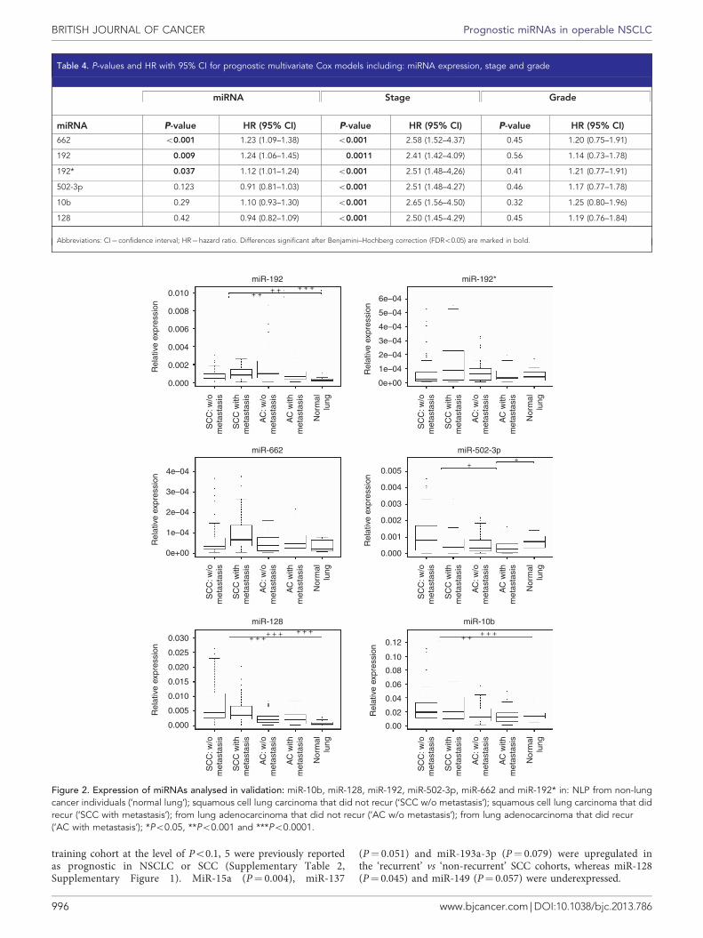
training cohort at the level of Po0.1, 5 were previously reportedas prognostic in NSCLC or SCC (Supplementary Table 2,Supplementary Figure 1). MiR-15a (P¼ 0.004), miR-137
(P¼ 0.051) and miR-193a-3p (P¼ 0.079) were upregulated inthe ‘recurrent’ vs ‘non-recurrent’ SCC cohorts, whereas miR-128(P¼ 0.045) and miR-149 (P¼ 0.057) were underexpressed.
Table 4. P-values and HR with 95% CI for prognostic multivariate Cox models including: miRNA expression, stage and grade
miRNA Stage Grade
miRNA P-value HR (95% CI) P-value HR (95% CI) P-value HR (95% CI)
662 o0.001 1.23 (1.09–1.38) o0.001 2.58 (1.52–4.37) 0.45 1.20 (0.75–1.91)
192 0.009 1.24 (1.06–1.45) 0.0011 2.41 (1.42–4.09) 0.56 1.14 (0.73–1.78)
192* 0.037 1.12 (1.01–1.24) o0.001 2.51 (1.48–4,26) 0.41 1.21 (0.77–1.91)
502-3p 0.123 0.91 (0.81–1.03) o0.001 2.51 (1.48–4.27) 0.46 1.17 (0.77–1.78)
10b 0.29 1.10 (0.93–1.30) o0.001 2.65 (1.56–4.50) 0.32 1.25 (0.80–1.96)
128 0.42 0.94 (0.82–1.09) o0.001 2.50 (1.45–4.29) 0.45 1.19 (0.76–1.84)
Abbreviations: CI¼ confidence interval; HR¼ hazard ratio. Differences significant after Benjamini–Hochberg correction (FDRo0.05) are marked in bold.
miR-192
0.010
0.008
0.006
Rel
ativ
e ex
pres
sion
0.004
0.002
0.000
+ + ++ ++ +
miR-192*
Rel
ativ
e ex
pres
sion
0e+00
1e–04
2e–04
3e–04
4e–04
0e+00
1e–04
2e–04
3e–04
4e–04
5e–04
6e–04
miR-128
0.030
0.025
0.020
Rel
ativ
e ex
pres
sion
0.015
0.010
0.005
0.000
SC
C: w
/om
etas
tasi
s
SC
C w
ithm
etas
tasi
s
AC
: w/o
met
asta
sis
AC
with
met
asta
sis
Nor
mal
lung
SC
C: w
/om
etas
tasi
s
SC
C w
ithm
etas
tasi
s
AC
: w/o
met
asta
sis
AC
with
met
asta
sis
Nor
mal
lung
SC
C: w
/om
etas
tasi
s
SC
C w
ithm
etas
tasi
s
AC
: w/o
met
asta
sis
AC
with
met
asta
sis
Nor
mal
lung
SC
C: w
/om
etas
tasi
s
SC
C w
ithm
etas
tasi
s
AC
: w/o
met
asta
sis
AC
with
met
asta
sis
Nor
mal
lung
SC
C: w
/om
etas
tasi
s
SC
C w
ithm
etas
tasi
s
AC
: w/o
met
asta
sis
AC
with
met
asta
sis
Nor
mal
lung
SC
C: w
/om
etas
tasi
s
SC
C w
ithm
etas
tasi
s
AC
: w/o
met
asta
sis
AC
with
met
asta
sis
Nor
mal
lung
+ + + + + ++ ++ + ++ + +
miR-10b
Rel
ativ
e ex
pres
sion
0.00
0.02
0.04
0.06
0.08
0.10
0.12
miR-662 miR-502-3p
++
Rel
ativ
e ex
pres
sion
Rel
ativ
e ex
pres
sion
0.000
0.001
0.002
0.003
0.004
0.005
Figure 2. Expression of miRNAs analysed in validation: miR-10b, miR-128, miR-192, miR-502-3p, miR-662 and miR-192* in: NLP from non-lungcancer individuals (‘normal lung’); squamous cell lung carcinoma that did not recur (‘SCC w/o metastasis’); squamous cell lung carcinoma that didrecur (‘SCC with metastasis’); from lung adenocarcinoma that did not recur (‘AC w/o metastasis’); from lung adenocarcinoma that did recur(‘AC with metastasis’); *Po0.05, **Po0.001 and ***Po0.0001.
BRITISH JOURNAL OF CANCER Prognostic miRNAs in operable NSCLC
996 www.bjcancer.com | DOI:10.1038/bjc.2013.786

miRNAs expression in SCC vs AC. To test for potentialdifferences in the miRNA expression between histopathologicalsubtypes of NSCLC, we compared between SCC and AC theexpression of six miRNAs selected for confirmatory analysis inSCC. Of those, expression of miR-128, miR-10b, miR-502-3p andmiR-192 was significantly different between SCC and AC (adjustedPo0.05; Figure 2).
miRNA expression in NSCLC vs NLP. Expression of miR-128and miR-192 differed significantly between NLP and SCC (adjustedPo0.006), irrespective of relapse status (Table 5, Figure 2).Expression of miR-662 and miR-10b was significantly differentbetween relapsed SCC and NLP (adjusted Po0.03) (SupplementaryTable 3). MiR-192 and miR-128 in NLP was differently expressedcompared with AC (adjusted Po0.001), irrespective ofrelapse status (Table 5, Figure 2). Expression of miR-502-3pwas significantly different between relapsed AC and NLP(adjusted P¼ 0.011), as well as between AC and NLP (adjustedP¼ 0.048) (Supplementary Table 3). There were no significantdifferences in expression between NLP from non-lung cancerindividuals and NLP adjacent to tumour from lung cancer patients(adjusted P40.05).
DISCUSSION
This is the first report on the prognostic significance of theexpression of miR-662, miR-192 and miR-192* in early stage SCC.Notably, the prognostic relevance of these miRNAs was confirmedin a sizeable independent cohort of patients. Our data also suggestthat these miRNAs are most likely not prognostic in lung AC.The study was devised in two steps: (i) relevant miRNAs wereidentified by screening of the expression of 677 miRNAs in thetraining cohort, and (ii) potentially relevant miRNAs weresubsequently assessed in the independent cohort of SCC, andadditionally determined in AC patients and in NLP. A number ofstringent methodological requirements were met to verify the studyhypothesis. First, we assumed that the loco-regional recurrences inthe bronchial stump, hilar or mediastinal lymph nodes and thechest wall most often occur stochastically, as a result of incompletetumour removal at submicroscopic level. Therefore, all cases withisolated loco-regional recurrences of NSCLC were excluded fromthe analyses on the premise that they likely occur irrespective of theprimary tumour potential for distant spread. Consequently, miRNAexpression was compared between the two groups of patients whoeither developed distant metastases or were free of recurrence afterlong-term follow-up. This approach has the potential to providethe clearest experimental setting for development of markers ofincreased risk of distant recurrence. To our knowledge, none
of the previously reported studies on prognostic gene signaturesmandated the exclusion of cases with the isolated local recurrences(Raponi et al, 2006; Guo et al, 2008; Raponi et al, 2009; Zhu et al,2010; Lu et al, 2012).
Second, as 85% of NSCLC recurrences become evident within 3years after resection (Pepek et al, 2011), the minimum follow-up of4 years was required for all patients considered as ‘controls’ toensure a reasonable probability that they were truly free of cancerdissemination. Relatively short follow-up of patients might haveconfounded the analyses in several previous studies. For example,Yu et al (2008) reported the censoring rate of 66%. Indeed, shortpostoperative observation renders a non-relapsed group inade-quate for prognostic analyses, as a patient without an evidentrelapse after, for example, 12 months is still at a relatively high riskof developing metastases in due course. Further, it was requiredthat the patients had not been administered induction or adjuvantchemotherapy, as we reasoned that its use might obfuscate theactual risk of tumour dissemination.
In this ‘proof of concept’ study, we identified and confirmed theprognostic significance of three new miRNAs in operable SCC.Owing to the differences in miRNA expression between the fresh-frozen and FFPE samples, we did not attempt to determine theoptimal cut-offs for both materials. The hazard ratios for theprognostic miRNAs were lower than those for the stage; however,the prognostic effect of each miRNA was independent of otherfactors including stage. The prognostic value of miRNAs in NSCLCmay likely be strengthened by their pooling into a multi-geneclassifier. However, we intentionally forwent here the developmentof multi-gene prognostic models, as their parameters (cut-offs,coefficients weighted for each miRNA in risk score equation etc.)tend to be unstable if developed in cohorts including fewer than250 cases (Michiels et al, 2005; van Vliet et al, 2008). Moreover, aformal validation of a multi-gene model necessitates a collection ofnumerous samples, preferably from many centres. This project iscurrently under way.
A relatively small training cohort (n¼ 50) is an apparentlimitation of our study, as it allows for reliable detection of only astrong HR, in the range of 44, with a power of 0.8 in the case of asingle hypothesis. Indeed, to detect a modest prognostic effect ofmiRNAs (e.g., HR¼ 2), more than 600 patients are needed in adiscovery set for high throughput scenario (4100 miRNAs tested).Thus, the results of the training phase in our study allowed only forranking miRNAs according to their potential prognostic relevance,which was subsequently verified in an independent sizeable cohortof patients. Also, the morphological heterogeneity of NSCLC, evenamong its major histological subtypes, is being increasinglyrecognised. Warth et al (2012) showed that almost 93% of ACsdisplayed at least two histological patterns according to the novelIASLC/ATS/ERS AC classification (Travis et al, 2011). Molecular
Table 5. Expression fold change and FDR: AC vs SCC, SCC vs NL parenchyma, AC vs NL parenchyma
miRNANormalised expressionfold change AC/SCC
SCC vs ACFDR
Normalised expressionfold change SCC/NL
SCC vs NLFDR
Normalised expressionfold change AC/NL
AC vs NLFDR
128 0.48 o0.001 8.04 o0.001 3.88 o0.001
10b 0.66 0.003 3.12 o0.001 2.04 0.063
192 4.00 0.003 1.19 0.005 4.75 o0.001
502-3p 0.55 0.016 1.69 0.865 0.94 0.048
662 0.54 0.3 2.02 0.053 1.09 0.326
192* 1.24 0.52 3.77 0.481 4.66 0.2
Abbreviations: AC¼ lung adenocarcinoma; NL¼ normal lung; SCC¼ squamous cell lung carcinoma. Differences significant after Benjamini–Hochberg correction (FDRo0.05) are markedin bold.
Prognostic miRNAs in operable NSCLC BRITISH JOURNAL OF CANCER
www.bjcancer.com | DOI:10.1038/bjc.2013.786 997

heterogeneity of SCC was recently reported as a part of the cancermutational atlas (Network CGAR, 2012). Also, four mRNAexpression-based classes of SCC were previously reported, where‘primitive’ subtype conferred poorer prognosis in reference to‘classical’, ‘secretory’ and ‘basal’ SCC subtypes (Wilkerson et al,2010). The prognostic role of those molecular features has not yetbeen validated and therefore they were not included in the currentanalysis.
None of the prognostically relevant miRNAs from our series hasbeen previously reported in SCC specific analyses. However, highexpression of miR-192* was recently found to correlate withshorter relapse-free survival in the training cohort of 357 NSCLCpatients, but not in separate analyses including AC (n¼ 189) orSCC (n¼ 106) (Lu et al, 2012). In gastric cancer, miR-192 wasshown to be overexpressed in vivo and exerted cell growth andmigration-promoting effects in vitro (Jin et al, 2011). Highexpression of miR-192 was also found in pancreatic cancer stemcells (Jung et al, 2011). This miRNA is encoded on chromosome 11in the vicinity of a gene for CDC42 binding protein kinase gamma(DMPK-like) that has been implicated as a marker of aggressivecourse of NSCLC. In the study by Lu et al (2012), the lowexpression of miR-662 correlated with shorter survival in AC, butnot in SCC and in the entire population of NSCLC. In the currentstudy, however, the high expression of miR-662 strongly correlatedwith short DMFS in SCC in both training and confirmatorycohorts. MiR-662 is encoded on chromosome 16 in proximity ofmesothelin (MSLN). Overexpression of mesothelin was reported inbreast cancer metastases (Wu et al, 2008) and was found topromote anchorage-independent growth of breast cancer cells(Uehara et al, 2008). In paediatric cancers, the expression of 63% ofmiRNAs located inside coding genes was significantly correlatedwith that of their host genes (Wei et al, 2009). Further research iswarranted to define the prognostic role of the genes potentiallyco-expressed with the reported miRNAs.
The computationally predicted mRNA targets of miRNAsassociated with an elevated metastatic potential of SCC werescrutinised in an attempt to find their functional correlates. Theoverexpression of miR-662, miR-192* or miR-192 is expected toresult in the downregulation of their mRNA targets. Along theselines, overexpression of miR-662 may provide an alternativemechanism of downregulation of a tumour suppressor gene GDF10that belongs to TGF-b family ligands. Intriguingly, miR-662 alsotargets the NLRC5 that was recently identified as a transactivationfactor for MHC I complexes, crucial for adaptive immune responseagainst viruses and malignancy (Meissner et al, 2010).
Among the top targets for miR-192* is RAB21, a GTP-bindingproteins that regulates intracellular trafficking of integrins, aprocess fundamental for cell adhesion and motility. Another targetpotentially downregulated by miR-192* is TSHZ3, a newlydescribed candidate tumour suppressor gene for breast andprostate cancers (Yamamoto et al, 2011). MiR-192 and miR-192*simultaneously target 13 mRNA, including zinc finger proteinsZEB2 and integrin, alpha V (vitronectin receptor, alpha polypeptide,antigen CD51).
We confirmed a potential prognostic role of some previouslyreported miRNAs. For example, the adverse effect of the highexpression of miR-15a, reported by Raponi et al (2009), miR-137,reported by Yu et al (2008), and miR-193-3p, reported by Lu et al(2012) was also significant or borderline in our training cohort(Cox analysis; P¼ 0.004, P¼ 0.051 and P¼ 0.079, respectively).Notably, Lu et al (2012) reported an inverse correlation betweenexpression of miR-15a and the risk of progression. The prognosticimpact of miR-128 and miR-149 reported by these authors wasconfirmed in the current study, yet the overlap with otherprognostic miRNAs from previous studies in NSCLC or SCCwas limited. These discrepancies may stem from the differences inmethodology used by different research groups and, in particular,
normalisation procedures. In the current study, we used the meanof the expression of U6 and RNU48.
Importantly, miRNAs predictive of the dissemination ofearly stage SCC did not predict for distant recurrence in AC.It may be therefore envisaged along the lines of previousreports (Landi et al, 2010; Lu et al, 2012) that SCC and ACemploy different molecular pathways during progression. More-over, the expression of two miRNAs prognostic for SCC (miR-662and miR-192*) was significantly different between SCC and AC.This finding additionally underscores the need for separateinvestigations of the prognostic effect of quantitative markers inthese two major NSCLC subtypes. The differential expression ofmiR-128 and miR-502-3p between SCC and AC is consistent withthe results of a previous study (Lu et al, 2012), whereas thedifferences in expression of miR-10b and miR-192 were notpreviously reported. Finally, expression of two prognostic miRNAs(miR-192 and miR-662) was significantly different in SCCcompared with NLP. Such differences render these miRNAsparticularly suited as constituents of clinically useful prognosticsignatures.
In conclusion, we identified and confirmed three novel miRNAsthat are predictive of dissemination in operable SCC and mayconstitute the components of multi miRNAs signatures. Ifcorroborated in further studies, these findings may be relevant toclinical practice, as they could facilitate identification of SCCpatients for adjuvant therapies.
ACKNOWLEDGEMENTS
We would like to thank Dr David Gandara for reviewing themanuscript and for valuable comments. The study was supportedby the Polish Ministry of Science grant no. N N403 210139.
CONFLICT OF INTEREST
Jacek Jassem and Marcin Skrzypski disclose the option agreementwith Novartis Pharma A.G. regarding the prognostic test in non-small cell lung cancer based on microRNA expression. Theremaining authors declare no conflict of interest.
REFERENCES
Aviel-Ronen S, Blackhall FH, Shepherd FA, Tsao MS (2006) K-ras mutationsin non-small-cell lung carcinoma: a review. Clin Lung Cancer 8(1): 30–38.
Bepler G, Olaussen KA, Vataire AL, Soria JC, Zheng Z, Dunant A, Pignon JP,Schell MJ, Fouret P, Pirker R, Filipits M, Brambilla E (2011) ERCC1 andRRM1 in the international adjuvant lung trial by automated quantitativein situ analysis. Am J Pathol 178(1): 69–78.
Calin GA, Croce CM (2006) MicroRNA-cancer connection: the beginning of anew tale. Cancer Res 66(15): 7390–7394.
Chee KG, Nguyen DV, Brown M, Gandara DR, Wun T, Lara PN (2008)Positron emission tomography and improved survival in patients withlung cancer: the Will Rogers phenomenon revisited. Arch Intern Med168(14): 1541–1549.
Friboulet L, Olaussen KA, Pignon JP, Shepherd FA, Tsao MS, Graziano S,Kratzke R, Douillard JY, Seymour L, Pirker R, Filipits M, Andre F,Solary E, Ponsonnailles F, Robin A, Stoclin A, Dorvault N, Commo F,Adam J, Vanhecke E, Saulnier P, Thomale J, Le Chevalier T, Dunant A,Rousseau V, Le Teuff G, Brambilla E, Soria JC (2013) ERCC1 isoformexpression and DNA repair in non-small-cell lung cancer. N Engl J Med368(12): 1101–1110.
Guo N, Wan Y, Tosun K, Lin H, Msiska Z, Flynn D, Remick S, Vallyathan V,Dowlati A, Shi X, Castranova V, Beer D, Qian Y (2008) Confirmation of
BRITISH JOURNAL OF CANCER Prognostic miRNAs in operable NSCLC
998 www.bjcancer.com | DOI:10.1038/bjc.2013.786

gene expression-based prediction of survival in non-small cell lung cancer.Clin Cancer Res 14(24): 8213–8220.
Hall JS, Taylor J, Valentine HR, Irlam JJ, Eustace A, Hoskin PJ, Miller CJ,West CM (2012) Enhanced stability of microRNA expression facilitatesclassification of FFPE tumour samples exhibiting near total mRNAdegradation. Br J Cancer 107(4): 684–694.
Huncharek M, Kupelnick B, Geschwind JF, Caubet JF (2000) Prognosticsignificance of p53 mutations in non-small cell lung cancer: a meta-analysis of 829 cases from eight published studies. Cancer Lett 153(1–2):219–226.
Jin Z, Selaru FM, Cheng Y, Kan T, Agarwal R, Mori Y, Olaru AV, Yang J,David S, Hamilton JP, Abraham JM, Harmon J, Duncan M, Montgomery EA,Meltzer SJ (2011) MicroRNA-192 and -215 are upregulated in humangastric cancer in vivo and suppress ALCAM expression in vitro.Oncogene 30(13): 1577–1585.
Jung DE, Wen J, Oh T, Song SY (2011) Differentially expressed microRNAs inpancreatic cancer stem cells. Pancreas 40(8): 1180–1187.
Kratz JR, He J, Van Den Eeden SK, Zhu ZH, Gao W, Pham PT,Mulvihill MS, Ziaei F, Zhang H, Su B, Zhi X, Quesenberry CP, Habel LA,Deng Q, Wang Z, Zhou J, Li H, Huang MC, Yeh CC, Segal MR, Ray MR,Jones KD, Raz DJ, Xu Z, Jahan TM, Berryman D, He B, Mann MJ,Jablons DM (2012) A practical molecular assay to predict survival inresected non-squamous, non-small-cell lung cancer: development andinternational validation studies. Lancet 379(9818): 823–832.
Landi M, Zhao Y, Rotunno M, Koshiol J, Liu H, Bergen A, Rubagotti M,Goldstein A, Linnoila I, Marincola F, Tucker M, Bertazzi P, Pesatori A,Caporaso N, McShane L, Wang E (2010) MicroRNA expressiondifferentiates histology and predicts survival of lung cancer. Clin Cancer Res16(2): 430–441.
Livak K, Schmittgen T (2001) Analysis of relative gene expression datausing real-time quantitative PCR and the 2(-Delta Delta C(T)) Method.Methods 25(4): 402–408.
Lu Y, Govindan R, Wang L, Liu PY, Goodgame B, Wen W, Sezhiyan A,Pfeifer J, Li YF, Hua X, Wang Y, Yang P, You M (2012) MicroRNAprofiling and prediction of recurrence/relapse-free survival in stage Ilung cancer. Carcinogenesis 33(5): 1046–1054.
McShane LM, Altman DG, Sauerbrei W, Taube SE, Gion M, Clark GM.Statistics Subcommittee of the NCI-EORTC Working Group on CancerDiagnostics (2005) REporting recommendations for tumour MARKerprognostic studies (REMARK). Br J Cancer 93(4): 387–391.
Meissner TB, Li A, Biswas A, Lee KH, Liu YJ, Bayir E, Iliopoulos D,van den Elsen PJ, Kobayashi KS (2010) NLR family member NLRC5 is atranscriptional regulator of MHC class I genes. Proc Natl Acad Sci USA107(31): 13794–13799.
Michiels S, Koscielny S, Hill C (2005) Prediction of cancer outcome withmicroarrays: a multiple random validation strategy. Lancet 365(9458):488–492.
Network CGAR (2012) Comprehensive genomic characterization ofsquamous cell lung cancers. Nature 489(7417): 519–525.
Pepek JM, Chino JP, Marks LB, Damico TA, Yoo DS, Onaitis MW, Ready NE,Hubbs JL, Boyd J, Kelsey CR (2011) How well does the new lung cancerstaging system predict for local/regional recurrence after surgery?:a comparison of the TNM 6 and 7 systems. J Thorac Oncol 489(7417):519–525.
Pignon J, Tribodet H, Scagliotti G, Douillard J, Shepherd F, Stephens R,Dunant A, Torri V, Rosell R, Seymour L, Spiro S, Rolland E, Fossati R,Aubert D, Ding K, Waller D, Le Chevalier T, Group LC (2008) Lungadjuvant cisplatin evaluation: a pooled analysis by the LACE CollaborativeGroup. J Clin Oncol 26(21): 3552–3559.
Raponi M, Dossey L, Jatkoe T, Wu X, Chen G, Fan H, Beer D (2009)MicroRNA classifiers for predicting prognosis of squamous cell lungcancer. Cancer Res 69(14): 5776–5783.
Raponi M, Zhang Y, Yu J, Chen G, Lee G, Taylor JM, Macdonald J, Thomas D,Moskaluk C, Wang Y, Beer DG (2006) Gene expression signatures forpredicting prognosis of squamous cell and adenocarcinomas of the lung.Cancer Res 66(15): 7466–7472.
Schmittgen T, Livak K (2008) Analyzing real-time PCR data by thecomparative C(T) method. Nat Protoc 3(6): 1101–1108.
Siegel R, Naishadham D, Jemal A (2013) Cancer statistics, 2013.CA Cancer J Clin 63(1): 11–30.
Skrzypski M, Dziadziuszko R, Jassem E, Szymanowska-Narloch A, Gulida G,Rzepko R, Biernat W, Taron M, Jelitto-Gorska M, Marjanski T, Rzyman W,Rosell R, Jassem J (2013) Main histologic types of non-small-cell lung
cancer differ in expression of prognosis-related genes. Clin Lung Cancer14(6): 666–673.e2.
Sobin LH, Wittekind C (2002) TNM Classification of Malignant Tumours.6th edn. John Wiley & Sons: Hoboken, NJ, USA.
Takamizawa J, Konishi H, Yanagisawa K, Tomida S, Osada H, Endoh H,Harano T, Yatabe Y, Nagino M, Nimura Y, Mitsudomi T, Takahashi T(2004) Reduced expression of the let-7 microRNAs in human lung cancersin association with shortened postoperative survival. Cancer Res 64(11):3753–3756.
Travis WD, Brambilla E, Muller-Hermelink HK, Harris CC (eds) (2004)World Health Organization Classification of Tumors. Pathology andGenetics. Tumours of the Lung, Pleura, Thymus and Heart. IARC Press:Lyon, France.
Travis WD, Brambilla E, Noguchi M, Nicholson AG, Geisinger KR,Yatabe Y, Beer DG, Powell CA, Riely GJ, Van Schil PE, Garg K,Austin JH, Asamura H, Rusch VW, Hirsch FR, Scagliotti G,Mitsudomi T, Huber RM, Ishikawa Y, Jett J, Sanchez-Cespedes M,Sculier JP, Takahashi T, Tsuboi M, Vansteenkiste J, Wistuba I, Yang PC,Aberle D, Brambilla C, Flieder D, Franklin W, Gazdar A, Gould M,Hasleton P, Henderson D, Johnson B, Johnson D, Kerr K, Kuriyama K,Lee JS, Miller VA, Petersen I, Roggli V, Rosell R, Saijo N, Thunnissen E,Tsao M, Yankelewitz D (2011) International association for the study oflung cancer/american thoracic society/european respiratory societyinternational multidisciplinary classification of lung adenocarcinoma. JThorac Oncol 6(2): 244–285.
Uehara N, Matsuoka Y, Tsubura A (2008) Mesothelin promotes anchorage-independent growth and prevents anoikis via extracellular signal-regulatedkinase signaling pathway in human breast cancer cells. Mol Cancer Res6(2): 186–193.
van Vliet MH, Reyal F, Horlings HM, van de Vijver MJ, Reinders MJ,Wessels LF (2008) Pooling breast cancer datasets has a synergeticeffect on classification performance and improves signature stability.BMC Genomics 9: 375.
Warth A, Muley T, Meister M, Stenzinger A, Thomas M, Schirmacher P,Schnabel PA, Budczies J, Hoffmann H, Weichert W (2012) The novelhistologic International Association for the Study of Lung Cancer/American Thoracic Society/European Respiratory Society classificationsystem of lung adenocarcinoma is a stage-independent predictor ofsurvival. J Clin Oncol 30(13): 1438–1446.
Wei JS, Johansson P, Chen QR, Song YK, Durinck S, Wen X, Cheuk AT,Smith MA, Houghton P, Morton C, Khan J (2009) microRNA profilingidentifies cancer-specific and prognostic signatures in pediatricmalignancies. Clin Cancer Res 15(17): 5560–5568.
Wilkerson MD, Yin X, Hoadley KA, Liu Y, Hayward MC, Cabanski CR,Muldrew K, Miller CR, Randell SH, Socinski MA, Parsons AM,Funkhouser WK, Lee CB, Roberts PJ, Thorne L, Bernard PS, Perou CM,Hayes DN (2010) Lung squamous cell carcinoma mRNA expressionsubtypes are reproducible, clinically important, and correspond to normalcell types. Clin Cancer Res 16(19): 4864–4875.
Winton T, Livingston R, Johnson D, Rigas J, Johnston M, Butts C, Cormier Y,Goss G, Inculet R, Vallieres E, Fry W, Bethune D, Ayoub J, Ding K,Seymour L, Graham B, Tsao MS, Gandara D, Kesler K, Demmy T,Shepherd F. National Cancer Institute of Canada Clinical Trials Group,National Cancer Institute of the United States Intergroup JBR.10 TrialInvestigators (2005) Vinorelbine plus cisplatin vs. observation in resectednon-small-cell lung cancer. N Engl J Med 352(25): 2589–2597.
Wu JM, Fackler MJ, Halushka MK, Molavi DW, Taylor ME, Teo WW,Griffin C, Fetting J, Davidson NE, De Marzo AM, Hicks JL, Chitale D,Ladanyi M, Sukumar S, Argani P (2008) Heterogeneity of breast cancermetastases: comparison of therapeutic target expression and promotermethylation between primary tumors and their multifocal metastases.Clin Cancer Res 14(7): 1938–1946.
Xie Y, Xiao G, Coombes KR, Behrens C, Solis LM, Raso G, Girard L, Erickson HS,Roth J, Heymach JV, Moran C, Danenberg K, Minna JD, Wistuba II(2011) Robust gene expression signature from formalin-fixedparaffin-embedded samples predicts prognosis of non-small-cell lungcancer patients. Clin Cancer Res 17(17): 5705–5714.
Yamamoto M, Cid E, Bru S, Yamamoto F (2011) Rare and frequent promotermethylation, respectively, of TSHZ2 and 3 genes that are bothdownregulated in expression in breast and prostate cancers. PLoS One6(3): e17149.
Ying SY, Lin SL (2009) Intron-mediated RNA interference and microRNAbiogenesis. Methods Mol Biol 487: 387–413.
Prognostic miRNAs in operable NSCLC BRITISH JOURNAL OF CANCER
www.bjcancer.com | DOI:10.1038/bjc.2013.786 999

Yu S, Chen H, Chang G, Chen C, Chen H, Singh S, Cheng C, Yu C,Lee Y, Chen H, Su T, Chiang C, Li H, Hong Q, Su H, Chen C,Chen W, Liu C, Chan W, Li K, Chen J, Yang P (2008) MicroRNAsignature predicts survival and relapse in lung cancer. Cancer Cell 13(1):48–57.
Zhu C, Ding K, Strumpf D, Weir B, Meyerson M, Pennell N, Thomas R,Naoki K, Ladd-Acosta C, Liu N, Pintilie M, Der S, Seymour L, Jurisica I,Shepherd F, Tsao M (2010) Prognostic and predictive gene signature for
adjuvant chemotherapy in resected non-small-cell lung cancer. J Clin Oncol28(29): 4417–4424.
This work is published under the standard license to publish agree-ment. After 12 months the work will become freely available andthe license terms will switch to a Creative Commons Attribution-NonCommercial-Share Alike 3.0 Unported License.
Supplementary Information accompanies this paper on British Journal of Cancer website (http://www.nature.com/bjc)
BRITISH JOURNAL OF CANCER Prognostic miRNAs in operable NSCLC
1000 www.bjcancer.com | DOI:10.1038/bjc.2013.786