Production of tissue-engineered skin and oral mucosa for clinical and experimental use
25
129 John W. Haycock (ed.), 3D Cell Culture: Methods and Protocols, Methods in Molecular Biology, vol. 695, DOI 10.1007/978-1-60761-984-0_9, © Springer Science+Business Media, LLC 2011 Chapter 9 Production of Tissue-Engineered Skin and Oral Mucosa for Clinical and Experimental Use Sheila MacNeil, Joanna Shepherd, and Louise Smith Abstract Since the early 1990s, our understanding of how epithelial and stromal cells interact in 3D tissue-engineered constructs has led to tissue-engineered skin and oral mucosa models, which are beginning to deliver benefit in the clinic (usually in small-scale reconstructive surgery procedures) but have a great deal to offer for in vitro investigations. These 3D tissue-engineered models can be used for a wide variety of purposes such as dermato- and mucotoxicity, wound healing, examination of pigmentation and mela- noma biology, and in particular, a recent development from this laboratory, as a model of bacterially infected skin. Models can also be used to investigate specific skin disease processes. In this chapter, we describe the basic methodology for producing 3D tissue-engineered skin and oral mucosa based on de-epidermised acellular human dermis, and we give examples of how these models can be used for a variety of applications. Key words: Tissue-engineered skin, Tissue-engineered oral mucosa, Pigmentation, Melanoma invasion, Infected skin, Wound healing Cultured autologous skin cells have been used to benefit patients with severe burns since the early 1980s, so the production of cul- tured cells and tissue-engineered skin is probably the most mature of all of the tissue-engineered tissues (recently reviewed in (1, 2)). In this chapter, we focus on producing epidermal dermal tissue- engineered skin and oral mucosa based on de-epidermised acellular human dermis. These tissue-engineered constructs can be used in the clinic, and here, issues of sterilisation and the use of accredited clean rooms must be tackled. However, the majority of the chapter looks at the production of these physiologically relevant tissue- engineered skin and oral mucosa models for in vitro research. 1. Introduction
Transcript of Production of tissue-engineered skin and oral mucosa for clinical and experimental use
129
John W. Haycock (ed.), 3D Cell Culture: Methods and Protocols, Methods in Molecular Biology, vol. 695,DOI 10.1007/978-1-60761-984-0_9, © Springer Science+Business Media, LLC 2011
Chapter 9
Production of Tissue-Engineered Skin and Oral Mucosa for Clinical and Experimental Use
Sheila MacNeil, Joanna Shepherd, and Louise Smith
Abstract
Since the early 1990s, our understanding of how epithelial and stromal cells interact in 3D tissue-engineered constructs has led to tissue-engineered skin and oral mucosa models, which are beginning to deliver benefit in the clinic (usually in small-scale reconstructive surgery procedures) but have a great deal to offer for in vitro investigations. These 3D tissue-engineered models can be used for a wide variety of purposes such as dermato- and mucotoxicity, wound healing, examination of pigmentation and mela-noma biology, and in particular, a recent development from this laboratory, as a model of bacterially infected skin. Models can also be used to investigate specific skin disease processes. In this chapter, we describe the basic methodology for producing 3D tissue-engineered skin and oral mucosa based on de-epidermised acellular human dermis, and we give examples of how these models can be used for a variety of applications.
Key words: Tissue-engineered skin, Tissue-engineered oral mucosa, Pigmentation, Melanoma invasion, Infected skin, Wound healing
Cultured autologous skin cells have been used to benefit patients with severe burns since the early 1980s, so the production of cul-tured cells and tissue-engineered skin is probably the most mature of all of the tissue-engineered tissues (recently reviewed in (1, 2)). In this chapter, we focus on producing epidermal dermal tissue-engineered skin and oral mucosa based on de-epidermised acellular human dermis. These tissue-engineered constructs can be used in the clinic, and here, issues of sterilisation and the use of accredited clean rooms must be tackled. However, the majority of the chapter looks at the production of these physiologically relevant tissue-engineered skin and oral mucosa models for in vitro research.
1. Introduction
130 MacNeil, Shepherd, and Smith
There are an increasing number of areas where these 3D physiologically relevant tissue-engineered models come into their own. The examples described in this chapter are the use of tissue-engineered skin to study wound healing, skin graft contracture, bacterial infection of skin, studies of normal skin cell pigmentation and of the pathology of how transformed melanocytes – melanoma cells – interact with skin. For oral mucosa, we describe its produc-tion and how this has been used clinically and indicate in brief how it can be used for a wide range of experimental studies.
A major concern when grafting back split-thickness skin (STS) or oral mucosa is the failure to achieve rapid neovascularisation. From our experience and that of others in the area, it is necessary to make sure that the tissue-engineered grafts are not too thick and are grafted onto well-vascularised wound beds. Under these circumstances, neovascularisation will occur within about 5–7 days, and the graft will survive. Grafting tissue-engineered skin onto a poorly vascularised wound bed has resulted in delayed angiogenesis and loss of grafts in our experience (3). Tissue-engineered skin or oral mucosa which becomes vascularised will generally survive long term (see ref. 4).
There are many applications for tissue-engineered skin and oral mucosa. This is a rapidly growing area. There is a need to reduce or replace animal experimentation in the development of products that are used on human skin and hair, but equally many of the more interesting things that happen in skin happen through a dia-logue between different skin cells and are rarely seen when indi-vidual cells are studied in monolayers. Table 1 lists some of the in vivo and in vitro applications of tissue-engineered skin. With respect to clinical applications, the clinical applications for epider-mal–dermal reconstructed skin and oral mucosa are still relatively in their infancy (compared to the expansion and clinical use of cul-tured keratinocytes or fibroblasts on their own). As it takes a cer-tain amount of time to produce these tissue-engineered constructs, they are entering the clinic for small-scale reconstructive surgery applications. Thus, although theoretically tissue-engineered skin could be used to replace full thickness skin loss in burn patients, in practice, the time taken to produce it (at least 6 weeks in our labo-ratories) takes it outside of the acute window (within 3 weeks) in which one would like to graft patients with extensive skin loss.
However, there are many reconstructive surgery applications where tissue-engineered skin and oral mucosa will be useful as listed in Table 1. With respect to in vitro applications, the table lists the ones that we are aware of. There will be many others since this area of research is rapidly expanding.
Thus, we have a basic recipe for the production of 3D tissue-engineered skin and oral mucosa which can be flexibly adapted for clinical use and for a wide range of investigations of skin and mucosa biology.
131Production of Tissue-Engineered Skin and Oral Mucosa for Clinical and Experimental Use
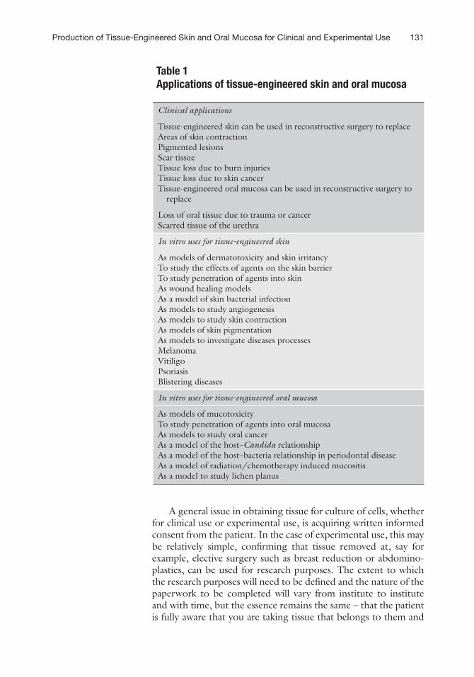
Table 1 Applications of tissue-engineered skin and oral mucosa
Clinical applications
Tissue-engineered skin can be used in reconstructive surgery to replaceAreas of skin contractionPigmented lesionsScar tissueTissue loss due to burn injuriesTissue loss due to skin cancerTissue-engineered oral mucosa can be used in reconstructive surgery to
replace
Loss of oral tissue due to trauma or cancerScarred tissue of the urethra
In vitro uses for tissue-engineered skin
As models of dermatotoxicity and skin irritancyTo study the effects of agents on the skin barrierTo study penetration of agents into skinAs wound healing modelsAs a model of skin bacterial infectionAs models to study angiogenesisAs models to study skin contractionAs models of skin pigmentationAs models to investigate diseases processesMelanomaVitiligoPsoriasisBlistering diseases
In vitro uses for tissue-engineered oral mucosa
As models of mucotoxicityTo study penetration of agents into oral mucosaAs models to study oral cancerAs a model of the host–Candida relationshipAs a model of the host–bacteria relationship in periodontal diseaseAs a model of radiation/chemotherapy induced mucositisAs a model to study lichen planus
A general issue in obtaining tissue for culture of cells, whether for clinical use or experimental use, is acquiring written informed consent from the patient. In the case of experimental use, this may be relatively simple, confirming that tissue removed at, say for example, elective surgery such as breast reduction or abdomino-plasties, can be used for research purposes. The extent to which the research purposes will need to be defined and the nature of the paperwork to be completed will vary from institute to institute and with time, but the essence remains the same – that the patient is fully aware that you are taking tissue that belongs to them and
132 MacNeil, Shepherd, and Smith
that they have consented freely that you may do this. Implicit in this is the understanding that you do not use it for other purposes and that it is acknowledged as a gift. The ethical consent proce-dure may exclude use of donated tissue for commercial purposes for example, but this may vary from place to place.
In culturing cells for clinical use, there will be a more exten-sive review process in which the protocol will have been reviewed by an ethical committee so that, for example, patients will donate a biopsy of skin to be used in their treatment or less commonly in the treatment of other patients. Again, the essence of informed consent is that the patient has a full understanding of the inten-tions of the researcher and that tissue is freely given for these purposes. Signed documentation is kept within the patient’s notes.
We begin by looking at the basic culture of cutaneous and oral keratinocytes and fibroblasts, then the preparation of the acellular de-epidermised dermis (DED), including sterilisation of this where it is required for clinical use, prior to describing the production of tissue-engineered skin and oral mucosa. We then describe ways in which the basic model can be modified to inves-tigate wound healing, pigmentation, melanoma invasion, and wound infection.
1. Fibroblast culture medium: DMEM high glucose (4,500 mg/L glucose), 10% v/v foetal calf serum (FCS), 2 mM l-glutamine, 0.625 mg/mL amphotericin B, 100 IU/mL penicillin, and 100 mg/mL streptomycin.
2. 3T3 Fibroblast culture medium: DMEM high glucose (4,500 mg/L glucose), 10% v/v newborn calf serum, 2 mM l-glutamine, 0.625 mg/mL amphotericin B, 100 IU/mL penicillin, and 100 mg/mL streptomycin.
3. Green’s medium: DMEM high glucose (4,500 mg/L glu-cose) and Ham’s F12 medium in a 3:1 ratio, 10% v/v FCS (UK), 10 ng/mL human recombinant epidermal growth factor, 0.4 mg/mL hydrocortisone, 10−10 M cholera toxin, 18 mM adenine, 5 mg/mL insulin, 5 mg/mL apo-transferrin, 20 mM 3,3,5-tri-idothyronine, 2 mM glutamine, 0.625 mg/mL amphotericin B, 100 IU/mL penicillin, and 1,000 mg/mL streptomycin.
4. To make all culture media, the ingredients are mixed at room temperature in a class II laminar flow hood to make a total volume of 500 mL. Medium is stored at <4°C for a maximum of 6 weeks prior to use. Warm an aliquot of medium to 37°C before use.
2. Materials
2.1. Medium for the Production of Tissue-Engineered Skin
133Production of Tissue-Engineered Skin and Oral Mucosa for Clinical and Experimental Use
1. Cryopreservation medium: 1 mL dimethyl sulphoxide (DMSO) is added to 9 mL of FCS to produce a 10% solution of DMSO in FCS. This solution is made up fresh each time it is needed.
2. “Difco-Trypsin”: 0.5 g of Difco Trypsin powder, 0.5 g of D-glucose, and 0.5 mL of phenol red to 500 mL of PBS. Adjust to pH 7.4 using 2 M NaOH and filter-sterilise. Store in 10-mL aliquots at −20°C until needed.
3. Collagenase A: 0.05% (w/v) Solution of collagenase A powder in serum-free fibroblast culture medium. Filter-sterilise this solution and store in 10-mL aliquots at −20°C until needed.
4. Skin collection solution: 500 mL of sterile phosphate-buffered saline, 0.625 mg/mL amphotericin B, 100 IU/mL penicillin, and 1,000 mg/mL streptomycin.
1. Our biopsies come from two sources: skin removed at breast reductions and abdominoplasties where there is no shortage of tissue, or smaller pieces of skin often obtained from the trimmed edges of skin grafts (3) in the treatment of major burn patients or from STS biopsies (usually of around 2 cm2) taken under local anaesthetic for the expansion of keratinocytes for the treatment of patients with non-healing ulcers (5).
2. The smaller the biopsy, the longer it will take to get a large expansion of keratinocytes, but the processing of the biopsies remain the same.
3. In making tissue-engineered skin for clinical use, we usually allow 6 weeks between obtaining the initial biopsy and having the material ready to graft (3).
4. When producing tissue-engineered oral mucosa for grafting, where the cells grow much more rapidly, the grafts can be produced within 3 weeks (see refs. 4, 6).
1. When the skin biopsy is transported to the lab, it travels in skin collection solution in a sealed container, enters the labo-ratory, and is stored in a dedicated refrigerator until use.
2. Ideally, cells are cultured from skin and oral mucosa biopsies within 48 h. However, viable cells can be isolated from skin biopsies up to 4 days after excision.
We next describe the methods for isolation of skin cells. In all cases, the fastest and most reliable expansion of epithelial cells occurs when these are cultured on a growth-arrested layer of 3T3 murine fibroblasts; hence, culture of 3T3s is also described.
2.2. Miscellaneous Solutions
3. Methods
3.1. Sourcing of Skin and Oral Mucosa Samples
3.2. Processing of Skin or Oral Mucosa for Cell Isolation
134 MacNeil, Shepherd, and Smith
1. Using sterile forceps and a sterile #22 scalpel cut STS into thin pieces (approximately 0.5 cm × 1 cm) (see Note 1).
2. Place the cut skin in 10 mL of “Difco-Trypsin” and incubate overnight at 4°C (typically 12–18 h).
3. Using sterile forceps check whether the epidermis is easily coming away from the dermis. If not, place the skin back in the fridge. Keep checking until the epidermis comes away easily.
4. Stop enzymatic activity by adding 5 mL of FCS. 5. Place the skin strips (epidermis uppermost) into a Petri dish
containing Green’s medium. 6. Using sterile forceps gently separate the epidermis from the
dermis. 7. Try to place the epidermis so that the two newly exposed
surfaces are uppermost. 8. Using a scalpel gently scrape the two newly exposed surfaces
(i.e. the top of the dermis and the bottom of the epidermis. This is where the more proliferative keratinocytes are. Melanocytes are also found here).
9. Place the resulting cell suspension into a 25-mL centrifuge tube containing 5 mL of Green’s medium.
10. Centrifuge the cells at 200 × g for 5 min, remove the superna-tant, break the pellet, and then re-suspend the cells in Green’s medium. At this point, try to remove any large pieces of epidermis that have been carried over.
11. Perform a cell count using Trypan blue to highlight non-viable cells.
12. Seed cells at a density of »4 × 10⁶ in 10–13 mL of Green’s medium in T75 flasks (seeded approximately 1 h earlier with 1 × 10⁶ iT3T fibroblast cells). For production of i3T3 cells, see Subheading 3.4 below.
13. Culture cells at 37°C, 5% CO2 in a humidified atmosphere. 14. Change the medium after 24 h and then every 2–3 days. The cells
will generally reach 70–80% confluency in 5–7 days.
1. Remove the medium and gently wash the cells with PBS. 2. To remove the i3T3 cells, incubate with 5 mL of 0.02% w/v
EDTA at 37°C. 3. Examine by phase contrast microscopy every 5 min and gently
tap to encourage fibroblast detachment, ensuring that the keratinocytes are still attached.
4. Remove the i3T3 containing EDTA solution and rinse again with PBS.
5. Add 2 mL of Trypsin/EDTA to the flask and incubate at 37°C.
3.3. Keratinocyte and Fibroblast Isolation
3.3.1. Keratinocyte Isolation
3.3.2. Keratinocyte Sub-culture
135Production of Tissue-Engineered Skin and Oral Mucosa for Clinical and Experimental Use
6. Encourage keratinocyte detachment by gentle tapping and confirm by phase microscopy after 5 min. If the cells have not detached after 5 min, place the flask back into the incubator and check again after another 5 min. Continue this until the cells have detached.
7. Add the cell suspension to 10 mL of Green’s medium to neu-tralise the trypsin and centrifuge at 200 × g for 5 min.
8. Remove the supernatant, break the pellet (gentle tapping works well), and re-suspend the cells in a known volume of Green’s medium and count the cells prior to use, again using Trypan blue.
9. If sub-culturing for expansion, add ~1 × 10⁶ cells to a T75 flask seeded approximately 1 h earlier with 1 × 10⁶ i3T3 cells.
10. Use primary keratinocytes between passages 1 and 3.
1. Take the dermis left over from the keratinocyte isolation step and using sterile forceps and a sterile #22 scalpel mince the dermis (into pieces approximately 1 mm× 1 mm).
2. Place this mince into a Petri dish containing collagenase A solution.
3. Incubate this solution at 37°C overnight. 4. Add 10 mL of fibroblast culture medium to the Petri dish to
stop the action of the collagenase. 5. Place the resulting cell suspension, minus any lumps of undi-
gested dermis, into a universal. 6. Spin down the cell solution at 400 × g for 10 min. 7. Remove the supernatant (be careful not to lose the pellet –
the supernatant is extremely viscous). Break the pellet and re-suspend the cells in fibroblast culture medium. Ideally, add a mycoplasma removal agent.
8. Seed the cells into a T25 flask containing fibroblast culture medium and incubate at 37°C, 5% CO2 in a humidified atmosphere.
9. Change the medium after 24 h, then every 3–4 days until the cells are 80% confluent (see Note 2).
1. Remove the medium and gently wash the cells with PBS. 2. Add 2 mL of Trypsin/EDTA to the flask and incubate at
37°C. 3. Encourage cell detachment by gentle tapping and confirm by
phase microscopy after 5 min. If the cells have not detached after 5 min, place the flask back in the incubator and check again after another 2–3 min. Continue this until the cells have detached.
3.3.3. Fibroblast Isolation
3.3.4. Fibroblast Sub-culture
136 MacNeil, Shepherd, and Smith
4. Add the cell suspension to 10 mL of fibroblast culture medium to neutralise the trypsin and centrifuge at 200 × g for 5 min.
5. Remove the supernatant, break the pellet, and re-suspend the cells in a known volume of fibroblast culture medium and count the cells prior to use.
6. If sub-culturing for expansion, seed ~1 × 10⁵ cells into a T75 flask.
7. Use primary dermal fibroblasts between passages 3 and 9.
1. Irradiated 3T3 (i3T3) murine fibroblasts are used as a feeder layer during keratinocyte culture. A known number of pro-liferative 3T3s are stored at passage 13 in cryovials con-taining 1 mL of cryopreservation medium in liquid nitrogen (−196°C).
2. For production of i3T3, passage 13 3T3s are thawed and expanded using standard fibroblast sub-culture protocol (see Subheading 3.3.4).
3. Once sufficient cell numbers have been achieved (this is usu-ally achieved at passage 17), sub-culture cells again.
4. Expose cells (known concentration suspended in 3T3 culture medium in 25-mL Universal containers) to a cobalt-60 source. They are then exposed to g-irradiation and receive a total radiation dose of 60 Grays.
5. Count and freeze down at 4 × 10⁶ cells/mL of cryopreserva-tion medium.
6. Place the cryovials in a Nalgene Freezing Container and place in a −80°C freezer overnight.
7. Remove the cryovials, now containing frozen cells, and transfer to a Dewar bucket containing liquid nitrogen (−196°C) for long-term storage.
Culture of oral keratinocytes and fibroblasts is identical to that of culture of cutaneous keratinocytes and fibroblasts with respect to processing and choice of medium (see ref. 6). The only essential differences are that oral keratinocytes and fibroblasts proliferate more rapidly and in broad terms expand approximately twice as fast as cells derived from skin.
Here, the key issue is whether the tissue-engineered skin or oral mucosa has been prepared for clinical use or for experimental use. For clinical use, our policy is to take strenuous steps to reduce the risk of infection for patients receiving tissue-engineered skin (see ref. 7–9) or oral mucosa. This was done by sourcing skin initially from registered tissue banks (where donors are screened for viral and bacterial contamination). In addition to working with skin
3.4. Production of Irradiated 3T3 Fibroblasts for Keratinocyte Culture
3.5. Culture of Oral Keratinocytes and Fibroblasts
3.6. Preparation of DED
137Production of Tissue-Engineered Skin and Oral Mucosa for Clinical and Experimental Use
from accredited tissue banks, skin is then terminally sterilised prior to further processing and for clinical use was also produced under clean room conditions according to protocols approved by the Human Tissue Authority for the UK.
For non-clinical use, DED from elective plastic surgery oper-ations could be used, and there is no absolute necessity to sterilise this dermis for experimental use. Omitting sterilisation results in a better basement membrane and hence good keratinocyte organ-isation, but there is obviously an increased risk of losing experi-ments to infection.
The desirable characteristics of the de-epidermised acellular dermis are that it should be flexible, have a normal dermal morphol-ogy without damage, retain good mechanical strength, be com-pletely de-cellularised, and have at least a partial retention of basement membrane proteins as these play a major role in attachment of kera-tinocytes and subsequent organisation of keratinocytes (10).
Our protocol for sterilisation of DED for clinical use is described in (8) and is as follows.
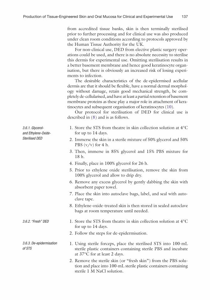
1. Store the STS from theatre in skin collection solution at 4°C for up to 14 days.
2. Immerse the skin in a sterile mixture of 50% glycerol and 50% PBS (v/v) for 4 h.
3. Then, immerse in 85% glycerol and 15% PBS mixture for 18 h.
4. Finally, place in 100% glycerol for 26 h. 5. Prior to ethylene oxide sterilisation, remove the skin from
100% glycerol and allow to drip dry. 6. Remove any excess glycerol by gently dabbing the skin with
absorbent paper towel. 7. Place the skin into autoclave bags, label, and seal with auto-
clave tape. 8. Ethylene-oxide-treated skin is then stored in sealed autoclave
bags at room temperature until needed.
1. Store the STS from theatre in skin collection solution at 4°C for up to 14 days.
2. Follow the steps for de-epidermisation.
1. Using sterile forceps, place the sterilised STS into 100-mL sterile plastic containers containing sterile PBS and incubate at 37°C for at least 2 days.
2. Remove the sterile skin (or “fresh skin”) from the PBS solu-tion and place into 100-mL sterile plastic containers containing sterile 1 M NaCl solution.
3.6.1. Glycerol- and Ethylene-Oxide-Sterilised DED
3.6.2. “Fresh” DED
3.6.3. De-epidermisation of STS
138 MacNeil, Shepherd, and Smith
3. Incubate overnight at 37°C (typically 14–18 h) or until there is visible separation of the dermis from the epidermis (see Note 3).
4. Now, wash the DED twice with PBS and incubate in Green’s medium or fibroblast culture medium at 37°C, 5% CO2 in a humidified atmosphere for a minimum of 48 h. This has a threefold purpose (1) ensuring that any NaCl solution remaining in the DED is washed out, (2) allowing the DED to become saturated with culture medium, and (3) providing a rough sterility check for the DED (in the presence of infection, the medium becomes more acidic, and therefore, the phenol red pH indicator in the medium changes colour from crimson red to yellow. The medium also becomes cloudy).
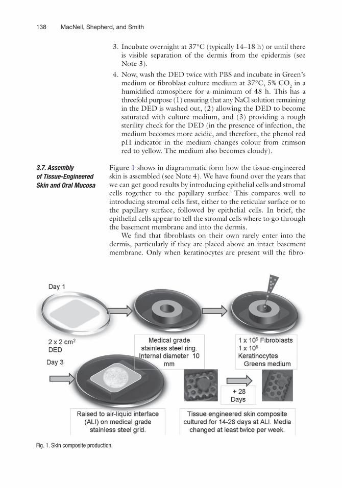
Figure 1 shows in diagrammatic form how the tissue-engineered skin is assembled (see Note 4). We have found over the years that we can get good results by introducing epithelial cells and stromal cells together to the papillary surface. This compares well to introducing stromal cells first, either to the reticular surface or to the papillary surface, followed by epithelial cells. In brief, the epithelial cells appear to tell the stromal cells where to go through the basement membrane and into the dermis.
We find that fibroblasts on their own rarely enter into the dermis, particularly if they are placed above an intact basement membrane. Only when keratinocytes are present will the fibro-
3.7. Assembly of Tissue-Engineered Skin and Oral Mucosa
Fig. 1. Skin composite production.
139Production of Tissue-Engineered Skin and Oral Mucosa for Clinical and Experimental Use
blasts enter the dermis through the basement membrane. This conclusion comes from the work of Ghosh et al. (7) and has been confirmed in a number of unpublished studies in our labora-tory since, such that routinely we would now co-culture epithelial cells and fibroblasts and place them together on the upper papil-lary surface, usually for 1 or 2 days submerged, prior to raising the DED with cells on it to an air–liquid interface. The air–liquid interface has been confirmed, in many studies, to provide a strong stimulus for keratinocyte differentiation.
The protocol we use was established within our laboratory by Ghosh (7). It was subsequently modified and has been used extensively in our laboratory (7, 8, 10–20).
1. Cut the DED with a sterile scalpel into squares of approxi-mately 2 × 2 cm and place into the wells of a six-well plate with the papillary dermis facing upwards (see Note 5).
2. Place a chamfered metal (medical-grade stainless steel) ring (internal diameter 1 cm) into the centre of each piece of DED and gently press down with sterile forceps to ensure a water-tight seal.
3. Flood the surrounding dermis (outside the ring) with 10% Green’s medium, and then inside each ring to check for leakage.
4. Once satisfied that a seal has been obtained, remove the medium from the centre of each ring.
5. Then, seed the DED inside each ring with 1 × 10⁵ fibroblasts and 1 × 10⁶ freshly isolated (P0) skin keratinocytes or 3 × 10⁵ P1-3 skin keratinocytes (or oral keratinocytes for the oral mucosa model), each in 250 mL of 10% Green’s medium.
6. After 24 h at 37°C, replace the seeding medium (which should have changed to a more yellowish colour) inside the rings with fresh Green’s medium.
7. After a further 24 h, remove the medium from inside the rings with a pipette. The surface of the constructs within the rings should have a slightly yellowish appearance.
8. Then, remove the steel ring using sterile forceps and raise the skin constructs onto stainless steel grids in new tissue culture wells.
9. Add fresh 10% Green’s medium to the level of the base of the skin constructs so that it laps the edges of, but does not cover, the DED, forming an air–liquid interface.
10. Replace the medium every 2–3 days. 11. Use skin or oral mucosa constructs for experimentation after
>10 days at the air–liquid interface (see Fig. 1).
3.8. Production of Tissue Engineered Skin
140 MacNeil, Shepherd, and Smith
As these tissue-engineered models develop in the laboratory, assurance that all is well can be obtained by seeing a yellowish tinge on them as they grow. The question of when are they ready to be used will depend on their application.
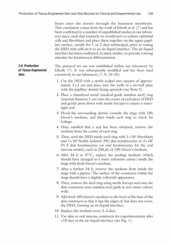
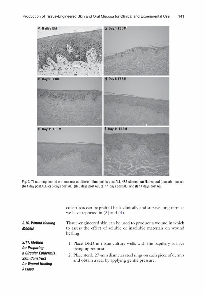
Figures 2 and 3 show the development of tissue-engineered skin and oral mucosa, respectively, over time. From this, it can be seen that tissue-engineered skin (Fig. 2) achieves a normal-looking gross morphology at around 10–14 days. This can be cultured for up to 28 days as in Harrison et al. 2007 (15, 21). (This is the longest period we have cultured this model at present). With buccal mucosa (see Fig. 3) by 5 days, there is a multilayered epithelium well attached to the underlying dermis, and this continues to mature throughout 14 days of investigation.
Confirmation of the maturation of the epidermis can be achieved by measuring electrical resistance across it (22) or by looking at the expression of specific cytokeratins (21). The most convincing evidence, however, is that these tissue-engineered
3.9. Maturation of Tissue Engineered Skin and Oral Mucosa
Fig. 2. H&E histology of (a) human skin, (b–f ) tissue-engineered skin raised to an air–liquid interface and sacrificed at (b) Day 1, (c) Day 7, (d) Day 14, (e) Day 21, and (f ) Day 28. Scale bar = 250 mm.
141Production of Tissue-Engineered Skin and Oral Mucosa for Clinical and Experimental Use
constructs can be grafted back clinically and survive long term as we have reported in (3) and (4).
Tissue-engineered skin can be used to produce a wound in which to assess the effect of soluble or insoluble materials on wound healing.
1. Place DED in tissue culture wells with the papillary surface being uppermost.
2. Place sterile 27-mm diameter steel rings on each piece of dermis and obtain a seal by applying gentle pressure.
3.10. Wound Healing Models
3.11. Method for Preparing a Circular Epidermis Skin Construct for Wound Healing Assays
Fig. 3. Tissue-engineered oral mucosa at different time points post ALI, H&E stained. (a) Native oral (buccal) mucosa. (b) 1 day post ALI, (c) 5 days post ALI, (d) 8 days post ALI, (e) 11 days post ALI, and (f) 14 days post ALI.
142 MacNeil, Shepherd, and Smith
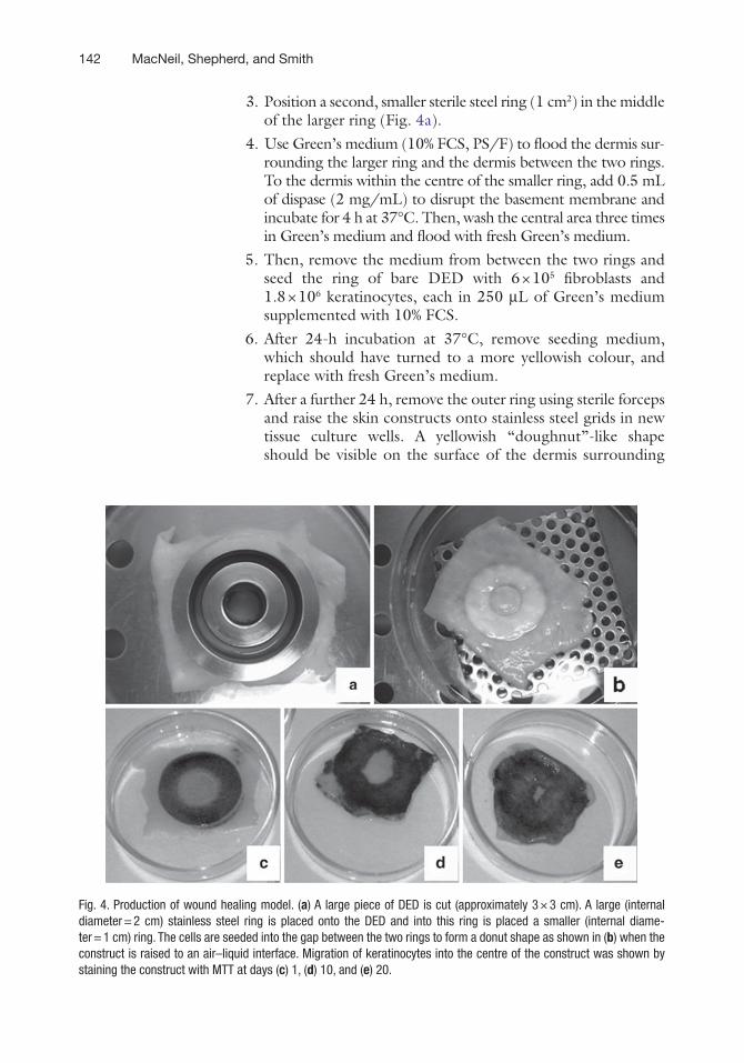
3. Position a second, smaller sterile steel ring (1 cm2) in the middle of the larger ring (Fig. 4a).
4. Use Green’s medium (10% FCS, PS/F) to flood the dermis sur-rounding the larger ring and the dermis between the two rings. To the dermis within the centre of the smaller ring, add 0.5 mL of dispase (2 mg/mL) to disrupt the basement membrane and incubate for 4 h at 37°C. Then, wash the central area three times in Green’s medium and flood with fresh Green’s medium.
5. Then, remove the medium from between the two rings and seed the ring of bare DED with 6 × 10⁵ fibroblasts and 1.8 × 10⁶ keratinocytes, each in 250 mL of Green’s medium supplemented with 10% FCS.
6. After 24-h incubation at 37°C, remove seeding medium, which should have turned to a more yellowish colour, and replace with fresh Green’s medium.
7. After a further 24 h, remove the outer ring using sterile forceps and raise the skin constructs onto stainless steel grids in new tissue culture wells. A yellowish “doughnut”-like shape should be visible on the surface of the dermis surrounding
Fig. 4. Production of wound healing model. (a) A large piece of DED is cut (approximately 3 × 3 cm). A large (internal diameter = 2 cm) stainless steel ring is placed onto the DED and into this ring is placed a smaller (internal diame-ter = 1 cm) ring. The cells are seeded into the gap between the two rings to form a donut shape as shown in (b) when the construct is raised to an air–liquid interface. Migration of keratinocytes into the centre of the construct was shown by staining the construct with MTT at days (c) 1, (d) 10, and (e) 20.
143Production of Tissue-Engineered Skin and Oral Mucosa for Clinical and Experimental Use
the inner ring still in position; this is the new epidermis which will have a ring of bare dermis in the centre (Fig. 4b).
8. Add fresh 10% Green’s medium to the level of the base of the skin constructs so that it laps the edges of, but does not cover, the DED, forming an air–liquid interface.
9. Change the medium every 2–3 days. 10. After 14 days culture at air–liquid interface, remove the
smaller inner ring using sterile forceps. 11. Use skin constructs for experimentation after removal of the
smaller inner ring. 12. Stain composites at the required time points with Alamar
Blue, diluted 1:10 in PBS, to monitor the rate of keratinocyte migration.
13. Examples of migration at day 0 (day of removal of the central ring), day 7, and day 13 at ALI can be seen in Fig. 4c–e.
Pathological skin contraction occurs following burn injuries such as superficial scalds where as many as one-third of the patients may go on to suffer disfiguring skin contraction. It also occurs quite commonly following extensive skin grafting. While there is a good clinical understanding of which patients, which sites of the body, and what circumstances give increased clinical risk of skin contraction, the mechanism of how fibroblasts and keratinocytes can both contribute to skin contraction is not fully understood as discussed in detail in (23).
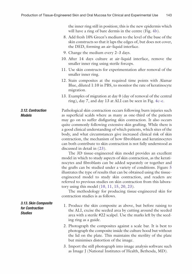
The 3D tissue-engineered skin model provides an excellent model in which to study aspects of skin contraction, as the kerati-nocytes and fibroblasts can be added separately or together and the grafts can be studied under a variety of conditions. Figure 5 illustrates the type of results that can be obtained using the tissue-engineered model to study skin contraction, and readers are referred to previous studies on skin contraction from this labora-tory using this model (10, 11, 15, 20, 23).
The methodology for producing tissue-engineered skin for contraction studies is as follows.
1. Produce the skin composite as above, but before raising to the ALI, excise the seeded area by cutting around the seeded area with a sterile #22 scalpel. Use the marks left by the seed-ing ring as a guide.
2. Photograph the composites against a scale bar. It is best to photograph the composite inside the culture hood but without the lid on the plate. This maintains the sterility of the plate but minimises distortion of the image.
3. Import the still photograph into image analysis software such as Image J (National Institutes of Health, Bethesda, MD).
3.12. Contraction Models
3.13. Skin Composite for Contraction Studies
144 MacNeil, Shepherd, and Smith
4. Use the scale bar to calibrate the image. 5. Draw freehand around the edges of the composite and record
the area of the composite. 6. Photograph the composite at least every 7 days.
Bacterial infection of skin is one of the most common contributors to failure of skin wounds to heal. However, despite the vast num-ber of chronic infected skin wounds, it is difficult to undertake investigational, mechanistic, or even therapeutic studies in
3.14. Bacterial Infected Skin Wound Model
Fig. 5. Contraction of composites. (a) Shows the composite after being cultured at an air–liquid interface for 28 days. The darker area in the centre of the composite is where the cells are. The bare dermis has had to pleat to accommodate the contraction of the composite over this time period. If the seeded area is excised ((b) excised composite at day 1 and (c) excised composite at day 28), the contraction is more noticeable, and it is easier to measure the change in area over time using image analysis software such as Image J giving data as shown in (d).
145Production of Tissue-Engineered Skin and Oral Mucosa for Clinical and Experimental Use
patients, and most animal models do not duplicate the conditions that prevail in a chronic infected skin wound in man.
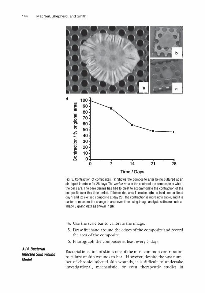
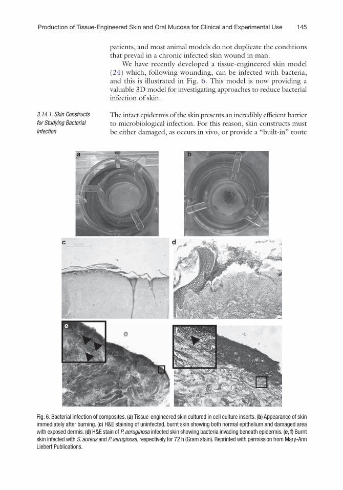
We have recently developed a tissue-engineered skin model (24) which, following wounding, can be infected with bacteria, and this is illustrated in Fig. 6. This model is now providing a valuable 3D model for investigating approaches to reduce bacterial infection of skin.
The intact epidermis of the skin presents an incredibly efficient barrier to microbiological infection. For this reason, skin constructs must be either damaged, as occurs in vivo, or provide a “built-in” route
3.14.1. Skin Constructs for Studying Bacterial Infection
Fig. 6. Bacterial infection of composites. (a) Tissue-engineered skin cultured in cell culture inserts. (b) Appearance of skin immediately after burning. (c) H&E staining of uninfected, burnt skin showing both normal epithelium and damaged area with exposed dermis. (d) H&E stain of P. aeruginosa infected skin showing bacteria invading beneath epidermis. (e, f) Burnt skin infected with S. aureus and P. aeruginosa, respectively for 72 h (Gram stain). Reprinted with permission from Mary-Ann Liebert Publications.
146 MacNeil, Shepherd, and Smith
for bacteria to invade into the dermis. There are two methods currently in use to provide a mode of entry; the first is to use skin constructs lacking epidermal cover over a defined area of dermis, and the second is to cause a burn injury to the epidermis.
For the first type, constructs such as those used on the wound-healing assays (see Fig. 4, Subheading 3.9), which are made using the double ring method, provide a circular area of unprotected dermis surrounded by intact epidermis. Bacteria can be intro-duced directly onto this dermal area. There is one difference in the protocol for wound-healing assays; when constructing the composites with the double rings, it is not necessary to incubate the dermis in the centre with dispase. For bacterial infection assays, the centre ring is flooded with fresh Green’s medium at the same time as the DED surrounding the outer ring. In this case, the area between the two rings is seeded with keratinocytes and fibroblasts at this time point. Otherwise, the protocol is as for the wound-healing assays. Composites are used >14 days at ALI, and the skin is infected directly via the central area of dermis (see bacterial infection, below).
For the second type, smaller constructs are prepared as below within tissue culture inserts. The smaller size of these constructs makes maximum use of DED available and can be used for any purpose, not only as an infection model. The engineered oral mucosa model can be prepared similarly.
1. Cut rings of DED 15 mm in diameter using a sterile cork borer if possible, or a sterile scalpel. The DED is cut slightly larger than the inserts to allow for skin contraction on burn-ing; if the skin or oral mucosa is to be used without burning or other causes of contraction, cut rings of DED 12 mm in diameter to fit the inserts.
2. Place the circles of DED, reticular surface uppermost, within 12-mm tissue culture inserts with 4 mM pores in the base (Greiner). The pores allow the medium to bathe the dermis from below. Ensure that the DED is pushed into the bottom of the inserts by pressing gently with sterile forceps.
3. Suspend the inserts from the edges of 12-well plates into the wells.
4. Add 10% Green’s medium to the bottom of the wells, sur-rounding the suspended inserts, so that it laps the under surface of the DED.
5. Seed the DED with 1 × 10⁵ fibroblasts and 5 × 10⁵ skin kerati-nocytes (or oral keratinocytes for the oral mucosa model), each in 250 mL of 10% Green’s medium.
6. After 24 h at 37°C, replace the seeding medium (which should have changed to a more yellowish colour) inside the inserts and the bathing medium in the wells with fresh Green’s medium.
3.14.2. Method for Preparation of the Skin or Oral Mucosa Model Within Tissue Culture Inserts
147Production of Tissue-Engineered Skin and Oral Mucosa for Clinical and Experimental Use
7. After a further 24 h, remove the medium from inside the inserts with a pipette. This allows the skin constructs to be at the air–liquid interface. The surface of the constructs within the inserts should have a slightly yellowish appearance. Replace the Green’s medium in the wells with fresh medium.
8. Replace Green’s medium every 24 h. Constructs within tissue culture inserts in 12-well plates have access to a smaller volume of Green’s medium than constructs grown in steel rings on grids, so the medium needs to be replaced more frequently.
9. Use skin or oral mucosa constructs for experimentation after >10 days at the air–liquid interface (see Fig. 6a for prepared composite within an insert).
10. After 14 days at ALI, the skin constructs within the inserts are burnt in the centre by applying a heated metal rod, 4 mm in diameter, to the surface of the skin for 6 s (Fig. 6b) (see Notes 5 and 6).
1. Culture bacterial species of interest in appropriate broth at 37°C for 24 h from stock plates prior to use. Centrifuge broths, then wash the resulting pellet in PBS and re-suspend bacteria in PBS to a concentration of 1 × 10¹⁰ cfu/mL. Bacteria can be stored at this concentration in PBS at 4°C for several days.
2. Wash skin constructs (by removing and replacing the Green’s medium at least every 24 h) in antibiotic-free Green’s medium for 72 h prior to infection. To infect the larger “double ring” constructs, pipette bacteria at the desired concentration (e.g., 1 × 10⁷ bacteria) in 20 mL of appropriate broth per composite directly onto the exposed dermis in the centre of the skin constructs.
3. For constructs in inserts, burn as above immediately prior to infection to provide the bacteria a mode of entry into the skin. Pipette bacteria of interest at the test concentration (e.g., 1 × 10⁷ bacteria) in 100 mL of appropriate broth per construct into the inserts, covering the epidermal surface.
4. Incubate infected skin constructs and non-infected controls in antibiotic-free Green’s medium at 37°C/5% CO2 for the desired amount of time, then sacrifice for analysis.
1. For histological analysis, bisect skin constructs with a sterile scalpel. Fix the tissue in 10% formalin for >24 h, then process and embed in paraffin using standard techniques. Alternatively, for frozen sections, after bisecting the skin, snap-freeze in liq-uid nitrogen and store samples at −80°C prior to sectioning on a cryostat.
2. Gram staining: To visualise bacteria in paraffin sections of skin, first dewax the sections through xylene and alcohol to
3.14.3. Bacterial Infection of Skin Constructs
3.14.4. Analysis of Infected Skin Constructs
148 MacNeil, Shepherd, and Smith
water as standard (as in for H&E staining). Then, with the slides still wet, perform a Gram stain on a rack over a sink.
3. A standard Gram stain is: 1 min crystal violet, wash in run-ning water, fix in Gram’s iodine (1 min), wash in running water, destain in iodine–acetone, 30 s, wash in running water, carbol fuschin (1 min).
4. At each step, pipette enough stain to cover the tissue section. After the final carbol fuschin step, tap the carbol fuschin off the slides, blot sections dry with filter paper, and then mount a coverslip using a non-aqueous mountant. Gram-positive bacteria will appear dark purple and Gram-negative pink (see Fig. 6e–f ). Gram-negative bacteria may be more difficult to see since there will be a high level of background staining, also pink, on the tissue sample.
5. In order to quantify the viable bacteria that have invaded into the skin after infection, first weigh the sample. This is impor-tant since not all skin constructs will be exactly of the same size or thickness. Then, mince the tissue in a sterile Petri dish using a sterile scalpel into small pieces and homogenise (for example, in a sterile glass homogeniser) in 1 mL of broth. Perform a serial dilution on the resulting homogenates, e.g., 1:102, 1:104, 1:106, etc.
6. Add 10-mL drops of each dilution in triplicate onto agar plates; allow the drops to dry and then incubate the plates at 37°C overnight. Count the number of resulting colonies and deduce the number of viable bacteria in the original sample from the dilution factor. For example, 15 colonies at a 1:106 dilution indicates a starting concentration of ~15,000,000 or 1.5 × 10⁷ cfu (colony forming units). It is then possible, using the weights of the tissue samples, to express the numbers of viable bacteria recovered per mg of tissue.
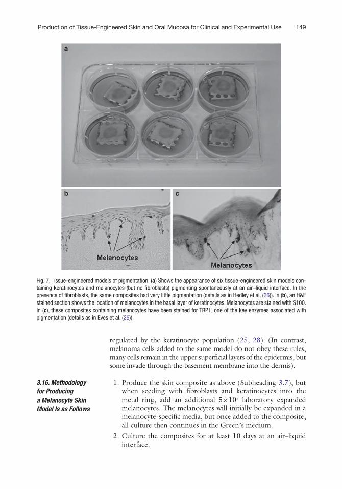
Tissue-engineered skin is going to prove very useful in under-standing the regulation of skin pigmentation. For example, mel-anocytes from pale skin donors when introduced to reconstructed skin give rise to barely pigmented skin (25). However, these tis-sue-engineered models spontaneously pigment if fibroblasts are omitted from the models (26). Exploring this in 2D co-culture models, we demonstrated that while normal fibroblasts will suppress melanocyte pigmentation, stressed fibroblasts (stressed by freezing or gamma radiation) produce soluble factors which induced pigmentation in melanocytes. These results were confirmed for melanocytes derived from skin, hair, and eye, indicating that this may be a generic biology (27).
Figure 7 illustrates that normal human melanocytes when added to these tissue-engineered skin models know where to go, orientating in the basal layer of keratinocytes, and their numbers are tightly
3.15. Skin Pigmentation
149Production of Tissue-Engineered Skin and Oral Mucosa for Clinical and Experimental Use
regulated by the keratinocyte population (25, 28). (In contrast, melanoma cells added to the same model do not obey these rules; many cells remain in the upper superficial layers of the epidermis, but some invade through the basement membrane into the dermis).
1. Produce the skin composite as above (Subheading 3.7), but when seeding with fibroblasts and keratinocytes into the metal ring, add an additional 5 × 10⁵ laboratory expanded melanocytes. The melanocytes will initially be expanded in a melanocyte-specific media, but once added to the composite, all culture then continues in the Green’s medium.
2. Culture the composites for at least 10 days at an air–liquid interface.
3.16. Methodology for Producing a Melanocyte Skin Model Is as Follows
Fig. 7. Tissue-engineered models of pigmentation. (a) Shows the appearance of six tissue-engineered skin models con-taining keratinocytes and melanocytes (but no fibroblasts) pigmenting spontaneously at an air–liquid interface. In the presence of fibroblasts, the same composites had very little pigmentation (details as in Hedley et al. (26)). In (b), an H&E stained section shows the location of melanocytes in the basal layer of keratinocytes. Melanocytes are stained with S100. In (c), these composites containing melanocytes have been stained for TRP1, one of the key enzymes associated with pigmentation (details as in Eves et al. (25)).
150 MacNeil, Shepherd, and Smith
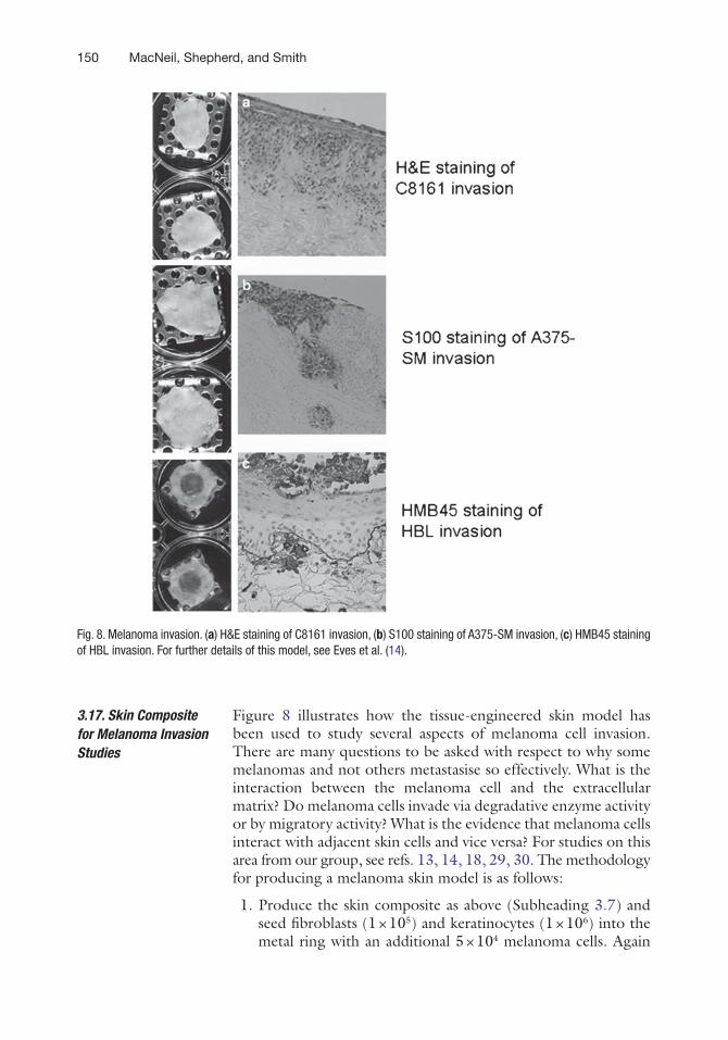
Figure 8 illustrates how the tissue-engineered skin model has been used to study several aspects of melanoma cell invasion. There are many questions to be asked with respect to why some melanomas and not others metastasise so effectively. What is the interaction between the melanoma cell and the extracellular matrix? Do melanoma cells invade via degradative enzyme activity or by migratory activity? What is the evidence that melanoma cells interact with adjacent skin cells and vice versa? For studies on this area from our group, see refs. 13, 14, 18, 29, 30. The methodology for producing a melanoma skin model is as follows:
1. Produce the skin composite as above (Subheading 3.7) and seed fibroblasts (1 × 10⁵) and keratinocytes (1 × 10⁶) into the metal ring with an additional 5 × 10⁴ melanoma cells. Again
3.17. Skin Composite for Melanoma Invasion Studies
Fig. 8. Melanoma invasion. (a) H&E staining of C8161 invasion, (b) S100 staining of A375-SM invasion, (c) HMB45 staining of HBL invasion. For further details of this model, see Eves et al. (14).
151Production of Tissue-Engineered Skin and Oral Mucosa for Clinical and Experimental Use
these will each initially be expanded in whichever medium is recommended for the particular cell line, but once added to the composite, all culture will be in Green’s medium.
2. Culture the composites for at least 10 days at an ALI and for up to 28 days depending on what aspects of melanoma inva-sion are being studied.
As mentioned previously, we have made tissue-engineered oral mucosa for clinical use. It has been used to replace scarred tissue in the urethra, and a 3-year follow-up study showed good results for three out of five patients and some recurrent constrictions for two of these five patients. As these patients had previously had a long history of recurrent tissue strictures, this result was not unexpected (4).
For in vitro purposes, we can also use the oral mucosa model to look at the penetration of agents across this barrier. Work ongoing in our laboratory is looking at the penetration of poly-mersomes being designed to carry drugs and genes into the body through the oral mucosa. Here, the tissue-engineered oral mucosa is an invaluable test bed model in developing the polymersomes.
1. Problems in cell isolation can occur with STS grafts that are too thick (because then it is difficult to separate the epidermis from the dermis) or when the pieces of biopsy are not cut sufficiently small to allow enzyme separation of epidermis and dermis.
2. At this point, the culture contains fibroblasts, keratinocytes, melanocytes, and other skin cells. When the cells have become confluent, subculture the cells into a T75 flask and then when this flask has become confluent, passage cells fairly aggres-sively (i.e. 1× T75 into 10× T75) to encourage a pure fibro-blast population. Only use when you are sure that the culture is not contaminated with other cell types. This is usually around passage 3.
3. The epidermis of the glycerol- and ethylene-oxide-sterilised skin can be difficult to remove. If this proves to be the case, leave in 1 M NaCl longer or gently detach by gentle scraping with a blunt-ended spatula. However, be careful – too much scraping will damage the basement membrane.
4. For any one experiment, use DED from a single patient and wherever possible cut from the same sheet. This is to try to reduce inter-patient variation in skin characteristics and dif-ferences in thickness.
3.18. Oral Mucosa Applications
4. Notes
152 MacNeil, Shepherd, and Smith
5. If unsure which is the papillary and which is the reticular dermis, the papillary dermis tends to look smoother than the reticular dermis which tends to look more ragged and “cotton wool-like.”
6. When performing the burn injury, if several samples are to be injured, it is best to hold the heated rod in a thermoprotec-tive glove as it will get very hot.
7. During burn injury, the surface of the skin may stick to the metal rod, so have a pair of sterile forceps to hand to gently tease the skin away from the metal.
Acknowledgements
We gratefully acknowledge the continued support for our research throughout the years of the Burns and Plastic Surgery Consultants of the Northern General Hospital Trust Burns Unit, particularly Mr Eric Freedlander and Mr David Ralston. We also acknowledge the histopathology services provided over the years by Dr Chris Layton – ever helpful.
References
1. MacNeil, S. (2007) Progress and opportuni-ties for tissue-engineered skin. Nature 445, 874–880.
2. MacNeil, S. (2007) Skin tissue engineering, in Tissue engineering using ceramics and polymers (Boccaccini, A. R., and Gough, J., Eds.), pp 375–403, Woodhead Publishing Limited, Cambridge.
3. Sahota, P. S., Burn, J. L., Heaton, M., Freedlander, E., Suvarna, S. K., Brown, N. J., and MacNeil, S. (2003) Development of a reconstructed human skin model for angio-genesis. Wound Repair Regen. 11, 275–284.
4. Bhargava, S., Patterson, J. M., Inman, R. D., MacNeil, S., and Chapple, C. R. (2008) Tissue-engineered buccal mucosa urethroplasty-clini-cal outcomes. Eur. Urol. 53, 1263–1269.
5. Moustafa, M., Simpson, C., Glover, M., Dawson, R. A., Tesfaye, S., Creagh, F. M., Haddow, D., Short, R., Heller, S., and MacNeil, S. (2004) A new autologous kerati-nocyte dressing treatment for non-healing diabetic neuropathic foot ulcers. Diabet. Med. 21, 786–789.
6. Bhargava, S., Chapple, C. R., Bullock, A. J., Layton, C., and Macneil, S. (2004) Tissue-engineered buccal mucosa for substitution urethroplasty. BJU Int. 93, 807–811.
7. Ghosh, M. M., Boyce, S., Layton, C., Freedlander, E., and MacNeil, S. (1997) A comparison of the methodologies for the preparation of human epidermal-dermal com-posites. Ann. Plast. Surg. 39, 390–404.
8. Chakrabarty, K. H., Dawson, R. A., Harris, P., Layton, C., Babu, M., Gould, L., Phillips, J., Leigh, I., Green, C., Freedlander, E., and MacNeil, S. (1999) Development of autolo-gous human dermal-epidermal composites based on sterilized human allodermis for clinical use. Br. J. Dermatol. 141, 811–823.
9. Huang, Q., Dawson, R. A., Pegg, D. E., Kearney, J. N., and MacNeil, S. (2004) Use of peracetic acid to sterilize human donor skin for production of acellular dermal matrices for clin-ical use. Wound Repair Regen. 12, 276–287.
10. Ralston, D. R., Layton, C., Dalley, A. J., Boyce, S., Freedlander, E., and MacNeil, S. (1999) The requirement for basement mem-brane antigens in the production of human epidermal/dermal composites in vitro. Br. J. Dermatol. 140, 605–615.
11. Chakrabarty, K. H., Heaton, M., Dalley, A. J., Dawson, R. A., Freedlander, E., Khaw, P. T., and MacNeil, S. (2001) Keratinocyte-driven contraction of reconstituted human skin. Wound Repair Regen. 9, 95–106.
153Production of Tissue-Engineered Skin and Oral Mucosa for Clinical and Experimental Use
12. Dawson, R. A., Goberdhan, N. J., Freedlander, E., and MacNeil, S. (1996) Influence of extra-cellular matrix proteins on human keratino-cyte attachment, proliferation and transfer to a dermal wound model. Burns 22, 93–100.
13. Eves, P., Katerinaki, E., Simpson, C., Layton, C., Dawson, R., Evans, G., and MacNeil, S. (2003) Melanoma invasion in reconstructed human skin is influenced by skin cells – inves-tigation of the role of proteolytic enzymes. Clin. Exp. Metastasis 20, 685–700.
14. Eves, P., Layton, C., Hedley, S., Dawson, R. A., Wagner, M., Morandini, R., Ghanem, G., and MacNeil, S. (2000) Characterization of an in vitro model of human melanoma inva-sion based on reconstructed human skin. Br. J. Dermatol. 142, 210–222.
15. Harrison, C. A., Gossiel, F., Layton, C. M., Bullock, A. J., Johnson, T., Blumsohn, A., and MacNeil, S. (2006) Use of an in vitro model of tissue-engineered skin to investigate the mechanism of skin graft contraction. Tissue Eng. 12, 3119–3133.
16. Harrison, C. A., Heaton, M. J., Layton, C. M., and MacNeil, S. (2006) Use of an in vitro model of tissue-engineered human skin to study keratinocyte attachment and migration in the process of reepithelialization. Wound Repair Regen. 14, 203–209.
17. Hernon, C., Harrison, C. A., Thornton, D. J. A., and MacNeil, S. (2007) Enhancement of keratinocyte performance in production of tis-sue engineered skin by use of low-calcium medium. Wound Repair Regen. 15, 718–726.
18. MacNeil, S., Eves, P., Richardson, B., Molife, R., Lorigan, P., Wagner, M., Layton, C., Morandini, R., and Ghanem, G. (2000) Oestrogenic steroids and melanoma cell inter-action with adjacent skin cells influence invasion of melanoma cells in vitro. Pigment Cell Res. 13(8), 68–72.
19. Ralston, D. R., Layton, C., Dalley, A. J., Boyce, S. G., Freedlander, E., and MacNeil, S. (1997) Keratinocytes contract human dermal extracel-lular matrix and reduce soluble fibronectin production by fibroblasts in a skin composite model. Br. J. Plast. Surg. 50, 408–415.
20. Thornton, D. J. A., Harrison, C. A., Heaton, M. J., Bullock, A. J., and MacNeil, S. (2008) Inhibition of keratinocyte-driven contraction of tissue-engineered skin in vitro by calcium chelation and early restraint but not submerged culture. J. Burn Care Res. 29, 369–377.
21. Harrison, C. A., Layton, C. M., Hau, Z., Bullock, A. J., Johnson, T. S., and MacNeil, S.
(2007) Transglutaminase inhibitors induce hyperproliferation and parakeratosis in tissue-engineered skin. Br. J. Dermatol. 156, 247–257.
22. Bullock, A. J., Barker, A. T., Coulton, L., and Macneil, S. (2007) The effect of induced biphasic pulsed currents on re-epithelializa-tion of a novel wound healing model. Bioelectromagnetics 28, 31–41.
23. Harrison, C. A., and MacNeil, S. (2008) The mechanism of skin graft contraction: an update on current research and potential future therapies. Burns 34, 153–163.
24. Shepherd, J., Douglas, I., Rimmer, S., Swanson, L., and MacNeil, S. (2009) Development of 3-dimensional tissue engineerd models of bac-terial infected human skin wounds. Tissue Eng. Part C Methods 15(3), 475–484.
25. Eves, P. C., Bullett, N. A., Haddow, D., Beck, A. J., Layton, C., Way, L., Shard, A. G., Gawkrodger, D. J., and MacNeil, S. (2008) Simplifying the delivery of melanocytes and keratinocytes for the treatment of vitiligo using a chemically defined carrier dressing. J. Invest. Dermatol. 128, 1554–1564.
26. Hedley, S. J., Layton, C., Heaton, M., Chakrabarty, K. H., Dawson, R. A., Gawkrodger, D. J., and MacNeil, S. (2002) Fibroblasts play a regulatory role in the con-trol of pigmentation in reconstructed human skin from skin types I and II. Pigment Cell Res. 15, 49–56.
27. Balafa, C., Smith-Thomas, L., Phillips, J., Moustafa, M., George, E., Blount, M., Nicol, S., Westgate, G., and MacNeil, S. (2005) Dopa oxidase activity in the hair, skin and ocular melanocytes is increased in the pres-ence of stressed fibroblasts. Exp. Dermatol. 14, 363–372.
28. Eves, P. C., Beck, A. J., Shard, A. G., and MacNeil, S. (2005) A chemically defined surface for the co-culture of melanocytes and keratinocytes. Biomaterials 26, 7068–7081.
29. Katerinaki, E., Evans, G. S., Lorigan, P. C., and MacNeil, S. (2003) TNF-alpha increases human melanoma cell invasion and migration in vitro: the role of proteolytic enzymes. Br. J. Cancer 89, 1123–1129.
30. Eves, P., Haycock, J., Layton, C., Wagner, M., Kemp, H., Szabo, M., Morandini, R., Ghanem, G., García-Borrón, J. C., Jiménez-Cervantes, C., and MacNeil, S. (2003) Anti-inflammatory and anti-invasive effects of a-melanocyte-stim-ulating hormone in human melanoma cells. Br. J. Cancer 89, 2004–2015.