Procedure_Catalogue-Phoenix-All_Cobined.pdf - Bio Group
203
PRODUCT PACK SIZE PAGE NO. ACID PHOSPHATASE 1 ALKALINE PHOSPHATASE 2 CHOLESTEROL 3 CHOLINESTERASE 4 CK‐MB 5 x 2.5mL 5 CK‐NAC 5 x 2.5mL 6 GLUCOSE 7 SGOT 8 SGPT 9 TRIGLYCERIDES 10 UREA‐B 11 UREA U.V 12 URIC ACID 13 ALKALINE PHOSPHATASE (S.L) 14 ALPHA AMYLASE (S.L) 15 CHOLESTEROL(S.L) 16 CHOLINESTERASE ( S.L) 17 CK‐MB (S.L) 18 CK‐NAC (S.L) 19 ENZYMATIC CREATININE (S.L) 20 GAMMA GT (S.L) 21 GLUCOSE (S.L) 22 HDL ‐ C DIRECT 23 LDH ‐ P (S.L) 24 LDL ‐ C DIRECT 25 LIPASE (S.L) 26 SGOT (S.L) 27 SGPT (S.L) 28 TRIGLYCERIDES (S.L) 29 UREA U.V (S.L) 30 URIC ACID (S.L) 31 ALBUMIN 32 BILIRUBIN DIRECT 33 BILIRUBIN TOTAL ‐TAB 34 BILIRUBIN TOTAL 35 BILIRUBIN TOTAL& DIRECT 36 CALCIUM 37 CALCIUM (ARSENAZO) 38 CHLORIDE 39 COPPER 2 x 25mL 40 CREATININE 41 HAEMOGLOBIN 42 HDL‐CHOLESTEROL 43 INORGANIC PHOSPHOROUS 44 IRON 45 MAGNESIUM 46 MICROPROTEIN 47 TIBC 50T 48 TOTAL PROTEIN 49 ZINC 2 x 10mL 50 ALPHA1ACID GLYCOPROTEIN WITH CALIBRATOR 51 APOA1 WITH CALIBRATOR 1 x 30ml/1 x 5ml/1ml 52 APOB WITH CALIBRATOR 1 x 30ml/1 x 5ml/1ml 53 Lypho CHEK 5 x 2 mL 10 x 3 mL 4 x 50 mL /4 x 100 mL 5x 3 mL 4 x 100 mL /4 x 250 mL/ 4 x 500 mL 5 x 20 mL/4 x 50 mL 5 x 20 mL/4 x 50 mL 5 x 20mL / 4 x 50 mL 200 mL 4 x 50 mL / 4 x 100 mL 5 x 20 mL/ 4 x 50 mL Liqui CHEK 2 x 10 mL /2 x 30 mL/2 x 50 mL 4 x 5 mL /4 x 10 mL 5 x 25 mL / 5 x 100 mL/ 4 x 250 mL 2 x 10 mL 2 x 12.5 mL/2 x 20 mL 2 x 10 mL /2 x 30 mL 2 x 40 mL /2 x 60 mL/4 x 60 mL 2 x 10 mL / 2 x 30 mL 5 x 100 mL /1 x 1000 mL 2 x 40 mL /2 x 60 mL / 4 x 60 mL 2 x 10 mL /2 x 30 mL 2 x 40 mL /2 x 60 mL 1 x 25 mL 3 x 20 mL /3 x 50 mL / 4 x 125 mL 3 x 20 mL / 3 x 50 mL/ 4 x 125 mL 4 x 10 mL /5 x 25 mL / 6 x 50 mL/ 5 x 100 mL 2 x 30 mL / 2 x 50 mL / 2 x 125 mL 2 x 30 mL / 2 x 50 mL / 2 x 100 mL ChemCHEK 4 x 50 mL 4 x 50 mL 4 x 50 mL 4 x 50 mL 4 x 50 mL 4 x 25 mL / 4 x 50 mL 4 x 25 mL 4 x 50 mL 4 x 50 mL 1000 mL 4 x 25 mL 4 x 25 mL / 4 x 50 mL 2 x 50 mL 4 x 25 mL 1 x 50mL 4 x 50mL SensIT 1 x 20 mL/1 x 10ml/1ml
-
Upload
khangminh22 -
Category
Documents
-
view
1 -
download
0
Transcript of Procedure_Catalogue-Phoenix-All_Cobined.pdf - Bio Group

PRODUCT PACK SIZE PAGE NO.
ACID PHOSPHATASE 1ALKALINE PHOSPHATASE 2CHOLESTEROL 3CHOLINESTERASE 4CK‐MB 5 x 2.5mL 5CK‐NAC 5 x 2.5mL 6GLUCOSE 7SGOT 8SGPT 9TRIGLYCERIDES 10UREA‐B 11UREA U.V 12URIC ACID 13
ALKALINE PHOSPHATASE (S.L) 14ALPHA AMYLASE (S.L) 15CHOLESTEROL(S.L) 16CHOLINESTERASE ( S.L) 17CK‐MB (S.L) 18CK‐NAC (S.L) 19ENZYMATIC CREATININE (S.L) 20GAMMA GT (S.L) 21GLUCOSE (S.L) 22HDL ‐ C DIRECT 23LDH ‐ P (S.L) 24LDL ‐ C DIRECT 25LIPASE (S.L) 26SGOT (S.L) 27SGPT (S.L) 28TRIGLYCERIDES (S.L) 29UREA U.V (S.L) 30URIC ACID (S.L) 31
ALBUMIN 32BILIRUBIN DIRECT 33BILIRUBIN TOTAL ‐TAB 34BILIRUBIN TOTAL 35BILIRUBIN TOTAL& DIRECT 36CALCIUM 37CALCIUM (ARSENAZO) 38CHLORIDE 39COPPER 2 x 25mL 40CREATININE 41HAEMOGLOBIN 42HDL‐CHOLESTEROL 43INORGANIC PHOSPHOROUS 44IRON 45MAGNESIUM 46MICROPROTEIN 47TIBC 50T 48TOTAL PROTEIN 49ZINC 2 x 10mL 50
ALPHA1ACID GLYCOPROTEIN WITH CALIBRATOR 51APOA1 WITH CALIBRATOR 1 x 30ml/1 x 5ml/1ml 52APOB WITH CALIBRATOR 1 x 30ml/1 x 5ml/1ml 53
Lypho CHEK5 x 2 mL
10 x 3 mL 4 x 50 mL /4 x 100 mL
5 x 3 mL
4 x 100 mL /4 x 250 mL/ 4 x 500 mL 5 x 20 mL/4 x 50 mL 5 x 20 mL/4 x 50 mL 5 x 20mL / 4 x 50 mL
200 mL 4 x 50 mL / 4 x 100 mL 5 x 20 mL/ 4 x 50 mL
Liqui CHEK2 x 10 mL /2 x 30 mL/2 x 50 mL
4 x 5 mL /4 x 10 mL5 x 25 mL / 5 x 100 mL/ 4 x 250 mL
2 x 10 mL2 x 12.5 mL/2 x 20 mL2 x 10 mL /2 x 30 mL
2 x 40 mL /2 x 60 mL/4 x 60 mL2 x 10 mL / 2 x 30 mL
5 x 100 mL /1 x 1000 mL2 x 40 mL /2 x 60 mL / 4 x 60 mL
2 x 10 mL /2 x 30 mL2 x 40 mL /2 x 60 mL
1 x 25 mL3 x 20 mL /3 x 50 mL / 4 x 125 mL 3 x 20 mL / 3 x 50 mL/ 4 x 125 mL
4 x 10 mL /5 x 25 mL / 6 x 50 mL/ 5 x 100 mL2 x 30 mL / 2 x 50 mL / 2 x 125 mL2 x 30 mL / 2 x 50 mL / 2 x 100 mL
ChemCHEK4 x 50 mL4 x 50 mL4 x 50 mL4 x 50 mL4 x 50 mL
4 x 25 mL / 4 x 50 mL4 x 25 mL4 x 50 mL
4 x 50 mL1000 mL
4 x 25 mL4 x 25 mL / 4 x 50 mL
2 x 50 mL4 x 25 mL1 x 50 mL
4 x 50 mL
SensIT1 x 20 mL/1 x 10ml/1ml

PRODUCT PACK SIZE PAGE NO.ASO LATEX WITH CALIBRATOR 1 x 45ml/1 x 5 ml/1ml 54ASO LEIT WITH CALIBRATOR 1 x 24ml/1 x 8 ml/1ml, 2 x 24ml/2 x 8 ml/2ml 55C3 WITH CALIBRATOR 1 x 30ml/1 x 5ml/1ml 56C4 WITH CALIBRATOR 1 x 30ml/1 x 5ml/1ml 57CERULOPLASMIN WITH CALIBRATOR 1 x 30ml/1 x 5ml/1ml 58CRP LATEX WITH CALIBRATOR 1 x 45ml/1 x 5 ml/2ml 59CRP LEIT WITH CALIBRATOR 1 x 24ml/1 x 8 ml/2ml, 2 x 24ml/2 x 8 ml/2ml 60CRP ULTRA WITH CALIBRATOR 1 x 18ml/1 x 9ml/2ml 61CYSTATIN C WITH CALIBRATOR 1 x 25ml/1 x 5ml/2ml 62FERRITIN+CALIBRATOR 1 x 30ml/1 x 10ml/1ml 63HBA1C DIRECT WITH CALIBRATOR 64
1 x 30ml/1 x 10ml/1ml 65 1 x 15ml/1 x 15ml/1ml 66 1 x 30ml/1 x 10ml/1ml 67 1 x 32ml/1 x 8ml/1ml 68
LIPOPROTEIN (A) WITH CALIBRATOR 1 x 20mL/1 x 4mL/1mL 69MICRO ALBUMIN LATEX WITH CALIBRATOR 70MICRO ALBUMIN IT WITH CALIBRATOR 2 x 25mL/2 x 5mL/1mL 71PREALBUMIN WITH CALIBRATOR 72RF LATEX WITH CALIBRATOR 1 x 45ml/1 x 5 ml/2ml 73R.F LEIT WITH CALIBRATOR 1 x 24ml/1 x 8 ml/1ml, 2 x 24ml/2 x 8 ml/2ml 74TRANSFFERIN WITH CALIBRATOR 75
ASO 50 T / 100 T 76CRP 50 T / 100 T 77RF 50 T / 100 T 78RPR 50 T /125 T / 500 T 79BLOOD GROUPINGANTI‐A 80
3 x 10mL 813 x 10mL 82
ANTI‐B 8384
1 x 10 ml 85
PT (SL) 86APTT 87
DILUENT 10 Litre /20 Litre 88E‐Z CLEANER 89LYSE 90PROBE CLEANER 91RINSE 10 Litre /20 Litre 92CONTROLS & CALIBRATORSASO CALIBRATOR 93MICROALBUMIN CALIBRATOR 94CRP CALIBRATOR 95
96PROTEIN CALIBRATOR 1 x 1 97MULTICALIBRATOR 98FERRITIN CALIBRATOR 99HbA1c DIRECT MULTI CALIBRATOR 100RF CALIBRATOR 101HbA1c CONTROL LEVEL 1 &2 2 x 0.5ml 102PROTEIN CONTROL(IMMUNOLOGY) 103QUALICHECK NORM & PATH 104
4 x 7.5/1 x 9.5/1 x .5/4 x .5 mLIgM WITH CALIBRATORIgG WITH CALIBRATORIgA WITH CALIBRATORIgE WITH CALIBRATOR
1 x 45 mL/1 x 5mL/1mL
1 x 25 mL/1 x 5mL/1mL
1 x 30 mL/1 x 10mL/1mLSero CHEK
1 x 10 mLANTI‐A,B&D(IgG+IgM)ANTI‐A,B&D(IgM)
1 x 10 mLANTI‐D (IgG+IgM) 1 x 10 mLANTI‐D (IgM)CoagTHREE
2 x 4 mL2 x 4 mL
HaemoCHEK
4 x 50 mL500 mL /2 x 500 mL
4 x 50 mL
1 x 1 mL1 x 1 mL1 x 2 mL
Lp(a) CALIBRATOR 1 x 1 mL
5 x 3 mL1 x 1 mL
4 x 0.5 mL1 x 1 mL
1 x 1 mL2 x 5 mL

PRODUCT PACK SIZE PAGE NO.
ALPHA 1‐ACID GLYCOPROTEIN 105C3 1 x 35mL /1 x6mL 107C4 109APO A1 1 x 35/1x6mL 111APO B 113CERULOPLASMIN 115PREALBUMIN 117TRANSFERRIN 1 x 25mL /1 x 4mL 119
121123
1 x 30mL/1 x 10mL 1251 x 22mL/1 x 6.5mL 127
FERRITIN 1 x 20mL/1 x 11mL 129CYSTATIN‐C 131HbA1c DIRECT 1 x 30/1 x 9.5/1 x 0.5/2x63mL 133ASO 135CRP 137RF 139
141MICROALBUMIN 143CRP Ultra 145ALBUMIN 148ALKALINE PHOSPHATASE 150AMYLASE 152BILIRUBIN DIRECT 154BILIRUBIN TOTAL (TAB) 156CALCIUM (ARSENAZO) 158CHLORIDE 160CHOLESTEROL 162CREATININE 164CREATINE KINASE 166ENZYMATIC CREATININE 168GAMMA GT 170GLUCOSE 172HDL – C DIRECT 174LDH 176LDL – C DIRECT 178MAGNESIUM 180PHOSPHOROUS 182SGOT 184SGPT 186TOTAL PROTEIN 188TRIGLYCERIDES 190UREA U V 192URIC ACID 194
Mispa Nano1 x 25mL /1 x4 mL
1 x35 mL/1x6mL
1 x 35mL/1 x 6 mL1 x 35 mL/1 x6 mL1 x25mL /1 x 4 mL
IgM 1 x 30/1 x 10 mLIgG 1 x 15mL/1 x 15 mLIgAIgE
1 x 23mL/1 x 5.5 mL
1 x 20/1 x 11 mL1 x 20/1 x8 mL1x 20/1 x 8 mL
Lp (a) 1 x 23/1 x 5.5 mL1 x 23/1 x 5.5 mL1x20 / 1x11 mL
4 x 30 mL2 x 30 / 2 x 8 mL
2 x 30 mL4 x 30 / 2 x 8 mL4 x 35 / 2 x 10 mL
2 x 35 mL2 x 30 mL4 x 35 mL
4 x 35 / 2 x 18 mL1 x 30 / 1 x 8 mL2 x 35 / 2 x 12 mL1 x 30 / 1 x 8 mL
4 x 35 mL2 x 30 / 2 x 10 mL1 x 30 / 1 x 8 mL1 x 30 / 1 x 10 mL
1 x 30 mL1 x 30 mL
4 x 35 / 2 x 18 mL4 x 35 / 2 x 18 mL
4 x 30 mL4 x 35 mL
4 x 30 / 2 x 16 mL2 x 35 mL

INTENDED USE
This reagent is intended for in vitro quantitative determination of Acid phosphatasein serum.
- Alpha - naphthylphosphate method
- Linear upto 150 U/L
- Tartrate included for determination of non-prostatic acid phosphatase
CLINICAL SIGNIFICANCE
Acid phosphatase is present in lysosomes, which are organells present in all cells withthe possible exception of erythrocytes. The greatest concentrations of ACP activityoccurs in liver, spleen, erythrocytes, platelets, bone marrow & the prostate gland whichis the richest source and it contributes a small portion of the enzyme present in serafrom healthy males.
Determination of ACP activity in serum is almost directed toward the prostatic enzymewith the intent of detecting or monitoring carcinoma of the prostate. Elevation of theenzymatic activity of prostatic ACP & thus of total ACP activity are found in the seraof about 60% of men with prostatic cancer & metastase.
Slight or moderate elevations in total ACP activity often occur in Pagets disease inhyperparathyroidism with skeletal involvement & in the presence of malignant invasionof the bones by cancers, such as breast cancer in women, unlike prostatic ACP, in thesecases the serum ACP activity is not inhibited by tartrate.
PRINCIPLE
Acid Phosphatase activity present in the sample is determined according to thefollowing reactions.
Acid Phosphatase
alpha - naphthyl-phosphate+H2O -----------------------> alpha –naphthol + phosphate
a-naphthol+ Fast Red TR -------------> Azo dye
Tartrate is used as specific inhibitor of the prostatic fraction.
REAGENT COMPOSITION
ACID PHOSPHATASE R1 5 x 2 mL
Citrate Buffer (pH 5.2) 50 mmol/L
ACID PHOSPHATASE R2 (Tablets) 1 x 5 x 2 mL
alpha–naphtylphosphate 10 mmol / L
Fast red TR 6 mmol / L
ACID PHOSPHATASE R3 1 x 1 mL
Sodium tartrate 2 mmol/L
STORAGE AND STABILITY
The sealed reagents are stable upto the expiry date stated on the label, when storedat 2 - 80C.
LINEARITY
This reagent is linear upto 150 U/L
If the concentration is greater than linearity (150 U/L), dilute the sample with normalsaline and repeat the assay. Multiply the result with dilution factor.
REFERENCE RANGE
It is recommended that each laboratory establish its own reference values.
The following value may be used as guide line.
Total acid phosphatase:
Men : < 5.4 U/L
Women : < 4.2 U/L
Prostatic acid Phosphatase : < 1.7 U/L
PREPARATION AND STABILITY OF WORKING REAGENT
Dissolve one tablet (R2) with 2 mL of Reagent 1 (R1)
The working reagent is stable for 2 days at 2-80C.
Wait for 10 minutes for complete dissolution.
PRECAUTION
To avoid contamination, use clean laboratory wares.
Avoid direct exposure of working reagent to light.
Acid phosphatase is extremely temperature labile. The sample should therefore becentrifugated immediately after coagulation. The serum should be cooled and proceedas quickly as possible. Do not use hemolytic serum. If the serum is to be examinedafter some time, its pH value should be adjusted to about 5 (0.02 mL acetate buffer5 M per 1 mL of serum).
SAMPLE
5 x 2 mL
11201001ACID PHOSPHATASE
GENERAL SYSTEM PARAMETER
Mode of Reaction Kinetic
Slope of reaction Increasing
Wavelength 405 nm
Temperature 370C
Factor 750
Linearity 150 U/L
Blank DI Water
Delay 300 sec.
No of reading 3
Interval 60 sec
Sample volume 100 µL
Reagent volume 1000 µL
Cuvette 1 cm light path
LABORATORY PROCEDURE
Total non inhibitor by tartrate fraction
Working reagent 1000 µL 1000 µL
Tartrate Solution R3
- 10 µL
Sample 100 µL 100 µL
Mix, and incubate for 5 minutes at 370C. Measure the change in absorbance perminute ( OD/min) during 3 minutes.
CALCULATION
Total acid phosphatase (U/L) = 750 x ( OD/min.) of total
Non prostatic ACP activity (U/L) = 750 x ( OD/min.) of non inhibitor fraction.
Fraction of prostatic acid phosphatase(U/L) = Total ACP activity - Non-prostatic ACP activity.
BIBLIOGRAPHY
1 Hillman G. Z., Clin.Chem. Biochem 9.273 (1971).
ADL/V.02/February 2013
Fresh serum only. Do not use plasma.
1

INTENDED USE
This reagent is intended for in vitro quantitative determination of Alkaline Phosphatasein serum or plasma.
- DGKC – SCE recommended procedure with DEA buffer
- Linear upto 900 U/L
- Working reagent stable upto 7 days at 2-80C
CLINICAL SIGNIFICANCE
Alkaline phosphatase is widely distributed throughout the body, but clinicallyimportant one for diagnostic reasons are in bone, liver, placenta & intestine. Growingbone is associated with the release of ALP and so in childhood the level of ALP isaround 3 times of that of adult. During pregnancy in 2nd&3rd trimester the enzyme risesconsiderably due to placenta releasing ALP. It can be used to examine placentalfunction.
Elevated levels are seen in bone diseases, e.g. pagets disease, rickets, osteoblasticmetastatic & in obstructive disease of biliary tract.
Decreased levels are rarely seen. e.g. in Vitamin A resistant rickets.
PRINCIPLE
Kinetic determination of ALP according to the following reaction
ALP
p-nitrophenyl phosphate + H2O-------- >p-nitrophenol+Inorgnic phosphate
ALP = Alkaline Phosphatase
REAGENT COMPOSITION
ALKALINE PHOSPHATASE R1 1 x 33 mL
Diethanolamine Buffer 1 mmol/L
Magnesium Chloride 0.5 mmol/L
ALKALINE PHOSPHATASE R2 10 x 3 mL
P-Nitrophenyl phosphate 10 mmol/L
STORAGE AND STABILITY
The sealed reagents are stable upto the expiry date stated on the label, when storedat 2-80C.
LINEARITY
The reagent is linear, up to 900 U/L.
If the concentration is greater than linearity (900 U/L), dilute the sample with normalsaline & repeat the assay. Multiply the result with dilution factor.
NORMAL RANGE
It is recommended that each laboratory establish its own reference values. Thefollowing value may be used as guide line.
Adults : 100 – 290 U/L
Children : 180- 1200 U/L
PREPARATION AND STABILITY OF WORKING REAGENT
Reconstitute the Reagent 2(R2) with the volume Reagent 1(R1) mentioned on the viallabel. The reconstituted reagent is stable for 7 days at 2-80C.
NOTE
Discard the working reagent if the blank absorbance exceeds 1.0 at 405 nm.
PRECAUTION
To avoid contamination, use clean laboratory wares. Avoid direct exposure of workingreagent to light.
SAMPLE
Serum / plasma (free of haemolysis)
ALKALINE PHOSPHATASE10 x 3 mL
11202001
GENERAL SYSTEM PARAMETER
Mode of Reaction Kinetic
Slope of reaction Increasing
Wavelength 405 nm
Temperature 370C
Factor 2750
Blank DI water
Linearity 900 U/L
Delay time 60 sec
No of readings 3
Interval 60 sec
Sample volume 20 µL
Reagent volume 1000 µL
Cuvette 1 cm light path
LABORATORY PROCEDURE
Working reagent 1000 µL
Sample 2µL
Mix and incubate at 370C for one minute. Measure the change in absorbance perminute ( OD/min) during 3 minutes.
CALCULATION
ALP Activity (U/L) = ( OD/ min.) x 2750
BIBLIOGRAPHY
1. Scand J. Clin. Lab. Invest. 32:29 (1974)2. Tietz, N.W..; Clin. Chem. 29:751 (1983)3. Z Klin. Chem. U. Clin. Biochem. 8 (1970) 658; 10 (1972), 182.
ADL/V.02/February 2013
2
INTENDED USE
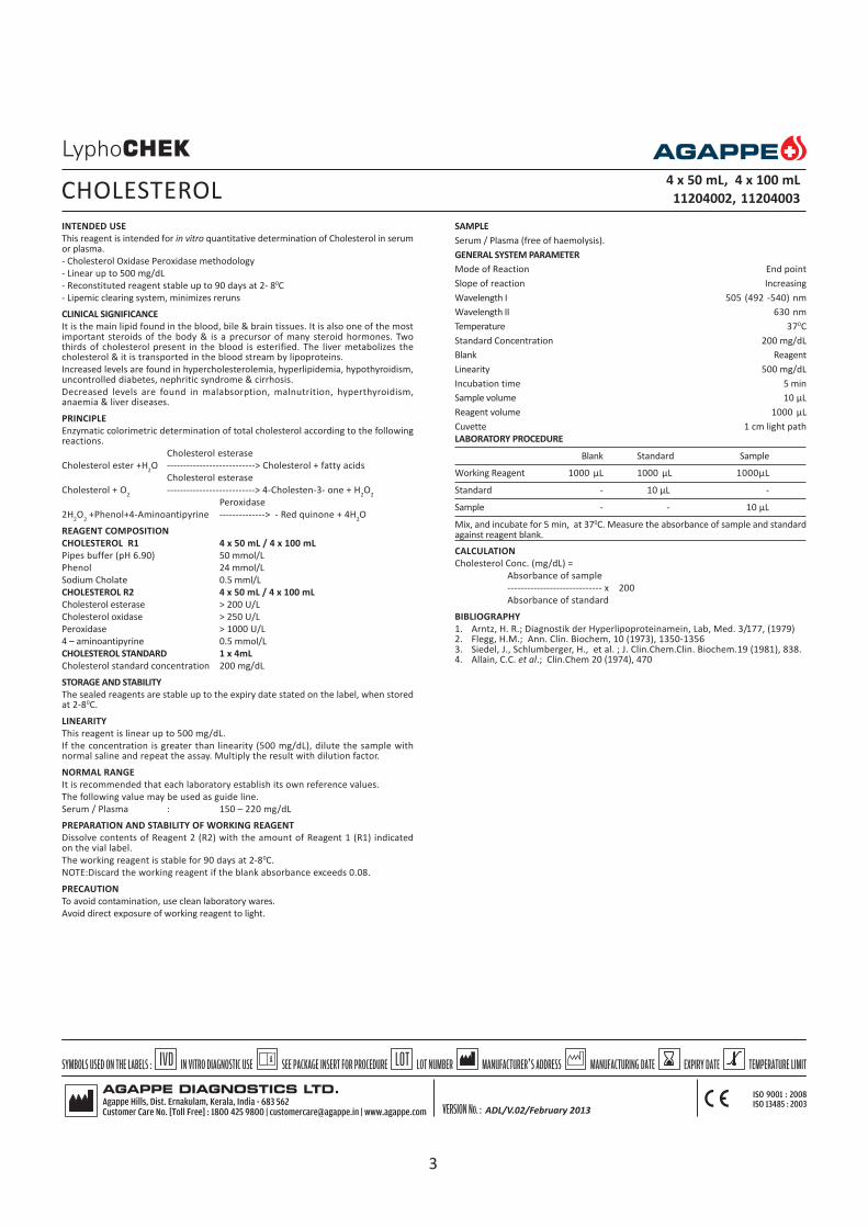
This reagent is intended for in vitro quantitative determination of Cholesterol in serumor plasma.
- Cholesterol Oxidase Peroxidase methodology
- Linear up to 500 mg/dL
- Reconstituted reagent stable up to 90 days at 2- 80C
- Lipemic clearing system, minimizes reruns
CLINICAL SIGNIFICANCE
It is the main lipid found in the blood, bile & brain tissues. It is also one of the mostimportant steroids of the body & is a precursor of many steroid hormones. Twothirds of cholesterol present in the blood is esterified. The liver metabolizes thecholesterol & it is transported in the blood stream by lipoproteins.
Increased levels are found in hypercholesterolemia, hyperlipidemia, hypothyroidism,uncontrolled diabetes, nephritic syndrome & cirrhosis.
Decreased levels are found in malabsorption, malnutrition, hyperthyroidism,anaemia & liver diseases.
PRINCIPLE
Enzymatic colorimetric determination of total cholesterol according to the followingreactions.
Cholesterol esterase
Cholesterol ester +H2O ---------------------------> Cholesterol + fatty acids
Cholesterol esterase
Cholesterol + O2
---------------------------> 4-Cholesten-3- one + H2O
2
Peroxidase
2H2O
2 +Phenol+4-Aminoantipyrine --------------> - Red quinone + 4H
2O
REAGENT COMPOSITION
CHOLESTEROL R1 4 x 50 mL / 4 x 100 mL
Pipes buffer (pH 6.90) 50 mmol/L
Phenol 24 mmol/L
Sodium Cholate 0.5 mml/L
CHOLESTEROL R2 4 x 50 mL / 4 x 100 mL
Cholesterol esterase > 200 U/L
Cholesterol oxidase > 250 U/L
Peroxidase > 1000 U/L
4 – aminoantipyrine 0.5 mmol/L
CHOLESTEROL STANDARD 1 x 4mL
Cholesterol standard concentration 200 mg/dL
STORAGE AND STABILITY
The sealed reagents are stable up to the expiry date stated on the label, when storedat 2-80C.
LINEARITY
This reagent is linear up to 500 mg/dL.
If the concentration is greater than linearity (500 mg/dL), dilute the sample withnormal saline and repeat the assay. Multiply the result with dilution factor.
NORMAL RANGE
It is recommended that each laboratory establish its own reference values.
The following value may be used as guide line.
Serum / Plasma : 150 – 220 mg/dL
PREPARATION AND STABILITY OF WORKING REAGENT
Dissolve contents of Reagent 2 (R2) with the amount of Reagent 1 (R1) indicatedon the vial label.
The working reagent is stable for 90 days at 2-80C.
NOTE:Discard the working reagent if the blank absorbance exceeds 0.08.
PRECAUTION
To avoid contamination, use clean laboratory wares.
Avoid direct exposure of working reagent to light.
CHOLESTEROL4 x 50 mL, 4 x 100 mL
11204002, 11204003
SAMPLE
Serum / Plasma (free of haemolysis).
GENERAL SYSTEM PARAMETER
Mode of Reaction End point
Slope of reaction Increasing
Wavelength I 505 (492 -540) nm
Wavelength II 630 nm
Temperature 370C
Standard Concentration 200 mg/dL
Blank Reagent
Linearity 500 mg/dL
Incubation time 5 min
Sample volume 10 µL
Reagent volume 1000 µL
Cuvette 1 cm light path
LABORATORY PROCEDURE
Blank Standard Sample
Working Reagent 1000 µL 1000 µL 1000µL
Standard - 10 µL -
Sample - - 10 µL
Mix, and incubate for 5 min, at 370C. Measure the absorbance of sample and standardagainst reagent blank.
CALCULATION
Cholesterol Conc. (mg/dL) =
Absorbance of sample
----------------------------- x 200
Absorbance of standard
BIBLIOGRAPHY
1. Arntz, H. R.; Diagnostik der Hyperlipoproteinamein, Lab, Med. 3/177, (1979)2. Flegg, H.M.; Ann. Clin. Biochem, 10 (1973), 1350-13563. Siedel, J., Schlumberger, H., et al. ; J. Clin.Chem.Clin. Biochem.19 (1981), 838.4. Allain, C.C. et al.; Clin.Chem 20 (1974), 470
ADL/V.02/February 2013
3

INTENDED USE:
This reagent is intended for in vitro quantitative determination of Cholinesterase inserum or plasma.
- Kinetic, Butyrylthiocholine method
- Linear up to 9084 U/L
- Stability of working reagent is 2 hours at 2-80C
CLINICAL SIGNIFICANCE
Cholinesterase levels in serum are useful as a test of liver function, as an indicator ofpossible insecticide poisoning or for detection of patients with atypical forms ofenzyme.
Measurements of serum Cholinesterase activity can serve as sensitive measures of thesynthetic capacity of the liver if the patient’s normal level is known.
Elevated levels (marginal increase) are observed in patients with nephritic syndrome,thyrotoxicosis & haemochromatosis in obese, diabetic people & in patients withanxiety & other psychiatric states.
Decreased levels are found in patients with acute infections, pulmonary embolism &muscular dystrophy as well as after surgical procedures. After a myocardial infarction,the enzyme levels decreases until the 5th day & then begins a slow rise to normal.
Decreased levels are also found in chronic renal disease & in pregnancy.
PRINCIPLE
Cholinesterase
Butyrylthiocholine + H2o --------------->Thiocholine + Butyrate
Thiocholine + Dithio-bis-nitrobenzoate--------------> nitro -2- mercapto-5- benzoato
REAGENT COMPOSITION
CHOLINESTERASE R1 5 x 3 mL
Buffer Solution
Phosphate buffer, (pH 7.7) 50 mmol/L
CHOLINESTERASE R2 1 x 5 x 3 mL
Tablets
5,5 DTNB 0.25 mmol/L
Butyrylthiocholine 7 mmol/L
STORAGE AND STABILITY
The sealed reagents are stable up to the expiry date stated on the label, when storedat 2- 80C.
LINEARITY
This reagent is linear up to 9084 U/L.
If the concentration is greater than linearity (9084 U/L), dilute the sample with normalsaline and repeat the assay.Multiply the result with dilution factor.
NORMAL RANGE
It is recommended that each laboratory establish its own reference values.
The following value may be used as guide line.
Serum : 4659-14443 U/L
PREPARATION AND STABILITY OF WORKING REAGENT
Dissolve one tablet R2 in one vial of Reagent 1 (R1). The stability of working reagentis 2 hours at 2-80c.
NOTE : Wait for 10 minutes for complete dissolution.
PRECAUTION
To avoid contamination, use clean laboratory wares.
Avoid direct exposure of working reagent to light.
SAMPLE
Fresh plasma (free of haemolysis), serum.
CHOLINESTERASE5 x 3 mL
11205001
GENERAL SYSTEM PARAMETER
( OD/60 sec) ( OD/30 sec)
Mode of Reaction Kinetic Kinetic
Slope of reaction Increasing Increasing
Wavelength 405 nm 405 nm
Temperature 370C 370C
Factor 22710 45420
Linearity 9084 U/L 9084 U/L
Blank DI Water DI Water
Delay 2 sec 2 sec
No of reading 2 4
Interval 60 sec 30 sec
Sample volume 20 µL 20 µL
Reagent volume 3000 µL 3000 µL
Cuvette 1 cm light path 1 cm light path
LABORATORY PROCEDURE
Working reagent 3000 µL
Sample (dilute 1+1 with saline solution.) 20 µL
Mix and measure the change in absorbance per minute ( OD/60 sec)during 2 minutes. Or measure the change in absorbance per 30 sec ( OD/30 sec)during 2 minutes.
CALCULATION
Cholinesterase activity (U/L) =
( OD/60 sec) x 22710
or
( OD/30 sec) x 45420
BIBLIOGRAPHY
Kendel, M., Yotros.; Kin. Wschr. 45, 325 (1967)
ADL/V.02/February 2013
4

INTENDED USE
This reagent is intended for in vitro quantitative determination of CK-MB in humanserum.
- Immuno – inhibition methodology
- Linear up to 1000 U/L
- Working Reagent Stable up to 8 days at 2- 80C
- Excellent pack sizes, Convenient for all laboratories
CLINICAL SIGNIFICANCE
CK-MB is an enzyme formed by the association of two subunits from muscle(M) andnerve cells (B). CK-MB is usually present in serum at low concentration; it increasesafter an acute infarct of myocardium and later descends at normal levels. Also isincreased, rarely, in skeletal muscle damage.
Clinical diagnosis should not be made on a single test result; it should integrate clinicaland other laboratory data.
PRINCIPLE
An antibody to the anti CK-M inhibits completely CK-MM and subunit (M) of the CK-MB. The activity of the non – inhibited CK-B subunit is then assayed by the followingseries of reactions:
CK
Phosphocreatine + ADP ----> Creatine + ATP
HK
ATP + Glucose ----- > ADP + Glucose - 6- Phosphate
G6P- DH
G6P+ NADP+ ------------> 6 – Phosphogluconate + NADPH + H+
The rate of NADPH formation, measured photometrically, is proportional to thecatalytic concentration of CK –B present in the sample.
REAGENT COMPOSITION
CK-MB R1 5 x 2.5 mL
Buffer
Imidazol (pH 6.7) 100 mmol/L
Glucose 20 mmol/L
Magnesium acetate 10 mmol/L
EDTA 2mmol/L
CK-MB R2 5 x 2.5 mL
*Anti CK –M 200 U/L
ADP 2 mmol/L
AMP 5mmol/L
di-Adenosine-5-pentaphosphate 10 mmol/L
NADP+ 2 mmol/L
Hexokinase 2500 U/L
Glucose-6-phosphate dehydrogenase 1500 U/L
N-acetyl cysteine 20 mmol/L
Creatine Phosphate 30 mmol/L
*Anti CK-M sufficient to inhibit up to 2000 U/L of CK-MM
CK-MB Calibrator 1 x 2 mL
Calibrator concentration is mentioned on vial label
STORAGE AND STABILITY
The sealed reagents are stable up to the expiry date stated on the label, when storedat 2 - 80C.
LINEARITY
This reagent is linear up to 1000 U/L.
If the concentration is greater than linearity (1000 U/L), dilute the sample with normalsaline and repeat the assay. Multiply result with dilution factor.
NORMAL VALUES
It is recommended that each laboratory should establish its own reference values.
The following value may be used as guide line.
250C 300 C 370C
CK-MB : > 10 U/L > 15 U/L >24 U/L
PREPARATION AND STABILITY OF WORKING REAGENT
Reconstitute one tablet of R2 in one vial of R1.
The reconstituted reagent is stable for 8 days at 2-80C.
Calibrator: Reconstitute the calibrator with 2 mL distilled water and reconstitutedcalibrator is stable for:
24 hrs at 15 to 25oC
3 Days 2 to 8oC
1 Month at -15 to -25oC
PRECAUTION
To avoid contamination, use clean laboratory materials.
Avoid direct exposure of working reagent to light.
SAMPLE
Serum (Free of haemolysis)
GENERAL SYSTEM PARAMETER
OD/300 Sec OD/60 Sec
Mode of Reaction Fix Time Kinetic
Slope of reaction Increasing Increasing
Wavelength 340 nm 340 nm
Temperature 370C 370C
Factor 1651 8254
Linearity 1000 U/L 1000 U/L
Blank DI Water DI Water
Delay time 600 sec 180 sec
No of reading - 3
Delta time 300 sec 60 sec
Reagent volume 1000 µL 1000 µL
Sample volume 40 µL 40 µL
Cuvette 1 cm light path 1 cm light path
LABORATORY PROCEDURE
Working reagent 1000 µL
Sample 40 µL
Mix and incubate at 370C for 10 minutes measure the initial absorbance(A1) and againtake the readings for 5 minutes(A2). Calculate the difference between absorbance A = (A2-A1), or mix and incubate at 370C for 3 minutes. Read the charge in absorbanceper minute ( OD/minutes) during 3 minutes.
CALCULATION
CK-MB Activity (U/L) = A x 1651
or
CK-MB Activity (U/L) = A/Min x 8254
BIBLIOGRAPHY
1. Abbot, B. et al.; Creatinine kinase. Kalpan, A. et al. Clin. Chem. The C.V.MosbyCo. St.Louis. Toronto Princeton 1984:1112 – 116
2. Gerhardt, W., et al.; Creatine K inase B-Subnit activity in serum afterimmunoinhibition of M-subunit activity.; Clin chem.1979; (25/7): 1274- 1280.
3. Young, D. S.; Effects of drugs on clinical Lab. Tests, 4th Ed AACC 2001.4. Burits, A. et al.; Tietz Textbook of Clinical Chemistry, 3rd Ed AACC 1999.5. Tietz, N. W. et al. Clinical Guide to Laboratory Tests, 3rd Ed AACC 1995.
CK MB with Calibrator 5 x 2.5 mL
11207004
5

INTENDED USE
This reagent is intended for in vitro quantitative determination of Creatine Kinase inhuman serum.
- Optimized IFCC Method
- Linear up to 1000 U/L
- Working Reagent is stable up to 5 days at 2-80C.
- Excellent pack Sizes, Convenient for all laboratories.
CLINICAL SIGNIFICANCE
It is mainly found in all muscle (Cardiac & Skeletal) & brain tissues. It plays animportant role in energy storing mechanism of the tissues. It’s iso-enzymes: CK-MBmainly exists in cardiac muscle tissues, CK-MM in skeletal muscle tissues & CK-BBin brain.
Increased levels are found in myocardial infarction, muscular dystrophy,cerebrovascular-disease, pulmonary infarction, electrical shocks & hypothyroidisim.
Decreased levels are, some times seen in early pregnancy, alcohol, liver diseses, RA.
PRINCIPLE
Creatine kinase(CK) catalyses the reversible transfer of a phosphate group fromphosphocreatinine to ADP. This reaction is coupled to those catalyzed by hexokinase(HK) and glucose – 6- phosphate dehygrogenase (G6P-DH)
CK
Phosphocreatine + ADP ----- > Creatine +ATP
HK
ATP + Glucose ----- > ADP + Glucose – 6- phosphate
G6P – DH
G6P + NADP+ ------------- > 6- Phosphogluconate + NADPH + H+
The rate of NADPH formation, measured photometrically, is proportional to thecatalytic concentration of CK present in the sample.
REAGENT COMPOSITION
CREATINE KINASE R1 5 x 2.5 mL
Buffer
Imidazol pH 7.0 100 mmol/L
Glucose 20 mmol/L
Magnesium acetate 10 mmol/L
EDTA 2mmol/L
CREATINE KINASE R2 5 x 2.5 mL
Substrate
ADP 2 mmol/L
AMP 5 mmol/L
Di-Adenosine – 5-pentaphosphate 10mmol/L
NADP+ 2 mmol/L
Hexokinase 2500 U/L
Glucose -6-phosphate dehydrogenase 1500 U/L
N-acetyl cysteine 20 mmol/L
Creatine Phosphate 30mmol/L
STORAGE AND STABILITY
The sealed reagents are stable up to the expiry date stated on the label, when storedat 2 - 80C.
LINEARITY
This reagent is linear up to 1000 U/L
If the concentration is greater than linearity (1000 U/L), dilute the sample withnormal saline and repeat the assay. Multiply the final result with dilution factor.
NORMAL VALUES
It is recommended that each laboratory establish its own reference value.
The following value may be used as guide line.
250C 300C 370C
Men up to : 80 U/L 130 U/L 195 U/L
Women up to : 70 U/L 110 U/L 170 U/L
PREPARATION AND STABILITY OF WORKING REAGENT
Reconstitute one tablet R2 with one vial of R1 .
The reconstituted reagent is stable for 5 days at 2-80C.
PRECAUTION
To avoid contamination, use clean laboratory wares.
Avoid direct exposure of working reagent to light.
CK-NAC5 x 2.5 mL
11206004
SAMPLE
Serum (Free of haemolysis)
GENERAL SYSTEM PARAMETER
Mode of Reaction Kinetic
Slope of reaction Increasing
Wavelength 340 nm
Temperature 370C
Factor 8095
Linearity 1000 U/L
Blank Distilled Water
Delay time 120 sec.
No of reading 3
Interval 60 sec
Reagent volume 1000 µL
Sample volume 20 µL
Cuvette 1 cm light path
LABORATORY PROCEDURE
Working reagent 1000 µL
Sample 20 µL
Mix and incubate at 370C for 2 minutes, measure the change in absorbance perminute ( OD/min) during 3 minutes.
CALCULATION
Creatine Kinase Activity (U/L) = ( OD /min.) x 8095
BIBLIOGRAPHY
1. Abbot, B. et al. Creatinine kinase. Kalpan, A. et al.; Clin. Chem. The C.V.MosbyCo. St Louis. Toronto Princeton 1984:1112 – 116
2. Gerhardt, W. et al. Creatine K inase B-Subnit activity in serum afterimmunoinhibition of M-subunit activity; Clin. chem.1979; (25/7): 1274- 1280.
3. Young, D. S.; Effects of drugs on clinical Lab. Tests, 4th ed AACC 2001.4. Burits, A. et al. ; Tietz Textbook of Clinical Chemistry, 3rd ed AACC 1999.6. Tietz, N. W. et al. Clinical Guide to Laboratory Tests, 3rd Ed AACC 1995.
ADL/V.02/February 2013
6

INTENDED USE
This reagent is intended for in vitro quantitative determination of Glucose in serum,plasma & CSF.
- GOD –PAP methodology
- Linear upto 500 mg/dL
- Reconstituted stability 90 days at 2-80C
- No interferences from Bilirubin and Creatinine
CLINICAL SIGNIFICANCE
Glucose is a major carbohydrate present in the blood & serves as a primary source ofenergy. It is usually obtained from ingested starch & sugar. The glucose concentrationis normally maintained at constant level. Excessive glucose is stored as inactiveglycogen mainly in the liver & little in the muscles.
Elevated blood glucose levels are found in diabetes mellitus, hyperthyroidism,hyperadrenalism & certain liver diseases.
Decreased levels are found in Insulinoma, hypothyroidism, hypopituitarism.
PRINCIPLE
Enzymatic colorimetric determination of glucose according to the following reaction.
Glucose Oxidase
Glucose+ O2
------------------> Gluconic acid + H2O
2
Peroxidase
2H2O
2+phenol + 4-Aminoantipyrine --------------- > red Quinonimine + 4H
2O
REAGENT COMPOSITION
GLUCOSE R1 4x100mL / 4x250mL / 4x500mL
Phosphate buffer, (pH 7.40) 100 mmol/L
Phenol 10 mmol/L
GLUCOSE R2 4x100mL / 4x250mL / 4x500mL
Glucose Oxidase > 10000 U/L
Peroxidase > 600U/L
4-Aminoantipyrine 270mmol/L
GLUCOSE STANDARD 1x4mL / 2x4mL
Glucose standard concentration 100 mg/dL
Note : For 4x500 mL and 4x250mL packs, Buffer will be provided separately
STORAGE AND STABILITY
The sealed reagents are stable upto the expiry date stated on the label, when storedat 2 - 80C.
LINEARITY
This reagent is linear upto 500mg/dL
If the concentration is greater than linearity (500 mg/dL), dilute the sample with normalsaline and repeat the assay. Multiply the result with dilution factor.
NORMAL RANGE
It is recommended that each laboratory establish its own reference values.
The following value may be used as guide line.
Serum / Plasma : 70-105 mg/dL
C S F : 50 -70 mg/dL
PREPARATION AND STABILITY OF WORKING REAGENT
Dissolve Reagent 2(R2 )with the volume of Reagent 1 (R1) indicated on the label.
The reconstituted reagent is stable for 90 days at 2-80c.
NOTE: Discard the working reagent if the blank absorbance exceeds 0.08.
PRECAUTION
To avoid contamination, use clean laboratory wares.
Avoid direct exposure of working reagent to light.
SAMPLE
Serum / plasma (free of haemolysis) / CSF.
GLUCOSE 4 x 100 mL,4 x 250 mL, 4 x 500 mL
11208001, 11208002, 11208003
GENERAL SYSTEM PARAMETER
Mode of Reaction Fixed time Kinetic
Slope of reaction Increasing
Wavelength 505 (500-540 nm)
Temperature 370C
Standard Concentration 100 mg/dL
Blank Reagent Blank
Linearity 500 mg/dL
Delay 30 sec.
Interval 60 sec
Sample volume 10 µL
Reagent volume 1000 µL
Cuvette 1 cm light path
CALCULATION FOR FIXED TIME
Absorbance of Sample
Glucose conc (mg/dL)= ----------------------------------- x 100
Absorbance of standard
GENERAL SYSTEM PARAMETER FOR ENDPOINT
Mode of Reaction End point
Slope of reaction Increasing
Wavelength 505 nm
Temperature 370C
Standard Concentration 100 mg/dL
Blank Reagent Blank
Linearity 500 mg/dL
Incubation time 10 min
Sample volume 10 µL
Reagent volume 1000 µL
Cuvette 1 cm light path
LABORATORY PROCEDURE
Blank Standard Sample
Working reagent 1000 µL 1000 µL 1000 µL
Standard - 10 µL -
Sample - - 10 µL
Mix and incubate for 10 minutes at 370C. Measure the change in absorbance ofstandard and sample against reagent blank.
CALCULATION FOR END POINT
Absorbance of Sample
Glucose Conc. (mg/dL) = ------------------------------ x 100
Absorbance of standard
BIBLIOGRAPHY
Trinder, P.; Ann. Clin., Biochem. 6, (1969) 24
ADL/V.02/February 2013
7

INTENDED USE
This reagent is intended for in vitro quantitative determination of SGOT in serum orplasma.
- Proven IFCC methodology
- Linear up to 350 U/L
- Reconstituted reagent stable up to 30 days at 2-80C
CLINICAL SIGNIFICANCE
It is present in most of the tissues. Especially in cardiac muscle, liver cells, skeletalmuscle & kidneys. Injury to these tissues results in the release of the enzyme in bloodstream.
Increased levels are found in myocardial infarction. The duration & extent of increaseis related to the infract. GOT determination is of considerable value to differentiatemyocardial infraction from other cardiac disorders.
Increased levels are also found in various types of liver disease, skeletal muscle trauma& in renal diseases.
Decreased levels may be found in pregnancy, Beri-Beri & Diabetic ketoacidosis.
PRINCIPLE
Kinetic determination of Aspartate Aminotrasferase (AST) based upon the followingreaction.
AST/SGOT
L- Aspartate + alpha - ketoglutarate ------ > Oxaloacetate + L-Glutamate.
MDH
Oxaloacetate + NADH + H+ ------ > L- Malate + NAD+
AST : Aspartate aminotransferase.
MDH : Malate dehydrogenase.
REAGENT COMPOSITION
SGOT R1 2 x 53 mL / 4 x 50 mL
Tris Buffer (pH 7.8) 88 mmol/L
L-Aspartate 260 mmol/L
SGOT R2 5x 20 mL / 4x 50 mL
MDH > 600 U/L
LDH > 900 U/L
NADH 0.20 mmol/L
a -ketoglutarate 12 mmol/L
STORAGE & STABILITY
The sealed reagents are stable up to the expiry date stated on the label, when storedat 2 - 80C.
LINEARITY
This reagent is linear up to 350 U/L.
If the concentration is greater than linearity (350 U/L), dilute the sample with normalsaline and repeat the assay. Multiply the result with dilution factor.
REFERENCE RANGE
It is recommended that each laboratory establish its own reference values.
The following value may be used as guide line.
Females upto 31U/L at 37o CS
Males upto 35U/L at 37o C
PREPARATION AND STABILITY OF WORKING REAGENT
Reconstitute the reagent 2 (R2 ) with the volume of reagent 1 (R1) mentioned on the
vial label. The reconstituted reagent is stable for 30 days at 2-80C.
NOTE: Discard the working reagent if the blank absorbance is less than 1.00 at 340nm.
PRECAUTION
To avoid contamination, use clean laboratory wares.
Avoid direct exposure of working reagent to light.
SAMPLE
Serum / plasma (free of haemolysis)
SGOT 5x 20 mL,4x 50 mL,
11213005, 11213003
GENERAL SYSTEM PARAMETER
Mode of Reaction Kinetic
Slope of reaction Decreasing
Wavelength 340 nm
Temperature 370C
Factor 1768
Linearity 350 U/L
Blank DI Water
Delay 60 sec.
No of reading 3
Interval 60 sec
Sample volume 100 µL
Reagent volume 1000 µL
Cuvette 1 cm light path
LABORATORY PROCEDURE
Working reagent 1000 µL
Sample 100 µL
Mix and incubate at 370C for 1 minute. Measure the change in absorbance per minute( OD/min) during 3 minutes.
CALCULATION
SGOT activity (U/L) = ( OD/min) x 1768
BIBLIOGRAPHY
Expert panel on enzyme of the IFCC; Clin. Chim. Acta, 70(1976) F 19.
ADL/V.02/February 2013
8

INTENDED USE
This reagent is intended for in vitro quantitative determination of SGPT in serum orplasma.
- IFCC recommended methodology
- Linear up to 350 U/L
- Reconstituted reagent stable up to 50 days at 2 - 80C
CLINICAL SIGNIFICANCE
It is present in most of the tissues, but mainly found in the liver. Increased levels arefound in hepatitis, cirrhosis, obstructive jaundice & other hepatic disease. SGPT activityis markedly elevated even before clinical signs of jaundice become apparent in diseaseassociated with hepatic necrosis. Slight elevations are also found in myocardialinfraction.
PRINCIPLE
Kinetic determination of Alanine Aminotransferase (ALAT) according to the followingreaction.
ALT
L-Alanine + alpha-ketoglutarate ----- > Pyruvate +L-Glutamate
LDH
Pyruvate +NADH+ H+ ----- > L-Lactate +NAD+
ALT – Alanine aminotranferase
LDH - Lactate dehydrogenase
REAGENT COMPOSITION
SGPT R1 2 x 53 mL/ 4 x 50 mL
Tris buffer (pH 7.5) 110 mmol/L
L-Alanine 550 mmol/L
SGPT R2 5 x 20 mL/ 4 x 50 mL
LDH > 200 U/L
NADH 0.20 mmol/L
a-ketoglutarate 16 mmol/L
STORAGE AND STABILITY
The sealed reagents are stable up to the expiry date stated on the label, when storedat 2 - 80C.
LINEARITY
This reagent is linear up to 350 U/L .
If the concentration is greater than linearity (350 U/L), dilute the sample with normalsaline and repeat the assay. Multiply the result with dilution factor.
NORMAL RANGE
It is recommended that each laboratory establish its own reference values.
The following value may be used as guide line.
Serum up to : 49 U/L
PREPARATION AND STABILITY OF WORKING REAGENT
Reconstitute the Reagent 2 (R2) with the volume of Reagent1 (R1) mentioned on thevial label. The reconstituted reagent is stable for 50 days at 2-80C.
NOTE: Discard the working reagent if the blank absorbance is less than 1.00 at 340nm.
PRECAUTION
To avoid contamination use clean laboratory wares.
Avoid direct exposure of working reagent to light.
SAMPLE
Serum / plasma (free of haemolysis)
SGPT 5 x 20 mL, 4 x 50 mL
11214005,11214003
GENERAL SYSTEM PARAMETER
Mode of Reaction Kinetic
Slope of reaction Decreasing
Wavelength 340 nm
Temperature 370C
Factor 1768
Blank DI Water
Linearity 350 U/L
Delay 60 sec.
No of reading 3
Interval 60 sec
Sample volume 100 µL
Reagent volume 1000 µL
Cuvette 1 cm light path
LABORATORY PROCEDURE
Working reagent 1000 µL
Sample 100 µL
Mix and incubate at 370C for 1 minute. Measure the change in absorbance per minute( OD/min) during 3 minutes.
CALCULATION
SGPT activity (U/L) = ( OD/min) x 1768
BIBLIOGRAPHY
Expert panel on enzyme of the IFCC; Clin. Chim. Acta, 70 (1976) F19.
ADL/V.02/February 2013
9

INTENDED USE
This reagent is intended for in vitro quantitative determination of triglycerides in serumor plasma.
- Proven GPO-POD methodology
- Linear up to 1000 mg/dL
- Extended stability, reconstituted reagent is stable up to 42 days at 2-80 C
CLINICAL SIGNIFICANCE
Triglycerides are simple lipids, formed in the liver by glycerol & fatty acids. They aretransported by VLDL, LDL & constitute about 95% of fat, stored as source of energyin the tissue & plasma. Increased levels are found in hyperlipidemias, diabetes,nephrotic syndrome & hypothyroidism. Increased levels are risk factor forarteriosclerotic coronary disease, peripheral vascular disease, acute pancreatitis &hyperlipoproteinaemia. Decreased levels are found in malnutrition & hyperthyroidism.
PRINCIPLE
Enzymatic determination of triglyceride is based on the following reactions:
LPL
TGL+H2O ---- >Glycerol + Fatty acid
GK
Glycerol + ATP ---- > Glycerol-3-phosphate + ADP
Mg++
GPO
Glycerol-3-phosphate+O2------ > Dihydroxyacetone phosphate +H
2O
2
POD
2H2O
2+4-Aminoantipyrine+p-Chlorophenol ------ > Red Quinone+4H
2O
GPO = Glycereol-3-phosphate Oxidase
LPL = Lipoprotein Lipase
GK = Glycerol Kinase
REAGENT COMPOSITION
TRIGLYCERIDES R1 2 x 53 mL / 4 x 50 mL
Pipes –buffer (pH 7.00) 50 mmol/L
p-Chlorophenol 5.3 mmol/L
Magnesium salt 15 mmol/L
TRIGLYCERIDES R2 5 x 20 mL / 4 x 50 mL
Lipoprotein lipase >1100 U/L
Glycerol Kinase > 800 U/L
Glycerol -3-phosphate oxidase > 5000 U/L
Peroxidase 350U/L
4-Amino antipyrine 0.7 mmol/L / 0.3 mmol/L
TRIGLYCERIDES STANDARD 1 x 4 mL
Triglycerides standard concentration 200mg/dL
STORAGE & STABILITY
The sealed reagents are stable up to the expiry date stated on the label, when storedat 2 - 80C.
LINEARITY
This reagent is linear up to 1000 mg/dL.
If the concentration is greater than linearity (1000 mg/dL), dilute the sample withnormal saline and repeat the assay. Multiply the result with dilution factor.
NORMAL RANGE
It is recommended that each laboratory establish its own reference values.
The following value may be used as guide line.
Normal Value 65-165 mg/dL
PREPARATION AND STABILITY OF WORKING REAGENT
Reconstitute the Reagent 2 (R2) with the volume of Reagent 1 (R1) indicated on thelabel.
Working reagent is stable for 42 days at 2-80C.
PRECAUTION
To avoid contamination, use clean laboratory wares.
Avoid direct exposure of working reagent to light.
SAMPLE
Serum / plasma (free of haemolysis)
TRIGLYCERIDES5 x 20 mL, 4 x 50 mL
11215002, 11215003
GENERAL SYSTEM PARAMETER
Mode of Reaction End Point
Slope of reaction Increasing
Wavelength I 505 nm (492-550 nm)
Wavelength II 630 nm
Temperature 370C
Standard Concentration 200 mg/dL
Linearity 1000 mg/dL
Blank Reagent
Incubation time 5 min
Sample volume 10 µL
Reagent volume 1000 µL
Cuvette 1 cm light path
LABORATORY PROCEDURE
Blank Standard Sample
Working Reagent 1000 µL 1000 µL 1000 µL
Standard - 10 µL -
Sample - - 10 µL
Mix and incubate for 5 minutes at 370C. Measure the change in absorbance ofstandard and sample against reagent blank.
CALCULATION
Absorbance of sample
Triglycerides Con. (mg/dL) = ------------------------------ x 200 mg/dL
Absorbance of standard
BIBLIOGRAPHY
1. Buccolo, G., David, M.; Clin.Chem., 19(1973) 4762. Wener, M., Gabrielson, D. G., Eastman, G.; Clin Chem 21 (1981) 2683. Annoni, G., Bottasso, B. M., Ciaci, D., Donato, M. F., Tripoli, A., Lamb, J.; J. Res.
Lab, Med., 9 (1982) 115
ADL/V.02/February 2013
10

INTENDED USE
This reagent is intended for in vitro quantitative determination of urea in serum, plasma& urine.
- Modified Berthelot methodology
- Linear up to 200 mg/dL
- Reconstituted reagent stability for 21 days at 2-80C.
CLINICAL SIGNIFICANCE
Proteins cannot be stored in human body, so excess should be broken down. Aminoacids which from the components of proteins, break down to give ammonia. This istoxic & so through a series of chemical reactions (urea cycle) non toxic urea isproduced & this is released into the blood which is filtered in the kidney & excretedin the urine.
Elevated levels are seen during increased protein in breakdown, dehydration, vomiting,diarrhea. Also seen in any kind of renal disorder like Glomerular nephritis, chronicnephritis & Nephritic syndrome.
Decreased levels are found in liver failure & pregnancy
PRINCIPLE
Enzymatic determination of Urea according to the following reaction.
Urease
Urea + H2O ----------> 2NH
3 + CO
2
Nitroprusside
NH3 + salicylate ------------------> 2.2-Dicarboxy Indophenol
Hypochlorite
REAGENT COMPOSITION
UREA B COLOUR REAGENT R1 2 x 53 mL
Sodium Salicylate 80 mmol/L
Sodium Nitroprusside 4 mmol/L
Sodium Hypochlorite 45 mg /dL
UREA B R2 10 x 10 mL
Phosphate buffer, (pH 6.9) 60 mmol/L
Urease 20 KU/L
UREA B STANDARD 1 x 4 mL
Urea –B Standard concentration 40 mg/dL
STORAGE AND STABILITY
The sealed reagents are stable up to the expiry date stated on the label, when storedat 2 - 80C.
LINEARITY
This reagent is linear up to 200 mg/dL.
If the concentration is greater than linearity (200 mg/dL), dilute the sample with normalsaline and repeat the assay. Multiply the result with dilution factor.
NORMAL RANGE
It is recommended that each laboratory establish its own reference values.
The following value may be used as guide line.
Serum : 10-55 mg/dL
Urine : 20-35 gm/24 hrs
PREPARATION AND STABILITY OF WORKING REAGENT
Reconstitute 1 vial of reagent 2 (R2) with the volume of DI water as indicated on thevial label.
Working reagent is stable for 21 days at 2-80C.
PRECAUTION
To avoid contamination, use clean laboratory wares.
Avoid direct exposure of working reagent to light.
SAMPLE
Serum / plasma (free of haemolysis) / Urine (1/100 Diluted)
GENERAL SYSTEM PARAMETER
Mode of Reaction End point
Slope of reaction Increasing
Wavelength 600 nm (580-650 nm)
Temperature 370C
Standard Concentration 40 mg/dL
Linearity 200 mg/dL
Blank Reagent
Sample volume 10 µL
Reagent volume 1000 µL + 1000 µL
Incubation time 5+5 min
Cuvette 1 cm light path
LABORATORY PROCEDURE
Blank Standard Sample
Working reagent 1000 µL 1000 µL 1000 µL
Standard - 10 µL -
Sample - - 10 µL
Mix and incubate for 5 minutes at 370C then add.
Colour Reagent 1000 µL 1000 µL 1000 µL
Mix and incubate for 5 minutes at 370C then add.
DI Water 1000 µL 1000µL 1000 µL
Mix well & measure the absorbance of sample and the standard against the reagentblank.
CALCULATION
Absorbance of sample
Urea Con. (mg/dL) = ---------------------------- X 40
Absorbance of Standard
BIBLIOGRAPHY
1. Wheatherburn M.W., Anal. Chem., 1967; 39;9712. Searcy, R.L., Reardon, J.E., Foreman, J.A.; Amer, J.Med, Tech., 1967; 33, 15-203. Henry, R. J.; Clin. Chem. Principle and Techniques, Harper & Row, New York,
1963.P.268
UREA - B 200 mL
11216001
11

INTENDED USE
This reagent is intended for in vitro quantitative determination of urea in serum,plasma & urine.
- Urease / GLDH methodology
- Linearity
Urea : 500 mg/dL
BUN : 235 mg/dL
- Working reagent stability is 42 days at 2-80C.
CLINICAL SIGNIFICANCE
Proteins cannot be stored in human body, so excess should be broken down. Aminoacids which from the components of proteins, break down to give ammonia. Thisis toxic & so through a series of chemical reactions (urea cycle) non toxic urea isproduced & this is released into the blood which is filtered in the kidney & excretedin the urine.
Elevated levels are seen during increased protein breakdown, dehydration, vomiting,diarrhea. Also seen in any kind of renal disorder like Glomerular nephritis, Chronicnephritis & Nephritic syndrome.
Decreased levels are found in liver failure & pregnancy.
PRINCIPLE
Urease
Urea + H2O ---------- > 2NH
3 + CO
2
GLDH
2 NH3+2a- ketoglutarate + 2NADH --------- > 2L-Glutamate+2NAD+ + 2H
2O
REAGENT COMPOSITION
UREA U.V. R1 4 x 50 mL / 4 x 100 mL
Buffer (pH 7.55) 75 mmol/L
UREA U.V. R2 4 x 50 mL / 4 x 100 mL
ADP 0.66 mmol/L
GLDH >1000U/L
Urease > 30000 U/L
NADH 0.32 mmol/L
a-ketoglutarate 7.5 mmol/L
UREA U.V. STANDARD 1 x 4 mL
Standard concentration for Urea 50 mg/dL
Standard concentration for BUN 23.4 mg/dL
STORAGE & STABILITY
The sealed reagents are stable up to the expiry date stated on the label, when storedat 2 - 80C.
LINEARITY
This reagent is linear up to 500 mg/dL.
If the concentration is greater than linearity (500 mg/dL), dilute the sample with normalsaline and repeat the assay. Multiply the result with dilution factor.
NORMAL RANGE
It is recommended that each laboratory establish its own reference values.
The following value may be used as guide line.
Urea Urea – N
Sreum : 10-50 mg/dL 4.7 – 23 mg/dL
Urine /24 hr :20-36 gm/L 9.4-16.9 gm/L
PREPARATION AND STABILITY OF WORKING REAGENT
Dissolve Reagent 2 (R2) with the volume of Reagent 1 (R1) indicated on the label.
Working reagent is stable for 42 days at 2-80C.
Note: Discard the working reagent if the blank absorbance is less than 1.0 at 340 nm.
PRECAUTION
To avoid contamination, use clean laboratory wares.
Avoid direct exposure of working reagent to light.
SAMPLE
Serum / plasma (free of haemolysis)
Urine (1/100 diluted).
Do not use anticoagulants containing fluoride or ammonium ions.
UREA U.V 4 x 50 mL, 4 x 100 mL
11217001, 11217002
GENERAL SYSTEM PARAMETER
Mode of Reaction Fixed Time
Slope of reaction Decreasing
Wavelength 340
Temperature 370C
Standard Concentration 50 mg/dL
Linearity 500 mg/dL
Blank DI Water
Delay time 30 sec
Interval 60 sec
Sample volume 10 µL
Reagent volume 1000 µL
Cuvette 1 cm light path
LABORATORY PROCEDURE
Standard Sample
Working reagent 1000µL 1000 µL
Standard 10 µL -
Sample - 10 µL
Mix and read the optical density (T1) 30 seconds after the sample or standard
addition. Exactly 60 seconds after the first reading take second reading (T2).
CALCULATION
(T1 –T
2)of Sample
Urea Conc. (mg/dL) = ------------------------ x 50
(T1-T
2) of standard
(T1 –T
2)of Sample
Urea BUN Conc. (mg/dL) = ------------------------ x 23.4
(T1-T
2) of standard
BIBLIOGRAPHY
1. Talke, H. and Schubert, G. E.; Kiln-Wocchsr 19; 43: 1742. Thomas, L.; Labor and Diagnose, 1. Auflage, S. 346
ADL/V.02/February 2013
12

INTENDED USE
This reagent is intended for invitro quantitative determination of Uric acid in serum,plasma & urine.
- Uricase - PAP methodology
- Linear up to 25 mg/dL
- Reconstituted reagent stability up to 30 days at 2-80C
- Fast incubation, just 5 minutes at 370C
CLINICAL SIGNIFICANCE
Uric acid is the end product of purine metabolism. Uric acid is excreted by the kidneys.Increased levels are found in gout, arthritis, impaired renal functions & starvation.Decreased levels are found in yellow atrophy of the liver, Wilson’s disease ,& Fanconissyndrom.
PRINCIPLE
Enzymatic determination of Uric acid according to the following reactions.
Uricase
Uric acid + 2H2O+O
2----------> Allantoin +CO
2 +H
2O
2Peroxidase
2H2O
2 + 4 -Aminoantipyrine +DHBS -------------> Red quinone + H
2O+ HCL
DHBS = 3,5-Dichloro -2-Hydroxybenzenesulfonic acid.
REAGENT COMPOSITION
URIC ACID R1 2 x 53 mL / 4 x 50 m L
Phosphate Buffer (pH 7.5) 100 mmol/L
DHBS 2 mmol/L
URIC ACID R2 5 x 20 mL / 4 x 50 mL
4 – Aminoantipyrine > 0.23mmol/L
Peroxidase > 660 U/L
Uricase > 60 U/L
URIC ACID STANDARD 1 x 4 mL
Uric acid standard concentration 6 mg/dL
STORAGE & STABILITY
The sealed reagents are stable up to the expiry date stated on the label, when storedat 2 - 80C.
LINEARITY
This reagent is linear up to 25 mg/dL.
If the concentration is greater than linearity (25 mg/dL), dilute the sample with normalsaline and repeat the assay. Multiply the result with dilution factor.
NORMAL RANGE
It is recommended that each laboratory establish its own reference values.
The following value may be used as guide line.
Serum / Plasma
Males : 3.4 - 7.0 mg/dL
Females : 2.5 - 6.0 mg/dL
Children : 2.5 - 5.5 mg/dL
Urine : 250 - 750 mg/ 24 hr urine
PREPARATION AND STABILITY OF WORKING REAGENT
Reconstitute the reagent 2 (R2) with the volume of Reagent 1 (R1) indicated on thevial label.
Working reagent is stable for 30 days at 2-80C.
PRECAUTION
To avoid contamination, use clean laboratory wares.
Avoid direct exposure of working reagent to light.
SAMPLE
Serum / plasma (free of haemolysis) / Urine (1/10 diluted with distilled water).
URIC ACID5x20 mL, 4 x 50 mL
11218002, 11218003
GENERAL SYSTEM PARAMETER
Mode of Reaction End point
Slope of reaction Increasing
Wavelength I 505 nm (500 - 550)
Wavelength II 630 nm
Temperature 370C
Standard Concentration 6 mg/dL
Linearity 25 mg/dL
Blank Reagent
Sample volume 20 µL
Reagent volume 1000 µL
Incubation time 5 minutes
Cuvette 1 cm light path
LABORATORY PROCEDURE
Blank Standard Sample
Working Reagent 1000 µL 1000 µL 1000 µL
Standard - 20 µL -
Sample - - 20 µL
Mix and incubate 5 min. at 370C. Measure absorbance of sample and standard againstthe reagent blank.
CALCULATION
Absorbance of Sample
Uric Acid Con. (mg/dL) = ------------------------------ x 6
Absorbance of Standard
BIBLIOGRAPHY
1.Trivdi R.C., Revbar L., Berka, E., Strong L.,Clin. Chem. 24 (1978) 1908
2. Text Book of medical Laboratory Technology, sood
ADL/V.02/February 2013
13

INTENDED USE
This reagent is intended for in vitro quantitative determination of Alkaline Phosphatasein serum or plasma.
- DGKC – SCE recommended procedure
- Linear upto 700 U/L
- Working reagent can be prepared as per requirement
- Pack sizes to suit all types of laboratories
CLINICAL SIGNIFICANCE
Alkaline phosphatase (ALP) is widely distributed throughout the body, but clinicallyimportant one for diagnostic reasons are in bone, liver, placenta & intestine. Growingbone is associated with the release of ALP and so in childhood the level of ALP isaround 3 times of that of adult. During pregnancy in 2nd & 3rd trimester the enzymerises considerably due to placenta releasing ALP. It can be used to examine placentalfunction.
Elevated levels are seen in bone diseases, e.g. pagets disease, rickets, osteoblasticmetastatic & in obstructive disease of biliary tract.
Decreased levels are rarely seen. e.g. in Vitamin A resistant rickets.
PRINCIPLE
Kinetic determination of ALP according to the following reaction
ALP
Para-nitrophenyl phosphate + H2O ----- > p-nitrophenol+Inorgnic
phosphate
ALP = Alkaline Phosphatase
REAGENT COMPOSITION
ALKALINE PHOSPHATASE(S.L) R1 2 x 8 mL /2 x 24 mL / 2 x 40 mL
Diethanolamine Buffer (pH 10.2) 125 mmol/L
Magnesium Chloride 0.625 mmol/L
ALKALINE PHOSPHATASE (S.L) R2 2 x 2 mL/2 x 6 mL / 2 x 10 mL
P-Nitrophenyl phosphate 50 mmol /L
STORAGE AND STABILITY
The sealed reagents are stable upto the expiry date stated on the label, when storedat 2-80C.
LINEARITY
The reagent is linear, upto 700 U/L.
If the concentration is greater than linearity (700 U/L), dilute the sample with normalsaline & repeat the assay. Multiply the result with dilution factor.
NORMAL RANGE
It is recommended that each laboratory establish its own reference values. Thefollowing value may be used as guide line.
Women: 64 - 306 U/L
Men : 80 - 306 U/L
Children up to 15 yrs: < 644 U/L
Children up to 17 yrs: < 483 U/L
PREPARATION AND STABILITY OF WORKING REAGENT
Mix 4 volume of Reagent 1 (R1) with 1 volume Reagent 2 (R2)
The working reagent is stable for 30 days at 2-80C.
Note : Discard the working reagent if the blank absorbance exceeds 1.00 at 405 nm.
PRECAUTION
To avoid contamination, use clean laboratory wares. Avoid direct exposure ofworking reagent to light.
SAMPLE
Serum / plasma (free of haemolysis)
ALKALINE PHOSPHATASE (S.L) 2 x 10 mL, 2 x 30 mL, 2 x 50 mL
11401001, 11401005, 11401003
GENERAL SYSTEM PARAMETER
Mode of Reaction Kinetic
Slope of reaction Increasing
Wavelength 405 nm
Temperature 370C
Factor 2750
Blank DI water
Linearity 700 U/L
Delay time 60 sec
No of readings 3
Interval 60 sec
Sample volume 20 µL
Reagent volume 1000 µL
Cuvette 1 cm light path
LABORATORY PROCEDURE
Working reagent 1000 µL
Sample 20 µL
Mix and incubate at 370C for one minute. Measure the change in absorbance perminute ( OD/min) during 3 minutes.
CALCULATION
ALP Activity (U/L) = ( OD / min.) x 2750
BIBLIOGRAPHY
1. Schlebusch, H., et al.; Dtsch .Med. Wschr. 99, 765 (1974)2. Z. Klin. Chem. Klin Biochem. 8,658 (1980) 10, 182 (1972)
ADL/V.02/JAN 2013
14

INTENDED USE
This reagent is intended for in vitro quantitative determination of amylase in serum,plasma & urine.
- CNPG3 methodology
- Linear upto 2000 U/L
CLINICAL SIGNIFICANCE
Amylase occurs in the salivary glands, fallopian tubes & in pancreas.alpha-amylase is secreted by the pancreas from where it enters the duodenum, throughthe pancreatic duct. Any obstruction to these ducts causes alpha-amylase enzyme toenter the blood stream.
Elevated levels seen in acute pancreatitis, peptic ulcers, biliary disease, parotitis & otherintestinal obstructions.
Decreased levels are seen in chronic pancreatic disorders having pancreatic celldestruction.
PRINCIPLE
Amylase
5CNPG3 ------------> 3 CNP +2CNPG2+3 Maltotriose + 2 Glucose.
CNP = 2-Chloro-4-nitrophenol
CNP-G2= 2-chloro -4-nitrophenyl-a-maltoside
REAGENT COMPOSITION
ALPHA AMYLASE (S.L) R14x 5 mL / 4 x 10 mL
MES Buffer (pH6.0) 50 mmol/L
CNPG3
2.27 mmol/L
Calcium chloride 60 mmol/L
Sodium chloride 70 mmol/L
Activator 900 mmol/L
STORAGE AND STABILITY
The sealed reagents are stable up to the expiry date stated on the label, when storedat 2-80C.
LINEARITY
The reagent is linear, up to 2000 U/L.
If the concentration is greater than linearity (2000 U/L), dilute the sample with normalsaline & repeat the assay. Multiply the result with dilution factor.
NORMAL RANGE
It is recommended that each laboratory establish its own reference values. Thefollowing value may be used as guide line.
Serum / plasma : 25 - 86 U/L
Urine : < 470 U/L
PREPARATION AND STABILITY OF REAGENT
The reagent is ready to use.
PRECAUTION
To avoid contamination, use clean laboratory wares. Avoid direct exposure of reagentto light.
This reagent is very sensitive to external contamination, ie Saliva, Sweat etc whichcontains alpha-amylase. Handle with gloves & keep vial tightly sealed after use.
Discard reagent if it turns cloudy.
SAMPLE
Fresh serum / plasma (free of haemolysis) / Urine (1/3 diluted)
GENERAL SYSTEM PARAMETER
Mode of Reaction Kinetic
Slope of reaction Increasing
Wavelength 405 nm
Temperature 370C
Factor 3178
Linearity 2000 U/L
Blank DI water
Delay time 60 sec
No.of readings 3
Interval 60 sec
Sample volume 25 µL
Reagent volume 1000 µL
Cuvette 1 cm light path
LABORATORY PROCEDURE
Reagent (R1) 1000 µL
Sample 25 µL
Mix and incubate for 1 min. at 370C . Measure the change in absorbance per minute( OD/min) during 3 minutes.
CALCULATION
alpha-Amylase activity (U/L) = ( OD/ min.) x 3178
BIBLIOGRAPHY
1. Junge, W., et al.; Clin. Biochem. 22, 109(1989)2. Hohenwallnern, W.; J.Clin. chem. Clin. Biochem. 27,97(1989)
4 x 5 mL, 4 x 10 mL
11402001, 11402002AMYLASE (S.L)
15

INTENDED USE
This reagent is intended for in vitro quantitative determination of Cholesterol in serumor plasma.
- CHOD-PAP methodology
- Linear upto 600 mg/dL
- Contains LCF (Lipaemic clearing factor) which minimizes rerun
CLINICAL SIGNIFICANCE
It is the main lipid found in the blood, bile & brain tissues. It is also one of the mostimportant steroids of the body & is a precursor of many steroid hormones. Two thirdsof cholesterol present in the blood is esterified. The liver metabolizes the cholesterol& it is transported in the blood stream by lipoproteins.
Increased levels are found in hypercholesterolaemia, hyperlipidaemia, hypothyroidism,uncontrolled diabetes, nephritic syndrome & cirrhosis.
Decreased levels are found in malabsorption, malnutrition, hyperthyroidism, anaemia& liver diseases.
PRINCIPLE
Enzymatic colorimetric determination of total cholesterol according to the followingreactions.
Cholesterol esterase
Cholesterol ester +H2O --------------------------> Cholesterol + fatty acids
Cholesterol Oxidase
Cholesterol + O2
--------------------------> 4-Cholesten-3- one + H2O
2
Peroxidase
2H2O
2 +Phenol+4-Aminoantipyrine --------------> - Red quinone + 4H
2O
REAGENT COMPOSITION
CHOLESTEROL (S.L) R1 5 x 25 mL / 5 x 100 mL / 4 x 250 mL
Pipes buffer (pH 6.70) 50 mmol/L
Phenol 24 mmol/L
Sodium Cholate 0.5 mmol/L
4-aminoantipyrine 0.5 mmol/L
Cholesterol Esterase > 180 U/L
Cholesterol Oxidase > 200 U/L
Peroxidase > 1000 U/L
CHOLESTEROL STANDARD 1 x 4 mL
Cholesterol std. concentration 200 mg/dL
STORAGE AND STABILITY
The sealed reagents are stable upto the expiry date stated on the label when stored at2- 80C.
LINEARITY
This reagent is linear upto 600 mg/dL.
If the concentration is greater than linearity (600 mg/dL), dilute the sample with normalsaline and repeat the assay. Multiply the result with dilution factor.
NORMAL RANGE
It is recommended that each laboratory establish its own reference values.
The following value may be used as guide line.
Serum / Plasma : 150 – 220 mg/dL
PREPARATION AND STABILITY OF WORKING REAGENT
The reagent is ready to use.
PRECAUTION
To avoid contamination, use clean laboratory wares.
Avoid direct exposure of reagent to light.
SAMPLE
Serum / Plasma (free of haemolysis).
CHOLESTEROL (S.L)5 x 25 mL, 5 x 100 mL, 4 x 250 mL
11403002, 11403003, 11403007
GENERAL SYSTEM PARAMETER
Mode of Reaction End point
Slope of reaction Increasing
Wavelength I 505 (492 -550) nm
Wavelength II 630 nm
Temperature 370C
Standard Concentration 200 mg/dL
Blank Reagent
Linearity 600 mg/dL
Incubation time 5 min
Sample volume 10 µL
Reagent volume 1000 µL
Cuvette 1 cm light path
LABORATORY PROCEDURE
Blank Standard Sample
Working Reagent 1000 µL 1000 µL 1000 µL
Standard - 10 µL -
Sample - - 10 µL
Mix,and incubate for 5 min.at 370C. Measure the absorbance of sample and standardagainst reagent blank.
CALCULATION
Cholesterol Conc. (mg/dL) = Absorbance of sample x 200
Absorbance of standard
BIBLIOGRAPHY
Allain, C.C., et al.; Clin.Chem 20 (1974), 470
ADL/V.02/JAN 2013
16

INTENDED USE
This reagent is intended for in vitro quantitative determination of cholinesterase inserum or plasma.
- New DGKC method
- Ready to use liquid stable reagents
- High linearity and high sensitivity
CLINICAL SIGNIFICANCE
Cholinesterase also known as acetyl-cholinesterase and is found mainly in the nerveendings and the grey matter of brain. It hydrolyses acetylcholine, released at the nerveendings to mediate transmission of impulses. A similar enzyme acyl cholineacylhydrolase also known as pseudocholinesterase is present in the serum, brain,heart, liver and pancreas but its biological role is unknown.
Cholinesterase levels in serum are useful as a test of liver function and as an indicatorof possible insecticide poisoning. Among the organic phosphorous compounds thatinhibit cholinesterase activity are mainly insectisides such as Parathion, Sarin andTetraethylpyrophosphate. During poisoning the level of enzymes decreases as itsactivity is inhibited.
PRINCIPLE
Cholinesterase catalyses the hydrolysis of butyrylthiocholine substrate formingbutyrate and thiocholine.
CHE
butyrylthiocholine+H2O`------- > Thiocholine + Butyrate
Thiocholine reduces hexacyanoferrate(3) to hexacyanoferrate(2)
Thiocholine + hexacyanoferrate(3)---------> Hexacyanoferrate(2)
The decrease of absorbance is followed at 405 nm and is proportional to the activityof cholinesterase in the sample.
REAGENT COMPOSITION
CHOLINESTERASE(S.L) R1 2 x 8 mL
Pyrophosphate buffer, (pH 7.6) 75 mmol/L
Hexa cyanoferrate(3) 2 mmol/L
CHOLINESTERASE (S.L)R2 2 x 2 mL
Butyrylthiocholine 15 mmol / L
STORAGE AND STABILITY
The sealed reagents are stable upto the expiry date stated on the label, when storedat 2- 80C.
LINEARITY
This reagent is linear upto 41400 U/L.
The lower detection limit is 50 U/L.
NORMAL RANGE
It is recommended that each laboratory establish its own reference values.
The following value may be used as guide line.
Females : 3930 – 10800 U/L
Males : 4620 – 11500 U/L
PREPARATION AND STABILITY OF WORKING REAGENT
Mix 4 parts of R1 with one part of R2 to prepare working reagent.
The working reagent is stable only for 3 hours at 15 -250C.
PRECAUTION
To avoid contamination, use clean laboratory wares. Use clean, dry disposable pipettetips for dispensing. Close reagent bottles immediately after use. Avoid direct exposureof working reagent to light.
SAMPLE
Use fresh serum / plasma.
Do not use sodium fluoride as an anticoagulant because it inhibits cholinesterase.Samples are stable for 15 days at 2-80C.
CHOLINESTERASE(S.L) 2 x 10 mL
11419002
GENERAL SYSTEM PARAMETER
Mode of Reaction Kinetic
Slope of reaction Decreasing
Wavelength 405 nm
Temperature 370C
Factor 22653
Linearity 41400 U/L
Blank DI Water
Delay 30 sec
No of reading 3
Interval 30 sec
Sample volume 50 µL
Reagent volume 1000 µL
Cuvette 1 cm light path
LABORATORY PROCEDURE
Working reagent 1000 µL
Sample 50 µL
Mix and incubate at 370C for 30 seconds . Measure the change in absorbance per30 seconds during 90 seconds.
CALCULATION
Cholinesterase activity (U/L) = ( OD/ 30 sec) x 22653
Unit conversion : U/L x 0.01667 = µ katal/L
INTERFERENCES
Test will not be affected by
Bilirubin upto20 mg/dL
Hemoglobin upto500 mg/dL
Triglycerides upto1000 mg/dL
BIBLIOGRAPHY
1. Eur. J. Clin. Chem; Clin. Biochem 30, 163(1992)2. Tietz, Text book of Clinical chemistry, 2nd Edition, Brutis, Ashwood (1994)3. Knedel, M and Bottger, R; Klin. Wschr. 1967; 45;30
ADL/V.02/February 2013
17

INTENDED USE
This reagent is intended for in vitro quantitative determination of CK-MB in humanserum or plasma.
- Immuno – inhibition methodology
- Linear up to 600 U/L
- Convenient pack sizes for all laboratories
CLINICAL SIGNIFICANCE
CK – MB levels increases significantly 4-6 hours following a myocardial infarction &peak at around 12 to 24 hours after the infarct. The levels return to normal in caseof no further myocardial damage after 24 - 48 hours. Hence the increased levels ofCK-MB along with elevated levels of CK-NAC is a good indicator of myocardial infarction.
PRINCIPLE
The procedure involves measurement of CK-activity in the presence of an antibodyto CK-M monomer. This antibody completely inhibits the activity of CK-MM & halfof the activity of CK-MB, while not affecting the B subunit activity of CK-MB &CK –BB. Then we use CK method to quantitatively determine CK-B activity. The CK-MBactivity is obtained by multiplying the CK-B activity by two.
REAGENT COMPOSITION
CK-MB (S.L) R1 2 x 10mL / 2 x 16 mL
Imidazole (pH 6.7) 125 mmol/L
D-Glucose 25 mmol/L
N-Acetyl –L- Cysteine 25 mmol/L
Magnesium acetate 12.5 mmol/L
NADP 2.52 mmol/L
EDTA 2.02 mmol/L
Hexokinase > 6800 U/L
Anti human polyclonal CK-M antibody (sheep) sufficient to inhibit up to 2000 U/Lof CK-MM.
CK-MB (S.L) R2 2 x 2.5 mL / 2 x 4 mL
Creatine phosphate 250 mmol/L
ADP 15.2mmol/L
AMP 25 mmol/L
Diadenosine pentaphosphate 103 mmol/L
G-6-PDH > 8800 U/L
STORAGE AND STABILITY
The sealed reagents are stable upto the expiry date stated on the label, when storedat 2 - 80C.
LINEARITY
This reagent is linear upto 600 U/L.
If the concentration is greater than linearity (600 U/L), dilute the sample with normalsaline and repeat the assay. Multiply the result with dilution factor.
NORMAL VALUES
It is recommended that each laboratory should establish its own reference values.
The following value may be used as guide line.
Serum up to : 24 U/L
% CK- MB : 6 - 25 %
PREPARATION AND STABILITY OF WORKING REAGENT
Mix 4 volume of Reagent 1 (R1) with 1 volume of Reagent 2 (R2).
The working reagent is stable for 14 days at 2-80C.
NOTE: Discard the working reagent if the blank absorbance exceeds 1.0 at 340 nm.
PRECAUTION
To avoid contamination, use clean laboratory wares.
Avoid direct exposure of working reagent to light.
SAMPLE
Serum / plasma (Free of haemolysis)
CK-MB (S.L) 2 x 12.5 mL, 2 x 20 mL
11405007, 11405002
GENERAL SYSTEM PARAMETER
Mode of Reaction Kinetic
Slope of reaction Increasing
Wavelength 340 nm
Temperature 370C
Factor 8254
Linearity 600 U/L
Blank DI Water
Delay 100 sec
No of reading 5
Interval 60 sec
Sample volume 40 µL
Reagent volume 1000 µL
Cuvette 1 cm light path
LABORATORY PROCEDURE
Working reagent 1000 µL
Sample 40 µL
Mix and incubate at 370C for100 seconds. Read the change in absorbance per minute( OD/ minute) during 5 minutes.
CALCULATION
CK-MB Activity (U/L) = OD/Min x 8254
BIBLIOGRAPHY
1. DGKC, J.Clin. Chem Clin. Bioch. 15, 255(1977)2. Di. Witt, C Trendelendurg, J. Clin. Chem, Clin Bioch. 20, 235 (1982)
ADL/V.02/February 2013
18
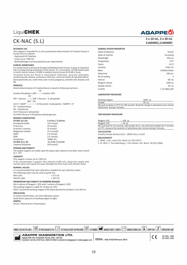
INTENDED USE
This reagent is intended for in vitro quantitative determination of Creatine Kinase inhuman serum or plasma.
- Optimized IFCC Method
- Linear up to 1700 U/L
- Working Reagent can be prepared as per requirements
CLINICAL SIGNIFICANCE
It is mainly found in all muscle (Cardiac & Skeletal) & brain tissues. It plays an importantrole in energy storing mechanism of the tissues. It’s iso-enzymes: CK-MB mainly existsin cardic muscle tissues, CK-MM in skeletal muscle tissues & CK-BB in brain.
Increased levels are found in myocardical infarction, muscular dystrophy,cerebrovascular-disease, pulmonary infarction, electrical shocks & hypothyroidisim.
Decreased levels are, some times seen in early pregnancy, alcoholic liver diseases andRA.
PRINCIPLE
Kinetic determination of Creatine Kianse is based on following reactions:-
CK
Creatine Phosphate + ADP ---- > Creatine +ATP
HK
ATP + Glucose ---- > ADP + Glucose – 6- phosphate
G6P – DH
G-6-P + NADP+ ----------- > D-Gluconate -6-phosphate + NADPH + H+
CK – Creatine Kinase
HK – Hexokinase
G-6-P-Glucose-6-phosphate
G-6-PDH-Glucose-6-Phosphate dehydrogenase.
REAGENT COMPOSITION
CK-NAC (S.L) R1 2 x 8 mL / 2 x24 mL
Immidazole buffer 125 mmol/L
D-Glucose 25 mmol/L
N-Acetyl-L-cysteine 25 mmol/L
Magnesium acetate 12.5 mmol/L
NADP 2.4 mmol/L
EDTA 2.0 mmol/L
Hexokinase >6800 U/L
CK-NAC (S.L) R2 2 x 2 mL / 2 x 6 mL
Creatine Phosphate 250 mmol/L
STORAGE AND STABILITY
The sealed reagents are stable upto the expiry date stated on the label, when storedat 2 - 80C.
LINEARITY
This reagent is linear up to 1700 U/L
If the concentration is greater than linearity (1700 U/L), dilute the sample withnormal saline and repeat the assay. Multiply the final result with dilution factor.
NORMAL VALUES
It is recommended that each laboratory establish its own reference value.
The following value may be used as guide line.
Men upto : < 171 U/L
Women upto : < 145 U/L
PREPARATION AND STABILITY OF WORKING REAGENT
Mix 4 volume of Reagent 1 (R1) with 1 volume of Reagent 2 (R2)
The working reagent is stable for 14 days at 2-80C.
NOTE: Discard the working reagent if the blank absorbance exceeds 1.0 at 340 nm.
PRECAUTION
To avoid contamination, use clean laboratory wares.
Avoid direct exposure of working reagent to light.
SAMPLE
Serum / Plasma (Free of haemolysis)
CK-NAC (S.L) 2 x 10 mL, 2 x 30 mL
11404001,11404005
GENERAL SYSTEM PARAMETER
Mode of Reaction Kinetic
Slope of reaction Increasing
Wavelength 340 nm
Temperature 370C
Factor 4127
Linearity 1700 U/L
Blank Distilled Water
Delay time 100 sec.
No of reading 3
Interval 60 sec
Reagent volume 1000 µL
Sample volume 40 µL
Cuvette 1 cm light path
LABORATORY PROCEDURE
Working reagent 1000 µL
Sample 40 µL
Mix and incubate at 370C for 100 seconds. Read the change in absorbance per minute( OD/min) during 3 minutes.
TWO REAGENT PROCEDURE
Reagent 1 R1 – 200 µL
Reagent 2 R2 – 50 µL
Mix and wait for 25 seconds, add sample 10 µL, mix well and incubate for 2 minutesat 370C, measure the variation of absorbance per minute during 3 minutes.
CALCULATION
Creatine Kinase Activity (U/L) = ( OD /min.) x 4127
BIBLIOGRAPHY
1. DGKC, J.Clin. chem.Clin. Bioch.15, 255 (1977)
2. Di. Witt, C. Tren delenburg, J. Clin chemie, Clin. Bioch. 20,235 (1982)
ADL/V.02/February 2013
19

INTENDED USE
This reagent is intended for in vitro quantitative determination creatinine in serum orurine.
- High Linearity of 200 mg/dL
- No sample dilution
- Ready to use reagents
CLINICAL SIGNIFICANCE
Creatinine is formed in muscles from phosphocreatinine. It is an important form ofenergy by being a store of high energy phosphate. Creatinine determinations have oneadvantage over urea determination that it is not affected by a high protein diet.
Serum creatinine is more specific & sensitive indicator of renal function. Simultaneousestimations of serum urea & creatinine provides better information. Serum ureanitrogen & creatinine ratio is > 15 in prerenal failure & < 10 in renal failure. Decreasedlevels are found in muscle dystrophy.
PRINCIPLE
Creatininase
Creatinine +H2O ------------------> Creatine
Creatinase
Creatine + H2O ------------------> Sarcosine +Urea
Sarcosine Oxidase
Sarcosine + O2+H
2O ------------------------> Glycine + HCHO + H
2O
2
Peroxidase
2 H2O
2+ 4-AA *1+ TOOS *2 ---------------> Quinone pigment +4 H
2O
* 1 : 4- Aminoantipyrine,
* 2 : N-ethyl-N-(2-hydroxy -3-sulfopropyl)-m-toluidine
Creatinine concentration can be obtained by measuring quinone pigmentphotometrically.
REAGENT COMPOSITION
CREATININE (S.L) R1 2 x 30 mL / 2 x 45 mL / 4x 45 mL
Creatinase 175000 IU/L
Sarcosine Oxidase 1500 IU/L
TOOS 1.13 mmol
CREATININE (S.L) R2 2 x 10 mL / 1 x 30 mL / 2 x 30 mL
Creatininase 75000 IU/L
Peroxidase 4500 units/L
4-AA 0.75 mmol
CREATININE STANDARD 1 x 4 mL
Creatinine Std. Conc. 2 mg/dL
STORAGE & STABILITY
The sealed reagents are stable upto the expiry date stated on the label, when storedat 2 - 80C.
LINEARITY
This reagent is linear upto 200 mg/dL.
If the concentration is greater than linearity dilute the sample with normal saline andrepeat the assay. Multiply the result with dilution factor.
REFERENCE VALUES
It is recommended that each laboratory establish its own reference value.
The following value may be used as guide line.
Serum : male : 0.6 – 1.1 mg/dL
: Female : 0.5- 0.8 mg/dL
Urine : Male : 1070-2150 mg/dL (24 hrs accumulated urine)
: Female : 769 – 1200 mg/dL (24 hrs accumulated urine)
PREPARATION AND STABILITY OF WORKING REAGENT
Reagent 1 & Reagent 2 are ready to use.
PRECAUTION
To avoid contamination, use clean laboratory wares.
Avoid direct exposure of reagent to light.
SAMPLE
Fresh Serum/urine(24 hour)
ENZYMATIC CREATININE2 x 40 mL, 2 x60 mL, 4 x 60 mL
11420002, 11420003, 11420004
GENERAL SYSTEM PARAMETER
Mode of Reaction End point
Slope of reaction Increasing
Wavelength I 546 nm
Wavelength II 630
Temperature 370C
Standard Conc. 2 mg/dL
Linearity 200 mg/dL
Blank Reagent
Incubation time 10 min.
Sample volume 10 µL
Reagent 1 volume 450 µL
Reagent 2 volume 150 µL
Cuvette 1 cm light path
LABORATORY PROCEDURE
Blank Calibrator Sample
Reagent 1 450 µL 450 µL 450 µL
Calib./Std - 10 µL -
Sample - - 10 µL
Mix and incubate for 5 minutes at 370C then add,
Reagent 2 150 µL 150 µL 150 µL
Mix and incubate for 5 minutes at 370C. Measure the absorbance of sample and thestandard against the reagent blank.
CALCULATION
Absorbance of sample
Creatinine Conc (mg/dL) = -------------------------------- x standard conc.
Absorbance of Standard
BIBLIOGRAPHY
Artiss, J.D., Mc Enroe, R.J., Zak, B.; Clin.Chem, 30 (1984)1389.
ADL/V.02/February 2013
20

INTENDED USE
This reagent is intended for in vitro Quantitative determination of gamma GT in serum.
- Szasz methodology
- Linear upto 232 U/L
- Working reagent can be prepared as per requirements
CLINICAL SIGNIFICANCE
GGT activity is elevated in all forms of liver diseases. It is highest in cases of intrahepaticor post hepatic biliary obstruction. (It may be 5 to 30 times higher than normal). It ismore sensitive than alkaline phosphatase NTP leucine amino peptidase andtransaminases in detection of obstructive jaundice, cholangitis, cholecystis neoplasm,it rises earlier than other enzyme and persists longer.
Moderate increase is observed in infectious hepatitis (2 to 5 times). Increases mayalso be observed in cases of drug intoxication, acute and chronic pancreatitis.
PRINCIPLE
Kinetic determination of Gamma GT according to the following reaction.
Gamma GT
GLUPA-C+Glycylglycine ---------------> L-Gamma-Glutamyl-GlycyLglycine +
5-Amino-2-nitrobenzoic acid.
GLUPA-C: L-Gamma -Glutamyl-3 Carboxy-p-nitroanilide
REAGENT COMPOSITION
Gamma GT R1 2 x 8 mL / 2 x 24 mL
Tris buffer pH (8.25) 133 mmol/L
Glycylglycine 138 mmol/L
Gamma GT R2 2 x 2 mL / 2 x 6 mL
GLUPA-C 23 mmol/L
STORAGE AND STABILITY
The sealed reagents are stable up to the expiry date stated on the label, when storedat 2 - 80C.
LINEARITY
This reagent is linear up to 232 U/L.
If the concentration is greater than linearity (232 U/L) dilute the sample with normalsaline and repeat the assay. Multiply the result with dilution factor.
NORMAL VALUES
It is recommended that each laboratory establish its own reference value.
The following values may be used as guide line.
Female : 5 - 32 U/L
Male : 10 - 45 U/L
PREPARATION AND STABILITY OF WORKING REAGENT
Mix 4 volume of Reagent 1 (R1) with 1 volume of reagent 2 (R2). This working reagentis stable for 21 days at 2-80C.
Note: Discard the working reagent if the blank absorbance exceeds 1.0 at 405 nm.
PRECAUTION:To avoid contamination, use clean laboratory wares.Avoid direct exposure of working reagent to light.
SAMPLE
Fresh Serum (free of haemolysis).
GENERAL SYSTEM PARAMETER
Mode of Reaction Kinetic
Slope of reaction Increasing
Wavelength 405 nm
Temperature 370C
Factor 1158
Linearity 232 U/L
Blank DI Water
Delay 60 sec
No of readings 3
Interval 60 sec
Sample volume 100 µL
Reagent volume 1000 µL
Cuvette 1 cm light path
LABORATORY PROCEDURE
Working reagent 1000 µL
Sample 100 µL
Mix and incubate for 1 minute at 370C. Read the change in absorbance per minute( OD/min) during 3 minutes.
CALCULATION
Gamma GT activity (U/L) = ( OD/min) x 1158
BIBLIOGRAPHY
1. Szasz, G.; Clin.Chem., 22, (1976),2051.2. Scand. J.Clin. Lab.Invest 36:711 (1976)3. Tietz, N.W.; Text book of Clin.Chem. 678-686 : (1986)
GAMMA GT(S.L) 2 x 10 mL, 2 x 30 mL
11416001, 11416005
21

INTENDED USE
This reagent is intended for in vitro quantitative determination of Glucose in serum,plasma & CSF.
- GOD-PAP methodology
- Linear upto 600 mg/dL
CLINICAL SIGNIFICANCE
Glucose is a major carbohydrate present in the blood & serves as a primary source ofenergy. It is usually obtained from ingested starch & sugar. The glucose concentrationis normally maintained at a constant level. Excessive glucose is stored as inactiveglycogen mainly in the liver & little in the muscles.
Elevated blood glucose levels are found in diabetes mellitus, hyperthyroidism,hyperadrenalism & certain liver diseases.
Decreased levels are found in Insulinoma, hypothyroidism, hypopituitarism.
PRINCIPLE
Enzymatic colorimetric determination of glucose according to the following reaction.
Glucose Oxidase
Glucose+ O2 + H
2O -----------------------> Gluconic acid + H
2O
2
Peroxidase
2H2O
2+phenol + 4-Aminoantipyrine -------------------> Quinonimine + 4H
2O
REAGENT COMPOSITION
GLUCOSE (S.L) R1 5 x 100 mL / 1 x 1000 mL
Tris Buffer, (pH 7.40) 92 mmol/L
Phenol 0.3 mmol/L
Glucose Oxidase 15000 U/L
4- Aminophenazone 2.6 mmol/L
GLUCOSE STANDARD 1 x 4 mL
Glucose standard concentration 100 mg/dL
STORAGE AND STABILITY
The sealed reagents are stable up to the expiry date stated on the label, when storedat 2 - 80C.
LINEARITY
This reagent is linear up to 600 mg/dL
If the concentration is greater than linearity (600 mg/dL), dilute the sample with normalsaline and repeat the assay. Multiply the result with dilution factor.
NORMAL RANGE
It is recommended that each laboratory establish its own reference values.
The following value may be used as guide line.
Serum / Plasma : 70-105 mg/dL
C S F : 50 -70 mg/dL
PREPARATION AND STABILITY OF WORKING REAGENT
The Reagent is ready to use.
PRECAUTION
To avoid contamination, use clean laboratory wares.
Avoid direct exposure of reagent to light.
SAMPLE
Serum / plasma (free of haemolysis) / CSF
GENERAL SYSTEM PARAMETER
Mode of Reaction End Point
Slope of reaction Increasing
Wavelength 505 (490-550 nm)
Temperature 370C
Standard Concentration 100 mg/dL
Linearity 600 mg/dL
Incubation Time 10 Minutes
Blank Reagent
Sample volume 10 µL
Reagent volume 1000 µL
Cuvette 1 cm light path
LABORATORY PROCEDURE
Blank Standard Sample
Working Reagent 1000 µL 1000 µL 1000 µL
Standard - 10 µL -
Sample - - 10 µL
Mix and incubate for 10 minutes at 370C. Read the absorbance of standard and sampleagainst reagent blank.
CALCULATION
Absorbance of Sample
Glucose Conc. (mg/dL) = -------------------------- x 100
Absorbance of standard
BIBLIOGRAPHY
1. Trinder, P.; Ann Clin Biochem. 6,24 (1969)2. Dingeon, B.; Ann.Bio.Clin 33,3 (1975)3. Lott, J. ; Clin.Chem. 21, 1754 (1975)
GLUCOSE (S.L)5 x 100 mL, 1 x 1000 mL
11406001, 11406002
22

INTENDED USE
This reagent is intended for in vitro quantitative determination of HDL Cholesterol inserum
- Direct determination of HDL Cholesterol
- Enzyme Selective Protection Method
- Linear up to 150 mg/dL
- Ready to use liquid stable reagents
CLINICAL SIGNIFICANCE
Blood total cholesterol levels have long been known to be related to coronary heartdisease (CHD). In recent years, in addition to total cholesterol, high densitylipoprotein cholesterol (HDL-C) has become an important tool used to assess anindividual risk of developing CHD since a strong negative relationship betweenHDL - C concentration and the incidence of CHD was reported.
PRINCIPLE
The reaction between cholesterol other than HDL and the enzyme for cholesterolassay is suppressed by the electrostatic interaction between polyanions & cationicsubstances. Hydrogen peroxide is formed by the free cholesterol in HDL by cholesteroloxidase. Oxidative condensation of EMSE and 4-AA is caused by hydrogen peroxidein the presence of peroxidase, and the absorbance of the resulting red-purple quinoneis measured to obtain the cholesterol value in HDL
Polyanions
Other lipoproteins than HDL --------------> Suppress reaction with enzyme
Cationic substances
cholesterol esterase
HDL (cholesterol esters) + H2O --------------------------> HDL (free cholesterol) +
Free fatty acids
cholesterol oxidase
HDL (free cholesterol) + O2
+ H+ -------------------------> Cholestenone + H2O
2
Peroxidase
2H2O
2 + 4-AA + EMSE + H
3 + O ---------------> Red-purple quinone + 5H
2O
REAGENT COMPOSITION
HDL –C DIRECT R1 2 x 30 mL / 2 x 45 / 4 x 45 mL
N—ethyl-N-(3-methylphenyl)-N’succinylethyenediame(EMSE)
HDL –C DIRECT R2 2 x 10 / 1 x 30 / 2 x 30 mL
Cholesterol Oxidase
4-Aminoantipyrin(4-AA)
HDL –C DIRECT CALIBRATOR 1 x 3 mL
STORAGE AND STABILITY
The sealed reagents are stable upto the expiry date stated on the label, when storedat 2 - 80C, protected from light. Do not freeze.
LINEARITY
This reagent is linear upto 150 mg/dL
If the concentration is greater than linearity (150 mg/dL), dilute the sample with normalsaline and repeat the assay. Multiply the result with dilution factor
NORMAL RANGE
It is recommended that each laboratory establish its own reference values.
The following value may be used as guide line.
Male : 35 - 80 mg/dL
Female : 42 - 88 mg/dL
PREPARATION AND STABILITY OF WORKING REAGENT
The Reagent1 & Reagent 2 are ready to use.
Calibrator : Reconstitute with 3 mL of distilled water. Let it stand for 30 minutes atroom temperature. Dissolve the content of the vial by swirling gently to avoid theformation of foam.
Stability : Reconstituted calibrator is stable only for 7 days at 2- 80C.
PRECAUTION
To avoid contamination, use clean laboratory wares. Use clean, dry disposablepipette tips for dispensing. Close reagent bottles immediately after use. Avoid directexposure of reagent to light. Do not blow into the reagent bottles.
SAMPLE
Fresh serum (free of haemolysis)
GENERAL SYSTEM PARAMETER
Mode of Reaction End Point
Slope of reaction Increasing
Wavelength I 578 nm(578-610)
Wavelength II 630 nm (630-700)
Temperature 370C
Calibrator Con As on the vial
Linearity 150 mg/dL
Incubation time 5 min+5 min
Blank Reagent
Sample volume 5 µL
Reagent volume 450 µL+150 µL
Cuvette 1 cm light path
LABORATORY PROCEDURE
Blank Calibrator Sample
Reagent1 450 µL 450 µL 450 µL
Calibrator - 5 µL -
Sample - - 5 µL
Mix and incubate for 5 minutes at 370C.
Reagent 2 150 µL 150 µL 150 µL
Mix and incubate for 5 minutes at 370C. Measure the absorbance of calibrator &sample against reagent blank at 578nm/ 630 nm.
CALCULATION
Absorbance of Sample
HDL-C Concentration (mg/dL) = ------------------------------ x Calibrator Concentration
Absorbance of Calibrator
ALTERNATIVE PROCEDURE
Mode of Reaction Differential
Slope of reaction Increasing
Wavelength I 600 nm
Wavelength II 700 nm
Temperature 370C
Calibrator Concentration As on the vial label
Linearity 150 mg/dL
Blank Reagent
Incubation time 5 minutes+5 minutes
Sample volume 3 µL
Reagent volume 270 µL + 90 µL
Cuvette 1 cm light path
LABORATORY PROCEDURE
Blank Calibrator Sample
Reagent1 270 µL 270 µL 270 µL
Calibrator - 3 µL -
Sample - - 3 µL
Mix and incubate for 5 minutes at 370C. Measure the absorbance (OD1) at 600 nm/700 nm.
Reagent 2 90 µL 90 µL 90 µL
Mix and incubate for 5 minutes at 370C. Measure the absorbance (OD2) at 600 nm/700 nm.
CALCULATION
(OD2-OD1) Sample
HDL-C Concentration (mg/dL) = ---------------------------- x Calibrator Conc.
(OD2-OD1) Calibrator
INTERFERING SUBSTANCES
Test will not be affected by:
Bilirubin up to 40 mg/dL
Ascorbic acid up to 50 mg/dL
Hemoglobin up to 500 mg/dL
Triglyceride up to 1000 mg/dL
*(when triglyceride in a sample exceeds 1000 mg/dL, dilute the sample 1+9 withsaline, repeat the assay and multiply result by 10)
BIBLIOGRAPHY
1. Williams, P., et al.; High density lipoprotein and coronary risk factor, Lancet. 1:72(1979)
2. Gordon,T., Castelli, W.P., Hjortland, M.C. et al. Am. J.Med. 62, 707-714 (1977)3. Rifai,N.and Warnick, G.R.,Ed. Laboratory Measurement of Lipids, Lipoproteins
and Apolipoproteins AACC Press. Washington, DC,USA,1994
HDL CHOLESTEROL (D) WITH CALIBRATOR 2 x 40 mL / 2 x 60 mL / 4 x 60 mL
11414003, 11414005, 11414004
ADL/V.02/February 2013
23

INTENDED USE
This reagent is intended for in vitro quantitative determination of Lactatedehydrogenase in serum or plasma.
- Based on SCE recommended method
- Linear up to 2400 U/L
CLINICAL SIGNIFICANCE
This enzyme is found in all organ cells, but especially plentiful in cardiac & skeletalmuscle, liver, kidney & RBC. LDH is found in the form of iso-enzymes based on theirelectrophoretic mobility with each iso-enzymes being primarily from different organs.
Elevated levels are found in myocardial infarction, liver diseases, hemolytic anaemias,pernicious anaemia, Leukemia & Pulmonary diseases. Elevations in acute MI reachesa peak in 48-72 hrs. belonged elevations, (10-14 days) are useful in the late diagnosisof the condition.
PRINCIPLE
Kinetic determination of lactate dehydrogenase according to the following reaction.
LDH
Pyruvate + NADH + H+ --------------> L-Lactate +NAD+
REAGENT COMPOSITION
LDH-P (S.L) R1 2 x 8mL / 2 x 24mL
Tris buffer (pH 7.4) 80 mmol/L
Pyruvate 1.6 mmol/L
Sodium chloride 200 mmol/L
LDH-P (S.L) R2 2 x 2 mL / 2 x 6 mL
NADH 240 mmol/L
STORAGE & STABILITY
The sealed reagents are stable up to the expiry date stated on the label, when storedat 2 - 80C.
LINEARITY
This reagent is linear up to 2400 U/L.
If the concentration is greater than linearity (2400 U/L), dilute the sample with normalsaline and repeat the assay. Multiply the result with dilution factor.
NORMAL RANGE
It is recommended that each laboratory establish its own reference values.
The following value may be used as guide line.
Serum /Plasma : 225-450 U/L
PREPARATION AND STABILITY OF WORKING REAGENT
Mix 4 volumes of Reagent 1 (R1) with 1 volume of Reagent 2 (R2)
The working reagent is stable for 21 days at 2-80C.
NOTE: Discard the working reagent if the blank absorbance is less than 1.0 at 340nm.
PRECAUTION
To avoid contamination, use clean laboratory wares.
Avoid direct exposure of reagent to light.
SAMPLE
Serum / plasma (free of haemolysis)
GENERAL SYSTEM PARAMETER
Mode of Reaction Kinetic
Slope of reaction Decreasing
Wavelength 340 nm
Temperature 370C
Factor 16030
Linearity 2400 U/L
Delay 60 sec.
No of reading 3
Interval 60 sec
Sample volume 10 µL
Reagent volume 1000 µL
Cuvette 1 cm light path
LABORATORY PROCEDURE
Working reagent 1000 µL
Sample 10 µL
Mix and incubate at 370C for 1 minute. Measure the change in absorbance per minute( OD/min) during 3 minutes.
CALCULATION
LDH-P activity (U/L) = ( OD/min) x 16030
BIBLIOGRAPHY
1. Z.Klin. chem. Klin Biochem.8,658 (1970), 1, 1820(1972)2. Wei Bhaar, D., et al.; Med.Welt 26,387 (1975)
LDH-P (S.L)2 x 10 mL, 2 x 30 mL
11407001, 11407004
24

INTENDED USE
This reagent is intended for in vitro quantitative determination of LDL Cholesterol inserum or plasma
- Direct determination of LDL Cholesterol
- Enzyme selective protection method
- Linear up to 450 mg/dL
- Ready to use liquid stable reagents
CLINICAL SIGNIFICANCE
Blood total cholesterol levels have long been known to be related to coronary heartdisease (CHD). In recent years, in addition to total cholesterol, low densitylipoprotein cholesterol (LDL-C) has become an important tool used to assess anindividual risk of developing CHD since a strong positive relationship between LDL-C concentration and the incidence of CHD was reported. LDL Cholestrol acts as akey factor in the pathogenesis of atherosclerosis and coronary artery disease.
PRINCIPLE
The LDL-C Direct is a homogeneous assay for directly measuring LDL-C levels inserum or plasma without the need for any off-line pretreatment or centrifugation
In the first reaction, non LDL unesterified cholesterol is subject to an enzymereaction and the peroxide generated is consumed by peroxidase in the presence of4-AAP to yield a colorless product. The second reagent consists of a detergentcapable of solubilizing LDL specifically. Cholesterol esterase and chromogeniccoupler react with this solubilize LDL-C to develop color. The intensity of colorformed is directly proportional to the concentration of LDL-C.
REAGENT COMPOSITION
LDL –C DIRECT R1 2 x 30 mL / 2 x 45 mL
4-Aminoantipyrin 0.5 mmol/L
CHE
CO 1.2 U/mL
Peroxidase
Good’s buffer pH 6.3
LDL –C DIRECT R2 2 x 10 / 1 x 30 mL
N,N-bis(4-sulfobutyl)-m-toluidine
disodium salt (DSBmT) 1.0 mmol/L
Good’s buffer pH 6.3
LDL –C DIRECT CALIBRATOR 1 x 3 mL
STORAGE AND STABILITY
The sealed reagents are stable up to the expiry date stated on the label, when storedat 2 - 80C, protected from light. Do not freeze.
LINEARITY
This reagent is linear up to 450 mg/dL
If the concentration is greater than linearity (450 mg/dL), dilute the sample withnormal saline and repeat the assay. Multiply the result with dilution factor
NORMAL RANGE
It is recommended that each laboratory establish its own reference values.
The following value may be used as guide line.
Desirable < 130 mg/dL
Borderline High Risk for CHD 130-159 mg/dL
High Risk for CHD >160 mg/dL.
PREPARATION AND STABILITY OF REAGENT
The Reagent 1 & Reagent 2 are ready to use.
Calibrator : Reconstitute with 3 mL of distilled water. Let it stand for 2 hours at roomtemperature. Dissolve the content of the vial by swirling gently to avoid theformation of foam.
Stability : Reconstituted calibrator is stable only for 7 days at 2- 80C.
PRECAUTION
To avoid contamination, use clean laboratory wares. Use clean, dry disposablepipette tips for dispensing. Close reagent bottles immediately after use. Avoid directexposure of reagent to light. Do not blow into the reagent bottles.
SAMPLE
Fresh serum (free of haemolysis) / EDTA Plasma
GENERAL SYSTEM PARAMETER
Mode of Reaction Differential
Slope of reaction Increasing
Wavelength I 546 nm
Wavelength II 660 nm
Temperature 370C
Calibrator Concentration As on the vial label
Linearity 450 mg/dL
Blank Reagent
Incubation time 5 min + 5 min
Sample volume 3 µL
Reagent volume 270 µL + 90 µL
Cuvette 1 cm light path
LABORATORY PROCEDURE
DIFFERENTIAL MEASUREMENT
Blank Calibrator Sample
Reagent 1 270 µL 270 µL 270 µL
Calibrator - 3 µL -
Sample - - 3 µL
Mix and incubate for 5 minutes at 370C. Measure the absorbance (OD1) at 546 nm/660 nm.
Reagent 2 90 µL 90 µL 90 µL
Mix and incubate for 5 minutes at 370C. Measure the absorbance (OD2) at 546 nm/660 nm.
CALCULATION
LDL - C Concentration (mg/dL) =
(OD2-OD1) Sample
--------------------------- x Calibrator Concentration
(OD2-OD1) Calibrator
ALTERNATIVE MEASUREMENT
Mode of Reaction End Point
Slope of reaction Increasing
Wavelength I 546 nm
Wavelength II 630 nm
Temperature 370C
Calibrator Concentration As on the vial label
Linearity 450 mg/dL
Blank Reagent
Incubation time 5 min +5 min
Sample volume 5 µL
Reagent volume 450 µL + 150 µL
Cuvette 1 cm light path
LABORATORY PROCEDURE
Blank Calibrator Sample
Reagent 1 450 µL 450 µL 450 µL
Calibrator - 5 µL -
Sample - - 5 µL
Mix and incubate for 5 minutes at 370C.
Reagent 2 150 µL 150 µL 150 µL
Mix and incubate for 5 minutes at 370C. Measure the absorbance of calibrator &sample against reagent blank at 546 nm/630 nm.
CALCULATION
Absorbance of Sample
LDL - C Concentration (mg/dL) = ------------------------------- x Calibrator Concentration
Absorbance of Calibrator
INTERFERING SUBSTANCES
Test will not be affected by:
Bilirubin up to 40 mg/dL
Ascorbic acid up to 50 mg/dL
Haemoglobin up to 500 mg/dL
Triglyceride up to 1000 mg/dL
*(when triglyceride in a sample exceeds 1000 mg/dL, dilute the sample 1+9 with saline,repeat the assay and multiply result by 10)
BIBLIOGRAPHY
1. Crouse, J.R., et al.; Studies of Low Density Lipoprotein molecular weight in humanbeing with coronary artery disease. J.Lipid Res 26:5666 (1985)
LDL CHOLESTEROL (D) WITH CALIBRATOR2 x 40 mL, 2 x 60 mL
11415003, 11415004
ADL/V.02/February 2013
25

INTENDED USE
This reagent is intended for in vitro quantitative determination of lipase in humanserum or plasma.
- Methyl resorufin method
- Linear up to 300 U/L
- Reagent is ready for use
CLINICAL SIGNIFICANCE
Lipase is a pancreatic enzyme necessary for the absorption and digestion ofnutrients that catalyses the hydrolysis of glycerol esters of fatty acids. Determinationof lipase is used for diagnosis of diseases such as acute and chronic pancreatitisand obstruction of the pancreatic duct. Clinical diagnosis should not be made on asingle test result; it should integrate clinical and other laboratory data.
PRINCIPLE
In the presence of colipase and bile acids lipase splits the synthetic substrate (1,2-O-Dilauryl-rac-glycero-3-glutaricacid (6-methyl-resorufin-ester) to glycerol andmethylresorufin-ester, which is spontaneously degraded to glutaric acid andmethylresorufin. The rate of methylresorufin formation, measured photometricallyis proportional to the catalytic concentration of lipase present in the sample.
REAGENT COMPOSITION
LIPASE (S.L) R1 2 x 10 mL
Goods Buffer (pH 8.0) 40 mmol/L
Taurodeoxycholate 3.4 mmol/L
Deoxycholate 6.4 mmol/L
Calcium chloride 12 mmol/L
Colipase 1.7 mg/dL
LIPASE(S.L) R2 1 x 5 mL
Tartrate Buffer (pH 4.0) 1.5 mmol/L
Taurodeoxycholate 3.4 mmol/L
Color substrate 0.13 mmol/L
LIPASE CALIBRATOR 1 x 3 mL
Lipase calibrator concentration is stated on the vial label.
STORAGE & STABILITY
The sealed reagents are stable up to the expiry date stated on the label, when storedat 2 - 80C, protected from light.
LINEARITY
This reagent is linear up to 300 U/L.
If the concentration is greater than linearity (300 U/L), dilute the sample withnormal saline and repeat the assay. Multiply the result with dilution factor.
NORMAL RANGE
It is recommended that each laboratory establish its own reference values.
The following value may be used as guide line.
Serum / Plasma : Up to 60 U/L
PREPARATION AND STABILITY OF REAGENT
Lipase R1 and Lipase R2 are ready to use.
Calibrator : Reconstitute with 3 mL of distilled water. Dissolve the content of thevial by swirling gently to avoid the formation of foam.
Stability : Reconstituted calibrator is stable only for 7 days at 2-80C and 3 monthsat -200C.
PRECAUTION
To avoid contamination, use clean laboratory wares. Use clean dry disposablepipette tips for dispensing. Close reagent and calibrator bottles immediately afterUse. Avoid direct exposure of reagent to light.
SAMPLE
Serum or plasma with sodium citrate, EDTA or heparin.
LIPASE(S.L)1 x 25 mL
11417001
GENERAL SYSTEM PARAMETER
Mode of Reaction Kinetic Fixed Time
Slope of reaction Increasing Increasing
Wavelength 580 nm 580 nm
Temperature 370C 370C
Calibrator concentration As on the vial As on the vial
Linearity 300 U/L 300 U/L
Blank Reagent Reagent
Delay time 120 sec. 120 sec
No of reading 2 -
Interval 60 sec 120 sec
Sample volume 20 µL 20 µL
Reagent volume 1250 µL (1000+250) 1250 µL (1000 + 250)
Cuvette 1cm light path 1 cm light path
LABORATORY PROCEDURE
Blank Calibrator Sample
Reagent 1 1000 µL 1000 µL 1000 µL
Calibrator - 20 µL -
Sample - - 20 µL
Dist. water 20 µL - -
Mix carefully (do not vortex); incubate for 1-5 minutes at 370C. Then add
Reagent 2 250 µL 250 µL 250 µL
Mix and incubate for 2 min at 37oC, read absorbance against reagent blank. Measurethe change in absorbance per minute ( OD/min) during 2 min.
or
Mix and read the optical density (T1) 120 seconds after the Reagent 2 addition. Take
second reading (T2) exactly after 120 seconds.
CALCULATION
Lipase U/L =
( OD/min) Sample - ( OD/min) Blank
---------------------------------------------------- x Calibrator concentration
( OD/min) calibrator - ( OD/min) Blank
or
(T2-T
1 ) of sample
------------------------- x Calibrator concentration
(T2-T
1 ) of standard
BIBLIOGRAPHY
1. Mc Neely , M. ; Lipase. Kaplan, A. et al.; Clin. Chem. The C.V.Mosby Co. St Louis,Toronto. Princeton 1984, 1130-1135
2. Burtis, A., et al. ; Tietz Textbook of Clinical chemistry, 3rd ed AACC3. Neumann, U., et al.; Methods of Enzymatic Analysis, Vol 4, 3rd Ed.
ADL/V.02/February 2013
26

INTENDED USE
This reagent is intended for in vitro quantitative determination of SGOT in serum orplasma.
- IFCC recommended procedure
- Linear up to 1000 U/L
- Working reagent can be prepared as per requirements
CLINICAL SIGNIFICANCE
It is present in most of the tissues. Especially in cardiac muscle, liver cells, skeletalmuscle & kidneys. Injury to these tissues results in the release of the enzyme in bloodstream.
Increased levels are found in myocardial infarction. The duration & extent of increaseis related to the infract. GOT determination is of considerable value to differentiatemyocardial infarction from other cardiac disorders.
Increased levels are also found in various types of liver disease, skeletal muscle trauma& in renal diseases.
Decreased levels may be found in pregnancy, Beri-Beri & Diabetic ketoacidosis.
PRINCIPLE
Kinetic determination of Aspartate Aminotrasferase (AST) based upon the followingreaction.
AST
L- Asparate + alpha - ketoglutarate ----- > Oxaloacetate + L-Glutamate.
MDH
Oxaloacetate + NADH + H+ -------- > L- Malate + NAD+
AST: Aspartate aminotransferase.
MDH : Malate dehydrogenase.
REAGENT COMPOSITION
SGOT (S.L) R1 2 x 24 mL / 3 x 40 mL / 4 x 100 mL
Tris Buffer (pH 7.8) 88 mmol/L
L-Aspartate 260 mmol/L
LDH > 1500 U/L
MDH > 900 U/L
SGOT (S.L)R2 2 x 6 mL / 3 x 10 mL / 4 x 25 mL
alpha -ketoglutarate 12 mmol/L
NADH 0.24 mmol/L
STORAGE AND STABILITY
The sealed reagents are stable up to the expiry date stated on the label, when storedat 2 - 80C.
LINEARITY
This reagent is linear up to 1000 U/L.
If the concentration is greater than 350 U/L, follow the high linearity procedure toget higher linearity of 1000 U/L.
If the concentration is greater than linearity, dilute the sample with normal saline andrepeat the assay. Multiply the result with dilution factor.
NORMAL RANGE
It is recommended that each laboratory establish its own reference values.
The following value may be used as guide line.
Serum up to : 46 U/L
PREPARATION AND STABILITY OF WORKING REAGENT
Mix 4 volumes of Reagent 1 (R1 ) with 1 volume of Reagent 2 (R2)
The working reagent is stable for 30 days at 2-80C.
NOTE: Discard the working reagent if the blank absorbance is less than 1.0 at 340 nm.
PRECAUTION
To avoid contamination, use clean laboratory wares.
Avoid direct exposure of reagent to light.
SAMPLE
Serum / plasma (free of haemolysis)
GENERAL SYSTEM PARAMETER
Normal procedure High Linearity procedure
Mode of Reaction Kinetic Kinetic
Slope of reaction Decreasing Decreasing
Wavelength 340 nm 340 nm
Temperature 370C 370C
Factor 1745 1745
Linearity 350 U/L 1000 U/L
Blank DI Water DI Water
Delay 60 sec 60 sec
No of reading 3 3
Interval 60 sec 20 sec
Sample volume 100 µL 100 µL
Reagent volume 1000 µL 1000 µL
Cuvette 1 cm light path 1 cm light path
LABORATORY PROCEDURE
Working reagent 1000 µL
Sample 100 µL
Mix and incubate at 370C for 1 minute. Measure the change in absorbance per minute( OD/min) during 3 minutes.
High Linearity Procedure
Mix and incubate at for 1 minutes 370C. Read the change in absorbance per 20 sec,during 1 minute.
CALCULATION
SGOT activity (U/L) = ( OD/min) x 1745
BIBLIOGRAPHY
1. Clin. Chem, Acta. 70, 19-42 (1976)2. Thefeld, W., et al.; Dtsch. Med Wschr.99, 343 (1974)
SGOT (S.L) 2 x 30 mL, 3 x 50 mL, 4 x 125 mL
11408005, 11408003, 11408007
27

INTENDED USE
This reagent is intended for in vitro quantitative determination of SGPT in serum orplasma.
- IFCC recommended methodology
- Linear up to 1000 U/L
- Working reagent can be prepared as per requirements
CLINICAL SIGNIFICANCE
It is present in most of the tissues, but mainly found in the liver. Increased levels arefound in hepatitis, cirrhosis, obstructive jaundice & other hepatic disease. SGPT activityis markedly elevated even before clinical signs of jaundice become apparent in diseaseassociated with hepatic necrosis. Slight elevations are also found in myocardialinfarction.
PRINCIPLE
Kinetic determination of Alanine Aminotransferase (ALT) according to the followingreaction.
ALT
L-Alanine + alpha-ketogutarate ----- >Pyruvate +L-Glutamate
LDH
Pyruvate +NADH+ H+ ----- > L-Lactate +NAD+
ALT – Alanine aminotranferase
LDH - Lactate dehydrogenase
REAGENT COMPOSITION
SGPT (S.L) R1 2 x 24 mL / 3 x 40 mL / 4 x 100 mL
Tris buffer (pH 7.5) 110 mmol/L
L-Alanine 600 mmol/L
LDH > 1500U/L
SGPT (S.L)R2 2 x 6 ml / 3 x 10 mL / 4 x 25 mL
alpha-ketoglutarate 16 mmol/L
NADH 0.24 mmol/L
STORAGE AND STABILITY
The sealed reagents are stable up to the expiry date stated on the label, when storedat 2 - 80C.
LINEARITY
This reagent is linear up to 1000 U/L.
If the concentration is greater than 350 U/L, follow the high linearity procedure toget higher linearity of 1000 U/L.
If the concentration is greater than linearity dilute the sample with normal saline andrepeat the assay. Multiply the result with dilution factor.
NORMAL RANGE
It is recommended that each laboratory establish its own reference values.
The following value may be used as guide line.
Serum up to : 49 U/L
PREPARATION AND STABILITY OF WORKING REAGENT
Mix 4 volumes of Reagent 1 (R1) with 1 volume of Reagent 2 (R2)
The working reagent is stable for 30 days at 2-80C.
NOTE: Discard the working reagent if the blank absorbance is less than 1.00 at 340nm.
PRECAUTION
To avoid contamination use clean laboratory wares.
Avoid direct exposure of working reagent to light.
SAMPLE
Serum / plasma (free of haemolysis)
GENERAL SYSTEM PARAMETER
Normal procedure High Linearity procedure
Mode of Reaction Kinetic Kinetic
Slope of reaction Decreasing Decreasing
Wavelength 340 nm 340 nm
Temperature 370C 370C
Factor 1745 1745
Linearity 350 U/L 1000 U/L
Blank DI Water DI Water
Delay 60 sec 60 sec
No of reading 3 3
Interval 60 sec 20 sec
Sample volume 100 µL 100 µL
Reagent volume 1000 µL 1000 µL
Cuvette 1 cm light path 1 cm light path
LABORATORY PROCEDURE
Working reagent 1000 µL
Sample 100 µL
Mix and incubate at 370C for 1 minute. Measure the change in absorbance per minute( OD/min) during 3 minutes.
High Linearity Procedure
Mix and incubate for 1 minute at 370C. Read the change in absorbance per 20 secduring 1 minutes.
CALCULATION
SGPT activity (U/L) = ( OD/min) x 1745
BIBLIOGRAPHY
1. Clin. Chem, Acta. 105, 147-172 (1780)2. Thefeld, W., et al.; Dtsch. Med Wschr.99, 343 (1994)
SGPT (S.L) 2 x 30 mL, 3 x 50 mL, 4 x 125 mL
11409005, 11409003, 11409006
28

INTENDED USE
This reagent is intended for in vitro quantitative determination of triglycerides in serumor plasma.
- GPO-PAP methodology
- Linear up to 1000 mg/dL
- Contains LCF (Lipaemic clearing factor) which minimizes rerun
CLINICAL SIGNIFICANCE
Triglycerides are simple lipids, formed in the liver by glycerol & fatty acids. Theyare transported by VLDL, LDL & constitute about 95% of fat, stored as source ofenergy in the tissue & plasma.
Increased levels are found in hyperlipidemias, diabetes, nephrotic syndrome &hypothyroidism. Increased levels are risk factor for arteriosclerotic coronary disease,peripheral vascular disease, acute pancreatitis & hyperlipoproteinaemia. Decreasedlevels are found in malnutrition & hyperthyroidism.
PRINCIPLE
Enzymatic determination of triglyceride is based on following reactions:
LPL
TGL+H2O ------ > Glycerol + Fatty acid
GK
Glycerol + ATP ------ > Glycerol-3-phosphate + ADP
Mg++
GPO
Glycerol-3-phospahte+O2
------- > Dihydroxyacetone phosphate +H2O
2
POD
2H2O
2+4-Aminoantipyrine+ p-chlorophenol ------- > Red Quinoneinimine
GPO = Glycereol-3-phosphate Oxidase
LPL = Lipoprotein Lipase
GK = Glycerol Kinase
REAGENT COMPOSITION
TRIGLYCERIDES REAGENT 4x10mL / 5x25mL / 6x50mL / 5x100mL
Pipes –buffer (pH 7.00) 50 mmol/L
p-Chlorophenol 5.3 mmol/L
Potassium ferrocynate 10 mmol/L
Magnesium Salt 17 mmol/L
4-Aminoantipyrine 0.9 mmol/L
ATP 3.15mmol/L
Lipoprotein Lipase > 1800 U/L
Glycerol Kinase > 450 U/L
Glycerol – 3- phosphate oxidase > 3500 U/L
Peroxidase > 450 U/L
TRIGLYCERIDES STANDARD 1 x 4 mL
Triglycerides std.concentration 200 mg/dL
STORAGE AND STABILITY
The sealed reagents are stable up to the expiry date stated on the label, when storedat 2 - 80C.
LINEARITY
This reagent is linear up to 1000 mg/dL.
If the concentration is greater than linearity (1000 mg/dL), dilute the sample withnormal saline and repeat the assay. Multiply the result with dilution factor.
REFERENCE RANGE
It is recommended that each laboratory establish its own reference values.
The following value may be used as guide line.
Male : 60-165 mg/dL
Female : 40-140 mg/dL
PREPARATION AND STABILITY OF REAGENT
The reagent ready to use.
PRECAUTION
To avoid contamination, use clean laboratory wares.
Avoid direct exposure of reagent to light.
SAMPLE
Serum / plasma (free of haemolysis)
GENERAL SYSTEM PARAMETER
Mode of Reaction End Point
Slope of reaction Increasing
Wavelength I 505 nm (492-550 nm)
Wavelength II 630 nm
Temperature 370C
Standard Concentration 200 mg/dL
Linearity 1000 mg/dL
Blank Reagent
Incubation time 5 min
Sample volume 10 µL
Reagent volume 1000 µL
Cuvette 1 cm light path
LABORATORY PROCEDURE
Blank Standard Sample
Working Reagent 1000 µL 1000 µL 1000 µL
Standard - 10 µL -
Sample - - 10 µL
Mix and incubate for 5 minutes at 370C. Measure the change in absorbance ofstandard and sample against reagent blank.
CALCULATION
Absorbance of Sample
Triglycerides Con. (mg/dL) = ------------------------------ x 200
Absorbance of Standard
BIBLIOGRAPHY
1. Schettler, G., Nussel, E.; Arav. Med 10, 25 (1975)2. Jacobs, N.J. , VanDemark, P.J.; Arch, Biochem, Biophy. 88, 250 – 255 (1960)
TRIGLYCERIDES (S.L)4x 10 mL, 5 x 25 mL, 6x 50 mL, 5 x 100 mL
11410007, 11410002, 11410008, 11410004
ADL/V.02/February 2013
29

INTENDED USE
This reagent is intended for in vitro quantitative determination of urea in serum, plasma& urine.
- Urease / GLDH methodology
- Linear up to 300 mg/dL
- Working reagent can be prepared as per requirement
CLINICAL SIGNIFICANCE
Proteins cannot be stored in human body, so excess should be broken down. Aminoacids which from the components of proteins, break down to give ammonia. This istoxic & so through a series of chemical reactions (urea cycle) non toxic urea isproduced & this is released into the blood which is filtered in the kidney & excretedin the urine.
Elevated levels are seen during increased protein breakdown, dehydration, vomiting,diarrhea. Also seen in any kind of renal disorder like Glomerular nephritis, Chronicnephritis & Nephritic syndrome.
Decreased levels are found in liver failure & pregnancy.
PRINCIPLE
Enzymatic determination of Urea according to the following reaction.
Urease
Urea + H2O ------------> 2NH
3 + CO
2
GLDH
2 NH3
+2- ketoglutarate + 2NADH ----------> L-Glutamate+2NAD+ + 2H2O
REAGENT COMPOSITION
UREA U.V (S.L) R1 2 x 24 mL/ 2 x 40 mL/ 2 x 100 mL
Tris Buffer (pH 7.60) 100 mmol/L
ADP 0.7 mmol/L
a-ketoglutarate 9.0 mmol/L
Urease > 6500 U/L
GLDH > 1100 U/L
UREA U.V (S.L) R2 2 x 6 mL/ 2 x 10 mL / 2 x 25 mL
NADH 0.25 mmol/L
2-Oxoglutarate 5 mmol/L
UREA U.V STANDARD 1 x 4 mL
Standard concentration for Urea 50 mg/dL
STORAGE & STABILITY
The sealed reagents are stable up to the expiry date stated on the label, when storedat 2 - 80C.
LINEARITY
This reagent is linear up to 300 mg/dL.
If the concentration is greater than linearity (300 mg/dL), dilute the sample with normalsaline and repeat the assay. Multiply the result with dilution factor.
REFERENCE RANGE
It is recommended that each laboratory establish its own reference values.
The following value may be used as guide line.
Serum/ Plasma : 10-50 mg/dL
Urine : 20-35 gm/24 hr
PREPARATION AND STABILITY OF WORKING REAGENT
Mix 4 volume of Reagent 1 (R1) with 1 volume of Reagent 2 (R2)
Working reagent is stable for 30 days at 2-80c.
Note
Discard the working reagent if the blank absorbance is less than 1.0 at 340 nm.
PRECAUTION
To avoid contamination useclean laboratory wares. Avoid direct exposure of reagentto light.
SAMPLE
Serum, Plasma ( free of hemolyses). Do not use anticoagulants containing Fluorideor Ammonium Ions.
Urine(1/100 diluted with distilled water (DI water). Multiply result with dilution factor.
UREA U.V (S.L)2 x 30 mL, 2 x 50mL, 2 x 125 mL
11412007, 11412002, 11412008
GENERAL SYSTEM PARAMETER
Mode of Reaction Fixed Time
Slope of reaction Decreasing
Wavelength 340
Temperature 370C
Blank DI Water
Standard Concentration 50 mg/dL
Linearity 300 mg/dL
Delay time 30 sec
Interval 60 sec
Sample volume 10 µL
Reagent volume 1000 µL
Cuvette 1 cm light path
LABORATORY PROCEDURE
Standard Sample
Working Reagent 1000 µL 1000 µL
Standard 10 µL -
Sample - 10 µL
Mix and read the optical density (T1) 30 seconds after the sample or standard
addition. Take second reading (T2) exactly 60 seconds after the first reading.
CALCULATION
( T1 –T
2)of Sample
Urea Conc. (mg/dL) = ------------------------ x 50
(T1-T
2) of standard
(T1 –T
2) of Sample
Urea BUN Conc. (mg/dL) = ------------------------ x 23.4
(T1-T
2) of standard
BIBLIOGRAPHY
1. Kassirer, J.P. New eng. J. Med.285, 385 (1971)2. Talke, H., Schubert, G.E. ; Klin. Wochenschr, 43, 174(1965)
ADL/V.02/February 2013
30

INTENDED USE:
This reagent is intended for in vitro quantitative determination of uric acid in serum,plasma & urine.
- Uricase – PAP methodology
- Linear up to 25 mg/dL
- Fast incubation, just 5 minutes at 370C
CLINICAL SIGNIFICANCE
Uric acid is the end product of purine metabolism. Uric acid is excreted by the kidneys.Increased levels are found in Gout, arthritis, impaired renal functions & starvation.Decreased levels are found in yellow atrophy of the liver.
PRINCIPLE
Enzymatic determination of uric acid according to the following reactions.
Uricase
Uric acid + 2H2O+O
2---------- > Allantoine +CO
2 +H
2O
2
Peroxidase
2H2O
2 + 4 -Aminoantipyrine +EHSPT ---------------- > Red quinone
EHSPT = N-Ethyl N-(2-Hydroxy-3-Sulfoproyl) n-Toluidine
REAGENT COMPOSITION
URIC ACID (S.L) R1 2 x 30 mL / 2 x 50 mL / 2 x 100 mL
Phosphate Buffer (pH 7.0) 100 mmol/L
EHSPT 1.10 mmol/L
Ferrocyanure 50 µmol/L
Amino-4-antipyrine 0.37 mmol/L
Uricase > 140 U/L
Peroxidase > 3000 U/L
URIC ACID STANDARD 1 x 4 mL
Uric acid standard concentration 8 mg/dL
STORAGE AND STABILITY
The sealed reagents are stable up to the expiry date stated on the label, when storedat 2 - 80C.
LINEARITY
This reagent is linear up to 25 mg/dL.
If the concentration is greater than linearity (25 mg/dL), dilute the sample with normalsaline and repeat the assay. Multiply the result with dilution factor.
NORMAL RANGE
It is recommended that each laboratory establish its own reference values.
The following value may be used as guide line.
Serum / Plasma
Men : 3.4 - 7.0 mg/dL
Women : 2.4 - 5.7 mg/dL
PREPARATION AND STABILITY OF REAGENT
The reagent is ready to use
Note : Discard the reagent if the blank absorbance exceeds 0.3
PRECAUTION
To avoid contamination, use clean laboratory wares.
Avoid direct exposure of reagent to light.
SAMPLE
Serum / EDTA or Heparin plasma (free of haemolysis)
URIC ACID (S.L)2x30mL, 2x50mL, 2x100mL
11413004, 11413002, 11413003
GENERAL SYSTEM PARAMETER
Mode of Reaction End point
Slope of reaction Increasing
Wavelength I 546 nm (540 - 560)
Wavelength II 630 nm
Temperature 370C
Standard Concentration 8 mg/dL
Blank Reagent
Incubation time 5 minutes
Linearity 25 mg/dL
Sample volume 25 µL
Reagent volume 1000 µL
Cuvette 1 cm light path
LABORATORY PROCEDURE
Blank Standard Sample
Reagent 1000 µL 1000 µL 1000 µL
Standard - 25 µL -
Sample - - 25 µL
Mix and incubate 5 min. at 370C. Measure absorbance of sample and standard againstthe reagent blank.
CALCULATION
Absorbance of Sample
Uric Acid Con. (mg/dL) = ------------------------------- x 8
Absorbance of Standard
BIBLIOGRAPHY
Barham, D., Trinder; Analyst 97, 142(1972)
ADL/V.03/February 2013
31

INTENDED USE
This reagent is intended for in vitro quantitative determination of albumin in serumor plasma.
- Bromocresol green methodology
- Linear up to 6 g/dL
CLINICAL SIGNIFICANCE
Albumin which is synthesized in the liver constitutes a major part of the totalproteins in the body, the other part being globulin, they form the major portion ofthe dissolved substances in the plasma. Functions of albumin includes distributionof extracellular fluid, regulation of osmotic pressure, acts as transport agent for awide variety of substance such as hormones, lipids, vitamins etc.
Increased levels are seen in dehydration.
Decreased levels are seen in liver disease (Hepatitis, Cirrhosis), malnutrition, kidneydisorders, increased fluid loss during extensive burns & malabsorption.
PRINCIPLE
The reaction between albumin from serum or plasma and the dye bromocresol-green produces a change in colour that is proportional to the albumin concentration.
REAGENT COMPOSITION
ALBUMIN REAGENT 4 x 50 mL
Succinate Buffer (pH 4.20) 75 mmol/L
Bromocresol green 0.14 g/L
ALBUMIN STANDARD 1 x 3 mL
Albumin standard concentration 3 g/dL
STORAGE & STABILITY
The sealed reagents are stable up to the expiry date stated on the label, when storedat room temperature & standard at 2 - 80C.
LINEARITY
This reagent is linear up to 6 g/dL.
If the concentration is greater than linearity (6 g/dL), dilute the sample with normalsaline and repeat the assay. Multiply the result with dilution factor.
NORMAL RANGE
It is recommended that each laboratory establish its own reference values.
The following value may be used as guide line.
Serum / plasma 3.5 – 5.5 g/dL
PREPARATION AND STABILITY OF REAGENT
Reagent and standard are ready to use.
PRECAUTION
To avoid contamination, use clean laboratory wares.
Avoid direct exposure of reagent to light.
SAMPLE
Serum / plasma (free of haemolysis)
GENERAL SYSTEM PARAMETER
Mode of Reaction End point
Slope of reaction Increasing
Wavelength 630 nm
Temperature 30oC
Standard Concentration 3 g/dL
Linearity 6 g/dL
Blank Reagent
Incubation time 1 min
Sample volume 10 µL
Reagent volume 1000µL
Cuvette 1 cm light path
LABORATORY PROCEDURE
Blank Standard Sample
Reagent 1000 µL 1000 µL 1000 µL
Standard - 10 µL -
Sample - - 10 µL
Mix and incubate for 1 minute. Measure absorbance of standard & sample againstthe reagent blank.
ALBUMIN4 x 50 mL
11001003
CALCULATION
Absorbance of SampleAlbumin Con. (g/dL) = -------------------------------- x 3
Absorbance of Standard
BIBLIOGRAPHY
1. Doumasa, B.T., et al; Clin. Chim Acta 31, 87 pp (1971)
2. Weis, W. A. ; Klin. Wochenschr. 43, S.273 (1965)
ADL/V.02/February 2013
32

LABORATORY PROCEDURE
Sample Blank Test
Direct bilirubin reagent 1000 µL 1000 µL
Activator Direct - 20 µL
Serum 50 µL 50 µL
Mix well and incubate for 5 minutes at room temperature. Measure the absorbanceof test against respective Blank at 546 nm.
CALCULATION
With factor :
Direct Bilirubin = OD of test – OD of sample blank x16
With calibrator :
OD of test –OD of sample blank
Bilirubin Concentration =---------------------------------------- x concentration of std.OD of calibrator – OD of calibrator blank
Alternative Method – without sample blank
GENERAL SYSTEM PARAMETER
Mode of Reaction End point
Slope of reaction Increasing
Wavelength I 546 nm
Wavelength II 630 nm
Temperature 30oC
Factor (Direct ) 20.0
Blank Reagent
Linearity 20 mg/dL
Reaction time 5 min
Sample volume 50 µL
Reagent volume 1000 µL
Activator 20 µL
Cuvette 1 cm light path
LABORATORY PROCEDURE
Reagent Blank Test
Direct Bilirubin Reagent 1000 µL 1000 µL
Activator Direct 20 µL 20 µL
Serum / Calibrator - 50 µL
Mix well and incubate for exactly 5 minutes. Measure the absorbance of calibratorand test against reagent blank at 546/630 nm.
CALCULATION
With factor :
Direct Bilirubin = OD of test – OD of reagent blank x 20
With calibrator :
OD of test –OD of reagent of blank
Bilirubin Concentration =------------------------------------------- x concentration of std.OD of calibrator – OD of sample blank
BIBLIOGRAPHY
1. Water, M., Gerard, H.; MICROCHEM JM 15, 231(1980)2. Annino J. S.; C.C. Principles and procedure,19603. A.A. A.C.C.; Clin. Chem. 8 : 405,196
INTENDED USE
This reagent is intended for in vitro quantitative determination of Bilirubin in serumor plasma .
- Linear up to 20 mg/dL
- Fast incubation 5 minutes at room temperature
- Sample volume only 50 µL
- Without sample blank procedure also included
CLINICAL SIGNIFICANCE
Bilirubin is formed by the break down of RBC’s in the spleen, liver & bone marrow.Small amount of bilirubin circulates in the plasma loosely bound to albumin, whichis not water soluble. This is referred to as indirect or unconjugated bilirubin. In theliver bilirubin is conjugated with glucuronic acid, which forms a soluble compound.This is referred to a direct bilirubin.
Elevated levels are found in Hepatitis, Cirrhosis, Haemolytic jaundice, obstruction ofbiliary tract & drug induced reactions.
PRINCIPLE
Sulfanilic acid reacts with sodium nitrite to form diazotized sulfanilic acid. DirectBilirubin reacts with diazotized sulfanilic acid to form azobilirubin.
REAGENT COMPOSITION
DIRECT BILIRUBIN REAGENT 4 x 50 mL
Sulfanilic acid 28.9 mmol/L
Hydrochloric acid 165 mmol/L
Preservatives and stabilizers
DIRECT BILIRUBIN ACTIVATOR 2 x 4 mL
BILIRUBIN CALIBRATOR
Not provided along with the Kit, recommended Agappe multicalibrator product code: 11610001
STORAGE AND STABILITY
The sealed reagents are stable up to the expiry date stated on the label, when storedat RT. The standard & activator should be stored at 2 - 80C
LINEARITY
This reagent is linear up to 20 mg/dL.
If the concentration is greater than linearity (20 mg/dL), dilute the sample with normalsaline and repeat the assay. Multiply the result with dilution factor.
NORMAL RANGE
It is recommended that each laboratory establish its own reference values.
The following value may be used as guide line.
Direct Bilirubin up to 0.4 mg/dL
PREPARATION AND STABILITY OF WORKING REAGENT
Reagents are ready to use.
PRECAUTION
To avoid contamination, use clean laboratory wares.
Avoid direct exposure of reagent to light.
SAMPLE
Serum / plasma (free of haemolysis)
GENERAL SYSTEM PARAMETER
Mode of Reaction End point
Slope of reaction Increasing
Wavelength 546 nm
Temperature 30oC
Factor (Direct ) 16.0
Blank Sample blank
Linearity 20 mg/dL
Reaction time 5 min
Sample volume 50 µL
Reagent volume 1000 µL
Activator 20 µL
Cuvette 1 cm light path
BILIRUBIN DIRECT4 x 50 mL
11003003
ADL/V.02/February 2013
33

INTENDED USE
This reagent is intended for in vitro quantitative determination of Bilirubin in serumor plasma.
- Modified TAB method
- Linear up to 25 mg/dL
- Fast incubation 5 minutes at room temperature
- Sample volume only 50 µL
- Without sample blank procedure also included
CLINICAL SIGNIFICANCE
Bilirubin is formed by the break down of RBC’s in the spleen, liver & bone marrow.Small amount of bilirubin circulates in the plasma loosely bound to albumin, whichis not water soluble. This is referred to as indirect or unconjugated bilirubin. In theliver bilirubin is conjugated with glucuronic acid, which forms a soluble compound.This is referred as direct bilirubin.
Elevated levels are found in Hepatitis, Cirrhosis, Haemolytic jaundice, obstruction ofbiliary tract & drug induced reactions.
PRINCIPLE
Sulfanilic acid reacts with sodium nitrite to form diazotized sulfanilic acid. TotalBilirubin reacts with diazotized sulfanilic acid in the presence of TAB form azobilirubin.
REAGENT COMPOSITION
TOTAL BILIRUBIN REAGENT 4 x 50 mL
Sulfanilic acid 28.9 mmol/L
TAB 9 mmol/L
Preservatives and Stabilizers
TOTAL BILIRUBIN ACTIVATOR 2 x 4 mL
BILIRUBIN CALIBRATOR
Not provided along with the Kit, recommended Agappe multicalibrator product code: 11610001
STORAGE AND STABILITY
The sealed reagents are stable up to the expiry date stated on the label, when storedat RT. The Calibrator & activator should be stored at 2 - 80C.
LINEARITY
This reagent is linear up to 25 mg/dL.
If the concentration is greater than linearity (25 mg/dL), dilute the sample with normalsaline and repeat the assay. Multiply the result with dilution factor.
NORMAL RANGE
It is recommended that each laboratory establish its own reference values.
The following value may be used as guide line.
Total Bilirubin - up to 1.2 mg/dL
PREPARATION AND STABILITY OF REAGENT
Reagents are ready to use.
PRECAUTION
To avoid contamination, use clean laboratory wares.
Avoid direct exposure of reagent to light.
SAMPLE
Serum/Plasma (free of haemolysis)
GENERAL SYSTEM PARAMETER
Mode of Reaction End point
Slope of reaction Increasing
Wavelength 546 nm
Temperature 30oC
Factor (Total ) 25
Blank Sample blank
Linearity 25 mg/dL
Reaction time 5 min
Sample volume 50 µL
Reagent volume 1000 µL
Activator 20 µL
Cuvette 1 cm light path
BILIRUBIN TOTAL-TAB4 x 50 mL
11003005
LABORATORY PROCEDURE
Sample Blank Test
Total Bilirubin Reagent 1000 µL 1000 µL
Activator Total - 20 µL
Serum / Calibrator 50 µL 50 µL
Mix well and incubate for 5 minutes at room temperature. Measure the absorbanceof calibrator and test against respective Blank at 546 nm.
CALCULATION
With factor :
Total Bilirubin = OD of test – OD of sample blank x 25
With calibrator :
OD of test –OD of sample blank
Bilirubin Concentration = ---------------------------------------------- x Conc.of calib.
OD of calibrator – OD of calibrator blank
Alternative Method – without sample blank
GENERAL SYSTEM PARAMETER
Mode of Reaction End point
Slope of reaction Increasing
Wavelength I 546 nm
Wavelength II 630 nm
Temperature 30oC
Factor (Total ) 29
Blank Reagent blank
Linearity 25 mg/dL
Reaction time 5 min
Sample volume 50 µL
Reagent volume 1000 µL
Activator 20 µL
Cuvette 1 cm light path
LABORATORY PROCEDURE
Reagent Blank Test
Total bilirubin reagent 1000 µL 1000 µL
Activator Total 20 µL 20 µL
Serum / Calibrator - 50 µL
Mix well and incubate for exactly 5 minutes.Measure the absorbance of calibrator andtest against reagent blank at 546/630 nm.
CALCULATION
With factor :
Total Bilirubin = OD of test – OD of reagent blank x 29.00
With calibrator :
OD of test – OD of reagent of blank
Bilirubin concentration = ----------------------------------------------- x conc. of calib.
OD of calibrator – OD of sample blank
BIBLIOGRAPHY
1. Walter, M., Gerard, H.; MICROCHEM JM 15, 231.(1980)2. Annino J. S.; C.C. Principles and procedure,19603. A.A. A.C.C.; Clin. Chem. 8 : 405,196
ADL/V.02/JAN 2013
34
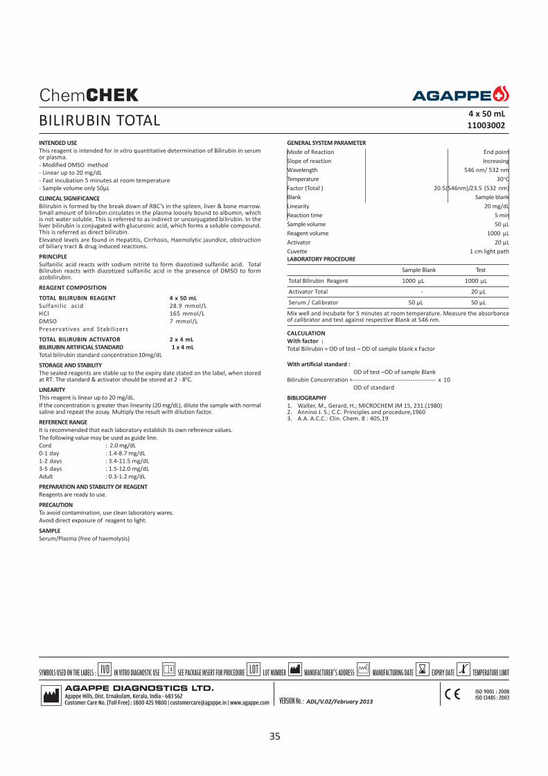
INTENDED USE
This reagent is intended for in vitro quantitative determination of Bilirubin in serumor plasma.
- Modified DMSO method
- Linear up to 20 mg/dL
- Fast incubation 5 minutes at room temperature
- Sample volume only 50µL
CLINICAL SIGNIFICANCE
Bilirubin is formed by the break down of RBC’s in the spleen, liver & bone marrow.Small amount of bilirubin circulates in the plasma loosely bound to albumin, whichis not water soluble. This is referred to as indirect or unconjugated bilirubin. In theliver bilirubin is conjugated with glucuronic acid, which forms a soluble compound.This is referred as direct bilirubin.
Elevated levels are found in Hepatitis, Cirrhosis, Haemolytic jaundice, obstructionof biliary tract & drug induced reactions.
PRINCIPLE
Sulfanilic acid reacts with sodium nitrite to form diazotized sulfanilic acid. TotalBilirubin reacts with diazotized sulfanilic acid in the presence of DMSO to formazobilirubin.
REAGENT COMPOSITION
TOTAL BILIRUBIN REAGENT 4 x 50 mL
Sulfanilic acid 28.9 mmol/L
HCl 165 mmol/L
DMSO 7 mmol/L
Preservatives and Stabilizers
TOTAL BILIRUBIN ACTIVATOR 2 x 4 mL
BILIRUBIN ARTIFICIAL STANDARD 1 x 4 mL
Total bilirubin standard concentration10mg/dL
STORAGE AND STABILITY
The sealed reagents are stable up to the expiry date stated on the label, when storedat RT. The standard & activator should be stored at 2 - 80C.
LINEARITY
This reagent is linear up to 20 mg/dL.
If the concentration is greater than linearity (20 mg/dL), dilute the sample with normalsaline and repeat the assay. Multiply the result with dilution factor.
REFERENCE RANGE
It is recommended that each laboratory establish its own reference values.
The following value may be used as guide line.
Cord : 2.0 mg/dL
0-1 day : 1.4-8.7 mg/dL
1-2 days : 3.4-11.5 mg/dL
3-5 days : 1.5-12.0 mg/dL
Adult : 0.3-1.2 mg/dL
PREPARATION AND STABILITY OF REAGENT
Reagents are ready to use.
PRECAUTION
To avoid contamination, use clean laboratory wares.
Avoid direct exposure of reagent to light.
SAMPLE
Serum/Plasma (free of haemolysis)
BILIRUBIN TOTAL4 x 50 mL
11003002
GENERAL SYSTEM PARAMETER
Mode of Reaction End point
Slope of reaction Increasing
Wavelength 546 nm/ 532 nm
Temperature 30oC
Factor (Total ) 20.5(546nm)/23.5 (532 nm)
Blank Sample blank
Linearity 20 mg/dL
Reaction time 5 min
Sample volume 50 µL
Reagent volume 1000 µL
Activator 20 µL
Cuvette 1 cm light path
LABORATORY PROCEDURE
Sample Blank Test
Total Bilirubin Reagent 1000 µL 1000 µL
Activator Total - 20 µL
Serum / Calibrator 50 µL 50 µL
Mix well and incubate for 5 minutes at room temperature. Measure the absorbanceof calibrator and test against respective Blank at 546 nm.
CALCULATION
With factor :
Total Bilirubin = OD of test – OD of sample blank x Factor
With artificial standard :
OD of test –OD of sample Blank
Bilirubin Concentration =--------------------------------------- x 10
OD of standard
BIBLIOGRAPHY
1. Walter, M., Gerard, H.; MICROCHEM JM 15, 231.(1980)2. Annino J. S.; C.C. Principles and procedure,19603. A.A. A.C.C.: Clin. Chem. 8 : 405,19
ADL/V.02/February 2013
35

INTENDED USE
This reagent is intended for in vitro quantitative determination of Bilirubin in serumor plasma.
- Modified DMSO/DIAZO method- Linear up to 20 mg/dL- Fast incubation 5 minutes at room temperature- Sample volume only 50 µL
CLINICAL SIGNIFICANCE
Bilirubin is formed by the break down of RBC’s in the spleen, liver & bone marrow.Small amount of bilirubin circulates in the plasma loosely bound to albumin, whichis not water soluble. This is referred to as indirect or unconjugated bilirubin. In theliver bilirubin is conjugated with glucuronic acid, which forms a soluble compound.This is referred as direct bilirubin.
Elevated levels are found in Hepatitis, Cirrhosis, Haemolytic jaundice, obstructionof biliary tract & drug induced reactions.
PRINCIPLE
Sulfanilic acid reacts with sodium nitrite to form diazotized sulfanilic acid. TotalBilirubin reacts with diazotized sulfanilic acid in the presence of DMSO to formazobilirubin.
REAGENT COMPOSITION
TOTAL BILIRUBIN REAGENT 2 x 50mL
Sulfanilic acid 28.9mmol/L
HCl 165mmol/L
DMSO 7mmol/L
Preservatives and Stabilizers
TOTAL BILIRUBIN ACTIVATOR 2 x 4 mL
DIRECT BILIRUBIN REAGENT 2 x 50 mL
Sulfanilic acid 28.9 mmol/L
HCl 165 mmol/L
DIRECT BILIRUBIN ACTIVATOR 2 x 4 mL
BILIRUBIN ARTIFICIAL STANDARD 1 x 4 mL
Total bilirubin standard concentration 10 mg/dL
Direct bilirubin standard concentration 7.7 mg/dL
STORAGE AND STABILITY
The sealed reagents are stable up to the expiry date stated on the label, when storedat RT. The standard & activator should be stored at 2 - 80C.
LINEARITY
This reagent is linear up to 20 mg/dL.
If the concentration is greater than linearity (20 mg/dL), dilute the sample with normalsaline and repeat the assay. Multiply the result with dilution factor.
NORMAL RANGE
It is recommended that each laboratory establish its own reference values.
The following value may be used as guide line.
Total Bilirubin - up to 1.2 mg/dL
Direct bilirubin up to 0.4 mg/dL
PREPARATION AND STABILITY OF REAGENT
Reagents are ready to use.
PRECAUTION
To avoid contamination, use clean laboratory wares.
Avoid direct exposure of reagent to light.
SAMPLE
Serum/Plasma (free of haemolysis)
BILIRUBIN TOTAL & DIRECT4 x 50 mL
11003001
GENERAL SYSTEM PARAMETER
Mode of Reaction End point
Slope of reaction Increasing
Wavelength 546 nm/ 532 nm
Temperature 30oC
Factor (Total ) 20.5(546nm)/23.5 (532 nm)
Factor (Direct) 16(546nm)/18 (532 nm)
Blank Sample blank
Linearity 20 mg/dL
Reaction time 5 min
Sample volume 50 µL
Reagent volume 1000 µL
Activator 20 µL
Cuvette 1 cm light path
LABORATORY PROCEDURE
Total Bilirubin Direct Bilirubin
Sample blank Test Sample blank Test
T.Bilirubin reagent 1000 µL 1000µL - -
D.Bilirubin reagent - - 1000 µL 1000 µL
Activator (T/D) - 20 µL - 20 µL
Serum 50 µL 50 µL 50 µL 50 µL
Mix well and incubate for exactly 5 minutes. Measure the absorbance of the sampleagainst the respective sample blank at 546 or 532 nm.
CALCULATION
For semi auto with factor :
Total Bilirubin = OD of test – OD of sample blank x Factor
Direct Bilirubin = OD of test – OD of sample blank x Factor
With artificial standard :
OD of test –OD of sample Blank
Total Bilirubin Concentration = ------------------------------------------ x 10OD of standard
OD of test –OD of sample Blank
Direct Bilirubin Concentration = ------------------------------------------ x 7.7 OD of standard
BIBLIOGRAPHY
1. Walter, M., Gerard, H.; MICROCHEM J M 15, 231.(1980)2. Annino J. S.; C.C. Principles and procedure, 1960.3. A.A. A.C.C.; Clin. Chem. 8 : 405,19
ADL/V.02/JAN 2013
36

INTENDED USE
This reagent is intended for in vitro quantitative determination of calcium in serum,plasma & urine.
- Modified OCPC methodology
- Linear up to 15 mg/dL
CLINICAL SIGNIFICANCE
Calcium is an important ion present in the body. Mainly it is found in bones. In serumcalcium exists equally in a free ionized form & also in a bound form with albumin.Calcium helps in enzyme activation, muscle contraction, coagulation of blood,regulation of some hormonal secretions & cell membrane permeability.
Increased levels are found in hyperthyroidism, malignant tumors, acute osteoporosis& adrenal insufficiency.
Decreased levels found in hypoparathyrodism, osteomalacia, rickets, renal failure &tetanus.
PRINCIPLE
Calcium OCPC procedure is based on on the reaction of calcium ions (Ca 2++) with O-cresolphthalein complex in an alkaline solution to form an intense violet colouredcomplex which shows maximum absorbance at 578nm. the 8-hydroxy quinoloineprevents Mg2++ interference upto 4 mmol/L.
REAGENT COMPOSITION
CALCIUM DYE REAGENT (R2) 2 x 25 mL / 2 x 50 mL
Diethylamine 360 mmol/L
CALCIUM BASE REAGENT (R1) 2 x 25 mL / 2 x 50 mL
O-Cresolphthalein complex 0.15 mmol/L
8-Hydroxyquinoline 17.2 mmol/L
CALCIUM STANDARD 1 x 4 mL
Calcium standard concentration 10 mg/dL
STORAGE & STABILITY
The sealed reagents are stable up to the expiry date stated on the label, when storedat 2 - 80C.
LINEARITY
This reagent is linear up to 15 mg/dL.
If the concentration is greater than linearity (15 mg/dL), dilute the sample with normalsaline and repeat the assay. Multiply the result with dilution factor.
NORMAL RANGE
It is recommended that each laboratory establish its own reference values.
The following value may be used as guide line.
Serum : 8.8 - 10.2 mg/dL
Urine : 100 - 400 mg/24 hrs
PREPARATION AND STABILITY OF REAGENT
Mix reagent 1 (R1) and Reagent 2 (R2) in the ratio 1:1.
PRECAUTION
To avoid contamination, use clean laboratory wares.
Avoid direct exposure of reagent to light.
(Use acid washed (50 % HNO3) glass wares & tips)
SAMPLE
Serum / plasma (free of haemolysis) / Urine (1/3 diluted)
GENERAL SYSTEM PARAMETER
Mode of Reaction End point
Slope of reaction Increasing
Wavelength 578 nm (565-580 nm)
Temperature 30oC
Standard Concentration 10 mg/dL
Linearity 15 mg/dL
Blank Reagent
Incubation time 5 min
Sample volume 10 µL
Reagent volume 1000 µL
Cuvette 1 cm light path
CALCIUM4 x 25 mL, 4 x 50 mL
11005001, 11005002
LABORATORY PROCEDURE
Blank Standard Sample
Working Reagent 1000 µL 1000 µL 1000 µL
Standard - 10 µL -
Sample - - 10 µL
Mix and incubate for 5 min. at room temperature. Read the absorbance of standardand sample against reagent blank.
CALCULATION
Absorbance of sample
Calcium Conc. (mg/dL) = --------------------------------- x 10
Absorbance of Standard
INTERFERENCE
Bilirubin concentrations higher than 20 mg/dL and phosphate higher than 40 mg/dL,will interfere with the assay.
BIBLIOGRAPHY
1. Schwarzenbach, G.; Analyst., 80, (1955) 713-7292. Kessler, G., Wolfman, M., Clin.Chem., 10, (1964) 686 – 7033. Connerty, H. V., Briggs, A.R., Am. J. Clin.Pathol., 45, (1965) 290-2964. Gitelmann, H. J. Anal Biochem 18, (1967) 521-5315. Biggs, H.G., Moorehead, W. R.; Clin.Chem., 20, (1974) 1458-1460
ADL/V.02/February 2013
37

INTENDED USE
This reagent is intended for in vitro quantitative determination of calcium in serum,plasma & urine.
- Modified Arsenazo III method
- Linear up to 18mg/dL
CLINICAL SIGNIFICANCE
Calcium is an important ion present in the body. Mainly it is found in bones. In serumcalcium exists equally in a free ionized form & also in a bound form with albumin.Calcium helps in enzyme activation, muscle contraction, coagulation of blood,regulation of some hormonal secretions & cell membrane permeability.
Increased levels are found in hyperthyroidism, malignant tumors, acute & osteoporosisadrenal insufficiency.
Decreased levels found in hypoparathyrodism, osteomalacia, rickets, renal failure &tetanus.
PRINCIPLE
At a neutral pH the Ca2+ form with Arsenazo III a complex, the color intensity of whichis directly proportional to the concentration of calcium in the sample.
REAGENT COMPOSITION
CALCIUM ARSENAZO REAGENT 4 x 25 mL
MES, pH 6.50 100 mmol/L
Arsenazo III 200 mmol/L
CALCIUM ARSENAZO STANDARD 1 x 4 mL
Calcium Standard Concentration 10 mg/dL
STORAGE & STABILITY
The sealed reagents are stable up to the expiry date stated on the label, when storedat 2 - 80C.
LINEARITY
This reagent is linear up to 18 mg/dL.
If the concentration is greater than linearity (18mg/d/L), dilute the sample with normalsaline and repeat the assay. Multiply the result with dilution factor.
NORMAL RANGE
It is recommended that each laboratory establish its own reference values.
The following value may be used as guide line.
Serum / plasma : 8.8 - 10.2 mg/dL
PREPARATION AND STABILITY OF WORKING REAGENT
The Reagent is ready to use.
PRECAUTION
To avoid contamination, use clean laboratory wares.
Avoid direct exposure of reagent to light.
NOTE: Use acid washed (50% HNO3) glass wares & tips.
SAMPLE
Serum / plasma (free of haemolysis)
Take dilution factor into account for the calculation of the concentration in urine.
GENERAL SYSTEM PARAMETER
Mode of Reaction End point
Slope of reaction Increasing
Wavelength 630 or 650 nm
Temperature RT
Standard Concentration 10 mg/dL
Linearity 18 mg/dL
Blank reagent
Incubation time 1 min
Sample volume 10 µL
Reagent volume 1000 µL
Cuvette 1 cm light path
CALCIUM (ARSENAZO)4 x 25 mL
11006001
LABORATORY PROCEDURE
Blank Standard Sample
Working Reagent 1000 µL 1000 µL 1000 µL
Standard - 10 µL -
Sample - - 10 µL
Mix and incubate for 1 minute. Measure the absorbance of standard and sampleagainst reagent blank.
CALCULATION
Absorbance of sample
Calcium Conc. (mg/dL) = ------------------------------- x 10
Absorbance of Standard
BIBLIOGRAPHY
Baver, P. J.; Anal. Biochem., 110, (1981), 61
Biggs, H.G., Moorchand, W.R., (1974), cLIN. CHEM. 20, 1458-1460.
ADL/V.02/February 2013
38
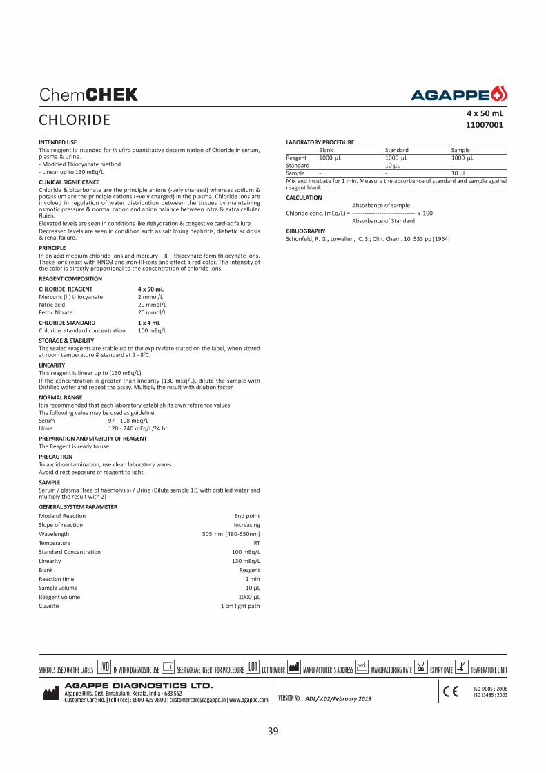
INTENDED USE
This reagent is intended for in vitro quantitative determination of Chloride in serum,plasma & urine.
- Modified Thiocyanate method
- Linear up to 130 mEq/L
CLINICAL SIGNIFICANCE
Chloride & bicarbonate are the principle anions (-vely charged) whereas sodium &potassium are the principle cations (+vely charged) in the plasma. Chloride ions areinvolved in regulation of water distribution between the tissues by maintainingosmotic pressure & normal cation and anion balance between intra & extra cellularfluids.
Elevated levels are seen in conditions like dehydration & congestive cardiac failure.
Decreased levels are seen in condition such as salt losing nephritis, diabetic acidosis& renal failure.
PRINCIPLE
In an acid medium chloride ions and mercury – II – thiocynate form thiocynate ions.These ions react with HNO3 and iron-III-ions and effect a red color. The intensity ofthe color is directly proportional to the concentration of chloride ions.
REAGENT COMPOSITION
CHLORIDE REAGENT 4 x 50 mL
Mercuric (II) thiocyanate 2 mmol/L
Nitric acid 29 mmol/L
Ferric Nitrate 20 mmol/L
CHLORIDE STANDARD 1 x 4 mL
Chloride standard concentration 100 mEq/L
STORAGE & STABILITY
The sealed reagents are stable up to the expiry date stated on the label, when storedat room temperature & standard at 2 - 80C.
LINEARITY
This reagent is linear up to (130 mEq/L).
If the concentration is greater than linearity (130 mEq/L), dilute the sample withDistilled water and repeat the assay. Multiply the result with dilution factor.
NORMAL RANGE
It is recommended that each laboratory establish its own reference values.
The following value may be used as guideline.
Serum : 97 - 108 mEq/L
Urine : 120 - 240 mEq/L/24 hr
PREPARATION AND STABILITY OF REAGENT
The Reagent is ready to use.
PRECAUTION
To avoid contamination, use clean laboratory wares.
Avoid direct exposure of reagent to light.
SAMPLE
Serum / plasma (free of haemolysis) / Urine (Dilute sample 1:1 with distilled water andmultiply the result with 2)
GENERAL SYSTEM PARAMETER
Mode of Reaction End point
Slope of reaction Increasing
Wavelength 505 nm (480-550nm)
Temperature RT
Standard Concentration 100 mEq/L
Linearity 130 mEq/L
Blank Reagent
Reaction time 1 min
Sample volume 10 µL
Reagent volume 1000 µL
Cuvette 1 cm light path
CHLORIDE4 x 50 mL
11007001
LABORATORY PROCEDURE
Blank Standard Sample
Reagent 1000 µL 1000 µL 1000 µL
Standard - 10 µL -
Sample - - 10 µL
Mix and incubate for 1 min. Measure the absorbance of standard and sample againstreagent blank.
CALCULATION
Absorbance of sample
Chloride conc. (mEq/L) = ------------------------------ x 100
Absorbance of Standard
BIBLIOGRAPHY
Schonfeld, R. G., Lowellen, C. S.; Clin. Chem. 10, 533 pp (1964)
ADL/V.02/February 2013
39

INTENDED USE
This reagent is intended for in vitro quantitative determination of Copper in serum.
CLINICAL SIGNIFICANCE
Copper is an essential trace mineral in humans, the function of copper is to help torelease energy, helps in melanin production in the skin, helps in the production ofred blood cells and aid in the absorption and transport of iron.
Acquired copper deficiency can cause hematological/neurological manifestations.Wilson disease (copper toxicity) is associated with neurological manifestations andlow serum copper, with copper deposited in tissues responsible for the toxicity. Thesymptoms of acute copper poisoning include nausea, vomiting and abdominal andmuscle pain.
PRINCIPLE
Copper in an acid medium reacts with the chromogen Di-Br-PAESA 4-(3,5-dibromo-2- pyridylazo)-N-ethyl-N-(3-sulphopropyl0aniline.) to form a coloured complex.Intensity of the colour is directly proportional to the amount of Copper present inthe sample.
REAGENT COMPOSITION
COPPER REAGENT 1 1 x 25 mL
Acetate buffer(pH 4.9) 0.1 M
COPPER REAGENT 2 1 x 25 mL
3,5 Di-Br-PAESA
Preservatives
COPPER STANDARD 1 x 4 mL
Copper Standard (Concentration) 200 µg/dL
STORAGE AND STABILITY
The sealed reagents are stable up to the expiry date stated on the label, when storedat 2-80C.
Note:Reagent 1 solidifies when kept at 2-8oC. Allow the reagent to melt by keeping atroom temperature before use.
LINEARITY
This reagent is linear up to 500 µg/dL.
If the concentration is greater than linearity (500 µg/dL), dilute the sample with normalsaline and repeat the assay. Multiply the result with dilution factor.
REFERENCE VALUES
It is recommended that each laboratory establish its own reference values.
The following value may be used as guide line.
Men : 70 - 140 µg/dL
Women : 80- 155 µg/dL
Newborns : 20- 70 µg/dL
Children up to 6 years : 90-190 µg/dL
Children up to 12 years : 80-160 µg/dL
PREPARATION OF WORKING REAGENT
Mix in equal parts the reagent 1 & reagent 2 .
PRECAUTION
To avoid contamination, use clean laboratory wares. It is recommended to usedisposable tubes. Use clean, dry disposable pipette tips for dispensing. Close reagentand standard bottles immediately after use. Avoid direct exposure of reagent to light.
Note: Use acid washed (50% HNO3) glass wares and tips.
SAMPLE
Serum or Plasma (free haemolysis). Use heparin as anticoagulant.Highly lipemic serummay interfere the assay. it is recommended to filter or centrifuge the sample
GENERAL SYSTEM PARAMETER
Mode of Reaction End Point
Slope of reaction Increasing
Wavelength 580 nm
Temperature 30oC
Standard concentration 200 µg/dL
Linearity 500 µg/dL
Blank Reagent Blank
Incubation Time 10 min
Sample volume 70 µL
Reagent volume 1000 µL
Cuvette 1 cm light path
LABORATORY PROCEDURE
Blank Standard Sample
WorkingReagent 1000 µL 1000 µL 1000 µL
Distilled water 70 µL - -
Standard - 70 µL -
Sample - - 70 µL
Mix and incubate for 10 minutes at 30oC. Read the absorbance(A) of standard andsample against blank at 580 nm. The colour is stable for 30 minutes.
CALCULATION
Absorbance of Sample
Copper µg/dL = -------------------------------- x 200
Absorbance of Standard
BIBLIOGRAPHY
1. Pasquinelli, F.; Diagnostica e Tecniche di Laboratorio, (pag.:1099-1102) RossiniEditrice.(1984)
2. Akitaabe, Sumico Yiamashita; Clin. Chem. 35(4):197,552-554(1989)3. Cuiti, R, Gallia, Giorn.; It. Chim. Clin. 12(2): 91-100 (1987)
COPPER2 x 25 mL
11020001
40

INTENDED USE
This reagent is intended for in vitro quantitative determination of creatinine in serum,plasma & urine.
- Modified Jaffe’s method
- Linear up to 24 mg/dL
- No Bilirubin interference up to 10 mg/dL
CLINICAL SIGNIFICANCE
Creatinine is formed in muscles from phospho creatinine. It is an important form ofenergy, being a store of high-energy phosphate. Creatinine determinations have oneadvantage over Urea determination that it is not affected by a high protein diet.
Serum creatinine is more specific & sensitive indicator of renal function. Simultaneousestimations of serum urea & creatinine provides better information. Serum ureanitrogen, creatinine ratio is > 15 in pre renal failure, & < 10 in renal failure.
Decreased levels are found in muscle dystrophy.
PRINCIPLE
Creatinine reacts with picric acid to produce a colored compound, creatinine alkalinepicrate. The change in absorbance is proportional to the creatinine concentration.
REAGENT COMPOSITION
CREATININE BASE REAGENT (R1) 2 x 50 mL
Sodium hydroxide 300 mmol/L
Sodium Phosphate 25 mmol/L
CREATININE DYE REAGENT (R2) 2 x 50 mL
Picric Acid 8.73 mmol/L
Surfactant
CREATININE STANDARD 1 x 4 mL
Creatinine standard concentration 2 mg/dL
STORAGE & STABILITY
The sealed reagents are stable up to the expiry date stated on the label, when storedat room temperature & standard at 2 - 80C.
LINEARITY
This reagent is linear up to 24 mg/dL.
If the concentration is greater than linearity (24 mg/dL), dilute the sample with normalsaline and repeat the assay. Multiply the result with dilution factor.
NORMAL RANGE
It is recommended that each laboratory establish its own reference values.
The following value may be used as guide line.
Serum : Men : 0.7 – 1.4 mg/dL
Female : 0.6 – 1.2 mg/dL
Urine : 0.80 – 1.80 gm/24 hour
PREPARATION AND STABILITY OF WORKING REAGENT
Mix 1 volume of Reagent 1 (R1) with 1 volume of Reagent 2 (R2).
PRECAUTION
To avoid contamination, use clean laboratory wares.
Avoid direct exposure of reagent to light.
SAMPLE
Serum / plasma (free of haemolysis) / Urine (diluted 1/100 with distilled water)
CREATININE4 x 50 mL
11009001
GENERAL SYSTEM PARAMETER
Mode of Reaction Fixed time
Slope of reaction Increasing
Wavelength 492 nm/ 505 nm
Temperature 370C
Standard Concentration 2 mg/dL
Linearity 24 mg/dL
Blank D I water
Delay time 60 sec
Interval 60 sec
Sample volume 100 µL
Reagent volume 1000 µL
Cuvette 1 cm light path
LABORATORY PROCEDURE
Standard Sample
Working Reagent 1000 µL 1000 µL
Standard 100 µL -
Sample - 100 µL
Mix and read the optical density (T1) 60 seconds after the sample or standard
addition. Exactly 60 seconds after the first reading take second reading (T2)
CALCULATION
(T2- T
1) of sample
Creatinine conc. (mg/dL) = ---------------------- x 2
(T2- T
1) of Standard
BIBLIOGRAPHY
1. Allen, L.C.; Clin chem. Vol.28 No.3, 1982, 555.2. Haeckel, R., et al. ; Clin. Chem. 27/1 179-183 (1981).3. Tanganelli, E., Prencipe,L. , Bassi, D., Cambiaghi, S. and Murador, E.; Clin.Chem
28/7, 1461-1464 (1982)
ADL/V.02/JAN 2013
41

INTENDED USE
This reagent is intended for in vitro quantitative determination of Haemoglobin inblood.
- Based on cyanmethaemoglobin method
- Linear up to 20 gm/dL
CLINICAL SIGNIFICANCE
A decrease in haemoglobin below normal range is an indication of anaemia. An increasein haemoglobin concentration occurs in haemoconcentration due to loss of bodyfluid in severe diarrhea and vomiting. High values are also observed in congenital heartdisease (due to reduced oxygen supply) in emphysema and also in poly cythemia.
Haemoglobin concentration drops during pregnancy due to haemodilution
PRINCIPLE
The Haemoglobin (oxyhaemoglobin,methemoglobin, Carboxyhaemoglobin) isconverted to cyanmethaemoglobin according to the following reactions.
K3 Fe(CN)6
Haemoglobin --------------- > Methemoglobin
KCN
Methemoglobin --------------- > cyanmethemoglobin
The intensity of the color is proportional to haemoglobin concentration and iscompared to known cyan methaemoglobin standard at 540 nm (green filter).
REAGENT COMPOSITION
HAEMOGLOBIN REAGENT 1000 mL
Potassium Phosphate 2.0 mmol/L
Potassium ferricyanide 0.60 mmol/L
Potassium cyanide 0.90 mmol/L
Sodium chloride 1.4 mmol/L
HAEMOGLOBIN STANDARD 1 x 4 mL
Cyanmethaemoglobin standard con. 60 mg/dL
STORAGE AND STABILITY
The sealed reagents are stable up to the expiry date stated on the label, when storedat room temperature & standard at 2 - 80C.
LINEARITY
This reagent is linear up to 20 gm/dL.
REFERENCE RANGE
It is recommended that each laboratory establish its own reference values.
The following value may be used as guide line.
Cord blood :13.5 - 20.5 gm/dL
0.5 - 2.0 yrs :11.3 - 14.1 gm/dL
Male :13.2 - 17.3 gm/dL
Female :11.7 - 15.3 gm/dL
PREPARATION AND STABILITY OF REAGENT
The reagent is ready to use.‘
PRECAUTION
To avoid contamination, use clean laboratory wares.
Avoid direct exposure of reagent to light. Do not pipette the reagent with mouth.
SAMPLE
Fresh whole blood.
GENERAL SYSTEM PARAMETER
Mode of Reaction End point
Slope of reaction Increasing
Wavelength 546nm (530-550nm)
Temperature 30oC
Blank Reagent
Linearity 20 gm/dL
Standard concentration 15 gm /dL (60x0.251)
Incubation time 5 min
Sample volume 20 µL
Reagent volume 5000 µL
Cuvette 1 cm light path
*NOTE : Analyzer users directly enter given Factor without running standard.
Factor 35
LABORATORY PROCEDURE
Blank Sample
Hb Reagent 5000 µL 5000 µL
Sample - 20 µL
Mix well and incubate at room temperature for 5 minutes. Measure the absorbanceof sample against reagent blank and measure the absorbance of standard directlyagainst blank (distilled water).
CALCULATION
Haemoglobin Conc. (gm/dL) =
Absorbance of sample
---------------------------- x 60 x 0.251
Absorbance of standard
Dilution factor
Where, 0.251 = ------------------
Convertion factor
OR
Absorbance of sample
-------------------------------- x 15
Absorbance of standard
15 = 60 x 0.251
BIBLIOGRAPHY
1. Drabkin, D.L., et al.; J.Bio.Chem, 98 (1932), 7192.. Zijlstra, N. C.; Clin.Chem.Acta, 5,(1960) 719
HAEMOGLOBIN 1 x 1000 mL
11011001
ADL/V.02/Jan 2013
3. Tietz Text book of Clin Chem.Carl.AB,Edward.R.A,3rd Edition 1999
42

INTENDED USE
This reagent is intended for in vitro quantitative determination of HDL in serum orplasma.
- Precipitation method, Phosphotungstate magnesium acetate reagent
- Linear up to 125 mg/dL
CLINICAL SIGNIFICANCE
Lipoproteins are the proteins, which mainly transport lipids in the blood stream. Theyare (HDL) High density lipoproteins, (LDL) Low density lipoproteins, (VLDL) Very lowdensity lipoproteins & chylomicrons. LDL carries cholesterol to the peripheral tissueswhere it can be deposited & increase the risk of atherosclerotic heart & peripheralvascular disease. Hence high levels of LDL are atherogenic. HDL transports cholesterolfrom peripheral tissues to the liver & then for excretion, hence HDL has a protectiveeffect. Hence the determination of serum HDL cholesterol is a useful tool to identifypatients at risk of developing coronary heart disease.
PRINCIPLE
The chylomicrons, Very low density lipoproteins (VLDL) and Low density lipoproteins(LDL) of serum are precipitated by phosphotungstic acid and magnesium ions.
After centrifugation, High density lipoproteins (HDL) are in the supernatant. HDLcontent of supernatant is measured by an enzymatic Method.
REAGENT COMPOSITION
HDL CHOLESTEROL R1 4 x 25 mL
Phosphotungstate 14 mmol/L
Magnesium Chloride 1 mmol/L
Preservative
HDL CHOLESTEROL STANDARD 1 x 4 mL
HDL Cholesterol concentration 50 mg/dL
STORAGE & STABILITY
The sealed reagents are stable up to the expiry date stated on the label, when storedat 2 - 80C.
LINEARITY
The reagent is linear up to 125 mg/dL.
If the concentration is greater than linearity (125 mg/dL), dilute the sample with normalsaline and repeat the assay. Multiply the result with dilution factor.
NORMAL RANGE
It is recommended that each laboratory establish its own reference values.
The following values may be used as guide line.
HDL Cholesterol
Men : 35-55 mg/dL
Women : 45-65 mg/dL
LDL Cholestrol
Suspicious : 150 mg/dL
Elevated : 190 mg/dL
PREPARATION AND STABILITY OF REAGENT
Reagent is ready to use.
PRECAUTION
To avoid contamination, use clean laboratory wares.
Avoid direct exposure of reagent to light.
SAMPLE
Serum / Plasma (free of haemolysis).
HDL CHOLESTEROL4 x 25 mL
11010001
GENERAL SYSTEM PARAMETER
Mode of Reaction End point
Slope of reaction Increasing
Wavelength I 505 (500 -532 nm)
Wavelength II 630 nm
Temperature 370C
Standard Concentration 50 mg/dL
Blank Cholesterol Reagent
Linearity 125 mg/dL
Incubation time 5 min
Sample volume 50 µL
Reagent volume 1000 µL
Cuvette 1 cm light path
LABORATORY PROCEDURE
1. PRECIPITATION
Sample 300 µL
HDL reagent 300 µL
Mix well, allow to stand for 10 min. at room temperature, mix again and centrifugefor 10 min, at 4000 rpm.
After centrifugation separate the clear supernatant from the precipitate within 1 hourand determine the HDL Cholesterol concentration using the cholesterol reagent.
2. HDL CHOLESTEROL DETERMINATION :
Blank Standard Sample
Cholesterol Reagent 1000 µL 1000 µL 1000 µL
Standard(HDL) - 50 µL -
HDL supernatant - - 50 µL
Mix and incubate for 5 min. at 370C. Measure the absorbance of the standard &sample against the reagent blank.
CALCULATION
HDL Cholesterol Conc. In mg/dL =
Absorbance of sample
------------------------------------ x concentration of standard x 2
Absorbance of standard
where, 2 = dilution factor of the sample.
LDL-Chol conc in mg/dL = Total Cholesterol – (HDL Chol. + Triglycerides / 5)
BIBLIOGRAPHY
1. Assmann, G.; Intermist 20 (1979), 5592. Gordon, T., et al.; Med 62 (1977), 7073. Friedewald, W. T., et al.; Clin.Chem.18 (1972), 499.
ADL/V.02/February 2013
43

INTENDED USE
This reagent is intended for in vitro quantitative determination of Phosphorous inserum or plasma.
- Phosphomolybdate methodology
- Linear up to 15 mg/dL
CLINICAL SIGNIFICANCE
Phosphorous is mainly combined with calcium & is found in bones. It is involved inthe carbohydrate metabolism & is a component of many other substances. Some ofits important functions include maintaining of acid-base balance, skeletal muscleformation. It is also required for normal functioning of RBC’s & muscles.
Increased levels are found in hypothyroidism, renal failure, bone metastasis & liverdisease.
Decreased levels are found in hyperparathyroidism, osteomalacia & disease associatedwith Vitamin D defieciency.
PRINCIPLE
Determination of inorganic phosphorous according to the following reaction.
Phosphorous
Ammonium molybdate +sulfuric acid ------------------->phosphomolybdic complex
REAGENT COMPOSITION
INORGANIC PHOSPHOROUS REAGENT 4 x 25 mL / 4 x 50 mL
Sulfuric acid 210 mmol/L
Ammonium molybdate 650 mmol/L
INORGANIC PHOSPHOROUS STANDARD 1 x 4 mL
Phosphorous standard concentration 5 mg/dL
STORAGE & STABILITY
The sealed reagents are stable up to the expiry date stated on the label, when storedat 2 80C.
LINEARITY
This reagent is linear up to 15 mg/dL.
If the concentration is greater than linearity (15 mg/dL), dilute the sample with normalsaline and repeat the assay. Multiply the result with dilution factor.
NORMAL RANGE
It is recommended that each laboratory establish its own reference values.
The following value may be used as guide line.
Serum : 2.7 - 4.5 mg/dL
Urine : 400 - 1300 mg/24 hr.
PREPARATION AND STABILITY OF REAGENT
Reagent and standard are ready to use.
PRECAUTION
To avoid contamination, use clean laboratory wares.
Avoid direct exposure of reagent to light.
NOTE: Use acid washed (50% HNO3) glasswares & tips.
SAMPLE
Serum / Plasma (free of haemolysis) / Urine (diluted 1/10 with distilled water).
INORGANIC PHOSPHOROUS4 x 25 mL, 4 x 50 mL
11012001, 11012002
GENERAL SYSTEM PARAMETER
Mode of Reaction End point
Slope of reaction Increasing
Wavelength 340 nm
Temperature 370C
Standard Concentration 5 mg/dL
Linearity 15 mg/dL
Blank Reagent
Reaction Time 1 min
Sample volume 20 µL
Reagent volume 1000 µL
Cuvette 1 cm light path
LABORATORY PROCEDURE
Blank Standard Sample
Reagent 1000 µL 1000 µL 1000 µL
Standard - 20 µL -
Sample - - 20 µL
Mix & incubate at RT for 5 minutes. Read the absorbance of sample and standardagainst reagent blank.
CALCULATION
Absorbance of sample
Phosphorous Conc. (mg/dL) = ------------------------------ x 5
Absorbance of standard
BIBLIOGRAPHY
1. Tietz, N., Clinical Guide to Laboratory Tests, W.B. Saunders Company, Philad, 1983,384
2. Henry, R.J.; Clin. Chem., Harper & Row Publishers. New Yoork 19743. Thomas, L.; Labor and Diagnose, 2 Aufl. Med.Vert. Gem. Marburg 1979 4.
Taussky, H.H., Schorr, E.; J.Biol. Chem 202, 675 (1953)
ADL/V.02/February 2013
44

INTENDED USE
This reagent is intended for in vitro quantitative determination of Iron in serum.
Iron Chromazurol method
Linearity- 500 µg/dL
CLINICAL SIGNIFICANCE
In the blood iron is present in the hemoglobin of erythrocytes. Major function of ironin the body is the transportation of oxygen to the cells and cellular oxidation. Ironabsorbed in the small intestine and bound to a globulin in the plasma called transferrin.It is then transported to bone marrow where RBC generation take place.
Increased levels of iron in serum are seen in hemolytic aneamia, hepatitis and lead &iron poisoning.
Deceased levels are found in iron deficiency anemia, late pregnancy and cancer.
PRINCIPLE
Iron, bound to Transferrin in Fe(III) form, is released in an acidic medium and the Ferricions are reduced to Ferrous ions. The Fe (II) ions react with
Cromazurol B to form an intensely coloured complex. Intensity of the colour
formed is directly proportional to the amount of Iron present in the
sample.
REAGENT COMPOSITION
IRON REAGENT 2 x 50 mL
Acetate Buffer (pH 4.7) 0.2 mol/L
CTMA Bromide 0.7 mmol/L
Cromazurol B 2 mmol/L
IRON STANDARD 1 x 4 mL
Iron Standard (Concentration) 200 µg/dL
STORAGE AND STABILITY
The sealed reagents are stable up to the expiry date stated on the label, when storedat 2 -80C.
LINEARITY
This reagent is linear up to 500 µg/dL.
If the concentration is greater than linearity (500 µg/dL), dilute the sample with normalsaline and repeat the assay. Multiply the result with dilution factor.
NORMAL RANGE
It is recommended that each laboratory establish its own reference values.
The following value may be used as guide line.
Serum Iron (Males) : 59 - 158 µg/dL
(Females) : 37 - 145 µg/dL
PREPARATION AND STABILITY OF REAGENT
Ready to use reagent.
PRECAUTION
To avoid contamination, use clean laboratory wares.
Avoid direct exposure of reagent to light.
Note: Use acid washed (50% HNO3) glass wares and tips.
SAMPLE
serum
IRON 2 x 50 mL
11022001
GENERAL SYSTEM PARAMETER
Mode of Reaction End Point
Slope of reaction Increasing
Wavelength 630 nm
Temperature 30oC
Standard concentration 200 µg/dL
Linearity 500 µg/dL
Blank Ragent Blank
Incubation Time 10 min
Sample volume 40 µL
Reagent volume 1000 µL
Cuvette 1 cm light path
LABORATORY PROCEDURE
Blank Standard Sample
Reagent 1000 µL 1000 µL 1000 µL
Distilled water 40 µL - -
Standard - 40 µL -
Sample - - 40 µL
Mix and incubate for 10 minutes at room temperature. Read the absorbance(A) ofstandard and sample against blank at 630 nm. The colour is stable for 1 hour whenprotected from light.
CALCULATION
Absorbance of Sample
Iron µg/dL = -------------------------------- x 200
Absorbance of Standard
BIBLIOGRAPHY
1. Weippl, G., et al: Blut.27.261 (1973)2. Garcic, A. ; Clin, chem, Acta 94,115 (1979)3. Tobacco, A., et al; Chem. Clin. Acta 114, 267 (1981)4. Teruzzi, A. , Torelli, G. ; 17th Meeting S.I. Bio.C.Clin.9,1080,communication (1985)
ADL/V.01/APR 2012
45

INTENDED USE
This reagent is intended for in vitro quantitative determination of Magnesium in serumor plasma.
- Xylidyl Blue with ATCS
- Linear up to 5 mg/dL
CLINICAL SIGNIFICANCE
Magnesium is the second most abundant intracellular cation of the human body afterpotassium, being essential in a great number of enzymatic and metabolic processes.
It is a co-factor of all the enzymatic reactions that involve ATP and found in themembranes that maintain the electrical excitability of muscular and nervous cells.
A low magnesium level is found in malabsorption syndrome, diuretics aminoglucosidetherapy, and hyperparathyroidism or diabetic acidosis.
Elevated concentration of magnesium is found in uremia, chronic renal failure,glomerulo nephritis, Addison’s disease or intensive anti acid therapy.
Clinical diagnosis is should not be made on a single test result; it should integrateclinical and other laboratory data.
PRINCIPLE
Magnesium reacts with xylidyl Blue to form a colored compound in alkaline solution.
The intensity of the color formed is proportional to the magnesium in the sample.
REAGENT COMPOSITION
MAGNESIUM REAGENT 4 x 25 mL
Xylidyl Blue 110mmol/L
Ethanolamine (pH 11.0) 1 mol/L
GEDTA 60 mmol/L
MAGNESIUM STANDARD 1 x 3 mL
Magnesium standard concentration 2 mg/dL
STORAGE AND STABILITY
The sealed reagents are stable up to the expiry date stated on the label, when storedat 2-80C, protected from light.
LINEARITY
This reagent is linear up to 5 mg/dL.
If the concentration is greater than linearity (5 mg/dL), dilute the sample with normalsaline and repeat the assay. Multiply the result with dilution factor.
NORMAL RANGE
It is recommended that each laboratory establish its own reference values.
The following value may be used as guide line.
Serum : 1.8 – 2.6 mg/dL
CSF : 2.1 – 3.3 mg/dL
Urine : 73 - 122 mg/24 h
PREPARATION AND STABILITY OF REAGENT
Reagent is ready to use.
PRECAUTION
To avoid contamination, use clean laboratory wares. It is recommended to usedisposable tubes. Use clean, dry disposable pipette tips for dispending. Close reagentand standard bottles immediately after Use. Avoid direct exposure of reagent to light.
SAMPLE
Serum (free haemolysis) / Heparinized plasma (Do not use oxalates or EDTA asanticoagulant) / Urine (should be acidified to pH 3-4 with concentrated HCL thendilute sample 1/5 with distilled water and multiply the result by 5).
MAGNESIUM 4 x 25 mL
11019001
GENERAL SYSTEM PARAMETER
Mode of Reaction End point
Slope of reaction Increasing
Wavelength 546 nm ( 520-550 nm)
Temperature 370C
Standard Concentration 2 mg/dL
Linearity 5 mg/dL
Blank Reagent
Incubation time 5 min
Sample volume 10 µL
Reagent volume 1000 µL
Cuvette 1 cm light path
LABORATORY PROCEDURE
Blank Standard Sample
Reagent 1 1000 µL 1000 µL 1000 µL
Standard - 10 µL -
Sample - - 10 µL
Mix & incubate for 5 minutes at 370C. Read the absorbance of sample and standardagainst reagent blank.
CALCULATION
Magnesium Conc. (mg/dL) =
Absorbance of sample
----------------------------- x Standard concentration
Absorbance of standard
Unit Conversion
mg/dL x 0.4114 = mmol/L
mg/dL x 0.82 = mEq/L
BIBLIOGRAPHY
1. Farrel, E. C.; Magnesium.in Kaplan, A., et al.; Clin chem. The CV Mosby Co. St. Louis,Torento, Princeton 1984; 1064-69
2. Brutis, C. A., et al. TIETZ Text book of Clinical chemistry, 3 rd edition W. B Saunderscompany; 1999, P.1395-1457
3. Young, D. S.; Effects of disease on clinical lab tests, 4th edition, AACC Press,2001.
ADL/V.02/February 2013
46

INTENDED USE
This reagent is intended for in vitro quantitative determination of total protein in urine.
- Pyrogallol Red method
- Linear up to 500 mg/dL
- Sensitivity of 5 mg/dL
CLINICAL SIGNIFICANCE
The presence of protein in urine is a very sensitive indicator of renal disorders. Thereare four ways by which increased amounts of protein can occur:increased glomerularpermeability; defective tubular re-absorption; increased plasma concentration of anabnormal, low molecular weight protein; and abnormal secretion of protein into theurinary tract.
Albuminuria, increased amounts of albumin in urine, has been recognized as an earlyindicator of renal damage in diabetes that can be reversed if detected and treated early.
PRINCIPLE
Proteins,in an acidic medium, combine with Pyrogallol Red and molybdate to form ablue purple coloured complex. The intensity of colour formed is directly proportionalto the amount of proteins present in the sample.
Proteins + Pyrogallol Red + Molybdate ----------> Blue purple coloured Complex
REAGENT COMPOSITION
MICROPROTEIN REAGENT 1 x 50 mL
Succinate buffer, pH 2.5 0.05 mmol/L
Sodium dodecylsulphate 0.07 mmol/L
Sodium molybdate 0.04 mmol/L
Pyrogallol-Red 0.06 mmol/L
MICROPROTEIN STANDARD 1 x 3 mL
Stabilized protein solution 100 mg/dL
Verified against NIST reference material.
STORAGE AND STABILITY
The sealed reagents are stable up to the expiry date stated on the label, when storedat 15 - 250C.
LINEARITY
This reagent is linear up to 500 mg/dL with a sensitivity of 5mg/dL.
If the concentration is greater than linearity (500 mg/dL), dilute the sample with normalsaline and repeat the assay. Multiply the result with dilution factor.
NORMAL RANGE
It is recommended that each laboratory establish its own reference values.
The following value may be used as guide line.
Urine < 24 mg/dL
Urine 24 hour < 120 mg /24 hour
CSF < 40 mg/dL
PREPARATION AND STABILITY OF REAGENT
Reagents are ready to use.
PRECAUTION
To avoid contamination, use clean laboratory wares.
Avoid direct exposure of reagent to light.
SAMPLE
Urine & CSF
Note: For haemolized or turbid samples it is suggested to carry out a sample blank:add 10 µL of sample to 1 mL of DI water and read the absorbance at 600 nm againstDI water.
GENERAL SYSTEM PARAMETER
Mode of Reaction End point
Slope of reaction Increasing
Wavelength 600 nm
Temperature 370C
Blank Reagent
Standard concentration 100 mg/dL
Linearity 500 mg/dL
Incubation Time 5 minutes
Sample volume 10 µL
Reagent volume 1000 µL
Cuvette 1 cm light path
LABORATORY PROCEDURE
Blank Standard Sample
Reagent 1000 µL 1000 µL 1000 µL
Distilled water 10 µL - -
Standard - 10 µL -
Sample - - 10 µL
Accurately mix and, after 5 minutes, read the absorbance of standard and sampleagainst blank at 600 nm. The colour is stable for 30 minutes.
CALCULATION
urine:
A sample
Proteins, mg/dL = -------------- x 100
A standard
Urine 24hrs:
A sample x1000 x T.V in litres
Proteins, mg/24 hrs = ---------------------------------
A standard
T.V = Total volume of 24 hr urine in milliitres
BIBLIOGRAPHY
Watnabe, N., et al.; Urinary protein as measured with a pyrogallol red-molibdatecomplex. Manually and Hitachi 726 automated analyzer. Clin. Chem. 32:1551-4 (1986)
MICROPROTEIN1 x 50 mL
11021001
47

INTENDED USE
This reagent is intended for in vitro quantitative determination of Total Iron BindingCapacity in serum.
CLINICAL SIGNIFICANCE
Total iron-binding capacity (TIBC) is the measure of the ability of serum
proteins, principally transferrin, to bind iron. It is the maximumconcentration
of iron that the serum proteins can bind.
Increase in TIBC is found in Iron deficiency anemias and pregnancy.Decrease in TIBC is found in hypoproteinemia, hemolytic / pernicious /sickle cell anemias, inflammatory diseases and cirrhosis.
PRINCIPLE
TIBC is determined by addition of sufficient Fe(III) to saturate ironbinding sites on apotransferrin. The Fe3+ is removed by adsorption withbasic magnesium carbonate powder. After centrifugation bound ironremaining in supernatant is measured.
The difference between TIBC values and serum iron gives theconcentration of unsaturated transferrin(UIBC).
REAGENT COMPOSITION
TIBC REAGENT A 1 x 5 mL
Iron saturating solution 25 mg/dL
TIBC REAGENT B 50 x 50 mg
Magnesium Carbonate (Powder)
STORAGE AND STABILITY
The sealed reagents are stable up to the expiry date stated on the label,when stored at 15-250C and protected from light.
LINEARITY
This reagent is linear up to 500µg/dL.
If the concentration is greater than linearity (500µg/dL), dilute thesample with normal saline and repeat the assay. Multiply the resultwith di lution factor.
EXPECTED VALUES
It is recommended that each laboratory establish its own referencevalues.
The following value may be used as guide line.
TIBC : 250 - 400 µg/dL
UIBC : 160 - 360 µg/dL
PREPARATION OF WORKING REAGENT
Reagent is ready to use.
PRECAUTION
To avoid contamination, use clean laboratory wares. It is recommendedto use disposable tubes. Use clean, dry disposable pipette tips fordispensing. Close reagent bottles immediately after use. Avoid directexposure of reagent to light.
Note: Use acid washed (50% HNO3) glass wares and tips.
SAMPLE
Serum (free from haemolysis)
TIBC 50 Test
11023001
GENERAL SYSTEM PARAMETER
Mode of Reaction End Point
Slope of reaction Increasing
Wavelength 630 nm
Temperature RT
Standard concentration 200 µg/dL
Linearity 500 µg/dL
Blank Reagent Blank
Incubation Time 10 min
Sample volume 40 µL
Reagent volume 1000 µL
Cuvette 1 cm light path
LABORATORY PROCEDURE
1. Serum Saturation procedure
Serum 500 µL
Reagent A 10 µL
Mix and leave at room temperature for 10 minutes; then add one aliquote of reagentB and shake; leave at room temperature for 15 minutes shaking at regular intervals (4times with vortex for 10-15 seconds). Centrifuge until you get a clear supernatant.
After centrifugation seperate the clear supernatent from the precipitate, and determinethe TIBC using iron reagent.
2. TIBC Determination
Blank Standard Sample
Iron Reagent 1000 µL 1000 µL 1000 µL
Distilled water 40 µL - -
Iron Standard - 40 µL -
TIBC supernatent - - 40 µL
Mix and incubate for 10 minutes at room temperature. Read the absorbance(A) ofstandard and sample against blank at 630 nm. The colour is stable for 1 hour ifprotected from light
CALCULATION
Absorbance of Sample
TIBC µg/dL = --------------------------------- x 200
Absorbance of Standard
UIBC = TIBC - Iron level
ADL/V.01/APR 2012
48

INTENDED USE
This reagent is intended for in vitro quantitative determination of Total Protein inserum or plasma.
- Direct Biuret Method
- Linearity up to 15 gm/dL
CLINICAL SIGNIFICANCE
Proteins form the major portion of dissolved substances in the plasma. They formthe basic structural components of the body. They constitute the enzymes presentin our body & also act as secondary source of energy. The other functions includedistribution of water, buffering, transport of various components, defense &coagulation of blood in our body.
Increased levels are found in dehydration & myeloma.
Decreased levels are found in liver disorders, Nephrotic syndrome, malnutrition &protein due to haemorrhage.
PRINCIPLE
Colorimetric determination of total protein based on the principle of the Biuret reaction(copper salt in an alkaline medium). Protein in plasma or serum sample forms a bluecolored complex when treated with cupric ions in alkaline solution. The intensity ofthe blue color is proportional to the protein concentration.
REAGENT COMPOSITION
TOTAL PROTEIN REAGENT 4 x 50 mL
Potassium iodide 6 mmol/L
Potassium sodium tartarate 21 mmol/L
Copper Sulphate 6 mmol/L
Sodium hydroxide 58 mmol/L
TOTAL PROTEIN STANDARD 1 x 3 mL
Total protein standard Concentration 6 gm/dL
STORAGE & STABILITY
The sealed reagents are stable up to the expiry date stated on the label, when storedat room temperature and standard at 2- 80C.
LINEARITY
The procedure is linear up to 15 gm/dL. If the concentration is greater than linearity(15 gm/dL), dilute the sample with normal saline and repeat the assay. Multiply theresult with dilution factor.
NORMAL RANGE
It is recommended that each laboratory establish its own reference values.
The following value may be used as guide line.
Serum : 6.2 – 8.0 gm/dL
PREPARATION AND STABILITY OF REAGENT
Reagent is ready to use.
PRECAUTION
To avoid contamination, use clean laboratory wares.
Avoid direct exposure of reagent to light.
SAMPLE
Serum / plasma (free of haemolysis)
GENERAL SYSTEM PARAMETER
Mode of Reaction End point
Slope of reaction Increasing
Wavelength 546 nm
Temperature 370C
Standard Concentration 6 g/dL
Linearity 15 g/dL
Blank Reagent
Incubation time 10 min
Sample volume 20 µL
Reagent volume 1000 µL
Cuvette 1 cm light path
LABORATORY PROCEDURE
Blank Standard Sample
Reagent 1000 µL 1000 µL 1000 µL
Standard - 20 µL -
Sample - - 20 µL
Mix and incubate for 10 minutes at 370C. Measure the absorbance of standard andsample against reagent blank.
CALCULATION
Absorbacne of sample
Total Protein Conc. (g/dL) = ------------------------------ x 6
Absorbance of standard
BIBLIOGRAPHY
Gomall, A.; J.Biol. Chem, 177 C (1949) 751
TOTAL PROTEIN4 x 50 mL
11013003
49

INTENDED USE
This reagent is intended for in vitro quantitative determination of Zinc in serum.
CLINICAL SIGNIFICANCE
Zinc is needed for the body’s defensive (immune) system to properly work. It playsa role in cell division, cell growth, wound healing, and the break down ofcarbohydrates . Z inc is also needed for the senses of smell and taste. Z incsupplements in large amounts may cause diarrohea, abdominal cramps, andvomiting. Decreased levels are found in cirrhosis, lung carcinomas, sickle cellanemia, acute myocardial infarction, renal failure, corticosteroid and oralcontraceptive therapy .
Although zinc is an essential requirement for good health, excess zinc can be harmful
PRINCIPLE
Zinc in an alkaline medium reacts with Nitro-PAPS to form a coloured complex.Intensity of the colour is directly proportional to the amount of Zinc present in thesample.
REAGENT COMPOSITION
ZINC REAGENT 1 2 x 8 mL
Borate buffer(pH 8.2) 0.3 M
Sal ic i la ldox ime 12.5 mM
Dimethylgl ioxime 1.25 mM
ZINC REAGENT 2 2 x 2 mL
NITRO-PAPS 0.4mM
Preservatives
ZINC STANDARD 1 x 4mL
Z inc Standard (Concentration) 200 µg/dL
STORAGE AND STABILITY
The sealed reagents are stable up to the expiry date stated on the label, when storedat 2-80C and protected from light.
LINEARITY
This reagent is linear up to 1000 µg/dL.
If the concentration is greater than linearity (1000 µg/dL), dilute the sample withnormal saline and repeat the assay. Multiply the result with dilution factor.
REFERENCE VALUES
It is recommended that each laboratory establish its own reference values.
The following value may be used as guide line.
Serum or Plasma : 70 - 115 µg/dL
Urine (24 hours) : 100 - 1000 µg/dL
PREPARATION OF WORKING REAGENT
Mix 4 parts of Reagent 1 with 1 part of Reagent 2.
PRECAUTION
To avoid contamination, use clean laboratory Wares. It is recommended to usedisposable tubes. Use clean, dry disposable pipette tips for dispensing. Close reagentand standard bottles immediately after use. Avoid direct exposure of reagent tolight.
SAMPLE
Serum or Plasma (free haemolysis). Use heparin as anticoagulant.
Urine (24 hours)
ZINC 2 x 10 mL
11024001
GENERAL SYSTEM PARAMETER
Mode of Reaction End Point
Slope of reaction Increasing
Wavelength 578 nm
Temperature 30oC
Standard concentration 200 µg/dL
Linearity 1000 µg/dL
Blank Reagent Blank
Incubation Time 5 min
Sample volume 50 µL
Reagent volume 1000 µL
Cuvette 1 cm light path
LABORATORY PROCEDURE
Blank Standard Sample
WorkingReagent 1000 µL 1000 µL 1000 µL
Distilled water 50 µL - -
Standard - 50 µL -
Sample - - 50 µL
Mix and incubate for 5 minutes at 30oC. Read the absorbance(A) of standard andsample against blank at 578 nm. The colour is stable for 30 minutes.
CALCULATION
Absorbance of Sample
Zinc µg/dL = ------------------------------- x 200
Absorbance of Standard
INTERFERENCE
Bilirubin uo to 20 mg/dL does not interfere.
BIBLIOGRAPHY
1. Pasquinelli, F.; Diagnostica e Tecniche di Laboratorio, (pag.:1103-1104) RossiniEditrice.(1984)
2. Testsuo Makino; Chimica Clinica Acta 197, 209-220(1991)3. Maringonia, A., Illuzi, R, ATB 1991 Abstract.
ADL/V.01/APR 2012
50

INTENDED USE
This reagent is intended for in vitro quantitative determination of Alpha1- AcidGlycoprotein in human serum
- Turbidimetric Immunoassay
- Linear up to 300 mg/dL
- Ready to use reagents
- Multipoint calibration
CLINICAL SIGNIFICANCE
Alpha-1-acid Glycoprotein is an acute-phase serum protein that is produced by theliver in response to inflammation and infection. AGP is useful in monitoring tumorrecurrence. Levels are also helpful in differentiating acute phase responses (elevatedlevels) from estrogen effects (normal or depressed levels). In addition, it is anexcellent protein in assessing in vivo hemolysis.
PRINCIPLE
The reagents containing polyclonal goat antihuman AGP when mixed with the serumsample containing AGP cause changes in absorbance, due to the development ofturbidity, which is directly proportional to the concentration of Alpha 1- AcidGlycoprotein in the sample.
REAGENT COMPOSITION
ALPHA 1- ACID GLYCOPROTEIN (AGP) R1 1 x 30 mL
Phosphate buffered saline (pH 7.43)
Polyethylene glycol (60 g/L)
Sodium azide (0.95 g/L)
ALPHA 1- ACID GLYCOPROTEIN (AGP) R2 1 x 3 mL
Phosphate buffered saline (pH 7.43)
Polyclonal goat anti-human Alpha 1- Acid Glycoprotein
(variable)
Sodium azide (0.95 g/L)
Calibrator 1 x 1 mL
Calibrator concentration is mentioned on vial label
STORAGE AND STABILITY
The reagents are stable until expiry date when kept at 2-80C.
NORMAL RANGE
It is recommended that each laboratory establish its own reference values.
The following values may be used as guide line.
Male : 50 - 130 mg/dL
Female : 40 - 120 mg/dL
PRECAUTION
To avoid contamination, use clean laboratory wares. Use clean, dry disposable pipettetips for dispensing. Close reagent bottles immediately after use. Avoid direct exposureof reagent to light.
SAMPLE
Use fresh serum. Dilute sample/control to 1/10 with saline.
GENERAL SYSTEM PARAMETERS FOR SEMI AUTO FOR FULLY AUTO
Mode of reaction End point End point
Slope of reaction Increasing Increasing
Wavelength 340 nm 340 nm
Temperature 37oC 37oC
Calibrator concentration As on vial label x Dilution factor
Linearity 300 mg/dL 300 mg/dL
Blank Reagent Blank Reagent Blank
Incubation time 5 min +5 min 5 min +5 min
Sample volume 5 µL 2.5 µL
Reagent 1 volume 500 µL 300 µL
Reagent 2 volume 50 µL 30 µL
Cuvette 1 cm light path 1 cm light path
CALIBRATION
Preparation of calibration curve:
Dilute the high concentrated calibrator 1/10 using saline and use this diluted calibratorfor the preparation of calibration curve.
Prepare the following calibrator dilution using normal saline as diluent. Multiply theconcentration of the AGP calibrator by the corresponding factor stated in the tablebelow to obtain the AGP concentration of each dilution.
Dilution 1 2 3 4 5 6
1/10dil.cal.(µL) - 10 10 25 50 100
Saline(µL) 100 150 70 75 50 -
Dil.factor 0 0.0625 0.125 0.25 0.5 1.0
LABORATORY PROCEDURE FOR FULLY AUTO
Blank Calibrator Sample/control
AGP R 1 300 µL 300 µL 300 µL
Dil.Calibrator - 2.5 µL -
Dil.Sample/control - - 2.5 µL
Mix and incubate for 5 minutes at 37°C. Measure the absorbance (A1) at 340 nm.
AGP R 2 30 µL 30 µL 30 µL
Mix well and incubate for 5 minutes at 37°C. Measure the absorbance (A2) at 340nm.
Alternative Procedure for Semi autoanalyzer:
Blank Calibrator Sample/control
AGP R 1 500 µL 500 µL 500 µL
Dil.Calibrator - 5 µL -
Dil.Sample/control - - 5 µL
Mix and incubate for 5 minutes at 37°C.
AGP R 2 50 µL 50 µL 50 µL
Mix well and incubate for 5 minutes at 37°C. Measure the absorbance against thereagent blank at 340 nm.
CALCULATION
Multi point calibration
Calculate the Abs, plot a standard curve & read the concentration of controls &samples.
PERFORMANCE CHARACTERISTICS:
Measuring Range:- 4- 300 mg/dL.
If the concentration is greater than linearity (300 mg/dL), dilute the diluted(1/10)sample with normal saline and repeat the assay. Multiply the result with dilution factor.
Prozone Effect:- >600 mg/dL
Precision in CV%:-
Low Medium High
Intra - Run 4.66 1.14 2.45
Inter - Run 2.55
Accuracy in mg/dL:-
control Assigned value Measured value
level 1 44.3(35.5-53.2) 45.98
level 2 69.3(55.5-83.2) 72.3
level 3 89 (71.2 - 107) 94.1
INTERFERENCE
No interference for
Hemoglobin upto 1000 mg/dL
Na- citrate upto 1000 mg/dL
Heparin upto 50 mg/dL
Bilirubin upto 20 mg/dL
Triglyceride upto 2500 mg/dL
BIBLIOGRAPHY
1. Schmid,K. in FW Putman (Ed), The plasma protein,Vol 1, Second edition, AcademicPress, New York 2975, ppt 184-228
2. Johnson, A.M. et al., J. Clin. Invest., 48 (1969)22933. Dati,F. et al.,Lab.Med. 13 (1989)87
ALPHA 1-ACID GLYCOPROTEIN WITH CALIBRATOR1 x 30/1 x 3/1 mL
11822002
ADL/V.02/February 2013
51

INTENDED USE
This reagent is intended for in vitro quantitative determination of Apolipoprotein A1in serum or plasma.
-Turbidimetric Immunoassay
-Linear up to 300 mg/dL
-Ready to use reagents
-Multipoint calibration
CLINICAL SIGNIFICANCE
Apo A1 is the main protein component of HDL. Apo A1 activates lecithin cholesterolacyltransferase which catalyses the esterification of cholesterol this can then betransported to the liver, metabolized and excreted. People with atherosclerotic vascularchanges frequently exhibit decreased levels of Apo A1. Even if the concentrations ofapolipoprotein B are normal, a decreased ApoA1 level may be a risk factor foratherosclerosis. Decreased levels of ApoA1 also occur in dyslipoproteinemias, acutehepatic cirrhosis and insulin treated patients.
PRINCIPLE
Anti-human Apo A1 antisera when mixed with human serum containing Apo A1, reactto cause an absorbance change , which is measured by immunoturbidometricprinciple. The change in the absorbance can be interpolated in a calibration curveprepared with different known concentrations of calibrator.
REAGENT COMPOSITION
Apo A1 R1 1 x 30 mL
Phosphate buffered saline (pH 7.43)
Polyethylene glycol 60 g/L
Detergent 0.1%
Sodium azide 0.95 g/L
Apo A1 R2 1 x 5 mL
Phosphate buffered saline (pH 7.43)
Polyclonal goat anti-human Apo-A1(Variable)
Sodium azide 0.95g/L
Calibrator 1 x 1 mL
Calibrator concentration is mentioned on vial label
STORAGE AND STABILITY
The sealed reagents are stable up to expiry date stated on the label, when stored at 2- 8oC.
REFERENCE RANGE
It is recommended that , each laboratory establish its own
reference values. The following values may be used as guidelines.
Men : 107 - 177 mg/dL
Women : 107 - 205 mg/dL
PRECAUTION
To avoid contamination, use clean laboratory wares; use clean dry disposable pipettetips for dispensing, close reagent bottle immediately after use . Avoid direct exposureof reagent to light.
SAMPLE
Use fresh serum. Dilute sample/control to 1 /10 with saline
GENERAL SYSTEM PARAMETERS FOR SEMI AUTO FOR FULLY AUTO
Mode of reaction Fixed Time End point
Slope of reaction Increasing Increasing
Wavelength 340 nm 340 nm
Temperature 37o C 37o C
Calibrator concentration As on vial label x Dilution factor
Linearity 300 mg/dL 300 mg/dL
Blank DI Water Reagent Blank
Incubation time 5 min +5 min 5 min +5 minDelay 5 sec -
Delta 300 sec -
Sample volume 5 µL 4 µL
Reagent 1 volume 450 µL 240 µL
Reagent 2 volume 75 µL 40 µL
Cuvette 1 cm light path 1 cm light path
CALIBRATION
Preparation of calibration curve:
Reconstitute the Apo A1 calibrator with 1 mL of distilled water. The reconstitutedcalibrator is stable for 7 days at 2-8oC.
Dilute the high concentrated calibrator 1/10 using normal saline and use this dilutedcalibrator for the preparation of calibration curve.
Prepare the following calibrator dilution using NaCl as diluent. Multiply theconcentration of the Apo A1 calibrator by the corresponding factors stated in the tablebelow to obtain the Apo A1 concentration of each dilution.
Dilution 1 2 3 4 5 6
1/10dilCali. (µL) - 10 10 20 50 100
Saline (µL) 100 150 70 60 50 -
Dil. factor 0 0.0625 0.125 0.25 0.5 1.0
LABORATORY PROCEDURE FOR FULLY AUTO
Blank Calibrator Sample/control
Apo A1 R 1 240 µL 240 µL 240 µL
Dil.Calibrator - 4 µL -
Dil.Sample/control - - 4 µL
Mix and incubate for 5 minutes at 37°C. Read the absorbance (A1) at 340 nm.
Apo A1 R 2 40 µL 40 µL 40 µL
Mix and incubate for 5 minutes at 37°C. Measure the absorbance (A2) at 340 nm.
ALTERNATIVE PROCEDURE FOR SEMI AUTO ANALYZER:
Calibrator Sample/control
Apo A1 R 1 450 µL 450 µL
Dil.Calibrator 5 µL -
Dil.Sample/control - 5 µL
Apo A1 R 2 75 µL 75 µL
Mix well, measure absorbance A1 immediately after addition of ApoA1 R2 and takeabsorbance (A2) exactly after 300 sec at 340 nm.
CALCULATION
Multipoint calibration
Calculate Abs plot standard curve and read the concentration of controls andsamples.
PERFORMANCE CHARACTERISTICS:
Measuring Range:- 4 - 300 mg/dL.
If the concentration is greater than 300mg/dl dilute the diluted(1/10) sample withnormal saline and repeat the assay. Multiply the result with dilution factor.
Prozone Effect:- >5500mg/dL
Precision in CV%:-
Low Medium High
Intra - Run 3.05 1.12 1.48
Inter - Run 1.63
Accuracy in mg/dL
control Assigned value Measured value
evel 1 126(100-152) 137.62
level 2 88(70-106) 88.6
INTERFERENCE
No interference upto
Hemoglobin 1000mg/dL
Triglyceride 2500 mg/dL
Bilirubin 20mg /dL
BIBLIOGRAPHY
1. Tillett. W. S.et al: Serological reactions in pneumonia with a non-protein samaticfraction of pneumococcus. J.Exp.Med..52,561(1930)
2. Ziegenhagen G, Drahovshy D. klinishe Bedeutung des Creaktiven protein. Med klin1983;78:45-50.
3. Rifal. N.Tracy.R.P.Ridker, P.M.; Clinical efficacy of an Automated Highsensitivity C-Reactive protein Assay. Clin. chem.45:12.
Apo A1 WITH CALIBRATOR1 x 30/1 x 5/1 mL
11811001
52

INTENDED USE
This reagent is intended for in vitro quantitative determination of Apo B in serum.
-Turbidometric Immuno assay
-High Linearity of 330 mg/dL
-Ready to use reagents
-Multi point calibration
CLINICAL SIGNIFICANCE
Apo B is the main protein component of LDL. It is necessary for the reaction with LDLreceptors in the liver and on cell walls and thus involved in transporting cholesterol,from the liver to the vessel cells.
Elevated levels of Apo-B are frequently found in atherosclerotic vascular changes andare a risk factor for atherosclerosis.
PRINCIPLE
The reagents containing polyclonal goat antihuman Apo-B antibodies when mixed withthe serum sample containing Apo-B cause changes in absorbance due to thedevelopment of turbidity, which is directly proportional to the concentration ofApo-B in the sample.
REAGENT COMPOSITION
Apo B R1 1 x 30 mL
Phosphate buffered saline (pH 7.43)
Polyethyleneglycol 60 g/L
Detergent 0.1%
Sodium azide. 0.95 g/L
Apo B R2 1 x 5 mL
Polyclonal goat anti-human Apo-B(Variable)
Sodium azide 0.95 g/L
Phosphate buffer pH 7.43
Calibrator 1 x 1 mL
Calibrator concentration is mentioned on vial label
STORAGE AND STABILITY
The sealed reagents are stable upto the expiry date mentioned on the label when storedat 2-8oC.
REFERENCE RANGE
It is recommended that, each laboratory establishes its own reference values. Thefollowing values may be used as reference.
Men : 60 - 138 mg/dL
Women : 52 - 129 mg/dL
PRECAUTION
To avoid contamination, use clean laboratory wares, such as pipette tips and testtubes. Close reagent bottle immediately after use. Avoid direct exposure of reagentto light.
SAMPLE
Use fresh serum . Dilute sample/ control to 1/10 in saline
GENERAL SYSTEM PARAMETERS Semi Auto Fully Auto
Mode of reaction Fixed Time End point
Slope of reaction Increasing Increasing
Wavelength 340 nm 340 nm
Temperature 37o C 37o C
Calibrator concentration As on vial label x Dilution factor
Linearity 330 mg/dL 330 mg/dL
Blank DI Water Reagent Blank
Incubation time - 5 min +5 minDelay 5 sec -
Delta 300 sec -
Sample volume 15 µL 10 µL
Reagent 1 volume 450 µL 240 µL
Reagent 2 volume 75 µL 40 µL
Cuvette 1 cm light path 1 cm light path
CALIBRATION
Preparation of calibration curve:
Reconstitute the Apo B calibrator with 1 mL of distilled water. The reconstitutedcalibrator is stable for 7 days at 2-8oC. Dilute the high concentrated calibrator 1/10using saline and use this diluted calibrator for the preparation of calibration curve.
Prepare the following calibrator dilution using NaCl as diluent. Multiply theconcentration of the Apo B calibrator by the corresponding factor stated in the tablebelow to obtain the Apo B concentration of each dilution.
Dilution 1 2 3 4 5 6
1/10Dil.cal.(µL) - 10 10 20 50 100
Saline (µL) 100 150 70 60 50 -
Dil. factor 0 0.0625 0.125 0.25 0.5 1.0
LABORATORY PROCEDURE FOR FULLY AUTO
Blank Calibrator Sample/control
Apo B R 1 240 µL 240 µL 240 µL
Dil.Calibrator - 10 µL -
Dil.Sample/control - - 10 µL
Mix and incubate for 5 minutes at 37°C. Read the absorbance(A1) at 340 nm.
Apo B R 2 40 µL 40 µL 40 µL
Mix and incubate for 5 minutes at 37°C. Measure the absorbance(A2)at 340 nm.
ALTERNATIVE PROCEDURE FOR SEMI AUTO ANALYZER
Calibrator Sample/control
Apo B R 1 450 µL 450 µL
Dil.Calibrator 15 µL -
Dil.Sample/control - 15 µL
Apo B R 2 75 µL 75 µL
Mix well, measure absorbance immediately and after addition of Apo B R2 (A1)andtake absorbance exactly after 300 sec(A2) at 340 nm.
CALCULATION
Multipoint calibration
Calculate abs, plot a standard curve & read the concentration of controls andsamples.
PERFORMANCE CHARACTERISTICS:
Measuring Range:- 8 - 330 mg/dL.
If the concentration is greater than 300 mg/dL , dilute thediluted(1/10) sample withnormal saline and repeat the assay .Multiply the result with dilution factor.
Prozone Effect:- >5000mg/dL
Precision in CV%:-
Low Medium High
Intra - Run 5.24 1.16 0.91
Inter - Run 1.02
Accuracy in mg/dL
control Assigned value Measured value
Biorad level 1 84.4(67.8-101) 83.16
Biorad level 2 47.2(37.6-56.8) 47.39
IINTERFERENCE
No interference for
Hemoglobin upto 1000 mg/dL
Triglyceride upto 2500 mg/dL
Bilirubin upto 20 mg /dL
BIBLIOGRAPHY
1. Tillett. W.S. et al: Serological reactions in Pneumonia a non-protein somatic fractionof pneumococcus. J. Exp.Med..52,561(1930)
2. Ziegenhagen G, Drahovshy D. klinishe Bedeutung des.Creaktiven protein. Med klin1983;78:45-50.
3. Rifal. N.Tracy.R.P.Ridker, P.M.clinical efficacy of anAutomated High sensitivity C-Reactive protein Assay. Clin. chem. 45:12.
Apo B WITH CALIBRATOR 1 x 30/1 x 5/1 mL
11812001
53

INTENDED USE
This reagent is intended for in vitro diagnostic quantitative determination of anti-Streptolysin O (ASO) in serum.
Linear up to 800 IU/mL
Single point calibration
CLINICAL SIGNIFICANCE
Streptolysin O is a toxic immunogenic exoenzyme produced by beta-hemolyticstreptococcus group A, C and G. Measuring the antibodies ASO is useful for thediagnosis of rheumatoid fever, acute glomerulonephritis and streptococcal infections.Rheumatic fever is an inflammatory disease affecting connective tissue from severalparts of human body such as skin, heart joints etc., and acute glomerulonephritis isa renal infection that affects mainly renal glomerulus.
PRINCIPLE
The reagent ASO-Turbilatex agglutination assay is a quantitative turbidimetric assay formeasurement of ASO in human serum or plasma.
Latex particles coated with streptolysin O are agglutinated when mixed with samplescontaining ASO. The agglutination causes an absorbance change, dependent upon theASO contents of the patient sample that can be quantified by comparison from acalibrator of known ASO concentration.
REAGENT COMPOSITION
ASO TURBILATEX R1 1 x 45 mL
Diluent
Tris buffer 20 mmol/L
Sodium azide 0.95 g/L
ASO TURBILATEX R2 1 x 5mL
ASO-Latex:
Suspension of latex particles coated with
streptolysin O, (pH 10.0)
Sodium azide 0.95 g/L
ASO CALIBRATOR 1 x 1 mL
Calibrator concentration is stated on the vial label.
PRECAUTIONS: Components from human origin have been tested and found to benegative for the presence of HbsAg, and HCV and of antibody to HIV (1/2). Howeverhandle cautiously as potentially infectious.
STORAGE & STABILITY
The sealed reagents are stable up to the expiry date stated on the label, when storedat 2-8oC.
NORMAL RANGE
It is recommended that each laboratory establish its own reference value. The followingvalue may be used as guideline.
Serum
up to 200 IU/mL(adults)
up to 100 IU/mL (children below 5 years)
PREPARATION AND STABILITY OF REAGENT
ASO calibrator:Reconstitute the calibrator with 1mL distilled water. Reconstitutedcalibrator is stable for 30 days at 2-8oC.
PRECAUTION
To avoid contamination, use clean laboratory wares. Avoid direct exposure of workingreagent to light.
SAMPLE
Fresh serum. (Do not use hemolyzed or lipemic serum)
GENERAL SYSTEM PARAMETERS
Mode of reaction Fixed time
Slope of reaction Increasing
Wavelength 546 nm (530-550nm)
Temperature 370C
Calibrator concentration As on vial label
Linearity 800 IU/mL
Blank DI water
Delay time 5 sec
Interval 120 sec
Sample volume 10 µL
Reagent 1 volume 900 µL
Reagent 2 volume 100 µL
Cuvette 1 cm path length
LABORATORY PROCEDURE
Calibrator Sample/control
ASO reagent 1 900 µL 900 µL
Calibrator 10 µL -
Sample/control - 10 µL
ASO reagent 2 100 µL 100 µL
Mix and read the absorbance immediately (A1) and after 2 minutes (A2) of sampleaddition at 546 nm.
CALCULATION
(A2-A1) sample x calibrator concentration
ASO con. in IU/mL = -------------------------------------------------------------
(A2-A1) calibrator
PERFORMANCE CHARACTERISTICS
Measuring range: 20-800 IU/mL
The reagent is linear up to 800 IU/mL.
If the concentration is greater than linearity, dilute the sample with normal saline andrepeat the assay. Multiply the result with dilution factor.
1. Detection limit: Values less than 20 IU/mL gives non reproducible results2. Prozone effect: No prozone effect was detected up to 2000 IU/mL.
INTERFERENCES
No interference
Rheumatoid factors : up to 300 IU/mL
Bilirubin : up to 20mg/dL
Lipemia : up to 10g/L.
BIBLIOGRAPHY
1. Haffejee, Quarterly Journal of Medicine 1992, New Series 84; 305: 641-6582. Alouf et al Biochemie. 1973; 56-61.
ASO TURBILATEX WITH CALIBRATOR1X45 mL/1X5 mL/1 mL
11801001
54

INTENDED USE
This reagent is intended for in vitro quantitative determination of Anti Streptolysin –O (ASO).
-Latex enhanced immunoturbidimetry
-Ready to use reagents
-No sample dilution required
-Linear up to 800 IU/mL
CLINICAL SIGNIFICANCE
ß - hemolytic streptococcus bacteria especially group A, C and G, produce an exotoxinknown as Streptolysin-O. People infected with this bacterium produce an antibodyknown as Anti Streptolysin-O (ASO). Measuring the levels of ASO is effective fordiagnosing, judging the progress of medical treatment and assessing the recovery fromdiseases like rheumatic fever, acute glomerulonephritis and tonsillitis.
PRINCIPLE
When an antigen-antibody reaction occurs between ASO in the sample andstreptolysin-O which has been sensitized to latex particles, agglutination occurs. Thisagglutination results in change of absorbance and is proportional to the quantity ofASO in the sample. The concentration can be determined by comparison with acalibrator of known ASO concentration
REAGENT COMPOSITION
ASO R 1 1 x 24 mL, 2 x 24 mL
Glycine buffer solution
ASO R2 1 x 8 mL, 2 x 8 mL
ASO Latex suspension particles coated with Streptolysin - O
CALIBRATOR 1 x 1 mL, 1 x 2 mL
ASO calibrator concentration as on vial label
STORAGE AND STABILITY
The sealed reagents are stable up to the expiry date stated on the label, when storedat 2- 80C.
NORMAL RANGE
It is recommended that each laboratory should establish its own reference values.
The following values may be used as guide line.
Serum
Adults : 200 IU/mL
children < 5 years : 100 IU/mL
PRECAUTION
To avoid contamination use clean laboratory wares. Use clean dry disposable pipettetips for dispensing. Close reagent bottles immediately after use. Avoid direct exposureof reagent to light.
SAMPLE
Fresh serum / plasma
GENERAL SYSTEM PARAMETER SEMI AUTO FULLY AUTO
Mode of Reaction Fixed time End point
Slope of reaction Increasing Increasing
Wavelength 578 nm 570/800 nm
Temperature 370C 370C
Calibrator Concentration As on vial label As on vial label
Linearity 800 IU/mL 800 IU/mL
Blank DI water Reagent blank
Delay time 5 sec -
Interval 240 sec -
Sample volume 5 µL 3 µL
Reagent 1 volume 450 µL 210 µL
Reagent 2 volume 150 µL 70 µL
Cuvette 1 cm light path 1 cm light path
LABORATORY PROCEDURE FOR FULLY AUTO
Blank Calibrator Sample/control
ASO R1 210 µL 210 µL 210 µL
Calibrator - 3 µL -
Sample/control - - 3 µL
Incubate for 5 minutes at 370C.
ASO R2 70 µL 70 µL 70 µL
Mix and read the absorbance immediately (A1), and after 4 minutes (A2) at 570/800nm.
ALTERNATIVE PROCEDURE FOR SEMI AUTO
Calibrator Sample/control
ASO R1 450 µL 450 µL
Calibrator 5 µL -
Sample/control - 5 µL
ASO R2 150 µL 150 µL
Mix and read the absorbance immediately (A1), and after 4 minutes (A2) at 578 nm.
CALCULATION
(A2-A1) sample
ASO Conc. in IU/mL = ------------------------- x Calibrator Conc.
(A2-A1) calibrator
PERFORMANCE CHARACTERISTICS:
Measuring Range:- 20 – 800 IU/mL.
If the concentration is greater than 800 IU/mL, dilute the sample with normal salineand repeat the assay. Multiply the result with dilution factor.
Prozone Effect:- > 5600 IU/mL
Precision in CV%:-
Medium High
Intra - Run 5.0 3.0
Inter - Run 8 5
Accuracy in IU/mL
control Assigned value Measured value
level 1 121(96.9-145) 105.7
level 2 198(158-237) 187.4
level 3 284(227-341) 247
INTERFERENCES
Do not interfere for
Hemoglobin up to 500 mg/dL
Bilirubin up to 20 mg/dL
Intrafat up to 5000 mg/dL
BIBLIOGRAPHY
1. Galuin, J.P. et al.: Particle enhanced photometric immune assay system, Clin. LabAssays (pap .Annu.clin.Lab.Assays Conf.)
2. Singer J.M. et al. The latex fixation test Application to the serologic diagnosis orrheumatoid arthritis, Amer J.Med 21, 888(1956)
ASO LEIT WITH CALIBRATOR 1 x 24 /1 x 8 /1 mL, 2 x 24 /2 x 8 /2 mL
11807004, 11807005
55
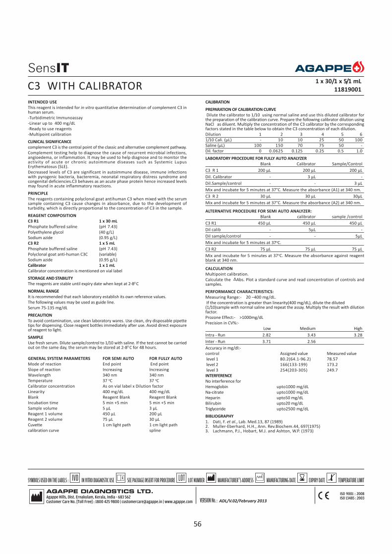
INTENDED USE
This reagent is intended for in vitro quantitative determination of complement C3 inhuman serum.
-Turbidimetric Immunoassay
-Linear up to 400 mg/dL
-Ready to use reagents
-Multipoint calibration
CLINICAL SIGNIFICANCE
complement C3 is the central point of the classic and alternative complement pathway.
Complement testing help to diagnose the cause of recurrent microbial infections,angioedema, or inflammation. It may be used to help diagnose and to monitor theactivity of acute or chronic autoimmune diseases such as Systemic LupusErythematosus (SLE).
Decreased levels of C3 are significant in autoimmune disease, immune infectionswith pyrogenic bacteria, bacteremia, neonatal respiratory distress syndrome andcongenital deficiencies.C3 behaves as an acute phase protein hence increased levelsmay found in acute inflammatory reactions.
PRINCIPLE
The reagents containing polyclonal goat antihuman C3 when mixed with the serumsample containing C3 cause changes in absorbance, due to the development ofturbidity, which is directly proportional to the concentration of C3 in the sample.
REAGENT COMPOSITION
C3 R1 1 x 30 mL
Phosphate buffered saline (pH 7.43)
Polyethylene glycol (40 g/L)
Sodium azide (0.95 g/L)
C3 R2 1 x 5 mL
Phosphate buffered saline (pH 7.43)
Polyclonal goat anti-human C3C (variable)
Sodium azide (0.95 g/L)
Calibrator 1 x 1 mL
Calibrator concentration is mentioned on vial label
STORAGE AND STABILITY
The reagents are stable until expiry date when kept at 2-80 C
NORMAL RANGE
It is recommended that each laboratory establish its own reference values.
The following values may be used as guide line.
Serum 75-135 mg/dL
PRECAUTION
To avoid contamination, use clean laboratory wares. Use clean, dry disposable pipettetips for dispensing. Close reagent bottles immediately after use. Avoid direct exposureof reagent to light.
SAMPLE
Use fresh serum. Dilute sample/control to 1/10 with saline. If the test cannot be carriedout on the same day, the serum may be stored at 2-80 C for 48 hours.
GENERAL SYSTEM PARAMETERS FOR SEMI AUTO FOR FULLY AUTO
Mode of reaction End point End point
Slope of reaction Increasing Increasing
Wavelength 340 nm 340 nm
Temperature 37 oC 37 oC
Calibrator concentration As on vial label x Dilution factor
Linearity 400 mg/dL 400 mg/dL
Blank Reagent Blank Reagent Blank
Incubation time 5 min +5 min 5 min +5 min
Sample volume 5 µL 3 µL
Reagent 1 volume 450 µL 200 µL
Reagent 2 volume 75 µL 30 µL
Cuvette 1 cm light path 1 cm light path
calibration curve spline
CALIBRATION
PREPARATION OF CALIBRATION CURVE
Dilute the calibrator to 1/10 using normal saline and use this diluted calibrator forthe preparation of the calibration curve. Prepare the following calibrator dilution usingNaCl as diluent. Multiply the concentration of the C3 calibrator by the correspondingfactors stated in the table below to obtain the C3 concentration of each dilution.
Dilution 1 2 3 4 5 6
1/10 Cali. (µL) - 10 10 25 50 100
Saline (µL) 100 150 70 75 50 -
Dil. factor 0 0.0625 0.125 0.25 0.5 1.0
LABORATORY PROCEDURE FOR FULLY AUTO ANALYZER
Blank Calibrator Sample/Control
C3 R 1 200 µL 200 µL 200 µL
Dil. Calibrator - 3 µL -
Dil.Sample/control - - 3 µL
Mix and incubate for 5 minutes at 37°C. Measure the absorbance (A1) at 340 nm.
C3 R 2 30 µL 30 µL 30µL
Mix and incubate for 5 minutes at 37°C. Measure the absorbance (A2) at 340 nm.
ALTERNATIVE PROCEDURE FOR SEMI AUTO ANALYZER:
Blank calibrator sample /control
C3 R1 450 µL 450 µL 450 µL
Dil calib - 5µL -
Dil sample/control - - 5µL
Mix and incubate for 5 minutes at 37oC.
C3 R2 75 µL 75 µL 75 µL
Mix and incubate for 5 minutes at 37oC. Measure the absorbance against reagentblank at 340 nm.
CALCULATION
Multipoint calibration.
Calculate the Abs. Plot a standard curve and read concentration of controls andsamples.
PERFORMANCE CHARACTERISTICS:
Measuring Range:- 20 –400 mg/dL.
If the concentration is greater than linearity(400 mg/dL), dilute the diluted(1/10)sample with normal saline and repeat the assay. Multiply the result with dilutionfactor.
Prozone Effect:- >1000mg/dL
Precision in CV%:-
Low Medium High
Intra - Run 2.82 3.43 3.28
Inter - Run 3.71 2.56
Accuracy in mg/dl:-
control Assigned value Measured value
level 1 80.2(64.1-96.2) 78.57
level 2 166(133-199) 173.2
level 3 254(203-305) 249.7
INTERFERENCE
No interference for
Hemoglobin upto1000 mg/dL
Na-citrate upto1000 mg/dL
Heparin upto50 mg/dL
Bilirubin upto20 mg/dL
Triglyceride upto2500 mg/dL
BIBLIOGRAPHY
1. Dati, F. et al., Lab. Med.13, 87 (1989)2. Muller-Eberhard, H.H., Ann. Rev.Biochem.44, 697(1975)3. Lachmann, P.J., Hobart, M.J. and Ashton, W.P. (1973)
C3 WITH CALIBRATOR1 x 30/1 x 5/1 mL
11819001
ADL/V.02/February 2013
56

INTENDED USE
This reagent is intended for in vitro quantitative determination of complement C4 inhuman serum.
-Turbidimetric Immunoassay
-Linear up to 80 mg/dL
-Ready to use reagents
-Multipoint calibration
CLINICAL SIGNIFICANCE
C4 is a constituent of C3 convertase & C5 convertase.
Decreased levels are found in hereditary angioneurotic odema, immune complexdisease and congenital deficiencies.
PRINCIPLE
The reagents containing polyclonal goat antihuman C4 when mixed with the serumsample containing C4 cause changes in absorbance, due to the development ofturbidity, which is directly proportional to the concentration of C4 in the sample.
REAGENT COMPOSITION
C4 R1 1 x 30 mL
Phosphate buffered saline (pH7.43)
Polyethylene glycol (40 g/L)
Sodium azide (0.95 g/L)
C4 R2 1 x 5 mL
Phosphate buffered saline (pH 7.43)
Polyclonal goat anti-human C4C (variable)
Sodium azide (0.95 g/L)
CALIBRATOR 1 x 1 mL
Calibrator concentration is mentioned on vial label
STORAGE AND STABILITY
The reagents are stable until expiry date when kept at 2-80 C.
NORMAL RANGE
It is recommended that each laboratory establish its own reference values.
The following values may be used as guide line.
Serum : 9 - 36 mg/dL
PRECAUTION
To avoid contamination, use clean laboratory wares. Use clean, dry disposable pipettetips for dispensing. Close reagent bottles immediately after use. Avoid direct exposureof reagent to light.
SAMPLE
Use fresh serum. Dilute the sample/control to 1/10 with saline. If the test cannot becarried out on the same day, the serum may be stored at 2-80 C for 48 hours.
GENERAL SYSTEM PARAMETERS FOR SEMI AUTO FOR FULLY AUTO
Mode of reaction End point End point
Slope of reaction Increasing Increasing
Wavelength 340 nm 340 nm
Temperature 37 oC 37 oC
Calibrator concentration As on vial label x Dilution factor
Linearity 80 mg/dL 80 mg/dL
Blank Reagent Blank Reagent Blank
Incubation time 5 min +5 min 5 min +5 minSample volume 5 µL 3µL
Reagent 1 volume 450 µL 200 µL
Reagent 2 volume 75 µL 30 µL
Cuvette 1 cm light path 1 cm light path
calibration curve spline
CALIBRATION
PREPARATION OF CALIBRATION CURVE
Dilute the high concentration calibrator to 1/10 with normal saline and use thisdiluted calibrator for the preparation of calibration curve. Prepare the followingcalibrator dilution using NaCl as diluent. Multiply the concentration of the C4calibrator by the corresponding factors stated in the table below to obtain the C4concentration of each dilution.
Dilution 1 2 3 4 5 6
1/10 dil.Cali. (µL) - 10 10 25 50 100
Saline (µL) 100 150 70 75 50 -
Dil. factor 0 0.0625 0.125 0.25 0.5 1.0
LABORATORY PROCEDURE FOR FULLY AUTO ANALYZER
Blank Calibrator Sample/control
C4 R 1 200 µL 200 µL 200 µL
Dil. Calibrator - 3 µL -
Dil.Sample/control - - 3 µL
Mix and incubate for 5 minutes at 37°C. Read the absorbance (A1) at 340 nm.
C4 R 2 30 µL 30 µL 30 µLMix and incubate for 5 minutes at 37°C. Measure the absorbance (A2) at 340 nm.
ALTERNATIVE PROCEDURE FOR SEMI AUTO ANALYZER:
Blank Calibrator Sample/control
C4 R 1 450 µL 450 µL 450 µL
Dil. Calibrator - 5 µL -
Dil.Sample/control - - 5 µL
Mix and incubate for 5 minutes at 37°C.
C4 R 2 75 µL 75 µL 75 µL
Mix and incubate for 5 minutes at 37°C. Measure the absorbance against reagentblank at 340 nm.
CALCULATION
Multipoint calibration
Calculate the Abs, plot a standard curve and read the concentration of controlsand samples.
PERFORMANCE CHARACTERISTICS:
Measuring Range:- 2 –80 mg/dL.
If the concentration is greater than linearity (80 mg/dL), dilute thediluted(1/10) samplewith normal saline and repeat the assay. Multiply the result with dilution factor.
Prozone Effect:- >1000mg/dL
Precision in CV%:-
Low Medium High
Intra - Run 4.54 2.18 3.96
Inter - Run 4.17 3.08
Accuracy in mg/dL
control Assigned value Measured value
level 1 12.7(10.1-15.2) 12.11
level 2 28.3(22.7-34.0) 27.4
level 3 41.8(33.5-50.2) 39.9
INTERFERENCE
No interference for
Hemoglobin upto1000 mg/dL
Na-citrate upto1000 mg/dL
Heparin upto 50 mg/dL
Turbidity upto 5%
Bilirubin upto 20 mg/dL
Triglyceride upto2500 mg/dL
BIBLIOGRAPHY
1. Dati, F. et al., Lab. Med.13, 87 (1989)2. Muller-Eberhard, H.H., Ann. Rev.Biochem.44, 697(1975)
C4 WITH CALIBRATOR1 x 30/1 x 5/1 mL
11820001
ADL/V.02/February 2013
57

INTENDED USE
This reagent is intended for in vitro quantitative determination of Ceruloplasmin inserum.
-Turbidimetric Immuno assay
-Linear up to 100 mg/dL
-No sample dilution needed
-Ready to use reagents
-Multipoint calibration
CLINICAL SIGNIFICANCE
Ceruloplasmin is a copper oxidase enzyme, important in regulating the ionic state ofiron and other metallic ions. Levels are decreased in hepatolenticular degeneration orWilson's disease and Menke's kinky hair syndrome. Levels are elevated by the acutephase response and particularly by estrogens.
PRINCIPLE
The reagents containing polyclonal goat antihuman ceruloplasmin when mixed withthe serum sample containing ceruloplasmin cause changes in absorbance due tothe development of turbidity, which is directly proportional to the concentrationof Ceruloplasmin in the sample.
REAGENT COMPOSITION
Ceruloplasmin - R1 1 x 30 mL
Phosphate buffered saline (pH 7.43)
Polyethylene glycol 40 g/L
Sodium azide 0.95 g/L
Ceruloplasmin - R2 1 x 5 mL
Phosphate buffered saline (pH 7.43)
Polyclonal goat anti- human Ceruloplasmin
Sodium azide 0.95 g/L
Calibrator 1 x 1 mL
Calibrator concentration is mentioned on vial label
STORAGE AND STABILITY
The sealed reagents are stable upto the expiry date mentioned on the label when storedat 2-8oC. Do not freeze.
REFERENCE RANGE
It is recommended that, each laboratory should establishes its own reference values.The following value may be used as a reference.
serum : 22 - 61 mg/dL
PRECAUTION
To avoid contamination, use clean laboratory wares, such as pipette tips and testtubes. Close reagent bottle immediately after use. Avoid direct exposure of reagentto light.
SAMPLE
Fresh serum.
GENERAL SYSTEM PARAMETERS Semi Auto Fully Auto
Mode of reaction Fixed Time End point
Slope of reaction Increasing Increasing
Wavelength 340 nm 340 nm
Temperature 37o C 37o C
Calibrator concentration As on vial label x Dilution factor
Linearity 100 mg/dl 100 mg/dl
Blank DI Water Reagent Blank
Incubation time 5 min 5 min + 5min
Delay 5 sec -
Delta 300 sec -
Sample volume 5 µL 4 µL
Reagent 1 volume 450 µL 300 µL
Reagent 2 volume 75 µL 50 µL
Cuvette 1 cm light path 1 cm light path
CALIBRATION
Preparation of calibration curve:
Prepare the following calibrator dilution using normal saline as diluent. Multiply theconcentration of the Ceruloplasmin calibrator by the corresponding factors stated inthe table below to obtain the Ceruloplasmin concentration of each dilution.
Dilution 1 2 3 4 5 6
Cali. (µL) - 10 10 20 50 100
Saline (µL) 100 150 70 60 50 -
Dil. factor 0 0.0625 0.125 0.25 0.5 1.0
LABORATORY PROCEDURE FOR FULLY AUTO
Blank calib Sample/control
Ceruloplasmin R1 300 µL 300 µL 300 µL
Dil.Calibrator - 4 µL -
Sample/control - - 4 µL
Mix and incubate for 5 minutes at 37°C. Read the absorbance(A 1) at 340 nm.
Ceruloplasmin R2 50 µL 50 µL 50 µL
Mix and incubate for 5 minutes at 37°C. Measure the absorbance (A2) at 340 nm.
ALTERNATIVE PROCEDURE FOR SEMI AUTO ANALYZER
Calibrator Sample/control
Ceruloplasmin R 1 450 µL 450 µL
Dil.calibrator 5 µL -
Sample/control - 5 µL
Ceruloplasmin R 2 75 µL 75 µL
Mix well, measure absorbance (A1)immediately after addition of Ceruloplasmin R2 andtake absorbance exactly after 300 sec (A2) at 340 nm.
CALCULATION
Multipoint calibration
Calculate the Abs, plot a standard curve & read the concentration of controls &samples.
PERFORMANCE CHARACTERISTICS:
Measuring Range:- 4 - 100 mg/dL.
If the concentration is greater than 100 mg/dL , dilute the diluted(1/10)sample withnormal saline and repeat the assay. Multiply the result with dilution factor.
Prozone Effect:- > 400 mg/dL
Precision in CV%:-
Low Medium High
Intra - Run 6.31 2.07 2.20
Inter - Run 3.91
Accuracy in mg/dL
control Assigned value Measured value
level 1 18.7(14.9-22.4) 20.98
level 2 35.8(28.7-43.0) 38.3
level 3 50.2(40.1-60.2) 48.57
Interference:-
No interference for
Hemoglobin upto 1000 mg/dL
Na - citrate upto 1000 mg/dL
Heparin upto 50 mg/dL
Bilirubin upto 20 mg/dL
Triglycerides upto 2500 mg/dL
BIBLIOGRAPHY
Poulik , M.D and Kleiss M.L in “The plasma proteins”, F.W. Putman, (ed.) Vol. 2Second edition, Academic press, New York, PP 52-108
CERULOPLASMIN WITH CALIBRATOR 1 x 30/1 x5 /1 mL
11813001
ADL/V.02/February 2013
58

INTENDED USE
This reagent is intended for in vitro diagnostic quantitative determination of C-reactiveprotein (CRP) in serum.
Linear up to 150 mg/L
Single point calibration
CLINICAL SIGNIFICANCE
CRP (C – reactive Protein) is a cytokine - induced, acute phase protein that increasesin concentration as a result of inflammation. CRP levels in the body has been used asa marker or indicator of infections and inflammation. The assay of CRP is moresensitive than the erythrocyte sedimentation rate (ESR) and leukocyte count. The CRPlevels rise and return to reference ranges more rapidly after the disease has subsided.
PRINCIPLE
This is a quantitative turbidimetric immuno assay for the measurement of CRP inhuman serum. CRP in the samples binds to specific anti-CRP antibodies, which havebeen adsorbed to latex particles and agglutinates. The agglutination is proportionalto the quantity of CRP in the sample. The actual concentration is then determined byinterpolation from a calibration curve prepared from calibrators of knownconcentrations.
REAGENT COMPOSITION
CRP TURBILATEX R1 1x45 mL
Diluent
Tris buffer 20 mmol/L
Sodium azide 0.95 g/L
CRP TURBILATEX R2 1x5 mL
CRP-Latex:
Suspension of latex particles coated with
anti human CRP
Sodium azide 0.95 g/L
CRP CALIBRATOR 1x1 mLCalibrator concentration is stated on the vial label.
PRECAUTIONS: Components from human origin have been tested and found to benegative for the presence of HbsAg, and HCV and of antibody to HIV (1/2). Howeverhandle cautiously as potentially infectious.
STORAGE & STABILITY
The sealed reagents are stable up to the expiry date stated on the label, when storedat 2-8oC.
NORMAL RANGE
It is recommended that each laboratory establish its own reference value. The followingvalue may be used as guideline.
Serum : up to 6 mg/L
PREPARATION AND STABILITY OF REAGENT
CRP calibrator: Reconstitute the calibrator with 1mL of distilled water.Reconstitutedcalibrator is stable for 30 days at 2-80C.
PRECAUTION
To avoid contamination, use clean laboratory wares. Avoid direct exposure of workingreagent to light.
SAMPLE
Fresh serum. (Do not use hemolyzed or lipemic serum)
GENERAL SYSTEM PARAMETERS
Mode of reaction Fixed time
Slope of reaction Increasing
Wavelength 546 nm (530-550nm)
Temperature 37oC
Calibrator concentration As on vial label
Linearity 150 mg/L
Blank DI water
Delay time 5 sec
Interval 120 sec
Sample volume 5 µL
Reagent 1 volume 900 µL
Reagent 2 volume 100 µL
Cuvette 1 cm path length
LABORATORY PROCEDURE
Calibrator Sample /control
CRP R1 900 µL 900 µL
Calibrator 5µL ---
Sample --- 5 µL
CRP R2 100 µL 100 µL
Mix and read the absorbance immediately (A1) and after 2 minutes (A2) of sampleaddition.
CALCULATION
(A2-A1) sample x calibrator concentration
CRP Conc. in mg/L = -------------------------------------------------------
(A2-A1) calibrator
PERFORMANCE CHARACTERISTICS
1. Linearity : The reagent is linear up to 150 mg/L. If the concentration is greater thanlinearity, dilute the sample with normal saline and repeat the assay. Multiply theresult with dilution factor.
2. Detection limit: Values less than 2mg/L gives non reproducible results3. Prozone effect: No prozone effect was detected up to 800 mg/L.
INTERFERENCES
No interference
Rheumatoid factors : up to 300 IU/mL
Bilirubin : up to 20 mg/dL
Lipemia : up to 10 g/L.
BIBLIOGRAPHY
1. Haffejee, Quarterly Journal of Medicine 1992, New Series 84; 305: 641-6582. Alouf et al Biochemie. 1973; 56-61.
CRP TURBILATEX WITH CALIBRATOR 1X45 mL/1X5mL/1X1mL
11802001
59

INTENDED USE
This reagent is intended for in vitro quantitative determination of C-reactive proteinin human serum or plasma by immunoturbidimetry.
-Latex enhanced immunoturbidimetry
-Linear up to 200 mg/L
-Ready to use reagents
-No sample dilution needed
CLINICAL SIGNIFICANCE
CRP (C – Reactive Protein) is a cytokine - induced, acute phase protein that increasesin concentration as a result of inflammation. CRP levels in the body has been used asa marker or indicator of infections and inflammation. The assay of CRP is moresensitive than the erythrocyte sedimentation rate (ESR) and leukocyte count. The CRPlevels rise and return to reference ranges more rapidly after the disease has subsided.
PRINCIPLE
This is a latex enhanced turbidimetric immuno assay. CRP in the samples binds tospecific anti-CRP antibodies, which have been adsorbed to latex particles andagglutinates. The agglutination is proportional to the quantity of CRP in the sample.The actual concentration is then determined by interpolation from a calibration curveprepared from calibrators of known concentrations.
REAGENT COMPOSITION
CRP R1 1 x 24 mL, 2 x 24 mL
Glycine buffer
CRP R2 1 x 8 mL, 2 x 8 mL
Latex suspension coated with anti-CRP antibodies. (rabbit polyclonal antibody)
CALIBRATOR 1 x 2 mL, 1 x 2 mL
CRP calibrator concentration as on vial label
STORAGE AND STABILITY
The sealed reagents are stable up to the expiry date stated on the label, when storedat 2-8oC.
NORMAL RANGE
It is recommended that each laboratory should establish its own reference values.The following value may be used as a guide line.
Serum up to 6 mg/L
PRECAUTION
To avoid contamination, use clean laboratory wares. Avoid direct exposure of reagentto light.
The reagent should be used according to this pack insert. If used otherwise,appropriate performance is not guaranteed.
SAMPLE
Fresh serum (Do not use hemolized or lipemic serum)
GENERAL SYSTEM PARAMETER SEMI AUTO FULLY AUTO
Mode of Reaction Fixed Time End point
Slope of reaction Increasing Increasing
Wavelength 578 nm 570/ 800nm
Temperature 370C 370C
No.of calib. 6 6
Calibrator concentration As on vial label x Dilution factor
Linearity 200 mg/L 200 mg/L
Blank DI Water Reagent blank
Delay 5 sec --
Interval 120 sec --
Sample volume 5 µL 3 µL
Reagent 1 volume 450 µL 210 µL
Reagent 2 volume 150 µL 70 µL
Cuvette 1cm light path 1cm light path
CALIBRATION
Preparation of calibration curve:
Prepare the following calibrator dilutions using normal saline as diluent. Multiply theconcentration of the CRP calibrator by the corresponding factor stated in the tablebelow to obtain the CRP concentration of each dilution.
Dilution 1 2 3 4 5 6
Cali. (µL) - 10 10 20 50 100
Saline (µL) 100 150 70 60 50 -
Dil. factor 0 0.0625 0.125 0.25 0.5 1.0
LABORATORY PROCEDURE FOR FULLY AUTO ANALYZER
Blank calibrator Sample/Control
CRP R 1 210 µL 210 µL 210 µL
Dil. Calibrator - 3 µL -
Sample/control - - 3 µL
Mix and incubate for 5 minutes at 37°C.
CRP R 2 70 µL 70 µL 70 µL
Mix and measure the absorbance immediately (A1) and after 2 minutes (A2) at 570/800nm.
ALTERNATIVE PROCEDURE FOR SEMI AUTO ANALYZER:
calibrator Sample/Control
CRP R 1 450 µL 450 µL
Dil. Calibrator 5 µL -
Sample/control - 5 µL
CRP R 2 150 µL 150 µL
Mix and measure the absorbance immediately (A1) and after 2 minutes (A2) at 578nm.
CALCULATION
Multi point calibration
Calculate the Abs, plot a standard curve & read the concentration of controls &samples.
PERFORMANCE CHARACTERISTICS:
Measuring Range:- 1 –200 mg/L.
If the concentration is greater than 200 mg/L, dilute the sample with normal salineand repeat the assay. Multiply the result with dilution factor.
Prozone Effect:- >1000 mg/L
Precision in CV%:-
Low Medium High
Intra - Run 7.0 5.0 3.0
Inter - Run 10 8 5
Accuracy in mg/L
control Assigned value Measured value
level 1 5.85(4.68-7.02) 5.05
level 2 27.3(21.9-32.8) 26.8
level 3 51.9(41.5-62.2) 49.6
INTERFERENCE
No interference for
Hemoglobin 500 mg/dL
Intrafat 500 mg/dL
Bilirubin 30 mg/dL
RF 500 IU/mL
BIBLIOGRAPHY
1. Tillett.W.S..et al: Serological reactions in pneumonia with a non protein somaticfraction of pneumococcus.J.Exp.Med..52,561(1930).
2. Zeigenhagen G,Drahovshy D.Klinishe Bedeutung des C-reaktiven protein.Med klin1983;78:45-50.
3. Rifal.N.Tracy.R.P.Ridker,P.M.Clinical efficacy of an Automated High sensitivity C-Reactive protein Assay: Clin chem. 45-12.
CRP LEIT WITH CALIBRATOR1 x 24 / 1 x 8 / 2 mL, 2 x 24 / 2 x 8 / 2 mL
11808004, 11808005
60
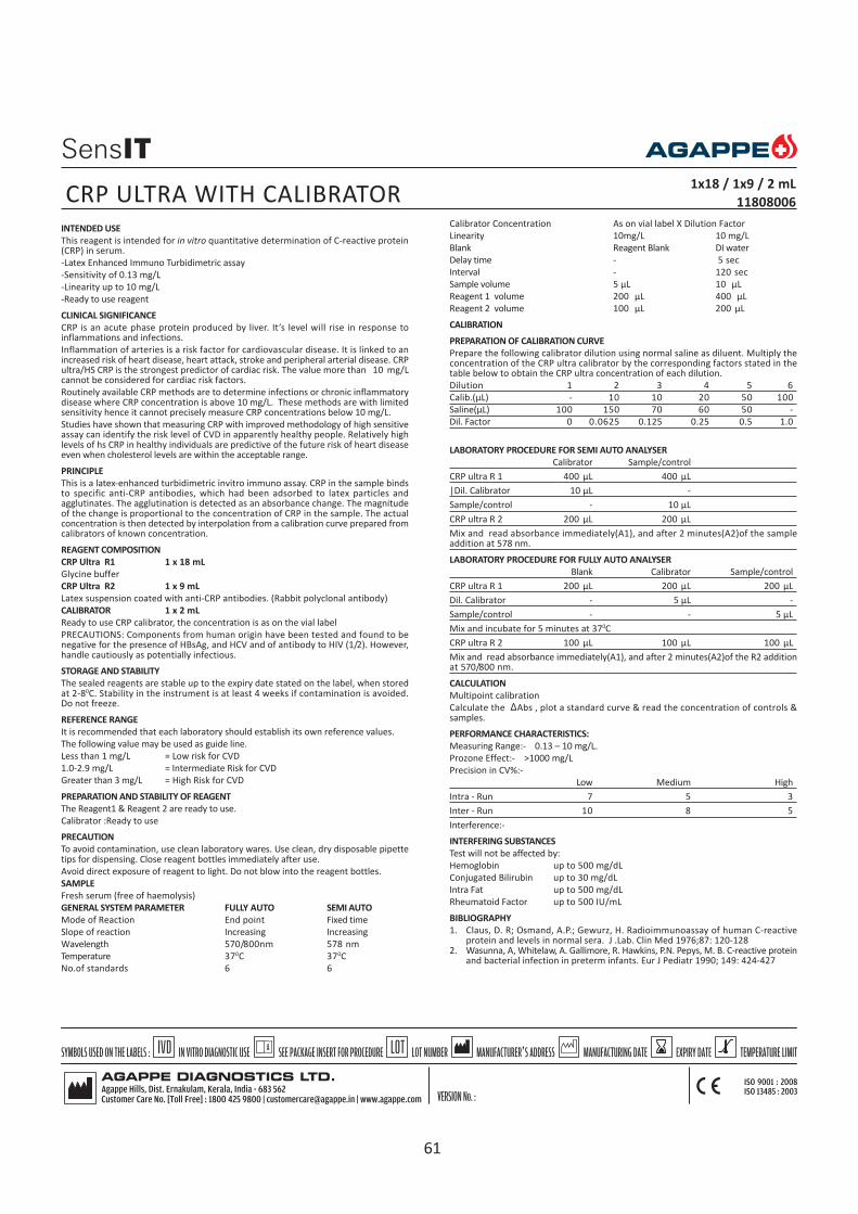
INTENDED USE
This reagent is intended for in vitro quantitative determination of C-reactive protein(CRP) in serum.
-Latex Enhanced Immuno Turbidimetric assay
-Sensitivity of 0.13 mg/L
-Linearity up to 10 mg/L
-Ready to use reagent
CLINICAL SIGNIFICANCE
CRP is an acute phase protein produced by liver. It’s level will rise in response toinflammations and infections.
Inflammation of arteries is a risk factor for cardiovascular disease. It is linked to anincreased risk of heart disease, heart attack, stroke and peripheral arterial disease. CRPultra/HS CRP is the strongest predictor of cardiac risk. The value more than 10 mg/Lcannot be considered for cardiac risk factors.
Routinely available CRP methods are to determine infections or chronic inflammatorydisease where CRP concentration is above 10 mg/L. These methods are with limitedsensitivity hence it cannot precisely measure CRP concentrations below 10 mg/L.
Studies have shown that measuring CRP with improved methodology of high sensitiveassay can identify the risk level of CVD in apparently healthy people. Relatively highlevels of hs CRP in healthy individuals are predictive of the future risk of heart diseaseeven when cholesterol levels are within the acceptable range.
PRINCIPLE
This is a latex-enhanced turbidimetric invitro immuno assay. CRP in the sample bindsto specific anti-CRP antibodies, which had been adsorbed to latex particles andagglutinates. The agglutination is detected as an absorbance change. The magnitudeof the change is proportional to the concentration of CRP in the sample. The actualconcentration is then detected by interpolation from a calibration curve prepared fromcalibrators of known concentration.
REAGENT COMPOSITION
CRP Ultra R1 1 x 18 mL
Glycine buffer
CRP Ultra R2 1 x 9 mL
Latex suspension coated with anti-CRP antibodies. (Rabbit polyclonal antibody)
CALIBRATOR 1 x 2 mL
Ready to use CRP calibrator, the concentration is as on the vial label
PRECAUTIONS: Components from human origin have been tested and found to benegative for the presence of HBsAg, and HCV and of antibody to HIV (1/2). However,handle cautiously as potentially infectious.
STORAGE AND STABILITY
The sealed reagents are stable up to the expiry date stated on the label, when storedat 2-80C. Stability in the instrument is at least 4 weeks if contamination is avoided.Do not freeze.
REFERENCE RANGE
It is recommended that each laboratory should establish its own reference values.
The following value may be used as guide line.
Less than 1 mg/L = Low risk for CVD
1.0-2.9 mg/L = Intermediate Risk for CVD
Greater than 3 mg/L = High Risk for CVD
PREPARATION AND STABILITY OF REAGENT
The Reagent1 & Reagent 2 are ready to use.
Calibrator :Ready to use
PRECAUTION
To avoid contamination, use clean laboratory wares. Use clean, dry disposable pipettetips for dispensing. Close reagent bottles immediately after use.
Avoid direct exposure of reagent to light. Do not blow into the reagent bottles.
SAMPLE
Fresh serum (free of haemolysis)
GENERAL SYSTEM PARAMETER FULLY AUTO SEMI AUTO
Mode of Reaction End point Fixed time
Slope of reaction Increasing Increasing
Wavelength 570/800nm 578 nm
Temperature 370C 370C
No.of standards 6 6
Calibrator Concentration As on vial label X Dilution Factor
Linearity 10mg/L 10 mg/L
Blank Reagent Blank DI water
Delay time - 5 sec
Interval - 120 sec
Sample volume 5 µL 10 µL
Reagent 1 volume 200 µL 400 µL
Reagent 2 volume 100 µL 200 µL
CALIBRATION
PREPARATION OF CALIBRATION CURVE
Prepare the following calibrator dilution using normal saline as diluent. Multiply theconcentration of the CRP ultra calibrator by the corresponding factors stated in thetable below to obtain the CRP ultra concentration of each dilution.
Dilution 1 2 3 4 5 6
Calib.(µL) - 10 10 20 50 100
Saline(µL) 100 150 70 60 50 -
Dil. Factor 0 0.0625 0.125 0.25 0.5 1.0
LABORATORY PROCEDURE FOR SEMI AUTO ANALYSER
Calibrator Sample/control
CRP ultra R 1 400 µL 400 µL
|Dil. Calibrator 10 µL -
Sample/control - 10 µL
CRP ultra R 2 200 µL 200 µL
Mix and read absorbance immediately(A1), and after 2 minutes(A2)of the sampleaddition at 578 nm.
LABORATORY PROCEDURE FOR FULLY AUTO ANALYSER
Blank Calibrator Sample/control
CRP ultra R 1 200 µL 200 µL 200 µL
Dil. Calibrator - 5 µL -
Sample/control - - 5 µL
Mix and incubate for 5 minutes at 370C
CRP ultra R 2 100 µL 100 µL 100 µL
Mix and read absorbance immediately(A1), and after 2 minutes(A2)of the R2 additionat 570/800 nm.
CALCULATION
Multipoint calibration
Calculate the Abs , plot a standard curve & read the concentration of controls &samples.
PERFORMANCE CHARACTERISTICS:
Measuring Range:- 0.13 – 10 mg/L.
Prozone Effect:- >1000 mg/L
Precision in CV%:-
Low Medium High
Intra - Run 7 5 3
Inter - Run 10 8 5
Interference:-
INTERFERING SUBSTANCES
Test will not be affected by:
Hemoglobin up to 500 mg/dL
Conjugated Bilirubin up to 30 mg/dL
Intra Fat up to 500 mg/dL
Rheumatoid Factor up to 500 IU/mL
BIBLIOGRAPHY
1. Claus, D. R; Osmand, A.P.; Gewurz, H. Radioimmunoassay of human C-reactiveprotein and levels in normal sera. J .Lab. Clin Med 1976;87: 120-128
2. Wasunna, A, Whitelaw, A. Gallimore, R. Hawkins, P.N. Pepys, M. B. C-reactive proteinand bacterial infection in preterm infants. Eur J Pediatr 1990; 149: 424-427
CRP ULTRA WITH CALIBRATOR 1x18 / 1x9 / 2 mL
11808006
61

INTENDED USE
This reagent is intended for in vitro quantitative determination of Cystatin C in humanserum.
-Latex enhanced Immunoturbidimetry
-Ready to use reagents
-No sample dilution required
-Linear up to 10 mg/L
CLINICAL SIGNIFICANCE
Cystatin C is a low molecular weight (13 Da) cytoplasmic protein, functioning as aninhibitor of various cystein protease in the blood stream. Cystatin C has a stableproduction rate and is removed from the blood circulation by glomerular filtration.In healthy individuals Cystatin C is completely reaborsorbed and degraded in thetubules but in subject with renal disorders its level in blood may be raised as high as2 to 5 times the normal values. Cystatin C is superior to serum creatinine as a markerof glomerular filtration Rate.
PRINCIPLE
Cystatin C in the test sample binds to the specific polyclonal rabbit anti-Cystatin Cantibody, which has been adsorbed to latex particle and agglutinates. The agglutinationis detected as absorbance change at 546 nm. The magnitude of change is proportionalto the quantity of Cystatin C in the sample and its concentration is determined byinterpolation from a calibration curve prepared from calibrators of knownconcentration.
REAGENT COMPOSITION
Cystatin C R1 1 x 25 mL
Tris buffer 1.2% (100 mM)
pH 8.5+0.3
Cystatin C R2 1 x 5 mL
Polystyrene latex particle coated with polyclonal anti Cystatin C antibody (rabbit)
CALIBRATOR 1 x 2 mL
Cystatin C calibrator concentration as mentioned on the vial label.
STORAGE AND STABILITY
The sealed reagents are stable up to the expiry date stated on the label, when storedat 2- 80C.
NORMAL RANGE
It is recommended that each laboratory establish its own reference values.
The following value may be used as guide line.
Individuals up to 50 years : 0.55 – 1.15 mg/L
Individuals above 50 years : 0.63 – 1.44 mg/L
PREPARATION OF REAGENT
Reagent 1 and Reagent 2 are ready to use.Cystatin –C calibrator: Ready to use Liquid stable.Unopened vials are stable till expiration date mentioned on the vials. Opened vials arestable for 4 weeks when stored at 2-80C.
PRECAUTIONS:To avoid contamination, use clean laboratory wares, such as pipette tips and testtubes. Close reagent bottle immediately after use. Avoid direct exposure of reagent tolight.
SAMPLE
Required sample material is human serum or EDTA/ Heparinized plasma. It isrecommended to analyze the sample as fresh as possible.
GENERAL SYSTEM PARAMETER SEMI AUTO FULLY AUTO
Mode of Reaction Fixed time End point
Slope of reaction Increasing Increasing
Wavelength 546 nm 546 nm/ 800nm
Temperature 370C 370C
No of calib 6 6Calibrator Conc. As on vial label x Dilution factorsLinearity 10 mg/L 10 mg/LBlank DI Water Reagent blank
Delay Time 5 sec --
Interval 300 sec --Sample volume 5 µL 3 µLReagent 1 volume 500 µL 200 µL
Reagent 2 volume 100 µL 40 µLCuvette 1 cm light path 1 cm light path
Dilution of calibrator for calibration curve:
Calibration Curve (range between 0-10 mg/L). Prepare the following calibrator dilutionusing normal saline as diluent. Multiply the concentration of the Cystatin C calibratorby the corresponding factor stated in the table below to obtain the Cystatin Cconcentration of each dilution.
Dilution 1 2 3 4 5 6
Cali. (µL) - 10 10 20 50 100
Saline (µL) 200 190 70 60 50 -
Dil. factor 0 0.05 0.125 0.25 0.5 1.0
LABORATORY PROCEDURE FOR FULLY AUTO ANALYZER
Blank Calibrator Sample/control
Cystatin R1 200 µL 200 µL 200 µL
Dil.Calibrator - 3 µL -
Sample/control - - 3 µL
Mix and incubate for 5 minutes at 37oC.
Cystatin R2 40 µL 40 µL 40 µL
Mix and read the absorbance immediately (A1), and after 5 minutes (A2) at 546/800nm.
ALTERNATIVE PROCEDURE FOR SEMI AUTO ANALYZER
Calibrator Sample/control
Cystatin R1 500 µL 500 µL
Dil.Calibrator 5 µL -
Sample/control - 5 µL
Cystatin R2 100 µL 100 µL
Mix and read the absorbance immediately (A1), and after 5 minutes (A2) at 546nm.
CALCULATION
Multipoint calibration
Calculate the abs, plot a standard curve & read the concentration of controls &samples.
PERFORMANCE CHARACTERISTICS:
Measuring Range:- 0.1 - 10 mg/L.
If the concentration is greater than 10 mg/L, dilute the sample with normal saline andrepeat the assay .Multiply the result with dilution factor.
Prozone Effect:- >60 mg/L
Precision :
MEAN INTRA RUN INTER RUN nVALUE CV(%) CV(%)
Low human serum pool 0.77 2.16 2.54 20
High human serum pool 5.94 0.67 1.45 20
Medium human serum pool 1.45 1.58 1.95 20
Medium human serum pool 2.72 1.22 1.37 20
Low human serum pool 0.46 3.96 4.77 20
High human serum pool 3.82 1.81 3.05 20
Accuracy in mg/L
control Assigned value Measured value
level 1 0.484(0.387-0.581) 0.438
level 2 0.572(0.457-0.686) 0.530
level 3 0.649(0.519-0.779) 0.614
IINTERFERENCE
No interference upto
Hemoglobin 500 mg/dL
Intrafat 1400 mg/dL
Bilirubin 25 mg/dL
BIBLIOGRAPHY
1. Cystatin-C as a marker of GFR - history, indications and future research; Clin.Biochem.38:1, 2005
2. Serum Cystatin -C is superior to serum creatinine as a marker of kidney function:a meta analysis. Am. J. Kidney Desease. 40, 221, 2002.
CYSTATIN-C WITH CALIBRATOR 1 x 25/1 x 5/2 mL
11810001
62

INTENDED USE
The reagent is intended for in vitro quantitative determination of Ferritin in serum
-Latex Enhanced Immunoturbidimetry
-High Linearity of 1000 ng/mL
-No sample dilution
-Ready to use reagents
-Multipoint calibration
CLINICAL SIGNIFICANCE
Ferritin is an iron-containing protein. It is mainly found in liver and spleen, where itsfunction is to store and release iron in the body. It is also found in small amounts inhuman serum. The serum levels tend to increase due to hepatitis and malignanttumors. The measurement of ferritin is useful in diagnosis, treatment, assessment ofdisease progression and post operative prognosis of abnormal iron metabolism andiron deficiency anaemia.
PRINCIPLE
Latex particles coated with anti-ferritin antibody are agglutinated when mixed withsamples containing Ferritin. The agglutination causes an absorbance change whichdepends on the Ferritin concentration in the sample, this can be interpolated using acalibration curve prepared from calibrators of different concentrations.
REAGENT COMPOSITION
Ferritin - R1 1 x 30 mL
Glycine buffer
Ferritin - R2 1 x 10 mL
Suspension of latex particle bound to anti-ferritin antibodies
Calibrator 1 x 1 ml
Calibrator concentration is mentioned on vial label
STORAGE AND STABILITY
The sealed reagents are stable upto the expiry date mentioned on the label when storedat 2-8oC.
NORMAL RANGE
It is recommended that, each laboratory should establish its own reference values. Thefollowing value may be used as a guide line.
Male : 30 - 220 ng/mL
Females : 20 - 110 ng/mL
PRECAUTION
To avoid contamination, use clean laboratory wares, use clean dry disposable pipettetips for dispensing, close reagent bottle immediately after use. Avoid direct exposureof reagent to light.
SAMPLE
Fresh Serum
GENERAL SYSTEM PARAMETER FULLY AUTO
Mode of reaction Rate
Slope of reaction Increasing
Wavelength 1 570 nm
Wavelength 2 800 nm
Temperature 37oC
Calibrator concentration As on vial label x Dilution factor
Linearity 1000 ng/mL
Blank Reagent Blank
sample volume 3 µL
Reagent 1 volume 210 µL
Reagent 2 volume 70 µL
Cuvette 1 cm light path
CALIBRATION
Preparation of calibration curve:
Prepare the following calibrator dilutions using normal saline as diluent. Multiply theconcentration of the Ferritin calibrator by the corresponding factor stated in the tablebelow to obtain the Ferritin concentration of each dilution.
Dilution 1 2 3 4 5 6
Cali. (µL) - 10 10 20 50 100
Saline (µL) 100 150 70 60 50 -
Dil. factor 0 0.0625 0.125 0.25 0.5 1.0
LABORATORY PROCEDURE
Blank calibrator Sample/control
Ferritin R 1 210 µL 210 µL 210 µL
Dil. Calibrator - 3 µL -
Sample/control - - 3 µL
Ferritin R 2 70 µL 70 µL 70 µL
Mix and read absorbance (A1) immediately and after 2 minutes (A2) at 570 nm and800 nm.
CALCULATION
Multi point calibration
Calculate the abs, plot a standard curve & read the concentration of controls &samples.
PERFORMANCE CHARACTERISTICS:
Measuring Range:- 10 –1000 ng/mL.
If the concentration is greater than 1000 ng/mL , dilute the sample with normal salineand repeat the assay .Multiply the result with dilution factor.
Prozone Effect:- >30000 ng/mL
Precision in CV%:-
Low High
Intra - Run 10 7
Inter - Run 12 10
Accuracy in ng/mL
control Assigned value Measured value|
level 1 31.2(24.9-37.4) 32.6
level 2 193(154-231) 182.2
level 3 331(264-397) 303.67
INTERFERENCE
No interference for
Hemoglobin 500 mg/dL
Triglyceride 3000 mg/dL
Bilirubin 30 mg/dL
Rheumatoid factor 560 IU/mL
BIBLIOGRAPHY
1. Cook, J.D., Lipschitz,D.A., Laughton, M.B.B., Miles, E.M. & Finch, C.A: Serumferritin as a measure of iron stores in normal subjects. Am.J.clin.Nutr. 27: 680,1974.
2. Walters,G.O.,Miller, F.M & Wormwood, M.: Serum ferritin concentration onand iron stores in normal subjects. J.Clin.Pathol. 26: 770-, 1973.
FERRITIN WITH CALIBRATOR 1 x 30/1 x 10/1 mL
11814002
63

INTENDED USE
This reagent is intended for in vitro quantitative determination of HbA1c in humanblood.
-Latex enhanced Immunoturbidimetry
-Ready to use liquid stable reagents
-Multipoint calibration
-Direct result (% HbA1c) from analyzer
-No total Hb determination required
CLINICAL SIGNIFICANCE
HbA1c is a glycated form of haemoglobin formed by the attachment of glucoseresidues in the blood to the hemoglobin molecules. In the diabetic population whereblood glucose levels are abnormally elevated the level of HbA1c also increases. Thelevel of HbA1c is proportional to the level of glucose in the blood and has been widelyaccepted as an indicator of the mean blood glucose concentration in the preceeding6-8 weeks. It is therefore a long-term indicator of diabetic control. For routine useHbA1c levels should be monitored every 3-4 months. However in gestational diabetesand after a change in therapy it may be useful to measure HbA1c more frequently at2-4 week intervals.
PRINCIPLE
This method utilizes the interaction of antigen and antibody to directly determine theHbA1c in whole blood. Total heomoglobin and HbA1c have the same nonspecificabsorption rate to latex particle. When mouse antihuman HbA1c monoclonalantibodies are added (R2), latex HbA1c – mouse antihuman HbA1c antibody complexis formed. Agglutination occurs when goat anti mouse IgG polyclonal antibodyinteracts with the monoclonal antibody. The amount of agglutination is proportionalto the amount of HbA1c absorbed onto the surface of latex particles. The amount ofagglutination is measured as absorbance, which is used to calculate HbA1c % froma calibration curve.
REAGENT COMPOSITION
HbA1c R1 4 x 7.5 mL
Latex0.13%(w/v)
Glycine buffer 20 mmol/L
HbA1c R2A 1 x 9.5 mL
Glycine buffer 80 mmol/L
HbA1c R2B 1 x 0.5 mL
Mouse anti-human HbA1c 10 mmol/L
Monoclonal antibody
Goat anti-mouse IgG 0.05 mg/mL
(Polyclonal)
Stabilizers 0.08 mg/dL
HbA1c R3 1 x 60 mL
Haemolysis Reagent
HbA1c DIRECT CALIBRATOR 4 x 0.5 mL
HbA1c 4 level calibrator lyophilized.
STORAGE AND STABILITY
The sealed reagents are stable up to the expiry date stated on the label, when storedat 2- 80C, protected from light.
MEARUREMENT RANGE
4-16% (NGSP)
NORMAL RANGE
It is recommended that each laboratory establish its own reference values.
The following value may be used as guide line.
According NGSP :
<6% for non-diabetic
< 7% for glycemic control of person with diabetes
PREPARATION AND STABILITY OF WORKING REAGENT
Reagent 1 & Reagent 3 are ready to use.
Reagent R2 is prepared by pouring the entire contents of the R2B vial into the R2Avial. Mix gently. This working reagent is stable for 30 days at 2-80C.
Calibrator: Reconstitute with 0.5 mL distilled water and it will be stable up to30 days at 2-80C. Do not freeze.
PRECAUTION
To avoid contamination, use clean laboratory wares. Use clean, dry disposable pipettetips for dispensing. Close reagent and standard bottles immediately after use.
Avoid direct exposure of working reagent to light.
SAMPLE
Whole blood, collected with EDTA
To determine HbA1c a heamolysate must be prepared for each sample
1. Dispense 1mL hemolysis reagent into a tube.2. Add 20 µLof well-mixed whole blood and mix.3. Allow to stand for 5 minutes or until complete lysis is evident.Follow the same procedure with calibrators and controls.
GENERAL SYSTEM PARAMETER FullyAuto
Mode of Reaction End point
Slope of reaction Increasing
Wavelength 600 nm
Temperature 370C
Calibrator Concentration As on vial label
Linearity 16%
Blank Reagent blank
Sample volume 6 µL
Reagent 1 volume 210 µL
Reagent 2 volume 75 µL
Cuvette 1 cm light path
LABORATORY PROCEDURE FOR FULLY AUTO
Blank Calibrator Sample/control
HbA1C R 1 210 µL 210 µL 210 µL
Hemolysate(calib) - 6 µL -
Hemolysate(sample/control) - - 6 µL
Mix & incubate for 5 min at 370C.
HbA1C R 2 75 µL 75 µL 75 µL
Mix and incubate for 5 min at 37oC and read absorbance(A) at 600 nm.
CALCULATION
Calibration curve
Calculate the Abs of calibrators = Abs calibrator – Abs Blank. Plot the D Abs of eachcalibrator versus assigned concentration (HbA1c %) on a linear graph paper. HbA1cresults according to NGSP for the samples and controls are determined using theprepared calibration curve.
Calculate
Abs of sample ie abs Sample - abs Blank .
HbA1c % in the sample is calculated by interpolation of Abs of sample on thecalibration curve. For calculation of results according to IFCC, use IFCC calibratorvalues (see calibrator insert), or use following equation.
NGSP = (0.915 x IFCC) + 2.15
Accuracy in %
control Assigned value Measured value
level 1 4.8 (3.8-5.9) 4.6
level 2 10.2(8.1-12.3) 10.1
INTERFERENCES
No interference up to :
Ascorbic acid 50 mg/dL
Bilirubin 50 mg/dL
Triglycerides 2000 mg/dL
Carbamylated Hb 7.5 mmol/L
Acetylated Hb 5.0 mmol/L
It has been reported that results may be inconsistent in patients who have thefollowing conditions: opiate addiction, lead poisoning, alcoholism, and ingestion oflarge doses of aspirin. Elevated HbF levels may lead to under estimation of HbA1c.
BIBLIOGRAPHY
1. Nathan, D.M., Clin, Chem. 29, pp.466-469 (1983)2. Engbeak, F., et al. Clin chem.35 pp. 93-97 (1989)3. American Diabetes Association : Clinical practice recommendations (position
statement). Diabetes care 24 (suppl.1) S33-S55, (2001).4. Tietz, N.W. Textbook of Clinical Chemistry, W.B. Saunders Company, p.794
-7795 (1999).
HbA1c DIRECT WITH CALIBRATOR 4 x 7.5/1 x 9.5/1 x 0.5/1x60mL/ 4 x 0.5 mL
11806001
64

INTENDED USE
This reagent is intended for in vitro quantitative determination of IgM antibodies inserum.
-Turbidimetric Immuno assay
-Linear up to 500 mg/dL
-No sample dilution
-Ready to use reagents
-Multipoint calibration
CLINICAL SIGNIFICANCE
IgM is important in early response to infections . The measurement of IgM is importantfor typing immuno deficiencies and myelomas. IgM plays an important role in thehumoral defense of the body. Serum levels may be increased in all kind of acuteinfections. Elevated levels in cord serum suggest clinical infection in the new born.
PRINCIPLE
Antibodies to IgM are combined with IgM in the patient’s serum, forming immunecomplexes. The immune complexes cause an increase in light scattering whichcorrelate with the concentration of IgM in the serum. The light scattering is measuredat 340 nm and 700 nm.
REAGENT COMPOSITION
IgM R1 (Buffer solution) 1 x 30 mL
Tris(hydroxymethyl)aminomethane 100 mmol/L
IgM R2 (Antiserum solution) 1 x 10 mL
Anti-human IgM antiserum
Calibrator 1 x 1 mL
Calibrator concentration is mentioned on vial label
STORAGE AND STABILITY
The sealed reagents are stable upto the expiry date mentioned on the label when storedat 2-8 0C. Do not freeze.
REFERENCE RANGE
It is recommended that each laboratory establish its own reference values. Thefollowing values may be used as reference.
Serum : 50 - 300 mg/dL
PRECAUTION
To avoid contamination, use clean laboratory wares. Avoid direct exposure of reagentto light.
The reagent should be used according to this pack insert. If used otherwise,appropriate performance is not guaranteed.
SAMPLE
Fresh Serum.
GENERAL SYSTEM PARAMETERS SEMI AUTO FULLY AUTO
Mode of reaction Endpoint Endpoint
Slope of reaction Increasing Increasing
Wavelength 340/630 nm 340/700 nm
Temperature 37 oC 37 oC
Calibrator concentration As on vial label x Dilution factor
Linearity 500 mg/dL 500 mg/dL
Blank Reagent Blank Reagent Blank
Incubation time(in minutes) 5min+ 5min 5min+ 5min
Sample volume 5 µL 3 µL
Reagent 1 volume 450 µL 180 µL
Reagent 2 volume 150 µL 60 µL
Cuvette 1 cm light path 1 cm light path
CALIBRATION
Preparation of calibration curve:
Prepare the following calibrator dilutions using normal saline as diluent. Multiply theconcentration of the IgM calibrator by the corresponding factor stated in the tablebelow to obtain the IgM concentration of each
Dilution 1 2 3 4 5 6
Cal. (µL) - 10 10 20 50 100
Saline (µL) 100 150 70 60 50 -
Dil. factor 0 0.0625 0.125 0.25 0.5 1.0
LABORATORY PROCEDURE FOR FULLY AUTO ANALYZER
Blank Calibrator Sample/control
IgM R 1 180 µL 180 µL 180 µL
Dil. Calibrator - 3 µL -
Sample/control - - 3 µL
Mix and incubate for 5 minutes at 37°C. Read the absorbance (A1) at 340 nm & 700nm.
IgM R 2 60 µL 60 µL 60 µL
Mix and incubate for 5 minutes at 37°C. Measure the absorbance(A 2) at 340 nm &700 nm.
ALTERNATIVE PROCEDURE FOR SEMI AUTO ANALYZER:
Blank Calibrator Sample/control
IgM R 1 450 µL 450 µL 450 µL
Dil. Calibrator - 5µ L -
Sample/control - - 5 µL
Mix and incubate for 5 minutes at 37°C.
IgM R 2 150 µL 150 µL 150 µL
Mix well and incubate for 5 minutes at 37°C. Measure the absorbance(A ) againstreagent blank at 340 nm & 630 nm.
CALCULATION
Multipoint calibration
Calculate the Abs, plot a standard curve & read the concentration of controls &samples.
PERFORMANCE CHARACTERISTICS:
Measuring Range:- 6 – 500 mg/dL
If the concentration is greater than 500 mg/dl , dilute the sample with normal salineand repeat the assay. Multiply the result with dilution factor.
Prozone Effect:- >5000mg/dL
Precision in CV%:-
Low Medium High
Intra - Run 1.43 1.59 0.64
Inter - Run 3.17 3.65 2.49
Accuracy mg/dL
control Assigned value Measured value
level 1 63.0(50.4-75.5) 64.1
level 2 140.0(112-168) 143.68
level 3 206(165-247) 209.1
INTERFERENCE
No interference for
Hemoglobin upto 1000mg/dL
Triglyceride upto 2500 mg/dL
Bilirubin upto 20mg /dL
Turbidity upto 5 %
BIBLIOGRAPHY
1. Otani, H. :Medical Technology,14, 965(1986)2. Rinsho Kensa Guide 1992, 269-274(1992), BUNKODO CO. LTD.3. Kanai’s manual of Clinical laboratory Medicine, 30th edition, 868- 873(1993),
KANEHARA & CO.,LTD.
IgM WITH CALIBRATOR 1 x 30/1 x 10/1 mL
11817001
ADL/V.02/February 2013
65

INTENDED USE
This reagent is intended for in vitro quantitative determination of IgG antibodies inserum.
-Turbidimetric Immuno assay
-High linearity of 2700 mg/dL
-Ready to use reagents
-Multipoint calibration
CLINICAL SIGNIFICANCE
IgG is a predominant serum immunoglobulin . The measurement of IgG is importantfor typing immunodeficiencies and myelomas. Increased levels are found in chronicinfections and chronic inflammation. IgG is the only immunoglobulin which crossesthe placenta and is therefore of special importance in infants defense againstinfection.
PRINCIPLE
Antibodies to IgG are combined with IgG in the patient’s serum, forming immunecomplexes. The immune complexes cause an increase in light scattering whichcorrelate with the concentration of IgG in the serum. The light scattering ismeasured by reading turbidity at 340 nm.
REAGENT COMPOSITION
IgG R1 (Buffer solution) 1 x 15 mL
Tris(hydroxymethyl)aminomethane 100 mmol/L
IgG R2 (Antiserum solution) 1 x 15 mL
Anti-human IgG antiserum
Calibrator 1 x 1 mL
Calibrator concentration is mentioned on vial label
STORAGE AND STABILITY
The sealed reagents are stable upto the expiry date mentioned on the label when storedat 2-8 0C.
REFERENCE RANGE
It is recommended that , each laboratory should establish its own reference values.The following value may be used as a reference.
serum : 650 - 1600 mg/dL
PRECAUTION
To avoid contamination, use clean laboratory wares, such as pipette tips and testtubes. Close reagent bottle immediately after use. Avoid direct exposure of reagentto light.
SAMPLE
Use fresh serum.
GENERAL SYSTEM PARAMETERS FOR SEMI AUTO FOR FULLY AUTO
Mode of reaction Endpoint Endpoint
Slope of reaction Increasing Increasing
Wavelength 340 nm 340 nm
Temperature 37 oC 37 oC
Calibrator concentration As on vial label x Dilution factor
Linearity 2700 mg/dL 2700 mg/dL
Blank Reagent Bank Reagent Bank
Incubation time 5min + 5min 5min + 5min
Sample volume 5 µL 2 µL
Reagent 1 volume 250 µL 150 µL
Reagent 2 volume 250 µL 150 µL
Cuvette 1 cm light path 1 cm light path
PROCEDURE
Preparation of calibration curve:
Prepare the following calibrator dilutions using normal saline as diluent. Multiply theconcentration of the IgG calibrator by the corresponding factor stated in the tablebelow to obtain the IgG concentration of each dilution.
Dilution 1 2 3 4 5 6
Cal(µL) - 10 10 20 50 100
Saline (µL) 100 150 70 60 50 -
Dil. factor 0 0.0625 0.125 0.25 0.5 1.0
LABORATORY PROCEDURE FOR FULLY AUTO
Blank Calibrator Sample/control
IgG R 1 150 µL 150 µL 150 µL
Dil. Calibrator - 2 µL -
Sample/control - - 2 µL
Mix and incubate for 5 minutes at 37°C. Read the absorbance (A1) at 340nm.
IgG R 2 150 µL 150 µL 150 µL
Mix and incubate for 5 minutes at 37°C. Measure the absorbance(A2) at 340nm.
ALTERNATIVE PROCEDURE FOR SEMI AUTO ANALYZER:
Blank Calibrator Sample/control
IgG R 1 250 µL 250 µL 250 µL
Dil. Calibrator - 5µL -
Sample/control - - 5 µL
Mix and incubate for 5 minutes at 37°C.
IgG R 2 250 µL 250 µL 250 µL
Mix and incubate for 5 minutes at 37°C. Measure the absorbance (A) at 340 nm.
CALCULATION
Calculate the Abs, plot a standard curve & read the concentration of controls &samples.
PERFORMANCE CHARACTERISTICS:
Measuring Range:- 90 –2700 mg/dL.
If the concentration is greater than 2700 mg/dL , dilute the sample with normal salineand repeat the assay .Multiply the result with dilution factor.
Prozone Effect:- >10000 mg/dL
Precision in CV%:-
Low Medium High
Intra - Run 2.5 3.25 4.18
Inter - Run 4.08 1.83
Accuracy in mg/dL
control Assigned value Measured value
level 1 967(774-1160) 897.3
level 2 1745(1396-2094) 1803.5
level 3 2643(2114-3171) 2595.7
INTERFERENCE
No interference for
Hemoglobin upto 1000 mg/dL
Triglyceride upto 2500 mg/dL
Bilirubin upto 20 mg/dL
BIBLIOGRAPHY
1. Otani,H. :Medical Technology,14, 965(1986)2. Rinsho Kensa Guide 1992, 269-274(1992), BUNKODO CO., LTD3. Kanai’s manual of Clinical laboratory Medicine, 30th edition, 868-873(1993),
KANEHARA & CO.,LTD.
IgG WITH CALIBRATOR1 x 15/1 x 15/1 mL
11816001
ADL/V.03/February 2013
66
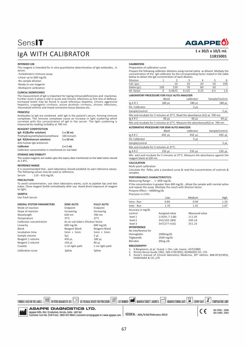
INTENDED USE
This reagent is intended for in vitro quantitative determination of IgA antibodies, inserum.
-Turbidimetric Immuno assay
-Linear up to 600 mg/dL
-No sample dilution
-Ready to use reagents
-Multipoint calibration
CLINICAL SIGNIFICANCE
The measurement of IgA is important for typing immunodeficiencies and myelomas.Further more it plays a role in acute and chronic infections as first line of defence.Increased levels may be found in acute infectious hepatitis, chronic aggressivehepatitis, cryptogenic cirrhosis, active alcoholic cirrhosis, chronic infections,rheumatoid arthritis and mixed connective tissue diseases etc.
PRINCIPLE
Antibodies to IgA are combined with IgA in the patient’s serum, forming immunecomplexes. The immune complexes cause an increase in light scattering whichcorrelate with the concentration of IgA in the serum. The light scattering ismeasured by reading turbidity at 700 nm.
REAGENT COMPOSITION
IgA R1(Buffer solution) 1 x 30 mL
Tris(hydroxymethyl)aminomethane 100 mmol/L
IgA R2(Antiserum solution) 1 x 10 mL
Anti-human IgA antiserum
Calibrator 1 x 1 mL
Calibrator concentration is mentioned on vial label
STORAGE AND STABILITY
The sealed reagents are stable upto the expiry date mentioned on the label when storedat 2-8oC.
REFERENCE RANGE
It is recommended that , each laboratory should establish its own reference values.The following values may be used as reference.
Serum : 110 - 410 mg/dL
PRECAUTION
To avoid contamination, use clean laboratory wares, such as pipette tips and testtubes. Close reagent bottle immediately after use. Avoid direct exposure of reagentto light.
SAMPLE
Use fresh Serum
ENERAL SYSTEM PARAMETERS SEMI AUTO FULLY AUTO
Mode of reaction Endpoint Endpoint
Slope of reaction Increasing Increasing
Wavelength 630 nm 700 nm
Temperature 37oC 37oC
Calibrator concentration As on vial label x Dilution factor
Linearity 600 mg/dL 600 mg/dL
Blank Reagent Blank Reagent Blank
Incubation time 5min + 5min 5min + 5min
Sample volume 5µL 2 µL
Reagent 1 volume 450 µL 180 µL
Reagent 2 volume 150 µL 60 µL
Cuvette 1 cm light path 1 cm light path
CALIBRATION
Preparation of calibration curve:
Prepare the following calibrator dilutions using normal saline as diluent. Multiply theconcentration of the IgA calibrator by the corresponding factor stated in the tablebelow to obtain the IgA concentration of each dilution.
Dilution 1 2 3 4 5 6
Cali. (µL) - 10 10 20 50 100
Saline (µL) 100 150 70 60 50 -
Dil. factor 0 0.0625 0.125 0.25 0.5 1.0
LABORATORY PROCEDURE FOR FULLY AUTO ANALYZER
Blank calibrator Sample/Control
Ig A R 1 180 µL 180 µL 180 µL
Dil. Calibrator - 2 µL -
Sample/control - - 2 µL
Mix and incubate for 5 minutes at 37°C. Read the absorbance (A1) at 700 nm.
Ig A R 2 60 µL 60 µL 60 µL
Mix and incubate for 5 minutes at 37°C. Measure the absorbance(A2) at 700 nm.
ALTERNATIVE PROCEDURE FOR SEMI AUTO ANALYZER:
Blank calibrator Sample/Control
Ig A R 1 450 µL 450 µL 450 µL
Dil. Calibrator - 5 µL -
Sample/control - - 5 µL
Mix and incubate for 5 minutes at 37°C.
Ig A R 2 150 µL 150 µL 150 µL
Mix well and incubate for 5 minutes at 37°C. Measure the absorbance against thereagent blank at 630 nm.
CALCULATION
Multi point calibration
Calculate the Abs, plot a standard curve & read the concentration of controls &samples.
PERFORMANCE CHARACTERISTICS:
Measuring Range:- 1 –600 mg/dL.
If the concentration is greater than 600 mg/dL , dilute the sample with normal salineand repeat the assay .Multiply the result with dilution factor.
Prozone Effect:- >6000mg/dL
Precision in CV%:-
Low Medium High
Intra - Run 0.84 0.94 1.20
Inter - Run 1.70 1.41 1.07
Accuracy in mg/dL
control Assigned value Measured value
level 1 115(91.7-138) 111.45
level 2 241(192-289) 240.16
level 3 347(277-416) 351.23
INTERFERENCE
No interference for
Hemoglobin 1000mg/dL
Triglyceride 2500 mg/dL
Bilirubin 20mg /dL
BIBLIOGRAPHY
1. K.Bergstorm, et al.: Scand. J. Clin. Lab. Invest., 637(1980)2. Rinsho Kensa Guide 1992, 269-274(1992), BUNKODO CO. LTD.3. Kanai’s manual of Clinical laboratory Medicine, 30th edition, 868-873(1993),
KANEHARA & CO.,LTD
IgA WITH CALIBRATOR 1 x 30/1 x 10/1 mL
11815001
ADL/V.02/February 2013
Calibration curve Spline Spline
67
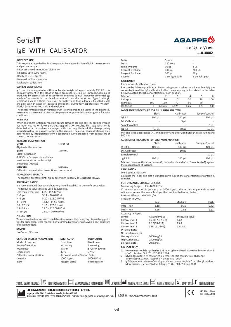
INTENDED USE
This reagent is intended for in vitro quantitative determination of IgE in human serumand plasma samples.
-Latex enhanced Immunoturbidimetry
-Linearity upto 1000 IU/mL
-Ready to use reagents
-No need to dilute samples
-Multipoint calibration
CLINICAL SIGNIFICANCE
IgE is an immunoglobulin with a molecular weight of approximately 190 KD. It isnormally present in the blood in trace amounts. IgE, like all immunoglobulins, isproduced by plasma cells in response to antigenic stimuli. However abnormal IgElevels often results in the development of clinically important Type 1 allergicreactions such as asthma, hay fever, dermatitis and food allergies. Elevated levelsare also seen in cases of parasitic infections, pulmonary aspergillosis, Wiskott-Aldrich Syndrome, hepatitis and myeloma.
The measurement of IgE in human serum is considered to be useful in the diagnosis,treatment, assessment of disease progression, or post-operative prognosis for suchconditions.
PRINCIPLE
When an antigen-antibody reaction occurs between IgE and anti-IgE antibody whichhas been coated on latex particles, agglutination results. This agglutination isdetected as an absorbance change, with the magnitude of the change beingproportional to the quantity of IgE in the sample. The actual concentration is thendetermined by interpolation from a calibration curve prepared from calibrators ofknown concentration.
REAGENT COMPOSITION
IgE R1 1 x 32 mL
Glycine buffer solution
IgE R2 1 x 8 mL
Latex suspension
0.125 % w/v suspension of latex
particles sensitized with anti IgE
antibodies (mouse)
Calibrator 1 x 1 mL
Calibrator concentration is mentioned on vial label
STORAGE AND STABILITY
The reagents are stable until expiry date when kept at 2-80 C. DO NOT FREEZE
REFERENCE RANGE
It is recommended that each laboratory should establish its own reference values.
The following values may be used as guide line.
Less than 1 year old 1.35 - 19.5 IU/mL
1 -3 yrs 5.24 - 30.0 IU/mL
4 -6 yrs 5.20 - 112.0 IU/mL
6 - 9 yrs 13.12 - 142.0 IU/mL
10 - 12 yrs 11.2 - 172.0 IU/mL
13 - 18 yrs 25.0 - 126.00 IU/mL
> 19 yrs 28.00 - 140.0 IU/mL
PRECAUTION
To avoid contamination, use clean laboratory wares. Use clean, dry disposable pipettetips for dispensing. Close reagent bottles immediately after use. Avoid direct exposureof reagent to light.
SAMPLE
Use Serum / Plasma
GENERAL SYSTEM PARAMETERS SEMI AUTO FULLY AUTO
Mode of reaction Fixed time Fixed time
Slope of reaction Increasing Increasing
Wavelength 578nm 570nm/ 800nm
Temperature 37 oC 37 oC
Calibrator concentration As on vial label x Dilution factor
Linearity 1000 IU/mL 1000 IU/mL
Blank Reagent Blank Reagent Blank
Delay 5 secs -
Delta 120 secs -
sample volume 10 µL 3 µL
Reagent 1 volume 400 µL 200 µL
Reagent 2 volume 100 µL 50 µL
Cuvette 1 cm light path 1 cm light path
CALIBRATION
Preparation of calibration curve:
Prepare the following calibrator dilution using normal saline as diluent. Multiply theconcentration of the IgE calibrator by the corresponding factors stated in the tablebelow to obtain the IgE concentration of each dilution.
Dilution 1 2 3 4 5 6
Cali. (µL) - 10 10 20 50 100
Saline (µL) 100 150 70 60 50 -
Dil. factor 0 0.0625 0.125 0.25 0.5 1.0
LABORATORY PROCEDURE FOR FULLY AUTO ANALYZER
Blank Calibrator Sample/control
IgE R 1 200 µL 200 µL 200 µL
Dil. Calibrator - 3 µL -
Sample/control - - 3 µL
IgE R2 50 µL 50 µL 50 µL
Mix and read absorbance (A1)immediately and after 2 minutes (A2) at 570 nm and800 nm.
ALTERNATIVE PROCEDURE FOR SEMI AUTO ANALYZER:
Blank calibrator Sample/Control
Ig E R 1 400 µL 400 µL 400 µL
Dil. Calibrator - 10 µL -
Sample/control - - 10 µL
Ig E R2 100 µL 100 µL 100 µL
Mix and measure the absorbance(A1) immediately and after 2 minutes (A2) againstthe reagent blank at 578 nm.
CALCULATION
Multi point calibration
Calculate the abs and plot a standard curve & read the concentration of controls &samples.
PERFORMANCE CHARACTERISTICS:
Measuring Range:- 25 –1000 IU/mL.
If the concentration is greater than 1000 IU/mL , dilute the sample with normalsaline and repeat the assay .Multiply the result with dilution factor.
Prozone Effect:- >50000IU/mL
Precision in CV%:-
Low Medium High
Intra - Run 1.18 0.36 0.82
Inter - Run 4.56 1.88 1.21
Accuracy in IU/mL
control Assigned value Measured value
Control level 1 46.9(37.5-56.3) 44.8
Control level 2 92.5(74-111) 89.8
Control level 3 138(111-166) 134.85
INTERFERENCE
No interference for
Hemoglobin upto 1000 mg/dL
Triglyceride upto 2500 mg/dL
Bilirubin upto 20 mg/dL
BIBLIOGRAPHY
1. Human neutrophils synthesize IL-8 in an IgE-mediated activation Monteseirin, J.et al. J Leukoc Biol. 76: 692-700, 2004
2. Myeloperoxidase release after allergen-specific conjunctival challenge Monteseirin, J. et al. J Asthma. 41: 639-643, 2004
3. IgE-dependent release of myeloperoxidase by neutrophils from allergic patientsMonteseirin, J. et al. Clin Exp Allergy. 31 (6): 889-891, Jun 2001
IgE WITH CALIBRATOR 1 x 32/1 x 8/1 mL
11818002
ADL/V.03/February 2013
68

INTENDED USE
This reagent is intended for in vitro quantitative determination of Lipoprotein (a) inserum.
-Multipoint calibration with fixed time mode
-Linearity up to 80 mg/dL
CLINICAL SIGNIFICANCE
Lp(a) is a low density lipoprotein like particle containing apoliprotein B-100disulphide-linked to one large glycoprotein called apoliprotein(a). Many investigatorshave confirmed that a high lipoprotein(a) concentration represents an indicator of riskfor cardio vascular diseases, especially when, the serum LDL-cholesterol or apo B areelevated. The quantification of Lp (a) in serum or plasma is important for identificationof individuals at risk for developing artherosclerosis.
PRINCIPLE
Latex particles coated with anti-human Lp(a) are agglutinated when mixed with samplescontaining Lp(a). The agglutination causes an absorbance change dependent upon theLp(a) concentration of the patient sample, that can be interpolated in a calibrationcurve prepared with different calibrators of different Lp(a) contents.
REAGENT COMPOSITION
LIPOPROTEIN (a) R1 1 x 20 mL
Buffer solution (pH 8.3)
LIPOPROTEIN (a) R2 1 x 4 mL
Lipoprotein (a) latex
LIPOPROTEIN (a) CALIBRATOR 1 x 1 mL
Calibrator concentration as on the vial label
STORAGE AND STABILITY
The sealed reagents are stable up to the expiry date stated on the label, when storedat 2- 80C. DO NOT FREEZE
NORMAL RANGE
It is recommended that each laboratory should establish its own reference values.
The following value may be used as guide line.
Serum up to 30 mg/dL
PREPARATION AND STABILITY OF REAGENT
Reagent 1 & Reagent 2 are ready to use, should be gently mixed before use.
Calibrator:Reconstitute the calibrator with 1 mL of distilled water & stable for 7 daysat 2-80C.
PRECAUTION
To avoid contamination, use clean laboratory wares.
Avoid direct exposure of to light.
SAMPLE
Fresh Serum sample (free of haemolysis)
GENERAL SYSTEM PARAMETER
Mode of Reaction Fixed time with multicalibrator
Slope of reaction Increasing
Wavelength 570 nm (500-600nm)
Temperature 370C
No of calibrators 5
Calibrator Concentration as on the vial label x Dilution factor
Linearity 80 mg/dL
Blank DI water
Delay time 5 sec
Interval 240 sec
Sample volume 15 µL
Reagent 1 volume 800 µL
Reagent 2 volume 200 µL
Cuvette 1 cm light path
CALIBRATION
Calibration curve : Prepare dilutions of the Lp(a) calibrator using 9 g/L saline as diluent:
Dilution 1 2 3 4 5
Cali. (µL) - 25 50 75 100
Saline (µL) 100 75 50 25 -
Dil. factor 0 0.25 0.5 0.75 1.0
Multiply the Lp(a) calibrator concentration by the corresponding dilution factorindicated in the table to obtain the Lp(a) concentration of the different (diluted)calibrators.
LABORATORY PROCEDURE
Calibrator Sample/Control
R 1 Buffer 800 µL 800 µL
R 2 Buffer 200 µL 200 µL
Calibrator 15 µL -
Sample - 15 µL
Mix and read the absorbance against blank after 10 seconds (A1) and after 4 minutes(A2) of the latex addition.
CALCULATION
Calculate the absorbance differences (A2-A1) of each diluted Lp(a) calibrator and plotthe values against the Lp (a) concentration in a calibration curve. Lp (a) concentrationin the sample is calculated by interpolation of (A2-A1) value on the calibration curve.
PERFORMANCE CHARACTERISTICS
1. Measurement Range: 12-80 mg/dLIf the concentration is greater than linearity (80 mg/dL), dilute the sample andrepeat the assay. Multiply the result with dilution factor.
2. Prozone effect: No prozone effect was detected up to 225 mg/dL.
INTERFERENCES
Bilirubin : up to 427 mmol/L no interference
Hemoglobin : up to 10 g/L no interference
Lipids : up to 5 g/L no interference
BIBLIOGRAPHY
1. Gaubalz, J. W. et al. J.Biol Chem 1983; 258 45832 -45892. Berg, K. A. Acta Pathol Microbiol Scand 1963:59:369-3823. Scanu, A. M. et al. J.Clin invest 1990;85: 1709 -1715
LIPOPROTEIN (a) WITH CALIBRATOR 1 x 20/1 x 4/1 mL
11804001
69

INTENDED USE
This reagent is intended for in vitro quantitative determination of microalbumin inurine.
-Turbilatex method with high sensitivity & specificity
-Linear up to 150 mg/L
-Single point calibration
CLINICAL SIGNIFICANCE
The significant increase of albumin concentration in the urine has been used someyears as an indicative value of incipient nephropathy and cardiovascular disease indiabetic patients. Microalbuminuria has also been associated with hypertension andrisk of cardiovascular disease in non-diabetic patients. Microalbuminuria occurs inresponse to acute inflammatory conditions such as ischemia, trauma and thermalinjury, surgery, pancreatitis and inflammatory bowel disease. In many of theseconditions, albumin excretion increase within minutes or hours of the initiatingstimulus. The degree of microalbuminuria is proportional to the severity of theinflammatory process.
PRINCIPLE
Latex particles coated with anti human albumin are agglutinated when mixed withsample containing albumin. The agglutination causes an absorbance change,dependent upon albumin concentration of the patient sample, that can be quantifiedby comparison with a calibrator of known microalbumin concentration
REAGENT COMPOSITION
MICROALBUMIN R1 1 x 45 mL
Diluent)
Glycine buffer (pH 10.0) 100 mmol/L
Sodium azide
MICROALBUMIN R2 1 x 5 mL
Latex
Suspension of latex particles
Coated with anti human
Albumin (pH 8.2)
Sodium azide 0.95 g/L
MICRO ALBUMIN CALIBRATOR 1 x 1 mL
Microalbumin calibrator concentration is stated on the vial label.
Components of human serum have been tested and found to be negative for thepresence of HBs Ag, HCV and of antibody to HIV (1/2). However handle cautiouslyas potentially infectious.
STORAGE AND STABILITY
The sealed reagents are stable up to the expiry date stated on the label, when storedat 2 - 80C. DO NOT FREEZE.
NORMAL RANGE
It is recommended that each laboratory establish its own reference values.
The following value may be used as guide line.
Urine : up to 15 mg/L
PREPARATION AND STABILITY OF REAGENT
Reagent 1 and reagent 2 are ready to use.
Calibrator : Ready to use. The calibrator is stable until the expiry date on the label whenstored tightly closed at 2-80C.
PRECAUTION
To avoid contamination, use clean laboratory wares.
Avoid direct exposure of reagent to light.
SAMPLE
Fresh urine.
MICROALBUMIN TURBILATEX WITH CALIBRATOR1 x 45/1 x 5/1 mL
11805001
GENERAL SYSTEM PARAMETER SEMI AUTO FULLY AUTO
Mode of reaction Fixed time Rate
Slope of reaction Increasing Increasing
Wavelength 546 nm 546nm
Temperature 370C 370C
Calibrator concentration As on vial label As on vial label
Linearity 150mg/L 150mg/L
Blank DI water Reagent blank
Delay 2sec --
Interval 120 sec --
Sample volume 7µL 3 µL
Reagent 1 volume 900 µL 270µL
Reagent 2 volume 100 µL 30µL
Cuvette 1 cm light path 1 cm light path
LABORATORY PROCEDURE FOR FULLY AUTO ANALYZER
Blank Calibrator Sample/control
MAlb R1 270 µL 270 µL 270 µL
Calibrator 3 µL -
Sample /control - - 3 µL
Mix and incubate for 5 minutes at 37oC.
MAlb R2 30 µL 30 µL 30 µL
Mix and read the absorbance immediately (A1), and after 2 minutes (A2) at 546nm.
ALTERNATIVE PROCEDURE FOR SEMI AUTO ANALYZER
Calibrator Sample/control
M Alb R1 900 µL 900 µL
Calibrator 7 µL -
Sample - 7 µL
Malb R2 100 µL 100 µL
Mix and read the absorbance immediately (A1), and after 2 minutes (A2) at 546nm.
CALCULATION
(A2-A1) sample
Malb Conc. in mg/L = ------------------------- x Calibrator Conc.
(A2-A1) calibrator
PERFORMANCE CHARACTERISTICS:
Measuring Range:- 2 - 150 mg/L.
If the concentration is greater than 150 mg/L. dilute the sample with normal salineand repeat the assay .Multiply the result with dilution factor.
Prozone Effect:- >1000 mg/L
Precision in CV%:-
Low Medium High
Intra - Run 2.25 1.48 1.93
Inter - Run 2.28 2.06 2.55
Accuracy in mg/L
control Assigned value Measured value
level 1 19.6(15.7-23.5) 21.2
level 2 67.2(53.7- 80.6) 69.8
INTERFERENCE
No interference upto
Hemoglobin 1000 mg/dL
Creatinine 3 g/L
Bilirubin 10 mg/dL
BIBLIOGRAPHY
1. Feldt –Rasmussen, B. et al J Diab Comp 1994; 8; 137 1452. Panuyiotou, B. N. Journal international Medical Research 1994 ; 22;181,
181-2013. Bar, J. et al Diabetic Medicine 1995; 12;649-6564. Gilbert ,R. E. et al Diabetic Medicine 1994; 11;636 - 6455. Medcalf, E. A. et al. Clin chem. 1990; 36/3; 446-4496. Young, D. S. Eeffects of drugs on clinical laboratory test, 4th ed. AACC Press, 1995.
ADL/V.02/February 2013
70

INTENDED USE
This reagent is intended for in vitro quantitative determination of microalbumin inhuman urine
-Turbidometric Immunoassay
-Linear up to 395 mg/L
-Ready to use reagents
-No need to dilute samples
-Multipoint calibration
CLINICAL SIGNIFICANCE
Albumin is normally found in the blood. When the kidneys are working properly,albumin will not be present in the urine. However, when the kidneys are damaged,small amounts of albumin leak into the urine. This condition is calledmicroalbuminuria.
Microalbuminuria is most often caused by kidney damage from diabetes. However,many other conditions can lead to kidney damage, such as high blood pressure,heart failure, cirrhosis, or systemic lupus erythematosus (SLE). If early kidneydamage is not treated, larger amounts of albumin and protein may leak into theurine. This condition is called macroalbuminuria or proteinuria, this can lead tochronic kidney disease.
PRINCIPLE
The reagents containing polyclonal goat antihuman microalbumin when mixed withthe urine sample containing microalbumin cause changes in absorbance, due tothe development of turbidity, which is directly proportional to the concentrationof microalbumin in the sample.
REAGENT COMPOSITION
Microalbumin R1 2 x 25 mL
Saline (9 g/L)
Accelerator
Sodium azide (0.95 g/L)
Microalbumin R2 2 x 5 mL
Phosphate buffered saline
Polyclonal goat anti-human albumin (variable)
Sodium azide (0.95 g/L)
Calibrator 1 x 1 mL
Calibrator concentration is mentioned on vial label
STORAGE AND STABILITY
The reagents are stable until expiry date when kept at 2-80 C. DO NOT FREEZE!
NORMAL RANGE
It is recommended that each laboratory establish its own reference values.
The following values may be used as guide line.
Urine : 0 -25 mg/L (IFCC)
PRECAUTION
To avoid contamination, use clean laboratory wares. Use clean, dry disposable pipettetips for dispensing. Close reagent bottles immediately after use. Avoid direct exposurereagent to light.Do not Freeze.
SAMPLE
Use fresh Urine.
GENERAL SYSTEM PARAMETERS FULLY AUTO
Mode of reaction Endpoint
Slope of reaction Increasing
Wavelength 340 nm
Temperature 37o C
Calibrator concentrationAs on vial label x Dilution factor
Linearity 395 mg/L
Blank Reagent Blank
Incubation time 5min + 5 min
Sample volume 3 µL
Reagent 1 volume 200 µL
Reagent 2 volume 40 µL
Cuvette 1 cm light path
CALIBRATION
PREPARATION OF CALIBRATION CURVE
Prepare the following calibrator dilutions using normal saline as diluent. Multiply theconcentration of the microalbumin calibrator by the corresponding factor stated inthe table below to obtain the microalbumin concentration of each dilution.
Dilution 1 2 3 4 5 6
Calib.(µL) - 10 10 25 50 100
Saline(µL) 100 150 70 75 50 -
Dil. Factor(µL) 0 0.0625 0.125 0.25 0.5 1.0
LABORATORY PROCEDURE FOR FULLY AUTO
Blank Calibrator Sample /control
Microalbumin R 1 200 µL 200 µL 200 µL
Dil.Calibrator - 3 µL -
Sample/control - - 3 µL
Mix and incubate for 5 minutes at 37°C. Read the absorbance (A 1) at 340 nm.
Microalbumin R2 40 µL 40 µL 40µL
Mix and incubate for 5 minutes at 37°C. Measure the absorbance (A2) at 340 nm.
CALCULATION
Multipoint calibration
Calculate the abs, plot a standard curve & read the concentration of controls &samples.
PERFORMANCE CHARACTERISTICS:
Measuring Range:- 4 - 395 mg/L
If the concentration is greater than linearity (395 mg/L), dilute the sample withnormal saline and repeat the assay. Multiply the result with dilution factor.
Prozone Effect:- >6000 mg/L
Precision in CV%:-
Low Medium High
Intra - Run 2.28 1.8 3.04
Inter - Run 2.93 0.66 0.53
Accuracy in mg/L
control Assigned value Measured value
level 1 19.6(15.7-23.5) 24.0
level 2 67.2(53.7- 80.6) 62
INTERFERENCE
No interference for
Hemoglobin upto 1000 mg/dl
Bilirubin upto 10 mg/dL
BIBLIOGRAPHY
1. Mount, J. J. Clin. Pathology, 22, 12 (1986)2. Schmidtz, A. et al., diabetic Medicine, 5 , 126 (1988)
MICROALBUMIN IT WITH CALIBRATOR 2 x 25 /2 x 5/1 mL
11824001
ADL/V.02/Februay 2013
71

INTENDED USE
This reagent is intended for in vitro quantitative determination of Prealbumin inhuman serum
-Turbidimetric Immunoassay
-Linear up to 80 mg/dL
-Ready to use reagents
CLINICAL SIGNIFICANCE
Prealbumin is a serum and cerebrospinal fluid carrier of thyroid hormone thyroxine(T4) and retinol. So the more accurate name for prealbumin is transthyretin .
Prealbumin is formed in the liver, it is a measure of hepatocyte function. Decreasedand increased levels of serum prealbumin are associated with liver disease and areaffected by the existence and degree of liver diseases.
The half life of prealbumin is approximately 2 days, making prealbumin a more timelyand sensitive indicator of protein status.
PRINCIPLE
Antibodies to prealbumin are combined with prealbumin in the patient’s serum,forming immune complexes.The immune complexes cause an increase in the lightscattering with the concentration of prealbumin in the serum. The light scattering ismeasured at 340 nm and 700 nm.
REAGENT COMPOSITION
Prealbumin R1(Buffer Solution) 1 x 30 mL
Tris (hydroxymethyl) aminomethane 100 mmol/L
Prealbumin R2 (Anti serum solution) 1 x 3 mL
Anti- human prealbumin anti serum(Variable)
Calibrator 1 x 1 mL
Calibrator concentration is mentioned on vial label
STORAGE AND STABILITY
The reagents are stable until expiry date when kept at 2-80 C. DO NOT FREEZE.
NORMAL RANGE
It is recommended that each laboratory should establish its own reference values.
The following values may be used as guide line.
Male : 23 - 42 mg/dL
Female : 22 - 34 mg/dL
PRECAUTION
To avoid contamination, use clean laboratory wares. Use clean, dry disposable pipettetips for dispensing. Close reagent bottles immediately after use. Avoid direct exposureof reagent to light.
SAMPLE
Use fresh serum. Dilute sample/control to 1/10 with saline.
GENERAL SYSTEM PARAMETERS FOR SEMI AUTO FOR FULLY AUTO
Mode of reaction End point End point
Slope of reaction Increasing Increasing
Wavelength 340 nm 340 nm
Temperature 37o C 37o C
Calibrator concentration As on vial label x Dilution factor
Linearity 80 mg/dL 80 mg/dL
Blank Reagent Blank Reagent Blank
Incubation time 5min + 5min 5min + 5min
Sample volume 25 µL 15 µL
Reagent 1 volume 500 µL 300 µL
Reagent 2 volume 50 µL 30 µL
Cuvette 1 cm light path 1 cm light path
CALIBRATION
Preparation of calibration curve:
Dilute the high concentrated calibrator to 1/10 using normal saline and use thisdiluted calibrator for the preparation of calibration curve. Prepare the followingcalibrator dilutions using NaCl as diluent. Multiply the concentration of thePrealbumin calibrator by the corresponding factor stated in the table below to obtainthe Prealbumin concentration of each dilution.
Dilution 1 2 3 4 5 6
Cali. 1/10 dil(µL) - 10 10 20 50 100
Saline (µL) 100 150 70 60 50 -
Dil. factor 0 0.0625 0.125 0.25 0.5 1.0
LABORATORY PROCEDURE FOR FULLY AUTO
Blank Calibrator Sample/control
Prealbumin R 1 300 µL 300 µL 300 µL
Dil. Calibrator - 15 µL -
Dil. Sample/control - - 15 µL
Mix and incubate for 5 minutes at 37°C. Read the absorbance (A1) at 340 nm.
Prealbumin R 2 30 µL 30 µL 30 µL
Mix and incubate for 5 minutes at 37°C. Measure the absorbance (A 2) at 340 nm.
ALTERNATIVE PROCEDURE FOR SEMIAUTOANALYZER:
Blank Calibrator Sample/control
Prealbumin R 1 500 µL 500 µL 500 µL
Dil. Calibrator - 25 µL -
Dil. Sample/control - - 25 µL
Mix and incubate for 5 minutes at 37°C.
Prealbumin R 2 50 µL 50 µL 50 µL
Mix and incubate for 5 minutes at 37°C. Measure the absorbance at 340 nm.
CALCULATION
Calculate the Abs, plot a standard curve & read the concentration of controls &samples.
PERFORMANCE CHARACTERISTICS:
Measuring Range:- 5 –80 mg/dL.
If the concentration is greater than 80 mg/dL , dilute the diluted(1/10)sample withnormal saline and repeat the assay .Multiply the result with dilution factor.
Prozone Effect:- >200 mg/dL
Precision in CV%:-
Low Medium High
Intra - Run 5.30 3.04 3.96
Inter - Run 6.22 4.71 4.1
Accuracy in mg/dL
control Assigned value Measured value
level 1 15.0(12.0-17.9 15.86
level 2 24.7(19.7-29.6) 26.1
level 3 32.1(25.7-38.6) 33.2
INTERFERENCE
No interference upto
Hemoglobin 1000 mg/dL
Triglyceride 2500 mg/dL
Bilirubin 20 mg/dL
BIBLIOGRAPHY
1. Japanese Journal of Clinical Medicine,53(enlarged) ,186 – 188 (1995)2. Journal of Clinical Experimental Medicine, 149 (5), 277 – 279 (1989)
PREALBUMIN WITH CALIBRATOR 1 x 30/1 x 3/1 mL
11823002
72
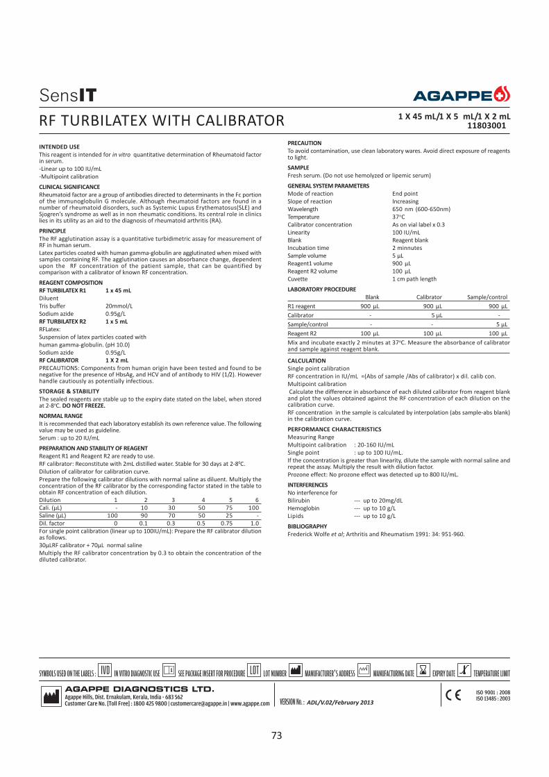
INTENDED USE
This reagent is intended for in vitro quantitative determination of Rheumatoid factorin serum.
-Linear up to 100 IU/mL
-Multipoint calibration
CLINICAL SIGNIFICANCE
Rheumatoid factor are a group of antibodies directed to determinants in the Fc portionof the immunoglobulin G molecule. Although rheumatoid factors are found in anumber of rheumatoid disorders, such as Systemic Lupus Erythematosus(SLE) andSjogren’s syndrome as well as in non rheumatic conditions. Its central role in clinicslies in its utility as an aid to the diagnosis of rheumatoid arthritis (RA).
PRINCIPLE
The RF agglutination assay is a quantitative turbidimetric assay for measurement ofRF in human serum.
Latex particles coated with human gamma-globulin are agglutinated when mixed withsamples containing RF. The agglutination causes an absorbance change, dependentupon the RF concentration of the patient sample, that can be quantified bycomparison with a calibrator of known RF concentration.
REAGENT COMPOSITION
RF TURBILATEX R1 1 x 45 mL
Diluent
Tris buffer 20mmol/L
Sodium azide 0.95g/L
RF TURBILATEX R2 1 x 5 mL
RFLatex:
Suspension of latex particles coated with
human gamma-globulin. (pH 10.0)
Sodium azide 0.95g/L
RF CALIBRATOR 1 X 2 mL
PRECAUTIONS: Components from human origin have been tested and found to benegative for the presence of HbsAg, and HCV and of antibody to HIV (1/2). Howeverhandle cautiously as potentially infectious.
STORAGE & STABILITY
The sealed reagents are stable up to the expiry date stated on the label, when storedat 2-8oC. DO NOT FREEZE.
NORMAL RANGE
It is recommended that each laboratory establish its own reference value. The followingvalue may be used as guideline.
Serum : up to 20 IU/mL
PREPARATION AND STABILITY OF REAGENT
Reagent R1 and Reagent R2 are ready to use.
RF calibrator: Reconstitute with 2mL distilled water. Stable for 30 days at 2-80C.
Dilution of calibrator for calibration curve.
Prepare the following calibrator dilutions with normal saline as diluent. Multiply theconcentration of the RF calibrator by the corresponding factor stated in the table toobtain RF concentration of each dilution.
Dilution 1 2 3 4 5 6
Cali. (µL) - 10 30 50 75 100
Saline (µL) 100 90 70 50 25 -
Dil. factor 0 0.1 0.3 0.5 0.75 1.0
For single point calibration (linear up to 100IU/mL): Prepare the RF calibrator dilutionas follows.
30µLRF calibrator + 70µL normal saline
Multiply the RF calibrator concentration by 0.3 to obtain the concentration of thediluted calibrator.
RF TURBILATEX WITH CALIBRATOR 1 X 45 mL/1 X 5 mL/1 X 2 mL11803001
PRECAUTION
To avoid contamination, use clean laboratory wares. Avoid direct exposure of reagentsto light.
SAMPLE
Fresh serum. (Do not use hemolyzed or lipemic serum)
GENERAL SYSTEM PARAMETERS
Mode of reaction End point
Slope of reaction Increasing
Wavelength 650 nm (600-650nm)
Temperature 37oC
Calibrator concentration As on vial label x 0.3
Linearity 100 IU/mL
Blank Reagent blank
Incubation time 2 minnutes
Sample volume 5 µL
Reagent1 volume 900 µL
Reagent R2 volume 100 µL
Cuvette 1 cm path length
LABORATORY PROCEDURE
Blank Calibrator Sample/control
R1 reagent 900 µL 900 µL 900 µL
Calibrator - 5 µL -
Sample/control - - 5 µL
Reagent R2 100 µL 100 µL 100 µL
Mix and incubate exactly 2 minutes at 37oC. Measure the absorbance of calibratorand sample against reagent blank.
CALCULATION
Single point calibration
RF concentration in IU/mL =(Abs of sample /Abs of calibrator) x dil. calib con.
Multipoint calibration
Calculate the difference in absorbance of each diluted calibrator from reagent blankand plot the values obtained against the RF concentration of each dilution on thecalibration curve.
RF concentration in the sample is calculated by interpolation (abs sample-abs blank)in the calibration curve.
PERFORMANCE CHARACTERISTICS
Measuring Range
Multipoint calibration : 20-160 IU/mL
Single point : up to 100 IU/mL.
If the concentration is greater than linearity, dilute the sample with normal saline andrepeat the assay. Multiply the result with dilution factor.
Prozone effect: No prozone effect was detected up to 800 IU/mL.
INTERFERENCES
No interference for
Bilirubin --- up to 20mg/dL
Hemoglobin --- up to 10 g/L
Lipids --- up to 10 g/L
BIBLIOGRAPHY
Frederick Wolfe et al; Arthritis and Rheumatism 1991: 34: 951-960.
ADL/V.02/February 2013
73

INTENDED USE
This reagent is intended for in vitro quantitative determination of Rheumatoid factorin Serum.
-Latex enhanced immunoturbidimetry
-Linear upto 135 IU/mL
-No sample dilution required
-Ready to use reagents
CLINICAL SIGNIFICANCE
Rheumatoid Factor (RF) is an auto antibody against human IgG commonly seen in seraof patients with rheumatoid arthritis. The measurement of RF value is useful inevaluating the diagnosis, effects of therapy and prognosis of RA, systemic lupuserythematosus, Chronic hepatopathy etc.
PRINCIPLE
When a sample containing rheumatoid factor is added to denatured human IgG whichhas been sensitized to latex particles, antigen-antibody reaction occurs leading toagglutination. This agglutination leads to an absorbance change which is measured at(550 to 660nm). The change in absorbance is proportional to agglutination and theactual concentration is determined by interpolation from a calibration curve preparedfrom known value calibrators.
REAGENT COMPOSITION
RF R 1 1 x 24 mL, 2 x 24 mL
Glycine Buffer Solution
RF R2 1 x 8 mL, 2 x 8 mL
Latex suspension coated with denatured human IgG
CALIBRATOR 1 x 1 mL, 1 x2 mL
RF calibrator concentration as on vial label.
STORAGE AND STABILITY
The sealed reagents are stable up to the expiry date stated on the label, when storedat 2- 80C. DO NOT FREEZE.
NORMAL RANGE
It is recommended that each laboratory should establish its own reference values.
The following value may be used as guide line.
Serum up to 18 IU/mL
PREPARATION OF REAGENT
Reagent 1 and Reagent 2 are ready to use
RF Calibrator : Ready to use Liquid stable
PRECAUTION
To avoid contamination, use clean laboratory wares. Avoid direct exposure of reagentto light & heat.
The reagent should be used according to this pack insert. If used otherwise,appropriate performance is not guaranteed.
SAMPLE
Fresh serum (Do not use haemolized or lipemic serum)
GENERAL SYSTEM PARAMETER SEMI AUTO FULLY AUTO
Mode of Reaction Fixed Time End point
Slope of reaction Increasing Increasing
Wavelength 578 nm 660 nm
Temperature 370C 370C
No.of calib. 6 6
Calibrator concentration As on vial label x Dilution factor
Linearity 135 IU/mL 135 IU/mL
Blank DI water Reagent blank
Delay 5 sec --
Interval 120 sec --
Sample volume 10 µL 8 µL
Reagent 1 volume 450 µL 210 µL
Reagent 2 volume 150 µL 70 µL
Cuvette 1 cm light path 1 cm light path
CALIBRATION
Preparation of calibration curve:
Prepare the following calibrator dilutions using normal saline as diluent. Multiplythe concentration of the RF calibrator by the corresponding factor stated in the tablebelow to obtain the RF concentration of each dilution.
Dilution 1 2 3 4 5 6
Cali. (µL) - 10 20 50 75 100
Saline (µL) 100 70 60 50 25 -
Dil. factor 0 0.125 0.25 0.5 0.75 1.0
LABORATORY PROCEDURE FOR FULLY AUTO ANALYZER
Blank Calibrator Sample /control
RF R1 210 µL 210 µL 210 µL
Dil.Calibrator 8 µL -
Sample/control - - 8 µL
Mix and incubate for 5 minutes at 37oC.
RF R2 70 µL 70 µL 70 µL
Mix and read the absorbance immediately (A1), and after 2 minutes (A2) at 660nm.
ALTERNATIVE PROCEDURE FOR SEMI AUTO ANALYZER
Calibrator Sample/control
RF R1 450 µL 450 µL
Dil.Calibrator 10 µL -
Sample /control - 10 µL
RF R2 150 µL 150 µL
Mix and read the absorbance immediately (A1), and after 2 minutes (A2) at 578nm.
CALCULATION
Multipoint calibration
Calculate the abs, plot a standard curve & read the concentration of controls &samples,by interpolation of curve.
PERFORMANCE CHARACTERISTICS:
Measuring Range:- 10 - 135 IU/mL.
If the concentration is greater than 135 IU/mL, dilute the sample with normal salineand repeat the assay .Multiply the result with dilution factor.
Prozone Effect:- >770 IU/mL
Precision in CV%:-
Low High
Intra - Run 5.0 1.0
Inter - Run 8.0 5.0
Accuracy in IU/mL
control Assigned value Measured value
level 1 23.8(19-28.5) 20.1
level 2 28.4(22.7-34) 30.81
level 3 37.9(30.3-45.5) 41.96
IINTERFERENCE
No interference upto
Hemoglobin 500 mg/dL
Intrafat 5000 mg/dL
Bilirubin 20 mg/dL
BIBLIOGRAPHY
Frederick Wolfe et al-Arthritis and Rheumatism 1991 : 34:951-960
RF LEIT WITH CALIBRATOR 1x 24 /1 x 8 / 1 mL, 2x 24 /2 x 8 / 2 mL
11809003, 11809004
74

INTENDED USE
This reagent is intended for in vitro quantitative determination of Transferrin inhuman serum
-Turbidimetric Immunoassay
-Linear up to 540 mg/dL
-Ready to use reagents
-Multipoint calibration
CLINICAL SIGNIFICANCE
Transferrin(TF) is synthesised in Liver. The main role of Transferrin is to deliver ironfrom absorption centres to all tissues. Increased serum Transferrin is associated withiron deficiency anemia, malignant tumor, polycythemia rubra, aplastic anemia,hemolytic anemia and hepatitis. Absence of Transferrin in the body creates a raregenetic disorder known as atransferrinemia; a condition characterized by anemia andhemosiderosis in the heart and liver that leads to many complications including heartfailure.
PRINCIPLE
Antibodies to human TF are combined with transferrin in the patients serum, formingimmune complexes. The immune complexes cause an increase in light scattering whichcorrelate with the concentration of TF in the serum. The light scattering is measured340 nm.
REAGENT COMPOSITION
Transferrin R1(Buffer solution) 1 x 30 mL
Tris (hydroxymethyl) aminomethane 100 mmol/L
Transferrin R2(Anti serum solution) 1 x 3 mL
Anti- human TF anti serum (Variable)
Calibrator 1 x 1 mL
Calibrator concentration is mentioned on vial label
STORAGE AND STABILITY
The reagents are stable until expiry date when kept at 2-80 C .
NORMAL RANGE
It is recommended that each laboratory establish its own reference values.
The following values may be used as guide line.
Male : 190 - 300 mg/dL
Female : 200 - 340 mg/dL
PRECAUTION
To avoid contamination, use clean laboratory wares. Use clean, dry disposable pipettetips for dispensing. Close reagent bottles immediately after use. Avoid direct exposureof reagent to light.
SAMPLE
Use fresh serum. Dilute serum/control to 1/10 with saline.
GENERAL SYSTEM PARAMETERS FOR SEMI AUTO FOR FULLY AUTO
Mode of reaction End point End point
Slope of reaction Increasing Increasing
Wavelength 340 nm 340 nm
Temperature 37oC 37oC
Calibrator concentration As on vial label x Dilution factor
Linearity 540 mg/dL 540 mg/dL
Blank Reagent Blank Reagent Blank
Incubation time 5min + 5min -
Sample volume 15 µL 8.5 µL
Reagent 1 volume 500 µL 300 µL
Reagent 2 volume 50 µL 30 µL
Cuvette 1 cm light path 1 cm light path
CALIBRATION
Preparation of calibration curve:
Dilute the high concentrated calibrator 1/10 with normal saline and use this dilutedcalibrator for the preparation of calibration curve.
Prepare the following calibrator dilutions using normal saline as diluent. Multiply theconcentration of the Transferrin calibrator by the corresponding factors stated in thetable below to obtain the Transferrin concentration of each dilution.
Dilution 1 2 3 4 5 6
1/10dilcali.(µL)| - 10 10 25 50 100
Saline (µL) 100 150 70 75 50 -
Dil. factor 0 0.0625 0.125 0.25 0.5 1
LABORATORY PROCEDURE FOR FULLY AUTO
Blank Calibrator Sample/control
Transferrin R1 300 µL 300 µL 300 µL
Dil. Calibrator - 8.5 µL -
Dil. Sample/control - - 8.5 µL
Mix and incubate for 5 minutes at 37°C. Read the absorbance (A1) at 340 nm.
Transferrin R2 30 µL 30 µL 30 µL
Mix and incubate for 5 minutes at 37°C. Measure the absorbance (A 2) at 340 nm.
ALTERNATIVE PROCEDURE FOR SEMIAUTOANALYZER:
Blank Calibrator Sample/control
Transferrin R1 500 µL 500 µL 500 µL
Dil. Calibrator - 15 µL -
Dil. Sample/control - - 15 µL
Mix and incubate for 5 minutes at 37°C.
Transferrin R 2 50 µL 50 µL 50 µL
Mix and incubate for 5 minutes at 37°C. Measure the absorbance (A) at
340 nm against reagent blank.
CALCULATION
Calculate the Abs, plot a standard curve & read the concentration of controls &samples.
PERFORMANCE CHARACTERISTICS:
Measuring Range:- 40 – 540 mg/dL.
If the concentration is greater than 540 mg/dL , dilute the diluted(1/10)sample withnormal saline and repeat the assay .Multiply the result with dilution factor.
Prozone Effect:- >1400 mg/dL
Precision in CV%:-
Low Medium High
Intra - Run 4.78 1.30 0.78
Inter - Run 4.64 2.46 0.9
Accuracy in mg/dL
control Assigned value Measured value
level 1 151(121-181) 160.48
level 2 250(200-300) 254.54
level 3 334 (267 - 400) 351.3
INTERFERING SUBSTANCES
No interference for
Hemoglobin upto1000 mg/dL
Bilirubin upto20 mg/dL
Lipemia upto5 % of intralipid
BIBLIOGRAPHY
1. K. Bergstorm, et al.: Scand. J.Clin Lab Invest., 40, 637- 640 (1980)2. H. Maikus, et al.: Clinica Chemica Acta, 88, 523- 530(1978)
TRANSFERRIN WITH CALIBRATOR 1 x 30/1 x3/1 mL
11821002
75

INTENDED USE: This reagent is intended for in vitro qualitative & semi quantitativedetermination of Anti Streptolysin O (ASO) in serum.
-No prozone effect detected up to 1500 IU/mL
-Rapid procedures; test time only 2 minutes
-Excellent clarity; clear agglutination
CLINICAL SIGNIFICANCE
Streptolysin O is a toxic immunogenic exoenzyme produced by ß-haemolyticStreptococcus group A, C and G. ASO antibodies are useful for the diagnosis ofrheumatoid fever, acute glomerulonephritis and streptococcal infections. Rheumaticfever is an inflammatory disease affecting connective tissue from several parts ofhuman body as skin, heart, joints etc. Acute glomerulonephritis is a renal infectionthat affects mainly glomerulus.
PRINCIPLE
ASO latex kit is a rapid agglutination procedure for the direct detection and semi-quantitation (on slide) of antistreptolysin-O (ASO). The antigen, latex particlessuspension coated with Streptolysin-O, agglutinates in the presence of specificantibodies present in the sera of patients with streptolysin O.
REAGENTS & MATERIALS PROVIDED
ASO LATEX 1 x 2.5 mL / 1 x 5 mL
Suspension of polystyrene particles coated with Streptolysin-O.
ASO POSITIVE CONTROL 1 x 0.5 mL
Human pooled serum
ASO NEGATIVE CONTROL 1 x 0.5 mL
Human pooled serum
PHYSIOLOGICAL SALINE BUFFER CONCENTRATE 1 x 5 mL
Dilute 1:20 (v/v) with distilled water
ACCESSORIES: for 50T for 100 T
1. Reaction slide 1 1
2. Serum droppers 50 100
3. Applicator sticks 50 100
4. Rubber teat 1 1
PRECAUTION
Reagent components of human origin have been tested and found to be negativefor the presence of antibody to HIV (1/2) as well as for HBsAg and HCV antibody.However, handle cautiously as potentially infectious.
The reagent and controls contain less than 0.1% sodium azide.
STORAGE AND STABILITY
When stored at 2-80C and protected from light, the reagent and the controls arestable until the expiry date stated on the label. DO NOT FREEZE
Preparation of physiological saline buffer: Prepare physiological saline buffer byadding 95mL of distilled water to 5 mL of physiological saline buffer concentrate(provided). It is stable up to expiry date, when stored at room temperature.
ANALYTICAL SENSITIVITY
The ASO latex reagent sensitivity has been adjusted to detect a minimum of
200 (+/- 50) IU/mL ASO antibodies in the undiluted samples.
SAMPLE
Fresh serum (free of haemolysis)
QUALITATIVE TEST
Allow all reagents as well as the sample to reach room temperature. Mix well beforeuse.
1. Place 1 drop of serum sample on the slide using a disposable serum dropper.2. Add one drop of ASO-latex reagent to the above drop and mix with disposable
applicator stick.3. Rock the slide gently to and fro for 2 minutes and immediately examine under
a good light source for agglutination, do not examine beyond 2 minutes.4. For positive & negative controls follow the same procedure as mentioned above
by taking control serum from respective vials.RESULT AND INTERPRETATION
Positive result:
The presence of agglutination indicate concentration of ASO in the sample equalor greater than 200 IU/mL (above normal).
Negative result:
The lack of agglutination indicates ASO level lower than 200 IU/mL in the sample,(within the normal range).
ASO50 T, 100 T
12201002, 12201003
SEMI – QUANTITATIVE TEST
In the cases in which it is desired to find out the titre of a positive sample, it is possibleby the serial dilution methodology.
1. Place 50 µL diluted physiological saline Buffer onto each of five circles of the slide2. Using a 50 µL micro pipette add 50 µL of the serum sample to the drop of saline
buffer in 1st circle.3. Using the same micro pipette, mix the sample with saline by aspirating back &
forth several times. Aspirate 50 µL from 1st circle and transfer to 2nd circle.Repeat the same operation up to 5th circle. Aspirate 50 µL from 5th circle anddiscard. Dilutions obtained as 1/2, 1/4 1/8, 1/16, 1/32
4. Then add 1 drop of ASO latex to the above circles. Mix and rock the slide gently toand fro for 2 minutes; observe the agglutination under good source of light.
CALCULATION
ASO Conc. (IU/mL) = sensitivity x Titre (Highest dilution serum showing agglutination)
Where, ASO sensitivity = 200 IU/mL
NOTE:
1. False positive results may be obtained in conditions such as rheumatoid arthritis,scarlet fever, several other streptococcal healthy carriers.
2. Early infections and children from 6 months to 2 years may cause false negativeresults.
3. A single ASO determination does not produce much information about theactual state of the disease. Titrations at bi weekly intervals during 4 or 6 weeksare advisable to follow the disease evolution.
PERFORMANCE CHARACTERISTICS
1. Diagnostic sensitivity : 98%2. Diagnostic specificity : 97%3. Prozone effect: No prozone effect detected up to 1500 IU/mL.Interferences
Hemoglobin : 10 g/L
Bilirubin : 20 mg/dL
Rheumatoid factor : 300 IU/mL
do not interfere. Other substances may interfere4.
BIBLIOGRAPHY
1. CURTIS G. D.W, KRAAK W.A.G., MITCHELL R.G. Comparison of latex andhemolysis test determination of antistreptolysin O (ASO) antibodies. J. Clin.Pathol. 41, 1331 (1988)
2. TADZYNSKY L.A., RYAN M.E. Diagnosis of rheumatoid fever. A guide to criteriaand manifestations. Post grad Med. 79, 295 (1986)
3. D’ANGELO W.A. Rheumatic Diseases. Diagnostic tests and procedures. in: TheLaboratory in Clinical Medicine (Halsted J.A. Ed.) Saunders Company,Philadelphia, chapter 29 (1976)
4. Young D.S., Effects of Drugs on Clinical Laboratory Test 4th ed. AACC Press, 1995.
ADL/V.02/February 2013
76

INTENDED USE: This reagent is intended for in vitro quantitative determination ofC-reactive protein (CRP) in serum
-No prozone effect detected up to 1600 mg/L
-Rapid procedure, only 2 minutes test
-Excellent clarity, clear agglutination
CLINICAL SIGNIFICANCE
CRP is a classic acute phase protein of human serum, synthesized by hepatocytes.Normally it is present only in trace amounts in serum, but it can increase as muchas 1,000 fold in response to injury or infection. The clinical measurement of CRP inserum therefore appears to be a valuable screening test for organic disease and asensitive index of disease activity in inflammatory, infections and ischemic conditions.
PRINCIPLE
CRP latex kit is a rapid agglutination procedure for the direct detection and semi-quantitation (on slide) of C-reactive protein (CRP). The reagent, latex particlessuspension coated with specific antihuman C-reactive protein antibodies,agglutinates in presence of CRP in patient serum.
REAGENTS & MATERIALS PROVIDED
CRP LATEX 1 x 2.5 mL / 1 x 5 mL
Suspension of polystyrene particles coated with anti human CRP goat antibodies.
CRP POSITIVE CONTROL 1 x 0.5 mL
Human pooled serum
CRP NEGATIVE CONTROL 1 x 0.5 mL
Human pooled serum
PHYSIOLOGICAL SALINE BUFFER CONCENTRATE 1 x 5 mL
Dilute 1:20 (v/v) with distilled water
ACCESSORIES: For 50 T For 100 T
1. Reaction slide 1 1
2. Plastic droppers 50 100
3. Applicator sticks 50 100
4. Rubber teat 1 1
PRECAUTION
Reagent components of human origin have been tested and found to be negativefor the presence of antibody to HIV (1/2) as well as for HBsAg and HCV antibody.However, handle cautiously as potentially infectious.
The reagent and controls contain less than 0.1% sodium azide.
STORAGE AND STABILITY
When stored at 2-80C and protected from light, the reagent and the controls arestable until the expiry date stated on the label. DO NOT FREEZE.
Preparation of physiological saline buffer: Prepare physiological saline buffer byadding 95mL of distilled water to 5mL of physiological saline buffer concentrate(provided). It is stable up to expiry date, when stored at room temperature.
ANALYTICAL SENSITIVITY
The CRP latex sensitivity has been adjusted to detect a minimum of6 (5-10) mg/L in the undiluted samples.
SAMPLE
Fresh serum (free of haemolysis)
QUALITATIVE TEST
Allow all reagents as well as the sample to reach room temperature. Mix well beforeuse.
1. Place 1 drop of serum sample on to the slide using a disposable serum dropper.2. Add one drop of CRP-latex reagent to the above drop and mix well with
disposable applicator stick.3. Rock the slide gently to and fro for 2 minutes and examine immediately under
good light source for agglutination, do not examine beyond 2 minutes.4. For positive & negative controls follow the same procedures as mentioned
above by taking control serum from respective vials.RESULT AND INTERPRETATION
Positive result:
The presence of agglutination indicate concentration of CRP in the sample equalor greater than 6 mg/L (above normal)
Negative result:
The lack of agglutination indicates CRP level lower than 6 mg/L in the sample,(within the normal range)
CRP 50 T, 100 T
12202002, 12202003
SEMI – QUANTITATIVE TEST
In the case in which it is desired to find out the titre of a positive sample, it ispossible by the serial dilution methodology.
1. Place 50 µL diluted saline Buffer onto each of five circles of the slide.2. Using a 50 µL micro pipette add 50 µL serum sample to the drop of saline
buffer in 1st circle.3. Using the same micro pipette, mix the sample with saline by aspirating back &
forth several times. Aspirate 50 µL from 1st circle and transfer to 2nd circle.Repeat the same operation up to 5th circle. Aspirate 50 µL from 5th circle anddiscard. Dilutions obtained as 1/2, 1/4, 1/8, 1/16, 1/32
4. Then add 1 drop of CRP latex reagent to the above circles. Mix and rock the slidegently to and fro for 2 minutes; observe the agglutination under good source oflight.
CALCULATION
Concentration of CRP in serum can be calculated as follows:
CRP Conc. (mg/L) = sensitivity x titre (highest dilution serum showing agglutination)
Where, CRP sensitivity = 6 mg/L
NOTE:
1. Reaction time is critical, if reaction time exceeds two minutes, drying of reactionmixture may cause false positive results.
2. Freezing the CRP latex reagent will cause spontaneous agglutination.3. Intensity of agglutination is not necessarily indicative of relative CRP
concentration, therefore screening reaction should not be graded.4. A false negative can be attributes to a prozone phenmenon (antigen excess). It
is recommended therefore to check sample with dilution.5. Clinical diagnosis should not be made on findings of a single test result, but
should integrate both clinical and laboratory data.
PERFORMANCE CHARACTERISTICS
Diagnostic sensitivity : 95.6%
Diagnostic specificity : 96.2%
Prozone effect : No prozone effect detected up to 1600mg/mL.
INTERFERENCES
Bilirubin up to 20 mg/dL
Hemoglobin up to 10 g/L
Lipids up to 10 gL
Rheumatoid factor up to 100 IU/mL
Do not interfere. Other substances may interfere.
BIBLIOGRAPHY
1. Wadsworth, C.Wadsworth, E., Efficacy of latex agglutination methods fordetermination of C - reactive protein in Pediatric sera. Clin. Chem. Acta, 138,(1984), 309
2. Ballou S.P, Kushner, I.C. Reactive Protein and acute phase response. ADV Int.Med, 37, (1992), 313.
3. Young D.S., Effects of Drugs on Clinical Laboratory Test 4th ed. AACC Press, 1995.
ADL/V.02/February 2013
77

INTENDED USE: This reagent is intended for in vitro qualitative & semiquantitative determination of Rheumatoid factor (RF) in serum.
-Sensitivity 8 IU/mL
-Optimized antigen concentration; No prozone effect up to 800 IU/mL
-Rapid procedure; only 2 minutes test
-Excellent clarity; crystal clear agglutination
CLINICAL SIGNIFICANCE
Rheumatoid factors are a group of antibodies directed to the determinants in the Fcportion of the immunoglobulin G molecule (IgG). Although rheumatoid factors arefound in a number of rheumatoid disorders, such as systemic lupus erythematosus(SLE) and Sjogrens syndrome as well as in non rheumatic condition, its central rolein clinics lies in its utility as an aid in the diagnosis of rheumatoid arthritis (RA).
PRINCIPLE
RF latex kit is a rapid agglutination procedure for the detection and semi-quantitation (on slide) of Rheumatoid Factors (RF). The antigen, a latex particlessuspension coated with human gamma-globulin, agglutinates in presence ofrheumatoid factors in the patient serum
REAGENTS & MATERIALS PROVIDED
RF LATEX 1 x 2.5 mL / 1 x 5 mL
Suspension of polystyrene particles coated with human gamma -globulin.
RF POSITIVE CONTROL 1 x 0.5 mL
Human pooled serum
RF NEGATIVE CONTROL 1 x 0.5 mL
Human pooled serum
PHYSIOLOGICAL SALINE BUFFER CONCENTRATE 1 x 5 mL
Dilute 1:20(v/v) with distilled water
ACCESSORIES:For 50 T For 100 T
1. Reaction slide 1 12. Plastic droppers 50 1003. Applicator sticks 50 1004. Rubber teat (blue) 1 1PRECAUTION
Reagent components of human origin have been tested and found to be negativefor the presence of antibody to HIV (1/2) as well as for HBsAg and HCV antibody.However, handle cautiously as potentially infectious.
The reagent and controls contain less than 0.1% sodium azide.
STORAGE AND STABILITY
When stored at 2-80C and protected from light, the reagent and the controls arestable until the expiry date stated on the label. DO NOT FREEZE.
Preparation of physiological saline buffer: Prepare saline buffer by adding 95 mLof distilled water to 5 mL of concentrate saline buffer (provided). It is stable up toexpiry date, when stored at room temperature.
ANALYTICAL SENSITIVITY
The RF sensitivity has been adjusted to detect a minimum of
8.0 IU/mL (6-16) RF in the undiluted samples.
SAMPLE
Fresh serum (free of haemolysis)
QUALITATIVE TEST
Allow all reagents as well as the sample to reach room temperature. Mix well beforeuse.
1. Place 1 drop of serum sample on to the slide with disposable serum dropper.2. Add one drop of RF-latex reagent to the above drop and mix well with
disposable applicator stick.3. Rock the slide gently to and fro for 2 minutes and then examine immediately
under good light source for agglutination, do not examine beyond 2 minutes.4. For positive & negative controls follow the same procedures as mentioned
aboveby taking control serum from respective vials.RESULT AND INTERPRETATION
Positive result:
The presence of agglutination indicated concentration of RF in the sample equalor greater than 8 IU/mL (above normal).
Negative result:
The lack of agglutination indicates RF level lower than 8 IU/mL in thesample, (within the normal range).
SEMI – QUANTITATIVE TEST
In the case in which it is desired to find out the titre of a positive sample, it isfeasible by the serial dilution methodology.
1. Place 50 µL diluted saline buffer onto each of five circles of the slide2. Using a 50 µL micro pipette add 50 µL of the serum sample to the drop of saline
buffer in 1st circle.3. Using the same micro pipette, mix the sample with saline by aspirating back &
forth several times. Aspirate 50 µL from 1st circle and transfer to 2nd circle.Repeat the same operation up to 5th circle. Aspirate 50 µL from 5th circle anddiscard. Dilutions obtained as ½, ¼, 1/8, 1/16, 1/32
4. Then add 1 drop of RF latex reagent to the above circles, mix and rock the slidegently to and fro for 2 minutes; observe the agglutination under good sourceof light.
CALCULATION
The Rheumatoid Factor (RF) level in serum can be calculated as follows:
RF conc. (mg/dL) = sensitivity x titre (highest dilution of serum showing agglutination)
Where, RF Sensitivity = 8.0 IU/mL
PERFORMANCE CHARACTERISTICS
1. Diagnostic sensitivity : 98%
2. Diagnostic specificity : 98.8%
3. Prozone effect : No prozone effect was detected up to 800 IU/mL.
INTERFERENCES
Bilirubin up to 20 mg/dL
Hemoglobin up to 10g/L
Lipids up to 10g/L
will not interfere. Other substances may interfere.
NOTE:
1. The incidence of false positive results is about 3-5%. Individuals suffering frominfectious mononuclear hepatitis, syphilis as well as elderly people may givepositive results.
2. Diagnosis should not be solely based on the results of latex method, but alsoshould be complemented with a Waaler Rose test along with clinicalexamination.
BIBLIOGRAPHY
1 . Frederick Wolf et al-Arthritis and Rheumatism 1991: 34: 951-9602. Robert W Dorner et al. Clinica Chemica Acta 1987; 167: 1-21
RF 50 T, 100 T
12203002, 12203003
ADL/V.02/February 2013
78

INTENDED USE: RPR (Rapid Plasma Reagin) test is a non-treponemal test for theserological diagnosis of Syphilis. Specificity, reactivity and sensitivity are similar tothat of classical VDRL test. It is a reliable, economical and rapid test and hencerecommended as a screening test.
CLINICAL SIGNIFICANCE
Syphilis is a veneral disease caused by the spirochete microorganism T.pallidum. Asthe organism is difficult to be cultured on artificial media, the diagnosis of syphilisdepends on the correlation of clinical data with the detection of specific antibodyby serological test. Serological screening tests for syphilis using cardiolipin andlecithin antigen are simple to perform and reliable but may give rise to smallproportion of false positive results because the tests use non treponemal antigens.These antigens detect reagin antibodies produced against cardiolipin during aninfection.
PRINCIPLE
RPR reagent is an antigen containing cardiolipin antigen, sensitized on carbon particleswhich detects reagin antibody present in serum of syphilitic individuals. When aspecimen contains antibody is mixed with the reagent, flocculation occurs due tocoagglutination of the carbon particles of the RPR antigen, which appear as blackclumps against the white background of the card. This coagglutination is readmacroscopically. Non-reactive specimens show a black button at the centre.
REAGENTS AND METERIALS PROVIDED
RPR ANTIGEN 1 x 1.1 mL, 1 x 2.6 mL, 4 x 2.6 mL
Antigen, Cardiolipin suspension containing carbon micro particles.
RPR POSITIVE CONTROL 1 x 0.3 mL
Human pooled serum
RPR NEGATIVE CONTROL 1 x 0.3 mL
Human pooled serum
PHYSIOLOGICAL SALINE 1 x 5mL
Dilute 1:20 (v/v) with H2O.
ACCESSORIES: For 50 Tests For 125 Tests For 500 Tests
1.Applicator stick 50 125 500
2. Serum dropper 50 125 500
3. Plastic slides 7 16 63
4. Dropper with needle 1 set 1 set 1 set
( Accessories will be provided in separate pouch)
PRECAUTION
Reagent components of human origin have been tested and found to be negativefor the presence of antibody to HIV (1/2) as well as for HBsAg and HCV antibody.However, handle cautiously as potentially infectious.
The reagent and controls contain less than 0.1% sodium azide.
STORAGE AND STABILITY
When stored at 2-80C and protected from light, the reagent and the controls arestable until the expiry date stated on the label. DO NOT FREEZE.
Preparation of saline buffer: Prepare saline buffer by adding 95 mL distilled waterto 5 mL concentrated saline buffer. It is stable up to expiry date when stored atroom temperature.
SAMPLE
Fresh serum (free of haemolysis)
QUALITATIVE TEST
Bring the test reagents and samples to room temperature
1. Place 1 drop of serum sample (50 µL or 0.05mL) on to the slide with disposableserum dropper.
2. Place one drop of RPR antigen suspension (18 µL) by antigen dropper3. Mix well and spread the liquid over the entire area of the circle using disposable
applicator stick4. Rock the slide gently to and fro for 8 minutes on a mechanical rotator& observe
immediately under good light source for the appearance of carbon particleclumping.
RESULT AND INTERPRETATION
Medium & large aggregates against white back ground - - Reactive
Small agglutinates around periphery - Weak reactive
No aggregates, button formation at the centre of circle - Non reactive
SEMI – QUANTITATIVE TEST
In the case in which it is desired to find out the titre of a positive sample, it ispossible by the serial dilution methodology.
1. Place 50 µL diluted saline Buffer onto each of five circles of the slide.2. Using a 50 µL micro pipette add 50 µL serum sample to the drop of saline buffer
in 1st circle.3. Using the same micro pipette, mix the sample with saline by aspirating back &
forth several times. Aspirate 50 µL from 1st circle and transfer to 2nd circle.Repeat the same operation up to 5th circle. Aspirate 50 µL from 5th circle anddiscard. Dilutions obtained as 1/2,1/4, 1/8, 1/16, 1/32
4. Then add 1 drop of RPR antigen suspension (18 µL) to the above circles.Mix androck the slide gently to and fro for 8 minutes; observe the agglutination undergood light source for appearance of carbon particle clumping.
INTERPRETATION
The titre of reagin antibodies is the highest dilution of the test sample which isreactive.
NOTE
1. As with tests based on reagin antibody detection, the RPR syphilis test mayproduce false positive results. These can be linked to diseases such as leprosy,systemic lupus erythoematosus (SLE), infectious mononucleosis, malaria, viralpneumonia. Any positive result must be confirmed by another serological assay(eg.TPHA) Final diagnosis will only be reached after correlation of the resultswith clinical signs.
2. False negative results may be seen in early and latent stages of the disease.3. Qunatitative procedure must be performed to determine the response to
treatment and detect re-infection.4. While dispensing reagents/specimens hold pipette/dropper vertically straight.5. The needle assembly should be thoroughly rinsed with distilled water and air
dried before further use.6. The test cards must not be reused.BIBLIOGRAPHY
1. Caumes E., Janier M. Syphilis, editions techniques. Encyclo. Med Chir (ParisFrance) Maladies Infectiouses. 8-039 -A-10(1994)
2. Drustin L.M. syphilis. Curr OPN. Infect Dis2 : 11-15 (1989)
RPR 50 T, 125 T, 500 T
12204001, 12204002, 12204003
ADL/V.03/February 2013
79

Blood GROUPING
INTENDED USE
This reagent is a monoclonal antibody solution intended for determination of red cells
having A antigen on the surface, who are categorized as A group individuals.
- Monoclonal antibody technology
- High avidity and high titre
- Convenient pack sizes
PRINCIPLE
This technique employs the principle of hemagglutination. When the red blood cells
bearing the A antigens are mixed with the Anti-A solution, if agglutination occurs
indicates the presence of A antigen, and the individual belongs to A group and if no
agglutination occurs indicated absence of A antigen.
REAGENT COMPOSITION
Anti A is ready to use reagent prepared from supernatents of mouse hybridoma cell
culters. These reagents contain sodium azide (<0.1%), sodium arsenite (0.02%) and
bovine albumin.
STORAGE AND STABILITY
The sealed reagent is stable up to expiry date stated on the vial label, when stored at
2-80C. It is advisable to minimize its time outside the refrigerator between two uses.
SAMPLE
Whole blood sample must be examined with in 48 hrs. Samples should be stored at
2-80C, if not tested immediately. Do not use haemolysed samples.
LABORATORY PROCEDURE
PLATE TECHNIQUE AT ROOM TEMPERATURE
Bring the reagents to room temperature. Place one drop of Anti-A reagent on a clean
slide, and then add one small drop of whole blood. Mix well with a mixing stick
uniformly. Rock the slide gently back and forth for about 3 minutes while
macroscopically observing the possible appearance of agglutination.
DIRECT METHOD IN A TUBE AT ROOM TEMPERATURE
Prepare a 5% suspension of red blood cells in isotonic saline solution. Using the vial
dropper, place one drop of Anti-A reagent in to labeled test tube. Add a drop of 5 %
RBC suspension. Shake to homogenize the mixture, then centrifuge at 1000 RPM for
60 seconds. Read macroscopically for appearance of any agglutination.
INTERPRETATION
If there is agglutination (ie clumping of red blood cells) the test result indicates the
presence of A antigen.
If there is no visible agglutination, (ie clumping is absent), the test result indicates the
absence of A antigen.
NOTE:
For getting high titre use ABD dilution buffer (code.13601330). The ABD dilution
buffer will be provided on request.
BIBLIOGRAPHY
1. Mannessier, L.; Blood transfusion center Lille France. The use of monoclonal
antibodies as blood grouping reagents: applications, advantages and problems.
Congress of the Italian society for blood transfusion –Rome-June 1992.
2. Blood group serology, Boorman, Dodd and Lincol Churchil Livingstone 6th edition.
3. Human blood groups, Geoff Daniela, 1st edition Blackwell Science, Oxford 1995.
ANTI - A1 x 10 mL
13601010
80

Blood GROUPING
INTENDED USE
This reagent is intended for in vitro determination of ABO group and Rh typing ofhuman red blood cell antigens.
- Monoclonal antibody technology
- Highly specific
- Convenient slide and tube formats.
CLINICAL SIGNIFICANCE
The ABO grouping is defined by both, the presence of ‘A’ and/or ‘B’ antigens on thesurface of the red blood cells, and by simultaneous presence of Anti-A and or Anti-Bantibodies in the serum. An individual has in his serum the antibodies correspondingto the antigens which are not present on his red blood cells.
Based on the principle of hemagglutination, human red blood cells possessing ‘A’ and/or ‘B’ antigen will agglutinate in the presence of corresponding antibody directedtowards the antigen.
Human red blood cells are classified as Rh positive (Rh+) or Rh negative (Rh-) dependingupon the presence or absence of ‘D’ antigen on them. Red blood cells possessing Dantigen will agglutinate in the presence of reagent containing corresponding antibody.
REAGENT COMPOSITION
Anti-A, Anti-B and Anti-D are ready to use reagents prepared from supernatents ofmouse hybridoma cell cultures. These antibodies of immunoglobin class IgM (Anti-D IgM+IgG) are a mixture of several monoclonal antibodies of the same specificitybut having capacity of recognising different epitopes of human red blood cellantigens ‘A’, ‘B’ and ‘D’ (Rh)
Anti-A 1 x 10 mL
Anti-B 1 x 10 mL
Anti-D(IgG+IgM) 1 x 10 mL
These reagents contain sodium azide (<0.1%), sodium arsenite (0.02%) and bovinealbumin.
STORAGE AND STABILITY
The sealed reagents are stable up to expiry date stated on the label when storedat 2-80C. It is advisable to minimize its time outside the refrigerator between two uses.
SAMPLE
Blood collected with or without anticoagulant : EDTA, heparin or citrate,
stored beween 20C to 80C upto 48 hours. Haemolysed blood should not be used.
LABORATORY PROCEDURE
PLATE TECHNIQUE AT ROOM TEMPERATURE
Bring the reagents to room temperature. Place one drop of Anti-A,Anti -B & Anti Dreagent on a clean slide, and then add one small drop of whole blood. Mix well with amixing stick uniformly. Rock the slide gently back and forth for about 3 minutes whilemacroscopically observing the possible appearance of agglutination.
DIRECT METHOD IN A TUBE AT ROOM TEMPERATURE
Prepare a 5% suspension of red blood cells in isotonic saline solution. Using the vialdropper, place one drop of Anti-A,Anti -B & Anti D reagent in to labeled test tube.Add a drop of 5 % RBC suspension. Shake to homogenize the mixture, then centrifugeat 1000 RPM for 60 seconds. Read macroscopically for appearance of anyagglutination.
INTERPRETATION
With both the above methods, if there is agglutination (the red blood cells from oneor several clumps) the reaction is positive and the antigen or at least one of theantigens corresponding to the reagent used is present on the red blood cells tested.
ANTI - A, B & D(IgG+IgM)
3 x 10 mL
13601001
If there is no agglutination (red blood cells form a homogenous suspension), thereaction is negative and the antigen is not present on the red blood cells.
In case of anti D agglutination a positive test indicates presence of Rh antigen i.e. Rhpositive. No agglutination or a negative test indicated either presence weaker variantsof Rh antigens like Du or absence of Rh antigen. This needs confirmation by carryingout indirect coomb’s test.
Drying at the periphery or fibrin strands should not be misinterpreted as agglutination.
NOTE:
For getting high titre use ABD dilution buffer.(code.13601330). The ABD dilutionbuffer will be provided on request.
BIBLIOGRAPHY
1. Common Technical Specifications (CTs) for the in vitro diagnosis medical devicesof annex II, list A, of directive 98/79/EC are notified in the official journal of theEuropean communities under document No.C (2202) 1344, with EEA relevance(2202/364/CE)
2. Mannessier .L Blood transfusion center Lille France. The use of monoclonalantibodies as blood grouping reagents: applications, advantages and problems.Congress of the Italian society for blood transfusion –Rome-June 1992.
3. Blood group serology, Boorman, Dodd and Lincoln, Churchill Livingstone, 6th
edition.4. Human blood groups, Geoff Daniels, 1st edition, Blackwell Science, Oxford 1995.
ADL/V.03/February 2013
81

Blood GROUPING
INTENDED USE
This reagent is intended for in vitro determination of ABO group and Rh typing ofhuman red blood cell antigens.
Monoclonal antibody technology
Highly specific
Convenient slide and tube formats
CLINICAL SIGNIFICANCE
The ABO grouping is defined by both, the presence of ‘A’ and/or ‘B’ antigens on thesurface of the red blood cells, and by simultaneous presence of Anti-A and or Anti-Bantibodies in the serum. An individual has in his serum the antibodies correspondingto the antigens which are not present on his red blood cells.
Based on the principle of hemagglutination, human red blood cells possessing ‘A’ and/or ‘B’ antigen will agglutinate in the presence of corresponding antibody directedtowards the antigen.
Human red blood cells are classified as Rh positive (Rh+) or Rh negative (Rh-) dependingupon the presence or absence of ‘D’ antigen on them. Red blood cells possessing Dantigen will agglutinate in the presence of reagent containing corresponding antibody.
REAGENT COMPOSITION
Anti-A, Anti-B and Anti-D are ready to use reagents prepared from supernatents ofmouse hybridoma cell cultures. These antibodies of immunoglobin class IgM (Anti-DIgM) are a mixture of several monoclonal antibodies of the same specificity but havingcapacity of recognising different epitopes of human red blood cell antigens ‘A’, ‘B’ and‘D’ (Rh)
Anti – A 1 x 10 mL
Anti – B 1 x 10 mL
Anti – D (IgM) 1 x 10 mL
These reagents contain sodium azide (<0.1%), sodium arsenite (0.02%) and bovinealbumin.
STORAGE AND STABILITY
The sealed reagents are stable up to expiry date stated on the label when stored at 2-80C. It is advisable to minimize its time outside the refrigerator and to avoid leaving itat room temperature between two uses.
SAMPLE
Blood collected with or without anticoagulant : EDTA, heparin or citrate, stored beween20C to 80C upto 48 hours. Haemolysed blood should not be used.
LABORATORY PROCEDURE
Plate technique at room temperature
On a rigorously clean plate, using the vial dropper, apply one drop of each reagent.Take one drop of blood and apply next to each drop of reagent, taking care not tocreate contact between the drops. Mix the blood and reagent using a spiral movementwith the end of the stirrer so as to create a regular lozenge of diameter 2 to 3 cm.Incubate the plate at room temperature and with out stirring for 30 seconds. Holdthe plate and give it a rolling movement for 3 minutes while macroscopically observingthe possible appearance of agglutinates. Read reaction immediately.
Direct method in a tube at room temperature
Prepare a 5% suspension of red blood cells in isotonic saline solution. Using the vialdropper, transfer a drop reagent Anti-A, Anti-B and Anti-D into correspondingly labeledtubes. Add a drop of red blood cell suspension. Shake to homogenize the mixture,then centrifuge at 1000 rpm for 1 minutes. Read macroscopically while gently shakingthe tubes. Note the appearance of any agglutinates.
INTERPRETATION
With both the above methods, if there is agglutination (the red blood cells from oneor several clumps) the reaction is positive and the antigen or at least one of theantigens corresponding to the reagent used is present on the red blood cells tested.
If there is no agglutination (red blood cells reform a homogenous suspension), thereaction is negative and the antigen is not present on the red blood cells.
In case of anti D agglutination is a positive test indicates presence of Rh antigen i.e.Rh positive. If there is no agglutination (i.e. clumbing is absent), it indicates eitherpresence of weaker varients of Rh antigens like Du or absence of Rh antigens. It ispossible to use Anti TOTEM in an andirect coomb’s test if weak / or partial antigens Dare to be detected.
Drying at the periphery or fibrin strands should not be misinterpreted as agglutination.
BIBLIOGRAPHY
1. Common Techanical Specifications (CTs) for the in vitro diagnosis medical devicesof annex II, list A, of directive 98/79/EC are notified in the official journal of theEuropean communities under document No.C (2202) 1344, with EEA relevance(2202/364/CE)
2. Mannessier, .L ; Blood transfusion center Lille France. The use of monoclonalantibodies as blood grouping reagents: applications, advantages and problems.Congress of the Italian society for blood transfusion –Rome-June 1992.
3. Blood group serology, Boorman, Dodd and Lincoln, Churchill Livingstone, 6th
edition.4. Human blood groups, Geoff Daniels, 1st edition, Blackwell Science, Oxford 1995.
ANTI - A, B & D(IgM)
3 x 10 mL
13601013
82

Blood GROUPING
INTENDED USE
This reagent is a monoclonal antibody solution intended for determination of red cells
having B antigen on the surface, who are categorized as B group individuals.
- Monoclonal antibody technology
- High avidity and high titre
- Convenient pack sizes
PRINCIPLE
This technique employs the principle of hemagglutination. When the red blood cells
bearing the B antigens are mixed with the Anti-B solution, if agglutination occurs
indicates the presence of B antigen, and the individual belongs to B group and if no
agglutination occurs indicated absence of B antigen.
REAGENT COMPOSITION
Anti B is ready to use reagent prepared from supernatents of mouse hybridoma cell
culters. These reagents contain sodium azide (<0.1%), sodium arsenite (0.02%) and
bovine albumin.
STORAGE AND STABILITY
The sealed reagent is stable up to expiry date stated on the vial label, when stored at
2-80C. It is advisable to minimize its time outside the refrigerator between two uses.
SAMPLE
Whole blood sample must be examined with in 48 hrs. Samples should be stored at
2-80C, if not tested immediately. Do not use haemolysed samples.
LABORATORY PROCEDURE
PLATE TECHNIQUE AT ROOM TEMPERATURE
Bring the reagents to room temperature. Place one drop of Anti-B reagent on a clean
slide, and then add one small drop of whole blood. Mix well with a mixing stick
uniformly. Rock the slide gently back and forth for about 3 minutes while
macroscopically observing the possible appearance of agglutination.
DIRECT METHOD IN A TUBE AT ROOM TEMPERATURE
Prepare a 5% suspension of red blood cells in isotonic saline solution. Using the vial
dropper, place one drop of Anti-B reagent in to labeled test tube. Add a drop of 5%
RBC suspension. Shake to homogenize the mixture, then centrifuge at 1000 RPM for
60 seconds. Read macroscopically for appearance of any agglutination.
INTERPRETATION
If there is agglutination (ie clumping of red blood cells) the test result indicates the
presence of B antigen.
If there is no visible agglutination, (ie clumping is absent), the test result indicates the
absence of B antigen.
NOTE:
For getting high titre use ABD dilution buffer (code.13601330). The ABD dilution
buffer will be provided on request.
BIBLIOGRAPHY
1. Mannessier, L.; Blood transfusion center Lille France. The use of monoclonal
antibodies as blood grouping reagents: applications, advantages and problems.
Congress of the Italian society for blood transfusion –Rome-June 1992.
2. Blood group serology, Boorman, Dodd and Lincol Churchil Livingstone 6th edition.
3. Human blood groups, Geoff Daniela, 1st edition Blackwell Science, Oxford 1995.
ANTI - B 1 x 10 mL
13601009
ADL/V.03/February 2013
83
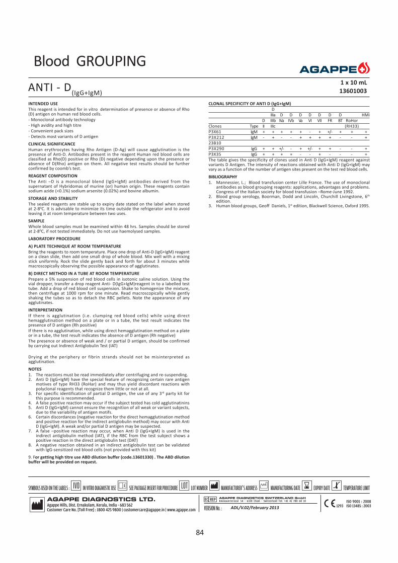
Blood GROUPING
INTENDED USE
This reagent is intended for in vitro determination of presence or absence of Rho(D) antigen on human red blood cells.
- Monoclonal antibody technology
- High avidity and high titre
- Convenient pack sizes
- Detects most variants of D antigen
CLINICAL SIGNIFICANCE
Human erythrocytes having Rho Antigen (D-Ag) will cause agglutination is thepresence of Anti-D. Antibodies present in the reagent Human red blood cells areclassified as Rho(D) positive or Rho (D) negative depending upon the presence orabsence of D(Rho) antigen on them. All negative test results should be furtherconfirmed by coomb’s test.
REAGENT COMPOSITION
The Anti –D is a monoclonal blend (IgG+IgM) antibodies derived from thesupernatant of Hybridomas of murine (or) human origin. These reagents containsodium azide (<0.1%) sodium arsenite (0.02%) and bovine albumin.
STORAGE AND STABILITY
The sealed reagents are stable up to expiry date stated on the label when storedat 2-80C. It is advisable to minimize its time outside the refrigerator and to avoidleaving it at room temperature between two uses.
SAMPLE
Whole blood samples must be examined within 48 hrs. Samples should be storedat 2-80C, if not tested immediately. Do not use haemolysed samples.
LABORATORY PROCEDURE
A) PLATE TECHNIQUE AT ROOM TEMPERATURE
Bring the reagents to room temperature. Place one drop of Anti-D (IgG+IgM) reagenton a clean slide, then add one small drop of whole blood. Mix well with a mixingstick uniformly. Rock the slide gently back and forth for about 3 minutes whilemacroscopically observing the possible appearance of agglutinates.
B) DIRECT METHOD IN A TUBE AT ROOM TEMPERATURE
Prepare a 5% suspension of red blood cells in isotonic saline solution. Using thevial dropper, transfer a drop reagent Anti- D(IgG+IgM)reagent in to a labelled testtube. Add a drop of red blood cell suspension. Shake to homogenize the mixture,then centrifuge at 1000 rpm for one minute. Read macroscopically while gentlyshaking the tubes so as to detach the RBC pellets. Note the appearance of anyagglutinates.
INTERPRETATION
If there is agglutination (i.e. clumping red blood cells) while using directhemagglutination method on a plate or in a tube, the test result indicates thepresence of D antigen (Rh positive)
If there is no agglutination, while using direct hemagglutination method on a plateor in a tube, the test result indicates the absence of D antigen (Rh negative)
The presence or absence of weak and / or partial D antigen, should be confirmedby carrying out Indirect Antiglobulin Test (IAT)
Drying at the periphery or fibrin strands should not be misinterpreted asagglutination.
NOTES
1. The reactions must be read immediately after centrifuging and re-suspending.2. Anti D (IgG+IgM) have the special feature of recognizing certain rare antigen
motives of type RH33 (RoHar) and may thus yield discordant reactions withpolyclonal reagents that recognize them little or not at all.
3. For specific identification of partial D antigen, the use of any 3rd party kit forthis purpose is recommended.
4. A false positive reaction may occur if the subject tested has cold agglutinatinins5. Anti D (IgG+IgM) cannot ensure the recognition of all weak or variant subjects,
due to the variability of antigen motifs.6. Certain discordances (negative reaction for the direct hemagglutination method
and positive reaction for the indirect antiglobulin method) may occur with AntiD (IgG+IgM). A weak and/or partial D antigen may be suspected.
7. A false –positive reaction may occur, when Anti D (IgG+IgM) is used in theindirect antiglobulin method (IAT), if the RBC from the test subject shows apositive reaction in the direct antiglobulin test (DAT)
8. A negative reaction obtained in an indirect antiglobulin test can be validatedwith IgG-sensitized red blood cells (not provided with this kit)
CLONAL SPECIFICITY OF ANTI D (IgG+IgM)
D
IIIa D D D D D D D HMi
D IIIb IVa IVb Va VI VII FR BT RoHor
Clones Type II IIIc (RH33)
P3X61 IgM + + + + + - + +/- + + +
P3X212 IgM - + - - + + + + - - +
23B10
P3X290 IgG + + +/- - + +/- + + - - +
P3X35 IgG + + + + - - + - - - +
The table gives the specificity of clones used in Anti D (IgG+IgM) reagent againstvariants D Antigen. The intensity of reactions obtained with Anti D (IgG+IgM) mayvary as a function of the number of antigen sites present on the test red blood cells.
BIBLIOGRAPHY
1. Mannessier, L.; Blood transfusion center Lille France. The use of monoclonalantibodies as blood grouping reagents: applications, advantages and problems.Congress of the Italian society for blood transfusion –Rome-June 1992.
2. Blood group serology, Boorman, Dodd and Lincoln, Churchill Livingstone, 6th
edition.3. Human blood groups, Geoff Daniels, 1st edition, Blackwell Science, Oxford 1995.
ANTI - D(IgG+IgM)
1 x 10 mL
13601003
ADL/V.02/February 2013
9. For getting high titre use ABD dilution buffer (code.13601330) . The ABD dilutionbuffer will be provided on request.
84

Blood GROUPING
INTENDED USE
This reagent is intended for in vitro determination of presence or absence of Rho (D)antigen on human red blood cells.
- Monoclonal IgM Antibodies
- High avidity and high titre
- Convenient pack sizes
PRINCIPLE
The test procedure recommended for the use of this antisera are based upon theagglutination (clumping) of red blood cells carrying the D antigen in the presence ofan IgM Anti-D antibody. All negative test results should be further confirmed by Dutest.
REAGENT COMPOSITION
The reagents contain monoclonal antibodies. The monoclonal antibodies are derivedfrom the supernantants of in vitro cultures of hybridomas of murine origin. Thesereagents contain sodium azide (<0.1%) sodium arsenite (0.02%) and bovine albumin.
STORAGE AND STABILITY
The reagents must be stored between +2oC and +80C. It is advisable to minimize itstime outside the refrigerator and to avoid leaving it at room temperature between twouses.
SAMPLE
Blood collected in anticoagulant : EDTA, heparin or citrated in a stoppered sterile tube,stored between 2-80C. At the time of the test, centrifuge the blood sample at 1200 gfor 3 minutes. Do not use hemolysed samples
LABORATORY PROCEDURE
A) PLATE TECHNIQUE AT ROOM TEMPERATURE
Bring the reagents to room temperature. Place one drop of Anti-D (IgM) reagent on aclean slide, then add one small drop of whole blood. Mix well with a mixing stickuniformly. Rock the slide gently back and forth for about 3 minutes whilemacroscopically observing the possible appearance of agglutination.
B) DIRECT METHOD IN A TUBE AT ROOM TEMPERATURE
Prepare a 5% suspension of red blood cells in isotonic saline solution. Using the vialdropper, transfer a drop reagent Anti- D(IgM)reagent in to a labelled test tube. Add adrop of red blood cell suspension. Shake to homogenize the mixture, then centrifugeat 1000 rpm for one minute. Read macroscopically while gently shaking the tubes soas to detach the RBC pellets. Note the appearance of any agglutination.
INTERPRETATION
If there is agglutination (ie, clumping red blood cells) with Anti-D(IgM) antigen D ispresent.
If there is no agglutination (ie, clumping is absent) it is possible to use Anti D (IgG)in a coomb’s test to rule out the presence of weak and partial D antigens.
NOTES
1. The reactions must be read immediately after centrifuging and resuspending.2. A false positive reaction may occur if the subject tested has cold agglutinins.3. Anti D (IgM) cannot ensure the recognition of weak or variant subjects, due to
the variability of antigen motifs.4. For getting high titre use ABD dilution buffer (code.13601330) . The ABD dilutionbuffer will be provided on request.
BIBLIOGRAPHY
1. Betermieux, C., Beolet, M. , Keyser, L.; A new strategy for D phenotyping withTOTEM multimonoclonal ANTI –D reagent, XX111rd I.S.B.T. Congress, July 1994.
2. Aramburu, E., Rabasa, P. , Esquiroz, R., Galarretat, B. Valoracion de un antisureoanti-D IgM-IgG monoclonal (DIAGAST) en donantes de sangre concentrationexpresividaddedli del anti geno. Hematology congress, Madrid, October 1990.
3. Mannessier, L. Blood transfusion center, Lille, France. The use of monoclonalantibodies as blood grouping reagents: applications, advantages and problems.Congress of the Italian society for blood transfusion ,Rome,June 199
ANTI - D(IgM)
1 x 10 mL
13601015
85

INTENDED USE
This reagent is intended for the in vitro determination of Prothrombin Time in citratedplasma.
CLINICAL SIGNIFICANCE
The Prothrombin Time is an indicator of the extrinsic blood coagulation mechanism.Deficiencies of Prothrombin and co factors V, VII and X give rise to a prolongedclotting time.
Presence of high levels of Heparin in the blood sample hypofibrinogenemia alsoattribute for prolonged time.
Common causes prolonged one stage prothrombin time are:
1. Therapy with coumarin or indanedione drugs.2. Obstructive jaundice3. Hemorrhagic disease of the new born4. Liver disease.Less common causes:1. Heparin therapy2. Loss of clotting proteins from blood via kidneys in renal disease.
Eg. Nephrotic syndrome3. Congenital deficiency of clotting factor(s)4. Fibrinogen deficiency5. Vitamin K deficiency
PRINCIPLE
Tissue thromboplastin in the presence of Ca++ activates extrinsic pathway of humanblood coagulation cascade. Activation time is proportional to the concentration ofindividual clotting factors taking part in the coagulation cascade. This assists inestimating cause and extent of haemorrhagic disorder.
When thromboplastin reagent is added to citrated plasma, clotting cascade isinitiated forming gel clot. The time required for clot formation would be prolongedif there is deficiency of factor (s) activity in the extrinsic pathway of the coagulationcycle.
REAGENT COMPOSITION
PROTHROMBIN TIME (PT)
TF (Rabbit origin)
Ca++
Antimicrobials
Stabilizers
PACK CONTENTS
PT Reagent 2 x 4 mL
Buffered tri sodium citrate (0.109M) 3.2% 1 x 10 mL
STORAGE AND STABILITY
The sealed reagents are stable up to the expiry date stated on the label, when storedat 2-80C.
Do not freeze.
NORMAL RANGE
Clotting time : 10 - 15 sec
Prothrombin ratio : 1 ± 0.15 (depends on methodology)
INR value : 0.8 -1.5
The clotting time of abnormal plasma will depend on the International SensitivityIndex (ISI) of the reagent lot. Normal range should be established in the user’slaboratory.
SAMPLE COLLECTION PREPARATION
Citrated plasma.
PT (S.L) 2 x 4 mL
12601003
Mix gently, 9 parts of blood in a plastic tube or siliconized glass tube containing 1part of 3.2% tri sodium citrate solution (0.109 M). Centrifuge immediately for 15min at 3000 rpm to obtain platelet poor plasma. Transfer supernatant plasma in asiliconized glass tube or plastic tube immediately, do not disturb buffy coat whilecollecting supernatant plasma. For best results the test should be done within 2hours of blood collection.
TEST PROCEDURE
The PT for each sample should be determined at least twice. This procedure pertainsto manual or semi-automated coagulation systems. Refer to your instrumentmanual for more detailed instrument specific instruction.
Manual Method
1. Gently swirl the reagent vials before use. Do not shake.2. Dispense from the vial enough PT Reagent for immediate use, into a thoroughly
clean dry test tube.3. Pre-warm the dispensed PT Reagent to 370C for 10 minutes.4. Pipette 100 µL of plasma into test cuvette at 370C, and incubate for
3 minutes.5. Add forcibly 200 µL pre-warmed PT reagent into the test cuvette.6. Start timer simultaneously and record the clotting time in seconds.
CALCULATION
The ISI as well as nature of preparation varies worldwide, it is recommended toexpress results of the coagulation assays in terms of INR and this can be derivedfrom the accompanying INR conversion table or calculated from the belowmentioned relationship
(International Normalized Ratio) INR = RISI where,
Mean PT of the patients plasma in sec
R(prothrombin ratio) = -----------------------------------------------------
MNPT (Mean Normal PT) for the reagent
MNPT: Each laboratory must establish its own MNPT for each lot of reagent andinstrument used. Usually plasma from at least 20 normal healthy individual shouldbe used to establish the MNPT. ISI value of the reagent is mentioned on the viallabel.
PRECAUTION
Venous blood should be directly drawn into the tube containing anticoagulant.
Ensure that the sample is free of microclots.
Separate plasma immediately by centrifugation after collection of blood.The test should be done within two hours of blood collection.
Plasma must be stored in siliconized glass tubes or plastic containers.
Avoid turbid, lipemic or hemolyzed samples.
Use clean dry micropipette tips and plasticware to dispense the reagent.
Close reagent vial and replace immediately to 2-80C after dispensing.
GUARANTEE
This product is guaranteed to perform as described on label and package insert.The manufacturer disclaims any warranty of use and sale for any other purpose.
BIBLIOGRAPHY
1. Dacie, J.V., Lewis, S.M.; Practical hematology. 19842. R Biggs, R, Mcfarlane, R. G; Human blood coagulation and its disorders 19633. Burtis, et al. Tietz: Text book of Clinical Chemistry AACC 19994. Roadck, B. F.; Diagnostic Hematology, clinical principles and applications 2nd
edition.5. John Bernad Henry: Clinical Diagnosis and management by laboratory methods
20th edition.6. Data on file of Agappe Diagnostics Ltd. Kerala.
ADL/V.02/February 2013
86

INTENDED USE
This reagent is intended for the in vitro determination of Activated PartialThromboplastin Time (APTT) in citrated plasma.
CLINICAL SIGNIFICANCE
The activated partial thromboplastin time (APTT) is used as a general screening testfor the detection of coagulation abnormalities in the intrinsic pathway. APTT is sensitiveto deficiencies or abnormalities of factors VIII, IX, XI, XII, X, V, II and I. APTT is alsosensitive to inhibitors of blood coagulation such as lupus inhibitor and fibrin/fibrinogen degradation products. APTT is the most widely used method for monitoringintravenous heparin anticoagulation therapy.
PRINCIPLE
In presence of Calcium ions cephaloplastin activates coagulation factors of intrinsicpathway in plasma leading to clot formation. Clotting time is proportional to theconcentration of factors VIII, IX, XI and XII as well as common pathway factors II, V,and X. As the reagent is prepared using one single species rabbit brain, it has therequired sensitivity to be used in heparin assays, also has better sensitivity for factorsVIII & LA.
PACK CONTENTS
APTT Reagent 1 2 x 4 mL
Calcium chloride solution 0.020 M/L
APTT Reagent 2 2 x 4 mL
Rabbit brain cephalin
Ellagic acid activator
Buffer
Stabilizers and preservatives
Tri sodium citrate (0.109 M/L) 3.2% 1 x 10 mL
STORAGE AND STABILITY
The sealed reagents are stable up to the expiry date stated on the label, when storedat 2-80C.
Do not freeze.
NORMAL RANGE
The normal values are between 21-38 seconds (at 3 minutes activation). The normaltime depends on the method, activation time, instrument etc. and must be determinedin each laboratory.
PRECAUTION
Venous blood should be directly drawn into the tube containing anticoagulant.
Ensure that the sample is free of microclots.
Separate plasma immediately by centrifugation after collection of blood.
The test should be done preferably within two hours of blood collection.
Plasma must be stored in siliconized glass tubes or plastic containers.
Avoid turbid, lipemic or hemolyzed samples.
Use clean dry micropipette tips and plasticware to dispense the reagent.
Mix the reagent (by gentle swirling) before use.
Close reagent vial and replace immediately to 2-80C after dispensing.
SAMPLE COLLECTION & PREPARATION
Citrated plasma.
Mix gently, 9 parts of blood in a plastic tube or siliconzied glass tube containing 1part of 3.2% tri-sodium citrate solution (0.109 M/L). Centrifuge immediately for 15minutes at 3000 rpm to obtain platelet poor plasma. Transfer supernatant plasma ina siliconized glass tube or plastic tube immediately; do not disturb the buffy coat whilecollecting supernatant plasma. The test should be done preferably within 2 hours ofblood collection.
TEST PROCEDURE
APTT of each sample should be determined at each level twice. This procedure pertainsto manual or semi-automated coagulation systems. Refer to your instrument manualfor more detailed instrument specific instructions
APTT 2 x 4 mL
12602001
Bring contents of the vial to room temperature and then swirl gently to mix to ahomogenized suspension. Keep the reagents at 370C for 10 minutes prior to use.
1. Gently swirl the reagent vials before use. Do not shake.2. Pre-warm enough volume of Reagent1 (CaCl
2) for immediate use, in a clean & dry
plastic test tube at 370C.3. Pipette 100 µL of test plasma or control in to a test cuvette at 370C.4. Pipette 100 µL of the pre-warmed reagent 2 (APTT Reagent) in to the test cuvette.5. Mix well and incubate at 370C for 3 minutes.6. Forcibly pipette 100 µL of pre warmed Reagent1 (CaCl
2) into the test cuvette.
7. Start a timer simultaneously and record the clotting time in seconds.Calibration curve for the determination of Heparin concentration:
Dilute Heparin (as used for treatment) with physiological saline to concentration of10 IU/mL.
Mix 0.2 mL of 10 IU/mL diluted heparin with 1.8 mL of FNP (Fresh Normal Plasma) toyield heparin standard of 1 IU/mL concentration.
Dilute the Heparin standard as prepared above (1 IU/ml) with FNP as follows.
Test Tube 1 2 3 4 5 6 7
Heparin std.(1IU/mL) 0.5 0.4 0.3 0.2 0.1 0.1 -
FNP in mL - 0.1 0.2 0.3 0.4 0.9 0.5
Heparin con.(IU/mL) 1 0.8 0.6 0.4 0.2 0.1 0
Procedure: Manual method to estimate Heparin concentration in the plasma/sample.
. Pipette 0.1mL of each Heparin dilution into clean test tubes.
. Add 0.1 mL APTT Reagent (pre-warmed at 370C). Mix and incubate for exactly3 minutes at 370C.
. Add. 0.1 mL Pre-warmed Calcium chloride (0.0290 M/L) and simultaneously startstopwatch.
. Observe clot formation carefully and note the time at the appearance of firstfibrin web.
. Plot mean of double determination in seconds against each heparinconcentration on heparin calibration graph paper provided.
. Connect points in a straight line.*Plot clotting time of sample on the calibration curve and read heparinconcentration in IU/mL
Note: Laboratories using coagulometers should follow instructions (sequence) ofthe coagulometer manufacturer.
Warranty: The product is designed to perform as described in the pack insert. Themanufacturer disclaims any implied warranty of use and sale for any other purpose.
CALCULATION
The result of APTT test can be reported directly in seconds.
BIBLIOGRAPHY
1. Dacie, J.V., Lewis, S.M.; Practical hematology. 19842. R Biggs, R, Mcfarlane, R. G; Human blood coagulation and its disorders 19633. Burtis, et al. Tietz: Text book of Clinical Chemistry AACC 19994. Roadck, B. F; Diagnostic Hematology, clinical principles and applications 2nd
edition.5. John Bernad Henry: Clinical Diagnosis and management by laboratory methods
20th edition.6. Data on file of Agappe Diagnostics Ltd. Kerala.
ADL/V.02/February 2013
87

Haemo CHEK
INTENDED USE
Diluent is intended for counting and sizing of blood cells in MINDRAY- 3 partDifferential Hematology Analyzers of BC Series. It is an azide free filtered isotonicsolution.
REAGENT COMPOSITION
Sodium chloride 3 - 5.5 g/L
Sodium sulphate anhydrous 7.5 - 11.5g/L
Buffering agents 1- 3 g/L
Anti fungal & anti bacterial Agent 0.8 - 2.5 g/L
STORAGE AND STABILITY
The sealed reagents are stable up to the expiry date stated on the label, when storedat 150C to 300C.
ONCE OPENED STABLE FOR 60 DAYS.
DIRECTIONS FOR USE
Follow the procedures given in the operation manual of Mindray 3 part differentialHematology Analyzer.
PRECAUTIONS AND WARNING
1. For in vitro diagnostic use only.2. Do not pipette by mouth.3. Do not inhale the reagent.4. If the reagent comes in contact with, eye or skin wash off immediately with large
quantity of water and seek medical attention immediately5. Do not use the reagent in any procedures other than those described herein6. Do not use containers and other materials in the package for any purpose other
than those described herein7. When discarding the reagents dispose of them according to local regulations.8. Take normal precautions required for handling laboratory reagents.
DILUENT1 x 10 Litre, 1 x 20 Litre
14001001, 14001002
ADL/V.02/February 2013
88

Haemo CHEK
INTENDED USE
EZ cleaner is intended for daily cleaning of MINDRAY- 3 part Differential HematologyAnalyzers of BC Series.
REAGENT COMPOSITION
Proteolytic enzyme 3.0 - 10 g/L
Substrate 0.3 - 1.5 g/L
Sodium chloride 3.0 - 5.0 g/L
Buffering agents 1.0 - 4.0 g/L
Anti fungal & anti bacterial agent 0.5 -2.5 g/L
STORAGE AND STABILITY
The sealed reagents are stable up to the expiry date stated on the label, when storedat 150C to 300C.
Discard the reagent if kept opened for more than 30 days.
DIRECTIONS FOR USE
Follow the procedures given in the operation manual of Mindray 3 part differentialhematology analyzer.
PRECAUTIONS AND WARNING
1. For in vitro diagnostic use only.2. Do not pipette by mouth.3. Do not inhale the reagent.4. If the reagent comes in contact with eye or skin, wash off immediately with large
amount of water and seek medical attention immediately5. Do not use the reagent described above in any procedures other than those
described herein.6. Do not use containers and other materials in the package for any purpose other
than those described herein.7. When discarding the reagents dispose of them according to local regulations.8. Attend to the normal precautions required for handling all laboratory reagents.
E-Z CLEANER4 x 50 mL
14002001
ADL/V.02/February 2013
89

Haemo CHEK
INTENDED USE
Lyse is intended for lysing of cells for WBC Count and Hemoglobin measurementin MINDRAY-3 part Differential Hematology Analyzers of BC Series.
REAGENT COMPOSITION
Quaternary ammonium salts - < 50 g/L
Non-ionic surfactant - < 15 g/L
2-Propanol - 0.1 - 1.5mL/L
STORAGE AND STABILITY
The sealed reagents are stable up to the expiry date stated on the label, when storedat 150C to 300C.
ONCE OPENED STABLE FOR 60 DAYS
DIRECTIONS FOR USE
Follow the procedures given in the operation manual of Mindray 3 part differentialhematology analyzer.
PRECAUTIONS AND WARNING
1. For in vitro diagnostic use only.2. Do not pipette by mouth.3. Do not inhale the reagent4. If the reagent comes in contact with eye or skin, wash off immediately with large
quantity of water and seek medical attention immediately5. Do not use the reagent described above in any procedures other than those
described herein.6. Do not use containers and other materials in the package for any purpose other
than those described herein.7. When discarding the reagents dispose of them according to local regulations.8. Attend to the normal precautions required for handling all laboratory reagents.
LYSE1x500 mL/2x500 mL
14003001/14003002
ADL/V.02/February 2013
90

Haemo CHEK
INTENDED USE
Probe cleaner is intended for periodical cleaning of sample probe inMINDRAY-3 partDifferential Hematology Analyzers of BC Series.
REAGENT COMPOSITION
Surfactant > 2.0 g/L
Sodium hypochlorite > 100 g/L
Sodium chloride > 100 g/L
STORAGE AND STABILITY
The sealed reagents are stable up to the expiry date stated on the label, when storedat 150C to 300C.
DIRECTIONS FOR USE
Follow the procedures given in the operation manual of Mindray 3 part differentialHematology Analyzer.
PRECAUTIONS AND WARNING
1. For in vitro diagnostic use only.2. Do not pipette by mouth.3. Do not inhale the reagent.4. If the reagent comes in contact with eye or skin, wash off immediately with large
amount of water and seek medical attention immediately5. Do not use the reagent described above in any procedures other than those
described here in.6. Do not use containers and other materials in the package for any purpose other
than those described here in.7. When discarding the reagents dispose of them according to local regulations8. Attend to the normal precautions required for handling all laboratory reagents.
PROBE CLEANER4 x 50 mL
14004001
ADL/V.02/February 2013
91

Haemo CHEK
INTENDED USE
Rinse is intended for cleaning the volumetric tubes of MINDRAY-3 part DifferentialHematology Analyzers of BC Series.
It is an azide free filtered solution.
REAGENT COMPOSITION
Sodium chloride 3.0 - 5.5 g/L
Sodium sulphate anhydrous 7.5 - 11.5 g/L
Buffering agents 1.0 - 3.0 g/L
Non ionic surfactant 5.0 - 8.0 g/L
Anti fungal & anti bacterial agent 0.8 - 2.5 g/L
STORAGE AND STABILITY
The sealed reagents are stable up to the expiry date stated on the label, when storedat 150C to 300C.
ONCE OPENED THE REAGENT IS STABLE FOR 60 DAYS
DIRECTIONS FOR USE
Follow the procedures given in the operation manual of Mindray 3 part differentialHematology Analyzer.
PRECAUTIONS AND WARNING
1. For in vitro diagnostic use only.2. Do not pipette by mouth.3. Do not inhale the reagent.4. If the reagent comes in contact with eye or skin, wash off immediately with large
quantity of water and seek medical attention immediately.5. Do not use the reagent described above in any procedures other than those
described here in.6. Do not use containers and other materials in the package for any purpose
other than those described here in.7. When discarding the reagents dispose of them according to local regulations.8. Attend to the normal precautions required for handling all laboratory reagents.
RINSE1 x 10 Litre, 1 x 20 Litre
14005001, 14005002
ADL/V.02/February 2013
92
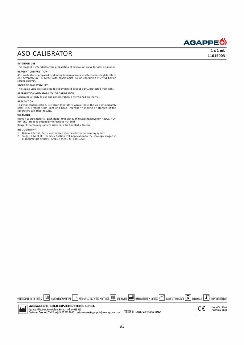
INTENDED USE
This reagent is intended for the preparation of calibration curve for ASO estimation.
REAGENT COMPOSITION
ASO calibrator is prepared by diluting human plasma which contains high levels ofAnti Streptolysin – O (ASO) with physiological saline containing 1%(w/v) bovineserum albumin.
STORAGE AND STABILITY
The sealed vials are stable up to expiry date if kept at 2-80C, protected from light.
PREPARATION AND STABILITY OF CALIBRATOR
Calibrator is ready to use and concentration is mentioned on the vial.
PRECAUTION
To avoid contamination, use clean laboratory wares. Close the vials immediatelyafter use. Protect from light and heat. Improper handling or storage of thecalibrators can affect results.
WARNING
Human source material. Each donor unit although tested negative for HbsAg, HCV,HIV(1&2) treat as potentially infectious material
Reagents containing sodium azide must be handled with care.
BIBLIOGRAPHY
1. Galuin, J.Pet al., Particle enhanced photometric immunoaasay system2. Singer, J. M.et al., The latex fixation test Application to the serologic diagnosis
of rheumatoid arthritis, Amer. J. med., 21, 888(1956)
ASO CALIBRATOR1 x 1 mL
11615003
ADL/V.01/APR 2012
93

INTENDED USE
This reagent is intended for the preparation of calibration curve for microalbuminestimation.
REAGENT COMPOSITION
A dilution of defibrinated human albumin solution with phosphate buffered saline,liquid stabilized filtered through 0.2m filter.
Sodium azide – 0.095%
STORAGE AND STABILITY
The sealed vials are stable up to expiry date at 2-80C, protected from light.
PREPARATION AND STABILITY OF CALIBRATOR
Calibrator is stable liquid and the concentration will be mentioned on the vial.Generate a calibration curve by diluting the calibrator as follows:
Preparation of calibration curve:
Prepare the following calibrator dilution using NaCI as diluent. Multiply theconcentration of the microalbumin calibrator by the corresponding factors stated inthe table below to obtain the microalbumin concentration of each dilution.
Dilution 1 2 3 4 5 6
Cali. (µL) - 10 10 25 50 100
Saline (µL) 100 150 70 75 50 -
Dil. factor 0 0.0625 0.125 0.25 0.5 1.0
Opened vials are stable up to 6 weeks when stored at 2-80C.
PRECAUTION
To avoid contamination, use clean laboratory wares. Close the vials immediatelyafter use. Protect from light and heat. Improper handling or storage of thecalibrators can affect results.
WARNING
Human source material. Each plasma donor unit although tested negative for HbsAg,HCV, HIV(1&2) treat as potentially infectious material.
Reagents containing sodium azide must be handled with care.
BIBLIOGRAPHY
Mount, N.J. Clin, Pathology, 22, 12(1986)
MICROALBUMIN CALIBRATOR1 x 1 mL
11618003
ADL/V.01/APR 2012
94

INTENDED USE
This reagent is intended for the preparation of calibration curve for CRP estimation.
REAGENT COMPOSITION
CRP calibrator is prepared by diluting defibrinated human plasma with phosphatebuffered saline.
STORAGE AND STABILITY
The sealed vials are stable up to expiry date when kept at 2-80C, protected from light.
PREPARATION AND STABILITY OF CALIBRATOR
Calibrator is stable liquid and concentration will be mentioned on the vial. Generate acalibration curve by diluting the calibrator serially.
Preparation of calibration Curve:
Prepare the following calibrator dilution using NaCI as diluent. Multiply theconcentration of the CRP calibrator by the corresponding factors stated in the tablebelow to obtain the CRP concentration of each dilution.
Dilution 1 2 3 4 5 6
Calib.(µL) - 10 10 20 50 100
Saline(µL) 100 150 70 60 50 -
Dil. Factor 0 0.0625 0.125 0.25 0.5 1.0
PRECAUTION
To avoid contamination, use clean laboratory wares. Close the vials immediately afteruse. Protect from light and heat. Improper handling or storage of the calibrators canaffect results.
WARNING
Human source material. Each donor unit although tested negative for HbsAg, HCV,HIV(1&2) treat as potentially infectious material.
Reagents containing sodium azide must be handled with care.
BIBLIOGRAPHY
1. Tillett. W.et al: Serological reactions in pneumonia with a non-protein somaticfraction of pneumoccous.J. exp. Med.. 52,561(1930)
2. Ziegenhagen G,. Drahovshy D. Klimishe Bedeutung desc-Reakriven protein. MedKlin 1983; 78:45-50
CRP CALIBRATOR1 x 2 mL
11616001
ADL/V.01/APR 2012
95

INTENDED USE
This reagent is intended for the preparation of calibration curve for Lp(a) estimation.
REAGENT COMPOSITION
Lyophilized human serum.
STORAGE AND STABILITY
The sealed vials are stable up to the expiry date at 2-80C, protected from light.
PREPARATION AND STABILITY OF CALIBRATOR
Remove the vial cap carefully and add exactly 1.0 mL of deionized water. After leavingit for more than 10 minutes, shake it gently until the content is completely dissolved.The reconstituted calibrator is stable for 7 days at 2-80C when protected from lightand heat.
PREPARATION OF CALIBRATION CURVE
Prepare the following calibrator dilution using NaCI as diluent. Multiply theconcentration of the Lp(a) calibrator by the corresponding factors stated in the tablebelow to obtain the Lp(a) concentration of each dilution.
Dilution 1 2 3 4 5 6
Calib.(µL) - 10 10 25 50 100
Saline(µL) 100 150 70 75 50 -
Dil. Factor 0 0.0625 0.125 0.25 0.5 1.0
PRECAUTION
To avoid contamination, use clean laboratory wares. Close the vials immediately afteruse. Protect from light and heat. Improper handling or storage of the calibrators canaffect results.
WARNING
Human source material. Each plasma donor unit although tested negative for HbsAg,HCV, HIV(1&2) treat as potentially infectious material.
BIBLIOGRAPHY
1. Utermann, G.et al. Lp(a) Glycoprotein phenotypes. Inheritance andrelation to Lp(a) Lipoprotein concentration in plasma.
2. J.Clin Invest., 80, 458(1987) MClea, J . W.et al. C DNA sequence ofhuman apo lipoprotein (a) is homologous, nature, 300, 132(1987)
Lp(a) CALIBRATOR1 x 1 mL
11619001
ADL/V.01/APR 2012
96

INTENDED USE
This reagent is intended for the preparation of reference curves for quantitativeimmunochemical determination of protein in human serum.
REAGENT COMPOSITION
Protein multicalibrator is a defibrinated human plasma, liquid stabilized and filteredthrough 0.2 m filter.
Sodium azide – 0.05%
STORAGE AND STABILITY
The sealed vials are stable up to expiry date at 2-80C, if protected from light.
PREPARATION AND STABILITY OF CALIBRATOR
This protein multicalibrator should be treated similar to sample/control and the testneeds to be performed as per the procedure of the respective reagent.
Opened vials are stable up to 6 weeks when stored at 2-80C.
PRECAUTION
To avoid contamination, use clean laboratory wares. Close the vials immediatelyafter use. Protect from light and heat. Improper handling or storage of thecalibrators can affect results.
WARNING
Human source material. Each plasma donor unit although tested negative for HbsAg,HCV, HIV(1&2) treat as potentially infectious material.
Reagents containing sodium azide must be handled with care.
BIBLIOGRAPHY
1. Dati, F. et al. Med. 13,87 (1889)2. Poulik, M.D. and klesis in The plasma proteinso, vol.2 second
edition.F.W. Putman Acedemic press, New York, PP 52-108
PROTEIN MULTICALIBRATOR1 x 1 mL
11614004
ADL/V.01/APR 2012
97

INTENDED USE
Multicalibrator is a calibration serum intended for calibration of clinical chemistryassays suitable for manual procedure and automated analyzers.
DESCRIPTION
Multicalibrator is lypholized calibrator based on human serum. The concentrationand activities of calibrator components have been chosen so as to ensure optimumcalibration of automated analyzers. The chemical and biological componentsincluded in this product were chosen to act in the serum matrix in a similar mannerto the corresponding components found in human serum.
REAGENT COMPOSITION
Multicalibrator 5 x 3mL
Lyophilized human serum with additives
Bacteriostatic agents and stabilizers.
(The concentration/activities of each calibrator components are Lot Specific. Pleaserefer to the attached value sheet).
PREPARATION
1. Carefully open one multicalibrator vial.2. Remove the cap and carefully lift the rubber stopper without removing it
completely, allowing air to enter the vial through the groove of the lower partof the stopper.
3. Remove the rubber stopper, avoiding any loss of the lyophilized material.4. Carefully pipette exactly the same amount of deionized water as mentioned
on the vial label, into the opened vial5. Replace the rubber stopper carefully.6. Dissolve the contents completely by occasional gentle swirling (within 30
minutes) (avoiding froth formation). DO NOT SHAKE7. Let it stand at room temperature and away from light for 10 minutes.8. Lyophilizate should be completely dissolved before use.
STORAGE AND STABILITY
Store at 2-8oC, away from light. Unopened vial is stable until expiry date stated onthe vial label.
Store the reconstituted calibrator vial tightly capped and protected from light whennot in use.
The components of the reconstituted calibrator is stable for,
At least 8 hours at 15-250C
At least 2 days at 2-80C
At least 30 days at -20oC.
Aliquot and freeze once only.
Bilirubin in the reconstituted calibrator (when stored in dark) is stable for;
At least 3 hrs at 15-250C
At least 8 hours at 2-80C
At least 14 days at -20oC.
Aliquot and freeze once only.
Acid phosphate in the reconstituted calibrator (when stored in dark) is stable for;
At least 4 hours 15-250C
At least 1 day at 2-80C
DIRECTIONS FOR USE
Multicalibrator is to be used in accordance with the directions accompanying thereagents or kits referring to the same method, and following the technical datasheet of the reagent in use. While following these directions, multicalibrator hasto be handled as patient serum.
ASSIGNED VALUES AND TRACEABILITY
The assigned values were determined using the methods mentioned in the valuesheet. Determinations were performed under standardized conditions onautomated analyzers using Agappe reagents and Master Calibrator (SRM/NIST whereever applicable).
The calibrator value specified is the median of all values obtained from separateparticipating laboratories in several independent series. Multicalibrator values aretraceable to reference materials (SRM/NIST) where applicable.
PRECAUTIONS AND WARNING
1. For in vitro diagnostic use only2. Do not pipette by mouth3. If the multicalibrator comes in contact with, eye or skin, immediately wash off
with large quantity of water, and seek medical attention immediately.4. Do not use containers and other materials in the package for any purpose other
than those described herein.5. When discarding the reagents dispose of them according to local regulations6. Attend to the normal precautions required for handling laboratory reagents.7. All human material should be considered potentially infectious. All products
derived from human blood are prepared exclusively from the blood of donorstested individually and shown to be free from HBsAg and antibodies to HCVand HIV. The testing methods applied were FDA-approved or cleared incompliance with the European Directve 98/79/EC, Annex II, List A. However, asno testing is absolute, the material should be treated just as carefully as patientspecimen.
8. Do not use multicalibrator over the expiration date/stability period.9. MSDS is available upon request10. Use clean and dry micropipette tips and glassware to dispense multicalibrator.
NOTES
1. Bacterial contamination of the reconstituted multicalibrator will causereductions in the stability of many components.(Criterion for the stability is;recovery within ±5% of initial value)
2. Bilirubin and Acid phosphatase are light sensitive.3. Creatinine Kinase is sensitive to light and temperature.4. For alkaline phosphate assay, allow the reconstituted multicalibrator to stand
for one hour at room temperature5. Inaccurate reconstitution and errors in assay technique can cause improper
calibration.6. Excessive turbidity may indicate microbial contamination in which case the vial
should not be used.
BIBLIOGRAPHY
1. Directive 2000/54/EC. Official Journal of the European Communities No. L262October 17,2000
2. ISO/DIS 17511. In vitro Diagnostic Medical Devises origin-MetrologicalTraceability of values assigned to calibrators and control materials. Geneva,International Organization for standardization 2000.
3. SRM for clinical Chemistry of the National Institute of Standards & Technology(NIST), Gaithersburg, Maryland, USA.
MULTICALIBRATOR5 x 3 mL
11610001
ADL/V.01/APR 2012
98

INTENDED USE
This reagent is intended for the preparation of calibration curve for Ferritin.
REAGENT COMPOSITION
Ferritin calibrator is prepared by diluting human plasma with phosphate bufferedsaline.
STORAGE AND STABILITY
The sealed vials are stable up to expiry date at 2-80C, protected from light.
PREPARATION AND STABILITY OF CALIBRATOR
Ferritin calibrator is available in liquid form. Concentration will be mentioned onthe vials. Opened vials are stable for 4 weeks when stored at 2-80C.
PREPARATION OF CALIBRATION CURVE
Prepare the following calibrator dilution using NaCI as diluent. Multiply theconcentration of the ferritin calibrator by the corresponding factors stated in the tablebelow to obtain the ferritin concentration of each dilution.
Dilution 1 2 3 4 5 6
Calib.(µL) - 10 10 25 50 100
Saline(µL) 100 150 70 75 50 -
Dil. Factor 0 0.0625 0.125 0.25 0.5 1.0
PRECAUTION
To avoid contamination, use clean laboratory wares. Close the vials immediatelyafter use. Protect from light and heat. Improper handling or storage of thecalibrators can affect results.
WARNING
Human source material. Each donor unit although tested negative for HbsAg, HCV,HIV(1&2) treat as potentially infectious material.
Reagents containing sodium azide must be handled with care.
BIBLIOGRAPHY
1. Cook, J.D., Lipschitz, D.A. Laughton, M.B.B., Miles, E.M. & Finch, C.A. Serumferritin as a measure of iron stores in normal subjects. A.M.J.clin Nutr 27:680-1974
2. Walters, G.O. Miller, F.M & Wormwood, M.Serum ferritn concentration and ironstores in normal subjects J. Clin pathol 26 770-1973
FERRITIN CALIBRATOR1 x 1 mL
11620003
ADL/V.01/APR 2012
99

INTENDED USE
The reagent is intended for the calibration of photometric systems with HbA1cDirect kit.
REAGENT COMPOSITION
Four levels Hemoglobin A1c calibrator set.
Lyophilized hemolysate prepared from human erythrocytes. Stabilizers to maintainhemoglobin in the reduced state for the accurate calibration of the HbA1cprocedure.
Values are LOT specific please refer to table with lot specific assay data.
STORAGE AND STABILITY
The sealed vials are stable up to the expiry date stated on the label, when storedat 2-8 0C.
PREPARATION & STABILITY OF THE CALIBRATOR
1. Open the vial carefully, avoiding any loss of the lyophilized material.2. Add exactly 500 µl of deionized water (inaccurate reconstitution of the
calibrators and error in assay technique can cause erroneous results).3. Close the vial carefully and gently mix for 10 minutes, or until all material has
dissolved, avoiding the formation of foam. Do not use shaker.4. Reconstituted calibrators should be assayed in the same manner as blood
specimens including the hemolysate procedure (please refer to HbA1c Directpack insert. Code No: 11806001
4.1 Dispense 1 mL hemolysis Reagent (R3) into a tube.4.2 Add 20 µL of calibrators and mix. Allow to stand for 5 minutes.Note: The reconstituted calibrators are stable for 30 days at 2-80C, if protected fromlight and heat. Do not freeze.
PRECAUTION
1. To avoid contamination, use clean laboratory wares.2. Close the vials immediately after use. Protect from light and heat. Improper
handing and /or storage of the calibrators can affect results.
WARNING
Human source material. Each donor unit although tested negative for HbsAg, HCV,HIV(1&2) treat as potentially infectious material.
BIBLIOGRAPHY
1. Nathan, D.M. clin. Chem.29 pp 466-469 (1983) Engbeak, F.et al. Clin.Chem. 35 pp 93-97 (1989)
2. America Diabets Association : Clinical Practice recommendations (positionstatement). Diabetes care 24 (suppl.1): S33-S55, (2001)
3. Tietz, N.W. Textbook of Clinical chemistry, W.B Saunders Company, p.794- 7795 (1999)
HbA1c DIRECT MULTICALIBRATOR4 x 0.5 mL
11604001
ADL/V.01/APR 2012
100

INTENDED USE
This reagent is intended for the preparation of calibration curve for RF estimation.
REAGENT COMPOSITION
RF calibrator prepared by diluting a high level of RF human plasma with saline. Solutioncontains 0.05 g% Sodium azide as preservative.
STORAGE AND STABILITY
The sealed vials are stable up to expiry date at 2-80C, if protected from light.
PREPARATION AND STABILITY OF CALIBRATOR
Calibrator is stable liquid and the concentration will be mentioned on the vial. Generatea calibration curve by diluting the calibrator as follows:
Preparation of calibration curve:
Prepare the following calibrator dilution using NaCI as diluent. Multiply theconcentration of the RF calibrator by the corresponding factors stated in the tablebelow to obtain the RF concentration of each dilution.
Dilution 1 2 3 4 5
Calib.(µL) 20 25 50 75 100
Saline(µL) 140 75 50 25 0
Dil. Factor 0.125 0.25 0.5 0.75 1
PRECAUTION
To avoid contamination, use clean laboratory wares. Close the vials immediately afteruse. Protect from light and heat. Improper handling or storage of the calibrators canaffect results.
WARNING
Human source material. Each plasma donor unit although tested negative for HbsAg,HCV, HIV(1&2) treat as potentially infectious material.
Reagents containing sodium azide must be handled with care.
BIBLIOGRAPHY
Frederick Wlofe et al. Arthritis and Rheumatism 1991: 34: 951-960
RF CALIBRATOR1 x 1 mL
11617001
101

INTENDED USE
HbA1c control is an assayed quality control intended for monitoring the precision oflaboratory testing procedures.
CHARACTERISTICS
This product is prepared from human blood. The control is provided in liquid form.To obtain consistent vial-to-vial assay values, the control requires proper storage andhandling as described.
STORAGE AND STABILITY
Unopened vial is stable until expiry date stated on the vial label at 2-8oC. Once controlis opened, all the analytes will be stable for 7 days when stored tightly capped at 2-8oC.
EXPECTED VALUES
The expected values mentioned in the label are specific for this lot of product.Determinations were performed under strictly standardized conditions on automatedanalyzers using Agappe reagents.
PROCEDURE
To determine HbA1c a heamolysate must be prepared for each test procedure
1.Dispense 500 µL hemolysis reagent into a tube.
2.Add 10 µL of HbA1c control and mix.
3.Allow to stand for 5 minutes or until complete lysis is evident.
Follow the same procedure instructed for Sample and Calibrator
PRECAUTIONS AND WARNING
1. For in vitro diagnostic use only2. Do not pipette by Mouth3. If the control comes in contact with eye or skin, wash off immediately with large
amount of water, and seek medical attention immediately.4. Do not use containers and other materials in the package for any purpose other
than those described herein.5. When discarding the controls dispose of them according to local regulations6. Attend to the normal precautions required for handling all laboratory reagents.7. All human material should be considered as potentially infectious. All products
derived from human blood are prepared exclusively from the blood of donorstested individually and shown to be free from HBsAg and antibodies to HCV andHIV, The testing methods applied were FDA-approved or cleared in compliance withEuropean Directive 98/79/EC, Annex II, List A. However, as no testing with absolutecertainly, the material should be treated just as carefully as patient specimen.
8. Do not use HbA1c Control over the expiration date/stability period. Use clean anddry micropipette tips and glassware to dispense HbA1c Control.
HbA1c CONTROL LEVEL 1 & 22 x 0.5 mL
11625002
102

INTENDED USE
Immunology control is an assayed quality control serum intended for monitoring theprecision of laboratory testing procedures.
CHARACTERISTICS
This product is prepared from human serum with added serum proteins. It is astabilized liquid product manufactured under rigid quality control standards. To obtainconsistent vial-to-vial assay values, the control requires proper storage and handlingas described.
STORAGE AND STABILITY
Unopened vial is stable until expiry date stated on the vial label at 2-8oC. Once opened,this product will be stable for 30 days when stored tightly capped at 2-8oC.
EXPECTED VALUES
The expected values mentioned in the value sheet are specific for this lot of product.Determinations were performed under strictly standardized conditions on automatedanalyzers using Agappe reagents.
PRECAUTIONS AND WARNING
1. For in vitro diagnostic use only2. Do not pipette by mouth3. If the control comes in contact with eye or skin, wash off immediately with large
amount of water, and seek medical attention immediately.4. Do not use containers and other materials in the package for any purpose other
than those described here in.5. When discarding the reagents dispose of them according to local regulations6. Attend to the normal precautions required for handling all laboratory reagents.7. All human material should be considered as potentially infectious. All products
derived from human blood are prepared exclusively from the blood of donorstested individually and shown to be free from HBsAg and antibodies to HCV andHIV, The testing methods applied were FDA-approved or cleared in compliance withEuropean Directive 98/79/EC, Annex II, List A. However, as no testing can rule outthe presence with absolute certainty, the material should be treated just ascarefully as patient specimen.
8. Do not use immunology control over the expiration date/stability period.9. Use clean and dry micropipette tips and glassware to dispense immunology
control.
PROTEIN CONTROL ( IMMUNOLOGY CONTROL )1 x 1 mL
11614005
103

INTENDED USE
Qualicheck Norm & Path is an assayed human serum used for quality controlchecking of clinical chemistry assays suitable for manual procedure and automatedanalyzers.
CHARACTERISTICS
Qualicheck Normal or Pathologic is lyophilized control sera based on human serum.The values given for routine methods, referred to hereafter as specified values, aredetermined under routine conditions according to the method indicated usingdifferent manual or automated assay instructions.
PREPARATION
Open the bottle very carefully, avoiding any loss of the lyophilized material andadd exactly 5 mL dist. water. Close bottle carefully and let stand for 30 minutes.Dissolve contents completely by swirl gently, avoiding the formation of foam. DONOT SHAKE.
STABILITY
The expiry date for the lyophilized control serum is given on the pack
The components of the reconstituted control serum are stable for:-
At least 8 hours at +250C
At least 3 days at +40C
At least 1 month at - 200C when frozen once.
Bilirubin in the reconstituted control serum when stored in the dark is stable for:-
At least 2 hours at + 250C
At least 6 hours at 40C
At least 2 weeks at – 200C when frozen once.
NOTES
This product has been prepared exclusively from the blood of donors testedindividually by FDA approved methods and shown to be free from HbsAg andantibodies to HIV 1 & 2.
As the risk of infection cannot be excluded with certainty, the product must behandled just as the patient specimen.
QUALICHECK NORM & PATH2 x 5 mL
11601001
ADL/V.01/APR 2012
104
ADL/V.01/APR 2012

INTENDED USE: This reagent is intended for in vitro quantitative determination ofAlpha1- Acid Glycoprotein in human serum
-Turbidimetric Immunoassay
-Linear up to 300 mg/dL
-Multipoint calibration
CLINICAL SIGNIFICANCE
Alpha-1-acid Glycoprotein is an acute-phase serum protein that is produced by theliver in response to inflammation and infection. AGP is useful in monitoring tumorrecurrence. Levels are also helpful in differentiating acute phase responses (elevatedlevels) from estrogen effects (normal or depressed levels). In addition, it is anexcellent protein in assessing in vivo hemolysis.
PRINCIPLE
The reagents containing polyclonal goat antihuman AGP when mixed with the serumsample containing AGP cause changes in absorbance, due to the development ofturbidity, which is directly proportional to the concentration of Alpha 1- AcidGlycoprotein in the sample.
REAGENT COMPOSITION
ALPHA 1- ACID GLYCOPROTEIN (AGP) R1 1 x 25 mL
Phosphate buffered saline (pH 7.43)
Polyethylene glycol (60 g/L)
Sodium azide(0.95 g/L)
ALPHA 1- ACID GLYCOPROTEIN (AGP) R2 1 x 4 mL
Phosphate buffered saline (pH 7.43)
Polyclonal goat anti-human Alpha 1- Acid Glycoprotein
(variable)
Sodium azide (0.95 g/L)
STORAGE AND STABILITY
The reagents are stable until expiry date when kept at 2-80C.
PRECAUTION
To avoid contamination, use clean laboratory wares. Use clean, dry disposablepipette tips for dispensing. Close reagent bottles immediately after use. Avoid directexposure of reagent to light.
NORMAL RANGE
It is recommended that each laboratory establish its own reference values.
The following values may be used as guide line.
Male : 50 - 130 mg/dL
Female : 40 - 120 mg/dL
SAMPLE
Use fresh serum. Dilute sample/control to 1/10 with saline.
CALIBRATION
Agappe Protein calibrator (11614002) is recommended for calibration of this assay.
Preparation of calibration curve:
Dilute the high concentrated calibrator 1/10 using saline and use this dilutedcalibrator for the preparation of calibration curve.
Prepare the following calibrator dilution using NaCl as diluent. Multiply theconcentration of the AGP calibrator by the corresponding factor stated in the tablebelow to obtain the AGP concentration of each dilution.
Dilution 1 2 3 4 5 6
1/10 dil.cal.(µL) - 10 10 25 50 100
Saline (µL) 100 150 70 75 50 -
Dil. factor 0 0.0625 0.125 0.25 0.5 1.0
Quality Control
It is recommended to use Agappe protein control(11614003) to verify theperformance of the assay. Each laboratory has to establish its own internal qualitycontrol scheme and procedure for corrective action, if controls do not recover within the acceptable tolerance.
ALPHA 1-ACID GLYCOPROTEIN1 x 25mL /1 x4 mL
12005054
PERFORMANCE CHARACTERISTICS:
Measuring Range:- 4- 300 mg/dl.
If the concentration is greater than linearity (300 mg/dL), dilute the diluted sample(1/10) with normal saline and repeat the assay. Multiply the result with dilution factor.
Prozone Effect:- >600 mg/dL
Precision in CV%:-
Low Medium High
Intra - Run 4.66 1.14 2.45
Inter - Run 2.55
Interference:-
No interference for
Hemoglobin upto 1000 mg/dL
Na- citrate upto 1000 mg/dL
Heparin upto 50 mg/dL
Bilirubin upto 20 mg/dL
Triglyceride upto 2500 mg/dL
BIBLIOGRAPHY
1. Schmid,K. in FW Putman,Editor, The plasma protein,Vol 1,second edition,Academic Press, New York 2975, ppt 184-228
2. Johnson, A.M. et al., J. Clin. Invest., 48 (1969)22933. Dati,F.et al.,Lab.Med. 13 (1989)87
ADL/V.01/APR 2012

Parameters Screen
Test AGP
No
Full Name AGP
Standard No. 6
Reaction Type Endpoint
Primary WL 340
Sec. WL
Direction Increase
Reaction Time -1 22
Incubation Time 20
Unit mg/dL
Precision 0.01
R1 300 µL
R2 30 µL
Sample 3 µL
R1 Blank
Mixed Rgt.Blank
Linearity Range 4 300
Linearity Limit
Substrate Limit
Factor
Prozone chek
q1 q2 q3 q4
PC
Reference Screen
Low
High
Calibration Screen
Rule Spline
Sensitivity
Replicates 1
Interval ( day) 0
Difference Limit
SD
Blank Response
Error Limit
Determination Coeff. 0
QC Screen
Control 1
Control 2
ALPHA 1-ACID GLYCOPROTEIN
ADL/V.01/APR 2012

INTENDED USE: This reagent is intended for in vitro quantitative determination ofcomplement C3 in human serum.
-Turbidimetric Immunoassay
-Linear up to 400 mg/dL
-Multipoint calibration
CLINICAL SIGNIFICANCE
complement C3 is the central point of the classic and alternative complementpathway.
Complement testing help to diagnose the cause of recurrent microbial infections,angioedema, or inflammation. It may be used to help diagnose and to monitor theactivity of acute or chronic autoimmune diseases such as Systemic LupusErythematosus (SLE).
Decreased levels of C3 are significant in autoimmune disease, immune infectionswith pyrogenic bacteria, bacteremia, neonatal respiratory distress syndrome andcongenital deficiencies.C3 behaves as an acute phase protein hence increased levelsmay found in acute inflammatory reactions.
PRINCIPLE
The reagents containing polyclonal goat antihuman C3 when mixed with the serumsample containing C3 cause changes in absorbance, due to the development ofturbidity, which is directly proportional to the concentration of C3 in the sample.
REAGENT COMPOSITION
C3 R1 1 x 35 mL
Phosphate buffered saline (pH 7.43)
Polyethylene glycol (40 g/L)
Sodium azide (0.95 g/L)
C3 R2 1 x 6 mL
Phosphate buffered saline (pH 7.43)
Polyclonal goat anti-human C3C (variable)
Sodium azide (0.95 g/L)
STORAGE AND STABILITY
The reagents are stable until expiry date when kept at 2-80 C
PRECAUTION
To avoid contamination, use clean laboratory wares. Use clean, dry disposablepipette tips for dispensing. Close reagent bottles immediately after use. Avoid directexposure of reagent to light.
NORMAL RANGE
It is recommended that each laboratory establish its own reference values.
The following values may be used as guide line.
Serum 75-135 mg/dL
SAMPLE
Use fresh serum. Dilute sample/control to 1/10 with saline. If the test cannot becarried out on the same day, the serum may be stored at 2-80 C for 48 hours.
CALIBRATION
Agappe Protein calibrator (11614002) is recommended for calibration of this assay.
Preparation of calibration curve:
Dilute the calibrator to 1/10 using normal saline and use this diluted calibrator forthe preparation of the calibration curve. Prepare the following calibrator dilutionusing NaCl as diluent. Multiply the concentration of the C3 calibrator by thecorresponding factors stated in the table below to obtain the C3 concentration of eachdilution.
Dilution 1 2 3 4 5 6
1/10 Cali. (µL) - 10 10 25 50 100
Saline (µL) 100 150 70 75 50 -
Dil. factor 0 0.0625 0.125 0.25 0.5 1.0
Quality Control
It is recommended to use Agappe protein control(11614003) to verify theperformance of the assay. Each laboratory has to establish its own internal qualitycontrol scheme and procedure for corrective action, if controls do not recover within the acceptable tolerance.
PERFORMANCE CHARACTERISTICS:
Measuring Range:- 20 –400 mg/dL.
If the concentration is greater than linearity (400 mg/dL), dilute the diluted sample(1/10) with normal saline and repeat the assay. Multiply the result with dilution factor.
Prozone Effect : >1000mg/dL
Precision in CV% :
Low Medium High
Intra - Run 2.82 3.43 3.28
Inter - Run 3.71 2.56
Interference:- No interference for
Hemoglobin upto 1000 mg/dL
Na-citrate upto 1000 mg/dL
Heparin upto 50 mg/dL
Bilirubin upto 20 mg/dL
Triglyceride upto 2500 mg/dL
BIBLIOGRAPHY
1. Dati, F. et al., Lab. Med.13, 87 (1989)2. Muller-Eberhard, H.H., Ann. Rev.Biochem.44, 697(1975)3. Lachmann, P.J., Hobart, M.J. and Ashton, W.P. (1973)
C31 x 35mL /1 x6mL
12005051
ADL/V.01/APR 2012

Parameters Screen
Test C3
No
Full Name C3
Standard No. 6
Reaction Type Endpoint
Primary WL 340
Sec. WL
Direction Increase
Reaction Time -1 22
Incubation Time 20
Unit mg/dL
Precision 0.01
R1 200 µL
R2 30 µL
Sample 3 µL
R1 Blank
Mixed Rgt.Blank
Linearity Range 20 400
Linearity Limit
Substrate Limit
Factor
Prozone chek
q1 q2 q3 q4
PC
Reference Screen
Low
High
Calibration Screen
Rule Spline
Sensitivity
Replicates 1
Interval ( day) 0
Difference Limit
SD
Blank Response
Error Limit
Determination Coeff. 0
QC Screen
Control 1
Control 2
C3
ADL/V.01/APR 2012
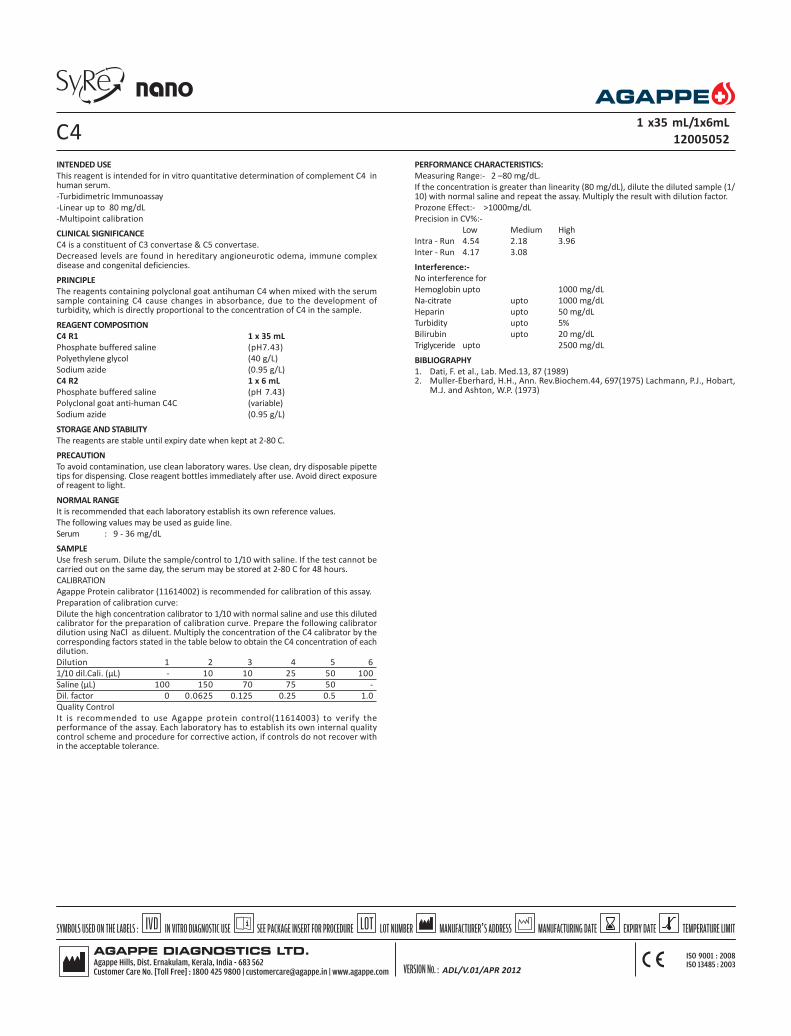
INTENDED USE
This reagent is intended for in vitro quantitative determination of complement C4 inhuman serum.
-Turbidimetric Immunoassay
-Linear up to 80 mg/dL
-Multipoint calibration
CLINICAL SIGNIFICANCE
C4 is a constituent of C3 convertase & C5 convertase.
Decreased levels are found in hereditary angioneurotic odema, immune complexdisease and congenital deficiencies.
PRINCIPLE
The reagents containing polyclonal goat antihuman C4 when mixed with the serumsample containing C4 cause changes in absorbance, due to the development ofturbidity, which is directly proportional to the concentration of C4 in the sample.
REAGENT COMPOSITION
C4 R1 1 x 35 mL
Phosphate buffered saline (pH7.43)
Polyethylene glycol (40 g/L)
Sodium azide (0.95 g/L)
C4 R2 1 x 6 mL
Phosphate buffered saline (pH 7.43)
Polyclonal goat anti-human C4C (variable)
Sodium azide (0.95 g/L)
STORAGE AND STABILITY
The reagents are stable until expiry date when kept at 2-80 C.
PRECAUTION
To avoid contamination, use clean laboratory wares. Use clean, dry disposable pipettetips for dispensing. Close reagent bottles immediately after use. Avoid direct exposureof reagent to light.
NORMAL RANGE
It is recommended that each laboratory establish its own reference values.
The following values may be used as guide line.
Serum : 9 - 36 mg/dL
SAMPLE
Use fresh serum. Dilute the sample/control to 1/10 with saline. If the test cannot becarried out on the same day, the serum may be stored at 2-80 C for 48 hours.
CALIBRATION
Agappe Protein calibrator (11614002) is recommended for calibration of this assay.
Preparation of calibration curve:
Dilute the high concentration calibrator to 1/10 with normal saline and use this dilutedcalibrator for the preparation of calibration curve. Prepare the following calibratordilution using NaCl as diluent. Multiply the concentration of the C4 calibrator by thecorresponding factors stated in the table below to obtain the C4 concentration of eachdilution.
Dilution 1 2 3 4 5 6
1/10 dil.Cali. (µL) - 10 10 25 50 100
Saline (µL) 100 150 70 75 50 -
Dil. factor 0 0.0625 0.125 0.25 0.5 1.0
Quality Control
It is recommended to use Agappe protein control(11614003) to verify theperformance of the assay. Each laboratory has to establish its own internal qualitycontrol scheme and procedure for corrective action, if controls do not recover within the acceptable tolerance.
C41 x35 mL/1x6mL
12005052
ADL/V.01/APR 2012
PERFORMANCE CHARACTERISTICS:
Measuring Range:- 2 –80 mg/dL.
If the concentration is greater than linearity (80 mg/dL), dilute the diluted sample (1/10) with normal saline and repeat the assay. Multiply the result with dilution factor.
Prozone Effect:- >1000mg/dL
Precision in CV%:-
Low Medium High
Intra - Run 4.54 2.18 3.96
Inter - Run 4.17 3.08
Interference:-
No interference for
Hemoglobin upto 1000 mg/dL
Na-citrate upto 1000 mg/dL
Heparin upto 50 mg/dL
Turbidity upto 5%
Bilirubin upto 20 mg/dL
Triglyceride upto 2500 mg/dL
BIBLIOGRAPHY
1. Dati, F. et al., Lab. Med.13, 87 (1989)2. Muller-Eberhard, H.H., Ann. Rev.Biochem.44, 697(1975) Lachmann, P.J., Hobart,
M.J. and Ashton, W.P. (1973)

Parameters Screen
Test C4
No
Full Name C4
Standard No. 6
Reaction Type Endpoint
Primary WL 340
Sec. WL
Direction Increase
Reaction Time -1 22
Incubation Time 20
Unit mg/dL
Precision 0.01
R1 200 µL
R2 30 µL
Sample 3 µL
R1 Blank
Mixed Rgt.Blank
Linearity Range 0 80
Linearity Limit
Substrate Limit
Factor
Prozone chek
q1 q2 q3 q4
PC
Refence Screen
Low
High
Calibration Screen
Rule Spline
Sensitivity
Replicates 1
Interval ( day) 0
Difference Limit
SD
Blank Response
Error Limit
Determination Coeff. 0
QC Screen
Control 1
Control 2
C4
ADL/V.01/APR 2012

INTENDED USE : This reagent is intended for in vitro quantitative determination ofApolipoprotein A1 in serum or plasma.
-Turbidimetric Immunoassay
-Linear up to 300 mg/dL
-Multipoint calibration.
CLINICAL SIGNIFICANCE
Apo A1 is the main protein component of HDL. Apo A1 activates lecithin cholesterolacyltransferase which catalyses the esterification of cholesterol this can then betransported to the liver, metabolized and excreted. People with atheroscleroticvascular changes frequently exhibit decreased levels of Apo A1. Even if theconcentrations of apolipoprotein B are normal, a decreased ApoA1 level may be arisk factor for atherosclerosis. Decreased levels of ApoA1 also occur indyslipoproteinemias, acute hepatic cirrhosis and insulin treated patients.
PRINCIPLE
Anti-human Apo A1 antisera when mixed with human serum containing Apo A1,react to cause an absorbance change , which is measured by immunoturbidometricprinciple. The change in the absorbance can be interpolated in a calibration curveprepared with different known concentrations of calibrator.
REAGENT COMPOSITION
Apo A1 R1 1 x 35 mL
Phosphate buffered saline(pH 7.43)
Polyethylene glycol 60 g/L
Detergent 0.1%
Sodium azide 0.95 g/L
Apo A1 R2 1 x 6 mL
Phosphate buffered saline(pH 7.43)
Polyclonal goat anti-human Apo-A1(Variable)
Sodium azide 0.95g/L
STORAGE AND STABILITY
The sealed reagents are stable up to expiry date stated on the label, when storedat 2 - 8oC.
PRECAUTION
To avoid contamination, use clean laboratory wares; use clean dry disposablepipette tips for dispensing, close reagent bottle immediately after use . Avoid directexposure of reagent to light.
REFERENCE RANGE
It is recommended that , each laboratory establish its own
reference values. The following values may be used as guidelines.
Men : 107 - 177 mg/dL
Women : 107 - 205 mg/dL
SAMPLE
Use fresh serum. Dilute sample/control to 1 /10 with saline
CALIBRATION
Agappe ApoA1&B calibrator (11613002) is recommended for calibration of thisassay.
Preparation of calibration curve:
Reconstitute the Apo A1 calibrator with 1 mL of distilled water. The reconstitutedcalibrator is stable for 7 days at 2-8oC.
Dilute the high concentrated calibrator 1/10 using saline and use this dilutedcalibrator for the preparation of calibration curve.
Prepare the following calibrator dilution using NaCl as diluent. Multiply theconcentration of the Apo A1 calibrator by the corresponding factors stated in the tablebelow to obtain the Apo A1 concentration of each dilution.
Dilution 1 2 3 4 5 6
1/10dilCali. (µL) - 10 10 20 50 100
Saline (µL) 100 150 70 60 50 -
Dil. factor 0 0.0625 0.125 0.25 0.5 1.0
Quality Control
It is recommended to use Agappe control to verify the performance of the assay.Each laboratory has to establish its own internal quality control scheme andprocedure for corrective action, if controls do not recover with in the acceptabletolerance.
PERFORMANCE CHARACTERISTICS:
Measuring Range:- 4 - 300 mg/dL.
If the concentration is greater than 300mg/dl dilute the diluted sample (1/10) withnormal saline and repeat the assay. Multiply the result with dilution factor.
Prozone Effect:- >5500mg/dL
Precision in CV%:-
Low Medium High
Intra - Run 3.05 1.12 1.48
Inter - Run 1.63
Hemoglobin 1000mg/dL
Triglyceride 2500 mg/dL
Bilirubin 20mg /dL
BIBLIOGRAPHY
1. Tillett. W. S.et al: serological reactions in pneumonia with a non-protein samaticfraction of pneumococcus. J.Exp.Med..52,561(1930)
2. Ziegenhagen G, Drahovshy D. klinishe Bedeutung des Creaktiven protein. Medklin 1983;78:45-50.
3. Rifal. N.Tracy.R.P.Ridker, P.M.clinical efficacy of an Automated high sensitivityC-Reactive protein Assay, Clin. chem. 45:12.
APO A11 x 35/1x6mL
12005030
ADL/V.01/APR 2012

Parameters Screen
Test APO A1
No
Full Name Apo A1
Standard No. 6
Reaction Type Endpoint
Primary WL 340
Sec. WL
Direction Increase
Reaction Time -1 22
Incubation Time 20
Unit mg/dL
Precision 0.01
R1 225 µL
R2 37 µL
Sample 3 µL
R1 Blank
Mixed Rgt.Blank
Linearity Range 4 300
Linearity Limit
Substrate Limit
Factor
Prozone chek
q1 q2 q3 q4
PC
Reference Screen
Low
High
Calibration Screen
Rule Spline
Sensitivity
Replicates 1
Interval ( day) 0
Difference Limit
SD
Blank Response
Error Limit
Determination Coeff. 0
QC Screen
Control 1
Control 2
APO A1
ADL/V.01/APR 2012

INTENDED USE : This reagent is intended for in vitro quantitative determination ofApo B in serum.
-Turbidometric Immuno assay
-High Linearity of 330 mg/dL
-Multi point calibration
CLINICAL SIGNIFICANCE
Apo B is the main protein component of LDL. It is necessary for the reaction withLDL receptors in the liver and on cell walls and thus involved in transportingcholesterol, from the liver to the vessel cells.
Elevated levels of Apo-B are frequently found in atherosclerotic vascular changesand are a risk factor for atherosclerosis.
PRINCIPLE
The reagents containing polyclonal goat antihuman Apo-B antibodies when mixedwith the serum sample containing Apo-B cause changes in absorbance due tothe development of turbidity, which is directly proportional to the concentrationof Apo-B in the sample.
REAGENT COMPOSITION
Apo B R1 1 x 35 mL
Phosphate buffered saline(pH 7.43)
Polyethyleneglycol 60 g/L
Detergent 0.1%
Sodium azide. 0.95 g/L
Apo B R2 1 x 6 mL
Polyclonal goat anti-human Apo-B(Variable)
Sodium azide 0.95 g/L
Phosphate buffer pH 7.43
STORAGE AND STABILITY
The sealed reagents are stable upto the expiry date mentioned on the label whenstored at 2-8oC.
PRECAUTION
To avoid contamination, use clean laboratory wares, such as pipette tips and testtubes. Close reagent bottle immediately after use. Avoid direct exposure of reagentto light
REFERENCE RANGE
It is recommended that, each laboratory establishes its own reference values. Thefollowing values may be used as reference.
Men : 60 - 138 mg/dL
Women : 52 - 129 mg/dL
SAMPLE
Use fresh serum . Dilute sample/ control to 1/10 in saline
CALIBRATION
Agappe Protein calibrator (11614002) is recommended for calibration of this assay.
Preparation of calibration curve:
Reconstitute the Apo B calibrator with 1 mL of distilled water. The reconstitutedcalibrator is stable for 7 days at 2-8oC. Dilute the high concentrated calibrator 1/10using saline and use this diluted calibrator for the preparation of calibration curve.
Prepare the following calibrator dilution using NaCl as diluent. Multiply theconcentration of the Apo B calibrator by the corresponding factor stated in the tablebelow to obtain the Apo B concentration of each dilution.
Dilution 1 2 3 4 5 6
1/10Dil.cal.(µL) - 10 10 20 50 100
Saline (µL) 100 150 70 60 50 -
Dil. factor 0 0.0625 0.125 0.25 0.5 1.0
APO B 1 x 35mL/1 x 6 mL
12602001
PERFORMANCE CHARACTERISTICS:
Measuring Range:- 8 - 330 mg/dL.
If the concentration is greater than 300 mg/dL, dilute the diluted sample (1/10) withnormal saline and repeat the assay. Multiply the result with dilution factor.
Prozone Effect:- >5000mg/dL
Precision in CV%:-
Low Medium High
Intra - Run 5.24 1.16 0.91
Inter - Run 1.02
Interference:-No interference for
Hemoglobin 1000mg/dL
Triglyceride 2500 mg/dL
Bilirubin 20mg /dL
BIBLIOGRAPHY
1. Tillett. W.S. et al: Serological reactions in Pneumonia a non-protein somaticfraction of pneumococcus. J. Exp.Med..52,561(1930)
2. Ziegenhagen G, Drahovshy D. klinishe Bedeutung des Creaktiven protein.Med klin 1983;78:45-50.
3. Rifal. N.Tracy.R.P.Ridker, P.M.clinical efficacy of anAutomated High sensitivity C-Reactive protein Assay Clin chem. 45:12.
ADL/V.01/APR 2012

Parameters Screen
Test APO B
No
Full Name Apo B
Standard No. 6
Reaction Type Endpoint
Primary WL 340
Sec. WL
Direction Increase
Reaction Time -1 22
Incubation Time 20
Unit mg/dL
Precision 0.01
R1 225 µL
R2 37 µL
Sample 3 µL
R1 Blank
Mixed Rgt.Blank
Linearity Range 8 330
Linearity Limit
Substrate Limit
Factor
Prozone chek
q1 q2 q3 q4
PC
Reference Screen
Low
High
Calibration Screen
Rule Spline
Sensitivity
Replicates 1
Interval ( day) 0
Difference Limit
SD
Blank Response
Error Limit
Determination Coeff. 0
QC Screen
Control 1
Control 2
APO B
ADL/V.01/APR 2012

INTENDED USE : This reagent is intended for in vitro quantitative determination ofCeruloplasmin in serum.
-Turbidimetric Immuno assay
-Linear up to 100 mg/dL
-Multipoint calibration
CLINICAL SIGNIFICANCE
Ceruloplasmin is a copper oxidase enzyme, important in regulating the ionic stateof iron and other metallic ions. Levels are decreased in hepatolenticulardegeneration or Wilson's disease and Menke's kinky hair syndrome. Levels areelevated by the acute phase response and particularly by estrogens.
PRINCIPLE
The reagents containing polyclonal goat antihuman ceruloplasmin when mixed withthe serum sample containing ceruloplasmin cause changes in absorbance due tothe development of turbidity, which is directly proportional to the concentrationof Ceruloplasmin in the sample.
REAGENT COMPOSITION
Ceruloplasmin - R1 1 x 35 mL
Phosphate buffered saline (pH 7.43)
Polyethylene glycol 40 g/L
Sodium azide 0.95 g/L
Ceruloplasmin - R2 1 x 6 mL
Phosphate buffered saline (pH 7.43)
Polyclonal goat anti- human Ceruloplasmin
Sodium azide 0.95 g/L
STORAGE AND STABILITY
The sealed reagents are stable upto the expiry date mentioned on the label whenstored at 2-8oC. Do not freeze.
PRECAUTION
To avoid contamination, use clean laboratory wares, such as pipette tips and testtubes. Close reagent bottle immediately after use. Avoid direct exposure of reagentto light.
REFERENCE RANGE
It is recommended that, each laboratory should establishes its own reference values.The following value may be used as a reference.
serum : 22 - 61 mg/dL
SAMPLE
Fresh serum.
CALIBRATION
Agappe Protein calibrator (11614002) is recommended for calibration of this assay.
Preparation of calibration curve:
Prepare the following calibrator dilution using NaCl as diluent. Multiply theconcentration of the Ceruloplasmin calibrator by the corresponding factors stated inthe table below to obtain the Ceruloplasmin concentration of each dilution.
Dilution 1 2 3 4 5 6
Cali. (µL) - 10 10 20 50 100
Saline (µL) 100 150 70 60 50 -
Dil. factor 0 0.0625 0.125 0.25 0.5 1.0
Quality Control
It is recommended to use Agappe protein control(11614003) to verify theperformance of the assay. Each laboratory has to establish its own internal qualitycontrol scheme and procedure for corrective action, if controls do not recover within the acceptable tolerance.
CERULOPLASMIN 1 x 35 mL/1 x6 mL
12005032
PERFORMANCE CHARACTERISTICS:
Measuring Range:- 4 - 100 mg/dL.
If the concentration is greater than 100 mg/dl , dilute the sample with normal salineand repeat the assay. Multiply the result with dilution factor.
Prozone Effect:- >400mg/dL
Precision in CV%:-
Low Medium High
Intra - Run 6.31 2.07 2.20
Inter - Run 3.91
Interference:-
No interference for
Hemoglobin upto 1000 mg/dL
Na - citrate upto 1000 mg/dL
Heparin upto 50 mg/dL
Bilirubin upto 20 mg/dL
Triglycerides upto 2500 mg/dL
BIBLIOGRAPHY
Poulik , M.D and Kleiss M.L in The plasma proteins”, F.W. Putman, editor,Vol. 2second edition, Academic press, New York, PP 52-108
ADL/V.01/APR 2012

Parameters Screen
Test Ceruloplasmin
No
Full Name Ceruloplasmin
Standard No. 6
Reaction Type Endpoint
Primary WL 340
Sec. WL
Direction Increase
Reaction Time -1 22
Incubation Time 20
Unit mg/dL
Precision 0.01
R1 225 µL
R2 37 µL
Sample 3 µL
R1 Blank
Mixed Rgt.Blank
Linearity Range 4 100
Linearity Limit
Substrate Limit
Factor
Prozone chek
q1 q2 q3 q4
PC
Reference Screen
Low
High
Calibration Screen
Rule Spline
Sensitivity
Replicates 1
Interval ( day) 0
Difference Limit
SD
Blank Response
Error Limit
Determination Coeff. 0
QC Screen
Control 1
Control 2
CERULOPLASMIN
ADL/V.01/APR 2012

INTENDED USE: This reagent is intended for in vitro quantitative determination ofPrealbumin in human serum
-Turbidimetric Immunoassay
-Linear up to 80 mg/dL
CLINICAL SIGNIFICANCE
Prealbumin is a serum and cerebrospinal fluid carrier of the thyroid hormonethyroxine (T4) and retinol. So the more accurate name for prealbumin istransthyretin .
Prealbumin is formed in the liver, it is a measure of hepatocyte function. Decreasedand increased levels of serum prealbumin are associated with liver disease and areaffected by the existence and degree of liver diseases.
The half life of prealbumin is approximately 2 days, making prealbumin a moretimely and sensitive indicator of protein status.
PRINCIPLE
Antibodies to prealbumin are combined with prealbumin in the patient’s serum,forming immune complexes.The immune complexes cause an increase in the lightscattering with the concentration of prealbumin in the serum. The light scatteringis measured by reading turbidity at 340 nm and 700 nm.
REAGENT COMPOSITION
Prealbumin R1(Buffer Solution) 1 x 25 mL
Tris (hydroxymethyl) aminomethane 100 mmol/L
Prealbumin R2 (Anti Serum Solution) 1 x 4 mL
Anti- human prealbumin anti serum(Variable)
STORAGE AND STABILITY
The reagents are stable until expiry date when kept at 2-80 C.
PRECAUTION
To avoid contamination, use clean laboratory wares. Use clean, dry disposablepipette tips for dispensing. Close reagent bottles immediately after use. Avoid directexposure of reagent to light.
NORMAL RANGE
It is recommended that each laboratory should establish its own reference values.
The following values may be used as guide line.
Male : 23 - 42 mg/dL
Female : 22 - 34 mg/dL
SAMPLE
Use fresh serum. Dilute sample/control to 1/10 with saline.
CALIBRATION
Agappe Protein calibrator (11614002) is recommended for calibration of this assay.
Preparation of calibration curve:
Dilute the high concentrated calibrator to 1/10 using saline and use this dilutedcalibrator for the preparation of calibration curve. Prepare the following calibratordilutions using NaCl as diluent. Multiply the concentration of the Prealbumincalibrator by the corresponding factor stated in the table below to obtain thePrealbumin concentration of each dilution.
Dilution 1 2 3 4 5 6
Cali.1/10 dil(µL) - 10 10 20 50 100
Saline (µL) 100 150 70 60 50 -
Dil. factor 0 0.0625 0.125 0.25 0.5 1.0
Quality Control
It is recommended to use Agappe protein control(11614003) to verify theperformance of the assay. Each laboratory has to establish its own internal qualitycontrol scheme and procedure for corrective action, if controls do not recover within the acceptable tolerance.
PERFORMANCE CHARACTERISTICS:
Measuring Range:- 5 –80 mg/dL.
If the concentration is greater than 80 mg/dL, dilute the diluted sample (1/10) withnormal saline and repeat the assay. Multiply the result with dilution factor.
Prozone Effect:- >200mg/dL
Precision in CV%:-
PREALBUMIN 1 x25mL /1 x 4 mL
12005055
Low Medium High
Intra - Run 5.30 3.04 3.96
Inter - Run 6.22 4.71 4.1
Interference:-
No interference for
Hemoglobin 1000mg/dL
Triglyceride 2500 mg/dL
Bilirubin 20mg /dL
BIBLIOGRAPHY
1. Japanese Journal of Clinical Medicine,53(enlarged) , 186 – 188 (1995)2. Journal of Clinical Experimental Medicine , 149 (5) , 277 – 279 (1989)
ADL/V.01/APR 2012

Parameters Screen
Test Pre Alb
No
Full Name Pre Alb
Standard No. 6
Reaction Type Endpoint
Primary WL 340
Sec. WL 670
Direction Increase
Reaction Time -1 22
Incubation Time 20
Unit mg/dL
Precision 0.01
R1 300 µL
R2 30 µL
Sample 15 µL
R1 Blank
Mixed Rgt.Blank
Linearity Range 5 80
Linearity Limit
Substrate Limit
Factor
Prozone chek
q1 q2 q3 q4
PC
Reference Screen
Low
High
Calibration Screen
Rule Spline
Sensitivity
Replicates 1
Interval ( day) 0
Difference Limit
SD
Blank Response
Error Limit
Determination Coeff. 0
QC Screen
Control 1
Control 2
PREALBUMIN
ADL/V.01/APR 2012

INTENDED USE: This reagent is intended for in vitro quantitative determination ofTransferrin in human serum
-Turbidimetric Immunoassay
-Linear up to 540 mg/dL
-Multipoint calibration
CLINICAL SIGNIFICANCE
Transferrin(TF) is synthesised in Liver. The main role of Transferrin is to deliveriron from absorption centres to all tissues. Increased serum Transferrin is associatedwith iron deficiency anemia, malignant tumor, polycythemia rubra. Increased levelsof serum transferrin are observed in patients with aplastic anemia, hemolyticanemia and hepatitis. Absence of Transferrin in the body creates a rare geneticdisorder known as atransferrinemia; a condition characterized by anemia andhemosiderosis in the heart and liver that leads to many complications includingheart failure.
PRINCIPLE
Antibodies to human TF are combined with transferrin in the patients serum,forming immune complexes. The immune complexes cause an increase in lightscattering which correlate with the concentration of TF in the serum. The lightscattering is measured by reading turbidity at 340 nm.
REAGENT COMPOSITION
Transferrin R1(Buffer solution) 1 x 25 mL
Tris (hydroxymethyl) aminomethane 100 mmol/L
Transferrin R2(Anti serum solution) 1 x 4 mL
Anti- human TF anti serum (Variable)
STORAGE AND STABILITY
The reagents are stable until expiry date when kept at 2-80 C .
PRECAUTION
To avoid contamination, use clean laboratory wares. Use clean, dry disposablepipette tips for dispensing. Close reagent bottles immediately after use. Avoid directexposure of reagent to light.
NORMAL RANGE
It is recommended that each laboratory establish its own reference values.
The following values may be used as guide line.
Male : 190 - 300 mg/dL
Female : 200 - 340 mg/dL
SAMPLE
Use fresh serum. Dilute serum/control to 1/10 with saline.
CALIBRATION
Agappe Protein calibrator (11614002) is recommended for calibration of this assay.
Preparation of calibration curve:
Dilute the high concentrated calibrator 1/10 with normal saline and use this dilutedcalibrator for the preparation of calibration curve.
Prepare the following calibrator dilutions using NaCl as diluent. Multiply theconcentration of the Transferrin calibrator by the corresponding factors stated in thetable below to obtain the Transferrin concentration of each dilution.
Dilution 1 2 3 4 5 6
1/10dilcali.(µl) - 10 10 25 50 100
Saline (µL) 100 150 70 75 50 -
Dil. factor 0 0.0625 0.125 0.25 0.5 1
Quality Control
It is recommended to use Agappe protein control(11614003) to verify theperformance of the assay. Each laboratory has to establish its own internal qualitycontrol scheme and procedure for corrective action, if controls do not recover within the acceptable tolerance.
TRANSFERRIN 1 x 25mL /1 x 4mL
12005056
PERFORMANCE CHARACTERISTICS:
Measuring Range:- 40 – 540 mg/dL.
If the concentration is greater than 540 mg/dL, dilute the diluted sample (1/10) withnormal saline and repeat the assay. Multiply the result with dilution factor.
Prozone Effect:- >1400mg/dL
Precision in CV%:-
Low Medium High
Intra - Run 4.78 1.30 0.78
Inter - Run 4.64 2.46 0.9
INTERFERING SUBSTANCES
No interference for
Hemoglobin upto 1000 mg/dL
Bilirubin upto 20 mg/dL
Lipemia upto 5 % of intralipid
BIBLIOGRAPHY
1. K. Bergstorm, et al.: J.Clin Lab Invest., 40, 637-640 (1980)2. H. Maikus, et al.: Clinica Chemica Acta, 88, 523-530(1978)
ADL/V.01/APR 2012
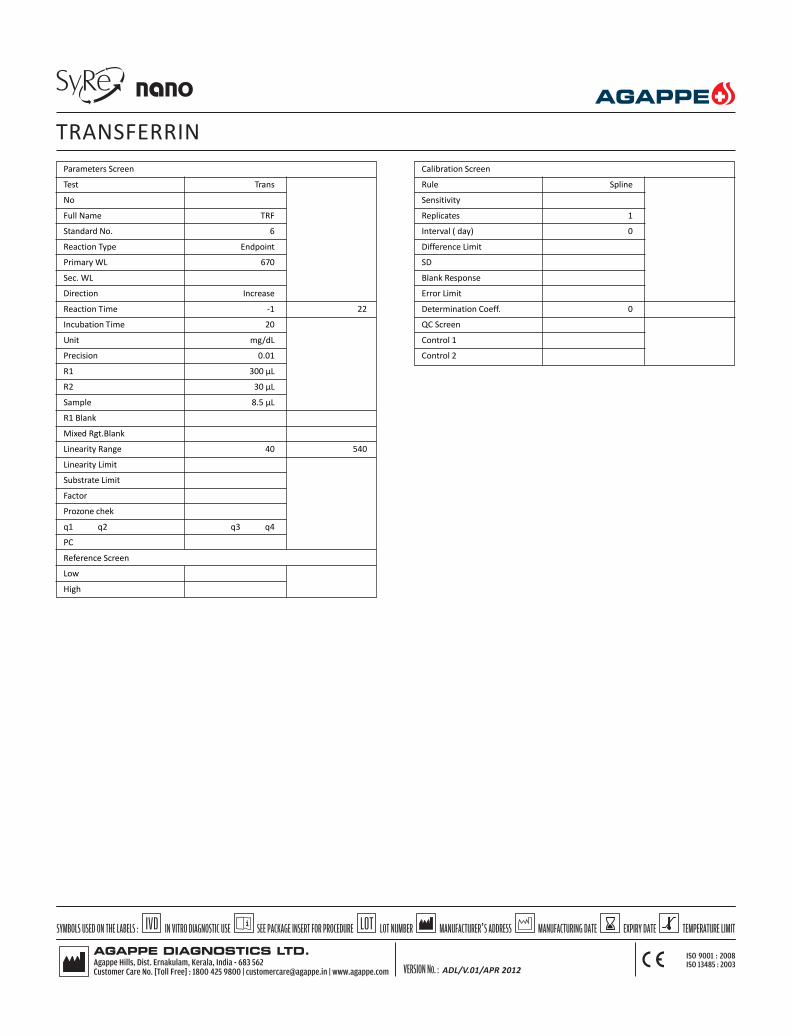
Parameters Screen
Test Trans
No
Full Name TRF
Standard No. 6
Reaction Type Endpoint
Primary WL 670
Sec. WL
Direction Increase
Reaction Time -1 22
Incubation Time 20
Unit mg/dL
Precision 0.01
R1 300 µL
R2 30 µL
Sample 8.5 µL
R1 Blank
Mixed Rgt.Blank
Linearity Range 40 540
Linearity Limit
Substrate Limit
Factor
Prozone chek
q1 q2 q3 q4
PC
Reference Screen
Low
High
Calibration Screen
Rule Spline
Sensitivity
Replicates 1
Interval ( day) 0
Difference Limit
SD
Blank Response
Error Limit
Determination Coeff. 0
QC Screen
Control 1
Control 2
TRANSFERRIN
ADL/V.01/APR 2012

INTENDED USE : This reagent is intended for in vitro quantitative determination ofIgM antibodies in serum.
-Turbidimetric Immuno assay
-Linear up to 500 mg/dL
-Multipoint calibration
CLINICAL SIGNIFICANCE
IgM is important in early response to infections . The measurement of IgM isimportant for typing immuno deficiencies and myelomas. IgM plays an importantrole in the humoral defense of the body. Serum levels may be increased in all kindof acute infections. Elevated levels in cord serum suggest clinical infection in thenew born.
PRINCIPLE
Antibodies to IgM are combined with IgM in the patient’s serum, forming immunecomplexes. The immune complexes cause an increase in light scattering whichcorrelate with the concentration of IgM in the serum. The light scattering ismeasured by reading turbidity at 340 nm and 700 nm.
REAGENT COMPOSITION
IgM R1 (Buffer solution) 1 x 30 mL
Tris(hydroxymethyl)aminomethane 100 mmol/L
IgM R2 (Antiserum solution) 1 x 10 mL
Anti-human IgM antiserum
STORAGE AND STABILITY
The sealed reagents are stable upto the expiry date mentioned on the label whenstored at 2-8 0C. Do not freeze.
PRECAUTION
To avoid contamination, use clean laboratory wares. Avoid direct exposure ofreagent to light.
The reagent should be used according to this pack insert. If used otherwise,appropriate performance is not guaranteed.
REFERENCE RANGE
It is recommended that each laboratory establish its own reference values. Thefollowing values may be used as reference.
Serum : 35 - 220 mg/dL
SAMPLE
Fresh Serum.
CALIBRATION
Agappe Protein calibrator (11614002) is recommended for calibration of this assay.
Preparation of calibration curve:
Prepare the following calibrator dilutions using NaCl as diluent. Multiply theconcentration of the IgM calibrator by the corresponding factor stated in the tablebelow to obtain the IgM concentration of each
Dilution 1 2 3 4 5 6Cal. (µL) - 10 10 20 50 100
Saline (µL) 100 150 70 60 50 -
Dil. factor 0 0.0625 0.125 0.25 0.5 1.0
Quality Control
It is recommended to use Agappe protein control(11614003) to verify theperformance of the assay. Each laboratory has to establish its own internal qualitycontrol scheme and procedure for corrective action, if controls do not recover within the acceptable tolerance.
IgM 1 x 30/1 x 10 mL
12005037
PERFORMANCE CHARACTERISTICS:
Measuring Range:- 6 – 500 mg/dL.
If the concentration is greater than 500 mg/dL, dilute the sample with normal salineand repeat the assay. Multiply the result with dilution factor.
Prozone Effect:- >5000mg/dL
Precision in CV%:-
Low Medium High
Intra - Run 1.43 1.59 0.64
Inter - Run 3.17 3.65 2.49
Interference:-
No interference for
Hemoglobin 1000 mg/dL
Triglyceride 2500 mg/dL
Bilirubin 20mg /dL
Turbidity 5 %
BIBLIOGRAPHY
1. Otani,H. :Medical Technology,14, 965(1986)2. Rinsho Kensa Guide 1992, 269-274(1992) Published by BUNKODO CO., LTD3. Kanai’s manual of Clinical laboratory Medicine, the 30 th edition,868-
873(1993)published by KANEHARA & CO.,LTD
ADL/V.01/APR 2012

Parameters Screen
Test IgM
No
Full Name IgM
Standard No. 6
Reaction Type EP
Primary WL 340
Sec. WL 670
Direction Increase
Reaction Time -1 22
Incubation Time 18
Unit mg/dL
Precision 0.1
R1 210 µL
R2 70 µL
Sample 3 µL
R1 Blank
Mixed Rgt.Blank
Linearity Range 6 500
Linearity Limit
Substrate Limit
Factor
Prozone chek
q1 q2 q3 q4
PC
Reference Screen
Low *
High *
* Data entered by the operator
Calibration Screen
Rule Spline
Sensitivity
Replicates 1
Interval ( day) 0
Difference Limit
SD
Blank Response
Error Limit
Determination Coeff. 0
QC Screen
Control 1 *
Control 2 *
IgM
ADL/V.01/APR 2012

INTENDED USE : This reagent is intended for in vitro quantitative determination ofIgG antibodies in serum.
-Turbidimetric Immuno assay
-High linearity of 2700 mg/dL
-Multipoint calibration
CLINICAL SIGNIFICANCE
IgG is a predominant serum immunoglobulin . The measurement of IgG is importantfor typing immunodeficiencies and myelomas. Increased levels are found in chronicinfections and chronic inflammation. IgG is the only immunoglobulin which crossesthe placenta and is therefore of special importance in infants defense againstinfection.
PRINCIPLE
Antibodies to IgG are combined with IgG in the patient’s serum, forming immunecomplexes. The immune complexes cause an increase in light scattering whichcorrelate with the concentration of IgG in the serum. The light scattering ismeasured by reading turbidity at 700 nm.
REAGENT COMPOSITION
IgG R1 (Buffer solution) 1 x 15 mL
Tris(hydroxymethyl)aminomethane 100 mmol/L
IgG R2 (Antiserum solution) 1 x 15 mL
Anti-human IgG antiserum
STORAGE AND STABILITY
The sealed reagents are stable upto the expiry date mentioned on the label whenstored at 2-8 0C.
PRECAUTION
To avoid contamination, use clean laboratory wares, such as pipette tips and testtubes. Close reagent bottle immediately after use. Avoid direct exposure of reagentto light.
REFERENCE RANGE
It is recommended that , each laboratory should establish its own reference values.The following value may be used as a reference.
serum : 870 - 1700 mg/dL
SAMPLE
Use fresh serum. Dilute sample/control to 1/10 with saline.
CALIBRATION
Agappe Protein calibrator (11614002) is recommended for calibration of this assay.
Preparation of calibration curve:
Dilute the high concentration calibrator to 1/10 using normal saline and use thisdiluted calibrator for the preparation of calibration curve.
Prepare the following calibrator dilutions using NaCl as diluent. Multiply theconcentration of the IgG calibrator by the corresponding factor stated in the tablebelow to obtain the IgG concentration of each dilution.
Dilution 1 2 3 4 5 6
1/10dil.Cal(µL) - 10 10 20 50 100
Saline (µL) 100 150 70 60 50 -
Dil. factor 0 0.0625 0.125 0.25 0.5 1.0
Quality Control
It is recommended to use Agappe protein control(11614003) to verify theperformance of the assay. Each laboratory has to establish its own internal qualitycontrol scheme and procedure for corrective action, if controls do not recover within the acceptable tolerance.
IgG 1 x 15mL/1 x 15 mL
12005036
PERFORMANCE CHARACTERISTICS:
Measuring Range:- 90 –2700 mg/dL.
If the concentration is greater than 2700 mg/dL, dilute the diluted sample (1/10) withnormal saline and repeat the assay .Multiply the result with dilution factor.
Prozone Effect:- >10000mg/dL
Precision in CV%:-
Low Medium High
Intra - Run 2.5 3.25 4.18
Inter - Run 4.08 1.83
Interference:-
No interference for
Hemoglobin 1000 mg/dL
Triglyceride 2500 mg/dL
Bilirubin 20mg/dL
BIBLIOGRAPHY
1. Otani,H. :Medical Technology,14, 965(1986)2. Rinsho Kensa Guide 1992, 269-274(1992) Published by BUNKODO CO., LTD3. Kanai’s manual of Clinical laboratory Medicine, the 30 th edition,868-
873(1993)published by KANEHARA & CO.,LTD
ADL/V.01/APR 2012

Parameters Screen
Test IgG
No
Full Name IgG
Standard No. 6
Reaction Type EP
Primary WL 670
Sec. WL
Direction Increase
Reaction Time -1 22
Incubation Time 3
Unit mg/dL
Precision 0.1
R1 180 µL
R2 180 µL
Sample 4 µL
R1 Blank
Mixed Rgt.Blank
Linearity Range 90 2700
Linearity Limit
Substrate Limit
Factor
Prozone chek
q1 q2 q3 q4
PC
Reference Screen
Low *
High *
* Data entered by the operator
Calibration Screen
Rule Spline
Sensitivity
Replicates 1
Interval ( day) 0
Difference Limit
SD
Blank Response
Error Limit
Determination Coeff. 0
QC Screen
Control 1 *
Control 2 *
IgG
ADL/V.01/APR 2012

INTENDED USE : This reagent is intended for in vitro quantitative determination ofIgA antibodies, in serum.
-Turbidimetric Immuno assay
-Linear up to 600 mg/dL
-Multipoint calibration
CLINICAL SIGNIFICANCE
The measurement of IgA is important for typing immunodeficiencies and myelomas.Further more it plays a role in acute and chronic infections as first line of defence.Increased levels may be found in acute infectious hepatitis, chronic aggressivehepatitis, cryptogenic cirrhosis, active alcoholic cirrhosis, chronic infections,rheumatoid arthritis, mixed connective tissue diseases etc.
PRINCIPLE
Antibodies to IgA are combined with IgA in the patient’s serum, forming immunecomplexes. The immune complexes cause an increase in light scattering whichcorrelate with the concentration of IgA in the serum. The light scattering ismeasured by reading turbidity at 700 nm.
REAGENT COMPOSITION
IgA R1(Buffer solution) 1 x 30 mL
Tris(hydroxymethyl)aminomethane 100 mmol/L
IgA R2(Antiserum solution) 1 x 10 mL
Anti-human IgA antiserum
STORAGE AND STABILITY
The sealed reagents are stable upto the expiry date mentioned on the label whenstored at 2-8oC.
PRECAUTION
To avoid contamination, use clean laboratory wares, such as pipette tips and testtubes. Close reagent bottle immediately after use. Avoid direct exposure of reagentto light.
REFERENCE RANGE
It is recommended that , each laboratory should establish its own reference values.The following values may be used as reference.
Serum : 110 - 410 mg/dL
SAMPLE
Use fresh Serum
CALIBRATION
Agappe Protein calibrator (11614002) is recommended for calibration of this assay.
Preparation of calibration curve:
Prepare the following calibrator dilutions using NaCl as diluent. Multiply theconcentration of the IgA calibrator by the corresponding factor stated in the tablebelow to obtain the IgA concentration of each dilution.
Dilution 1 2 3 4 5 6
Cali. (µL) - 10 10 20 50 100
Saline (µL) 100 150 70 60 50 -
Dil. factor 0 0.0625 0.125 0.25 0.5 1.0
Quality Control
It is recommended to use Agappe protein control(11614003) to verify theperformance of the assay. Each laboratory has to establish its own internal qualitycontrol scheme and procedure for corrective action, if controls do not recover within the acceptable tolerance.
IgA 1 x 30mL/1 x 10mL
12005035
PERFORMANCE CHARACTERISTICS:
Measuring Range:- 1 –600 mg/dL.
If the concentration is greater than 600 mg/dL , dilute the sample with normal salineand repeat the assay .Multiply the result with dilution factor.
Prozone Effect:- >6000mg/dL
Precision in CV%:-
Low Medium High
Intra - Run 0.84 0.94 1.20
Inter - Run 1.70 1.41 1.07
Interference:-
No interference for
Hemoglobin 1000 mg/dL
Triglyceride 2500 mg/dL
Bilirubin 20mg /dL
BIBLIOGRAPHY
1. K.Bergstorm, et al.: Scand. J. Clin. Lab. Invest., 637(1980)2. Rinsho Kensa Guide 1992, 269-274(1992) Published by BUNKODO CO., LTD3. Kanai’s manual of Clinical laboratory Medicine, the 30 th edition,868-
873(1993)published by KANEHARA & CO.,LTD
ADL/V.01/APR 2012

Parameters Screen
Test IgA
No
Full Name IgA
Standard No. 6
Reaction Type EP
Primary WL 670
Sec. WL
Direction Increasing
Reaction Time -1 22
Incubation Time 3
Unit mg/dL
Precision 0.1
R1 210 µL
R2 70 µL
Sample 3 µL
R1 Blank
Mixed Rgt.Blank
Linearity Range 1 600
Linearity Limit
Substrate Limit
Factor
Prozone chek
q1 q2 q3 q4
PC
Reference Screen
Low *
High *
* Data entered by the operator
Calibration Screen
Rule Spline
Sensitivity
Replicates 1
Interval ( day) 0
Difference Limit
SD
Blank Response
Error Limit
Determination Coeff. 0
QC Screen
Control 1 *
Control 2 *
IgA
ADL/V.01/APR 2012
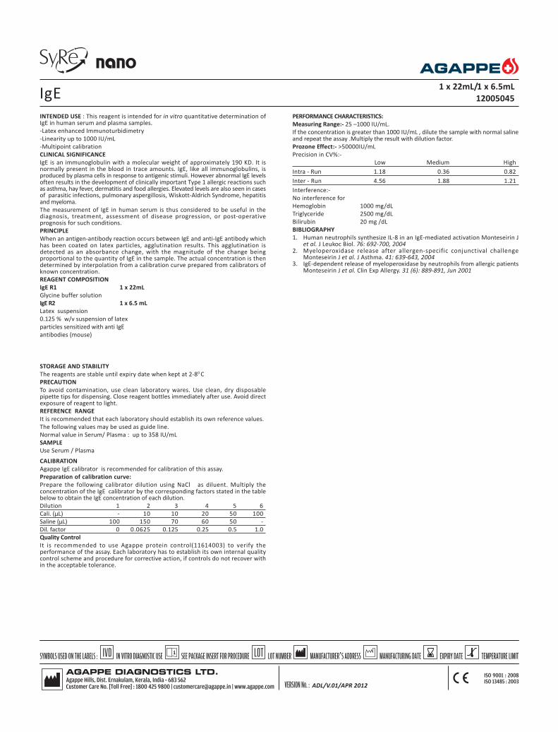
INTENDED USE : This reagent is intended for in vitro quantitative determination ofIgE in human serum and plasma samples.
-Latex enhanced Immunoturbidimetry
-Linearity up to 1000 IU/mL
-Multipoint calibration
CLINICAL SIGNIFICANCE
IgE is an immunoglobulin with a molecular weight of approximately 190 KD. It isnormally present in the blood in trace amounts. IgE, like all immunoglobulins, isproduced by plasma cells in response to antigenic stimuli. However abnormal IgE levelsoften results in the development of clinically important Type 1 allergic reactions suchas asthma, hay fever, dermatitis and food allergies. Elevated levels are also seen in casesof parasitic infections, pulmonary aspergillosis, Wiskott-Aldrich Syndrome, hepatitisand myeloma.
The measurement of IgE in human serum is thus considered to be useful in thediagnosis, treatment, assessment of disease progression, or post-operativeprognosis for such conditions.
PRINCIPLE
When an antigen-antibody reaction occurs between IgE and anti-IgE antibody whichhas been coated on latex particles, agglutination results. This agglutination isdetected as an absorbance change, with the magnitude of the change beingproportional to the quantity of IgE in the sample. The actual concentration is thendetermined by interpolation from a calibration curve prepared from calibrators ofknown concentration.
REAGENT COMPOSITION
IgE R1 1 x 22mL
Glycine buffer solution
IgE R2 1 x 6.5 mL
Latex suspension
0.125 % w/v suspension of latex
particles sensitized with anti IgE
antibodies (mouse)
STORAGE AND STABILITY
The reagents are stable until expiry date when kept at 2-80 C
PRECAUTION
To avoid contamination, use clean laboratory wares. Use clean, dry disposablepipette tips for dispensing. Close reagent bottles immediately after use. Avoid directexposure of reagent to light.
REFERENCE RANGE
It is recommended that each laboratory should establish its own reference values.
The following values may be used as guide line.
Normal value in Serum/ Plasma : up to 358 IU/mL
SAMPLE
Use Serum / Plasma
CALIBRATION
Agappe IgE calibrator is recommended for calibration of this assay.
Preparation of calibration curve:
Prepare the following calibrator dilution using NaCl as diluent. Multiply theconcentration of the IgE calibrator by the corresponding factors stated in the tablebelow to obtain the IgE concentration of each dilution.
Dilution 1 2 3 4 5 6
Cali. (µL) - 10 10 20 50 100
Saline (µL) 100 150 70 60 50 -
Dil. factor 0 0.0625 0.125 0.25 0.5 1.0
Quality Control
It is recommended to use Agappe protein control(11614003) to verify theperformance of the assay. Each laboratory has to establish its own internal qualitycontrol scheme and procedure for corrective action, if controls do not recover within the acceptable tolerance.
IgE 1 x 22mL/1 x 6.5mL
12005045
PERFORMANCE CHARACTERISTICS:
Measuring Range:- 25 –1000 IU/mL.
If the concentration is greater than 1000 IU/mL , dilute the sample with normal salineand repeat the assay .Multiply the result with dilution factor.
Prozone Effect:- >50000IU/mL
Precision in CV%:-
Low Medium High
Intra - Run 1.18 0.36 0.82
Inter - Run 4.56 1.88 1.21
Interference:-
No interference for
Hemoglobin 1000 mg/dL
Triglyceride 2500 mg/dL
Bilirubin 20 mg /dL
BIBLIOGRAPHY
1. Human neutrophils synthesize IL-8 in an IgE-mediated activation Monteseirin Jet al. J Leukoc Biol. 76: 692-700, 2004
2. Myeloperoxidase release after allergen-specific conjunctival challengeMonteseirin J et al. J Asthma. 41: 639-643, 2004
3. IgE-dependent release of myeloperoxidase by neutrophils from allergic patientsMonteseirin J et al. Clin Exp Allergy. 31 (6): 889-891, Jun 2001
ADL/V.01/APR 2012

Parameters Screen
Test IgE
No
Full Name IgE
Standard No. 6
Reaction Type FT
Primary WL 578
Sec. WL 670
Direction Increase
Reaction Time 1 9
Incubation Time 3
Unit IU/mL
Precision 0.1
R1 200µL
R2 50 µL
Sample 3 µL
R1 Blank 0
Mixed Rgt.Blank 0
Linearity Range 25 1000
Linearity Limit
Substrate Limit
Factor
Prozone chek
q1 q2 q3 q4
PC Abs
Reference Screen
Low
High
Calibration Screen
Rule Spline
Sensitivity
Replicates 1
Interval ( day)
Difference Limit
SD
Blank Response
Error Limit
Determination Coeff. 0
QC Screen
Control 1
Control 2
IgE
ADL/V.01/APR 2012

INTENDED USE : The reagent is intended for in vitro quantitative determination ofFerritin in serum
-Latex Enhanced Immunoturbidimetry
-High Linearity of 1000 ng/mL
-Multipoint calibration
CLINICAL SIGNIFICANCE
Ferritin is an iron-containing protein. It is mainly found in liver and spleen, whereits function is to eliminate and store iron in the body. It is also found in smallamounts in human serum. The serum levels tend to increase due to hepatitis andmalignant tumors. The measurement of ferritin is useful in diagnosis, treatment,assessment of disease progression and post operative prognosis of abnormal ironmetabolism and iron deficiency anaemia.
PRINCIPLE
Latex particles coated with anti-ferritin antibody are agglutinated when mixed withsamples containing Ferritin. The agglutination causes an absorbance change whichdepends on the Ferritin concentration in the sample, this can be interpolated usinga calibration curve prepared from calibrators of different concentrations.
REAGENT COMPOSITION
Ferritin - R1 1 x 20 mL
Glycine buffer
Ferritin - R2 1 x 11 mL
Suspension of latex particle bound to anti-ferritin antibodies
STORAGE AND STABILITY
The sealed reagents are stable upto the expiry date mentioned on the label whenstored at 2-8oC.
PRECAUTION
To avoid contamination, use clean laboratory wares, use clean dry disposablepipette tips for dispensing, close reagent bottle immediately after use. Avoid directexposure of reagent to light.
NORMAL RANGE
It is recommended that, each laboratory should establish its own reference values.The following value may be used as a guide line.
Male : 30 - 220 ng/mL
Females : 20 - 110 ng/mL
SAMPLE
Fresh Serum
CALIBRATION
Agappe Ferritin calibrator (11620002) is recommended for calibration of this assay.
Preparation of calibration curve:
Prepare the following calibrator dilutions using NaCl as diluent. Multiply theconcentration of the Ferritin calibrator by the corresponding factor stated in the tablebelow to obtain the Ferritin concentration of each dilution.
Dilution 1 2 3 4 5 6
Cali. (µL) - 10 10 20 50 100
Saline (µL) 100 150 70 60 50 -
Dil. factor 0 0.0625 0.125 0.25 0.5 1.0
Quality Control
It is recommended to use Agappe protein control(11614003) to verify theperformance of the assay. Each laboratory has to establish its own internal qualitycontrol scheme and procedure for corrective action, if controls do not recover within the acceptable tolerance.
FERRITIN1 x 20mL/1 x 11mL
12005044
PERFORMANCE CHARACTERISTICS:
Measuring Range:- 10 –1000 ng/mL.
If the concentration is greater than 1000 ng/mL , dilute the sample with normal salineand repeat the assay .Multiply the result with dilution factor.
Prozone Effect:- >30000ng/mL
Precision in CV%:-
Low High
Intra - Run 10 7
Inter - Run 12 10
Interference:-
No interference for
Hemoglobin 500mg/dL
Triglyceride 3000 mg/dL
Bilirubin 30mg /dL
Rheumatoid factor up to 560 IU/mL
BIBLIOGRAPHY
1. Cook, J.D., Lipschitz,D.A., Laughton, M.B.B., Miles, E.M. & Finch, C.A: Serumferritin as a measure of iron stores in normal subjects. Am.J.clin.Nutr. 27: 680,1974.
2. Walters,G.O.,Miller, F.M & Wormwood, M. : Serum ferritin concentration onand iron stores in normal subjects. J.clin.pathol. 26: 770-, 1973.
ADL/V.01/APR 2012

Parameters Screen
Test Ferittin
No
Full Name Ferittin
Standard No. 5
Reaction Type EP
Primary WL 578
Sec. WL
Direction Increase
Reaction Time 0 30
Incubation Time 3
Unit ng/L
Precision 0.1
R1 180 µL
R2 90 µL
Sample 3 µL
R1 Blank
Mixed Rgt.Blank
Linearity Range 10 1000
Linearity Limit
Substrate Limit
Factor
Prozone chek
q1 q2 q3 q4
PC
Refence Screen
Low
High
Calibration Screen
Rule Spline
Sensitivity
Replicates 1
Interval ( day) 0
Difference Limit
SD
Blank Response
Error Limit
Determination Coeff. 0
QC Screen
Control 1
Control 2
FERRITIN
ADL/V.01/APR 2012

INTENDED USE: This reagent is intended for in vitro quantitative determination ofCystatin C in human serum.
-Latex enhanced Immunoturbidimetry
-Linear of 10 mg/L
CLINICAL SIGNIFICANCE
Cystatin C is a low molecular weight (13 Da) cytoplasmic protein, functioning as aninhibitor of various cystein protease in the blood stream. Cystatin C has a stableproduction rate and is removed from the blood circulation by glomerular filtration.In healthy individuals Cystatin C is completely reabsorbed and degraded in the tubulesbut in subject with renal disorders its level in blood may be raised as high as 2 to 5times the normal values. Cystatin C is superior to serum creatinine as a marker ofglomerular filtration Rate.
PRINCIPLE
Cystatin C in the test sample binds to the specific polyclonal rabbit anti-Cystatin Cantibody, which has been adsorbed to latex particle and agglutinates. Theagglutination is detected as absorbance change at 546 nm. The magnitude of changeis proportional to the quantity of Cystatin C in the sample and its concentration isdetermined by interpolation from a calibration curve prepared from calibrators ofknown concentration.
REAGENT COMPOSITION
Cystatin C R1 1 x 23 mL
Tris buffer 1.2% (100 mM)
pH 8.5+0.3
Cystatin C R2 1 x 5.5 mL
Polystyrene latex particle coated with polyclonal anti Cystatin C antibody (rabbit)
Reagents required but not provided:
Cystatin C Calibrator (Product code -11623003)
STORAGE AND STABILITY
The sealed reagents are stable up to the expiry date stated on the label, when storedat 2- 80C.
PRECAUTIONS:
To avoid contamination, use clean laboratory wares, such as pipette tips and testtubes. Close reagent bottle immediately after use. Avoid direct exposure of reagentto light.
NORMAL RANGE
It is recommended that each laboratory establish its own reference values.
The following value may be used as guide line.
Individuals up to 50 years : 0.55 – 1.15 mg/L
Individuals above 50 years : 0.63 – 1.44 mg/L
SAMPLE
Required sample material is human serum or EDTA/ Heparinized plasma. It isrecommended to analyze the sample as fresh as possible.
CALIBRATION
Agappe Cystatin C calibrator is recommended for calibration of this assay.
Dilution of calibrator for calibration curve:
Calibration Curve (range between 0-10 mg/L). Prepare the following calibratordilution using NaCI as diluent. Multiply the concentration of the Cystatin C calibratorby the corresponding factor stated in the table below to obtain the Cystatin Cconcentration of each dilution.
Dilution 1 2 3 4 5 6
Cali. (µL) - 10 10 20 50 100
Saline (µL) 200 190 70 60 50 -
Dil. factor 0 0.05 0.125 0.25 0.5 1.0
PERFORMANCE CHARACTERISTICS:
Measuring Range:- 0.1 - 10 mg/L.
If the concentration is greater than 10 mg/L, dilute the sample with normal salineand repeat the assay .Multiply the result with dilution factor.
Prozone Effect:- >60 mg/L
Precision :
MEAN INTRA RUN INTER RUN nVALUE CV(%) CV(%)
Low human serum pool 0.77 2.16 2.54 20
High human serum pool 5.94 0.67 1.45 20
Medium human serum pool 1.45 1.58 1.95 20
Medium human serum pool 2.72 1.22 1.37 20
Low human serum pool 0.46 3.96 4.77 20
High human serum pool 3.82 1.81 3.05 20
CYSTATIN-C 1 x 23mL/1 x 5.5 mL
12005043
Interference:-
No interference for
Hemoglobin 500 mg/dL
Intrafat 1400 mg/dL
Bilirubin 25 mg /dL
BIBLIOGRAPHY
Filler G,.Bokenkamp A, Hofmann W, Le Bricon T, Martnez – Bru C, Grubb ADharnidharka VR, Kwon Stevens G.
ADL/V.01/APR 2012

Parameters Screen
Test Cys - C
No
Full Name Cystatin -C
Standard No. 6
Reaction Type EP
Primary WL 546
Sec. WL 670
Direction Increase
Reaction Time 0 18
Incubation Time 3
Unit mg/L
Precision 0.1
R1 200 µL
R2 40 µL
Sample 3 µL
R1 Blank
Mixed Rgt.Blank
Linearity Range 0.1 10
Linearity Limit
Substrate Limit
Factor
Prozone chek
q1 q2 q3 q4
PC
Reference Screen
Low
High
Calibration Screen
Rule Spline
Sensitivity
Replicates 1
Interval ( day) 0
Difference Limit
SD
Blank Response
Error Limit
Determination Coeff. 0
QC Screen
Control 1
Control 2
CYSTATIN-C
ADL/V.01/APR 2012

INTENDED USE: This reagent is intended for in vitro quantitative determination ofHbA1c in human blood.
-Latex enhanced Immunoturbidimetry
-Multipoint calibration
-Direct result in % HbA1c from analyzer
-No total Hb determination required
CLINICAL SIGNIFICANCE
HbA1c is a glycated form of haemoglobin formed by the attachment of glucoseresidues in the blood to the hemoglobin molecules. In the diabetic populationwhere blood glucose levels are abnormally elevated the level of HbA1c alsoincreases. The level of HbA1c is proportional to the level of glucose in the bloodand has been widely accepted as an indicator of the mean blood glucoseconcentration in the preceeding 6-8 weeks. It is therefore a long-term indicator ofdiabetic control. For routine use HbA1c levels should be monitored every 3-4months. However in gestational diabetes and after a change in therapy it may beuseful to measure HbA1c more frequently at 2-4 week intervals.
PRINCIPLE
This method utilizes the interaction of antigen and antibody to directly determinethe HbA1c in whole blood. Total heomoglobin and HbA1c have the samenonspecific absorption rate to latex particle. When mouse antihuman HbA1cmonoclonal antibodies are added (R2), latex HbA1c – mouse antihuman HbA1cantibody complex is formed. Agglutination occurs when goat anti mouse IgGpolyclonal antibody interacts with the monoclonal antibody. The amount ofagglutination is proportional to the amount of HbA1c absorbed onto the surfaceof latex particles. The amount of agglutination is measured as absorbance, whichis used to calculate HbA1c % from a calibration curve.
REAGENT COMPOSITION
HbA1c R1 1 x 30 mL
Latex 0.13%(w/v)
Glycine buffer 20 mmol.L
HbA1c R2A 1 x 9.5 mL
Glycine buffer 80 mmol/L
HbA1c R2B 1 x 0.5 mL
Mouse anti-human HbA1c 10 mmol/L
Monoclonal antibody
Goat anti-mouse IgG 0.05 mg/dL
(Polyclonal)
Stabilizers 0.08 mg/dL
HbA1c R3 2x 63 mL
Haemolysis Reagent
STORAGE AND STABILITY
The sealed reagents are stable up to the expiry date stated on the label, when storedat 2- 80C, protected from light.
LINEARITY
The reagent is linear up to 16% (NGSP)
NORMAL RANGE
It is recommended that each laboratory establish its own reference values.
The following value may be used as guide line.
According NGSP :
<6% for non-diabetic
< 7% for glycemic control of person with diabetes
According to IFCC:
< 4.2% for a nion-diabetic
< 5.3% for glycemic control of a person with diabetes.
PREPARATION AND STABILITY OF WORKING REAGENT
Reagent 1 & Reagent 3 are ready to use.
Reagent R2 is prepared by pouring the entire contents of the R2B vial into the R2Avial. Mix gently. This working reagent is stable for 30 days at 2-80C.
CALIBRATION
Agappe HbA1C Direct Multicalibrator ( 11604001) is recommended for calibrationof this assay. Reconstitute the calibrator with 0.5 mL distilled water and it will bestable up to 30 days at 2-80C. Do not freeze.
Quality Control
It is recommended to use Agappe HbA1C Control level 1&2 (11604003) to verifythe performance of the assay. Each laboratory has to establish its own internal qualitycontrol scheme and procedure for corrective action, if controls do not recover within the acceptable tolerance.
PRECAUTION
To avoid contamination, use clean laboratory wares. Use clean, dry disposablepipette tips for dispensing. Close reagent and standard bottles immediately afteruse.
Avoid direct exposure of working reagent to light.
SAMPLE
Whole blood, collected with EDTA
To determine HbA1c a heamolysate must be prepared for each sample
1. Dispense 1mL hemolysis reagent into a tube.2. Add 20 µL of well-mixed whole blood and mix.3. Allow to stand for 5 minutes or until complete lysis is evident.
Follow the same procedure with calibrators and controls.INTERFERENCES
No interference up to :
Ascorbic acid 50 mg/dL
Bilirubin 50 mg/dL
Triglycerides 2000 mg/dL
Carbamylated Hb 7.5 mmol/L
Acetylated Hb 5.0 mml/L
It has been reported that results may be inconsistent in patients who have thefollowing conditions: opiate addiction, lead poisoning, alcoholism, and ingestion oflarge doses of aspirin. Elevated HbF levels may lead to under estimation of HbA1c.
BIBLIOGRAPHY
1. Nathan, D.M., Clin, Chem. 29, pp.466-469 (1983)2. Engbeak, F., et al. Clin chem.35 pp. 93-97 (1989)3. American Diabetes Association : Clinical practice recommendations (position
statement). Diabetes care 24 (suppl.1) S33-S55, (2001).4. Tietz, N.W. Textbook of Clinical Chemistry, W.B. Saunders Company,
p.794 -7795 (1999).
HbA1c DIRECT 1 x 30/1 x 9.5/1 x 0.5/2x63mL
12005029
ADL/V.01/APR 2012

Parameters Screen
Test HbA1C D
No
Full Name HbA1C Direct
Standard No. 5
Reaction Type Endpoint
Primary WL 630
Sec. WL
Direction Increase
Reaction Time -1 19
Incubation Time 18
Unit %
Precision 0.01
R1 210 µL
R2 75 µL
Sample 6 µL
R1 Blank
Mixed Rgt.Blank
Linearity Range 3 16
Linearity Limit
Substrate Limit
Factor
Prozone chek
q1 q2 q3 q4
PC
Refence Screen
Low *
High *
* Data entered by the operator
Calibration Screen
Rule Spline
Sensitivity
Replicates 1
Interval ( day) 0
Difference Limit
SD
Blank Response
Error Limit
Determination Coeff. 0
QC Screen
Control 1 *
Control 2 *
HbA1c DIRECT
ADL/V.01/APR 2012

INTENDED USE: This reagent is intended for in vitro quantitative determination ofAnti Streptolysin – O (ASO).
-Latex enhanced immunoturbidimetry
-Linear up to 800 IU/mL.
CLINICAL SIGNIFICANCE
ß-hemolytic streptococcus bacteria especially group A, C and G, produce an exotoxinknown as Streptolysin-O. People infected with this bacterium produce an antibodyknown as Anti Streptolysin-O (ASO). Measuring the levels of ASO is effective fordiagnosing, judging the progress of medical treatment and assessing the recoveryfrom diseases like rheumatic fever, acute glomerulonephritis and tonsillitis.
PRINCIPLE
When an antigen-antibody reaction occurs between ASO in the sample andstreptolysin-O which has been sensitized to latex particles, agglutination occurs. Thisagglutination results in change of absorbance and is proportional to the quantityof ASO in the sample. The actual concentration is determined by interpolation froma calibration curve prepared from calibrators of known concentration.
REAGENT COMPOSITION
ASO R 1 1 x 20 mL
Glycine buffer solution
ASO R2 1 x 11 mL
ASO Latex suspension particles coated with Streptolysin - O
STORAGE AND STABILITY
The sealed reagents are stable up to the expiry date stated on the label, when storedat 2- 80C.
PRECAUTION
To avoid contamination use clean laboratory wares. Use clean dry disposable pipettetips for dispensing. Close reagent bottles immediately after use. Avoid directexposure of reagent to light.
NORMAL RANGE
It is recommended that each laboratory should establish its own reference values.
The following values may be used as guide line.
Serum
Adults : 200 IU/mL
children < 5 years : 100 IU/mL
CALIBRATION
Agappe ASO Calibrator ( Product code-11615002) is recommended for calibrationof this assay.
Quality Control
It is recommended to use Agappe protein control(11614003) to verify theperformance of the assay. Each laboratory has to establish its own internal qualitycontrol scheme and procedure for corrective action, if controls do not recover within the acceptable tolerance.
SAMPLE
Fresh serum / plasma
ASO 1 x 20/1 x 11 mL
12005040
PERFORMANCE CHARACTERISTICS:
Measuring Range:- 20–800 IU/mL.
If the concentration is greater than 800 IU/mL, dilute the sample with normal salineand repeat the assay. Multiply the result with dilution factor.
Prozone Effect:- >5600 IU/mL
Precision in CV%:-
Medium High
Intra - Run 5.0 3.0
Inter - Run 8 5
INTERFERENCES
Do not interfere for
Hemoglobin up to 500 mg/dL
Bilirubin up to 20 mg/dL
Intrafat up to 5000 mg/dL
BIBLIOGRAPHY
1. Galuin, J.P. et al.: Particle enhanced photometric immune assay system, Clin.Lab Assays (pap .Annu.clin.Lab.Assays Conf.)
2. Singer J.M. et al. The latex fixation test Application to the serologic diagnosisor rheumatoid arthritis , Amer J.Med 21, 888(1956)
ADL/V.01/APR 2012

Parameters Screen
Test ASO
No
Full Name ASO
Standard No. 2
Reaction Type Endpoint
Primary WL 578
Sec. WL
Direction Increase
Reaction Time 0 15
Incubation Time 10
Unit IU/mL
Precision 0.01
R1 180µL
R2 90µL
Sample 3 µL
R1 Blank
Mixed Rgt.Blank
Linearity Range 20 800
Linearity Limit
Substrate Limit
Factor
Prozone chek
q1 q2 q3 q4
PC
Reference Screen w
High
Calibration Screen
Rule Spline
Sensitivity
Replicates 1
Interval ( day) 0
Difference Limit
SD
Blank Response
Error Limit
Determination Coeff. 0
QC Screen
Control 1
Control 2
ASO
ADL/V.01/APR 2012

INTENDED USE : This reagent is intended for in vitro quantitative determination ofC-reactive protein in human serum or plasma by immunoturbidimetry.
-Latex enhanced immunoturbidimetry
-Linear up to 200 mg/L
CLINICAL SIGNIFICANCE
CRP (C – reactive Protein) is a cytokine - induced, acute phase protein thatincreases in concentration as a result of inflammation. CRP levels in the body hasbeen used as a marker or indicator of infections and inflammation. The assay ofCRP is more sensitive than the erythrocyte sedimentation rate (ESR) and leukocytecount. The CRP levels rise and return to reference ranges more rapidly after thedisease has subsided.
PRINCIPLE
This is a latex enhanced turbidimetric immuno assay. CRP in the samples binds tospecific anti-CRP antibodies, which have been adsorbed to latex particles andagglutinates. The agglutination is proportional to the quantity of CRP in the sample.The actual concentration is then determined by interpolation from a calibrationcurve prepared from calibrators of known concentrations.
REAGENT COMPOSITION
CRP R1 1 x 20 mL
Glycine buffer
CRP R2 1 x 8 mL
Latex suspension coated with anti-CRP antibodies. (rabbit polyclonal antibody)
STORAGE AND STABILITY
The sealed reagents are stable up to the expiry date stated on the label, when storedat 2-8oC.
PRECAUTION
To avoid contamination, use clean laboratory wares. Avoid direct exposure ofreagent to light.
The reagent should be used according to this pack insert. If used otherwise,appropriate performance is not guaranteed.
NORMAL RANGE
It is recommended that each laboratory should establish its own reference values.The following value may be used as a guide line.
Serum up to 6 mg/L
SAMPLE
Fresh serum (Do not use hemolized or lipemic serum)
CALIBRATION
Agappe CRP Calibrator ( 11616002) is recommended for calibration of this assay.
Preparation of calibration curve:
Prepare the following calibrator dilutions using NaCl as diluent. Multiply theconcentration of the CRP calibrator by the corresponding factor stated in the tablebelow to obtain the CRP concentration of each dilution.
Dilution 1 2 3 4 5 6
Cali. (µL) - 10 10 20 50 100
Saline (µL) 100 150 70 60 50 -
Dil. factor 0 0.0625 0.125 0.25 0.5 1.0
Quality Control
It is recommended to use Agappe protein control(11614003) to verify theperformance of the assay. Each laboratory has to establish its own internal qualitycontrol scheme and procedure for corrective action, if controls do not recover within the acceptable tolerance.
CRP 1 x 20/1 x8 mL
12005041
PERFORMANCE CHARACTERISTICS:
Measuring Range:- 1 –200 mg/L.
If the concentration is greater than 200mg/L, dilute the sample with normal saline andrepeat the assay. Multiply the result with dilution factor.
Prozone Effect:- >1000mg/L
Precision in CV%:-
Low Medium High
Intra - Run 7.0 5.0 3.0
Inter - Run 10 8 5
Interference:-
No interference for
Hemoglobin 500mg/dL
Intrafat 500 mg/dL
Bilirubin 30mg /dL
RF up to 500 IU/mL
BIBLIOGRAPHY
1. Tillett.W.S..et al: Serological reactions in pneumonia with a non protein somaticfraction of pneumococcus.J.Exp.Med..52,561(1930).
2. Zeigenhagen G,Drahovshy D.Klinishe Bedeutung des C-reaktiven protein.Medklin1983;78:45-50.
3. Rifal.N.Tracy.R.P.Ridker,P.M.Clinical efficacy of an Automated High sensitivity CReactive protein Assay..45-12.
ADL/V.01/APR 2012

Parameters Screen
Test CRP LEIT
No
Full Name CRP LEIT
Standard No. 6
Reaction Type Endpoint
Primary WL 578
Sec. WL
Direction Increase
Reaction Time 0 15
Incubation Time 10
Unit mg/L
Precision 0.01
R1 210 µL
R2 70 µL
Sample 3 µL
R1 Blank
Mixed Rgt.Blank
Linearity Range 1 200
Linearity Limit
Substrate Limit
Factor
Prozone chek
q1 q2 q3 q4
PC
Reference Screen w
High
Calibration Screen
Rule Spline
Sensitivity
Replicates 1
Interval ( day) 0
Difference Limit
SD
Blank Response
Error Limit
Determination Coeff. 0
QC Screen
Control 1
Control 2
CRP
ADL/V.01/APR 2012
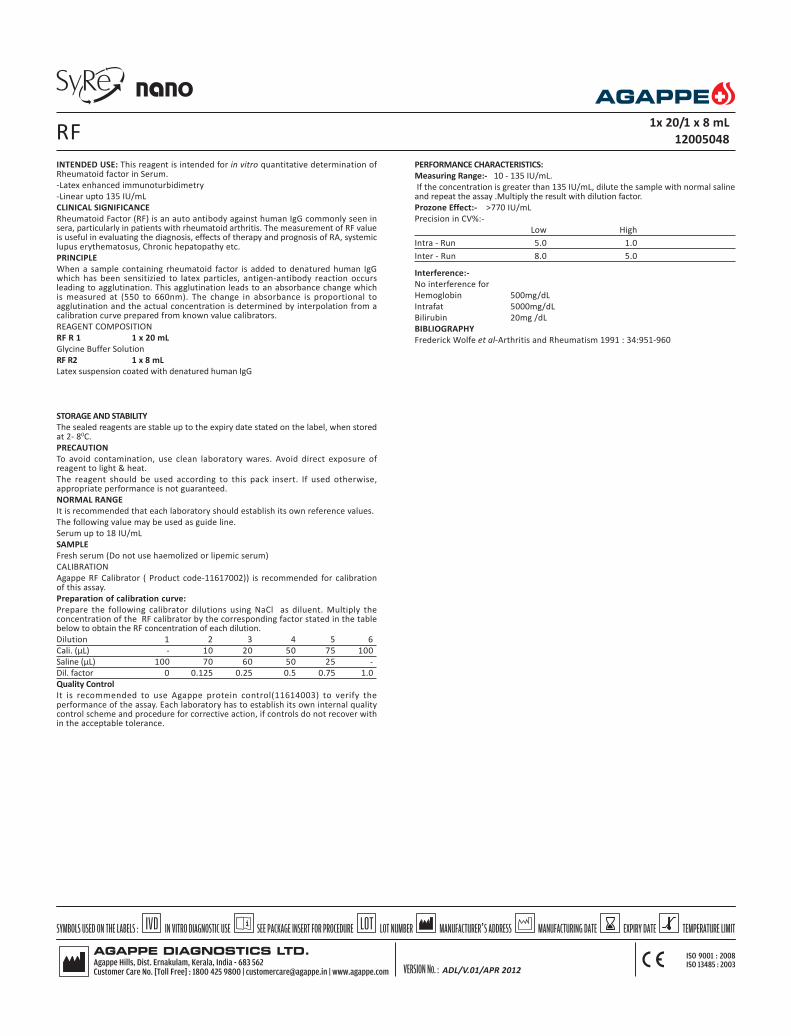
INTENDED USE: This reagent is intended for in vitro quantitative determination ofRheumatoid factor in Serum.
-Latex enhanced immunoturbidimetry
-Linear upto 135 IU/mL
CLINICAL SIGNIFICANCE
Rheumatoid Factor (RF) is an auto antibody against human IgG commonly seen insera, particularly in patients with rheumatoid arthritis. The measurement of RF valueis useful in evaluating the diagnosis, effects of therapy and prognosis of RA, systemiclupus erythematosus, Chronic hepatopathy etc.
PRINCIPLE
When a sample containing rheumatoid factor is added to denatured human IgGwhich has been sensitizied to latex particles, antigen-antibody reaction occursleading to agglutination. This agglutination leads to an absorbance change whichis measured at (550 to 660nm). The change in absorbance is proportional toagglutination and the actual concentration is determined by interpolation from acalibration curve prepared from known value calibrators.
REAGENT COMPOSITION
RF R 1 1 x 20 mL
Glycine Buffer Solution
RF R2 1 x 8 mL
Latex suspension coated with denatured human IgG
STORAGE AND STABILITY
The sealed reagents are stable up to the expiry date stated on the label, when storedat 2- 80C.
PRECAUTION
To avoid contamination, use clean laboratory wares. Avoid direct exposure ofreagent to light & heat.
The reagent should be used according to this pack insert. If used otherwise,appropriate performance is not guaranteed.
NORMAL RANGE
It is recommended that each laboratory should establish its own reference values.
The following value may be used as guide line.
Serum up to 18 IU/mL
SAMPLE
Fresh serum (Do not use haemolized or lipemic serum)
CALIBRATION
Agappe RF Calibrator ( Product code-11617002)) is recommended for calibrationof this assay.
Preparation of calibration curve:
Prepare the following calibrator dilutions using NaCl as diluent. Multiply theconcentration of the RF calibrator by the corresponding factor stated in the tablebelow to obtain the RF concentration of each dilution.
Dilution 1 2 3 4 5 6
Cali. (µL) - 10 20 50 75 100
Saline (µL) 100 70 60 50 25 -
Dil. factor 0 0.125 0.25 0.5 0.75 1.0
Quality Control
It is recommended to use Agappe protein control(11614003) to verify theperformance of the assay. Each laboratory has to establish its own internal qualitycontrol scheme and procedure for corrective action, if controls do not recover within the acceptable tolerance.
RF 1x 20/1 x 8 mL
12005048
PERFORMANCE CHARACTERISTICS:
Measuring Range:- 10 - 135 IU/mL.
If the concentration is greater than 135 IU/mL, dilute the sample with normal salineand repeat the assay .Multiply the result with dilution factor.
Prozone Effect:- >770 IU/mL
Precision in CV%:-
Low High
Intra - Run 5.0 1.0
Inter - Run 8.0 5.0
Interference:-
No interference for
Hemoglobin 500mg/dL
Intrafat 5000mg/dL
Bilirubin 20mg /dL
BIBLIOGRAPHY
Frederick Wolfe et al-Arthritis and Rheumatism 1991 : 34:951-960
ADL/V.01/APR 2012
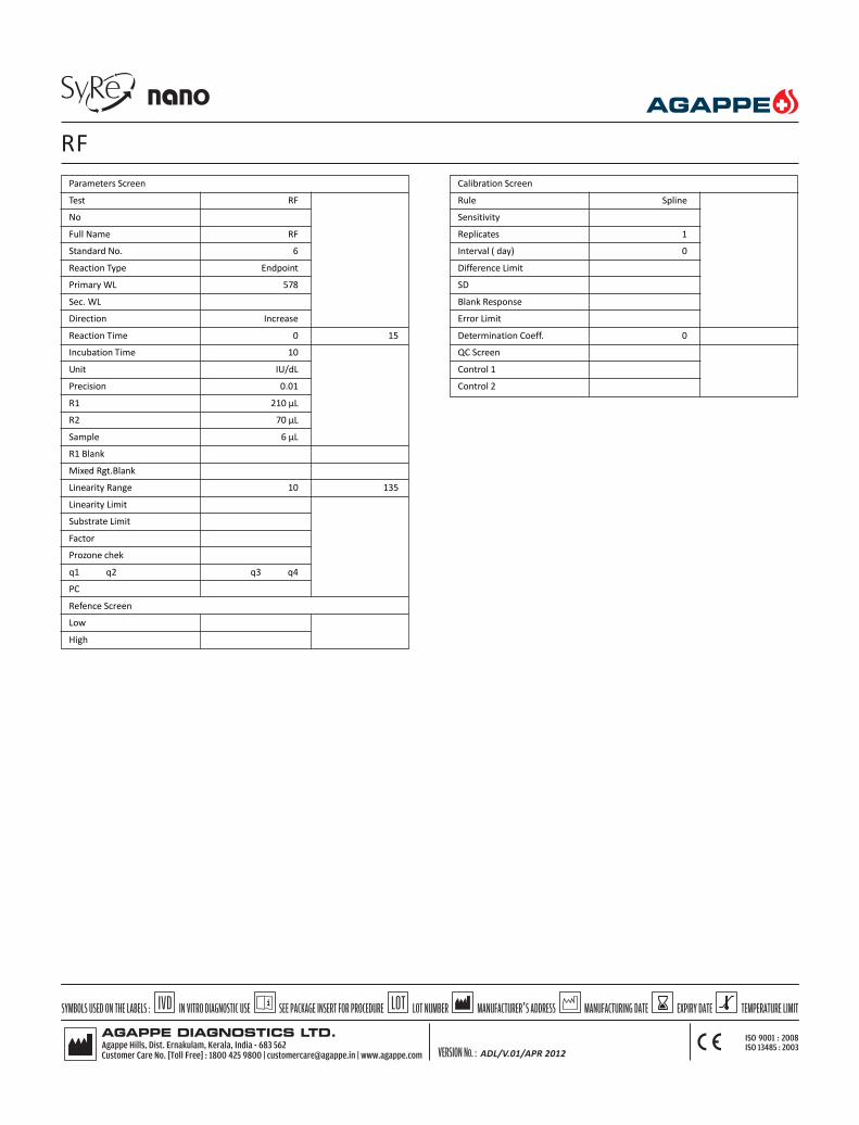
Parameters Screen
Test RF
No
Full Name RF
Standard No. 6
Reaction Type Endpoint
Primary WL 578
Sec. WL
Direction Increase
Reaction Time 0 15
Incubation Time 10
Unit IU/dL
Precision 0.01
R1 210 µL
R2 70 µL
Sample 6 µL
R1 Blank
Mixed Rgt.Blank
Linearity Range 10 135
Linearity Limit
Substrate Limit
Factor
Prozone chek
q1 q2 q3 q4
PC
Refence Screen
Low
High
Calibration Screen
Rule Spline
Sensitivity
Replicates 1
Interval ( day) 0
Difference Limit
SD
Blank Response
Error Limit
Determination Coeff. 0
QC Screen
Control 1
Control 2
RF
ADL/V.01/APR 2012

INTENDED USE: This reagent is intended for in vitro quantitative determination ofLipoprotein (a) in serum.
-Multipoint calibration with fixed time mode
-Linearity up to 80 mg/dL
CLINICAL SIGNIFICANCE
Lp(a) is a low density lipoprotein like particle containing apoliprotein B-100disulphide-linked to one large glycoprotein called apoliprotein(a). Many investigatorshave confirmed that a high lipoprotein(a) concentration represents an indicator ofrisk for cardio vascular diseases, especially when, the serum LDL-cholesterol or apoB are elevated. The quantification of Lp (a) in serum or plasma is important foridentification of individuals at risk for developing artherosclerosis.
PRINCIPLE
Latex particles coated with anti-human Lp(a) are agglutinated when mixed withsamples containing Lp(a). The agglutination causes an absorbance changedependent upon the Lp(a) contents of the patient sample, that can be interpolatedin a calibration curve prepared with different calibrators of different Lp(a) contents.
REAGENT COMPOSITION
LIPOPROTEIN (a) R1 1 x 23 mL
Buffer solution (pH 8.3)
LIPOPROTEIN (a) R2 1 x 5.5 mL
Lipoprotein (a) latex
Reagents required but not provided:
Lp(a) Calibrator ( Product code-11619002)
STORAGE AND STABILITY
The sealed reagents are stable up to the expiry date stated on the label, when storedat 2- 80C.
PRECAUTION
To avoid contamination, use clean laboratory wares.
Avoid direct exposure of working reagent to light.
NORMAL RANGE
It is recommended that each laboratory should establish its own reference values.
The following value may be used as guide line.
Serum up to 30 mg/dL
SAMPLE
Fresh Serum sample (free of haemolysis)
CALIBRATION
Agappe Lp(a) Calibrator ( 11619002) is recommended for calibration of this assay.
Calibration curve : Prepare dilutions of the Lp(a) calibrator using 9 g/L saline asdiluent:
Dilution 1 2 3 4 5
Cali. (µL) - 25 50 75 100
Saline (µL) 100 75 50 25 -
Dil. factor 0 0.25 0.5 0.75 1.0
Multiply the Lp(a) calibrator concentration by the corresponding dilution factorindicated in the table to obtain the Lp(a) concentration of the different (diluted)calibrators.
PERFORMANCE CHARACTERISTICS
1. Measurement Range: 12-80mg/dLIf the concentration is greater than linearity (80mg/dL), dilute the sample andrepeat the assay. Multiply the result with dilution factor.
2. Prozone effect: No prozone effect was detected up to 225 mg/dL.INTERFERENCES
Bilirubin : up to 427 mmol/L no interference
Hemoglobin : up to 10 g/L no interference
Lipids : up to 5 g/L no interference
BIBLIOGRAPHY
1. Gaubalz JW, et al. J.Biol Chem 1983; 258 45832 -45892. Berg K A Acta Pathol Microbiol Scand 1963:59:369-3823. Scanu AM , et al. J.Clin invest 1990;85: 1709 -1715
Lp (a) 1 x 23/1 x 5.5 mL
12005046
ADL/V.01/APR 2012

Parameters Screen
Test LP(a) LEIT
No
Full Name LP(a) LEIT
Standard No. 6
Reaction Type EP
Primary WL 670
Sec. WL
Direction Increase
Reaction Time 0 18
Incubation Time 3
Unit mg/dL
Precision 0.1
R1 200 µL
R2 40 µL
Sample 3 µL
R1 Blank
Mixed Rgt.Blank
Linearity Range 12 80
Linearity Limit
Substrate Limit
Factor
Prozone chek
q1 q2 q3 q4
PC
Refence Screen
Low *
High *
* Data entered by the operator
Calibration Screen
Rule Spline
Sensitivity
Replicates 1
Interval ( day) 0
Difference Limit
SD
Blank Response
Error Limit
Determination Coeff. 0
QC Screen
Control 1 *
Control 2 *
Lp (a)
ADL/V.01/APR 2012

INTENDED USE: This reagent is intended for in vitro quantitative determination ofmicroalbumin in human urine
-Turbidometric Immunoassay
-Linear up to 395 mg/L
-Multipoint calibration
CLINICAL SIGNIFICANCE
Albumin is normally found in the blood. When the kidneys are working properly,albumin will not be present in the urine. However, when the kidneys are damaged,small amounts of albumin leak into the urine. This condition is calledmicroalbuminuria.
Microalbuminuria is most often caused by kidney damage from diabetes. However,many other conditions can lead to kidney damage, such as high blood pressure,heart failure, cirrhosis, or systemic lupus erythematosus (SLE). If early kidneydamage is not treated, larger amounts of albumin and protein may leak into theurine. This condition is called macroalbuminuria or proteinuria, this can lead tochronic kidney disease.
PRINCIPLE
The reagents containing polyclonal goat antihuman microalbumin when mixed withthe urine sample containing microalbumin cause changes in absorbance, due tothe development of turbidity, which is directly proportional to the concentrationof microalbumin in the sample.
REAGENT COMPOSITION
Microalbumin R1 1 x 23 mL
Saline (9 g/L)
Accelerator
Sodium azide (0.95 g/L)
Microalbumin R2 1 x 5.5 mL
Phosphate buffered saline
Polyclonal goat anti-human albumin (variable)
Sodium azide (0.95 g/L)
STORAGE AND STABILITY
The reagents are stable until expiry date when kept at 2-80 C.
PRECAUTION
To avoid contamination, use clean laboratory materials. Use clean, dry disposablepipette tips for dispensing. Close reagent bottles immediately after use. Avoid directexposure of reagent to light. Do not Freeze.
NORMAL RANGE
It is recommended that each laboratory establish its own reference values.
The following values may be used as guide line.
Urine : 0 -25 mg/L (IFCC)
SAMPLE
Use fresh Urine.
CALIBRATION
Agappe Microalbumin Calibrator ( Product code-11618002) is recommended forcalibration of this assay.
PREPARATION OF CALIBRATION CURVE
Prepare the following calibrator dilutions using NaCl as diluent. Multiply theconcentration of the microalbumin calibrator by the corresponding factor stated inthe table below to obtain the microalbumin concentration of each dilution.
Dilution 1 2 3 4 5 6
Calib.(µL) - 10 10 25 50 100
Saline(µL) 100 150 70 75 50 -
Dil. Factor(µL) 0 0.0625 0.125 0.25 0.5 1.0
MICROALBUMIN 1 x 23/1 x 5.5 mL
12005047
PERFORMANCE CHARACTERISTICS:
Measuring Range:- 4 - 395 mg/L
If the concentration is greater than linearity (395 mg/L), dilute the sample with normalsaline and repeat the assay. Multiply the result with dilution factor.
Prozone Effect:- >6000 mg/L
Precision in CV%:-
Low Medium High
Intra - Run 2.28 1.8 3.04
Inter - Run 2.93 0.66 0.53
Interference:-
No interference for
Hemoglobin 1000mg/dl
Bilirubin 10mg/dL
BIBLIOGRAPHY
1. Mount,J., J. Clin. Pathology, 22, 12 (1986)2. Schmidtz, A., et al., diabetic Medicine, 5 , 126 (1988)
ADL/V.01/APR 2012

Parameters Screen
Test MicroAlb
No
Full Name Micro Albumin
Standard No. 6
Reaction Type Fixed time
Primary WL 340
Sec. WL
Direction Increase
Reaction Time -1 22
Incubation Time 20
Unit mg/L
Precision 0.01
R1 200 µL
R2 40 µL
Sample 3 µL
R1 Blank
Mixed Rgt.Blank
Linearity Range 4 395
Linearity Limit
Substrate Limit
Factor
Prozone chek
q1 q2 q3 q4
PC
Refence Screen
Low
High
Calibration Screen
Rule Spline
Sensitivity
Replicates 1
Interval ( day) 0
Difference Limit
SD
Blank Response
Error Limit
Determination Coeff. 0
QC Screen
Control 1
Control 2
MICROALBUMIN
ADL/V.01/APR 2012
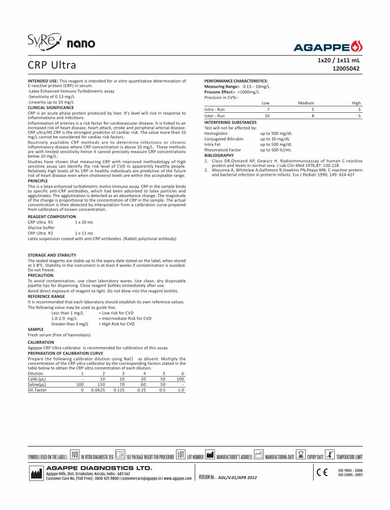
INTENDED USE: This reagent is intended for in vitro quantitative determination ofC-reactive protein (CRP) in serum.
-Latex Enhanced Immuno Turbidimetric assay
-Sensitivity of 0.13 mg/L
-Linearity up to 10 mg/L
CLINICAL SIGNIFICANCE
CRP is an acute phase protein produced by liver. It’s level will rise in response toinflammations and infections.
Inflammation of arteries is a risk factor for cardiovascular disease. It is linked to anincreased risk of heart disease, heart attack, stroke and peripheral arterial disease.CRP ultra/HS CRP is the strongest predictor of cardiac risk. The value more than 10mg/L cannot be considered for cardiac risk factors.
Routinely available CRP methods are to determine infections or chronicinflammatory disease where CRP concentration is above 10 mg/L. These methodsare with limited sensitivity hence it cannot precisely measure CRP concentrationsbelow 10 mg/L.
Studies have shown that measuring CRP with improved methodology of highsensitive assay can identify the risk level of CVD in apparently healthy people.Relatively high levels of hs CRP in healthy individuals are predictive of the futurerisk of heart disease even when cholesterol levels are within the acceptable range.
PRINCIPLE
This is a latex-enhanced turbidimetric invitro immuno assay. CRP in the sample bindsto specific anti-CRP antibodies, which had been adsorbed to latex particles andagglutinates. The agglutination is detected as an absorbance change. The magnitudeof the change is proportional to the concentration of CRP in the sample. The actualconcentration is then detected by interpolation from a calibration curve preparedfrom calibrators of known concentration.
REAGENT COMPOSITION
CRP Ultra R1 1 x 20 mL
Glycine buffer
CRP Ultra R2 1 x 11 mL
Latex suspension coated with anti-CRP antibodies. (Rabbit polyclonal antibody)
STORAGE AND STABILITY
The sealed reagents are stable up to the expiry date stated on the label, when storedat 2-80C. Stability in the instrument is at least 4 weeks if contamination is avoided.Do not freeze.
PRECAUTION
To avoid contamination, use clean laboratory wares. Use clean, dry disposablepipette tips for dispensing. Close reagent bottles immediately after use.
Avoid direct exposure of reagent to light. Do not blow into the reagent bottles.
REFERENCE RANGE
It is recommended that each laboratory should establish its own reference values.
The following value may be used as guide line.
Less than 1 mg/L = Low risk for CVD
1.0-2.9 mg/L = Intermediate Risk for CVD
Greater than 3 mg/L = High Risk for CVD
SAMPLE
Fresh serum (free of haemolysis)
CALIBRATION
Agappe CRP Ultra calibrator is recommended for calibration of this assay.
PREPARATION OF CALIBRATION CURVE
Prepare the following calibrator dilution using NaCl as diluent. Multiply theconcentration of the CRP ultra calibrator by the corresponding factors stated in thetable below to obtain the CRP ultra concentration of each dilution.
Dilution 1 2 3 4 5 6
Calib.(µL) - 10 10 20 50 100
Saline(µL) 100 150 70 60 50 -
Dil. Factor 0 0.0625 0.125 0.25 0.5 1.0
CRP Ultra 1x20 / 1x11 mL
12005042
PERFORMANCE CHARACTERISTICS:
Measuring Range:- 0.13 – 10mg/L.
Prozone Effect:- >1000mg/L
Precision in CV%:-
Low Medium High
Intra - Run 7 5 3
Inter - Run 10 8 5
INTERFERING SUBSTANCES
Test will not be affected by:
Hemoglobin up to 500 mg/dL
Conjugated Bilirubin up to 30 mg/dL
Intra Fat up to 500 mg/dL
Rheumatoid Factor up to 500 IU/mL
BIBLIOGRAPHY
1. Claus DR,Osmand AP, Gewurz H. Radioimmunoassay of human C-reactiveprotein and levels in normal sera. J Lab Clin Med 1976;87: 120-128
2. Wasunna A, Whitelaw A,Gallimore R,Hawkins PN,Pepys MB. C-reactive proteinand bacterial infection in preterm infants. Eur J Pediatr 1990; 149: 424-427
ADL/V.01/APR 2012

CRP Ultra
Parameters Screen
Test CRP
No
Full Name CRP
Standard No. 6
Reaction Type Endpoint
Primary WL 578
Sec. WL
Direction Increase
Reaction Time 0 15
Incubation Time 10
Unit mg/L
Precision 0.01
R1 180 µL
R2 90 µL
Sample 3 µL
R1 Blank
Mixed Rgt.Blank
Linearity Range 0.13 10
Linearity Limit
Substrate Limit
Factor
Prozone chek
q1 q2 q3 q4
PC
Refence Screen
Low
High
Calibration Screen
Rule Spline
Sensitivity
Replicates 1
Interval ( day) 0
Difference Limit
SD
Blank Response
Error Limit
Determination Coeff. 0
QC Screen
Control 1
Control 2
ADL/V.01/APR 2012

SyRe - BS120/200 - Updated
ADL/V.01/APR 2012

INTENDED USE
This reagent is intended for in vitro quantitative determination of Albumin in serumor plasma.
-Bromocresol green methodology
-Linear up to 6 g/dL
CLINICAL SIGNIFICANCE
Albumin which is synthesized in the liver constitutes a major part of the totalproteins in the body, the other part being globulin; they form the major portion ofthe dissolved substances in the plasma. Functions of Albumin includes distributionof extracellular fluid, regulation of osmotic pressure, acts as transport agent for awide variety of substance such as hormones lipids, vitamins etc.
Increased levels are seen in dehydration.
Decreased levels are seen in liver disease (Hepatitis, Cirrhosis), malnutrition, kidneydisorders, increased fluid loss during extensive burn & malabsorption.
PRINCIPLE
The reaction between albumin from serum or plasma and the dye bromocresol-green produces a change in colour that is proportional to the albumin concentration
REAGENT COMPOSITION
ALBUMIN REAGENT 4 x 30 mL
Succinate Buffer (pH 4.20) 75 mmol/L
Bromocresol green 0.14 g/L
Reagents Required But not Provided
Multi Calibrator ( Product Code No - 11610001)
Qualicheck path (Product Code No - 11601002)
Qualicheck norm (Product Code No - 11601003)
STORAGE & STABILITY
The sealed reagents are stable up to the expiry date stated on the label, when storedat room temperature.
LINEARITY
This reagent is linear upto 6 g/dL
If the concentration is greater than linearity (6 g/dL), dilute the sample with normalsaline and repeat the assay. Multiply the result with dilution factor.
NORMAL RANGE
It is recommended that each laboratory establish its own reference values.
The following value may be used as guide line.
Serum/plasma – 3.5 – 5.5 g/dL
CALIBRATION
Agappe Multicalibrator( Product Code No - 11610001)is recommended forcalibration .
Quality Control – It is recommended to use Qualicheck Norm (Code No. 11601003)or Qualicheck path (Code No. 11601002) to verify the performance of themeasurement procedure.
Each Laboratory has to establish its own internal quality control scheme andprocedures for corrective action if controls do not recover within the acceptabletolerance.
BIBLIOGRAPHY
1. Doumasa B.T.etal: Clin. Chim Acta 31, 87 pp (1971)
2. Weis, W.A. :Klin. Wochenschr. 43, S.273 (1965)
ALBUMIN4 x 30 mL
12005001
ADL/V.01/APR 2012

Parameters Screen
Test ALB
No
Full Name Albumin
Standard No. 2
Reaction Type End Point
Primary WL 630
Sec. WL
Direction Increase
Reaction Time 0 15
Incubation Time
Unit g/dL
Precision 0.1
R1 250 µL
R2 0
Sample 3 µL
R1 Blank 0 3000
Mixed Rgt.Blank
Linearity Range 0 6
Linearity Limit
Substrate Limit
Factor
Prozone chek
q1 q2 q3 q4
PC
Reference Screen
Low *
High *
Calibration Screen
Rule Two Point Linear
Sensitivity
Replicates 1
Interval ( day) 0
Difference Limit
SD
Blank Response
Error Limit
Determination Coeff. 0
QC Screen
Control 1 *
Control 2 *
Albumin
ADL/V.01/APR 2012

INTENDED USE
This reagent is intended for in vitro quantitative determination of AlkalinePhosphatase in serum or plasma.
-DGKC – SCE recommended procedure .
-Linear up to 700 U/L
CLINICAL SIGNIFICANCE
Alkaline phosphatase is widely distributed throughout the body, but clinicallyimportant one for diagnostic reasons are in bone, liver, placenta & intestine.Growing bone is associated with the release of ALP and so in childhood the levelof ALP is around 3 times of that of adult. During pregnancy in 2nd & 3rd trimesterthe enzyme rises considerably due to placenta releasing ALP. It can be used toexamine placental function.
Elevated levels are seen in bone diseases, e.g. Paget’s disease, Rickets, Osteoblasticmetastatic & in obstructive disease of biliary tract.
Decreased levels are rarely seen. e.g. in Vitamin A resistant rickets.
ALP = Alkaline Phophatase
PRINCIPLE
Kinetic determination of Alkaline Phosphatase(ALP) is based upon the followingreaction.
ALP
Para nitrophenyl phosphate + H2O ------------> p-nitrophenol+Inorganic phosphate
ALP = Alkaline Phosphatase
REAGENT COMPOSITION
ALKALINE PHOSPHATASE R1 2 x 30 mL
Diethanolamine Buffer,(pH10.2) 125 mmol/L
Magnesium Chloride 0.625mmol/L
ALKALINE PHOSPHATASE R2 2 x 8 mL
P-Nitrophenyl phosphate 50 mmol /L
REAGENTS REQUIRED BUT NOT PROVIDED
Multi Calibrator (Product Code No - 11610001)
Qualicheck path (Product Code No - 11601002)
Qualicheck norm (Product Code No - 11601003)
STORAGE AND STABILITY
The sealed reagents are stable up to the expiry date stated on the label, when storedat 2-80C.
LINEARITY
The reagent is linear, up to 700 U/L
If the concentration is greater than linearity (700 U/L), dilute the sample withnormal saline & repeat the assay. Multiply the result with dilution factor.
NORMAL RANGE
It is recommended that each laboratory establish its own reference values. Thefollowing value may be used as guideline.
Women 64 – 306 U/L
Men 80 – 306 U/L
Children up to 15yrs < 644 U/L
PRECAUTION
To avoid contamination use clean laboratory wares.
Avoid direct exposure of working reagent to light.
SAMPLE
Fresh serum/plasma (free of haemolysis)
CALIBRATION
Agappe Multicalibrator( Product Code No - 11610001)is recommended forcalibration .
Quality Control – It is recommended to use Qualicheck Norm (Code No. 11601003)or Qualicheck path (Code No. 11601002) to verify the performance of themeasurement procedure.
Each Laboratory has to establish it’s own internal quality control scheme andprocedures for corrective action if controls do not recover within the acceptabletolerance.
BIBLIOGRAPHY
1. Schlebush, H.et al.,Dtsh.med.Wschr.99,765(1974)2. Z.Klin. Chem.,Klin. Biochem.8,658(1980)10,182(1972)
ALKALINE PHOSPHATASE2 x 30 / 2 x 8 mL
12005002
ADL/V.01/APR 2012

Parameters Screen
Test ALP
No
Full Name Alk. Phosphatase
Standard No. 2
Reaction Type Kinetic
Primary WL 405
Sec. WL
Direction Increase
Reaction Time 4 12
Incubation Time 10
Unit U/L
Precision 0.1
R1 200 µL
R2 50 µL
Sample 5 µL
R1 Blank 0 10000
Mixed Rgt.Blank
Linearity Range 0 700
Linearity Limit 0.2
Substrate Limit 13000
Factor
Prozone chek
q1 q2 q3 q4
PC
Reference Screen
Low
High
Calibration Screen
Rule Two Point Linear
Sensitivity
Replicates 1
Interval ( day) 0
Difference Limit
SD
Blank Response
Error Limit
Determination Coeff. 0
QC Screen
Control 1
Control 2
ALKALINE PHOSPHATASE
ADL/V.01/APR 2012

INTENDED USE
This reagent is intended for in vitro quantitative determination of Amylase in serum,plasma & urine.
-CNPG3 methodology.
-Linear up to 2000 U/L
CLINICAL SIGNIFICANCE
Amylase occurs in the salivary glands, fallopian tubes & in pancreas.
a -amylase is secreted by the pancreas from where it enters the duodenum, throughthe pancreatic duct. Any obstruction to these ducts causes
a-amylase enzyme to enter the blood stream.
Elevated levels are seen in acute pancreatitis, peptic ulcers, biliary disease, parotitis& other intestinal obstructions.
Decreased levels are seen in chronic pancreatic disorders having pancreatic celldestruction.
PRINCIPLE
Amylase
5CNPG3 -------------> 3 CNP +2CNPG
2+3 Maltotriose + 2 Glucose.
CNP : Chloro-4-nitrophenol
CNP-G2 : 2-chloro -4-nitrophenyl-a maltoside
REAGENT COMPOSITION
ALPHA AMYLASE (S.L) R1 2 x 30 mL
MES Buffer (pH6.0) 50mmol/L
CNPG3
2.27 mmol/L
Calcium chloride 60 mmol/L
Sodium chloride 70 mmol/L
Activator 900 mmol/L
REAGENTS REQUIRED BUT NOT PROVIDED
Multi Calibrator (Product Code No - 11610001)
Qualicheck path (Product Code No - 11601002)
Qualicheck norm (Product Code No - 11601003)
STORAGE AND STABILITY
The sealed reagents are stable up to the expiry date stated on the label, when storedat 2-80C.
LINEARITY
The reagent is linear, up to 2000 U/L
If the concentration is greater than linearity (2000 U/L), dilute the sample withnormal saline & repeat the assay. Multiply the result with dilution factor.
NORMAL RANGE
It is recommended that each laboratory establish its own reference values. Thefollowing value may be used as guide line.
Serum , plasma 25-86 U/L
Urine < 470 U/L
PRECAUTION
To avoid contamination, use clean laboratory wares.
Avoid direct exposure of reagent to light.
This reagent is very sensitive to external contamination, ie Saliva, Sweat etc whichcontains a-amylase. Handle with gloves & keep vial tightly sealed after use.
Discard reagent if it turns cloudy.
SAMPLE
Fresh serum, plasma (free of haemolysis)
Urine (1/3 diluted)
CALIBRATION
Agappe Multicalibrator( Product Code No - 11610001)is recommended forcalibration .
Quality Control – It is recommended to use Qualicheck Norm (Code No. 11601003)or Qualicheck path (Code No. 11601002) to verify the performance of themeasurement procedure.
Each Laboratory has to establish its own internal quality control scheme andprocedures for corrective action if controls do not recover within the acceptabletolerance.
BIBLIOGRAPHY
1. Junge, W. et al., Clin. Biochem. 22, 109(1989)2. Hohenwallnern, W., J.Clin. chem.. Clin. Biochem. 27,97(1989)
AMYLASE 2 x 30 mL
12005003
ADL/V.01/APR 2012

Parameters Screen
Test Amy
No
Full Name Amylase
Standard No. 2
Reaction Type Kinetic
Primary WL 405
Sec. WL
Direction Increase
Reaction Time 4 12
Incubation Time
Unit U/L
Precision 0.1
R1 200 µL
R2 0
Sample 5 µL
R1 Blank 0 2000
Mixed Rgt.Blank
Linearity Range 0 2000
Linearity Limit 0.2
Substrate Limit 18000
Factor
Prozone chek
q1 q2 q3 q4
PC
Reference Screen
Low
High
Calibration Screen
Rule Two Point Linear
Sensitivity
Replicates 1
Interval ( day) 0
Difference Limit
SD
Blank Response
Error Limit
Determination Coeff. 0
QC Screen
Control 1
Control 2
AMYLASE
ADL/V.01/APR 2012

INTENDED USE
This reagent is intended for in vitro quantitative determination of Bilirubin in serumor plasma .
-Linear up to 20 mg/dL
CLINICAL SIGNIFICANCE
Bilirubin is formed by the breakdown of RBC’s in the spleen, liver & bone marrow.Small amount of bilirubin circulates in the plasma loosely bound to albumin, whichis not water soluble. This is referred to as indirect or unconjugated bilirubin. In theliver bilirubin is conjugated with glucuronic acid, which forms a soluble compound.This is referred to a direct bilirubin.
Elevated levels are found in Hepatitis, Cirrhosis, Haemolytic jaundice, obstructionof biliary tract & drug induced reactions.
PRINCIPLE
Sulfanilic acid reacts with sodium nitrite to form diazotized sulfanilic acid. DirectBilirubin reacts with diazotized sulfanic acid to form azobilirubin.
REAGENT COMPOSITION
DIRECT BILIRUBIN REAGENT 4 x 30 mL
Sulfanilic acid 28.9 mmol/L
Hydrochloric acid 165 mmol/L
Preservatives and stabilizers
DIRECT BILIRUBIN ACTIVATOR 2 x 8 mL
REAGENTS REQUIRED BUT NOT PROVIDED
Multi Calibrator ( Product Code No - 11610001)
Qualicheck path (Product Code No - 11601002)
Qualicheck norm (Product Code No - 11601003)
STORAGE & STABILITY
The sealed reagents are stable up to the expiry date stated on the label, when storedat RT. Activator should be stored at 2 - 80C
LINEARITY
This reagent is linear up to 20 mg/dL.
If the concentration is greater than linearity (20 mg/dL), dilute the sample withnormal saline and repeat the assay. Multiply the result with dilution factor.
NORMAL RANGE
It is recommended that each laboratory establish its own reference values.
The following value may be used as guide line.
Direct Bilirubin - up to 0.4 mg/dL
PRECAUTION
To avoid contamination, use clean laboratory wares.
Avoid direct exposure of reagent to light.
SAMPLE
Serum / plasma (free of haemolysis)
CALIBRATION
Agappe Multicalibrator( Product Code No - 11610001)is recommended forcalibration .
Quality Control – It is recommended to use Qualicheck Norm (Code No. 11601003)or Qualicheck path (Code No. 11601002) to verify the performance of themeasurement procedure.
Each Laboratory has to establish its own internal quality control scheme andprocedures for corrective action if controls do not recover within the acceptabletolerance.
BIBLIOGRAPHY
1. Water M., Gerard H.: MICROCHEM JM 15, 231(1980)2. Annino J. S.: C.C. Principles and procedure,19603. A.A. A.C.C.: Clin Chem 8 : 405,196
BILIRUBIN DIRECT 4 x 30 / 2 x 8 mL
12005004
ADL/V.01/APR 2012

Parameters Screen
Test Bili D
No
Full Name Bilirubin Direct
Standard No. 2
Reaction Type End Point
Primary WL 546
Sec. WL 630
Direction Increase
Reaction Time -1 20
Incubation Time 10
Unit mg/dL
Precision 0.1
R1 300 µL
R2 35 µL
Sample 15 µL
R1 Blank 0 1000
Mixed Rgt.Blank 0 1200
Linearity Range 0 24
Linearity Limit
Substrate Limit
Factor
Prozone chek
q1 q2 q3 q4
PC Abs
Reference Screen
Low
High
Calibration Screen
Rule Two Point Linear
Sensitivity
Replicates 1
Interval ( day) 0
Difference Limit
SD
Blank Response
Error Limit
Determination Coeff. 0
QC Screen
Control 1
Control 2
BILIRUBIN DIRECT
ADL/V.01/APR 2012

INTENDED USE
This reagent is intended for in vitro quantitative determination of Bilirubin in serumor plasma
-Modified TAB method
-Linear up to 25 mg/dL
CLINICAL SIGNIFICANCE
Bilirubin is formed by the breakdown of RBCs in the spleen, liver & bone marrow.Small amount of bilirubin circulates in the plasma loosely bound to albumin, whichis not water soluble. This is referred to as indirect or unconjugated bilirubin. In theliver bilirubin is conjugated with glucuronic acid, which forms a soluble compound.This is referred to as direct bilirubin.
Elevated levels are found in Hepatitis, Cirrhosis, Haemolytic jaundice, obstructionof biliary tract & drug induced reactions.
PRINCIPLE
Sulfanilic acid reacts with sodium nitrite to form diazotized sulfanilic acid. TotalBilirubin reacts with diazotized sulfanilic acid in the presence of TAB to formazobilirubin.
REAGENT COMPOSITION
TOTAL BILIRUBIN REAGENT 4 x 35 mL
Sulfanilic acid 28.9 mmol/L
TAB 9 mmol/L
Preservatives and Stabilizers
TOTAL BILIRUBIN ACTIVATOR 2 x 10 mL
REAGENTS REQUIRED BUT NOT PROVIDED
Multi Calibrator ( Product Code No - 11610001)
Qualicheck path (Product Code No - 11601002)
Qualicheck norm (Product Code No - 11601003)
STORAGE & STABILITY
The sealed reagents are stable up to the expiry date stated on the label, when storedat RT. Activator should be stored at 2 - 80C
LINEARITY
This reagent is linear up to 25 mg/dL.
If the concentration is greater than linearity (25 mg/dL), dilute the sample withnormal saline and repeat the assay. Multiply the result with dilution factor.
NORMAL RANGE
It is recommended that each laboratory establish its own reference values.
The following value may be used as guide line.
Total Bilirubin - up to 1.2 mg/dL
PRECAUTION
To avoid contamination, use clean laboratory wares.
Avoid direct exposure of reagent to light.
SAMPLE
Serum/Plasma (free of haemolysis)
CALIBRATION
Agappe Multicalibrator( Product Code No - 11610001)is recommended forcalibration .
Quality Control – It is recommended to use Qualicheck Norm (Code No. 11601003)or Qualicheck path (Code No. 11601002) to verify the performance of themeasurement procedure.
Each Laboratory has to establish its own internal quality control scheme andprocedures for corrective action if controls do not recover within the acceptabletolerance.
BIBLIOGRAPHY
1. Walter M., Gerard H.: MICROCHEM JM 15, 231.(1980)2. Annino J. S.: C.C. Principles and procedure,19603. A.A. A.C.C.: Clin Chem 8 : 405,196
BILIRUBIN TOTAL (TAB) 4 x 35 / 2 x 10 mL
12005005
ADL/V.01/APR 2012

Parameters Screen
Test Bili T
No
Full Name Bilirubin Total
Standard No. 2
Reaction Type End Point
Primary WL 546
Sec. WL 630
Direction Increase
Reaction Time -1 20
Incubation Time 10
Unit mg/dL
Precision 0.1
R1 300 µL
R2 35 µL
Sample 15 µL
R1 Blank 0 1000
Mixed Rgt.Blank 0 2000
Linearity Range 0 25
Linearity Limit
Substrate Limit
Factor
Prozone chek
q1 q2 q3 q4
PC Abs
Reference Screen
Low
High
Calibration Screen
Rule Two Point Linear
Sensitivity
Replicates 1
Interval ( day) 0
Difference Limit
SD
Blank Response
Error Limit
Determination Coeff. 0
QC Screen
Control 1
Control 2
BILIRUBIN TOTAL
ADL/V.01/APR 2012
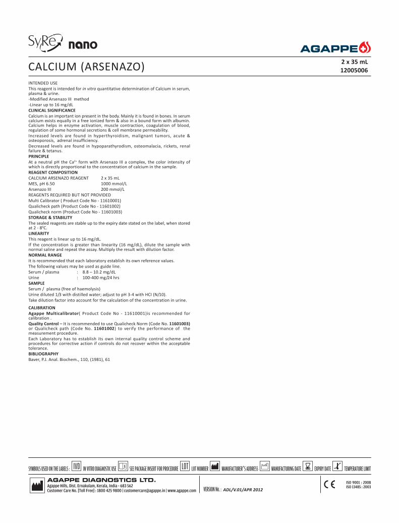
INTENDED USE
This reagent is intended for in vitro quantitative determination of Calcium in serum,plasma & urine.
-Modified Arsenazo III method
-Linear up to 16 mg/dL
CLINICAL SIGNIFICANCE
Calcium is an important ion present in the body. Mainly it is found in bones. In serumcalcium exists equally in a free ionized form & also in a bound form with albumin.Calcium helps in enzyme activation, muscle contraction, coagulation of blood,regulation of some hormonal secretions & cell membrane permeability.
Increased levels are found in hyperthyroidism, malignant tumors, acute &osteoporosis, adrenal insufficiency.
Decreased levels are found in hypoparathyrodism, osteomalacia, rickets, renalfailure & tetanus.
PRINCIPLE
At a neutral pH the Ca2+ form with Arsenazo III a complex, the color intensity ofwhich is directly proportional to the concentration of calcium in the sample.
REAGENT COMPOSITION
CALCIUM ARSENAZO REAGENT 2 x 35 mL
MES, pH 6.50 1000 mmol/L
Arsenazo III 200 mmol/L
REAGENTS REQUIRED BUT NOT PROVIDED
Multi Calibrator ( Product Code No - 11610001)
Qualicheck path (Product Code No - 11601002)
Qualicheck norm (Product Code No - 11601003)
STORAGE & STABILITY
The sealed reagents are stable up to the expiry date stated on the label, when storedat 2 - 80C.
LINEARITY
This reagent is linear up to 16 mg/dL.
If the concentration is greater than linearity (16 mg/dL), dilute the sample withnormal saline and repeat the assay. Multiply the result with dilution factor.
NORMAL RANGE
It is recommended that each laboratory establish its own reference values.
The following values may be used as guide line.
Serum / plasma : 8.8 – 10.2 mg/dL
Urine : 100-400 mg/24 hrs
SAMPLE
Serum / plasma (free of haemolysis)
Urine diluted 1/3 with distilled water; adjust to pH 3-4 with HCI (N/10).
Take dilution factor into account for the calculation of the concentration in urine.
CALIBRATION
Agappe Multicalibrator( Product Code No - 11610001)is recommended forcalibration .
Quality Control – It is recommended to use Qualicheck Norm (Code No. 11601003)or Qualicheck path (Code No. 11601002) to verify the performance of themeasurement procedure.
Each Laboratory has to establish its own internal quality control scheme andprocedures for corrective action if controls do not recover within the acceptabletolerance.
BIBLIOGRAPHY
Baver, P.J. Anal. Biochem., 110, (1981), 61
CALCIUM (ARSENAZO)2 x 35 mL
12005006
ADL/V.01/APR 2012

Parameters Screen
Test Cal
No
Full Name Calcium A
Standard No. 2
Reaction Type End Point
Primary WL 630
Sec. WL
Direction Increase
Reaction Time 0 15
Incubation Time
Unit mg/dL
Precision 0.1
R1 300 µL
R2 0
Sample 3 µ L
R1 Blank 0 15000
Mixed Rgt.Blank
Linearity Range 0 15
Linearity Limit
Substrate Limit
Factor
Prozone chek
q1 q2 q3 q4
PC
Reference Screen
Low
High
Calibration Screen
Rule Two Point Linear
Sensitivity
Replicates 1
Interval ( day) 0
Difference Limit
SD
Blank Response
Error Limit
Determination Coeff. 0
QC Screen
Control 1
Control 2
CALCIUM (ARSENAZO)
ADL/V.01/APR 2012

INTENDED USE
This reagent is intended for in vitro quantitative determination of Chloride in serum,plasma & urine.
-Modified Thiocyanate method
-Linear up to 130 mEq/L
CLINICAL SIGNIFICANCE
Chloride & bicarbonate are the principle anions (-vely charged) where as sodium &potassium are the principle cations (+vely charged) in the plasma. Chloride ions areinvolved in regulation of water distribution between the tissues by maintainingosmotic pressure & normal cation & anion balance between intra & extra cellularfluids.
Elevated levels are seen in conditions like dehydration & congestive cardiac failure.
Decreased levels are seen in condition such as salt losing nephritis, diabetic acidosis& renal failure.
PRINCIPLE
In an acid medium chloride ions and mercury ( II) thiocynate form thiocynate ions.These ions react with HNO
3 and iron (III) ions and effect a red color. The intensity
of the color is directly proportional to the concentration of chloride ions.
REAGENT COMPOSITION
CHLORIDE REAGENT 2 x 30 mL
Mercuric (II) thiocyanate 2 mmol/L
Nitric acid 29 mmol/L
Ferric Nitrate 20 mmol/L
REAGENTS REQUIRED BUT NOT PROVIDED
Multi Calibrator ( Product Code No - 11610001)
Qualicheck path (Product Code No - 11601002)
Qualicheck norm (Product Code No - 11601003)
STORAGE & STABILITY
The sealed reagents are stable up to the expiry date stated on the label, when storedat room temperature.
LINEARITY
This reagent is linear up to 130 mEq/L
If the concentration is greater than linearity (130 mEq/L), dilute the sample withdistilled water and repeat the assay. Multiply the result with dilution factor.
NORMAL RANGE
It is recommended that each laboratory establish its own reference values.
The following values may be used as guideline.
Serum : 97 - 108 mEq/L
Urine : 120 - 240 mEq/L/24 hr
SAMPLE
Serum / plasma (free of haemolysis) / Urine (Dilute sample 1:1 with distilled waterand multiply the result with 2)
CALIBRATION
Agappe Multicalibrator( Product Code No - 11610001)is recommended forcalibration .
Quality Control – It is recommended to use Qualicheck Norm (Code No. 11601003)or Qualicheck path (Code No. 11601002) to verify the performance of themeasurement procedure.
Each Laboratory has to establish its own internal quality control scheme andprocedures for corrective action if controls do not recover within the acceptabletolerance.
BIBLIOGRAPHY
Schonfed R.G.Lowellen C.S: Clin Chem.10,533 pp (1964)
CHLORIDE 2 x 30 mL
12005007
ADL/V.01/APR 2012

Parameters Screen
Test Chloride
No
Full Name Chloride
Standard No. 2
Reaction Type End Point
Primary WL 510
Sec. WL
Direction Increase
Reaction Time 0 15
Incubation Time
Unit mEq/L
Precision 0.1
R1 250 µL
R2 0
Sample 3 µL
R1 Blank 0 1000
Mixed Rgt.Blank
Linearity Range 0 130
Linearity Limit
Substrate Limit
Factor
Prozone chek
q1 q2 q3 q4
PC
Reference Screen
Low
High
Calibration Screen
Rule Two Point Linear
Sensitivity
Replicates 1
Interval ( day) 0
Difference Limit
SD
Blank Response
Error Limit
Determination Coeff. 0
QC Screen
Control 1
Control 2
CHLORIDE
ADL/V.01/APR 2012

INTENDED USE
This reagent is intended for in vitro quantitative determination of Cholesterol inserum or plasma.
-CHOD-PAP methodology.
-Linear up to 600 mg/dL.
-Contains LCF (Lipamic Clearing factor) which minimizes rerun.
CLINICAL SIGNIFICANCE
Cholesterol is the main lipid found in the blood, bile & brain tissues. It is also oneof the most important steroids of the body & is a precursor of many steroidhormones. Two thirds of cholesterol present in the blood is esterified. The livermetabolizes the cholesterol & it is transported in the blood stream by lipoproteins.
Increased levels are found in hypercholesterolemia, hyperlipidemia, hypothyroidism,uncontrolled diabetes, nephritic syndrome & cirrhosis.
Decreased levels are found in malabsorption, malnutrition, hyperthyroidism,anaemia & liver diseases.
PRINCIPLE
Enzymatic determination of total cholesterol according to the following reactions.
CHE
Cholesterol ester +H2O ------------> Cholesterol + fatty acids
CHO
Cholestrol + O2
----------------> 4-Cholesten-3- one + H2O
2
POD
2H2O
2 +Phenol+4-Aminoantipyrine-------------> Red quinone + 4H
2O
CHE : Cholesterol Esterase
CHO : Cholesterol Oxidase
POD : Peroxidase
REAGENT COMPOSITION
CHOLESTEROL (S.L) R1 4 x 35 mL
Pipes buffer (pH 6.70) 50 mmol/L
Phenol 24 mmol/L
Sodium Cholate 0.5 mml/L
Cholesterol Esterase > 180 U/L
Cholesterol Oxidase > 200 U/L
Peroxidase > 1000 U/L
4 – aminoantipyrine 0.5 mmol/L
Reagents Required But not Provided
Multi Calibrator ( Product Code No - 11610001)
Qualicheck path (Product Code No - 11601002)
Qualicheck norm (Product Code No - 11601003)
STORAGE & STABILITY
The sealed reagents are stable up to the expiry date stated on the label, when storedat 2- 80C.
LINEARITY
This reagent is linear up to 600 mg/dL.
If the concentration is greater than linearity ( 600 mg/dL), dilute the sample withnormal saline and repeat the assay. Multiply with dilution factor
NORMAL RANGE
It is recommended that each laboratory establish its own reference values.
The following value may be used as guide line.
Serum, Plasma : 150 – 220 mg/dL
SAMPLE
Serum, Plasma (free of haemolysis).
CALIBRATION
Agappe Multicalibrator( Product Code No - 11610001)is recommended forcalibration .
Quality Control – It is recommended to use Qualicheck Norm (Code No. 11601003)or Qualicheck path (Code No. 11601002) to verify the performance of themeasurement procedure.
Each Laboratory has to establish its own internal quality control scheme andprocedures for corrective action if controls do not recover within the acceptabletolerance.
BIBLIOGRAPHY
Allain C.C. et al., Clin.Chem 20 (1974), 470
CHOLESTEROL 4 x 35 mL
12005008
ADL/V.01/APR 2012

Parameters Screen
Test CHO
No
Full Name Cholesterol
Standard No. 2
Reaction Type End Point
Primary WL 510
Sec. WL 630
Direction Increase
Reaction Time 0 24
Incubation Time
Unit mg/dL
Precision 0.1
R1 300 µL
R2 0
Sample 3 µL
R1 Blank 0 2000
Mixed Rgt.Blank
Linearity Range 0 600
Linearity Limit
Substrate Limit
Factor
Prozone chek
q1 q2 q3 q4
PC Abs
Reference Screen
Low 150
High 220
Calibration Screen
Rule Two Point Linear
Sensitivity
Replicates 1
Interval ( day) 0
Difference Limit
SD
Blank Response
Error Limit
Determination Coeff.
QC Screen
Control 1
Control 2
CHOLESTEROL
ADL/V.01/APR 2012

CREATININE 4 x 35 / 2 x 18 mL
12002055
INTENDED USE: This reagent is intended for in vitro quantitative determination ofcreatinine in serum, plasma & urine.
CLINICAL SIGNIFICANCE
It is formed in muscles from phospho creatinine. It is important form of energy beinga store of high-energy phosphate. Creatinine determinations have one advantage overUrea determination that it is not affected by a high protein diet.
Serum creatinine is more specific & sensitive indicator of renal function. Simultaneousestimations of serum urea & creatinine provides better information. Serum ureanitrogen, creatinine ratio is > 15 in pre renal failure, & < 10 in renal failure.
Decreased levels are found in muscle dystrophy.
PRINCIPLE
Creatinine reacts with picric acid to produce a colored compound, creatinine alkalinepicrate. The change in absorbance is proportional to the creatinine concentration.
REAGENT COMPOSITION
CREATININE DYE REAGENT 4 x 35 mL
Picric Acid 8.73 mmol/L
Surfactant
CREATININE BASE REAGENT 2 x 18 mL
Sodium hydroxide 300 mmol/L
Sodium Phosphate 25 mmol/L
Reagents required but not provided
Multicalibrator ( Product Code No. 11610001)
Qualichek Norm ( Product Code No. 11601003)
Qualichek Path ( Product Code No. 11601002)
STORAGE & STABILITY
The sealed reagents are stable up to the expiry date stated on the label, when storedat room temperature.
This reagent is linear up to (24 mg/dL).
If the concentration is greater than linearity (24 mg/dL), dilute the sample with normalsaline and repeat the assay. Multiply the result with dilution factor.
NORMAL RANGE
It is recommended that each laboratory establish its own reference values.
The following value may be used as guide line.
Serum : Men : 0.7 – 1.4 mg/dL
Female : 0.6 – 1.2 mg/dL
Urine : : 0.80 – 1.80 gm/24 hr
PRECAUTION
To avoid contamination, use clean laboratory wares.
Avoid direct exposure of working reagent to light.
SAMPLE
Serum / plasma (free of haemolysis) / Urine (diluted 1/100 with distilled water)
CALIBRATION
Agappe Multicalibrator (Code No. 11610001) is recommended for calibration of thisassay.
Quality Control – It is recommended to use Qualicheck Norm (Code No. 11601003)or Qualicheck path (Code No. 11601002) to verify the performance of themeasurement procedure.
Each Laboratory has to establish its own internal quality control scheme andprocedures for corrective action if controls do not recover within the acceptabletolerance.
BIBLIOGRAPHY
1. Allen. L.C.: Clin chem.Vol.28 No.3, 1982, 555.2. Haeckel, R. et al. Chlin. Chem. 27/1 179-183 (1981).3. Tanganelli, E. Prencipe, L; Bassi, D. Cambiaghi, S. and Murador,E.: Clin.Chem 287,
1461-1464 (1982)

Parameters Screen
Test CRT
No
Full Name Creatinine
Standard No. 2
Reaction Type Fixed Time
Primary WL 510
Sec. WL
Direction Increase
Reaction Time 4 12
Incubation Time 5
Unit mg/dL
Precision 0.1
R1 200 µL
R2 200 µL
Sample 40 µL
R1 Blank
Mixed Rgt.Blank
Linearity Range 0 24
Linearity Limit
Substrate Limit
Factor
Prozone chek
q1 q2 q3 q4
PC Abs
Reference Screen
Low
High
Calibration Screen
Rule Two Point Linear
Sensitivity
Replicates 1
Interval ( day) 0
Difference Limit
SD
Blank Response
Error Limit
Determination Coeff. 0
QC Screen
Control 1
Control 2
CREATININE

INTENDED USE
This reagent is intended for in vitro Quantitative determination of Creatine Kinasein human serum.
-Optimized IFCC Method.
-Linear up to 1700 U/L.
CLINICAL SIGNIFICANCE
It is mainly found in all muscle (Cardiac & Skeletal) & brain tissues. It plays animportant role in energy storing mechanism of the tissues. Its iso-enzymes: CK-MBmainly exists in cardic muscle tissues, CK-MM in skeletal muscle tissues & CK-BB inbrain & lungs.
Increased levels are found in myocardical infarction, muscular dystrophy,cerebrovascular-disease, pulmonary infarction, electrical shocks & hypothyrodisim.
Decreased levels are, sometimes seen in early pregnancy.
PRINCIPLE
Kinetic determination of Creatinine Kinase is based on following reactions
CK
Creatinine Phophate + ADP ------------> Creatine + ATP
HK
ATP + D-Glucose --------------------> G-6-p+ADP
G-6-PDH
G-6-P + NADP+ ---------------> D-Gluconate -6-Phosphate + NADPH + H+
CK- Creatinine Kinase
Hk- Hexokinase
G-6-P-D- Glucose-6-phosphate
G-6-PDH- Glucose-6 Phosphate dehydrogenase
REAGENT COMPOSITION
CREATINE KINASE R1 1 x 30 mL
Imidazole (pH 6.7) 125 mmol/L
D-Glucose 25 mmol/L
N-Acetyl-L-Cysteine 25 mmol/L
Magnesium acetate 12.5 mmol/L
NADP 2.52 mmol/L
EDTA 2.02 mmol/L
Hexokinase > 6800 U/L
CREATINE KINASE(S.L) R2 1 x 8 mL
Creatine Phosphate 250 mmol/L
ADP 15.2 mmol/L
AMP 25 mmol/L
Diadenosine-5-pentaphosphate 103 µmmol/L
G-6-PDH >88800 U/L
Reagents Required But not Provided
Multi Calibrator (Product Code No - 11610001)
Qualicheck path (Product Code No - 11601002)
Qualicheck norm (Product Code No - 11601003)
STORAGE & STABILITY
The sealed reagents are stable up to the expiry date stated on the label, when storedat 2 - 80C.
LINEARITY
This reagent is linear up to 1700 U/L.
If the concentration is greater than linearity (1700 U/L), dilute the sample withnormal saline and repeat the assay. Multiply the final result with dilution factor.
NORMAL VALUES
It is recommended that each laboratory establish its own reference value.
The following values may be used as guide line.
Men : < 171 U/L
Women : < 145 U/L
PRECAUTION
To avoid contamination, use clean laboratory wares.
Avoid direct exposure of reagent to light.
SAMPLE
Serum/Plasma (Free of haemolysis)
CREATINE KINASE 1 x 30 / 1 x 8 mL
12005010
CALIBRATION
Agappe Multicalibrator(Product Code No - 11610001)is recommended forcalibration .
Quality Control : It is recommended to use Qualicheck Norm (Code No. 11601003)or Qualicheck path (Code No. 11601002) to verify the performance of themeasurement procedure.
Each Laboratory has to establish its own internal quality control scheme andprocedures for corrective action if controls do not recover within the
acceptable tolerance.
BIBLIOGRAPHY
1. DGKC,J.Clin.chem.clin.Bioch.15,255(1977)2. Di.Witt, Trendelenberg, J.Clin.Chemie,clin. Bioch.20,235(1982)
ADL/V.01/APR 2012

Parameters Screen
Test CK- NAC
No
Full Name CK- NAC
Standard No. 2
Reaction Type Kinetic
Primary WL 340
Sec. WL
Direction Increase
Reaction Time 8 20
Incubation Time 5
Unit U/L
Precision 0.1
R1 200 µL
R2 50 µL
Sample 12.5 µL
R1 Blank 0 10000
Mixed Rgt.Blank 0 10000
Linearity Range 0 2000
Linearity Limit 0.2
Substrate Limit 20000
Factor
Prozone chek
q1 q2 q3 q4
PC
Reference Screen
Low
High
Calibration Screen
Rule Two Point Linear
Sensitivity
Replicates 1
Interval ( day) 0
Difference Limit
SD
Blank Response
Error Limit
Determination Coeff. 0
QC Screen
Control 1
Control 2
CREATINE KINASE
ADL/V.01/APR 2012
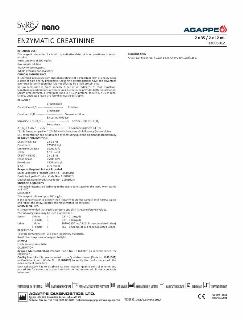
INTENDED USE
This reagent is intended for in vitro quantitative determination creatinine in serumor urine.
-High Linearity of 200 mg/dL
-No sample dilution
-Ready to use reagents
-MSDS available for analyzers
CLINICAL SIGNIFICANCE
It is formed in muscles from phosphocreatinine. It is important form of energy beinga store of high energy phosphate. Creatinine determinations have one advantageover urea determination that it is not affected by a high protein diet.
Serum creatinine is more specific & sensitive indicator of renal function.Simultaneous estimations of serum urea & creatinine provides better information.Serum urea nitrogen & creatinine ratio is > 15 in prerenal failure & < 10 in renalfailure. Decreased levels are found in muscle dystrophy.
PRINCIPLE
Creatininase
Creatinine +H2O --------------------------> Creatine
Creatinase
Creatine + H2O ---------------------------> Sarcosine +Urea
Sarcosine Oxidase
Sarcosine + O2+H
2O ----------------------------> Glycine + HCHO + H
2O
2
Peroxidase
2 H2O
2 + 4-AA *1+ TOOS *2 ---------------------> Quinone pigment +4 H
2O
*1 : 4- Aminoantipyrine, * 2N-Ethyl –N-(2-Hydroxy -3-Sulfopropyl)-m-toluidine
CRE concentration can be obtained by measuring quinone pigment photometrically
REAGENT COMPOSITION
CREATININE R1 2 x 35 mL
Creatinase 175000 IU/L
Sacrosine Oxidase 15000 lU/L
TOOS 1.13 mmol
CREATININE R2 2 x 12 mL
Creatininase 75000 IU/L
Peroxidase 4500 units /L
4-AA 0.75 mmol
Reagents Required But not Provided
Multi Calibrator ( Product Code No - 11610001)
Qualicheck path (Product Code No - 11601002)
Qualicheck norm (Product Code No - 11601003)
STORAGE & STABILITY
The sealed reagents are stable up to the expiry date stated on the label, when storedat 2 - 80C
LINEARITY
This reagent is linear up to 200 mg/dL
If the concentration is greater than linearity dilute the sample with normal salineand repeat the assay. Multiply the result with dilution factor.
NORMAL VALUES
It is recommended that each laboratory establish its own reference values.
The following value may be used as guide line.
Serum : Male : 0.6 – 1.1 mg/dL
: Female : 0.5 – 0.8 mg/dL
Urine : Male : 1070–2150 md/dL(24 hrs-accumulated urine)
: Female : 769 – 1200 mg/dL (24 hr accumulated urine)
PRECAUTION
To avoid contamination, use clean laboratory materials.
Avoid direct exposure of reagent to light.
SAMPLE
Fresh Serum/Urine 24 hr
CALIBRATION
Agappe Multicalibrator( Product Code No - 11610001)is recommended forcalibration .
Quality Control – It is recommended to use Qualicheck Norm (Code No. 11601003)or Qualicheck path (Code No. 11601002) to verify the performance of themeasurement procedure.
Each Laboratory has to establish its own internal quality control scheme andprocedures for corrective action if controls do not recover within the acceptabletolerance.
ENZYMATIC CREATININE 2 x 35 / 2 x 12 mL
12005012
BIBLIOGRAPHY
Artiss. J.D. Mc Enroe, R.J.Zak B.Clin.Chem, 30 (1984)1389.
ADL/V.01/APR 2012

Parameters Screen
Test Enzy CRT
No
Full Name Enzymatic Creatinine
Standard No. 2
Reaction Type End Point
Primary WL 546
Sec. WL 630
Direction Increase
Reaction Time -1 22
Incubation Time 20
Unit mg/dL
Precision 0.1
R1 225 µL
R2 75 µL
Sample 6 µL
R1 Blank 0 1500
Mixed Rgt.Blank 1 1700
Linearity Range 0 200
Linearity Limit
Substrate Limit
Factor
Prozone chek
q1 q2 q3 q4
PC
Reference Screen
Low
High
Calibration Screen
Rule Two Point Linear
Sensitivity
Replicates 1
Interval ( day) 0
Difference Limit
SD
Blank Response
Error Limit
Determination Coeff. 0
QC Screen
Control 1
Control 2
ENZYMATIC CREATININE
ADL/V.01/APR 2012

INTENDED USE
This reagent is intended for in vitro Quantitative determination of GAMMA GT inserum.
-Szasz methodology.
-Linear up to 232 U/L.
CLINICAL SIGNIFICANCE
GGT activity is elevated in all forms of liver diseases. It is highest in cases ofintrahepatic or post hepatic biliary obstruction. (It may be 5 to 30 times higher thannormal) It is more sensitive than Alkaline Phosphatase , NTP, Leucineaminopeptidase and transaminases in detection of obstructive jaundice, cholangitis,cholecystis neoplasm, it rises earlier than other enzyme and persists longer.
Moderate increase is observed in infectious hepatitis. (2 to 5 times) Increases mayalso be observed in cases of drug intoxication, acute and chronic pancreatitis.
PRINCIPLE
Kinetic determination of Gamma GT according to the following reaction.
Gamma GT
GLUPA-C+Glycylglycine --------------> L-Gamma-Glutamyl-Glycylglycine
+ 5-Amino-2-nitrobenzoic acid.
GLUPA-C: L-Gamma -Glutamyl-3 Carboxy-P-nitroanilide
REAGENT COMPOSITION
Gamma GT R1 1 x 30 ml
Tris buffer pH (8.25) 133 mmol/L
Glycylglycine 138 mmol/L
Gamma GT R2 1 x 8 ml
GLUPA - C 23 mmol/L
Reagents Required But not Provided
Multi Calibrator ( Product Code No - 11610001)
Qualicheck path (Product Code No - 11601002)
Qualicheck norm (Product Code No - 11601003)
STORAGE & STABILITY
The sealed reagents are stable up to the expiry date stated on the label, when storedat 2 - 80C.
LINEARITY
This reagent is linear up to 232 U/L.
If the concentration is greater than linearity (232 U/L) dilute the sample with normalsaline and repeat the assay. Multiply the result with dilution factor.
NORMAL VALUES
It is recommended that each laboratory establish its own reference value.
The following values may be used as guide line.
Female : 5-32 U/L
Male : 10-45 U/L
PRECAUTION
To avoid contamination, use clean laboratory wares. .
Avoid direct exposure of reagent to light.
SAMPLE
Fresh Serum (free of haemolysis)
CALIBRATION
Agappe Multicalibrator( Product Code No - 11610001)is recommended forcalibration .
Quality Control – It is recommended to use Qualicheck Norm (Code No. 11601003)or Qualicheck path (Code No. 11601002) to verify the performance of themeasurement procedure.
Each Laboratory has to establish its own internal quality control scheme andprocedures for corrective action if controls do not recover within the acceptabletolerance.
BIBLIOGRAPHY
1. Szasz, G.Clin.Chem., 22, (1976),20512. SCJ.Clin. Lab.Invest 36: 711 (1976)3. Tietz N.W. Text book of Clin.Chem. 678 686 : (1986)
GAMMA GT 1 x 30 / 1 x 8 mL
12005013
ADL/V.01/APR 2012

Parameters Screen
Test GGT
No
Full Name GGT
Standard No. 2
Reaction Type kinetic
Primary WL 405
Sec. WL
Direction Increase
Reaction Time 4 12
Incubation Time 10
Unit U/L
Precision 0.1
R1 200 µL
R2 50 µL
Sample 25 µL
R1 Blank 0 10000
Mixed Rgt.Blank 0 13000
Linearity Range 0 200
Linearity Limit 0.2
Substrate Limit 18000
Factor
Prozone chek
q1 q2 q3 q4
PC Abs
Reference Screen
Low
High
Calibration Screen
Rule Two Point Linear
Sensitivity
Replicates 1
Interval ( day) 0
Difference Limit
SD
Blank Response
Error Limit
Determination Coeff. 0
QC Screen
Control 1
Control 2
GAMMA GT
ADL/V.01/APR 2012

INTENDED USE
This reagent is intended for in vitro quantitative determination of Glucose in serum,plasma & CSF
-GOD –PAP methodology
-Linear up to 600 mg/dL
CLINICAL SIGNIFICANCE
Glucose is a major carbohydrate present in the blood & serves as a primary sourceof energy. It is usually obtained from ingested starch & sugar. The glucoseconcentration is normally maintained at constant level. Excessive glucose is storedas a inactive glycogen mainly in the liver & little in the muscles.
Elevated blood glucose levels are found in diabetes mellitus, hyperthyroidism,hyperadrenalism & certain liver diseases.
Decreased levels are found in Insulinoma, hypothyroidism, hypopituitarism.
PRINCIPLE
Enzymatic colorimetric determination of glucose according to the following reaction.
GOD
Glucose+ O2 + 2H
2O --------------------> H
2O
2 + Gluconic acid
POD
2H2O
2+phenol + 4-Ap -------------------> Quinonimine + 4H
2O
GOD – Glucose Oxidase
POD – Peroxidase
REAGENT COMPOSITION
GLUCOSE REAGENT R1 4 x 35 mL
Tris Buffer, (pH 7.40) 92 mmol/L
Phenol 0.3 mmol/L
Glucose Oxidase 15000 U/L
4- Aminophenazone 2.6 mmol/L
Reagents Required But not Provided
Multi Calibrator ( Product Code No - 11610001)
Qualicheck path (Product Code No - 11601002)
Qualicheck norm (Product Code No - 11601003)
STORAGE & STABILITY
The sealed reagents are stable up to the expiry date stated on the label, when storedat 2 - 80C.
LINEARITY
This reagent is linear up to 600 mg/dL
If the concentration is greater than linearity (600 mg/dL), dilute the sample withnormal saline and repeat the assay. Multiply the result with dilution factor.
NORMAL RANGE
It is recommended that each laboratory establish its own reference values.
The following values may be used as guide line.
Serum : Fasting : 70-110 mg/dL
2 hrs postprandial : 70-140 mg/dL
C S F : 50 -70 mg/dL
PRECAUTION
To avoid contamination, use clean laboratory materials.
Avoid direct exposure of reagent to light.
SAMPLE
Serum / plasma (free of haemolysis) / CSF
CALIBRATION
Agappe Multicalibrator( Product Code No - 11610001)is recommended forcalibration .
Quality Control – It is recommended to use Qualicheck Norm (Code No. 11601003)or Qualicheck path (Code No. 11601002) to verify the performance of themeasurement procedure.
Each Laboratory has to establish its own internal quality control scheme andprocedures for corrective action if controls do not recover within the acceptabletolerance.
BIBLIOGRAPHY
1. Trinder, P. Ann Clin Biochem. 6,24 (1979)2. Dingeon, B. Ann.Bio.Clin 33,3 (1975)3. Lott, J. Clin.Chem. 21, 1754 (1975)
GLUCOSE 4 x 35 mL
12602001
ADL/V.01/APR 2012

Parameters Screen
Test Glucose
No
Full Name Glucose
Standard No. 2
Reaction Type EP
Primary WL 510
Sec. WL 630
Direction Increase
Reaction Time 0 38
Incubation Time
Unit mg/dL
Precision 0.1
R1 300 µL
R2 0
Sample 3 µL
R1 Blank 0 2000
Mixed Rgt.Blank
Linearity Range 0 500
Linearity Limit
Substrate Limit
Factor
Prozone chek
q1 q2 q3 q4
PC Abs
Reference Screen
Low 70
High 110
Calibration Screen
Rule Two Point Linear
Sensitivity
Replicates 1
Interval ( day) 0
Difference Limit
SD
Blank Response
Error Limit
Determination Coeff.
QC Screen
Control 1
Control 2
GLUCOSE
ADL/V.01/APR 2012

INTENDED USE: This reagent is intended for in vitro quantitative determination ofHDL Cholesterol in serum
CLINICAL SIGNIFICANCE
Blood total cholesterol levels have long been known to be related to coronary heartdisease (CHD). In recent years, in addition to total cholesterol, high densitylipoprotein cholesterol (HDL-C) has become an important tool used to assess anindividual risk of developing CHD since a strong negative relationship between HDL-C concentration and the incidence of CHD was reported.
PRINCIPLE
The reaction between cholesterol other than HDL & enzyme for cholesterol assayis suppressed by the electrostatic interaction between polyanions & cationicsubstances. Hydrogen peroxide is formed by the free cholesterol in HDL bycholesterol oxidase. Oxidative condensation of EMSE and 4-AA is caused byhydrogen peroxide in the presence of peroxidise, and the absorbance of theresulting red-purple quinine is measured to obtain the cholesterol value in HDL
Polyanions
Other lipoproteins than HDL------------> Suppress reaction with enzyme
Cationic substances
cholesterol esterase
HDL (cholesterol esters) + H2O ----------------> HDL (free cholesterol) + Free fatty acids
cholesterol oxidase
HDL (free cholesterol) + O2 + H+-------------------> Cholestenone + H
2O
2
Peroxidase
2H2O
2 + 4-AA + EMSE + H
3 + O ---------------> Red-purple quinine + 5H
2O
REAGENT COMPOSITION
HDL - C DIRECT R1 2 x 30 mL
N-Ethyl-N-(3-methylphenyl)-N’succinylethyenediame(EMSE)
HDL - C DIRECT R2 2 x 10 mL
Cholesterol Oxidase
4-Aminoantipyrine (4-AA)
Reagents Required But not Provided
Multi Calibrator (Product Code No - 11610001)
Qualicheck path (Product Code No - 11601002)
Qualicheck norm (Product Code No - 11601003)
STORAGE & STABILITY
The sealed reagents are stable up to the expiry date stated on the label, when storedat 2 - 80C, protected from light. Do not freeze.
LINEARITY
This reagent is linear up to 150 mg/dL
If the concentration is greater than linearity (150 mg/dL), dilute the sample withnormal saline and repeat the assay. Multiply the result with dilution factor.
NORMAL RANGE
It is recommended that each laboratory establish its own reference values.
The following value may be used as guide line.
Male : 35-80 mg/dL
Female : 42- 88 mg/dL
PRECAUTION
To avoid contamination, use clean laboratory materials. Use clean, dry disposablepipette tips for dispensing. Close reagent bottles immediately after use.
Avoid direct exposure of reagent to light. Do not blow into the reagent bottles.
SAMPLE
Fresh serum (free of haemolysis)
INTERFERING SUBSTANCES
Test will not be affected by:
Bilirubin up to 40 mg/dL
Ascorbic acid up to 50 mg/dL
Hemoglobin up to 500 mg/dL
Triglyceride up to 1000 mg/dL
(when triglyceride in a sample exceeds 1000 mg/dL, dilute the sample to 1+9 withsaline, repeat the assay and multiply result by 10)
HDL – C DIRECT 2 x 30 / 2 x 10 mL
12005015
CALIBRATION
Agappe Multicalibrator( Product Code No - 11610001)is recommended forcalibration .
Quality Control – It is recommended to use Qualicheck Norm (Code No. 11601003)or Qualicheck path (Code No. 11601002) to verify the performance of themeasurement procedure.
Each Laboratory has to establish its own internal quality control scheme andprocedures for corrective action if controls do not recover within the acceptabletolerance.
BIBLIOGRAPHY
1. Williams P et al., High density lipoprotein and coronary risk factor, Lancet.1:72 (1979)
2. Gordon,T.Castelli, W.P.Hjortland, M.C. et al. Am.J.Med 62, 707-714 (1977)3. Rifai, N.and Warnick, G.R., Ed. Laboratory Measurement of Lipids,
Lipoproteins and Apolipoproteins AACC Press. Washington, DC,USA,1994
ADL/V.01/APR 2012

Parameters Screen
Test HDL D
No
Full Name HDL Direct
Standard No. 2
Reaction Type End Point
Primary WL 578
Sec. WL 670
Direction Increase
Reaction Time -1 22
Incubation Time 20
Unit mg/dL
Precision 0.1
R1 225 µL
R2 75 µL
Sample 3 µL
R1 Blank 0 1000
Mixed Rgt.Blank 0 3000
Linearity Range 0 200
Linearity Limit
Substrate Limit
Factor
Prozone chek
q1 q2 q3 q4
PC Abs
Reference Screen
Low
High
Calibration Screen
Rule Two Point Linear
Sensitivity
Replicates 1
Interval ( day) 0
Difference Limit
SD
Blank Response
Error Limit
Determination Coeff. 0
QC Screen
Control 1
Control 2
HDL – C DIRECT
ADL/V.01/APR 2012

INTENDED USE
This reagent is intended for in vitro quantitative determination of LactateDehydrogenase in serum or plasma.Based on SCE recommended method.
-Linear up to 2400 U/L.
CLINICAL SIGNIFICANCE
This enzyme is found in all organ cells, but especially plentiful in cardiac & skeletalmuscle, liver, kidney & RBC. LDH is found in the form of iso-enzymes. Based on theirelectrophoretic mobility with each iso-enzymes being primarily from differentorgans.
Elevated levels are found in myocardial infraction, liver diseases, hemolyticanaemia’s, pernicious anaemia, Leukemia & Pulmonary diseases. Elevations in acuteMI reaches a peack in 48-72 hrs. Delayed elevations, (10-14 days) are useful in thelate diagnosis of the condition.
PRINCIPLE
Kinetic determination of lactate dehydrogenase according to the following reaction.
LDH
Pyruvate + NADH + H+ -------------> L-Lactate +NAD+
REAGENT COMPOSITION
LDH -P R1 1 x 30 mL
Tris buffer (pH 7.4) 80 mmol/L
Pyruvate 1.6 mmol/L
Sodium Chloride 200 mmol/L
LDH – P R2 1 x 8 mL
NADH 240 mmol/L
Reagents Required But not Provided
Multi Calibrator ( Product Code No - 11610001)
Qualicheck path (Product Code No - 11601002)
Qualicheck norm (Product Code No - 11601003)
STORAGE & STABILITY
The sealed reagents are stable up to the expiry date stated on the label, when storedat 2 - 80C.
LINEARITY
This reagent is linear up to 2400 U/L.
If the concentration is greater than linearity (2400 U/L), dilute the sample withnormal saline and repeat the assay. Multiply the result with dilution factor
NORMAL RANGE
It is recommended that each laboratory establish its own reference values.
The following value may be used as guide line.
Serum /Plasma : 225-450 U/L
PRECAUTION
To avoid contamination, use clean laboratory wares.
Avoid direct exposure of reagent to light.
SAMPLE
Serum, plasma (free of haemolysis)
CALIBRATION
Agappe Multicalibrator( Product Code No - 11610001)is recommended forcalibration .
Quality Control - It is recommended to use Qualicheck Norm (Code No. 11601003)or Qualicheck path (Code No. 11601002) to verify the performance of themeasurement procedure.
Each Laboratory has to establish its own internal quality control scheme andprocedures for corrective action if controls do not recover within the acceptabletolerance.
BIBLIOGRAPHY
1. Z.Klin chem. Klin Biochem.8,658 (1970), 1, 1820(1972)2. Wei Bhaar, D.et al. Med.Welt 26,387 (1975)
LDH 1 x 30 / 1 x 8 mL
12005016
ADL/V.01/APR 2012

Parameters Screen
Test LDH
No
Full Name LDH
Standard No. 2
Reaction Type kinetic
Primary WL 340
Sec. WL
Direction Decrease
Reaction Time 4 12
Incubation Time 10
Unit U/L
Precision 0.1
R1 200 µL
R2 50 µL
Sample 3 µL
R1 Blank 0 20000
Mixed Rgt.Blank 1000 25000
Linearity Range 0 2000
Linearity Limit 0.2
Substrate Limit 8000
Factor
Prozone chek
q1 q2 q3 q4
PC Abs
Reference Screen
Low
High
Calibration Screen
Rule Two Point Linear
Sensitivity
Replicates 1
Interval ( day) 0
Difference Limit
SD
Blank Response
Error Limit
Determination Coeff. 0
QC Screen
Control 1
Control 2
LDH
ADL/V.01/APR 2012

INTENDED USE
This reagent is intended for in vitro quantitative determination of LDL Cholesterolin serum or plasma
CLINICAL SIGNIFICANCE
Blood total cholesterol levels have long been known to be related to coronary heartdisease (CHD). In recent years, in addition to total cholesterol, low densitylipoprotein cholesterol (LDL-C) has become an important tool used to assess anindividual risk of developing CHD since a strong positive relationship between LDL-C concentration and the incidence of CHD was reported. LDL Cholestrol acts as akey factor in the pathogenesis of atherosclerosis and coronary artery disease.
PRINCIPLE
The LDL-C Direct is a homogeneous assay. When serum is mixed with R1, amphotericsurfactants protect LDL from enzyme reactions. CHE and CO reacts with non-LDLcholesterol, which decomposed to water by catalase. R2 enables the conversationof LDL-C to hydrogen peroxide, which upon oxidative condensation with HDAOS &4AA yields a color complex. By measuring the absorbance of this blue color complexproduced, the LDL-C concentration in the sample can be calculated when comparedwith the absorbance of the LDL-C Calibrator.
REAGENT COMPOSITION
LDL –C DIRECT R1 1 x 30 mL
4-Aminoantipyrin 0.5 mmol/l
CHE
CO 1.2 U/mL
Peroxidase
Good’s buffer pH 6.3
LDL –C DIRECT R2 1 x 10 mL
N,N-bis(4-sulfobutyl)-m-toluidine
disodium salt (DSBmT) 1.0 mmol/l
Good’s buffer pH 6.3
Reagents Required But not Provided
Multi Calibrator ( Product Code No - 11610001)
Qualicheck path (Product Code No - 11601002)
Qualicheck norm (Product Code No - 11601003)
STORAGE & STABILITY
The sealed reagents are stable up to the expiry date stated on the label, when storedat 2 - 80C.
LINEARITY
This reagent is linear up to 450 mg/dL
If the concentration is greater than linearity (450 mg/dL), dilute the sample withnormal saline and repeat the assay. Multiply the result with dilution factor.
NORMAL RANGE
It is recommended that each laboratory establish its own reference values.
The following value may be used as guide line.
Desirable < 130 mg/dL
Borderline 130-159 mg/dL
High Risk for CHD > 160 mg/dL.
PRECAUTION
To avoid contamination, use clean laboratory materials. Use clean, dry disposablepipette tips for dispensing. Close reagent bottles immediately after use.
Avoid direct exposure of reagent to light. Do not blow into the reagent bottles.
SAMPLE
Fresh serum (free of haemolysis) or EDTA Plasma
INTEREFERING SUBSTANCES
Test will not be affected by:
Bilirubin up to 40 mg/dL
Ascorbic acid up to 50 mg/dL
Haemoglobin up to 500 mg/dL
Triglyceride up to 1000 mg/dL
(when triglyceride in a sample exceeds 1000 mg/dL, dilute the sample 1+9 withsaline, repeat the assay and multiply result by 10)
LDL – C DIRECT 1 x 30 / 1 x 10 mL
12005017
CALIBRATION
Agappe Multicalibrator( Product Code No - 11610001)is recommended forcalibration .
Quality Control – It is recommended to use Qualicheck Norm (Code No. 11601003)or Qualicheck path (Code No. 11601002) to verify the performance of themeasurement procedure.
Each Laboratory has to establish its own internal quality control scheme andprocedures for corrective action if controls do not recover within the acceptabletolerance.
BIBLIOGRAPHY
1. Crouse J.R et al., Studies of low density lipoprotein molecular weight in humanbeingd with coronary artery disease. J.Lipid Res 26:5666 (1985)
2. Gordon,T.Castelli, W.P.Hjortland, M.C. et al. AM.J.Med 62, 707-714 (1977)Rifai, N.and Warnick, G.R., Ed. Laboratory Measurement of Lipids, Lipoproteinsand Apolipoproteins AACC Press. Washington, DC,USA,1994
ADL/V.01/APR 2012

Parameters Screen
Test LDL D
No
Full Name LDL Direct
Standard No. 2
Reaction Type End Point
Primary WL 546
Sec. WL 630
Direction Increase
Reaction Time -1 22
Incubation Time 20
Unit mg/dL
Precision 0.1
R1 225 µL
R2 75 µL
Sample 3 µL
R1 Blank 0 15000
Mixed Rgt.Blank 0 3000
Linearity Range 0 450
Linearity Limit
Substrate Limit
Factor
Prozone chek
q1 q2 q3 q4
PC Abs
Reference Screen
Low
High
Calibration Screen
Rule Two Point Linear
Sensitivity
Replicates 1
Interval ( day) 0
Difference Limit
SD
Blank Response
Error Limit
Determination Coeff. 0
QC Screen
Control 1
Control 2
LDL – C DIRECT
ADL/V.01/APR 2012

INTENDED USE
This reagent is intended for in vitro quantitative determination of Magnesium inserum or plasma.
-Xylidyl Blue with ATCS
-Linear up to 5 mg/dL
CLINICAL SIGNIFICANCE
Magnesium is the second most abundant intracellular cation of the human bodyafter potassium, being essential in a great number of enzymatic and metabolicprocesses.
It is a co-factor of all the enzymatic reactions that involve ATP and found in themembranes that maintain the electrical excitability of muscular and nervous cells.
A low magnesium level is found in malabsorption syndrome, diureticsaminoglucoside therapy, and hyperparathyroidism or diabetic acidosis.
Elevated concentration of magnesium is found in uremia, chronic renal failure,glomerulo nephritis, Addison’s disease or intensive anti acid therapy.
Clinical diagnosis should not be made on a single test result; it should integrateclinical and other laboratory data.
PRINCIPLE
Magnesium reacts with xylidyl Blue to form a colored compound in alkaline solution.
The intensity of the colour formed is proportional to the magnesium in the sample.
REAGENT COMPOSITION
MAGNESIUM REAGENT 1 x 30 mL
Xylidyl Blue 110 mmol/L
Ethanolamine (pH 11.0) 1 mol/L
GEDTA 60 mmol/L
Reagents Required But not Provided
Multi Calibrator ( Product Code No - 11610001)
Qualicheck path (Product Code No - 11601002)
Qualicheck norm (Product Code No - 11601003)
STORAGE & STABILITY
The sealed reagents are stable up to the expiry date stated on the label, when storedat 2 -80C, protected from light.
LINEARITY
This reagent is linear up to 5 mg/dL.
If the concentration is greater than linearity (5 mg/dL), dilute the sample withnormal saline and repeat the assay. Multiply the result with dilution factor.
NORMAL RANGE
It is recommended that each laboratory establish its own reference values.
The following values may be used as guide line.
Serum : 1.8 – 2.6 mg/dL
CSF : 2.1 – 3.3 mg/dL
Urine : 73 - 122 mg/24 h
PRECAUTION
To avoid contamination, use clean laboratory materials. It is recommended to usedisposable tubes. Use clean, dry disposable pipette tips for dispensing. Close reagentimmediately after Use. Avoid direct exposure of reagent to light.
SAMPLE
Serum (free haemolysis) or Heparinized plasma.
(Do not use oxalates or EDTA as anticoagulant)
Urine should be acidified to pH 3-4 with concentrated HCl then dilute sample 1/5with distilled water and multiply the result by 5.
CALIBRATION
Agappe Multicalibrator( Product Code No - 11610001)is recommended forcalibration .
Quality Control – It is recommended to use Qualicheck Norm (Code No. 11601003)or Qualicheck path (Code No. 11601002) to verify the performance of themeasurement procedure.
Each Laboratory has to establish its own internal quality control scheme andprocedures for corrective action if controls do not recover within the acceptabletolerance.
BIBLIOGRAPHY
1. Farrel, E.C. Magnesium. Kaplan a et al. Clin chem.. The CV Mosby Co St LouisTorento. Princeton 1984; 1064-69
2. Brutis, C. A. et al. TIETZ textbook of clinical chemistry, 3 rd edition W BSaunders company; 1999, P.1395-1457
3. Young, D. S. Effects of disease on clinical lab tests 4th edition AACC Press, 2001
MAGNESIUM 1 x 30 mL
12005018
ADL/V.01/APR 2012

Parameters Screen
Test Mag
No
Full Name Magnesium
Standard No. 2
Reaction Type End Point
Primary WL 546
Sec. WL 0
Direction Increase
Reaction Time 0 20
Incubation Time
Unit mg/dL
Precision 0.1
R1 300 µL
R2
Sample 3 µL
R1 Blank
Mixed Rgt.Blank
Linearity Range 0 5
Linearity Limit
Substrate Limit
Factor
Prozone chek
q1 q2 q3 q4
PC Abs
Reference Screen
Low
High
Calibration Screen
Rule Two Point Linear
Sensitivity
Replicates 1
Interval ( day)
Difference Limit
SD
Blank Response
Error Limit
Determination Coeff. 0
QC Screen
Control 1
Control 2
MAGNESIUM
ADL/V.01/APR 2012

INTENDED USE
This reagent is intended for in vitro quantitative determination of Phosphorous inserum or plasma.
-Phosphomolybdate methodology
-Linear up to 15 mg/dL
CLINICAL SIGNIFICANCE
Phosphorous is mainly combined with calcium & is found in bones. It is involved inthe carbohydrate metabolism & is a component of many other substances. Someof its important functions include maintaining of acid-base balance, skeletal muscleformation. It is also required for normal functioning of RBCs & muscles.
Increased levels are found in hypothyroidism, renal failure, bone metastasis & liverdisease.
Decreased levels are found in hyperparathyroidism, osteomalacia & diseaseassociated with Vitamin D deficiency.
PRINCIPLE
Determination of inorganic phosphorous according to the following reaction.
phosphorous
Ammonium molybdate + sulfuricaid ----------------> phosphomolybidiccomplex
REAGENT COMPOSITION
INORGANIC PHOSPHOROUS REAGENT 1 x 30 mL
Sulfuric acid 210 mmol/L
Ammonium molybdate 650 mmol/L
Reagents Required But not Provided
Multi Calibrator ( Product Code No - 11610001)
Qualicheck path (Product Code No - 11601002)
Qualicheck norm (Product Code No - 11601003)
STORAGE & STABILITY
The sealed reagents are stable up to the expiry date stated on the label, when storedat 2 80C.
LINEARITY
This reagent is linear up to 15 mg/dL.
If the concentration is greater than linearity (15 mg/dL), dilute the sample withnormal saline and repeat the assay. Multiply the result with dilution factor.
NORMAL RANGE
It is recommended that each laboratory establish its own reference values.
The following values may be used as guide line.
Serum : 2.7 – 4.5 mg/dL
Urine : 400 – 1300 mg/24 hr.
PREPARATION AND STABILITY OF WORKING REAGENT
Reagent is ready to use.
PRECAUTION
To avoid contamination, use clean laboratory materials.
Avoid direct exposure of working reagent to light.
SAMPLE
Serum / Plasma (free of haemolysis)
Urine diluted to 1/10 with distilled water
CALIBRATION
Agappe Multicalibrator( Product Code No - 11610001)is recommended forcalibration .
Quality Control – It is recommended to use Qualicheck Norm (Code No. 11601003)or Qualicheck path (Code No. 11601002) to verify the performance of themeasurement procedure.
Each Laboratory has to establish its own internal quality control scheme andprocedures for corrective action if controls do not recover within the acceptabletolerance.
BIBLIOGRAPHY
1. Tietz, N., Clinical Guide to Laboratory Tests, W.B. Saunders Company, Philad,1983, 384
2. Henry, R.J.Clin. Chem., Harper & Row Publishers. New Yoork 19743. Thomas, L., Labor and Diagnose, 2 Aufl. Med.Vert. Gem. Marburg 1979 Taussky,
H.H.Schorr, E.,J.Biol. Chem 202, 675 (1953)
PHOSPHOROUS1 x 30 mL
12005019
ADL/V.01/APR 2012

Parameters Screen
Test Phos
No
Full Name Phosphorous
Standard No. 2
Reaction Type End Point
Primary WL 340
Sec. WL
Direction Increase
Reaction Time 0 15
Incubation Time
Unit mg/dL
Precision 0.1
R1 250 µL
R2 0
Sample 5 µL
R1 Blank 0 3500
Mixed Rgt.Blank
Linearity Range 0 15
Linearity Limit
Substrate Limit
Factor
Prozone chek
q1 q2 q3 q4
PC
Reference Screen
Low
High
Calibration Screen
Rule Two Point Linear
Sensitivity
Replicates 1
Interval ( day) 0
Difference Limit
SD
Blank Response
Error Limit
Determination Coeff. 0
QC Screen
Control 1
Control 2
PHOSPHOROUS
ADL/V.01/APR 2012

INTENDED USE
This reagent is intended for in vitro quantitative determination of SGOT in serumor plasma.
-Proven IFCC methodology
-Linear up to 1000 U/L.
CLINICAL SIGNIFICANCE
It is present in most of the tissues, especially in cardiac muscle, liver cells, skeletalmuscle & kidneys. Injury to these tissues results in the release of the enzyme inblood stream.
Increased levels are found in myocardial infarction. The duration & extent ofincrease is related to the infract. SGOT determination is of considerable value todifferentiate myocardial infraction from other cardiac disorders.
Increased levels are also found in various types of liver disease, skeletal muscletrauma & in renal diseases.
Decreased levels may be found in pregnancy, Beri-Beri & Diabetic ketoacidosis.
PRINCIPLE
Kinetic determination of Aspartate Aminotrasferase (AST) based upon the followingreaction.
AST
L- Asparate + a - ketoglutarate ------------------->Oxaloacete + L-Glutamate
MDH
Oxaloacetate + NADH + H+ -------------------> L- Malate + NAD+
AST :Aspartate aminotransferase.
MDH : Malate dehydrogenase.
REAGENT COMPOSITION
SGOT R1 4 x 35 mL
Tris Buffer (pH7.8) 88 mmol/L
L-Aspartate 260 mmol/L
MDH > 900 U/L
LDH > 1500 U/L
SGOT R2 2 x 18 mL
a -ketoglutarate 12 mmol/L
NADH 0.24 mmol/L
Reagents Required But not Provided
Multi Calibrator ( Product Code No - 11610001)
Qualicheck path (Product Code No - 11601002)
Qualicheck norm (Product Code No - 11601003)
STORAGE & STABILITY
The sealed reagents are stable up to the expiry date stated on the label, when storedat 2 - 80C.
LINEARITY
This reagent is linear up to 1000 U/L.
If the concentration is greater than linearity (1000 U/L), dilute the sample withnormal saline and repeat the assay. Multiply the result with dilution factor.
NORMAL RANGE
It is recommended that each laboratory establish its own reference values.
The following value may be used as guide line.
Serum up to 46 U/L
PRECAUTION
To avoid contamination, use clean laboratory materials.
Avoid direct exposure of working reagent to light.
SAMPLE
Serum/plasma (free of haemolysis)
CALIBRATION
Agappe Multicalibrator( Product Code No - 11610001)is recommended forcalibration .
Quality Control – It is recommended to use Qualicheck Norm (Code No. 11601003)or Qualicheck path (Code No. 11601002) to verify the performance of themeasurement procedure.
Each Laboratory has to establish its own internal quality control scheme andprocedures for corrective action if controls do not recover within the acceptabletolerance.
BIBLIOGRAPHY
1. Clin, Chim, Acta 105,147-172 (1980).2. Thefeld,W. et.al.,Dtsh.med.wschr.99,343 (1994)
SGOT4 x 35 / 2 x 18 mL
12005020
ADL/V.01/APR 2012

Parameters Screen
Test SGOT
No
Full Name SGOT
Standard No. 2
Reaction Type Kinetic
Primary WL 340
Sec. WL
Direction Decrease
Reaction Time 4 12
Incubation Time 10
Unit U/L
Precision 0.1
R1 200 µL
R2 50 µL
Sample 10 µL
R1 Blank 0 20000
Mixed Rgt.Blank 10000 25000
Linearity Range 0 350
Linearity Limit 0.2
Substrate Limit 8000
Factor
Prozone chek
q1 q2 q3 q4
PC Abs
Reference Screen
Low
High
Calibration Screen
Rule Two Point Linear
Sensitivity
Replicates 1
Interval ( day) 0
Difference Limit
SD
Blank Response
Error Limit
Determination Coeff. 0
QC Screen
Control 1
Control 2
SGOT
ADL/V.01/APR 2012

INTENDED USE
This reagent is intended for in vitro quantitative determination of SGPT in serumor plasma.
-IFCC recommended methodology
-Linear up to 1000 U/L.
CLINICAL SIGNIFICANCE
It is present in most of the tissues, but mainly found in the liver. Increased levelsare found in hepatitis, cirrhosis, obstructive jaundice & other hepatic disease. SGPTactivity is markedly elevated even before clinical signs of jaundice become apparentin disease associated with hepatic necrosis. Slight elevations are also found inmyocardial infraction.
PRINCIPLE
Kinetic determination of SGPT base on the following reaction.
ALT
L-Alanine + a-ketogutarate -------------------> Pyruvate +L-Glutamate
LDH
Pyruvate +NADH+ H+ ----------------------------> L-Lactate +NAD+
ALT – Alanine aminotranferase
LDH - Lactate dehydrogenase
REAGENT COMPOSITION
SGPT R1 4 x 35 mL
Tris buffer (pH 7.5) 110 mmol/L
L-Alanine 600 mmol/L
LDH > 1500 U/L
SGPT R2 2 x 18 mL
NADH 0.24 mmol/L
a-ketoglutarate 16 mmol/L
Reagents Required But not Provided
Multi Calibrator ( Product Code No - 11610001)
Qualicheck path (Product Code No - 11601002)
Qualicheck norm (Product Code No - 11601003)
STORAGE & STABILITY
The sealed reagents are stable up to the expiry date stated on the label, when storedat 2 - 80C.
LINEARITY
This reagent is linear up to 1000 U/L.
If the concentration is greater than linearity (1000 U/L), dilute the sample withnormal saline and repeat the assay. Multiply the result with dilution factor.
NORMAL RANGE
It is recommended that each laboratory establish its own reference values.
The following value may be used as guide line.
Serum up to 49 U/L
PRECAUTION
To avoid contamination use clean laboratory materials.
Avoid direct exposure of reagent to light.
SAMPLE
Serum/plasma (free of haemolysis)
CALIBRATION
Agappe Multicalibrator( Product Code No - 11610001)is recommended forcalibration .
Quality Control – It is recommended to use Qualicheck Norm (Code No. 11601003)or Qualicheck path (Code No. 11601002) to verify the performance of themeasurement procedure.
Each Laboratory has to establish its own internal quality control scheme andprocedures for corrective action if controls do not recover within the acceptabletolerance.
BIBLIOGRAPHY
1. Clin, Chim, Acta 105,147-172 (1980).
2. Thefeld, W. et.al.,Dtsh.med.wschr.99,343 (1994)
SGPT 4 x 35 / 2 x 18 mL
12005021
ADL/V.01/APR 2012

Parameters Screen
Test SGPT
No
Full Name SGPT
Standard No. 2
Reaction Type Kinetic
Primary WL 340
Sec. WL
Direction Decrease
Reaction Time 4 12
Incubation Time 10
Unit U/L
Precision 0.1
R1 200 µL
R2 50 µL
Sample 10 µ L
R1 Blank 0 20000
Mixed Rgt.Blank 10000 25000
Linearity Range 0 350
Linearity Limit 0.2
Substrate Limit 8000
Factor
Prozone chek
q1 q2 q3 q4
PC Abs
Reference Screen
Low 0
High 49
Calibration Screen
Rule Two Point Linear
Sensitivity
Replicates 1
Interval ( day) 0
Difference Limit
SD
Blank Response
Error Limit
Determination Coeff. 0
QC Screen
Control 1
Control 2
SGPT
ADL/V.01/APR 2012

INTENDED USE
This reagent is intended for in vitro quantitative determination of Total Protein inserum or plasma.
-Direct Biuret Method
-Linearity up to 15 gm/dL.
CLINICAL SIGNIFICANCE
Proteins form the major portion of dissolved substances in the plasma. They formthe basic structural components of the body. They constitute the enzymes presentin our body & also act as secondary source of energy. The other functions includedistribution of water, buffering, transport of various components, defense &coagulation of blood in our body.
Increased levels are found in dehydration & myeloma.
Decreased levels are found in liver disorders, Nephrotic syndrome, malnutrition &protein due to haemorrhage.
PRINCIPLE
Colorimetric determination of total protein based on the principle of the Biuretreaction (copper salt in an alkaline medium). Protein in plasma or serum sampleforms a blue colored complex when treated with cupric ions in alkaline solution.The intensity of the blue color is proportional to the protein concentration.
REAGENT COMPOSITION
TOTAL PROTEIN REAGENT 4 x 30 mL
Potassium Iodide 6 mmol/L
Potassium sodium tartarate 21 mmol/L
Copper Sulphate 6 mmol/L
Sodium hydroxide 58 mmol/L
Reagents Required But not Provided
Multi Calibrator ( Product Code No - 11610001)
Qualicheck path (Product Code No - 11601002)
Qualicheck norm (Product Code No - 11601003)
STORAGE & STABILITY
The sealed reagents are stable up to the expiry date stated on the label, when storedat room temperature .
LINEARITY
The procedure is linear up to 15 gm/dL.
If the concentration is greater than linearity (15 gm/dL), dilute the sample withnormal saline and repeat the assay. Multiply the result with dilution factor.
NORMAL RANGE
It is recommended that each laboratory establish its own reference values.
The following value may be used as guide line.
Serum : 6.2 – 8.0 gm/dL
PRECAUTION
To avoid contamination, use clean laboratory materials.
Avoid direct exposure of reagent to light.
SAMPLE
Serum / plasma (free of haemolysis)
CALIBRATION
Agappe Multicalibrator( Product Code No - 11610001)is recommended forcalibration .
Quality Control – It is recommended to use Qualicheck Norm (Code No. 11601003)or Qualicheck path (Code No. 11601002) to verify the performance of themeasurement procedure.
Each Laboratory has to establish its own internal quality control scheme andprocedures for corrective action if controls do not recover within the acceptabletolerance.
BIBLIOGRAPHY
Gomall, A.J.Biol. Chem, 177 C (1949) 751
TOTAL PROTEIN 4 x 30 mL
12005022
ADL/V.01/APR 2012

Parameters Screen
Test TPR
No
Full Name Total Protein
Standard No. 2
Reaction Type End Point
Primary WL 546
Sec. WL
Direction Increase
Reaction Time 0 22
Incubation Time
Unit g/dL
Precision 0.1
R1 250 µL
R2 0
Sample 5 µL
R1 Blank 0 3000
Mixed Rgt.Blank
Linearity Range 0 15
Linearity Limit
Substrate Limit
Factor
Prozone chek
q1 q2 q3 q4
PC
Reference Screen
Low
High
Calibration Screen
Rule Two Point Linear
Sensitivity
Replicates 1
Interval ( day) 0
Difference Limit
SD
Blank Response
Error Limit
Determination Coeff. 0
QC Screen
Control 1
Control 2
TOTAL PROTEIN
ADL/V.01/APR 2012

INTENDED USE
This reagent is intended for in vitro quantitative determination of Triglycerides inserum or plasma.
-GPO-PAP methodology
-Linear up to 1000 mg/dL.
-Contains LCF (Lipaemic clearing factor ) which minimizes returns
CLINICAL SIGNIFICANCE
Triglycerides are simple lipids, formed in the liver by glycerol & fatty acids. Theyare transported by VLDL, LDL & constitute about 95% of fat, stored as source ofenergy in the tissue & plasma.
Increased levels are found in hyperlipidemias, diabetes, nephrotic syndrome &hypothyroidism. Increased levels are risk factor for arteriosclerotic coronary disease,peripheral vascular disease, acute pancreatitis & hyperlipoproteinaemia. Decreasedlevels are found in malnutrition & hyperthyroidism.
PRINCIPLE
Enzymatic determination of triglyceride is based on following reactions:
Lipoprotein lipase
TGL+H2O -------------------------> Glycerol + Fatty acid
Glycerol kinase
Glycerol + ATP ---------------------------> Glycerol-3-phosphate + ADP
GPO
Glycerol-3-phospahte+O2 --------------> Dihydroxyacetone phosphate
+H2O
2
POD
H2O
2+4-Aminoantipyrine+p-Chlorophenol---------------> Quinone+4H
2O
GPO = Glycereol-3-phosphate Oxidase.
LPL = Lipoprotein Lipase
GK = Glycerol Kinase
AAP=aminoantipyrin
REAGENT COMPOSITION
TRIGLYCERIDES 4 x 35 mL
Pipes –buffer (pH 7.00) 50 mmol/L
p-Chlorophenol 5.3 mmol/L
Potassium ferrocynate 10 mmL
Magnesium salt 17 mmol/L
4-Aminoanyipyrine 0.9 mmol/L
ATP 3.15 mmol/L
Lipoprotein lipase >1800 U/L
Glycerol Kinase > 450 U/L
Glycerol -3-phosphate oxidase > 3500 U/L
Peroxidase >450 U/L
Reagents Required But not Provided
Multi Calibrator ( Product Code No - 11610001)
Qualicheck path (Product Code No - 11601002)
Qualicheck norm (Product Code No - 11601003)
LINEARITY
This reagent is linear up to 1000 mg/dL.
If the concentration is greater than linearity (1000 mg/dL), dilute the sample withnormal saline and repeat the assay. Multiply the result with dilution factor.
NORMAL RANGE
It is recommended that each laboratory establish its own reference values.
The following values may be used as guide line.
Male 60-165 mg/dL
Female 40-140 mg/dL
PRECAUTION
To avoid contamination, use clean laboratory materials.
Avoid direct exposure of reagent to light.
SAMPLE
Serum / plasma (free of haemolysis)
TRIGLYCERIDES 4 x 35 mL
12005023
CALIBRATION
Agappe Multicalibrator( Product Code No - 11610001)is recommended forcalibration .
Quality Control – It is recommended to use Qualicheck Norm (Code No. 11601003)or Qualicheck path (Code No. 11601002) to verify the performance of themeasurement procedure.
Each Laboratory has to establish its own internal quality control scheme andprocedures for corrective action if controls do not recover within the acceptabletolerance.
BIBLIOGRAPHY
1. Schettler G. Nussel,E,Arav, Med. 10,25 (1975) J2. Jacobs,N.J. ,Van Demark,P.J. Arch Biochem. Biophy. 88,250-255 (1960)
ADL/V.01/APR 2012

Parameters Screen
Test TGL
No
Full Name Triglycerides
Standard No. 2
Reaction Type End Point
Primary WL 510
Sec. WL 630
Direction Increase
Reaction Time 0 20
Incubation Time
Unit mg/dL
Precision 0.1
R1 300 µL
R2 0
Sample 3 µL
R1 Blank 0 3000
Mixed Rgt.Blank
Linearity Range 0 1000
Linearity Limit
Substrate Limit
Factor
Prozone chek
q1 q2 q3 q4
PC Abs
Reference Screen
Low
High
Calibration Screen
Rule Two Point Linear
Sensitivity
Replicates 1
Interval ( day) 0
Difference Limit
SD
Blank Response
Error Limit
Determination Coeff.
QC Screen
Control 1
Control 2
TRIGLYCERIDES
ADL/V.01/APR 2012

INTENDED USE
This reagent is intended for in vitro quantitative determination of urea in serum,plasma & urine.
-Urease / GLDH methodology
-Linear up to 300 mg/dL
CLINICAL SIGNIFICANCE
Proteins cannot be stored in human body, so excess should be broken down. Aminoacids which from the components of proteins, break down to give ammonia. Thisis toxic & so through a series of chemical reactions (urea cycle) non toxic urea isproduced & this is released into the blood which is filtered in the kidney & excretedin the urine.
Elevated levels are seen during increased protein breakdown, dehydration, vomiting,diarrhea. Also seen in any kind of renal disorder like Glomerular nephritis, Chronicnephritis & Nephritic syndrome.
Decreased levels are found in liver failure & pregnancy..
PRINCIPLE
urease
Urea + H2O --------------------> 2NH
3 + CO
2
GLDH
2 NH4+2a-ketoglutarate + 2NADH --------------> 2L-Glutamate+2NAD+ 2H
2O
REAGENT COMPOSITION
UREA U.V. R1 4 x 30 mL
Buffer (pH 7.6) 100 mmol/L
ADP 0.7 mmol/L
a-ketoglutarate 9.0 mmol/L
GLDH >1100 U/L
Urease >6500 U/L
UREA U.V. R2 2 x 16 mL
NADH 0.25 mmol/L
2-Oxoglutarate 5 mmol/L
Reagents Required But not Provided
Multi Calibrator ( Product Code No - 11610001)
Qualicheck path (Product Code No - 11601002)
Qualicheck norm (Product Code No - 11601003)
STORAGE & STABILITY
The sealed reagents are stable up to the expiry date stated on the label, when storedat 2 - 80C.
LINEARITY
This reagent is linear up to 300 mg/dL.
If the concentration is greater than linearity (300 mg/dL), dilute the sample withnormal saline and repeat the assay. Multiply the result with dilution factor.
NORMAL RANGE
It is recommended that each laboratory establish its own reference values.
The following value may be used as guide line.
Serum/Plasma : 10-50 mg/dL
Urine : 20-35 gm/24 hr
PRECAUTION
To avoid contamination, use clean laboratory wares.
Avoid direct exposure of reagent to light.
SAMPLE
Serum, plasma (free of haemolysis).
(Do not use anticoagulants containing fluorides and ammonium ions)
Urine (Diluted to 1/100 with Dilled water)
Multiply the result with the dilution factor.
CALIBRATION
Agappe Multicalibrator( Product Code No - 11610001)is recommended forcalibration .
Quality Control – It is recommended to use Qualicheck Norm (Code No. 11601003)or Qualicheck path (Code No. 11601002) to verify the performance of themeasurement procedure.
Each Laboratory has to establish its own internal quality control scheme andprocedures for corrective action if controls do not recover within the acceptabletolerance.
BIBLIOGRAPHY
1. Talke, H. and Schubert G.E.Kiln-Wocchsr 43: 174 (1965)2. Kassirer, J.P.,New Eng. J.Med. 285,385 (1971)
UREA U V 4 x 30 / 2 x 16 mL
12005024
ADL/V.01/APR 2012
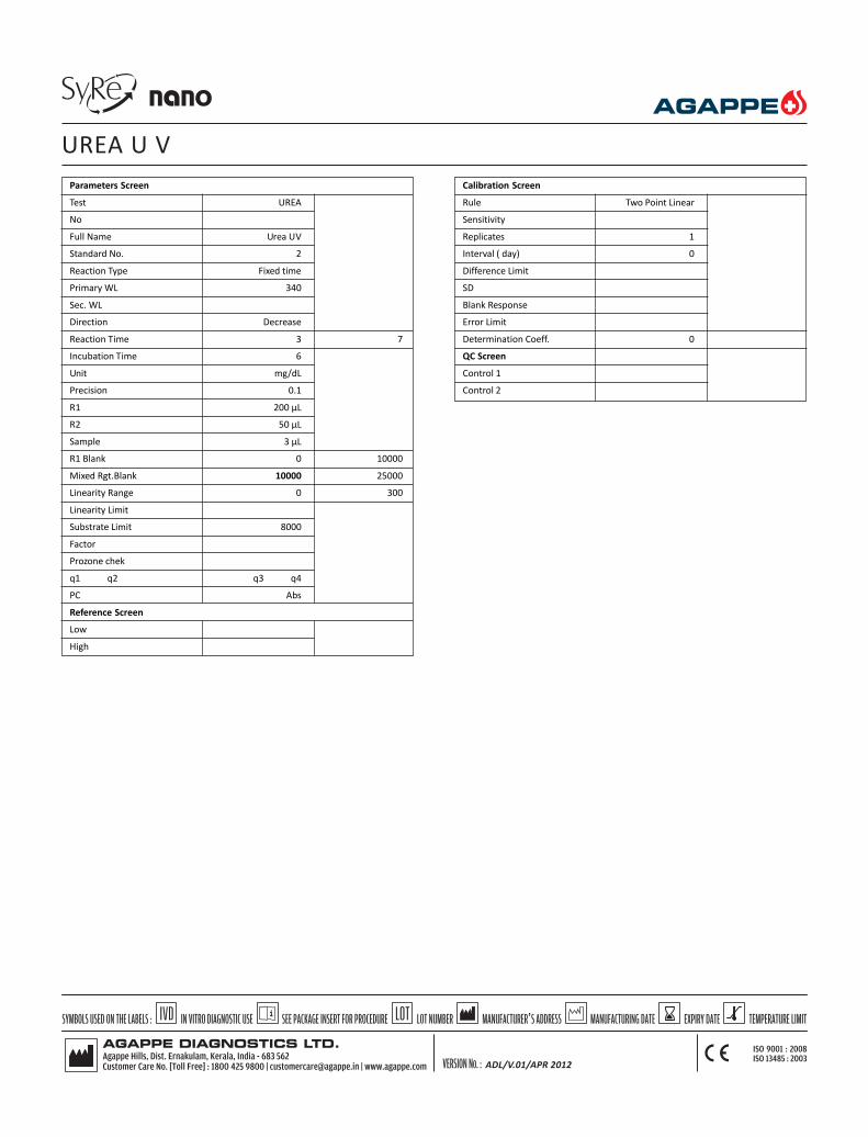
Parameters Screen
Test UREA
No
Full Name Urea UV
Standard No. 2
Reaction Type Fixed time
Primary WL 340
Sec. WL
Direction Decrease
Reaction Time 3 7
Incubation Time 6
Unit mg/dL
Precision 0.1
R1 200 µL
R2 50 µL
Sample 3 µL
R1 Blank 0 10000
Mixed Rgt.Blank 10000 25000
Linearity Range 0 300
Linearity Limit
Substrate Limit 8000
Factor
Prozone chek
q1 q2 q3 q4
PC Abs
Reference Screen
Low
High
Calibration Screen
Rule Two Point Linear
Sensitivity
Replicates 1
Interval ( day) 0
Difference Limit
SD
Blank Response
Error Limit
Determination Coeff. 0
QC Screen
Control 1
Control 2
UREA U V
ADL/V.01/APR 2012

INTENDED USE: This reagent is intended for in vitro quantitative determination ofUric acid in serum, plasma & urine.
-Uricase methodology.
-Linear up to 25 mg/dL.
CLINICAL SIGNIFICANCE
Uric acid is the end product of purine metabolism. Uric acid is excreted by thekidneys.
Increased levels are found in Gout, arthritis, impaired renal functions & starvation.
Decreased levels are found in yellow atrophy of the liver.
PRINCIPLE
Enzymatic determination of Uric acid according to the following reactions.
Uricase
Uric acid + 2H2O+O
2 ----------------------> Allantoine +CO
2 +H
2O
2
Peroxidase
2H2O
2 + 4 – Aminoantipyrine +EHSPT--------------->Red quinone
EHSPT= N-Ethyl N-(2-Hydroy -3 Sulfopropyl) n-Toluidine
REAGENT COMPOSITION
URIC ACID R1 2 x 35 mL
Phosphate Buffer (pH 7.5) 100 mmol/L
EHSPT 1.10 mmol/L
Ferrocyanure 50 mmol/L
Amino -4-antipyrine 0.37 mmol/L
Peroxidase > 3000 U/L
Uricase > 140 U/L
Reagents Required But not Provided
Multi Calibrator ( Product Code No - 11610001)
Qualicheck path (Product Code No - 11601002)
Qualicheck norm (Product Code No - 11601003)
STORAGE & STABILITY
The sealed reagents are stable up to the expiry date stated on the label, when storedat 2 - 80C.
LINEARITY
This reagent is linear up to 25 mg/dL.
If the concentration is greater than linearity (25 mg/dL), dilute the sample withnormal saline and repeat the assay. Multiply the result with dilution factor.
NORMAL RANGE
It is recommended that each laboratory establish its own reference values.
The following values may be used as guide line.
Serum / Plasma :
Men 3.4-7.0 mg/dL
Women 2.4-5.7 mg/dL
PRECAUTION
To avoid contamination, use clean laboratory materials.
Avoid direct exposure of reagent to light.
SAMPLE
Serum, EDTA or Heparin plasma (free of haemolysis)
CALIBRATION
Agappe Multicalibrator( Product Code No - 11610001)is recommended forcalibration .
Quality Control – It is recommended to use Qualicheck Norm (Code No. 11601003)or Qualicheck path (Code No. 11601002) to verify the performance of themeasurement procedure.
Each Laboratory has to establish its own internal quality control scheme andprocedures for corrective action if controls do not recover within the acceptabletolerance.
BIBLIOGRAPHY
1. Barham,D.,Trider P., Analyst 97,142 (1972)
2. Fossatj Clin. Chem. 26/2,227 (1980)
URIC ACID 2 x 35 mL
12602001
ADL/V.01/APR 2012

Parameters Screen
Test Uric Acid
No
Full Name Uric Acid
Standard No. 2
Reaction Type End Point
Primary WL 546
Sec. WL 670
Direction Increase
Reaction Time 0 38
Incubation Time
Unit mg/dL
Precision 0.1
R1 300 µL
R2
Sample 6 µL
R1 Blank 0 2000
Mixed Rgt.Blank
Linearity Range 0 15
Linearity Limit
Substrate Limit
Factor
Prozone chek
q1 q2 q3 q4
PC Abs
Reference Screen
Low
High
Calibration Screen
Rule Two Point Linear
Sensitivity
Replicates 1
Interval ( day) 0
Difference Limit
SD
Blank Response
Error Limit
Determination Coeff. 0
QC Screen
Control 1
Control 2
URIC ACID
ADL/V.01/APR 2012

INTENDED USEThis reagent is intended for in vitro quantitative determination of HbA1c in humanblood.Latex enhanced ImmunoturbidimetryReady to use liquid stable reagentsMultipoint calibrationDirect result (% HbA1c) from analyzerNo total Hb determination required
CLINICAL SIGNIFICANCEHbA1c is a glycated form of haemoglobin formed by the attachment of glucoseresidues in the blood to the hemoglobin molecules. In the diabetic populationwhere blood glucose levels are abnormally elevated the level of HbA1c alsoincreases. The level of HbA1c is proportional to the level of glucose in the bloodand has been widely accepted as an indicator of the mean blood glucoseconcentration in the preceeding 6-8 weeks. It is therefore a long-term indicator ofdiabetic control. For routine use HbA1c levels should be monitored every 3-4months. However in gestational diabetes and after a change in therapy it may beuseful to measure HbA1c more frequently at 2-4 week intervals.
PRINCIPLEThe principle of the test is latex agglutination method that measures the ratio ofhemoglobin A1c that occupy in a total hemoglobin in the whole blood. The sample(hemolysis sample ) is added to the unsensitized latex particles, and the surfaces ofthe latex adsorbs a total hemoglobin in the sample. Anti human HbA1c mousemonoclonal antibody complex agglutinates by anti-mouse IgG goat antibody. At thistime, the amount of agglutination caused depends on the amount of HbA1c thatadsorbs the surface of the latex, this this agglutination is measured as a turbidity. theconcentration of HbA1c (%) in the sample is determined by referring to the calibrationcurve obtained by the same test of diluted standard solutions.
REAGENT COMPOSITIONHbA1c R1 1 x 15 mL/2 x 15 mLLatexHbA1c R2 1 x 5 mL / 2x 5 mLAnti-human HbA1c mouse monoclonal antibodyAnti-mouse IgG goat antibodyHbA1c R3 1 x 32 mL / 1 x 63 mLHaemolysis ReagentHbA1c DIRECT CALIBRATOR 4 x 0.5 mLHbA1c 4 level calibrator (lyophilized).
STORAGE AND STABILITYThe sealed reagents are stable up to the expiry date stated on the label, when storedat 2- 80C, protected from light. Once opened the reagent is stable upto four weeks, ifcontamination is avoided. Recalibration is recommended after 30 days. DO NOT FREEZECalibrator : Reconstitute the calibrator with 0.5mL distilled water. It is stable for 30days at 2-8oC . DO NOT FREEZE.
LINEARITY RANGEThe reagent is linear upto 13% (NGSP)
REFERENCE RANGEIt is recommended that each laboratory establish its own reference values.The following value may be used as guide line.Reference normal value (NGSP): 4.6% -6.2%
PRECAUTIONTo avoid contamination, use clean laboratory wares. Use clean, dry disposable pipettetips for dispensing. Close reagent and standard bottles immediately after use.Avoid direct exposure of working reagent to light.
SAMPLEWhole blood, collected with EDTATo determine HbA1c a heamolysate must be prepared for each sample1. Dispense 1mL hemolysis reagent into a tube.2. Add 20 μLof well-mixed whole blood and mix.3. Allow to stand for 5 minutes or until complete lysis is evident.
Follow the same procedure with controls and calibrator.
GENERAL SYSTEM PARAMETER FullyAutoMode of Reaction End pointSlope of reaction IncreasingWavelength 660 nmTemperature 370CCalibrator Concentration As on vial labelLinearity 13%Blank Reagent blankSample volume 7.5 μLReagent 1 volume 180 μLReagent 2 volume 60 μLCuvette 1 cm light pathLABORATORY PROCEDURE FOR FULLY AUTO
Blank Calibrator Sample/controlHbA1c R 1 180 μL 180 μL 180 μLCalibrator - 7.5μL -Hemolysate(sample/control) - - 7.5μLMix & incubate for 5 min at 370C.HbA1c R 2 60 μL 60 μL 60 μLMix and incubate for 5 min at 37oC and read absorbance(A) at 660 nm.
CALCULATIONCalibration curveCalculate the Abs of calibrators = Abs calibrator – Abs Blank. Plot the D Abs of eachcalibrator versus assigned concentration (HbA1c %) on a linear graph paper. HbA1cresults according to NGSP for the samples and controls are determined using theprepared calibration curve.Calculate
Abs of sample ie abs Sample - abs Blank .HbA1c % in the sample is calculated by interpolation of Abs of sample on thecalibration curve. For calculation of results according to IFCC, use IFCC calibratorvalues (see calibrator insert), or use following equation.NGSP = (0.915 x IFCC) + 2.15
INTERFERENCESNo interference upto :Ascorbic acid 50 mg/dLBilirubin 40mg/dLIntra lipid 3000 mg/dL
It has been reported that results may be inconsistent in patients who have thefollowing conditions: opiate addiction, lead poisoning, alcoholism, and ingestion oflarge doses of aspirin. Elevated HbF levels may lead to under estimation of HbA1c.
BIBLIOGRAPHY1. Engbeak, F., et al. Clin chem.35 p. 93-97 (1989)2. American Diabetes Association : Clinical practice recommendations (position
statement). Diabetes care 24 (suppl.1) S33-S55, (2001).3. Tietz, N.W. Textbook of Clinical Chemistry, W.B. Saunders Company, p.794 -795
(1999).
HbA1c DIRECT WITH CALIBRATOR 1 x 15 /1 x 5/ 1x 32 /4 x0.5mL11835001
ADL/V.02/July 2013
2 x15/2 x5 /1x 63/4x0.5mL11835002
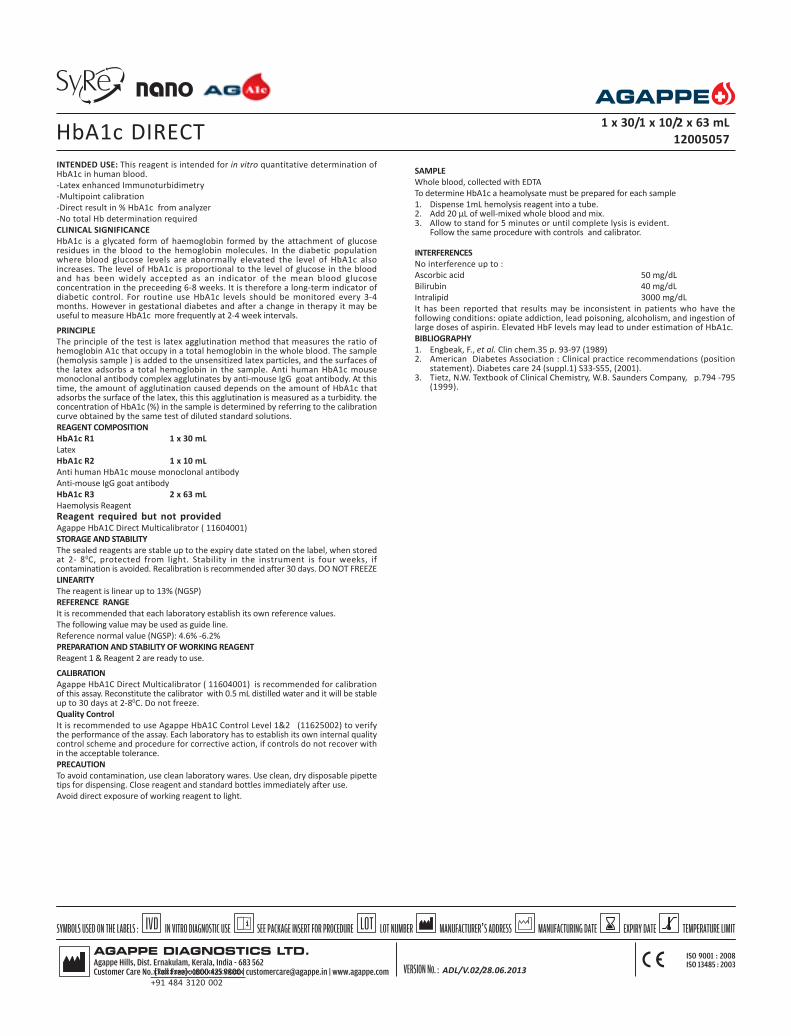
xxxxxxxxxxxxxxxxxxxxxx+91 484 3120 002
INTENDED USE: This reagent is intended for in vitro quantitative determination ofHbA1c in human blood.-Latex enhanced Immunoturbidimetry-Multipoint calibration-Direct result in % HbA1c from analyzer-No total Hb determination requiredCLINICAL SIGNIFICANCEHbA1c is a glycated form of haemoglobin formed by the attachment of glucoseresidues in the blood to the hemoglobin molecules. In the diabetic populationwhere blood glucose levels are abnormally elevated the level of HbA1c alsoincreases. The level of HbA1c is proportional to the level of glucose in the bloodand has been widely accepted as an indicator of the mean blood glucoseconcentration in the preceeding 6-8 weeks. It is therefore a long-term indicator ofdiabetic control. For routine use HbA1c levels should be monitored every 3-4months. However in gestational diabetes and after a change in therapy it may beuseful to measure HbA1c more frequently at 2-4 week intervals.
PRINCIPLEThe principle of the test is latex agglutination method that measures the ratio ofhemoglobin A1c that occupy in a total hemoglobin in the whole blood. The sample(hemolysis sample ) is added to the unsensitized latex particles, and the surfaces ofthe latex adsorbs a total hemoglobin in the sample. Anti human HbA1c mousemonoclonal antibody complex agglutinates by anti-mouse IgG goat antibody. At thistime, the amount of agglutination caused depends on the amount of HbA1c thatadsorbs the surface of the latex, this this agglutination is measured as a turbidity. theconcentration of HbA1c (%) in the sample is determined by referring to the calibrationcurve obtained by the same test of diluted standard solutions.REAGENT COMPOSITIONHbA1c R1 1 x 30 mLLatexHbA1c R2 1 x 10 mLAnti human HbA1c mouse monoclonal antibodyAnti-mouse IgG goat antibodyHbA1c R3 2 x 63 mLHaemolysis ReagentReagent required but not providedAgappe HbA1C Direct Multicalibrator ( 11604001)STORAGE AND STABILITYThe sealed reagents are stable up to the expiry date stated on the label, when storedat 2- 80C, protected from light. Stability in the instrument is four weeks, ifcontamination is avoided. Recalibration is recommended after 30 days. DO NOT FREEZELINEARITYThe reagent is linear up to 13% (NGSP)REFERENCE RANGEIt is recommended that each laboratory establish its own reference values.The following value may be used as guide line.Reference normal value (NGSP): 4.6% -6.2%PREPARATION AND STABILITY OF WORKING REAGENTReagent 1 & Reagent 2 are ready to use.
CALIBRATIONAgappe HbA1C Direct Multicalibrator ( 11604001) is recommended for calibrationof this assay. Reconstitute the calibrator with 0.5 mL distilled water and it will be stableup to 30 days at 2-80C. Do not freeze.Quality ControlIt is recommended to use Agappe HbA1C Control Level 1&2 (11625002) to verifythe performance of the assay. Each laboratory has to establish its own internal qualitycontrol scheme and procedure for corrective action, if controls do not recover within the acceptable tolerance.PRECAUTIONTo avoid contamination, use clean laboratory wares. Use clean, dry disposable pipettetips for dispensing. Close reagent and standard bottles immediately after use.Avoid direct exposure of working reagent to light.
SAMPLEWhole blood, collected with EDTATo determine HbA1c a heamolysate must be prepared for each sample1. Dispense 1mL hemolysis reagent into a tube.2. Add 20 μL of well-mixed whole blood and mix.3. Allow to stand for 5 minutes or until complete lysis is evident.
Follow the same procedure with controls and calibrator.
INTERFERENCESNo interference up to :Ascorbic acid 50 mg/dLBilirubin 40 mg/dLIntralipid 3000 mg/dLIt has been reported that results may be inconsistent in patients who have thefollowing conditions: opiate addiction, lead poisoning, alcoholism, and ingestion oflarge doses of aspirin. Elevated HbF levels may lead to under estimation of HbA1c.BIBLIOGRAPHY1. Engbeak, F., et al. Clin chem.35 p. 93-97 (1989)2. American Diabetes Association : Clinical practice recommendations (position
statement). Diabetes care 24 (suppl.1) S33-S55, (2001).3. Tietz, N.W. Textbook of Clinical Chemistry, W.B. Saunders Company, p.794 -795
(1999).
HbA1c DIRECT 1 x 30/1 x 10/2 x 63 mL12005057
ADL/V.02/28.06.2013

xxxxxxxxxxxxxxxxxxxxxx+91 484 3120 002
Parameters Screen
Test HbA1c D
No
Full Name HbA1c Direct
Standard No. 5
Reaction Type Endpoint
Primary WL 670
Sec. WL
Direction Increase
Reaction Time -1 19
Incubation Time 12
Unit %
Precision 0.01
R1 180 μL
R2 60 μL
Sample 7.5μL
R1 Blank
Mixed Rgt.Blank
Linearity Range 3 13
Linearity Limit
Substrate Limit
Factor
Prozone chek
q1 q2 q3 q4
PC
Refence Screen
Low *
High *
* Data entered by the operator
Calibration Screen
Rule Spline
Sensitivity
Replicates 1
Interval ( day) 0
Difference Limit
SD
Blank Response
Error Limit
Determination Coeff. 0
QC Screen
Control 1 *
Control 2 *
HbA1c DIRECT
ADL/V.01/28.06.2013

AGAPPE DIAGNOSTICS LTD.
Agappe Hills, Dist. Ernakulam, Kerala, India - 683 562Customer Care No. [Toll Free] : 1800 425 9800 | [email protected] | www.agappe.com
ISO 9001 : 2008ISO 13485 : 2003
IVD LOT LOT NUMBERSYMBOLS USED ON THE LABELS : SEE PACKAGE INSERT FOR PROCEDUREIN VITRO DIAGNOSTIC USE MANUFACTURER S ADDRESS’ TEMPERATURE LIMITEXPIRY DATEMANUFACTURING DATE
VERSION No. :
INTENDED USE: This reagent is intended for in vitro quantitative determination ofHbA1c in human blood.Latex enhanced ImmunoturbidimetryMultipoint calibrationDirect result in % HbA1c from analyzerNo total Hb determination requiredCLINICAL SIGNIFICANCEHbA1c is a glycated form of haemoglobin formed by the attachment of glucoseresidues in the blood to the hemoglobin molecules. In the diabetic populationwhere blood glucose levels are abnormally elevated the level of HbA1c alsoincreases. The level of HbA1c is proportional to the level of glucose in the bloodand has been widely accepted as an indicator of the mean blood glucoseconcentration in the preceeding 6-8 weeks. It is therefore a long-term indicator ofdiabetic control. For routine use HbA1c levels should be monitored every 3-4months. However in gestational diabetes and after a change in therapy it may beuseful to measure HbA1c more frequently at 2-4 week intervals.PRINCIPLEThe kit is based on latex agglutination method that measures the ratio of hemoglobinA1c that occupy in a total haemoglobin in the whole blood. The sample is added tothe unsensitization latex particles, and the surfaces of the latex adsorbs a totalhaemoglobin in the sample. Anti -human HbA1c mouse monoclonal antibody is toreact and anti-human Hba1c mouse monoclonal antibody complex agglutinates byanti-mouse IgG goat anti body. At this time the amount of agglutination causeddepends on the amount of HbA1c that adsorbs the surface of the latex, thisagglutination is measured as a turbidity. The concentration of HbA1c(%)in the wholeblood is calculated from a standard curve.REAGENT COMPOSITIONHbA1c R1 1 x 26 mLLatexHbA1c R2 1 x 9 mLAnti-human HbA1c mouse monoclonal antibodyAnti mouse IgG goat antibodyHbA1c R3 1 x 63 mLHaemolysis ReagentReagent required but not providedAgappe HbA1C Direct Multicalibrator ( 11604001) STORAGE AND STABILITYThe sealed reagents are stable up to the expiry date stated on the label, when storedat 2- 80C, protected from light. Stability in the instrument is four weeks, ifcontamination is avoided. Recalibration is recommended after 30 days. DO NOT FREEZEPRECAUTIONClose reagent and standard bottles immediately after use. Keep the reagent back tospecified temperature after each useAvoid direct exposure of reagent to light.Avoid freezing of reagents and refrigerate them at proper temperature. Shake R1and R2 reagent well before use.LINEARITYThe reagent is linear up to 13% (NGSP)REFERENCE RANGEIt is recommended that each laboratory establish its own reference values.The following value may be used as guide line.Reference normal value (NGSP) : 4.6 - 6.2 %PREPARATION AND STABILITY OF REAGENTReagent 1, reagent R2 and reagent R3 are ready to use.
CALIBRATIONAgappe HbA1C Direct Multicalibrator (11604001) is recommended for calibration ofthis assay. Reconstitute the calibrator with 0.5 mL distilled water and it will be stableup to 30 days at 2-80C. Do not freeze.Quality ControlIt is recommended to use Agappe HbA1C Control level 1&2 (11625002) to verify theperformance of the assay. Each laboratory has to establish its own internal qualitycontrol scheme and procedure for corrective action, if controls do not recover within the acceptable tolerance.SAMPLEWhole blood, collected with EDTATo determine HbA1c a heamolysate must be prepared for each sample1. Dispense 1mL hemolysis reagent into a tube.2. Add 20 μL of well-mixed whole blood and mix.3. Allow to stand for 5 minutes or until complete lysis is evident.
Follow the same procedure with controls and calibrator.
INTERFERENCESNo interference upto :Ascorbic acid 50 mg/dLBilirubin 40 mg/dLIntralipid 3000 mg/dL
It has been reported that results may be inconsistent in patients who have thefollowing conditions: opiate addiction, lead poisoning, alcoholism, and ingestion oflarge doses of aspirin. Elevated HbF levels may lead to under estimation of HbA1c.BIBLIOGRAPHY1. Nathan, D.M., Clin, Chem. 29, pp.466-469 (1983)2. Engbeak, F., et al. Clin chem.35 pp. 93-97 (1989)3. American Diabetes Association : Clinical practice recommendations (position
statement). Diabetes care 24 (suppl.1) S33-S55, (2001).4. Tietz, N.W. Textbook of Clinical Chemistry, W.B. Saunders Company,
p.794 -795 (1999).
HbA1c DIRECT 1 x 26 mL/1 x 9 mL/1x 63 mL
12013042
ADL/V.01/July 2013
Page 1Test (Name) HbA1cDecimal 2Unit %Reac. Method EndBlank R BlankWave length I 670Wave length II -Test Time 270 - 300Sample 8 μLR1 210 μLR2 70 μLCalculation Method Spline
Mispa Mini Assay Parameter