Probiotics and Prebiotics in Human Nutrition and Health
392
www.ebook3000.com
-
Upload
khangminh22 -
Category
Documents
-
view
0 -
download
0
Transcript of Probiotics and Prebiotics in Human Nutrition and Health

Edited by Venketeshwer Rao and Leticia G. Rao
Probiotics and Prebiotics in Human Nutrition and Health
www.ebook3000.com

Спизжено у ExLib: avxhome.se/blogs/exLib
Stole src from http://avxhome.se/blogs/exLib:
Stole src from http://avxhome.se/blogs/exLib/
AvE4EvA MuViMix RecordsPublishing Process Manager Technical EditorCover Designer
Probiotics and Prebiotics in Human Nutrition and Health
Edited by Venketeshwer Rao and Leticia G. Rao
Published by ExLi4EvACopyright © 2016
All chapters are Open Access distributed under the Creative Commons Attribution 3.0 license, which allows users to download, copy and build upon published articles even for commercial purposes, as long as the author and publisher are properly credited, which ensures maximum dissemination and a wider impact of our publications. After this work has been published, authors have the right to republish it, in whole or part, in any publication of which they are the author, and to make other personal use of the work. Any republication, referencing or personal use of the work must explicitly identify the original source.
As for readers, this license allows users to download, copy and build upon published chapters even for commercial purposes, as long as the author and publisher are properly credited, which ensures maximum dissemination and a wider impact of our publications.
Notice Statements and opinions expressed in the chapters are these of the individual contributors and not necessarily those of the editors or publisher. No responsibility is accepted for the accuracy of information contained in the published chapters. The publisher assumes no responsibility for any damage or injury to persons or property arising out of the use of any materials, instructions, methods or ideas contained in the book.
Спизжено у ExLib: avxhome.se/blogs/exLibISBN-10: 953-51-2476-5ISBN-13: 978-953-51-2476-4 Print ISBN-10: 953-51-2475-7ISBN-13: 978-953-51-2475-7
www.ebook3000.com

Preface
Contents
Chapter 1 Prebiotic and Probiotic Approaches to Improving Food Safety on the Farm and Their Implications on Human Health by William B. Smith, Todd R. Callaway, Luis O. Tedeschi, Francis M. Rouquette, Trisha Sheridan and Jennifer Adamski
Chapter 2 Probiotics: A Comprehensive Review of Their Classification, Mode of Action and Role in Human Nutrition by Amirreza Khalighi, Reza Behdani and Shabnam Kouhestani
Chapter 3 Prebiotics: Metabolism and Symbiotic Synergy with Probiotics in Promoting Health by Nditange Shigwedha, Penny Hiwilepo-Van Hal, Li Jia, Liubov Sichel and Shuang Zhang
Chapter 4 Lactobacillus reuteri, Infant Allergy Prevention and Childhood Immune Maturation by Anna Forsberg
Chapter 5 The Synergistic Contribution of Lactobacillus and Dietary Phytophenols in Host Health by Danielle N. Kling, Guillermo E. Marcial, Dana N. Roberson, Graciela L. Lorca and Claudio F. Gonzalez
Chapter 6 Pili in Probiotic Bacteria by Vengadesan Krishnan, Priyanka Chaurasia and Abhiruchi Kant
Chapter 7 Biosynthesis of Vitamins by Probiotic Bacteria by Qing Gu and Ping Li
Chapter 8 Bioactive Compounds of Lactic Acid Bacteria. Case Study: Evaluation of Antimicrobial Activity of Bacteriocin-producing Lactobacilli Isolated from Native Ecological Niches of Ecuador by Gabriela N. Tenea and Lucia Yépez
www.ebook3000.com

VI Contents
Chapter 9 Fructosyltransferase Sources, Production, and Applications for Prebiotics Production by Mariela R. Michel, Rosa M. Rodríguez-Jasso, Cristóbal N. Aguilar, Silvia M. Gonzalez-Herrera, Adriana C. Flores- Gallegos and Raúl Rodríguez-Herrera
Chapter 10 Antimicrobial Effect of Probiotics against Common Pathogens by Sabina Fijan
Chapter 11 Probiotics in Childhood Celiac Disease by Caterina Anania, Francesca Olivero, Eugenia Olivero and Lucia Pacifico
Chapter 12 Probiotics for Prevention and Treatment of Candidiasis and Other Infectious Diseases: Lactobacillus spp. and Other Potential Bacterial Species by Michelle Peneluppi Silva, Rodnei Dennis Rossoni, Juliana Campos Junqueira and Antonio Olavo Cardoso Jorge
Chapter 13 Phosphorus Nutrition and Health: Utilization of Phytaseproducing Bifidobacteria in Food Industry by Long Chen, Fengshou Tian and Zhongke Sun
Chapter 14 Probiotic Microorganisms in Dry Fermented Meat Products by Katarzyna Neffe‐Skocińska, Karolina Wójciak and Dorota Zielińska
Chapter 15 Pectic Oligosaccharides and Other Emerging Prebiotics by Beatriz Míguez, Belén Gómez, Patricia Gullón, Beatriz Gullón and José Luis Alonso
Chapter 16 Yeasts as Potential Source for Prebiotic β-Glucan: Role in Human Nutrition and Health by Pedro De Oliva-Neto, Sidmeire Santos Oliveira, Estevão Zilioli and Márcia Zilioli Bellini
www.ebook3000.com

Chapter 17 Prebiotics, Probiotics, Synbiotics and Functional Foods in Control and Treatment of Type II Diabetes Mellitus and Colorectal Cancer by Samuel Longoria-García, Ruth E. Belmares-Cerda, Mildred I.M. Flores-Verástegui, Juan C. Contreras-Esquivel, Julio C. Montañez-Sáenz and Mario Alberto Cruz-Hernández
Chapter 18 Probiotics, Prebiotics, and Biogenics for the Stomach by Yasuhiko Komatsu, Yuji Aiba, Yasuhiro Nakano and Yasuhiro Koga
VII Contents
www.ebook3000.com
Preface
Probiotic microorganisms are recognised as being beneficial for human health. Prebiotics are substrates that are used preferentially by the probiotic bacteria for their growth. A great deal of interest has been generated in recent years in identifying probiotic bacteria and prebiotics, their characterization, mechanisms of action and their role in the prevention and management of human health disorders. Together they are referred to as synbiotic.
This book is in response to the need for more current and global scope of probiotics and prebiotics. It contains chapters written by internationally recognized authors. The book has been planned to meet the needs of the researchers, health professionals, government regulatory agencies and industries.
This book will serve as a standard reference book in this important and fast-growing area of probiotics and prebiotics in human nutrition and health.
www.ebook3000.com

Chapter 1
Prebiotic and Probiotic Approaches to Improving FoodSafety on the Farm and Their Implications on HumanHealth1
William B. Smith, Todd R. Callaway, Luis O. Tedeschi,Francis M. Rouquette Jr., Trisha Sheridan andJennifer Adamski
Additional information is available at the end of the chapter
http://dx.doi.org/10.5772/63114
Abstract
Human health is a broad category that encompasses the entirety of the food produc‐tion system. Livestock production practices have important effects on human healthbecause livestock not only are a primary food source but also can be the source ofpathogenic bacteria that may enter the food chain indirectly. As government regula‐tion and public scrutiny restrict the prophylactic use of antibiotic and antimicrobialinterventions, other techniques must be used to reduce the burden of animal‐bornepathogenic bacteria entering the food system. Prebiotics (isolated compounds thatenhance natural microflora and thereby decrease pathogens) and probiotics (livemicrobes that are administered to livestock to enhance microbial diversity and crowdout pathogens) represent two unique opportunities for alternative measures inpathogen reduction. This review addresses the link between animal production andhuman health, the agricultural sources of pathogenic organisms, and the probiotic andprebiotic approaches that have been evaluated in an effort to reduce carriage offoodborne pathogenic bacteria by livestock.
Keywords: food safety, livestock, prebiotic, preharvest intervention, probiotic
1 Proprietary brand names are necessary to report factually on available data; however, the USDA neitherguarantees nor warrants the standard of the product, and the use of the name by the USDA implies no approvalof the product and/or exclusion of others that may be suitable.

1. Introduction: why is farm‐based intervention of interest to humanhealth?
This book is dedicated to the understanding and dissemination of knowledge surroundingprebiotic use in human health. Thus, it begs the following questions: When a reader finds thisparticular manuscript, what is the point? What is the objective of a farm‐based perspectivewhen the focus is on human health? While these may be valid questions to the casual observer,a full understanding of potential pathogens and intervention in the subject of human healthmust by rights include a discussion of the foodstuff at its source. Like all mammals, livestockharbor a diverse collection of bacteria [1]. In fact, the gastrointestinal tract of these animals canharbor in excess of 2000 bacterial species at concentrations of 1010 cells/g of digesta [2]. Whilethe majority of these organisms are beneficial to the host and part of the stable native microfloraof the gut [3], certain instances or conditions allow pathogenic bacteria to colonize within theanimal. Some of these bacteria can make their way from the gut or the hide during processing[4], introducing pathogens into the abattoir (slaughter plant) at harvest that must then be dealtwith in final food products. As noted in Reference [1], a great number of these pathogenicbacteria in the realm of human health are also of interest in that of livestock animal health andcan commonly be traced back to those very animals. Since these pathogens are a threat to thewell‐being of both humans and livestock, one must then investigate intervention strategies bywhich the microbial burden may be reduced at the source so that these pathogenic organismswould never enter the human food chain.
Traditionally, farm‐level or feeder/finisher‐level control of pathogens has been achievedthrough prophylactic antibiotic and antimicrobial addition to feeds. The main source ofprevention of pathogenic bacterial entry into the food system is through Hazard Analysis andCritical Control Point (HACCP) plans at the abattoir [5]. It should be noted that HACCP controlmeasures are only effective to a certain point (i.e., they are not perfect), but any reduction ofpathogen shedding prior to entry into the abattoir will reduce the burden and assist in theefficacy of in‐plant HACCP‐based controls [6]. In fact, with the subtherapeutic antibiotic useban in the European Union [7,8] and increased public scrutiny of antibiotic use in livestock inthe United States [9], alternative preharvest control strategies must be devised and imple‐mented, especially given the direct correlation between live animals shedding foodbornepathogenic bacteria, such as Escherichia coli O157:H7, and the incidence of positive carcassesat the abattoir [5]. Thus, preharvest intervention strategies, such as use of probiotics andprebiotics, need to be viewed as an additional critical control measure that can be included inthe food safety continuum.
So how then do preharvest interventions in animals work? Much of the efficacy of productsthat will be described in the present review can be loosely grouped under an umbrella conceptknown as a “competitive enhancement” approach to pathogen reduction [1,10–13]. The firstfacet is based upon the introduction of naturally-occurring microflora isolates from thegastrointestinal tract of an animal of the same species [1], occupying all available ecologicalniches in the gastrointestinal tract and thereby excluding pathogens [1,14]. When used inneonatal (or newly hatched) animals, this technique is known as “competitive exclusion” (CE),
Probiotics and Prebiotics in Human Nutrition and Health2

which reduces pathogen penetration of the naive and essentially sterile neonatal gastrointes‐tinal tract [1,14]. Use of probiotics (also known in the animal industry as direct‐fed microbials[DFMs]) is a slightly different approach in which existing gastrointestinal microbial popula‐tions can be diversified or modified/attenuated by daily inclusion of a bacterial or fungalpopulation or end‐product, and this may have an inhibitory effect on pathogenic bacteria,including foodborne pathogens [1,15]. A further competitive enhancement strategy is theaddition of prebiotics, which are limiting nutrients or isolated compounds that are indigestibleby the host but give specific innate microbes a competitive advantage that can have a delete‐rious effect on pathogenic bacteria, to the diet [1]. Furthermore, several of these approachescan be synergistically combined and are termed “synbiotics”; for example, a DFM dependenton the inclusion of prebiotics can be maintained in the gut and given a further competitiveadvantage to remain in the population to benefit host animal health and production or toimprove food safety.
2. Pathogens: what are the sources?
As previously noted, the body , and especially the gut, of most food animals contains manymicroorganisms [2]. While the vast majority of these are beneficial (commensal) to the host,there are select species and serovars (e.g., Salmonella) that exhibit pathogenic or toxigeniceffects in both humans and livestock. These pathogens are naturally occurring organisms that,given the opportunity, can colonize the environment of the innate gut microflora and take holdof niches in an otherwise healthy animal. This section provides a discussion of some of themore common pathogenic bacteria in livestock and how these microbes may become a problemin the safety and security of the food chain.
2.1. Campylobacter
Campylobacter has been identified as one of the most common foodborne pathogenic bacteria.Most commonly, Campylobacter has been linked to poultry products and linked to human casesof gastroenteritis in most cases as well as the Guillain‐Barré syndrome, reactive arthritis, andirritable bowel syndrome or inflammatory bowel disease in the most severe cases [16,17].Campylobacter is a major concern for infection in poultry production [16–18]. One route ofcontamination, also common to most other pathogens, is through livestock water sources [19].In an area of intense livestock (dairy) production in England, Campylobacter jejuni was foundin 14.3% of water sources sampled (predominantly in running water or troughs), Campylobactercoli was found in 18.5% (predominantly in stagnant water), and Campylobacter lari wasidentified in 4.2% [20]. In this same study, variables were regressed to show their impact onthe prevalence of Campylobacter spp. In a multiple regression model, water source and soil typeplayed the most significant role in determining the environmental prevalence of Campylobact‐er, with natural water sources and high clay content both increasing its prevalence [20].
Prebiotic and Probiotic Approaches to Improving Food Safety on the Farm and Their Implications on Human Healthhttp://dx.doi.org/10.5772/63114
3
2.2. Enterohemorrhagic E. coli (EHEC)
Enterohemorrhagic E. coli is a group of highly virulent foodborne pathogenic bacteria that isof great interest to human health. The well‐known E. coli serotype O157:H7 was first identifiedin a clinical outbreak of undercooked hamburger patties at a commercial fast food chain in theUnited States [21]. In fact, this pathogenic serotype has been linked to one of the greatestfoodborne pathogen outbreaks in American history [22,23]. In this landmark case, in whichover 150 cases were reported and multiple deaths occurred [22], E. coli O157:H7 was isolatedfrom ground beef patties and subsequently sourced to the abattoir in which meat wascontaminated from pathogenically infected animals [23]. These human infections commonlyresulted in postdiarrheal hemolytic uremic syndrome (HUS) and disproportionately affectedthe young and elderly [22]. While inoculation of the livestock host is generally achievedthrough fecal‐oral contamination or contaminated drinking sources [19,24], this does notaccount for the transmission of pathogens from the live animal to the meat during processing.Much of the contamination in the plant, especially with regard to E. coli O157:H7, can be tracedto contamination of the hide and interaction during hide removal and evisceration [25]. In asampling of over 2500 cattle hides from across North America, researchers discovered thatover half of the hides were contaminated with nearly 300 unique isolates of E. coli O157:H7 [4].Additionally, the frequency of the unique isolates obtained from cattle hides was very similarto the prevalence of isolates identified in human clinical cases [4]. In a survey of high‐through‐put Midwestern United States abattoirs, 11% of all hides, 43% of pre‐eviscerated carcasses, and2% of postprocessed carcasses tested positive for EHEC O157:H7 [5]. This included positivetests for hides in 38% of introduced lots, pre‐eviscerated carcasses in 87% of lots, and post‐processed carcasses in 17% of lots [5]. While E. coli O157:H7 is the best known of the EHECgroup, other members (e.g., O26, O111) also pose significant threats to the food supply aroundthe world. Although E. coli O157:H7 was quickly categorized by the U.S. Food Safety InspectionService as an adulterant [26], an additional six serotypes are now included in this importantcategory [27] and thus carry an important public health and economic impact.
2.3. Salmonella
Salmonella is another bacterial pathogen of significant concern both as a foodborne pathogenand as a threat to animal health, having been identified in all vertebrates [28]. More than 2500separate serotypes comprise Salmonella enterica [29], which is the most common species foundin food animals. Salmonella accounted for 55% of the foodborne illness outbreaks in the UnitedStates from 1993 to 1997 [30] and 26% of the outbreaks from 1998 to 2008 [31], with one of themost massive outbreaks being from ice cream hauled in tanker trucks that had improperlyhandled raw eggs [30]. Although researchers identify Salmonella as a ubiquitous microbe, ithas been noted that the primary reservoir for such a pathogen is the digestive tract of the animal(indicating fecal‐oral transmission or accidental contamination at the abattoir) and conditionsunder intensive production where animals are in close contact with one another are favored[32]. It should be noted, however, that a common vehicle for Salmonella contamination inhuman food is not livestock per se but instead vine‐stalk vegetables [31]. That said, in anevaluation of butcher shop poultry in Portugal, 60% of the products were found to be conta‐
Probiotics and Prebiotics in Human Nutrition and Health4

minated with Salmonella and the pathogen S. enteritidis was found to make up 44% of thosecases [32].
2.4. Others of interest
While Campylobacter, E. coli, and Salmonella are all identified and targeted as the primarypathogens of interest for reduction in the human food system [19], there are other pathogensof importance that are far less commonly addressed in scientific research. Clostridium, likemany other pathogens discussed herein, is a Gram‐positive, spore‐forming pathogenic bac‐terium [33]. Clostridium difficile infection is characterized by severe diarrhea and pseudo‐membranous colitis [33]. C. difficile is a known potential resident of the livestock intestinaltract and has been identified in up to 12% of sampled retail ground beef and ground pork ina Canadian study [34]. Clostridium perfringens, the leading cause of necrotic enteritis, can alsobecome a human health issue and has been isolated as a portion of the natural microflora ofthe jejunum, cecum, and cloaca of poultry [35]. Listeria monocytogenes is a pathogen mostcommonly associated with dairy products [36]. At the time of their review, Skovgaard andMorgen [37] stated that most human cases of listeriosis are of unknown origin, althoughfood was suspected, and recent high‐profile outbreaks have definitely confirmed such suspi‐cion [36]. Listeriosis has been linked to central nervous system infections, bacteremia, andendocarditis [37]. Listeria has been isolated from dairy feces as well as feedstuffs and attrib‐uted to mastitis in these animals [38]. Staphylococcus aureus has been associated with all live‐stock species [39]. It is an opportunistic pathogen that will colonize both livestock andhumans in an infectious nature [40].In dairy cattle, the pathogen is known as one of the lead‐ing causes of mastitis, and mastitis is among the leading losses to the dairy industry [41]. Inone study, 296 individual isolates of S. aureus of animal origin were discovered; while noneof the isolates from cattle or swine were found to be common with human infection, a signif‐icant number of the poultry isolates were common with those found in the bloodstream ofhumans [40].
3. Probiotics/direct‐fed microbials (DFMs)
A list of probiotics that have been used in food animals to reduce pathogenic bacteria ispresented in Table 1. Probiotics used in animals are known as DFMs and defined as live,biologically active microbes (bacterial or fungal), or dead cultures that include the end‐products of their fermentation, that are administered to an animal in hopes of enhancing thenatural gastrointestinal ecosystem and occupying any niches in which pathogenic organismsmay thrive [10,42]. Again, this concept is broadly categorized as competitive enhancement inwhich live, naturally occurring microbes are added to the host animal to enhance the innatepopulation in the gut [10,15]. As noted in Reference [43], the concept of CE specificallyoriginated with the application of mature broiler gastrointestinal contents for the reduction ofSalmonella [44]. While addition of DFMs to mature animals yields mixed and often negativeresults, their administration to livestock early in life (as early as the day of hatch in broilers)
Prebiotic and Probiotic Approaches to Improving Food Safety on the Farm and Their Implications on Human Healthhttp://dx.doi.org/10.5772/63114
5

has been shown to be effective in reducing pathogenic bacterial loads by kick‐starting thenatural succession of commensal bacterial colonization of the gastrointestinal microflora [18].In addition to the direct addition of probiotics to neonatal diets, passive immunity may alsobe conveyed to the neonate through supplementation of the dam before birth [45].
In addition to the benefits to livestock and human health in terms of a reduction in colonizationand shedding of pathogenic microbes, probiotics have also found a niche in the livestockmarket because of their added benefit of enhanced production performance. Because there arecurrently no economic incentives to implement food safety interventions in live animals,interventions should be able to “pay for themselves” by improving animal growth or produc‐tion efficiency. Many studies report the beneficial effects of DFMs on production efficiency incattle [9,46,47], swine [48], and poultry. The supplementation of feedlot cattle with a combi‐nation of Lactobacillus acidophilus NP45/NP51 and Propionibacterium freudenreichii NP24 resultedin an increase in the graded fat thickness of the animals at slaughter [9], indicative of improvedgain and efficiency. The use of Enterococcus faecium in feedlot cattle was able to increase theenergetic efficiency of the rumen by increasing the proportion of propionate (a glucogenicvolatile fatty acid) produced through ruminal fermentation, but all other digestive andproduction traits were not altered although fecal coliform shedding was increased, potentiallydue to colonic acidification [46]. Feeding multiparous dairy cows a combination of Saccharo‐myces cerevisiae (Diamond V‐XP, Diamond V, Cedar Rapids, IA) and Propionibacterium spp.P169 resulted in an increase in fat‐corrected milk yield, percent lactose, and weight gainpostpartum [47]. When nursery piglets were supplemented with a combination of Bacillussubtilis and Bacillus amyloliquefaciens, their average daily gain increased and gain‐to‐feed ratiosdecreased [48]. However, because the focus of this publication is on human health, thebeneficial effects of probiotics on animal production will be disregarded in this review,although it is important to understand that the economic benefits may indeed pay for theinclusion of a food safety enhancement.
Product Species Effective against Reported results Source
Bacillus spp. Broilers Campylobacter jejuniSamonellaTyphimurium[21]
1 to 3 log reduction intracloacallyPercentage reduction in the crop andceca
[17][55]
B. subtilis Swine Clostridium perfringensEscherichia coli
Increased litter survival, weaningweights and Lactobacillus populations
[50]
BiofeedTM (Bifidobacteriumlonghum, B. thermophylum,Lactobacillus acidophilus andStreptococcus faecium)
Swine ‐‐‐
Reduced pathogen load and incidence ofdiarrhea
[1]
BovamineTM (L. acidophilusand Propionibacteriumfreudenreichii)
Beef cattle Escherichia coli Reduces populations of O157:H7 [1]
Enterococcus faecium Swine Swine influenza A Up to 4 log reduction in virus titers [59]
Probiotics and Prebiotics in Human Nutrition and Health6

Product Species Effective against Reported results Source
Enhancement in nitric oxide production
Lactic acid bacteria Cattle Escherichia coliSamonella Typhimurium
High efficacy in reduction by twoisolatesModerate efficacy by 12 isolates
[49]
L. acidophilus NPC 747 Cattle Escherichia coli 49% reduction in fecal shedding [45]
L. crispatus Cattle (invitro)
Escherichia coli Reduction on agar spot plates, noantibiotic resistance, and survival inmanure and rumen fluid
[46]
LiveBacTM Dairy cattle ‐ Pathogenic protection agent [1]
Pedicoccus acidilactici Cattle (invitro)
Escherichia coli Effective inhibition on agar spots [46]
Spiromac‐CTM (Bacillus,Cellulomonas, Lactobacillus,Saccharomyces cerevisiae andSpirulina)
Cattle ‐‐‐
Reduced disease incidence [1]
Table 1. Experimental results reported for selected probiotics for use in control of pathogenic bacteria in livestockspecies.
3.1. Cattle
In an evaluation of multiple potential candidates as probiotics for use in beef cattle, Brashearset al. [49] found several viable isolates from small and large intestinal and fecal samples invitro, all of the lactic acid bacteria (LAB) family. Twenty‐seven of the 86 isolates exhibitedgreater than 50% survival after 3 hours at pH 3; of these, 8 isolates that could withstand 3 hoursin a bile solution with greater than or equal to 60% survival were identified [49]. Finally, LABS7 and F30 had a high level of efficacy against E. coli ATCC 25923 and 80% of the isolates usedin bile testing had moderate efficacy in Salmonella Typhimurium activity. When feedlot cattlewere administered a combination of L. acidophilus NP51 and P. freudenreichii NP24, fecalshedding of E. coli O157 was reduced 1 week prior to and on the day of shipment to the abattoir[9]. However, the trend shifted in terms of hide contamination in which the highest reductionin pathogen incidence was found when a high concentration of L. acidophilus NP45 was addedto the previously mentioned microbial cocktail [9].
The dietary addition of the DFM L. acidophilus NPC 747 reduced shedding of E. coli O157:H7in feedlot cattle [50]. While this trend was observed in the feedlot, fecal shedding was not foundto be different at the time of slaughter, mainly due to the overall shedding level to which theanimals had been reduced (1.47% of treated animals). A decrease in shedding prevalence inthe feedlot, however, was seen as a significant benefit given that the pathogen load at abattoirentry was highly reduced and the subsequent opportunity for contamination by transfer of E.coli O157:H7 from hides (1.66% infection) or the environment (in both the feedlot and theabattoir) was therefore not as great [50].
Prebiotic and Probiotic Approaches to Improving Food Safety on the Farm and Their Implications on Human Healthhttp://dx.doi.org/10.5772/63114
7
Brashears et al. [51] conducted a systematic review and meta‐analysis of studies in which DFMswere used in the suppression of verotoxin‐producing or Shiga toxin‐producing E. coli O157.Their study found that there was an odds ratio of 0.46 (0.46 times as likely to exhibit presence)for the efficacy of DFMs on the suppression of E. coli O157 at the conclusion of an experiment,with over 50% of the variability in efficacy coming from the heterogeneity in experiments [51].When looking at the combination effect of DFMs NP51 and NP24, there was an odds ratio of0.43, with 58% of the variability due to heterogeneity. This effect somewhat changed, however,when the evaluation was made throughout the individual trial [51]. In this instance, the efficacyof DFMs exhibited an odds ratio of 0.55.
In an effort to isolate and identify LAB for E. coli control in cattle, Nurmi et al. [52] were ableto identify several microbes with the characteristics necessary for introduction as probiotics.Pediococcus acidilactici was identified as having the most control of E. coli O157:H7 in vitro,exhibiting 129% of the spot plate inhibition of L. acidophilus [52]. However, P. acidilactici wasshown to be resistant to common antibiotics and therefore dropped from the final selection ofpotential candidate organisms. Based on its lack of antibiotic resistance, effective inhibition ofE. coli, and survival and efficacy in both manure and rumen fluid, Lactobacillus crispatus wasrecommended for further work as a probiotic for cattle feed inclusion to reduce E. coli O157:H7[52].
3.2. Poultry
Competitive exclusion has its origins in poultry production. Following a severe Salmonellaoutbreak in Finland in 1971, researchers began administering obligate anaerobes to populatethe gut of poultry, albeit with little success [44]. However, when natural microflora were takenfrom adult poultry and administered to newly hatched chicks, the results gave rise to theconcept of CE (also known at that time as the Nurmi concept) by early population of intestinalmicroflora [53]. Stemming from this, most probiotic research studies dealing with CE havetaken place in the poultry industry [54], given that poultry production is riddled with concernssurrounding Salmonella and Campylobacter, the production setting lends itself to immediateinoculation of naive hatchlings, and poultry have a very short growth phase (approximately42 days from hatch to processing) [1].
The efficacy of probiotics is impacted by the ability of bacteria or isolates to pass through theharsh conditions of the gastric stomach (or proventriculus) to make it to the lower intestine,where conditions are favorable for microbial growth. In an investigation of the administrationof Bacillus spp. isolated from broiler ceca, oral administration was only able to reduce C.jejuni populations in the cecum by 1 log in 1 of 10 instances, whereas intracloacal administrationreduced C. jejuni populations by 1 to 3 log10 [17]. This was attributed to the inability of theBacillus spp. to survive the conditions of the proventriculus for colonization of the lower gut.It should be noted that this is not a practical route of administration in a commercial settingand thus only demonstrates a need for probiotic survival to demonstrate proof of concept forproduct efficacy. The results of this trial are supported by the work of Arsi et al. [18], whoreported that certain isolates of Bacillus spp. and Lactobacillus spp. reduced Campylobactercolonization in vitro by 1 to 2 log. However, when tested in vivo, these same isolates were
Probiotics and Prebiotics in Human Nutrition and Health8
ineffective in reducing Campylobacter populations, demonstrating the inconsistency of probi‐otic intervention with pathogen colonization of poultry [18]. However, an in vitro evaluationof Bacillus spp. isolates revealed that three strains (AM 0902, AM1109A, and B2) were able totolerate pH 2 for up to 4 hours, with an additional two strains (NP122 and RW41) able to toleratethis pH for up to 2 hours, indicating a potential to survive the proventriculus [55]. It was furtherdeduced that NP122 could reduce Salmonella Typhimurium concentrations in the crop by 16%and in the ceca by 50%, with AM1109A/B exhibiting a slight reduction in both locations inyoung broilers [55].
3.3. Swine
While most research studies are directed toward establishing an innate microbial populationin neonatal livestock, other work has shown positive results with administration of DFMs tomature animals. Bacillus species are Gram‐positive bacteria that, in the spore stage, are resistantto acidic conditions (due to the enhanced spore coat that protects the bacteria through thestomach [56]) and have been shown to reduce pathogenic clostridial strains, such as C.difficile and C. perfringens [45,57]. When B. subtilis was administered to mature sows, nursingpiglets at 3 days of age were shown to have increased ileal concentrations and piglets at 10days of age were shown to have increased colonic concentrations of Lactobacillus gasseri orLactobacillus johnsonii as well as decreased incidence of E. coli and C. perfringens [57]. Thesebenefits were linked to a decrease in pathogen shedding in the sows and a more rapidgastrointestinal colonization of commensal bacteria in piglets. A preliminary study demon‐strated that when piglets were treated with a porcine‐derived bacterial culture at farrowingand weaning, they exhibited decreased Salmonella serovar Choleraesuis shedding from 65% to70% postweaning as well as decreased colonization in both the colon and the cecum [58].
Enterococcus faecium NCIMB 10415 is a recognized probiotic approved by the European Unionand has been evaluated for its efficacy in reducing swine influenza virus (SwIV), specificallyH1N1 and H3N2 [59]. E. faecium was shown to increase cell survivability (40-80%) and reduceviral titers (up to 4 log) of both SwIV strains in two media [59]. In this publication, it washypothesized and demonstrated that E. faecium operates through the adsorption of viralparticles as well as the stimulation of nitric oxide production, which in itself has antiviralproperties.
The link between livestock production and human health exists not only in their directrelationship through the food chain but also in the coexistence of the species in close proximityto human housing. Puphan et al. [48] reported a reduction in fecal ammonia and hydrogensulfide, both highly noxious gases, from swine that were supplemented orally with a combi‐nation of B. subtilis and B. amyloliquefaciens. Furthermore, when a combination of B. subtilis andBacillus licheniformis was administered to growing pigs, manure from the pens dispersed morequickly, meaning that pens could be cleaned and manure solubilized more quickly for a lessnoxious waste product [60]. These data indicate that a positive impact on humans that goesfar beyond the direct health/non‐health dichotomy can be mediated by probiotics.
Prebiotic and Probiotic Approaches to Improving Food Safety on the Farm and Their Implications on Human Healthhttp://dx.doi.org/10.5772/63114
9
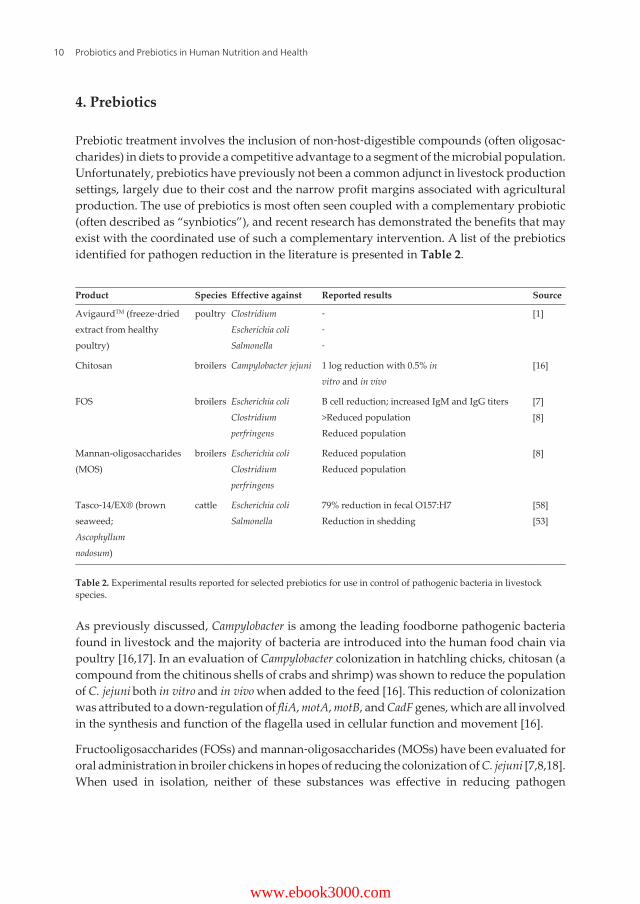
4. Prebiotics
Prebiotic treatment involves the inclusion of non‐host‐digestible compounds (often oligosac‐charides) in diets to provide a competitive advantage to a segment of the microbial population.Unfortunately, prebiotics have previously not been a common adjunct in livestock productionsettings, largely due to their cost and the narrow profit margins associated with agriculturalproduction. The use of prebiotics is most often seen coupled with a complementary probiotic(often described as “synbiotics”), and recent research has demonstrated the benefits that mayexist with the coordinated use of such a complementary intervention. A list of the prebioticsidentified for pathogen reduction in the literature is presented in Table 2.
Product Species Effective against Reported results Source
AvigaurdTM (freeze‐driedextract from healthypoultry)
poultry ClostridiumEscherichia coliSalmonella
‐‐‐
[1]
Chitosan broilers Campylobacter jejuni 1 log reduction with 0.5% invitro and in vivo
[16]
FOS broilers Escherichia coliClostridiumperfringens
B cell reduction; increased IgM and IgG titers>Reduced populationReduced population
[7][8]
Mannan-oligosaccharides(MOS)
broilers Escherichia coliClostridiumperfringens
Reduced populationReduced population
[8]
Tasco‐14/EX® (brownseaweed;Ascophyllumnodosum)
cattle Escherichia coliSalmonella
79% reduction in fecal O157:H7Reduction in shedding
[58][53]
Table 2. Experimental results reported for selected prebiotics for use in control of pathogenic bacteria in livestockspecies.
As previously discussed, Campylobacter is among the leading foodborne pathogenic bacteriafound in livestock and the majority of bacteria are introduced into the human food chain viapoultry [16,17]. In an evaluation of Campylobacter colonization in hatchling chicks, chitosan (acompound from the chitinous shells of crabs and shrimp) was shown to reduce the populationof C. jejuni both in vitro and in vivo when added to the feed [16]. This reduction of colonizationwas attributed to a down‐regulation of fliA, motA, motB, and CadF genes, which are all involvedin the synthesis and function of the flagella used in cellular function and movement [16].
Fructooligosaccharides (FOSs) and mannan‐oligosaccharides (MOSs) have been evaluated fororal administration in broiler chickens in hopes of reducing the colonization of C. jejuni [7,8,18].When used in isolation, neither of these substances was effective in reducing pathogen
Probiotics and Prebiotics in Human Nutrition and Health10
www.ebook3000.com

colonization in broiler chicks. However, the synergistic combination of Bacillus spp., Lactoba‐cillus spp., and MOSs reduced Campylobacter colonization. As an added benefit, FOSs weredemonstrated to induce weight gain in broiler chicks both alone and in combination withprobiotics [18]. This is in contrast to the work of Janardhana et al. [7] and Kim et al. [8], withboth groups having examined FOSs and MOSs for prebiotic addition to the feed of broilerchicks. The dietary addition of FOSs has been shown to reduce B cells and increase IgM andIgG titers in broiler chicks, both indicators of an enhancement of gastrointestinal immunefunction [7]. Likewise, FOSs were shown to decrease the incidence of C. perfringens and E.coli at 0.25% inclusion as well as bolster the population of Lactobacillus spp. [8]. This samereduction in C. perfringens and E. coli was achieved with 0.05% inclusion of MOSs [8].
Essential oils and polyphenolics have also been tested in relation to the reduction of pathogenspread from livestock [61]. Noted essential oil components that have been tested includecarvacrol (from savory), curcumin (from turmeric), eugenol (from allspice, betel pepper, andcloves), piperin (from black pepper), and thymol (from thyme) [35,62]. Fecal shedding of C.perfringens was reduced up to 30 days following supplementation with two essential oil blends[35]. Intestinal concentrations of C. perfringens were reduced for up to 21 days with essentialoil administration, but this effect was negated by day 30 [35]. Tedeschi et al. [63] demonstratedthat purified coumaric and cinnamic acids, both components of lignin, were able to reduce E.coli survival by 10‐ to 20‐fold when mixed with feces, although diets containing forage rich insuch compounds had no such effect. Berard et al. [62] also noted that catechol and pyrogallol(hydroxylated phenols) have toxic effects in the presence of microorganisms, mainly throughsubstrate deprivation. Callaway et al. [64] discussed the concept that saponins (natural plant‐based detergents) may have an antimicrobial effect by binding cholesterol, thereby disruptingthe microbial membrane, in addition to tannins, which may act in substrate deprivation bybinding protein and essential cations. Orange peel, a source of essential oils in the citrus family,has been shown to reduce cecal and rectal populations of E. coli O157:H7 with 5% and 10%dietary inclusion in sheep 96 hours following inoculation, but fecal shedding was only reducedat 10% inclusion [65]. Inclusion of orange peel at 10% of the diet was also shown to reduceSalmonella populations, although diet palatability issues were detected in excess of 10%inclusion [66].
Brown seaweed (Ascophyllum nodosum) is another prebiotic additive that has been noted forboth its production and antimicrobial characteristics [61,67]. The use of Tasco‐14® increasedthe marbling in carcasses from supplemented animals [67] and reduced fecal shedding of E.coli O157:H7 from 34% of the population to 7% of the population with supplementation [68],but there was no effect in Salmonella. However, unpublished data from the Callaway laboratoryat USDA‐ARS in College Station, Texas, demonstrate a small reduction in both E. coli O157:H7and Salmonella populations in vitro.
5. Conclusions
The gastrointestinal tracts of humans and animals are living ecosystems teeming withdiversity, and harnessing that ecology is a vital step toward a full understanding and appre‐
Prebiotic and Probiotic Approaches to Improving Food Safety on the Farm and Their Implications on Human Healthhttp://dx.doi.org/10.5772/63114
11

ciation of both livestock and human health. As was stated in the beginning, an understandingof the human‐animal interface is crucial to the homogeny of food safety protocols and healthconcerns. While most prebiotic and probiotic innovations in livestock production have soughtto increase performance characteristics for maximization of potential, these ventures haveoften led to the discovery of novel avenues in the improvement of food safety. These newapproaches to health and safety come at a crucial time when governmental regulation andpublic scrutiny necessitate an alteration in current practices in animal health and management.It is through the use of novel and innovative techniques that we will enhance our knowledgeof the ecosystem in which we live and will forge new paths in scientific discovery and healthyliving.
Author details
William B. Smith1, Todd R. Callaway2*, Luis O. Tedeschi3, Francis M. Rouquette Jr.1,Trisha Sheridan4 and Jennifer Adamski4
*Address all correspondence to: [email protected]
1 Department of Soil and Crop Sciences, Texas A&M AgriLife Research, Overton, TX, USA
2 Food and Feed Safety Research Unit, Southern Plains Agricultural Center, Agricultural Re‐search Service, USDA, College Station, TX, USA
3 Department of Animal Science, Texas A&M University, College Station, TX, USA
4 Nell Hodgson Woodruff School of Nursing, Emory University, Atlanta, GA, USA
References
[1] Callaway T.R., Edrington T.S., Anderson R.C., Harvey R.B., Genovese K.J., KennedyC.N., et al. Probiotics, prebiotics and competitive exclusion for prophylaxis againstbacterial disease. Animal Health Research and Reviews. 2008;9:217-225.
[2] Hungate R.E. The Rumen and Its Microbes. New York, NY: Academic Press; 1966. 533pp.
[3] Lu J., Idris U., Harmon B., Hofacre C., Maurer J.J., Lee M.D. Diversity and successionof the intestinal bacterial community of the maturing broiler chicken. Applied andEnvironmental Microbiology. 2003;69:6816-6824.
[4] Arthur T.M., Bosilevac J.M., Nou X., Shackelford S.D., Wheeler T.L., Koohmaraie M.Comparison of the molecular genotypes of Escherichia coli O157:H7 for the hides of beef
Probiotics and Prebiotics in Human Nutrition and Health12
cattle in different regions of North America. Journal of Food Protection. 2007;70(7):1622-1626.
[5] Elder R.O., Keen J.E., Siragusa G.R., Barkocy‐Gallagher G.A., Koohmarie M., LagreidW.W. Correlation of enterohemorrhagic Escherichia coli O157:H7 prevalence in feces,hides, and carcasses of beef cattle during processing. Proceedings of the NationalAcademy of Science (USA). 2000;97(7):2999-3003.
[6] Sargeant J.M., Amezcua M.R., Rajic A., Waddell L. Pre‐harvest interventions to reducethe shedding of E. coli O157 in the faeces of weaned domestic ruminants: A systematicreview. Zoonoses and Public Health. 2007;54:260-277.
[7] Janardhana V., Broadway M.M., Bruce M.P., Lowenthal J.W., Geier M.S., Hughes R.J.,et al. Prebiotics modulate immune responses in the gut‐associated lymphoid tissue ofchickens. Journal of Nutrition. 2009;139:1404-1409.
[8] Kim G.‐B., Seo Y.M., Kim C.H., Paik I.K. Effect of dietary prebiotic supplementation onthe performance, intestinal microflora, and immune response of broilers. PoultryScience. 2011;90:75-82.
[9] Elam N.A., Gleghorn J.F., Rivera J.D., Galyean M.L., Defoor P.J., Brashears M.M., et al.Effects of live cultures of Lactobacillus acidophilus (strains NP45 and NP51) andPropionibacterium freudenreichii on performance, carcass, and intestinal characteristics,and Escherichia coli strain O157 shedding of finishing beef steers. Journal of AnimalScience. 2003;81:2686-2698.
[10] Callaway T.R., Edrington T.S., Anderson R.C., Byrd J.A., Kogut M.H., Harvey R.B., etal. Using antimicrobial cultures, bacteriocins and bacteriophages to reduce carriage offoodborne pathogens in cattle and swine. In: LaCroix C., editor. Protective Cultures,Antimicrobial Metabolites and Bacteriophages for Food and Beverage Biopreservation.Oxford, UK: Woodhead Publishing; 2011. pp. 204-224.
[11] Callaway T.R., Edrington T.S., Loneragan G.H., Carr M.A., Nisbet D.J. Current andnear‐market intervention strategies for reducing Shiga‐toxin producing Escherichiacoli (STEC) shedding in cattle. Agriculture, Food and Analytical Bacteriology.2013;3:103-120.
[12] Callaway T.R., Anderson R.C., Edrington T.S., Genovese K.J., Harvey R.B., Poole T.L.,et al. Novel methods for pathogen control in livestock preharvest: An update. In: SofosJ.N., editor. Advances in Microbial Food Safety. Oxford, UK: Woodhead Publishing;2013. pp. 275-304.
[13] Callaway, T.R., Edrington T.S., Nisbet D.J. Ecological and dietary impactors of food‐borne pathogen prevalence and methods to reduce colonization in cattle. Journal ofAnimal Science. 2014;92:7308-7342.
Prebiotic and Probiotic Approaches to Improving Food Safety on the Farm and Their Implications on Human Healthhttp://dx.doi.org/10.5772/63114
13

[14] Steer T., Carpenter H., Tuohy K., Gibson G.R. Perspective on the role of the human gutmicrobiota and its modulation by pro‐ and prebiotics. Nutrition Research Reviews.2000;13(2):229-254. DOI: 10.1079/095442200108729089
[15] Collins D.M., Gibson G.R. Probiotics, prebiotics, and synbiotics: Approaches formodulating the microbial ecology of the gut. American Journal of Clinical Nutrition.1999;69:1052S-1057S.
[16] Arambel H.R., Donoghue A.M., Arsi K., Upadhyay A., Woo‐Ming A., Blore P.J., et al.Chitosan supplementation reduces enteric colonization of Campylobacter jejuni inbroiler chickens and down‐regulates expression of colonization genes. Advances inFood Technology and Nutritional Sciences Open Journal. 2015;1(5):104-111.
[17] Arsi K., Donoghue A.M., Woo‐Ming A., Blore P.J., Donoghue D.J. Intercloacal inocu‐lation, an effective screening method for determining the efficacy of probiotic bacterialisolates against Campylobacter colonization in broiler chickens. Journal of Food Protec‐tion. 2015;78(1):209-213. DOI: 10.4315/0362‐028X.JFP‐14‐326
[18] Arsi K., Donoghue A.M., Woo‐Ming A., Blore P.J., Donoghue D.J. The efficacy ofselected probiotic and prebiotic combinations in reducing Campylobacter colonizationin broiler chickens. Journal of Applied Poultry Science. 2005;pfv032. DOI: 10.3382/japr/pfv032
[19] Doyle M.P., Erickson M.C. Reducing the carriage of foodborne pathogens in livestockand poultry. Poultry Science. 2006;85:960-973.
[20] Kemp R., Leatherbarrow A.J.H., Williams N.J., Hart C.A., Clough H.E., Turner J., et al.Prevalence and genetic diversity of Campylobacter spp. in environmental water samplesfrom a 100‐square‐kilometer predominately dairy farming area. Applied and Environ‐mental Microbiology. 2005;71(4):1876-1882. DOI: 10.1128/AEM.71.4.1876‐1882.2005
[21] Riley L.W., Remis R.S., Helgerson S.D., McGee H.B., Wells J.G., Davis B.R. Hemorrhagiccolitis associated with a rare Escherichia coli serotype. The New England Journal ofMedicine. 1983;308(12):681-685.
[22] Bell B.P., Goldoft M., Griffin P.M., Davis M.A., Gordon D.C., Tarr P.I., et al. A multistateoutbreak of Escherichia coli O157:H7 — Associated bloody diarrhea and hemolyticuremic syndrome from hamburgers: The Washington experience. Journal of theAmerican Medical Association. 1994;272(17):1349-1353.
[23] Tuttle J., Gomez T., Doyle M.P., Wells J.G., Zhao T., Tauxe R.V., et al. Lessons from alarge outbreak of Escherichia coli O157:H7 infections: Insights into the infectious doseand method of widespread contamination of hamburger patties. Epidemiology andInfection. 1999;122:185-192.
[24] LeJeune J.T., Besser T.E., Hancock D.D. Cattle water troughs as reservoirs of Escherichiacoli O157. Applied and Environmental Microbiology. 2001;67(7):3053-3057. DOI:10.1128/AEM.67.7.3053‐3057.2001
Probiotics and Prebiotics in Human Nutrition and Health14
[25] Nou, X., Rivera‐Betancourt M., Bosilevac J.M., Wheeler T.L., Shackelford S.D., Gwart‐ney B.L., et al. Effect of chemical dehairing on the prevalence of Escherichia coli O157:H7and the levels of aerobic bacteria and Enterobacteriaceae on carcasses in a commercialbeef processing plant. Journal of Food Protection. 2003;66(11):2005-2009.
[26] FSIS. FSIS Policy on Non-intact Raw Beef Products Contaminated with E. coli O157:H7[Internet]. January 1999 [Updated: 17 February 1999]. Available from: http://www.fsis.usda.gov/Oa/background/O157policy.htm [Accessed: 28 April 2016]
[27] USDA/FSIS. Shiga toxin‐producing Escherichia coli in certain raw beef products. In: FSIS,editor. Federal Register. 2012.
[28] Genovese K.J., Anderson R.C., Harvey R.B., Callaway T.R., Poole D.H., Edrington T.S.,et al. Competitive exclusion of Salmonella from the gut of neonatal and weaned pigs.Journal of Food Protection. 2003;66(8):1353-1359.
[29] Popoff M.Y., Bockemuhl J., Gheesling L.L. Supplement 2002 (no. 46) to the Kauffman‐White scheme. Research in Microbiology. 2004;155:568-570.
[30] Olsen S.J., MacKinon L.C., Goulding J.S., Bean N.H., Slutsker L. Surveillance forfoodborne disease outbreak — United States, 1993-1997. Morbidity and MortalityWeekly Report. 2000;49(SS01):1-51.
[31] Gould, L.H., Walsh K.A., Vieira A.R., Herman K., Williams I.T., Hall A.J., et al.Surveillance for foodborne disease outbreaks — United States, 1998-2008. Morbidityand Mortality Weekly Report. 2013;62(SS02):1-34.
[32] Antunes P., Reu C., Sousa J.C., Peixe L., Pestana N. Incidence of Salmonella from poultryproducts and their susceptibility to antimicrobial agents. International Journal of FoodMicrobiology. 2003;82(2):97-103. DOI: 10.1016/S0168‐1605(02)00251‐9
[33] CDC. Severe Clostridium difficile‐associated disease in populations previously at lowrisk — Four states, 2005. Morbidity and Mortality Weekly Report. 2005;54(47):1201-1205.
[34] Weese J.S., Avery B.P., Rousseau J., Reid‐Smith R. Detection and enumeration ofClostridium difficile spores in retail beef and pork. Applied and Environmental Micro‐biology. 2009;75(15):5009-5011. DOI: 10.1128/AEM.00480‐09
[35] Mitsch P., Zitterl‐Eglseer K., Kohler B., Gabler C., Losa R., Zimpernik I. The effect oftwo different blends of essential oil components on the proliferation of Clostridiumperfringens in the intestines of broiler chickens. Poultry Science. 2004;83(4):669-675. DOI:10.1093/pa/83.4.669
[36] CDC. Multistate outbreak of listeriosis linked to Blue Bell Creameries products (finalupdate) [Internet]. [Updated: 10 June 2015]. Available from http://www.cdc.gov/listeria/outbreaks/ice‐cream‐03‐15/ [Accessed: 31 January 2016]
Prebiotic and Probiotic Approaches to Improving Food Safety on the Farm and Their Implications on Human Healthhttp://dx.doi.org/10.5772/63114
15

[37] Farber J. M., Peterkin P.I. Listeria monocytogenes, a food‐borne pathogen. Microbiologyand Molecular Biology Review. 1991;55(3):476-511.
[38] Skovgaard N., Morgen C.‐A. Detection of Listeria spp. in faeces from animals, in feeds,and in raw foods of animal origin. International Journal of Food Microbiology.1988;6(3):229-242. DOI: 10.1046/0168‐1605(88)90015‐3
[39] Deiters C., Gunnewig V., Friedrich A.W., Mellmann A., Kock R. Are cases of methicillin‐resistant Staphylococcus aureus clonal complex (CC) 398 among humans still livestock‐associated? International Journal of Medical Microbiology. 2015;305:110-113.
[40] Hasman H., Moodley A., Guardabassi L., Stegger M., Skov R.L., Aarestrup F.M. spatype distribution in Staphylococcus aureus originating from pigs, cattle and poultry.Veterinary Microbiology. 2010;141(3-4):326-331. DOI: 10.1016/j.vetmic.2009.09.025
[41] Barkema H.W., Schukken Y.H., Zadoks R.N. Invited review: The role of cow, pathogen,and treatment regimen in the therapeutic success of bovine Staphylococcus aureusmastitis. Journal of Dairy Science. 2006;89(6):1877-1895. DOI: 10.3168/jds.S0022‐0302(06)72256‐1
[42] Fuller R. Probiotics in man and animals. Journal of Applied Bacteriology.1989;66:365-378.
[43] Bielke L.R., Elwood A.L., Donoghue A.M., Newberry L.A., Neighbor N.K., Hargis B.M.Approach for selection of individual enteric bacteria for competitive exclusion in turkeypoults. Poultry Science. 2003;82:1378-1382.
[44] Nurmi E., Rantala M. New aspects of Salmonella infection in broiler production. Nature.1973;241:210-211.
[45] Baker A.A., Davis E., Rehberger T., Rosener D. Prevalence and diversity of toxigenicClostridium perfringens and Clostridium difficile among swine herds in the Midwest.Applied and Environmental Microbiology. 2010;76:2961-2967.
[46] Beauchemin K.A., Yang W.Z., Morgavi D.P., Ghorbani G.R., Kautz W., Leedle J.A.Z.Effects of bacterial direct‐fed microbials and yeast on site and extent of digestion, bloodchemistry, and subclinical ruminal acidosis in feedlot cattle. Journal of Animal Science.2003;81:1628-1640.
[47] Lehloenya K.V., Stein D.R., Allen D.T., Selk G.E., Jones D.A., Aleman M.M., et al. Effectsof feeding yeast and propionibacteria to dairy cows on milk yield and components, andreproduction. Journal of Animal Physiology and Animal Nutrition. 2007;92:190-202.DOI: 10.1111/j.1439‐0396.2007.00726.x
[48] Cai L., Indrakumar S., Kiarie E., Kim I.H. Effects of a multi‐strain Bacillus species‐baseddirect‐fed microbial on growth performance, nutrient digestibility, blood profile, andgut health in nursery pigs fed corn‐soybean meal‐based diets. Journal of AnimalScience. 2015;93:4336-4342. DOI: 10.2527/jas2015‐9056
Probiotics and Prebiotics in Human Nutrition and Health16
[49] Puphan K., Sornplang P., Uriyapongson S., Navanukraw C. Screening of lactic acidbacteria as potential probiotics in beef cattle. Pakistan Journal of Nutrition. 2015;14(8):474-479.
[50] Brashears M.M., Galyean M.L., Loneragan G.H., Mann J.E., Killinger‐Mann K. Preva‐lence of Escherichia coli O157:H7 and performance by beef feedlot cattle given Lactoba‐cillus direct‐fed microbials. Journal of Food Protection. 2003;66:748-754.
[51] Wisener L.V., Sargeant J.M., O'Connor A.M., Faires M.C., Glass‐Kaastra S.K. The useof direct‐fed microbials to reduce shedding of Escherichia coli O157 in beef cattle: Asystematic review and meta‐analysis. Zoonoses and Public Health. 2014;62:75-89. DOI:10.1111/zph.12112
[52] Brashears M.M., Jaroni D., Trimble J. Isolation, selection, and characterization of lacticacid bacteria for a competitive exclusion product to reduce shedding of Escherichiacoli O157:H7 in cattle. Journal of Food Protection. 2003;66(3):355-363.
[53] Nurmi E., Nuotio L., Schneitz C. The competitive exclusion concept: Development andfuture. International Journal of Food Microbiology. 1992;15(3-4):237-240. DOI:10.1016/0168‐1605(92)90054‐7
[54] Nava G.M., Bielke L.R., Callaway T.R., Castaneda M.P. Probiotic alternatives to reducegastrointestinal infections: The poultry experience. Animal Health Research Reviews.2005;6(1):105-118. DOI: 10.1079/AHR2005103
[55] Menconi A., Morgan M.J., Pumford N.R., Hargis B.M., Tellez G. Physiological proper‐ties and Salmonella growth inhibition of probiotic Bacillus strains isolated from envi‐ronmental and poultry sources. International Journal of Bacteriology. 2013;958408:1-8.DOI: http://dx.doi.org/10.1155/2013/958408
[56] Hong H.A., Duc L.H., Cutting S.M. The use of bacterial spore formers as probiotics.FEMS Microbiology Review. 2005;29:813-835.
[57] Baker A.A., Davis E., Spencer J.D., Moser R., Rehberger T. The effect of a Bacillus‐baseddirect‐fed microbial supplemented to sows on the gastrointestinal microbiota of theirneonatal piglets. Journal of Animal Science. 2013;91:3390-3399. DOI: 10.2527/jas2012‐5821
[58] Anderson R.C., Stanker L.H., Young C.R., Buckley S.A., Genovese K.J., Harvey R.B., etal. Effect of competitive exclusion treatment on colonization of early‐weaned pigs bySalmonella serovar Choleraesuis. Swine Health and Production. 1999;7(4):155-160.
[59] Wang Z., Chai W., Burwinkel M., Twardziok S., Wrede P., Palissa C., et al. Inhibitoryinfluence of Enterococcus faecium on the propagation of Swine Influenza A virus invitro. PLoS One. 2013;8(1):e54043. DOI: 10.1371/journal.pone.0053043
[60] Davis M.E., Parrott T., Brown D.C., de Rodas B.Z., Johnson Z.B., Maxwell C.V., et al.Effect of a Bacillus‐based direct‐fed microbial feed supplement on growth performance
Prebiotic and Probiotic Approaches to Improving Food Safety on the Farm and Their Implications on Human Healthhttp://dx.doi.org/10.5772/63114
17
and pen cleaning characteristics of growing‐finishing pigs. Journal of Animal Science.2008;86:1459-1467. DOI: 10.2527/jas.2007‐0603
[61] Crossland W.L., Callaway T.R., Tedeschi L.O. Shiga toxin‐producing E. coli andruminant diets: A match made in heaven? In: Ricke S.C., Donaldson J.R., Phillips C.A,editors. Food safety: Emerging issues, technologies and systems. London, UK: Elsevier;2015. pp. 185-213.
[62] Cowan M.M. Plant products as antimicrobial agents. Clinical Microbiological Reviews.1999;12(4):564-582. DOI: 0893‐8512/99/$04.00+0
[63] Berard N.C., Holley R.A., McAllister T.A., Ominski K.H., Wittenberg K.M., BouchardK.S., et al. Potential to reduce Escherichia coli shedding in cattle feces by using sainfoin(Onobrychis viciifolia) forage, tested in vitro and in vivo. Applied and EnvironmentalMicrobiology. 2009;75(4):1074-1079. DOI: 10.1128/AEM.00983‐08
[64] Tedeschi L.O., Callaway T.R., Muir J.P., Anderson R.C. Potential environmentalbenefits of feed additives and other strategies for ruminant production. RevistaBrasileira de Zootecnia. 2011;40:291-309.
[65] Callaway T.R., Carroll J.A., Arthington J.D., Edrington T.S., Rossman M.L., Carr M.A.,et al. Escherichia coli O157:H7 populations in ruminants can be reduced by orange peelproduct feeding. Journal of Food Protection. 2011;74(11):1917-1921.
[66] Callaway T.R., Carroll J.A., Arthington J.D., Edrington T.S., Anderson R.C., RossmanM.L., et al. Orange peel products can reduce Salmonella populations in ruminants.Foodborne Pathogens and Disease. 2011;8(10):1071-1075.
[67] Anderson M.J., Blanton Jr. J.R., Gleghorn J., Kim S.W., Johnson J.W. Ascophyllumnodosum supplementation strategies that improve overall carcass merit of implantedEnglish crossbred cattle. Asian‐Australasian Journal of Animal Science. 2006;19(10):1514-1518. DOI: 10.5713/ajas.2006.1514
[68] Braden K.W., Blanton Jr. J.R., Allen V.G., Pond K.R., Miller M.F. Ascophyllum nodosumsupplementation: A preharvest intervention for reducing Escherichia coli O157:H7 andSalmonella spp. in feedlot steers. Journal of Food Protection. 2004;67(9):1824-1828.
Probiotics and Prebiotics in Human Nutrition and Health18
Chapter 2
Probiotics: A Comprehensive Review of TheirClassification, Mode of Action and Role in HumanNutrition
Amirreza Khalighi, Reza Behdani andShabnam Kouhestani
Additional information is available at the end of the chapter
http://dx.doi.org/10.5772/63646
Abstract
Probiotics are live microorganisms that live in gastrointestinal (GI) tract and arebeneficial for their hosts and prevent certain diseases. In this chapter, after a completeintroduction to probiotics, definition, mechanism of action, and their classification,currently used organisms will be discussed in detail. Moreover, different kinds ofnutritional synthetic products of probiotics along with their safety and drug interac‐tion will be noticed. This chapter mentions all clinical trial studies that have been doneto evaluate probiotic efficacy with a focus on gastrointestinal diseases.
In the end, findings of our pilot study regarding the effect of probiotic on Small IntestinalBacterial Overgrowth (SIBO) will be presented. The nutritional effects of Probiotics ona host’s health will be collected and their usage criteria will be discussed. Somesuggestions for the Probiotics daily consumption will be presented and the follow-upfor their new adverse reaction will be emphasized, if any.
Keywords: probiotics, gastrointestinal (GI) tract, nutrition, related disorders, probioticproducts
1. Introduction to probiotics
The term probiotic is derived from Greek and literally means “for life.” It was first coined in1965 by Lilley and Stillwell to describe substances secreted by one microorganism thatstimulate the growth of another [1, 2]. In 1974, Parker modified this definition to “…organisms

and substances which contribute to intestinal microbial balance” [1, 3]. The current definitionof probiotics by Food and Agriculture Organization of the United Nations (FAO) and theWorld Health Organization (WHO) is “live microorganisms which when administered inadequate amounts confer a health benefit to the host” [4–6]. Probiotic organisms require certaincharacteristics to enable them to exert maximum therapeutic effects. Of these characteristics,there are some that are considered almost essential for a probiotic to have therapeutic effects,including gastric acid and bile salt stability, ability to adhere to the intestinal mucosa, andability to colonize the intestinal tract [1, 7].
2. Mechanism of action
The exact mechanisms by which probiotics accomplish their beneficial actions have not beenwell documented. However, there are several postulated mechanisms that explain many oftheir favorable effects [8] (Figure 1).
One of such mechanisms is a competition for adhesion sites, which means probiotics fight forcellular attachments. Many pathogenic organisms must associate with the GI tract epitheliumto colonize effectively [9]. However, some strains of bifidobacteria and lactobacilli can adhereto the epithelium and act as “colonization barriers” by preventing pathogens from adheringto the mucosa [1, 10]. This effect was demonstrated with the Lactobacillus rhamnosus strain GGand Lactobacillus plantarum 299v. Both of these organisms showed the ability to inhibitattachment of Escherichia coli to human colon cells [1, 11].
Another possible mechanism of action is the modification of the microbial flora through thesynthesis of antimicrobial compounds [12]. Many types of lactobacilli and bifidobacteriaproduce bacteriocinsor and other antimicrobial compounds. Bacteriocins are defined as“compounds produced by bacteria that have a biologically active protein moiety and abactericidal action” [1, 13]. Other biologically active compounds produced by lactic acidbacteria include hydrogen peroxide, diacetyl, and short-chain fatty acids. The release of thesecompounds by probiotic organisms results in a beneficial modification of the microflora [1,14]. However, not all strains of lactobacilli or bifidobacteria produce antimicrobial compounds,and some produce compounds that are fairly nonspecific in their activity, so that beneficialbacteria, as well as pathogenic organisms, may be negatively affected [1].
It has also been observed that probiotics can stimulate the immune response [15]. This immuneresponse may take the form of increased secretion of immunoglobulin-A (IgA) [1, 16], elevatednumbers of natural killer cells, or enhanced phagocytic activity of macrophages [1, 17].Increased secretion of IgA may decrease numbers of pathogenic organisms in the gut, thusimproving the composition of the microflora [1, 10]. Due to these immunomodulating effects,some researchers think probiotics might not only fight intestinal and urogenital pathogens,but might also be helpful for conditions, such as inflammatory bowel disease (IBD), pouchitis,food allergy, and for use as an adjuvant to vaccination [18–22]. Probiotics may also competefor nutrients that would otherwise be utilized by pathogens [1, 23]. This situation occurs withClostridium difficile, a potentially pathogenic organism that is dependent upon monosacchar‐
Probiotics and Prebiotics in Human Nutrition and Health20

ides for its growth. Probiotic organisms in sufficient numbers can utilize most of the availablemonosaccharides, which results in the inhibition of C. difficile [1, 24].
Figure 1. Schematic diagram illustrating potential or known mechanisms whereby probiotic bacteria might impact onthe microbiota. These mechanisms include (1) competition for dietary ingredients as growth substrates, (2) bioconver‐sion of, for example, sugars into fermentation products with inhibitory properties, (3) production of growth substrates,for example, EPS or vitamins, for other bacteria, (4)direct antagonism by bacteriocins, (5) competitive exclusion forbinding sites, (6) improved barrier function, (7) reduction of inflammation, thus altering intestinal properties for colo‐nization and persistence within, and (8) stimulation of innate immune response (by unknown mechanisms). IEC : intraepithelial cells, DC: dendritic cells, T:T-cells.
3. Classification
There are many different microorganisms currently used as probiotics [1, 20, 25] (Table 1). Tobetter understand how bacteria are named and classified, the following discussion may behelpful. Genus is the first name of a bacterium (e.g., Lactobacillus). It is somewhat general and
Probiotics: A Comprehensive Review of Their Classification, Mode of Action and Role in Human Nutritionhttp://dx.doi.org/10.5772/63646
21

refers to a grouping of organisms based on similarity of qualities, such as physical character‐istics, metabolic needs, and metabolic end products.
Species is a bacterium’s second name (e.g., acidophilus). It is a much more narrow classificationbased on shared common characteristics that distinguish them from other species. Strain is aneven more specific classification that divides members of the same species into subgroupsbased on several properties that these bacteria have in common that are distinct from othermembers of the species (e.g., strain LA5) [1, 26].
Lactobacillus spp. acidophilusplantarumrhamnosusparacaseifermentumreuterijohnsoniibreviscaseilactisdelbrueckii gasseri
Bifidobacterium spp. Breveinfantislongumbifidumthermophilumadolescentisanimalislactis
Bacillus spp. coagulans
Streptococcus spp. thermophilus
Enterococcus spp. faecium
Saccharomyces spp. cerevisiae
Table 1. Common probiotic microorganisms.
3.1. Lactobacillus species
Lactobacillus refers to a group of lactic acid–producing Gram-positive rods that are obligateand facultative anaerobes in the human gastrointestinal and genitourinary tracts [27, 29–32].The name lactobacillus refers to the bacterium's ability to produce lactic acid, not to the abilityto digest lactose [28]. Lactobacilli are used therapeutically as probiotics, the opposite ofantibiotics. They are considered "friendly" bacteria and are taken for the purpose of recolo‐nizing areas of the body to provide nutritional benefits including inducing growth factors and
Probiotics and Prebiotics in Human Nutrition and Health22

increasing the bioavailability of minerals [32]. Lactobacilli also stabilize the mucosal barrierand decrease intestinal permeability [33].
Altering the normal flora allows for potential colonization by pathogenic organisms [34],which can result in side effects, such as diarrhea, cramping, and less commonly pseudomem‐branous colitis (PMC), caused by C. difficile. The theory is that taking lactobacillus probioticsduring antibiotic treatment can prevent or minimize normal flora depletion and pathogenicbacteria colonization. There is some evidence to support this theory [35, 36]. Hydrogenperoxide–producing lactobacilli are bactericidal to the vaginal pathogen Gardnerella vaginalis,and their presence in the vagina has been associated with decreased frequencies of bacterialvaginosis and trichomoniasis [37]. In the vagina, lactic acid from lactobacilli lowers vaginalpH, which can prevent pathogen growth.
There is some preliminary evidence that lactobacilli and other probiotics might help protectagainst cancer. In animal models, lactobacillus has been shown to bind dietary carcinogens[38] and decrease development of tumors in the colon after carcinogen challenge [39, 40].Preliminary research also suggests that lactobacilli, especially L. plantarum, can reduce theseverity of chemotherapy-induced enterocolitis [41]. According to other research studies,Lactobacillus bulgaricus and Lactobacillus sporogenes might have hypolipidemic and antiathero‐sclerotic effects. Limited clinical evidence suggests that it can reduce total and low-densitylipoprotein (LDL) cholesterol with no effect on high-density lipoprotein (HDL) [42, 43].Fermented dairy products, such as yogurt and acidophilus milk, also seem to have a beneficialeffect on cholesterol. Lactobacilli and other probiotic bacteria seem to bind bile acids tocholesterol. They also seem to increase fatty acid production in the intestine, which decreasescirculatory fatty acid concentrations either by inhibiting hepatic cholesterol synthesis orredistributing cholesterol from the plasma to the liver.
Most researchers agree that the effectiveness of lactobacilli and other probiotics for allindications depends on their ability to colonize an area of tissue. To do this, lactobacilluspreparations must contain live and viable organisms. Products stored for long periods of timeor stored improperly may contain few live and active organisms. For oral preparations,bacteria must also remain viable after passing through the gut, and then they must be able tolatch on to the intestinal epithelium. Lactobacilli strains might vary in their effectiveness dueto differences in their ability to adhere to the epithelial cells by host factors such as hormonelevels [30, 44, 45]. This ability can change during a woman's menstrual cycle in response tochanging hormone levels. In postmenopausal women, correcting low estrogen levels can helprestore lactobacillus colonization without supplementation [29, 30].
3.2. Bifidobacterium species
Bifidobacterium is an anaerobic, Gram-positive, nonspore-forming, pleomorphic rod. Bacteriain the Bifidobacterium genus produce lactic and acetic acids as by-products of glucose utiliza‐tion. BB536 is a type of probiotic bacteria, which, according to secondary sources, was firstisolated from the intestinal tract of healthy infants. Bifidobacteria, in combination withLactobacillus species and the probiotic yeast Saccharomyces boulardii, seem to reduce the adverseeffects of Helicobacter therapy, but do not seem to improve compliance [46]. In addition,
Probiotics: A Comprehensive Review of Their Classification, Mode of Action and Role in Human Nutritionhttp://dx.doi.org/10.5772/63646
23

Bifidobacterium infantis in combination with Lactobacillus acidophilus seems to reduce theincidence of NEC and NEC-associated mortality in critically ill neonates [47].
3.3. Bacillus species
Bacillus coagulans is a Gram-positive rod, which produces lactic acid, and therefore is oftenmisclassified as lactic acid bacteria, such as lactobacillus. In fact, some commercial productscontaining B. coagulans are marketed as Lactobacillus sporogenes or "spore-forming lactic acidbacterium." It forms spores, which is an important factor in differentiating these species. B.coagulans is used therapeutically in a similar manner as other probiotics such as lactobacillusand bifidobacterium; however, B. coagulans is not a component of the normal human flora. Inorder to be effective for restoring normal flora and prevent pathogenic colonization, probioticsmust have the ability to persist and colonize in the intestinal mucosa. When the Bacillus sporeis ingested by humans, it is unknown what happens to the spore. It is unknown if the Bacillusspore is capable of germinating in the intestinal tract or if colonization occurs [48].
B. coagulans might reduce pathogenic bacteria colonization through several mechanisms. B.coagulans produces coagulin and lactic acid, which have antibacterial activity and might reducepathogenic bacteria growth through this mechanism [29, 49, 50]. Animal model research alsosuggests that ingesting bacillus spores increases immune response [48]. Proponents of B.coagulans suggest that this species of probiotics offers advantages over others such as lactoba‐cillus because Bacillus species can be stored indefinitely in desiccated forms [48]. Bacillusspores are also resistant to high temperatures and to acid.
3.4. Saccharomyces spp.
S. boulardii, also known as Saccharomyces cerevisiae, is a nonpathogenic yeast strain that hasbeen used for the treatment and prevention of diarrhea resulting from multiple etiologies.S. boulardii has been isolated from the skins of tropical fruits found in Indochina. Theindigenous population of Indochina has long used these fruit skins to prevent and treatdiarrhea [51].
S. boulardii is prepared by lyophylization (freeze drying) of live yeast organisms and encap‐sulation using lactose in the preparation. S. boulardii cannot be distinguished from other S.cerevisiae strains by phenotypic criteria, so identification of these infections requires moleculartyping. Comparative molecular studies show that S. boulardii is genetically very close or nearlyidentical to S. cerevisiae [52]. Results suggest that microsatellite polymorphism analysis of theYKL139w and YLR177w genes and the analysis by Ty917 hybridization are the most usefultools for the correct identification of S. boulardii strains [53]. However, metabolically andphysiologically, S. boulardii shows a very different behavior than S. cerevisiae, particularly inrelation to growth yield and resistance to temperature and acidic stresses, which are importantcharacteristics for a microorganism to be used as a probiotic. The German Commission Emonograph lists S. boulardii as S. cerevisiae Hansen CBS 5926.
Probiotics and Prebiotics in Human Nutrition and Health24
4. Commercial forms
There are two main forms in which probiotic organisms can be ingested—fermented foodsand supplements. Fermented foods can be of both dairy and vegetable origin, with the mostcommonly known of each being yogurt and sauerkraut, respectively. Probiotic supplementsconsist of freeze-dried (lyophilized) bacteria in powder, capsule, or tablet form. Regardless ofthe form in which the microorganisms are consumed, for clinical efficacy, products containingprobiotic organisms must provide live organisms in sufficient numbers to exert therapeuticeffects. Both types of fermented foods and supplements are able to do this. Pros (advantage)and cons (disadvantage) of common probiotic delivery systems are compared [1] (Table 2).
Deliverysystem
Pros Cons
Fermenteddairy
-Affordability and easy Availability-Ease of incorporation into daily patterns-Additional nutritional benefits-Enhanced bacterial survivalthrough upper GI tract (100× less bacteria can be given perdose)-Effective in the upper GI tract
-Contains dairy proteins and lactose-Taste can be issue-Not suitable when travelling-Not suitable for vegans
Capsules -Ease of administration-Contain no binders
-Not therapeutic in upper GI tract (unlessopened or chewed)-May contain allergenic excipients-Higher cost
Tablets -Ease of administration-Effective in the upper GI tract
-May contain allergenic or otherwiseproblematic binders and excipients (e.g.,gluten)-Higher cost
Powders -Effective in the upper GI tract-Dosages can be easily adjusted-Can be incorporated into foods or drinks-Contain no binders
Table 2. The pros and cons of different probiotic delivery systems.
4.1. Using the right strain
To achieve successful and reproducible clinical outcomes, it is imperative to use the exactprobiotic strain that has been proven to have the specific therapeutic action that is desired. Forexample, L. rhamnosus GG was found to prevent viral gastroenteritis [1, 54] and maintainulcerative colitis in remission [1, 55]. Other strains of L. rhamnosus cannot be assumed to act ina similar manner. The clinician who chooses to use the exact strain that had the effects in clinical
Probiotics: A Comprehensive Review of Their Classification, Mode of Action and Role in Human Nutritionhttp://dx.doi.org/10.5772/63646
25

trials can be confident of similar results. Using another closely related strain may or may nothave any effect. Whenever possible, use the exact strain used in research, as other strains, evenclosely related ones, may not have the same effects [1].
4.2. Dosage
The dosage of probiotic foods and supplements is based solely upon the number of liveorganisms present in the product. Successful results have been attained in clinical trials usingbetween 107and 1011 viable bacteria per day [1, 56, 57]. Interestingly, it appears that 100 timesfewer viable bacteria need to be given in a dairy medium than in a freeze-dried supplementto achieve similar numbers of live bacteria in the lower bowel [1, 58]. Dairy appears to workas an ideal transport medium for the bacteria, enhancing their survival through the upper GItract [1, 59].
4.2. Safety and adverse reactions
While probiotics are used widely and adverse effects are uncommon, there is no systematicreporting system for probiotics. Most studies did not report a statistically significant increasein adverse events compared with controls, but it has been questioned if probiotics are safe inimmunosuppressed individuals [60]. There are isolated case reports of bacteremia withLactobacillus and fungemia with S. boulardii. A case-review study found sepsis, liver abscess,and endocarditis from Lactobacillus GG to occur mostly in patients with severe illness [61]. Thesame paper reviewed S. boulardii fungemia and found numerous cases, some related toingestion of S. boulardii, but others resulting from suspected contamination of central lineswhen the product capsules were opened, and the lyophilized yeast was allowed to becomeairborne. Again, most, but not all, cases were in immunosuppressed individuals [60, 62]. Twosystematic reviews and an Agency for Healthcare Research and Quality study have evaluatedthe safety of probiotics and concluded that adverse effects are uncommon, but seriousinfections with Lactobacilli or S. boulardii can occur [63, 64]. Given this conclusion, it is prudentto avoid probiotics in individuals who are immunosuppressed or severely ill.
4.3. Drug interaction
Lactobacilli and bifidobacteria are negatively affected by alcohol and antibiotics [1, 65].Although there is no evidence that the organism interferes with the activity of most antibiotics,the metabolism of sulfasalazine, chloramphenicol palmitate, and phthalylsulfathiazole maybe affected by some strains of L. acidophilus [1, 66].
4.4. Clinical studies of probiotics
Table 3 lists conditions for which probiotics have been studied in more than 800 randomized,controlled clinical trials (RCT) [4]. It is notable that there has been at least one clinical trial ina variety of clinical conditions. GI tract conditions, such as inflammatory illnesses (e.g.,inflammatory bowel diseases or necrotizing enterocolitis in neonates) or enteric infections,have been studied most often [4, 67].
Probiotics and Prebiotics in Human Nutrition and Health26

Abdominal conditions
Acute amebiasis
Acute pancreatitis
Alcoholic liver injury
Collagenous colitis
Constipation
Colorectal neoplasia prevention
Diverticular colonic disease
Gas and bloating
Gastrointestinal transit timeand gastric emptying
Gastrointestinal symptomsafter loop ileostomy reversal
Helicobacter pylori infection
Hematochezia in breastfed infantsand in presumed infant allergic colitis
Hepatic encephalopathy
Infant colic
Inflammatory bowel diseases(Crohn’s disease, ulcerative colitis, pouchitis)
Irritable bowel syndrome (IBS)
Lactose intolerance
Nonalcoholic steatohepatitis
NSAID-induced small bowel injury
Prevention and treatment of pediatric cow’s milk allergy
Prevention and treatment ofdiarrheal diseases (infectious and noninfectious)
Prevention of antibiotic-associated diarrhea (AAD)
Prevention of necrotizing enterocolitis (NEC)
Primary sclerosing cholangitis (PSC) in patients with IBD
Small intestinal bacterial overgrowth (SIBO)
Tolerance of enteral feeds in ICU patients
Viral shedding
Oral and respiratory tract conditions
Gingivitis
Probiotics: A Comprehensive Review of Their Classification, Mode of Action and Role in Human Nutritionhttp://dx.doi.org/10.5772/63646
27

Dental caries
Halitosis
Prevention of upper respiratory tract infections (URTI)
Pulmonary exacerbations in cystic fibrosis (CF)
Urinary and reproductive tract conditions
Prevention and treatment of bacterial vaginosisand fungal vulvovaginosis
Prevention of preterm deliveries associatedwith bacterial vaginosis
Recurrent urinary tract infections (UTI)
Recurrent bladder cancer
Allergic or skin conditions
Atopic dermatitis
Allergic rhinitis and rhinosinusitis
Allergic asthma
Cutaneous viral warts
Prevention and treatment of pediatric eczema
Skin burns
Other
Acute otitis media
Chronic kidney disease
Effect on infant mortality in preterm infants
Effect on CD4 count in patients with HIV
Estrogen metabolism
Fasting glucose, insulin sensitivity, andglucose control in diabetic patients
Hyperlipidemia
Hypertension
Infant blood pressure and metabolic profilefrom mothers treated with probiotics
Inhibition of nasal, oral, or fecal colonizationwith pathogenic bacteria
Markers of metabolic syndrome and cardiovascular disease
Mastitis
Pediatric otitis media
Probiotics and Prebiotics in Human Nutrition and Health28

Pregnancy after in vitro fertilization (IVF)
Prevention and treatment of gestational diabetes
Prevention of type-1 diabetes mellitus
Prevention of infections in preterm infants,infants, and young children
Prevention of nosocomial infections in ICUs
Prevention of infections in the postoperative setting
Prevention of skeletal muscle damage under oxidative stress
Psychological distress, mood, and cognition
Reduction of biologically active aflatoxin
Rheumatoid arthritis (RA)
Spondyloarthropathy
Urinary oxalate excretion (risk factor for nephrolithiasis)
Vaccine-specific antibody development
Waist circumference and obesity
Table 3. Clinical conditions or settings studied in randomized, controlled clinical trials to evaluate probiotic efficacy.
Indication Efficacy and quality of evidence
Infectious diarrhea
Prevention
Treatment
Traveler’s diarrhea prevention
Moderate
High
Moderate
Antibiotic-associated diarrhea prevention High
Clostridium difficile infection (CDI)
Prevention
Treatment
Moderate
None
Recurrent CDI treatment Low to moderate
IBD
UC treatment
Pouchitis treatment and prevention
Crohn’s disease treatment
Moderate
High
Low
IBS treatment Moderate
Table 4. Indications, efficacy, and quality of evidence for probiotics in GI diseases.
Probiotics: A Comprehensive Review of Their Classification, Mode of Action and Role in Human Nutritionhttp://dx.doi.org/10.5772/63646
29

4.5. Efficacy in GI diseases
An ambitious meta-analysis of 11 species of probiotics evaluated their efficacy in the preven‐tion and/or treatment of eight major GI tract diseases and concluded that there was efficacy intreatment of infectious diarrhea, antibiotic-associated diarrhea (AAD), C. difficile infection(CDI), Helicobacter pylori eradication, IBS, and pouchitis; there was a lack of efficacy fortraveler’s diarrhea (TD) and necrotizing enterocolitis (NEC) [60, 68]. Some of these resultsconflict with meta-analyses of individual diseases, and results of all studies should beinterpreted with caution. Rigorous blinded RCTs of specific probiotics are needed to providerobust data on efficacy, adverse events, and cost benefit before widespread use can berecommended for many products on an evidence-based approach (Table 4);despite the lackof data, these products are widely used [60].
5. A pilot study to evaluate the efficacy of probiotic on treatment in patientswith small intestinal bacterial overgrowth (SIBO)
Generally, small intestinal bacterial overgrowth (SIBO) can be the result of a change in theclinical condition which has altered the pH and the bowel movements. In addition, immunedeficiency and malnutrition are the other risk factors accompanying it [69, 70]. SIBO can leadsto steatorrhea, vitamin B12-absorptive impairment, injury to the small intestinal microvilli,which itself causes malabsorption, coma, neurological deficit, and acidosis-induced shock [70].SIBO has also been proposed to be a common causative factor in the pathogenesis of irritablebowel syndrome (IBS) [71]. The diagnosis of this syndrome is made by hydrogen breath test(HBT) [71, 72].
This study was performed on the patients with chronic stomach pain and discomfort orchanges in their defecation, who were referred to the Infectious and Internal Diseases Clinicsof Quaem Hospital, Mashhad, Iran, from May 2010 to October 2011 [73]. The study protocolwas approved by the Research Council Ethics Committee of Mashhad University of MedicalSciences. Accordingly, the diagnosis was confirmed by hydrogen breath test (HBT) afterobtaining informed written consent. Thirty consecutive cases with a positive test result wereincluded in the study and were randomized in a double-blind manner into two groups:probiotic drug user and control group. After an initial 3-week aggressive therapy with broad-spectrum antibiotics, a 15-day maintenance antibiotic therapy with minocycline, 100 mg twicea day, and 15 days with a probiotic (named Lactol), including Bacillus coagulan spores andfructo-oligosaccharides (Bioplus Life Sciences Pvt. Ltd., India), twice a day after meals, wereadministered for the study group, and the same regimen without probiotic for the controlgroup. After 6 months, the HBT result and the GI symptoms were analyzed and comparedbetween the two groups.
As presented in the following tables, the number of patients with complaints of bloating,belching and diarrhea was remarkably less in the patients receiving a probiotic in comparisonto controls (Tables 5 and 6 ). In spite of the aggressive and maintenance treatments adminis‐
Probiotics and Prebiotics in Human Nutrition and Health30

tered for all the cases, 93.3% patients showed negative result of the HBT at the end of treatmentin the study group compared to 66.7% in the control group, showing the effectiveness of theprobiotic treatment. As an additional finding, 33.3% patients of the study group and 53.3% ofthe controls had a Bachelor degree or higher education, with no significant difference [75–79].
Parameter Study group Control group Total
Gender(M/F)
Male
Female
8 (53.3)
7 (46.7)
7 (46.7)
8 (53.3)
15(50.0)
15(50.0)
Age (years) 34.60 ± 10.68 42.86 ± 16.61 38.73 ± 14.35
Location of pain
Epigastric
Umbilical
Other sites
8 (53.3)
6 (40.0)
1 (6.7)
5 (33.3)
6 (40.0)
4 (26.7)
13 (43.3)
12 (40.0)
5 (16.7)
Flatulence
Yes
No
6 (40.0)
9 (60.0)
11 (73.3)
4 (26.7)
17 (56.7)
13 (43.3)
Belching
Yes
No
9 (60.0)
6 (40.0)
10 (66.7)
5 (33.3)
19 (63.3)
11 (36.7)
Nausea
Yes
No
3 (20.0)
12 (80.0)
3(20.0)
12(80.0)
6 (20.0)
24 (80.0)
Vomiting
Yes
No
4 (26.7)
11 ( 73.3)
3 (20.0)
12(80.0)
7 (23.3)
23 (77.7)
Constipation
Yes
No
9 (60.0)
6 (40.0)
3 (20.0)
12 (80.0)
12 (40.0)
18 (60.0)
Diarrhea
Yes
No
2 (13.3)
13 (86.7)
8 (53.3)
7 (46.7)
10 (33.3)
20 (66.7)
Loss of appetite
Yes
No
6(40.0)
9(60.0)
5 (33.3)
10 (66.7)
11 (36.7)
19 (63.3)
Table 5. Clinical characteristics of subjects at baseline (values in parentheses are percentages).
Probiotics: A Comprehensive Review of Their Classification, Mode of Action and Role in Human Nutritionhttp://dx.doi.org/10.5772/63646
31

In conclusion, the results of this pilot study showed that addition of a probiotic to the main‐tenance regimen may improve the GI tract symptoms and prevent the probable complicationsin patients with SIBO. Therefore, based on low side effects of the probiotics, it seems that theirlong-term prescription in SIBO, considering the recurrence favor of this syndrome, is desirable(e.g., probiotic containing dairy products or supplement daily drugs).
Parameter Study group Control group Total P value
Location of pain
Epigastric
Umbilical
Other sites
Without pain
0 (0.0)
0 (0.0)
0 (0.0)
15 (100.0)
3 (20.0)
3 (20.0)
2 (13.3)
7 (46.7)
3 (10.0)
3 (10.0)
2 (6.7)
22 (73.3)
0.002
Flatulence
Yes
No
2 (13.3)
13 (86.7)
8 (53.3)
7 (46.7)
10 (33.3)
20 (66.7)
0.049
Belching
Yes
No
3 (20.0)
12 (80.0)
9 (60.0)
6 (40.0)
12 (40.0)
18 (60.0)
0.025
Nausea
Yes
No
0 (0.0)
15 (100.0)
1 (6.7)
14 (93.3)
1 (3.3)
29 (96.7)
0.999
Vomiting
Yes
No
1 (6.7)
14 (93.3)
1 (6.7)
14 (93.3)
2(6.7)
28 (93.3)
0.999
Constipation
Yes
No
1 (6.7)
14 (93.3)
2 (13.3)
13 (86.7)
3 (10.0)
27 (90.0)
0.999
Diarrhea
Yes
No
1 (6.7)
14 (93.3)
8 (53.3)
7 (46.7)
9 (30.0)
21 (70.0)
0.014
Loss of appetite
Yes
No
1 (6.7)
14 (93.3)
4 (26.7)
11 (73.3)
5 (16.7)
25 (83.3)
0.330
Hydrogen breath test
Positive
Negative
1 (6.7)
14 (93.3)
5 (33.3)
10 (66.7)
6 (20.0)
24 (80.0)
0.169
Table 6. Clinical characteristics data of subjects after 6 months of treatment (values in parentheses are percentages).
Probiotics and Prebiotics in Human Nutrition and Health32

Author details
Amirreza Khalighi1*, Reza Behdani2 and Shabnam Kouhestani3
*Address all correspondence to: [email protected], [email protected]
1 Infectious diseases Specialist, Head of Infectious Diseases Ward & Infection Prevention &Control (IPC) coordinator, Razavi Hospital, Mashhad, Iran
2 GP, Volunteer Research Assistant, Hepatology Research Department, Toronto GeneralHospital, University Health Network (UHN), Toronto, Canada
3 Internist, Internal Medicine & Nutritional Consultant, Medical Council of Iran/Mashhadbranch, Mashhad, Iran
References
[1] Hawrelak J, BNat(Hons). Probiotics. In: Pizzorno JE, Murray MT, editors. Textbook ofNatural Medicine. 4th ed. St. Louis, Missouri: Churchill Livingstone Elsevier; 2013. p.979–94.
[2] Lilly DM, Stillwell RH. Probotics: growth-promoting factors produced by microorgan‐isms. Science (New York, NY). 1965;147(3659):747–8.
[3] Parker R. Probiotics, the other half of the antibiotic story. Animal Nutrition and Health.1974;29:4–8.
[4] Chen LA, Sears CL. Prebiotics, Probiotics, and Synbiotics. In: Bennett JE, Dolin R, BlaserMJ, editors. Mandell, Douglas, and Bennett’s Principles and Practice of InfectiousDiseases. 8th ed. Philadelphia, PA: Saunders Elsevier; 2015. p. 19–25.e1.
[5] Sanders ME. Probiotics: definition, sources, selection, and uses. Clinical InfectiousDiseases: An Official Publication of the Infectious Diseases Society of America. 2008;46Suppl 2:S58–61; discussion S144–51.
[6] Guarner F, Schaafsma GJ. Probiotics. International Journal of Food Microbiology.1998;39(3):237–8.
[7] Dunne C, O’Mahony L, Murphy L, Thornton G, Morrissey D, O’Halloran S, et al. Invitro selection criteria for probiotic bacteria of human origin: correlation with in vivofindings. The American Journal of Clinical Nutrition. 2001;73(2 Suppl):386s–92s.
[8] O’Toole PW, Cooney JC. Probiotic bacteria influence the composition and function ofthe intestinal microbiota. Interdisciplinary Perspectives on Infectious Diseases. 2008;2008:175285.
Probiotics: A Comprehensive Review of Their Classification, Mode of Action and Role in Human Nutritionhttp://dx.doi.org/10.5772/63646
33
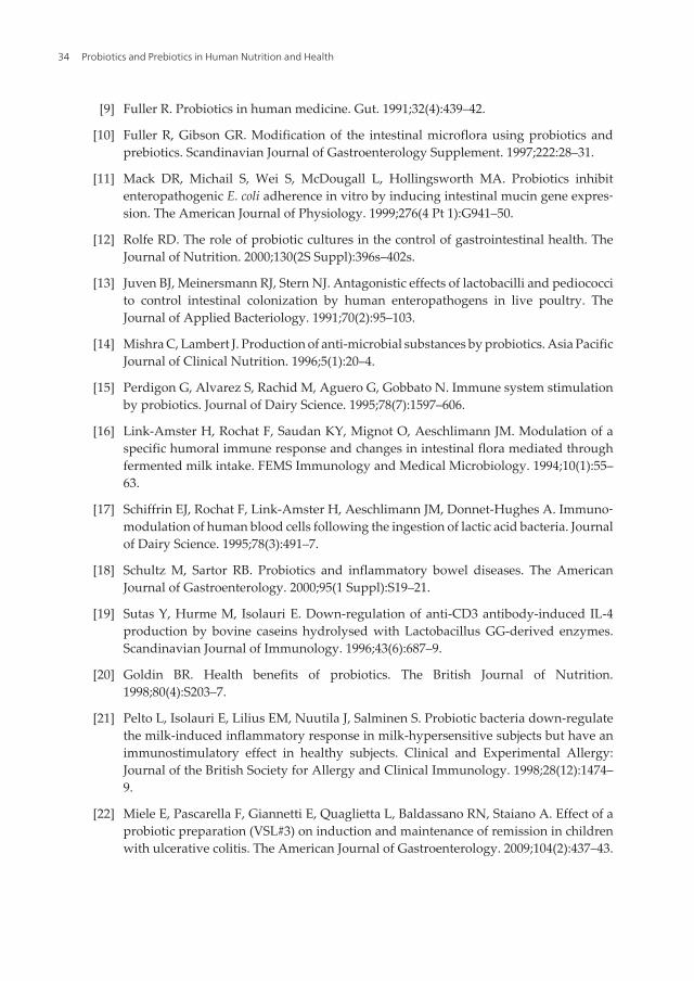
[9] Fuller R. Probiotics in human medicine. Gut. 1991;32(4):439–42.
[10] Fuller R, Gibson GR. Modification of the intestinal microflora using probiotics andprebiotics. Scandinavian Journal of Gastroenterology Supplement. 1997;222:28–31.
[11] Mack DR, Michail S, Wei S, McDougall L, Hollingsworth MA. Probiotics inhibitenteropathogenic E. coli adherence in vitro by inducing intestinal mucin gene expres‐sion. The American Journal of Physiology. 1999;276(4 Pt 1):G941–50.
[12] Rolfe RD. The role of probiotic cultures in the control of gastrointestinal health. TheJournal of Nutrition. 2000;130(2S Suppl):396s–402s.
[13] Juven BJ, Meinersmann RJ, Stern NJ. Antagonistic effects of lactobacilli and pediococcito control intestinal colonization by human enteropathogens in live poultry. TheJournal of Applied Bacteriology. 1991;70(2):95–103.
[14] Mishra C, Lambert J. Production of anti-microbial substances by probiotics. Asia PacificJournal of Clinical Nutrition. 1996;5(1):20–4.
[15] Perdigon G, Alvarez S, Rachid M, Aguero G, Gobbato N. Immune system stimulationby probiotics. Journal of Dairy Science. 1995;78(7):1597–606.
[16] Link-Amster H, Rochat F, Saudan KY, Mignot O, Aeschlimann JM. Modulation of aspecific humoral immune response and changes in intestinal flora mediated throughfermented milk intake. FEMS Immunology and Medical Microbiology. 1994;10(1):55–63.
[17] Schiffrin EJ, Rochat F, Link-Amster H, Aeschlimann JM, Donnet-Hughes A. Immuno‐modulation of human blood cells following the ingestion of lactic acid bacteria. Journalof Dairy Science. 1995;78(3):491–7.
[18] Schultz M, Sartor RB. Probiotics and inflammatory bowel diseases. The AmericanJournal of Gastroenterology. 2000;95(1 Suppl):S19–21.
[19] Sutas Y, Hurme M, Isolauri E. Down-regulation of anti-CD3 antibody-induced IL-4production by bovine caseins hydrolysed with Lactobacillus GG-derived enzymes.Scandinavian Journal of Immunology. 1996;43(6):687–9.
[20] Goldin BR. Health benefits of probiotics. The British Journal of Nutrition.1998;80(4):S203–7.
[21] Pelto L, Isolauri E, Lilius EM, Nuutila J, Salminen S. Probiotic bacteria down-regulatethe milk-induced inflammatory response in milk-hypersensitive subjects but have animmunostimulatory effect in healthy subjects. Clinical and Experimental Allergy:Journal of the British Society for Allergy and Clinical Immunology. 1998;28(12):1474–9.
[22] Miele E, Pascarella F, Giannetti E, Quaglietta L, Baldassano RN, Staiano A. Effect of aprobiotic preparation (VSL#3) on induction and maintenance of remission in childrenwith ulcerative colitis. The American Journal of Gastroenterology. 2009;104(2):437–43.
Probiotics and Prebiotics in Human Nutrition and Health34

[23] Vanderhoof JA, Young RJ. Use of probiotics in childhood gastrointestinal disorders.Journal of Pediatric Gastroenterology and Nutrition. 1998;27(3):323–32.
[24] Wilson KH, Perini F. Role of competition for nutrients in suppression of Clostridiumdifficile by the colonic microflora. Infection and Immunity. 1988;56(10):2610–4.
[25] Macfarlane GT, Cummings JH. Probiotics and prebiotics: can regulating the activitiesof intestinal bacteria benefit health? BMJ (Clinical Research ed). 1999;318(7189):999–1003.
[26] McKane L, Kandel J, eds. Microbiology: Essentials and Applications. New York:McGraw-Hill; 1986.
[27] Fujisawa T, Benno Y, Yaeshima T, Mitsuoka T. Taxonomic study of the Lactobacillusacidophilus group, with recognition of Lactobacillus gallinarum sp. nov. and Lactobacillusjohnsonii sp. nov. and synonymy of Lactobacillus acidophilus group A3 (Johnson et al.1980) with the type strain of Lactobacillus amylovorus (Nakamura 1981). InternationalJournal of Systematic Bacteriology. 1992;42(3):487–91.
[28] Vanderhoof JA, Young RJ. Current and potential uses of probiotics. Annals of Allergy,Asthma & Immunology: Official Publication of the American College of Allergy,Asthma, & Immunology. 2004;93(5 Suppl 3):S33–7.
[29] McGroarty JA. Probiotic use of lactobacilli in the human female urogenital tract. FEMSImmunology and Medical Microbiology. 1993;6(4):251–64.
[30] Bruce AW, Reid G. Intravaginal instillation of lactobacilli for prevention of recurrenturinary tract infections. Canadian Journal of Microbiology. 1988;34(3):339–43.
[31] Gupta K, Stapleton AE, Hooton TM, Roberts PL, Fennell CL, Stamm WE. Inverseassociation of H2O2-producing lactobacilli and vaginal Escherichia coli colonization inwomen with recurrent urinary tract infections. The Journal of Infectious Diseases.1998;178(2):446–50.
[32] Madsen KL, Doyle JS, Jewell LD, Tavernini MM, Fedorak RN. Lactobacillus speciesprevents colitis in interleukin 10 gene-deficient mice. Gastroenterology. 1999;116(5):1107–14.
[33] Shornikova AV, Casas IA, Isolauri E, Mykkanen H, Vesikari T. Lactobacillus reuteri asa therapeutic agent in acute diarrhea in young children. Journal of Pediatric Gastroen‐terology and Nutrition. 1997;24(4):399–404.
[34] Reid G, Bruce AW, Cook RL, Llano M. Effect on urogenital flora of antibiotic therapyfor urinary tract infection. Scandinavian Journal of Infectious Diseases. 1990;22(1):43–7.
[35] Alander M, Satokari R, Korpela R, Saxelin M, Vilpponen-Salmela T, Mattila-SandholmT, et al. Persistence of colonization of human colonic mucosa by a probiotic strain,
Probiotics: A Comprehensive Review of Their Classification, Mode of Action and Role in Human Nutritionhttp://dx.doi.org/10.5772/63646
35

Lactobacillus rhamnosus GG, after oral consumption. Applied and EnvironmentalMicrobiology. 1999;65(1):351–4.
[36] Sullivan A, Barkholt L, Nord CE. Lactobacillus acidophilus, Bifidobacterium lactis andLactobacillus F19 prevent antibiotic-associated ecological disturbances of Bacteroidesfragilis in the intestine. The Journal of Antimicrobial Chemotherapy. 2003;52(2):308–11.
[37] Maggi L, Mastromarino P, Macchia S, Brigidi P, Pirovano F, Matteuzzi D, et al.Technological and biological evaluation of tablets containing different strains oflactobacilli for vaginal administration. European Journal of Pharmaceutics andBiopharmaceutics: official journal of Arbeitsgemeinschaft fur PharmazeutischeVerfahrenstechnik eV. 2000;50(3):389–95.
[38] El-Nezami H, Kankaanpaa P, Salminen S, Ahokas J. Ability of dairy strains of lacticacid bacteria to bind a common food carcinogen, aflatoxin B1. Food and chemicaltoxicology: an international journal published for the British Industrial BiologicalResearch Association. 1998;36(4):321–6.
[39] Goldin BR, Gualtieri LJ, Moore RP. The effect of Lactobacillus GG on the initiation andpromotion of DMH-induced intestinal tumors in the rat. Nutrition and Cancer.1996;25(2):197–204.
[40] McIntosh GH, Royle PJ, Playne MJ. A probiotic strain of L. acidophilus reduces DMH-induced large intestinal tumors in male Sprague-Dawley rats. Nutrition and Cancer.1999;35(2):153–9.
[41] Mao Y, Nobaek S, Kasravi B, Adawi D, Stenram U, Molin G, et al. The effects ofLactobacillus strains and oat fiber on methotrexate-induced enterocolitis in rats.Gastroenterology. 1996;111(2):334–44.
[42] Doncheva NI, Antov GP, Softova EB, Nyagolov YP. Experimental and clinical studyon the hypolipidemic and antisclerotic effect of Lactobacillus Bulgaricus strain GB N 1(48). Nutrition Research. 2002;22(4):393–403.
[43] Losada MA, Olleros T. Towards a healthier diet for the colon: the influence of fructoo‐ligosaccharides and lactobacilli on intestinal health. Nutrition Research. 2002;22(1):71–84.
[44] Oberhelman RA, Gilman RH, Sheen P, Taylor DN, Black RE, Cabrera L, et al. A placebo-controlled trial of Lactobacillus GG to prevent diarrhea in undernourished Peruvianchildren. The Journal of Pediatrics. 1999;134(1):15–20.
[45] Reid G, Cook RL, Bruce AW. Examination of strains of lactobacilli for properties thatmay influence bacterial interference in the urinary tract. The Journal of Urology.1987;138(2):330–5.
[46] Cremonini F, Di Caro S, Covino M, Armuzzi A, Gabrielli M, Santarelli L, et al. Effect ofdifferent probiotic preparations on anti-Helicobacter pylori therapy-related side
Probiotics and Prebiotics in Human Nutrition and Health36
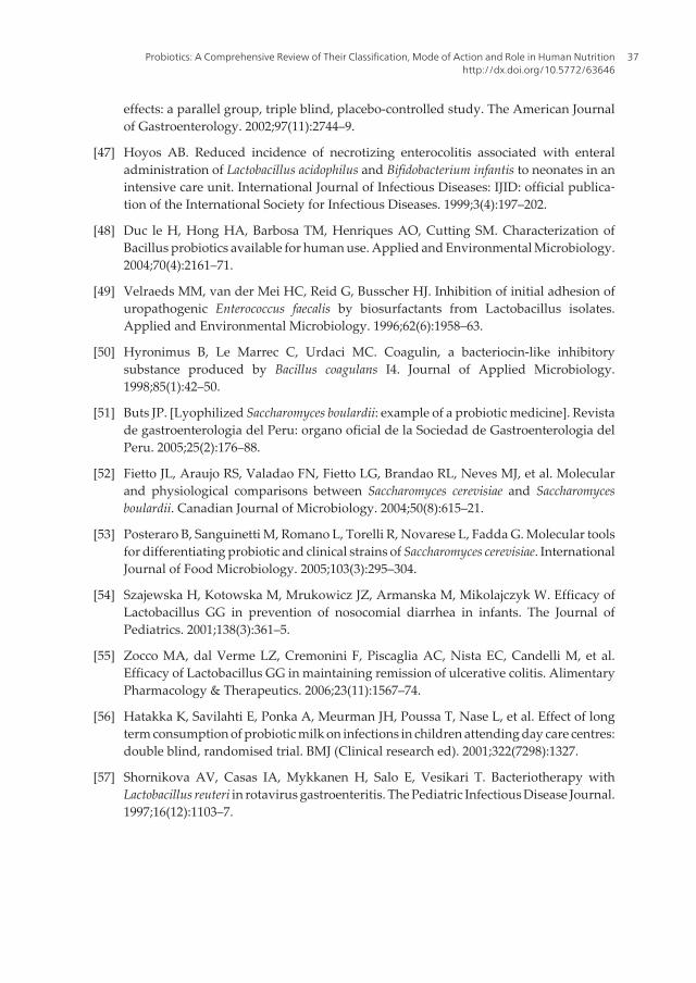
effects: a parallel group, triple blind, placebo-controlled study. The American Journalof Gastroenterology. 2002;97(11):2744–9.
[47] Hoyos AB. Reduced incidence of necrotizing enterocolitis associated with enteraladministration of Lactobacillus acidophilus and Bifidobacterium infantis to neonates in anintensive care unit. International Journal of Infectious Diseases: IJID: official publica‐tion of the International Society for Infectious Diseases. 1999;3(4):197–202.
[48] Duc le H, Hong HA, Barbosa TM, Henriques AO, Cutting SM. Characterization ofBacillus probiotics available for human use. Applied and Environmental Microbiology.2004;70(4):2161–71.
[49] Velraeds MM, van der Mei HC, Reid G, Busscher HJ. Inhibition of initial adhesion ofuropathogenic Enterococcus faecalis by biosurfactants from Lactobacillus isolates.Applied and Environmental Microbiology. 1996;62(6):1958–63.
[50] Hyronimus B, Le Marrec C, Urdaci MC. Coagulin, a bacteriocin-like inhibitorysubstance produced by Bacillus coagulans I4. Journal of Applied Microbiology.1998;85(1):42–50.
[51] Buts JP. [Lyophilized Saccharomyces boulardii: example of a probiotic medicine]. Revistade gastroenterologia del Peru: organo oficial de la Sociedad de Gastroenterologia delPeru. 2005;25(2):176–88.
[52] Fietto JL, Araujo RS, Valadao FN, Fietto LG, Brandao RL, Neves MJ, et al. Molecularand physiological comparisons between Saccharomyces cerevisiae and Saccharomycesboulardii. Canadian Journal of Microbiology. 2004;50(8):615–21.
[53] Posteraro B, Sanguinetti M, Romano L, Torelli R, Novarese L, Fadda G. Molecular toolsfor differentiating probiotic and clinical strains of Saccharomyces cerevisiae. InternationalJournal of Food Microbiology. 2005;103(3):295–304.
[54] Szajewska H, Kotowska M, Mrukowicz JZ, Armanska M, Mikolajczyk W. Efficacy ofLactobacillus GG in prevention of nosocomial diarrhea in infants. The Journal ofPediatrics. 2001;138(3):361–5.
[55] Zocco MA, dal Verme LZ, Cremonini F, Piscaglia AC, Nista EC, Candelli M, et al.Efficacy of Lactobacillus GG in maintaining remission of ulcerative colitis. AlimentaryPharmacology & Therapeutics. 2006;23(11):1567–74.
[56] Hatakka K, Savilahti E, Ponka A, Meurman JH, Poussa T, Nase L, et al. Effect of longterm consumption of probiotic milk on infections in children attending day care centres:double blind, randomised trial. BMJ (Clinical research ed). 2001;322(7298):1327.
[57] Shornikova AV, Casas IA, Mykkanen H, Salo E, Vesikari T. Bacteriotherapy withLactobacillus reuteri in rotavirus gastroenteritis. The Pediatric Infectious Disease Journal.1997;16(12):1103–7.
Probiotics: A Comprehensive Review of Their Classification, Mode of Action and Role in Human Nutritionhttp://dx.doi.org/10.5772/63646
37
[58] Saxelin M. Colonization of the human gastrointestinal tract by probiotic bacteria.Nutrition Today. 1996;31(6):9S.
[59] Kailasapathy K, Chin J. Survival and therapeutic potential of probiotic organisms withreference to Lactobacillus acidophilus and Bifidobacterium spp. Immunology and CellBiology. 2000;78(1):80–8.
[60] Surawicz CM, Brandt LJ. Probiotics and Fecal Microbiota Transplantation. In: FeldmanM, Friedman LS, Brandt LJ, editors. Sleisenger and Fordtran’s Gastrointestinal andLiver Disease. 10th ed. Philadelphia, PA: Saunders Elsevier; 2016. p. 2339–43.e3.
[61] Segarra-Newnham M. Probiotics for Clostridium difficile-associated diarrhea: focus onLactobacillus rhamnosus GG and Saccharomyces boulardii. The Annals of Pharmaco‐therapy. 2007;41(7):1212–21.
[62] Elahi B, Nikfar S, Derakhshani S, Vafaie M, Abdollahi M. On the benefit of probioticsin the management of pouchitis in patients underwent ileal pouch anal anastomosis: ameta-analysis of controlled clinical trials. Digestive Diseases and Sciences. 2008;53(5):1278–84.
[63] Hempel S, Newberry S, Ruelaz A, Wang Z, Miles JN, Suttorp MJ, et al. Safety ofprobiotics used to reduce risk and prevent or treat disease. Evidence Report/Technol‐ogy Assessment. 2011(200 ):1–645.
[64] Whelan K, Myers CE. Safety of probiotics in patients receiving nutritional support: asystematic review of case reports, randomized controlled trials, and nonrandomizedtrials. The American Journal of Clinical Nutrition. 2010;91(3):687–703.
[65] Finegold S, Mathisen G, George W. Changes in the Human Intestinal Flora Related tothe Administration of Antimicrobial Agents. In: Hentges DJ, editor. Human IntestinalMicroflora in Health and Disease. London: Academic Press; 1983. p. 355–448.
[66] Pradhan A, Majumdar MK. Metabolism of some drugs by intestinal lactobacilli andtheir toxicological considerations. Acta Pharmacologica et Toxicologica . 1986;58(1):11–5.
[67] Goldin BR, Gorbach SL. Clinical indications for probiotics: an overview. ClinicalInfectious Diseases: An Official Publication of the Infectious Diseases Society ofAmerica. 2008;46 Suppl 2:S96–100; discussion S44–51.
[68] Ritchie ML, Romanuk TN. A meta-analysis of probiotic efficacy for gastrointestinaldiseases. PLoS One. 2012;7(4):e34938.
[69] Binder H. Disorders of Absorption. In: Fauci A, Kasper D, Longo D, Braunwald E,Hauser S, Jameson J, editors. Harrison’s Principles of Internal Medicine. 18th ed. Newyork: McGraw Hill; 2012. p. 2460–76.
[70] Kleigman R, Behrman R, Jenson H, Stanton B. Nelson Textbook of Pediatrics. 19th ed.Philadelphia, PA: Saunders; 2011.
Probiotics and Prebiotics in Human Nutrition and Health38

[71] Quigley EM. Bacteria: a new player in gastrointestinal motility disorders – infections,bacterial overgrowth, and probiotics. Gastroenterology Clinics of North America.2007;36(3):735–48, xi.
[72] O’Mahony S, Shanhan F. Enteric microbiota and small intestinal bacterial overgrowth.In: Feldman M, Friedman LS, Brandt LJ, editors. Sleisenger & Fordtran’s Gastrointes‐tinal & Liver Disease: Pathophysiology, Diagnosis, Management. 9th ed. Philadelphia:Saunders Elsevier; 2010. p. 1772–7.
[73] Khalighi AR, Khalighi MR, Behdani R, Jamali J, Khosravi A, Kouhestani S, et al.Evaluating the efficacy of probiotic on treatment in patients with small intestinalbacterial overgrowth (SIBO) – a pilot study. The Indian Journal of Medical Research.2014;140(5):604–8.
[74] Soifer LO, Peralta D, Dima G, Besasso H. [Comparative clinical efficacy of a probioticvs. an antibiotic in the treatment of patients with intestinal bacterial overgrowth andchronic abdominal functional distension: a pilot study]. Acta gastroenterologicaLatinoamericana. 2010;40(4):323–7.
[75] Barrett JS, Canale KE, Gearry RB, Irving PM, Gibson PR. Probiotic effects on intestinalfermentation patterns in patients with irritable bowel syndrome. World Journal ofGastroenterology. 2008;14(32):5020–4.
[76] Jonkers D, Penders J, Masclee A, Pierik M. Probiotics in the management of inflamma‐tory bowel disease: a systematic review of intervention studies in adult patients. Drugs.2012;72(6):803–23.
[77] Rohde CL, Bartolini V, Jones N. The use of probiotics in the prevention and treatmentof antibiotic-associated diarrhea with special interest in Clostridium difficile-associateddiarrhea. Nutrition in Clinical Practice: Official Publication of the American Society forParenteral and Enteral Nutrition. 2009;24(1):33–40.
[78] Bixquert Jimenez M. Treatment of irritable bowel syndrome with probiotics. Anetiopathogenic approach at last? Revista espanola de enfermedades digestivas: organooficial de la Sociedad Espanola de Patologia Digestiva. 2009;101(8):553–64.
Probiotics: A Comprehensive Review of Their Classification, Mode of Action and Role in Human Nutritionhttp://dx.doi.org/10.5772/63646
39

Chapter 3
Prebiotics: Metabolism and Symbiotic Synergy withProbiotics in Promoting Health
Nditange Shigwedha, Penny Hiwilepo-Van Hal,Li Jia, Liubov Sichel and Shuang Zhang
Additional information is available at the end of the chapter
http://dx.doi.org/10.5772/64091
Abstract
Prebiotics are non‐digestible food ingredients that beneficially affect the host byselectively stimulating the growth and activity of probiotic bacteria in the colon. Alldietary prebiotics and/or dietary fiber provide the physiological and beneficial effectsand, therefore, are considered as essential nutrients. According to the Codex Alimen‐tarius and the Canadian Bureau of Nutritional Sciences, dietary fiber consists ofcarbohydrates with a degree of polymerization (DP) of three or more that naturallyoccur in foods of plant origin and that are not digested and absorbed by the smallintestine. The same definition goes well along with the term dietary prebiotics. Foodand Drug Administration (FDA)|Institute of Medicine (IOM) states that dietary fiberonly comes from plant foods and anything else is regarded as “added fiber” or “novelfiber.” Dietary fiber and/dietary prebiotics can be industrially produced for a broadrange of food applications. They can also be processed into capsules for the purpose ofmicroencapsulating probiotics. In this chapter, the most recognized physiological and/or beneficial effects of the prebiotics are clarified. New evidence on the concentrationsof the short‐chain fatty acids (SCFAs) and their metabolic relationship with better healthor disease prevention in the host is provided.
Keywords: Prebiotics, Probiotics, Synbiotic, Microencapsulation, SCFA (short‐chainfatty acids)
1. Introduction
All dietary prebiotics have the physiological and/or beneficial effects in humans and, thus, areregarded as essential nutrients. According to the Codex Alimentarius, Canadian Bureau of
Nutritional Sciences, IOM, and American Association of Cereal Chemistry International(AACCI), dietary fibers are identified as the edible parts of plants or analogous carbohydratesthat are resistant to digestion and absorption in the human small intestine with complete orpartial fermentation in the large intestine. Dietary fiber includes polysaccharides, oligosacchar‐ides, lignin, and associated plants substances. Dietary fibers promote beneficial physiologicaleffects including laxation and/or blood cholesterol attenuation, and/or blood glucose attenua‐tion [1]. Ordinarily, a prebiotic is defined as a non‐digestible food ingredient that confersbeneficial effects in the host by selectively stimulating the growth and/or activity of one or alimited number of bacteria in the colon and thus improves host health and well‐being [2–4].From those two descriptions, it is clear that “dietary fiber” and/or “prebiotics” are certainly acomplex of many different plant sources, with different chemical and physiological proper‐ties. Collectively when ingested, they are a cohesive unit for essentiality. In the United States ofAmerica, the joint recommendation by FDA|IOM says that dietary fiber only comes from plantfood (fruits, vegetables, or grains); and anything else was regarded as “added fiber” or “novelfiber.” Interestingly, that FDA|IOM recommendation fits well with the terminology “dietaryprebiotics”. A dietary prebiotic is defined as a selectively fermented ingredient that results inspecific changes in the composition and/or activity of the gastrointestinal microbiota, thusconferring benefit(s) upon host health [5]. Polymers and oligomers are the common types.Besides, a proof of the new oligomers and/or polymers about whether they confer the physio‐logical, or beneficial effects would be required before being started to be used in the public.
The primary reason that the prebiotics transit through the stomach and small intestinesundigested is because humans do not have the intestinal enzymes needed to digest them. Theycharacteristically make their way intact to the colon, where they will be bacterially fermented,together with the unabsorbed nutrients [6]. The salivary and pancreatic α‐amylases are theonly enzymes needed for starch digestion. It is also necessary to emphasize here that all sourcesof dietary fiber are potential prebiotics, and every source of dietary prebiotics (with a fewexceptions) is selectively fermented, thus mutually providing energy and simple sugars to thehost through the gut microbiota. Therefore, due to similarities between these two terms, wewill use the term “prebiotics” to refer to both the “dietary fiber and dietary prebiotics”throughout this chapter.
2. Whole grain, roughages, and prebiotics as essential nutrients: linkedwith probiotics for health
Whole grain and roughage are terms that have been easily confused with prebiotics in researchand sometimes used interchangeably. Whole grains are cereal grains that consist of the intact,ground, cracked, or flaked kernel, which includes the bran, the germ, and the innermost partof the kernel or endosperm [7]. Apart from prebiotics alone, whole grains provide a variety ofother nutrients too. Roughages are the coarse indigestible constituents of food or fodder, whichprovide bulk to the diet and promote normal bowel function. Hence, it has now become clearthat where deficiency occurs, the whole‐grain products can be enriched with the prebiotics orelse roughages. This fortification would be necessary and may clarify some of the confusions
Probiotics and Prebiotics in Human Nutrition and Health42

among the consumers who still believe that whole grains and prebiotics are one and the same.In Figure 1, for example, it can be observed that the average intake of prebiotics amongAmericans (male and female) between 2009 and 2010 was around 18 g/day, which was farbelow the daily recommended intakes (DRIs). The optimum daily requirement for prebioticsis estimated to be between 35 and 50 g/day [8]. This quantity would be needed for properintestinal functions and to adequately support the immune functions. Fascinatingly, anadditional severe deficiency of the prebiotics in some foods linked to whole grain and non‐whole grain can be witnessed in Figure 2. Yeast bread and breakfast cereals each seem toprovide nearly 1/3rd of the whole‐grain per serving. Others such as the grain‐based snacksappear to offer about 1/5th in addition to <1/10th which appears to come from rice, pasta, quickbreads, pastries, cakes, pies, cookies, and miscellaneous grains [9]. Research has furtherrevealed that whole‐grain consumers had significantly better nutrient profiles (includinghigher intakes of minerals and vitamins as percentages of 1989 Recommended DietaryAllowances and as nutrients per 1000 kcal, and lower intakes of total fat, saturated fat, andadded sugars as % of food energy) than the nonconsumers. It was found that consumers weremore likely to meet pyramid recommendations for the grain, fruit, and dairy food groups thanthe nonconsumers [9].
Figure 1. A parallel link of prebiotics made from the average intakes and projected daily recommended intakes orDRIs (g/day) among the American males and females, respectively. Source: Remade from Ref. [8].
Figure 2. A comparison made between constituents of whole grain and non‐whole grain as fiber representation insome common foods. Source: Modified from Ref. [9].
Prebiotics: Metabolism and Symbiotic Synergy with Probiotics in Promoting Healthhttp://dx.doi.org/10.5772/64091
43

When the probiotic bacteria selectively ferment the prebiotics in the colon, a symbiotic synergyhas been observed. During the metabolic process, that interdependent relationship exertsbeneficial health effects to the host. For instance, probiotics selectively receive differentprebiotics as nutrients from the host, initiate fermentation in the colon, provide the host withadditional genetic and metabolic attributes, boost the immune system, and be able to harnessnutrients that are otherwise inaccessible. Perceptibly, synbiotic is a mixture of both probioticsand prebiotics, which beneficially affect the host by improving the survival and implantationof live microbial dietary supplements in the gastrointestinal (GI) tract [2]. Therefore, prebiotics,probiotics, and synbiotics (combination of prebiotics and probiotics) can make up a distinctclass of the essential functional ingredients in foods. Probiotics are defined as live microor‐ganisms, which when administered in adequate amounts confer a health benefit on the host[10]. As a live microbial food supplement, probiotics have been linked to numerous beneficialeffects of improving the intestinal microbial balance in humans. Naturally, the number ofbacteria living in the human body and inside the gut is vast and is estimated to be 100 trillionbacteria [11, 12].
Besides commensal bacteria, probiotics, such as bifidobacteria, enterococci, streptococci, andlactobacilli, must co‐exist with their host and must evade or endure the diversity of responsesthat the host has already developed to eliminate pathogenic bacteria while at the same timeselectively ferment all the prebiotics. All these functions are mutually coordinated and providea significant synergy to the host. An advancement of intestinal microbiota including bifido‐bacteria (representing some of the probiotic bacteria) in early childhood from birth to 24 months is being demonstrated in Figure 3 [13]. In fact, an understanding on how the humanimmune system could differentiate between probiotics and harmful bacteria is no longer aserious challenge as it used to be in the past [12, 14].
Figure 3. Succession flows of gut microbiota in a healthy infant from birth. Source: From Ref. [13].
Probiotics and Prebiotics in Human Nutrition and Health44
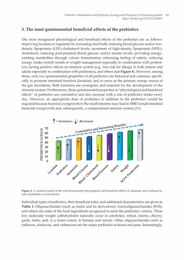
3. The most quintessential beneficial effects of the prebiotics
The most recognized physiological and beneficial effects of the prebiotics are as follows:improving laxation or regularity by increasing stool bulk; reducing blood glucose and/or low‐density lipoprotein (LDL)‐cholesterol levels; increment of high‐density lipoprotein (HDL)‐cholesterol; reducing post‐prandial blood glucose and/or insulin levels; providing energy‐yielding metabolites through colonic fermentation; enhancing feeling of satiety; reducingenergy intake (which results in weight management especially in combination with probiot‐ics); having positive effects on immune system (e.g., less risk for allergy in both infants andadults especially in combination with probiotics), and others (see Figure 4). However, amongthose, only two quintessential properties of all prebiotics are historical and common, specifi‐cally to promote intestinal function (laxation), and to serve as the primary energy source ofthe gut microbiota. Both functions are synergistic and essential for the development of theimmune system. Furthermore, these quintessential properties as “physiological and beneficialeffects” of prebiotics are cumulative and also increase with a rise of prebiotics intake everyday. However, an appropriate dose of probiotics in addition to the prebiotics would berequired because bacterial overgrowth in the small intestine may lead to SIBO (small intestinalbacterial overgrowth) and, subsequently, a compromised immune system [11].
Figure 4. A classical model of the well‐documented physiological and beneficial effects of adequate and continual in‐take of prebiotics in individuals.
Individual types of prebiotics, their beneficial roles, and additional characteristics are given inTable 1. Oligosaccharides (such as inulin and its derivatives), fructooligosaccharides (FOS),and others are some of the food ingredients recognized to meet the prebiotics’ criteria. Theselow molecular weight carbohydrates naturally occur in artichokes, wheat, onions, chicory,garlic, leeks, and, to a lesser extent, in banana and cereals. Other oligosaccharides such asraffinose, stachyose, and verbascose are the major prebiotics in beans and peas. Interestingly,
Prebiotics: Metabolism and Symbiotic Synergy with Probiotics in Promoting Healthhttp://dx.doi.org/10.5772/64091
45

these simple molecules of soluble dietary fiber can be produced industrially, and more novelprebiotics continue to be developed as functional foods in the food industry (see Table 2).Various methods can be applied to produce prebiotics. These include an enzymatic method[e.g., galactooligosaccharides (GOS), FOS, and oligofructose]; extraction method (e.g., inulinand soy oligosaccharides from vegetable sources); chemical synthetic method (e.g., lactuloseand polydextrose); and a combination of both chemical and enzymatic techniques (e.g.,resistant maltodextrin). Inulin, GOS, and FOS, for instance, have been increasingly added tofoods in many parts of the world. The practice started in Japan and some European countriesa few decades ago. In Canada, pulses, peas, and others are acceptable in foods as “added fibers”and more ways to put prebiotics in different kinds of diets keep emerging with new technology.
Prebiotics Fermentability
Primarysource
Beneficial role‡
Cellulose/hemicellulose/lignin/waxes
Low Plant foods Laxation
Guar gum High Guar bean (legume) Viscofier, blood lipid lowering,attenuates blood glucose response
Inulin/oligofructose/FOS High Chicory root, wheat,Jerusalem artichoke,banana, onions, leeks,garlic, can be synthesizedfrom simple sugars
Prebiotic effects, calcium absorption,attenuates total cholesterolRises HDL‐cholesterol
Chitooligosaccharides (COS) Low Derivative of chitin Rises HDL‐cholesterol, attenuatestotal cholesterol
Galactooligosaccharide (GOS) High Human and cow's milk,synthesized from lactose
Prebiotic effects, calciumabsorption, lipid profilesimprovement
Xylooligosaccharides (XOS) High Corn cobs, rice hulls,straws, bagasse, maltcakes, and bran
Blood lipid lowering
Soybean oligosaccharides High Soybean Blood lipid lowering, attenuatestotal cholesterol
β‐Glucan and oat bran High Oats and barley Blood lipid lowering, attenuatesblood glucose response
Pectin, gums High Plant foods Blood lipid lowering, attenuatesblood glucose response, emulsifier,thickener
Polydextrose High Synthesized fromdextrose (glucose)
Laxation, bulking agent, prebioticeffects
Psyllium High Psyllium husk (plant) Laxation, blood lipid lowering
Probiotics and Prebiotics in Human Nutrition and Health46
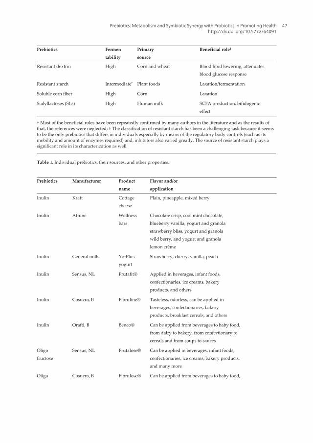
Prebiotics Fermentability
Primarysource
Beneficial role‡
Resistant dextrin High Corn and wheat Blood lipid lowering, attenuatesblood glucose response
Resistant starch Intermediate† Plant foods Laxation/fermentation
Soluble corn fiber High Corn Laxation
Sialyllactoses (SLs) High Human milk SCFA production, bifidogeniceffect
† Most of the beneficial roles have been repeatedly confirmed by many authors in the literature and as the results ofthat, the references were neglected; † The classification of resistant starch has been a challenging task because it seemsto be the only prebiotics that differs in individuals especially by means of the regulatory body controls (such as itsmobility and amount of enzymes required) and, inhibitors also varied greatly. The source of resistant starch plays asignificant role in its characterization as well.
Table 1. Individual prebiotics, their sources, and other properties.
Prebiotics Manufacturer Productname
Flavor and/orapplication
Inulin Kraft Cottagecheese
Plain, pineapple, mixed berry
Inulin Attune Wellnessbars
Chocolate crisp, cool mint chocolate,blueberry vanilla, yogurt and granolastrawberry bliss, yogurt and granolawild berry, and yogurt and granolalemon crème
Inulin General mills Yo‐Plusyogurt
Strawberry, cherry, vanilla, peach
Inulin Sensus, NL Frutafit® Applied in beverages, infant foods,confectionaries, ice creams, bakeryproducts, and others
Inulin Cosucra, B Fibruline® Tasteless, odorless, can be applied inbeverages, confectionaries, bakeryproducts, breakfast cereals, and others
Inulin Orafti, B Beneo® Can be applied from beverages to baby food,from dairy to bakery, from confectionary tocereals and from soups to sauces
Oligofructose
Sensus, NL Frutalose® Can be applied in beverages, infant foods,confectionaries, ice creams, bakery products,and many more
Oligo Cosucra, B Fibrulose® Can be applied from beverages to baby food,
Prebiotics: Metabolism and Symbiotic Synergy with Probiotics in Promoting Healthhttp://dx.doi.org/10.5772/64091
47

Prebiotics Manufacturer Productname
Flavor and/orapplication
fructose from dairy to bakery, from confectionary tocereals and from soups to sauces
Oligofructose
Orafti, B Beneo Can be applied from beverages to baby food,from dairy to bakery, from confectionary tocereals and from soups to sauces
GOS FrieslandCampina
Vivinal®GOS
For dairy products
Table 2. Samples of some of the prebiotic‐enhanced foods available in the international market.
The Food scientists appear to be on the verge of being capable of manipulating the gutsituations by diet control, thus possibly increasing an individual's health. It is also known thatdiets consisting of different components that are fermentable by gut microbiota are substratesfor various kinds of probiotic bacteria in the gut. Moreover, the fact that minerals absorptionand vitamin synthesis have been observed in the host confirms the symbiotic synergy of theprebiotics with probiotics in promoting health and suggests the existence of a multifunctionalmetabolism that directly involve a collective participation of many systems.
4. Prebioticious materials for encapsulation
Various kinds of prebiotics today are being processed into capsules (thickness: μm—mm) forthe purpose of microencapsulating live and/or lysate probiotic cells. If both probiotic cells andprebiotics are combined, then the product becomes a synbiotic. Microencapsulation is theprocess by which viable and/or lysate probiotic cells are packed within a wall (an outerpackaging) material for the purpose of shielding them from the surrounding environment.The standard load capacity of viable cells in the encapsulation materials varies from productto product. Usually, the viable cells occur at the concentration of 107–1012 CFU/g, while lysatecells are being measured in milligrams (mg). Most of the commercial yogurts or probioticsupplements contain 1–9 registered probiotic strains. In the case of multiple strains, theproportion of each probiotic strain in a package varies from batch to batch too. It is importantto emphasize here that different combinations of probiotics are more likely to ferment all theprebiotics selectively and provide the host with the most significant needs. To achieve constantprobiotical colonization in human or animal colon, microencapsulation is so far the bestapproach. Microencapsulation protects probiotics against O2 toxicity [15], stomach's acid [16],and bile in the small intestines [16].
So far, the prebiotics in the make include oligofructose‐enriched inulin [17], pullulan/starch‐blended edible‐films [18], denatured whey protein–alginate [19], alginate/chitosan/carboxy‐methyl chitosan [20], resistant starch, carrageenan, chitosan, alginate, cellulose acetatephthalate, gellan gum, pectin, gum arabic, xanthan gum, guar gum, locust bean [21–23], starch/
Probiotics and Prebiotics in Human Nutrition and Health48

spherulites [24, 25], and many others. Interestingly, a comparison was made between cocoabutter (lipid) and starch encapsulation materials, and a lipid system was found to be moreeffective at protecting the probiotics [25]. Whether the prebiotics are blended or used individ‐ually, they are all made to be harmonious mediators of probiotical or synbiotical microencap‐sulation. The selected materials (either oligomers or polymers) may also be used for theformation of microcapsules and body weight control in humans and animals. Moreover, highsurvival rates of encapsulated probiotics have been found in Cheddar cheese (6 months ofstorage period) [26] and in yogurt (4 weeks of storage period at 4°C [27].
5. Body weight management
All prebiotics appear to have a tremendous impact on body weight control in both animalmodels and humans. It has been found that the body fat in rats fed with a diet high in prebioticswas significantly lowered than the ones fed with a diet high in protein and control diet [28].In similar studies on humans, it has also been shown that the increase in prebiotics intake wasassociated with weight loss due to a decrease in energy intake [11, 29]. Also, none of the polyolsor sugar substitutes (sweeteners) were shown to have exerted any negative effect on theviability of the starter or probiotic cultures in cheese [30]. Apart from microencapsulation, ithas also been indicated that the fat in cottage cheese provides some protection to the probioticsas they strive to survive the gastric and intestinal transit to confer health benefits in the terminalileum and colon of the human GI tract [31]. Therefore, the inclusion of the adequate amountof prebiotics in every meal would be effective in controlling the current epidemic of overweightand few other digestion problems.
Noticeably, the speedy solution of weight control seems to be more in the lower fermentableprebiotics (such as lignin, waxes, cellulose, and hemicellulose) in combination with theprobiotic strains which selectively favor them. Resistant starches are somewhat characterizedbetween the lower and higher fermentability as part of the prebiotics (see Table 1). Theprebiotics permitted in Canada include acacia gum, barley bran, oat bran, corn bran, β‐glucan,fructooligosaccharide, galactooligosaccharide, inulin, modified wheat starch, oat hull fiber,partially hydrolyzed guar, pea hull fiber, polysaccharide complex (glucomannan, xanthangum, sodium alginate), psyllium seed husk, sieved barley meal, sugar beet fiber, wheat bran,edible parts of traditional fruits, veggies, legumes, resistant maltodextrin (Fibersol‐2), andmany others [32].
6. SCFAs and monosaccharides homeostasis
Probiotics have been recognized to hydrolyze and selectively ferment prebiotics to generatethe SCFAs and monosaccharides which can be absorbed and utilized as energy by the host.The three predominant SCFAs in the human gut are acetate, propionate, and butyrate. Thesehave been reported with high levels in the colon. Propionate and butyrate are respectively
Prebiotics: Metabolism and Symbiotic Synergy with Probiotics in Promoting Healthhttp://dx.doi.org/10.5772/64091
49

utilized by the liver and colon, whereas acetate enters the systemic circulation and reaches theperipheral tissues [33]. Further literature highlights that the vast influence of these SCFAs onthe host physiological benefits is through the nutri‐ and immuno‐modulatory functions [34].For example, butyrate expresses its potential in improving immune functions, intestinalbarrier, and oxidative stress through silencing the histone deacetylation of nuclear factor kappaB (NF‐κB), interferon‐γ, peroxisome proliferator‐activated receptor‐γ (PPARγ), and gluta‐thione‐S‐transferase (GST) [35].
It is well documented that the two endogenous ligands, G‐protein coupled receptor 41 (GPR‐41) and GPR‐43 mediate the signaling actions of the SCFAs [36]. GPR‐41 and GPR‐43 are knownproteins from the GPRs superfamily within the mammalian genome. They are particularlyexpressed in the adipocytes and identified as receptors for the fatty acids [37]. Acetate,preferentially, activates GPR‐43 in vitro, whereas butyrate is more selective for GPR‐41.Ordinarily, propionate displays most of the potent effect on both GPR‐41 and GPR‐43 [38]. Itis important to highlight that GRP‐41 has been associated more with a strong influence on thebody weight and glucose homeostasis through increasing the enteroendocrine cell hormoneknown as peptide YY (PYY). The literature reported PYY as actually a key factor involved inenergy homeostasis as well as in glucose metabolism [39, 40].
Figure 5. A represents a classical model of a normal digestion process; B represents a challenging digestion processdue to an excessive intestinal fermentation of prebiotics. Source: Modified from Ref. [11].
Under a normal circumstance, the macronutrients are sensed in different parts of the smallintestines. Also, the diverse types of gut hormones (such as PYY, cholecystokinin or CCK, and
Probiotics and Prebiotics in Human Nutrition and Health50

glucagon‐like peptide‐1 or GLP‐1) control gastric emptying and the motility of food throughthe whole digestive tract. Gastric emptying is, in fact, the main controller of viscosity and therate of nutrients delivery to the body. Up to now, no any in vitro technique can account forthat sensation. A recent in vitro study revealed that some of the prebiotics (if not all) are slightlyhydrolyzed—first in the stomach and small intestinal conditions—and then fermented in thecolon [41]. In the same study, the SCFAs with lactate were synthesized and bifidobacterialpopulation also significantly increased.
The GPR‐43 as another sensor of metabolic homeostasis suppresses fat accumulation in theadipose tissue through insulin signaling pathway. Also, GPR‐43 promotes the metabolism ofunincorporated lipids and glucose in other tissues [40]. Apart from the amounts and propor‐tions of those SCFAs during the fermentation stage of prebiotics, probiotics also simultane‐ously play other physiological benefits. Those include the balance of microbiological changes,locations of fermentation, and rates of fermentation for each prebiotic, locations of gasproduction, and pH changes throughout the digestion process. Naturally, all these parametersare difficult to measure in a real life situation. The batch systems which have been used inmany experiments do not reflect the true interactions of a synbiotic community (combinationof probiotics and prebiotics) in the host.
7. Implications of excessive bacteria and highly fermentable prebiotics inthe small intestines
Probiotics too should be able to provide health benefits when administered in sufficientquantity. However, the recent data support that, beyond prebiotics being able to meet thenutritional benefits, options of food selection also may regulate various functions in the bodyand may play detrimental or beneficial roles in some diseases. For the prebiotics to meet thebeneficial dietary effects, their fermentability status in the colon plays a significant role,especially to the commensal bacteria when all start to compete for nutrients. For example,flatulence is often a complaint when large doses of FOS are taken, which suggest that perhapsthere is a tolerance limit for each prebiotics. Additionally, an overgrowth of bacteria in thesmall intestines (including those that for some reasons migrated from the colon) may causedigestion problems and poor overall health [11].
Figure 5(B) represents an example of excessive intestinal prebiotics fermentation. Here, it canbe observed that the excess bacteria in the small intestines may result in the fermentation ofundigested carbohydrates (starches and oligosaccharides) before being metabolized. Thenagain, some of the prebiotics may absorb much water from the rest of the body into the smallintestine through osmosis, which can result in watery diarrhea. Fermentation of the undigestedcarbohydrates in the small intestine and an excessive fermentation of prebiotics in the coloncreate gas. When this happens, the pressure from an abnormal amount of gas inside the smallintestine and colon, respectively, can cause bloating, abnormal pain, flatulence, diarrhea, oreven constipation [11].
Prebiotics: Metabolism and Symbiotic Synergy with Probiotics in Promoting Healthhttp://dx.doi.org/10.5772/64091
51

Unlike in the colon, excess gasses generated by excess bacteria in the small intestine cannot beeasily expelled by passing it out and so the result is bloating. Apparently, if the excess bacteriaproduce methane (CH3), this gas tells the intestines to move upwards, causing the content inthe intestine to stall or to back up. In contrast, hydrogen (H2) gas in the small intestines isassociated with diarrhea. Furthermore, the unabsorbed monosaccharides in the small intes‐tines can result in osmotic responses [11]. Another author has reported that about 84% ofpeople with irritable bowel syndrome (IBS) have a bacterial overgrowth in the small intestine[42]. So, if this estimate is correct, people who complain of bloating, flatulence, diarrhea, orconstipation are encouraged to take probiotics at appropriate doses to balance the intestinalbacterial structure, content viscosity, and to restore the beneficial and physiological activitiesin the intestines. Just like prebiotics, probiotics is as important as multi‐vitamins and multi‐minerals too, and all should be taken daily.
Figure 5(A) represents a normal process of digestion with balanced gut flora. Here, proteins,fats, and monosaccharides (such as glucose, fructose, and galactose) are absorbed from thesmall intestines, while water is absorbed from the colon into the bloodstream. The unabsorbedprebiotics (cellulose, hemicellulose, polyols, fructans, galactans, and others) do not cause anyproblem. Thus, diet control can be used to favor the growth of some selected gut inhabitants[43].
It is necessary to state here that the small intestines are a mysterious and largely inaccessiblepart of the body. Endoscopy tests are reported to have only shown about 60 cm of what isexactly happening inside of that part of the GI tract. Besides, a colonoscopy that goes throughthe rectum also shows extremely little of the other end of the small intestine. Therefore, it wouldbe wise for individuals to ingest strong probiotic bacteria that survive and transit the stomachacid and bile salts during the digestion process. As discussed earlier on, the use of prebioticiousmaterials for probiotic microencapsulation appears to be one of the best practices, and othersimilar kinds of materials continue to be developed. Usually, the probiotics that pass throughthe small intestine will meet on other extreme the commensal bacteria that migrated from thecolon before they start prebiotic fermentation and compete for the nutrients (usually themonosaccharides) meant for assimilation by the host in a synergetic manner.
8. Conclusion
Prebiotics, either as occurring naturally in fruits, grain products, roughages, vegetables,legumes, soy, nuts, other foods or as added fibers, they all provide the physiological and/orbeneficial effects in a symbiotic relationship with probiotics. Whole grains are not prebiotics.Both whole and refined grains need to be enriched with the prebiotics to meet the estimateddaily requirement of 35–50 g/day. The benefits of dietary prebiotics are cumulative andincrease with an increasing intake of the prebiotics in combination with the multi‐vitamins,multi‐minerals, and probiotic strains. While probiotic bacteria are easily found in capsules asdietary adjuncts, the consumers are also advised to opt for foods which provide more prebi‐otics for the overall health. Each prebiotic appears to have its tolerance limit. Beyond such a
Probiotics and Prebiotics in Human Nutrition and Health52

tolerance limit, metabolism problems might occur. Diets consisting of different fermentableprebiotics are substrates for different probiotics in the gut. Furthermore, more evidence aboutthe SCFAs to have a relationship with better health in the host has been provided. Extra caremust also be taken into account when dealing probiotics and prebiotics relationship becausethe action of excess commensal bacteria in the small intestine may be detrimental in somedisease. The involvement of excess anaerobic bacteria in the small intestine leads to prebioticfermentation of which should only take place in the large intestines.
Acknowledgements
We are grateful to Ms. Dajana Pemac and the entire staff of InTech for helpful advice and theopportunity given to us to express our views. All the speakers on the Food, Health, & NutritionTrack at the Annual IFT Meetings, Chicago Ill., 2013 & 2015, are also gratefully acknowledged.
Author details
Nditange Shigwedha1*, Penny Hiwilepo-Van Hal2, Li Jia3, Liubov Sichel4 and Shuang Zhang5
*Address all correspondence to: [email protected]
1 School of Food Science and Engineering, Harbin Institute of Technology, Harbin, China
2 Department of Food Science and Technology, University of Namibia, Windhoek, Namibia
3 Wyeth-Nutrition, Suzhou, China
4 Pure Research Products LLC, Boulder, USA
5 Food College, Northeast Agricultural University, Harbin, China
References
[1] AACCI. The Definition of Dietary Fiber. 2001. Available from: http://www.aaccnet.org/initiatives/definitions/pages/dietaryfiber.aspx. [Accessed 2016‐02‐08]
[2] Gibson GR, McCartney AL. Modification of the gut flora by dietary means. Biochem.Soc. Trans. 1998;26(2):222–8. doi:10.1042/bst0260222
[3] Glenn GR, Roberfroid MB. Dietary modulation of the human colonic microbiota:introducing the concept of prebiotics. J. Nutr. 1995;125:1401–12.
Prebiotics: Metabolism and Symbiotic Synergy with Probiotics in Promoting Healthhttp://dx.doi.org/10.5772/64091
53

[4] Gibson GR, Probert HM, Van Loo J, Rastall RA, Roberfroid MB. Dietary modulation ofthe human colonic microbiota: updating the concept of prebiotics. Nutr. Res. Rev.2004;17(02):259–75. doi:10.1079/NRR200479
[5] Gibson GR, Scott KP, Rastall RA, Tuohy KM, Hotchkiss A, Dubert‐Ferrandon A, GareauM, Murphy EF, Saulnier D, Loh G, Macfarlane S. Dietary prebiotics: current status andnew definition. Food Sci. Technol. Bull. Funct. Foods. 2010;7:1–9. doi:10.1616/1476‐2137.15880
[6] Shigwedha N, Zhang L, Sichel L, Jia L, Gong P, Liu W, Wang S, Zhang S, Han X, GaoW. More than a few LAB alleviate common allergies: impact of paraprobiotics incomparison to probiotical live cells. J. Biosci. Med. 2014;2(03):56–64. doi:10.4236/jbm.2014.23008
[7] FDA. The Scoop on Whole Grains. 2009. http://www.fda.gov/ForConsumers/Consum‐erUpdates/ucm151902.htm. Accessed 2016‐02‐08
[8] USDA/ARS. Food Surveys Research Group, NHANES 2009–2010. 2010. http://www.ars.usda.gov/research/projects/projects.htm?ACCN_NO=415257. Accessed2016‐02‐08
[9] Cleveland LE, Moshfegh AJ, Albertson AM, Goldman JD. Dietary intake of wholegrains. J. Am. Coll. Nutr. 2000;19(sup3):331S–8S.
[10] Jia L, Shigwedha N, Mwandemele OD. Use of Dacid-, Dbile-, zacid-, and zbile-values inevaluating bifidobacteria with regard to stomach pH and bile salt sensitivity. J. FoodSci. 2010;75(1):M14–8. doi:10.1111/j.1750‐3841.2009.01398.x
[11] Jacob A. Digestive health with REAL food: a practical guide to an anti‐inflammatory,nutrient dense diet for IBS and other digestive issues. In: Sylvester R, editor. PaleoMedia Group, Bend; 2013. 396 p.
[12] Shigwedha N, Sichel L, Al‐Shura AN, Zhang L, Jia L. Probiotics, paraprobiotics, andprobiotical cell fragments (PCFs) as crisis management tools for important healthproblems. AASCIT J. Med. 2015;1(1):1–9. http://www.aascit.org/journal/archive2?journalId=979&paperId=1746
[13] Salminen S, Isolauri E. Intestinal colonization, microbiota, and probiotics. J. Pediatrics.2006;149(5):S115–20. doi:10.1016/j.jpeds.2006.06.062
[14] Shigwedha N, Sichel L, Jia L, Zhang L. Probiotical cell fragments (PCFs) as “NovelNutraceutical Ingredients”. J. Biosci. Med. 2014;2(03):43–55. doi:10.4236/jbm.2014.23007
[15] Talwalkar A, Kailasapathy K. Effect of microencapsulation on oxygen toxicity inprobiotic bacteria. Aust. J. Dairy Technol. 2003;58(1):36.
Probiotics and Prebiotics in Human Nutrition and Health54

[16] Chandramouli V, Kailasapathy K, Peiris P, Jones M. An improved method of micro‐encapsulation and its evaluation to protect Lactobacillus spp. in simulated gastricconditions. J. Microbiol. Methods. 2004;56(1):27–35. doi:10.1016/j.mimet.2003.09.002
[17] Fritzen‐Freire CB, Prudêncio ES, Amboni RD, Pinto SS, Negrão‐Murakami AN,Murakami FS. Microencapsulation of bifidobacteria by spray drying in the presence ofprebiotics. Food Res. Int. 2012;45(1):306–12. doi:10.1016/j.foodres.2011.09.020
[18] Kanmani P, Lim ST. Development and characterization of novel probiotic‐residingpullulan/starch edible films. Food Chem. 2013;141(2):1041–9. doi:10.1016/j.foodchem.2013.03.103
[19] Rajam R, Karthik P, Parthasarathi S, Joseph GS, Anandharamakrishnan C. Effect ofwhey protein–alginate wall systems on survival of microencapsulated Lactobacillusplantarum in simulated gastrointestinal conditions. J. Funct. Foods. 2012;4(4):891–8. doi:10.1016/j.jff.2012.06.006
[20] Li XY, Chen XG, Sun ZW, Park HJ, Cha DS. Preparation of alginate/chitosan/carboxy‐methyl chitosan complex microcapsules and application in Lactobacillus casei ATCC 393.Carbohydr Polymers. 2011;83(4):1479–85. doi:10.1016/j.carbpol.2010.09.053
[21] Wolfe LA. Encapsulation of probiotic bacteria in a water‐in‐solid‐fat emulsion topromote acid resistance [Thesis]. The Pennsylvania State University, University Park,PA; 2012.
[22] Brinques GB, Ayub MA. Effect of microencapsulation on survival of Lactobacillusplantarum in simulated gastrointestinal conditions, refrigeration, and yogurt. J. FoodEng. 2011;103(2):123–8. doi:10.1016/j.jfoodeng.2010.10.006
[23] Ding WK, Shah NP. Acid, bile, and heat tolerance of free and microencapsulatedprobiotic bacteria. J. Food Sci. 2007;72(9):M446–50. doi:10.1111/j.1750‐3841.2007.00565.x
[24] Chittiprolu S. Effect of starch spherulites on survival of bifidobacteria in the presenceof acid or bile [Thesis]. The Pennsylvania State University, University Park, PA; 2009.
[25] Lahtinen SJ, Ouwehand AC, Salminen SJ, Forssell P, Myllärinen P. Effect of starch-andlipid-based encapsulation on the culturability of two Bifidobacterium longum strains.Lett. Appl. Microbiol. 2007;44(5):500–5. doi:10.1111/j.1472‐765X.2007.02110.x
[26] Darukaradhya J. Enumeration and survival studies of free and encapsulated Lactoba‐cillus acidophilus and Bifidobacterium lactis in Cheddar cheese [Thesis]. NSW: Universityof Western Sydney; 2005.
[27] Krasaekoopt W, Bhandari B, Deeth H. Survival of microencapsulated probiotics inhigh‐solids yogurt from UHT milk. Aust. J. Dairy Technol. 2003;58(2):195.
[28] Reimer RA, Maurer AD, Eller LK, Hallam MC, Shaykhutdinov R, Vogel HJ, Weljie AM.Satiety hormone and metabolomic response to an intermittent high energy diet differs
Prebiotics: Metabolism and Symbiotic Synergy with Probiotics in Promoting Healthhttp://dx.doi.org/10.5772/64091
55

in rats consuming long‐term diets high in protein or prebiotic fiber. J. Proteome Res.2012;11(8):4065–74. doi:10.1021/pr300487s
[29] Slavin J. Fiber and prebiotics: mechanisms and health benefits. Nutrients. 2013;5(4):1417–35. doi:10.3390/nu5041417
[30] Esmerino EA, Cruz AG, Pereira EP, Rodrigues JB, Faria JA, Bolini HM. The influenceof sweeteners in probiotic Petit Suisse cheese in concentrations equivalent to that ofsucrose. J. Dairy Sci. 2013;96(9):5512–21. doi:10.3168/jds.2013‐6616
[31] Abadía‐García L, Cardador A, del Campo ST, Arvízu SM, Castaño‐Tostado E, Regala‐do‐González C, García‐Almendarez B, Amaya‐Llano SL. Influence of probiotic strainsadded to cottage cheese on generation of potentially antioxidant peptides, anti‐listerialactivity, and survival of probiotic microorganisms in simulated gastrointestinalconditions. Int. Dairy J. 2013;33(2):191–7. doi:10.1016/j.idairyj.2013.04.005
[32] Canadian Food Inspection Agency. Carbohydrates—elements within the nutritionfacts table 2014. Available from: http://www.inspection.gc.ca. [Accessed 2016‐02‐08].
[33] Lin HV, Frassetto A, Kowalik Jr EJ, Nawrocki AR, Lu MM, Kosinski JR, Hubert JA,Szeto D, Yao X, Forrest G, Marsh DJ. Butyrate and propionate protect against diet‐induced obesity and regulate gut hormones via free fatty acid receptor 3‐independentmechanisms. PLoS One. 2012;7(4):e35240. doi:10.1371/journal.pone.0035240
[34] Sun Y, O’Riordan MX. Regulation of bacterial pathogenesis by intestinal short‐chainfatty acids. Adv. Appl. Microbiol. 2013;85:93–118. doi:10.1016/B978‐0‐12‐407672‐3.00003‐4
[35] Leonel AJ, Alvarez‐Leite JI. Butyrate: implications for intestinal function. Curr. Opin.Clin. Nutr. Metab. Care. 2012;15(5):474–9. doi:10.1097/MCO.0b013e32835665fa
[36] Brown AJ, Goldsworthy SM, Barnes AA, Eilert MM, Tcheang L, Daniels D, Muir AI,Wigglesworth MJ, Kinghorn I, Fraser NJ, Pike NB. The Orphan G protein‐coupledreceptors GPR41 and GPR43 are activated by propionate and other short chaincarboxylic acids. J. Biol. Chem. 2003;278(13):11312–9. doi:10.1074/jbc.M211609200
[37] Ge H, Li X, Weiszmann J, Wang P, Baribault H, Chen JL, Tian H, Li Y. Activation of Gprotein‐coupled receptor 43 in adipocytes leads to inhibition of lipolysis and suppres‐sion of plasma free fatty acids. Endocrinology. 2008;149(9):4519–26. doi:10.1210/en.2008‐0059
[38] Le Poul E, Loison C, Struyf S, Springael JY, Lannoy V, Decobecq ME, Brezillon S,Dupriez V, Vassart G, Van Damme J, Parmentier M. Functional characterization ofhuman receptors for short chain fatty acids and their role in polymorphonuclear cellactivation. J. Biol. Chem. 2003;278(28):25481–9. doi:10.1074/jbc.M301403200
[39] Samuel BS, Shaito A, Motoike T, Rey FE, Backhed F, Manchester JK, Hammer RE,Williams SC, Crowley J, Yanagisawa M, Gordon JI. Effects of the gut microbiota on hostadiposity are modulated by the short‐chain fatty‐acid binding G protein‐coupled
Probiotics and Prebiotics in Human Nutrition and Health56

receptor, Gpr41. Proc. Natl. Acad. Sci. 2008;105(43):16767–72. doi:10.1073/pnas.0808567105
[40] Kimura I, Ozawa K, Inoue D, Imamura T, Kimura K, Maeda T, Terasawa K, KashiharaD, Hirano K, Tani T, Takahashi T. The gut microbiota suppresses insulin‐mediated fataccumulation via the short‐chain fatty acid receptor GPR43. Nat. Commun. 2013;4:1829.doi:10.1038/ncomms2852
[41] Moon JS, Joo W, Ling L, Choi HS, Han NS. In vitro digestion and fermentation ofsialyllactoses by infant gut microflora. J. Funct. Foods. 2016;21:497–506. doi:10.1016/j.jff.2015.12.002
[42] Pimentel M. A New IBS Solution. 2005. http://anewibssolution.com. Accessed 2016‐02‐08
[43] Umu ÖC, Oostindjer M, Pope PB, Svihus B, Egelandsdal B, Nes IF, Diep DB. Potentialapplications of gut microbiota to control human physiology. Antonie Van Leeuwen‐hoek. 2013;104(5):609–18. doi:10.1007/s10482‐013‐0008‐0
Prebiotics: Metabolism and Symbiotic Synergy with Probiotics in Promoting Healthhttp://dx.doi.org/10.5772/64091
57

Chapter 4
Lactobacillus reuteri, Infant Allergy Prevention andChildhood Immune Maturation
Anna Forsberg
Additional information is available at the end of the chapter
http://dx.doi.org/10.5772/63120
Abstract
The increasing allergy prevalence in affluent countries may be caused by reducedmicrobial stimulation, resulting in an abnormal postnatal immune maturation. Thischapter concerns the theories behind the use of probiotics in randomized preventiontrials, and how this supplementation affects the immunity of pregnant women, theimmune development in their children, and possibly preventing allergic diseases. Moststudies investigating the underlying mechanisms have focused on postnatal microbialexposure. An increasing body of evidence from studies suggests that the maternalmicrobial environment during pregnancy can program the immune development of thechild. In human allergy intervention studies, probiotic supplementation to the motherduring pregnancy, as well as to her baby postnatally, may be important for preven‐tive effects. Also, prenatal environmental exposures may alter gene expression viaepigenetic mechanisms, aiming to induce physiological adaptations to the anticipatedpostnatal environment. The maternal microbial environment during pregnancy mayprogram the immune development of the child.
Keywords: allergy, immune maturation, Lactobacillus reuteri, probiotics, allergy pre‐vention, allergens, TLRs, cytokines, chemokines
1. Probiotics in allergy prevention
Different probiotic strains have been used in allergy prevention trials with successful andunsuccessful results. Why use probiotics to try and prevent childhood allergic diseases then?The increasing allergy prevalence in affluent countries may be caused by reduced microbialstimulation, reflecting an abnormal postnatal immune maturation [1], resulting in allergicdiseases in children. Of course, this is a multifactorial problem where changing climate, living

conditions, and urbanization have led to a biodiversity loss. Studies show altered microbiotaand general microbial deprivation which characterize people living in urban affluent environ‐ments. Consequently, this seems to be a risk factor for immune dysregulation and impairedimmune tolerance. It is further enhanced by physical inactivity and a western diet poor in freshfruit and vegetables, which may act in synergy with dysbiosis of the gut flora [2]. Probiotics maybe one way to increase microbial stimulation, enrich the gut flora and balance a skewed immunesystem, which will be discussed later.
2. A brief introduction to childhood allergic diseases
The cost of allergic diseases is burdening the society; reduced life quality and increased sickleave are common, asthma in children is one of the most common chronic diseases affectingchildren at an early age. About 20% of the population is affected by allergic disease such asatopic dermatitis, food allergy, asthma, allergic rhinitis, and conjunctivitis [3]. Furthermore,the “atopic march,” as commonly referred to, is an age-associated variation in allergy-relatedsymptoms in childhood. The first allergy-related symptoms are often eczema and food allergyat a young age, later followed by asthma and rhinoconjunctivitis in school-aged children. Inaddition to the age variation in allergic diseases, there seems to be a gender issue as well. Boysearly in life have a higher incidence of allergic diseases than girls [4–6] and are also moresusceptible to infections maybe due to the more Th2-deviated immunity [4, 5]. Females arecharacterized by increased inflammatory responses and infections clearance, possibly reflect‐ing the stronger Th1 immunity observed in girls [4, 5]. Of course, this has its pros and cons.This results not only in a better protection against infection but also in increased susceptibilityto autoimmunity later in life. Allergy-related sex differences diminish at puberty; at adult age,no clear sex differences concerning allergy can be found [7].
3. The importance of the environment and the discovery of the beneficialeffect of exposure to microbes
In the beginning, when elucidating the mechanism behind the increasing rates of allergicdiseases, the focus was on living conditions. In 1989, a researcher named Strachan discoveredthat there was an association between siblings, family size, and hay fever [8]. This led todiscoveries that children born in a farm had less allergies than children born in urban areas.The step after that was to focus on postnatal microbial exposure [9–12]. How infants areprepared for life outside the uterus, and how can the maternal environment be protectiveagainst allergic development in the offspring? The maternal microbial environment has beenproposed to be able to program the immune development of the child, during pregnancy [13].Especially, if the mother is exposed to farm environment, in particular during pregnancy,development of allergic diseases seems to be attenuated. Interestingly, research has showedthat exposure later in life, after pregnancy and later, seems to have a weaker effect [14, 15]which opens up several other questions. For example, is it possible to program the develop‐ment of immunity in the child? When is the “window of opportunity”? Can we manipulate
Probiotics and Prebiotics in Human Nutrition and Health60
this and protect the child against the development of diseases? How can this be done?Numerous studies show that exposure to farm environments during infancy and even in fetallife [16, 17] reduces the incidence of allergic diseases. Furthermore, a recent report in Swedenpresents data that contact with farm animals or dogs during childhood may protect againstasthma development [18]. Exposures to farming areas and also consumption of raw milk havebeen associated with the upregulation of certain receptors associated with innate immunity.In the Protection Against Allergy: Study in Rural Environments (PASTURE) birth cohort study,1133 pregnant women were recruited in rural areas of Austria, Finland, France, Germany, andSwitzerland and showed that farming-related exposures, such as raw farm milk consumption,that were previously reported to decrease the risk for allergic outcomes, were associated witha change in gene expression of innate immunity receptors in early life. Raw milk of courseincludes many Lactobacilli strains among others. Therefore, it is believed that microbialexposure in early life educates the developing immune system, driving postnatal maturationof immune regulation as discussed in [19]. The author also suggests that the theory should bereferred to as “microbial deprivation hypothesis” since the exposure to a wealth of commensal,non-pathogenic microorganisms early in life is of benefit. The epidemiological studies aresupported by animal models, demonstrating that microbial exposure during gestation canprevent allergic responses in the offspring [20, 21].
3.1. Animal models show the benefit of microbial exposure during gestation
The beneficial effect of exposure to microbes have been further explored, primarily in animalmodels, to try to pinpoint what the mechanism on immune tolerance and protection of allergicdisease might be. Of importance is the maternal environment, suggesting that maternalimmunity may be transferred or at least influence the offspring. In experimental murinemodels, the mother is treated with lipopolysaccharide which attenuated allergic disease andassociated inflammation in offspring [22–24]. One study explored the effect of LPS on femaleBALB/c mice before conception and during pregnancy. Several weeks after birth offspringwere sensitized to ovalbumin (OVA) followed by aerosol allergen challenges. LPS may operatein prenatal life in order to modulate the development of allergies in the offspring since LPSexposure prenatally enhanced Th1-associated IFN-gamma in offspring. OVA sensitization wasfollowed with a reduction in anti-OVA IgG1 and IgE as well as unchanged IgG2a antibodyresponses, accompanied by a significant decrease in Th2-associated cytokine levels. This wasfollowed by a reduction of eosinophils and macrophages in bronchoalveolar lavage fluids,which are often increased in allergic airways. However, clinical manifestations such as airwayhyper-responsiveness, a hallmark of bronchial asthma, were not affected [22]. Another studyalso investigated the effect of LPS on pregnant mice and further explored the effect of LPS onthe offspring before allergen sensitization with OVA. Prenatal and postnatal LPS exposuresuppressed allergen-specific IgE production, eosinophilic airway inflammation and in vivoairway reactivity in response to methacholine. The suppression of allergen-mediated inflam‐matory responses was associated with an increased shift toward Th1 responses in culture(spleen cells) and may be mediated via Toll-like receptor (TLR) and T-bet expression by lungtissues [23]. Another group used a rat model and investigated the effect of prenatal LPSexposure on postnatal T cell differentiation and experimental allergic airway disease. The
Lactobacillus reuteri, Infant Allergy Prevention and Childhood Immune Maturationhttp://dx.doi.org/10.5772/63120
61
expression of T cell-related transcription factors and cytokines was quantified in the lung, andairway hyper responsiveness was measured. Prenatal LPS exposure induced a Th1 immunemilieu in the offspring of rats and also reduced OVA-induced airway inflammation, eosino‐philia, and airway responsiveness [21].
The next step was to use the commensal Acinetobacter lwoffii [20]. Acinetobacter lwoffii is derivedfrom cow shed and is non-pathogenic. The strain was used in an experimental allergic airwayinflammation mouse model. Maternal intranasal exposure to A. lwoffi F78 protected theoffspring from development of allergic disease and resulted in an induction of proinflamma‐tory cytokines and upregulation of TLRs. On the contrary, suppression of TLRs was observedin placental tissue. To investigate if TLRs were of importance, a knockout mice was used(TLR2/3/4/7/9(-/-). In that model, the asthma-preventive effect was completely eliminated.Additionally, the mild local and systemic inflammatory response was also absent in these A.lwoffii F78-exposed mothers. Therefore, it is believed that there is a direct relationship betweenmaternal bacterial exposures, functional maternal TLR signaling and asthma protection in theprogeny. The main receptors for bacterial products are the TLRs. Farm studies have also shownthat these receptors can be upregulated in neonates after maternal contact with farm animalsand after farm-related exposures [14, 25, 26]. One study investigated both atopic sensitizationand the gene expression of receptors of innate immunity (TLRs), and how they were relatedto maternal exposure to stables during pregnancy. A dose-response relation was foundbetween the upregulation of these genes and the number of different farm animal species themother had encountered during pregnancy. Interestingly, it seemed like each additional farmanimal species increased the expression of TLR2, TLR4, and CD14 [14]. In another study, itwas also shown that gene expression of innate immunity receptors in cord blood was overallhigher in neonates of farmers, significantly so for TLR7 and TLR8. The study further enhancedthe fact that farming-related exposures, such as raw farm milk consumption, that werepreviously reported to decrease the risk for allergic outcomes was associated with a change ingene expression of innate immunity receptors in early life [26].
3.1.1. Toll-like receptors in the immune system
TLRs are included in the innate immune system and belong to the group of pattern recognitionreceptors (PRRs) which recognize the so-called pathogen-associated molecular patterns(PAMPs). These are evolutionarily conserved structures from bacteria, viruses, parasites andfungi. The PRRs are expressed on a wide variety of immune cells as well as mucosal andepithelial cells. Some subgroups of the PRRs include TLRs, NOD-like receptors (NLRs), RIG-1-like receptors (RLRs), β-glucan receptors, and other C-type lectins.
3.2. Microbial exposure to counteract the Th2 skewing in allergic diseases?
Continued enhanced postnatal microbial exposure may be required for optimal allergyprotection, however [15]. A reduced microbial pressure could result in insufficient inductionof T cells with regulatory and/or Th1-like properties which counteract allergy-inducing Th2responses [16, 17, 27, 28]. Farm exposures during pregnancy increase the number and functionof cord blood Treg cells associated with lower Th2 cytokine secretion and lymphocyte
Probiotics and Prebiotics in Human Nutrition and Health62
proliferation. Cord blood Treg cell counts were increased, with maternal farming exposuresand associated with higher FOXP3 and higher lymphocyte activation gene 3 (Ppg) expressions.Furthermore, Treg cell function was more efficient, and FOXP3 demethylation in offspring ofmothers with farm milk exposure was increased, possibly reflecting an increased immuneregulatory capacity [17]. Also, failure to upregulate the interferon gamma (IFNγ) responseduring infancy is an important determinant of the risk of allergic disease. Early life exposurehas also been associated with decreased IFNγ gene expression of naïve T cells [28]. Allergicdiseases are known to be dependent on Th2 responses to allergens, and microbial stimulationmay be one way to deviate a skewed Th2-associated immunity to a more Th1/Treg-associatedresponse. The immune system is generally divided into the innate and adaptive arm. The firstline of defense is the innate immunity which responds rapidly to common components ofbacteria, viruses, parasites and fungi, structures preserved through evolution, such aspathogen-associated molecular patterns (PAMPs). The innate immune system includesphysical barriers of the mucosa, the epithelial cell layer, as well as cell responding immediatelywith phagocytosis of microorganisms, extinction of infected cells, and cooperation withadaptive immunity. The PRRs are expressed on various cells of the immune system such asmonocytes, macrophages, DC, natural killer cells, innate lymphoid cells as well as mucosalepithelial and endothelial cells. The adaptive immune system requires longer time to developbut is more specific and can develop memory to encounter antigens. The adaptive part, on thecontrary, consists of T and B lymphocytes and a rich and specific antibody repertoire.
The key players in adaptive immunity may be the CD4+ T helper (Th) cells that have a centralrole by orchestrating immune responses to pathogens. As naïve cells they exit the thymus. Thcells may differentiate into four major effector subsets, Th1, Th2, Th17, and Treg cells. Microbialstimulation of DC leads to secretion of cytokines, such as IL-10 and the proinflammatory IL-12as well as upregulation of co-stimulatory molecules. It has been suggested that different speciesof Lactobacillus exert very different DC activation patterns and, furthermore, at least onespecies may be capable of inhibiting activities of other species in the genus [29]. DCs are alsoable to attract cells via secretion of chemokines, for example, Th2 cells are attracted by thesecretion of CCL17 and CCL18 and CCL22. There are two major DC populations in blood,mainly characterized by their different TLR receptor expression and different function, themyeloid-derived DCs and the plasmacytoid DCs. Surface expression of CD antigens alsodistinguish them from each other, both subtypes lack the common lineage markers but expressHLA-DR for antigen presentation [30].
4. Probiotics and immune regulation
Probiotics have been defined as “live microorganisms which when ingested in adequateamounts confer a beneficial effect on the host” [31]. Probiotics to prevent allergic disease havegained much attention. Contacts with microbial organisms from the environment [8] and atmucosal sites, such as the gut [32, 33], may be essential in the induction of T regulatory cellsafter birth and have a beneficial effect on infant gut flora. The intestinal flora may vary betweenallergic and non-allergic infants. Also, allergic disease among children may be associated with
Lactobacillus reuteri, Infant Allergy Prevention and Childhood Immune Maturationhttp://dx.doi.org/10.5772/63120
63

differences in their intestinal microflora as evident in two countries with a low (Estonia) anda high (Sweden) prevalence of allergy. Differences in the indigenous intestinal flora mightaffect the development and priming of the immune system in early childhood. In one study[33], feces samples were diluted and cultured and the allergic. The allergic children in Estoniaand Sweden were less often colonized with lactobacilli. When comparing allergic and non-allergic infants in Sweden, it was shown that there were differences in the composition of thegut microbiota before clinical manifestations. In comparison with healthy infants, babies whodeveloped allergy were less often colonized with enterococci during the first month of life andwith bifidobacteria during the first year of life. Furthermore, allergic infants had higher countsof clostridia at 3 months, Staphylococcus aureus at 6 months, whereas the counts of Bacteroideswere lower at 12 months [32]. Possibly probiotics and prebiotics may modulate the composi‐tion of the gut flora in a healthy way.
It has also been suggested that certain strains of probiotic bacteria can induce immunoregu‐lation by modulating dendritic cells and induce Tregs [12, 34–36]. A mixture of probiotics (acombination, or selectively, of L. acidophilus, L. casei, Lactobacillus reuteri, Bifidobacteriumbifidium, and Streptococcus thermophilus) was found to upregulate CD4+Foxp3+ regulatory T cells(Tregs). The administration of the probiotics mixture in mice models induced both T cell andB cell hyporesponsiveness and downregulated Th1, Th2, and Th17 cytokines and generatedTregs with increased suppressor activity [12]. In another study [34], BALB/c mice were treateddaily with L. reuteri by gavage which also increased Tregs with a great capacity to supress Teffector cells [34]. O´Mahony and colleagues showed that ingestion with a probiotic strainenhanced the clearance of pathogens via the generation and function of Tregs that controlexcessive NFκB [35]. Lactobacilli species may have different properties to induce Tregs [36].The intestine provides a unique environment for the development of both immunity andtolerance, and the initiated immune response is dependent on DC type and state of activation.The probiotic supplementation during pregnancy and early childhood could possibly providemicrobial stimulation needed for normal development of immunoregulatory capacity,providing a source of TLR-ligand exposure [37, 38].
4.1. Treg cells in immunity
In addition to conventional Th cells, CD4+ T cells can also differentiate into T regulatory cells(Tregs) that are not only essential for the regulation of inflammatory responses to pathogensbut also for peripheral tolerance and the protection against autoimmune diseases. There aretwo main types of Treg cells, thymic Tregs (also called natural) are generated in the thymusand are believed to protect against self-reactive immune responses, and peripheral Treg cells(also called inducible) that are generated in peripheral tissues and may have specificity to self-and foreign antigens. FoxP3 is a key transcription factor for the development and function ofnatural CD4+ regulatory T cells. As other cells, different subpopulations can be defined withinthe FoxP3-positive cells. The first definition of Treg cells is the CD4dimCD25hiFoxP3+ Tregcells, described in [39]. Later on, CD45RA+FoxP3lo resting Treg cells (rTreg cells) and CD45RA-FoxP3hi-activated Treg cells (aTreg cells) were discovered; both subtypes seem to be suppres‐sive in vitro. In company of these subsets, the cytokine-secreting CD45RA-FoxP3lo non-
Probiotics and Prebiotics in Human Nutrition and Health64

suppressive T cells were defined. Terminally differentiated aTreg cells rapidly died, whereasrTreg cells proliferated and converted into aTreg cells in vitro and in vivo. Taken together, thedissection of FoxP3+ cells into subsets enables one to analyze Treg cell differentiation dynamicsand interactions in normal and disease states, and to control immune responses throughmanipulating particular FoxP3+ subpopulations [40].
In allergic diseases, the Th1/Th2 paradigm is useful, but it is obviously a simplification. Tregcells are important in the suppression of allergen-specific responses in several ways [41].
5. Lactobacillus reuteri
Lactobacillus reuteri is an obligate heterofermative [42] Gram positive rod that has been isolatedfrom the GI tract in several mammals, including humans, as well as from different foodproducts [43, 44]. In addition to glycerol, L. reuteri produce the antimicrobial metabolitereuterin during anaerobic conditions [45]. The strain L. reuteri ATCC 55730 is considered to besafe, and in the USA, probiotics has started getting regulated in a similar way as pharmaceut‐icals [46].
6. Probiotics in human allergy prevention trials
Probiotics have been used in intervention studies with preventive effects on eczema duringinfancy with varying results. [47–52]. Different study design, probiotic strain, duration offollow up, etc. have resulted in different outcomes. However, there seems to be a benefit insupplementing with probiotics in prevention of childhood eczema. Randomized placebocontrolled trial to prevent childhood eczema have been conducted since the first study in 2001[53]. One of the main questions to answer is to whom you may supplement. Studies have beenconducted with different modes of supplementation, supplementing only mothers duringgestation, only mothers during breastfeeding, only infants after delivery or both mothersduring gestation and infants after delivery [54]. Probiotic supplementation to the motherduring pregnancy, as well as to her baby postnatally, may be important for preventive effectson childhood allergic disease [55]. Thus, a preventive effect on atopic eczema, the mostcommon allergic disease at this age, has primarily been demonstrated in studies whereprobiotics were given both pre- and postnatally [48, 49, 56–59], whereas two studies withpostnatal supplementation only failed to prevent allergic disease [60, 61].
6.1. Supplementation with Lactobacillus reuteri to prevent childhood IgE-associated eczema
Furthermore, in human allergy intervention studies, our study, using Lactobacillus reuterisupplementation, had the most clear effect on infant sensitization to allergens at 2 years of age[17]. L reuteri ATCC 55730 (1 x 10⁸ colony forming units) was given to pregnant women dailyfrom pregnancy week 36 until delivery. The infants continued with the same product to 12months of age and were followed up until 24 months. Primary outcome was allergic disease,
Lactobacillus reuteri, Infant Allergy Prevention and Childhood Immune Maturationhttp://dx.doi.org/10.5772/63120
65
with or without positive skin prick test or circulating IgE to food allergens. The study wasdesigned to have enough power to detect true differences between the probiotic-supplementedgroup and the placebo group, which was based on a 40% anticipated allergic disease risk at 2years. The aim was a 50% reduction in frequency of allergic disease which could be detectedat a 5% level of significance with 80% power. The study was also designed to allow a dropoutfrequency of 20%. Further considerations were done with differences in living environmentand other possible confounding factors since the study was a randomized placebo-controlledmulticenter trial. To achieve high-quality result study, participants were monitored regularly.At 2 years of age, children were examined by a pediatrician, 1 year after the termination oftreatment. The L. reuteri-supplemented infants had less IgE-associated eczema during thesecond year, 8% versus 20% (P = .02). Skin prick test reactivity was also less common in thetreated than in the placebo group, significantly so for infants with mothers with allergies, 14%versus 31% (P = .02). Wheeze and other potentially allergic diseases were not affected. A totalof 184 completed a 7-year follow-up. The primary outcomes at 7 year of age were allergicdisease and skin prick test reactivity. The prevalence of asthma (15% in the probiotic vs. 16%in placebo group), allergic rhinoconjunctivitis (27% vs. 20%), eczema (21% vs. 19%) and skinprick test reactivity (29% vs. 26%) was similar in the probiotic and placebo groups. No severeadverse events were reported [62]. Our study is one that has resulted in less IgE-associatedeczema at 2 years of age and is one where both mother and child received the product, themother from gestational week 32 and the child from birth to 1 year of age. If prenatal microbialexposure is vital for the preventive effect, starting supplementation already from the secondtrimester of pregnancy, when circulating fetal T cells have developed [63], may have a morepowerful preventive effect on allergy development.
6.1.1. The importance of study design in probiotic trials
There have been implications that besides the design of the study the importance of theprobiotic strain has been highlighted. To exemplify this, probiotics that are being used in trialsto prevent childhood allergic disease, are also used to prevent necrotizing enterocolitis. Thegreat importance of strain has been shown recently. Necrotizing enterocolitis is one of the mostdevastating diseases encountered in premature infants [64].
7. Clinical investigations and sample collection
In vitro studies examining responses to common allergens and determining the cytokine andchemokine patterns are a way to explore the effect of probiotic supplementation on immunestatus in infants. In vitro studies are a good complement to clinical studies in pinpointing theexact mechanism of probiotic supplementation in clinical trials. One way is to collect cord andperipheral blood from the infants included in the study at the different follow-up meetingswith research nurses or doctors. From the blood, it is relatively simple to collect cells usinggradient centrifugation and to store these cells for later use in liquid nitrogen. One of thebenefits with this system is that you are able to analyze all samples during a limited time periodinstead of analyzing them over years, which is the usual time period for this kind of clinical
Probiotics and Prebiotics in Human Nutrition and Health66

trials. After thawing, counting, and stimulation of cells, it is important to incubate them witha proper media and in a time period that is optimal for your experiment. To have in mind isthat when you investigate innate responses they are designed to respond rather immediately,whereas adaptive responses may need antigen presentation first and then start to produce thecytokines and chemokines associated with that type of response. Commonly thought, antigenpresentation and production of allergen-associated biomolecules may take 6 days to reachlevels of optimal detection.
7.1. Detection of biomolecules in serum and plasma, a way to determine immune status
To be able to investigate effects that are not obvious clinically one rather convenient way is tomeasure biomolecules in serum and plasma. Serum and plasma are quite easily collected;venous blood samples are centrifuged and aliquoted to several small tubes that are kept in thefreezer at −20 or −70°C. If you compare collecting serum and plasma samples to cell samples,the first is rather time efficient, whereas collecting cells in the laboratory from venous bloodsamples is rather time consuming. However, there are some drawbacks with serum andplasma samples; one is that it is relatively hard to detect cytokines in this kinds of samplessince that type of biomolecules are produced at a lower concentration compared to, for examplechemokines, and are acting more locally than the chemo attractive chemokines. Cells havereceptors for both cytokines and chemokines on their surface [65].
The determination of circulating chemokine levels in epidemiological studies may be a toolfor the identification of factors associated with the development of sensitization or allergicdisease.
7.1.1. ELISA
One common method to measure biomolecules in fluids, actually all types of fluids, such asserum, plasma, blood, saliva, etc. is enzyme-linked immuno sorbent assay (ELISA). Thismethod is based on two antibodies with two different epitopes that bind the same biomolecule,in a “sandwich”model. The first thing to do when performing an ELISA is to bind the anti‐bodies to a surface, preferentially one in a well, in a 96-well plate used for the purpose. Thebinding is enhanced by adding a buffer. Thereafter, one must block the other surface to preventunspecific binding to the plastic of the wells. This is often performed with bovine serumalbumin or milk. The sample is added, and after incubation the capture antibody is added.ELISA has several ways of detection; one is to add streptavidin-conjugated horseradishperoxidase (HRP). The streptavidin will form a strong binding to the biotin-conjugated captureantibody. Then the addition of a substrate for the enzyme that is conjugated to the captureantibody; if HRP is used, TMB is a good substrate (although toxic) and HRP turns TMB to ayellow product and the reaction is incubated for about 15–30 min. The addition of a H2SO4 willstop the reaction and turn the liquid blue. The color of the product is relative to the concen‐tration of the biomolecules in the sample and easily detected using an ELISA reader thatmeasures optical density (OD). To further enhance the capability of your ELISA, you can adda standard curve with known concentration that you may relate your samples to.
Lactobacillus reuteri, Infant Allergy Prevention and Childhood Immune Maturationhttp://dx.doi.org/10.5772/63120
67

7.1.2. Multiplex bead assay
Another good method to measure biomolecules in fluids is multiplex bead arrays. These beadarrays are based on the same principles as ELISA, but you couple the capture antibody to abead instead of the well bottom. That antibody binds an epitope on the biomolecule of interestand a second detection antibody often conjugated with biotin is common; the biotin-conjugatedantibody then reacts with streptavidin with a bound fluorescent molecule, often pycoerythrin(PE). The main advantage of the multiplex technology is that it enables several simultaneousanalysis, that is, you are able to detect several biomolecules from the same sample at the sametime by mixing beads with distinct fluorescent spectra (a mixture of two or more dyes trappedinside the beads) in the same well upon analysis. The detection method is also a bit differentfrom ELISA, which is based on optical density, and the multiplex bead array is a flow cytom‐etry-based system which aligns all beads in a single row to enable single-bead analysis. Thesingle bead is excited with a laser to determine the bead emission, that is, the biomolecule youmeasure and the concentration of the biomolecule trapped on the bead. If you include sampleswith known concentration, you can create a standard curve and relate all the measured samplesto that to determine the unknown concentration. Since this method can be labor and costeffective, it is possible to determine, for example, the immune status in quite large cohorts bycollecting serum and plasma samples and measure biomolecules such as cytokines andchemokines.
8. What are the mechanisms behind probiotics in allergy prevention trials?
The mechanisms behind probiotic supplementation have not been totally mapped yet; variouseffects on the immune system have been reported after probiotic treatment in allergy preven‐tion trials. There is no consensus among studies, possibly due to different study designs, whenprobiotics have been introduced prenatally, pre- and postnatally, or only postnatally. Theprobiotic strain is also of great importance. Evidence of increased CRP, total IgE, and IL-10levels, which are characteristic of a low-grade inflammation has been presented [66]. Anotherstudy decreased that after supplementation during pregnancy with L. rhamnosus and B. lactis,IFN-γ in cord blood increased [67]. Anti-CD3/CD28-induced IL-2 mRNA expression at 13months of age was showed after probiotic supplementation at weaning [68]. Although noallergy preventive effect was observed in some cohorts, an immune modulatory effect wasdetected [69]. Reduced TNF and IL-10 responses to house dust mite were found [70]. Boyle etal. demonstrated that prenatal Lactobacillus GG supplementation during the last month ofpregnancy reduced heat-killed LGG-induced CD4+T cell proliferation [71], although no allergypreventive effects were observed [72]. Taken together, the studies indicate that several strainsof Lactobacillus may modulate immunity in infants.
8.1. Lactobacillus reuteri and chemokines
The analysis of circulating chemokines is a useable tool to investigate the T helper (Th)1/Th2imbalance in allergic disease and other diseases in vivo. Circulating levels of Th1-associated
Probiotics and Prebiotics in Human Nutrition and Health68

CXC-chemokine ligand (CXCL)9, CXCL10, and CXCL11 and Th2-associated CC-hemokineligand (CCL)17 and CCL22 have been related to allergic disease, sensitization, and probioticsupplementation [73]. Infants are born with a Th2 deviation of the immune system, which isalso reflected in chemokine concentration early in life. The Th2-associated chemokines CCL17and CCL22 have been shown to be the highest at birth and then decreased, whereas CCL18and the Th1-associated chemokines increased with age. Allergic children have been observedto have high Th2-associated chemokine concentration, as expected. Interestingly, differentallergic symptoms may be related to different chemokines. Furthermore, an imbalance incirculating Th1- and Th2-associated chemokines may precede the onset of sensitization,eczema, and recurrent wheeze from birth [73, 74]. Supplementation with specific probioticstrains [75] may be detected by the presence of strain in stool samples. The presence of L.reuteri in stool in the first week of life was associated with low CCL17 and CCL22 and highCXCL11 levels at 6 months of age. However, no other differences were observed between theprobiotic and placebo groups. Low Th2-associated chemokine levels and high Th1-associatedlevels may be of benefit to counteract a Th2 deviation and could possibly imply a decreasedtendency to develop allergic diseases. High Th1-associated chemokine levels were associatedwith day care. As discussed previously, day care is associated with reduced incidence ofallergic diseases, possibly by inducing a Th1-associated immunity. Also, to keep in mind wheninvestigating these chemokines is that Th1 and Th2 cytokines are likely important upstreammediators of these effects, as they induce the production of the respective chemokines. Thenames of the chemokines also indicate how they are regulated.
8.2. Lactobacillus reuteri and allergen responsiveness
Probiotic treatment with Lactobacillus reuteri [75] has been shown to be associated with lowersecretion of allergen induced Th2- and Th1-related cytokines during infancy, as well as withlow IL-10 and Th2-associated CCL22 responses [76]. In our study, the differences were moremarked for responses to the perennial and ubiquitously present [9] cat allergen than the foodallergen OVA and the seasonal birch allergen. Allergens may have different route and durationof exposure, which may imply different regulation. Also, in Sweden, it is uncommon to beallergic to house dust mite but however quite common to be allergic to birch and grass.Moreover, low mitogen induced Th2-like responses were also associated with L. reuterisupplementation. The lower cytokine and chemokine levels in the probiotic group couldindicate an increased immune regulatory capacity, possibly implying a reduced atopicpropensity, consistent with our previous findings in this cohort [75]. We also investigated ifprobiotic supplementation affected the pattern of cytokine release after stimulation in thisstudy; however, the allergen- and mitogen-induced cytokine responses seemed to be inde‐pendently associated with probiotic treatment and allergy development, since logisticregression indicated separate effects of treatment and allergy on immune responses. Possiblyanother mechanism than we were able to investigate is responsible for this effect. However,this could be due to the fact that only few allergic infants were included. In agreement withprevious studies [77], allergic infants did show high Th2 responses after birch and food allergenstimulation, whereas probiotic supplementation showed less clear effects.
Lactobacillus reuteri, Infant Allergy Prevention and Childhood Immune Maturationhttp://dx.doi.org/10.5772/63120
69

In this study, treatment reduced the incidence of clinical manifestations as well as sensitization,possibly reflected by the lower responses to allergen stimulation in probiotic-treated infants[75]. It is believed that some strains of probiotics can induce a Th1 immunity to counteract adeviated Th2 immunity in infants and allergic diseases. To investigate this hypothesis, Th1-associated factors were investigated in this cohort. The mRNA expression of the transcriptionfactors T-bet and GATA-3, driving Th1 and Th2 differentiation, respectively, was not influ‐enced by probiotic treatment, although T-bet expression correlated to the secretion of IFN-γand the Th1-associated chemokine CXCL10. Neither Foxp3 nor Ebi3 mRNA expressions wereaffected by probiotic treatment, while Ebi3 and Foxp3 expressions were correlated to eachother and associated with IL-10 secretion, supporting an immune regulatory role of Ebi3 [78].
The lower allergen responsiveness in the infants receiving probiotics, as compared to placebo,is similar to our previously reported observations of lower allergen-induced cytokine secretionduring infancy in a country with a low incidence of allergies (Estonia) [27]. Thus, allergen-induced IL-5, -13, -10, and IFN-γ responses were lower in Estonian than in Swedish children.Living conditions are different, and besides that, lactobacilli were more often detected in fecalsamples from Estonian than Swedish children [79]. A low lactobacilli colonization has beenassociated with allergic disease development [80].
8.3. Lactobacillus reuteri and Toll-like receptors
Can pre- and postnatal supplementation with Lactobacillus reuteri affect the innate cytokineand chemokine responses to bacterial products and the expression of associated receptors, i.e.,TLR2, 4 and 9 In this study, TLR2 stimulation leads to lower IL-1β, IL-6, IL-10, CCL4, andCXCL8 responses in the probiotic treated infants [81]. These differences were not dependenton the differences in TLR2 mRNA expression in the probiotic and placebo groups. Probioticsupplementation may thus be associated with an increased immune regulatory capacityduring infancy, in line with our previous findings showing lower allergen responsiveness inthe probiotic-treated children [81].
Low responsiveness to stimulation with lipoteichoic acid after previous supplementation withthe Gram positive bacteria Lactobacillus reuteri could be related to a phenomenon referred toin the literature as lipopolysaccharide tolerance [82]. Our results could suggest that such adownregulation occurs in vivo as a consequence of long-term exposure to TLR2 ligands, thatis, supplementation with the Gram positive Lactobacillus reuteri. The expression of TLRreceptors has been shown not to be involved in this phenomenon, but studies suggest that thisis dependent on a downstream effect involving IRAK [83]. This would also explain why theTLR2 mRNA expression was not affected by probiotic supplementation, whereas studies ofchildren growing up on a farm have shown that microbial exposure upregulate these receptors[14, 25, 26]. Our data, however, indicate that the TLR2 mRNA expression and LTA-inducedcytokine and chemokine responses are not correlated. The lower responses to TLR2 stimulationcould be dependent on an induction of regulatory macrophages responding to stimuli withlower secretion of proinflammatory cytokines and chemokines [84]. Whether this downregu‐lation of TLR2 responses is also related to the decreased incidence of IgE-associated disease inprobiotic-treated children is not known. The logistic regression analyses suggested, however,
Probiotics and Prebiotics in Human Nutrition and Health70
www.ebook3000.com
that the effects on TLR2 responsiveness were related to probiotic supplementation but notallergy development in this study. This could also be due to the fact that there were few allergicchildren included in these analyses, although we did detect higher levels of TLR2 mRNAexpression in non-allergic than allergic infants at 12 months of age. Other studies suggestdifferences in TLR responsiveness between children who do or do not develop allergy [85–87].
That probiotic supplementation may be associated with an increased immune regulatorycapacity during infancy is also in line with studies suggesting that immune regulatorymechanisms are established at a later age in Sweden compared to Estonia [27], a country withhigher microbial exposure and a lower allergy prevalence than Sweden [9]. Comparatively,after allergen stimulation, Estonian infants responded with lower levels of cytokines, both Th1and Th2, than Swedish infants [27]. Estonian infants also secrete lower levels of proinflam‐matory cytokines after LPS stimulation compared to Swedish infants (unpublished). Anotherstudy comparing countries with higher microbial exposure and less allergies with countrieswith less microbial exposure and more allergic diseases demonstrate that neonatal antigenpresenting cells (APCs) are more quiescent in children born under traditional, i.e., Papua NewGuinea, compared to modern environmental conditions, i.e. Australia [88]. This was reflectedby less responsiveness to stimulation in vitro in APCs from newborns born in Papua NewGuinea, while they exhibited higher baseline levels of activation and inhibitory markers in theresting state compared to APC from Australian neonates [88]. This quiescent function couldpotentially be a protective mechanism learned in utero. Thus, lower TLR-induced levels ofproinflammatory cytokines and chemokines may be due to an enhanced immune regulatorycapacity among infants living in conditions with a higher microbial burden.
Of course, there has been an effort in elucidating the passage ways that the bacteria colonizethe body and affect the immune system. Until recently, the infant intestines were supposed tooriginate from perineal, vaginal, and fecal microbiota and before delivery thought to be sterile.However, the microbial colonization might already start before birth by microbial transferthrough the placental barrier. DNA from a wide variety of microbial taxa in the humanplacenta, umbilical cord blood, amniotic fluid, and meconium have been found and sequenced.The bacteria may gain access through ascent from the vagina and/or through the blood streamfor bacteria from intestinal or oral origin. So one might say that the sterile womb theory ishistory [89]. These findings suggest that normal colonization may already start before birth,colonization that is of benefit for the infant and not detrimental and leading to disease. LabeledEnterococcus faecium in an experimental animal study showed that beyond transplacentalpassage, bacteria can be transferred via the gastrointestinal canal [90]. As reviewed in [89],bacteria may travel via the bloodstream from the mouth, of course without causing sepsis, viathe breast, external through the sebaceous skin to the breast milk. Internally, via the entero-mammary pathway that brings gut bacteria to the mammary gland via lymph and bloodcirculation.
Other possible effector mechanisms of probiotic supplementation could be dependent onepigenetic changes, although this needs further investigation. Thus, epigenetic regulation hasbeen suggested as one of the underlying effector mechanisms for the allergy preventive effectof microbial exposure during pregnancy [91].
Lactobacillus reuteri, Infant Allergy Prevention and Childhood Immune Maturationhttp://dx.doi.org/10.5772/63120
71

8.4. Lactobacillus reuteri and the composition of breast milk
Breast milk not only provides the necessary nutrients for growth and development, it alsocontains many important immunological components to provide the immunological imma‐ture infant for the surrounding environment and the challenges outside the womb. Suchcomponents include immune cells, antibodies (especially IgA antibodies), pro- and anti-inflammatory cytokines such as TNF, IL-10, and TGF-b, and factors that may modify immuneresponses to bacteria, e.g., soluble CD14 (sCD14).
How can probiotic supplementation change the composition of breast milk? Well, nutritional,metabolic, and immunological processes in the gut are reflected in the mammary gland andthe milk through the entero-mammary link [89]. In addition, the immunological compositionof breast milk differs between mothers, and the reasons for these differences and the conse‐quences for the breastfed infants are not fully elucidated yet. When Lactobacillus reuteri wassupplemented to pregnant mothers from gestational week 32 during pregnancy, supplemen‐tation was associated with low levels of TGF-b2 and slight and increased levels of IL-10 incolostrum. The slightly higher levels of IL-10 could be due to the reason that L. reuteripreviously had been reported to induce IL-10-producing regulatory T cells in vitro [36].
9. Probiotics and epigenetic mechanisms?
Epigenetic modifications can alter the DNA sequence without heritable changes [92] and havebeen shown to be important in prenatal immune programming. Epigenetic modifications canalter the DNA compaction and open/close for gene transcription [92]. The most importantmechanisms are posttranslational histone modifications and methylation of DNA CpGdinucleotide [92]. The methylation pattern is thus preserved with high fidelity through celldivisions, assuring preservation of cellular inheritance [93]. The epigenetic pattern variesbetween tissue and cell type, and also between individuals and over time, representingimmune maturation, ageing and disease states. There are many examples that epigenetic arenot permanent but changes over time. Some are implemented during only a short time to openor close chromatin state and access transcription of certain genes. In addition, the epigeneticstate is reversible and with the appropriate enzymatic machinery, the whole epigenome canbe modified [94]. Once these islands are methylated, gene transcription might not occur. Onceremoved, the promoter allows interaction with various transcription factors and allows geneactivation. In T-cells, epigenetics are important for differentiation [95]. Of course, there areseveral ways of keeping/removing the epigenome. The major regulatory enzymes of DNAmethylation are DNA methyltransferases (DNMTs). There are different DNMTs that playunique roles in the DNA methylation process [94]. DNA methylation patterns may beresponsible for the Th2 skewing in neonates. Possibly, hypermethylation of the IFNG promotermay restrict expression of the IFNG gene. A permissive epigenetic state at the IL13 locus hasbeen described for human naive neonatal CD4 cells as consistent with the Th2 skewing of theimmune system early in life [96, 97]. Prenatal environmental exposures may also alter gene
Probiotics and Prebiotics in Human Nutrition and Health72

expression via epigenetic mechanisms, aiming to induce physiological adaptations to theanticipated postnatal environment, but potentially also increasing disease susceptibility in theoffspring [98]. The maternal microbial environment could possibly influence infant immunematuration [13, 14, 20] and T effector and T regulatory immunity [17, 28]. Th1, Th2, and Th17differentiation is controlled epigentically [95, 99, 100], and human T regulatory cell differen‐tiation needs demethylation of the FOXP3 promoter [101]. Also, the immunological interactionbetween the mother infants is close during pregnancy [102, 103]. Children growing up in atraditional farming environment had a lower risk of respiratory allergic disease later in life, asdiscussed previously. There is increasing evidence that at least some of the protective effectsare mediated through epigenetic modifications [16, 94].
10. Conclusion and future directions
Recently, the World Allergy Organisation wrote guidelines for probiotic use in prevention ofallergic disease: World Allergy Organization-McMaster University Guidelines for AllergicDisease Prevention (GLAD-P): Probiotics [104]. After much research effort different strains ofprobiotics as supplementation during pregnancy and/or postnatal were used to prevent thedevelopment of allergic diseases in infants. There are some common guidelines to try to sumup the advances and bring them into practical guidelines in this field. Allergic diseases havea strong hereditary factor, the prevalence in infants without parents or siblings with allergicsymptoms is 10% but reaches 20-30% if the first-degree has been made in trying to identifyfactors critical for allergy development. Of essential importance seems to be the gut microbiota.Colonization patterns differ between allergic and non-allergic infants, depending on deliverymode, and geographical factors influence this pattern. The gut microbiota and microbialenvironment have been reported to modulate immunity and may be one way to try to stop theescalating rate of allergic disease prevalence. WAO recommendations about probioticsupplementation for the prevention of allergy are intended to support parents, clinicians andother health care professionals in their decisions whether to use probiotics in pregnancy andduring breastfeeding, and whether to give them to infants. “The WAO guideline paneldetermined that there is a likely net benefit from using probiotics resulting primarily from theprevention of eczema. The WAO guideline panel suggests: a) using probiotics in pregnantwomen at high risk of having an allergic child; b) using probiotics in women who breastfeedinfants at high risk of developing allergy; and c) using probiotics in infants at high risk ofdeveloping allergy” [104].
Of course, there are many questions to answer and discuss. Several strains have been used inallergy prevention probiotic trials. There is a consensus that the strain is of importance, sincedifferent strains have different properties and also, as evident in allergy prevention trials,different outcome after use. Also, exactly when are the best “window of opportunity” andwhich population is the most susceptible for intervention methods?
Lactobacillus reuteri, Infant Allergy Prevention and Childhood Immune Maturationhttp://dx.doi.org/10.5772/63120
73

Author details
Anna Forsberg
Address all correspondence to: [email protected]
Unit for Autoimmunity and Immune Regulation (AIR), Department of Clinical andExperimental Medicine (IKE), Faculty of Health Sciences, Linköping University, Linköping,Sweden
References
[1] Schaub B, Lauener R, von Mutius E, The many faces of the hygiene hypothesis. J AllergyClin Immunol 2006; 117: 969–77.
[2] Haahtela T, Holgate S, Pawankar R, Akdis CA, Benjaponpitak S, Caraballo L, DemainJ, Portnoy J, von Hertzen L, The biodiversity hypothesis and allergic disease: worldallergy organization position statement. World Allergy Organ J 2013; 6: 3.
[3] Ebert CS, Jr., Pillsbury HC, 3rd, Epidemiology of allergy. Otolaryngol Clin North Am2011; 44: 537–48, vii.
[4] Chen W, Mempel M, Schober W, Behrendt H, Ring J, Gender difference, sex hormones,and immediate type hypersensitivity reactions. Allergy 2008; 63: 1418–27.
[5] Casimir GJ, Mulier S, Hanssens L, Zylberberg K, Duchateau J, Gender differences ininflammatory markers in children. Shock 2010; 33: 258–62.
[6] Pennell LM, Galligan CL, Fish EN, Sex affects immunity. J Autoimmun 2012; 38: J282–91.
[7] Postma DS, Gender differences in asthma development and progression. Gend Med2007; 4 Suppl B: S133–46.
[8] Strachan DP, Hay fever, hygiene, and household size. BMJ 1989; 299: 1259–60.
[9] Böttcher MF, Björkstén B, Gustafson S, Voor T, Jenmalm MC, Endotoxin levels inEstonian and Swedish house dust and atopy in infancy. Clin Exp Allergy 2003; 33: 295–300.
[10] Bashir ME, Louie S, Shi HN, Nagler-Anderson C, Toll-like receptor 4 signaling byintestinal microbes influences susceptibility to food allergy. J Immunol 2004; 172: 6978–87.
[11] Foliaki S, Pearce N, Bjorksten B, Mallol J, Montefort S, von Mutius E, Antibiotic use ininfancy and symptoms of asthma, rhinoconjunctivitis, and eczema in children 6 and 7
Probiotics and Prebiotics in Human Nutrition and Health74

years old: International Study of Asthma and Allergies in Childhood Phase III. J AllergyClin Immunol 2009; 124: 982–9.
[12] Kwon HK, Lee CG, So JS, Chae CS, Hwang JS, Sahoo A, Nam JH, Rhee JH, Hwang KC,Im SH, Generation of regulatory dendritic cells and CD4+Foxp3+ T cells by probioticsadministration suppresses immune disorders. Proc Natl Acad Sci U S A 2010; 107: 2159–64.
[13] Hawrylowicz C, Ryanna K, Asthma and allergy: the early beginnings. Nat Med 2010;16: 274–5.
[14] Ege MJ, Bieli C, Frei R, van Strien RT, Riedler J, Ublagger E, Schram-Bijkerk D, Bru‐nekreef B, van Hage M, Scheynius A, Pershagen G, Benz MR, Lauener R, von MutiusE, Braun-Fahrlander C, Prenatal farm exposure is related to the expression of receptorsof the innate immunity and to atopic sensitization in school-age children. J Allergy ClinImmunol 2006; 117: 817–23.
[15] Douwes J, Cheng S, Travier N, Cohet C, Niesink A, McKenzie J, Cunningham C, LeGros G, von Mutius E, Pearce N, Farm exposure in utero may protect against asthma,hay fever and eczema. Eur Respir J 2008; 32: 603–11.
[16] von Mutius E, Vercelli D, Farm living: effects on childhood asthma and allergy. NatRev Immunol 2010; 10: 861–8.
[17] Schaub B, Liu J, Hoppler S, Schleich I, Huehn J, Olek S, Wieczorek G, Illi S, von MutiusE, Maternal farm exposure modulates neonatal immune mechanisms through regula‐tory T cells. J Allergy Clin Immunol 2009; 123: 774–82 e5.
[18] Fall T, Lundholm C, Ortqvist AK, Fall K, Fang F, Hedhammar A, Kampe O, IngelssonE, Almqvist C, Early exposure to dogs and farm animals and the risk of childhoodasthma. JAMA Pediatr 2015; 169: e153219.
[19] Björkstén B, Diverse microbial exposure – consequences for vaccine development.Vaccine 2012; 30: 4336–40.
[20] Conrad ML, Ferstl R, Teich R, Brand S, Blumer N, Yildirim AO, Patrascan CC, Ha‐nuszkiewicz A, Akira S, Wagner H, Holst O, von Mutius E, Pfefferle PI, Kirschning CJ,Garn H, Renz H, Maternal TLR signaling is required for prenatal asthma protection bythe nonpathogenic microbe Acinetobacter lwoffii F78. J Exp Med 2009; 206: 2869–77.
[21] Cao L, Wang J, Zhu Y, Tseu I, Post M, Maternal endotoxin exposure attenuates allergicairway disease in infant rats. Am J Physiol Lung Cell Mol Physiol 2010; 298: L670–7.
[22] Blümer N, Herz U, Wegmann M, Renz H, Prenatal lipopolysaccharide-exposureprevents allergic sensitization and airway inflammation, but not airway responsivenessin a murine model of experimental asthma. Clin Exp Allergy 2005; 35: 397–402.
[23] Gerhold K, Avagyan A, Seib C, Frei R, Steinle J, Ahrens B, Dittrich AM, Blumchen K,Lauener R, Hamelmann E, Prenatal initiation of endotoxin airway exposure prevents
Lactobacillus reuteri, Infant Allergy Prevention and Childhood Immune Maturationhttp://dx.doi.org/10.5772/63120
75

subsequent allergen-induced sensitization and airway inflammation in mice. J AllergyClin Immunol 2006; 118: 666–73.
[24] Cao L, Wang J, Zhu Y, Tseu I, Post M, Maternal endotoxin exposure attenuate allergicairway disease in infant rats. Am J Physiol Lung Cell Mol Physiol 2010; 298: L670–7.
[25] Roduit C, Wohlgensinger J, Frei R, Bitter S, Bieli C, Loeliger S, Buchele G, Riedler J,Dalphin JC, Remes S, Roponen M, Pekkanen J, Kabesch M, Schaub B, von Mutius E,Braun-Fahrlander C, Lauener R, Prenatal animal contact and gene expression of innateimmunity receptors at birth are associated with atopic dermatitis. J Allergy ClinImmunol 2011; 127: 179–85 e1.
[26] Loss G, Bitter S, Wohlgensinger J, Frei R, Roduit C, Genuneit J, Pekkanen J, RoponenM, Hirvonen MR, Dalphin JC, Dalphin ML, Riedler J, von Mutius E, Weber J, KabeschM, Michel S, Braun-Fahrlander C, Lauener R, Prenatal and early-life exposures alterexpression of innate immunity genes: the PASTURE cohort study. J Allergy ClinImmunol 2012; 130: 523–30 e9.
[27] Böttcher MF, Jenmalm MC, Voor T, Julge K, Holt PG, Björkstén B, Cytokine responsesto allergens during the first 2 years of life in Estonian and Swedish children. Clin ExpAllergy 2006; 36: 619–28.
[28] Vuillermin PJ, Ponsonby AL, Saffery R, Tang ML, Ellis JA, Sly P, Holt P, Microbialexposure, interferon gamma gene demethylation in naive T-cells, and the risk of allergicdisease. Allergy 2009; 64: 348–53.
[29] Christensen HR, Frokiaer H, Pestka JJ, Lactobacilli differentially modulate expressionof cytokines and maturation surface markers in murine dendritic cells. J Immunol 2002;168: 171–8.
[30] McGovern N, Chan JK, Ginhoux F, Dendritic cells in humans – from fetus to adult. IntImmunol 2015; 27: 65–72.
[31] FAO/WHO, Health and nutritional properties of probiotics in food including powdermilk with live lactic acid bacteria. Report of a joint FAO/WHO expert consultation onevaluation of health and nutritional properties in food including powder milk with livelactic acid bacteria, 2001.
[32] Björkstén B, Sepp E, Julge K, Voor T, Mikelsaar M, Allergy development and theintestinal microflora during the first year of life. J Allergy Clin Immunol 2001; 108: 516–20.
[33] Björkstén B, Naaber P, Sepp E, Mikelsaar M, The intestinal microflora in allergicEstonian and Swedish 2-year-old children. Clin Exp Allergy 1999; 29: 342–6.
[34] Karimi K, Inman MD, Bienenstock J, Forsythe P, Lactobacillus reuteri-induced regulatoryT cells protect against an allergic airway response in mice. Am J Respir Crit Care Med2009; 179: 186–93.
Probiotics and Prebiotics in Human Nutrition and Health76
[35] O'Mahony C, Scully P, O'Mahony D, Murphy S, O'Brien F, Lyons A, Sherlock G,MacSharry J, Kiely B, Shanahan F, O'Mahony L, Commensal-induced regulatory T cellsmediate protection against pathogen-stimulated NF-kappaB activation. PLoS Pathog2008; 4: e1000112.
[36] Smits HH, Engering A, van der Kleij D, de Jong EC, Schipper K, van Capel TM, ZaatBA, Yazdanbakhsh M, Wierenga EA, van Kooyk Y, Kapsenberg ML, Selective probioticbacteria induce IL-10-producing regulatory T cells in vitro by modulating dendritic cellfunction through dendritic cell-specific intercellular adhesion molecule 3-grabbingnonintegrin. J Allergy Clin Immunol 2005; 115: 1260–7.
[37] Renz H, Brandtzaeg P, Hornef M, The impact of perinatal immune development onmucosal homeostasis and chronic inflammation. Nat Rev Immunol 2012; 12: 9–23.
[38] Jenmalm MC, Childhood immune maturation and allergy development: regulation bymaternal immunity and microbial exposure. Am J Reprod Immunol 2011; 66(Suppl. 1):75–80.
[39] Sakaguchi S, Miyara M, Costantino CM, Hafler DA, FOXP3+ regulatory T cells in thehuman immune system. Nat Rev Immunol 2010; 10: 490–500.
[40] Miyara M, Yoshioka Y, Kitoh A, Shima T, Wing K, Niwa A, Parizot C, Taflin C, HeikeT, Valeyre D, Mathian A, Nakahata T, Yamaguchi T, Nomura T, Ono M, Amoura Z,Gorochov G, Sakaguchi S, Functional delineation and differentiation dynamics ofhuman CD4+ T cells expressing the FoxP3 transcription factor. Immunity 2009; 30: 899–911.
[41] Vignali DA, Collison LW, Workman CJ, How regulatory T cells work. Nat RevImmunol 2008; 8: 523–32.
[42] Stiles ME, Holzapfel WH, Lactic acid bacteria of foods and their current taxonomy. IntJ Food Microbiol 1997; 36: 1–29.
[43] Reuter G, The Lactobacillus and bifidobacterium microflora of the human intestine:composition and succession. Curr Issues Intest Microbiol 2001; 2: 43–53.
[44] Haarman M, Knol J, Quantitative real-time PCR analysis of fecal Lactobacillus speciesin infants receiving a prebiotic infant formula. Appl Environ Microbiol 2006; 72: 2359–65.
[45] Chung TC, Axelsson L, Lindgren SE, Dobrogosz WJ, In vitro studies on reuterinsynthesis by Lactobacillus reuteri. Microbial Ecol Health Dis 1989; 2:137–44.
[46] Degnan FH, The US Food and Drug Administration and probiotics: regulatorycategorization. Clin Infect Dis 2008; 46(Suppl. 2): S133–6; discussion S44–51.
[47] Kalliomäki M, Salminen S, Arvilommi H, Kero P, Koskinen P, Isolauri E, Probiotics inprimary prevention of atopic disease: a randomised placebo-controlled trial. Lancet2001; 357: 1076–79.
Lactobacillus reuteri, Infant Allergy Prevention and Childhood Immune Maturationhttp://dx.doi.org/10.5772/63120
77
[48] Kukkonen K, Savilahti E, Haahtela T, Juntunen-Backman K, Korpela R, Poussa T, TuureT, Kuitunen M, Probiotics and prebiotic galacto-oligosaccharides in the prevention ofallergic diseases: a randomized, double-blind, placebo-controlled trial. J Allergy ClinImmunol 2007; 119: 192–8.
[49] Wickens K, Black PN, Stanley TV, Mitchell E, Fitzharris P, Tannock GW, Purdie G,Crane J, A differential effect of 2 probiotics in the prevention of eczema and atopy: adouble-blind, randomized, placebo-controlled trial. J Allergy Clin Immunol 2008; 122:788–94.
[50] West CE, Hammarström ML, Hernell O, Probiotics during weaning reduce theincidence of eczema. Pediatr Allergy Immunol 2009; 20: 430–7.
[51] Kim JY, Kwon JH, Ahn SH, Lee SI, Han YS, Choi YO, Lee SY, Ahn KM, Ji GE, Effect ofprobiotic mix (Bifidobacterium bifidum, Bifidobacterium lactis, Lactobacillus acidophilus) inthe primary prevention of eczema: a double-blind, randomized, placebo-controlledtrial. Pediatr Allergy Immunol 2010; 21: e386–93.
[52] Dotterud CK, Storro O, Johnsen R, Oien T, Probiotics in pregnant women to preventallergic disease: a randomized, double-blind trial. Br J Dermatol 2010; 163: 616–23.
[53] Kalliomäki M, Kirjavainen P, Eerola E, Kero P, Salminen S, Isolauri E, Distinct patternsof neonatal gut microflora in infants in whom atopy was and was not developing. JAllergy Clin Immunol 2001; 107: 129–34.
[54] Cuello-Garcia CA, Brozek JL, Fiocchi A, Pawankar R, Yepes-Nunez JJ, Terracciano L,Gandhi S, Agarwal A, Zhang Y, Schunemann HJ, Probiotics for the prevention ofallergy: a systematic review and meta-analysis of randomized controlled trials. JAllergy Clin Immunol 2015; 136: 952–61.
[55] Lee J, Seto D, Bielory L, Meta-analysis of clinical trials of probiotics for prevention andtreatment of pediatric atopic dermatitis. J Allergy Clin Immunol 2008; 121: 116–21 e11.
[56] Kalliomäki M, Salminen S, Arvilommi H, Kero P, Koskinen P, Isolauri E, Probiotics inprimary prevention of atopic disease: a randomised placebo-controlled trial. Lancet2001; 357: 1076–9.
[57] Kalliomäki M, Salminen S, Poussa T, Arvilommi H, Isolauri E, Probiotics and preven‐tion of atopic disease: 4-year follow-up of a randomised placebo-controlled trial. Lancet2003; 361: 1869–71.
[58] Abrahamsson TR, Jakobsson T, Böttcher MF, Fredrikson M, Jenmalm MC, Björkstén B,Oldaeus G, Probiotics in prevention of IgE-associated eczema: a double-blind, random‐ized, placebo-controlled trial. J Allergy Clin Immunol 2007; 119: 1174–80.
[59] Kim JY, Kwon JH, Ahn SH, Lee SI, Han YS, Choi YO, Lee SY, Ahn KM, Ji GE, Effect ofprobiotic mix (Bifidobacterium bifidum, Bifidobacterium lactis, Lactobacillus acidophilus) inthe primary prevention of eczema: a double-blind, randomized, placebo-controlledtrial. Pediatr Allergy Immunol 2010;21: e386–93.
Probiotics and Prebiotics in Human Nutrition and Health78
[60] Taylor AL, Dunstan JA, Prescott SL, Probiotic supplementation for the first 6 monthsof life fails to reduce the risk of atopic dermatitis and increases the risk of allergensensitization in high-risk children: a randomized controlled trial. J Allergy ClinImmunol 2007; 119: 184–91.
[61] Soh SE, Aw M, Gerez I, Chong YS, Rauff M, Ng YP, Wong HB, Pai N, Lee BW, ShekLP, Probiotic supplementation in the first 6 months of life in at risk Asian infants –effects on eczema and atopic sensitization at the age of 1 year. Clin Exp Allergy 2009;39: 571–8.
[62] Abrahamsson TR, Jakobsson T, Bjorksten B, Oldaeus G, Jenmalm MC, No effect ofprobiotics on respiratory allergies: a seven-year follow-up of a randomized controlledtrial in infancy. Pediatr Allergy Immunol 2013; 24: 556–61.
[63] Papiernik M, Correlation of lymphocyte transformation and morphology in the humanfetal thymus. Blood 1970; 36: 470–9.
[64] Abrahamsson TR, Not all probiotic strains prevent necrotising enterocolitis in prema‐ture infants. Lancet 2016; 387: 624–5.
[65] Peases JE, Williams TJ, Chemokines and their receptors in allergic disease. J AllergyClin Immunol 2006; 118: 305–17.
[66] Marschan E, Kuitunen M, Kukkonen K, Poussa T, Sarnesto A, Haahtela T, Korpela R,Savilahti E, Vaarala O, Probiotics in infancy induce protective immune profiles that arecharacteristic for chronic low-grade inflammation. Clin Exp Allergy 2008; 38: 611–18.
[67] Prescott SL, Wickens K, Westcott L, Jung W, Currie H, Black PN, Stanley TV, MitchellEA, Fitzharris P, Siebers R, Wu L, Crane J, Supplementation with Lactobacillus rhamno‐sus or Bifidobacterium lactis probiotics in pregnancy increases cord blood interferon-gamma and breast milk transforming growth factor-beta and immunoglobin Adetection. Clin Exp Allergy 2008; 38: 1606–14.
[68] West CE, Hernell O, Andersson Y, Sjöstedt M, Hammarström ML, Probiotic effects onT-cell maturation in infants during weaning. Clin Exp Allergy 2012; 42: 540–9.
[69] Prescott SL, Wiltschut J, Taylor A, Westcott L, Jung W, Currie H, Dunstan JA, Earlymarkers of allergic disease in a primary prevention study using probiotics: 2.5-yearfollow-up phase. Allergy 2008; 63: 1481–90.
[70] Taylor AL, Hale J, Wiltschut J, Lehmann H, Dunstan JA, Prescott SL, Effects of probioticsupplementation for the first 6 months of life on allergen- and vaccine-specific immuneresponses. Clin Exp Allergy 2006; 36: 1227–35.
[71] Boyle RJ, Mah LJ, Chen A, Kivivuori S, Robins-Browne RM, Tang ML, Effects ofLactobacillus GG treatment during pregnancy on the development of fetal antigen-specific immune responses. Clin Exp Allergy 2008; 38: 1882–90.
[72] Boyle RJ, Ismail IH, Kivivuori S, Licciardi PV, Robins-Browne RM, Mah LJ, Axelrad C,Moore S, Donath S, Carlin JB, Lahtinen SJ, Tang ML, Lactobacillus GG treatment during
Lactobacillus reuteri, Infant Allergy Prevention and Childhood Immune Maturationhttp://dx.doi.org/10.5772/63120
79
pregnancy for the prevention of eczema: a randomized controlled trial. Allergy 2011;66: 509–16.
[73] Abrahamsson TR, Sandberg Abelius M, Forsberg A, Björkstén B, Jenmalm MC, A Th1/Th2-associated chemokine imbalance during infancy in children developing eczema,wheeze and sensitization. Clin Exp Allergy 2011; 41: 1729–39.
[74] Abelius MS, Ernerudh J, Berg G, Matthiesen L, Nilsson LJ, Jenmalm MC, High cordblood levels of the T-helper 2-associated chemokines CCL17 and CCL22 precede allergydevelopment during the first 6 years of life. Pediatr Res 2011; 70: 495–500.
[75] Abrahamsson TR, Jakobsson T, Böttcher MF, Fredrikson M, Jenmalm MC, Björkstén B,Oldeaus G, Probiotics in prevention of IgE-associated eczema: a double blind, random‐ized, placebo-controlled trial. J Allergy Clin Immunol 2007; 119: 1174–80.
[76] Forsberg A, Abrahamsson TR, Björkstén B, Jenmalm MC, Pre- and post-natal Lactoba‐cillus reuteri supplementation decreases allergen responsiveness in infancy. Clin ExpAllergy 2013; 43: 434–42.
[77] Böttcher MF, Jenmalm MC, Björkstén B, Immune responses to birch in young childrenduring their first 7 years of life. Clin Exp Allergy 2002; 32: 1690–8.
[78] Collison LW, Chaturvedi V, Henderson AL, Giacomin PR, Guy C, Bankoti J, FinkelsteinD, Forbes K, Workman CJ, Brown SA, Rehg JE, Jones ML, Ni HT, Artis D, Turk MJ,Vignali DA, IL-35-mediated induction of a potent regulatory T cell population. NatImmunol 2010; 11: 1093–101.
[79] Sepp E, Julge K, Vasar M, Naaber P, Björkstén B, Mikelsaar M, Intestinal microflora ofEstonian and Swedish infants. Acta Paediatr 1997; 86: 956–61.
[80] Sjögren YM, Jenmalm MC, Böttcher MF, Björkstén B, Sverremark-Ekström E, Alteredearly infant gut microbiota in children developing allergy up to 5 years of age. Clin ExpAllergy 2009; 39: 518–26.
[81] Forsberg A, Abrahamsson TR, Bjorksten B, Jenmalm MC, Pre- and postnatal adminis‐tration of Lactobacillus reuteri decreases TLR2 responses in infants. Clin Transl Allergy2014; 4: 21.
[82] Biswas SK, Lopez-Collazo E, Endotoxin tolerance: new mechanisms, molecules andclinical significance. Trends Immunol 2009; 30: 475–87.
[83] Fan H, Cook JA, Molecular mechanisms of endotoxin tolerance. J Endotoxin Res 2004;10: 71–84.
[84] Svensson J, Jenmalm MC, Matussek A, Geffers R, Berg G, Ernerudh J, Macrophages atthe fetal-maternal interface express markers of alternative activation and are inducedby M-CSF and IL-10. J Immunol 2011; 187: 3671–82.
Probiotics and Prebiotics in Human Nutrition and Health80

[85] Prescott SL, Noakes P, Chow BW, Breckler L, Thornton CA, Hollams EM, Ali M, vanden Biggelaar AH, Tulic MK, Presymptomatic differences in Toll-like receptor functionin infants who have allergy. J Allergy Clin Immunol 2008; 122: 391–9, 99 e1–5.
[86] Tulic MK, Hodder M, Forsberg A, McCarthy S, Richman T, D'Vaz N, van den BiggelaarAH, Thornton CA, Prescott SL, Differences in innate immune function between allergicand nonallergic children: new insights into immune ontogeny. J Allergy Clin Immunol2011; 127: 470–78 e1.
[87] Amoudruz P, Holmlund U, Saghafian-Hedengren S, Nilsson C, Sverremark-EkstromE, Impaired Toll-like receptor 2 signalling in monocytes from 5-year-old allergicchildren. Clin Exp Immunol 2009; 155: 387–94.
[88] Lisciandro JG, Prescott SL, Nadal-Sims MG, Devitt CJ, Richmond PC, Pomat W, SibaPM, Holt PG, Strickland DH, van den Biggelaar AH, Neonatal antigen-presenting cellsare functionally more quiescent in children born under traditional compared withmodern environmental conditions. J Allergy Clin Immunol 2012; 130: 1167–74 e10.
[89] Funkhouser LJ, Bordenstein SR, Mom knows best: the universality of maternalmicrobial transmission. PLoS Biol 2013; 11: e1001631.
[90] Jimenez E, Fernandez L, Marin ML, Martin R, Odriozola JM, Nueno-Palop C, NarbadA, Olivares M, Xaus J, Rodriguez JM, Isolation of commensal bacteria from umbilicalcord blood of healthy neonates born by cesarean section. Curr Microbiol 2005; 51: 270–4.
[91] Brand S, Teich R, Dicke T, Harb H, Yildirim AO, Tost J, Schneider-Stock R, WaterlandRA, Bauer UM, von Mutius E, Garn H, Pfefferle PI, Renz H, Epigenetic regulation inmurine offspring as a novel mechanism for transmaternal asthma protection inducedby microbes. J Allergy Clin Immunol 2011; 128: 618–25, e1–7.
[92] Jaenisch R, Bird A, Epigenetic regulation of gene expression: how the genome integratesintrinsic and environmental signals. Nat Genet 2003; 33(Suppl): 245–54.
[93] Kim JK, Samaranayake M, Pradhan S, Epigenetic mechanisms in mammals. Cell MolLife Sci 2009; 66: 596–612.
[94] Harb H, Renz H, Update on epigenetics in allergic disease. J Allergy Clin Immunol2015; 135: 15–24.
[95] Janson PC, Linton LB, Bergman EA, Marits P, Eberhardson M, Piehl F, Malmstrom V,Winqvist O, Profiling of CD4+ T cells with epigenetic immune lineage analysis. JImmunol 2011; 186: 92–102.
[96] Webster RB, Rodriguez Y, Klimecki WT, Vercelli D, The human IL-13 locus in neonatalCD4+ T cells is refractory to the acquisition of a repressive chromatin architecture. JBiol Chem 2007; 282: 700–9.
Lactobacillus reuteri, Infant Allergy Prevention and Childhood Immune Maturationhttp://dx.doi.org/10.5772/63120
81

[97] Martino D, Maksimovic J, Joo JH, Prescott SL, Saffery R, Genome-scale profiling revealsa subset of genes regulated by DNA methylation that program somatic T-cell pheno‐types in humans. Genes Immun 2012; 13: 388–98.
[98] Wadhwa PD, Buss C, Entringer S, Swanson JM, Developmental origins of health anddisease: brief history of the approach and current focus on epigenetic mechanisms.Semin Reprod Med 2009; 27: 358–68.
[99] Wilson CB, Rowell E, Sekimata M, Epigenetic control of T-helper-cell differentiation.Nat Rev Immunol 2009; 9: 91–105.
[100] Janson PC, Winerdal ME, Winqvist O, At the crossroads of T helper lineage commit‐ment - Epigenetics points the way. Biochim Biophys Acta 2009; 1790: 906–19.
[101] Janson PC, Winerdal ME, Marits P, Thorn M, Ohlsson R, Winqvist O, FOXP3 promoterdemethylation reveals the committed Treg population in humans. PloS ONE 2008; 3:e1612.
[102] Jenmalm MC, Björkstén B, Cord blood levels of immunoglobulin G subclass antibodiesto food and inhalant allergens in relation to maternal atopy and the development ofatopic disease during the first 8 years of life. Clin Exp Allergy 2000; 30: 34–40.
[103] Sandberg M, Frykman A, Ernerudh J, Berg G, Matthiesen L, Ekerfelt C, Nilsson LJ,Jenmalm MC, Cord blood cytokines and chemokines and development of allergicdisease. Pediatr Allergy Immunol 2009; 20: 519–27.
[104] Fiocchi A, Pawankar R, Cuello-Garcia C, Ahn K, Al-Hammadi S, Agarwal A, Beyer K,Burks W, Canonica GW, Ebisawa M, Gandhi S, Kamenwa R, Lee BW, Li H, Prescott S,Riva JJ, Rosenwasser L, Sampson H, Spigler M, Terracciano L, Vereda-Ortiz A,Waserman S, Yepes-Nunez JJ, Brozek JL, Schunemann HJ, World Allergy Organiza‐tion-McMaster University Guidelines for Allergic Disease Prevention (GLAD-P):Probiotics. World Allergy Organ J 2015; 8: 4.
Probiotics and Prebiotics in Human Nutrition and Health82
Chapter 5
The Synergistic Contribution of Lactobacillus and DietaryPhytophenols in Host Health
Danielle N. Kling, Guillermo E. Marcial,Dana N. Roberson, Graciela L. Lorca andClaudio F. Gonzalez
Additional information is available at the end of the chapter
http://dx.doi.org/10.5772/63787
Abstract
Phytophenols are found ubiquitously among all plants. They are important in diets richin fruits and vegetables because these compounds provide health benefits to the host,ultimately decreasing the incidence of chronic diseases. These compounds act as naturalantioxidants and provide anti-inflammatory, antiviral, antibiotic, and antineoplasticproperties. Reactive oxygen species (ROS) are produced under normal physiologicalfunctions, and low/moderate levels are required for cellular turnover and signaling.However, when ROS levels become too high, oxidative stress can occur. Phytophenolsquench ROS and ultimately avoid the damaging effects ROS elicit on the cell. The highestsource of bioavailable phytophenols comes from our diet as a component usually esterifiedin plant fiber. For phytophenols to be absorbed by the body, they must be released byesterases, or other related enzymes. The highest amount of esterase activity comes fromthe gastrointestinal (GI) microbiota; therefore, the host requires the activity of mutualis‐tic bacteria in the GI tract to release absorbable phytophenols. For this reason, mutualis‐tic bacteria have been investigated for beneficial properties in the host. Our laboratoryhas begun studying the interaction of Lactobacillus johnsonii N6.2 with the host since it wasfound to be negatively correlated with type 1 diabetes (T1D). Analyses of this strain haverevealed two important characteristics: (1) It has the ability to release phytophenols fromdietary fiber through the secretion of two strong cinnamoyl esterases and (2) L. johnso‐nii also has the ability to generate significant amounts of H2O2, controlling the activity ofindoleamine 2,3-dioxygenase (IDO), an immunomodulatory enzyme.
Keywords: Lactobacillus, Lactobacillus johnsonii N6.2, Indolamine 2,3-dioxygenase, 5-hydroxytryptamine, reactive oxygen species, esterase

1. Phytophenols
Phytophenols, also called polyphenols or simply phenols, are a unique group of monocyclicand polycyclic phytochemicals found within fruits, vegetables, and other plants as a compo‐nent of plant fiber. Phytophenols are ubiquitously found as secondary metabolites in plants andare therefore consumed in relatively high quantities. They are a very diverse and multi-functional group of active plant compounds with substantial health potential in many areas,and numerous scientific studies demonstrate that increasing the intake of plant foods rich infiber can minimize the incidence of modern diseases [1–3].
Consumption of foods and beverages containing phytophenols may impact nutrient levels inthe body by preventing their oxidation. Their activity is based on functional groups’ capacityto accept a free radical’s negative charge [4, 5]. In order to be absorbed by intestinal epithelialcells, phytophenols attached to fiber can only be released by the enzymatic activities of thegastrointestinal (GI) microbiota [6–9] because the phenolic esterase enzymes necessary torelease antioxidant phytophenols from plant fiber are not produced by the host GI system. Ithas been shown in vitro that after hydrolysis with purified enzymes, more biologically activecompounds can be released, including hydroxytyrosol and elenolic acid from oleuropein [10,11] and dihydroxyphenyllactic acid from rosmarinic and salvianolic acids [12, 13]. Neverthe‐less, very little is known about the modifications that these natural compounds undergo afteringestion.
All phytophenols arise from a common intermediate, phenylalanine, or a close precursor,shikimic acid [14]. Often they are present in conjugated forms, with sugar residues linked tohydroxyl groups, although in some cases, direct links of the sugar to an aromatic carbon doexist. In addition, associations with other compounds are also common, including linkageswith carboxylic and organic acids, amines, and lipids, as well as associations with otherphenols [15].
Plants produce an impressive array of phenolic compounds, and it is thought that these plant-based constituents have a stronger biological antioxidant effect when compared to syntheticantioxidants. This is mainly because phytophenols are part of the normal function of livingplants and therefore are thought to have better compatibility with the body [4, 16, 17]. Althoughthere are more than 8000 identified polyphenolic compounds, they can be sorted into four mainclasses: phenolic acids, flavonoids, stilbenes, and lignans [18]. Figure 1 illustrates the differentgroups, which are divided by the number of rings they contain as well as the structuralelements that bind these rings together.
Phenolic acids are derivatives of either benzoic acid or cinnamic acid and can thus be dividedinto two classes. They make up about a third of the polyphenolic compounds found in humandiets. These phenolic compounds can be found in all plant-based material, although they aremost commonly found in acidic fruits [19]. Flavonoids are the most abundant polyphenoliccompounds found in our diet and are also the most well-studied group. More than 4000varieties have been accounted for, often contributing to the color of flowers, fruits, and leaves[20]. Six subclasses exist, as shown in Figure 2, based upon variations in structure: flavonols,flavones, flavanones, flavanols, anthocyanins, and isoflavones.
Probiotics and Prebiotics in Human Nutrition and Health84

Figure 1. Chemical structures of the different classes of polyphenols, broadly divided into four classes [14].
Figure 2. Chemical structures of subclasses of flavonoids [14].
Stilbenes contain two phenyl moieties connected by a two-carbon methylene bridge. Theirsynthesis is typically initiated as a result of injury or infection in plants, and as a consequence,their occurrence in our diet is much lower than either phenolic acids or flavonoids. The beststudied stilbene is resveratrol, found mainly in grapes and as a result also in red wine. Lignansare diphenolic compounds formed by the dimerization of two cinnamic acid residues, as seenin Figure 1.
The Synergistic Contribution of Lactobacillus and Dietary Phytophenols in Host Healthhttp://dx.doi.org/10.5772/63787
85

Estimating the total polyphenol content is most accurately done through analysis of everyindividual phytophenolic compound. Due to the large diversity in phytophenolics, the onlyway to complete this task is through a compilation of the literature data. Fortunately, the USDAdatabase contains a nearly complete source of food composition data [21–23]. This databasecombined with other literature sources for the remaining phytophenolic compounds was usedto develop the Phenol-Explorer database. This recently developed database is the mostcomplete source on the content of polyphenols in foods, including glycosides, esters, andaglycones of flavonoids, phenolic acids, lignans, stilbenes, and other polyphenols [24].
The occurrence of dietary phenolics in plants is not uniform, even at the cellular level. Insolublephytophenols are often found in cells walls, while soluble phytophenols are found within thevacuoles of plant cells [25]. In many instances, plant-based foods contain a variable mixture ofpolyphenols. Some polyphenols, such as flavanones and isoflavones, are found only in specificfoods, whereas others such as quercetin are found in nearly all plant products. Conventionally,the outer tissues of a plant contain higher levels of phenolics than the inner tissues [26].
Various other factors can affect the concentration of dietary phytophenols, including ripenessof the plant when harvested, environmental factors, storage, and processing of plant materials[14]. Before harvesting, abiotic factors such as soil type, exposure to sunlight, and amount ofrainfall can alter phenolic compounds in plants. In addition, the degree of ripeness whenharvested can be positively or negatively correlated with the concentration of polyphenols,depending upon which compound is under observation [27]. Storage of plant-based foods alsoaffects polyphenol levels, and the oxidation of polyphenols over time can be beneficial (as inthe case of black tea) or harmful (as in the case of browning of fruit) to polyphenolic compoundconcentrations [27]. Cooking also has a major effect on phytophenolic compounds, anddepending on how the material is processed, cooking may account for a 30–80% loss ofphenolic content [28].
Bioavailability is described as the proportion of the nutrient that follows natural pathways tobe digested, absorbed, and metabolized in the body. For phytophenols, there is no relationshipbetween the quantity of phenolic compounds found in food and their bioavailability, and everyone of the numerous known polyphenols differs in its bioavailability. Furthermore, the mostubiquitous phytophenols found in plant-based foods are not necessarily the same as those thatshow the highest concentration of metabolites in tissues. Often, polyphenols are present in aform that cannot directly be absorbed by the body, including esters, glycosides, or polymers[29]. Due to the microbial modification of phytophenols during absorption in the intestinalcells and later in the liver, the compounds reaching the bloodstream and bodily tissues aredrastically different from those originally ingested. As a consequence, identifying all themetabolites and subsequently evaluating their activity is a difficult task. It is the chemicalstructure of the phytophenolic compound that determines absorption rate and extent ratherthan the concentration of the compound found in the diet [30]. Evidence does indirectly suggestthat phenols are absorbed to some extent through the gut barrier due to an increase inantioxidant capacity of plasma after ingestion of phytophenol-rich foods [31, 32].
The potential pharmacological properties of these natural plant compounds have beendemonstrated in vitro and include anti-inflammatory [9], antioxidant [33–35], antineoplastic
Probiotics and Prebiotics in Human Nutrition and Health86

[10, 36], antiviral [37], and antibiotic [38] properties. Although several mechanisms of actioncombine to provide the widespread health benefits offered by phytophenols, their role asantioxidants is the mostly frequently studied mechanism. The intestinal inflammatory processis primarily a consequence of the overproduction of inflammatory mediators, triggered by anexcess of reactive oxygen species (ROS) [39–41].
ROS are the by-products of cellular redox processes in the body. These free radical compoundscontain one or more unpaired electrons in their outer orbit, creating instability that leads tosignificant reactivity. ROS species include superoxide (O2•−), hydroxyl (•OH), peroxyl (ROO•),lipid peroxyl (LOO•), and alkoxyl (RO•) radicals. Oxygen free radicals can also be convertedto other non-radical reactive species, which are dangerous for health due to their tendency tolead to free radical reactions in living organisms. These species include hydrogen peroxide(H2O2), ozone (O3), singlet oxygen (1/2O2), and hypochlorous acid (HOCl). ROS are capable ofmodifying structural proteins or inactivating enzymes, and as a consequence disruptingnormal physiologic functions in the body [42–44]. Production of free radicals is a normal partof our physiology and occurs continually to keep the body functioning properly. Processesthat generate ROS include activities of the immune system, metabolism, and inflammationresponses, along with stress, pollution, radiation, diet, toxins, exhaust fumes, and smoking. [4,16, 42, 45].
Excessive production of ROS can easily overwhelm both the enzymatic and non-enzymaticantioxidant defense systems, leading to oxidative stress and inflammation. It has been widelydiscussed in scientific literature that increasing the intake of natural antioxidants minimizesthe deleterious effects of ROS [34, 46–48]. Evidence collected from feeding assays using dietsrich in antioxidant plant phenolics supports this claim [2, 7, 49]. The intake of phytophenolshas been shown to minimize the production of ROS and mitigate their harmful impact on theGI system [3, 33, 50].
Oxidative stress leads to disease through four destructive pathways: membrane lipid peroxi‐dation, protein oxidation, DNA damage, and disturbance of reducing equivalents in the cell[4]. These steps often lead to altered signaling pathways and cell destruction. Oxidative stresshas been connected to various diseases such as cancer, cardiovascular diseases, neurologicaldisorders, diabetes, and aging. Each molecule in the body is at risk of damage by ROS, anddamaged molecules can impair cellular functioning and lead to cell death, which ultimatelyresults in diseased states [43, 44, 51]. Due to the antioxidant properties of phytophenoliccompounds, they are associated with the prevention of a large array of diseases, includingcardiovascular disease, cancer, diabetes, rheumatoid arthritis, neurodegenerative diseases, GIdiseases, renal disorders, pulmonary disorders, eye disorders, infertility, and pregnancycomplications, as well as slowing the progression of aging [4].
Although reduction of ROS has been shown to decrease risk of a huge array of diseases, theclassical model of ROS generation and resulting oxidative stress contrasts with some emergingscientific evidence. Benefits of ROS can in fact occur when these species are present in low/moderate concentrations, as part of normal physiological functions [43]. The majority of cellsproduce superoxide and hydrogen peroxide constitutively, while other cells possess inducibleROS release systems. Beneficial effects can include defense against infectious agents by
The Synergistic Contribution of Lactobacillus and Dietary Phytophenols in Host Healthhttp://dx.doi.org/10.5772/63787
87

phagocytosis, killing of cancer cells by macrophages and cytotoxic lymphocytes, detoxificationof xenobiotics by Cytochrome P450, generation of ATP in mitochondria (energy production),cell growth, and the induction of mitogenic responses at low concentrations. ROS also plays arole in cellular signaling, including activation of several cytokines and growth factors, non-receptor tyrosine kinase activation, protein tyrosine phosphatase activation, release of calciumfrom intracellular stores, and activation of nuclear transcription factors. ROS can also initiatevital actions such as gene transcription and regulation of soluble guanylate cyclase activity incells [44, 50].
Reactive oxygen species (ROS) are known to play a dual role in biological systems; they arewell documented for playing a role as both deleterious and beneficial species [43, 44, 52]. Wehypothesize that redox homeostasis in the GI tract is dependent on the dynamic interplaybetween the generation of ROS and the ROS quencher ability of antioxidant phytophenolsreleased by intestinal microbes. Although there are possible benefits to maintain low levels ofROS in the proper functioning of the body, the diet and lifestyle of the majority results inincreased levels of ROS in the body are known to be harmful and can lead to the progressionof disease. In this way, it is critical to maintain the proper balance of ROS in the body, andphenolic compounds have been shown to reestablish a healthy level of ROS. Next, we turn tothe vital interaction of phytophenols and microflora of the gut system that can lead to creationof redox balance critical to health.
2. Lactic acid bacteria
The group known as lactobacilli is composed of several genera of bacteria (Leuconostoc,Pediococcus, Lactococcus, and Streptococcus), Lactobacillus being the largest order in the phylumFirmicutes and the class Bacilli [53]. These free living lactic acid bacteria flourish in differentbiological niches such as soil, plants (fruits, beverage, and silage) and fermented foods (cheese,fermented milk, yogurt, meat products, alcoholic beverages, and pickled products). They arealso associated with mammals as members of the microbial community characteristic of theoral cavity, GI system, urinary tract, skin, etc. [54–57]. The genus Lactobacillus is composed ofnutritionally fastidious gram-positive, non-spore-forming rods or coccobacilli, catalase-negative, aerotolerant or anaerobic bacteria. The main characteristic of their homo- or hetero-fermentative metabolism is the production of lactic acid as the primary end fermentationproduct. The genus Lactobacillus is represented by over 212 species described to date, includingseveral industrially relevant microorganisms such as L. acidophilus, L. bulgaricus, L. casei, L.lactis, L. paracasei, L. plantarum, L. reuteri, L. fermentum, L. salivarius, L. rhamnosus, L. delbrueckii,and L. johnsonii.
The Lactobacillus genus is widely studied because of the bacterias’ capacity to produce lacticacid. Thus, most studies regarding their physiology were centered on acidifying bacteria suchas L. delbrueckii subsp. bulgaricus, which in combination with Streptococcus thermophilus acidifymilk in a few hours. This process is critical to optimize the production of fermented dairyproducts such as cheese and yogurt, or other non-dairy products such as pickles, sauerkraut,and sourdough bread.
Probiotics and Prebiotics in Human Nutrition and Health88

The extensive use of these bacteria in food and beverage industries drove the scientificattention toward the evaluation of their impact on health, mainly on the GI system’s integrityand responsiveness. Regardless, lactic acid bacteria were safely used for centuries to modifyfood flavor and texture, modern genomics bring back to light the scientific discussion towardtheir impact on human health [54, 58].
Studies of the human microbiome revealed that lactobacilli could occupy different microha‐bitats in the human body, such as the buccal cavity and nasal fossa, but they mainly thrive inthe gut and the urogenital tract [59]. In women, it was observed that variations of estrogen andglycogen stimulates the growth of lactic acid bacteria. Depletion of vaginal lactobacilli couldgive rise to adverse microbial flora colonization inducing urogenital infection [60]. Gustafssonet al. evaluated the population of lactobacilli in healthy fertile and postmenopausal women incorrelation with hormone levels [57]. They demonstrated that L. crispatus is the most abundantbacteria and, together with L. vaginalis, L. jensenii and L. gasseri are responsible in protectingthe urogenital tract against vaginal infection. This effect was associated with the capacity ofthese Lactobacillus species to produce H2O2, which negatively affects the viability of pathogenicbacteria [61, 62]. The GI system is home to many different kinds of microorganisms, whichglobally is referred as the gut microbiota. Among these microbes, one of the most abundantgroups, in this complex microbial population, is Lactobacillus. Although it is not the mostabundant genus in the microflora, it is considered one of the most important genus due topotential beneficial effects associated with them. Scientific studies revealed that the Lactoba‐cillus abundance in the gut microbiota changes according to the portion of the GI tract. Thehighest presence of Lactobacillus sequences was found in the jejunum and ileum lumen, 16%respect to the total microbiota. Their abundance in the colon/rectal lumen decreased to 9.9%.Surprisingly, Lactobacillus sequences were lower than 0.5% in the fecal samples studied [63–66]. The main Lactobacillus strains found in feces are L. acidophilus, L. crispatus, L. gasseri, L.reuteri, L. brevis, L. sakei, L. curvatus, L. casei, L. paracasei, L. rhamnosus, L. delbrueckii, L. brevis, L.johnsonii, L. plantarum, and L. fermentum [67]. In the GI context, these bacteria interact withother intestinal microbes, with food components and with the GI mucosa. The consequencesof these interactions are endless; in addition, it is extremely difficult to isolate the effects andstudy them separately. Probably one of the most complex and interesting systems effected isthe host immune system. Commensals can help in educating and maturing the host immuneresponse and prompt the immunological defensive arsenal. All members of the Lactobacillusgroup are classified as GRAS (generally recognized as safe) organisms; consequently, they areconsidered innocuous or beneficial for health. The specific mechanisms by which these bacteriaare considered beneficial are still the subject of important discrepancies and the center ofscientific debates.
Lactobacilli are excellent organic acid producers, converting sugars into lactic acid and otherby-products such as acetate, ethanol, CO2, butyrate, and succinate. They produce smallmolecules, as well, such as H2O2, or compounds such as diacetyl, or acetaldehyde [67]. Severalof these metabolites are bioactive, with beneficial effects for the human GI. At the same time,they are essential for the dairy industry because they provide flavoring and display naturalpreservative properties [68]. They help to maintain the integrity of GI layers, favoring the
The Synergistic Contribution of Lactobacillus and Dietary Phytophenols in Host Healthhttp://dx.doi.org/10.5772/63787
89

renewal of the epithelium. A continuous renewal of the GI layers is critical to maintain anadequate barrier function to minimize several significant human diseases, including autoim‐munity and cancer. According to recently published studies, the production of low amountsof H2O2 at the GI level is beneficial to the host. Besides its well-characterized antimicrobialactivity, this molecule could directly down-regulate the early stages of the host inflammatoryresponse and improve epithelial cell restitution and healing via the oxidation of cysteineresidues in the host tyrosine phosphatases [62, 69].
Other important metabolites synthesized by Lactobacillus species are larger molecules such aspolysaccharides (viscosifying agents) [70] and enzymes (proteases, bacteriocins, esterases, andlipases) [6, 71, 72], which improve dairy product quality (flavor development, texture modi‐fication) and provide beneficial effects to boost human health [73]. L. helveticus is consideredone of the most efficient species associated with proteolysis in cheese ripening. L. helveticusalso produces bioactive peptides with antihypertensive and antimicrobial activity [74]. Indeed,Lactobacillus antimicrobial activity is directly related to its ability to secrete bacteriocins. Asubset of Lactobacillus strains produce these kind of antimicrobial peptides such as L. sakei(bavaricin, sakacin) [75], L. curvatus (curvaticin), L. plantarum (pediocin), L. salivairus (bacter‐iocins) [76], and L. acidophilus (acidocin) [77]. These antimicrobials may play an essential rolein regulating the composition of the microbial communities within the GI system, influencingthe host’s health; however, not all of them showed promising effects on human [71].
Maintenance of the GI redox homeostasis is essential in minimizing human diseases. Theproduction of enzymes, which could increase the amount of free and active antioxidant agentsin the GI lumen, is another important characteristic associated with several Lactobacillus strains.These enzymes, such as esterases and/or lipases, are synthesized by the intestinal microbiotaand can release redox quenchers like the above-described phytophenols that are ingested withthe host diet. Thus, the ingestion of probiotic bacteria able to produce these enzymes is ahealthy and natural alternative to modulate the redox status in the GI tract. Lactobacilli areexcellent producers of lipases and esterases, and several of the best producing strains wereselected by the dairy industry due to their contribution in cheese ripening. The esterases areactive toward a wide range of ester substrates from free fatty acids to tri-, di-, and monoacyl‐glyceride substrates. Cinnamoyl esterases (CE) are one of the most important enzymesinvolved in releasing antioxidant molecules from dietary fibers. These enzymes break downthe ester linkages between hydroxycinnamates and sugars, commonly found in the fiber ofdietary plants, releasing phenolics such as hydroxycinnamic, ferulic, coumaric, and caffeicacids with high ROS scavenging activity. Genes encoding various esterases have beendescribed in L. fermentum and L. reuteri, L. leichmanni, and L. farciminis, and the first two speciesare frequently found in animal and human feces. These enzymes have also demonstrated tobe active toward soluble polyphenols such as chlorogenic acid to release caffeic and quinicacids [78]. The accumulation of the enzymatic products released (monophenols) in Lactobacil‐lus cultures suggests that these microorganisms do not (or do so extremely slowly) metabolizethe phenolic acids released. The enzymatic action correlates directly with increased amountsof phenolics (i.e., caffeic acid) detected in the bloodstream of model animals fed with fibers incombination with probiotics formulated with those strains [78]. Guglielmetti et al. studied the
Probiotics and Prebiotics in Human Nutrition and Health90

activity of CE produced by L. helveticus MIMLh5 on soluble phenolics, such as chlorogenicacid, to enrich food with free caffeic acid [79]. L. helveticus enzymes are mainly intracellular,but some of them could be surface-associated as observed in L. fermentum [80]. L. plantarum,frequently found in plant-derived food products where hydroxycinnamoyl esters are abun‐dant, produces the enzyme Lp_0796 (esterase), which hydrolyzes the four model substratesfor feruloyl esterases (methyl ferulate, methyl caffeate, methyl p-coumarate, and methylsinapinate). This esterase is generally present among several L. plantarum strains and providesnew insights into the metabolism of hydroxycinnamic compounds in this bacterial species [81].Further studies on L. plantarum showed another esterase encoded by the est_1092 gene is ableto hydrolyze hydroxycinnamic esters, such as methyl ferulate or methyl caffeate, and is activeon a broad range of phenolic esters [82]. L. acidophilus produces a novel CE with high similarity(70%) with the main CE characterized in L. johnsonii LJ1228 [72]. Other L. acidophilus and L.johnsonii strains displayed, as well, high CE activity [79]. One strain of L. johnsonii showed highferulic acid esterase activity, stimulates insulin production, and alleviates symptoms causedby diabetes [83]. However, there is no direct evidence to associate the ability to releasephenolics with the capacity to stimulate insulin production. The strain L. johnsonii N6.2presented two different proteins with ferulic acid esterase activity. These enzymes showedhigh affinities and catalytic efficiencies toward aromatic compounds such as ethyl ferulate andchlorogenic acid [6]. L. johnsonii NCC533 also hydrolyzes rosmarinic acid, the main compo‐nents of rosemary extracts, and it is ascribed to many health benefits.
The released monophenols (caffeic acid or other cinnamic acids) may exert its biologicalactivities on the host, either at the level of the colonic mucosa itself, or in other tissues andorgans, possibly after further modification by mammalian enzymes in the liver [80]. The releaseand solubilization of these phenolics, from fiber, also favor its absorption and further modifi‐cation by other GI commensals. In vitro fermentation assays demonstrate that the fecalmicrobiota can efficiently metabolize caffeic, chlorogenic, and caftaric acids. With the use ofhighly sensitive analytical techniques, it was possible to identify two major metabolites: 3-hydroxyphenylpropionic (3-HPP) and benzoic acids (BA) once the original compounds werefully metabolized. Similar metabolic patterns were observed for other polyphenolic acids,suggesting a large and important metabolic flexibility of the gut microbiota [84]. Evidence fora metabolic pathway leading to the formation of BA from 3-HPP is supported by the estab‐lished quality of intestinal microorganisms to carry out biological dehydroxylation of 3-HPPto 3-phenylpropionic acid, which can itself be further β-oxidized into BA by the colonicmicrobiota [85]. Alternatively, the absorbed cinnamic and phenylpropionic acids undergo β-oxidation in the liver to produce BA, which is subsequently conjugated to glycine to formhippuric acid in the liver [86].
The capacity of lactic acid bacteria to transform phenolic compounds into smaller novelmolecules able to be absorbed in the GI system reoriented modern research to use combinationsof probiotics and prebiotic products together. A large variety of dietary fibers were used forthis purpose. Yet, the microbial metabolism of the released compounds by different biocon‐version pathways, such as glycosylation, deglycosylation, ring cleavage, methylation, glucur‐onidation, and sulfate conjugation, depends on the microbial strains and substrates used. The
The Synergistic Contribution of Lactobacillus and Dietary Phytophenols in Host Healthhttp://dx.doi.org/10.5772/63787
91

results of such combinations are a large array of new metabolites, many of them recognizedas bioactive molecules. This strategy demonstrates to have the potential to produce extractswith a high-added value from plant-based matrices (soybean, apple, cereals, among others).
Studies of apple juice fermentation to manage hyperglycemia, hypertension, and modulationof microbiota composition were also carried out. Apple juice, fermented by L. acidophilus,showed outstanding effects enhancing the free radical-scavenging activity in blood samples.Lactobacillus fermented samples inhibited H. pylori in vitro. However, the fermented extractsdid not exert inhibitory effects on the beneficial intestinal species such as Bifidobacteriumlongum. Thus, these data provided biochemical rationale for the development of new ferment‐ed food to reduce hyperglycemia (diabetes) and other chronic diseases [87]. The developmentof probiotics with therapeutic and preventative effects for various diseases and metabolicdisorders is the trend of new healthy nutrition. The main limitation for oral probiotics is theharsh conditions of the GI system. For that, the beneficial bacteria have to reach the intestinesalive, colonize, and locally release enzymes or bioactive metabolites.
The benefits of Lactobacillus intake is not only linked to the capacity to hydrolyze phytophenolsinside the lower GI system but also to prehydrolyze those present in plant extract (juice, fruits,etc.) and increased the phenolic content in food and beverages. Predigestion will enhance theirabsorption once they reach the small intestine to exert their healing properties. For example,the use of three Lactobacillus strains (L. johnsonii LA1, L. reuteri SD2112, and L. acidophilus LA-5)improved the bioavailability of the dietary phenolics present in barley and oat flour by 20-fold[88]. The free ferulic acid in the pretreated cereals increased from 1 μg/g dried weight up to39–56 μg/g dried weight. Comparing the three strains used, L. johnsonii demonstrate to be moreactive in releasing phenolic acids than the other strains. These data showed that cerealfermentation with specific probiotic strains can significantly increase the quantity of freephenolic acids, improving their bioavailability [89]. L. johnsonii NCC 533 synthesizes esterasesand a hydroxycinnamate decarboxylase responsible for the biotransformation of chlorogenicand caffeic acids. The complete hydrolysis of 5-caffeoylquinic acid in vitro occurred during thefirst 16 h of incubation. After 48 h, caffeic acid was completely transformed to 4-vinylcatechol(4-VC). In this case, the bacteria increased the presence of caffeic acid and simultaneouslygenerated flavor compounds from plant phytophenols [90]. These data provide solid evidencethat the same microorganism is able to hydrolyze caffeoyl quinic acids into 4-VC, combiningchlorogenate esterase and a hydroxycinnamate decarboxylase activity [6, 91]. Similar resultshave been reported in the case of some L. brevis strains [92].
The ability of lactic acid bacteria to metabolize dietary phytophenols prompts the use of newcomponent combinations in fermented products. Several of these new blends were formulatedwith plant extracts rich in aromatic compounds. Example of this is the addition of green tea inbioyogurts fermented with selected lactic acid bacteria. Species such as St. thermophilus, L.acidophilus LA-5, B. animalis subsp. lactis BB-12, or acidophilus enhanced the antioxidantcapacity of these preparations in dose-dependent manner. Similar studies were carried outwith different tea extracts, green, white, and black tea (Camellia sinensis) in yogurt combinedwith L. acidophilus LA-5, Bifidobacterium Bb-12, L. casei LC-01, S. thermophilus Th-4, and L.delbrueckii ssp. bulgaricus. In general, the three types of tea extracts did not significantly affect
Probiotics and Prebiotics in Human Nutrition and Health92

the viability of the bacteria used during storage [93]. The tea extracts could be successfullyused as a functional additives in fermented food, adding extra value to the known healthbenefits of probiotics. Others extracts prepared from olive, garlic, onion, and citrus were alsoevaluated using similar formulations [94].
3. A model case study, Lactobacillus johnsonii N6.2
The intestinal epithelium is one of the most immunologically active surfaces of the body dueto the high abundance of microbes and food antigens that are constantly exposed to the GIsystem. The mucosal surface of the intestinal epithelium is the first line of defense frominvading pathogens in the GI tract. Breaching this barrier and subsequently activating aberrantimmune signaling have been involved in many diseases, both locally and systematicallyrelated. In this context, it has been proposed that there is a complex interplay between gutresident microbiota [95, 96], gut permeability [97], and altered immune function in thedevelopment of type 1 diabetes [98].
Currently, our scientific efforts are directed on characterizing a strain of Lactobacillus (L.johnsonii N6.2). This lactobacilli is abundant in GI microbiome in a line of animals used as aT1D model, in contrast to the scarcity observed in the counterpart diabetes prone animals.Type 1 diabetes (T1D), also referred to as diabetes mellitus type 1, is an autoimmune diseasein which pancreatic β-cells produce little to no insulin due to their destruction. Its morecommonly known and more prevalent counterpart, type 2 diabetes, occurs when the bodybecomes resistant to insulin. Both of these conditions result in increased blood glucose levels,called hyperglycemia. Insulin is the hormone responsible for absorbing sugar, in the form ofglucose, from circulating blood to be stored in skeletal muscles and fat cells. Although type 1diabetes has a genetic component and primarily occurs in adolescents and children, it ispossible for adults to develop the disease too. Five to 10 percent of diabetes cases in adults arethe result of T1D, and an estimated 80 people per day are newly diagnosed with T1D [99,100]. Unfortunately, recently epidemiological studies have suggested that the incidence of T1Dis increasing up to 3–4% globally every year, most notably among youths [101, 102].
L. johnsonii N6.2 was discovered when it was negatively correlated with diabetes developmentwhen analyzing the stool samples from BioBreeding diabetes-prone (BB-DP) and BioBreedingdiabetes-resistant (BB-DR) rats. Stool embodies a representative microbiome of an individualand is a useful sample for understanding the microbial diversity of the GI tract. Currently, itis estimated that more than 1000 microbial species encompassing more than 100 trillionmicroorganisms colonize the GI system, collectively outnumbering human genes by 150-fold[103]. These microorganisms grow more in number and diversity progressing through the GItract. The BioBreeding rat is popular model when studying type 1 diabetes, as it spontaneouslydevelops this disease through its genetic predisposition. After using culture-independentmethods, it was found that two genera, Bifidobacterium and Lactobacillus, showed a higherabundance in BB-DR rats [104]. Quantitative PCR of 16S rRNA revealed a higher abundanceof Bifidobacterium and Lactobacillus in BB-DR samples [104]. However, it was unknown whetherthe higher abundance of these bacteria was just the common microflora of a “healthy” gut or
The Synergistic Contribution of Lactobacillus and Dietary Phytophenols in Host Healthhttp://dx.doi.org/10.5772/63787
93

if they played a part in preventing the onset of T1D. Further analyses of the Lactobacillus strainsin the BB-DR rat model revealed that those with CE activity, such as L. johnsonii N6.2 and L.reuteri TD1, were negatively correlated with T1D development [6].
As it was described before, the release of antioxidant compounds by probiotic bacteria isrelevant since an enhanced oxidative stress response triggered by the excessive production ofreactive oxygen species is observed in T1D and other diseases [105–107]. This characteristicwas relevant in the study because a low dosage of ferulic acid stimulates the release of insulinand alleviates symptoms common to T1D in rodents [83, 108, 109]. Therefore, it would seemplausible that orally administering lactic acid bacteria containing CE qualities would helpreduce blood glucose levels and ultimately prevent the onset of diabetes. To confirm this, afeeding experiment of L. johnsonii N6.2 and L. reuteri TD1 on BioBreeding rats was conductedto determine whether these strains were responsible for the lack of T1D development. WhileL. johnsonii N6.2-fed rats were associated with reduced diabetes onset, L. reuteri T1D showedsimilar diabetes development characteristics as vehicle-fed control groups [110]. A feruloylesterase screening assay of bacterial stool sample isolates from BB-DR rats on MRS mediasupplemented with feruloyl esters demonstrates that L. johnsonii N6.2 contained the highestferuloyl esterase activity [6]. Enzymatic screening of two purified L. johnsonii proteins, Lj0536and Lj1228, showed high preference and good enzymatic activity using aromatic esters assubstrates (Figure 3). Lj1228 displayed the best hydrolytic activity with ethyl ferulate, chloro‐genic acid, and rosmarinic acid, while Lj0536 showed a preference to ethyl ferulate. Sequenceanalyses of these proteins revealed a 42% similarity and the classical serine nucleophilic motifcharacteristic for some feruloyl esterases [111, 112]. Biochemical analyses of these enzymessuggested that they maintain excellent activity in the presence of emulsifiers. Their activitywas tested in the presence of conjugated and deconjugated bile salts of which none of thecompounds assayed decreased their activity. Interestingly, with increasing concentrations of
Figure 3. L. johnsonii LJ0536 hydrolyze a wide range of substrates. The product(s) of hydrolysis for each substrate areboxed.
Probiotics and Prebiotics in Human Nutrition and Health94

sodium glycocholate, Lj0536 showed increased activity. In this condition, both enzymes wereactive against a wide variety of substrates, showing the highest affinity toward aromatic esters.
L. johnsonii post-weaning feedings has demonstrated a decreased incidence of diabetes in BB-DP rats compared to vehicle-fed controls and L. reuteri TD1-fed rats [110]. With this in mind,it was then determined what type of altered environment L. johnsonii created compared tohealthy controls and diabetic animals (including those animals from L. johnsonii feedings andcontrols that developed T1D). The first thing that was noticed was the modification of theintestinal microbiota as determined by real-time quantification. While all animals showed anabundance of Lactobacillus in stool samples, differences in species seem to differ among feedinggroups (L. johnsonii, L. reuteri, and vehicle control). Vehicle control animals displayed apredominance of L. murinus (65%), while 88% and 92% of L. johnsonii and L. reuteri, respectively,corresponded to the fed bacteria in each group. Analyses of ileal mucosa unveiled a significantincrease of the Lactobacillus population in all rats that did not develop diabetes, and a significantincrease of enterobacteria was found in all diabetic animals. Since no differences in themicrobiota were obtained in stool samples, but were statistically significant in ileal mucosa,the positive effect of L. johnsonii N6.2 could be exhibited primarily in the intestinal mucosa[110].
As it was observed that an altered intestinal microbiota was associated with diabetes onset, aspreviously suggested [95, 96], gut permeability and barrier function were investigated nextbetween L. johnsonii-fed, healthy controls, and diabetic animals. It was previously reportedthat changes in intestinal morphology and permeability, partly due to decreased levels ofclaudin-1, were observed before the onset of T1D [97]. Claudin-1 is an intercellular tightjunction protein responsible for cell-to-cell adhesion in epithelial cell layers. This protein isimportant in strengthening the physical barrier that keeps the contents of gut lumen frompassing into the lamina propria. It has been suggested that unregulated passage of environ‐mental antigens through the intercellular space of the intestinal epithelial could trigger theautoimmune response that contributes to T1D. Expression analysis of the claudin-1 gene in L.johnsonii-fed animals exposed its higher abundance when compared to healthy controls ordiabetic animals [110]. Furthermore, a significant increase in goblet cells was unveiled inhealthy controls and L. johnsonii-fed animals compared to those that developed diabetes.Goblet cells produce mucin, the main constituent of the mucosal lining of the GI tract. Thisfeature is important when considering the harsh environment of the GI tract and the constantexposure to potential invading pathogens and inflammatory antigens. The mucosal layerserves as one’s first line of defense against these threats by acting as a physical, viscous, andcontinuously moving layer that rests above epithelial cells. Most harmful substances gettrapped in the mucous and before even making it to the epithelial layer, get swept down theintestines. The increase in claudin-1 and goblet cell levels in L. johnsonii-fed animals strength‐ens and physically protects the epithelial cell layer and undoubtedly intensifies intestinalbarrier function contributing to the decrease in diabetes onset.
Among the destructive properties of reactive oxygen species (ROS) generated during earlydisease development is its ability to disrupt the function of epithelial tight junction proteins[113]. To determine the extent of the oxidative stress environment, ileal mucosal hexanoyl-
The Synergistic Contribution of Lactobacillus and Dietary Phytophenols in Host Healthhttp://dx.doi.org/10.5772/63787
95

lysine levels were quantified by ELISA and a significant increase of levels was observed indiabetic animals when compared to healthy controls and L. johnsonii-fed animals [110]. Due tothe difference in the oxidative environment between diabetic and non-diabetic animals, theexpression of genes involved in ROS detoxification pathways were also quantified. It wasevident that L. johnsonii helps the host to cope with intestinal oxidative stress response as levelsof superoxide dismutase 2, catalase, glutathione reductase, and glutathione peroxide wereinduced in diabetic animals. Meanwhile, superoxide dismutase and glutathione peroxidasewere induced in healthy controls compared to L. johnsonii-fed groups. Taken collectively,catalase and glutathione reductase were negatively correlated with a healthy status, whilesuperoxide dismutase 2 and glutathione peroxidase were negatively correlated with L.johnsonii feeding. Also among the stress response genes assayed was inducible nitric oxidesynthase (iNOS), which produces nitric oxide in the presence of ROS. The mRNA levels ofiNOS were significantly reduced in L. johnsonii-fed rats compared to healthy controls and thosethat developed diabetes. When further examining the iNOS protein levels via Western blot,L. johnsonii-fed rats and healthy controls showed similar levels of detection, suggesting thatexpression of iNOS is associated with healthy status. Amid the inducers of iNOS expressionis INFγ, a pro-inflammatory cytokine [114, 115]. It was hypothesized that a negative correlationexisted between pro-inflammatory cytokines, specifically INFγ, and the reduced stressresponse due to L. johnsonii feeding. This hypothesis was proven as diabetic animals showeda significant increase in INFγ gene expression compared to healthy animals; meanwhile,healthy controls and L. johnsonii-fed animals did not show any statistical differences.
Since it has been determined that L. johnsonii N6.2 feedings can promote a healthy gutmicrobiota and strengthen epithelial barrier function, it was next examined whether L.johnsonii could influence immune function. At the intestinal mucosal layer, resident microbiotaand host cells reside in constant homeostasis, epithelial cells tightly controlled by the recog‐nition and tolerance of local bacteria. Host cells recognize the resident microbiota or theirassociated components through pattern recognition receptors (Toll-like receptor, TLR) and/orby cytoplasmic nucleotide-binding oligomerization domain (NOD)-like receptors, which cansubsequently initiate an immune response. Of the first things noticed with L. johnsoniiadministration was the overexpression of pro-inflammatory chemokine mRNA levels,particularly CCL20 (MIP3A), CXCL8 (IL-8), and CXCL10 (IP-10), suggesting that L. johnsoniimay prime the innate immune system to become more resistant to a subsequent stronginflammatory response [116]. Investigation of the ability of L. johnsonii to activate TLR andNOD-like receptor revealed that exposure to L. johnsonii created a 4.2- and 10-fold increasedexpression of TLR7 and TLR9, respectively. Because both of these receptors are involved innucleic acid recognition, cell free extracts and purified L. johnsonii nucleic acid extracts weretested on their ability to induce expression of these TLRs. In both cases, cell-free extracts andpurified L. johnsonii nucleic acid were able to increase the mRNA levels of TLR7 and TL9,suggesting that the ability for epithelial cells to sense foreign nucleic acids may be involved inthe observed increased of some chemokine levels. This also suggests that L. johnsonii predom‐inantly exerts its signaling capability through RNA/DNA recognition, as opposed to other cellcomponents, such as peptidoglycan that is sensed by TLR2 and NOD2. Lastly, consequencesof TLR9 induction by L. johnonii were determined by exploring the expression of Frizzled 5
Probiotics and Prebiotics in Human Nutrition and Health96
receptor (fzd5), which is responsible for Paneth cell maturation, and INF-α, which is secretedby TLR9 activity and induces the chemokine CXCL10 [117–119]. Paneth cells are located at thebase of intestinal glands throughout the small intestines and secrete antimicrobial peptides.L. johnsonii administration showed higher levels of Paneth cells in agreement with the higherlevels of fzd5, and a higher level of INF-α, in agreement with the observed increased levels ofCXCL10 [116]. As discovered in this study, L. johnsonii may be able to prime the immune systemby activating an innate immune response early on and therefore protecting the host from moreprominent stimuli later on.
As more is studied about L. johnsonii N6.2, it has been found that its protective functions arevery diverse, supporting its probiotic qualities. In addition to the activation of innate immuneresponse, adaptive immune response stimulation was discovered when diabetes-resistant L.johnsonii-fed rats were correlated with a T helper 17 (Th17) cell bias [120]. Th17 cells protectthe host from extracellular pathogens by recruiting neutrophils and macrophages to the siteof infection. This activity could aid in defending the host from aberrant microflora that couldultimately trigger the autoimmune response leading to T1D. While experimenting with otherhost-effected pathways, our recent focus has been on the ability of L. johnsonii to modulate thetryptophan catabolism pathway. This pathway involves the rate-limiting enzyme indoleamine2,3-dioxygenase (IDO), which is the first enzyme along the pathway that breaks downtryptophan into kynurenine. Kynurenine is a potent aryl hydrocarbon receptor (AhR) ligand;however, the largest source of AhR ligands is found in the diet among which are vegetableand fruit phytophenols [121–123]. Interestingly, IDO induction has also been linked to AhR[124–126]. AhR is a ligand-activated, basic helix-loop-helix transcriptional activator that isassociated with many diseases, including autoimmunity [127–129]. Since it was previouslyfound that L. johnsonii associates with the ileal mucosa and its colonization correlated withdecreased expression of pro-inflammatory cytokine INFγ, the ileal tissue seemed to suggestthe best site for observing L. johnsonii effects on host cells [110, 130]. More importantly, INFγhas been noted as a primary inducer of IDO in many cell types. Indeed, while surveyingdifferent tissues via quantification of the IDO gene expression, the ileum appeared to have adecreased appearance of IDO transcripts in L. johnsonii-fed animals compared to controlanimals [130]. Healthy control rats expressed a 4.7-fold higher level of IDO transcripts, whilediabetic animals expressed 11.8-fold increase in mRNA levels compared to L. johnsonii-fedanimals [130]. This correlated with an observed decrease in blood serum kynurenine levelsthrough HPLC in L. johnsonii-fed animals compared to healthy controls and diabetic animals[130]. However, since this study was performed at 120 days, after diabetes onset, it could notreveal early developmental or physiological effects of the bacterial feeding in the host. Toaddress this, a study was completed to evaluate the effects of L. johnsonii feedings in aprediabetic host. As a reliable indicator of IDO activity, systemic kynurenine: tryptophan ratioswere quantified via HPLC in 30- and 60-day-old prediabetic BBDP rats. At both ages, serumkynurenine levels decreased significantly from the controls while only at 30 days of age didserum tryptophan levels show a significant increase [130]. To verify that IDO activity wasresponsible for the reduced systemic kynurenine: tryptophan ratios between bacterial-fedgroups and controls, the activity of a related enzyme more commonly found in the liver,tryptophan 2,3-dioxygenase (TDO), was examined. TDO, unlike IDO, was previously reported
The Synergistic Contribution of Lactobacillus and Dietary Phytophenols in Host Healthhttp://dx.doi.org/10.5772/63787
97

to be unresponsive to inflammatory stimuli and is important in homeostatic control oftryptophan levels under normal conditions. However, recently TDO expression has beencorrelated to neurological diseases, such as Alzheimer’s, and various cancers, such as ovariancarcinoma, breast, and gliomas [121, 131]. After examining activity levels from tissue lysates,there was not a significant difference between the TDO activities in L. johnsonii-fed rats andvehicle-fed controls. IDO is widely distributed throughout the human host, and therefore,significant levels can be found throughout the GI tract. Since this enzyme, and its downstreamcatabolite kynurenine, is activated during inflammatory conditions, it could be involved in theinflammatory response associated with diabetes. Interestingly, we have reported that L.johnsonii feedings can reduce the expression of INFγ, a pro-inflammatory cytokine, in the ileumof rats after diabetes onset [110]. Upon performing Western blots of lysates of the colon, cecum,duodenum, jejunum, ileum, liver, and pancreas, it was found that the colon and ileum hadvariable, but overall decreased, levels of IDO in L. johnsonii-fed animals compared to controlanimals [130]. This correlates well with reduced ileal INFγ expression and overall decreasedinflammation in L. johnsonii-fed hosts. At this point, it appeared that L. johnsonii effected IDOactivity and subsequent systemic kynurenine concentrations.
L. johnsonii produces an inhibitor of IDO affecting the enzymatic activity and the productssynthesized downstream the pathway. Diluted cell-free supernatant (CFS) of L. johnsonii wasincubated with purified recombinant IDO, and the resulting kynurenine concentrations werequantified. Increasing concentrations of L. johnsonii CFS caused an increased inhibition of IDOactivity. Furthermore, CFS from L. johnsonii N6.2 most potently inhibited IDO when comparedto other enteric Lactobacillus species CFS effect on IDO activity [130]. This observation stimu‐lated the characterization of L. johnsonii N6.2 supernatant in order to locate the IDO inhibitor.It was found that increased concentrations of hydrogen peroxide (H2O2) correlated withincreased L. johnsonii culture incubation time before centrifugation and collection of the CFS[130]. Upon increasing concentrations of catalase, which decreases the pool of H2O2, IDOactivity increased, supporting the role of H2O2 in CFS as an inhibitor of IDO. Likewise, uponincreasing concentrations of H2O2, IDO activity decreased in a dose-dependent manner. Thisstrongly supported H2O2 as an inhibitor of IDO enzyme activity. This enzyme contains hemein the catalytic center to carry out its dioxygenase activity. When the active ferrous centers areoxidized to its inactive ferric form, the dioxygenase activity of IDO is restricted [132]. Thiscauses an accumulation of tryptophan and a decrease in kynurenine levels that can be detectedthroughout the host. Hydrogen peroxide has the ability to oxidize the reactive heme ferrouscenters of IDO, rendering the enzyme inactive. In this current study, the biological relevanceof H2O2 was tested by measuring the levels of H2O2 in the GI tract of L. johnsonii-fed animals.Since ileal IDO levels were reduced, the hypothesis was that an increase of hydrogen peroxidewould be found in these tissues compared to other sites of the body. This could potentiallyexplain the difference in IDO expression of the ileum compared to other sites of the GI tract.Indeed, when measuring H2O2 levels from GI contents, the ileum contained higher levelscompared to other sections of the digestive tract [130]. Since L. johnsonii most stronglyassociates with the host mucosa at this site, it further supports the hypothesis of the ability ofL. johnsonii to produce an inhibitor of IDO [110]. Upon RNA-seq analysis of L. johnsonii grownunder different aeration conditions, a gene (T285_08005) regulating H2O2 production was
Probiotics and Prebiotics in Human Nutrition and Health98

identified. The encoding protein contained a Per-Arnst-Sim (PAS) domain and regulated theH2O2 production from heterodimeric FMN reductases, FRedA and FRedB (WP_004898036.1and WP_011162530.1, respectively) [62].
After experiencing reduced kynurenine production and IDO inhibition in response to L.johnsonii, it was hypothesized that other tryptophan metabolite concentrations could beeffected. Tryptophan is a precursor to the neurotransmitter 5-hydroxytryptamine (5-HT,serotonin), which is predominantly produced in enterochromaffin cells along the GIepithelium. IDO also has the enzymatic activity to catalyze 5-HT to 5-hydroxykynuramine,aiding in increased 5-HT turnover [133, 134]. Using ELISA, 5-HT levels in ileum tissue lysatesand blood serum collected from 60-day-old BBDP rats were quantified. Serotonin levels weresignificantly elevated, both locally and peripherally, in L. johnsonii-fed animals [130].
Figure 4 most accurately summarizes the work our group has done in characterizingLactobacillus johnsonii N6.2, starting with its discovery in 2008 and most recently revealing itsregulating effects on IDO. This bacterial genera was correlated with a reduced diabetes onsetwhen comparing BioBreeding diabetes-prone and diabetes-resistant rats [104]. It wasidentified that Lactobacillus strains that contain CE activity were more correlated with diabetesresistance, and through subsequent feeding assays of these potential bacterial targets, L.johnsonii N6.2 was identified as being negatively associated with T1D [6, 110]. Since thisdiscovery, L. johnsonii N6.2 has been characterized in regard to its diabetes resistance. L.johnsonii strengthens gut permeability through a higher abundance of the tight junctionclaudin-1 levels and goblet cells, and it reduces GI stress by reducing the expression ofoxidative stress genes and inflammatory INF-γ levels [110]. Among the most recent and mostinteresting findings of the qualities of L. johnsonii is its ability to modulate IDO activity throughits production of H2O2 [130]. However, this bacterium’s esterase activity has the ability toquench its own H2O2 production through the release of phytophenols. These antioxidants havethe potential to eliminate part of the pool of produced H2O2, along with other even moredangerous ROS that precede chronic diseases. In the case of L. johnsonii N6.2, sufficient H2O2
production is observed in the ileum, where L. johnsonii is localized [110, 130]. Conversely,reduced levels of oxidative stress genes are observed in the ileum of L. johnsonii-fed rats [6,110]. Thus, one of the main probiotic properties of L. johnsonii could be its ability to maintainredox homeostasis in the GI tract. This balance is dependent on the dynamic interplay betweenthe generation of H2O2 and the ROS quenching ability of antioxidant phytophenols releasedby this bacterium. The H2O2 released by this bacterium in the intestinal lumen would stimulateoxidative stress defense mechanisms in host cells, while controlling the activity of IDO [130,132]. Meanwhile, enzymes unique to L. johnsonii will increase the pool of free, bioavailableantioxidant phytophenols in the intestinal lumen. The phenolic released will differentiallyquench the most reactive ROS.
Although L. johnsonii N6.2 was found and characterized in regards to its correlation withreduced T1D onset, it has the potential to expand its beneficial functions into the realm of otherchronic diseases. IDO is an immunoregulatory enzyme whose altered activity has been
The Synergistic Contribution of Lactobacillus and Dietary Phytophenols in Host Healthhttp://dx.doi.org/10.5772/63787
99

observed in a multitude of diseases, including autoimmunity and cancer [135–137]. L. johnso‐nii N6.2 has the ability to regulate IDO activity by inactivating its redox-sensitive heme centersthrough H2O2 production [130]. The effect of this inactivation has the potential to expand overinto other chronic disease and reduce their occurrence. This makes regulators of IDO animportant immunotherapy target in preventing some of today’s most serious diseases.Resident microbiota provide many protective functions for the host, such as outcompetingpathogenic threats, releasing necessary resources from digested foods, and maintaining GIhomeostasis. It is to no surprise that disturbances to microfloral composition could dictatedisease onset. There are numerous probiotics in the market, encompassing many differentgenera. These probiotics exert beneficial properties through their unique enzymes, theirreleased metabolites, or a combination of both as in the case of L. johnsonii N6.2. Therefore,probiotic administration serves as an important defense in preventing some of the mostcommon and chronic diseases.
Acknowledgements
This material is based upon work that is supported by the National Institute of Food andAgriculture, US Department of Agriculture, under award number 2015-67017-23182, andJuvenile Diabetes Research Foundation under award number 1-INO-2014-176-A-V.
Figure 4. Gastrointestinal (GI) epithelium with proposed mechanisms of phytophenol and H2O2 action. This figuresummarizes the results published by the group between 2009 and 2014.
Probiotics and Prebiotics in Human Nutrition and Health100

Author details
Danielle N. Kling1, Guillermo E. Marcial1,3, Dana N. Roberson1, Graciela L. Lorca1,2 andClaudio F. Gonzalez1,2*
*Address all correspondence to: [email protected]
1 Department of Microbiology and Cell Science, IFAS, University of Florida, Gainesville, USA
2 Genetics Institute, University of Florida, Gainesville, USA
3 CERELA-Conicet, Tucuman, Argentina
References
[1] Zamora-ros R, Cherubini A, Urp M, Bandinelli S, Ferrucci L, Andres-lacueva C. Highconcentrations of a urinary biomarker of polyphenol intake are associated withdecreased mortality in older adults. J Nutr. 2013;143:1445–50.
[2] Sansbury LB, Wanke K, Albert PS, Kahle L, Schatzkin A, Lanza E. The effect of strictadherence to a high-fiber, high-fruit and -vegetable, and low- fat eating pattern onadenoma recurrence. Am J Epidemiol. 2009;170(5):576–84.
[3] Davis CD, Milner JA. Gastrointestinal microflora, food components and colon cancerprevention. J Nutr Biochem. 2009;20(10):743–52.
[4] Sen S, Chakraborty R. The role of antioxidants in human health. Oxidative Stress:Diagnostics, Prevention, and Therapy; 2011. p. 1–37.
[5] Watson R, Preedy V, Zibadi S. Polyphenols in Human Health and Disease. 1st ed.London: Academic Press; 2013. 1488p.
[6] Kin KL, Lorca GL, Gonzalez CF. Biochemical properties of two cinnamoyl esterasespurified from a Lactobacillus johnsonii strain isolated from stool samples of diabetes-resistant rats. Appl Environ Microbiol. 2009;75(15):5018–24.
[7] Topakas E, Vafiadi C, Christakopoulos P. Microbial production, characterization andapplications of feruloyl esterases. Process Biochem. 2007;42(4):497–509.
[8] Aura A-M. Microbial metabolism of dietary phenolic compounds in the colon. Phyto‐chem Rev. 2008;7(3):407–29.
[9] Russell W, Scobbie L, Chesson A, Richardson A, Stewart C, Duncan S, et al. Anti-inflammatory implications of the microbial transformation of dietary phenolic com‐pounds. Nutr Cancer. 2008;60(5):636–42.
The Synergistic Contribution of Lactobacillus and Dietary Phytophenols in Host Healthhttp://dx.doi.org/10.5772/63787
101

[10] Sirianni R, Chimento A, De Luca A, Casaburi I, Rizza P, Onofrio A, et al. Oleuropeinand hyrdroxytyrosol inhibit MCF-7 breast cancer cell proliferation interfering withERK1/2 activation. Mol Nutr Food Res. 2010;54(6):833–40.
[11] Ciafardini G, Marsilio V, Lanza B, Pozzi N. Hydrolysis of oleuropein by Lactobacillusplantarum strains associated with olive fermentation. Appl Environ Microbiol.1994;60(11):4142–7.
[12] Hlid RABEL, Respy VAC, Age NIP, Aab THR, Ansen CARLRIKH. Hydrolysis ofrosmarinic acid from rosemary extract with esterases and Lactobacillus johnsonii invitro and in a gastrointestinal model. J Agric Food Chem. 2009;57(17):7700–5.
[13] Yang J, Zhang G, Tian J, Li C, Jiang W, Xing Y, et al. Cardioprotective effect ofSMND-309, a novel derivate of salvianolic acid B on acute myocardial infarction in rats.Basic Clin Pharmacol Toxicol. 2009;317–23.
[14] Pandey KB, Rizvi SI. Plant polyphenols as dietary antioxidants in human health anddisease. Oxid Med Cell Longev. 2009;2(5):270–8.
[15] Kondratyuk T, Pezzuto J. Natural product polyphenols of relevance to human health.Pharm Biol. 2004;42:46–63.
[16] Sen S, Chakraborty R, Sridhar C, Reddy YSR, De B. Free radicals, antioxidants, diseasesand phytomedicines: current and future prospect. Int J Pharm Sci. 2010;3(1):91–100.
[17] Krishnaiah D, Sarbatly R, Bono A. Phytochemical antioxidants for health and medicine—a move towards nature. Biotechnol Mol Biol Rev. 2007;1(September):97–104.
[18] Spencer JPE, Abd MM, Mohsen E, Minihane A, Mathers JC. Biomarkers of the intakeof dietary polyphenols: strengths, limitations and application in nutrition research. BrJ Nutr. 2008;44:12–22.
[19] Shahidi F, Naczk M. Food Phenolics: Sources, Chemistry, Effects, Applications.Lancaster, PA: Technomic Publishing Co Inc; 1995.
[20] Groot H De, Rauen U. Tissue injury by reactive oxygen species and the protective effectsof flavonoids. Fundam Clin Pharmacol. 1998;12:249–55.
[21] Human B, Rock L, Cranberries OS. USDA Database for the Proanthocyanidin Contentof Selected Foods; 2004.
[22] Bhagwat S, Haytowitz DB, Holden JM. USDA Database for the Flavonoid Content ofSelected Foods; 2011.
[23] Bhagwat S, Haytowitz DB, Holden JM. USDA Database for the Isoflavone Content ofSelected Foods; 2008.
[24] Neveu V, Perez-Jimenez J, Vos F, Crespy V, Du Chaffaut L, Mennen L. Phenol-Explorer:an online comprehensive database on polyphenol contents in foods. Databases. 2010.
Probiotics and Prebiotics in Human Nutrition and Health102

[25] Wink M. Campartmentation of secondary metabolites and xenobiotics in plantvacuoles. Adv Bot Res. 1997;25:141–69.
[26] Gbmez-cordovi C, Estrella I, Fermentaciones I De, Csic I, Cierva CJ De, Madrid E-.Importance of phenolic compounds for the characterization of fruit juices. J Agric FoodChem. 1992;40:1531–5.
[27] Manach C, Scalbert A, Morand C, Rémésy C, Jime L. Polyphenols: food sources andbioavailability. Am J Clin Nutr. 2004;79:727–47.
[28] Crozier A, Lean MEJ, Mcdonald MS, Black C. Quantitative analysis of the flavonoidcontent of commercial tomatoes, onions, lettuce, and celery. J Agric Food Chem.1997;45:590–5.
[29] Archivio MD, Filesi C, Benedetto R Di, Gargiulo R, Giovannini C, Masella R. Polyphe‐nols, dietary sources and bioavailability. Ann 1st Super Sanita. 2007;43:348–61.
[30] Setchell K, Faughnan MS, Avades T, Zimmer-nechemias L, Brown NM, Wolfe BE.Comparing the pharmacokinetics of daidzein and genistein with the use of 13 C-labeledtracers in premenopausal women. Am J Clin Nutr. 2003;77:411–9.
[31] Duthie G, Pederson M, Gardner P, Morrice P, Jenkinson A, McPhail D, et al. The effectof whisky and wine consumption on total phenolic content and antioxidant capacityof plasma from health volunteers. Eur J Clin Nutr. 1998;52:733–6.
[32] Young JF, Nielsen SE, Haraldsdóttir J, Daneshvar B, Lauridsen ST, Knuthsen P, et al.Effect of fruit juice intake on urinary quercetin excretion and biomarkers of antioxida‐tive` status. Am J Clin Nutr. 1999;69:87–94.
[33] Edward WCÆ, Birch J. Radical scavenging activity of lipophilized products fromtransesterification of flaxseed oil with cinnamic acid or ferulic acid. Lipids. 2009;44(9):807–15.
[34] Nanjundaiah SM, Nayaka H, Annaiah M, Dharmesh SM. Gastroprotective effect ofginger rhizome (Zingiber officinale) extract: role of gallic acid and cinnamic acid in H+, K+, -ATPase/H. pylori inhibition and anti-oxidative mechanism. Evid Based Com‐plement Altern Med. 2009;
[35] Moon D, Kim M, Lee J, Hyun Y, Kim G. Rosmarinic acid sensitizes cell death throughsuppression of TNF-a-induced NF-kB activation and ROS generation in humanleukemia U937 cells. Cancer Lett. 2010;288(2):183–91 [Elsevier Ireland Ltd].
[36] Ding M, Yuan Y. Study on the mechanisms of an extract of Salvia miltiorrhiza on theregulation of permeability of endothelial cells exposed to tumour necrosis factor-a. JPharm Pharmacol. 2007;59(7):1027–33.
[37] Lee-huang S, Huang PL, Zhang D, Lee JW, Bao J, Sun Y, et al. Discovery of small-molecule HIV-1 fusion and integrase inhibitors oleuropein and hydroxytyrosol: partII. Integrase inhibition. Biochem Biophys Res Commun. 2009;354(4):872–8.
The Synergistic Contribution of Lactobacillus and Dietary Phytophenols in Host Healthhttp://dx.doi.org/10.5772/63787
103

[38] Bioactive berry compounds—novel tools against human pathogens. Appl MicrobiolBiotechnol. 2005;67(1):8–18.
[39] Crosier A, Jaganath I, Clifford M. Dietary phenolics: chemistry, bioavailability andeffects on health. Nat Prod Rep. 2009;26(8):1001–41.
[40] Omatsu T, Naito Y, Handa O. Involvement of reactive oxygen species in indomethacin-induced apoptosis of small intestinal epithelial cells. J Gastroenterol. 2009;44(19):30–4.
[41] Suschek CV, Gummersbach C, Hemmrich K, Kro K, Fehsel K, Pallua N. New aspectsof adipogenesis: radicals and oxidative stress. Differentiation. 2009;77(2):115–20.
[42] Pham-huy LA, He H, Pham-huy C. Free radicals, antioxidants in disease and health.Int J Biochem. 2008;4(2):89–96.
[43] Valko M, Leibfritz D, Moncol J, Cronin MTD, Mazur M, Telser J. Free radicals andantioxidants in normal physiological functions and human disease. Int J Biochem.2007;39:44–84.
[44] Fang Y, Yang S, Wu G. Free radicals, antioxidants, and nutrition. Nutrition.2002;18:872–9.
[45] Sarma A, Mallick A, Ghosh A. Free radicals and their role in different clinical condi‐tions: an overview. Int J Pharma Sci. 2010;1(3):185–92.
[46] Jaganathan SK, Mandal M. Antiproliferative Effects of honey and of its polyphenols. JBiomed Biotechnol. 2009;8306–16.
[47] Jemai H, El Feki A, Sayadi S. Antidiabetic and antioxidant effects of hydroxytyrosoland oleuropein from olive leaves in alloxan-diabetic rats. J Agric Food Chem.2009;57(19):8798–804.
[48] Mueller M, Lukas B, Novak J, Simoncini T, Genazzani A, Jungbauer A. Oregano: asource for peroxisome proliferator-activated receptor gamma antagonists. J Agric FoodChem. 2008;56(24):1621–30.
[49] Tian J, Fu F, Li G, Wang Y, Gao Y, Liu Z, et al. SMND-309, a novel derivate of salvianolicacid B, ameliorates cerebral infarction in rats: characterization and role. Brain Res.2009;1263:114–21.
[50] Lu Y, Foo LY. Salvianolic acid L, a potent phenolic antioxidant from Salvia officinalis.Tetrahedron Lett. 2001;42(46):8223–5.
[51] Devasagayam T, Tilak J, Boloor K, Sane K, Ghaskadbi S, Lele R. Free radicals andantioxidants in human health: current status and future prospects. Assoc PhysiciansIndia. 2004;52:794–804.
[52] Shi Y, Yao S, Zhongjian J, Nianyun L, Rongliang Z. Dietary polyphenols act as scav‐engers of reducing radicals. Food Chem. 2011;124:1322–7.
Probiotics and Prebiotics in Human Nutrition and Health104

[53] Hammes WP, Hertel C. The genera lactobacillus and carnobacterium. In: The Prokar‐yotes [Internet]. New York, NY: Springer US; 2006. p. 320–403. Available from: http://link.springer.com/10.1007/0-387-30744-3_10.
[54] Canchaya C, Claesson MJ, Fitzgerald GF, van Sinderen D, O’Toole PW. Diversity of thegenus Lactobacillus revealed by comparative genomics of five species. Microbiology[Internet]. 2006;152(Pt 11):3185–96. Available from: http://www.ncbi.nlm.nih.gov/pubmed/17074890.
[55] Claesson MJ, van Sinderen D, O’Toole PW. The genus Lactobacillus—genomic basisfor understanding its diversity. FEMS Microbiol Lett [Internet]. 2007;269(1):22–8.Available from: http://www.ncbi.nlm.nih.gov/pubmed/17343688.
[56] Walter J. Ecological role of lactobacilli in the gastrointestinal tract: implications forfundamental and biomedical research. Appl Environ Microbiol [Internet]. 2008;74(16):4985–96. Available from: http://aem.asm.org/cgi/doi/10.1128/AEM.00753-08.
[57] Gustafsson RJ, Ahrné S, Jeppsson B, Benoni C, Olsson C, Stjernquist M, et al. TheLactobacillus flora in vagina and rectum of fertile and postmenopausal healthySwedish women. BMC Womens Health [Internet]. 2011;11(1):17. Available from: http://www.biomedcentral.com/1472-6874/11/17.
[58] Pot B, Felis GE, Bruyne K De, Tsakalidou E, Papadimitriou K, Leisner J, et al. The genusLactobacillus. In: Holzapfel WH, Wood BJB, editors. Lactic Acid Bacteria [Internet].Chichester, UK: John Wiley & Sons, Ltd; 2014. p. 249–353. Available from: http://doi.wiley.com/10.1002/9781118655252.
[59] Huttenhower C, Gevers D, Knight R, Abubucker S, Badger JH, Chinwalla AT, et al.Structure, function and diversity of the healthy human microbiome. Nature [Internet].2012;486(7402):207–14. Available from: http://www.nature.com/doifinder/10.1038/nature11234.
[60] Forsum U, Holst E, Larsson PG, Vasquez A, Jakobsson T, Mattsby-Baltzer I. Bacterialvaginosis – a microbiological and immunological enigma. APMIS [Internet].2005;113(2):81–90. Available from: http://www.ncbi.nlm.nih.gov/pubmed/15723682.
[61] Petrova MI, Lievens E, Malik S, Imholz N, Lebeer S. Lactobacillus species as biomarkersand agents that can promote various aspects of vaginal health. Front Physiol [Internet].2015;6:81. Available from: http://www.ncbi.nlm.nih.gov/pubmed/25859220.
[62] Valladares RB, Graves C, Wright K, Gardner CL, Lorca GL, Gonzalez CF. H2O2production rate in Lactobacillus johnsonii is modulated via the interplay of a hetero‐dimeric flavin oxidoreductase with a soluble 28 Kd PAS domain containing protein.Front Microbiol [Internet]. 2015;6(July):1–14. Available from: http://journal.frontier‐sin.org/Article/10.3389/fmicb.2015.00716/abstract.
[63] Hayashi H, Sakamoto M, Benno Y. Phylogenetic analysis of the human gut microbiotausing 16S rDNA clone libraries and strictly anaerobic culture-based methods. Microbiol
The Synergistic Contribution of Lactobacillus and Dietary Phytophenols in Host Healthhttp://dx.doi.org/10.5772/63787
105
Immunol [Internet]. 2002;46(8):535–48. Available from: http://www.ncbi.nlm.nih.gov/pubmed/12363017.
[64] Hayashi H, Takahashi R, Nishi T, Sakamoto M, Benno Y. Molecular analysis of jejunal,ileal, caecal and recto-sigmoidal human colonic microbiota using 16S rRNA genelibraries and terminal restriction fragment length polymorphism. J Med Microbiol[Internet]. 2005;54(Pt 11):1093–101. Available from: http://www.ncbi.nlm.nih.gov/pubmed/16192442.
[65] Wang M, Ahrné S, Jeppsson B, Molin G. Comparison of bacterial diversity along thehuman intestinal tract by direct cloning and sequencing of 16S rRNA genes. FEMSMicrobiol Ecol [Internet]. 2005;54(2):219–31. Available from: http://www.ncbi.nlm.nih.gov/pubmed/16332321.
[66] Ley RE, Turnbaugh PJ, Klein S, Gordon JI. Microbial ecology: human gut microbesassociated with obesity. Nature [Internet]. 2006;444(7122):1022–3. Available from:http://www.nature.com/doifinder/10.1038/4441022a.
[67] Moon S-K, Wee Y-J, Choi G-W. A novel lactic acid bacterium for the production of highpurity L-lactic acid, Lactobacillus paracasei subsp. paracasei CHB2121. J Biosci Bioeng[Internet]. 2012;114(2):155–9. Available from: http://www.ncbi.nlm.nih.gov/pubmed/22578598.
[68] Steele J, Broadbent J, Kok J. Perspectives on the contribution of lactic acid bacteria tocheese flavor development. Curr Opin Biotechnol [Internet]. 2013;24(2):135–41.Available from: http://www.ncbi.nlm.nih.gov/pubmed/23279928.
[69] Hertzberger R, Arents J, Dekker HL, Pridmore RD, Gysler C, Kleerebezem M, et al.H2O2 production in species of the Lactobacillus acidophilus group: a central role for anovel NADH-dependent flavin reductase. Appl Environ Microbiol [Internet].2014;80(7):2229–39. Available from: http://www.ncbi.nlm.nih.gov/pubmed/24487531.
[70] Yang T, Wu K, Wang F, Liang X, Liu Q, Li G, et al. Effect of exopolysaccharides fromlactic acid bacteria on the texture and microstructure of buffalo yoghurt. Int Dairy J[Internet]. 2014;34(2):252–6. Available from: http://linkinghub.elsevier.com/retrieve/pii/S0958694613002239.
[71] Dobson A, Cotter PD, Ross RP, Hill C. Bacteriocin production: a probiotic trait? ApplEnviron Microbiol [Internet]. 2012;78(1):1–6. Available from: http://aem.asm.org/cgi/doi/10.1128/AEM.05576-11.
[72] Baick S-C, Kim C-H. Assessment of characteristics and functional properties oflactobacillus species isolated from kimchi for dairy use. Korean J food Sci Anim Resour[Internet]. 2015;35(3):339–49. Available from: http://www.ncbi.nlm.nih.gov/pubmed/26761848.
Probiotics and Prebiotics in Human Nutrition and Health106

[73] Sharma R, Sanodiya BS, Bagrodia D, Pandey M, Sharma A, Bisen PS. Efficacy andpotential of lactic acid bacteria modulating human health. Int J Pharma Bio Sci.2012;3(4):935–48.
[74] Beganović J, Kos B, Leboš Pavunc A, Uroić K, Džidara P, Šušković J. Proteolytic activityof probiotic strain Lactobacillus helveticus M92. Anaerobe [Internet]. 2013;20:58–64.Available from: http://www.ncbi.nlm.nih.gov/pubmed/23454496.
[75] Todorov SD, Rachman C, Fourrier A, Dicks LMT, van Reenen CA, Prévost H, et al.Characterization of a bacteriocin produced by Lactobacillus sakei R1333 isolated fromsmoked salmon. Anaerobe [Internet]. 2011;17(1):23–31. Available from: http://www.ncbi.nlm.nih.gov/pubmed/20152920.
[76] Svetoch EA, Eruslanov BV, Levchuk VP, Perelygin VV, Mitsevich EV, Mitsevich IP, etal. Isolation of Lactobacillus salivarius 1077 (NRRL B-50053) and characterization of itsbacteriocin, including the antimicrobial activity spectrum. Appl Environ Microbiol[Internet]. 2011;77(8):2749–54. Available from: http://www.ncbi.nlm.nih.gov/pubmed/21378051.
[77] Kanatani K, Oshimura M, Sano K. Isolation and characterization of acidocin A andcloning of the bacteriocin gene from Lactobacillus acidophilus. Appl Environ Microbiol[Internet]. 1995;61(3):1061–7. Available from: http://www.ncbi.nlm.nih.gov/pubmed/7793908.
[78] Donaghy J, Kelly PF, McKay AM. Detection of ferulic acid esterase production byBacillus spp. and lactobacilli. Appl Microbiol Biotechnol [Internet]. 1998;50(2):257–60.Available from: http://www.ncbi.nlm.nih.gov/pubmed/9763694.
[79] Guglielmetti S, De Noni I, Caracciolo F, Molinari F, Parini C, Mora D. Bacterialcinnamoyl esterase activity screening for the production of a novel functional foodproduct. Appl Environ Microbiol [Internet]. 2008;74(4):1284–8. Available from: http://www.ncbi.nlm.nih.gov/pubmed/18165367.
[80] Couteau D, McCartney AL, Gibson GR, Williamson G, Faulds CB. Isolation andcharacterization of human colonic bacteria able to hydrolyse chlorogenic acid. J ApplMicrobiol [Internet]. 2001;90(6):873–81. Available from: http://www.ncbi.nlm.nih.gov/pubmed/11412317.
[81] Esteban-Torres M, Reverón I, Mancheño JM, de Las Rivas B, Muñoz R. Characterizationof a feruloyl esterase from Lactobacillus plantarum. Appl Environ Microbiol [Internet].2013;79(17):5130–6. Available from: http://www.ncbi.nlm.nih.gov/pubmed/23793626.
[82] Esteban-Torres M, Landete JM, Reverón I, Santamaría L, de las Rivas B, Muñoz R. ALactobacillus plantarum esterase active on a broad range of phenolic esters. ApplEnviron Microbiol [Internet]. 2015;81(9):3235–42. Available from: http://www.ncbi.nlm.nih.gov/pubmed/25746986.
The Synergistic Contribution of Lactobacillus and Dietary Phytophenols in Host Healthhttp://dx.doi.org/10.5772/63787
107

[83] Balasubashini MS, Rukkumani R, Viswanathan P, Menon VP. Ferulic acid alleviateslipid peroxidation in diabetic rats. Phytother Res [Internet]. 2004;18(4):310–4. Availablefrom: http://www.ncbi.nlm.nih.gov/pubmed/15162367.
[84] Gonthier M-P, Remesy C, Scalbert A, Cheynier V, Souquet J-M, Poutanen K, et al.Microbial metabolism of caffeic acid and its esters chlorogenic and caftaric acids byhuman faecal microbiota in vitro. Biomed Pharmacother = Biomédecine pharmaco‐thérapie [Internet]. 2006;60(9):536–40. Available from: http://www.ncbi.nlm.nih.gov/pubmed/16978827.
[85] Pagella JH, Chen XB, MacLeod NA, Orskov ER, Dewey PJ. Excretion of benzoic acidderivatives in urine of sheep given intraruminal infusions of 3-phenylpropionic andcyclohexanecarboxylic acids. Br J Nutr [Internet]. 1997;77(4):577–92. Available from:http://www.ncbi.nlm.nih.gov/pubmed/9155507.
[86] Chesson A, Provan GJ, Russell WR, Scobbie L, Richardson AJ, Stewart C. Hydroxycin‐namic acids in the digestive tract of livestock and humans. J Sci Food Agric [Internet].1999;79(3):373–8. Available from: http://doi.wiley.com/10.1002/%28SICI%291097-0010%2819990301%2979%3A3%3C373%3A%3AAID-JSFA257%3E3.0.CO%3B2-6.
[87] Ankolekar C, Johnson K, Pinto M, Johnson D, Labbe RG, Greene D, et al. Fermentationof whole apple juice using lactobacillus acidophilus for potential dietary managementof hyperglycemia, hypertension, and modulation of beneficial bacterial responses. JFood Biochem [Internet]. 2012;36(6):718–38. Available from: http://doi.wiley.com/10.1111/j.1745-4514.2011.00596.x.
[88] Huynh N, Van Camp J, Smagghe G, Raes K. Improved release and metabolism offlavonoids by steered fermentation processes: a review. Int J Mol Sci [Internet].2014;15(11):19369–88. Available from: http://www.mdpi.com/1422-0067/15/11/19369/.
[89] Hole AS, Rud I, Grimmer S, Sigl S, Narvhus J, Sahlstrøm S. Improved bioavailabilityof dietary phenolic acids in whole grain barley and oat groat following fermentationwith probiotic Lactobacillus acidophilus, Lactobacillus johnsonii, and Lactobacillusreuteri. J Agric Food Chem [Internet]. 2012;60(25):6369–75. Available from: http://www.ncbi.nlm.nih.gov/pubmed/22676388.
[90] Bel-Rhlid R, Thapa D, Kraehenbuehl K, Hansen CE, Fischer L. Biotransformation ofcaffeoyl quinic acids from green coffee extracts by Lactobacillus johnsonii NCC 533.AMB Express [Internet]. 2013;3:28. Available from: http://www.ncbi.nlm.nih.gov/pubmed/23692950.
[91] Bel-Rhlid R, Crespy V, Pagé-Zoerkler N, Nagy K, Raab T, Hansen C-E. Hydrolysis ofrosmarinic acid from rosemary extract with esterases and Lactobacillus johnsonii invitro and in a gastrointestinal model. J Agric Food Chem [Internet]. 2009;57(17):7700–5. Available from: http://www.ncbi.nlm.nih.gov/pubmed/19658402.
Probiotics and Prebiotics in Human Nutrition and Health108
[92] Curiel JA, Rodríguez H, Landete JM, de las Rivas B, Muñoz R. Ability of Lactobacillusbrevis strains to degrade food phenolic acids. Food Chem [Internet]. 2010;120(1):225–9. Available from: http://linkinghub.elsevier.com/retrieve/pii/S030881460901190X.
[93] Muniandy P, Shori AB, Baba AS. Comparison of the effect of green, white and blacktea on Streptococcus thermophilus and Lactobacillus spp. in yogurt during refrigeratedstorage. J Assoc Arab Univ Basic Appl Sci [Internet]. 2015; Available from: http://linkinghub.elsevier.com/retrieve/pii/S1815385215000334.
[94] Michael M, Phebus RK, Schmidt KA. Plant extract enhances the viability of Lactoba‐cillus delbrueckii subsp. bulgaricus and Lactobacillus acidophilus in probiotic nonfatyogurt. Food Sci Nutr [Internet]. 2015;3(1):48–55. Available from: http://doi.wiley.com/10.1002/fsn3.189.
[95] Brugman S, Klatter FA, Visser JTJ, Wildeboer-Veloo ACM, Harmsen HJM, Rozing J, etal. Antibiotic treatment partially protects against type 1 diabetes in the Bio-Breedingdiabetes-prone rat. Is the gut flora involved in the development of type 1 diabetes?Diabetologia [Internet]. 2006;49(9):2105–8. Available from: http://www.ncbi.nlm.nih.gov/pubmed/16816951.
[96] Schwartz RF, Neu J, Schatz D, Atkinson MA, Wasserfall C. Comment on: Brugman Set al. (2006) Antibiotic treatment partially protects against type 1 diabetes in the Bio-Breeding diabetes-prone rat. Is the gut flora involved in the development of type 1diabetes? Diabetologia 49:2105–2108. Diabetologia [Internet]. 2007;50(1):220–1. Avail‐able from: http://www.ncbi.nlm.nih.gov/pubmed/17119915.
[97] Neu J, Reverte CM, Mackey AD, Liboni K, Tuhacek-Tenace LM, Hatch M, et al. Changesin intestinal morphology and permeability in the biobreeding rat before the onset oftype 1 diabetes. J Pediatr Gastroenterol Nutr [Internet]. 2005;40(5):589–95. Availablefrom: http://www.ncbi.nlm.nih.gov/pubmed/15861021.
[98] Caicedo RA, Li N, Des Robert C, Scumpia PO, Hubsher CP, Wasserfall CH, et al.Neonatal formula feeding leads to immunological alterations in an animal model oftype 1 diabetes. Pediatr Res [Internet]. 2008;63(3):303–7. Available from: http://www.ncbi.nlm.nih.gov/pubmed/18091354.
[99] National Diabates Statistics Report, 2014 [Internet]. Centers of Disease Control andPrevention. 2014 [cited 2015 Jan 1]. Available from: http://www.cdc.gov/diabetes/pubs/statsreport14/national-diabetes-report-web.pdf.
[100] Prime Group for JDRF [Internet]. 2011 [cited 2015 Jan 1]. Available from: http://www2.jdrf.org/site/DocServer/KW-T1D_Factsheet.pdf?docID=1024.
[101] Patterson CC, Gyürüs E, Rosenbauer J, Cinek O, Neu A, Schober E, et al. Trends inchildhood type 1 diabetes incidence in Europe during 1989–2008: evidence of non-uniformity over time in rates of increase. Diabetologia [Internet]. 2012;55(8):2142–7.Available from: http://www.ncbi.nlm.nih.gov/pubmed/22638547.
The Synergistic Contribution of Lactobacillus and Dietary Phytophenols in Host Healthhttp://dx.doi.org/10.5772/63787
109
[102] Bell RA, Mayer-Davis EJ, Beyer JW, D’Agostino RB, Lawrence JM, Linder B, et al.Diabetes in non-Hispanic white youth: prevalence, incidence, and clinical characteris‐tics: the SEARCH for Diabetes in Youth Study. Diabetes Care [Internet]. 2009;32(Suppl2):S102–11. Available from: http://www.ncbi.nlm.nih.gov/pubmed/19246575.
[103] Qin J, Li R, Raes J, Arumugam M, Burgdorf KS, Manichanh C, et al. A human gutmicrobial gene catalogue established by metagenomic sequencing. Nature [Internet].2010;464(7285):59–65. Available from: http://www.ncbi.nlm.nih.gov/pubmed/20203603.
[104] Roesch LFW, Lorca GL, Casella G, Giongo A, Naranjo A, Pionzio AM, et al. the OnsetDiabet Rat Model. 2010;3(5):536–48.
[105] Malaguti C, La Guardia PG, Leite ACR, Oliveira DN, de Lima Zollner RL, CatharinoRR, et al. Oxidative stress and susceptibility to mitochondrial permeability transitionprecedes the onset of diabetes in autoimmune non-obese diabetic mice. Free Radic Res[Internet]. 2014;48(12):1494–504. Available from: http://www.ncbi.nlm.nih.gov/pubmed/25236567.
[106] Babu PVA, Liu D, Gilbert ER. Recent advances in understanding the anti-diabeticactions of dietary flavonoids. J Nutr Biochem [Internet]. 2013;24(11):1777–89. Availablefrom: http://www.ncbi.nlm.nih.gov/pubmed/24029069.
[107] Lightfoot YL, Chen J, Mathews CE. Oxidative stress and beta cell dysfunction. MethodsMol Biol [Internet]. 2012;900:347–62. Available from: http://www.ncbi.nlm.nih.gov/pubmed/22933078.
[108] Balasubashini MS, Rukkumani R, Menon VP. Protective effects of ferulic acid onhyperlipidemic diabetic rats. Acta Diabetol. 2003;40:118–22.
[109] Adisakwattana S, Moonsan P, Yibchok-Anun S. Insulin-releasing properties of a seriesof cinnamic acid derivatives in vitro and in vivo. J Agric Food Chem [Internet].2008;56(17):7838–44. Available from: http://www.ncbi.nlm.nih.gov/pubmed/18651742.
[110] Valladares R, Sankar D, Li N, Williams E, Lai K-K, Abdelgeliel AS, et al. Lactobacillusjohnsonii N6.2 mitigates the development of type 1 diabetes in BB-DP rats. PLoS One[Internet]. 2010;5(5):e10507. Available from:/pmc/articles/PMC2865539/?report=ab‐stract.
[111] Crepin VF, Faulds CB, Connerton IF. Production and characterization of the Talaro‐myces stipitatus feruloyl esterase FAEC in Pichia pastoris: identification of the nucle‐ophilic serine. Protein Expr Purif [Internet]. 2003;29(2):176–84. Available from: http://linkinghub.elsevier.com/retrieve/pii/S1046592803000500.
[112] Lai K-K, Stogios PJ, Vu C, Xu X, Cui H, Molloy S, et al. An inserted α/β subdomainshapes the catalytic pocket of Lactobacillus johnsonii cinnamoyl esterase. PLoS One[Internet]. 2011;6(8):e23269. Available from: http://www.ncbi.nlm.nih.gov/pubmed/21876742.
Probiotics and Prebiotics in Human Nutrition and Health110

[113] Elamin E, Masclee A, Juuti-Uusitalo K, van Ijzendoorn S, Troost F, Pieters H-J, et al.Fatty acid ethyl esters induce intestinal epithelial barrier dysfunction via a reactiveoxygen species-dependent mechanism in a three-dimensional cell culture model. PLoSOne [Internet]. 2013;8(3):e58561. Available from: http://www.ncbi.nlm.nih.gov/pubmed/23526996.
[114] Tate DJ, Patterson JR, Velasco-Gonzalez C, Carroll EN, Trinh J, Edwards D, et al.Interferon-gamma-induced nitric oxide inhibits the proliferation of murine renal cellcarcinoma cells. Int J Biol Sci [Internet]. 2012;8(8):1109–20. Available from: http://www.ncbi.nlm.nih.gov/pubmed/22991499.
[115] Riquelme P, Tomiuk S, Kammler A, Fändrich F, Schlitt HJ, Geissler EK, et al. IFN-γ-induced iNOS expression in mouse regulatory macrophages prolongs allograftsurvival in fully immunocompetent recipients. Mol Ther [Internet]. 2013;21(2):409–22.Available from: http://www.ncbi.nlm.nih.gov/pubmed/22929659.
[116] Kingma SDK, Li N, Sun F, Valladares RB, Neu J, Lorca GL. Lactobacillus johnsonii N6.2Stimulates the innate immune response through Toll-Like receptor 9 in Caco-2 cellsand increases intestinal crypt Paneth cell number in biobreeding diabetes-prone rats. JNutr. 2011;1–3:4–9.
[117] van Es JH, Jay P, Gregorieff A, van Gijn ME, Jonkheer S, Hatzis P, et al. Wnt signallinginduces maturation of Paneth cells in intestinal crypts. Nat Cell Biol [Internet].2005;7(4):381–6. Available from: http://www.ncbi.nlm.nih.gov/pubmed/15778706.
[118] Veckman V, Miettinen M, Pirhonen J, Sirén J, Matikainen S, Julkunen I. Streptococcuspyogenes and Lactobacillus rhamnosus differentially induce maturation and produc‐tion of Th1-type cytokines and chemokines in human monocyte-derived dendritic cells.J Leukoc Biol [Internet]. 2004;75(5):764–71. Available from: http://www.ncbi.nlm.nih.gov/pubmed/14966192.
[119] Hilbert T, Bongartz J, Weisheit C, Knüfermann P, Baumgarten G, Hoeft A, et al. Beta2-adrenoceptor stimulation suppresses TLR9-dependent IFNA1 secretion in humanperipheral blood mononuclear cells. PLoS One [Internet]. 2013;8(5):e65024. Availablefrom: http://www.ncbi.nlm.nih.gov/pubmed/23724117.
[120] Lau K, Benitez P, Ardissone A, Wilson TD, Collins EL, Lorca G, et al. Inhibition of type1 diabetes correlated to a Lactobacillus johnsonii N6.2-mediated Th17 bias. J Immunol[Internet]. 2011;186(6):3538–46. Available from: http://www.ncbi.nlm.nih.gov/pubmed/21317395.
[121] Opitz CA, Litzenburger UM, Sahm F, Ott M, Tritschler I, Trump S, et al. An endogenoustumour-promoting ligand of the human aryl hydrocarbon receptor. Nature [Internet].2011;478(7368):197–203. Available from: http://www.ncbi.nlm.nih.gov/pubmed/21976023.
[122] Bessede A, Gargaro M, Pallotta MT, Matino D, Servillo G, Brunacci C, et al. Arylhydrocarbon receptor control of a disease tolerance defence pathway. Nature [Inter‐
The Synergistic Contribution of Lactobacillus and Dietary Phytophenols in Host Healthhttp://dx.doi.org/10.5772/63787
111

net]. 2014;511(7508):184–90. Available from: http://www.ncbi.nlm.nih.gov/pubmed/24930766.
[123] Denison MS, Nagy SR. Activation of the aryl hydrocarbon receptor by structurallydiverse exogenous and endogenous chemicals. Annu Rev Pharmacol Toxicol [Internet].2003;43(1):309–34. Available from: http://www.annualreviews.org/doi/abs/10.1146/annurev.pharmtox.43.100901.135828.
[124] Vogel CFA, Goth SR, Dong B, Pessah IN, Matsumura F. Aryl hydrocarbon receptorsignaling mediates expression of indoleamine 2,3-dioxygenase. Biochem Biophys ResCommun [Internet]. 2008;375(3):331–5. Available from: http://www.ncbi.nlm.nih.gov/pubmed/18694728.
[125] Jux B, Kadow S, Esser C. Langerhans cell maturation and contact hypersensitivity areimpaired in aryl hydrocarbon receptor-null mice. J Immunol [Internet]. 2009;182(11):6709–17. Available from: http://www.ncbi.nlm.nih.gov/pubmed/19454665.
[126] Nguyen NT, Kimura A, Nakahama T, Chinen I, Masuda K, Nohara K, et al. Arylhydrocarbon receptor negatively regulates dendritic cell immunogenicity via akynurenine-dependent mechanism. Proc Natl Acad Sci U S A [Internet]. 2010;107(46):19961–6. Available from: http://www.ncbi.nlm.nih.gov/pubmed/21041655.
[127] Monteleone I, Rizzo A, Sarra M, Sica G, Sileri P, Biancone L, et al. Aryl hydrocarbonreceptor-induced signals up-regulate IL-22 production and inhibit inflammation in thegastrointestinal tract. Gastroenterology [Internet]. 2011;141(1):237–48, 248.e1. Availa‐ble from: http://www.ncbi.nlm.nih.gov/pubmed/21600206.
[128] Veldhoen M, Hirota K, Westendorf AM, Buer J, Dumoutier L, Renauld J-C, et al. Thearyl hydrocarbon receptor links TH17-cell-mediated autoimmunity to environmentaltoxins. Nature [Internet]. 2008;453(7191):106–9. Available from: http://www.ncbi.nlm.nih.gov/pubmed/18362914.
[129] Nakahama T, Kimura A, Nguyen NT, Chinen I, Hanieh H, Nohara K, et al. Arylhydrocarbon receptor deficiency in T cells suppresses the development of collagen-induced arthritis. Proc Natl Acad Sci U S A [Internet]. 2011;108(34):14222–7. Availablefrom: http://www.ncbi.nlm.nih.gov/pubmed/21825138.
[130] Valladares R, Bojilova L, Potts AH, Cameron E, Gardner C, Lorca G, et al. Lactobacillusjohnsonii inhibits indoleamine 2,3-dioxygenase and alters tryptophan metabolite levelsin BioBreeding rats. FASEB J [Internet]. 2013;27(4):1711–20. Available from: http://www.ncbi.nlm.nih.gov/pubmed/23303207.
[131] Pilotte L, Larrieu P, Stroobant V, Colau D, Dolusic E, Frédérick R, et al. Reversal oftumoral immune resistance by inhibition of tryptophan 2,3-dioxygenase. Proc NatlAcad Sci U S A [Internet]. 2012;109(7):2497–502. Available from: http://www.ncbi.nlm.nih.gov/pubmed/22308364.
[132] Freewan M, Rees MD, Plaza TSS, Glaros E, Lim YJ, Wang XS, et al. Human indoleamine2,3-dioxygenase is a catalyst of physiological heme peroxidase reactions: implications
Probiotics and Prebiotics in Human Nutrition and Health112

for the inhibition of dioxygenase activity by hydrogen peroxide. J Biol Chem [Internet].2013;288(3):1548–67. Available from: http://www.ncbi.nlm.nih.gov/pubmed/23209301.
[133] Godbout JP, Moreau M, Lestage J, Chen J, Sparkman NL, O’Connor J, et al. Agingexacerbates depressive-like behavior in mice in response to activation of the peripheralinnate immune system. Neuropsychopharmacology [Internet]. 2008;33(10):2341–51.Available from: http://www.ncbi.nlm.nih.gov/pubmed/18075491.
[134] Corona AW, Norden DM, Skendelas JP, Huang Y, O’Connor JC, Lawson M, et al.Indoleamine 2,3-dioxygenase inhibition attenuates lipopolysaccharide inducedpersistent microglial activation and depressive-like complications in fractalkinereceptor (CX(3)CR1)-deficient mice. Brain Behav Immun [Internet]. 2013;31:134–42.Available from: http://www.ncbi.nlm.nih.gov/pubmed/22926082.
[135] Shibata Y, Hara T, Nagano J, Nakamura N, Ohno T, Ninomiya S, et al. The role ofindoleamine 2,3-dioxygenase in diethylnitrosamine-induced liver carcinogenesis.PLoS One [Internet]. 2016;11(1):e0146279. Available from: http://www.ncbi.nlm.nih.gov/pubmed/26727596.
[136] Masaki A, Ishida T, Maeda Y, Suzuki S, Ito A, Takino H, et al. Prognostic significanceof tryptophan catabolism in adult T-cell leukemia/lymphoma. [Rinshō ketsueki]Japanese J Clin Hematol [Internet]. 2015;56(11):2295–304. Available from: http://www.ncbi.nlm.nih.gov/pubmed/26666715.
[137] Ben-Haj-Ayed A, Moussa A, Ghedira R, Gabbouj S, Miled S, Bouzid N, et al. Prognosticvalue of indoleamine 2,3-dioxygenase activity and expression in nasopharyngealcarcinoma. Immunol Lett [Internet]. 2016;169:23–32. Available from: http://www.ncbi.nlm.nih.gov/pubmed/26608400.
The Synergistic Contribution of Lactobacillus and Dietary Phytophenols in Host Healthhttp://dx.doi.org/10.5772/63787
113

Chapter 6
Pili in Probiotic Bacteria
Vengadesan Krishnan, Priyanka Chaurasia andAbhiruchi Kant
Additional information is available at the end of the chapter
http://dx.doi.org/10.5772/63087
Abstract
The ability to adhere to intestinal epithelial tissue and mucosal surfaces is a key criterionin selecting probiotics. Adhesion is considered to be a prerequisite for successfulcolonization and survival in the gastrointestinal tract to provide persistent beneficialeffects to the host. Bacteria express a multitude of surface components that mediateadherence. Pili or fimbriae are surface adhesive components implicated in initiatingbacterial adhesion and mediating interaction with the host. These nonflagellar proteina‐ceous fiber appendages were identified and explored over several decades in pathogen‐ic bacteria, and many distinct types are known. However, the presence of pili in probioticsand/or commensalic bacteria has only recently been recognized. Thus knowledge aboutpili in probiotics is relatively limited, but structural and functional data have begun toemerge. Availability of these data in the future would enable us to understand the pili-mediated adhesion strategies of probiotics. This knowledge could be utilized to developantiadhesion-based therapies against bacterial infections as well as probiotic designs forbeneficial effects. This chapter will briefly summarize the current knowledge of pili inprobiotics with emphasis on members of lactobacilli and bifidobacteria.
Keywords: Adhesion, Bifidobacteria, Lactobacilli, Pili, Probiotics
1. Introduction
Bacterial colonization of humans seems to commence at birth and evolves throughout life. Itdepends on several factors including mode of birth, age, geographical location, local environ‐ment, diet, stress, illness, medications, and antibiotic treatment. Bacteria colonize all parts ofthe human body that are exposed to external environment. Specifically, the gastrointestinal tract(GIT) harbors more than 1000 species, and this complex microbial community is referred to as

the “gut microbiota” [1, 2]. The gut microbiota are well recognized because of their impact onhealth and disease. However, knowledge on the precise mechanism(s) by which the microbio‐ta exerts its influence remains largely unknown. Lactobacillus and Bifidobacterium speciesconstitute a major part of the microbiota and are believed to play an essential role in modulat‐ing immune system, resisting pathogen colonization, metabolism, and energy balance [3, 4].Some members of these two genera are also popular as probiotics. Though the specific contri‐bution of these members to the beneficial effects is subject to investigation and speculation, it iswidely accepted that their presence in the GIT often confers health benefits. The molecularmechanisms that allow these members to colonize the GIT have not yet been elucidated in detail,though their persistence was shown to be essential for the beneficial effects.
Most pathogenic bacteria are known to express multitude of surface components for estab‐lishing contacts and mediating interactions with the host for bacterial colonization. Amongthese, long, hair‐like filamentous structures known as pili or fimbriae have been oftenimplicated in adhesion processes and shown to be required for bacterial colonization on hosttissues (for reviews, see [5–11]). Typically, these structures are made up of building blockscalled pilins or fimbrilins. Genes for these pilins along with other genes required for the pilusassembly are located in the same place in the genome called pilus gene cluster or PathogenicityIsland. Distinct pilus structures (e.g., chaperone‐mediated, type IV, Curli, and CS1) are knownin Gram‐negative pathogens. Their structure, function, and biogenesis have been well exploredto some extent. The details of pili have begun to emerge for Gram‐positive pathogens a decadeago (for reviews, see [8, 10–15]). The sortase‐mediated pili seem to be conserved across theGram‐positive pathogens. Some of the pilus types (e.g., type IV) exist both in the Gram‐negative and Gram‐positive pathogens. The pilus types have been majorly categorized basedon secretion systems, biogenesis, architecture, and function. The sortase‐mediated pili differfrom other known types by being a covalent polymer in which pilin subunits are covalentlytethered to each other by sortase‐mediated isopeptide bonds. The pili and their componentsin the pathogens are recognized as virulence factors as they play a key role in pathogenesis.Also, they are considered as potential vaccine candidates because of their immunogenicproperties.
Although the focus is traditionally on pili in pathogenic bacteria for last few decades, theyhave been recently identified in many gut commensalic bacteria and often shown to be essentialfor their colonization and persistence in the the GIT and for immune modulation. Althoughthe pili in pathogenic bacteria are regularly reviewed, this chapter attempts to give a briefoverview of pili in beneficial bacteria, which is relatively recent.
2. Sortase‐mediated pili
As demonstrated first in pathogen Corynebacterium diptheriae [15, 16], the sortase‐mediatedpilus (SpaA‐type) model consists of three different types of pilins (one major pilin and twoancillary pilins). Typically, the loci for the pilins and at least one sortase are located togetherin the genome as a pilus operon or gene cluster (Figure 1A). Similar to microbial surface
Probiotics and Prebiotics in Human Nutrition and Health116
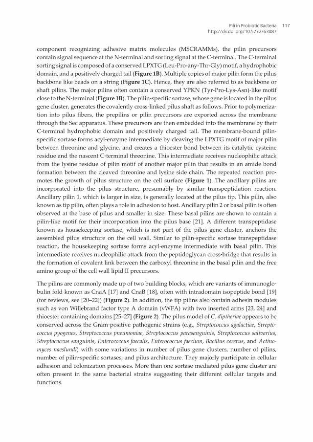
component recognizing adhesive matrix molecules (MSCRAMMs), the pilin precursorscontain signal sequence at the N‐terminal and sorting signal at the C‐terminal. The C‐terminalsorting signal is composed of a conserved LPXTG (Leu-Pro-any-Thr-Gly) motif, a hydrophobicdomain, and a positively charged tail (Figure 1B). Multiple copies of major pilin form the pilusbackbone like beads on a string (Figure 1C). Hence, they are also referred to as backbone orshaft pilins. The major pilins often contain a conserved YPKN (Tyr-Pro-Lys-Asn)‐like motifclose to the N‐terminal (Figure 1B). The pilin‐specific sortase, whose gene is located in the pilusgene cluster, generates the covalently cross‐linked pilus shaft as follows. Prior to polymeriza‐tion into pilus fibers, the prepilins or pilin precursors are exported across the membranethrough the Sec apparatus. These precursors are then embedded into the membrane by theirC‐terminal hydrophobic domain and positively charged tail. The membrane‐bound pilin‐specific sortase forms acyl‐enzyme intermediate by cleaving the LPXTG motif of major pilinbetween threonine and glycine, and creates a thioester bond between its catalytic cysteineresidue and the nascent C‐terminal threonine. This intermediate receives nucleophilic attackfrom the lysine residue of pilin motif of another major pilin that results in an amide bondformation between the cleaved threonine and lysine side chain. The repeated reaction pro‐motes the growth of pilus structure on the cell surface (Figure 1). The ancillary pilins areincorporated into the pilus structure, presumably by similar transpeptidation reaction.Ancillary pilin 1, which is larger in size, is generally located at the pilus tip. This pilin, alsoknown as tip pilin, often plays a role in adhesion to host. Ancillary pilin 2 or basal pilin is oftenobserved at the base of pilus and smaller in size. These basal pilins are shown to contain apilin‐like motif for their incorporation into the pilus base [21]. A different transpeptidaseknown as housekeeping sortase, which is not part of the pilus gene cluster, anchors theassembled pilus structure on the cell wall. Similar to pilin‐specific sortase transpeptidasereaction, the housekeeping sortase forms acyl‐enzyme intermediate with basal pilin. Thisintermediate receives nucleophilic attack from the peptidoglycan cross‐bridge that results inthe formation of covalent link between the carboxyl threonine in the basal pilin and the freeamino group of the cell wall lipid II precursors.
The pilins are commonly made up of two building blocks, which are variants of immunoglo‐bulin fold known as CnaA [17] and CnaB [18], often with intradomain isopeptide bond [19](for reviews, see [20–22]) (Figure 2). In addition, the tip pilins also contain adhesin modulessuch as von Willebrand factor type A domain (vWFA) with two inserted arms [23, 24] andthioester containing domains [25–27] (Figure 2). The pilus model of C. diptheriae appears to beconserved across the Gram‐positive pathogenic strains (e.g., Streptococcus agalactiae, Strepto‐coccus pyogenes, Streptococcus pneumoniae, Streptococcus parasanguinis, Streptococcus salivarius,Streptococcus sanguinis, Enterococcus faecalis, Enterococcus faecium, Bacillus cererus, and Actino‐myces naeslundi) with some variations in number of pilus gene clusters, number of pilins,number of pilin‐specific sortases, and pilus architecture. They majorly participate in cellularadhesion and colonization processes. More than one sortase‐mediated pilus gene cluster areoften present in the same bacterial strains suggesting their different cellular targets andfunctions.
Pili in Probiotic Bacteriahttp://dx.doi.org/10.5772/63087
117

Figure 1. Schematic diagram of typical sortase‐mediated pili. (A) Pilus gene cluster for sortase‐mediated pilus assem‐bly. It encodes genes for a major (red), basal (blue), tip (green) pilins, and a pilin‐specific sortase (purple). More thanone sortase (e.g., SpaD‐ and SpaH‐pilus gene cluster in C. diphtheriae) and less than three pilins (e.g., type 1 and 2 pilusgene clusters in A. oris) have also been observed. In the pilus gene cluster, differences in the order of gene's arrange‐ment and the presence of transposon elements in the vicinity are often observed. (B) Conserved features of sortase pi‐lins. Signal sequence (SS) and LPXTG‐containing cell wall sorting signal (CWSS) are at the N‐ and C‐terminals of allthe (basal, major, and tip) pilins. In addition, the basal and major pilins have pilin motif (YPKN) in the vicinity of N‐terminals. A conserved element called E‐box (LXET) has also been observed in the sortase pilins. The basal pilins con‐sist of 1–3 CnaB domains (Figure 2A). The major pilins contain 2–4 CnaA/B domains. CnaB domains are often at the N‐and C‐terminals, and CnaA at the middle (Figure 2B). The tip plins have adhesive domains (vWFA/thioestercontaining domains) in addition to CnaA/B domains (Figure 2C). (C) Sortase‐mediated pilus structure. The pilus ismade up of three distinct types of pilins: basal (blue), major (red), and tip (green) pilin. In the pilus, the pilins are teth‐ered to each other by sortase‐mediated covalent links (see the text for details). Multiple copies of the major pilins formthe pilus shaft in a head‐to‐tail fashion like beads on a string. The tip pilin is often located at tip projecting adhesivedomain for favoring adhesion. The basal pilin is often located at the base of pilus shaft and helping for anchoring thepolymerized pilus on the cell wall through the housekeeping sortase.
The sortase‐mediated pili, which are being actively investigated in Gram‐positive pathogensand considered as virulence factors, have been detected in several gut commensals as men‐tioned in the following sections. The pilus‐like gene clusters were earlier noticed in probioticLactobacillus johnsonii NCC 533 [28], but first received attention through probiotic Lactobacillusrhamnosus GG in 2009 [29, 30]. Since then, it has been identified in several species and strainsof probiotic and other commensal bacteria by genomic analysis and shown to be essential fortheir adherence and colonization in GIT. Their presence was further confirmed by imaginganalysis in the L. rhamnosus GG [29, 31], genus of Bifidobacterium [32, 33], Lacococcus lactis IL1403and TIL448 [34, 35], and recently in Lactobacillus ruminis ATCC 25644 [36]. Hence, the view ofsurface piliation has now been expanded to include its role also as a niche‐adaptation factor.
Probiotics and Prebiotics in Human Nutrition and Health118
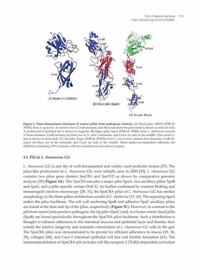
Figure 2. Three‐dimensional structures of sortase‐pilins from pathogenic bacteria. (A) Basal pilin, GBS52 (PDB id:3PHS), from S. agalactiae. It consists of two CnaB domains, and the lysine from the pilin motif is shown as stick (in red).A proline‐rich C‐terminal tail is shown in magenta. (B) Major pilin, SpaA (PDB id: 3HR6), from C. diphtheriae consistsof three domains. CnaB domains (in blue) are at N‐ and C‐terminals, and CnaA (in red) at the middle. Pilin motif ly‐sine is shown in stick (red). (C) Tip pilin, RrgA (PDB id: 2WW8), from S. pneumoniae contains four domains. CnaB do‐mains (in blue) are at the terminals and CnaA (in red) at the middle. Metal (pink)‐ion‐dependent adhesion site(MIDAS) containing vWFA domain with two inserted arms are shown in green.
2.1. Pili in L. rhamnosus GG
L. rhamnosus GG is one the of well-documented and widely used probiotic strains [37]. Thepilus‐like protrusions in L. rhamnosus GG were initially seen in 2009 [30]. L. rhamnosus GGcontains two pilus gene clusters SpaCBA and SpaFED as shown by comparative genomicanalysis [29] (Figure 3A). The SpaCBA encodes a major pilin SpaA, two ancillary pilins SpaBand SpaC, and a pilin‐specific sortase (SrtC1). As further confirmed by western blotting andimmunogold electron microscopy [29, 31], the SpaCBA pilus of L. rhamnosus GG has similarmorphology to the three‐pilins architecture model of C. diptheriae [15, 16]. The repeating SpaAmakes the pilus backbone. The cell wall anchoring SpaB and adhesive SpaC ancillary pilinsare found at the base and tip of the pilus, respectively (Figure 3C). However, in contrast to thepili from most Gram‐positive pathogens, the tip pilin (SpaC) and, to a lesser extent, basal pilin(SpaB) are found sporadically throughout the SpaCBA pilus backbone. Such a distribution isthought to enhance adherence to the intestinal mucosa and epithelial layer and thereby thenextend the relative longevity and transient colonization of L. rhamnosus GG cells in the gut.The SpaCBA pilus was demonstrated to be pivotal for efficient adherence to mucus [29, 38,39], collagen [40], and Caco‐2 intestinal epithelial cell line and biofilm formation [41]. Theimmunomodulation of SpaCBA pili includes toll‐like receptor 2 (TLR2)‐dependent activation
Pili in Probiotic Bacteriahttp://dx.doi.org/10.5772/63087
119

and dendritic cell cytokine production [42], dampening endogenous interleukin (IL)‐8 mRNAlevels [41], eliciting macrophage‐mediated anti‐inflammatory cytokine mRNA expression [43],inducing TLR‐related gene expression in a human fetal intestine model [44], and stimulatingcellular responses in intestinal epithelial cells [45]. Interestingly, the SpaC plays a role in mostof the SpaCBA pili‐triggered host cell immune responses. The surface piliation apparentlyprovides a niche‐specific fitness to L. rhamnosus GG cells for extending their transient coloni‐zation in the gut [46]. Presumably, this is an advantage over nonpiliated probiotic bacteria. Forexample, the non‐SpaCBA piliated L. rhamnosus LC705, which is genetically similar to L.rhamnosus GG, shows decreased adherence to intestinal mucus in the comparative study [29].More recently, the key role of L. rhamnosus GG pili in interaction with β‐lactoglobulin has alsobeen demonstrated [47].
Figure 3. Schematic diagram of sortase‐mediated pili in L. rhamnosus GG. (A) SpaCBA and SpaFED pilus gene clus‐ters identified in L. rhamnosus GG. Each cluster encodes a tip pilin (SpaC/SpaF), major pilin (SpaA/SpaD), basal pilin(SpaB/SpaE), and pilin‐specific sortase (SrtC1/SrtC2). (B) Predicted elements required for the pilus assembly in theSpaCBA pilins. The basal pilin SpaB contains a single CnaB domain with FPKN pilin motif and LPQTG‐containingCWSS at C‐terminal. Residue numbers and positions were labeled and marked by arrow. The major pilin SpaA containtwo CnaB domains, and its pilin and sorting motif are marked. The tip pilin SpaC contains a vWFA domain and itsMIDAS (DMSGS) motif is marked. (C) The SpaCBA pilus model consists of SpaA, SpaB, and SpaC. The possible sor‐tase‐mediated intercovalent link is marked by arrow with details of residues involved. A possible mode of associationfor SpaC and SpaB along the pilus shaft other than at the tip and base of the pilus needs to be further shown by a high‐resolution imaging technique or structural studies.
Similar to SpaCBA, the SpaFED operon encodes the pilus backbone (SpaD), the pilus tip (SpaF)and the base (SpaE) pilins, as well as a putative sortase enzyme (SrtC2) required for pilusassembly (Figure 3A). Though the recombinant SpaF has been shown to bind intestinal mucus[39], the genes associated with the spaFED pilus gene cluster are not constitutively expressed
Probiotics and Prebiotics in Human Nutrition and Health120
in the tested laboratory conditions [31]. Thus, the native form of the SpaFED pilus remainshypothetical, not only in L. rhamnosus GG, but also in other strains carrying the spaFED operon(e.g., L. rhamnosus LC705) [31, 46]. However, L. rhamnosus GG SpaFED pili can be readilyproduced as an assembled structure in recombinant L. lactis [48].
Obtaining three‐dimensional structural insights into pilus assembly and adhesion mecha‐nisms through the structural biology techniques has been instrumental for Gram‐negativepathogens in the past (for reviews, see [5, 8, 49, 50]), and it was begun much later for Gram‐positive pathogens in 2007 ([19, 51], for reviews, see [11, 20–22]). The structures of individ‐ual major as well as ancillary pilins from several pathogenic strains have been determined(for recent review, see [21]) (Figure 2). A Cryo‐EM study on S. pneumoniae pili has alsosupported the sortase‐mediated three pilins architectural model [52]. According to currentstructural knowledge, the basal pilins consist of 1–3 CnaB domains often with intradomainisopeptide bonds (Figure 2A). Conserved proline‐rich C‐terminal tails in the known basalpilins suggest their likely role in pilus anchoring via housekeeping sortase. The presence ofa pilin‐like motif with a lysine in the basal pilin indicates that they could be incorporatedinto the pilus base by sortase (Figure 2). The major pilins are made of 2–4 CnaB/A do‐mains (Figure 2). The CnaB domains are at the N‐ and C‐terminals, whereas the CnaAdomain is in the middle. The pilin motif is present at the C‐terminal region of N‐terminalCnaB domain (Figure 2B). The N‐terminal domain in many pilins seems to be flexible withno or slow forming internal isopeptide bond. In some crystal structure studies, a fiber‐likepilus arrangement in the crystal packing has been observed though the sortase‐mediatedintermolecular amide bond between the backbone pilins was absent. The tip pilins containadhesive domains at the tip in addition to CnaA and CnaB domains that form a stalk andconnect adhesive domains to the pilus shaft (Figure 2C). These adhesive domains are oftena modified vWFA domain with two inserted arms [23, 24], and thioester containing domain[25]. The complicated domains arrangement and folding in tip pilins makes difficult topredict them from their sequence.
Detailed structural knowledge is yet to emerge for pili and related components for probiot‐ic bacteria. However, preliminary crystallographic data are available for some of the pilins(SpaA [53], SpaD [54], and SpaC [55]) in L. rhamnosus GG. Our initial analysis of ongoingstructural investigations on pilus constituents of L. rhamnosus GG and comparison with theircounterparts in pathogens suggest that SpaA may consist of two CnaB domains (Fig‐ure 3B), and SpaD contains three domain with CnaB domains at the terminals and CnaAdomain in the middle. Though it is yet to be validated, it is tempting us to describe Lys171from the pilin motif SpaA as the possible linking lysine that could involve in the SpaA‒SpaA and SpaA‒SpaC pilins covalent association during SpaCBA pilus shaft polymeriza‐tion by pilin‐specific SrtC1 (Figure 3B and C). Similarly, Lys182 in SpaB seems a likelycandidate for its incorporation into the pilus (Figure 3B and C). Such a linking lysine is yetto be predicted for SpaC for its incorporation other than at the pilus tip. In contrast to knownpathogenic tip pilins (e.g., GBS104 [24] and RrgA [23]), but similar to eukaryotic proteins(e.g., integrins, complement C2a, and Fb), the vWFA domain predicted in SpaC [55] seemsnot to have the two inserted arms, suggesting both possible differences and similarities in
Pili in Probiotic Bacteriahttp://dx.doi.org/10.5772/63087
121
binding mechanism via a metal‐ion‐containing vWFA adhesin domain. Certainly, knowl‐edge generated from our ongoing structural investigations would provide new insights intopilus assembly and adhesion mechanisms in L. rhamnosus GG, and serves as a model forprobiotics.
2.2. Pili in L. ruminis
L. ruminis, one of the dominating Lactobacillus species in the mammalian intestines, is routinelyisolated from the feces of human, cattle, and pigs. It is one of the few motile members knownin lactobacilli. It is also recognized as an autochthonous microbiota in the GIT. The pilus geneidentified in the human‐derived intestinal isolate L. rumini ATCC 25644 has been named aslrpCBA (L. rumini pilus) [36] since they appear to be different from the known lactobacillarpilus types (SpaCBA and SpaFED) at the primary structural level. The LrpCBA pilus operonencodes tip (lrpC), basal (lrpB), and major (lrpA) pilins and a pilin‐specific sortase (SrtC).Sequence of L. ruminis pilins displays the common pilin features such as LPXTG‐like motifs,E‐box motif, and pilin motifs (in major and basal pilins) [36] (Figure 2). The expression andsurface localization of lrpCBA pilus gene product have further confirmed by immunoblotanalysis and immune‐electron microscopic visualization (for details, see [36]). Interestingly,the pilus genes have also been detected in L. ruminis ATCC 27782 from bovine gut origin [56],but the microarray analysis showed that the corresponding genes were upregulated in humanstrain compared with the bovine isolate. The ability of LrpCBA pilus to adhere to gut epithelialcells and extracellular matrix (ECM) proteins, and immune‐modulation activities has beendemonstrated using recombinant‐piliated lactococci (for details, see [36]). Interestingly, the tippilin LrpC supports L. ruminis binding to ECM‐related substrates but not to the mucosalsurfaces.
2.3. Pili in other Lactobacillus species
The presence of sortase‐mediated pilus gene clusters has been reported in many strains ofLactobacillus casei [57–60] and Lactobacillus paracasei [61], which are members of the normalhuman gut microbiota and used extensively as probiotics and in the food industries.Although the pilus expression and function are yet to be studied in detail, the most analyzedstrains in the L. casei and L. paracasei group show that they contain SpaCBA and SpaFED pilusgene clusters. In contrast, only few strains in L. rhamnosus group have SpaCBA cluster (e.g.,L. rhamnosus GG and LMS2‐1 strain). However, several L. paracasei strains includingCOM0101 are shown to have truncated SpaC gene [60]. The transposon genes, which arepresent in the vicinity of the SpaCBA cluster in L. rhamnosus, seem to be absent in the L. caseisuggesting that L. rhamnosus GG and LMS2-1 could have acquired the SpaCBA pilus genecluster through horizontal gene transfer (HGT) from L. casei [57, 62]. This is further evi‐denced by the presence of high nucleotide sequence identity in spaCBA cluster of L.rhamnosus and L. casei [57, 62].
Probiotics and Prebiotics in Human Nutrition and Health122

2.4. Pili in L. lactis
L. lactis is another widely used species as starter in dairy fermentation and best characterizedstrain in lactic acid bacteria (LAB). They seem to present in nutrient‐rich ecological niches (gutmucus, milk, and plants). A functional pilus operon (pil) has been shown to present in L.lactis IL1403 [34, 63]. It encodes tip (YhgD), major (YhgE), and basal (YhhB) pilins and a pilin‐specific sortase (SrtC). The presence of pilus structures has been confirmed by immunogoldelectron microscopy and atomic force microscopy (AFM) analyses. The major YhgE and basalYhhB pilins display typical LPXTG motifs and pilin motifs. Additionally, the YhgE has an E‐box. The pili were also shown to promote biofilm formation by confocal laser scanningmicroscopy (CLSM). The occurrence of pili in few other L. lactis isolates from clinical andvegetal environments was also visualized by by transmission electron microscopy (TEM)analysis [34]. Later, a proteomic analysis study has also detected pilus genes (YhgE2, YhhB2,ORF4, and SrtC2) in a vegetal isolate L. lactis subsp. lactis TIL448 [35]. The YhgE2 was shownto play a major role in intestinal epithelial Caco‐2 cells adhesion. The pilus biogenesis andmorphology were further analyzed by immunoblot, electron micrograph, transcriptional, andAFM experiments [35, 64].
2.5. Pili in bifidobacteria
Bifidobacteria are the common components of the gut microbiota of a broad range of hosts [65].Several members of bifidobacteria are typical inhabitants of the infant intestine [66], which isthought to be sterile at birth. Identification of many bifidobacterial strains in the stools ofhealthy infants suggests that they could be the first colonizers in the GIT subsequent to birth.Genomic analysis has revealed pilus genes cluster in several bifidobacterial strains [67].Interestingly, many pilus gene clusters are flanked by transposon elements indicating theiracquisition by HGT. The presence of pilus structures was further examined by AFM andtranscription analysis in Bifidobacterium bifidum, Bifidobacterium dentium, Bifidobacteriumlongum subsp. longum, Bifidobacterium adolescentis, and Bifidobacterium animalis subsp. lactis [67].The pilus gene clusters often found to contain one major pilin (FimA or FimP) and one or twoancillary pilins (FimB or FimQ) with a pilin‐specific sortase. Many of these pilus genes aresimilar to the (two‐pilins) pilus gene clusters identified in Gram‐positive pathogens such asActinomyces oris [68, 69] and Bacillus cereus [70], which lack basal pilus genes differing from thethree‐pilins architectural model of C. diphtheriae [15, 16]. A. oris encodes two different fimbriae(types 1 and 2). Type 1 fimbria, which mediates the interaction of actinomyces to tooth enamel,consists of the major pilin (FimP) and tip pilin (FimQ). Whereas, the type 2 fimbria that mediateinteraction with oral streptococci and host cell for causing dental plaque is made of major pilin(FimA) and tip pilin (FimB). Similarly, B. cereus pili is composed of major pilin BcpA and thetip pilin (BcpB). In the two‐pilin sortase‐mediated pili model, the last major pilin may functionas the pilus base. The three‐dimensional structures for major pilins for A. oris are availablewhile they are yet to be elucidated for tip pilins. The major pilins of bifidobacteria have typicalpilin motif and LPXTG motif required for pilus polymerization [67]. The role of pili inadherence, immunomodulation, and bacterial aggregations was further extensively exploredin B. bifidum PRL2010, which contains three different pilus gene clusters (pil1, pil2, and pil3)
Pili in Probiotic Bacteriahttp://dx.doi.org/10.5772/63087
123

[71]. Apart from sortase‐mediated pili, the presence of type IV pili has also been reported inbifidobacteria (e.g., Bifidobacterium breve UCC2003 [32]), which is described below.
3. Tad pili
The Tad (tight adherence) pili, which was first described in Aggregatibacter (Actinobacillus)actinomycetemcomitans [72], is a specialized subtype of type IV pili (for reviews, see [5, 7, 8, 73,74]). Tad pili in this bacterium were shown to mediate adhesion to surfaces and essential forcolonization and pathogenesis. Apart from adhesion, the type IV pili have been implicated inseveral functions such as aggregation, biofilm formation, twitching motility, DNA uptake, andelectron transfer. Type IV pili are found to be present in Gram‐negative (e.g., enteropathogenicEscherichia coli, Salmonella enterica, Pseudomonas aeruginosa, Neisseria meningitides, and Vibriocholerae) as well as Gram‐positive bacteria (Clostridium perfringens, Mycobacterium tuberculosis,and Ruminococcus albus). Type IV pili are typically 6‒8 nm in diameter and several micrometerslong. The type IV pilus is comprised of homopolymers of a single (major) pilin subunit(Figure 4). The major pilins in Tad pili are relatively smaller in size (∼7 kDa) compared withother known pilus types in type IV. The flexible homopolymer filaments in type IV often havetendency to form characteristic helical bundles by lateral interactions. Some pili possess anadhesive or ancillary pilins at the pilus tip or can be decorated with pseudopilins along thepilus.
Figure 4. Schematic diagram of type IV pilus structure. (A) Type IV (gonococcal) pilus model. Major pilins form thefilament majorly by hydrophobic interactions between their N‐terminal helices in the filament core. The globular headof major pilins pack on the filament surface. (B) Type IV major pilin, PilE (PDB id: 1AY2), from N. gonorrhoeae showingN‐terminal helix (in red) and globular head with D‐region.
Type IV pilus assembly is a complex process, which requires protein products from multiplegenes (∼14) including minor pilins, prepilin peptidase, ATPase, inner membrane core proteins,and accessory proteins. Many of the core genes are conserved across different bacterial species.Tad pili seem to differ from other type IV pilus types by lacking four core homologous minor
Probiotics and Prebiotics in Human Nutrition and Health124

pilins. The type IV pilins are synthesized as precursors with a leader peptide and transportedacross the inner membrane into the periplasmic space, where they are retained in the innermembrane through their N‐terminal hydrophobic segments. The globular domain is foldedwith stabilizing intramolecular disulfide bonds. A dedicated prepilin peptidase cleaves thepositively charged leader sequence and methylates the N‐terminal amine to generate themature pilin. The methylated, positively charged N‐terminal residue is thought to attractnegatively charged glutamate (at fifth position) of adjacent major pilin in the growing pilusfiber. This results in vertical displacement between one pilin and the next. The assemblyATPase associated with the cytoplasmic part of the inner membrane protein undergoesconformational change during ATP hydrolysis and pushes the pilus filament out of themembrane, providing a gap for the next major pilin. Type IV pili is further complicated bydivergence and divided into two classes (types IVa and IVb) based on the length of leaderpeptides and mature pilins. The pilins of type IVa are typically 150‒160 residues long with ashort leader peptide (<10 residues), whereas the pilins of type IVb are either long (180‒200residues) or short (40‒50 residues) with longer leader peptides (∼15‒30 residues). The Tad piliare monophyletic subclass of type IVb pili [73]. The pilins of Tad pili are short with 40‒50residues long.
Though the sequence and structural diversity are associated with the pilins in type IV, theyshare a common lollipop‐like architecture consisting of an extended N‐terminal helical stickfollowed by a globular head containing a β‐sheet with 4–7 strands [74] (Figure 4B). TheN‐terminal half of the helix is hydrophobic and multifunctional regulatory domain. Itprotrudes from the globular head and forms the central hydrophobic core of the grow‐ing filament during the pilus assembly. Prior to assembly, it acts as transmembrane segmentto retain individual pilin in the cytoplasmic membrane. The C‐terminal half of the helix isamphipathic and embedded in the globular head. For many pili, a hypervariable C‐terminal loop known as D‐region or disulfide‐bonded loop (DSL) performs an essentialrole in surface adherence (Figure 4B). The conserved disulfide bridge in the D‐regionobserved in several Gram‐negative major pilins appears to be off in Gram‐positive pilins(e.g., PilA1 in Chlostrodium difficle [75]). The Tad genes are also widespread in the ge‐nomes of Gram‐positive species (C. diphtheriae, Thermobifida fusca, and Streptomyces coelicolor).Recently, they have been identified in probiotic B. breve.
3.1. Tad pili in B. breve
Apart from sortase‐dependent pili, B. breve UCC2003 was recently shown to contain the typeIVb or Tad pilus gene cluster named tad2003 [32]. The presence of pili was further confirmed byimmunogold transmission electron microscopy and shown to be essential for efficient gutcolonization in a murine model by mutational analysis [32]. Specifically, the Tad locus is highlyconserved among all sequenced bifidobacterial strains supporting a ubiquitous pilus‐mediat‐ed host colonization and persistence mechanism for intestinal bifidobacteria. The structuraldata are yet to come for pilins of Tad pilus from beneficial bacteria for shedding light on theirstructure and function.
Pili in Probiotic Bacteriahttp://dx.doi.org/10.5772/63087
125
4. Future perspectives
Adhesion of bacteria to host surfaces is a prerequisite and crucial step for bacterial coloniza‐tion, which may result in pathogenic or commensal relationship. The pili have been oftenimplicated in initiating adhesion and mediating interaction with host. Understanding pilusstructure and function, and their mediated interactions with the host has been achieved to acertain extent in pathogenic strains. The pili and their components are recognized as viru‐lence factors in pathogenic strains, and also considered as potential vaccine candidates incombating bacterial infection. Recent identification of such surface organelles in probiotic orcommensal bacteria gives a new perspective as a niche‐adaption factor as well. The sortase‐mediated pili initially discovered in Gram‐positive pathogens appear to be widespreadamong commensals. The Tad pili, which are known to present in both Gram‐negative andGram‐positive pathogens, have also been detected in some commensal strains. It may not bea surprise if additional pilus type comes in the future from the fast‐growing technology andgenomes for gut microbiota. Available preliminary data suggest that the pili from pathogen‐ic and beneficial bacteria share several sequence and structural features. The presence oftransposable element in several pilus gene clusters indicates that the pathogenic and com‐mensal bacteria may be acquired from each other during the evolution. The challenge isnow to understand the differences between the (enemy) pathogenic and (friendly) beneficialbacteria in their pili‐mediated adhesion strategies and interactions with the host. Thisknowledge is crucial in optimizing probiotics and targeting adhesion‐based therapies forhuman health. The journey of pilus research in probiotics has begun with the prototypeSpaCBA pili in L. rhamnosus GG. The ongoing and future research hopefully would shedlight in this area.
Acknowledgments
This work was funded by the Regional Centre for Biotechnology (RCB) and the Departmentof Biotechnology (DBT) (Grant No. BT/PR5891/BRB/10/1098/2012), India.
Author details
Vengadesan Krishnan1*, Priyanka Chaurasia1,2 and Abhiruchi Kant1,2
*Address all correspondence to: [email protected]
1 Regional Centre for Biotechnology, NCR Biotech Science Cluster, Faridabad, India
2 Department of Biotechnology, Manipal University, Manipal, Karnataka, India
Probiotics and Prebiotics in Human Nutrition and Health126

References
[1] Gerritsen J, Smidt H, Rijkers GT, de Vos WM. Intestinal microbiota in human healthand disease: the impact of probiotics. Genes & Nutrition. 2011;6:209‒40. doi:10.1007/s12263‐011‐0229‐7.
[2] Qin J, Li R, Raes J, Arumugam M, Burgdorf KS, Manichanh C, et al. A human gutmicrobial gene catalogue established by metagenomic sequencing. Nature.2010;464:59‒65. doi:10.1038/nature08821.
[3] Chow J, Lee SM, Shen Y, Khosravi A, Mazmanian SK. Host‐bacterial symbiosis in healthand disease. Advances in Immunology. 2010;107:243‒74. doi:10.1016/B978‐0‐12‐381300‐8.00008‐3.
[4] Ventura M, O'Flaherty S, Claesson MJ, Turroni F, Klaenhammer TR, van Sinderen D,et al. Genome‐scale analyses of health‐promoting bacteria: probiogenomics. NatureReviews Microbiology. 2009;7:61‒71. doi:10.1038/nrmicro2047.
[5] Craig L, Li J. Type IV pili: paradoxes in form and function. Current Opinion inStructural Biology. 2008;18:267‒77. doi:10.1016/j.sbi.2007.12.009.
[6] Kline KA, Dodson KW, Caparon MG, Hultgren SJ. A tale of two pili: assembly andfunction of pili in bacteria. Trends in Microbiology. 2010;18:224‒32. doi:10.1016/j.tim.2010.03.002.
[7] Pelicic V. Type IV pili: e pluribus unum? Molecular Microbiology. 2008;68:827‒37. doi:10.1111/j.1365‐2958.2008.06197.x.
[8] Proft T, Baker EN. Pili in Gram‐negative and Gram‐positive bacteria ‒ structure,assembly and their role in disease. Cellular and Molecular Life Science. 2009;66:613‒35. doi:10.1007/s00018‐008‐8477‐4.
[9] Sauer FG, Mulvey MA, Schilling JD, Martinez JJ, Hultgren SJ. Bacterial pili: molecularmechanisms of pathogenesis. Current Opinion in Microbiology. 2000;3:65‒72.
[10] Scott JR, Zahner D. Pili with strong attachments: Gram‐positive bacteria do it differ‐ently. Molecular Microbiology. 2006;62:320‒30. doi:10.1111/j.1365‐2958.2006.05279.x.
[11] Hendrickx AP, Budzik JM, Oh SY, Schneewind O. Architects at the bacterial surface ‒sortases and the assembly of pili with isopeptide bonds. Nature Reviews Microbiology.2011;9:166‒76. doi:10.1038/nrmicro2520.
[12] Danne C, Dramsi S. Pili of Gram‐positive bacteria: roles in host colonization. Researchin Microbiology. 2012;163:645‒58. doi:10.1016/j.resmic.2012.10.012.
[13] Mandlik A, Swierczynski A, Das A, Ton‐That H. Pili in Gram‐positive bacteria:assembly, involvement in colonization and biofilm development. Trends in Microbi‐ology. 2008;16:33‒40. doi:10.1016/j.tim.2007.10.010.
Pili in Probiotic Bacteriahttp://dx.doi.org/10.5772/63087
127

[14] Telford JL, Barocchi MA, Margarit I, Rappuoli R, Grandi G. Pili in Gram‐positivepathogens. Nature Reviews Microbiology. 2006;4:509‒19. doi:10.1038/nrmicro1443.
[15] Ton‐That H, Schneewind O. Assembly of pili in Gram‐positive bacteria. Trends inMicrobiology. 2004;12:228‒34. doi:10.1016/j.tim.2004.03.004.
[16] Ton‐That H, Marraffini LA, Schneewind O. Sortases and pilin elements involved inpilus assembly of Corynebacterium diphtheriae. Molecular Microbiology. 2004;53:251‒61.
[17] Symersky J, Patti JM, Carson M, House‐Pompeo K, Teale M, Moore D, et al. Structureof the collagen‐binding domain from a Staphylococcus aureus adhesin. Nature StructuralBiology. 1997;4:833‒8.
[18] Deivanayagam CC, Rich RL, Carson M, Owens RT, Danthuluri S, Bice T, et al. Novelfold and assembly of the repetitive B region of the Staphylococcus aureus collagen‐binding surface protein. Structure. 2000;8:67‒78.
[19] Kang HJ, Coulibaly F, Clow F, Proft T, Baker EN. Stabilizing isopeptide bonds revealedin Gram‐positive bacterial pilus structure. Science. 2007;318:1625‒8.
[20] Kang HJ, Baker EN. Structure and assembly of Gram‐positive bacterial pili: uniquecovalent polymers. Current Opinion in Structural Biology. 2012;22:200‒7.
[21] Krishnan V. Pilins in Gram‐positive bacteria: a structural perspective. IUBMB Life.2015;67:533‒43. doi:10.1002/iub.1400.
[22] Vengadesan K, Narayana SV. Structural biology of Gram‐positive bacterial adhesins.Protein Science 2011;20:759‒72. doi:10.1002/pro.613.
[23] Izoré T, Contreras‐Martel C, El Mortaji L, Manzano C, Terrasse R, Vernet T, et al.Structural basis of host cell recognition by the pilus adhesin from Streptococcus pneu‐moniae. Structure. 2010;18:106‒15.
[24] Krishnan V, Dwivedi P, Kim BJ, Samal A, Macon K, Ma X, et al. Structure of Streptococcusagalactiae tip pilin GBS104: a model for GBS pili assembly and host interactions. ActaCrystallographica Section D, Biological Crystallography. 2013;69:1073‒89. doi:10.1107/S0907444913004642.
[25] Linke‐Winnebeck C, Paterson NG, Young PG, Middleditch MJ, Greenwood DR, WitteG, et al. Structural model for covalent adhesion of the Streptococcus pyogenes pilusthrough a thioester bond. The Journal of Biological Chemistry. 2014;289:177‒89. doi:10.1074/jbc.M113.523761.
[26] Pointon JA, Smith WD, Saalbach G, Crow A, Kehoe MA, Banfield MJ. A highly unusualthioester bond in a pilus adhesin is required for efficient host cell interaction. TheJournal of Biological Chemistry. 2010;285:33858‒66. doi:10.1074/jbc.M110.149385.
[27] Walden M, Crow A, Nelson MD, Banfield MJ. Intramolecular isopeptide but notinternal thioester bonds confer proteolytic and significant thermal stability to the S.pyogenes pilus adhesin Spy0125. Proteins. 2014;82:517‒27. doi:10.1002/prot.24420.
Probiotics and Prebiotics in Human Nutrition and Health128

[28] Pridmore RD, Berger B, Desiere F, Vilanova D, Barretto C, Pittet AC, et al. The genomesequence of the probiotic intestinal bacterium Lactobacillus johnsonii NCC 533. Proceed‐ings of the National Academy of Sciences of the United States of America.2004;101:2512‒7.
[29] Kankainen M, Paulin L, Tynkkynen S, von Ossowski I, Reunanen J, Partanen P, et al.Comparative genomic analysis of Lactobacillus rhamnosus GG reveals pili containing ahuman‐mucus binding protein. Proceedings of the National Academy of Sciences ofthe United States of America. 2009;106:17193‒8. doi:10.1073/pnas.0908876106.
[30] Lebeer S, Verhoeven TL, Francius G, Schoofs G, Lambrichts I, Dufrene Y, et al. Identi‐fication of a gene cluster for the biosynthesis of a long, galactose‐rich exopolysaccharidein Lactobacillus rhamnosus GG and functional analysis of the priming glycosyltransfer‐ase. Appled and Environmental Microbiology. 2009;75:3554‒63. doi:10.1128/AEM.02919‐08.
[31] Reunanen J, von Ossowski I, Hendrickx AP, Palva A, de Vos WM. Characterization ofthe SpaCBA pilus fibers in the probiotic Lactobacillus rhamnosus GG. Applied andEnvironmental Microbiology. 2012;78:2337‒44.
[32] O'Connell Motherway M, Zomer A, Leahy SC, Reunanen J, Bottacini F, Claesson MJ,et al. Functional genome analysis of Bifidobacterium breve UCC2003 reveals type IVbtight adherence (Tad) pili as an essential and conserved host‐colonization factor.Proceedings of the National Academy of Sciences of the United States of America.2011;108:11217‒22. doi:10.1073/pnas.1105380108.
[33] Turroni F, Foroni E, Serafini F, Viappiani A, Montanini B, Bottacini F, et al. Ability ofBifidobacterium breve to grow on different types of milk: exploring the metabolism ofmilk through genome analysis. Applied and Environmental Microbiology.2011;77:7408‒17. doi:10.1128/AEM.05336‐11.
[34] Oxaran V, Ledue‐Clier F, Dieye Y, Herry JM, Pechoux C, Meylheuc T, et al. Pilusbiogenesis in Lactococcus lactis: molecular characterization and role in aggregation andbiofilm formation. PLoS One. 2012;7:e50989. doi:10.1371/journal.pone.0050989.
[35] Meyrand M, Guillot A, Goin M, Furlan S, Armalyte J, Kulakauskas S, et al. Surfaceproteome analysis of a natural isolate of Lactococcus lactis reveals the presence of piliable to bind human intestinal epithelial cells. Molecular & Cellular Proteomics: MCP.2013;12:3935‒47. doi:10.1074/mcp.M113.029066.
[36] Yu X, Jaatinen A, Rintahaka J, Hynonen U, Lyytinen O, Kant R, et al. Human gut‐commensalic Lactobacillus ruminis ATCC 25644 displays sortase‐assembled surfacepiliation: phenotypic characterization of its fimbrial operon through in silico predictiveanalysis and recombinant expression in Lactococcus lactis. PLoS One. 2015;10:e0145718.doi:10.1371/journal.pone.0145718.
[37] Lee YK, Salminen S. Handbook of Probiotics and Prebiotics: John Wiley & Sons, Inc.,New Jersey; 2008. doi:10.1002/9780470432624
Pili in Probiotic Bacteriahttp://dx.doi.org/10.5772/63087
129

[38] Nishiyama K, Ueno S, Sugiyama M, Yamamoto Y, Mukai T. Lactobacillus rhamnosus GGSpaC pilin subunit binds to the carbohydrate moieties of intestinal glycoconjugates.Animal Science Journal. 2015. doi:10.1111/asj.12491.
[39] von Ossowski I, Reunanen J, Satokari R, Vesterlund S, Kankainen M, Huhtinen H, etal. Mucosal adhesion properties of the probiotic Lactobacillus rhamnosus GG SpaCBAand SpaFED pilin subunits. Applied and Environmental Microbiology. 2010;76:2049‒57.
[40] Tripathi P, Beaussart A, Alsteens D, Dupres V, Claes I, von Ossowski I, et al. Adhesionand nanomechanics of pili from the probiotic Lactobacillus rhamnosus GG. ACS Nano.2013;7:3685‒97. doi:10.1021/nn400705u.
[41] Lebeer S, Claes I, Tytgat HL, Verhoeven TL, Marien E, von Ossowski I, et al. Functionalanalysis of Lactobacillus rhamnosus GG pili in relation to adhesion and immunomodu‐latory interactions with intestinal epithelial cells. Applied and Environmental Micro‐biology. 2012;78:185‒93.
[42] von Ossowski I, Pietila TE, Rintahaka J, Nummenmaa E, Makinen VM, Reunanen J, etal. Using recombinant Lactococci as an approach to dissect the immunomodulatingcapacity of surface piliation in probiotic Lactobacillus rhamnosus GG. PLoS One.2013;8:e64416. doi:10.1371/journal.pone.0064416.
[43] Vargas Garcia CE, Petrova M, Claes IJ, De Boeck I, Verhoeven TL, Dilissen E, et al.Piliation of Lactobacillus rhamnosus GG promotes adhesion, phagocytosis, and cytokinemodulation in macrophages. Applied and Environmental Microbiology. 2015;81:2050‒62. doi:10.1128/AEM.03949‐14.
[44] Ganguli K, Collado MC, Rautava J, Lu L, Satokari R, von Ossowski I, et al. Lactobacillusrhamnosus GG and its SpaC pilus adhesin modulate inflammatory responsiveness andTLR‐related gene expression in the fetal human gut. Pediatric Research. 2015;77:528‒35. doi:10.1038/pr.2015.5.
[45] Ardita CS, Mercante JW, Kwon YM, Luo L, Crawford ME, Powell DN, et al. Epithelialadhesion mediated by pilin SpaC is required for Lactobacillus rhamnosus GG‐inducedcellular responses. Applied and Environmental Microbiology. 2014;80:5068‒77. doi:10.1128/AEM.01039‐14.
[46] Kant R, Rintahaka J, Yu X, Sigvart‐Mattila P, Paulin L, Mecklin JP, et al. A comparativepan‐genome perspective of niche‐adaptable cell‐surface protein phenotypes inLactobacillus rhamnosus. PLoS One. 2014;9:e102762. doi:10.1371/journal.pone.0102762.
[47] Guerin J, Bacharouche J, Burgain J, Lebeer S, Francius G, Borges F, et al. Pili ofLactobacillus rhamnosus GG mediate interaction with β‐lactoglobulin. Food Hydrocol‐loids. 2016;58:35‒41.
[48] Rintahaka J, Yu X, Kant R, Palva A, von Ossowski I. Phenotypical analysis of theLactobacillus rhamnosus GG fimbrial spaFED operon: surface expression and functional
Probiotics and Prebiotics in Human Nutrition and Health130
characterization of recombinant SpaFED pili in Lactococcus lactis. PLoS One.2014;9:e113922. doi:10.1371/journal.pone.0113922.
[49] Fronzes R, Christie PJ, Waksman G. The structural biology of type IV secretion systems.Nature Reviews Microbiology. 2009;7:703–14. doi:10.1038/nrmicro2218.
[50] Waksman G, Hultgren SJ. Structural biology of the chaperone‐usher pathway of pilusbiogenesis. Nature Reviews Microbiology. 2009;7:765–74.
[51] Krishnan V, Gaspar AH, Ye N, Mandlik A, Ton‐That H, Narayana SVL. An IgG‐likedomain in the minor pilin GBS52 of Streptococcus agalactiae mediates lung epithelial celladhesion. Structure. 2007;15:893–903.
[52] Hilleringmann M, Ringler P, Muller SA, De Angelis G, Rappuoli R, Ferlenghi I, et al.Molecular architecture of Streptococcus pneumoniae TIGR4 pili. EMBO Journal.2009;28:3921–30. doi:10.1038/emboj.2009.360.
[53] Singh D, von Ossowski I, Palva A, Krishnan V. Purification, crystallization andpreliminary crystallographic analysis of the SpaA backbone‐pilin subunit fromprobiotic Lactobacillus rhamnosus GG. Acta Crystallographica Section F, StructuralBiology and Crystallization Communication. 2013;69:1182‒5. doi:10.1107/S1744309113024676.
[54] Chaurasia P, von Ossowski I, Palva A, Krishnan V. Purification, crystallization andpreliminary X‐ray diffraction analysis of SpaD, a backbone‐pilin subunit encoded bythe fimbrial spaFED operon in Lactobacillus rhamnosus GG. Acta CrystallographicaSection F, Structural Biology Communication. 2015;71:103‒6. doi:10.1107/S2053230X14027216.
[55] Kant A, von Ossowski I, Palva A, Krishnan V. Crystallization and X-ray crystallo‐graphic analysis of the adhesive SpaC pilin subunit in the SpaCBA pilus of gut?adaptedLactobacillus rhamnosus GG. Protein & Peptide Letters. 2016;23:365-71. doi:10.2174/0929866523666160106153055.
[56] Forde BM, Neville BA, O'Donnell MM, Riboulet‐Bisson E, Claesson MJ, Coghlan A, etal. Genome sequences and comparative genomics of two Lactobacillus ruminis strainsfrom the bovine and human intestinal tracts. Microbial Cell Factories. 2011;10 (Suppl.1):S13. doi:10.1186/1475‐2859‐10‐S1‐S13.
[57] Broadbent JR, Neeno‐Eckwall EC, Stahl B, Tandee K, Cai H, Morovic W, et al. Analysisof the Lactobacillus casei supragenome and its influence in species evolution and lifestyleadaptation. BMC Genomics. 2012;13:533. doi:10.1186/1471‐2164‐13‐533.
[58] Douillard FP, Ribbera A, Jarvinen HM, Kant R, Pietila TE, Randazzo C, et al. Compa‐rative genomic and functional analysis of Lactobacillus casei and Lactobacillus rhamno‐sus strains marketed as probiotics. Applied and Environmental Microbiology.2013;79:1923‒33. doi:10.1128/AEM.03467‐12.
Pili in Probiotic Bacteriahttp://dx.doi.org/10.5772/63087
131
[59] Munoz‐Provencio D, Rodriguez‐Diaz J, Collado MC, Langella P, Bermudez‐HumaranLG, Monedero V. Functional analysis of the Lactobacillus casei BL23 sortases. Appliedand Environmental Microbiology. 2012;78:8684‒93. doi:10.1128/AEM.02287‐12.
[60] Toh H, Oshima K, Nakano A, Takahata M, Murakami M, Takaki T, et al. Genomicadaptation of the Lactobacillus casei group. PLoS One. 2013;8:e75073. doi:10.1371/journal.pone.0075073.
[61] Smokvina T, Wels M, Polka J, Chervaux C, Brisse S, Boekhorst J, et al. Lactobacillusparacasei comparative genomics: towards species pan‐genome definition and exploita‐tion of diversity. PLoS One. 2013;8:e68731. doi:10.1371/journal.pone.0068731.
[62] Aleksandrzak‐Piekarczyk T, Koryszewska‐Baginska A, Grynberg M, Nowak A,Cukrowska B, Kozakova H, et al. Genomic and functional characterization of theunusual pLOCK 0919 plasmid harboring the spaCBA pili cluster in Lactobacillus caseiLOCK 0919. Genome Biology and Evolution. 2016;8:202‒17. doi:10.1093/gbe/evv247.
[63] Dieye Y, Oxaran V, Ledue‐Clier F, Alkhalaf W, Buist G, Juillard V, et al. Functionalityof sortase A in Lactococcus lactis. Applied and Environmental Microbiology.2010;76:7332‒7. doi:10.1128/AEM.00928‐10.
[64] Le DT, Tran TL, Duviau MP, Meyrand M, Guerardel Y, Castelain M, et al. Unravelingthe role of surface mucus‐binding protein and pili in muco‐adhesion of Lactococcuslactis. PLoS One. 2013;8:e79850. doi:10.1371/journal.pone.0079850.
[65] Turroni F, Ribbera A, Foroni E, van Sinderen D, Ventura M. Human gut microbiotaand bifidobacteria: from composition to functionality. Antonie van Leeuwenhoek.2008;94:35‒50. doi:10.1007/s10482‐008‐9232‐4.
[66] Koenig JE, Spor A, Scalfone N, Fricker AD, Stombaugh J, Knight R, et al. Succession ofmicrobial consortia in the developing infant gut microbiome. Proceedings of theNational Academy of Sciences of the United States of America. 2011;108 (Suppl. 1):4578‒85. doi:10.1073/pnas.1000081107.
[67] Foroni E, Serafini F, Amidani D, Turroni F, He F, Bottacini F, et al. Genetic analysis andmorphological identification of pilus‐like structures in members of the genus Bifido‐bacterium. Microbial Cell Factories. 2011;10 (Suppl. 1):S16. doi:10.1186/1475‐2859‐10‐S1‐S16.
[68] Mishra A, Das A, Cisar JO, Ton‐That H. Sortase‐catalyzed assembly of distinct hetero‐meric fimbriae in Actinomyces naeslundii. Journal of Bacteriology. 2007;189:3156‒65. doi:10.1128/JB.01952‐06.
[69] Mishra A, Wu C, Yang J, Cisar JO, Das A, Ton‐That H. The Actinomyces oris type 2fimbrial shaft FimA mediates co‐aggregation with oral streptococci, adherence to redblood cells and biofilm development. Molecular Microbiology. 2010;77:841‒54. doi:10.1111/j.1365‐2958.2010.07252.x.
Probiotics and Prebiotics in Human Nutrition and Health132

[70] Budzik JM, Marraffini LA, Schneewind O. Assembly of pili on the surface of Bacilluscereus vegetative cells. Molecular Microbiology. 2007;66:495‒510. doi:10.1111/j.1365‐2958.2007.05939.x.
[71] Turroni F, Serafini F, Foroni E, Duranti S, O'Connell Motherway M, Taverniti V, et al.Role of sortase‐dependent pili of Bifidobacterium bifidum PRL2010 in modulatingbacterium‐host interactions. Proceedings of the National Academy of Sciences of theUnited States of America. 2013;110:11151‒6. doi:10.1073/pnas.1303897110.
[72] Kachlany SC, Planet PJ, DeSalle R, Fine DH, Figurski DH. Genes for tight adherence ofActinobacillus actinomycetemcomitans: from plaque to plague to pond scum. Trends inMicrobiology. 2001;9:429‒37.
[73] Burrows LL. Pseudomonas aeruginosa twitching motility: type IV pili in action. AnnualReview of Microbiology. 2012;66:493–520. doi:10.1146/annurev‐micro‐092611‐150055.
[74] Giltner CL, Nguyen Y, Burrows LL. Type IV pilin proteins: versatile molecularmodules. Microbiology and Molecular Biology Reviews: MMBR. 2012;76:740–72. doi:10.1128/MMBR.00035‐12.
[75] Piepenbrink KH, Maldarelli GA, Martinez de la Pena CF, Dingle TC, Mulvey GL, LeeA, et al. Structural and evolutionary analyses show unique stabilization strategies inthe type IV pili of Clostridium difficile. Structure. 2015;23:385–96. doi:10.1016/j.str.2014.11.018.
Pili in Probiotic Bacteriahttp://dx.doi.org/10.5772/63087
133

Chapter 7
Biosynthesis of Vitamins by Probiotic Bacteria
Qing Gu and Ping Li
Additional information is available at the end of the chapter
http://dx.doi.org/10.5772/63117
Abstract
Vitamins are important micronutrients that are often precursors to enzymes, which allliving cells require to perform biochemical reactions. However, humans cannot producemany vitamins, so they have to be externally obtained. Using vitamin‐producingmicroorganisms could be an organic and marketable solution to using pseudo‐vitaminsthat are chemically produced, and could allow for the production of foods with higherlevels of vitamins that could reduce unwanted side effects. Probiotic bacteria, as well ascommensal bacteria found in the human gut, such as Lactobacillus and Bifidobacterium, cande novo synthesize and supply vitamins to human body. In humans, members of the gutmicrobiota are able to synthesize vitamin K, as well as most of the water‐soluble B vitamins,such as cobalamin, folates, pyridoxine, riboflavin, and thiamine.
Keywords: probiotic, folate, riboflavin, cobalamin, biosynthesis
1. Introduction
Vitamins are typically categorized as fat‐soluble vitamins, which includes vitamins A, D, E, andK, or as water‐soluble vitamins, which includes vitamin C, biotin (vitamin H or B7), and a seriesof B vitamins—thiamin (B1), riboflavin (B2), niacin (B3), pantothenic acid (B5), pyridoxine (B6),folic acid (B11), and cobalamin (B12). While fat‐soluble vitamins act as important elements ofcell membranes, water‐soluble vitamins serve as coenzymes that typically transport specificchemical groups [1]. Humans are incapable of synthesizing most vitamins and they conse‐quently have to be obtained exogenously. The use of vitamin‐producing microorganisms mightrepresent a more natural and consumer‐friendly alternative to fortification using chemicallysynthesized pseudo‐vitamins.

The biochemical pathways involved in B‐vitamin biosynthesis by food microorganisms werepreviously described in detail [2]. Many prokaryotes need water‐soluble vitamins for nutri‐tional purposes [3], but also typically need them for biosynthetic processes. The ability ofparticular microorganisms to produce B vitamins could supplant the expensive chemicalproduction of these vitamins to enrich food or be improved for in situ fortification of fermentedfoods. Much research has been conducted in recent years to elucidate the biosyntheticpathways of these vitamins in a number of microorganisms.
Probiotic bacteria positively impact the immune system and the composition and functioningof the gut microbiota [4]. Furthermore, the production of vitamins has resulted in many healthybenefits to the host. Probiotic bacteria, mostly belonging to the genera Lactobacillus andBifidobacterium, confer a number of health benefits, including vitamin production [5]. Probioticbacteria, members of the gut microbiota, are able to synthesize vitamin K and most of the water‐soluble B vitamins, such as biotin, cobalamin, folates, nicotinic acid, panthotenic acid, pyri‐doxine, riboflavin, and thiamine, in humans [6].
The production of B‐vitamins, especially folate and riboflavin (B2), by probiotic bacteria hasbeen extensively researched as described in a recent review [7, 8]. Several lactic acid bacteria(LAB) species (e.g., Lactococcus lactis, Lactobacillus gasseri, and Lactobacillus reuteri) andBifidobacterium (e.g., B. adolescentis) produce these vitamins, often in large quantities, and are,therefore, often found in fermented foods [9, 10]. Moreover, increased vitamin biosynthesishas been obtained by metabolic engineering [11, 12]. Folate biosynthetic genes and riboflavinbiosynthetic operon have been overexpressed in L. lactis, resulting in types that produce folate[12] or riboflavin [12] at higher rates. Sybesma et al. [13] modified the biosynthetic pathwaysof folate and riboflavin in L. lactis, resulting in the simultaneous overproduction of bothvitamins, through directed mutagenesis and selection and metabolic engineering.
This review focused on riboflavin, folic acid, and cobalamin, three of the water‐soluble Bvitamins whose biosynthetic pathways were inextricably linked, briefly covering theirphysiological functions and dietary sources before concentrating on novel overproductionstrategies in probiotics.
2. Riboflavin biosynthesis
In contrast to many plants, fungi, and bacteria, humans cannot produce riboflavin or vitaminB2, and thus require it as a dietary supplement. Riboflavin is available as a dietary source andis also produced by the microflora of the large intestine [6, 14]. Riboflavin (vitamin B2) playsan essential role in cellular metabolism, as it is the precursor of the coenzymes flavin mono‐nucleotide (FMN) and flavin adenine dinucleotide (FAD), which both act as hydrogen carriersin many biological redox reactions.
Riboflavin is synthesized by many bacteria and its biosynthetic pathway has been studiedextensively in Bacillus subtilis and Escherichia coli. Bacher et al. [15, 16] found that riboflavinbiosynthesis requires the precursor's guanosine 5′‐triphosphate (GTP) and ribulose 5‐phos‐phate. The first step of the GTP‐dependent branch of the biosynthetic pathway is encoded by
Probiotics and Prebiotics in Human Nutrition and Health136

ribA in E. coli. In B. subtilis it is also encoded by ribA but in this case RibA acts as a bifunctionalenzyme that also catalyzes the configuration of 3,4‐dihydroxy‐2‐butanone 4‐phosphate fromribulose 5‐ phosphate [17]. The overexpression of RibA in B. subtilis produces 25% moreriboflavin, indicating that this enzyme is rate‐limiting in riboflavin biosynthesis [18]. However,in Lactococcus lactis, the overexpression of ribA did not lead to increased riboflavin production[12].
The ability of some bacteria and fungi to overproduce riboflavin has been harnessed forindustrial production. Such commercial producers include the ascomycetes Eremotheciumashbyii and Ashbya gossypii. However, advantages were perceived in developing bacterial andyeast fermentations to avail of their high growth rates, and less costly and complex growthmedia. Currently, A. gossypii, Candida famata, and B. subtilis are exploited for riboflavinproduction, with riboflavin production levels reaching 15 g/L, 20 g/L, and 14 g/L, respectively[19–21]. In A. gossypii, metabolic engineering increased riboflavin production almost 10‐fold[22]. A. gossypii has also been targeted as a microorganism to overproduce riboflavin using oilwaste [23]. In the case of B. subtilis, high levels of riboflavin production were achieved as aresult of exposure to purine analogues and the toxic riboflavin analogue roseoflavin, or bygenetic engineering [19, 24].
It has been reported that fermentation of cow milk with L. lactis and Propionibacterium freuden‐reichii ssp. shermanii as starter cultures significantly increased the riboflavin content of milk.Since the riboflavin produced by starter cultures is largely in the free form, the bio‐availabilityis expected to be better than the bio‐availability of riboflavin in unprocessed milk [12, 25]. Thefood‐grade fermentative LAB L. lactis also grows in the absence of riboflavin. On the basis ofthe genome sequence of L. lactis IL1403 [26], it seemed that all genes involved in riboflavinbiosynthesis (rib genes) were present in this organism.
Species and/or strain‐specific traits in LAB provided genetic information for riboflavinbiosynthesis. Several of the sequenced members of LAB possessed similar abilities to produceriboflavin, as suggested by comparative genome analysis, but an interrupted rib operon wassometimes seen in certain strains. Deficient genetic information was usually related to theinability to produce riboflavin in LAB. For instance, the sequenced genome of Lactobacillusplantarum strain WCFS1 had an incomplete rib operon, which lacked the entire ribG and partof the ribB genes [27]. Further, this strain could not grow unless riboflavin was present [28].However, several selected strains of L. plantarum contained the whole rib operon and couldproduce vitamin B2. The L. plantarum strain NCDO 1752, and the recently sequenced L.plantarum strain JDMI and L. plantarum strains, for example, were isolated from cereals‐derivedproducts [28, 29]. Furthermore, even in LAB strains that contained all rib genes, riboflavinproduction had to be confirmed by chemical analysis.
3. Folate biosynthesis by human gut commensals
Folic acid, also known as vitamin B11, is a dietary necessity for humans, because it is used inseveral metabolic reactions, such as the biosynthesis of the building blocks of DNA and RNA,
Biosynthesis of Vitamins by Probiotic Bacteriahttp://dx.doi.org/10.5772/63117
137

the nucleotides. It is recommended that adults take 200 μg daily, but pregnant women areencouraged to take a double dose daily, as folic acid could thwart neural‐tube defects innewborns [30]. Low folic acid has been linked to high homocysteine levels in the blood, whichcould lead to coronary diseases [31, 32]. It has also been shown to protect against some formsof cancer [33]. Folate is conspicuously absent in many food products and is considered anessential additive to the general diet.
Folates are comprised of a mono‐ or polyglutamyl conjugate and these compounds werenamed after the number of glutamyl residues (PteGlun), where n denoted the total number ofglutamyl residues. The folates act as enzyme co‐substrates in one‐carbon (C1) metabolism ofamino acids and nucleotides, in which the fully reduced (tetrahydro‐) form functions as anacceptor or donor of a single carbon unit [34]. Folic acid has played a significant role in theproduction of purines and pyrimidines, and, therefore, in DNA synthesis. Methioninesynthase uses 5‐methyltetrahydrofolate in the conversion of l‐homocysteine to l‐methionine[35]. A majority of the methionine formed is converted to S‐adenosylmethionine, which is acommon donor of methyl groups for DNA, RNA, hormones, neurotransmitters, membranelipids, and proteins [36]. The folate molecule contains one pterin moiety, created from 6‐hydroxymethyl‐7,8‐dihydropterin pyrophosphate (DHPPP), bound to para‐aminobenzoicacid (pABA, vitamin B10). As such, de novo biosynthesis called for both the precursors, DHPPPand pABA. Plants and bacteria could make the latter from the pentose phosphate pathway.Erythrose 4‐phosphate and phosphoenolpyruvate go through the shikimate pathway tobecome chorismate, which acts as a branching point toward the biosynthesis of aromatic aminoacids and pABA. Chorismate is transformed via aminodeoxychorismate synthase into 4‐amino‐4‐deoxychorismate. Subsequently, pyruvate is cleaved by 4‐amino‐4‐deoxychorismatelyase to give pABA, which is ultimately necessary for folate biosynthesis. The biosynthesis ofDHPPP proceeds via the conversion of GTP in four consecutive steps. The first step is catalyzedby GTP cyclohydrolase I and involves an extensive transformation of GTP, through Amadorirearrangement, to form a pterin ring structure. Following dephosphorylation, the pterinmolecule undergoes aldolase and pyrophosphokinase reactions, which produce the activatedpyrophosphorylated DHPPP.
Folate biosynthesis continues with the formation of a C–N bond joining DHPPP to pABA. Thiscondensation reaction, catalyzed by dihydropteroate synthase, yields 7,8‐dihydropteroate(DHP). DHP is glutamylated by dihydrofolate synthase, resulting in dihydrofolate (DHF). Itis then reduced by DHF reductase to the biologically active cofactor tetrahydrofolate (THF)and subjected to the addition of multiple glutamate moieties by folylpolyglutamate synthaseto yield THF‐polyglutamate. Polyglutamilation may also take place before the occurrence ofthe reduction step, being catalyzed by DHF synthase or, in many bacteria, by a bifunctionalenzyme that is responsible for both DHF synthase and folylpolyglutamate synthase activities[37].
However, although all available complete bifidobacterial genomes are expected to specifyaminodeoxychorismate synthase, a gene specifying a putative 4‐amino‐4‐deoxychorismatelyase can only be found on the genome of B. adolescentis ATCC15703 and B. dentium Bd1 [9],which are, thus, expected to accomplish de novo biosynthesis of pABA. In contrast, B.
Probiotics and Prebiotics in Human Nutrition and Health138
animalis subsp. lactis does not appear to possess the entire pathway for DHPPP biosynthesisor the gene encoding dihydropteroate synthase. Thus, B. animalis subsp. lactis was predictedto be auxotrophic for folates or DHP, and would, therefore, be unable to complete folatebiosynthesis, even if pABA was present.
Lactobacilli are also typical human gut commensals and were recently investigated to discoverif they could serve as possible folate producers [38]. Lactobacilli from various fermented foodshave been investigated as starter cultures for the manufacturing of folate‐fortified dairyproducts, while lactobacilli isolated from the human gut have been explored as folate‐producing probiotics [39–42]. The availability of genome sequences of various lactobacilliprovided an important contribution to the genetics underlying folate biosynthesis in this groupof microorganisms [38]. For example, lactobacilli did not appear to harbor the genetic deter‐minants for de novo pABA synthesis, with the exception of L. plantarum WCFS1 [27], suggest‐ing that the vast majority of lactobacilli were unable to synthesize folate in the absence ofpABA.
Currently, the strains of Lactobacillus with the greatest relevance for the manufacturing ofprobiotics and functional foods belong to the species L. acidophilus, L. casei, L. paracasei, L.plantarum, L. reuteri, and L. salivarius [43]. Like L. lactis, these species harbor a folate biosynthesiscluster that includes the gene encoding dihydropteroate synthase and all of the genes for thebiosynthesis of DHPPP, with the exception of alkaline phosphatase. In L. lactis, thedephosphorylation of dihydroneopterin triphosphate into the monophosphate wasdemonstrated to occur through an alternative route, involving a Nudix pyrophosphohydro‐lase [44]. Many lactobacilli contain various genes encoding putative Nudix enzymes, such asmutT genes for DNA repair. However, Lactobacillus sakei, Lactobacillus helveticus, andLactobacillus delbrueckii have a homologue of the L. lactis gene in the fol cluster. In contrast, inLactobacillus fermentum, L. plantarum, and L. reuteri, the fol cluster held the gene of a putativenon‐Nudix purine NTP pyrophosphatase, which could be responsible for hydrolyzingdihydroneopterin triphosphate in these species. As such, L. plantarum, L. sakei, L. delbrueckii,L. reuteri, L. helveticus, and L. fermentum were predicted to generate DHPPP and could also befolate producers if cultured with pABA present [37, 44].
4. Vitamin B12 biosynthesis
Vitamin B12, otherwise known as cobalamin, is the biggest and most intricate vitamin.Cobalamin describes a cluster of cobalt‐containing compounds (corrinoids) that have a loweraxial ligand, which holds the cobalt‐coordinated nucleotide (5, 6‐dimethylbenzimidazole) asa base. Although humans only use vitamin B12 for two enzymatic activities, it is still animportant dietary supplement. (R)‐methyl‐malonyl‐CoA mutase assists in the metabolism ofpropionyl‐CoA, which compounds such as valine, thymine, methionine, and odd‐chain fattyacids produce when broken down. This ado‐cobalamin‐dependent enzyme catalyzes therearrangement of propionyl‐CoA following its carboxylation and epimerization to succinyl‐CoA, which then goes through the citric acid cycle. Methionine synthase needs vitamin B12 in
Biosynthesis of Vitamins by Probiotic Bacteriahttp://dx.doi.org/10.5772/63117
139
the form of methylcobalamin. Using 5‐methyltetrahydrofolate as a methyl donor, this enzymemethylates homocysteine to form methionine [45].
Humans cannot synthesize vitamin B12, and, thus must obtain it from organisms that can.Only a limited number of bacteria are known to produce vitamin B12, three of which—Pseudomonas denitrificans, Bacillus megaterium, and Propionibacterium freudenreichii—are usedfor commercial production [46–48].
Cobalamin has the most complex structure of all the vitamins synthesized by bacteria requiringabout 30 genes for its biosynthesis. Most of the work in characterizing cobalamin biosynthesishas been performed in Salmonella typhimurium and P. denitrificans. Two different pathwaysexist for adenosylcobalamin (ado‐cobalamin) biosynthesis: (1) an oxygen‐dependent pathway,which is found in P. denitrificans, and (2) an anaerobic pathway, which has been identified in,among others, S. typhimurium, P. freudenreichii subsp. Shermanii, and B. megaterium. Every generequired in the anaerobic cobalamin biosynthesis was found on the genome of S. sanguinis [49].
Genes encoding enzymes contributing to the oxygen‐dependent pathway have been given theprefix cob, while those involved in the oxygen‐independent pathway have the prefix cbi [50].Due to the early insertion of cobalt in the anaerobic pathway, the remaining intermediates arecobalto‐complexes and therefore require enzymes with different substrate specificities thanthe intermediates in the aerobic pathway although many of the reactions catalyzed are similar.CobZ was identified in Rhodobacter capsulatus, which catalyzes a reaction similar to thatadvanced by CobG, but in a different way, as the two proteins did not display any primarysequence resemblance. CobZ was also found to have a flavin in the form of a non‐covalentlybound FAD, two Fe‐S centers, and a b‐type heme, which was not similar to CobG [51]. It wasthought that the final step in the cobalamin biosynthetic pathway in S. typhimurium involvedthe dephosphorylation of adenosylcobalamin‐5′‐phosphate, which is catalyzed by CobC andchallenges the pathway indicated where CobS catalyzes the condensation of a‐ribazole and anAdo‐GDP‐cobinamide [52]. The gene that reduces cobalt in the aerobic pathway has yet to beidentified, but two candidate genes were identified to encode this enzyme, named CobR [53].
LAB are traditionally known as auxotrophic for cobalamin and are generally used for thebiological analysis of this vitamin. Recently, however, cobalamins were identified in L.reuteri as were some of the genes encoding enzymes for the biosynthesis of this vitamin [54].The presence of a B12‐dependent metabolic pathway that converts glycerol into propanediolmost likely allowed this LAB to synthesize B12. The discovery of the biosynthetic genes couldincrease the production of B12 through metabolic engineering, and facilitate the transfer of theproduction pathway to other LAB.
L. reuteri CRL1098 was also found to metabolize glycerol in a B12‐free medium, indicating thata LAB might also be able to make cobalamin [55]. Chromatographic analysis of the intracellularbacterial extract of L. reuteri CRL 1098 proved that this strain was able to produce a cobalamin‐like compound with an absorption spectrum that was similar to that of standard cobalaminbut had a distinct elution time, while cobalamin production was proved with differentbioassays [55]. Genetic evidence of cobalamin biosynthesis by L. reuteri CRL 1098 was thenachieved by using different molecular biology techniques, and it was found that at least 30
Probiotics and Prebiotics in Human Nutrition and Health140

genes assisted the de novo synthesis of the vitamin. The genetic organization (cob and cbi genes)resembled that of Salmonella enterica and Listeria innocua [56].
The complete genome of Lactobacillus sanfranciscensis TMW 1.1304, isolated from industrialsourdough fermentation, was also recently sequenced [57]. The data showed that only onegene necessary to the cobalamine synthesis was encoded by the sequenced strain L. sanfran‐ciscensis TMW1.1304. Conversely, growth experiments revealed that several L. sanfranciscen‐sis strains grew on vitamin B12‐free media, which implied that these strains could synthesizecobalamine de novo [57].
Other strains of genus Lactobacilli such as Lactobacillus coryniformis isolated from goat milk [58],L. plantarum isolated from kanjika or Japanese pickles [59, 60], Lactobacillus rossiae isolated fromsourdough [61], and Lactobacillus fermentum CFR 2195 isolated from breast‐fed healthy infants’fecal matter [62] were shown to produce cobalamin‐type compounds. Moreover, the geneticand biochemical data suggested that cobalamin biosynthesis genes would be spread toLactobacillus buchneri, Lactobacillus hilgardii, and Lactobacillus brevis, and also contain genes ofthe cob‐pdu gene cluster [63]. Therefore, the possibility of various cobalamin‐producing strainsand species of LAB would benefit not only from future basic studies on cobalamin production,but also from its application in the development of vitamin B12‐contained fermented products.
5. Biosynthesis of other B‐group vitamins
Thiamine (vitamin B1) is a coenzyme in the pentose phosphate pathway that is required tosynthesize fatty acids, steroids, nucleic acids, and the aromatic amino acid precursors intovarious neurotransmitters and other bioactive compounds essential for brain function [64].Beyond its role as a necessary cofactor in the folate cycle, vitamin B6 (pyridoxine) also playsan important role in amino acid metabolism, which makes it a rate‐limiting cofactor in thesynthesis of neurotransmitters such as dopamine, serotonin, gamma‐aminobutyric acid(GABA), noradrenaline, and the hormone melatonin [64].
LAB fermentation in yogurt, cheese, and other fermented products was shown to result inincreased levels of riboflavin, folate, vitamin B12, niacin, and pyridoxine [65, 66]. Soy fermen‐tation with Streptococcus thermophilus ST5 and Lactobacillus helveticus R0052 or Bifidobacteriumlongum R0175 also caused a small increase in thiamine and pyridoxine concentration that wasnot statistically significant [67].
6. Biosynthesis of vitamin K
Vitamin K serves as a cofactor for the enzyme that converts specific glutamyl residues in a fewproteins to g‐carboxyglutamyl (Gla) residues, aiding in the process. Humans obtain the dailynutritional requirement for vitamin K through the dietary phylloquinone that exists in plants,and, to some extent, through bacterially produced polyisoprenyl‐containing compounds
Biosynthesis of Vitamins by Probiotic Bacteriahttp://dx.doi.org/10.5772/63117
141

called menaquinones created in the human gut [68]. LAB were examined for their ability toproduce quinone compounds, as vitamin K occurred naturally in two forms, namely, K1(phylloquinone) in green plants, and K2 (menaquinones) in animals and some bacteria [69].
7. Conclusions
The use of vitamin‐producing strains provided a new perspective on the specific uses ofprobiotics. Many food‐grade bacteria overproduce B vitamins, including riboflavin (vitaminB2), folate (vitamin B11), and cyanocobalamine (vitamin B12), which could allow them toorganically enrich raw food materials like soy, milk, meat, and vegetables with B vitamins,preventing the need for additives. Thus, the food industry could take advantage of these noveland efficient vitamin‐producing strains to add nutritional value to fermented products andsave money in the process. Notably, vitamin metabolism pathways were shown in genes thatspecified the biosynthetic enzymes for riboflavin, cobalamin, and folate production. It isincreasingly possible to identify potential vitamin‐producing strains and interpret theintertwined mechanisms for their biosynthesis, because of the expanding availability ofgenome sequences, which could be used to expand the vitamin‐producing capacities of thehuman gut.
Acknowledgements
This project was funded by the International Science & Technology Cooperation Program ofChina (2013DFA32330), the National Natural Science Foundation of China (No. 31071513, No.31271821), the Natural Science Foundation of Zhejiang Province (No. LY16C200002), theNational High Technology Research and Development Program (“863” Program) of China(2012AA022105B), the National Research Foundation for the Doctoral Program of HigherEducation (20133326110005), and the Science Foundation of the Zhejiang Education Depart‐ment (No. Y201534497).
Author details
Qing Gu* and Ping Li
*Address all correspondence to: [email protected]
Key Laboratory for Food Microbial Technology of Zhejiang Province, College of Food Scienceand Biotechnology, Zhejiang Gongshang University, Hangzhou, China
Probiotics and Prebiotics in Human Nutrition and Health142

References
[1] Baku TK, Dickerson JWT. Vitamins in Human Health and Disease. CAB International,Wallinford; 1996.
[2] LeBlanc JG, Laino JE, del Valle MJ, Vannini V, van Sinderen D, Taranto MP, de ValdezGF, de Giori GS, Sesma F. B‐group vitamin production by lactic acid bacteria – currentknowledge and potential applications. J Appl Microbiol. 2011;111:1297–1309. DOI:10.1111/j.1365‐2672.2011.05157.x
[3] Snell EE. From bacterial nutrition to enzyme structure: a personal odyssey. Annu RevBiochem. 1993;62:1–27. DOI: 10.1146/annurev.bi.62.070193.000245
[4] Hill C, Guarner F, Reid G, Gibson GR, Merenstein DJ, Pot B, Morelli L, Canani RB, FlintHJ, Salminen S, Calder PC, Sanders ME. Expert consensus document. The InternationalScientific Association for Probiotics and Prebiotics consensus statement on the scopeand appropriate use of the term probiotic. Nat Rev Gastroenterol Hepatol. 2014;11:506–514. DOI:10.1038/nrgastro.2014.66
[5] Kanmani P, Satish Kumar R, Yuvaraj N, Paari KA, Pattukumar V, Arul V. Probioticsand its functionally valuable products‐a review. Crit Rev Food Sci Nutr. 2013;53:641–658. DOI: 10.1080/10408398.2011.553752
[6] Hill MJ. Intestinal flora and endogenous vitamin synthesis. Eur J Cancer Prev.1997;6(Suppl 1):S43–S45. DOI: 10.1097/00008469‐199703001‐00009
[7] Burgess CM, Smid EJ, van Sinderen D. Bacterial vitamin B2, B11 and B12 overproduc‐tion: an overview. Int J Food Microbiol. 2009;133:1–7. DOI: 10.1016/j.ijfoodmicro.2009.04.012
[8] Rossi M, Amaretti A, Raimondi S. Folate production by probiotic bacteria. Nutrients.2011;3:118–134. DOI: 10.3390/nu3010118
[9] Ventura M, Turroni F, Zomer A, Foroni E, Giubellini V, Bottacini F, Canchaya C,Claesson MJ, He F, Mantzourani M, Mulas L, Ferrarini A, Gao B, Delledonne M,Henrissat B, Coutinho P, Oggioni M, Gupta RS, Zhang Z, Beighton D, Fitzgerald GF,O'Toole PW, van Sinderen D. The Bifidobacterium dentium Bd1 genome sequence reflectsits genetic adaptation to the human oral cavity. PLoS Genet. 2009;5:e1000785. DOI:10.1371/journal.pgen.1000785
[10] Papagianni M. Metabolic engineering of lactic acid bacteria for the production ofindustrially important compounds. Comput Struct Biotechnol J. 2012;3:e201210003.DOI: 10.5936/csbj.201210003
[11] Hugenholtz J, Sybesma W, Groot MN, Wisselink W, Ladero V, Burgess K, van SinderenD, Piard JC, Eggink G, Smid EJ, Savoy G, Sesma F, Jansen T, Hols P, Kleerebezem M.Metabolic engineering of lactic acid bacteria for the production of nutraceuticals.Antonie Van Leeuwenhoek. 2002;82:217–235.
Biosynthesis of Vitamins by Probiotic Bacteriahttp://dx.doi.org/10.5772/63117
143

[12] Burgess C, O’ Connell‐Motherway M, Sybesma W, Hugenholtz J, van Sinderen D.Riboflavin production in Lactococcus lactis: potential for in situ production of vitamin‐enriched foods. Appl Environ Microbiol. 2004;70:5769–5777. DOI:10.1128/AEM.70.10.5769‐5777.2004
[13] Sybesma W, Burgess C, StarrenburgM, van Sinderen D, Hugenholtz J. Multivitaminproduction in Lactococcus lactis using metabolic engineering. Metab Eng. 2004;6:109–115. DOI:10.1016/j.ymben.2003.11.002
[14] Wrong O, Edmonds C, Chadwich V. The Large Intestine. Its Role in Mammalian Nutritionand Homeostasis. Wiley & Sons, New York; 1981.
[15] Bacher A, Eberhardt S, Fischer M, Kis K, Richter G. Biosynthesis of vitamin b2 (ribo‐flavin). Annu Rev Nutr. 2000;20:153–167. DOI: 10.1146/annurev.nutr.20.1.153
[16] Perkins JB, Pero J. Vitamin biosynthesis. In: Sonenshein A, Hoch J, Losick R., editors.Bacillus subtilis and its Closest Relatives from Genes to Cells. ASM Press, Washington, DC,USA; 2002. p. 271–286.
[17] Richter G, Ritz H, Katzenmeier G, Volk R, Kohnle A, Lottspeich F, Allendorf D, BacherA. Biosynthesis of riboflavin: cloning, sequencing, mapping, and expression of the genecoding for GTP cyclohydrolase II in Escherichia coli. J Bacteriol. 1993;175:4045–4051.DOI: 0021‐9193/93/134045‐07$02.00/0
[18] Hümbelin M, Griesser V, Keller T, Schurter W, Haiker M, Hohmann H, Ritz H, RichterG, Bacher A, van Loon A. GTP cyclohydrolase II and 3,4‐dihydroxy‐ 2‐butanone 4‐phosphate synthase are rate‐limiting enzymes in riboflavin synthesis of an industrialBacillus subtilis strain used for riboflavin production. J Ind Microbio Biotechnol.1999;22:1–7. 10.1038/sj.jim.2900590
[19] Perkins J, Sloma A, Hermann T, Theriault K, Zachgo E, Erdenberger T, Hannett N,Chatterjee N, Williams II V, Rufo Jr. GA, Hatch R, Pero J. Genetic engineering of Bacillussubtilis for the commercial production of riboflavin. J Ind Microbiol Biotechnol.1999;22:8–18. 10.1038/sj.jim.2900587
[20] Stahmann K, Revuelta J, Seulberger H. Three biotechnical processes using Ashbyagossypii, Candida famata, or Bacillus subtilis compete with chemical riboflavin produc‐tion. Appl Microbiol Biotechnol. 2000;53:509–516. 10.1007/s002530051649
[21] Schallmey M, Singh A, Ward O. Developments in the use of Bacillus species forindustrial production. Can J Microbiol. 2004;50:1–17. 10.1139/w03‐076
[22] Jimenez A, Santos MA, Pompejus M, Revuelta JL. Metabolic engineering of the purinepathway for riboflavin production in Ashbya gossypii. Appl Environ Microbiol.2005;71:5743–5751. doi:10.1128/AEM.71.10
[23] Park EY, Zhang JH, Tajima S, Dwiarti L. Isolation of Ashbya gossypii mutant for animproved riboflavin production targeting for biorefinery technology. J Appl Microbiol.2007;103:468–476. DOI: 10.1111/j.1365‐2672.2006.03264.x
Probiotics and Prebiotics in Human Nutrition and Health144

[24] Perkins JB, Pero J, Sloma A. Riboflavin overproducing strains of bacteria. Europeanpatent EP0405370; 1991.
[25] Singh R, Deodhar AD. Relative bioavailability of riboflavin in cow's milk and fermentedmilk using rat bioassay. Int Dairy J. 1994;4:59–71. DOI:10.1016/0958‐6946(94)90049‐3
[26] Bolotin A, Wincker P, Mauger S, Jaillon O, Malarme K, Weissenbach J, Ehrlich SD,Sorokin A. The complete genome sequence of the lactic acid bacterium Lactococcuslactis ssp. lactis IL1403. Genome Res. 2001;11:731–753. DOI:10.1101/gr.169701
[27] Kleerebezem M, Boekhorst J, van Kranenburg R, Molenaar D, Kuipers OP, Leer R,Tarchini R, Peters SA, Sandbrink HM, Fiers MW, Stiekema W, Lankhorst RM, Bron PA,Hoffer SM, Groot MN, Kerkhoven R, de Vries M, Ursing B, de Vos WM, Siezen RJ.Complete genome sequence of Lactobacillus plantarum WCFS1. Proc Natl Acad Sci USA.2003;100:1990–1995. DOI: 10.1073/pnas.0337704100
[28] Burgess CM, Smid EJ, Rutten G, van Sinderen D. A general method for selection ofriboflavin‐overproducing food grade micro‐organisms. Microb Cell Factories.2006;5:24. DOI: 10.1186/1475‐2859‐5‐24
[29] Capozzi V, Menga V, Digesu AM, De Vita P, van Sinderen D, Cattivelli L, Fares C,Spano G. Biotechnological production of vitamin B2‐enriched bread and pasta. J AgricFood Chem. 2011;59:8013–8020. DOI: 10.1021/jf201519h
[30] MRC Vitamin Study Research Group. Prevention of neural tube defects: results of theMedical Research Council vitamin study. Lancet. 1991;338:131–137. DOI: 10.1016/0140‐6736(91)90133‐A
[31] Boushey CJ, Beresford AA, Omenn GS, Moltulsky AG. A quantitative assessment ofplasma homocysteine as a risk factor for vascular disease. J Am Med Assoc.1995;274:1049–1057. DOI:10.1001/jama.1995.03530130055028
[32] Kamphuis MH, Geerlings MI, Grobbee DE, Kromhout D. Dietary intake of B(6‐9‐12)vitamins, serum homocysteine levels and their association with depressive symptoms:the Zutphen Elderly Study. Eur J Clin Nutr. 2008;62:939–945. DOI: 10.1038/sj.ejcn.1602804
[33] Ames BN. Micronutrient deficiencies. A major cause of DNA damage. Ann NY AcadSci. 1999;889:87–106. DOI: 10.1111/j.1749‐6632.1999.tb08727.x
[34] Combs GF. The Vitamins: Fundamental Aspects in Nutrition and Health. Academic Press,San Diego; 1992.
[35] Brody T. Folic acid. In: Machlin LJ, editor. Handbook of Vitamins. Marcel Dekker Inc.,New York; 1991. p. 453–489.
[36] Laiño JE, de Giori GS, LeBlanc JG. Folate production by lactic acid bacteria. In: WatsonR, editor. Bioactive Foods and Supplements in Health Promotion ‐ Liver and Gastrointestinal
Biosynthesis of Vitamins by Probiotic Bacteriahttp://dx.doi.org/10.5772/63117
145

Disease. Elsevier, Oxford, UK; 2013. p. 251–270. DOI:10.1016/B978‐0‐12‐397154‐8.00030‐0
[37] De Crécy‐Lagard V, El Yacoubi B, de la Garza RD, Noiriel A, Hanson AD. Comparativegenomics of bacterial and plant folate synthesis and salvage: predictions and valida‐tions. BMC Genomics. 2007;8:245. DOI: 10.1186/1471‐2164‐8‐245
[38] Kleerebezem M, Vaughan EE. Probiotic and gut lactobacilli and bifidobacteria:molecular approaches to study diversity and activity. Annu Rev Microbiol.2009;63:269–290. DOI: 10.1146/annurev.micro.091208.073341
[39] Rao DR, Reddy AV, Pulusani SR, Cornwell PE. Biosynthesis and utilization of folic acidand vitamin B12 by lactic cultures in skim milk. J Dairy Sci. 1984;67:1169–1174. DOI:10.3168/jds.S0022‐0302(84)81419‐8
[40] Wegkamp A, Starrenburg M, de Vos WM, Hugenholtz J, Sybesma W. Transformationof folate‐consuming Lactobacillus gasseri into a folate producer. Appl Environ Microbiol.2004;70:3146–3148. DOI: 10.1128/AEM.70.5.3146‐3148.2004
[41] Santos F, Wegkamp A, de Vos WM, Smid EJ, Hugenholtz J. High‐level folate productionin fermented foods by the B12 producer Lactobacillus reuteri JCM1112. Appl EnvironMicrobiol. 2008;74:3291–3294. DOI: 10.1128/AEM.02719‐07
[42] Laino JE, Leblanc JG, Savoy de Giori G. Production of natural folates by lactic acidbacteria starter cultures isolated from artisanal Argentinean yogurts. Can J Microbiol.2012;58:581–588. DOI: 10.1139/w2012‐026
[43] FAO/WHO. Health and nutritional properties of probiotics in food including powdermilk with live lactic acid bacteria. In: Report of a Joint FAO/WHO Expert Consultation onEvaluation of Health and Nutritional Properties of Probiotics in Food including Powder Milkwith Live Lactic Acid Bacteria, Córdoba, Argentina. WHO, Geneva, Switzerland; 2001. p.1–34.
[44] Klaus SM, Wegkamp A, Sybesma W, Hugenholtz J, Gregory JF III, Hanson AD. A nudixenzyme removes pyrophosphate from dihydroneopterin triphosphate in the folatesynthesis pathway of bacteria and plants. J Biol Chem. 2005;280:5274–5280. DOI:10.1074/jbc.M413759200
[45] Ellenbogen L, Cooper BA. Vitamin B12. In: Machlin, L.J., editor Handbook of Vitamins.Marcel Dekker Inc., New York; 1991. p. 491–536.
[46] Warren MJ, Raux E, Schubert HL, Escalante‐Semerena JC. The biosynthesis of adeno‐sylcobalamin (vitamin B12). Nat Prod Rep. 2002;19:390–412. DOI: 10.1039/B108967F
[47] Warren MJ. Finding the final pieces of the vitamin B12 biosynthetic jigsaw. Proc NatlAcad Sci USA 2006;103:4799–4800. DOI: 10.1073/pnas.0601030103
[48] Escalante‐Semerena JC. Conversion of cobinamide into adenosylcobamide in bacteriaand archaea. J Bacteriol. 2007;189:4555–4560. DOI: 10.1128/JB.00503‐07
Probiotics and Prebiotics in Human Nutrition and Health146

[49] Xu P, Alves JM, Kitten T, Brown A, Chen Z, Ozaki LS, Manque P, Ge X, Serrano MG,Puiu D, Hendricks S, Wang Y, Chaplin MD, Akan D, Paik S, Peterson DL, Macrina FL,Buck GA. Genome of the opportunistic pathogen Streptococcus sanguinis. J Bacteriol.2007;189:3166–3175. DOI: 10.1128/JB.01808‐06
[50] Martens JH, Barg H,Warren MJ, Jahn D. Microbial production of vitamin B12. ApplMicrobiol Biotechnol. 2002;58:275–285. DOI: 10.1007/s00253‐001‐0902‐7
[51] McGoldrick HM, Roessner CA, Raux E, Lawrence AD, McLean KJ, Munro AW,Santabarbara S, Rigby SE, Heathcote P, Scott AI, Warren MJ. Identification andcharacterization of a novel vitamin B12 (cobalamin) biosynthetic enzyme (CobZ) fromRhodobacter capsulatus, containing flavin, heme, and Fe–S cofactors. J Biol Chem.2005;280:1086–1094. DOI: 10.1074/jbc.M411884200
[52] Zayas CL, Escalante‐Semerena JC. Reassessment of the late steps of coenzyme B12synthesis in Salmonella enterica: evidence that dephosphorylation of adenosylcobala‐min‐ 5′‐phosphate by the CobC phosphatase is the last step of the pathway. J Bacteriol.2007;189:2210–2218. DOI: 10.1128/JB.01665‐06
[53] Heldt D, Lawrence AD, Lindenmeyer M, Deery E, Heathcote P, Rigby SE, Warren MJ.Aerobic synthesis of vitamin B12: ring contraction and cobalt chelation. Biochem SocTrans. 2005;33:815–819. DOI: 10.1042/BST0330815
[54] Kleerebezem M, Hugenholtz J. Metabolic pathway engineering in lactic acid bacteria.Curr Opin Biotechnol. 2003;14:232–237. DOI:10.1016/S0958‐1669(03)00033‐8
[55] Taranto MP, Vera JL, Hugenholtz J, de Valdez GF and Sesma F. Lactobacillus reuteriCRL1098 produces cobalamin. J Bacteriol. 2003;185:5643–5647. DOI: 10.1128/JB.185.18.5643‐5647.2003
[56] Santos F, Vera JL, Lamosa P, de Valdez GF, de Vos WM, Santos H, Sesma F andHugenholtz J. Pseudovitamin B(12) is the corrinoid produced by Lactobacillus reuteriCRL1098 under anaerobic conditions. FEBS Lett. 2007;581:4865–4870. DOI: 10.1016/j.febslet.2007.09.012
[57] Vogel RF, Pavlovic M, Ehrmann MA, Wiezer A, Liesegang H, Offschanka S, Voget S,Angelov A, Böcker G, Liebl W. Genomic analysis reveals Lactobacillus sanfranciscensisas stable element in traditional sourdoughs. Microb Cell Factories. 2011;10:S6. DOI:10.1186/1475‐2859‐10‐S1‐S6
[58] Martin R, Olivares M, Marin ML, Xaus J, Fernandez L, Rodriguez JM. Characterizationof a reuterin‐producing Lactobacillus coryniformis strain isolated from a goat's milkcheese. Int J Food Microbiol. 2005;104:267–277. DOI: 10.1016/j.ijfoodmicro.2005.03.007
[59] Madhu AN, Giribhattanavar P, Narayan MS, Prapulla SG. Probiotic lactic acid bacte‐rium from kanjika as a potential source of vitamin B12: evidence from LC‐MS, immu‐nological and microbiological techniques. Biotechnol Lett. 2010;32:503–506. DOI:10.1007/s10529‐009‐0176‐1
Biosynthesis of Vitamins by Probiotic Bacteriahttp://dx.doi.org/10.5772/63117
147
[60] Masuda M, Ide M, Utsumi H, Niiro T, Shimamura Y, Murata M. Production potencyof folate, vitamin B(12), and thiamine by lactic acid bacteria isolated from Japanesepickles. Biosci Biotechnol Biochem. 2012;76:2061–2067. DOI: 10.1271/bbb.120414
[61] De Angelis M, Bottacini F, Fosso B, Kelleher P, Calasso M, Di Cagno R, Ventura M,Picardi E, van Sinderen D, Gobbetti M. Lactobacillus rossiae, a vitamin B12 producer,represents a metabolically versatile species within the Genus Lactobacillus. PLoS One.2014;9:e107232. DOI: 10.1371/journal.pone.0107232
[62] Basavanna G, Prapulla SG. Evaluation of functional aspects of Lactobacillus fermentumCFR 2195 isolated from breast fed healthy infants’ fecal matter. J Food Sci Technol.2013;50:360–366. DOI: 10.1007/s13197‐011‐0345‐9
[63] Capozzi V, Russo P, Duenas MT, Lopez P, Spano G. Lactic acid bacteria producing B‐group vitamins: a great potential for functional cereals products. Appl MicrobiolBiotechnol. 2012;96:1383–1394. DOI: 10.1007/s00253‐012‐4440‐2
[64] Kerns JC, Arundel C, Chawla LS. Thiamin deficiency in people with obesity. Adv NutrInt Rev J. 2015;6:147–153. DOI: 10.3945/an.114.007526
[65] Shahani K, Chandan RC. Nutritional and healthful aspects of cultured and culture‐containing dairy foods. J Dairy Sci. 1979;62:1685–1694. DOI:10.3168/jds.S0022‐0302(79)83481‐5
[66] Alm L. Effect of fermentation on B‐vitamin content of milk in Sweden. J Dairy Sci.1982;65:353–359. DOI: 10.3168/jds.S0022‐0302(82)82199‐1
[67] Champagne CP, Tompkins TA, Buckley ND, Green‐ Johnson JM. Effect of fermentationby pure and mixed cultures of Streptococcus thermophilus and Lactobacillus helveticus onisoflavone and B‐vitamin content of a fermented soy beverage. Food Microbiol.2010;27:968–972. DOI: 10.1016/j.fm.2010.06.003
[68] Suttie JW. The importance of menaquinones in human nutrition. Annu Rev Nutr.1995;15:399–417. DOI: 10.1146/annurev.nu.15.070195.002151
[69] Morishita T, Tamura N, Makino T, Kudo S. Production of menaquinones by lactic acidbacteria. J Dairy Sci. 1999;82:1897–1903. DOI:10.3168/jds.S0022‐0302(99)75424‐X
Probiotics and Prebiotics in Human Nutrition and Health148

Chapter 8
Bioactive Compounds of Lactic Acid Bacteria. Case Study:Evaluation of Antimicrobial Activity of Bacteriocin-producing Lactobacilli Isolated from Native EcologicalNiches of Ecuador
Gabriela N. Tenea and Lucia Yépez
Additional information is available at the end of the chapter
http://dx.doi.org/10.5772/63112
Abstract
Food preservation through natural methods represents one of the concerns world‐wide to solve economic losses due to microbial decomposition of raw materials andfoodstuffs. However, public concern over the emergence of strains resistant to manyantibiotics, particularly pathogens such as E. coli and Salmonella sp. draw much attentionas new challenge in food industry is to find new alternative quality-control methods offood products. In Ecuador, the lack of quality control, bad storage condition, andinsufficient preservation against spoilage bacteria had at higher extent repercussions onfood safety and security. The most frequent pathogens detected in fresh meat and drinksalong with traditional local food products, represent a serious problem producingsizable food damage and associated diseases. The capacity of lactobacilli to inhibitpathogens has been recently exploited to prevent microbial spoilage. Here we brieflyreview the principal biopeptides (i.e., bacteriocins) of lactic acid bacteria, their mainmode of action, the classification, and its biotechnological applications. Moreover, wediscussed the preliminary results on the evaluation of antimicrobial activity of somenative lactic acid bacteria isolated from microbiota of Ecuador against frequentcontaminants found in the local market.
Keywords: lactic acid bacteria, biopreservation, bacteriocins, food pathogens, probiot‐ic

1. Introduction
Lactic acid bacteria (LAB) are among the most favorable microorganisms known for theirprobiotic properties and for the ability to produce antimicrobial compounds (i.e., bacteriocin,organic acids, diacetyl, hydrogen peroxide) with inhibitory action of harmful bacteria growthalong with their critical role in food protection and health maintenance [1–3].
Nowadays, one of the biggest issues faced by the food-processing industry is contaminationwith pathogens caused by poor maintenance and unhygienic sanitary behavior and insuffi‐cient attention to the handling and preservation, contributing greatly to decrease the qualityof products and also increase consummators foodborne illness in the population [4–6]. Thus,the preservation through natural methods represents one of the main concerns at the globallevel to solve economic losses due to microbial decomposition of raw materials and food‐stuffs.
With concomitant expansion of the research, commercial, food industry and medical sec‐tors, the field of biopreservation using probiotic bacteria is developing rapidly with accu‐mulation of many data about their benefits. The complete genome sequencing as well as theidentification of functional properties will further contribute to the reinforcement of mostpowerful products with improved biotechnological characteristics. Although many bacteriaproduce antimicrobial substances, the benefits of those produced by LAB is of particularinterest because of their Generally Recognized as Safe (GRAS) status, which acts as naturalbiopreservative and natural flavor enhancers [3, 7–9]. Hence, the majority of antimicrobialpeptide-producing LAB are ideally suited to food applications. Therefore, the production ofbacteriocins by LAB is not only advantageous to the bacteria themselves but could also beexploited as a tool of food industry to control undesirable bacteria in a natural manner, andbe allowable to the consumer.
As the main source of knowing LAB is represented by the human microflora and fermentedmilk products, it would be more valuable to search for other sources of probiotic microor‐ganism, which might possess powerful properties and beneficial for either human health orfood preservation. During the last decade, extensive progress has been made with respect tothe isolation of LAB with highly antimicrobial properties as well as comprehension of bac‐teriocin structure and function, regulation, and immunity. Further investigations may helpto develop new methods for food preservation by direct comparisons between strains bac‐teriocin producers and non-produced isogenic strains. In this context, bacteriocin of LABwould offer several benefits such as the use reduction of chemical compounds in food pres‐ervation. In this chapter, we will briefly review the main information about the role of bac‐teriocin of LAB in food preservation, their classification and mode of action along with theirbiotechnological benefits. Moreover, we shall present the preliminary results on the evalua‐tion of antimicrobial activity of some native lactic acid bacteria isolated from microbiota ofEcuador against frequent contaminants found in the local food market.
Probiotics and Prebiotics in Human Nutrition and Health150

2. Bacteriocins of lactic acid bacteria and their biotechnologicalapplications
Antimicrobial heterogeneous compounds (i.e., bacteriocine) are ribosomally synthesizedpolypeptide or low-molecular-weight proteins (composed of 20–60 amino acid residues),which, in case of LAB, are generally recognized as safe compounds [9]. They bind to thereceptor of the target cell, and their mode of action included pore formation, degradation ofcellular DNA, disruption through specific cleavage of 16S rRNA, and inhibition of peptido‐glycan synthesis [10, 11]. Bacteriocins being proteinaceous agents differ from most antibioticsbecause they are rapidly digested by proteases in the digestive tract.
2.1. Types of bacteriocins
More than three hundred different bacteriocins have been described for the genera Lactobacil‐lus, Lactococcus, Leuconostoc, Pediococcus, and Enterococcus. These peptides are colorless,odorless, and tasteless, and according to their molecular mass, thermo stability, enzymatic,and sensitivity, the presence of posttranslational modified amino acids and their mode ofaction are classified into four major groups [10–13].
Bacteriocins of Class I: They, known as lantibiotics, are small peptides of <5 kDa, heat stable thatacting on the membrane structure, and contain the thio-ester amino acids lanthionine andmethyllanthionine as well as other modified amino acids such as dehydrated serine andthreonine. From this class, the most studied bacteriocin is nisin produced by Lactococcuslactis subsp. lactis and discovered since 1928 as being the first bioactive compound used in foodsystem as biopreservative [12]. According to their structural similarities, the lantibiotics weredivided into two subclasses. Subclass Ia, comprising positively charged peptides (i.e., nisin),generally acts by forming pores in the cytoplasmic membrane of the target species. SubclassIb are peptides either negatively charged or no net charged, more rigid in their structure whichexert their action by interfering with enzymatic reactions of sensitive bacteria. The moststudied bacteriocins of class I are Nisin Z and Q, Enterocin W, and Nukacin ISK-1 [14–16].
Bacteriocins of Class II: They, known as non-lantibiotics, are heat-stable bacteriocins of variablemolecular weight, <10 kDa, containing in their composition regular amino acids. This classwas subdivided into four subclasses. Subclass IIa, comprising Pediocin PA-1 and Sakacin P, areknown for their antimicrobial activity against Listeria. Members of pediocin-like peptides havea high degree of homology (40–60%), particularly at the N-terminal domain, containing“pediocine box” or homologous region YGNGVXCXXXXCXV, with two residues of cysteineforming a disulfide bridge. Other known bacteriocins of subclass IIa are Enterocin NKR-5-3C[17, 18], Enterocin A [15], Munditicin [19], and Leucocin A [15]. Subclass IIb, comprising distinctpeptides with little or no activity, refers to two-component bacteriocins that require twopeptides to work synergistically. In this group are enclosed Lactacin F and Lactococcin G.Subclass IIc are small peptides, heat stable, and transported by leader peptides, comprisingDiverginin A and Acidocin B. Subclass IId includes sec-dependent bacteriocins, and leaderlessbacteriocins are Lacticin Q [20], Z [21], Weissellicin Y and M [22], and Leucocin Q and N [15].
Bioactive Compounds of Lactic Acid Bacteria. Case Study: Evaluation of Antimicrobial Activity of Bacteriocin-producingLactobacilli Isolated from Native Ecological Niches of Ecuador
http://dx.doi.org/10.5772/63112
151

Bacteriocins of Class III: They are larger peptides, about 430 kDa, heat liable comprisingHelveticins J and V, Acidofilicin A, and Lactacins A and B.
Bacteriocins of Class IV: They contain modified peptides with either lipid or carbohydratecomponents, or they form large complexes with other chemical moieties, lipids, or carbohy‐drates.
Regardless of many biotechnological applications, nisin remain the only commercial bacter‐iocin approved by World Health Organization Expert Committee on Food Additives and bythe US Food and Drug for its use in food industry [23]. Nisin is a 34 amino acid long peptideof 5-kDa molecular weight, and its synthesis is a complex mechanism involving processes oftranscription, transduction, posttranscriptional modifications, secretion, and signs of trans‐duction [24]. There are two forms of nisin, A and Z known for their action against Bacillus andClostridium in processed cheese. Its lethal activity is close related with two important proper‐ties, cationic and hydrophobicity. However, small-size bacteriocins are active at differentranges of pH (i.e., from 3.0 up to 9.0), and their high isoelectric point allows the interactionwith the anionic surface at the bacterial membrane at physiological pH values. Another featureis heat stability related to the monosulfide and disulfide intramolecular bonds, which maintainstable the secondary structure by reduction of the possible unfolded structures. This propertyexplains the high resistance to autoclaving conditions of some LAB bacteriocins [25]. Forexample, Helveticin J is inhibited after 1- to 15-min incubation at 60–100°C, but can be easilyrecovered from bacterial culture. On the other hand, nisin has higher antimicrobial activity atpH of 2.0–4.0, and has heated stability at 100°C for 10 min of incubation while at pH 7.0 it isinactivated making this bacteriocin useful for food preservation [25].
Early studies showed that bacteriocins overcome different functions of the living cells, suchas transcription, translation, and replication, due to their variation in the chemical structure,but most of them are acting by forming membrane channels or pores that destroy the energyof sensitive cells [25]. Regarding their mode of action, it has been shown that they are effectiveagainst Gram-positive bacteria and might be inefficient to inhibit Gram-negative organisms[24, 26–28]. Have been proposed numerous mechanisms of action such as the inhibition ofspore germination as well as inactivation of anionic carriers through the formation of selectiveand non-selective pores and alteration of enzymatic activity [26, 27]. The effect on sensitivecells could be bactericidal or bacteriostatic depending on the dosage, degree of purification,and physiological state on the indicator cells along with experimental working conditions [24].They bind to the cell cytoplasmic membrane with harmful effects in different ways. SubclassIa bacteriocins are associated electrostatically with the negatively charged membrane phos‐pholipids, which allowed the interaction with the cytoplasmic membrane of the target cellgenerating unspecific ionic channels. Inhibitory activity of subclass IIa is related to the presenceof the sequence YGNGV at their N-terminus region. According to previous studies, some non-lanthionine bacteriocins are more active at the lower pH [24, 26]. In case of subclass IIc, themechanism of action is controlled by the presence or absence of intramolecular disulfide bonds.For example, in case of lactococcin A, a bacteriocine without cysteine residues, the activity isrelated to the pore formation on sensitive cell membranes, while, in cerein 7/8, activity
Probiotics and Prebiotics in Human Nutrition and Health152
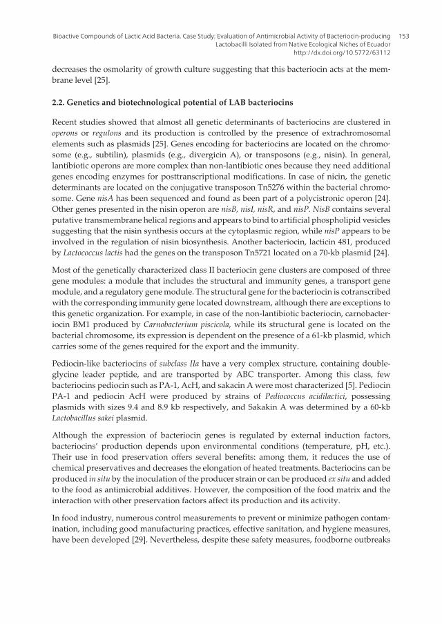
decreases the osmolarity of growth culture suggesting that this bacteriocin acts at the mem‐brane level [25].
2.2. Genetics and biotechnological potential of LAB bacteriocins
Recent studies showed that almost all genetic determinants of bacteriocins are clustered inoperons or regulons and its production is controlled by the presence of extrachromosomalelements such as plasmids [25]. Genes encoding for bacteriocins are located on the chromo‐some (e.g., subtilin), plasmids (e.g., divergicin A), or transposons (e.g., nisin). In general,lantibiotic operons are more complex than non-lantibiotic ones because they need additionalgenes encoding enzymes for posttranscriptional modifications. In case of nicin, the geneticdeterminants are located on the conjugative transposon Tn5276 within the bacterial chromo‐some. Gene nisA has been sequenced and found as been part of a polycistronic operon [24].Other genes presented in the nisin operon are nisB, nisI, nisR, and nisP. NisB contains severalputative transmembrane helical regions and appears to bind to artificial phospholipid vesiclessuggesting that the nisin synthesis occurs at the cytoplasmic region, while nisP appears to beinvolved in the regulation of nisin biosynthesis. Another bacteriocin, lacticin 481, producedby Lactococcus lactis had the genes on the transposon Tn5721 located on a 70-kb plasmid [24].
Most of the genetically characterized class II bacteriocin gene clusters are composed of threegene modules: a module that includes the structural and immunity genes, a transport genemodule, and a regulatory gene module. The structural gene for the bacteriocin is cotranscribedwith the corresponding immunity gene located downstream, although there are exceptions tothis genetic organization. For example, in case of the non-lantibiotic bacteriocin, carnobacter‐iocin BM1 produced by Carnobacterium piscicola, while its structural gene is located on thebacterial chromosome, its expression is dependent on the presence of a 61-kb plasmid, whichcarries some of the genes required for the export and the immunity.
Pediocin-like bacteriocins of subclass IIa have a very complex structure, containing double-glycine leader peptide, and are transported by ABC transporter. Among this class, fewbacteriocins pediocin such as PA-1, AcH, and sakacin A were most characterized [5]. PediocinPA-1 and pediocin AcH were produced by strains of Pediococcus acidilactici, possessingplasmids with sizes 9.4 and 8.9 kb respectively, and Sakakin A was determined by a 60-kbLactobacillus sakei plasmid.
Although the expression of bacteriocin genes is regulated by external induction factors,bacteriocins’ production depends upon environmental conditions (temperature, pH, etc.).Their use in food preservation offers several benefits: among them, it reduces the use ofchemical preservatives and decreases the elongation of heated treatments. Bacteriocins can beproduced in situ by the inoculation of the producer strain or can be produced ex situ and addedto the food as antimicrobial additives. However, the composition of the food matrix and theinteraction with other preservation factors affect its production and its activity.
In food industry, numerous control measurements to prevent or minimize pathogen contam‐ination, including good manufacturing practices, effective sanitation, and hygiene measures,have been developed [29]. Nevertheless, despite these safety measures, foodborne outbreaks
Bioactive Compounds of Lactic Acid Bacteria. Case Study: Evaluation of Antimicrobial Activity of Bacteriocin-producingLactobacilli Isolated from Native Ecological Niches of Ecuador
http://dx.doi.org/10.5772/63112
153

do occur frequently with particular concern on consumers health. Among food pathogens, L.monocytogenes is extremely strong, surviving refrigeration temperatures and high salt concen‐tration. Other pathogens such as Salmonella sp. and E. coli are also frequently detected inprocessed or fresh foods. Nowadays, many investigations are focused on discovering novelbacteriocins for controlling the undesirable bacteria in food products [25, 29]. There is a needto attract consumer attention to natural substances rather than conventional synthesis ofchemical one as protector against pathogens. As probiotics has been accepted in the marketfor their beneficial properties, and in the same way, the bacteriocin-producing probiotic strainsshould become attractive especially to natural food preservation.
Continued research on bacteriocins will undoubtedly lead to our increased understanding,and with the emergence of new bacteriocins, new potential biopreservatives.
3. Antimicrobial activity of LAB strains isolated from native microbiotaof Ecuador
The presence of pathogens in many food products has become a serious problem worldwide.During the last decade, several laboratories have worked towards the identification of novelprobiotic strains with better performance benefits such as novel attractive alternative antimi‐crobial methods to conventional ones [30–36].
Ecuador is known as country with large diversity of native unexploited resources. Someregions were included recently in the governmental policy as important resources to beexploited as reservoirs of unknown microorganisms that could become as potential areas ofhighly interest for biotechnology research, food sovereignty, and security. The lack of qualitycontrol, bad storage condition, and lack of preservation against spoilage bacteria had effect onfood safety and security. Among the most food pathogens worldwide due to the considerablehuman rates of illness reported, Salmonella and E. coli remain the wide species detected in thelocal food market in Ecuador. Most produced traditional foods, such as mote (a fermentedmaize dish), handmade chees, and milk containing drinks, maintained in defective storageconditions appear to pose significant number of pathogens; therefore, the risk of developingdiseases associated with food born pathogens is elevated. In this context, the aforementionedproblems identifying new alternatives for food biopreservation have become an attractiveapproach to be considered. Some native wild plants and fruits derived have been recentlyscreened for the presence of probiotic LAB [37]. Preliminary investigation reveled the presenceof LAB showing probiotic potential (submitted manuscript). Probiotic bacteria, although nota new concept, draw the attention of the scientific community for their highly potential to actas natural food preservative. However, in this study, we present the results on the antimicro‐bial activity of ten LAB strains to select those with promising potential in biopreservation. Apreliminary characterization of the bacteriocin of selected LAB is also described.
Probiotics and Prebiotics in Human Nutrition and Health154

4. Materials and methods
4.1. Bacteria and sampling source of isolation
Sampling material consisting of native fruits and flowers has been collected without no specificethic permits. The reservation was located on subtropical humid mesothermal region of SantoDomingo de Los Tsachilas Provence at 43 km away from Quito, the capital city. At the location,the GPS points have been recorded and the location map was designed using the ArcGISsoftware (a complete platform of GIS to create, analyze, store, and disseminate geographicdata, models, and maps) in order to track each sample in case of cross-contamination.Approximately ten grams of wild orange, immature and mature berries, guayusa, strawberry,achiote and flower inflorescence (Heliconia sp., Fucsia sp., Bromelia sp.) collected asepticallywere transferred in Erlenmeyer flasks (500 ml) containing sterile water (100 ml) and incubatedstatically for up to 5 days at the room temperature. MRS agar [38] plates were used for theinoculation, the samples were incubated under anaerobic conditions at 37°C for 72 h, andisolated individual colonies were randomly selected and purified by replating on samemedium. The purified colonies (>100 colonies/each sample) were Gram stained and tested forthe mobility, indole production, catalase production, spore formation, and production of gasfrom glucose. Cells morphology and colonial characteristics on MRS agar were examined, andbased on these results the colonies were preliminary classified as follows: (i) presumptivelactococci, gram positive, coccal morphology, catalase negative, non-motile, and gasproduction from glucose, and (ii) gram positive, with morphological aspect of rods, catalasenegative, non-motile, with and without production of gas from glucose, and presumptivelactobacilli, stored at −80°C in 20% glycerol. Moreover, the API 50CH strips (Biomerieux, Marcyl’Etoile France, cat # 50300) were used for the metabolic characterization of the each isolate andtentatively identified at genus level. Furthermore, the isolates selected for their probioticperformance (bile tolerance, survival under acidic conditions, antibiotic tolerance, and salttolerance) were analyzed for their antimicrobial activity. As reference strain, Lactobacillusfermentum CNCM 1‐2998 (API50CH, 80% identity) recuperated from an available commercialprobiotic Lacteol Forte (Axcan Pharma, France) has been used.
4.2. Pathogens isolation
Food products consisting of chicken and cheese were purchased from the local market, andstandard bacterial culture media were used to screen and isolate the contaminants. However,Salmonella sp. and Escherichia coli were identified in each food sample. The isolated and purifiedbacterial cultures were further purified and used as indicator strain.
4.3. Antimicrobial activity of selected isolates
Antimicrobial activity was performed against both E. coli and Salmonella sp., using agar welldiffusion method under anaerobic conditions [1]. The LAB isolates were grown in MRS brothat 37°C for 16 h, and the supernatants were collected by centrifugation at 13000×g for 20 minsterilized using 0.22 μm porosity filter. The indicator strains (100 μl) grown in broth medium
Bioactive Compounds of Lactic Acid Bacteria. Case Study: Evaluation of Antimicrobial Activity of Bacteriocin-producingLactobacilli Isolated from Native Ecological Niches of Ecuador
http://dx.doi.org/10.5772/63112
155

(7 log CFU/ml) were mixed with 1.5 ml of soft MRS agar (0.75%), were overlaid on the nutrientagar plates, and incubated at 37°C for 2 h. The cell-free supernatant (100 μl) was spotted ontothe wells (7 mm) made on overlaid agar, incubated at 37°C, and subsequently examined forinhibition zones at different intervals of time (18–24–36–48 h). The experiments were run intriplicate, and the mean values of zone of inhibition were estimated. We considered that theisolates had higher inhibitory activity when the diameter of zone of inhibition was higher than>15 mm, intermediary activity when the zone of inhibition was 10–15 mm, and lower activitywhen the diameter of zone of inhibition was lower than 7 mm.
4.4. The effect of different pH, heat, and detergents on antimicrobial activity
The pH of supernatant was adjusted to 3.0, 4.0, and 7.0 and then kept at room temperature for4 h. To test heat sensitivity, 100 μl of culture supernatant was heated for 30 min at 30, 45, 60,75, and 90°C. Residual activity of each isolate for different pHs and temperature was deter‐mined by the agar well diffusion method as described above for both indicator strains. Theresistant culture supernatants were further heated for 10, 30, and 60 min at 100°C. Anotherbatch of cell-free supernatants treated with 1, 2 and 5% Triton X-100 (BDH Chemicals Ltd,Poole, England) and the same concentration of EDTA (Merck) were incubated for 30 min at30°C. The activity was measured using agar well diffusion method [1].
4.5. Effect of chloroform on antimicrobial activity
To test the effect of chloroform on inhibitory activity, the culture supernatant of each samplewas mixed with an equal volume of chloroform and kept at room temperature for 4 h beforeantimicrobial activity testing.
4.6. Statistical analysis
Statistical analysis was carried out by one-way analysis of variance, the means were separatedby Tukey post-hoc test, and the results were considered statistically significant at the p < 0.05level (SPSS version 10.0.6, USA).
5. Results and discussions
5.1. Screening of LAB isolates
Regardless of numerous probiotic strains presented in the market, there is an ongoing needfor the improvement of LAB strains to be used as starter cultures or to develop new naturalmethod for biopreservation; thus, LAB isolated from their natural environment (e.g., nativefruits, flowers) might possess unusual characteristics including phenotypic differences andintraspecific variability compared to the known ones. In this investigation, we assumed thatacid-tolerant bacteria might be detected as the fermentation of raw material reached at aboutpH 3.5. Figure 1 shows the distribution of biological material used as source of initial screeningof LAB.
Probiotics and Prebiotics in Human Nutrition and Health156
However, preliminary phenotypic analysis suggested the relatedness of the bacterial isolatesfrom wild-type fruits and mature inflorescence of several tropical flowers (>100 colonies/sample) with LAB, which were affiliated to two larger groups: Lactococcus (54%) and Lactoba‐cilli (46%) genera. Furthermore, carbohydrate profiles conducted on ten randomly selectedisolates related to each type of biological material (sample of origin) assigned the selectedisolates as follows: UTNFa38, UTNFa40, and UTNFa41 were identified as Lactococcus lactis ssp.lactis, with identity of 90–99%, the isolate UTNFa37, as Lactobacillus collinoides (99%), UTNFa39,as Lactobacillus brevis 3 with 98% identity, while UTNFa19 and UTNFa23 were identified asLactobacillus paracasei ssp. paracasei 1 with 99.7 and 98.2%, respectively. The isolates UTNFa33and UTNFa17.2 were identified as Lactobacillus paracasei ssp. paracasei 3 with 99.6 and 97.9%identity, and UTNFa8.2 was identified as Lactobacillus pentosus with 98.3%. Table 1 presentsthe classification of isolates on the basis of morphological, physiological and metabolicproperties. Similar to our study, numerous lactobacilli species (i.e., L. paracasei, L. pentosus)were identified in different fruits and vegetables [39].
Strain code Cell form/cellulararrangement
Specie assignation % of identity basedon API 50 CHL
UTNFa19 Coccus/single Lactobacillus paracasei ssp. paracasei 1 99.70
UTNFa38 Bacilli/rods/single Lactococcus lactis ssp. lactis 98.00
UTNFa17.2 Bacilli/rods/single Lactobacillus paracasei ssp. paracasei 3 97.90
UTNFa23 Bacilli/rods/single Lactobacillus paracasei ssp. paracasei 1 98.20
UTNFa8.2 Bacilli/rods/single Lactobacillus pentosus 98.30
UTNFa33 Bacilli/rods/single Lactobacillus paracasei ssp. paracasei 1 99.60
UTNFa39 Bacilli/rods/single Lactobacillus brevis 3 98.00
UTNFa40 Coccus/single Lactococcus lactis ssp. lactis 90.00
UTNFa41 Coccus/single Lactococcus lactis ssp. lactis 99.00
UTNFa37 Bacilli/rods/single Lactobacillus collinoides 99.00
Table 1. Classification of LAB isolates.
The antimicrobial activity of the selected strains was evaluated against two Selected foodbornpathogens using agar-well assay. The zone of inhibition was easily visualized, and the meanvalue of the inhibition zone was determined. The cell-free supernatants were considered ascrude bacteriocin. Among tested isolates, most of them showed elevated inhibitory activity forboth pathogen tested. Nonetheless, results from enzyme inactivation analysis demonstratedthat antimicrobial activity was lost or unstable after treatment with proteolytic enzymes suchproteinase K and trypsin, whereas catalase treatment did not affect the activity of antimicrobialsubstance produced by the tested isolates, confirming its protein status. The sensitivity of thefound substance to proteolytic enzymes is a proof of its proteinaceous nature, which allowsconsidering as bacteriocin.
Bioactive Compounds of Lactic Acid Bacteria. Case Study: Evaluation of Antimicrobial Activity of Bacteriocin-producingLactobacilli Isolated from Native Ecological Niches of Ecuador
http://dx.doi.org/10.5772/63112
157

5.2. Effect of pH on inhibitory activity
The antimicrobial effect exerted by LAB strains is related to the production of lactic acid,reduction of pH, and inhibitory compounds [39], has attracted much attention, and attributedas important selection criteria of a probiotic microorganism [2, 33]. An elevated antimicrobialactivity against both food pathogens was observed at the pH 3.0 with the mean range valueof inhibition zone 15.25 mm (±0.5) of the supernatant of tested isolates. In Figure 2A, weshowed the mean value of inhibition zone displayed by each isolate at different pH towardsSalmonella sp. Although at the pH 4.0 no significant difference between the mean values ofinhibition zone was recorded, the mean range of inhibition zone was 13.58 mm (±1.24) for E.coli and 12.09 mm (±2.04) for Salmonella sp., after 48 h of incubation. With the increase of thepH, we observed a gradually reduction of the antimicrobial activity as no activity was recordedat the pH 7.0 for all selected isolates as well as the reference probiotic. Figure 2B showed theclear inhibition zone of two isolates UTNFa40 and UTNFa41 at the pH 3.0 and 4.0, and no zoneformation at pH 7.0.
Overall all selected isolates, in particular two isolates, UTNFa40 and UTNFa41, displayedelevated inhibitory activity in comparison with the reference strain. Of course, the efficiencyand the nature of this antimicrobial activity have to be investigated. Recent studies showedthe importance of bacteriocin produced by the Lactobacillus pentosus ST712BZ strain isolatedfrom boza in the preservation of beverage products [3]. In other investigation, L. pentosus, abile-resistant strain, displayed bacteriocin activity against a wide range of spoilage andpathogen bacteria [32]. In agreement with the studies, we showed that the isolate UTNFa8.2
Figure 1. Origin of sampling (geographical distribution according with ArcGIS software).
Probiotics and Prebiotics in Human Nutrition and Health158

assigned as L. pentosus, a bile, and acid-resistant strain displayed elevated antimicrobialactivity, which will further allow us to explore its biotechnological properties.
5.3. Effect of the heat, detergents, and chloroform on inhibitory activity
The inhibitory activity was not significantly reduced in case of the heat treatment. The meanvalue of zone on inhibition varied at the incubation temperature of 30°C from 19 mm (±2.34)towards Salmonella sp. and, respectively, from 20.18 mm (±3.72) towards E. coli. At the 60°C, itvaries from 16.33 mm (±2.92) towards Salmonella sp. and from 17.5 mm (±3.17) towards E.coli, and at the 75°C it varies from 14.83 mm (±3.05) towards Salmonella and from 15.5 mm(±3.27) in case of E. coli. The increase of temperature of 90°C showed a reduction of the
Figure 2. Antimicrobial activity towards Salmonella sp. (A) Mean value of zone of inhibition in mm recorded at testedpHs (bars represent the means ± SD). (B) The visualized clear zone of inhibition at pH 3.0, 4.0, and 7.0 of UTNFa41 andUTNFa40.
Bioactive Compounds of Lactic Acid Bacteria. Case Study: Evaluation of Antimicrobial Activity of Bacteriocin-producingLactobacilli Isolated from Native Ecological Niches of Ecuador
http://dx.doi.org/10.5772/63112
159

inhibition zone was observed for both pathogens. Figure 3 shows the mean values of theinhibition zone recorded after 30-min incubation at different temperature. At 100°C, after30 min of incubation, two isolates were resistant and maintain its inhibitory activity.
Figure 3. Mean values of zone of inhibition at different temperature of cell-free supernatant towards E. coli and Salmo‐nella sp. (bars represent the means ± SD).
The heat stability could be an advantage when the strains are intended to be used as biopre‐servative of processed foods. Similarly, Todorov and col., showed that some bacteriocinsremain stable after incubation at 100°C for 120 min [34]. In other study, bacteriocin-likesubstance of Lactobacillus fermentum KN02 was strongly influenced by the pH and temperature.The strain has the maximum productivity at the pH 2.0 and was resistant to heat at 100°C [40].
Due to their resistance to temperature and low pH, the bacteriocins would be digested byhuman and animal peptidases, thus avoiding resistance and problems associated with thepresence of residues in feed and food [35]. However, at the treatment of the selected cell-freesupernatants with Triton-X 100 and EDTA, an increase in the inhibitory activity was recorded.An increase with 5% of both Triton-X 100 and EDTA results in an increase of inhibitory activityfor some of the isolates. For example, Figure 4 shows the mean values of the zone of inhibitionrecorded towards E. coli and Salmonella sp. after the treatment with 1, 2 and 5% Triton-X 100for each strain tested. Similar studies showed that the heat does not have any effect on cell-free supernatants activity as well as no effect on the inhibitory activity of the bacteriocins ofLactobacillus sakei isolated from the fermented meat was observed after the treatment withseveral detergents including EDTA and Triton-X 100 [35].
On the contrary, in our study, we observed an increase in the concentration of either EDTA orTriton-X 100, and the inhibitory activity was elevated for most of the isolates. Figure 5A showsthe inhibitory activity towards Salmonella sp. by the appearance of the clear zone after treatmentof cell-free supernatant with different concentration of EDTA. In Figure 5B, the effect of EDTAon antimicrobial activity towards Salmonella is shown. We observed an increase in theinhibitory activity with the increase of the concentration of EDTA. However, a positive effectof detergents in the antimicrobial activity of each isolate has been detected.
Probiotics and Prebiotics in Human Nutrition and Health160

Figure 5. (A) The appearance of the clear inhibition zone at different concentration of EDTA of isolates UTNFa23 andUTNFa41 towards Salmonella sp. (B). The antimicrobial activity recorded as mean value of inhibition zone of LAB afterthe treatment with 1, 2, 5% EDTA towards Salmonella sp.
Figure 4. The inhibition activity of the isolated strains towards Salmonella sp. (A) and E. coli (B) after the treatment withTriton-X 100.
Bioactive Compounds of Lactic Acid Bacteria. Case Study: Evaluation of Antimicrobial Activity of Bacteriocin-producingLactobacilli Isolated from Native Ecological Niches of Ecuador
http://dx.doi.org/10.5772/63112
161

The antimicrobial activity of most of the isolates was lost in case of chloroform treatment ofthe cell-free supernatants. Among analyzed strains, the isolate UTNFa38 and isolate UTNFa41remained active towards E. coli as well as Salmonella sp., after the treatment with chloroform.The mean value of inhibition zone was 10 mm for UTNFa38 and respectively, 11 mm forUTNFa41 towards E. coli, while the mean value of inhibition zone was 9 mm for UTNFa38 and12 mm for UTNFa41, towards Salmonella sp. The resistance to chloroform treatment and boilingdemonstrates the nature of low-molecular, non-lipid-containing bacteriocins. Eight isolates wereidentified as lipid-containing bacteriocins because of their sensitivity to chloroform. Similarstudies showed the broad spectrum of inhibitory activity of Lactobacillus paracasei subsp.paracasei isolated from natural homemade cheese [41]. Besides several Lactobacillus strains fromdifferent species, the bacteriocin from L. paracasei ssp. paracasei also inhibits the growth ofvarious pathogenic bacteria such as Streptococcus, Staphylococcus, Shigella, Listeria, andPseudomonas.
The stability of crude cell supernatant of each selected LAB to different conditions reflects thatthese compounds would remain effective in the processing of foods [42]. Recent investigationshowed the broad spectrum of inhibitory activity towards Pseudomonas of some bacilli isolatedfrom onion and fresh-cut salads [43]. In other work, it has been demonstrated the antimicrobialactivity against spoilage pathogens of some LAB isolated from mango pulp [44]. The sixisolated strains had inhibitory effects on sensitive bacteria including E. coli, demonstrating thepotential of usage of this compound as a preservative in mango or fruit pulp industry. Insimilar work, several LAB isolated from foods and spoilage halotolerant bacteria isolated fromcharqui, a Brazilian fermented, salted meat product. The bacteriocin of Lactococcus lactis subsp.lactis (L. lactis 69) inhibited, in vitro, Listeria monocytogenes, S. aureus [45]. In our study, theresulted data revealed a wide spectrum of inhibitory activity against two food pathogens ofsome LAB isolated from natural microbiota of Ecuador, and shall further characterize anddetermine its molecular size and mode of action, as well as its effectiveness as a biopreservativein different food products as such or in combination with other methods.
6. Conclusions
Bacteriocins produced by genera Lactobacillus or other genera have been reported. Neverthe‐less, the studies in the field of natural food biopreservation are conducted to an increasingextent. As consumers are more concern about the food quality along with their refusal ofchemical additives, there is a growing demand for alternative antimicrobial treatments andbioactive compounds such as bacteriocins from lactic acid bacteria are well-accepted naturalmeans of selective microbial inhibition.
However, characterization of specific microbiota would further contribute substantially togain better knowledge for the improvement of current commercial probiotic strains. Thestudies conducted up to date indicate that interest on bacteriocins will be high. Thus, all thestudies carried out on novel bacteriocins are important to propose new alternatives in foodpreservation.
Probiotics and Prebiotics in Human Nutrition and Health162

Acknowledgements
The Technical University of the North, Ibarra Republic of Ecuador research Grant No. 01388,financed the work. GNT was sponsored by the Prometeo Project of the Secretary for HigherEducation, Science, Technology and Innovation (SENESCYT). The authors would like to thankDr. Miguel Naranjo Toro for his technical support.
Author details
Gabriela N. Tenea* and Lucia Yépez
*Address all correspondence to: [email protected]; [email protected]
Faculty of Engineering in Agricultural and Environmental Sciences, Technical University ofthe North, Av. 17 de Julio s-21 y José María Córdova. Ibarra, Barrio El Olivo, Ecuador
References
[1] Ehrmann MA, Kurzak P, Bauer J, Vogel RF. Characterization of lactobacilli towardstheir use as probiotic adjuncts in poultry. J Appl Microbiol. 2002;92:966–975. doi:10.1046/j.1365-2672.2002.01608.x.
[2] Tesfaye A. Antagonism and primary in vitro probiotic evaluation of lactic acid bacteria(LAB) recovered from ergo. ARPN J Agric Biol Sci. 2014;9(7):240–245. ISSN 1990-6145.
[3] Todorov SD, Dicks LMT. Bacteriocin production by Lactobacillus pentosus St712bzisolated from boza. Braz J Microbiol. 2007;38:166–172. ISSN 1517-8283.
[4] Hernandez D, Cardell E, Zarate V. Antimicrobial activity of lactic acid bacteria isolatedfrom Tenerife cheese: initial characterization of plantaricin TF711, a bacteriocin-likesubstance produced by Lactobacillus plantarum TF711. J Appl Microbiol 2005;99:77–84.doi:10.1111/j.1365-2672.2005.02576.x.
[5] Todorov SD, Vaz-Velho M, Gambossy de Melo Franco BD, Holzapfel WH. Partialcharacterization of a bacteriocins produced by three strains of Lactobacillus sakei isolatedfrom salpicao, a fermented meat product from North-West of Portugal. Food Control.2013;30:111–121. doi:10.1016/j.foodcont.2012.07.022.
[6] Enan G, Al-Mohammadi AR, El-Didamony G, Mahmoud EF, Haliem A, Zakaria A.Antimicrobial activity of Enterococcus faecium NM2 Isolated from Urine: purification,characterization and bactericidal action of enterocin NM2. Asian J Appl Sci. 2014;7:621–634. doi:10.3923/ajaps.2014.621.634.
Bioactive Compounds of Lactic Acid Bacteria. Case Study: Evaluation of Antimicrobial Activity of Bacteriocin-producingLactobacilli Isolated from Native Ecological Niches of Ecuador
http://dx.doi.org/10.5772/63112
163

[7] Longdet IY, Kutdhik RJ, Nwoyeocha IG. The probiotic efficacy of Lactobacillus casei fromhuman breast milk against Shigellosis in Albino rats. Adv Biotechnol Chem Proc.2011;1:12–16. doi:10.5772/2825.
[8] Mirnejad R, Vahdati AR, Erfani M, Piranfar V. The antimicrobial effect of Lactobacilluscasei culture supernatant against multiple drug resistant clinical isolates of Shigellasonnei and Shigella flexneri in vitro. Iran Red Crescent Med J. 2013;15(2):122–126. doi:10.5812/ircmj.7454.
[9] Soumya TV, Reshma J, Surya J. Characterization of bacteriocin produced by Lactoba‐cillus sp and optimization of cultural conditions. IJSRP. 2012;2(12). ISSN 2250-3153.
[10] Deegana LH, Cottera PD, Hilla C, Ross P. Bacteriocins: biological tools for biopreser‐vation and shelf-life extension. Int Dairy J. 2006;16:1058–1071. doi:10.1016/j.idairyj.2005.10.026.
[11] Murua A, Todorov SD, Vieira ADS, Martinez RCR, Cenci, A, Franco BDGM. Isolationand identification of bacteriocinogenic strain of Lactobacillus plantarum with potentialbeneficial properties from donkey milk. J Appl Microbiol. 2013;14:17931809. doi:10.1111/jam.12190.
[12] Gürakan GC. Biopreservation by lactic acid bacteria. In Metabolism and Applicationsof Lactic acid bacteria. Editor: Barbaros Ozer. Published by Research Signpost, India.2007:15–31. ISBN: 978-81-308-0203-9.
[13] Klaenhammer TR. Bacteriocins of lactic acid bacteria. Biochimie. 1988;70:337–349. doi:10.1016/0300-9084(88)90206-4.
[14] Matsusaki H, Endo N, Sonomoto K, Ishizaki A. Lantibiotic nisin Z fermentativeproduction by Lactococcus lactis IO-1: relationship between production of the lantibioticand lactate and cell growth. Appl Microbiol Biotechnol. 1996;45:36–40. doi:10.1007/s002530050645.
[15] Sawa N, Wilaipun P, Kinoshita S, Zendo T, Leelawatcharamas V, Nakayama J,Sonomoto K. Isolation and characterization of enterocin W, a novel two-peptidelantibiotic produced by Enterococcus faecalis NKR-4-1. Appl Environ Microbiol.2012;78:900–903. doi:10.1128/AEM.06497-11.
[16] Sashihara T, Kimura H, Higuchi T, Adachi A, Matsusaki H, Sonomoto K, Ishizaki A. Anovel lantibiotic, nukacin ISK-1, of Staphylococcus warneri ISK-1: cloning of the struc‐tural gene and identification of the structure. Biosci Biotechnol Biochem. 2000;64:2420–2428. doi:10.1271/bbb.64.2420.
[17] Himeno K, Fujita K, Zendo T, Wilaipun P, Ishibashi N, Masuda Y, Yoneyama F,Leelawatcharamas V, Nakayama J, Sonomoto K. Identification of enterocin NKR-5-3C,a novel class IIa bacteriocin produced by a multiple bacteriocin producer, Enterococcusfaecium NKR-5-3. Biosci Biotechnol Biochem. 2012;76:1245–1247. doi:10.1271/bbb.120089.
Probiotics and Prebiotics in Human Nutrition and Health164

[18] Ishibashi N, Himeno K, Fujita K, Masuda Y, Perez RH, Zendo T, Wilaipun P, Leela‐watcharamas V, Nakayama J, Sonomoto K. Purification and characterization ofmultiple bacteriocins and an inducing peptide produced by Enterococcus faeciumNKR-5-3 from Thai fermented fish. Biosci Biotechnol Biochem. 2012;76:947–953. doi:10.1271/bbb.110972.
[19] Zendo T, Eungruttanagorn N, Fujioka S, Tashiro Y, Nomura K, Sera Y, Kobayashi G,Nakayama J, Ishizaki A, Sonomoto K. Identification and production of a bacteriocinfrom Enterococcus mundtii QU2 isolated from soybean. J Appl Microbiol. 2005;99:1181–1190. doi:10.1111/j.1365-2672.2005.02704.x.
[20] Fujita K, Ichimasa S, Zendo T, Koga S, Yoneyama F, Nakayama J, Sonomoto K.Structural analysis and characterization of lacticin Q, a novel bacteriocin belonging toa new family of unmodified bacteriocins of Gram-positive bacteria. Appl EnvironMicrobiol. 2007;73:2871–2877. doi:10.1128/AEM.02286-06.
[21] Iwatani S, Zendo T, Yoneyama F, Nakayama J, Sonomoto K. Characterization andstructure analysis of a novel bacteriocin, lacticin Z, produced by Lactococcus lactis QU14. Biosci Biotechnol Biochem. 2007;71:1984–1992. doi:10.1271/bbb.70169.
[22] Masuda Y, Zendo T, Sawa N, Perez RH, Nakayama J, Sonomoto K. Characterizationand identification of weissellicin Y and weissellicin M, novel bacteriocins produced byWeissella hellenica QU 13. J Appl Microbiol. 2012;112:99–108. doi:10.1111/j.1365-2672.2011.05180.x.
[23] Perez RH, Zendo T, Sonomoto K. Novel bacteriocins from lactic acid bacteria (LAB):various structures and applications, Perez et al. Microb Cell Factories. 2014;13(Suppl1):S3.
[24] Parada JL, Caron Ricoy C, Bianchi P, Medeiros A, Soccol CR. Bacteriocins from lacticacid bacteria: purification, properties and use as biopreservatives. Braz Arch BiolTechnol. 2007;50(3):521–542. ISSN 1516-8913.
[25] Oscariz JC, Pisabarro AG. Classification and mode of action of membrane-activebacteriocins produced by gram-positive bacteria. Int Microbiol. 2001;4:13–19. doi:10.1007/s101230100003.
[26] Abee T. Pore-forming bacteriocins of gram-positive bacteria and self-protectionmechanisms of producer organisms. FEMS Microbiol Lett. 1995;129:1–10. doi:10.1111/j.1574-6968.1995.tb07548.x.
[27] Abee T, Krockel L, Hill C. Bacteriocins: mode of actions and potentials in food preser‐vation and control of food poisoning. Int J Food Microbiol. 1995;28:169–185. doi:10.1016/0168-1605(95)00055-0.
[28] Ennahar S, Sashihara T, Sonomoto K, Ishizaki A. Class IIa bacteriocins: biosynthesis,structure and activity. FEMS Microbiol Rev. 2001;24:85–106. doi:10.1111/j.1574-6976.2000.tb00534.x.
Bioactive Compounds of Lactic Acid Bacteria. Case Study: Evaluation of Antimicrobial Activity of Bacteriocin-producingLactobacilli Isolated from Native Ecological Niches of Ecuador
http://dx.doi.org/10.5772/63112
165

[29] Adak GK, Long SM, O’Brian SJ. Trends in indigenous foodborne disease and deaths,England and Wales. Gut. 2002;51:832–841. doi:10.1136/gut.51.6.832.
[30] Choi HJ, Cheigh CI, Kim SB, Pyun YR. Production of a nisin-like bacteriocin byLactococcus lactis subsp. lactis A164 isolated from Kimchi. J Appl Microbiol. 2000;88:563–571. doi:10.1046/j.1365-2672.2000.00976.x.
[31] Danielsen M, Wind A. Susceptibility of Lactobacillus spp. to antimicrobial agents. Int JFood Microbiol. 2003;82:1–1. doi:10.1016/S0168-1605(02)00254-4.
[32] Enan G, Al-Mohammadi AR, El-Didamony G, Mahmoud EF, Haliem A, Zakaria A.Antimicrobial activity of Enterococcus faecium NM2 Isolated from Urine: purification,characterization and bactericidal action of enterocin NM2. Asian J Appl Sci. 2014;7:621–634. doi:10.3923/ajaps.2014.621.634.
[33] Longdet IY, Kutdhik RJ, Nwoyeocha IG. The probiotic efficacy of Lactobacillus casei fromhuman breast milk against Shigellosis in Albino rats. Adv Biotechnol Chem Proc.2011;1:12–16.
[34] Nes IF, Johnsborg O. Exploration of antimicrobial potential of LAB by genomics. CurrOpin Biotechnol. 2004;15(2):100–4. doi:10.1016/j.copbio.2004.02.001.
[35] Todorov SD, Rachman C, Fourrier A, Dicks LMT, van Reenen CA, Prevost, H, et al.Characterization of a bacteriocins produced by Lactobacillus sakei R1333 isolated fromsmoked salmon. Anaerobe. 2011;17(1):23–31. doi:10.1016/j.anaerobe.2010.01.004.
[36] Todorov SD, Vaz-Velho M, Gambossy de Melo, Franco BD, Holzapfel WH. Partialcharacterization of a bacteriocin produced by three strains of Lactobacillus sakei isolatedfrom salpicao, a fermented meat product from North-West of Portugal. Food Control.2013;30:111–121. doi:10.1016/j.foodcont.2012.07.022.
[37] Yepez L, Tenea GN. Genetic diversity of lactic acid bacteria strains towards theirpotential probiotic application. Rom Biotechnol Lett. 2015;20(2):10191–10199. ISSN2248-3942.
[38] De Man JC, Rogosa M, Sharpe E. A medium for the cultivation of lactobacilli. J ApplBacteriol. 1960;23:130–155. doi:10.1111/j.1365-2672.1960.tb00188.x.
[39] Maldonado-Barragan A, Caballero-Guerrero B, Lucena-Padros H, Ruiz-Barba JL.Genome sequence of Lactobacillus pentosus IG1, a strain isolated from Spanish-stylegreen olive fermentations. J Bacteriol. 2011;193(19):5605. doi:10.1128/JB.05736-11.
[40] Nithya K, Senbagam D, Senthilkumar B, Udhayashree N, Gurusamy R. Characteriza‐tion of bacteriocin-producing lactic acid bacteria and its application as a food preser‐vative. Afr J Microbiol Res. 2012;6 (6):1138–1146. doi:10.5897/AJMR11.1214.
[41] Tolinački M, Kojić M, Lozo J, Terzić-Vidojević A, Topisirović L, Fira D. Characterizationof the bacteriocin-producing strain Lactobacillus paracasei subsp. paracasei Bgub9. ArchBiol Sci Belgrade. 2010;62(4):889–899. doi:10.2298/ABS1004889T.
Probiotics and Prebiotics in Human Nutrition and Health166

[42] Osmanagaoglu O. Detection and characterization of Leucocin OZ, a new anti-listerialbacteriocin produced by Leuconostoc carnosum with a broad spectrum of activity. FoodControl. 2007;18:118–123. doi:10.1016/j.foodcont.2005.08.015.
[43] Sharpe VD. Biopreservation of fresh-cut salads using bacteriocinogenic lactic acidbacteria isolated from commercial produce. Dalhousie University, Halifax, NS, Canada,Master thesis. 2009.
[44] Ravi V, Prabhu M, Subramanyam D. Isolation of bacteriocin producing bacteria frommango pulp and its antimicrobial activity. J Microbiol Biotechnol Res. 2011;1(2):54–63.http://scholarsresearchlibrary.com/archive.html.
[45] Biscola V, Todorov SD, Capuano VSC, Abriouel H, Gálvez A, Franco BDGM. Isolationand characterization of a nisin-like bacteriocin produced by a Lactococcus lactis strainisolated from charqui, a Brazilian fermented, salted and dried meat product. Meat Sci.2013;93:607–613.
Bioactive Compounds of Lactic Acid Bacteria. Case Study: Evaluation of Antimicrobial Activity of Bacteriocin-producingLactobacilli Isolated from Native Ecological Niches of Ecuador
http://dx.doi.org/10.5772/63112
167


Chapter 9
Fructosyltransferase Sources, Production, andApplications for Prebiotics Production
Mariela R. Michel, Rosa M. Rodríguez-Jasso,Cristóbal N. Aguilar, Silvia M. Gonzalez-Herrera,Adriana C. Flores- Gallegos andRaúl Rodríguez-Herrera
Additional information is available at the end of the chapter
http://dx.doi.org/10.5772/62827
Abstract
Fructooligosaccharides (FOS) are considered prebiotic compounds and are found indifferent vegetables and fruits but at low concentrations. FOS are produced by enzymat‐ic transformation of sucrose using fructosyltransferase (FTase). Development of newproduction methods and search for FTase with high activity and stability for FOSproduction Is an actual research topic. In this article is discussed the most recent advanceson FTase and its applications. Different microorganisms have been tested under variousfermentation systems in order to identify and characterize new genes codifying for FTase.Some of these genes have been isolated from bacteria, fungi, and plants, with a wide rangeof percentages of identity but retaining the eight highly conserved motifs of the hydrolasefamily 32 glycoside. Therefore, this article presents an overview of the most recent advanceson FTase and its applications.
Keywords: Enzyme production, Fructooliogosacarides, Fructosyltransferase, 1-kes‐tose, 1-nystose, 1-β-fructofuranosyl nystose
1. Introduction
At the present time, there is a growing consumer demand for healthier and calorie-controlledfoods. For this reason, food industry has developed different alternatives for sweeteners, and
among them is the fructooligosaccharides (FOS). Use of these compounds offers multiple healthadvantages. FOS are fructose oligomers with a terminal glucosyl unit and a general formula ofGFn; where typical values of n are 2–4 and are composed of sugars units such as 1-kestose, 1-nystose, and 1- β -fructofuranosyl nystose, which can be found in different fruits and vegeta‐bles, but in very low concentrations to exert a beneficial effect on health. In addition, its productionis limited by seasonal conditions [1]. FOS cannot be hydrolyzed by the gastrointestinal enzymesand are recognized as prebiotic which selectively stimulate growth and/or activity of bifido‐bacteria and lactobacilli, microorganisms that promote benefits to human health [2, 3].
FOS can be produced by the action of different types of enzymes with transfructosylationactivity (i.e., fructosyltransferase—FTase [EC 2.4.1.9] and/or β-fructofuranosidase—FFases[EC 3.2.1.26]). These enzymes are obtained from different plants and microorganisms [4]. Thereaction mechanism of FTase depends on enzyme source [5]. Most of these enzymes have beenisolated from different fungal strains such as: Aureobasidium spp., Aspergillus spp., Penicilli‐um spp., and Fusarium spp. [6]. However, different FTases from bacterial (Bacillus macerans,Lactobacillus reuteri, and Zymomonas mobilis) species have been also reported [7].
2. FTase: an overview
FOS can be synthesized in nature by the catalytic action of enzymes with transfructosylatingactivity. They are classified as 1F-FTases (E.C. 2.4.1.9, E.C. 2.4.1. 99, and E.C. 2.4.1.100), or β -FFases (, E.C. 3.2.1.26). FTase catalyzes the transfer of a fructosyl group to a molecule of sucroseor a FOS when a FOS with a chain longer by one fructosyl unit is formed [8]. This enzyme alsomediates polymerization reactions, where degree of polymerization (DP) decreases to themaximum by transferring fructosyl units from higher molecular mass fructans [9]. The reactionmechanism of the FTases depends on the enzyme source. In plants and some microorganisms,a series of enzymes act together, whereas a single enzyme works in most of the microorganisms[10].
The FTase that converts sucrose to the shortest β (2–1) linked fructan 1-kestose is called sucrose:sucrose 1-FTase (1-SST) [11]. It is reported that, FTase differs in molecular weight and prop‐erties depending on its origin [12]. Properties of FTases can change according to the microor‐ganism and culture medium composition, especially the carbon source, which can play a roleas an inductor [1]. FTase can be produced intra- and/or extracellular by different microorgan‐isms, including bacteria and fungi. Despite the large number of microbial FTase producers,only some of them have the potential for industrial application and have been used in severalstudies about FOS production [1]. The transfructosylating activity is responsible for FOSproduction from sucrose, although quantitative differences exist because of the microbialstrains [13].
FTase has been produced using both solid and liquid fermentation, and FOS obtained by thesefermentations have been reported. Factors affecting FOS by fermentation were mentioned,such as temperature, pH, and substrate concentration [14, 15]. FTase has a temperatureoptimum between 50 and 60°C, and the optimal pH is between 4.5 and 6.5 [1, 8].
Probiotics and Prebiotics in Human Nutrition and Health170

2.1. Mechanism of action
The reaction mechanism depends on the enzyme source and purity. The accepted mechanismis a type disproportionate reaction where FTase catalyzes the transfer of a fructosyl moiety ofa sucrose or fructooligosacharide donor to another sucrose or FOS acceptor to provide asuperior FOS [1]. The reaction mechanism has been expressed as follows:
1 1GF GF GF GF 1 3+ -+ = -n n nn n
where GF is sucrose or FOS n is the number of fructosyl units.
3. Microbial and plant FTases
Several fungal strains, especially those from Aspergillus genus, are known to produce extrac‐ellular or intracellular FTase. The microbial FTase (Ftase; E.C. 2.4.1.9) catalyses formation ofFOS from sucrose; FTases obtained from microorganisms are single enzymes with bothtransferase and hydrolase activities [12]. Some of the fungi reported as FTase producers arethe following: Aspergillus niger ATCC 20611 [13], A. niger strain AN 166 [16], Aspergillusfoetidus [17], Aspergillus oryzae CFR 202 [15, 18–20], Aureobasidium pullulans CFR 77 [18], A.oryzae KB [21], and Aspergillus awamori GHRTS [22]. Some of these fungal strains have thecapacity to produce two types of FTases (Table 1). The enzymatic activity is different becauseit depends on carbon source and type of microorganism. The FFase with a high transfructo‐sylating activity has been studied extensively in A. niger because this fungal specie is used forindustrial production of FOS. A. niger AS0023 produces two types of FFases, and one of theenzymes has high transfructosylating activity [24].
Source FTase Activity Temperature Reference
Aspergillus niger YZ59 Recombinant Ftase 1020 U/ml 55°C [23]
Aspergillus awamori GHRTS 6120 U/gds 30°C [22]
Aspergillus oryzae CFR 202 16.5 U/ml/min 30°C [19]
Aspergillus niger 107.87 U/mL 30°C [16]
Table 1. Microbial FTases produced by filamentous fungi, its activity, and fermentation temperature of the maximumenzyme activity reached.
Bacterial strains have been reported to produce different inulinases, but reports on enzymesable to produce FOS are scarce from bacterial strains. Someone bacteria mentioned to producethese enzymes are the following: B. macerans, L. reuteri, and Z. mobilis [7].
The FTases obtained from plants have other amino acid composition that is different frommicrobial FTases such as sucrose: sucrose 1-FTase (1-SST) and fructan: fructan 1-FTase (1-FFT).
Fructosyltransferase Sources, Production, and Applications for Prebiotics Productionhttp://dx.doi.org/10.5772/62827
171

Plants such as Cichorium intybus and Helianthus tuberosus produce high levels of FTases suchas 1-SST, (EC. 2.4.1.99) [10]. In 1995, Ftase were isolated and purified from barley (Hordeumvulgare) [25].
4. Structure of FTase
According to the Protein Data Bank (PDB) of Research Collaborators for Structural Bioinfor‐matics (RCSB) data base, the crystal dimensional structure of FTase from Aspergillus japoni‐cus comprises 632 residues that fold into two domains, with a N-terminal five-blade-propeller(residues 21–468), and a C-terminal sandwich domain (480–653), which are linked by a 9-residue short-helix (469–479) [26].
5. FTases properties
5.1. Factors affecting FTase activity
Fructosyltransferase (FTase) participates on FOS/fructan production by catalyzing the transferof a fructose unit from one sucrose/fructan to another [26]. This enzyme has been included inthe glycoside hydrolase family 32 (GH32) and has been isolated from different sources, andthe optimal conditions for the enzyme activity have been reported (Table 2). The optimaltemperature reported for FTase enzyme activity ranges from 52 to 65°C, while the optimal pHvaries widely from 4.5 to 8.0 [27, 28]. There are different reports mentioned about the chemicalreagents and amino acids that positively affect FTase activity [29, 30]. On the other hand, thereis a controversy in the use of detergents—some authors mention that these compoundsenhance FTase activity [29], and in contrast there are others who mention that these com‐pounds negatively affect FTase activity [27].
Source Temperature pH Positive effect Observation References
Aspergillus sp. 52 °C 4.5 FeSO4, Fe2+, Fe2+ Ca2+ Intra- and extracellular [27]
MarineAspergillus niger
65°C 8.0 Intracellular [28]
Aspergillusaculeatus
60°C 5.0–7.0 Dithiothreitol,2-mercaptoethanol,sodium dodecylsulphate,Tween 80
[29]
Penicilliumpurpurogenum
55 °C 5.5 Leucine induced slightlyextracellular production
Intra- and extracellular [30, 31]
Table 2. Optimal conditions for FTase activity from different microbial sources.
Probiotics and Prebiotics in Human Nutrition and Health172

5.2. Carbon and nitrogen sources
Different reports mentioned that the preferred carbon source to produce FTase is sucrose. Patiland Butle [31] indicated that Syncephalastrum recemosum Cohn preferred sucrose to produceFTase. Similarly, Dhake and Patil [30] employing Penicillium purpurogenum found that the bestcarbon source for FTase production was sucrose. The complete hydrolysis of this carbohydratewas reported by Kumar et al. [28] who used a marine A. niger strain to degrade sucrose, andthe product consisted entirely of D-fructose, although different products from sucrosehydrolysis have been reported depending on the FTase enzyme origin. A recombinant FTasefrom timothy (Phleum pratense L.) and expressed in Pichia pastoris produced linear β (2, 6)-linkedlevans from sucrose [32]. While recombinant FTase from L. reuteri for sucrose hydrolysis wasused, large amounts of FOS with _(231)-linked fructosyl units, plus a high-molecular-weightfructan polymer (>107) with _-(231) linkages (an inulin) were found [33]. Amount of sucrosein the fermentation reactor may alter the final product. Ghazi et al. [29] found that FTase fromA. aculeatus at elevated sucrose concentrations showed a high transferase/hydrolase ratio. Thekcat and Km values for transfructosylating and hydrolytic activities vary. Ghazi et al. [29]modified sucrose using a microbial FTase and found transfructosylating activity of1.62 ± 0.09 × 10⁴ s − 1 for kcat and 0.53 ± 0.05 M for Km, whereas for hydrolytic activity, thekcat and Km values were 775 ± 25 s − 1 and 27 ± 3 mM, respectively. On the other hand, thebest nitrogen source to produce FTase by S. recemosum Cohn is ammonium nitrate [31].
5.3. FTase biochemical properties
Biochemical properties of FTase may change depending on its origin. Ghazi et al. [29] reporteda dimeric glycoprotein with 20% of carbohydrate content and a molecular mass of around135 kDa from A. aculeatus. In contrast, a FTase from A. foetidus was found as extensivelyglycosylated, with a probable active form of a dimer of identical subunits and an apparentmass of 180 kDa [34]. On the other hand, a FTase which catalyze formation and extension ofP-2, 6-linked fructans in barley (H. vulgare L.) was mentioned to occur in two closely similarisoforms having both of them two subunits with masses of 49 and 23 kDa [25].
Chuankhayan et al. [26] sequenced a recombinant FTase from Aspergillus japonica. In this case,they found that this enzyme comprises two domains with an N-terminal catalytic domaincontaining a five-blade-propeller-fold linked to a C-terminal-sandwich domain. In addition,the same authors reported four substrate-binding subsites (1–3) in the catalytic pocket withshapes and characters distinct from those of clan GH-J enzymes; in this step, they used differentFTase mutants. The residue Asp-60 was proposed for nucleophile, Asp-191 for transition-statestabilizer, and Glu-292 for general acid/base catalyst, which govern the binding of the terminalfructose at the 1 subsite and the catalytic reaction. Although to define the 1 subsite for FTasepreference of fructosyl and glucosyl, moieties are needed, the residues Ile-143, Arg-190,Glu-292, Glu-318, and His-332 combine the hydrophobic Phe-118 and Tyr-369. On the onehand, to define the _2 subsite for raffinose, Ile-143 and Gln-327 are required, on the other hand,Tyr-404 and Glu-405 are needed to define the 2 and 3 subsites for inulin-type substrates withhigher structural flexibilities.
Fructosyltransferase Sources, Production, and Applications for Prebiotics Productionhttp://dx.doi.org/10.5772/62827
173
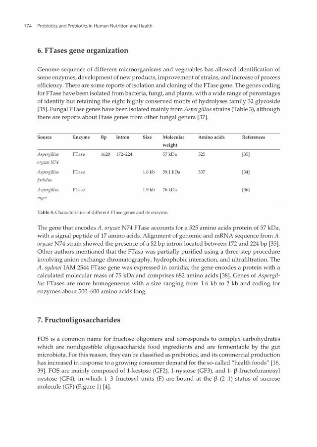
6. FTases gene organization
Genome sequence of different microorganisms and vegetables has allowed identification ofsome enzymes, development of new products, improvement of strains, and increase of processefficiency. There are some reports of isolation and cloning of the FTase gene. The genes codingfor FTase have been isolated from bacteria, fungi, and plants, with a wide range of percentagesof identity but retaining the eight highly conserved motifs of hydrolyses family 32 glycoside[35]. Fungal FTase genes have been isolated mainly from Aspergillus strains (Table 3), althoughthere are reports about Ftase genes from other fungal genera [37].
Source Enzyme Bp Intron Size Molecularweight
Amino acids References
Aspergillusoryzae N74
FTase 1620 172–224 57 kDa 525 [35]
Aspergillusfoetidus
FTase 1.6 kb 59.1 kDa 537 [34]
Aspergillusniger
FTase 1.9 kb 76 kDa [36]
Table 3. Characteristics of different FTase genes and its enzyme.
The gene that encodes A. oryzae N74 FTase accounts for a 525 amino acids protein of 57 kDa,with a signal peptide of 17 amino acids. Alignment of genomic and mRNA sequence from A.oryzae N74 strain showed the presence of a 52 bp intron located between 172 and 224 bp [35].Other authors mentioned that the FTasa was partially purified using a three-step procedureinvolving anion exchange chromatography, hydrophobic interaction, and ultrafiltration. TheA. sydowi IAM 2544 FTase gene was expressed in conidia; the gene encodes a protein with acalculated molecular mass of 75 kDa and comprises 682 amino acids [38]. Genes of Aspergil‐lus FTases are more homogeneous with a size ranging from 1.6 kb to 2 kb and coding forenzymes about 500–600 amino acids long.
7. Fructooligosaccharides
FOS is a common name for fructose oligomers and corresponds to complex carbohydrateswhich are nondigestible oligosaccharide food ingredients and are fermentable by the gutmicrobiota. For this reason, they can be classified as prebiotics, and its commercial productionhas increased in response to a growing consumer demand for the so-called “health foods” [16,39]. FOS are mainly composed of 1-kestose (GF2), 1-nystose (GF3), and 1- β-fructofuranosylnystose (GF4), in which 1–3 fructosyl units (F) are bound at the β (2–1) status of sucrosemolecule (GF) (Figure 1) [4].
Probiotics and Prebiotics in Human Nutrition and Health174

Figure 1. Chemical structures of the most common FOS, where (a) 1-kestose, (b) 1- nystose, and (c) 1-β fructofuranosylnystose.
FOS can be found in several vegetal sources such as tomato, onion, barley, garlic, Jerusalemartichoke, banana, rye, honey, sugar beet, to name a few; however, FOS concentration in thesesources is low, and mass production are limited by seasonal conditions [3, 4]. At the industriallevel, FOS are mainly produced from the disaccharide sucrose by action of different microbialenzymes with transfructosylating activity such as FTase (EC 2.4.1.9) and/or β-fructofuranosi‐dase (EC 3.2.1.26), [4]. Moreover, FOS compounds have received a generally recognized as safestatus (GRAS) from the Food and Drug Administration (FDA) and has been consumed becauseof the several benefits of FOS to human health such as calorie-free and noncariogenic sweet‐eners, stimulate bifidobacteria growth, and activation of the immune system; have beenclaimed to contribute to the prevention of colon cancer and reduce the levels of serumcholesterol, phospholipids, and triglycerides; also promote calcium and magnesium absorp‐tion in animals and the human gut [14, 19, 22, 40, 41].
Dominguez et al. [7] reported that FOS have low sweetness intensity as they are only aboutone-third as sweet as sucrose, they supply small quantities of power, about 0–3 kcal/g sugarsubstitute. Other authors mentioned that FOS are about 0.4–0.6 times sweeter than sucrose andare considered as noncariogenic since no compounds are produced when polyglucanes arepassing through the mouth [37]. These last properties are very useful in types of foods in whichsucrose use is limited because of its high sweetness [7].
Fructosyltransferase Sources, Production, and Applications for Prebiotics Productionhttp://dx.doi.org/10.5772/62827
175

7.1. FOS production
Production of FOS has received particular attention in recent years, so there is necessity forthe development of new enzymatic systems [42]. FOS represent one of the major classes ofbifidogenic oligosaccharides in terms of production volume. Kestose and nystose are the mainprebiotic compounds, which can be principally produced by hydrolysis of inulin or bytransfructosylation of sucrose [24]. The enzymes that are potentially useful for high productionof FOS were reported about three decades ago. Hidaka et al. [13] reported an A. niger enzymewith transfructosylating activity which reached a maximum FOS conversion of 55–60% (w/w)based on total sugars, and this enzyme was successively used for industrial production of FOS.Almost at the same time, McCleary et al. [43] investigated the other A. niger enzyme withtransfructosylating activity, which could compete with other enzymes for industrial produc‐tion of FOS because of its considerably high activity. Chien et al. [39] mentioned a FOSproduction process using A. japonicus enzymes, while Antosova et al. [44] reported a FOSproduction process using enzymes from A. pullulans CCY 27-1-1194.
8. Use of FTAse for FOS production
FOS have demonstrated important properties for improving human health, thus they haveattracted an increased interest mainly as ingredients for food applications. They contribute to10% of the natural sweeteners, and their demand has risen rapidly (about 15% per year) in thelast 15 years [45]. Consequently, establishing sustainable and economically viable industrialprocess for the production of FOS with high yields and productivities is strongly desirable [46].These can be manufactured by three methods: (1) extraction from inulin-rich plant materials,(2) by enzymatic synthesis from sucrose, and (3) by enzymatic degradation of inulin [45, 47].Most of the FOS marketed as food ingredients/nutritional supplements are synthesized eitherfrom sucrose by the action of FTases [48, 49] or by enzymatic degradation of inulin [50, 51]. Inthis section, we will discuss the production of FOS through FTase.
Commercial production of FOS was first developed using enzymatic fructosyl transfer onsucrose by Hidaka et al. [52], and since then, β-fructofuranosidase has been isolated especiallyfrom fungi: A. niger [13, 53], A. japonicus [54, 55], A. oryzae [21, 56], A. pullulans [57, 58], andFusarium oxysporum [59].
Nowadays, to reduce cost, enzyme immobilization techniques have been applied. Fungal β-fructofuranosidase has been covalently immobilized onto inorganic supports such as porousglass or porous silica [54, 60]. Aspergillus FTase was immobilized in methacrylamide-basedpolymeric beads and various linocellulosic materials to produce FOS from sucrose [55]. Ganaieet al. [49] evaluated immobilization of Aspergillus flavus FTase with sodium alginate andchitosan forming gel bead for continuous production of FOS, showing that recycling efficiencyof alginate beads was more successful as compared to chitosan beads. In addition, formationof FOS by FTase entrapped alginate beads was higher than by chitosan beads in 36 h of enzyme-substrate reaction according to HPLC analysis (66.75 and 42.79%, respectively).
Probiotics and Prebiotics in Human Nutrition and Health176

However, it has been observed that enzymes immobilized on a porous support decreaseapparently its enzymatic activity because of diffusion resistance. Instead, the use of magneticnanoparticles is proposed, which offers (a) a higher specific surface area that permits bindingof a larger amount of enzyme, (b) relatively low mass transfer resistance and (c) selectiveseparation from a reaction mixture by application of a magnetic field [61, 62]. Chen et al. [63]evaluated that β-fructofuranosidase from A. japonicus was immobilized in Fe3O4-CS nanopar‐ticles, retaining up to 88% of its activity. The recovery of enzyme activity was inverselyproportional to enzyme concentration. The main oligosaccharide products were 1-kestose andnystose. After 10 days, it was observed that the consumption of sucrose appear to have stoppedbecause of the accumulation of glucose, which inhibited transfructosylating reactions [64]. Theimmobilized enzyme can easily be recovered by applying a magnetic field and reused it forFOS production.
Another alternative for FOS production is the use of solid-state fermentation (SSF). Mostinvestigations on FOS production are based on submerged fermentation systems, but SSF isattractive because of low capital cost and low demand of water, generating less wastewater asa consequence. Besides, higher productivities and yields could be obtained at industrial scale[14]. Mussatto et al. [46] evaluated the economic and environmental impact of three differentfermentation processes for FOS production: (1) submerged fermentation of sucrose solutionby A. japonicus using free cells (FCF) or (2) using immobilized cells in corn cobs (ICF), and (3)SSF using coffee silver-skin as support material and nutrient source (SSF). In this study, anannual productivity goal of 200 t was established. Based on parameters such as productivity,product concentrations, yields, and thermo-physical data, SSF was the most attractive processwith higher annual productivity of FOS (232.6 t) and purity (98.6%) against 148.9 t and 96.6%for FCF and 158.3 t and 98.4% for ICF. The SSF also produced greater amounts of the shorterchain FOS (GF2 and GF3), which have more prebiotic activity and stronger sweetness [65].Although, the three processes are economically feasible, SSF has the highest potential to beimplemented on an industrial scale not only because of productivity and purity but alsobecause of the lowest payback time, wastewater generation, carbon footprint, and highestannual profit.
9. Properties of FOS
General structure of FOS can be depicted as GFn, where “n” is the number of fructosyl units(F) that are bound by β (2→1) position of sucrose with the last one attached to a terminal glucose(G) [51]. FOS produced from sucrose have lower DP (DP˂4) than those from inulin (DP > 9)[47]. This is relevant because DP plays an important role in the gut fermentation of FOS. Theshort-chain FOSs are fermented by the bacteria present in the proximal colon, while the long-chain FOS are fermented in distal colon [66]. Structurally, FOSs produced from sucrose arekestose (GF2), nystose (GF3), and 1-β-fructofuranosyl nystose (GF4).
FOSs contain several qualities that make its usage possible as an alternative sweetener in thefood market. They are water soluble and one-third as sweet as sucrose [67]. However, their
Fructosyltransferase Sources, Production, and Applications for Prebiotics Productionhttp://dx.doi.org/10.5772/62827
177

viscosity and thermal stability is higher than sucrose. They are stable in a pH range from 4.0to 7.0 and can be refrigerated for a period of one year. Their high moisture-retaining capacityprovides prevention of excessive drying besides controlling microbial contamination owingtheir low water interacting activity [68].
They can be considered as noncariogenic sugar substitutes in confectionary, gums, and drinkssince they cannot serve as a substrate of Streptococcus mutans, Lactobacillus acidophilus, and otherbacteria to form insoluble β-glucans implicated in plaque formation, which causes dentalcavities [69]. As FOSs possess β-configuration in anomeric carbon, C2 in the fructose mono‐mers, they are nondigestible by human digestive enzymes which are mostly specific for α-glycosidic bonds and therefore, are not utilized as energy source in the body [70].Consequently, they are safe for consumption by diabetics [65, 71, 72].
10. FOS as prebiotic
The most widely used definition for prebiotic is “nondigestible food ingredient that benefi‐cially affects host’s health by selectively stimulating the growth and/or activity of one or alimited number of bacteria in the colon” [73]. In 2004, the definition was updated, and it wasdefined as “selectively fermented ingredients that allow specific changes, both in the compo‐sition and/or activity in the gastrointestinal microbiota that confers benefits upon host well-being and health” [74]. According to this, prebiotics are a major part of the functional foodsand among them, the FOS are in focus due to their functional properties and economicalpotential [65, 75].
A prebiotic must fulfill three criteria: (1) no hydrolysis or absorption in the upper part of thedigestive system, (2) selective substrate for one or more desired bacteria species in the colonand stimulation of that species regarding growth and activation and (3) able to positivelyinfluence the numeric proportion of different bacteria species in the colon [76]. FOS selectivelystimulates the growth of Bifidobacteria and Lactobacillus sp. in the colon, and these bacteria showcommensalism association in the host body. These colon-specific anaerobic bacterial groupsdegrade FOS producing short-chain fatty acids (SCFA) such as acetate (C2), propionate (C3),and butyrate (C4), decreasing colon pH and subsequently enhancing the absorption of mineralions (Ca2+ and Mg2+) and nutrients in the host body [77, 78].
Many Bifidobacteria and Lactobacillus sp. are resistant to acidic pH, but it is harmful to thoseantagonist bacteria in colon like Clostridium sp. [79]. Furthermore, compared with otheranaerobes in the gastrointestinal tract, lactobacilli and bifidobacteria have enzymes with loweractivities, such as β-glucosidase, β-glucuronidase, urease, azoreductase, and nitrate reductase,which are involved in the formation of mutagens and carcinogens [80].
Antiinflammatory and antitumorigenic roles of SCFA have been reported [81, 82]. As a resultof the prebiotic function, a decrease in inflammatory markers such as phagocytosis andinterleukin (IL)-6 production by increasing CD3+, CD4+, and CD8+ populations has beenobserved [83]. In case of antitumorigenic roles, especially in the context of colon cancer, the
Probiotics and Prebiotics in Human Nutrition and Health178
action mechanism is yet unclear [80]. However, it is known that butyrate has an important roleon DNA methylation thus modifying gene expression so it may directly enhance cell prolif‐eration of normal cells, but suppress cell proliferation of transformed cells. Furthermore, inthe presence of butyrate, apoptosis may be enhanced in transformed cells but inhibited innormal cells [84]. Thus, the regular intake of FOS as a part of diet could help to improve healthand over all well-being by providing resistance against the intestinal/extra intestinal patho‐gens, enhancing the growth of the colon microbiota which have metabolic activities andbiochemical processes with a tremendous influence in human host [85].
11. Applications of FOS
FOS are components of functional food that are becoming popular in the society because theyhave a potential for enhancing flavor quality and physicochemical properties of food products,besides FOS offer various benefits for human health and are also of industrial interest [34].FOS are used in different food applications and other areas because of its positive impact onhuman health, physical performance, or state of mind [12], and the most relevant uses in foodformulations are the following: beverages (fruit drinks, coffee, cocoa, tea, soda, health drinks,and alcoholic beverages), dairy products (fermented milk, instant powders, powdered milk,and ice cream), also in light jam products and confectionary [86].
12. Functional foods
The growing interest of consumers in the relationship between nutrition and health hasincreased demand among the population for food products that improve or benefit their healthbeyond basic nutrition [87, 88]. Because of this demand, both the academic community andthe food industry have focused on developing products that meet these characteristics, whichare now called functional foods.
The term “functional foods” was first coined in Japan after a group of scientists and nutrition‐ists conducted numerous studies and defined them as “Food for specified health uses”(FoSHU) [89]. Though there is a great number of definitions, Doyon and Labrecque [90]identified four key concepts after reviewing more than 20 definitions: (a) health benefits, (b)the nature of the food, (c) level of function, and (d) consumption pattern. The definition offunctional food has evolved, and the latest is that proposed by the European Commission forconcerted action Functional Food Science in Europe (FUFOSE) that mentions that “a food canbe regarded as ‘functional’ if it is satisfactorily demonstrated to beneficially effect one or moretarget functions in the body, beyond adequate nutritional effects, in a way that is relevant toeither an improved state of health and well-being and/or reduction of risk of disease. Func‐tional foods must remain as food and they must demonstrate their effects in amounts that cannormally be expected to be consumed in the diet: they are not pills or capsules, but part of anormal food pattern” [91, 92].
Fructosyltransferase Sources, Production, and Applications for Prebiotics Productionhttp://dx.doi.org/10.5772/62827
179

The challenges for the food industry are great so that the biological value of the functionalingredient is not disturbed and sensory characteristics of the food are acceptable [93]. Accord‐ing to various investigations, the success and acceptance of these foods is influenced by factorssuch as clarity and understanding of the information about nutrition and health benefits thatare provided to consumers, especially in elderly consumers [87].
Experts recognize that there are scientific evidence on the effectiveness of various functionalfoods, which can be useful to balance a poor diet or assist in avoiding health problems in somecases [94]. In general, the consumers’ attitude towards functional foods is positive and havegreat potential in the food industry.
13. Applications of FOS in food formulations
FOS are ingredients that have been applied in a variety of food matrices, their prebioticpotential has been proven, and its technological properties allow easy incorporation into foods,mainly those that are probiotics [95]. The FOS have comparable glucose syrups and sugarproperties and proved approximately a 30–50% sweetness compared with sugar table.Therefore, their application has a dual benefit: (1) as a substitute for sugar and (2) for theirprebiotic properties [96].
Akalin and Erisir [97] evaluated the effect of supplementation of oligofructose or inulin insymbiotic ice cream, in the rheological properties and probiotics survival. They found that thesurvival rate of the probiotics during storage at 30, 40, and 90 days was better with oligofruc‐tose. The FOS were evaluated in cookies and Quicks bread and found that consumers hadpreference equal to or greater for products supplemented with FOS [96, 98]. Although the FOSare easy to incorporate into foods such as yogurt, processing conditions such as acidity andtemperature should be considered since they have reported low prebiotic activities underacidic conditions and high thermal processing times [95].
New applications in different food matrices are being evaluated. Valencia et al. [99] supple‐mented a creamy milk chocolate dessert with FOS and probiotics. They found positive resultsin the consumer acceptability test and in the survival probiotics. Moreover, in a cooked ham,FOS as substitutes of dextrose were added; the appearance of the ham did not change and theaddition of FOS in ham transformed it into a healthier product [100].
There is an innovative trend in the FOS application in different types of food and, undoubtedly,to maximize the benefits that can confer the FOS, factors as type of food matrix, processingconditions, and added amount should be considered.
14. Future trends
Because of the importance of this enzyme in the modern industry, it is important to relate aset of FTases from different organisms to allow the identification of features that could be usedfor the identification and classification of new FTases, and also it is necessary to improve the
Probiotics and Prebiotics in Human Nutrition and Health180

conditions and costs of FTases production process. Further studies of gene sequencing willallow distinguishing among the set of FTase and β FFase enzymes.
15. Conclusions
The studies on production and application of FOS are of high interest for food industry be‐cause of several health benefits and biofunctional properties that these compounds provide.FOS can be synthesizing from precursors such as sucrose using FTase enzymes. These en‐zymes can be obtained from different microorganisms (bacteria and fungi) and plants. Themain disadvantage of this production is the low yields of enzymatic activity and FOS. Thus,search for new microbial sources of FTase enzymes is a very important research topic aswell as studies about the evolution of FTase genes from different sources, and relate theirfunction with the nucleotide sequence using functional genomics studies.
Acknowledgements
This project was financially supported by the Universidad Autónoma de Coahuila. MRMMwould like to thank the financial support received from CONACYT during her master’sdegree.
Author details
Mariela R. Michel1, Rosa M. Rodríguez-Jasso1, Cristóbal N. Aguilar1,Silvia M. Gonzalez-Herrera2, Adriana C. Flores- Gallegos1 and Raúl Rodríguez-Herrera1*
*Address all correspondence to: [email protected]
1 Department of Food Research, Autonomous University of Coahuila, Saltillo, Mexico.
2 Division of Postgraduate Studies, Durango Institute of Technology, Durango, Mexico
References
[1] Maiorano A. E., Piccoli R. M., Da Silva E. S., De Andrade Rodrigues M. F. Microbialproduction of fructosyltransferases for synthesis of prebiotics. Biotechnology Letters.2008; 30:1867–1877. DOI:10.1007/s10529-008-9793-3.
[2] Antošová M., Illeová V., Vandáková M., Družkovská A., Polakovič M. Chromato‐graphic separation and kinetic properties of fructosyltransferase from Aureobasidiumpullulans. Journal of Biotechnology, 2008; 135:58–63. DOI:10.1016/j.jbiotec.2008.02.016.
Fructosyltransferase Sources, Production, and Applications for Prebiotics Productionhttp://dx.doi.org/10.5772/62827
181
[3] Mussatto S. I., Prata M. B., Rodrigues L. R., Teixeira J. A. Production of fructooligosac‐charides and β-fructofuranosidase by batch and repeated batch fermentation withimmobilized cells of Penicillium expansum. European Food Research and Technology.2012; 235:13–22. DOI:10.1007/s00217-012-1728-5.
[4] Mussatto S. I., Aguilar C. N., Rodrigues L. R., Teixeira J. A. Colonization of Aspergillusjaponicus on synthetic materials and application to the production of fructooligosac‐charides. Carbohydrate Research. 2009; 344:795–800. DOI:10.1016/j.carres.2009.01.025.
[5] Sánchez O. F., Rodriguez A. M., Silva E., Caicedo L. A. Sucrose biotransformation tofructooligosaccharides by Aspergillus sp. N74 Free Cells. Food and Bioprocess Technolo‐gy. 2008; 3:662–673. DOI:10.1007/s11947-008-0121-7.
[6] Dominguez A., Nobre C., Rodrigues L. R., Peres A. M., Torres D., Rocha I., Teixeira J.New improved method for fructooligosaccharides production by Aureobasidiumpullulans. Carbohydrate Polymers. 2012; 89:1174–1179. DOI:10.1016/j.carbpol.2012.03.091.
[7] Dominguez A. L., Rodrigues L. R., Lima N. M., Teixeira J. A. An overview of the recentdevelopments on fructooligosaccharide production and applications. Food and Biopro‐cess Technology. 2013; 7:324–337. DOI:10.1007/s11947-013-1221-6.
[8] Antošová M., Polakovič M. Fructosyltransferases: the enzymes catalyzing productionof fructooligosaccharides. Chemical Papers. 2001; 55:350–358.
[9] Koops A. J., Jonker H. H. Purification and Characterization of the enzymes of fructanbiosynthesis in tubers of Helianthus Fuberbsus Colombia. Plant Physiology. 1996;110:1167–1175.
[10] Edelman J., Jefford T. G. The mechanisim of fructosan metabolism in higher plants asexemplified in Helianthus tuberosus. New Phytologist.1968;67:517–531. DOI:10.1111/j.1469-8137.1968.tb05480.x.
[11] Altenbach D., Rudiño-Pinera E., Olvera C., Boller T., Wiemken A., Ritsema T. Anacceptor-substrate binding site determining glycosyl transfer emerges from mutantanalysis of a plant vacuolar invertase and a fructosyltransferase. Plant MolecularBiology. 2009; 69:47–56. DOI:10.1007/s11103-008-9404-7.
[12] Sangeetha P. T., Ramesh M. N., Prapulla S. G. Recent trends in the microbial production,analysis and application of Fructooligosaccharides. Trends in Food Science and Technol‐ogy. 2005; 16:442–457. DOI:10.1016/j.tifs.2005.05.003
[13] Hidaka H., Hirayama M., Sumi N. A Fructooligosaccharide-producing enzyme from.Agricultural and Biological Chemistry. 1988; 52:1181–1187.
[14] Mussatto S. I., Teixeira J. A. Increase in the fructooligosaccharides yield and produc‐tivity by solid-state fermentation with Aspergillus japonicus using agro-industrialresidues as support and nutrient source. Biochemical Engineering Journal. 2010; 53:154–157. DOI:10.1016/j.bej.2010.09.012
Probiotics and Prebiotics in Human Nutrition and Health182

[15] Sangeetha P. T., Ramesh M. N., Prapulla S. G. Production of fructosyl transferase byAspergillus oryzae CFR 202 in solid-state fermentation using agricultural by-products.Applied Microbiology and Biotechnology. 2004; 65:530–537. DOI:10.1007/s00253-004-1618-2
[16] La Rotta C. E., Ospina S. A., López-Munguía A. Production and characterization ofcrude enzimatic extracts with fructosyl transferase activity. Revista Colombiana deCiencias Químico-Farmacéuticas. 1998; 27:53–56 (in Spanish).
[17] Wang X. D., Rakshit S. K. Improved extracellular transferase enzyme production byAspergillus foetidus for synthesis of isooligosaccharides. Bioprocess Engineering. 1999; 20:429–434.
[18] Sangeetha P. T., Ramesh M. N., Prapulla S. G. Production of fructo-oligosaccharides byfructosyl transferase from Aspergillus oryzae CFR 202 and Aureobasidium pullulans CFR77. Process Biochemistry. 2004; 39:755–760. DOI:10.1016/S0032-9592(03)00186-9
[19] Sangeetha P. T., Ramesh M. N., Prapulla S. G. Fructooligosaccharide production usingfructosyl transferase obtained from recycling culture of Aspergillus oryzae CFR 202.Process Biochemistry. 2005; 40:1085–1088. DOI:10.1016/j.procbio.2004.03.009
[20] Sangeetha P. T., Ramesh M. N., Prapulla S. G. Maximization of fructooligosaccharideproduction by two stage continuous process and its scale up. Journal of Food Engineer‐ing. 2005; 68:57–64. DOI:10.1016/j.jfoodeng.2004.05.022.
[21] MasahiroKurakake, OgawaKenji, SugieMotoki, A. T., Kouji Sugiura, T. Komaki. Twotypes of fructofuranosidases from Aspergillus oryzae KB. Journal of Agricultural and FoodChemistry. 2008; 56:591–596.
[22] Sathish T., Prakasham R. S. Intensification of fructosyltransferases and fructo-oligo‐saccharides production in solid state fermentation by Aspergillus awamori GHRTS.Indian Journal of Microbiology. 2013; 53:337–342. DOI:10.1007/s12088-013-0380-5.
[23] Yang H., Wang Y., Zhang L., Shen W. Heterologous expression and enzymaticcharacterization of fructosyltransferase from Aspergillus niger in Pichia pastoris. NewBiotechnology. 2015; 00:1–7. DOI:10.1016/j.nbt.2015.04.005.
[24] Yoshikawa J., Amachi S., Shinoyama H., Fujii T. Multiple˂β-fructofuranosidases byAureobasidium pullulans DSM2404 and their roles in fructooligosaccharide production.FEMS Microbiology Letters. 2006; 265:159–163. DOI:10.1111/j.1574-6968.2006.00488.x
[25] Sprenger N., Bortlik K., Brandt A., Boller T., Wiemken A. Purification, cloning, andfunctional expression of sucrose: fructan 6-fructosyltransferase, a key enzyme offructan synthesis in barley. Proceedings of the National Academy of Sciences. 1995;92:11652–11656.
[26] Chuankhayan P., Hsieh C. Y., Huang Y. C., Hsieh Y. Y., Guan H. H., Hsieh Y. C., ChenC. J. Crystal structures of Aspergillus japonicus fructosyltransferase complex with donor/
Fructosyltransferase Sources, Production, and Applications for Prebiotics Productionhttp://dx.doi.org/10.5772/62827
183

acceptor substrates reveal complete subsites in the active site for catalysis. Journal ofBiological Chemistry. 2010; 285:23251–23264. DOI:10.1074/jbc.M110.113027.
[27] ArtheeR., VijilaK. Study on fructosyltransferase enzyme from Aspergillus sp. infructooligosaccharides production. Research Journal of Recent Sciences. 2014; 3:147–153.
[28] AmrendraKumar, Vaishnavi .R, Saravanakumar .,A K. S.A. and, Tank S. K. Biotrans‐formation of sucrose by using thermostable and alkaline fructosyltransferase enzymeisolated. International Journal of Science, Environment. 2014; 3:708–713.
[29] Ghazi I., Fernandez-Arrojo L., Garcia-Arellano H., Ferrer M., Ballesteros A., Plou F. J.Purification and kinetic characterization of a fructosyltransferase from Aspergillusaculeatus. Journal of Biotechnology. 2007; 128:204–211. DOI:10.1016/j.jbiotec.2006.09.017.
[30] Dhake,A. B., Patil M. B. Effect of substrate feeding on production of fructosyltransferaseby Penicillium purpurogenum. Brazilian Journal of Microbiology. 2007; 38:194–199. DOI:10.1590/S1517-83822007000200002.
[31] Patil M. B., ButleA.. Fructosyltransferase production by indigenously isolated Synce‐phalastrum racemosum Cohn. Journal of Global Biosciences. 2014; 3:597–603.
[32] Tamura K. I., Kawakami A., Sanada Y., Tase K., Komatsu T., Yoshida M. Cloning andfunctional analysis of a fructosyltransferase cDNA for synthesis of highly polymerizedlevans in timothy (Phleum pratense L.). Journal of Experimental Botany. 2009; 60:893–905.DOI:10.1093/jxb/ern337.
[33] Van Hijum S. a F. T., Van Geel-Schutten G. H., Rahaoui H. Van der Maarel M. J. E. C.,Dijkhuizen, L. Characterization of a novel fructosyltransferase from Lactobacillusreuteri that synthesizes high-molecular-weight inulin and inulin oligosaccharides.Applied and Environmental Microbiology. 2002; 68:4390–4398. DOI:10.1128/AEM.68.9.4390-4398.2002.
[34] Rehm J., Willmitzer L., Heyer A. G. Production of 1-kestose in transgenic yeastexpressing a fructosyltransferase from Aspergillus foetidus. Journal of Bacteriology. 1998;180:1305–1310.
[35] Rodríguez M. A, Sánchez O. F., Alméciga-Díaz C. J. Gene cloning and enzyme structuremodeling of the Aspergillus oryzae N74 fructosyltransferase. Molecular Biology Reports.2011; 38:1151–1161. DOI:10.1007/s11033-010-0213-0.
[36] Goosen C., Yuan X.-L., van Munster J. M., Ram A. F. J., van der Maarel M. J. E. C.,Dijkhuizen L. Molecular and biochemical characterization of a novel intracellularinvertase from Aspergillus niger with transfructosylating activity. Eukaryotic Cell. 2007;6: 674–681. DOI:10.1128/EC.00361-06.
[37] Alméciga-Díaz C. J., Gutierrez Á. M., Bahamon I., Rodríguez A., Rodríguez M. A,Sánchez O. F. Computational analysis of the fructosyltransferase enzymes in plants,fungi and bacteria. Gene. 2011; 484:26–34. DOI:10.1016/j.gene.2011.05.024.
Probiotics and Prebiotics in Human Nutrition and Health184

[38] Heyer A. G., Wendenburg R. Gene cloning and functional characterization by heterol‐ogous expression of the fructosyltransferase of Aspergillus sydowi IAM 2544. Applied andEnvironmental Microbiology. 2001; 67:363–370. DOI:10.1128/AEM.67.1.363.
[39] Chien C.-S., Lee W.-C., Lin T.-J. Immobilization of Aspergillus japonicus by entrappingcells in gluten for production of fructooligosaccharides. Enzyme and Microbial Technol‐ogy. 2001; 29:252–257. DOI:10.1016/S0141-0229(01)00384-2.
[40] Mussatto S. I., Ballesteros L. F., Martins S., Maltos D. A. F., Aguilar C. N., Teixeira J. A.Maximization of fructooligosaccharides and β-fructofuranosidase production byAspergillus japonicus under solid-state fermentation conditions. Food and BioprocessTechnology. 2013; 6:2128–2134. DOI:10.1007/s11947-012-0873-y.
[41] Silva J. B. Da, Fai A. E. C., dos Santos R., Basso L. C., Pastore G. M. Parameters evaluationof fructooligosaccharides production by sucrose biotransformation using an osmo‐philic Aureobasium pullulans strain. Procedia Food Science. 2011; 1:1547–1552. DOI:10.1016/j.profoo.2011.09.229.
[42] L’Hocine, L., Wang Z., Jiang B., Xu S. Purification and partial characterization offructosyltransferase and invertase from Aspergillus niger AS0023. Journal of Biotechnol‐ogy. 2000; 81:73–84. DOI:10.1016/S0168-1656(00)00277-7.
[43] McCleary B. V., Gibson T. S., Sheehan H., Casey A., Horgan L., O’Flaherty, J. Purifica‐tion, properties, and industrial significance of transglucosidase from Aspergillus niger.Carbohydrate Research. 1989; 185:147–162.
[44] Antosova M., Polakovic M., Slovinská M., Madlová A., Illeová V., Bales V. Effect ofsucrose concentration and cultivation time on batch production of fructosyltransferaseby Aureobasidium pullulans CCY 27-1-1194. Hemical Papers-Slovak Academy of Sciences.2003; 56:394–399.
[45] Panesar P.S., Bali V., Kumari S., Babbar N., Oberoi H.S. Prebiotics. In: G.S. Brar (Ed.),Biotransformation of Waste Biomass into High Value Biochemicals. Springer. Ney York, USA,2014; 504 pp.
[46] Mussatto S. I., Aguiar L. M., Marinha M. I., Jorge R. C., Ferreira E. C. Economic analysisand environmental impact assessment of three different fermentation processes forfructooligosaccharides production. Bioresource Technology. 2015; 198:673–681. DOI:10.1016/j.biortech.2015.09.060.
[47] Rastall R.A. Functional oligosaccharides: application and manufacture. Annual Reviewof Food Science and Technology. 2010; 1:305–339.
[48] Bali V., Panesar P.S., Bera M.B., Panesar R. Fructo-oligosaccharides: production,purification and potential applications. Critical Reviews in Food Science and Nutrition.2015; 55:1475–1490.
Fructosyltransferase Sources, Production, and Applications for Prebiotics Productionhttp://dx.doi.org/10.5772/62827
185

[49] Ganaie M. A., Rawat H. K., Wani O. A., Gupta U. S., Kango N. Immobilization offructosyltransferase by chitosan and alginate for efficient production of fructooligo‐saccharides. Process Biochemistry. 2014; 49:840–844. DOI:10.1016/j.procbio.2014.01.026.
[50] Mutanda T., Mokoena M. P., Olaniran, A. O., Wilhelmi, B. S., Whiteley, C. G. Microbialenzymatic production and applications of short-chain fructooligosaccharides andinulooligosaccharides: Recent advances and current perspectives. Journal of IndustrialMicrobiology and Biotechnology. 2014; 41:893–906. DOI:10.1007/s10295-014-1452-1.
[51] Singh R. S., Singh R. P. Production of fructooligosaccharides from inulin by endoinu‐linases and their prebiotic potential. Food Technology and Biotechnology. 2010; 48: 435–450.
[52] Hidaka,H., Eida,T., Tokunaga,T., Tashiro,Y. Effects of fructooligosaccharides onintestinal flora and human health. Bifidobacteria and Microflora. 1986; 5:37–50.
[53] Nguyen,Q.D., Rezessy-Szabo,J.M., Bhat,M.K. Purification and some properties of b-fructofuranosidase from Aspergillus niger IMI303386. Process Biochemistry. 2005;40:2461–2466.
[54] Hayashi,S., Matsuzaki,K., Takasaki,Y., Ueno,H., Imada,K. Production of b-fructofura‐nosidase by Aspergillus japonicus. World Journal of Microbiology and Biotechnology. 1992;8:155–159.
[55] Mussatto,S.I., Aguilar,C.N., Rodrigues,L.R., Teixeira,J.A. Fructooligosaccharides andb-fructofuranosidase production by Aspergillus japonicus immobilized on lignocellulo‐sic materials. Journal of Molecular Catalisis B: Enzymatic. 2009; 59:76–81.
[56] Chang,C.T., Lin,Y.Y., Tang,M.S., Lin,C.F. Purification and properties of betafructofur‐anosidase from Aspergillus oryzae ATCC 76080. Biochemistry and Molecular BiologyInternational. 1994; 32:269–277.
[57] Jung,K.H., Lim,J.Y., Yoo,S.J., Lee,J.H., Yoo,M.Y. Production of fructosyltransferasefrom Aureobasidium pullulans. Biotechnology Letters. 1987; 9:703–708.
[58] Yoshikawa,J., Amachi,S., Shinoyama,H., Fujii,T. Purification and some properties of b-fructofuranosidase I formed by Aureobasidium pullulans DSM 2404. Journal Bioscienceand Bioengineering. 2007; 103:491–493.
[59] Nishizawa,M., Maruyama,Y., Nakamura,M. Purification and characterization ofinvertase isozyme from Fusarium oxysporum. Agricultural and Biological Chemistry. 1980;44:489–498.
[60] Ortega-Muñoz,M., Morales-Sanfrutos,J., Megia-Fernandez,F., Lopez-Jaramillo,F. J.,Hernandez-Mateo,F., Santoyo-Gonzalez,F. Vinyl sulfone functionalized silica: a readyto use pre-activated material for immobilization of biomolecules. Journal of MaterialsChemistry. 2010; 20:7189–7196.
Probiotics and Prebiotics in Human Nutrition and Health186

[61] Halling,P.J., Dunnill,P. Magnetic supports for immobilized enzymes and bioaffinityadsorbents. Enzyme and Microbial Technology.1980; 2: 2–10.
[62] Kim,J., Grate,J.W., Wang,P. Nanostructures for enzyme stabilization. Chemical Engi‐neering Science. 2006; 61:1017–1026.
[63] Chen S. C., Sheu D. C., Duan K. J. Production of fructooligosaccharides using β-fructofuranosidase immobilized onto chitosan-coated magnetic nanoparticles. Journalof the Taiwan Institute of Chemical Engineers. 2014; 45:1105–1110. DOI:10.1016/j.jtice.2013.10.003.
[64] Duan,K. J., Chen,J.S., Sheu,D.C. Kinetic studies and mathematical model for enzymaticproduction of fructooligosaccharides from sucrose. Enzyme and Microbial Technology.1994; 16:334–339.
[65] Yun J. W. Fructooligosaccharides—occurrence, preparation, and application. Enzymeand Microbial Technology. 1996; 19:107–117. DOI:10.1016/0141-0229(95)00188-3.
[66] Meyer D., Stasse-Wolthuis M. The bifidogenic effect of inulin and oligofructose and itsconsequences for gut health. European Journal of Clinical Nutrition, 63:1277–1289. DOI:10.1038/ejcn.2009.64.
[67] Salinas M. A., Perotti N. A. Production of fructosyltransferase by Aureobasidium sp.ATCC 20524 in batch and two-step batch cultures. Journal of India Industrial MicrobiologyBiotechnology. 2009; 36:39–43.
[68] Crittenden R. G., Playne M. J. Production, properties and applications of food-gradeoligosaccharides. Trends in Food Science and Technology. 1996; 7: 353–361.
[69] Oku T. Special physiological functions of newly developed mono-and oligosacchar‐ides. In: Goldberg (Ed.), Functional Foods: Designer Foods, Pharmafoods, Nutraceuticals.Chapman &Hall, Ney York, USA. pp. 202–218.
[70] Kaur N., Gupta A. K. Applications of inulin and oligofructose in health and nutrition.Journal of Biosciences. 2002; 27:703–714. DOI:10.1007/BF02708379.
[71] Rycroft C. E., Jones M. R., Gibson G. R., Rastall R. A. A comparative in vitro evaluationof the fermentation properties of prebiotic oligosaccharides. Journal of Applied Microbi‐ology. 2001; 91:878–887. DOI:10.1046/j.1365-2672.2001.01446.x.
[72] Villegas B., Costell E. Flow behaviour of inulin-milk beverages. Influence of inulinaverage chain length and of milk fat content. International Dairy Journal. 2007; 17:776–781. DOI:10.1016/j.idairyj.2006.09.007.
[73] Gibson,G. R., Roberfroid,M. B. Dietary modulation of the human colonic microbiota:introducing the concept of prebiotics. Journal of Nutrition. 1995; 125:1401–1412.
[74] Gibson G. R., Probert, H. M., Loo, J. Van, Rastall R. A, Roberfroid M. B. Dietarymodulation of the human colonic microbiota: updating the concept of prebiotics.Nutrition Research Reviews. 2004; 17:259–275. DOI:10.1079/NRR200479.
Fructosyltransferase Sources, Production, and Applications for Prebiotics Productionhttp://dx.doi.org/10.5772/62827
187

[75] Flores-Gallegos A. C., Contreras-Esquivel J. C., Morlett-Chavez J. A, Aguilar C. N.,Rodriguez-Herrera R. Comparative study of fungal strains for thermostable inulinaseproduction. Journal of Bioscience and Bioengineering. 2015; 119:421–426. DOI:10.1016/j.jbiosc.2014.09.020.
[76] Kovács Z., Benjamins E., Grau K., Rehman A.U., Ebrahimi M., and Czermak P. Recentdevelopments in manufacturing oligosaccharides with prebiotic functions. Advances inBiochemical Engineering Biotechnology. 2014; 143:257–295.
[77] Scholz-Ahrens K.E., Ade P., Marten B., Weber P., Timm W., Asil Y., Glüer C., Schre‐zenmeir J. Mint: Prebiotics, probiotics, and synbiotics affect mineral absorption, bonemineral content and bone structure. Journal Nutrition. 2007; 137:838S–8346S.
[78] Tazoe H., Otomo Y., Kaji I., Tanaka R., Karaki S.I., Kuwahara A. Roles of short-chainfatty acids receptors, GPR41 and GPR43 on colonic functions. Journal of Physiology andPharmacology. 2008; 59:251–262.
[79] Sangeetha P. T., Ramesh M. N., Prapulla S. G. Microbial production of fructooligosac‐charides. Asian Journal of Microbiology Biotechnology Environmental Sciences. 2003; 5:313–331.
[80] Pool-Zobel B.L., Neudecker C., Domizlaff I., Ji S., Schillinger U., Rumney C., MorettiM., Vilarini I., Scassellati S.R., Rowland I. Lactobacillus- and Bifidobacterium-mediatedantigenotoxicity in the colon of rats. Nutrition and Cancer. 1996; 26:365–380.
[81] Alva-Murillo N., Ochoa-Zarzosa A., Lopez-Meza J.E. Short chain fatty acids (propionicand hexanoic) decrease Staphylococcus aureus internalization into bovine mammaryepithelial cells and modulate antimicrobial peptide expression. Veterinary Microbiolo‐gy. 2012; 155:324–331.
[82] Tan J., Mckenzie C., Potamitis M., Thorburn A. N., Mackey C. R., Macia L. The role ofshort-chain fatty acids in health and disease. Advances in Immunology. 2014; 121:91–119.
[83] Guigoz Y., Rochat F., Perruisseau-Carrier G., Rochat I., Schiffrin E. J. Effects of oligo‐saccharide on the faecal flora and non-specific immune system in elderly people.Nutrition Research. 2002; 22:13–25. DOI:10.1016/S0271-5317(01)00354-2.
[84] Hass R., Busche R., Luciano L., Reale E., Engelhardt W. Lack of butyrate is associatedwith induction of bax and subsequent apoptosis in the proximal colon of guinea pig.Gastroenterology. 1997; 112:875–881.
[85] Peshev D., den Ende W. V. Fructans: prebiotics and immunomodulators. Journal ofFunctional Foods. 2014; 8:348–357.
[86] Mussatto S. I., Mancilha I. M. Non-digestible oligosaccharides: a review. CarbohydratePolymers. 2007; 68:587–597. DOI:10.1016/j.carbpol.2006.12.011.
Probiotics and Prebiotics in Human Nutrition and Health188

[87] Vella M.N.; Stratton L.M.; Sheeshka J.; Duncan A.M. Functional food awareness andperceptions in relation to information sources in older adults. Nutrition Journal. 2014;13:1–25.
[88] Abdel-Salam A.M. Functional food: hopefulness to good health. American Journal ofFood Technology. 2010; 5:86–99.
[89] Shimizu M. Functional food in Japan: current status and future of gut-modulating food.Journal of Food and Drug Analysis. 2012; 20:213–216.
[90] Doyon M.; Labrecque J. A. Functional foods: a conceptual definition. British FoodJournal. 2008; 110:1133–1149.
[91] Diplock A., Aggett P., Ashwell M., Bornet F., Fern E., Roberfroid M. Scientific conceptsof functional foods in Europe: consensus document. British Journal of Nutrition. 1999;9:1–27.
[92] Ozen A. E., Pons A., Tur J. A. Worldwide consumption of functional foods: A systematicreview. Nutrition Reviews. 2012; 70:472–481.
[93] Spence J. T. Challenges related to the composition of functional foods. Journal of FoodComposition and Analysis. 2006; 19:S4–S6.
[94] Crowe K. M., Francis C. Position of the academy of nutrition and dietetics: functionalfoods. Journal of the Academy of Nutrition and Dietetics. 2013; 113:1096–1103.
[95] Vega R., Zuniga-Hansen M. E. The effect of processing conditions on the stability offructooligosaccharides in acidic food products. Food Chemistry. 2015; 173:784–789.
[96] Handa C., Goomer S., Siddhu A. Physicochemical properties and sensory evaluationof fructoligosaccharide enriched cookies. Journal of Food Science And Technology. 2012;49:192–199.
[97] Akalın A. S., Erişir D. Effects of inulin and oligofructose on the rheological character‐istics and probiotic culture survival in low fat probiotic Ice cream. Journal of FoodScience. 2008; 73:M184-M188.
[98] Rößle C., Ktenioudaki A., Gallagher E. Inulin and oligofructose as fat and sugarsubstitutes in quick breads (scones): a mixture design approach. European Food Researchand Technology. 2011; 233:167–181.
[99] Valencia M.S., Salgado S.M., Andrade S.A.C., Padilha V.M., Livera A.V.S., StamfordT.L.M. Development of creamy milk chocolate dessert added with fructooligosacchar‐ide and Lactobacillus paracasei subsp. Paracasei LBC 81. LWT––Food Science and Technol‐ogy. 2016; 69:104–109.
[100] Resconi V.C., Keenan D.F., Barahona M., Guerrero L., Kerry J.P., Hamill R.M. Ricestarch and fructo-oligosaccharides as substitutes for phosphate and dextrose in wholemuscle cooked hams: sensory analysis and consumer preferences. LWT—Food Scienceand Technology. 2016; 66:284–292.
Fructosyltransferase Sources, Production, and Applications for Prebiotics Productionhttp://dx.doi.org/10.5772/62827
189


Chapter 10
Antimicrobial Effect of Probiotics against CommonPathogens
Sabina Fijan
Additional information is available at the end of the chapter
http://dx.doi.org/10.5772/63141
Abstract
The antimicrobial or antagonistic activity of probiotics is an important property thatincludes the production of antimicrobial compounds, competitive exclusion ofpathogens, enhancement of the intestinal barrier function and others. There are manymethods to ascertain probiotic properties, including various in vitro and in vivo methods.The in vivo methods include various modifications of the spot‐on lawn assay, agar welldiffusion assay (AWDA), co‐culturing methods, usage of cell lines and others. In manycases in vitro antagonist activity is observed, but in real settings it is not observed. Thein vivo methods mainly used are animal models; however, their use is being restrictedaccording to the European legislation OJ L136. The justification of animal models is alsoquestionable as the results of studies on animals do not predict the same results forhumans. The use of replacement alternative methods, for example incorporating humancells and tissues, avoids such confounding variables. Most important studies are double‐blinded randomized clinical trials; however, these studies are difficult to perform as itis not easy to achieve uniform conditions. There is a clear need for more elaborate assaysthat would better represent the complex interactions between the probiotics and thefinal host. This complex situation is a challenge for scientists.
Keywords: antimicrobial effect, in vitro methods, in vivo methods, pathogens, probiot‐ics
1. Introduction
Throughout the history of microbiology, most human studies have been focused on the disease‐causing organisms found on or in people; whilst fewer studies have examined the benefits ofthe resident bacteria. However, we are surrounded by beneficial microorganisms that live in or
on the human body. The intestinal microbiota is very well adapted, exceptionally stable andvery specific for each individual. In normal conditions of stable functioning of the digestivesystem, neutral and beneficial microorganisms dominate. It is estimated that there are 100 trillionmicroorganisms in the intestine of a human adult and this is 10 times larger than the number ofcells in the human body [1, 2]. However, the balance of the intestinal microbiota is negativelyinfluenced by modern lifestyle, leading to increased numbers of pathogenic microorganismsthat disrupt microbial balance and cause a reverse from beneficial to harmful functioning. Insuch cases, the external support with probiotics is very welcome and supported by severalscientific studies [3].
According to the Food and Agriculture Organisation of the United Nations (FAO) and theWorld Health Organisation (WHO), probiotics are defined as live microorganisms, whichwhen administered in adequate amounts confer a health benefit on the host [4, 5]. The mostcommon probiotic bacteria are certain strains from the genera Lactobacillus (i.e., L. rhamnosus,L. acidophilus, L. plantarum, L. casei, L. delbrueckii subsp. Bulgaricus, etc.) and Bifidobacterium (i.e.,B. infantis, B. animalis subsp. lactis, B. longum, etc.). Other probiotic bacteria include Pediococcusacidilactici, Lactococcus lactis subsp. lactis, Leuconostoc mesenteroides, Bacillus subtilis, Enterococcusfaecium, Streptococcus thermophilus, Escherichia coli Nissle 1917, etc. Certain yeasts such asSaccharomyces boulardii are also probiotics [6, 7].
Probiotics together with other beneficial microbes are commensals of the gut and differ frompathogenic bacteria in the terms of their actions on immune cells in the gut as they do notstimulate the proliferation of mononuclear cells or trigger an inflammatory action [8]. Regard‐less of whether the probiotics are used for human or animal consumption, there are severalcharacteristics that a probiotic must achieve. Some of the important characteristics of probioticsinclude the following: a probiotic must be generally required as safe (GRAS); a probiotic shouldexhibit bile and acid tolerance in order to survive the path from the oral cavity to the smallintestine where it lives, multiplies and excretes beneficial nutrients and molecules; a probioticshould have the ability to adhere to mucus and/or epithelial cells, and/or other surfaces; aprobiotic should be susceptible to antibiotics; a probiotic should exhibit antimicrobial activityagainst pathogens [3, 5, 9, 10]. Although it is accepted that probiotics must be of human origin[4, 5], many authors have found that some strains that are not normally isolated from humanhave shown to be effective [11, 12], which negates this requirement. As noted above, one ofthe important attributes of probiotics is their antimicrobial effect against pathogens bymaintaining the homeostasis of the intestinal flora. Assessing the antimicrobial effect of variousprobiotics against pathogenic microorganisms is the guiding concept of this chapter. Thischapter reviews the principles and results from various authors of different methods fordetermining the antimicrobial or antagonistic effect of probiotics against potential pathogens.
2. Antimicrobial or antagonistic properties of probiotics
In literature both the terms “antimicrobial” and “antagonistic” are found to determine theability of one species to inhibit the growth of another species. According to the online Ency‐clopaedia Britannica, “antagonism” refers to an “association between organisms in which one
Probiotics and Prebiotics in Human Nutrition and Health192
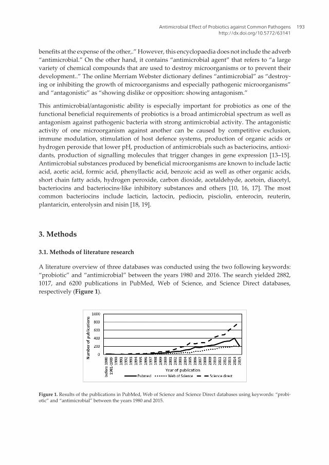
benefits at the expense of the other,.” However, this encyclopaedia does not include the adverb“antimicrobial.” On the other hand, it contains “antimicrobial agent” that refers to “a largevariety of chemical compounds that are used to destroy microorganisms or to prevent theirdevelopment..” The online Merriam Webster dictionary defines “antimicrobial” as “destroy‐ing or inhibiting the growth of microorganisms and especially pathogenic microorganisms”and “antagonistic” as “showing dislike or opposition: showing antagonism.”
This antimicrobial/antagonistic ability is especially important for probiotics as one of thefunctional beneficial requirements of probiotics is a broad antimicrobial spectrum as well asantagonism against pathogenic bacteria with strong antimicrobial activity. The antagonisticactivity of one microorganism against another can be caused by competitive exclusion,immune modulation, stimulation of host defence systems, production of organic acids orhydrogen peroxide that lower pH, production of antimicrobials such as bacteriocins, antioxi‐dants, production of signalling molecules that trigger changes in gene expression [13–15].Antimicrobial substances produced by beneficial microorganisms are known to include lacticacid, acetic acid, formic acid, phenyllactic acid, benzoic acid as well as other organic acids,short chain fatty acids, hydrogen peroxide, carbon dioxide, acetaldehyde, acetoin, diacetyl,bacteriocins and bacteriocins‐like inhibitory substances and others [10, 16, 17]. The mostcommon bacteriocins include lacticin, lactocin, pediocin, pisciolin, enterocin, reuterin,plantaricin, enterolysin and nisin [18, 19].
3. Methods
3.1. Methods of literature research
A literature overview of three databases was conducted using the two following keywords:“probiotic” and “antimicrobial” between the years 1980 and 2016. The search yielded 2882,1017, and 6200 publications in PubMed, Web of Science, and Science Direct databases,respectively (Figure 1).
Figure 1. Results of the publications in PubMed, Web of Science and Science Direct databases using keywords: “probi‐otic” and “antimicrobial” between the years 1980 and 2015.
Antimicrobial Effect of Probiotics against Common Pathogenshttp://dx.doi.org/10.5772/63141
193

All three databases showed great increase in the number of publications in the past 10 years.The highest results were obtained via Science Direct due to the fact that this database includesvarious journals and books from the area of food and dairy sciences, the area of probiotics foranimals as well as the area of human probiotics. PubMed on the other hand contains onlyresearch of probiotics for humans. Also, the programs for keyword searching for each chosendatabase seem to differ among each other thus yielding very different numbers of publicationsin journals, chapters and conference proceedings. The research on the methods for determiningthe antimicrobial effect of strain‐specific probiotics was conducted by adding an additionalkeyword aside “probiotic.” This keyword described the various investigated methods fordetermining the antimicrobial/antagonistic activity of probiotics (i.e., spot‐on lawn, agar spot,agar well diffusion, paper disc, co‐culturing, in vivo). Of course, it was not possible to screenevery single article therefore only a selection of the most recent or relevant were used.
3.2. In vitro methods for determining the antimicrobial/antagonistic effect of probioticsagainst other microorganisms
3.2.1. Spot‐on lawn antimicrobial assay/agar spot antimicrobial assay
The spot‐on lawn antimicrobial assay has been described by several authors. Several modifi‐cations of the method have been made. Also various other expressions are used such as agarspot assay, critical dilution assay and deferred antagonism assay [10, 16, 20–22].
One of the simplest published principles of the spot‐on lawn antimicrobial assay (Figure 2a)consists of the following steps: different nutrients, selective or differential media, are preparedand various chosen indicator microorganisms or pathogens at different initial concentrationsare either inoculated in a confluent manner after hardening of agar or are mixed with the agarin liquid state and poured into the plate. Different dilutions of aliquots of the investigatedprobiotic or cell‐free supernatant with bacteriocins are then spotted onto the media alreadyinoculated with chosen indicator microorganisms [20, 23, 24].
After incubation, the antimicrobial activity is expressed either as inhibition zone or as arbitraryunits (AU/mL). The zone of inhibition is noted either as the diameter or the area of theinhibition zone. The critical dilution is noted as the last dilution that produces a zone ofinhibition larger than 6 mm. Arbitrary units are defined as the reciprocal of the highest dilutionat which the growth of the indicator microorganism or pathogen is inhibited and are calculatedas (1000/a) x D in AU/mL, where a is the aliquot of cell‐free supernatant added to well in µLand D is the dilution factor [22, 24].
A modification is the agar spot antimicrobial assay (Figure 2b) and consists of the followingsteps: MRS agar or other specified agar is prepared and the probiotic bacteria or test cultures(few µL) are spotted on. These agars are then incubated to develop spots. Next, the indicatorbacteria (pathogenic species, spoilage species and other probiotic species) are mixed intospecific soft agar (0.7%) and poured over a plate. The plates are then incubated aerobically oranaerobically and the inhibition zones are read. A clear zone of more than 1 mm around thespot is considered as positive [25]. A third modification is the spot‐on lawn antimicrobial assaywith wells (Figure 2c), which consists of the following steps: chosen nutrients, selective or
Probiotics and Prebiotics in Human Nutrition and Health194
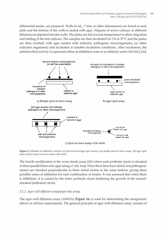
differential media, are prepared. Wells (6 mL, 7 mm or other dimensions) are bored in eachplate and the bottom of the wells is sealed with agar. Aliquots of active cultures at differentdilutions are pipetted into the wells. The plates are left at room temperature to allow migrationand settling of the test cultures. The samples are then incubated for 3 h at 37°C and the platesare then overlaid with agar seeded with indicator pathogenic microorganisms (or otherindicator organisms) and incubated at suitable incubation conditions. After incubation, theantimicrobial activity is expressed either as inhibition zone or as arbitrary units (AU/mL) [16].
Figure 2. Schemes of different versions of spot‐on lawn/agar spot assays: (a) simple spot‐on lawn assay, (b) agar spotassay and (c) spot‐on lawn assay with wells.
The fourth modification is the cross streak assay [26] where each probiotic strain is streakedin three parallel lines onto agar using a 1‐mL loop. Once these lines have dried, test pathogenicstrains are streaked perpendicular to these initial strains in the same fashion, giving threepossible zones of inhibition for each combination of strains. It was assumed that when thereis inhibition, it is caused by the tester probiotic strain hindering the growth of the second‐streaked (indicator) strain.
3.2.2. Agar well diffusion assay/paper disc assay
The agar well diffusion assay (AWDA) (Figure 3a) is used for determining the antagonisticeffects of cell‐free supernatants. The general principle of agar well diffusion assay consists of
Antimicrobial Effect of Probiotics against Common Pathogenshttp://dx.doi.org/10.5772/63141
195

the following steps: different nutrients, selective or differential media, are prepared. The platesare inoculated with the chosen indicator microorganism. The 6‐mm or 7‐mm wells are boredin each plate. Aliquots of different dilutions of cell‐free supernatants are pipetted into the wells.After incubation, the antimicrobial activity is expressed either as inhibition zone or as arbitraryunits (AU/mL) [20, 22]. The paper disc assay (Figure 3b) is a modification where instead ofmaking wells, discs measuring 6 mm are absorbed with aliquots of cell‐free supernatant andplaced on the agar inoculated with indicator strains. After incubation, the inhibition zone isevaluated based on the clear zone around the paper disc [23].
Figure 3. Scheme of the agar well diffusion assay and paper disc assay: (a) agar well diffusion assay and (b) paper discassay.
3.2.3. Co‐culturing assays for determining the antimicrobial activity
Determining the antimicrobial activity of probiotics against common pathogens is also possiblewith the co‐culturing assay. This method includes the following steps: preparation of incuba‐tion media (i.e., nutrient broth, reconstituted skim milk, sterilized milk, yogurt, whey, etc.).Aliquots of pathogenic and probiotics microorganisms are inoculated into the incubationmedia. The samples are mixed well and incubated. After incubation, the population ofpathogenic bacteria are counted on appropriate agars. Values are usually expressed as log cfu/mL [14–16, 27, 28].
The microtitre plate assay is a version of the co‐culturing assay that includes the followingsteps: cell‐free supernatant of active probiotic or other investigated microorganism is preparedand divided into several parts that undergo different conditions (i.e., NaOH added to neu‐tralize pH, left acidic, heated, etc.). Pathogenic microorganisms are cultured and added toappropriate broth. The microtitre plate is used to prepare mixes of probiotics/cell‐free super‐natants and pathogenic microorganisms and incubated at suitable incubation conditions.Before and after incubation, the optical density at 620 nm is measured and the suppressiveactivity is calculated as a percentage of inhibition of pathogen growth [15].
Another important type of co‐culturing assay is using cell lines. As several important mecha‐nisms underlying the beneficial effects of probiotics include the effects of probiotic propertieson specific tissues, particularly on the intestine, the evaluation of probiotic effects on human
Probiotics and Prebiotics in Human Nutrition and Health196

intestinal cell lines in vitro is meaningful as these cells mimic the systemic environment of anorganism and are used as a biological matrix alternative to in vivo tests. In fact, in vitro evidenceis particularly important considering that the EU directives tend to discourage in vivoexperiments on animals [29]. Several different cell lines have been used, such as HT‐29 cell linefrom human colon [29], IPEC‐J2 porcine neonatal jejunal cell line [30], Vero African greenmonkey kidney epithelial cell line [31], Caco‐2 colon adenocarcinoma cell line [32], HIEC‐6normal epithelial small intestine cell line [33], BALB/c3T3 murine embryonal fibroblast cellline [29] and many others. Cells are routinely grown in Dubelco's modified Eagles’ medium(DMEM), McCoy's 5a medium or other medium and seeded in well plates or microtitre platesthat are incubated at 37°C for 24 h in 1 mL medium in 5% CO2. Probiotic and pathogenicmicroorganisms are then added and the cell viability is determined after incubation.
3.3. In vivo methods for determining the antimicrobial/antagonistic effect of probioticsagainst other microorganisms
For in vivo testing, randomized double blind, placebo‐controlled human trials should beundertaken to establish the efficacy of the probiotic product. The consultation recognized thatthere is a need for human studies in which adequate numbers of subjects are enrolled to achievestatistical significance. In order to ascertain that a given probiotic can prevent or treat a specificpathogen infection, a clinical study must be designed to verify exposure to the said pathogen(preventive study) or that the infecting microorganism is that specific pathogen (treatmentstudy). If the goal is to apply probiotics in general to prevent or treat a number of infectiousgastroenteritis or urogenital conditions, the study design must define the clinical presentation,symptoms and signs of infection, and include appropriate controls [4].
However, many scientists have reverted to in vivo animal studies. The animal models do notnecessarily provide scientifically appropriate and relevant results for human, due to obviousspecies‐specific differences in anatomy, biochemistry, physiology, pharmacokinetics and toxicresponses. Especially, in medicine and pharmacy, the “safety testing” on animals led tothousands of deaths worldwide due to the evidence that animal tests are not only worthless,but they are also dangerously unpredictable. The use of replacement alternative methods,especially incorporating human cells and tissues, avoids such confounding variables [32].
The European legislation OJ L136 of 08.06.2000 includes the 3Rs regulation that results inimportant reduction of studies on animal models and consists of the following. Reduction: usingalternative methods for obtaining comparable levels of information from the use of feweranimals in scientific procedure or for obtaining more information from the same number ofanimals. Refinement: using alternatives methods that alleviate or minimise potential pain,suffering and distress, and which enhance animal wellbeing. Replacement: using alternativemethods that permit a given purpose to be achieved without conducting experiments or otherscientific procedures on animals [32].
The in vivo animal model antimicrobial study is described as follows: briefly, all animal modelsinclude at least two groups under controlled settings. One group receives chosen probiotic andpathogen (treated infected group) and the other receives only the pathogen (untreated infectedgroup). The observed difference includes the exanimation of faeces as well as the examination
Antimicrobial Effect of Probiotics against Common Pathogenshttp://dx.doi.org/10.5772/63141
197
of different cells after scarifying the animals (spleens, lymph nodes, blood, liver, colon, cecum,etc.). Animals used in these studies include mice, rats, chicks, rabbits, pigs, Fish and evenworms [30, 34–37]. Work is done under accordance with the guidelines of the Europeanconvention for the protection of vertebrate animals used for experimental and other scientificpurposes (Directive 86/609/EEC).
4. Recent results of in vitro antimicrobial/antagonistic assays for variousprobiotic strains
The following section contains results of the antimicrobial/antagonistic assays for variousprobiotic strains or strains with probiotic‐like properties against various potential pathogens,spoilage microorganisms, or other probiotic microorganisms. The results reported usingdifferent assays (spot‐on lawn/agar spot, agar well diffusion/paper disc, co‐culturing, micro‐titre plate and cell line assays) are published by various authors stated in the text and some ofthe individual procedures are briefly explained.
4.1. Recent results of selected spot‐on lawn/agar spot antimicrobial assays
The antimicrobial activity of Lactobacillus plantarum EM against seven potential pathogensusing the spot‐on‐lawn assay was conducted by Choi and Chang [10]. The following nutrientand selective media were used: Luria‐Bertani agar for E. coli O157:H7 ATCC 43895, P. aerugi‐nosa ATCC 27853, S. enterica serovar Typhi ATCC 19430, nutrient agar supplemented with 2%NaCl for V. parahaemolyticus ATCC 17802 and tryptic soy agar for B. cereus KCTC 3624, M.luteus ATCC 15307 and S. aureus ATCC 29123. All potential pathogenic bacteria had an initialinoculum of 6 log cfu/mL. An aliquot of 10 µL of L. plantarum EM was spotted onto each plate.The plates were incubated aerobically at 37°C. After incubation the arbitrary units (AU/mL)were determined. L. plantarum EM exhibited strong antimicrobial activity against the sevenchosen potential pathogenic bacteria. The strongest activity was noted against V. parahaemo‐lyticus, ATCC 17802 (25600 units) and the weakest activity was noted for S. aureus, ATCC 29123(200 units). As one of the most important requirements of probiotics is a broad antimicrobialspectrum, the authors found that L. plantarum EM fulfilled the beneficial requirements ofprobiotics [10].
In another research [24], screening for bacteriocins using the spot‐on lawn method was used.One hundred and fifty lactic acid bacteria were isolated from samples of traditional fermentedVietnamese pork. The isolate named L. plantarum KL‐1 was found to produce bacteriocins,effective against various Gram‐positive and Gram‐negative bacteria. The test was conductedby preparing two layers of soft agar (0.8% agar). The first layers were poured into the plate;then the top layer, which included 5 mL of soft agar together with 10 µL (about 107 cfu/mL) offreshly grown test bacterial strains, was added. The Gram‐positive strains included S. aureusTISTR 118, E. faecalis JCM 5803, E. faecalis TISTR 888, L. lactis subsp. cremoris TISTR1344, L.mesenteroides JCM 6124, L. mesenteroides TISTR 942, L. sakei subsp. sakei JCM 1157, L. sakei TISTR890, L. plantarum ATCC 14917, L. inoccua ATCC 33090, Streptococcus sp. TISTR 1030, B. coagu‐
Probiotics and Prebiotics in Human Nutrition and Health198

lans JCM 2257, B. coagulans TISTR 1447 and B. campeatris NBRC 11547. The Gram‐negative teststrains included P. fluorescens TISTR 358, P. fluorescens JCM 5963 and A. hydrophila TISTR 1321.Bacteriocin activity was tested by spotting 10 µL of previously prepared cell‐free supernatant(in different dilutions) of the isolate L. plantarum KL‐1. The inhibition zone was observed afterovernight incubation at the proper temperature for each indicator microorganisms and thespectrum was expressed at arbitrary units (AU) [22]. The results show that the antimicrobialactivity of the bacteriocin was strain specific. The bacteriocin was most effective against bothstrains of L. sakei. The bacteriocin was less effective against certain Gram‐positive bacteria (bothstrains of E. faecalis, L. plantarum and both strains of L. mesenteroides subsp. mesenteroides).However, it was not effective against S. aureus, both strains of B. coagulans and any of the chosenGram‐negative bacteria.
Tharmaraj and Shah [16] used the spot‐on‐lawn technique with wells to test the inhibition ofchosen pathogenic bacteria (E. coli, S. typhimurium, S. aureus and B. cereus) and spoilage bacteria(B. stearothermophilus and P. aeruginosa) by one strain of L. casei (Shirota YLC); two strains ofL. paracasei subsp. paracasei (LCS1, LC01), L. acidophilus (LA5, LAC1) and B. animalis (BB12,BLC1); three strains of P. freudenreichii subsp. shermanii (P, PS1, PB10360) and four strains ofL. rhamnosus (LC705, LBA, LGG, LR1524). MRS agar was used for lactobacilli (L. acidophilus,L. casei, L. paracasei subsp. paracasei and L. rhamnosus), MRS agar + L‐cysteine (0.05%) for B.animalis, NaLa agar for P. freudenreichii subsp. shermanii and nutrient agar was used forpathogenic bacteria. Suitable agar (25 mL) was poured into the plates and wells were cut witha sterile metal borer. The bottom of the wells was sealed with 0.8% agar. Active culture (50 µL)of producing probiotics was then filled into the wells and left at room temperature for 2 h,followed by incubating at 37°C for 3 h. The remaining depth of the well was sealed with 1%agar. Finally, the spotted plates were overlaid with 10 mL of 0.8% agar seeded with about107 cfu/mL of pathogenic bacteria. The plates were incubated anaerobically for 72 h at 37°C forall chosen pathogenic and spoilage bacteria. Both chosen spoilage bacteria were additionallyincubated aerobically for 24 h at 37°C. On average, among all the probiotic and spoilagebacterial interactions, the spore formers were inhibited by the probiotic microorganisms to agreater extent (average zone of inhibition, 19 mm) than the non‐spore formers (average zoneof inhibition, 14 mm). Also, the Gram‐positive bacteria (average zone of inhibition, 18 mm)were inhibited more than the Gram‐negative bacteria (average zone of inhibition, 14 mm).Strains of P. freudenreichii subsp. shermanii did not show notable inhibitory effect.
Soomro et al. [23] also used the spot‐on lawn method to determine antimicrobial activity ofvarious Lactobacillus species. They found that L. acidophilus J1 showed an inhibitory effectagainst E. coli. The plates were prepared by inoculating 100 µL of indicator strain E. coli grownin broth with 3.5 mL soft MRS agar and were overlaid over MRS agar. The plates wereincubated for 2 h at 37°C after which 30 µL of cell‐free supernatant of L. acidophilus J1 wasspotted onto the overlaid surface. The pH of the cell‐free supernatant was adjusted to 5.5 toeliminate the effect of organic acids and hydrogen peroxide. The plates were incubated at37°C for 18 h and were subsequently examined for inhibition zones. It was found that theinhibitory effect against E. coli was due to the production of a bacteriocin.
Antimicrobial Effect of Probiotics against Common Pathogenshttp://dx.doi.org/10.5772/63141
199
Probiotic strains or strains with probioticpotential with efficient antimicrobialactivity in vitro
Indicator pathogenic microorganisms Reference
Lb:L. casei C1; L. plantarum C4 G+:L. monocytogenesG‐:S. Typhimurium CECT 4157, Y. enterocolitica IP383
[35]
Lb:Lactobacillus MSMC64‐1 G+: MRSA DMST 20651, 20654, G‐: S. Typhi DMST 5784, V.parahaemolyticus DMST 5665, S. dysenteriae DMST 15111
[38]
Oth:S.cerevisiae JCM7255 G+:S. agalactiae [39]
Oth:B. pumilus B16, B. mojavensis J7 G‐:V. parahaemolyticus [40]
Lb:L. acidophilus La‐5, Bb: B. longum ATCC15707
G+:S. aureus, L. monocytogenes;G‐:E. coli O157:H7 [41]
Lb:L. acidophilus JN188382, L. fermentumJN188383, L. fermentum JN188384, L. buchneriJN188385, L. buchneri JN188386, L. buchneriJN188387, L. casei JN188388, L. casei JN188389,L. casei JN188390
G+:E. faecium ATCC 51558, S. epidermidis ATCC 12228, P. acnesATCC 6919, L. monocytogenes, S. aureus S244; G‐: E. coli ATCC29181, K. pneumoniae K36, E. cloacae, S. sonnei ATCC 25931, H.pylori ATCC 43579, V. parahaemolyticus, fng: C. albicans ATCC44831
[42]
Oth:E. faecium CV1, LPP29, W. cibaria P71, L.lactis subsp. cremoris SMF110, Lc.mesenteroides subsp. cremoris SMM69, P.pentosaceus SMM73, TPP3
G‐:T. maritimum NCIMB2154, LL01.8.3.8, V. splendidusCECT528, DMC‐1
[43]
Lb:L. plantarum P6 G+:S. aureus ATCC 25923, B. cereus, L. ivanovii ATCC19119;G‐:E. coli ATCC 25921, S. enterica, P. aeruginosa
[44]
Oth:E. faecalis AP‐216, E. faecalis AP 45 G+:C. perfringens KCTC 3269, KCTC 5100, L. monocytogenesKCTC 3569, 3586, 3710
[45]
Oth:B. subtilis DCU, B. pumilus BP, B. cereusHL7
G‐:V. parahaemolyticus [46]
Lb: LAB 18, LAB 48 G‐:S. enterica serovar Enteritidis phage type 13A, E. coliO157:H7, C. jejuni
[47]
Lb:L. plantarum CK06, CK19, B01, B07, K09,K10, K21, LM11, ZS07, ZS11 and ZS15
G+:S. aureus SSV25, S. epidermidis SSV30, S. lentus CCM3472, E. faecalis V583, L. monocytogenes CCM 4699, G‐: A.calcoaceticus CCM 4503; S. paucimobilis CCM 3293; S. entericasubsp. enterica TA100 CCM 3812
[48]
Lb:L. fermentum PXN 44, L. plantarum PXN 47 G+:E. faecalis NCTC 00775; G‐: E. coli NCTC 9001 [49]
Lb:L. paraplantarum FT259 G+:L. monocytogenes IAL 633, L. innocua ATCC 3309 [50]
Where probiotics are divided as follows: lb: lactobacilli; bb: Bifidobacterium; oth: other; and pathogens are divided asfollows: G+: Gram positive; G−: Gram negative; fng: fungi.
Table 1. A selection of assays published since 2013 of successful antimicrobial activity of chosen probiotics using thespot‐on lawn/agar spot assay on chosen pathogenic microorganisms.
Probiotics and Prebiotics in Human Nutrition and Health200
Assays published since 2013 of antimicrobial activity of chosen probiotics using the spot‐onlawn/agar spot assay on chosen pathogenic microorganisms are noted in Table 1.
As noted in Table 1, the most common investigated probiotic strains or strains with probiot‐ic potential were from the genus Lactobacillus (L. plantarum, L. acidophilus, L casei and L.fermentum). Several studies included probiotic strains of the genus Bacillus. The most com‐mon pathogens included in the assays were S. aureus, L. monocytogenes, E. coli, Vibrio spp. andSalmonella spp. One study examined the antimycotic properties of probiotics against variousstrains of the genus Aspergillus. Different strains of E. faecium were on the one hand used asprobiotics and on the other hand used as potential pathogen, thus proving the dualistic natureof this species.
4.2. Recent results of selected agar well diffusion assays/paper disc methods
The agar well diffusion assay was conducted in the research by Ali et al. [51], where 14 isolateswith probiotic potential were screened for antimicrobial activity against E. coli and S. aureus.The probiotic isolates were identified as two Lactobacillus spp. (S2a3 and S4b1), elevenBifidobacterium spp. (FCb1, Kb2, LZa7, LZb8, RC1b8, RC2b4, RC4a3, RC4b2, SCa4, SCb2 andY2a5) and one Streptococcus spp. (RC2b3). A volume of 100 µL of cell‐free supernatant ofisolates with probiotic potential was filled in 7‐mm wells cut in nutrient agar previouslyinoculated with E. coli or S. aureus. The diameter of inhibition zone was measured after 48 hof incubation. The supernatant obtained from all 14 isolates exhibited varying degrees ofinhibitory activity against S. aureus and E. coli. The isolates LZb8, S4b1 and RC4a3 exhibitedthe superior antibacterial activity with inhibition zones ranged 8.3–8.4 mm. The least activitywas recorded for the isolates SCa4 and RC4b2 (inhibition zone ranged 2.3–2.5 mm), whereasthe isolates Kb2, LZa7, RC2b4, RC2b3, SCb2 and Y2a5 (inhibition zone ranged 3.5–4.8 mm)were moderately active against S. aureus. It is worth mentioning that the inhibitory activity ofthe tested isolates supernatants was slightly less against E. coli as compared to that obtainedagainst S. aureus, indicating that E. coli could be less sensitive.
In the previously mentioned research by Soomro et al. [23], the paper disc method was alsoused. Sterile filter discs measuring 6‐mm diameter with an absorbed aliquot of 20 µL of cell‐free supernatant of L. acidophilus J1 were placed on MRS and nutrient agar plates containingthe target strain E. coli. After incubation at 37°C, the inhibitory activity was evaluated. It wasfound that the paper disc assay yielded an inhibition zone of 10 mm and was more appropri‐ate compared to the spot‐on lawn assay.
Assays published since 2013 of antimicrobial activity of chosen probiotics using the agarwell diffusion assay or the paper disc assay on chosen pathogenic microorganisms are notedin Table 2. The results show that the agar well diffusion assay or the paper disc assay is themost common method used for determining the antimicrobial or antagonistic effect. Themost common investigated probiotic strains or strains with probiotic potential were againfrom the genus Lactobacillus. Some assays included bifidobacteria and bacteria from the ge‐nus Pediococcus and Lactococcus. The most common pathogens included in the assays were
Antimicrobial Effect of Probiotics against Common Pathogenshttp://dx.doi.org/10.5772/63141
201
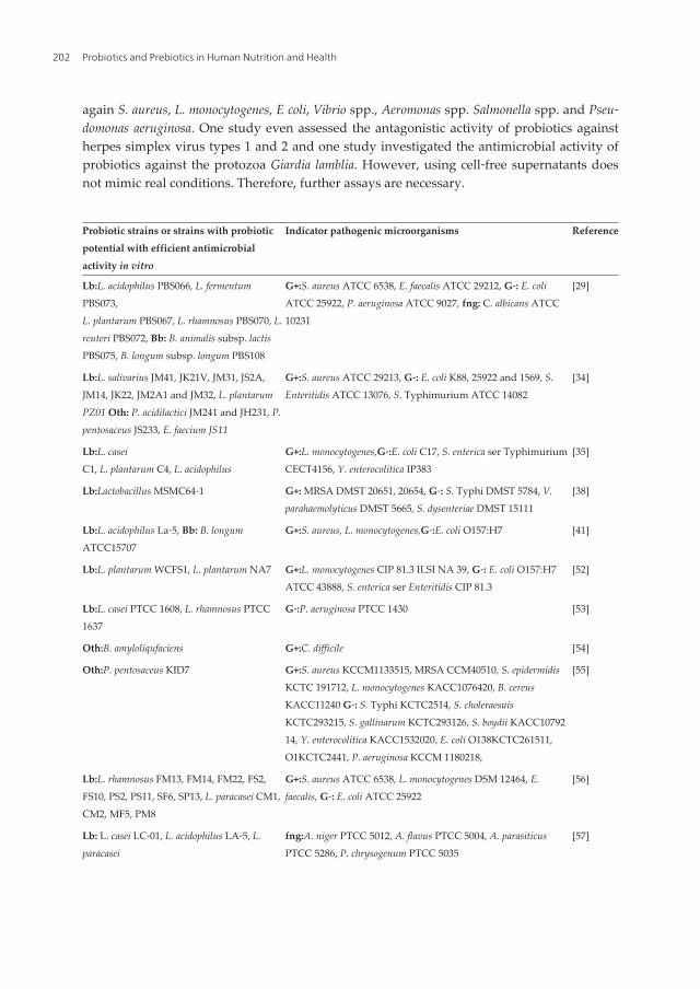
again S. aureus, L. monocytogenes, E coli, Vibrio spp., Aeromonas spp. Salmonella spp. and Pseu‐domonas aeruginosa. One study even assessed the antagonistic activity of probiotics againstherpes simplex virus types 1 and 2 and one study investigated the antimicrobial activity ofprobiotics against the protozoa Giardia lamblia. However, using cell‐free supernatants doesnot mimic real conditions. Therefore, further assays are necessary.
Probiotic strains or strains with probioticpotential with efficient antimicrobialactivity in vitro
Indicator pathogenic microorganisms Reference
Lb:L. acidophilus PBS066, L. fermentumPBS073,L. plantarum PBS067, L. rhamnosus PBS070, L.reuteri PBS072, Bb: B. animalis subsp. lactisPBS075, B. longum subsp. longum PBS108
G+:S. aureus ATCC 6538, E. faecalis ATCC 29212, G‐: E. coliATCC 25922, P. aeruginosa ATCC 9027, fng: C. albicans ATCC10231
[29]
Lb:L. salivarius JM41, JK21V, JM31, JS2A,JM14, JK22, JM2A1 and JM32, L. plantarumPZ01 Oth: P. acidilactici JM241 and JH231, P.pentosaceus JS233, E. faecium JS11
G+:S. aureus ATCC 29213, G‐: E. coli K88, 25922 and 1569, S.Enteritidis ATCC 13076, S. Typhimurium ATCC 14082
[34]
Lb:L. caseiC1, L. plantarum C4, L. acidophilus
G+:L. monocytogenes,G‐:E. coli C17, S. enterica ser TyphimuriumCECT4156, Y. enterocolitica IP383
[35]
Lb:Lactobacillus MSMC64‐1 G+: MRSA DMST 20651, 20654, G‐: S. Typhi DMST 5784, V.parahaemolyticus DMST 5665, S. dysenteriae DMST 15111
[38]
Lb:L. acidophilus La‐5, Bb: B. longumATCC15707
G+:S. aureus, L. monocytogenes,G‐:E. coli O157:H7 [41]
Lb:L. plantarum WCFS1, L. plantarum NA7 G+:L. monocytogenes CIP 81.3 ILSI NA 39, G‐: E. coli O157:H7ATCC 43888, S. enterica ser Enteritidis CIP 81.3
[52]
Lb:L. casei PTCC 1608, L. rhamnosus PTCC1637
G‐:P. aeruginosa PTCC 1430 [53]
Oth:B. amyloliqufaciens G+:C. difficile [54]
Oth:P. pentosaceus KID7 G+:S. aureus KCCM1133515, MRSA CCM40510, S. epidermidisKCTC 191712, L. monocytogenes KACC1076420, B. cereusKACC11240 G‐: S. Typhi KCTC2514, S. choleraesuisKCTC293215, S. gallinarum KCTC293126, S. boydii KACC1079214, Y. enterocolitica KACC1532020, E. coli O138KCTC261511,O1KCTC2441, P. aeruginosa KCCM 1180218,
[55]
Lb:L. rhamnosus FM13, FM14, FM22, FS2,FS10, PS2, PS11, SF6, SP13, L. paracasei CM1,CM2, MF5, PM8
G+:S. aureus ATCC 6538, L. monocytogenes DSM 12464, E.faecalis, G‐: E. coli ATCC 25922
[56]
Lb: L. casei LC‐01, L. acidophilus LA‐5, L.paracasei
fng:A. niger PTCC 5012, A. flavus PTCC 5004, A. parasiticusPTCC 5286, P. chrysogenum PTCC 5035
[57]
Probiotics and Prebiotics in Human Nutrition and Health202

Probiotic strains or strains with probioticpotential with efficient antimicrobialactivity in vitro
Indicator pathogenic microorganisms Reference
Lb:L. viridescens NRRL B‐1951 G+:L. monocytogenes CWD 1002, CWD 1198 [58]
Lb:L. plantarum S2 G+:S. aureus CMCC2607, G‐: E. coli CMCC44825,S. Typhimurium CMCC50115, S. flexneri CMCC51061
[59]
Oth:B. subtilis JQ302302, B. aerophilusJQ312663
G‐:A. hydrophila ATCC 49140, MTCC 1739, Aeromonas sp.JX136697, JX136698, A. enteropelogenes JX136699, P. rettgeri JX136696
[60]
Lb:L. acidophilus L‐1, L. bulgaricus 6, L.plantarum 24‐4B, L. fermentum 1, L. brevis, Bb:B. animalis subsp. lactis L‐3
G+:S. aureus, B. cereusG‐:E. coli
[61]
Lb:L. plantarum DK211, DK303; L. paracaseiDK215, L. sakei DK301
G+:S. aureus KCTC 3881, E. faecalis KCTC 2011, B. cereus KCTC3624
[62]
Lb: L. mesenteroides subsp. mesenteroides SD1,SD23, SF2, SF3
G+:S. aureus ATCC 25923, FRI 184, L. monocytogenes ATCC19115, G‐: E. coli ATCC43895, S. enterica ATCC 14028
[63]
Oth:L. mesenteroides MTCC 5442, B. subtilis G‐:V. cholerae [64]
Lb:L. fermentum M059, L. fermentum F‐6,Oth: W. cibaria 4213
G+:S. aureus ATCC 6538, B. cereus NCIM 245, B.subtilis ATCC 6633, G‐: E. coli ATCC 25922, S. Typhi 25,P. aeruginosa ATCC 27853
[65]
Lb:L. brevis DT24 G‐:E. coli MTCC 729 [66]
Lb:L. rhamnosus (4 strains) G‐:Helicobacter pylori [67]
Lb:L. plantarum, L. salivarius G+:S. aureus ATCC 6538S, C. perfringens ATCC 13124, G‐: E. coliATCC 8734, S. Enteritidis ATCC 13311, R. anatipestifer ATCC11845, P. multocida ATCC 43137
[68]
Lb:L. acidophilus P106, L. plantarum P164 Prs:Giardia lamblia [69]
Lb:L. casei G‐: S. flexneri, S. sonnei [70]
Lb:L. helveticus PJ4, L. plantarum PJ7 G+:S. aureus MTCC737, G‐: E. coli MTCC443, S.Typhimurium MTCC733, S. fleksneri MTCC1457,P. aeruginosa MTCC1688
[71]
Lb:L. salivarius K35, K43 G+:S. mutans ATCC 25175 [72]
Oth:B. amyloliquefaciens KATMIRA1933 Vr: Herpes simplex virus types 1 and 2 [73]
Oth:B. amyloliquefaciens FPTB16 G‐:E. tarda, A. hydrophila, V. harveyi, V. parahaemolyticus [74]
Where probiotics are divided as follows: lb: lactobacilli; bb: Bifidobacterium; oth: other; and pathogens are divided asfollows: G+: Gram positive; G−: Gram negative; fng: fungi; prs: parasite.
Table 2. A selection of assays published since 2013 of successful antimicrobial activity of chosen probiotics using theagar well diffusion assay or the paper disc assay on chosen pathogenic microorganisms.
Antimicrobial Effect of Probiotics against Common Pathogenshttp://dx.doi.org/10.5772/63141
203

4.3. Recent results of selected co‐culturing assays
The influence of the potential pathogenic bacteria P. aeruginosa ATCC 27853 against variouscombinations of probiotic supplements and the kefir microbiota was investigated using theco‐culturing method [6]. One multispecies, one oligospecies and one monospecies probioticsupplement as well as kefir microbiota from kefir grains originally from the Caucasianmountains were used. The multispecies supplement contained L. acidophilus (NIZO 3678;NIZO 3887), L. paracasei NIZO 3672, L. plantarum NIZO 3684, L. rhamnosus NIZO 3689, L.salivarius NIZO 3675, B. bifidum NIZO 3804, B. lactis NIZO 3680 and E. faecium NIZO 3886. Theoligospecies supplement contained L. acidophilus LA‐5, B. infantis BB‐12 and E. faecium. Themonospecies supplement contained L. reuteri DSM 17938. Co‐culturing was conducted byadding 1 mL of probiotic samples and 1 mL of overnight P. aeruginosa suspension in 40 mLpreviously sterilized bovine milk. Samples were incubated for 4 days at 25°C with agitation(250/min). After incubation, serial 10‐fold dilutions were prepared for all samples and the P.aeruginosa populations were enumerated on cetrimide agar with added glycerol. It was foundthat the P. aeruginosa population in milk without probiotics reached an average of 9.2 log/mL,whilst the P. aeruginosa population in milk reached 5.2, 8.3, 8.3 and 5.0 for the samples withthe multispecies, oligospecies and monospecies probiotic supplement as well as the kefirmicrobiota respectively. This research thus found that the decrease of the potential pathogenP. aeruginosa was dependent on the type of probiotic microbiota and that both multispeciesmicrobiota (multispecies probiotic supplement kefir microbiota) with a much more diversepopulation than the other two samples (oligospecies probiotic supplement and the monospe‐cies supplement) exhibited an efficient synergistic effect [7]. Both samples also exhibited ahigher increase in the total population of anaerobic microorganisms after fermentation withP. aeruginosa thus indicating that a more successful quorum‐sensing regulatory network wasestablished and yielded antagonistic effects against the potential pathogen P. aeruginosa.
Tharmaraj and Shah [16], as already mentioned in the previous section, also investigated theinhibition effect of various probiotics against chosen pathogenic and spoilage bacteria withthe co‐culturing method. Briefly, 9 mL of reconstituted skim milk was inoculated with 1 mLof overnight culture of probiotic bacteria and 0.1 mL of pathogenic or spoilage bacteria. Themedium was mixed well and incubated at 37°C for 24 h. / were counted on nutrient agar andthe log population calculated (Table 6). All four pathogenic bacteria were inhibited by allprobiotic strains tested to varying degrees. On average, the probiotic bacteria reduced thepopulation of pathogenic bacteria by 2.8 log units. B. cereus was inhibited to a greater degreeby all probiotic bacteria and strains than other pathogenic bacteria. On average, the inhibito‐ry effect of all probiotic bacteria and strains was the weakest against E. coli. S. aureus wasinhibited to a greater degree by B. animalis and L. rhamnosus than the other probiotic bacteria.
Ratsep et al. [15] published their research of a microtitre plate assay on the antimicrobial effectof L. plantarum (five strains: N11, N27, N33, N44 and E) supernatant against various C. difficilestrains (six clinical isolates from Norwegian patients, six clinical isolates from Estonian patientsand two reference strains: VPI 10463 and M13042). Overnight C. difficile cultures were addedto BHI broth with a density according to McFarland 3.0. Various reaction mixes were pre‐pared (natural (acidic), neutral (pH 6.0) and neutral, heated for 20 min at 100°C) and incubat‐
Probiotics and Prebiotics in Human Nutrition and Health204

ed under anaerobic conditions for 48 h at 37°C. The optical density at 620 mm was measuredat the beginning of incubation and after 48 h and the suppressive activity of L. plantarum strainscalculated as a percentage of inhibition. The highest inhibitions of C. difficile growth were inthe samples of heated neutralized supernatants and the lowest in cases of the neutralizedsamples. There was statistically higher inhibition in heated neutralized supernatants versusneutralized supernatants of N11 and E56 lactobacilli strains. When comparing antagonisticactivity of L. plantarum strains, there was a relevant difference only between N11 and N33strains in the samples of heated neutralized supernatants. The neutralization of supernatantdid not reduce its inhibitory effect. Thus, lowering the pH of the environment is not the mainmechanism in inhibition of C. difficile by lactobacilli. Also heating of supernatant did not reduceits activity; thus, some thermostable compounds may be involved in the inhibition. The above‐mentioned authors [15] also performed the co‐culturing assay with the L. plantarum strains andC. difficile strains by inoculating 50 mL of brain heart infusion broth with 50 µL of lactobacillisuspension and 50 µL of C. difficile suspension and incubating under anaerobic conditionsfor 48 h at 37°C. After incubation, serial 10‐fold dilutions were prepared and the C. difficilepopulations were enumerated on fastidious anaerobe agar. It was found that the five L.plantarum strains (N11, N27, N33, N44 and E) were able to inhibit the growth of the 14 C. difficilestrains (six clinical isolates from Norwegian patients and Estonian patients and two refer‐ence strains: VPI 10463, M13042) in the co‐culture incubation as the average log cfu/mL after48 h was 3.0, whereas the average log cfu/mL of the C. difficile strains alone was 7.0.
Another important co‐culturing method is the use of cell lines as noted in the research by Abdelet al. [31] of 12 lactobacilli isolates interfering with the adherence and invasion of S. Typhi 66using kidney epithelial cell line Vero (ATCC CCl‐81). The same authors also investigated thisinterference with the co‐culturing assay in MRS broth. It was found that nine lactobacilliisolates inhibited the growth of S. Typhi in the co‐culturing assay. Nine lactobacilli isolateswere also successful in achieving a >50% inhibition of adherence of S. Typhi isolate (SS6) toVero cells.
Assays published since 2013 of antimicrobial activity of chosen probiotics using various co‐culturing assays on chosen pathogenic microorganisms are noted in Table 3. The results weresimilar to the results of spot‐on lawn and agar well diffuse assays.
Probiotic strains or strains with probiotic potentialwith efficient antimicrobial activity in vitro
Indicator pathogenic microorganisms Reference
Lb:L. plantarum FH185 G+:S. aureus,G‐:S. Typhimurium [9]
Lb:L. acidophilus La‐5, Bb: B. longum ATCC15707 G+:S. aureus, L. monocytogenes,G‐:E. coli O157:H7 [41]
Lb:L. acidophilus P106, L. plantarum P164 Prs:Giardia lamblia [69]
Lb:L. rhamnosus GR‐1, L. reuteri RC‐14 Fng:C. glabrata [75]
Lb:L. helveticus KLDS 1.8701 G+:S. aureus ATCC 25923, L. monocytogenes ATCC19115, G‐: E. coli O157:H7 ATCC 43889, S.Typhimurium ATCC 14028
[76]
Antimicrobial Effect of Probiotics against Common Pathogenshttp://dx.doi.org/10.5772/63141
205

Probiotic strains or strains with probiotic potentialwith efficient antimicrobial activity in vitro
Indicator pathogenic microorganisms Reference
Lb:L. reuteriOth:B. subtilis MA139 G‐:E. coli K88 [77]
Lb:L. plantarum C014 G‐:A. hydrophila TISTR 1321 [78]
Lb:L. acidophilus, Oth:Pediococcus G‐:S. Enteritidis 13A [79]
Lb:L. paracasei CNCM I‐4034, L. rhamnosus CNCMI‐4036, Bb: B. breve CNCM I‐4035
G‐:E. coli ETEC CECT 501, S. Typhimurium CECT443 S. Typhi CECT 725, S. sonnei CECT 457
[80]
Lb:L. fermentum 907, Bb: B. longum 1011 G‐:E. coli O157:H7, E. coli O86 [81]
Where probiotics are divided as follows: lb: lactobacilli; bb: Bifidobacterium; and pathogens are divided as follows: G+:Gram positive; G−: Gram negative; fng: fungi; prs: parasite.
Table 3. A selection of assays published since 2013 of successful antimicrobial activity of chosen probiotics using theco‐culturing assay on chosen pathogenic microorganisms.
5. Recent results of in vivo antimicrobial/antagonistic assays for variousprobiotic strains
5.1. Recent results of determining the in vivo antimicrobial assays using animal models
In the research by Mazaya et al. [36], both in vitro and in vivo studies were conducted. Thesignificant in vitro antimicrobial activity (no method specified) of two lactobacillus strainsisolated from Egyptian dairy products (L. plantarum LA5 and L. paracasei LA7) was foundagainst several potential pathogens: S. aureus ATCC 25923, B. subtilis ATCC 23857, M. luteusATCC 21882, P. aeruginosa ATCC 27853 and S. Typhi. In vivo assays were also conducted on5‐week‐old male mice, divided into six groups (10 mice/group). Animals within differenttreatment groups were treated daily for 8 days as follows: Group 1, untreated control; Group 2,animals challenged with single inoculation S. Typhi (200 µL aliquot of 1X 10⁸/P.O); Group 3,animals treated orally with L. plantarum (LA5) (200 µL aliquot of 1X 10⁸/P.O) for 7 days;Group 4, animals treated orally with L. paracasei (LA7) (200 µL aliquot of 1X 10⁸/P.O) for 7days; Group 5, animals challenged with single inoculation S. Typhi, then treated with LA5 fornext 7 days; Group 6, animals challenged with single inoculation S. Typhi and treated withLA 7 for next 7 days. Administration of LA5 or LA7 counteracted the pathogenic effectresulting from Salmonella infection. The lactobacilli succeeded to get rid of salmonellosis basedon its phagocytic and immunostimulant activity against typhoid antigen.
Lazarenko et al. [82] conducted an in vitro and in vivo assay to determine anti‐staphylococcalactions of certain probiotic cultures (L. casei IMV B‐7280, L. acidophilus IMV B‐7279; B. longumVK1 and B. bifidum VK2). In vitro assay using perpendicular strokes yielded antagonisticactivity against all three strains of S. aureus (209‐P, 43, 8325‐4). The in vivo study with S. aureus8325‐4 on mice showed that the combination of probiotics (L. casei IMV B‐7280, B. longum VK1and B. bifidum VK2) was most successful as after day 9 no colonies of S. aureus 8325‐4 werefound in the vagina of the mice.
Probiotics and Prebiotics in Human Nutrition and Health206

In the study by Bujalance et al. [35], a lack of correlation between in vitro and in vivo meth‐ods of the antimicrobial activity of probiotic lactobacilli against enteropathogenic bacteria wasdetermined. In this study, the in vitro assay using the agar spot test showed that 20 strains ofprobiotic lactobacilli successfully inhibited Y. enterocolitica, S. enterica ser Typhimurium and L.monocytogenes. However, in the in vivo study using mouse models the selected strains (L. caseiC1 and L. plantarum C4) lacked protective effects against S. Typhimurium. Similar conclu‐sions of finding no antagonism in vivo are noted in the study by Bratz et al. [83]. Anotherstudy [79] found that the tea Yerba mate exhibited antagonistic activity in vitro but as a feedadditive did not reduce S. Enteritidis colonization in vivo in broiler chickens. These studiesprove that successful in vitro assays do not necessarily mean that the chosen microorganismwith probiotic properties will be successful in real conditions. On the other hand, in vivo animalstudies do not automatically prove antagonism in humans or other species. Therefore, thejustification of using vertebrate animal models is questionable [32]. Gupta et al. [84] usedDrosophila melanogaster commonly known as the “fruit fly,” instead of a vertebrate animalmodel. It is a eukaryotic organism and is considered an alternative in the drug discoveryprocess, mainly because the key physiological processes are well conserved from fly tohumans. Moreover, a short life cycle, distinct developmental stages, easy cultivation, numer‐ous offspring and a strong cytogenetic/genetic background make Drosophila a model organ‐ism to study many biological processes including toxicity testing. Zhou et al. [30] used porcineneonatal jejunal epithelial cell lines (IPEC‐J2) for in vitro assay and worms (Caenorhabditiselegans) for in vivo testing of lactobacilli isolates against enterotoxigenic E. coli
Probiotic strains or strains with probioticpotential with efficient antimicrobial activityin vivo using animal models
Indicator pathogenic microorganisms Reference
Lb:L. reuteri CL9, K16, K67 and S33 G‐:E. coli O149: K88 JG280 [30]
Lb:L. salivarius JM32, L. plantarum PZ01 Oth:P. acidilactici JH231
G‐:S. Enteritidis ATCC 13076 [34]
Lb:L. plantarum LA5 and L. paracasei LA7 G‐:S. Typhi [36]
Oth:B. amyloliqufaciens G+:C. difficile [54]
Lb:L. plantarum C014 G‐:A. hydrophila TISTR 1321 [78]
Lb:L. casei B–7280, Bb: B. longum VK1, B. bifidumVK2 G+: S. aureus8325–4 [82]
Lb:L. plantarum LR/14 Ins:Drosophila melanogaster [84]
Lb:L. acidophilus CH1 Prs:Enterocytozoon bieneusi [85]
Oth:Pseudoalteromonas sp. G‐:V. harveryi ATCC 14126 [86]
Where probiotics are divided as follows: lb: lactobacilli; bb: Bifidobacterium; oth: other; and pathogens are divided asfollows: G+: Gram positive; G−: Gram negative; fng: fungi; prs: parasite; ins: insect.
Table 4. A selection of assays published since 2013 of successful antimicrobial activity of chosen probiotics using the invivo animal models on chosen pathogenic microorganisms.
Antimicrobial Effect of Probiotics against Common Pathogenshttp://dx.doi.org/10.5772/63141
207

The most recent in vivo antagonistic assays using animal models are noted in Table 4. Theseresults confirm the strain specific antagonistic activity of chosen probiotics.
5.2. Recent results of determining the in vivo antimicrobial assays using clinical trials
Most important research on the antagonistic effect of probiotics are clinical trials, however onlya few well conducted clinical studies have been reported. Most clinical studies include thecomparison of antibiotic therapy with adjuvant probiotic therapy. In the study by Dore et al.[87], in this prospective, single centre, open label pilot study, patients scheduled for upperendoscopy for any reason and found to be positive for H. pylori infection were invited to enter.The intervention consisted of L. reuteri (DSM 17938, Reuflor, BioGaia AB, Sweden) 108 cfu/tablet plus pantoprazole (proton pump inhibitor) 20 mg twice a day. A 76% decrease in ureaseactivity was observed. The absence of a control group with pantoprazole without L. reuterihowever prevents any definite conclusion.
In the study by Pendharkar et al. [88] the clinical outcome for women conventionally treatedfor bacterial vaginosis and yeast infection with probiotics bacilli was investigated. This studyis an example of the antibiotic therapy with adjuvant probiotic therapy. In the clinical trial,women were recruited in three groups as follows: women with bacterial vaginosis receivingclindamycin and metronidazole treatment together with a prolonged administration ofEcoVag® (containing L. rhamnosus DSM 14870 and L. gasseri DSM 14869) for 10 consecutivedays after each antibiotic treatment followed by weekly administration of capsules for nextfour months, women with recurrent vulvovaginal candidiasis receiving extended flucona‐zole and EcoVag® treatment, and women receiving extended fluconazole treatments only.The 6‐ and 12‐month cure rates for bacterial vaginosis were 67 %. The 6‐ and 12‐month curerates for vulvovaginal candidiasis were 100 and 89% in women receiving fluconazole andEcoVag®, and 100 and 70% in women receiving fluconazole only. The study suggests that thetreatment with antibiotics or anti‐fungal medication in combination with EcoVag® capsulesprovide long‐term cure against bacterial vaginosis and R‐VVC.
Some of the most recent in vivo antagonistic clinical trials are noted in Table 5. These resultsconfirm that adjuvant therapy with antibiotics and chosen probiotics enhances the antagonis‐tic activity.
Probiotic strains with efficientantimicrobial activity using in vivoclinical trials and other therapies
Pathogenicmicroorganisms ortreated disease
Basic therapy Type oftrial
Reference
Lb:L. reuteri DSM 17938 G+:H. pylori Proton pump inhibitor:pantoprazole
CT [87]
Lb:L. rhamnosus DSM 14870, L. gasseriDSM 14869
Bc: not specified, Fng:Candida spp.
Antibiotics: Clindamycin,metronidazole, fluconazole
CT [88]
Lb:L. rhamnosus GG (LGG), Bb: B.bacterium BB‐12
G+:H. pylori Standard triple H. pylorieradication therapy with
DBRCT [89]
Probiotics and Prebiotics in Human Nutrition and Health208

Probiotic strains with efficientantimicrobial activity using in vivoclinical trials and other therapies
Pathogenicmicroorganisms ortreated disease
Basic therapy Type oftrial
Reference
antibiotics: omeprazole,pantoprazole, clarithromycin,amoxicillin, or metronidazole
Lb:L. rhamnosus GG (LGG), L.acidophilus La‐5, Bb: B. bacterium BB‐12
Dis: AAD Antibiotics: broad spectrumoral antibiotics
DBRCT [90]
Lb:L. acidophilus, L. paracaseiBb:B. lactisAt: bismuth
G+:H. pylori Antibiotics: broad spectrumoral antibiotics: lansoprazole,amoxicillin, clarithromycin
BDRCT [91]
Lb:L. casei DN‐114001 Dis: AAD Antibiotics: broad spectrumoral antibiotics
CT [92]
Lb:L. acidophilus, l. rhamnosus, L.acidophilus, L. bulgaricus, Bb:B.bifidusOth:S. Thermophilus
Dis: bacterial vaginosis Antibiotic: metronidazole CT [93]
Where probiotics are divided as follows: lb: lactobacilli; bb: Bifidobacterium; oth: other; pathogens are divided as follows:bc: bacteria; G+: Gram positive; G−: Gram negative; fng: fungi; and clinical trials are divided as follows: DBRCT: double‐blinded randomized clinical trial; CT: clinical trial; AT: additional therapy; Dis: disease; AAD: antibiotic associateddiarrhoea.
Table 5. A selection of published since 2013 of antimicrobial activity of chosen probiotics using in vivo clinical trials onchosen pathogenic microorganisms or treatment of diseases.
6. Discussion and conclusions
The antimicrobial ability of probiotics is a very important trait and includes the production ofantimicrobial compounds, competitive exclusion of pathogens, enhancement of the intesti‐nal barrier function and others. Usually, probiotic strains produce more than one antimicro‐bial substance that may act synergistically, increasing the spectrum of targetedmicroorganisms. This property may be desirable as long as this antimicrobial spectrum isrestricted to pathogenic microorganisms but it cannot be excluded that it will not affect thenormal microbiota of the gut or other microbiotas as well [94]. The results show that probiot‐ic properties are strain dependent and that strain identification is imperative [3].
Probiotic candidates have been accessed from very diverse habitats including faeces of breast‐fed human infants [65, 69, 80, 85, 95], faeces of healthy adults [9, 15, 65, 70], faeces of elderly[81], faeces of children [25, 96], breast milk [42], human saliva [52], vaginal isolates of healthywomen [66, 75], various fermented foods or beverages including raw or fermented milk [23,35, 44], kefir [97], cheese [51, 56, 98], whey [99], yogurt [16, 41], dahi [100, 101], other dairyproducts [25, 36, 61], sourdough [102], sausages [17], fermented meat [24], kimchi [10, 62],maize [25, 59], fermented olives [103], Yerba mate [79], ragi [64], soy sauce [86], soil [104], as
Antimicrobial Effect of Probiotics against Common Pathogenshttp://dx.doi.org/10.5772/63141
209

well as animal origin including rat faeces [71], geese [68], calves [105], pigs [45], fish [39, 60,63, 78] and other seafood [40, 43, 46] and many others.
By far, the most commonly investigated probiotic were bacteria of the genus Lactobacillus (L.plantarum, L. acidophilus, L. fermentum, L. casei, L. paracasei and L. reuteri). The genus Bifidobac‐terium and other probiotic microorganisms (Lactococcus, Pediococcus, Enterococcus, Bacillus andSaccharomyces) have been also been investigated, but to a somewhat lesser extent. Studies werealso conducted on known probiotics from various tissue type collections. The most commonpathogens used to test the antagonistic activity of probiotics were different strains of S. aureus,E. faecium, E. faecalis, L. monocytogenes, E. coli, various Salmonella, Vibrio and Yersinia spp.,and P. aeruginosa.
The antimicrobial activity of probiotic microorganisms has a very wide area applicationincluding adjuvant therapy to antibiotic consumption or for correcting dysbiosis of thegastrointestinal tract microbiome due to diarrhoea [37, 38, 106], antagonistic activity in humansagainst urinary tract infections [26, 66, 75], eradicating H. pylori infections [87], nosocomialinfections [15, 96], dental biofilm formation [72], lowering serum cholesterol [10, 55], treatingfevers [31], as well as in the agro‐food industry for manufacturing fermented products [44, 62,107], preventing food spoilage [16, 23, 41], as food additives for functional foods [50, 56, 57,59, 67, 97], as prophylactic agents, adjuvants or alternatives to antibiotic therapies to antibiot‐ic therapy in poultry [34, 47, 68], cattle [105, 108], pigs [45], fish [74, 78] and other livestockindustry [104], just to name a few.
The process of determining antimicrobial properties of probiotic is complex and includes invitro assays, in vivo models or substitute models, clinical studies, metagenomic analyses andmathematical modelling. Only after all these steps are completed, a probiotic candidate canbe identified as such [109]. In vitro studies are the most represented. Although, they are a crucialstep in selecting probiotic candidates, they are only the first step as efficient antimicrobialactivity via in vitro studies does not necessarily mean that the antimicrobial activity is presentin in vivo assays. Therefore, further research methods (double‐blinded randomized clinicaltrials) are necessary to prove the important antimicrobial trait of probiotic candidates. As notedin Section 5.2, there are only a few well‐conducted published clinical studies. Most clinicalstudies include the comparison of antibiotic therapy with adjuvant probiotic therapy which isan important aim of probiotic consumption.
There is a clear need for more elaborate assays that would better represent the complexinteractions between the probiotics and the host microbiome to understand the consequen‐ces of the in situ production of antimicrobials by the former [94]. Another important fact is thatprobiotics are often found to have higher antagonist activity as multispecies groups [6, 7, 26].Quorum sensing among probiotics is also an important factor; however, quorum‐sensingstudies among probiotics are sparse. It is well known that microorganisms coordinatecollective behaviour in response to environmental challenges using sophisticated intercellu‐lar communication networks and that they are not limited to communication within their ownspecies but are capable of intercepting messages and coerce cohabitants into behaviouralmodifications [110], therefore probiotics are included. Although all these facts make re‐
Probiotics and Prebiotics in Human Nutrition and Health210

search of the antimicrobial/antagonistic activity of probiotics even more complex, it alsopresents a great opportunity for future research.
Author details
Sabina Fijan
Address all correspondence to: [email protected]
Faculty of Health Sciences, University of Maribor, Maribor, Slovenia
References
[1] Peterson J, Garges S, Giovanni M, McInnes P. The NIH human microbiome project.Genome Res. 2009;19:2317–2323.
[2] Rijkers GT, De Vos WM, Brummer RJ, Morelli L, Corthier G, Marteau P. Health benefitsand health claims of probiotics: bridging science and marketing. Br J Nutr.2011;106:1291–1296.
[3] Fijan S. Microorganisms with claimed probiotic properties. Int J Environ Res PublicHealth. 2014;11:4745–4767.
[4] Food and Agriculture Organization of the United Nations/World Health Organiza‐tion FAO/WHO. Health and Nutritional Properties of Probiotics in Food includingPowder Milk with Live Lactic [Internet]; 2001. Available from: http://www.who.int/foodsafety/publications/fs_management/en/probiotics.pdf [Accessed: 11.1.2016]
[5] Working Group on Drafting Guidelines for the Evaluation of Probiotics in Food.Guidelines for the Evaluation of Probiotics in Food; Joint FAO/WHO. London, Ontario,Canada: Food and Agriculture Organization of the United Nations/World HealthOrganization FAO/WHO; 2002.
[6] Fijan S. Influence of the growth of Pseudomonas aeruginosa in milk fermented bymultispecies probiotics and Kefir microbiota. J Probiotics Health. 2016;3:136. DOI:10.4172/2329‐8901.1000136.
[7] Koning CJ, Jonkers D, Smidt H, Rombouts F, Pennings HJ, Wouters E, Stobberingh E,Stockbrügger R. The effect of a multispecies probiotic on the composition of the faecalmicrobiota and bowel habits in chronic obstructive pulmonary disease patients treatedwith antibiotics. Br J Nutr. 2010;103:1452–1460.
[8] He F, Morita H, Ouwehand AC. Bifidobacteria and lactobacilli exhibited differentmitogenic activity on murine splenocytes. Int J Probiotics Prebiotics. 2006;1:77–82.
Antimicrobial Effect of Probiotics against Common Pathogenshttp://dx.doi.org/10.5772/63141
211
[9] Park S‐Y; Lim S‐D. Probiotic characteristics of Lactobacillus plantarum FH185 isolatedfrom human feces. Korean J Food Sci. 2015;35(5):615–621.
[10] Choi EA, Chang HC. Cholesterol‐lowering effects of a putative probiotic strainLactobacillus plantarum EM isolated from kimchi. LWT – Food Sci Technol. 2015;62(1):210–217.
[11] Sanders ME. Probiotics: definition, sources, selection, and uses. Clin Infect Dis.2008;46(2):S58–S61. DOI: 10.1086/523341.
[12] Sanders ME. Summary of probiotic activities of Bifidobacterium lactis HN019. J ClinGastroenterol. 2006; 40:776–783.
[13] Saxelin M, Tynkkynen S, Mattila‐Sandholm T, de Vos WM. Probiotic and otherfunctional microbes: from markets to mechanisms. Curr Opin Biotechnol. 2005;16:204–211.
[14] Millette M, Luquet FM, Lacroix M. In vitro growth control of selected pathogens byLactobacillus acidophilus—and Lactobacillus casei‐fermented milk. Lett Appl Microbiol.2007;44:314–319.
[15] Ratsep M, Naaber P, Koljalg S, Smidt I, Shkut E, Sepp E. Effect of Lactobacillus planta‐rum strains on clinical isolates of Clostridium difficile in vitro. J Probiotics Health.2014;2:119.
[16] Tharmaraj N, Shah N P. Antimicrobial effects of probiotics against selected pathogen‐ic and spoilage bacteria in cheese‐based dips. Int Food Res J. 2009;16:261–276.
[17] Ammor S, Tauveron G, Dufour E, Chevallier I. Antibacterial activity of lactic acidbacteria against spoilage and pathogenic bacteria isolated from the same meat small‐scale facility. Food Control. 2006;17:454–461.
[18] Arqués JL, Rodríguez E, Langa S, Landete JM, Medina M. Antimicrobial activity oflactic acid bacteria in dairy products and gut: effect on pathogens. BioMed Res Int.2015;2015:9. DOI: 10.1155/2015/584183.
[19] Šušković J, Kos B, Beganovič J, Leboš Pavunc A, Habjanič K, Matošić S. Antimicrobi‐al activity—the most important property of probiotic and starter lactic acid bacteria.Food Technol Biotechnol. 2010;48(3):296–307.
[20] Tagg JR, Dajani AS, Wannmaker LW. Bacteriocines of gram positive bacteria. BacteriolRev. 1976;40:722–756.
[21] Hoover DG, Harlander SK. Screening methods for detecting bacteriocin activity. In:Hoover DG, Steenson LR, editors. Bacteriocins of lactic acid bacteria. San Diego:Academic Press; 1993. p. 23–29.
[22] Parente E, Brienza C, Moles M, Ricciardi A. A comparison of methods for the meas‐urement of bacteriocin activity. J Microbiol Methods. 1995;22:95–108.
Probiotics and Prebiotics in Human Nutrition and Health212

[23] Soomro AH, Musad T, Sammiand S, Rathore HA. Comparison of different methods fordetection of antimicrobial activity of Lactobacillus spp. Pakistan J Zool. 2007;39(4):265–268.
[24] Pilasombut K, Rumjuankiat K, Ngamyeesoon N, Duy le ND. In vitro characterizationof bacteriocin produced by lactic acid bacteria isolated from Nem Chua, a traditionalVietnamese fermented pork. Korean J Food Sci Anim. 2015;35(4):473–478.
[25] Jacobsen CN, Rosenfeldt Nielsen V, Hayford AE, Møller PL, Michaelsen KF, Paerre‐gaard A, Sandström B, Tvede M, Jakobsen M. Screening of probiotic activities of forty‐seven strains of Lactobacillus spp. by in vitro techniques and evaluation of thecolonization ability of five selected strains in humans. Appl Environ Microbiol.1999;65:4949–4956.
[26] Chapman CM1, Gibson GR, Rowland I. In vitro evaluation of single‐ and multi‐strainprobiotics: inter‐species inhibition between probiotic strains, and inhibition ofpathogens. Anaerobe. 2012;18(4):405–413. DOI: 10.1016/j.anaerobe.2012.05.004.
[27] Fijan S, Šostar Turk, S. Influence of the microbial community in Kefir Grains on thesurvival and growth of Staphylococcus aureus in Milk. Abstracts from the meeting: 7thProbiotics, prebiotics & new foods, September 8‐10 2013; Rome, Iniversità Urbaniana.J Clin Gastroenterol. 2014;48(1):S113–S114.
[28] Fijan S, Trunk M, Šostar Turk S. Influence of the microbial community in kefir grainson the survival and growth of Candida albicans in milk. In: International scientificconference on probiotics and prebiotics, Budapest, June 24–26, 2014. Kysucké NovéMesto: Pamida; 2014. p. 89.
[29] Presti I, D'Orazio G, Labra M, La Ferla B, Mezzasalma V, Bizzaro G, Giardina S,Michelotti A, Tursi F, Vassallo M, Di Gennaro P. Evaluation of the probiotic proper‐ties of new Lactobacillus and Bifidobacterium strains and their in vitro effect. ApplMicrobiol Biotechnol. 2015;99(13):5613–5626. DOI: 10.1007/s00253‐015‐6482‐8.
[30] Zhou M, Zhu J, Yu H, Yin X, Sabour PM, Zhao L, Chen W, Gong J. Investigation intoin vitro and in vivo models using intestinal epithelial IPEC‐J2 cells and Caenorhabditiselegans for selecting probiotic candidates to control porcine enterotoxigenic Escheri‐chia coli. J Appl Microbiol. 2014;117(1):217–226. DOI: 10.1111/jam.12505.
[31] Abdel‐Daim A, Hassouna N, Hafez M, Ashor MS, Aboulwafa MM. Antagonisticactivity of Lactobacillus isolates against Salmonella typhi in vitro. Biomed Res Int.2013;2013:680605. DOI: 10.1155/2013/680605.
[32] Cencič A. Can functional cell models replace laboratory animals in biomedicalresearch? J Bioanal Biomed. 2012;4:e105. DOI: 10.4172/1948‐593X.1000e105.
[33] Cencič A, Langerholc T. Functional cell models of the gut and their applications in foodmicrobiology—a review. Int J Food Microbial. 2010;141:S4–S14. DOI: 10.1016/j.ijfood‐micro.2010.03.026.
Antimicrobial Effect of Probiotics against Common Pathogenshttp://dx.doi.org/10.5772/63141
213

[34] Feng J, Wang L, Zhou L, Yang X, Zhao X. Using in vitro immunomodulatory proper‐ties of lactic acid bacteria for selection of probiotics against Salmonella infection in broilerchicks. PLoS One. 2016;11(1):e0147630. DOI: 10.1371/journal.pone.0147630.
[35] Bujalance C, Jiménez‐Valera M, Moreno E, Ruiz‐López MD, Lasserrot A, Ruiz‐BravoA. Lack of correlation between in vitro antibiosis and in vivo protection againstenteropathogenic bacteria by probiotic lactobacilli. Res Microbiol. 2014;165(1):14–20.DOI: DOI: 10.1016/j.resmic.2013.10.006.
[36] Mazaya B, Hamzawy MA, Khalil MA, Tawkol WM, Sabit H. Immunomodulatory andantimicrobial efficacy of Lactobacilli against enteropathogenic infection of Salmonellatyphi: In‐vitro and in‐vivo study. Int J Immunopathol Pharmacol. 2015;28(4):469–478.DOI: 10.1177/0394632015592099.
[37] Bendali F1, Madi N, Sadoun D. Beneficial effects of a strain of Lactobacillus paracaseisubsp. paracasei in Staphylococcus aureus‐induced intestinal and colonic injury. Int JInfect Dis. 2011;15(11):e787–e794. DOI: 10.1016/j.ijid.2011.07.003.
[38] Chimchang J, Theparee T, Ladda B, Tanasupawat S, Wongsatayanon BT, Taweechoti‐patr M. Antimicrobial properties of a potential probiotic Lactobacillus from Thainewborn feces. J Med Assoc Thai. 2015;98(9):S116–S122.
[39] Pinpimai K, Rodkhum C, Chansue N, Katagiri T, Maita M, Pirarat N. The study on thecandidate probiotic properties of encapsulated yeast, Saccharomyces cerevisiae JCM 7255,in Nile Tilapia (Oreochromis niloticus). Res Vet Sci. 2015;102:103–111. DOI: 10.1016/j.rvsc.2015.07.021.
[40] Liu XF, Li Y, Li JR, Cai LY, Li XX, Chen JR, Lyu SX. Isolation and characterisation ofBacillus spp. antagonistic to Vibrio parahaemolyticus for use as probiotics in aquacul‐ture. World J Microbiol Biotechnol. 2015;31(5):795–803. DOI: 10.1007/s11274‐015‐1833‐2.
[41] El‐Kholy AM, El‐Shinawy SH, Meshref AMS, Korny AM. Screening of antagonisticactivity of probiotic bacteria against some food‐borne pathogens. J Appl EnvironMicrobiol. 2014;2(2):53–60.
[42] Shokryazdan P,Sieo CC, Kalavathy R, Liang JB, Alitheen NB, Jahromi MF, Ho YW.Probiotic potential of Lactobacillus strains with antimicrobial activity against somehuman pathogenic strains. BioMed Res Int. 2014;2014:16. DOI: http://dx.DOI.org/10.1155/2014/927268.
[43] Muñoz‐Atienza E, Araújo C, Magadán S, Hernández PE, Herranz C, Santos Y, CintasLM. In vitro and in vivo evaluation of lactic acid bacteria of aquatic origin as probiot‐ics for turbot (Scophthalmus maximus L.) farming. Fish Shellfish Immunol. 2014;41(2):570–580. DOI: 10.1016/j.fsi.2014.10.007.
Probiotics and Prebiotics in Human Nutrition and Health214

[44] Mami A, Kerfouf A, Kihal M. Study of the antimicrobial and probiotic effect ofLactobacillus Plantarum isolated from raw goat's milk from the region of WesternAlgeria. Int J Sci Basic Appl Res. 2014;13(1):18–27.
[45] Han SK, Shin MS, Park HE, Kim SY, Lee WK. Screening of bacteriocin‐producingEnterococcus faecalis strains for antagonistic activities against Clostridium perfrin‐gens. Korean J Food Sci Anim Resour. 2014;34(5):614–621.
[46] Wu HJ, Sun LB, Li CB, Li ZZ, Zhang Z, Wen XB, Hu Z, Zhang YL, Li SK. Enhance‐ment of the immune response and protection against Vibrio parahaemolyticus byindigenous probiotic Bacillus strains in mud crab (Scylla paramamosain). Fish ShellfishImmunol. 2014;41(2):156–162. DOI: 10.1016/j.fsi.2014.08.027.
[47] Menconi A, Kallapura G, Latorre JD, Morgan MJ, Pumford NR, Hargis BM, Tellez G.Identification and characterization of lactic acid bacteria in a commercial probioticculture. Biosci Microbiota Food Health. 2014;33(1):25–30. DOI: 10.12938/bmfh.33.25.
[48] Belicova A, Mikulašova M, Dušinsky R. Probiotic potential and safety properties ofLactobacillus plantarum from Slovak Bryndza cheese. Biomed Res Int. 2013;2013:760928.DOI: 10.1155/2013/760298.
[49] Chapman CM, Gibson GR, Todd S, Rowland I. Comparative in vitro inhibition ofurinary tract pathogens by single‐ and multi‐strain probiotics. Eur J Nutr. 2013;52(6):1669–1677. DOI: 10.1007/s00394‐013‐0501‐2.
[50] Tulini FL, Winkelströter LK, De Martinis EC. Identification and evaluation of theprobiotic potential of Lactobacillus paraplantarum FT259, a bacteriocinogenic strainisolated from Brazilian semi‐hard artisanal cheese. Anaerobe. 2013;22:57–63. DOI:10.1016/j.anaerobe.2013.06.006.
[51] Ali F S, Saad O A O, Salwa AH. Antimicrobial activity of probiotic bacteria. Egypt AcadJ Biol Sci. 2013;5(2):21–34.
[52] Aoudia N, Rieu A, Briandet R, Deschamps J, Chluba J, Jego G, Garrido C, Guzzo J.Biofilms of Lactobacillus plantarum and Lactobacillus fermentum: effect on stress respons‐es, antagonistic effects on pathogen growth and immunomodulatory properties. FoodMicrobiol. 2016;53(Pt A):51–59. DOI: DOI: 10.1016/j.fm.2015.04.009.
[53] Aminnezhad S, Kermanshahi RK, Ranjbar R. Evaluation of synergistic interactionsbetween cell‐free supernatant of Lactobacillus Strains and Amikacin and genetamicinagainst Pseudomonas aeruginosa. Jundishapur J Microbiol. 2015;18(8):e16592. DOI:10.5812/jjm.8(4)2015.16592.
[54] Geeraerts S, Ducatelle R, Haesebrouck F, Van Immerseel F. Bacillus amyloliquefa‐ciens as prophylactic treatment for Clostridium difficile‐associated disease in a mousemodel. J Gastroenterol Hepatol. 2015;30(8):1275–1280. DOI: 10.1111/jgh.12957.
[55] Damodharan K, Lee YS, Palaniyandi SA, Yang SH, Suh JW. Preliminary probiotic andtechnological characterization of Pediococcus pentosaceus strain KID7 and in vivo
Antimicrobial Effect of Probiotics against Common Pathogenshttp://dx.doi.org/10.5772/63141
215

assessment of its cholesterol‐lowering activity. Front Microbiol. 2015;6:768. DOI:10.3389/fmicb.2015.00768.
[56] Caggia C, De Angelis M, Pitino I, Pino A, Randazzo CL. Probiotic features ofLactobacillus strains isolated from Ragusano and Pecorino Siciliano cheeses. FoodMicrobiol. 2015;50:109–117. DOI: 10.1016/j.fm.2015.03.010.
[57] Abbaszadeh S, Tavakoli R, Sharifzadeh A, Shokri H. Lactic acid bacteria as functionalprobiotic isolates for inhibiting the growth of Aspergillus flavus, A. parasiticus, A. nigerand Penicillium chrysogenum. J Mycol Med. 2015;25(4):263–267. DOI: 10.1016/j.mycmed.2015.10.011.
[58] Ünlü G, Nielsen B, Ionita C. Production of Antilisterial Bacteriocins from lactic acidbacteria in dairy‐based media: a comparative study. Probiotics Antimicrob Proteins.2015;7(4):259–274. DOI: 10.1007/s12602‐015‐9200‐z.
[59] Yu X, Yin J, Li L, Luan C, Zhang J, Zhao C, Li S. Prebiotic potential of xylooligosac‐charides derived from corn cobs and their in vitro antioxidant activity when com‐bined with Lactobacillus. J Microbiol Biotechnol. 2015;25(7):1084–1092. DOI 10.4014/jmb.1501.01022.
[60] Thankappan B1, Ramesh D, Ramkumar S, Natarajaseenivasan K, Anbarasu K.Characterization of Bacillus spp. from the gastrointestinal tract of Labeo rohita—towardsto identify novel probiotics against fish pathogens. Appl Biochem Biotechnol.2015;175(1):340–353. DOI: 10.1007/s12010‐014‐1270‐y.
[61] Georgievaa R, Yochevab L, Tserovskab L, Zhelezovab G, Stefanovaa N, Atanasovaa A,Dangulevaa A, Ivanovaa G, Karapetkova N, Rumyana N, Karaivanovaa E. Antimicro‐bial activity and antibiotic susceptibility of Lactobacillus and Bifidobacterium spp.intended for use as starter and probiotic cultures. Biotechnol Biotech Equipm.2015;29(1):84–91. DOI: 10.1080/13102818.2014.987450.
[62] Baick SC, Kim CH. Assessment of characteristics and functional properties ofLactobacillus species isolated from Kimchi for dairy use. Korean J Food Sci AnimResour. 2015;35(3):339–349. DOI: 10.5851/kosfa.2015.35.3.339.
[63] Diana CR, Humberto HS, Jorge YF. Probiotic properties of Leuconostoc mesenteroidesisolated from Aguamiel of Agave salmiana. Probiotics Antimicrob Proteins. 2015;7(2):107–117. DOI: 10.1007/s12602‐015‐9187‐5.
[64] VidyaLaxme B, Rovetto A, Grau R, Agrawal R. Synergistic effects of probioticLeuconostoc mesenteroides and Bacillus subtilis in malted ragi (Eleucine corocana) food forantagonistic activity against V. cholerae and other beneficial properties. J Food SciTechnol. 2014;51(11):3072–3082. DOI: 10.1007/s13197‐012‐0834‐5.
[65] Gomathi S, Sasikumar P, Anbazhagan K, Sasikumar S, Kavitha M, Selvi MS, Sadasi‐vam Selvam G. Screening of indigenous oxalate degrading lactic acid bacteria from
Probiotics and Prebiotics in Human Nutrition and Health216
human faeces and South Indian fermented foods: assessment of probiotic potential. SciWorld J. 2014;2014:11. DOI: 10.1155/2014/648059.
[66] Trivedi D, Jena PK, Seshadri S. Colicin E2 Expression in Lactobacillus brevis DT24, avaginal probiotic isolate, against uropathogenic Escherichia coli. ISRN Urol.2014;2014:869610. DOI: 10.1155/2014/869610.
[67] Gotteland M, Cires MJ, Carvallo C, Vega N, Ramirez MA, Morales P, Rivas P, AstudilloF, Navarrete P, Dubos C, Figueroa A, Troncoso M, Ulloa C, Mizgier ML, Carrasco‐PozoC, Speisky H, Brunser O, Figueroa G. Probiotic screening and safety evaluation ofLactobacillus strains from plants, artisanal goat cheese, human stools, and breast milk.J Med Food. 2014;17(4):487–495. DOI: DOI: 10.1089/jmf.2013.0030.
[68] Dec M, Puchalski A, Urban‐Chmiel R, Wernicki A. Screening of Lactobacillus strainsof domestic goose origin against bacterial poultry pathogens for use as probiotics. PoultSci. 2014;93(10):2464–2472. DOI: DOI: 10.3382/ps.2014‐04025.
[69] Amer EI, Mossallam SF, Mahrous H. Therapeutic enhancement of newly derivedbacteriocins against Giardia lamblia. Exp Parasitol. 2014;146:52–63. DOI: 10.1016/j.exppara.2014.09.005.
[70] Mirnejad R, Vahdati AR, Rashidiani J, Erfani M, Piranfar V. The antimicrobial effect ofLactobacillus casei culture supernatant against multiple drug resistant clinical isolates ofShigella sonnei and Shigella flexneri in vitro. Iran Red Crescent Med J. 2013;15(2):122–126.DOI: 10.5812/ircmj.7454.
[71] Jena PK, Trivedi D, Thakore K, Chaudhary H, Giri SS, Seshadri S. Isolation andcharacterization of probiotic properties of Lactobacilli isolated from rat fecal microbio‐ta. Microbiol Immunol. 2013;57(6):407–416. DOI: 10.1111/1348‐0421.12054.
[72] Wu CC, Lin CT, Wu CY, Peng WS, Lee MJ, Tsai YC. Inhibitory effect of Lactobacillussalivarius on Streptococcus mutans biofilm formation. Mol Oral Microbiol. 2015;30(1):16–26. DOI: 10.1111/omi.12063.
[73] Torres NI, Noll KS, Xu S, Li J, Huang Q, Sinko PJ, Wachsman MB, Chikindas ML. Safety,formulation, and in vitro antiviral activity of the antimicrobial peptide subtilosinagainst herpes simplex virus type 1. Probiotics Antimicrob Proteins. 2013;5(1):26–35.DOI: 10.1007/s12602‐012‐9123‐x.
[74] Das A, Nakhro K, Chowdhury S, Kamilya D. Effects of potential probiotic Bacillusamyloliquefaciens [corrected] FPTB16 on systemic and cutaneous mucosal immuneresponses and disease resistance of catla (Catla catla). Fish Shellfish Immunol.2013;35(5):1547‐53. DOI: 10.1016/j.fsi.2013.08.022.
[75] Chew SY, Cheah YK, Seow HF, Sandai D, Than LT. Probiotic Lactobacillus rhamnosusGR‐1 and Lactobacillus reuteri RC‐14 exhibit strong antifungal effects against vulvova‐ginal candidiasis‐causing Candida glabrata isolates. J Appl Microbiol. 2015;118(5):1180–1190. DOI: 10.1111/jam.12772.
Antimicrobial Effect of Probiotics against Common Pathogenshttp://dx.doi.org/10.5772/63141
217
[76] Bian X, Evivie SE, Muhammad Z, Luo GW, Liang HZ, Wang NN, Huo GC. In vitroassessment of the antimicrobial potentials of Lactobacillus helveticus strains isolated fromtraditional cheese in Sinkiang China against food‐borne pathogens. Food Funct. 2016;7:789-797. DOI: 10.1039/C5FO01041A.
[77] Yang JJ, Niu CC, Guo XH. Mixed culture models for predicting intestinal microbialinteractions between Escherichia coli and Lactobacillus in the presence of probioticBacillus subtilis. Benef Microbes. 2015;6(6):871–877. DOI: 10.3920/BM2015.0033.
[78] Butprom S, Phumkhachorn P, Rattanachaikunsopon P. Effect of Lactobacillus planta‐rum C014 on innate immune response and disease resistance against Aeromonashydrophila in hybrid catfish. ScientificWorldJournal. 2013;2013:392523. DOI:10.1155/2013/392523.
[79] Gonzalez‐Gil F, Diaz‐Sanchez S, Pendleton S, Andino A, Zhang N, Yard C, Crilly N,Harte F, Hanning I. Yerba mate enhances probiotic bacteria growth in vitro but as afeed additive does not reduce Salmonella Enteritidis colonization in vivo. Poult Sci.2014;93(2):434–440. DOI: 10.3382/ps.2013‐03339.
[80] Muñoz‐Quezada S, Bermudez‐Brito M, Chenoll E, Genovés S, Gomez‐Llorente C,Plaza‐Diaz J, Matencio E, Bernal MJ, Romero F, Ramón D, Gil A. Competitive inhibi‐tion of three novel bacteria isolated from faeces of breast milk‐fed infants againstselected enteropathogens. Br J Nutr. 2013;109;(2):S63–S69. DOI: 10.1017/S0007114512005600.
[81] Likotrafiti E, Tuohy KM, Gibson GR, Rastall RA. Development of antimicrobialsynbiotics using potentially‐probiotic faecal isolates of Lactobacillus fermentum andBifidobacterium longum. Anaerobe. 2013;20:5–13. DOI: 10.1016/j.anaerobe.2013.01.002.
[82] Lazarenko L, Babenko L, Shynkarenko Sichel, Pidgorskyi V, Mokrozub V, Voronko‐va O, Spivak M. Antagonistic action of Lactobacilli and bifidobacteria in relation toStaphylococcus aureus and their influence on the immune response in cases of intrava‐ginal Staphylococcosis in mice. Probiotics Antimicrob Proteins. 2012;4(2):78–89.
[83] Bratz K, Gölz G, Janczyk P, Nöckler K, Alter T. Analysis of in vitro and in vivo effectsof probiotics against Campylobacter spp. Berl Munch Tierarztl Wochenschr. 2015;128(3–4):155–162.
[84] Gupta R, Sarkar S, Srivastava S. In vivo toxicity assessment of antimicrobial peptides(AMPs LR14) derived from Lactobacillus plantarum Strain LR/14 in Drosophila mela‐nogaster. Probiotics Antimicrob Proteins. 2014;6(1):59–67. DOI: 10.1007/s12602‐013‐9154‐y.
[85] Mossallam SF, Amer EI, Diab RG. Potentiated anti‐microsporidial activity of Lactoba‐cillus acidophilus CH1 bacteriocin using gold nanoparticles. Exp parasitol. 2014;144:14–21.
Probiotics and Prebiotics in Human Nutrition and Health218
[86] Morya VK, Choi W, Kim EK. Isolation and characterization of Pseudoalteromonas sp.from fermented Korean food, as an antagonist to Vibrio harveyi. Appl MicrobiolBiotechnol. 2014;98(3):1389–1395. DOI: 10.1007/s00253‐013‐4937‐3.
[87] Dore MP, Cuccu M, Pes GM, Manca A, Graham DY. Lactobacillus reuteri in the treat‐ment of Helicobacter pylori infection. Intern Emerg Med. 2014;9:649–654. DOI: 10.1007/s11739‐013‐1013‐z
[88] Pendharkar S, Brandsborg E, Hammarström L, Marcotte H, Larsson PG. Vaginalcolonisation by probiotic lactobacilli and clinical outcome in women conventionallytreated for bacterial vaginosis and yeast infection. BMC Infect Dis. 2015;15:255. DOI:10.1186/s12879‐015‐0971‐3.
[89] Hauser G, Salkic N, Vukelic K, Jajac Knez A, Stimac D. Probiotics for standard tripleHelicobacter pylori eradication: a randomized, double‐blind, placebo‐controlled trial.Medicine (Baltimore). 2015;94(17):e685, DOI: 10.1097/MD.0000000000000685.
[90] Fox MJ, Ahuja KD, Robertson IK, Ball MJ, Eri RD. Can probiotic yogurt preventdiarrhoea in children on antibiotics? A double‐blind, randomised, placebo‐controlledstudy. BMJ Open. 2015;5(1):e006474. DOI: 10.1136/bmjopen‐2014‐006474.
[91] Srinarong C, Siramolpiwat S, Wongcha‐um A, Mahachai V, Vilaichone RK. Improvederadication rate of standard triple therapy by adding bismuth and probiotic supple‐ment for Helicobacter pylori treatment in Thailand. Asian Pac J Cancer Prev. 2014;15(22):9909–9913.
[92] Dietrich CG, Kottmann T, Alavi M. Commercially available probiotic drinks contain‐ing Lactobacillus casei DN‐114001 reduce antibiotic‐associated diarrhea. World JGastroenterol. 2014;20(42):15837–15844. DOI: 10.3748/wjg.v20.i42.15837.
[93] Bodean O, Munteanu O, Cirstoiu C, Secara D, Cirstoiu M. Probiotics—a helpfuladditional therapy for bacterial vaginosis. J Med Life. 2013;6(4):434–436.
[94] Papadimitriou K, Zoumpopoulou G, Foligné B, Alexandraki V, Kazou M, Pot B,Tsakalidou E. Discovering probiotic microorganisms: in vitro, in vivo, genetic andomics approaches. Front Microbiol. 2015;6:58. DOI: 10.3389/fmicb.2015.00058.
[95] Basyigit Kilic G, Kuleasan H, Sömer VF, Akpinar D. Determining potential probioticproperties of human originated Lactobacillus plantarum Strains. Biotechnol BioprocessEng. 2013;18:479–485. DOI: 10.1007/s12257‐012‐0785‐8.
[96] Jamalifar H, Rahimi HR, Samadi N, Shahverdi AR, Sharifian Z, Hosseini F, Eslahi H,Fazeli MR. Antimicrobial activity of different Lactobacillus species against multi‐drugresistant clinical isolates of Pseudomonas aeruginosa. Iran J Microbiol. 2011;3(1):21–25.
[97] Carasi P, Díaz M, Racedo SM, De Antoni G, Urdaci MC, Serradell Mde L. Safetycharacterization and antimicrobial properties of kefir‐isolated Lactobacillus kefiri.Biomed Res Int. 2014;2014:208974. DOI: 10.1155/2014/208974.
Antimicrobial Effect of Probiotics against Common Pathogenshttp://dx.doi.org/10.5772/63141
219

[98] Zavišić G, Ristić S, Petrièević S, Novaković Jovanović J, Janać Petković B, Strahinić I,Piperski V. Characterisation and preliminary lipid‐lowering evaluation of Lactobacil‐lus isolated from a traditional Serbian dairy product. Benef Microbes. 2015;6(1):119–128. DOI: 10.3920/BM2014.0018.
[99] Goyal N, Dixit K, Gandhi D. Antimicrobial activity of probiotic Lactobacillus strainstowards Salmonella enterica ser enteritidis in whey. Internet J Microbiol. 2007;5(1):6.
[100] Shandilya UK, Sharma A, Kapila R, Kansal VK. Probiotic Dahi containing Lactobacil‐lus acidophilus and Bifidobacterium bifidum modulates immunoglobulin levels andcytokines expression in whey proteins sensitized mice. J Sci Food Agric. 2015. DOI:10.1002/jsfa.7497.
[101] Kaushal D, Kansal VK. Dahi containing Lactobacillus acidophilus and Bifidobacteriumbifidum improves phagocytic potential of macrophages in aged mice. J Food SciTechnol. 2014;51(6):1147–1153. DOI 10.1007/s13197‐012‐0637‐8.
[102] Tan Q1, Xu H, Aguilar ZP, Peng S, Dong S, Wang B, Li P, Chen T, Xu F, Wei H. Safetyassessment and probiotic evaluation of Enterococcus faecium YF5 isolated from sour‐dough. J Food Sci. 2013;78(4):M587–M593. DOI: 10.1111/1750‐3841.12079.
[103] Argyri AA, Zoumpopoulou G, Karatzas KA, Tsakalidou E, Nychas GJ, Panagou EZ,Tassou CC. Selection of potential probiotic lactic acid bacteria from fermented olivesby in vitro tests. Food Microbiol. 2013;33(2):282–291. DOI: 10.1016/j.fm.2012.10.005.
[104] Isa K, Oka K, Beauchamp N, Sato M, Wada K, Ohtani K, Nakanishi S, McCartney E,Tanaka M, Shimizu T, Kamiya S, Kruger C, Takahashi M. Safety assessment of theClostridium butyricum MIYAIRI 588® probiotic strain including evaluation of antimi‐crobial sensitivity and presence of Clostridium toxin genes in vitro and teratogenicityin vivo. Hum Exp Toxicol. 2015. DOI: 10.1177/0960327115607372.
[105] Eberhart LJ, Ochoa JN, Besser TE, Call DR. Microcin MccPDI reduces the prevalenceof susceptible Escherichia coli in neonatal calves. J Appl Microbiol. 2014;117(2):340–346.DOI: 10.1111/jam.12535.
[106] Iordache F, Iordache C, Chifiriuc MC, Bleotu C, Smarandache D, Sasarman E, Laza V,Bucu M, Dracea D, Larion C, Cota A, Lixandru M. Antimicrobial and immunomodu‐latory activity of some probiotic fractions with potential clinical application. ArchZootech. 2008;11(3): 41–51.
[107] Beganović J1, Kos B, Leboš Pavunc A, Uroić K, Džidara P, Šušković J. Proteolyticactivity of probiotic strain Lactobacillus helveticus M92. Anaerobe. 2013;20:58–64. DOI:10.1016/j.anaerobe.2013.02.004.
[108] Soleimani NA, Kermanshahi RK, Yakchali B, Sattari TN. Antagonistic activity ofprobiotic lactobacilli against Staphylococcus aureus isolated from bovine mastitis. Afr JMicrobiol Res. 2010;4(20):2169–2173.
[109] Buffie CG, Bucci V, Stein RR, McKenney PT, Ling L, Gobourne A, No D, Liu H,Kinnebrew M, Viale A, Littmann E, van den Brink MR, Jenq RR, Taur Y, Sander C,
Probiotics and Prebiotics in Human Nutrition and Health220

Cross JR, Toussaint NC, Xavier JB, Pamer EG. Precision microbiome reconstitutionrestores bile acid mediated resistance to Clostridium difficile. Nature. 2015;517(7533):205–208. DOI: 10.1038/nature13828.
[110] Atkinson S, Williams PJ. Quorum sensing and social networking in the microbial world.R Soc Interface. 2009;6(40):959–978.
Antimicrobial Effect of Probiotics against Common Pathogenshttp://dx.doi.org/10.5772/63141
221


Chapter 11
Probiotics in Childhood Celiac Disease
Caterina Anania, Francesca Olivero,Eugenia Olivero and Lucia Pacifico
Additional information is available at the end of the chapter
http://dx.doi.org/10.5772/63119
Abstract
Celiac disease (CD) is an autoimmune enteropathy induced by gluten ingestion ingenetically susceptible individuals. Genetic predisposition plays an important role inthe development of CD, but it is not sufficient by itself for the disease development.Although gluten proteins are the main environmental factor involved in CD pathogen‐esis and ingestion of gluten is necessary to manifest the disease, recent studies havesuggested that alteration of the microbiota could be involved and, in particular, theinterplay between gut microbiota and the mucosal immune system. Dysbiosis, thealteration of the microbiota, has been associated with a variety of intestinal patholo‐gies including Crohn disease and CD. Most observational studies in children and adultswith CD have shown alterations in the intestinal microbiota composition compared tocontrol subjects, which is only partially recovered after treatment with a gluten‐free diet(GFD). At this time, the only treatment for CD is lifelong adherence to a GFD, whichinvolves the elimination of grains containing gluten, wheat, rye, and barley. However,it is difficult for many patients to follow a GFD. Abnormalities in the gut microbiomein CD patients have led to the use of probiotics as a promising alternative as atherapeutic or preventative approach.
Keywords: celiac disease, gluten free diet, intestinal microbiota, dysbiosis, probiotics
1. Introduction
Celiac disease (CD) is an autoimmune enteropathy induced by gluten ingestion in geneticallysusceptible individuals [1]. The major genetic risk factor for CD is represented by HLA‐DQgenes. Ninety percent of affected individuals carry the HLA‐DQ2 haplotype, 5% the DQ8haplotype, and the remaining 5% carry at least one of the two DQ2 alleles [1, 2]. Genetic

predisposition plays an important role in the development of CD but it is not sufficient by itselffor the disease development [3]. Approximately, 30% of the general population carry the HLA‐DQ2/8 CD susceptibility genes, however, only 2–5% of these individuals will develop CD,suggesting that additional environmental factors contribute to disease development [4].Although gluten proteins are the main environmental factor involved in CD pathogenesis andingestion of gluten is necessary to manifest the disease, recent studies have suggested thatpotential factors such as birth delivery, breast‐feeding, infectious agents, and antibiotic intakecould contribute to the development of CD [5–7]. The alteration of the microbiota could also beinvolved and, in particular, the interplay between gut microbiota and the mucosal immunesystem [8].
The microbiota, the set of microorganisms that colonize the human body, has a fundamentalrole for the host. It is important for both physiological and metabolic factors, ranging from theabsorption of nutrients to the regulation and development of the immune system [9].Dysbiosis, the alteration of the microbiota, has been associated with a variety of pathologieslike Crohn disease and obesity [10, 11]. Most observational studies in children and adults withCD have shown alterations in the intestinal microbiota composition compared to controlsubjects, which is partially recovered after treatment with a gluten‐free diet (GFD) [12–14]. Ithas been demonstrated that levels of Bifidobacteria and Lactobacilli are reduced in CD patients[14, 15]. Specific alterations in the microbiota could contribute to the etiopathogenesis of CDby providing proteolytic activities that influence the generation of toxic and immunogenicpeptides from gluten, and compromise the intestinal barrier function [16]. Probiotics arenonpathogenic live microorganisms, which, when orally administered in adequate amounts,alter the microflora of the host and have beneficial health effect [17].
At this time, the only treatment for CD is lifelong adherence to a GFD, which involves theelimination of grains containing gluten, wheat, rye, and barley. However, it is difficult formany patients to follow a GFD. Some probiotics have been found to digest or alter glutenpolypeptides [18]. Abnormalities in the gut microbiome in CD patients have led to the use ofprobiotics as a promising alternative as a therapeutic or preventative approach.
Here we focus on the role of microbiota in the pathogenesis of CD and on the chances forprobiotics to be involved in an alternative treatment strategy.
2. Microbiota composition in celiac children
Several research papers have suggested that an important risk factor involved in the etiolo‐gy of CD could be the gut microbiota. Multiple studies investigating the role of gut microbio‐ta in CD have been performed on fecal samples and, later, on duodenal biopsies.
The studies that have addressed the relation between fecal microbiota and CD in the pedia‐tric population are summarized in Table 1 [13, 19–24]. In the earliest report involving a totalof 49 children, 26 celiac patients aged 12–48 months and 23 age‐matched controls, Collado etal. evaluated the composition of the fecal microbiota by both culture‐dependent and culture‐
Probiotics and Prebiotics in Human Nutrition and Health224
independent methods using fluorescent in situ hybridization (FISH) [13]. They showed a highlevel of Bacteroides, Clostridium, and Staphylococcus in fecal samples from CD children com‐pared to healthy subjects when analyzed by culture methods. The numbers of Bacteroides‐Prevotella, Clostridium histolyticum, Eubacterium rectale‐Clostridium coccoides, Atopobium, andsulfate‐reducing bacterial groups were also significantly higher in fecal samples from CDchildren analyzed by FISH [13]. Subsequently, Sanz et al. [19], using polymerase chainreaction (PCR) and denaturing gradient gel electrophoresis (DGGE) in 10 CD children aged15–45 months and 10 age‐matched healthy controls, demonstrated that the presence of speciessuch as Lactobacillus curvatus, Leuconostoc mesenteroides, and Leuconostoc carnosus were charac‐teristic of coeliac patients, while the Lactobacillus casei group was characteristic of healthycontrols. Moreover, the authors found a reduction in Bifidobacterium population diversity inCD patients. Collado et al. [20], using real‐time PCR, evaluated duodenal and fecal microbio‐ta in three groups of children: (1) untreated CD patients on a gluten‐containing diet (GCD); (2)treated CD patients who had been on a GFD for a minimum of two years; and (3) healthycontrols. They found that feces and biopsies of CD patients had an increased presence ofBifidobacterium, Bacteroides, and Clostridium leptum groups with respect to the control group;Escherichia coli and Staphylococcus were otherwise predominant in CD subjects on GFD. GFDdetermined a complete normalization of gut microbiota [20]. De Palma et al. examined fecalmicrobiology and immunoglobulin‐associated features in active and non‐active stages of CDin children and in age‐matched controls [21]. They found that in CD patients there was analteration in the type of fecal immunoglobulin‐coated bacteria along with a shift in thecomposition of the microbiota. In fact, they demonstrated a reduction of the percentages of theIgA‐coated bacteria in CD patients on a GFD and in those not following a GFD compared tothe control group. They also found a reduction of the percentages of IgG‐ and IgM‐coatedbacteria in treated CD patients with respect to untreated CD subjects and control group.Moreover, treated and untreated CD subjects showed a predominance of Bacteroides‐Prevotellaas well as an impaired mucosal barrier, as suggested by the reduction of IgA‐coated bacteriawith respect to the controls [21]. Sanchez et al., in an attempt to determine whether intestinalStaphylococcus spp. and their pathogenic features differed between CD patients and healthycontrols, studied 40 CD children (20 active CD and 20 non‐active CD) and 20 healthy con‐trols [22]. Staphylococci were isolated from feces and identified by PCR and DNA sequencing.CD was associated with alterations in species diversity and composition of the fecal Staphylo‐coccus population. Staphylococcus epidermidis isolates carrying the mecA gene and both the mecAand atIE genes were more abundant in CD patients than in controls, most likely reflectingincreased exposure of these subjects to opportunistic staphylococcal pathogens and antimi‐crobials, which in turn affected the composition/features of their intestinal microbiota [22]. DiCagno et al. in a study including seven CD patients on GFD, seven CD patients on a GCD, andseven healthy controls, utilizing DGGE analysis and gas chromatography‐mass spectrome‐try‐solid‐phase microextraction analysis of fecal volatile organic compounds (VOCs), foundthat the fecal microbiota and VOCs of CD patients on GFD were more similar to those of healthypatients than to those of CD patients on GCD [23]. Consequently, the authors speculated thatLactobacillus and Bifidobacterium strains isolated from healthy children could be a potentialprobiotic treatment to restore the balance of intestinal microbiota in treated and untreated CD
Probiotics in Childhood Celiac Diseasehttp://dx.doi.org/10.5772/63119
225

patients [23]. Similar conclusions have been reached by Lorenzo Pisarello et al. [24] in a veryrecent work. They found lower counts of Lactobacillus in the feces of CD compared to con‐trols. Furthermore, the authors selected from feces of controls 5 Lactobacillus strains because oftheir high resistance percentages to gastrointestinal tract conditions. Lactobacillus rhamnosus(LC4) showed the highest percentage of autoaggregation and Lactobacillus paracasei showedhigh hydrophobicity suggesting a potential use of these strains as probiotics in CD [24].
Author/References
Year Country Patients populationand sample size
Methods Main results
Collado et al.[13]
2007 Spain 26 untreated CD (meanage, 26 months)23 controls (mean age,23.1 months)
Culture+ FISH In untreated CD:↑ Bacteroides↑ Staphylococcus↑ Clostridium↑ Bacteroides‐Prevotella,↑ Clostridium hystoliticum,↑ Eubacterium rectale‐C. coccoides,↑ Atopobium, Staphylococcus↓ Bifidobacterium
Sanz et al. [19] 2007 Spain 10 untreated CD (meanage, 28 months)10 controls (mean age, 24months)
Culture+qPCR+DGGE
In untreated CD:High diversity of fecal microbiota↑ Leuconostoc carnosum,↑ Leuconostoc mesenteroides,↑ Lactobacillus curvatus↓ Lactobacillus casei,↓ Bifidobacterium adolescentis
Collado et al.[20]
2009 Spain 30 untreated CD (meanage, 38.5 months)18 treated CD (mean age,37.7 months)30 controls (mean age,33.5 months)
qPCR In untreated and treated CD:↑ Bacterial count↑ E. coli,↑ Bacteroides,↑ Clostridium leptumStaphylococcus prevalence↓ BifidobacteriumIn treated CD:↑ Lactobacillus
Di Cagno et al.[23]
2009 Italy 7 untreated CD (range, 6–12 years)7 treated CD (range, 6–12years)7 (range, 6–12 years)controls
PCR+DGGE In treated and untreated CD:↓ Ratio of cultivable lactic acidbacteria and Bifidobacterium toBacteroides and EnterobacteriaIn treated CD and in controls:Lactobacillus brevis, Lactobacillusrossiae, Lactobacillus pentosusOnly in controls:
Probiotics and Prebiotics in Human Nutrition and Health226
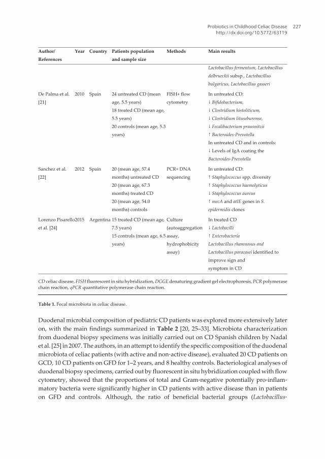
Author/References
Year Country Patients populationand sample size
Methods Main results
Lactobacillus fermentum, Lactobacillusdelbrueckii subsp., Lactobacillusbulgaricus, Lactobacillus gasseri
De Palma et al.[21]
2010 Spain 24 untreated CD (meanage, 5.5 years)18 treated CD (mean age,5.5 years)20 controls (mean age, 5.3years)
FISH+ flowcytometry
In untreated CD:↓ Bifidobacterium,↓ Clostridium histoliticum,↓ Clostridium lituseburense,↓ Fecalibacterium prausnitzii↑ Bacteroides‐PrevotellaIn untreated CD and in controls:↓ Levels of IgA coating theBacteroides‐Prevotella
Sanchez et al.[22]
2012 Spain 20 (mean age, 57.4months) untreated CD20 (mean age, 67.3months) treated CD20 (mean age, 54.0months) controls
PCR+ DNAsequencing
In untreated CD:↑ Staphylococcus spp. diversity↑ Staphylococcus haemolyticus↓ Staphylococcus aureus↑ mecA and atIE genes in S.epidermidis clones
Lorenzo Pisarelloet al. [24]
2015 Argentina 15 treated CD (mean age,7.5 years)15 controls (mean age, 6.5years)
Culture(autoaggregationassay,hydrophobicityassay)
In treated CD↓ Lactobacilli↑ EnterobacteriaLactobacillus rhamnosus andLactobacillus paracasei identified toimprove sign andsymptom in CD
CD celiac disease, FISH fluorescent in situ hybridization, DGGE denaturing gradient gel electrophoresis, PCR polymerasechain reaction, qPCR quantitative polymerase chain reaction.
Table 1. Fecal microbiota in celiac disease.
Duodenal microbial composition of pediatric CD patients was explored more extensively lateron, with the main findings summarized in Table 2 [20, 25–33]. Microbiota characterizationfrom duodenal biopsy specimens was initially carried out on CD Spanish children by Nadalet al. [25] in 2007. The authors, in an attempt to identify the specific composition of the duodenalmicrobiota of celiac patients (with active and non‐active disease), evaluated 20 CD patients onGCD, 10 CD patients on GFD for 1–2 years, and 8 healthy controls. Bacteriological analyses ofduodenal biopsy specimens, carried out by fluorescent in situ hybridization coupled with flowcytometry, showed that the proportions of total and Gram‐negative potentially pro‐inflam‐matory bacteria were significantly higher in CD patients with active disease than in patientson GFD and controls. Although, the ratio of beneficial bacterial groups (Lactobacillus‐
Probiotics in Childhood Celiac Diseasehttp://dx.doi.org/10.5772/63119
227

Bifidobacterium) to potentially harmful Bacteroides‐E. coli was significantly reduced in CDpatients on GFD, there was not a complete normalization of gut microbiota compared withcontrols [25]. Several subsequent Spanish studies confirmed these results [20, 26–28].Particularly, these studies found that the Bacteroides, E. coli, Bifidobacterium, Enterobacteriacae,and Staphylococcus groups were significantly more abundant in GCD patients than in thecontrols with a greater diversity of these species [20, 26, 28], while, in contrast, members of thefamily Streptococcaceae were less abundant in CD patients [28]. Furthermore, the Prevotellagenera were more frequent in healthy subjects than in celiac patients [27]. Ou et al. identi‐fied Clostridium, Prevotella and Actinomyces as predominant bacteria in the proximal smallintestine biopsies from a cohort of 45 CD children and 18 healthy controls born during the so‐called “Swedish CD epidemic” (2004‐2007). This could explain the four‐fold increase in theincidence of CD in children less than two years of age observed between 2004 and 2007 [29].Schippa et al. [30] analyzed the mucosa‐associated microbiota of CD children, before and aftera GFD, and controls by temporal temperature gradient gel electrophoresis (TTGE). The mostimportant findings of the study were: a demonstration of a presence of peculiar microbialTTGE profile and a significant higher biodiversity in CD pediatric patients’ duodenal mucosaafter 9 months of GFD compared to healthy controls. Di Cagno et al. [31], utilizing culture‐dependent and culture‐independent methods and metabolomics analyses, investigated thedifferences in the microbiota and metabolome of 19 treated CD patients and 15 controls. Theyconfirmed the lower levels of Lactobacillus and increased levels of Bacteroides in CD patients.Moreover, the authors showed that a GFD lasting at least two years did not completely restorethe microbiota and metabolome in CD patients [31]. A recent Spanish study demonstrated thatthe intestinal microbiota of patients with duodenal Marsh 3c lesions showed similarity of 98%and differed from that of CD patients with other type of histologic lesion as Marsh 3a, Marsh 3b,and Marsh 2 [32]. This indicated that the composition of duodenal microbiota differeddepending on the grade of intestinal damage.
Authors/references
Years Country Patients populationand sample size
Methods Main results
Nadal et al. [25] 2007 Spain 20 (untreated CD (meanage, 5.1 years)10 treated CD (mean age,5.6 years)8 controls (mean age, 4.1years)
FISH+ flowcytometry
In untreated CD:↑ Total bacteria↑ Gram‐negative bacteria↑ Bacteroides and E. coli, whichnormalized after GFDIn treated and untreated CD:↓ The ratio of Lactobacillus‐Bifidobacterium to Bacteroides
Collado et al.[20]
2009 Spain 8 untreated CD (mean age,56.4 months)8 treated CD (mean age,65.2 months)
qPCR In untreated CD:↑Bacterial counts↑ Lactobacillus prevalence↓ C. coccoides prevalence↑ Staphylococcus
Probiotics and Prebiotics in Human Nutrition and Health228

Authors/references
Years Country Patients populationand sample size
Methods Main results
8 controls (mean age, 45.0months)
↑ E. coli↓ BifidobacteriumIn treated and untreated CD:↑ Bacteroides↑ C. leptum
Ou et al.[29] 2009 Sweden 33 untreated CD (medianage, 5.9 years)17 treated CD (median age,7.5 years)3 challenged CD (medianage, 10.8 years)18 controls (mean age, 3.2years)
Culture+Scanningelectronmicroscopy
In untreated CD↑ Streptococcus↑ Neisseria
Schippa et al.[30]
2010 Italy 20 CD (before and afterGFD) (mean age, 8.3 years)10 controls (mean age, 11.7years)
TTGE Differences in biodiversity betweenuntreated CD and treated CD↑Bacteroides vulgatus and E. coli in CD
Sanchez et al.[26]
2010 Spain 20 treated CD (mean age,51.1 months)12 untreated CD (meanage, 54.9 months)8 controls (mean age, 50.1months)
PCR‐DDGE In untreated and treated CD:↓Bacteroides diversityIn untreated CD:↓ Bacteroides dorei↑Bifidobacterium diversity↑Bifidobacterium adolescentis,↑ Bifidobacterium animalis↓Bacteroides diastonis, ↓Bacteroidesfragilis↓ Bacteroides thetaiotaomicron,↓ Bacteroides uniformis↓ Bacteroides Ovatus
Di Cagno et al.[31]
2011 Italy 19 treated CD (mean age9.7 years)15 controls (mean age, 10.4years)
PCR‐DDGE In treated CD:↓ Lactobacillus↓ Enterococcus↓ Bifidobacteria↑Bacteroides,↑ Staphylococcus, ↑Salmonella,↑ Shigella, Klebsiella
Sanchez et al.[28]
2013 Spain 32 untreated CD ( meanage, 5.1 years)17 treated CD (mean age,5.9 years)
Culture+PCR
In untreated CD:↑ Proteobacteria, Enterobacteriaceae,Staphylococcaceae (Klebsiella oxytoca,
Probiotics in Childhood Celiac Diseasehttp://dx.doi.org/10.5772/63119
229
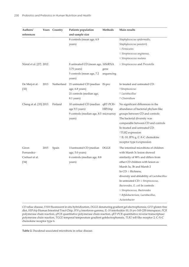
Authors/references
Years Country Patients populationand sample size
Methods Main results
8 controls (mean age, 6.9years)
Staphylococcus epidermidis,Staphylococcus pausteri)↓ Firmicutes↓ Streptococcus anginosus,↓ Streptococcus mutans
Nistal et al. [27] 2012 8 untreated CD (mean age,3.75 years)5 controls (mean age, 7.2years)
16SrRNAgenesequencing
↓ Streptococcus and Prevotella
De Meij et al.[32]
2013 Netherland 21 untreated CD (medianage, 6.8 years)21 controls (median age,8.1 years)
IS‐pro In treated and untreated CD:↑Streptococcus↑ Lactobacillus↑ Clostridium
Cheng et al. [33] 2013 Finland 10 untreated CD (medianage 9.5 years)9 controls (median age, 8.5years)
qRT‐PCR+HIPchipmicroarray
No significant differences in theabundance of bacterial phylum‐likegroups between CD and controlsThe bacterial diversity wascomparable between CD and controlsIn treated and untreated CD:↑TLR2 expression↑ IL‐10, IFN‐g, C-X-C chemokinereceptor type 6 expression
GironFernandez‐Crehuet et al.[34]
2015 Spain 11untreated CD (medianage, 5.0 years)6 controls (median age, 8.8years)
DGGE The intestinal microbiota of childrenwith Marsh 3c lesion showedsimilarity of 98% and differs fromother CD children with lesion asMarsh 3a, 3b and Marsh 2In CD: ↓ Richness,diversity and abitability of LactobacillusIn untreated CD: ↓ Streptococcus,Bacteroides, E. coli In controls↓ Streptococcus, Bacteroides↑ Bifidobacterium, Lactobacillus,Acinetobacter
CD celiac disease, FISH fluorescent in situ hybridization, DGGE denaturing gradient gel electrophoresis, GFD gluten‐freediet, HIPchip Human Intestinal Tract Chip, IFN‐g interferon‐gamma, IL‐10 interleukin‐10, IS‐pro 16S‐23S interspacer, PCRpolymerase chain reaction, qPCR quantitative polymerase chain reaction, qRT‐PCR quantitative reverse‐transcriptase‐polymerase chain reaction, TGGE temporal temperature gradient gelelectrophoresis, TLR2 toll‐like receptor 2, C-X-Cchemokine receptor type 6.
Table 2. Duodenal‐associated microbiota in celiac disease.
Probiotics and Prebiotics in Human Nutrition and Health230

In contrast, two recent studies reached different results. De Meij et al. [33], analyzing the totalmicrobiome profile in small bowel biopsies of 21 untreated CD and 21 age‐matched controls,found that mucosa‐associated duodenal microbiome composition and diversity did not differbetween children with untreated CD and controls. The same results were obtained by Chenget al. using bacterial phylogenetic microarray to comprehensively profile the microbiota induodenal biopsies of 10 CD and nine healthy children, suggesting that the duodenal mucosa‐associated bacteria do not play an important role in the pathogenesis of CD [34].
In summary, although the majority of the studies available have confirmed the presence ofintestinal dysbiosis in CD children characterized by low levels of Lactobacilli and Bifidobacte‐ria and increase in Gram‐negative bacteria (Bacteroides), which were not completely normal‐ized after GFD, some of them have failed to find a distinct signature that defines celiacmicrobiota. The available articles regarding the relationship between the gut microbiota andGFD, demonstrated that a GFD only allows a partial recovery of the gut microbiota in CDpatients [30, 34, 35].
3. Pathogenetic role of intestinal dysbiosis in CD
The intestinal microbiota composition and function play a fundamental role in the balancebetween the host's health and disease by different mechanisms: (1) regulation of epithelial cellproliferation and expression of tight junction proteins which act on intestinal permeability; (2)influence on mucin gene expression by goblet cells and their glycosylation pattern; 3) secretionof antimicrobial peptides (defensins, angiogenins, Reg3γ, etc.) by intestinal cells, whichcontribute to control gut bacterial adhesion. Certain components of the gut microbiota alsoaffect the expression and activation of pattern recognition receptors (PRR), such as toll‐likereceptors (TLRs), which are expressed by epithelial cells and innate immune cells. Themammalian TLR recognizes specific patterns of microbial components, called pathogen‐associated molecular patterns (PAMPs). After the PRR‐PAMP interaction, activated innateimmune cells start the adaptive immune response by presenting the antigen and by produc‐ing cytokines, which leads to antigen‐specific, protective immune response. In inflammatoryand autoimmune diseases this response causes damage to host's tissues [36]. The gut micro‐biota impacts on adaptive immunity. Recently, specific commensal bacteria have been shownto influence T lymphocyte production (Th1, Th17) or anti‐inflammatory regulatory T cells(Tregs) [36].
To date, human microbiota and mucosal barrier function are the key players in etiology ofmany inflammatory and autoimmune diseases [37]. Changes in mechanisms regulatingmucosal immunity and tolerance, can lead to impaired mucosal barrier function, increasedpenetration of microbial components from lumen into the mucosa and circulation, andconsequently lead exaggeration of aberrant immune responses and inflammation.
The exact mechanisms through which the gut microbiota might influence CD onset orprogression is unknown, but could include activation of innate immune system, modulation
Probiotics in Childhood Celiac Diseasehttp://dx.doi.org/10.5772/63119
231

of the epithelial barrier, or exacerbation of the gliadin‐specific immune response [38].Moreover, the presence of microbiota can significantly influence the inflammatory effect ofgluten. The microbiota may facilitate the access of gliadin peptides to the lamina propria andits interaction with infiltrated lymphocytes and antigen presenting cells (APCs) responsiblefor triggering the immune response via different mechanisms. In genetically predisposedindividuals, gluten in association with microbial antigens can stimulate and modulate innateand adaptative immune response, sustaining a chronic mucosal inflammation, underliningthis chronic disease [38].
4. Probiotics in the treatment of CD
Probiotics are nonpathogenic live microorganisms, which when orally administered inadequate amounts, alter the microflora of the host and have beneficial health effects. Probioticshave shown to preserve the intestinal barrier promoting its integrity both in vitro and invivo [39, 40] as well as regulating the response of the innate and adaptative immune system.The association of CD with intestinal dysbiosis and the evidence supporting a role for themicrobiota and specific bacteria in maintaining gut barrier function and regulating theresponse of the innate and adaptive immune system, have supported the potential use ofprobiotics in CD treatment [41, 42]. Although the data regarding the use of probiotics for CDare encouraging, most of these data come from in vitro experimental models of CD [43, 44].Studies regarding probiotics and CD in humans are very scarce [45–47]. Smecuol et al.evaluated the effect of the Bifidobacterium infantis natren life start (NLS) on gut permeability,the occurrence of symptoms, and presence of inflammatory cytokines in adult CD patients onGCD. Results have shown that probiotics did not modify intestinal permeability probably dueto an insufficient dose or a short time of administration. However, probiotic administrationimproved gastrointestinal symptoms, alleviating and reducing constipation [47].
In children, the clinical trials performed on the effect of probiotics on CD are summarized inTable 3. In the earliest study Olivares et al. [45] evaluated the influence of Bifidobacteriumlongum CECT 7347 in addition to a GFD in children newly diagnosed with CD. They showeda decrease in peripheral CD3+ T lymphocytes and a trend in the reduction of tumor necrosisfactor (TNF)‐α serum levels, and a reduction in the Bacteroides fragilis group(pro‐inflammato‐ry bacteria) and in the content of IgA in stools. Klemenak et al. [46] evaluated the effect of acombination of the strains Bifidobacterium breve BR03 and B. breve B632, as compared to placebo.They reported that B. breve strains decreased the production of the pro‐inflammatory cyto‐kine TNF‐α in children CD on a GFD.
At this time, the only treatment for CD is lifelong GFD, which involves the elimination of grainscontaining gluten, wheat, rye, and barley in addition to food products and additives derivedfrom them [48]. To date, adherence to a diet is difficult for many patients. Studies have shownthat dietary transgression in patients with CD is common and can occur anywhere from 32%to 55% [49]. Moreover, a GFD may be rich in high glycemic index foods which can increase
Probiotics and Prebiotics in Human Nutrition and Health232

insulin resistance and, thus, the risk of obesity and cardiovascular disease. In the last decade,new therapies have been suggested to improve compliance to a GFD or to replace a GFD [50].The use of probiotics appears to be able to reduce the damage caused by eating gluten‐containing foods and may even accelerate mucosal healing after the initiation of GFD [50, 51].A specific commercially available probiotic, VSL#3 (containing eight different bacteria), hasbeen shown to reduce the toxicity of gluten when used in a fermentation process [52]. It isthought that the gut microbiota can be modified in its composition and function by probioticadministration. These may counteract or postpone the onset of CD, and it can be useful inpatients on GFD, when the normal composition of the intestinal flora has not yet fullyrecovered.
Authors/references
Years Country Studydesign
Patients populationand sample size
Main results Comments
Olivares et al.[45]
2013 Spain DB, R, PC 18 CD (mean age, 6.8 years)received B. longum CECT7347; 18 CD (mean age 8.5years) received placebo for3 months in parallel withthe GFD
↓B. fragilis group↓activated T‐lymphocytes↓TNF‐α
B. longum CECT 7347could improve the healthstatus of CD patients
Klemenac et al.[46]
2015 ItalySlovenia
DB, R, PC 22 CD (age, 10.43) dailyreceived B. breve25 CD (age, 10.81) dailyreceived placebo for 3months18 (age, 8.83) controls
↓TNF‐α levels onCD group
Probiotic interventionwith B. breve strains hasshown a positive effecton decreasing theproduction of pro‐inflammatory cytokineTNF‐α in children withCD on GFD
CD celiac disease, DB double-blind, R randomized, PC placebo controlled.
Table 3. Clinical trials on the effect of probiotics for CD.
5. Conclusions
An alternative treatment that can improve CD patients’ quality of life may lie in probiotics. Inparticular, probiotics such as Lactobacilli and Bifidobacterium could be useful to reset altered gutmicrobiota, as well as reduce gliadin toxicity and immune activation. Their use as a primaryprophylactic treatment for children at high risk of CD is also a potential consideration.However, their use in routine clinical practice is hindered by limited data from human studies.The role of specific probiotics and their mechanism of action need to be identified in a largerexperimental population to confirm their effectiveness.
Probiotics in Childhood Celiac Diseasehttp://dx.doi.org/10.5772/63119
233

Author details
Caterina Anania1, Francesca Olivero1, Eugenia Olivero2 and Lucia Pacifico1*
*Address all correspondence to: [email protected]
1 Policlinico Hospital Umberto I, Sapienza University of Rome, Rome, Italy
2 Ernst & Young Financial‐Business Advisors, Rome, Italy
References
[1] Fasano A, Catassi C. Clinical practice. Celiac disease. N Engl J Med. 2012; 367:2419–2426. DOI: 10.1056/NEJMcp1113994.
[2] Track GJ, Verbeek WH, Schreurs MW, Mulder CJ. The spectrum of celiac disease:epidemiology, clinical aspects and treatment. Nat Rev Gastroenterol Hepatol.2010;7:204–213. DOI: 10.1038/nrgastro.2010.23.
[3] Husby S, Koletzko S, Korponay‐Szabo IR, Mearin ML, Phillips A, Shamir R, Tron‐cone R, Giersiepen K, Branski D, Catassi C, Lelgeman M, Maki M, Ribes‐Koninckx C,Ventura A, Zimmer KP. European Society for Pediatric Gastroenterology, Hepatolo‐gy, and Nutrition guidelines for the diagnosis of coeliac disease. J Pediatr Gastroen‐terol Nutr. 2012;54:136–160. DOI: 10.1097/MPG.0b013e1821a23d0.
[4] Van Belzen MJ, Koeleman BPC, Crusius JBA, MeijervJWR, Bardoel AFJ, Pearson PL,Sandkuijl LA, Houwen RH, Wijmenga C. Defining the contribution of the HLA regionto cis DQ2‐positive coeliac disease patients. Gen Immun. 2004;5:215–220. http://dx.doi.org/10.1038/sj.gene.6364061.
[5] IavarsonA, Hernell O, Stenlud H, Persson LA. Breast‐feeding protects against coeliacdisease. Am J Clin Nutr. 2002;75:914–921. DOI
[6] Stene LC, Honeyman MC, Hoffenberg EJ, Haas JE, Sokol RJ, Emery L, Taki I, NorrisJM, Erlich HA, Eisenbarth GS, Rewers M. Rotavirus infection frequency and risk ofcoeliac disease autoimmunity in early childhood: a longitudinal study. Am JGastroenterol. 2006;101:2333–2340. DOI:10.1111/j.1572‐0241.2006.00741.x.
[7] Akobeng AK, Ramanan AV, Buchan I, Heller R. Effect of breast feeding on risk of coeliacdisease: a systematic review and meta‐analysis of observational studies. Arch DisChild. 2006;91:39–43. DOI: 10.1136/adc.2005.082016.
[8] Szebeni B, Veres G, Dezsofi A, Rusai K, Vannay A, Bokodi G, Vásárhelyi B, Korpo‐nay‐Szabó IR, Tulassay T, Arató A. Increased mucosal expression of toll‐like receptor
Probiotics and Prebiotics in Human Nutrition and Health234

(TLR)2 and TLR4 in coeliac disease. J Pediatr Gastroenterol Nutr. 2007;45:187–193.DOI: 10.1097/MPG.0b013e318064514a.
[9] Vanderpool C, Yan F, Polk DB. Mechanisms of probiotic action: implications fortherapeutic applications in inflammatory bowel diseases. Inflamm Bowel Dis.2008;14:1585–1596. DOI: 10.1097/MPG.0b013e318064514a.
[10] Morgan XC, Tickle TL, Sokol H, Gevers D, Devaney KL, Ward DV, Reyes JA, Shah SA,LeLeiko N, Snapper SB, Bousvaros A, Korzenik J, Sands BE, Xavier RJ, HuttenhowerC. Dysfunction of the intestinal microbiome in inflammatory bowel disease andtreatment. Genome Biol. 2012;13:R79. DOI: 10.1186/gb‐2012‐13‐9‐r79.
[11] Miele L, Giorgio V, Alberelli MA, De Candia E, Gasbarrini A, Grieco A. Impact of gutmicrobiota on obesity, diabetes, and cardiovascular disease risk. Curr Cardiol Rep. 201517:120. DOI: 10.1007/s11886‐015‐0671‐z.
[12] Forsberg G, Fahlgren A, Hörstedt P, Hammarström S, Hernell O, Hammarström ML.Presence of bacteria and innate immunity of intestinal epithelium in childhood celiacdisease. Am J Gastroenterol 2004;99;894–904. DOI :10.1111/j.1572‐0241.2004.04157.x.
[13] Collado MC, Calabuig M, Sanz Y. Differences between the fecal microbiota of coeliacinfants and healthy controls. Curr Issues Intest Microbiol. 2007;8:9–14.
[14] Nadal I, Donat E, Ribes‐Koninckx C, Calabuig M, Sanz Y. Imbalance in the composi‐tion of the duodenal microbiota of children with coeliac disease. J Med Microbiol.2007;56:1669–1674. DOI: 10.1099/jmm.0.47410‐0.
[15] Collado MC, Donat E, Ribes‐Koninckx C, Calabuig M, Sanz Y. Imbalances in faecal andduodenal Bifidobacterium species composition in active and non‐active coeliac disease.BMC Microbiol. 2008;8:232. DOI: 10.1186/1471‐2180‐8‐232.
[16] Fernandez‐Feo M, Wei G, Blumenkranz G, Dewhirst FE, Schuppan D, Oppenheim FG,Helmerhorst EJ. The cultivable human oral gluten‐degrading microbiome and itspotential implications in coeliac disease and gluten sensitivity. Clin Microbiol Infect.2013;19:E386–394. DOI: 10.1111/1469‐0691.12249.
[17] Food and Agriculture Organization of the United Nations, World Health Organiza‐tion. Guidelines for the evaluation of probiotics in food: joint FAO/WHO WorkingGroup report on drafting guidelines for the evaluation of probiotics in food. WorldHealth Organization, Geneva, Switzerland. ftp://ftp.fao.org/es/esn/food/wgre‐port2.pdf. Accessed 27 April 2014.
[18] Lindfors K, Blomqvist T, Juuti‐Uusitalo K, Stenman S, Venäläinen J, Mäki M, KaukinenK. Live probiotic Bifidobacterium lactis bacteria inhibit the toxic effects induced by wheatgliadin in epithelial cell culture. Clin Exp Immunol. 2008;152:552–558. DOI: 10.1111/j.1365‐2249.2008.03635.x.
[19] Sanz Y, Sánchez E, Marzotto M, Calabuig M, Torriani S, Dellaglio F. Differences in faecalbacterial communities in coeliac and healthy children as detected by PCR and dena‐
Probiotics in Childhood Celiac Diseasehttp://dx.doi.org/10.5772/63119
235

turing gradient gel electrophoresis. FEMS Immunol Med Microbiol. 2007;51:562–568.DOI: http://dx.doi.org/10.1111/j.1574‐695X.2007.00337.x.
[20] Collado MC, Donat E, Ribes‐Koninckx C, Calabuig M, Sanz Y. Specific duodenal andfaecal bacterial groups associated with paediatric coeliac disease. J Clin Pathol.2009;62:264–269. DOI: 10.1136/jcp.2008.061366.
[21] De Palma G, Nadal I, Medina M, Donat E, Ribes‐Koninckx C, Calabuig M, Sanz Y.Intestinal dysbiosis and reduced immunoglobulin‐coated bacteria associated withcoeliac disease in children. BMC Microbiol. 2010;10:63. DOI: 10.1186/1471‐2180‐10‐63.
[22] Sánchez E, Ribes‐Koninckx C, Calabuig M, Sanz Y. Intestinal Staphylococcus spp. andvirulent features associated with coeliac disease. Clin Pathol. 2012;65:830–834. DOI:10.1136/jclinpath‐2012‐200759.
[23] Di Cagno R, Rizzello CG, Gagliardi F, Ricciuti P, Ndagijimana M, Francavilla R,Guerzoni ME, Crecchio C, Gobbetti M, De Angelis M. Different fecal microbiotas andvolatile organic compounds in treated and untreated children with celiac disease. ApplEnviron Microbiol. 2009;75:3963–3971. DOI: 10.1128/AEM.02793‐08.
[24] Lorenzo Pisarello MJ, Vintiñi EO, González SN, Pagani F, Medina MS. Decrease inlactobacilli in the intestinal microbiota of celiac children with a gluten‐free diet, andselection of potentially probiotic strains. Can J Microbiol. 2015;61:32–37. DOI: 10.1139/cjm‐2014‐0472.
[25] Nadal I, Donat E, Ribes‐Koninckx C, Calabuig M, Sanz Y. Imbalance in the composi‐tion of the duodenal microbiota of children with coeliac disease. J Med Microbiol.2007;56:1669–1674. DOI: 10.1099/jmm.0.47410‐0.
[26] Sánchez E, Donat E, Ribes‐Koninckx C, Calabuig M, Sanz Y. Intestinal Bacteroidesspecies associated with coeliac disease. J Clin Pathol. 2010;63:1105–1111. DOI: 10.1136/jcp.2010.07695.
[27] Nistal E, Caminero A, Herrán AR, Arias L, Vivas S, de Morales JM, Calleja S, de MieraLE, Arroyo P, Casqueiro J. Differences of small intestinal bacteria populations in adultsand children with/without celiac disease: effect of age, gluten diet, and disease.Inflamm Bowel Dis. 2012;18:649–656. DOI: 10.1002/ibd.21830.
[28] Sánchez E, Donat E, Ribes‐Koninckx C, Fernández‐Murga ML, Sanz Y. Duodenal‐mucosal bacteria associated with celiac disease in children. Appl Environ Microbiol.2013;79:5472–5479. DOI: 10.1128/AEM.00869‐13.
[29] Ou G, Hedberg M, Hörstedt P, Baranov V, Forsberg G, Drobni M, Sandström O, WaiSN, Johansson I, Hammarström ML, Hernell O, Hammarström S. Proximal smallintestinal microbiota and identification of rod‐shaped bacteria associated withchildhood celiac disease. Am J Gastroenterol. 2009;104:3058–3067. DOI: 10.1038/ajg.2009.524.
Probiotics and Prebiotics in Human Nutrition and Health236

[30] Schippa S, Iebba V, Barbato M, Di Nardo G, Totino V, Checchi MP, Longhi C, MaiellaG, Cucchiara S, Conte MP. A distinctive ‘microbial signature’ in celiac pediatricpatients. BMC Microbiol. 2010;10:175.DOI: 10.1186/1471‐2180‐10‐175.
[31] Di Cagno R, De Angelis M, De Pasquale I, Ndagijimana M, Vernocchi P, Ricciuti P,Gagliardi F, Laghi L, Crecchio C, Guerzoni ME, Gobbetti M, Francavilla R. Duodenaland faecal microbiota of celiac children: molecular, phenotype and metabolomecharacterization. BMC Microbiol. 2011;11:219. DOI: 10.1186/1471‐2180‐11‐219.
[32] Girón Fernández‐Crehuet F, Tapia‐Paniagua S, Moriñigo Gutiérrez MA, Navas‐LópezVM, Juliana Serrano M, Blasco‐Alonso J, Sierra Salinas C. The duodenal microbiotacomposition in children with active coeliac disease is influenced for the degree ofenteropathy. An Pediatr (Barc). 2015; 18 pii: S1695–4033(15)00311‐2. doi: 10.1016/j.anpedi.2015.06.014.
[33] de Meij TG, Budding AE, Grasman ME, Kneepkens CM, Savelkoul PH, Mearin ML.Composition and diversity of the duodenal mucosa‐associated microbiome in chil‐dren with untreated coeliac disease. Scand J Gastroenterol. 2013;48:530–536. DOI:10.3109/00365521.2013.775666.
[34] Cheng J, Kalliomäki M, Heilig HG, Palva A, Lähteenoja H, de Vos WM, Salojärvi J,Satokari R. Duodenal microbiota composition and mucosal homeostasis in pediatricceliac disease. BMC Gastroenterol. 2013;13:113. DOI: 10.1186/1471‐230X‐13‐113.
[35] Collado MC, Donat E, Ribes‐Koninckx C, Calabuig M, Sanz Y. Imbalances in faecal andduodenal Bifidobacterium species composition in active and non‐active coeliac disease.BMC Microbiol. 2008;8:232. DOI: 10.1186/1471‐2180‐8‐232.
[36] Tlaskalová‐Hogenová H, Stěpánková R, Kozáková H, Hudcovic T, Vannucci L,Tučková L, Rossmann P, Hrnčíř T, Kverka M, Zákostelská Z, Klimešová K, PřibylováJ, Bártová J, Sanchez D, Fundová P, Borovská D, Srutková D, Zídek Z, Schwarzer M,Drastich P, Funda DP. The role of gut microbiota (commensal bacteria) and the mucosalbarrier in the pathogenesis of inflammatory and autoimmune diseases and cancer:contribution of germ‐free and gnotobiotic animal models of human diseases. Cell MolImmunol. 2011;8:110–120. DOI: 10.1038/cmi.2010.67.
[37] Verdu EF, Galipeau HJ, Jabri B. Novel players in coeliac disease pathogenesis: role ofthe gut microbiota. Nat Rev Gastroenterol Hepatol. 2015;12:497–506. DOI: 10.1038/nrgastro.2015.90.
[38] Pagliari D, Urgesi R, Frosali S, Riccioni ME, Newton EE, Landolfi R, Pandolfi F, CianciR. The interaction among microbiota, immunity, and genetic and dietary factors is thecondicio sine qua non celiac disease can develop. J Immunol Res. 2015;2015:123653.DOI: 10.1155/2015/123653.
[39] Madsen K, Cornish A, Soper P, McKaigney C, Jijon H, Yachimec C, Doyle J, Jewell L,De Simone C. Probiotic bacteria enhance murine and human intestinal epithelial barrier
Probiotics in Childhood Celiac Diseasehttp://dx.doi.org/10.5772/63119
237

function. Gastroenterology 2001;121:580–591. DOI: http://dx.doi.org/10.1053/gast.2001.27224.
[40] Gupta P, Andrew H, Kirschner BS, Guandalini S. Is lactobacillus GG helpful in childrenwith Crohn's disease? Results of a preliminary, open‐label study. J Pediatr Gastroen‐terol Nutr. 2000;31:453–457.
[41] Sanz Y, De Pama G, Laparra M. Unraveling the ties between celiac disease and intestinalmicrobiota. Int Rev Immunol. 2011;30:207–218. DOI: 10.3109/08830185.2011.599084.
[42] Cenit MC, Olivares M, Codoñer‐Franch P, Sanz Y. Intestinal microbiota and celiacdisease: cause, consequence or co‐evolution? Nutrients. 2015;7:6900–6923. DOI:10.3390/nu7085314.
[43] Aloisio I, Santini C, Biavati B, Dinelli G, Cencič A, Chingwaru W, Mogna L, Di GioiaD. Characterization of Bifidobacterium spp. strains for the treatment of entericdisorders in newborns. Appl Microbiol Biotechnol. 2012;96:1561–1576. DOI: 10.1007/s00253‐012‐4138‐5.
[44] Mogna L, Del Piano M, Mogna G. Capability of the two microorganisms Bifidobacteri‐um breve B632 and Bifidobacterium breve BR03 to colonize the intestinal microbiota ofchildren. J Clin Gastroenterol. 2014;48 Suppl 1:S37–9. DOI: 10.1097/MCG.0000000000000234.
[45] Olivares M, Castillejo G, Varea V, Sanz Y. Double‐blind, randomised, placebo‐control‐led intervention trial to evaluate the effects of Bifidobacterium longum CECT 7347 inchildren with newly diagnosed coeliac disease. Br J Nutr. 2014;112:30–40. DOI: 10.1017/S0007114514000609.
[46] Klemenak M, Dolinšek J, Langerholc T, Di Gioia D, Mičetić‐Turk D. Administration ofBifidobacterium breve decreases the production of TNF‐α in children with celiacdisease. Dig Dis Sci. 2015;60:3386–3392. DOI: 10.1007/s10620‐015‐3769‐7.
[47] Smecuol E, Hwang HJ, Sugai E, Corso L, Cherñavsky AC, Bellavite FP, González A,Vodánovich F, Moreno ML, Vázquez H, Lozano G, Niveloni S, Mazure R, Meddings J,Mauriño E, Bai JC. Exploratory, randomized, double‐blind, placebo‐controlled studyon the effects of Bifidobacteriuminfantis natren life start strain super strain in activeceliac disease. J Clin Gastroenterol. 2013;47:139–147. DOI: 10.1097/MCG.0b013e31827759ac.
[48] Green PH, Jabri B. Coeliac disease. Lancet. 2003; 362:383–391. DOI: http// dx.doi.org/0.1016/s0140‐6736(03)14027‐5.
[49] Silvester JA, Rashid M. Long‐term follow‐up of individuals with celiac disease: anevaluation of current practiceguidelines. Can J Gastroenterol. 2007;21:557–564.
[50] Kaukinen K, Lindfors K, Mäki M. Advances in the treatment of coeliac disease: animmunopathogenic perspective. Nat Rev Gastroenterol Hepatol. 2014;11:36–44.DOI:10.1038/nrgastro.2013.141.
Probiotics and Prebiotics in Human Nutrition and Health238

[51] Lindfors K, Blomqvist T, Juuti‐Uusitalo K, Stenman S, Venäläinen J, Mäki M, KaukinenK. Live probiotic Bifidobacterium lactis bacteria inhibit the toxic effects induced bywheat gliadin in epithelial cell culture. Clin Exp Immunol. 2008;152:552–558. DOI:10.1111/j.1365‐2249.2008.03635.x.
[52] De Angelis M, Rizzello CG, Fasano A, Clemente MG, De Simone C, Silano M, DeVincenzi M, Losito I, Gobbetti M. VSL#3 probiotic preparation has the capacity tohydrolyze gliadin polypeptides responsible for Celiac Sprue. Biochim Biophys Acta.2006;1762:80–93.
Probiotics in Childhood Celiac Diseasehttp://dx.doi.org/10.5772/63119
239

Chapter 12
Probiotics for Prevention and Treatment of Candidiasisand Other Infectious Diseases: Lactobacillus spp. andOther Potential Bacterial Species
Michelle Peneluppi Silva, Rodnei Dennis Rossoni,
Juliana Campos Junqueira and
Antonio Olavo Cardoso Jorge
Additional information is available at the end of the chapter
http://dx.doi.org/10.5772/64093
Abstract
The resident microbiota in the human body, such as the oral cavity, gastrointestinal tractand genitourinary tract, is able to provide resistance to disease. However, imbalancesin the microbial components can promote the growth of opportunistic microorgan‐isms, such as yeasts of genus Candida. Fungal infections present as a major cause ofinfectious diseases and the microorganisms of genus Candida are the most frequentlyisolated pathogenic fungi in human fungal infections. Bacillus spp. and Lactobacillus spp.are bacteria that have probiotic effects used in commercially available products and instudies that aim for the development of probiotics able to inhibit the microbialpathogenicity and restore the balance of resident microbiota. Thus, with increasingfungus resistance to the use of antifungal agents, which are capable of causing seriousside effects to the host organism unable to destroy the target microorganism, it becomesimportant to develop therapeutic and/or prophylactic alternatives that have a differentand an effective mechanism of action with capacity to combat fungal infections withoutharming the patient. Probiotic bacteria provide an alternative strategy for the preven‐tion and treatment of candidiasis and other infectious diseases.
Keywords: probiotic, Candida spp., Bacillus spp., Lactobacillus spp., prevention andtreatment

1. Introduction
The incidence of fungal infections has increased significantly in the past 25 years [1]. Humanbeings are colonized by a diverse and complex collection of microorganisms, contributing allof them to host nutrition, development of the immune system, response to pathogens andmucosal cell differentiation and proliferation [2].
Probiotic bacteria are also used in human and animal nutrition to influence beneficially thebalance of intestinal microbiota of the host. Probiotics have several beneficial effects related toincreasing digestion, strengthening the immune system and stimulating the production ofvitamin. The use of probiotics is aimed to reduce the use of antibiotics and improve animalgrowth, as well as feed conversion [3].
Infectious diseases along with multidrug resistance are the major public health problem indeveloping countries with increased mortality and morbidity [4, 5]. Apart from the threat ofmultidrug resistance, several studies have confirmed that the continuous use of antibiotics candamage human commensal microbiota [5, 6]. Thus, an alternative and effective research focusis necessary to combat these pathogens with no effect on normal microbiota. In this regard,the use of probiotics and their natural metabolic compounds can be a substitute in variousfood and pharmaceutical industries [5].
There are around 600 pathogenic fungal species for humans and this group includes the fungithat cause infection of skin (e.g., Malassezia species) and fungi that have the potential to causesystemic infections (e.g., Cryptococcus neoformans and Candida albicans) [7]. The yeasts of thegenus Candida are the fourth most common cause of systemic infections acquired in hospi‐tals in the United States with 50% mortality rates. The most pathogenic species is C. albicansand can cause two major types of human infections: superficial infections, such as oralcandidiasis, and systemic infections [8, 9].
The genus Candida is commonly found in the oral cavity of healthy individuals, isolated fromapproximately 75% of the population with a higher prevalence of C. albicans, followed by C.tropicalis and C. glabrata [10]. Candida species are a frequent cause of recurrent infections in themucosa when favored by risk factors such as the use of antibiotics of broad spectrum andcorticosteroids for long time, human immunodeficiency virus (HIV) infection, radiotherapyin the area of head and neck, the use of orthodontic appliances, deficient oral hygiene, amongother factors affecting immunocompromised patients that may result in transition of com‐mensal phase of C. albicans to pathogenic [11, 12].
Under certain conditions of immunosuppression, such as individuals with acquired immu‐nodeficiency syndrome (AIDS), oral manifestations are the most important and earliestindicators of infection. The oral candidiasis is accepted internationally as a cardinal sign ofHIV infection and is present in 50% of patients with HIV infection and in 80% of patients withAIDS [13, 14].
In Brazil during the period among 1996–2006, candidiasis was the second cause of deaths inHIV-positive patients due to fungal infections, being responsible for an average of 39 annual
Probiotics and Prebiotics in Human Nutrition and Health242

deaths [15]. Moreover, oral candidiasis remains clinically relevant in these individuals, wheretreatment is difficult and recurrent episodes are frequent, requiring multiple antifungaltreatments, which may lead to resistance selection [16, 17]. Due to this, C. albicans can developresistance to antifungals used to treat oral candidiasis, such as fluconazole and miconazole [18,19].
Due to the high recurrence of Candida lesions, and the increased resistance of conventionalantifungal drugs in clinical practice, the continuous use of probiotics to prevent fungalinfections may be an interesting strategy. In this chapter, we discuss how probiotics can helpin the prevention and/or adjuvant treatment of candidiasis.
2. Probiotic
The history of probiotics began with the history of man; cheese and fermented milk were wellknown to the Greeks and Romans who recommended their consumption, especially forchildren and convalescents. The first association of probiotics and health benefits was madeat the turn of the century when the Russian scientist, Elie Metchnikoff, systematically studiedthe composition of the microbiota and suggested that the ingestion of fermented milk wouldimprove this so-called autointoxication [20].
Probiotics play an important role in human health. There is general agreement on the importantrole of the gastrointestinal microbiota in the health and well-being status of humans andanimals [21]. Probiotics are defined as live microorganisms, which when administered inadequate amounts confer a health benefit on the host. This term is defined by a United Nationsand World Health Organization Expert Panel [22].
There was an increase in the number of searches, both in vivo and in vitro, related to the benefitsof probiotics on health and described in the literature for the treatment of infectious diseasescaused by fungi, viruses, and bacteria or diarrhea associated with the use of antibiotics,alleviation of inflammatory chronic bowel disease, decreased risk of colon cancer, reducedallergies, effect on intestinal microbiota [21], and anticancer therapies [23].
Other beneficial effects of probiotics include lowering serum cholesterol level [24–27],improving lactose intolerance, increasing the utilization of nutrients, decreasing the use ofantibiotics [24, 27], and antidiabetic treatments [26, 28, 29]. In the context linking food andhealth, probiotics have been the subject of numerous scientific studies and publicationsdemonstrating their therapeutic effectiveness on both systemic and gastrointestinal tract [21](Figure 1).
Microorganisms commonly used as probiotics belong to the heterogeneous group includingBacillus, Lactobacillus, Bifidobacterium, Saccharomyces cerevisiae, and Escherichia coli [30, 31]( Figure 1).
Probiotics for Prevention and Treatment of Candidiasis and Other Infectious Diseases: Lactobacillus spp. and OtherPotential Bacterial Species
http://dx.doi.org/10.5772/64093
243

Figure 1. Some properties of probiotics.
3. Lactobacillus spp.
3.1. General characteristics
Lactobacillus spp. are Gram-positive bacteria, facultative anaerobic bacilli found in the normalmicrobiota of the gastrointestinal tract of birds and mammals, and genitourinary tract and oralcavity in the humans [31, 32]. This genre is heterogeneous and the number of species isconstantly being modified due to the description of new species and reclassification ofothers [33]. Some members of the genus Lactobacillus were reclassified into Carnobacterium [34],Atopobium [35], Weissella [36], and Paralactobacillus [37]. In early 2007, 120 species composingthe genus Lactobacillus [33] and in 2008 over 145 new species have already been identified [38,39].
Different Lactobacillus species found in the gastrointestinal tract are concerned with the balanceof microbiota and it has been widely studied due to their health-promoting properties [40].Their effects on intestinal microbiota in terms of protection include competition for adhesionsites with pathogenic microorganisms and antimicrobial substance production, such asorganic acids, lactic acid, carbon dioxide, and bacteriocins [41]. In addition, the regular use ofprobiotic appears to prevent certain gastrointestinal disorders such as lactose intolerance [42].
In 1907, Elie Metchnikoff won the Nobel Medicine Prize because he noticed that the dailyconsumption of Bulgarian yogurt (known for its rich composition in lactic acid bacteria) isbeneficial to health. Metchnikoff worked at the Pasteur Institute in Paris and he discovered L.
Probiotics and Prebiotics in Human Nutrition and Health244

bulgaricus and this strain was introduced into the commercial production of dairy productsacross Europe. He dedicated the last decade of his life to the study of bacteria that producelactic acid as a means to increase human longevity. After the studies of Metchnikoff, theconcept of probiotics was established and a new microbiology area started to develop [43].
3.2. Lactobacillus as probiotics and its mechanism of action
The main characteristics that a Lactobacillus strain needs to have to exercise an effectiveprobiotic action against pathogenic microorganisms are related to three factors: the ability toinhibit the adhesion and colonization of pathogenic microorganisms in the host tissues,biosurfactant production, and hydrogen peroxide (H2O2). There is a collagen-binding proteincalled 29 kD present on the surface of some lactobacilli, which causes it to be capable of bindingto collagen vaginal epithelial cells and to inhibit binding of pathogenic microorganisms to hosttissues in significant numbers [44]. Some strains of lactobacilli produce biosurfactantsgenerically known as surlactin, which are responsible for reducing the surface tension of liquidand thereby inhibiting the adherence of microorganisms. Surlactin studies are very impor‐tant to help in the understanding of the urogenital tract microbiota and their maintenance fora balanced microbiota [45]. Other lactobacilli strains have the ability to produce hydrogenperoxide, which can be toxic to microorganisms that do not produce catalase [46, 47].
According to Reid and Bruce [46], not all probiotic strains have the same mechanisms of actionand each has characteristics suitable for your application. For example, L. casei Shirota isingested daily for about 24 million people who do not have the 29-kDa protein and do notproduce H2O2. In the case of strain Shirota, its main action seems to be through the modula‐tion of the host immune response.
In a recent study, Abedin-Do et al. [48] showed that some Lactobacillus strains exert innate andadaptive immune responses via their binding to pattern recognition receptors expressed onimmune cells and many other tissues such as the intestinal epithelium. Furthermore,Lactobacillus can modulate the expression of genes involved in the regulation of immunesystem [49–53].
Members of our group evaluated the capacity of L. rhamnosus and its products to induce thesynthesis of cytokines (tumor necrosis factor (TNF)-α, interleukin (IL)-1β, IL-4, IL-6, IL-10, andIL-12) by mouse macrophages. Jorjão et al. [54] used three microorganism preparations: liveL. rhamnosus (LLR) suspension, heat-killed L. rhamnosus (HKLR) suspension, and the super‐natant of a heat-killed L. rhamnosus (SHKLR) suspension. LLR and HKLR groups were able tosignificantly increase the production of TNF-α, IL-6, and IL-10. SHKLR also significantlyincreased the production of TNF-α and IL-10 but not IL-6. All the L. rhamnosus suspensionswere not able to produce detectable levels of IL-1β or significant levels of IL-4 and IL-12. Theauthors concluded that live and heat-killed L. rhamnosus suspensions are able to induce thesynthesis of different cytokines with pro-inflammatory (TNF-α and IL-6) or regulatory (IL-10)functions, suggesting the role of strain L. rhamnosus ATCC 7469 in the modulation or in thestimulation of immune responses.
Probiotics for Prevention and Treatment of Candidiasis and Other Infectious Diseases: Lactobacillus spp. and OtherPotential Bacterial Species
http://dx.doi.org/10.5772/64093
245
In order for probiotic strains to have a satisfactory action, they must remain alive against stresschallenges along the entire gastrointestinal tract, including the presence of bile in the smallintestine. Bile is highly toxic to microorganisms not adapted to intestinal conditions. Moreover,some lactobacilli developed specific mechanisms to resist the deleterious effects caused bythese compounds [55]. Among these mechanisms, we can cite the efflux pump that activelyremoves the acids and accumulated bile salts within the cytoplasm and the enzymatic activityof hydrolases, which are capable of neutralizing deleterious effect of bile [56–58].
According to FAO WHO [22], the ideal characteristics of a probiotic strain of Lactobacillusconsidered are as follows:
• Not pathogenic;
• Stable in acid and in the presence of bile;
• Adhesion ability in human mucosa;
• Colonize the intestine;
• Remain viable during storage and use;
• Have beneficial physiological effects and safe.
3.3. Lactobacillus in prevention and treatment of Candida infection
In vitro assays are important to evaluate the antifungal activity of each strain and characteri‐zation of the mechanisms of action, performing as a screening to in vivo tests with experi‐mental models.
Sookkhee et al. [59] isolated and identified different species of lactic acid bacteria from the oralcavity of 130 volunteers in Thailand and they studied probiotic action against C. albicans invitro. The authors found 3790 different samples of lactic acid bacteria including the generaLactococcus, Lactobacillus, Streptococcus, Leuconostoc, and Pediococcus, and it was concluded thatL. paracasei and L. rhamnosus strains were two species that had the greatest number of clinicalisolates able to inhibit C. albicans.
Noverr and Huffnagle [60] examined the effect of living cultures, heat-killed cultures, andsupernatants of probiotic bacteria (L. casei, L. paracasei, and L. rhamnosus) on the morphogen‐esis of C. albicans and observed an inhibition in the formation of germ tube when C. albicansinteracted with living cells or supernatant of Lactobacillus. It was also found that superna‐tants obtained from cultures of 2 h inhibited germ tube formation of C. albicans. However, theaddition of 24-h growth cultures took complete inhibition, suggesting that the accumulationof a soluble compound of the supernatant is responsible for this inhibition.
Coman et al. [61] evaluated the antifungal activities of two probiotic strains, L. rhamnosusIMC 501® and L. paracasei IMC 502®, and their 1:1 combination, named SYNBIO®, using agarwell-diffusion method and liquid coculture assay. They tested probiotic strains in eight strainsof Candida, including C. albicans, C. krusei, C. glabrata, C. parapsilosis, and C. tropicalis. All theCandida strains are strongly inhibited, except C. glabrata and C. tropicalis, and during the
Probiotics and Prebiotics in Human Nutrition and Health246

coculture assay, the inhibitory activity of probiotic bacteria against Candida strains wasapproximately 40% in some cases and absent in other cases, in particular against some strainsof C. albicans and C. tropicalis. The authors concluded that in vitro screening of Lactobacillusstrains according to their activity in various environmental conditions might be a valuablemethod that could precede clinical efficacy studies for adjunct treatment with probiotics incure of different infections.
Parolin et al. [62] identified 17 clinical strains of Lactobacillus from the vaginal cavity of healthypremenopausal women, including the following species: L. crispatus, L. gasseri, and L. vaginalis,and evaluated their in vitro activity against Candida spp. (nine strains) and characterized theirantifungal mechanisms of action. In general, the strains tested were more active toward C.albicans. No Lactobacillus strains showed activity against C. krusei and C. parapsilosis. All strainsproduced hydrogen peroxide and lactate, and in particular, L. crispatus BC2, L. gasseri BC10,and L. gasseri BC11 appeared to be the most active strains in reducing pathogen adhesion. Itwas concluded that these in vitro assays are prerequisites for the development of newtherapeutic agents based on probiotics for prophylaxis and adjuvant therapy of Candidainfection.
Some in vivo studies also show the effectiveness of probiotics in Candida infection. Wagner etal. [63] demonstrated that the inoculation of probiotics (L. acidophilus, L. reuteri, L. casei GG,and B. animalis) in immunodeficient mice reduced the density of C. albicans in gastrointesti‐nal tract, incidence of systemic candidiasis, and prolonged the survival of adult and neona‐tal mice. Probiotic bacteria also modulated antibody and cell-mediated immune responses toC. albicans. The authors demonstrated that probiotic bacteria can protect immunodeficient micefrom candidiasis; however, none of the probiotic bacteria we studied completely eliminatedC. albicans from the alimentary tract.
Matsubara et al. [64] evaluated the oral colonization by C. albicans in experimental murineimmunosuppressed and treatment with L. acidophilus and L. rhamnosus. The colonization byC. albicans on the oral mucosa, started on day 1 after inoculation, remained highest from day3 until day 7 and then decreased significantly. Probiotic bacteria reduced Candida coloniza‐tion on the oral mucosa significantly compared to the untreated group of animals (negative-control group). The reduction of yeast colonization in the group treated with L. rhamnosus wassignificantly higher compared to the group receiving nystatin (positive-control group). Theauthors concluded that the treatment with probiotics in this model may be an effectivealternative to prevent it.
Deng et al. [65] evaluated the probiotic action in vitro and the anticolonization capacity of L.paracasei FJ861111.1 in vivo in mice infected with other selected pathogenic microorganisms.In vitro results showed that Shigella dysenteriae, Staphylococcus aureus, Cronobacter sakazakii, E.coli, and C. albicans were inhibited by L. paracasei FJ861111.1 that presented elevated survivalat pH 2.5 and bile salt concentration at 0.3%. In vivo results demonstrated that the ferment‐ed milk with L. paracasei improved significantly the total population of bacteria, and thepresence of Lactobacillus in the feces of mice. The colonization by C. albicans was significantlyinhibited in the intestine of mice after infection and demonstrated the potential of this strainused as a probiotic organism for the production of functional fermented milk.
Probiotics for Prevention and Treatment of Candidiasis and Other Infectious Diseases: Lactobacillus spp. and OtherPotential Bacterial Species
http://dx.doi.org/10.5772/64093
247
Although mice and rats are the gold standard for Candida studies, economic and ethical issueslimit the use of mammals in these experiments, especially when a large number of strains needto be analyzed [66]. Invertebrate models have been used to study the microbial pathogenici‐ty and pathogen-host interactions, which provided considerable insight into different aspectsof microbial infection [67]. In this respect, Galleria mellonella has been found to be an interest‐ing invertebrate model for the study of the pathogenicity of C. albicans [68–71]. Recently, ourlaboratory developed pioneering in vivo study to evaluate the probiotic action of L. acidophi‐lus in the experimental candidiasis in G. mellonella. Vilela et al. [31] demonstrated that theinoculation of L. acidophilus into G. mellonella infected with C. albicans reduced the number ofyeast cells in the larval hemolymph and increased the survival of these animals. However, L.acidophilus exerted no inhibitory effect on C. albicans filamentation in G. mellonella tissues. Inthis study, we verified that G. mellonella is an adequate model for the study of the probiotics.
4. Bacillus spp.
Bacillus spp. were classified a long time as only soil microorganisms, but they are alsocommensal microorganisms of the gut of humans and animals due to the great adaptability tothe intestinal environment, representing part of your natural life cycle [72–74]. Some Bacillusspecies have been used as probiotics for at least 50 years, but scientific interest for thesemicroorganisms has occurred mainly in the last 15 years [30, 75].
Among the large number of probiotic products in use today are bacterial spore formers, mostlyof the genus Bacillus. Bacillus bacteria have been used widely as putative probiotics becausethey secrete many exoenzymes [76–78]. The species that have been most extensively exam‐ined include B. subtilis, B. clausii, B. coagulans, B. licheniformis, and B. polyfermenticus [26, 30, 79].Although it requires an evaluation in each case, many species of Bacillus are considered asnonpathogenic and safe for animal and human consumption [79–81].
Used primarily in their spore form, these products have been shown to prevent gastrointesti‐nal disorders and the diversity of species used and their applications are astonishing [30], then,demonstrating that exert immune stimulation, antimicrobial activity, and competitiveexclusion. Studies have shown that these bacteria are able to grow inside the intestinal tractand could be considered temporary residents. This is important because it indicates that theyare not exogenous microorganisms but may have unique symbiotic relationship with thehost [74].
4.1. General characteristics
The members of genus Bacillus are Gram-positive, aerobic or facultative anaerobic, catalase-positive, and spore-forming bacteria [82, 83]. These microorganisms are saprophytic com‐mon in soil, water, dust, and air [84] and also involved in food spoilage [85]. These bacteriaare considered allochthonous and enter the gut by association with food [30] or in an endo‐symbiotic relationship with their host, being able to survive temporarily and proliferate withinthe gastrointestinal tract [30, 86].
Probiotics and Prebiotics in Human Nutrition and Health248

B. subtilis is a model microorganism for studies involving the genus Bacillus [87]. This speciesis a widely used oral vaccine delivery system since it has been classified as a novel foodprobiotic for both human and animal consumption [88, 89]. The beneficial effects of B. subtilison the balance of the gastrointestinal microbiota justify its use as probiotic in pharmaceuticalpreparations, for the prevention and treatment of intestinal disorders and the reduction ofinflammation [90–92].
4.2. Spores as probiotics
Sporulation of Bacillus spp. represents a protection process, which is usually induced by lowlevels of nutrients and conditions unfavorable to the survival of the bacteria in vegetativeform [93]. The spores are extremely resistant cell structures that when exposed to appropri‐ate abiotic factors, through the germination, they can return to vegetative form [94].
Bacterial spore formers are being used as probiotic supplements for use in animal feeds, forhuman dietary supplements, as well as in registered medicines [74]. The use of spore-basedproducts raises a number of questions. Since the bacterial species being used are not consid‐ered resident members of the gastrointestinal microbiota, how do they exert a beneficial effect?According to Cutting [74], while often considered soil organisms this conception is mis‐placed and Bacilli should be considered as gut commensals. Therefore, in fact, the question tobe answered is what produces the probiotic effect: the vegetative cells (spores germinated) orthe spores themselves? The natural life cycle of spore-forming microorganisms involves sporegermination, sporulation, and re-proliferation when nutrients are scarce [30]. According tothese authors, although it is unlikely that they are true commensals, a unique dual life cycleof spore formers in the environment and within the gut of animals could represent a mecha‐nism that may be responsible for probiotic action.
Bacillus spp. forms thermostable spores and shows advantages over other microorganismsnon-spore-forming, but also have probiotic activity. Thus, the product can be stored at roomtemperature in the dried form without any deleterious effect on the viability. Furthermore,since spores are extremely stable and resistant, they are able to survive low pH of gastricbarrier [95, 96]. Therefore, a particular dose of ingested spores can be stored indefinitelywithout refrigeration and the desired dose of vegetative bacteria will reach the small intes‐tine intact [74].
The research efforts and the search for new perspectives for clinical and nutritional applica‐tions with probiotic preparations that last comparatively more than other pharmaceuticaldrugs are justified because the spores are more resistant than the vegetative cells. This allowsfor greater reliability in the treatment method with probiotics and reduces the cost of produc‐tion [79].
4.3. Mechanism of action of Bacillus probiotic
Before a bacterial strain can be considered probiotic, some criteria must be assessed asinhibition capacity in the growth of harmful microorganisms, not toxic, not pathogenic, andbe tolerant to acid, bile salt conditions, and pancreatic secretions in order to reach the small
Probiotics for Prevention and Treatment of Candidiasis and Other Infectious Diseases: Lactobacillus spp. and OtherPotential Bacterial Species
http://dx.doi.org/10.5772/64093
249

and large intestines, its ability to adhere to intestinal epithelial cells [82, 97–99], remain viableduring transport and storage, exert beneficial effects on the host, stabilize the intestinalmicrobiota, adhere to the intestinal epithelial cell lining, and produce antimicrobial substan‐ces toward pathogen [82, 98].
Many authors have proposed that the properties of adhesion are a decisive factor for theselection of new probiotic strains. The mechanisms of action of probiotics against gastrointes‐tinal pathogens consist principally on the following:
• Competition for nutrients and sites of accession;
• Production of antimicrobial metabolites [21, 100];
• Changes in environmental conditions;
• Modulation of the immune response of the host [21, 101].
The principal mechanism by probiotics is the production of antimicrobials that inhibitpathogenic microorganisms. Bacillus species produce a large number of antimicrobials andinclude bacteriocins and bacteriocin-like inhibitory substances, subtilin and coagulin, as wellas antibiotics, surfactin, iturins A, C, D, E, and bacilysin [30, 102]. In 1979, Ozawa et al. [103]demonstrated that B. subtilis var. natto inhibited the growth of C. albicans in the intestinal tractand [104] showed that a surfactin had activity against yeast.
Figure 2. Mechanism of action of Bacillus probiotic.
Probiotics and Prebiotics in Human Nutrition and Health250
Stimulation of the immune system or immunomodulation is considered an importantmechanism to probiotics. Studies in humans and animal models have provided that the oraladministration of spores stimulates the immune system, and this confirms that spores areneither innocuous gut passengers nor treated as a food. Helper lymphocyte (Th1) responsesare important for IgG synthesis but more importantly for cytotoxic T-lymphocyte recruit‐ment, and for the destruction of intracellular microorganisms, and involve presentation ofantigens on the surface of the host cell by a class I major histocompatibility complex (MHC)-processing pathway [30].
Studies have shown that small amount of inoculum of B. subtilis spores can germinate in thesmall intestine, grow, proliferate, and then again sporulate [105, 106]. Thus, the spores ofBacillus spp. can germinate in significant numbers in the jejunum and ileum [107], andstimulate and regulate the synthesis of immunoglobulin A, the pro-inflammatory cytokinessuch as tumor necrosis factor and interferon γ, and the helper T lymphocytes [108]. There‐fore, through colonization, immune stimulation, and antimicrobial activity developed by thesebacteria it is possible to prove that they have the potential probiotic effect [109].
Different mechanisms have been proposed for competitive exclusion agents includingcompetition for host-mucosal receptor sites, secretion of antimicrobials, production offermentation by-products, such as volatile fatty acids, competition for essential nutrients, andstimulation of host immune functions [30] (Figure 2).
4.4. Studies with Bacillus spp. as probiotics
In literature, there are in vivo and in vitro studies of Bacillus spp. about the benefits of theirprobiotic action in humans and animals. However, despite its recognized probiotic action andits benefits to human and animal health, to date, there are no studies on the effect of Bacillusspp. in the genus Candida. Subsequent text describes some studies with the genus Bacillus asprobiotic.
Lee et al. [26] studied the potential probiotic characteristics of B. polyfermenticus KU3 isolatedfrom kimchi, a Korean dish made from fermented vegetables. The spore cell of B. polyfermen‐ticus KU3 was highly resistant to artificial gastric juice and survived for 24 h in artificial bileacid. B. polyfermenticus KU3 did not generate the carcinogenic enzymes, β-glucosidase, N-acetyl-β-glucosaminidase, and β-glucuronidase, and adhered strongly to HT-29 humanintestinal epithelial cell lines. The authors found that B. polyfermenticus KU3 strongly inhibit‐ed the proliferation of cancer cells such as HeLa, LoVo, HT-29, AGS, and MCF-7 cells. Thesupernatant of B. polyfermenticus KU3 had an anticancer effect against HeLa and LoVo cells.Conversely, the proliferation of normal MRC-5 cells was not inhibited. They also demonstrat‐ed the anti-inflammatory activity of B. polyfermenticus KU3 under inflammatory conditions, asshown by the reduction in nitric oxide and pro-inflammatory cytokines (TNF-α, IL-10, TGF-β2, and COX-2). This study demonstrated the probiotic characteristics of B. polyfermenticus KU3and provided evidence for the effect of this bacterium against various cancer cells.
Probiotics for Prevention and Treatment of Candidiasis and Other Infectious Diseases: Lactobacillus spp. and OtherPotential Bacterial Species
http://dx.doi.org/10.5772/64093
251

Studies performed by Thirabunyanon and Thongwittaya [99] investigated the activity ofisolates of Bacillus spp. for possible use as potential probiotics, and their protective inhibi‐tion activity against Salmonella enteritidis infection. The gastrointestinal tracts of native chickenswere evaluated for use as a potential probiotic. Bacillus demonstrated higher growth inhibi‐tion of seven food-borne pathogens, including S. enteritidis, S. typhimurium, E. coli, B. cereus, S.aureus, Listeria monocytogenes, and Vibrio cholerae. The authors concluded that B. subtilis NC11has a protective activity against S. enteritidis infection, and is able to competitively exclude itfrom its original site in the gastrointestinal tract, which is the beginning of the route of food-pathogenic contamination.
Rhee et al. [110] studied the effect of bacteria administered orally on the development of thegut-associated lymphoid tissue (GALT) in infant rabbits and B. subtilis showed greaterimportance in GALT development. Besides, B. subtilis secretes antimicrobial agents, ascoagulin, amicoumacin, and subtilisin, which may have probiotic effect by suppressing thegrowth of competing microorganisms, such as enteric pathogens.
Pinchuk and colleagues [90] demonstrated that a probiotic strain B. subtilis 3, originally isolatedfrom animal feed, has inhibitory effect against Helicobacter pylori due to the production ofantibiotics, including amicoumacin A. The group of isocoumarin antibiotics (which theamicoumacin A belongs) can exert, among other properties, anti-inflammatory and anti-tumoractions, and present potential for use in the treatment of H. pylori infection.
In the human and animal consumption, the spores of B. subtilis were used as probiotics andcompetitive exclusion agents [107, 111], and, in some countries, B. subtilis was applied in oralbacteriotherapy of gastrointestinal disorders [107].
Bacillus probiotics were developed for topical and oral treatment of uremia [30]. B. coagulanshad the ability to secrete a bacteriocin, coagulin, that has activity against a broad spectrum ofenteric microbes [112] and since 1983 [113] showed the beneficial effects of Bacillus probiot‐ics on urinary tract infections.
Ghelardi and colleagues [114] aimed to investigate the survival and persistence of B. clausii inthe human gastrointestinal tract following oral administration as spore-based probioticformulation. The authors concluded that B. clausii strains can have different ability to sur‐vive in the intestinal environment. B. clausii spores administered as a liquid suspension or alyophilized form behave similarly in vivo and B. clausii spores survive transit through thehuman gastrointestinal tract, and they can germinate, outgrowth, and multiply as vegetativeforms.
The use of Bacillus species as probiotic is expanding rapidly with increasing number of studiesdemonstrating immune stimulation, antimicrobial activities, and competitive exclusion bythese microorganisms. Most research with Bacillus has been performed in animals and someclinical studies also in humans. Thus, the question is: Are the findings relevant to probioticresearch in humans?
Therefore, if the results are promising and not only the bacteria are becoming superbacteria,but also other microorganisms such as fungi, why not apply the probiotic properties of Bacillusspp. in the genus Candida?
Probiotics and Prebiotics in Human Nutrition and Health252

5. Conclusion and future perspective
This chapter sought to provide the reader knowledge about the probiotic action of bacteriaBacillus spp. and Lactobacillus spp., describing the characteristics of microorganisms, theprobiotic mechanism of action, and the studies described in the literature.
The high prevalence of Candida spp. associated with the increased resistance of microorgan‐isms to conventional antifungal treatments boosts the development of research for newtreatments to infections caused by Candida, such as probiotics. The treatment with probioticspromotes the reestablishment of the natural condition of microbiota with advantages overconventional antifungal because they do not induce microbial resistance, are nontoxic whenadministered in adequate amount, and therefore do not produce undesirable side effects, andalso stimulate the immune system.
Infectious diseases along with the resistance of microorganisms to drugs represent seriousproblem in health. The knowledge of microorganisms that have characteristics capable ofinfluencing the pathogenicity of Candida, and that characterize possible methods of preven‐tion and treatment for candidiasis, is important, mainly, to provide alternative for microbialresistance without causing harmful side effects to the human organism and do not causeresistance to the fungus.
Author details
Michelle Peneluppi Silva*, Rodnei Dennis Rossoni, Juliana Campos Junqueira andAntonio Olavo Cardoso Jorge
*Address all correspondence to: [email protected]
Department of Biosciences and Oral Diagnosis ‐ Institute of Science and Technology, São PauloState University/UNESP ‐ São Jos dos Campos, São Paulo, Brazil
References
[1] Soni JN, Soman SS. Synthesis and antimicrobial evaluation of amide derivatives ofbenzodifuran-2-carboxylic acid. Eur J Med Chem. 2014;75:77–81.
[2] Nader-Macías ME, Juárez Tomás MS. Profiles and technological requirements ofurogenital probiotics. Adv Drug Deliv Rev. 2015;92:84–104.
[3] De Baets L, Van Iwaarden P, Meeus N, Schimmel H, Philipp W, Emons H. First certifiedreference materials for molecular fingerprinting of two approved probiotic Bacillusstrains. Int J Food Microbiol. 2009;129(1):16–20.
Probiotics for Prevention and Treatment of Candidiasis and Other Infectious Diseases: Lactobacillus spp. and OtherPotential Bacterial Species
http://dx.doi.org/10.5772/64093
253

[4] World Health Organization (WHO). Antimicrobial Resistance: No Action Today, NoCure Tomorrow. Geneva, Switzerland: WHO Press, 2011.
[5] Abdhul K, Ganesh M, Shanmughapriya S, Vanithamani S, Kanagavel M, Anbarasu K,Natarajaseenivasan K. Bacteriocinogenic potential of a probiotic strain Bacilluscoagulans [BDU3] from Ngari. Int J Biol Macromol. 2015;79:800–6.
[6] Blaser M. Antibiotic overuse: stop the killing of beneficial bacteria. Nature.2011;476(7361):393–4.
[7] Brown GD, Denning DW, Levitz SM. Tackling human fungal infections. Science.2012;336(6082):647.
[8] Pfaller MA, Diekema DJ. Epidemiology of invasive candidiasis: a persistent publichealth problem. Clin Microbiol Rev. 2007;20(1):133–63.
[9] Mayer FL, Wilson D, Hube B. Candida albicans pathogenicity mechanisms. Virulence.2013;4(2):119–28.
[10] Ruhnke M. Skin and mucous membrane infections. In: Calderone RA, editor. Candidaand Candidiasis. Washington, DC: ASM Press; 2002. pp. 307–25.
[11] Pappas PG, Kauffman CA, Andes D, Benjamin DK Jr, Calandra TF, Edwards JE Jr, FillerSG, Fisher JF, Kullberg BJ, Ostrosky-Zeichner L, Reboli AC, Rex JH, Walsh TJ, Sobel JD;Infectious Diseases Society of America. Clinical practice guidelines for the manage‐ment of candidiasis: 2009 update by the Infectious Diseases Society of America. ClinInfect Dis. 2009;48(5):503–35.
[12] Salvatori O, Puri S, Tati S, Edgerton M. Innate immunity and saliva in Candida albicans-mediated oral diseases. J Dent Res. 2016 Jan 8. pii: 0022034515625222.
[13] Coogan MM, Greenspan J, Challacombe SJ. Oral lesions in infection with humanimmunodeficiency virus. Bull World Health Organ. 2005;83(9):700–6.
[14] Warrier SA, Sathasivasubramanian S. Human immunodeficiency virus induced oralcandidiasis. J Pharm Bioallied Sci. 2015;7(Suppl 2):S812–4.
[15] Prado M, Silva MB, Laurenti R, Travassos LR, Taborda CP. Mortality due to systemicmycoses as a primary cause of death or in association with AIDS in Brazil: a reviewfrom 1996 to 2006. Mem Inst Oswaldo Cruz. 2009;104(3):513–21.
[16] Cassone A, Cauda R. Candida and candidiasis in HIV-infected patients: where com‐mensalism, opportunistic behavior and frank pathogenicity lose their borders. AIDS.2012;26(12):1457–72.
[17] De Paula SB, Morey AT, Santos JP, Dos Santos PM, Gameiro DG, Kerbauy G, Sena EM,Ueda LT, Carneiro M, Pinge-Filho P, Yamauchi LM, Yamada-Ogatta SF. Oral Candidacolonization in HIV-infected patients in Londrina-PR, Brazil: antifungal susceptibili‐ty and virulence factors. J Infect Dev Ctries. 2015;9(12):1350–9.
Probiotics and Prebiotics in Human Nutrition and Health254

[18] Sánchez-Vargas LO, Ortiz-López NG, Villar M, Moragues MD, Aguirre JM, Cashat-Cruz M, Lopez-Ribot JL, Gaitán-Cepeda LA, Quindós G. Point prevalence, microbiol‐ogy and antifungal susceptibility patterns of oral Candida isolates colonizing orinfecting Mexican HIV/AIDS patients and healthy persons. Rev Iberoam Micol.2005;22(2):83–92.
[19] Delgado AC, de Jesus Pedro R, Aoki FH, Resende MR, Trabasso P, Colombo AL, deOliveira MS, Mikami Y, Moretti ML. Clinical and microbiological assessment ofpatients with a long-term diagnosis of human immunodeficiency virus infection andCandida oral colonization. Clin Microbiol Infect. 2009;15(4):364–71.
[20] Gismondo MR, Drago L, Lombardi A. Review of probiotics available to modifygastrointestinal flora. Int J Antimicrob Agents. 1999;12(4):287–92.
[21] Saad N, Delattre C, Urdaci M, Schmitter JM, Bressollier P. An overview of the lastadvances in probiotic and prebiotic field. LWT – Food Sci Technol. 2013;50(1):1–16.
[22] FAO/WHO. Guidelines for the Evaluation of Probiotics in Food. London, Ontario: Foodand Agriculture Organization of the United Nations and World Health OrganizationWorking Group Report; 2002. pp. 1–11.
[23] Chen C, Khismatullin DB. Lipopolysaccharide induces the interactions of breast cancerand endothelial cells via activated monocytes. Cancer Lett. 2014;345(1):75–84.
[24] Guo XH, Kim JM, Nam HM, Park SY, Kim JM. Screening lactic acid bacteria from swineorigins for multistrain probiotics based on in vitro functional properties. Anaerobe.2010;16(4):321–6.
[25] Guo CF, Zhang S, Yuan YH, Yue TL, Li JY. Comparison of lactobacilli isolated fromChinese susan-tsai and koumiss for their probiotic and functional properties. J FunctFoods. 2015;12:294–302.
[26] Lee NK, Son SH, Jeon EB, Jung GH, Lee JY, Paik HD. The prophylactic effect of probioticBacillus polyfermenticus KU3 against cancer cells. J Funct Foods. 2015;14:513–18.
[27] Angmo K, Kumari A, Savitri, Bhalla TC. Probiotic characterization of lactic acid bacteriaisolated from fermented foods and beverage of Ladakh. LWT – Food Sci Technol.2016;66:428–35.
[28] Giraffa G. Selection and design of lactic acid bacteria probiotic culture. Eng Life Sci.2012;12(4):391–8.
[29] Chen P, Zhang Q, Dang H, Liu X, Tian F, Zhao J, Chen Y, Zhang H, Chen W. Screen‐ing for potential new probiotic based on probiotic properties and α-glucosidaseinhibitory activity. Food Control. 2014;35(1):65–72.
[30] Hong HA, Duc le H, Cutting SM. The use of bacterial spore formers as probiotics. FEMSMicrobiol Rev. 2005;29(4):813–35.
[31] Vilela SF, Barbosa JO, Rossoni RD, Santos JD, Prata MC, Anbinder AL, Jorge AO,Junqueira JC. Lactobacillus acidophilus ATCC 4356 inhibits biofilm formation by C.
Probiotics for Prevention and Treatment of Candidiasis and Other Infectious Diseases: Lactobacillus spp. and OtherPotential Bacterial Species
http://dx.doi.org/10.5772/64093
255

albicans and attenuates the experimental candidiasis in Galleria mellonella. Virulence.2015;6(1):29–39.
[32] Caufield PW, Schön CN, Saraithong P, Li Y, Argimón S. Oral lactobacilli and dentalcaries: a model for niche adaptation in humans. J Dent Res. 2015 Sep;94(9 Suppl):110S–8S.
[33] Delfederico L, Hollmann A, Martínez M, Iglesias NG, De Antoni G, Semorile L.Molecular identification and typing of lactobacilli isolated from kefir grains. J DairyRes. 2006 ;73(1):20–7.
[34] Huch Née Kostinek M, Hanak A, Specht I, Dortu CM, Thonart P, Mbugua S, HolzapfelWH, Hertel C, Franz CM. Use of Lactobacillus strains to start cassava fermentations forGari production. Int J Food Microbiol. 2008;128(2):258–67.
[35] McLeod A, Nyquist OL, Snipen L, Naterstad K, Axelsson L. Diversity of Lactobacillussakei strains investigated by phenotypic and genotypic methods. Syst Appl Microbiol.2008;31(5):393–403.
[36] Molenaar D, Bringel F, Schuren FH, de Vos WM, Siezen RJ, Kleerebezem M. Explor‐ing Lactobacillus plantarum genome diversity by using microarrays. J Bacteriol.2005;187(17):6119–27.
[37] Tamang JP, Tamang B, Schillinger U, Franz CM, Gores M, Holzapfel WH. Identifica‐tion of predominant lactic acid bacteria isolated from traditionally fermented vegeta‐ble products of the Eastern Himalayas. Int J Food Microbiol. 2005;105(3):347–56.
[38] Claesson MJ, van Sinderen D, O’Toole PW. Lactobacillus phylogenomics – towards areclassification of the genus. Int J Syst Evol Microbiol. 2008;58(Pt 12):2945–54.
[39] Parolo CC, Do T, Henssge U, Alves LS, de Santana Giongo FC, Corção G, Maltz M,Beighton D. Genetic diversity of Lactobacillus paracasei isolated from in situ human oralbiofilms. J Appl Microbiol. 2011;111(1):105–13.
[40] Veljovic K, Terzic-Vidojevic A, Vukasinovic M, Strahinic I, Begovic J, Lozo J, Ostojic M,Topisirovic L. Preliminary characterization of lactic acid bacteria isolated from Zlatarcheese. J Appl Microbiol. 2007;103(6):2142–52.
[41] Al-Allaf MAH, Al-Rawi AMM, Al-Mola AT. Antimicrobial activity of lactic acidbacteria isolated from minced beef meat against some pathogenic bacteria. Iraqi J VetSci. 2009;23:115–7.
[42] Zavisic G, Petricevic S, Radulovic Z, Begovic J, Golic N, Topisirovic L, Strahinic I.Probiotic features of two oral Lactobacillus isolates. Braz J Microbiol. 2012;43(1):418–28.
[43] Meurman JH. Probiotics: do they have a role in oral medicine and dentistry? Eur J OralSci. 2005;113(3):188–96.
[44] Heinemann C, van Hylckama Vlieg JE, Janssen DB, Busscher HJ, van der Mei HC, ReidG. Purification and characterization of a surface-binding protein from Lactobacillus
Probiotics and Prebiotics in Human Nutrition and Health256
fermentum RC-14 that inhibits adhesion of Enterococcus faecalis 1131. FEMS MicrobiolLett. 2000;190(1):177–80.
[45] Reid G, Heinemann C, Velraeds M, van der Mei HC, Busscher HJ. Biosurfactantsproduced by Lactobacillus. Methods Enzymol. 1999;310:426–33.
[46] Reid G, Bruce AW. Selection of Lactobacillus strains for urogenital probiotic applica‐tions. J Infect Dis. 2001;183 Suppl 1:S77–80.
[47] Balkus JE, Mitchell C, Agnew K, Liu C, Fiedler T, Cohn SE, Luque A, Coombs R,Fredricks DN, Hitti J. Detection of hydrogen peroxide-producing Lactobacillus speciesin the vagina: a comparison of culture and quantitative PCR among HIV-1 seroposi‐tive women. BMC Infect Dis. 2012;12:188.
[48] Abedin-Do A, Taherian-Esfahani Z, Ghafouri-Fard S, Ghafouri-Fard S, Motevaseli E.Immunomodulatory effects of Lactobacillus strains: emphasis on their effects on cancercells. Immunotherapy. 2015;7(12):1307–29.
[49] Galdeano CM, Perdigón G. The probiotic bacterium Lactobacillus casei induces activa‐tion of the gut mucosal immune system through innate immunity. Clin VaccineImmunol. 2006;13(2):219–26.
[50] Chon H, Choi B, Lee E, Lee S, Jeong G. Immunomodulatory effects of specific bacteri‐al components of Lactobacillus plantarum KFCC11389P on the murine macrophage cellline RAW 264.7. J Appl Microbiol. 2009;107(5):1588–97.
[51] Chon H, Choi B, Jeong G, Lee E, Lee S. Suppression of proinflammatory cytokineproduction by specific metabolites of Lactobacillus plantarum 10hk2 via inhibiting NF-kB and p38 MAPK expressions. Comp Immunol Microbiol Infect Dis. 2010;33(6):e41–9.
[52] Kim YG, Ohta T, Takahashi T, Kushiro A, Nomoto K, Yokokura T, Okada N, DanbaraH. Probiotic Lactobacillus casei activates innate immunity via NF-kappaB and p38 MAPkinase signaling pathways. Microbes Infect. 2006;8(4):994–1005.
[53] Wagner RD, Johnson SJ. Probiotic lactobacillus and estrogen effects on vaginal epithe‐lial gene expression responses to Candida albicans. J Biomed Sci. 2012;19:58.
[54] Jorjão AL, de Oliveira FE, Leão MV, Carvalho CA, Jorge AO, de Oliveira LD. Live andheat-killed Lactobacillus rhamnosus ATCC 7469 may induce modulatory cytokinesprofiles on macrophages RAW 264.7. Sci World J. 2015;2015:716749.
[55] Sánchez B, Ruiz L, Gueimonde M, Ruas-Madiedo P, Margolles A. Adaptation ofbifidobacteria to the gastrointestinal tract and functional consequences. Pharmacol Res.2013;69(1):127–36.
[56] Piddock LJ. Multidrug-resistance efflux pumps – not just for resistance. Nat RevMicrobiol. 2006;4(8):629–36.
Probiotics for Prevention and Treatment of Candidiasis and Other Infectious Diseases: Lactobacillus spp. and OtherPotential Bacterial Species
http://dx.doi.org/10.5772/64093
257

[57] Duary RK, Batish VK, Grover S. Relative gene expression of bile salt hydrolase andsurface proteins in two putative indigenous Lactobacillus plantarum strains under invitro gut conditions. Mol Biol Rep. 2012;39(3):2541–52.
[58] Ruiz L, Margolles A, Sánchez B. Bile resistance mechanisms in Lactobacillus andBifidobacterium. Front Microbiol. 2013;4:396.
[59] Sookkhee S, Chulasiri M, Prachyabrued W. Lactic acid bacteria from healthy oral cavityof Thai volunteers: inhibition of oral pathogens. J Appl Microbiol. 2001;90(2):172–9.
[60] Noverr MC, Huffnagle GB. Regulation of Candida albicans morphogenesis by fatty acidmetabolites. Infect Immun. 2004;72(11):6206–10.
[61] Coman MM, Verdenelli MC, Cecchini C, Silvi S, Orpianesi C, Boyko N, Cresci A. Invitro evaluation of antimicrobial activity of Lactobacillus rhamnosus IMC 501(®),Lactobacillus paracasei IMC 502(®) and SYNBIO(®) against pathogens. J Appl Microbiol.2014;117(2):518–27.
[62] Parolin C, Marangoni A, Laghi L, Foschi C, Ñahui Palomino RA, Calonghi N, CeveniniR, Vitali B. Isolation of vaginal lactobacilli and characterization of anti-candida activi‐ty. PLoS One. 2015;10(6):e0131220.
[63] Wagner RD, Pierson C, Warner T, Dohnalek M, Farmer J, Roberts L, Hilty M, Balish E.Biotherapeutic effects of probiotic bacteria on candidiasis in immunodeficient mice.Infect Immun. 1997;65(10):4165–72.
[64] Matsubara VH, Silva EG, Paula CR, Ishikawa KH, Nakamae AE. Treatment withprobiotics in experimental oral colonization by Candida albicans in murine model (DBA/2). Oral Dis. 2012;18(3):260–4.
[65] Deng K, Chen T, Wu Q, Xin H, Wei Q, Hu P, Wang X, Wang X, Wei H, Shah NP. Invitro and in vivo examination of anticolonization of pathogens by Lactobacillus paraca‐sei FJ861111.1. J Dairy Sci. 2015;98(10):6759–66.
[66] Jacobsen ID. Galleria mellonella as a model host to study virulence of Candida. Virulence.2014;5(2):237–9.
[67] Fedhila S, Buisson C, Dussurget O, Serror P, Glomski IJ, Liehl P, Lereclus D, Nielsen-LeRoux C. Comparative analysis of the virulence of invertebrate and mammalianpathogenic bacteria in the oral insect infection model Galleria mellonella. J InvertebrPathol. 2010;103(1):24–9.
[68] Cotter G, Doyle S, Kavanagh K. Development of an insect model for the in vivopathogenicity testing of yeasts. FEMS Immunol Med Microbiol. 2000;27(2):163–9.
[69] Fuchs BB, O’Brien E, Khoury JB, Mylonakis E. Methods for using Galleria mellonella asa model host to study fungal pathogenesis. Virulence. 2010;1(6):475–82.
[70] Junqueira JC, Fuchs BB, Muhammed M, Coleman JJ, Suleiman JM, Vilela SF, Costa AC,Rasteiro VM, Jorge AO, Mylonakis E. Oral Candida albicans isolates from HIV-posi‐
Probiotics and Prebiotics in Human Nutrition and Health258

tive individuals have similar in vitro biofilm-forming ability and pathogenicity asinvasive Candida isolates. BMC Microbiol. 2011;11:247.
[71] Rossoni RD, Barbosa JO, Vilela SF, dos Santos JD, de Barros PP, Prata MC, AnbinderAL, Fuchs BB, Jorge AO, Mylonakis E, Junqueira JC. Competitive interactions be‐tween C. albicans, C. glabrata and C. krusei during biofilm formation and developmentof experimental candidiasis. PLoS One. 2015;10(7):e0131700.
[72] Fakhry S, Sorrentini I, Ricca E, De Felice M, Baccigalupi L. Characterization of sporeforming Bacilli isolated from the human gastrointestinal tract. J Appl Microbiol.2008;105(6):2178–86.
[73] Hong HA, Huang JM, Khaneja R, Hiep LV, Urdaci MC, Cutting SM. The safety ofBacillus subtilis and Bacillus indicus as food probiotics. J Appl Microbiol. 2008;105(2):510–20.
[74] Cutting SM. Bacillus probiotics. Food Microbiol. 2011;28(2):214–20.
[75] Sanders ME, Morelli L, Tompkins TA. Spore formers as human probiotics: Bacillus,Sporolactobacillus, and Brevibacillus. Compr Rev Food Sci Food Saf. 2003;2(3):101–10.
[76] Moriarty, DJ. Microbial biotechnology: a key ingredient for sustainable aquaculture.Infofish Int. 1996;4:29–33.
[77] Moriarty DJ. Control of luminous Vibrio species in penaeid aquaculture ponds.Aquaculture. 1998;164:351–8.
[78] Ziaei-Nejad S, Rezaei MH, Takami GA, Lovett DL, Mirvaghefi AR, Shakouri M. Theeffect of Bacillus spp. bacteria used as probiotics on digestive enzyme activity, surviv‐al and growth in the Indian white shrimp Fenneropenaeus indicus. Aquaculture.2006;252(2–4):516–24.
[79] Foligné B, Peys E, Vandenkerckhove J, Van Hemel J, Dewulf J, Breton J, Pot B. Sporesfrom two distinct colony types of the strain Bacillus subtilis PB6 substantiate anti-inflammatory probiotic effects in mice. Clin Nutr. 2012;31(6):987–94.
[80] Sorokulova I. Preclinical testing in the development of probiotics: a regulatoryperspective with Bacillus strains as an example. Clin Infect Dis. 2008;46(Suppl.2):S92e5.
[81] Endres JR, Qureshi I, Farber T, Hauswirth J, Hirka G, Pasics I, Schauss AG. One-yearchronic oral toxicity with combined reproduction toxicity study of a novel probiotic,Bacillus coagulans, as a food ingredient. Food Chem Toxicol. 2011;49(5):1174–82.
[82] Patel AK, Deshattiwar MK, Chaudhari BL, Chincholkar SB. Production, purificationand chemical characterization of the catecholate siderophore from potent probioticstrains of Bacillus spp. Bioresour Technol. 2009;100(1):368–73.
[83] Selim KM, Reda RM. Improvement of immunity and disease resistance in the Niletilapia, Oreochromis niloticus, by dietary supplementation with Bacillus amyloliquefa‐ciens. Fish Shellfish Immunol. 2015;44(2):496–503.
Probiotics for Prevention and Treatment of Candidiasis and Other Infectious Diseases: Lactobacillus spp. and OtherPotential Bacterial Species
http://dx.doi.org/10.5772/64093
259

[84] Nicholson WL. Roles of Bacillus endospores in the environment. Cell Mol Life Sci.2002;59(3):410–6.
[85] Granum PE. Bacillus cereus and food poisoning In: Berkeley R, Heyndrickx M, LoganNA, De Vos P, editors. Applications and Systematics of Bacillus and Relatives. Oxford:Blackwell Science; 2002. pp. 37–46.
[86] Jensen GB, Hansen BM, Eilenberg J, Mahillon J. The hidden lifestyles of Bacillus cereusand relatives. Environ Microbiol. 2003;5(8):631–40.
[87] Earl AM, Losick R, Kolter R. Ecology and genomics of Bacillus subtilis. TrendsMicrobiol. 2008;16(6):269–75.
[88] Huang JM, Hong HA, Van Tong H, Hoang TH, Brisson A, Cutting SM. Mucosaldelivery of antigens using adsorption to bacterial spores. Vaccine. 2010;28(4):1021–30.
[89] Wang X, Chen W, Tian Y, Mao Q, Lv X, Shang M, Li X, Yu X, Huang Y. Surface displayof Clonorchis sinensis enolase on Bacillus subtilis spores potentializes an oral vaccinecandidate. Vaccine. 2014;32(12):1338–45.
[90] Pinchuk IV, Bressollier P, Verneuil B, Fenet B, Sorokulova IB, Mégraud F, Urdaci MC.In vitro anti-Helicobacter pylori activity of the probiotic strain Bacillus subtilis 3 is dueto secretion of antibiotics. Antimicrob Agents Chemother. 2001;45(11):3156–61.
[91] Taillade P, Urdaci MC. Strain for the treatment and/or prevention of chronic inflam‐matory diseases. Patent WO/2014/207360. 2014.
[92] Dudonné S, Varin TV, Anhê FF, Dubé P, Roy D, Pilon G, Marette A, Levy E, Jacquot C,Urdaci M, Desjardins Y. Modulatory effects of a cranberry extract co-supplementa‐tion with Bacillus subtilis CU1 probiotic on phenolic compounds bioavailability and gutmicrobiota composition in high-fat diet-fed mice. PharmaNutrition. 2015;3(3):89–100.
[93] Setlow P. Spores of Bacillus subtilis: their resistance to and killing by radiation, heat andchemicals. J Appl Microbiol. 2006;101(3):514–25.
[94] Moir A. How do spores germinate? J Appl Microbiol. 2006;101(3):526–30.
[95] Spinosa MR, Braccini T, Ricca E, De Felice M, Morelli L, Pozzi G, Oggioni MR. On thefate of ingested Bacillus spores. Res Microbiol. 2000;151(5):361–8.
[96] Barbosa TM, Serra CR, La Ragione RM, Woodward MJ, Henriques AO. Screening forBacillus isolates in the broiler gastrointestinal tract. Appl Environ Microbiol. 2005;71(2):968–78.
[97] Salminen S, von Wright A, Morelli L, Marteau P, Brassart D, de Vos WM, Fondén R,Saxelin M, Collins K, Mogensen G, Birkeland SE, Mattila-Sandholm T. Demonstra‐tion of safety of probiotics – a review. Int J Food Microbiol. 1998;44(1–2):93–106.
[98] Tomasik PJ, Tomasik P. Probiotics and prebiotics. Cereal Chem. 2003;80(2):113–7.
Probiotics and Prebiotics in Human Nutrition and Health260

[99] Thirabunyanon M, Thongwittaya N. Protection activity of a novel probiotic strain ofBacillus subtilis against Salmonella enteritidis infection. Res Vet Sci. 2012;93(1):74–81.
[100] Alvarez-Olmos MI, Oberhelman RA. Probiotic agents and infectious diseases: amodern perspective on a traditional therapy. Clin Infect Dis. 2001;32(11):1567–76.
[101] Tien MT, Girardin SE, Regnault B, Le Bourhis L, Dillies MA, Coppée JY, Bourdet-SicardR, Sansonetti PJ, Pédron T. Anti-inflammatory effect of Lactobacillus casei on Shigella-infected human intestinal epithelial cells. J Immunol. 2006;176(2):1228–37.
[102] Urdaci MC, Pinchuk I. Antimicrobial activity of Bacillus probiotics In: Ricca E,Henriques AO, Cutting SM, editors. Bacterial Spore Formers: Probiotics and Emerg‐ing Applications. Norfolk, UK: Horizon Bioscience; 2004. pp. 171–82.
[103] Ozawa K, Yagu-Uchi K, Yamanaka K, Yamashita Y, Ueba K, Miwatani T. Antagonis‐tic effects of Bacillus natto and Streptococcus faecalis on growth of Candida albicans.Microbiol Immunol. 1979;23(12):1147–56.
[104] Nagal S, Okimura K, Kaizawa N, Ohki K, Kanatomo S. Study on surfactin, a cyclicdepsipeptide. II. Synthesis of surfactin B2 produced by Bacillus natto KMD 2311. ChemPharm Bull (Tokyo). 1996;44(1):5–10.
[105] Hoa TT, Duc LH, Isticato R, Baccigalupi L, Ricca E, Van PH, Cutting SM. Fate anddissemination of Bacillus subtilis spores in a murine model. Appl Environ Microbiol.2001;67(9):3819–23.
[106] Tam NK, Uyen NQ, Hong HA, Duc le H, Hoa TT, Serra CR, Henriques AO, CuttingSM. The intestinal life cycle of Bacillus subtilis and close relatives. J Bacteriol. 2006;188(7):2692–700.
[107] Casula G, Cutting SM. Bacillus probiotics: spore germination in the gastrointestinaltract. Appl Environ Microbiol. 2002;68(5):2344–52.
[108] Duc le H, Hong HA, Barbosa TM, Henriques AO, Cutting SM. Characterization ofBacillus probiotics available for human use. Appl Environ Microbiol. 2004;70(4):2161–71.
[109] Deng J, Li Y, Zhang J, Yang Q. Co-administration of Bacillus subtilis RJGP16 andLactobacillus salivarius B1 strongly enhances the intestinal mucosal immunity of piglets.Res Vet Sci. 2013;94(1):62–8.
[110] Rhee KJ, Sethupathi P, Driks A, Lanning DK, Knight KL. Role of commensal bacteriain development of gut-associated lymphoid tissues and preimmune antibody reper‐toire. J Immunol. 2004;172(2):1118–24.
[111] Mazza P. The use of Bacillus subtilis as an antidiarrhoeal microorganism. Boll ChimFarm. 1994;133(1):3–18.
[112] Hyronimus B, Le Marrec C, Urdaci MC. Coagulin, a bacteriocin-like inhibitorysubstance produced by Bacillus coagulans I4. J Appl Microbiol. 1998;85(1):42–50
Probiotics for Prevention and Treatment of Candidiasis and Other Infectious Diseases: Lactobacillus spp. and OtherPotential Bacterial Species
http://dx.doi.org/10.5772/64093
261

[113] Meroni PL, Palmieri R, Barcellini W, De Bartolo G. Zanussi, C. Effect of long-termtreatment with B. subtilis on the frequency of urinary tract infections in older patients.Chemioterapia 1983;2:142–4.
[114] Ghelardi E, Celandroni F, Salvetti S, Gueye SA, Lupetti A, Senesi S. Survival andpersistence of Bacillus clausii in the human gastrointestinal tract following oraladministration as spore-based probiotic formulation. J Appl Microbiol. 2015;119(2):552–9.
Probiotics and Prebiotics in Human Nutrition and Health262
Chapter 13
Phosphorus Nutrition and Health: Utilization of Phytase-producing Bifidobacteria in Food Industry
Long Chen, Fengshou Tian and Zhongke Sun
Additional information is available at the end of the chapter
http://dx.doi.org/10.5772/63644
Abstract
Phosphorus plays vital roles in human health and nutrition. In nature, phosphorusexists as phosphate, either inorganic or organic. The major form of phosphate in plant-derived diets is phytate that cannot be degraded by monogastric animal, as well ashumans. Initially, this chapter reviews current research of phosphorus in humannutrition and health. Subsequently, problems of phytate degradation and phosphorusutilization in plant-derived diet are outlined. Next, as the main part, the enzymes ofphytase, which catalyze the release of inorganic phosphorus from phytate, arecompared, especially those produced by gut microbiota. Meanwhile, how probioticbifidobacteria can be used for producing phytase and therefore enhance their benefi‐cial effects are discussed. Phytase-producing bifidobacteria can be either isolated rarelyin nature or constructed by genetic cloning of phytase genes from other well-charac‐terized enzymes. The combination of bifidobacteria and highly active phytase mayimprove human health and nutrition especially as supplementary probiotic foods.Therefore, potential application is prospected. Finally, other considerations related toindustrial production and usage of phosphorus-enriched additives are remarked. Inconclusion, improving and maintaining the phosphorus balance in food by bifidobac‐teria may be promising for a healthier life.
Keywords: bifidobacteria, phytate, phytase, phosphorus, nutrition
1. Introduction
Phosphorus is an essential nutrient for the body and is routinely consumed through food. Afterconsumption, phosphorus is usually bound with oxygen and exists as phosphate in the body.Both organic and inorganic forms of phosphate are present in regularly consumed foods. The

amount of total phosphate ingestion can be significantly influenced by processed food. Followinga meal, inorganic phosphate can be rapidly absorbed across the small intestine and enter thebloodstream causing an elevation in serum phosphate levels. An increase in serum levels ofinorganic phosphate usually reduces serum levels of ionic calcium by forming a calciumphosphate complex. High ratio of phosphate to calcium usually leads to hypophosphatemia.In contrast, dietary phosphate deficiency, mostly due to malnutrition, can also impair the bonemineralization process and eventually lead to the development of rickets [1]. Nevertheless,phosphorus homeostasis is important for versatile functions, especially skeletal growth,development, and maintenance [2].
Despite the essential role of phosphate in living cells and wide application of phosphateadditives in kinds of food, humans cannot efficiently digest plant-derived phosphate, namely,phytate that is the main form in both cereals and vegetables [3]. Degradation of phytate iscatalyzed by phytases, which are predominately presented on bacteria and fungi. Bifidobac‐teria are the most frequently used microbial supplements in functional foods and probioticformulations [4]. Probiotics have many beneficial effects in human intestine [5]. Phytaseactivity was detected in a few species of bifidobacteria [6,7]. Furthermore, heterologoussecretion of phytases cloned from other bacteria was reported in bifidobacteria as well [8].These strains can be used in fermented foods for conversion of poorly digestible phytateenriched in plant materials, which serves an alternative approach for dietary phosphorussupplementation in humans, especially those health-compromised individuals.
2. Role of phosphorus in human nutrition and health
2.1. General biochemistry and distribution of phosphorus
In biological systems, phosphorus involves in many important reactions, including formingcell membrane and nucleic acids, generation of ATP, cell signaling through protein phosphor‐ylation or dephosphorylation, urinary buffering, and bone mineralization. In addition,phosphorus widely takes part in biochemical reactions, e.g., glucose and triacylglycerol(triglyceride) utilize phosphate to synthesize glucose 6-phosphate and glycerol 3-phosphaterespectively. Phosphorus is the sixth abundant element in the human body and comprisesapproximately 1% of total body weight [9]. In mammals, phosphorus is presented as phos‐phate, which is a predominantly intracellular anion. There is 85% phosphate in bone andteeth, 14% in other tissues, and 1% in extracellular fluid.
Under steady state, a regular Western diet provides approximately 20 mg/kg/day of phos‐phorus [10]. Around 16 mg/kg/day is absorbed in the proximal intestine, mainly in the jejunum.The normal range of serum phosphate concentration is 4.5–8.3 mg/dl and higher in infantswho require more of the mineral for bone growth and soft tissue buildup [11]. At zero metabolicbalance, about 13 mg/kg/day phosphorus is excreted in the urine in adults. Thus, underphosphate equilibrium state and normal renal function, the amount of urine phosphorus canbe an indicator of the amount absorbed in the intestine [12]. Reasonably, phosphate absorp‐
Probiotics and Prebiotics in Human Nutrition and Health264

tion and reabsorption decline along human aging, respectively, in both the intestinal tract andkidney. Meanwhile, expression of sodium-phosphate co-transporters decreases [13].
2.2. Phosphorus for nutrition and health
Phosphorus can be supplied in two forms, namely, organic phosphate and inorganic phos‐phate. Inorganic phosphate additives have greater bioavailability than organic sources ofphosphorus that are the main form of phosphate in plant-derived foods. Phosphorus servesvital roles in the human body and is essential component of nutrient. It is crucial for bonegrowth and mineralization. Both bench and clinical researches show that phosphate is one ofthe major factors in the maintenance of bone health, and its deficiency results in bone pathologyand clinical illness, such as rickets and osteomalacia [14]. Inorganic phosphorus is one of thetwo main ionic components required for hydroxyapatite formation during the mineraliza‐tion of the extracellular matrix [15].
Roughly, 80–90% of the mineral content of bone is calcium and phosphorus, and 85% of thephosphorus is in the skeleton. Adequate phosphorus intake is essential for skeletal minerali‐zation. Although calcium plays an important role in regulating chondrocyte maturation,apoptosis of hypertrophic chondrocytes is dependent upon circulating phosphate at normallevels [16]. Diets high in phosphorus often lead to diminished intestinal calcium absorption,reducing serum calcium concentration, and stimulating parathyroid hormone (PTH) secre‐tion [17]. Phosphorus also directly regulates the production of 1,25(OH)2D by kidney cells.Furthermore, phosphorus is considered to be a major dietary source of acid [18].
2.3. Phosphate homeostasis and health
As more than 2000 chemical reactions in living cells use phosphate, optimal phosphate balanceis essential for effective regulation. Generally, phosphate homeostasis is determined by bothintestinal absorption from consumed food and renal excretion of the serum phosphate.Sodium-dependent phosphate (NaPi) transporters actively regulate the intestinal phosphateabsorption and partially mediate renal phosphate excretion and reabsorption as well.Parathyroid hormone (PTH) facilitates urinary phosphate excretion because of stronginhibition of NaPi transporters function [10]. Dietary phosphate restriction induces anadaptive increase of intestinal phosphate uptake, and prolonged restriction increases NaPi-2aactivity, thereby attempting to restore the balance by increasing kidney phosphate reabsorp‐tion [19].
The maintenance of optimal phosphate balance is managed by complex interactions be‐tween the gut, kidney, and bone, as well as “phosphatonins” involving multiple regulators.More precisely, the duodenum and jejunum are responsible for phosphorus absorption in thediet via both passive diffusion and active sodium-dependent transportation [20]. The kidneyis the major organ involved in the regulation of phosphate homeostasis. A variety of factorsalong the proximal convoluted and straight tubule of the kidney, including serum PTH,calcium, 1,25(OH)2D3, and bicarbonate concentrations, take part in the regulation of phos‐phate. In animals with intact parathyroid glands, the phosphate concentration in the proxi‐
Phosphorus Nutrition and Health: Utilization of Phytase-producing Bifidobacteria in Food Industryhttp://dx.doi.org/10.5772/63644
265
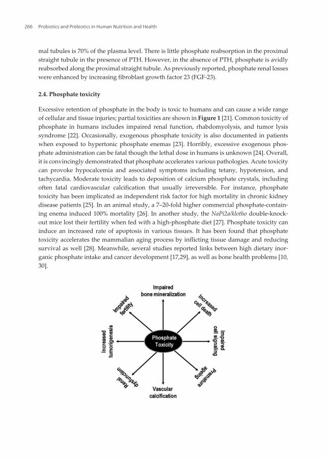
mal tubules is 70% of the plasma level. There is little phosphate reabsorption in the proximalstraight tubule in the presence of PTH. However, in the absence of PTH, phosphate is avidlyreabsorbed along the proximal straight tubule. As previously reported, phosphate renal losseswere enhanced by increasing fibroblast growth factor 23 (FGF-23).
2.4. Phosphate toxicity
Excessive retention of phosphate in the body is toxic to humans and can cause a wide rangeof cellular and tissue injuries; partial toxicities are shown in Figure 1 [21]. Common toxicity ofphosphate in humans includes impaired renal function, rhabdomyolysis, and tumor lysissyndrome [22]. Occasionally, exogenous phosphate toxicity is also documented in patientswhen exposed to hypertonic phosphate enemas [23]. Horribly, excessive exogenous phos‐phate administration can be fatal though the lethal dose in humans is unknown [24]. Overall,it is convincingly demonstrated that phosphate accelerates various pathologies. Acute toxicitycan provoke hypocalcemia and associated symptoms including tetany, hypotension, andtachycardia. Moderate toxicity leads to deposition of calcium phosphate crystals, includingoften fatal cardiovascular calcification that usually irreversible. For instance, phosphatetoxicity has been implicated as independent risk factor for high mortality in chronic kidneydisease patients [25]. In an animal study, a 7–20-fold higher commercial phosphate-contain‐ing enema induced 100% mortality [26]. In another study, the NaPi2a/klotho double-knock‐out mice lost their fertility when fed with a high-phosphate diet [27]. Phosphate toxicity caninduce an increased rate of apoptosis in various tissues. It has been found that phosphatetoxicity accelerates the mammalian aging process by inflicting tissue damage and reducingsurvival as well [28]. Meanwhile, several studies reported links between high dietary inor‐ganic phosphate intake and cancer development [17,29], as well as bone health problems [10,30].
Probiotics and Prebiotics in Human Nutrition and Health266
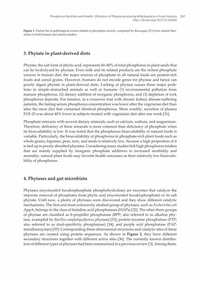
Figure 1. Partial list of pathological events related to phosphate toxicity composed by Razzaque [21] from related liter‐atures in both human and animal studies.
3. Phytate in plant-derived diets
Phytate, the salt form of phytic acid, represents 60–80% of total phosphorus in plant seeds thatcan be hydrolyzed by phytase. Even milk and its related products are the richest phosphatesources in human diet; the major sources of phosphate in all natural foods are protein-richfoods and cereal grains. However, humans do not encode genes for phytase and hence canpoorly digest phytate in plant-derived diets. Lacking of phytase causes three major prob‐lems in simple-stomached animals as well as humans: (1) environmental pollution frommanure phosphorus, (2) dietary addition of inorganic phosphorus, and (3) depletion of rockphosphorus deposits. For instance, in a crossover trial with chronic kidney disease-sufferingpatients, the fasting serum phosphorus concentration was lower after the vegetarian diet thanafter the meat diet that contained identical phosphorus. More notably, secretion of plasmaFGF-23 was about 40% lower in subjects treated with vegetarian diet after one week [31].
Phosphate interacts with several dietary minerals, such as calcium, sodium, and magnesium.Therefore, deficiency of these minerals is more common than deficiency of phosphate whenits bioavailability is low. It was noted that the phosphorus bioavailability of natural foods isvariable. Particularly, the bioavailability of phosphorus in phosphate-rich plant foods such aswhole grains, legumes, peas, nuts, and seeds is relatively low, because a high proportion of itis tied up in poorly absorbed phytates. Considering many studies link high phosphorus intakesthat are mainly supplied by inorganic phosphate additives to increased morbidity andmortality, natural plant foods may favorite health outcomes as their relatively low bioavaila‐bility of phosphorus.
4. Phytases and gut microbiota
Phytases (myoinositol hexakisphosphate phosphohydrolase) are enzymes that catalyze thestepwise removal of phosphates from phytic acid (myoinositol hexakisphosphate) or its saltphytate. Until now, a plenty of phytases were discovered and they show different catalyticmechanisms. The first and most extensively studied group of phytases, such as Escherichia coliAppA, belongs to the class of histidine acid phosphatases (HAPs) [32]. The other three groupsof phytase are classified as b-propeller phosphatase (BPP; also referred to as alkaline phy‐tase, exampled by Bacillus amyloliquefaciens phytase) [33], protein tyrosine phosphatase (PTP;also referred to as dual-specificity phosphatase) [34], and purple acid phosphatase (PAP;metalloenzymes) [35]. Corresponding three-dimensional structures and catalytic sites of thesephytases are created using protein sequences. As shown in Figure 2, they have differentsecondary structures together with different active sites [36]. The currently known distribu‐tion of different types of phytases had been summarized in a previous review [3]. Among them,
Phosphorus Nutrition and Health: Utilization of Phytase-producing Bifidobacteria in Food Industryhttp://dx.doi.org/10.5772/63644
267
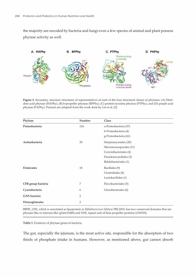
the majority are encoded by bacteria and fungi even a few species of animal and plant possess
phytase activity as well.
Figure 2. Secondary structure structures of representatives of each of the four structural classes of phytases. (A) Histi‐dine acid phytase (HAPhy), (B) b-propeller phytase (BPPhy), (C) protein tyrosine phytase (PTPhy), and (D) purple acidphytase (PAPhy). Pictures are adapted from the work done by Lei et al. [3].
Phylum Number Class
Proteobacteria 124 a-Proteobacteria (57)b-Proteobacteria (4)g-Proteobacteria (61)
Actinobacteria 39 Streptomycetales (20)Micromonosporales (11)Corynebacteriales (4)Pseudonocardiales (3)Bifidobacteriales (1)
Firmicutes 18 Bacillales (9)Clostridiales (8)Lactobacillales (1)
CFB group bacteria 7 Flavobacteriales (5)
Cyanobacteria 6 Gloeobacterales (4)
GNS bacteria 2
Dictyoglomales 2
BBPR_1292, which is annotated as lipoprotein in Bifidobacterium bifidum PRL2010, has two conserved domains that arephytase-like or esterase-like (pfam13449) and NHL repeat unit of beta-propeller proteins (cl18310).
Table 1. Existence of phytase genes in bacteria.
The gut, especially the jejunum, is the most active site, responsible for the absorption of two
thirds of phosphate intake in humans. However, as mentioned above, gut cannot absorb
Probiotics and Prebiotics in Human Nutrition and Health268

organic phosphorus presented as phytate in plant-derived diets. We know that human gutconsists of a complex community of microorganisms, namely, gut microbiota. One main roleof gut microorganisms is they benefit the host by fermentation of human readily undigestedsubstrates to absorbable nutrients. Some gut microorganisms produce kinds of enzymes andmany of these enzymes are deficient in host, thereby symbiotic relationship is developed. Sucha case is phytase producing microbe, like Escherichia coli, Streptomyces coelicolor, Clostridi‐um spp., and so on. Search of “phytase” in the NCBI gene database yielded 198 genes annotatedas bacterial phytase. A detailed presence of phytase genes in bacteria is demonstrated inTable 1. Among them, Proteobacteria and Actinobacteria are the most predominating groupsthat are also natural habitants of human gut.
5. Phytate degradation by bifidobacteria
5.1. Phytase-encoding genes
As shown in Table 1, there is only one gene in B. bifidum PRL2010 that was annotated aspossible phytase-encoding gene. Meanwhile, two enzymes in bifidobacteria, exactly B.pseudocatenulatum ATCC 27919 and B. longum subsp. infantis ATCC 15697 with phytaseactivity, have been characterized [37]. Therefore, protein sequences of these three enzymes(BBPR_1292, BIFPSEUDO_03792, and BLON_0263) were used for searching homologues inBifidobacterium (taxid: 1678).
Organism Protein name Accession Locus_tag Length(aa)
B. dentium ATCC 27678 Histidine acidphosphatase
WP_003838654 BIFDEN_01159 637
B. dentium ATCC 27679 Histidine acidphosphatase
WP_003843340 HMPREF0168_2166 631
B. dentium Bd1 Histidine acidphosphatase
WP_012902513 BDP_1985 643
B. longum DJO10A Histidine acidphosphatase
WP_010081042 Blon03000750 617
B. longum DJO10A Histidine acidphosphatase
WP_012472023 BLD_1202 622
B. longum NCC2705 Histidine acidphosphatase
WP_011068470 BL0400 606
B. longum subsp. infantis 157F Histidine acidphosphatase
WP_015713264 BLIF_0216 622
B. longum subsp. infantis ATCC15697=JCM 1222
Histidine acidphosphatase
WP_012576702 Blon_0263 623
Phosphorus Nutrition and Health: Utilization of Phytase-producing Bifidobacteria in Food Industryhttp://dx.doi.org/10.5772/63644
269

Organism Protein name Accession Locus_tag Length(aa)
B. longum subsp. infantis ATCC15697=JCM 1222
Histidine acidphosphatase
WP_014484530 BLIJ_0267 618
B. longum subsp. infantis CCUG 52486 Histidine acidphosphatase
WP_007051528 BLIG_00414 617
B. longum subsp. longum 35B Histidine acidphosphatase
WP_007057720 HMPREF1314_0451 572
B. longum subsp. longum 44B Histidine acidphosphatase
WP_007056476 HMPREF1312_1349 617
B. longum subsp. longum BBMN68 Histidine acidphosphatase
WP_013410389 BBMN68_1139 622
B. longum subsp. longum F8 Histidine acidphosphatase
WP_015512490 BIL_17170 617
B. long um subsp. longum JCM 1217 Histidine acidphosphatase
WP_007054753 BLLJ_0234 617
B. longum subsp. longum KACC 91563 Histidine acidphosphatase
WP_014485906 BLNIAS_02473 617
B. pseudocatenulatum DSM 20438=JCM1200
Histidine acidphosphatase
WP_004222312 BIFPSEUDO_03792 639
B. sp. 12_1_47BFAA Histidine acidphosphatase
WP_008783259 HMPREF0177_01170 561
Table 2. Protein list of histidine acid phosphatase in Bifidobacterium sp.*.
Available from http://www.ncbi.nlm.nih.gov/proteinclusters/?term=BIFPSEUDO_03792.
BLAST searches revealed that (1) BBPR_1292-like proteins are exclusively presented on thegenomes of all B. bifidum strains with at least 99% identity and (2) BIFPSEUDO_03792 andBLON_0263 are presented in a few strains of Bifidobacterium. Though there is the presence ofBBPR_1292-like proteins, there is no specific phytase activity that had been detected in B.bifidum. Based on sequence comparisons, these two characterized enzymes are more close tothe phytases of plants, fungi, and vertebrates. However, in the protein clusters database ofhistidine acid phosphatase (PCLA_3557679), there are 18 proteins that belong to 16bifidobacteria strains as listed in Table 2. Notably, all these predicted phytases belong to B.dentium, B. longum, and B. pseudocatenulatum. Nevertheless, phytase activity has been detectedin some Bifidobacterium sp. even it is not a common metabolic feature.
5.2. Phytase enzyme activities
Initially, it was believed that bifidobacteria are phytase negative, as very low level activity maybe because of unspecific release by phosphatase, except B. pseudocatenulatum ATCC 27919 [6].
Probiotics and Prebiotics in Human Nutrition and Health270

To further evaluate the enzyme activities, five strains of different bifidobacterial species, i.e.,B. animalis, B. bifidum, B. infantis, B. longum, and B. pseudolongum, were inoculated to degrademyoinositol hexaphosphate (InsP(6)). In a complex medium in which phytic acid was the onlysource of phosphorus, B. infantis ATCC 15697 showed the highest level of phytate-degrad‐ing activity. The optimal condition is at slight acid pH (6.0–6.5) and higher temperature (50°C).Maximum activity appears at the stationary phase of growth and when 1% lactose was usedas carbon source [7]. The same research team compared phosphatase and phytase activitiesof 23 bifidobacterial strains (13 from infants and 10 from adults) belonging to three differentspecies (B. longum, B. breve, and B. catenulatum). The highest phytate-degrading activity isdisplayed in B. longum BIF307, similar to previous comparison in which is B. infantis, asubspecies of B. longum has the highest phytase activity.
Although two novel phytases from B. pseudocatenulatum and B. longum subsp. infantis had beencharacterized, parallel comparison of bifidobacterial phytase activities to E. coli AppA isdifficult, as they were expressed as relative percentage activity. Nevertheless, the enzymes thatbelong to a new subclass are highly specific for the hydrolysis of phytate and rendermyoinositol triphosphate as the final hydrolysis product [37]. From our experience, nativephytase activity in bifidobacteria is extremely lower than commercial enzymes. Therefore, weconstructed a series of recombinant B. bifidum S17 that can secrete heterologous AppA withinhigh specific activity to phytate; even our primary aim is using appA as a suitable secretionreporter [8]. Among these constructs, B. bifidum S17/pMgapS6P using the GAP promoter andBBIF_1761 signal direct the most efficient phytase secretion (Figure 3).
Figure 3. Phytase activity in spent cell-free medium of recombinant B. bifidum S17 strains. B. bifidum S17-harboringpMgapP-derived plasmids containing different SPs (S0–S6) were grown in 5 ml reinforced clostridia medium underanaerobic condition. The control plasmid pMgapP (−) contains no SP and serves as a background control for expres‐sion of a nonsecreted phytase. Values are relative phytase units (RPU) per ml supernatant and are mean ± standarddeviation of three independent cultures measured in technical triplicates. The figure is adapted from Osswald et al. [8].
Phosphorus Nutrition and Health: Utilization of Phytase-producing Bifidobacteria in Food Industryhttp://dx.doi.org/10.5772/63644
271

6. Potential application of phytase-producing bifidobacteria in foods
Despite phytase’s main application in animal feeds, its applications in human foods can beequally important, if not exceed. To a large extent, using phytase in human foods is notprimarily the target to improve phosphorus consumption, because depletion of phytate is moreimportant as it chelates essential minerals, including iron, zinc, and calcium, contributing todeficiencies of these nutrients. It was estimated that there are approximately two to threebillion people around the world suffering from mineral deficiency. The application of phytasein human health may be more exciting but need further in-depth study of potential adverseeffects. Because certain inositol phosphates are beneficial to human health, phytase andphytase-producing cells can be immobilized as cost-effective bioreactors for large-scaleproduction of these compounds [39]. There are many successful attempts to use phytase inbrewing, baking, and dephytination of soy milk [25].
In fact, application of phytase or phytase-producing bacteria in food has already beenillustrated. For example, B. pseudocatenulatum ATCC 27919 was tested as a starter in sour‐dough for the production of whole rye-wheat mixed bread [40]. In situ production of phy‐tase during fermentation by probiotics results higher mineral availability in breads. The abilityof B. infantis ATCC 15697 to degrade InsP(6) and accumulate InsP(3) could contribute to thereduction of the anti-nutritional properties of InsP(6) and generation of intermediate com‐pounds with beneficial properties. B. longum BIF307, another phytase producer, was used inwhole wheat bread making and decreased InsP(6) content. In another study, phytase-produc‐ing bifidobacterial strains significantly reduced the InsP(6) + InsP(5) concentrations com‐pared to control samples during the bread-making process. Meanwhile, dialyzable Fe contentswere increased from 2.3- to 5.6-folds. However, the effects appeared to be still insufficient toimprove Fe bioavailability in Caco-2 cells [41]. Anyway, with similar technological andsensorial quality, levels of InsP(6) are significantly lower in bifidobacteria-fermented bread.Collectively, probiotic bifidobacteria are particularly suitable to reduce the content of InsP(6)in rich fiber products for human consumption [42].
7. Advantages of using phytase-producing bifidobacteria
One of the most important advantages of using phytase-producing bifidobacteria is safety. Aswidely known, several species of bifidobacteria are generally regarded as safe (GRAS) orqualified presumption of safety (QPS). The GRAS status made these strains particularlyattractive for application in both food and pharmaceutical industries. Currently, probioticbifidobacteria are widely used as micro-ecological reagent in many countries. These micro-ecological reagents had been added into both foods and pharmaceuticals without additionaltoxicity test.
Secondly, as important as the safety issue, many beneficial effects of bifidobacteria made thempromising in industry especially ameliorating gastrointestinal disorders (both bacterial- andviral-induced gastroenteritis), allergic diseases, antibiotic-associated diarrhea, lactose
Probiotics and Prebiotics in Human Nutrition and Health272
intolerance, constipation, and irritable bowel disease. Let alone increasing iron accessibility,phytase-producing bifidobacteria has expanded nutrition profile [41,42]. In the gut of humaneating plant-derived diets, phytase-producing bifidobacteria can degrade phytate-basedcomponents, therefore improving their adaptability or cross-feeding other symbiotic inhabi‐tants in the same niche.
Thirdly, intake of phytase-producing bifidobacteria is superior to eating inorganic phos‐phate additives for human. In one aspect, phosphorus homeostasis can be easily disturbedafter eating external inorganic phosphate additives. In another aspect, phytase-producingbifidobacteria can improve organic phytate-originated phosphorus. Thereby, supplementa‐tion of external inorganic phosphate additives becomes unnecessary. This is particularlysignificant for avoiding excessive phosphate, resulting in different kinds of toxicities that arelargely caused by phosphate-containing additives in foods and drinks. For example, phos‐phorus-based food additives may pose high risk in people suffering chronic kidney disease,as this made dietary management of hyperphosphatemia practically difficult. In dialysispatients, managing hyperphosphatemia may require using phosphate binder other thanrestricting protein intake as this allows patients to eat more protein-rich foods [43]. In addition,a study evaluated 93 premature infants with a mean gestational age of 27.5 ± 2.0 weeks. Theresult demonstrated that elevated serum phosphorus was inversely correlated to the day oflife of the infant after receiving human milk-derived fortifier though the incidence of hyper‐phosphatemia was mild and transient in this population [44]. For those health promisedpeople, intake of phytase-producing bifidobacteria supplied a novel interventional approach.
Lastly but not least, bifidobacteria can produce phytase in human gut as microbial cell factories.They can be ingested as live cells and then colonized in the intestine to facilitate the degrada‐tion of plant-derived diets. The relatively constant replication of bifidobacteria in humanintestine can either enlarge the bioavailability of organic phytate or downsize the toxicity ofexcessive phosphorus, hence maintaining the balance of phosphorus in a long term.
8. Final remarks
Phosphorus salts are added to foods as additives in many countries. Thus, dietary intake ofphosphorus is higher than the recommended daily allowance in these countries and popula‐tions. For instance, phosphorus additives were particularly common in the categories of smallgoods, bakery goods, frozen meals, and biscuits in Australia [45]. In the United States, it hasbeen estimated that phosphorus additives may add as much as 1 g of phosphorus to the diet[46]. However, high phosphorus intake has been shown to inhibit the increase in serum1,25(OH)2D concentration in response to low dietary calcium intake [30].
Collectively, although long-term and large amount consumption of phosphorus additives,little is known about risk associated with dietary phosphorus intake. A prospective cohortstudy of healthy adults reveals that high dosage of dietary phosphorus intake is associatedwith increased mortality [47]. Considering that the deleterious effects of chronic ingestion ofunrestricted amounts of phosphate in individuals are not clear, consumption of high phos‐
Phosphorus Nutrition and Health: Utilization of Phytase-producing Bifidobacteria in Food Industryhttp://dx.doi.org/10.5772/63644
273

phate-containing processed foods and soft drinks should be alarming, particularly for health-compromised individuals [48,49]. Under those circumstances as mentioned above, usingphytase-producing bifidobacteria may be a new way for increasing bioavailability of phos‐phorus from plant-derived diets, therefore avoiding supplementation of inorganic additives.
Author details
Long Chen1, Fengshou Tian2 and Zhongke Sun1*
*Address all correspondence to: [email protected]
1 College of Life Sciences and Agronomy, Zhoukou Normal University, Chuanhui District,Zhoukou City, China
2 School of Chemistry and Chemical Engineering, Zhoukou Normal University, ChuanhuiDistrict, Zhoukou City, China
References
[1] Fukagawa M, Hamada Y, Nakanishi S, Tanaka M. The kidney and bone metabolism:nephrologists’ point of view. J Bone Miner Metab. 2006; 24: 434–438.
[2] Huttunen MM, Pietila PE, Viljakainen HT, Lamberg-Allardt CJ. Prolonged increase indietary phosphate intake alters bone mineralization in adult male rats. J Nutr Biochem.2006; 17: 479–484.
[3] Lei XG, Weaver JD, Mullaney E, Ullah AH, Azain MJ. Phytase, a new life for an “old”enzyme. Annu Rev Anim Biosci. 2013; 1: 283–309.
[4] Kleerebezem M, Vaughan EE. Probiotic and gut lactobacilli and bifidobacteria:molecular approaches to study diversity and activity. Annu Rev Microbiol. 2009; 63:269–290.
[5] Gareau MG, Sherman PM, Walker WA. Probiotics and the gut microbiota in intesti‐nal health and disease. Nat Rev Gastroenterol Hepatol. 2010; 7: 503–514.
[6] Haros M, Bielecka M, Sanz Y. Phytase activity as a novel metabolic feature inBifidobacterium. FEMS Microbiol Lett. 2005; 247: 231–239.
[7] Haros M, Bielecka M, Honke J, Sanz Y. Myo-inositol hexakisphosphate degradation byBifidobacterium infantis ATCC 15697. Int J Food Microbiol. 2007; 117: 76–84.
Probiotics and Prebiotics in Human Nutrition and Health274
[8] Osswald A, Westermann C, Sun Z, Riedel CU. A phytase-based reporter system foridentification of functional secretion signals in bifidobacteria. PLoS ONE. 2015; 10(6):e0128802.
[9] Farrow EG, White KE. Recent advances in renal phosphate handling. Nat Rev Nephrol.2010; 6: 207–217.
[10] Trautvetter U, Jahreis G, Kiehntopf M, Glei M. Consequences of a high phosphorusintake on mineral metabolism and bone remodeling in dependence of calcium intakein healthy subjects – a randomized placebo-controlled human intervention study. NutrJ. 2016; 15(1): 7.
[11] Bergwitz C, Huppner H. Regulation of phosphate homeostasis by PTH, vitamin D andFGF-23. Annu Rev Med. 2010; 61: 91–104.
[12] Berndt TJ, Schiavi S, Kumar R. “Phosphatonins” and the regulation of phosphorushomeostasis. Am J Physiol Renal Physiol. 2005; 289: F1170–F1182.
[13] Xu H, Bai L, Collins JF, Ghishan FK. Age-dependent regulation of rat intestinal typeIIb sodium–phosphate cotransporter by 1,25-(OH)2 vitamin D3. Am J Physiol CellPhysiol. 2002; 282: C487–C493.
[14] Penido MG, Alon Uri S. Phosphate homeostasis and its role in bone health. PediatrNephrol. 2012; 27(11): 2039–2048.
[15] Magne D, Bluteau G, Faucheux C, Palmer G, Vignes-Colombeix C, Pilet P, Rouillon T,Caverzasio J, Weiss P, Daculsi G, Guicheux J. Phosphate is a specific signal for ATDC5chondrocyte maturation and apoptosis-associated mineralization: possible implica‐tion of apoptosis in the regulation of endochondral ossification. J Bone Miner Res. 2003;18: 1430–1442.
[16] Sabbagh Y, Carpenter TO, Demay MB. Hypophosphatemia leads to rickets byimpairing caspase-mediated apoptosis of hypertrophic chondrocytes. Proc Natl AcadSci U S A. 2005; 102: 9637–9642.
[17] Wilson KM, Shui IM, Mucci LA, Giovannucci E. Calcium and phosphorus intake andprostate cancer risk: a 24-y follow-up study. Am J Clin Nutr. 2015; 101(1): 173–183.
[18] Remer T, Manz F. Estimation of the renal net acid excretion by adults consuming dietscontaining variable amounts of protein. Am J Clin Nutr. 1994; 59: 1356–1361.
[19] Lotscher M, Kaissling B, Biber J, Murer H, Levi M. Role of microtubules in the rapidregulation of renal phosphate transport in response to acute alterations in dietaryphosphate content. J Clin Invest. 1997; 99: 1302–1312.
[20] Amanzadeh J, Reilly RF Jr. Hypophosphatemia: an evidence-based approach to itsclinical consequences and management. Nat Clin Pract Nephrol. 2006; 2: 136–148.
[21] Razzaque MS Phosphate toxicity: new insights into an old problem. Clin Sci (Lond).2011; 120(3): 91–97.
Phosphorus Nutrition and Health: Utilization of Phytase-producing Bifidobacteria in Food Industryhttp://dx.doi.org/10.5772/63644
275

[22] Berner YN, Shike M. Consequences of phosphate imbalance. Annu Rev Nutr. 1988; 8:121–148.
[23] Ismail EA, Al-Mutairi G, Al-Anzy H. A fatal small dose of phosphate enema in a youngchild with no renal or gastrointestinal abnormality. J Pediatr Gastroenterol Nutr. 2000;30: 220–221.
[24] Perlman JM. Fatal hyperphosphatemia after oral phosphate overdose in a prematureinfant. Am J Health Syst Pharm. 1997; 54: 2488–2490.
[25] Van-Etten RL, Davidson R, Stevis PE, MacArthur H, Moore DL. Covalent structure,disulfide bonding, and identification of reactive surface and active site residues ofhuman prostatic acid phosphatase. J Biol Chem. 1991; 266: 2313–2319.
[26] Martin RR, Lisehora GR, Braxton M Jr, Barcia PJ. Fatal poisoning from sodiumphosphate enema. Case report and experimental study. J Am Med Assoc. 1987; 257:2190–2192.
[27] Ohnishi M, Razzaque MS. Dietary and genetic evidence for phosphate toxicityaccelerating mammalian aging. FASEB J. 2010; 24(9): 3562–3571.
[28] Ohnishi M, Nakatani T, Lanske B, Razzaque MS. Reversal of mineral ion homeostasisand soft-tissue calcification of klotho knockout mice by deletion of vitamin D 1α-hydroxylase. Kidney Int. 2009; 75: 1166–1172.
[29] Lee YS, Ha YC, Chae CH, Beck GR Jr, Cho MH. High dietary inorganic phosphateincreases lung tumorigenesis and alters Akt signaling. Am J Respir Crit Care Med. 2009;179: 59–68.
[30] Takeda E, Yamamoto H, Yamanaka-Okumura H, Taketani Y. Increasing dietaryphosphorus intake from food additives: potential for negative impact on bone health.Adv Nutr. 2014; 5(1): 92–97.
[31] Moe SM, Zidehsarai MP, Chambers MA, Jackman LA, Radcliffe JS, Trevino LL,Donahue SE, Asplin, JR. Vegetarian compared with meat dietary protein source andphosphorus homeostasis in chronic kidney disease. Clin J Am Soc Nephrol. 2011; 6(2):257–264.
[32] Voormolen N, Noordzij M, Grootendorst DC, Beetz I, Sijpkens YW, van Manen JG,Boeschoten EW, Huisman RM, Krediet RT, Dekker FW. High plasma phosphate as arisk factor for decline in renal function and mortality in pre-dialysis patients. NephrolDial Transplant. 2007; 22: 2909–2916.
[33] Ha NC, Kim YO, Oh TK, Oh BH. Preliminary X-ray crystallographic analysis of a novelphytase from a Bacillus amyloliquefaciens strain. Acta Crystallogr D Biol Crystallogr.1999; 55: 691–693.
[34] Chu HM, Guo RT, Lin TW, Chou CC, Shr HL, Lai HL, Tang TY, Cheng KJ, Selinger BL,Wang AH. Structures of Selenomonas ruminantium phytase in complex with persulfat‐
Probiotics and Prebiotics in Human Nutrition and Health276

ed phytate: DSP phytase fold and mechanism for sequential substrate hydrolysis.Structure. 2004; 12: 2015–2024.
[35] Hegeman CE, Grabau EA. A novel phytase with sequence similarity to purple acidphosphatases is expressed in cotyledons of germinating soybean seedlings. PlantPhysiol. 2001; 126: 1598–1608.
[36] Mullaney EJ, Ullah AHJ. Phytases: attributes, catalytic mechanisms and applications.In Inositol Phosphates Linking Agriculture and the Environment, 2007. eds. BL Turner,AE Richardson, EJ Mullaney, pp. 97–110. Cambridge, MA: CABI
[37] Tenenhouse HS. Regulation of phosphorus homeostasis by the type IIa Na/phos‐phate cotransporter. Annu Rev Nutr. 2005; 25: 197–214.
[38] Palacios MC, Haros M, Rosell CM, Sanz Y. Selection of phytate-degrading humanbifidobacteria and application in whole wheat dough fermentation. Food Microbiol.2008; 25: 169–176.
[39] Meyer AS. Enzyme technology for precision functional food ingredient processes. AnnNY Acad Sci. 2010; 1190: 126–132.
[40] García-Mantrana I, Monedero V, Haros M. Myo-inositol hexakisphosphate degrada‐tion by Bifidobacterium pseudocatenulatum ATCC 27919 improves mineral availability ofhigh fibre rye–wheat sour bread. Food Chem. 2015; 178: 267–275.
[41] Sanz-Penella JM, Laparra JM, Sanz Y, Haros M. Assessment of iron bioavailability inwhole wheat bread by addition of phytase-producing bifidobacteria. J Agric FoodChem. 2012; 60(12): 3190–3195.
[42] Sanz-Penella JM, Tamayo-Ramos JA, Sanz Y, Haros M. Phytate reduction in bran-enriched bread by phytase-producing bifidobacteria. J Agric Food Chem. 2009; 57(21):10239–10244.
[43] Fouque D, Horne R, Cozzolino M, Kalantar-Zadeh K. Balancing nutrition and serumphosphorus in maintenance dialysis. Am J Kidney Dis. 2014; 64(1): 143–150.
[44] Chetta KE, Hair AB, Hawthorne KM, Abrams SA. Serum phosphorus levels inpremature infants receiving a donor human milk derived fortifier. Nutrients. 2015; 7(4):2562–2573.
[45] McCutcheon J, Campbell K, Ferguson M, Day S, Rossi M. Prevalence of phosphorus-based additives in the Australian food supply: a challenge for dietary education? J RenNutr. 2015; 25(5): 440–444.
[46] Bell RR, Draper HH, Tzeng DY, Shin HK, Schmidt GR. Physiological responses ofhuman adults to foods containing phosphate additives. J Nutr. 1977; 107: 42–50.
[47] Chang AR, Lazo M, Appel LJ, Gutiérrez OM, Grams ME. High dietary phosphorusintake is associated with all cause mortality: results from NHANES III. Am J Clin Nutr.2014; 99(2): 320–327.
Phosphorus Nutrition and Health: Utilization of Phytase-producing Bifidobacteria in Food Industryhttp://dx.doi.org/10.5772/63644
277

[48] Fernando GR, Martha RM, Evangelina R. Consumption of soft drinks with phosphor‐ic acid as a risk factor for the development of hypocalcemia in postmenopausal women.J Clin Epidemiol. 1999; 52: 1007–1010.
[49] Jin H, Xu CX, Lim HT, Park SJ, Shin JY, Chung YS, Park SC, Chang SH, Youn HJ, LeeKH, Lee YS, Ha YC, Chae CH, Beck GR Jr, Cho MH. High dietary inorganic phos‐phate increases lung tumorigenesis and alters Akt signaling. Am J Respir Crit CareMed. 2009; 179(1): 59–68.
[50] Lee KJ, Kim KS, Kim HN, Seo JA, Song SW. Association between dietary calcium andphosphorus intakes, dietary calcium/phosphorus ratio and bone mass in the Koreanpopulation. Nutr J. 2014; 13(1): 114.
[51] Segawa H, Hanazaki A, Miyamoto K. Intracellular and extracellular functions ofphosphorus compound in the body. Clin Calcium. 2016; 26(2): 187–191.
[52] Taal MW, Thurston V, McIntyre NJ, Fluck RJ, McIntyre CW. The impact of vitamin Dstatus on the relative increase in fibroblast growth factor 23 and parathyroid hor‐mone in chronic kidney disease. Kidney Int. 2014; 86(2): 407–413.
[53] Tamayo-Ramos JA, Sanz-Penella JM, Yebra MJ, Monedero V, Haros M. Novel phytas‐es from Bifidobacterium pseudocatenulatum ATCC 27919 and Bifidobacterium longumsubsp. infantis ATCC 15697. Appl Environ Microbiol. 2012; 78(14): 5013–5015.
[54] Ushasree MV, Gunasekaran P, Pandey A. Single-step purification and immobiliza‐tion of MBP-phytase fusion on starch agar beads: application in dephytination of soymilk. Appl Biochem Biotechnol. 2012; 167: 1–10.
[55] Wesseling-Perry K. FGF-23 in bone biology. Pediatr Nephrol. 2010; 25: 603–608.
Probiotics and Prebiotics in Human Nutrition and Health278

Chapter 14
Probiotic Microorganisms in Dry Fermented MeatProducts
Katarzyna Neffe‐Skocińska, Karolina Wójciak andDorota Zielińska
Additional information is available at the end of the chapter
http://dx.doi.org/10.5772/64090
Abstract
In the modern lifestyle, food is supposed not only to feed the hunger but also to providean appropriate amount and quality of nutrients necessary for proper functioning of thebody. The interest of consumers in functional food, including fermented products withprobiotic properties, has been growing for several years. Meat and meat products representone of the most important components of contemporary human diet. Meat fermenta‐tion is one of the oldest methods of preserving food. This is a low‐energy, biologicalacidulation which results in unique flavour and palatability, colour, microbiological safetyand tenderness. Changes of muscle form into fermented meat product are caused byhomo‐ or heterofermentative starter cultures or “wild” microorganisms which lower thepH. Fermented meat products are one of the most cherished and valuable food prod‐ucts. Fermentation and ageing process would deliver most aromatic and rich in flavourproducts, which is incomparable with other processes. A new solution is dry‐aged meatswith the use of new probiotic starter cultures with a high degree of health safety and longshelf life due to the inhibition of growth of the pathogenic microorganisms and thereforereduction of the formation of harmful compounds from protein transformation or lipidoxidation.
Keywords: dry‐fermented meat, probiotic starter cultures, lipid oxidation, biogenicamine, bioactive peptides
1. Introduction
Meat and meat products are one of the most important components of human nutrition.Manufacturing raw cured sausages has a very long tradition and its origin is to be sought in the

Roman times in the Mediterranean area. In the past, raw cured sausages were manufacturedonly in the cool and cold time of the year and, therefore, it was possible to avoid the risk ofspoilage. Nowadays, the preservation role of meat fermentation has become largely obsoletedue to the introduction of the cold chain. Nevertheless, fermented meat products remain verypopular and are still produced in large amounts, especially in Europe, probably because of theirunique and specific sensory properties, their convenience and their alleged rootedness inculinary and cultural heritage [1].
One of the most promising areas of development in the human nutritional field over the lasttwo decades has been the use of probiotics and recognition of their role in human health anddisease. Lactic acid‐producing bacteria are the most commonly used probiotics in foods andsupplements. The means by which probiotic bacteria elicit their health effects are not under‐stood fully, but may include competitive exclusion of enteric pathogens, neutralization ofdietary carcinogens, production of antimicrobial metabolites and modulation of mucosal andsystemic immune function [2]. According to the currently adopted definition by the Food andAgriculture Organization/World Health Organization [3], probiotics are defined as “livemicroorganisms which when administered in adequate amounts confer a health benefit on thehost”.
Although dairy products are the most commonly used food vehicles for the delivery ofprobiotics, several investigations dealing with the use of probiotics in cereal products [4, 5]vegetables and fruit juices [6, 7] and fermented meat products [8–13] to improve theirnutritional value as functional foods have been reported.
The commercial application of probiotics in meat products is not yet popular, mainly be‐cause of technological issues. As fermented meat products are processed without heating, theycould be suitable products for assessing probiotic LAB as starter cultures [14]. However,probiotics may be inactivated due to low pH or water activity value, as well as presence ofnative microorganisms or curing salt. The most important problem is to find compromisebetween technology, safety, quality and health‐beneficial value of food [15].
2. Lactic acid bacteria in meat fermentation
2.1. Traditional starter cultures
Starter cultures are live, defined and specially selected microorganisms with the GRAS(generally recognized as safe) safety status, responsible for the proper course of meat ageing.Starter cultures may consist of selected bacteria, moulds or yeasts. Their use in the produc‐tion of dry‐aged cold meats is always intentional and aims at obtaining the specified sensoryand microbiological characteristics in the end product [16–18].
The fermentation of food is known from centuries. First fermentation processes were drivenby adventitious microbiota, represented by unknown microorganisms naturally present in theraw food ingredients and in the environment. A number of fermented foods, such as tradi‐tional cheeses, dry‐fermented sausages and fermented beverages, are still produced withoutthe addition of microbial inoculation [19]. Over time, with the aim to improve and standard‐
Probiotics and Prebiotics in Human Nutrition and Health280

ize the foods, the subsequent evolutionary step has been constantly associated with the back‐slopping approaches, where higher counts of microorganisms are added to activate thefermentation. However, these natural starter cultures were often variable in load and compo‐sition, and if, on one hand, they can confer to the product peculiar characteristics of unique‐ness and quality, on the other hand, they are continuously evolving according to seasonal andenvironmental variations and may result in variable qualities of the final product. For thisreason, since the beginning of the past century, strains isolated from the best natural fermen‐tations have been cultivated and studied under defined conditions by industrial companiesand research institutions and used as selected starter cultures in food productions [20].
Group of microorganisms Technological function and changes in meat>
Lactic acid bacteriaLactobacillus sakeiLactobacillus curvatusLactobacillus plantarumLactobacillus pentosusLactobacillus caseiPediococcus acidilacticiPediococcus pentosaceus
Natural preservative: lactic acid production, acidifying bacteria;inhibitingthe development of putrefactive and pathogenic microorganisms;storage stabilization Good sensory quality, flavour and aromadevelopmentProteolysis and lipolysis stabilization
Gram‐positive cocciStaphylococcus carnosusStaphylococcus xylosusMicrococcus varians
Bacteria redox flavouring: nitrates and nitrites reduction; using upthe oxygen; decomposition of peroxides; lipolysis stabilization (delayingrancidity); colour stabilization; good sensory quality, flavour and aromadevelopment
YeastsDebaryomyces hanseniiCandida famata
The surface microflora: using up the oxygen; decomposition ofperoxides;delaying rancidity colour stabilization; good sensory quality, flavourand aroma development
MouldsPenicillium nalgiovensePenicillium camembertiPenicillium chrysogenum
The surface microflora: using up the oxygen; decomposition ofperoxides;proteolysis and lipolysis stabilization; good sensory quality, flavour andaroma development
Mixed composition of yeasts and mouldsPenicillium nalgiovense + Debaryomyces hanseniiPenicillium candidum + Debaryomyces hansenii
Table 1. The starter cultures composition for dry‐fermented meat production [15].
Currently, the production of commercial starter preparations uses primarily lactic acid bacteriawhich show favourable technological effect. Typical starter cultures (Table 1) which are usedto all types of fermented cold meats are composed of mildly acidifying bacteria cultures of thefollowing species: Lactobacillus sakei, L. curvatus, L. plantarum, Pediococcus acidilactici andPediococcus pentosaceus. In Europe, mainly cultures of L. sakei and L. curvatus are used to
Probiotic Microorganisms in Dry Fermented Meat Productshttp://dx.doi.org/10.5772/64090
281
manufacture dry‐aged cold meats [14, 16]. Strains of bacteria belonging to one species oftendiffer in various physiological and metabolic properties among each other. Due to the fact thatmost strains of L. curvatus demonstrate the ability to produce biogenic amines, L. sakei bacteriaare more often used in practice. These microorganisms are not capable of catalysing thedecarboxylation of amino acids, thereby reducing or even inhibiting the formation of biogen‐ic amines in dry‐fermented meat products [14, 15, 21–23].
In spite of LAB, starter cultures may also contain micrococci, most frequently those ofMicrococcus varians species and of Staphylococcus xylosus, Staphylococcus carnosus and Strepto‐myces griseus [15].
Apart from providing consistent quality and typical sensory features, the primary function ofLAB bacteria included in such starter cultures is mainly to preserve the product through theproduction of lactic acid during metabolic changes and competition with microbiota natural‐ly occurring in the meat product and pathogenic microorganisms. The composition of startercultures impacts the duration of the ageing process and storage stability of such products butalso on their flavour, odour and texture [16, 24].
2.2. Development of novel starter cultures
One of the major focuses of the current innovation in development of novel starter cultures tomeat industry seems to be on improved food safety and health properties. The isolation andselection of lactic acid bacteria which can be used as starter cultures in meat fermentationpresent a considerable challenge to standardization and management of quality of dry‐fermented sausage. The basic starter cultures used in meat industry are selected strains ofhomofermentative Lactobacillus (lactic acid bacteria (LAB)) and/or Pediococcus and Gram‐positive catalase‐positive cocci (GCC), nonpathogenic, coagulase‐negative staphylococci and/or Kocuria. Lactic acid bacteria originating from fermented meats are specially adapted to theecology of meat fermentation. The rapid production of lactic acid in those products is primarilyresponsible for the quality and safety of the product [16, 25].
First of all the addition of selected starter cultures usually induces a higher acidification,compared to the standard product, which was reported by several authors [13, 26–28].Moreover, in Spanish raw‐fermented sausage with addition of probiotic starter cultures, thereduction of fat and salt has been achieved [27]. Also flavour, texture and taste are veryimportant components of the final quality of dry‐fermented meat products, and most of thesetraits are related to the metabolic activities of microorganisms [20].
Selected LAB starter cultures could have positive influence on sensorial acceptation of dry‐fermented meat products. For example, in [29], they have found that Lb. sakei and Staphylococ‐cus equorum added to the Dacia sausage resulted in better smell intensity, overall quality andmastication attributes, as well as lower biogenic amines content in comparison to controlsamples. In Ref. [30], they studied the effect of selected LAB starter cultures in Italian dry‐fermented sausage and found that the obtained products were saltier, juicier and more tastyas compared to the control.
Probiotics and Prebiotics in Human Nutrition and Health282

It has been also found that probiotic starter cultures may have been successfully used infermentation process of meat products. In Ref. [31], they found that probiotic bacteria did notchange the characteristic flavour and aroma of raw sausages in comparison to productobtained from commercial starter cultures. Also in studies [12, 27], they have obtained thesausages with probiotic cultures addition and recorded a satisfactory overall sensory qualitywithout any noticeable off‐flavour.The recent literature is also well consistent in indicating advantages of selected starter culturesin the control of pathogenic bacteria and other spoilage microflora. Fermented meat productsare commonly considered safe for consumption, and the acidification by lactic acid starterbacteria is one of the main preserving factors. The most frequently isolated lactic acid bacteriafrom dry sausages processed with different technologies are L. sakei, L. curvatus and L.plantarum [32, 33].The lactic acid bacteria (LAB) produce an array of antimicrobial substances (such as organicacids, diacetyl, acetoin, hydrogen peroxide, reuterin, reutericyclin, antifungal peptides andbacteriocins) [34, 35]. Therefore, there is an increasing interest in lactic acid bacteria (LAB)derived from meat that can be used as starter or adjunct cultures in dry sausage fermentation.Their ability to produce bacteriocins and nonproteinaceous low‐molecular‐mass antimicrobialcompounds (mainly lactic acid and hydrogen peroxide) is of importance [36]. As there is nosufficient glucose in meat to reduce the pH, the addition of glucose is essential to develop thedesired metabolic activity to produce lactic acid via glycolysis. Hydrogen peroxide is producedafter glucose is consumed by cells [37].Bacteriocins are the peptides produced by lactic acid bacteria with antibacterial properties.These peptides can reduce or inhibit the growth of other Gram‐positive [38–40], and thus theycan be used to control the growth of food‐borne pathogens such as L. monocytogenes in foodproducts [41]. In Ref. [38], they isolated P. acidilactici from Spanish dry‐fermented sausagesand found that they had a strong inhibitory effect against members of Gram‐positive genera.It has been observed that starter cultures containing L. sakei reduced the growth of Listeria infermented sausages [42, 43]. Also L. curvatus and L. plantarum in sausage starter cultures haveshown antilisterial effect [44, 45]. In the other study [46], they reported antilisterial effect of alactic acid bacterium isolated from Italian salami. In Ref. [47], they found that nine strains ofLactobacillus casei and three strains of L. plantarum isolated from dry‐fermented sausages hadan antagonistic activity against the indicator species tested. The bacteriocin produced by L.casei was named as Lactocin 705 and showed antibacterial effects against L. plantarum, L.monocytogenes, S. aureus and a wide range of Gram‐negative bacteria. Bacteriocinogenic startercultures are recommended as an additional hurdle to reduce the risk of L. monocytogenes in drysausage [48]. In contrast sakacin P, synthesized by L. sakei subsp. sakei 2a isolated from porksausage, inhibits the growth of Listeria monocytogenes [49]. The addition of the bacteriocino‐genic L. sakei CTC494 in combination with some ingredients (i.e. pepper, salt and nitrite) insausage batter has a dramatic effect on L. monocytogenes survival in fermented sausages [50].Sakacins produced by L. sakei are mainly active against other LAB and L. monocytogenes as wellas against the Gram‐negative psychrotroph Aeromonas hydrophila [51, 52].
Probiotic Microorganisms in Dry Fermented Meat Productshttp://dx.doi.org/10.5772/64090
283

Besides prevention of microbiological hazards, also probiotic starter cultures have beendeveloped. Many advantages and disadvantages are connected with application of probioticbacteria to dry‐fermented meat products.
2.3. Probiotic starter cultures: benefits and problems
Dynamic development of the functional food market has contributed to the attempt to usestarter cultures consisting of probiotic LAB in meat processing. Two trends may be observedduring development of new probiotic starter cultures. One of them is an attempt to applyalready known probiotic cultures (from the gastrointestinal tract of healthy humans) used, e.g.in the production of fermented milk beverages. The second one consists in isolating of thestrains of lactic acid bacteria from naturally fermented meat products and examining them interms of probiotic qualities as well as of safety of use in an industrial scale [25, 53–55].The number of benefits arising from the use of probiotic starter cultures is worth noticing(Table 2). The important aspect of using cultures of probiotic bacteria in the production of dry‐fermented meat, in addition to the possibilities of growth and survival in meat environmentand exercising favourable effect of these microorganisms on human body, is the ability toinhibit the growth of pathogenic microflora, which usually is capable of producing biogenicamines. Model studies with the use of probiotic bacteria as a starter culture to manufactureripening meat products revealed that strains of Lactobacillus acidophilus, L. lactis subsp. lactis,Lactococcus lactis subsp. lactis, L. plantarum, L. reuteri and L. fermentum reduce or even inhibitthe production of biogenic amines in the products discussed [56–58].
Advantages Disadvantages
Meat, as a rich source of nutritioncomponents, is a suitable medium forprobiotic bacteria growth
Technological issues:
• Presence of native microflora can inhibit growth of probiotic bacteria
• Presence of other inhibitory factors: low water activity, low sugarcontents, nitrates and salt additives
• Difficulties with inoculation probiotic in appropriate number ofbacteria
• Stability of probiotic during storage
Beneficial effect on human healthcorresponding with applied probiotic strain
Difficulties with identification of probiotic strains in meat matrix
Inhibition of pathogen growth, production ofbacteriocin and antimicrobial compounds
Non‐sterile environment can affect the spontaneous fermentation orspoilage
Unique sensorial quality No detailed procedure for production of probiotic meat products
Reduction of fat oxidation, proteolyticreactions
Possibilities of biogenic amine production
Table 2. Benefits and issues related to application of probiotic starter cultures.
Probiotics and Prebiotics in Human Nutrition and Health284

As a result of their own metabolic changes, the probiotic starter cultures produce a number ofbacteriostatic and bactericidal substances (e.g. lactic acid, acetic acid, formic acid, ethanol andbacteriocins), thereby inhibiting the growth of undesired and pathogenic microbiota, includ‐ing Gram‐negative strains from family Enterobacteriaceae (e.g. Escherichia coli) and Pseudomo‐naceae as well as species such as Listeria monocytogenes or Staphylococcus aureus, thus naturallypreserving the products discussed [14, 48, 59, 60].
Another benefit arising from the use of probiotic bacteria to manufacture dry‐fermented meatproducts is reduction of fat oxidation and proteolytic reactions. Primary and secondaryproducts resulting from degradation of lipids and proteins have significant impact ondeterioration of sensory quality (i.e. negative aftertastes: bitter, of old fat) but also of featuressuch as colour and texture. Additionally, they affect the health safety and shorten the use bydate of the meat products discussed [18, 61–63]. Research on autoxidation of the fat in ripeningproducts clearly proves that probiotic strains protect lipids from oxidation during storagewhich is indicated by substantially lower values of TBARS parameter in comparison to thecontrol sample [64, 65].
Proteolysis is one of the most important biochemical processes in dry‐aged meat productswhere proteins are metabolized and broken down to polypeptides, peptides and free aminoacids [62, 66]. Protein breakdown takes place with the participation of microbiologicalenzymes, which in turn leads to deamination and decarboxylation. These reactions occur fasterat low pH values. The basic products of protein decarboxylation in meat are cadaverine,putrescine, tyramine and histamine. Large concentration of biogenic amines in meat prod‐ucts may result in adverse symptoms in consumers, such as increase in blood pressure, increasein rate and strength of heart contraction and problems with the central nervous system,including migraines. Additionally, it may cause stomachaches, vomiting or severe sweating[56]. It has been observed that the proper choice of probiotic strains limits proteolytic changesin dry‐aged meat products [15, 26, 67]. This relation has been observed in studies [66] and [23]which examined dry‐aged pork tenderloin with addition of L. casei ŁOCK 0900 strain withdocumented probiotic and very good technological properties. Controlling and properconduct of the process of fermentation and ageing ensure low concentration of biogenic aminesin raw cold‐meat products, thereby preventing poisoning [15].
It is currently believed that dry‐fermented meat products are an appropriate medium forprobiotic bacteria. Many studies have shown that growth and survival rate of probiotic LABin dry‐fermented sausages are possible [18, 60]. However, the previous attempts to manufac‐ture meat product in controlled process of fermentation and ageing conducted by proven andselected probiotic strains mainly pertain to sausages [1, 10, 23, 33, 54, 57, 68, 69]. Moreover,there are few research works published which will unequivocally confirm the technologicalsuitability of probiotic cultures and their healthy impact on human body caused by regularconsumption of such meat products [70–72].
Probiotic strains used in the production of dry‐aged meat products must demonstrate suitabletechnological properties without departing from the traditional starter cultures used in meatprocessing. Therefore, the idea to use probiotic starter cultures in meat processing industry
Probiotic Microorganisms in Dry Fermented Meat Productshttp://dx.doi.org/10.5772/64090
285

raises many issues from the technological, microbiological and analytical perspective(Table 2).
The task consisting in introducing the probiotic starter cultures to meat is not easy to per‐form, as these bacteria which are in fact the intestinal bacteria do not demonstrate very goodtechnological properties. It was determined that from 50 to 500 million of lactic acid bacteria,including mainly L. sakei and L. curvatus, may be placed into one gram of the product discussed.Therefore, the raw meat material itself constitutes a problem, because spontaneous growth ofLAB may occur there.
Secondly, inoculation of the probiotic bacteria is performed to the raw material which is notsterile as is the case of fermented milk products or fermented juices [73, 74]. Thirdly, certainanalytic difficulties also emerge here. Identification of selected probiotic strains in meat withthe use of traditional microbiological methods is not complete, because only the generalnumber of LAB is assayed. Only advanced identification methods based on genetic analysisof nucleotide sequences typical for given bacterial strain may ensure that all of them will beassayed [54].
Numerous authors of available literature focus on the survival rate of probiotic starter bacteriaadded to dry‐fermented meat products. The factors limiting and even inhibiting the growthof the discussed microbes in meat environment include mainly the native microflora but alsolow water activity and the content of sugars naturally occurring in meat, as well as techno‐logical additives: sodium chloride, nitrates and other curing agents [15].
Low content of simple sugars in fresh meat, necessary for lactic acid bacteria, includingprobiotic ones, to conduct metabolic change, also poses a significant problem (4.5–7 mM/g ofraw material). Therefore, microorganisms discussed start to use amino acids as an alterna‐tive source of carbon which starts the spoilage process of meat and results in intensive bittertaste. For this reason, saccharides are added in amount of 0.4–0.8% during the production ofaged meat products [13, 48].
There is also a technological difficulty in inoculating bacterial strain (form, number, applica‐tion method) to cured element to an unground meat in particular. Starter cultures are mostoften manufactured in lyophilized or frozen form. In the case of dry‐fermented sausages, theyare inoculated to the sausage meat, usually after they are mixed with cold water or curingbrine. Adding starter cultures to tenderloin or ham is significantly more difficult, as they arealso posed by varied consistency of different muscles. They result, i.e. from the presence of fatat the meat's surface which may uneven drying up and thereby excessive or inhibited growthof microorganisms [15].
One of the major technological problems involves no detailed procedure for production ofprobiotic meat products developed. This primarily requires determination of optimaltemperature for fermentation and ageing, which will allow probiotic strains to grow anddominate natural microbiota in the meat. Fermentation and ageing of meat products are mainlyconducted in the temperature within the range of 15–26°C. Lower temperature allows to obtainhigh‐quality product with a long ageing period and use by date. In contrast, the use of higher
Probiotics and Prebiotics in Human Nutrition and Health286

temperature decreases the duration of fermentation; however there is a risk that microorgan‐isms responsible for product's spoilage will grow in the meat [13, 15].
Additionally, one of the important technological criteria to be met by a probiotic strain includesstability during storage, i.e. capability to retain the number of 6.00–7.00 log cfu/g in the productin the last use by date [70, 71].
Another aspect of the selection of bacterial cultures pertains mainly to their beneficial impacton sensory quality and also moderate acidifying activity, low thermal activity within rangeof 0–15°C, antagonism with respect to undesired microbes and food pathogens as well asresistance to bacteriophages [18, 56, 75].
Probiotic microorganisms selected to the production of aged meat products must meet notonly the necessary requirements of safety and functionality but also the technological criteriadiscussed above.
3. Technological and health aspects of probiotic meat products
3.1. Influence of probiotic bacteria on lipid oxidation
The shelf life of fermented meat products is generally not limited by bacterial deteriorationbut by chemical spoilage [76]. It is oxidation stability that is the main restriction on the shelflife of probiotic meat products [77]. Processed products, which are minced, mixed with saltand heated, expose muscle tissue to oxidative stress responsible for loss of quality and lead tooxidative flavours and loss of haem iron and vitamins and finally cause discoloration [77].Oxidation of lipids can also have a negative effect on nutritional value and may be responsi‐ble for the production of toxic compounds. In Ref. [76], they have reported that enzymatichydrolysis during fermentation accelerates lipid peroxidation. In addition, a strong correla‐tion between lipid and myoglobin oxidation, especially in fresh meat, has been documentedby scientists [78]. In [78], they have reported that secondary lipid oxidation products (2‐heptenal, 2‐nonenal, 4‐hydroxy‐2‐nonenal) promote pigment oxidation. However, there is alsostrong evidence that haem pigments may initiate lipid oxidation through the reaction ofhydrogen peroxide with metmyoglobin to form ferryl and perferrylmyoglobin, which havepowerful prooxidant effects on lipids. In probiotic meat product, the fact that the high 20–30%concentration of fat (dry‐fermented ham, neck, sausage) does not limit the adaptive capacityof probiotic bacteria in fermented meat products is interesting. Probiotic bacteria can stabi‐lize the oxidation process taking during the maturing and prolonged storage period. In Ref.[79], they found that neither the presence nor the level of probiotic Bifidobacterium animalis ssp.lactis BB‐12 has a negative influence on colour and oxidative stability of dry‐cured neckduring 12 months of ageing. The authors observed the significantly (P < 0.05) lower TBARSvalues in neck B1 and B2 (1.54 and 1.69 mg MDA/kg) compared to the values with spontane‐ously added LAB (2.26 mg MDA/kg). In [80], they pointed out that inoculation with L.fermentum HL57 potential probiotic strain increased the amount of malondialdehyde in Iberiandry‐fermented sausages resulting in a negative colour and taste. The interaction of myoglo‐
Probiotic Microorganisms in Dry Fermented Meat Productshttp://dx.doi.org/10.5772/64090
287
bin with H2O2 activates metmyoglobin, which may be a ferrylmyoglobin radical that is veryunstable and can transform rapidly into the peroxyl radical form. On the other hand, inresearch [81], they proposed that L. casei, L. plantarum, L. curvatus and L. sakei strains activelycontribute to the hydrolysis of sarcoplasmic proteins such as myoglobin. Also in Ref. [82], theyfound that some strains of lactic acid bacteria demonstrated antioxidative activity withinhibition rates of ascorbate autoxidation in the range of 7-12%. In another paper the sameauthors presented that six strains of L. acidophilus and two strains of B. longum demonstratedan inhibitory effect on linoleic acid peroxidation [83]. The inhibitory rates of linoleic acidperoxidation ranged from 33 to 46% when 1 mL of intracellular cell‐free extract was tested [83].Authors of the Ref. [18] found out that the addition of probiotic strain L. casei ŁOCK 0900changed physicochemical profile of dry‐fermented sausages. Sausages with lower probioticbacteria inoculation (6.0 log cfu/g) had better quality, inclusive colour and lipid oxidativestability (lower peroxide value, conjugated dienes and TBARS value) than those with 6.3 logcfu/g of probiotic strain. Based on research [18], it can be concluded that dry‐fermentedsausages produced with probiotic L. casei 0900 ŁOCK are oxidatively stable and the stabilityof the fat does not limit the shelf life of probiotic‐treated dry‐fermented sausages. The studysuggests that the probiotic strain can be used in the production of edible sausage. The studyconducted by authors of [84] proved that the use of potential probiotic L. acidophilus Bauer indry‐fermented pork neck production process decreases the hydroperoxide concentration at alevel comparable to synthetic antioxidant.
Studies of the effects of the probiotic L. rhamnosus LOCK 0900 strain with green tea extract onthe oxidative stability of ageing dry‐cured pork loin showed that adding the probiotic strainwith natural antioxidant increased the antioxidant potential of meat product by lowering theoxidation‐reduction potential and TBARS values and improving the part of red in the generaltone of colour [13].
The results presented by authors of Ref. [85] clearly demonstrated that the use of probioticstrains mixture (L. casei LOCK 0900, L. casei LOCK 0908 and Lactobacillus paracasei LOCK 0919)is possible in manufacturing process of organic dry‐fermented sausages without nitrate and/ornitrite. The uncured fermented sausages with probiotic strains have appropriate oxidativestability and are shelf‐stable during 180 days of storage period [85].
Examples of starter cultures that represent significant influences on fermented meat prod‐ucts regarding lipid oxidation, proteolysis, biogenic amine formation and sensorial qualitywere collected in Table 3.
Starter culture Product Influence Literature
L. rhamnosus LOCK900(Formerly Lactobacilluscasei ŁOCK0900)
Dry‐fermentedsausage
The probiotic strain can be used in the productionof edible sausageDry‐fermented sausage produced with probiotic isoxidatively stable during storage
[18]
Dry‐fermentedpork loin
Pork loin inoculation with a probiotic strainhas different free amino acid concentrations, the taste
[92]
Probiotics and Prebiotics in Human Nutrition and Health288
Starter culture Product Influence Literature
and flavour attributes
Dry‐fermentedpork loin
Inoculation with a probiotic strain proved to be aprotective measure against the formation andaccumulation of biogenic amine
[91]
L. acidophilus Bauer Dry‐fermentedpork neck
Probiotic strain decreases the hydroperoxideconcentration at a level comparable tosynthetic antioxidantL. acidophilus Bauer has antioxidant properties
[84]
L. rhamnosus LOCK900(Formerly Lactobacilluscasei ŁOCK0900)
Dry‐curedpork loin
Addition of probiotic strain with green teaextract increased the antioxidant potential of meatproduct and improved the part of red in thegeneral tone of colour
[13]
L. rhamnosus LOCK900, L.rhamnosus LOCK908 andL. casei LOCK919(Formerly L. casei ŁOCK0900, L. casei ŁOCK 0908and L. paracasei ŁOCK0919)
Uncured fermentedpork sausage(organic sausage)
The uncured fermented sausages with probiotic strainmixture have appropriate oxidative stability and areshelf‐stable during 180 days of storage period
[85]
Table 3. Influence of probiotic starter cultures on lipid oxidation, proteolysis, biogenic amine formation and sensorialquality in meat processing.
3.2. Influence of probiotic bacteria on proteolysis and biogenic amine formation
Proteolysis results in generation of peptides, oligopeptides and finally free amino acids(FAAs). Proteolysis is one of the most important biochemical changes, which take place duringageing of fermented meat products. FAAs contribute to the basic taste and aroma of ferment‐ed meat products [86]. However, an excessive amount of FAAs seems to be responsible for thebiogenic amine formation.
The biogenic amines (BAs) are the compounds in which one, two or three hydrogens ofammonia are replaced by alkyl or aryl groups. Tyramine and phenylethylamine have aromaticstructure, while putrescine, cadaverine, spermine and spermidine have the aliphatic one.Heterocyclic structures were proved for histamine and tryptamine. Based on number of aminegroup, we can divide the BA into the monoamines (phenylethylamine, tyramine), diamines(cadaverine, putrescine) and polyamines (spermidine, spermine) [87]. Biogenic amines havebeen reported in variety of foods, such as fish, meat, cheese, vegetable and wine [88]. They canbe formed but also degraded as a result of normal metabolism of living cells in plant, animaland microorganism. BA can be produced by two different pathways: firstly by the decarbox‐ylation of free amino acids and secondly by the amination and transamination of aldehydesand ketones [15]. The control of biogenic amines formation mainly focused on the control‐ling the growth of biogenic amine‐forming bacteria. Microorganisms have a different ability
Probiotic Microorganisms in Dry Fermented Meat Productshttp://dx.doi.org/10.5772/64090
289

to synthesize decarboxylases. Pseudomonas, Enterobacteriaceae, enterococci and lactobacilli werefound to have a high decarboxylase activity [88]. Within the same species, the presence, theactivity and the specificity of decarboxylases are strain dependent. Bacterial amino aciddecarboxylases usually have an acidic pH optimum 4.9–5.3, because the BA productions havebeen recognized as defence microbial mechanisms against an acidic environment [88, 89]. Onthe other hand, rapid and intense acidifications of environment reduce the growth ofEnterobacteriaceae and enterococci. The factors that could significantly influence the BAformations in fermented meat are pH value, redox potential, environment microorganisms,starter culture, temperature of maturing, salt concentration, additives, water activity andhygienic quality of meat [89]. Authors of paper [90] proved that when salt concentrationincreased from 0 to 6%, the rate of BA production of Lactobacillus delbrueckii subsp. bulgaricusdecreased. Other authors proved that concentration of sodium chloride at the level from 3.5to 5.5% inhibited the histamine formation. Several studies showed that temperature offermentation, maturing and storing have influence on BA accumulation in meat product [88].Higher maturing temperature (20–25°C) could stimulate the growth of LAB which inhibits theamine‐positive bacteria. The storing temperature below 4°C inhibits most of amine‐positivebacteria except from psychrotrophic Pseudomonas, so the BA concentration in this kind of meatproduct is relatively low compared with product stored at 14–16°C. The addition of sugar(glucose, lactose) to fermented meat products has some influence on bacterial populationdynamics and BA production. The absence of sugar stimulated the proteolysis and tyramine,cadaverine, putrescine and tryptamine formation. The preservative sodium nitrite could beadded to fermented meat products because of their ability to reduce the putrescine andcadaverine accumulation. The addition of pure or mixture of various probiotic strains candecrease BA formation in fermented meat products. It is extremely important that probioticstarter culture should not form BA and have to be competitive in suppressing growth of amine‐positive microbiota. In Ref. [91], they pointed out that pork loin inoculation with a probioticstrain L. casei ŁOCK 0900 has different free amino acid concentrations, which could influ‐ence the taste and flavour attributes. Also in [92], they presented evidence that some sorts ofprobiotic starter cultures (L. casei ŁOCK 0900, Bifidobacterium bifidum, L. acidophilus Bauer) havedifferent abilities to create the high‐ or low‐molecular‐weight peptides and free amino acids.Another authors [93] found out that LAB reduced the pH values during ageing and therebyactivated the endogenous acid protease (cathepsin B, L). Inoculation with a probiotic strain L.casei ŁOCK 0900 proved to be a protective measure against the formation and accumulationof cadaverine, putrescine, spermine and tryptamine. In Ref. [91] the author has not observedthe correlation between the higher content of free amino acids (potential precursor of BA) andthe level of biogenic amines. A 50% BA decrease was observed in comminuted fermented meatproducts with mixture of L. curvatus CTC371 and S. xylosus despite the increase of free aminoacid availability. In Ref. [23] BA changes during maturation were presented. Potential probioticpork loins were analysed in 4‐, 8‐ and 16‐month‐old samples. The authors have not detectedhistamine and spermidine. Spermine was present at very low levels (4.0–5.8 mg/kg), whilecadaverine (10.8–39.6 mg/kg) and tryptamine (17.8–49.2 mg/kg) were the most abundant BA.The level of all BA did not exceed the suggested toxic limits [23].
Probiotics and Prebiotics in Human Nutrition and Health290

3.3. Bioactive peptides in probiotic fermented meat products
The proteolytic activity during meat processing generates a large amount of peptides and freeamino acids because of calpain, cathepsin and peptidase enzymatic activity [86]. The mostinteresting peptides are those that can be considered as bioactive peptides because of theirdifferent health‐care abilities like antihypertensive activity, antioxidant activity, antimicrobialactivity, etc. The activities of bioactive peptides depend on their chemical structure (aminoacids composition, kind of amino acid in N‐ and C‐terminal), the length of molecule chain andtheir weight, charge character of amino acids and the hydrophobic/hydrophilic property. Meathas been reported to contribute to the generation of bioactive peptides. As such peptides,antihypertensive, opioid, immunostimulating, antimicrobial, antithrombotic, hypocholester‐olemic, antioxidative and prebiotic activities have been studied. Angiotensin I‐convertingenzyme (ACE) plays an important physiological role in the regulation of blood pressure.Fermented food with probiotic bacteria, especially Lactobacillus, produce bioactive peptidesknown to inhibit the activity of angiotensin‐converting enzyme (ACE) and thus alleviate highblood pressure (hypertension). Authors of the study [94] used calpis sour milk fermented withLactobacillus helveticus and Saccharomyces cerevisiae and identified two peptides Ile‐Pro‐Pro andVal‐Pro‐Pro, both of which possessed ACE inhibitory activity in vitro. These two bioactivepeptides were released from b‐casein and k‐casein by lactobacilli enzymes. In Ref. [95],hypertensive subjects were fed with milk fermented with L. helveticus LBK‐16H containingbioactive peptides. After 21 weeks test subjects showed a significant lowering of their bloodpressure. Even in antihypertensive peptides, it has been found that antioxidative (VW, DLYA,SLYA, DLQEKLE) and prebiotic (ELM) peptides are generated from meat protein byenzymatic digestion. In [96], they reported that bioactive peptides from hydrolysis ofsarcoplasmic porcine proteins by the activity of L. sakei CRL 1862 and L. curvatus CRL 705showed high ACE inhibitory activity. In [97], they pointed out that some peptides from Spanishdry‐cured ham have DPPH radical‐scavenging activity (39–92%) as well as superoxide ion‐extinguishing ability with values ranging from 41.67 to 50.27% of the antioxidant activity,suggesting the presence of peptides with antioxidant activity. Moreover every sampleexhibited pooled fractions corresponding to 1700 Da or lower were the most antihyperten‐sive with a decrease of 38.38 mm Hg in systolic blood pressure [97]. The antioxidant activityof low‐molecular‐weight compounds isolated from Iberian‐fermented sausage (chorizo) wastested in Ref. [98]. Authors did not observe many bioactive peptides; however plenty freeamino acids, bacterial metabolites and β‐alanyl‐peptides have been identified. Bioactivepeptides from meat and fermented meat products exhibit various biological activities whichare favourable for human health.
4. Conclusion
In recent years, the possibility of development of probiotic meat products has been discussedin the field of meat science and industry. Probiotics in meat may exert their benefits by way ofseveral mechanisms; therefore human clinical studies are needed to assess the health‐
Probiotic Microorganisms in Dry Fermented Meat Productshttp://dx.doi.org/10.5772/64090
291

promoting effect of probiotic dry‐fermented meat products. So far, there is a little scientificevidence related to such studies. Therefore, further studies are required to demonstrate theclear benefits of probiotic meat products for human health.
On the other hand, the use of probiotic strain which possesses the ability to create the bioactivecompounds is the challenge for research and meat industry and could develop novel functionalmeat products. Possible generation of bioactive peptides in dry meat products fermented byprobiotics as starter cultures seems the most promising way for designing novel functionalfood. Therefore meat products with probiotics have a great future potential and it is expectedthat increasing interest will be shown in basic research and potential applications for designingnew meat products.
Target products with probiotic bacteria are mainly dry sausages but also hams and loins, whichare processed by fermentation without heat treatment. Technically, it has already becomepossible to produce probiotic meat products; moreover probiotic raw‐fermented meatproducts exist on a German and Japanese market. However, the production of probiotic meatproducts requires overcoming certain technological limitations, such as the native microfloraof meat, a need to use additives such as nitrites and salt and also low water activity and lowcontent or absence of natural sugars. Probiotic bacteria strains that can be used in themanufacturing of dry‐fermented meat products should be capable of surviving in conditionsfound in fermented products. Moreover, the product should maintain its sensorycharacteristics.
Additionally, since food safety is another critical aspect of food quality, efforts should also bedirected to ensure that new functional meat products are safe. The dry‐fermented meatproducts with probiotic starter cultures addition have to possess appropriate biogenic amineprofile and should be oxidative and shelf‐stable. Along with accumulation of scientific data,there is an urgent need to inform consumers of the exact physiological value of probiotic meatproducts. Without proof of product safety, most consumers would hesitate to adopt new foodsin their diet.
Acknowledgements
The authors would like to express our appreciation to Professor Danuta Kołożyn‐Krajewskaand Professor Zbigniew Dolatowski for their support and constructive critiques during allstudies on the possibility of the use of probiotic starter cultures in meat fermentation.
Author details
Katarzyna Neffe‐Skocińska1*, Karolina Wójciak2 and Dorota Zielińska1
*Address all correspondence to: [email protected]
Probiotics and Prebiotics in Human Nutrition and Health292

1 Department of Food Gastronomy and Food Hygiene, Warsaw University of Life Sciences(WULS), Warsaw, Poland
2 Department of Meat Technology and Food Quality, University of Life Sciences (LULS),Lublin, Poland
References
[1] Leroy F, Geyzen A, Janssens M, De Vuyst L, Scholliers P. Meat fermentation at thecrossroads of innovation and tradition: a historical outlook. Trends in Food Science &Technology. 2013;31(2):130–137.
[2] Corthesy B, Gaskins HR, Mercenier A. Cross‐talk between probiotic bacteria and thehost immune system. Journal of Nutrition. 2007;137:781S–790S.
[3] FAO/WHO. Report of a joint FAO/WHO working group of drafting guidelines for theevaluation of probiotics in food: guidelines for the evaluation of probiotics in food,London; 2002.
[4] Kedia G, Wang R, Patel H, Pandiella SS. Use of mixed cultures for the fermentation ofcereal‐based substrates with potential probiotic properties. Process Biochemistry.2002;42(1):65–70.
[5] Rozada‐Sánchez R, Sattur AP, Thomas K, Pandiella SS. Evaluation of Bifidobacteriumspp. for the production of a potentially probiotic malt‐based beverage. ProcessBiochemistry. 2002;43(8):848–854.
[6] Kun S, Rezessy‐Szabó JM, Nguyen QD, Hoschke Á. Changes of microbial populationand some components in carrot juice during fermentation with selected Bifidobacteri‐um strains. Process Biochemistry. 2008;43(8):816–821.
[7] Zielińska D, Kamińska A, Kołożyn‐Krajewska D. Development of tofu productionmethod with probiotic bacteria addition. The Journal of Microbiology, Biotechnologyand Food Sciences. 2015;4(6):485.
[8] Klingberg TD, Axelsson L, Naterstad K, Elsser D, Budde BB. Identification of poten‐tial probiotic starter cultures for Scandinavian‐type fermented sausages. Internation‐al Journal of Food Microbiology. 2005;105(3):419–431.
[9] Macedo REFD, Pflanzer Jr SB, Terra NN, Freitas RJSD. Production of fermented sausageusing probiotic Lactobacillus strains: quality characteristics. Food Science andTechnology (Campinas). 2008;28(3):509–519.
[10] Pennacchia C, Vaughan EE, Villani F. Potential probiotic Lactobacillus strains fromfermented sausages: further investigations on their probiotic properties. Meat Science.2006;73(1):90–101.
Probiotic Microorganisms in Dry Fermented Meat Productshttp://dx.doi.org/10.5772/64090
293
[11] Rouhi M, Sohrabvandi S, Mortazavian AM. Probiotic fermented sausage: viability ofprobiotic microorganisms and sensory characteristics. Critical Reviews in Food Scienceand Nutrition. 2013;53(4):331–348.
[12] Rubio R, Aymerich T, Bover‐Cid S, Guàrdia MD, Arnau J, Garriga M. Probiotic strainsLactobacillus plantarum 299V and Lactobacillus rhamnosus GG as starter cultures forfermented sausages. LWT‐Food Science and Technology. 2013;54(1):51–56.
[13] Neffe‐Skocińska K, Jaworska D, Kołożyn‐Krajewska D, Dolatowski Z, Jachacz‐JówkoL. The effect of LAB as probiotic starter culture and green tea extract addition on dryfermented pork loins quality. BioMed Research International. 2015. Available from:http://dx.doi.org/10.1155/2015/452757 [Accessed: 2015‐01‐01].
[14] Ammor MS, Mayo B. Selection criteria for lactic acid bacteria to be used as functionalstarter cultures in dry sausage production: an update. Meat Science. 2007;76(1):138–146.
[15] Kołożyn‐Krajewska D, Dolatowski ZJ. Probiotic meat products and human nutrition.Process Biochemistry. 2012;47(12):1761–1772.
[16] Hugas M, Monfort JM. Bacterial starter cultures for meat fermentation. FoodChemistry. 1997;59(4):547–54.
[17] Erkkilä S, Petäjä E, Eerola S, Lilleberg L, Mattila‐Sandholm T, Suihko ML. Flavourprofiles of dry sausages fermented by selected novel meat starter cultures. MeatScience. 2001;58:111–116.
[18] Wójciak KM, Dolatowski ZJ, Kołożyn‐Krajewska D, Trząskowska M. The effect of theLactobacillus casei ŁOCK 0900 probiotic strain on the quality of dry‐fermented sausageduring chilling storage. Journal of Food Quality. 2012;35,353‐365.
[19] Steinkraus K. Handbook of Indigenous Fermented Foods, revised and expanded.2nd ed. CRC Press; 1995. 121p.
[20] Bassi D, Puglisi E, Cocconcelli PS. Comparing natural and selected starter cultures inmeat and cheese fermentations. Current Opinion in Food Science. 2015;2:118–122.
[21] Pereira CI, Crespo MT, Romao MV. Evidence for proteolytic activity and biogenicamines production in Lactobacillus homohiochii. International Journal of FoodMicrobiology. 2001;68(3):211–216.
[22] Bover‐Cid S, Miguelez‐Arrizado MJ, Latorre Moratalla LL, Vidal Carou MC. Freez‐ing of meat raw materials affects tyramine and diamine accumulation in spontaneous‐ly fermented sausages. Meat Sciences. 2006;72(1):62–68.
[23] Stadnik J, Dolatowski ZJ. Biogenic amines content during extended ageing of dry‐curedpork loins inoculated with probiotics. Meat Science. 2012;91:374–377.
[24] Incze K. Fermented meat products. Fleischwirtschaft. 2002;4:112–117.
Probiotics and Prebiotics in Human Nutrition and Health294

[25] Lücke FK. Utilization of microbes to process and preserve meat. Meat Science.2000;56(2):105–115.
[26] Castellano P, Aristoy MC, Sentandreu MA, Vignolo G, Toldra F. Lactobacillus sakeiCRL1862 improves safety and protein hydrolysis in meat systems. Journal of AppliedMicrobiology. 2012;113(6):1407–1416.
[27] Rubio R, Jofré A, Aymerich T, Guàrdia MD, Garriga M. Nutritionally enhancedfermented sausages as a vehicle for potential probiotic lactobacilli delivery. MeatScience. 2014;96(2):937–942.
[28] Jaworska D, Neffe K, łożyn-Krajewska D, Dolatowski Z. Survival during storage andsensory effect of potential probiotic lactic acid bacteria Lactobacillus acidophilus Bauerand Lactobacillus casei Bif3'/IV in dry fermented pork loins. International Journal of FoodScience & Technology. 2011;46(12):2491–2497.
[29] Simion AMC, Vizireanu C, Alexe P, Franco I, Carballo J. Effect of the use of selectedstarter cultures on some quality, safety and sensorial properties of Dacia sausage, atraditional Romanian dry‐sausage variety. Food Control. 2014;35(1):123–131.
[30] Cenci‐Goga BT, Rossitto PV, Sechi P, Parmegiani S, Cambiotti V, Cullor JS. Effect ofselected dairy starter cultures on microbiological, chemical and sensory characteris‐tics of swine and venison (Dama dama) nitrite‐free dry‐cured sausages. Meat Science.2012;90(3):599–606.
[31] Sameshima T, Magome C, Takeshita K, Arihara K, Itoh M, Kondo Y. Effect of intesti‐nal Lactobacillus starter cultures on the behaviour of Staphylococcus aureus in ferment‐ed sausage. International Journal of Food Microbiology. 1998;41(1):1–7.
[32] Schillinger U, Lücke FK. Antibacterial activity of Lactobacillus sakei isolated from meat.Applied and Environmental Microbiology. 1989;55(8):1901–1906.
[33] Papamanoli E, Tzanetakis N, Litopoulou‐Tzanetaki E, Kotzekidou P. Characteriza‐tion of lactic acid bacteria isolated from a Greek dry‐fermented sausage in respect oftheir technological and probiotic properties. Meat Science. 2003;65(2):859–867.
[34] Holzapfel WH, Geisen R, Schillinger U. Biological preservation of foods with refer‐ence to protective cultures, bacteriocins and food‐grade enzymes. International Journalof Food Microbiology. 1995;24(3):343–362.
[35] Leroy F, De Vuyst L. Lactic acid bacteria as functional starter cultures for the foodfermentation industry. Trends in Food Science & Technology. 2004;15(2):67–78.
[36] Gaenzle MG, Hertle C, van der Vossen JMBM, Hammes WP. Effect of bacteriocin‐producing lactobacilli on the survival of Escherichia coli and Listeria in a dynamic modelof the stomach and the small intestine. International Journal of Food Microbiology.1999;48(1):21–35.
Probiotic Microorganisms in Dry Fermented Meat Productshttp://dx.doi.org/10.5772/64090
295

[37] Condon S. Responses of lactic acid bacteria to oxygen. FEMS Microbiology Reviews.1987;3(3):269–280.
[38] Cintas LM, Rodriguez JM, Fernandez MF, Sletten K, Nes IF, Hernandez PE, Holo HIsolation and characterization of pediocin L50, a new bacteriocin from Pediococcusacidilactici with a broad inhibitory spectrum. Applied and Environmental Microbiolo‐gy. 1995;61(7):2643–2648.
[39] Cleveland J, Montville TJ, Nes IF, Chikindas ML. Bacteriocins: safe, natural antimicro‐bials for food preservation. International Journal of Food Microbiology. 2001;71(1):1–20.
[40] Diep DB, Nes IF. Ribosomally synthesized antibacterial peptides in Gram positivebacteria. Current Drug Targets. 2002;3(2):107–122.
[41] Ennahar S, Sonomoto K, Ishizaki A. Class IIa bacteriocins from lactic acid bacteria:antibacterial activity and food preservation. Journal of Bioscience and Bioengineering.1999;87(6):705–716.
[42] Hugas M, Garriga M, Aymerich MT, Monfort JM. Inhibition of Listeria in dry ferment‐ed sausages by the bacteriocinogenic Lactobacillus sakei CTC494. Journal of AppliedMicrobiology.1995;79(3):322–330.
[43] De Martinis EC, Franco BD. Inhibition of Listeria monocytogenes in a pork product by aLactobacillus sakei strain. International Journal of Food Microbiology. 1998;42(1):119–126.
[44] Campanini M, Pedrazzoni I, Barbuti S, Baldini P. Behaviour of Listeria monocyto‐genes during the maturation of naturally and artificially contaminated salami: effect oflactic‐acid bacteria starter cultures. International Journal of Food Microbiology.1993;20(3):169–175.
[45] Dicks LMT, Mellett FD, Hoffman LC. Use of bacteriocin‐producing starter cultures ofLactobacillus plantarum and Lactobacillus curvatus in production of ostrich meat salami.Meat Science. 2004;66(3):703–708.
[46] Teixeira de Carvalho AA, Aparecida de Paula R, Mantovani HC, Alencar de Moraes C.Inhibition of Listeria monocytogenes by a lactic acid bacterium isolated from Italiansalami. Food Microbiology. 2006;23(3):213–219.
[47] Vignolo GM, Suriani F, Holgado APDR, Oliver G. Antibacterial activity of Lactobacil‐lus strains isolated from dry fermented sausages. Journal of Applied Microbiology.1993;75(4),344‐349.
[48] Työppönen S, Petäjä E, Mattila‐Sandholm T. Bioprotectives and probiotics for drysausages. International Journal of Food Microbiology. 2003;83(3):233–244.
[49] De Carvalho KG, Bambirra F, Kruger M, Barbosa M, Oliveira J, Santos A, Nicoli J,Bemquerer M, De Miranda A, Salvucci E, Sesma F, Franco B. Antimicrobial com‐pounds produced by Lactobacillus sakei subsp. sakei 2a, a bacteriocinogenic strain
Probiotics and Prebiotics in Human Nutrition and Health296

isolated from a Brazilian meat product. Journal Indian Microbiology Biotechnology.2010;37:381–390.
[50] Hugas M, Garriga M, Pascual M, Aymerich MT, Monfort JM. Enhancement of sakacinK activity against Listeria monocytogenes in fermented sausages with pepper ormanganese as ingredients. Food Microbiology. 2002;19(5):519–528.
[51] Schillinger U, Lücke FK. Antibacterial activity of Lactobacillus sakei isolated from meat.Applied and Environmental Microbiology. 1989;55(8):1901–1906.
[52] Lewus CB, Montville TJ. Detection of bacteriocins produced by lactic acid bacteria.Journal of Microbiological Methods. 1991:13(2), 145–150.
[53] Hammes WP, Hertel C. New developments in meat starter cultures. Meat Science.1998;49:S125–138.
[54] Kröckel L. Use of probiotic bacteria in meat products. Fleischwirtschaft. 2006;86:109–113.
[55] Aro Aro JM, Nyam‐Osor P, Tsuji K, Shimada K, Fukushima M, Sekikawa M. The effectof starter cultures on proteolytic changes and amino acid content in fermentedsausages. Food Chemistry. 2010;119(1):279–285.
[56] Stadnik J. Biogenic amines in dry cured meat products. Food. Science. Technology.Quality. 2013;3(88):5–15.
[57] Ruiz‐Moyano S, Martin A, Benito MJ, Hernandez A, Casquete R, Serradilla MJ, CórdobaMC. Safety and functional aspects of pre‐selected lactobacilli for probiotic use in Iberiandry‐fermented sausages. Meat Science. 2009;83:460–467.
[58] Deepika Priyadarshani WM, Rakshit SK. Screening selected strains of probiotic lacticacid bacteria for their ability to produce biogenic amines (histamine and tyramine).International Journal of Food Science and Technology. 2011;46(10):2062–2069.
[59] Coll Cárdenas F, Giannuzzi NE, Zaritzky NE. Mathematical modeling of microbialgrowth in ground beef from Argentina. Effect of lactic acid addition, temperature andpackaging film. Meat Science. 2008;79:509–520.
[60] Rivera‐Espinoza Y, Gallardo‐Navarro Y. Non‐dairy probiotic products. FoodMicrobiology. 2010;27:1–11.
[61] Toldrá F, Flores M. The role of muscle proteases and lipases in flavour developmentduring the processing of dry‐cured ham. Critical Reviews in Food Science andNutrition. 1998;38:331–352.
[62] Flores M, Barat JM, Aristoy MC, Peris MM, Grau R, Toldr F. Accelerated processing ofdry‐cured ham. Part 2. Influence of brine thawing/salting operation on proteolysis andsensory acceptability. Meat Science. 2006;72:766–772.
Probiotic Microorganisms in Dry Fermented Meat Productshttp://dx.doi.org/10.5772/64090
297
[63] Singh VP, Pathak V, Verma AK. Fermented meat products: organoleptic qualities andbiogenic amines – a review. American Journal of Food Technology. 2012;7:278–88.
[64] Estevez M, Cava R. Effectiveness of rosemary essential oil as an inhibitor of lipid andprotein oxidation: contradictory effects in different types of frankfurters. Meat Science.2006;72:348–55.
[65] Valencia I, Ansorena D, Astiasaran I. Stability of linseed oil and antioxidants contain‐ing dry fermented sausages: a study of the lipid fraction during different storageconditions. Meat Science. 2006;73:269–77.
[66] Okoń A, Dolatowski ZJ. Proteolysis of proteins in raw‐ripening meat products usingLactobacillus casei ŁOCK 0900 probiotic strain. Food. Science. Technology. Quality.2012;6(85):138–151.
[67] Bodmer S, Imark C, Kneubuhl M. Biogenic amines in foods: histamine and foodprocessing. Inflammation Research. 1999;48:296–300.
[68] Pennacchia C, Ercolini D, Blaiotta G, Pepe O, Mauriello G, Villani F. Selection ofLactobacillus strains from fermented sausages for their potential use as probiotics. MeatScience. 2004;67:309–317.
[69] Arihara K. Strategies for designing novel functional meat products. Meat Science.2006;74:219–229.
[70] Jahreis G, Vogelsang H, Kiessling G, Schubert R, Bunte C, Hammes WP. Influence ofprobiotic sausage (Lactobacillus paracasei) on blood lipids and immunological parame‐ters of healthy volunteers. Food Research International. 2002;235:133–138.
[71] Klingberg TD, Budde BB. The survival and persistence in the human gastrointestinaltract of five probiotic lactobacilli consumed as freeze‐dried cultures or as probioticsausage. International Journal of Food of Microbiology. 2006;109:157–159.
[72] Jiménez‐Colmenero F, Ventanas J, Toldrá F. Nutritional composition of dry‐cured hamand its role in a healthy diet. Meat Science. 2010;84:585–593.
[73] Klingberg TD, Axelsson L, Naterstad K, Elsser D, Bude BB. Identification of potentialstarter cultures for Scandinavian‐type fermented sausages. International Journal ofFood of Microbiology. 2005;105:419–431.
[74] Heller KJ. Probiotic bacteria in fermented foods: product characteristics and starterorganisms. American Journal of Clinical Nutrition. 2005;73:374S‐379S.
[75] Khan MI, Arshad MS, Anjum FM, Sameen A, Rehman A, Gill WT. Meat as a function‐al food with special reference to probiotic sausages. Food Research International.2011;44:3125–3133.
[76] Nassu RT, Goncalves LAG, Da Silva MAAP, Beserra FJ. Oxidative stability of ferment‐ed goat meat sausage with different levels of natural antioxidant. Meat Science.2003;63:43–49.
Probiotics and Prebiotics in Human Nutrition and Health298
[77] Summo C, Caponio VM, Pasqualone A. Effect of vacuum‐packaging storage on thequality level of ripened sausages. Meat Science. 2006;74:249–254.
[78] Faustman C, Liebler DC, Mcclure TD, SUN Q. Alpha, beta‐unsaturated aldehydesaccelerate oxymyoglobin oxidation. Journal of Agriculture and Food Chemistry,1999;47:3140–3144.
[79] Libera J, Karwowska M, Stasiak DM, Dolatowski ZJ. Microbiological and physico‐chemical properties of dry‐cured neck inoculated with probiotic of Bifidobacteriumanimalis ssp. lactis BB‐12. International Journal of Food Science and Technology.2015;50:1560–1566.
[80] Ruiz‐Moyano S, Martán A, Benito MJ, Hernández A, Casquete R, De Guia Córdoba M.Application of Lactobacillus fermentum HL57 and Pediococcus acidilactici SP979 aspotential probiotics in the manufacture of traditional Iberian dry‐fermented sausages.Food Microbiology. 2011;28:839–847.
[81] Fadda S, Sanz Y, Vignolo G, Aristoy M, Oliver G, Toldrá F. Hydrolysis of pork musclesarcoplasmic proteins by Lactobacillus curvatus and Lactobacillus sakei. Applied andEnvironmental Microbiology. 1999;65:578–584.
[82] Lin MY, Yen CL. Antioxidative ability of lactic acid bacteria. Journal of Agriculture andFood Chemistry. 1999;47:1460–1466.
[83] Lin MY, Yen CL. Inhibition of lipid peroxidation by Lactobacillus acidophilus andBifidobacterium longum. Journal of Agriculture and Food Chemistry. 1999;47:3661–3664.
[84] Libera J, Dolatowski ZJ. Effect of Lactobacillus acidophilus (Bauer) and Bifidobacteriumbifidum on probiotic bacteria on changes in fat in dry‐fermented meat products duringstorage. Food. Science. Technology. Quality. 2014;4(95):160–172.
[85] Wójciak KM, Karwowska M, Dolatowski ZJ. Fatty acid profile, colour and lipidoxidation of organic fermented sausage during chilling storage as influenced by acidwhey and probiotic strains addition. Scientia Agricola. 2015;72(2):124–131.
[86] Toldrá F, Reig M. Innovation for healthier processed meats. Trends in Food Science andTechnology. 2011;22:517–522.
[87] Spano G, Russo P, Lonvaud‐Funel A, Lucas P, Alexandre H, Grandvalet C. Biogenicamines in fermented foods. European Journal of Clinical Nutrition. 2010;64:95–100.
[88] Suzzi G, Gardini F. Biogenic amines in dry fermented sausages: a review. Internation‐al Journal of Food Microbiology. 2003;88:41–54.
[89] Chong CY, Abu Bakary F, Russly AR, Jamilah B, Mahyudin NA. The effects of foodprocessing on biogenic amines formation. International Food Research Journal.2011;18(3):867–876.
[90] Chander H, Batish VH, Babu, S, Singh RS. Factors affecting amine production by aselected strain of Lactobacillus bulgaricus. Journal of Food Science. 1989;54:940–942.
Probiotic Microorganisms in Dry Fermented Meat Productshttp://dx.doi.org/10.5772/64090
299

[91] Stadnik J, Dolatowski ZJ. Free amino acids and biogenic amines content during ageingof dry‐cured pork loins inoculated with Lactobacillus casei ŁOCK 0900 probiotic strain.Food Science and Technology Research. 2015;21(2):167–174.
[92] Okoń A, Dolatowski ZJ. Effect of probiotic bacteria on free amino acid profile andsensory traits of raw‐ripening pork sirloin during storage. Food. Science. Technology.Quality. 2014;3(94):92–107.
[93] Casaburi A, Di Monaco R, Cavella S, Toldrá F, Ercolini D, Villani F. Proteolytic andlipolytica starter cultures and their effect on traditional fermented sausages ripeningand sensory traits. Food Microbiology. 2008;25(2):335–347.
[94] Nakamura Y, Yamamoto N, Sakai K, Takano T. Antihypertensive effect of sour milkand peptides isolated from it that are inhibitors to angiotensin I‐converting enzyme.Journal of Dairy Science. 1994;78:1253–1257.
[95] Seppo L, Jauhiainen T, Poussa T, Korpela R. A fermented milk high in bioactivepeptides has a blood pressure‐lowering effect in hypertensive subjects. The Ameri‐can Journal of Clinical Nutrition. 2003;77:326–330.
[96] Castellano P, Aristoy MC, Sentandreu MA, Vignolo G, Toldrá F. Peptides withangiotensin I converting enzyme (ACE) inhibitory activity generated from porcineskeletal muscle proteins by the action of meat‐borne Lactobacillus. Journal of Pro‐teome Research. 2013;89:183–190.
[97] Escudero E, Aristoy MC, Nishimura H, Arihara K, Toldrá F. Antihypertensive effectand antioxidant activity of peptide fractions extracted from Spanish dry‐cured ham.Meat Science. 2012;91:306–311.
[98] Broncano JM, Otte J, Petrón MJ, Parra V, Timón ML. Isolation and identification of lowmolecular weight antioxidant compounds from fermented “chorizo” sausages. MeatScience. 2012;90:494–501.
Probiotics and Prebiotics in Human Nutrition and Health300

Chapter 15
Pectic Oligosaccharides and Other Emerging Prebiotics
Beatriz Míguez, Belén Gómez, Patricia Gullón,Beatriz Gullón and José Luis Alonso
Additional information is available at the end of the chapter
http://dx.doi.org/10.5772/62830
Abstract
A prebiotic is a selectively fermented ingredient that results in specific changes in thecomposition and/or activity of the gastrointestinal microbiota, thus conferring benefit(s)upon host health. The most widely accepted prebiotics are lactulose, inulin, fructooligo‐saccharides (FOS), galactooligosaccharides (GOS), and the human milk oligosacchar‐ides (HMO). However, there is a growing list of potential prebiotics although the evidencefor these, especially in humans, is not as well established as for FOS and GOS. Some ofthem are already commercialized but others such as polydextrose (PDX), pecticoligosaccharides (POS), bacterial exopolysaccharides (EPS), polysaccharides derivedfrom algae and sugar alcohols are still in the early stages of development. This chaptersummarizes the scientific literature regarding the manufacture and the evaluation of theproperties of this group “emerging prebiotics”.
Keywords: emerging prebiotics, pectic oligosaccharides, polydextrose, algae-derivedoligosaccharides, bacterial exopolysaccharides, sugar alcohols
1. Introduction
The consumption of prebiotics is being specially considered as a good health-improving strategy;they have been recently defined as “nondigestible compounds that through its metaboliza‐tion by microorganisms in the gut, modulate the composition and/or the activity of the gutmicrobiota, thus conferring physiological benefit effects on the host health” [1].
The microbial communities that inhabit the human intestinal tract constitute a complexassociation, comprising more than 1000 species and around 1014 microorganisms, mainlyanaerobic (>99.9%). Figure 1 shows the human gastrointestinal tract, indicating the different

levels of microorganisms and the main bacterial groups. Along the jejunum, and particular‐ly in the ileum, there is a gradual increase in the number and diversity of bacteria, and finally,the majority of gastrointestinal microbes are housed in the colon [2].
Figure 1. The human gastrointestinal tract (CFU, colony-forming units).
However, scientific works on this field suggest that the gut microbiota is not only a simplycollection of microorganisms, but also reflects an interrelationship between the differentgroups that might work together for the benefit of the host [2]. In addition, the microbiota alsoestablishes a close symbiosis with the host: humans provide the nutrients and the appropri‐ate conditions for its development, and it performs three essential primary functions: meta‐bolic, trophic, and defensive [3]. In fact, there is a long list of pathologies which are linked tothe alteration of the gut microbiota, including hepatic encephalopathy, diarrhea, diabetes,obesity, colon cancer, IBS, IBD, gastrointestinal infections, and necrotizing enterocolitis [4, 5].
The composition of the gut microbiota is influenced by a variety of factors that include: (i) themicrobial species which are acquired at birth, (ii) host genetics, (iii) age [6–8], (iv) diseases andantibiotic usage [9, 10], (v) the stress [11], and (vi) the diet. In fact, the diet is probably the mostimportant factor and several studies are focused on the modulation of the gut microbiota bythe consumption of functional foods, such as prebiotics [12–14].
For considering a food ingredient as a prebiotic, it must fulfill the following requirements [15]:(i) it cannot be hydrolyzed or absorbed in the upper gastrointestinal tract, (ii) it has toencourage the development of beneficial bacteria such as bifidobacteria and lactobacilli, and(iii) it must induce beneficial physiological effects on the host health, so that well-conductedhuman trials are required.
In addition to the generally identified as beneficial bacteria (bifidobacteria, lactobacilli, andeven, eubacteria), a recent review by Hill et al. [16] indicates that the species Akkermansiamuciniphila and Faecalibacterium prausnitzii, and others such as Roseburia spp. and Eubacteri‐um hallii, which could be useful to alleviate gut inflammation, to induce and regulate of theimmune system or to improve the intestinal barrier function.
Probiotics and Prebiotics in Human Nutrition and Health302
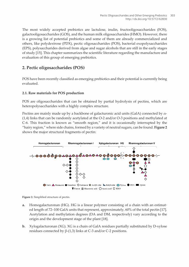
The most widely accepted prebiotics are lactulose, inulin, fructooligosaccharides (FOS),galactooligosaccharides (GOS), and the human milk oligosaccharides (HMO). However, thereis a growing list of potential prebiotics and some of them are already commercialized andothers, like polydextrose (PDX), pectic oligosaccharides (POS), bacterial exopolysaccharides(EPS), polysaccharides derived from algae and sugar alcohols that are still in the early stagesof study [15]. This chapter summarizes the scientific literature regarding the manufacture andevaluation of this group of emerging prebiotics.
2. Pectic oligosaccharides (POS)
POS have been recently classified as emerging prebiotics and their potential is currently beingevaluated.
2.1. Raw materials for POS production
POS are oligosaccharides that can be obtained by partial hydrolysis of pectins, which areheteropolysaccharides with a highly complex structure.
Pectins are mainly made up by a backbone of galacturonic acid units (GalA) connected by α-(1,4) links that can be randomly acetylated at the O-2 and/or O-3 positions and methylated atC-6. This fraction is known as “smooth region,” and it is occasionally interrupted by the“hairy region,” where side chains, formed by a variety of neutral sugars, can be found. Figure 2shows the major structural fragments of pectin:
Figure 2. Simplified structure of pectin.
a. Homogalacturonan (HG). HG is a linear polymer consisting of a chain with an estimat‐ed length of 72–100 GalA units that represent, approximately, 60% of the total pectin [17].Acetylation and methylation degrees (DA and DM, respectively) vary according to theorigin and the development stage of the plant [18].
b. Xylogalacturonan (XG). XG is a chain of GalA residues partially substituted by D-xyloseresidues connected by β-(1,3) links at C-3 and/or C-2 positions.
Pectic Oligosaccharides and Other Emerging Prebioticshttp://dx.doi.org/10.5772/62830
303

c. Rhamnogalacturonan I (RG-I). It represents up to 7–14% of the pectins [19] and containsalternating units of α-(1,4)-galacturonosyl and α-(1,2)-rhamnosyl. In many cases,rhamnose residues show side chains as substituents on the O-4 position, made up ofarabinan and/ or arabinogalactan I and II, although lower concentrations of xylose orglucose can be also found [18].
d. Rhamnogalacturonan II (RG-II). RGII is a region characterized by a length of 7–9 GalAunits, where complex branches made up of 12 types of monosaccharides (as a maxi‐mum) can exist, including some minority monomers such as apiose, fucose, acetic acid,DHA, or KDO [20].
Pectin has a great number of applications including its use as ingredient for medicaments fortreating gastrointestinal disorders, diabetes, high blood pressure, or hypercholesterolemia [21–23].
Currently, citrus pulp and apple pomace are the major sources of pectin, but this polymer canalso be found in other agro-products such as sugar beet pulp [24].
2.2. Manufacture and purification
Several methods have been used for POS production from both agro-industrial byproductsand purified pectins, including partial enzymatic hydrolysis, acid hydrolysis, hydrothermaltreatments, dynamic high-pressure microfluidization, or photochemical reaction in mediacontaining TiO2 [24].
Chemical methods include the acidic or basic hydrolysis of α and β-glucosidic links of theprincipal chains of HG, RG-I, and RG-II and their side chains. These methodologies includehydrothermal treatments and processes where external acids are added. In both cases,hydronium ions act as catalytic species [24]. A variety of raw materials such as orange albedo,apple pulp, or deesterified beet pulp have been treated to obtain POS, using acids such asHNO3, HCl, or TFA [24], although alkalis (KOH) can also be employed [25]. POS mixtures havebeen obtained from lemon and orange peel wastes [26–28], dried apple pomace [29], sugar beetpulp [30], or alperujo [31] using stainless steel reactors, whereas Sato et al. [32] employed botha batch and a continuous tubular flow reactor to produce arabinooligosaccharides (AraOS)and feruloylated AraOS from beet fiber.
As an alternative, pectin-degrading enzymes constitute a group of enzymes that catalyze thedegradation of the pectic polymers in plant cells. Although pectins have a complex structure,they can be modified by diverse enzymes, including hydrolases, lyases, and esterases [33].
Several raw materials with different characteristics have been enzymatically treated, such asbergamot peel [34], gum tragacanth [35, 36], ginseng pectin [37], orange peel wastes [38], lemonpeel wastes [39], sugar beet [40, 41], apple pectin [42], or medicinal herbs [43].
Both mono-active and commercial mixtures can be used for pectin depolymerization; however,mono-active enzymes target only specific structures, causing the release of more definedoligosaccharides than when commercial enzyme mixtures or chemical treatments are em‐ployed [44]. Mixture of several preparations have been widely employed for POS produc‐
Probiotics and Prebiotics in Human Nutrition and Health304
tion [38, 41, 45–48]. A comparable yield respect to acidic treatments can be achieved usingenzyme preparations [42].
In addition, enzymes can be also advantageous for the alteration of the methylation oracetylation degree of the polymer [44].
On the other hand, chemical and enzymatic hydrolysis have been combined to depolymer‐ize pectin [49] or to obtain different pectin fractions, such as POS and neutral and acidicxylooligosaccharides [31]. Other technologies that have been combined are enzymatic andmicrowave-assisted alkaline extraction [48], hydrothermal and acid treatment for polygalac‐turonic acid hydrolysis [50], subcritical water and ultrasonic-assisted treatments [51].
Finally, physical technologies (for instance, the dynamic high-pressure microfluidizationunder acidic conditions) have been emerged as innovative [52].
After production, purification stages are usually needed to obtain a product suitable to be usedas food ingredient. The most common purification technique is the membrane filtration. Aprocess involving diafiltration followed by concentration was performed by Gómez et al. [26]to purify pectic oligosaccharides from autohydrolysis liquors obtained from lemon peelwastes, yielding a refined product with about 98 wt% of oligomers which contained oligoga‐lacturonides (with DP in the range of 2–18) and AraOS (with DP in the range of 2–8). A similarapproach was performed by Gómez et al. [27] achieving a refined final product containing 90%of the target product, where there were identified AraOS (DP 3–21), GalOS (DP 5–12), andOGalA (DP 2–12), with variable DM and also long-chain products.
Rubio-Senent et al. [53] isolated fractions (MW > 3 kDa) which were rich in pectic material froman alperujo aqueous hydrolysate by ultrafiltration thought 3 kDa regenerated cellulose.
Ultrafiltration and diafiltration (50 kDa cut-off) were employed by Sulek et al. [54] to isolateAraOS, which were further fractionated into a stirred membrane reactor equipped with a 1kDa MWCO.
This methodology has also been employed to sequentially fractionate oligosaccharides by itsmolecular weight [55].
Other alternatives were also used in this field; Lama-Muñoz et al. [31] fractionated and purifiedneutral and POS by adsorption XAD chromatography (Amberlite XAD-16 resin), and the gelSephadex G-75 was selected by Lee et al. [56] to purify POS from Korean Citrus Hallabongpeels.
2.3. Prebiotic potential of POS
POS have been suggested as a new class of prebiotics, which are capable of exerting a numberof health-promoting effects, including [24] stimulation of apoptosis in human colonicadenocarcinoma cells, potential for cardiovascular protection in vivo, reduction of damage byheavy metals, antiobesity effects, antitoxic, antiinfection, antibacterial, and antioxidantproperties.
Pectic Oligosaccharides and Other Emerging Prebioticshttp://dx.doi.org/10.5772/62830
305
The main derived products from the intestinal bacterial fermentation of POS, as well as fromother dietary fiber, are the SCFA (acetate, propionate, and butyrate). SCFA exert severalbeneficial effects including: (i) a key role in the prevention and treatment of the metabolicsyndrome, bowel disorders, and cancer [57–59]; (ii) protection against diet-induced obesityand regulation of the gut hormones [60]; or (iii) a positive effect on the treatment of ulcera‐tive colitis, Crohn’s disease, and antibiotic-associated diarrhea and obesity [61–63]. Particu‐larly, butyrate is the major energy source for the colonocytes, propionate has a role ingluconeogenesis processes, and acetate is used for the lipogenesis [64].
The following paragraphs summarize the results derived from the recent in vitro and in vivostudies carried out employing POS as substrate:
a) In vitro assays
Citrus peel wastes and sugar beet pulp were subjected to hydrothermal treatment and theresulting liquors refined by membrane filtration. The final POS mixtures were then ferment‐ed by human fecal samples leading to an increase of the bacterial population of up to eightdifferent groups. Specifically, POS from sugar beet pulp showed the highest bifidogenicpotential and the maximum SCFA concentration. Meanwhile, the largest increase inLactobacillus population was observed using POS from orange peel wastes as a carbon source,whereas the best results for other bacterial groups such as Eubacterium, Faecalibacterium, orRoseburia were observed for POS from lemon peels wastes [27, 65]. In the same way, POSderived from sugar beet (enriched in AraOS) were used as substrates in in vitro fermentationassays of POS leading to increases in bifidobacteria populations (which preferred lowmolecular weight fractions) without stimulating the growth of Clostridium [66–68]. In a recentstudy with POS from sugar beet pulp containing GalOS, AraOS, and mixtures of acidicoligosaccharides (mainly made up of RG and HG oligosaccharides), no a clear bifidogeniceffect was observed, whereas important increases of Faecalibacterium were reported. Moreover,the SCFA concentrations were found higher in experiments with POS than with FOS [69].
Regarding apple pectin, a variety of works were reported concluding that POS might be aninteresting prebiotic candidate with slightly improved physiological properties if they arecompared to commercial ones. In this context, Gulfi et al. [70] indicated that pectin hairyregions from ripe apples revealed to be a very readily fermentable substrate for human colonicbacteria, showing a substantial impact on pH and SCFA production. Suzuki et al. [71] foundthat AraOS from apple pectin, especially those that consist of more than three units, are moreselectively utilized by Bifidobacterium adolescentis, B. longum, and Bacteroides vulgatus than FOSand XOS. Meanwhile, Chen et al. [52] reported the ability of apple-derived POS for promot‐ing the bifidobacteria and lactobacilli growth and for decreasing numbers of bacteroides andclostridia, whereas the fermentation of refined POS mixture from apple pomace with humanfeces resulted in an increase in the populations of Bifidobacterium, Eubacterium rectale andLactobacillus, but also of Clostridium and Bacteroides [72].
Some authors as Mandalari et al. [12] employed other types of pectin sources, demonstratingthat almond seeds, which contain arabinose-rich pectin, exhibited potential for their use as anovel sources of prebiotics, increasing the populations of bifidobacteria and Eubacterium rectale
Probiotics and Prebiotics in Human Nutrition and Health306

with the subsequent increase in butyrate concentrations. Guevara-Arauza et al. [73] ob‐served that POS from nopal act as prebiotics, reducing putrefactive ammonium production,increasing SCFA production, and sustaining bifidogenic effects over longer periods of time.
In addition, in order to elucidate structure–function relationships in POS, Onumpai et al. [74]compared the fermentation properties of pectin fractions and their parent pectins using a pH-controlled fecal fermentation system. All of the tested carbohydrates increased the popula‐tions of bacteroides, but just galactan- and arabinan-derived oligosaccharides increased thebifidobacteria counts. On the other hand, methylated oligogalacturonides, compared to theparent polysaccharide and to other pectic fractions, caused a significant increase in theFaecalibacterium prausnitzii populations [74].
b) In vivo assays
Despite the advances in in vitro models, the in vivo studies involving the use of animals andespecially of humans provide the best models for studying the changes in the microbiotapopulations. However, they often require specialist facilities and are both expensive and time-consuming, limiting the number of this type of assays [75].
Jiao et al. [76] demonstrated that water-soluble oligosaccharides isolated from Panax gin‐seng significantly inhibited tumor growth in mice by enhancing their immune system. In thislast year, native intact (TrPP) and modified, low molecular weight (MTrPP) forms of pecticpolysaccharides isolated from turmeric were evaluated for ulcer-preventive potentials in invivo rat models. MTrPP was rich in galacturonic acid (687 mg/g; TrPP-544 mg/g) and galac‐tose (52.9%; TrPP-21.7%) from HG and RG-I containing galactan. The results suggested thatMTrPP possess significantly improved ulcer-preventive properties than TrPP (inhibiting ulcerscores up to 85%), revealing that the fine structural features of pectin are crucial in deliver‐ing its therapeutic benefits against gastric ulcer [77].
Regarding the clinical assays, Fanaro et al. [78] observed increased counts of bifidobacteriaand lactobacilli by the administration of POS as a component of infant formulae. Similarly,Magne et al. [79] detected increased proportions of bifidobacteria in the mixture GOS/FOS/POSrespect to the mixture GOS/FOS, as well as the proportions of Bacteroides and Clostridiumcoccoides decreased. Moreover, the use of neutral and acidic oligosaccharides to preterminfants (mixtures of POS, GOS and FOS) showed a trend toward a lower incidence of seriousendogenous infection and serious infectious episodes [80]. Finally, the intake of POS in amixture with short-chain GOS and long-chain FOS by volunteers who were in the earlier stagesof HIV-1 infection, resulted in the modulation of gut microbiota by increasing the bifidobac‐teria numbers and by decreasing the counts of pathogens [81].
Pectic Oligosaccharides and Other Emerging Prebioticshttp://dx.doi.org/10.5772/62830
307
3. Polydextrose (PDX)
3.1. Structure and manufacture
PDX is an artificial highly branched polysaccharide synthesized conventionally by randompolycondensation of glucose with sorbitol and a food grade acid (e.g., citric acid) as catalyst,at a high temperature and under partial vacuum [82]. Recently, other methods have beenexplored as the synthesis by microwave irradiation [83]. PDX is composed of a mixture ofglucose oligomers, with an average degree of polymerization (DP) of 12, ranging from DP 2–120 [84, 85] and contains all different combinations of α- and β-(1,2), (1,3), (1,4), and (1,6)glycosidic linkages, but α-( 1,6) linkages are predominant [85, 86]. PDX is regarded as aresistant polysaccharide [87] and it is widely used in the food industry as a low-energy bulkingagent (1 kcal/g) and as a sugar or fat replacer [86].
3.2. Prebiotic effects
Due to its complex structure and to the nature of its glycosidic bonds, PDX is resistant tomammalian digestive enzymes in the upper gastrointestinal tract. For this reason, PDX reachesthe colon intact where it is partially fermented by gut microbiota, stimulating selectively targetbacterial groups [84, 85, 88]. These two characteristics, indigestibility and selective fermenta‐bility, support that the PDX has been identified as a source of prebiotic fiber with several health-promoting effects [89], including:
• Improvement of the bowel function, by promoting the growth of beneficial bacteria (e.g.,bifidobacteria and lactobacilli) while preventing the growth of harmful ones (such asclostridia and bacteroides), decrease of fecal pH and increase of the residual concentrationof short chain fatty acids (SCFA) [88].
• Reduction of the risk of colon cancer development [88, 90].
• Modulation of the lipid metabolism, decreasing the total cholesterol and LDL cholesteroland increasing HDL cholesterol [84].
• Prevention of the adhesion of opportunistic pathogens related with meningitis and sepsisin neonates [91].
• Anti-inflammatory action [92] and positive effects on canine osteoarthritis [93].
• Reduction of the symptoms of human atopic eczema [94].
• Improvement of the absorption of magnesium, calcium and iron [95–97]. The studies relatedto the biological and prebiotics effects of PDX (observed in vivo, in vitro and humanintervention assays) are summarized in Table 1.
Biological and prebiotic effects Study typeReferencesProliferation of Lactobacillus and Bifidobacterium species and decreases in Bacteroides species. Increases in concentrations of SCFA. Improvementof the bowel function and inhibition of the excessive glucose absorption in the small intestine
C.I. [98]
Increases in Ruminococcus intestinalis and Clostridium clusters I, II, and IV that are butyrate-producing. Decreases in fecal water genotoxicity C.I. [88]
Probiotics and Prebiotics in Human Nutrition and Health308

Biological and prebiotic effects Study typeReferencesReduction of LDL cholesterol and total cholesterol C.I. [99]
Infants fed with formulas with PDX had softer stools (similar to breastfed infants) in comparison with those who receive unsupplementedformulas
C.I. [100]
Increases in bifidobacteria and stools weight. Decreased in fecal ammonia, phenol, indoles and BCFA (isobutyrate, isovalerate, and valerate) C.I. [101]
Reduction of the orofecal transit time, and improvement of stool consistency in persons suffering from constipation C.I. [102]
Increases in Faecalibacterium prausnitzii numbers C.I. [103]
Reduction in fecal pH and improvement of stool consistency C.I. [104]
Supplementation with GOS–polydextrose and Lactobacillus rhamnosus GG in preterm infants reduces the risk of rhinovirus infections in infants C.I. [105]
The intake of yogurt with polydextrose, B. lactis HN019, and L. acidophilus NCFM® improved constipation C.I. [106]
Reduction of the production of biogenic amines and BCFA in rats. Improvement of the immune function A.S. [107]
Increases in defecation without diarrhea C.I. [108]
Increases in populations of bifidobacteria with a similar pattern with breastfed infants C.I. [109]
Increases in the number of bifidobacteria and selective stimulation of Bifidobacterium infantis compared with other carbohydrates tested in vitro [110]
Increases in bifidobacteria and lactobacilli and SCFA production in vitro [111]
Increases in the concentration of acetate and propionate and reduction of BCFA concentration. in vitro(C.M.)
[112]
Increases in the production of fecal SCFA, especially acetate and propionate, and decreased fecal indole A.S. (dogs) [113]
Reduction of the expression of mucosal COX-2 (closely related to the colorectal cancer) A.S. (pigs) [114]
Increases in the content of ileal lactobacilli and in the levels of propionic and lactic acid. Reduction of cytokine expression A.S. (pigs) [89]
Reduction of chronic visceral hypersensitivity in rats exposed to early-life painful stimulus A.S. (rats) [115]
Improved calcium absorption in postmenopausal rats A.S. (rats) [116]
Ability to inhibit adherence of C. sakazakii to gastrointestinal epithelial cells in vitro [91]
Positive effect in canine osteoarthritis A.S. (dogs) [93]
Reduction of symptoms of allergen-induced dermatitis A.S.(mice) [94]
Stimulation of apoptosis in colon cancer cells in vitro(C.M.)
[90]
A.S., animal study; C.I., clinical intervention; C.M., colonic model.
Table 1. Results obtained in studies carried out using polydextrose as substrate.
4. Algae-derived oligosaccharides
4.1. Structure, sources, and production
Seaweeds are a source of bioactive compounds like sulphated polysaccharides, proteins,polyunsaturated fatty acids (PUFA), and polyphenols with potential beneficial health effects,such as antibacterial [115], anti-inflammatory [116, 117], antioxidant [118–120], antitumoral[121, 122], anticoagulant [123] antiadhesive [116], and apoptotic activities [124, 125] amongothers. The major polysaccharides which can be found in seaweeds are alginates, laminarins,fucans and cellulose in brown seaweeds, ulvan in green seaweeds, and agars and carrageen‐ans in red seaweeds. Several extraction methods of bioactive sulphated polysaccharides fromseaweeds have been investigated in recent years, including: diluted acid extraction [126, 127],hydrothermal processing [128], microwave-assisted extraction [129–131], ultrasound-assist‐ed extraction [132], enzyme-assisted extraction [132, 133], or pressurized liquid extraction[134].
4.2. Prebiotic properties
In the last decade, seaweed polysaccharides have been considered as dietary fibers and haveattracted much interest because of their potential use as prebiotics [135, 136]. In this sense,
Pectic Oligosaccharides and Other Emerging Prebioticshttp://dx.doi.org/10.5772/62830
309

several studies have reported that seaweed polysaccharides resist the digestion in the upperof gastrointestinal tract, support the growth of lactic acid bacteria, reduce of harmful bacte‐ria as well as modulate the intestinal metabolism through their effects on pH and SCFAconcentration [137].
To date, several studies in vitro and in vivo were carried out to evaluate the potential prebiot‐ic effects of seaweed polysaccharides. Table 2 summarizes the results obtained. No humantrials have been conducted yet using this type of substrates.
Product or seaweed Biological and prebiotic effects Study type ReferencesChondrus crispus andSarcodiotheca gaudichaudii
Increases in the numbers of Bif. longum and Streptococcus salivarius and reduction in the populations of C.perfringens. Increases in SCFAs concentration and i-butyric acid.
A.S. (hens) [140]
Laminarin Increases in the levels of SCFAs In vitro(HGM)
[137]
Laminarin Variations of mucus composition in jejunum, ileum, cecum, and colon A.S. (rats) [137]
Ascophyllum nodosum Reduction in populations of Escherichia coli In vitro(PGM)
[141]
Carrageenans Increases in cecal moisture and in concentrations of acetic and propionic acid. Reduction in the levels oftriglycerides and total cholesterol
A.S. (rats) [142]
Alginate oligosaccharides Increases in numbers of fecal bifidobacteria and lactobacilli and reduced counts of bacteroides respect to theFOS
A.S. (rats) [143]
Alginate oligosaccharides Stimulation of the growth of Bifidobacterium bifidum ATCC 29521 and Bifidobacterium longum SMU 27001 In vitro [143]
Saccharina latissima Increases in the concentrations of acetic and propionic acids A.S (rats) [144]
Laminarin and fucoidan Reduction in the populations of Enterobacteria and increases in the populations of Lactobacilli A.S. (pigs) [145]
Porphyran Increases in the content of propionic acid in the cecum. Decreases in the number of Clostridium coccoides. A.S. (mice) [146]
Carrageenan Increases in the populations of Bif. breve and reduction in the populations of Clostridium septicum andStreptococcus neumoniae. Increases in the concentrations of SCFAs and immunoglobulin levels
A.S. (rats) [147]
Fucoidan and laminarin Increases in the counts of Lactobacillus and Bifidobacterium in the ileum A.S. (piglet)[148]
Low MW polysaccharides fromagar and alginate
Increases in the number of bifidobacteria. No effect on the populations of Lactobacilli, Bacteroides, Eubacteriumrectale/C. coccoides, and C. histolyticum
In vitro(HGM)
[149]
Fucoidan Stimulation of apoptosis in HT-29 and HCT116 human colon cancer cells In vitro [150]
Himanthalia elongata Increases in the acetic, propionic, and butyric acids concentrations. Improvement of the lipid profile A.S. (rats) [151]
Fucoidan Inhibition of the adhesion of Helicobacter pylori to the gastric mucous In vitro [152]
C. crispus Enhancement of the host immunity and reduction of the infection by Pseudomonas aeruginosa In vitro [153]
A.S., animal study; C.I., clinical intervention; C.M., colonic model; HGM, human gut microbiota; PGM, pig gut microbiota.
Table 2. Results obtained in studies carried out using polysaccharides and oligosaccharides derived from algae assubstrates.
5. Bacterial exopolisaccharides
5.1. Structure, sources, and production
Bacteria can produce polysaccharides that usually play a protective role against environ‐ment pressures. As these polymers are excreted into the extracellular surrounding, they areknown as EPS. They can occur in two forms (capsules or biofilm) [150, 151] and are classi‐fied in two groups according to their composition:
• homo-EPSs made up of a single type of monosaccharide such as fructans, α-D-glucans, β-D-glucans, dextran, curdlan, alternan, mutan, reuteran, or levan [152–154].
Probiotics and Prebiotics in Human Nutrition and Health310

• hetero-EPSs composed of different types of monosaccharides, mainly D-glucose, D-galac‐tose, L-rhamnose, and their derivatives, such as xanthan, gellan, alginate, hyaluronan,succinoglycan, kefiran, emulsan, galactoPol, or FucoPol [152–154]. The heteropolysacchar‐ides are the most abundant bacterial EPSs.
The critical factors for maximum EPSs production are carbon and nitrogen sources, mineralrequirements, oxygen and aeration rate, temperature and pH [155], among others. Sugars arethe most commonly carbon sources used for the production of bacterial EPSs. However,cheaper substrates, such as agro-food or industrial wastes and byproducts are suitable carbonsources for EPSs production [153]. EPSs synthesis is generally favored by the presence of thecarbon source in excess, and the production of most bacterial EPSs occurs under aerobicconditions [153].
On the other hand, the methods for EPSs extraction have a crucial influence as theirphysicochemical properties could be affected by the isolation and purification techniques[154]. It can be carried out by two methods: (i) by solvent precipitation when they are in slimform and (ii) by alkaline extraction prior centrifugation and alcohol precipitation when theyare in form of capsule. The recovery is performed by solvent precipitation [155].
5.2. Biological properties
The EPSs have been proved to have functional roles in human or animal health includingimmunomodulatory properties, antiviral, antioxidant, antimutagenecity, antihypertensive,antiulcer, and antitumor activities, and have also been used as food additives for textureimprovement, as gelling agents or emulsifiers [152, 155, 156]. Moreover, EPSs may induce otherpositive physiological responses including lower cholesterol levels, reduced formation ofpathogenic biofilms, modulation of adhesion to epithelial cells, and increased levels ofbifidobacteria, showing a prebiotic potential [157].
The use of bacterial EPSs as prebiotic substrates has been scarcely investigated [151]. Table 3shows the results from some in vitro and in vivo assays that have explored the prebiotic potentialof this kind of substrates. Up to date, not human interventions with bacterial EPSs have beencarried out.EPS type Producer strain Biological and prebiotic effects Study type ReferencesLevan Lactobacillus sanfranciscensis
LTH1729, Lactobacillussanfranciscensis LTH2590
Bifidogenic effect; enhanced growth Eubacteriumbiforme
In vitro (HGM) [162]
EPS (type notidentified)
Weissella cibaria A2, Weeissellaconfusa A9, Lactobacillus plantarumA3 and Pediococcus.pentosaceus 5S4
High resistance to gastric and intestinal digestions,enhancement of growth of Bifibacterium bifidum andsome growth in case of B. longum, B. adolescentis,and Lb. acidophilus
In vitro (purecultures)
[155]
EPS (type notidentified)
Weissella cibaria A2 Enhanced growth of Bifidobacterium and Lactobacillus/Enteroccoccus groups, reduction of numbers ofClostrida. Increase in SCFA concentrations (acetate,propionate, butyrate)
In vitro (HGM) [155]
EPS (type notidentified)
B. animalis, B. pseudocatenultum, B.longum
Increases in SCFA concentration and moderatebifidogenic effect
In vitro (HGM) [163]
Fructan Lactobacillus sanfranciscensis TMW1.392
Metabolized by B. breve, B. bifidum, B. adolescentis,and B. infantis
In vitro (purecultures)
[164]
Pectic Oligosaccharides and Other Emerging Prebioticshttp://dx.doi.org/10.5772/62830
311

EPS type Producer strain Biological and prebiotic effects Study type ReferencesDextran Leuconostoc mesenteroides NRRL
B-1426Low digestibility by simulated human gastric juice,high resistance to digestion by human a-amylases,stimulated the growth of B. animalis, B. infantis, Lb.acidophilus
In vitro (purecultures)
[165]
Reuteran Lb. reuteri TMW 1.656 Contribution to the prevention of enterotoxigenic E.coli adhesion to the intestinal mucosa
In vivo (weanlingpiglets)
[166]
EPS (type notidentified)
B. bifidum WBIN03 Significant inhibition of enterobacteria, enterococci,and Bacteroides fragilis; significant enhancement ofthe amount of Lactobacillus and total anaerobes
In vivo (mice) [167]
HGM, human gut microbiota.
Table 3. Results obtained in studies carried out using bacterial exopolysaccharides as substrates.
6. Sugar alcohols
6.1. Definition and production
Sugar alcohols are low digestible carbohydrates that are hydrogenated, which means that thereis an alcohol group (>CH–OH) in place of the carbonyl group (>C=O) in the aldose and ketosemoieties of mono-, di-, oligo- and polysaccharides [162]. They can be classified into threegroups: (i) hydrogenated monosaccharides (erythritol, xylitol, sorbitol, manitol); (ii) hydro‐genated disaccharides (lactitol, isomalt, maltitol), and (iii) hydrogenated polysaccharides(hydrogenated starch hydrolysates (HSHs), polyglycitols) [163].
Sugar alcohols occur naturally in certain fruits and vegetables, and some of them are evengenerated by the human body. However, huge amounts of sugar alcohols are manufacturedfor the food industry (Table 4) where they are used as replacers in foodstuffs performingfunctions such as flavor enhancer, humectant, sweetener, anticaking agent, bulking agent,glazing agent, stabilizer, thickener, emulsifier, and sequestrant [166].
Sugar alcoholNatural source SynthesisErythritol Vegetables, fruits (melons,
peaches, mushrooms, fermentedfoods (wine, beer, sake, soy sauce)
Fermentation of glucose using yeasts or lactic acid bacteria
Xylitol Fruits, vegetables, berries, oats,mushrooms
Metal catalyzed hydrogenation of D-xyloseBiotechnological production from corn cobs, waste of sugarcane, and other fibers usingyeasts
Sorbitol Apples, pears, apricots,nectarines, prunes, dates, raisins
Catalytic hydrogenation of glucose or dextrose using Ni catalyst at high Tª.Electrochemical reduction of dextrose at pH>7
Mannitol Fruits, vegetables, brown seaweeds,wine
Fermentative process using lactic acid bacteria
Isomalt – Enzymatic transglucosidation of sucrose into maltulose and further hydrogenation
Lactitol – Catalytic hydrogenation of lactose using Raney nickel as catalyst
Maltitol – Catalytic hydrogenation of maltose or very high maltose glucose syrup
Polyglycitols/HSHs
– Partial hydrolysis of starch (from corn, potato or wheat) resulting in dextrins thatundergoes subsequent hydrogenation
Probiotics and Prebiotics in Human Nutrition and Health312

Table 4. Natural sources and industrial synthesis of sugar alcohols [170, 171]
6.2. Biological properties
Sugar alcohols are characterized by their lower blood glucose response, and they can be
metabolized without insulin [166]. Although they are structurally similar to sugars, their
nutritional value is lower than them because they are only partially absorbed by the body, and
the absorbed portions are either poorly metabolized (e.g., erythritol) or excreted via the urinary
tract. The unabsorbed polyols are partially fermented in the colon, and they can modulate
beneficially the gut microbiota acting as prebiotics [109, 162]. Table 5 lists the results ob‐
tained in several studies that have been carried out with sugar alcohols.
Sugar alcoholBiological and prebiotic effects Study type ReferencesErythritol Not change on bacterial population dynamics but significant increase in acetate In vitro (human gut
microbiota)[113]
Sorbitol Favors growth of autochthonous Lactobacillus species and increases colonicproduction of butyrate
In vivo (in rat) [174]
Mannitol Modification of large intestine fermentation to produce more butyrate and propionate In vivo (in rat and pigmodel)
[175]
Promotion of absorption and retention of Ca and Mg In vivo (in rat) [176]
Lowering effect on body fat accumulation and reduction of the level of serumtriglycerides
in vivo (in rat) [177]
Isomalt Significant increase in bifidobacteria and increase in butyrate, acetate andpropionate
In vitro (human gutmicrobiota)
[113]
Lactitol Ability to reduce circulating levels of NH3 and toxic microbial substances, the clinicalutility of which is the treatment of hepatic encephalopathy
C.I. [178]
Reduction of levels of plasma endotoxin in chronic viral hepatitis through improvingintestinal microbiota
C.I. [179]
Significant increases in counts of Bifidobacterium and both propionic and butyric acidsand significant reduction of fecal pH with a consumption of 10 g/d
C.I. [180]
Fermentation by pure cultures of Bifidobacterium lactis Bi-07, Lactobacillus acidophilusNCFM, Lactobacillis paracasei Lpc-37, Lactobacillus rhamnosus HN001
In vitro (pure cultures) [181]
Increase fecal numbers of L. acidophilus NCFM. No significant changes in SCFA andfecal concentrations of spermicine and PGE2
C.I. [182]
Significant increase in bifidobacteria and increases in butyrate, acetate andpropionate
In vitro (human gutmicrobiota)
[113]
Maltitol Significant increase in bifidobacteria, minor increase in Lactobacillus/enterococci, andincreases in major SCFA (acetate, propionate, and butyrate)
In vitro (human gutmicrobiota)
[113]
Significant increases in bifidobacteria, lactobacilli, clostridium histolyticum/perfringenspopulations, bacteroides, Fusobacterium prausnitzii, E. rectal, R. flavefaciens, Atopobium, R.bromii, and in major SCFA (acetate, propionate, and butyrate)
C.I. [183]
C.I., clinical intervention.
Table 5. Biological and prebiotic effects of sugar alcohols.
Pectic Oligosaccharides and Other Emerging Prebioticshttp://dx.doi.org/10.5772/62830
313

Acknowledgements
The authors acknowledge the financial support received from “Xunta de Galicia” (Project Ref.GRC2014/018 and “INBIOMED”) and from the Spanish “Ministry of Economy and Competi‐tivity” (Project “Advanced processing technologies for biorefineries,” referenceCTQ2014-53461-R). Both projects were partially funded by the FEDER Program of theEuropean Union (“Unha maneira de facer Europa”).
Author details
Beatriz Míguez1,2, Belén Gómez1,2, Patricia Gullón3, Beatriz Gullón4 and José Luis Alonso1,2*
*Address all correspondence to: [email protected]
1 Chemical Engineering Department, Polytechnical Building, University of Vigo (CampusOurense), Spain
2 CITI, Galicia Avenue, n° 2, Tecnopole, San Cibrao das Viñas, Spain
3 Chemical and Environmental Engineering Department, University of Basque Country,Square Europa, Spain
4 Department of Chemical Engineering, Institute of Technology, University of Santiago deCompostela, Spain
References
[1] Bindels LB, Delzenne NM, Cani PD, Walter J. Towards a more comprehensive conceptfor prebiotics. Nature Reviews Gastroenterology & Hepatology 2015;12:303–10. doi:10.1038/nrgastro.2015.47.
[2] Binns N. Probiotics, prebiotics and the gut microbiota. ILSI Europe. Brussels, Belgium,2013.
[3] Gotteland M. The role of the intestinal microbiota in the development of the obesityand the diabetes type-2. Chilean Journal of Endocrinology and Diabetes 2013; 6:155-162.
[4] Guarner F. The colon as an organ: habitat of bacterial flora. Hospital Nutrition 2002; 17Suppl 2: 7-10.
[5] Guarner F. Role of intestinal flora in health and disease. Hospital Nutrition 2007; 22Suppl 2: 14-19.
Probiotics and Prebiotics in Human Nutrition and Health314

[6] Woodmansey EJ. Intestinal bacteria and ageing. Journal of Applied Microbiology2007;102:1178–86. doi:10.1111/j.1365-2672.2007.03400.x.
[7] O’Toole PW, Claesson MJ. Gut microbiota: Changes throughout the lifespan frominfancy to elderly. International Dairy Journal 2010;20:281–91. doi:10.1016/j.idairyj.2009.11.010.
[8] Claesson MJ, Jeffery IB, Conde S, Power SE, O’Connor EM, Cusack S, et al. Gutmicrobiota composition correlates with diet and health in the elderly. Nature2012;488:178–84. doi:10.1038/nature11319.
[9] Ladirat SE. Galacto-oligosaccharides to counter the side effects of antibiotic treat‐ments. Wageningen University. Wageningen, The Netherlands, 2014.
[10] Willing BP, Russell SL, Finlay BB. Shifting the balance: antibiotic effects on host-microbiota mutualism. Nature Reviews Microbiology 2011;9:233–43. doi:10.1038/nrmicro2536.
[11] Prados-Bo A, Gómez-Martínez S, Nova E, Marcos A. Role of probiotics in obesitymanagement. Nutrición Hospitalaria 2015;31 Suppl 1:10–8. doi:10.3305/nh.2015.31.sup1.8702.
[12] Mandalari G, Nueno-Palop C, Bisignano G, Wickham MSJ, Narbad A. Potentialprebiotic properties of almond (Amygdalus communis L.) seeds. Applied and Environ‐mental Microbiology 2008;74:4264–70. doi:10.1128/AEM.00739-08.
[13] Samanta AK, Senani S, Kolte AP, Sridhar M, Sampath KT, Jayapal N, et al. Produc‐tion and in vitro evaluation of xylooligosaccharides generated from corn cobs. Foodand Bioproducts Processing 2012;90:466–74. doi:10.1016/j.fbp.2011.11.001.
[14] Cruz-Guerrero A, Hernández-Sánchez H, Rodríguez-Serrano G, Gómez-Ruíz L,García-Garibay M, Figueroa-González I. Commercial probiotic bacteria and prebioticcarbohydrates: a fundamental study on prebiotics uptake, antimicrobials productionand inhibition of pathogens. Journal of the Science of Food and Agriculture2014;94:2246–52. doi:10.1002/jsfa.6549.
[15] Corzo N, Alonso JL, Azpiroz F, Calvo MAA, Cirici M, Leis R, et al. Prebiotics; con‐cept, properties and beneficial effects. Hospital Nutrition 2015;31: 99–118. doi:10.3305/nh.2015.31.sup1.8715.
[16] Hill C, Guarner F, Reid G, Gibson GR, Merenstein DJ, Pot B, et al. Expert consensusdocument: The International Scientific Association for Probiotics and Prebioticsconsensus statement on the scope and appropriate use of the term probiotic. NatureReviews Gastroenterology & Hepatology 2014;11:506–14. doi:10.1038/nrgastro.2014.66.
[17] Voragen AGJ, Coenen GJ, Verhoef RP, Schols H a. Pectin, a versatile polysaccharidepresent in plant cell walls. Structural Chemistry 2009;20:263–75. doi:10.1007/s11224-009-9442-z.
Pectic Oligosaccharides and Other Emerging Prebioticshttp://dx.doi.org/10.5772/62830
315

[18] Ele-Ekouna J-P, Pau-Roblot C, Courtois B, Courtois J. Chemical characterization ofpectin from green tea (Camellia sinensis). Carbohydrate Polymers 2011;83:1232–9. doi:10.1016/j.carbpol.2010.09.028.
[19] Jackson CL, Dreaden TM, Theobald LK, Tran NM, Beal TL, Eid M, et al. Pectin inducesapoptosis in human prostate cancer cells: correlation of apoptotic function with pectinstructure. Glycobiology 2007;17:805–19. doi:10.1093/glycob/cwm054.
[20] Westphal Y. Analytical profiling of plant cell wall polysaccharides. WageningenUniversity. Wageningen, The Netherlands, 2010.
[21] Paulsen BS, Barsett H. Polysaccharides I. In: Heinze T, editor. Advances in PolymerScience, vol. 186, Berlin/Heidelberg: Springer-Verlag; 2005, pp. 69–101. doi:10.1007/b136812.
[22] Yamada H, Kiyohara H. Immunomodulating Activity of Plant PolysaccharideStructures. In: Kamerling JP, Boons GJ, Lee YC, Suzuki A, Taniguchi N, Voragen AGJ,editors. Comprehensive Glycoscience. Volume 4 Cell Glycobiology and Develop‐ment Health and Disease in Glycomedicine. Oxford: Elsevier; 2007, p. 663–694. doi:10.1016/B978-044451967-2/00125-2.
[23] Matsumoto T, Moriya M, Sakurai MH, Kiyohara H, Tabuchi Y, Yamada H. Stimulato‐ry effect of a pectic polysaccharide from a medicinal herb, the roots of Bupleurumfalcatum L., on G-CSF secretion from intestinal epithelial cells. International Immuno‐pharmacology 2008;8:581–8. doi:10.1016/j.intimp.2008.01.006.
[24] Gullón B, Gómez B, Martínez-Sabajanes M, Yáñez R, Parajó JC, Alonso JL. Pecticoligosaccharides: manufacture and functional properties. Trends in Food Science &Technology 2013;30:153–61. doi:10.1016/j.tifs.2013.01.006.
[25] Deng J, Shi ZJ, Li XZ, Liu HM. Soluble polysaccharides isolation and characterizationfrom rabbiteye blueberry (Vaccinium ashei) fruits. BioResources 2013;8:405–19.
[26] Gómez B, Gullón B, Yáñez R, Parajó JC, Alonso JL. Pectic Oligosacharides from lemonpeel wastes: production, purification, and chemical characterization. Journal ofAgricultural and Food Chemistry 2013;61:10043–53. doi:10.1021/jf402559p.
[27] Gómez B, Gullón B, Remoroza C, Schols HA, Parajó JC, Alonso JL. Purification,characterization, and prebiotic properties of pectic oligosaccharides from orange peelwastes. Journal of Agricultural and Food Chemistry 2014;62:9769–82. doi:10.1021/jf503475b.
[28] Martínez-Sabajanes M, Yáñez R, Alonsó JL, Parajó JC. Chemical production of pecticoligosaccharides from orange peel wastes. Industrial and Engineering ChemistryResearch 2010;49:8470–6. doi:10.1021/ie101066m.
[29] Wang X, Lü X. Characterization of pectic polysaccharides extracted from apple pomaceby hot-compressed water. Carbohydrate Polymers 2014;102:174–84. doi:10.1016/j.carbpol.2013.11.012.
Probiotics and Prebiotics in Human Nutrition and Health316

[30] Martínez-Sabajanes M, Gullón B, Schols HA, Alonso JL, Parajó JC. Assessment of theproduction of oligomeric compounds from sugar beet pulp. Industrial and Engineer‐ing Chemistry Research 2009;48:4681–7. doi:10.1021/ie8017753.
[31] Lama-Muñoz A, Rodríguez-Gutiérrez G, Rubio-Senent F, Fernández-Bolaños J.Production, characterization and isolation of neutral and pectic oligosaccharides withlow molecular weights from olive by-products thermally treated. Food Hydrocol‐loids 2012;28:92–104. doi:10.1016/j.foodhyd.2011.11.008.
[32] Sato N, Takano Y, Mizuno M, Nozaki K, Umemura S, Matsuzawa T, et al. Productionof feruloylated arabino-oligosaccharides (FA-AOs) from beet fiber by hydrothermaltreatment. The Journal of Supercritical Fluids 2013;79:84–91. doi:10.1016/j.supflu.2013.01.012.
[33] Bonnin E, Garnier C, Ralet M-C. Pectin-modifying enzymes and pectin-derivedmaterials: applications and impacts. Applied Microbiology and Biotechnology2014;98:519–32. doi:10.1007/s00253-013-5388-6.
[34] Mandalari G, Nueno Palop C, Tuohy K, Gibson GR, Bennett RN, Waldron KW, et al.In vitro evaluation of the prebiotic activity of a pectic oligosaccharide-rich extractenzymatically derived from bergamot peel. Applied Microbiology and Biotechnolo‐gy 2006;73:1173–9. doi:10.1007/s00253-006-0561-9.
[35] Coenen GJ, Kabel MA, Schols HA, Voragen AGJ. CE-MSn of complex pectin-derivedoligomers. Electrophoresis 2008;29:2101–11. doi:10.1002/elps.200700465.
[36] Gavlighi HA, Meyer AS, Mikkelsen JD. Tragacanth gum: Functionality and prebioticpotential. Agro Food Industry Hi-Tech 2013;24:46–8.
[37] Yu L, Zhang X, Li S, Liu X, Sun L, Liu H, et al. Rhamnogalacturonan I domains fromginseng pectin. Carbohydrate Polymers 2010;79:811–7. doi:10.1016/j.carbpol.2009.08.028.
[38] Martínez-Sabajanes M, Yáñez R, Alonso JL, Parajó JC. Pectic oligosaccharides produc‐tion from orange peel waste by enzymatic hydrolysis. International Journal of FoodScience & Technology 2012;47:747–54. doi:10.1111/j.1365-2621.2011.02903.x.
[39] Gómez B, Yáñez R, Parajó JC, Alonso JL. Production of pectin-derived oligosacchar‐ides from lemon peels by extraction, enzymatic hydrolysis and membrane filtration.Journal of Chemical Technology and Biotechnology 2016;91:234–247. doi:10.1002/jctb.4569.
[40] Concha J, Weinstein C, Zúñiga ME. Production of pectic extracts from sugar beet pulpwith antiproliferative activity on a breast cancer cell line. Frontiers of Chemical Scienceand Engineering 2013;7:482–9. doi:10.1007/s11705-013-1342-5.
[41] Martínez-Sabajanes M, Gullón B, Yáñez R, Alonso JL, Parajó JC. Direct enzymaticproduction of oligosaccharide mixtures from sugar beet pulp: experimental evalua‐
Pectic Oligosaccharides and Other Emerging Prebioticshttp://dx.doi.org/10.5772/62830
317

tion and mathematical modeling. Journal of Agricultural and Food Chemistry2009;57:5510–7. doi:10.1021/jf900654g.
[42] Wikiera A, Mika M, Starzyńska-Janiszewska A, Stodolak B. Application of Celluclast1.5L in apple pectin extraction. Carbohydrate Polymers 2015;134:251–7. doi:10.1016/j.carbpol.2015.07.051.
[43] Sakurai MH, Matsumoto T, Kiyohara H, Yamada H. B-cell proliferation activity ofpectic polysaccharide from a medicinal herb, the roots of Bupleurum falcatum L. and itsstructural requirement. Immunology 1999;97:540–7. doi:10.1046/j.1365-2567.1999.00774.x.
[44] Holck J, Hotchkiss AT, Meyer AS, Mikkelsen JD, Rastall RA. Production and bioactiv‐ity of pectic oligosaccharides from fruit and vegetable biomass. In: Moreno FJ, SanzML, editors. Food Oligosaccharides: Production, Analysis and Bioactivity, Chichester,UK: John Wiley & Sons, Ltd; 2014, p. 76–87. doi:10.1002/9781118817360.
[45] Concha Olmos J, Zúñiga Hansen ME. Enzymatic depolymerization of sugar beet pulp:Production and characterization of pectin and pectic-oligosaccharides as a potentialsource for functional carbohydrates. Chemical Engineering Journal 2012;192:29–36.doi:10.1016/j.cej.2012.03.085.
[46] Combo AMM, Aguedo M, Quiévy N, Danthine S, Goffin D, Jacquet N, et al. Charac‐terization of sugar beet pectic-derived oligosaccharides obtained by enzymatichydrolysis. International Journal of Biological Macromolecules 2013;52:148–56. doi:10.1016/j.ijbiomac.2012.09.006.
[47] Ravn HC, Bandsholm Sørensen O, Meyer AS. Time of harvest affects the yield of solublepolysaccharides extracted enzymatically from potato pulp. Food and BioproductsProcessing 2015;93:77–83. doi:10.1016/j.fbp.2013.11.006.
[48] Khodaei N, Karboune S. Enzymatic generation of galactose-rich oligosaccharides/oligomers from potato rhamnogalacturonan I pectic polysaccharides. Food Chemis‐try 2016;197:406–14. doi:10.1016/j.foodchem.2015.10.122.
[49] Wikiera A, Mika M, Starzyńska-Janiszewska A, Stodolak B. Development of com‐plete hydrolysis of pectins from apple pomace. Food Chemistry 2015;172:675–80. doi:10.1016/j.foodchem.2014.09.132.
[50] Manderson K, Pinart M, Tuohy KM, Grace WE, Hotchkiss AT, Widmer W, et al. In vitrodetermination of prebiotic properties of oligosaccharides derived from an orange juicemanufacturing by-product stream. Applied and Environmental Microbiology2005;71:8383–9. doi:10.1128/AEM.71.12.8383-8389.2005.
[51] Chen H, Fu X, Luo Z. Properties and extraction of pectin-enriched materials from sugarbeet pulp by ultrasonic-assisted treatment combined with subcritical water. FoodChemistry 2015;168:302–10. doi:10.1016/j.foodchem.2014.07.078.
Probiotics and Prebiotics in Human Nutrition and Health318

[52] Chen J, Liang R, Liu W, Li T, Liu C, Wu S, et al. Pectic-oligosaccharides prepared bydynamic high-pressure microfluidization and their in vitro fermentation properties.Carbohydrate Polymers 2013;91:175–82. doi:10.1016/j.carbpol.2012.08.021.
[53] Rubio-Senent F, Rodríguez-Gutiérrez G, Lama-Muñoz A, Fernández-Bolaños J. Pectinextracted from thermally treated olive oil by-products: Characterization, physico-chemical properties, in vitro bile acid and glucose binding. Food Hydrocolloids2015;43:311–21. doi:10.1016/j.foodhyd.2014.06.001.
[54] Sulek K, Vigsnaes LK, Schmidt LR, Holck J, Frandsen HL, Smedsgaard J, et al. Acombined metabolomic and phylogenetic study reveals putatively prebiotic effects ofhigh molecular weight arabino-oligosaccharides when assessed by in vitro fermenta‐tion in bacterial communities derived from humans. Anaerobe 2014;28:68–77. doi:10.1016/j.anaerobe.2014.05.007.
[55] Gavlighi HA, Michalak M, Meyer AS, Mikkelsen JD. Enzymatic depolymerization ofgum tragacanth: bifidogenic potential of low molecular weight oligosaccharides.Journal of Agricultural and Food Chemistry 2013;61:1272–8. doi:10.1021/jf304795f.
[56] Lee EH, Park H-R, Shin M-S, Cho SY, Choi H-J, Shin K-S. Antitumor metastasis activityof pectic polysaccharide purified from the peels of Korean Citrus Hallabong.Carbohydrate Polymers 2014;111:72–9. doi:10.1016/j.carbpol.2014.04.073.
[57] Harig JM, Soergel KH, Komorowski RA, Wood CM. Treatment of diversion colitis withshort-chain-fatty acid irrigation. New England Journal of Medicine 1989;320:23–8.
[58] Donohoe DR, Garge N, Zhang X, Sun W, O’Connell TM, Bunger MK, et al. Themicrobiome and butyrate regulate energy metabolism and autophagy in the mamma‐lian colon. Cell Metabolism 2011;13:517–26. doi:10.1016/j.cmet.2011.02.018.
[59] Fukuda S, Toh H, Hase K, Oshima K, Nakanishi Y, Yoshimura K, et al. Bifidobacteriacan protect from enteropathogenic infection through production of acetate. Nature2011;469:543–7. doi:10.1038/nature09646.
[60] Lin H V, Frassetto A, Kowalik EJ, Nawrocki AR, Lu MM, Kosinski JR, et al. Butyrateand propionate protect against diet-induced obesity and regulate gut hormones viafree fatty acid receptor 3-independent mechanisms. Plos One 2012;7:e35240. doi:10.1371/journal.pone.0035240.
[61] Di Sabatino A, Morera R, Ciccocioppo R, Cazzola P, Gotti S, Tinozzi FP, et al. Oralbutyrate for mildly to moderately active Crohn’s disease. Alimentary Pharmacology &Therapeutics 2005;22:789–94. doi:10.1111/j.1365-2036.2005.02639.x.
[62] Binder HJ. Role of colonic short-chain fatty acid transport in diarrhea. Annual Reviewof Physiology 2010;72:297–313. doi:10.1146/annurev-physiol-021909-135817.
[63] Chambers ES, Viardot A, Psichas A, Morrison DJ, Murphy KG, Zac-Varghese SEK, etal. Effects of targeted delivery of propionate to the human colon on appetite regula‐
Pectic Oligosaccharides and Other Emerging Prebioticshttp://dx.doi.org/10.5772/62830
319

tion, body weight maintenance and adiposity in overweight adults. Gut 2015;64:1744–54. doi:10.1136/gutjnl-2014-307913.
[64] Scott KP, Gratz SW, Sheridan PO, Flint HJ, Duncan SH. The influence of diet on the gutmicrobiota. Pharmacological Research 2013;69:52–60. doi:10.1016/j.phrs.2012.10.020.
[65] Gómez B, Gullón B, Yáñez R, Schols H, Alonso JL. Prebiotic potential of pectins andpectic oligosaccharides derived from lemon peel wastes and sugar beet pulp: Acomparative evaluation. Journal of Functional Foods 2016;20:108–21. doi:10.1016/j.jff.2015.10.029.
[66] Al-Tamimi MAHM, Palframan RJ, Cooper JM, Gibson GR, Rastall RA. In vitrofermentation of sugar beet arabinan and arabino-oligosaccharides by the human gutmicroflora. Journal of Applied Microbiology 2006;100:407–14. doi:10.1111/j.1365-2672.2005.02780.x.
[67] Holck J, Lorentzen A, Vigsnæs LK, Licht TR, Mikkelsen JD, Meyer AS. Feruloylatedand nonferuloylated arabino-oligosaccharides from sugar beet pectin selectivelystimulate the growth of Bifidobacterium spp. in human fecal in vitro fermentations.Journal of Agricultural and Food Chemistry 2011;59:6511–9. doi:10.1021/jf200996h.
[68] Gullón B, Martínez-Sabajanes M, Sanz Y, Alonso JL, Parajó JC. Pectic-oligosacchar‐ides from sugar beet pulp: membrane purification and preliminary evaluation of itsprebiotic potential. 6th International CIGR Technical Symposium—Towards aSustainable Food Chain: Food Process, Bioprocessing and Food Quality Management,2011.
[69] Leijdekkers AGM, Aguirre M, Venema K, Bosch G, Gruppen H, Schols HA. In vitrofermentability of sugar beet pulp derived oligosaccharides using human and pig fecalinocula. Journal of Agricultural and Food Chemistry 2014;62:1079–87. doi:10.1021/jf4049676.
[70] Gulfi M, Arrigoni E, Amadò R. In vitro fermentability of a pectin fraction rich in hairyregions. Carbohydrate Polymers 2007;67:410–6. doi:10.1016/j.carbpol.2006.06.018.
[71] Suzuki Y, Tanaka K, Amano T, Asakura T, Muramatsu N. Utilization by intestinalbacteria and digestibility of arabino-oligosaccharides in vitro. Journal of the JapaneseSociety for Horticultural Science 2004;73:574–9.
[72] Gullón B, Gullón P, Sanz Y, Alonso JL, Parajó JC. Prebiotic potential of a refined productcontaining pectic oligosaccharides. LWT—Food Science and Technology 2011;44:1687–96. doi:10.1016/j.lwt.2011.03.006.
[73] Guevara-Arauza JC, de Jesús Ornelas-Paz J, Pimentel-González DJ, Rosales MendozaS, Soria Guerra RE, Paz Maldonado LMT. Prebiotic effect of mucilage and pectic-derived oligosaccharides from nopal (Opuntia ficus-indica). Food Science and Biotech‐nology 2012;21:997–1003. doi:10.1007/s10068-012-0130-1.
Probiotics and Prebiotics in Human Nutrition and Health320

[74] Onumpai C, Kolida S, Bonnin E, Rastall RA. Microbial utilization and selectivity ofpectin fractions with various structures. Applied and Environmental Microbiology2011;77:5747–54. doi:10.1128/AEM.00179-11.
[75] Macfarlane GT, Macfarlane S. Models for intestinal fermentation: association be‐tween food components, delivery systems, bioavailability and functional interactionsin the gut. Current Opinion in Biotechnology 2007;18:156–62. doi:10.1016/j.copbio.2007.01.011.
[76] Jiao L, Zhang X, Li B, Liu Z, Wang M, Liu S. Anti-tumour and immunomodulatoryactivities of oligosaccharides isolated from Panax ginseng C.A. Meyer. InternationalJournal of Biological Macromolecules 2014;65:229–33. doi:10.1016/j.ijbiomac.2014.01.039.
[77] Harsha MR, Chandra Prakash S, Dharmesh SM. Modified pectic polysaccharide fromturmeric (Curcuma longa): a potent dietary component against gastric ulcer. Carbohy‐drate Polymers 2016;138:143–55. doi:10.1016/j.carbpol.2015.11.043.
[78] Fanaro S, Jelinek J, Stahl B, Boehm G, Kock R, Vigi V. Acidic oligosaccharides frompectin hydrolysate as new component for infant formulae: effect on intestinal flora,stool characteristics, and pH. Journal of Pediatric Gastroenterology and Nutrition2005;41:186–90. doi:10.1097/01.mpg.0000172747.64103.d7.
[79] Magne F, Hachelaf W, Suau A, Boudraa G, Bouziane-Nedjadi K, Rigottier-Gois L, et al.Effects on faecal microbiota of dietary and acidic oligosaccharides in children duringpartial formula feeding. Journal of Pediatric Gastroenterology and Nutrition2008;46:580–8. doi:10.1097/MPG.0b013e318164d920.
[80] Westerbeek EA, van den Berg JP, Lafeber HN, Fetter WP, Boehm G, Twisk JW, et al.Neutral and acidic oligosaccharides in preterm infants: a randomized, double-blind,placebo-controlled trial. The American Journal of Clinical Nutrition 2010;91:679–86.doi:10.3945/ajcn.2009.28625.
[81] Gori A, Rizzardini G, van’t Land B, Amor KB, van Schaik J, Torti C, et al. Specificprebiotics modulate gut microbiota and immune activation in HAART-naive HIV-infected adults: results of the “COPA” pilot randomized trial. Mucosal Immunology2011;4:554–63. doi:10.1038/mi.2011.15.
[82] Craig SAS, Holden JF, Troup JP, Auerbach MH, Frier HI. Polydextrose as soluble fiber:Physiological and analytical aspects. Cereal Foods World 1998;43:370–6.
[83] Wang H, Shi Y, Le G. Rapid microwave-assisted synthesis of polydextrose andidentification of structure and function. Carbohydrate Polymers 2014;113:225–30. doi:10.1016/j.carbpol.2014.07.012.
[84] Putaala H. Polydextrose in Lipid Metabolism. In: Valenzuela R, editor. LipidMetabolism. Croatia: InTechOpen; 2013, p. 233-261. doi:10.5772/51791
Pectic Oligosaccharides and Other Emerging Prebioticshttp://dx.doi.org/10.5772/62830
321
[85] Röytiö H, Ouwehand AC. The fermentation of polydextrose in the large intestine andits beneficial effects. Beneficial Microbes 2014;5:305–13. doi:10.3920/BM2013.0065.
[86] Lahtinen SJ, Knoblock K, Drakoularakou A, Jacob M, Stowell J, Gibson GR, et al. Effectof molecule branching and glycosidic linkage on the degradation of polydextrose bygut microbiota. Bioscience, Biotechnology, and Biochemistry 2010;74:2016–21. doi:10.1271/bbb.100251.
[87] Aidoo RP, Depypere F, Afoakwa EO, Dewettinck K. Industrial manufacture of sugar-free chocolates—applicability of alternative sweeteners and carbohydrate polymers asraw materials in product development. Trends in Food Science & Technology2013;32:84–96. doi:10.1016/j.tifs.2013.05.008.
[88] Costabile A, Fava F, Röytiö H, Forssten SD, Olli K, Klievink J, et al. Impact of polydex‐trose on the faecal microbiota: a double-blind, crossover, placebo-controlled feedingstudy in healthy human subjects. The British Journal of Nutrition 2012;108:471–81.doi:10.1017/S0007114511005782.
[89] Herfel TM, Jacobi SK, Lin X, Fellner V, Walker DC, Jouni ZE, et al. Polydextroseenrichment of infant formula demonstrates prebiotic characteristics by alteringintestinal microbiota, organic acid concentrations, and cytokine expression in suck‐ling piglets. The Journal of Nutrition 2011;141:2139–45. doi:10.3945/jn.111.143727.
[90] Putaala H, Mäkivuokko H, Tiihonen K, Rautonen N. Simulated colon fiber metabo‐lome regulates genes involved in cell cycle, apoptosis, and energy metabolism inhuman colon cancer cells. Molecular and Cellular Biochemistry 2011;357:235–45. doi:10.1007/s11010-011-0894-2.
[91] Quintero M, Maldonado M, Perez-Munoz M, Jimenez R, Fangman T, Rupnow J, et al.Adherence inhibition of Cronobacter sakazakii to intestinal epithelial cells by prebioticoligosaccharides. Current Microbiology 2011;62:1448–54. doi:10.1007/s00284-011-9882-8.
[92] Witaicenis A, Fruet AC, Salem L, Di Stasi LC. Dietary polydextrose prevents inflam‐matory bowel disease in trinitrobenzenesulfonic acid model of rat colitis. Journal ofMedicinal Food 2010;13:1391–6.
[93] Beynen AC, Saris DHJ, de Jong L, Staats M, Einerhand AWC. Impact of dietarypolydextrose on clinical signs of canine osteoarthritis. American Journal of Animal andVeterinary Sciences 2011;6:93–9. doi:10.3844/ajavsp.2011.93.99.
[94] Weise C, Ernst D, van Tol EAF, Worm M. Dietary polyunsaturated fatty acids and non-digestible oligosaccharides reduce dermatitis in mice. Pediatric Allergy and Immunol‐ogy 2013;24:361–7. doi:10.1111/pai.12073.
[95] Mineo H, Hara H, Kikuchi H, Sakurai H, Tomita F. Various indigestible saccharidesenhance net calcium transport from the epithelium of the small and large intestine ofrats in vitro. Journal of Nutrition 2001;131:3243–6.
Probiotics and Prebiotics in Human Nutrition and Health322

[96] Freitas dos Santos E, Hitomi Tsuboi K, Araújo MR, Ouwehand AC, Adami AndreolloN, Kenji Miyasaka C. Dietary polydextrose increases calcium absorption in normal rats.ABCD Surgery Brazilian Archives of Digestive Surgery. 2009; 22: 201–205.
[97] Freitas dos Santos E, Hitomi Tsuboi K, Rachel Araujo M, Almeida Falconi M,Ouwehand AC. Ingestion of polydextrose increase the iron absorption in rats submit‐ted to partial gastrectomy. Acta Cirurgica Brasileira 2010; 25: 518-524.
[98] Ashley C, Johnston WH, Harris CL, Stolz SI, Wampler JL, Berseth CL. Growth andtolerance of infants fed formula supplemented with polydextrose (PDX) and/orgalactooligosaccharides (GOS): double-blind, randomized, controlled trial. NutritionJournal 2012;11:38. doi:10.1186/1475-2891-11-38.
[99] Boler BMV, Serao MCR, Bauer LL, Staeger MA, Boileau TW, Swanson KS, et al.Digestive physiological outcomes related to polydextrose and soluble maize fibreconsumption by healthy adult men. The British Journal of Nutrition 2011;106:1864–71.doi:10.1017/S0007114511002388.
[100] Hengst C, Ptok S, Roessler A, Fechner A, Jahreis G. Effects of polydextrose supple‐mentation on different faecal parameters in healthy volunteers. International Journalof Food Sciences and Nutrition 2009;60 Suppl 5:96–105. doi:10.1080/09637480802526760.
[101] Hooda S, Boler BMV, Serao MCR, Brulc JM, Staeger MA, Boileau TW, et al. 454pyrosequencing reveals a shift in fecal microbiota of healthy adult men consumingpolydextrose or soluble corn fiber. The Journal of Nutrition 2012;142:1259–65. doi:10.3945/jn.112.158766.
[102] Timm DA, Thomas W, Boileau TW, Williamson-Hughes PS, Slavin JL. Polydextroseand soluble corn fiber increase five-day fecal wet weight in healthy men and women.The Journal of Nutrition 2013;143:473–8. doi:10.3945/jn.112.170118.
[103] Luoto R, Ruuskanen O, Waris M, Kalliomäki M, Salminen S, Isolauri E. Prebiotic andprobiotic supplementation prevents rhinovirus infections in preterm infants: arandomized, placebo-controlled trial. The Journal of Allergy and Clinical Immunolo‐gy 2014;133:405–13. doi:10.1016/j.jaci.2013.08.020.
[104] Magro DO, de Oliveira LMR, Bernasconi I, Ruela Mde S, Credidio L, Barcelos IK, et al.Effect of yogurt containing polydextrose, Lactobacillus acidophilus NCFM and Bifidobac‐terium lactis HN019: a randomized, double-blind, controlled study in chronic consti‐pation. Nutrition Journal 2014;13:75. doi:10.1186/1475-2891-13-75.
[105] Peuranen S, Tiihonen K, Apajalahti J, Kettunen A, Saarinen M, Rautonen N. Combi‐nation of polydextrose and lactitol affects microbial ecosystem and immune respons‐es in rat gastrointestinal tract. The British Journal of Nutrition 2004;91:905–14. doi:10.1079/BJN20041114.
[106] Ribeiro TCM, Costa-Ribeiro H, Almeida PS, Pontes MV, Leite MEQ, Filadelfo LR, et al.Stool pattern changes in toddlers consuming a follow-on formula supplemented with
Pectic Oligosaccharides and Other Emerging Prebioticshttp://dx.doi.org/10.5772/62830
323

polydextrose and galactooligosaccharides. Journal of Pediatric Gastroenterology andNutrition 2012;54:288–90. doi:10.1097/MPG.0b013e31823a8a4c.
[107] Scalabrin DMF, Mitmesser SH, Welling GW, Harris CL, Marunycz JD, Walker DC, etal. New prebiotic blend of polydextrose and galacto-oligosaccharides has a bifidogen‐ic effect in young infants. Journal of Pediatric Gastroenterology and Nutrition2012;54:343–52. doi:10.1097/MPG.0b013e318237ed95.
[108] Probert HM, Apajalahti JHA, Rautonen N, Stowell J, Gibson GR. Polydextrose, lactitol,and fructo-oligosaccharide fermentation by colonic bacteria in a three-stage continu‐ous culture system. Applied and Environmental Microbiology 2004;70:4505–11. doi:10.1128/AEM.70.8.4505-4511.2004.
[109] Beards E, Tuohy K, Gibson G. Bacterial, SCFA and gas profiles of a range of foodingredients following in vitro fermentation by human colonic microbiota. Anaerobe2010;16:420–5. doi:10.1016/j.anaerobe.2010.05.006.
[110] Mäkeläinen HS, Mäkivuokko HA, Salminen SJ, Rautonen NE, Ouwehand AC. Theeffects of polydextrose and xylitol on microbial community and activity in a 4-stagecolon simulator. Journal of Food Science 2007;72:M153–9. doi:10.1111/j.1750-3841.2007.00350.x.
[111] Beloshapka AN, Wolff AK, Swanson KS. Effects of feeding polydextrose on faecalcharacteristics, microbiota and fermentative end products in healthy adult dogs. TheBritish Journal of Nutrition 2012;108:638–44. doi:10.1017/S0007114511005927.
[112] Fava F, Mäkivuokko H, Siljander-Rasi H, Putaala H, Tiihonen K, Stowell J, et al. Effectof polydextrose on intestinal microbes and immune functions in pigs. The BritishJournal of Nutrition 2007;98:123–33. doi:10.1017/S0007114507691818.
[113] Kannampalli P, Pochiraju S, Chichlowski M, Berg BM, Rudolph C, Bruckert M, et al.Probiotic Lactobacillus rhamnosus GG (LGG) and prebiotic prevent neonatal inflamma‐tion-induced visceral hypersensitivity in adult rats. Neurogastroenterology andMotility 2014;26:1694–704. doi:10.1111/nmo.12450.
[114] Legette LL, Lee W, Martin BR, Story JA, Campbell JK, Weaver CM. Prebiotics en‐hance magnesium absorption and inulin-based fibers exert chronic effects on calciumutilization in a postmenopausal rodent model. Journal of Food Science 2012;77:H88–94. doi:10.1111/j.1750-3841.2011.02612.x.
[115] [117] González del Val A, Platas G, Basilio A, Cabello A, Gorrochategui J, Suay I, et al.Screening of antimicrobial activities in red, green and brown macroalgae from GranCanaria (Canary Islands, Spain). International Microbiology 2001;4:35–40. doi:10.1007/s101230100006.
[116] Cumashi A, Ushakova NA, Preobrazhenskaya ME, D’Incecco A, Piccoli A, Totani L, etal. A comparative study of the anti-inflammatory, anticoagulant, antiangiogenic, and
Probiotics and Prebiotics in Human Nutrition and Health324

antiadhesive activities of nine different fucoidans from brown seaweeds. Glycobiolo‐gy 2007;17:541–52. doi:10.1093/glycob/cwm014.
[117] Kang JY, Khan MNA, Park NH, Cho JY, Lee MC, Fujii H, et al. Antipyretic, analgesic,and anti-inflammatory activities of the seaweed Sargassum fulvellum and Sargassumthunbergii in mice. Journal of Ethnopharmacology 2008;116:187–90. doi:10.1016/j.jep.2007.10.032.
[118] Chandini SK, Ganesan P, Bhaskar N. In vitro antioxidant activities of three selectedbrown seaweeds of India. Food Chemistry 2008;107:707–13. doi:10.1016/j.foodchem.2007.08.081.
[119] Rupérez P, Ahrazem O, Leal JA. Potential antioxidant capacity of sulfated polysac‐charides from the edible marine brown seaweed Fucus vesiculosus. Journal ofAgricultural and Food Chemistry 2002;50:840–5. doi:10.1021/jf010908o.
[120] Balboa EM, Moure A, Domínguez H. Valorization of Sargassum muticum biomassaccording to the biorefinery concept. Marine Drugs 2015;13:3745–60. doi:10.3390/md13063745.
[121] Maruyama H, Tamauchi H, Hashimoto M, Nakano T. Antitumor activity and im‐mune response of Mekabu fucoidan extracted from Sporophyll of Undaria pinnatifida.In Vivo 2003;17:245–9.
[122] Ye H, Wang K, Zhou C, Liu J, Zeng X. Purification, antitumor and antioxidant activitiesin vitro of polysaccharides from the brown seaweed Sargassum pallidum. FoodChemistry 2008;111:428–32. doi:10.1016/j.foodchem.2008.04.012.
[123] Pereira MG, Benevides NMB, Melo MRS, Valente AP, Melo FR, Mourão PAS. Structureand anticoagulant activity of a sulfated galactan from the red alga, Gelidium crinale. Isthere a specific structural requirement for the anticoagulant action? CarbohydrateResearch 2005;340:2015–23. doi:10.1016/j.carres.2005.05.018.
[124] Kwon M-J, Nam T-J. Porphyran induces apoptosis related signal pathway in AGSgastric cancer cell lines. Life Sciences 2006;79:1956–62. doi:10.1016/j.lfs.2006.06.031.
[125] Aisa Y, Miyakawa Y, Nakazato T, Shibata H, Saito K, Ikeda Y, et al. Fucoidan inducesapoptosis of human HS-sultan cells accompanied by activation of caspase-3 and down-regulation of ERK pathways. American Journal of Hematology 2005;78:7–14. doi:10.1002/ajh.20182.
[126] Anastyuk SD, Imbs TI, Shevchenko NM, Dmitrenok PS, Zvyagintseva TN. ESIMSanalysis of fucoidan preparations from Costaria costata, extracted from alga at differ‐ent life-stages. Carbohydrate Polymers 2012;90:993–1002. doi:10.1016/j.carbpol.2012.06.033.
[127] Ale MT, Mikkelsen JD, Meyer AS. Designed optimization of a single-step extraction offucose-containing sulfated polysaccharides from Sargassum sp. Journal of AppliedPhycology 2012;24:715–23. doi:10.1007/s10811-011-9690-3.
Pectic Oligosaccharides and Other Emerging Prebioticshttp://dx.doi.org/10.5772/62830
325
[128] Balboa EM, Rivas S, Moure A, Domínguez H, Parajó JC. Simultaneous extraction anddepolymerization of fucoidan from Sargassum muticum in aqueous media. MarineDrugs 2013;11:4612–27. doi:10.3390/md11114612.
[129] Rodriguez-Jasso RM, Mussatto SI, Pastrana L, Aguilar CN, Teixeira JA, Aguilar CN.Microwave-assisted extraction of sulfated polysaccharides (fucoidan) from brownseaweed. Carbohydrate Polymers 2011;86:1137–44. doi:sulfated polysaccharides.
[130] Lorbeer AJ, Lahnstein J, Fincher GB, Su P, Zhang W. Kinetics of conventional andmicrowave-assisted fucoidan extractions from the brown alga, Ecklonia radiata. Journalof Applied Phycology 2015;27:2079–87. doi:10.1007/s10811-014-0446-8.
[131] Yuan Y, Macquarrie D. Microwave assisted extraction of sulfated polysaccharides(fucoidan) from Ascophyllum nodosum and its antioxidant activity. CarbohydratePolymers 2015;129:101–7. doi:10.1016/j.carbpol.2015.04.057.
[132] Rodrigues D, Sousa S, Silva A, Amorim M, Pereira L, Rocha-Santos TAP, et al. Impactof enzyme- and ultrasound-assisted extraction methods on biological properties of red,brown, and green seaweeds from the central west coast of Portugal. Journal ofAgricultural and Food Chemistry 2015;63:3177–88. doi:10.1021/jf504220e.
[133] Athukorala Y, Jung W-K, Vasanthan T, Jeon Y-J. An anticoagulative polysaccharidefrom an enzymatic hydrolysate of Ecklonia cava. Carbohydrate Polymers 2006;66:184–91. doi:10.1016/j.carbpol.2006.03.002.
[134] Shang YF, Kim SM, Lee WJ, Um B-H. Pressurized liquid method for fucoxanthinextraction from Eisenia bicyclis (Kjellman) Setchell. Journal of Bioscience and Bioengin‐eering 2011;111:237–41. doi:10.1016/j.jbiosc.2010.10.008.
[135] Devillé C, Gharbi M, Dandrifosse G, Peulen O. Study on the effects of laminarin, apolysaccharide from seaweed, on gut characteristics. Journal of the Science of Food andAgriculture 2007;87:1717–25. doi:10.1002/jsfa.2901.
[136] O’Sullivan L, Murphy B, McLoughlin P, Duggan P, Lawlor PG, Hughes H, et al.Prebiotics from marine macroalgae for human and animal health applications. MarineDrugs 2010;8:2038–64. doi:10.3390/md8072038.
[137] Gupta S, Abu-Ghannam N. Bioactive potential and possible health effects of ediblebrown seaweeds. Trends in Food Science & Technology 2011;22:315–26. doi:10.1016/j.tifs.2011.03.011.
[138] Gómez-Ordóñez E, Jiménez-Escrig A, Rupérez P. Effect of the red seaweed Mastocar‐pus stellatus intake on lipid metabolism and antioxidant status in healthy Wistar rats.Food Chemistry 2012;135:806–11. doi:10.1016/j.foodchem.2012.04.138.
[139] Wang Y, Han F, Hu B, Li J, Yu W. In vivo prebiotic properties of alginate oligosacchar‐ides prepared through enzymatic hydrolysis of alginate. Nutrition Research2006;26:597–603. doi:10.1016/j.nutres.2006.09.015.
Probiotics and Prebiotics in Human Nutrition and Health326

[140] Jiménez-Escrig A, Gómez-Ordóñez E, Tenorio MD, Rupérez P. Antioxidant andprebiotic effects of dietary fiber co-travelers from sugar Kombu in healthy rats. Journalof Applied Phycology 2012;25:503–12. doi:10.1007/s10811-012-9884-3.
[141] Lynch MB, Sweeney T, Callan JJ, O’Sullivan JT, O’Doherty J V. The effect of dietaryLaminaria-derived laminarin and fucoidan on nutrient digestibility, nitrogen utilisa‐tion, intestinal microflora and volatile fatty acid concentration in pigs. Journal of theScience of Food and Agriculture 2010;90:430–7. doi:10.1002/jsfa.3834.
[142] Kitano Y, Murazumi K, Duan J, Kurose K, Kobayashi S, Sugawara T, et al. Effect ofdietary porphyran from the red alga, Porphyra yezoensis, on glucose metabolism indiabetic KK-Ay mice. Journal of Nutritional Science and Vitaminology 2012;58:14–9.doi:10.3177/jnsv.58.14.
[143] Liu J, Kandasamy S, Zhang J, Kirby CW, Karakach T, Hafting J, et al. Prebiotic effectsof diet supplemented with the cultivated red seaweed Chondrus crispus or with fructo-oligo-saccharide on host immunity, colonic microbiota and gut microbial metabolites.BMC Complementary and Alternative Medicine 2015;15:279. doi:10.1186/s12906-015-0802-5.
[144] Murphya P, Dal Bello F, O’Doherty J, Arendt EK, Sweeney T, Coffey A. The effects ofliquid versus spray-dried Laminaria digitata extract on selected bacterial groups in thepiglet gastrointestinal tract (GIT) microbiota. Anaerobe 2013;21:1–8. doi:10.1016/j.anaerobe.2013.03.002.
[145] Ramnani P, Chitarrari R, Tuohy K, Grant J, Hotchkiss S, Philp K, et al. In vitrofermentation and prebiotic potential of novel low molecular weight polysaccharidesderived from agar and alginate seaweeds. Anaerobe 2012;18:1–6. doi:10.1016/j.anae‐robe.2011.08.003.
[146] Kim EJ, Park SY, Lee J-Y, Park JHY. Fucoidan present in brown algae induces apopto‐sis of human colon cancer cells. BMC Gastroenterology 2010;10:96. doi:10.1186/1471-230X-10-96.
[147] Villanueva M-J, Morcillo M, Tenorio M-D, Mateos-Aparicio I, Andrés V, Redondo-Cuenca A. Health-promoting effects in the gut and influence on lipid metabolism ofHimanthalia elongata and Gigartina pistillata in hypercholesterolaemic Wistar rats.European Food Research and Technology 2013;238:409–16. doi:10.1007/s00217-013-2116-5.
[148] Shibata H, Iimuro M, Uchiya N, Kawamori T, Nagaoka M, Ueyama S, et al. Preven‐tive effects of Cladosiphon fucoidan against Helicobacter pylori infection in Mongoliangerbils. Helicobacter 2003;8:59–65. doi:10.1046/j.1523-5378.2003.00124.x.
[149] Liu J, Hafting J, Critchley AT, Banskota AH, Prithiviraj B. Components of the cultivat‐ed red seaweed Chondrus crispus enhance the immune response of Caenorhabditis elegansto Pseudomonas aeruginosa through the pmk-1, daf-2/daf-16, and skn-1 pathways.
Pectic Oligosaccharides and Other Emerging Prebioticshttp://dx.doi.org/10.5772/62830
327

Applied and Environmental Microbiology 2013;79:7343–50. doi:10.1128/AEM.01927-13.
[150] Donot F, Fontana A, Baccou JC, Schorr-Galindo S. Microbial exopolysaccharides: Mainexamples of synthesis, excretion, genetics and extraction. Carbohydrate Polymers2012;87:951–62. doi:10.1016/j.carbpol.2011.08.083.
[151] Hongpattarakere T, Cherntong N, Wichienchot S, Kolida S, Rastall RA. In vitroprebiotic evaluation of exopolysaccharides produced by marine isolated lactic acidbacteria. Carbohydrate Polymers 2012;87:846–52. doi:10.1016/j.carbpol.2011.08.085.
[152] Harutoshi T. Exopolysaccharides of Lactic Acid Bacteria for Food and Colon HealthApplications. In: Kongo JM, editor. Lactic Acid Bacteria—R & D for Food, Health andLivestock Purposes, InTechOpen; 2013, p. 515–38. doi:10.5772/50839.
[153] Freitas F, Alves VD, Reis MAM. Advances in bacterial exopolysaccharides: fromproduction to biotechnological applications. Trends in Biotechnology 2011;29:388–98.doi:10.1016/j.tibtech.2011.03.008.
[154] Mishra A, JHA B. Microbial exopolysaccharides, in the prokaryotes. In: Rosenberg E,DeLong EF, Lory S, Stackebrandt E, Thompson F, editors. Applied Bacteriology andBiotechnology, Berlin, Heidelberg: Springer-Verlag; 2013, p. 179–92. doi:10.1007/978-3-642-31331-8_25.
[155] Kumar AS, Mody K, Jha B. Bacterial exopolysaccharides—a perception. Journal of BasicMicrobiology 2007;47:103–17. doi:10.1002/jobm.200610203.
[156] Madhuri K, Prabhakar K. Microbial exopolysaccharides: biosynthesis and potentialapplications. Oriental Journal of Chemistry 2014;30:1401–10. doi:10.13005/ojc/300362.
[157] Patel A, Prajapat JB. Food and health applications of exopolysaccharides produced bylactic acid bacteria. Advances in Dairy Research 2013;01. doi:10.4172/2329-888X.1000107.
[158] Korakli M, Ganzle MG, Vogel RF. Metabolism by bifidobacteria and lactic acid bacteriaof polysaccharides from wheat and rye, and exopolysaccharides produced byLactobacillus sanfranciscensis. Journal of Applied Microbiology 2002;92:958–65. doi:10.1046/j.1365-2672.2002.01607.x.
[159] Kothari D, Tingirikari JMR, Goyal A. In vitro analysis of dextran from Leuconostocmesenteroides NRRL B-1426 for functional food application. Bioactive Carbohy‐drates and Dietary Fibre 2015;6:55–61. doi:10.1016/j.bcdf.2015.08.001.
[160] Yang Y, Galle S, Le MHA, Zijlstra RT, Gänzle MG. Feed Fermentation with Reuteran-and Levan-producing Lactobacillus reuteri reduces colonization of weanling pigs byenterotoxigenic Escherichia coli. Applied and Environmental Microbiology2015;81:5743–52. doi:10.1128/AEM.01525-15.
[161] Li S, Chen T, Xu F, Dong S, Xu H, Xiong Y, et al. The beneficial effect of exopolysac‐charides from Bifidobacterium bifidum WBIN03 on microbial diversity in mouse
Probiotics and Prebiotics in Human Nutrition and Health328

intestine. Journal of the Science of Food and Agriculture 2014;94:256–64. doi:10.1002/jsfa.6244.
[162] Livesey G. Health potential of polyols as sugar replacers, with emphasis on lowglycaemic properties. Nutrition Research Reviews 2003;16:163–91. doi:10.1079/NRR200371.
[163] Grabitske HA, Slavin JL. Gastrointestinal effects of low-digestible carbohydrates.Critical Reviews in Food Science and Nutrition 2009;49:327–60. doi:10.1080/10408390802067126.
[164] Sarmiento-Rubiano LA, Zúñiga M, Pérez-Martínez G, Yebra MJ. Dietary supplemen‐tation with sorbitol results in selective enrichment of lactobacilli in rat intestine.Research in Microbiology 2007;158:694–701. doi:10.1016/j.resmic.2007.07.007.
[165] Maekawa M, Ushida K, Hoshi S, Kashima N, Ajisaka K, Yajima T. Butyrate andpropionate production from D-mannitol in the large intestine of pig and rat. MicrobialEcology in Health and Disease 2005;17:169–76. doi:10.1080/08910600500430730.
[166] Grembecka M. Sugar alcohols—their role in the modern world of sweeteners: a review.European Food Research and Technology 2015;241:1–14. doi:10.1007/s00217-015-2437-7.
[167] Xiao J, Li X, Min X, Sakaguchi E. Mannitol improves absorption and retention of calciumand magnesium in growing rats. Nutrition (Burbank, Los Angeles County, California)2013;29:325–31. doi:10.1016/j.nut.2012.06.010.
[168] Nishiyama A, Nishioka S, Islam SM, Sakaguchi E. Mannitol lowers fat digestibility andbody fat accumulation in both normal and cecectomized rats. Journal of NutritionalScience and Vitaminology 2009;55:242–51. doi:10.3177/jnsv.55.242.
[169] Blanc P, Daures JP, Rouillon JM, Peray P, Pierrugues R, Larrey D, et al. Lactitol orlactulose in the treatment of chronic hepatic encephalopathy: results of a meta-analysis.Hepatology 1992;15:222–8.
[170] Chen C, Li L, Wu Z, Chen H, Fu S. Effects of lactitol on intestinal microflora and plasmaendotoxin in patients with chronic viral hepatitis. The Journal of Infection 2007;54:98–102. doi:10.1016/j.jinf.2005.11.013.
[171] Finney M, Smullen J, Foster HA, Brokx S, Storey DM. Effects of low doses of lactitol onfaecal microflora, pH, short chain fatty acids and gastrointestinal symptomology.European Journal of Nutrition 2007;46:307–14. doi:10.1007/s00394-007-0666-7.
[172] Mäkeläinen H, Saarinen M, Stowell J, Rautonen N, Ouwehand AC. Xylo-oligosacchar‐ides and lactitol promote the growth of Bifidobacterium lactis and Lactobacillus speciesin pure cultures. Beneficial Microbes 2010;1:139–48. doi:10.3920/BM2009.0029.
[173] Ouwehand AC, Tiihonen K, Saarinen M, Putaala H, Rautonen N. Influence of acombination of Lactobacillus acidophilus NCFM and lactitol on healthy elderly:
Pectic Oligosaccharides and Other Emerging Prebioticshttp://dx.doi.org/10.5772/62830
329

intestinal and immune parameters. The British Journal of Nutrition 2009;101:367–75.doi:10.1017/S0007114508003097.
[174] Beards E, Tuohy K, Gibson G. A human volunteer study to assess the impact ofconfectionery sweeteners on the gut microbiota composition. The British Journal ofNutrition 2010;104:701–8. doi:10.1017/S0007114510001078.
[175] Jie Z, Bang-yao L, Ming-jie X, Hai-wei L, Zu-kang Z, Ting-song W, et al. Studies on theeffects of polydextrose intake on physiologic functions in Chinese people. AmericanJournal of Clinical Nutrition 2000;72:1503–9.
[176] Liu S, Tsai CE. Effects of biotechnically synthesized oligosaccharides and polydex‐trose on serum lipids in the human. Journal of the Chinese Nutrition Society 1995;20:1–12.
[177] Kulshreshtha G, Rathgeber B, Stratton G, Thomas N, Evans F, Critchley A, et al. Feedsupplementation with red seaweeds, Chondrus crispus and Sarcodiotheca gaudichaudii,affects performance, egg quality, and gut microbiota of layer hens. Poultry Science2014;93:2991–3001. doi:10.3382/ps.2014-04200.
[178] Dierick N, Ovyn A, De Smet S. In vitro assessment of the effect of intact marine brownmacro-algae Ascophyllum nodosum on the gut flora of piglets. Livestock Science2010;133:154–6. doi:10.1016/j.livsci.2010.06.051.
[179] Bello FD, Walter J, Hertel C, Hammes WP. In vitro study of prebiotic properties oflevan-type exopolysaccharides from Lactobacilli and non-digestible carbohydratesusing denaturing gradient gel electrophoresis. Systematic and Applied Microbiology2001;24:232–7. doi:10.1078/0723-2020-00033.
[180] Salazar N, Gueimonde M, Hernández-Barranco AM, Ruas-Madiedo P, de los Reyes-Gavilán CG. Exopolysaccharides produced by intestinal Bifidobacterium strains act asfermentable substrates for human intestinal bacteria. Applied and EnvironmentalMicrobiology 2008;74:4737–45. doi:10.1128/AEM.00325-08.
[181] Patra F, Tomar SK, Arora S. Technological and functional applications of low-caloriesweeteners from lactic acid bacteria. Journal of Food Science 2009;74:R16–23. doi:10.1111/j.1750-3841.2008.01005.x.
Probiotics and Prebiotics in Human Nutrition and Health330

Chapter 16
Yeasts as Potential Source for Prebiotic β-Glucan: Role inHuman Nutrition and Health
Pedro De Oliva-Neto, Sidmeire Santos Oliveira,Estevão Zilioli and Márcia Zilioli Bellini
Additional information is available at the end of the chapter
http://dx.doi.org/10.5772/63647
Abstract
Yeasts are a potential source for prebiotic β-glucans. This polysaccharide is character‐ized by D-glucose monomers linked by β-glycosidic bonds. There are significantstructural differences in β-glucans depending on the source and method in which theyare obtaining. This polymer is a healthier food and feed additive. Numerous benefi‐cial effects have been attributed to this polymer, in particular immunomodulatoryaction. Different studies confirm safe use and applicability of β-glucans in medicine forthe treatment of diseases (cancer, infections, respiratory diseases) and reduction inglucose and cholesterol levels. Many advances in the processes to obtain β-glucans havebeen presented, including extraction, purification, and chemical modification, aimingthe biological properties and yield. One limitation of their use is the cost, so a strate‐gic discussion of the use of yeast biomass was performed for the production of β-glucans. An extensive and systematic review was undertaken to contribute to the scienceand technology to obtain β-glucans and their use in different applications.
Keywords: β-glucans, chemical properties, extraction, purification, immunostimulat‐ing properties
1. Introduction
One of the important biopolymers present in some cereals and fungi is the β-glucan. Thispolysaccharide plays an important role in the immune system, skin protection, among others.In addition to their cholesterol-lowering and potential cancer-preventing properties, β-glucansmay be useful in controlling blood glucose levels. The β-D-glucans from yeast and some plants

have been shown to have antitumor and antibacterial activity when injected or ingested byanimals in experimentation [1–7].
Cereal grains, such as barley, oats, and rye, and fungi, such as Aspergillus, Saccharomyces, andmushrooms, contain β-D-glucans [8, 9]. The concentration of β-glucan in oats ranges from 1.9to 8.0% [10, 11]. In barley grains, these values can reach 3.5–4.8% [12]. These variations areassociated with genotype of grain and location and the environmental conditions in which theculture was grown and may result in variation in the quality of β-glucan. This problem isreduced if the source is from microorganisms, which are cultured in defined conditions, andthey are not dependent from the location or the environmental conditions.
2. Chemical structure of β-glucans
Glucans are glucose polymers, classified according to their interchain linkage as being eitherα- or β-linked. β-glucans are a heterogeneous group of non-starch polysaccharides, consist‐ing of D-glucose monomers linked by β-glycosidic bonds [13]. The central skeleton of the β-glucans is formed by linear monomers of D-glucose connected at position β-(1-3), with sidechains attached to β-(1-6) or β-(1-4)-D-glucopyranosyl unit linkage (Figure 1). In yeast, theskeleton is branching at β-(1-6) and in plants and bacteria at β-(1-4) unit linkage [14–16]. Inmushrooms, molecules with binding β-(1-6) and others with connections β-(1-6) and β-(1-4),whether or not linked to protein, were reported [17]. Significant structural differences in β-glucans are characterized by the glycosidic linkage ratios depending on both the source andmethod of isolation. In the cereal β-glucans, for example, the trisaccharide-to-tetrasaccharideratios follow the order of wheat (4.2–4.5), barley (2.8–3.3), and oat (2.0–2.4) [18].
Figure 1. Polymer of β (1-3)-D-glycopyranosyl units with branching at β (1-6) and β (1-4)-D-glycopyranosyl units.
Probiotics and Prebiotics in Human Nutrition and Health332
3. Biological properties and applications of β-glucans
In the last decades, the β-glucans have received special attention for its biological activity.Numerous beneficial effects have been attributed to this polymer, in particular due to itsimmunomodulatory potential. However, beyond the modulatory action of the immunesystem, several other activities related to β-glucans have been evaluated and proven, as theirantitumoral, anti-inflammatory, antimutagenic, and antioxidant action, their hypoglycemicand hypocholesterolemic capacity, and also their protective effect against infections [19].The β-glucans have a long scientific history, encompassing hundreds of studies. However, thismolecule is not properly explored even in therapy, as an additive in food or feed, probablydue to its relatively expensive price—about U$ 36/kg (brand Macrogard; Biorigin, Quatá, SãoPaulo, Brazil). Research involving the biological activity of this polymer originated in the1940s, with the renowned scientist Pillemer and his colleagues, who obtained a crude-insolubleextract of the cell wall of the Saccharomyces cerevisiae, called zymosan, consisting of proteins,chitin, β-glucan, mannans, and lipids. According to the authors, this extract was able tostimulate the immune response in a non-specific manner [20]. Clinical studies in humansinvolving the β-glucans began in the 1970s, even before the evidence of their mode of actionon the immune system, with reports of curing different types of cancer, including breast cancer,melanoma, and adenosquamous carcinoma of the lung after the application of extracts of β-glucans [21].
3.1. Immune system and immunomodulatory activity of β-glucans
The immune system operates seeking to protect the organism from infections that can becaused by various agents, including bacteria, viruses, fungi, and parasites. The cells andmolecules of the immune system are highly specialized in the defense against infection.Individuals with a compromised immune defense system due to various factors, such as age,chronic infection, or malnutrition, are subject to several problems, including arthritis, reducedhealing capacity, reduced proliferation of bone marrow cells with consequent low defense cellcounts, anemia, and increased incidence of all types of microbial infections. Studies also showthat one of the main elements of the process of aging is a decrease in the functional effective‐ness of the immune system [22]. Among the immunologically competent cells, macrophagesplay a major role in the initiation and maintenance of immune response both innate andadaptive [15]. In addition to the functions of phagocytosis and the release of lysosomalenzymes, macrophages are also responsible for the release of a number of cytokines andinflammatory mediators can stimulate the immune system in general [22].
The β-glucan belongs to the class of substances BRMs, or a variety of different substancesknown as Biological Response Modifiers. being able to trigger a series of events in the immuneresponse [23], increasing the immune defense of the host by activating the functions of cells ofthe immune system [20]. This polymer is currently considered as one of the most potentstimulators of the immune response, effective both orally or intravenously, completely non-toxic and safe [15]. The response of β-glucan in vertebrates begins with its recognition byreceptors present on the cell surface of various immune cells, such as macrophages, neutro‐
Yeasts as Potential Source for Prebiotic β-Glucan: Role in Human Nutrition and Healthhttp://dx.doi.org/10.5772/63647
333
phils, dendritic cells, and natural killer cells (NK), and receptors have also been describedpresently as non-immune cells, endothelial cells, fibroblasts, alveolar epithelial, and Langer‐hans cells [20]. The various receptors present on the cell membranes of immune cells relatedto the recognition of β-glucan in vertebrates are dectin-1, complement receptor 3 (CR3),lactosylceramide receptor, Toll-like receptor 2 (TLR-2), and scavenger receptors [24, 25]. Thedectin-1 is a type II transmembrane protein with receptor extracellular domain CDR which isresponsible for carbohydrate recognition, and a cytoplasmic tail with immunoreceptorITAM (tyrosine-based activating motif) involved in superoxide production by macrophagesin response to the immunosystem defense. The dectin-1 can mediate diverse cellular respons‐es, including phagocytosis and endocytosis. This protein may also induce the production ofcytokines and inflammatory chemokines, such as tumor necrosis factor (TNF-α), macro‐phage inflammatory protein-2 (MIP-2), and interleukin-12 (IL-12) [16]. The receptor CR3stimulates cytokine secretion by NK cells, especially in the presence of pathogens. This receptoracts as a cell adhesion molecule since it has a binding site for carbohydrates located on theterminal carbon, and thus a receptor for the phagocyte β-glucan [15].
3.2. Medical application: cancer
Anticarcinogenic substances are able to reduce, delay, or even prevent the development ofmalignancies [26]. Different studies have shown anticarcinogenic action of β-glucans and theirderivatives [1, 2, 4]. At the end of 1970s, a study on mice with subcutaneous tumor implanta‐tion revealed that extracts containing high concentrations of β-glucan significantly reducedgrowth of mammary carcinomas and melanomas in animals treated and verified an increasein survival of these animals [27]. Kogan et al. [6] observed increased inhibition in the occur‐rence of lung metastases up to 94% in animals that received oral administration of β-glucanduring treatment with cyclophosphamide for Lewis lung carcinoma. Several surveys show theeffectiveness of antitumor action of β-glucans in chemotherapy and the improvement in thesurvival of patients with different types of cancer. A study involving women with malig‐nant breast tumors confirmed the activation and proliferation of monocytes in peripheral bloodof patients upon oral administration of β-glucan. According to the researchers, clinicalimprovement in the survival of patients with no evidence of any recurrent side effects wasdemonstrated [4]. The effective immune response against tumor cells mediated by β-glucansis based on the activation and expansion of several immune functions, among them theactivation of cytotoxic T cells specifically attack cancer cells [28]. The CD4+ T lymphocytes playa role as modulators of immune cells to produce multiple cytokines. The latter are mediatorsessential for the generation of an effective immune response involving CD8+ T cells, which arenecessary for the defense against tumor cells [1]. The effects of β-glucan in lymphocyteactivation involving the antitumor immune response have been reported in experimentalanimal and human models. A study of 30 patients with advanced prostate cancer, who weretreated by oral administration of a soluble fraction of β-glucan (carboxymethyl glucan),revealed that after administration there was a significant increase in CD3+, CD4+, and CD8+in peripheral blood of patients and consequent stimulation of the immune system [2].
Probiotics and Prebiotics in Human Nutrition and Health334

3.3. β-glucans applied in other diseases
Since β-glucans affect immune function stimulating various immune cell activations, studieswere performed to demonstrate the effective application of this immunomodulatory com‐pound in treatment of diseases. Patients with severe periodontitis have failed for the recruit‐ment and activation of macrophages [29]. β-glucans induce macrophage activation andestablishment of Th1, and their use may be responsible for the inhibition of tissue destruc‐tion in periodontal disease. The use of β-glucan in dental treatment has been systematicallyevaluated in recent years. Studies with animals showed a significantly reduced periodontalbone loss after oral administration of β-(1-3),(1-6) glucan [30]. Acar et al. [31] investigated theeffects of non-surgical periodontal therapy (NPT) with an adjunctive use of systemic β-glucanon clinical, microbiological, and gingival parameters. Their findings showed that β-glucanmight increase the concentration of TGF-β1, thereby augmenting periodontal healingpotential. Proposals for treating allergic diseases using β-glucans have also been reported. Anew therapeutic strategy for allergic diseases using β-glucan was proposed, with beneficialaction in restoring the function of type 2 T-helper cells. Through the application of subcuta‐neous injections in child patients, β-glucan was demonstrated to be able to modulate allergicsensitization in patients, greatly improving their quality of life [32]. Furthermore, the antibac‐terial, antiviral, and antifungal properties of β-glucan and its derivatives are also reported.Different studies have shown the protective effect of β-glucan to Staphylococcus aureus [33],Pneumocystis carinii, Leishmania donovani, and Influenza virus [22]. The protective effect of β-glucan from S. cerevisiae against DNA damage and cytotoxicity in wild-type (k1) and repair-deficient xrs5 CHO cells were evaluated by Oliveira et al. [34].
3.4. Food and feed applications of β-glucans
The search for higher human living standards and greater longevity has generated the needfor the development of nutritional alternatives that result in improved general health whichmeans more enjoyment of life, less diseases and less time, and money required for medicalneeds. In this context, special foods enriched with molecules with health benefits are beendeveloped. Some studies dealing with the enormous benefits of β-glucan as a nutritionalsupplement [31, 35–37]. Used as adjunctive to the positive effects of antioxidants, lipid balanceenhancers, antibiotics, and other therapeutics, the β-glucans are currently considered a trueantiaging supplement. These properties are associated with several studies which have shownbiological activity of β-glucan, describing its action modulating the immune system andantitumor action [1, 3].
3.4.1. β-glucan in the human diet
In recent years, there has been increasing interest in the effect of the use of β-glucan as a dietarysupplement. Different studies seek to prove the use of this polysaccharide in the diet hasseveral health benefits. The beneficial effects of consistent intake of β-glucan and its action inreducing cholesterol levels in the blood have been systematically studied. A study of 20
Yeasts as Potential Source for Prebiotic β-Glucan: Role in Human Nutrition and Healthhttp://dx.doi.org/10.5772/63647
335

hypercholesterolemic patients, who received daily dietary supplement containing 5.8 g of β-glucan for 4 weeks, reported a 9% decrease in cholesterol level in the intervention group, whilethere was no difference in the placebo (maltodextrin) group [7]. Nicolosi et al. [38] observed asignificant reduction in total and LDL cholesterol in hypercholesterolemic obese patientsafter 8 weeks of intake of orange juice supplemented with β-glucan. The action of β-glucan oncholesterol reduction can be explained in terms of the reduction in bile reabsorption or theincrease in viscosity in the small intestine. However, a more likely explanation relates to thesize of the molecule and its subsequent absorption by the intestine. According to Kim et al. [37],molecules of small size, which are consequently less viscous, are less effective in loweringcholesterol. Studies with β-glucan of low molecular size (370,000–1,000,000) reported thispolysaccharide ineffective in reducing the cholesterol level [39], whereas Braaten et al. [7]reported a significant reduction in cholesterol levels in the blood of patients who included β-glucan of molecular size above 1.2 million in their diet. β-glucans become a great special foodin a diet designed to adjunct in diabetic patients. The action of this polymer in lowering bloodglucose level is also reported in the literature. Research has demonstrated the antidiabetic effectof IL-1 cytokine, which increases insulin production, resulting in the lowering of blood glucoselevels [40, 41]. Since the β-glucan acts on the activation of macrophages, and these areconsidered the major source of IL-1 in the human body, this polymer becomes useful in dietsdesigned for diabetic patients. According to Regand et al. [42], the physiological activity of β-glucan in reducing glycemic responses has been mostly attributed to its effect in increasingviscosity in the upper digestive tract. The introduction of β-glucan in the diet may decreasethe incidence of colds, respiratory diseases, in addition to alleviating the symptoms caused bythese diseases, since this polymer increases the body’s potential to defend against invadingpathogens [31]. A study with seventy-five marathon runners showed that daily administrationof β-glucan can prevent upper respiratory tract (URTI) symptoms and improve overall healthand mood following a competitive marathon [36]. More recently, a study of 162 healthyparticipants with recurring infections who received a diet supplemented with β-(1-3),(1-6)glucan showed a reduction in the number of symptomatic common cold infections by 25% andthe mean symptom score was 15% lower compared to the control group [35].
3.4.2. β-glucan additives in animal feed
β-glucan has been prominent among the ingredients used as supplements in animal feed inorder to reduce the risk of chronic diseases both in mammals and in fish and birds [43], sincethey are able to absorb mycotoxins, thus decreasing their toxic effect and mediating theirremoval from the body [44]. Different food supplements containing β-glucan are available forcommercial use for animals. Among them, Bio-Mos® is used in the prevention of infectiousdiseases of various origins and MTB100® in the elimination of the mycotoxins and inhibi‐tion of their toxic effect, both manufactured by Alltech Inc. (Nicholasville, KY). Animals treatedwith foods supplemented with β-glucan exhibit greater resistance to pathogenic microorgan‐isms, and bacteria or viruses requiring lower dosages of antibiotics or antivirals to deal withinfections [5].
Probiotics and Prebiotics in Human Nutrition and Health336

4. Production of purified β-glucan
4.1. β-glucan extraction and purification by yeast cells
Many processes and raw materials of obtaining β-glucans have been described, but thechallenge is finding the best extraction leading to high purity with the great immunostimu‐lant and antitumor action, periodontal therapy, among others. After the discovery of thebenefits of β-glucan for animals and humans, various processes of purification and isolationof this polysaccharide have been developed [44]. The research for new methods of obtainingβ-glucan is being conducted prioritizing a non-aggressive extraction, which preserves the mostof the original structure of the macromolecule. Currently, β-glucan used as additive in feed isproduced by the cultivation of S. cerevisiae or as residue from the fuel ethanol or beer indus‐try. In this respect, just the Brazilian production of sugar cane in the 2012/2013 harvest was589 million tons of cane and 23.64 billion liters in ethanol was produced [45–47] with expect‐ation of growing. The trading of β-glucan could be increased, since the yeast extraction fromthe fuel ethanol distilleries up to 5% per day or 7.5 kg powder yeast per m3 ethanol/day wouldbe possible, which could reach at least 177,300 ton of yeast/day, only in the Brazilian fuelethanol industry.
4.1.1. Lysis of yeast cell
The basic process of β-glucan extraction involves the lysis of cells (chemical, biochemical,mechanical, or by autohydrolysis), separation of cell wall (centrifugation or filtration),extraction, and purification (precipitation and centrifugation). The yeast cells are normallyprocessed to produce β-glucan, mannan, and yeast extract. One important aspect of thetechnology to produce β-glucan and other valuable products from yeasts is the method of cellwall lysis. Yeast autolysis is used in the industrial processes due to the low cost, fractiona‐tion efficiency, and quality of products obtained. Firstly, the fresh yeast cells are autolysed,and the cell wall is separated by centrifugation.
The yeast cell wall has a thickness of 100–200 nm, and the wall is not only for protection andstructural function but is also metabolically important [48]. The thickness and structure of thewall could vary depending on several factors like the strain, the industrial process of yeasts,and culture conditions. The concentration of β-glucans also depends on these parameters sincethe wall is used for the β-glucan extraction. The outer layer of mannoproteins retains theperiplasmic proteins conferring resistance to the cells of yeasts and acts as a barrier to externalattack of enzymes and some other molecules [49]. The layer of glucan is more internal andlinked with chitin in adjacent layers to the plasma membrane and confers rigidity and the cellshape [50].
Autolysis is an irreversible process caused by intracellular enzymes of yeast under stressconditions, such as temperature, pH, yeast concentrations unsuitable for the survival of thecells. This process is based mainly on heat treatment and causes lysis of the cells from activationof a group of intracellular enzymes that breaks the wall [51]. According to Nagodawithana[51], lysis occurs primarily because of the enzymes β-(1-3) glucanase and protease. Enzymes
Yeasts as Potential Source for Prebiotic β-Glucan: Role in Human Nutrition and Healthhttp://dx.doi.org/10.5772/63647
337
β-(1-6) glucanases and mannanases participate in solubilizing the matrix of the cell wall, andover forty enzymes have been identified in S. cerevisiae containing a major role in the autolyt‐ic process. Probably due to metabolic differences between different strains of S. cerevisiae,several studies disagree on the physicochemical conditions more appropriate for the autoly‐sis of the cells vary from 45 to 55°C in 3–7 days of treatment [52]. The optimization of autoly‐sis of Saccharomyces cerevisiae from a brewery was studied aiming at the maximum ribonucleicacid extraction and yeast extract production [52]. The best conditions for yeast autolysiswere 55.2°C, pH = 5.1 and 9.8% NaCl in 24 h of processing. In these conditions, the RNAextraction yield was 89.7%, resulting in 51.3% of dehydrated yeast extract with 57.9% protein,and 48.7% cellular wall with 21.7% protein. The thermal shock at 60°C per 15 min prior toautolysis provided an increase in this yield of 89.7–91.4%. The optimized autolysis includingNaCl plasmolysis was efficient, economic, and fast, thus usable for industrial purposes.Currently, yeast residues are exported as yeast flour for feed at low prices by the countrieswhich are producers of fuel ethanol and beer. The improvement of the technology of fractio‐nation and purification in other products like β-glucan, RNA, mannan, mannoprotein, andothers is strategic since more valuable products can be produced. This is in accordance withthe concept of biorefinery, that is, co-production of biofuels, bioenergy, and marketablechemicals from renewable biomass [53].
Thereafter, the β-glucan is extracted from autolysed yeast cells by hot alkaline hydrolysis(NaOH) and purified by citric acid precipitation. Another combination of alkali and inorgan‐ic acid to extract β-(1-3) glucan was performed by Sandula et al., [54], followed the methoddescribed by Machová et al., [55] to obtain water-insoluble β-(1-3) glucan from S. cerevisiae. Inthis method, 6% NaOH solution at 60°C was also used; however, the extraction was done by4% phosphoric acid at room temperature [56]. The effects of drying were evaluated in threedifferent processes (lyophilization, spray drying, and solvent precipitation) on the physicalproperties and immunoregulatory effect of β-glucan of Saccharomyces.
4.2. Chemical modification of β-1,3 glucan
The research of modification of β-1,3 glucan has been performed aiming to improve biologi‐cal properties. Others steps to obtain modified glucan like methylation, permethylation,carboxymethylation, sulfoethylation, and ultrasonication Depending on the application or useof this molecule. Carboxymethylation of the glucans was made with glucan or chitin–glucancomplex suspended in a mixture with 30% NaOH and isopropanol, and stirred at 10°C for 1h. The degree of substitution of the carboxymethylated glucan was 0.56 or 0.91 for glucanand 0.43 for chitin–glucan complex, depending on the amount of monochloroacetic acidused [57]. The procedure of sulfoethylation of the glucans was performed using sodium β-chloroethylsulfonate in isopropanol solution [58], and permethylation of baker’s yeast glucanwas carried out according to Ciucanu & Kerek [59] using powdered NaOH. The immunomo‐dulatory activity was detected in fibrillar (non-soluble) and partially hydrolyzed baker’s yeastglucan as well as its soluble derivatives prepared by carboxymethylation and sulfoethyla‐tion. All these glucans showed anti-infective activity against Klebsiella pneumoniae afterintravenous or subcutaneous prophylactic application to mice [60].
Probiotics and Prebiotics in Human Nutrition and Health338
The evolution of β-(1,3) glucan use in the pharmaceutical and medical areas, as well as foodand feed, depends on the development of more economical and efficient methods of extrac‐tion, purification, and chemical modification of this interesting molecule. Although theirbiological properties are amply evidenced, more studies are needed about its application,making this knowledge more available to benefit the health of human and animal.
5. Conclusion
β-(1-3) glucan is a promising healthier food and feed additive whose special properties certifiedranging from the activation of the immune system, replacement of antibiotics in animalproduction, particularly for fish and pork, and various therapies: antitumor, allergic andrespiratory diseases, periodontitis and peritonitis. This polymer has also proven to be availableas food ingredient for the control of cholesterol and diabetes in special foods. Despite havingstarted their studies for some decades, this molecule remains expensive and not widelyavailable, with the technology dominated by a few producers.
The extraction methods using alkali and acid, with previous pre-treatments, and the step ofpurification and chemical modification, are needed to obtain β-glucan according to specificbiological properties. The solubility, molecular size, level of protein, and degree of methyla‐tion are essential parameters to be considered for these properties. This work also highlight‐ed some technological aspects of economic obtaining of β-glucan from yeast.
Author details
Pedro De Oliva-Neto1*, Sidmeire Santos Oliveira2, Estevão Zilioli3 and Márcia Zilioli Bellini3
*Address all correspondence to: [email protected]
1 São Paulo State University (UNESP)—School of Sciences and Languages of Assis, Assis,Brazil
2 Estácio de Sá Colleges, Ourinhos, SP, Brazil
3 Integrated Adamantinenses Colleges (FAI), Adamantina, Brazil
References
[1] Magnani, M., Castro-Gómez, R.J.H., Mori, M.P., Kuasne, H., Gregório, E.P., Libos Jr.,F. & Cólus, I.M.S. (2011a). Protective effect of carboxymethyl-glucan (CM-G) against
Yeasts as Potential Source for Prebiotic β-Glucan: Role in Human Nutrition and Healthhttp://dx.doi.org/10.5772/63647
339

DNA damage in patients with advanced prostate cancer. Genetics and Molecular Biology,34(1), 131–135.
[2] Magnani, M., Castro-Gómez, R.J.H., Aoki, M.N., Gregório, E.P. & Libos Jr, F. Morimoto,H.K., Reiche, E.M.V. & Watanabe, M.A.E. (2011b). Analysis of peripheral T cells andthe CC chemokine receptor (CCR5) delta32 polymorphism in prostate cancer patientstreated with carboxymethyl-glucan (CM-G). Natural Product Research: Formerly NaturalProduct Letters, 26(10), 945–951.
[3] Murakawa, K., Fukunaga, K., Tanouch, M., Hosokawa, M., Hossan, Z. & Takahashi,K. (2007). Therapy of myeloma in vivo using marine phospholipid in combination withAgaricus blazei Murill as an immune respond activator. Journal of Oleo Science, 56(4), 179–188.
[4] Demir, G., Klein, H.O., Mandel-Molinas, N. & Tuzuner, N. (2007). β-glucan inducesproliferation and activation of monocytes in peripheral blood of patients with ad‐vanced breast cancer. International Immunopharm, 7, 113–116.
[5] Xiao, Z., Trincado, C.A. & Murtaugh, M.P. (2004). β-Glucan enhancement of T cell IFN-gamma response in swine. Veterinary Immunology and Immunopathology, 102(3), 315–320.
[6] Kogan, G., Šandula, J., Korolenko, T.A., Falameeva, O.V., Poteryaeva, O.N., Zhanae‐va, S.Ya., Levina, O.A., Filatova, T.G. & Kaledin, V.I. (2002). Increased efficiency ofLewis lung carcinoma chemotherapy with a macrophage stimulator—yeast carboxy‐methyl glucan. International Immunopharmacol, 2, 775–781.
[7] Braaten, J.T., Wood, P.J., Scott, F.W., Wolynetz, M.S., Lowe, M.K., Bradley-White, P. &Collins, M.W. (1994). Oat beta-glucan reduces blood cholesterol concentration inhypercholesterolemic subjects. European Journal of Clinical Nutrition, 48, 465–74.
[8] Genc, H., Ozdemir, M. & Demirbas, A. (2001). Analysis of mixed-linked (1→3), (1→4)-β-D-glucans in cereal grain from Turkey. Food Chemistry, 73, 221–224.
[9] Hegsted, M., Windhauser, M.M. & Lester, S.B. (1993). Stabilized rice bran and oat branlower cholesterol in humans. Nutrition Research, 13(4), 387–398.
[10] Saastamoinem, M.; Plaami, S. & Kumpulainen, J. (1989). Pentosans and β glucan contentof Finnish winter varieties as compared with rye six other countries. Journal of CerealScience, 10, 199–207.
[11] Sayar, S.; Jannink, J.; White, P.J. (2005). In Vitro bile acid binding of flours from oat linesvarying in percentage and molecular weight distribution of β-glucan. J. Agric. FoodChem., 53: 8797–8803.
[12] Engstrom, D.F., Mathison G.W. & Goonewardene, L.A. (1992). Effect of β-glucan,starch, and fibre content and steam vs. dry rolling of barley grain on its degradabilityand utilization by steers. Animal Feed Science and Technology, 37(2), 33–46.
Probiotics and Prebiotics in Human Nutrition and Health340

[13] El Khoury, D., Cuda, C., Luhovyy, B.L. & Anderson, G.H. (2012). Beta glucan: healthbenefits in obesity and metabolic syndrome. Journal of Nutrition and Metabolism, 2012:28. doi:10.1155/2012/851362.
[14] Magnani, M. & Castro-Gómez, R.J.H. (2008). β-glucan from Saccharomyces cerevisiae:constitution, bioactivity and obtaining. Semina: Ciencias Agrarias, 29(3), 631–650.
[15] Chan, G.C., Chan, W.K. Sze, D.M. (2009). The effects of β-glucan on human immuneand cancer cells. Journal of Hematology & Oncology, 2, 25–36.
[16] Stone, B.A. & Clarke, A.E. (1992). Chemistry and biology of (1,3)-D-glucans. La TrobeUniversity, Victoria, Australia Press.
[17] Camelini, C.M., Mendonça, M., Dias, P.F. & Maraschin, M. (2005). β-glucans frommushrooms. Biotecnologia Ciencia e Desenvolvimento, 35, 36–47.
[18] Li, W., Cui, S.W. & Kakuda, Y. (2005). Extraction, fractionation, structural and physi‐cal characterization of wheat β-D-glucans. Carbohydrate Polymers, 63, 408–416.
[19] Jantova, S., Bakos, D., Birosova, L. & Matejov, P. (2015). Biological properties of a novelcoladerm-beta glucan membrane. In vitro assessment using human fibroblasts. BiomedPapers of Medical Faculty of the University Palacky Olomouc, Czech Repub, 159(1):67–76.doi: 10.5507/bp.2012.115.
[20] Brown, G.D. & Gordon, S. (2003). Fungal β-glucans and mammalian immunity.Immunity, 19(3), 311–315.
[21] Mansell, P.W.A., Ichinose, I.I., Reed, R.J., Krements, E.T., McNamee, R.B. & Di Luzio,N.R. (1975). Macrophage-mediated destruction of human malignant cells in vive.Journal of the National Cancer Institute, 54, 571–580.
[22] Akramienė, D., Kondrotas, A., Didžiapetrienė, J. & Kėvelaitis, E. (2007). Effects of β-glucans on the immune system. Medicina (Kaunas), 43(8), 597–606.
[23] Bohn, J.A. & BeMiller, J.M. (1995). (1-3)-β-D-glucans as biological response modifiers:a review of structure-functional activity relationships. Carbohydrate Polymers, 28 (1), 3–14.
[24] Kwiatkowski, S., Kwiatkowski, S.E. Yeast (Saccharomyces cerevisiae) Glucan Polysac‐charides—Occurrence, Separation and Application in Food, Feed and Health Indus‐tries. (2012). In: D.N. Karunaratne (Ed.), The Complex World of Polysaccharides, (47–70),In Tech, Rijeka, Croatia. http://dx.doi.org/10.5772/48100.
[25] Battle, J., Ha, T., Li, C., Della Beffa, V., Rice, P., Kalbfleisch, J., Browder, W. & Williams,D. (1998). Ligand binding to the (1-3)-β-D-glucan receptor stimulates NFkB activa‐tion, but not apoptosis in U937 cells. Biochemical and Biophysical Research Communica‐tions, 249(2), 499–504.
[26] Kuroda, Y. & Hara, Y. (1999). Antimutagenic and anticarcinogenic activity of teapolyphenols. Mutation Research, 436(1), 69–97.
Yeasts as Potential Source for Prebiotic β-Glucan: Role in Human Nutrition and Healthhttp://dx.doi.org/10.5772/63647
341
[27] Di Luzio, N.R. (1983). Immunopharmacology of glucan: a broad spectrum enhancer ofhost defense mechanisms. Trends in Pharmacological Sciences, 4, 344–347.
[28] Manners, D.J., Mason, A.J. & Patterson, J.C. (1973). The structure of a 1,3 β-glucan fromyeast cell walls. Biochemistry Journal, 135, 19–30.
[29] Chaple, C.C., Srivastrava, M. & Hunter, N. (1998). Failure of macrophage activation indestructive periodontal disease. The Journal of Pathology, 186, 281–286.
[30] Hartland, R.P., Vermeulen, C.A., Sietsma, J.H., Wessels, J.G.H. & Klis, F.M. (1994). Thelinkage of (1-3)-β-glucan to chitin during cell wall assembly in Saccharomyces cerevi‐siae. Yeast (Chichester, England), 10(12), 1591–1599.
[31] Acar, N.N., Noyan, U., Kuru, L., Kadir, T. & Kuru, B. (2012). Adjunctive Systemic Useof Beta-Glucan in the Nonsurgical Treatment of Chronic Periodontitis. In N. Budeneli(Ed.), Pathogenesis and Treatment of Periodontitis, (pp. 167–182). Shanghai: In Tech China.
[32] Sarinho, E., Medeiros, D., Schor, D., Silva, A.R., Sales, V., Motta, M.E., Costa, A.,Azoubel, A. & Rizzo, J.A. (2009). Production of interleukin-10 in asthmatic childrenafter beta-1-3-glucan. Allergol Immunopathol, 37, 188–192.
[33] Di Luzio, N.R. & Williams, D.L. (1978). Protective effect of glucan against systemicStaphylococcus aureus septicemia in normal and leukemic mice. Infection and Immunity,20, 804–810.
[34] Oliveira, R.J., Matuo, R., da Silva, A.F., Matiazi, H.J., Mantovani, M.S. & Ribeiro, L.R.(2007). Protective effect of β-glucan extracted from Saccharomyces cerevisiae against DNAdamage and cytotoxicity in wild-type (k1) and repair-deficient xrs5 CHO cells.Toxicology In Vitro: An International Journal Published in Association with BIBRA, 21, 41–52.
[35] Auinger, A., Riede, L., Bothe, G., Busch, R. & Gruenwald, J. (2013). Yeast (1,3)-(1,6)-beta-glucan helps to maintain the body’s defense against pathogens: a double-blind,randomized, placebo controlled, multicentric study in healthy subjects. EuropeanJournal of Nutrition, 52(8), 1913–1918.
[36] Talbott, S. & Talbott, J. (2009). Effect of Beta 1,3/1,6 Glucan on upper respiratory tractinfection symptoms and mood state in marathon athletes. Journal of Sports Science andMedicine, 8, 509–515.
[37] Kim, S.Y., Song, H.J., Lee, Y.Y., Cho, K. & Roh, Y.K. (2006). Biomedical Issues of Dietaryfiber –Glucan. Journal of Korean Medical Science, 21, 781–789.
[38] Nicolosi, R., Bell, S.J., Bistrian, B.R., Greenberg, I., Forse, R.A. & Blackburn, G.L. (1999).Plasma lipid changes after supplementation with β-glucan fiber from yeast. AmericanJournal of Clinical Nutrition, 70(2), 208–212.
[39] Beer, M.U., Arrigoni, E. & Amado, R. (1995). Effects of oat gum on blood cholesterollevels in healthy young men. European Journal of Clinical Nutrition, 49, 517–22.
Probiotics and Prebiotics in Human Nutrition and Health342

[40] Lang, C.H. & Dobrescu, C. (1989). Interleukin-l induced increases in glucose utiliza‐tion are insulin mediated. Life Sciences 45(22), 27–34.
[41] Robertsen, B., Engstad, R.E. & Jorgensen, J.B. (1994). Beta-glucans as Immunostimu‐lants in fish. Modulators of Fish Immune Responses, 1, Fair Haven, NJ, USA.
[42] Regand, A., Tosh, S.M., Wolever, T.M.S. & Wood, P.J. (2009). Physicochemical proper‐ties of β-glucan in differently processed oat foods influence glycemic response. Journalof Agricultural and Food Chemistry, 57, 8831–8838.
[43] Kogan, G. & Kocher, A. (2007). Role of yeast cell wall polysaccharides in pig nutritionand health protection. Livestock Science, 109, 161–165.
[44] Li, J., Li, D.F., Xing, J.J., Cheng, Z.B. & Lai, C.H. (2006). Effects of β-glucan extractedfrom Saccharomyces cerevisiae on growth performance, and immunological andsomatotropic responses of pigs challenged with Escherichia coli lipopolysaccharide.Journal of Animal Science, 84, 2374–2381.
[45] MAPA—Brazilian Department of Agriculture, Livestock and Supply (2013). http://www.agricultura.gov.br/vegetal/safras-estoques. Access in the site: April 23, 2014.
[46] CONAB Brazilian National Company of Supply. (2013). Site: http://www.con‐ab.gov.br/OlalaCMS/uploads/arquivos/13_04_09_10_30_34_boletim_cana_portu‐gues_abril_2013_4o_lev.pdf. Access in the site: Access in: April, 2014.
[47] Aguiar, C. (2011). STAB congress of sugar and ethanol. Management practices andSustainability. Access site: http://www.stab.org.br/12sba/5.CLEBERAGUIAR.SAOMA‐NOEL.26.10.11hrs.pdf
[48] Dzienzak, J.D. (1987). Yeast and Yeast derivatives applications. Food Technology, 41(2),122–125.
[49] Kopecká, M., Phaff, H.J. & Fleet, G.H. (1974). Demonstration of a fibrillar componentin the cell wall of the yeast Saccharomyces cerevisiae and its chemical nature. The Journalof Cell Biology, 62(1), 66–76.
[50] Kapteyn, J.C., Ram, A.F., Gross, E.M., Kollar, R., Montijn, R.C., Van Den Ende, H.,Llobel, A.A., Cabib, E. & Klis, F.M. (1997). Altered extent of cross-linking of β 1,6-glucosylated mannoproteins to chitin in Saccharomyces cerevisiae mutants with re‐duced cell wall β 1,3 glucan content. Journal of Bacteriology, 179(20), 6279–6284.
[51] Nagodawithana, T. (1992). Yeast-derived flavors and flavor enhancers and theirprobable mode of action. Food Technology, 46, 138–144.
[52] Oliveira, A.M. & Oliva-Neto, P. (2011). Improvement in RNA extraction from S. cerevisieby optimization in the autolysis and NH3 hydrolysis. Brazilian Archives of Biology andTechnology, 54, 1007–1018.
[53] Cherubini, F. & Ulgiati, S. (2010), Crop residues as raw materials for biorefinery systems—A LCA case study. Applied Energy, 87, 47–57.
Yeasts as Potential Source for Prebiotic β-Glucan: Role in Human Nutrition and Healthhttp://dx.doi.org/10.5772/63647
343

[54] Sandula, J., Kogan, G., Kacuráková, M. & Machová, E. (1999). Microbial (1-3)-β-D-glucan their preparation, physico-chemical characterization and immunomodulatoryactivity. Carbohydrate Polymer, 38, 247–253.
[55] Machová, E., Kogan, G. Alfóldi, J., Soltes, L. & Sandula, J.(1995). Enzymatic andultrasonic depolymerization of carboxymethylated β(1-3)-D-glucans derived from S.cerevisiae. Journal of Applied Polymer Science, 55, 699–704.
[56] Torronen, R., Kansanen, L., Uusitupa, M., Hanninen, O., Myllymaki, O., Harkonen,H. & Malkki, Y. (1992). Effects of an oat bran concentrate on serum lipids in free-livingmen with mild to moderate hypercholesterolaemia. European Journal of ClinicalNutrition, 46, 621–627.
[57] Jamas, S., Easson, D.D. & Ostroff, G.R. (1997). Glucan preparation. US Patent 5622939.
[58] Pastýr, J., Sandula, J., Master, L., Trnovec, T. & Benes, G. (1997). Preparation of water-soluble sulfoderivatives of microbial β(1-3)-D-glucans. Slovak Patent no. 278783.
[59] Ciucanu, I. & Kerek, F. (1984). A simple and rapid method for the permethylation ofcarbohydrates. Carbohydr Res., 131, 209–217.
[60] Kogan, G., Master, C., Sandula, J., Navarová, J. & Trnovec, T. (1989). Recent results onthe structure and immunomodulating activities of yeast glucan. In V. Crescenzi, I.C.M.Dea, S. Pruletti, S.S. Stivala, I.W. Sutherland (Eds). Biochemical and BiotechnologicalAdvances in Industrial Polysaccharides. Elsevier. New York.
Probiotics and Prebiotics in Human Nutrition and Health344

Chapter 17
Prebiotics, Probiotics, Synbiotics and Functional Foods inControl and Treatment of Type II Diabetes Mellitus andColorectal Cancer
Samuel Longoria-García, Ruth E. Belmares-Cerda,Mildred I.M. Flores-Verástegui,Juan C. Contreras-Esquivel,Julio C. Montañez-Sáenz andMario Alberto Cruz-Hernández
Additional information is available at the end of the chapter
http://dx.doi.org/10.5772/63643
Abstract
Prebiotics, probiotics and synbiotics are components that enhance human health byseveral mechanisms. Patients suffering from type II diabetes mellitus (T2DM) andcolorectal cancer have seen benefits when treated with a prebiotic, probiotic or synbiotictherapy. These benefits include the improvement of their lipid profile, oxidative stressstatus, as well as the modulation of the inflammatory and immune responses. Theassociated benefits of prebiotic, probiotic or synbiotic functional foods have beenstudied, showing promising results into the prevention or control of diabetes andcolorectal cancer. This novelty research provides new evidence that the use of functionalfoods along with medical therapy could be used to further enhance patient’s health.
Keywords: prebiotics, probiotics, synbiotics, T2DM, colorectal cancer
1. Introduction
Prebiotics, probiotics and synbiotics provide several health benefits to its consumer, such asbetter control of the glycemic index, blood triglycerides (TG) reduction, prevention of cancer,improvement of mineral absorption, among others [1–3]. Prebiotics, probiotics and synbiot‐
ics have been added to food products in order to develop functional foods that conferadditional health benefits besides the nutritional ones. Due to the health benefits they provide,the market for functional foods has increased in the previous years, growing up to a 47.6 billionUS$, and it is expected to continue growing during the following years [4].
The objective of this chapter was to show some of the latest work done regarding the use ofprebiotics, probiotics and synbiotics in prevention and treatment of type II diabetes mellitus(T2DM) and colorectal cancer, along with clinical studies showing that functional foodsenriched with at least one of these components show a health benefit to patients.
2. Diabetes
Diabetes is a disease in which the body cannot regulate the amount of sugar in blood, beingtwo major types of the disease:
• Type I: there is little or none insulin production, and insulin injections are needed daily.
• Type II: insulin resistance is present and glucose is unable to enter the cells to be used storedor used as energy.
Symptoms of both types include fatigue, blurry vision and slower healing in bladder andkidney infections. For type I, insulin injection is currently the only treatment, as for type II,medication is used when needed. These therapeutic drugs include α-glucosidase inhibitors,sulfonylureas, biguanides, among others. However, for most cases of T2DM, weight loss,healthy diet as well as exercise are enough to control or put into remission the disease [5]. Dueto the nature of T2DM, this type poses a real possibility of overcoming the disease and wheremost of research is done in order to prevent, control and cure the disease.
The epidemics of diabetes is growing alarmingly, and it is estimated that by 2030, 342 millionpeople (4.8% world’s population) will be suffering from this disease [6]. It is estimated that 4million people die from its complications each year, costing around 3.9 billion US$ for Brazil,0.8 billion US$ for Argentina, 2.0 billion US$ for Mexico, and up to 44 billion US$ for USA in1994; in 2012, it was 245 billion US$ for USA [7,8].
2.1. T2DM prebiotic, probiotic and synbiotic clinical therapy
2.1.1. Proposed molecular mechanisms
The molecular mechanisms on how probiotics or prebiotics work is not fully understood yet;however, few proposed or suggested mechanisms have been presented. Since T2DM is at ahigher risk of cardiovascular complications, improvement or control of the lipid profileassociated with prebiotics and probiotics has been studied, and it has been suggested that thisimprovement is done by the production of short-chain fatty acids (SCFA), which act asinhibitors of lipid synthesis in liver [9].
Probiotics and Prebiotics in Human Nutrition and Health346

Probiotics have also shown the ability to reduce reactive oxygen species (ROS) which, amongother harmful effects, damage the intestinal barrier and allow bacterial translocation, whichmight lead to different infections and inflammation. Bifidobacterium has been associated withcontrol of mild chronic inflammation, since it has been found that when levels of Bifidobacte‐rium decrease, bacterial lipopolysaccharides (LPS) increase, and this is a characteristic ofendotoxemia which leads to a higher concentration of pro-inflammatory cytokines [10]. Also,it has been shown that probiotic Lactobacillus casei regulates the release of LPS into blood vialiver GlyRs upregulation [11].
Another proposed mechanism is that probiotics have the ability to modulate Th1 and Th2 pro-inflammatory responses, aiding in prevention of development of T2DM. Probiotic regulationof expression of FoxA2 gene, whose product affected inulin sensitivity, has also been found.Also, a probiotic effect in Cl secretion and chloride channel protein expression in small intestinewas determined. Chloride channel protein expression modulation has the effect of maintainingthe normal function of tight junction barrier, decreasing bacterial translocation. All theseresults were observed in L. casei; further studies would be needed in order to associate similareffects for other probiotic strains [11]. However, these results show possible molecularmechanisms in which probiotics act on immune response.
Moving on to prebiotics, inulin, the most widely studied prebiotic, has shown the effect ofglycemic index control by reducing the absorption rate of glucose and lipid profile control bydecreasing the amount of serum triglycerides through the inhibition of glycerol-3-phosphateacyltransferase and fatty acid synthase as well as key enzymes in de novo lipid synthesis [12].
Extensive work has been done regarding the study of molecular mechanisms in which bothprebiotics and probiotics function. Still, further studies are needed in order to establish a betterunderstanding of the molecular mechanisms in which both enhance human health.
2.1.2. Recent studies done with T2DM
Several studies had been made with the use of prebiotics, probiotics or synbiotics into thetreatment of T2DM. One of the first most recent studies uses probiotics as an aid in thetreatment in diabetic rats along with gliclazide, an antidiabetic drug. Forty rats were dividedinto four groups: healthy, healthy probiotic, diabetic and diabetic probiotic. In the last two,diabetes was induced by alloxan solution injection (30 mg/kg). A mixture of Lactobacillusacidophilus, B. lactis and Lactobacillus rhamnosus was prepared in a formulation andadministered along with the pharmaceutical to both healthy and diabetic male Wistar rats ina concentration of 1011 cells/g and 20 mg/kg, respectively. Probiotics were administeredthrough gavage twice daily for 3 days for both health and diabetic groups and, after taking abaseline blood sample, gliclazide was administered by gavage as a single sample, taking bloodsample doses from 5 min up to 600 min. Insulin concentrations in blood and blood glucoselevels were measured for analysis. HPLC and MS were used to determine gliclazide serumconcentration using a non-compartmental model. Parameters such as maximum
Prebiotics, Probiotics, Synbiotics and Functional Foods in Control and Treatment of Type II Diabetes Mellitus andColorectal Cancer
http://dx.doi.org/10.5772/63643
347

concentration, time to maximum concentration, half-life and mean residence time wereevaluated by an analysis of variance (ANOVA). The study showed that in groups withprobiotic treatment, there was no difference in glucose levels in healthy rats, but there was asignificant reduction in diabetic ones from 23.8 ± 3 mmol/l to 12.6 ± 4 mmol/l. The bioavailabilityof gliclazide in both healthy and diabetic rats was studied, and results showed that there wasa reduction in bioavailability in healthy rats from (1.06 ± 0.30) × 104 µg/mL to (0.45 ± 0.14) ×104 µg/mL and an increase in diabetic ones from (0.80 ± 0.15) × 104 µg/mL to (1.00 ± 0.23) ×104 µg/mL [13]. However, alloxan-induced diabetes is considered to be suffering from type Idiabetes.
A different study used oligofructose-enriched inulin in order to evaluate the effect on severalT2DM markers such as triglycerides (TG), total cholesterol (TC), malondialdehyde (MDA),low-density lipoprotein cholesterol (LDL-C), among others. A randomized, triple-blind,placebo-controlled trail was conducted for 8 weeks in 70 diabetic female volunteers whoseages range from 25 to 65 years old and having diabetes diagnosed for more than 6 months;however, only 52 patients completed the study. Maltodextrin was used as placebo in thecontrol group, while the oligofructose-enriched inulin for the intervention group, both dosesconsisted of 5 g of supplement to be eaten during breakfast and 5 g at dinner. An Analysis ofCovariance (ANCOVA) was performed to identify differences between the two groups.Results show that there was a general decrease in lipid levels, such as TC, from 203.1 mg/dLto 175 mg/dL, and LDL-C from 116.3 mg/dL to 94.3 mg/dL. There was no significant decreasein TG, from 216.8 mg/dL to 176.9 mg/dL, nor in MDA which values ranged from 4.3 nmol/mLto 2.6 nmol/mL [14]. This study suggests that these prebiotics have potential in improving thelipid profile of patients with T2DM, and this would lead to a decrease in the cardiovascularrisk associated with the disease.
Impaired glucose tolerance is a major risk factor involved in T2DM, and a study was madeassessing the effect of a probiotic in a preventive and/or ameliorating way in male SpragueDawley rats. L. casei was administered on a 109 CFU/d to 50 rats divided into five groups:normal control (NC), L. casei preventive (LP), L. casei therapeutic (LT), hyperinsulinemia modelgroup at 9 weeks (HMI) and hyperinsulinemia model group at 13 weeks (HMII). During thecourse of the study, 14 weeks, they evaluated parameters such as blood glucose level, total bileacids levels and liver glycogen content along with the composition of intestinal predominatebacteria. The statistical analysis was performed using an ANOVA and Fisher’s least significantdifference (LSD) to compare among groups. This study suggests an increase in glucosetolerance as well as the number of Lactobacillus and Bifidobacterium present in colon, whiledecreasing Clostridium. L. casei ameliorated glucose tolerance in rats, and this is suggested bythe decrease in glycogen content in liver, stopping an excessive stress with an increase in liver’sglucose uptake due to the fact that over 70% of dietary fructose is metabolized by the liverleading to an improvement in health [15].
As mentioned earlier, it is suggested that lipid profile and oxidative stress are improved byprobiotics. A single-blinded clinical trial was performed with 40 T2DM patients studying the
Probiotics and Prebiotics in Human Nutrition and Health348

effect of probiotics L. acidophilus, Lactobacillus bulgaricus, L. casei and L. bifidum in 1500 mgcapsules twice daily during 6 weeks, while control group receives 1000 mg magnesium stearatecapsules. Lipid profile and oxidative stress biomarkers such as TC, TG, LDL-C, among otherswere evaluated. For statistical analysis, paired t-test samples were used to compare continuousvariables within groups, while comparison between different groups was done through twoindependent-samples t-tests. In the absence of normal distribution, Wilcoxon and Mann–Whitney U-tests were used. There was not any significant difference found between controlgroup and probiotic treated group, and authors argue that it might have been due to the samplesize or the short duration of the study [6]. These results pose controversial evidence betweenhealth enhancement properties of prebiotics, probiotics and synbiotics; however, furtheranalyses into the sample size, duration of each trial, and dosage have to be taken into accountin order to establish an objective conclusion as well as the duration of the study.
Further studies have been done in the topic of T2DM; however, there is no an extensive amountof literature available. A short summary of these is presented in Table 1.
Authors Component Host Dosage/length
Study’sdesign
[16]* Several Bifidumand Lactobacillusstrains
T2DM patients 4 g sachetsdaily intake(2.5 × 10⁹
CFU/g)26 weeks
Single-center, double-blinded, randomized,placebo-controlled study with 60 patients
[11] L. casei SpragueDawleyRats
4 × 10⁹ CFU/drat 2 weeks
1. Sixteen rats divided into high-fat fructosediet (HFS) and normal control (NC)2. Twenty-seven rats divided into three groups:HSF, NC, and HSF with probiotics ANOVAfollowed by LSD
[17]* Inulinoligofructose
Pre-diabeticpatients
10 g daily6.5 months
Randomized crossover controlled trialKolmogorov–Smirnov goodness-of-fit test,Pearson correlation, and ANOVA
[18] Several Bifidumand Lactobacillusstrains
T2DM patients Range from1.5 × 109 to7 × 109 CFU15 months
Randomized double-blinded controlled clinical trialKolmogorov–Smirnov test, Paired sample t-test,Student’s t-test
[19]* Inulin Pre-diabeticpatients
10 g inulindaily6 weeks
Double-blinded, placebo-controlled, parallelgroup design Multiple-sample repeated-measures analysis of variance, ANCOVA
[20] L. acidophilusBifidobacterium
T2DM patients 109 CFU/day7 months
Randomized double-blinded parallel groupplacebo-controlled trial Shapiro–Wilk
Prebiotics, Probiotics, Synbiotics and Functional Foods in Control and Treatment of Type II Diabetes Mellitus andColorectal Cancer
http://dx.doi.org/10.5772/63643
349

Authors Component Host Dosage/length
Study’sdesign
animalis test, Student’s t-test
[21] B. animalisPolydextroseAntidiabeticdrugs
Mice 109 CFUPolydextrose0.25 g/day4 weeks
1. Forty mice divided into four groups: diabeticcontrol, B. animalis (B420) intake, metformin,metformin + B4202. Forty-eight mice divided into six groups: non-diabetic control, diabetic control, sitagliptin (SITA),SITA + polydextrose (PD), SITA + B420, SITA + PD + B4202 × 2 Factorial, Shapiro–Wilk, ANOVA, Tukey’sHSD
* To our knowledge, results of clinical trial have not been published to the date of writing.
Table 1. Recent studies done with prevention or treatment of T2DM using prebiotics, probiotics or synbiotics.
2.2. Functional foods in T2DM
As mentioned above, T2DM can be controlled by a healthy diet. This has been used as a noveltyapproach into the treatment of the disease using prebiotic, probiotic or synbiotic functionalfoods, while evaluating the health benefits provided. Most of the functional foods studied areeither yoghurts or breads.
A probiotic yogurt with L. acidophilus and B. lactis was used to evaluate the effect on the lipidprofile of T2DM patients. This was a double-blinded randomized controlled clinical trial inwhich a total of 64 subjects were assigned to either a control group or a treatment group. Three-hundred grams of either control or probiotic yoghurt were consumed daily during the 6-weekperiod the study lasted. It was determined that an average of 4.14 × 10⁶ CFU/g for L. acidophi‐lus and 3.61 × 10⁶ CFU/g for B. lactis was the concentration on probiotic yoghurts whenconsumed by patients. For statistical analysis of the parameters measured, different tests weremeasured such as Kolmogorov–Smirnov, t-tests, chi-squared tests and Mann–Whitney U-test.It was found a decrease of 4.54% of total cholesterol and 7.45% decrease in LDL-C, while nosignificant effect was found on triglycerides and in high-density lipoprotein cholesterol (HDL-C). However, authors discussed certain limitations such as the short duration time and thelack of a control group who did not consumed yogurt at all [22]. This study suggests that theconsumption of a probiotic yoghurt might help reduce cardiovascular risk in patients withT2DM.
One year later, results from another similar study were published in which a probiotic yogurtcontaining L. acidophilus and B. lactis was used to assess the effect on oxidative stress biomark‐ers of T2DM patients. Similarly, this was a double-blinded randomized controlled clinical trial,conformed by 64 patients in which patients were randomly assigned either a control or anintervention group. During 6 weeks, patients consumed 300 g a day of either a probiotic orconventional yoghurt. Probiotic yoghurt contained an average of 1.85 × 10⁶ CFU/g of L.acidophilus and 1.79 × 10⁶ CFU/g. of B. lactis. Some of the parameters measured were the
Probiotics and Prebiotics in Human Nutrition and Health350
glutathione peroxidase activity, MDA serum concentration and hemoglobin A1c. Severalstatistical tests were used such as Kolgomorov–Smirnov, independent-samples t-test, chi-square and Mann–Whitney U-tests. It was shown that the consumption of this yogurtdecreased fasting blood glucose and increased erythrocyte superoxide dismutase andglutathione peroxidase activity. These results show the improvement in the oxidative stressstatus of patients and that this probiotic yogurt is a promising agent for diabetes manage‐ment [23].
On another study, the evaluation of the lipid profile of T2DM patients while consuming asynbiotic bread containing Lactobacillu sporogenes and inulin was made. This study was arandomized double-blinded controlled clinical trial in which 78 subjects were randomlyassigned to three groups: a control group consuming bread, a probiotic bread consumingprobiotic bread with a bacteria concentration of 1 × 10⁸ CFU and a synbiotic bread contain‐ing the probiotic and 0.07 g inulin per 1 g of bread. One hundred-twenty grams of bread wereconsumed daily for 8 weeks. For statistical analysis, tests such as Kolmogorov–Smirnov andANOVA were used to identify significant differences. The best results were obtained with thesynbiotic bread in which triacylglycerols (TAG), very low-density lipoprotein cholesterol(VLDL-C) and the ratio between total cholesterol and HDL-C were decreased significantlycompared to the control group and the probiotic one. However, there was no effect on thefasting plasma glucose (FPS), total cholesterol (TC), LDL-C and non-HDL-C. These resultsshow that this synbiotic bread enhances patient’s health even further than probiotic orprebiotic ones [24].
On a different approach, L. sporogenes with inulin were used as synbiotic components in adifferent study. This clinical trial consisted of a randomized double-blinded crossover inwhich 62 patients consumed the product during 6 weeks. The dosage consumed by subjectsdaily was of 27 × 107 CFU and 1.08 inulin. Statistical analysis of the assessed variables wasevaluated through Kolmogorov–Smirnov and paired t-tests. The assessed variables wereconsistent with other studies regarding the lipid profile. It was found that there was asignificant decrease in insulin levels, serum hs-CRP, while there was an increase in uric acidlevels, but no significant effect on LDL-C, serum triglycerides and HDL-C in patients withT2DM. These results also suggest that synbiotics have a positive effect in glycemic control [25].However, the high dosage of probiotics should be taken into account when comparing withsimilar studies, and this dosage was the highest found in consulted literature.
In a different study, another synbiotic functional food was developed enriched with β-carotene, and this food contained inulin as a prebiotic and L. sporogenes as a probiotic. This wasdouble-blinded controlled crossover clinical trial in which 102 patients were randomlyallocated to a control food group or a synbiotic one for 6 weeks. Their daily dosage was 3 ×10⁷ CFU, 0.3 g of inulin and 0.15 g β-carotene. Results show a decreased insulin’s concentra‐tion, triglycerides, VLDL-C and TC/HDL-C, improving the lipid profile of patients and lowercardiovascular disease associated with T2DM; however, β-carotene should be taken intoaccount when considering these results, and authors suggest a mechanism in which β-caroteneimpacts gene expression and gut microbiota–SCFA–hormone axis [26]. Nonetheless, previ‐
Prebiotics, Probiotics, Synbiotics and Functional Foods in Control and Treatment of Type II Diabetes Mellitus andColorectal Cancer
http://dx.doi.org/10.5772/63643
351

ous studies show the health enhancing properties of probiotics and prebiotics, so this shouldnot take any credit for these results.
To conclude with T2DM and functional foods, another synbiotic bread was developed usingL. sporogenes and inulin to evaluate its effect of nitric oxide (NO) and MDA, biomarkers ofoxidative stress and liver enzyme activities. A randomized double-blinded, placebo-control‐led clinical was done, and 81 patients were divided into three groups: Group A consumingsynbiotic bread, Group B consuming probiotic bread and Group C consuming control bread.All three groups consumed 120 g/day of bread, and dosages for treatment groups consistedof 3 × 10⁸ CFU and 0.21 g inulin per day. Statistical analysis was done using Kolmogorov–Smirnov and ANOVA tests. It was observed that there was a significant increase in NO andMDA levels, while there was no effect on plasma total antioxidant capacity, plasma gluta‐thione (GSH) and serum liver enzymes, among others. This study shows that the consump‐tion of the synbiotic bread had positive effects on NO and MDA levels, improving the oxidativestress status of T2DM patients [27]. These results support the idea that functional foods mightbe useful as an aid in the treatment of T2DM. There were no significant differences in othervariables such as GSH activity, blood pressure, serum liver enzymes, among others, whichmust be considered when taking a control of T2DM using functional foods.
3. Colorectal cancer
As any other cancer, colorectal is characterized by uncontrolled proliferation of cells whichlead to the formation of tumors. Symptoms involve blood in stool, either diarrhea or consti‐pation, fatigue, frequent gas pain cramps, among others. Colorectal cancer is the third mostcommon cancer worldwide in men, just below lung and prostate cancers, and second mostcommon in women just below breast cancer. There were 1.3 million new diagnosed cases ofcolorectal cancer in 2012 and it is expected that this figure will keep growing [28].
The cost of colorectal cancer in 2010 in the US was of 14.14 billion US$, while worldwide it isestimated to be of 99 billion US$ annually [29, 30].
3.1. Colorectal cancer prebiotic, probiotic and synbiotic clinical therapy
3.1.1. Proposed molecular mechanisms
Several molecular mechanisms in which probiotics and prebiotics work and help prevent aswell as ameliorate health in colorectal cancer patients have been proposed, some are present‐ed here. Probiotics cause the acidification of pH which has been shown to inhibit Escherichiacoli and clostridia, subsequently causing the decrease in bacterial enzymes linked to conver‐sion of procarcinogens into carcinogens such as β-glucuronidase. Probiotics isolated from“idly,” a traditional cereal pulse from India, had the ability to exert desmutagenicity in variousmutagens such as heterocyclic amines and aflatoxins. Also, bifidobacteria have shown bindingproperties on the carcinogens such as methylazoxymethanol and 3-amino-1,4-dimethyl-5H-pyrido[4,3-b]indole and the ability of removing them physically through feces, reducing the
Probiotics and Prebiotics in Human Nutrition and Health352
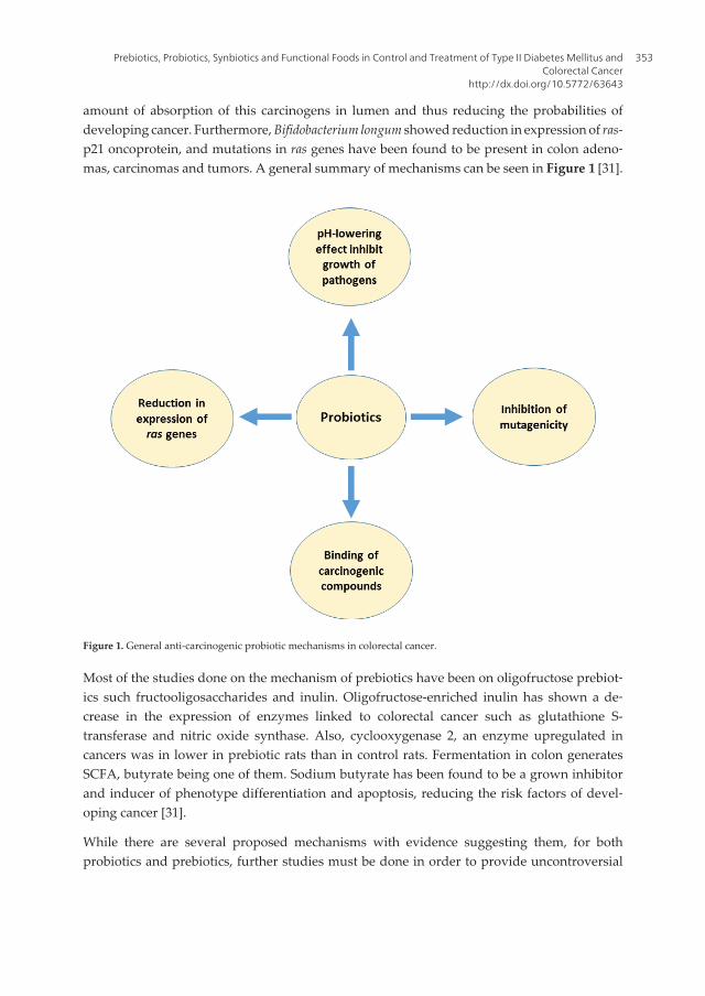
amount of absorption of this carcinogens in lumen and thus reducing the probabilities ofdeveloping cancer. Furthermore, Bifidobacterium longum showed reduction in expression of ras-p21 oncoprotein, and mutations in ras genes have been found to be present in colon adeno‐mas, carcinomas and tumors. A general summary of mechanisms can be seen in Figure 1 [31].
Figure 1. General anti-carcinogenic probiotic mechanisms in colorectal cancer.
Most of the studies done on the mechanism of prebiotics have been on oligofructose prebiot‐ics such fructooligosaccharides and inulin. Oligofructose-enriched inulin has shown a de‐crease in the expression of enzymes linked to colorectal cancer such as glutathione S-transferase and nitric oxide synthase. Also, cyclooxygenase 2, an enzyme upregulated incancers was in lower in prebiotic rats than in control rats. Fermentation in colon generatesSCFA, butyrate being one of them. Sodium butyrate has been found to be a grown inhibitorand inducer of phenotype differentiation and apoptosis, reducing the risk factors of devel‐oping cancer [31].
While there are several proposed mechanisms with evidence suggesting them, for bothprobiotics and prebiotics, further studies must be done in order to provide uncontroversial
Prebiotics, Probiotics, Synbiotics and Functional Foods in Control and Treatment of Type II Diabetes Mellitus andColorectal Cancer
http://dx.doi.org/10.5772/63643
353

evidence on the established pathways and provide better understanding of the moleculardynamics followed in the human colon.
3.1.2. Recent studies done with colorectal cancer
Several studies have been made in order to evaluate the effect on several variables associat‐ed with colorectal cancer. In a randomized double-blinded placebo-controlled trial, a synbi‐otic treatment containing L. rhamnosus, B. lactis and inulin was used in a study evaluating theeffects on several markers of immune functions. During 12 weeks, 34 patients were random‐ly placed on either the control group or the treatment one. Control or placebo group con‐sumed daily encapsulated maltodextrin and a 10 g sachet of maltodextrin, while the treatmentgroup consumed 1 × 10¹⁰ CFU L. rhamnosus and 1 × 10¹⁰ CFU of B. lactis along with a 10 g sachetof inulin. Several parameters such as burst activity of monocytes and neutrophils, lytic activityof natural killer cells and production of interleukins (IL)-2, IL-10 and IL-12, among others wereevaluated. The statistical analysis was done using Kolgomorov–Smirnov, ANOVA andDunnett’s tests. The synbiotic therapy only showed significant effect on an increased capaci‐ty of peripheral blood mononuclear cells to produce IFN-γ. These results do not showpromising evidence, but it should be noted that authors measured immune response factorsin blood. Authors even suggest future studies should aim gut-associated immune system. Themain contribution of this study was to determine that the immune effects of a synbiotictreatment are kept in human colon [32].
A different approach has also been taken, and Bifidobacterium adolescentis extracts were usedin a study to evaluate the antiproliferative effects on three human colon cancer cells: Caco-2,HT-29 and SW480, measuring the production of tumor necrosis factor-α (TNF-α) and NO. Thisstudy consisted in the isolation of B. adolescentis from 20 healthy Koreans 20–30 years old.Extracts were prepared in several concentrations ranging from 12.5 µg/mL up to 200 µg/mLand incubated with the different cell lines mentioned earlier. There was a significant de‐crease in the proliferation of the three human colon cancer cells, correlated with the increasein TNF-α and NO production, from 25 µg/mL to 200 µg/mL in a dose-dependent manner.There are no data shown for the increase in NO production. The increase in both of theseimmune response markers as well as the decrease in cancer cell proliferation show the potentialof including B. adolescentis in therapy or diet diminishing cancer advance; however, studiesshould be done in vivo, without using an extract, and also in clinical trials before reaching adefinitive conclusion [33].
Fructans and soybean meal (SM) were used to evaluate the effect on tumors. Some of thevariables measured in colorectal cancer-induced rats with azoxymethane were GST activityand bacterial enzyme activity. Ninety Fisher 344 male rats were randomly assigned to ninegroups, which difference was the diet. Control groups rats were fed with American Instituteof Nutrition-93 Growth/Maintenance (AIN-93G/M) diet, and the eight groups were fed withthe following diets: prebiotics 5%, prebiotics 10%, SM 5%, SM 10%, prebiotics 5% + SM 5%,10% + SM 10%, 5% + SM 10% and 10% + SM 5%. Tumors present in control group were biggerin size than those fed with either fructans, soybean meal or both. GST activity was increasedin two- to fourfold in rats fed with treatment diets compared to the control group, and β-
Probiotics and Prebiotics in Human Nutrition and Health354
glucosidase activity showed no significant difference between control group and treatmentone, with the exception of a significant increase in rats fed with prebiotics 10% and rats fedwith prebiotics and SM 10% + 5%. Overall there were better results obtained in prebiotics +SM consumption [34]. These results suggest that prebiotics can be used in treatment ofcolorectal cancer.
L. acidophilus has also shown properties in colon tumor suppression in rats. This probiotic wasinoculated into female BALB/cByJ mice during 14 consecutive days at a concentration of 1 ×10⁸ CFU/mouse. After 14 days, cancer cell implantation was done using CT-26 cells in aconcentration of 5 × 10⁶. And during the following 3 weeks, 1 × 10⁹ CFU/mouse of L. acidophi‐lus was inoculated. After 28 days of tumor induction, mice were killed and several variableswere assessed, such as chemokine mRNA expression, tumor size and cell surface pheno‐types. For the statistical analysis of results, one-way and two-way ANOVA tests were used.Tumors in rats that were pre-inoculated showed a 50.3% size reduction, and there was anenhanced tumor apoptosis and downregulation of CXCR4 mRNA expressions in colon. Theseresults show that L. acidophilus is able to play a role in attenuating tumor growth as well asincreasing apoptosis in tumor tissue [35]. This study contributes to the understanding in howprobiotics regulate tumor proliferation in an in vivo system.
The effect of inulin and lactulose on procarcinogenic biomarkers in 1,2-dimethylhydrazinedihydrochloride (DMH)-induced rats has also been evaluated. Thirty-two male SpragueDawley rats were divided into four groups: group I which is the control group received a singledose of EDTA saline solution per week, group II received a single dose of DMH per week,group III received a single dose of DMH + inulin 10 mg/0.1 mL and Group IV received DMH+ lactulose 14 mg/0.1 mL. All doses were given during the course of 6 weeks. For groups IIIand IV, prebiotics were administered orally daily and on the 8th day, a single dose of DMHwas administered. Three variables were measured among others, and these are as follows:nitroreductase, β-glucosidase and β-glucuronidase activities. Statistical analysis was doneusing one-way ANOVA and a post hoc LSD tests. Activity of β-glucuronidase (0.045 ± 0.006µg/h/mg) and β-glucosidase (1.007 ± 0.115 µg/h/mg) was found to be decreased in the inulin+ DMH group when compared to control (0.243 ± 0.059 µg/h/mg and 2.219 ± 0.745 µg/h/mg,respectively). Nitroreductase activity was increased in inulin + DMH (0.045 ± 0.005 µg/h/mg)compared to control (0.0162 ± 0.005 µg/h/mg) [36]. These results also suggest the colorectalcancer protection properties of inulin, which could be used in the prevention of developingcolorectal cancer.
On similar study, thirty male and female Sprague Dawley rats were divided into three groups:a control group fed only with conventional feed, a DMH group and a DMH + inulin fed group.DMH and DMH + inulin group were treated with DMH at a dose of 21 mg/kg five times inweekly intervals, and DMH + inulin rats were fed with a dose of 80 g/kg of conventional feedduring 28 weeks. For statistical analysis of the variables evaluated, a one-way ANOVA testwas used. It was found that activity of β-glucuronidase decreased as well as the number ofCOX-2- and NFκB-positive cells along with a decrease in the expression of IL-2, TNF-α andIL-10. Moreover, there was a significant decrease in β-glucosidase activity (0.03 ± 0.02µmol/min/g) when compared to the control group (0.08 ± 0.02 µmol/min/g), and also there was
Prebiotics, Probiotics, Synbiotics and Functional Foods in Control and Treatment of Type II Diabetes Mellitus andColorectal Cancer
http://dx.doi.org/10.5772/63643
355

a significant decrease in coliforms (5.96 ± 0.22 log10 CFU/g) when compared to control (6.17 ±0.56 log10 CFU/g) and DMH group 6.34 ± 0.25 log10 CFU/g). This decrease in coliforms explainsthe reduction in β-glucuronidase activity. Butyric and propionic acid levels were higher inDMH + inulin group, and these short-chain fatty acids have been associated with apoptosisand metastasis, carcinogen reduction, among others [37].
Several other studies have been made, and these are shown along with a brief summary ofeach in Table 2.
Authors Component Organism/cellline
Dosage/length Study’s design
[38] L. delbrueckiifermentationsupernatant
Colon cancerSW620
Several proteinconcentrationsranging from0 up to 0.75mg/mL24 h
L. delbrueckii fermented MRS medium.Supernatant was incubated with SW620cells and evaluated in viability essaysStatistical analysis were done using one-way ANOVA and Bonferroni’s multiplecomparison test
[39] L. plantarumL. rhamnosussupernatants
Caco-2HT-29
2.5, 5 and 10mg/mL 48 h
Probiotic fermented medium Supernatantswas incubated with cancer cells andevaluated viabilityStatistical data were analyzed using one-wayANOVA
[40] L. casei C57BL/6 mice 1 × 10⁸ CFU10 weeks
Mice were administered probiotic and DMH,intestinal damage evaluation, cytokineanalysis, gene expression analysisBonferroni’s multiple comparison test
[41] Xylooligosaccharides (XOS)
Wistar rats 5% and 10% XOS45 days
XOS diet in rats, bacterial analysis y cecalmatter, biochemical assays, proliferationmarkersOne-way ANOVA
[42] L. paracasei HT-29 0, 10, 50, 100, MOI48 h
Calculation of multiplicity of infection (MOI),analysis cell distribution, RNA extraction, andsemiquantitative RT-PCROne-way ANOVA and Duncan’s post hoc tests
Table 2. Recent studies done regarding colorectal cancer using prebiotics or probiotics.
3.2. Functional foods in colorectal cancer
To the best of our knowledge, only a couple of studies have been made regarding prebiotic,probiotic or synbiotic functional foods for prevention, control or treatment of colorectal cancer.
A synbiotic food using oligofructose-enriched inulin and L. rhamnosus and B. lactis wasdeveloped and evaluated as a potential reduction risk agent in colorectal cancer patients. This
Probiotics and Prebiotics in Human Nutrition and Health356

study was a randomized double-blinded placebo-controlled trial in which 37 colon cancerpatients were divided into a control group or an intervention one. Intervention patients weregiven daily a synbiotic food during 12 weeks consisting of 12 g of inulin and 1010 CFU ofprobiotics. For statistical analysis of the data, a generalized linear modeling was used. It wasfound that the number of Bifidobacterium and Lactobacillus in feces was increased, while adecrease in the number of Clostridium was found. The effect of the synbiotic intervention onDNA damage, as well as the effect on epithelial barrier functions in tumor cell invasion, wasalso studied. It was found that intervention group had a significant decrease in the level ofDNA damage (55.84 ± 21.21 tail lengths) compared to the placebo group (59.18 ± 15.94 taillengths), but there was no significant difference between control (101.9 ± 6.6) and interven‐tion group (104.9 ± 6.2). A decrease in the level of DNA suggests that there was a decrease inexposure of the colon epithelium to cytotoxic and genotoxic agents, along with decreasedcancer cell proliferation. An improvement of the epithelial barrier function is associated withlower cancer risk, while in this study there was no significant difference, it has been seen thatprobiotics provide a better formation of this layer [43].
A study was done on 56 F344 rats using a probiotic fermented milk with L. acidophilus, L. caseiand L. rhamnosus. The rats were divided into seven groups randomly: group 1 served as controlby receiving 0.85% saline solution by gavage. Rats in groups 2–7 were injected with DMH 30mg/kg once a week for 6 weeks; group 2 served as positive carcinogenic control, and groups3 through 7 were supplemented with 2, 1.5, 1, 0.5 and 0.25 mL of probiotic milk containing atleast 50 × 109 CFU during 12 weeks. Variables were observed and evaluated such as the activityof quinone reductase (QR), GST and β-glucuronidase. Statistical analysis of the data was doneusing one-way ANOVA test. It was found that G4 and G5 improved 154% and 109% com‐pared to control group. QR activity was reduced significantly in all rats treated with DMHwhen compared to the control group. β-Glucuronidase activity showed a significant de‐crease by 49% compared to control group. This study shows that there is potential in probiot‐ic functional foods in the prevention, control and treatment of colorectal cancer; however,further studies are needed in order to provide more information about this [44].
4. Conclusions and perspectives
The effect of prebiotics, probiotics and synbiotics over several health markers in T2DM andcolorectal cancer patients has been shown through several studies discussed in this chapter.Some of the health benefits presented in this chapter for T2DM are the improvement of lipidand glycemic profile, increase in blood insulin concentration and modulation on the inflam‐matory response. For colorectal cancer, some of the health benefits presented in this chapterare the modulation of the immune response, antitumor activity and tumor size reduction.However, further research is needed in order to understand completely the specific molecu‐lar pathway of each component has.
The use of functional foods for prevention and control of T2DM is a promising opportunitywhich must be taken into account, after all, and one of the most common causes of this disease
Prebiotics, Probiotics, Synbiotics and Functional Foods in Control and Treatment of Type II Diabetes Mellitus andColorectal Cancer
http://dx.doi.org/10.5772/63643
357

is obesity and poor diet. The design of functional foods with prebiotics, probiotics or synbiot‐ics that will help enhance T2DM patient’s health would be an aid in the fight against it;however, the elimination or substitution of antidiabetic drugs is not recommended orendorsed.
There is much to do in the research of prebiotic, probiotic or synbiotic functional foods for theprevention, control or treatment of colorectal cancer. There is evidence suggesting that therapyenhances patient’s health, and this should encourage further research into the developmentof functional foods and their clinical studies in patients. If successful results during thefollowing years are obtained, this could provide as an aid in the fight against colorectal cancer.
The use of functional foods should be used with caution and as a support to clinical therapy,not exclusively as an alternative. This combination could lead to further improvement inpatient’s health as some studies have found synergistic effect of probiotics along with medicaldrugs.
Author details
Samuel Longoria-García1, Ruth E. Belmares-Cerda1, Mildred I.M. Flores-Verástegui2,Juan C. Contreras-Esquivel1, Julio C. Montañez-Sáenz1 and Mario Alberto Cruz-Hernández2*
*Address all correspondence to: [email protected]
1 Department of Food Research, School of Chemical Sciences, Autonomous University ofCoahuila, Saltillo, Coahuila, Mexico
2 Department of Food Science and Technology, Antonio Narro Autonomous AgriculturalUniversity, Buenavista Saltillo, Coahuila, Mexico
References
[1] Zannini E., Pontonio E., WatersD. M., Arendt E. K. Applications of microbial fermen‐tations for production of gluten‐free products and perspectives. Applied Microbiolo‐gy and Biotechnology. 2012;93(2):473‐485. DOI: 10.1007/s00253‐011‐3707‐3
[2] Mensink M. A., Frijlink H.W., Van der Voort M.K., Hinrichs L.J. Inulin, a flexibleoligosaccharide I: Review of its physicochemical characteristics. CarbohydratePolymers. 2015;130DOI: 10.1016/j.carbpol.2015.05.026
[3] Mensink M.A., Frijlink H.W., Van der Voort M.K., Hinrichs L.J. Inulin, a flexibleoligosaccharide. II: Review of its pharmaceutical applications. Carbohydrate Poly‐mers. 2015;134:418‐428. DOI: 10.1016/j.carbpol.2015.08.022
Probiotics and Prebiotics in Human Nutrition and Health358

[4] Siró I., Kápolna E., Kápolna B., Lugasi A. Functional food. Product development,marketing and consumer acceptance—A review. Appetite. 2008;51(3):456‐467. DOI:10.1016/j.appet.2008.05.060
[5] Wisse B., Zieve D., Ogilvie I. Diabetes: MedlinePlus Medical Encyclopedia [Internet].[Updated: 8/5/2014]. Available from: https://www.nlm.nih.gov/medlineplus/ency/article/001214.htm [Accessed: 02/01/2016]
[6] Mazloom Z., Yousefinejad A., Dabbaghmanesh, M. H. Effect of Probiotics on LipidProfile , Glycemic Control , Insulin Action , Oxidative Stress , and InflammatoryMarkers in Patients with Type 2 Diabetes : A Clinical Trial. Iranian Journal of MedicalSciences. 2013;38(1):38‐43.
[7] WHO. WHO Diabetes: The cost of Diabetes [Internet].Available from: http://www.who.int/mediacentre/factsheets/fs236/en/ [Accessed: 2/01/2016]
[8] Yang W., Dall T.M., Halder P., Gallo P., Kowal S. L., Hogan P. F., et al. Economic costsof diabetes in the U.S. in 2012. Diabetes care. 2013;36(4):1033‐1046. DOI: 10.2337/dc12‐2625
[9] Brown A. C., Valiere A. Probiotics and Medical Nutrition Therapy. Nutrition ClinicalCare. 2004;7(2):56‐68. DOI: 10.1016/j.micinf.2011.07.011.Innate
[10] Gomes A. C., Bueno A. A., de Souza G. M., Mota J. F. Gut microbiota, probiotics anddiabetes. Nutrition journal. 2014;13(60)DOI: 10.1186/1475‐2891‐13‐60
[11] Zhang Y., Guo X., Guo J., He Q., Li H., Song Y., et al. Lactobacillus casei reducessusceptibility to type 2 diabetes via microbiota‐mediated body chloride ion influx.Scientific reports. 2014;4:5654. DOI: 10.1038/srep05654
[12] Boeckner L. S., Schnepf M. I., Tungland B. C. Inulin: A review of nutritional and healthimplications. Advances in Food and Nutrition Research. 2001;43:1‐63. DOI: 10.1016/S1043‐4526(01)43002‐6
[13] Al‐Salami H., Butt G., Fawcett J.P., Tucker I.G., Golocorbin‐Kon S., Mikov M. Probiotictreatment reduces blood glucose levels and increases systemic absorption of glicla‐zide in diabetic rats. European journal of drug metabolism and pharmacokinetics.2008;33(2):101‐106. DOI: 10.1007/BF03191026
[14] Aliasgharzadeh A., Khalili M., Mirtaheri E., Gargari B. P., Farhangi M. A., Babaei H.,et al. A Combination of Prebiotic Inulin and Oligofructose Improve Some of Cardio‐vascular Disease Risk Factors in Women with Type 2 Diabetes: A RandomizedControlled Clinical Trial. Advanced Pharmaceutical Bulletin. 2015;5(4):507‐514. DOI:10.15171/apb.2015.069
[15] Zhang Y., Wang L., Zhang J., Li Y., He Q., Li H., et al. Probiotic Lactobacillus caseiZhang ameliorates high‐fructose‐induced impaired glucose tolerance in hyperinsuli‐nemia rats. European Journal of Nutrition. 2014;53(1):221‐232. DOI: 10.1007/s00394‐013‐0519‐5
Prebiotics, Probiotics, Synbiotics and Functional Foods in Control and Treatment of Type II Diabetes Mellitus andColorectal Cancer
http://dx.doi.org/10.5772/63643
359

[16] Alokail M. S., Sabico S., Al‐saleh Y., Al‐daghri N. M., Alkharfy K. M., Vanhoutte P. M.,et al. Effects of probiotics in patients with diabetes mellitus type 2 : study protocol fora randomized . Trials. 2013;14:195.
[17] Kellow N.J., Coughlan M.T., Savige G. S., Reid C. M. Effect of dietary prebioticsupplementation on advanced glycation, insulin resistance and inflammatory bio‐markers in adults with pre‐diabetes: a study protocol for a double‐blind placebo‐controlled randomised crossover clinical trial. BMC endocrine disorders. 2014;14(1):55.DOI: 10.1186/1472‐6823‐14‐55
[18] Asemi Z., Sahar B., Shakeri H., Jamal A., Faraji A. Effect of multispecies probioticsupplements on serum minerals, liver enzymes and blood pressure in patients withtype 2 diabetes. International Journal of Diabetes in Developing Countries. 2015;35(2):90‐95. DOI: 10.1159/000349922
[19] Mitchell C.M., Davy B.M., Halliday T.M., Hulver M.W., Neilson A.P., Ponder M.A., etal. The effect of prebiotic supplementation with inulin on cardiometabolic health:Rationale, design, and methods of a controlled feeding efficacy trial in adults at risk oftype 2 diabetes. Contemporary Clinical Trials. 2015;45:328‐337. DOI: 10.1016/j.cct.2015.10.012
[20] Tonucci, L. B., Olbrich dos Santos K. M., Licursi de Oliveira L., Rocha Ribeiro S.M.,Duarte Martino H. S. Clinical application of probiotics in type 2 diabetes mellitus: Arandomized, double‐blind, placebo‐controlled study. Clinical Nutrition. Forthcoming.DOI: 10.1016/j.clnu.2015.11.011
[21] Stenman L. K., Waget A., Garret C., Briand F., Burcelin R., Sulpice T., et al. ProbioticB420 and prebiotic polydextrose improve efficacy of antidiabetic drugs in mice.Diabetology & Metabolic Syndrome. 2015;7:75. DOI: 10.1186/s13098‐015‐0075‐7
[22] Ejtahed H. S., Mohtadi‐Nia J., Homayouni‐Rad A., Niafar M., Asghari‐Jafarabadi M.,Mofid V., et al. Effect of probiotic yogurt containing Lactobacillus acidophilus andBifidobacterium lactis on lipid profile in individuals with type 2 diabetes mellitus.Journal of dairy science. 2011;94(7):3288‐3294. DOI: 10.3168/jds.2010‐4128
[23] Ejtahed H. S., Mohtadi‐Nia J., Homayouni‐Rad A., Niafar M., Asghari‐Jafarabadi M.,Mofid V. Probiotic yogurt improves antioxidant status in type 2 diabetic patients.Nutrition. 2012;28(5):539‐543. DOI: 10.1016/j.nut.2011.08.013
[24] Shakeri H., Hadaegh H., Abedi F., Tajabadi‐Ebrahimi M., Mazroii N., Ghandi Y., et al.Consumption of synbiotic bread decreases triacylglycerol and VLDL levels whileincreasing HDL levels in serum from patients with type‐2 diabetes. Lipids. 2014;49(7):695‐701. DOI: 10.1007/s11745‐014‐3901‐z
[25] Asemi Z., Khorrami‐Rad A., Alizadeh S.A., Shakeri H., Esmaillzadeh A. Effects ofsynbiotic food consumption on metabolic status of diabetic patients: A double‐blindrandomized cross‐over controlled clinical trial. Clinical Nutrition. 2014;33(2):198‐203.DOI: 10.1016/j.clnu.2013.05.015
Probiotics and Prebiotics in Human Nutrition and Health360
[26] Asemi Z., Alizadeh S., Ahmad K., Goli M.,Esmaillzadeh A. Effects of beta‐carotenefortified synbiotic food on metabolic control of patients with type 2 diabetes mellitus:A double‐blind randomized cross‐over controlled clinical trial. Clinical nutrition .Forthcoming. DOI: 10.1016/j.clnu.2015.07.009
[27] Bahmani F., Tajadadi‐Ebrahimi M., Kolahdooz F., Mazouchi M., Hadaegh H., Jamal A.,et al. The Consumption of Synbiotic Bread Containing Lactobacillus sporogenes andInulin Affects Nitric Oxide and Malondialdehyde in Patients with Type 2 DiabetesMellitus: Randomized, Double‐Blind, Placebo‐Controlled Trial. Journal of theAmerican College of Nutrition. 2015;0(0):1‐8. DOI: 10.1080/07315724.2015.1032443
[28] Ferlay J., Soerjomataram I., Ervik M., Dikshit R., Eser S., Mathers C., et al. CancerIncidence and Mortality Worldwide: IARC CancerBase No. 11 [Internet]. 2012 .Available from: http://globocan.iarc.fr [Accessed: 2/01/2016]
[29] Mariotto A. B., Robin Yabroff K., Shao Y., Feuer E.J., Brown M.L. Projections of the costof cancer care in the United States: 2010‐2020. Journal of the National Cancer Institute.2011;103(2):117‐128. DOI: 10.1093/jnci/djq495
[30] The American Cancer Society. The Global Economic Cost of Cancer. In: LiveStrong,editor. The American Cancer Society; 2012. p. 1‐14.
[31] Liong M. T. Roles of probiotics and prebiotics in colon cancer prevention: Postulatedmechanisms and in‐vivo evidence. nternational Journal of Molecular Sciences.2008;9(5):854‐863. DOI: 10.3390/ijms9050854
[32] Roller M., Clune Y., Collins K., Rechkemmer G., Watzl B. Consumption of prebioticinulin enriched with oligofructose in combination with the probiotics Lactobacillusrhamnosus and Bifidobacterium lactis has minor effects on selected immune parame‐ters in polypectomised and colon cancer patients. The British journal of nutrition.2007;97(4):676‐684. DOI: 10.1017/S0007114507450292
[33] Lee D. K., Jang S.,Kim M. J., Kim J. H., Chung M. J., Kim K. J., et al. Anti‐proliferativeeffects of Bifidobacterium adolescentis SPM0212 extract on human colon cancer celllines. BMC cancer. 2008;8:310. DOI: 10.1186/1471‐2407‐8‐310
[34] Gourineni V. P., Verghese M., Boateng J., Shackelford L., Bhat N. K., Walker L. T.Combinational effects of prebiotics and soybean against azoxymethane‐induced coloncancer in vivo. Journal of Nutrition and Metabolism. 2011;:868197. DOI:10.1155/2011/868197
[35] Chen C.C., Lin W.C., Kong M.S., Shi H.N., Walker W.A., Lin C.Y., et al. Oral inocula‐tion of probiotics Lactobacillus acidophilus NCFM suppresses tumour growth both insegmental orthotopic colon cancer and extra‐intestinal tissue. British Journal ofNutrition. 2012;107(11):1623‐1634. DOI: 10.1017/S0007114511004934
[36] Verma A., Shukla G. Administration of prebiotic inulin suppresses 1,2 dimethylhy‐drazine dihydrochloride induced procarcinogenic biomarkers fecal enzymes and
Prebiotics, Probiotics, Synbiotics and Functional Foods in Control and Treatment of Type II Diabetes Mellitus andColorectal Cancer
http://dx.doi.org/10.5772/63643
361
preneoplastic lesions in early colon carcinogenesis in Sprague Dawley rats. Journal ofFunctional Foods. 2013;5(2):991‐996. DOI: 10.1016/j.jff.2013.02.006
[37] Hijová E., Szabadosova Vi., Štofilová J., Hrcková G. Chemopreventive and metaboliceffects of inulin on colon cancer development. Journal of Veterinary Science. 2013;14(4):387‐393. DOI: 10.4142/jvs.2013.14.4.387
[38] Wan Ying., Xin Y., Zhang C., Wu D., Ding D., Tang L., et al. Fermentation superna‐tants of lactobacillus delbrueckii inhibit growth of human colon cancer cells and induceapoptosis through a caspase 3‐dependent pathway. Oncology Letters. 2014;7(5):1738‐1742. DOI: 10.3892/ol.2014.1959
[39] Sadeghi‐Aliabadi H., Mohammadi F., Fazeli H., Mirlohi M. Effects of lactobacillusplantarum A7 with probiotic potential on colon cancer and normal cells proliferationin comparison with a commercial strain. Iranian Journal of Basic Medical Sciences.2014;17(10):815‐819.
[40] Lenoir M., del Carmen S., Cortes-Perez N.G., Lozano-Ojalvo D., Muñoz-Provencio D.,Chain F., et al. Lactobacillus casei BL23 regulates Treg and Th17 T-cell populations andreduces DMH-associated colorectal cancer. Journal of Gastroenterology. 2016;1–12.DOI: 10.1007/s00535–015–1158–9.
[41] Aachary A.A., Gobinath D., Srinivasan K., Prapulla S. G. Protective effect of xylooli‐gosaccharides from corncob on 1,2‐dimethylhydrazine induced colon cancer in rats.Bioactive Carbohydrates and Dietary Fibre. 2015;5(2):146‐152. DOI: 10.1016/j.bcdf.2015.03.004
[42] Huang L., Shan Y., He C., Ren M., Tian P., Song W. Effects of L. paracasei subp. paracaseiX12 on cell cycle of colon cancer HT‐29 cells and regulation of mTOR signallingpathway. Journal of Functional Foods. 2016;21:431‐439. DOI: 10.1016/j.jff.2015.12.024
[43] Rafter J., Bennett M., Caderni G., Clune Y., Hughes R., Karlsson P. C., et al. Dietarysynbiotics reduce cancer risk factors in polypectomized and colon cancer patients.American Journal of Clinical Nutrition. 2007;85(2):488‐496. DOI: 85/2/488 [pii]
[44] Desrouillères K., Millette M., Vu K.D., Touja, R., Lacroix, M. Cancer preventive effectsof a specific probiotic fermented milk containing Lactobacillus acidophilus CL1285, L.casei LBC80R and L. rhamnosus CLR2 on male F344 rats treated with 1,2‐dimethylhy‐drazine. Journal of Functional Foods. 2015;17:816‐827. DOI: 10.1016/j.jff.2015.06.035
Probiotics and Prebiotics in Human Nutrition and Health362
Chapter 18
Probiotics, Prebiotics, and Biogenics for the Stomach
Yasuhiko Komatsu, Yuji Aiba, Yasuhiro Nakano andYasuhiro Koga
Additional information is available at the end of the chapter
http://dx.doi.org/10.5772/62828
Abstract
Recently, many studies concerning probiotics, prebiotics, and biogenics have beenperformed, whereas only a few are related to the stomach (about 2% as publicationnumber). In this chapter, we focus on recent studies on probiotics, prebiotics, and biogenicsfor the stomach and also describe our recent research on a novel strain of lactobacillusbeneficial to stomach, Lactobacillus johnsonii No.1088 (LJ88). As probiotics for the stomach,some beneficial strains were summarized, and underlying mechanisms of anti-Helicobacterpylori activity were discussed. Prebiotics for the stomach were considered as a futurepotential target, since no indigenous bacteria beneficial to the stomach have been foundto date. As biogenics, some plant-derived candidates were discussed. In this context,recent results on LJ88 lactobacillus were presented. Orally administered LJ88 inhibitedH. pylori growth and the increase in the number of gastrin-producing cells, which sideeffect is caused by triple therapy for H. pylori. LJ88 had no resistance to typical antibiot‐ics, and both living and heat-killed forms of it increased the number of bifidobacteriaamong human intestinal-microbiota in mice. These results suggest that LJ88 is alactobacillus beneficial to both stomach and intestine as a probiotic and biogenic.
Keywords: Probiotics, Prebiotics, Biogenics, Stomach, Helicobacter pylori
1. Introduction
Historically, probiotics have been thought as agents beneficial to improve the microbialenvironment in the intestines, but some strains of lactic acid bacteria have been used asprobiotics, with the claim of providing health benefits to the stomach.
Nestlé’s Lactobacillus L. johnsonii La1 (LC1) [1–3] and Meiji’s Lactobacillus gasseri OLL2716 [4–6] are typical strains said to be useful to reduce the number of Helicobacter pylori in stomach

infections. Recently, we found a novel strain of lactic acid bacteria, L. johnsonii No.1088 (LJ88),which is extremely acid resistant and also has the ability to significantly reduce the number ofinfective H.pylori in the stomach [7]. Furthermore, LJ88 not only has anti-pylori activity butalso reduces excessive gastric acid production [7]. So we are very interested in the beneficialeffects of probiotics on stomach health. Likewise, those effects of “Prebiotics” are also of greatinterest.
In addition to living bacteria, i.e., “Probiotics”, heat-killed “dead” bacteria retain somebeneficial properties of probiotic bacteria. For example, the ability of heat-killed LJ88 to reduceexcessive gastric acid production can be thought as having this property [7]. Such foodingredients that beneficially affect the host by “direct” stimulation, suppression, etc., weredefined by Mitsuoka as “Biogenics” [8]. So we added this category to this chapter. So the titleof the chapter was chosen to be “Probiotics, Prebiotics, and Biogenics for the Stomach”.
In this chapter, we review the current status of probiotics, prebiotics, and biogenics for thestomach, and also discuss novel aspects of our lactic acid bacterium, LJ88, which is benefi‐cial to the stomach.
2. Number of publications
Figure 1 depicts yearly changes up to 2014 in the number of publications related to “probiot‐ics OR prebiotics OR biogenics” as a whole (A) and those related to the stomach (B), based ona PubMed search. The total number of publications shown in Figure 1A was 14,417, of whichthose including the word “stomach” (Figure 1B) were only 290 (about 2% of the total publi‐cations). As shown in Figure 1A, the number of publications in this area increased almostlinearly from year 2000, reaching 1936 publications in 2014; whereas the subset related to thestomach hit its ceiling at about 30 publications/year (Figure 1B).
Figure 1. Yearly change in the number of publications related to probiotics/prebiotics/biogenics (A) and the subset of“A” related to the stomach (B).
Probiotics and Prebiotics in Human Nutrition and Health364

As shown above, probiotics/prebiotics/biogenics involving the stomach is not a major area ofthis research field. However, since a variety of bacteria have been detected not only from fecesor saliva but also from gastric fluid, although mainly as dead forms [9], it is thought that thisarea will expand in the future.
2.1. Anti-H. pylori activity of probiotics
2.1.1. Probiotics and virulent bacteria
Although a very recent definition of probiotics is “live microorganisms, which when con‐sumed in adequate amounts, confer a health effect on the host” [10], probiotics have beenthought as agents that improve the balance of microbiota mainly in the intestines. Typically,the ingestion of probiotics brings about an increase in the number of so-called “beneficialbacteria”, e.g., bifidobacteria, and a decrease in the number of so-called “bad” bacteria, e.g.,clostridia. Moreover, some probiotic strains have been reported to inhibit the growth of somevirulent bacteria, resulting in prevention of and recovery from diarrhea.
As regards the stomach, H. pylori is the main virulent bacteria residing in the gastric mucosa,causing chronic gastritis and peptic ulcer. Also, H. pylori is now thought to be responsible foralmost all cases of gastric cancer [11]. Some strains of probiotic bacteria have been reported tobe effective in reducing the number of H. pylori, and also decreasing the extent of inflamma‐tion caused by infection by this bacterium.
2.1.2. Probiotic strains useful to reduce symptoms related to H. pylori infection
One of the well-known probiotic strains beneficial for the treatment of H. pylori infections is L.johnsonii La1, which was found and developed by a Swiss company, Nestlé, and has beenwidely used in fermented milk worldwide [1–3]. Another strain beneficial to H. pylori-infectedsubjects is L. gasseri OLL2716, found by Meiji, a Japanese company [4–6]. This strain is nowused mainly in fermented milk in Japan as LG21 and promoted as “lactic acid bacteriacombating risk” (a catchy tag from Meiji). In addition to these two strains of probiotic bacteria,some other strains have been reported to be effective in ameliorating symptoms derived fromH. pylori infection, e.g., Lactobacillus acidophilus Strain LB [12], Bacillus subtilis 3 [13], Weissellaconfusa Strain PL9001 [14], Lactobacillus delbrueckii subsp. bulgaricus [15], and Lactobacillus reuteri[16].
2.1.3. L. johnsonii No. 1088 (LJ88) as a probiotic
Recently, we found a novel strain of lactobacillus, LJ88, in the gastric juice of a healthy humanvolunteer. When administered as a living form, LJ88 reduced the number of H. pylori in thestomach of human intestinal microbiota-bearing mice, as shown in Figure 2 [7]. This anti-H.pylori effect of LJ88 can be brought not only by proliferating bacteria ( Figure 2A) but also byits lyophilized form ( Figure 2B), suggesting that this strain is useful both as fermented milkand also as the lyophilized form of a dietary supplement.
Probiotics, Prebiotics, and Biogenics for the Stomachhttp://dx.doi.org/10.5772/62828
365

From Aiba et al. [7] with permission.
Figure 2. Anti-H. pylori effect of L. johnsonii No. 1088 (LJ88) in human intestinal microbiota-bearing mice. Mice withhuman intestinal microbiota were prepared by using germ-free mice and were then infected with H. pylori No. 130(109 cfu/mice). H. pylori-bearing mice were orally and daily administered live LJ88 (A) or a comparable number oflyophilized cells (B) for two or four weeks. In mice treated with either live or the lyophilized (freeze-dried) form ofLJ88, the number of H. pylori in the stomach was significantly decreased. Statistical significance was determined by useof Student’s t-test (*p < 0.05, **p < 0.01, ‡p < 0.0001 vs. no treatment for comparable time periods).
To evaluate the probiotic property of LJ88, we examined the sensitivity of LJ88 to differenttypes of antibiotics. Mueller–Hinton agar plates containing 128, 64, 32, 16, 8, 4, 2, 1, 0.5, 0.25,0.125, 0.0625, 0.031, 0.016, 0.008, 0.004, 0.002 or 0.001 μg/mL of different antibiotics (ampicil‐lin, oxacillin, cefoxitin, gentamicin, clarithromycin, vancomycin, ciprofloxacin, and chloram‐phenicol) were prepared; and 5000 cfu of LJ88 (5 μL), after having been cultured in Mueller–Hinton broth for 24 h at 37 °C, was inoculated onto each plate. The minimum inhibitoryconcentrations (MICs) were determined after cultivation for 48 h at 37°C. The results aredepicted in Table 1. As shown in Table 1, no resistance to any of the antibiotics used wasobserved, suggesting that LJ88 should be of no concern with respect to the transfer of drug-resistance genes to virulent bacteria.
Antibiotics MIC (μg/mL)
ampicilin 0.004
oxacillin 0.125
cefoxitin 0.004
gentamicin 0.25
clarithromycin 0.5
vancomycin 0.016
ciprofloxacin 0.5
chloramphenicol 0.5
Table 1. MIC of various antibiotics against LJ88.
Probiotics and Prebiotics in Human Nutrition and Health366
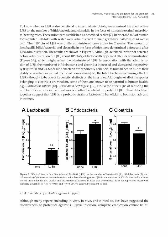
To know whether LJ88 is also beneficial to intestinal microbiota, we examined the effect of liveLJ88 on the number of bifidobacteria and clostridia in the feces of human intestinal microbio‐ta-bearing mice. These mice were established as described earlier [7]. In brief, 0.5 mL of humanfeces diluted 100-fold with water were administered to male germ-free Balb/c mice (4 weeksold). Then 109 cfu of LJ88 was orally administered once a day for 2 weeks. The amount oflactobacilli, bifidobacteria, and clostridia in the feces of mice were determined before and afterLJ88 administration. The results are shown in Figure 3. Although lactobacilli were not detectedbefore administration of LJ88, about 108 cfu/g of lactobacilli appeared after its administration(Figure 3A), which might reflect the administered LJ88. In association with the administra‐tion of LJ88, the number of bifidobacteria and clostridia increased and decreased, respective‐ly (Figure 3B and C). Since bifidobacteria are reportedly beneficial to human health due to theirability to regulate intestinal microbial homeostasis [17], the bifidobacteria-increasing effect ofLJ88 is thought to be one of its beneficial effects on the intestines. Although not all of the speciesbelonging to clostridia are virulent, some of them are known to be harmful to human health,e.g. Clostridium difficile [18], Clostridium perfringens [19], etc. So the effect LJ88 of reducing thenumber of clostridia in the intestines is another beneficial property of LJ88. These data takentogether suggest that LJ88 is a probiotic strain of lactobacilli beneficial to both stomach andintestines.
Figure 3. Effect of live Lactocacillus johnsonii No.1088 (LJ88) on the number of lactobacilli (A), bifidobacteria (B), andchlostoridia (C) in feces of human intestinal microbiota-bearing mice. LJ88 in the measure of 109 cfu was orally admin‐istered once a day for two weeks, and the number of bacteria in feces was determined. Each bar represents mean withstandard deviation (n = 5). *p < 0.05, and **p < 0.001 vs. control by Student’s t-test.
2.1.4. Limitation of probiotics against H. pylori
Although many reports including in vitro, in vivo, and clinical studies have suggested theeffectiveness of probiotics against H. pylori infection, complete eradication cannot be at‐
Probiotics, Prebiotics, and Biogenics for the Stomachhttp://dx.doi.org/10.5772/62828
367

tained by probiotics alone. The standard and more effective way to eradicate H. pylori infectionsis the so-called “triple therapy” consisting of two antibiotics and one proton pump inhibitor(PPI) [20]. But the cost for such a therapy is expensive, and so a lower cost way to control theextent of H. pylori at a level under the asymptomatic one is needed. Moreover, since theeradication rate of this triple therapy is not 100%, probiotics effective in increasing theeradication rate of triple therapy might be meaningful. In fact, some strains have been reportedto have such a property [21].
2.1.5. Possible mechanism underlying anti-H. pylori activity of probiotics
Although the exact mechanisms underlying the anti-H. pylori activity of probiotics have notyet been fully elucidated, some putative ones have been proposed, as shown in Table 2. Wedescribe them in brief here.
Proposed Mechanisms Described in
Lactic acid production 2.1.5.1
Production of antimicrobial products 2.1.5.2
Competition for adherent sites 2.1.5.3
Immunological mechanisms 2.1.5.4
Co-aggregation with Helicobacter pylori 2.1.5.5
Table 2. Putative mechanisms by which probiotics inhibit H. pylori.
2.1.5.1. Lactic acid
H. pylori can survive in the highly acidic gastric mucosa by producing urease, which de‐grades urea to ammonia and carbon dioxide, and the resulting ammonia neutralizes the gastricacid to elevate pH of surrounding environment. Lactic acid produced by probiotic bacteriacompetes with the pH elevation by urease mentioned above, which makes the environmentunsuitable for H. pylori to survive [22–24]. In addition to acidification, lactic acid inhibits ureaseactivity itself [22], which might be another molecular mechanism for lactic acid to inhibitsurvival of H. pylori in the stomach. But since not all lactic acid bacteria producing the samelevel of lactic acid can inhibit H. pylori to the same extent [3], the production of lactic acid mayonly be part of the anti-H. pylori effect of lactic acid bacteria.
2.1.5.2. Antimicrobial products
Some probiotic strains have reported to secrete antimicrobial substances other than lactic acid.The culture supernatants of L. johnsonii La1 [3] and L. acidophilus Strain LB [12] can inhibit thegrowth of H. pylori in vitro and in vivo in a pH-independent manner, but the molecularstructures of these active substances have not yet been determined. Moreover, some strains ofL. delbrueckii supsp. bulgaricus reportedly inhibit the growth of H. pylori in an agar-well diffusion
Probiotics and Prebiotics in Human Nutrition and Health368

assay under both acidic and neutral pH conditions, suggesting secretion of anti-H. pylorisubstances [15]. Bacteriocins are being widely investigated as proteinaceous antimicrobialsubstances produced by bacteria [25]. Kim et al. examined the anti-H. pylori activity of selectedknown bacteriocins and found that lacticins A164 and BH5 produced by Lactococcus lactis subsp.lactis A164 and BH5, respectively, strongly inhibit the growth of H. pylori [26]. Other than lacticacid bacteria, another probiotic strain of bacillus, B. subtilis 3, has been reported to produceaminocoumacin A, another anti-H. pylori substance [13]. As described here, the molecularnature of almost all of the anti-H. pylori substances produced by probiotic bacteria is un‐known and remains to be elucidated.
2.1.5.3. Competition
For H. pylori to grow in gastric mucosa, it is necessary first for the bacteria to adhere to theinner surface of the stomach. So if probiotics and/or its products can compete with the siteswhere H. pylori adhere, the growth of H. pylori might be inhibited. Kabir et al. reported that ananti-H. pylori strain of Lactobacillus salivarius inhibit the attachment of H. pylori to human gastriccell lines (MKN45 and KATO-III) and murine gastric epithelial cells, whereas other lactic acidbacteria not inhibiting H. pylori (Enterococcus faecalis, and also Streptococcus aureus) do not [27].Furthermore, L. reuteri has been reported to compete with the specific binding sites of H. pylori,i.e., asialo-GMI and sulfatide [28]. Such competition which is either specific or nonspecific,might be one of the potential mechanisms underlying the anti-H. pylori activity of probiotics.
2.1.5.4. Immunological mechanisms
H. pylori infection of the stomach stimulates the production of inflammatory cytokines, suchas IL-8, resulting in the activation of monocytes and dendritic cells, which then produce Tumornecrosis factors (TNF)-α, Interleukin (IL)-1, and IL-6, which in turn stimulate Th1 helper Tcells [29]. Such reactions promote inflammation in the stomach to combat H. pylori, but theseinflammatory reactions are unsuccessful to eradicate the bacteria. However, some probioticstrains have reported to reduce the extent of inflammation and to decrease the level of specificImmunoglobulin (IgG) against H. pylori in animal models [22, 24, 27].
2.1.5.5. Coaggregation
Coaggregation with pathogenic bacteria has been proposed as a mechanism by which probioticbacteria can inhibit the growth of pathogenic bacterial. Recently, Holtz et al. reported thatnonviable L. reuteri DSM17648 coaggregates with H. pylori and exerts anti-H. pylori activity [30].So this mechanism can also be thought as one of the possible mechanisms for probiotic bacteriato inhibit H. pylori.
Probiotics, Prebiotics, and Biogenics for the Stomachhttp://dx.doi.org/10.5772/62828
369

2.2. Gastric acid-reducing activity of probiotics
2.2.1. Gastroesophageal reflux disease (GERD)
Gastroesophageal reflux disease (GERD) is a chronic disease caused by backflow of gastricacid to the esophagus and is subjectively recognized mainly as heartburn. Although protonpump inhibitors (PPIs) have been strongly recommended, and their effectiveness againstGERD is widely recognized, hypergastrinemia is a concern as a side-effect of long-term usageof PPIs [31]. In relation to H. pylori, it had been debated whether H. pylori infection is possi‐bly beneficial to the host by moderating the extent of acidity of gastric juice to weakenGERD [32]. However, infection by H. pylori itself has not been reported to bring about anydifference in subjective or objective measures of GERD [33]. H. pylori has another implica‐tion in GERD that is related to the adverse effects of drugs used for treat H. pylori infection,e.g., PPIs. Mentioned earlier, the recent standard therapy for H. pylori is the so-called “tripletherapy” including two antibiotics and one PPI. But even after successful eradication of H.pylori by triple therapy, cessation of PPI may possibly bring about GERD as a side effect, whichmight arise because of the hypergastrinemia induced by PPIs via increased gastrin produc‐tion by gastrin-producing cells (G-cells) and/or an increase in the number of G-cells in thegastric epithelia. So it would be beneficial to have the way to suppress hypergastrinemiapossibly caused by PPI administration. Also, in GERD without H. pylori infection, a way toavoid a kind of PPI-addiction to control heartburn is desirable.
2.2.2. Probiotics effective in reducing the production of gastric acid
LJ88, as mentioned above, can reduce the number of H. pylori in the stomach. Moreover, LJ88has another interesting property, i.e., that of reducing the production of gastric acid. Themechanism underlying this effect has been investigated, and it was found that LJ88 reducesthe number of G-cells. Because gastrin is the hormone secreted by G-cells when stimulated bya variety of stimuli [34, 35]; e.g., distension of gastric antrum, vagal stimulation, presence ofpartially digested proteins (amino acids, etc.), and hypercalcemia, if the number of G-cellsdecreases, the maximal level of production of gastrin might be reduced without cessation ofthe stimuli-induced increase in the production of gastrin itself. Although the standard way totreat GERD and hyperacidity might be drugs directly inhibiting production of gastric acid,e.g., PPI, H2-blocker, and Potassium-Competitive Acid Blocker (P-CAB), probiotics reducingthe number of G-cells are thought to be a mild way to treat GERD and hyperacidity. In additionto LJ88, another probiotic bacteria, L. gasseri OLL2716 has been reported to reduce the numberof gastrin-positive cells in the stomach [36]. The exact mechanism by which these bacteriareduce the number of G-cells has not been elucidated to date, although stimulation of Toll-likereceptor 2 by cell-wall components has been proposed as one candidate [7].
2.3. Implications of proton-pump inhibitors for viability of gastric microbiota
The stomach is considered to be a barrier to prevent virulent bacteria from entering thegastrointestinal tract due to its high acidity. However, irrespective of such a harmful condi‐tion for bacteria, a significant number of live bacteria exist in the stomach environment.
Probiotics and Prebiotics in Human Nutrition and Health370

Namely, in healthy persons, the number of live bacteria in gastric fluid is reportedly about102–104 cfu/mL [9, 37]. But in subjects administered PPI, this number is reported to beincreased 1000-fold or more over that of the subjects without PPI treatment, i.e., about 107 cfu/mL [9]. Since the pH value of gastric fluid in subjects treated or not with PPI is about 3.2 or 1.6,respectively [9], such an increase in live bacteria in the stomach is thought to be caused by theincrease in pH due to the PPI administration. Interestingly, the number of bacteria quanti‐fied by real-time polymerase chain reaction (PCR) with universal primers to bacterial 16SrRNA is about 108 cfu/mL in gastric fluid, irrespective of treatment with PPI [9]. Because thequantitative PCR method counts not only living bacteria but also dead ones, almost all of thebacterial bodies are thought to exist in stomach as their dead form in normal subjects(>99.99% = (1–104/108) × 100). In PPI-administered subjects, about 10% (= 107/108 × 100) existalive in the stomach, suggesting that in such a condition, probiotics ingested might affect thestomach partly as their living form. In addition to the total number (both living and dead) ofbacteria in gastric fluid, the composition of bacteria at the genus level is not different be‐tween PPI-treated and not-treated groups [9], so that a part of the effects of probiotic bacte‐ria will be retained in the stomach even after bacterial death due to high acidity (as biogenics;see below).
3. Prebiotics for the stomach
Prebiotics were defined by Gibson and Roberfroid as “non-digestible food ingredients thatbeneficially affect the host by selectively stimulating the growth and/or activity of one or alimited number of bacterial species already resident in the colon, and thus attempt to im‐prove host health” [38]. So if indigenous bacteria exist in stomach beneficial to host health, e.g.,those corresponding to bifidobacteria in the colon, then the concept “prebiotics for thestomach” will become meaningful. However, since we do not have any evidence showing theexistence of such resident bacteria in the stomach, “prebiotics for stomach” remains as a merehypothesis for now. Of course, some beneficial indigenous bacteria may possibly be found inthe stomach in the future. In such a case, “prebiotics for the stomach” will come to have afactual basis for further research and development.
4. Biogenics for the stomach
Biogenics were originally defined by Mitsuoka as “food ingredients that beneficially affect thehost by direct immunostimulation, suppression of mutagenesis, tumorigenesis, peroxidation,hyper-cholesterolemia or intestinal putrefaction” [8]. He proposed the following agents ascandidates of biogenics: i.e., biological response modifier (BRM), carotenoids, flavonoids,eicosapentaenoic acid, docosahexaenoic acid, lacto-tripeptide, immunopotentiators, etc. [8]Although Mitsuoka’s original concept of biogenics seems not to have included beneficial effectto the stomach, we think that agents directly affecting the stomach could be thought as a kindof biogenic as well.
Probiotics, Prebiotics, and Biogenics for the Stomachhttp://dx.doi.org/10.5772/62828
371

4.1. Heat-killed bacteria as biogenics for the stomach
4.1.1. Gastric acid-reducing activity of heat-killed bacteria
One of the characteristic effects of our LJ88 is the reduced production of gastrin, as men‐tioned above. We found that such an effect is the property of not only living bacteria but alsoheat-killed ones [7, 36], allowing LJ88 to be thought as a kind of biogenics for the stomach. Wealready mentioned about a possible side effect of PPI, i.e., an increase in the number of G-cells,which might cause gastric hyperacidity after cessation of PPI. Especially, such a side effectmight be of concern after triple therapy to eradicate a H. pylori infection.
Figure 4. Increase in the number of gastrin-positive cells by H. pylori eradication with triple therapy including PPI, andits decrease by treatment with heat-killed L. johnsonii No. 1088 (LJ88). (A) Summary of different treatments of six ex‐perimental groups. (B) Results of the experiment. H. pylori infection of germ-free mice decreased the number of gas‐trin-positive cells (left-side bar graph), whereas treatment with triple therapy including PPI reverted the number ofgastrin-positive cells to a higher level (middle bar graph). However, treatment with heat-killed LJ88 significantly de‐creased the number of gastrin-positive cells (right-side bar graph). Statistical significance was determined by use ofStudent’s t-test.
Probiotics and Prebiotics in Human Nutrition and Health372
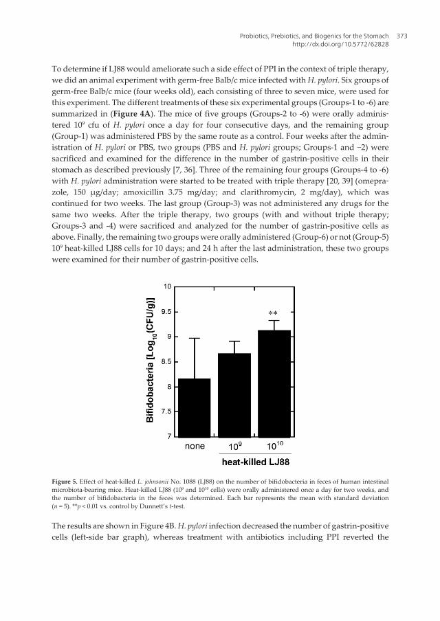
To determine if LJ88 would ameliorate such a side effect of PPI in the context of triple therapy,we did an animal experiment with germ-free Balb/c mice infected with H. pylori. Six groups ofgerm-free Balb/c mice (four weeks old), each consisting of three to seven mice, were used forthis experiment. The different treatments of these six experimental groups (Groups-1 to -6) aresummarized in (Figure 4A). The mice of five groups (Groups-2 to -6) were orally adminis‐tered 109 cfu of H. pylori once a day for four consecutive days, and the remaining group(Group-1) was administered PBS by the same route as a control. Four weeks after the admin‐istration of H. pylori or PBS, two groups (PBS and H. pylori groups; Groups-1 and −2) weresacrificed and examined for the difference in the number of gastrin-positive cells in theirstomach as described previously [7, 36]. Three of the remaining four groups (Groups-4 to -6)with H. pylori administration were started to be treated with triple therapy [20, 39] (omepra‐zole, 150 μg/day; amoxicillin 3.75 mg/day; and clarithromycin, 2 mg/day), which wascontinued for two weeks. The last group (Group-3) was not administered any drugs for thesame two weeks. After the triple therapy, two groups (with and without triple therapy;Groups-3 and -4) were sacrificed and analyzed for the number of gastrin-positive cells asabove. Finally, the remaining two groups were orally administered (Group-6) or not (Group-5)109 heat-killed LJ88 cells for 10 days; and 24 h after the last administration, these two groupswere examined for their number of gastrin-positive cells.
Figure 5. Effect of heat-killed L. johnsonii No. 1088 (LJ88) on the number of bifidobacteria in feces of human intestinalmicrobiota-bearing mice. Heat-killed LJ88 (109 and 1010 cells) were orally administered once a day for two weeks, andthe number of bifidobacteria in the feces was determined. Each bar represents the mean with standard deviation(n = 5). **p < 0.01 vs. control by Dunnett’s t-test.
The results are shown in Figure 4B. H. pylori infection decreased the number of gastrin-positivecells (left-side bar graph), whereas treatment with antibiotics including PPI reverted the
Probiotics, Prebiotics, and Biogenics for the Stomachhttp://dx.doi.org/10.5772/62828
373

number of gastrin-positive cells to a higher level (middle bar graph). However, treatment withheat-killed LJ88 significantly decreased the number of gastrin-positive cells (right-side bargraph). These results suggest that LJ88, even in its heat-killed form, can prevent the increasein gastric acid after triple therapy by decreasing the number of gastrin-positive cells, the effectof which might be beneficial for prophylaxis of GERD. Since this result was obtained by usinga mouse model, it should be examined whether or not the same mechanism works also inhumans.
Since live LJ88 were beneficial not only to the stomach but also to intestinal microbiota, asshown in Figure 3, we examined the effect of heat-killed LJ88 on intestinal bacteria bydetermining the number of bifidobacteria in the feces of human intestinal microbiota-bear‐ing mice. As shown in Figure 5, heat-killed LJ88 increased the number of bifidobacteria in thefeces by the administration of 1010 cells for two weeks, suggesting that heat-killed LJ88 mightalso be beneficial to not only the stomach but also to the intestines as well.
4.1.2 Anti-H. pylori activities of heat-killed bacteria
We already described that some probiotic strains have anti-H. pylori activity, and possiblemechanisms underlying such an activity were discussed (Section 3 and listed in Table 2).Among them, some mechanisms can be expected to belong not only to live bacteria (probiot‐ics) but also to heat-killed ones (biogenics).
One possible mechanism might be competition between H. pylori and probiotic bacteria foradherence sites on gastric epithelial cells. So some probiotic strains proposed to compete foradherence sites on gastric surface might have anti-H. pylori activity even in their heat-killedforms. However, no such examples have been reported to date.
Another potential mechanism might be coaggregation with H. pylori. Examining the anti-H.pylori effect of heat-killed Lactobacius reuteri DSM17648, Holz et al. found that it coaggregateswell with H. pylori both in vitro and in vivo, and that it exerts anti-H. pylori activity also in theclinical situation [30]. This pioneering result suggests that other probiotic strains having anti-H. pylori activity are worth being examined for their ability to coaggregate with H. pylori.
4.2 Soybean-related products as biogenics for the stomach
Historically, it has been suggested that soy products prevent the incidence of various can‐cers including gastric cancer, and several meta-analysis studies concluded that nonferment‐ed and fermented soy foods reduce and increase, respectively, the risk of gastric cancer [40,41]. However, it has also been suggested that “nonfermented” and “fermented” soy foods arepossibly associated with “fruit/vegetable” and “salt intake,” respectively [40, 41]. So preven‐tive and stimulatory effects of nonfermented and fermented soy foods should be consideredtaking these factors in mind. Since isoflavones are one of the proposed molecular candidatesfor preventing gastric cancer, a large-scale, population-based, prospective, cohort study wasconducted to investigate the relationship between isoflavone-intake and risk of gastric cancerin Japan [42]. The results suggested that higher intake of isoflavones does not prevent gastric
Probiotics and Prebiotics in Human Nutrition and Health374

cancer [42]. So even if nonfermented soy foods can reduce the risk of gastric cancer, theresponsible molecules might not be isoflavones in soy foods. However, since genistein, whichis one of the soybean isoflavones, reportedly has a protective effect against stress-inducedgastric mucosal lesions in rats [43], soy foods might be beneficial to the stomach even if theircancer-preventing effects are not so large.
4.3. Brassicaceae vegetable-related products as biogenics for the stomach
Vegetables of Brassicaceae classification, including cabbage and broccoli, reportedly containS-methylmethionine, also known as vitamin U. S-methylmethionin is a useful ingredientoriginally found as anti-ulcerogenic factors in raw cabbage juice [44, 45], and has been used asan ingredient of gastrointestinal drugs in Japan for over 50 years, e.g., Cabagin U. [46]. SoBrassicaceae vegetables might be thought as good biogenics for the stomach for treatment and/or prevention of gastric ulcer.
Furthermore, broccoli sprouts especially contain sulforaphane, an isothiocyanate compoundreported to have anti-H. pylori activity both in vitro [47] and in vivo [48]. Sulforaphone alsohas been reported to have protective and reparative effects against oxidative stress in gastricmucosa by stimulating nrf2 gene-dependent antioxidant enzyme activities, and also to haveanti-inflammatory effects on gastric mucosa during H. pylori infections [49]. So amongBrassicaceae vegetables, broccoli sprouts are thought to be an especially beneficial biogenicfor the stomach.
4.4. Other natural products beneficial to the stomach, including those with anti-H. pyloriactivity
Because of the wide variety and expected low toxicity of natural products, extracts andessential oils prepared from various plants have been examined their anti-ulcer and anti-H.pylori activities. Bonifácio extensively reviewed such products, including 21 different plantextracts and 18 different essential oils [50]. Most of the extracts and essential oils, described inthe review mentioned above, were examined only in vitro, although some of them have beenevaluated in vivo as well. Bonamin et al. reported that a methanol extract and its enrichedalkaloid fraction of a Brazilian plant, Strychnos pseudoquina St. Hil. (Loganiaceae), were effectiveagainst gastric ulcer induced by acetic acid, and also had anti-H. pylori activity in vitro [51].Extracts of other Brazilian plants, e.g., Qualea parviflora Mart. (from bark) [52], Hancorniaspeciosa Gomez (Mangaba; from bark) [53], and Byrsonima intermedia A. Juss. (Malpighiaceae;from leaves) [54], have also been reported to have anti-ulcer activity in vivo and anti-H. pyloriactivity in vitro. Ohno et al. reported that 13 different essential oils prepared from a variety ofplants inhibited the growth of H. pylori in vitro [55]. Among them, essential oils fromCymbopogon citratus (lemongrass) and Lippia citriodora (lemon verbena) were found to bebactericidal [55]. They also found that essential oil from lemongrass inhibited H. pylori in amurine model [55]. Thus, natural sources including herbal and medicinal plants can be thoughtof as future promising sources of new biogenics for the stomach.
Probiotics, Prebiotics, and Biogenics for the Stomachhttp://dx.doi.org/10.5772/62828
375

5. Future directions
In this report, we discussed probiotics, prebiotics, and biogenics for the stomach. As shown inFigure 1, this research area remains small to date, as only 2% of the total volume of publica‐tions concerning “probiotics, prebiotics, or biogenics” as a whole has focused on the stom‐ach. However, the research efforts made related to this interesting research field, as mentionedin this review, are none the less very significant. We think future research in this field will goin the following directions:
Concerning probiotics for the stomach, a search for new probiotic strains beneficial to thestomach is warranted. Although no probiotic bacteria able to reside and grow in the stom‐ach have yet been found, the possible existence of such a kind of so-called “extremophile” [56]type of probiotic bacteria cannot be denied in principle. Indeed, most researchers did notbelieve in the existence of indigenous bacteria in the stomach until 1984, when H. pylori wasfirst described to exist there [57]. Other extremely acid-resistant probiotic strains that cansurvive in the stomach for a significant time period even if not able to grow there, such as ourLJ88, will be a more promising type of bacteria as probiotics for the stomach.
However, since “extremophile” probiotics or indigenous bacteria beneficial to the stomachhave not been found to date, prebiotics for such bacteria are also unknown as well. If suchbacteria are found in the future, compounds supporting the growth of these bacteria in thestomach may be regarded as “prebiotics for the stomach.” Specific substances specificallyutilized by supposed stomach bacteria beneficial to the host might be such candidates.
As described in this report, some strains of heat-killed bacteria are thought to be good biogenicsfor the stomach, as they, like LJ88, might be effective as anti-H. pylori agents and also as gastrin-inhibiting ones. Such novel kinds of more effective bacteria may possibly be found in the future.Moreover, the possibility of new biogenics for the stomach, derived from natural sources, e.g.,vegetables, fruits, traditional medicinal plants, fungi, products of microorganisms, and marineorganisms, should be examined, and promising candidates may well be found in the future.
Practically speaking, appropriate combinations of probiotics, prebiotics (putative), andbiogenics might be important for stomach health.
Author details
Yasuhiko Komatsu1*, Yuji Aiba1, Yasuhiro Nakano2 and Yasuhiro Koga2
*Address all correspondence to: [email protected]; [email protected]
1 Development Research Department, Snowden, Tokyo, Japan
2 University School of Medicine, Tokai, Kanagawa, Japan
Probiotics and Prebiotics in Human Nutrition and Health376

References
[1] Fukushima Y, Yamano T, Kusano A, Takada M, Amano M, Iino H. Effect of ferment‐ed milk containing Lactobacillus johnsonii La1 (LC1) on defecation in healthy Japaneseadults—A double blind placebo controlled study—. Bioscience Microflora. 2004;23(4):139–47.
[2] Felley CP, Corthesy-Theulaz I, Rivero JL, Sipponen P, Kaufmann M, Bauerfeind P, etal. Favourable effect of an acidified milk (LC-1) on Helicobacter pylori gastritis in man.Eur J Gastroenterol Hepatol. 2001;13(1):25–9.
[3] Michetti P, Dorta G, Wiesel PH, Brassart D, Verdu E, Herranz M, et al. Effect of whey-based culture supernatant of Lactobacillus acidophilus (johnsonii) La1 on Helicobacterpylori infection in humans. Digestion. 1999;60(3):203–9.
[4] Sakamoto I, Igarashi M, Kimura K, Takagi A, Miwa T, Koga Y. Suppressive effect of L.gasseri OLL 2716 (LG21) on Helicobacter pylori infection in humans. J AntimicrobChemother. 2001;47(5):709–10.
[5] Fujimura S, Kato S, Oda M, Miyahara M, Ito Y, Kimura K, et al. Detection of Lactoba‐cillus gasseri OLL2716 strain administered with yogurt drink in gastric mucus layer inhumans. Lett Applied Microbiol. 2006;43(5):578–81.
[6] Kato-Mori Y, Orihashi T, Kanai Y, Sato M, Sera K, Hagiwara K. Fermentation metab‐olites from Lactobacillus gasseri and Propionibacterium freudenreichii exert bacteriocidaleffects in mice. J Med food. 2010;13(6):1460–7.
[7] Aiba Y, Nakano Y, Koga Y, Takahashi K, Komatsu Y. A highly acid-resistant novelstrain of Lactobacillus johnsonii No. 1088 has antibacterial activity, including that againstHelicobacter pylori, and inhibits gastrin-mediated acid production in mice. Microbiolo‐gy Open. 2015;4:465–74.
[8] Mitsuoka T. Development of functional foods. Biosci Microbiota Food Health. 2014;33(3):117–28.
[9] Tsuda A, Suda W, Morita H, Takanashi K, Takagi A, Koga Y, et al. Influence of proton-pump inhibitors on the luminal microbiota in the gastrointestinal tract. Clin TranslGastroenterol. 2015;6:e89.
[10] Guarner F, Schaafsma GJ. Probiotics. Int J Food Microbiol. 1998;39(3):237–8.
[11] Peek RM Jr., Blaser MJ. Helicobacter pylori and gastrointestinal tract adenocarcinomas.Nat Rev Cancer. 2002;2(1):28–37.
[12] Coconnier MH, Lievin V, Hemery E, Servin AL. Antagonistic activity againstHelicobacter infection in vitro and in vivo by the human Lactobacillus acidophilus strainLB. Appl Environ Microb. 1998;64(11):4573–80.
Probiotics, Prebiotics, and Biogenics for the Stomachhttp://dx.doi.org/10.5772/62828
377

[13] Pinchuk IV, Bressollier P, Verneuil B, Fenet B, Sorokulova IB, Megraud F, et al. In vitroanti-Helicobacter pylori activity of the probiotic strain Bacillus subtilis 3 is due to secretionof antibiotics. Antimicrob Agents Chemother. 2001;45(11):3156–61.
[14] Nam H, Ha M, Bae O, Lee Y. Effect of Weissella confusa strain PL9001 on the adher‐ence and growth of Helicobacter pylori. Appl Environ Microb. 2002;68(9):4642–5.
[15] Boyanova L, Stephanova-Kondratenko M, Mitov I. Anti-Helicobacter pylori activity ofLactobacillus delbrueckii subsp. bulgaricus strains: preliminary report. Lett ApplMicrobiol. 2009;48(5):579–84.
[16] Francavilla R, Lionetti E, Castellaneta SP, Magista AM, Maurogiovanni G, Bucci N, etal. Inhibition of Helicobacter pylori infection in humans by Lactobacillus reuteri ATCC55730 and effect on eradication therapy: a pilot study. Helicobacter. 2008;13(2):127–34.
[17] Tojo R, Suarez A, Clemente MG, de los Reyes-Gavilan CG, Margolles A, GueimondeM, et al. Intestinal microbiota in health and disease: role of bifidobacteria in guthomeostasis. World J Gastroenterol. 2014;20(41):15163–76.
[18] Trifan A, Stanciu C, Stoica O, Girleanu I, Cojocariu C. Impact of Clostridium difficileinfection on inflammatory bowel disease outcome: a review. World J Gastroenterol.2014;20(33):11736–42.
[19] Shindo Y, Dobashi Y, Sakai T, Monma C, Miyatani H, Yoshida Y. Epidemiological andpathobiological profiles of Clostridium perfringens infections: review of consecutiveseries of 33 cases over a 13-year period. Int J Clin Exp Pathol. 2015;8(1):569–77.
[20] Malfertheiner P, Megraud F, O’Morain C, Bazzoli F, El-Omar E, Graham D, et al.Current concepts in the management of Helicobacter pylori infection: the Maastricht IIIConsensus Report. Gut. 2007;56(6):772–81.
[21] Lesbros-Pantoflickova D, Corthesy-Theulaz I, Blum AL. Helicobacter pylori andprobiotics. J Nutr. 2007;137(3 Suppl 2):812S-8S.
[22] Aiba Y, Suzuki N, Kabir AM, Takagi A, Koga Y. Lactic acid-mediated suppression ofHelicobacter pylori by the oral administration of Lactobacillus salivarius as a probiotic ina gnotobiotic murine model. Am J Gastroenterol. 1998;93(11):2097–101.
[23] Midolo PD, Lambert JR, Hull R, Luo F, Grayson ML. In vitro inhibition of Helicobacterpylori NCTC 11637 by organic acids and lactic acid bacteria. J Appl Bacteriol. 1995;79(4):475–9.
[24] Sgouras D, Maragkoudakis P, Petraki K, Martinez-Gonzalez B, Eriotou E, Michopou‐los S, et al. In vitro and in vivo inhibition of Helicobacter pylori by Lactobacillus casei strainShirota. Appl Environ Microbiol. 2004;70(1):518–26.
[25] Cotter PD, Ross RP, Hill C. Bacteriocins—a viable alternative to antibiotics? Nat RevMicrobiol. 2013;11(2):95–105.
Probiotics and Prebiotics in Human Nutrition and Health378

[26] Kim TS, Hur JW, Yu MA, Cheigh CI, Kim KN, Hwang JK, et al. Antagonism ofHelicobacter pylori by bacteriocins of lactic acid bacteria. J Food Prot. 2003;66(1):3–12.
[27] Kabir AM, Aiba Y, Takagi A, Kamiya S, Miwa T, Koga Y. Prevention of Helicobacterpylori infection by lactobacilli in a gnotobiotic murine model. Gut. 1997;41(1):49–55.
[28] Mukai T, Asasaka T, Sato E, Mori K, Matsumoto M, Ohori H. Inhibition of binding ofHelicobacter pylori to the glycolipid receptors by probiotic Lactobacillus reuteri. FEMSImmunol Med Microbiol. 2002;32(2):105–10.
[29] Noach LA, Bosma NB, Jansen J, Hoek FJ, van Deventer SJ, Tytgat GN. Mucosal tumornecrosis factor-alpha, interleukin-1 beta, and interleukin-8 production in patients withHelicobacter pylori infection. Scand J Gastroenterol. 1994;29(5):425–9.
[30] Holz C, Busjahn A, Mehling H, Arya S, Boettner M, Habibi H, et al. Significant reductionin Helicobacter pylori load in humans with non-viable Lactobacillus reuteri DSM17648: apilot study. Probiotics Antimicrob Proteins. 2015;7(2):91–100.
[31] Kahrilas PJ, Shaheen NJ, Vaezi MF, Hiltz SW, Black E, Modlin IM, et al. AmericanGastroenterological Association Medical Position Statement on the management ofgastroesophageal reflux disease. Gastroenterology. 2008;135(4):1383–91.
[32] Richter JE. H pylori: the bug is not all bad. Gut. 2001;49(3):319–20.
[33] Fallone CA, Barkun AN, Mayrand S, Wakil G, Friedman G, Szilagyi A, et al. There isno difference in the disease severity of gastro-oesophageal reflux disease betweenpatients infected and not infected with Helicobacter pylori. Aliment Pharmacol Ther.2004;20(7):761–8.
[34] Schubert ML, Makhlouf GM. Neural, hormonal, and paracrine regulation of gastrinand acid secretion. Yale J Biol Med. 1992;65(6):553–60; Discussion 621–3.
[35] Feng J, Petersen CD, Coy DH, Jiang JK, Thomas CJ, Pollak MR, et al. Calcium-sensingreceptor is a physiologic multimodal chemosensor regulating gastric G-cell growth andgastrin secretion. Proc Natl Acad Sci USA. 2010;107(41):17791–6.
[36] Takahashi H, Nakano Y, Matsuoka T, Kumaki N, Asami Y, Koga Y. Role of indige‐nous lactobacilli in gastrin-mediated acid production in the mouse stomach. ApplEnviron Microbiol. 2011;77(19):6964–71.
[37] Delgado S, Cabrera-Rubio R, Mira A, Suarez A, Mayo B. Microbiological survey of thehuman gastric ecosystem using culturing and pyrosequencing methods. Microb Ecol.2013;65(3):763–72.
[38] Gibson GR, Roberfroid MB. Dietary modulation of the human colonic microbiota:introducing the concept of prebiotics. J Nutr. 1995;125(6):1401–12.
[39] Lind T, Megraud F, Unge P, Bayerdorffer E, O’Morain C, Spiller R, et al. The MACH2study: role of omeprazole in eradication of Helicobacter pylori with 1-week tripletherapies. Gastroenterology. 1999;116(2):248–53.
Probiotics, Prebiotics, and Biogenics for the Stomachhttp://dx.doi.org/10.5772/62828
379
[40] Wu AH, Yang D, Pike MC. A meta-analysis of soyfoods and risk of stomach cancer: theproblem of potential confounders. Cancer Epidemiol Biomarkers Prev. 2000;9(10):1051–8.
[41] Kim J, Kang M, Lee JS, Inoue M, Sasazuki S, Tsugane S. Fermented and non-ferment‐ed soy food consumption and gastric cancer in Japanese and Korean populations: ameta-analysis of observational studies. Cancer Sci. 2011;102(1):231–44.
[42] Hara A, Sasazuki S, Inoue M, Iwasaki M, Shimazu T, Sawada N, et al. Isoflavone intakeand risk of gastric cancer: a population-based prospective cohort study in Japan. Am JClin Nutr. 2012;95(1):147–54.
[43] Takekawa S, Matsui T, Arakawa Y. The protective effect of the soybean polyphenolgenistein against stress-induced gastric mucosal lesions in rats, and its hormonalmechanisms. J Nutr Sci Vitamino. 2006;52(4):274–80.
[44] Cheney G. Anti-peptic ulcer dietary factor (vitamin “U”) in the treatment of pepticulcer. J Am Diet Asso. 1950;26(9):668–72.
[45] Cheney G. Vitamin U therapy of peptic ulcer. Calif Med. 1952;77(4):248–52.
[46] Kosaki S, Yoshioka Y, Mitsuba K, Sakai R. Effectiveness of Cabagin U for peptic ulcer(in Japanese). J New Remedies & Clinics. 1964;13(11):1288–92.
[47] Fahey JW, Haristoy X, Dolan PM, Kensler TW, Scholtus I, Stephenson KK, et al.Sulforaphane inhibits extracellular, intracellular, and antibiotic-resistant strains ofHelicobacter pylori and prevents benzo[a]pyrene-induced stomach tumors. Proc NatlAcad Sci USA. 2002;99(11):7610–5.
[48] Haristoy X, Angioi-Duprez K, Duprez A, Lozniewski A. Efficacy of sulforaphane ineradicating Helicobacter pylori in human gastric xenografts implanted in nude mice.Antimicrob Agents Chemother. 2003;47(12):3982–4.
[49] Yanaka A. Sulforaphane enhances protection and repair of gastric mucosa againstoxidative stress in vitro, and demonstrates anti-inflammatory effects on Helicobacterpylori-infected gastric mucosae in mice and human subjects. Curr Pharma Des.2011;17(16):1532–40.
[50] Bonifacio BV, dos Santos Ramos MA, da Silva PB, Bauab TM. Antimicrobial activity ofnatural products against Helicobacter pylori: a review. Ann Clin Microbiol Antimicrob.2014;13:54.
[51] Bonamin F, Moraes TM, Kushima H, Silva MA, Rozza AL, Pellizzon CH, et al. Can aStrychnos species be used as antiulcer agent? Ulcer healing action from alkaloid fractionof Strychnos pseudoquina St. Hil. (Loganiaceae). J Ethnopharmacol. 2011;138(1):47–52.
[52] Mazzolin LP, Nasser AL, Moraes TM, Santos RC, Nishijima CM, Santos FV, et al. Qualeaparviflora Mart.: an integrative study to validate the gastroprotective, antidiarrheal,antihemorragic and mutagenic action. J Ethnopharmacol. 2010;127(2):508–14.
Probiotics and Prebiotics in Human Nutrition and Health380

[53] Moraes Tde M, Rodrigues CM, Kushima H, Bauab TM, Villegas W, Pellizzon CH, et al.Hancornia speciosa: indications of gastroprotective, healing and anti-Helicobacter pyloriactions. J Ethnopharmacol. 2008;120(2):161–8.
[54] Santos RC, Kushima H, Rodrigues CM, Sannomiya M, Rocha LR, Bauab TM, et al.Byrsonima intermedia A. Juss.: gastric and duodenal anti-ulcer, antimicrobial andantidiarrheal effects in experimental rodent models. J Ethnopharmacol. 2012;140(2):203–12.
[55] Ohno T, Kita M, Yamaoka Y, Imamura S, Yamamoto T, Mitsufuji S, et al. Antimicrobi‐al activity of essential oils against Helicobacter pylori. Helicobacter. 2003;8(3):207–15.
[56] Rothschild LJ, Mancinelli RL. Life in extreme environments. Nature. 2001;409(6823):1092–101.
[57] Marshall BJ, Warren JR. Unidentified curved bacilli in the stomach of patients withgastritis and peptic ulceration. Lancet. 1984;1(8390):1311–5.
Probiotics, Prebiotics, and Biogenics for the Stomachhttp://dx.doi.org/10.5772/62828
381