Primitive streak formation in mice is preceded by localized activation of Brachyury and Wnt3
9
Primitive streak formation in mice is preceded by localized activation of Brachyury and Wnt3 Jaime A. Rivera-Pe ´rez, Terry Magnuson * Department of Genetics, Campus Box 7264, University of North Carolina, Chapel Hill, NC 27599-7264, USA Received for publication 24 January 2005, revised 11 August 2005, accepted 9 September 2005 Available online 10 November 2005 Abstract The prevalent model for the generation of axial polarity in mouse embryos proposes that a radial to a linear transition in the expression of primitive streak markers precedes the formation of the primitive streak on one side of the epiblast. This model contrasts with the models of mesoderm formation in other vertebrates as it suggests that the primitive streak is initially established in a radial pattern rather than a localized region of the epiblast. Here, we examine the proposed correlation between the expression of Brachyury and Wnt3 , two genes reported as expressed radially in the proximal epiblast, with the movements of proximal anterior epiblast cells at stages leading to the formation of the primitive streak. Our results reveal that neither Brachyury nor Wnt3 forms a ring of expression in the proximal epiblast as previously thought. In embryos dissected between 5.5 and 6.5 dpc, Brachyury is first expressed in the distal extra-embryonic ectoderm and subsequently on one side of the epiblast. Wnt3 expression is evident first in the posterior visceral endoderm of 5.5 dpc embryos and later in the posterior epiblast. Lineage analysis shows that the movements of the proximal epiblast do not restrict Brachyury expression to the posterior epiblast. Our data suggest a model whereby the localized expression of these genes in the posterior epiblast, and hence the formation of the primitive streak, is the result of local cell – cell interactions in the future posterior portion of the egg cylinder rather than regionalization of a radial pattern of expression in proximal epiblast cells. D 2005 Elsevier Inc. All rights reserved. Keywords: Mouse; Epiblast; Brachyury; Wnt3; Visceral endoderm; Primitive streak; Iontophoresis Introduction A fundamental process during the generation of the anteroposterior axis in mammals is the formation of the primitive streak. In mice, the primitive streak forms in a localized region of the epiblast located adjacent to the extra- embryonic ectoderm where it marks the posterior site of the embryo. During gastrulation, the primitive streak serves as a conduit for the generation of mesoderm and definitive endoderm (Tam and Gad, 2004). The current model for the establishment of the anteroposterior axis and hence the primitive streak, proposes an orthogonal rotation from the proximodistal to the transverse plane of the egg cylinder at stages preceding the formation of the primitive streak (Bed- dington and Robertson, 1999; Lu et al., 2001; Thomas and Beddington, 1996; Thomas et al., 1998). In this model, extra- embryonic ectoderm induces the proximal epiblast to express several genes that form a ring of expression in the proximal epiblast. The expression of these genes is subsequently restricted to one side of the epiblast where the primitive streak will eventually form. The transition from a radial to a linear pattern of expression of primitive streak markers suggests that the potential for the formation of the primitive streak is initially established in the proximal epiblast (see Tam and Gad, 2004). Two hypotheses have been offered to explain this process. One hypothesis proposes that radial epiblast markers are allocated to the posterior epiblast by the posteriorward movements of proximal epiblast cells at stages preceding the appearance of the streak (Thomas et al., 1998). The second hypothesis, based on lineage studies and on tissue recombination experiments, proposes that the anterior visceral endoderm known as the AVE suppresses the expression of radially expressed genes in the anterior epiblast, limiting their expression to the posterior epiblast (Kimura et al., 2000; Thomas et al., 1998). The movements of the distal visceral endoderm, the precursor of the AVE, have been well documented in lineage experiments 0012-1606/$ - see front matter D 2005 Elsevier Inc. All rights reserved. doi:10.1016/j.ydbio.2005.09.012 * Corresponding author. Fax: (919) 843 6365. E-mail address: [email protected] (T. Magnuson). Developmental Biology 288 (2005) 363 – 371 www.elsevier.com/locate/ydbio
-
Upload
independent -
Category
Documents
-
view
7 -
download
0
Transcript of Primitive streak formation in mice is preceded by localized activation of Brachyury and Wnt3
lsevier.com/locate/ydbio
Developmental Biology 2
Primitive streak formation in mice is preceded by localized activation
of Brachyury and Wnt3
Jaime A. Rivera-Perez, Terry Magnuson *
Department of Genetics, Campus Box 7264, University of North Carolina, Chapel Hill, NC 27599-7264, USA
Received for publication 24 January 2005, revised 11 August 2005, accepted 9 September 2005
Available online 10 November 2005
Abstract
The prevalent model for the generation of axial polarity in mouse embryos proposes that a radial to a linear transition in the expression of
primitive streak markers precedes the formation of the primitive streak on one side of the epiblast. This model contrasts with the models of
mesoderm formation in other vertebrates as it suggests that the primitive streak is initially established in a radial pattern rather than a localized
region of the epiblast. Here, we examine the proposed correlation between the expression of Brachyury andWnt3, two genes reported as expressed
radially in the proximal epiblast, with the movements of proximal anterior epiblast cells at stages leading to the formation of the primitive streak.
Our results reveal that neither Brachyury norWnt3 forms a ring of expression in the proximal epiblast as previously thought. In embryos dissected
between 5.5 and 6.5 dpc, Brachyury is first expressed in the distal extra-embryonic ectoderm and subsequently on one side of the epiblast. Wnt3
expression is evident first in the posterior visceral endoderm of 5.5 dpc embryos and later in the posterior epiblast. Lineage analysis shows that the
movements of the proximal epiblast do not restrict Brachyury expression to the posterior epiblast. Our data suggest a model whereby the localized
expression of these genes in the posterior epiblast, and hence the formation of the primitive streak, is the result of local cell–cell interactions in the
future posterior portion of the egg cylinder rather than regionalization of a radial pattern of expression in proximal epiblast cells.
D 2005 Elsevier Inc. All rights reserved.
Keywords: Mouse; Epiblast; Brachyury; Wnt3; Visceral endoderm; Primitive streak; Iontophoresis
Introduction
A fundamental process during the generation of the
anteroposterior axis in mammals is the formation of the
primitive streak. In mice, the primitive streak forms in a
localized region of the epiblast located adjacent to the extra-
embryonic ectoderm where it marks the posterior site of the
embryo. During gastrulation, the primitive streak serves as a
conduit for the generation of mesoderm and definitive
endoderm (Tam and Gad, 2004). The current model for the
establishment of the anteroposterior axis and hence the
primitive streak, proposes an orthogonal rotation from the
proximodistal to the transverse plane of the egg cylinder at
stages preceding the formation of the primitive streak (Bed-
dington and Robertson, 1999; Lu et al., 2001; Thomas and
Beddington, 1996; Thomas et al., 1998). In this model, extra-
0012-1606/$ - see front matter D 2005 Elsevier Inc. All rights reserved.
doi:10.1016/j.ydbio.2005.09.012
* Corresponding author. Fax: (919) 843 6365.
E-mail address: [email protected] (T. Magnuson).
embryonic ectoderm induces the proximal epiblast to express
several genes that form a ring of expression in the proximal
epiblast. The expression of these genes is subsequently
restricted to one side of the epiblast where the primitive streak
will eventually form. The transition from a radial to a linear
pattern of expression of primitive streak markers suggests that
the potential for the formation of the primitive streak is initially
established in the proximal epiblast (see Tam and Gad, 2004).
Two hypotheses have been offered to explain this process. One
hypothesis proposes that radial epiblast markers are allocated to
the posterior epiblast by the posteriorward movements of
proximal epiblast cells at stages preceding the appearance of
the streak (Thomas et al., 1998). The second hypothesis, based
on lineage studies and on tissue recombination experiments,
proposes that the anterior visceral endoderm known as the AVE
suppresses the expression of radially expressed genes in the
anterior epiblast, limiting their expression to the posterior
epiblast (Kimura et al., 2000; Thomas et al., 1998).
Themovements of the distal visceral endoderm, the precursor
of the AVE, have been well documented in lineage experiments
88 (2005) 363 – 371
www.e
J.A. Rivera-Perez, T. Magnuson / Developmental Biology 288 (2005) 363–371364
using DiI (Thomas et al., 1998), iontophoresis (Rivera-Perez et
al., 2003) or time-lapse imaging (Srinivas et al., 2004). The AVE
is characterized by the expression of multiple genes that include
Hhex and several repressors of Nodal and Wnt signaling such as
Cer1, Lefty1, Dkk1, Sfrp5 and Sfrp1 (Beddington and Robert-
son, 1998; Finley et al., 2003; Kemp et al., 2005; Perea-Gomez
et al., 2001). The study of the morphogenetic movements of the
epiblast, on the contrary, has been limited to stages immediately
before or after the primitive streak appears (Lawson et al., 1991).
These studies have revealed an anisotropic spread of proximal
epiblast cells towards the primitive streak. At the molecular
level, Brachyury was reported to form a ring of expression in the
proximal epiblast at pre-streak stages and then to localize to the
posterior epiblast and primitive streak (Thomas and Beddington,
1996; Thomas et al., 1998). Recently, however, the pattern of
expression of Brachyury was shown to form a ring of expression
in the extra-embryonic ectoderm rather than the proximal
epiblast (Perea-Gomez et al., 2004). The expression of Wnt3
was also reported to form a ring in the proximal epiblast and later
to be restricted to the posterior epiblast/primitive streak regions
(Liu et al., 1999; Lu and Robertson, 2004).
We have previously identified molecular and morphological
landmarks that allow the determination of the polarity of egg
cylinder stage embryos before the appearance of the primitive
streak (Rivera-Perez et al., 2003). Taking advantage of these
landmarks, we have labeled anterior proximal epiblast cells at
pre-streak stages and compared the distribution of labeled
descendants with a systematic analysis of the expression of
Brachyury and Wnt3. Our results show that Brachyury
expression does not form a ring of expression in the proximal
epiblast as originally reported (Thomas and Beddington, 1996;
Thomas et al., 1998) but rather in the extra-embryonic
ectoderm as reported recently (Perea-Gomez et al., 2004). In
the epiblast, Brachyury RNA and protein are restricted to one
side of the epiblast abutting the extra-embryonic ectoderm and
later in the primitive streak. Similarly, Wnt3 RNA does not
form a ring of expression in the proximal epiblast. Wnt3
expression was observed first in the posterior visceral
endoderm as early as 5.5 dpc and later in both the posterior
visceral endoderm and epiblast or the primitive streak.
Interestingly, Wnt3 expression was observed in the posterior
visceral endoderm before Hhex-expressing distal visceral
endoderm cells (indicative of the AVE) had shifted to one side
of the epiblast. These results suggest that the symmetry-
breaking event that leads to the formation of the anteroposterior
axis precedes the shift of the distal AVE to one side of the
epiblast. A direct comparison between the movements of
anterior epiblast and the expression of Brachyury revealed a
spatial asynchrony between these events, showing that cell
movements do not account for the posterior restriction of
Brachyury expression. Based on these results, we propose a
model in which the posterior epiblast expression of primitive
streak markers is produced by interactions between the epiblast
and visceral endoderm region located at the epiblast/extra-
embryonic ectoderm boundary. This model suggests an
evolutionary conservation of the mechanisms that lead to
mesoderm formation in vertebrates.
Materials and methods
Lineage analysis and mouse strains
Lineage analysis was conducted in embryos derived from crosses
between Tg(Hhex-eGFP)ARbe homozygous males (Rodriguez et al., 2001)
maintained as mixed stock and CD-1 females (Charles River Laboratories).
Cells were labeled intracellularly with Lysinated Rhodamine Dextran
(LDRX) and Horseradish peroxidase (HRP) using iontophoresis (Lawson
et al., 1991). One to three epiblast cells were labeled per embryo. Labeled
embryos were cultured in media composed of 80% rat serum in MEMa
(Gibco) suspended from the lid of snap cap tubes as described (Rivera-Perez
et al., 2003).
Brachyury (T) heterozygous mice were obtained from the Jackson
Laboratory (B10;TFLe-a/a, T tf/++ /J, Stock Number 003879). Animals were
maintained under a 12-h light cycle. The middle of the dark cycle (midnight)
that occurred prior to observing a copulation plug was considered the beginning
of gestation.
Embryo staging
Embryos at 5.5 and 5.75 dpc were staged using morphological and
fluorescent landmarks (Rivera-Perez et al., 2003). Embryos at 5.5 dpc were
characterized by the presence of tall columnar visceral endoderm cells located
at the tip of the egg cylinder. These group of cells appeared as a thickening of
the visceral endoderm layer and were distinguished by eGFP fluorescence in
Tg(Hhex-eGFP)ARbe transgenic embryos. In 5.75 dpc embryos, the Hhex-
expressing area of visceral endoderm (now AVE) was located on one side of the
epiblast and its most proximal extent reached the epiblast/extra-embryonic
ectoderm boundary. In embryos between 5.75 and 6.5 days of gestation
(without a primitive streak), the embryonic stage was defined only as days post-
coitum (dpc) since there are no reliable landmarks to establish an unambiguous
developmental stage during this period of development. Embryos at 5.5, 5.75
and 6.5 dpc were dissected 12, 18 and 14 h after the middle of the dark cycle,
respectively. Embryos at 6.0 dpc were dissected within 1 h before or after the
middle of the dark cycle. Embryos with a primitive streak were staged
according to Downs and Davies (1993).
Whole-mount RNA in situ analysis
CD-1 or Heterozygous Tg(Hhex-eGFP)ARbe embryos were used in whole-
mount in situ hybridization. Embryos were fixed overnight in 4% parafor-
maldehyde prepared in PBS. After fixation, they were dehydrated in methanol
series and stored in 100% methanol at �20-C. Hybridization was done at
70-C following the protocol of J. Rossant’s laboratory (http://www.mshri.on.
ca/rossant/protocols/html, based on Henrique et al., 1995). The Brachyury
probe was transcribed from plasmid pSK75 (a gift from B. Herrmann). It
consisted of the complete coding sequence, 108 bp of the 5V UTR and 354 bp
of the 3VUTR (Herrmann et al., 1990). We also used a 3V Brachyury probe that
spanned 544 bp from the SacI to the EcoRI sites in exon 8 (Herrmann et al.,
1990). The Wnt3 probe (a gift from R. Behringer) was a cDNA fragment
containing exons 3–5 (Liu et al., 1999; Roelink et al., 1990). For HRP/whole-
mount in situ double staining, embryos were fixed in 4% paraformaldehyde,
assayed for HRP activity (Beddington and Lawson, 1990) and then subjected
to whole-mount in situ hybridization. Embryos hybridized with the Brachyury
cDNA or Wnt3 probes were treated for 2 min with proteinase K (10 Ag/ml)
and allowed to stain for 5 days in the color reaction solution. Embryos used in
these experiments were littermates. Embryos hybridized with the Brachyury 3Vprobe and those assayed for HRP activity, were treated for 5 min with
proteinase K (10 Ag/ml) and the color reaction conducted for 1 day or 1 h to 3
days, respectively.
Immunofluorescence staining
Embryos obtained from CD1 crosses were fixed for 1 h in 4%
paraformaldehyde prepared in PBS. After fixation, embryos were washed 3
J.A. Rivera-Perez, T. Magnuson / Developmental Biology 288 (2005) 363–371 365
times in PBS for 10 min and incubated in blocking solution (5% goat serum,
0.5% Triton X-100 and 1% bovine serum albumin in PBS) for 1 h.
Subsequently, embryos were incubated in primary antibody solution [Rabbit
anti Brachyury antiserum (generated using Xenopus Brachyury protein, a
generous gift of F. Conlon) diluted 1:2000 and Rat anti E-cadherin IgG (Zymed
Cat No. 13-1900) diluted 1:500 in blocking solution] for 2 h. After primary
antibody staining, embryos were washed three times in PBT (0.5% Triton X-
100, 1% BSA in PBS) and incubated for 1 h in secondary antibody solution
[Alexa-fluor 488 Goat anti Rabbit IgG (Molecular probes Cat. No. A11008)
and Alexa-fluor 594 Goat anti Rat IgG (Molecular Probes, Cat. No. A-11007)
diluted 1:500 in PBT]. Embryos were then rinsed 3 times in PBT for 10 min,
once in PBSAT (0.1% Triton X-100, 0.5% BSA in PBS) for 5 min and
counterstained in a 1 Ag/ml aqueous solution of DAPI (4V, 6-diamidino-2-
phenylindole, dihydrochloride) for 2 min. After DAPI staining, embryos were
washed twice in PBSAT for 5 min and mounted in a solution containing 2.5%
N-Propyl Gallate and 50% Glycerol in PBS.
Histology
Whole-mount hybridized embryos were rinsed in PBS, re-fixed for 1 h in
4% paraformaldehyde, equilibrated in gradients of 10, 20 and 30% sucrose/PBS
and mounted in OCT. Cryosections were obtained every 7 Am and mounted in
70% glycerol/PBS.
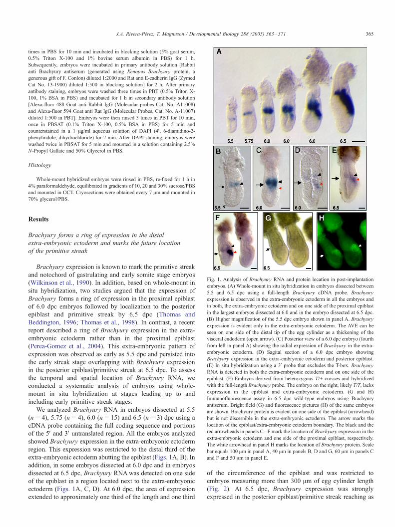
Fig. 1. Analysis of Brachyury RNA and protein location in post-implantation
embryos. (A) Whole-mount in situ hybridization in embryos dissected between
5.5 and 6.5 dpc using a full-length Brachyury cDNA probe. Brachyury
expression is observed in the extra-embryonic ectoderm in all the embryos and
in both, the extra-embryonic ectoderm and on one side of the proximal epiblast
in the largest embryos dissected at 6.0 and in the embryo dissected at 6.5 dpc.
(B) Higher magnification of the 5.5 dpc embryo shown in panel A. Brachyury
expression is evident only in the extra-embryonic ectoderm. The AVE can be
seen on one side of the distal tip of the egg cylinder as a thickening of the
visceral endoderm (open arrow). (C) Posterior view of a 6.0 dpc embryo (fourth
from left in panel A) showing the radial expression of Brachyury in the extra-
embryonic ectoderm. (D) Sagital section of a 6.0 dpc embryo showing
Brachyury expression in the extra-embryonic ectoderm and posterior epiblast.
(E) In situ hybridization using a 3V probe that excludes the T-box. Brachyury
RNA is detected in both the extra-embryonic ectoderm and on one side of the
epiblast. (F) Embryos derived from heterozygous T/+ crosses and hybridized
with the full-length Brachyury probe. The embryo on the right, likely T/T, lacks
expression in the epiblast and extra-embryonic ectoderm. (G and H)
Immunofluorescence assay in 6.5 dpc wild-type embryos using Brachyury
antiserum. Bright field (G) and fluorescence pictures (H) of the same embryos
are shown. Brachyury protein is evident on one side of the epiblast (arrowhead)
but is not discernible in the extra-embryonic ectoderm. The arrow marks the
location of the epiblast/extra-embryonic ectoderm boundary. The black and the
red arrowheads in panels C–F mark the location of Brachyury expression in the
extra-embryonic ectoderm and one side of the proximal epiblast, respectively.
The white arrowhead in panel H marks the location of Brachyury protein. Scale
bar equals 100 Am in panel A, 40 Am in panels B, D and G, 60 Am in panels C
and F and 50 Am in panel E.
Results
Brachyury forms a ring of expression in the distal
extra-embryonic ectoderm and marks the future location
of the primitive streak
Brachyury expression is known to mark the primitive streak
and notochord of gastrulating and early somite stage embryos
(Wilkinson et al., 1990). In addition, based on whole-mount in
situ hybridization, two studies argued that the expression of
Brachyury forms a ring of expression in the proximal epiblast
of 6.0 dpc embryos followed by localization to the posterior
epiblast and primitive streak by 6.5 dpc (Thomas and
Beddington, 1996; Thomas et al., 1998). In contrast, a recent
report described a ring of Brachyury expression in the extra-
embryonic ectoderm rather than in the proximal epiblast
(Perea-Gomez et al., 2004). This extra-embryonic pattern of
expression was observed as early as 5.5 dpc and persisted into
the early streak stage overlapping with Brachyury expression
in the posterior epiblast/primitive streak at 6.5 dpc. To assess
the temporal and spatial location of Brachyury RNA, we
conducted a systematic analysis of embryos using whole-
mount in situ hybridization at stages leading up to and
including early primitive streak stages.
We analyzed Brachyury RNA in embryos dissected at 5.5
(n = 4), 5.75 (n = 4), 6.0 (n = 15) and 6.5 (n = 3) dpc using a
cDNA probe containing the full coding sequence and portions
of the 5V and 3V untranslated region. All the embryos analyzed
showed Brachyury expression in the extra-embryonic ectoderm
region. This expression was restricted to the distal third of the
extra-embryonic ectoderm abutting the epiblast (Figs. 1A, B). In
addition, in some embryos dissected at 6.0 dpc and in embryos
dissected at 6.5 dpc, Brachyury RNAwas detected on one side
of the epiblast in a region located next to the extra-embryonic
ectoderm (Figs. 1A, C, D). At 6.0 dpc, the area of expression
extended to approximately one third of the length and one third
of the circumference of the epiblast and was restricted to
embryos measuring more than 300 Am of egg cylinder length
(Fig. 2). At 6.5 dpc, Brachyury expression was strongly
expressed in the posterior epiblast/primitive streak reaching as

Fig. 2. Scatter plot depiction of embryonic dimensions and comparison to the onset of Brachyury expression in the epiblast. Brachyury expression in the epiblast is
evident only in embryos with more than 300 Am of egg cylinder and 150 Am or more of epiblast length. Embryos at 5.5 (blue), 5.75 (green), 6.0 (yellow) and 6.5
(red) dpc have been arranged according to egg cylinder length. The egg cylinder (H) and epiblast (h) length for each embryo are depicted as circles and squares,
respectively. Circles or squares lined in black represent embryos with expression in the epiblast. The length of the embryos is given in micrometers on the ordinate
axis. The abscise axis represents individual embryos.
J.A. Rivera-Perez, T. Magnuson / Developmental Biology 288 (2005) 363–371366
much as half of the length and circumference of the epiblast
(Fig. 1A).
To exclude the possibility that expression of Brachyury in
the extra-embryonic ectoderm was the result of non-specific
hybridization produced by the use of the full coding sequence of
the gene, we repeated the whole-mount in situ hybridization
experiment using a probe that included a portion of the C-
terminus domain of the protein and a piece of the 3V untranslatedregion but excluded the T-box. We also examined embryos
obtained from crosses between Brachyury heterozygous (T/+)
mice. T is a large deletion on chromosome 17 that encompasses
the entire sequence of Brachyury (Herrmann et al., 1990). All
18 embryos assayed with the 3V probe between 6.25 and 6.5 dpcshowed staining in the extra-embryonic ectoderm (Fig. 1E)
suggesting that Brachyury expression in this region of the egg
cylinder was not the result of cross-hybridization produced by
the presence of the T-box in the cDNA probe. In addition, from
24 embryos obtained from three litters of heterozygous T/+
crosses at 6.5 dpc, four embryos that did not show Brachyury
staining in the extra-embryonic ectoderm also lacked Brachyury
expression in the epiblast (Fig. 1F). These embryos are likely T/
T homozygous, which lack the entire gene.
To corroborate our whole-mount in situ hybridization
results, we analyzed the presence of Brachyury protein in
embryos between 6.0 and 6.5 dpc using antiserum generated
against Xenopus Brachyury. This antiserum detects Brachyury
in the primitive streak and notochord of early somite stage
embryos (data not shown) as expected for these stages of
development according to in situ hybridization data (Wilkinson
et al., 1990). Brachyury protein was detected in embryos
dissected at 6.25 (20/63) and 6.5 (n = 10) but not at 6.0 dpc
(n = 22). Brachyury immunofluorescence was restricted to cells
located on one side of the proximal epiblast abutting the extra-
embryonic ectoderm (Figs. 1G, H). We did not observe a ring of
Brachyury immunostaining in the proximal epiblast. Despite the
detection of Brachyury protein in the posterior epiblast of the
older embryos analyzed, we could not discern Brachyury
immunostaining in the extra-embryonic ectoderm at any of
the stages assayed. These results may reflect an inability of our
immunoassay to detect low levels or lack of Brachyury in the
extra-embryonic ectoderm or in the younger embryos analyzed.
Epiblast cell movements do not confine Brachyury expression
to one side of the epiblast
The pattern of expression of Brachyury was proposed to
mirror the posteriorward movement of epiblast cells towards
the future location of the primitive streak (Beddington and
Robertson, 1999; Thomas et al., 1998). In this proposal, radial
expression of Brachyury in the proximal epiblast is restricted to
the posterior portion of the epiblast by the posteriorly directed
spread of proximal epiblast cells. As shown above, Brachyury
does not form a ring of expression in the proximal epiblast,
thus the posteriorward spread of the epiblast cannot account for
a shift of Brachyury-expressing anterior epiblast cells towards
the future location of the streak. Nonetheless, there is a
possibility that anteriorly located proximal epiblast cells are
prevented from expressing Brachyury by the action of the AVE
and only express the gene once they move away from it. This
possibility is supported by tissue recombination or explant
experiments that show that the AVE represses expression of
Brachyury in the vicinity of AVE cells in AVE/ectodermal
explants (Kimura et al., 2000; Perea-Gomez et al., 2001). To
gain insight into this hypothesis, we determined whether
Brachyury-expressing cells in the posterior epiblast of 6.25
dpc embryos descended from proximal anterior epiblast cells.
We labeled anterior epiblast cells located next to the epiblast/
extra-embryonic ectoderm boundary with horseradish peroxi-
dase, cultured the labeled embryos for 12 h to approximately
6.25 dpc and then subjected them to in situ hybridization using
the Brachyury cDNA probe (Fig. 3). Seven out of 20 labeled
embryos from three independent experiments contained HRP-

Fig. 4. Whole-mount Wnt3 in situ hybridization in embryos dissected between
5.5 and 6.5 dpc. (A) Wnt3 RNA is detected on one side of the egg cylinder at
the boundary between the epiblast and extra-embryonic ectoderm. Expression
is evident in a small area of visceral endoderm in 5.5 dpc embryos and
increases in larger embryos forming a crescent that extends to about half the
length of the epiblast and tapers anteriorly around the circumference of the egg
cylinder. (B) Higher magnification of the 5.5 dpc embryo shown in panel A.
The area of Wnt3 staining is visible on the visceral endoderm layer (red
arrowhead). The AVE is located distally and can be distinguished by a
thickening of the visceral endoderm (open arrowhead). (C and D) Side (C) and
posterior view (D) of the 5.75 dpc embryo shown in panel A.Wnt3 is expressed
on one side of the egg cylinder and its expression is aligned with the shorter
axis of the embryo. (E–G) Cross-sections of the 5.75 (E) and the two largest
6.0 dpc embryos (F, G) shown in panel A. Wnt3 RNA is evident in the visceral
endoderm of the 5.75 dpc embryo and in both the posterior epiblast and
overlying visceral endoderm in the embryos dissected at 6.0 dpc. In the smaller
6.0 dpc embryo sectioned (F, fourth from left in panel A), Wnt3 expression is
aligned with the short axis of the egg cylinder. In the largest 6.0 dpc embryo
sectioned (G, fifth from left in panel A), expression associates with the long
axis of the egg cylinder. The arrow points to the location of the epiblast/extra-
embryonic ectoderm boundary. The dotted line in panels F–G marks the long
axis of the embryo. Scale bar equals 100 Am in panel A, 80 Am in panel B and
60 Am in panels C and D.
Fig. 3. Analysis of anterior epiblast cell movements and comparison with
Brachyury expression. Proximal anterior epiblast cells of 5.75 dpc embryos
were labeled with HRP and LRDX and the distribution of their descendants was
compared to the pattern of expression of Brachyury after 12 h in culture. (A–C)
5.75 dpc embryo at the time of labeling, note the thickened visceral endoderm
(open arrowhead) and the fluorescence provided by the Tg(Hex-eGFP)ARbe
transgene (B) marking the location of the AVE. The AVE reaches the epiblast/
extra-embryonic ectoderm boundary (arrow). The labeled epiblast cell is
revealed by the fluorescence provided by LRDX (C). (D) Anterior epiblast
descendants (stained brown) are spread around the anterior half of the epiblast
while expression of Brachyury (purple) is visible on the opposite side of the egg
cylinder. BF, Bright Field; GFP, Green Fluorescent Protein.
J.A. Rivera-Perez, T. Magnuson / Developmental Biology 288 (2005) 363–371 367
positive descendants, these embryos revealed Brachyury
expression in the posterior epiblast opposite to the location of
the HRP-positive cells (Fig. 3D). These results demonstrate
that anterior epiblast cells or their descendants are not the
predecessors of the Brachyury-positive cells observed in the
posterior epiblast region of the labeled embryos and suggest
that Brachyury-expressing cells in non-cultured embryos at
6.25 dpc are not descendants of the anterior epiblast.
Wnt3 expression in the posterior visceral endoderm is the first
marker of anteroposterior polarity in early post-implantation
embryos
Analysis of the Brachyury promoter has shown that
Brachyury is a target of the Wnt signaling pathway (Arnold
et al., 2000; Yamaguchi et al., 1999). Also, Wnt3 mutants lack
a primitive streak and Brachyury expression, showing a
requirement of Wnt3 for Brachyury expression in vivo (Liu
et al., 1999). Since we observed restricted expression of
Brachyury to one side of the proximal epiblast in pre-streak
embryos, we wondered if Wnt3 followed a similar pattern of
expression. To determine the onset and spatial distribution of
Wnt3 expression in post-implantation embryos, we conducted
an analysis of embryos dissected at 5.5 (n = 4), 5.75 (n = 5),
6.0 (n = 10) and 6.5 (n = 4) dpc.
Wnt3 expression was detected in all embryos analyzed
(Fig. 4A). At 5.5 dpc, Wnt3 RNA was observed in a small
area of visceral endoderm overlying the epiblast at the
boundary between epiblast and extra-embryonic ectoderm

J.A. Rivera-Perez, T. Magnuson / Developmental Biology 288 (2005) 363–371368
(Figs. 4A, B). In these embryos, the Hex-expressing visceral
endoderm cells indicative of the future AVE were located at
the distal tip of the epiblast (Fig. 3B). These results show that
Wnt3 expression in the posterior visceral endoderm precedes
the distal to proximal movements of the AVE. At 5.75 dpc,
the area of Wnt3 expression was clearly evident on one side
of the egg cylinder abutting the epiblast/extra-embryonic
ectoderm boundary (Figs. 4C, D). This area of expression
extended to one third of the length of the epiblast half of the
egg cylinder and encompassed half its circumference. In
cross-sections, the area of expression was restricted to the
visceral endoderm layer in one of two embryos analyzed (Fig.
4E) but included both the posterior epiblast and overlying
visceral endoderm of the second embryo sectioned (not
shown). In embryos dissected at 6.0 dpc, Wnt3 RNA spanned
one third of the length of the epiblast and about two thirds of
the circumference of the egg cylinder (Fig. 4A). Expression
included both the posterior epiblast and overlying visceral
endoderm (Figs. 4F, G) in four of five embryos sectioned but
was restricted to the visceral endoderm layer of one embryo
(not shown). Wnt3 expression was oriented either parallel to
the long axis of the egg cylinder in the smaller embryos
sectioned (Fig. 4F) or perpendicular to the long axis in the
larger 6.0 dpc embryos (Fig. 4G). At 6.5 dpc, the expression
of Wnt3 extended to half the length of the epiblast and
overlying visceral endoderm and tapered anteriorly encom-
passing the whole circumference of the egg cylinder (Figs.
4A and 5). In these embryos, however, expression of Wnt3 in
the anterior portion of the egg cylinder was confined to the
Fig. 5. Analysis of Wnt3 expression in 6.5 dpc embryos. (A) Wnt3 expression
covers one side of the egg cylinder spanning half of the length of the epiblast
and tapering anteriorly around the circumference of the egg cylinder. In the
anterior portion of the egg cylinder, Wnt3 RNA is evident in the visceral
endoderm covering the extra-embryonic ectoderm proximal to the AVE
(arrowhead). (B–D) Cross-sections of the embryo shown in panel A, the
approximate location of the sections is marked in panel A. In the most proximal
sections (B and C), Wnt3 expression is restricted to the visceral endoderm
(arrowheads). In the most distal section (D), Wnt3 expression is observed in
approximately half of the circumference of the epiblast and overlying visceral
endoderm. Arrow points to the location of the epiblast/extra-embryonic
ectoderm boundary. Scale bar equals 50 Am.
anterior visceral endoderm layer overlying the extra-embry-
onic ectoderm and proximal to the location of the AVE. This
expression is evident in cross-sections of 6.5 dpc embryos
(Figs. 5B, C). The expression of Wnt3 in the epiblast was
confined to the posterior epiblast encompassing about half the
circumference of the epiblast (Fig. 5C).
Three conclusions can be drawn from these results; first, the
expression of Wnt3 does not form a ring of expression in the
epiblast. Second, Wnt3 expression in the posterior visceral
endoderm precedes its expression in the epiblast and third,
Wnt3 expression in the posterior visceral endoderm provides
the first landmark of the anteroposterior axis in early post-
implantation embryos.
Discussion
Brachyury expression is restricted to the posterior epiblast and
extra-embryonic ectoderm
The expression pattern of Brachyury has traditionally been
considered a hallmark of primitive streak formation in mice
(Wilkinson et al., 1990). Therefore, the report that Brachyury
was expressed radially in the proximal epiblast (Thomas and
Beddington, 1996; Thomas et al., 1998) suggested that the
potential to form the streak initially resided in proximal epiblast
cells (Tam and Gad, 2004). Our results show that Brachyury
forms a ring of expression in the extra-embryonic ectoderm and
not in the proximal epiblast. In addition, evidence from whole-
mount in situ hybridization and immunofluorescence demon-
strates the presence of Brachyury RNA and protein on one side
of the proximal epiblast in embryos at pre- and early streak
stages. Therefore, these results confirm those of Perea-Gomez
et al. (2004) and suggest that the initial steps to form the
primitive streak begin on a localized region of the proximal
epiblast rather than reflecting the transformation of a radial to a
linear specification program established in the proximal
epiblast.
Wnt3 expression is restricted to the posterior epiblast and
provides the first landmark of the anteroposterior axis in the
early post-implantation embryo
Wnt3 was reported to form a ring of expression in the
proximal epiblast of 6.25 dpc embryos at stages before the
appearance of the streak and later in the posterior visceral
endoderm and epiblast at the onset of primitive streak
formation (Liu et al., 1999; Lu and Robertson, 2004). In our
experiments, we did not observe a ring of expression of Wnt3
in the epiblast, instead we observed expression of Wnt3 in the
posterior epiblast and overlaying visceral endoderm. At 6.5
dpc, the pattern of expression of Wnt3 tapered from about half
the length of the epiblast and overlying visceral endoderm in
the posterior region to the anterior visceral endoderm overlying
the extra-embryonic ectoderm. The circumferential expression
of Wnt3 in the visceral endoderm layer, could have led to the
belief that there is radial expression of Wnt3 in the proximal
epiblast.

J.A. Rivera-Perez, T. Magnuson / Developmental Biology 288 (2005) 363–371 369
Two additional observations have emerged from our study.
First, we noted that expression of Wnt3 is restricted to the
visceral endoderm layer in the earliest embryos dissected and
subsequently spreads to the adjacent epiblast. Wnt3 expression
also precedes the expression of Brachyury in the epiblast.
Thus, Wnt3 signaling in the posterior visceral endoderm may
direct the formation of the primitive streak in a non-cell
autonomous manner. Second, we observed expression of Wnt3
in the posterior visceral endoderm of embryos dissected at 5.5
dpc. In these embryos, morphological and fluorescent land-
marks indicated that the AVE was still located at the tip of the
epiblast. The movement of the AVE to one side of the epiblast
is considered the symmetry-breaking event that leads to the
establishment of the anteroposterior axis in post-implantation
embryos (Beddington and Robertson, 1999; Rossant and Tam,
2004; Thomas et al., 1998). In our experiments, Wnt3
expression in the posterior visceral endoderm precedes the
movements of the AVE to one side of the epiblast. These data
suggest that the symmetry-breaking event that establishes the
anteroposterior axis in mice occurs before the AVE shifts to
one side of the epiblast.
The movements of the proximal epiblast towards the future
location of the primitive streak are not mirrored by the
expression of primitive streak markers
The allocation of primitive streak markers such as Brachy-
ury was proposed to be a consequence of movements of
proximal epiblast cells towards the future location of the
primitive streak (Beddington and Robertson, 1999; Lu et al.,
2001; Thomas et al., 1998). In our experiments, Brachyury
Fig. 6. Proposed model of the morphogenetic and molecular events occurring betw
visceral endoderm while Hex-expressing cells indicative of the AVE are still located
endoderm cells have reached the epiblast/extra-embryonic ectoderm boundary and c
2004). The AVE cells express multiple antagonists of Wnt signaling and likely prev
expression of Wnt3 first in the posterior visceral endoderm and later in the posteri
subsequent formation of the primitive streak on one side of the epiblast.
expression was observed in posterior epiblast cells at a time
when labeled anterior epiblast cells were still restricted to the
anterior half of the circumference of the egg cylinder. These
data show that Brachyury-expressing cells in the posterior
region of the epiblast are not descendants of anterior epiblast
cells at 6.25 dpc. Similarly, since the expression of Wnt3 is
restricted to the posterior side of the epiblast, epiblast cell
movements do not appear to account for its posterior
restriction.
Another gene known to form a ring of expression in the
proximal epiblast is Nodal. Nodal expression initially covers
the whole epiblast at 5.5 dpc and then is found in the proximal
and later posterior epiblast (Conlon et al., 1994). The transition
of Nodal expression from the proximal to the posterior epiblast
does not appear to be a consequence of posteriorward move-
ments of the proximal epiblast. Evidence for this assertion is
provided by the comparison of the pattern of expression of
Nodal with the distribution of h-galactosidase staining in a
heterozygous Nodal-LacZ transgenic line (Perea-Gomez et al.,
2004). Because of the stability of the h-galactosidase protein,
the cells that express Nodal (h-galactosidase and Nodal
positive) can be distinguished from those that no longer
express the gene (h-galactosidase positive and negative for
Nodal expression). In these experiments, the activity of h-galactosidase was detected as a ring in the proximal epiblast
while the expression of Nodal was confined to the posterior
epiblast. Therefore, the posterior restriction of Nodal expres-
sion in these embryos is due to changes in the location of the
expression of the gene rather than by cell movement (Perea-
Gomez et al., 2004). Taken together, these data suggest that the
posteriorward movements of proximal epiblast do not trans-
een 5.5 and 6.5 dpc. At 5.5 dpc, Wnt3 expression is present in the posterior
at the tip of the egg cylinder. By 5.75 dpc, the Hex-expressing distal visceral
over the anterior half of the epiblast (Rivera-Perez et al., 2003; Srinivas et al.,
ent the spread of Wnt signaling to the AVE and anterior epiblast. The localized
or epiblast results in the activation of Brachyury in the posterior epiblast and

J.A. Rivera-Perez, T. Magnuson / Developmental Biology 288 (2005) 363–371370
form a radial pattern of expression of primitive streak markers
into a linear one at stages before the appearance of the streak.
The movement of the distal visceral endoderm towards one
side of the epiblast prompted researchers to propose that this
region of visceral endoderm, the AVE, restricts the radial
expression of Brachyury to the posterior epiblast by active
suppression of Brachyury expression in the proximal anterior
epiblast (Kimura et al., 2000; Thomas et al., 1998). The lack of
expression of Brachyury in the proximal anterior epiblast
contradicts this hypothesis. The AVE may instead prevent the
expression of Brachyury in the anterior epiblast by blocking
the spread of Wnt3 signaling from the posterior region of the
egg cylinder to the anterior epiblast (Fig. 6). Support for this
hypothesis is provided by the analysis of Wnt3 expression in
Lpp3 mutant embryos (Escalante-Alcalde et al., 2003). Lpp3
mutants have expanded or duplicated primitive streaks. In
addition, analysis of AVE markers revealed that, in about 30%
of Lpp3 null embryos, the AVE remains at the distal tip of the
epiblast. In these embryos, Wnt3 expression has expanded to
the anterior epiblast. These data suggest that misallocation of
the AVE to the distal epiblast, leads to ectopic expression of
Wnt3 in the proximal anterior epiblast resulting in expansion or
duplications of the primitive streak.
A Nieuwkoop-like center in mice? Posterior visceral endoderm
versus extra-embryonic ectoderm
The Nieuwkoop center in amphibians is an area of the
dorsal–vegetal region of the blastula that can induce an
organizer in a non-cell autonomous manner without contribut-
ing cells to the induced axial structures (Harland and Gerhart,
1997). In mice, the location of a Nieuwkoop-like center has
been predicted to reside in two extra-embryonic tissues, the
posterior visceral endoderm and the extra-embryonic ectoderm
(Bachvarova, 1996; Conlon and Beddington, 1995). Wnt
signaling is known to induce an organizer in Xenopus (Fagotto
et al., 1997) or a primitive streak in misexpression experiments
in the chick (Skromne and Stern, 2001). These experiments
suggest that Wnt signaling mediates the effects of the
Nieuwkoop center. In our experiments, the localized expression
of Wnt3 in the posterior visceral endoderm suggests that this
region of the pre-streak embryo is the equivalent to the
Nieuwkoop center in mice. In a recent study, the non-
phosphorylated form of h-catenin, indicative of Wnt activity,
was found in the posterior epiblast and overlying posterior
visceral endoderm (Mohamed et al., 2004). These data suggest
that Wnt3 activity may lead to the generation of the active form
of h-catenin in the posterior visceral endoderm and epiblast. In
chick embryos, the first indication of non-radial distribution of
the nuclear form of h-catenin is found in the Koller’s sickle
(Roeser et al., 1999). The Koller’s sickle has been proposed to
be the Nieuwkoop center equivalent in chick (Callebaut, 2005
and references herein). This structure is located adjacent to the
region where chicken Brachyury is first expressed in the
epiblast (Knezevic et al., 1997). Hence, these data suggest that
localized Wnt signaling in extra-embryonic tissues, namely the
posterior visceral endoderm in the mouse and Koller’s sickle in
the chick, precedes the activation of Brachyury in the epiblast
and subsequent appearance of the primitive streak in amniotes.
In chick embryos, the Nieuwkoop center equivalent has also
been proposed to reside in the posterior marginal zone of the
pre-primitive streak embryo at stage XII (Bachvarova et al.,
1998). Topologically, the equivalent of the chick posterior
marginal zone in the mouse is the posterior extra-embryonic
ectoderm. This region of the mouse embryo is thus a candidate
Nieuwkoop center equivalent in mice. In support of this
hypothesis, two recent articles have proposed that the extra-
embryonic ectoderm is responsible for induction of the
primitive streak. In Elf5 mutants, the apparent lack of extra-
embryonic ectoderm and absence of Brachyury expression in
half of the mutant embryos has been interpreted as proof of the
inductive role of extra-embryonic ectoderm in primitive streak
formation (Donnison et al., 2005). Also, based on experiments
in which the egg cylinder is severed at the epiblast/extra-
embryonic ectoderm boundary and on experiments in which
extra-embryonic ectoderm cells are grafted to the distal tip of
the epiblast, the extra-embryonic ectoderm has been proposed
to be the source of signals that induce a streak (Rodriguez et
al., 2005). Both studies, however, rely on the expression of
Brachyury as a marker of the primitive streak. Our results and
those of Perea-Gomez et al. (2004) clearly demonstrate that
Brachyury is not an exclusive marker of the primitive streak or
mesoderm in mice; therefore, caution should be taken on the
interpretation of these and other experiments in which
Brachyury is used as a primitive streak or mesodermal marker.
Regardless of whether the posterior visceral endoderm, the
extra-embryonic ectoderm or a synergy of both tissues results
in the induction of the primitive streak in mice, functional
evidence to support their role in primitive streak formation
remains to be provided.
Acknowledgments
We thank R. Behringer and M. Wakamiya for providing
Wnt3 probe and B. Herrmann for Brachyury probe. The
Brachyury antiserum was a generous gift of F. Conlon. This
work was supported by a grant from the NIH to TM and an
AHA fellowship to JA R-P.
References
Arnold, S.J., Stappert, J., Bauer, A., Kispert, A., Herrmann, B.G., Kemler, R.,
2000. Brachyury is a target gene of the Wnt/beta-catenin signaling pathway.
Mech. Dev. 91, 249–258.
Bachvarova, R.F., 1996. Anterior–posterior polarization and mesoderm
inducing factors in the pre-gastrula mouse embryo. Comparison to chick
and frog embryos. Adv. Dev. Biol. 4, 147–191.
Bachvarova, R.F., Skromne, I., Stern, C.D., 1998. Induction of primitive streak
and Hensen’s node by the posterior marginal zone in the early chick
embryo. Development 125, 3521–3534.
Beddington, R.S.P., Lawson, K.A., 1990. Clonal analysis of cell lineages. In:
Copp, A.J., Cockroft, D.L. (Eds.), Postimplantation Mammalian Embryos:
A Practical Approach. Oxford Univ. Press, Oxford, NY, pp. 267–292.
Beddington, R.S., Robertson, E.J., 1998. Anterior patterning in mouse. Trends
Genet. 14, 277–284.
J.A. Rivera-Perez, T. Magnuson / Developmental Biology 288 (2005) 363–371 371
Beddington, R.S., Robertson, E.J., 1999. Axis development and early
asymmetry in mammals. Cell 96, 195–209.
Callebaut, M., 2005. Origin, fate, and function of the components of the avian
germ disc region and early blastoderm: role of ooplasmic determinants.
Dev. Dyn. 233, 1194–1216.
Conlon, F., Beddington, R., 1995. Mouse gastrulation from a frog’s perspective.
Semin. Dev. Biol. 6, 249–256.
Conlon, F.L., Lyons, K.M., Takaesu, N., Barth, K.S., Kispert, A., Herrmann, B.,
Robertson, E.J., 1994. A primary requirement for nodal in the formation
and maintenance of the primitive streak in the mouse. Development 120,
1919–1928.
Donnison, M., Beaton, A., Davey, H.W., Broadhurst, R., L’Huillier, P., Pfeffer,
P.L., 2005. Loss of the extraembryonic ectoderm in Elf5 mutants leads to
defects in embryonic patterning. Development 132, 2299–2308.
Downs, K.M., Davies, T., 1993. Staging of gastrulating mouse embryos by
morphological landmarks in the dissecting microscope. Development 118,
1255–1266.
Escalante-Alcalde, D., Hernandez, L., Le Stunff, H., Maeda, R., Lee, H.S.,
Gang Jr., C., Sciorra, V.A., Daar, I., Spiegel, S., Morris, A.J., Stewart, C.L.,
2003. The lipid phosphatase LPP3 regulates extra-embryonic vasculogen-
esis and axis patterning. Development 130, 4623–4637.
Fagotto, F., Guger, K., Gumbiner, B.M., 1997. Induction of the primary
dorsalizing center in Xenopus by the Wnt/GSK/beta-catenin signaling
pathway, but not by Vg1, Activin or Noggin. Development 124,
453–460.
Finley, K.R., Tennessen, J., Shawlot, W., 2003. The mouse secreted frizzled-
related protein 5 gene is expressed in the anterior visceral endoderm and
foregut endoderm during early post-implantation development. Gene
Expression Patterns 3, 681–684.
Harland, R., Gerhart, J., 1997. Formation and function of Spemann’s organizer.
Annu. Rev. Cell Dev. Biol. 13, 611–667.
Henrique, D., Adam, J., Myat, A., Chitnis, A., Lewis, J., Ish-Horowicz, D.,
1995. Expression of a Delta homologue in prospective neurons in the chick.
Nature 375, 787–790.
Herrmann, B.G., Labeit, S., Poustka, A., King, T.R., Lehrach, H., 1990.
Cloning of the T gene required in mesoderm formation in the mouse. Nature
343, 617–622.
Kemp, C., Willems, E., Abdo, S., Lambiv, L., Leyns, L., 2005. Expression of
all Wnt genes and their secreted antagonists during mouse blastocyst and
postimplantation development. Dev. Dyn. 233, 1064–1075.
Kimura, C., Yoshinaga, K., Tian, E., Suzuki, M., Aizawa, S., Matsuo, I., 2000.
Visceral endoderm mediates forebrain development by suppressing poste-
riorizing signals. Dev. Biol. 225, 304–321.
Knezevic, V., De Santo, R., Mackem, S., 1997. Two novel chick T-box genes
related to mouse Brachyury are expressed in different, non-overlapping
mesodermal domains during gastrulation. Development 124, 411–419.
Lawson, K.A., Meneses, J.J., Pedersen, R.A., 1991. Clonal analysis of epiblast
fate during germ layer formation in the mouse embryo. Development 113,
891–911.
Liu, P., Wakamiya, M., Shea, M.J., Albrecht, U., Behringer, R.R., Bradley, A.,
1999. Requirement for Wnt3 in vertebrate axis formation. Nat. Genet. 22,
361–365.
Lu, C.C., Robertson, E.J., 2004. Multiple roles for Nodal in the epiblast of the
mouse embryo in the establishment of anterior–posterior patterning. Dev.
Biol. 273, 149–159.
Lu, C.C., Brennan, J., Robertson, E.J., 2001. From fertilization to gastrulation:
axis formation in the mouse embryo. Curr. Opin. Genet. Dev. 11, 384–392.
Mohamed, O.A., Clarke, H.J., Dufort, D., 2004. Beta-catenin signaling marks
the prospective site of primitive streak formation in the mouse embryo. Dev.
Dyn. 231, 416–424.
Perea-Gomez, A., Rhinn, M., Ang, S.L., 2001. Role of the anterior visceral
endoderm in restricting posterior signals in the mouse embryo. Int. J. Dev.
Biol. 45, 311–320.
Perea-Gomez, A., Camus, A., Moreau, A., Grieve, K., Moneron, G., Dubois,
A., Cibert, C., Collignon, J., 2004. Initiation of gastrulation in the mouse
embryo is preceded by an apparent shift in the orientation of the anterior–
posterior axis. Curr. Biol. 14, 197–207.
Rivera-Perez, J.A., Mager, J., Magnuson, T., 2003. Dynamic morphogenetic
events characterize the mouse visceral endoderm. Dev. Biol. 261, 470–487.
Rodriguez, T.A., Casey, E.S., Harland, R.M., Smith, J.C., Beddington, R.S.,
2001. Distinct enhancer elements control Hex expression during gastrula-
tion and early organogenesis. Dev. Biol. 234, 304–316.
Rodriguez, T.A., Srinivas, S., Clements, M.P., Smith, J.C., Beddington, R.S.,
2005. Induction and migration of the anterior visceral endoderm is
regulated by the extra-embryonic ectoderm. Development 132, 2513–2520.
Roelink, H., Wagenaar, E., Lopes da Silva, S., Nusse, R., 1990. Wnt-3, a gene
activated by proviral insertion in mouse mammary tumors, is homologous
to int-1/Wnt-1 and is normally expressed in mouse embryos and adult brain.
Proc. Natl. Acad. Sci. U. S. A. 87, 4519–4523.
Roeser, T., Stein, S., Kessel, M., 1999. Nuclear beta-catenin and the
development of bilateral symmetry in normal and LiCl-exposed chick
embryos. Development 126, 2955–2965.
Rossant, J., Tam, P.P., 2004. Emerging asymmetry and embryonic patterning in
early mouse development. Dev. Cell 7, 155–164.
Skromne, I., Stern, C.D., 2001. Interactions between Wnt and Vg1 signalling
pathways initiate primitive streak formation in the chick embryo.
Development 128, 2915–2927.
Srinivas, S., Rodriguez, T., Clements, M., Smith, J.C., Beddington, R.S., 2004.
Active cell migration drives the unilateral movements of the anterior
visceral endoderm. Development 131, 1157–1164.
Tam, P.P.L., Gad, J.M., 2004. Gastrulation in the mouse embryo. In: Stern, C.D.
(Ed.), Gastrulation. From Cells to Embryo. Cold Spring Harbor Laboratory
Press, Cold Spring Harbor, NY, pp. 233–262.
Thomas, P., Beddington, R., 1996. Anterior primitive endoderm may be
responsible for patterning the anterior neural plate in the mouse embryo.
Curr. Biol. 6, 1487–1496.
Thomas, P.Q., Brown, A., Beddington, R.S., 1998. Hex: a homeobox gene
revealing peri-implantation asymmetry in the mouse embryo and an early
transient marker of endothelial cell precursors. Development 125, 85–94.
Wilkinson, D.G., Bhatt, S., Herrmann, B.G., 1990. Expression pattern of the
mouse T gene and its role in mesoderm formation. Nature 343, 657–659.
Yamaguchi, T.P., Takada, S., Yoshikawa, Y., Wu, N., McMahon, A.P., 1999.
T (Brachyury) is a direct target of Wnt3a during paraxial mesoderm
specification. Genes Dev. 13, 3185–3190.