Presence of Teucrium microphyllum in Turkey: Morpho-anatomical, karyological and ecological studies
9
www.biodicon.com Biological Diversity and Conservation ISSN 1308-8084 Online; ISSN 1308-5301 Print 6/3 (2013) 79-87 Research article/Araştırma makalesi Presence of Teucrium microphyllum in Turkey: Morpho-anatomical, karyological and ecological studies Taner ÖZCAN *1 1 Balikesir University, Necatibey Faculty of Education, SSME, Department of Biology Education, 10100 Balikesir, Turkey Abstract This study presents investigations on the morphological, anatomical, karyological and ecological features a perennial shrubby species Teucrium microphyllum Desf., belonging to Sect. Chamaedrys (Mill.) Schreb., endemic to Central and South Aegean Islands and Southwest Anatolia. Description of T. microphyllum was expanded. The morphological features indumentum of stem, leaf, calyx and corolla were determined using stereo-microscope and scanning electron microscope. Cross-sections of stem and leaf was taken using razor by manually. Stem is rectangular and has 4-5-layered collenchyma in the corners. Leaf is bifacial. There are 2(-3)-layered palisade parenchyma and spongy parenchyma has 2-4 layers. Chromosome number was determined with using squash preparation method and the somatic chromosome number was found as 2n = 30. The basic chromosome number of the species was x = 15. The karyotype formula of this species consists of fifteen median chromosome pairs. The soil samples were collected from the localities at a depth of between 0–20 cm. The texture, total salt, inorganic matters (P, K, Cu, Fe, Zn, Mn) and the percentage of organic matter were determined. Besides, the associated species of T. microphyllum were determined for the ecological observation. T. microphyllum was studied morphologically, anatomically, karyologically and ecologically for the first time. Key words: Anatomy, Morphology, Ecology, Chromosome number, Teucrium microphyllum, Turkey. ---------- ---------- Özet Bu çalışma, Merkez ve Güney Ege Adaları’na ve Güneybatı Anadolu’ya endemik olan, Chamaedrys (Mill.) Schreb. seksiyonu üyesi çok yıllık çalımsı Teucrium microphyllum Desf. türünün morfolojik, anatomik, karyolojik ve ekolojik özelliklerine dair araştırmaları sunmaktadır. T. microphyllum’un betimi genişletilmiştir. Morfolojik özellikler, gövde, yaprak, kaliks ve korollanın tüy örtüsü stereo-mikroskop ve taramalı elektron mikroskobu kullanılarak belirlenmiştir. Gövde ve yaprak enine kesitleri jilet kullanılarak elle alınmıştır. Gövde dört köşeli ve köşelerde 4-5 tabaka kollenkima içermektedir. Yaprak bifasiyal tiptedir. 2( -3) sıra palizat parankiması vardır ve sünger parankiması da 2-4 sıralıdır. Kromozom sayısı ezme preparat yöntemi kullanılarak belirlenmiştir ve somatik kromozom sayısı 2n = 30 olarak bulunmuştur. Türün temel kromozom sayısı ise x = 15’tir. Türün karyotip formülü ise 15 medyan kromozom çiftinden meydana gelmektedir. Toprak örnekleri lokalitelerden 0-20 cm arasındaki derinliklerden alınmıştır. Toprak bünyesi, toplam tuz, inorganik maddeler (P, K, Cu, Fe, Zn, Mn) ve organik madde yüzdesi belirlenmiştir. Ayrıca, T. microphyllum türünün iştirakçi türleri ekolojik gözlemler için belirlenmiştir. T. microphyllum morfolojik, anatomik, karyolojik ve ekolojik açıdan ilk kez çalışılmıştır. Anahtar kelimeler: Anatomi, Morfoloji, Ekoloji, Kromozom sayısı, Teucrium microphyllum, Türkiye. 1. Introduction The genus Teucrium L. belonging to the family Lamiaceae is a large genus in the subfamily Ajugoideae (Harley et al., 2004). The polymorphic and cosmopolitan genus Teucrium comprises approximately more than 260 species (about 370 taxa) (Tutin and Wood, 1972; Govaerts et al., 2010). Teucrium is also widely distributed in Europe, * Corresponding author / Haberleşmeden sorumlu yazar: Tel.: +0537 6731556; Fax.: +0537 6731556; E-mail: [email protected] © 2008 All rights reserved / Tüm hakları saklıdır BioDiCon. 317-0313
Transcript of Presence of Teucrium microphyllum in Turkey: Morpho-anatomical, karyological and ecological studies

www.biodicon.com Biological Diversity and Conservation
ISSN 1308-8084 Online; ISSN 1308-5301 Print 6/3 (2013) 79-87
Research article/Araştırma makalesi
Presence of Teucrium microphyllum in Turkey: Morpho-anatomical, karyological and ecological studies
Taner ÖZCAN *1
1 Balikesir University, Necatibey Faculty of Education, SSME, Department of Biology Education, 10100 Balikesir,
Turkey
Abstract
This study presents investigations on the morphological, anatomical, karyological and ecological features a
perennial shrubby species Teucrium microphyllum Desf., belonging to Sect. Chamaedrys (Mill.) Schreb., endemic to
Central and South Aegean Islands and Southwest Anatolia. Description of T. microphyllum was expanded. The
morphological features indumentum of stem, leaf, calyx and corolla were determined using stereo-microscope and
scanning electron microscope. Cross-sections of stem and leaf was taken using razor by manually. Stem is rectangular
and has 4-5-layered collenchyma in the corners. Leaf is bifacial. There are 2(-3)-layered palisade parenchyma and
spongy parenchyma has 2-4 layers. Chromosome number was determined with using squash preparation method and
the somatic chromosome number was found as 2n = 30. The basic chromosome number of the species was x = 15. The
karyotype formula of this species consists of fifteen median chromosome pairs. The soil samples were collected from
the localities at a depth of between 0–20 cm. The texture, total salt, inorganic matters (P, K, Cu, Fe, Zn, Mn) and the
percentage of organic matter were determined. Besides, the associated species of T. microphyllum were determined for
the ecological observation. T. microphyllum was studied morphologically, anatomically, karyologically and ecologically
for the first time.
Key words: Anatomy, Morphology, Ecology, Chromosome number, Teucrium microphyllum, Turkey.
---------- ----------
Özet
Bu çalışma, Merkez ve Güney Ege Adaları’na ve Güneybatı Anadolu’ya endemik olan, Chamaedrys (Mill.)
Schreb. seksiyonu üyesi çok yıllık çalımsı Teucrium microphyllum Desf. türünün morfolojik, anatomik, karyolojik ve
ekolojik özelliklerine dair araştırmaları sunmaktadır. T. microphyllum’un betimi genişletilmiştir. Morfolojik özellikler,
gövde, yaprak, kaliks ve korollanın tüy örtüsü stereo-mikroskop ve taramalı elektron mikroskobu kullanılarak
belirlenmiştir. Gövde ve yaprak enine kesitleri jilet kullanılarak elle alınmıştır. Gövde dört köşeli ve köşelerde 4-5
tabaka kollenkima içermektedir. Yaprak bifasiyal tiptedir. 2(-3) sıra palizat parankiması vardır ve sünger parankiması
da 2-4 sıralıdır. Kromozom sayısı ezme preparat yöntemi kullanılarak belirlenmiştir ve somatik kromozom sayısı 2n =
30 olarak bulunmuştur. Türün temel kromozom sayısı ise x = 15’tir. Türün karyotip formülü ise 15 medyan kromozom
çiftinden meydana gelmektedir. Toprak örnekleri lokalitelerden 0-20 cm arasındaki derinliklerden alınmıştır. Toprak
bünyesi, toplam tuz, inorganik maddeler (P, K, Cu, Fe, Zn, Mn) ve organik madde yüzdesi belirlenmiştir. Ayrıca, T.
microphyllum türünün iştirakçi türleri ekolojik gözlemler için belirlenmiştir. T. microphyllum morfolojik, anatomik,
karyolojik ve ekolojik açıdan ilk kez çalışılmıştır.
Anahtar kelimeler: Anatomi, Morfoloji, Ekoloji, Kromozom sayısı, Teucrium microphyllum, Türkiye.
1. Introduction
The genus Teucrium L. belonging to the family Lamiaceae is a large genus in the subfamily Ajugoideae
(Harley et al., 2004). The polymorphic and cosmopolitan genus Teucrium comprises approximately more than 260
species (about 370 taxa) (Tutin and Wood, 1972; Govaerts et al., 2010). Teucrium is also widely distributed in Europe,
* Corresponding author / Haberleşmeden sorumlu yazar: Tel.: +0537 6731556; Fax.: +0537 6731556; E-mail: [email protected] © 2008 All rights reserved / Tüm hakları saklıdır BioDiCon. 317-0313

Taner ÖZCAN et al., Presence of Teucrium microphyllum in Turkey: Morpho-anatomical, karyological and ecological studies
80 Biological Diversity and Conservation – 6 / 3 (2013)
Asia, America, Australia, but the major area of distribution for this genus is the Mediterrenean area, containing about
96% of all taxa of the genus (Cantino, 1992; Navarro and El Oualidi, 2000). The flowers of this genus completely lack
in the upper lip of the corolla and it is an unusual feature compared with the other members of Lamiaceae (De Martino
et al., 2010). And also style is not gynobasic (Ekim, 1982; Navarro and El Oualidi, 2000).
In the Flora of Turkey (Ekim, 1982), the genus Teucrium is represented by 27 species and the total number
has reached 34 species (46 taxa) by adding the new species and new records (Duman, 2000; Dönmez, 2006; Dönmez,
2010; Dinç, 2012; Dirmenci, 2012). Twelve of these taxa are endemic for Turkey (Ekim, 1982; Dönmez, 2006;
Dönmez, 2010; Dinç, 2012; Dirmenci, 2012). Teucrium has been divided into eight sections distinguishable from the
calyx shape and the inflorescence structure in the Flora of Turkey (Ekim, 1982). Among them, Sect. Teucrium Benth.
has thirteen taxa, the number of taxa Sect. Scordium Boiss. is three, Sect. Chamaedrys (Mill.) Schreb. has thirteen taxa,
Sect. Polium Benth. has two taxa, Sect. Isotriodon Boiss. has ten taxa, Sect. Stachybotrys Benth. has three taxa, Sect.
Scorodonia Benth. and Sect. Spinularia Boiss. have one each taxon (Ekim, 1982; Duman, 2000; Dönmez, 2006;
Dönmez, 2010; Dinç, 2012).
The members of Teucrium genus grow in open, dry, rocky places (especially limestone and serpentine),
slopes and disturbed areas and tend to occupy exposed habitats. Most of the species are chamaephytes and under 50 cm,
and show a flowering peak at the end of spring-summer, but the flowering season of some Mediterranean species may
also extend into the autumn and winter (Kummerov, 1983).
The Aegean endemic species T. microphyllum belonging to Sect. Chamaedrys, including twelve taxa,
distributed in South Aegean Islands and South West Turkey (Datça peninsula). T. microphyllum is known as Adayavşanı
in Turkey (Dirmenci, 2012). Some specimens was collected from Datça Peninsula by Annette Carlström in 1987. The
Anatolian finds were published in Carlström (Carlström, 1987) but it is too late to publish in Davis and Greuter et al.
(Med-Checklist 24) (Greuter and Raus, 2006). Morpho-anatomical, karyological and ecological features of T.
microphyllum were investigated for the first time in this study. At the same time, the species description in Flora of
Turkey was expanded.
2. Materials and methods
The aerial parts of Teucrium microphyllum used in this study were collected in the flowering season (June,
2012) from Datça peninsula (around Knidos antique city) in Muğla province. The specimens were dried using standard
herbarium techniques and deposited in the Necatibey Education Faculty Herbarium, Balıkesir University.
2.1 Anatomical methods
Anatomical observations were performed using specimens fixed in F.A.A (Formalin-Acetic acid-Alcohol)
during twenty four hours and then stored in 70 % ethanol (Feder and O’Brien, 1968). Anatomical investigations were
carried out on the cross-sections of the stems and the leaves. The cross-sections were dyed with phloroglucinol-HCl and
cleared with using chloral hydrate for observing better (Yakar-Tan, 1982). The photographs of the sections were taken
using Olympus BX51 microscope and Nikon Eclipse E600 microscope.
2.2 Morphological methods
Morphological features were determined on living and herbarium materials. About ten specimens were used
to determine the morphological characteristics. A Olympus SZX14 stereomicroscope with a drawing tube was used for
the morphological studies.
Also, trichome micromorphology was studied by Tabletop scanning electron microscopy (SEM). For SEM,
small pieces of stem and leaves, calyx, corolla and nutlet were investigated and photographed using a NeoScope JCM.
SEM studies were made in Basic Sciences Research and Applied Center, Balıkesir University.
2.3 Ecological methods
The soil samples were collected from the localities at a depth of between 0–20 cm. The texture, total salt,
inorganic matters (P, K, Cu, Fe, Zn, Mn) and the percentage of organic matter were determined (Kaçar, 1972; Bayraklı,
1987; Tüzüner, 1990). On the other hand, the associated species of T. microphyllum were determined for the ecological
observation
2.4 Karyological methods
All of the cytological observations were made using root tips (about 1-2 mm), germinated on wet filter paper
in petri dishes. After germination, fresh root tips pretreated in α-mono-bromonaphthalene at 4°C during 16 hours, and
then fixed with glacial absolute alcohol:acetic acid (3:1) 4°C during 24 hours. After 24 hours, these were deposited in

Taner ÖZCAN et al., Presence of Teucrium microphyllum in Turkey: Morpho-anatomical, karyological and ecological studies
Biological Diversity and Conservation – 6 / 3 (2013) 81
70% ethanol at 4°C until analysis. The root tips were hydrolyzed in 1N HCl at room temperature for 8-8.5 minutes.
Finally, they were squashed and stained in 2% aceto-orcein. Karyotypes were determined using Image Analysis System
(BsPro200) on a personal computer (Martin, 2006). Cytological investigations were made in Necmettin Erbakan
University, Ahmet Keleşoğlu Education Faculty, Plant Biology Research Laboratuary.
3. Results
3.1 Morphological Studies
Teucrium microphyllum Desf. (Syn: T. quadratulum Sm.)
Shrubby, much branched from the woody base. Stem 5-40 cm, densely white pubescent with sessile glands.
Leaves 5-10×2-5 mm, elliptic to narrowly lanceolate, crenate-dentate, slightly revolute-margined, acute at apex,
attenuate at base, discolour, green and puberulens above, white tomentose beneath, midrib impressed above.
Inflorescence raceme, vertisillasters distant below, approximate above, 2-4-flowered.
Calyx 4-6(-7) × 2-3 mm, not bilabiate, green to purplish, tube gren below, purplish towards to teeth, gibbous,
pedicellate, pedicel 3-4 mm long, white tomentose with long hairy and glandular papillate with sessile glands; teeth
almost equal, triangular-lanceolate, acute, almost 1/3 of calyx, glabrous inside, 10-nerved. Corolla 9-10 mm, pinkish-
purple, tube inside or slightly exceeding the calyx, sparsely long hairy and glandular papillate on tube, bearded on both
surface of lower lip. Stamens 4, didynamous, exserted from corolla tube, filament glabrous above, hairy below. Stylus
exserted from corolla tube, 2 short subequal branches, not gynobasic. Nutlets 2-3 mm long, oblong-obovate, blackish-
brown, glandular and eglandular hairy.
Examined Specimens
Turkey. C1 Muğla: Marmaris, around Knidos, 30 m, 13.06.2011, Özcan (184), Dirmenci & Akçiçek. From Knidos to
Marmaris, 1-1.5. km, 58 m, 36041.119 K, 27023.161’ D, 16.05.2012’, Özcan (207), Dirmenci & Yıldırım.
Greece. Kreta, Namos Lasithiou, Eparchia Mirabellou Kalo Chorio (-Kalamafka), 35006’29” N, 25042’33”
E, 200 m, NN orchideenreiche Phrygana auf NE-exp Kalkstein Terra fusca, 13.05,1998. (N. Böhling), (B-Photo !); Near
Odigiritias, Roadside, 22.05.1984. (J.M. Shay Coll. No: 1335), (B-Photo !); Acheological site “Sedones” near Kamilari,
Rounded Knoll, several km W. of Kamilari, 20.05.1987. (J.M. Shay Coll. No: 1817). (B-Photo !); South of Sivas on
road to Listaros, steep slope below terracewith grazed shrubby vegetation. 31.05.1987. (J. M. Shay Coll. No: 1863), (B-
Photo !); Kommos, 1.3 km from W from Pitsidia, Midslope, N. facing, shiny leaves and red flowers. (J. M. Shay Coll.
No: 82-1247), (B-Photo !); Berg Kolas, NE-Flanke zwischen Aperi und Kato Lastos, 35033’50” N, 27010’20” E,
Kalkgestein, 650-900 m, 14.05.1982. (WG 19060- TR 6372), (B-Photo !); Eparchie Kissamos: Kap Koutri (alt
Phalasarna), 35030’30” N, 23034’ E. Kalkfelsen, 5-30 m, 01.06.1982. (No: 19452). (B-Photo !); Ep. Ajios Vasillios,
Hang zwischen Preveli-Bucht und Straße W der Schlucht 35009’30” N, 24028’ E, Kalkfelstriften, z. T. Phrygana, 0-150
m, 22.05.1983. (No:889). (B-Photo !); Tristomon, 35049’20” N, 27013’50” E, Ruderalstellen im Dorf. 10 m,
22.05.1982. (Raus 6534 & Pleger), (B-Photo !); Griechenland, Kreta, Nomos Chania, Sfakia, unterster Teil der Imbros-
Schlucht (Faragi imbrou) NE oberhalb Kommitades, ca. 200-300 m, Phrygana, 10.05.1998. (Leg E. Hörandl & F.
Hadacek- Nr 8497 (B-Photo !); Griechenland, Kreta, Nomos Lassithiou, N Agios Nikolaos, Akrotiri Agios Ioannis NE
Voruas, Weg von den Windmühlen S Voruas zum Kap, S-SE Hang des Berges Trouli, bei der Kapelle, 260 m, s. m.
Ernst Vitek. 14.05.2002, (B-Photo !).
As it is seen from figures. 1, 2 and 3, leaves curve to the abaxial side. There are glandular and eglandular
hairs both adaxial and abaxial sides but eglandular hairs are dense on the abaxialsides and mostly 1-2 celled. Calyx is
almost same length of corolla tube and gibbous at base. Stem is mostly woody.
3.2 Anatomical Studies
3.2.1 Stem
It is rectangle shaped just like the other members of the family. The epidermis consists of cubic or oval cells
forming a single layer and is surrounded by a thinner cuticle layer. Eglandular hairs are mostly 2-3-celled and glandular
hairs are densely one head and one stalk. There are 2-5 layers of collenchyma in the corner of the stem under the
epidermis. In the cortex, 3-5-layered parenchymatous cells are located under the collenchyma tissue. And some of these
cells include chlorophyll. The parenchymatic endodermis is 1(-2) layers. The vascular bundles at the corners are larger
than between corners. Cambium is indistinguishable. The phloem and the xylem members are clear. Phloem is not only
in the corners but also between the corners. But, it is more seriate in the corners. Xylem is 5-8-layered and phloem 4-5
layered in the corners. The pith is present at the middle of the stem, and it is completely filled up with large orbicular
parenchymatic cells and some of these cells include lignin (Figure 4)
3.2.2 Leaf
As it is seen Figure 5, the upper and lower epidermis cells comprise uniseriate, oblong or rectangular cells.
The abaxial epidermis cells are smaller than the adaxial ones (Figure 5-A, B). Both epidermis cells are covered with a

Taner ÖZCAN et al., Presence of Teucrium microphyllum in Turkey: Morpho-anatomical, karyological and ecological studies
82 Biological Diversity and Conservation – 6 / 3 (2013)
cuticle. The adaxial cuticle layer is thick than the abaxial cuticle. There are eglandular and glandular trichomes on the
entire epidermal surface, but trichome density of abaxial side is more than adaxial side. Midrib is orbicular shaped and
has 5–6 layered collenchyma located above lower epidermis.
Figure 1. T. microphyllum. A-General appearance, B- Floral stem, C- Calyx and leaves, D- Flowers
Figure 2. T. microphyllum. A- General apperance, B- Adaxial side of leaf, C- Abaxial side of leaf, D- Calyx, E-Corolla

Taner ÖZCAN et al., Presence of Teucrium microphyllum in Turkey: Morpho-anatomical, karyological and ecological studies
Biological Diversity and Conservation – 6 / 3 (2013) 83
Figure 3. SEM photographs of Teucrium microphyllum. A, B- Calyx, C, D- Upside of nutlet, E, F- Underside of nutlet,
G, H-Corolla, I-Adaxial side of leaf, K-Abaxial side of leaf, L-Stem.
The parenchymatic endodermis is 1(-2) layers. The vascular bundles at the corners are larger than between
corners. Cambium is indistinguishable. The phloem and the xylem members are clear. Phloem is not only in the corners
but also between the corners. But, it is more seriate in the corners. Xylem is 5-8-layered and phloem 4-5 layered in the
corners. The pith is present at the middle of the stem, and it is completely filled up with large orbicular parenchymatic
cells and some of these cells include lignin (Figure 4).
Phloem is 4-10 layers and xylem is 3-8 layers. Leaves are bifacial (dorsiventral). Palisade parenchyma cells are
usually 2, sometimes 3-layered under the upper epidermis. Spongy parenchyma cells are 2-4-layered under the palisade
tissue.

Taner ÖZCAN et al., Presence of Teucrium microphyllum in Turkey: Morpho-anatomical, karyological and ecological studies
84 Biological Diversity and Conservation – 6 / 3 (2013)
Figure 4. T. microphyllum. Cross-sections of stem. A- General apperance, B- The region of between the corners, C- One corner of
stem (ct: cuticle, ep: epidermis, co: collenchyma, pr: parenchyma, en: endodermis, sc: sclerenchyma, ph: phloem, xy: xylem).
Figure 5. T. microphyllum. Cross-sections of leaf. A- Midrib, B- Mesophyll (ct: cuticle, ep: epidermis, ppr: palisade parenchyma, spr:
spongy parenchyma, xy: xylem, ph: phloem, co: collenchyma, pr: parenchyma, ad: adaxial epidermis, ab: abaxial epidermis).
Taner ÖZCAN et al., Presence of Teucrium microphyllum in Turkey: Morpho-anatomical, karyological and ecological studies
Biological Diversity and Conservation – 6 / 3 (2013) 85
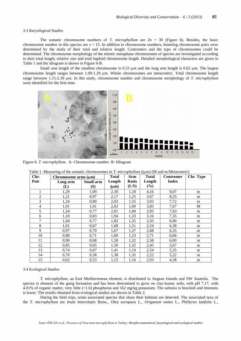
3.3 Karyological Studies
The somatic chromosome numbers of T. microphyllum are 2n = 30 (Figure 6). Besides, the basic
chromosome number in this species are x = 15. In addition to chromosome numbers, homolog chromosome pairs were
determined by the study of their total and relative length. Centromers and the type of chromosomes could be
determined. The chromosome morphology of the mitotic metaphase chromosomes of species are investigated according
to their total length, relative size and total haploid chromosome length. Detailed morphological characters are given in
Table 1 and the idiogram is shown in Figure 6-B.
Small arm length of the smallest chromosome is 0.53 m and the long arm length is 0.62 m. The largest
chromosome length ranges between 1.09-1.29 m. Whole chromosomes are metacentric. Total chromosome length
range between 1.15-2.39 m. In this study, chromosome number and chromosome morphology of T. microphyllum
were identified for the first time.
Figure 6. T. microphyllum. A- Chromosome number. B- Idiogram
Table 1. Measuring of the somatic chromosomes in T. microphyllum ((m) (M and m:Metacentric)
Chr.
Pair Chromosome arms (m) Total
Length
(m)
Arm
Ratio
(L/S)
Total
Length
(%)
Centromer
Index
Chr. Type
Long arm
(L)
Small arm
(S)
1 1,29 1,09 2,39 1,18 4,16 9,07 m
2 1,21 0,97 2,17 1,25 3,67 8,25 m
3 1,24 0,80 2,03 1,55 3,03 7,72 m
4 1,01 1,01 2,02 1,00 3,83 7,67 M
5 1,24 0,77 2,01 1,60 2,93 7,63 m
6 1,10 0,83 1,94 1,33 3,16 7,35 m
7 1,04 0,77 1,82 1,35 2,93 6,90 m
8 1,01 0,67 1,68 1,51 2,54 6,38 m
9 0,97 0,70 1,67 1,37 2,68 6,35 m
10 0,88 0,71 1,60 1,23 2,71 6,06 m
11 0,90 0,68 1,58 1,32 2,58 6,00 m
12 0,85 0,65 1,50 1,32 2,45 5,67 m
13 0,74 0,67 1,41 1,10 2,54 5,35 m
14 0,70 0,58 1,38 1,35 2,22 5,22 m
15 0,62 0,53 1,15 1,16 2,03 4,38 m
3.4 Ecological Studies
T. microphyllum, an East Mediterrenean element, is distributed in Aegean Islands and SW Anatolia. The
species is element of the garig formation and has been determined to grow on clay-loamy soils, with pH 7.17, with
4.01% of organic matter, very little (<1.0) phosphorus and 162 mg/kg potassium. The saltness is brackish and limeness
is lower. The results obtained from ecological studies are shown in Table 2.
During the field trips, some associated species that share their habitats are detected. The associated taxa of
the T. microphyllum are Inula heterolepis Boiss., Olea europaea L., Origanum onites L., Phillyrea latifolia L.,

Taner ÖZCAN et al., Presence of Teucrium microphyllum in Turkey: Morpho-anatomical, karyological and ecological studies
86 Biological Diversity and Conservation – 6 / 3 (2013)
Sarcopterium spinosum (L.) Spach, Tymbra capitata (L.) Cav., Achillea cretica, Amygdalus sp., Arbutus sp., Genista
sp., Helichrysum sp.
4. Discussion
In this study, Aegean endemic Teucrium microphyllum was studied morphological, anatomical, karyological
and ecologically for the first time. Morphologic data obtained in this study was compared with Ekim’s (1982) data.
Description of the species was expended.
Table 2. The soil of T. microphyllum analysis results and comment
Analysis Values
Parameter Module Analysis Result Comment
Structure % 66 Clay loam
pH 7,17 Neutral
E.C. μS/cm 278 Brackish
Lime % 0,78 Lower lime
OrganicSubst. % 4,01 Good
P(Phosphor) mg/kg <1,0 Very little
K (Potasium) mg/kg 162 Enough
Cu (Copper) mg/kg 1,0(±0,1) Enough
Fe (Iron) mg/kg 7,5(±0,8) Enough
Zn (Zinc) mg/kg 1,4(±0,1) Enough
Mn (Manganese) mg/kg 27,9(±2,5) Enough
Stems of Lamiaceae species are rectangular and there are collenchymatic tissue at the corners. Scleranchymatic
tissue covers the vascular tissue (Metcalfe and Chalk, 1983). The lower part of stem of T. sandrasicum O.Schwarz is
orbicular shaped and parenchymatic cells are 4-5-layered. Schlerenchymatic cells do not cover all over the vascular
bundle and they are like bouquets (Dinç et al., 2008). The stems of T. polium L. and T. montanum L. have one-layered
collenchyma between the corners and 6-7-layered collenchyma in the corners. There are 4-5-layered parenchymatic
cells under the collenchymatic tissue. Endodermis consists of 1-2 layers (Dinç et al., 2011). T. polium has a rectangle
shaped stem and epidermis consists of rectangular cells and this tissue is single layered (Dehshiri and Azadbakht, 2012).
The floral stem of T. microphyllum is rectangular and has thick cuticle (Figure 4). There are glandular and eglandular
hairs (Figure 3-L and Figure 4). Collenchymatic tissue has 2-5 layers in the corners. Vascular bundle has phloem (4-5-
layered), Xylem (5-8-layered) and schlerenchymatic cells. Sclerenchymatic cells forming bundles are between
endodermis and phloem. Dinç et al. (2011) reported that abaxial and adaxial epidermises of T. sandrasicum consist of
uniseriate, oval and rectangular cells. T. microphyllum is almost similar T. sandrasicum. Epidermis cells of the adaxial
side are larger than abaxial side in T. sandrasicum and T. microphyllum. It is the same for T. polium and T. montanum
(Dinç et al., 2011). The walls of abaxial epidermis cells are folded but adaxial epidermises are almost smooth and
thicker. This feature is same for T. polium, T. montanum and T. sandrasicum (Dinç et al., 2008; Dinç et al., 2011). T.
microphyllum has 1(-2)-layered collenchymatic tissue below the midrib, but T. sandrasicum, T. polium and T.
montanum have collenchyma both below and above midrib (Dinç et al., 2008; Dinç et al., 2011). As it was reported by
Lakusic et al. (2007), T. arduini L. has collenchymatic cells both adaxial and abaxial sides, too. Leaf is bifacial and the
mesophyll is clearly differentiated into palisade and spongy parenchyma (Figure 5-B). The palisade parenchyma
consists of usually 2 rarely 3 layers and spongy parenchyma is 2-4-layered. T. arduini, T. sandrasicum, T. polium and T.
montanum are also bifacial. T. arduini had 1-3-layered palisade and 2-3-layered spongy parenchyma (Lakusic et al.,
2007). T. sandrasicum are 2-layered palisade and 2-3-layered spongy parenchyma and it is same for T. polium and T.
montanum (Dinç et al., 2008; Dinç et al., 2011).
The basic chromosome numbers for the genus Teucrium have a big variation as x = 5, 8, 13, 16 (Darlington
and Wylie, 1955; Valde´S-Bermejo and Sanchez-Crespo; 1978). In Flora of Turkey (Güner et al., 2000), the somatic
chromosome number of T. alyssifolium Stapf and T. divaricatum Sieb. subsp. divaricatum have been reported as 2n =
30 and 2n = 64, respectively. According to the results of Martin et al. (2006) studies, somatic chromosome number of T.
lamiifolium d'Urv. subsp. lamiifolium was observed as 2n = 32. In this study, somatic choromosome number of T.
microphyllum was found as 2n = 30 and has showed idiogram and chromosome morphology of the species for the first
time.
After analyzing soil structures of T. microphyllum, the soil has a neutral pH and it is lower limy. Inorganic
matters (except phosphor) are enough. And associated species of the species were determined with this study.
Acknowledgements
I would like to thank my sueprvisor Assoc. Prof. Dr. Tuncay Dirmenci for his valuable comments and
guidance. I would like to thank Assoc. Prof. Dr. Esra Martin and Fahim Altınordu for karyological studies. I am grateful

Taner ÖZCAN et al., Presence of Teucrium microphyllum in Turkey: Morpho-anatomical, karyological and ecological studies
Biological Diversity and Conservation – 6 / 3 (2013) 87
to Basic Sciences Research and Applied Center of Balikesir University for SEM studies and to Scientific Research
Projects Unit (project number: 2012/18) for their financial supports.
References
Bayraklı, F. 1987. Toprak ve Bitki Analizleri. Ondokuz Mayıs Üniv. Ziraat Fakültesi Yayınları: 17, Samsun.
Cantino, P.D., Harley, R.M., Wagstaff, S.J. 1992. Genera of Labiatae:status and classification. In: Harley, R.M., Reynolds, T.
(Eds.),Advances in Labiate Science. Royal Botanic Gardens, Kew, 511–522.
Carlström, A.A. 1987. Survey of the flora and phytogeography of Rhodos, Simi, Tilos and the Marmaris Peninsula (SE Greece, SW
Turkey), PhD Thesis, University of Lund, Sweden.
Darlington, C.D., Wylie, A.P. 1955. Chromosome Atlas of Flowering Plants. Allen and Unwin Press.
De Martino, L., Formisano, C., Mancini, E., De Feo, V., Piozzi, F., Rigano, D., Senatore, F. 2010. Chemical composition and
phytotoxic effects ofessential oils from four Teucrium species, Nat Prod Commun 5: 1969–1976.
Dehshiri, M.M., Azadbakht, M. 2012. Anatomy of Iranian species Teucrium polium (Lamiaceae). Journal of Biology and today's
World 1/2: 93-98.
Dinç, M., Duran, A., Pınar, M., Öztürk. M. 2008. Anatomy, palynology and nutlet micromorphology of Turkish endemic Teucrium
sandrasicum (Lamiaceae). Biologia 63/5: 637-641.
Dinç, M., Doğu, S. 2012. Anatomical and micromorphological studies on Teucrium sect. Isotriodon (Lamiaceae) in Turkey with a
taxonomic note. Biologia 67/4: 663—672.
Dönmez, A.A. 2006. Teucrium chasmophyticum Rech. f. (Lamiaceae) A New Record for the Flora of Turkey. Turk J.Bot. 30: 317-
320.
Dönmez, A.A., Mutlu, B., Özcan, D.A. 2010. Teucrium melissoides Boiss. & Hausskn. ex Boiss. (Lamiaceae) A New Record for
Flora of Turkey. Hacettepe J. Biol & Chem, 38(4): 291-294.
Duman, H. 2000. Teucrium L. (Ed.) Güner, A., Özhatay, N., Ekim, T., and Başer, K.H.C. Flora of Turkey and EastAegean Islands,
Vol. 11 (Supplement II), Edinburgh University Press, Edinburgh, 197-198.
Ekim, T. 1982. Teucrium L. Davis, P.H. (ed.) Flora of Turkey and East Aegean Islands, Vol. 7, Edinburgh University Press,
Edinburgh, 53-75.
Feder, N., O'Brien, T.P. 1968. Plant Microtechnique. Some principles and new methods Amer. J. Bot. 55: 123-142
Govaerts, R., Paton, A., Harvey, Y., Navarro, T. & Del Rosario Garcia Pena, M. 2010. World Checklist of Lamiaceae. The Board of
Trustees of the Royal Botanic Gardens, Kew. Published on the Internet; www.kew.org/wcsp/[accessed on 31 July 2010]
Greuter, W., Raus, T. (ed.) 2006. Med-Checklist Notulae-24. Willdenowia 36: 724.
Güner, A., Özhatay, N., Ekim, T., Başer, K.H.C. 2000. Flora of Turkey and the East Aegean Islands, Vol.11, Edinburgh Univ. Pres.
Edinburgh.
Dirmenci, T. 2012. Teucrium L. In: Güner, A., Aslan, S., Ekim, T., Vural, M., Babaç, M.T. (eds.) Türkiye Bitkileri Listesi (Damarlı
Bitkiler). İstanbul: Nezahat Gökyiğit Botanik Bahçesi ve Flora Araştırmaları Derneği Yayını, 595-598.
Harley, R.M., Atkins, S., Budanstev, A.L., Cantino, P.D., Conn, B.J., Grayer, R., Harley, M.M. 2004. Labiatae. In:
Kubitzki, K. (Ed.),The Families and Genera of Vascular Plants VII. Springer, Berlin/Heidelberg.
Kaçar, B. 1972. Bitki Besleme Uygulama Kılavuzu. Ankara Ünv. Ziraat Fakültesi Yayınları: 647, Ankara
Kummerov, I. 1983. Comparative phenology of Mediterranean-type plant communities. In: Kruger, F.J., Michell, D.T. and Jarvis,
J.U.M. (eds.). Mediterranean-type Ecosystems: The role of Nutrients. No. 43, Springer, Berlin, 300- 317.
Lakušić, B., Lakušić, D., Slavkovska, V., Stevanović, V., Stevanović, B. 2007. Morpho-anatomical differentiation of the Balkan
endemic species Teucrium arduini L. (Lamiaceae), Arch Biol Sci. 59; 369–381.
Martin, E., Dinç, M., Duran, A., Öztürk, M. 2006. Karyological Studies on Lotus strictus Fisher & C.A.Mey. (Leguminosae),
Centaurea amanicola Hub.-Mor. (Compositae) and Teucrium lamiifolium d’Urv. subsp. lamiifolium (Labiatae). American-
Eurasian Journal of Scientific Research 1(1): 12–17.
Metcalfe, C.R., Chalk, L. 1983. Anatomy of the dicotyledons, 2nd edn. Vol. II. Wood structure and conclusion of the general
introduction. Clarendon Press: Oxford.
Navarro, T. El Oualidi, J. 2000. Trichome morphology in Teucrium L. (Labiatae) A taxonomic review. Anales Jardin Botanico De
Madrid 57(2): 277-297.
Tutin, G., Wood, D. 1972. Teucrium. In: Tutin, T.G. et al. (Eds.): Flora Europaea 3, Cambridge University Press, Cambridge, 129-135.
Tüzüner, A. 1990. Toprak ve Su Analiz Laboratuarları El Kitabı. T.C. Tarım, Orman ve Köyişleri Bakanlığı Köy Hizmetleri Genel
Müdürlüğü. Ankara, Turkey.
Valde´S-Bermejo, E., Sanchez-Crespo, A. 1978. Datos cariologicos taxonomicos sobre el genero Teucrium L. (Labiatae) en la
peninsula iberica. Acta Bot, Mal. 4: 27-54.
Yakar-Tan, N. 1982. Bitki Mikroskopisi Klavuz Kitabı. İstanbul Üniv. Fen Fak. Yay. No. 166, İstanbul.
(Received for publication 28 March, 2013; The date of publication 15 December 2013)