Preparation of fluoride substituted apatite cements as the building blocks for tooth enamel...
6
Please cite this article in press as: J. Wei, et al., Preparation of fluoride substituted apatite cements as the building blocks for tooth enamel restoration, Appl. Surf. Sci. (2011), doi:10.1016/j.apsusc.2011.04.067 ARTICLE IN PRESS G Model APSUSC-21728; No. of Pages 6 Applied Surface Science xxx (2011) xxx–xxx Contents lists available at ScienceDirect Applied Surface Science journal homepage: www.elsevier.com/locate/apsusc Preparation of fluoride substituted apatite cements as the building blocks for tooth enamel restoration Jie Wei a,b , Jiecheng Wang a , Xiaochen Liu a , Jian Ma c , Changsheng Liu b , Jing Fang a,∗ , Shicheng Wei a,d,∗∗ a Center for Biomedical Materials and Tissue Engineering, Academy for Advanced Inter-disciplinary Studies, Peking University, Beijing 100871, PR China b Key Laboratory for Ultrafine Materials of Ministry of Education, East China University of Science and Technology, Shanghai 200237, PR China c Hospital of Stomatology, Tongji University, Shanghai 200072, PR China d School and Hospital of Stomatology, Peking University, Beijing 100081, PR China article info Article history: Received 31 January 2011 Received in revised form 12 April 2011 Accepted 12 April 2011 Available online xxx Keywords: Dental enamel Carious cavities Fluorapatite cement Restoration Cytocompatibility abstract Fluoride substituted apatite cement (fs-AC) was synthesized by using the cement powders of tetracal- cium phosphate (TTCP) and sodium fluoride (NaF), and the cement powders were mixed with diluted phosphoric acid (H 3 PO 4 ) as cement liquid to form fs-AC paste. The fs-AC paste could be directly filled into the carious cavities to repair damaged dental enamel. The results indicated that the fs-AC paste was changed into fluorapatite crystals with the atom molar ratio for calcium to phosphorus of 1.66 and the F ion amount of 3 wt% after self-hardening for 2 days. The solubility of fs-AC in Tris–HCl solution (pH 6) was slightly lower than hydroxyapatite cement (HAC) that was similar to the apatite in enamel, indicating the fs-AC was much insensitive to the weakly acidic solution than the apatite in enamel. The fs-AC was tightly combined with the enamel surface because of the chemical reaction between the fs-AC and the apatite in enamel after the caries cavities was filled with fs-AC. The extracts of fs-AC caused no cytotoxicity on L929 cells, which satisfied the relevant criterion on dental biomaterials, revealing good cytocompatibility. The fs-AC had potential prospect for the reconstitution of carious lesion of dental enamel. © 2011 Elsevier B.V. All rights reserved. 1. Introduction Dental caries is a prevalent chronic and world-wide oral disease [1]. As a non-living tissue, the main composition of mature enamel is inorganic apatite so dental enamel is scarcely self-repaired by liv- ing organisms after substantial mineral loss [2]. Traditional clinical practice has recommended complete removal of softened and dis- colored dentin (demineralized carious dentin) to eliminate infected tissue and create a hard foundation to support a proposed restora- tion such as composite resin or metal alloys, which result in poor adhesion between the repair materials and enamel at the interface during the restoration, and this technique is far from ideal, because excessive sound dentin is removed [3–5]. Obviously, reconstitution of carious dentin is a more desirable clinical approach than the tra- ditional clinical practice. It has been proved that fluoride could not only improve the acid resistance of apatite crystals effectively (a ∗ Corresponding author. Tel.: +86 10 62753404; fax: +86 10 62753404. ∗∗ Corresponding author at: Center for Biomedical Materials and Tissue Engineer- ing, Academy for Advanced Inter-disciplinary Studies, Peking University, Beijing 100871, PR China. Tel.: +86 10 62753404; fax: +86 10 62753404. E-mail addresses: [email protected] (J. Fang), [email protected] (S. Wei). certain concentration) but also inhibit metabolism of bacterial [6]. Yamagishi et al. reported a paste of fluoridated hydroxyapatite that could be used to repair early carious lesion, and the apatite paste could be bond to the surfaces of the dental enamel [7]. However, this method may limit its application in the restoration of badly carious lesion of dental enamel, such as carious cavities. Hydroxyapatite biomaterials are promising candidates for reconstruction of calcified tissue, such as human tooth and bone, because they are the main inorganic components of dentin and bone minerals [8]. However, the native structure of enamel is too complex to be remodeled, and the synthesized apatite biomaterials (such as bioceramics) often have different from the natural ones [9]. The human tooth is protected by enamel that is composed of apatite crystals, acid-forming bacteria cause microscopic damage to the enamel, creating carious cavities, which cannot be repaired by the restorative materials (such as amalgam, ceramics, or polymer com- posites) because these materials without viscidity do not adhere (bond) perfectly to the enamel surfaces owing to the differences in chemical composition and crystal structure [10]. In order to further restoration of badly carious lesion of dental enamel, such as carious cavities, in this study, a paste of fluoride substituted apatite cement (fs-AC) with good viscosity was synthesized, and the fs-AC pastes could be directly filled into the carious cavities and adhered with dental enamel surfaces to repair the damaged enamel. 0169-4332/$ – see front matter © 2011 Elsevier B.V. All rights reserved. doi:10.1016/j.apsusc.2011.04.067
-
Upload
independent -
Category
Documents
-
view
1 -
download
0
Transcript of Preparation of fluoride substituted apatite cements as the building blocks for tooth enamel...
G
A
Pt
Ja
b
c
d
a
ARRAA
KDCFRC
1
[iipcttadeodo
i1
(
0d
ARTICLE IN PRESSModel
PSUSC-21728; No. of Pages 6
Applied Surface Science xxx (2011) xxx–xxx
Contents lists available at ScienceDirect
Applied Surface Science
journa l homepage: www.e lsev ier .com/ locate /apsusc
reparation of fluoride substituted apatite cements as the building blocks forooth enamel restoration
ie Weia,b, Jiecheng Wanga, Xiaochen Liua, Jian Mac, Changsheng Liub, Jing Fanga,∗, Shicheng Weia,d,∗∗
Center for Biomedical Materials and Tissue Engineering, Academy for Advanced Inter-disciplinary Studies, Peking University, Beijing 100871, PR ChinaKey Laboratory for Ultrafine Materials of Ministry of Education, East China University of Science and Technology, Shanghai 200237, PR ChinaHospital of Stomatology, Tongji University, Shanghai 200072, PR ChinaSchool and Hospital of Stomatology, Peking University, Beijing 100081, PR China
r t i c l e i n f o
rticle history:eceived 31 January 2011eceived in revised form 12 April 2011ccepted 12 April 2011vailable online xxx
eywords:ental enamel
a b s t r a c t
Fluoride substituted apatite cement (fs-AC) was synthesized by using the cement powders of tetracal-cium phosphate (TTCP) and sodium fluoride (NaF), and the cement powders were mixed with dilutedphosphoric acid (H3PO4) as cement liquid to form fs-AC paste. The fs-AC paste could be directly filledinto the carious cavities to repair damaged dental enamel. The results indicated that the fs-AC pastewas changed into fluorapatite crystals with the atom molar ratio for calcium to phosphorus of 1.66 andthe F ion amount of 3 wt% after self-hardening for 2 days. The solubility of fs-AC in Tris–HCl solution(pH 6) was slightly lower than hydroxyapatite cement (HAC) that was similar to the apatite in enamel,
arious cavitiesluorapatite cementestorationytocompatibility
indicating the fs-AC was much insensitive to the weakly acidic solution than the apatite in enamel. Thefs-AC was tightly combined with the enamel surface because of the chemical reaction between the fs-ACand the apatite in enamel after the caries cavities was filled with fs-AC. The extracts of fs-AC caused nocytotoxicity on L929 cells, which satisfied the relevant criterion on dental biomaterials, revealing goodcytocompatibility. The fs-AC had potential prospect for the reconstitution of carious lesion of dentalenamel.
. Introduction
Dental caries is a prevalent chronic and world-wide oral disease1]. As a non-living tissue, the main composition of mature enamels inorganic apatite so dental enamel is scarcely self-repaired by liv-ng organisms after substantial mineral loss [2]. Traditional clinicalractice has recommended complete removal of softened and dis-olored dentin (demineralized carious dentin) to eliminate infectedissue and create a hard foundation to support a proposed restora-ion such as composite resin or metal alloys, which result in poordhesion between the repair materials and enamel at the interfaceuring the restoration, and this technique is far from ideal, becausexcessive sound dentin is removed [3–5]. Obviously, reconstitution
Please cite this article in press as: J. Wei, et al., Preparation of fluoride srestoration, Appl. Surf. Sci. (2011), doi:10.1016/j.apsusc.2011.04.067
f carious dentin is a more desirable clinical approach than the tra-itional clinical practice. It has been proved that fluoride could notnly improve the acid resistance of apatite crystals effectively (a
∗ Corresponding author. Tel.: +86 10 62753404; fax: +86 10 62753404.∗∗ Corresponding author at: Center for Biomedical Materials and Tissue Engineer-ng, Academy for Advanced Inter-disciplinary Studies, Peking University, Beijing00871, PR China. Tel.: +86 10 62753404; fax: +86 10 62753404.
E-mail addresses: [email protected] (J. Fang), [email protected]. Wei).
169-4332/$ – see front matter © 2011 Elsevier B.V. All rights reserved.oi:10.1016/j.apsusc.2011.04.067
© 2011 Elsevier B.V. All rights reserved.
certain concentration) but also inhibit metabolism of bacterial [6].Yamagishi et al. reported a paste of fluoridated hydroxyapatite thatcould be used to repair early carious lesion, and the apatite pastecould be bond to the surfaces of the dental enamel [7]. However,this method may limit its application in the restoration of badlycarious lesion of dental enamel, such as carious cavities.
Hydroxyapatite biomaterials are promising candidates forreconstruction of calcified tissue, such as human tooth and bone,because they are the main inorganic components of dentin andbone minerals [8]. However, the native structure of enamel is toocomplex to be remodeled, and the synthesized apatite biomaterials(such as bioceramics) often have different from the natural ones [9].The human tooth is protected by enamel that is composed of apatitecrystals, acid-forming bacteria cause microscopic damage to theenamel, creating carious cavities, which cannot be repaired by therestorative materials (such as amalgam, ceramics, or polymer com-posites) because these materials without viscidity do not adhere(bond) perfectly to the enamel surfaces owing to the differencesin chemical composition and crystal structure [10]. In order tofurther restoration of badly carious lesion of dental enamel, such as
ubstituted apatite cements as the building blocks for tooth enamel
carious cavities, in this study, a paste of fluoride substituted apatitecement (fs-AC) with good viscosity was synthesized, and the fs-ACpastes could be directly filled into the carious cavities and adheredwith dental enamel surfaces to repair the damaged enamel.

IN PRESSG Model
A
2 ace Science xxx (2011) xxx–xxx
2
2
po(tsapt
pcfØttptaHfp
ttapawpCM
2
lhts4wsHwrtTwa
2
wa5ppa3
0
1000
2000
3000
4000
5000
605040302010
2 Theta (degree)
*
*
***
* **
*
ARTICLEPSUSC-21728; No. of Pages 6
J. Wei et al. / Applied Surf
. Materials and methods
.1. Preparation and characterization of fs-AC
The fluoride substituted apatite cement consisted of the cementowders and cement liquid. The cement powders were composedf tetracalcium phosphate (Ca4(PO4)2O, TTCP) and sodium fluorideNaF), and cement liquid was diluted phosphoric acid (H3PO4) withhe concentration of 17% (v/v). TTCP was synthesized by a solid-to-olid reaction between calcium phosphate and calcium carbonatet a temperature of 1500 ◦C for 8 h. The TTCP was grounded in alanetary ball mill for 1 h, followed by sieving through 120 mesheso obtain TTCP powders [11].
The fs-AC with self-hardening (self-setting) property was pre-ared by mixing the cement powders (5 g TTCP + 0.46 g NaF) withement liquid (3.2 mL diluted H3PO4) to form a cement paste. Thes-AC paste was placed into stainless steel molds with the size of10 mm × 2 mm. After stored in beakers in a constant tempera-
ure oven at 37 ◦C and 100% relative humidity (r.h.) for 2 days,he pre-hardened fs-AC solid mass sample was obtained. We pre-ared hydroxyapatite cement (HAC) (as a control), which containedetracalcium phosphate as cement powder and diluted phosphoriccid (17% H3PO4) as cement liquid without sodium fluoride. TheAC was prepared by mixing the cement powder with liquid to
orm cement paste, and the following process was similar to thereparation of fs-AC.
In order to confirm that the F ion entered into the apatite crys-al lattice while did not adsorb on the surfaces of apatite cement,he experiments were done as follows: the pre-hardened fs-ACnd HAC samples were grounded into powers, respectively. Theower samples were then immersed in deionized water for 24 hnd the water was changed one time at 12 h. Finally, these samplesere dried at 120 ◦C for 24 h to obtain fs-AC and HAC power sam-les, which were characterized by X-ray diffraction (XRD; Rigakuo., Japan), and Fourier transform-infrared spectroscopy (FT-IR;agna-IR 550, Nicolet, American).
.2. Solubility of fs-AC
The solubility of the fs-AC was characterized by the weightoss ratio (wt%) in Tris–HCl solution at different time, and theydroxyapatite cement (HAC) was used as a control. After set-ing for 2 days and dried at 50 ◦C for 24 h, the fs-AC and HACamples (Ø10 mm × 2 mm) with initial weight (Wi), were put in00 mL of Tris–HCl solution (pH 6, adjusted by diluted HCl) with aeight-to-volume ratio of 0.5 g/mL. The solution was continuously
haken in a water bath at 37 ◦C. At different time, the fs-AC andAC samples were removed from the Tris–HCl solution, cleanedith water, dried at 50 ◦C for 24 h and its new weight (Wt) was
ecorded. It was then re-immersed into a fresh Tris–HCl solution athe same weight-to-volume ratio followed by continuous shaking.he weight loss ratio of the fs-AC and HAC samples at different timeas calculated. Three samples of each kind of cement were tested
nd the average value was recorded.
.3. Cytotoxicity of fs-AC
L929 cells were used to test the cytotoxicity of the fs-AC, whichas carried out by using the fs-AC extracts in contact with L929 cells
ccording to International Standard Organization (ISO/EN 10993-), and hydroxyapatite cement (HAC) was used as a control. To
Please cite this article in press as: J. Wei, et al., Preparation of fluoride srestoration, Appl. Surf. Sci. (2011), doi:10.1016/j.apsusc.2011.04.067
repare the fs-AC extracts, a stock solution of 200 mg/mL was firstrepared by adding 5 g fs-AC (after setting for 2 days and driedt 50 ◦C for 24 h) into DMEM culture medium. After incubation at7 ◦C for 24 h, the mixture was centrifuged and the supernatant was
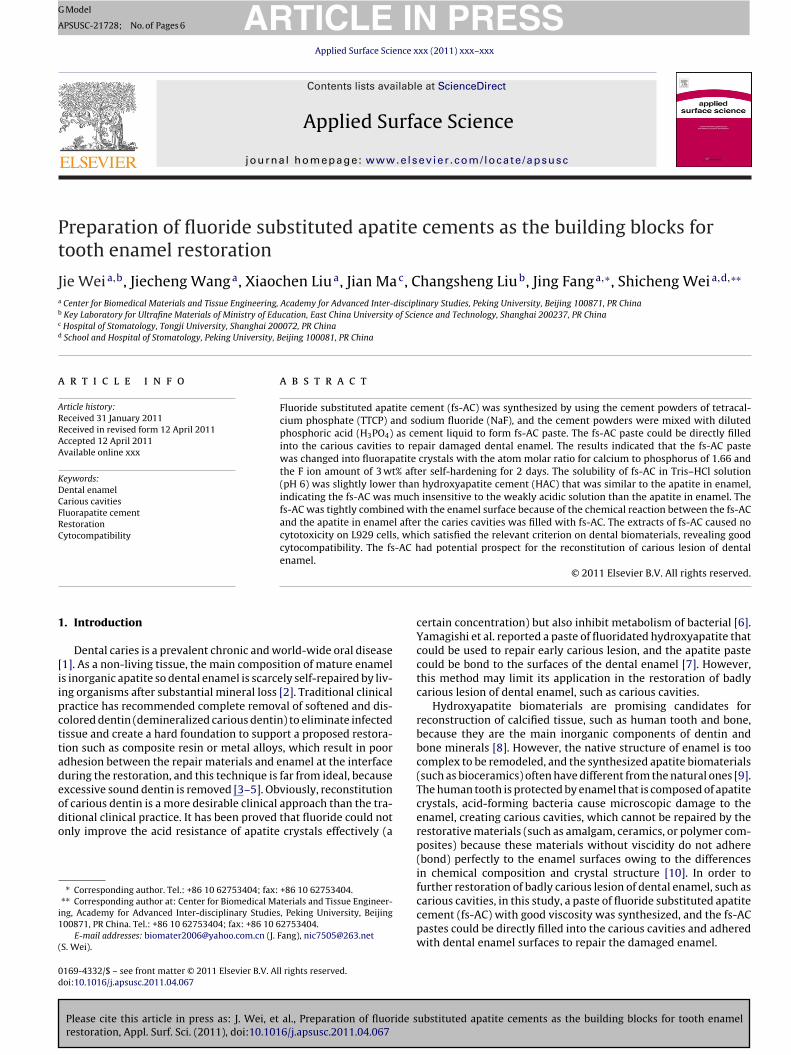
Fig. 1. XRD of fs-AC (a) and HAC (a) after hardening for 2 days, * represents apatite.
collected. Subsequently, the extracts were sterilized by filtrationthrough 0.2 �m filter membranes for cell cultured experiments.
The cells were seeded on a 96-well plate and incubated for 24 h.Then, the culture medium was removed and replaced by 50 �L ofextracts and 50 �L of DMEM medium supplemented with 10% FCS.The DMEM with 10% FCS (without extract supplement) was usedas a blank control. After incubation for 24 h, MTT test was carriedout to test cell viability. In brief, 100 mL of 0.5 mg/mL 3-(4,5)-dimethylthiahiazo (-z-y1)-3,5-dipheny-tetrazoliumromide (MTT)solution was added into each well. After additional incubation for4 h, dimethyl sulfoxide (DMSO) was added to stop the reactionbetween MTT and cells. The optical density (OD) was measuredby a microplate reader at the wavelength of 492 nm.
2.4. Restoration of enamel carious cavities
Human teeth with big enamel carious cavities were degreasedwith absolute ethanol, and etched with 17% phosphoric acid forabout 30 min. The fs-AC pastes were filled into the enamel cari-ous cavities immediately before the phosphoric acid solution dried.After the tooth samples with enamel carious cavities were filledwith the fs-AC pastes, the as-prepared tooth samples were storedin beakers in a constant temperature oven at 37 ◦C and 100% relativehumidity (r.h.) for 2 days. The tooth samples with fs-AC repair theenamel carious cavities were sectioned perpendicular to the dentalcrown using a diamond saw, and interface between the enamel andfs-AC was examined by using scanning electron microscope (SEM,JSM-6360LV, JEOL). The surface morphology and microstructure ofthe fs-AC filled into the dental enamel carious cavities were alsoexamined with SEM.
3. Results
3.1. XRD analysis
The phase compositions and crystal structure of the hardenedfs-AC and HAC were characterized by powder XRD as shown inFig. 1. It can be seen that the diffraction peaks of the two samplesat 2� = 25.7◦, 28◦, 29◦, 31.8◦, 32.4◦, 33.5◦, 39◦, 46.8◦, 49.5◦ and 52.6◦
were ascribed to apatite both in Fig. 1a and b. Clearly, all of thepeaks can be readily indexed to a pure hexagonal phase, whichwas in accordance with apatite structure. The XRD results indicatedthat the hardened fs-AC and HAC were all apatite structure. Thepresence of HAC could be attributed to the chemical reactions as
ubstituted apatite cements as the building blocks for tooth enamel
follows [12]:
5Ca4(PO4)2O + 2H3PO4 → 2Ca10(PO4)6(OH)2 (HA)

ARTICLE IN PRESSG Model
APSUSC-21728; No. of Pages 6
J. Wei et al. / Applied Surface Science xxx (2011) xxx–xxx 3
5001000150020002500300035004000
0
20
40
60
80
100Tr
ansm
ittan
ce (%
)
-1
a
b
t
5
3
att8
6(apwt
3
cArcifl
00.20.40.60.8
11.21.41.61.8
2
15107531Time (day)
HACfs-Ac
Wei
ght l
oss r
atio
(%)
Wavenumber (cm )
Fig. 2. IR of fs-AC (a) and HAC (b) after hardening for 2 days.
The presence of fs-AC could be attributed to the chemical reac-ions as follows:
Ca4(PO4)2O + 2H3PO4 + 4NaF → 2Ca10(PO4)6F2 (FA)
.2. IR analysis
Fig. 2 shows the IR analysis of the hardened fs-AC and HAC. Thebsorption bands at 1071 and 950 cm−1 were ascribed to PO4
3−. Thewo peaks at 1431 and 1750 cm−1 and the broad band from 2800o 3750 cm−1 were attributed to the absorbed water. The band at76 cm−1 might correspond to the vibration of P–O–H from PO4.
The OH-specific peaks were found at around 3571 cm−1 and32 cm−1 in Fig. 2a, revealing that the HAC was hydroxyapatiteHA). However, it was found that no OH-specific peaks appearedt around 3571 cm−1 and 632 cm−1 in Fig. 2b, indicating that no HAresent in the finally hardened product of fs-AC because –OH groupas replaced by F group to form FA (Ca10(PO4)6F2). The results of
he IR analysis were in accordance with the XRD of the fs-AC.
.3. Elemental composition
The chemical elemental composition of the fs-AC was furtherharacterized by EDS. The EDS spectrum (Fig. 3) shows that the fs-C contained Ca, P and F elements. Furthermore, the EDS results
Please cite this article in press as: J. Wei, et al., Preparation of fluoride srestoration, Appl. Surf. Sci. (2011), doi:10.1016/j.apsusc.2011.04.067
eveal that the fs-AC had an average Ca/P ratio of 1.66, which waslosely similar to that of HA (Ca/P = 1.67). The amount of F ionsn fs-AC was 3 wt%, which was almost similar to that of F ion inuorapatite. Combined with the results of XRD, IR and EDS analysis,
543210
C a
P
F
Energy (KeV)
Fig. 3. EDS of the fs-AC after hardening for 2 days.
Fig. 4. Weight loss ratios of fs-AC and HAC after immersing in Tris–HCl solutionover time.
it could be suggested that the produced fluoridated apatite cementwas fluorapatite (FA).
3.4. Solubility in Tris–HCl solution
The solubility of the fs-AC and HAC samples in Tris–HCl solutionwas characterized by their weight loss ratio after immersing intothe solution. Fig. 4 shows the weight loss ratio of the fs-AC and HACimmersing in the acidic Tris–HCl solutions (pH 6) for various timeperiods. It was found that the weight loss ratio of the fs-AC wasslightly lower than HAC throughout the soaking. The weight lossratio of the fs-AC was 0.84 wt% while HAC was around 1.28 wt% bythe end of the experiment, indicating the solubility of fs-AC wasslightly lower than the HAC. The results suggested that the fs-ACwas much insensitive to the acidic solutions than that of HAC.
After self-hardening for 2 days, the fs-AC was changed into flu-orapatite and HAC was changed into hydroxyapatite, respectively.Hydroxyapatite bioceramic and hydroxyapatite cement were bio-compatibility and have been used for bone repair in clinic formany years, and hydroxyapatite is the main composition of den-tal enamel. In this experiment, we want to testify that the fs-ACwith 3 wt% F had lower solubility and was much insensitive tothe weakly acidic solutions than HAC (as a control) because pre-vious study showed that fluoride can improve the acid resistanceof apatite effectively.
3.5. Cytotoxicity
The effects of the fs-AC extracts on L929 cells cultured for 24 h
ubstituted apatite cements as the building blocks for tooth enamel
are shown in Fig. 5. It can be seen that there was no obvious dif-ferences for optical density (OD) value between the fs-AC and HAC(OD represents cell viability). HAC with excellent biocompatibilityhas been applied as bone cement in clinic for many years. More-
0
0.2
0.4
0.6
0.8
1
Blank HAC fs-ACSamples
Opt
ical
Den
sity
Fig. 5. Effects of the fs-AC extracts on L929 cells cultured for 24 h. The experimentalgroup compared with the blank and HAC control group.

ARTICLE IN PRESSG Model
APSUSC-21728; No. of Pages 6
4 J. Wei et al. / Applied Surface Science xxx (2011) xxx–xxx
s cavit
otog
3
cctTvmtTw
Fig. 6. Photos of the human dental enamel cariou
ver, no significant differences for OD value were found betweenhe fs-AC and blank control. The results suggested that the extractsf fs-AC caused no cytotoxicity on L929 cells, indicating fs-AC hadood cytocompatibility.
.6. Restoration of enamel carious cavities
Fig. 6 shows the photos of the human tooth enamel cariousavities filled with the fs-AC. It was found that the tooth enamelarious cavities were repair by the fs-AC. Fig. 7 exhibits the dis-inct interface morphologies of the dense fs-AC and enamel by SEM.he SEM images (under different magnifications) showed the trans-ersely section of the fs-AC applied to the enamel carious cavity. The
Please cite this article in press as: J. Wei, et al., Preparation of fluoride srestoration, Appl. Surf. Sci. (2011), doi:10.1016/j.apsusc.2011.04.067
icrostructure of the restored enamel revealed no obvious struc-ural gap at the interface between the fs-AC and the enamel region.his showed that the newly-form fs-AC had properly integratedith the tooth enamel.
Fig. 7. SEM images of the transversely section of the fs-AC applied to the human toot
ies before (a) and after (b and c) filled with fs-AC.
The surface morphology and microstructure of fs-AC filled intothe enamel carious cavities after self-hardening for 2 days wereexamined by SEM as shown in Fig. 8. It can be seen that the SEMimage of the fs-AC layer on the enamel surface, which containedconspicuously elongated apatite crystals that had grown on thetooth enamel surface. The homogeneous rod-like crystals had atypical apatite hexagonal cross section of approximately 400 nmin length and 50 nm in diameter.
4. Discussions
The restoration of the carious cavities of dental enamel byusing synthetic apatite was always suggested in dental research[13]. However, the native structure of dental enamel is too com-
ubstituted apatite cements as the building blocks for tooth enamel
plex to be remodeled, and the synthesized apatite crystallitesoften have different from the natural ones, which result in pooradhesion between the synthetic apatite and enamel at the inter-face during the restoration [14]. In order to further repair badly
h enamel carious cavities under different magnifications: (a) 35× and (b) 400×.

ARTICLE ING Model
APSUSC-21728; No. of Pages 6
J. Wei et al. / Applied Surface Sc
Fe
costctifrTcnfl
sastcrsatt
totatTaafwet
csTwcnis
ig. 8. SEM images of the surface morphology/microstructure of fs-AC on thenamel surfaces after hardening for 2 days.
arious lesion of dental enamel (carious cavities), a paste of flu-ride substituted apatite cement (fs-AC) was synthesized in thistudy. The fs-AC paste could be directly filled into the carious cavi-ies to restore damaged enamel. It was found that the fs-AC crystalsould be formed on the surface of human teeth with tight contacto the enamel (fs-AC closely bond to the enamel apatite). Accord-ng to the analysis of XRD, IR and EDS, the results indicated that thes-AC was finally changed into fluorapatite (FA), which had a Ca/Patio of 1.66 and the F ion amounts of 3 wt%. The mixed powders ofTCP (basic) reacted with phosphoric acid solution (acidic) to formement paste that first changed into hydroxyapatite (acidic–basiceutralization reaction), then, hydroxyapatite reacted with sodiumuoride to form fluorapatite.
The carious cavities of dental enamel could be repaired by theelf-hardening fs-AC because the restorative material was cementnd could adhere perfectly to the enamel surfaces owing to theimilarity of fs-AC in chemical composition and crystal structureo enamel apatite [15]. In our study, it was found that the fs-ACould be completely filled the porous residual carious dentin. Thisecovery was caused not only by cement penetration, but also byome chemical reactions between the residual structure (enamelpatite) and the fs-AC (fluorapatite). With this chemical reaction,he fs-AC would adhere perfectly to the enamel surface and repairhe enamel carious cavities.
In this study, the results showed the fs-AC was firmly bondedo the surface of the dental enamel because the carious cavitiesf enamel surfaces etched with 17% phosphoric acid would causehe solution of the apatite in enamel layer. When the fs-AC pastepplied on the enamel surface, the fs-AC surface was also par-ially dissolved, which was conglutinated with enamel surfaces.his means that the fs-AC would chemically reacted with enamelpatite, but this chemical reactions were incomplete, only occurredt the interface between the fs-AC and the enamel surface. There-ore, the results demonstrated that the fs-AC was tightly combinedith the enamel surface, and the microstructure of the restored
namel revealed no obvious structural gap at the interface betweenhe fs-AC and the enamel region.
Although dental enamel is not a living part but it has a closehemical structure to apatite, this hard tissue is biologically con-tructed by living organisms with well-organized structures [16].he chemical similarity of the apatite for both fs-AC and enamelould ensure the restoration of enamel defects, since it was too
omplicated to remodel the functions of living organism to induce
Please cite this article in press as: J. Wei, et al., Preparation of fluoride srestoration, Appl. Surf. Sci. (2011), doi:10.1016/j.apsusc.2011.04.067
ew enamel apatite on a non-living surface [17]. The fs-AC approx-mate to the basic building blocks of enamel may be a candidateince the in vitro biomimetic construction of enamel by using fluo-
PRESSience xxx (2011) xxx–xxx 5
rapatite had already been achieved [18]. It has been suggested thatthe fluorapatite was analogous to the subunits of biological apatite[19]. Therefore, the repair of carious cavities of dental enamel couldbe improved by using the fs-AC.
The human tooth is protected by dental enamel consisting ofapatite. Acid-forming bacteria will cause damage to the enamel,creating cavities in the enamel [20,21]. In this study, the solubilityof the pre-hardened fs-AC in acidic environment (pH 6) was charac-terized by using Tris–HCl solution with the extended experimentalperiods, and the results were expressed as the weight loss ratio ofthe fs-AC and HAC with time in the acidic solutions. The resultsrevealed that the weight loss ratio of the fs-AC of 0.84 wt% whilethe HAC was about 1.28 wt% after soaking in Tris–HCl solution for15 days. The weight loss ratio of the fs-AC was slightly lower thanHAC, indicating the fs-AC was much insensitive to the weakly acidicsolutions than HAC, which is an apatite similar to enamel apatite.It can be suggested that the underlying enamel surface would bewell protected after repaired by the cement since the fs-AC wasinsensitive to the weakly acidic condition. The results are in accordwith previous study that fluoride can improve the acid resistanceof apatite crystals effectively [6].
As dental biomaterials for repair the enamel carious cavity, thebiocompatibility and biosecurity of the fs-AC are very important. Inthis study, the cytotoxicity of the fs-AC was determined by using theextracts of the pre-hardened fs-AC co-cultured with L929 cells for24 h according to the standard from ISO. The results suggested thatthe extracts of the fs-AC caused no cytotoxicity on L929 cells, indi-cating the fs-AC had good cytocompatibility, and could be satisfiedwith the relevant criterion on dental biomaterials.
5. Conclusions
Fluoride substituted apatite cement (fs-AC) was developed torepair the dental enamel carious lesion by filling directly intoenamel carious cavity. The result showed that the pre-hardenedfs-AC was fluorapatite (FA) with the Ca/P of 1.66 and the F ionamounts of 3 wt%. The fs-AC was tightly combined with the enamelsurface because of the chemical reaction between the fs-AC andenamel apatite after the cement filled into the caries cavity of den-tal enamel. The less soluble fluorapatite cement filling into enamelcarious cavity should offer considerable protection against cariessince the cement was much insensitive to the weakly acidic solu-tions. This study confirmed the possibility of applying the fs-ACpastes to directly repair badly carious lesion of the damaged dentalenamel, and the fs-AC with good biocompatibility could be used asa substitute for the conventional dental restorative materials. Thisstrategy may have prospective applications in dentistry as it offersan easy but effective method to reconstruct tooth enamel defectsthat are suffering from mineral loss.
Acknowledgements
The authors appreciate financial support from the 973 Projectof the Ministry of Science and Technology of the People’s Republicof China (Grant 2007CB936103), Nano special program of Scienceand Technology Development of Shanghai (No. 1052nm06600), andKey Medical Program of Science and Technology Development ofShanghai (No. 09411954900).
References
ubstituted apatite cements as the building blocks for tooth enamel
[1] H.F. Chen, B.H. Clarkson, K. Sun, J.F. Mansfield, J. Colloid Interf. Sci. 288 (2005)97.
[2] H.F. Chen, Z.Y. Tang, J. Liu, K. Sun, S.R. Chang, M.C. Peters, J.F. Mansfield, A.Czajka-Jakubowska, B.H. Clarkson, Adv. Mater. 18 (2006) 1846.
ING Model
A
6 ace Sc
[
[
[
[
[
[
[
[[
ARTICLEPSUSC-21728; No. of Pages 6
J. Wei et al. / Applied Surf
[3] A. Boyde, Microstructure of enamel, in: D.J. Chadwick, G. Cardew (Eds.), DentalEnamel: Ciba Foundation Symposium 205, John Wiley & Sons, New York, 1997,p. 18.
[4] R.P. Shellis, R.M. Wilson, J. Colloid Interf. Sci. 278 (2004) 325.[5] S. Busch, Angew. Chem. Int. Ed. 43 (2004) 1428.[6] Y. Fan, Z. Sun, J. Moradian-Oldak, Biomaterials 30 (2009) 478.[7] K. Yamagishi, K. Onuma, T. Suzuki, F. Okada, J. Tagami, M. Otsuki, P.A.
Senawangse, Nature 433 (2005) 819.[8] C.E. Fowler, M. Li, S. Mann, H.C. Margolis, J. Mater. Chem. 15 (2005) 3317.[9] K. Onuma, K. Yamagishi, A. Oyane, J. Cryst. Growth 282 (2005) 199.10] J. Kirkham, A. Firth, D. Vernals, N. Boden, C. Robinson, R.C. Shore, S.J. Brookes,
Please cite this article in press as: J. Wei, et al., Preparation of fluoride srestoration, Appl. Surf. Sci. (2011), doi:10.1016/j.apsusc.2011.04.067
A. Aggeli, J. Dent. Res. 86 (2007) 426.11] J. Wei, J.F. Jia, F. Wu, S.C. Wei, H.J. Zhou, H.B. Zhang, J.W. Shin, C.S. Liu, Bioma-
terials 31 (2010) 1260.12] J.F. Jia, H.J. Zhou, J. Wei, H. Hong, F.P. Chen, S.C. Wei, J.W. Shin, C.S. Liu, J. R. Soc.
Interf. 7 (2010) 1171.
[[
[
PRESSience xxx (2011) xxx–xxx
13] L. Li, H.H. Pan, J.H. Tao, X.R. Xu, C.Y. Mao, X.H. Gu, R.K.J. Tang, Mater. Chem. 18(2008) 4079.
14] X.K. Wang, C.J. Xia, Z.H. Zhang, X.L. Den, S.C. Wei, G. Zheng, H.F. Chen, J. Nanosci.Nanotechnol. 9 (2009) 1361.
15] Y. Shibata, L.H. He, Y. Kataoka, T. Miyazaki, M.V. Swain, J. Dent. Res. 87 (2008)233.
16] B. Fu, Q. Shen, W. Qian, Y. Zeng, X. Sun, M. Hannig, J. Mater. Sci. Mater. Med. 16(2005) 827.
17] Y.J. Yin, S. Yun, J.S. Fang, H.F. Chen, Chem. Commun. 39 (2009) 5892–5894.18] B.H. Yoon, H.W. Kim, S.H. Lee, C.J. Bae, Y.H. Koh, Y.M. Kong, H.E. Kim, Biomate-
rials 26 (2005) 2957.
ubstituted apatite cements as the building blocks for tooth enamel
19] H.G. Zhang, Q.S. Zhu, Mater. Lett. 59 (2005) 3054.20] L.J. Wang, X.Y. Guan, H.Y. Yin, J. Moradian-Oldak, G.H. Nancollas, J. Phys. Chem.
C 112 (2008) 5892.21] S.S. Gao, S.B. Huang, L.M. Qian, H.Y. Yu, Z.R. Zhou, Wear 267 (2009)
726.