Postembedding Decalcification of Mineralized Tissue Sections ...
11
Research Article Postembedding Decalcification of Mineralized Tissue Sections Preserves the Integrity of Implanted Biomaterials and Minimizes Number of Experimental Animals Thaqif El Khassawna, 1 Diaa Eldin S. Daghma, 1 Sabine Stoetzel, 1 Seemun Ray, 1 Stefanie Kern, 1 Deeksha Malhan, 1 Volker Alt, 1,2 Ulrich Thormann, 1,2 Anja Henß, 3 Marcus Rohnke, 3 Annette Stengel, 1 Fathi Hassan, 1 Stefan Maenz, 4 Klaus D. Jandt, 4 Michael Diefenbeck, 5 Matthias Schumacher, 6 Michael Gelinsky, 6 Katrin Susanne Lips, 1 and Christian Heiss 1,2 1 Experimental Trauma Surgery, Faculty of Medicine, Justus-Liebig University of Giessen, Giessen, Germany 2 Department of Trauma, Hand and Reconstructive Surgery, University Hospital of Giessen-Marburg, Giessen, Germany 3 Institute for Physical Chemistry, Justus-Liebig-University of Giessen, Giessen, Germany 4 Chair of Materials Science, Otto Schott Institute of Materials Research, Friedrich Schiller University Jena, Jena, Germany 5 Department of Trauma, Hand and Reconstructive Surgery, University Hospital Jena, uringia, Germany 6 Centre for Translational Bone, Joint and Soſt Tissue Research, Medical Faculty and University Hospital, Technische Universit¨ at Dresden, Dresden, Germany Correspondence should be addressed to Christian Heiss; [email protected] Received 25 November 2016; Revised 18 February 2017; Accepted 28 February 2017; Published 23 March 2017 Academic Editor: Jianshu Li Copyright © 2017 aqif El Khassawna et al. is is an open access article distributed under the Creative Commons Attribution License, which permits unrestricted use, distribution, and reproduction in any medium, provided the original work is properly cited. Bone histology of decalcified or undecalcified samples depends on the investigation. However, in research each method provides different information to answer the scientific question. Decalcification is the first step aſter sample fixation and governs what analysis is later feasible on the sections. Besides, decalcification is favored for immunostaining and in situ hybridization. Otherwise, sample decalcification can be damaging to bone biomaterials implants that contains calcium or strontium. On the other hand, aſter decalcification mineralization cannot be assessed using histology or imaging mass spectrometry. e current study provides a solution to the hardship caused by material presence within the bone tissue. e protocol presents a possibility of gaining sequential and alternating decalcified and undecalcified sections from the same bone sample. In this manner, investigations using histology, protein signaling, in situ hybridization, and mass spectrometry on the same sample can better answer the intended research question. Indeed, decalcification of sections and grindings resulted in well-preserved sample and biomaterials integrity. Immunostaining was comparable to that of classically decalcified samples. e study offers a novel approach that incites correlative analysis on the same sample and reduces the number of processed samples whether clinical biopsies or experimental animals. 1. Introduction Handling and planning of calcified tissue samples, whether experimental animal samples or patient biopsies, is challeng- ing. e embedding type affects the utilization of the samples in later investigations. erefore, the choice of embedding technique and resin type is carefully made at the study planning stage. In bone research, decalcified samples are easier to handle and suitable for most common staining procedures. However, the need of undecalcified samples to examine mineralization or osteoid formation is crucial in most bone diseases or their respective animal models. Biomaterials are developing to enhance healing in dis- eased bone, most importantly osteoporotic fractures. In the Hindawi BioMed Research International Volume 2017, Article ID 2023853, 10 pages https://doi.org/10.1155/2017/2023853
-
Upload
khangminh22 -
Category
Documents
-
view
0 -
download
0
Transcript of Postembedding Decalcification of Mineralized Tissue Sections ...
Research ArticlePostembedding Decalcification of Mineralized Tissue SectionsPreserves the Integrity of Implanted Biomaterials andMinimizes Number of Experimental Animals
Thaqif El Khassawna,1 Diaa Eldin S. Daghma,1 Sabine Stoetzel,1 Seemun Ray,1 StefanieKern,1 Deeksha Malhan,1 Volker Alt,1,2 Ulrich Thormann,1,2 Anja Henß,3 Marcus Rohnke,3
Annette Stengel,1 Fathi Hassan,1 Stefan Maenz,4 Klaus D. Jandt,4 Michael Diefenbeck,5
Matthias Schumacher,6 Michael Gelinsky,6 Katrin Susanne Lips,1 and Christian Heiss1,2
1Experimental Trauma Surgery, Faculty of Medicine, Justus-Liebig University of Giessen, Giessen, Germany2Department of Trauma, Hand and Reconstructive Surgery, University Hospital of Giessen-Marburg, Giessen, Germany3Institute for Physical Chemistry, Justus-Liebig-University of Giessen, Giessen, Germany4Chair of Materials Science, Otto Schott Institute of Materials Research, Friedrich Schiller University Jena, Jena, Germany5Department of Trauma, Hand and Reconstructive Surgery, University Hospital Jena, Thuringia, Germany6Centre for Translational Bone, Joint and Soft Tissue Research, Medical Faculty and University Hospital, Technische UniversitatDresden, Dresden, Germany
Correspondence should be addressed to Christian Heiss; [email protected]
Received 25 November 2016; Revised 18 February 2017; Accepted 28 February 2017; Published 23 March 2017
Academic Editor: Jianshu Li
Copyright © 2017 Thaqif El Khassawna et al. This is an open access article distributed under the Creative Commons AttributionLicense, which permits unrestricted use, distribution, and reproduction in any medium, provided the original work is properlycited.
Bone histology of decalcified or undecalcified samples depends on the investigation. However, in research each method providesdifferent information to answer the scientific question. Decalcification is the first step after sample fixation and governs whatanalysis is later feasible on the sections. Besides, decalcification is favored for immunostaining and in situ hybridization. Otherwise,sample decalcification can be damaging to bone biomaterials implants that contains calcium or strontium. On the other hand,after decalcification mineralization cannot be assessed using histology or imaging mass spectrometry. The current study providesa solution to the hardship caused by material presence within the bone tissue. The protocol presents a possibility of gainingsequential and alternating decalcified and undecalcified sections from the same bone sample. In this manner, investigations usinghistology, protein signaling, in situ hybridization, and mass spectrometry on the same sample can better answer the intendedresearch question. Indeed, decalcification of sections and grindings resulted in well-preserved sample and biomaterials integrity.Immunostaining was comparable to that of classically decalcified samples.The study offers a novel approach that incites correlativeanalysis on the same sample and reduces the number of processed samples whether clinical biopsies or experimental animals.
1. Introduction
Handling and planning of calcified tissue samples, whetherexperimental animal samples or patient biopsies, is challeng-ing.The embedding type affects the utilization of the samplesin later investigations. Therefore, the choice of embeddingtechnique and resin type is carefully made at the study
planning stage. In bone research, decalcified samples areeasier to handle and suitable for most common stainingprocedures. However, the need of undecalcified samples toexamine mineralization or osteoid formation is crucial inmost bone diseases or their respective animal models.
Biomaterials are developing to enhance healing in dis-eased bone, most importantly osteoporotic fractures. In the
HindawiBioMed Research InternationalVolume 2017, Article ID 2023853, 10 pageshttps://doi.org/10.1155/2017/2023853

2 BioMed Research International
Intact bone
Rat bone samples
Femur Femur Tibia
Paraffin
Undecalci�ed Decalci�ed
(i) TRAP(ii) ColI
TRAP & ColI
T9100
Decalci�ed Undecalci�edUndecalci�edvon Kossa/van Gieson staining
Silver staining
Masson-Goldnerstaining
CPC cement Intramedullary pin
Fractured bone
T7200T9100
ToF-SIMS
ToF-SIMS
Figure 1: A schematic explains the experimental design and the route of samples according to the desired stain, calcification status, andembedding medium.
last decade, more biomaterials gained interest to improvehealing outcome for delayed union and osteoporotic frac-tures [1], among which calcium-containing materials arearguably more than other types of materials. For example,osteoconductive properties of injectable calcium phosphatecements (CPC) established their ability to stimulate boneformation [2]. Furthermore, CPCs were reported to act asdrug-delivery systems [3]. Moreover, recent developmentsfocused on doping CPC to enhance healing. Bisphosphonates(i.e., risedronate) were utilized due to release kinetics prop-erties and biocompatibility when tested on osteoblasts [4].Zoledronate doped CPC showed increased bone density in𝜇CT and histology in ovariectomized rats when compared tothe controls [5].
Locally, bone morphogenetic protein-2 (BMP-2) loadedgelatinmicrospheres used to charge CPC showed stimulatingeffect on osteoblasts resulting in enhanced healing in anosteoporotic goat model [6]. Furthermore, strontium (II)modified calcium a-tricalcium phosphate based phosphatecement (SrCPC) was reported to enhance fracture healing inovariectomized rats [7–9].
In all the above-mentioned studies, histology was per-formed on undecalcified sections or grindings. The mainlimitation is the loss of possibility of staining other importantcell types such as osteocytes to reflect their response to thematerials.
This study utilized consecutive sections prepared foreither decalcified tissue testing after paraffin embedding orundecalcified tissue investigating after PMMA embedding.
The aim of the current paper is to establish a protocolthat permits comparative analysis of same specimen toenhance statement deduction, avoids decalcification of entiresample, minimizes the number of experimental animals, andincreases the benefit of clinical samples from patients.
Indeed the study showed that postembedding decalcifica-tion of sections preserved the integrity of calcium phosphate-cement materials and enables the visualization of osteocytes,which must be stained in decalcified tissue. Furthermore, wecould show that the method enhances information acquisi-tion by applying interdisciplinary analysis such as imagingmass spectrometry. Exemplarily we have chosen Time ofFlight Secondary IonMass Spectrometry (ToF-SIMS) for thisstudy.
2. Materials and Methods
This study was performed in full compliance with the insti-tutional laws and the German animal protection laws. Allexperiments were approved by the ethical commission ofthe local governmental institution (“RegierungspraesidiumGiessen,” permit number: 89/2009 and permit number: Nr.108/2011) and the Animal Care Committee of Thuringia(Reg. number G 02–008/10). This study is focused on longbones of rat animal model to show comparability. Therefore,historical samples from several previous studies were utilizedfor different analysis. Femora, both intact and with meta-physeal defect, filled with biomaterial and tibia with coatedintramedullary pin were used. An overview of the workflowis provided in Figure 1.

BioMed Research International 3
2.1. Animal Models Grouping and Sample Collection. The firstgroup of samples was the intact bones that were obtainedfrom Sham operated female Sprague Dawley rats at the ageof 14 months. Rats were euthanized by CO
2inhalation in
a digitally controlled cabinet using a 10-minute programcertified by the regional ethical committee. The programincluded conformation of death by 50% additional time in aeuthanasia chamber filledwith 100%CO
2. Briefly, the animals
are euthanized by 7 minutes at a 20% flow rate of CO2and
then maintained for an additional 3 minutes in the closedchamber with 100% flow rate of CO
2. After euthanasia, left
and right femora were collected for the analysis. Left femurwas embedded in PMMA (Technovit� 9100 NEU, HeraeusKulzer, Hanau, Germany) for undecalcified staining andpostsectioning decalcification. Samples were vacuumed at200mbar in an exicator for 15 minutes every day throughoutthe 14 days of PMMA infiltration.Right femur was decalcifiedin Tris-EDTA for 4–6 weeks at 4∘C for paraffin embeddingto serve as control. The experimental design and the detailedhistological and structural description were described earlier[10].
The second group of samples was utilized from 4mmwedge-shaped metaphyseal defect at the distal end of theleft femur as described previously [11]. The fracture gapwas filled with strontium-doped CPC (SrCPC) to enhancehealing in the osteoporotic rat fracture model. The euthana-sia was carried out by a two-step process inhalation ofanesthesia gas (isoflurane 4% vol) followed by inhalationof CO
2. Confirmation of death followed with a physical
method of cervical dislocation. The CO2was delivered from
a compressed gas canister using a gradual fill method. Agradual displacement rate from 10% to 30% of the chambervolume/minutewas administeredwhichwas controlled usinga flowmeter. Finally the animal was assessed for heartbeat,respiration, and absence of reflexes to assess the euthanasiaprocedure. After euthanasia, the left femur with the defectwas collected and embedded in PMMA T9100 (Technovit9100 NEU, Heraeus Kulzer, Hanau, Germany) for undecal-cified staining and postsectioning decalcification. Strontium-doped CPC contained samples were infiltrated with PMMAwithout the use of vacuum to avoid a possible damage toimplant integrity at the region of sample-implant interface.Further details were described elsewhere for defect creation[12], material description [7], embedding procedure [10, 13],and healing outcome [9].
The third group of samples was utilized from a study thatevaluated the effect of polyelectrolyte coatings of titaniumalloy implants on implant anchorage. The titanium alloy (Ti)implants used here were coated with chitosan (Chi) andhyaluronic acid (HA). Animals were euthanized at 8 weeksafter surgery using intraperitoneal injection of sodiumpento-barbital overdose (200mg/kg, Narcoren, 16 g/100mL, Garb-sen, Germany). Conformation of death followed throughbilateral thoracotomy. After euthanasia proximal tibiae withthe coated titanium pin were collected, then fixed in 5% neu-tral buffered formalin for 10 days, dehydrated with increasingconcentrations of alcohol, then impregnated with a 1 : 1 mix-ture of absolute alcohol and Technovit 7200VLC, followed byinfiltration with pure Technovit 7200VLC (Heraeus Kulzer,
Hanau, Germany), and embedded into the methacrylate-based PMMA T7200 resin without decalcification. 200𝜇mthick cross-sections were prepared using an EXAKT 300diamond band saw. Next, the slices were ground with theEXAKT 400CS grinding system and special grinding papersto a thickness of 15–25𝜇m. A complete description of thestudy and the anchorage outcome of the different coatingswas previously reported [8].
2.2. Sectioning and Grindings. Paraffin embedded decalcifiedsamples were sectioned in 5 𝜇m thick sections. Undecalcifiedfemora samples (intact and defect) embedded in PMMAT9100were sectioned in 5 𝜇mthick sections ontoKawamoto’sfilm (SECTION-LAB Co. Ltd.,Hiroshima, Japan). Undecalci-fied rat tibia samples with coated titanium pin embedded inPMMA T7200 were ground into 15–20 𝜇m thick grindings.
2.3. Decalcification Protocol. Sections and grindings weredeplastified in methacrylic acid (MEA) three times for 10minutes each and then decalcified in a solution of 4% PFA(paraformaldehyde) and EDTA. The process went 2 daysfor 5 𝜇m thick sections and one week for 15–25 𝜇m thickgrindings. Staining protocols were then carried out startingfrom the descending alcohol series.
2.4. Investigative Histology. The first sample group served asa control to check validity of enzyme histochemical stainingand immunostaining.Therefore, decalcified paraffin sections,undecalcified PMMA sections, and postembedding decal-cified PMMA sections were used to visualize osteoclastsby enzyme histochemical tartrate resistant acid phosphatase(TRAP) staining as shown in [13] and immunostaining fortype I collagen (ColI) as described in [10].
Samples from the second group (metaphyseal defect filledwith SrCPC) were stained with the osteoid specific stain vonKossa/van Gieson as described earlier [13].
Samples from the third group (proximal tibia with coatedtitanium pin) were then stained with modified Masson-Goldner without removing the PMMA as described earlier[8].
The postembedding decalcified PMMA sections fromsecond and third sample groupswere stainedwith silver (50%silver nitrate in formic acid + gelatin for 45 minutes, washingtwice with distilled water and 5% sodium thiosulfate for 10minutes) to visualize osteocytes, protocol described in [10].
2.5. ToF-SIMS Analysis. Same section was measured in ToF-SIMS before decalcification and then again after decalcifica-tion and silver staining.
ToF-SIMS measurements were carried out with a ToF-SIMSVmachine (IONTOF,Munster, Germany). Data acqui-sition and evaluation were performed using SurfaceLabsoftware 6.6. The images were obtained in the “stage scan”mode, where single measurement regions (patches) of 400× 400𝜇m2 are stitched together to acquire images of 9 ×6.5mm2 and 9.5 × 7.5mm2, respectively. The spectrometrymode was applied with a mass resolution of m/z (C
2H+5) =
5625 and a lateral resolution of <10mm. For these images,

4 BioMed Research InternationalPa
raffi
nPr
eem
bedd
ing
deca
l.
600 �휇m
(a)
Para
ffin
Pree
mbe
ddin
g de
cal.
20 �휇m
(b)
Para
ffin
Pree
mbe
ddin
g de
cal.
10 �휇m
(c)
PMM
AU
ndec
al.
600 �휇m
(d)
PMM
AU
ndec
al.
20 �휇m
(e)
PMM
AU
ndec
al.
10 �휇m
(f)
PMM
APo
stem
bedd
ing
deca
l.
600 �휇m
(g)
PMM
APo
stem
bedd
ing
deca
l.
20 �휇m
(h)
PMM
APo
stem
bedd
ing
deca
l.
10 �휇m
(i)
Figure 2: Specific osteoclast staining after postembedding decalcification of rat femur. Comparison of undecalcified bone sections withdecalcified bone reflects no alteration of bone integrity. (a–c) Preembedding decalcified and paraffin embedded bone samples show TRAPpositive cells. (d–f) Undecalcified PMMA section shows the specificity of TRAP staining. (g–i) Postembedding decalcification result iscomparable to both decalcified and undecalcified sections. (c, f, and i) Magnified images show the multinucleated osteoclasts on bonesurface. Images (a), (d), and (g) were acquired by 5x magnification objective; then individual tiles were stitched together by Leica applicationsuite (LASX) software. Images (b), (e), and (h) were acquired by 40x magnification objective. Images (c), (f), and (i) were acquired by 100xmagnification objective.
13 scans with 5 shots/pixel, 100 pixel per millimeter, and 5frames per patch were recorded. Bi
3
+ was used as primary ionspecies. A detailed description of the method can be foundelsewhere [14–16].
3. Results
The results reported here represent a systematic prove of aprotocol that, to our knowledge, was never obtained withexisting processing and embedding techniques. Postembedd-ing section decalcification preserved sample integrity withoutstructural discrepancies when compared to the sectionsfrom undecalcified specimens. Investigative stains specific to
paraffin embedded samples, such as silver staining of osteo-cytes, obtained good results after postembedding PMMAsec-tion decalcification. The differentiation steps in all protocolswere exactly the same for all sample groups.
The protocol used for TRAP biochemical staining wasthe standard one used for paraffin embedded samples (Fig-ures 2(a)–2(c)) and the undecalcified samples embeddedin PMMA resin (Figures 2(d)–2(f)). The overview of thepostembedding decalcified sections (Figure 2(g)) shows int-ermediate intensity of overall staining, reflecting lower reac-tion to the hematoxylin counterstaining compared with theundecalcified section but higher reaction when comparedwith paraffin embedded decalcified sample sections.

BioMed Research International 5Pa
raffi
nPr
eem
bedd
ing
deca
l.
800 �휇m
(a)
Para
ffin
Pree
mbe
ddin
g de
cal.
500 �휇m
(b)
Para
ffin
Pree
mbe
ddin
g de
cal.
200 �휇m
(c)
PMM
AU
ndec
al.
800 �휇m
(d)
PMM
AU
ndec
al.
500 �휇m
(e)
PMM
AU
ndec
al.
200 �휇m
(f)
PMM
APo
stem
bedd
ing
deca
l.
800 �휇m
(g)
PMM
APo
stem
bedd
ing
deca
l.
500 �휇m
(h)
PMM
APo
stem
bedd
ing
deca
l.
200 �휇m
(i)
Figure 3: Postembedding decalcification of rat femur preserves antigen specificity in bone tissue. (a–c) Preembedding decalcified bonesamples embedded in paraffin show positive ColI signaling in bone matrix. (d–f) PMMA undecalcified section shows the specificity of ColIimmunostaining. (g–i) Postembedding decalcification shows no discrepancies in signal intensity and localization. Images (a), (d), and (g)were acquired by 5x magnification objective; then individual tiles were stitched together by Leica application suite (LASX) software. Images(b), (e), and (h) were acquired by 5x magnification objective. Images (c), (f), and (i) were acquired by 10x magnification objective.
Immunostaining of ColI to investigate bone matrixshowed comparable signal intensity on pre- and postdecalci-fied sections (Figure 3) and in comparison to paraffin embed-ded sections.The staining proves that the antibody specificitywas not jeopardized by the postembedding decalcification.
The postembedding decalcification also maintainedintegrity of SrCPC located in the metaphyseal defect at thedistal femur of osteoporotic rats (Figure 4).The undecalcifiedvon Kossa/van Gieson stain showed the bone in black andnonmineralized tissue as red (Figure 4(a)).The SrCPC is alsostained when light-reduced silver is visualized as metallicsilver in black.
In this status, visualization of osteocyte and cellularnetwork is not feasible. However, the postembedding decalci-fication shows clear osteocyte aggregation at the bone implantinterface with their canaliculi in silver staining (Figures4(c)–4(d)).
The same principle was applied to the grinding wheretitanium pins were coated with hyaluronic acid. Coatingsof titanium alloy implants were meant to enhance implantanchorage. Although the modified Masson-Goldner stain
clearly recognizes various tissue types and their mineral-ization, visualization of osteocyte and cellular network wasnot feasible (Figure 5, upper panel). After decalcification,implementation of silver stain could depict the osteocytesand osteocytic canaliculi. The section quality and materialintegrity were not affected. Visually no material loss at thebone-material interface was observed which is crucial todeduce information about the coating properties (Figure 5,lower panel).
However, the interdisciplinary potential of the proposedprotocol was best demonstrated through testing by means ofToF-SIMS.
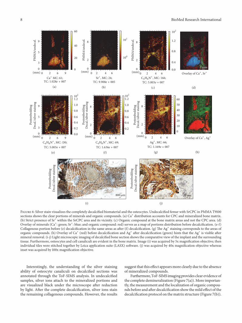
Before decalcification, calcium ion (Ca+) distributionwithin the SrCPC and the mineralized bone was apparent(Figure 6(a)). Presentable is the strontium ion (Sr+) distri-bution mainly within the cement and in the close vicinity(Figure 6(b)). The collagen fiber components were alsoseen in regions of bone matrix irrelevant of mineralization(Figure 6(c)). The overlay reflects the signal intensity anddistribution of the investigated constituents (Figure 6(d)).Interestingly, after decalcification no Ca+ or Sr+ were seen

6 BioMed Research International
von
Koss
a/va
n G
ieso
nU
ndec
al.
800 �휇m
(a)
Und
ecal
.
20 �휇m
von
Koss
a/va
n G
ieso
n
(b)
Silv
er st
aini
ng
Poste
mbe
ddin
g de
cal.
800 �휇m
(c)
Silv
er st
aini
ng
Poste
mbe
ddin
g de
cal.
20 �휇m
(d)
Figure 4: Postembedding decalcification of rat femur preserves the integrity of calcium-containing materials and allows investigatingosteocytes. The SrCPC cement in the fracture defect was embedded in PMMA T9100. (a-b) von Kossa/van Gieson staining of undecalcifiedbone sample differentiates soft tissue around the fracture defect (red) and stains ossified tissue and materials in black. (c-d) Silver staining ofpostembedding decalcified sections visualizes osteocytes within thematrix and differentiates the implant from the surrounding tissue. Images(a) and (c) were acquired by 5xmagnification objective; then individual tiles were stitched together by Leica application suite (LASX) software.Images (b) and (d) were acquired by 40x magnification objective while inset of image (d) was acquired by 100x magnification objective.
in the silver-stained section reflecting a complete decalcifi-cation within very short time (Figure 6(e)). However, thecollagenous portion remained constant before (Figure 6(e))and after decalcification (Figure 6(f)). Distribution of silverions was seen in the same areas of the collagenous portion(Figure 6(g)). The overlay of the Ca+ signal before decalcifi-cation andAg+ after decalcification (Figure 6(h)) reflects howsilver only stains the remaining organic compounds with theexception of the bonematrix (Figure 6(i)). However, the lightmicroscopy image can still show the borders of the materialareas without distortion besides enabling the visualization ofosteocyte and cellular network in the vicinity and within thematerials (Figures 6(i)–6(j)).
4. Discussion
Theacquisition of clinical and preclinical samples depends onlimited availability. Inmany cases, the need for decalcificationin special investigative histology is hindered due to the pres-ence of bone substitute materials. Furthermore, correlatinglight microscopy from the same sample with new imagingtechniques based on element analysis such as ToF-SIMS isdemanding.
Decalcified sections from undecalcified specimens weretested in comparison with the sections originated from
decalcified specimens; both groupswere stainedwith a hema-toxylin eosin stain, an enzyme histochemical stain (TRAP),and an immunostaining of ColI.
Paraffin is an inert embedding medium, which is appliedonly to decalcified tissue. However, paraffin does not interactchemically with tissue components [17]. Due to decalcifi-cation, bone structure is poorly preserved and informationabout mineralization is lost. Often, decalcified bone samplesexhibited shrinking artifacts especially in bone marrow areaafter paraffin embedding. Moreover, immunolocalization ofcellular and bone matrix markers such as ColI and TRAP isroutinely performed on paraffin embedded decalcified bone[18, 19].
Methyl-methacrylate (MMA) embedding showed depen-dable tissue coloration [20]. However, high polymerizationtemperature ofMMAaffects proteins and hinders subsequentantibody-antigen coupling [21]. However, the effect wasimproved by the introduction of low-temperature polymer-izing PMMA (Technovit 9100 New�), and immunostainingsbecame feasible [22, 23]. The introduction of bone substitutematerials with mineral composition created a new hardshipas preembedding decalcification will cause material loss.Furthermore, studies compared bone structure embeddedin low-temperature polymerizing PMMA with decalcifiedbone samples embedded in paraffin. Bone morphology and

BioMed Research International 7
Und
ecal
.M
asso
n-G
oldn
er
800 �휇m
(a)U
ndec
al.
Mas
son-
Gol
dner
20 �휇m
(b)
Und
ecal
.M
asso
n-G
oldn
er
10 �휇m
(c)
Silv
er st
aini
ngPo
stem
bedd
ing
deca
l.
800 �휇m
(d)
Silv
er st
aini
ngPo
stem
bedd
ing
deca
l.
20 �휇m
(e)
Silv
er st
aini
ngPo
stem
bedd
ing
deca
l.
10 �휇m
(f)
Figure 5: Histological stains of undecalcified tibia with intramedullary pin aligned in compliance with decalcified bone. The bone sampleswere embedded in PMMAT7200.Masson-Goldner staining of undecalcified bone shows the presence of soft tissue around the implant regionand the magnified image shows the clear view of osteoid (a–c). The decalcified bone section shows the comparative view and presence of softtissue around the implant region (d–f). The magnified image shows the view of osteocyte canaliculi. Images (a) and (d) were acquired by5x magnification objective; then individual tiles were stitched together by Leica application suite (LASX) software. Images (b) and (e) wereacquired by 40x magnification objective. Images (c) and (f) were acquired by 100x magnification objective.
structure were improved in PMMA sections and no crackingeffects were seen [24]. Moreover, decalcified bones showedreduced immunohistochemical reaction [24]. Other studiesshowed that decalcification resulted in distorted tissue mor-phology and reduced staining quality [25].
Furthermore, the widely accepted fluorochrome labellingis an efficient ethically acceptable standard technique toinvestigate bone dynamics in skeletal research. The methodis applied clinically on iliac crest biopsies of patients incombination with plain histology [26] and in preclinicalanimal models to investigate bone formation rate [27]. Theuse of fluorochrome labelling is increasing with the promptdevelopment of finite element methods. However, imaging offluorochrome labelling is only possible on undecalcified sam-ples, which hinders further histological analysis especiallywith small sample or rare pathological samples.
The proposed protocol shall enable diverse utilizationof fluorochrome labelled bone samples by using same orsequential sections. Thereby, the presented protocol opensmore possibilities to correlate bone dynamics with cellularand structural changes using image registry techniques or inmathematical modelling in terms of finite element analysis.Furthermore, the labelled areas upon postembedding decal-cification can still be investigated in the subsequent section ifneeded.
Taken together, the proposed protocol offers the advan-tages of undecalcified bone embedding in low-temperature
polymerization PMMA. Thereby, the method preserves theintegrity of biomaterials when sample decalcification isneeded.
Furthermore, biomaterials still require characterizationon how they affect cellular behavior. The obligatory use ofa different set of animals to gain samples for rhodaminestaining to investigate osteocytes in mineral-based bioma-terials is challenging. The difficulty of decalcification wascircumvented by the proposed protocol. More importantly,the need of additional set of animals in fracture healingstudies with biomaterials can be avoided.
For example, the study of the second group of samplesthat previously reported enhanced healing using SrCPCcompared to CPC only in osteoporotic fractures [12]. Thisapplies also to the study of the third group of samples thatpreviously reported enhanced biomechanical competencethrough better implant anchorage resulting from coating oftitanium pins [8]. Unfortunately, the interesting results couldnot be correlated to the role of osteocytes due to the technicalinapplicability. Such a role can now be investigated using thecurrent protocol with comparable material visual integritybefore and after decalcification.
ToF-SIMSmeasurement as a sensitive chemical investiga-tion is more informative in bone tissue when performed onundecalcified samples. However, same-sample investigationis more powerful to understand changes in physiologicalsamples.
8 BioMed Research International
(mm)0
3
5
9
PMM
A/un
deca
l.
TC: 1.028e + 007
2 6 90Ca+ MC: 61;
0
20
40
60
(a)
(mm)
TC: 9.900e + 005
101
100
0
2
8
PMM
A/un
deca
l.
2 4 60Sr+ , MC: 24;
(b)
(mm)
TC: 5.003e + 007
0
2
8
PMM
A/un
deca
l.
64200.0
0.4
0.8
1.2
102
C4H8N+ , MC: 166;
(c)
Overlay of Ca+ , Sr+1mm
(d)
(mm)
Poste
mbe
ddin
g
0
3
5
9
Dec
al./s
ilver
stai
ning
TC: 5.003e + 007
2 6 900.0
0.2
0.4
0.6
0.8
1.0
1.2102
C4H8N+ , MC: 130;
(e)
1.2
1.0
0.8
0.6
0.4
0.2
(mm)
Poste
mbe
ddin
g
TC: 1.636e + 007
0
2
8
Dec
al./s
ilver
stai
ning
2 4 60
102
C4H8N+ , MC: 69;
(f)
6050403020100
(mm)
TC: 1.169e + 007
Poste
mbe
ddin
g0
2
8
Dec
al./s
ilver
stai
ning
2 4 60
Ag+ , MC: 66;
(g)
Overlay of Ca+ , Ag+
1mm
(h)
Poste
mbe
ddin
gD
ecal
./silv
er st
aini
ng
800 �휇m
(i)
G
Poste
mbe
ddin
gD
ecal
./silv
er st
aini
ng
20 �휇m
(j)
Figure 6: Silver stain visualizes the completely decalcified biomaterial and the osteocytes. Undecalcified femur with SrCPC in PMMAT9100sections shows the clear portions of minerals and organic compounds. (a) Ca+ distribution accounts for CPC and mineralized bone matrix.(b) Strict presence of Sr+ within the SrCPC area and its vicinity. (c) Organic compound at the bone matrix areas and not the CPC area. (d)Overlay of minerals (Ca+: green; Sr+: blue; and organic compound: red) serves as a map of portions distribution before decalcification. (e-f)Collagenous portion before (e) decalcification in the same areas as after (f) decalcification. (g) The Ag+ staining corresponds to the areas oforganic compounds. (h) Overlay of Ca+ (red) before decalcification and Ag+ after decalcification (green) hints that the Ag+ is visible aftermineral removal. (i-j) Light microscopic imaging of decalcified bone section shows the comparative view of the implant and the surroundingtissue. Furthermore, osteocytes and cell canaliculi are evident in the bone matrix. Image (i) was acquired by 5x magnification objective; thenindividual tiles were stitched together by Leica application suite (LASX) software. (j) was acquired by 40x magnification objective whereasinset was acquired by 100x magnification objective.
Interestingly, the understanding of the silver stainingability of osteocyte canaliculi on decalcified sections wasannotated through the ToF-SIMS analysis. In undecalcifiedsamples, silver ions attach to the mineralized portions andare visualized black under the microscope after reductionby light. After the complete decalcification, silver ions stainthe remaining collagenous compounds. However, the results
suggest that this effect appearsmore clearly due to the absenceof mineralized compounds.
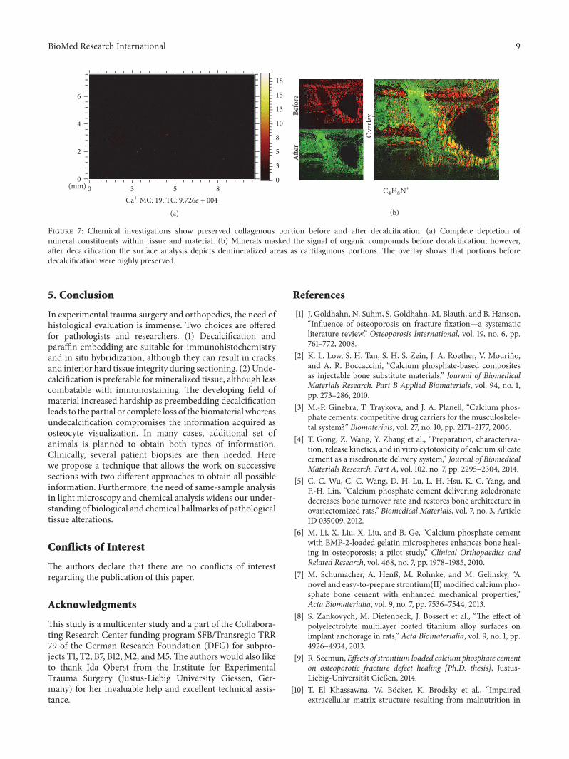
Furthermore,ToF-SIMSimagingprovides clear evidence ofthe complete demineralization (Figure 7(a)).More importan-tly, themeasurement and the localization of organic compou-nds before and after decalcification show themild effect of thedecalcification protocol on thematrix structure (Figure 7(b)).
BioMed Research International 9
(mm)
6
4
2
0 0
3
5
8
10
13
15
18
3 5 80Ca+ MC: 19; TC: 9.726e + 004
(a)
Befo
reA
fter
Ove
rlay
C4H8N+
Ove
rlay
(b)
Figure 7: Chemical investigations show preserved collagenous portion before and after decalcification. (a) Complete depletion ofmineral constituents within tissue and material. (b) Minerals masked the signal of organic compounds before decalcification; however,after decalcification the surface analysis depicts demineralized areas as cartilaginous portions. The overlay shows that portions beforedecalcification were highly preserved.
5. Conclusion
In experimental trauma surgery and orthopedics, the need ofhistological evaluation is immense. Two choices are offeredfor pathologists and researchers. (1) Decalcification andparaffin embedding are suitable for immunohistochemistryand in situ hybridization, although they can result in cracksand inferior hard tissue integrity during sectioning. (2)Unde-calcification is preferable for mineralized tissue, although lesscombatable with immunostaining. The developing field ofmaterial increased hardship as preembedding decalcificationleads to the partial or complete loss of the biomaterial whereasundecalcification compromises the information acquired asosteocyte visualization. In many cases, additional set ofanimals is planned to obtain both types of information.Clinically, several patient biopsies are then needed. Herewe propose a technique that allows the work on successivesections with two different approaches to obtain all possibleinformation. Furthermore, the need of same-sample analysisin light microscopy and chemical analysis widens our under-standing of biological and chemical hallmarks of pathologicaltissue alterations.
Conflicts of Interest
The authors declare that there are no conflicts of interestregarding the publication of this paper.
Acknowledgments
This study is a multicenter study and a part of the Collabora-ting Research Center funding program SFB/Transregio TRR79 of the German Research Foundation (DFG) for subpro-jects T1, T2, B7, B12, M2, andM5.The authors would also liketo thank Ida Oberst from the Institute for ExperimentalTrauma Surgery (Justus-Liebig University Giessen, Ger-many) for her invaluable help and excellent technical assis-tance.
References
[1] J. Goldhahn, N. Suhm, S. Goldhahn, M. Blauth, and B. Hanson,“Influence of osteoporosis on fracture fixation—a systematicliterature review,” Osteoporosis International, vol. 19, no. 6, pp.761–772, 2008.
[2] K. L. Low, S. H. Tan, S. H. S. Zein, J. A. Roether, V. Mourino,and A. R. Boccaccini, “Calcium phosphate-based compositesas injectable bone substitute materials,” Journal of BiomedicalMaterials Research. Part B Applied Biomaterials, vol. 94, no. 1,pp. 273–286, 2010.
[3] M.-P. Ginebra, T. Traykova, and J. A. Planell, “Calcium phos-phate cements: competitive drug carriers for the musculoskele-tal system?” Biomaterials, vol. 27, no. 10, pp. 2171–2177, 2006.
[4] T. Gong, Z. Wang, Y. Zhang et al., “Preparation, characteriza-tion, release kinetics, and in vitro cytotoxicity of calcium silicatecement as a risedronate delivery system,” Journal of BiomedicalMaterials Research. Part A, vol. 102, no. 7, pp. 2295–2304, 2014.
[5] C.-C. Wu, C.-C. Wang, D.-H. Lu, L.-H. Hsu, K.-C. Yang, andF.-H. Lin, “Calcium phosphate cement delivering zoledronatedecreases bone turnover rate and restores bone architecture inovariectomized rats,” Biomedical Materials, vol. 7, no. 3, ArticleID 035009, 2012.
[6] M. Li, X. Liu, X. Liu, and B. Ge, “Calcium phosphate cementwith BMP-2-loaded gelatin microspheres enhances bone heal-ing in osteoporosis: a pilot study,” Clinical Orthopaedics andRelated Research, vol. 468, no. 7, pp. 1978–1985, 2010.
[7] M. Schumacher, A. Henß, M. Rohnke, and M. Gelinsky, “Anovel and easy-to-prepare strontium(II)modified calcium pho-sphate bone cement with enhanced mechanical properties,”Acta Biomaterialia, vol. 9, no. 7, pp. 7536–7544, 2013.
[8] S. Zankovych, M. Diefenbeck, J. Bossert et al., “The effect ofpolyelectrolyte multilayer coated titanium alloy surfaces onimplant anchorage in rats,” Acta Biomaterialia, vol. 9, no. 1, pp.4926–4934, 2013.
[9] R. Seemun,Effects of strontium loaded calcium phosphate cementon osteoporotic fracture defect healing [Ph.D. thesis], Justus-Liebig-Universitat Gießen, 2014.
[10] T. El Khassawna, W. Bocker, K. Brodsky et al., “Impairedextracellular matrix structure resulting from malnutrition in

10 BioMed Research International
ovariectomized mature rats,” Histochemistry and Cell Biology,vol. 144, no. 5, pp. 491–507, 2015.
[11] V. Alt, U. Thormann, S. Ray et al., “A new metaphyseal bonedefect model in osteoporotic rats to study biomaterials for theenhancement of bone healing in osteoporotic fractures,” ActaBiomaterialia, vol. 9, no. 6, pp. 7035–7042, 2013.
[12] U. Thormann, S. Ray, U. Sommer et al., “Bone formation ind-uced by strontiummodified calcium phosphate cement in criti-cal-size metaphyseal fracture defects in ovariectomized rats,”Biomaterials, vol. 34, no. 34, pp. 8589–8598, 2013.
[13] T. El Khassawna, W. Bocker, P. Govindarajan et al., “Effects ofmulti-deficiencies-diet on bone parameters of peripheral bonein ovariectomized mature rat,” PLoS ONE, vol. 8, no. 8, ArticleID e71665, 2013.
[14] A. Henss, M. Rohnke, T. El Khassawna et al., “Applicability ofToF-SIMS for monitoring compositional changes in bone in along-term animal model,” Journal ofThe Royal Society Interface,vol. 10, no. 86, Article ID 20130332, 2013.
[15] J. C. Vickerman andD. Briggs,TOF-SIMS:Materials Analysis byMass Spectrometry, IM Publications, Chichester, UK, 2013.
[16] A. Henss, A. Hild, M. Rohnke, S. Wenisch, and J. Janek, “Timeof flight secondary ion mass spectrometry of bone—impactof sample preparation and measurement conditions,” Biointer-phases, vol. 11, no. 2, pp. 1–13, 2016.
[17] H.-E. Schaefer, “Die histologische Bearbeitungstechnik vonBeckenkammbiopsien auf der Basis von Entkalkung und Para-ffineinbettung unter Berucksichtigung osteologischer undhamatologischer Fragestellungen,” Der Pathologe, vol. 16, no. 1,pp. 11–27, 1995.
[18] G. Jundt, K.-H. Berghauser, J. D. Termine, and A. Schulz,“Osteonectin—a differentiation marker of bone cells,” Cell andTissue Research, vol. 248, no. 2, pp. 409–415, 1987.
[19] A. L. Bronckers, S. Gay, R. D. Finkelman, andW.T. Butler, “Dev-elopmental appearance of Gla proteins (osteocalcin) and alka-line phosphatase in tooth germs and bones of the rat,” Bone andmineral, vol. 2, no. 5, pp. 361–373, 1987.
[20] R. Schenk, “On the histological processing of undecalcifiedbone,” Acta Anatomica, vol. 60, pp. 3–19, 1965.
[21] R. G. Erben, “Embedding of bone samples in methylmethacry-late: an improved method suitable for bone histomorphometry,histochemistry, and immunohistochemistry,” Journal of Histo-chemistry and Cytochemistry, vol. 45, no. 2, pp. 307–313, 1997.
[22] J. Bernhards, B.Weitzel,M.Werner,M. Rimpler, andA.Georgii,“A new histological embedding method by low-temperaturepolymerisation of methyl methacrylate allowing immuno- andenzyme histochemical studies on semi-thin sections of unde-calcified bone marrow biopsies,” Histochemistry, vol. 98, no. 3,pp. 145–154, 1992.
[23] P. Derkx, A. L. Nigg, F. T. Bosman et al., “Immunolocalizationand quantification of noncollagenous bone matrix proteins inmethylmethacrylate-embedded adult human bone in combina-tion with histomorphometry,” Bone, vol. 22, no. 4, pp. 367–373,1998.
[24] G.Wittenburg, C. Volkel, R. Mai, and G. Lauer, “Immunohisto-chemical comparison of differentiationmarkers on paraffin andplastic embedded human bone samples,” Journal of Physiologyand Pharmacology, vol. 60, supplement 8, pp. 43–49, 2009.
[25] J. B.Matthews andG. I.Mason, “Influence of decalcifying agentson immunoreactivity of formalin-fixed, paraffin-embeddedtissue,” The Histochemical Journal, vol. 16, no. 7, pp. 771–787,1984.
[26] R. R. Recker, D. B. Kimmel, A.M. Parfitt, K.M.Davies, N. Kesh-awarz, and S. Hinders, “Static and tetracycline-based bonehistomorphometric data from 34 normal postmenopausalfemales,” Journal of Bone and Mineral Research, vol. 3, no. 2, pp.133–144, 1988.
[27] R. G. Erben, “Bone-labeling techniques,” inHandbook of Histo-logy Methods for Bone and Cartilage, pp. 99–117, SpringerScience+Business Media, 2003.

Submit your manuscripts athttps://www.hindawi.com
ScientificaHindawi Publishing Corporationhttp://www.hindawi.com Volume 2014
CorrosionInternational Journal of
Hindawi Publishing Corporationhttp://www.hindawi.com Volume 2014
Polymer ScienceInternational Journal of
Hindawi Publishing Corporationhttp://www.hindawi.com Volume 2014
Hindawi Publishing Corporationhttp://www.hindawi.com Volume 2014
CeramicsJournal of
Hindawi Publishing Corporationhttp://www.hindawi.com Volume 2014
CompositesJournal of
NanoparticlesJournal of
Hindawi Publishing Corporationhttp://www.hindawi.com Volume 2014
Hindawi Publishing Corporationhttp://www.hindawi.com Volume 2014
International Journal of
Biomaterials
Hindawi Publishing Corporationhttp://www.hindawi.com Volume 2014
NanoscienceJournal of
TextilesHindawi Publishing Corporation http://www.hindawi.com Volume 2014
Journal of
NanotechnologyHindawi Publishing Corporationhttp://www.hindawi.com Volume 2014
Journal of
CrystallographyJournal of
Hindawi Publishing Corporationhttp://www.hindawi.com Volume 2014
The Scientific World JournalHindawi Publishing Corporation http://www.hindawi.com Volume 2014
Hindawi Publishing Corporationhttp://www.hindawi.com Volume 2014
CoatingsJournal of
Advances in
Materials Science and EngineeringHindawi Publishing Corporationhttp://www.hindawi.com Volume 2014
Smart Materials Research
Hindawi Publishing Corporationhttp://www.hindawi.com Volume 2014
Hindawi Publishing Corporationhttp://www.hindawi.com Volume 2014
MetallurgyJournal of
Hindawi Publishing Corporationhttp://www.hindawi.com Volume 2014
BioMed Research International
MaterialsJournal of
Hindawi Publishing Corporationhttp://www.hindawi.com Volume 2014
Nano
materials
Hindawi Publishing Corporationhttp://www.hindawi.com Volume 2014
Journal ofNanomaterials