
The origin of conodonts and of vertebrate mineralized skeletons
8
LETTER doi:10.1038/nature12645 The origin of conodonts and of vertebrate mineralized skeletons Duncan J. E. Murdock 1 , Xi-Ping Dong 2,3 , John E. Repetski 4 , Federica Marone 5 , Marco Stampanoni 5,6 & Philip C. J. Donoghue 1 Conodonts are an extinct group of jawless vertebrates whose tooth- like elements are the earliest instance of a mineralized skeleton in the vertebrate lineage 1,2 , inspiring the ‘inside-out’ hypothesis that teeth evolved independently of the vertebrate dermal skeleton and before the origin of jaws 3–6 . However, these propositions have been based on evidence from derived euconodonts. Here we test hypo- theses of a paraconodont ancestry of euconodonts 7–11 using syn- chrotron radiation X-ray tomographic microscopy to characterize and compare the microstructure of morphologically similar euco- nodont and paraconodont elements. Paraconodonts exhibit a range of grades of structural differentiation, including tissues and a pattern of growth common to euconodont basal bodies. The different grades of structural differentiation exhibited by paraconodonts demonstrate the stepwise acquisition of eucono- dont characters, resolving debate over the relationship between these two groups. By implication, the putative homology of euco- nodont crown tissue and vertebrate enamel must be rejected as these tissues have evolved independently and convergently. Thus, the precise ontogenetic, structural and topological similarities between conodont elements and vertebrate odontodes appear to be a remarkable instance of convergence. The last common ancestor of conodonts and jawed vertebrates probably lacked mineralized skeletal tissues. The hypothesis that teeth evolved before jaws and the inside-out hypothesis of dental evolution must be rejected; teeth seem to have evolved through the extension of odontogenic com- petence from the external dermis to internal epithelium soon after the origin of jaws. The soft tissue anatomy of euconodonts substantiates their vertebrate affinity 12,13 , but homology of euconodont and vertebrate skeletal tis- sues 1,14,15 remains the subject of controversy 16,17 . The mineralized skele- ton of euconodonts consists of an oropharyngeal array of tooth-like elements that are composed of two mineralized structural elements, the crown and basal body which are comprised of tissues that resemble enamel and dentine 8 . Euconodont elements grew through centrifugal appositional growth, with laminae in the crown and basal body added in synchrony, in a manner comparable to enamel and dentine in the teeth of jawed vertebrates. However, knowledge of conodont skeletal tissues is based largely on extremely derived euconodonts and hypotheses of homology to canonical vertebrate skeletal tissues have taken no account of the evolutionary origin of the conodont skeleton. Based principally on similarities in morphology and patterns of growth, an evolutionary series was proposed originally among protoconodonts, paraconodonts and euconodonts 7–11 . Protoconodonts have been recognized subsequently as stem-chaetognaths 18 and excluded from euconodont ancestry, but the hypothesis that euconodonts are derived paraconodonts remains 10,11 . Paraconodont elements are unipart, and have been considered 1 School of Earth Sciences, University of Bristol, Wills Memorial Building, Queen’s Road, Bristol BS8 1RJ, UK. 2 School of Earth and Space Science, Peking University, Beijing 100871, China. 3 State Key Laboratory of Palaeobiology and Stratigraphy, Nanjing Institute of Geology and Palaeontology, Chinese Academy of Sciences, Nanjing 210008, China. 4 US Geological Survey, MS 926A, National Center, Reston, Virginia 20192, USA. 5 Swiss Light Source, Paul Scherrer Institut, Villigen 5232, Switzerland. 6 Institute for Biomedical Engineering, University of Zu ¨ rich and ETH Zu ¨ rich, Ra ¨ mistrasse, Zu ¨ rich 8006, Switzerland. a b c d e f g h Figure 1 | Element growth and microstructure of the paraconodont Furnishina, Threadgill Creek section, Wilberns Formation, central Texas, 1,115 feet above base of Cambrian strata. a–c, The complete element has been subdivided into a number of discrete growth stages delimited by lines showing cessation of growth (b, c). d–h, Initial growth stage, protoelement (d), is not enveloped by subsequent growth lamellae, rather lamellae are added to the proximal and lateral margins of the protoelement only (e–h). Scale bar, 50 mm. 546 | NATURE | VOL 502 | 24 OCTOBER 2013 Macmillan Publishers Limited. All rights reserved ©2013
-
Upload
unitedstatesgeologicalsurvey -
Category
Documents
-
view
1 -
download
0
Transcript of The origin of conodonts and of vertebrate mineralized skeletons

LETTERdoi:10.1038/nature12645
The origin of conodonts and of vertebratemineralized skeletonsDuncan J. E. Murdock1, Xi-Ping Dong2,3, John E. Repetski4, Federica Marone5, Marco Stampanoni5,6 & Philip C. J. Donoghue1
Conodonts are an extinct group of jawless vertebrates whose tooth-like elements are the earliest instance of a mineralized skeleton inthe vertebrate lineage1,2, inspiring the ‘inside-out’ hypothesis thatteeth evolved independently of the vertebrate dermal skeleton andbefore the origin of jaws3–6. However, these propositions have beenbased on evidence from derived euconodonts. Here we test hypo-theses of a paraconodont ancestry of euconodonts7–11 using syn-chrotron radiation X-ray tomographic microscopy to characterizeand compare the microstructure of morphologically similar euco-nodont and paraconodont elements. Paraconodonts exhibit arange of grades of structural differentiation, including tissuesand a pattern of growth common to euconodont basal bodies.The different grades of structural differentiation exhibited byparaconodonts demonstrate the stepwise acquisition of eucono-dont characters, resolving debate over the relationship betweenthese two groups. By implication, the putative homology of euco-nodont crown tissue and vertebrate enamel must be rejected asthese tissues have evolved independently and convergently. Thus,the precise ontogenetic, structural and topological similaritiesbetween conodont elements and vertebrate odontodes appear tobe a remarkable instance of convergence. The last common ancestorof conodonts and jawed vertebrates probably lacked mineralizedskeletal tissues. The hypothesis that teeth evolved before jaws and
the inside-out hypothesis of dental evolution must be rejected; teethseem to have evolved through the extension of odontogenic com-petence from the external dermis to internal epithelium soon afterthe origin of jaws.
The soft tissue anatomy of euconodonts substantiates their vertebrateaffinity12,13, but homology of euconodont and vertebrate skeletal tis-sues1,14,15 remains the subject of controversy16,17. The mineralized skele-ton of euconodonts consists of an oropharyngeal array of tooth-likeelements that are composed of two mineralized structural elements,the crown and basal body which are comprised of tissues that resembleenamel and dentine8. Euconodont elements grew through centrifugalappositional growth, with laminae in the crown and basal body added insynchrony, in a manner comparable to enamel and dentine in the teethof jawed vertebrates. However, knowledge of conodont skeletal tissues isbased largely on extremely derived euconodonts and hypotheses ofhomology to canonical vertebrate skeletal tissues have taken no accountof the evolutionary origin of the conodont skeleton. Based principally onsimilarities in morphology and patterns of growth, an evolutionary serieswas proposed originally among protoconodonts, paraconodonts andeuconodonts7–11. Protoconodonts have been recognized subsequentlyas stem-chaetognaths18 and excluded from euconodont ancestry, butthe hypothesis that euconodonts are derived paraconodonts remains10,11.Paraconodont elements are unipart, and have been considered
1School of Earth Sciences, University of Bristol, Wills Memorial Building, Queen’s Road, Bristol BS8 1RJ, UK. 2School of Earth and Space Science, Peking University, Beijing 100871, China. 3State KeyLaboratory of Palaeobiology and Stratigraphy, Nanjing Institute of Geology and Palaeontology, Chinese Academy of Sciences, Nanjing 210008, China. 4US Geological Survey, MS 926A, National Center,Reston, Virginia 20192, USA. 5Swiss Light Source, Paul Scherrer Institut, Villigen 5232, Switzerland. 6Institute for Biomedical Engineering, University of Zurich and ETH Zurich, Ramistrasse, Zurich 8006,Switzerland.
a b c d e f g h
Figure 1 | Element growth and microstructure of the paraconodontFurnishina, Threadgill Creek section, Wilberns Formation, central Texas,1,115 feet above base of Cambrian strata. a–c, The complete element has beensubdivided into a number of discrete growth stages delimited by lines showing
cessation of growth (b, c). d–h, Initial growth stage, protoelement (d), is notenveloped by subsequent growth lamellae, rather lamellae are added to theproximal and lateral margins of the protoelement only (e–h). Scale bar, 50mm.
5 4 6 | N A T U R E | V O L 5 0 2 | 2 4 O C T O B E R 2 0 1 3
Macmillan Publishers Limited. All rights reserved©2013
homologous to the euconodont basal body alone because they grewthrough apposition of lamella layers to the proximal surface only.However, the histological comparisons of protoconodont and eucono-dont elements have been vague and aspects of paraconodont elementstructure and growth remain equivocal. For example, the homology ofprotoconodont elements and euconodont element basal bodies has beenrejected on the basis that a basal body may not be primitive for eucono-donts, and therefore could not be homologous to any paraconodonttissues19,20. Key to the interpretation of paraconodont morphogenesisis the nature of the earliest stages of growth, or ‘protoelement’, whichforms the distal-most part of the element. If characterized by completecentrifugal growth, this would result in a protoelement stage reminiscentof a euconodont crown plus basal body8. By contrast, addition of lamel-lae to the proximal surface only (that is, basal internal accretion) wouldresult in a morphology reminiscent of the euconodont basal body alone.However, the evidential basis of this characterization has been criticizedby some as an analytical artefact7,10,11. We used synchrotron radiationX-ray tomographic microscopy (SRXTM) to characterize the elementstructure of paraconodonts and early euconodonts, non-invasively andat sub-micron resolution. We used the ensuing datasets to characterizethe component tissues and uncover the pattern of development recordedin the sclerochronology of the growth arrest lines preserved in themineralized tissues.
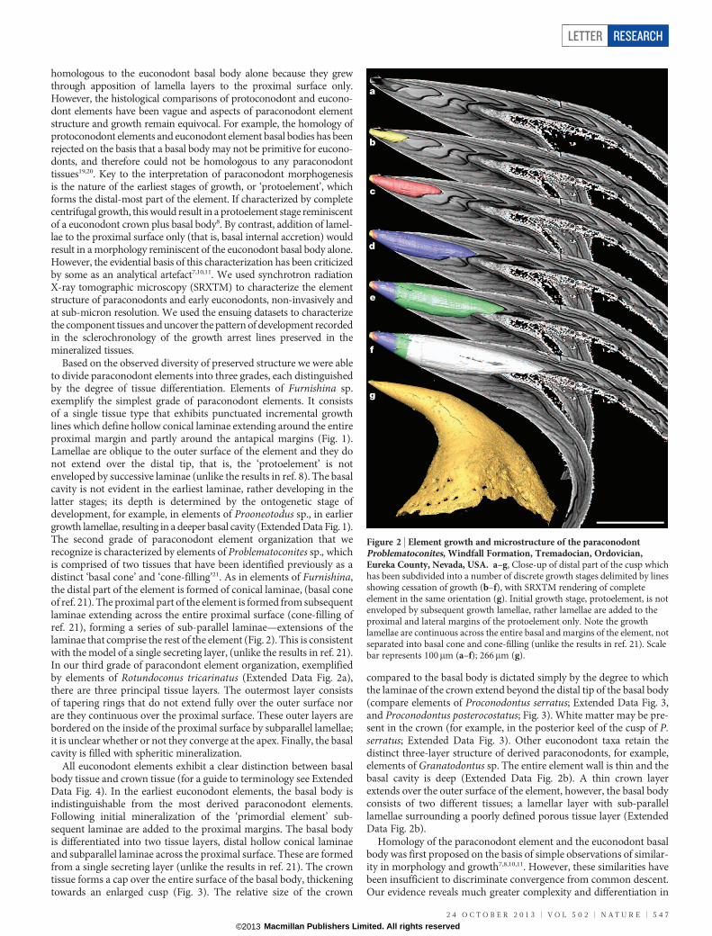
Based on the observed diversity of preserved structure we were ableto divide paraconodont elements into three grades, each distinguishedby the degree of tissue differentiation. Elements of Furnishina sp.exemplify the simplest grade of paraconodont elements. It consistsof a single tissue type that exhibits punctuated incremental growthlines which define hollow conical laminae extending around the entireproximal margin and partly around the antapical margins (Fig. 1).Lamellae are oblique to the outer surface of the element and they donot extend over the distal tip, that is, the ‘protoelement’ is notenveloped by successive laminae (unlike the results in ref. 8). The basalcavity is not evident in the earliest laminae, rather developing in thelatter stages; its depth is determined by the ontogenetic stage ofdevelopment, for example, in elements of Prooneotodus sp., in earliergrowth lamellae, resulting in a deeper basal cavity (Extended Data Fig. 1).The second grade of paraconodont element organization that werecognize is characterized by elements of Problematoconites sp., whichis comprised of two tissues that have been identified previously as adistinct ‘basal cone’ and ‘cone-filling’21. As in elements of Furnishina,the distal part of the element is formed of conical laminae, (basal coneof ref. 21). The proximal part of the element is formed from subsequentlaminae extending across the entire proximal surface (cone-filling ofref. 21), forming a series of sub-parallel laminae—extensions of thelaminae that comprise the rest of the element (Fig. 2). This is consistentwith the model of a single secreting layer, (unlike the results in ref. 21).In our third grade of paracondont element organization, exemplifiedby elements of Rotundoconus tricarinatus (Extended Data Fig. 2a),there are three principal tissue layers. The outermost layer consistsof tapering rings that do not extend fully over the outer surface norare they continuous over the proximal surface. These outer layers arebordered on the inside of the proximal surface by subparallel lamellae;it is unclear whether or not they converge at the apex. Finally, the basalcavity is filled with spheritic mineralization.
All euconodont elements exhibit a clear distinction between basalbody tissue and crown tissue (for a guide to terminology see ExtendedData Fig. 4). In the earliest euconodont elements, the basal body isindistinguishable from the most derived paraconodont elements.Following initial mineralization of the ‘primordial element’ sub-sequent laminae are added to the proximal margins. The basal bodyis differentiated into two tissue layers, distal hollow conical laminaeand subparallel laminae across the proximal surface. These are formedfrom a single secreting layer (unlike the results in ref. 21). The crowntissue forms a cap over the entire surface of the basal body, thickeningtowards an enlarged cusp (Fig. 3). The relative size of the crown
compared to the basal body is dictated simply by the degree to whichthe laminae of the crown extend beyond the distal tip of the basal body(compare elements of Proconodontus serratus; Extended Data Fig. 3,and Proconodontus posterocostatus; Fig. 3). White matter may be pre-sent in the crown (for example, in the posterior keel of the cusp of P.serratus; Extended Data Fig. 3). Other euconodont taxa retain thedistinct three-layer structure of derived paraconodonts, for example,elements of Granatodontus sp. The entire element wall is thin and thebasal cavity is deep (Extended Data Fig. 2b). A thin crown layerextends over the outer surface of the element, however, the basal bodyconsists of two different tissues; a lamellar layer with sub-parallellamellae surrounding a poorly defined porous tissue layer (ExtendedData Fig. 2b).
Homology of the paraconodont element and the euconodont basalbody was first proposed on the basis of simple observations of similar-ity in morphology and growth7,8,10,11. However, these similarities havebeen insufficient to discriminate convergence from common descent.Our evidence reveals much greater complexity and differentiation in
a
b
c
d
e
f
g
Figure 2 | Element growth and microstructure of the paraconodontProblematoconites, Windfall Formation, Tremadocian, Ordovician,Eureka County, Nevada, USA. a–g, Close-up of distal part of the cusp whichhas been subdivided into a number of discrete growth stages delimited by linesshowing cessation of growth (b–f), with SRXTM rendering of completeelement in the same orientation (g). Initial growth stage, protoelement, is notenveloped by subsequent growth lamellae, rather lamellae are added to theproximal and lateral margins of the protoelement only. Note the growthlamellae are continuous across the entire basal and margins of the element, notseparated into basal cone and cone-filling (unlike the results in ref. 21). Scalebar represents 100mm (a–f); 266mm (g).
LETTER RESEARCH
2 4 O C T O B E R 2 0 1 3 | V O L 5 0 2 | N A T U R E | 5 4 7
Macmillan Publishers Limited. All rights reserved©2013

the structure and growth of paraconodont elements than has beendescribed previously, corroborating this hypothesis of homology.First, the protoelement of both the paraconodont element and euco-nodont element basal body is not overgrown at the distal tip. Rather, itis permanently exposed at the tip of protoconodont elements andremains in direct contact with the crown at the core of euconodontelements. Subsequent to the initial mineralization of the protoelement,the ontogeny of both paraconodont elements and the basal bodies ofeuconodont elements follow the same pattern of growth. The develop-ment of structural diversity exhibited by conodont elements is dictatedsimply by the relative timing of changes in the mode of secretion and,ultimately, through the differentiation of two principal structural ele-ments, the basal body and crown, the latter characterizing the firsteuconodont elements. The basal cone and cone-filling structuredescribed previously in euconodont basal bodies21 is manifest also inprotoconodont elements, though we show that these are not separatestructures and the growth lamellae are continuous between them.Crucially, the range of structures exhibited by the elements of differentparaconodont species lie within nested sets of structural complexity,the most complex of which exhibit greater similarity to euconodontelements than other paraconodonts. Indeed, in terms of structure andarrangement of the component tissues, the basal bodies of elements ofthe early euconodont Proconodontus are effectively indistinguishablefrom the most complex paraconodont elements, such as those ofProblematoconites. The same comparison can be made of the paraco-nodont Rotundoconus and the euconodont Granatodontus.
Direct comparison of ontogeny and tissue organization, coupledwith a clear spectrum of complexity through early conodont elements,demonstrates that the similarities between paraconodont and eucono-dont elements go beyond analogy. Our results corroborate the hypo-thesis that the structural organization of the euconodont element wasnot only derived through the evolution of the enamel-like crown tissuefrom a paraconodont-grade ancestor, but also that characteristics ofthe euconodont basal body were assembled stepwise among differentevolutionary grades of paraconodonts (Fig. 4). Evidently, the proposi-tion of homology between euconodont crown tissue and vertebrateenamel1,15,22,23 fails a test of phylogenetic congruence24 and must there-fore be rejected. In this light, it is pertinent to question the proposedhomology of euconodont basal tissue and vertebrate dentine since thisis based largely on the topological and developmental relationship ofeuconodont basal tissue with crown tissue14. Among other early skel-etonizing vertebrates, dentine is encountered only in the dermal skel-eton, and it appears secondarily and convergently in the pharyngeal
and oral cavities of the jawless thelodonts25 and early jawed verte-brates26. Therefore there is no potential homologue of paraconodontelements in other total group gnathostomes. Thus, while it appearsthat conodonts afford the earliest manifestation of a mineralized skel-eton in vertebrates, this skeleton evolved independently of other skel-etonizing vertebrates. Although there is a remarkable similaritybetween euconodont elements and the odontodes of vertebrate scalesand teeth, which extends from details of tissue microstructure throughto the topological and developmental relationship among these tis-sues14,27, it now appears to be a remarkable instance of evolutionaryconvergence. Euconodonts were influential in the hypothesis that teethevolved before jaws and the ‘inside-out’ hypothesis in which dentalevolution is independent of the tooth-like ‘odontode’ structures assoc-iated with external dermal scales3,4,6. This view now lacks any evidentialbasis and must be rejected; teeth appear to have evolved through the
a b c d e f Figure 3 | Element growth of theeuconodont Proconodontusposterocostatus, Gros VentreFormation, Late Cambrian,Bighorn Mountains, Wyoming,USA. a, Longitudinal sectionshowing delimitation of element intocrown and basal body. b–f, SRXTMrenderings of the initial two growthlayers of basal body and therelationship between the crown (red)and basal body (blue, purple, green).The growth of the basal bodycontinues as in elements of theparaconodont Furnishina, but withaddition of crown tissue. Scale bar,50mm.
Proconodontus
Spheritic mineralization
in the basal body
Lamellae extend across basal cavity
Incremental growth around the base and side
Crown
tissue
Furnishina /
Prooneotodus Problematoconites Rotundoconus Granatodontus
CONODONTA
EUCONODONTA
Basal
body
reduced
PARACONODONTA
Figure 4 | Proposed phylogenetic hypothesis for the relationship betweenparaconodonts and euconodonts, and the evolution of conodont skeletalcharacters. Euconodonts are derived from a paraphyletic assemblage ofparaconodonts that exhibit increasing basal body complexity, but aredifferentiated by the acquisition of the crown. Thus, the euconodont crowncannot be a homologue of vertebrate enamel.
RESEARCH LETTER
5 4 8 | N A T U R E | V O L 5 0 2 | 2 4 O C T O B E R 2 0 1 3
Macmillan Publishers Limited. All rights reserved©2013

extension of odontogenic competence from the external dermis tointernal epithelium soon after the origin of jaws26.
METHODS SUMMARYWe compared well-preserved, morphologically similar, paraconodont and euco-nodont elements from Middle Cambrian to Lower Ordovician age deposits; TC1115, Furnishina sp. from Threadgill Creek section, Wilberns Formation, centralTexas, 1,115 feet above base of Cambrian strata; USNM 593438, 593439 and593440, Prooneotodus sp., Problematoconites sp., and Proconodontus serratusfrom the Cambrooistodus subzone of the Eoconodontus zone of the WindfallFormation, Tremadocian, Ordovician, Eureka County, Nevada, USA; LapworthMuseum of Geology BU4421 Proconodontus posterocostatus from Gros VentreFormation, Late Cambrian, Bighorn mountains, Wyoming, USA; GMPKU3068,Rotundoconus tricarinatus from Cordylodus intermedius Zone, Furongian (UpperCambrian), Panjiazui Formation, Wa’ergang section, Wa’ergangvillage, TaoyuanCounty, Hunan Province, China; USNM 521006, Granatodontus sp. from SteptoeSouth section, Whipple Cave Formation, uppermost Cambrian, northern EganRange, White Pine County, Nevada, USA. Specimens were mounted on 3-mmbrass stubs using clear nail varnish and volumetrically characterized usingSRXTM28. Measurements were taken using 310 and 320 objective lenses at10–15 keV. For each data set, 1,501 projections over 180 degrees were acquired,resulting in volumetric data with voxel sizes of 0.74 and 0.36mm, respectively.These experiments were performed on the TOMCAT beamline29 at the Swiss LightSource, Paul Scherrer Institut, Villigen, Switzerland. Figures were prepared usingthe VSG software Avizo (v6.4–7.1). Discrete growth stages or tissues, delimited bylines showing cessation of growth, were identified in the SRXTM slice data andindividually labelled. These labels were then used to generate a three-dimensionalsurface representing the extent of an individual growth stage or tissue. Successivegrowth stages are distinguished by (arbitrary) colours.
Online Content Any additional Methods, Extended Data display items and SourceData are available in the online version of the paper; references unique to thesesections appear only in the online paper.
Received 24 July; accepted 10 September 2013.
Published online 16 October 2013.
1. Sansom, I. J., Smith, M. P., Armstrong, H. A. & Smith, M. M. Presence of the earliestvertebrate hard tissues in conodonts. Science 256, 1308–1311 (1992).
2. Donoghue, P. C. J. & Sansom, I. J. Origin and early evolution of vertebrateskeletonization. Microsc. Res. Tech. 59, 352–372 (2002).
3. Smith, M. M. & Coates, M. I. Evolutionary origins of the vertebrate dentition:phylogenetic patterns and developmental evolution. Eur. J. Oral Sci. 106 (suppl. 1),482–500 (1998).
4. Smith, M. M. & Coates, M. I. in Development, function and evolution of teeth (edsTeaford M. F., Ferguson M. W. J., & Smith M. M.) 133–151 (Cambridge Univ. Press,2000).
5. Smith, M. M. & Coates, M. I. in Major events of early vertebrate evolution (ed. AhlbergP. E.) 223–240 (Taylor & Francis, 2001).
6. Fraser, G. J., Cerny, R., Soukup, V., Bronner-Fraser, M. & Streelman, J. T. Theodontodeexplosion: the originof tooth-like structures in vertebrates. Bioessays 32,808–817 (2010).
7. Bengtson, S. Structure of some Middle Cambrian conodonts, and early evolutionof conodont structure and function. Lethaia 9, 185–206 (1976).
8. Muller, K. J.&Nogami, Y. Uber den Feinbau derConodonten.Memoirs of the Facultyof Science, Kyoto University, Series of Geology and Mineralogy 38, 1–87 (1971).
9. Muller, K. J. & Nogami, Y. Growth and function of conodonts in Proceedings of the24th International Geological Congress 20–27 (Montreal, 1972).
10. Szaniawski, H. in Palaeobiology of Conodonts (ed. Aldridge R. J.) 35–47 (EllisHorwood, 1987).
11. Szaniawski, H. & Bengtson, S. Origin of euconodont elements. J. Paleontol. 67,640–654 (1993).
12. Aldridge, R. J., Briggs, D. E. G., Smith, M. P., Clarkson, E. N. K. & Clark, N. D. L. Theanatomy of conodonts. Phil. Trans. R. Soc. Lond. B 340, 405–421 (1993).
13. Pridmore, P. A., Barwick, R. E. & Nicoll, R. S. Soft anatomy and the affinities ofconodonts. Lethaia 29, 317–328 (1997).
14. Donoghue, P. C. J. Growth and patterning in the conodont skeleton. Phil. Trans. R.Soc. Lond. B 353, 633–666 (1998).
15. Sansom, I. J., Smith, M. P. & Smith, M. M. Dentine in conodonts. Nature 368, 591(1994).
16. Blieck, A. et al. Fossils, histology, and phylogeny: why conodonts are notvertebrates. Episodes 33, 234–241 (2010).
17. Turner, S. et al. False teeth: conodont-vertebrate phylogenetic relationshipsrevisited. Geodiversitas 32, 545–594 (2010).
18. Szaniawski, H. Chaetognath grasping spines recognized among Cambrianprotoconodonts. J. Paleontol. 56, 806–810 (1982).
19. Dzik, J. Remarkson the evolutionofOrdovician conodonts.Acta Palaeontol. Pol. 21,395–453 (1976).
20. Dzik, J. inProblematic Fossil Taxa (edsHoffman A.& Nitecki M.H.)240–254 (OxfordUniv. Press, 1986).
21. Gross, W. Uber die basis der Conodonten. Palaeont. Zeits. 31, 78–91 (1957).22. Donoghue, P. C. J. Microstructural variation in conodont enamel is a functional
adaptation. Proc. R. Soc. Lond. B 268, 1691–1698 (2001).23. Donoghue, P. C. J., Purnell, M. A. & Aldridge, R. J. Conodont anatomy, chordate
phylogeny and vertebrate classification. Lethaia 31, 211–219 (1998).24. Patterson, C. in Problems of Phylogenetic Reconstruction. Systematics Association
Special Volume Vol. 29 (eds Joysey K. A. & Friday A. E.) 21–74 (Academic Press,1982).
25. Rucklin, M., Giles, S., Janvier, P. & Donoghue, P. C. J. Teeth before jaws?Comparative analysis of the structure and development of the external andinternal scales in the extinct jawless vertebrate Loganellia scotica. Evol. Dev. 13,523–532 (2011).
26. Rucklin, M. et al. Development of teeth and jaws in the earliest jawed vertebrates.Nature 491, 748–751 (2012).
27. Donoghue, P. C. J. & Aldridge, R. J. in Major events in early vertebrate evolution:palaeontology, phylogeny, genetics and development (ed. Ahlberg P. E.) 85–105(Taylor & Francis, 2001).
28. Donoghue, P. C. J. et al. Synchrotron X-ray tomographic microscopy of fossilembryos. Nature 442, 680–683 (2006).
29. Stampanoni, M. et al. Trends in synchrotron-based tomographic imaging: the SLSexperience. Proc. SPIE 6318, 63180M (2006).
Acknowledgements The SRXTM experiments were performed on the TOMCATbeamline at the Swiss Light Source, Paul Scherrer Institut (Villigen, Switzerland),funded through a project awarded toP.C.J.D. andS.Bengtson (Stockholm). NERCgrantNE/G016623/1 to P.C.J.D., a studentship to DJEM funded by NERC and the PaulScherrer Institut, and NSFC Project 41372015 to X.-P.D. Thanks to R. Stamm (USGS)for reviewing a draftof thismanuscript; and thanks to J. E.Cunningham, D.O. Jones andM.Rucklin for assistance at the beamline. Any useof trade, firm, or product names is fordescriptive purposes only and does not imply endorsement by the U.S. Government.
Author Contributions D.J.E.M. and P.C.J.D. conceived and designed the research;D.J.E.M., F.M. and M.S. collected the SRXTM data; J.E.R. and X.-P.D. provided materialand taxonomic information; D.J.E.M. analysed the data, prepared the figures and wrotethe paper with substantive edits from P.C.J.D. and minor edits from the remainingauthors.
Author Information Reprints and permissions information is available atwww.nature.com/reprints. The authors declare no competing financial interests.Readers are welcome to comment on the online version of the paper. Correspondenceand requests for materials should be addressed to P.C.J.D.([email protected]).
LETTER RESEARCH
2 4 O C T O B E R 2 0 1 3 | V O L 5 0 2 | N A T U R E | 5 4 9
Macmillan Publishers Limited. All rights reserved©2013

Extended Data Figure 1 | Growth of the paraconodont elementsProoneotodus, Windfall Formation, Tremadocian, Ordovician, EurekaCounty, Nevada, USA. a, d, e, Initial two growth stages highlighted usingSRXTM rendering. b, c, Longitudinal sections through the element showing
successive lines of cessation of growth. Note the protoelement is not engulfed bysubsequent growth lamellae and basal cavity begins to develop in the second setof lamellae. Scale bar represents 75mm (a, b); 50mm (c–e).
RESEARCH LETTER
Macmillan Publishers Limited. All rights reserved©2013

Extended Data Figure 2 | Comparison of the internal structure of theelements of the paraconodont Rotundoconus tricarinatus and theeuconodont Granatodontus sp. a, R. tricarinatus from the Cordylodusintermedius Zone, Furongian (upper Cambrian), Panjiazui Formation,Wa’ergang section, Wa’ergangvillage, Taoyuan County, Hunan Province,China Steptoe South section. b, Granatodontus sp. from the Whipple CaveFormation, uppermost Cambrian, northern Egan Range, White Pine County,
Nevada, USA. Longitudinal and orthogonal sections generated from SRXTMdata. In elements of R. tricarinatus, wall consists of three layers, the outermosttapering rings that do not extend fully over outer surface nor are continuousover basal surface. In elements of Granatodontus, a thin crown extends over theouter surface of the element, basal body consists of a lamellar layer with sub-parallel lamellae surrounding a poorly defined porous tissue layer. Scale barrepresents 50mm (a); 30mm (b).
LETTER RESEARCH
Macmillan Publishers Limited. All rights reserved©2013

Extended Data Figure 3 | Proconodontus serratus, Windfall Formation,Tremadocian, Ordovician, Eureka County, Nevada, USA. a, b, SRXTMrendering of external morphology (a) and lateral aspect of internal structure
(b) of an element of the euconodont Proconodontus serratus. Note distinctionof tissues into crown and basal body. Scale bar represents 100mm.
RESEARCH LETTER
Macmillan Publishers Limited. All rights reserved©2013

Extended Data Figure 4 | Descriptive terminology of paraconodont andeuconodont elements. Labels are superimposed over the proposedphylogenetic hypothesis for the relationship between paraconodonts andeuconodonts, and the evolution of conodont skeletal characters. Euconodontsare derived from a paraphyletic assemblage of paraconodonts that exhibitincreasing basal body complexity, but are differentiated by the acquisition ofthe crown. Thus, the euconodont crown cannot be a homologue of vertebrateenamel.
LETTER RESEARCH
Macmillan Publishers Limited. All rights reserved©2013


![Self-assembly of extended Schiff base amino acetate skeletons, 2-{[(2 Z)-(3-hydroxy-1-methyl-2-butenylidene)]amino}phenylpropionate and 2-{[( E)-1-(2-hydroxyaryl)alkylidene]amino}phenylpropionate](https://static.fdokumen.com/doc/165x107/631bb3c5665120b3330b7ec0/self-assembly-of-extended-schiff-base-amino-acetate-skeletons-2-2-z-3-hydroxy-1-methyl-2-butenylideneaminophenylpropionate.jpg)

