Post-transplant infections: single center experience from the developing world
12
Post-transplant infections: single center experience from the developing world Khalil Ullah a, * , Shahid Raza a , Parvez Ahmed a , Qamar-un-Nisa Chaudhry a , Tariq Mahmood Satti a , Suhaib Ahmed b , Sajjad Hussain Mirza b , Fahim Akhtar b , Khalid Kamal a , Farrukh Mahmood Akhtar a a Armed Forces Bone Marrow Transplant Centre, Rawalpindi, Pakistan b Armed Forces Institute of Pathology, Rawalpindi, Pakistan Received 17 December 2006; received in revised form 15 June 2007; accepted 23 June 2007 Corresponding Editor: Ziad Memish, Riyadh, Saudi Arabia International Journal of Infectious Diseases (2008) 12, 203—214 http://intl.elsevierhealth.com/journals/ijid KEYWORDS Allogeneic stem cell transplants; Infections; Antimicrobials Summary Objective: To describe our experience of post-transplant infections in allogeneic stem cell transplants at the Armed Forces Bone Marrow Transplant Centre, Rawalpindi, Pakistan. Methods: From July 2001 to September 2006, patients with malignant and non-malignant hematological disorders having human leukocyte antigen (HLA)-matched sibling donors were selected for transplant. Pre-transplant infection surveillance was carried out, and strict pro- phylaxis against infection was observed. After admission to the hospital, patients were kept in protective isolation rooms, equipped with a HEPA filter positive-pressure laminar airflow ventila- tion system. Bone marrow and/or peripheral blood stem cells were used as the stem cell source. Cyclosporin and prednisolone were used as prophylaxis against graft-versus-host disease (GVHD). The engraftment was monitored with cytogenetic/molecular analysis and change of blood group. Survival was calculated from the date of transplant to death or last follow-up. Results: One hundred and fifty-four patients received allogeneic stem cell transplants from HLA- matched siblings for various hematological disorders at the Armed Forces Bone Marrow Transplant Centre, Rawalpindi, Pakistan between July 2001 and September 2006. Indications for transplant included aplastic anemia (n = 66), b-thalassemia major (n = 40), chronic myeloid leukemia (n = 33), acute leukemia (n = 8), and miscellaneous disorders (n = 7). One hundred and twenty patients were male and 34 were female. The median age of the patient cohort was 14 years (range 1 1 /4 À 54 years). One hundred and thirty-six patients and 135 donors were cytomegalovirus (CMV) IgG-positive. One hundred and forty patients (90.9%) developed febrile episodes in different phases of post-transplant recovery. Infective organisms were isolated in 150 microbiological culture specimens out of 651 specimens from different sites of infections (23.0% culture positivity). Post-transplant infections were confirmed in 120 patients (77.9%) on the basis of clinical assessment and microbiological, virological, and histopathological examination. Mortality related to infections was 13.0%. Fatal infections included CMV disease (100% mortality, 6/6), * Corresponding author. Tel.: +92 51 561 31287; fax: +92 51 9271860. E-mail address: [email protected] (K. Ullah). 1201-9712/$32.00 # 2007 International Society for Infectious Diseases. Published by Elsevier Ltd. All rights reserved. doi:10.1016/j.ijid.2007.06.012
-
Upload
independent -
Category
Documents
-
view
0 -
download
0
Transcript of Post-transplant infections: single center experience from the developing world

International Journal of Infectious Diseases (2008) 12, 203—214
http://intl.elsevierhealth.com/journals/ijid
Post-transplant infections: single center experiencefrom the developing world
Khalil Ullah a,*, Shahid Raza a, Parvez Ahmed a, Qamar-un-Nisa Chaudhry a,Tariq Mahmood Satti a, Suhaib Ahmed b, Sajjad Hussain Mirza b,Fahim Akhtar b, Khalid Kamal a, Farrukh Mahmood Akhtar a
aArmed Forces Bone Marrow Transplant Centre, Rawalpindi, PakistanbArmed Forces Institute of Pathology, Rawalpindi, Pakistan
Received 17 December 2006; received in revised form 15 June 2007; accepted 23 June 2007Corresponding Editor: Ziad Memish, Riyadh, Saudi Arabia
KEYWORDSAllogeneic stem celltransplants;Infections;Antimicrobials
Summary
Objective: To describe our experience of post-transplant infections in allogeneic stem celltransplants at the Armed Forces Bone Marrow Transplant Centre, Rawalpindi, Pakistan.Methods: From July 2001 to September 2006, patients with malignant and non-malignanthematological disorders having human leukocyte antigen (HLA)-matched sibling donors wereselected for transplant. Pre-transplant infection surveillance was carried out, and strict pro-phylaxis against infection was observed. After admission to the hospital, patients were kept inprotective isolation rooms, equipped with a HEPA filter positive-pressure laminar airflow ventila-tion system. Bone marrow and/or peripheral blood stem cells were used as the stem cell source.Cyclosporin and prednisolone were used as prophylaxis against graft-versus-host disease (GVHD).The engraftment was monitored with cytogenetic/molecular analysis and change of blood group.Survival was calculated from the date of transplant to death or last follow-up.Results: One hundred and fifty-four patients received allogeneic stem cell transplants from HLA-matched siblings for various hematological disorders at the Armed Forces Bone Marrow TransplantCentre, Rawalpindi, Pakistan between July 2001 and September 2006. Indications for transplantincluded aplastic anemia (n = 66), b-thalassemia major (n = 40), chronic myeloid leukemia(n = 33), acute leukemia (n = 8), and miscellaneous disorders (n = 7). One hundred and twentypatients weremale and 34 were female. Themedian age of the patient cohort was 14 years (range11/4� 54 years). One hundred and thirty-six patients and 135 donors were cytomegalovirus (CMV)IgG-positive. One hundred and forty patients (90.9%) developed febrile episodes in differentphases of post-transplant recovery. Infective organisms were isolated in 150 microbiologicalculture specimens out of 651 specimens from different sites of infections (23.0% culturepositivity). Post-transplant infections were confirmed in 120 patients (77.9%) on the basis ofclinical assessment andmicrobiological, virological, and histopathological examination. Mortalityrelated to infections was 13.0%. Fatal infections included CMV disease (100% mortality, 6/6),
* Corresponding author. Tel.: +92 51 561 31287; fax: +92 51 9271860.E-mail address: [email protected] (K. Ullah).
1201-9712/$32.00 # 2007 International Society for Infectious Diseases. Published by Elsevier Ltd. All rights reserved.doi:10.1016/j.ijid.2007.06.012
204 K. Ullah et al.
disseminated aspergillosis (66.7% mortality, 4/6), pseudomonas septicemia (42.9% mortality, 9/21),and tuberculosis (25% mortality, 1/4).Conclusions: More than 90% of our patients developed febrile episodes with relatively low cultureyield. The majority of infections were treated effectively, however CMV, aspergillosis, and pseu-domonas infections remained problematic with high mortality.# 2007 International Society for Infectious Diseases. Published by Elsevier Ltd. All rights reserved.
Introduction
Stem cell transplantation (SCT) is the most effective treat-ment for hematological disorders. Opportunistic infectionsare one of the main causes of morbidity and mortality afterSCTand the successful outcome of SCT is largely determinedby infectious complications. According to International BoneMarrow Transplant Registry (IBMTR) data for the period1996—2000, infections contributed to 17% of deaths in allo-geneic SCT and 21% in autologous SCT.1
The epidemiology of these infections depends on thedegree, type, and duration of immune suppression, theuse of prophylactic antibiotics, surveillance for organismsassociated with nosocomial infections, the emergence ofdrug-resistant organisms, and the use of isolation precau-tions.2 The relative frequencies of opportunistic pathogensvary at different periods post-SCT. Therefore it is importantto define the pattern of infectious complications at differenttimes after SCT.3 Factors that influence the risk of infectionsinclude mucosal damage, presence of a right atrial catheter,and prolonged neutropenia prior to SCT. The role of graft-versus-host disease (GVHD) and its treatment in the patho-genesis of these infections has been well documented.4
Bloodstream infections in patients with profound neutro-penia are associated with prolonged hospitalization and poorshort-term survival. Systemic fungal infections are seriouscomplications as they carry higher morbidity and mortalitycompared with frequently isolated nosocomial blood-bornepathogens such as Staphylococcus aureus and Pseudomonasaeruginosa, etc.5 Systemic candidiasis is a serious complica-tion associated with high morbidity and mortality. Antifungalprophylaxis with fluconazole during the early post-transplantperiod has been shown to significantly reduce the incidenceof candidemia and improve short-term survival. Nosocomialfungemia due to Candida species has steadily increased overthe past four decades in SCT recipients.6
For decades, various approaches have been undertaken inan attempt to reduce the risk of translocated oral and bowelflora, which, along with central venous catheters are thesource of serious infections in SCT patients through the years.Numerous regimens have been tried, including neomycin andpolymyxin, trimethoprim—sulfamethoxazole (TMP—SMX),and most recently, the oral quinolones, particularly cipro-floxacin. Although the practice of oral prophylaxis is routinein many SCT centers, the current problems with drug resis-tance may force a careful reconsideration of this still unpro-ven approach.7,8
Cytomegalovirus (CMV) infection remains one of the mostimportant complications of allogeneic SCT, although theimpact on morbidity and mortality has been reduced duringthe last decade by improvements in management. The dif-ferent preventive strategies for CMV disease include use of
the appropriate blood products, immunoglobulin, and use ofantiviral agents either as chemoprophylaxis or pre-emptivetherapy. The currently available antiviral agents for theprevention of CMV infection and to treat disease are acyclo-vir, valacyclovir, ganciclovir, valganciclovir, foscarnet, andcidofovir.9—11
The Armed Forces Bone Marrow Transplant Centre hasprovided bone marrow transplant facilities in Pakistan since2001. The bone marrow transplant program is in the evolu-tionary stages in this country and no consolidated data haveyet been published. With this background we describe hereinour initial experiences of post-transplant infectious compli-cations in various malignant and non-malignant hematologi-cal disorders for the period from July 2001 to September2006.
Patients and methods
Patients with malignant and non-malignant hematologicaldisorders having human leukocyte antigen (HLA)-matchedsibling donors were selected for transplant. The age limitfor b-thalassemia major, Gaucher’s disease, and Fanconi’sanemia was 14 years. For aplastic anemia, the age limit was40 years, and for myelodysplastic syndrome (MDS), acuteleukemias, chronic myeloid leukemia (CML), and lymphomas,the age limit was 55 years. These patients were stratified intodifferent risk groups according to well-established criteria.Patients with aplastic anemia were categorized into severeand very severe aplastic anemia on the basis of degree ofpancytopenia and marrow cellularity according to Camittacriteria. Patients with b-thalassemia major were categorizedinto risk classes I, II, and III on the basis of presence orabsence of hepatomegaly, degree of fibrosis on liver biopsy,and adequacy of iron chelation according to Lucarelli’sPesaro group risk classification. Patients with CML wereclassified into standard and high risk on the basis of durationof disease, phase of disease, response to therapy, age, andpatient/donor sex combination according to European Groupfor Blood and Marrow Transplantation (EBMT) risk stratifica-tion criteria.
Pre-transplant infection surveillance
As per our transplant protocol, after HLA-typing all patientsand sibling donors underwent pre-transplant infection sur-veillance. Prospective surveillancewas carried out to detectthe spectrum of microbial pathogens in our patients and totreat them with appropriate antibiotics. Surveillance cul-tures were taken from the nose, throat, stool, and urine forthe pattern of organisms involved, particularly methicillin-resistant Staphylococcus aureus (MRSA), Streptococcusspp, Salmonella spp, Shigella spp, and enteropathogenic
Post-transplant infections: single center experience 205
Escherichia coli. All stool samples were also examinedmicroscopically for intestinal parasites including Entamoebahistolytica, Giardia lamblia, intestinal flagellates, hel-minths, and Cryptosporidium.
Peripheral blood films were screened for malarial para-sites by conventional microscopy after preparing thick andthin Giemsa-stained peripheral blood smears. All patientsand donors were screened for tuberculosis by Mantoux testand chest X-ray, while sputum for acid-fast bacilli (AFB)examination and PCR for Mycobacterium tuberculosis weredone in suspicious cases. As per our anti-tuberculosis treat-ment policy, patients and/or donors with a positive Mantouxtest or with active tuberculosis received four-drug anti-tuberculosis therapy (rifampin, isoniazid, pyrazinamide,and ethambutol) for at least two months. After re-evaluationthese patients were subjected to allogeneic SCT, and anti-tuberculosis therapy was continued for another 6 monthspost-transplant.
Virological screening for hepatitis B, hepatitis C, HIV, CMV,and Epstein—Barr virus (EBV) was carried out by ELISA andmolecular analysis (PCR) where indicated. All patients withpositive hepatitis B, hepatitis C, and HIV results were con-sidered unfit for transplant. Virological screening for hepa-titis B virus (HBV) was done by testing for hepatitis B surfaceantigen (HBsAg) and anti-hepatitis B core antigen (anti-HBc)using the MONOLISA plus enzyme immunoassay kit (Bio-Rad).All HBsAg- and anti-HBc-negative patients were subjected totransplant, while HBsAg- and anti-HBc-positive patients werefurther tested for HBV-DNA by real-time PCR (Sacace Bio-technologies, Italy). These patients, however, were consid-ered unfit for transplant. Hepatitis C virus (HCV) screeningwas done by testing for anti-HCV antibodies by ELISA (Innot-est HCV Ab IV, Italy; fourth generation enzyme immunoassaykit). HCV-RNA was tested for by PCR using QIAamp viral RNAkit (Qiagen, Germany). Anti-HCV-positive patients with nor-mal ALT and negative PCR were subjected to transplant,while HCV PCR-positive patients were considered unfit fortransplant. CMV antigenemia testing was done by CMV BriteKit (IQ Corporation B.V., The Netherlands) to detect CMVantigen PP65 in peripheral blood polymorphonuclear (PMN)cells, and CMV PCR was performed using CMV Real-TM QuantRG real-time kit for quantitative CMV PCR (Corbett Researchand Sacace Biotechnologies, Italy). Test results of 20 000copies/ml were considered positive. CMV antigenemia wasmonitored weekly for the first 100 days by ELISA and CMVmolecular analysis and thereafter monthly for one year. Pre-transplant screening for Varicella zoster virus (VZV) wascarried out by indigenously manufactured complement fixa-tion test (CFT). These tests were performed in pre-transplantassessment only to detect high-risk patients. VZV-positivepatients were given acyclovir prophylaxis in high doses forlonger duration (9 months). Pre-transplant screening for EBVwas carried out by EBV viral capsid antigen (VCA) IgM enzymeimmunoassay by using DIA PRO diagnostic bioprobes (Srl,Italy).
After admission to the hospital, patients were kept inprotective isolation rooms, equipped with a HEPA filter posi-tive-pressure laminar airflow ventilation system. Clinicalexamination of patients was performed twice a day as wellas when required. All patients were provided with a bacteria-reduced diet. Leuko-depleted and irradiated blood productswere used during the post-transplant period.
Antimicrobial protocol
Antimicrobial prophylaxis consisted of ciprofloxacin 250—500 mg twice daily (10—30 mg/kg daily in two divided doses)from the start of conditioning for gut decontamination.Empiric broad-spectrum anti-pseudomonal penicillin in com-bination with aminoglycoside was started once neutropenicpatients developed febrile spikes as defined. These consistedof piperacillin—tazobactam 4.5 g IV 6-hourly (adults andchildren >12 years) or 90 mg/kg 6-hourly (children <50 kg)along with amikacin 15 mg/kg/day. Blood cultures weretaken at the start of fever and whenever antibiotics werechanged. Results of bacteriological cultures were usuallyavailable by the third day. In culture-negative patientswho had persisting symptoms, antibiotics were changed onthe fifth day.
Fluconazole 50—100 mg daily (3—6 mg/kg daily) and acy-clovir 250—500 mg three time a day (5 mg/kg three timesdaily) were used as antifungal and antiviral prophylaxis,respectively, starting at day �2, continuing until day +180.Patients who required antifungals in therapeutic doses weregiven amphotericin 1.0—1.5 mg/kg/day. Patients with a con-firmed fungal infection responding poorly to amphotericin ordeveloping deranged renal functions were given either vor-iconazole or caspofungin depending on availability. Acyclovirwas switched to therapeutic doses upon confirmation/strongsuspicion of susceptible viral infection. Prophylaxis for CMVinfection consisted of acyclovir at a dose of 10 mg/kg threetimes daily from day�2 to day +180. Once the antigenemia orrising copies of CMV PCR was confirmed, pre-emptive anti-viral therapy with ganciclovir was started at 5 mg/kg every12 hours for 14 days or until 7 days after the clearance of CMVantigen from blood, whichever was later, and then ganciclo-vir (5 mg/kg/day) continued for the next 14 days. CMV dis-ease was treated with ganciclovir (5 mg/kg every 12 hours)for three weeks followed by 5 mg/kg/day for the next 4weeks.
Pentamidine sulfate (300 mg) nebulization monthly wasused as prophylaxis for Pneumocystis jiroveci infection at thetime of conditioning. TMP—SMX combination was used asprophylaxis after hematological recovery and continuedfor 9 months post-transplant. Chloroquine sulfate 250—500 mg (10—15 mg/kg) weekly was used as prophylaxisagainst malaria and continued for 6 months post-transplant.Amoebic dysentery, giardiasis, and helminthic infestationsare quite prevalent in our country, so all patients alsoreceived metronidazole 200—400 mg three times daily(7.5 mg/kg three times a day) for 7 days and albendazole(200—400 mg) as a single dose before transplant.
Documentation of infections and definitions
Neutropenia was defined as an absolute neutrophil count(ANC) <0.5 � 109/l and was considered to have ended atan ANC >1.0 � 109/l. An infection was defined as nosocomialwhen there was no evidence of infection at the time ofadmission to the hospital. A febrile temperature spike of>38 8C on two occasions 30 minutes apart or >39 8C on oneoccasion in neutropenic patients was considered as febrileneutropenia. A blood stream infection (BSI) was definedwhenthe pathogen was isolated from blood culture and was notrelated to infection at another site. Blood culture samples
206 K. Ullah et al.
were drawn from the catheter lumens as well as from aperipheral vein. Single blood culture isolates were sufficientto classify a febrile episode as bacteremia. Catheter-asso-ciated sepsis was defined as primary sepsis if there were signsof infection at the insertion site of an intravascular catheterand one of the following: (1) fever >38 8C, (2) chills, (3)hypotension. Pneumonia was defined as fever >38 8C, pro-duction of sputum, cough, dyspnea, ronchi/rales, or pleuralrub with radiological evidence/organism isolated frombronchoalveolar lavage (BAL) or cultured from blood. A softtissue infection was defined when there was localized pain,redness, swelling, or purulent discharge with positive culturefrom the site involved. Disseminated aspergillosis wasdefined when Aspergillus infection was established at morethan one site. Pneumocystis jiroveci (carinii) pneumonitis(PCP) was defined when radiological evidence of pneumonitiswas combined with isolation of P. jiroveci from inducedsputum or BAL fluid. We designed consensus definitions forCMV antigenemia, CMV infection, CMV disease, and CMVsyndrome. CMV antigenemia was defined as detection ofCMVantigen in blood. CMV infection was defined as detectionof CMV antigen in blood with signs of respiratory or gastro-intestinal system symptoms (pneumonia/diarrhea) withoutdetection of CMV antigen from the appropriate tissue speci-men. CMV disease was defined as patients having detectableCMV antigenemia along with appropriate tissue diagnosis.CMV syndrome was defined by fever, pancytopenia, and CMVantigenemia. CMV pneumonia was defined by the presence ofcough, fever, and dyspnea combined with radiological evi-dence of lung infiltrates and detection of CMV in BAL. CMVenteritis was defined by the presence of diarrhea and detec-tion of CMV in involved gut biopsy. Infection-related mortal-ity was defined as the death rate associated withdisseminated bacterial, viral, and fungal infections.
Transplant procedures
Patients with aplastic anemia received lymphoglobulin (SANGStat, Lyon, France) 15 mg/kg daily for 3 days (total dose45 mg/kg) and cyclophosphamide 50 mg IV daily for four days(total dose 200 mg/kg). Patients with thalassemia (class I andclass II) and Gaucher’s disease received conditioning withbusulfan 3.5 mg/kg PO in divided doses daily for four days(total dose 14 mg/kg) followed by cyclophosphamide 50 mg/kg once daily IV for four days (total dose 200 mg/kg). Class IIIthalassemic patients received long-duration conditioningwith hydroxyurea 30 mg/kg daily and azathioprine 3 mg/kgdaily (from day �45 to day �11), fludarabine 20 mg/m2 daily(from day�17 to day�11), busulfan 3.5 mg/kg PO in divideddoses daily for four days (total dose 14 mg/kg), followed bycyclophosphamide 40 mg/kg once daily IV for four days (totaldose 160 mg/kg). Patients with acute leukemias and CMLreceived busulfan 4 mg/kg daily for four days (total dose16 mg/kg), followed by cyclophosphamide either 40 mg/kgdaily for four days (total dose 200 mg/kg) or 60 mg/kg dailyfor two days in high-risk CML patients (total dose 120 mg/kg).Patients with Fanconi’s anemia received lymphoglobulin15 mg/kg daily for three days (total dose 45 mg/kg), fludar-abine 30 mg/m2 daily for 5 days (total dose 150 mg/m2), andcyclophosphamide 5 mg/kg daily for four days (total dose20 mg/kg). Patients with non-Hodgkin’s lymphoma (NHL)received Campath-1H 20 mg daily for five days, fludarabine
20 mg/m2 for four days, and melphalan 140 mg/m2 for oneday only.
Stem cell source
The stem cell source for thalassemic patients was primarilybone marrow, whereas peripheral blood stem cells (PBSC)were the main stem cell source for CML patients. However inaplastic anemia, bone marrow and PBSC were both used asthe stem cell source. Bone marrow was harvested from thesibling donor on day 0 under general anesthesia by multiplepunctures in both iliac crests. Granulocyte-colony stimulat-ing factor (G-CSF) mobilized PBSC were harvested fromsibling donors. All donors received G-CSF (Filgen) 10 mg/kg/day for 5 days prior to PBSC harvest. PBSC were harvestedon day �2 and day �1 to achieve a standard dose of mono-nuclear cells>4.0 � 108/kg body weight of patient by using aCOBE Spectra cell separator. The median apheresis time was245 min (range 220—270 min). Bone marrow/PBSC harvestswere infused into the patient on day 0 under cover of steroidsand antihistamines.
GVHD prophylaxis
As prophylaxis against GVHD, cyclosporin (5 mg/kg/day IV intwo divided doses) and prednisolone (0.5 mg/kg/day) wereused from day �2 onwards. The IV dose of cyclosporin wasswitched over to oral cyclosporin at the time of dischargefrom hospital. The oral dose of the cyclosporin at the time ofswitchover from IV dose was doubled and continued for 9months. Thereafter cyclosporin was gradually tapered offover the next three months (total duration one year). Cyclos-porin dose was adjusted according to blood levels as well asaccording to the renal status of the patient. Trough levels ofcyclosporin were maintained between 200 and 300 ng/ml.Prednisolone was gradually tapered off until day +90 post-SCT. Intravenous methotrexate (10 mg/m2) was given on days+1, +3, +6, and +11 along with folinic acid rescue therapy.Thalassemia patients also received IV immunoglobulin500 mg/kg on day�1 and then 250 mg/kg on days +8 and +22.
GVHD was diagnosed and graded both clinically and his-tologically.12 GVHD was managed with escalating doses ofcyclosporin and steroids. In steroid-resistant GVHD, inter-leukin-2 receptor antibodies and anti-thymocyte globulin(ATG) were used. Follow-up bone marrow examinations weredone on days +30, +100, +180, and +360. The engraftmentwas monitored with cytogenetic/molecular analysis andchange of blood group.
Statistical methods
Survival was calculated from the date of transplant to deathor last follow-up according to Kaplan—Meier and Cox (pro-portional hazard) regression analysis methods. The analysiswas performed with Stats Direct software and MS excelsoftware.
Results
From July 2001 to September 2006, a total of 154 patientsreceived allogeneic SCT from HLA-matched sibling donorsat the Armed Forces Bone Marrow Transplant Centre,

Post-transplant infections: single center experience 207
Rawalpindi for various hematological disorders. Diseasesincluded were aplastic anemia (66), b-thalassemia (40),CML (33), acute leukemia (8), and miscellaneous disorders,which included Fanconi’s anemia (3), MDS (2), Gaucher’sdisease (1), and NHL (1).
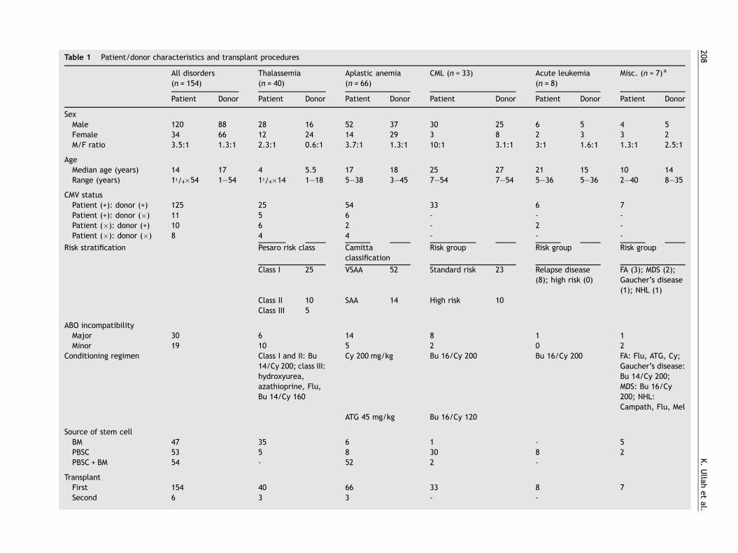
One hundred and twenty patients were male and 34 werefemale. The median age of the patients was 14 years (range11/4� 54 years). Eighty-eight donors were male and 66 werefemale. Sixty-six patients were transplanted across gender.Forty-nine patients had ABO mismatch transplants withmajor ABO mismatch in 30 patients and minor ABO mismatchin 19 patients. In 125 transplants both the patient and thedonor were CMV-positive, in 11 transplants the patient waspositive while the donor was negative, in 10 transplants thepatient was negative and the donor was positive, while ineight transplants both the patient and donor were CMV-negative. Forty-seven patients received bone marrow, 53received PBSC, and 54 received both bone marrow and PBSCharvest. Patient/donor characteristics and transplant proce-dures are shown in Table 1.
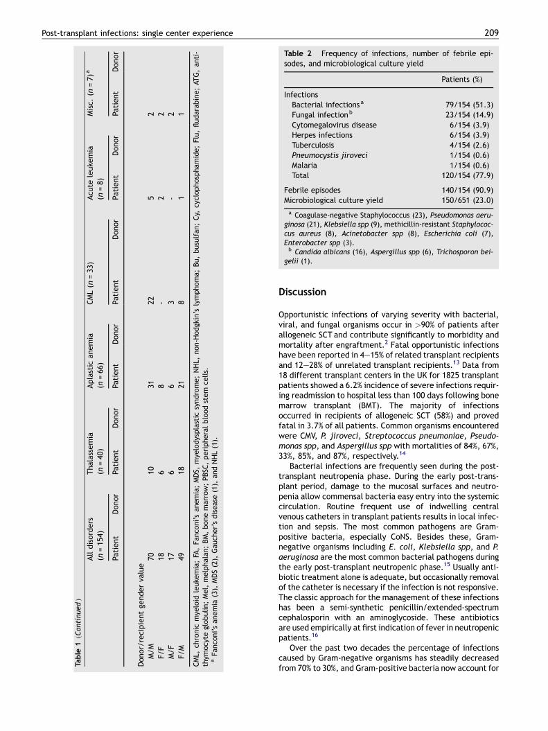
Out of 154 patients who underwent SCT, 140 patients hadfebrile episodes either alone or in association with other signsand symptoms during different post-transplant recoveryphases. Relevant cultures and samples were taken in allfebrile patients to ascertain causative organisms. Out of651 cultures from different sites, 150 culture specimens(23.0%) were positive. Post-transplant infection was con-firmed in 120 patients (77.9%) on the basis of clinical assess-ment and microbiological, virological, and histopathologicalanalysis.
Bacterial infections were seen in 79/154 (51.3%) patients.The majority of bacterial pathogens were Gram-negativeorganisms (60.8%) compared with Gram-positive organisms(39.2%). Infective organisms isolated in our patients in orderof frequency were coagulase-negative Staphylococcus(CoNS) in 23 patients (14.9%), followed by Pseudomonasspp in 21 patients (13.6%), Klebsiella spp in nine patients(5.8%), MRSA in eight patients (5.2%), Acinetobacter spp ineight patients (5.2%), E. coli in seven patients (4.5%), andEnterobacter spp in three (1.9%) patients. Mycobacteriumtuberculosiswas seen in four patients (2.6%). The majority ofbacterial pathogens were isolated from either blood drawnfrom a peripheral vein or fluid collected from the centralvenous line (heparinized saline used to lock the line when notin use).
Fungal infections were seen in 23 patients (14.9%). Amongfungal infections, Candida albicans was isolated from blood,central venous line fluid, and sputum in 16 patients (10.4%).Aspergillus spp was isolated from blood, tissues, sputum,BAL, and pus in six patients (3.9%). Pneumocystis jiroveciwasisolated from BAL in one patient (0.6%).
Plasmodium falciparum was isolated from peripheralblood in one patient (0.6%). CMV infection was documentedin 28 patients (18.2%). CMV disease (two enterocolitis, fourpneumonia) was seen in six patients (3.9%). Vesicular erup-tions in dermatomal pattern suggestive of herpes zosterinfection were clinically seen in six patients (3.9%). Thefrequencies of infections seen in our patients are shown inTable 2.
Infection-related morality was observed in 20 patients(13.0%). The various infective causes of mortality werePseudomonas septicemia, disseminated aspergillosis, CMV
disease, and disseminated tuberculosis. Fatal Pseudomonassepticemia was observed in the early recovery phase whereasfatal fungal and CMV infections were observed in the mid andlate recovery phases. Mortality related to tuberculosis wasobserved in the mid recovery phase. Out of 21 patients whodeveloped Pseudomonas infection, nine died of septicemiaduring the first 30 days post-SCT.
Four patients out of six who developed Aspergillus infec-tion died of disseminated disease involving the lungs, liver,and subcutaneous tissues. Two patients developed Aspergil-lus infection during the mid recovery phase. Of these, onepatient developed bilateral pneumonitis and nodular ulcer-ating lesions in the lower abdomen and right thigh on day +45;the tissue biopsy and culture from the site revealed thegrowth of Aspergillus fumigatus. The other patient devel-oped grade III acute GVHD, which was complicated by pul-monary and hepatic aspergillosis at day +74; tissue biopsy andculture confirmed the growth of Aspergillus fumigatus. Theremaining two patients developed disseminated aspergillosisduring the late recovery phase at day +123 and day +187.Both patients had chronic extensive GVHD, which was com-plicated by jaundice, hepatomegaly, and subcutaneous nod-ular swellings involving the thighs. Tissue biopsy specimensconfirmed the growth of Aspergillus fumigatus. All patientsreceived amphotericin B initially against Aspergillus infec-tion. Two patients were treated with amphotericin B com-bined with itraconazole, while one patient was treated withvoriconazole and the other received caspofungin. None ofthese patients responded to antifungal therapy and they diedon days +59, +72, +135, and +201 post-SCT.
Out of six patients with CMV disease, two developedenterocolitis during the mid recovery phase at days +42and +59. CMV pneumonitis was observed in four patientsduring the late recovery phase at days +98, +116, +127,and +139 post-SCT. All patients were treated with ganciclovir,however none responded to antiviral therapy and they died atdays +54, +67, +110, +123, +133, and +155 post-SCT.
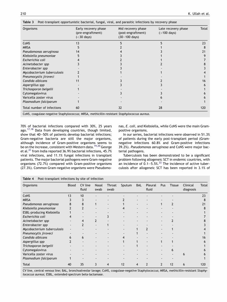
A total of four patients had tuberculous infection at days+21, +30, +92, and +241 post-SCT. Out of these, one patientdeveloped tuberculous lymphadenopathy with superior med-iastinal widening at day +92. Mediastinoscopic tissue biopsyand cultures were positive for M. tuberculosis with caseatinggranulomatas. The patient did not respond to treatment anddied on day +124 post-transplant. The frequency of infectionsin the early, mid, and late recovery phases, and the sites ofinfection are shown in Tables 3 and 4.
Aspergillus infection was observed in aplastic anemia andCML only, whereas Candida infection was more commonamong thalassemic patients. CMV disease was more frequentin thalassemia and CML patients. Apart from these, the otherinfections were evenly distributed in all the hematologicaldisorders as shown in Table 5.
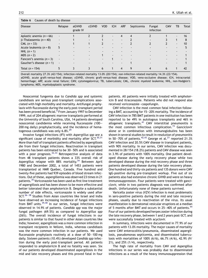
In our study, at the end of 5 years, the overall survival anddisease-free survival were 72.7% and 70.7%, respectively,with a median follow-up of 56.2 � 3.1 months. Disease-freesurvival for the different hematological disorders in ourpatients as compared to those reported by the Center forInternational Blood and Marrow Transplant Research(CIBMTR) and EBMT are shown in Figure 1. Overall mortalitywas observed in 27.3% (42/154). Mortality related to infec-tions was 13.0% (20/154). Post-transplant fatal infections indifferent hematological disorders are shown in Table 6.
208K.Ullah
etal.
Table 1 Patient/donor characteristics and transplant procedures
All disorders(n = 154)
Thalassemia(n = 40)
Aplastic anemia(n = 66)
CML (n = 33) Acute leukemia(n = 8)
Misc. (n = 7)a
Patient Donor Patient Donor Patient Donor Patient Donor Patient Donor Patient Donor
SexMale 120 88 28 16 52 37 30 25 6 5 4 5Female 34 66 12 24 14 29 3 8 2 3 3 2M/F ratio 3.5:1 1.3:1 2.3:1 0.6:1 3.7:1 1.3:1 10:1 3.1:1 3:1 1.6:1 1.3:1 2.5:1
AgeMedian age (years) 14 17 4 5.5 17 18 25 27 21 15 10 14Range (years) 11/4�54 1—54 11/4�14 1—18 5—38 3—45 7—54 7—54 5—36 5—36 2—40 8—35
CMV statusPatient (+): donor (+) 125 25 54 33 6 7Patient (+): donor (�) 11 5 6 - - -Patient (�): donor (+) 10 6 2 - 2 -Patient (�): donor (�) 8 4 4 - - -
Risk stratification Pesaro risk class Camittaclassification
Risk group Risk group Risk group
Class I 25 VSAA 52 Standard risk 23 Relapse disease(8); high risk (0)
FA (3); MDS (2);Gaucher’s disease(1); NHL (1)
Class II 10 SAA 14 High risk 10Class III 5
ABO incompatibilityMajor 30 6 14 8 1 1Minor 19 10 5 2 0 2
Conditioning regimen Class I and II: Bu14/Cy 200; class III:hydroxyurea,azathioprine, Flu,Bu 14/Cy 160
Cy 200 mg/kg Bu 16/Cy 200 Bu 16/Cy 200 FA: Flu, ATG, Cy;Gaucher’s disease:Bu 14/Cy 200;MDS: Bu 16/Cy200; NHL:Campath, Flu, Mel
ATG 45 mg/kg Bu 16/Cy 120
Source of stem cellBM 47 35 6 1 - 5PBSC 53 5 8 30 8 2PBSC + BM 54 - 52 2 -
TransplantFirst 154 40 66 33 8 7Second 6 3 3 - -
Post-transplant infections: single center experience 209
Table 2 Frequency of infections, number of febrile epi-sodes, and microbiological culture yield
Patients (%)
InfectionsBacterial infections a 79/154 (51.3)Fungal infectionb 23/154 (14.9)Cytomegalovirus disease 6/154 (3.9)Herpes infections 6/154 (3.9)Tuberculosis 4/154 (2.6)Pneumocystis jiroveci 1/154 (0.6)Malaria 1/154 (0.6)Total 120/154 (77.9)
Febrile episodes 140/154 (90.9)Microbiological culture yield 150/651 (23.0)
a Coagulase-negative Staphylococcus (23), Pseudomonas aeru-ginosa (21), Klebsiella spp (9), methicillin-resistant Staphylococ-cus aureus (8), Acinetobacter spp (8), Escherichia coli (7),Enterobacter spp (3).b Candida albicans (16), Aspergillus spp (6), Trichosporon bei-
gelii (1).
Table
1(Continued
)
Alldisorders
(n=154
)Thalassemia
(n=40
)Aplastic
anemia
(n=66
)CML(n
=33
)Acu
teleuke
mia
(n=8)
Misc.
(n=7)
a
Patient
Donor
Patient
Donor
Patient
Donor
Patient
Donor
Patient
Donor
Patient
Donor
Donor/recipientge
nderva
lue
M/M
70
1031
225
2F/F
186
8-
22
M/F
17
66
3-
2F/M
4918
218
11
CML,
chronic
mye
loid
leuke
mia;FA
,Fa
nco
ni’san
emia;MDS,
mye
lodysplastic
syndrome;NHL,
non-H
odgkin’s
lymphoma;
Bu,busulfan
;Cy,
cyclophospham
ide;Flu,fludarabine;AT
G,anti-
thym
ocyte
globulin;Mel,melphalan
;BM,bonemarrow;PBSC
,peripheralbloodstem
cells.
aFa
nco
ni’san
emia
(3),
MDS(2),
Gau
cher’sdisease(1),
andNHL(1).
Discussion
Opportunistic infections of varying severity with bacterial,viral, and fungal organisms occur in >90% of patients afterallogeneic SCT and contribute significantly to morbidity andmortality after engraftment.2 Fatal opportunistic infectionshave been reported in 4—15% of related transplant recipientsand 12—28% of unrelated transplant recipients.13 Data from18 different transplant centers in the UK for 1825 transplantpatients showed a 6.2% incidence of severe infections requir-ing readmission to hospital less than 100 days following bonemarrow transplant (BMT). The majority of infectionsoccurred in recipients of allogeneic SCT (58%) and provedfatal in 3.7% of all patients. Common organisms encounteredwere CMV, P. jiroveci, Streptococcus pneumoniae, Pseudo-monas spp, and Aspergillus spp with mortalities of 84%, 67%,33%, 85%, and 87%, respectively.14
Bacterial infections are frequently seen during the post-transplant neutropenia phase. During the early post-trans-plant period, damage to the mucosal surfaces and neutro-penia allow commensal bacteria easy entry into the systemiccirculation. Routine frequent use of indwelling centralvenous catheters in transplant patients results in local infec-tion and sepsis. The most common pathogens are Gram-positive bacteria, especially CoNS. Besides these, Gram-negative organisms including E. coli, Klebsiella spp, and P.aeruginosa are the most common bacterial pathogens duringthe early post-transplant neutropenic phase.15 Usually anti-biotic treatment alone is adequate, but occasionally removalof the catheter is necessary if the infection is not responsive.The classic approach for the management of these infectionshas been a semi-synthetic penicillin/extended-spectrumcephalosporin with an aminoglycoside. These antibioticsare used empirically at first indication of fever in neutropenicpatients.16
Over the past two decades the percentage of infectionscaused by Gram-negative organisms has steadily decreasedfrom 70% to 30%, and Gram-positive bacteria now account for
210 K. Ullah et al.
Table 3 Post-transplant opportunistic bacterial, fungal, viral, and parasitic infections by recovery phase
Organisms Early recovery phase(pre-engraftment)(<30 days)
Mid recovery phase(post-engraftment)(30—100 days)
Late recovery phase(>100 days)
Total
CoNS 13 5 5 23MRSA 5 2 1 8Pseudomonas aeruginosa 14 4 3 21Klebsiella pneumoniae 5 3 1 9Escherichia coli 4 2 1 7Acinetobacter spp 3 3 2 8Enterobacter spp - 3 - 3Mycobacterium tuberculosis 2 1 1 4Pneumocystis jiroveci 1 - - 1Candida albicans 11 3 2 16Aspergillus spp - 3 3 6Trichosporon beigelii 1 - - 1Cytomegalovirus - 3 3 6Varicella zoster virus - - 6 6Plasmodium falciparum 1 - - 1
Total number of infections 60 32 28 120
CoNS, coagulase-negative Staphylococcus; MRSA, methicillin-resistant Staphylococcus aureus.
70% of bacterial infections compared with 30%, 25 yearsago.17,18 Data from developing countries, though limited,show that 40—50% of patients develop bacterial infections;Gram-negative bacteria are still the major organisms,although incidence of Gram-positive organisms seems tobe on the increase, consistent withWestern data.19,20 Georgeet al.21 from India reported 36.9% bacterial infections, 45.7%viral infections, and 11.1% fungal infections in transplantpatients. Themajor bacterial pathogens were Gram-negativeorganisms (72.7%) compared with Gram-positive organisms(27.3%). Common Gram-negative organisms were Pseudomo-
Table 4 Post-transplant infections by site of infection
Organisms Blood CV linefluid
Nasalswab
Throatswab
CoNS 13 10 - -MRSA 3 3 - -Pseudomonas aeruginosa 8 8 1 -Klebsiella pneumoniae 2 2 - -ESBL-producing Klebsiella 1 - - -Escherichia coli 4 - - 3Acinetobacter spp - 4 2 -Enterobacter spp - 2 - 1Mycobacterium tuberculosis - - - -Pneumocytis jiroveci - - - -Candida albicans 6 6 - -Aspergillus spp 2 - - -Trichosporon beigelii - - - -Cytomegalovirus - - - -Varicella zoster virus - - - -Plasmodium falciparum 1 - - -
Total 40 35 3 4
CV line, central venous line; BAL, bronchoalveolar lavage; CoNS, coagulococcus aureus; ESBL, extended-spectrum beta-lactamase.
nas, E. coli, and Klebsiella, while CoNS were the main Gram-positive organisms.
In our series, bacterial infections were observed in 51.3%of patients during the early post-transplant period (Gram-negative infections 60.8% and Gram-positive infections39.2%). Pseudomonas aeruginosa and CoNS were major bac-terial pathogens.
Tuberculosis has been demonstrated to be a significantproblem following allogeneic SCT in endemic countries, withan incidence of 0.1—5.5%.22 The incidence of active tuber-culosis after allogeneic SCT has been reported in 3.1% of
Sputum BAL Pleuralfluid
Pus Tissue Clinicaldiagnosis
Total
- - - - - 232 - - - - 81 - - 1 2 214 - - - - 8- - - - - 1- - - - - 7- - - - 2 8- - - - - 3- 1 2 - 1 4- 1 - - - 14 - - - - 161 1 - 1 1 6- 1 - - - 1- - - - 6 6- - - - - 6 6- - - - - 1
12 4 2 2 12 6 120
lase-negative Staphylococcus; MRSA, methicillin-resistant Staphy-

Post-transplant infections: single center experience 211
Table 5 Post-transplant opportunistic infections by disease
Organisms Aplastic anemia b-Thalassemia CML Acute leukemia Misc. Total
CoNS 11 5 4 2 1 23MRSA 2 3 2 1 0 8Pseudomonas aeruginosa 10 2 4 3 2 21Klebsiella pneumoniae 4 2 2 0 1 9Escherichia coli 1 2 2 1 1 7Acinetobacter johnsonii 3 3 1 1 0 8Enterobacter spp 2 1 0 0 0 3Mycobacterium tuberculosis 1 1 2 0 0 4Pneumocystis jiroveci 0 0 0 1 0 1Candida albicans 1 9 3 1 2 16Aspergillus spp 3 0 3 0 0 6Trichosporon beigelii 0 0 0 1 0 1Cytomegalovirus 1 3 2 0 0 6Varicella zoster virus 2 3 1 0 0 6Plasmodium falciparum 1 0 0 0 0 1
Total 42 34 26 11 7 120
CML, chronic myeloid leukemia; CoNS, coagulase-negative Staphylococcus; MRSA, methicillin-resistant Staphylococcus aureus.
patients in Korea.23 Similarly in Turkey, where tuberculosis isendemic, a 30—40 times higher incidence has been reportedin BMT patients compared to the general Turkish popula-tion.24 A study from Taiwan shows a trend towards increasedrisk of having pulmonary tuberculosis in allogeneic SCT ascompared to autologous SCT (4.8 � 1.8% vs. 0).25 In 2001,George et al.26 and Chandy et al.27 from Vellore, Indiareported 1.38% and 2.2% incidence of tuberculosis in trans-plant recipients, respectively. However, updated data fromthe same center in 2006 show 1.7% tuberculosis in transplantpatients.21 In our series, four (2.6%) patients developed
Figure 1 HLA-identical sibling hematopoietic stem cell transplatransplant (DFS) data, 1998—2004; Marcelo et al., Report on state of2006;12:5—10. **EBMT Working Party on Pediatric Diseases, Pesaro grcure of thalassemia by bone marrow transplantation. Bone Marrow TrTransplant Centre Pakistan, post-transplant (DFS) data, 2001—2006.
tuberculosis. Of these, two patients developed the diseasein the early post-transplant period whereas the other twodeveloped tuberculosis in the mid and late recovery phases.These infections were most probably due to reactivation ofprevious tuberculosis infection.
PCP accounts for fewer than 10% of the cases of inter-stitial pneumonia in patients with allogeneic SCT.28 One ofour patients developed PCP pneumonia during the firstpost-transplant month. Because of early diagnosis andinitiation of treatment this patient made a completerecovery.
ntation disease-free survival (DFS) by disease. *CIBMTR post-the art in blood and marrow transplantation. CIBMTR Newsletteroup post-transplant (DFS) data, 1982—2001; Lucarelli et al., Theansplant 2001;28(Suppl 1):S11—3. ***Armed Forces Bone Marrow
212 K. Ullah et al.
Table 6 Causes of death by disease
Disease Relapse aGVHDgrade IV
cGVHD VOD ICH ARF Septicemia Fungalinfection
CMV TB Total
Aplastic anemia (n = 66) - 1 2 2 - 4 4 2 1 - 16b-Thalassemia (n = 40) - 1 - 1 - - 2 - 3 1 8CML (n = 33) - 1 1 2 - - 2 2 2 - 10Acute leukemia (n = 8) 5 - - - - - - - - - 5NHL (n = 1) - - - - - - - - - - -MDS (n = 2) - - - 1 - - - - - - 1Fanconi’s anemia (n = 3) - - - - 1 - - - - - 1Gaucher’s disease (n = 1) - - - - - - 1 - - - 1
Total (n = 154) 5 3 3 6 1 4 9 4 6 1 42
Overall mortality 27.3% (42/154); infection-related mortality 13.0% (20/154); non-infection-related mortality 14.3% (22/154).aGVHD, acute graft-versus-host disease; cGVHD, chronic graft-versus-host disease; VOD, veno-occlusive disease; ICH, intracranialhemorrhage; ARF, acute renal failure; CMV, cytomegalovirus; TB, tuberculosis; CML, chronic myeloid leukemia; NHL, non-Hodgkin’slymphoma; MDS, myelodysplastic syndrome.
Nosocomial fungemia due to Candida spp and systemiccandidiasis are serious post-transplant complications asso-ciated with high morbidity and mortality. Antifungal prophy-laxis with fluconazole during the early post-transplant periodhas been proved beneficial.6 From January 1997 to December1999, out of 204 allogeneic marrow transplants performed atthe University of South Carolina, USA, 14 patients developednosocomial candidemia while receiving fluconazole (100—200 mg daily) prophylactically, and the incidence of hema-togenous candidiasis was only 6.8%.29
Invasive fungal infections (IFI) with Aspergillus spp are asignificant cause of morbidity and mortality after SCT.30,31
More than half of transplant patients affected by aspergillosisdie from their fungal infections. Reactivation in transplantpatients has been estimated to be 30—50% and is associatedwith a poor prognosis.32,33 Retrospective analysis of datafrom 48 transplant patients shows a 33% overall risk ofAspergillus relapse with 88% mortality.34 Between April1980 and December 2002, a total of 1453 patients weretransplanted at HMR Quebec, Canada. Five hundred andtwenty-five patients had 939 episodes of blood stream infec-tions. Out of these, aspergillemia was observed 23 times in 21patients.35 Voriconazole has been used as first line treatmentof aspergillosis and has been shown to be more effective andbetter tolerated than amphotericin B. Despite a substantialnumber of side effects, voriconazole is widely used afterSCT.36—38 Studies from Asian countries like Israel and Indiahave observed an increasing incidence of fungal infectionsfrom BMT units.39,40 In our series, fungal infections wereobserved in 14.9% of patients. Candida spp was the majorfungal pathogen (69.5%) as compared to Aspergillus spp(26%). The overall incidence of fungal infections in ourpatients is similar to that found in other Asian countries likeIndia; however, aspergillosis was a more common infection intransplant recipients in Vellore, India, whereas candidiasiswas the more common infection in our patients. We usedfluconazole prophylaxis routinely at a dose of 50—100 mgdaily. Sixteen patients (10.4%) developed Candida spp infec-tion during the early post-transplant period. All patientsresponded to amphotericin B and no fatality was seen. Sixof our patients developed disseminated aspergillosis in themid and late recovery phases and this proved fatal in four
patients. All patients were initially treated with amphoter-icin B and itraconazole. Patients who did not respond alsoreceived voriconazole—caspofungin.
CMV infection is the most common fatal infection follow-ing a BMT, accounting for 15—20% mortality. The incidence ofCMV infection in 785 BMT patients in one institution has beenreported to be 49% in autologous transplants and 46% inallogeneic transplants.41 CMV interstitial pneumonitis isthe most common infectious complication.15 Gancicloviralone or in combination with immunoglobulins has beenshown in several studies to result in resolution of pneumonitisin 50—70% of patients.42,43 George et al.21 reported 21.3%CMV infection and 20.5% CMV disease in transplant patients,with 90% mortality. In our series, CMV infection was docu-mented in 28/154 (18.2%) patients and CMV disease was seenin 3.9% of patients with 100% mortality. One patient devel-oped disease during the early recovery phase while twodeveloped disease during the mid recovery phase and threepatients developed disease during the late recovery phase.One hundred and thirty-six patients and 135 donors were CMVIgG-positive during pre-transplant workup. Five out of sixpatients also had extensive chronic GVHD and were on heavyimmunosuppression. Four patients were treated with ganci-clovir, while in two patients diagnosis was confirmed afterdeath. Unfortunately none of these patients survived.
Varicella zoster virus (VZV) infection is most problematicin sero-positive patients during the mid and late recoveryphases, usually due to reactivation of the virus. Its usualmanifestation is dermatomal vesicular eruptions at a medianof 5 months after BMT and occurs in 25—40% of patients.44
Four of our patients developed herpes zoster infection duringthe late recovery phase, between 1 and 2 years post-SCT, andwere successfully treated with acyclovir.
In summary, infections were documented in 77.9% of ourpatients with 13.0% mortality. The major causes of mortalitywere CMV enterocolitis/pneumonia, disseminated aspergil-losis, pseudomonas septicemia, and disseminated tubercu-losis with mortalities of 100% (6/6), 66.7% (4/6), 42.9% (9/21), and 25% (1/4), respectively.
The high rate of mortality from CMV and Aspergillusinfections in our series was probably due to disseminatedinfections as a result of the heavy immunosuppression that
Post-transplant infections: single center experience 213
these patients were receiving due to GVHD. None of ourpatients developed protozoal or helminthic infections apartfrom a single case of malaria. This could be due to oureffective prophylaxis.
Pakistan is a developing country in Southeast Asia wheretuberculosis and malaria are prevalent and the majority ofthe population is sero-positive for CMV. We were apprehen-sive about an increased incidence of tuberculosis, malaria,and CMV disease in our patients when we started the bonemarrow transplant program in our country. We screened allpatients and donors for these diseases.
One of our major concerns is the limited availability ofnewer, less toxic antifungal and antiviral drugs. Anothermajor concern is poor patient compliance with treatmentnot uncommon in developing countries such as Pakistan.There is a need to improve social services in our countryas well as to educate the patients and their families tocomply with treatment during the post-transplant period.
Acknowledgments
We thank Dr Amin Waqar, Commandant Armed Forces insti-tute of Pathology, Rawalpindi, Pakistan for helpful contribu-tions, technical assistance and discussions. We also thank DrMuhammad Ayyub, Commandant Armed Forces Institute ofTransfusion, Rawalpindi, Pakistan for generously providing usblood products. We are extremely grateful to Mr Abdul Wajidof this center for helping us in statistical analysis and manu-script preparation.
Conflict of interest: No conflict of interest to declare.
References
1. Loberiza Jr F. Reports on state of the art in blood and marrowtransplantation–—part I of the IBMTR/ABMTR summary slides withguide. IBMTR/ABMTR Newsletter 2003;10:7—10. Available at:http://www.cibmtr.org/PUBLICATIONS/NEWSLETTER/2003Nov.pdf (accessed August 2007).
2. Hayes-Lattin B, Leis JF, Maziarz RT. Isolation in the allogeneictransplant environment: how protective is it? Bone MarrowTransplant 2005;36:373—81.
3. Einsele H, Bertz H, Beyer J, Kiehl MG, Runde V, Kolb HJ, et al.Infectious complications after allogeneic stem cell transplanta-tion: epidemiology and interventional therapy strategies. AnnHematol 2003;82:S175—85.
4. McCann S, Byrne JL, Rovira M, Shaw P, Ribaud P, Sica S, et al.Outbreaks of infectious diseases in stem cell transplant units: asilent cause of death for patients and transplant programmes.Bone Marrow Transplant 2004;33:519—29.
5. Pittet D, Li N, Woolson RF, Wenzel RP. Microbiological factorsinfluencing the outcome of nosocomial bloodstream infections: a6-year validated, population-based model. Clin Infect Dis1997;24:1068—78.
6. Marr KA, Seidel K, Slavin MA, Bowden RA, Schoch HG, Flowers ME,et al. Prolonged fluconazole prophylaxis is associated with per-sistent protection against candidiasis-related death in allogeneicmarrow transplant recipients: long term follow-up of a rando-mized, placebo-controlled trial. Blood 2000;96:2055—61.
7. Sepkowitz KA. Antibiotic prophylaxis in patients receiving hema-topoietic stem cell transplant. Bone Marrow Transplant 2002;29:367—71.
8. Engels EA, Lau J, Barza M. Efficacy of quinolone prophylaxis inneutropenic cancer patients: a meta-analysis. J Clin Oncol1998;16:1179—87.
9. Ljungman P, Reusser P, de la Camara R, Einsele H, Engelhard D,Ribaud P, et al. Management of CMV infections: recommenda-tions from the infectious diseases working party of the EBMT.Bone Marrow Transplant 2004;33:1075—81.
10. Ljungman P, Griffiths P, Paya C. Definitions of cytomegalovirusinfection and disease in transplant recipients. Clin Infect Dis2002;34:1094—7.
11. Ljungman P, De La Camara R, Milpied N, Volin L, Russell CA, CrispA, et al. A randomized study of valaciclovir as prophylaxis againstCMV reactivation in allogeneic bone marrow transplant recipi-ents. Blood 2002;73:930—6.
12. Glucksberg H, Storb R, Fefer A, Buckner CD, Neiman PE, Clift RA,et al. Clinical manifestations of graft-versus-host disease inhuman recipients of marrow from HLA-matched sibling donors.Transplantation 1974;18:295—304.
13. Fujimaki K, Maruta A, Yoshida M, Kodama F, Matsuzaki M, Fuji-sawa S, et al. Immune reconstitution assessed during five yearsafter allogeneic bone marrow transplantation. Bone MarrowTransplant 2001;27:1275—81.
14. Hoyle C, Goldman JM. Life-threatening infections occurringmore than 3 months after BMT. 18 UK Bone Marrow TransplantTeams. Bone Marrow Transplant 1994;14:247—52.
15. Wingard JR. Advances in the management of infectious compli-cations after bone marrow transplantation. Bone Marrow Trans-plant 1990;6:371—83.
16. Wingard JR. Opportunistic infections after blood and bone mar-row transplantation. Clin Infect Dis 1999;1:3—20.
17. Collin BA, Leather HL, Wingard JR, Ramphal R. Evolution, inci-dence and susceptibility of bacterial bloodstream isolates from519 bone marrow transplant patients. Clin Infect Dis 2001;33:947—53.
18. Wang CC, Mattson D, Wald A. Corynebacterium jeikeium bacter-emia in bone marrow transplant patients with Hickman cathe-ters. Bone Marrow Transplant 2001;27:445—9.
19. Ghosh K, Shenoy AK, Al-Mahrooqi Z. Bacteriological infectionsduring the first hundred days of allogeneic bone marrow trans-plantation–—experience from Oman. J Assoc Physicians India2002;50:910—2.
20. Arns da Cunha C, Weisdorf D, Shu XO, DeFor T, Pastor 3rd JD,Johnson JR. Early Gram-positive bacteremia in BMT recipients:impact of three different approaches to antimicrobial prophy-laxis. Bone Marrow Transplant 1998;21:173—80.
21. George B, Mathews V, Viswabandya A, Srivastava A, Chandy M.Infections in children undergoing allogeneic bone marrow trans-plantation in India. Pediatr Transplant 2006;10:48—54.
22. Kindler T, Schindel C, Brass U, Fischer T. Fatal sepsis due toMycobacterium tuberculosis after allogeneic bone marrowtransplantation. Bone Marrow Transplant 2001;27:217—8.
23. Lee J, Lee MH, Kim WS, Kim K, Park SH, Lee SH, et al. Tubercu-losis in hematopoietic stem cell transplant recipients in Korea.Int J Hematol 2004;79:185—8.
24. Arslan O, Gurman G, Dilek I, Ozcan M, Koc H, Ilhan O, et al.Incidence of tuberculosis after bone marrow transplantation in asingle center fromTurkey.Haematologia (Budap)1998;29:59—62.
25. Ku SC, Tang JL, Hsueh PR, Luh KT, Yu CJ, Yang PC. Pulmonarytuberculosis in allogeneic hematopoietic stem cell transplanta-tion. Bone Marrow Transplant 2001;27:1293—7.
26. George B, Mathews V, Srivastava V, Srivastava A, Chandy M.Tuberculosis among allogeneic bone marrow transplant recipi-ents in India. Bone Marrow Transplant 2001;27:973—5.
27. Chandy M, Srivastava A, Dennison D, Mathews V, George B.Allogeneic bone marrow transplantation in the developingworld: experience from a center in India. Bone Marrow Trans-plant 2001;27:785—90.
28. De Castro N, Neuville S, Sarfati C, Ribaud P, Derouin F, GluckmanE, et al. Occurrence of Pneumocystis jiroveci pneumonia afterallogeneic stem cell transplantation: a 6-year retrospectivestudy. Bone Marrow Transplant 2005;36:879—83.
214 K. Ullah et al.
29. Safdar A, Van Rhee F, Henslee-Downey JP, Singhal S, Mehta J.Candida glabrata and Candida krusei fungemia after high-riskallogeneic marrow transplantation: no adverse effect of low-dose fluconazole prophylaxis on incidence and outcome. BoneMarrow Transplant 2001;28:873—8.
30. Mehta P, Augustson B, Krishnamurthy S, Jacob A, Roy D, Olliff J,et al. Successful allogeneic haematopoietic stem cell transplan-tation in patients with poor-risk leukaemia and prior invasivefungal infection. Bone Marrow Transplant 2004;34:825—6.
31. Simoneau E, Kelly M, Labbe AC, Roy J, Laverdiere M. What is theclinical significance of positive blood cultures with Aspergillusspp in hematopoietic stem cell transplant recipients? A 23 yearexperience. Bone Marrow Transplant 2005;35:303—6.
32. Girmenia C, Nucci M, Martino P. Clinical significance of Asper-gillus fungaemia in patients with haematological malignanciesand invasive aspergillosis. Br J Haematol 2001;114:93—8.
33. Lin SJ, Schranz J, Teutsch SM. Aspergillosis case—fatality rate:systematic review of the literature. Clin Infect Dis 2001;32:358—64.
34. Offner F, Cordonnier C, Ljungman P, Prentice HG, Engelhard D,De Bacquer D, et al. Impact of previous aspergillosis on theoutcome of bone marrow transplantation. Clin Infect Dis1998;26:1098—103.
35. Kontoyiannis DP, Sumoza D, Tarrand J, Bodey GP, Storey R, RaadII. Significance of aspergillemia in patients with cancer: a 10-year study. Clin Infect Dis 2000;31:188—9.
36. Herbrecht R, Denning DW, Patterson TF, Bennett JE, Greene RE,Oestmann JW, et al. Voriconazole versus amphotericin B forprimary therapy of invasive aspergillosis. N Engl J Med2002;347:408—15.
37. Patterson TF, Kirkpatrick WR, White M, Hiemenz JW, Wingard JR,Dupont B, et al. Invasive aspergillosis. Disease spectrum, treat-
ment practices, and outcomes. I3 Aspergillus Study Group.Medicine (Baltimore) 2000;79:250—60.
38. Cordonnier C, Maury S, Pautas C, Bastie JN, Chehata S, CastaigneS, et al. Secondary antifungal prophylaxis with voriconazole toadhere to scheduled treatment in leukemic patients and stemcell transplant recipients. Bone Marrow Transplant 2004;33:943—8.
39. Weinberger M, Sacks T, Sulkes J, Shapiro M, Polacheck I. Increas-ing fungal isolation from clinical specimens: experience in auniversity hospital over a decade. J Hosp Infect 1997;35:185—95.
40. George B, Mathew V, Srivastava A, Chandy M. Infections amongallogeneic bone marrow transplant recipients in India. BoneMarrow Transplant 2004;33:311—5.
41. Wingard JR, Piantadosi S, Burns WH, Zahurak ML, Santos GW,Saral R. Cytomegalovirus infections in bone marrow transplantrecipients given intensive cytoreductive therapy. Rev Infect Dis1990;12(Suppl 7):S793—804.
42. Reusser P, Einsele H, Lee J, Volin L, Rovira M, Engelhard D, et al.Randomized multicenter trial of foscarnet versus ganciclovir forpreemptive therapy of cytomegalovirus infection after allo-geneic stem cell transplantation. Blood 2002;99:1159—64.
43. Ljungman P, Deliliers GL, Platzbecker U, Matthes-Martin S,Bacigalupo A, Einsele H, et al. Cidofovir for cytomegalovirusinfection and disease in allogeneic stem cell transplant recipi-ents. The infectious Diseases Working Party of the EuropeanGroup for Blood and Marrow Transplantation. Blood 2001;97:388—92.
44. Ljungman P, Engelhard D, de la Camara R, Einsele H, LocasciulliA, Martino R, et al. Vaccination of stem cell transplant recipi-ents: recommendations of the Infectious Diseases Working Partyof the EBMT. Bone Marrow Transplant 2005;35:737—46.