Positive and negative mutant selection in the human histone hairpin-binding protein using the yeast...
10
1594–1603 Nucleic Acids Research, 2000, Vol. 28, No. 7 © 2000 Oxford University Press Positive and negative mutant selection in the human histone hairpin-binding protein using the yeast three-hybrid system Franck Martin 1,2 , Fabrice Michel 1 , Daniel Zenklusen 1 , Berndt Müller 1 and Daniel Schümperli 1, * 1 Abteilung für Entwicklungsbiologie, Zoologisches Institut der Universität Bern, Baltzerstrasse 4, 3012 Bern, Switzerland and 2 Institut de Biologie Moléculaire et Cellulaire du CNRS, UPR 9002, 15 rue René Descartes, F-67084 Strasbourg Cedex, France Received December 6, 1999; Revised and Accepted February 7, 2000 ABSTRACT We have used the yeast three-hybrid system in a positive selection for mutants of the human histone hairpin-binding protein (HBP) capable of interacting with non-canonical hairpins and in a negative selection for loss-of-binding mutants. Interestingly, all mutations from the positive selection are located in the N- and C-terminal regions flanking a minimal RNA-binding domain (RBD) previously defined between amino acids 126 and 198. Further, in vitro binding studies demonstrate that the RBD, which shows no obvious similarity to other RNA-binding motifs, has a relaxed sequence specificity compared to full-length HBP, allowing it to bind to mutant hairpin RNAs not normally found in histone genes. These findings indicate that the sequences flanking the RBD are important for restricting binding to the highly conserved histone hairpin structure. Among the loss-of-binding mutations, about half are nonsense mutations distributed throughout the N-terminal part and the RBD whereas the other half are missense mutations restricted to the RBD. Whereas the nonsense mutations permit a more precise definition of the C-terminal border of the RBD, the missense mutations identify critical residues for RNA binding within the RBD. INTRODUCTION RNA–protein interactions are involved in the control of gene expression at several levels, including pre-mRNA splicing and 3′ processing, nucleo-cytoplasmic transport, mRNA localisation, translation and stability. They thereby affect important aspects of cell physiology, development, disease or pathogen infec- tion. For certain types of RNA-binding proteins, their inter- actions with their RNA targets have been characterised in detail, using reverse genetic, biochemical and structural methods (for recent reviews see 1,2). However, there is still a demand for new methods to characterise RNA-binding proteins. In particular, positive and negative genetic selections allowing the isolation of multiple gain-of-function or loss-of- function mutants for RNA binding are highly desirable. A potential system amenable to such selection techniques is the yeast three-hybrid system that was independently developed with very similar characteristics by Putz et al. (3) and Sengupta et al. (4). In this system, the expression of selectable marker genes is dependent upon the interaction between an RNA-binding protein and its RNA target. The system has been used successfully to isolate cDNAs for proteins binding to the hairpin structure at the 3′-end of animal replication-dependent histone mRNAs (5,6), to the 3′-untrans- lated regions of the dendritically localised transcript Arg3.1 (3), of the fem-3 mRNA that controls sexual cell fate in Caenor- habditis elegans (7), and of nanos mRNA in Drosophila (8). Moreover, a recent report described the selection, from fragments of yeast genomic DNA, of sequences that, when expressed in the three-hybrid system, were able to interact with a given RNA-binding protein (9). Here we have successfully used the three-hybrid system to select and characterise both gain- and loss-of-function mutations for the histone hairpin-binding protein (HBP) in combination with wild-type and mutant RNA binding substrates. The replication-dependent animal histone mRNAs lack poly(A) tails but end in an evolutionarily conserved hairpin structure. This hairpin is part of a signal for endonucleolytic 3′-end processing of histone pre-mRNAs (reviewed in 10–12). Moreover, the hairpin is involved in nucleo-cytoplasmic trans- port of the mature histone mRNAs (13,14), required for their efficient translation (15,16) and for regulation of their stability (17,18). A protein interacting with the hairpin was first identified as a factor important for efficient histone RNA 3′ processing (19,20) and termed hairpin-binding factor. Other factors involved in histone RNA 3′-end processing are the U7 snRNP and a heat-labile processing factor (reviewed in 10,12). After cloning of its cDNA, the hairpin-binding factor was renamed hairpin-binding protein or HBP (6). A protein with identical properties was found to be associated with mature histone mRNAs on polysomes and termed stem–loop-binding protein or SLBP (21–23). Thus HBP/SLBP appears to be a *To whom correspondence should be addressed. Tel: +41 31 631 4675; Fax: +41 31 631 4616; Email: [email protected] Present addresses: Daniel Zenklusen, Institut de Microbiologie, Université de Lausanne, Rue du Bugnon 44, 1011 Lausanne, Switzerland Berndt Müller, Department of Molecular and Cell Biology, Institute of Medical Sciences, University of Aberdeen, Aberdeen AB25 2ZD, UK
Transcript of Positive and negative mutant selection in the human histone hairpin-binding protein using the yeast...

1594–1603 Nucleic Acids Research, 2000, Vol. 28, No. 7 © 2000 Oxford University Press
Positive and negative mutant selection in the humanhistone hairpin-binding protein using the yeastthree-hybrid systemFranck Martin1,2, Fabrice Michel1, Daniel Zenklusen1, Berndt Müller1 and Daniel Schümperli1,*
1Abteilung für Entwicklungsbiologie, Zoologisches Institut der Universität Bern, Baltzerstrasse 4, 3012 Bern,Switzerland and 2Institut de Biologie Moléculaire et Cellulaire du CNRS, UPR 9002, 15 rue René Descartes,F-67084 Strasbourg Cedex, France
Received December 6, 1999; Revised and Accepted February 7, 2000
ABSTRACT
We have used the yeast three-hybrid system in apositive selection for mutants of the human histonehairpin-binding protein (HBP) capable of interactingwith non-canonical hairpins and in a negative selectionfor loss-of-binding mutants. Interestingly, all mutationsfrom the positive selection are located in the N- andC-terminal regions flanking a minimal RNA-bindingdomain (RBD) previously defined between aminoacids 126 and 198. Further, in vitro binding studiesdemonstrate that the RBD, which shows no obvioussimilarity to other RNA-binding motifs, has a relaxedsequence specificity compared to full-length HBP,allowing it to bind to mutant hairpin RNAs notnormally found in histone genes. These findings indicatethat the sequences flanking the RBD are importantfor restricting binding to the highly conservedhistone hairpin structure. Among the loss-of-bindingmutations, about half are nonsense mutationsdistributed throughout the N-terminal part and theRBD whereas the other half are missense mutationsrestricted to the RBD. Whereas the nonsense mutationspermit a more precise definition of the C-terminalborder of the RBD, the missense mutations identifycritical residues for RNA binding within the RBD.
INTRODUCTION
RNA–protein interactions are involved in the control of geneexpression at several levels, including pre-mRNA splicing and3′ processing, nucleo-cytoplasmic transport, mRNA localisation,translation and stability. They thereby affect important aspectsof cell physiology, development, disease or pathogen infec-tion. For certain types of RNA-binding proteins, their inter-actions with their RNA targets have been characterised indetail, using reverse genetic, biochemical and structuralmethods (for recent reviews see 1,2). However, there is still ademand for new methods to characterise RNA-binding
proteins. In particular, positive and negative genetic selectionsallowing the isolation of multiple gain-of-function or loss-of-function mutants for RNA binding are highly desirable.
A potential system amenable to such selection techniques isthe yeast three-hybrid system that was independentlydeveloped with very similar characteristics by Putz et al. (3)and Sengupta et al. (4). In this system, the expression ofselectable marker genes is dependent upon the interactionbetween an RNA-binding protein and its RNA target. Thesystem has been used successfully to isolate cDNAs forproteins binding to the hairpin structure at the 3′-end of animalreplication-dependent histone mRNAs (5,6), to the 3′-untrans-lated regions of the dendritically localised transcript Arg3.1 (3),of the fem-3 mRNA that controls sexual cell fate in Caenor-habditis elegans (7), and of nanos mRNA in Drosophila (8).Moreover, a recent report described the selection, from fragmentsof yeast genomic DNA, of sequences that, when expressed inthe three-hybrid system, were able to interact with a givenRNA-binding protein (9). Here we have successfully used thethree-hybrid system to select and characterise both gain- andloss-of-function mutations for the histone hairpin-bindingprotein (HBP) in combination with wild-type and mutant RNAbinding substrates.
The replication-dependent animal histone mRNAs lackpoly(A) tails but end in an evolutionarily conserved hairpinstructure. This hairpin is part of a signal for endonucleolytic3′-end processing of histone pre-mRNAs (reviewed in 10–12).Moreover, the hairpin is involved in nucleo-cytoplasmic trans-port of the mature histone mRNAs (13,14), required for theirefficient translation (15,16) and for regulation of their stability(17,18). A protein interacting with the hairpin was firstidentified as a factor important for efficient histone RNA 3′processing (19,20) and termed hairpin-binding factor. Otherfactors involved in histone RNA 3′-end processing are the U7snRNP and a heat-labile processing factor (reviewed in 10,12).After cloning of its cDNA, the hairpin-binding factor wasrenamed hairpin-binding protein or HBP (6). A protein withidentical properties was found to be associated with maturehistone mRNAs on polysomes and termed stem–loop-bindingprotein or SLBP (21–23). Thus HBP/SLBP appears to be a
*To whom correspondence should be addressed. Tel: +41 31 631 4675; Fax: +41 31 631 4616; Email: [email protected] addresses:Daniel Zenklusen, Institut de Microbiologie, Université de Lausanne, Rue du Bugnon 44, 1011 Lausanne, SwitzerlandBerndt Müller, Department of Molecular and Cell Biology, Institute of Medical Sciences, University of Aberdeen, Aberdeen AB25 2ZD, UK
Nucleic Acids Research, 2000, Vol. 28, No. 7 1595
multifunctional protein controlling most or all importantaspects of histone mRNA metabolism. It contains a centraldomain of ~73 amino acids that is conserved between thehuman, mouse, Xenopus laevis and C.elegans proteins(5,6,12,24). This region was defined by deletion analysis of thehuman protein to represent a minimal RNA-binding domain(RBD) (5). Neither the protein as a whole nor the RBD aresimilar to any known RNA-binding proteins, so that HBP/SLBPappears to represent a new type of RNA–protein interaction.
We have used the yeast three-hybrid system to select formutations in human HBP that allow it to bind to an RNAcontaining a non-canonical hairpin as well as for mutationsthat abolish binding to the natural hairpin. All gain-of-functionmutations lie in the N- and C-terminal regions flanking theRBD, suggesting that these regions contribute to the fullsubstrate specificity. Consistent with this, the human RBDshows a relaxed substrate specificity both when tested in theyeast three-hybrid system and in direct RNA-binding assays.In contrast, the loss-of-function mutations either belong to anested set of C-terminal truncations that help to define theC-terminal border of the RBD or contain point substitutions inthe RBD that reveal critical residues for RNA binding.
MATERIALS AND METHODS
Nucleic acids
The hairpin RNAs wtHP (5′-GGACAAAAGGCCCUUUUC-AGGGCCACCC), cgHP (5′-GGACAAAACCCCCUUUUC-AGGGGGACCC; changes underlined), U11G (5′-GGACA-AAAGGCCCUGUUCAGGGCCACCC), U13A (5′-GGACAA-AAGGCCCUUUACAGGGCCACCC), G5U/U13A (5′-GGAC-AAAAUGCCCUUUACAGGGCCACCC) and mutHP (5′-GG-ACAAAACCGGAAAGCCUUCCGGACCC) were transcribedfrom oligonucleotides by T7 RNA polymerase (25), including[α-32P]UTP in the transcription reaction, purified by dena-turing polyacrylamide electrophoresis and stored in H2O.
Strains
Plasmids were amplified in Escherichia coli strain XL1-blue.The randomly mutated library for the second positive selectionwas obtained by amplification in the mutagenic E.coli strainXL1 red [endA, gyrA96, thi-1, hsdR17, supE44, relA1, lac,mutD5, mutS, mutT, Tn10(Tetr)] (Stratagene). For proteinexpression, strain BL21 [F–, ompT, rB
–, mB–] (DE3) plysS
(Pharmacia) was used. The three-hybrid system procedures werecarried out in the Saccharomyces cerevisiae strain L40-coat (4).
Plasmids
Plasmid pET-15B was used for protein expression in E.coli.The shuttle vector pIII/MS2.2 was used for expression of thehybrid RNA molecules in yeast (4). pIII/wt/MS2 designates aplasmid encoding a hybrid RNA containing, 5′→3′, the RNaseP leader sequence, the wild-type histone RNA hairpin and theMS2 binding sites (6). Five additional plasmids expressingmutant hairpin sequences were made by insertion of double-stranded oligonucleotides into the SmaI site of the pIII/MS2.2plasmid. Oligonucleotides used were: 5′-GGAGCTCAA-CAAAAGGCCCTGTTCAGGGCCACCC (pIII/U11G/MS2);5′-GGAGCTCAACAAAAGGCCCTTTACAGGGCCACCC(pIII/U13A/MS2); 5′-GGAGCTCAACAAAATGCCCTTTA-
CAGGGCCACCC (pIII/G5U/U13A/MS2); 5′-GGAGCT-CAACAAAACCCCCTTTTCAGGGCCACCC (pIII/cg/MS2);5′-GGAGCTCAACAAAACCGGAAAGCCTTCCGGACCC(pIII/mut/MS2); only one strand is shown and deviations fromthe wild-type sequence are underlined. HBPs were expressedas HBP–Gal4 activation domain (AD) fusion proteins in yeast.Mutant HBPs are designated by the change of amino acid at theposition within the HBP open reading frame.
Mutagenesis and screening procedure
A pAct-HBP plasmid library produced by hydroxylaminetreatment (26) was introduced into S.cerevisiae L40-coat cellscontaining the plasmid pIII/cg/MS2 as previously described(6,27). Transformants were grown on synthetic medium(YNB) lacking uracil, histidine and leucine for selection of theURA3, HIS3 and LEU2 marker genes, respectively. To favourthe selection of HBP variants binding with high affinity to themutated cg hairpin, 25 mM 3-amino-1,2,4-triazole (3AT) wasincluded in the medium. Colonies appeared 5–6 days later andwere analysed further for lacZ expression by plating onmedium lacking uracil and leucine and supplemented with80 mg/l 5-bromo-4-chloro-3-indolyl-β-D-galactopyranoside(X-Gal). pAct-HBP mutant plasmid DNA from growing strainsco-expressing pIII/cg/MS2 RNA was isolated and subjected tofurther mutagenesis by amplification in E.coli XL1 red beforebeing subjected to a second screening in L40-coat cellsexpressing cgHP RNA. Colonies growing after 3 days in thepresence of 25 mM 3AT were then characterised as describedabove.
To confirm that the changes in marker gene activationdescribed above were caused by mutations in the HBP codingsequence and not by mutations introduced elsewhere in theplasmids, we subcloned the coding regions of the mutantproteins into non-mutagenised pAct plasmids and re-testedactivation of the HIS3 marker gene in combination with thevarious hairpin elements. The results obtained with theseconstructs were identical to those shown in Figure 2 (data notshown), indicating that these experiments reflect a change inRNA-binding specificity of HBP.
To select for loss-of-function mutations, the same pAct-HBPplasmid library produced by hydroxylamine treatment wasintroduced into S.cerevisiae L40-coat cells containing theplasmid pIII/wt/MS2. Transformants were plated on syntheticmedium (YNB) lacking uracil and leucine. Growing colonieswere replica plated on YNB medium lacking uracil and leucinesupplemented with X-Gal. White colonies appearing amongthe vast majority of blue ones were further analysed. Afterconfirming the white phenotype by isolating the pAct-HBPplasmid and re-transforming it into S.cerevisiae L40-coat cellscontaining plasmid pIII/wt/MS2, the HBP cDNA insert of thepositive clones was sequenced.
Testing of reporter gene activation in the yeastthree-hybrid system
To determine the level of lacZ expression, β-galactosidaseactivity was measured (28) in extracts from L40-coat cellsexpressing both HBP and RNA prepared as previouslydescribed (6). Measurements were done with extracts preparedfrom three independent transformants at two dilutions ofextract. Alternatively, drops (3–4 µl) of yeast double trans-formants grown to OD600 = 0.1 were applied to plates prepared

1596 Nucleic Acids Research, 2000, Vol. 28, No. 7
with YNB medium lacking uracil and leucine but supple-mented with 80 µg/ml X-Gal. Incubation was for 3 days at30°C. To test for HIS3 activation, drops (3–4 µl) of yeastdouble transformants grown to OD600 = 0.1 were applied toplates prepared with YNB medium lacking uracil, leucine andhistidine but supplemented with different concentrations(0–400 mM) of the competitive His3p inhibitor 3AT (29).Levels of HIS3 expression were defined by the highest 3ATconcentration allowing for growth.
Production of recombinant proteins
Recombinant His-tagged HBP and E219K/E47K/S23P wereproduced with an N-terminal M(H)6LEA tag in SF21 insectcells using a modified version of the Bac-to-Bac baculovirusexpression system (Life Technologies) for protein expression.The RBD was expressed as an N-terminal MGSS(H)6SSGL-VPRGSHMLE-tagged fusion protein from plasmid pET-15Bin E.coli BL21. Proteins were purified by Ni affinity columnchromatography using Ni–NTA resin (Qiagen). The RBD wasfurther purified by phosphocellulose and Superose 12 columnchromatography.
Binding assays
Recombinant HBP or RBD (6 or 2 pmol, respectively) and55 fmol 32P-labelled RNA were incubated for 20 min on ice in
10 µl of 10 mM Tris–HCl, pH 7.5, 0.3 M KCl, 10% glycerol,1 mg/ml BSA, 1 U/µl RNAsin (Promega). Analysis was bynative 5% polyacrylamide gel electrophoresis for reactionswith HBP and by 8% polyacrylamide gel electrophoresis forreactions with RBD, using 50 mM Tris, 50 mM glycine asbuffer system, and products were visualised by auto-radiography or using a PhosphorImager.
DNA sequencing
DNA sequencing was done with the oligonucleotides 5′-TG-CTCTACTCTGCGCTCT (located in the Gal4 activationdomain) and 5′-GAGAGAGAAAATCATCATCAGGAA(located between nt 421 and 445 in the HBP coding sequence)labelled at their 5′-end with IRD41, using the SequiThermLong-Read Cycle Sequencing Kit (Epicentre Technologies).Sequences were analysed on an automatic sequencer (Licor4200).
RESULTS
Interaction of wild-type HBP with non-canonical hairpins
We tested a series of hairpin mutants for their binding to wild-type HBP in the yeast three-hybrid system. For this purpose,plasmid pAct-HBP, previously selected from a human cDNA
Figure 1. (A) Sequence variants of the histone RNA hairpin used in this work. The wild-type hairpin (wtHP) from the mouse histone H4-12 gene (49) is shownwith base substitutions introduced either singly or in combination in different mutants. Only 4 nt preceding and following the hairpin are shown, although the RNAsused in vitro or in the three-hybrid system contained additional nucleotides (see Materials and Methods). The hairpin structures of mutHP and cgHP are shownbelow; flanking nucleotides were identical to the wtHP RNA. (B) Schematic drawing of the yeast three-hybrid system and (C) activation of the HIS3 and lacZgenes. Hybrid RNA molecules with binding sites for MS2 coat protein (X) and wild-type or mutant hairpin elements (Y) as indicated were expressed in yeast strainL40-coat along with a fusion between the RBD of MS2 coat protein and the LexA DNA-binding domain that binds to an upstream activating sequence (UAS) infront of the HIS3 and lacZ reporter genes (4) and with the cDNA encoding human HBP fused to the Gal4 AD (6). Expression of the reporter genes was assayed byscoring for growth on His selective plates supplemented with 25 mM 3AT and by β-galactosidase assay of yeast extracts (see Materials and Methods).

Nucleic Acids Research, 2000, Vol. 28, No. 7 1597
library (6), was introduced into the L40-coat yeast strain that wasalready expressing a fusion RNA combining the MS2 site witheither the wild-type histone hairpin or one of seven mutanthairpins (see Materials and Methods). Expression of the tworeporter genes, HIS3 and lacZ, was determined by growth onplates lacking histidine and supplemented with 25 mM 3AT,which increases selectivity for the HIS3 marker, and byβ-galactosidase assay of yeast cell extracts (see Materials andMethods). Three hairpins containing mutations in the loop(U11G, U13A and U11G/U13A) and a single mutation in thestem (C19A) showed reduced binding to wild-type HBP asdetermined by β-galactosidase activity but were still able toactivate HIS3 expression at a high enough level to permitgrowth of the yeast cells on selective medium containing25 mM 3AT (Fig. 1); these were therefore not deemed suitableto select for compensating mutations. In contrast, mutant hair-pins G5U/U13A, cg and mut were completely unable topromote growth on His selective medium (Fig. 1), makingthem interesting candidates for three-hybrid selection forcompensating mutations in the HBP gene.
Isolation of gain-of-function mutations for the cg hairpin
We used the G5U/U13A and cg hairpins as baits to select forcompensating mutations in the HBP cDNA. Plasmid pAct-HBP was subjected to random mutagenesis using hydroxy-lamine, amplified in E.coli and then used for the transformation ofyeast strain L40-coat co-expressing either G5U/U13A or cgHPfusion RNA. For each of the hairpins, ~450 000 pAct-HBPtransformants were screened by growth on selective media
lacking histidine, uracil and leucine and supplemented with25 mM 3AT. This allowed the isolation of two mutant HBP–ADfusion proteins able to interact with the cg hairpin, but none forG5U/U13A. Both of these mutations, E219K and Q208Stop,were found to interact only very weakly with the cg hairpin(see below). To obtain mutants interacting more strongly andalso to increase the spectrum of possible mutation types, thesetwo plasmids were grown in the mutagenic E.coli strain XL1red, re-isolated and used in a second round of screening. Thisallowed the isolation of seven further compensating mutationsfrom the original mutant E219K but none from Q208Stop.Surprisingly, all the compensating mutations from bothscreens were located outside the previously defined RBD(Fig. 2, top), indicating that the N- and C-terminal regions ofthe protein might play a role in discriminating between naturaland divergent hairpins.
Interactions of the isolated HBP mutants with various hairpinswere determined by growth at different 3AT concentrations indrop test assays (Fig. 2, bottom; see Materials and Methods).This assay gave highly reproducible results and the valuesshown indicate the highest concentration of 3AT at whichgrowth was possible. Already the mutations isolated in the firstround of screening showed a higher resistance to 3AT thanwild-type HBP. The apparent discrepancy that E219K andQ208Stop ceased to grow at 3AT concentrations over 12.5 and15 mM, respectively, whereas 25 mM had been used for theirinitial selection can be explained by the fact that the singlecolonies appeared in the selection only after prolonged incubation(5–6 days) whereas the drop tests, containing a lot of seeded
Figure 2. Positive selection of HBP mutants interacting with cgHP. (Top) Positions of mutations. HBP is depicted as a bar with the minimal RBD shown in grey.Numbers indicate amino acid positions. The isolated mutants are indicated. E219K and Q208Stop (indicated by light grey boxes) were isolated in the first round ofselection. The other changes were isolated in the second round of selection starting with mutagenised E219K (see text). (Bottom left) Drop tests of E219K. Yeaststrain L40-coat expressing the indicated hairpin RNAs as MS2 fusions along with the E219K AD fusion were tested for growth on His selective plates containingdifferent concentrations of 3AT as indicated. (Bottom right) Summary of the drop tests done with yeast expressing wtHBP, wtHP or mutants thereof as indicated.The iron regulatory protein (IRP) and iron response element (IRE) were used as controls (4,37). Numbers indicate the maximal 3AT concentrations allowing forgrowth; 0 means no growth at 2.5 mM 3AT, which was the lowest 3AT concentration tested.

1598 Nucleic Acids Research, 2000, Vol. 28, No. 7
cells, were interpreted after 3 days. Interestingly, Q208Stophad lost the ability to interact with the G5U/U13A hairpin andshowed a slightly reduced 3AT resistance in combination withthe wild-type hairpin, whereas these interactions were notsignificantly affected for E219K. The interaction with the othermutant hairpins, U11G, U13A, U11G/U13A and C19A, wasnot affected for either mutant (data not shown). Two of themutants isolated in the second round of screening showed onlya slight increase in 3AT resistance (E219K/A257S and E219/L259S) and also interacted similarly to the original E219K incombination with the other hairpins tested. In contrast, theother four double mutants isolated in the second round showeda marked increase in binding to the cg hairpin. The triplemutant E219K/S23P/E47K showed the strongest interactionwith the cg hairpin. Since each of its additional amino acidexchanges was also obtained in one of the double mutants,both of which displayed an intermediate 3AT resistance, weconclude that these substitutions had additive effects. Similardifferences between the abilities of these mutants to interactwith the cg hairpin were also observed when blue colour on X-galplates was used as an indicator of lacZ reporter gene expression(data not shown).
With the exception of E219K/E83K, none of the mutantsisolated in the second screening showed a reduced interactionwith the wild-type hairpin. However, all mutant HBPs thatstrongly activated HIS3 gene expression with the cg hairpinalso showed some increase for the other two mutant hairpins,G5U/U13A and mutHP. Again, the triple mutant showed thestrongest interaction with these other hairpins. Thus it appearsthat some of the selected mutants did not have an altered, butrather a relaxed, specificity for hairpin structures (see Discussion).
We also tried to test whether the increased interaction of thetriple mutant HBP to the cg hairpin could be demonstrated byin vitro RNA-binding assays. For this purpose, we purifiedrecombinant His-tagged wild-type HBP and E219K/S23P/E47Kfrom insect cells infected with recombinant baculovirus to>90% homogeneity using Ni affinity column chromatographyand used these proteins in binding studies. In electrophoreticmobility shift assays (EMSA) we detected binding of bothproteins to wtHP RNA, but an interaction with cgHP RNAcould not be reproducibly demonstrated, suggesting that suchan interaction, if it occurred, was too weak to withstand theelectrophoresis conditions (data not shown). A close interaction ofboth wild-type HBP and E219K/S23P/E47K with cgHP RNAcould, however, be demonstrated by UV crosslinking (data notshown). For both proteins, the formation of this UV adduct wasmuch weaker than with the wild-type hairpin. Unfortunately,however, the formation of this UV adduct could not be used asa quantitative measure for binding of the two proteins to cgHPRNA, since ‘freezing’ a protein–RNA complex by cross-linking does not reflect the binding affinity.
The minimal RBD has a relaxed RNA-binding specificity
The fact that all of the above mutations were located in the N-and C-terminal regions of the protein indicated that theseregions play a role in discriminating between natural and non-canonical RNA hairpins. It therefore seemed likely that theminimal RBD previously defined by gel mobility shift analysisof in vitro translated truncated proteins (amino acids 126–198)(5) might have a relaxed substrate specificity. To test this, weinserted a HBP fragment spanning amino acids 121–203,
i.e. encompassing the minimal RBD into the pAct vector andanalysed its interaction with different hairpin RNAs by itsability to activate the lacZ reporter gene in the yeast three-hybrid system (Fig. 3A). The data for HBP–AD have alreadybeen presented and described in Figure 1 above, but are shownagain for the sake of comparison. Measurements of β-galacto-sidase activity were done with three independent transformantsand at two different concentrations of extract. The datapresented in Figure 3A show the mean ± SD obtained for eachcombination.
Interestingly, lacZ activation in cells expressing the RBD–ADfusion and wtHP RNA was only ~50% of that of the correspondingHBP–AD strain (Fig. 3A). However, in contrast to the observationswith HBP–AD strains, near identical activation of the lacZgene was observed in cells expressing RBD–AD in the presenceof wtHP and U11G RNAs, indicating a change in bindingselectivity of the RBD caused by loss of the flankingsequences. This was further supported by a small but significantlacZ activation in cells expressing cgHP, mutHP and G5U/U13ARNA. Only U13A RNA showed a reduction in β-galactosidaseactivity that was proportional to that observed in thecorresponding HBP–AD cells.
To confirm that these differences in relative lacZ activationreflected a change in RNA-binding selectivity, we comparedthe binding of recombinant HBP and RBD, purified to >90%homogeneity, to short synthetic 32P-labelled RNA moleculesderived from the hairpin RNAs used in the three-hybridsystem. Figure 3B and C shows the results of a representativein vitro binding experiment in which the protein concentrationshad been adjusted to just allow for maximal binding of wtHPRNA. Confirming the observations made with the three-hybridsystem, we detected binding of HBP to wtHP (Fig. 3B, lane 1)and more weakly to U13A and U11G RNA (lanes 3 and 4) butnot to either mutHP, G5U/U13A or cgHP RNA (lanes 2, 5 and6). The RBD bound to all RNA substrates used (Fig. 3C,lanes 1–6), but more weakly to the mutant hairpins than towtHP RNA, confirming the RNA–RBD interactions observedin the three-hybrid system. However, there were quantitativedifferences between the three-hybrid and in vitro systems. Forexample, marker gene activation was greater with U11G thanwith U13A, whereas the opposite was true for RNA binding invitro (Fig. 3C, lanes 3 and 4). A similarly inverted order wasalso observed between G5U/U13A and cgHP (lanes 5 and 6).
Summarising these results, despite some quantitative differencesbetween the yeast three-hybrid and in vitro RNA-bindingassays, the original assumption that removal of N- andC-terminal sequences leads to a relaxed RNA-binding specificityin the RBD could be confirmed in both systems (see Discussion).
Isolation of loss-of-function mutations by negative three-hybridselection
We next tried to isolate HBP mutants with reduced ability tointeract with wild-type hairpin RNA in the yeast three-hybridsystem. For this purpose, L40-coat yeast cells expressing thewild-type hairpin fusion RNA were transformed with mutagenisedpAct-HBP and plated on selective medium lacking uracil andleucine. Replicas of these plates were then transferred ontosimilar plates additionally containing X-Gal. The white coloniesamong a majority of blue ones appearing on these plates werefurther analysed for HIS3 expression at different 3AT concen-trations and characterised by DNA sequencing of the cDNA

Nucleic Acids Research, 2000, Vol. 28, No. 7 1599
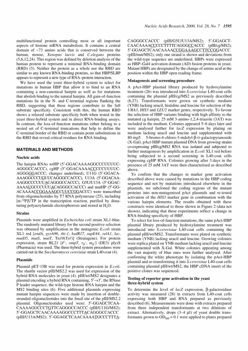
insert (see Materials and Methods). A total of 42 whitecolonies were isolated from ~30 000 transformants and 12 ofthese had point mutations in the HBP cDNA. Six of themutants created nonsense codons at positions Q12, Q139,Q141, R163, W183 and W190, respectively (Fig. 4, top).These were unable to grow on His selective medium,independent of the 3AT concentration, indicating that they hadcompletely lost the ability to interact with the wild-typehairpin. The other six clones had single amino acidsubstitutions at four different positions within the RBD; E157,T171, R181 and D184. Interestingly, three of the mutantsaffected the same residue, R181, with a Cys substitution beingisolated twice and a Gln once. The 3AT titrations indicate thatD184N had the strongest effect on interaction with the wild-type hairpin (growth only at 2.5 mM 3AT; Fig. 4, bottom) andE157K had the most moderate effect (100 mM 3AT), with theother four mutants being able to grow at up to 12.5–50 mM3AT. Thus selection for loss-of-function mutations by theyeast three-hybrid system allowed identification of criticalresidues for RNA binding and helped to define the C-terminalborder of the RBD (see Discussion).
Two mutants constructed by site-directed mutagenesis wereincluded in this analysis (indicated by asterisks in Fig. 4). Oneof these had two Arg at positions 137/138 replaced by Ala,resulting in a complete loss of RNA binding in vitro (data notshown) and a complete inability to grow on His selectiveplates even at the lowest 3AT concentration of 2.5 mM
(Fig. 4, bottom). The other had an Ala exchange of the sameT171 residue for which we isolated a loss-of-binding mutant inthe present selection, but showed near normal binding to wild-type hairpin RNA in vitro (J.Link and B.Müller, unpublishedobservations). In contrast to the selected mutant T171I, theconstructed T171A showed only a moderate reduction in 3ATresistance in the yeast three-hybrid system, indicating that, forthis residue, not only the position but also the nature of theamino acid change was important.
DISCUSSION
The yeast three-hybrid system as a positive and negativeselection system
In this paper we have used the yeast three-hybrid system toselect for point mutations in human HBP with altered RNA-binding properties. The three-hybrid system proved to beamenable to both positive and negative selection schemes. This‘classic genetics’ approach to select altered RNA-bindingphenotypes from a random pool of mutants is an importantcomplement to the popular ‘reverse genetics’ strategy and canprovide valuable information on functionally importantresidues in new types of RNA-binding proteins such as HBP.Selection schemes based on the expression of RNA-bindingproteins in bacteria have been described (30–34), but the yeastthree-hybrid system(s) (3,4) or other eukaryotic selection
Figure 3. Binding of full-length human HBP (wtHBP) and its minimal RBD to different hairpin structures. (A) Activation of the lacZ gene in the yeast three-hybridsystem. Either wtHBP (6) or a fragment encompassing the RBD (amino acid residues 121–203) were expressed in the yeast three-hybrid system as AD–HBPfusions. β-Galactosidase activities were measured with extracts prepared from three independent transformants at two dilutions of extract (see Materials and Methods).Standard deviations are indicated with error bars. The values for wtHBP are the same as presented in Figure 1. (B and C) EMSA with wtHBP (B) or with the RBD(C) and short synthetic hairpin RNAs. wtHBP and a fragment encompassing the RBD (amino acid residues 121–203) were expressed in insect cells and E.coli,respectively. Both proteins contained an N-terminal histidine tag and were purified to >90% homogeneity. Protein concentrations (HBP 0.6 µM; RBD 0.2 µM) werechosen to give near complete binding of wtHP RNA. The proteins were incubated with the indicated 32P-labelled hairpin RNAs and analysed by non-denaturing gelelectrophoresis and autoradiography as described in Materials and Methods. (D) Gel analysis of the free hairpin RNAs used for EMSA in (B) and (C). Theadditional band in the U11G RNA preparation (lane 4) that is also visible in the binding reactions is most likely due to an alternative folding of the RNA.
1600 Nucleic Acids Research, 2000, Vol. 28, No. 7
systems based on different principles (35–37) have thepotential advantage that RNA–protein interactions areanalysed in a eukaryotic environment.
The yeast three-hybrid system has previously been used toquantitate RNA–protein interactions (reviewed in 38) and inpositive selection schemes to isolate cDNAs encoding specificRNA-binding proteins (3,5–8) or RNA ligands for a givenRNA-binding protein (9). The system has the advantage thattwo marker genes are available for selection and that theselection stringency for one of them, HIS3, can be varied overa wide range using different concentrations of the drug 3AT.This is particularly useful for a very strong RNA-bindingprotein such as HBP that shows growth on His selectivemedium at 3AT concentrations of up to 225 mM (Fig. 2).
We exploited the proven suitability of the three-hybridsystem for positive selection by searching for compensatingmutations of HBP that would allow it to bind to mutant RNAhairpins. The selection criterion was growth on His selectivemedium at 25 mM 3AT, i.e. the concentration that had beenused for the original isolation of HBP cDNA (6). Three of themutant hairpins analysed, cgHP, G5U/U13A and mutHP, didnot allow growth in combination with wild-type HBP–ADfusion protein under these conditions and showed no lacZactivity (Fig. 1), and cgHP and G5U/U13A were then used forselection. However, compensating mutants were only successfully
selected with cgHP RNA as the bait, and not with G5U/U13ARNA. This could be because the cg mutation modified thehairpin element only at the bottom of the stem, whereas G5U/U13Ahad modifications in both the stem and the loop. Therefore,more extensive mutations may have been needed to allow HBPto bind to this very different RNA structure.
Other mutant hairpins had residual lacZ activity and stillpromoted growth at 25 mM 3AT (Fig. 1). However, consideringthe extremely high 3AT resistance (225 mM) observed for thewild-type HBP/hairpin combination, it is likely that compen-sating mutations for these other mutant hairpins could alsohave been isolated by applying more stringent selection conditions.
Although most of the mutants binding more strongly tocgHP displayed a relaxed binding specificity, this does notmean that mutants with a more selectively altered specificitycould not be isolated using a similar approach. However, thecreation of true altered specificity mutants could require morethan one or a few amino acid changes, because the network ofinteractions in the RBD may be too complex. In this case, itmight be necessary to apply an in vivo evolution schemeconsisting of multiple rounds of mutagenesis and selection.We know from other work that the RBD of the C.elegans HBPdiscriminates between hairpins differing only in the firstposition of the loop and that variants of the human RBD withsimilar sequence specificity can be obtained by multiple site-directed amino acid substitutions (F.Michel, D.Schümperli andB.Müller, submitted for publication). The problem of isolatingrelaxed specificity mutants in the larger N- and C-terminalregions might also be circumvented by targeting the mutagenesismore specifically to the RBD. Finally, other sequence-specificRNA-binding proteins may not be so prone to relaxation of theRNA-binding specificity as human HBP.
The isolation of loss-of-binding mutations in HBP representsthe first successful case of negative screening using the yeastthree-hybrid system. To select the mutants, we made use of theblue/white colour phenotype on X-gal plates. Not unexpectedly,the procedure yielded a larger proportion of false positive colonies(30 of 42) than positive selection, since reporter gene expressioncan also be lost when other features of the HBP–AD expressionplasmid are mutated (e.g. the yeast replicon, the promoter orthe activation domain of the fusion gene). All the mutantslocated in the HBP coding sequence were very informative,with about half of them representing premature stop codonsand the other half being missense mutations. If one wanted toavoid full characterisation of the nonsense mutations, onecould select for conditional phenotypes (e.g. temperature-sensitive mutations) or screen the mutant colonies by westernblot for production of full-length protein.
Relationship between reporter gene activities in yeast andin vitro RNA-binding results
An interesting aspect from our work bears on the relationshipbetween reporter gene activities in the yeast three-hybridsystem and the physical strength of the protein–RNA interaction.For wild-type HBP, hairpins U13A and U11G, which yielded~50–60 U of β-galactosidase, produced very weakly detectableprotein–RNA complexes by EMSA (Fig. 3). In contrast, theRBD formed readily detectable protein–RNA complexes withthe cgHP and G5U/U13A RNAs although lacZ activity wasonly 17.5 and 5.3 U, respectively. For the HIS3 marker, ourresults with the triple mutant protein E219K/S23P/E47K and
Figure 4. Negative selection of HBP mutants with reduced binding to wild-typehistone hairpin RNA. (Top) Positions of mutations. HBP is depicted as a barwith the minimal RBD shown in grey. Numbers indicate amino acid positions.The isolated mutants are indicated. Two identical mutants (R181C) were isolatedindependently. RR137/8AA and T171A were derived by site-directedmutagenesis and are marked with an asterisk. (Bottom) Summary of drop testsdone with yeast expressing each of the mutant proteins together with wtHP asindicated. The bars indicate the maximal 3AT concentrations allowing forgrowth; 0 means no growth at 2.5 mM 3AT, which was the lowest 3ATconcentration tested.

Nucleic Acids Research, 2000, Vol. 28, No. 7 1601
cgHP RNA indicate that a 3AT resistance over 50 mM isrequired for in vitro binding of full-length HBP. In contrast, theRBD formed complexes in vitro with cgHP RNA whichallowed growth at only 2.5 mM 3AT. This implies that therelationship between reporter gene activation and the biochemicalstrength of an interaction varies between different protein–RNApairs. Presumably, the levels of reporter gene activation in thethree-hybrid system can be affected in many ways, amongothers through different folding in combination with the Gal4AD, oligomerisation or additional interactions with the tran-scription machinery. Thus, it is important that data from thethree-hybrid system be interpreted in terms of relative RNAbinding strength only when studying closely related variants inthe RNA or protein of a given interaction pair.
For minor variants of such a pair, e.g. when comparing theinteractions with different hairpin mutants for either full-lengthHBP (Figs 1 and 3) or for the RBD (Fig. 3), the β-galactosidaseassays and the binding observed in vitro by EMSA are in goodgeneral agreement, although minor variations have also beenobserved (see description of Fig. 3 in Results). However, evenbetween these assays the relationship is not necessarily linear.For example, the relative affinities of hairpins with the U11G,U13A or U11G/U13A mutation were previously reported to be15, 20 and 5% of that of wtHP RNA (39), respectively, and yetthe β-galactosidase activity was only reduced ~2.5-fold(Fig. 1). For other mutations, such as mutHP and cgHP, nothree-hybrid interaction was observed, in full agreement withearlier in vitro binding studies (6,39,40) and with data fromthis paper (Fig. 3). For the other assay used in this work,scoring growth on His selective plates with different 3ATconcentrations, non-linearity between 3AT resistance andHIS3 expression is even to be expected.
Specificity of RNA recognition by HBP: interplay betweenthe N- and C-terminal regions and the minimal RBD
Interestingly, all the compensating mutations isolated in thepositive selection were located in the N- and C-terminalregions, with none in the RBD (Fig. 2). Moreover, one mutation,Q208Stop, removed most of the C-terminal part of the protein.Several of the double mutations and the triple mutation notonly increased the interaction with cgHP but also with mutHPand G5U/U13A RNAs (Fig. 2). Furthermore, the RBD ofhuman HBP was found to have a relaxed specificity comparedto full-length wtHBP (Fig. 3). All these findings indicate thatthe flanking regions contribute to the full binding specificity ofHBP, although the RBD by itself is sufficient for binding.There are two possible explanations for these findings whichare not mutually exclusive.
First, the N- and C-terminal regions might contribute to thefull binding specificity by imposing structural constraints onthe RBD. Their mutation or removal might render the RBDmore flexible, allowing it to accommodate non-canonical hairpins.However, extrapolating from allosterically regulated enzymes,one would expect a loss of substrate binding rather than areduced binding specificity as a result of such a structuralrelaxation.
Second, it is possible that residues in the N- and C-terminaldomains are truly involved in discrimination of non-cognatesubstrates and that we have identified critical anti-determinantspreventing the binding of non-cognate hairpins. Such anti-determinants have been identified in the interactions between
aminoacyl-tRNA synthetases and their cognate tRNAs (41–43)and for E.coli initiation factor IF3 (44). Acidic residues (Aspand Glu) play a direct role in this discrimination of non-cognateRNA substrates. In this context it is interesting to note that three ofthe eight mutations isolated by us were substitutions of Gluresidues by Lys (E47K, E83K and E219K). However, as theside chain of Lys is two carbon atoms longer than the Glu sidechain, it is also possible that these mutations had a steric effectand changed the structure of the protein, thus interfering withRNA binding. The mutation S227I led to the replacement of ahydrophilic by a hydrophobic amino acid, whereas S23Preplaced Ser by Pro, perhaps involving a change in HBPsecondary structure. Finally, mutations A257S and L259S hadonly moderate effects on binding to cgHP RNA, so thesechanges are presumably not very important.
It is possible, however, that the increased binding to themutHP and G5U/U13A RNAs does not really reflect a loss ofbinding specificity. We selected for HBP mutants able to bindto cgHP RNA, which has two C-G base pairs at the bottom ofthe stem. The same C-G base pairs were present in mutHP(along with other changes), while in G5U/U13A RNA thebottom base pair was destabilised by the introduction ofanother pyrimidine, uridine, on the 5′-side. It is therefore alsopossible that one or more pyrimidines at the beginning of adouble-stranded RNA region where bases are more accessiblethan within a narrow RNA A-helix (1) is the common structureof these three hairpin RNAs that the HBP mutants have‘learned’ to recognise.
Whichever of the above explanations is correct, it is clearthat the N- and C-terminal regions flanking the RBDcontribute significantly to the full sequence specificity of thehuman HBP. Moreover, as we isolated two double mutants(E219K/S23P and E219K/E47K) containing each of the additionalmutations present in the triple mutant (E219K/S23P/E47K) andthese activated the yeast reporter genes to intermediate levelsbetween that of the single mutant E219K and the triple mutant,it appears that the effect of the three mutations in changing orreducing the binding specificity was roughly additive.
In this work we have isolated a total of seven nonsensemutations. One, Q208Stop, retained binding to wtHP RNA andshowed increased binding to cgHP RNA (Fig. 2). All the othersix mutations with stop codons at Q12, Q139, Q141, R163,W183 and W190 completely lost the ability to interact withwtHP RNA. This allows us to position the C-terminal border ofthe RBD between amino acids 190 and 208. Previous workusing in vitro translated truncated proteins defined the C-terminalborder of the RBD as between residues 180 (non-functional)and 198 (functional) (5). Our new data thus narrows thismargin to residues 190–198. It is not very surprising that six ofthe seven nonsense mutations occurred at Q and W residues,since the mutagenic agent used (hydroxylamine) primarilyproduces C→T or A→G mutations (45). Thus CAA or CAGcodons (Asn) can readily be converted to TAA or TAG andTGG (Trp) can be converted to TAG. In our negative screen,we have isolated stop mutations in three of six Asn residuesand in two of six Trp codons located in the N-terminal regionand the RBD, indicating that the negative screen did not fullysaturate the cDNA for mutations.
All the missense mutations isolated in the negative selectionwere located within the previously defined RBD. This region ishighly conserved among the five HBPs that have been

1602 Nucleic Acids Research, 2000, Vol. 28, No. 7
characterised (human, mouse, C.elegans and two proteinsnamed xSLBP1 and xSLBP2 from X.laevis) (5,6,24). Compar-ison of these five proteins within the RBD yields the consensussequence shown in Figure 5. Moreover, by computer alignmentusing the PHDsec program (46,47) three α-helical stretches arepredicted to occur within this region (Fig. 5). All but one of theloss-of-binding mutations are located within these putativeα-helices: the in vitro constructed RR137/8AA in helix 1,E157K in helix 2 and R181Q, R181C and D184N in helix 3.Only T171I is located in the linker region between thepredicted helices 2 and 3. All of the mutated residues exceptfor E157 are absolutely conserved between the five proteins;E157 (which had the weakest effect on binding) is altered to Aand Q in xSLBP1 and xSLBP2, respectively, but is conservedin mouse and C.elegans. Although the selection did notsaturate HBP for mutations (see above), these mutants identifya first set of amino acid residues that are critical for RNAbinding.
Three of the mutations, RR137/8AA, R181Q and R181C,replaced a positively charged amino acid by an uncharged oneand had severe effects on RNA binding. This suggests thatthese residues may make important contacts with the negativelycharged RNA backbone. In particular, R181, for which a Cyssubstitution was isolated twice and an Asn substitution once,seems to be a very important residue.
After having isolated T171I as a loss-of-binding mutation,we also tested an Ala substitution of the same residue that wehad obtained by site-directed mutagenesis. Interestingly, theAla substitution had only a minor effect on binding of HBP towtHP RNA (Fig. 4), indicating that not only the position butalso the nature of the change was very critical. Because of thesize difference between Ala and Ile it is likely that the T171Imutation poses a steric problem for RNA binding. Thesequence TPNK is a consensus sequence for cdc2/cyclin Bkinase, but evidence to be reported elsewhere suggests that thissite is not a substrate for phosphorylation by cdc2/cyclin Bkinase (J.Link and B.Müller, unpublished observations).
A very interesting mutation is D184N, since the substitutionof an Asp by an Asn is a very minor change in terms of stericeffect, implying that this residue might be involved in crucialinteractions for RNA binding. The negatively charged Aspmight, for example, interact directly with one of the bases inthe RNA hairpin.
In summary, this analysis has allowed us to identify criticalresidues for RNA binding within the RBD of human HBP. It isinteresting to note that HBP is a multifunctional protein. It iscertainly involved in histone RNA 3′ processing and presum-ably also in nucleo-cytoplasmic transport and translation ofhistone mRNA as well as in the regulation of its cytoplasmichalf-life (12). The minimal region required for histone RNA 3′processing in vitro has been mapped to the RBD plus 20 aminoacids beyond its C-terminal border (48). Therefore, some ofthe residues of the RBD are likely to be important for RNAprocessing and perhaps also for some other functions of HBP.It will be interesting to analyse in detail whether the residuesidentified here are also involved in these functions. Moreover,the information gathered here can be used as a starting pointfor modelling the structure of the complex between the RBDand the histone RNA hairpin.
ACKNOWLEDGEMENTS
We are grateful to Marvin Wickens for supplying us with theyeast three-hybrid system. The HBP mutants RR137/8AA andT171A were obtained by site-directed mutagenesis by RogerWiprächtiger and Julia Link, respectively. Ramesh S. Pillaicontributed RNA-binding assays that were not included in thefinal version of the paper. This work was supported by theState of Bern and by Swiss National Science Foundation Grant31-52619.97 to D.S. and B.M.
REFERENCES
1. Cusack,S. (1999) Curr. Opin. Struct. Biol., 9, 66–73.2. Draper,D.E. (1999) J. Mol. Biol., 293, 255–270.
Figure 5. Structure of the minimal RBD of human HBP. The minimal RBD defined by Marzluff and co-workers (5) is shown in bold italic. The fragment designatedRBD in this study corresponds to amino acid residues 121–203. The consensus sequence was derived from a comparison of human, mouse and C.elegans HBPs(5,6) as well as of the two related Xenopus proteins xSLBP1 and xSLBP2 (24). The residues shown are present in at least three of the five proteins. Putative α-helicalregions predicted by the PHDsec program are shown below (46,47). Amino acid residues mutated in loss-of-binding mutants are underlined in the human HBPsequence and indicated by circles in the α-helical projections.

Nucleic Acids Research, 2000, Vol. 28, No. 7 1603
3. Putz,U., Skehel,P. and Kuhl,D. (1996) Nucleic Acids Res., 24, 4838–4840.4. Sengupta,D.J., Zhang,B.L., Kraemer,B., Pochart,P., Fields,S. and
Wickens,M. (1996) Proc. Natl Acad. Sci. USA, 93, 8496–8501.5. Wang,Z.F., Whitfield,M.L., Ingledue,T.C., Dominski,Z. and
Marzluff,W.F. (1996) Genes Dev., 10, 3028–3040.6. Martin,F., Schaller,A., Eglite,S., Schümperli,D. and Müller,B. (1997)
EMBO J., 16, 769–778.7. Zhang,B., Gallegos,M., Puoti,A., Durkin,E., Fields,S., Kimble,J. and
Wickens,M.P. (1997) Nature, 390, 477–484.8. Dahanukar,A., Walker,J.A. and Wharton,R.P. (1999) Mol. Cell, 4,
209–218.9. Sengupta,D.J., Wickens,M. and Fields,S. (1999) RNA, 5, 596–601.
10. Birnstiel,M.L. and Schaufele,F.J. (1988) In Birnstiel,M.L. (ed.), Structureand Function of Major and Minor Small Nuclear RibonucleoproteinParticles. Springer Verlag, New York, NY, pp. 155–182.
11. Marzluff,W.F. (1992) Gene Expr., 2, 93–97.12. Müller,B. and Schümperli,D. (1997) Semin. Cell Dev. Biol., 8, 567–576.13. Eckner,R., Ellmeier,W. and Birnstiel,M.L. (1991) EMBO J., 10,
3513–3522.14. Williams,A.S., Ingledue,T.C., Kay,B.K. and Marzluff,W.F. (1994)
Nucleic Acids Res., 22, 4660–4666.15. Sun,J., Pilch,D.R. and Marzluff,W.F. (1992) Nucleic Acids Res., 20,
6057–6066.16. Gallie,D.R., Lewis,N.J. and Marzluff,W.F. (1996) Nucleic Acids Res., 24,
1954–1962.17. Pandey,N.B. and Marzluff,W.F. (1987) Mol. Cell. Biol., 7, 4557–4559.18. Harris,M.E., Böhni,R., Schneiderman,M.H., Ramamurthy,L.,
Schümperli,D. and Marzluff,W.F. (1991) Mol. Cell. Biol., 11, 2416–2424.19. Mowry,K.L., Oh,R. and Steitz,J.A. (1989) Mol. Cell. Biol., 9, 3105–3108.20. Vasserot,A.P., Schaufele,F.J. and Birnstiel,M.L. (1989) Proc. Natl Acad.
Sci. USA, 86, 4345–4349.21. Pandey,N.B., Sun,J. and Marzluff,W.F. (1991) Nucleic Acids Res., 19,
5653–5659.22. Hanson,R.J., Sun,J.H., Willis,D.G. and Marzluff,W.F. (1996)
Biochemistry, 35, 2146–2156.23. Dominski,Z., Sumerel,J., Hanson,R. and Marzluff,W. (1996) RNA, 1,
915–923.24. Wang,Z.F., Ingledue,T.C., Dominski,Z., Sanchez,R. and Marzluff,W.F.
(1999) Mol. Cell. Biol., 19, 835–845.25. Milligan,J., Groebe,D., Witherell,G. and Uhlenbeck,O. (1987)
Nucleic Acids Res., 15, 8783–8794.
26. Isackson,P.J. and Bertrand,K.P. (1985) Proc. Natl Acad. Sci. USA, 82,6226–6230.
27. Gietz,D., St Jean,A., Woods,R.A. and Schiestl,R.H. (1992)Nucleic Acids Res., 20, 1425.
28. Miller,J.H. and Albertini,A.M. (1983) J. Mol. Biol., 164, 59–71.29. Hampsey,M. (1997) Yeast, 13, 1099–1133.30. MacWilliams,M.P., Celander,D.W. and Gardner,J.F. (1993)
Nucleic Acids Res., 21, 5754–5760.31. Jain,C. and Belasco,J.G. (1996) Cell, 87, 115–125.32. Laird-Offringa,I.A. and Belasco,J.G. (1996) Methods Enzymol., 267,
149–168.33. Wilhelm,J.E. and Vale,R.D. (1996) Genes Cells, 1, 317–323.34. Harada,K., Martin,S.S. and Frankel,A.D. (1996) Nature, 380, 175–179.35. Stripecke,R., Oliveira,C.C., McCarthy,J.E. and Hentze,M.W. (1994)
Mol. Cell. Biol., 14, 5898–5909.36. Paraskeva,E., Atzberger,A. and Hentze,M.W. (1998) Proc. Natl Acad.
Sci. USA, 95, 951–956.37. Tan,R. and Frankel,A.D. (1998) Proc. Natl Acad. Sci. USA, 95,
4247–4252.38. Zhang,B., Kraemer,B., SenGupta,D., Fields,S. and Wickens,M. (1999)
Methods Enzymol., 306, 93–113.39. Williams,A.S. and Marzluff,W.F. (1995) Nucleic Acids Res., 23,
654–662.40. Schaller,A., Martin,F. and Müller,B. (1997) J. Biol. Chem., 272,
10435–10441.41. Muramatsu,T., Nishikawa,K., Nemoto,F., Kuchino,Y., Nishimura,S.,
Miyazawa,T. and Yokoyama,S. (1988) Nature, 336, 179–181.42. Schmitt,E., Meinnel,T., Panvert,M., Mechulam,Y. and Blanquet,S. (1993)
J. Mol. Biol., 233, 615–628.43. Bedouelle,H., Guez-Ivanier,V. and Nageotte,R. (1993) Biochimie, 75,
1099–1108.44. Sacerdot,C., de Cock,E., Engst,K., Graffe,M., Dardel,F. and Springer,M.
(1999) J. Mol. Biol., 288, 803–810.45. Rose,M.D. and Fink,G.R. (1987) Cell, 48, 1047–1060.46. Rost,B. and Sander,C. (1993) J. Mol. Biol., 232, 584–599.47. Rost,B. and Sander,C. (1994) Proteins, 19, 55–72.48. Dominski,Z., Zheng,L.X., Sanchez,R. and Marzluff,W.F. (1999)
Mol. Cell. Biol., 19, 3561–3570.49. Meier,V.S., Böhni,R. and Schümperli,D. (1989) Nucleic Acids Res., 17,
795.