Porous Silicon Optical Devices: Recent Advances in ... - X-MOL
26
sensors Review Porous Silicon Optical Devices: Recent Advances in Biosensing Applications Rosalba Moretta 1 , Luca De Stefano 1 , Monica Terracciano 2, * and Ilaria Rea 1 Citation: Moretta, R.; De Stefano, L.; Terracciano, M.; Rea, I. Porous Silicon Optical Devices: Recent Advances in Biosensing Applications. Sensors 2021, 21, 1336. https://doi.org/10.3390/ s21041336 Academic Editor: Vittorio M.N. Passaro Received: 30 December 2020 Accepted: 5 February 2021 Published: 13 February 2021 Publisher’s Note: MDPI stays neutral with regard to jurisdictional claims in published maps and institutional affil- iations. Copyright: © 2021 by the authors. Licensee MDPI, Basel, Switzerland. This article is an open access article distributed under the terms and conditions of the Creative Commons Attribution (CC BY) license (https:// creativecommons.org/licenses/by/ 4.0/). 1 National Research Council, Institute of Applied Sciences and Intelligent Systems, Unit of Naples, 80131 Naples, Italy; [email protected] (R.M.); [email protected] (L.D.S.); [email protected] (I.R.) 2 Department of Pharmacy, University of Naples Federico II, 80131 Naples, Italy * Correspondence: [email protected] Abstract: This review summarizes the leading advancements in porous silicon (PSi) optical-biosensors, achieved over the past five years. The cost-effective fabrication process, the high internal surface area, the tunable pore size, and the photonic properties made the PSi an appealing transducing substrate for biosensing purposes, with applications in different research fields. Different optical PSi biosensors are reviewed and classified into four classes, based on the different biorecognition elements immobilized on the surface of the transducing material. The PL signal modulation and the effective refractive index changes of the porous matrix are the main optical transduction mecha- nisms discussed herein. The approaches that are commonly employed to chemically stabilize and functionalize the PSi surface are described. Keywords: optical biosensor; porous silicon; surface functionalization; immunosensor; aptasensor 1. Introduction The term “biosensor” refers to an analytical and powerful tool made up of a biore- ceptor (i.e., a biological recognition element) connected to a transducing substrate [1–3]. The bioreceptor is a biologically active material (i.e., enzyme, protein, antibody, oligonu- cleotide, and so on) responsible for the device selectivity; the transducer converts a specific biological recognition event (i.e., antibody-antigen interaction) into a measurable signal in real-time. The first biosensor was made by Professor L.C. Clarck in 1956 for oxygen detection in the blood of patients undergoing surgery [4]. To date, incredible progress has been made both in technology and applications of biosensors, representing a very attractive research field, as demonstrated by the vast literature in the last 20 years. It is expected that the biosensor market could reach U.S. Dollar (USD) 36.0 billion by 2027, due to the rising demand for such devices, with applications not only in biomedical diagnosis but also in environmental and food quality monitoring, industrial process control, agriculture, and others [5]. Moreover, the request for low-cost and user-friendly devices, with a fast-response time, is slowly replacing the currently available techniques for the identification of analytes, which are time-consuming, expensive, and require specialized laboratory equipment. The design and the fabrication of the sensing system require the meticulous research of materials that have the desired transducer properties. In this context, nanostructured materials gained prominence in many applications because of their unique physicochemical characteristics over their bulk counterpart, such as high surface-to-volume ratio, small size, light absorption, optical sensitivity, and electrical and thermal conductivity [6,7]. Moreover, nanostructure-based biosensors show enhanced biosensing performances over conventional detection methods (i.e., higher sensitivity, fast response time, and low limit of detection (LoD)) [8–10]. Among the many available nanomaterials (e.g., quantum dots Sensors 2021, 21, 1336. https://doi.org/10.3390/s21041336 https://www.mdpi.com/journal/sensors
-
Upload
khangminh22 -
Category
Documents
-
view
0 -
download
0
Transcript of Porous Silicon Optical Devices: Recent Advances in ... - X-MOL
sensors
Review
Porous Silicon Optical Devices: Recent Advances inBiosensing Applications
Rosalba Moretta 1 , Luca De Stefano 1 , Monica Terracciano 2,* and Ilaria Rea 1
�����������������
Citation: Moretta, R.; De Stefano, L.;
Terracciano, M.; Rea, I. Porous Silicon
Optical Devices: Recent Advances in
Biosensing Applications. Sensors 2021,
21, 1336. https://doi.org/10.3390/
s21041336
Academic Editor: Vittorio
M.N. Passaro
Received: 30 December 2020
Accepted: 5 February 2021
Published: 13 February 2021
Publisher’s Note: MDPI stays neutral
with regard to jurisdictional claims in
published maps and institutional affil-
iations.
Copyright: © 2021 by the authors.
Licensee MDPI, Basel, Switzerland.
This article is an open access article
distributed under the terms and
conditions of the Creative Commons
Attribution (CC BY) license (https://
creativecommons.org/licenses/by/
4.0/).
1 National Research Council, Institute of Applied Sciences and Intelligent Systems, Unit of Naples,80131 Naples, Italy; [email protected] (R.M.); [email protected] (L.D.S.);[email protected] (I.R.)
2 Department of Pharmacy, University of Naples Federico II, 80131 Naples, Italy* Correspondence: [email protected]
Abstract: This review summarizes the leading advancements in porous silicon (PSi) optical-biosensors,achieved over the past five years. The cost-effective fabrication process, the high internal surfacearea, the tunable pore size, and the photonic properties made the PSi an appealing transducingsubstrate for biosensing purposes, with applications in different research fields. Different opticalPSi biosensors are reviewed and classified into four classes, based on the different biorecognitionelements immobilized on the surface of the transducing material. The PL signal modulation andthe effective refractive index changes of the porous matrix are the main optical transduction mecha-nisms discussed herein. The approaches that are commonly employed to chemically stabilize andfunctionalize the PSi surface are described.
Keywords: optical biosensor; porous silicon; surface functionalization; immunosensor; aptasensor
1. Introduction
The term “biosensor” refers to an analytical and powerful tool made up of a biore-ceptor (i.e., a biological recognition element) connected to a transducing substrate [1–3].The bioreceptor is a biologically active material (i.e., enzyme, protein, antibody, oligonu-cleotide, and so on) responsible for the device selectivity; the transducer converts a specificbiological recognition event (i.e., antibody-antigen interaction) into a measurable signalin real-time.
The first biosensor was made by Professor L.C. Clarck in 1956 for oxygen detection inthe blood of patients undergoing surgery [4]. To date, incredible progress has been madeboth in technology and applications of biosensors, representing a very attractive researchfield, as demonstrated by the vast literature in the last 20 years.
It is expected that the biosensor market could reach U.S. Dollar (USD) 36.0 billion by2027, due to the rising demand for such devices, with applications not only in biomedicaldiagnosis but also in environmental and food quality monitoring, industrial process control,agriculture, and others [5]. Moreover, the request for low-cost and user-friendly devices,with a fast-response time, is slowly replacing the currently available techniques for theidentification of analytes, which are time-consuming, expensive, and require specializedlaboratory equipment.
The design and the fabrication of the sensing system require the meticulous researchof materials that have the desired transducer properties. In this context, nanostructuredmaterials gained prominence in many applications because of their unique physicochemicalcharacteristics over their bulk counterpart, such as high surface-to-volume ratio, smallsize, light absorption, optical sensitivity, and electrical and thermal conductivity [6,7].Moreover, nanostructure-based biosensors show enhanced biosensing performances overconventional detection methods (i.e., higher sensitivity, fast response time, and low limitof detection (LoD)) [8–10]. Among the many available nanomaterials (e.g., quantum dots
Sensors 2021, 21, 1336. https://doi.org/10.3390/s21041336 https://www.mdpi.com/journal/sensors
Sensors 2021, 21, 1336 2 of 26
(QDs), metallic nanoparticles, carbon nanotube), porous silicon (PSi) has outstandingwindows for applications in several research fields, from biosensing to drug delivery,thanks to its well-known optical and physical features [11–13].
Although it was accidentally discovered in 1956 by Uhlirs during an electrochemicalexperiment [14], this material received due attention only in 1990, when Canham discoveredits intrinsic photoluminescence (PL) at room temperature [15]. Since the first experimentson PSi as a biosensor platform two decades ago, it is still an actual topic for research studies,as evidenced by the sustained number of scientific papers on PSi-based sensors publishedevery year in peer-review journals [16].
PSi exhibits air-filled pores and a high surface area (up to 800 m2/g) [17]; these in-teresting characteristics, together with versatile surface chemical modification, tunablecharacteristic sizes, photoluminescence, biocompatibility, and biodegradability makes thismaterial an appealing optical transducer [12,18–22]. Moreover, due to the high surfacereactivity of PSi [23], several biomolecules can be easily immobilized within the porousmatrix, by using well-established chemical approaches [13,24]. Since non-specific interac-tions reduce the selectivity of a biosensing platform, a blocking process of residual groupsis generally performed as a final step of functionalization, by using agents like maleimide,bovine serum albumin, etc.
PSi is commonly obtained via electrochemical etching, a fabrication strategy thatdoes not require expensive equipment, and allows a good reproducibility of the fabricatedPSi substrates. This technique enables a fine control on the pore size and on the opticalresponse of the material [25,26]. This tuning is generally not easily achievable in otherporous materials, such as porous alumina [27,28] or porous titania [29,30].
Recently, PSi was also explored as a host matrix for the immobilization of severalnanomaterials (i.e., QDs, graphene oxide, carbon dots) due to its high internal volume[31–35]. This feature made PSi more interesting than the other nanomaterials for biosens-ing applications. In fact, the possibility of combining different elements into the PSimatrix paved the way for the development of hybrid platforms that showed enhancedbiosensing performances in terms of sensitivity, signal enhancement, signal stability, anddual-mode detection.
The optical response of PSi structures are strongly affected by their structural prop-erties such as porosity (ratio of the fraction of voids in the layer to total volume), layer(s)thickness, and pore size and morphology [25]. Based on the International Union of Pureand Applied Chemistry (IUPAC) definition, three different PSi structures, with differentpore sizes, could be distinguished—microporous Si (pore diameter d < 2 nm), mesoporousSi (pore diameter 2 < d < 50 nm), and macroporous Si (pore diameter d > 50 nm) [36].Instead, the pore morphology is the least quantifiable aspect [25] and considers propertieslike shape (i.e., smooth, branched, facetted), orientation, and interconnection betweenpores. All these features are strongly influenced by the monocrystalline silicon and theanodizing regimes [25]. Macroporous and mesoporous Si are commonly employed asbiosensing platforms to allow the attachment of biomolecules within their matrices. Thecapture of analytes can be monitored via reflectance, when dealing with macroporous ormesoporous structures (i.e., monolayer, Bragg mirror) [37,38], or via photoluminescencewith mesoporous matrices (i.e., resonant microcavity, nanowires) [39,40]. Unfortunately,the intrinsic PL of PSi is usually unstable, since it strongly depends on the surroundingchemical environment that can influence its intensity [11]. For this reason, several worksreported on the use of PSi substrates to embed emitting materials or molecules withinpores, which sharpen the emission spectrum and amplify the signal [13,39,41].
Additionally, the versatile surface modification, optical tunability, low-cost fabrication,and label-free working conditions confer added value to this material. PSi is also compatiblewith microelectronics and MEMS fabrications systems [42–44]. Furthermore, the material’sbiocompatibility allows the development of implantable biosensing devices that could beused for real-time detection of in vivo analytes [45].

Sensors 2021, 21, 1336 3 of 26
PSi optical devices are widely used to detect different biomolecules (i.e., DNA,enzymes, cells, bacteria, antigen). Most PSi-based biosensors work in a label-free modality.The optical transduction principle relies on the change of the refractive index of the PSilayer, due to the substitution of air inside the pores with a target analyte. The changeof the refractive index is detected as a wavelength shift of the corresponding reflectivityspectrum. Moreover, the intrinsic PL signal of PSi might be used as a further transductionmechanism for biosensing applications, by monitoring its variation upon a biomolecularrecognition event. This review focuses on advances on the development of PSi-basedoptical biosensors achieved over the past five years. The fabrication process of PSi is brieflydiscussed, followed by an extensive description of the most common strategies used tostabilize and functionalize the material with biomolecules. Different examples of biosensordevelopment, classified by biomolecules used as sensing elements, such as immunosensors,aptasensors, DNA hybridization, enzyme biosensors, and their application in the biomedi-cal and environmental fields are examined [21,36,46]. Table 1 summarizes the detection ofseveral biomolecules using PSi-optical devices.
2. PSi: From Fabrication to the Bioconjugation
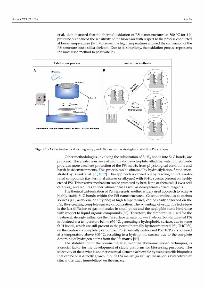
Although different strategies were developed to fabricate PSi structures, the anodicelectrochemical etching of a crystalline silicon wafer in hydrofluoric acid (HF) water so-lution, remains the most commonly used. In 1956, Uhlirs first observed the formation ofPSi during electrochemical procedures for polishing of silicon and germanium wafers [14].This methodology is advantageous to other fabrication techniques (i.e., metal-assistedchemical etching [47,48], etc.), since it allows us to obtain different photonic structures witha high reproducibility [49]. The electrochemical etching is performed into an etching cellin which the silicon wafer acts as an anode and metals (e.g., platinum) act as a cathode.The current flow between the two electrodes leads to the dissolution of the crystallinestructure, forming pores in the silicon wafer. The pore formation process is controlledby a complex mix of electronic and chemical factors [25]. A schematization of the elec-trochemical cell is shown in Figure 1A. Different porous silicon structures, with specificmorphological and optical properties, can be engineered by changing the type of siliconwafer conductivity, doping level, concentration of hydrofluoric acid in electrolyte, appliedvoltage, current density, light intensity, and temperature [50,51]. Metal-assisted chemicaletching (MACE) is another technique used for the fabrication of PSi. It is less common thanthe above-mentioned anodic etching. In a typical MACE process, a thin layer of noble metal(i.e., Au, Pt, etc.) sputtered on the silicon surface, catalyzes the etching of the materialwhen placed in an oxidizing solution with hydrofluoric acid (HF). This technique generatesseveral silicon nanostructures of tailored geometry, i.e., nanopores, porous silicon layers,nanowires, nanoneedles [52–54]
The freshly etched PSi is composed of hydride terminated groups (Si-H, Si-H2, andSi-H3), which make PSi nanostructure highly reactive and unstable. The aging effects,responsible for the uncontrolled growth of native oxide and the degradation of PSi ma-trix in alkaline or aqueous environments, strongly affect this material’s physicochemicaland optoelectronic properties [55]. These effects might lead to zero-point drifts in thereflectivity spectrum and reduce, as a consequence, the sensitivity of the devices [25].In this context, a proper surface chemical modification plays an important role to stopaging and to stabilize the PSi nanostructure (Figure 1B). Among the various developedtreatments, the intentional growth of an oxide layer, under controlled conditions, repre-sents one of the main approaches used to stabilize the hydrogen-terminated PSi surface.Despite the various available strategies for oxidizing the PSi matrix (i.e., ozone oxidation,electrochemical oxidation, and oxidation in aqueous solution), thermal oxidation is themost commonly used technique, by partially or fully replacing the reactive Si-Hx speciesinto Si-O bonds [56]. This procedure is not only used to passivate the surface but also toconvert a hydrophobic surface into a hydrophilic one. The rate of passivation is highlydependent on the temperature and on the duration of the treatment. Therefore, Shtenberg
Sensors 2021, 21, 1336 4 of 26
et al. demonstrated that the thermal oxidation of PSi nanostructures at 800 ◦C for 1 h,profoundly enhanced the sensitivity of the biosensor with respect to the process conductedat lower temperatures [57]. Moreover, the high temperatures allowed the conversion of thePSi structure into a silica skeleton. Due to its simplicity, the oxidation process representsthe most used method to passivate PSi.
Sensors 2021, 21, x FOR PEER REVIEW 4 of 26
al. demonstrated that the thermal oxidation of PSi nanostructures at 800 °C for 1 h, pro-
foundly enhanced the sensitivity of the biosensor with respect to the process conducted
at lower temperatures [57]. Moreover, the high temperatures allowed the conversion of
the PSi structure into a silica skeleton. Due to its simplicity, the oxidation process repre-
sents the most used method to passivate PSi.
Other methodologies, involving the substitution of Si-Hx bonds into Si-C bonds, are
proposed. The greater resistance of Si-C bonds to nucleophilic attack by water or hydrox-
ide provides more excellent protection of the PSi matrix from physiological conditions
and harsh basic environments. This process can be obtained by hydrosilylation, first
demonstrated by Buriak et al. [23,31,32]. This approach is carried out by reacting liquid
unsaturated compounds (i.e., terminal alkenes or alkynes) with Si-Hx species present on
freshly etched PSi. This reactive mechanism can be promoted by heat, light, or chemicals
(Lewis acid catalysis), and requires an inert atmosphere as well as deoxygenate/dried re-
agents.
The thermal carbonization of PSi represents another widely used approach to achieve
highly stable Si-C bonds within the PSi nanostructures. Gaseous molecules as carbon
sources (i.e., acetylene or ethylene) at high temperatures, can be easily adsorbed on the
PSi, thus creating complete surface carbonization. The advantage of using this technique
is the fast diffusion of gas molecules in small pores and the negligible steric hindrance
with respect to liquid organic compounds [58]. Therefore, the temperature, used for the
treatment, strongly influences the PSi surface termination—a hydrocarbon-terminated PSi
is obtained at a temperature below 650 °C, generating a hydrophobic surface, due to some
Si-H bonds, which are still present in the pores (thermally hydrocarbonized PSi, THCPSi);
on the contrary, a completely carbonized PSi (thermally carbonized PSi, TCPSi) is ob-
tained at a temperature above 800 °C, resulting in a hydrophilic surface due to the com-
plete desorbing of hydrogen atoms from the PSi matrix [59].
Figure 1. (A) Electrochemical etching setup; and (B) passivation strategies to stabilize PSi surfaces.
The stabilization of the porous material, with the above-mentioned techniques, is a
crucial factor for the development of stable platforms for biosensing purposes. The selec-
tivity of the device is another essential element, achievable by using specific bioprobes
Figure 1. (A) Electrochemical etching setup; and (B) passivation strategies to stabilize PSi surfaces.
Other methodologies, involving the substitution of Si-Hx bonds into Si-C bonds, areproposed. The greater resistance of Si-C bonds to nucleophilic attack by water or hydroxideprovides more excellent protection of the PSi matrix from physiological conditions andharsh basic environments. This process can be obtained by hydrosilylation, first demon-strated by Buriak et al. [23,31,32]. This approach is carried out by reacting liquid unsatu-rated compounds (i.e., terminal alkenes or alkynes) with Si-Hx species present on freshlyetched PSi. This reactive mechanism can be promoted by heat, light, or chemicals (Lewis acidcatalysis), and requires an inert atmosphere as well as deoxygenate/dried reagents.
The thermal carbonization of PSi represents another widely used approach to achievehighly stable Si-C bonds within the PSi nanostructures. Gaseous molecules as carbonsources (i.e., acetylene or ethylene) at high temperatures, can be easily adsorbed on thePSi, thus creating complete surface carbonization. The advantage of using this techniqueis the fast diffusion of gas molecules in small pores and the negligible steric hindrancewith respect to liquid organic compounds [58]. Therefore, the temperature, used for thetreatment, strongly influences the PSi surface termination—a hydrocarbon-terminated PSiis obtained at a temperature below 650 ◦C, generating a hydrophobic surface, due to someSi-H bonds, which are still present in the pores (thermally hydrocarbonized PSi, THCPSi);on the contrary, a completely carbonized PSi (thermally carbonized PSi, TCPSi) is obtainedat a temperature above 800 ◦C, resulting in a hydrophilic surface due to the completedesorbing of hydrogen atoms from the PSi matrix [59].
The stabilization of the porous material, with the above-mentioned techniques, isa crucial factor for the development of stable platforms for biosensing purposes. Theselectivity of the device is another essential element, achievable by using specific bioprobesthat can be or is directly grown into the PSi matrix (in situ synthesis) or is synthetized exsitu, and is then, immobilized on the surface.

Sensors 2021, 21, 1336 5 of 26
After the PSi stabilization, different linkers can be used to graft biomolecules. Theoxidized PSi can be readily modified via the silanization process (Figure 2A), based on theuse of silane coupling agents (i.e., APTES, APDMES) characterized by different terminalmotifs (i.e., NH2; SH, COOH; CHO) that act as anchorage sites for proteins, antibody,DNA and others [60]. Silane-based chemistry is one of the most exploited modifications ofporous silica-based synthetic or natural materials since it offers an effortless way to attacha biological or chemical molecule to the porous surface [61–63]. Silanization requires theavailability of hydroxyl groups on the PSi surface in order to hydrolyze the alkoxy groupsof the alkyl silane molecule, thus forming Si-O-Si bonds [64]. Thiol or primary aminogroups can be also used for grafting biomolecules. Their exposure on PSi surface can beobtained via a new silanization process, known as “ring-opening-click reaction”, proposedby Sailor et al., in which the heterocycles silanes, having Si-N or Si-S motifs in the ring,undergo a ring-opening reaction to modify the hydroxylated porous walls. The proposedsurface chemistry, obtained in mild conditions and without the formation of by-products,does not interfere with the protein activity (Figure 2B) [65]. Although silanol chemistry isthe most conventional approach for bioprobe immobilization, it is well known that Si-O-Sibonds are characterized by the lack of stability in aqueous and alkaline media, causingdegradation of the coating surface, which is the main problem related to this chemistry.
Higher stability can be obtained through the Si-C bonds. The carboxyl acid groupof undecylenic acid, as a result of the hydrosilylation process, are currently activatedvia 1-ethyl-3-[3-dimethylaminopropyl] carbodiimide hydrochloride (EDC) coupling agentfor loading primary amine-containing biomolecules through direct reaction or via N-hydroxysulfosuccinimmide (NHS) [31]. Finally, the same surface chemical modificationadopted for hydrosilylated-PSi can be applied for TCPSi and THCPSi. Sciacca et al. pro-posed a radical coupling reaction by using a radical initiator benzoyl peroxide and adicarboxylic acid as a linker, generating a surface coated with carboxyl acid groups. Thissurface can be used to attach amino-terminal biomolecules via EDC/NHS chemistry [66].
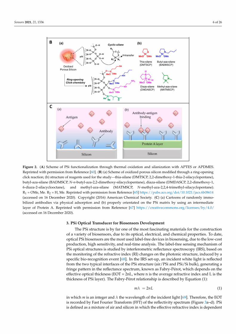
The performances of a bio-device strongly depend on the right orientation of thebiomolecules. This factor is particularly important for the immobilization of antibodies.Several research groups reported a valid strategy to optimize the biomolecules orientationon PSi matrix, by covering the surface with a layer of protein A. In such a design, theprotein A interacts with the fragment crystallizable (FC) region of antibodies, leavingthe antigen-binding fragment (Fab) region available toward the antigen (Figure 2C). Thismethodology allows proper anchoring of the antibody to the surface, maximizing itsbinding capability [67].
The stability and sensitivity of a biosensor are crucial when the target is in complexbiological systems. In this context, the minimization of the unspecific adsorption of un-wanted biomolecules is required. This aim can be achieved by using blocking agents to capthe available reactive sites (i.e., polyethylene glycol, maleimide, bovine serum albumin).
Sensors 2021, 21, x FOR PEER REVIEW 6 of 26
Figure 2. (A) Scheme of PSi functionalization through thermal oxidation and silanization with
APTES or APDMES. Reprinted with permission from Reference [60]. (B) (a) Scheme of oxidized
porous silicon modified through a ring-opening click reaction; (b) structure of reagents used for
the study—thia-silane (DMTSCP, 2,2-dimethoxy-1-thia-2-silacyclopentane), butyl-aza-silane
(BADMSCP, N-n-butyl-aza-2,2-dimethoxy-silacyclopentane), diaza-silane (DMDASCP, 2,2-di-
methoxy-1,6-diaza-2-silacyclooctane), and methyl-aza-silane (MATMSCP, N-methyl-aza-2,2,4-
trimethyl-silacyclopentane). R1 = OMe, Me. R2 = H, Me. Reprinted with permission from Reference
[65] https://pubs.acs.org/doi/10.1021/jacs.6b08614 (accessed on 16 December 2020). Copyright
(2016) American Chemical Society. (C) (a) Cartoons of randomly immobilized antibodies via phys-
ical adsorption and (b) properly orientated on the PSi matrix by using an intermediate layer of
Protein A. Reprinted with permission from Reference [67] https://creativecommons.org/li-
censes/by/4.0/ (accessed on 16 December 2020).
3. PSi Optical Transducer for Biosensors Development
The PSi structure is by far one of the most fascinating materials for the construction
of a variety of biosensors, due to its optical, electrical, and chemical properties. To date,
Figure 2. Cont.
Sensors 2021, 21, 1336 6 of 26
Sensors 2021, 21, x FOR PEER REVIEW 6 of 26
Figure 2. (A) Scheme of PSi functionalization through thermal oxidation and silanization with
APTES or APDMES. Reprinted with permission from Reference [60]. (B) (a) Scheme of oxidized
porous silicon modified through a ring-opening click reaction; (b) structure of reagents used for
the study—thia-silane (DMTSCP, 2,2-dimethoxy-1-thia-2-silacyclopentane), butyl-aza-silane
(BADMSCP, N-n-butyl-aza-2,2-dimethoxy-silacyclopentane), diaza-silane (DMDASCP, 2,2-di-
methoxy-1,6-diaza-2-silacyclooctane), and methyl-aza-silane (MATMSCP, N-methyl-aza-2,2,4-
trimethyl-silacyclopentane). R1 = OMe, Me. R2 = H, Me. Reprinted with permission from Reference
[65] https://pubs.acs.org/doi/10.1021/jacs.6b08614 (accessed on 16 December 2020). Copyright
(2016) American Chemical Society. (C) (a) Cartoons of randomly immobilized antibodies via phys-
ical adsorption and (b) properly orientated on the PSi matrix by using an intermediate layer of
Protein A. Reprinted with permission from Reference [67] https://creativecommons.org/li-
censes/by/4.0/ (accessed on 16 December 2020).
3. PSi Optical Transducer for Biosensors Development
The PSi structure is by far one of the most fascinating materials for the construction
of a variety of biosensors, due to its optical, electrical, and chemical properties. To date,
Figure 2. (A) Scheme of PSi functionalization through thermal oxidation and silanization with APTES or APDMES.Reprinted with permission from Reference [60]. (B) (a) Scheme of oxidized porous silicon modified through a ring-openingclick reaction; (b) structure of reagents used for the study—thia-silane (DMTSCP, 2,2-dimethoxy-1-thia-2-silacyclopentane),butyl-aza-silane (BADMSCP, N-n-butyl-aza-2,2-dimethoxy-silacyclopentane), diaza-silane (DMDASCP, 2,2-dimethoxy-1,6-diaza-2-silacyclooctane), and methyl-aza-silane (MATMSCP, N-methyl-aza-2,2,4-trimethyl-silacyclopentane).R1 = OMe, Me. R2 = H, Me. Reprinted with permission from Reference [65] https://pubs.acs.org/doi/10.1021/jacs.6b08614(accessed on 16 December 2020). Copyright (2016) American Chemical Society. (C) (a) Cartoons of randomly immo-bilized antibodies via physical adsorption and (b) properly orientated on the PSi matrix by using an intermediatelayer of Protein A. Reprinted with permission from Reference [67] https://creativecommons.org/licenses/by/4.0/(accessed on 16 December 2020).
3. PSi Optical Transducer for Biosensors Development
The PSi structure is by far one of the most fascinating materials for the constructionof a variety of biosensors, due to its optical, electrical, and chemical properties. To date,optical PSi biosensors are the most used label-free devices in biosensing, due to the low-costproduction, high sensitivity, and real-time analysis. The label-free sensing mechanism ofPSi optical structures is studied by interferometric reflectance spectroscopy (IRS), based onthe monitoring of the refractive index (RI) changes on the photonic structure, induced by aspecific bio-recognition event [68]. In the IRS set-up, an incident white light is reflectedfrom the two typical interfaces of the PSi structure (air/PSi and PSi/Si bulk), generating afringe pattern in the reflectance spectrum, known as Fabry-Pérot, which depends on theeffective optical thickness (EOT = 2nL, where n is the average refractive index and L is thethickness of PSi layer). The Fabry-Pérot relationship is described by Equation (1):
mλ = 2nL (1)
in which m is an integer and λ the wavelength of the incident light [69]. Therefore, the EOTis recorded by Fast Fourier Transform (FFT) of the reflectivity spectrum (Figure 3a–d). PSiis defined as a mixture of air and silicon in which the effective refractive index is dependent

Sensors 2021, 21, 1336 7 of 26
on the content of air inside the pores. When the air in the porous matrix is replaced with ananalyte, an increase in the RI occurs, producing a shift of the reflectivity spectrum to longerwavelengths (red-shift). Moreover, a decrease in the average RI of the material causes ashift to shorter wavelengths (blue-shift); this phenomenon is generally ascribable to theoxidation/corrosion of the PSi skeleton [38,70].
Sensors 2021, 21, x FOR PEER REVIEW 8 of 26
Figure 3. (a) Functionalization scheme of PSi; (b) optical setup for spectroscopy reflectometry, (c) reflectivity, and (d) FFT
spectra before (black line) and after (red line) PSi functionalization; (e) optical setup for photoluminescence spectroscopy,
and (f) corresponding spectra before (black line) and after (red line) the functionalization procedure.
3.1. Porous Silicon Immunosensors
The antibodies represent the most common probes used in biosensing systems. Spe-
cific antibodies or their fragments (fragment binding antigen-Fab) can be used as biosen-
sor molecular recognition elements against precise antigens. The development of hybrid-
ization and cloning techniques allowed the production of a variety of antibodies, paving
Figure 3. (a) Functionalization scheme of PSi; (b) optical setup for spectroscopy reflectometry, (c) reflectivity, and (d) FFTspectra before (black line) and after (red line) PSi functionalization; (e) optical setup for photoluminescence spectroscopy,and (f) corresponding spectra before (black line) and after (red line) the functionalization procedure.

Sensors 2021, 21, 1336 8 of 26
Although all PSi devices are able to detect the presence of analytes within the pores,the optical response of the material can be finely tuned by changing the layers porosity,thickness, and number. The PSi monolayer is the simplest photonic structure used inbiosensing, whose spectrum shows a periodic behaviour typical of Fabry-Pérot interfer-ometer. However, more complex multi-layered structures with different porosities (i.e.,Bragg mirror, microcavity, PSi rugate filter) can be fabricated, showing enhanced opticalproperties. For example, the reflectivity spectra of the Bragg mirror show a wide range ofwavelengths having high reflectance (i.e., stopbands) whose maximum reflectance valueis dependent on the number of layers in the structure. Moreover, to detect the infiltrationof analytes into the pores, the spectral position of one edge of the stopband is moni-tored [71,72]. The typical narrow spectral feature of a microcavity makes this photonicstructure easy to control upon molecular infiltration, with respect to the Bragg mirror.Moreover, as previously reported, PSi exhibits an intrinsic PL signal at room temperature.Therefore, the modulation of this signal, caused by a biological recognition event, might beemployed for biosensing purposes. A scheme of the optical setup for PL measurements isshown in Figure 3e,f.
The main drawback of label-free, PSi-based biosensors is a low sensitivity, in themicromolar range, due to the slow and limited mass-diffusion inside the matrix. This limitis overcome through the fabrication of more complex and multi-layered optical structures(i.e., Bragg mirror, rugate filters, microcavities, ring resonators). An example is providedby the open-ended PSi microcavity membrane that allows a fast response time and a morefavourable interaction between the analyte and the inner surface [73,74]. Moreover, thesensitivity of PSi-based biosensors can be greatly improved by optimizing the experimentalplatform engineering [75,76], data processing methodologies [77,78], as well as signalamplification mechanisms [79]. Moreover, the PSi matrix can be integrated with inorganicmaterials (i.e., quantum dots, graphene oxide, titanium dioxide) allowing the realizationof hybrid devices with advanced properties, and improving the biosensing performances(i.e., sensitivity, stability, the limit of detection). In the following sections, the realization ofPSi-based devices are reported, highlighting the enormous versatility of PSi as a transducersurface for the development of different types of biosensors.
3.1. Porous Silicon Immunosensors
The antibodies represent the most common probes used in biosensing systems. Specificantibodies or their fragments (fragment binding antigen-Fab) can be used as biosensormolecular recognition elements against precise antigens. The development of hybridizationand cloning techniques allowed the production of a variety of antibodies, paving the wayfor the development of different immunosensors. The antibodies are widely used inPSi-based devices for the detection of several analytes.
The development of a highly sensitive PSi immunosensor needs a careful study onseveral working parameters, such as the antibody orientation onto PSi surface, the analytediffusion into the matrix, as well as the choice of the photonic structure [67,80].
Zhuo et al. reported the development of a hybrid TiO2–PSi-based immunosensorfor the rapid detection of the S-layer protein (SLP), a surface protein available on thecell walls of several bacteria [81]. The device was obtained by spinning TiO2 solutiononto the PSi and putting it in a furnace at 500 ◦C for 1 h [82]. The as-obtained hybridplatform was integrated into a microfluidic system, in which, first, protein A and then,anti-SLP were pumped through the cell. Finally, SLP was injected into the system. Theprotein A, adsorbed on the surface, was used as a spacer to reduce the steric hindranceand improve the antigen–antibody interaction. The following system demonstrated anincreased sensitivity thanks to the high refractive index of the material and the dynamicsampling system, reaching an LoD of 0.70 ± 0.37 pM [80]. Some results are reportedin Figure 4A.

Sensors 2021, 21, 1336 9 of 26
Sensors 2021, 21, x FOR PEER REVIEW 9 of 26
the way for the development of different immunosensors. The antibodies are widely used
in PSi-based devices for the detection of several analytes.
The development of a highly sensitive PSi immunosensor needs a careful study on
several working parameters, such as the antibody orientation onto PSi surface, the analyte
diffusion into the matrix, as well as the choice of the photonic structure [67,80].
Zhuo et al. reported the development of a hybrid TiO2–PSi-based immunosensor for
the rapid detection of the S-layer protein (SLP), a surface protein available on the cell walls
of several bacteria [81]. The device was obtained by spinning TiO2 solution onto the PSi
and putting it in a furnace at 500°C for 1 h [82]. The as-obtained hybrid platform was
integrated into a microfluidic system, in which, first, protein A and then, anti-SLP were
pumped through the cell. Finally, SLP was injected into the system. The protein A, ad-
sorbed on the surface, was used as a spacer to reduce the steric hindrance and improve
the antigen–antibody interaction. The following system demonstrated an increased sensi-
tivity thanks to the high refractive index of the material and the dynamic sampling system,
reaching an LoD of 0.70 ± 0.37 pM [80]. Some results are reported in Figure 4A.
Figure 4. (A) (a) EOT signals of anti-SLP/ProteinA/TiO2-PSi-based immunosensor after incubation in different SLP con-
centrations (from bottom to up). (b) Specificity test of TiO2-PSi-microfluidic biosensor measured via EOT response to BSA
and SLP. (c) SLP detection in a dilute crude extract. Reprinted with permission from Reference [80]; Copyright (2020)
Elsevier B.V. (B) (a) Reflectivity spectra and (b) corresponding Fourier Transforms (FFT) of anti-HSP70-PSi device before
(black line) and after (red line) HSP70 detection at concentration of 200,000 ng/mL. (c) Relationship between HSP70 con-
centrations and EOT shifts (inset semi-log plot). Reprinted with permission from Reference [83]; Copyright (2020) Elsevier
Ltd.
The antibodies were also exploited as bioprobes for the direct detection of whole-cell
organisms such as bacteria or viruses. A recent work of Tang et al. reported the Escherichia
coli detection by using a nanopore array as a sensing platform, upon functionalization
with E. coli antibody and integration into a microfluidic system. The oxidized PSi was
activated by NaOH, followed by HCl treatment and washings in deionized water. After
Figure 4. (A) (a) EOT signals of anti-SLP/ProteinA/TiO2-PSi-based immunosensor after incubation in different SLPconcentrations (from bottom to up). (b) Specificity test of TiO2-PSi-microfluidic biosensor measured via EOT responseto BSA and SLP. (c) SLP detection in a dilute crude extract. Reprinted with permission from Reference [80]; Copyright(2020) Elsevier B.V. (B) (a) Reflectivity spectra and (b) corresponding Fourier Transforms (FFT) of anti-HSP70-PSi devicebefore (black line) and after (red line) HSP70 detection at concentration of 200,000 ng/mL. (c) Relationship between HSP70concentrations and EOT shifts (inset semi-log plot). Reprinted with permission from Reference [83]; Copyright (2020)Elsevier Ltd.
The antibodies were also exploited as bioprobes for the direct detection of whole-cellorganisms such as bacteria or viruses. A recent work of Tang et al. reported the Escherichiacoli detection by using a nanopore array as a sensing platform, upon functionalization withE. coli antibody and integration into a microfluidic system. The oxidized PSi was activatedby NaOH, followed by HCl treatment and washings in deionized water. After silanizationwith Amino-propyl-triethoxy-silane (APTES) and modification with glutaraldehyde (GA),E. coli antibodies were attached to the surface. The biosensing principle was based on thedirect capture of E. coli on the immunosensor, reducing the pore accessibility, which wasmeasured through indirect Fourier Transformed Reflectometric Interference Spectroscopy(FT-RIS). A bacterial density, ranging from 103 to 107 colony-forming unit (CFU) mL−1 waslinearly correlated to a decrease in EOT shift [84].
Recent studies focused on the heat shock protein 70 (HSP70), a molecular chaperonefound in eukaryotic cells, with anti-apoptotic role, whose high expression was correlatedto several types of cancers [85]. Thus, the monitoring of HSP70 levels could be useful forthe early detection of this pathological condition. Although several electrochemical andoptical biosensors were already developed for HSP70 detection, the high costs and thecomplex procedures for the preparation of the platforms made necessary the developmentof an alternative device [86–88]. In this context, Manyia et al. [83] reported a proof-of-concept study for the fabrication of a low-cost and label-free optical immunosensor forHSP70. After PSiO2 surface silanization and subsequent modification with GA, an anti-HSP70 antibody was grafted within the pores. The detection of HSP70 reveals an LoD of1290 ± 160 ng/mL, showing a sensitive detection in the range of 3000–500,000 ng/mL
Sensors 2021, 21, 1336 10 of 26
(Figure 4B). Moreover, the sensitivity of this platform could be improved after its integrationinto a microfluidic system.
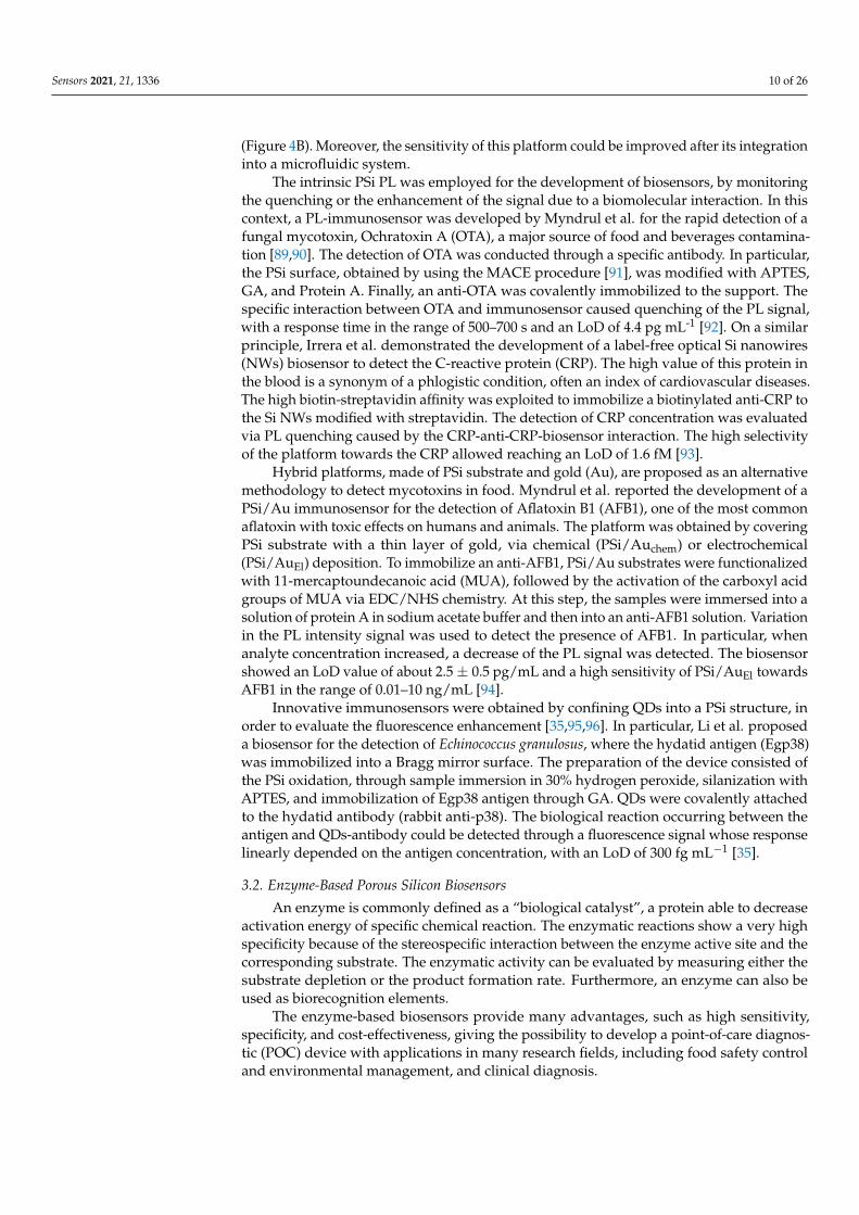
The intrinsic PSi PL was employed for the development of biosensors, by monitoringthe quenching or the enhancement of the signal due to a biomolecular interaction. In thiscontext, a PL-immunosensor was developed by Myndrul et al. for the rapid detection of afungal mycotoxin, Ochratoxin A (OTA), a major source of food and beverages contamina-tion [89,90]. The detection of OTA was conducted through a specific antibody. In particular,the PSi surface, obtained by using the MACE procedure [91], was modified with APTES,GA, and Protein A. Finally, an anti-OTA was covalently immobilized to the support. Thespecific interaction between OTA and immunosensor caused quenching of the PL signal,with a response time in the range of 500–700 s and an LoD of 4.4 pg mL-1 [92]. On a similarprinciple, Irrera et al. demonstrated the development of a label-free optical Si nanowires(NWs) biosensor to detect the C-reactive protein (CRP). The high value of this protein inthe blood is a synonym of a phlogistic condition, often an index of cardiovascular diseases.The high biotin-streptavidin affinity was exploited to immobilize a biotinylated anti-CRP tothe Si NWs modified with streptavidin. The detection of CRP concentration was evaluatedvia PL quenching caused by the CRP-anti-CRP-biosensor interaction. The high selectivityof the platform towards the CRP allowed reaching an LoD of 1.6 fM [93].
Hybrid platforms, made of PSi substrate and gold (Au), are proposed as an alternativemethodology to detect mycotoxins in food. Myndrul et al. reported the development of aPSi/Au immunosensor for the detection of Aflatoxin B1 (AFB1), one of the most commonaflatoxin with toxic effects on humans and animals. The platform was obtained by coveringPSi substrate with a thin layer of gold, via chemical (PSi/Auchem) or electrochemical(PSi/AuEl) deposition. To immobilize an anti-AFB1, PSi/Au substrates were functionalizedwith 11-mercaptoundecanoic acid (MUA), followed by the activation of the carboxyl acidgroups of MUA via EDC/NHS chemistry. At this step, the samples were immersed into asolution of protein A in sodium acetate buffer and then into an anti-AFB1 solution. Variationin the PL intensity signal was used to detect the presence of AFB1. In particular, whenanalyte concentration increased, a decrease of the PL signal was detected. The biosensorshowed an LoD value of about 2.5 ± 0.5 pg/mL and a high sensitivity of PSi/AuEl towardsAFB1 in the range of 0.01–10 ng/mL [94].
Innovative immunosensors were obtained by confining QDs into a PSi structure, inorder to evaluate the fluorescence enhancement [35,95,96]. In particular, Li et al. proposeda biosensor for the detection of Echinococcus granulosus, where the hydatid antigen (Egp38)was immobilized into a Bragg mirror surface. The preparation of the device consisted ofthe PSi oxidation, through sample immersion in 30% hydrogen peroxide, silanization withAPTES, and immobilization of Egp38 antigen through GA. QDs were covalently attachedto the hydatid antibody (rabbit anti-p38). The biological reaction occurring between theantigen and QDs-antibody could be detected through a fluorescence signal whose responselinearly depended on the antigen concentration, with an LoD of 300 fg mL−1 [35].
3.2. Enzyme-Based Porous Silicon Biosensors
An enzyme is commonly defined as a “biological catalyst”, a protein able to decreaseactivation energy of specific chemical reaction. The enzymatic reactions show a very highspecificity because of the stereospecific interaction between the enzyme active site and thecorresponding substrate. The enzymatic activity can be evaluated by measuring either thesubstrate depletion or the product formation rate. Furthermore, an enzyme can also beused as biorecognition elements.
The enzyme-based biosensors provide many advantages, such as high sensitivity,specificity, and cost-effectiveness, giving the possibility to develop a point-of-care diagnos-tic (POC) device with applications in many research fields, including food safety controland environmental management, and clinical diagnosis.

Sensors 2021, 21, 1336 11 of 26
Table 1. Summary of detection of various Biomolecules using PSi optical device as a transducing substrate.
PSi Structure TransductionMechanism
Type ofBioprobe Capture Probe Analyte LoD Ref.
TiO2 PSi Reflectivity Antibody Anti-SLP S-layer protein 0.70 ± 0.37 pM [80]
Si nanoporearray Reflectivity Antibody E. coli antibody E. coli 103 to 107 CFU mL−1 [84]
Fabry-Pérot Reflectivity Antibody Anti-HSP70 HSP70 1290 ± 160 ng/mL [83]
Single layer Photoluminescence Antibody Anti-OTA Ochratoxin A 4.4 pg mL−1 [92]
Si NWs Photoluminescence Antibody Anti-CRPs C-reactiveprotein 1.6 fM [93]
PSi layercovered with
thin layer of AuPhotoluminescence Antibody Anti-AFB1 Aflatoxin B1 2.5 ± 0.5 pg/mL [94]
Bragg mirror Photoluminescence Antibody Anti-p38 Echinococcusgranulosus 300 fg mL−1 [35]
Single layer Reflectivity Enzyme Horseradishperoxidase
Ag+, Pb2+ andCu2 60–120 ppb [97]
Single layer Reflectivity Enzyme1-Naphthyl-N-
acetyl-β-D-glucosaminide
NGAsesubstrate 0.51 µM/min [98]
Microcavity Fluorescence(FRET-based) Enzyme Fluorogenic
peptidesSortase A and
MMPs 4.6 × 10⁻⁸ M [39]
Microcavity Fluorescence Enzyme Resazurin L-lactatedehydrogenase 0.08 U/mL [99]
SiNWs Photoluminescence Enzyme Glucoseoxidase Glucose 1.06 µM [40]
Single layer Reflectivity DNA DNA 1 × 10⁻⁹ M [100]
Double Braggmirror Reflectivity DNA
Complementaryand partially
complementaryDNA
AOB gene 27.1 nM and 35 nM [71]
Microcavity Photoluminescence DNA DNA - [101]
Bragg mirrorDigital
fluorescencemicroscopy
DNA DNA - 88 pM [102]
Au/PSi Photoluminescence DNA DNA - 328.7 nM [103]
Ring resonator Reflectivity DNA PNA - 3 nM [104]
Single layer Reflectivity andFluorescence PNA DNA SCN5A gene 25 ± 2 µM and 18 ±
3 µM [37]
Single layer Reflectivity Aptamer Bacteria Lactobacillusacidophilus 106 cells mL−1 [105]
Single layer Reflectivity Aptamer40-mer
anti-HIs-tag6H7 aptamer
His-taggedprotein 7.5 nM [79]
Single layer Reflectivity andIAW Aptamer 28-mer anti
TNF α aptamer TNFα 3.0 nM [77]
Single layer Reflectivity Aptamer
17-merthrombinbindingaptamer
Humanα-thrombin 1.5 ± 0.3 nM [106]
Sensors 2021, 21, 1336 12 of 26
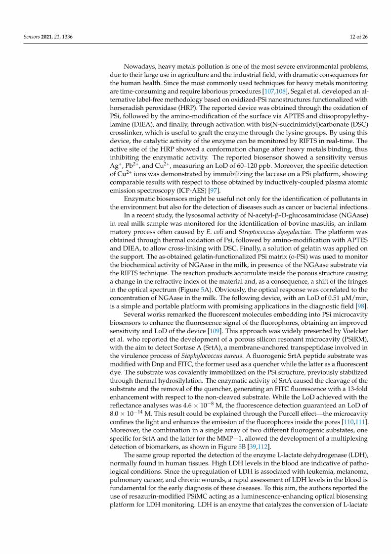
Nowadays, heavy metals pollution is one of the most severe environmental problems,due to their large use in agriculture and the industrial field, with dramatic consequences forthe human health. Since the most commonly used techniques for heavy metals monitoringare time-consuming and require laborious procedures [107,108], Segal et al. developed an al-ternative label-free methodology based on oxidized-PSi nanostructures functionalized withhorseradish peroxidase (HRP). The reported device was obtained through the oxidation ofPSi, followed by the amino-modification of the surface via APTES and diisopropylethy-lamine (DIEA), and finally, through activation with bis(N-succinimidyl)carbonate (DSC)crosslinker, which is useful to graft the enzyme through the lysine groups. By using thisdevice, the catalytic activity of the enzyme can be monitored by RIFTS in real-time. Theactive site of the HRP showed a conformation change after heavy metals binding, thusinhibiting the enzymatic activity. The reported biosensor showed a sensitivity versusAg+, Pb2+, and Cu2+, measuring an LoD of 60–120 ppb. Moreover, the specific detectionof Cu2+ ions was demonstrated by immobilizing the laccase on a PSi platform, showingcomparable results with respect to those obtained by inductively-coupled plasma atomicemission spectroscopy (ICP-AES) [97].
Enzymatic biosensors might be useful not only for the identification of pollutants inthe environment but also for the detection of diseases such as cancer or bacterial infections.
In a recent study, the lysosomal activity of N-acetyl-β-D-glucosaminidase (NGAase)in real milk sample was monitored for the identification of bovine mastitis, an inflam-matory process often caused by E. coli and Streptococcus dysgalactiae. The platform wasobtained through thermal oxidation of Psi, followed by amino-modification with APTESand DIEA, to allow cross-linking with DSC. Finally, a solution of gelatin was applied onthe support. The as-obtained gelatin-functionalized PSi matrix (o-PSi) was used to monitorthe biochemical activity of NGAase in the milk, in presence of the NGAase substrate viathe RIFTS technique. The reaction products accumulate inside the porous structure causinga change in the refractive index of the material and, as a consequence, a shift of the fringesin the optical spectrum (Figure 5A). Obviously, the optical response was correlated to theconcentration of NGAase in the milk. The following device, with an LoD of 0.51 µM/min,is a simple and portable platform with promising applications in the diagnostic field [98].
Several works remarked the fluorescent molecules embedding into PSi microcavitybiosensors to enhance the fluorescence signal of the fluorophores, obtaining an improvedsensitivity and LoD of the device [109]. This approach was widely presented by Voelckeret al. who reported the development of a porous silicon resonant microcavity (PSiRM),with the aim to detect Sortase A (SrtA), a membrane-anchored transpeptidase involved inthe virulence process of Staphylococcus aureus. A fluorogenic SrtA peptide substrate wasmodified with Dnp and FITC, the former used as a quencher while the latter as a fluorescentdye. The substrate was covalently immobilized on the PSi structure, previously stabilizedthrough thermal hydrosilylation. The enzymatic activity of SrtA caused the cleavage of thesubstrate and the removal of the quencher, generating an FITC fluorescence with a 13-foldenhancement with respect to the non-cleaved substrate. While the LoD achieved with thereflectance analyses was 4.6 × 10⁻⁸ M, the fluorescence detection guaranteed an LoD of8.0 × 10−14 M. This result could be explained through the Purcell effect—the microcavityconfines the light and enhances the emission of the fluorophores inside the pores [110,111].Moreover, the combination in a single array of two different fluorogenic substates, onespecific for SrtA and the latter for the MMP−1, allowed the development of a multiplexingdetection of biomarkers, as shown in Figure 5B [39,112].
The same group reported the detection of the enzyme L-lactate dehydrogenase (LDH),normally found in human tissues. High LDH levels in the blood are indicative of patho-logical conditions. Since the upregulation of LDH is associated with leukemia, melanoma,pulmonary cancer, and chronic wounds, a rapid assessment of LDH levels in the blood isfundamental for the early diagnosis of these diseases. To this aim, the authors reported theuse of resazurin-modified PSiMC acting as a luminescence-enhancing optical biosensingplatform for LDH monitoring. LDH is an enzyme that catalyzes the conversion of L-lactate

Sensors 2021, 21, 1336 13 of 26
to pyruvate and the reduction of NAD+ into NADH. This reaction was coupled to thereduction of non-fluorescent resazurin into fluorescent-resorufin [113]. For the platformdevelopment, PSiMC was passivated via thermal hydrocarbonization and thermal hydrosi-lylation, followed by the formation of acid chloride by using thionyl chloride and acylationof a non-fluorescent resazurin. The passivation procedure strongly improves the surfacestability, preventing both the oxidation of PSiMC and resazurin, prior to detection. TheLDH detection through resazurin-PSiMC was monitored as an increase in fluorescenceintensity of 10 and 5-fold higher, with respect to that of single-layer and detuned micro-cavity, respectively. The reported platform showed an LoD of 0.08 U/mL and a detectionrange between 0.16 and 6.5 U/mL, which covered the concentration range of the enzymein healthy and damaged cells [99].
Sensors 2021, 21, x FOR PEER REVIEW 13 of 26
achieved with the reflectance analyses was 4.6 × 10⁻⁸ M, the fluorescence detection guar-
anteed an LoD of 8.0 × 10⁻¹⁴ M. This result could be explained through the Purcell effect—
the microcavity confines the light and enhances the emission of the fluorophores inside
the pores [110,111]. Moreover, the combination in a single array of two different fluoro-
genic substates, one specific for SrtA and the latter for the MMP−1, allowed the develop-
ment of a multiplexing detection of biomarkers, as shown in Figure 5B [39,112].
The same group reported the detection of the enzyme L-lactate dehydrogenase
(LDH), normally found in human tissues. High LDH levels in the blood are indicative of
pathological conditions. Since the upregulation of LDH is associated with leukemia, mel-
anoma, pulmonary cancer, and chronic wounds, a rapid assessment of LDH levels in the
blood is fundamental for the early diagnosis of these diseases. To this aim, the authors
reported the use of resazurin-modified PSiMC acting as a luminescence-enhancing optical
biosensing platform for LDH monitoring. LDH is an enzyme that catalyzes the conversion
of L-lactate to pyruvate and the reduction of NAD+ into NADH. This reaction was cou-
pled to the reduction of non-fluorescent resazurin into fluorescent-resorufin [113]. For the
platform development, PSiMC was passivated via thermal hydrocarbonization and ther-
mal hydrosilylation, followed by the formation of acid chloride by using thionyl chloride
and acylation of a non-fluorescent resazurin. The passivation procedure strongly im-
proves the surface stability, preventing both the oxidation of PSiMC and resazurin, prior
to detection. The LDH detection through resazurin-PSiMC was monitored as an increase
in fluorescence intensity of 10 and 5-fold higher, with respect to that of single-layer and
detuned microcavity, respectively. The reported platform showed an LoD of 0.08 U/mL
and a detection range between 0.16 and 6.5 U/mL, which covered the concentration range
of the enzyme in healthy and damaged cells [99].
Figure 5. (A) (a) Schematic representation of o-PSi biosensor for the NGAse activity detection. The NGAse present in the
milk hydrolyzes the NGAse substrate into N-acetyl- β-D-glucosaminide and 1-naphthol. The reaction between 1-Naphthol
and Hexazonium Pararosaniline forms an insoluble product that precipitates into the PSi nanostructures causing a change
in the reflectivity spectra; (b) reflective-based response of NGAse catalytic activity in milk samples positive to S. dysgalac-
tiae and (c) E. coli. The lines (b1, c1) are the baseline, the reaction solution and the dye are represented by lines (b2, c2)
while the buffer wash is represented by lines (b3, c3); (d) averaged optical response obtained by subtracting the blank
Figure 5. (A) (a) Schematic representation of o-PSi biosensor for the NGAse activity detection. The NGAse present in themilk hydrolyzes the NGAse substrate into N-acetyl- β-D-glucosaminide and 1-naphthol. The reaction between 1-Naphtholand Hexazonium Pararosaniline forms an insoluble product that precipitates into the PSi nanostructures causing a changein the reflectivity spectra; (b) reflective-based response of NGAse catalytic activity in milk samples positive to S. dysgalactiaeand (c) E. coli. The lines (b1, c1) are the baseline, the reaction solution and the dye are represented by lines (b2, c2) while thebuffer wash is represented by lines (b3, c3); (d) averaged optical response obtained by subtracting the blank relative EOT forthe corresponding milk samples. Reprinted with permission from Reference [98]; Copyright (2020) American ChemicalSociety. (B) (a) Scheme of a FRET-based PSi biosensor for the detection of SrtA and MMP. The FRET peptide substates areimmobilized into a PSi microcavity (central panels). The enzymatic cleavage causes a fluorescence enhancement (rightpanels); (b)reflectance spectra of PSiRM before and after incubation with SrtA; (c) fluorescence emission of SrtA substratein solution (dashed line) and immobilized on the PSi surface after SrtA incubation. Reprinted with permission fromReference [39] https://creativecommons.org/licenses/by/4.0/ (accessed on 16 December 2020).
Ghosh et al. explored the PL tunability of Si NWs, after their surface modificationby glucose (GL) and by its conjugated enzyme, glucose oxidase (GOx). The surface mod-ification of Si NWs was obtained by dipping the samples in GL/GOx solution for 1 h at60 ◦C. A different PL effect was measured based on the different glucose concentrations.In particular, the H2O2 production in the GL/GO solution caused a PL quenching of SiNWs at lower GL concentrations; on the contrary, an enhancement of PL was observed at

Sensors 2021, 21, 1336 14 of 26
higher GL concentration—the different behaviour could be correlated to the removal ofSi-H bonds on Si NWs surface and to the attachment of different carbon-based functionalgroups. Therefore, the proposed biosensor showed a detection range from 0.5 to 1.0 mMand an LoD of 1.06 µM [40].
3.3. Oligonucleotides- and Analogues-Based Biosensors
Oligonucleotide based biosensing technologies represent a field under intense investi-gation due to their broad applications in biomedicine and environmental monitoring.
An oligonucleotide-based biosensor shows a short functional nucleic acid, generallyidentified in a single-stranded DNA (ssDNA) or RNA, which is immobilized on the surfaceof the transducer material and specifically interacts with a complementary DNA or RNAsequence, through the base pairing. The same hybridization mechanism is outperformedby analogues of oligonucleotides (i.e., peptide nucleic acid, PNA), that have had a greatimpact in biosensing because of their enhanced properties, due to traditional oligonu-cleotide probes, opening the way for the development of highly specific platforms for theearly detection of genetic diseases. Finally, aptamers are single-strands of DNA or RNA,commonly known to have a high affinity towards biological (i.e., whole cells, protein)or chemical compounds (i.e., metal ions). The high binding affinity towards the targetmakes the aptamers a valid alternative to antibodies. In all reported cases, the specifichybridization between the bioprobe and the respective target is detectable as an opticalmeasurable signal.
3.3.1. DNA and Peptide Nucleic Acid-PSi Biosensors
In recent years, there has been a substantial development in DNA-based biosensorsdue to the stability and sensitivity that distinguish these kinds of probes. In a DNA-PSibased biosensor, the interaction between the probe and the target is monitored as a red-shift in the reflectivity spectra, due to an increase in the average refractive index of thematerial [76,114]. However, many published articles report a shift of the spectrum at lowerwavelengths when DNA/DNA hybridization occurs—this effect could be related to thecorrosion of PSi structure, caused by the accumulation on the PSi surface of the DNAbackbone negative charges [115,116]. Moreover, the corrosion process was controlled byWeiss et al. In particular, they obtained a greater stable surface, also in harsh environmentalconditions, passivating the PSi surface through thermal carbonization. As a result, a 16-merss-DNA, covalently attached to TCPSi, was efficiently hybridized to the complementaryDNA, minimizing the unspecific interactions [58].
The low LoD of PSi biosensors in the micromolar range, due to the limited masstransport of the analytes into pores, was exceeded by incorporating the biosensing architec-ture into a microfluidic system [75,76]. Moreover, a three-order magnitude enhancementin the DNA-PSi-based biosensor was measured by implementing the electrokinetic iso-tachophoresis (ITP) strategy, developed by Segal et al., into a microfluidic platform. Byusing an applied electric field, the charged analytes were separated on the basis of theirionic mobility, leading to the sample preconcentration, and as consequence, a great im-provement in terms of the device sensitivity was achieved. A schematic representation ofthe PSi device integrated with ITP is shown in Figure 6A. The device was integrated intoa microfluidic system after the PSi oxidation and its silanization with APTES. All othersurface modifications were carried out by pumping the solutions into the microchannel. Inparticular, the NH2- PSi was incubated with a solution of sulfo-SMCC crosslinker, used tobound the thiol-modified ss-DNA. This technique allowed to improve the sensitivity ofFabry-Pérot biosensor of 1000-fold, measuring an LoD of 1 × 10⁻⁹ M [100].

Sensors 2021, 21, 1336 15 of 26Sensors 2021, 21, x FOR PEER REVIEW 16 of 26
Figure 6. (A) (a) Scheme of the integrated PSiO2 interferometric biosensor combined with the ITP technique into a micro-
fluidic system; (b–d) scheme of the concentration process of target DNA (ssDNA) under ITP, followed by binding with
immobilized DNA probe; (e,f) reflectivity spectra and corresponding FFT before and after DNA hybridization. Reprinted
with permission from reference [100]; Copyright (2015) Wiley-VCH. (B) (a) Schematic representation of the sensing prin-
ciple—the hybridization between target QDs-DNA and the complementary sequence, immobilized into PSi microcavity,
causes a huge red-shift of the reflectivity spectra, increasing the response signal; (b) reflectance spectra after each func-
tionalization steps; (c) relationship between the of QDs-DNA and control DNA at different concentrations and reflectance
spectra. Reprinted with permission from Reference [101] https://creativecommons.org/licenses/by/4.0/ (accessed on 16 De-
cember 2020). (C) (a) Functionalization steps and sensing mechanism of QDs-AuNPs FRET biosensor for DNA detection;
Figure 6. (A) (a) Scheme of the integrated PSiO2 interferometric biosensor combined with the ITP technique into amicrofluidic system; (b–d) scheme of the concentration process of target DNA (ssDNA) under ITP, followed by bindingwith immobilized DNA probe; (e,f) reflectivity spectra and corresponding FFT before and after DNA hybridization.Reprinted with permission from reference [100]; Copyright (2015) Wiley-VCH. (B) (a) Schematic representation of thesensing principle—the hybridization between target QDs-DNA and the complementary sequence, immobilized into PSimicrocavity, causes a huge red-shift of the reflectivity spectra, increasing the response signal; (b) reflectance spectra aftereach functionalization steps; (c) relationship between the of QDs-DNA and control DNA at different concentrationsand reflectance spectra. Reprinted with permission from Reference [101] https://creativecommons.org/licenses/by/4.0/(accessed on 16 December 2020). (C) (a) Functionalization steps and sensing mechanism of QDs-AuNPs FRET biosensor forDNA detection; (b) DNA conjugation to QDs, and (c) DNA conjugated to AuNPs; (d) PL emission of QDs embedded in PSimatrix before and after hybridization with AuNPs-DNA; (e) control experiment performed by using AuNPs-conjugatedwith non-complementary DNA; (f) quenching efficiency Q = 1 − Fq/F0 with several DNA target concentrations; and(g) linear relationship between decrease of PL intensity and different AuNPs-target DNA. Reprinted with permission fromReference [103]. https://creativecommons.org/licenses/by/4.0/ (accessed on 24 January 2021).

Sensors 2021, 21, 1336 16 of 26
The design of sophisticated photonic structures (i.e., ring resonator, Bragg mirror,waveguides) represents a further strategy to enhance the detection of low concentratedanalytes. This issue might be pursued by confining the incident light and increasing theinteraction between light and biomolecules. A recent example was described by Zhang et al.They reported a double Bragg mirror for detecting ammonia-oxidizing bacteria (AOB),microorganisms converting organic nitrogen into nitrite and nitrate. This PSi structure,obtained by electrochemical etching at room temperature, showed a deeper resonancepeak and a broader reflectivity stop band in the reflectance spectrum, guaranteeing highsensitivity and specificity of the structure for biosensing applications. A DNA sequence,specific for AOB bacteria detection, was immobilized onto oxidized transducer materialthrough APTES and GA and used to monitor the hybridization with either complete andpartial complementary DNA sequences. The reporting LoDs were 27.1 nM and 35 nM,respectively, providing the feasibility for AOB detection in real environment [71].
The in situ ssDNA probe synthesis represents an alternative technique employedto enhance the density of capture sites in PSi photonic biosensors. Using this approach,Weiss et al. demonstrated a 5–7-fold enhanced sensitivity and a 3-fold reduction in re-sponse time in silicon microring resonators and photonic crystals, with respect to thetraditional conjugation strategy. The in situ synthesis, performed by growing unchargedDNA monomers, base-by-base, reduced the steric hindrance and charge repulsion duringthe ssDNA immobilization [117]. In this context, the pore size of the PSi matrix representsa parameter that should be evaluated. Terracciano et al. reported the impact of pore sizeon the functionalization yield of PSi transducer, through an in situ synthesis approach.After the in situ synthesis of a 19-mer DNA sequence onto mesoporous PSiO2, the authorsmeasured a medium-yield process. This effect was related to the pore size (~20 nm); amacroporous PSi with pore diameters larger than 50 nm resulted in maximizing the yieldof in situ synthesis and the sensing performance of the device [38].
The sensing performance of the PSi device could be enhanced, in terms of sensitivityand signal stability, using the PSi platform as a host matrix for the incorporation of severalcompounds or nanomaterials, such as quantum dots (QDs), metals, polymers or fluorescentmolecules. In this context, Lv et al. proposed a biosensor in which QDs were embeddedinto PSi structure. The DNA probe, immobilized onto oxidized PSi microcavity throughGA, specifically hybridized with the target DNA, coupled to CdSe/ZnS QDs, improvingthe sensitivity of PSi device 5 times, compared to the unlabelled-DNA hybridization [101].This effect could be related to the higher refractive index of QDs. A schematization of thesensing principle and some of the results are shown in Figure 6B.
Wei et al. proposed a novel, fast, and low-cost method to detect DNA/DNA interactionbased on the digital imaging of PSi/QDs surface fluorescence. The authors reported thecovalent grafting of a ssDNA into a Bragg mirror substrate. To modify the PSi surface, thesample was soaked in 30% H2O2, followed by silanization with APTES, modification withGA, and finally DNA attachment. Moreover, the carboxyl acid groups of CdSe/ZnS QDswere used to immobilize the ssDNA-target via covalent chemistry. To detect hybridization,the PSi surface was imaged by digital microscopy and, then, the images were analyzed byan image processing software. Data revealed an LoD of 88 pM, with a sensitivity slightlylower than that of conventional fluorescence analyses, but higher than the one obtainedfrom reflectance spectroscopy. Therefore, the proposed approach is fast, low-cost, andconvenient for detecting biological interactions [102].
An alternative approach to detecting an 16S rRNA, specific to Actinobacteria, wasproposed by Zhang et al. These classes of bacteria are involved in the turnover of theorganic matter and xenobiotics and they are significantly present in several habitats. Severalmethodologies were already proposed to detect DNA or RNA in food, environment,and other matrices. Moreover, although most of the proposed techniques are accurate,they are generally time-consuming and expensive [118,119]. The authors proposed a PSibiosensor based on the FRET mechanism between QDs and gold nanoparticles (AuNPs) inwhich the former act as an emission donor and the latter as a fluorescence quencher [120].

Sensors 2021, 21, 1336 17 of 26
For the device realization, the PSi DBR was oxidized in H2O2 solution, silanized withAPTES, modified with GA and, finally, QDs-DNA were bound onto the PSi substrate.Then, AuNPs were conjugated to the DNA target via thiol chemistry. To perform thehybridization experiment, different concentrations of AuNPs-DNA were added to themodified-PSi device, resulting in a quenching of the fluorescence intensity, which revealedto be proportional to the concentration of c-DNA in the range from 0.25 to 10 µM. Moreover,an LoD of 328.7 nM was achieved (Figure 6C), obtaining a simple, flexible, highly sensitive,and responsive platform [103].
Recently, peptide nucleic acid (PNA), a synthetic nucleic acids analogue, was em-ployed as a bioprobe in oligonucleotides targeting. The uncharged PNA backbone, charac-terized by N-(2-aminoethyl)glycine motifs linked by peptide bonds, provides a strongerinteraction between PNA/DNA strands in comparison to DNA/DNA duplex [121,122].Consequently, a DNA/PNA base mismatch would be more destabilizing with respect toDNA/DNA complex. Other properties, such as the capability to hybridize in low-saltconcentration as well as the chemical and thermal stability make the PNA a powerful probeto discriminate single-point mutations, allowing the development of biosensors for earlydiagnosis of genetic diseases.
One of the examples in the use of PNA as bioprobe in a PSi platform was reportedby Weiss et al. They tried to reduce the corrosion process of the PSi matrix, normallyinduced by DNA/DNA hybridization, by replacing DNA molecules with a charge-neutralPNA probe and shelled the negative charge on the DNA backbone by the introduction ofMg2+ ions in the hybridization medium [115]. The same group later reported the designof a ring resonator, patterned with a PSi slab waveguide. The PSi waveguide was firstlysilanized with APTES and then modified with SPDP, used for the immobilization of thiolate-ssDNA. The results highlighted a measured LoD of 3 nM when PNA/DNA hybridizationoccurred [104].
Recently, Moretta et al. explored the introduction of a specific PNA sequence onto ahybrid GO-PSi platform in order to develop an assay for the rapid detection of BrugadaSyndrome (BS). To obtain the device, freshly etched PSi was passivated via thermal hy-drosilylation with undecylenic acid, followed by the PEGylation process via EDC/NHS.By using the same covalent approach, first graphene oxide (GO) and finally the PNA wereimmobilized on the surface of the porous matrix. The PNA probe, used in this study,was properly designed in order to detect a punctual mutation related to the SCN5A gene,responsible for BS. The hybridization between the PNA-GO-PSi device and the DNA targetwas quantified by using reflectivity analysis and fluorescence microscopy, showing an LoDof 25 ± 2 µM and 18 ± 3 µM, respectively [37].
3.3.2. Porous Silicon Aptasensors
Although the antibodies were extensively used as detection probes in various types ofbiosensors over the last 70 years, they suffer from several drawbacks (i.e., high cost, largesize, and instability), making the use of alternative probes essential.
In this regard, several scientific papers published in the last 20 years, have highlightedthe possibility of replacing the antibodies with aptamers, as promising capture probes forbiosensing applications. The idea of using aptamers was initially proposed in 1990 [123].Aptamers are short (usually from 20 to 60 nucleotides) single-strand DNA or RNA oligonu-cleotides, with a specific 3-dimensional structure that ensures the binding to a specific target.They are commonly synthetized by using the SELEX (Systematic Evolution of Ligandsby Exponential enrichment) technique, which consists of repeated rounds of an in vitroselection and amplification process. This methodology allows not only the engineeringof these biomolecules toward different targets (i.e., small molecules, proteins, whole cells)but also the reproducibility of the synthetic process [124,125]. Aptamers are consideredas strong rivals to antibodies, showing a high affinity towards their targets thanks totheir dissociation constants (Kd), ranging from picomolar to nanomolar. Therefore, theircapability to discriminate any structural differences among analogue targets makes them

Sensors 2021, 21, 1336 18 of 26
highly selective [126]. The aforementioned properties, coupled with cost-effectiveness,simple synthetic process, stability in non-physiological conditions as well as resistance todegradation and denaturation, suggest the use of the aptamers as an interesting alternativeto antibodies [127,128].
The versatility of aptamers as recognition elements makes them extensively exploredin the construction of aptasensors [21]. Segal is a pioneer in the use of aptamers in PSioptical devices, widely used for the detection of several proteins or whole-cells, as demon-strated in several published papers [79,105,129–131]. An example was provided by theuse of the aptamer Hemag1P, a 78-nucleotide sequence, employed as a capture probe forthe real-time monitoring of Lactobacillus acidophilus, a Gram-positive bacterium, presentin fermented and dairy-containing food products. A PSi Fabry-Pérot thin film, used as asubstrate, was oxidized, silanized with MPTMS, and finally incubated with the aptamerthat specifically targets S–proteins, on the outer membrane of living bacteria. The largesize of L. acidophilus cells prevented their infiltration into the porous matrix, leading to thedirect capture of bacteria onto the PSi platform. Therefore, the monitoring of aptamer–cellinteraction was performed by measuring the changes in the intensity of the reflectivityspectrum, caused by the light scattering of bacteria residing on the biosensor [132]. Theaforementioned aptasensor demonstrated its ability to detect relatively low concentrationsof L. acidophilus cells (low as 106 cells per mL), discriminating between live and deadbacteria. The development of such a biosensor could be useful to monitor probiotic amountin functional food and pharmaceuticals.
Unfortunately, the insufficient sensitivity of label-free PSi biosensors, due to thehindered diffusion of analyte into the porous matrix, represents a limiting factor. In thiscontext, Arshavsky–Graham et al. reported a proof-of-concept work in which the specificaffinity of an aptasensor to its target was paired to the abovementioned ITP technique.The device was integrated into a microfluidic system, and prior to integration with PDMSmicrochannels, the oxidized PSi was modified with APTES. Finally, the EDC solutionand the aptamer were pumped under vacuum into the microchannels to guarantee acovalent bond of the probe to the device. The suggested assay, also applicable in complexmedia, allowed to enhance the biosensor’s sensitivity up to 1000-fold, measuring an LoDof 7.5 nM, when the interaction between the aptamer and the His-tagged protein used asmodel, occurs [79].
The sensitivity and the LoD of PSi aptasensors were improved by Barillaro et al. witha new post-processing analysis of the reflectance signal, instead of the conventional FFT re-flectance (Figure 7A). This methodology, known as interferogram average over wavelength(IAW) reflectance spectroscopy, consisted of the calculation of averaged interferometricvalues over the spectral range of interest. This methodology was applied for the detectionof tumor necrosis factor alfa (TNFα) at a concentration down to 3.0 nM. Moreover, Marianiet al. showed that the diffusion of biomolecules into the pores could be improved and theprotein detection was enhanced by a factor 104 with respect to nonamplified label-free PSibiosensors, by combining a novel PSi structure (with a pore size of ~80 nm) with the IAWtechnique. The following device was obtained by immobilizing a thiol modifier-aptameron APDMES-modified PSiO2 through SMCC [77].
A further example of a PSi-based biosensor was proposed for the detection of humanα-thrombin. Human α-thrombin, also known as coagulation factor II, is a serine proteasethat converts soluble fibrinogen into insoluble strands of fibrin, promoting blood clotting.High levels of thrombin in the blood could be a cause of several pathological coagulationdiseases. For this reason, devices that are able to detect the thrombin levels in the bloodwith high selectivity, low LoD, and in a short time are urgently required. In this context,Terracciano et al. successfully demonstrated the development of a new label-free opticalaptasensor, realized by in situ synthesis of a 15-mer thrombin binding aptamer (TBATT),covalently conjugated to the APTES-modified macroporous silica surface. The in situsynthesis shows many advantages—an increased density of the bioprobe on the surface,enhancement of the device sensitivity, automatable fabrication process, and possibility to

Sensors 2021, 21, 1336 19 of 26
functionalize the surface in microarrays. Moreover, the PSi surface functionalization withTBA (FTBA) was estimated to be (1.92 ± 0.03) 10−5 mol/g, considering a PSi sample havinga weight of 0.2 mg. The ratio FTBA/SSA (specific surface area, which was 199 m2 g−1 for as-etched PSi) was used to calculate the PSi functionalization in terms of nmol cm−1, resultingin 0.0125 ± 0.0002 nmol cm−2. The LoD reached by the aptasensor was 1.5 ± 0.3 nM.Moreover, the high selectivity and regenerability of the biosensor were proved. [106].
Sensors 2021, 21, x FOR PEER REVIEW 20 of 26
Figure 7. (A) (a) Cartoon of the as-prepared PSi platform. (b) Four-step protocol used to immobilize the aptamer probe
into the PSi structure consisting of thermal oxidation, silanization with APDMES, grafting of SMCC, and aptamer binding;
(c) FFT spectra measured after each functionalization steps; (d) reflection spectra acquired before (black line) and after
injection of TNFα (red line) with (e) corresponding spectral interferogram; (f) IAW signals obtained in the range of TNFα
tested (3–390 nM) for PSi with smaller (black line) and larger (red line) pores. The blank IAW0 value is subtracted from the
data; (g) calibration curve over the whole range of the tested TNFα obtaining using the EOT value as output signal. Re-
printed with permission from Reference [77]; Copyright (2016) American Chemical Society. (B) (a) Scheme of the immo-
bilization process of the 6H7 aptamer (upper panel) and antibodies, via random and oriented conjugation (lower panel)
into PSiO2; (b) EOT changes of the three type biosensors after the exposure to different concentration of his-tagged-tyrosi-
nase; (c,d) binding rates obtained after the incubation of the biosensors with different concentrations of tyrosinase. Re-
printed with permission from Reference [133]; Copyright (2020) The Royal Society of Chemistry 2020.
4. PSi Biosensors for Analytes Detection in Complex Matrices
Many studies highlight the ability of biosensors to discriminate between target and
non-target sequences, reaching a high sensitivity level. Although most of these analyses
were carried out in clean buffers, the detection of analytes in complex media is still a chal-
lenge. The cross-reactivity and the non-specific attachment of biomolecules must be
avoided in order to obtain a reliable result. This issue could be achieved by treating the
surfaces with blocking agents (i.e., Tris, BSA, PEG). In some cases, additional steps, in-
cluding pre-treatment or sample separation through suitable methodologies, are required
Figure 7. (A) (a) Cartoon of the as-prepared PSi platform. (b) Four-step protocol used to immobilize the aptamer probeinto the PSi structure consisting of thermal oxidation, silanization with APDMES, grafting of SMCC, and aptamer binding;(c) FFT spectra measured after each functionalization steps; (d) reflection spectra acquired before (black line) and afterinjection of TNFα (red line) with (e) corresponding spectral interferogram; (f) IAW signals obtained in the range of TNFαtested (3–390 nM) for PSi with smaller (black line) and larger (red line) pores. The blank IAW0 value is subtracted fromthe data; (g) calibration curve over the whole range of the tested TNFα obtaining using the EOT value as output signal.Reprinted with permission from Reference [77]; Copyright (2016) American Chemical Society. (B) (a) Scheme of theimmobilization process of the 6H7 aptamer (upper panel) and antibodies, via random and oriented conjugation (lowerpanel) into PSiO2; (b) EOT changes of the three type biosensors after the exposure to different concentration of his-tagged-tyrosinase; (c) EOT changes vs time of oriented antibody and aptamer onto PSiO2 upon exposure to different concentrationsof his-tagged-tyrosinase; (d,e) binding rates at 10 min and 60 min obtained after the incubation of the biosensors with dif-ferent concentrations of tyrosinase. (ns) or (*) indicate the non-significant or statistically significant difference, respec-tively,between the total binding or the binding rate of the oriented antibody and aptamer-based biosensors. Reprinted withpermission from Reference [133]; Copyright (2020) The Royal Society of Chemistry 2020.

Sensors 2021, 21, 1336 20 of 26
A recent interesting study, conducted by Arshavsky-Graham et al., compared, forthe first time, the performances of an aptamer, a randomly immobilized antibody and anoriented antibody on the PSi surface for the his-tagged-tyrosinase detection, as reportedin Figure 7B. A PSiO2 substrate, modified with APTES and succinic acid, was used tocovalently link the aptamer to the surface via EDC/NHS chemistry. Silanized PSIO2was functionalized with GA for the immobilization of antibodies. The direct exposureof antibodies to GA resulted in a random orientation. Vice versa, a modification of GA-silanized-PSiO2 with streptavidin (SA) and biotinylated-PrA allowed the right orientationof antibodies.
In brief, while unoriented antibodies on the PSi surface caused a poor biosensingperformance, aptamer, and oriented antibody-based biosensors had similar results in termsof LoD, binding rate, and selectivity. Nevertheless, the superiority of the aptasensors wasrepresented by the faster and cost-effective fabrication, as well as the possibility to storethe device in dry conditions. Furthermore, PSi aptasensors could be regenerated thanksto the reversible denaturation process of aptamers [130,133]. Performances of the devicecould be improved through a lower surface coverage. Indeed, a reduced density surfaceallows minimizing the effects of steric hindrance and the electrostatic repulsion of thenegatively-charged DNA molecules at higher concentrations [105,130,134].
4. PSi Biosensors for Analytes Detection in Complex Matrices
Many studies highlight the ability of biosensors to discriminate between target andnon-target sequences, reaching a high sensitivity level. Although most of these analyseswere carried out in clean buffers, the detection of analytes in complex media is still achallenge. The cross-reactivity and the non-specific attachment of biomolecules mustbe avoided in order to obtain a reliable result. This issue could be achieved by treatingthe surfaces with blocking agents (i.e., Tris, BSA, PEG). In some cases, additional steps,including pre-treatment or sample separation through suitable methodologies, are requiredto improve the specificity. Involving PSi as a transducer material for the detection ofanalytes in complex matrices, not only requires a proper surface stabilization, but also theappropriate tailoring of the porous matrix. The most common real samples are blood orserum, saliva, water, bacterial lysates, and human isles of Langerhans [79,134–136].
Bonanno et al. developed a PSi Bragg-mirror-competitive test to detect opioids in urinespecimens. They demonstrated that, by varying the device chemical surface and volume ofurine sample added to the porous matrix, sensitivity and specificity of the assay could beimproved. Additionally, the fabrication of PSi layers, with different porosity, improved theinfiltration of macromolecules through the pores. Good results were obtained by using adevice that was previously oxidized, amino-silanized, and subsequently, exposed to bovineserum albumin (BSA). Finally, a morphine analogue (M3G) was covalently bound to lysinegroups exposed on the BSA-PSi platform. The free drugs, contained in urine samples andanalogues, bound to the matrix, competed for the binding site of the antibodies. An indirectmethodology was used to detect the free drug, measuring an LoD of 0.018 µM and makingthe platform appealing for POC applications [137,138].
The foodborne diseases represent a public health problem, caused by the contamina-tion of food with several types of microorganisms or mycotoxins that could occur duringthe several steps of food production. The ingestion of contaminated food is the cause ofserious complications. For this reason, the rapid detection of bacterial contamination inthe complex food industry water is crucial to prevent foodborne diseases. This issue waspursued by Massad-Ivanir et al. They designed a PSi-based biosensor for the real-timedetection of E. coli in complex water samples. The silanized-PSi was modified with GAfollowed by exposure to SA. Finally, a biotinylated E. coli antibody was bound to the device.In the biosensing experiment, the interaction between E. coli cells-antibody was monitoredas a decrease in the intensity of the reflected light, demonstrating an LoD of 103 cells mL−1
in the food industry water, without pre-enrichment of the sample. Such a device could be

Sensors 2021, 21, 1336 21 of 26
considered a promising tool to detect contaminations during product processing, reducingthe risk of the spread of pathogens [136].
A huge step forward was taken by the Voelcker group, which investigated the use ofPSi optical rugate filter like an in vivo biosensor. First, the stability and the biocompatibilityof thermally oxidized and thermal hydrocarbonized devices were compared in in vitrostudies. Data obtained demonstrated that not only did the THC-PSi maintain its structuraland optical integrity, but its pre-incubation in cell medium (DMEM) for 10 days madethe material not cytotoxic. As a result, the PITHC-PSi, subcutaneously implanted in amurine model, revealed that the optical signal was recorded through the skin of the mice1 week after implantation, emphasizing how the stabilization of the surface is important tominimize the degradation of the device in physiological media [45].
5. Conclusions
The well-established and low-cost fabrication processes, the versatility of the material,and its optical and physical properties, together with the high internal surface area, makesPSi an appealing material for the development of optical biosensors with applications infood safety, environmental monitoring, and medical diagnostics. To this aim, the knowl-edge of surface chemistry modification approaches plays a crucial role in PSi-based devicesto protect the PSi from degradation and provide functional groups that are useful for theattachment of biorecognition probes. This aspect is extensively examined in this review,together with an accurate exploration of the recent advancements in optical PSi-basedbiosensors for the rapid detection of different analytes. The attention was mainly focusedon identifying the different adopted strategies to improve the biosensing performances interms of stability, sensitivity, and detection limit, starting from the bioprobe immobilization.
Although the detection of analytes in clean samples is widely validated, identifyinga specific molecule in real samples still represents a challenge. Moreover, even thougha few examples are already reported in the literature, further efforts are needed to en-hance biosensor stability in complex sample matrices and minimize the non-specificadsorption of biomolecules. This issue could be achieved through a practical surfacemodification strategy.
Other studies are necessary to obtain the miniaturization of the devices that couldbecome important for the development and commercialization of POC devices.
Author Contributions: R.M. writing-draft preparation; M.T. writing-review and editing; I.R. andL.D.S. supervision. All authors have read and agreed to the published version of the manuscript.
Funding: This research received partial financial support from PON-AIM RTDA_L1 (AIM 1873131-2).
Institutional Review Board Statement: Not applicable.
Informed Consent Statement: Not applicable.
Data Availability Statement: Not applicable.
Acknowledgments: M. Terracciano acknowledges financial support from PON-AIM RTDA_L1(AIM 1873131-2).
Conflicts of Interest: The authors declare no conflict of interest.
References1. Mehrotra, P. Biosensors and their applications—A review. J. Oral Biol. Craniofac. Res. 2016, 6, 153–159. [CrossRef]2. Bhalla, N.; Jolly, P.; Formisano, N.; Estrela, P. Introduction to biosensors. Essays Biochem. 2016, 60, 1–8.3. Thvenot, D.R.; Toth, K.; Durst, R.A.; Wilson, G.S. Electrochemical biosensors: Recommended definitions and classification
(Technical Report). Pure Appl. Chem. 1999, 71, 2333–2348. [CrossRef]4. Clark, L.C.; Lyons, C. Electrode Systems for Continuous Monitoring in Cardiovascular Surgery. Ann. N. Y. Acad. Sci. 2006, 102,
29–45. [CrossRef] [PubMed]5. Biosensors Market Size Worth $36.0 Billion By 2027|CAGR: 7.9%. Available online: https://www.grandviewresearch.com/press-
release/global-biosensors-market (accessed on 16 August 2020).

Sensors 2021, 21, 1336 22 of 26
6. Kreyling, W.G.; Semmler-Behnke, M.; Chaudhry, Q. A complementary definition of nanomaterial. Nano Today 2010, 5, 165–168.[CrossRef]
7. Nasrollahzadeh, M.; Issaabadi, Z.; Sajjadi, M.; Sajadi, S.M.; Atarod, M. Types of Nanostructures. In Interface Science and Technology;Elsevier B.V.: Amsterdam, The Netherlands, 2019; Volume 28, pp. 29–80.
8. Su, H.; Li, S.; Jin, Y.; Xian, Z.; Yang, D.; Zhou, W.; Mangaran, F.; Leung, F.; Sithamparanathan, G.; Kerman, K. Nanomaterial-basedbiosensors for biological detections. Adv. Health Care Technol. 2017, 3, 19–29. [CrossRef]
9. Pirzada, M.; Altintas, Z. Nanomaterials for Healthcare Biosensing Applications. Sensors 2019, 19, 5311. [CrossRef]10. Malhotra, B.D.; Ali, M.A. Nanomaterials in Biosensors. In Nanomaterials for Biosensors; Elsevier: Amsterdam, The Netherlands,
2018; pp. 1–74.11. Tieu, T.; Alba, M.; Elnathan, R.; Cifuentes-Rius, A.; Voelcker, N.H. Advances in Porous Silicon-Based Nanomaterials for Diagnostic
and Therapeutic Applications. Adv. Ther. 2019, 2, 1800095. [CrossRef]12. Li, W.; Liu, Z.; Fontana, F.; Ding, Y.; Liu, D.; Hirvonen, J.T.; Santos, H.A. Tailoring Porous Silicon for Biomedical Applications:
From Drug Delivery to Cancer Immunotherapy. Adv. Mater. 2018, 30, 1703740. [CrossRef] [PubMed]13. Arshavsky-Graham, S.; Massad-Ivanir, N.; Segal, E.; Weiss, S. Porous Silicon-Based Photonic Biosensors: Current Status and
Emerging Applications. Anal. Chem. 2019, 91, 441–467. [CrossRef]14. Uhlir, A. Electrolytic Shaping of Germanium and Silicon. Bell Syst. Tech. J. 1956, 35, 333–347. [CrossRef]15. Canham, L.T. Silicon quantum wire array fabrication by electrochemical and chemical dissolution of wafers. Appl. Phys. Lett.
1990, 57, 1046–1048. [CrossRef]16. Available online: https://www.scopus.com (accessed on 25 January 2021).17. Herino, R.; Bomchil, G.; Barla, K.; Bertrand, C.; Ginoux, J.L. Porosity and Pore Size Distributions of Porous Silicon Layers. J.
Electrochem. Soc. 1987, 134, 1994–2000. [CrossRef]18. Lee, S.; Kang, J.; Kim, D. A Mini Review: Recent Advances in Surface Modification of Porous Silicon. Materials 2018, 11, 2557.
[CrossRef]19. Santos, H.A. Porous Silicon for Biomedical Applications; Woodhead Publishing: Cham, Switzerland, 2014; ISBN 978-0-85709-715-6.20. Low, S.P.; Voelcker, N.H. Biocompatibility of Porous Silicon. In Handbook of Porous Silicon; Springer International Publishing:
Cham, Switzerland, 2014; pp. 1–13.21. Terracciano, M.; Rea, I.; Borbone, N.; Moretta, R.; Oliviero, G.; Piccialli, G.; De Stefano, L. Porous Silicon-Based Aptasensors: The
Next Generation of Label-Free Devices for Health Monitoring. Molecules 2019, 24, 2216. [CrossRef]22. Torres-Costa, V.; Agulló-Rueda, F.; Martín-Palma, R.J.; Martínez-Duart, J.M. Porous silicon optical devices for sensing applications.
Opt. Mater. 2005, 27, 1084–1087. [CrossRef]23. Buriak, J.M.; Allen, M.J. Lewis Acid Mediated Functionalization of Porous Silicon with Substituted Alkenes and Alkynes. J. Am.
Chem. Soc. 1998, 120, 1339–1340. [CrossRef]24. Dhanekar, S.; Jain, S. Porous silicon biosensor: Current status. Biosens. Bioelectron. 2013, 41, 54–64. [CrossRef] [PubMed]25. Sailor, M.J. Porous Silicon in Practice: Preparation, Characterization and Applications; Wiley-VCH: Weinheim, Germany, 2012;
ISBN 9783527313785.26. Canham, L. Tunable properties of Porous Silicon. In Handbook of Porous Silicon; Springer International Publishing: Cham,
Switzerland, 2014; pp. 201–206, ISBN 9783319057446.27. Md Jani, A.M.; Losic, D.; Voelcker, N.H. Nanoporous anodic aluminium oxide: Advances in surface engineering and emerging
applications. Prog. Mater. Sci. 2013, 58, 636–704. [CrossRef]28. Alvarez, S.D.; Li, C.P.; Chiang, C.E.; Schuller, I.K.; Sailor, M.J. A label-free porous alumina interferometric immunosensor. ACS
Nano 2009, 3, 3301–3307. [CrossRef]29. Mun, K.S.; Alvarez, S.D.; Choi, W.Y.; Sailor, M.J. A stable, label-free optical interferometric biosensor based on TiO2 Nanotube
Arrays. ACS Nano 2010, 4, 2070–2076. [CrossRef] [PubMed]30. Song, Y.Y.; Schmuki, P. Modulated TiO2 nanotube stacks and their use in interference sensors. Electrochem. Commun. 2010, 12,
579–582. [CrossRef]31. Moretta, R.; Terracciano, M.; Dardano, P.; Casalino, M.; De Stefano, L.; Schiattarella, C.; Rea, I. Toward multi-parametric porous
silicon transducers based on covalent grafting of Graphene oxide for biosensing applications. Front. Chem. 2018, 6, 583. [CrossRef][PubMed]
32. Moretta, R.; Terracciano, M.; Dardano, P.; Casalino, M.; Rea, I.; De Stefano, L. Covalent grafting of graphene oxide on functional-ized macroporous silicon. Open Mater. Sci. 2018, 4, 15–22. [CrossRef]
33. Gammoudi, H.; Belkhiria, F.; Sahlaoui, K.; Zaghdoudi, W.; Daoudi, M.; Helali, S.; Morote, F.; Saadaoui, H.; Amlouk, M.;Jonusauskas, G.; et al. Enhancement of the photoluminescence property of hybrid structures using single-walled carbonnanotubes/pyramidal porous silicon surface. J. Alloys Compd. 2018, 731, 978–984. [CrossRef]
34. Massad-Ivanir, N.; Bhunia, S.K.; Raz, N.; Segal, E.; Jelinek, R. Synthesis and characterization of a nanostructured poroussilicon/carbon dot-hybrid for orthogonal molecular detection. NPG Asia Mater. 2018, 10, e463. [CrossRef]
35. Li, Y.; Jia, Z.; Lv, G.; Wen, H.; Li, P.; Zhang, H.; Wang, J. Detection of Echinococcus granulosus antigen by a quantum dot/poroussilicon optical biosensor. Biomed. Opt. Express 2017, 8, 3458. [CrossRef] [PubMed]
36. Harraz, F.A. Porous silicon chemical sensors and biosensors: A review. Sens. Actuators B Chem. 2014, 202, 897–912. [CrossRef]

Sensors 2021, 21, 1336 23 of 26
37. Moretta, R.; Terracciano, M.; Borbone, N.; Oliviero, G.; Schiattarella, C.; Piccialli, G.; Falanga, A.P.; Marzano, M.; Dardano, P.;De Stefano, L.; et al. PNA-Based Graphene Oxide/Porous Silicon Hybrid Biosensor: Towards a Label-Free Optical Assay forBrugada Syndrome. Nanomaterials 2020, 10, 2233. [CrossRef]
38. Terracciano, M.; Rea, I.; Stefano, L.; Rendina, I.; Oliviero, G.; Nici, F.; D’Errico, S.; Piccialli, G.; Borbone, N. Synthesis of mixed-sequence oligonucleotides on mesoporous silicon: Chemical strategies and material stability. Nanoscale Res. Lett. 2014, 9, 317.[CrossRef]
39. Krismastuti, F.S.H.; Cavallaro, A.; Prieto-Simon, B.; Voelcker, N.H. Toward Multiplexing Detection of Wound Healing Biomarkerson Porous Silicon Resonant Microcavities. Adv. Sci. 2016, 3, 1500383. [CrossRef] [PubMed]
40. Ghosh, R.; Das, R.; Giri, P.K. Label-free glucose detection over a wide dynamic range by mesoporous Si nanowires based onanomalous photoluminescence enhancement. Sens. Actuators B Chem. 2018, 260, 693–704. [CrossRef]
41. Sciacca, B.; Frascella, F.; Venturello, A.; Rivolo, P.; Descrovi, E.; Giorgis, F.; Geobaldo, F. Doubly resonant porous siliconmicrocavities for enhanced detection of fluorescent organic molecules. Sens. Actuators B Chem. 2009, 137, 467–470. [CrossRef]
42. Benecke, W.; Splinter, A. MEMS applications of porous silicon. In Proceedings of the Device and Process Technologies for MEMS andMicroelectronics II; Chiao, J.-C., Faraone, L., Harrison, H.B., Shkel, A.M., Eds.; SPIE: Bellingham, WA, USA, 2001; Volume 4592,p. 76.
43. Gautier, G.; Defforge, T.; Desplobain, S.; Billoue, J.; Capelle, M.; Poveda, P.; Vanga, K.; Lu, B.; Bardet, B.; Lascaud, J.; et al. PorousSilicon in Microelectronics: From Academic Studies to Industry. ECS Trans. 2015, 69, 123–134. [CrossRef]
44. Gautier, G.; Leduc, P. Porous silicon for electrical isolation in radio frequency devices: A review. Appl. Phys. Rev. 2014, 1, 11101.[CrossRef]
45. Tong, W.Y.; Sweetman, M.J.; Marzouk, E.R.; Fraser, C.; Kuchel, T.; Voelcker, N.H. Towards a subcutaneous optical biosensor basedon thermally hydrocarbonised porous silicon. Biomaterials 2016, 74, 217–230. [CrossRef]
46. Jane, A.; Dronov, R.; Hodges, A.; Voelcker, N.H. Porous silicon biosensors on the advance. Trends Biotechnol. 2009, 27, 230–239.[CrossRef] [PubMed]
47. Balderas-Valadez, R.F.; Agarwal, V.; Pacholski, C. Fabrication of porous silicon-based optical sensors using metal-assistedchemical etching. RSC Adv. 2016, 6, 21430–21434. [CrossRef]
48. Huang, Z.; Geyer, N.; Werner, P.; De Boor, J.; Gösele, U. Metal-assisted chemical etching of silicon: A review. Adv. Mater. 2011, 23,285–308. [CrossRef]
49. Maniya, N.H.; Patel, S.R.; Murthy, Z.V.P. Electrochemical preparation of microstructured porous silicon layers for drug deliveryapplications. Superlattices Microstruct. 2013, 55, 144–150. [CrossRef]
50. Smith, R.L.; Collins, S.D. Porous silicon formation mechanisms. J. Appl. Phys. 1992, 71, R1–R22. [CrossRef]51. Rendina, I.; Rea, I.; Rotiroti, L.; De Stefano, L. Porous silicon-based optical biosensors and biochips. Phys. E 2007, 38, 188–192.
[CrossRef]52. Chiappini, C.; Liu, X.; Fakhoury, J.R.; Ferrari, M. Biodegradable porous silicon barcode nanowires with defined geometry. Adv.
Funct. Mater. 2010, 20, 2231–2239. [CrossRef]53. Chiappini, C.; Martinez, J.O.; De Rosa, E.; Almeida, C.S.; Tasciotti, E.; Stevens, M.M. Biodegradable nanoneedles for localized
delivery of nanoparticles in vivo: Exploring the biointerface. ACS Nano 2015, 9, 5500–5509. [CrossRef] [PubMed]54. Mikhael, B.; Elise, B.; Xavier, M.; Sebastian, S.; Johann, M.; Laetitia, P. New silicon architectures by gold-assisted chemical etching.
ACS Appl. Mater. Interfaces 2011, 3, 3866–3873. [CrossRef] [PubMed]55. Shabir, Q.; Webb, K.; Nadarassan, D.K.; Loni, A.; Canham, L.T.; Terracciano, M.; De Stefano, L.; Rea, I. Quantification and
Reduction of the Residual Chemical Reactivity of Passivated Biodegradable Porous Silicon for Drug Delivery Applications. Silicon2017, 10, 1–11. [CrossRef]
56. Pap, A.E.; Kordás, K.; Tóth, G.; Levoska, J.; Uusimäki, A.; Vähäkangas, J.; Leppävuori, S.; George, T.F. Thermal oxidation ofporous silicon: Study on structure. Appl. Phys. Lett. 2005, 86, 041501. [CrossRef]
57. Shtenberg, G.; Massad-Ivanir, N.; Fruk, L.; Segal, E. Nanostructured Porous Si Optical Biosensors: Effect of Thermal Oxidation onTheir Performance and Properties. ACS Appl. Mater. Interfaces 2014, 6, 16049–16055. [CrossRef] [PubMed]
58. Layouni, R.; Choudhury, M.H.; Laibinis, P.E.; Weiss, S.M. Thermally Carbonized Porous Silicon for Robust Label-Free DNAOptical Sensing. ACS Appl. Bio Mater. 2020, 3, 622–627. [CrossRef]
59. Riikonen, J.; Rigolet, S.; Marichal, C.; Aussenac, F.; Lalevée, J.; Morlet-Savary, F.; Fioux, P.; Dietlin, C.; Bonne, M.; Lebeau, B.;et al. Endogenous Stable Radicals for Characterization of Thermally Carbonized Porous Silicon by Solid-State Dynamic NuclearPolarization 13C NMR. J. Phys. Chem. C 2015, 119, 19272–19278. [CrossRef]
60. Terracciano, M.; Rea, I.; Politi, J.; De Stefano, L. Optical characterization of aminosilane-modified silicon dioxide surface forbiosensing. J. Eur. Opt. Soc. 2013, 8, 13075. [CrossRef]
61. Tramontano, C.; Chianese, G.; Terracciano, M.; de Stefano, L.; Rea, I. Nanostructured Biosilica of Diatoms: From Water World toBiomedical Applications. Appl. Sci. 2020, 10, 6811. [CrossRef]
62. Terracciano, M.; Shahbazi, M.A.; Correia, A.; Rea, I.; Lamberti, A.; De Stefano, L.; Santos, H.A. Surface bioengineering of diatomitebased nanovectors for efficient intracellular uptake and drug delivery. Nanoscale 2015, 7, 20063–20074. [CrossRef] [PubMed]
63. Terracciano, M.; De Stefano, L.; Rea, I. Diatoms green nanotechnology for biosilica-based drug delivery systems. Pharmaceutics2018, 10, 242. [CrossRef] [PubMed]

Sensors 2021, 21, 1336 24 of 26
64. Gooding, J.J.; Ciampi, S. The molecular level modification of surfaces: From self-assembled monolayers to complex molecularassemblies. Chem. Soc. Rev. 2011, 40, 2704–2718. [CrossRef] [PubMed]
65. Kim, D.; Zuidema, J.M.; Kang, J.; Pan, Y.; Wu, L.; Warther, D.; Arkles, B.; Sailor, M.J. Facile Surface Modification of HydroxylatedSilicon Nanostructures Using Heterocyclic Silanes. J. Am. Chem. Soc. 2016, 138, 15106–15109. [CrossRef]
66. Sciacca, B.; Alvarez, S.D.; Geobaldo, F.; Sailor, M.J. Bioconjugate functionalization of thermally carbonized porous silicon using aradical coupling reaction. Dalton Trans. 2010, 39, 10847–10853. [CrossRef] [PubMed]
67. Caroselli, R.; García Castelló, J.; Escorihuela, J.; Bañuls, M.; Maquieira, Á.; García-Rupérez, J. Experimental Study of the OrientedImmobilization of Antibodies on Photonic Sensing Structures by Using Protein A as an Intermediate Layer. Sensors 2018, 18, 1012.[CrossRef]
68. Pacholski, C.; Sartor, M.; Sailor, M.J.; Cunin, F.; Miskelly, G.M. Biosensing Using Porous Silicon Double-Layer Interferometers:Reflective Interferometric Fourier Transform Spectroscopy. J. Am. Chem. Soc. 2005, 127, 11636–11645. [CrossRef]
69. Lin, V.S.Y.; Motesharei, K.; Dancil, K.P.S.; Sailor, M.J.; Ghadiri, M.R. A porous silicon-based optical interferometric biosensor.Science 1997, 278, 840–843. [CrossRef] [PubMed]
70. Chapron, J.; Alekseev, S.A.; Lysenko, V.; Zaitsev, V.N.; Barbier, D. Analysis of interaction between chemical agents and porous Sinanostructures using optical sensing properties of infra-red Rugate filters. Sens. Actuators B Chem. 2007, 120, 706–711. [CrossRef]
71. Zhang, H.; Lv, J.; Jia, Z. Detection of ammonia-oxidizing bacteria (AOB) using a porous silicon optical biosensor based on amultilayered double bragg mirror structure. Sensors 2018, 18, 105. [CrossRef] [PubMed]
72. Rea, I.; Iodice, M.; Coppola, G.; Rendina, I.; Marino, A.; De Stefano, L. A porous silicon-based Bragg grating waveguide sensor forchemical monitoring. Sens. Actuators B Chem. 2009, 139, 39–43. [CrossRef]
73. Zhao, Y.; Gaur, G.; Mernaugh, R.L.; Laibinis, P.E.; Weiss, S.M. Comparative Kinetic Analysis of Closed-Ended and Open-EndedPorous Sensors. Nanoscale Res. Lett. 2016, 11, 1–9. [CrossRef] [PubMed]
74. Zhao, Y.; Gaur, G.; Retterer, S.T.; Laibinis, P.E.; Weiss, S.M. Flow-Through Porous Silicon Membranes for Real-Time Label-FreeBiosensing. Anal. Chem. 2016, 88, 10940–10948. [CrossRef]
75. Rea, I.; Orabona, E.; Lamberti, A.; Rendina, I.; De Stefano, L. A microfluidics assisted porous silicon array for optical label-freebiochemical sensing. Biomicrofluidics 2011, 5, 034120. [CrossRef]
76. De Stefano, L.; Orabona, E.; Lamberti, A.; Rea, I.; Rendina, I. Microfluidics assisted biosensors for label-free optical monitoring ofmolecular interactions. Sens. Actuators B Chem. 2013, 179, 157–162. [CrossRef]
77. Mariani, S.; Pino, L.; Strambini, L.M.; Tedeschi, L.; Barillaro, G. 10 000-Fold Improvement in Protein Detection Using Nanostruc-tured Porous Silicon Interferometric Aptasensors. ACS Sens. 2016, 1, 1471–1479. [CrossRef]
78. Mariani, S.; Strambini, M.; Barillaro, G. Femtomole Detection of Proteins Using a Label-Free Nanostructured Porous SiliconInterferometer for Perspective Ultrasensitive Biosensing. Anal. Chem. 2016, 88, 8. [CrossRef]
79. Arshavsky-Graham, S.; Massad-Ivanir, N.; Paratore, F.; Scheper, T.; Bercovici, M.; Segal, E. On Chip Protein Pre-Concentration forEnhancing the Sensitivity of Porous Silicon Biosensors. ACS Sens. 2017, 2, 1767–1773. [CrossRef]
80. Zhuo, S.; Xun, M.; Li, M.; Kong, X.; Shao, R.; Zheng, T.; Pan, D.; Li, J.; Li, Q. Rapid and label-free optical assay of S-layer proteinwith high sensitivity using TiO2-coated porous silicon-based microfluidic biosensor. Sens. Actuators B Chem. 2020, 321, 128524.[CrossRef]
81. Jabbari, V.; Mokarram, R.R.; Khiabani, M.S.; Askari, F.; Ahmadi, E.; Mohammad Hassanzadeh, A.; Buick Aghazadeh, S.;Asgharzadeh, M.; Kafil, H.S. Molecular Identification of Lactobacillus Acidophilus as a Probiotic Potential from Traditional Doogh Samplesand Evaluation of Their Antimicrobial Activity Against Some Pathogenic Bacteria; Allied Academies: London, UK, 2017; Volume 28.
82. Li, J.; Sailor, M.J. Synthesis and characterization of a stable, label-free optical biosensor from TiO2-coated porous silicon. Biosens.Bioelectron. 2014, 55, 372–378. [CrossRef]
83. Maniya, N.H.; Srivastava, D.N. Fabrication of porous silicon based label-free optical biosensor for heat shock protein 70 detection.Mater. Sci. Semicond. Process. 2020, 115, 105126. [CrossRef]
84. Tang, Y.; Li, Z.; Luo, Q.; Liu, J.; Wu, J. Bacteria detection based on its blockage effect on silicon nanopore array. Biosens. Bioelectron.2016, 79, 715–720. [CrossRef]
85. Lianos, G.D.; Alexiou, G.A.; Mangano, A.; Mangano, A.; Rausei, S.; Boni, L.; Dionigi, G.; Roukos, D.H. The role of heat shockproteins in cancer. Cancer Lett. 2015, 360, 114–118. [CrossRef]
86. Sun, B.; Cai, J.; Li, W.; Gou, X.; Gou, Y.; Li, D.; Hu, F. A novel electrochemical immunosensor based on PG for early screening ofdepression markers-heat shock protein 70. Biosens. Bioelectron. 2018, 111, 34–40. [CrossRef]
87. Sun, Y.; Bai, Y.; Song, D.; Li, X.; Wang, L.; Zhang, H. Design and performances of immunoassay based on SPR biosensor withmagnetic microbeads. Biosens. Bioelectron. 2007, 23, 473–478. [CrossRef]
88. Demirbakan, B.; Sezgintürk, M.K. A novel immunosensor based on fullerene C60 for electrochemical analysis of heat shockprotein 70. J. Electroanal. Chem. 2016, 783, 201–207. [CrossRef]
89. Malir, F.; Ostry, V.; Pfohl-Leszkowicz, A.; Malir, J.; Toman, J. Ochratoxin A: 50 years of research. Toxins 2016, 8, 191. [CrossRef][PubMed]
90. Liang, R.; Shen, X.L.; Zhang, B.; Li, Y.; Xu, W.; Zhao, C.; Luo, Y.; Huang, K. Apoptosis Signal-regulating Kinase 1 promotesOchratoxin A-induced renal cytotoxicity. Sci. Rep. 2015, 5, 1–12. [CrossRef] [PubMed]

Sensors 2021, 21, 1336 25 of 26
91. Iatsunskyi, I.; Jancelewicz, M.; Nowaczyk, G.; Kempinski, M.; Peplinska, B.; Jarek, M.; Załeski, K.; Jurga, S.; Smyntyna, V. Atomiclayer deposition TiO2 coated porous silicon surface: Structural characterization and morphological features. Thin Solid Films 2015,589, 303–308. [CrossRef]
92. Myndrul, V.; Viter, R.; Savchuk, M.; Shpyrka, N.; Erts, D.; Jevdokimovs, D.; Silamik, elis, V.; Smyntyna, V.; Ramanavicius, A.;Iatsunskyi, I. Porous silicon based photoluminescence immunosensor for rapid and highly-sensitive detection of Ochratoxin A.Biosens. Bioelectron. 2018, 102, 661–667. [CrossRef]
93. Irrera, A.; Leonardi, A.A.; Di Franco, C.; Lo Faro, M.J.; Palazzo, G.; D’Andrea, C.; Manoli, K.; Franzò, G.; Musumeci, P.; Fazio,B.; et al. New Generation of Ultrasensitive Label-Free Optical Si Nanowire-Based Biosensors. ACS Photonics 2018, 5, 471–479.[CrossRef]
94. Myndrul, V.; Viter, R.; Savchuk, M.; Koval, M.; Starodub, N.; Silamik, elis, V.; Smyntyna, V.; Ramanavicius, A.; Iatsunskyi, I. Goldcoated porous silicon nanocomposite as a substrate for photoluminescence-based immunosensor suitable for the determinationof Aflatoxin B1. Talanta 2017, 175, 297–304. [CrossRef]
95. Dovzhenko, D.; Osipov, E.; Martynov, I.; Linkov, P.; Chistyakov, A. Enhancement of Spontaneous Emission from CdSe/CdS/ZnSQuantum Dots at the Edge of the Photonic Band Gap in a Porous Silicon Bragg Mirror. Phys. Procedia 2015, 73, 126–130. [CrossRef]
96. Zhou, J.; He, L.; Jia, Z. Properties of fluorescence based on the immobilization of graphene oxide quantum dots in nanostructuredporous silicon films. Chin. Opt. Lett. 2016, 14, 041601.
97. Shtenberg, G.; Massad-Ivanir, N.; Segal, E. Detection of trace heavy metal ions in water by nanostructured porous Si biosensors.Analyst 2015, 140, 4507–4514. [CrossRef]
98. Kumar, D.N.; Pinker, N.; Shtenberg, G. Porous Silicon Fabry–Pérot Interferometer for N-Acetyl-β-D-Glucosaminidase BiomarkerMonitoring. ACS Sens. 2020, 5, 1969–1976. [CrossRef]
99. Jenie, S.N.A.; Prieto-Simon, B.; Voelcker, N.H. Development of L-lactate dehydrogenase biosensor based on porous siliconresonant microcavities as fluorescence enhancers. Biosens. Bioelectron. 2015, 74, 637–643. [CrossRef] [PubMed]
100. Vilensky, R.; Bercovici, M.; Segal, E. Oxidized Porous Silicon Nanostructures Enabling Electrokinetic Transport for EnhancedDNA Detection. Adv. Funct. Mater. 2015, 25, 6725–6732. [CrossRef]
101. Lv, C.; Jia, Z.; Lv, J.; Zhang, H.; Li, Y. High Sensitivity Detection of CdSe/ZnS Quantum Dot-Labeled DNA Based on N-typePorous Silicon Microcavities. Sensors 2017, 17, 80. [CrossRef]
102. Wei, H.; Zhang, M.; Jia, Z.; Zhang, H.; Wang, J.; Huang, X.; lv, C. Detection using a quantum dots/porous silicon optical biosensorbased on digital fluorescence images. Sens. Actuators B Chem. 2020, 315, 128108. [CrossRef]
103. Zhang, H.; Lv, J.; Jia, Z. Efficient Fluorescence Resonance Energy Transfer between Quantum Dots and Gold Nanoparticles Basedon Porous Silicon Photonic Crystal for DNA Detection. Sensors 2017, 17, 1078. [CrossRef] [PubMed]
104. Rodriguez, G.A.; Hu, S.; Weiss, S.M. Porous silicon ring resonator for compact, high sensitivity biosensing applications. Opt.Express 2015, 23, 7111. [CrossRef] [PubMed]
105. Urmann, K.; Arshavsky-Graham, S.; Walter, J.G.; Scheper, T.; Segal, E. Whole-cell detection of live: Lactobacillus acidophilus onaptamer-decorated porous silicon biosensors. Analyst 2016, 141, 5432–5440. [CrossRef] [PubMed]
106. Terracciano, M.; De Stefano, L.; Borbone, N.; Politi, J.; Oliviero, G.; Nici, F.; Casalino, M.; Piccialli, G.; Dardano, P.; Varra, M.; et al.Solid phase synthesis of a thrombin binding aptamer on macroporous silica for label free optical quantification of thrombin. RSCAdv. 2016, 6, 86762–86769. [CrossRef]
107. Li, M.; Gou, H.; Al-Ogaidi, I.; Wu, N. Nanostructured sensors for detection of heavy metals: A review. ACS Sustain. Chem. Eng.2013, 1, 713–723. [CrossRef]
108. Quang, D.T.; Kim, J.S. Fluoro- and chromogenic chemodosimeters for heavy metal ion detection in solution and biospecimens.Chem. Rev. 2010, 110, 6280–6301. [CrossRef] [PubMed]
109. Jenie, S.N.A.; Plush, S.E.; Voelcker, N.H. Recent Advances on Luminescent Enhancement-Based Porous Silicon Biosensors. Pharm.Res. 2016, 33, 2314–2336. [CrossRef]
110. Purcell, E.M. Spontaneous Emission Probabilities at Radio Frequencies; Springer: Boston, MA, USA, 1995; p. 839.111. Koenderink, A.F. On the use of Purcell factors for plasmon antennas. Opt. Lett. 2010, 35, 4208. [CrossRef]112. Krismastuti, F.S.H.; Pace, S.; Voelcker, N.H. Porous Silicon Resonant Microcavity Biosensor for Matrix Metalloproteinase Detection.
Adv. Funct. Mater. 2014, 24, 3639–3650. [CrossRef]113. O’Brien, J.; Wilson, I.; Orton, T.; Pognan, F. Investigation of the Alamar Blue (resazurin) fluorescent dye for the assessment of
mammalian cell cytotoxicity. Eur. J. Biochem. 2000, 267, 5421–5426. [CrossRef] [PubMed]114. De Stefano, L.; Arcari, P.; Lamberti, A.; Sanges, C.; Rotiroti, L.; Rea, I.; Rendina, I. DNA Optical Detection Based on Porous Silicon
Technology: From Biosensors to Biochips. Sensors 2007, 7, 214–221. [CrossRef]115. Zhao, Y.; Lawrie, J.L.; Beavers, K.R.; Laibinis, P.E.; Weiss, S.M. Effect of DNA-Induced Corrosion on Passivated Porous Silicon
Biosensors. ACS Appl. Mater. Interfaces 2014, 6, 13510–13519. [CrossRef] [PubMed]116. Steinem, C.; Janshoff, A.; Lin, V.S.Y.; Völcker, N.H.; Reza Ghadiri, M. DNA hybridization-enhanced porous silicon corrosion:
Mechanistic investigations and prospect for optical interferometric biosensing. Tetrahedron 2004, 60, 11259–11267. [CrossRef]117. Hu, S.; Zhao, Y.; Qin, K.; Retterer, S.T.; Kravchenko, I.I.; Weiss, S.M. Enhancing the Sensitivity of Label-Free Silicon Photonic
Biosensors through Increased Probe Molecule Density. Acs Photonics 2014, 1, 590–597. [CrossRef]118. Kurbanoglu, S.; Dogan-Topal, B.; Rodriguez, E.P.; Bozal-Palabiyik, B.; Ozkan, S.A.; Uslu, B. Advances in electrochemical DNA
biosensors and their interaction mechanism with pharmaceuticals. J. Electroanal. Chem. 2016, 775, 8–26. [CrossRef]

Sensors 2021, 21, 1336 26 of 26
119. Zhang, H.; Jia, Z.; Lv, X.; Zhou, J.; Chen, L.; Liu, R.; Ma, J. Porous silicon optical microcavity biosensor on silicon-on-insulatorwafer for sensitive DNA detection. Biosens. Bioelectron. 2013, 44, 89–94. [CrossRef]
120. Bu, D.; Zhuang, H.; Yang, G.; Ping, X. An immunosensor designed for polybrominated biphenyl detection based on fluorescenceresonance energy transfer (FRET) between carbon dots and gold nanoparticles. Sens. Actuators B Chem. 2014, 195, 540–548.[CrossRef]
121. Egholm, M.; Buchardt, O.; Christensen, L.; Behrens, C.; Freier, S.M.; Driver, D.A.; Berg, R.H.; Kim, S.K.; Norden, B.; Nielsen,P.E. PNA hybridizes to complementary oligonucleotides obeying the Watson-Crick hydrogen-bonding rules. Nature 1993, 365,566–568. [CrossRef] [PubMed]
122. Uhlmann, E.; Peyman, A.; Breipohl, G.; Will, D.W. PNA: Synthetic Polyamide Nucleic Acids with Unusual Binding Properties.Angew. Chem. Int. Ed. 1998, 37, 2796–2823. [CrossRef]
123. Tuerk, C.; Gold, L. Systematic evolution of ligands by exponential enrichment: RNA ligands to bacteriophage T4 DNA polymerase.Science 1990, 249, 505–510. [CrossRef] [PubMed]
124. Jayasena, S.D. Aptamers: An emerging class of molecules that rival antibodies in diagnostics. Clin. Chem. 1999, 45, 1628–1650.[CrossRef]
125. Hermann, T.; Patel, D.J. Adaptive recognition by nucleic acid aptamers. Science 2000, 287, 820–825. [CrossRef]126. Jenison, R.D.; Gill, S.C.; Pardi, A.; Polisky, B. High-resolution molecular discrimination by RNA. Science 1994, 263, 1425–1429.
[CrossRef] [PubMed]127. Darmostuk, M.; Rimpelova, S.; Gbelcova, H.; Ruml, T. Current approaches in SELEX: An update to aptamer selection technology.
Biotechnol. Adv. 2014, 33, 1141–1161. [CrossRef]128. Song, S.; Wang, L.; Li, J.; Zhao, J.; Fan, C. Aptamer-based biosensors. Trends Anal. Chem. 2008, 27, 2. [CrossRef]129. Modh, H.; Witt, M.; Urmann, K.; Lavrentieva, A.; Segal, E.; Scheper, T.; Walter, J.G. Aptamer-based detection of adenosine
triphosphate via qPCR. Talanta 2017, 172, 199–205. [CrossRef] [PubMed]130. Urmann, K.; Walter, J.G.; Scheper, T.; Segal, E. Label-free optical biosensors based on aptamer-functionalized porous silicon
scaffolds. Anal. Chem. 2015, 87, 1999–2006. [CrossRef] [PubMed]131. Urmann, K.; Reich, P.; Walter, J.-G.; Beckmann, D.; Segal, E.; Scheper, T. Rapid and label-free detection of protein a by aptamer-
tethered porous silicon nanostructures. J. Biotechnol. 2017, 257, 171–177. [CrossRef]132. Massad-Ivanir, N.; Shtenberg, G.; Tzur, A.; Krepker, M.A.; Segal, E. Engineering nanostructured porous SiO2 surfaces for bacteria
detection via “direct cell capture”. Anal. Chem. 2011, 83, 3282–3289. [CrossRef]133. Arshavsky-Graham, S.; Urmann, K.; Salama, R.; Massad-Ivanir, N.; Walter, J.-G.; Scheper, T.; Segal, E. Aptamers vs. antibodies as
capture probes in optical porous silicon biosensors. Analyst 2020, 145, 1991–5003. [CrossRef] [PubMed]134. Chhasatia, R.; Sweetman, M.J.; Prieto-Simon, B.; Voelcker, N.H. Performance optimisation of porous silicon rugate filter biosensor
for the detection of insulin. Sens. Actuators B Chem. 2018, 273, 1313–1322. [CrossRef]135. Chhasatia, R.; Sweetman, M.J.; Harding, F.J.; Waibel, M.; Kay, T.; Thomas, H.; Loudovaris, T.; Voelcker, N.H. Non-invasive,
in vitro analysis of islet insulin production enabled by an optical porous silicon biosensor. Biosens. Bioelectron. 2017, 91, 515–522.[CrossRef]
136. Massad-Ivanir, N.; Shtenberg, G.; Raz, N.; Gazenbeek, C.; Budding, D.; Bos, M.P.; Segal, E. Porous Silicon-Based Biosensors:Towards Real-Time Optical Detection of Target Bacteria in the Food Industry. Sci. Rep. 2016, 6, 1–12. [CrossRef] [PubMed]
137. Bonanno, L.M.; DeLouise, L.A. Tunable detection sensitivity of opiates in urine via a label-free porous silicon competitiveinhibition immunosensor. Anal. Chem. 2010, 82, 714–722. [CrossRef] [PubMed]
138. Bonanno, L.M.; Kwong, T.C.; DeLouise, L.A. Label-free porous silicon immunosensor for broad detection of opiates in a blindclinical study and results comparison to commercial analytical chemistry techniques. Anal. Chem. 2010, 82, 9711–9718. [CrossRef]