Plasticity of hippocampus following perinatal asphyxia: Effects on postnatal apoptosis and...
13
Plasticity of Hippocampus Following Perinatal Asphyxia: Effects on Postnatal Apoptosis and Neurogenesis P. Morales, 1 J.L. Fiedler, 2 S. Andre ´s, 2 C. Berrios, 2 P. Huaiquı ´n, 1 D. Bustamante, 1 S. Cardenas, 1 E. Parra, 3 and M. Herrera-Marschitz 1 * 1 Programme of Molecular and Clinical Pharmacology, ICBM, Medical Faculty, University of Chile, Santiago, Chile 2 Department of Biochemistry and Molecular Biology, Chemical and Pharmaceutical Sciences Faculty, University of Chile, Santiago, Chile 3 University of Tarapaca, Arica, Chile Asphyxia during delivery produces long-term deficits in brain development, including hippocampus. We investi- gated hippocampal plasticity after perinatal asphyxia, measuring postnatal apoptosis and neurogenesis. As- phyxia was performed by immersing rat fetuses with uterine horns removed from ready-to-deliver rats into a water bath for 20 min. Caesarean-delivered pups were used as controls. The animals were euthanized 1 week or 1 month after birth. Apoptotic nuclear morphology and DNA breaks were assessed by Hoechst and TUNEL assays. Neurogenesis was estimated by bromo- deoxyuridine/MAP-2 immunocytochemitry, and the lev- els and expression of proteins related to apoptosis and cell proliferation were measured by Western blots and in situ hybridization, respectively. There was an in- crease of apoptosis in CA1, CA3, and dentate gyrus (DG) and cell proliferation and neurogenesis in CA1, DG, and hilus regions of hippocampus 1 week after as- phyxia. The increase of apoptosis in CA3 and cell pro- liferation in the suprapyramidal band of DG was still observed 1 month following asphyxia. There was an increase of BAD, BCL-2, ERK2, and bFGF levels in whole hippocampus and bFGF expression in CA1 and CA2 and hilus at P7 and P30. There was a concomitant decrease of phosphorylated-BAD (Ser112) levels. The increase of BAD levels supports the idea of delayed cell death after perinatal asphyxia, whereas the in- creases of BCL-2, ERK2, and bFGF levels suggest the activation of neuroprotective and repair pathways. In conclusion, perinatal asphyxia induces short- and long- term regionally specific plastic changes, including delayed cell death and neurogenesis, involving pro- and antiapoptotic as well as mitogenic proteins, favoring hip- pocampal functional recovery. V V Key words: neonatal; hypoxia; cell death; cell pro- liferation; rat Asphyxia is a major cause of death and neurological injury in newborns, frequently associated with difficult or prolonged delivery (Vannucci and Hagberg, 2004). There is clinical and experimental evidence indicating that the neurocircuitries of the hippocampus are vulnera- ble for perinatal asphyxia (van Erp et al., 2002; see Harry and d’Hellencourt, 2003). Delayed cell death has been observed days after global (Kirino et al., 1984; Dell’Anna et al., 1997) or focal (Nakano et al., 1990) hypoxia/is- chemia, involving CA1 (Kirino et al., 1984), CA3 (Nakajima et al., 2000), and dentate gyrus (DG; Wang et al., 1999a) regions, with features suggesting apoptosis (Nakajima et al., 2000; Northington et al., 2001). The extent of cell loss produced by hypoxia-ischemia depends on the severity of the insult as well as on the stage of brain development when the insult occurs (Van- nucci and Hagberg, 2004). Indeed, there is evidence showing that the neonatal brain is more vulnerable to oxidative stress than the mature brain, probably because of low antioxidant capability (Aspberg and Tottmar, 1992), leading to accumulation of hydrogen peroxide in particularly vulnerable brain regions, including the hip- pocampus (Lafemina et al., 2006). Apoptosis is triggered by the activation of endoge- nous proteases (caspases), resulting in cytoskeletal disrup- tion, cell shrinkage, and membrane blebbing. The nu- cleus undergoes chromatin condensation and nuclear DNA degradation resulting from endonuclease activation (see Yuan and Yankner, 2000). The BCL-2 protein fam- ily encodes specific proteins that regulate apoptosis under Contract grant sponsor: FONDECYT; Contract grant numbers: 1080447, 11070192, 1080489, 1060774. *Correspondence to: Prof. Mario Herrera-Marschitz, MD Sci, PhD, Pro- gramme of Molecular and Clinical Pharmacology, ICBM, Medical Fac- ulty, University of Chile, P.O. Box 70000, Santiago 7, Chile. E-mail: [email protected]
-
Upload
independent -
Category
Documents
-
view
3 -
download
0
Transcript of Plasticity of hippocampus following perinatal asphyxia: Effects on postnatal apoptosis and...

Plasticity of Hippocampus FollowingPerinatal Asphyxia: Effects on PostnatalApoptosis and Neurogenesis
P. Morales,1 J.L. Fiedler,2 S. Andres,2 C. Berrios,2 P. Huaiquın,1 D. Bustamante,1
S. Cardenas,1 E. Parra,3 and M. Herrera-Marschitz1*1Programme of Molecular and Clinical Pharmacology, ICBM, Medical Faculty, University of Chile,Santiago, Chile2Department of Biochemistry and Molecular Biology, Chemical and Pharmaceutical Sciences Faculty,University of Chile, Santiago, Chile3University of Tarapaca, Arica, Chile
Asphyxia during delivery produces long-term deficits inbrain development, including hippocampus. We investi-gated hippocampal plasticity after perinatal asphyxia,measuring postnatal apoptosis and neurogenesis. As-phyxia was performed by immersing rat fetuses withuterine horns removed from ready-to-deliver rats into awater bath for 20 min. Caesarean-delivered pups wereused as controls. The animals were euthanized 1 weekor 1 month after birth. Apoptotic nuclear morphologyand DNA breaks were assessed by Hoechst andTUNEL assays. Neurogenesis was estimated by bromo-deoxyuridine/MAP-2 immunocytochemitry, and the lev-els and expression of proteins related to apoptosis andcell proliferation were measured by Western blots andin situ hybridization, respectively. There was an in-crease of apoptosis in CA1, CA3, and dentate gyrus(DG) and cell proliferation and neurogenesis in CA1,DG, and hilus regions of hippocampus 1 week after as-phyxia. The increase of apoptosis in CA3 and cell pro-liferation in the suprapyramidal band of DG was stillobserved 1 month following asphyxia. There was anincrease of BAD, BCL-2, ERK2, and bFGF levels inwhole hippocampus and bFGF expression in CA1 andCA2 and hilus at P7 and P30. There was a concomitantdecrease of phosphorylated-BAD (Ser112) levels. Theincrease of BAD levels supports the idea of delayedcell death after perinatal asphyxia, whereas the in-creases of BCL-2, ERK2, and bFGF levels suggest theactivation of neuroprotective and repair pathways. Inconclusion, perinatal asphyxia induces short- and long-term regionally specific plastic changes, includingdelayed cell death and neurogenesis, involving pro- andantiapoptotic as well as mitogenic proteins, favoring hip-pocampal functional recovery. VV
Key words: neonatal; hypoxia; cell death; cell pro-liferation; rat
Asphyxia is a major cause of death and neurologicalinjury in newborns, frequently associated with difficult
or prolonged delivery (Vannucci and Hagberg, 2004).There is clinical and experimental evidence indicatingthat the neurocircuitries of the hippocampus are vulnera-ble for perinatal asphyxia (van Erp et al., 2002; see Harryand d’Hellencourt, 2003). Delayed cell death has beenobserved days after global (Kirino et al., 1984; Dell’Annaet al., 1997) or focal (Nakano et al., 1990) hypoxia/is-chemia, involving CA1 (Kirino et al., 1984), CA3(Nakajima et al., 2000), and dentate gyrus (DG; Wanget al., 1999a) regions, with features suggesting apoptosis(Nakajima et al., 2000; Northington et al., 2001). Theextent of cell loss produced by hypoxia-ischemiadepends on the severity of the insult as well as on thestage of brain development when the insult occurs (Van-nucci and Hagberg, 2004). Indeed, there is evidenceshowing that the neonatal brain is more vulnerable tooxidative stress than the mature brain, probably becauseof low antioxidant capability (Aspberg and Tottmar,1992), leading to accumulation of hydrogen peroxide inparticularly vulnerable brain regions, including the hip-pocampus (Lafemina et al., 2006).
Apoptosis is triggered by the activation of endoge-nous proteases (caspases), resulting in cytoskeletal disrup-tion, cell shrinkage, and membrane blebbing. The nu-cleus undergoes chromatin condensation and nuclearDNA degradation resulting from endonuclease activation(see Yuan and Yankner, 2000). The BCL-2 protein fam-ily encodes specific proteins that regulate apoptosis under
Contract grant sponsor: FONDECYT; Contract grant numbers:
1080447, 11070192, 1080489, 1060774.
*Correspondence to: Prof. Mario Herrera-Marschitz, MD Sci, PhD, Pro-
gramme of Molecular and Clinical Pharmacology, ICBM, Medical Fac-
ulty, University of Chile, P.O. Box 70000, Santiago 7, Chile.
E-mail: [email protected]

different physiological and pathological conditions (seeDavies, 1995). BCL-2 and BCL-XL promote survival(Howard et al., 2002), whereas BAX, BID, and BCL-XS
accelerate apoptotic cell death, promoting mitochondrialcytochrome c release, activating intrinsic apoptotic path-ways (see Cory et al., 2003). The BCL-XL/BCL-2-associ-ated death promoter (BAD) acts as a proapoptotic protein(Yang et al., 1995). Apoptotic stimuli, including anoxia,induce dephosphorylation of cytosolic BAD (Wang et al.,1999b), which is then dissociated from the 14-3-3 proteinchaperone and translocated to the mitochondria, where itbinds to BCL-2 and BCL-XL, promoting mitochondriacytochrome c release (Yang et al., 1995; Zha et al.,1996). Otherwise, BAD is maintained in the cytosol,phosphorylated by the activation of the ERK and/orAKT pathways, bound to the 14-3-3 protein, exerting anantiapoptotic effect (Zha et al., 1996; Jin et al., 2002).
Multiple cell death mediators are activated by neo-natal hypoxia-ischemia injury, including various mem-bers of the BCL-2 (Chen et al., 1996; Ness et al., 2006),death receptor (Graham et al., 2004), and caspase(Cheng et al., 1998; see Golan and Huleihel, 2006) pro-tein families. After neonatal hypoxia-ischemic condi-tions, there is an increase in the number of immunoreac-tive cells for proapoptotic proteins, BAX and caspase 3,correlating with an increase of apoptosis in the hippo-campus (Ferrer et al., 1997; Daval et al., 2004). In agree-ment, BAD gene-disrupted mice exhibit protectionagainst neonatal hypoxia-ischemia (Ness et al., 2006). Inan adult rat model of global ischemia, it has been shownthat BAD/BCL-XL binding is increased in CA1 and DGregions (Abe et al., 2004).
During the reoxygenating phase, there are somemechanisms for limiting cell death but also for promot-ing plastic changes, including neurogenesis and neurito-genesis, required for functional recovery. Several neuro-trophins, such as basic fibroblast growth factor (bFGF;Andersson et al., 1995), brain-derived neurotrophic fac-tor (BDNF; Scheepens et al., 2003a), and neuronalgrowth factor (NGF; Scheepens et al., 2003a), are up-regulated following asphyxia, probably for preventingcell death (Cheng et al., 1997; Han and Holtzman,2000). Indeed, there is evidence that bFGF expression isincreased by hypoxia-ischemic injuries, promoting cellsurvival and neurogenesis (Takami et al., 1992; Ander-sson et al., 1995; Ganat et al., 2002), in agreement withevidence showing that bFGF controls neural prolifera-tion and cell migration during development but alsoduring the postnatal period, including adulthood (seeDono, 2003). Hence, postnatal neurogenesis can be amechanism to replace cell loss and to repair altered neu-rocircuitry, perhaps using mechanisms similar to thoserequired for neuritogenesis. In agreement, neurogenesisis increased in juvenile (Scheepens et al., 2003b; Davalet al., 2004) and adult (Yagita et al., 2001; Yoshimuraet al., 2001) rat hippocampus, including DG (Moraleset al., 2005; Lichtenwalner and Parent, 2006) and CA1–CA3 (Nakatomi et al., 2002; Daval et al., 2004) regionsfollowing hypoxia-ischemia.
Therefore, we have investigated the consequencesof perinatal asphyxia on hippocampal plasticity, using anoninvasive experimental model largely mimicking themain features of asphyxia occurring at birth (Bjelkeet al., 1991; Andersson et al., 1992; Herrera-Marschitzet al., 1993; see Weitzdoerfer et al., 2004). We havefocused on the effects observed 1 week (P7) and 1month (P30) after birth, measuring 1) apoptosis (byHoechst and TUNEL assays), 2) neurogenesis [by bro-modeoxyuridine (BrdU)/MAP-2 immunocytochemitry],and 3) levels and expression of proteins related to apo-ptosis and mitogenesis (by Western blots and in situhybridization). We focus on the effects observed at P7and P30, because P7 is a period when granular layers ofthe hippocampus are established (Bayer, 1982) and thereis a beginning of intensive synaptogenesis (Amaral andDent, 1981), and P30 is a period when the neurocircui-try of the hippocampus of the rat has already achievedmature features (Amaral and Witter, 1995).
MATERIALS AND METHODS
Perinatal Asphyxia
Pregnant Wistar rats (UChA, bred at a local colony)within the last day of gestation (G22) were euthanized byneck dislocation and hysterectomized. One or two pups wereimmediately removed and used as caesarean-delivered controls(CS), and the uterine horns containing the remaining fetuseswere immersed in a water bath at 378C for 20 min (AS), aperiod associated with a 50% decrease in survival (Herrera-Marschitz et al., 1993; Bustamante et al., 2007). After as-phyxia, the uterine horns were incised, and the pups wereremoved and stimulated to breathe. After a 60-min observa-tion period, the pups were given to surrogate dams for nurs-ing, pending further experiments. At P7 and P30, the ratswere used for 1) immunocytochemistry, 2) immunoblotting,and/or 3) in situ hybridization studies.
For BrdU analysis, the rats were treated with BrdU(100 mg/kg, i.p.; Sigma, St. Louis, MO) dissolved in 0.1 Mphosphate-buffered saline (PBS; four doses with 4-hr intervalsover a 12-hr period), and killed 4 hr after the last dose of BrdU.
Tissue Fixation
Rats were deeply anesthetized with chloral hydrate(400 mg/kg i.p.) and perfused intracardially with 100 ml of0.1 M PBS (pH 7.4), followed by 200 ml formalin solution[4% paraformaldehyde (PF); Sigma; in 0.1 M PBS, pH 7.4].The brain was removed from the skull, postfixed in a formalinsolution overnight, and immersed in 30% sucrose in 0.1 M ofPBS at 48C for 2–3 days; then embedded in cryomatrix(Thermo Electron Corp, Pittsburgh, PA) and stored at –708C.Coronal sections (20 lm thick) were sliced from the frozenfixed brains and processed between Bregma –4.52 and –2.56(Paxinos and Watson, 1986) for immunocytochemistry.
Immunocytochemistry
Cellular proliferation was labelled with an antibodyagainst the mitotic marker BrdU (Megabase, Lincoln, NE)and neuronal phenotype with an antibody against MAP-2

(Sigma). The tissue was first treated for MAP-2. The storedslices were postfixed in methanol 100% (J.T. Backer, Paris,KY) for 30 min; rinsed three times; and preincubated in0.1 M PBS, 0.1% Triton, and 5% normal goat serum (NGS;Jackson Immunoresearch, West Grove, PA) for 1 hr. A mousemonoclonal antibody against MAP-2, immunospecific for allforms of mature and immature neurons (Sigma; dilution1:2,000, 5% NGS and 0.1% Triton in 0.1 M PBS), wasapplied overnight at 48C. After extensive washings, sectionswere incubated in a Tyramide Amplification Kit No. 3 (TSA;Molecular Probes, Eugene, OR), according to the instructionsof the supplier. After that, the tissue was postfixed in 4% PFfor 15 min at 48C and washed extensively to preserve the in-tegrity of the MAP-2 staining according to Kobayashi et al.(2006).
For BrdU, the slices were treated with 2 N HCl for30 min at 378C for DNA denaturation; extensively washed in0.1 M PBS; and incubated in 0.1 M PBS, 5% NGS, and 0.1%Triton for 1 hr at room temperature. A rabbit polyclonal anti-body against BrdU (dilution 1:4,000, 5% NGS and 0.1% Tri-ton in 0.1 M PBS; Megabase) was applied overnight at 48C.After extensive washings, sections were incubated in the TSAKit No. 12. The sections were washed again, coverslippedwith DAKO fluorescent mounting medium (DAKO, Carpin-teria, CA), and examined under the field of an epifluorescenceand a confocal microscope. Selected slices were counterstainedwith propidium iodide (PI; 500 nM for 5 min; Sigma) forconfirming the nuclear labelling.
Cell Quantification, Optical Disector,and Confocal Microscopy
Stereological quantification was conducted as previouslydescribed (Morales et al., 2005). Confocal microscopy wasperformed by using a Zeiss LSM410 confocal laser scanningmicroscope with a 3633 (1.4 N.A.) oil-immersion objectivelens. Hippocampal MAP-2- and/or BrdU-immunoreactive(IR) cells were counted by an investigator blinded to the pro-tocol treatment, using the optical disector technique describedin detail by Gundersen et al. (1988). Briefly, MAP-2- orBrdU-IR cells were counted as they came into focus whilescanning through the section. The disector height (h) was setat 10 lm, and nuclei within the first 3 lm of the sectionwere not counted. Also, we discarded all the nuclei that inter-sected the left and the bottom sides of the frame. For eachsection, six unbiased counting frames were sampled in a sys-tematically random fashion inside the area of hippocampalsubfields. The area of disector (adis) was set at 4.5 3 104 lm2.The area (a) of hippocampal subfields was measured in ImageJ 1.32 software. The total number of MAP-2- or BrdU-posi-tive cells in each hippocampal region was then estimated as:N 5 SQ– � t/h � a/adis, where Q– is the total number ofcounted MAP-2- or BrdU-positive cells in each hippocampalregion, t is the average slice thickness, a is the area of hippo-campal region, adis is the area of disector, and h is the dissectorheight. Cells were considered doubly labelled when MAP-2and BrdU overlapped at four levels through a section (z-step1 lm).
Apoptotic Morphology
Coronal sections were stained with Hoechst 33342 (bis-benzimide; 2.5 lg/ll; Sigma) and mounted with a fluorescentmounting medium (DAKO). Hoechst 33342 is used for inves-tigating nuclear morphology, revealing chromatin condensa-tion during apoptosis, which is detected as an intensivelybright blue staining. Apoptotic nuclei were identified by usingthe criteria proposed by Greiner et al. (2001). Briefly, at leastone of the following criteria had to be fulfilled to count forapoptotic cells: 1) tightly condensed nuclear material, 2) darklystained spherical or clumped nuclear material, and 3) frag-mented nuclei. An average of the number of apoptotic cellswas calculated from five slices/animal (one slice of 20 lm ev-ery 100 lm), inspected at 3100, and expressed as means 6SEM.
DNA Fragmentation
Coronal sections were processed according to theTUNEL-based detection kit NeuroTACS (R&D Systems,Minneapolis, MN). Briefly, coronal slices from each animalwere washed in 0.1 M PBS for 10 min at room temperature,incubated with a Neuropore solution (R&D Systems) for25 min at room temperature, and washed twice in DNAase-free water for 2 min or in the presence of DNAase as a posi-tive control. The samples were then immersed in an H2O2
quenching solution for 5 min, washed in PBS, and incubatedin terminal deoxynucleotidyl transferase (TdT) labelling bufferfor 5 min at room temperature. Afterward, a reaction buffercontaining biotinylated dNTP and TdT was applied for90 min at 378C in a humidity chamber. As a negative control,a set of sections was incubated in the absence of TdT. Thereaction was stopped with TdT stop buffer, and the sliceswere washed twice with PBS and incubated with streptavidin-HRP solution for 10 min. After PBS rinsing, the slides wereimmersed in the diaminobenzidine (DAB) solution for 4–10min, washed with PBS, counterstained with Neuro TACSBlue for nuclear labelling (R&D Systems), dehydrated in agraded ethanol series, and mounted with Entellan (Merck,Darmstadt, Germany). An average of the number of darkbrown DAB-stained nucleus (TUNEL positive) was calculatedfrom five slices/animal (one slice of 20 lm every 100 lm),inspected at 3100, and expressed as means 6 SEM.
In Situ Hybridization
The in situ hybridization was conducted with an oligo-nucleotide probe complementary to bFGF mRNA (732pb-763pb access No. NM_019305.1; Shimasaki et al., 1988),labelled with digoxigenin oligonucleotide 30-OH tailing kit(Roche Molecular Biochemicals, Mannheim, Germany). Thelabelled oligonucleotide was ethanol precipitated and resus-pended in DEPC-treated water.
The hybridization was performed as previously describedby Cardenas et al. (2002). In brief, coronal brain slices werewashed in 0.1 M PBS, permeated with 0.001% proteinase K(molecular biology grade; Merck), and then treated with0.25% acetic anhydride in 0.1 M triethanolamine. After dehy-dration in increasing concentrations of ethanol, the slices weredelipidated in chloroform for 10 min, rinsed in ethanol, and

air dried. Hybridization was carried out with 50 pmol/mlbFGF digoxigenin oligonucleotide in the presence of 43 SSCsolution, 50% formamide, 1% Denhart’s solution, 50 lg/mlsheared salmon sperm DNA, 10% dextran sulfate, and 125lg/ml tRNA (378C for 16 hr in a humidity chamber). Thehybridization was stopped with 23 SSC solution, and theslides were immersed in successive washing solutions of 43SSC for 10 min, 23 SSC (3 times 15 min each). A final wash(23 SSC) was done at 378C for 15 min. To visualize thedigoxigenin-hybridized probe, the slides were incubated for2 hr in a solution of 0.1 M maleic acid, 150 mM NaCl, pH7.4 (buffer 1), in the presence of 0.03% Triton X-100, 3% fe-tal calf serum, antidigoxigenin Fab fragments conjugated withalkaline phosphatase (dilution 1:500; Roche), and 1% blockingreagent for nucleic acid hybridization (Roche). After quicklyrinsing in buffer 1, sections were incubated in 100 mM NaCl,50 mM MgCl2, 100 mM Tris (pH 9.5) in the presence of thealkaline phosphatase substrate 5-bromo-4-chloro-3-indolylphosphate/nitroblue tetrazolium (BCIP/NBT) and 0.024%levamizole for 16 hr at room temperature. The reaction wasstopped by a 30-min wash in 10 mM Tris, 1 mM EDTA, pH8.0. Controls for specific hybridization were made with anantisense digoxigenin oligonucleotide in the presence of5 nmol/ml unlabeled probe.
Semiquantitative Analysis of the Hybridization Signal
For the semiquantitative analysis, densitometric measure-ments from each hippocampal subfield were analyzed in UN-SCAN-IT software (Silk Scientific, Orem, UT). A gray scalewas used for measuring the intensity of the signals (pixels)observed in the hippocampal regions; i.e., the darker the stain-ing the higher optical density, and the lighter staining thelower optical density. The hybridization signal in the stratumradiatum was considered as background and was subtractedfrom the optical density values obtained in the hippocampalcell layers. Data are expressed as optical density (pixels), andfor each animal the value represents the average of measure-ments made from four or five brain sections.
Western Blotting Analysis
In a separate, but similarly treated, series of animals, thehippocampus was dissected and homogenized with a glass-glasshomogenizer in 5 vol 10 mM HEPES, pH 7.9, 1.5 mMMgCl2, 10 mM KCl, 0.1 mM EGTA, 0.1 mM EDTA.0.5 mM DTT, 0.1 mM Na3VO4, 100 lg/ml PMSF, 2 lg/mlleupeptin, 2 lg/ml aprotinin, and 0.05% Triton X-100according to Greiner et al. (2001) and Andres et al. (2006).After centrifugation at 10,000g for 15 min, the supernatantwas collected and the protein content determined accordingto Bradford (1976). 50 lg of protein extract was resolved on a12% SDS-polyacrylamide gel (PAGE; 100 V) and blottedonto a 0.45-lm nitrocellulose membrane (1 hr, 95 V). Theequality of protein loading in the wells was confirmed byPonceau red (Sigma) staining. For ERK1/2, bFGF, CREB,and b-actin determination, the membranes were treated witha PBS cocktail containing 0.1% Tween-20 (PBST) and 3%(w/v) nonfat dry milk at room temperature for 1 hr. ForBAD and BCL-2 determination, the membranes were treated
with a Tris-buffered saline (TBS) containing 0.1% Tween-20(TBST) and 1% (w/v) nonfat dry milk at room temperaturefor 1 hr.
For evaluation of mitogenic protein levels (bFGF,ERK1, and ERK2), the membranes were incubated withmouse monoclonal bFGF (dilution 1:250; Upstate, Charlottes-ville, VA) or rabbit polyclonal antibodies phospho-ERK1/2,phospho-ERK1/2 (P-ERK1/2; Thr202/204; dilution 1:3,000;Cell Signalling, Beverly MA), total ERK1/2 (dilution 1:3,000;Santa Cruz Biotechnology, Santa Cruz, CA), total CREB(dilution 1:1,000; Cell Signalling), phospho-CREB (P-CREB;Ser133; dilution 1:1,000; Upstate), or b-actin (dilution1:5,000; Sigma), in 3% nonfat dry milk-0.1% T-PBS.
The levels of BAD and BCL-2 were measured as previ-ously described (Greiner et al., 2001; Andres et al., 2006). Forphospho-BAD (P-BAD; Ser112) and total BAD, the mem-branes were incubated with rabbit polyclonal antibody (dilu-tion 1:500; Cell Signalling). For BCL-2, the membranes wereincubated with a mouse monoclonal antibody (dilution 1:100;Santa Cruz Biotechnology) in 1% nonfat dry milk diluted inTBS. The membranes were incubated overnight with the pri-mary antibody, then washed and incubated with a peroxidase-conjugated secondary antibody for 2 hr. After additionalwashes, the membranes were incubated with an enhancedchemiluminescent substrate, according to the instructions ofthe manufacturer (Perkin Elmer Life Sciences, Boston, MA)and exposed to X-ray film (MR-1; Eastman-Kodak, Roches-ter, NY). The intensity of the bands was determined and ana-lyzed in UN-SCAN-IT software, and the results wereexpressed as a ratio to the b-actin. Also, ratios for P-CREB/total CREB, P-ERK/total ERK, and P-BAD/total BADwere determined. All data were normalized to the controls.
Statistical Analysis
The data were obtained from at least six independentexperiments (each experiment representing a different litter)using four to six male rats from each experimental group. Val-ues are expressed as the mean 6 SEM throughout the study.Multiple (nonparametric) comparisons were analyzed withKruskal-Wallis’s ANOVA (H) and/or Mann-Whitney test(GraphPad Prism 4.00). P < 0.05 was used as a limit for statis-tical significance.
The experimental protocol was approved by a LocalCommittee for Ethics for Experiments With Laboratory Ani-mals at the Medical Faculty, University of Chile (CBA No.0136, FMUCH) and by an ad hoc National Commission ofthe Chilean Council for Science and Technology Research(CONICYT), endorsing the Principles of laboratory animalcare (NIH; No. 86-23; revised 1985).
RESULTS
Perinatal Asphyxia Increases Apoptosisin Hippocampus
The hippocampus of CS (control) and AS (as-phyxia-exposed) animals was examined for apoptoticmorphology after nuclear Hoechst staining and for DNAfragmentation with the TUNEL assay. Figure 1 showsphotomicrographs of nuclei labelled with Hoechst in the

suprapyramidal band (granular cells) of DG of CS (Fig.1a) and AS (Fig. 1b–d) rats, depicting nuclei with apo-ptotic morphology (Fig. 1, arrowheads). Double staining(Hoechst/MAP-2) revealed that approximately 95% ofcells with apoptotic morphology were positive for aneuronal phenotype (cf. Fig. 1c vs. d). Figure 2 showsthe stereological quantitative analysis of apoptotic nucleilabelled by the Hoechst staining of control (n 5 6) andasphyxia-exposed (n 5 6) rats at P7 and P30. At P7, asignificant increase of the number of apoptotic nucleiwas observed in CA3 (>2-fold) and DG (supra- andinfrapyramidal bands; >2-fold) of AS compared with CSanimals (Fig. 2A). That increase was still observed atP30, but only in the CA3 region (Fig. 2B).
A similar result was observed with the TUNELassay, showing an increase of apoptotic nuclei in CA1(�1.5-fold), CA3 (�2-fold), and supra- and infrapyrami-dal (�1.5-fold) bands of the DG of AS (n 5 4) vs. CS(n 5 4) animals compared at P7 (Fig. 3). Figure 3 showsphotomicrographs illustrating TUNEL-positive nuclei(brown; counterstained with Neuro TACS Blue) insuprapyramidal band at P7 of CS (Fig. 3a) and AS (Fig.3b) animals.
Perinatal Asphyxia Increases Pro- andAntiapoptotic Protein Levels in Hippocampus
The effect of perinatal asphyxia on the expressionof pro- and antiapoptotic proteins, BAD and BCL-2,
Fig. 2. Quantification of apoptotic nuclei in hippocampus of control(n 5 6, open bars) and asphyxia-exposed (n 5 6, hatched bars) ratsat P7 (A) and at P30 (B). At P7, there was a significant increase inthe number of apoptotic nuclei in CA3 (>2-fold) and DG (supra-and infrapyramidal bands; >2-fold) of asphyxia-exposed comparedwith control rats. This effect was still observed in CA3 at P30. Dataare means 6 SEM. *P < 0.05 (Mann-Whitney test).
Fig. 3. Quantification of the number of TUNEL-positive nuclei inhippocampus of control (n 5 4, open bars) and asphyxia-exposed(n 5 4, hatched bars) rats at P7. There was a significant increase in thenumber of TUNEL-positive nuclei in CA1 (�1.5-fold), CA3 (�2-fold), and DG (�1.5-fold; supra- and infrapyramidal bands) of as-phyxia-exposed compared with control rats. Data are means 6 SEM.*P < 0.05 (Mann-Whitney test). Photomicrographs illustrate TUNEL-positive (brown; arrowheads) and Neuro-TACS blue (violet)-stainednuclei in the suprapyramidal band at P7 in control (a) and asphyxia-exposed (b) rats. Scale bar 5 20 lm.
Fig. 1. a–d: Fluorescent photomicrographs showing Hoechst-labellednuclei (blue) and cases of chromatin condensation (bright blue;arrowheads), illustrating apoptotic cell morphology, in suprapyramidalband of control (a) and asphyxia-exposed (b–d) rats at P7. A caseshowing a chromatin fragmented nucleus (arrowhead; c) was doublystained with an antibody against MAP-2 (red; arrow; d), indicating aneuronal phenotype. Observe that MAP-2 labels cytoplasm only, sur-rounding the unstained nuclei. Scale bars 5 20 lm.

respectively, was investigated with immuno-Westernblots. Figure 4A shows representative immunoblots fortotal BAD, P-BAD (Ser112), and BCL-2 levels of hip-pocampal extracts obtained from CS and AS animals atP7. The densitometric analysis (Fig. 4B) revealed thatthere was an increase (>3-fold) of the BAD/b-actin ra-tio of AS (n 5 4–5) compared with CS (n 5 4–5) ani-mals. The ratio P-BAD/total BAD was decreased (by�70%), whereas the ratio BCL-2/b-actin was increased(>2-fold), following perinatal asphyxia. Also, the ratioBAD/BCL-2 was increased in AS (�2-fold) comparedwith CS animals.
Perinatal Asphyxia Increases Cell Proliferationin Hippocampus
To assay cell proliferation, the rats were injectedwith BrdU (100 mg/kg, i.p.) at P6 and P29, starting 24hr before the brain was removed, and treated for BrdUimmunoreactivity (the last dose of BrdU was adminis-tered 4 hr before death). As shown in Figure 5, the ster-eological analysis revealed that there was a significantincrease in the number of BrdU-positive nuclei in DG(supra- and infrapyramidal bands; �1.5-fold) and hilus(�1.5-fold) of AS (n 5 6) compared with CS (n 5 6)animals. That increase was still observed (�2-fold) 1month following asphyxia (P30, n 5 6 for each condi-tion), but only in the suprapyramidal band of DG. Fig-ure 5 shows photomicrographs illustrating BrdU-positivenuclei in the suprapyramidal band of DG of CS (Fig. 5a)and AS (Fig. 5b) animals.
There was also a significant increase in CA1 (�2-fold) at P7, but the total amount of BrdU-positive cellsobserved in that region was significantly lower than thatin DG regions (as an average, there were �2,500 BrdU-
positive cells in CA1 vs. �25,000 BrdU-positive cells inDG regions). At P30, no differences between groupswere observed in the number of BrdU-positive cells inthe CA1 region.
Perinatal Asphyxia Increases Neurogenesisin Hippocampus
The phenotype of BrdU-positive cells was eval-uated by double immunostaining with an antibodyagainst MAP-2. In the suprapyramidal band, 40% and38% of BrdU-positive cells were also positive for MAP-2 in CS and AS animals, respectively. In the infrapyra-midal band, the percentage was 42% for both conditions.In the hilus, the percentage was 30% and 20%, for CSand AS animals, respectively. No double-labelled cellswere observed in any of the regions of the corni ammo-nis of CS animals, but 10% of BrdU cells in CA1 of ASanimals were also positive for MAP-2.
Fig. 4. A: Immunoblots from hippocampal extracts of control andasphyxia-exposed rats at P7. Samples (50 lg protein/lane) wereresolved by SDS-PAGE, followed by immunoblotting with BCL-2,BAD, phospho-BAD, and b-actin antibodies as described in Materialsand Methods. In B, the densitometric analysis of the intensity of pro-tein bands of control (n 5 4–5, open bars) and asphyxia-exposed(n 5 4–5, hatched bars) rats is shown. There was a significantincrease of total BAD/b-actin (>3-fold), BCL-2/b-actin (>2-fold),and BAD/BCL-2 (�2-fold) ratio in samples of asphyxia-exposedcompared with control rats. The P-BAD/total BAD ratio was insteaddecreased (by 70%) in samples of asphyxia-exposed animals. Data aremeans 6 SEM. *P < 0.05 (Mann-Whitney test).
Fig. 5. Quantification of BrdU-positive cells observed in hippocam-pus of control (n 5 6, open bars) and asphyxia-exposed (n 5 6,hatched bars) rats at P7 and P30. There was a significant increase ofBrdU positive cells in DG (supra- and infrapyramidal bands) of as-phyxia-exposed (hatched bars) compared with control (open bars)rats. At P7, there was an increase of BrdU-positive cells in CA1region (�2-fold) following asphyxia, but the total amount of BrdU-positive cells observed in that region was significantly lower than inDG (as an average, there were �2,500 BrdU-positive cells in CA1vs. �25,000 BrdU-positive cells in DG regions of control animals).At P30, no differences between groups were observed in the numberof BrdU-positive cells in the CA1 region. Photomicrographs illustrateBrdU-positive (green; arrowheads) and PI (red)-stained nuclei insuprapyramidal band at P7 in control (a) and asphyxia-exposed (b)rats. Data are means 6 SEM. *P < 0.005 (Mann-Whitney test).Scale bars 5 20 lm.
In comparison with CS (n 5 6), double-stainedcells were increased in DG (supra- and infrapyramidalbands; �2-fold) and hilus (1.5-fold) of AS (n 5 6) ani-mals. Some few double-stained cells (�300 double-stained cells) were observed in CA1 of AS but never inCS animals (Fig. 6). Hoechst staining indicated no evi-dence of cell death among BrdU/MAP-2-positive cellsin any of the regions of the hippocampus.
Perinatal Asphyxia Increases the Levels of ERK2and bFGF in Hippocampus
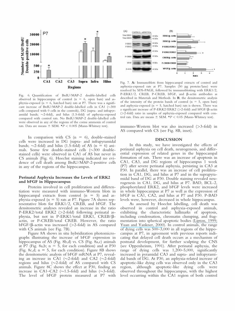
Proteins involved in cell proliferation and differen-tiation were measured with immuno-Western blots inhippocampal extracts from control (n 5 5) and as-phyxia-exposed (n 5 5) rats at P7. Figure 7A shows rep-resentative blots for ERK1/2, CREB, and bFGF. Thedensitometric analyses revealed an increase in the ratioP-ERK2/total ERK2 (>2-fold) following perinatal as-phyxia, but not in P-ERK1/total ERK1, CREB/b-actin, or P-CREB/total CREB. However, the ratiobFGF/b-actin was increased (>2-fold) in AS comparedwith CS animals (see Fig. 7B).
Figure 8A shows in situ hybridization photomicro-graphs illustrating the increase of bFGF expression inhippocampus of AS (Fig. 8b,d) vs. CS (Fig. 8a,c) animalsat P7 (Fig. 8a,b; n 5 5, for each condition) and at P30(Fig. 8c,d; n 5 5, for each condition). Figure 8B showsthe densitometric analysis of bFGF mRNA at P7, reveal-ing an increase in CA1 (�2-fold) and CA2 (�2-fold)regions and hilus (�2-fold) of AS compared with CSanimals. Figure 8C shows the effect at P30, finding anincrease in CA1–CA2 (�1.5-fold) and hilus (�3-fold).The level of bFGF protein measured at P7 with
immuno-Western blot was also increased (>3-fold) inAS compared with CS (see Fig. 8B, inset).
DISCUSSION
In this study, we have investigated the effects ofperinatal asphyxia on cell death, neurogenesis, and differ-ential expression of related genes in the hippocampalformation of rats. There was an increase of apoptosis inCA1, CA3, and DG regions of hippocampus 1 week(P7) after severe perinatal asphyxia, persisting in CA3 atP30. In parallel, there was an increase of cell prolifera-tion in CA1, DG, and hilus at P7 and in the suprapyra-midal band of DG at P30. Double staining revealed neu-rogenesis in CA1, DG, and hilus at P7. BAD, BCL-2,phosphorylated ERK2, and bFGF levels were increasedin whole hippocampus at P7 as well as the expression ofbFGF in CA1, CA2, and hilus at P7 and P30. P-BADlevels were, however, decreased in whole hippocampus.
As assessed by Hoechst labelling, cell death wasobserved in control and asphyxia-exposed animals,exhibiting the characteristic hallmarks of apoptosis,including condensation, chromatin clumping, and frag-mentation into spherical apoptotic bodies (Lipton, 1999;Yuan and Yankner, 2000). In control animals, the rangeof dying cells was 500–3,000 in all regions of the hippo-campus at P7, in agreement with previous reports indi-cating that delayed cell death occurs as a mechanism ofpostnatal development, for further sculpting the CNS(see Oppenheinm, 1991). After perinatal asphyxia, therange of dying cells was 1,200–5,000, significantlyincreased in pyramidal CA3 and supra- and infrapyrami-dal bands of DG. At P30, an asphyxia-related increase ofapoptotic-like dying cells was observed only in the CA3region, although apoptotic-like dying cells wereobserved throughout the hippocampus, with the highestlevel occurring within the CA1 region of both control
Fig. 6. Quantification of BrdU/MAP-2 double-labelled cellsobserved in hippocampus of control (n 5 6, open bars) and as-phyxia-exposed (n 5 6, hatched bars) rats at P7. There was a signifi-cant increase of BrdU/MAP-2 double-labelled cells in CA1 (�300cells compared with 0 cells in the controls), DG (supra- and infrapyr-amidal bands; �2-fold), and hilus (1.5-fold) of asphyxia-exposedcompared with control rats. No BrdU/MAP-2 double-labelled cellswere observed in any of the regions of the cornu ammonis of controlrats. Data are means 6 SEM. *P < 0.005 (Mann-Whitney test).
Fig. 7. A: Immunoblots from hippocampal extracts of control andasphyxia-exposed rats at P7. Samples (50 lg protein/lane) wereresolved by SDS-PAGE, followed by immunoblotting with ERK1/2,P-ERK1/2, CREB, P-CREB, bFGF, and b-actin antibodies asdescribed in Materials and Methods. In B, the densitometric analysisof the intensity of the protein bands of control (n 5 5, open bars)and asphyxia-exposed (n 5 5, hatched bars) rats is shown. There wasa significant increase of P-ERK2/ERK2 (>2-fold) and bFGF/b-actin(>2-fold) ratio in samples of asphyxia-exposed compared with con-trol rats. Data are means 6 SEM. *P < 0.05 (Mann-Whitney test).

(2,000–4,400 dying cells) and asphyxia-exposed (1,400–4,400 dying cells) animals. Perhaps this observation hasto be considered when discussing the particular vulner-ability of the CA1 region (see Pulsinelli, 1988). Celldeath displayed a neuronal phenotype, as shown byMAP-2/Hoechst double labelling, but no dying cellswere observed among newly generated cells (BrdU/MAP-2/Hoechst), suggesting that perinatal asphyxiamainly affected nonproliferating cells.
Apoptosis was confirmed by the TUNEL tech-nique, showing a significant increase of TUNEL-positivecells in CA3 and supra- and infrapyramidal bands follow-ing perinatal asphyxia at P7. An increase of cell death af-ter asphyxia was also observed in CA1 but reaching asignificant level only when evaluated by the TUNEL
technique, although a trend for an elevated apoptosiswas also observed when labelling with Hoechst at P7.
The expression of proteins involved in the regula-tion of the apoptotic cascade was analyzed to character-ize further the delayed cell death observed after perinatalasphyxia. Mitochondrial membrane permeability hasbeen suggested as a critical factor that activates the apo-ptotic cascade. Under apoptosis-induced conditions,including hypoxia-ischemia, the mitochondrial mem-brane increases its permeability, leading to the release ofcytochrome c and other proapoptotic factors, such asAIF (Zhu et al., 2003, 2006), Smac/Diablo (Matsumoriet al., 2005), and Endo-G (Li et al., 2001). The releaseof cytochrome c to the cytoplasm causes caspase-3 acti-vation (Daval et al., 2004; Chen et al., 2005). Caspase-3immunoreactivity has been observed to be elevated 3days after hypoxia, but that elevation has been shown tobe transient (Pourie et al., 2006).
Mitochondrial membrane permeability is regulatedby proapoptotic (BCL-XS, tBID, BAX, BAD) and antia-poptotic (BCL-2, BCL-XL) members of the BCL-2 pro-tein family (see Cory et al., 2003), which can formhomo- and/or heterodimers in the mitochondrial mem-brane (Yang et al., 1995). Indeed, it has been shownthat the expression of BAX and BCL-2 genes isincreased in CA1, CA3, and DG following hypoxic-is-chemic insults (Chen et al., 1998; Daval et al., 2004).BAD promotes apoptosis through its heterodimerizationwith the antiapoptotic proteins BCL-2 and/or BCL-XL
(Yang et al., 1995), leading to leakage of mitochondrialproapoptotic factors (Tan et al., 2000). BAD is regulatedthrough selective phosphorylation by several protein ki-nases, via the AKT and the mitogen-activated proteinkinases MEK/ERK pathways (Jin et al., 2002). BADphosphorylation, at Ser112 and/or Ser136 (Zhu et al.,2002; Hirai et al., 2004), creates binding sites for thechaperone 14-3-3, retaining BAD in the cytoplasm, pre-venting the heterodimerization with BCL-2 and/orBCL-XL in the mitochondrial membrane (Zha et al.,1996). BAD phosphorylation at Ser155 also inhibits itsdeath-promoting activity by interfering the binding toBCL-XL (Tan et al., 2000).
In this study, BAD was increased by perinatal as-phyxia, explaining perhaps the apoptosis observed inCA1, CA3, and DG regions. The increase of BADcould be due to enhanced gene expression, decreasedprotein degradation, and/or other metabolic changesinvolving phosphorylation/dephosphorylation mecha-nisms. The enhanced expression of BAD is perhaps asignal for a dangerous condition menacing the integrityof the genome. Otherwise, as shown here, a concomi-tant decrease of P-BAD (Ser112) could imply adecreased BAD phosphorylation, or an increased phos-phatase-dependent dephosphorylation. In agreement, ithas been reported that hypoxia-ischemia triggers BADdephosphorylation by calcineurin (Wang et al., 1999b).In adult gerbils, cerebral ischemia promotes BAD trans-location to mitochondria in CA1 but not in the DGregion of hippocampus (Dluzniewska et al., 2005). It
Fig. 8. A: Photomicrographs illustrating the expression of bFGF inhippocampus of control (a,c) and asphyxia-exposed (b,d) rats at P7(a,b, upper panel) and P30 (c,d, lower panel). Densitometric analysisof bFGF mRNA in hippocampus of control (n 5 5, open bars) andasphyxia-exposed (n 5 5, hatched bars) rats at P7 (B) and P30 (C).There was a significant increase of bFGF expression in CA1, CA2(�1.5-fold), and hilus (>2-fold) of asphyxia-exposed compared withcontrol rats at both P7 and P30. Data are means 6 SEM. *P < 0.05(Mann-Whitney test). Scale bar 5 200 lm.
was shown that Raf-1, a member of the ERK signallingpathway, was instead decreased in CA1 (Dluzniewskaet al., 2005). Furthermore, in BAD-deficient mice, neo-natal hypoxia-ischemia does not increase caspase-3 im-munostaining compared with similarly treated wild-typeanimals (Ness et al., 2006), supporting the idea thatBAD participates in the hypoxia-ischemia-induced apo-ptosis.
BCL-2 is implicated in survival and cell differentia-tion (see Cory et al., 2003). In the present study, BCL-2levels increased in whole hippocampal extracts followingperinatal asphyxia, perhaps restricting the progression ofapoptosis. However, the BAD/BCL-2 ratio increased,indicating that an apoptotic mechanism prevailed andthat the increase of BCL-2 was not enough to preventcell death.
It has been reported that hypoxia-ischemia inducesa biphasic increase of BCL-2 immunoreactivity in CA1,with peaks at P3 and P20, suggesting that BCL-2 is asso-ciated with both early and delayed neuroprotection(Daval et al., 2004). An increase of BCL-2 expressionhas also been observed in CA3 and DG after hypoxia-is-chemia in adult rats (Chen et al., 1996). However, adown-regulation of BCL-2 transcription has beenobserved after left carotid artery occlusion followed by2.5 hr of hypoxia exposure at P7 in rats (Kumral et al.,2006). Together, these results suggest that changes inBCL-2 expression probably depend on the severity ofthe insult and the developmental stage of the brain whenthe insult takes place.
Neurotrophic factors regulate BCL-2 expressionthrough transcription factors, such as the cAMP responseelement binding (CREB) protein (Jin et al., 2001; seeLonze and Ginty, 2002; Lee et al., 2004) and nuclearfactor-jB (NFjB; Tamatani et al., 1999, 2000). CREBactivity is regulated through phosphorylation at Ser133(P-CREB) by the ERK/MAPK pathway (Curtis andFinkbeiner, 1999). An increase of P-CREB levels hasbeen observed 6 hr after ischemic insults (Walton et al.,1996; Lee et al., 2004). In the present study, however,no changes were detected in total CREB or its phos-phorylated form 1 week after perinatal asphyxia, perhapssuggesting that the increase of BCL-2 by perinatal as-phyxia was due to an early transient increase of CREB.Alternatively, BCL-2 gene expression can be regulatedby NFjB, as reported by Tamatani et al. (1999, 2000),although in this study we did not monitored that factor.
In adults, new neurons can be generated in thesubventricular zone (SVZ). The cells generated in SVZmigrate to the olfactory bulb (Doetsch et al., 1999; Sha-piro et al., 2006), where they differentiate to interneur-ons (Doetsch et al., 1999). The rate of neurogenesis canbe increased by brain injuries, including hypoxia-ische-mia, probably corresponding to a compensatory mecha-nism for the replacement of dead cells (see Lichtenwal-ner and Parent, 2006). Neurogenesis in SVZ has beenproposed also to contribute to neuronal replacement inthe CA1 region after hypoxia-ischemia (Nakatomi et al.,2002; Ong et al., 2005). In agreement, we found here
that perinatal asphyxia induced a twofold increase ofBrdU-positive cells in the CA1 region, compared withcontrol animals, but only 10% of those BrdU-positivecells were positive for MAP-2. No BrdU-positive cellswere positive for MAP-2 in any of the regions of thecornu ammonis of control animals.
Cell proliferation has also been observed in SGZ ofDG following hypoxia-ischemia (Scheepens et al.,2003b; Bartley et al., 2005). In agreement, it was foundhere that the number of BrdU positive cells wasincreased in the supra- and infrapyramidal band of DGand in hilus by perinatal asphyxia at P7. At P30, how-ever, there was an increase only in the suprapyramidalband of the DG. This finding is in agreement with find-ings reported by Choi et al. (2003), showing an increaseof cell proliferation in both supra- and infrapyramidalbands after 10 days but only in the medial suprapyrami-dal band after 28 days of global ischemia.
Double staining revealed that the number ofBrdU/MAP-2-positive cells was increased in the supra-and infrapyramidal band of DG and in hilus by perina-tal asphyxia, which is in agreement with a previousstudy with hippocampal organotypic cultures (Moraleset al., 2005, 2007). In contrast to the CA1 region,�40% of the total number of BrdU-positive cells werealso positive for MAP-2 in DG, and the percentage ofdouble-labelled cells was similar in both control and as-phyxia-exposed animals. The remaining amount per-haps represented proliferation of progenitor cells, orcells at an earlier stage of differentiation, which canpotentially differentiate to neurons (Ganat et al., 2002;Bartley et al., 2005) or to glial cells (Kee et al., 2001;Zaidi et al., 2004).
ERK1/2 proteins are associated with cell prolifera-tion (Zhou et al., 2004) and have been found to be acti-vated early (0–72 hr) after neonatal hypoxia/ischemia inhippocampus (Wang et al., 2003; Haddad, 2004; Jonesand Bergeron, 2004). Interestingly, it has been reportedthat the activation of ERK1/2 proteins is inhibited byU0126, an inhibitor of mitogen-activated protein kinase(Favata et al., 1998), reducing the effect of hypoxia-is-chemia on BrdU labelling (Zhou et al., 2004).
ERK activity is regulated through phosphorylationat threonine and tyrosine residues by the MAPK path-way (Boulton et al., 1991; see Haddad, 2004). In thepresent study, we found an increase of P-ERK2, but notof total ERK or P-ERK1, levels after the perinatalinsult. It can be suggested that ERK2 phosphorylation isrelated to neurogenesis, but we cannot discard its partici-pation in cell death in the perinatal asphyxia model, assuggested by Castro-Obregon et al. (2004). Indeed, ithas been suggested that nuclear translocation of ERK2 isrequired for cell cycle progression (Hulleman et al.,1999; Chu et al., 2004).
The MEK/ERK pathway promotes CREB activa-tion through phosphorylation (see Lonze and Ginty,2002), increasing proliferation and cell differentiation inDG of adult mouse hippocampus (Nakagawa et al.,2002; Fujioka et al., 2004). In agreement, cell prolifera-
tion decreases in conditional transgenic mice expressinga CREB dominant negative mutant in hippocampus(Nakagawa et al., 2002). In the present study, no corre-lation between ERK activation and CREB phosphoryla-tion was observed, but a transient early CREB activationcould have taken place at earlier stages that was unde-tectable at P7.
bFGF is essential for neuronal development (Dono,2003); it is involved in proliferation (Raballo et al.,2000) and differentiation (Vicario-Abejon et al., 1995;Cheng et al., 2002). The expression of bFGF is increasedafter brain injury (Takami et al., 1992; Andersson et al.,1995), promoting neurogenesis and survival (Wagneret al., 1999; Yoshimura et al., 2001). In the presentstudy, there was an increase of bFGF expression in CA1,CA2, and hilus and bFGF levels in whole hippocampus1 week and 1 month after perinatal asphyxia. Similarresults have been observed following ischemia (Takamiet al., 1992) and hypoxia (Ganat et al., 2002) in adultanimals. It has been shown that intraventricular infusionof bFGF markedly augmented endogenous progenitorproliferation, leading to regeneration of CA1 neurons,ameliorating the neurological deficits induced by ische-mia (Nakatomi et al., 2002). Overexpression of bFGFby a herpes simplex virus-1 vector increased significantlythe number of dividing cells in DG, attenuating theeffect of ischemia on cell death (Yoshimura et al., 2001).These results support the idea that bFGF up-regulatesneurogenesis and protects neurons against degenerationof hippocampus, as shown after brain traumatic insults inadult animals (Yoshimura et al., 2001, 2003). Interest-ingly, it has been shown that both ERK1 and ERK2 areactivated by bFGF (Hetman et al., 1999), suggesting thatbFGF can induce the phosphorylation of ERK2, pro-moting cell proliferation, as reported here.
In conclusion, perinatal asphyxia induces regionallyspecific delayed cell death and neurogenesis in rat hippo-campus accompanied by an increase of pro- and antia-poptotic proteins. The increased neurogenesis found inCA1, DG, and hilus of asphyxia-exposed animals to-gether with the increased expression of mitogenic pro-teins suggest that plastic changes occur, perhaps provid-ing a framework for functional recovery of the hippo-campus.
ACKNOWLEDGMENTS
We are grateful for the excellent technical and sec-retarial help from Mr. Juan Santibanez, Ms. CarmenAlmeyda, and Ms. Ana Marıa Mendez. The supportfrom the confocal unit (CESAT, ICBM), led by Dr.Jorge Sans, is kindly acknowledged.
REFERENCES
Abe T, Takagi N, Nakano M, Furuya M, Takeo S. 2004. Altered Bad
localization and interaction between Bad and Bcl-xL in the hippocam-
pus after transient global ischemia. Brain Res 1009:159–168.
Amaral DG, Dent JA. 1981. Development of the mossy fibers of the
dentate gyrus: I. A light and electron microscopic study of the mossy
fibers and their expansions. J Comp Neurol 195:51–86.
Amaral DG, Witter MP. 1995. Hippocampal formation. In: Paxinos G,
editor. The rat nervous system. San Diego: Academic Press.
Andersson K, Bjelke B, Bolme P, Ogren SO. 1992. Asphyxia-induced
lesion of the rat hippocampus (CA1, CA3) and the nigro-striatal dopa-
mine system. Hypoxia and ischemia. CNS. In: Gross J, editor. Wissen-
schaflige publikationen de Humboldt-Universitat zu Berlin. B Medizin
41:71–76.
Andersson K, Blum M, Chen Y, Eneroth P, Gross J, Herrera-Marschitz
M, Bjelke B, Bolme P, Diaz R, Jamison L, Loidl F, Ungethum U,
Astrom G, Ogren SO. 1995. Perinatal asphyxia increases bFGF mRNA
levels and DA cell body number in mesencephalon of rats. Neuroreport
6:375–337.
Andres S, Cardenas S, Parra C, Bravo J, Greiner M, Rojas P, Morales P,
Lara H, Fiedler JL. 2006. Effect of long-term adrenalectomy on the ap-
optosis in rat hippocampus. Endocrine 29:299–307.
Aspberg A, Tottmar O. 1992. Development of antioxidant enzymes in
rat brain and in reaggregation culture of fetal brain cells. Brain Res Dev
Brain Res 66:55–58.
Bartley J, Soltau T, Wimborne H, Kim S, Martin-Studdard A, Hess D,
Hill W, Waller J, Carroll J. 2005. BrdU-positive cells in the neonatal
mouse hippocampus following hypoxic-ischemic brain injury. BMC
Neurosci 6:15.
Bayer SA. 1982. Changes in the total number of dentate granule cells in
juvenile and adult rats: a correlated volumetric and 3H-thymidine auto-
radiographic study. Exp Brain Res 46:315–323.
Bjelke B, Andersson K, Ogren SO, Bolme P. 1991. Asphyctic lesion:
proliferation of tyrosine hydroxylase-immunoreactive nerve cell bodies
in the rat substantia nigra and functional changes in dopamine neuro-
transmission. Brain Res 543:1–9.
Boulton TG, Nye SH, Robbins DJ, Ip NY, Radziejewska E, Morgen-
besser SD, DePinho RA, Panayotatos N, Cobb MH, Yancopoulos GD.
1991. ERKs: a family of protein-serine/threonine kinases that are acti-
vated and tyrosine phosphorylated in response to insulin and NGF. Cell
65:663–675.
Bradford MM. 1976. A rapid and sensitive method for the quantitation
of microgram quantities of protein utilizing the principle of protein-dye
binding. Anal Biochem 72:248–254.
Bustamante D, Morales P, Pereyra JT, Goiny M, Herrera-Marschitz M.
2006. Nicotinamide prevents the effect of perinatal asphyxia on dopa-
mine release evaluated with in vivo microdialysis 3 months after birth.
Exp Brain Res 177:358–369.
Cardenas SP, Parra C, Bravo J, Morales P, Lara HE, Herrera-Marschitz
M, Fiedler JL. 2002. Corticosterone differentially regulates bax, bcl-2
and bcl-x mRNA levels in the rat hippocampus. Neurosci Lett 331:9–
12.
Castro-Obregon S, Rao RV, del Rio G, Chen SF, Poksay KS, Rabiza-
deh S, Vesce S, Zhang XK, Swanson RA, Bredesen DE. 2004. Alter-
native, nonapoptotic programmed cell death: mediation by arrestin 2,
ERK2, and Nur77. J Biol Chem 279:17543–17553.
Chen J, Zhu RL, Nakayama M, Kawaguchi K, Jin K, Stetler RA, Simon
RP, Graham SH. 1996. Expression of the apoptosis-effector gene, Bax,
is up-regulated in vulnerable hippocampal CA1 neurons following
global ischemia. J Neurochem 67:64–71.
Chen J, Nagayama T, Jin K, Stettler RA, Zhu RL, Graham SH, Simon
RP. 1998. Induction of caspase-3-like protease may mediate delayed
neuronal death in the hippocampus after transient cerebral ischemia.
J Neurosci 18:4914–4928.
Chen Z, Kontonotas D, Friedmann D, Pitts-Kiefer A, Frederick JR,
Siman R, Neumar RW. 2005. Developmental status of neurons selec-
tively vulnerable to rapidly triggered post-ischemic caspase activation.
Neurosci Lett 376:166–170.
Cheng Y, Gidday JM, Yan Q, Shah AR, Holtzman DM. 1997. Marked
age-dependent neuroprotection by brain-derived neurotrophic against
neonatal hypoxic-ischemic brain injury. Ann Neurol 41:521–529.

Cheng Y, Deshmukh M, D’Costa A, Demaro JA, Gidday JM, Shah A,
Sun Y, Jacquin MF, Johnson EM, Holtzman DM. 1998. Caspase inhib-
itor affords neuroprotection with delayed administration in a rat model
of neonatal hypoxic-ischemic brain injury. J Clin Invest 101:1992–
1999.
Cheng Y, Black IB, DiCicco-Bloom E. 2002. Hippocampal granule neu-
ron production and population size are regulated by levels of bFGF.
Eur J Neurosci 15:3–12.
Choi YS, Lee MY, Sung KW, Jeong SW, Choi JS, Park HJ, Kim ON,
Lee SB, Kim SY. 2003. Regional differences in enhanced neurogenesis
in the dentate gyrus of the adult rats after transient forebrain ischemia.
Mol Cells 16:232–238.
Chu CT, Levinthal DJ, Kulich SM, Chalovich EM, DeFranco DB.
2004. Oxidative neuronal injury. The dark side of ERK1/2. Eur J Bio-
chem 271:2060–2066.
Cory S, Huang DC, Adams JM. 2003. The Bcl-2 family: roles in cell
survival and oncogenesis. Oncogene 22:8590–8607.
Curtis J, Finkbeiner S. 1999. Sending signals from the synapse to the nu-
cleus: possible roles for CaMK, Ras/ERK, and SAPK pathways in the
regulation of synaptic plasticity and neuronal growth. J Neurosci Res
58:88–95.
Daval JL, Pourie G, Grojean S, Lievre V, Strazielle G, Vert P. 2004.
Neonatal hypoxia triggers transient apoptosis followed by neurogenesis
in the rat CA1 hippocampus. Pediatric Res 55:561–567.
Davies JC. 1995. The Bcl-2 family of proteins, and the regulation of cell
survival. Trends Neurol Sci 18:355–358.
Dell’Anna E, Chen Y, Engidawork E, Andersson K, Lubec G, Luthman
J, Herrera-Marschitz M. 1997. Delayed neuronal death following peri-
natal asphyxia in rat. Exp Brain Res 115:105–115.
Dluzniewska J, Beresewicz M, Wojewodzka U, Gajkowska B, Zablocka
B. 2005. Transient cerebral ischemia induces delayed proapoptotic Bad
translocation to mitochondria in CA1 sector of hippocampus. Brain
Res Mol Brain Res 133:274–280.
Doetsch F, Caille I, Lim DA, Garcia-Verdugo JM, Alvarez-Buylla A.
1999. Subventricular zone astrocytes are neural stem cells in the adult
mammalian brain. Cell 97:703–716.
Dono R. 2003. Fibroblast growth factors as regulators of central nervous
system development and function. Am J Physiol Regul Integr Comp
Physiol 284:R867–R881.
Favata MF, Horiuchi KY, Manos EJ, Daulerio AJ, Stradley DA, Feeser
WS, Van Dyk DE, Pitts WJ, Earl RA, Hobbs F, Copeland RA,
Magolda RL, Scherle PA, Trzaskos JM. 1998. Identification of a novel
inhibitor of mitogen-activated protein kinase kinase. J Biol Chem
273:18623–18632.
Ferrer I, Pozas E, Lopez E, Ballabriga J. 1997. Bcl-2, Bax and Bcl-x
expression following hypoxia-ischemia in the infant rat brain. Acta
Neuropathol 94:583–589.
Fujioka T, Fujioka A, Duman RS. 2004. Activation of cAMP signaling
facilitates the morphological maturation of newborn neurons in adult
hippocampus. J Neurosci 24:319–328.
Ganat Y, Soni S, Chacon M, Schwartz ML, Vaccarino FM. 2002.
Chronic hypoxia up-regulates fibroblast growth factor ligands in the
perinatal brain and induces fibroblast growth factor-responsive radial
glial cells in the subependymal zone. Neuroscience 112:977–991.
Golan H, Huleihel M. 2006. The effect of prenatal hypoxia on brain de-
velopment: short- and long-term consequences demonstrated in rodent
models. Dev Sci 9:338–349.
Graham EM, Sheldon RA, Flock DL, Ferriero DM, Martin LJ,
O’Riordan DP, Northington FJ. 2004. Neonatal mice lacking func-
tional Fas death receptors are resistant to hypoxic-ischemic brain injury.
Neurobiol Dis 17:89–98.
Greiner M, Cardenas S, Parra C, Bravo J, Avalos AM, Paredes A, Lara
HE, Fiedler JL. 2001. Adrenalectomy regulates apoptotic-associated
genes in rat hippocampus. Endocrine 15:323–333.
Gundersen NJ, Bagger P, Bendtsen TF, Evans SM, Korbo L, Marcussen
H, Møller A, Nielsen K, Nyengaard JR, Pakkenberg B, Sørensen FB,
Vesterby A, West MJ. 1988. The new stereological tools: disector, frac-
tionator, nucleator and point sampled interecepts and their use in path-
ologic research and diagnosis. Acta Pathol Microbial Immunol Scand
96:857–881.
Haddad JJ. 2004. Hypoxia and the regulation of mitogen-activated pro-
tein kinases: gene transcription and the assessment of potential pharma-
cologic therapeutic interventions. Int Immunopharmacol 4:1249–1285.
Han BH, Holtzman DM. 2000. BDNF protects the neonatal brain from
hypoxic-ischemic injury in vivo via the ERK pathway. J Neurosci
20:5775–5781.
Harry JG, d’Hellencourt CL. 2003. Dentate gyrus: alterations that occur
with hippocampal injury. Neurotoxicology 24:343–356.
Herrera-Marschitz M, Loidl CF, Andersson K, Ungerstedt U. 1993. Pre-
vention of mortality induced by perinatal asphyxia: hypothermia or glu-
tamate antagonism? Amino Acids 5:413–419.
Hetman M, Kanning K, Cavanaugh JE, Xia Z. 1999. Neuroprotection
by brain-derived neurotrophic factor is mediated by extracellular signal-
regulated kinase and phosphatidylinositol 3-kinase. J Biol Chem
274:22569–22580.
Hirai K, Hayashi T, Chan PH, Zeng J, Yang GY, Basus VJ, James TL,
Litt L. 2004. PI3K inhibition in neonatal rat brain slices during and af-
ter hypoxia reduces phospho-Akt and increases cytosolic cytochrome c
and apoptosis. Brain Res Mol Brain Res 124:51–61.
Howard S, Bottino C, Brooke S, Cheng E, Giffard RG, Sapolsky R.
2002. Neuroprotective effects of bcl-2 overexpression in hippocampal
cultures: interactions with pathways of oxidative damage. J Neurochem
83:914–923.
Hulleman E, Bijvelt JJ, Verkleij AJ, Verrips CT, Boonstra J. 1999. Nu-
clear translocation of mitogen-activated protein kinase p42MAPK dur-
ing the ongoing cell cycle. J Cell Physiol 180:325–333.
Jin K, Mao XO, Simon RP, Greenberg DA. 2001. Cyclic AMP response
element binding protein (CREB) and CREB binding protein (CBP) in
global cerebral ischemia. J Mol Neurosci 16:49–56.
Jin K, Mao XO, Zhu Y, Greenberg DA. 2002. MEK and ERK protect
hypoxic cortical neurons via phosphorylation of Bad. J Neurochem
80:119–125.
Jones NM, Bergeron M. 2004. Hypoxia-induced ischemic tolerance in
neonatal rat brain involves enhanced ERK1/2 signalling. J Neurochem
89:157–167.
Kee NJ, Preston E, Wojtowicz JM. 2001. Enhanced neurogenesis after
transient global ischemia in the dentate gyrus of the rat. Exp Brain Res
136:313–320.
Kirino T, Tamura A, Sano K. 1984. Delayed death in the rat hippocam-
pus following transient forebrain ischemia. Acta Neuropathol 64:139–
147.
Kobayashi T, Ahlenius H, Thored P, Kobayashi R, Kokaia Z, Lindvall
O. 2006. Intracerebral infusion of glial cell line-derived neurotrophic
factor promotes striatal neurogenesis after stroke in adult rats. Stroke
37:2361–2367.
Kumral A, Genc S, Ozer E, Yilmaz O, Gokmen N, Koroglu TF, Duman
N, Genc K, Ozkan H. 2006. Erythropoietin downregulates bax and
DP5 proapoptotic gene expression in neonatal hypoxic-ischemic brain
injury. Biol Neonate 89:205–210.
Lafemina MJ, Sheldon RA, Ferriero DM. 2006. Acute hypoxia-ischemia
results in hydrogen peroxide accumulation in neonatal but not adult
mouse brain. Pediatric Res 59:680–683.
Lee HT, Chang YC, Wang LY, Wang ST, Huang CC, Ho CJ. 2004.
cAMP response element-binding protein activation in ligation precon-
ditioning in neonatal brain. Ann Neurol 56:611–623.
Li LY, Luo X, Wang X. 2001. Endonuclease G is an apoptotic DNase
when released from mitochondria. Nature 412:95–99.

Lichtenwalner RJ, Parent JM. 2006. Adult neurogenesis and the ischemic
forebrain. J Cereb Blood Flow Metab 26:1–20.
Lipton P. 1999. Ischemic cell death in brain neurons. Physiol Rev
79:1431–1540.
Lonze BE, Ginty DD. 2002. Function and regulation of CREB family
transcription factors in the nervous system. Neuron 35:605–623.
Matsumori Y, Hong SM, Aoyama K, Fan Y, Kayama T, Sheldon RA,
Vexler ZS, Ferriero DM, Weinstein PR, Liu J. 2005. Hsp70 overex-
pression sequesters AIF and reduces neonatal hypoxic/ischemic brain
injury. J Cereb Blood Flow Metab 25:899–910.
Morales P, Reyes P, Klawitter V, Huaiquın P, Bustamante D, Fiedler JL,
Herrera-Marschitz M. 2005. Effects of perinatal asphyxia on cell prolif-
eration and neuronal phenotype evaluated with organotypic hippocam-
pal cultures. Neuroscience 135:421–431.
Morales P, Huaiquin P, Bustamante D, Fiedler J, Herrera-Marschitz M.
2007. Perinatal asphyxia induces neurogenesis in hippocampus: an orga-
notypic culture study. Neurotoxicity Res 12:81–84.
Nakagawa S, Kim JE, Lee R, Malberg JE, Chen J, Steffen C, Zhang YJ,
Nestler EJ, Duman RS. 2002. Regulation of neurogenesis in adult
mouse hippocampus by cAMP and the cAMP response element-bind-
ing protein. J Neurosci 22:3673–3682.
Nakajima W, Ishida A, Lange MS, Gabrielson KL, Wilson MA, Martin
LJ, Blue ME, Johnston MV. 2000. Apoptosis has a prolonged role in
the neurodegeneration after hypoxic ischemia in the newborn rat. J
Neurosci 20:7994–8004.
Nakano S, Kogure K, Fujikura H. 1990. Ischemia-induced slowly pro-
gressive neuronal damage in the rat brain. Neuroscience 38:115–124.
Nakatomi H, Kuriu T, Okabe S, Yamamoto S, Hatano O, Kawahara N,
Tamura A, Kirino T, Nakafuku M. 2002. Regeneration of hippocam-
pal pyramidal neurons after ischemic brain injury by recruitment of en-
dogenous neural progenitors. Cell 110:429–441.
Ness JM, Harvey CA, Strasser A, Bouillet P, Klocke BJ, Roth KA. 2006.
Selective involvement of BH3-only Bcl-2 family members Bim and
Bad in neonatal hypoxia-ischemia. Brain Res 1099:150–159.
Northington FJ, Ferriero DM, Graham EM, Traystman RJ, Martin LJ.
2001. Early neurodegeneration after hypoxia-ischemia in neonatal rat is ne-
crosis while delayed neuronal death is apoptosis. Neurobiol Dis 8:207–219.
Ong J, Plane JM, Parent JM, Silverstein FS. 2005. Hypoxic-ischemic
injury stimulates subventricular zone proliferation and neurogenesis in
the neonatal rat. Pediatric Res 58:600–606.
Oppenheim RW. 1991. Cell death during development of the nervous
system. Annu Rev Neurosci 14:453–501.
Paxinos G, Watson C. 1986. The rat brain in stereotaxic coordinates.
New York: Academic Press.
Pourie G, Blaise S, Trabalon M, Nedelec E, Gueant JL, Daval JL. 2006.
Mild, non-lesioning transient hypoxia in the newborn rat induces
delayed brain neurogenesis associated with improved memory scores.
Neuroscience 140:1369–1379.
Pulsinelli WA. 1988. Selective neuronal vulnerability: morphological and
molecular characteristics. Prog Brain Res 63:29–37.
Raballo R, Rhee J, Lyn-Cook R, Leckman JF, Schwartz ML, Vaccarino
FM. 2000. Basic fibroblast growth factor (Fgf2) is necessary for cell pro-
liferation and neurogenesis in the developing cerebral cortex. J Neuro-
sci 20:5012–5023.
Scheepens A, Wassink G, Blanco CE. 2003a. The effect of a global birth
asphyxia on the ontogeny of BDNF and NGF protein expression in
the juvenile brain. Brain Res Dev Brain Res 140:215–221.
Scheepens A, Wassink G, Piersma MJ, Van de Berg WDJ, Blanco CE.
2003b. A delayed increase in hippocampal proliferation following global
asphyxia in the neonatal rat. Brain Res Dev Brain Res 142:67–76.
Shapiro EM, Gonzalez-Perez O, Manuel Garcia-Verdugo J, Alvarez-
Buylla A, Koretsky AP. 2006. Magnetic resonance imaging of the
migration of neuronal precursors generated in the adult rodent brain.
Neuroimage 32:1150–1157.
Shimasaki S, Emoto N, Koba A, Mercado M, Shibata F, Cooksey K,
Baird A, Ling N. 1988. Complementary DNA cloning and sequencing
of rat ovarian basic fibroblast growth factor and tissue distribution study
of its mRNA. Biochem Biophys Res Commun 157:256–263.
Takami K, Iwane M, Kiyota Y, Miyamoto M, Tsukuda R, Shiosaka S.
1992. Increase of basic fibroblast growth factor immunoreactivity and
its mRNA level in rat brain following transient forebrain ischemia. Exp
Brain Res 90:1–10.
Tamatani M, Che YH, Matsuzaki H, Ogawa S, Okado H, Miyake S,
Mizuno T, Tohyama M. 1999. Tumor necrosis factor induces Bcl-2
and Bcl-x expression through NFkappaB activation in primary hippo-
campal neurons. J Biol Chem 274:8531–8538.
Tamatani M, Mitsuda N, Matsuzaki H, Okado H, Miyake SI, Vitek MP,
Yamaguchi A, Tohyama M. 2000. A pathway of neuronal apoptosis
induced by hypoxia/reoxygenation: roles of nuclear factor-jB and Bcl-
2. J Neurochem 75:683–693.
Tan Y, Demeter MR, Ruan H, Comb MJ. 2000. BAD Ser-155 phos-
phorylation regulates BAD/Bcl-xL interaction and cell survival. J Biol
Chem 275:25865–25869.
Van Erp TGM, Saleh PA, Rosso PA, Huttunen M, Lonnqvist J, Pirkola
T, Salonen O, Valanne L, Poutanen V-P, Standerskold-Nordenstam C-
G, Cannon TD. 2002. Contributions of genetic risk and fetal hypoxia
to hippocampal volume in patients with schizophrenia or schizoaffective
disorder, their unaffected siblings and healthy unrelated volunteers. Am
J Psychiatry 159:1514–1520.
Vannucci SJ, Hagberg H. 2004. Hypoxia-ischemia in the immature brain.
J Exp Biol 207:3149–3154.
Vicario-Abejon C, Johe KK, Hazel TG, Collazo D, McKay RD. 1995.
Functions of basic fibroblast growth factor and neurotrophins in the dif-
ferentiation of hippocampal neurons. Neuron 15:105–114.
Wagner JP, Black IB, DiCicco-Bloom E. 1999. Stimulation of neonatal
and adult brain neurogenesis by subcutaneous injection of basic fibro-
blast growth factor. J Neurosci 19:6006–6016.
Walton M, Sirimanne E, Williams C, Gluckman P, Dragunow M. 1996.
The role of the cyclic AMP-responsive element binding protein
(CREB) in hypoxic-ischemic brain damage and repair. Brain Res Mol
Brain Res 43:21–29.
Wang HD, Fukuda T, Suzuki T, Hashimoto K, Liou SY, Momoi T,
Kosaka T, Yamamoto K, Nakanishi H. 1999a. Differential effects of Bcl-
2 overexpression on hippocampal CA1 neurons and dentate granule cells
following hypoxic ischemia in adult mice. J Neurosci Res 57:1–12.
Wang HG, Pathan N, Ethell IM, Krajewski S, Yamaguchi Y, Shibasaki F,
McKeon F, Bobo T, Franke TF, Reed JC. 1999b. Ca21-induced apopto-
sis through calcineurin dephosphorylation of BAD. Science 284:339–343.
Wang X, Zhu C, Qiu L, Hagberg H, Sandberg M, Blomgren K. 2003.
Activation of ERK1/2 after neonatal rat cerebral hypoxia-ischaemia. J
Neurochem 86:351–362.
Weitzdoerfer R, Pollak A, Lubec B. 2004. Perinatal asphyxia in the rat
has lifelong effects on morphology, cognitive functions, and behavior.
Semin Perinatol 28:249–256.
Yagita Y, Kitagawa K, Ohtsuki T, Takasawa K, Miyata T, Okano H,
Hori M, Matsumoto M. 2001. Neurogenesis by progenitor cells in the
ischemic adult rat hippocampus. Stroke 32:1890–1896.
Yang E, Zha J, Jockel J, Boise LH, Thompson CB, Korsmeyer SJ. 1995.
Bad, a heterodimeric partner for Bcl-xL and Bcl-2, displaces Bax and
promotes cell death. Cell 80:285–291.
Yoshimura S, Takagi Y, Harada J, Teramoto T, Thomas SS, Waeber C,
Bakowska JC, Breakefield XO, Moskowitz MA. 2001. FGF-2 regula-
tion of neurogenesis in adult hippocampus after brain injury. Proc Natl
Acad Sci U S A 98:5874–5879.
Yoshimura S, Teramoto T, Whalen MJ, Irizarry MC, Takagi Y, Qiu J,
Harada J, Waeber C, Breakefield XO, Moskowitz MA. 2003. FGF-2
regulates neurogenesis and degeneration in the dentate gyrus after trau-
matic brain injury in mice. J Clin Invest 112:1202–1210.

Yuan J, Yankner ,BA. 2000. Apoptosis in the nervous system. Nature
407:802–809.
Zaidi AU, Bessert DA, Ong JE, Xu H, Barks JD, Silverstein
FS, Skoff RP. 2004. New oligodendrocytes are generated after
neonatal hypoxic-ischemic brain injury in rodents. Glia 46:380–
930.
Zha J, Harada H, Yang E, Jockel J, Korsmeyer SJ. 1996. Serine phospho-
rylation of death agonist BAD in response to survival factor results in
binding to 14-3-3 not BCL-XL. Cell 87:619–628.
Zhou L, Del Villar K, Dong Z, Miller CA. 2004. Neurogenesis response
to hypoxia-induced cell death: map kinase signal transduction mecha-
nisms. Brain Res 1021:8–19.
Zhu C, Qiu L, Wang X, Hallin U, Cande C, Kroemer G, Hagberg H,
Blomgren K. 2003. Involvement of apoptosis-inducing factor in neuro-
nal death after hypoxia-ischemia in the neonatal rat brain. J Neurochem
86:306–317.
Zhu C, Xu F, Wang X, Shibata M, Uchiyama Y, Blomgren K, Hagberg
H. 2006. Different apoptotic mechanisms are activated in male and
female brains after neonatal hypoxia-ischaemia. J Neurochem 96:1016–
1027.
Zhu Y, Yang GY, Ahlemeyer B, Pang L, Che XM, Culmsee C, Klumpp
S, Krieglstein J. 2002. Transforming growth factor-beta 1 increases bad
phosphorylation and protects neurons against damage. J Neurosci
22:3898–3909.