Plasmodium vivax antigen discovery based on alpha-helical coiled coil protein motif
13
Plasmodium vivax Antigen Discovery Based on Alpha- Helical Coiled Coil Protein Motif Nora Ce ´ spedes 1,2 , Catherine Habel 3 , Mary Lopez-Perez 4 , Ange ´ lica Castellanos 1,5 , Andrey V. Kajava 6,7 , Catherine Servis 3 , Ingrid Felger 8 , Remy Moret 9 , Myriam Are ´ valo-Herrera 1,2 , Giampietro Corradin 3 , So ´ crates Herrera 1,4 * 1 Malaria Vaccine and Drug Development Center (MVDC), Cali, Colombia, 2 School of Health, University of Valle, Cali, Colombia, 3 Biochemistry Department, University of Lausanne, Epalinges, Switzerland, 4 Caucaseco Scientific Research Center, Cali, Colombia, 5 Fundacio ´ n Centro de Primates, Cali, Colombia, 6 Centre de Recherches de Biochimie Macromoleculaire (CRBM) and Institut de Biologie Computationnelle (IBC), CNRS, University of Montpellier, Montpellier, France, 7 University ITMO, St. Petersburg, Russia, 8 Swiss Tropical and Public Health Institute, Basel, Switzerland, 9 Ho ˆ pital Saint Camille, Ouagadougou, Burkina Faso Abstract Protein a-helical coiled coil structures that elicit antibody responses, which block critical functions of medically important microorganisms, represent a means for vaccine development. By using bioinformatics algorithms, a total of 50 antigens with a-helical coiled coil motifs orthologous to Plasmodium falciparum were identified in the P. vivax genome. The peptides identified in silico were chemically synthesized; circular dichroism studies indicated partial or high a-helical content. Antigenicity was evaluated using human sera samples from malaria-endemic areas of Colombia and Papua New Guinea. Eight of these fragments were selected and used to assess immunogenicity in BALB/c mice. ELISA assays indicated strong reactivity of serum samples from individuals residing in malaria-endemic regions and sera of immunized mice, with the a- helical coiled coil structures. In addition, ex vivo production of IFN-c by murine mononuclear cells confirmed the immunogenicity of these structures and the presence of T-cell epitopes in the peptide sequences. Moreover, sera of mice immunized with four of the eight antigens recognized native proteins on blood-stage P. vivax parasites, and antigenic cross- reactivity with three of the peptides was observed when reacted with both the P. falciparum orthologous fragments and whole parasites. Results here point to the a-helical coiled coil peptides as possible P. vivax malaria vaccine candidates as were observed for P. falciparum. Fragments selected here warrant further study in humans and non-human primate models to assess their protective efficacy as single components or assembled as hybrid linear epitopes. Citation: Ce ´spedes N, Habel C, Lopez-Perez M, Castellanos A, Kajava AV, et al. (2014) Plasmodium vivax Antigen Discovery Based on Alpha-Helical Coiled Coil Protein Motif. PLOS ONE 9(6): e100440. doi:10.1371/journal.pone.0100440 Editor: Takafumi Tsuboi, Ehime University, Japan Received March 6, 2014; Accepted May 23, 2014; Published June 24, 2014 Copyright: ß 2014 Ce ´spedes et al. This is an open-access article distributed under the terms of the Creative Commons Attribution License, which permits unrestricted use, distribution, and reproduction in any medium, provided the original author and source are credited. Data Availability: The authors confirm that all data underlying the findings are fully available without restriction. All data are included within the Supporting Information files. Funding: This work was funded by Colciencias (Contract 278-2008, Contract 360-2011, Contract 458-2012 and Contract 719-2013) and NIH (Grant number 5U19AI089702. The funders had no role in study design, data collection and analysis, decision to publish, or preparation of the manuscript. Competing Interests: The authors have declared that no competing interests exist. * Email: [email protected] Introduction Despite the important reduction in reported malaria incidence during the last decade in a number of countries worldwide, malaria infection still represents one of the major global public health threats. The World Health Organization (WHO) estimated an annual global burden of 207 million malaria cases and 627,000 deaths in 2012 [1]. Of at least six different malaria parasite species which can be transmitted to humans, Plasmodium vivax is the second most parasite species of epidemiological importance with 70–80 million cases estimated per year worldwide [2]. In most malaria-endemic areas, it coexists with P. falciparum, thus making its control more difficult. Due to the limited impact and cyclical loss of effectiveness of some of the classical malaria control measures, and based on multiple evidence on the feasibility of malaria vaccines, significant efforts have been invested in the development of malaria subunit vaccines over the past 2 to 3 decades [3–5]. Significant progress has been achieved with P. falciparum where several vaccine candidates are currently in clinical development [6]; with one now being considered for licensure [7]. In contrast, development of P. vivax vaccines has been significantly neglected and only a few candidates have been selected for clinical testing [8]. Most P. vivax antigens considered to have vaccine potential have been tested in in vitro studies as well as in preliminary preclinical studies in mice and primates [9–13]. Only a few of these antigens further selected by classical immuno-serological methods have undergone phase I clinical trials [14–16]. In the past, the number of parasite antigens available for vaccine studies has been quite limited. Presently, advances in the establishment of Plasmodium genomes and proteomes [17–19] together with high throughout laboratory techniques [20], can potentially accelerate the devel- opment of malaria vaccines. Additionally, the use of bioinformatics tools to explore the malaria genome/proteome databases has allowed new approaches for identification of parasite proteins containing a-helical coiled coil domains [21]. Such domains readily fold into stable structures that are capable of eliciting antibodies reactive with structurally native epitopes, PLOS ONE | www.plosone.org 1 June 2014 | Volume 9 | Issue 6 | e100440
-
Upload
independent -
Category
Documents
-
view
2 -
download
0
Transcript of Plasmodium vivax antigen discovery based on alpha-helical coiled coil protein motif

Plasmodium vivax Antigen Discovery Based on Alpha-Helical Coiled Coil Protein MotifNora Cespedes1,2, Catherine Habel3, Mary Lopez-Perez4, Angelica Castellanos1,5, Andrey V. Kajava6,7,
Catherine Servis3, Ingrid Felger8, Remy Moret9, Myriam Arevalo-Herrera1,2, Giampietro Corradin3,
Socrates Herrera1,4*
1 Malaria Vaccine and Drug Development Center (MVDC), Cali, Colombia, 2 School of Health, University of Valle, Cali, Colombia, 3 Biochemistry Department, University of
Lausanne, Epalinges, Switzerland, 4 Caucaseco Scientific Research Center, Cali, Colombia, 5 Fundacion Centro de Primates, Cali, Colombia, 6 Centre de Recherches de
Biochimie Macromoleculaire (CRBM) and Institut de Biologie Computationnelle (IBC), CNRS, University of Montpellier, Montpellier, France, 7 University ITMO, St.
Petersburg, Russia, 8 Swiss Tropical and Public Health Institute, Basel, Switzerland, 9 Hopital Saint Camille, Ouagadougou, Burkina Faso
Abstract
Protein a-helical coiled coil structures that elicit antibody responses, which block critical functions of medically importantmicroorganisms, represent a means for vaccine development. By using bioinformatics algorithms, a total of 50 antigens witha-helical coiled coil motifs orthologous to Plasmodium falciparum were identified in the P. vivax genome. The peptidesidentified in silico were chemically synthesized; circular dichroism studies indicated partial or high a-helical content.Antigenicity was evaluated using human sera samples from malaria-endemic areas of Colombia and Papua New Guinea.Eight of these fragments were selected and used to assess immunogenicity in BALB/c mice. ELISA assays indicated strongreactivity of serum samples from individuals residing in malaria-endemic regions and sera of immunized mice, with the a-helical coiled coil structures. In addition, ex vivo production of IFN-c by murine mononuclear cells confirmed theimmunogenicity of these structures and the presence of T-cell epitopes in the peptide sequences. Moreover, sera of miceimmunized with four of the eight antigens recognized native proteins on blood-stage P. vivax parasites, and antigenic cross-reactivity with three of the peptides was observed when reacted with both the P. falciparum orthologous fragments andwhole parasites. Results here point to the a-helical coiled coil peptides as possible P. vivax malaria vaccine candidates aswere observed for P. falciparum. Fragments selected here warrant further study in humans and non-human primate modelsto assess their protective efficacy as single components or assembled as hybrid linear epitopes.
Citation: Cespedes N, Habel C, Lopez-Perez M, Castellanos A, Kajava AV, et al. (2014) Plasmodium vivax Antigen Discovery Based on Alpha-Helical Coiled CoilProtein Motif. PLOS ONE 9(6): e100440. doi:10.1371/journal.pone.0100440
Editor: Takafumi Tsuboi, Ehime University, Japan
Received March 6, 2014; Accepted May 23, 2014; Published June 24, 2014
Copyright: � 2014 Cespedes et al. This is an open-access article distributed under the terms of the Creative Commons Attribution License, which permitsunrestricted use, distribution, and reproduction in any medium, provided the original author and source are credited.
Data Availability: The authors confirm that all data underlying the findings are fully available without restriction. All data are included within the SupportingInformation files.
Funding: This work was funded by Colciencias (Contract 278-2008, Contract 360-2011, Contract 458-2012 and Contract 719-2013) and NIH (Grant number5U19AI089702. The funders had no role in study design, data collection and analysis, decision to publish, or preparation of the manuscript.
Competing Interests: The authors have declared that no competing interests exist.
* Email: [email protected]
Introduction
Despite the important reduction in reported malaria incidence
during the last decade in a number of countries worldwide,
malaria infection still represents one of the major global public
health threats. The World Health Organization (WHO) estimated
an annual global burden of 207 million malaria cases and 627,000
deaths in 2012 [1].
Of at least six different malaria parasite species which can be
transmitted to humans, Plasmodium vivax is the second most parasite
species of epidemiological importance with 70–80 million cases
estimated per year worldwide [2]. In most malaria-endemic areas,
it coexists with P. falciparum, thus making its control more difficult.
Due to the limited impact and cyclical loss of effectiveness of
some of the classical malaria control measures, and based on
multiple evidence on the feasibility of malaria vaccines, significant
efforts have been invested in the development of malaria subunit
vaccines over the past 2 to 3 decades [3–5]. Significant progress
has been achieved with P. falciparum where several vaccine
candidates are currently in clinical development [6]; with one
now being considered for licensure [7]. In contrast, development
of P. vivax vaccines has been significantly neglected and only a few
candidates have been selected for clinical testing [8].
Most P. vivax antigens considered to have vaccine potential have
been tested in in vitro studies as well as in preliminary preclinical
studies in mice and primates [9–13]. Only a few of these antigens
further selected by classical immuno-serological methods have
undergone phase I clinical trials [14–16]. In the past, the number
of parasite antigens available for vaccine studies has been quite
limited. Presently, advances in the establishment of Plasmodium
genomes and proteomes [17–19] together with high throughout
laboratory techniques [20], can potentially accelerate the devel-
opment of malaria vaccines. Additionally, the use of bioinformatics
tools to explore the malaria genome/proteome databases has
allowed new approaches for identification of parasite proteins
containing a-helical coiled coil domains [21].
Such domains readily fold into stable structures that are capable
of eliciting antibodies reactive with structurally native epitopes,
PLOS ONE | www.plosone.org 1 June 2014 | Volume 9 | Issue 6 | e100440

Ta
ble
1.
Bio
info
rmat
ics
anal
ysis
of
coile
dco
ilfr
agm
en
ts.
Pe
pti
de
MW
P.
viva
xP
.fa
lcip
aru
ma
ase
qu
en
ceP
osi
tio
nC
ell
loca
liz
ati
on
/fu
nct
ion
Co
ile
dco
ilD
om
ain
PvP
ep
52
93
6P
VX
_0
03
58
5P
FB0
14
5c
IAD
IKIS
LEK
LKY
EVK
DK
KD
CLE
NV
20
3–
22
7H
ypo
the
tica
lp
rote
in8
4–
38
0
PvP
ep
12
50
39
PV
X_
00
35
85
PFB
01
45
cY
KK
ELEE
KA
KIIE
DLK
DK
ICT
LTN
EVM
DLK
NV
KN
ELA
ERD
SSL
10
23
–1
06
5H
ypo
the
tica
lp
rote
in1
01
6–
12
44
PvP
ep
27
31
69
PV
X_
11
33
35
MA
L6P
1.3
7K
KQ
NA
EKEL
SVLK
KN
YD
AM
SEEI
EEIT
65
4–
68
0H
ypo
the
tica
lp
rote
in6
36
–7
43
PvP
ep
40
36
34
PV
X_
11
93
85
PFC
02
35
wN
ETIQ
RM
SNSL
LKY
EQD
IET
YQ
NEV
STLT
GK
67
5–
70
5H
ypo
the
tica
lp
rote
in5
94
–7
44
PvP
ep
42
33
48
PV
X_
08
77
30
PF0
7_
00
14
NT
PD
YY
KK
ITT
KLQ
NN
INN
VEE
YIN
NIT
ND
INIL
KSS
ID1
54
–1
92
Hyp
oth
eti
cal
pro
tein
16
4–
25
9
PvP
ep
43
45
83
PV
X_
08
96
60
PFD
06
85
cSV
DIN
ALN
EQV
KK
LREE
LNK
VT
NEY
DD
FKN
KLE
LLY
QK
77
9–
81
6C
hro
mo
som
eas
soci
ate
dp
rote
in7
26
–9
17
PvP
ep
45
43
33
PV
X_
12
33
85
PF1
1_
02
07
KEV
KV
EVN
EVG
EEV
NE
VK
EEV
NEA
KEE
VIE
KK
EEM
TE
65
0–
68
6H
ypo
the
tica
lp
rote
in5
57
–7
81
PvP
ep
52
36
17
PV
X_
12
34
80
PFL
07
70
wV
EQV
KK
EIN
QIN
EQ
ININ
ETK
ITH
LRN
KIE
17
6–
20
5Se
cre
tory
pat
hw
ay1
66
–2
07
PvP
ep
63
36
58
PV
X_
11
81
60
PF0
7_
00
86
NN
EMD
ETLS
KLK
KD
INK
LNEK
IQK
YD
NY
VK
20
7–
23
6H
ypo
the
tica
lp
rote
in1
62
–2
44
PvP
ep
82
.02
67
21
PV
X_
12
27
40
MA
L13
P1
.96
ETIN
QID
QK
MEE
IEN
NIN
LALE
ELK
NLD
QK
ILEL
QA
SFT
CY
ENEI
KQ
VIK
KIE
GLE
K8
62
–9
18
Stru
ctu
ral
mai
nte
nan
ceo
fch
rom
oso
me
29
80
–1
04
5
PvP
ep
82
.03
65
74
PV
X_
09
19
10
MA
L13
P1
.96
IEQ
LNT
KM
KN
INEN
SND
SEH
VN
LAEF
ELK
IAEL
KED
VN
NIN
NM
MK
TFE
MK
FSA
LEK
47
1–
52
6K
elc
hd
om
ain
-co
nta
inin
gp
rote
in4
62
–5
51
PvP
ep
83
45
36
PV
X_
08
77
30
PFC
03
45
wLQ
NN
INN
VEE
YIN
NIT
ND
INIL
KSS
IDD
ERN
ERIIY
NN
16
6–
20
3H
ypo
the
tica
lp
rote
in1
64
–2
59
PvP
ep
90
41
64
PV
X_
00
07
2P
FD0
52
0c
TR
RM
HSE
LSD
GN
KEL
KK
LKK
NIV
QSD
VLN
AQ
LEL
NI
63
–9
8H
ypo
the
tica
lp
rote
in6
4–
98
PvP
ep
95
35
12
PV
X_
11
74
55
PF1
4_
05
74
EKG
LKD
LND
KIR
NY
DSI
IEN
QK
KEL
EHLK
14
5–
17
3H
ypo
the
tica
lp
rote
in1
45
–2
45
PvP
ep
96
.01
65
95
PV
X_
12
40
60
PF1
3_
01
07
VEA
VP
ENA
EAA
PEN
AD
PV
HEN
AEA
AP
ENA
EPV
HEN
AE
77
3–
80
9Se
cre
tory
pat
hw
ay7
73
–8
09
PvP
ep
96
.03
44
82
PV
X_
08
43
85
PF1
3_
01
07
DV
QR
IDT
INK
NIS
TIN
DD
VD
HIN
SNIN
NIN
DN
LHK
INSH
20
51
–2
08
9H
ypo
the
tica
lp
rote
in2
04
9–
20
88
PvP
ep
10
13
55
4P
VX
_0
85
15
5P
F14
_0
25
5N
KLT
EMR
RK
LKIID
EK
VQ
SVY
KA
IHA
VLN
N3
14
–3
43
Co
rA-l
ike
Mg
2+
tran
spo
rte
rp
rote
in3
13
–3
43
PvP
ep
10
63
44
1P
VX
_1
14
43
0M
AL6
P1
.16
3K
TID
QLD
FEIN
DLN
SKLK
NY
EKSV
SQN
KK
67
3–
70
1H
ypo
the
tica
lp
rote
in4
30
–7
99
PvP
ep
12
33
45
5P
VX
_1
17
85
5P
F14
_0
50
0EK
YSL
IKEE
IKY
LNED
LD
DLD
NSV
NV
VK
K4
3–
71
Hyp
oth
eti
cal
pro
tein
40
–8
6
PvP
ep
12
53
09
2P
VX
_0
99
41
0P
FI0
97
5c
ILR
KIE
HSL
KG
WEA
DY
NEL
KG
KY
NSV
19
90
–2
01
5H
ypo
the
tica
lp
rote
in1
92
4–
20
82
do
i:10
.13
71
/jo
urn
al.p
on
e.0
10
04
40
.t0
01
Plasmodium vivax Antigen Discovery
PLOS ONE | www.plosone.org 2 June 2014 | Volume 9 | Issue 6 | e100440
and are generally monomorphic [22]; these structures have the
capacity to block critical functions of medically important
microorganisms [23,24]. Specifically in P falciparum some antigens
containing these domains have been involved in antibody-
dependent inhibition of malaria parasite growth [25,26], and
therefore represent targets for vaccine development, thus drasti-
cally reducing the time required for antigen selection and
preclinical testing [21].
In the past few years, approximately 170 P. falciparum a-helical
coiled coil protein fragments have been assessed by combining
genome-wide bioinformatics analysis, peptide selection, peptide
chemical synthesis, immune and biochemical assays, in vitro
functional assays, with associated protection analysis [25,26]
(unpublished data). A total of 140 putative a-helical coil-
containing proteins of 200 to 10,000 amino acids in length were
identified as new target proteins in P. falciparum asexual blood
stages. Here we describe studies carried out using the same
technology and approach with P. vivax antigens orthologous to P.
falciparum, which have been evaluated for their antigenicity using
human sera and immunogenicity in mice.
Materials and Methods
P. vivax genome bioinformatics analysisOrthologues are good candidates for multi-species vaccines as
they have the potential to elicit antigenic reactions against all the
species included in the search parameters. A P. vivax Salvador I
genome database (PlasmoDB) was used for the selection of P. vivax
orthologous to P. falciparum protein sequences from asexual blood
stages containing a-helical coiled coil structures, analyzed by
COILS software [27]. Fifty P. vivax orthologues were found to have
at least 30% homology with the 170 P. falciparum a-helical coiled-
coil proteins previously identified. Sequences were of the maximal
length possible in order to maximize the stability of the a-helical
conformations and to increase the array of conformational
epitopes that could be yielded. Selected a-helical coiled coil-
containing proteins were further characterized as to possible
surface location and GPI anchoring, using the following software:
identification of potential signal peptides by SecretomeP and
SignalP (http://www.cbs.dtu.dk/services/) [28]; transmembrane
spanning region- (TMPRED http://www.ch.embnet.org/ soft-
ware/TMPRED_rm.html and TMHMM http://www.cbs.dtu.
dk/services/TMHMM; [29], and GPI-anchored proteins (http://
mendel.imp.univie.ac.at/sat/gpi/gpi_server.html [30]; and pre-
diction of sub-cellular localization (pTARGET) [31]. Additionally,
major histocompatibility complex protein (MHC-II) binding
predictions were made using the IEDB analysis resource
Consensus tool [32,33] which combines predictions from ANN
aka NetMHC [34,35], SMM [36] and Comblib [37] within the
sequence of preselected peptides used in murine immunogenicity
studies.
Peptide synthesisFifty P. vivax polypeptides 25 to 57 amino acids long were
synthesized by fluorenylmethoxycarbonyl (F-moc) solid-phase
chemistry [38] using an Intavis AG Bioanalytical synthesizer
(Germany) (Table S1). The resulting construct was HPLC-
purified; purity was confirmed by analytic C18 HPLC and mass
spectrometry (MALDI-TOF; Applied Biosystem, Foster City, CA).
All reagents were purchased from Fluka (Buchs, Switzerland) and
Novabiochem (Laufelfingen, Switzerland). Additionally, five P.
falciparum polypeptides (Pf-P27, Pf-P43, Pf-P45, Pf-P82 and Pf-P96)
described previously [26] were used to test cross-reactivity between
P. vivax and P. falciparum species.
Circular dichroism studiesSpectra of peptides were recorded on a JASCO J-810
spectrometer (JASCO corporation, Tokyo, Japan) equipped with
a temperature controller and a 0.1 cm path length cuvette. The
measurements were made in water at pH 7.3 and 22uC and at a
peptide concentration of 0.15 mg/mL.
Human seraHuman serum samples from adults living in malaria-endemic
areas of Colombia and Papua New Guinea (PNG) as well as from
a non-endemic area (Switzerland) were used to assess peptide
antigenicity. Sera (n = 42) were collected from Maprik District of
the East Sepik Province, a malaria-endemic region of PNG, during
a cross-sectional survey described previously [39], whereas the
Colombian samples (n = 90) were obtained from two geographi-
cally distant and epidemiologically different malaria-endemic sites:
Tumaco (Narino state, n = 51) and Tierralta (Cordoba state,
Figure 1. Representative CD spectra of peptides (A) PvPep40 and (B) PvPep63. The CD’s were done at room temperature on 150 mg/mLsamples in aqueous solutions. Spectrums came from the averages of duplicates in the far UVs, from 190 to 250 nm and were smoothed with a 5points filter.doi:10.1371/journal.pone.0100440.g001
Plasmodium vivax Antigen Discovery
PLOS ONE | www.plosone.org 3 June 2014 | Volume 9 | Issue 6 | e100440

Ta
ble
2.
Pre
vale
nce
of
anti
bo
die
sre
acti
veto
P.
viva
xco
iled
coil
frag
me
nts
invo
lun
tee
rsfr
om
PN
Gan
dC
olo
mb
ia.
PN
G(n
=4
2)a
Co
lom
bia
(n=
90
)a
Se
rop
rev
ale
nce
bR
ati
o.
2S
ero
pre
va
len
ceR
ati
o.
2
Pe
pti
de
Pro
tein
nP
erc
en
tag
eP
erc
en
tag
ec
nP
erc
en
tag
eP
erc
en
tag
ep
va
lue
dse
rop
rev
ale
nce
pv
alu
ed
rati
o.
2
PvP
ep
5P
VX
_0
03
58
51
53
62
48
95
0.0
00
40
.00
16
PvP
ep
27
PV
X_
11
33
35
25
60
43
66
73
24
ns
0.0
42
1
PvP
ep
40
PV
X_
11
93
85
13
31
17
89
10
.00
21
0.0
01
4
PvP
ep
42
PV
X_
08
77
30
12
29
26
69
77
24
0.0
00
1n
s
PvP
ep
43
PV
X_
08
96
60
23
55
43
27
30
13
0.0
07
50
.00
02
PvP
ep
45
PV
X_
12
33
85
24
57
33
39
43
15
ns
0.0
19
4
PvP
ep
52
PV
X_
12
34
80
18
43
19
25
27
16
ns
ns
PvP
ep
63
PV
X_
11
81
60
23
55
26
12
13
20
.00
01
0.0
00
1
PvP
ep
82
.02
PV
X_
12
27
40
15
36
24
40
44
15
ns
ns
PvP
ep
82
.03
PV
X_
09
19
10
20
48
26
77
86
36
0.0
00
1n
s
PvP
ep
83
PV
X_
08
77
30
22
52
33
48
53
17
ns
0.0
42
1
PvP
ep
90
PV
X_
00
07
25
13
31
19
18
20
7n
sn
s
PvP
ep
95
PV
X_
11
74
55
17
40
29
39
43
11
ns
0.0
22
0
PvP
ep
96
.01
PV
X_
12
40
60
27
64
43
25
28
20
0.0
00
10
.01
10
PvP
ep
96
.03
PV
X_
08
43
85
30
71
60
36
40
12
0.0
01
30
.00
01
PvP
ep
10
1P
VX
_0
85
15
51
63
81
02
21
0.0
00
10
.03
53
PvP
ep
10
6P
VX
_1
14
43
01
74
03
62
52
89
ns
0.0
00
4
aH
um
anse
rasa
mp
lew
ere
test
ed
at1
:20
0d
iluti
on
.bC
orr
esp
on
ds
ton
um
be
ran
dp
erc
en
tag
eo
fp
osi
tive
volu
nte
ers
;Pe
rce
nta
ge
of
po
siti
vere
spo
nse
se
valu
ate
das
OD
valu
es
abo
veth
en
eg
ativ
eco
ntr
ol
me
an+
3SD
.Se
rasa
mp
les
ob
tain
ed
fro
mSw
iss
adu
ltd
on
ors
wit
hn
om
alar
iah
isto
ryan
dn
op
revi
ou
str
ave
lto
mal
aria
-en
de
mic
are
asw
ere
use
das
ne
gat
ive
con
tro
l.c P
erc
en
tag
eo
fO
Dra
tio
hig
he
rth
an2
be
twe
en
the
me
ano
fth
ee
xpe
rim
en
tala
nd
the
me
ano
fth
eco
ntr
ol
sera
OD
.dp
valu
eca
lcu
late
db
yFi
she
r’s
exa
ctte
stb
etw
ee
nP
NG
and
Co
lom
bia
.N
S=
no
tsi
gn
ific
ant
(p.
0.0
5).
do
i:10
.13
71
/jo
urn
al.p
on
e.0
10
04
40
.t0
02
Plasmodium vivax Antigen Discovery
PLOS ONE | www.plosone.org 4 June 2014 | Volume 9 | Issue 6 | e100440
Plasmodium vivax Antigen Discovery
PLOS ONE | www.plosone.org 5 June 2014 | Volume 9 | Issue 6 | e100440

n = 39). Previous infection with P. vivax was confirmed based on a
positive P. vivax blood-stage immunofluorescent antibody test
(IFAT) result. Ethical clearances for this study were obtained from
the PNG Medical Research Advisory Committee as well as from
the Institutional Review Boards (IRB) of the Malaria Vaccine and
Drug Development Center–MVDC (CECIV) in Cali, Colombia.
Written informed consent (IC) was obtained from each volunteer.
Negative control samples were obtained from Swiss adult donors
with no history of malaria and no previous travel to malaria-
endemic areas. Human antibodies specific to Pf-P27 and Pf-P45
[26], were affinity-purified from a pool of human serum samples
from adults living in Burkina Faso, and used to test cross-reactivity
to the respective P. vivax orthologues.
Animals and immunization proceduresFive-week old female BALB/c mice, maintained at the facility of
MVDC and handled according to institutional guidelines, were
divided into eight groups of four animals each. Mice were injected
three times with the selected antigens formulated in Montanide
ISA 720 adjuvant (Seppic Inc., Paris, France). Each mouse was
injected subcutaneously at the base of the tail with 20 mg of the
peptide formulation in a final volume of 50 mL on days 0, 20 and
40. Approximately 150 mL of whole blood were collected eight
days before the first immunization, and ten days after second and
third immunizations, under anesthesia from the orbital sinus;
antibody responses were measured by ELISA as described
previously [40]. Twenty days after the final immunization, mice
were euthanized by anesthetic inhalation and spleens and lymph
nodes were aseptically removed. Mononuclear cells were obtained
by lymph node and spleen maceration followed by separation
using Ficoll-hystopaque gradients; cells were assayed immediately.
IFN-c production by mononuclear cells was determined using a
specific ELIspot assay as described below.
Ethics StatementThis study was carried out in strict accordance with institutional
guidelines. The protocol was approved by the Committee on the
Ethics of Animal Experiments of the Universidad del Valle (Permit
Number: 004-08). All surgery was performed under anesthesia,
and all efforts were made to minimize suffering.
ELISA testAntibody responses to the tested antigens were measured in
human and murine sera by ELISA as described previously [40].
Briefly, ELISA plates (Nunc-Immuno Plate, Thermo, USA) were
coated with 5 mg/mL of the respective polypeptides overnight.
Plates were then blocked with 5% skim milk in PBS+0.05% tween-
20 (PBST) pH 7.4 for 2 h at room temperature. After washing,
plates were incubated 1 h at room temperature with sera samples
prepared in PBST/2.5% skim-milk as follows: human sera were
tested at a 1:200 dilution, whereas murine sera were tested at
three-fold serial dilutions starting at 1:100. IgG antibodies were
detected using alkaline phosphatase-conjugated anti-human or
anti-mouse immunoglobulin (Sigma Chemical Co., St Louis, MO)
at a 1:1000 dilution. Enzymatic activity was developed after
incubation for 30 min at room temperature with para-nitrophenyl
phosphate substrate. The final reaction was read at 405 nm in a
microplate reader (MRX, Dynex Technologies, Inc., Chantilly,
VA). Cut-off points were calculated as three SD above the mean
absorbance value of sera from healthy malaria- naıve Swiss
volunteers or naıve mice, respectively. Positive responders were
classified according to the OD ratio (OD values of tested sample
divided by the cut-off value). Results were considered positive
when absorbance of the test sera was higher than or equal to the
cut-off points. All ELISA experiments were performed in
duplicates in two independent experiments.
Since all fragments were orthologous to P. falciparum, we tested
the cross-reactivity to this species using P. falciparum antigens and
sera from mice immunized with P. vivax a-helical coiled coil
fragments (PvPep27, PvPep43, PvPep45, PvPep82 and PvPep96).
Likewise, we tested the P. vivax fragments with affinity-purified
human IgG specific to Pf-P27 and Pf-P45, two P. falciparum
fragments which had previously shown capacity to induce strong
monocyte-dependent parasite killing [26]. As negative control, a
different a-helical coiled coil non-related antigen was used.
Figure 2. Immunogenicity of coiled coil peptides in BALB/c mice. Titration of IgG antibody responses to coiled coil peptides in immunizedmice. Evaluation on days 0, 30 and 50. Titers shown are according to a Log10 scale. ‘‘w, g, b and t corresponds to an identification mark for each oneof the animals per group. ELISA experiments were performed by duplicated in two independent experiments.doi:10.1371/journal.pone.0100440.g002
Table 3. Immunogenicity of P. vivax coiled coil fragments in BALB/c mice.
Antigen Protein IDa ELISA titer range ELISA responders IFAb
n Percentage 1:20
PvPep27 PVX_113335 3.06102–2.46104 2 50% 2
PvPep42 PVX_087730 96102–86103 3 75% 2
PvPep43 PVX_089660 6.66105–2.06106 3 100% +
PvPep45 PVX_123385 2.76103–2.26105 3 75% ++
PvPep52 PVX_123480 7.26104–2.06106 4 100% 2
PvPep82.02 PVX_122740 2.46104–2.26105 4 100% ++
PvPep95 PVX_117455 2.46104–7.26104 3 75% 2
PvPep96.03 PVX_084385 7.26104–2.26105 4 100% +
aID from PlasmoDB; b(-) negative, (+) positive with 1-10, and (++) positive between 10 to 20 fluorescent parasites per well, respectively.doi:10.1371/journal.pone.0100440.t003
Plasmodium vivax Antigen Discovery
PLOS ONE | www.plosone.org 6 June 2014 | Volume 9 | Issue 6 | e100440
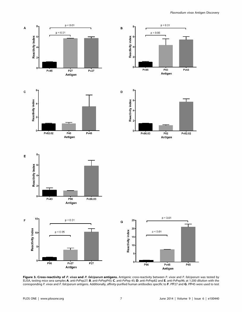
Figure 3. Cross-reactivity of P. vivax and P. falciparum antigens. Antigenic cross-reactivity between P. vivax and P. falciparum was tested byELISA, testing mice sera samples A. anti-PvPep27; B. anti-PvPepP43; C. anti-PvPep 45; D. anti-PvPep82 and E. anti-PvPep96; at 1:200 dilution with thecorresponding P. vivax and P. falciparum antigens. Additionally, affinity-purified human antibodies specific to F. PfP27 and G. PfP45 were used to test
Plasmodium vivax Antigen Discovery
PLOS ONE | www.plosone.org 7 June 2014 | Volume 9 | Issue 6 | e100440

IFA testParasite recognition by anti-peptide antibodies was determined
by IFAT, using as antigen, P. vivax blood stages obtained from
Colombian patients, and mouse sera collected 10 days after last
peptide immunization. Briefly, parasites were incubated with sera
diluted 1:20. This reaction was developed with fluorescein-
conjugated goat anti-mouse IgG (Jackson Immunoresearch
Laboratories, Inc., Baltimore, MD) diluted 1:1000. Slides were
mounted in 30% glycerol and examined under a Nikon Eclipse
microscope by epifluorescence. P. falciparum parasite cross-reactiv-
ity was also determined by IFAT using as antigen, Pf-FCB-1
blood-stage parasites derived from in vitro cultures [41].
Cellular immune responses in miceTo determine the potential of eight selected peptides to
stimulate T-cell responses, in vitro IFN-c-production by lymph
nodes and splenocytes obtained from immunized mice was
quantified. For this purpose a commercial mouse anti-IFN-cELISpot kit (Mabtech AB, Stockholm, Sweden) was used; the test
carried out according to the manufacturer’s instructions. Multi-
screen 96-well plates (Millipore, Bedford, MA) were coated
overnight at room temperature with 5 mg/mL anti-mouse IFN-cantibodies. RPMI 1640 medium containing 10% fetal bovine
serum (FBS, GIBCO) was used as a blocking solution. Freshly
isolated mononuclear cells were plated into duplicate wells at
56105 cells in RPMI 1640 medium supplemented with 10% FBS
(100 mL/well). Culture medium alone, Conconavalin A or 10 mg
of each synthetic peptide/mL medium (100 mL/well) was added
and plates were cultured for 40 h at 37uC in a 5% CO2 humidified
atmosphere. After washing, biotinylated antibody at 1 mg/mL was
added and incubated for 2 h at room temperature. Plates were
washed and alkaline phosphatase-streptavidin (Mabtech AB,
Stockholm, Sweden) was added (1:1000). Spots were visualized
by adding 50 mL/well of BCIP/NBT (Sigma), scanned and
counted using the AID ELISpot reader (AID Autoimmun
Diagnostika GmbH, Germany) to determine the number of
spots/well. Results were expressed as the mean number of IFN-cspot-forming cells (SFC) per 106 cells.
Statistical analysisFisher’s exact test (262 contingency tables) was used to compare
differences in seroprevalence between the PNG and Colombian
groups; the ANOVA test was used to compare groups. Dunnett’s
Multiple Comparison Test was used as post-hoc analysis and p
value,0.05 was considered statistically significant. Statistical
analyses were performed using GraphPad Prism software (version
5.01; GraphPad Software Inc., San Diego, CA, USA.
Results
P. vivax genome bioinformatic analysisA total of 50 P. vivax fragments, 25–57 residues long and containing
the a-helical coiled coil motifs, were selected based on proteome and
transcriptome data of P. falciparum orthologues present in erythrocytic
parasite stages (Tables 1 and S1). Variable homology (29 to 100%
identity) was observed between P. falciparum and the corresponding P.
vivax fragments (Table S1), most of which (32 antigens) were greater
than 60% homologous. Identification of potential signal peptides,
transmembrane (TM) regions, and GPI-anchored or sub-cellular
localization prediction revealed five proteins containing TM domains
(PvPep39, PvPep101, PvPep122, PvPep123 and PvPep131) and
another three involved in secretory pathways (PvPep52, PvPep60,
PvPep96.01). These latter peptides also contained a signal peptide.
One of the proteins is predicted to be located in the mitochondria
(PvPep39); none contained a GPI anchor.
Circular dichroism studiesCircular dichroism (CD) studies of 16 randomly selected peptides
indicate that they assume a total or partial a-helical conformation in
water. Peptides 40-43, 55, 60 and 65 exhibit a CD pattern
characteristic of a high a-helical content as indicate for PvPep40
(Figure 1A), whereas the remaining peptides (2, 5, 12, 27, 41, 45, 48 59
and 63) show CD profiles similar to that shown for peptide PvPep63
(Figure 1B) or intermediate between those shown in Figures 1A and
1B, all characteristic of a partial a-helical organization.
Recognition of a-helical coiled coil peptides by humansera
Out of the 50 a-helical coiled coil peptides tested by ELISA
using human sera, 43 were recognized by PNG (n = 42) sera at
the reactivity of homologous P. falciparum and P. vivax antigens. Human IgG was tested at a 1:200 dilution. In all cases, a non-related antigen wasused as a negative control. Reactivity index defined as OD values of tested sample divided by the cut-off value, are reported as mean 6 SEM for eachmouse serum. Cross reactivity experiments were performed in duplicate in two independent experiments.doi:10.1371/journal.pone.0100440.g003
Table 4. Reactivity of IgG tested with different parasite antigen fragments and whole parasites.
Origin Antibody Identitya (%) P. vivax fragmentsb P. falciparum fragmentsc P. vivax parasited P. falciparum parasitee
Mouse anti PvPep27 63 + + - -
Mouse anti-PvPep43 82 + + + -
Mouse anti-PvPep45 44 + - + -
Mouse anti-PvPep82 61 + - + -
Mouse anti-PvPep96 43 + - + -
Human anti Pf-P27 NAd + + + +
Human anti Pf-P45 NA + + + +
aIdentity between P. vivax and P. falciparum orthologous antigens; bReactivity tested by ELISA test using P. vivax antigens; cReactivity tested by ELISA test using P.falciparum antigens; dReactivity tested by IFA test with P. vivax blood stages; eReactivity tested by IFA test with P. falciparum blood stages; dDoes not apply.doi:10.1371/journal.pone.0100440.t004
Plasmodium vivax Antigen Discovery
PLOS ONE | www.plosone.org 8 June 2014 | Volume 9 | Issue 6 | e100440

variable prevalence, however in all cases prevalence was .10%;
20 antigens displayed reactivity .29% (see Table S1). In addition,
17 peptides, which showed more than 30% of prevalence with
PNG samples, were further tested with Colombian sera; all these
peptides were antigenic with variable prevalence (Table 2). Ten
peptides (PvPep27, PvPep42, PvPep43, PvPep45, PvPep82.02,
PvPep82.03, PvPep83, PvPep95, PvPep96.01 and PvPep96.03)
tested with the 90 human Colombian sera samples displayed a
high degree of recognition, ranging from 30% to 86%. Recogni-
tion of the 17 peptides by PNG sera ranged between 29-71%,
whereas recognition by Colombian sera for the same 17 peptides
varied between 2–86%.
Interestingly, seven of the 17 selected peptides were the most
antigenic (.50% of responders) in PNG (PvPep27, PvPep43,
PvPep45, PvPep63, PvPep83, PvPep96.01, PvPep96.03), four
peptides (PvPep27, PvPep42, PvPep82.03 and PvPep83) were the
most reactive with Colombian sera (Table 2). Differences in
reactivity were also observed between the two malaria-endemic
sites in Colombia, Tierralta and Tumaco (data not shown).
Responses against 16/17 peptides were stronger with PNG as
compared with Colombian sera, presenting with OD ratios .2
(Table 2).
Immunogenicity of a-helical coiled coil peptides in miceEight peptides that showed prevalence .50% either with PNG
or Colombian sera were further tested for their immunogenicity in
BALB/c mice. Immunized mice developed specific IgG antibodies
to the a-helical coiled coil fragments after the second immuniza-
tion dose as determined by ELISA with the exception of those
immunized with PvPep42; three immunization doses were needed
to produce detectable antibody levels (Figure 2). Antibody titers
increased steadily with titers ranging from 96102 to 26106 after
the third immunization (Table 3). Mice immunized with PvPep27
and PvPep95 showed variable responses that were not uniform in
all animals; two animals in each group failed to develop the typical
boosting response after third dose. Antibody titers decreased and
became negative (PvPep27) or remained stable (PvPep95); neither
recognized the native protein in the IFAT (Table 3).
However, sera from four of the eight immunized groups were
able to recognize native protein on P. vivax asexual blood stages in
IFAT assays at a 1:20 dilution; two showed strong reactivity
(Table 3). Control mice, which received only adjuvant in saline
solution, were non-responsive as indicated by ELISA and IFAT
(data not shown).
Cross-reactivity testsSera from mice immunized with PvPep27 and PvPep43 were
reactive with the corresponding orthologues Pf-P27 and Pf-P43
with similar reactivity indices as compared to a control sample
(Figure 3). None of the other antigens (Pf-P45, Pf-P82 or Pf-P96)
showed significant cross-reactivity. Moreover, cross-reactivity was
also observed when specific affinity-purified human IgG to Pf-P27
and Pf-P45 were tested with the corresponding P. vivax orthologue;
three-fold less reactivity was observed in both cases as compared
with the positive control. Additionally, cross-reactivity with whole
P. falciparum parasites was observed by IFAT (Table 4). No
relationship was observed between homology and reactivity since
fragments with low identity, such as PvPep45, were highly reactive
with both the P. vivax fragment and the P. falciparum parasite,
whereas PvPep82.02 with greater than 60% homology was not
reactive (Table 4).
Cellular immune responses in miceT-cell IFN-c production was induced by six (PvPep27, PvPep42,
PvPep43, PvPep45, PvPep52 and PvPep82.02) of the eight peptides
tested by ELIspot; PvPep95 and PvPep96.03 were not recognized
by murine lymphocytes (Table 3). The greatest IFN-c production
was induced by PvPep43 and PvPep52 (mean SFC 344.7615.33
and 304660.8, respectively) followed by PvPep45, PvPep27 and
PvPep42 (mean SFC 176.7698.1, 127.3647.6 and 68.3647.6,
respectively) (Figure 4).
Additionally, the selected peptides presented potential CD4+epitopes in their amino acid sequences as confirmed by
bioinformatics analysis (Table 5). No apparent relation was
observed between the affinity of the predicted epitope and the
IFN-c results obtained, when mouse epitopes were described
(Table 5). Higher affinity, defined as the lower percentile rank,
were observed for PvPep27 and PvPep82.02 epitopes. When the
alleles from human were tested, higher affinity was observed in all
cases compared with mouse epitopes, although differences were
observed in the main epitopes found. Same epitopes predicted for
mouse alleles could be present in human alleles but with lower
affinity.
Discussion
In an attempt to identify new target parasite antigens for
malaria vaccine development, bioinformatics tools have been
previously used to select proteins containing a-helical coiled coil
motifs in P. falciparum proteins. In this study, similar algorithms
were used in a pilot search of P. falciparum orthologous antigens in
the P. vivax genome, and 50 a-helical coiled coil P. vivax segments
showing a high degree of homology to the previously identified
orthologous P. falciparum fragments were selected, and were further
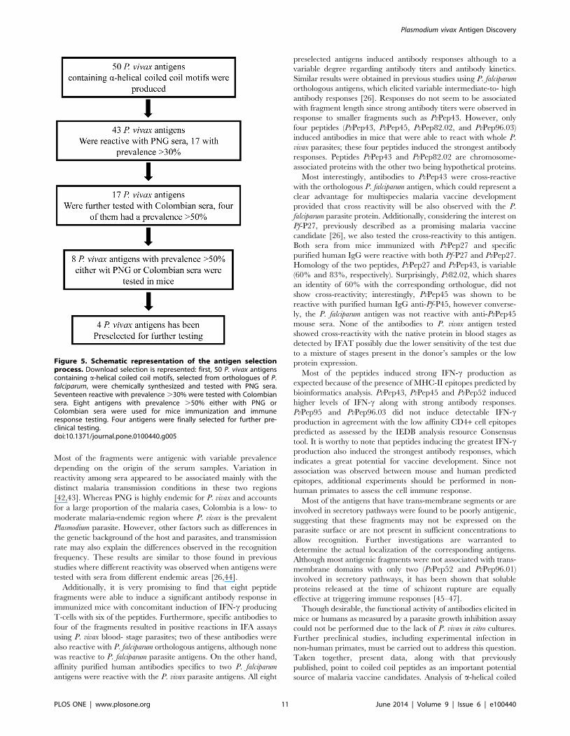
assess in antigenicity and immunogenicity studies; at the end four
antigens were identified as potential targets for additional testing as
vaccine candidates (Figure 5).
It is interesting to note that of the 50 fragments tested
containing a-helical coils, 19 were recognized by sera of
individuals living in P. vivax endemic areas of PNG and Colombia.
Figure 4. Production of IFN-c by mononuclear cells fromimmunized mice. Proliferative responses of mononuclear cellsobtained from mice immunized with eight synthetic peptides, andfurther in vitro-stimulated with 10 mg/mL of corresponding coiled coilpeptides. Conconavalin A (Con A) mitogen was used as a positivecontrol. RPMI 1640 medium was used as a negative control. Data of SFCwere reported as mean 6 SEM for each peptide. P value was calculatedby the ANOVA test. MN: mononuclear cells.doi:10.1371/journal.pone.0100440.g004
Plasmodium vivax Antigen Discovery
PLOS ONE | www.plosone.org 9 June 2014 | Volume 9 | Issue 6 | e100440

Ta
ble
5.
Ce
llim
mu
ne
resp
on
sean
das
soci
atio
nw
ith
HLA
IIe
pit
op
es
pre
dic
tio
n.
An
tig
en
IFN
-cp
rod
uct
ion
SF
Ca
Mo
use
Hu
ma
n
Me
an
ran
ge
Pe
pti
de
All
ele
Pe
rce
nti
lera
nk
Pe
pti
de
All
ele
Pe
rce
nti
lera
nk
PvP
ep
27
16
41
25
–2
28
KK
QN
AEK
ELSV
LKK
NH
2-I
ad9
.8V
LKK
NY
DA
MSE
EIEE
HLA
-DQ
A1
*04
01
/DQ
B1
*04
02
1.0
2
KK
QN
AEK
ELSV
LKK
NH
LA-D
PA
1*0
20
1/D
PB
1*0
50
11
6.0
9
PvP
ep
42
13
31
04
–1
69
PD
YY
KK
ITT
KLQ
NN
IH
2-I
ad2
0.7
2IN
NIT
ND
INIL
KSS
IH
LA-D
RB
1*0
30
10
.31
PD
YY
KK
ITT
KLQ
NN
IH
LA-D
RB
5*0
10
11
.4
PvP
ep
43
34
43
14
–3
60
VK
KLR
EELN
KV
TN
EYH
2-I
ad4
3.0
1T
NEY
DD
FKN
KLE
LLY
HLA
-DR
B1
*08
01
2.3
7
VK
KLR
EELN
KV
TN
EY
HLA
-DP
A1
*02
01
/DP
B1
*05
01
9.1
4
PvP
ep
45
25
41
91
–3
39
NEA
KEE
VIE
KK
EEM
TH
2-I
ed
48
.85
INK
NIS
TIN
DD
VD
HI
HLA
-DR
B3
*01
01
0.0
1
PvP
ep
52
27
71
22
–3
75
NIN
ETK
ITH
LRN
KIE
H2
-Ie
d3
2.4
4IN
EQIN
INET
KIT
HL
HLA
-DR
B1
*07
01
0.3
5
NIN
ETK
ITH
LRN
KIE
HLA
-DR
B1
*08
27
4.2
9
PvP
ep
82
.02
69
36
–1
42
NLD
QK
ILEL
QA
SFT
CH
2-I
ad7
.93
NEI
KQ
VIK
KIE
GLE
KH
LA-D
RB
5*0
10
10
.39
NLD
QK
ILEL
QA
SFT
CH
LA-D
RB
1*0
10
20
.39
PvP
ep
95
NR
bN
RLK
DLN
DK
IRN
YD
SII
H2
-Ie
d4
7.2
2EK
GLK
DLN
DK
IRN
YD
HLA
-DR
B3
*01
01
0.2
6
LKD
LND
KIR
NY
DSI
IH
LA-D
RB
5*0
10
11
7.0
4
PvP
ep
96
.03
NR
NR
ND
DV
DH
INSN
INN
INH
2-I
ab4
2.2
8IN
KN
IST
IND
DV
DH
IH
LA-D
RB
3*0
10
10
.01
ND
DV
DH
INSN
INN
INH
LA-D
RB
1*0
10
21
2.6
1
aSF
C:
spo
t-fo
rmin
gco
lon
ies6
10
6;
bN
ore
spo
nse
ob
serv
ed
.d
oi:1
0.1
37
1/j
ou
rnal
.po
ne
.01
00
44
0.t
00
5
Plasmodium vivax Antigen Discovery
PLOS ONE | www.plosone.org 10 June 2014 | Volume 9 | Issue 6 | e100440
Most of the fragments were antigenic with variable prevalence
depending on the origin of the serum samples. Variation in
reactivity among sera appeared to be associated mainly with the
distinct malaria transmission conditions in these two regions
[42,43]. Whereas PNG is highly endemic for P. vivax and accounts
for a large proportion of the malaria cases, Colombia is a low- to
moderate malaria-endemic region where P. vivax is the prevalent
Plasmodium parasite. However, other factors such as differences in
the genetic background of the host and parasites, and transmission
rate may also explain the differences observed in the recognition
frequency. These results are similar to those found in previous
studies where different reactivity was observed when antigens were
tested with sera from different endemic areas [26,44].
Additionally, it is very promising to find that eight peptide
fragments were able to induce a significant antibody response in
immunized mice with concomitant induction of IFN-c producing
T-cells with six of the peptides. Furthermore, specific antibodies to
four of the fragments resulted in positive reactions in IFA assays
using P. vivax blood- stage parasites; two of these antibodies were
also reactive with P. falciparum orthologous antigens, although none
was reactive to P. falciparum parasite antigens. On the other hand,
affinity purified human antibodies specifics to two P. falciparum
antigens were reactive with the P. vivax parasite antigens. All eight
preselected antigens induced antibody responses although to a
variable degree regarding antibody titers and antibody kinetics.
Similar results were obtained in previous studies using P. falciparum
orthologous antigens, which elicited variable intermediate-to- high
antibody responses [26]. Responses do not seem to be associated
with fragment length since strong antibody titers were observed in
response to smaller fragments such as PvPep43. However, only
four peptides (PvPep43, PvPep45, PvPep82.02, and PvPep96.03)
induced antibodies in mice that were able to react with whole P.
vivax parasites; these four peptides induced the strongest antibody
responses. Peptides PvPep43 and PvPep82.02 are chromosome-
associated proteins with the other two being hypothetical proteins.
Most interestingly, antibodies to PvPep43 were cross-reactive
with the orthologous P. falciparum antigen, which could represent a
clear advantage for multispecies malaria vaccine development
provided that cross reactivity will be also observed with the P.
falciparum parasite protein. Additionally, considering the interest on
Pf-P27, previously described as a promising malaria vaccine
candidate [26], we also tested the cross-reactivity to this antigen.
Both sera from mice immunized with PvPep27 and specific
purified human IgG were reactive with both Pf-P27 and PvPep27.
Homology of the two peptides, PvPep27 and PvPep43, is variable
(60% and 83%, respectively). Surprisingly, Pv82.02, which shares
an identity of 60% with the corresponding orthologue, did not
show cross-reactivity; interestingly, PvPep45 was shown to be
reactive with purified human IgG anti-Pf-P45, however converse-
ly, the P. falciparum antigen was not reactive with anti-PvPep45
mouse sera. None of the antibodies to P. vivax antigen tested
showed cross-reactivity with the native protein in blood stages as
detected by IFAT possibly due the lower sensitivity of the test due
to a mixture of stages present in the donor’s samples or the low
protein expression.
Most of the peptides induced strong IFN-c production as
expected because of the presence of MHC-II epitopes predicted by
bioinformatics analysis. PvPep43, PvPep45 and PvPep52 induced
higher levels of IFN-c along with strong antibody responses.
PvPep95 and PvPep96.03 did not induce detectable IFN-cproduction in agreement with the low affinity CD4+ cell epitopes
predicted as assessed by the IEDB analysis resource Consensus
tool. It is worthy to note that peptides inducing the greatest IFN-cproduction also induced the strongest antibody responses, which
indicates a great potential for vaccine development. Since not
association was observed between mouse and human predicted
epitopes, additional experiments should be performed in non-
human primates to assess the cell immune response.
Most of the antigens that have trans-membrane segments or are
involved in secretory pathways were found to be poorly antigenic,
suggesting that these fragments may not be expressed on the
parasite surface or are not present in sufficient concentrations to
allow recognition. Further investigations are warranted to
determine the actual localization of the corresponding antigens.
Although most antigenic fragments were not associated with trans-
membrane domains with only two (PvPep52 and PvPep96.01)
involved in secretory pathways, it has been shown that soluble
proteins released at the time of schizont rupture are equally
effective at triggering immune responses [45–47].
Though desirable, the functional activity of antibodies elicited in
mice or humans as measured by a parasite growth inhibition assay
could not be performed due to the lack of P. vivax in vitro cultures.
Further preclinical studies, including experimental infection in
non-human primates, must be carried out to address this question.
Taken together, present data, along with that previously
published, point to coiled coil peptides as an important potential
source of malaria vaccine candidates. Analysis of a-helical coiled
Figure 5. Schematic representation of the antigen selectionprocess. Download selection is represented: first, 50 P. vivax antigenscontaining a-helical coiled coil motifs, selected from orthologues of P.falciparum, were chemically synthesized and tested with PNG sera.Seventeen reactive with prevalence .30% were tested with Colombiansera. Eight antigens with prevalence .50% either with PNG orColombian sera were used for mice immunization and immuneresponse testing. Four antigens were finally selected for further pre-clinical testing.doi:10.1371/journal.pone.0100440.g005
Plasmodium vivax Antigen Discovery
PLOS ONE | www.plosone.org 11 June 2014 | Volume 9 | Issue 6 | e100440

coil motifs should be extended to the entire group of erythrocytic
parasite antigens. Poly-subunit antigens should be designed,
containing both relevant P. vivax and P. falciparum fragments that
are capable of inducing effective immune responses. Thus, this
study has direct relevance to P. vivax asexual blood- stage vaccine
design and suggests that some of the antigens tested could be
effective in different malaria settings such as PNG and Colombia.
Supporting Information
Table S1 Bioinformatics analysis of coiled coil frag-ments and Antibody response to PNG sera samples of allcoiled coil P. vivax tested antigens.
(XLSX)
Acknowledgments
Authors are grateful for the participation of the community from malaria-
endemic regions of PNG and Colombia as well as Swiss volunteers. We
thank Geraldine Frank and Eliecer Jimenez for their expert technical
support. Dr. Alice Koumare of the Centre National de Transfusion
Sanguine in Ouagadougou, Burkina Faso for providing immune plasma.
We also thank Seppic Inc, Paris, France for the supply of Montanide
adjuvant.
Author Contributions
Conceived and designed the experiments: NC GC SH. Performed the
experiments: NC CH MLP AC. Analyzed the data: NC AK MLP.
Contributed reagents/materials/analysis tools: AK CS IF RM. Wrote the
paper: NC MLP AC MAH GC SH.
References
1. WHO (2013) World malaria report 2013. World Health Organization.
2. Mendis K, Sina BJ, Marchesini P, Carter R (2001) The neglected burden of
Plasmodium vivax malaria. Am J Trop Med Hyg 64: 97–106.
3. Greenwood BM, Targett GA (2011) Malaria vaccines and the new malaria
agenda. Clin Microbiol Infect 17: 1600–1607.
4. Hill AV (2011) Vaccines against malaria. Philos Trans R Soc Lond B Biol Sci
366: 2806–2814.
5. Schwartz L, Brown GV, Genton B, Moorthy VS (2012) A review of malaria
vaccine clinical projects based on the WHO rainbow table. Malar J 11: 11.
6. Salvador A, Hernandez RM, Pedraz JL, Igartua M (2012) Plasmodium falciparum
malaria vaccines: current status, pitfalls and future directions. Expert Rev
Vaccines 11: 1071–1086.
7. Agnandji ST, Lell B, Fernandes JF, Abossolo BP, Methogo BG, et al. (2012) A
phase 3 trial of RTS,S/AS01 malaria vaccine in African infants. N Engl J Med
367: 2284–2295.
8. Valencia SH, Rodriguez DC, Acero DL, Ocampo V, Arevalo-Herrera M (2011)
Platform for Plasmodium vivax vaccine discovery and development. Mem Inst
Oswaldo Cruz 106 Suppl 1: 179–192.
9. Arevalo-Herrera M, Castellanos A, Yazdani SS, Shakri AR, Chitnis CE, et al.
(2005) Immunogenicity and protective efficacy of recombinant vaccine based on
the receptor-binding domain of the Plasmodium vivax Duffy binding protein in
Aotus monkeys. Am J Trop Med Hyg 73: 25–31.
10. Bell BA, Wood JF, Bansal R, Ragab H, Cargo J III, et al. (2009) Process
development for the production of an E. coli produced clinical grade
recombinant malaria vaccine for Plasmodium vivax. Vaccine 27: 1448–1453.
11. Moreno A, Caro-Aguilar I, Yazdani SS, Shakri AR, Lapp S, et al. (2008)
Preclinical assessment of the receptor-binding domain of Plasmodium vivax
Duffy-binding protein as a vaccine candidate in rhesus macaques. Vaccine 26:
4338–4344.
12. Vicentin EC, Francoso KS, Rocha MV, Iourtov D, Dos Santos FL, et al. (2014)
Invasion-inhibitory antibodies elicited by immunization with Plasmodium vivax
apical membrane antigen-1 expressed in Pichia pastoris yeast. Infect Immun 82:
1296–1307.
13. Teixeira LH, Tararam CA, Lasaro MO, Camacho AG, Ersching J, et al. (2014)
Immunogenicity of a prime-boost vaccine containing the circumsporozoite
proteins of Plasmodium vivax in rodents. Infect Immun 82: 793–807.
14. Herrera S, Bonelo A, Perlaza BL, Fernandez OL, Victoria L, et al. (2005) Safety
and elicitation of humoral and cellular responses in colombian malaria-naive
volunteers by a Plasmodium vivax circumsporozoite protein-derived synthetic
vaccine. Am J Trop Med Hyg 73: 3–9.
15. Herrera S, Fernandez OL, Vera O, Cardenas W, Ramirez O, et al. (2011) Phase
I safety and immunogenicity trial of Plasmodium vivax CS derived long synthetic
peptides adjuvanted with montanide ISA 720 or montanide ISA 51. Am J Trop
Med Hyg 84: 12–20.
16. Wu Y, Ellis RD, Shaffer D, Fontes E, Malkin EM, et al. (2008) Phase 1 trial of
malaria transmission blocking vaccine candidates Pfs25 and Pvs25 formulated
with montanide ISA 51. PLoS One 3: e2636.
17. Carlton JM, Adams JH, Silva JC, Bidwell SL, Lorenzi H, et al. (2008)
Comparative genomics of the neglected human malaria parasite Plasmodium
vivax. Nature 455: 757–763.
18. Gardner MJ, Hall N, Fung E, White O, Berriman M, et al. (2002) Genome
sequence of the human malaria parasite Plasmodium falciparum. Nature 419:
498–511.
19. Florens L, Washburn MP, Raine JD, Anthony RM, Grainger M, et al. (2002) A
proteomic view of the Plasmodium falciparum life cycle. Nature 419: 520–526.
20. Doolan DL, Southwood S, Freilich DA, Sidney J, Graber NL, et al. (2003)
Identification of Plasmodium falciparum antigens by antigenic analysis of
genomic and proteomic data. Proc Natl Acad Sci U S A 100: 9952–9957.
21. Corradin G, Villard V, Kajava AV (2007) Protein structure based strategies for
antigen discovery and vaccine development against malaria and other
pathogens. Endocr Metab Immune Disord Drug Targets 7: 259–265.
22. Kulangara C, Kajava AV, Corradin G, Felger I (2009) Sequence conservation in
Plasmodium falciparum alpha-helical coiled coil domains proposed for vaccine
development. PLoS One 4: e5419.
23. Singh S, Soe S, Roussilhon C, Corradin G, Druilhe P (2005) Plasmodium
falciparum merozoite surface protein 6 displays multiple targets for naturally
occurring antibodies that mediate monocyte-dependent parasite killing. Infect
Immun 73: 1235–1238.
24. Tripet B, Kao DJ, Jeffers SA, Holmes KV, Hodges RS (2006) Template-based
coiled-coil antigens elicit neutralizing antibodies to the SARS-coronavirus.
J Struct Biol 155: 176–194.
25. Olugbile S, Villard V, Bertholet S, Jafarshad A, Kulangara C, et al. (2011)
Malaria vaccine candidate: design of a multivalent subunit alpha-helical coiled
coil poly-epitope. Vaccine 29: 7090–7099.
26. Villard V, Agak GW, Frank G, Jafarshad A, Servis C, et al. (2007) Rapid
identification of malaria vaccine candidates based on alpha-helical coiled coil
protein motif. PLoS One 2: e645.
27. Lupas A, Van Dyke M, Stock J (1991) Predicting coiled coils from protein
sequences. Science 252: 1162–1164.
28. Bendtsen JD, Nielsen H, von Heijne G, Brunak S (2004) Improved prediction of
signal peptides: SignalP 3.0. J Mol Biol 340: 783–795.
29. Krogh A, Larsson B, von Heijne G, Sonnhammer EL (2001) Predicting
transmembrane protein topology with a hidden Markov model: application to
complete genomes. J Mol Biol 305: 567–580.
30. Eisenhaber B, Bork P, Eisenhaber F (1999) Prediction of potential GPI-
modification sites in proprotein sequences. J Mol Biol 292: 741–758.
31. Guda C, Subramaniam S (2005) pTARGET [corrected] a new method for
predicting protein subcellular localization in eukaryotes. Bioinformatics 21:
3963–3969.
32. Kim Y, Ponomarenko J, Zhu Z, Tamang D, Wang P, et al. (2012) Immune
epitope database analysis resource. Nucleic Acids Res 40: W525–530.
33. Wang P, Sidney J, Kim Y, Sette A, Lund O, et al. (2010) Peptide binding
predictions for HLA DR, DP and DQ molecules. BMC Bioinformatics 11: 568.
34. Lundegaard C, Lamberth K, Harndahl M, Buus S, Lund O, et al. (2008)
NetMHC-3.0: accurate web accessible predictions of human, mouse and
monkey MHC class I affinities for peptides of length 8-11. Nucleic Acids Res 36:
W509–512.
35. Nielsen M, Lundegaard C, Worning P, Lauemoller SL, Lamberth K, et al.
(2003) Reliable prediction of T-cell epitopes using neural networks with novel
sequence representations. Protein Sci 12: 1007–1017.
36. Peters B, Sette A (2005) Generating quantitative models describing the sequence
specificity of biological processes with the stabilized matrix method. BMC
Bioinformatics 6: 132.
37. Sidney J, Assarsson E, Moore C, Ngo S, Pinilla C, et al. (2008) Quantitative
peptide binding motifs for 19 human and mouse MHC class I molecules derived
using positional scanning combinatorial peptide libraries. Immunome Res 4: 2.
38. Atherton E, Hubscher W, Sheppard RC, Woolley V (1981) Synthesis of a 21-
residue fragment of human proinsulin by the polyamide solid phase method.
Hoppe Seylers Z Physiol Chem 362: 833–839.
39. Alpers MP, al-Yaman F, Beck HP, Bhatia KK, Hii J, et al. (1992) The Malaria
Vaccine Epidemiology and Evaluation Project of Papua New Guinea: rationale
and baseline studies. P N G Med J 35: 285–297.
40. Arevalo-Herrera M, Roggero MA, Gonzalez JM, Vergara J, Corradin G, et al.
(1998) Mapping and comparison of the B-cell epitopes recognized on the
Plasmodium vivax circumsporozoite protein by immune Colombians and
immunized Aotus monkeys. Ann Trop Med Parasitol 92: 539–551.
41. Trager W, Jensen JB (1976) Human malaria parasites in continuous culture.
Science 193: 673–675.
42. Koepfli C, Ross A, Kiniboro B, Smith TA, Zimmerman PA, et al. (2011)
Multiplicity and diversity of Plasmodium vivax infections in a highly endemic
region in Papua New Guinea. PLoS Negl Trop Dis 5: e1424.
Plasmodium vivax Antigen Discovery
PLOS ONE | www.plosone.org 12 June 2014 | Volume 9 | Issue 6 | e100440

43. Arevalo-Herrera M, Quinones ML, Guerra C, Cespedes N, Giron S, et al.
(2012) Malaria in selected non-Amazonian countries of Latin America. Acta
Trop 121: 303–314.
44. Cespedes N, Arevalo-Herrera M, Felger I, Reed S, Kajava AV, et al. (2013)
Antigenicity and immunogenicity of a novel chimeric peptide antigen based on
the P. vivax circumsporozoite protein. Vaccine 31: 4923–4930.
45. Jafarshad A, Dziegiel MH, Lundquist R, Nielsen LK, Singh S, et al. (2007) A
novel antibody-dependent cellular cytotoxicity mechanism involved in defense
against malaria requires costimulation of monocytes FcgammaRII and
FcgammaRIII. J Immunol 178: 3099–3106.46. Kulangara C, Luedin S, Dietz O, Rusch S, Frank G, et al. (2012) Cell biological
characterization of the malaria vaccine candidate trophozoite exported protein
1. PLoS One 7: e46112.47. Olugbile S, Kulangara C, Bang G, Bertholet S, Suzarte E, et al. (2009) Vaccine
potentials of an intrinsically unstructured fragment derived from the blood stage-associated Plasmodium falciparum protein PFF0165c. Infect Immun 77: 5701–
5709.
Plasmodium vivax Antigen Discovery
PLOS ONE | www.plosone.org 13 June 2014 | Volume 9 | Issue 6 | e100440