GIS, Mining, Nigeria, Minerals, Pollution, Environmental ...

REVIEW ARTICLEpublished: 26 November 2013
doi: 10.3389/fmicb.2013.00344
Phylogenetic significance of composition and crystalmorphology of magnetosome mineralsMihály Pósfai1, Christopher T. Lefèvre2, Denis Trubitsyn3, Dennis A. Bazylinski3 and
Richard B. Frankel4*
1 Department of Earth and Environmental Sciences, University of Pannonia, Veszprém, Hungary2 Laboratoire de Bioénergétique Cellulaire, Biologie Végétale et Microbiologie Environnementales, CEA/CNRS/Aix-Marseille Université, Saint Paul lez Durance,
France3 School of Life Sciences, University of Nevada at Las Vegas, Las Vegas, NV, USA4 Department of Physics, California Polytechnic State University, San Luis Obispo, CA, USA
Edited by:
Wei Lin, Chinese Academy ofSciences, China
Reviewed by:
Ulysses Lins, Universidade Federaldo Rio de Janeiro, BrazilArash Komeili, University ofCalifornia, Berkeley, USA
*Correspondence:
Richard B. Frankel, Department ofPhysics, California Polytechnic StateUniversity, 1 Grand Avenue, SanLuis Obispo, CA 93407, USAe-mail: [email protected]
Magnetotactic bacteria (MTB) biomineralize magnetosomes, nano-scale crystals ofmagnetite or greigite in membrane enclosures that comprise a permanent magneticdipole in each cell. MTB control the mineral composition, habit, size, and crystallographicorientation of the magnetosomes, as well as their arrangement within the cell.Studies involving magnetosomes that contain mineral and biological phases requiremultidisciplinary efforts. Here we use crystallographic, genomic and phylogeneticperspectives to review the correlations between magnetosome mineral habits andthe phylogenetic affiliations of MTB, and show that these correlations have importantimplications for the evolution of magnetosome synthesis, and thus magnetotaxis.
Keywords: magnetotactic bacteria, magnetite, greigite, magnetosomes, morphology, biomineralization, evolution
INTRODUCTIONAll magnetotactic bacteria (MTB) contain magnetosomes com-prising nano-scale, magnetite (Fe3O4) or greigite (Fe3S4) crystalsenclosed in phospholipid bilayer membranes (Gorby et al., 1988;Bazylinski and Frankel, 2004). The magnetosomes constitute apermanent magnetic dipole moment in the cell, and are essen-tial for magnetotaxis. The magnetosome membrane is derived byinvagination of the cytoplasmic membrane (Komeili et al., 2004)and is the locus of biological control over the nucleation andgrowth of the mineral crystal. Most MTB species or strains exclu-sively produce either magnetite (Frankel et al., 1979) or greigitemagnetosomes (Mann et al., 1990), although several MTB canproduce magnetosomes of both kinds, depending on environ-mental conditions (Bazylinski et al., 1993; Kasama et al., 2006;Lins et al., 2007; Lefèvre et al., 2011c; Wang et al., 2013).
The crystal size, crystallographic orientation and arrangementof magnetosomes in MTB are all highly significant for the mag-netic properties of the cell (Frankel and Blakemore, 1980; Mannet al., 1984a,b; Moskowitz et al., 1988; Bazylinski and Frankel,2003). With a few exceptions, the lengths of individual magneto-some crystals range from about 35 to 120 nm (Devouard et al.,1998) (Table 1); this is within the permanent single-magnetic-domain (SD) size range for both minerals (Butler and Banerjee,1975). In the majority of MTB, the magnetosomes are organizedin one or more straight chains of various lengths, parallel to theaxis of motility of the cell. In cells of some species, however, thereare multiple individual chains or a chain with multiple strands(Vali and Kirschvink, 1991) or even dispersed aggregates or clus-ters of magnetosomes that occur in some magnetotactic cocci(Towe and Moench, 1981; Cox et al., 2002; Zhang et al., 2012).
When magnetosomes are arranged in chains, magnetic inter-actions between them cause their magnetic moments to ori-ent parallel to each other along the chain axis (Frankel andBlakemore, 1980; Frankel, 1984), resulting in a permanent, mag-netic dipole. The permanent magnetism of magnetosome chainshas been demonstrated by electron holography in the electronmicroscope (Dunin-Borkowski et al., 1998), by pulsed magneticfield remanence measurements on individual cells (Penningaet al., 1995; Hanzlik et al., 2002) and by magnetic imaging directlyin living cells (Le Sage et al., 2013).
The magnetosome membrane originates from the cytoplasmicmembrane and contains unique proteins that are not present inthe cytoplasmic or outer membranes (Komeili, 2012). These pro-teins, specific to MTB, are designated with the prefix Mam orMms, although some are not found in every species of MTB. TheMms proteins in particular are present only in certain phyloge-netic groups of MTB. While not all Mam proteins are found in themagnetosome membrane, all Mms proteins are. The Mam andMms proteins are thought to be responsible for biomineralizationof the magnetosome crystal, the organization of the magneto-some chain, and the crystallographic orientation of the individualmagnetosomes with respect to the chain (Komeili, 2012). Theroles of relatively few of the magnetosome membrane proteinshave been elucidated (Jogler and Schüler, 2009; Murat et al., 2010;Lohsse et al., 2011; Uebe et al., 2011; Komeili, 2012).
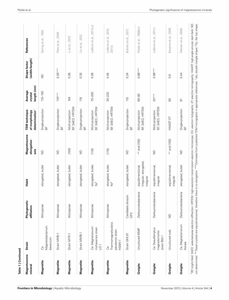
All known MTB are phylogenetically affiliated with the Alpha-,Delta- or Gammaproteobacteria classes of the Proteobacteria phy-lum, the Nitrospirae phylum or the candidate division OP3 whichis part of the Planctomycetes–Verrucomicrobia–Chlamydiae (PVC)bacterial superphylum (Lefèvre and Bazylinski, 2013) (Table 1).
www.frontiersin.org November 2013 | Volume 4 | Article 344 | 1

Pósfai et al. Phylogenetic significance of magnetosome minerals
Ta
ble
1|B
ibli
og
rap
hic
listi
ng
of
ma
gn
eto
tacti
cb
acte
ria
ch
ara
cte
rize
da
nd
the
co
mp
osit
ion
an
dm
orp
ho
log
yo
fth
eir
ma
gn
eto
so
me
cry
sta
lsa
na
lyze
d.
Ma
gn
eto
so
me
min
era
l
Str
ain
Ph
ylo
ge
ne
tic
affi
lia
tio
n
Ha
bit
Ma
gn
eto
so
me
elo
ng
ati
on
ax
is
TE
Mte
ch
niq
ue
of
mo
rph
olo
gy
de
term
ina
tio
n*
Ave
rag
e
cry
sta
l
len
gth
(nm
)
Sh
ap
efa
cto
r
(wid
th/l
en
gth
)
Re
fere
nce
s
Ma
gn
eti
teM
agne
tosp
irillu
mm
agne
tota
ctic
umst
rain
MS
-1
Alp
ha-p
rote
obac
teria
cubo
ctah
edra
l**
Sin
gle-
proj
ectio
nB
F,SA
ED
,H
RTE
M,E
H,B
FET
430.
9D
evou
ard
etal
.,19
98;
Bus
eck
etal
.,20
01;
Koba
yash
iet
al.,
2006
Ma
gn
eti
teM
agne
tosp
irillu
mm
agne
ticum
stra
inA
MB
-1
Alp
ha-p
rote
obac
teria
cubo
ctah
edra
l**
Sin
gle-
proj
ectio
nB
F,SA
ED
,HR
TEM
450.
85Li
etal
.,20
09
Ma
gn
eti
teM
agne
tosp
irillu
mgr
yphi
swal
dens
est
rain
MS
R-1
Alp
ha-p
rote
obac
teria
cubo
ctah
edra
l**
Mul
ti-pr
ojec
tion
BF,
SAE
D,
HR
TEM
,BF
ET
330.
91S
chef
fele
tal
.,20
06;
Faiv
reet
al.,
2008
Ma
gn
eti
teM
agne
tosp
irath
ioph
ilast
rain
MM
S-1
(MV-
4)
Alp
ha-p
rote
obac
teria
elon
gate
d,oc
tahe
dral
[111
]S
ingl
e-pr
ojec
tion
BF,
SAE
D,H
RTE
M22
–85
0.85
Mel
drum
etal
.,19
93a;
Dev
ouar
det
al.,
1998
Ma
gn
eti
teM
agne
tovi
brio
blak
emor
eist
rain
MV-
1
Alp
ha-p
rote
obac
teria
elon
gate
d,oc
tahe
dral
[111
]M
ulti-
proj
ectio
nB
F,SA
ED
,H
RTE
M,H
AA
DF
ET
600.
65M
eldr
umet
al.,
1993
a;D
evou
ard
etal
.,19
98;
Thom
as-K
eprt
aet
al.,
2001
;Cle
met
tet
al.,
2002
Ma
gn
eti
teM
agne
tovi
brio
blak
emor
eist
rain
MV-
2
Alp
ha-p
rote
obac
teria
elon
gate
d,pr
ism
atic
[111
]S
ingl
e-pr
ojec
tion
BF,
SAE
D,H
RTE
M30
–59
0.54
Mel
drum
etal
.,19
93a
Ma
gn
eti
teM
agne
toco
ccus
mar
inus
stra
inM
C-1
Alp
ha-p
rote
obac
teria
octa
hedr
al,e
long
ated
[111
]S
ingl
e-pr
ojec
tion
BF
30–1
100.
93M
eldr
umet
al.,
1993
b;D
evou
ard
etal
.,19
98
Ma
gn
eti
teS
trai
nM
C-2
Alp
ha-p
rote
obac
teria
octa
hedr
al,e
long
ated
ND
Sin
gle-
proj
ectio
nB
F,SA
ED
,HR
TEM
30–1
200.
85D
evou
ard
etal
.,19
98
Ma
gn
eti
teC
andi
datu
sM
agne
toco
cus
yuan
dadu
cum
stra
inY
DC
-1
Alp
ha-p
rote
obac
teria
elon
gate
d,pr
ism
atic
ND
Sin
gle-
proj
ectio
nB
F10
80.
64Li
nan
dPa
n,20
09
Ma
gn
eti
teS
trai
nM
O-1
Alp
ha-p
rote
obac
teria
octa
hedr
al,e
long
ated
ND
Sin
gle-
proj
ectio
nB
F64
0.89
Lefè
vre
etal
.,20
09
Ma
gn
eti
teM
agne
tosp
irasp
.Q
H-2
Alp
ha-p
rote
obac
teria
octa
hedr
al,e
long
ated
ND
Sin
gle-
proj
ectio
nB
F81
0.71
Zhu
etal
.,20
10 (Con
tinue
d)
Frontiers in Microbiology | Aquatic Microbiology November 2013 | Volume 4 | Article 344 | 2

Pósfai et al. Phylogenetic significance of magnetosome minerals
Ta
ble
1|C
on
tin
ue
d
Ma
gn
eto
so
me
min
era
l
Str
ain
Ph
ylo
ge
ne
tic
affi
lia
tio
n
Ha
bit
Ma
gn
eto
so
me
elo
ng
ati
on
ax
is
TE
Mte
ch
niq
ue
of
mo
rph
olo
gy
de
term
ina
tio
n*
Ave
rag
e
cry
sta
l
len
gth
(nm
)
Sh
ap
efa
cto
r
(wid
th/l
en
gth
)
Re
fere
nce
s
Ma
gn
eti
teun
cultu
red
cocc
usIt
aipu
-IN
Del
onga
ted,
pris
mat
ic[1
11]
Mul
ti-pr
ojec
tion
BF,
SAE
D,
HR
TEM
,EH
210
0.9
Lins
etal
.,20
05
Ma
gn
eti
teun
cultu
red
cocc
usIt
aipu
-III
ND
elon
gate
d,pr
ism
atic
[111
]M
ulti-
proj
ectio
nB
F,SA
ED
,H
RTE
M,E
H
130
0.6
Lins
etal
.,20
05
Ma
gn
eti
teun
cultu
red
cocc
usN
Del
onga
ted,
pris
mat
ic[1
11]
Mul
ti-pr
ojec
tion
BF,
SAE
D,
HR
TEM
,HA
AD
FET
<80
0.88
Sim
pson
etal
.,20
05
Ma
gn
eti
teun
cultu
red
cocc
usN
Del
onga
ted,
pris
mat
icN
DH
AA
DF
ETN
DN
DB
usec
ket
al.,
2001
Ma
gn
eti
teS
trai
nB
W-2
Gam
ma-
prot
eoba
cter
iaoc
tahe
dral
ND
Sin
gle-
proj
ectio
nB
F67
0.94
Lefè
vre
etal
.,20
12
Ma
gn
eti
teS
trai
nS
S-5
Gam
ma-
prot
eoba
cter
iaoc
tahe
dral
,elo
ngat
ed[1
11]
Sin
gle-
proj
ectio
nB
F,SA
ED
,HR
TEM
860.
75Le
fèvr
eet
al.,
2012
Ma
gn
eti
teS
trai
nZZ
-1D
elta
-pro
teob
acte
riael
onga
ted,
bulle
t,dt
s1N
DS
ingl
e-pr
ojec
tion
BF
84**
*0.
44**
*Le
fèvr
eet
al.,
2011
b
Ma
gn
eti
teS
trai
nM
L-1
Del
ta-p
rote
obac
teria
elon
gate
d,bu
llet,
dts1
ND
Sin
gle-
proj
ectio
nB
FN
DN
DLe
fèvr
eet
al.,
2011
b
Ma
gn
eti
teS
trai
nA
V-1
Del
ta-p
rote
obac
teria
elon
gate
d,bu
llet,
dts1
[100
]M
ulti-
proj
ectio
nB
F,SA
ED
,HR
TEM
30–1
200.
45Le
fèvr
eet
al.,
2011
d
Ma
gn
eti
teD
esul
fovi
brio
mag
netic
usst
rain
RS
-1
Del
ta-p
rote
obac
teria
elon
gate
d,bu
llet
[100
]M
ulti-
proj
ectio
nB
F,SA
ED
,H
RTE
M,B
FET
400.
5S
akag
uchi
etal
.,19
93;
Pósf
aiet
al.,
2006
Ma
gn
eti
teC
a.D
esul
fam
plus
mag
neto
mor
tisst
rain
BW
-1
Del
ta-p
rote
obac
teria
elon
gate
d,bu
llet
ND
Mul
ti-pr
ojec
tion
BF,
SAE
D,H
RTE
M55
***
0.6*
**Le
fèvr
eet
al.,
2011
c
Ma
gn
eti
teU
ncul
ture
dM
ultic
ellu
lar
Del
ta-p
rote
obac
teria
elon
gate
d,bu
llet,
dts1
[100
]S
ingl
e-pr
ojec
tion
BF,
SAE
D,H
RTE
M10
40.
4Ke
imet
al.,
2007
Ma
gn
eti
teC
a.M
agne
tana
nas
tsin
gtao
ensi
s
Del
ta-p
rote
obac
teria
elon
gate
d,bu
llet
ND
Sin
gle-
proj
ectio
nB
F10
20.
37Zh
ouet
al.,
2012
(Con
tinue
d)
www.frontiersin.org November 2013 | Volume 4 | Article 344 | 3
Pósfai et al. Phylogenetic significance of magnetosome minerals
Ta
ble
1|C
on
tin
ue
d
Ma
gn
eto
so
me
min
era
l
Str
ain
Ph
ylo
ge
ne
tic
affi
lia
tio
n
Ha
bit
Ma
gn
eto
so
me
elo
ng
ati
on
ax
is
TE
Mte
ch
niq
ue
of
mo
rph
olo
gy
de
term
ina
tio
n*
Ave
rag
e
cry
sta
l
len
gth
(nm
)
Sh
ap
efa
cto
r
(wid
th/l
en
gth
)
Re
fere
nce
s
Ma
gn
eti
teC
a.M
agne
toba
cter
ium
bava
ricum
Nitr
ospi
rae
elon
gate
d,bu
llet
ND
Sin
gle-
proj
ectio
nB
F11
0–15
0N
DS
prin
get
al.,
1993
Ma
gn
eti
teS
trai
nM
HB
-1N
itros
pira
eel
onga
ted,
bulle
tN
DS
ingl
e-pr
ojec
tion
BF
119 *
**0.
35**
*Fl
ies
etal
.,20
05
Ma
gn
eti
teS
trai
nM
YR
-1N
itros
pira
eel
onga
ted,
bulle
t[1
00]
Mul
ti-pr
ojec
tion
BF,
SAE
D,H
RTE
M10
40.
36Li
etal
.,20
10
Ma
gn
eti
teS
trai
nM
WB
-1N
itros
pira
eel
onga
ted,
bulle
tN
DS
ingl
e-pr
ojec
tion
BF
116
0.35
Lin
etal
.,20
12
Ma
gn
eti
teC
a.M
agne
toov
umm
ohav
ensi
sst
rain
LO-1
Nitr
ospi
rae
elon
gate
d,bu
llet,
fts2
[110
]M
ulti-
proj
ectio
nB
F,SA
ED
,HR
TEM
70–2
000.
36Le
fèvr
eet
al.,
2011
a,d
Ma
gn
eti
teC
a.Th
erm
omag
neto
vibr
iopa
iute
nsis
stra
inH
SM
V-1
Nitr
ospi
rae
elon
gate
d,bu
llet,
fts2
[110
]M
ulti-
proj
ectio
nB
F,SA
ED
,HR
TEM
30–2
200.
45Le
fèvr
eet
al.,
2010
,20
11d
Ma
gn
eti
teS
trai
nS
KK
-01
Can
dida
tedi
visi
onO
P3
elon
gate
d,bu
llet
ND
Sin
gle-
proj
ectio
nB
F11
00.
34Ko
linko
etal
.,20
12
Gre
igit
eU
ncul
ture
dM
MP
Del
ta-p
rote
obac
teria
equi
dim
ensi
onal
,irr
egul
ar;e
long
ated
,irr
egul
ar
**an
d[1
00]
Mul
ti-pr
ojec
tion
BF,
SAE
D,H
RTE
M60
–90
0.86
***
Pósf
aiet
al.,
1998
a,b
Gre
igit
eC
a.D
esul
fam
plus
mag
neto
mor
tisst
rain
BW
-1
Del
ta-p
rote
obac
teria
equi
dim
ensi
onal
,irr
egul
arN
DM
ulti-
proj
ectio
nB
F,SA
ED
,HR
TEM
33**
*0.
96**
*Le
fèvr
eet
al.,
2011
c
Gre
igit
eU
ncul
ture
dro
dsN
Deq
uidi
men
sion
al,
irreg
ular
**an
d[1
00]
HA
AD
FET
600.
9K
asam
aet
al.,
2006
Gre
igit
eC
a.M
agne
tom
orum
litor
ale
Del
ta-p
rote
obac
teria
elon
gate
d,bu
llet
ND
Sin
gle-
proj
ectio
nB
F91
0.44
Wen
ter
etal
.,20
09
* BF,
brig
ht-fi
eld;
SAE
D,s
elec
ted-
area
elec
tron
diff
ract
ion;
HR
TEM
,hig
h-re
solu
tion
tran
smis
sion
elec
tron
mic
rosc
opy;
EH
,ele
ctro
nho
logr
aphy
;ET,
elec
tron
tom
ogra
phy;
HA
AD
F,hi
gh-a
ngle
annu
lar
dark
-fiel
d.N
D,
not
dete
rmin
ed.*
* The
secr
ysta
lsar
eeq
uidi
men
sion
al,t
here
fore
ther
eis
noel
onga
tion.
*** E
stim
ated
from
publ
ishe
dTE
Mm
icro
grap
hsin
appr
opria
tere
fere
nces
.1dt
s,do
uble
tria
ngle
shap
e;2ft
s,fla
ttop
shap
e.
Frontiers in Microbiology | Aquatic Microbiology November 2013 | Volume 4 | Article 344 | 4

Pósfai et al. Phylogenetic significance of magnetosome minerals
While magnetite-producing MTB occur in all five taxa, greigite-producing bacteria are restricted to a particular clade of sulfate-reducing bacteria in the Deltaproteobacteria (Lefèvre et al., 2011c;Lefèvre and Bazylinski, 2013).
A compelling feature of magnetosome magnetite crystals isthat they have species-specific, two-dimensional projected shapeswhen observed in an electron microscope (Figure 1). This impliesthat, in addition to size, orientation and arrangement, the mag-netosome membrane proteins control the morphology of themagnetosome crystals.
In the past decade, a fortuitous confluence of advances in elec-tron microscopy, increasing success in the axenic cultivation ofMTB from diverse environments, and the availability of facil-ities for rapid sequencing of bacterial genomes, have revealeda relationship between magnetosome crystal composition andmorphology and the phylogenetic affiliations of MTB. In thisreview we describe this relationship and also discuss the impli-cations for the evolutionary history of magnetosome formationand magnetotaxis.
EXPERIMENTAL DETERMINATION OF CRYSTALMORPHOLOGYTwo-dimensional projections of magnetosomes in bright-field(BF) transmission electron microscopy (TEM) images have been
used for the approximate evaluation of magnetosome morpholo-gies (Matsuda et al., 1983; Mann et al., 1987a,b; Meldrum et al.,1993a,b; Devouard et al., 1998). However, without informationabout the thickness profile of each crystal, it is difficult to deter-mine 3-dimensional (3D) habits from 2D images. For an unam-biguous identification of magnetosome morphologies, it is neces-sary to tilt the specimen in order to obtain images along severalprojection directions (Pósfai et al., 2013). By taking into accountconstraints resulting from the known point group of magnetite,the morphologies of the crystals can be better interpreted andmodeled (Lefèvre et al., 2011d). If multi-projection magnetosomeoutlines are complemented by selected-area electron diffraction(SAED) patterns and high-resolution (HR) TEM images obtainedalong certain crystallographic directions, the exact relationshipbetween crystal morphology and internal structure can be estab-lished (Simpson et al., 2005; Pósfai et al., 2006; Faivre et al., 2008;Li et al., 2010; Lefèvre et al., 2011d).
The ultimate solution for obtaining the precise 3D morpholo-gies of nanocrystals is provided by electron tomography (ET)(Pósfai et al., 2013). The technique is based on large numbers ofimages acquired as a function of specimen tilt angle, followed by3D reconstruction and visualization. However, crystalline mate-rials, including the minerals within magnetosomes, can exhibitstrong diffraction contrast in BF TEM images. In such cases the
FIGURE 1 | Magnetite magnetosomes with octahedral and
cuboctahedral morphologies. (A) Transmission electron microscope (TEM)image of a partial chain of relatively regular octahedra in an unidentifiedfreshwater spirillum. (B) TEM image of a partial chain of cuboctahedralmagnetosomes in a cell of an alphaproteobacterial Magnetospirillum speciesisolated from Lake Ely, Pennsylvania. (C) High-resolution TEM image of acuboctahedral magnetosome from the magnetotactic alphaproteobacterium
Magnetospirillum gryphiswaldense strain MSR-1, with its Fourier transforminserted in the upper left, indicating that the crystal is viewed along the [100]direction. (D) Schematic model for a segment of the chain of octahedra in(A). (E) A morphological model for the crystal shown in (C); although thefaces of the forms {111} (the octahedron) and {100} (the cube) dominate themorphology, smaller faces of {110} (the dodecahedron) also appear, resultingin an octagonal two-dimensional projection.
www.frontiersin.org November 2013 | Volume 4 | Article 344 | 5

Pósfai et al. Phylogenetic significance of magnetosome minerals
intense diffracted beams are excluded from image formation,resulting in images in which the contrast is no longer domi-nated by variations in specimen thickness and density. A solutionto this problem is provided by the acquisition of tilt series ofhigh-angle annular dark-field (HAADF) images using a scan-ning transmission electron microscope (Midgley and Weyland,2011). A HAADF detector collects electrons that are scattered atrelatively large angles and are typically unaffected by the crys-tallography of the sample. Therefore, the contrast in HAADFimages is directly related to the thickness of the material thatthe electron beam passed through, provided that the sample ishomogeneous. HAADF ET has been used for the characterizationof the morphologies of magnetite crystals from several strains ofMTB (Table 1) (Buseck et al., 2001; Thomas-Keprta et al., 2001;Clemett et al., 2002; Kasama et al., 2006). A rarely used but pos-sible alternative to ET is to obtain thickness information usingelectron holography for the reconstruction of 3D magnetosomemorphologies (Lins et al., 2005).
MAGNETITE MAGNETOSOME CRYSTALSThe minerals magnetite and greigite are isostructural, with face-centered cubic, inverse-spinel crystal structures (Fd3m spacegroup) (Palache et al., 1944). Three idealized habits based on thelow-index forms {100}, {110}, and {111} have been described formagnetite crystals in magnetosomes. These include equidimen-sional [octahedra and cuboctahedra, a morphology with faces ofthe {100} (cube) and {111} (octahedron)]; elongated-prismatic;and elongated-anisotropic (Lefèvre et al., 2011d) (Table 1). Thecuboctahedral crystal morphology, with six equivalent faces of theform {100} and eight equivalent faces of the form {111}, preservesthe symmetry of the cubic crystal system and is considered closeto the equilibrium growth form of magnetite (Mann and Frankel,
1989; Devouard et al., 1998) (Figure 1). Elongated octahedralhabits also occur in some strains (Figure 2). The elongated-prismatic crystals are cuboctahedra with enhanced growth par-allel to one of the <111> axes. This causes the differential growthof some symmetry-related crystal faces and introduces faces of theform {110} (Figure 3). The growth of the elongated-anisotropiccrystals appears to be more complex because this habit lacks acenter of symmetry that represents a greater departure from equi-librium (Mann and Frankel, 1989; Li et al., 2010; Lefèvre et al.,2011d). The elongated-anisotropic crystals typically have high-index faces in addition to those of the three low-index formsdescribed above, and can be further grouped into several subcat-egories depending on their elongation directions (Figures 4, 5).
Magnetotactic Alpha- and Gammaproteobacteria mineralizemagnetite magnetosome crystals with cuboctahedral, elongatedoctahedral or elongated prismatic habits (Figures 1–3) (Mannet al., 1984a; Lefèvre et al., 2012). For instance, it was shown thatMTB of the genus Magnetospirillum in the Alphaproteobacteriamineralize magnetosomes with cuboctahedral habits comprising{100} and {111} faces (Mann et al., 1984a,b). In other magneto-tactic Alphaproteobacteria, including magnetotactic cocci and vib-rios, the cuboctahedra are elongated parallel to the [111] crystalaxis that is oriented parallel to the chain axis. Crystal elongationparallel to [111] results in a non-equidimensional crystal habitwith two groups of six {110} faces and two larger and six smaller{111} faces. The six {100} faces remain equidimensional. Theresult is a prism-like arrangement with a hexagonal cross-sectionperpendicular to [111] through the center of the crystal (Figure 3)(Towe and Moench, 1981; Meldrum et al., 1993a,b). The remain-ing faces form corner facets at the intersections between the body{110} and end-cap {111} faces (Figure 3E). The sizes of the crys-tals, the width/length ratios, and the relative sizes of the corner
FIGURE 2 | Magnetite magnetosomes with elongated octahedral
habits in the magnetotactic Gammaproteobacteria strain SS-5. (A)
TEM image of a chain of highly elongated magnetosomes. Black arrowsmark crystals with pronounces octahedral facets, and white arrowspoint to magnetosomes with a “waisted” appearance, probably a resultof twinning. (B) TEM image of part of a magnetosome chain withelongated octahedral habits (marked by black arrows and modeled in
the lower left), a twinned crystal (marked by a white arrow), and amagnetosome showing slightly irregular surfaces, elongatedapproximately parallel to [111] (as indicated in the image). (C)
High-resolution TEM image of the magnetosome in the lower right in(B), viewed along [1–10], as indicated by the Fourier transform in thelower right. The surfaces of the crystal slightly deviate from theoctahedral planes as marked in the image.
Frontiers in Microbiology | Aquatic Microbiology November 2013 | Volume 4 | Article 344 | 6

Pósfai et al. Phylogenetic significance of magnetosome minerals
FIGURE 3 | Magnetite magnetosomes with elongated prismatic habits
from magnetotactic Alphaproteobacteria. (A) TEM image of a cell ofa vibrioid MTB from Lake Mead, Nevada, containing a chain ofelongated magnetosomes. (B) TEM image of two double chains ofelongated magnetosomes from a freshwater coccus. (C) High-resolution
TEM image of a magnetosome from a freshwater coccus with (D) itsselected-area electron diffraction pattern (in [1–10] orientation) and (E) amorphological model that consists of six large and six smalldodecahedral faces, and smaller faces of the cube and octahedron. Theelongation direction is [111].
www.frontiersin.org November 2013 | Volume 4 | Article 344 | 7

Pósfai et al. Phylogenetic significance of magnetosome minerals
faces differ between species, resulting in the distinctive projectedshapes.
Magnetosomes with elongated-anisotropic habits havebeen found in three phylogenetic groups of MTB: theDeltaproteobacteria, the Nitrospirae phylum and the candi-date division OP3. The most common 2D projected image ofelongated-anisotropic crystals is the bullet or flat-top shape (fts),with one flat end and one narrower, rounded, end (Blakemoreet al., 1980; Mann et al., 1987a,b; Thornhill et al., 1994; Isambertet al., 2007) (Figures 4A,B). Sometimes the magnetosomecrystals with fts projections are bent in one direction alongtheir length (Hanzlik et al., 2002) (Figure 4B). Some elongated-anisotropic magnetosomes have distinctive projected imageswith a double-triangle shape (dts), two isosceles triangles sharinga common base (Figure 4C). These dts magnetosomes occur insome MTB phylogenetically affiliated with the Nitrospirae andwith the Deltaproteobacteria (Vali and Kirschvink, 1991; Pósfaiet al., 2006; Lins et al., 2007; Li et al., 2010; Lefèvre et al., 2011d).Both projected triangles have the same width, but in maturecrystals one triangle is longer than the other.
In MTB of the Alphaproteobacteria, magnetosomes arranged ina chain are invariably oriented with a <111> crystal axis parallelto the magnetosome chain axis (Mann et al., 1984a,b). In thosestrains with elongated-prismatic habits, the axis of elongationis the <111> axis of orientation (Meldrum et al., 1993a,b)(Table 1). This is not the case for the elongated-anisotropicmagnetosomes in MTB affiliated with either the Nitrospirae orthe Deltaproteobacteria (Lefèvre et al., 2011d). While elongated-anisotropic magnetosomes are usually oriented with their longaxes parallel to the chain axis, the axis of elongation can varyamong the <100>, <110>, or <111> axes (Figures 4, 5). Sincethe easy magnetization axis in magnetite is parallel to <111>,elongation along this direction maximizes the magnetic momentof the crystal because the directions of shape and magnetocrys-talline anisotropies coincide. Therefore, the <111>-elongationsof magnetosomes could be interpreted as selected by evolution.However, no trivial explanation exists for the presence of <100>
and <110>-type elongations which are highly unusual (or maybeeven unknown) in inorganic magnetite crystals, and offer nofunctional advantage for magnetotaxis.
The elongated-anisotropic magnetosomes in the alkaliphilicdissimilatory sulfate-reducing Deltaproteobacteria, strain AV-1,and the freshwater Nitrospirae strain LO-1 have the followingfeatures: (i) the majority of magnetosome crystals have dts pro-jected images and are single crystals without defects or twinning; aminority are strongly curved and comprise several crystallites thatare not all in the same orientation; (ii) the habits of the dts mag-netosomes can be modeled on the basis of a regular, or slightlyelongated, half-octahedron (four-sided pyramid) as a base, andan elongated, pointed section that consists mostly of high-indexfaces. The curved outlines suggest that the surface of the elongatedsection cannot be completely described using Miller-indices; (iii)the axis of elongation of the dts magnetosomes is parallel to [100](Lefèvre et al., 2011d) (Figure 5B).
The bent, elongated-anisotropic, fts magnetite magnetosomesin the moderately thermophilic Nitrospirae strain HSMV-1 havethe following features: (i) the magnetosomes are highly elongated
and many of them are bent in one direction (hook shaped); (ii)from the analysis of high resolution images and their Fouriertransforms the principal elongation axis is [110]; (iii) idealizedmorphological models have elongated “prismatic” side faces thatare parallel to [110] and may include certain faces of the {100},{111}, {110}, and {112} forms (Figure 5A). The narrow, roundedends of the models consist of faces of the same forms (Lefèvreet al., 2011d). However, it should be noted that these crystals donot appear to be bound by well-developed, smooth, faces andinstead, outlines of the crystals are irregular. Thus, any model ofthese crystals is just an approximation.
GREIGITE MAGNETOSOME CRYSTALSThe first reports on greigite-producing MTB were from sam-ples collected in marine, estuarine, and salt marsh environments(Heywood et al., 1990; Mann et al., 1990). It is only recentlythat freshwater greigite-producing MTB were described (Lefèvreet al., 2011c; Wang et al., 2013). Only one MTB that synthesizesgreigite magnetosomes, Candidatus Desulfamplus magnetomor-tis, is available in pure culture (Lefèvre et al., 2011c). Recognizedgreigite-producing MTB include the magnetotactic multicellularprokaryotes (MMP) (Farina et al., 1990; Mann et al., 1990) anda variety of relatively large, rod-shaped bacteria (Heywood et al.,1990; Lefèvre et al., 2011c). Like magnetite crystals in magneto-somes, the morphologies of the greigite crystals also appear to bespecies-and/or strain-specific (Heywood et al., 1991).
While greigite is common in all MTB containing iron sulfidemagnetosomes, mackinawite (tetragonal FeS), and tentatively,sphalerite-type cubic FeS were also identified in some (Pósfaiet al., 1998a,b). Mackinawite is known to convert to greigite overtime under reducing sulfidic conditions (Pósfai et al., 1998b).Orientation relationships between the two minerals indicate thatthe cubic close-packed S substructure remains unchanged duringthe transformation; only the Fe atoms rearrange. Planar defectstypically occur along the close-packed layers of greigite crystals;such defects indicate that all greigite crystals formed by solid-statetransformation from mackinawite or cubic FeS.
In most cases neither the orientations, nor the morpholo-gies of greigite crystals are as strictly controlled as those ofmagnetite magnetosomes, resulting in fairly disordered chainsof irregularly-shaped crystals (Kasama et al., 2006). The habitsof greigite magnetosomes are either equidimensional, withirregularly-shaped surfaces that lack clear facets, or slightlyelongated parallel to [100]. Since the easy magnetization axisin greigite is thought to be [100] (Hoffmann, 1992), thiselongation maximizes the magnetic moment of the crystal.Since all known greigite-producing organisms are affiliated withDeltaproteobacteria class, and little information is available ongreigite morphologies, further analysis of possible relationshipsbetween magnetosome morphologies and the systematics ofgregite-producing bacteria appears to be premature.
BIOLOGICAL CONTROL OF MAGNETOSOMEMINERALIZATIONThe production of magnetite and greigite crystals is under strictgenetic control by MTB. The genes encoding the Mam pro-teins responsible for magnetosome formation are reasonably
Frontiers in Microbiology | Aquatic Microbiology November 2013 | Volume 4 | Article 344 | 8

Pósfai et al. Phylogenetic significance of magnetosome minerals
FIGURE 4 | HRTEM images of magnetite magnetosomes with highly
anisotropic, elongated, pointed habits but different elongation
directions. (A) A magnetosome from an unidentified freshwater rod,elongated parallel to [111], with the corresponding selected areaelectron diffraction (SAED) pattern in the lower left. (B) A compositeimage of a curved, fts magnetosome from the magnetotactic
Nitrospirae strain HSMV-1, elongated parallel to [110], with thecorresponding Fourier transform in the lower right. (C) A dtsmagnetosome form the magnetotactic Deltaproteobacteria strain AV-1,elongated parallel to [001], with the corresponding SAED pattern inthe upper left. All three images were obtained with the electronbeam parallel to [1–10].
www.frontiersin.org November 2013 | Volume 4 | Article 344 | 9

Pósfai et al. Phylogenetic significance of magnetosome minerals
FIGURE 5 | Tentative morphological models for the elongated
magnetosomes in Figures 4B,C. (A) Two possible morphologies for themagnetosome in Figure 4B. The curving of the magnetosome is not taken
into account. Both models are elongated along [110] but have different formsas their prismatic faces. (B) An approximate model for the morphology of the[001]-elongated magnetosome in Figure 4C.
conserved, with some exceptions, and are located as clusters inclose proximity within the genomes of all MTB that have beensequenced (Grünberg et al., 2001; Matsunaga et al., 2005; Joglerand Schüler, 2009; Jogler et al., 2009, 2011; Nakazawa et al., 2009;Schübbe et al., 2009; Abreu et al., 2011; Komeili, 2012; Ji et al.,2013; Lefèvre et al., 2013b). In the genomes of some MTB, theclusters are flanked, and occasionally interrupted, by genomicelements characteristic of a “genomic island” (e.g. transposases,insertion sequences, t-RNA genes); hence the name “magneto-some gene island (MAI)” (Schübbe et al., 2003; Ullrich et al.,2005). The similar organization of the MAI in the genomes of dif-ferent MTB is the basis for the suggestion that the MAI might havebeen acquired by different bacterial species via horizontal genetransfer, thereby explaining the great diversity of the group andthe apparent polyphyletic trait of magnetotaxis (DeLong et al.,1993). However, recent genomic and phylogenetic studies suggesta monophyletic model in which mam genes were acquired by ver-tical descent from a common ancestor of all MTB (discussed inmore detail below) (Lefèvre et al., 2013a).
The minimum set of mam genes necessary for magneto-some formation, initially recognized in the Alphaproteobateria(Murat et al., 2010, 2012; Lohsse et al., 2011), is also presentin the MTB from other phylogenetic affiliations (Lefèvre et al.,2013b). Ten genes (mamABEIKLMOPQ) are conserved in allmagnetite-producing MTB while only nine of them, excludingmamL, appear to be conserved in the two greigite-producingMTB with their genomes sequenced (Abreu et al., 2011; Lefèvreet al., 2013b). In addition to this core of mam genes, othergenes are present in the vicinity of the chromosomal region con-taining magnetosome genes. The mms genes and the mamXYand mamGFDC clusters are specific to the magnetotacticAlphaproteobacteria, whereas the mad genes are specific to andconserved within the Deltaproteobacteria class and the Nitrospiraephylum (Lefèvre et al., 2013b). Thus, despite the nine mam genes
that seem to be absolutely necessary for the formation of mag-netite and greigite magnetosomes (vesicle formation, iron uptake,nucleation of the crystal and alignment of magnetosomes) othergenes are also MTB-specific but only present in certain groups.These genes are likely involved in control of the crystal size,morphology and organization of the magnetosomes or potentialfunctions related to magnetotaxis (e.g. aerotaxis) (Scheffel et al.,2008; Murat et al., 2010; Lohsse et al., 2011; Lefèvre et al., 2013b).
PHYLOGENETIC SIGNIFICANCEIf the magnetotactic trait was transferred between the differ-ent phylogenetic groups that contain MTB through horizontalgene transfer, we would expect to find all magnetosome crys-tal morphologies in the different groups of MTB. Indeed, thegenetic information responsible for mineral type and morphol-ogy, although originating from a similar core of Mam proteins,is sufficiently variable to retrace the evolutionary history ofthe different morphological types. Recently, it was shown thatphylogenetic trees based on Mam proteins, reflecting the evo-lution of magnetosomes, and the tree based on the 16S rRNAgene sequences, reflecting the evolution of MTB, are congruent(Lefèvre et al., 2013a) (Figure 6). This indicates that all MTBevolved from a common ancestor with magnetosome genes. Sincethe magnetosome genes appear to have been mainly transferredby descent with the accrual of variations over time, it is logicalto expect a similar pattern of evolution between the 16S rRNAgenes, the Mam proteins and the type of magnetosomes in thedifferent species or strains of MTB (Figure 6). This new modelof transfer of magnetosome formation by descent from a com-mon ancestor to all MTB suggests that in the past all bacteria,magnetotactic or not, sharing a common ancestor with MTB (i.e.,all the Proteobacteria and likely all the Nitrospirae and OP3 divi-sion) were capable of magnetosome formation, but many lost thiscapacity over time. Indeed, during evolution, Proteobacteria and
Frontiers in Microbiology | Aquatic Microbiology November 2013 | Volume 4 | Article 344 | 10

Pósfai et al. Phylogenetic significance of magnetosome minerals
FIGURE 6 | Evolution and transfer of magnetosomes. Phylogenetictrees based on 16S rRNA gene sequences reflecting the evolution ofMTB (A) and on concatenated magnetosome protein sequences(MamABEIKMPQ and FeoB) reflecting the evolution of magnetotaxis (B).Reproduced with permission from Lefèvre et al. (2013a,b). Trees wereconstructed applying the maximum likelihood algorithm. Bootstrap valuesat nodes were calculated with 100 replicates. Magnetotactic strains
used for the analysis are Magnetospirillum magnetotacticum (MS-1),Ms. magneticum (AMB-1), Ms. gryphiswaldense (MSR-1), Magnetovibrioblakemorei (MV-1), Magnetococcus marinus (MC-1), strain SS-5, themagnetotactic multicellular prokaryote “Candidatus Magnetoglobusmulticellularis” (MMP), “Ca. Desulfamplus magnetomortis” (BW-1),Desulfovibrio magneticus (RS-1), strain ML-1, and “Ca.Magnetobacterium bavaricum.”
Nitrospirae diverged greatly in their ecophysiology. Thus, mag-netosome formation likely became obsolete in most microorgan-isms of these phyla and the magnetotactic trait was lost throughthe loss of the magnetosome genes (Lefèvre and Wu, 2013).
As previously noted, magnetotactic Alpha- andGammaproteobacteria, the later diverging classes of theProteobacteria, biomineralize magnetite that includecuboctahedral and elongated prisms (Devouard et al., 1998;Lefèvre et al., 2012) (Figures 1–3). All the elongations are parallelto <111>. In contrast, in the magnetotactic Deltaproteobacteria,the most deeply diverging group of the Proteobacteria thatbiomineralize magnetite or greigite or both, the magnetitecrystals are always bullet-shaped (Figure 4C). Greigite-producingMTB form a monophyletic clade in the Deltaproteobacteriaclass (Lefèvre et al., 2013a). The magnetotactic Nitrospiraeand strain SKK-01 of the candidate division OP3, the mostdeeply branching phylogenetic groups that contain MTB (Jogleret al., 2011; Kolinko et al., 2012), are known to biomineralizemagnetite crystals whose morphologies are very similar, if notidentical, to those found in the Deltaproteobacteria (Figure 4B)(Lefèvre et al., 2011d). Thus, there is a strong and importantcorrelation between the morphology and the composition ofthe magnetosomes produced by MTB and their phylogeneticaffiliation (Abreu et al., 2011; Jogler et al., 2011; Kolinko et al.,2012; Lefèvre et al., 2012, 2013a). Because MTB in the mostdeeply branching phylogenetic groups all have magnetite mag-netosomes with similar, elongated-anisotropic habits, it has beensuggested that the earliest magnetosome mineral phase in MTBwas elongated-anisotropic magnetite (Lefèvre et al., 2013a).
The recent sequencing of the genomes of two greigite-bearingbacteria, Ca. Magnetoglobus multicellularis (Abreu et al., 2011)and Ca. Desulfamplus magnetomortis (Lefèvre et al., 2011c),gave genomic and phylogenetic evidence that the gene clusterresponsible for greigite production emerged from duplication
and successive rearrangement of the gene cluster responsible formagnetite formation (Lefèvre et al., 2013b). This duplicationlikely led to the adaptation of MTB to highly reduced environ-ments. Indeed, greigite producers are generally found in reducedenvironments with high concentrations of hydrogen sulfide inthe sediments (Bazylinski et al., 1995; Lefèvre et al., 2011c).Ca. Desulfamplus magnetomortis, the only greigite producer inaxenic culture that also produces magnetite, has two clusters ofmam genes in its genome; one presumably involved in magnetiteformation and the other in greigite formation. There is evidencethat depending on environmental conditions, this bacteriumpreferentially mineralizes magnetite (high redox potential) orgreigite (reduced conditions) (Lefèvre et al., 2011c, 2013b). Inenvironmental studies of chemically-stratified coastal ponds, itwas found that magnetite producers are mostly found at theoxic-anoxic interface (OAI) while greigite producers are foundbelow the OAI, in a more reduced biotope (Bazylinski et al., 1995;Simmons et al., 2004). Thus, even if there is genetic control overthe type of crystal mineralized in the magnetosome membrane,environmental parameters also play a role in the regulation ofthe chemical composition of the crystal. Since octahedral andanisotropic magnetite particles can be formed in microaerobicas well as in anaerobic conditions, it is not known if specificenvironmental conditions are required for their morphologicaldifferentiation.
MAGNETOSOMES IN EUKARYOTES?A number of eukaryotes also appear to use magnetic fieldsfor orientation, navigation, and homing, a process known asmagnetoreception (Kirschvink et al., 2001). In some cases,magnetoreception appears to be due to the presence of mag-netosomes or magnetosome-like structures that contain singlemagnetic domain crystals of magnetite as do most known MTB.These organisms include single-celled eukaryotes such as algae
www.frontiersin.org November 2013 | Volume 4 | Article 344 | 11

Pósfai et al. Phylogenetic significance of magnetosome minerals
and protists (Torres de Araujo et al., 1986; Bazylinski et al.,2000, 2012) and higher organisms such as the sockeye salmon,Oncorhynchus nerka (Mann et al., 1988) or the honey bee, Apismellifera (Kuterbach et al., 1982). A key question regarding theseorganisms is how they obtained and built these structures.
The alga, discovered in brackish mud and water samples col-lected from a coastal mangrove swamp near Fortaleza, Brazil,was tentatively identified as Anisonema platysomum and exhibitedmagnetotaxis (Torres de Araujo et al., 1986). Cells contain numer-ous, well-organized chains of bullet-shaped magnetite crystals.Other magnetotactic protists have since been shown to containmagnetite crystals (Bazylinski et al., 2000). Thus, the origin ofthese putative “magnetosomes” in magnetotactic protists is animportant question (Bazylinski et al., 2012). There appear to betwo possibilities: the protists biomineralize the magnetite crystalsthemselves or they ingest MTB and/or bacterial magnetosomesfrom lysed MTB cells and incorporate them either temporar-ily or permanently in the cell. Both scenarios seem to occur innature. The arrangement of magnetosomes appears to be so pre-cisely structured in the euglenoid alga described by Torres deAraujo et al. (1986), it seems likely that this organism biomin-eralizes and arranges endogenous magnetite crystals in a highlycontrolled fashion within the cell, where intracellular structuralfilaments play a significant role in the synthesis of the magneto-some chain, as has been shown for MTB (Komeili et al., 2006;Scheffel et al., 2006). For this arrangement to occur by ingestingMTB, significant numbers of MTB would have to be consumedand because the magnetite crystals are all bullet-shaped, theMTB would all have to be from specific phylogenetic groups(e.g. Deltaproteobacteria), which seems implausible. Other mag-netotactic protists, including dinoflagellates, biflagellates, andciliates, contain magnetosomes that are not well-organized inthe cell and thus probably ingest MTB and contain the bacterialmagnetosomes for an undetermined amount of time (Bazylinskiet al., 2000, 2012). The specific crystal habits of these latterprotists have not been examined.
Cells of the ethmoid tissue of Oncorhynchus nerka containchains of well-ordered crystals of cuboctahedral crystals of mag-netite with {111} faces of adjacent crystals lying perpendicular tothe chain axis (Mann et al., 1988). The consistent structural fea-tures of these particles suggest that they are biomineralized by theorganism as in the magnetotactic alga. If we assume that the mag-netite crystals in both the alga and O. nerka are biomineralized bythe organism and address similar questions that we discuss aboutbiomineralization of magnetosomes in MTB, an obvious ques-tion, amongst many others, is how these eukaryotic organismscontrol the size and morphology of their magnetite crystals? Dothese organisms contain similar genes to the magnetosome (e.g.mam) genes of MTB? If so, will the genes of the alga be more simi-lar to those of the phylogenetic groups of MTB that biomineralizebullet-shaped magnetosome magnetite crystals and those of O.nerka more similar to those of MTB that also produce cubocta-hedral magnetosome magnetite particles? How do these findingsrelate to the evolution of the magnetosome? One intriguing ideadiscussed a number of years ago was the possibility that an MTBwas the ancestral eukaryotic host cell (Vali and Kirschvink, 1991;Kirschvink and Hagadorn, 2000). However, no genes orthologousto Mam, Mms or Mad genes were found in the available genomes
of eukaryotes able to produce intracellular magnetosome-likestructures (unpublished data). It is possible that the genes respon-sible for magnetosome formation in prokaryotes and eukaryotesmay have diverged too greatly to be recognized as orthologous. Itis also possible that magnetosome formation in prokaryotes andeukaryotes has had separate origins, i.e., magnetosome formationin these two domains is polyphyletic. It is clear that magneto-some formation in eukaryotes is much less understood than inprokaryotes.
CONCLUSIONS AND PERSPECTIVESWe have shown in this review that the morphological propertiesof magnetosome minerals correlate strongly with specific phylo-genetic groups of MTB thus reflecting the evolutionary path ofmagnetotaxis. While a number of genes are clearly important inthe biomineralization process, those genes responsible for mag-netosome crystal morphology are not yet known. Nonetheless,phylogenetic analyses of magnetosome proteins indicate that thefirst magnetosomes contained bullet-shaped crystals of magnetite(Lefèvre et al., 2013a).
In addition, it is known that a number of environmentalparameters influence the morphology and composition of themagnetosome crystals, although they have been little studied.Despite the fact that reducing, sulfidic environments appear tofavor the formation of greigite (Bazylinski et al., 1995; Simmonset al., 2004; Lefèvre et al., 2011c), and that rates of iron uptakeby MTB appear to change the morphology of magnetite crystals(Faivre et al., 2008), we do not know what chemical or physicalparameters regulate magnetosome formation and control theirmorphology. Nevertheless, the environmental factors appear tohave only slight control over magnetosome morphologies, andthe basic crystal habits (cuboctahedral, elongated-prismatic, andelongated-anisotropic) are clearly determined by genetics.
It is also important to note that magnetite and greigite for-mation continue to occur under anaerobic conditions (whereMTB respire with nitrate, nitrous oxide or sulfate as termi-nal electron acceptors), where chemical or redox gradients areabsent and chemical conditions are homogeneous. Here magne-totaxis does not provide an apparent advantage. This suggests thatmagnetosomes may have additional functions in the absence ofoxygen.
Progress in these domains will require the development ofnew genetic systems in all taxa in which MTB occur, addi-tional genomic studies of new MTB and highly controlled growthstudies in which the effects of specific environmental parame-ters can be precisely determined. Understanding how eukaryotesbiomineralize magnetosome-like structures will require initiationof molecular and genomic studies.
ACKNOWLEDGMENTSMihály Pósfai received support from the EU FP7 program(grant Bio2MaN4MRI). Dennis A. Bazylinski is supported byUS National Science Foundation (NSF) Grant EAR-0920718 andby subcontract SC-12-384 from US Department of Energy con-tract DE-AC02-07CH11358 to the Ames Laboratory at IowaState University. Christopher T. Lefèvre is funded by the Frenchnational research agency ANR (ANR-12-NANO-0013 entitledMEFISTO).
Frontiers in Microbiology | Aquatic Microbiology November 2013 | Volume 4 | Article 344 | 12

Pósfai et al. Phylogenetic significance of magnetosome minerals
REFERENCESAbreu, F., Cantão, M. E., Nicolás, M. F., Barcellos, F. G., Morillo, V., Almeida,
L. G., et al. (2011). Common ancestry of iron oxide- and iron-sulfide-based biomineralization in magnetotactic bacteria. ISME J. 5, 1634–1640. doi:10.1038/ismej.2011.35
Bazylinski, D. A., and Frankel, R. B. (2003). “Biologically controlled mineraliza-tion in prokaryotes,” in Biomineralization, eds P. M. Dove, J. J. DeYoreo, and S.Weiner (Washington, DC: Mineralogical Soc America), 217–247.
Bazylinski, D. A., and Frankel, R. B. (2004). Magnetosome formation in prokary-otes. Nat. Rev. Microbiol. 2, 217–230. doi: 10.1038/nrmicro842
Bazylinski, D. A., Frankel, R. B., Heywood, B. R., Ahmadi, S., King, J. W., Donaghay,P. L., et al. (1995). Controlled biomineralization of magnetite (Fe3O4) andgreigite (Fe3S4). Appl. Environ. Microbiol. 61, 3232–3239.
Bazylinski, D. A., Heywood, B. R., Mann, S., and Frankel, R. B. (1993). Fe3O4 andFe3S4 in a bacterium. Nature 366, 218–218. doi: 10.1038/366218a0
Bazylinski, D. A., Lefèvre, C. T., and Frankel, R. B. (2012). “Magnetotactic pro-tists at the oxic-anoxic transition zones of coastal aquatic environments,” inAnoxia: Evidence for Eukaryote Survival and Paleontological Strategies, eds A. V.Altenbach, J. M. Bernhard, and J. Seckbach (Dordrecht: Springer), 133–143.
Bazylinski, D. A., Schlezinger, D. R., Howes, B. H., Frankel, R. B., and Epstein, S.S. (2000). Occurrence and distribution of diverse populations of magnetic pro-tists in a chemically stratified coastal salt pond. Chem. Geol. 169, 319–328. doi:10.1016/S0009-2541(00)00211-4
Blakemore, R. P., Frankel, R. B., and Kalmijn, A. J. (1980). South-seeking mag-netotactic bacteria in the southern-hemisphere. Nature 286, 384–385. doi:10.1038/286384a0
Buseck, P. R., Dunin-Borkowski, R. E., Devouard, B., Frankel, R. B., McCartney, M.R., Midgley, P. A., et al. (2001). Magnetite morphology and life on Mars. Proc.Natl. Acad. Sci. U.S.A. 98, 13490–13495. doi: 10.1073/pnas.241387898
Butler, R., and Banerjee, S. (1975). Theoretical single-domain grain-size rangein magnetite and titanomagnetite. J. Geophys. Res. 80, 4049–4058. doi:10.1029/JB080i029p04049
Clemett, S. J., Thomas-Keprta, K. L., Shimmin, J., Morphew, M., McIntosh, J. R.,Bazylinski, D. A., et al. (2002). Crystal morphology of MV-1 magnetite. Am.Miner. 87, 1727–1730.
Cox, B. L., Popa, R., Bazylinski, D. A., Lanoil, B., Douglas, S., Belz, A., et al.(2002). Organization and elemental analysis of P-, S-, and Fe-rich inclusionsin a population of freshwater magnetococci. Geomicrobiol. J. 19, 387–406. doi:10.1080/01490450290098504
DeLong, E. F., Frankel, R. B., and Bazylinski, D. A. (1993). Multiple evolutionaryorigins of magnetotaxis in bacteria. Science 259, 803–806. doi: 10.1126/sci-ence.259.5096.803
Devouard, B., Pósfai, M., Hua, X., Bazylinski, D. A., Frankel, R. B., and Buseck, P. R.(1998). Magnetite from magnetotactic bacteria: size distributions and twinning.Am. Miner. 83, 1387–1398.
Dunin-Borkowski, R. E., McCartney, M. R., Frankel, R. B., Bazylinski, D. A.,Pósfai, M., and Buseck, P. R. (1998). Magnetic microstructure of magnetotacticbacteria by electron holography. Science 282, 1868–1870. doi: 10.1126/sci-ence.282.5395.1868
Faivre, D., Menguy, N., Pósfai, M., and Schüler, D. (2008). Environmental param-eters affect the physical properties of fast-growing magnetosomes. Am. Miner.93, 463–469. doi: 10.2138/am.2008.2678
Farina, M., Esquivel, D., and Lins de Barros, H. G. P. (1990). Magnetic iron-sulfur crystals from a magnetotactic microorganism. Nature 343, 256–258. doi:10.1038/343256a0
Flies, C. B., Peplies, J., and Schüler, D. (2005). Combined approach for charac-terization of uncultivated magnetotactic bacteria from various aquatic envi-ronments. Appl. Environ. Microbiol. 71, 2723–2731. doi: 10.1128/AEM.71.5.2723-2731.2005
Frankel, R. B. (1984). Magnetic guidance of organisms. Annu. Rev. Biophys. Bioeng.13, 85–103. doi: 10.1146/annurev.bb.13.060184.000505
Frankel, R. B., and Blakemore, R. P. (1980). Navigational compass in mag-netic bacteria. J. Magn. Magn. Mater. 15–8, 1562–1564. doi: 10.1016/0304-8853(80)90409-6
Frankel, R. B., Blakemore, R. P., and Wolfe, R. S. (1979). Magnetite infreshwater magnetotactic bacteria. Science 203, 1355–1356. doi: 10.1126/sci-ence.203.4387.1355
Gorby, Y. A., Beveridge, T. J., and Blakemore, R. P. (1988). Characterization of thebacterial magnetosome membrane. J. Bacteriol. 170, 834–841.
Grünberg, K., Wawer, C., Tebo, B. M., and Schüler, D. (2001). A large genecluster encoding several magnetosome proteins is conserved in differentspecies of magnetotactic bacteria. Appl. Environ. Microbiol. 67, 4573–4582. doi:10.1128/AEM.67.10.4573-4582.2001
Hanzlik, M., Winklhofer, M., and Petersen, N. (2002). Pulsed-field-remanencemeasurements on individual magnetotactic bacteria. J Magn. Magn Mater. 248,258–267. doi: 10.1016/S0304-8853(02)00353-0
Heywood, B. R., Bazylinski, D. A., Garratt-Reed, A., Mann, S., and Frankel, R. B.(1990). Controlled biosynthesis of greigite (Fe3S4) in magnetotactic bacteria.Naturwissenschaften 77, 536–538. doi: 10.1007/BF01139266
Heywood, B. R., Mann, S., and Frankel, R. B. (1991). “Structure, morphology andgrowth of biogenic greigite,” in Materials Synthesis Based on Biological Processes,eds M. Alpert, P. Calvert, R. B. Frankel, P. Rieke, and D. Tirrell (Pittsburgh, PA:Materials Research Society), 93–108.
Hoffmann, V. (1992). Greigite (Fe3S4): magnetic properties and first domainobservations. Phys. Earth. Planet. Inter. 70, 288–301. doi: 10.1016/0031-9201(92)90195-2
Isambert, A., Menguy, N., Larquet, E., Guyot, F., and Valet, J.-P. (2007).Transmission electron microscopy study of magnetites in a freshwa-ter population of magnetotactic bacteria. Am. Miner. 92, 621–630. doi:10.2138/am.2007.2278
Ji, B., Zhang, S.-D., Arnoux, P., Rouy, Z., Alberto, F., Philippe, N., et al. (2013).Comparative genomic analysis provides insights into the evolution and nicheadaptation of marine Magnetospira sp. QH-2 strain. Environ. Microbiol. doi:10.1111/1462-2920.12180. [Epub ahead of print].
Jogler, C., Kube, M., Schübbe, S., Ullrich, S., Teeling, H., Bazylinski, D.A., et al. (2009). Comparative analysis of magnetosome gene clustersin magnetotactic bacteria provides further evidence for horizontal genetransfer. Environ. Microbiol. 11, 1267–1277. doi: 10.1111/j.1462-2920.2009.01854.x
Jogler, C., and Schüler, D. (2009). Genomics, genetics, and cell biol-ogy of magnetosome formation. Annu. Rev. Microbiol. 63, 501–521. doi:10.1146/annurev.micro.62.081307.162908
Jogler, C., Wanner, G., Kolinko, S., Niebler, M., Amann, R., Petersen, N., et al.(2011). Conservation of proteobacterial magnetosome genes and structures inan uncultivated member of the deep-branching Nitrospira phylum. Proc. Natl.Acad. Sci. U.S.A. 108, 1134–1139. doi: 10.1073/pnas.1012694108
Kasama, T., Pósfai, M., Chong, R. K. K., Finlayson, A. P., Buseck, P. R.,Frankel, R. B., et al. (2006). Magnetic properties, microstructure, composition,and morphology of greigite nanocrystals in magnetotactic bacteria fromelectron holography and tomography. Am. Miner. 91, 1216–1229. doi:10.2138/am.2006.2227
Keim, C. N., Martins, J. L., Lins de Barros, H. G. P., and Farina, M. (2007).“Structure, behavior, ecology and diversity of multicellular magnetotacticprokaryotes,” in Magnetoreception and Magnetosomes in Bacteria MicrobiologyMonographs, ed D. Schüler (Berlin: Springer), 103–132. doi: 10.1007/7171_040
Kirschvink, J. L., and Hagadorn, J. W. (2000). “A grand unified theory of biomin-eralization,” in The Biomineralisation of Nano- and Micro-Structures, ed E.Bäuerlein (Weinheim: Wiley), 139–150.
Kirschvink, J. L., Walker, M. M., and Diebel, C. E. (2001). Magnetite-basedmagnetoreception. Curr. Opin. Neurobiol. 11, 462–467. doi: 10.1016/S0959-4388(00)00235-X
Kobayashi, A., Kirschvink, J. L., Nash, C. Z., Kopp, R. E., Sauer, D. A., Bertani, L. E.,et al. (2006). Experimental observation of magnetosome chain collapse in mag-netotactic bacteria: sedimentological, paleomagnetic, and evolutionary implica-tions. Earth Planet. Sci. Lett. 245, 538–550. doi: 10.1016/j.epsl.2006.03.041
Kolinko, S., Jogler, C., Katzmann, E., Wanner, G., Peplies, J., and Schüler, D.(2012). Single-cell analysis reveals a novel uncultivated magnetotactic bac-terium within the candidate division OP3. Environ. Microbiol. 14, 1709–1721.doi: 10.1111/j.1462-2920.2011.02609.x
Komeili, A. (2012). Molecular mechanisms of compartmentalization and biomin-eralization in magnetotactic bacteria. FEMS Microbiol. Rev. 36, 232–255. doi:10.1111/j.1574-6976.2011.00315.x
Komeili, A., Li, Z., Newman, D. K., and Jensen, G. J. (2006). Magnetosomes arecell membrane invaginations organized by the actin-like protein MamK. Science311, 242–245. doi: 10.1126/science.1123231
Komeili, A., Vali, H., Beveridge, T. J., and Newman, D. K. (2004). Magnetosomevesicles are present before magnetite formation, and MamA is required
www.frontiersin.org November 2013 | Volume 4 | Article 344 | 13

Pósfai et al. Phylogenetic significance of magnetosome minerals
for their activation. Proc. Natl. Acad. Sci. U.S.A. 101, 3839–3844. doi:10.1073/pnas.0400391101
Kuterbach, D. A., Walcott, B., Reeder, R. J., and Frankel, R. B. (1982). Iron-containing cells in the honey bee (Apis mellifera). Science 218, 695–697. doi:10.1126/science.218.4573.695
Lefèvre, C. T., Abreu, F., Schmidt, M. L., Lins, U., Frankel, R. B., Hedlund, B.P., et al. (2010). Moderately thermophilic magnetotactic bacteria from hotsprings in Nevada. Appl. Environ. Microbiol. 76, 3740–3743. doi: 10.1128/AEM.03018-09
Lefèvre, C. T., and Bazylinski, D. A. (2013). Ecology, diversity, and evolution ofmagnetotactic bacteria. Microbiol. Mol. Biol. Rev. 77, 497–526. doi: 10.1128/MMBR.00021-13
Lefèvre, C. T., Bernadac, A., Yu-Zhang, K., Pradel, N., and Wu, L.-F. (2009).Isolation and characterization of a magnetotactic bacterial culture from theMediterranean Sea. Environ. Microbiol. 11, 1646–1657. doi: 10.1111/j.1462-2920.2009.01887.x
Lefèvre, C. T., Frankel, R. B., Abreu, F., Lins, U., and Bazylinski, D. A. (2011a).Culture-independent characterization of a novel, uncultivated magnetotac-tic member of the Nitrospirae phylum. Environ. Microbiol. 13, 538–549. doi:10.1111/j.1462-2920.2010.02361.x
Lefèvre, C. T., Frankel, R. B., Pósfai, M., Prozorov, T., and Bazylinski, D. A. (2011b).Isolation of obligately alkaliphilic magnetotactic bacteria from extremely alka-line environments. Environ. Microbiol. 13, 2342–2350. doi: 10.1111/j.1462-2920.2011.02505.x
Lefèvre, C. T., Menguy, N., Abreu, F., Lins, U., Pósfai, M., Prozorov, T., et al. (2011c).A cultured greigite-producing magnetotactic bacterium in a novel group ofsulfate-reducing bacteria. Science 334, 1720–1723. doi: 10.1126/science.1212596
Lefèvre, C. T., Pósfai, M., Abreu, F., Lins, U., Frankel, R. B., and Bazylinski,D. A. (2011d). Morphological features of elongated-anisotropic magneto-some crystals in magnetotactic bacteria of the Nitrospirae phylum andthe Deltaproteobacteria class. Earth Planet. Sci. Lett. 312, 194–200. doi:10.1016/j.epsl.2011.10.003
Lefèvre, C. T., Trubitsyn, D., Abreu, F., Kolinko, S., de Almeida, L. G. P., deVasconcelos, A. T. R., et al. (2013a). Monophyletic origin of magnetotaxis andthe first magnetosomes. Environ. Microbiol. 15, 2267–2274. doi: 10.1111/1462-2920.12097
Lefèvre, C. T., Trubitsyn, D., Abreu, F., Kolinko, S., Jogler, C., de Almeida, L. G.P., et al. (2013b). Comparative genomic analysis of magnetotactic bacteria fromthe Deltaproteobacteria provides new insights into magnetite and greigite mag-netosome genes required for magnetotaxis. Environ. Microbiol. 15, 2712–2735.doi: 10.1111/1462-2920.12128
Lefèvre, C. T., Viloria, N., Schmidt, M. L., Pósfai, M., Frankel, R. B., and Bazylinski,D. A. (2012). Novel magnetite-producing magnetotactic bacteria belonging tothe Gammaproteobacteria. ISME J. 6, 440–450. doi: 10.1038/ismej.2011.97
Lefèvre, C. T., and Wu, L. F. (2013). Evolution of the bacterial organelle responsiblefor magnetotaxis. Trends Microbiol. 21, 534–543. doi: 10.1016/j.tim.2013.07.005
Le Sage, D., Arai, K., Glenn, D. R., DeVience, S. J., Pham, L. M., Rahn-Lee, L.,et al. (2013). Optical magnetic imaging of living cells. Nature 496, 486–489. doi:10.1038/nature12072
Li, J., Pan, Y., Chen, G., Liu, Q., Tian, L., and Lin, W. (2009). Magnetite mag-netosome and fragmental chain formation of Magnetospirillum magneticumAMB-1: transmission electron microscopy and magnetic observations. Geophys.J. Int. 177, 33–42. doi: 10.1111/j.1365-246X.2009.04043.x
Li, J., Pan, Y., Liu, Q., Yu-Zhang, K., Menguy, N., Che, R., et al. (2010).Biomineralization, crystallography and magnetic properties of bullet-shapedmagnetite magnetosomes in giant rod magnetotactic bacteria. Earth Planet. Sci.Lett. 293, 368–376. doi: 10.1016/j.epsl.2010.03.007
Lin, W., Li, J., and Pan, Y. (2012). Newly isolated but uncultivated magnetotac-tic bacterium of the phylum Nitrospirae from Beijing, China. Appl. Environ.Microbiol. 78, 668–675. doi: 10.1128/AEM.06764-11
Lin, W., and Pan, Y. (2009). Uncultivated magnetotactic cocci from YuandaduPark in Beijing, China. Appl. Environ. Microbiol. 75, 4046–4052. doi:10.1128/AEM.00247-09
Lins, U., Keim, C. N., Evans, F. F., Farina, M., and Buseck, P. R. (2007). Magnetite(Fe3O4) and greigite (Fe3S4) crystals in multicellular magnetotactic prokary-otes. Geomicrobiol. J. 24, 43–50. doi: 10.1080/01490450601134317
Lins, U., McCartney, M. R., Farina, M., Frankel, R. B., and Buseck, P. R. (2005).Habits of magnetosome crystals in coccoid magnetotactic bacteria. Appl.Environ. Microbiol. 71, 4902–4905. doi: 10.1128/AEM.71.8.4902-4905.2005
Lohsse, A., Ullrich, S., Katzmann, E., Borg, S., Wanner, G., Richter, M., et al.(2011). Functional analysis of the magnetosome island in Magnetospirillumgryphiswaldense: the mamAB operon is sufficient for magnetite biomineraliza-tion. PLoS ONE 6:e25561. doi: 10.1371/journal.pone.0025561
Mann, S., and Frankel, R. B. (1989). “Magnetite biomineralization in unicellu-lar organisms,” in Biomineralization: Chemical and Biochemical Perspectives, S.Mann, J. Webb, and R. J. P. Williams (New York, NY: VCH), 389–426.
Mann, S., Frankel, R. B., and Blakemore, R. P. (1984a). Structure, morphol-ogy and crystal-growth of bacterial magnetite. Nature 310, 405–407. doi:10.1038/310405a0
Mann, S., Moench, T. T., and Williams, R. J. P. (1984b). A high resolu-tion electron microscopic investigation of bacterial magnetite. Implicationsfor crystal growth. Proc. R. Soc. Lond. B Biol. Sci. 221, 385–393. doi:10.1098/rspb.1984.0040
Mann, S., Sparks, N. H. C., and Blakemore, R. P. (1987a). Structure, morphologyand crystal-growth of anisotropic magnetite crystals in magnetotactic bacteria.Proc. R. Soc. Lond. B Biol. Sci. 231, 477–487. doi: 10.1098/rspb.1987.0056
Mann, S., Sparks, N. H. C., and Blakemore, R. P. (1987b). Ultrastructure and char-acterization of anisotropic magnetic inclusions. Proc. R. Soc. Lond. B Biol. Sci.231, 469–476. doi: 10.1098/rspb.1987.0055
Mann, S., Sparks, N. H. C., Frankel, R. B., Bazylinski, D. A., and Jannasch,H. W. (1990). Biomineralization of ferrimagnetic greigite (Fe3S4) and ironpyrite (FeS2) in a magnetotactic bacterium. Nature 343, 258–261. doi:10.1038/343258a0
Mann, S., Sparks, N. H., Walker, M. M., and Kirschvink, J. L. (1988). Ultrastructure,morphology and organization of biogenic magnetite from sockeye salmon,Oncorhynchus nerka: Implications for magnetoreception. J. Exp. Biol. 140,35–49.
Matsuda, T., Endo, J., Osakabe, N., Tonomura, A., and Arii, T. (1983). Morphologyand structure of biogenic magnetite particles. Nature 302, 411–412. doi:10.1038/302411a0
Matsunaga, T., Okamura, Y., Fukuda, Y., Wahyudi, A. T., Murase, Y., and Takeyama,H. (2005). Complete genome sequence of the facultative anaerobic magnetotac-tic bacterium Magnetospirillum sp. strain AMB-1. DNA Res. 12, 157–166. doi:10.1093/dnares/dsi002
Meldrum, F. C., Mann, S., Heywood, B. R., Frankel, R. B., and Bazylinski, D. A.(1993a). Electron-microscopy study of magnetosomes in 2 cultured vibrioidmagnetotactic bacteria. Proc. R. Soc. Lond. B Biol. Sci. 251, 237–242.
Meldrum, F. C., Mann, S., Heywood, B. R., Frankel, R. B., and Bazylinski, D. A.(1993b). Electron-microscopy study of magnetosomes in a cultured coccoidmagnetotactic bacterium. Proc. R. Soc. Lond. B Biol. Sci. 251, 231–236. doi:10.1098/rspb.1993.0034
Midgley, P. A., and Weyland, M. (2011). “STEM tomography,” in ScanningTransmission Electron Microscopy: Imaging and Analysis, eds S. J. Pennycook andP. D. Nellist (Berlin: Springer), 353–392. doi: 10.1007/978-1-4419-7200-2_8
Moskowitz, B. M., Frankel, R. B., Flanders, P. J., Blakemore, R. P., and Schwartz, B.B. (1988). Magnetic-properties of magnetotactic bacteria. J. Magn. Magn. Mater.73, 273–288. doi: 10.1016/0304-8853(88)90093-5
Murat, D., Falahati, V., Bertinetti, L., Csencsits, R., Körnig, A., Downing, K., et al.(2012). The magnetosome membrane protein, MmsF, is a major regulatorof magnetite biomineralization in Magnetospirillum magneticum AMB-1. Mol.Microbiol. 85, 684–699. doi: 10.1111/j.1365-2958.2012.08132.x
Murat, D., Quinlan, A., Vali, H., and Komeili, A. (2010). Comprehensive geneticdissection of the magnetosome gene island reveals the step-wise assembly ofa prokaryotic organelle. Proc. Natl. Acad. Sci. U.S.A. 107, 5593–5598. doi:10.1073/pnas.0914439107
Nakazawa, H., Arakaki, A., Narita-Yamada, S., Yashiro, I., Jinno, K., Aoki, N.,et al. (2009). Whole genome sequence of Desulfovibrio magneticus strain RS-1 revealed common gene clusters in magnetotactic bacteria. Genome Res. 19,1801–1808. doi: 10.1101/gr.088906.108
Palache, C., Berman, H., and Frondel, C. (1944). Dana’s System of Mineralogy. NewYork, NY: Wiley.
Penninga, I., Dewaard, H., Moskowitz, B. M., Bazylinski, D. A., and Frankel, R. B.(1995). Remanence measurements on individual magnetotactic bacteria using apulsed magnetic-field. J. Magn. Magn. Mater. 149, 279–286. doi: 10.1016/0304-8853(95)00078-X
Pósfai, M., Buseck, P. R., Bazylinski, D. A., and Frankel, R. B. (1998a). Iron sulfidesfrom magnetotactic bacteria: Structure, composition, and phase transitions.Am. Miner. 83, 1469–1481.
Frontiers in Microbiology | Aquatic Microbiology November 2013 | Volume 4 | Article 344 | 14

Pósfai et al. Phylogenetic significance of magnetosome minerals
Pósfai, M., Buseck, P. R., Bazylinski, D. A., and Frankel, R. B. (1998b). Reactionsequence of iron sulfide minerals in bacteria and their use as biomarkers. Science280, 880–883. doi: 10.1126/science.280.5365.880
Pósfai, M., Kasama, T., and Dunin-Borkowski, R. E. (2013). “Biominerals at thenanoscale: transmission electron microscopy methods for studying the spe-cial properties of biominerals,” in Minerals at the Nanoscale, EMU Notes inMineralogy, eds F. Nieto and K. J. T. Livi (London: European MineralogicalUnion and Mineralogical Society of Great Britain and Ireland), 377–435.
Pósfai, M., Moskowitz, B. M., Arató, B., Schüler, D., Flies, C., Bazylinski, D.A., et al. (2006). Properties of intracellular magnetite crystals produced byDesulfovibrio magneticus strain RS-1. Earth Planet. Sci. Lett. 249, 444–455. doi:10.1016/j.epsl.2006.06.036
Sakaguchi, T., Burgess, J. G., and Matsunaga, T. (1993). Magnetite formation by asulfate-reducing bacterium. Nature 365, 47–49. doi: 10.1038/365047a0
Scheffel, A., Gärdes, A., Grünberg, K., Wanner, G., and Schüler, D. (2008).The major magnetosome proteins MamGFDC are not essential for magnetitebiomineralization in Magnetospirillum gryphiswaldense but regulate the size ofmagnetosome crystals. J. Bacteriol. 190, 377–386. doi: 10.1128/JB.01371-07
Scheffel, A., Gruska, M., Faivre, D., Linaroudis, A., Plitzko, J. M., and Schüler, D.(2006). An acidic protein aligns magnetosomes along a filamentous structure inmagnetotactic bacteria. Nature 440, 110–114. doi: 10.1038/nature04382
Schübbe, S., Kube, M., Scheffel, A., Wawer, C., Heyen, U., Meyerdierks, A.,et al. (2003). Characterization of a spontaneous nonmagnetic mutant ofMagnetospirillum gryphiswaldense reveals a large deletion comprising a putativemagnetosome island. J. Bacteriol. 185, 5779–5790. doi: 10.1128/JB.185.19.5779-5790.2003
Schübbe, S., Williams, T. J., Xie, G., Kiss, H. E., Brettin, T. S., Martinez, D., et al.(2009). Complete genome sequence of the chemolithoautotrophic marine mag-netotactic coccus strain MC-1. Appl. Environ. Microbiol. 75, 4835–4852. doi:10.1128/AEM.02874-08
Simmons, S. L., Sievert, S. M., Frankel, R. B., Bazylinski, D. A., and Edwards, K. J.(2004). Spatiotemporal distribution of marine magnetotactic bacteria in a sea-sonally stratified coastal salt pond. Appl. Environ. Microbiol. 70, 6230–6239. doi:10.1128/AEM.70.10.6230-6239.2004
Simpson, E. T., Kasama, T., Pósfai, M., Buseck, P. R., Harrison, R. J., and Dunin-Borkowski, R. E. (2005). Magnetic induction mapping of magnetite chains inmagnetotactic bacteria at room temperature and close to the Verwey transitionusing electron holography. J. Phys. Conf. Ser. 17, 108–121. doi: 10.1088/1742-6596/17/1/017
Spring, S., Amann, R., Ludwig, W., Schleifer, K. H., Vangemerden, H., and Petersen,N. (1993). Dominating role of an unusual magnetotactic bacterium in themicroaerobic zone of a fresh-water sediment. Appl. Environ. Microbiol. 59,2397–2403.
Thomas-Keprta, K. L., Clemett, S. J., Bazylinski, D. A., Kirschvink, J. L., McKay, D.S., Wentworth, S. J., et al. (2001). Truncated hexa-octahedral magnetite crys-tals in ALH84001: presumptive biosignatures. Proc. Natl. Acad. Sci. U.S.A. 98,2164–2169. doi: 10.1073/pnas.051500898
Thornhill, R. H., Burgess, J. H., Sakaguchi, T., and Matsunaga, T. (1994).A morphological classification of bacteria containing bullet-shaped mag-netic particles. FEMS Microbiol. Lett. 115, 169–176. doi: 10.1111/j.1574-6968.1994.tb06633.x
Torres de Araujo, F. F., Pires, M. A., Frankel, R. B., and Bicudo, C. E. M.(1986). Magnetite and magnetotaxis in algae. Biophys. J. 50, 375–378. doi:10.1016/S0006-3495(86)83471-3
Towe, K. M., and Moench, T. T. (1981). Electron-optical characterization ofbacterial magnetite. Earth Planet. Sci. Lett. 52, 213–220. doi: 10.1016/0012-821X(81)90222-3
Uebe, R., Junge, K., Henn, V., Poxleitner, G., Katzmann, E., Plitzko, J. M.,et al. (2011). The cation diffusion facilitator proteins MamB and MamM ofMagnetospirillum gryphiswaldense have distinct and complex functions, andare involved in magnetite biomineralization and magnetosome membraneassembly. Mol. Microbiol. 82, 818–835. doi: 10.1111/j.1365-2958.2011.07863.x
Ullrich, S., Kube, M., Schübbe, S., Reinhardt, R., and Schüler, D. (2005). A hyper-variable 130-kilobase genomic region of Magnetospirillum gryphiswaldensecomprises a magnetosome island which undergoes frequent rearrange-ments during stationary growth. J. Bacteriol. 187, 7176–7184. doi:10.1128/JB.187.21.7176-7184.2005
Vali, H., and Kirschvink, J. L. (1991). “Observations of magnetosome organization,surface structure, and iron biomineralization of undescribed magnetic bacteria:evolutionary speculations,” in Iron Biominerals Plenum, eds R. B. Frankel and R.P. Blakemore (New York, NY: Plenum Press), 97–115.
Wang, Y., Lin, W., Li, J., and Pan, Y. (2013). High diversity of magnetotac-tic deltaproteobacteria in a freshwater niche. Appl. Environ. Microbiol. 79,2813–2817. doi: 10.1128/AEM.03635-12
Wenter, R., Wanner, G., Schüler, D., and Overmann, J. (2009). Ultrastructure, tacticbehaviour and potential for sulfate reduction of a novel multicellular magneto-tactic prokaryote from North Sea sediments. Environ. Microbiol. 11, 1493–1505.doi: 10.1111/j.1462-2920.2009.01877.x
Zhang, W.-Y., Zhou, K., Pan, H.-M., Yue, H.-D., Jiang, M., Xiao, T., et al. (2012).Two genera of magnetococci with bean-like morphology from intertidal sedi-ments of the Yellow Sea, China. Appl. Environ. Microbiol. 78, 5606–5611. doi:10.1128/AEM.00081-12
Zhou, K., Zhang, W.-Y., Yu-Zhang, K., Pan, H.-M., Zhang, S.-D., Zhang, W.-J., et al. (2012). A novel genus of multicellular magnetotactic prokaryotesfrom the Yellow Sea. Environ. Microbiol. 14, 405–413. doi: 10.1111/j.1462-2920.2011.02590.x
Zhu, K., Pan, H., Li, J., Yu-Zhang, K., Zhang, S.-D., Zhang, W.-Y., et al. (2010).Isolation and characterization of a marine magnetotactic spirillum axenicculture QH-2 from an intertidal zone of the China Sea. Res. Microbiol. 161,276–283. doi: 10.1016/j.resmic.2010.02.003
Conflict of Interest Statement: The authors declare that the research was con-ducted in the absence of any commercial or financial relationships that could beconstrued as a potential conflict of interest.
Received: 02 September 2013; accepted: 30 October 2013; published online: 26November 2013.Citation: Pósfai M, Lefèvre CT, Trubitsyn D, Bazylinski DA and Frankel RB (2013)Phylogenetic significance of composition and crystal morphology of magnetosomeminerals. Front. Microbiol. 4:344. doi: 10.3389/fmicb.2013.00344This article was submitted to Aquatic Microbiology, a section of the journal Frontiersin Microbiology.Copyright © 2013 Pósfai, Lefèvre, Trubitsyn, Bazylinski and Frankel. This is an open-access article distributed under the terms of the Creative Commons Attribution License(CC BY). The use, distribution or reproduction in other forums is permitted, providedthe original author(s) or licensor are credited and that the original publication in thisjournal is cited, in accordance with accepted academic practice. No use, distribution orreproduction is permitted which does not comply with these terms.
www.frontiersin.org November 2013 | Volume 4 | Article 344 | 15
Copyright © 2022 FDOKUMEN