Phylogenetic analysis, homology modelling, molecular dynamics and docking studies of caffeoyl–CoA...
19
ORIGINAL PAPER Phylogenetic analysis, homology modelling, molecular dynamics and docking studies of caffeoyl–CoA-O- methyl transferase (CCoAOMT 1 and 2) isoforms isolated from subabul (Leucaena leucocephala) Nataraj Sekhar Pagadala & Manish Arha & P. S. Reddy & Ranadheer Kumar & V. L. Sirisha & S. Prashant & K. Janardhan Reddy & Bashir Khan & S. K. Rawal & P. B. Kavi Kishor Received: 7 August 2008 / Accepted: 3 October 2008 / Published online: 2 December 2008 # Springer-Verlag 2008 Abstract Caffeoyl coenzyme A O-methyltransferase (CCoAOMT) is an important enzyme that participates in lignin biosynthesis especially in the formation of cell wall ferulic esters of plants. It plays a pivotal role in the methylation of the 3-hydroxyl group of caffeoyl CoA. Two cDNA clones that code CCoAOMT were isolated earlier from subabul and in the present study; 3D models of CCoAOMT1 and CCoAOMT2 enzymes were built using the MODELLER7v7 software to find out the substrate binding sites. These two proteins differed only in two amino acids and may have little or no functional redundan- cy. Refined models of the proteins were obtained after energy minimization and molecular dynamics in a solvated water layer. The models were further assessed by PRO- CHECK, WHATCHECK, Verify_3D and ERRAT pro- grams and the results indicated that these models are reliable for further active site and docking analysis. The refined models showed that the two proteins have 9 and 10 α-helices, 6 and 7 β-sheets respectively. The models were used for docking the substrates CoA, SAM, SAH, caffeoyl CoA, feruloyl CoA, 5-hydroxy feruloyl CoA and sinapyl CoA which showed that CoA and caffeoyl CoA are binding with high affinity with the enzymes in the presence and absence of SAM. It appears therefore that caffeoyl CoA is the substrate for both the isoenzymes. The results also indicated that CoA and caffeoyl CoA are binding with higher affinity to CCoAOMT2 than CCoAOMT1. There- fore, CCoAOMT2 conformation is thought to be the active form that exists in subabul. Docking studies indicated that conserved active site residues Met58, Thr60, Val63, Glu82, Gly84, Ser90, Asp160, Asp162, Thr169, Asn191 and Arg203 in CCoAOMT1 and CCoAOMT2 enzymes create the positive charge to balance the negatively charged caffeoyl CoA and play an important role in maintaining a functional conformation and are directly involved in donor- substrate binding. Keywords Caffeoyl–CoA 3-O-methyl transferase . Docking . Modelling . S-adenosyl homocysteine Introduction Subabul (Leucaena leucocephala L.) is a leguminous tree species used mainly for pulpwood. The deposition of a heterogeneous plant polymer lignin in specialized cell walls, has allowed successful land colonization by trache- ophytes. To endure the negative pressure generated from transpiration, lignin provides the mechanical strength to the walls of tracheary elements (TEs). Lignin also renders the walls of maturing tracheary elements indigestible by hydrolytic enzymes released during autolysis of xylo- J Mol Model (2009) 15:203–221 DOI 10.1007/s00894-008-0395-8 N. Sekhar Pagadala : P. S. Reddy : R. Kumar : V. L. Sirisha : S. Prashant : P. B. Kavi Kishor (*) Department of Genetics, Osmania University, Hyderabad 500 007, India e-mail: [email protected] M. Arha : B. Khan : S. K. Rawal Plant Tissue Culture Division, National Chemical Laboratory, Pune 411 008, India K. Janardhan Reddy Department of Botany, Osmania University, Hyderabad 500 007, India
Transcript of Phylogenetic analysis, homology modelling, molecular dynamics and docking studies of caffeoyl–CoA...
ORIGINAL PAPER
Phylogenetic analysis, homology modelling, moleculardynamics and docking studies of caffeoyl–CoA-O- methyltransferase (CCoAOMT 1 and 2) isoforms isolatedfrom subabul (Leucaena leucocephala)
Nataraj Sekhar Pagadala & Manish Arha & P. S. Reddy &
Ranadheer Kumar & V. L. Sirisha & S. Prashant &K. Janardhan Reddy & Bashir Khan & S. K. Rawal &P. B. Kavi Kishor
Received: 7 August 2008 /Accepted: 3 October 2008 / Published online: 2 December 2008# Springer-Verlag 2008
Abstract Caffeoyl coenzyme A O-methyltransferase(CCoAOMT) is an important enzyme that participates inlignin biosynthesis especially in the formation of cell wallferulic esters of plants. It plays a pivotal role in themethylation of the 3-hydroxyl group of caffeoyl CoA. TwocDNA clones that code CCoAOMT were isolated earlierfrom subabul and in the present study; 3D models ofCCoAOMT1 and CCoAOMT2 enzymes were built usingthe MODELLER7v7 software to find out the substratebinding sites. These two proteins differed only in twoamino acids and may have little or no functional redundan-cy. Refined models of the proteins were obtained afterenergy minimization and molecular dynamics in a solvatedwater layer. The models were further assessed by PRO-CHECK, WHATCHECK, Verify_3D and ERRAT pro-grams and the results indicated that these models arereliable for further active site and docking analysis. Therefined models showed that the two proteins have 9 and 10α-helices, 6 and 7 β-sheets respectively. The models wereused for docking the substrates CoA, SAM, SAH, caffeoyl
CoA, feruloyl CoA, 5-hydroxy feruloyl CoA and sinapylCoAwhich showed that CoA and caffeoyl CoA are bindingwith high affinity with the enzymes in the presence andabsence of SAM. It appears therefore that caffeoyl CoA isthe substrate for both the isoenzymes. The results alsoindicated that CoA and caffeoyl CoA are binding withhigher affinity to CCoAOMT2 than CCoAOMT1. There-fore, CCoAOMT2 conformation is thought to be the activeform that exists in subabul. Docking studies indicated thatconserved active site residues Met58, Thr60, Val63, Glu82,Gly84, Ser90, Asp160, Asp162, Thr169, Asn191 andArg203 in CCoAOMT1 and CCoAOMT2 enzymes createthe positive charge to balance the negatively chargedcaffeoyl CoA and play an important role in maintaining afunctional conformation and are directly involved in donor-substrate binding.
Keywords Caffeoyl–CoA 3-O-methyl transferase .
Docking .Modelling . S-adenosyl homocysteine
Introduction
Subabul (Leucaena leucocephala L.) is a leguminous treespecies used mainly for pulpwood. The deposition of aheterogeneous plant polymer lignin in specialized cellwalls, has allowed successful land colonization by trache-ophytes. To endure the negative pressure generated fromtranspiration, lignin provides the mechanical strength to thewalls of tracheary elements (TEs). Lignin also renders thewalls of maturing tracheary elements indigestible byhydrolytic enzymes released during autolysis of xylo-
J Mol Model (2009) 15:203–221DOI 10.1007/s00894-008-0395-8
N. Sekhar Pagadala : P. S. Reddy : R. Kumar :V. L. Sirisha :S. Prashant : P. B. Kavi Kishor (*)Department of Genetics, Osmania University,Hyderabad 500 007, Indiae-mail: [email protected]
M. Arha :B. Khan : S. K. RawalPlant Tissue Culture Division, National Chemical Laboratory,Pune 411 008, India
K. Janardhan ReddyDepartment of Botany, Osmania University,Hyderabad 500 007, India
genesis. Further, deposition of lignin in the walls ofsclerenchyma cells adds physical toughness and chemicaldurability to the wall. It may also deter feeding byherbivores. Lignin is often deposited at the sites ofwounding or pathogen invasion, which may provide aphysical barrier for protection of adjacent tissues fromfurther damage. Lignin contributes up to 15% to 35% of thedry weight of wood [1–2], and is considered to bedehydrogenatively polymerized from the monolignols likep-coumaryl, coniferyl and sinapyl alcohols. It is known thatthese monolignols are synthesized through the phenyl-propanoid metabolism. These monolignols differ structur-ally by the methoxyl group at the 3C and 5C positions ofthe aromatic ring. Therefore, the enzymatic steps involvedin the methoxylation of hydroxycinnamic acids are highlycritical in the synthesis of different monolignols, thusinfluencing lignin composition. Also, the roles of methox-ylation in determining lignin composition were amplydemonstrated in transgenic plants with alterations in theexpression of genes involved in methoxylation [3–5]. Sincethe first elucidation of the phenylpropanoid biosyntheticpathway, the methylation step is thought to be carried outby caffeic acid O-methyl-transferase (COMT) using freeacid forms of hydroxycinnamates as substrates. As sug-gested by Neish (1968), methylation might also occur onthe ester forms of hydroxycinnamic acids. The firstevidence for a possible involvement of caffeoyl-CoA 3-O-methyl transferase (CCoAOMT) in lignin biosynthesiscame from the study of xylogenesis in the Zinnia system[6]. It was found that the activity of an O-methyl transferasethat uses both caffeoyl CoA and 5-hydroxyferuloyl CoA assubstrates increased concomitantly with the timing oflignification during in vitro differentiation of trachearyelements. The expression of CCoAOMT gene was shownto be induced during lignification in both in vitro trachearyelements and lignifying tissues of Zinnia stems. BesidesZinnia and parsley [6–7], in a number of other plants suchas forsythia, tobacco, tomato, alfalfa, soybean, and pine [8–12], the association of CCoAOMT was shown withlignification. In aspen, CCoAOMT activity was shown tobe seasonally regulated during wood formation [13]. Thus,enough evidence exists for the role of CCoAOMT duringlignification.
Like other enzymes involved in lignin biosynthesis,CCoAOMT is also thought to be a gene family in manyplants [14]. However, it is difficult to determine the specificroles of each isoenzyme in plants because many potentialsubstrates and multitude of isozymes exist. CCoAOMT hasnot yet been purified and the x-ray crystallographicstructure does not exist in literature. This being animportant enzyme in lignin biosynthesis, homology model-ling in combination with molecular dynamics simulationsand docking studies provide a powerful approach in
understanding the structure-function relationships of iso-forms of this enzyme. In order to understand the structuralbasis of the high degree of specificity of CCoAOMTisoforms for hydroxycinnamoyl-CoA esters and the struc-ture and function of each isoenzyme in Leucaena leucoce-phala, three-dimensional models of CCoAOMT isoformswere constructed using homology-modelling methods.Docking studies were carried out with the substrateS-adenosyl homocystiene to find out the accurate confor-mation and orientation of protein with the substrate.
Materials and methods
Sequence analysis
Protein sequences of the CCoAOMT1 and CCoAOMT2from subabul (Leucaena leucocephala) were aligned withthe related family of gene sequences using clustalX [15]software. Percentage of identity and similarity of the querywith the family of sequences was analyzed using GENE-DOC software [16]. The phylogenetic tree for thesesequences was produced by TREEVIEW software [17]along with the bootstrap values predicted using NJPLOTsoftware [18].
Computational methods for building three dimensionalstructure
The 3D models of caffeoyl-CoA 3-O-methyl transferase(CCoAOMT1 and CCoAOMT2) were built by homologymodelling based on high-resolution crystal structures ofhomologous proteins. A basic local alignment search tool(BLAST) search for the sequence similarities with severalmembers of the CCoAOMT family was used for selectingthe 3D models of the closest homologues available in theBrookhaven Protein Data Bank (PDB). The gene sequencesof CCoAOMT1 (DQ431233) and CCoAOMT2(DQ431234) were obtained from National Center forBiotechnology Information (NCBI) GenBank database.The BLAST search [19] resulted in three-reference proteins,including crystal structure of Medicago sativa feruoyl andcaffeoyl coenzyme A 3-O- methyltransferase [20], crystalstructure of human catechol-O-methyltransferase domaincontaining 1 in complex with S-adenosyl-l-methionine andcrystal structure of putative O-methyltransferase fromBacillus halodurans. The crystal structure of caffeoylCoA-O-methyltransferase from Medicago sativa wasobtained earlier by Ferrer et al. [20] and provided anew understanding of the substrate preferences. The abovethree proteins exhibited a high level of sequence identitywith CCoAOMT. The coordinates of crystal structure ofalfalfa feruoyl coenzyme A 3-O-methyltransferase and
204 J Mol Model (2009) 15:203–221
alfalfa caffeoyl coenzyme A 3-O-methyltransferase wereused as templates to build the initial models ofCCoAOMT1 and CCoAOMT2 by pair-wise sequencealignment using clustalX software [15], based on theNeedleman-Wunsch algorithm [21]. The 3D models ofCCoAOMT1 and CCoAOMT2 were generated by theautomated homology modelling software MODELLER7v7 (http://salilab.org) on windows operating environment[22]. This program is used for comparative protein structuremodelling that optimally satisfies spatial restraints whichincludes (i) homology-derived restraints on the distancesand dihedral angles in the target sequence extracted fromits alignment with the template structures (ii) stereochemical restraints such as bond length and bond anglepreferences, obtained from the CHARMM-22 molecularmechanics force field [23] (iii) statistical preferences fordihedral angles and non-bonded interatomic distances,obtained from a representative set of known proteinstructures [24] and (iv) optional manually curated restraints,such as those from NMR spectroscopy, rules of secondarystructure packing, cross-linking experiments, fluorescencespectroscopy, image reconstruction from electron micros-copy, site-directed mutagenesis and intuition. The spatialrestraints are expressed as probability density functions(pdfs) for the features restrained. The pdfs restrainCα-Cαdistances, main-chain N-O distances, main-chainand side-chain dihedral angles. The 3D model of theprotein was obtained by optimization of the molecular pdfsuch that the model violates the input restraints as little aspossible. The molecular pdf was derived as a combinationof pdfs restraining individual spatial features of the wholemolecule. The optimization procedure is a variable targetfunction method that applies the conjugate gradientsalgorithm to positions of all non-hydrogen atoms. Thismodel building procedure is similar to structure determinedby NMR spectroscopy.
Molecular dynamics simulations
The structure with the least modeller objective function,obtained from the modeller was improved by moleculardynamics and equilibration methods using Nano MolecularDynamics (NAMD 2.5) software [25] and Chemistry ofHarvard Molecular Modelling (CHARMM27) force fieldfor lipids and proteins [26–28] along with the TIP3P modelfor water [29]. The simulations began with a 100,000-stepminimization of the designed side chains and solvent toremove any bad contacts. A cut off of 12 Å (switchingfunction starting at 10 Å) for van der Waals interactionswas assumed. An integration time step of 2 fs was used,permitting a multiple time-stepping algorithm [30–31] to beemployed in which interactions involving covalent bondswere computed every time step. Short-range non-bonded
interactions were computed every two-time step, and long-range electrostatic forces were computed every four-timesteps. The pair list of the non-bonded interaction wasrecalculated every ten-time steps with a pair list distance of13.5 Å. The short-range non-bonded interactions weredefined as van der Waals and electrostatic interactionsbetween particles within 12 Å. A smoothing function wasemployed for the van der Waals interactions at a distance of10 Å. The protein backbone, unmutated side chains, andcrystallographic water were fixed for this minimization.The backbone atoms were harmonically constrained with arestraining constant of 10.0 kcal/mol/A°, and the systemswere heated to 300 K over the course of 6 ps at constantvolume. The simulations were equilibrated for 500 ps withNPT ensemble (1 atm, 300 K) while the harmonicconstraints were gradually turned off. With no harmonicconstraints, the simulations ran for 3 ns in the NPTensemble using Langevin dynamics at a temperature of300 K with a damping coefficient of γ=5 ps-1 [32].Pressure was maintained at 1 atm using the Langevinpiston method with a piston period of 100 fs, a dampingtime constant of 50 fs, and a piston temperature of 300 K.Non-bonded interactions were smoothly switched off from10 to 12 A°. The list of non-bonded interactions wastruncated at 14 A°. Covalent bonds involving hydrogenwere held rigid using the SHAKE algorithm, allowing a2 fs time step. Periodic boundary conditions were includedfor the above studies. Atomic coordinates were saved every1 ps for the trajectory analysis during the last 2 ns of MDsimulation. CHARMM27 [33] force-field parameters wereused in all simulations in this study. The moleculardynamics studies were performed with a periodicboundary conditions in the NPT ensemble at T=310 Kwith temperature coupling and constant pressure (P=1atm). The SHAKE algorithm [34] was applied to fix allcovalent bonds containing a hydrogen atom, a time step of2 fs was used, and the non-bonded pair list was updatedevery 10 steps. The particles mesh Ewald (PME) method[35] was used to treat long-range electrostatic interactions.A residue-based cutoff of 10 Å was applied to the non-covalent interactions. During the molecular dynamicssimulation, the coordinates of the simulated protein weresaved every 1 ps. The structure having nearer to averageenergy was taken and again minimization and equilibra-tion were performed using the same calculations givenabove. Finally, the graph was drawn by taking root meansquare deviation (RMSD) of structures generated duringminimization and equilibration methods on X-axis withtime in ps on Y-axis. Structure with least RMSDdifference between the structures generated was used forfurther studies. All hydrogen atoms were included duringthe calculation. In this step, the quality of the initial modelwas improved.
J Mol Model (2009) 15:203–221 205

Validation of CCoAOMT1 and CCoAOMT2
The least energy structure obtained from the homologymodelling was solvated with solvent water molecules andwas energy-minimized to make it suitable for performingmolecular dynamics (MD) simulation to relax the loops andside chains (see below for the details of the MD simulationprocedure). The simulated 3D model was evaluated for itsstereochemical quality by Ramachandran’s map usingPROCHECK [36] and environment profile using ERRATgraph (Structure Evaluation Server) [37]. The residuepacking and atomic contact analysis was performed byusing the Whatif program [38] to identify bad packing ofside chain atoms or unusual residue contacts. The softwareWHATCHECK [39] was used to obtain the Z-score ofRamachandran’s plot. Secondary structures of proteins wereanalyzed and compared by the Swiss Protein DatabankViewer (SPDBV) software [40]. The protein models wereused for the identification of active site and for docking thesubstrate with the enzyme.
Active site identification
The binding pockets of CCoAOMT1 and CCoAOMT2from subabul (Leucaena leucocephala) were identifiedusing cavity module of SPDBV software suite and alsobased on structure-structure comparison. Computed Atlasof Surface Topography of Proteins (CASTP) program [41]was used for identifying and characterizing protein activesites, binding sites, and functional residues located onprotein surfaces and voids buried in the interior of proteinsby measuring concave surface regions on three-dimensionalstructures of proteins. It also measures the area and volumeof pocket or void by solvent accessible surface model(Richards’ surface) and by molecular surface model (Con-nolly’s surface). It can also be used to study surface featuresand functional regions of proteins.
Docking studies of CCoAOMT1 and CCoAOMT2
The substrates, including all hydrogen atoms, were builtand optimized with Chemsketch software suite. Ex-tremely Fast Rigid Exhaustive Docking (FRED) version2.1 was used for docking studies (Open Eye ScientificSoftware, Santa Fe, NM). This program generates anensemble of different rigid body orientations (poses) foreach compound conformer within the binding pocketand then passes each molecule against a negative imageof the binding site. Poses clashing with this ‘bump map’are eliminated. Poses surviving the bump test are thenscored and ranked with a Gaussian shape function. Wedefined the binding pocket using the ligand-free proteinstructure and a box enclosing the binding site. This box
was defined by extending the size of a ligand by 4 Å(add box parameter of FRED). This dimension wasconsidered here appropriate to allow, for instance,compounds larger than the co-crystallized ones to fitinto the binding site. One unique pose for each of thebest-scored compounds was saved for the subsequentsteps. The compounds used for docking were convertedin 3D with OMEGA (Open Eye Scientific Software,Santa Fe, NM). To this set, the coenzyme (generation ofmulticonformer with Omega) corresponding to themodelled protein was added. It is an implementationof multiconformer docking, meaning that a conforma-tional search of the ligand is first carried out, and allrelevant low-energy conformations are then rigidlyplaced in the binding site. This two-step process allowsonly the remaining six rotational and translationaldegrees of freedom for the rigid conformer to beconsidered. The FRED process uses a series of shape-based filters and the default scoring function is basedon Gaussian shape fitting [42].
Results
Sequence analysis
cDNA clones of CCoAOMT1 and CCoAOMT2 wereisolated from subabul (GenBank Accession numbersDQ431233 and DQ431234 respectively) by Rawal and hisco-workers at the National Chemical Laboratory, Pune,earlier. In the present study, the phylogenetics and 3Dstructures of these two enzymes are modelled. The BLASTsearch against the deduced amino acid sequences ofCCoAOMT1 and CCoAOMT2 from subabul resulted inthe identification of 30 sequences from different species asshown in Fig. 1. Sequence analysis using GENEDOCsoftware showed that most of the negatively charged aminoacids (glutamic acid and aspartic acid) are highly conservedwithin this family of sequences. Amino acid residuesDNTL appeared as highly conserved in the CCoAOMTfamily of proteins. Percentage of identity and similarity ofthe query with these sequences showed that CCoAOMT1and CCoAOMT2 are closely related to Medicago sativasequences with the percentage identity of 91% and 83%and similarity of 96% and 90% respectively. CCoAOMT1and CCoAOMT2 showed 85% and 78% identity withAtCAMT4, 83% and 75% with PcCAMT, 83% and 75%with McCAMT, 79% and 72% with PtaCAMT_PINTA,85% and 78% with ZeCAMT, 75% and 68% withZmCAMT1, 73% and 67% with ZmCAMT2, 55% and50% with AtCAMT3, 52% and 47% with S1CAMT, 49%and 45% with AtCAMT1, 47% and 43% with AtCAMT2,50% and 45% with PKCAMT, 89% and 79% with
206 J Mol Model (2009) 15:203–221

NtCAMT5, 82% and 74% with CnCAMT, 87% and 80%with EgCAMT2, 87% and 80 NtCAMT1, 87% and 79%with NtCAMT4, 87% and 79% with NtCAMT2, 87% and80% with NtCAMT3, 88% and 80% with StCAMT, 89%and 82% with VvCAMt, 89% and 81% with PtCAMT1,89% and 81% with PtwCAMT, 89% and 82% withPtCAMT2 and 89% and 82% with NtCAMT6. Phyloge-netic analysis of the CCoAOMT family revealed 12subfamilies. Subfamilies 2, 3, 4, and 5 fell under the majorsubfamily with 15 sequences, where CCoAOMT1 andCCoAOMT2 are closely related to each other falling undersubfamily 5. Minor families 5, 6, 7, 8, 9, 10 and 11 fellunder one subfamily where 6, 10 and 11 are too divergentshowing separate branches in the phylogenetic tree asshown in Fig. 1.
Homology modelling of CCoAOMT1 and CCoAOMT2enzymes
Two cDNA clones of caffeoyl coenzyme A 3-O-methyltransferases were earlier isolated (GenBank Acces-sion numbers DQ431233 and DQ431234) from subabul.The two clones (each 244 amino acids long) differed fromeach other by two amino acids (isoleucine in place of lysineand lysine in place of arginine (K 46 I and K 133 R).Usually, a high level of sequence identity should guaranteemore accurate alignment between the target sequence andtemplate structure. The BLAST search resulted in theidentification of the crystal structures of Medicago sativaferuoyl coenzyme A 3-O-methyltransferase, human catechol-O-methyltransferase and the putative O-methyltransferase
Fig. 1 Phylogenetic tree ofCCoAOMT gene family (11subfamilies). Programs usedwere clustalX for alignments,and graphical output was pro-duced by TREEVIEW. Valuesindicate the number of times of1,000 bootstraps that eachbranch topology was found dur-ing bootstrap analysis
J Mol Model (2009) 15:203–221 207

from Bacillus halodurans. These three proteins exhibited ahigh level of sequence identity with CCoAOMT. Theidentity of these three reference proteins with CCoAOMT1and CCoAOMT2 enzymes were found as 92%, 36% and29% respectively. In the following step, 1SUS and 1SUIwere chosen as reference structures for modellingCCoAOMT1 and CCoAOMT2. Coordinates from thereference protein (1SUS and 1SUI) to the StructurallyConserved Regions (SCRs), structurally variable regions(SVRs), N-termini and C-termini were assigned to the targetsequence based on the satisfaction of spatial restraints. Allside chains of the model protein were set by rotamers. Of the20 structures calculated for the same target (CCoAOMT1and CCoAOMT2) and the template (1SUS and 1SUI), theone with the lowest value of the MODELLER objectivefunction was selected as the best model for CCoAOMT1 andCCoAOMT2. These initial models generated were refined bymolecular dynamics and the graphs are drawn by taking timein ps on X-axis and RMSD (Å) on Y-axis (Figs. 2a and b).From these graphs, it was found that the RMSD of Cα forthe structures generated are highly stable upto 400 ps, thenincreases and become stable at 500 ps of moleculardynamics. The structures having least RMSD of Cα
generated were used for further analysis. The final stablestructures of CCoAOMT1 (Fig. 3a) and CCoAOMT2(Fig. 3b) enzymes have 9 and 10 α-helices, 6 and 7β-sheets. The β-sheets appeared in parallel joined byα-helices at both the ends in both the enzymes. InCCoAOMT1, β1 is between H3 and H4, β2 is betweenH4 and H5, β3 is between H6 and H7, β4 is between H7and H8, β5 and β6 are parallel to each at the C terminusof the enzyme (Fig. 3a). In CCoAOMT2, β1 is betweenH4 and H5, β2 is between H5 and H6, β3 is between H7and H8, β4 is between H8 and H9, β5 is between H9 andH10, β6 and β7 are parallel to each other at the Cterminus (Fig. 3b). These beta sheets between differentα-helices play an important role in conformationalchanges of the protein forming Rossman fold in the coreregions of the protein.
Validation of CCoAOMT1 and CCoAOMT2 enzymes
The geometry of the final refined models was evaluatedwith Ramachandran’s plot calculations computed with thePROCHECK program. This revealed that the backbone ϕand φ dihedral angles of CCoAOMT1 and CCoAOMT2
0
0.5
1
1.5
2
2.5
3
0 100 200 300 400 500 600
Time (Ps)
RM
SD (
A)
0
0.5
1
1.5
2
2.5
3
0 100 200 300 400 500 600
Time( Ps)
RM
SD (
A)
A B
Fig. 2 a and b CalculatedRMSD graphs of molecular dy-namics simulations ofCCoAOMT1 (a) andCCoAOMT2 (b) using NAMDsoftware. Time (ps) is taken onX-axis and RMSD on Y-axis
A B
Fig. 3 a and b Final 3D struc-ture of CCoAOMT1 (a) andCCoAOMT2 (b) enzymes. Thestructure is obtained by energyminimization and equilibrationover the last 25,000 runs with 50ps of molecular dynamics simu-lation. α-helices are representedin red and beta sheets in yellowcoloured ribbons
208 J Mol Model (2009) 15:203–221
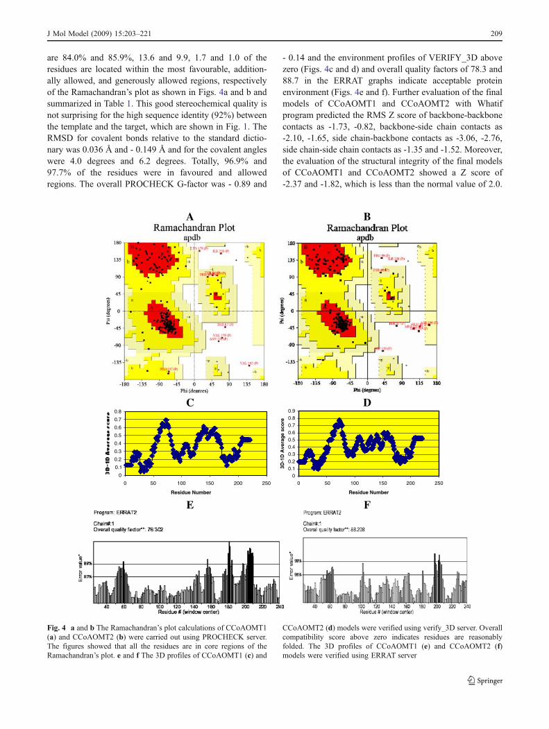
are 84.0% and 85.9%, 13.6 and 9.9, 1.7 and 1.0 of theresidues are located within the most favourable, addition-ally allowed, and generously allowed regions, respectivelyof the Ramachandran’s plot as shown in Figs. 4a and b andsummarized in Table 1. This good stereochemical quality isnot surprising for the high sequence identity (92%) betweenthe template and the target, which are shown in Fig. 1. TheRMSD for covalent bonds relative to the standard dictio-nary was 0.036 Å and - 0.149 Å and for the covalent angleswere 4.0 degrees and 6.2 degrees. Totally, 96.9% and97.7% of the residues were in favoured and allowedregions. The overall PROCHECK G-factor was - 0.89 and
- 0.14 and the environment profiles of VERIFY_3D abovezero (Figs. 4c and d) and overall quality factors of 78.3 and88.7 in the ERRAT graphs indicate acceptable proteinenvironment (Figs. 4e and f). Further evaluation of the finalmodels of CCoAOMT1 and CCoAOMT2 with Whatifprogram predicted the RMS Z score of backbone-backbonecontacts as -1.73, -0.82, backbone-side chain contacts as-2.10, -1.65, side chain-backbone contacts as -3.06, -2.76,side chain-side chain contacts as -1.35 and -1.52. Moreover,the evaluation of the structural integrity of the final modelsof CCoAOMT1 and CCoAOMT2 showed a Z score of-2.37 and -1.82, which is less than the normal value of 2.0.
A B
C D
0
0.1
0.2
0.3
0.4
0.5
0.6
0.7
0.8
0 50 100 150 200 250
Residue Number
00.10.2
0.30.40.50.6
0.70.80.9
0 50 100 150 200 250
Residue Number
3D-1
D A
vera
ge
sco
re
E F
Fig. 4 a and b The Ramachandran’s plot calculations of CCoAOMT1(a) and CCoAOMT2 (b) were carried out using PROCHECK server.The figures showed that all the residues are in core regions of theRamachandran’s plot. e and f The 3D profiles of CCoAOMT1 (c) and
CCoAOMT2 (d) models were verified using verify_3D server. Overallcompatibility score above zero indicates residues are reasonablyfolded. The 3D profiles of CCoAOMT1 (e) and CCoAOMT2 (f)models were verified using ERRAT server
J Mol Model (2009) 15:203–221 209

Therefore, it is believed that the final refined models aregood for further analysis. This value falls in the acceptablerange for a valid structure. It is generally accepted that ifthe Whatif score is below -5, the model is certainly of lowquality and if it is above -2, it is recommended as a goodstructure. This trend continued even in the data obtainedwith the validation program WHATCHECK in which theZ-scores of bond lengths, bond angles, omega anglerestraints, side-chain planarity, improper dihedral distribu-tion inside/outside distribution for CCoAOMT1 are 1.871,1.605, 1.366, 4.783, 2.228, 1.087 and CCoAOMT2 are5.714, 1.756, 1.836, 3.312, 4.735, 1.062 and are positive(positive is better than average). In all likelihood, thebackbone conformation (BBC) and inside/outside distribu-tion (IOD) parameters are nearer to crystal structure valuespredicting that the structures are highly reliable for furtherstudies as shown in Table 2.
Secondary structure prediction
Amino acid sequences of template, final refined models ofCCoAOMT1 and CCoAOMT2 proteins were aligned usingSPDBV based on the superimposition of their 3D struc-tures. Given their PDB files, secondary structures were alsoanalyzed and compared by the SPDBV software suite(http://www.expasy.org/spdbv). The secondary structures oftemplate and final CCoAOMT1 and CCoAOMT2 enzymesappeared highly conserved and showed close similarity tothe whole structures of template (1SUS and 1SUI)indicating that final structures are reliable (Figs. 2a andb). The aligned proteins of CCoAOMT1 and CCoAOMT2with the templates contain a 9 and 10 stranded α- heliceswith 6-stranded β-sheets in CCoAOMT1 and 7 stranded
β-sheets in CCoAOMT2 (Figs. 5a and b). Furthermore, inspite of several amino acid differences in the primarystructures of CCoAOMT1 and CCoAOMT2, their second-ary structures turned out to be identical except for one α-helix at Trp55, Asn56, and Ile57 and one β-sheet at Val182,Ile183, Gly184, Tyr185, Asp186, and Asn187 as shown inFig. 5c. In fact, from the structure-structure comparison, itwas found that α-helices 1, 2, 6 and 9 contain 3, 3, 2 and8 residues longer than CCoAOMT1 and β-sheets (ofCCoAOMT2) 2, 4, 6 and 7 contain 1, 1, 6, and 6 residueslonger than of CCoAOMT1. These secondary structures ofCCoAOMT1 were also compared with the templates andfound that α-helices 1, 2, 5, 6 and 8 contain 1, 3, 3, 1 and 3residues lesser than the template 1SUS and β-sheets ofCCoAOMT1 1, 2, 3, 5 and 6 contains 2, 1, 2, 4 and 4residues lesser than that of template. The comparison ofsecondary structures of CCoAOMT2 with the template1SUI showed that α-helices 1 and 10 contain 1 and 1 andβ-sheets of CCoAOMT2 1, 3 and 4 contain 2, 2, and 1residue lesser than the template 1SUI. These domainsexhibit a core α/β Rossmann-fold topology in which 6 and7 parallel β-sheets are flanked on each side by α-helicesthat provide the binding site for SAM/SAH [43]. This corestructure is highly conserved among the CCoAOMT familymembers, despite relatively low residue identity betweenthese enzymes. These refined models were used further foractive site and docking analysis.
Active site identification of CCoAOMT1 and CCoAOMT2enzymes
Once the final model was built, the possible binding sites ofCCoAOMT1 and CCoAOMT2 were searched based on the
Table 2 WHATCHECK Z-scores for quality assessment and statistical analysis of CCoAOMT1 and CCoAOMT2
Structure Z-score RMS Z-scores
Structure PQ RPA χNR BBC BL BA ΩR SCP IDD IODCCoAOMT1 -2.371 -3.390 -3.110 -10.894 1.871 1.605 1.366 4.783 2.228 1.087CCoAOMT2 -1.816 -1.531 -3.369 -10.439 5.714 1.756 1.836 3.312 4.735 1.062
PQ, second-generation packing quality; RPA, Ramachandran plot appearance; χNR, χ−1 /χ−2 rotamer normality; BBC, backbone conformation;BL, bond lengths; BA, bond angles; Ω, omega angle restraints; SCP, side-chain planarity; IDD, improper dihedral distribution; and IOD, inside/outside distribution.
Table 1 % of residues falling in the core region of the Ramachandran’s plot
CCoAOMT1 CCoAOMT2
% of residue in most favoured regions 84.0 85.9% of residue in the additionally allowed zones 13.6 9.9% of residue in the generously allowed regions 1.7 1.0% of residue in disallowed regions 0.7 3.1% of non-glycine and non-proline residues 100.0 100.0
210 J Mol Model (2009) 15:203–221

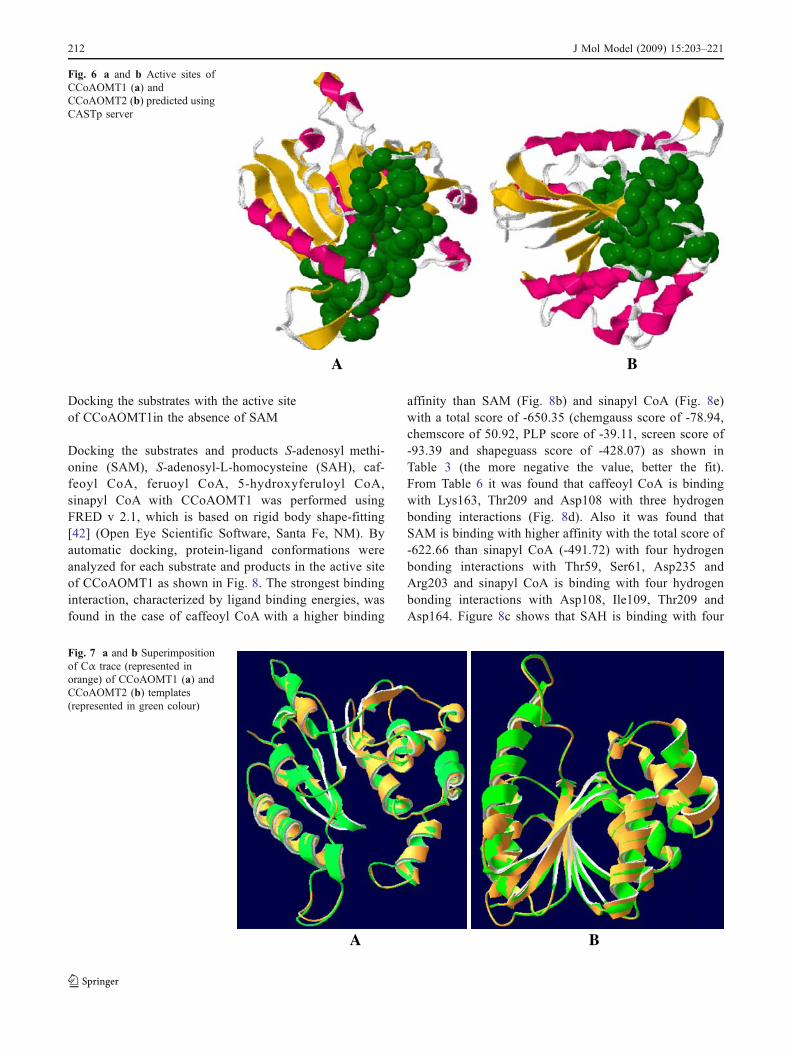
CASTP Server and structural comparison of the templateand the models built. In this study, active sites weresearched to identify protein active sites and binding sitesby locating cavities in the CCoAOMT1 and CCoAOMT2structures. When the search was complete, the largest sitewas automatically displayed on the structure as shown inFigs. 6a and b. It appeared that CCoAOMT1 andCCoAOMT2 and their templates 1SUS and 1SUI are wellconserved in both sequence and structure, hence, theirbiological function may be identical. In fact, from thestructure-structure comparison of template, and from finalrefined models using SPDBV program (Mate et al 1999), itwas found that the residues in active sites, Met58, Thr60,Val63, Glu82, Gly84, Ser90, Asp160, Asp162, Thr169,Asn191 and Arg203 of CCoAOMT1 and CCoAOMT2 arehighly conserved and also with the active site of templates(Figs. 7a and b). Hence, the results were used to guide theprotein-ligand docking experiments.
Superimposition of 1SUS and 1SUI with CCoAOMT 1and CCoAOMT 2 enzymes
The structural superimposition of Cα trace of template,CCoAOMT1 and CCoAOMT2 are shown in the Figs. 7aand b respectively. The weighted RMSD of Cα tracebetween the template and initial models of CCoAOMT1and CCoAOMT2 generated from the MODELLER was0.22 Å and 0.2 Å. Cα trace between the templates, 1SUSand 1SUI with the final refined models of CCoAOMT1 andCCoAOMT2 was 0.72 Å and 0.4 Å with a difference of0.5 Å and 0.2 Å between initial and final refined models.The RMSD of Cα trace between initial and final refinedmodels of CCoAOMT1 and CCoAOMT2 was also calcu-lated and found as 0.69 Å and 0.34 Å. The RMSD of Cαtrace of active sites between initial and final refined modelsof CCoAOMT1 and CCoAOMT2 with the templates 1SUSand 1SUI was calculated and found as 0.45 Å and 0.17 Åwith a difference of 0.28 Å with the template. The RMSDof Cα trace of active sites of final refined models ofCCoAOMT1 and CCoAOMT2 was found as 0.54 Å.Further, RMSD of Cα trace of active sites of initial andfinal refined models of CCoAOMT1 and CCoAOMT2were found as 0.46 Å and 0.15 Å. These studies show thatRMSD difference of 0.31 Å was responsible for conforma-tion changes in the active sites of CCoAOMT1 andCCoAOMT2, which makes the substrate to bind in theactive site of the enzymes. The two protein models built(CCoAOMT1 and CCoAOMT2) were used for docking thesubstrates and products.
A
B
C
Fig. 5 a, b and c Secondary structure alignment of CCoAOMT1 andCCoAOMT2 with the template 1SUS and 1SUI were predicted usingSPDBV software suite (a and b). c represents secondary structurealignment of CCoAOMT1 and CCoAOMT2
�
J Mol Model (2009) 15:203–221 211
Docking the substrates with the active siteof CCoAOMT1in the absence of SAM
Docking the substrates and products S-adenosyl methi-onine (SAM), S-adenosyl-L-homocysteine (SAH), caf-feoyl CoA, feruoyl CoA, 5-hydroxyferuloyl CoA,sinapyl CoA with CCoAOMT1 was performed usingFRED v 2.1, which is based on rigid body shape-fitting[42] (Open Eye Scientific Software, Santa Fe, NM). Byautomatic docking, protein-ligand conformations wereanalyzed for each substrate and products in the active siteof CCoAOMT1 as shown in Fig. 8. The strongest bindinginteraction, characterized by ligand binding energies, wasfound in the case of caffeoyl CoA with a higher binding
affinity than SAM (Fig. 8b) and sinapyl CoA (Fig. 8e)with a total score of -650.35 (chemgauss score of -78.94,chemscore of 50.92, PLP score of -39.11, screen score of-93.39 and shapeguass score of -428.07) as shown inTable 3 (the more negative the value, better the fit).From Table 6 it was found that caffeoyl CoA is bindingwith Lys163, Thr209 and Asp108 with three hydrogenbonding interactions (Fig. 8d). Also it was found thatSAM is binding with higher affinity with the total score of-622.66 than sinapyl CoA (-491.72) with four hydrogenbonding interactions with Thr59, Ser61, Asp235 andArg203 and sinapyl CoA is binding with four hydrogenbonding interactions with Asp108, Ile109, Thr209 andAsp164. Figure 8c shows that SAH is binding with four
A B
Fig. 6 a and b Active sites ofCCoAOMT1 (a) andCCoAOMT2 (b) predicted usingCASTp server
A B
Fig. 7 a and b Superimpositionof Cα trace (represented inorange) of CCoAOMT1 (a) andCCoAOMT2 (b) templates(represented in green colour)
212 J Mol Model (2009) 15:203–221

A B
C D
E
Fig. 8 Binding of CoA (a),SAM (b), SAH (c), caffeoylCoA (d), sinapyl CoA (e) in theactive site of CCoAOMT1 en-zyme. Substrates and productsare represented in ball and stickmodel and residues are labelledin white colour. Protein is rep-resented in orange colour
Table 3 The total energies of Chemguass, Chem score, PLP and Shapeguass scores of the best docked conformations of substrates and productsagainst CCoAOMT1 in the absence of SAM
Substrate Chemgauss score Chem score PLP score Screen score Shapeguass score Total score
CoA -78.23 31.86 5.09 -40.42 -637.47 -719.17SAM -55.38 -6.71 -39.11 -93.39 -428.07 -622.66SAH -57.58 -3.27 -38.11 -86.86 -411.52 -597.34Caffeoyl CoA -78.94 50.92 31.61 17.82 -671.76 -650.35Sinapyl CoA -82.96 73.97 74.44 75.58 -632.75 -491.72
J Mol Model (2009) 15:203–221 213
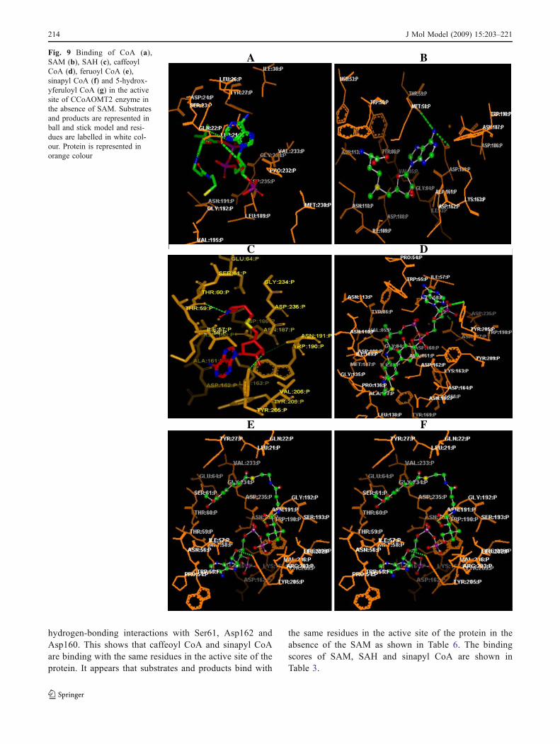
hydrogen-bonding interactions with Ser61, Asp162 andAsp160. This shows that caffeoyl CoA and sinapyl CoAare binding with the same residues in the active site of theprotein. It appears that substrates and products bind with
the same residues in the active site of the protein in theabsence of the SAM as shown in Table 6. The bindingscores of SAM, SAH and sinapyl CoA are shown inTable 3.
A B
C D
E F
Fig. 9 Binding of CoA (a),SAM (b), SAH (c), caffeoylCoA (d), feruoyl CoA (e),sinapyl CoA (f) and 5-hydrox-yferuloyl CoA (g) in the activesite of CCoAOMT2 enzyme inthe absence of SAM. Substratesand products are represented inball and stick model and resi-dues are labelled in white col-our. Protein is represented inorange colour
214 J Mol Model (2009) 15:203–221
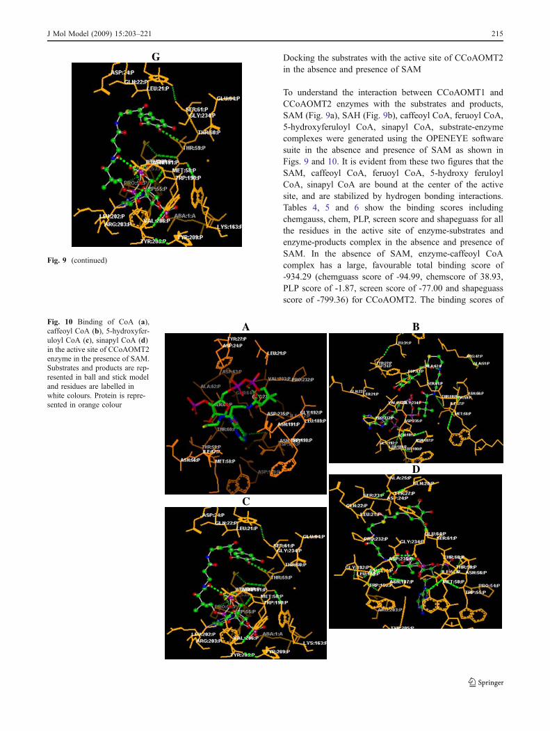
Docking the substrates with the active site of CCoAOMT2in the absence and presence of SAM
To understand the interaction between CCoAOMT1 andCCoAOMT2 enzymes with the substrates and products,SAM (Fig. 9a), SAH (Fig. 9b), caffeoyl CoA, feruoyl CoA,5-hydroxyferuloyl CoA, sinapyl CoA, substrate-enzymecomplexes were generated using the OPENEYE softwaresuite in the absence and presence of SAM as shown inFigs. 9 and 10. It is evident from these two figures that theSAM, caffeoyl CoA, feruoyl CoA, 5-hydroxy feruloylCoA, sinapyl CoA are bound at the center of the activesite, and are stabilized by hydrogen bonding interactions.Tables 4, 5 and 6 show the binding scores includingchemgauss, chem, PLP, screen score and shapeguass for allthe residues in the active site of enzyme-substrates andenzyme-products complex in the absence and presence ofSAM. In the absence of SAM, enzyme-caffeoyl CoAcomplex has a large, favourable total binding score of-934.29 (chemguass score of -94.99, chemscore of 38.93,PLP score of -1.87, screen score of -77.00 and shapeguassscore of -799.36) for CCoAOMT2. The binding scores of
G
Fig. 9 (continued)
A B
C
D
Fig. 10 Binding of CoA (a),caffeoyl CoA (b), 5-hydroxyfer-uloyl CoA (c), sinapyl CoA (d)in the active site of CCoAOMT2enzyme in the presence of SAM.Substrates and products are rep-resented in ball and stick modeland residues are labelled inwhite colours. Protein is repre-sented in orange colour
J Mol Model (2009) 15:203–221 215
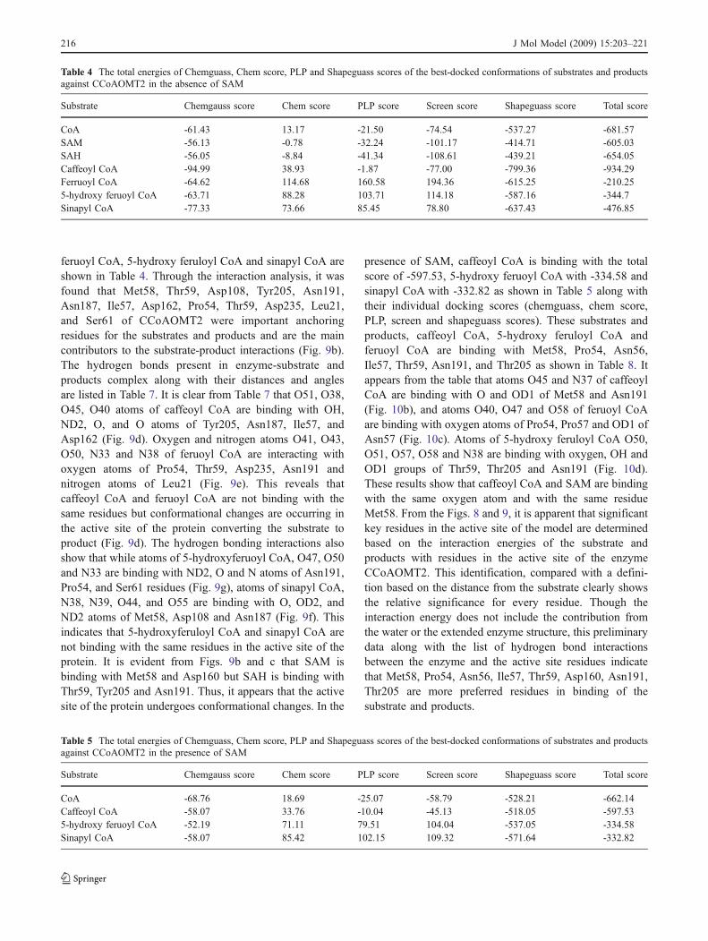
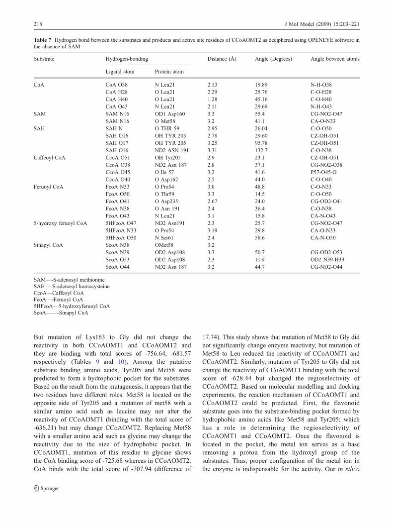
feruoyl CoA, 5-hydroxy feruloyl CoA and sinapyl CoA areshown in Table 4. Through the interaction analysis, it wasfound that Met58, Thr59, Asp108, Tyr205, Asn191,Asn187, Ile57, Asp162, Pro54, Thr59, Asp235, Leu21,and Ser61 of CCoAOMT2 were important anchoringresidues for the substrates and products and are the maincontributors to the substrate-product interactions (Fig. 9b).The hydrogen bonds present in enzyme-substrate andproducts complex along with their distances and anglesare listed in Table 7. It is clear from Table 7 that O51, O38,O45, O40 atoms of caffeoyl CoA are binding with OH,ND2, O, and O atoms of Tyr205, Asn187, Ile57, andAsp162 (Fig. 9d). Oxygen and nitrogen atoms O41, O43,O50, N33 and N38 of feruoyl CoA are interacting withoxygen atoms of Pro54, Thr59, Asp235, Asn191 andnitrogen atoms of Leu21 (Fig. 9e). This reveals thatcaffeoyl CoA and feruoyl CoA are not binding with thesame residues but conformational changes are occurring inthe active site of the protein converting the substrate toproduct (Fig. 9d). The hydrogen bonding interactions alsoshow that while atoms of 5-hydroxyferuoyl CoA, O47, O50and N33 are binding with ND2, O and N atoms of Asn191,Pro54, and Ser61 residues (Fig. 9g), atoms of sinapyl CoA,N38, N39, O44, and O55 are binding with O, OD2, andND2 atoms of Met58, Asp108 and Asn187 (Fig. 9f). Thisindicates that 5-hydroxyferuloyl CoA and sinapyl CoA arenot binding with the same residues in the active site of theprotein. It is evident from Figs. 9b and c that SAM isbinding with Met58 and Asp160 but SAH is binding withThr59, Tyr205 and Asn191. Thus, it appears that the activesite of the protein undergoes conformational changes. In the
presence of SAM, caffeoyl CoA is binding with the totalscore of -597.53, 5-hydroxy feruoyl CoA with -334.58 andsinapyl CoA with -332.82 as shown in Table 5 along withtheir individual docking scores (chemguass, chem score,PLP, screen and shapeguass scores). These substrates andproducts, caffeoyl CoA, 5-hydroxy feruloyl CoA andferuoyl CoA are binding with Met58, Pro54, Asn56,Ile57, Thr59, Asn191, and Thr205 as shown in Table 8. Itappears from the table that atoms O45 and N37 of caffeoylCoA are binding with O and OD1 of Met58 and Asn191(Fig. 10b), and atoms O40, O47 and O58 of feruoyl CoAare binding with oxygen atoms of Pro54, Pro57 and OD1 ofAsn57 (Fig. 10c). Atoms of 5-hydroxy feruloyl CoA O50,O51, O57, O58 and N38 are binding with oxygen, OH andOD1 groups of Thr59, Thr205 and Asn191 (Fig. 10d).These results show that caffeoyl CoA and SAM are bindingwith the same oxygen atom and with the same residueMet58. From the Figs. 8 and 9, it is apparent that significantkey residues in the active site of the model are determinedbased on the interaction energies of the substrate andproducts with residues in the active site of the enzymeCCoAOMT2. This identification, compared with a defini-tion based on the distance from the substrate clearly showsthe relative significance for every residue. Though theinteraction energy does not include the contribution fromthe water or the extended enzyme structure, this preliminarydata along with the list of hydrogen bond interactionsbetween the enzyme and the active site residues indicatethat Met58, Pro54, Asn56, Ile57, Thr59, Asp160, Asn191,Thr205 are more preferred residues in binding of thesubstrate and products.
Table 4 The total energies of Chemguass, Chem score, PLP and Shapeguass scores of the best-docked conformations of substrates and productsagainst CCoAOMT2 in the absence of SAM
Substrate Chemgauss score Chem score PLP score Screen score Shapeguass score Total score
CoA -61.43 13.17 -21.50 -74.54 -537.27 -681.57SAM -56.13 -0.78 -32.24 -101.17 -414.71 -605.03SAH -56.05 -8.84 -41.34 -108.61 -439.21 -654.05Caffeoyl CoA -94.99 38.93 -1.87 -77.00 -799.36 -934.29Ferruoyl CoA -64.62 114.68 160.58 194.36 -615.25 -210.255-hydroxy feruoyl CoA -63.71 88.28 103.71 114.18 -587.16 -344.7Sinapyl CoA -77.33 73.66 85.45 78.80 -637.43 -476.85
Table 5 The total energies of Chemguass, Chem score, PLP and Shapeguass scores of the best-docked conformations of substrates and productsagainst CCoAOMT2 in the presence of SAM
Substrate Chemgauss score Chem score PLP score Screen score Shapeguass score Total score
CoA -68.76 18.69 -25.07 -58.79 -528.21 -662.14Caffeoyl CoA -58.07 33.76 -10.04 -45.13 -518.05 -597.535-hydroxy feruoyl CoA -52.19 71.11 79.51 104.04 -537.05 -334.58Sinapyl CoA -58.07 85.42 102.15 109.32 -571.64 -332.82
216 J Mol Model (2009) 15:203–221

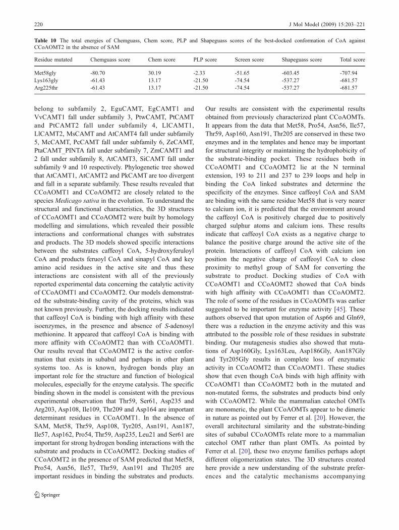
Docking studies of CoA
Docking studies of CoA in the absence of SAM withCCoAOMT1 show that CoA binds with two hydrogenbonding interactions Ile109 and Ala161 (Fig. 8a) with atotal score of -638.8 (chemguass score of -78.23, chem31.86, PLP 7.09, screen -40.42 and shapeguass -637.47) asshown in Table 3. The binding studies of CoA withCCoAOMT2 revealed that the residue Leu21 is involvedwith four hydrogen-bonding interactions (Fig. 9a) with thetotal score of -681.57 (-61.43, 13.17, -21.50, -71.54,-681.51 scores for chemguass, chem, PLP, screen andshapeguass respectively) as shown in Table 4. In thepresence of SAM, CoA binds with one hydrogen bondinginteraction with Asp186 (Fig. 10a) with the total score of-662.8 (chemguass score of -68.76, chem score of 18.69,PLP score of -25.07, screen score of -58.79 and shapeguassscore of -662.14) as shown in Table 5. These studies showthat CoA binds with higher affinity with CCoAOMT2 thanCCoAOMT1 in the presence and absence of SAM. Thisreveals that CCoAOMT2 is the active form that exits in thebiological systems.
In order to evaluate the validity of the modelledstructure, site-directed mutagenesis of CCoAOMT1 andCCoAOMT2 was carried out with the substrate CoA.Based on the molecular structure and reaction mecha-nisms, metal binding amino acids and hydrophobicpocket were hypothesized to be important for thereaction. Thus, we selected for mutagenesis those aminoacids that are thought to be important for the metal
binding and hydrophobic pocket formation. Asp162,Asp188, and Asn189, which are predicted to be involvedin metal binding, were mutagenized. Lys165 was alsomutagenized as it is predicted to assist Asp122 andAsp189 in maintaining the proper configuration of thesetwo metal binding amino acids through hydrogen bonds.Lastly, Tyr205 and Met60 were mutagenized, as both areinvolved in the formation of hydrophobic pocket and theformer is likely to be important for the regioselectivity ofCCoAOMT1 and CCoAOMT2. First, site-directed muta-tions were carried out in the three metal binding aminoacids. Mutation of Asp160 to Gly did not change thereactivity of CCoAOMT1 binding with the total score of-712.42 but complete loss of activity was noticed inCCoAOMT2. Mutation of Asp186 to Gly resulted in analmost complete loss of activity in CCoAOMT2 but didnot change the reactivity of CCoAOMT1 binding withthe score of -722.78. Also, mutation of Asn187 to Glyresulted in the loss of enzymatic activity in CCoAOMT2but no change was observed in the activity ofCCoAOMT1 binding with the total score of -719.45.Lys163 is predicted to have a role in the properpositioning of the two metal binding amino acids,Asp160 and Asn187 through hydrogen bonds. Consistentwith our hypothesis, mutation of Lys163 to Leu resultedin complete loss of activity in CCoAOMT2, but bindingwith the total scores of -747.63 in CCoAOMT1 suggeststhat the hydrogen bonds between Lys163 and the twometal binding amino acids are indeed critical for theenzymatic activity of CCoAOMT2 than CCoAOMT1.
Table 6 Hydrogen bond between the substrates and products and active site residues of CCoAOMT1 as deciphered using OPENEYE software inthe absence of SAM
Substrate Hydrogen-bonding Distance (Å) Angle (Degrees) Angle between the atoms
Ligand atom Protein atom
CoA CoA O35 N Ile 109 1.80 25.01 P45-O35-NCoA H27 O Ala161 2.76 2.76 N27-H27-O
SAM SAM O43 NH2 Arg 203 2.71 45.0 CZ-NH2-O18SAH SAH N14 OG Ser 61 3.26 122.9 CA-N-O50
SAH N O Asp 162 3.18 116.94 CG-OD2-H52SAH N O Asp 160 2.65 110.06 CG-NO2-O47SAH N OD1 Asp160 2.97 98.32 CG-NO2-O47
Caffeoyl CoA CcoA O49 NZ Lys163 2.8 54.8 CE-NZ-O49CcoA O5 OH Thr209 2.5 39.7 CZ-OH-O39CcoA N32 OD1 Asp108 3.4 55.3 N32-OD1-CG
Sinapyl CoA ScoA N39 O Asp108 2.3 41.39 CG-OD2-N39ScoA O53 N Ile109 3.2 66.1 H53-O53-NScoA O41 OH Thr209 2.9 60.6 CZ-OH-O41ScoA N52 OD2 Asp162 2.4 32.7 CG-OD2-H52
SAM—-S-adenosyl methionineSAH—-S -adenosyl homocysteineCcoA—Caffeoyl CoAScoA——-Sinapyl CoA
J Mol Model (2009) 15:203–221 217
But mutation of Lys163 to Gly did not change thereactivity in both CCoAOMT1 and CCoAOMT2 andthey are binding with total scores of -756.64, -681.57respectively (Tables 9 and 10). Among the putativesubstrate binding amino acids, Tyr205 and Met58 werepredicted to form a hydrophobic pocket for the substrates.Based on the result from the mutagenesis, it appears that thetwo residues have different roles. Met58 is located on theopposite side of Tyr205 and a mutation of met58 with asimilar amino acid such as leucine may not alter thereactivity of CCoAOMT1 (binding with the total score of-636.21) but may change CCoAOMT2. Replacing Met58with a smaller amino acid such as glycine may change thereactivity due to the size of hydrophobic pocket. InCCoAOMT1, mutation of this residue to glycine showsthe CoA binding score of -725.68 whereas in CCoAOMT2,CoA binds with the total score of -707.94 (difference of
17.74). This study shows that mutation of Met58 to Gly didnot significantly change enzyme reactivity, but mutation ofMet58 to Leu reduced the reactivity of CCoAOMT1 andCCoAOMT2. Similarly, mutation of Tyr205 to Gly did notchange the reactivity of CCoAOMT1 binding with the totalscore of -628.44 but changed the regioselectivity ofCCoAOMT2. Based on molecular modelling and dockingexperiments, the reaction mechanism of CCoAOMT1 andCCoAOMT2 could be predicted. First, the flavonoidsubstrate goes into the substrate-binding pocket formed byhydrophobic amino acids like Met58 and Tyr205; whichhas a role in determining the regioselectivity ofCCoAOMT1 and CCoAOMT2. Once the flavonoid islocated in the pocket, the metal ion serves as a baseremoving a proton from the hydroxyl group of thesubstrates. Thus, proper configuration of the metal ion inthe enzyme is indispensable for the activity. Our in silico
Table 7 Hydrogen bond between the substrates and products and active site residues of CCoAOMT2 as deciphered using OPENEYE software inthe absence of SAM
Substrate Hydrogen-bonding Distance (Å) Angle (Degrees) Angle between atoms
Ligand atom Protein atom
CoA CoA O38 N Leu21 2.13 19.89 N-H-O38CoA H28 O Leu21 2.29 25.76 C-O-H28CoA H40 O Leu21 1.28 45.16 C-O-H40CoA O43 N Leu21 2.11 29.69 N-H-O43
SAM SAM N16 OD1 Asp160 3.3 55.4 CG-NO2-O47SAM N16 O Met58 3.2 41.1 CA-O-N33
SAH SAH N O THR 59 2.95 26.04 C-O-O50SAH O16 OH TYR 205 2.78 29.60 CZ-OH-O51SAH O17 OH TYR 205 3.25 95.78 CZ-OH-O51SAH O16 ND2 ASN 191 3.31 132.7 C-O-N38
Caffeoyl CoA CcoA O51 OH Tyr205 2.9 23.1 CZ-OH-O51CcoA O38 ND2 Asn 187 2.8 37.1 CG-NO2-O38CcoA O45 O Ile 57 3.2 41.6 P57-O45-OCcoA O40 O Asp162 2.5 44.0 C-O-O40
Feruoyl CoA FcoA N33 O Pro54 3.0 48.8 C-O-N33FcoA O50 O Thr59 3.3 14.5 C-O-O50FcoA O41 O Asp235 2.67 24.0 CG-OD2-O41FcoA N38 O Asn 191 2.4 36.4 C-O-N38FcoA O43 N Leu21 3.1 15.8 CA-N-O43
5-hydroxy feruoyl CoA 5HFcoA O47 ND2 Asn191 2.3 25.7 CG-NO2-O475HFcoA N33 O Pro54 3.19 29.8 CA-O-N335HFcoA O50 N Ser61 2.4 58.6 CA-N-O50
Sinapyl CoA ScoA N38 OMet58 3.2ScoA N39 OD2 Asp108 3.3 50.7 CG-OD2-O53ScoA O53 OD2 Asp108 2.3 11.9 OD2-N39-H39ScoA O44 ND2 Asn 187 3.2 44.7 CG-ND2-O44
SAM—-S-adenosyl methionineSAH—-S-adenosyl homocysteineCcoA—Caffeoyl CoAFcoA—-Feruoyl CoA5HFcoA—5-hydroxyferuoyl CoAScoA——-Sinapyl CoA
218 J Mol Model (2009) 15:203–221

mutagenesis results clearly demonstrated the importance ofthese amino acids to the enzymatic activity of CCoAOMT1and CCoAOMT2.
Discussion
In plants, CCoAOMTs are S-adenosyl-L-methionine-dependent O-methyltransferases (OMTs) involved in ligninbiosynthesis. Plant CCoAOMTs belong to a distinct familyof OMTs. CCoAOMT plays a predominant role in thesynthesis of guaiacyl lignin and is essential in providingsubstrates for the synthesis of syringyl lignin. It also plays apivotal role in the methylation of 3-hydroxyl group of caffeoylCoA. Further, CCoAOMT-mediated methylation reaction isessential to channel substrates for 5-methoxylation ofhydroxycinnamates [44]. It was found that this enzyme isexpressed highly in lignifying tissues especially in xylem
ray parenchyma [44]. It appears therefore thatCCoAOMT-mediated lignin biosynthesis is commonamong plants and is also an important pathway as pointedout earlier by Ye and Varner (1995). Indeed, suppressionof CCoAOMT enzyme in transgenic tobacco caused adecrease in lignin content [5]. However, it is not knownwhether the two genes are expressed in a tissue specificmanner or not. It is also not known if they have anyfunctional redundancy. Our work shows that two genes(isoforms) for CCoAOMT exist in subabul tree.
Amino acid sequence analysis of CCoAOMT1 andCCoAOMT2 revealed that they are closely related to thecrystal structure of Medicago sativa feruoyl coenzyme A 3-O-methyltransferase and caffeoyl coenzyme A 3-O-methyltransferase of plant OMTs as shown in Fig. 1.Sequence analysis also reveal that CnCAMT, NtCAMT5,NtCAMT6 and EgCAMT2 belong to the subfamily 1,NtCAMT4, NtCAMT1, NtCAMT3, NtCAMT2, StCAMT
Table 9 The total energies of Chemguass, Chem score, PLP and Shapeguass scores of the best-docked conformation of CoA againstCCoAOMT1 in the absence of SAM
Residue mutated Chemguass score Chem score PLP score Screen score Shapeguass score Total score
Met58Gly -77.75 21.84 -12.21 -52.13 -605.43 -725.68Met58Leu -64.59 24.31 -11.05 -51.87 -533.01 -636.21Asp160Gly -76.82 32.11 5.44 -37.02 -636.13 -712.42Lys163Gly -83.32 32.57 -5.00 -76.03 -624.86 -756.64Lys163Leu -84.91 34.59 0.36 -64.23 -633.44 -747.63Asp186Gly -78.18 31.86 5.42 -39.83 -642.05 -722.78Asn187Gly -68.00 29.45 -8.95 -76.68 -595.27 -719.45Tyr205Gly -64.51 25.39 -4.81 -62.23 -522.28 -628.44Asp225Gyr -78.34 31.86 5.42 -39.83 -642.65 -723.54
Table 8 Hydrogen bond between the substrates and products and active site residues of CCoAOMT2 in the presence of SAM as deciphered usingOPENEYE software in the presence of SAM
Substrate Hydrogen-bonding Distance (Å) Angle (Degrees) Angle between atoms
Ligand atom Protein atom
CoA CoA H26 OD1 Asp186 2.34 69.77 CG-OD1-H26Caffeoyl CoA CcoA N37 O Met58 2.5 42.9 C-O-N37
CcoA O45 OD1 Asn191 2.9 31.8 CG-OD1-O45Feruoyl CoA FcoA O47 O Pro54 3.4 18.6 C-O-O47
FcoA O40 OD1 Asn 56 3.1 57.3 CG-H4-O40FcoA O58 O Ile 57 2.6 20.5 C-O-O58
5-hydroxy feruoyl CoA 5HFcoA O50 O Thr59 3.2 22.3 C-O-O505HFcoA O51 O Thr59 3.2 25.1 C-O-O515HFcoA O57 OH Thr205 3.3 46.9 CZ-OH-O575HFcoA O58 OH Thr205 3.1 35.4 CZ-OH-O585HFcoA N38 OD1 Asn191 2.8 47.1 CG-OD1-N38
SAM—-S-adenosyl methionineSAH—-S-adenosyl homocysteineCcoA—Caffeoyl CoAFcoA—-Feruoyl CoA5HFCoA—5-hydroxy feruoyl CoA
J Mol Model (2009) 15:203–221 219
belong to subfamily 2, EguCAMT, EgCAMT1 andVvCAMT1 fall under subfamily 3, PtwCAMT, PtCAMTand PtCAMT2 fall under subfamily 4, LlCAMT1,LlCAMT2, MsCAMT and AtCAMT4 fall under subfamily5, McCAMT, PcCAMT fall under subfamily 6, ZeCAMT,PtaCAMT_PINTA fall under subfamily 7, ZmCAMT1 and2 fall under subfamily 8, AtCAMT3, SiCAMT fall undersubfamily 9 and 10 respectively. Phylogenetic tree showedthat AtCAMT1, AtCAMT2 and PkCAMT are too divergentand fall in a separate subfamily. These results revealed thatCCoAOMT1 and CCoAOMT2 are closely related to thespecies Medicago sativa in the evolution. To understand thestructural and functional characteristics, the 3D structuresof CCoAOMT1 and CCoAOMT2 were built by homologymodelling and simulations, which revealed their possibleinteractions and conformational changes with substratesand products. The 3D models showed specific interactionsbetween the substrates caffeoyl CoA, 5-hydroxyferuloylCoA and products feruoyl CoA and sinapyl CoA and keyamino acid residues in the active site and thus theseinteractions are consistent with all of the previouslyreported experimental data concerning the catalytic activityof CCoAOMT1 and CCoAOMT2. Our models demonstrat-ed the substrate-binding cavity of the proteins, which wasnot known previously. Further, the docking results indicatedthat caffeoyl CoA is binding with high affinity with theseisoenzymes, in the presence and absence of S-adenosylmethionine. It appeared that caffeoyl CoA is binding withmore affinity with CCoAOMT2 than with CCoAOMT1.Our results reveal that CCoAOMT2 is the active confor-mation that exists in subabul and perhaps in other plantsystems too. As is known, hydrogen bonds play animportant role for the structure and function of biologicalmolecules, especially for the enzyme catalysis. The specificbinding shown in the model is consistent with the previousexperimental observation that Thr59, Ser61, Asp235 andArg203, Asp108, Ile109, Thr209 and Asp164 are importantdeterminant residues in CCoAOMT1. In the absence ofSAM, Met58, Thr59, Asp108, Tyr205, Asn191, Asn187,Ile57, Asp162, Pro54, Thr59, Asp235, Leu21 and Ser61 areimportant for strong hydrogen bonding interactions with thesubstrate and products in CCoAOMT2. Docking studies ofCCoAOMT2 in the presence of SAM predicted that Met58,Pro54, Asn56, Ile57, Thr59, Asn191 and Thr205 areimportant residues in binding the substrates and products.
Our results are consistent with the experimental resultsobtained from previously characterized plant CCoAOMTs.It appears from the data that Met58, Pro54, Asn56, Ile57,Thr59, Asp160, Asn191, Thr205 are conserved in these twoenzymes and in the templates and hence may be importantfor structural integrity or maintaining the hydrophobicity ofthe substrate-binding pocket. These residues both inCCoAOMT1 and CCoAOMT2 lie at the N terminalextension, 193 to 211 and 237 to 239 loops and help inbinding the CoA linked substrates and determine thespecificity of the enzymes. Since caffeoyl CoA and SAMare binding with the same residue Met58 that is very nearerto calcium ion, it is predicted that the environment aroundthe caffeoyl CoA is positively charged due to positivelycharged sulphur atoms and calcium ions. These resultsindicate that caffeoyl CoA exists as a negative charge tobalance the positive charge around the active site of theprotein. Interactions of caffeoyl CoA with calcium ionposition the negative charge of caffeoyl CoA to closeproximity to methyl group of SAM for converting thesubstrate to product. Docking studies of CoA withCCoAOMT1 and CCoAOMT2 showed that CoA bindswith high affinity with CCoAOMT1 than CCoAOMT2.The role of some of the residues in CCoAOMTs was earliersuggested to be important for enzyme activity [45]. Theseauthors observed that upon mutation of Asp66 and Gln69,there was a reduction in the enzyme activity and this wasattributed to the possible role of these residues in substratebinding. Our mutagenesis studies also showed that muta-tions of Asp160Gly, Lys163Leu, Asp186Gly, Asn187Glyand Tyr205Gly results in complete loss of enzymaticactivity in CCoAOMT2 than CCoAOMT1. These studiesshow that even though CoA binds with high affinity withCCoAOMT1 than CCoAOMT2 both in the mutated andnon-mutated forms, the substrates and products bind onlywith CCoAOMT2. While the mammalian catechol OMTsare monomeric, the plant CCoAOMTs appear to be dimericin nature as pointed out by Ferrer et al. [20]. However, theoverall architectural similarity and the substrate-bindingsites of subabul CCoAOMTs relate more to a mammaliancatechol OMT rather than plant OMTs. As pointed byFerrer et al. [20], these two enzyme families perhaps adoptdifferent oligomerization states. The 3D structures createdhere provide a new understanding of the substrate prefer-ences and the catalytic mechanisms accompanying
Table 10 The total energies of Chemguass, Chem score, PLP and Shapeguass scores of the best-docked conformation of CoA againstCCoAOMT2 in the absence of SAM
Residue mutated Chemguass score Chem score PLP score Screen score Shapeguass score Total score
Met58gly -80.70 30.19 -2.33 -51.65 -603.45 -707.94Lys163gly -61.43 13.17 -21.50 -74.54 -537.27 -681.57Arg225thr -61.43 13.17 -21.50 -74.54 -537.27 -681.57
220 J Mol Model (2009) 15:203–221

CCoAOMT-mediated O-methylation of CoA-linked sub-strates because of high similarity in SAM binding region ofmethyl transferases despite the differences in structuralsimilarity. The approach might be applicable for theprediction of substrate and regioselectivity of otherenzymes, and aid in efforts to engineer new regioselectiv-ities of existing enzymatic reactions. Also, the interactionsbetween the enzyme and the substrate proposed in thisstudy are useful for understanding the potential mechanismof enzyme-substrate binding. Thus, it is concluded thatthese two genes may have tissue specific expression formethylation reactions associated with lignin biosynthesis inplants.
Acknowledgements The authors are thankful to the CSIR, NewDelhi, for financial assistance in the form of a research project (CSIR-NMITLI) on paper and pulp.
References
1. Higuchi T (1998) Kung S-D, Yang S-F (eds) Discoveries in plantbiology. World Scientific, Singapore, 233–269
2. Grima-Pettenati J, Goffner D (1999) Plant Sci 145:51–653. Meyer K, Sirley AM, Cusumano JC, Bell D Lelong A, Chappel C
(1998) Proc Natl Acad Sci USA 95:6619–66234. Tsai CJ, Mielke MR, Hu WJ, Podila GK, Chiang VL (1998) Plant
Physiol 117:101–1125. Zhong R, Morrison WH, Negrel J, Ye ZH (1998) Plant Cell
10:2033–20456. Ye ZH, Kneusel RE, Matern U, Varner JE (1994) Plant Cell
6:1427–14397. Ye ZH, Varner JE (1995) Plant Physiol 108:459–4678. Ye ZH (1997) Plant Physiol 115:1341–13509. Inoue K, Vincent JH, Sewalt G, Balance MNIW, Sturzer C, Dixon
RA (1998) Plant Physiol 117:761–77010. Martz F, Maury S, Pincon G, Legrand M (1998) Plant Mol Biol
36:427–43711. Kersey R, Inoue K, Schubert KR, Dixon RA (1999) Protoplasma
209:46–5712. Li L, Osakabe K, Joshi CP, Chiang VL (1999) Plant Mol Biol
40:555–56513. Meng H, Campbell WH (1998) Plant Mol Biol 38:513–52014. Maury S, Geoffroy P, Legrand M (1999) Plant Physiol 121:215–
22415. Thompson JD, Gibson TJ, Plewniak F, Jeanmougin F, Higgins DJ
(1997) Nucleic Acids Res 24:4876–488216. Nicholas KB, Nicholas HB (1997) http://www.psc.edu/biomed/
genedoc [Online.]17. Page RDM (1996) Comput Appl Biosci 12:357–35818. Perrière G, Gouy M (1996) Biochimie 78:364–36919. Altschul SF, Gish W, Miller W, Myers EW, Lipman DJ (1990) J
Mol Biol 215:403–410
20. Ferrer J, Zubeita C, Dixon RA, Noel JP (2005) Plant Physiol137:1009–1017
21. Needleman SB, Wunsch CD (1970) J Mol Biol 48:443–45322. Sali A, Blundell TL (1993) J Mol Biol 234:779–81523. MacKerell AD Jr, Bashford D, Bellott M, Dunbrack RL Jr,
Evanseck J, Field M, Fischer JS, Gao J, Guo H, Ha S (1998) JPhys Chem B 102:3586–3616
24. Sali A, Overington JP (1994) Protein Sci 3:1582–159625. Kale L, Skeel R, Bhandarkar M, Brunner R, Gursoy A, Krawetz
N, Phillips J, Shinozaki A, Varadarajan K, Schulten K (1999) JComput Phys 151:283
26. MacKerell AD Jr, Bashford D, Bellott M, Dunbrack RL Jr,Evanseck J, Field M, Fischer JS, Gao J, Guo H, Ha S, Joseph D,Kuchnir L, Kuczera K, Lau FTK, Mattos C, Michnick S, Ngo T,Nguyen DT, Prodhom B, Roux B, Schlenkrich M, Smith J, StoteR, Straub J, Watanabe M, Wiorkiewicz-Kuczera J, Yin D, KarplusM (1992) FASEB J 6:A143–A143
27. MacKerell AD Jr, Bashford D, Bellott M, Dunbrack RL Jr,Evanseck J, Field M, Fischer JS, Gao J, Guo H, Ha S, Joseph D,Kuchnir L, Kuczera K, Lau FTK, Mattos C, Michnick S, Ngo T,Nguyen DT, Prodhom B, Reiher IWE, Roux B, Schlenkrich M,Smith J, Stote R, Straub J, Watanabe M, Wiorkiewicz-Kuczera J,Yin D, Karplus M (1998a) J Phys Chem B 102:3586–3616
28. Schlenkrich M, Brickmann J, MacKerell AD Jr, Karplus M (1996)A molecular perspective from computation and experiment. In:Merz KM, Roux B (eds) Birkhauser, Boston MA, pp 31–81
29. Jorgensen WL, Chandresekhar J, Madura JD, Impey RW, KleinML (1983) J Chem Phys 79:926–935
30. Grubmuller H, Heller H, Windemuth A, Schulten K (1991) MolSim 6:121–142
31. Schlick T, Skeel R, Brunger A, Kale L, Board JA Jr, Hermans J,Schulten K (1999) Algorithmic challenges in computationalmolecular biophysics. J Comput Phys 151:9–48
32. Brunger (1992) X-PLOR, Version 3.1: Yale University NewHaven CT
33. MacKerell AD Jr, Brooks B, Brooks CL III, Nilsson L, Roux B,Won Y, Karplus M (1998b) In: Schleyer P (ed) The encyclopediaof computational chemistry. John Wiley & Sons, Chichester, UK,pp 271–277
34. Ryckaert J-P, Ciccotti G, Berendsen HJC (1977) J Comput Phys23:327–341
35. Essmann U, Perera L, Berkowitz ML, Darden T, Lee H, PedersenLG (1995) J Chem Phys 103:8577
36. Laskoswki RA, MacArthur MW, Moss DS, Thorton JM (1993) JAppl Cryst 26:283–291
37. Colovos K, Yeates TO (1993) Protein Sci 2:1511–151938. Vriend G (1990) J Mol Graph 8:52–5639. Hooft RWW, Vriend G, Sander C, Abola EE (1996) Nature
381:27240. Guex N, Peitsch MC (1997) Electrophoresis 18:2714–272341. Dundas J, Ouyang Z, Tseng J, Binkowski A, Turpaz Y, Liang J
(2006) Nucleic Acids Res 34:W116–W11842. Schulz-Gasch T, Stahl M (2003) J Mol Model 9:47–5743. Rossmann MG, Moras D, Olsen KW (1974) Nature 250:194–19944. Zhong RW, Morrison H III, Himmelsbach DS, Poole FL II, Ye ZH
(2000) Plant Physiol 124:563–57745. Hoffmann L, Maury S, Bergdoll M, Thion L, Erard M, Legrand M
(2001) J Biol Chem 276:36831–36838
J Mol Model (2009) 15:203–221 221