Photodynamic therapy and endoscopic mucosal resection as minimally invasive approaches for the...
10
Photodiagnosis and Photodynamic Therapy (2005) 2, 35—44 REVIEW Photodynamic therapy and endoscopic mucosal resection as minimally invasive approaches for the treatment of early esophageal tumors: Pre-clinical and clinical experience in Lausanne A. Radu MD a,∗ , P. Grosjean a , Y. Jaquet a , R. Pilloud a , G. Wagnieres b , H. van den Bergh b , Ph. Monnier a a Department of Otolaryngology, Head and Neck Surgery, CHUV Hospital, CH-1011 Lausanne, Switzerland b Institute of Environmental Engineering, EPFL, CH-1015 Lausanne, Switzerland KEYWORDS Esophagus; Photodynamic therapy; Photofrin; Foscan; Endoscopic mucosal resection Summary Esophageal cancer, when detected at an early stage, has a very good probability of being eradicated by surgery or radiotherapy. However, less aggressive treatments also tend to provide high rates of cure without the side effects of rad- ical surgery or radiotherapy. Among them, photodynamic therapy and endoscopic mucosal resection have been experienced as alternative techniques for mucosal ab- lation in patients with superficial squamous-cell carcinoma (SCC) of the esophagus, or high-grade dysplasia and early stage adenocarcinoma arising in Barrett’s esopha- gus. We report on the results of our clinical experience with photodynamic therapy and discuss about its advantages and limitations. We also present a pre-clinical study, which had evaluated the feasibility, efficacy, and safety of a promising new method of endoscopic mucosal resection (EMR) based on the use of a modified rigid esophagoscope. The animal model chosen was the sheep because of its similari- ties with humans regarding the thickness and histologic structure of the esophagus. This new resection modality offers a promising approach in comparison with other options currently available, namely EMRs performed with flexible gastroscopes. It appears to be superior in terms of the size of the resected specimen, the precision and regularity of the resection depth, and the accuracy of histological diagnosis with safety margins. © 2005 Published by Elsevier B.V. * Corresponding author. Tel.: +41 21 314 27 00; fax: +41 21 314 27 06. E-mail address: [email protected] (A. Radu). 1572-1000/$ — see front matter © 2005 Published by Elsevier B.V. doi:10.1016/S1572-1000(05)00035-9
Transcript of Photodynamic therapy and endoscopic mucosal resection as minimally invasive approaches for the...
Photodiagnosis and Photodynamic Therapy (2005) 2, 35—44
REVIEW
Photodynamic therapy and endoscopicmucosal resection as minimally invasiveapproaches for the treatment of earlyesophageal tumors: Pre-clinical and clinicalexperience in Lausanne
A. Radu MDa,∗, P. Grosjeana, Y. Jaqueta, R. Pillouda,G. Wagnieresb, H. van den Berghb, Ph. Monniera
a Department of Otolaryngology, Head and Neck Surgery, CHUV Hospital, CH-1011 Lausanne, Switzerlandb Institute of Environmental Engineering, EPFL, CH-1015 Lausanne, Switzerland
KEYWORDSEsophagus;Photodynamic therapy;Photofrin;Foscan;Endoscopic mucosalresection
Summary Esophageal cancer, when detected at an early stage, has a very goodprobability of being eradicated by surgery or radiotherapy. However, less aggressivetreatments also tend to provide high rates of cure without the side effects of rad-ical surgery or radiotherapy. Among them, photodynamic therapy and endoscopicmucosal resection have been experienced as alternative techniques for mucosal ab-lation in patients with superficial squamous-cell carcinoma (SCC) of the esophagus,or high-grade dysplasia and early stage adenocarcinoma arising in Barrett’s esopha-gus. We report on the results of our clinical experience with photodynamic therapyand discuss about its advantages and limitations. We also present a pre-clinicalstudy, which had evaluated the feasibility, efficacy, and safety of a promising newmethod of endoscopic mucosal resection (EMR) based on the use of a modified rigidesophagoscope. The animal model chosen was the sheep because of its similari-ties with humans regarding the thickness and histologic structure of the esophagus.This new resection modality offers a promising approach in comparison with otheroptions currently available, namely EMRs performed with flexible gastroscopes. Itappears to be superior in terms of the size of the resected specimen, the precision
and regularity of the resection depth, and the accuracy of histological diagnosis with safety margins.© 2005 Published by Elsevier B.V.* Corresponding author. Tel.: +41 21 314 27 00; fax: +41 21 314 27 06.E-mail address: [email protected] (A. Radu).
1572-1000/$ — see front matter © 2005 Published by Elsevier B.V.doi:10.1016/S1572-1000(05)00035-9

36 A. Radu et al.
Contents
Introduction ...................................................................................................... 36Photodynamic therapy............................................................................................ 36
Materials and methods ....................................................................................... 36Results ....................................................................................................... 37
Endoscopic mucosal resection (EMR).............................................................................. 38Materials and methods ....................................................................................... 38Results—–hemi-circumferential resections .................................................................... 40Results—–circumferential resections .......................................................................... 40
Discussion ........................................................................................................ 40References ....................................................................................................... 42
Introduction
There is a tremendous interest in detectingesophageal cancer at an early stage, since at an in-vasive phase the disease is linked to a very poorprognosis and survival rate [1—3]. Hence, iden-tification of superficial squamous-cell cancers ofthe esophagus has recently risen thanks to an in-creased frequency of mass screening techniqueswith balloon or sponge cytology and to a greaterincidence of endoscopic examination of dye stain-ing methods [4—7]. For Barrett’s esophagus, en-doscopic surveillance has also yielded a signifi-cant number of early adenocarcinomas in West-ern countries. The standard treatment modalitiesto cure these early esophageal carcinoma, such assurgery, radiotherapy or chemotherapy, are asso-ciated with high rates of morbidity and mortality[8—11]. Efforts have thus been made to developminimally invasive endoscopic methods to treat se-vere dysplasia and superficial adenocarcinoma aris-ing in Barrett’s esophagus, or early (in situ and T1a)squamous-cell carcinomas (SCCs). Several forms ofablative treatment using thermal, photochemical,and mechanical methods have been tested so far
nique, which is well tolerated and allows rapid re-covery with excellent healing, can be performedon an outpatient basis. However, this proceduredoes not provide tissue specimens for a pathologi-cal analysis, which is necessary to assert the com-plete destruction of the lesion. In this respect,EMR techniques have a non-negligible advantage,though they can supply only relatively small piecesof mucosa when applied with a flexible gastroscope[24]. Correct identification and orientation of thepiecemeal resected specimens is thus difficult toachieve, especially in long altered segments suchas in Barrett’s esophagus.
In this paper we first report on the results of ourPDT clinical experience for early esophageal can-cers and latter, on an animal study using a new EMRrigid esophagoscope allowing submucosal resectionof large specimens in a single piece.
Photodynamic therapy
Materials and methods
PDT has started to be investigated in our clinic fortodTtcdtd
ewfoomt
to destroy or remove abnormal superficial layers ofthe esophagus. These have ranged from differenttypes of laser — such as potassium titanyl phos-phate (KTP) and neodymium—yttrium aluminiumgarnet (Nd:YAG) [12,13] — argon plasma coagu-lation [14,15], and multipolar electrocoagulation[16,17], to photodynamic therapy (PDT) [18,19] andvarious endoscopic mucosal resection (EMR) tech-niques using suction, snare, forceps, and electro-cautery devices [20—22].
PDT has the distinct advantage that it can berepeated multiple times without inducing any re-sistance or cumulative toxicity [23]. It also has thepotential of being used in association with otherprocedures and does not preclude the use of even-tual more aggressive future treatments in case offailure. Another benefit of PDT is that this tech-
he treatment of early squamous-cell carcinomasf the esophagus in 1984 using hematoporphyrinerivative (HPD) and Photofrin in the beginning.hese two drugs are considered as first genera-ion photosensitizers. The tetra(m-hydroxyphenyl)-hlorin (mTHPC), lately named Foscan, was intro-uced in 1992 because it showed distinct advan-ages in terms of chemical purity, phototoxicity, anduration of skin photosensitization [25].Fifty-five early squamous-cell carcinomas of the
sophagus (22 in situ and 33 microinvasive-T1a)ere treated by PDT. Sixty-eight PDTs were per-ormed because nine tumors were illuminated twor three times. The tumors were detected as sec-nd primary malignancies in 38 patients with a pri-ary head and neck cancer. Diagnosis and staging ofhe lesions were based through endoscopic morpho-

Photodynamic therapy and endoscopic mucosal resection as minimally invasive approaches 37
logic criteria and histologic examination on biopsyspecimens and later on endoscopic ultrasonogra-phy [26]. The superficial longitudinal spread of thelesions varied from 1 to 6 cm with radial exten-sions between 60◦ and 200◦. Esophageal carcinomaswith complete radial extension were not includedfor PDT in order to avoid potential post-treatmentstenosis.
The first 21 tumors were treated with HPD andPhotofrin and the last 34 with Foscan. The com-pounds were used at doses of 3mg/kg for HPD, 1or 2mg/kg for Photofrin, and 0.15mg/kg for Fos-can. The PDTs using the first generation photosen-sitizers were performed at 630 nm with an argonion pumped dye laser for the first 14 cases and at514 nm with the same argon ion laser operated inthe single-line mode for the remaining seven le-sions. At the beginning of our study, we used cir-cumferential cylindrical light distributors. Later on,due to post-therapeutic esophageal stenosis, thetumors were treated only with 180◦ or 240◦ win-dowed cylindrical light distributors with a length of4 or 6 cm [27]. The total light doses ranged from 100to 180 J/cm2 at both wavelengths. Light was deliv-ered at an intensity varying from 80 to 150mW/cm2
wigddwmowwa
dnwaa
R
Te
Fai1vtt
ith an exposure time of about 20min. The major-ty of the PDTs with Foscan were performed withreen light at 514 nm. Only three esophageal irra-iations were carried out at 652 nm. Lasers and lightistributors were exactly the same as those for PDTith HPD and Photofrin. Most of the 514 nm illu-inations were carried out with total light dosesf 75—120 J/cm2 and an intensity of 100mW/cm2;hereas, the majority of the 652 nm irradiationsere performed with light doses of 8—12 J/cm2 andn intensity of 150mW/cm2.The follow-up included a first endoscopy 7 or 10
ays after PDT to assess the extent of the tumorecrosis macroscopically. Subsequent endoscopiesith staining (Toluidine blue), as well as biopsiesnd abrasive cytology, were performed 3 monthsfter the treatment and twice a year thereafter.
esults
he results of our study confirm that PDT is anfficient treatment for in situ and micro-invasive
igure 1 In situ squamous-cell carcinoma of the esoph-gus irradiated with green light (514 nm) 4 days afternjection of 0.15mg/kg of Foscan with a light dose of20 J/cm2 at an intensity of 100mW/cm2: (a) endoscopiciew of the tumor before treatment; (b) 7 days afterreatment using a 180◦ windowed cylindrical light dis-ributor; and (c) complete response 3 months after PDT.

38 A. Radu et al.
Table 1 Results of photodynamic therapy with HPD, Photofrin and Foscan for 55 early squamous-cell carcinomasin the esophagus: Complete responses with no recurrence as a function of the tumor staging and photosensitizers.
Success rate
Photosensitizer In situ SCC Micro-invasive (T1a) SCC Total
HPD/Photofrin 7/8 9/13 16/21Foscan 13/14 13/20 26/34
Total 20/22 (96%) 22/33 (66%) 42/55
(T1a) squamous-cell carcinomas of the esophagus.A complete response was achieved in 76% of thetumors, with a mean disease-free follow-up of 32months. An example of early esophageal cancer be-fore and after PDT is presented in Fig. 1. In allbut one of these lesions, a single treatment wassufficient to eradicate the lesion. The importanceof tumor staging in achieving a complete responsewas emphasized by the significantly lower are ratesobtained in T1a tumors (67%) compared to in situcarcinomas (90%). The results showed similar PDTefficacy whatever photosensitizer was used. Fol-lowing treatment with HPD and Photofrin, 16 of 21(76%) early cancers achieved a complete response,whereas with Foscan 26 of 34 (76%) tumors wereeradicated. The results obtained by PDT with thedifferent photosensitizers as a function of the tu-mor staging are presented in Table 1.
The 24% rate of treatment failures could be ex-plained by either partial surface illumination ofthe tumoral field or understaging of the lesions.Esophageal folds likely caused the irregular tumornecrosis that was noticed at the first follow-up en-doscopic examination in some cases. In one pa-tient, the frequent up and down sliding of the
the mid esophagus. Two other patients treated withred light for an early tumor in the lower esophagusdeveloped a high-grade fever and pleural effusion,respectively. Both complications were consideredas likely transmural necrosis with occult perfora-tion of the esophagus and resolved spontaneouslyunder antibiotic treatment. Because the appliedlight intensity was low enough to avoid thermal in-juries, the transmural damage was assumed to becaused by PDT only. Skin photosensitivity, as pre-viously reported, is shorter with Foscan than withthe first generation photosensitizers [28]. All of thethree cutaneous reactions following treatment withFoscan occurred within the first week after injec-tion, whereas a second-degree sunburn happened 2months after HPD administration.
Endoscopic mucosal resection (EMR)
Materials and methods
The EMR technique used in our pre-clinical study isbased on a modified standard rigid esophagoscope(Karl Storz Ltd., Tuttlingen, Germany). The proto-tl[taItOrcwAsaodtttv
esophagus due to a hiatal hernia presumably pre-vented homogenous light delivery. In five cases,esophagectomy was performed, as PDT failed tocure the tumors. Histopathological analysis of thesurgical specimens showed submucosal carcinomas(T1b stage) in four cases and limited tumoral in-vasion of the muscularis propria in the fifth. The-ses cases stress the difficult problem of an accu-rate preoperative differentiation between intra-mucosal (T1a) and submucosal (T1b) carcinomasalthough 20MHz endoscopic ultrasonography wasperformed in most cases before PDT. There werefive major complications: two stenoses and threeesophagotracheal fistulas. Both stenoses were in-duced after circumferential irradiation and weresuccessfully managed by repeated bougienage witha Savary—Gillard dilator. Esophagotracheal fistulas,which required surgery, occurred within 3 weeks af-ter red light illumination at 630 (HPD) or 652 nm(Foscan) of lesions located on the anterior wall of
ype is shown in Fig. 2. It has been designed fol-owing a preliminary study on more than 20 animals29]. The instrument consists of an outer metallicube, at the distal end of which a window is cre-ted with an opening of 200◦ and 6 cm in length.t is introduced in the esophagus under visual con-rol with a 0◦ scope inserted through an inner tube.nce at the level of the lesion, the inner tube isemoved and replaced by the resectoscope, whichonsists of a transparent window and a diathermyire loop that can be moved through the window.n angulated 30◦ scope attached to the resecto-cope allows side viewing through the window andccurate targeting of the lesion. Suction is turnedn, creating negative pressure inside the resectionevice. The mucosa and part of the submucosa are,herefore, aspirated through the holes drilled intohe window. The thin wire loop (0.3mm in diame-er) is connected to a high-frequency surgical de-ice allowing resection of the mucosa. The space

Photodynamic therapy and endoscopic mucosal resection as minimally invasive approaches 39
Figure 2 The prototype rigid esophagoscope for endoscopic mucosal resection: (a) view of the whole instrument and(b) view of the different windows with holes and resection wire loops.
between the window and the wire loop is selectedprecisely (1± 0.1mm) and corresponds to the depthof ablation, which does not damage the muscularispropria of the esophagus. Once the mucosectomycompleted, the device is removed under continu-ous negative pressure to keep the resected mucosalspecimen stuck to the window. Finally, the mucosalspecimen (up to 12 cm2 in a single piece), is re-moved and prepared for histological analysis. Thisincludes confirmation of the lateral safety marginsand depth of tumor invasion. If a circumferentialresection is required, the procedure is done in twosteps. An anterior EMR is performed, followed by asecond posterior EMR during the same session.
Firstly, 21 sheep underwent 55 distinct hemi-circumferential resections. In the second part ofthe study, 24 circumferential resections were car-ried out in 24 animals. In these latter 360◦ re-sections, which are at high risk of inducing steno-sis, Mitomycin-C was applied topically at a dose of2mg/ml at the site of mucosectomy 1 week after
the procedure. Mitomycin-C is an alkylating agentthat can inhibit cell division, protein synthesis, andfibroblast proliferation [30]. It appears to be ben-eficial in modulating wound healing processes andreducing scar formation in the treatment of steno-sis [31,32]. The sheep model was chosen becausethe esophageal wall in sheep is similar to that inhumans. All of the different layers of the humanesophagus are identified throughout the length ofthe sheep esophageal wall with very similar thick-ness in both species [33,34]. The lengths of the win-dows used during the resection were 2.2, 3.3, 4.4,and 5.5 cm. Evaluation of the tissue defect was car-ried out at the end of each procedure. Follow-upendoscopies were performed at 1, 2, and 3 monthsto detect potential complications and to observethe process of healing and reepithelialization. Theanimals were euthanized 3 months after the mu-cosal ablation and the esophagus resected for histo-logical examination of the tissue damage and scar-ring.

40 A. Radu et al.
Figure 3 Histological section of a resected esophageal specimen (Masson’s trichrome stain). The cut is regular andhas been made clearly through the submucosa; no fibers from the muscularis propria are seen. E: epithelium; MM:muscularis mucosae (the bar represents 1mm).
Results—–hemi-circumferential resections
For the hemi-circumferential resections, the sur-face of tissue defect was matching endoscopicallythe size of the window in the majority of the cases.All of the specimens were obtained as single dis-tinct pieces with regular margins. Their size rangedfrom 6 to 12 cm2. No complications such as stenosisor perforation were observed and all the animalswere euthanized as planned. In all of the cases,complete reepithelialization was obtained within 6weeks of the EMR. Fifty-five resected pieces wereanalyzed microscopically. The muscularis mucosaelayer was outlined perfectly in all of the specimens.In 48 of the 55 cases, an accurate depth of re-section was achieved; the cut was straight throughthe submucosa with no intrusion into the muscularispropria (Fig. 3). In the remaining seven specimens,scarce superficial fibers from the internal layer ofthe muscularis propria wee seen focally. Regardingthe evaluation of the esophagi, complete morpho-logically normal reepithelialization was seen micro-scopically in each of the resected areas. Scarring inthe lamina propria and submucosa, with disappear-ance of the muscularis mucosae, was observed in all
ily removed with a standard biopsy forceps undervisual guidance in order to obtain a perfect 360◦resection. One animal had to be sacrificed imme-diately after the mucosectomy due to a perfora-tion. The complication was due to inaccurate po-sitioning of the resectoscope during the second re-section and a subsequent overlapping resection. Asin the hemi-circumferential resections, the spec-imens were cut precisely with straight margins.Histological assessment of the resected pieces re-vealed that the incision was made through the sub-mucosa in all cases. However, two more animalswere sacrificed earlier than the desired 3-monthinterval due to perforation: one related to a di-lation attempt and another potentially to the im-paction of straw at the site of resection. No com-plications occurred in the six sheep in which theshortest window (2.2 cm) was used. Histological ex-amination of their esophagi showed that the exter-nal layer of the muscularis propria was fully pre-served. When longer windows were used (3.3, 4.4,and 5.5), stenoses were encountered in more thantwo-third of the cases. However, all but one animalwas successfully treated with one or 5 dilations atmost with Savary—Gillard bougies and a new ap-pwas3ntdwfil
D
Pr
of the cases, as expected. In addition and more sur-prisingly, focal areas of fibrosis and atrophic mus-cle fibers were noticed in the internal layer of themuscularis propria in all of the 48 sites in which anaccurate resection had been performed. In the re-maining seven sites, the damage partly extendedto the superficial part of the external layer of themuscularis propria. These last cases correspondedto the resected specimen, in which a few musclefibers could be seen.
Results—–circumferential resections
The resection performed in two steps was entirelycircumferential in 14 sheep. In the other 10 ani-mals, thin residual mucosal strips were still visi-ble at the end of the procedure. These were eas-
lication of Mitomycin-C. Only one sheep resectedith the largest window (5.5 cm) had a persisting,lbeit moderate (11mm in diameter), stenosis de-pite several dilations. The animals survived the-month desired interval until their sacrifice, witho significant weight loss. Though, in these cases,he microscopic evaluation revealed that the tissueamage was more significant than in the group inhich the shortest window was used, with densebrous scarring extending focally to the externalayer of the muscularis propria.
iscussion
hotodynamic therapy has gained an increasingecognition as an effective modality for esophageal

Photodynamic therapy and endoscopic mucosal resection as minimally invasive approaches 41
tumors localized to the superficial layers. Its bene-fits are well known and undeniable. Photodynamictherapy is minimally invasive, non-mutilating andcauses few side effects. All of these factors con-tribute to minimize mortality and morbidity in pa-tients who are already in poor general condition.Photodynamic therapy has also the potential to beused as a neo-adjuvant treatment, as it involves adifferent curative process than conventional treat-ment modalities.
Despite its advantages, PDT continues to be atreatment that is limited by several drawbacks. Theonce admitted relative selectivity of the photosen-sitizer for the pathological cells is currently con-troversial. In our study, endoscopic controls 7 or10 days after PDT revealed not only necrosis ofthe tumor, but also injuries in the surrounding nor-mal irradiated mucosa. True selectivity was neverobserved with either photosensitizer administeredand the extent of the superficial tissue damage sim-ply matched the geometry of the illuminated area.This is emphasized by the several esophageal per-foration, which occurred as a consequence of non-selective necrosis of normal tissue since no trans-mural carcinomatous invasion was present beforePdsdiwttwTtitsm
dtttillbeddteIi
of green light after sensitization with Foscan led tosevere complications in the esophagus of the sheepthat are highly likely to occur in humans as well[36]. This could be related to additional injury ofblood vessels that are responsible for oxygenationof the healthy tissue underlying the tumor. Becauseall the available photosensitizers display a high ab-sorption peak around 420 nm, one should take intoaccount the possibility of irradiating superficial tu-mors with blue light, which has a lesser tissue pene-tration depth than green light. Although a relativelylonger illumination period is required, the use ofblue light likely rules out the risk of perforation orstenosis.
Endoscopic mucosal resection represents an al-ternative and an attractive method of ablating ab-normal mucosa in the esophagus. In comparisonto the others minimally invasive techniques, EMRhas the distinct advantage of providing a resectedspecimen for histological study. It has been usedfor the treatment of early and well circumscribedsquamous-cell carcinomas [20], as well as to re-move segments of intestinal metaplasia in Barrett’sesophagus [21,37]. Several modalities have beendeveloped using flexible endoscopes based on theplsewwrtccenttscd
mmkkpeums2set
DT. The insufficient therapeutic selectivity of theyes is also underlined by the occurrence in ourtudy of two esophageal stenoses. All of the threeyes showed important inter-individual variationsn terms of tumor damage, even in patients treatedith the same drug and light parameters. We no-iced, however, in the PDTs with Foscan, that pa-ients who showed the highest fluorescence signalere also those having the highest response to PDT.herefore, by measuring the relative dye level inhe tissue by light-induced fluorescence just beforellumination, and adjusting the light dose deliveredo the lesion to the drug fluorescence signal, onehould be able to obtain more effective results andinimize the risk of generating complications [35].Another major limiting factor in using PDT is the
epth of tissue destruction. Because the ideal pho-osensitizer with high selectivity for the tumoralissue has not yet been developed and because thehickness of the esophageal wall is only about 5mm,llumination with red light, which can penetrate ateast 1 cm, carries the risk of perforation. Greenight has a lower tissue penetration but appears toe effective in the cure of superficial cancers in thesophagus, which usually do not exceed 2mm of in-epth invasion. In our study, the use of green lightid not adversely affect the efficacy of PDT. The ac-ual depth of the necrosis is usually unclear, how-ver, and might be much deeper than one assumes.n a pre-clinical study conducted in our institution,t has been showed that application of high doses
rinciple of first lifting and then sectioning theesion, with or without suction [20,21]. Althoughmall lesions (<2 cm2) can safely be removed, thexact depth of resection is difficult to predict. EMRith the flexible endoscope becomes cumbersomehen long segments of Barrett’s mucosa have to beesected. The need of multiple resections carrieshe risk of perforation due to overlapping of suc-essive resections reaching and damaging the mus-ularis propria of the esophageal wall. Moreover,radication of all the foci of intestinal metaplasia inot guaranteed, since the rates of complete resec-ion can be as low as 40% [38]. Finally, correct iden-ification and orientation of the multiple resectedpecimens by the pathologist makes the histologi-al mapping and analysis of the resected marginsifficult.This has stimulated the gastroenterologic com-unity to develop new techniques of large en blocucosal resections with needle-knives of differentinds (insulated-tip, hook-triangle tip and sheatednives) [39,40]. Rosch et al. [24] have recently re-orted their experience in 37 patients undergoingn bloc EMRs for mucosal and submucosal tumorssing the insulated-tip knife. Complete tumor re-oval was achieved in only 25% of the mucosal le-ions, although 21 of 37 (58%) measured less thancm in diameter. When considering piecemeal re-ections, this result improved to 65%. In experi-nced hands, the procedure lasted for 45 minuteso 2 hours and the dissection was almost never pre-
42 A. Radu et al.
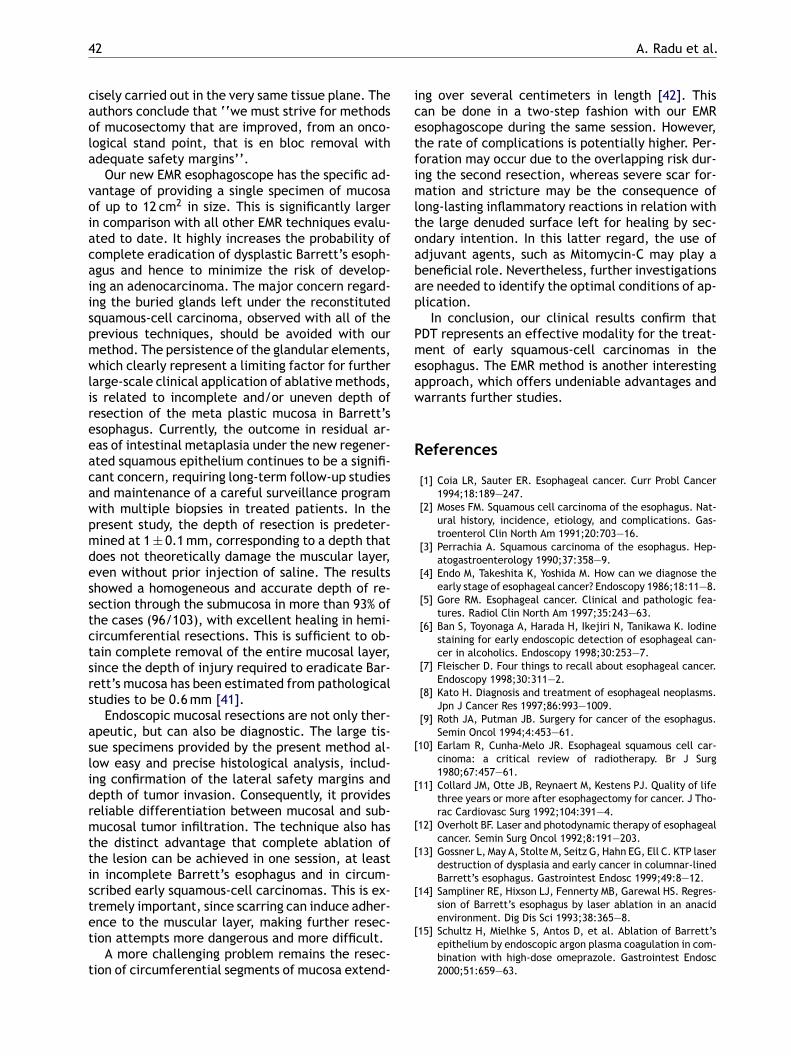
cisely carried out in the very same tissue plane. Theauthors conclude that ‘‘we must strive for methodsof mucosectomy that are improved, from an onco-logical stand point, that is en bloc removal withadequate safety margins’’.
Our new EMR esophagoscope has the specific ad-vantage of providing a single specimen of mucosaof up to 12 cm2 in size. This is significantly largerin comparison with all other EMR techniques evalu-ated to date. It highly increases the probability ofcomplete eradication of dysplastic Barrett’s esoph-agus and hence to minimize the risk of develop-ing an adenocarcinoma. The major concern regard-ing the buried glands left under the reconstitutedsquamous-cell carcinoma, observed with all of theprevious techniques, should be avoided with ourmethod. The persistence of the glandular elements,which clearly represent a limiting factor for furtherlarge-scale clinical application of ablativemethods,is related to incomplete and/or uneven depth ofresection of the meta plastic mucosa in Barrett’sesophagus. Currently, the outcome in residual ar-eas of intestinal metaplasia under the new regener-ated squamous epithelium continues to be a signifi-cant concern, requiring long-term follow-up studies
ing over several centimeters in length [42]. Thiscan be done in a two-step fashion with our EMResophagoscope during the same session. However,the rate of complications is potentially higher. Per-foration may occur due to the overlapping risk dur-ing the second resection, whereas severe scar for-mation and stricture may be the consequence oflong-lasting inflammatory reactions in relation withthe large denuded surface left for healing by sec-ondary intention. In this latter regard, the use ofadjuvant agents, such as Mitomycin-C may play abeneficial role. Nevertheless, further investigationsare needed to identify the optimal conditions of ap-plication.
In conclusion, our clinical results confirm thatPDT represents an effective modality for the treat-ment of early squamous-cell carcinomas in theesophagus. The EMR method is another interestingapproach, which offers undeniable advantages andwarrants further studies.
References
[1] Coia LR, Sauter ER. Esophageal cancer. Curr Probl Cancer
[
[
[
[
[
[
and maintenance of a careful surveillance programwith multiple biopsies in treated patients. In thepresent study, the depth of resection is predeter-mined at 1± 0.1mm, corresponding to a depth thatdoes not theoretically damage the muscular layer,even without prior injection of saline. The resultsshowed a homogeneous and accurate depth of re-section through the submucosa in more than 93% ofthe cases (96/103), with excellent healing in hemi-circumferential resections. This is sufficient to ob-tain complete removal of the entire mucosal layer,since the depth of injury required to eradicate Bar-rett’s mucosa has been estimated from pathologicalstudies to be 0.6mm [41].
Endoscopic mucosal resections are not only ther-apeutic, but can also be diagnostic. The large tis-sue specimens provided by the present method al-low easy and precise histological analysis, includ-ing confirmation of the lateral safety margins anddepth of tumor invasion. Consequently, it providesreliable differentiation between mucosal and sub-mucosal tumor infiltration. The technique also hasthe distinct advantage that complete ablation ofthe lesion can be achieved in one session, at leastin incomplete Barrett’s esophagus and in circum-scribed early squamous-cell carcinomas. This is ex-tremely important, since scarring can induce adher-ence to the muscular layer, making further resec-tion attempts more dangerous and more difficult.
A more challenging problem remains the resec-tion of circumferential segments of mucosa extend-
1994;18:189—247.[2] Moses FM. Squamous cell carcinoma of the esophagus. Nat-
ural history, incidence, etiology, and complications. Gas-troenterol Clin North Am 1991;20:703—16.
[3] Perrachia A. Squamous carcinoma of the esophagus. Hep-atogastroenterology 1990;37:358—9.
[4] Endo M, Takeshita K, Yoshida M. How can we diagnose theearly stage of esophageal cancer? Endoscopy 1986;18:11—8.
[5] Gore RM. Esophageal cancer. Clinical and pathologic fea-tures. Radiol Clin North Am 1997;35:243—63.
[6] Ban S, Toyonaga A, Harada H, Ikejiri N, Tanikawa K. Iodinestaining for early endoscopic detection of esophageal can-cer in alcoholics. Endoscopy 1998;30:253—7.
[7] Fleischer D. Four things to recall about esophageal cancer.Endoscopy 1998;30:311—2.
[8] Kato H. Diagnosis and treatment of esophageal neoplasms.Jpn J Cancer Res 1997;86:993—1009.
[9] Roth JA, Putman JB. Surgery for cancer of the esophagus.Semin Oncol 1994;4:453—61.
10] Earlam R, Cunha-Melo JR. Esophageal squamous cell car-cinoma: a critical review of radiotherapy. Br J Surg1980;67:457—61.
11] Collard JM, Otte JB, Reynaert M, Kestens PJ. Quality of lifethree years or more after esophagectomy for cancer. J Tho-rac Cardiovasc Surg 1992;104:391—4.
12] Overholt BF. Laser and photodynamic therapy of esophagealcancer. Semin Surg Oncol 1992;8:191—203.
13] Gossner L, May A, Stolte M, Seitz G, Hahn EG, Ell C. KTP laserdestruction of dysplasia and early cancer in columnar-linedBarrett’s esophagus. Gastrointest Endosc 1999;49:8—12.
14] Sampliner RE, Hixson LJ, Fennerty MB, Garewal HS. Regres-sion of Barrett’s esophagus by laser ablation in an anacidenvironment. Dig Dis Sci 1993;38:365—8.
15] Schultz H, Mielhke S, Antos D, et al. Ablation of Barrett’sepithelium by endoscopic argon plasma coagulation in com-bination with high-dose omeprazole. Gastrointest Endosc2000;51:659—63.

Photodynamic therapy and endoscopic mucosal resection as minimally invasive approaches 43
[16] Van Laethem JL, Cremer M, Peny MO, Delhaye M, Deviere J.Eradication of Barrett’s mucosa with argon plasma coagula-tion and acid suppression: immediate and mid term results.Gut 1998;43:747—51.
[17] Sharma P, Bhattacharyya A, Garewal HS, Sampliner RE.Durability of new squamous epithelium after endo-scopic reversal of Barrett’s esophagus. Gastrointest Endosc1999;50:159—64.
[18] Savary JF, Grosjean P, Monnier P, et al. Photodynamic ther-apy of early squamous cell carcinomas of the esophagsu: areview of 31 cases. Endoscopy 1998;30:258—65.
[19] Overholt BF, Panjehpour M, Haydek JM. Photodynamic ther-apy for Barrett’s esophagus: follow-up in 100 patients. Gas-trointest Endosc 1999;49:1—7.
[20] Inoue H, Takeshita K, Hori H, Muraoka Y, Yoneshima H, EndoM. Endoscopic mucosal resection with a cap-fitted panendo-scope for esophagus, stomach, and colon mucosal lesions.Gastrointest Endosc 1993;39:58—62.
[21] Nijhawan PK, Wang KK. Endoscopic mucosal resection forlesions with endoscopic features suggestive of malignancyand high-grade dysplasia within Barrett’s esophagus. Gas-trointest Endosc 2000;52:328—32.
[22] Ell C, May A, Gossner L, et al. Endoscopic mucosal resec-tion of early cancer and high-grade dysplasia in Barrett’sesophagus. Gastroenterology 2000;118:670—7.
[23] Fisher AMR, Murphree L, Gomer CJ. Clinical and preclinicalphotodynamic therapy. Lasers Surg Med 1995;17:2—31.
[24] Rosch T, Sarbia M, Schumacher B, Deinert K, FrimbergerE, Toermer T. Attempted endoscopic en bloc resection ofmucosal and submucosal tumors using insulated tip knives:
[
[
[
[
[
[
[
[
[
[
[
stage carcinomas of the esophagus using fluorescence spec-troscopy. Lasers Surg Med 1996;19:340—6.
[36] Radu A, Conde R, Fontolliet C, Wagnieres, van den Bergh H,Monnier P. Mucosal ablation with photodynamic therapy inthe esophagus: optimisation of light dosimetry in the sheepmodel. Gastrointest Endosc 2003;57:897—905.
[37] May A, Gossner L, Pech O, et al. Intraepithelial high-gradeneoplasia and early adenocarcinoma in short-segment Bar-rett’s esophagus: curative treatment using local endoscopictreatment techniques. Endoscopy 2002;34:604—10.
[38] Kida M. Endoscopic tumor diagnosis and treatment. En-doscopy 2000;32:836—44;Ackroyd R, Brown NJ, Stephenson TJ, Stoddard CJ, ReedMW. Ablation treatment for Barrett’s esophagus: whatdepth of tissue distruction is needed. J Clin Pathol1999;52:509—12.
[39] Kuwano H, Mochiki E, Asao T, Kato H, Shimura T, TsutsumiS. Double endoscopic intraluminal operation for upper di-gestive tract diseases: proposal of anovel procedure. AnnSurg 2004;239:22—7.
[40] Rajan E, Gostout CJ, Feitoza AB, Leontovich ON, Her-man LJ, Burgart LJ. Widespread EMR: a new techniquefor removal of large areas of mucosa. Gastrointest Endosc2004;60:623—7.
[41] Ackroyd R, Brown NJ, Stephenson TJ, Stoddard CJ, ReedMW. Ablation treatment for Barrett’s esophagus: whatdepth of tissue distruction is needed. J Clin Pathol1999;52:509—12.
[42] Conio M, Sorbi D, Batts KP, Gostout CJ. Endoscopic circum-ferential esophageal mucosectomy in a porcine model: an
CnamcHPGTe
ipoopt
msoc0aas
a pilot series. Endoscopy 2004;36:788—801.25] Berenbaum MC, Akande SL, Bonnett R, et al. meso-
Tetra(hydroxyphenyl) porphyrins, a new class of potenttumours photosensitizers with favourable selectivity. Br JCancer 1986;54:717—25.
26] Monnier P, Fontolliet C, Ollyo JB. Endoscopic findings of100 early-stage esophageal cancers. Diagn Ther Endosc1994;1:83—92.
27] Van den Bergh H. On the evolution of some endoscopiclight delivery systems for photodynamic therapy. Endoscopy1998;30:392—407.
28] Wagnieres G, Hadjur C, Grosjean P, et al. Clinicalevaluation of the cutaneous phototoxicity of 5,10,15,20-tetra(m-hydroxyphenyl)chlorin. Photochem Photobiol1998;68:382—7.
29] Grosjean P. La mucosectomie endoscopique oesophagi-enne: etude pre-clinique. Probl Actuelles Otorhinolaryngol1998;21:293—302.
30] Khaw PT, Doyle JW, Sherwood MB, Smith MF, McGorrayS. Prolonged localized tissue effects from 5-minute ex-posure to fluorouracil and mitomycin C. Arch Ophtalmol1993;111:263—7.
31] Rahbar R, Jones DT, Nuss RC, et al. The role of mitomycin inthe prevention and treatment of scar formation in the pe-diatric aerodigestive tract: friend or foe? Arch OtolaryngolHead Neck Surg 2002;128:401—6.
32] Eliashar R, Eliachar I, Esclamado R, Gramlich T, StromeM. Can topical mitomycin prevent laryngotracheal steno-sis? Laryngoscope 1999;109:1594—600.
33] Liu JB, Miller LS, Goldberg BB, et al. Transnasal US of theesophagus: preliminary morphologic and function studies.Radiology 1992;184:721—7.
34] Bays R, Wagnieres G, Robert D, et al. Light dosimetry forphotodynamic therapy in the esophagus. Lasers Surg Med1997;20:290—303.
35] Braichotte DR, Savary JF, Monnier P, van den Bergh H. Op-timizing light dosimetry in photodynamic therapy of early
assessment of technical feasibility, safety, and outcome.Endoscopy 2001;33:791—4.
ommentary on Radu et al.’s paper: Photody-amic therapy and endoscopic mucosal resections minimally invasive procedures for the treat-ent of early esophageal tumors: Preclinical andlinical experience in Lausanne: by Professorugh Barr MD, ChM, FRCS, FRCSE, Cranfieldostgraduate Medical School in Gloucestershire,reat Western Road, Gloucester GL1 3NN, UK.el.: +44 1452 394679; fax: +44 1452 394813;-mail: [email protected]
Very difficult choices face both patient and clin-cian when early superficial cancers or areas of dys-lasia are detected in the esophagus. These areften found in assymptomatic patients as a resultf surveillance. The paper from Lausanne is an im-ortant contribution examining minimally invasiveherapy for these lesions.Cancer in the esophagus develops through aulti-step process, and progression from dyspla-ia to metastatic cancer may not inevitable, andften takes many years [1,2]. However, in theolumnar lined esophagus as many as 40% (range—73%) of patients with high-grade dysplasia maylready have a coexistent cancer, and between 5nd 60% of patients will develop cancer duringurveillance over 1—7 years [3—5]. In this context

44 A. Radu et al.
radical surgical resection has an unacceptable mor-tality (2—11%) [6,7] and morbidity (early morbidity:50%; late morbidity: 26%) [8].
The Lausanne group, who have substantial expe-rience in the techniques of photodynamic therapypredominantly treating early esophageal squamous-cell carcinoma. The best results are in patientswith the most superficial disease. They highlightthe problem of understaging the disease and inade-quate illumination, as a cause of treatment failure.The major complications of fistulation and strictureformation remain of concern. These are relatedto transmural damage and deep muscular necro-sis. The ideal photosensitiser is much debated, withsome clinicians preferring aminolaevulinic acid,since the effect is more superficial [9,10]. This risksundertreatment of an occult cancer but should beadequate to eradicate dysplasia [11,12] and avoidspossibly disastrous complications.
The authors also present the experimental useof the ‘Lausanne esophageal resectoscope’. Cur-rently endoscopic mucosal resection is performedusing a variety of devices. These are usually per-formed at flexible endoscopy with patient underconscious sedation. For the removal of extensive
performing endoscopic mucosal resection, will de-pend on a clear demonstration of efficacy in pa-tients. We are looking forward to the initial clini-cal reports and subsequent comparison with flexibleendoscopic mucosal resection.
[1] Weston AP, Sharma P, Topalovski, Richards R, Cherian R,Dixon A. Long-term follow-up of Barrett’s high-grade dys-plasia. Am J Gastroenterol 2000;95:1888—93.
[2] Jankowski J, Harrison RF, Perry I, Balkwill F, Tselepis C. Bar-rett’s metaplasia. Lancet 2000;356:2079—86.
[3] Pellegrini CA, Pohl D. High-grade dysplasia in Barrett’sesophagus: surveillance or operation? J Gastrointest Surg2000;4:131—4.
[4] Buttar NS, Wang KK, Sebo TJ, et al. Extent ofhigh-grade dysplasia in Barrett’s esophagus correlateswith risk of adenocarcinoma. Gastroenterology 2001;120:1630—9.
[5] Schnell TG, Sontag SJ, Chejfec G, et al. Long-term nonsur-gical management of Barrett’s esophagus with high-gradedysplasia. Gastroenterology 2001;120:1607—19.
[6] Bachmann MO, Alderson D, Edwards D, et al. Cohort study inSouth and West England of the influence of specialization onthe management and outcome of patients with oesophagealand gastric cancers. BJS 2002;89:914—22.
[7] Bollschweiler E, Schroder W, Holscher AH, Siewert JR.Preoperative risk analysis in patients with adenocarci-noma or squamous cell carcinoma of the oesophagus. BJS
[
[
[
areas piecemeal resection may be necessary. Al-though devices are being introduced that can beused to remove large areas. The ‘Lausanne resec-toscope’ is a modified rigid esophagoscope. The au-thors from the department of Otolaryngology arevery familiar with these devices as are thoracic sur-geons. The device will be less familiar to Gastroen-terologists who perform the majority of upper gas-trointestinal endoscopy. The resectoscope is ableto provide full and accurate mucosectomy, remov-ing all potentially abnormal tissue. The initial ex-periments are very encouraging with clear demon-stration of a very accurate and clean resection. Themain concerns are of stricture formation, and per-foration when the resection fields overlap. It willnot have the advantages of flexible endoscopy, andmay require general anesthesia. The acceptabilityof this technique to the current group of clinicians’
2000;87:1106—10.[8] Zaninotto, Parenti AR, Ruol A, Costantini M, Merigliano S,
Ancona E. Oesophageal resection for high-grade dysplasiain Barrett’s oesophagus. BJS 2000;97:1102—5.
[9] Barr H, Shepherd NA, Dix A, Roberts DJH, Tan WC, Kras-ner N. Eradication of highgrade dysplasia in columnar-lined (Barrett’s) oesophagus using photodynamic ther-apy withendogenously generated protoporphyrin IX. Lancet1996;348:584—5.
10] Gossner L, Stolte M, Stroke R, et al. Photodynamic ther-apy of high-grade dysplasia and early stage carcinomasby means of 5-aminolaevulinic acid. Gastroenterology1998;114:447—55.
11] Biddlestone LR, Barham CP, Wilkinson SP, Barr H, Shep-herd NA. The histopathology of treated Barrett’s Oesoph-agus: squamous re-epithelialisation following acid suppres-sion, laser and photodynamic therapy. Am J Surg Pathol1998;22:239—45.
12] Barr H. Barrett’s esophagus: treatment with 5-aminolevulinic acid photodynamic therapy gastrointestinalendoscopy. Clin North Am 2000;10:421—38.