Phacoemulsification Skills Training and Assessment - Archive ...
20
HAL Id: hal-00477855 https://hal.archives-ouvertes.fr/hal-00477855 Submitted on 30 Apr 2010 HAL is a multi-disciplinary open access archive for the deposit and dissemination of sci- entific research documents, whether they are pub- lished or not. The documents may come from teaching and research institutions in France or abroad, or from public or private research centers. L’archive ouverte pluridisciplinaire HAL, est destinée au dépôt et à la diffusion de documents scientifiques de niveau recherche, publiés ou non, émanant des établissements d’enseignement et de recherche français ou étrangers, des laboratoires publics ou privés. Phacoemulsification Skills Training and Assessment Anthony Spiteri, Rajesh Aggarwal, Tom Kersey, Larry Benjamin, Ara Darzi, Philip Bloom To cite this version: Anthony Spiteri, Rajesh Aggarwal, Tom Kersey, Larry Benjamin, Ara Darzi, et al.. Phacoemulsifi- cation Skills Training and Assessment. British Journal of Ophthalmology, BMJ Publishing Group, 2009, n/a (n/a), pp.bjo.2009.159715v1-n/a. 10.1136/bjo.2009.159715. hal-00477855
-
Upload
khangminh22 -
Category
Documents
-
view
2 -
download
0
Transcript of Phacoemulsification Skills Training and Assessment - Archive ...
HAL Id: hal-00477855https://hal.archives-ouvertes.fr/hal-00477855
Submitted on 30 Apr 2010
HAL is a multi-disciplinary open accessarchive for the deposit and dissemination of sci-entific research documents, whether they are pub-lished or not. The documents may come fromteaching and research institutions in France orabroad, or from public or private research centers.
L’archive ouverte pluridisciplinaire HAL, estdestinée au dépôt et à la diffusion de documentsscientifiques de niveau recherche, publiés ou non,émanant des établissements d’enseignement et derecherche français ou étrangers, des laboratoirespublics ou privés.
Phacoemulsification Skills Training and AssessmentAnthony Spiteri, Rajesh Aggarwal, Tom Kersey, Larry Benjamin, Ara Darzi,
Philip Bloom
To cite this version:Anthony Spiteri, Rajesh Aggarwal, Tom Kersey, Larry Benjamin, Ara Darzi, et al.. Phacoemulsifi-cation Skills Training and Assessment. British Journal of Ophthalmology, BMJ Publishing Group,2009, n/a (n/a), pp.bjo.2009.159715v1-n/a. �10.1136/bjo.2009.159715�. �hal-00477855�
1
Phacoemulsification Skills Training and
Assessment
Anthony Spiteri MRCSEd(Ophth))
Rajesh Aggarwal PhD, MRCS
Tom Kersey MRCOphth
Larry Benjamin FRCSEd, FRCOphth
Ara Darzi MD, FRCS and
Philip Bloom FRCS, FRCOphth
Western Eye Hospital and
Department of Biosurgery & Surgical Technology, St. Mary’s Campus,
Imperial College London U.K.
Correspondence to: Mr. Anthony Spiteri, Specialist Trainee, South Thames Rotation, 34 Ashgrove House, 28 Lindsay Square London, SW1V 2HW Email: [email protected] Tel: +44 7818 232255 License statement: the Corresponding Author has the right to grant on behalf of all
authors and does grant on behalf of all authors, an exclusive licence (or non-exclusive for
government employees) on a worldwide basis to the BMJ Publishing Group Ltd and its
Licensees to permit this article (if accepted) to be published in BJO and any other
BMJPGL products to exploit all subsidiary rights, as set out in our licence .
Conflicting interests: None.
Ethics approval: Not required.
2
Introduction
Cataract extraction by phacoemulsification was pioneered by Charles David Kelman 1 in
New York City in 1967. Over the next 25 years it replaced the traditional inpatient
cataract surgery with a much less intrusive and more curative procedure that could be
performed on an outpatient basis. According to Hospital Episode Statistics over 280,000
phacoemulsifications were carried out in the financial year 2005-2006 in the UK alone.2
It has become by far the key index operation for Ophthalmologists, with the Royal
College guidelines expecting trainees to complete 350 phacoemulsification cataract
procedures by the end of Ophthalmic Specialist Training year 73.
Challenges in phacoemulsification training
The quality of ophthalmic surgical training is increasingly challenged by an untimely
convergence of several factors. The public’s unprecedented high expectations and
demands for safety are fuelled by readily accessible information on hospital
performances such as the Electronic Cataract National Dataset4. In addition, consultant-
led care provided by Independent Treatment Centres (ITCs) and the further reduction of
working hours (to 48 hours a week by 2009), imposed by the European Working Time
Directive, are leading to concerning reports 5’ 6 about the reduced overall training
opportunities.
The training of novice phacoemulsification surgeons, as with all postgraduate medical
training, has altered substantially in the last decade 7. Although the Halstedian apprentice
model of teaching has served ophthalmology education for over 100 years, it fails to
ensure that experience is gained in all the essential areas and is therefore inappropriate as
well as stressful. The limitations8 of this approach are hard to ignore, and even more
difficult to amend.

3
Increasingly, the demonstration of competency is gathering general consensus as the
preferred method to drive ophthalmic trainees along the surgical learning curve.
Benjamin9 reports that team working, leadership, insight, dexterity, decision making,
prioritizing and empathy are all important and should be assessed.
Moreover, the almost 10-year-old accepted standard for complication rates as determined
by the 1997-8 National Cataract Surgery Survey (NCSS) is out-of-date. This reflects the
great advances in the practice and provision of modern phacoemulsification surgery
achieved since then10. It also suggests that although training opportunities are being
compromised, the required expected standards are higher.
The challenge for the current generation of ophthalmologists is to increase the quality of
training and indeed maintain standards. It is necessary to utilise training methods outside
the operating theatre (wet labs, simulation) while simultaneously demonstrating
competence during formal assessments.
Phacoemulsification TRAINING outside the operating theatre
Wet labs use cadaveric human or animal models, or synthetic eyes (designed specifically
for performing phacoemulsification) to rehearse the steps of cataract extraction.
Laboratory practice allows surgeons to acquire skills in a controlled environment, free of
the pressures of operating on real patients according to Piaget’s and Vygotsky’s
pedagogical philosophy of “learning by doing”. This has been proven to be effective in
training general surgeons.11’ 12
4
The mandatory introduction of micro-surgical skills courses in the UK was an attempt to
teach basic micro-surgical skills within a structured curriculum. This is not an entirely
new concept in ophthalmology. Kirby13 reported a resident course in basic ophthalmic
surgery techniques that had been in use since 1966. The success of this form of training is
now evident from the large number of micro-surgical skills courses available worldwide.
Courses last 2-3 days and consist of didactic lectures with some hands-on wet lab
simulator practice. Skills practice varies from performing single tasks like knot tying to
learning of complex procedures such as lid and corneal wound repair, capsulorrhexis and
phacoemulsification.
However these methods have been criticized for being unrealistic14 with inaccurate
simulation of tissue consistency and anatomy15 and also lacking any form of objective
assessment. These problems can be addressed through the development of computer
simulators after similar technology had been used to train airline pilots for a number of
years providing realistic simulation with an accurate assessment of performance. It has
already been emulated successfully in other branches of surgery16’17’18’19. The possibility
of an analogous situation in ophthalmology is becoming a reality.
Virtual reality systems in phacoemulsification
The term virtual reality refers to a “computer-generated representation of an environment
that allows sensory interaction, thus giving the impression of actually being present”20.
Over the past two decades researchers have developed many ophthalmic virtual reality
surgery simulators.
5
The training systems published in the literature come from the USA, Japan, France,
Sweden, Austria and Germany. One of the first projects to embrace this technology for
ophthalmic surgical education was the Ophthalmic Retrobulbar Injection Simulator
(ORIS)21 while Sinclair et al 22 were the first to develop haptic (tactile) feedback in an
eye surgical simulator using 4 instruments (scalpel, forceps, scissors and
phacoemulsifier). Webster et al utilized haptic feedback for simulating Gimbel and
Neuhann’s continuous curvilinear capsulorrhexis technique.23 They recently further
described simulations of this technique using the EYESI.24’ 25
Phacoemulsification surgery prototypes started appearing over the past five years, one of
the earliest being developed in Sweden by Laurell et al.26 Another novel method of
teaching cataract surgery was developed by Prinz et al. The “Ophthalmic Operation
Vienna” uses surgical videos accompanied by 3D animated sequences of all surgical
steps for cataract and glaucoma surgery.27 Moreover, Henderson et al described the
Virtual Mentor28 to teach cognitive aspects of cataract surgery isolated from the physical
tasks. The use of endoscopy has also been proposed as a tool to augment three-
dimensional understanding of ocular structures and thereby assist in teaching cataract
surgery to residents29. Posterior segment simulators range from retinal photocoagulation30
to vitreoretinal surgery.31’ 32’ 33’ 34
At present, the only commercially available ophthalmic virtual reality surgical training
system on the market is the EYESI produced by VR magic, Mannheim, Germany (Fig 1).
EYESI was originally designed as a vitreoretinal surgery simulator.35 It has since
evolved to include a breadth of ophthalmic surgical procedures including capsulorrhexis
simulation and phacoemulsification modules. The simulator consists of a mannequin
head prop with a separate vitreoretinal and cataract surgical interface. Movements of the
instruments are relayed in real time onto a computer monitor as well as onto a stereo
microscope complete with foot pedals.
6
Using this simulator, trainees can practice standardized phacoemulsification techniques
and abstract tasks repeatedly with instant objective feedback of performance. With
graded exercises at different skill levels, they can be used as the basis for a structured
training programme.
Learning curves on phacoemulsification trainers.
A learning curve is defined as a line graph displaying opportunities across the x-axis, and
a measure of student performance along the y-axis. To our knowledge, there is no
published data on the learning curve of ophthalmic surgical trainees during wet lab
training or microsurgical skills courses. This is probably owing to the fact that validated
eye surgical skills assessment tools are only recently emerging. Prior to this, assessment
on the rate of proficiency acquisition using these methods was not accurately possible.
Assessment using virtual reality systems is more readily available using the
accompanying manufacture-provided default performance thresholds (though these too
need yet to be validated).
Transfer of skills from phacoemulsification trainer to human patient
There are virtual reality to operating room (so-called “VR to OR”) trials published in
laparoscopy (17, 18), bronchoscopy (19) and endovascular (20) surgery. The legitimacy
of ophthalmologic surgery simulators through trials however beckons. These studies are
essential to show transfer of simulated skills to real operations before simulators can be
incorporated into the ophthalmology training curriculum.

7
The studies that have so far established validity relate to vitreoretinal procedures. Peugnet
et al36 tested a retinal photocoagulation simulator. Their results showed that residents
training on the simulator required only 25.4 days of training compared with 42.25 days in
the group that received conventional training. EYESI’s vitreoretinal module was studied
by Jonas and colleagues37 who found that the group that trained on the simulator
performed better than the non-trained group when evaluated in a wet lab setting
performing vitrectomy on pig eyes.
One of the first phacoemulsification simulator trials was carried out by Folgar et al38 who
compared objective surgical outcomes in cataract surgeries performed by residents who
have trained with the EYESI versus those who have trained utilizing traditional didactic
and wet-lab methods. They suggest that ophthalmology residents who train with the
EYESI simulator system may decrease surgical time, ultrasound time and energy
dissipation, and rely less on attending interventions compared to residents who begin
training directly with patients
The promising trial results are an indication that virtual reality simulation might be what
training programmes desperately need in these challenging times. However it is necessary
to obtain objective measurements of real performance to confirm this.
ASSESSMENT of technical skills in phacoemulsification surgery
Adequate assessment is necessary to demonstrate competence. For any method of skill
assessment to be used with confidence it must be objective, reliable, valid and feasible.
Reliability of an evaluation instrument relates to its ability to provide consistent results
with minimal errors of measurement. Test/Retest reliability and Inter/Intra observer
reliability are the most commonly used methods.39
8
Validity is defined as the extent to which an assessment instrument measures what it was
designed to measure. An assessment should be able to demonstrate several forms of
validity from the most basic face validity to content and construct or contrast
(discriminative) validity to the most powerful predictive validity. Feasibility or
practicality refers to the length and number of assessments, the number of personnel and
the required time, space and costs.
Traditionally, however, assessment methods have been subjective making them
inherently susceptible to weaknesses. The opinion of the assessing trainer, for example,
may be critically important. Friedlich et al 40demonstrated that a new exam for family
medicine had low reliability when family physicians serve as examiners but moderate
reliability when surgeons where the evaluators. Furthermore, the keeping of a personal
surgical logbook is a reflection of one’s experience as opposed to one’s expertise.
Complication rates, not only accumulation of numbers, need to be addressed when
evaluating the competence of a surgeon.
Emerging tools in ophthalmic surgical skill assessment.
In an attempt to design better ways of training ophthalmic surgeons, a number of
different objective assessment tools have been described. Objective Assessment of Skills
in Intraocular Surgery (OASIS)41 (Table 1) is an entirely objective tool and thus has no
inter-rater variability. It is a feasible one page evaluation form with pre-, intra- and post-
operative results components to phacoemulsification. The more subjective Global Rating
Assessment of Skills in Intraocular Surgery (GRASIS) was developed later to be
complementary to OASIS. GRASIS is a subjective, also one-page, evaluation form based
on a 5-point Likert Scale with middle and extreme points anchored by explicit
descriptors42. A similar method is used in assessing ophthalmic plastic surgical skills
(OPSSAT)43.
9
Another method of assessment, originally developed to improve human performance and
safety in high-risk industry, is the Human Reliability Analysis (HRACS). The underlying
principle is error analysis and has been used successfully in assessing laparoscopic
cholecystectomies. Its use was modified for use in ophthalmology and has had face and
content validity established44.
Video-based assessment in phacoemulsification surgery
In order to reduce assessor bias, video-based assessments have been shown to reduce
subjectivity in a manner similar to the Objective Structured Clinical Examination (OSCE)
used in assessing the clinical skills of history taking, physical examination and patient-
doctor communication45. Martin et al 46 developed a similar approach to the assessment
of operative skill, the Objective Structured Assessment of Technical Skill (OSATS) in
general surgery. They used a 2-hour 6-station measurement of surgical performance of
surgical trainees during simulated tasks e.g. excision of skin lesion, abdominal wall
closure and bowel anastomoses. The OSATS tool demonstrated high reliability and
construct validity. This study also concluded that bench model simulation gives
equivalent results to use of live animals for this test format suggesting that we can
effectively measure residents’ technical ability outside the operating room using bench
model simulations.
Saleh, Gauva et al47 adapted OSATS for Cataract Surgery (OSACSS). OSACSS has a
six-item global rating system and a fourteen-item task-specific component consisting of a
checklist particular to cataract surgery. Each of the components is rated on a 5-point
Likert scale. Thirty-eight surgical videotapes from 38 surgeons of four different levels of
experience were evaluated. This study demonstrated construct validity of the OSACCS
system with the total scores as well as the global and task-specific scores.
10
Binenbaum, Volpe et al48’ 49 developed the Eye Surgical Skills Assessment Test
(ESSAT). This is a 3-station wet lab setting including skin suturing, muscle recession and
phacoemulsification / wound construction and suturing technique. Similar to OSACCS,
the assessment methods include a station-specific checklist and a global rating scale
performance for experts to complete while reviewing the resident’s videotaped
performance.
The disadvantages with all of the above rating scales are that they are complex and time-
consuming. Furthermore, the scales are open to human error and not entirely without
subjectivity. To achieve instant objective feedback of a surgeon’s technical skills
dexterity analysis and virtual reality may be more useful.
Dexterity analysis in ophthalmic surgery
The Imperial College Surgical Assessment Device (ICSAD) (fig. 2) is an objective
assessment of corneal suturing by using economy of movement measures. It is a system
that has recently been validated to discern between trainees of different surgical
experience in laparoscopic procedures, both for simple tasks 50 and for real procedures
such as laparoscopic cholecystectomy51.
The ICSAD has sensors placed on the back of a surgeon’s hands52. A commercially
available device (Isotrack II; Polhemus, Vermont, USA) emits electromagnetic waves to
track the position of the sensors in the x, y and z axes 20 times per second. This device is
able to run from a standard laptop computer and data are analysed in terms of time taken,
distance travelled and total number of movements for each hand.
11
Saleh et al 53evaluated motion tracking using the ICSAD to assess its role as a more
objective assessment of ophthalmic surgical skill. The performance of 3 groups of
differing levels of surgical skill was analysed while performing corneal sutures. Highly
statistically significant differences were found between the 3 grades of surgeon
experience for time taken (p<.001), number of hand movements (p<.001), and path
length of the hand movements (p<.002) to complete the given task.
Virtual reality simulators as assessment devices
Assessment of any task performed on a simulator or otherwise, together with meaningful
feedback, is a vital part of the learning process.54 The importance of virtual reality
simulators in assessing the training of ophthalmic surgeons is acknowledged in the new
Work Based Assessments (WBAs) for Ophthalmic Specialist Training (OST) released by
the Royal College of Ophthalmologists in June 2007. The practical skills WBAs such as
the Direct Observation of Procedural Skills (DOPS) and the Objectively Structured
Assessment of Technical Skills (OSATS) both include simulator, together with wet lab
and patient, as an optional method of having the procedure performance assessed.
The use of the EYESI as an assessment device was first demonstrated by Rossi et al55
using the vitreoretinal module. They showed a significant difference between students,
residents and experienced surgeons in average completion time and number of surgical
mistakes when performing the membrane-peeling module. Park et al 56 later
demonstrated that more experienced intraocular surgeons scored higher than more junior
residents when performing 5 basic psychomotor tasks on the EYESI. When evaluated by
group, higher course scores correlated with years of prior intraocular surgical experience.
The more experienced intraocular surgeons reported that they felt the simulator to be an
excellent supplemental teaching tool for intraocular surgery.

12
Comparison of assessment tools
Currently there is no consensus regarding the optimal assessment tool for
phacoemulsification procedures, and perhaps video-based, virtual reality and dexterity
systems should be used in conjunction. Other tools, such as eye-tracking devices, may
also be used to highlight important areas of the operation.
Conclusion
Prior to incorporating virtual simulation into current training programmes there is a need
to develop validated curricula for basic, intermediate and advanced levels of training.
Learning curves then need to be determined which will allow the correct frequency of
training to be calculated and whether tutors need to be present at all times. Competency
levels should be defined using at least a national approach, enabling standardization of
training programmes. The organization of these programmes must also be addressed. It is
not necessary for every hospital to have a virtual reality simulation laboratory; training
can be provided at regional skills centres. Unlike the current one-off microsurgical skills
courses, with the establishment of regional centres, it should prove possible to develop
training programmes over periods of months and years.57
Such tools can then be utilized to address the increasingly limited opportunities for
technical training and assessment that are offered to doctors, not only during training but
throughout their careers (re-training and re-validation). Furthermore, for the first time, a
proficiency based curriculum can make the actual level of skill rather than a
predetermined period of time the primary factor in physicians’ progression up the training
ladder. This risk-free training in technical skills will also ensure that patients are cared for
by doctors with expertise in the procedures they perform. Financially, improved surgical
training outside the theatre translates into greater cost-effectiveness in terms of
anaesthesia and operating room time. Furthermore costs will be reduced from managing
patient morbidity following surgical complications.
13
Simulations, however, are not intended as a stand-alone teaching medium. They are more
likely to be successful if systematically integrated with enriched curricular and
educational environments. Previous attempts in other surgical specialties to rely solely on
virtual-reality have been unsuccessful58 and such a notion in ophthalmology training is
likewise not recommended. Without doubt, virtual reality is more likely to be successful
if systematically integrated into a carefully constructed education and training program
that objectively evaluates technical skills proximate to the learning experience59. Training
could comprise starting with wet-labs and simulators to gain awareness followed by
structured or modular training in theatre with scored assessments and ICSAD type
systems to show improvements in efficiency would comprise a good package. This will
lead to improved clinical reasoning and professionalism which will inevitably translate
into enhanced patient care.
References 1 Kelman, C.D. Phaco-emulsification and aspiration. A new technique of cataract removal. Am.J.Ophthalmol. 64, 23–35 (1967). 2 Headline figures 2005-06 www.hesonline.nhs.uk 3 Royal College of Ophthalmologists website: http://www.rcophth.ac.uk/docs/education/workplace_based_assessement_handbook_OST_year_1.doc 4 Jaycock P, Johnston RL, Taylor H, et al. Eye. 2007 Nov 23 [Epub ahead of print] The cataract National Dataset of electronic multi-centre audit of 55 567 operations: updating benchmark standards of care in the United Kingdom and internationally. 5 Au L. Saha K, Fernando B, Ataullah S, Spencer F. ‘Fast-track’ cataract services and diagnostic and treatment centre: impact on surgical training. Eye (2008) 22, 55-59. 6 Aslam SA, Elliot AJ, Cataract surgery for junior ophthalmologists: are there enough cases? Eye 2007 Jun;21(6):799-801
7 Gibson A, Boulton MG, Watson MP, Moseley MJ, Murray PI, Fielder AR. The first cut is the deepest: basic surgical training in ophthalmology. Eye (2005);19:1264-70
8 Smith JH: Teaching phacoemulsification in US Ophthalmology residencies: can the quality be maintained? Curr Opin Ophthalmol 16:27-32, 2005 9 Benjamin L. Selection, teaching and training in Ophthalmology. Clin Experiment Ophthalmol 2005; 33:524-530
14
10 Zaidi FH, Corbett MC, Burton BJ, Bloom PA. Raising the benchmark for the 21st century – the 1000 cataract operations audit and survey: outcomes, Consultant supervised training and sourcing NHS choice. Br J Ophthalmol 2007;91:731-736 11 Qayumi AK, Chiefetz RE, Forward AD, et al. Teaching and evaluation of basic surgical techniques: the University of British Columbia experience. J Invest Surg 1999; 12:341-350. 12 Velmahos GC, Toutouzas KG, Sillin LF et al. Cognitive task analysis for teaching technical skills in an inanimate surgical skills laboratory. Am J Surg 2004; 187: 114-119 13 Kirby TJ. A course in surgical technique for residents in ophthalmology. Trans Am Ophthalmol Soc 1983;81:333-57 14 Spitz L, Kiely EM, Piero A, Drake DP, McAndrew F. Decline in surgical training. Lancet 2002; 359:83 15 Smith JH: Teaching phacoemulsification in US ophthalmology residencies: can the quality be maintained? Curr Opin Ophthalmol 16:27-32. 2005 16 Aggarwal R, Ward J, Balasundaram I, Sains P, Athanasiou T, Darzi A. Proving the effectiveness of virtual reality simulation for training in laparoscopic surgery. Ann Surg 2007;246:1-9 17 Aggarwal R, Tully A, Grantcharov T, Larsen CR, Miskry T, Farthing A, Darzi A. Virtual reality simulation training can improve technical skills during laparoscopic salpingectomy for ectopic preganancy. BJOG 2006 ;113:1382-1387 18 Colg HG, Crawford SW, Galbraith O 3rd. Virtual reality bronchoscopy simulation: a revolution in procedural training. Chest 2001 Oct;120 (4): 1333-9 19 Aggarwal R, Black SA, Hance JR, Darzi A and Cheshire NJW. Virtual reality simulation training can improve inexperienced surgeons’ endovascular skills. Eur J Vasc Endovasc Surg xx, 1-6 (2005) 20 Coleman J, Nduka CC, Darzi A. Virtual reality and laparoscopic surgery. Br J Surg 1994; 81:1709-1711 21 Merril JR, Notaroberto NF, Laby DM et al: The Ophthalmic Retrobulbar Injection Simulator (ORIS): An application of Virtual Reality to Medical Education. IEEE Proceeding of Annual Symposium Computer Applications in Medical care. Piscataway, IEEE Service Centre. 1992, pp 702-6 22 Sinclair MJ, Peifer JW, Haleblian R, et al: Computer-simulated eye surgery. A novel teaching method for residents and practitioners. Ophthalmology 102:517-21. 1995 23 Webster R, Sassani J, Shenk R, et al: A haptic surgical simulator for the continuous curvilinear capsulorehxis procedure during cataract surgery. Stud Health Technol Inform 98:404-6, 2004 24 Webster R, Sassani J, Shenk R, et al: Simulating the continuous curvilinear capsulorehxis procedure during cataract surgery on the EYESI system. . Stud Health Technol Inform 111:592-5, 2005 25 R. Webster, J. W. Sassani, and M. Harris A Didactic Training Simulation System for the Continuous Curvilinear Capsulorhexis Cataract Procedure on the EYESITM System. Invest. Ophthalmol. Vis. Sci., May 2007; 48: 1051. 26 Laurell CG, Soderburg P, Nordh L et al: Computer-simulated phacoemulsification. Ophthalmology 111:693-8, 2004
15
27 Prinz A, Bolz M, Findi O. Advantage of three dimensional animated teaching over traditional surgical videos for teaching ophthalmic surgery: a randomized study. Br J Ophthalmol 2005; 89:1495-1499 28 Henderson BA, Neaman A, Kim BH, Lowenstein J. Virtual training tool. Ophthalmology 2006; 113: 1058-1059. 29 Henderson BA, Ali R, Kim JY, Ament CS. Using endoscopy to teach cataract surgery. J Cataract Refract Surg. 2006 Oct;32(10):1606-10. 30 Rouland JF, Dubois P, Challiou C, et al: SOPHOCLE (Ophthalmologic Simulator of Laser PHOtocoagulation):contribution to virtual reality. J Fr Ophthalmol 18:536-41,1995 31 Hikichi T, Yoshida A, Igarashi S et al: Vitreous surgery simulator. Arch Ophthalmol 118:1679-81, 2000 32 Jonas JB, Rabathge S, Bender HJ: Computer-assisted training system for pars plana vitrectomy. Acta Ophthalmol Scand 81: 600-4, 2003 33 Verma D, Wills D, Verma M: Virtual Reality simulator for vitreoretinal surgery. Eye 17:71-3, 2003 34 Rossi JV, Verma D, Fujii GY, et al: Virtual vitreoretinal surgical simulator as a training tool. Retina 24:231-6, 2004 35 Khalifa YM, Bogorad D, Gibson V, Peifer J, Nussbaum J: Virtual Reality in Ophthalmology training. Surv Ophthalmol 51:359-273, 2006 36 Peugnet F, Dubois P, Ronald J: Virtual reality versus conventional training in retinal photocoagulation: a first clinical assessment. Comput Aided Surg 3: 20-6,1998 37 Jonas JB, Rabathge S, Bender HJ: Computer-assisted training system for pars plana vitrectomy. Acta Ophthalmol Scand 81: 600-4, 2003 38 F. A. Folgar, J. Wong, E. M. Helveston, and L. Park. Surgical Outcomes in Cataract Surgeries Performed by Residents Training With the EYESI Simulator System Invest. Ophthalmol. Vis. Sci., May 2007; 48: 1049. 39 Murphy KR, Davidshofer CO (1998) Psychological testing : principles and applications. 4th ed. Prentice-Hall, Upper Saddle River, New Jersey. 40 Friedlich M, MacRae H, Oandasan I et al. Structured assessment of minor surgical skills (SAMAA) for family medicine residents. Acad Med 2001; 76: 1241-6 41 Cremers SL, Ciolino JB, Ferrufino-Ponce ZK, Henderson BA. Objective Assessment of Intraocular Skills (OASIS) Ophthalmology. 2005 July; 112(7):1236-41 42 Cremers SL, Lora AN, Ferrufino-Ponce ZK. Global Rating Assessment of Skills in Intraocular surgery. Ophthalmology 2005 Oct;112(10):1655-60 43 Gauba V Saleh G et al. Ophthalmic Plastic Surgical Skills Assessment Tool. OPRS . Arch of Ophthalmology. [In press] . 44 Gauba V, Saleh G et al. Human reliability analysis in cataract surgery. Arch of Ophthalmology. [In press] 45 Van der Vleuten CPM, Swanson DB. Assessment of clinical skills with standardized patients: state of the art. Teaching and learning in Medicine 1990; 2: 58-76
16
46 Martin JA, Regehr G, Reznick R, Macrae J et al. Objective structured assessment of technical skills (OSATS) for surgical residents. Br J Surg 1997, 84, 273-278. 47 Saleh GM, Gauba V, Mitra A, Litwin AS, Chung AKK, Benjamin L. Objective Structured Assessment of Cataract Surgical Skill. Arch of Ophthalmology. 2007;125:363-366 48 Fisher JB, Binenbaum G, Tapino P, Volpe NJ. Development and Face and Content Validity of and Eye Surgical Skills Assessment Test for Ophthalmology Residents. Ophthalmology 2006; 113:2364-2370 49 Taylor JB, Binenbaum G, Tapino T, Volpe NJ. Microsurgical Lab testing is a reliable method for assessing ophthalmology residents’ surgical skills. Br J Ophthalmol 2007; 91:1691-1694 50 Torkington J, Smith SG, Rees BI, Darzi A. Skill transfer from virtual reality to a real laparosopic task. Surg Endosc 2001;15:1076-1079. 51 Smith SG, Torkington J, Brown TJ, Taffinder NJ, Darzi A.. Motion analysis. Surg Endosc 2002;16:640-645 52 Taffinder NJ, Smith SG, Mair J, Russell RCG, Darzi A. Can a computer measure surgical precision? Reliability, validity and feasibility of the ICSAD. Surg Endosc 1999;13 (Suppl 1) 81. 53 Saleh GM, Voyazis Y, Hance J, Ratnasothy J, Darzi A. Evaluating surgical dexterity during corneal suturing. Arch Ophthalmol. 2006;124;1263-1266. 54 Carter FJ, Schijven MP, Aggarwal R et al. Consensus guidelines for validation of virtual reality surgical simulators. Surg Endosc (2005) 19: 1523-1532 55 Rossi JV, Verma D, Fujii JY, et al: Virtual vitreoretinal surgical simulator as a training tool. Retina 24:231-6, 2004. 56 L. Park, J.J. Song, J.M. Dodick, and E.M. Helveston The EYESI 2.2 Ophthalmosurgical Simulator: Is it a Good Teaching Tool? Invest. Ophthalmol. Vis. Sci., May 2006; 47: 4421. 57 Aggarwal R, Moorthy K, Darzi A : Laparoscopic skills training and assessment. Br J Surg 2004;91:1549-1558 58 Gerson LB, Van Dam J: A prospective randomized trial comparing a virtual reality simulator to bedside teaching for training in sigmoidoscopy. Endoscopy 35:569-75, 2003 59 Hanna GB, Shimi SM, Cuschieri A. Task performance in endoscopic surgery is influenced by location of the image display. Ann Surg 1998; 227:481-484
17
Fig 1- A: The EYESI virtual reality ophthalmosurgical simulator
B: Display screen
C: Training history graph.
Fig 2- Imperial College surgical assessment device (ICSAD)
A: Sensors and motion box
B: Data analysis graphs
Table 1 -Comparison of OASIS and GRASIS.
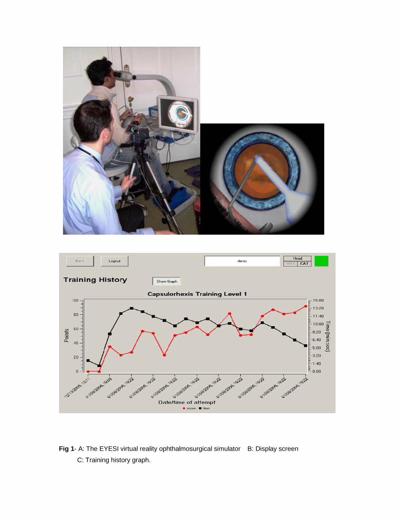
Fig 1- A: The EYESI virtual reality ophthalmosurgical simulator B: Display screen
C: Training history graph.
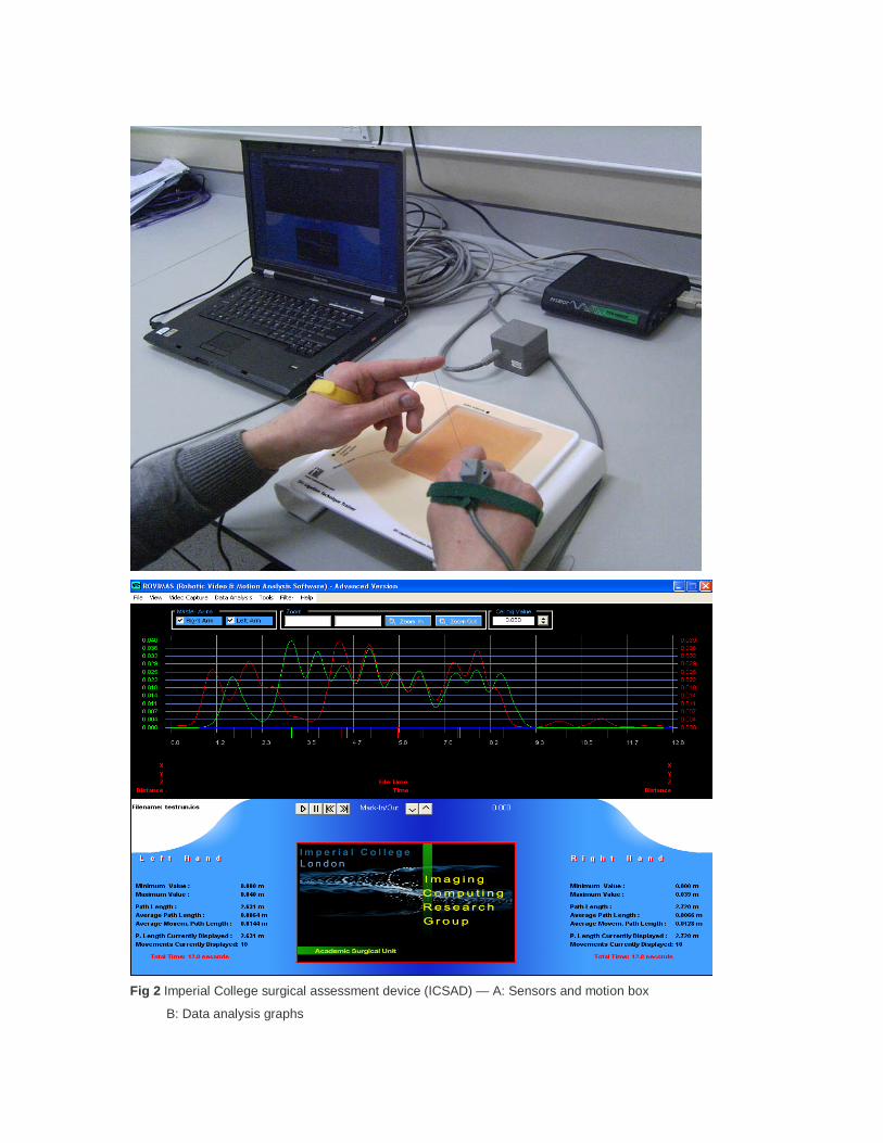
Fig 2 Imperial College surgical assessment device (ICSAD) — A: Sensors and motion box
B: Data analysis graphs