cyber-systemics, systemic governance and disruption - The ...
Upload
independentCategory
view
0download
0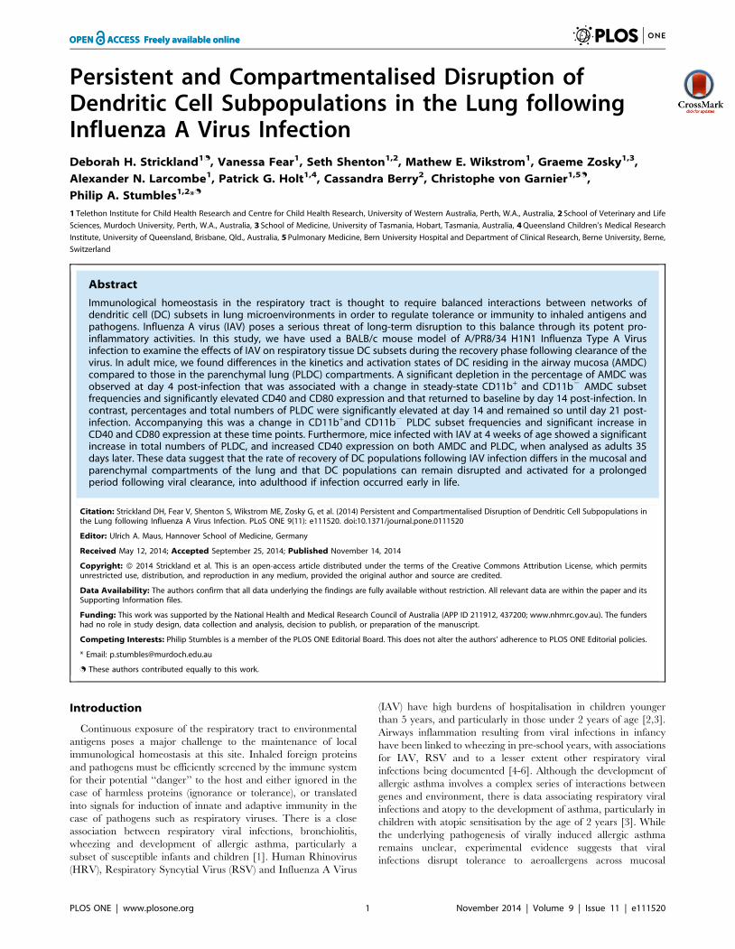
Persistent and Compartmentalised Disruption ofDendritic Cell Subpopulations in the Lung followingInfluenza A Virus InfectionDeborah H. Strickland1., Vanessa Fear1, Seth Shenton1,2, Mathew E. Wikstrom1, Graeme Zosky1,3,
Alexander N. Larcombe1, Patrick G. Holt1,4, Cassandra Berry2, Christophe von Garnier1,5.,
Philip A. Stumbles1,2*.
1 Telethon Institute for Child Health Research and Centre for Child Health Research, University of Western Australia, Perth, W.A., Australia, 2 School of Veterinary and Life
Sciences, Murdoch University, Perth, W.A., Australia, 3 School of Medicine, University of Tasmania, Hobart, Tasmania, Australia, 4 Queensland Children’s Medical Research
Institute, University of Queensland, Brisbane, Qld., Australia, 5 Pulmonary Medicine, Bern University Hospital and Department of Clinical Research, Berne University, Berne,
Switzerland
Abstract
Immunological homeostasis in the respiratory tract is thought to require balanced interactions between networks ofdendritic cell (DC) subsets in lung microenvironments in order to regulate tolerance or immunity to inhaled antigens andpathogens. Influenza A virus (IAV) poses a serious threat of long-term disruption to this balance through its potent pro-inflammatory activities. In this study, we have used a BALB/c mouse model of A/PR8/34 H1N1 Influenza Type A Virusinfection to examine the effects of IAV on respiratory tissue DC subsets during the recovery phase following clearance of thevirus. In adult mice, we found differences in the kinetics and activation states of DC residing in the airway mucosa (AMDC)compared to those in the parenchymal lung (PLDC) compartments. A significant depletion in the percentage of AMDC wasobserved at day 4 post-infection that was associated with a change in steady-state CD11b+ and CD11b2 AMDC subsetfrequencies and significantly elevated CD40 and CD80 expression and that returned to baseline by day 14 post-infection. Incontrast, percentages and total numbers of PLDC were significantly elevated at day 14 and remained so until day 21 post-infection. Accompanying this was a change in CD11b+and CD11b2 PLDC subset frequencies and significant increase inCD40 and CD80 expression at these time points. Furthermore, mice infected with IAV at 4 weeks of age showed a significantincrease in total numbers of PLDC, and increased CD40 expression on both AMDC and PLDC, when analysed as adults 35days later. These data suggest that the rate of recovery of DC populations following IAV infection differs in the mucosal andparenchymal compartments of the lung and that DC populations can remain disrupted and activated for a prolongedperiod following viral clearance, into adulthood if infection occurred early in life.
Citation: Strickland DH, Fear V, Shenton S, Wikstrom ME, Zosky G, et al. (2014) Persistent and Compartmentalised Disruption of Dendritic Cell Subpopulations inthe Lung following Influenza A Virus Infection. PLoS ONE 9(11): e111520. doi:10.1371/journal.pone.0111520
Editor: Ulrich A. Maus, Hannover School of Medicine, Germany
Received May 12, 2014; Accepted September 25, 2014; Published November 14, 2014
Copyright: � 2014 Strickland et al. This is an open-access article distributed under the terms of the Creative Commons Attribution License, which permitsunrestricted use, distribution, and reproduction in any medium, provided the original author and source are credited.
Data Availability: The authors confirm that all data underlying the findings are fully available without restriction. All relevant data are within the paper and itsSupporting Information files.
Funding: This work was supported by the National Health and Medical Research Council of Australia (APP ID 211912, 437200; www.nhmrc.gov.au). The fundershad no role in study design, data collection and analysis, decision to publish, or preparation of the manuscript.
Competing Interests: Philip Stumbles is a member of the PLOS ONE Editorial Board. This does not alter the authors’ adherence to PLOS ONE Editorial policies.
* Email: [email protected]
. These authors contributed equally to this work.
Introduction
Continuous exposure of the respiratory tract to environmental
antigens poses a major challenge to the maintenance of local
immunological homeostasis at this site. Inhaled foreign proteins
and pathogens must be efficiently screened by the immune system
for their potential ‘‘danger’’ to the host and either ignored in the
case of harmless proteins (ignorance or tolerance), or translated
into signals for induction of innate and adaptive immunity in the
case of pathogens such as respiratory viruses. There is a close
association between respiratory viral infections, bronchiolitis,
wheezing and development of allergic asthma, particularly a
subset of susceptible infants and children [1]. Human Rhinovirus
(HRV), Respiratory Syncytial Virus (RSV) and Influenza A Virus
(IAV) have high burdens of hospitalisation in children younger
than 5 years, and particularly in those under 2 years of age [2,3].
Airways inflammation resulting from viral infections in infancy
have been linked to wheezing in pre-school years, with associations
for IAV, RSV and to a lesser extent other respiratory viral
infections being documented [4-6]. Although the development of
allergic asthma involves a complex series of interactions between
genes and environment, there is data associating respiratory viral
infections and atopy to the development of asthma, particularly in
children with atopic sensitisation by the age of 2 years [3]. While
the underlying pathogenesis of virally induced allergic asthma
remains unclear, experimental evidence suggests that viral
infections disrupt tolerance to aeroallergens across mucosal
PLOS ONE | www.plosone.org 1 November 2014 | Volume 9 | Issue 11 | e111520
barriers together with enhanced pro-allergic immune responses
[7,8].
Under normal circumstances, immunological homeostasis
within the respiratory tract is maintained via the surveillance
activities of local dendritic cell (DC) populations. These are
distributed within respiratory tissues as integrated networks,
playing a crucial role in sampling of inhaled antigens including
viruses and allergens and in the initiation of subsequent tolerance
and/or adaptive T cell-mediated immune responses in draining
lymph nodes (DLN) [9]. Earlier observations from our group in a
rat model were the first to demonstrate the rapid expansion of
airway mucosal DC (AMDC) during acute viral (parainfluenza)
infection, and the apparent persistence of this response beyond
viral clearance [10]. This was subsequently confirmed in a mouse
RSV model with respect to whole lung DC and similar
observations have been reported in humans for nasal mucosal
DC populations in children post RSV and HRV infections
[11,12]. Given the key role that these frontline DC populations in
local immune surveillance for all classes of environmental antigens,
their long-term disruption following viral infection has significant
implications in regards to maintenance of general immunological
homeostasis within the respiratory tract. With this in mind we
have sought to confirm and extend these earlier observational
studies, aiming to more comprehensively identify which DC
subsets are susceptible to the effects of virus, and in which precise
tissue microenvironments within the respiratory tree; moreover we
have extended the studies to encompass immunologically imma-
ture weanling animals, similar to the age range described above for
maximal susceptibility to severe virus associated airways inflam-
mation in humans.
Early studies in rodent models demonstrated the capacity of
respiratory tract DC to direct the outcome of CD4+ T cell
responses to inhaled allergens and a number of subsequent studies
have confirmed the essential requirement for DC migration to
DLN for induction of CD4+ T cell mediated allergic airways
inflammation and asthmatic syndromes [13,14]. Furthermore, our
previous work in rodents identified a subdivision of function based
on anatomical location, with AMDC being functionally distinct
from their counterparts in parenchymal lung tissue, most likely due
to micro-environmental differences between these anatomical
locations [15,16]. Consistent with airway mucosal surfaces being
the first site of exposure to inhaled allergens and viruses, AMDC
show high levels of endocytic activity and rapid turn-over and
drainage to airway DLN, defining their proposed role as
‘‘gatekeepers’’ for the initiation of adaptive immunity to inhaled
allergens and pathogens [15,17-19].
A number of DC subsets have been described in rodents and
humans with differing capacities to influence naıve and memory
CD4+ and CD8+ T cell responses [20]. In the mouse, major
populations of classical (also termed ‘‘myeloid’’ or ‘‘conventional’’)
DC (cDC) and plasmacytoid DC (pDC) have been identified, as
well as a number of cDC subsets with distinct phenotypic and
transcriptional profiles [21]. In the mouse respiratory tract, two
dominant cDC subsets based on the reciprocal expression of
CD11b and the alpha (E) integrin CD103 have been described,
whereby CD11blo (CD103+) DC express tight junction proteins,
reside within the airway epithelium and increase in numbers
during allergic airways inflammation, while the CD11bhi
(CD1032) subset readily produces a number of chemokines that
regulate CD4+ T cell activity [16,22,23]. During IAV infection,
both DC subsets have been shown to be infected and be capable of
presenting viral antigens to T cells, however the CD11blo subset
appears to be the predominant subset for migration to DLN and
for either direct or cross-presentation of viral antigens to CD8+ T
cells although, other subsets in tissue and DLN are likely to be
involved [24–27]. In addition to cDC, other non-myeloid lineages
such as plasmacytoid DC (pDC) in lungs, as well as resident
lymphoid-origin DC in DLN, may also play important roles in
initiating T cell immunity to IAV [27–29].
In this study we have characterised the impact of IAV infection
on the frequency and activation of cDC in anatomical compart-
ments of the respiratory tract of BALB/c mice following IAV
infection. IAV infection was shown to have a differential impact on
DC numbers and activation status in airway mucosal and
parenchymal lung tissues, which represent two anatomically and
immunologically distinct compartments of the respiratory tract.
Furthermore, IAV infection of juvenile mice induced long-term
alterations on the frequency and activation states of parenchymal
lung DC subsets that persisted into adulthood.
Results
Clinical features of the BALB/c mouse model of influenzaA virus (IAV) infection
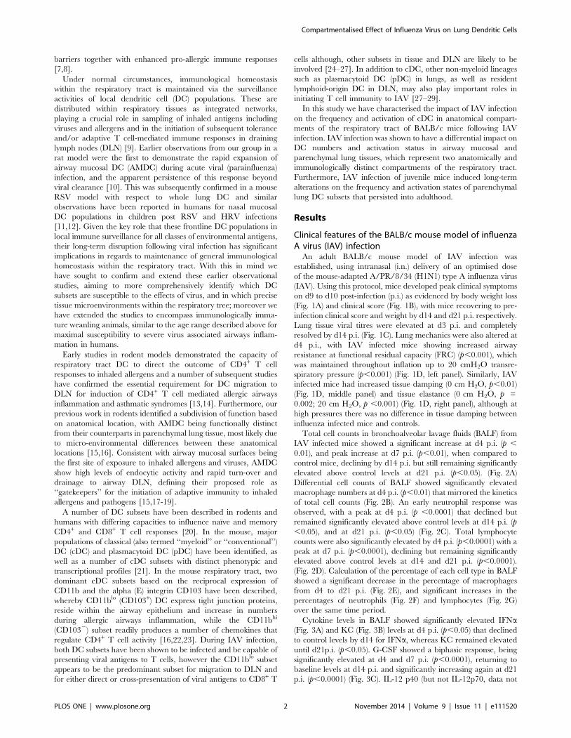
An adult BALB/c mouse model of IAV infection was
established, using intranasal (i.n.) delivery of an optimised dose
of the mouse-adapted A/PR/8/34 (H1N1) type A influenza virus
(IAV). Using this protocol, mice developed peak clinical symptoms
on d9 to d10 post-infection (p.i.) as evidenced by body weight loss
(Fig. 1A) and clinical score (Fig. 1B), with mice recovering to pre-
infection clinical score and weight by d14 and d21 p.i. respectively.
Lung tissue viral titres were elevated at d3 p.i. and completely
resolved by d14 p.i. (Fig. 1C). Lung mechanics were also altered at
d4 p.i., with IAV infected mice showing increased airway
resistance at functional residual capacity (FRC) (p,0.001), which
was maintained throughout inflation up to 20 cmH2O transre-
spiratory pressure (p,0.001) (Fig. 1D, left panel). Similarly, IAV
infected mice had increased tissue damping (0 cm H2O, p,0.01)
(Fig. 1D, middle panel) and tissue elastance (0 cm H2O, p =
0.002; 20 cm H2O, p ,0.001) (Fig. 1D, right panel), although at
high pressures there was no difference in tissue damping between
influenza infected mice and controls.
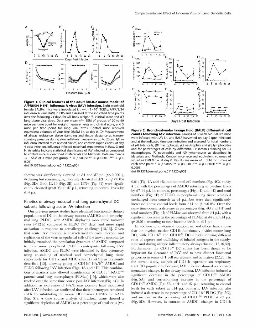
Total cell counts in bronchoalveolar lavage fluids (BALF) from
IAV infected mice showed a significant increase at d4 p.i. (p ,
0.01), and peak increase at d7 p.i. (p,0.01), when compared to
control mice, declining by d14 p.i. but still remaining significantly
elevated above control levels at d21 p.i. (p,0.05). (Fig. 2A)
Differential cell counts of BALF showed significantly elevated
macrophage numbers at d4 p.i. (p,0.01) that mirrored the kinetics
of total cell counts (Fig. 2B). An early neutrophil response was
observed, with a peak at d4 p.i. (p ,0.0001) that declined but
remained significantly elevated above control levels at d14 p.i. (p,0.05), and at d21 p.i. (p,0.05) (Fig. 2C). Total lymphocyte
counts were also significantly elevated by d4 p.i. (p,0.0001) with a
peak at d7 p.i. (p,0.0001), declining but remaining significantly
elevated above control levels at d14 and d21 p.i. (p,0.0001).
(Fig. 2D). Calculation of the percentage of each cell type in BALF
showed a significant decrease in the percentage of macrophages
from d4 to d21 p.i. (Fig. 2E), and significant increases in the
percentages of neutrophils (Fig. 2F) and lymphocytes (Fig. 2G)
over the same time period.
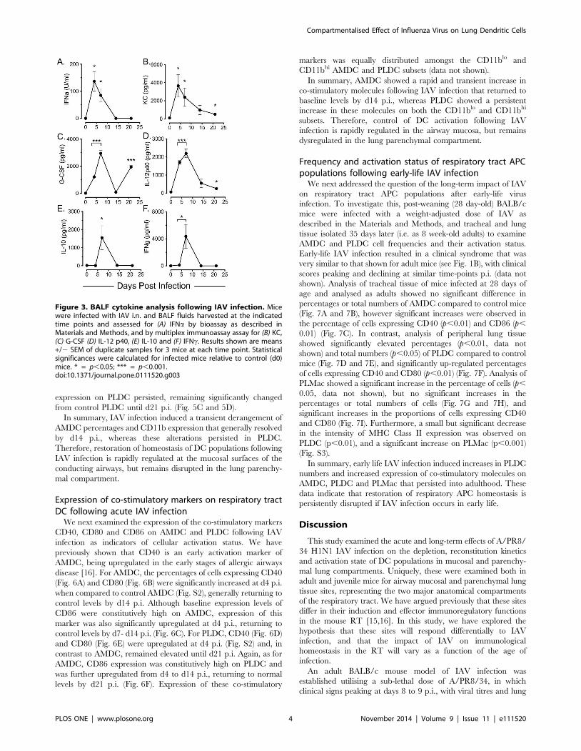
Cytokine levels in BALF showed significantly elevated IFNa(Fig. 3A) and KC (Fig. 3B) levels at d4 p.i. (p,0.05) that declined
to control levels by d14 for IFNa, whereas KC remained elevated
until d21p.i. (p,0.05). G-CSF showed a biphasic response, being
significantly elevated at d4 and d7 p.i. (p,0.0001), returning to
baseline levels at d14 p.i. and significantly increasing again at d21
p.i. (p,0.0001) (Fig. 3C). IL-12 p40 (but not IL-12p70, data not
Compartmentalised Effect of Influenza Virus on Lung Dendritic Cells
PLOS ONE | www.plosone.org 2 November 2014 | Volume 9 | Issue 11 | e111520
shown) was significantly elevated at d4 and d7 p.i. (p,0.0001),
declining but remaining significantly elevated at d21 p.i. (p,0.05)
(Fig. 3D). Both IL-10 (Fig. 3E) and IFNc (Fig. 3F) were signifi-
cantly elevated (p,0.05) at d7 p.i., returning to control levels by
d14 p.i.
Kinetics of airway mucosal and lung parenchymal DCsubsets following acute IAV infection
Our previous mouse studies have identified functionally distinct
populations of DC in the airway mucosa (AMDC) and parenchy-
mal lung (PLDC), with AMDC displaying more rapid turnover
rates (,12 h) compared to PLDC (.7 days) and more rapid
activation in response to aeroallergen challenge [15,16]. Given
that acute IAV infection is characterised by early infection and
replication of the virus in epithelial cells of the airway mucosa, we
initially examined the population dynamics of AMDC compared
to their more peripheral PLDC counterparts following IAV
infection. AMDC and PLDC were identified by flow cytometry
using co-staining of tracheal and parenchymal lung tissue
respectively for CD11c and MHC class II (I-A/E) as previously
described [15], allowing gating of CD11c+ I-A/Ehi AMDC and
PLDC following IAV infection (Figs. 4A and 4D). This combina-
tion of markers also allowed identification of CD11c+ I-A/Elow
parenchymal lung macrophages (PLMac) [15], which were also
tracked over the same time course post-IAV infection (Fig. 4G). In
addition, as expression of I-A/E may possibly have modulated
after IAV infection, we confirmed that these phenotypes remained
stable by substituting the mouse DC marker CD205 for I-A/E
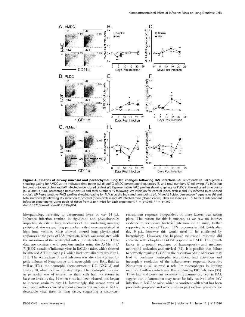
(Fig. S1). A time course analysis of tracheal tissue showed a
significant depletion of AMDC as a percentage of total cells (p,
0.01) (Fig. 4A and 4B), but not total cell numbers (Fig. 4C), at day
4 p.i. with the percentages of AMDC returning to baseline levels
by d7-14 p.i. In contrast, percentages (Fig. 4D and 4E) and total
numbers (Fig. 4F) of PLDC in peripheral lung tissue remained
unchanged from controls at d4 p.i., but were then significantly
increased above control levels from d14 p.i (p ,0.05). Over the
same time-course, a decrease in percentages (Fig. 4G and 4H) and
total numbers (Fig. 4I) of PLMac was observed from d4 p.i., with a
significant decrease in the percentage of PLMac at d4 and d14 p.i.
(p,0.01), returning to near-baseline levels at d21 p.i.
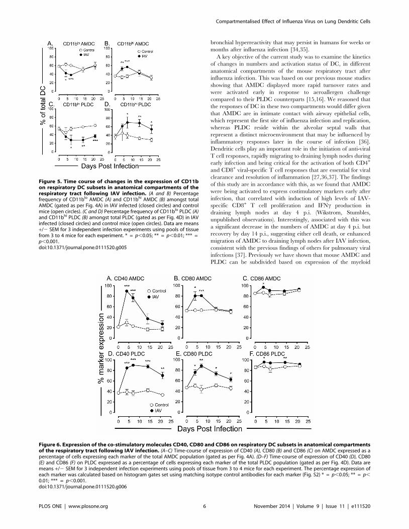
In addition to anatomical location, we and others have shown
that the myeloid marker CD11b functionally divides mouse lung
DC, with CD11bhi and CD11blo DC subsets showing different
rates of capture and trafficking of inhaled antigens in the steady-
state and during allergic inflammatory airways disease [15,16,30].
Furthermore, the CD11blo DC subset has been shown to be
important for clearance of IAV and to have distinct functional
properties in terms of T cell recruitment and activation [22,23]. In
the current study, analysis of CD11b expression on respiratory
tract DC populations following IAV infection showed a compart-
mentalised change. In the airway mucosa, IAV infection induced a
significant decrease in the percentage of CD11blo AMDC
(Fig. 5A), and corresponding increase in the percentage of
CD11bhi AMDC (Fig. 5B) at d4 and d7 p.i., returning to control
levels for each subset at d14 p.i. Similarly, IAV infection also
induced a decrease in the percentage of CD11blo PLDC (Fig. 5C),
and increase in the percentage of CD11bhi PLDC at d7 p.i.
(Fig. 5D). However, in contrast to AMDC, changes in CD11b
Figure 1. Clinical features of the adult BALB/c mouse model ofA/PR8/34 H1N1 influenza A virus (IAV) infection. Eight week-oldfemale BALB/c mice were inoculated i.n. with 16102 TCID50 A/PR/8/34influenza A virus (IAV) in PBS and assessed at the indicated time pointsover the following 21 days for (A) body weight (B) clinical score and (C)lung tissue viral titres. Data are mean +/2 SEM of groups of 20 to 60mice per time point for weight measurements and clinical score, and 5mice per time point for lung viral titres. Control mice receivedequivalent volumes of virus-free DMEM i.n. at day 0. (D) Measurementof airway resistance, tissue damping and tissue elastance at transre-spiratory pressure during slow inflation manoeuvres up to 20cm H2O ininfluenza infected mice (closed circles) and controls (open circles) at day4 post infection. Influenza infected mice had impairments in Raw, G andH. Asterisks indicate statistical significance of IAV infected as comparedto control mice as described in Materials and Methods. Data are means+/2 SEM of 4 mice per group. * = p,0.05; ** = p,0.01; *** = p,0.001.doi:10.1371/journal.pone.0111520.g001
Figure 2. Bronchoalveolar lavage fluid (BALF) differential cellcounts following IAV infection. Groups of 8 week old BALB/c micewere infected with IAV i.n. and BALF harvested on day 0 (pre-infection)and at the indicated time post-infection and assessed for total numbersof (A) total cells, (B) macrophages, (C) neutrophils and (D) lymphocytesand for percentages of cells by differential Leishman’s staining for (E)macrophages, (F) neutrophils and (G) lymphocytes as described inMaterials and Methods. Control mice received equivalent volumes ofvirus-free DMEM i.n. at day 0. Results are mean +/2 SEM for 5 mice ateach time point. * = p,0.05; ** = p,0.01; *** = p,0.001; **** = p,0.0001.doi:10.1371/journal.pone.0111520.g002
Compartmentalised Effect of Influenza Virus on Lung Dendritic Cells
PLOS ONE | www.plosone.org 3 November 2014 | Volume 9 | Issue 11 | e111520
expression on PLDC persisted, remaining significantly changed
from control PLDC until d21 p.i. (Fig. 5C and 5D).
In summary, IAV infection induced a transient derangement of
AMDC percentages and CD11b expression that generally resolved
by d14 p.i., whereas these alterations persisted in PLDC.
Therefore, restoration of homeostasis of DC populations following
IAV infection is rapidly regulated at the mucosal surfaces of the
conducting airways, but remains disrupted in the lung parenchy-
mal compartment.
Expression of co-stimulatory markers on respiratory tractDC following acute IAV infection
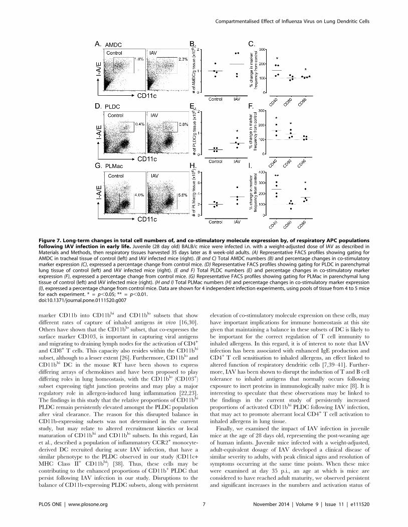
We next examined the expression of the co-stimulatory markers
CD40, CD80 and CD86 on AMDC and PLDC following IAV
infection as indicators of cellular activation status. We have
previously shown that CD40 is an early activation marker of
AMDC, being upregulated in the early stages of allergic airways
disease [16]. For AMDC, the percentages of cells expressing CD40
(Fig. 6A) and CD80 (Fig. 6B) were significantly increased at d4 p.i.
when compared to control AMDC (Fig. S2), generally returning to
control levels by d14 p.i. Although baseline expression levels of
CD86 were constitutively high on AMDC, expression of this
marker was also significantly upregulated at d4 p.i., returning to
control levels by d7- d14 p.i. (Fig. 6C). For PLDC, CD40 (Fig. 6D)
and CD80 (Fig. 6E) were upregulated at d4 p.i. (Fig. S2) and, in
contrast to AMDC, remained elevated until d21 p.i. Again, as for
AMDC, CD86 expression was constitutively high on PLDC and
was further upregulated from d4 to d14 p.i., returning to normal
levels by d21 p.i. (Fig. 6F). Expression of these co-stimulatory
markers was equally distributed amongst the CD11blo and
CD11bhi AMDC and PLDC subsets (data not shown).
In summary, AMDC showed a rapid and transient increase in
co-stimulatory molecules following IAV infection that returned to
baseline levels by d14 p.i., whereas PLDC showed a persistent
increase in these molecules on both the CD11blo and CD11bhi
subsets. Therefore, control of DC activation following IAV
infection is rapidly regulated in the airway mucosa, but remains
dysregulated in the lung parenchymal compartment.
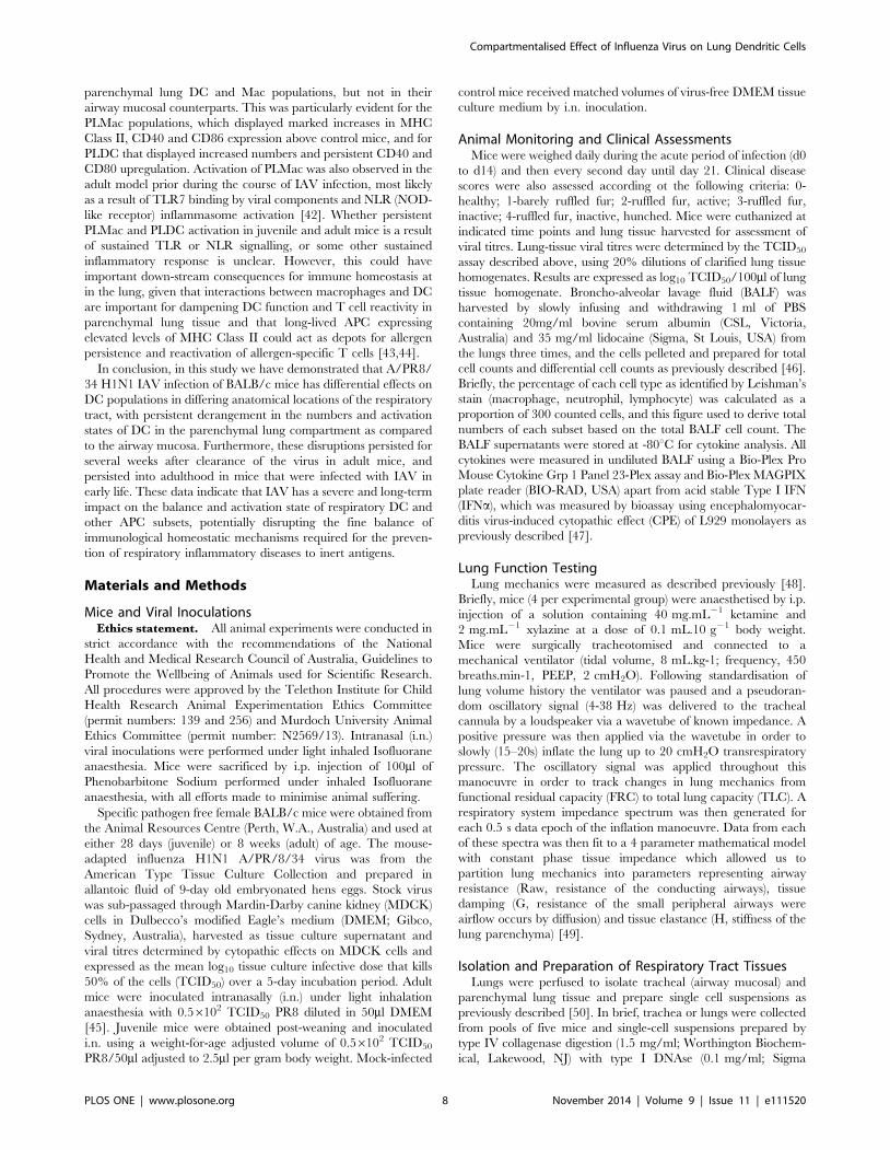
Frequency and activation status of respiratory tract APCpopulations following early-life IAV infection
We next addressed the question of the long-term impact of IAV
on respiratory tract APC populations after early-life virus
infection. To investigate this, post-weaning (28 day-old) BALB/c
mice were infected with a weight-adjusted dose of IAV as
described in the Materials and Methods, and tracheal and lung
tissue isolated 35 days later (i.e. as 8 week-old adults) to examine
AMDC and PLDC cell frequencies and their activation status.
Early-life IAV infection resulted in a clinical syndrome that was
very similar to that shown for adult mice (see Fig. 1B), with clinical
scores peaking and declining at similar time-points p.i. (data not
shown). Analysis of tracheal tissue of mice infected at 28 days of
age and analysed as adults showed no significant difference in
percentages or total numbers of AMDC compared to control mice
(Fig. 7A and 7B), however significant increases were observed in
the percentage of cells expressing CD40 (p,0.01) and CD86 (p,
0.01) (Fig. 7C). In contrast, analysis of peripheral lung tissue
showed significantly elevated percentages (p,0.01, data not
shown) and total numbers (p,0.05) of PLDC compared to control
mice (Fig. 7D and 7E), and significantly up-regulated percentages
of cells expressing CD40 and CD80 (p,0.01) (Fig. 7F). Analysis of
PLMac showed a significant increase in the percentage of cells (p,
0.05, data not shown), but no significant increases in the
percentages or total numbers of cells (Fig. 7G and 7H), and
significant increases in the proportions of cells expressing CD40
and CD80 (Fig. 7I). Furthermore, a small but significant decrease
in the intensity of MHC Class II expression was observed on
PLDC (p,0.01), and a significant increase on PLMac (p,0.001)
(Fig. S3).
In summary, early life IAV infection induced increases in PLDC
numbers and increased expression of co-stimulatory molecules on
AMDC, PLDC and PLMac that persisted into adulthood. These
data indicate that restoration of respiratory APC homeostasis is
persistently disrupted if IAV infection occurs in early life.
Discussion
This study examined the acute and long-term effects of A/PR8/
34 H1N1 IAV infection on the depletion, reconstitution kinetics
and activation state of DC populations in mucosal and parenchy-
mal lung compartments. Uniquely, these were examined both in
adult and juvenile mice for airway mucosal and parenchymal lung
tissue sites, representing the two major anatomical compartments
of the respiratory tract. We have argued previously that these sites
differ in their induction and effector immunoregulatory functions
in the mouse RT [15,16]. In this study, we have explored the
hypothesis that these sites will respond differentially to IAV
infection, and that the impact of IAV on immunological
homeostasis in the RT will vary as a function of the age of
infection.
An adult BALB/c mouse model of IAV infection was
established utilising a sub-lethal dose of A/PR8/34, in which
clinical signs peaking at days 8 to 9 p.i., with viral titres and lung
Figure 3. BALF cytokine analysis following IAV infection. Micewere infected with IAV i.n. and BALF fluids harvested at the indicatedtime points and assessed for (A) IFNa by bioassay as described inMaterials and Methods, and by multiplex immunoassay assay for (B) KC,(C) G-CSF (D) IL-12 p40, (E) IL-10 and (F) IFNc. Results shown are means+/2 SEM of duplicate samples for 3 mice at each time point. Statisticalsignificances were calculated for infected mice relative to control (d0)mice. * = p,0.05; *** = p,0.001.doi:10.1371/journal.pone.0111520.g003
Compartmentalised Effect of Influenza Virus on Lung Dendritic Cells
PLOS ONE | www.plosone.org 4 November 2014 | Volume 9 | Issue 11 | e111520
histopathology reverting to background levels by day 14 p.i.
Influenza infection resulted in significant and physiologically
important deficits in lung mechanics of the conducting airways,
peripheral airways and lung parenchyma that were maintained at
high lung volume. Mice showed altered lung physiological
responses at the peak of IAV infection, which was associated with
the maximum of the neutrophil influx into alveolar space. These
data are consistent with previous studies using the A/Mem/1/
71(H3N1) strain of influenza virus in BALB/c mice, which showed
heightened AHR at day 4 p.i. which had normalised by day 20 p.i.
[31]. The acute phase of viral infection was also characterised by
peak influxes of lymphocytes and neutrophils into BAL fluid as
well as IFNa, the neutrophil chemoattractant KC (CXCL1) and
IL-12 p70, which declined by day 14 p.i. The neutrophil response
in particular was of interest, as these cells had not return to
baseline levels by day 14 when virus had been cleared, and began
to increase again by day 14. Interestingly, this second wave of
neutrophil influx occurred without a concurrent increase in KC or
detectable viral titres in lung tissue, suggesting a secondary
recruitment response independent of these factors was taking
place. The reason for this is unclear, as we saw no indirect
evidence of secondary bacterial infection in the mice, further
supported by a lack of Type 1 IFN responses in BAL fluids after
day 9 p.i., however this would need to be confirmed by
bacteriology. However, the bi-phasic neutrophil response did
correlate with a bi-phasic G-CSF response in BALF. This growth
factor is a potent regulator of haemopoesis, and mediates
neutrophil activation and survival [32]. It is possible that failure
to correctly regulate G-CSF in the resolution phase of disease may
lead to persistent neutrophil recruitment and activation and
incomplete resolution of the inflammatory response. Recently,
Narasaraju et al. showed a role for macrophages in limiting
neutrophil influxes into lavage fluids following PR8 infection [33].
These late and persistent increases in inflammatory cells in BAL
suggest that inflammation may never be fully resolved after IAV
infection in BALB/c mice, which is consistent with what has been
previously proposed and which may in part explain post-infective
Figure 4. Kinetics of airway mucosal and parenchymal lung DC changes following IAV infection. (A) Representative FACS profilesshowing gating for AMDC at the indicated time points p.i. (B and C) AMDC percentage frequencies (B) and total numbers (C) following IAV infectionfor control (open circles) and IAV infected mice (closed circles). (D) Representative FACS profiles showing gating for PLDC at the indicated time pointsp.i. (E and F) PLDC percentage frequencies (E) and total numbers (F) following IAV infection for control (open circles) and IAV infected mice (closedcircles). (G) Representative FACS profiles showing gating for PLMac at the indicated time points p.i. (H and I) PLMac percentage frequencies (H) andtotal numbers (I) following IAV infection for control (open circles) and IAV infected mice (closed circles). Data are means +/2 SEM for 3 independentinfection experiments using pools of tissue from 3 to 4 mice for each experiment. * = p,0.05; ** = p,0.01.doi:10.1371/journal.pone.0111520.g004
Compartmentalised Effect of Influenza Virus on Lung Dendritic Cells
PLOS ONE | www.plosone.org 5 November 2014 | Volume 9 | Issue 11 | e111520
bronchial hyperreactivity that may persist in humans for weeks or
months after influenza infection [34,35].
A key objective of the current study was to examine the kinetics
of changes in numbers and activation status of DC, in different
anatomical compartments of the mouse respiratory tract after
influenza infection. This was based on our previous mouse studies
showing that AMDC displayed more rapid turnover rates and
were activated early in response to aeroallergen challenge
compared to their PLDC counterparts [15,16]. We reasoned that
the responses of DC in these two compartments would differ given
that AMDC are in intimate contact with airway epithelial cells,
which represent the first site of influenza infection and replication,
whereas PLDC reside within the alveolar septal walls that
represent a distinct microenvironment that may be influenced by
inflammatory responses later in the course of infection [36].
Dendritic cells play an important role in the initiation of anti-viral
T cell responses, rapidly migrating to draining lymph nodes during
early infection and being critical for the activation of both CD4+
and CD8+ viral-specific T cell responses that are essential for viral
clearance and resolution of inflammation [27,36,37]. The findings
of this study are in accordance with this, as we found that AMDC
were being activated to express costimulatory markers early after
infection, that correlated with induction of high levels of IAV-
specific CD8+ T cell proliferation and IFNc production in
draining lymph nodes at day 4 p.i. (Wikstrom, Stumbles,
unpublished observations). Interestingly, associated with this was
a significant decrease in the numbers of AMDC at day 4 p.i. but
recovery by day 14 p.i., suggesting either cell death, or enhanced
migration of AMDC to draining lymph nodes after IAV infection,
consistent with the previous findings of others for pulmonary viral
infections [37]. Previously we have shown that mouse AMDC and
PLDC can be subdivided based on expression of the myeloid
Figure 5. Time course of changes in the expression of CD11bon respiratory DC subsets in anatomical compartments of therespiratory tract following IAV infection. (A and B) Percentagefrequency of CD11blo AMDC (A) and CD11bhi AMDC (B) amongst totalAMDC (gated as per Fig. 4A) in IAV infected (closed circles) and controlmice (open circles). (C and D) Percentage frequency of CD11blo PLDC (A)and CD11bhi PLDC (B) amongst total PLDC (gated as per Fig. 4D) in IAVinfected (closed circles) and control mice (open circles). Data are means+/2 SEM for 3 independent infection experiments using pools of tissuefrom 3 to 4 mice for each experiment. * = p,0.05; ** = p,0.01; *** =p,0.001.doi:10.1371/journal.pone.0111520.g005
Figure 6. Expression of the co-stimulatory molecules CD40, CD80 and CD86 on respiratory DC subsets in anatomical compartmentsof the respiratory tract following IAV infection. (A–C) Time-course of expression of CD40 (A), CD80 (B) and CD86 (C) on AMDC expressed as apercentage of cells expressing each marker of the total AMDC population (gated as per Fig. 4A). (D–F) Time-course of expression of CD40 (D), CD80(E) and CD86 (F) on PLDC expressed as a percentage of cells expressing each marker of the total PLDC population (gated as per Fig. 4D). Data aremeans +/2 SEM for 3 independent infection experiments using pools of tissue from 3 to 4 mice for each experiment. The percentage expression ofeach marker was calculated based on histogram gates set using matching isotype control antibodies for each marker (Fig. S2) * = p,0.05; ** = p,
0.01; *** = p,0.001.doi:10.1371/journal.pone.0111520.g006
Compartmentalised Effect of Influenza Virus on Lung Dendritic Cells
PLOS ONE | www.plosone.org 6 November 2014 | Volume 9 | Issue 11 | e111520
marker CD11b into CD11bhi and CD11blo subsets that show
different rates of capture of inhaled antigens in vivo [16,30].
Others have shown that the CD11blo subset, that co-expresses the
surface marker CD103, is important in capturing viral antigens
and migrating to draining lymph nodes for the activation of CD4+
and CD8+ T cells. This capacity also resides within the CD11bhi
subset, although to a lesser extent [26]. Furthermore, CD11blo and
CD11bhi DC in the mouse RT have been shown to express
differing arrays of chemokines and have been proposed to play
differing roles in lung homeostasis, with the CD11blo (CD103+)
subset expressing tight junction proteins and may play a major
regulatory role in allergen-induced lung inflammation [22,23].
The findings in this study that the relative proportions of CD11bhi
PLDC remain persistently elevated amongst the PLDC population
after viral clearance. The reason for this disrupted balance in
CD11b-expressing subsets was not determined in the current
study, but may relate to altered recruitment kinetics or local
maturation of CD11bhi and CD11blo subsets. In this regard, Lin
et al., described a population of inflammatory CCR2+ monocyte-
derived DC recruited during acute IAV infection, that have a
similar phenotype to the PLDC observed in our study (CD11c+MHC Class II+ CD11bhi) [38]. Thus, these cells may be
contributing to the enhanced proportions of CD11b+ PLDC that
persist following IAV infection in our study. Disruptions to the
balance of CD11b-expressing PLDC subsets, along with persistent
elevation of co-stimulatory molecule expression on these cells, may
have important implications for immune homeostasis at this site
given that maintaining a balance in these subsets of DC is likely to
be important for the correct regulation of T cell immunity to
inhaled allergens. In this regard, it is of interest to note that IAV
infection has been associated with enhanced IgE production and
CD4+ T cell sensitisation to inhaled allergens, an effect linked to
altered function of respiratory dendritic cells [7,39–41]. Further-
more, IAV has been shown to disrupt the induction of T and B cell
tolerance to inhaled antigens that normally occurs following
exposure to inert proteins in immunologically naıve mice [8]. It is
interesting to speculate that these observations may be linked to
the findings in the current study of persistently increased
proportions of activated CD11bhi PLDC following IAV infection,
that may act to promote aberrant local CD4+ T cell activation to
inhaled allergens in lung tissue.
Finally, we examined the impact of IAV infection in juvenile
mice at the age of 28 days old, representing the post-weaning age
of human infants. Juvenile mice infected with a weight-adjusted,
adult-equivalent dosage of IAV developed a clinical disease of
similar severity to adults, with peak clinical signs and resolution of
symptoms occurring at the same time points. When these mice
were examined at day 35 p.i., an age at which is mice are
considered to have reached adult maturity, we observed persistent
and significant increases in the numbers and activation status of
Figure 7. Long-term changes in total cell numbers of, and co-stimulatory molecule expression by, of respiratory APC populationsfollowing IAV infection in early life. Juvenile (28 day old) BALB/c mice were infected i.n. with a weight-adjusted dose of IAV as described inMaterials and Methods, then respiratory tissues harvested 35 days later as 8 week-old adults. (A) Representative FACS profiles showing gating forAMDC in tracheal tissue of control (left) and IAV infected mice (right). (B and C) Total AMDC numbers (B) and percentage changes in co-stimulatorymarker expression (C), expressed a percentage change from control mice. (D) Representative FACS profiles showing gating for PLDC in parenchymallung tissue of control (left) and IAV infected mice (right). (E and F) Total PLDC numbers (E) and percentage changes in co-stimulatory markerexpression (F), expressed a percentage change from control mice. (G) Representative FACS profiles showing gating for PLMac in parenchymal lungtissue of control (left) and IAV infected mice (right). (H and I) Total PLMac numbers (H) and percentage changes in co-stimulatory marker expression(I), expressed a percentage change from control mice. Data are shown for 4 independent infection experiments, using pools of tissue from 4 to 5 micefor each experiment. * = p,0.05; ** = p,0.01.doi:10.1371/journal.pone.0111520.g007
Compartmentalised Effect of Influenza Virus on Lung Dendritic Cells
PLOS ONE | www.plosone.org 7 November 2014 | Volume 9 | Issue 11 | e111520
parenchymal lung DC and Mac populations, but not in their
airway mucosal counterparts. This was particularly evident for the
PLMac populations, which displayed marked increases in MHC
Class II, CD40 and CD86 expression above control mice, and for
PLDC that displayed increased numbers and persistent CD40 and
CD80 upregulation. Activation of PLMac was also observed in the
adult model prior during the course of IAV infection, most likely
as a result of TLR7 binding by viral components and NLR (NOD-
like receptor) inflammasome activation [42]. Whether persistent
PLMac and PLDC activation in juvenile and adult mice is a result
of sustained TLR or NLR signalling, or some other sustained
inflammatory response is unclear. However, this could have
important down-stream consequences for immune homeostasis at
in the lung, given that interactions between macrophages and DC
are important for dampening DC function and T cell reactivity in
parenchymal lung tissue and that long-lived APC expressing
elevated levels of MHC Class II could act as depots for allergen
persistence and reactivation of allergen-specific T cells [43,44].
In conclusion, in this study we have demonstrated that A/PR8/
34 H1N1 IAV infection of BALB/c mice has differential effects on
DC populations in differing anatomical locations of the respiratory
tract, with persistent derangement in the numbers and activation
states of DC in the parenchymal lung compartment as compared
to the airway mucosa. Furthermore, these disruptions persisted for
several weeks after clearance of the virus in adult mice, and
persisted into adulthood in mice that were infected with IAV in
early life. These data indicate that IAV has a severe and long-term
impact on the balance and activation state of respiratory DC and
other APC subsets, potentially disrupting the fine balance of
immunological homeostatic mechanisms required for the preven-
tion of respiratory inflammatory diseases to inert antigens.
Materials and Methods
Mice and Viral InoculationsEthics statement. All animal experiments were conducted in
strict accordance with the recommendations of the National
Health and Medical Research Council of Australia, Guidelines to
Promote the Wellbeing of Animals used for Scientific Research.
All procedures were approved by the Telethon Institute for Child
Health Research Animal Experimentation Ethics Committee
(permit numbers: 139 and 256) and Murdoch University Animal
Ethics Committee (permit number: N2569/13). Intranasal (i.n.)
viral inoculations were performed under light inhaled Isofluorane
anaesthesia. Mice were sacrificed by i.p. injection of 100ml of
Phenobarbitone Sodium performed under inhaled Isofluorane
anaesthesia, with all efforts made to minimise animal suffering.
Specific pathogen free female BALB/c mice were obtained from
the Animal Resources Centre (Perth, W.A., Australia) and used at
either 28 days (juvenile) or 8 weeks (adult) of age. The mouse-
adapted influenza H1N1 A/PR/8/34 virus was from the
American Type Tissue Culture Collection and prepared in
allantoic fluid of 9-day old embryonated hens eggs. Stock virus
was sub-passaged through Mardin-Darby canine kidney (MDCK)
cells in Dulbecco’s modified Eagle’s medium (DMEM; Gibco,
Sydney, Australia), harvested as tissue culture supernatant and
viral titres determined by cytopathic effects on MDCK cells and
expressed as the mean log10 tissue culture infective dose that kills
50% of the cells (TCID50) over a 5-day incubation period. Adult
mice were inoculated intranasally (i.n.) under light inhalation
anaesthesia with 0.56102 TCID50 PR8 diluted in 50ml DMEM
[45]. Juvenile mice were obtained post-weaning and inoculated
i.n. using a weight-for-age adjusted volume of 0.56102 TCID50
PR8/50ml adjusted to 2.5ml per gram body weight. Mock-infected
control mice received matched volumes of virus-free DMEM tissue
culture medium by i.n. inoculation.
Animal Monitoring and Clinical AssessmentsMice were weighed daily during the acute period of infection (d0
to d14) and then every second day until day 21. Clinical disease
scores were also assessed according ot the following criteria: 0-
healthy; 1-barely ruffled fur; 2-ruffled fur, active; 3-ruffled fur,
inactive; 4-ruffled fur, inactive, hunched. Mice were euthanized at
indicated time points and lung tissue harvested for assessment of
viral titres. Lung-tissue viral titres were determined by the TCID50
assay described above, using 20% dilutions of clarified lung tissue
homogenates. Results are expressed as log10 TCID50/100ml of lung
tissue homogenate. Broncho-alveolar lavage fluid (BALF) was
harvested by slowly infusing and withdrawing 1 ml of PBS
containing 20mg/ml bovine serum albumin (CSL, Victoria,
Australia) and 35 mg/ml lidocaine (Sigma, St Louis, USA) from
the lungs three times, and the cells pelleted and prepared for total
cell counts and differential cell counts as previously described [46].
Briefly, the percentage of each cell type as identified by Leishman’s
stain (macrophage, neutrophil, lymphocyte) was calculated as a
proportion of 300 counted cells, and this figure used to derive total
numbers of each subset based on the total BALF cell count. The
BALF supernatants were stored at -80uC for cytokine analysis. All
cytokines were measured in undiluted BALF using a Bio-Plex Pro
Mouse Cytokine Grp 1 Panel 23-Plex assay and Bio-Plex MAGPIX
plate reader (BIO-RAD, USA) apart from acid stable Type I IFN
(IFNa), which was measured by bioassay using encephalomyocar-
ditis virus-induced cytopathic effect (CPE) of L929 monolayers as
previously described [47].
Lung Function TestingLung mechanics were measured as described previously [48].
Briefly, mice (4 per experimental group) were anaesthetised by i.p.
injection of a solution containing 40 mg.mL21 ketamine and
2 mg.mL21 xylazine at a dose of 0.1 mL.10 g21 body weight.
Mice were surgically tracheotomised and connected to a
mechanical ventilator (tidal volume, 8 mL.kg-1; frequency, 450
breaths.min-1, PEEP, 2 cmH2O). Following standardisation of
lung volume history the ventilator was paused and a pseudoran-
dom oscillatory signal (4-38 Hz) was delivered to the tracheal
cannula by a loudspeaker via a wavetube of known impedance. A
positive pressure was then applied via the wavetube in order to
slowly (15–20s) inflate the lung up to 20 cmH2O transrespiratory
pressure. The oscillatory signal was applied throughout this
manoeuvre in order to track changes in lung mechanics from
functional residual capacity (FRC) to total lung capacity (TLC). A
respiratory system impedance spectrum was then generated for
each 0.5 s data epoch of the inflation manoeuvre. Data from each
of these spectra was then fit to a 4 parameter mathematical model
with constant phase tissue impedance which allowed us to
partition lung mechanics into parameters representing airway
resistance (Raw, resistance of the conducting airways), tissue
damping (G, resistance of the small peripheral airways were
airflow occurs by diffusion) and tissue elastance (H, stiffness of the
lung parenchyma) [49].
Isolation and Preparation of Respiratory Tract TissuesLungs were perfused to isolate tracheal (airway mucosal) and
parenchymal lung tissue and prepare single cell suspensions as
previously described [50]. In brief, trachea or lungs were collected
from pools of five mice and single-cell suspensions prepared by
type IV collagenase digestion (1.5 mg/ml; Worthington Biochem-
ical, Lakewood, NJ) with type I DNAse (0.1 mg/ml; Sigma
Compartmentalised Effect of Influenza Virus on Lung Dendritic Cells
PLOS ONE | www.plosone.org 8 November 2014 | Volume 9 | Issue 11 | e111520
Aldrich). DLN (upper paratracheal and parathymic) were pooled
separately from the same groups of mice, finely chopped with a
scalpel, and digested with type IV collagenase and type I DNAse.
All digestions and washes were performed in glucose sodium
potassium buffer (11 mM D-glucose, 5.5 mM KCl, 137 mM
NaCl, 25 mM Na2HPO4, 5.5 mM NaH2PO4.2H2O) with debris
and RBCs removed as previously described [51].
Analysis of Cell Surface Markers by Flow CytometryAfter preparation of single-cell suspensions, FcR were routinely
blocked using 2.4G2 (BD Biosciences) for 10 min on ice to prevent
non-specific binding of phenotyping antibodies subsequently
added. Airway, lung and draining lymph node DC populations
were identified in tracheal digests using combinations of fluoro-
chrome labelled mAbs (all from BD Biosciences, NSW, Australia
except where indicated) to mouse CD11c (clone N418), I-A/I-E
(clone 2G9), CD205 (DEC205; Serotec, Oxford, UK), CD11b
(clone M1/70), CD40 (clone 3/23), CD80 (clone 16-10A1) and
CD86 (clone GL1). All labelling was performed in glucose sodium
potassium buffer containing 0.2% BSA for 30 min on ice. All Abs
were used as direct conjugates to FITC, Phycoerythrin (PE), PE-
Cy7, allophycocyanin (APC), APC-Cy7, or biotin as required.
Where appropriate, biotinylated antibodies were detected with
Streptavidin conjugated PE-Cy5 (BD Biosciences). Appropriately
matched and conjugated IgG isotype controls (BD Biosciences)
were used in all experiments, and cytometer compensation settings
were adjusted using single-stained controls for each experiment.
Samples were collected using a FACSCalibur or LSRII flow
cytometer (BD Biosciences) and analyzed using FlowJo software
(TreeStar, Ca, USA).
Determination of Total Cell CountsTotal counts for AMDC, PLDC and PLMac were determined
on the basis of tissue weight and total cell yield for each tissue
(trachea and peripheral lung), and then calculated on the basis of
the total percentage of each DC type as determined by FACS.
Values are expresses as number of cells/g tissue according to the
formula: (% frequency x total cells/g tissue)/100.
Statistical AnalysisTwo-tailed, unpaired Student’s t tests assuming equal variance
were employed to calculate significances (GraphPad Prism, CA,
USA), with p-values ,0.05 considered statistically significant.
Statistical significance is indicated as follows: * = p,0.05; ** =
p,0.01; *** = p,0.001; **** = p,0.0001.
Supporting Information
Figure S1 CD205 and CD11c staining of respiratorytissues. Tracheal (A) and parenchymal lung tissue (B and C) cells
were prepared as per Methods, labelled for CD11c and CD205
and analysed by flow cytometry at the indicated time points after
IAV infection. Representative dot plots of 3 experiments for each
time point are shown, with AMDC (A), PLDC (B) and PLMac (C)
gated as indicated.
(TIF)
Figure S2 Expression levels of co-stimulatory markerson AMDC and PLDC at d4 post-IAV infection. Adult
BALB/c mice were infected with IAV and 4 days later
(representing a time point of peak expression of each marker)
tracheal and lung tissue were prepared for flow cytometry analysis
for each marker on AMDC (A) and PLDC (B). Representative
histograms (of 3 experiments) are shown for control and IAV
infected mice, showing isotype controls (grey shaded) and specific
marker (solid black) expression.
(TIF)
Figure S3 Changes in MHC Class II (I-A/E) expressionon respiratory APC of adult mice following IAV infectionat 4 weeks of age. Mice were infected with IAV at 4 weeks of
age and respiratory tissue harvested 35 days later. The mean
fluorescence intensity (MFI) of I-A/E expression on AMDC (A),
PLDC (B) and PLMac (C) was then determined for IAV infected
mice (black line) and control mice (grey shade), and expressed as a
percentage change in MFI of IAV infected mice as compared to
control mice (D). Histograms are representative of 4 independent
experiments using pooled tissue from 5 mice in each, gated on DC
populations as shown in Fig. S1. Panel D shows cumulative data of
4 independent experiments using pools of tissue from 5 mice in
each experiment.
(TIF)
Acknowledgments
We thank Miranda Smith, Elizabeth Bozanich, Jenny Thomas, Siew Ping
Lau and Sylvia Napoli for their excellent technical assistance during this
study.
Author Contributions
Conceived and designed the experiments: PAS DHS CvG VF PGH GZ
ANL. Performed the experiments: VF SS MEW GZ ANL CB. Analyzed
the data: PAS DHS CvG MEW GZ VF SS CB. Contributed reagents/
materials/analysis tools: PAS DHS CB GZ ANL. Wrote the paper: PAS
DHS CvG VF MEW GZ.
References
1. Gern JE (2008) Viral respiratory infection and the link to asthma. Pediatr Infect
Dis J 27: S97–103.
2. Iwane MK, Edwards KM, Szilagyi PG, Walker FJ, Griffin MR, et al. (2004)
Population-based surveillance for hospitalizations associated with respiratory
syncytial virus, influenza virus, and parainfluenza viruses among young children.
Pediatrics 113: 1758–1764.
3. Kusel MM, de Klerk NH, Kebadze T, Vohma V, Holt PG, et al. (2007) Early-
life respiratory viral infections, atopic sensitization, and risk of subsequent
development of persistent asthma. J Allergy Clin Immunol 119: 1105–1110.
4. Lemanske RF Jr, Jackson DJ, Gangnon RE, Evans MD, Li Z, et al. (2005)
Rhinovirus illnesses during infancy predict subsequent childhood wheezing.
J Allergy Clin Immunol 116: 571–577.
5. Sigurs N, Gustafsson PM, Bjarnason R, Lundberg F, Schmidt S, et al. (2005)
Severe respiratory syncytial virus bronchiolitis in infancy and asthma and allergy
at age 13. Am J Respir Crit Care Med 171: 137–141.
6. Stein RT, Sherrill D, Morgan WJ, Holberg CJ, Halonen M, et al. (1999)
Respiratory syncytial virus in early life and risk of wheeze and allergy by age 13
years. Lancet 354: 541–545.
7. Al-Garawi AA, Fattouh R, Walker TD, Jamula EB, Botelho F, et al. (2009)
Acute, but not resolved, influenza A infection enhances susceptibility to house
dust mite-induced allergic disease. J Immunol 182: 3095–3104.
8. Tsitoura DC, Kim S, Dabbagh K, Berry G, Lewis DB, et al. (2000) Respiratory
infection with influenza A virus interferes with the induction of tolerance to
aeroallergens. J Immunol 165: 3484–3491.
9. Holt P, Strickland D, Wikstrom M, Jahnsen F (2008) Regulation of
immunological homeostasis in the respiratory tract. Nature reviews Immunology
8: 142–194.
10. McWilliam AS, Marsh AM, Holt PG (1997) Inflammatory infiltration of the
upper airway epithelium during Sendai virus infection: involvement of epithelial
dendritic cells. J Virol 71: 226–236.
11. Beyer M, Bartz H, Horner K, Doths S, Koerner-Rettberg C, et al. (2004)
Sustained increases in numbers of pulmonary dendritic cells after respiratory
syncytial virus infection. J Allergy Clin Immunol 113: 127–133.
12. Gill MA, Palucka AK, Barton T, Ghaffar F, Jafri H, et al. (2005) Mobilization of
plasmacytoid and myeloid dendritic cells to mucosal sites in children with
respiratory syncytial virus and other viral respiratory infections. J Infect Dis 191:
1105–1115.
Compartmentalised Effect of Influenza Virus on Lung Dendritic Cells
PLOS ONE | www.plosone.org 9 November 2014 | Volume 9 | Issue 11 | e111520
13. Stumbles PA, Thomas JA, Pimm CL, Lee PT, Venaille TJ, et al. (1998) Resting
respiratory tract dendritic cells preferentially stimulate T helper cell type 2 (Th2)responses and require obligatory cytokine signals for induction of Th1 immunity.
J Exp Med 188: 2019–2031.
14. Lambrecht B, Hammad H (2010) The role of dendritic and epithelial cells asmaster regulators of allergic airway inflammation. Lancet 376: 835–878.
15. von Garnier C, Filgueira L, Wikstrom M, Smith M, Thomas JA, et al. (2005)Anatomical location determines the distribution and function of dendritic cells
and other APCs in the respiratory tract. J Immunol 175: 1609–1618.
16. von Garnier C, Wikstrom ME, Zosky G, Turner DJ, Sly PD, et al. (2007)Allergic airways disease develops after an increase in allergen capture and
processing in the airway mucosa. J Immunol 179: 5748–5759.17. McWilliam AS, Napoli S, Marsh AM, Pemper FL, Nelson DJ, et al. (1996)
Dendritic cells are recruited into the airway epithelium during the inflammatoryresponse to a broad spectrum of stimuli. J Exp Med 184: 2429–2432.
18. Stumbles PA, Strickland DH, Pimm CL, Proksch SF, Marsh AM, et al. (2001)
Regulation of dendritic cell recruitment into resting and inflamed airwayepithelium: use of alternative chemokine receptors as a function of inducing
stimulus. J Immunol 167: 228–234.19. Wikstrom M, Stumbles P (2007) Mouse respiratory tract dendritic cell subsets
and the immunological fate of inhaled antigens. Immunology and cell biology
85: 182–190.20. Heath W, Carbone F (2009) Dendritic cell subsets in primary and secondary T
cell responses at body surfaces. Nature immunology 10: 1237–1281.21. Miller J, Brown B, Shay T, Gautier E, Jojic V, et al. (2012) Deciphering the
transcriptional network of the dendritic cell lineage. Nature immunology 13:888–899.
22. Beaty SR, Rose CE Jr, Sung SS (2007) Diverse and potent chemokine
production by lung CD11bhigh dendritic cells in homeostasis and in allergic lunginflammation. J Immunol 178: 1882–1895.
23. Sung SS, Fu SM, Rose CE Jr, Gaskin F, Ju ST, et al. (2006) A major lungCD103 (alphaE)-beta7 integrin-positive epithelial dendritic cell population
expressing Langerin and tight junction proteins. J Immunol 176: 2161–2172.
24. Moltedo B, Li W, Yount J, Moran T (2011) Unique type I interferon responsesdetermine the functional fate of migratory lung dendritic cells during influenza
virus infection. PLoS pathogens 7(11): e1002345.25. Ho AW, Prabhu N, Betts RJ, Ge MQ, Dai X, et al. (2011) Lung CD103+
dendritic cells efficiently transport influenza virus to the lymph node and loadviral antigen onto MHC class I for presentation to CD8 T cells. J Immunol 187:
6011–6021.
26. Kim TS, Braciale TJ (2009) Respiratory dendritic cell subsets differ in theircapacity to support the induction of virus-specific cytotoxic CD8+ T cell
responses. PLoS One 4: e4204.27. GeurtsvanKessel CH, Willart MA, van Rijt LS, Muskens F, Kool M, et al.
(2008) Clearance of influenza virus from the lung depends on migratory
langerin+ CD11b2 but not plasmacytoid dendritic cells. J Exp Med 205: 1621–1634.
28. Ingulli E, Funatake C, Jacovetty EL, Zanetti M (2009) Cutting edge: antigenpresentation to CD8 T cells after influenza A virus infection. J Immunol 182:
29–33.29. Belz G, Smith C, Kleinert L, Reading P, Brooks A, et al. (2004) Distinct
migrating and nonmigrating dendritic cell populations are involved in MHC
class I-restricted antigen presentation after lung infection with virus. Proceedingsof the National Academy of Sciences of the United States of America 101: 8670–
8675.30. Fear VS, Burchell JT, Lai SP, Wikstrom ME, Blank F, et al. (2011) Restricted
aeroallergen access to airway mucosal dendritic cells in vivo limits allergen-
specific CD4+ T cell proliferation during the induction of inhalation tolerance.J Immunol 187: 4561–4570.
31. Bozanich EM, Gualano RC, Zosky GR, Larcombe AN, Turner DJ, et al. (2008)Acute Influenza A infection induces bronchial hyper-responsiveness in mice.
Respir Physiol Neurobiol 162: 190–196.
32. Eyles JL, Roberts AW, Metcalf D, Wicks IP (2006) Granulocyte colony-
stimulating factor and neutrophils–forgotten mediators of inflammatory disease.
Nat Clin Pract Rheumatol 2: 500–510.
33. Narasaraju T, Yang E, Samy RP, Ng HH, Poh WP, et al. (2011) Excessive
neutrophils and neutrophil extracellular traps contribute to acute lung injury of
influenza pneumonitis. Am J Pathol 179: 199–210.
34. Snelgrove R, Goulding J, Didierlaurent A, Lyonga D, Vekaria S, et al. (2008) A
critical function for CD200 in lung immune homeostasis and the severity of
influenza infection. Nature immunology 9: 1074–1083.
35. Sterk P (1993) Virus-induced airway hyperresponsiveness in man. The European
respiratory journal 6: 894–902.
36. Yoo J-K, Kim T, Hufford M, Braciale T (2013) Viral infection of the lung: Host
response and sequelae. The Journal of allergy and clinical immunology 132:
1263–1276.
37. Legge KL, Braciale TJ (2003) Accelerated migration of respiratory dendritic cells
to the regional lymph nodes is limited to the early phase of pulmonary infection.
Immunity 18: 265–277.
38. Lin KL, Suzuki Y, Nakano H, Ramsburg E, Gunn MD (2008) CCR2+
monocyte-derived dendritic cells and exudate macrophages produce influenza-
induced pulmonary immune pathology and mortality. J Immunol 180: 2562–
2572.
39. Brimnes MK, Bonifaz L, Steinman RM, Moran TM (2003) Influenza Virus-
induced Dendritic Cell Maturation Is Associated with the Induction of Strong T
Cell Immunity to a Coadministered, Normally Nonimmunogenic Protein. J Exp
Med 198: 133–144.
40. Dahl M, Dabbagh K, Liggitt D, Kim S, Lewis D (2004) Viral-induced T helper
type 1 responses enhance allergic disease by effects on lung dendritic cells.
Nature immunology 5: 337–343.
41. Suzuki S, Suzuki Y, Yamamoto N, Matsumoto Y, Shirai A, et al. (1998)
Influenza A virus infection increases IgE production and airway responsiveness
in aerosolized antigen-exposed mice. The Journal of allergy and clinical
immunology 102: 732–740.
42. Ichinohe T, Lee HK, Ogura Y, Flavell R, Iwasaki A (2009) Inflammasome
recognition of influenza virus is essential for adaptive immune responses. J Exp
Med 206: 79–87.
43. Bilyk N, Holt PG (1993) Inhibition of the immunosuppressive activity of resident
pulmonary alveolar macrophages by granulocyte/macrophage colony-stimulat-
ing factor. J Exp Med 177: 1773–1777.
44. Julia V, Hessel E, Malherbe L, Glaichenhaus N, O’Garra A, et al. (2002) A
restricted subset of dendritic cells captures airborne antigens and remains able to
activate specific T cells long after antigen exposure. Immunity 16: 271–283.
45. Southam DS, Dolovich M, O’Byrne PM, Inman MD (2002) Distribution of
intranasal instillations in mice: effects of volume, time, body position, and
anesthesia. Am J Physiol Lung Cell Mol Physiol 282: L833–839.
46. Zosky GR, von Garnier C, Stumbles PA, Holt PG, Sly PD, et al. (2004) The
pattern of methacholine responsiveness in mice is dependent on antigen
challenge dose. Respir Res 5: 15.
47. Cull V, Bartlett E, James C (2002) Type I interferon gene therapy protects
against cytomegalovirus-induced myocarditis. Immunology 106: 428–437.
48. Hantos Z, Collins RA, Turner DJ, Janosi TZ, Sly PD (2003) Tracking of airway
and tissue mechanics during TLC maneuvers in mice. J Appl Physiol 95: 1695–
1705.
49. Hantos Z, Daroczy B, Suki B, Nagy S, Fredberg JJ (1992) Input impedance and
peripheral inhomogeneity of dog lungs. J Appl Physiol 72: 168–178.
50. Stumbles P, Strickland D, Wikstrom M, Thomas J, von Garnier C, et al. (2010)
Identification and isolation of rodent respiratory tract dendritic cells. Methods in
molecular biology.pp. 249–312.
51. Wikstrom ME, Batanero E, Smith M, Thomas JA, von Garnier C, et al. (2006)
Influence of mucosal adjuvants on antigen passage and CD4+ T cell activation
during the primary response to airborne allergen. J Immunol 177: 913–924.
Compartmentalised Effect of Influenza Virus on Lung Dendritic Cells
PLOS ONE | www.plosone.org 10 November 2014 | Volume 9 | Issue 11 | e111520
Copyright © 2022 FDOKUMEN