Permanent Tracheostomy for Long-Term Respiratory Studies
12
Permanent Tracheostomy for Long-Term Respiratory Studies in Canines Carlo R. Bartoli, B.S. † , Ichiro Akiyama, M.D. † , Kazunori Okabe, M.D. * , Edgar A. Diaz, M.D. † , and John J. Godleski, M.D. ‡,§ †Physiology Program, Department of Environmental Health, Harvard School of Public Health, Boston, MA *Okayama University Graduate School of Medicine, Okayama, Japan ‡Harvard Medical School, Boston, MA §Brigham & Women’s Hospital, Department of Pathology, Boston, MA Abstract Introduction—We describe a modified surgical technique for permanent, anterior tracheal-wall stoma for chronic, repeated respiratory studies in trained, conscious dogs. These cannula-free tracheostomies require minimal daily maintenance, permit repeated intubation with endotracheal tubes modified for airflow respiratory measurement, and facilitate up to 6 hour, continuous administration of aerosol agents during long-term, repeated respiratory studies. Methods—In 20 dogs, during a 30-40 minute procedure, portions of tracheal rings 2-4 were removed to create an oval stoma, approximately 2×1 cm. The dermis was secured to the transected cartilage and tracheal mucosa in such a manner that skin covered the sternohyoid muscles and grew- in flush with the tracheal mucosa at the stomal opening. Stomas were cleaned daily, and fur was clipped weekly around the stomal site. No other maintenance procedures or environmental modifications were needed. Animals breathed through both the stoma and the upper airway and barked normally. Results—Stomas remained viable in long-term animals (n=4) ongoing for 70.3±32.2 months (Mean ±SEM), with an ongoing maximum of 126 months. Post-mortem examinations were performed on relatively short-term animals (n=16) sacrificed at 16.7±7.3 months. Thirteen showed no appreciable tracheal stenosis and three showed <10% stenosis at the level of the stoma. Histopathological examination of the stomal opening and surrounding tissue revealed minimal chronic inflammation and no evidence of necrosis or infection. Conclusions—During long-term respiratory studies, this practical and dependable tracheal stoma provides a means for examining acute and chronic effects of environmental and pathophysiological influences on the respiratory system of conscious dogs. Keywords Tracheotomy; Permanent Tracheostomy; Chronic Respiratory Studies Address for correspondence: Carlo Bartoli, Harvard Institute of Medicine, Department of Cardiovasculaar Research, 77 Ave Louis Pasteur, Room # 223, Boston, MA 02115, Phone: 617-667-0731, Fax: 617-975-5270, Email: [email protected]. Publisher's Disclaimer: This is a PDF file of an unedited manuscript that has been accepted for publication. As a service to our customers we are providing this early version of the manuscript. The manuscript will undergo copyediting, typesetting, and review of the resulting proof before it is published in its final citable form. Please note that during the production process errors may be discovered which could affect the content, and all legal disclaimers that apply to the journal pertain. NIH Public Access Author Manuscript J Surg Res. Author manuscript; available in PMC 2009 March 1. Published in final edited form as: J Surg Res. 2008 March ; 145(1): 124–129. doi:10.1016/j.jss.2007.03.076. NIH-PA Author Manuscript NIH-PA Author Manuscript NIH-PA Author Manuscript
-
Upload
independent -
Category
Documents
-
view
1 -
download
0
Transcript of Permanent Tracheostomy for Long-Term Respiratory Studies

Permanent Tracheostomy for Long-Term Respiratory Studies inCanines
Carlo R. Bartoli, B.S.†, Ichiro Akiyama, M.D.†, Kazunori Okabe, M.D.*, Edgar A. Diaz, M.D.†,and John J. Godleski, M.D.‡,§
†Physiology Program, Department of Environmental Health, Harvard School of Public Health, Boston, MA
*Okayama University Graduate School of Medicine, Okayama, Japan
‡Harvard Medical School, Boston, MA
§Brigham & Women’s Hospital, Department of Pathology, Boston, MA
AbstractIntroduction—We describe a modified surgical technique for permanent, anterior tracheal-wallstoma for chronic, repeated respiratory studies in trained, conscious dogs. These cannula-freetracheostomies require minimal daily maintenance, permit repeated intubation with endotrachealtubes modified for airflow respiratory measurement, and facilitate up to 6 hour, continuousadministration of aerosol agents during long-term, repeated respiratory studies.
Methods—In 20 dogs, during a 30-40 minute procedure, portions of tracheal rings 2-4 wereremoved to create an oval stoma, approximately 2×1 cm. The dermis was secured to the transectedcartilage and tracheal mucosa in such a manner that skin covered the sternohyoid muscles and grew-in flush with the tracheal mucosa at the stomal opening. Stomas were cleaned daily, and fur wasclipped weekly around the stomal site. No other maintenance procedures or environmentalmodifications were needed. Animals breathed through both the stoma and the upper airway andbarked normally.
Results—Stomas remained viable in long-term animals (n=4) ongoing for 70.3±32.2 months (Mean±SEM), with an ongoing maximum of 126 months. Post-mortem examinations were performed onrelatively short-term animals (n=16) sacrificed at 16.7±7.3 months. Thirteen showed no appreciabletracheal stenosis and three showed <10% stenosis at the level of the stoma. Histopathologicalexamination of the stomal opening and surrounding tissue revealed minimal chronic inflammationand no evidence of necrosis or infection.
Conclusions—During long-term respiratory studies, this practical and dependable tracheal stomaprovides a means for examining acute and chronic effects of environmental and pathophysiologicalinfluences on the respiratory system of conscious dogs.
KeywordsTracheotomy; Permanent Tracheostomy; Chronic Respiratory Studies
Address for correspondence: Carlo Bartoli, Harvard Institute of Medicine, Department of Cardiovasculaar Research, 77 Ave LouisPasteur, Room # 223, Boston, MA 02115, Phone: 617-667-0731, Fax: 617-975-5270, Email: [email protected]'s Disclaimer: This is a PDF file of an unedited manuscript that has been accepted for publication. As a service to our customerswe are providing this early version of the manuscript. The manuscript will undergo copyediting, typesetting, and review of the resultingproof before it is published in its final citable form. Please note that during the production process errors may be discovered which couldaffect the content, and all legal disclaimers that apply to the journal pertain.
NIH Public AccessAuthor ManuscriptJ Surg Res. Author manuscript; available in PMC 2009 March 1.
Published in final edited form as:J Surg Res. 2008 March ; 145(1): 124–129. doi:10.1016/j.jss.2007.03.076.
NIH
-PA Author Manuscript
NIH
-PA Author Manuscript
NIH
-PA Author Manuscript

IntroductionTracheotomy is a valuable clinical procedure for emergency or elective airway management.It has become standard in our laboratory for environmental exposure studies with limitedmaterial for aerosol inhalation and for chronic, repeated respiratory measurement in laboratoryanimals. The continued use of tracheostomy in clinical medicine and respiratory physiologyresearch is testament to its medical value. However, tracheotomy and tracheal stoma intubationpresent noteworthy hazards and complications. Some of the more common complicationsinclude tracheal stenosis secondary to cuffed endotracheal tube inflation [2-4,20-22],pneumonia [10,14], tracheobronchitis [13,14], tracheitis [11], hemorrhage from innominateartery dissection [9,24], laryngeal trauma including laryngeal nerve damage [14], stomaocclusion [14], stoma closure [12-14], and tracheo-esophageal fistula [23,24].
Surgical procedures in canines have been described for permanent tracheostomy [13-17]. Theliterature present different surgical approaches with much contention concerning direction andtype of incision, location of stoma placement, and merit of cartilage excision [5,6,8,16,17].However, most studies were designed to look at short-term changes in tracheal structure overa period of weeks or months and do not consider longer-term stoma viability.
We describe a modified surgical technique for long-term, permanent anterior tracheal wallstoma for repeated intubation, collection of respiratory measurements, and continuousadministration of aerosol agents. This study contains a description of the surgical techniques,post-operative care, and ongoing maintenance of the tracheal stomas. Recommendations aremade to prevent common complications of tracheotomy surgery and for ongoing tracheal stomamaintenance. Using this methodology, stomas were maintained for more than 10 years inindividual animals.
Materials and MethodsAnimals and Anesthesia
All animals received humane care and were handled in accordance with National Institute ofHealth and Harvard University animal care committee guidelines. Experimental proceduresfollowed approved animal studies protocols at The Harvard School of Public Health in Boston,MA.
Twenty adult female mongrel dogs, weighing an average of 15.4 kg (range: 12-19.3 kg), wereused for this study. These were all research purpose-bred animals obtained from either ButlerFarms (USDA # 21-A-003, Clyde, NY) or from Marshall Laboratories. All surgeries wereperformed aseptically while animals were maintained under a surgical level of anesthesia. Inthe operating room, the animals were pre-anesthetized using ketamine (10 mg/kg bolus),xylazine (1.5 mg/kg bolus), and atropine (0.04 mg/kg bolus). Animals were placed in the supineposition on the operating table. A trans-oral endotracheal tube was inserted to initiatemechanical ventilation with pure oxygen and 1.5% isofluorane for anesthesia.
Surgical ProcedureThe surgical site, centered on the ventral midline of the neck, was prepared for surgery. A 6cm anterior neck incision was made parallel to the trachea from just below the thyroid cartilageto the 6th tracheal ring (Figure 1A). The subcutaneous fascia was dissected. The sternohyoidmuscles were exposed and divided down the midline. Three 3-0 Prolene stitches were used tofix the divided sternohyoid muscle to the lateral trachea in a hexagonal configuration (Figure1B). This decreased tension on the skin-to-mucosa anastomosis and thereby decreased thetendency for dehiscence. Note that the central suture is more lateral than the top and bottomsutures.
Bartoli et al. Page 2
J Surg Res. Author manuscript; available in PMC 2009 March 1.
NIH
-PA Author Manuscript
NIH
-PA Author Manuscript
NIH
-PA Author Manuscript
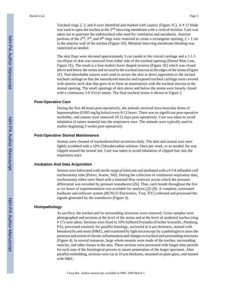
Tracheal rings 2, 3, and 4 were identified and marked with cautery (Figure 1C). A # 11 bladewas used to open the trachea at the 2nd intra-ring membrane with a vertical incision. Care wastaken not to puncture the endotracheal tube used for ventilation and anesthesia. Anteriorportions of the 2nd, 3rd, and 4th rings were removed to create a rectangular opening, 2 × 1 cmin the anterior wall of the trachea (Figure 1D). Minimal intra-ring membrane bleeding wascauterized as needed.
The skin flaps were elevated approximately 3 cm caudal to the cricoid cartilage and a 1-1.5cm ellipse of skin was removed from either side of the tracheal opening (Dotted Blue Line,Figure 1E). The result is a four-leafed clover shaped incision (Figure 1E) which was closedabove and below the stoma and secured to the tracheal mucosa at the edges of the stoma (Figure1F). Non-absorbable sutures were used to secure the skin in direct apposition to the incisedtracheal cartilage so that the sternohyoid muscles and exposed tracheal cartilage were coveredwith anterior neck skin that grew-in to form an anastomosis with the tracheal mucosa at thestomal opening. The small openings of skin above and below the stoma were loosely closedwith a continuous 3-0 Vicryl suture. The final tracheal stoma is shown in Figure 2.
Post-Operative CareDuring the first 48 hours post-operatively, the animals received intra-muscular doses ofbuprenorphine (0.005 mg/kg bolus) every 8-12 hours. There was no significant post-operativemorbidity, and sutures were removed 10-12 days post-operatively. Care was taken to avoidinhalation of suture material into the respiratory tract. The animals were typically used instudies beginning 3 weeks post-operatively.
Post-Operative Stomal MaintenanceStomas were cleaned of tracheobronchial secretions daily. The skin and stomal area werelightly scrubbed with a 10% Chlorahexadine solution. Once per week, or as needed, fur wasclipped around the stomal site. Care was taken to avoid inhalation of clipped hair into therespiratory tract.
Intubation And Data AcquisitionStomas were lubricated with sterile surgical lubricant and intubated with a # 5-8 inflatable cufftracheostomy tube (Portex, Keene, NH). During the collection of continuous respiratory data,tracheostomy tubes were fitted with a minimal flow restrictor across which the pressuredifferential was recorded by pressure transducers [26]. Thus, each breath throughout the fiveor six hours of experimentation was available for analysis [25-29]. A complete, automatedhardware and software system (BUXCO Electronics, Troy, NY) collected and processed thesignals generated by the transducers (Figure 3).
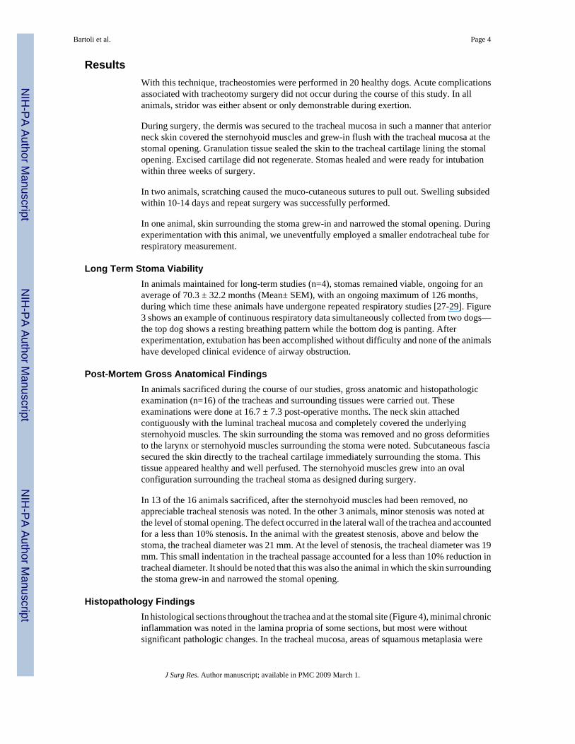
HistopathologyAt sacrifice, the trachea and its surrounding structures were removed. Gross samples werephotographed and sections at the level of the stoma and at the level of unaltered trachea (ring# 17) were taken. Sections were fixed in 10% buffered Formalin (Fischer Scientific, Pittsburg,PA), processed routinely for paraffin histology, sectioned at 4 μm thickness, stained withhematoxylin and eosin (H&E), and examined by light microscopy by a pathologist to asses thepresence and extent of chronic inflammation and changes to tracheal and surrounding structures(Figure 4). In several instances, large whole-mounts were made of the trachea, surroundingmuscles, and other tissues in the area. These sections were processed with longer time periodsfor each step of the histological process to assure penetration of the larger specimen. Afterparaffin embedding, sections were cut at 10 μm thickness, mounted on plate glass, and stainedwith H&E.
Bartoli et al. Page 3
J Surg Res. Author manuscript; available in PMC 2009 March 1.
NIH
-PA Author Manuscript
NIH
-PA Author Manuscript
NIH
-PA Author Manuscript
ResultsWith this technique, tracheostomies were performed in 20 healthy dogs. Acute complicationsassociated with tracheotomy surgery did not occur during the course of this study. In allanimals, stridor was either absent or only demonstrable during exertion.
During surgery, the dermis was secured to the tracheal mucosa in such a manner that anteriorneck skin covered the sternohyoid muscles and grew-in flush with the tracheal mucosa at thestomal opening. Granulation tissue sealed the skin to the tracheal cartilage lining the stomalopening. Excised cartilage did not regenerate. Stomas healed and were ready for intubationwithin three weeks of surgery.
In two animals, scratching caused the muco-cutaneous sutures to pull out. Swelling subsidedwithin 10-14 days and repeat surgery was successfully performed.
In one animal, skin surrounding the stoma grew-in and narrowed the stomal opening. Duringexperimentation with this animal, we uneventfully employed a smaller endotracheal tube forrespiratory measurement.
Long Term Stoma ViabilityIn animals maintained for long-term studies (n=4), stomas remained viable, ongoing for anaverage of 70.3 ± 32.2 months (Mean± SEM), with an ongoing maximum of 126 months,during which time these animals have undergone repeated respiratory studies [27-29]. Figure3 shows an example of continuous respiratory data simultaneously collected from two dogs—the top dog shows a resting breathing pattern while the bottom dog is panting. Afterexperimentation, extubation has been accomplished without difficulty and none of the animalshave developed clinical evidence of airway obstruction.
Post-Mortem Gross Anatomical FindingsIn animals sacrificed during the course of our studies, gross anatomic and histopathologicexamination (n=16) of the tracheas and surrounding tissues were carried out. Theseexaminations were done at 16.7 ± 7.3 post-operative months. The neck skin attachedcontiguously with the luminal tracheal mucosa and completely covered the underlyingsternohyoid muscles. The skin surrounding the stoma was removed and no gross deformitiesto the larynx or sternohyoid muscles surrounding the stoma were noted. Subcutaneous fasciasecured the skin directly to the tracheal cartilage immediately surrounding the stoma. Thistissue appeared healthy and well perfused. The sternohyoid muscles grew into an ovalconfiguration surrounding the tracheal stoma as designed during surgery.
In 13 of the 16 animals sacrificed, after the sternohyoid muscles had been removed, noappreciable tracheal stenosis was noted. In the other 3 animals, minor stenosis was noted atthe level of stomal opening. The defect occurred in the lateral wall of the trachea and accountedfor a less than 10% stenosis. In the animal with the greatest stenosis, above and below thestoma, the tracheal diameter was 21 mm. At the level of stenosis, the tracheal diameter was 19mm. This small indentation in the tracheal passage accounted for a less than 10% reduction intracheal diameter. It should be noted that this was also the animal in which the skin surroundingthe stoma grew-in and narrowed the stomal opening.
Histopathology FindingsIn histological sections throughout the trachea and at the stomal site (Figure 4), minimal chronicinflammation was noted in the lamina propria of some sections, but most were withoutsignificant pathologic changes. In the tracheal mucosa, areas of squamous metaplasia were
Bartoli et al. Page 4
J Surg Res. Author manuscript; available in PMC 2009 March 1.
NIH
-PA Author Manuscript
NIH
-PA Author Manuscript
NIH
-PA Author Manuscript

noted near the stoma and at the cuff site, but these were mild as illustrated below. No otherpathologic changes were found. In Figure 4A,B, the typical gross appearance of the cross-section at the stoma is illustrated. Figure 4A is a gross section photograph of the cross-sectionincluding surrounding muscles after fixation. Figure 4B is a whole-mount section of the tracheawith adjacent structures. Boxes illustrate the areas of sections shown at higher magnificationin 4C-F. In Figure 4C, the junction of the skin and the tracheal mucosa illustrate the transitionfrom squamous to respiratory mucosa. Figure 4D-F illustrate the respiratory mucosa at highermagnification and show minimal chronic inflammation. Figures 4D, F show that the mucosahas squamous metaplasia and no chronic inflammation. Figure 4E shows respiratory mucosawithout metaplasia and minimal chronic inflammation at the lamina propria.
DiscussionStomal Complications
Acute and chronic complications associated with tracheostomy did not occur during this study.Complications of tracheostomy have received extensive review in the medical literature, andtracheal stenosis within 6 weeks of tracheotomy surgery [7] has been especially emphasized[2-4,20,21,23]. Two factors can be considered: the endotracheal tube cuff pressure and thetracheal incision. Cuff pressure is an important etiologic factor in tracheal stenosis secondaryto intubation with cuffed inflatable tracheostomy tubes. Studies have shown a progressiveinflammatory response at the areas of maximum irritation from the tracheostomy tube and/orinflatable cuff [3,4,23]. Strictures developing at the level of the cuff are the result ofcircumferential mucosal ulceration of tracheal-mucosal membrane, which result from localischemic change due to pressure exerted between the outside of the cuff and the tracheal wall[3,4]. The mucosa at these sites becomes hypoxic and ulcerates to expose the cartilaginousrings [3,4]. Destruction and absorption of the exposed underlying cartilage and subsequenthealing by concentric fibrotic contracture narrow the tracheal passage [23]. As cited in previousliterature [3,23], to avoid complications of stenosis, the use of high compliance-low pressurecuffs is recommended. These were used in our study. Furthermore, in previous studies withserious complications, the endotracheal tubes did remain in the stomas permanently [3,4,13,14]. During our studies, intubations were performed for a maximum of six hours per day, twiceper week. Likely, our choice of cuff and intermittent rather than chronic intubation preventedthe development of more severe inflammation and subsequent major tracheal stenosis.
In previous studies, the tracheal incision has received much scrutiny and has been a technicalpoint of contention. Several types of tracheostomy incisions in dogs, including transverse andvertical, U-shaped flap through two tracheal rings, and excision of 1 cm segments of 1 and 2rings have been compared [5,6,8,16,17,22]. Vertical tracheostomy shows the most consistenthealing [5,6]. However, vertical incisions have been associated with higher incidence ofstenosis [22]. In these studies, a stoma was fashioned around a single vertical slit made throughthe intra-ring membrane and held open with sutures fixed to the surrounding muscle. It ispossible that this approach causes undue pressure-strain to the tracheal wall resulting in moresubstantial stenosis. As we have demonstrated, a better approach may be a vertical incisionextended down across tracheal rings and the removal of a 2 × 1 cm portion of multiple rings.This approach minimizes pressure-strain to the tracheal wall while ensuring a sufficiently largestoma for intubation.
Airway MaintenanceDuring surgery, the dermis was secured to the tracheal mucosa in such a manner that skincovered the sternohyoid muscles and grew in flush with the tracheal mucosa at the stomalopening. Barking, coughing, and use of the tongue for thermoregulation were not affected.Contrary to previous reports [18,19], animals did not require humidification of the vivarium
Bartoli et al. Page 5
J Surg Res. Author manuscript; available in PMC 2009 March 1.
NIH
-PA Author Manuscript
NIH
-PA Author Manuscript
NIH
-PA Author Manuscript

ambient air. To intubate the animal, no local anesthetic was required—only a lubricant.Animals displayed little reaction to intubation and in most cases resting measurements couldbe taken within 5 minutes. After training, dogs would lay unrestrained in a cubicle for repeated5 or 6 hour respiratory measurement during aerosol administration. Results of respiratory andsystemic effects of air pollution (which include 4 of the dogs reported in this paper) have beenpublished [27-29].
Viscous, mucoid tracheo-bronchial secretions were noted lining the outside of the stoma,especially after prolonged intubation. These secretions did not cause complications such aspulmonary obstruction. On occasion, wood shaving bedding became stuck to thetracheobronchial secretions surrounding the stomas and posed risk of inhalation and pulmonaryobstruction. These observations necessitate daily cleaning of the stomas to preventcomplications.
In groups of dogs from previous studies from our laboratory [28,29], rarely, a few dogs, whenexcited, would occlude the stomal opening with the posterior membranous portion of thetrachea causing apoplexy which immediately relieved the obstruction.
Respiratory MeasurementIn the past, respiratory measurement in the conscious dog has been made with a face mask,head tent, or tracheostomy intubation [1]. The former two have been sources of dead space andgas leakage. Additionally, they tended to excite the animals and cause hyperventilation.Permanent tracheostomy cannulation was introduced as an alternative, however, this high-maintenance technique became associated with pressure necrosis, tracheal stenosis, and a highmortality rate [13,14]. As we have shown, cannula-free tracheostomy in conjunction withintermittent cuffed endotracheal tube intubation during long-term experimentation is a usefulalternative which prevents major complications associated with tracheostomy intubation.
The presence of a tracheostomy has been shown to have no effect on airway mechanics [25].Our finding of lack of pathological changes in the trachea and only minimal chronicinflammation at the stomal site is further evidence that tracheostomy as used in our studies isunlikely to alter lung physiology.
ConclusionsDuring long-term respiratory studies, this practical and dependable tracheal stoma provides ameans for examining acute and long-term effects of environmental and pathophysiologicalinfluences on the respiratory systems of conscious dogs. With minimal post-operative andmaintenance-related complications, individual subjects could be used for chronicexperimentation for an ongoing period of 10 years.
AcknowledgmentsThe authors acknowledge and thank Dr. Gregory Wellenius, Sandra Verrier, Tracy Katz, Lani Lee, Jeffrey Pettit, andthe Harvard veterinary staff for their support and assistance during the instrumentation and maintenance of theseanimals. We also thank Dr. Cheryl Killigsworth who made substantial contributions to the use of tracheostomy anddevelopment of the surgical technique used in our laboratory.
This study was supported in part by grants NIEHS R01 ES12972 and ES00002 from the National Institute of Healthand through grant RD 83191701 from the USEPA.
Works Cited1. Foss ML, Barnard RJ, Tipton CM. Use of tracheostomized dogs in chronic metabolic assessments of
treadmill work. Lab Anim Sci Jun;1972 22(3):397–401. [PubMed: 4338706]
Bartoli et al. Page 6
J Surg Res. Author manuscript; available in PMC 2009 March 1.
NIH
-PA Author Manuscript
NIH
-PA Author Manuscript
NIH
-PA Author Manuscript

2. Murphy DA, MacLean LD, Dobell AR. Tracheal stenosis as a complication of tracheostomy. AnnThorac Surg Jan;1966 2(1):44–51. [PubMed: 5924802]
3. Ching NP, Ayres SM, Paegle RP, Linden JM, Nealon TF Jr. The contribution of cuff volume andpressure in tracheostomy tube damage. J Thorac Cardiovasc Surg Sep;1971 62(3):402–10. [PubMed:5126681]
4. Shelly WM, Dawson RB, May IA. Cuffed tubes as a cause of tracheal stenosis. J Thorac CardiovascSurg May;1969 57(5):623–7. [PubMed: 5787886]
5. Mendez-Picon G, Ehrlich FE, Salzberg AM. The effect of tracheostomy incisions on tracheal growth.J Pediatr Surg Oct;1976 11(5):681–5. [PubMed: 993937]
6. Smith MM, Saunders GK, Leib MS, Simmons EJ. Evaluation of horizontal and vertical tracheotomyhealing after short-duration tracheostomy in dogs. J Oral Maxillofac Surg Mar;1995 53(3):289–94.[PubMed: 7861280]
7. Bryant LR, Mujia D, Greenberg S, Huey JM, Schechter FG, Albert HM. Evaluation of tracheal incisionsfor tracheostomy. Am J Surg May;1978 135(5):675–9. [PubMed: 646041]
8. Bardin J, Boyd AD, Hirose H, Engelman RM. Tracheal healing following tracheostomy. Surg Forum1974;25(0):210–2. [PubMed: 4439168]
9. Cooper JD. Trachea-innominate artery fistula: successful management of 3 consecutive patients. AnnThorac Surg Nov;1977 24(5):439–47. [PubMed: 335995]
10. MacPhail CM, Monnet E. Outcome of and postoperative complications in dogs undergoing surgicaltreatment of laryngeal paralysis: 140 cases (1985-1998). J Am Vet Med Assoc Jun 15;2001 218(12):1949–56. [PubMed: 11417740]
11. Milo A, Eliachar I, Lane CJ, Myles JL, Munoz-Ramirez H. Clinical and histologic evaluation of anindwelling, inflatable, long-term laryngeal stent in the canine model. Otolaryngol Head Neck SurgSep;1999 121(3):195–202. [PubMed: 10471857]
12. Eliachar I, Levine SC, Tucker HM. A modified technique for tubeless tracheostomy. OtolaryngolHead Neck Surg Jun;1986 94(5):548–52. [PubMed: 3088514]
13. Ritter JW. Permanent tracheostomy for expired air collection in conscious dogs. Lab Anim Sci Feb;1984 34(1):79–81. [PubMed: 6716964]
14. Miles WK. Tubeless canine tracheostomy for laryngeal experimentation. Arch Otolaryngol Aug;197092(2):124–7. [PubMed: 5428305]
15. Dalgard DW, Marshall PM, Fitzgerald GH, Rendon F. Surgical technique for a permanenttracheostomy in Beagle dogs. Lab Anim Sci Jun;1979 29(3):367–70. [PubMed: 502464]
16. Lulenski GC, Batsakis JG. Management of the flap tracheostomy. An experimental study. ArchOtolaryngol May;1979 105(5):260–3. [PubMed: 435148]
17. Eliachar I, Levine SC, Tucker HM. A modified technique for tubeless tracheostomy. OtolaryngolHead Neck Surg Jun;1986 94(5):548–52. [PubMed: 3088514]
18. Leverment JN, Rae S. Cuffed tube tracheostomy in the dog. Lab Anim Oct;1978 12(4):203–6.[PubMed: 732261]
19. Tsuda T, Noguchi H, Takumi Y, Aochi O. Optimum humidification of air administered to atracheostomy in dogs. Scanning electron microscopy and surfactant studies. Br J Anaesth Oct;197749(10):965–77. [PubMed: 579156]
20. Miller DR, Sethi G. Tracheal stenosis following prolonged cuffed intubation: cause and prevention.Ann Surg Feb;1970 171(2):283–93. [PubMed: 5413464]
21. Goldberg M, Pearson FG. Pathogenesis of tracheal stenosis following tracheostomy with a cuffedtube. An experimental study in dogs. Thorax Nov;1972 27(6):678–91. [PubMed: 4647628]
22. Lulenski GC, Batsakis JG. Tracheal incision as a contributing factor to tracheal stenosis. Anexperimental study. Ann Otol Rhinol Laryngol Nov-Dec;1975 84(6):781–6. [PubMed: 1200567]
23. Andrews MJ. The incidence and pathogenesis of tracheal injury following tracheostomy with cuffedtube and assisted ventilation. Analysis of a 3-year prospective study. Br J Surg Oct;1971 58(10):749–55. [PubMed: 5097951]
24. Stiles PJ. Tracheal lesions after tracheostomy. Thorax Nov;1965 20(6):517–22. [PubMed: 5862749]25. Drazen JM, O’Cain CF, Ingram RH. Experimental induction of chronic bronchitis in dogs. Effects
on airway obstruction and responsiveness. Am Rev Respir Dis 1982;126:75–79. [PubMed: 6953777]
Bartoli et al. Page 7
J Surg Res. Author manuscript; available in PMC 2009 March 1.
NIH
-PA Author Manuscript
NIH
-PA Author Manuscript
NIH
-PA Author Manuscript
26. Gazula, GK.; Godleski, JJ. Flow restrictor for measuring respiratory parameters. US patent. #6224.560B1. 2001.
27. Godleski, JJ.; Verrier, RL.; Koutrakis, P.; Catalano, P.; Coull, B.; Reinisch, U.; Lovett, EG.;Lawrence, J.; Murthy, GG.; Wolfson, JM.; Clarke, RW.; Nearing, BD.; Killingsworth, C. peerdiscussion. Research Reports of the Health Effects Institute; 2000. Mechanisms of morbidity andmortality from exposure to ambient air particles; p. 5-88.p. 89-103.Report number 91
28. Clarke RW, Coull B, Reinisch U, Catalano P, Killingsworth CR, Koutrakis P, Kavouras I, KrishnaMurthy GG, Lawrence J, Lovett EG, Wolfson JM, Verrier RL, Godleski JJ. Inhaled concentratedambient particles are associated with hematological and broncho-alveolar lavage changes in canines.Env Health Perspectives 2000;108:1179–1187.
29. Wellenius GA, Coull BA, Godleski JJ, Koutrakis P, Okabe K, Savage ST, Lawrence JE, KrishnaMurthy GG, Verrier RL. Inhalation of Concentrated Ambient Air Particles Exacerbates MyocardialIschemia in Conscious Dogs. Env Health Perspectives 2003;111:402–408.
Bartoli et al. Page 8
J Surg Res. Author manuscript; available in PMC 2009 March 1.
NIH
-PA Author Manuscript
NIH
-PA Author Manuscript
NIH
-PA Author Manuscript

Figure 1.A: A 6 cm incision is made parallel to the trachea on the anterior neck. B: The trachea isexposed and the sternohyoid muscles are sutured to the lateral tracheal wall. Blue X’s markthe location of sutures. C: Tracheal rings 2,3,4 are identified and marked with electro-cautery.D: Rings 2,3,4 are removed creating a rectangular stoma, 2 × 1 cm in the anterior wall of thetrachea. E: A four-leafed clover shaped incision was formed by removing a 2 cm × 1 cmsemicircle of skin from either side of the tracheal opening (Dotted Blue Line). F: The edgesof the four-leafed clover shaped incision approximate to the edges of the stomal opening.
Bartoli et al. Page 9
J Surg Res. Author manuscript; available in PMC 2009 March 1.
NIH
-PA Author Manuscript
NIH
-PA Author Manuscript
NIH
-PA Author Manuscript

Figure 2.Final surgical result. Fourteen 3-0 Prolene stitches were used to suture the anterior neck skin(four-leafed clover incision) to the edges of exposed tracheal cartilage.
Bartoli et al. Page 10
J Surg Res. Author manuscript; available in PMC 2009 March 1.
NIH
-PA Author Manuscript
NIH
-PA Author Manuscript
NIH
-PA Author Manuscript
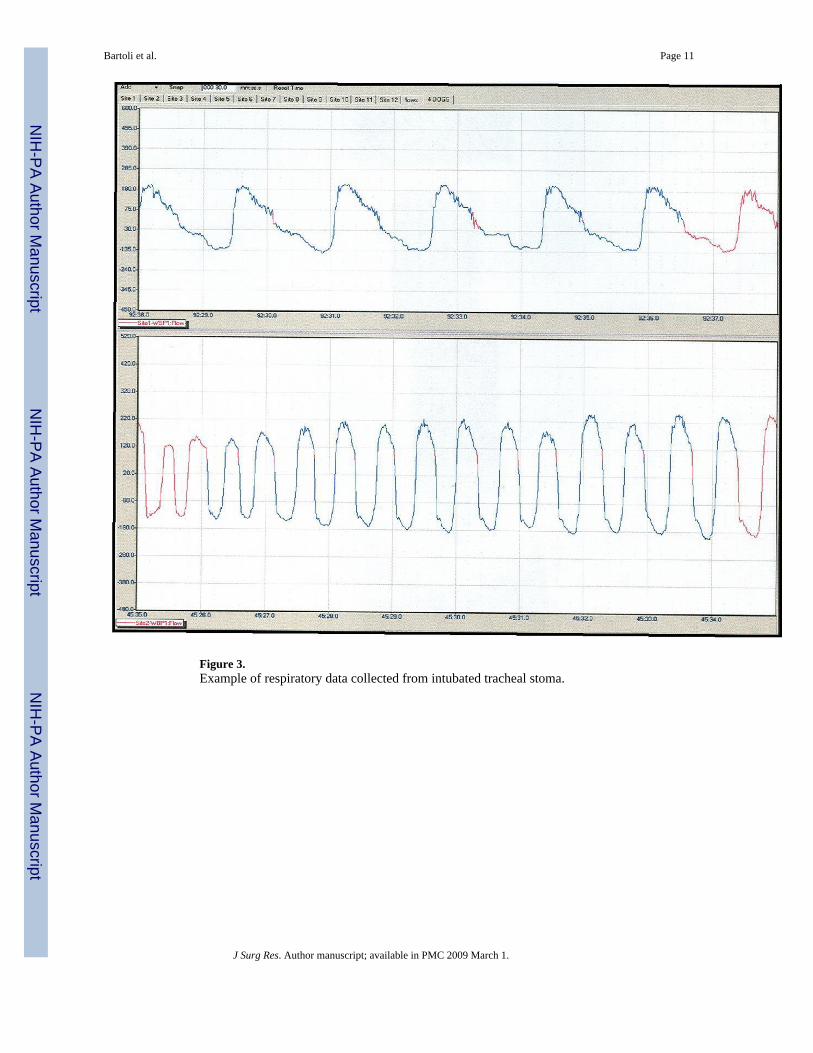
Figure 3.Example of respiratory data collected from intubated tracheal stoma.
Bartoli et al. Page 11
J Surg Res. Author manuscript; available in PMC 2009 March 1.
NIH
-PA Author Manuscript
NIH
-PA Author Manuscript
NIH
-PA Author Manuscript

Figure 4.Histopathology. A,B: The typical gross appearance of a cross-section at the stoma. A: Thecross-section with fixation. B: A whole-mount large histological section showing the trachea,stoma, cutaneous mucosal junction, and the lateral muscle-trachea approximation. The boxeshighlight areas illustrated at higher magnifications in the photomicrographs shown in C-F.C: The cutaneous-mucosal junction showing the transition from stratified squamousepithelium to respiratory mucosa. There is minimal chronic inflammation in both thesubcutaneous tissue and the lamina propria of the trachea. Magnification 40X. D: The latertracheal wall. The respiratory mucosa has slight squamous metaplasia, but the lamina propriabetween the mucosa and cartilage has no pathological changes. Magnification 200X. E: Theposterior tracheal wall opposite the stoma. Slight chronic inflammation is present in the laminapropria. This was the most severe chronic inflammation seen in any section of the trachea. Therespiratory mucosa has no pathological changes. Magnification 200X. F: Lateral tracheal wallwith squamous metaplasia, no chronic inflammation, and normal tracheal mucosal glands.There was neither an increase in tracheal mucosal gland thickness in the lamina propria, nor apathological shift in the normal serous/mucous gland proportions. Magnification 200X.
Bartoli et al. Page 12
J Surg Res. Author manuscript; available in PMC 2009 March 1.
NIH
-PA Author Manuscript
NIH
-PA Author Manuscript
NIH
-PA Author Manuscript