Performance evaluation of sensing fabrics for monitoring physiological and biomechanical variables
8
IEEE TRANSACTIONS ON INFORMATION TECHNOLOGY IN BIOMEDICINE, VOL. 9, NO. 3, SEPTEMBER 2005 345 Performance Evaluation of Sensing Fabrics for Monitoring Physiological and Biomechanical Variables Enzo Pasquale Scilingo, Angelo Gemignani, Rita Paradiso, Nicola Taccini, Brunello Ghelarducci, and Danilo De Rossi Abstract—In the last few years, the smart textile area has become increasingly widespread, leading to developments in new wearable sensing systems. Truly wearable instrumented garments capable of recording behavioral and vital signals are crucial for several fields of application. Here we report on results of a careful char- acterization of the performance of innovative fabric sensors and electrodes able to acquire vital biomechanical and physiological signals, respectively. The sensing function of the fabric sensors re- lies upon newly developed strain sensors, based on rubber-carbon- coated threads, and mainly depends on the weaving topology, and the composition and deposition process of the conducting rubber- carbon mixture. Fabric sensors are used to acquire the respitrace (RT) and movement sensors (MS). Sensing features of electrodes, instead rely upon metal-based conductive threads, which are in- strumental in detecting bioelectrical signals, such as electrocar- diogram (ECG) and electromyogram (EMG). Fabric sensors have been tested during some specific tasks of breathing and movement activity, and results have been compared with the responses of a commercial piezoelectric sensor and an electrogoniometer, respec- tively. The performance of fabric electrodes has been investigated and compared with standard clinical electrodes. Index Terms—Biomechanics, sensing fabric, vital signs. I. INTRODUCTION A N emerging concept of health care, based on continu- ously monitoring vital and behavioral signs to provide assistance to patients and health care users, is gaining wide consensus [1]–[4]. Advances in sensor technology, as well as in communication technology and treatment of data, have stimulated the creation of a new generation of health care mon- itoring and diagnostic systems [5]–[7]. The use of functional materials enables design and production of a new generation of garments with distributed sensors and electrodes [8]–[10], allowing the noninvasive monitoring of health status. Fibers and yarns with specialized electrophysical properties can be integrated and used as basic elements to fabricate woven or knitted functional textile systems [11], [12]. These integrated Manuscript received May 12, 2004; revised November 27, 2004. This work was supported in part by the EU Commission under Contract IST-2001-37778 Wealthy. E. P. Scilingo and D. De Rossi are with the Interdepartmental Re- search Center “E. Piaggio,” University of Pisa, Pisa 56126, Italy (e-mail: [email protected]; [email protected]). A. Gemignani and B. Ghelarducci are with the Department of Physiology and Biochemistry, Faculty of Medicine, University of Pisa 56126, Italy (e-mail: [email protected]; [email protected]). R. Paradiso and N. Taccini are with MILIOR S.p.A. and SMARTEX s.r.l., 59100 Prato, Italy (e-mail: [email protected]; [email protected]). Digital Object Identifier 10.1109/TITB.2005.854506 systems enable the detection of electrical [electrocardiogram (ECG), electromyogram (EMG)] and mechanical (respirogram, motor activity) physiological signals [13], [14]. Wearable comfortable systems aid the daily acquisition and processing of multiparametric health data, providing an early detection of pathological signs and improving the curative rate of disease without interfering with daily life [15], [16]. This paper is aimed at assessing the properties of fabrics able to acquire bio- electrical and biomechanical signals, in particular, the sensing properties of bioelectrical fabrics sensors, and the biomechan- ical performance related to the piezoresistive properties of fabric sensors. In order to confer special functions to fabric, the intrinsic properties of materials have been modified by means of dedicated treatments. Bioelectrical (ECG and EMG) and biomechanical (respirogram and movement activity) signals acquired through sensing fabrics are compared with signals acquired through standard electrodes and sensors commonly employed in clinical and experimental studies. The comparison had been done both in terms of engineering assessment and clinical validation, i.e., each signal has been quantitatively evaluated in the time and frequency domain to make sure that no medical and diagnostic information is lost. Moreover, it is worth emphasizing that although fabric sensors and electrodes should be thought to be integrated in a garment, the aim of this paper is to investigate the performance of the single fabric electrode and sensor, without envisioning their behavior in the context of a sensorized shirt. In order to validate the per- formance of fabric sensors and electrodes, their response in the time and frequency domains has been compared with the outputs of commercial devices. Concerning the ECG, surface EMG, and respirogram, the comparison has been performed by extracting the most significant parameters at rest conditions. Due to motionless conditions, the acquired signals are quite artifact-free, and the noise level is partially suppressed, not least because it is suitably filtered. Further studies are currently being performed in order to analyze recordings while subjects are moving. II. MATERIALS AND METHODS A. Fabric Electrodes Conductive fabric electrodes have been realized with com- mercial stainless steel threads twisted around a standard contin- uous viscose textile yarn. They were woven using the tubular intarsia technique to get a double face, where the external part 1089-7771/$20.00 © 2005 IEEE
Transcript of Performance evaluation of sensing fabrics for monitoring physiological and biomechanical variables

IEEE TRANSACTIONS ON INFORMATION TECHNOLOGY IN BIOMEDICINE, VOL. 9, NO. 3, SEPTEMBER 2005 345
Performance Evaluation of Sensing Fabricsfor Monitoring Physiological and
Biomechanical VariablesEnzo Pasquale Scilingo, Angelo Gemignani, Rita Paradiso, Nicola Taccini, Brunello Ghelarducci, and
Danilo De Rossi
Abstract—In the last few years, the smart textile area has becomeincreasingly widespread, leading to developments in new wearablesensing systems. Truly wearable instrumented garments capableof recording behavioral and vital signals are crucial for severalfields of application. Here we report on results of a careful char-acterization of the performance of innovative fabric sensors andelectrodes able to acquire vital biomechanical and physiologicalsignals, respectively. The sensing function of the fabric sensors re-lies upon newly developed strain sensors, based on rubber-carbon-coated threads, and mainly depends on the weaving topology, andthe composition and deposition process of the conducting rubber-carbon mixture. Fabric sensors are used to acquire the respitrace(RT) and movement sensors (MS). Sensing features of electrodes,instead rely upon metal-based conductive threads, which are in-strumental in detecting bioelectrical signals, such as electrocar-diogram (ECG) and electromyogram (EMG). Fabric sensors havebeen tested during some specific tasks of breathing and movementactivity, and results have been compared with the responses of acommercial piezoelectric sensor and an electrogoniometer, respec-tively. The performance of fabric electrodes has been investigatedand compared with standard clinical electrodes.
Index Terms—Biomechanics, sensing fabric, vital signs.
I. INTRODUCTION
AN emerging concept of health care, based on continu-ously monitoring vital and behavioral signs to provide
assistance to patients and health care users, is gaining wideconsensus [1]–[4]. Advances in sensor technology, as wellas in communication technology and treatment of data, havestimulated the creation of a new generation of health care mon-itoring and diagnostic systems [5]–[7]. The use of functionalmaterials enables design and production of a new generationof garments with distributed sensors and electrodes [8]–[10],allowing the noninvasive monitoring of health status. Fibersand yarns with specialized electrophysical properties can beintegrated and used as basic elements to fabricate woven orknitted functional textile systems [11], [12]. These integrated
Manuscript received May 12, 2004; revised November 27, 2004. This workwas supported in part by the EU Commission under Contract IST-2001-37778Wealthy.
E. P. Scilingo and D. De Rossi are with the Interdepartmental Re-search Center “E. Piaggio,” University of Pisa, Pisa 56126, Italy (e-mail:[email protected]; [email protected]).
A. Gemignani and B. Ghelarducci are with the Department of Physiologyand Biochemistry, Faculty of Medicine, University of Pisa 56126, Italy (e-mail:[email protected]; [email protected]).
R. Paradiso and N. Taccini are with MILIOR S.p.A. and SMARTEX s.r.l.,59100 Prato, Italy (e-mail: [email protected]; [email protected]).
Digital Object Identifier 10.1109/TITB.2005.854506
systems enable the detection of electrical [electrocardiogram(ECG), electromyogram (EMG)] and mechanical (respirogram,motor activity) physiological signals [13], [14]. Wearablecomfortable systems aid the daily acquisition and processingof multiparametric health data, providing an early detection ofpathological signs and improving the curative rate of diseasewithout interfering with daily life [15], [16]. This paper isaimed at assessing the properties of fabrics able to acquire bio-electrical and biomechanical signals, in particular, the sensingproperties of bioelectrical fabrics sensors, and the biomechan-ical performance related to the piezoresistive properties offabric sensors. In order to confer special functions to fabric, theintrinsic properties of materials have been modified by meansof dedicated treatments. Bioelectrical (ECG and EMG) andbiomechanical (respirogram and movement activity) signalsacquired through sensing fabrics are compared with signalsacquired through standard electrodes and sensors commonlyemployed in clinical and experimental studies. The comparisonhad been done both in terms of engineering assessment andclinical validation, i.e., each signal has been quantitativelyevaluated in the time and frequency domain to make sure thatno medical and diagnostic information is lost. Moreover, it isworth emphasizing that although fabric sensors and electrodesshould be thought to be integrated in a garment, the aim ofthis paper is to investigate the performance of the single fabricelectrode and sensor, without envisioning their behavior inthe context of a sensorized shirt. In order to validate the per-formance of fabric sensors and electrodes, their response inthe time and frequency domains has been compared with theoutputs of commercial devices. Concerning the ECG, surfaceEMG, and respirogram, the comparison has been performed byextracting the most significant parameters at rest conditions.Due to motionless conditions, the acquired signals are quiteartifact-free, and the noise level is partially suppressed, notleast because it is suitably filtered. Further studies are currentlybeing performed in order to analyze recordings while subjectsare moving.
II. MATERIALS AND METHODS
A. Fabric Electrodes
Conductive fabric electrodes have been realized with com-mercial stainless steel threads twisted around a standard contin-uous viscose textile yarn. They were woven using the tubularintarsia technique to get a double face, where the external part
1089-7771/$20.00 © 2005 IEEE

346 IEEE TRANSACTIONS ON INFORMATION TECHNOLOGY IN BIOMEDICINE, VOL. 9, NO. 3, SEPTEMBER 2005
Fig. 1. Enlarged view of the structure of a fabric electrode for ECG and EMGrecordings integrated into a sensorized shirt. The darker area in the pattern isyarned with conductive threads, while the surrounding lighter region is realizedwith the nonconductive basal yarn acting as insulator.
Fig. 2. Hydrogel membrane used to improve the skin-electrode coupling. Itcovers the conductive area coming into contact with the skin.
is realized with the basal yarn, not sensitive, to isolate the elec-trode from the external environment (see Fig. 1). To improvethe quality of the electrical signal in dynamic conditions, a hy-drogel membrane, obtained from ST&D Ltd (Belfast, U.K.), hasbeen used (see Fig. 2). The membrane reduces the contact resis-tance between the skin and the electrode, as well as improvingcomfort. In addition to the presence of a bactericide/fungicide,the hydrogel membrane has been chosen with a pH range be-tween 3.5 and 9, hence causing minimum skin irritation. Thereal challenge would be the removal of the membrane, allowinga long-term electrode entirely made of fabric to be available,but, in this case, the crucial issue of the electrical coupling be-tween electrode and skin should be suitably addressed. Conduc-tive yarns and coated fabrics are resistant to repeated washingin aqueous solutions, without decreasing their performance.
B. Acquisition of Bioelectrical Signals Through Electrodes
The ECG and EMG electrodes based on conductive fabrichave been tested in comparison with standard ECG clinical elec-trodes (RedDot by 3M) and standard surface EMG Ag/AgClelectrodes, respectively. Preliminarily, the electrical impedancein the transversal direction of fabric electrodes has been mea-sured across two plates, having the same surface as the elec-trodes, and covered by adhesive copper, pressed on both elec-trode’s faces.
Since both ECG and EMG are low-intensity signals, theyhave been suitably amplified and filtered by means of a GRASS-TELEFACTOR model 15LT device, equipped with differentialamplifiers, model 15A54 and gain 1000. Next, signals were ac-quired by a National Instruments acquisition card PC-6036E.
Fig. 3. ECG leads located according standard configuration of Einthoven.LA and RA: left and right arm, respectively. LL and RL: left and right leg,respectively. L1, L2, and L3: bipolar Einthoven leads.
1) Electrocardiogram (ECG): The electrodes (Red Dot andfabric) were positioned according to standard Einthoven con-figuration, with two electrodes placed onto each root arm andone electrode onto left root leg. The bipolar lead L1, which isthe upper side of the conventional Einthoven’s triangle config-uration, and measures the potential difference between the twoarms, has been selected for our experiments. The right-leg elec-trode (RL) has been also used as reference (see Fig. 3). Be-sides being largely used for clinical investigation, this lead isalso quite immune to artifacts due to small movements of thetorso. The GRASS-TELEFACTOR was set with the bandpassfilter between 1 and 100 Hz, notch filter at 50 Hz. Moreover, theNational Instrument acquisition card was set with a samplingrate of 1000 Hz. The filter settings were chosen in order to pre-serve the most significant frequency content of the ECG signalwithin the bandwidth usually analyzed in diagnostic and mon-itoring activity, according to the American Heart Association(AHA) recommendations (0.1–100 Hz) [17]. The high samplingfrequency allows postacquisition digital elaboration and noisefiltering.
The evaluation of performance of fabric electrodes withrespect to standard clinical RedDot electrodes has been done,comparing the most significant time parameters extracted fromthe morphology waveform (P-waves, QRS complexes, STsegments, and T waves), and performing a spectral analysis[fast Fourier transform (FFT)] on signals coming from bothelectrodes. In addition, the magnitude squared coherencefunction, estimated by using the toolbox COHERE (MatlabProgram), was calculated on ECG signals acquired with bothfabric and Red Dot electrodes. The coherence is expressed bythe ratio between the power spectra of two signals and the crossspectrum of same signals
(1)
Since fabric electrodes are made of standard textile filamentstwisted together with thin stainless-steel threads, we tackled thecrucial issue of the polarization phenomena. In the literature, it
SCILINGO et al.: PERFORMANCE EVALUATION OF SENSING FABRICS 347
Fig. 4. Strain fabric sensor, basically a textile band wrapping around the chest,used for monitoring both respiration and movement signals.
is reported that stainless-steel electrodes are far more polariz-able than Ag-AgCl electrodes, and can be reliably used only forprocedures lasting 3–10 min [18]. In order to assess the repeata-bility and robustness of signals over time, we carried out ECGacquisitions from fabric electrodes during rest conditions, overperiods of 30 min per day for a week. Each daily acquisition of30 min has been split in epochs of 50 s, on each of which, weimplemented the power spectral density (PSD). In this way, weobtained 36 PSD curves per day. The Welch method has beenapplied on ECG values corresponding to each epoch, choosing512 samples and an overlap rate of 50%.
2) Electromyogram (EMG): Surface electromyography wasrecorded from the biceps brachii during isometric dynamic con-tractions. Before placing the electrodes, the skin was preparedaccording to standard procedures. After shaving, the skin wascleansed with alcohol and slightly abraded with abrasive paste.The GRASS-TELEFACTOR was set with the bandpass filterbetween 10 and 500 Hz [19] and notch filter at 50 Hz. The Na-tional Instruments acquisition card was set with a sampling rateof 2000 Hz. Though we were aware that an oversampling pro-duced no significant benefits in detecting timing and amplitudemeasurements of the electromyographic signal [20], we chose,however, twice the Nyquist rate, in order to further digitally filterthe acquired data. Surface EMG signals were detected with twoelectrodes, with 20 mm interelectrode distance, in differentialconfiguration with a ground reference electrode placed at theleft wrist. The experimental paradigm consisted of alternatingresting and muscular contraction periods.
C. Piezoresistive Sensors
A coating process has been applied on jersey fabric, art.Spighetta/5575, composed of 86% polyester and 14% lycra(Milior SpA, Italy). The coating layer is realized with a solutionof rubber containing micro disperse phases of carbon, and it isused to realize efficient strain-gauge sensors.
D. Acquisition of Biomechanical Signals Through Sensors
Strain fabric sensors have been used to detect both respira-tion and motor activity signals (see Fig. 4). In order to eval-uate the performance of fabric sensors in monitoring respiration,a comparison with the signal obtained by a dc-coupled stan-dard polymeric piezoelectric transducer has been done. Simi-larly, the performance of fabric piezoresistive sensors in mea-suring motor activity has been evaluated by comparing theirsignals with those simultaneously acquired by commercial elec-trogoniometers. Respiration signal was acquired by a NationalInstruments (PC-6036E) acquisition card with a sampling rate
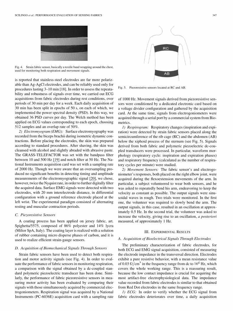
Fig. 5. Piezoresistive sensors located at RC and AB.
of 1000 Hz. Movement signals derived from piezoresistive sen-sors were conditioned by a dedicated electronic card based ona voltage divider configuration and gathered by the acquisitioncard. At the same time, signals from electrogoniometers wereacquired through a serial port by a commercial system from Bio-metrics.
1) Respirogram: Respiratory changes (inspiration and expi-ration) were detected by strain fabric sensors placed along thesemicircumference of the rib cage (RC) and the abdomen (AB)below the xiphoid process of the sternum (see Fig. 5). Signalsderived from both fabric and polymeric piezoelectric dc-cou-pled transducers were processed. In particular, waveform mor-phology (respiratory cycle: inspiration and expiration phases)and respiratory frequency (calculated as the number of respira-tory cycles per minute) were studied.
2) Movement Sensors: The fabric sensor’s and electrogo-niometer’s responses, both placed on the right elbow joint, wereacquired during the flexoextension mevements of the arm. Inparticular, a subject volunteered to wear both sensors, and hewas asked to repeatedly bend his arm, endeavoring to keep thevelocity as constant as possible. The output signals were sinu-soidal waves in rough. Two trials were monitored. In the firstone, the volunteer was required to slowly bend the arm. Theoutput signals, in this case, resulted in an oscillation at approx-imately 0.5 Hz. In the second trial, the volunteer was asked toincrease the velocity, giving rise to an oscillation, a posteriorimeasured, of approximately 1 Hz.
III. EXPERIMENTAL RESULTS
A. Acquisition of Bioelectrical Signals Through Electrodes
The preliminary characterization of fabric electrodes, forboth ECG and EMG signal acquisition, consisted of measuringthe electrode impedance in the transversal direction. Electrodesexhibit a pure resistive behavior, with a mean resistance valueof 0.03 cm in the frequency range from dc to Hz, whichcovers the whole working range. This is a reassuring result,because the low contact impedance is crucial for acquiring themost artifact-free electrophysiological data. The impedancevalue recorded from fabric electrodes is similar to that obtainedfrom Red Dot electrodes in the same frequency range.
1) ECG: In order to verify whether the ECG signal fromfabric electrodes deteriorates over time, a daily acquisition

348 IEEE TRANSACTIONS ON INFORMATION TECHNOLOGY IN BIOMEDICINE, VOL. 9, NO. 3, SEPTEMBER 2005
Fig. 6. 3-D PSD curve of ECG evaluated on the first day of the week ofacquisition over a period of 30 min, calculated every 50 s. The x axis reportsthe frequency, the y axis the time, and the z axis the PSD amplitudes. The grayscale is related to the amplitude values. On the xy plane, the isofrequency PSDvalues are reported.
Fig. 7. 3-D PSD curve of ECG evaluated on the last day of the week ofacquisition over a period of 30 min, calculated every 50 s. The x axis reportsthe frequency, the y axis the time, and the z axis the PSD amplitudes. The grayscale is related to the amplitude values. On the xy plane, the isofrequency PSDvalues are reported.
lasting 30 min was carried out for a week. In addition to avisual inspection of ECG waveforms, which, however, didnot show significant differences in the morphology, we alsocalculated and compared the PSD curves over a period of30 min, calculated every 50 s, thus obtaining 36 curves. Figs. 6and 7 show the three-dimensional (3-D) PSD curves of ECGversus frequency and time acquired on the first and last daysof the week. It is worth noting that morphology of the PSDcurves versus frequency does not significantly change withtime, implying that the fabric electrode response did not showany polarization effect. Afterwards, a comparative study interms of performance, in both time and frequency domains,between fabric and Red Dot electrodes was done. Time analysishas been performed by extracting from an ECG signal a wholecardiac cycle with diastolic and systolic phases. Already, at aglance, the waveform morphology of ECG signals derived fromboth fabric and standard clinical electrodes, depicted in Fig. 8,
TABLE ITEMPORAL PARAMETERS OF CARDIAC CYCLE
appears very similar. The visual similarity was quantitativelyassessed by comparing the most significant components seg-ments of both signals. Numerical values, reported in Table I,exhibit marginal differences, electrocardiographically irrele-vant. In order to further validate similarities between the twoelectrodes, a careful analysis in the frequency domain has beendone. Fig. 9 shows the comparison between the PSDs evalu-ated for both electrodes. The analysis has been carried out onECG signals acquired over 5-min sequences by using Welch’smethod. Even in this case, the two curves are nearly overlappedand the main frequency components are the same. Moreover,the magnitude squared coherence function calculated on ECGsignals acquired with fabric and Red Dot electrodes showed across-spectrum factor greater than 0.95.
2) EMG: In Fig. 10, the surface EMG signals obtainedduring the stimulation of biceps brachi, by simultaneouslyusing the fabric and the Ag-AgCl standard electrodes, areshown. Both EMG signals over time coming from fabric andstandard electrodes are very similar in terms of amplitude valuesand time intervals. A more detailed investigation, by comparingthe signals in the frequency domain, has been performed. InFig. 11, the PSD of both signals is reported. The depression at50 Hz is due to the notch filter used to reject interference fromthe electric supply. Usually, parameters of the power densityspectrum used to provide meaningful information for the EMGfrequency spectrum are the mean, peak, and median frequency.Table II reports these values for both Red Dot and fabricelectrodes of the PSD of the surface EMG during a contractionphase. Even in this case, numerical differences are irrelevant.
B. Acquisition of Biomechanical Signals Through Sensors
1) Movement Sensors: The signals acquired from the fabricsensor and electrogoniometer placed on the right elbow jointwere acquired with time, and reported in Figs. 12 and 13,during slow and fast movements, respectively. Signals comingfrom electrogoniometers are reported in arbitrary units, whilethe response of the piezoresistive fabric sensors was measuredin Ohms. It is worth pointing out that our aim was to verifywhether the fabric sensor was able to measure the motor ac-tivity as the number of movements per unit of time. Therefore,for this purpose, only the frequency behavior was relevant.Figs. 14 and 15 show the comparison of frequency componentsbetween fabric sensor and electrogoniometer, respectively, forslow and fast movements. In both cases, the principal frequencycomponents are coincident.
2) Respitrace: Respiratory activity produces volumechanges of the RC and AB during the inspiratory (volume

SCILINGO et al.: PERFORMANCE EVALUATION OF SENSING FABRICS 349
Fig. 8. ECG L1 in rest conditions. P wave, QRS complex and T wave of a cardiac beat is shown for Red Dot (upper) and for fabric electrodes (lower).
Fig. 9. PSD curve of ECGs. Grey curve refers to ECG coming from Red Dotelectrodes, while darker curve is related to ECG coming from fabric electrodes.
increase) and expiratory (volume decrease) phases of the res-piratory cycle. To evaluate the behavior of the fabric sensor, acomparison between the response of piezoresistive fabric andcommonly used piezoelectric sensors has been done, and theresults are shown in Fig. 16. Fig. 17 shows the respiratory ratechanges during a period ranging from slow to fast breathing.
IV. DISCUSSION AND CONCLUSIONS
In this paper, we reported on a new technology of sensorsand electrodes. The innovative idea was to implement transduc-tion functions directly onto textile substrates, which can be in-tegrated in usual garments. Here, fabric sensors able to acquirebiomechanical and respiration signals and fabric electrodes toacquire ECG and surface EMG are proposed. In order to as-sess and validate the performance, signals coming from fabricsensors and electrodes have been compared in time and in the
Fig. 10. EMG of the left biceps brachi. Red Dot (upper) and Fabric electrode(lower).
Fig. 11. Normalized PSD of the EMG signal acquired by using Red Dot(lighter dashed curve) and standard (darker continuous curve) electrodes.

350 IEEE TRANSACTIONS ON INFORMATION TECHNOLOGY IN BIOMEDICINE, VOL. 9, NO. 3, SEPTEMBER 2005
TABLE IIFREQUENCY PARAMETERS OF PSD OF THE SURFACE EMG
DURING THE CONTRACTION PHASE
Fig. 12. Comparison between the signals from electrogoniometer(upper) and fabric sensor (lower) during a slow movement of the arm, atapproximately 0.5 Hz.
Fig. 13. Comparison between the signals from electrogoniometer (upper) andfabric sensor (lower) during a fast movement of the arm, at approximately 1 Hz.
frequency domain with standard sensors and clinical electrodes.Experimental results showed that fabric electrodes and sensorscan be adequately employed to acquire and monitor vital phys-iological and biomechanical signals, without loss of informa-tion. The qualitative similarity of the signals has been assessedby a quantitative analysis in the time and frequency domains ofthe signals claiming a complete overlap. Although these resultsare very encouraging, some specific issues should be more care-fully addressed when the isolated fabric sensors and electrodesare further developed into an integrated wearable garment. For
Fig. 14. Comparison between the spectral components of the movementsignal acquired simultaneously from electrogoniometer (lighter dashed curve)and fabric sensor (darker continuous curve) during a slow movement of thearm, at approximately 0.5 Hz.
Fig. 15. Comparison between the spectral components of the movementsignal acquired simultaneously from electrogoniometer (lighter dashed curve)and fabric sensor (darker continuous curve) during a fast movement of the arm,at approximately 1 Hz.
example, the reproducibility of the positioning of the sensitivespots in the garment represents a critical point. In particular, forECG, while the positioning of the standard leads (L1-L2-L3)at the root of the arms and at the left groin will not appreciablydiffer from the standard wrist and foot positioning, the recordingfrom precordial leads has to meet more strict topographical con-straints. Another problem is represented by the deterioration ofthe signal-to-noise ratio produced by micromovements of theelectrodes with respect to the skin. So far, this has been neu-tralized by the interposition of hydrogel membranes, but fur-ther efforts in modifying the structure of the yarn in the sensi-tive spot are necessary to stabilize the skin–electrode contact.Most probably, these problems explain why, so far, in the smartshirts used to monitor vital signals in human subjects, standardclinical electrodes and sensors have been simply inserted in the

SCILINGO et al.: PERFORMANCE EVALUATION OF SENSING FABRICS 351
Fig. 16. Respiration signal obtained from a standard piezoelectric sensor(upper) compared with the signal obtained from a fabric piezoresistive sensor(lower).
Fig. 17. Respiration rate obtained by computing the distance between theminimal local values for standard piezoelectric sensor (upper) and piezoresistivefabric sensor (lower).
fabric, thus forming a hybrid tool [16]. Our idea was to imple-ment transduction functions directly onto textile substrates em-ployed to construct a comfortable, easily wearable smart gar-ment. This solution appears suitable for several reasons: 1) thegarment can be personalized, and this will optimize the posi-tioning of the sensitive regions, thus making the monitoringmore accurate and reproducible; 2) such a garment will be moresimple and self-manageable; and 3) the absence of visible stan-dard electrodes and connecting wires may contribute to a bettercompliance of the subjects in accepting this form of monitoring.Finally, any effort in this direction will add a valuable techno-logical contribution to textile engineering, and to its practicalapplications, such as in telemedicine.
REFERENCES
[1] M. Pacelli, R. Paradiso, G. Anerdi, S. Ceccarini, M. Ghignoli, F. Lorussi,E. P. Scilingo, D. De Rossi, A. Gemignani, and B. Ghelarducci, “Sensingthreads and fabrics for monitoring body kinematic and vital signs,” inFibers and Textiles for the Future, M. Isosomppi and R. Salonen, Eds.,2001, pp. 55–63.
[2] R. Paradiso, “Wearable heath care system,” in Proc. 4th Int. IEEE EMBSSpecial Topic Conf. Inf. Technol. Applic. Biomed., Birmingham, U.K.,Apr. 2003, pp. 283–286.
[3] E. Kyriacou, S. Pavlopoulos, A. Berler, M. Neophytou, A. Bourka, A.Georgoulas, A. Anagnostaki, D. Karayiannis, C. Schizas, C. Pattichis,A. Andreou, and D. Koutsouris, “Multi-purpose healthcare telemedicinesystems with mobile communication link support,” Biomed. Eng. On-line, vol. 2, no. 1, p. 7, Mar. 24, 2003.
[4] K. Shimuzu, “Telemedicine by mobile communication,” IEEE Eng.Med. Biol., vol. 18, no. 4, pp. 32–44, Apr. 1999.
[5] F. Lorussi, A. Tognetti, M. Tesconi, P. Pastacaldi, and D. De Rossi,“Strain sensing fabric for hand posture and gesture monitoring,” in Proc.Int. Workshop New Generation Wearable Syst. Ehealth: Toward a Rev-olution of Citizens’ Health, Life Style Management?, Lucca, Italy, Dec.2003, [CD-ROM].
[6] J. P. Dan and J. Luprano, “Homecare: A telemedical application,” Med.Device Technol., vol. 14, no. 10, pp. 25–28, Dec. 2003.
[7] D. De Rossi, A. Della Santa, and A. Mazzoldi, “Dressware: Wearablehardware,” Mat. Sci. Eng. C, vol. 7, pp. 31–35, 1999.
[8] L. Van Langenhove, C. Hertleer, M. Catrysse, R. Puers, H. Van Egmond,and D. Matthijs, “Smart textiles,” in Wearable eHealth Systems forPersonalised Health Management, A. Lymberis and D. de Rossi,Eds. Amsterdam, The Netherlands: IOS Press, 2004, pp. 344–351.
[9] T. Kirstein, M. Lawrence, and G. Tröster, “Functional electrical stim-ulation (FES) with smart textile electrodes,” in Proc. Int. WorkshopNew Generation Wearable Syst. Ehealth: Toward a Revolution ofCitizens’ Health and Life Style Management?, Lucca, Italy, Dec. 2003,[CD-ROM].
[10] E. Bonderover and S. Wagner, “Transistor fibers for etextiles amorphoussilicon thin film transistors on kapton fibers,” in Proc. Int. InteractiveTextiles for Warrior Conf., Boston, MA, Jul. 2002, [CD-ROM].
[11] T. E. Campbell, B. J. Munro, J. R. Steele, and G. G. Wallace, “Con-ducting polymer fabric strain gauges: A unique biomechanical moni-toring device,” in Proc. Int. Soc. Biomech. XIXth Congr., P. Milburn, B.Wilson, and T. Yanai, Eds., Dunedin, New Zealand, Jul. 2003, p. 48.
[12] S. Wagner, E. Bonderover, W. B. Jordan, and J. C. Sturn, “Electrotex-tiles: Concepts and challenges,” Int. J. High Speed Electron. Syst., vol.12, no. 2, pp. 391–399, 2002.
[13] R. Paradiso, A. Gemignani, E. P. Scilingo, and D. De Rossi, “Knittedbio-clothes for cardiopulmonary monitoring,” in Proc. 25th Annu. Int.Conf. IEEE Eng. Med. Biol. Soc., vol. 1, R. E. Leder, Ed., Cancún,México, Sep. 2003, pp. 3720–3723.
[14] J. L. Weber, D. Blanc, A. Dittmar, B. Comet, C. Corroy, N. Noury,R. Baghai, S. Vaysse, and A. Blinowska, “Telemonitoring of vitalparameters with newly designed biomedical clothing,” in WearableeHealth Systems for Personalised Health Management, A. Lymberisand D. de Rossi, Eds. Amsterdam, The Netherlands: IOS Press, 2004,pp. 260–265.
[15] D. De Rossi, F. Lorussi, A. Mazzoldi, P. Orsini, and E. P. Scilingo, “Ac-tive dressware: Wearable kinesthetic systems,” in Sensors and Sensingin Biology and Engineering. New York: Springer-Verlag, 2003, ch. 26,pp. 379–392.
[16] P. Grossman, “The LifeShirt: A multi-function ambulatory system thatmonitors health, disease, and medical intervention in the real world,” inProc. Int. Workshop New Generation Wearable Syst. Ehealth: Toward aRevolution of Citizens’ Health and Life Style Management?, Pisa, Italy,Dec. 2003, [CD-ROM].
[17] M. Carskadon and A. Rechtschaffen, “Monitoring and staging humansleep,” in Principles and Practice of Sleep Medicine, 3rd ed, M. H.Kryger, T. Roth, and W. C. Dement, Eds. Philadelphia, PA: WB Saun-ders, 2000, pp. 1197–1215.
[18] W. C. Hatim and M. Carim, “Bioelectrodes,” in Encyclopedia of MedicalDevices and Instrumentation. New York: Wiley Interscience, 2006,vol. 1, pp. 195–226.
[19] E. A. Clancy, E. L. Morin, and R. Merletti, “Sampling, noise-reductionand amplitude estimation issues in surface electromyography,” J. Elec-tromyogr. Kinesiol., vol. 12, no. 1, pp. 1–16, Feb 2002.
[20] J. C. Ives and J. K. Wigglesworth, “Sampling rate effects on surfaceEMG timing and amplitude measures,” Clin. Biomech., vol. 18, no. 6,pp. 543–552, 2003.

352 IEEE TRANSACTIONS ON INFORMATION TECHNOLOGY IN BIOMEDICINE, VOL. 9, NO. 3, SEPTEMBER 2005
Enzo Pasquale Scilingo received the Laurea degreein electronic engineering from the University of Pisa,Pisa, Italy, and the Ph.D. degree in bioengineeringfrom the University of Milan, Milan, Italy, in 1995and 1998, respectively.
For two years, he was a Postdoctoral Fellow withthe Italian National Research Council, and currentlyhe is a Postdoctoral Fellow with the Information En-gineering Department, University of Pisa, and he pur-sues his research work with the Interdepartmental Re-search Center “E. Piaggio.” His research interests are
in haptic interfaces, biomedical and biomechanical signal processing, modeling,control, and instrumentation. He is the author of several papers, contributionsto international conferences, and chapters in international books.
Angelo Gemignani received the degree in medicinefrom the University of Pisa, Pisa, Italy, in 1991. Hewas a Resident in psychiatry from 1991 to 1996, andreceived the Ph.D. degree in basic neuroscience in2000.
Since 2002, he has been a Researcher with the De-partment of Physiology and Biochemistry, Facultyof Medicine, the University of Pisa, Pisa, Italy. Since1988, he has been involved in different scientificdomains from sleep disorders to cognitive and emo-tional modulation of brain and autonomic activity.
He spent three years (1995–1998) as a Research Fellow with the Laboratory ofNeurobiophysics, Unitè de Recherche en RMN Bioclinique—INSERM U438,Grenoble, France, where he worked on the postprocessing of fMRI signalsduring actual movement and motor imagery. His main methodologies of re-search are neuropsychological tests, wake and sleep EEG, autonomic variablesand fMRI. He has good experience in the postprocessing of autonomic signal,as well as of EEG and fMRI signals. His fields of interest include cognitiveneuroscience, experimental psychology, and neurophysiology.
Rita Paradiso graduated in physics from the Univer-sity of Genoa, Genoa, Italy, and received the Ph.D.degree in bioengineering in 1991. During her Ph.D.,she worked at the Physics Department of Queen MaryCollege, London, U.K.
In 1993, she received a Postdoctor CE fellowship,in the framework of the Human Capital and Mobilityproject at the Molecular Chemical Laboratory-CNE,Saclay, France. In 1994, she was a PostdoctorateFellow from Genoa University, at the Departmentof Material Engineering of the University of Trento,
Italy. During 1998, she worked at the IRST-Instituto Trentino di Cultura onFIBIA, a project related to bio-activation of MEMS. From 1998 to 1999, shewas Research Manager of Technobiochip s.r.l., Marciana (LI), Italy, working ontwo BRITE-EURAM II projects: BE97-4511 (PRO.MO.FILM) and BE97-5141(BIO.M.I.ST.), and on a National Project, PNR Tema 10: Biosensors for theEnvironmental Control. Molecular electronics, biosensors, and biomaterialsfor biomedical applications have been her main research topics. In particular,she has worked on bio-functionalized surfaces and their characterization. Shehas over 20 scientific publications and conference presentations since 1989.She joined Smartex in 2000 as R&D Manager. Since September 2001 has beenthe coordinator of WEALTHY (IST-2001-37778), and since January 2004, hasbeen working in MYHEART, an Integrated Project (IST-2002-507816).
Nicola Taccini received the degree in electronic en-gineering in 2002, with a bioengineering specializa-tion.
His dissertation was on development and im-plementation of systems for monitoring cinematicvariables of body’s segments, mainly focusing onhand gestures. He joined Smartex, Prato, Italy, inJuly 2002, where he takes care of software andhardware development and signal analysis.
Brunello Ghelarducci received the medical degreefrom the University of Pisa, Pisa, Italy, in 1967.
He specialized in neurology in 1970. In 1971, hejoined the Institute of Physiology, Pisa, Italy. Since1986, he has been a full Professor of Human Phys-iology with the Medical Faculty, University of Pisa.His research activity has always been performed inthe field of vestibulo-spinal sensorimotor integrationand cerebellar motor coordination. In 1973–74, hewas with Tokyo University, Tokyo, Japan, workingon cerebellar plasticity in the control of the vestibulo-
oculomotor reflex. He has continued this line of research in Pisa, studying thedevelopment and the characteristics of cerebellar control on the vestibulo-ocu-lomotor reflex in the rabbit and in newborn humans. In 1983, in collaborationwith Prof. K.M. Spyer of the University College of London, he began a seriesof investigations on the role of the cerebellum in the control of autonomic reflexactivity in the rabbit, in particular, of the autonomic and behavioral responsesto alerting stimuli. He has published several papers in international journals.
Dr. Ghelarducci belongs to several scientific organizations, including IBRO,the Physiological Society of London, the European Neuroscience Association,and the Japanese Physiological Society.
Danilo De Rossi graduated from University ofGenova, Genova, Italy, with the Ph.D. degree inchemical engineering in 1976.
From 1976 to 1981, he was a Researcher with theInstitute of Clinical Physiology of CNR. He workedin France, USA, Brazil, and Japan. Since 1982, he hasbeen with the School of Engineering, University ofPisa, Pisa, Italy, where presently he is a Full Professorof Bioengineering and President of the BiomedicalEngineering Teaching Track. Since 1999, he has alsobeen an Adjunct Professor of Material Science with
Wollongong University, Wollongong, Australia. His scientific activities are re-lated to the physics of organic and polymeric materials, and to the design ofsensors and actuators for bioengineering and robotics. He is the author of over150 technical and scientific publications, and is co-author of seven books.