Digital X-ray stereophotogrammetry for cochlear implantation

BonhoefferSeamus Cullen, Fiona Walker, Victor Tsang, John Deanfield, Andrew M. Taylor and PhilippSilvia Schievano, Vivek Muthurangu, Twin Yen Lee, Giovanni Parenzan, Graham Derrick,
Philipp Lurz, Louise Coats, Sachin Khambadkone, Johannes Nordmeyer, Younes Boudjemline,Learning Curve on Clinical Outcome
Percutaneous Pulmonary Valve Implantation: Impact of Evolving Technology and
Print ISSN: 0009-7322. Online ISSN: 1524-4539 Copyright © 2008 American Heart Association, Inc. All rights reserved.
is published by the American Heart Association, 7272 Greenville Avenue, Dallas, TX 75231Circulation doi: 10.1161/CIRCULATIONAHA.107.735779
2008;117:1964-1972; originally published online April 7, 2008;Circulation.
http://circ.ahajournals.org/content/117/15/1964World Wide Web at:
The online version of this article, along with updated information and services, is located on the
http://circ.ahajournals.org//subscriptions/
is online at: Circulation Information about subscribing to Subscriptions:
http://www.lww.com/reprints Information about reprints can be found online at: Reprints:
document. Permissions and Rights Question and Answer this process is available in the
click Request Permissions in the middle column of the Web page under Services. Further information aboutOffice. Once the online version of the published article for which permission is being requested is located,
can be obtained via RightsLink, a service of the Copyright Clearance Center, not the EditorialCirculationin Requests for permissions to reproduce figures, tables, or portions of articles originally publishedPermissions:
by guest on September 26, 2013http://circ.ahajournals.org/Downloaded from by guest on September 26, 2013http://circ.ahajournals.org/Downloaded from by guest on September 26, 2013http://circ.ahajournals.org/Downloaded from by guest on September 26, 2013http://circ.ahajournals.org/Downloaded from by guest on September 26, 2013http://circ.ahajournals.org/Downloaded from by guest on September 26, 2013http://circ.ahajournals.org/Downloaded from by guest on September 26, 2013http://circ.ahajournals.org/Downloaded from by guest on September 26, 2013http://circ.ahajournals.org/Downloaded from by guest on September 26, 2013http://circ.ahajournals.org/Downloaded from by guest on September 26, 2013http://circ.ahajournals.org/Downloaded from
Percutaneous Pulmonary Valve ImplantationImpact of Evolving Technology and Learning Curve on Clinical Outcome
Philipp Lurz, BSc; Louise Coats, MRCP; Sachin Khambadkone, MD, MRCP;Johannes Nordmeyer, MD; Younes Boudjemline, MD; Silvia Schievano, MEng;
Vivek Muthurangu, MRCP; Twin Yen Lee, RN; Giovanni Parenzan, MA; Graham Derrick, MRCP;Seamus Cullen, MRCPI; Fiona Walker, MRCP; Victor Tsang, MD, FRCS; John Deanfield, FRCP;
Andrew M. Taylor, MD, MRCP, FRCR; Philipp Bonhoeffer, MD
Background—Percutaneous pulmonary valve implantation was introduced in the year 2000 as a nonsurgical treatment forpatients with right ventricular outflow tract dysfunction.
Methods and Results—Between September 2000 and February 2007, 155 patients with stenosis and/or regurgitationunderwent percutaneous pulmonary valve implantation. This led to significant reduction in right ventricular systolicpressure (from 63�18 to 45�13 mm Hg, P�0.001) and right ventricular outflow tract gradient (from 37�20 to17�10 mm Hg, P�0.001). Follow-up ranged from 0 to 83.7 months (median 28.4 months). Freedom from reoperationwas 93% (�2%), 86% (�3%), 84% (�4%), and 70% (�13%) at 10, 30, 50, and 70 months, respectively. Freedom fromtranscatheter reintervention was 95% (�2%), 87% (�3%), 73% (�6%), and 73% (�6%) at 10, 30, 50, and 70 months,respectively. Survival at 83 months was 96.9%. On time-dependent analysis, the first series of 50 patients (log-rank testP�0.001) and patients with a residual gradient �25 mm Hg (log-rank test P�0.01) were associated with a higher riskof reoperations.
Conclusions—Percutaneous pulmonary valve implantation resulted in the ability to avoid surgical right ventricular outflowtract revision in the majority of cases. This procedure might reduce the number of operations needed over the totallifetime of patients with right ventricle–to–pulmonary artery conduits. (Circulation. 2008;117:1964-1972.)
Key Words: pulmonary valve � heart defects, congenital � catheterization � stenosis � regurgitation
Dysfunction of the right ventricular outflow tract (RVOT)with pulmonary stenosis and/or regurgitation is a com-
mon and challenging condition in children and adults withcongenital heart defects. Surgical RVOT revision can beperformed with a very low mortality,1,2 but valved conduitshave a limited lifespan, often �10 years.3–7 As a result, themajority of patients with right ventricle (RV)–to–pulmonaryartery (PA) conduits undergo multiple open heart operations.To prolong conduit lifespan, bare-metal stenting in the settingof the RVOT obstruction has been performed.8–10 This leadsto a reduction in RV pressures and sometimes symptomaticimprovement but causes free pulmonary regurgitation (PR)with detrimental effects on RV function and risk of arrhyth-mia.11–13 Transcatheter valve insertion in the pulmonaryposition has been shown to be a safe and feasible treatmentfor both pulmonary stenosis and PR.14,15 We now describeearly and late results of percutaneous pulmonary valveimplantation (PPVI) and the impact of evolving technology
and a learning curve on clinical outcome in our total experi-ence of 155 consecutive patients.
Clinical Perspective p 1972
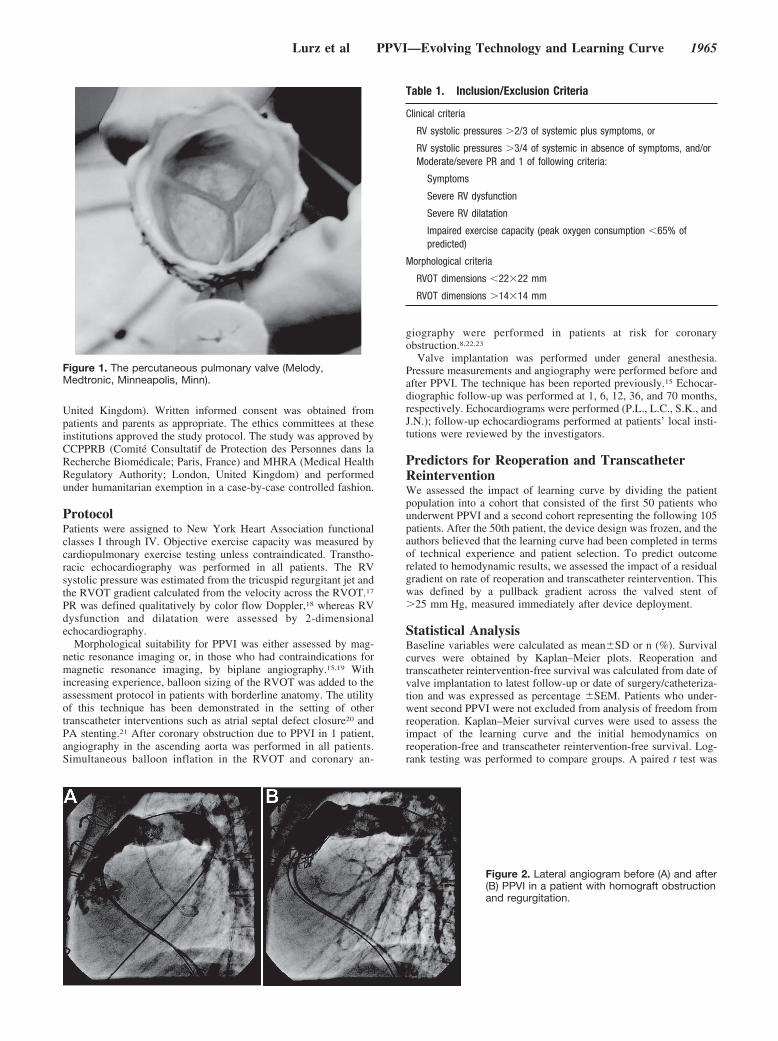
MethodsPatientsBetween September 2000 and February 2007, 155 patients under-went PPVI (Figures 1 and 2). Patients had to fulfill clinical andmorphological criteria to be considered suitable candidates. Inclu-sion criteria, summarized in Table 1, were based on surgicalindications for RVOT revision.16 Exclusion criteria were pregnancy,occluded central veins, active infection, and weight �20 kg. Valveimplantation was performed to reduce RVOT dysfunction andthereby delay surgery and prolong conduit lifespan. Criteria forsurgical or transcatheter reintervention were based on the initialinclusion criteria.
PPVI was performed by a single operator at Hôpital NeckerEnfants Malades (Paris, France), Great Ormond Street Hospital forChildren, The Heart Hospital, and The Harley Street Clinic (London,
Received August 23, 2007; accepted February 15, 2008.From the UCL Institute of Child Health (P.L., L.C., S.K., J.N., S.S., V.M., G.D., S.C., V.T., J.D., A.M.T., P.B.), London, United Kingdom; Great
Ormond Street Hospital for Children NHS Trust (P.L., L.C., S.K., J.N., S.S., V.M., T.Y.L., G.P., G.D., V.T., J.D., A.M.T., P.B.), Cardiothoracic Unit,London, United Kingdom; Service de Cardiologie Pédiatrique (Y.B.), Hôspital Necker Enfants Malades, Paris, France; and The Heart Hospital NHS Trust(S.C., F.W., V.T., J.D., P.B.), London, United Kingdom.
Correspondence to Philipp Bonhoeffer, Cardiac Unit, Great Ormond Street Hospital, Great Ormond St, London WC1N 3JH, United Kingdom. [email protected]
© 2008 American Heart Association, Inc.
Circulation is available at http://circ.ahajournals.org DOI: 10.1161/CIRCULATIONAHA.107.735779
1964
Interventional Cardiology
United Kingdom). Written informed consent was obtained frompatients and parents as appropriate. The ethics committees at theseinstitutions approved the study protocol. The study was approved byCCPPRB (Comité Consultatif de Protection des Personnes dans laRecherche Biomédicale; Paris, France) and MHRA (Medical HealthRegulatory Authority; London, United Kingdom) and performedunder humanitarian exemption in a case-by-case controlled fashion.
ProtocolPatients were assigned to New York Heart Association functionalclasses I through IV. Objective exercise capacity was measured bycardiopulmonary exercise testing unless contraindicated. Transtho-racic echocardiography was performed in all patients. The RVsystolic pressure was estimated from the tricuspid regurgitant jet andthe RVOT gradient calculated from the velocity across the RVOT.17
PR was defined qualitatively by color flow Doppler,18 whereas RVdysfunction and dilatation were assessed by 2-dimensionalechocardiography.
Morphological suitability for PPVI was either assessed by mag-netic resonance imaging or, in those who had contraindications formagnetic resonance imaging, by biplane angiography.15,19 Withincreasing experience, balloon sizing of the RVOT was added to theassessment protocol in patients with borderline anatomy. The utilityof this technique has been demonstrated in the setting of othertranscatheter interventions such as atrial septal defect closure20 andPA stenting.21 After coronary obstruction due to PPVI in 1 patient,angiography in the ascending aorta was performed in all patients.Simultaneous balloon inflation in the RVOT and coronary an-
giography were performed in patients at risk for coronaryobstruction.8,22,23
Valve implantation was performed under general anesthesia.Pressure measurements and angiography were performed before andafter PPVI. The technique has been reported previously.15 Echocar-diographic follow-up was performed at 1, 6, 12, 36, and 70 months,respectively. Echocardiograms were performed (P.L., L.C., S.K., andJ.N.); follow-up echocardiograms performed at patients’ local insti-tutions were reviewed by the investigators.
Predictors for Reoperation and TranscatheterReinterventionWe assessed the impact of learning curve by dividing the patientpopulation into a cohort that consisted of the first 50 patients whounderwent PPVI and a second cohort representing the following 105patients. After the 50th patient, the device design was frozen, and theauthors believed that the learning curve had been completed in termsof technical experience and patient selection. To predict outcomerelated to hemodynamic results, we assessed the impact of a residualgradient on rate of reoperation and transcatheter reintervention. Thiswas defined by a pullback gradient across the valved stent of�25 mm Hg, measured immediately after device deployment.
Statistical AnalysisBaseline variables were calculated as mean�SD or n (%). Survivalcurves were obtained by Kaplan–Meier plots. Reoperation andtranscatheter reintervention-free survival was calculated from date ofvalve implantation to latest follow-up or date of surgery/catheteriza-tion and was expressed as percentage �SEM. Patients who under-went second PPVI were not excluded from analysis of freedom fromreoperation. Kaplan–Meier survival curves were used to assess theimpact of the learning curve and the initial hemodynamics onreoperation-free and transcatheter reintervention-free survival. Log-rank testing was performed to compare groups. A paired t test was
Figure 1. The percutaneous pulmonary valve (Melody,Medtronic, Minneapolis, Minn).
Figure 2. Lateral angiogram before (A) and after(B) PPVI in a patient with homograft obstructionand regurgitation.
Table 1. Inclusion/Exclusion Criteria
Clinical criteria
RV systolic pressures �2/3 of systemic plus symptoms, or
RV systolic pressures �3/4 of systemic in absence of symptoms, and/orModerate/severe PR and 1 of following criteria:
Symptoms
Severe RV dysfunction
Severe RV dilatation
Impaired exercise capacity (peak oxygen consumption �65% ofpredicted)
Morphological criteria
RVOT dimensions �22�22 mm
RVOT dimensions �14�14 mm
Lurz et al PPVI—Evolving Technology and Learning Curve 1965

used to evaluate differences in invasively measured pressures beforeand after PPVI. Echocardiographic data were evaluated for changesfrom 1 month postprocedure to 6, 12 and 36 months using a linearmodel with an unstructured covariance structure and time as arepeated variable. A P value of �0.05 was considered statisticallysignificant. Statistical analysis was performed on SPSS 11.0 for Mac(SPSS Inc., Chicago, Ill, USA).
The authors had full access to and take full responsibility for theintegrity of the data. All authors have read and agree to themanuscript as written.
Results
Study Population and CharacteristicsOut of 163 patients enrolled, 8 patients did not meet themorphological criteria and therefore did not undergo PPVI.The reasons were unfavorable RVOT dimensions as assessedby balloon sizing in 6 patients. Another reason for abandon-ment of PPVI was proven risk of left coronary artery
Table 2. Patient Characteristics
ParameterTotal Population
(n�155)Predominantly Stenosis*
(n�63)Predominantly Regurgitation†
(n�47)Combined Lesions‡
(n�45)
Age at implantation, median(range), y
21.2 (7–71) 19.6 (9–54) 22.6 (7–58) 21.9 (9–71)
Diagnosis
Tetralogy of Fallotvariant, n (%)
94 (60.6) 36 (57.1) 33 (70.2) 25 (55.6)
Pulmonary stenosis 35 10 13 12
Pulmonary atresia 51 21 19 11
APV 8 5 1 2
Double-outlet RV, n (%) 9 (5.8) 3 (4.8) 5 (10.6) 1 (2.2)
TGA, VSD, PS, n (%) 14 (9.0) 8 (12.7) 0 (0) 6 (13.3)
Ross procedure, n (%) 12 (7.7) 7 (11.1) 1 (2.1) 4 (8.9)
Truncus arteriosus, n (%) 17 (11.0) 5 (7.9) 5 (10.6) 7 (15.6)
Other, n (%) 9 (5.8) 4 (6.3) 3 (6.3) 2 (4.4)
RVOT characteristics, n (%)
Homograft 126 (81.3) 51 (80.9) 37 (78.7) 38 (84.4)
Hancock 11 (7.1) 7 (11.1) 1 (2.1) 3 (6.7)
Other conduit type 7 (4.5) 3 (4.7) 3 (6.4) 1 (2.2)
Patch-extended RVOT 2 (1.3) 1 (1.6) 1 (2.1) 0
Native outflow tract 5 (3.2) 1 (1.6) 2 (4.2) 2 (4.4)
Other 4 (2.6) 0 (0) 3 (6.4) 1 (2.2)
Open heart surgeries, mean 1.8�0.9 1.9�0.9 1.5�0.8 1.9�0.9
Operations in total, mean 2.5�1.1 2.6�1.1 2.4�1.0 2.5�12
Transcatheter interventions 0.4�0.8 0.4�0.7 0.3�0.6 0.6�1.0
NYHA class, n (%)
1 17 (11.0) 7 (11.1) 3 (6.4) 7 (15.6)
2 82 (52.9) 38 (60.3) 23 (48.9) 21 (46.7)
3 45 (29.0) 16 (25.4) 17 (36.2) 12 (26.7)
4 11 (7.1) 2 (3.2) 4 (8.5) 5 (11.1)
CPEX (n�130)
Peak V̇O2,kg � mL�1 � min�1
23.2�7.3 24.2�7.1 21.8�7.1 23.6�7.6
% of predicted V̇O2 60.7�15.9 62.0�15.7 58.9�15.3 61.2�17.1
Echocardiography
RV SP, mm Hg 66.6�21.6 81.4�17.9 47.3�15.9 66.6�15.9
Peak RVOTgradient, mm Hg
57.0�23.9 75.9�17.5 32.7�10.8 65.2�14.5
RV to sys pressure ratio 0.68�0.2 0.82�0.14 0.51�0.14 0.65�0.14
APV indicates absent pulmonary valve syndrome; TGA, transposition of the great arteries; VSD, ventricular septal defect; PS, pulmonary stenosis;CPEX, cardiopulmonary exercise test; peak V̇O2, peak oxygen consumption; RV SP, RV systolic pressure; RV to sys pressure ratio, RV to systemicsystolic pressure ratio.
*RVOT gradient �50 mm Hg on echocardiography and less than moderate PR on echocardiography.†At least moderate PR on echocardiography and RVOT gradient �50 mm Hg on echocardiography.‡Patients who did not fit either the “predominantly stenosis” or “predominantly regurgitant” categories.
1966 Circulation April 15, 2008
compression demonstrated by balloon inflation within theRV-to-PA conduit (n�1).22
The median age of the present patient population was 21.2years (range 7 to 71 years); 57 patients were �16 years old(37%), and 42% were female (Table 2). Most patients (61%)had tetralogy of Fallot or a variant morphology; 92% had anRV-to-PA conduit placed at previous surgery, 81% of whichwere homografts. Before the procedure, an RV-to–systemicpressure ratio of �2/3 was present in 58% of patients;echocardiography showed moderate or severe PR in 64%.The vast majority of patients complained of symptoms (138of 155 patients), and 61% had impaired exercise capacity(defined as a peak oxygen consumption �65% of predicted).In total, 17 asymptomatic patients underwent PPVI. Indica-tions for intervention in the absence of symptoms wereRV-to–systemic pressure ratio �3/4 (0.89�0.13) in 9, peakoxygen consumption �65% of predicted (54.7�1.0%) in 4,and severe RV dilatation in 4 (z score for RV end-diastolicdiameter assessed on echocardiography 5.9�2.3).
All but 8 patients in the present series were suitable forsurgical RV-to-PA conduit replacement. In these 8, contrain-dications for surgery were pulmonary hypertension (n�4),severe chest deformity (n�2), and cardiogenic shock whenreferred for PPVI (n�2).
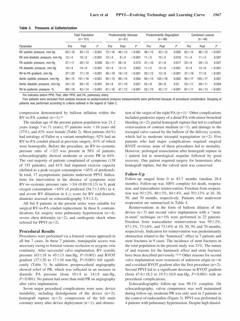
Procedural ResultsProcedures were performed via a femoral venous approach inall but 7 cases. In these 7 patients, transjugular access wasnecessary owing to femoral venous occlusion or azygous veincontinuity. After successful valve implantation, RV systolicpressure (63�18 to 45�13 mm Hg, P�0.001) and RVOTgradient (37�20 to 17�10 mm Hg, P�0.001) fell signifi-cantly (Table 3). In addition, postprocedural angiographyshowed relief of PR, which was reflected in an increase indiastolic PA pressure (from 10�4 to 14�9 mm Hg,P�0.001). No patient had more than mild PR on angiographyafter valve implantation.
Seven major procedural complications were seen: deviceinstability, including dislodgement of the device (n�2);homograft rupture (n�3); compression of the left maincoronary artery after device deployment (n�1); and obstruc-
tion of the origin of the right PA (n�1).1 Other complicationsincluded guidewire injury of a distal PA with minor bronchialbleeding (n�2); partial homograft rupture that led to confinedextravasation of contrast medium (n�1); and damage to thetricuspid valve caused by the balloon of the delivery system,which led to moderate tricuspid regurgitation (n�2). Fivepatients who had major complications required surgicalRVOT revision; none of these procedures led to mortality.However, prolonged resuscitation after homograft rupture in1 patient led to neurological sequelae followed by goodrecovery. One patient required surgery for hemostasis afterhomograft rupture, but the valved stent was conserved.
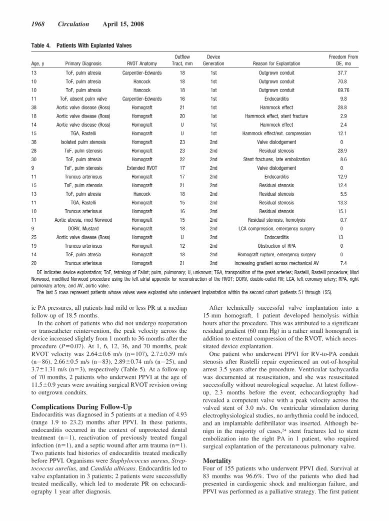
Follow-UpFollow-up ranged from 0 to 83.7 months (median 28.4months). Follow-up was 100% complete for death, reopera-tion, and transcatheter reintervention. Freedom from reopera-tion was 93�2%, 86�3%, 84�4%, and 70�13% at 10, 30,50, and 70 months, respectively. Patients who underwentreoperation are summarized in Table 4.
Reinterventions in the form of balloon dilation of thedevice (n�3) and second valve implantation with a “stent-in-stent” technique (n�19) were performed in 22 patients.Freedom from transcatheter reintervention was 95�2%,87�3%, 73�6%, and 73�6% at 10, 30, 50, and 70 months,respectively. Indication for reintervention was predominantlyobstruction related to the “hammock” effect in 7 patients andstent fractures in 9 cases. The incidence of stent fractures inthe total population in the present study was 21%. The natureof and reasons for the hammock effect and stent fractureshave been described previously.15,24 Other reasons for secondvalve implantation were restenosis of unknown origin (n�4)and a residual RVOT gradient after the first procedure (n�2).Second PPVI led to a significant decrease in RVOT gradient(from 47.6�18.2 to 19.5�10.0 mm Hg, P�0.001) with noprocedural complications.
Echocardiographic follow-up was 90.1% complete. Onechocardiography, valvar competence was well maintainedduring follow-up; moderate PR was only seen in 2 patients inthe context of endocarditis (Figure 3). PPVI was performed in4 patients with pulmonary hypertension. Despite high diastol-
Table 3. Pressures at Catheterization
Total Population(n�151)
Predominantly Stenosis(n�61)
Predominantly Regurgitant(n�46)
Combined Lesions(n�44)
Parameter Pre Post P Pre Post P Pre Post P Pre Post P
RV systolic pressure, mm Hg 63�18 45�13 �0.001 72�16 46�13 �0.001 48�13 43�12 0.002 62�15 46�12 �0.001
RV end-diastolic pressure, mm Hg 12�4 10�5 �0.001 12�4 9�4 �0.001 11�5 10�5 0.016 11�4 11�5 0.507
PA systolic pressure, mm Hg 27�11 29�12 0.056 25�11 26�9 0.373 31�10 31�9 0.917 25�9 29�13 0.027
PA diastolic pressure, mm Hg 10�4 14�9 �0.001 10�4 12�4 0.003 11�5 15�6 �0.001 9�4 13�6 �0.001
RV-to-PA gradient, mm Hg 37�20 17�10 �0.001 48�18 19�12 �0.001 20�13 13�9 �0.001 37�18 17�8 �0.001
Aortic systolic pressure, mm Hg 94�15 101�16 �0.001 92�15 98�14 0.004 94�14 102�16 0.002 94�17 105�17 0.001
Aortic diastolic pressure, mm Hg 54�10 58�10 �0.001 54�9 57�10 0.021 55�9 58�8 0.01 53�11 59�11 0.004
RV-to-systemic pressure, % 69�19 45�14 �0.001 81�16 47�12 �0.001 52�15 42�11 �0.001 67�11 44�15 �0.001
Pre indicates before PPVI; Post, after PPVI; and PA, pulmonary artery.Four patients were excluded from analysis because no postprocedural pressure measurements were performed because of procedural complication. Grouping of
patients was performed according to criteria outlined in the legend of Table 2.
Lurz et al PPVI—Evolving Technology and Learning Curve 1967
ic PA pressures, all patients had mild or less PR at a medianfollow-up of 18.5 months.
In the cohort of patients who did not undergo reoperationor transcatheter reintervention, the peak velocity across thedevice increased slightly from 1 month to 36 months after theprocedure (P�0.07). At 1, 6, 12, 36, and 70 months, peakRVOT velocity was 2.64�0.6 m/s (n�107), 2.7�0.59 m/s(n�86), 2.66�0.5 m/s (n�83), 2.89�0.74 m/s (n�25), and3.7�1.31 m/s (n�3), respectively (Table 5). At a follow-upof 70 months, 2 patients who underwent PPVI at the age of11.5�0.9 years were awaiting surgical RVOT revision owingto outgrown conduits.
Complications During Follow-UpEndocarditis was diagnosed in 5 patients at a median of 4.93(range 1.9 to 23.2) months after PPVI. In these patients,endocarditis occurred in the context of unprotected dentaltreatment (n�1), reactivation of previously treated fungalinfection (n�1), and a septic wound after arm trauma (n�1).Two patients had histories of endocarditis treated medicallybefore PPVI. Organisms were Staphylococcus aureus, Strep-tococcus aurelius, and Candida albicans. Endocarditis led tovalve explantation in 3 patients; 2 patients were successfullytreated medically, which led to moderate PR on echocardi-ography 1 year after diagnosis.
After technically successful valve implantation into a15-mm homograft, 1 patient developed hemolysis withinhours after the procedure. This was attributed to a significantresidual gradient (60 mm Hg) in a rather small homograft inaddition to external compression of the RVOT, which neces-sitated device explantation.
One patient who underwent PPVI for RV-to-PA conduitstenosis after Rastelli repair experienced an out-of-hospitalarrest 3.5 years after the procedure. Ventricular tachycardiawas documented at resuscitation, and she was resuscitatedsuccessfully without neurological sequelae. At latest follow-up, 2.3 months before the event, echocardiography hadrevealed a competent valve with a peak velocity across thevalved stent of 3.0 m/s. On ventricular stimulation duringelectrophysiological studies, no arrhythmia could be induced,and an implantable defibrillator was inserted. Although be-nign in the majority of cases,24 stent fractures led to stentembolization into the right PA in 1 patient, who requiredsurgical explantation of the percutaneous pulmonary valve.
MortalityFour of 155 patients who underwent PPVI died. Survival at83 months was 96.6%. Two of the patients who died hadpresented in cardiogenic shock and multiorgan failure, andPPVI was performed as a palliative strategy. The first patient
Table 4. Patients With Explanted Valves
Age, y Primary Diagnosis RVOT AnatomyOutflow
Tract, mmDevice
Generation Reason for ExplantationFreedom From
DE, mo
13 ToF, pulm atresia Carpentier-Edwards 18 1st Outgrown conduit 37.7
10 ToF, pulm atresia Hancock 18 1st Outgrown conduit 70.8
10 ToF, pulm atresia Hancock 18 1st Outgrown conduit 69.76
11 ToF, absent pulm valve Carpentier-Edwards 16 1st Endocarditis 9.8
38 Aortic valve disease (Ross) Homograft 21 1st Hammock effect 28.8
18 Aortic valve disease (Ross) Homograft 20 1st Hammock effect, stent fracture 2.9
14 Aortic valve disease (Ross) Homograft U 1st Hammock effect 2.4
15 TGA, Rastelli Homograft U 1st Hammock effect/ext. compression 12.1
38 Isolated pulm stenosis Homograft 23 2nd Valve dislodgement 0
28 ToF, pulm stenosis Homograft 23 2nd Residual stenosis 28.9
30 ToF, pulm atresia Homograft 22 2nd Stent fractures, late embolization 8.6
9 ToF, pulm stenosis Extended RVOT 17 2nd Valve dislodgement 0
11 Truncus arteriosus Homograft 17 2nd Endocarditis 12.9
15 ToF, pulm stenosis Homograft 21 2nd Residual stenosis 12.4
13 ToF, pulm atresia Hancock 18 2nd Residual stenosis 5.5
11 TGA, Rastelli Homograft 15 2nd Residual stenosis 13.3
10 Truncus arteriosus Homograft 16 2nd Residual stenosis 15.1
11 Aortic atresia, mod Norwood Homograft 15 2nd Residual stenosis, hemolysis 0.7
9 DORV, Mustard Homograft 18 2nd LCA compression, emergency surgery 0
25 Aortic valve disease (Ross) Homograft U 2nd Endocarditis 13
19 Truncus arteriosus Homograft 12 2nd Obstruction of RPA 0
14 ToF, pulm atresia Homograft 18 2nd Homograft rupture, emergency surgery 0
20 Truncus arteriosus Homograft 21 2nd Increasing gradient across mechanical AV 7.4
DE indicates device explantation; ToF, tetralogy of Fallot; pulm, pulmonary; U, unknown; TGA, transposition of the great arteries; Rastelli, Rastelli procedure; ModNorwood, modified Norwood procedure using the left atrial appendix for reconstruction of the RVOT; DORV, double-outlet RV; LCA, left coronary artery; RPA, rightpulmonary artery; and AV, aortic valve.
The last 5 rows represent patients whose valves were explanted who underwent implantation within the second cohort (patients 51 through 155).
1968 Circulation April 15, 2008

had a history of 5 previous operations, deteriorated 6 weeksafter a technically successful PPVI, and died of a chestinfection. The second presented in multiorgan failure andsevere fluid overload with critical recoarctation and RVOTobstruction. Despite successful PPVI and dilation of severerecoarctation, the patient died of pulmonary edema 24 hoursafter the procedure.
Two patients died suddenly at 8 and 35 months after PPVI.In both cases, autopsy revealed the valved stent appropriatelyseated in the RVOT. At latest follow-up, echocardiographyshowed good valvar competence of the device with a peakvelocity across it of �3 m/s. The deaths were presumed to bedue to arrhythmia.
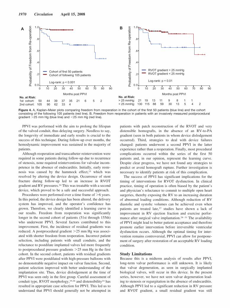
Impact of the Learning Curve on OutcomeFreedom from reoperation in the first series of 50 patients wassignificantly shorter than in the subsequent patient population(log-rank test P�0.001; Figure 4A). In addition, residualgradients measured invasively after PPVI were associatedwith higher rates of reoperation (log-rank test P�0.01; Figure4B) and transcatheter reintervention (log-rank test P�0.008).
Of the first 50 patients, 16 patients underwent deviceexplantation compared with 5 patients in the second cohort.The incidence of a residual gradient of �25 mm Hg washigher in the first cohort of patients (n�16 of 50 versus 5 of105 patients in the second cohort). Freedom from transcath-
eter reintervention did not differ significantly in the 2 cohortson time-dependent analysis (n�13 versus n�9; log-rank testP�0.18). This is due to the fact that reinterventions wereperformed predominantly due to stent fractures, a complica-tion that was not affected by the learning curve. The inci-dence of procedural complications fell from 6% in the firstcohort to 2.9% in the second cohort of patients (n�3 of 50versus 3 of 105 patients).
DiscussionPercutaneous interventions on the RVOT have been per-formed to avoid multiple open heart operations in patientswith repaired congenital heart disease. Balloon dilation andbare-metal stenting of the outflow tract have successfullyachieved relief of obstruction; however, these procedurescompromise valvular function.8–10,25 In contrast, PPVI repre-sents a percutaneous technique to treat both pulmonarystenosis and regurgitation. We report our current experienceof 155 patients who underwent transcatheter pulmonary valveimplantation. This experience constitutes the longest andlargest single-operator experience in percutaneous insertionof a heart valve. We have demonstrated that PPVI is aneffective treatment for RVOT dysfunction. Device deploy-ment was successful with very few exceptions and led to amarked improvement in hemodynamics. This was achievedwith a very low complication rate and mortality.
0%
20%
40%
60%
80%
100%
pre PPVI 1 month 6 months 12 months 36 months 70 months
)401=n()151=n( (n=109) )3=n()341=n( (n=32)
ereveStnesbA ModerateMildTrivial
% o
f eac
h re
gurg
itatio
n gr
ade
Figure 3. Valvar competence during follow-up assessed on echocardiography. PR was graded as absent, trivial, mild, moderate, orsevere and expressed as a percentage before valve implantation and at 1, 6, 12, 36, and 70 months after PPVI.
Table 5. Peak Velocity Across the RVOT During Follow-Up Assessed by Echocardiography
BeforePPVI*
1 Month AfterPPVI
6 Months AfterPPVI
12 Months AfterPPVI
36 MonthsAfter PPVI
70 MonthsAfter PPVI
Reoperated/recatheterized patients 3.94�0.87 (39) 3.4�0.67 (36) 3.77�0.63 (23) 3.9�0.69 (21) 4.16�0.42 (7) � � �
Patients without any intervention after PPVI 3.58�0.83 (112) 2.64�0.6 (107) 2.70�0.59 (86) 2.66�0.50 (83) 2.89�0.74 (25) 3.70�1.31 (3)†
Total patient population 3.67�0.85 (151) 2.84�0.7 (143) 2.93�0.74 (109) 2.91�0.74 (104) 3.17�0.86 (32) 3.70�1.31 (3)
Values are expressed as mean�SD (n). Velocity was measured in meters per second.*In 4 patients, echocardiographic assessment of the RVOT was not possible owing to the retrosternal position of the RV-to-PA conduit.†Two patients were awaiting surgical RVOT revision due to outgrown conduit.
Lurz et al PPVI—Evolving Technology and Learning Curve 1969
PPVI was performed with the aim to prolong the lifespanof the valved conduit, thus delaying surgery. Needless to say,the longevity of immediate and early results is crucial to thesuccess of this technique. During follow-up over months, thehemodynamic improvement was sustained in the majority ofpatients.
Although reoperation and transcatheter reintervention wererequired in some patients during follow-up due to recurrenceof stenosis, none required reinterventions for valvular incom-petence in the absence of endocarditis. Initially, early reste-nosis was caused by the hammock effect,15 which wasresolved by altering the device design. Occurrence of stentfracture during follow-up led to an increase in RVOTgradient and RV pressures.24 This was treatable with a seconddevice, which proved to be a safe and successful approach.
Procedures were performed over a time frame of �6 years.In this period, the device design has been altered, the deliverysystem has improved, and the operator’s confidence hasgrown. Therefore, we clearly identified a learning curve inour results. Freedom from reoperation was significantlylonger in the second cohort of patients (51st through 155th)who underwent PPVI. Several factors contributed to thisimprovement. First, the incidence of residual gradients wasreduced. A postprocedural gradient �25 mm Hg was associ-ated with shorter freedom from reoperation. Incorrect patientselection, including patients with small conduits, and thereluctance to postdilate implanted valves led more frequentlyto postprocedural pressure gradients �25 mm Hg in the firstcohort. In the second cohort, patients with residual gradientsafter PPVI were postdilated with high-pressure balloons withno demonstrable negative impact on valvar function. Second,patient selection improved with better understanding of theimplantation site. Thus, device dislodgement at the time ofPPVI was seen only in the first group. Careful assessment ofconduit type, RVOT morphology,19,26 and distensibility24 hasresulted in appropriate case selection for PPVI. This led us tounderstand that PPVI should generally not be attempted in
patients with patch reconstruction of the RVOT and verydistensible homografts, in the absence of an RV-to-PAgradient (seen in both patients in whom device dislodgementoccurred). Third, strategies to deal with device failureschanged: patients underwent a second PPVI in the latterexperience rather than a reoperation. Finally, most proceduralcomplications occurred within the series of the first 50patients and, in our opinion, represent the learning curve.Despite clear progress, we have not found any strategies topredict or avoid homograft rupture. Further investigation isnecessary to identify patients at risk of this complication.
The success of PPVI has significant implications for thetiming of interventions for RVOT dysfunction. In clinicalpractice, timing of operation is often biased by the patient’sand physician’s reluctance to commit to multiple open heartsurgeries, thereby exposing the RV to an increasing durationof abnormal loading conditions. Although reduction of RVdiastolic and systolic volumes can be achieved even whenpatients are treated late,27 studies have shown a lack ofimprovement in RV ejection fraction and exercise perfor-mance after surgical valve implantation.28–31 The availabilityof PPVI might lead to better patient acceptance and thereforepromote earlier intervention before irreversible ventriculardysfunction occurs. Although the optimal timing for inter-vention remains controversial, PPVI can allow for postpone-ment of surgery after restoration of an acceptable RV loadingcondition.
Study LimitationsBecause this is a midterm analysis of results after PPVI,long-term valvar performance is still unknown. It is likelythat valvar degeneration, as seen in surgically implantedbiological valves, will occur in this device. In the presentseries, however, we have not seen valvar degeneration lead-ing to stenosis or regurgitation in the absence of endocarditis.Although PPVI led to a significant reduction in RV pressureand RVOT gradient, a small residual gradient was still
9080706050403020100
1.0
.8
.6
.4
.2
0.09080706050403020100
1.0
.8
.6
.4
.2
0.0
No. at Risk: > 25 mmHg: 21 19 13 11 9 8 1 1 < 25 mmHg: 130 115 88 59 30 13 5 4 2
RVOT gradient > 25 mmHgRVOT gradient < 25 mmHg
Log-rank: p = 0.01
Prob
abilit
yof
Free
dom
from
Re-
oper
atio
nA
Cohort of first 50 patientsCohort of following 105 patients
Log-rank: p < 0.001
Prob
abilit
yof
Free
dom
from
Re-
oper
atio
n
No. at Risk: 1st cohort: 50 44 39 37 35 21 6 5 22nd cohort: 105 90 62 33 4
B
IVPP tsop shtnoMIVPP tsop shtnoM
Figure 4. A, Kaplan–Meier plots comparing freedom from reoperation in the cohort of the first 50 patients (blue line) and the cohortconsisting of the following 105 patients (red line). B, Freedom from reoperation in patients with an invasively measured postproceduralgradient �25 mm Hg (blue line) and �25 mm Hg (red line).
1970 Circulation April 15, 2008
present in the majority of cases after the procedure. Thelong-term effects of low outflow tract gradients are unknown.Again, longer follow-up is necessary to address this question.Results of PPVI were not compared with surgical pulmonaryvalve implantation. However, transcatheter implantation of avalve in the pulmonary position was performed to prolonga surgical result after conduit placement rather than as asurrogate for open heart surgery. Therefore, we chose toassess the potential of PPVI to delay surgery by applying thesame criteria for reintervention as for initial treatment, whichwere based on surgical indications.
ConclusionsPPVI improves RV outflow hemodynamics and delays sur-gery by prolonging conduit lifespan. Our experience repre-sents the impact of evolving technologies in medicine, withprogressive improvement in clinical results by device andtechnique modification. PPVI should reduce the number ofoperations and the cumulative hemodynamic burden on theRV over the total lifetime of children and young adults,potentially improving the life expectancy of patients withcongenital heart disease that involves the RVOT.
AcknowledgmentsWe are indebted to Vickie Conley (Medtronic) and Allen Tower(NuMed), without whom this technology would not have beendeveloped.
Sources of FundingP. Lurz is funded by the European Union (Health-e-Child Initiative);Drs Coats, Deanfield, and Bonhoeffer and S. Schievano are fundedby the British Heart Foundation. Dr Taylor is funded by the HigherEducation Funding Council for England.
DisclosuresDr Bonhoeffer is a consultant to Medtronic and NuMed and hasreceived honoraria and royalties for the device described. Drs Taylorand Khambadkone are consultants to Medtronic and have receivedhonoraria. The remaining authors report no conflicts.
References1. Kanter KR, Budde JM, Parks WJ, Tam VK, Sharma S, Williams WH,
Fyfe DA. One hundred pulmonary valve replacements in children afterrelief of right ventricular outflow tract obstruction. Ann Thorac Surg.2002;73:1801–1806.
2. Bielefeld MR, Bishop DA, Campbell DN, Mitchell MB, Grover FL,Clarke DR. Reoperative homograft right ventricular outflow tract recon-struction. Ann Thorac Surg. 2001;71:482–487.
3. Askovich B, Hawkins JA, Sower CT, Minich LL, Tani LY, Stoddard G,Puchalski MD. Right ventricle-to-pulmonary artery conduit longevity: isit related to allograft size? Ann Thorac Surg. 2007;84:907–911.
4. Brown JW, Ruzmetov M, Rodefeld MD, Vijay P, Turrentine MW. Rightventricular outflow tract reconstruction with an allograft conduit innon-Ross patients: risk factors for allograft dysfunction and failure. AnnThorac Surg. 2005;80:655–663.
5. Stark J, Bull C, Stajevic M, Jothi M, Elliott M, de Leval M. Fate ofsubpulmonary homograft conduits: determinants of late homograftfailure. J Thorac Cardiovasc Surg. 1998;115:506–514.
6. Tweddell JS, Pelech AN, Frommelt PC, Mussatto KA, Wyman JD,Fedderly RT, Berger S, Frommelt MA, Lewis DA, Friedberg DZ, ThomasJP Jr, Sachdeva R, Litwin SB. Factors affecting longevity of homograftvalves used in right ventricular outflow tract reconstruction for congenitalheart disease. Circulation. 2000;102(suppl III):III-130–III-135.
7. Oosterhof T, Meijboom FJ, Vliegen HW, Hazekamp MG, ZwindermanAH, Bouma BJ, van Dijk AP, Mulder BJ. Long-term follow-up ofhomograft function after pulmonary valve replacement in patients withtetralogy of Fallot. Eur Heart J. 2006;27:1478–1484.
8. Peng LF, McElhinney DB, Nugent AW, Powell AJ, Marshall AC, BachaEA, Lock JE. Endovascular stenting of obstructed right ventricle-to-pul-monary artery conduits: a 15-year experience. Circulation. 2006;113:2598–2605.
9. Powell AJ, Lock JE, Keane JF, Perry SB. Prolongation of RV-PA conduitlife span by percutaneous stent implantation: intermediate-term results.Circulation. 1995;92:3282–3288.
10. Fogelman R, Nykanen D, Smallhorn JF, McCrindle BW, Freedom RM,Benson LN. Endovascular stents in the pulmonary circulation: clinicalimpact on management and medium-term follow-up. Circulation. 1995;92:881–885.
11. Bove EL, Byrum CJ, Thomas FD, Kavey RE, Sondheimer HM,Blackman MS, Parker FB Jr. The influence of pulmonary insufficiency onventricular function following repair of tetralogy of Fallot: evaluationusing radionuclide ventriculography. J Thorac Cardiovasc Surg. 1983;85:691–696.
12. Carvalho JS, Shinebourne EA, Busst C, Rigby ML, Redington AN.Exercise capacity after complete repair of tetralogy of Fallot: deleteriouseffects of residual pulmonary regurgitation. Br Heart J. 1992;67:470–473.
13. Gatzoulis MA, Balaji S, Webber SA, Siu SC, Hokanson JS, Poile C,Rosenthal M, Nakazawa M, Moller JH, Gillette PC, Webb GD, RedingtonAN. Risk factors for arrhythmia and sudden cardiac death late after repairof tetralogy of Fallot: a multicentre study. Lancet. 2000;356:975–981.
14. Bonhoeffer P, Boudjemline Y, Saliba Z, Merckx J, Aggoun Y, Bonnet D,Acar P, Le Bidois J, Sidi D, Kachaner J. Percutaneous replacement ofpulmonary valve in a right-ventricle to pulmonary-artery prostheticconduit with valve dysfunction. Lancet. 2000;356:1403–1405.
15. Khambadkone S, Coats L, Taylor A, Boudjemline Y, Derrick G, Tsang V,Cooper J, Muthurangu V, Hegde SR, Razavi RS, Pellerin D, Deanfield J,Bonhoeffer P. Percutaneous pulmonary valve implantation in humans:results in 59 consecutive patients. Circulation. 2005;112:1189–1197.
16. Therrien J, Warnes C, Daliento L, Hess J, Hoffmann A, Marelli A, ThilenU, Presbitero P, Perloff J, Somerville J, Webb GD. Canadian Cardio-vascular Society Consensus Conference 2001 update: recommendationsfor the management of adults with congenital heart disease part III. CanJ Cardiol. 2001;17:1135–1158.
17. Yock PG, Popp RL. Noninvasive estimation of right ventricular systolicpressure by Doppler ultrasound in patients with tricuspid regurgitation.Circulation. 1984;70:657–662.
18. Joyce JJ, Hwang EY, Wiles HB, Kline CH, Bradley SM, Crawford FA Jr.Reliability of intraoperative transesophageal echocardiography duringtetralogy of Fallot repair. Echocardiography. 2000;17:319–327.
19. Schievano S, Migliavacca F, Coats L, Khambadkone S, Carminati M,Wilson N, Deanfield JE, Bonhoeffer P, Taylor AM. Percutaneous pul-monary valve implantation based on rapid prototyping of right ventricularoutflow tract and pulmonary trunk from MR data. Radiology. 2007;242:490–497.
20. Rome JJ, Keane JF, Perry SB, Spevak PJ, Lock JE. Double-umbrellaclosure of atrial defects: initial clinical applications. Circulation. 1990;82:751–758.
21. Nagm AM, Moore JW. Balloon sizing of pulmonary branch stenosis: auseful method to guide stent implantation. J Invasive Cardiol. 2003;15:437–438.
22. Sridharan S, Coats L, Khambadkone S, Taylor AM, Bonhoeffer P. Imagesin cardiovascular medicine: transcatheter right ventricular outflow tractintervention: the risk to the coronary circulation. Circulation. 2006;113:e934–e935.
23. Maheshwari S, Bruckheimer E, Nehgme RA, Fahey JT, Kholwadwala D,Hellenbrand WE. Single coronary artery complicating stent implantationfor homograft stenosis in tetralogy of Fallot. Cathet Cardiovasc Diagn.1997;42:405–407.
24. Nordmeyer J, Khambadkone S, Coats L, Schievano S, Lurz P, ParenzanG, Taylor A, Lock J, Bonhoeffer P. Risk stratification, systematic clas-sification and anticipatory strategies for stent fractures after percutaneouspulmonary valve implantation. Circulation. 2007;115:1392–1397.
25. Garty Y, Veldtman G, Lee K, Benson L. Late outcomes after pulmonaryvalve balloon dilatation in neonates, infants and children. J InvasiveCardiol. 2005;17:318–322.
26. Schievano S, Coats L, Migliavacca F, Norman W, Frigiola A, DeanfieldJ, Bonhoeffer P, Taylor AM. Variations in right ventricular outflow tractmorphology following repair of congenital heart disease: implications forpercutaneous pulmonary valve implantation. J Cardiovasc Magn Reson.2007;9:687–695.
Lurz et al PPVI—Evolving Technology and Learning Curve 1971
27. Oosterhof T, van Straten A, Vliegen HW, Meijboom FJ, van Dijk AP,Spijkerboer AM, Bouma BJ, Zwinderman AH, Hazekamp MG, de RoosA, Mulder BJ. Preoperative thresholds for pulmonary valve replacementin patients with corrected tetralogy of Fallot using cardiovascularmagnetic resonance. Circulation. 2007;116:545–551.
28. Therrien J, Siu SC, McLaughlin PR, Liu PP, Williams WG, Webb GD.Pulmonary valve replacement in adults late after repair of tetralogy ofFallot: are we operating too late? J Am Coll Cardiol. 2000;36:1670–1675.
29. Ghez O, Tsang VT, Frigiola A, Coats L, Taylor A, Van Doorn C,Bonhoeffer P, De Leval M. Right ventricular outflow tract reconstruction
for pulmonary regurgitation after repair of tetralogy of Fallot: preliminaryresults. Eur J Cardiothorac Surg. 2007;31:654–658.
30. Dave HH, Buechel ER, Dodge-Khatami A, Kadner A, Rousson V,Bauersfeld U, Pretre R. Early insertion of a pulmonary valve for chronicregurgitation helps restoration of ventricular dimensions. Ann ThoracSurg. 2005;80:1615–1620.
31. Gengsakul A, Harris L, Bradley TJ, Webb GD, Williams WG, Siu SC,Merchant N, McCrindle BW. The impact of pulmonary valvereplacement after tetralogy of Fallot repair: a matched comparison. EurJ Cardiothorac Surg. 2007;32:462–468.
CLINICAL PERSPECTIVEAfter the first transcatheter valve implantation in 2000, a new field in interventional cardiology developed. New techniquesevolved rapidly, with significant improvement in outcomes. We assessed our results after percutaneous pulmonary valveimplantation over a time frame of 6 years from the first to the 155th patient. The aim of percutaneous pulmonary valveimplantation was to prolong the lifespan of conduits, which were surgically placed from the right ventricle to the pulmonaryartery. Because of the limited longevity of conduits, the majority of patients with right ventricular outflow tract dysfunctiontraditionally undergo multiple open heart operations for conduit replacement. We show that percutaneous pulmonary valveimplantation successfully restores conduit function with a low procedural complication rate. Explantation-free survival 5years after percutaneous pulmonary valve implantation is �70%. This prolonged conduit lifespan should reduce thenumber of multiple open heart operations over the total lifespan of children and young adults with congenital heart diseaseand potentially improve life expectancy of these patients. As with all new procedures, whether surgical or interventional,the impact of the learning curve on a novel technique must be recognized. Therefore, we divided our patient populationinto a cohort consisting of the first 50 patients and a second cohort representing the later experience of 105 patients. In ourexperience, explantation-free survival was significantly longer in patients who underwent percutaneous pulmonary valveimplantation in the second cohort. The impact of a learning curve on clinical results of new interventional procedures isof importance, especially if interventions and devices are compared with conventional therapies.
1972 Circulation April 15, 2008
Copyright © 2022 FDOKUMEN