Percent Shear Area Determination in Charpy Impact Testing
28
This paper was submitted to the Journal of ASTM International in January, 2008. It is currently being peer reviewed. 1 * MPM Technologies, Inc., State College, Pennsylvania, USA 16803. ** National Institute of Standards and Technology, Boulder, Colorado, USA 80305 Percent Shear Area Determination in Charpy Impact Testing M. P. Manahan, Jr. * , C. N. McCowan ** , and M.P. Manahan, Sr. * Abstract: The Charpy test is used throughout the world in a wide range of industries because of its low cost and the fact that notching and dynamic loading produces a crack tip stress field, which is conservative for many applications. As a result of its widespread use, there is a compelling motivation to extract as much data as possible from the Charpy test. In a Charpy impact test, three key measurements are typically made: total absorbed energy, lateral expansion, and percent shear fracture area. At present, the measurement of absorbed energy and lateral expansion is quantitative and well defined, but the methods used by most laboratories in the measurement of percent shear are qualitative and imprecise. This is ironic for a 100-year-old test because, as discussed in this paper, it can be reasonably argued that percent shear is the most fundamental and physically meaningful of the three Charpy parameters for brittle fracture characterization. Digital image analysis for shear fracture area is shown to be accurate, precise, and easy to use on a routine basis. Recommendations for changes to the ASTM E 23 standard are provided. Keywords: Percent Shear, Fracture Appearance Transition Temperature (FATT), Charpy Impact Testing, Brittle Fracture, Ductile Fracture, Shear Fracture Area (SFA), Ductile-Brittle Transition Temperature (DBTT), Digital Image Analysis
-
Upload
khangminh22 -
Category
Documents
-
view
2 -
download
0
Transcript of Percent Shear Area Determination in Charpy Impact Testing

This paper was submitted to the Journal of ASTM International in January, 2008. It is currently
being peer reviewed.
1
* MPM Technologies, Inc., State College, Pennsylvania, USA 16803.
** National Institute of Standards and Technology, Boulder, Colorado, USA 80305
Percent Shear Area Determination in Charpy Impact Testing
M. P. Manahan, Jr.
*, C. N. McCowan
**, and M.P. Manahan, Sr.
*
Abstract: The Charpy test is used throughout the world in a wide range of industries because of
its low cost and the fact that notching and dynamic loading produces a crack tip stress field,
which is conservative for many applications. As a result of its widespread use, there is a
compelling motivation to extract as much data as possible from the Charpy test. In a Charpy
impact test, three key measurements are typically made: total absorbed energy, lateral expansion,
and percent shear fracture area. At present, the measurement of absorbed energy and lateral
expansion is quantitative and well defined, but the methods used by most laboratories in the
measurement of percent shear are qualitative and imprecise. This is ironic for a 100-year-old test
because, as discussed in this paper, it can be reasonably argued that percent shear is the most
fundamental and physically meaningful of the three Charpy parameters for brittle fracture
characterization. Digital image analysis for shear fracture area is shown to be accurate, precise,
and easy to use on a routine basis. Recommendations for changes to the ASTM E 23 standard
are provided.
Keywords: Percent Shear, Fracture Appearance Transition Temperature (FATT), Charpy Impact
Testing, Brittle Fracture, Ductile Fracture, Shear Fracture Area (SFA), Ductile-Brittle Transition
Temperature (DBTT), Digital Image Analysis

2
Original Charpy Test Development
The first pendulum impact machine was designed by S. B. Russell and coworkers in May,
1896. Actual test results using the pendulum machine were reported in 1897 at an American
Society of Civil Engineers (ASCE) conference and were published as an ASCE Transactions in
1898 [1]. Russell tested both smooth and notched specimens and found that notched specimens
were preferred for testing ductile materials because the specimen could be completely fractured
when notched. The pendulum impact machine had been conceived by Russell to enable the
measurement of the energy absorbed by the specimen during fracture, and this machine was a
prototype of the pendulum machine used throughout the world today as specified in ASTM
Standard E 23, ―Standard Test Methods for Notched Bar Impact Testing of Metallic Materials‖.
In 1901, a French researcher named Charpy published his work—the second publication
in the world on pendulum machine testing—on notched bar pendulum impact tests [2]. Charpy’s
tests used a pendulum impact machine based on the design developed by Russell. Because of
Charpy’s early involvement with the notched bar pendulum impact test and his work toward
standardization, this test is referred to throughout the world today as the ―Charpy‖ test. The
original focus of the pendulum machine was on the measurement of absorbed energy [3, 4].
Other parameters from the Charpy test, in particular lateral expansion and percent shear, were
investigated much later.
Historical Perspective on the Use of Percent Shear
No seminal paper identifying shear area as a key Charpy parameter was found in the
literature search for this paper. This is due, in part, to the fact that titles and abstracts of early
papers in the field were apparently not focused specifically on percent shear fracture area alone.
Nevertheless, papers dating back to the late 1800s have mentioned the importance of fracture
surface examination in dynamic testing. For example, in Reference [5], photographs of fracture
surfaces were reported for various grades of wrought iron and used as part of the analysis to
determine the adequacy of the castings for gun barrel applications. The Reference [5] paper was
mentioned in the 1902 review report [6] on the state of knowledge concerning impact tests in the
time period up to 1902. We did not find any papers or reports in the Reference [6] bibliography
to shear area measurement or fracture appearance transition temperature (FATT). Similarly,
Reference [7] was published in 1925, and presented a history of important developments in
impact testing up to that time. Again, since no references to shear area measurement or FATT
were reported, we have concluded that these parameters were not in use up to at least 1925.
Considering the historical progression of the ASTM E 23 standard from 1947 to 1981, a
total of six revisions (E23-47T, E23-56T, E23-64, E23-66, E23-72, and E23-81) to the standard
were issued. There is no mention of shear area measurement in the 1947 standard. However,
there was one mention of fracture surface examination in the 1947 version which stated, ―So
pronounced is the effect of temperature on the ability of steel to stand up in service when
notched, that comparisons are frequently made by examining fractures and by plotting energy
value versus temperature from tests of notched bars at a series of low temperatures‖. The Report
section of the standard only mentions energy as the key Charpy parameter.

3
The 1956 standard contains a similar statement to that of the 1947 standard, but adds that
fracture appearance versus temperature is a useful plot. The quote from the standard follows:
―So pronounced is the effect of temperature on the behavior of steel when notched that
comparisons are frequently made by examining specimen fractures and by plotting energy value
and fracture appearance versus temperature from tests of notch bars at a series of temperatures‖.
The 1956 standard also includes a note on some of the definitions of transition temperature
which may be found using shear area: ―Note 2 – Some of the many definitions of transition
temperature currently being used are: (a) the lowest temperature that exhibits a fibrous fracture,
(b) the temperature where the fracture shows a 50 per cent fibrous appearance, (c) the
temperature corresponding to the energy value 50 per cent of the difference between values
obtained at 100 per cent fibrous and 0 per cent fibrous, and (d) the temperature corresponding to
a specific energy value‖. The 1956 standard also required, under Report requirements, ―(8)
Appearance of fractured surface‖. Since there was no guidance on how to make this
measurement in the 1956 standard, it must be assumed that the user of the standard could apply
any method desired, the simplest being a statement that there was, or was not, a brittle region on
the fracture surface.
Finally, the 1981 standard provided four methods for determining percentage shear
fracture area which are similar to today’s standard, and stated that fracture appearance should be
reported when required. There was also a note in the 1981 standard which said, ―Note 9 –
Because of the subjective nature of the evaluation of fracture appearance, it is not recommended
that it be used in specifications‖.
Several industries today require that shear fracture area (SFA) be measured, but as a
Charpy parameter, it is not widely used. This is probably due to the difficulty in measuring
percent shear to any reasonable degree of accuracy and to the historical perspective as delineated
in Note 9 of the 1981 standard.
Current ASTM E 23 Methods
In the present E 23 standard [8], there are five methods for determining the percent shear
fracture area. The five methods, presented in ASTM E 23 in increasing order of precision, are
listed below:
1. Measure the length and width of the flat fracture region of the fracture surface and
determine the percent shear using tables in the standard.
2. Compare the appearance of the fracture of the specimen with the fracture appearance
chart in the standard.
3. Magnify the facture surface and compare it to a precalibrated overlay chart, or measure
the percent shear fracture by means of a planimeter.
4. Photograph the fracture surface at a suitable magnification and measure the percent shear
fracture by means of a planimeter.
5. Capture a digital image of the fracture surface and measure the percent shear fracture
using image analysis software.

4
The method most used in industrial applications is the photograph comparison method
(method 2) because of its simplicity and low cost. However, this method’s inherent error is at
least ±5 %, provided the correct photograph can be identified. That is, given a classical fracture
surface of a full-sized Charpy specimen, the shear fracture area can only be stated to a degree of
certainty of ±5%. If the operator’s judgment is incorrect by one photograph, the error becomes
15%. As discussed later in this paper, measurements on ―textbook‖ fracture surfaces have
resulted in average errors on the order of 20 % shear area, with the largest deviation being on the
order of 55 % shear area. With these large margins of error, a better method should be used to
provide reliable, quantitative data, and we have recommended that users of the standard be
appropriately warned concerning the poor precision and accuracy of the photograph comparison
method.
The area measurement methods such as the digital imaging method (method 5) and the
planimeter method (method 4) have higher precision than the photograph comparison method
(method 2). Although method 4 and method 5 have similar precision, digital imaging is expected
to be favored by industry in the coming years, especially for applications in which accurate
measurement of percent shear is needed.
Validity of Shear Fracture Area as a Charpy Parameter
Before discussing the analysis for SFA measurement methods, it seems appropriate to
present the argument for SFA as an important and fundamental Charpy parameter. In order to set
the proper context for this discussion, we must first define the need for the Charpy test. In
particular, what is needed is a simple and cost-effective test that can provide useful data for
protection against brittle fracture in components and structures. Of course, the Charpy test can
also be used for protection against ductile fracture, which is important in applications such as
upper shelf energy drop in nuclear pressure vessels due to neutron irradiation. It is also
beneficial as a comparison test for ductile fracture evaluation in general. However, we will limit
our current discussion to brittle fracture applications for consideration of SFA measurement
technology.
All body-centered-cubic materials undergo transitional fracture behavior over some
temperature range. This means that the material will be 100% brittle below a certain temperature
and 100% ductile above a higher temperature. The intermediate temperature range is referred to
as the transition region where the fracture is mixed mode. The traditional definition of mixed
mode is a fracture surface where there is one separate region of brittle fracture surrounded by a
second region of ductile (fibrous) fracture. An example of mixed mode fracture is shown in
Figure 1, and Figure 2 shows the load-deflection behavior observed when a specimen fractures in
the transition region. Referring to Figure 2, the crack forms in the specimen at the notch root as
the loading increases to the peak load level. Stable crack growth initiates at the notch in the
center region where the stress intensity factor is highest and spreads along the root of the notch
toward the free surfaces. Just prior to peak load, the stable crack is formed and begins to
propagate into the uncracked ligament. The load decreases as a result of the load-carrying area
reduction, and the stable crack propagates until the brittle crack initiates. Brittle fracture is
signaled by the rapid load drop which indicates the microsecond time scale brittle crack advance.

5
The remaining energy after brittle fracture is expended in tearing the shear lips and in
bending/fracturing the plastic hinge at the rear of the ligament. For tests near the lower shelf,
brittle fracture occurs during initial load rise, well before reaching peak load. For tests on the
upper shelf, the load-deflection curve is similar to that of Figure 2, but there is no vertical
unloading due to brittle fracture. Since the SFA is proportional to the height of the brittle
fracture unloading, physically based correlations have been developed to calculate the SFA based
on the brittle fracture initiation and arrest loads.
Percent shear is arguably a physically more meaningful Charpy parameter than absorbed
energy for protection against brittle fracture. There are at least four arguments in favor of using
SFA in conjunction with absorbed energy to define a ductile-brittle transition temperature
(DBTT) for ferritic materials:
There is no physical reason why the SFA transition curve should coincide with the
absorbed energy transition curve. Therefore, the Charpy parameter that is used to
define DBTT should be selected based on the applicability of the parameter to the
in-service component.
DBTT is usually defined as a temperature below which fracture occurs nearly
entirely by cleavage. Safe temperature limits for structures should be based
primarily on FATT since this index is a fracture-based index.
Absorbed energy is not a fundamental material property (as opposed to, for
example, fracture toughness) and should only be used when it is supported by
other meaningful parameters such as SFA, or is backed by in-service structural
failure data.
SFA can be used directly to relate fracture appearance data from miniaturized and
subsized specimens to full size specimens, provided the miniature specimen stress
fields reasonably simulate the full size Charpy fields.
One illustration of the drawback associated with the use of the energy parameter is in the
nuclear industry where neutron damage causes a shift in the absorbed energy as neutron damage
increases. The nuclear industry tracks pressure vessel embrittlement using the 30 ft-lb Charpy
index. In general, as a result of neutron damage, the Charpy energy-temperature plot exhibits
three changes: the energy-temperature curve is shifted to higher temperatures; the slope in the
transition region decreases; and the upper shelf energy drops. As a result of these neutron
damage effects, the SFA level corresponding to the 30 ft-lb index, changes continuously
throughout the irradiation. This phenomenon is illustrated graphically in Figures 3 through 5.
As shown in Figure 3, the change in SFA does not increase proportionally to the Charpy shift
measured at 30 ft-lbs (ΔT30). Case 1 shows a material with a low upper shelf energy (USE) and a
significant decrease in slope in the transition region. This results in an irradiation induced
change in SFA at the 30 ft-lb index of about 45% (see Figure 4). For this particular case, since
the shelf drop and slope decrease are large, the 30 ft-lb index crosses the transition curve at a
point on the post-irradiation curve that corresponds to a very large SFA level. Case 2 shows a
change in SFA at the 30 ft-lb index of about 18% for another weld irradiated to a higher fluence
but with a comparable ΔT30. In this case, the shelf drop and transition slope change are not large,
and the 30 ft-lb index crosses the transition curve at a point on the post-irradiation curve that

6
corresponds to an SFA level which is close to the unirradiated value. Therefore, there are clearly
cases where neutron damage monitoring using an energy index will result in significantly
different SFA values in the irradiated materials. This means that the use of a fixed energy
parameter can yield neutron embrittlement data that are based on a continuously changing SFA
level.
An illustration of the second bullet above is given in Reference [9] which highlights the
inconsistencies that can occur between Charpy energy and fracture appearance. Results from a
Charpy test for a weld metal are reported, showing high energy and other evidences of good
ductility, but the SFA is only 20 %. This particular specimen was not completely fractured into
two separate halves since it passed through the anvils as one piece, the lateral expansion was
about 11%, there were large indentations in the specimen from the anvils, but the fracture surface
showed 80% cleavage across the center of the surface. Reference [9] also reported instrumented
striker data and concluded that some materials have high resistance to crack formation at the
notch but exhibit very low resistance to brittle crack propagation. In such cases, the absorbed
energy can be high but the fracture surface exhibits predominantly brittle fracture. Further
evidence was reported in Reference [9] for as-welded and stress relieved material which
exhibited ~40 C differences in temperature at the middle of the transition region between the
energy-temperature plots and the SFA-temperature plots.
It can be reasonably deduced from the examples above that SFA is not only a useful
Charpy parameter, but also an informative, necessary parameter that should be considered when
results are applied to an in-service component.
Percent Shear Measurement using Digital Imaging
Since the focus of the rest of this paper is on digital imaging, it is seems fitting that we
should define exactly what is meant by digital imaging technology. Digital imaging systems can
use image analysis technologies for easy, accurate, and precise measurement of percent shear
fracture area. These systems generally consist of a camera, lens, lighting, data acquisition
software, and image analysis software. Percent shear measurement via this method involves
capturing the image of the fracture surface, outlining the brittle area, and outlining the outside
region of the fracture surface. The software automatically integrates the areas to determine SFA.
There have been a number of recent advances, such as the use of a telecentric lens (Figure 6) that
make these systems more accurate and precise. Even still, the accurate measurement of percent
shear is not a changeling application for most modern systems. While the telecentric lens greatly
reduces parallax distortion and other optics problems such as lens axis perpendicularity, a
recently developed two-dimensional calibration technology eliminates residual errors in the
optics. With this technology, it is possible to re-map the pixel grid to yield a nearly undistorted
image. In the results reported here, state-of-the-art imaging technology was used that
incorporates these features.
Digital Imaging System Precision and Accuracy
A multi-phase study was conducted to validate the digital imaging system precision and

7
accuracy. The first phase of this work consisted of a calibration study, in which one technical
staff member, as well as a non-technical staff member, used a digital imaging system to
determine the accuracy and precision of the method. To determine the accuracy of the digital
imaging system, a precision reticle with length accuracy of ±78.7μin (±2μm) and 196.7μin (5
μm) line width, was used. A 0.2- by 0.2-inch (0.51- by 0.51-cm) square area was outlined using
the software, and the resulting areas are summarized in Table 1. A mean value of exactly 0.04
in2 (0.26 cm
2) would result in perfect accuracy. As shown, the measured values are accurate to
within ± 0.06 % on area, which indicates that the digital imaging system is very accurate, and
good results can be obtained by staff with no formal training in engineering or other technical
fields.
To demonstrate the precision of the digital imaging system in a typical shear area
application (without using precision-made tooling such as the reticle), the digital imaging system
was used to measure the percent shear of a LaserJet printout of a 1:1-scale CAD drawing of a
Charpy bar with areas drawn which give 20%, 40%, 60%, and 80% shear (Figure 7). Technical
staff members knowledgeable of Charpy impact testing, as well as a non-technical assistant, used
digital imaging systems to determine the simulated percent shear areas. The brittle areas were
denoted by the inner square or circle center, while the ductile areas were defined by the
remaining outer area. Both square and circles were used to simulate the various contours
outlined on a typical Charpy bar.
The data shown in Table 2 summarize the averages and standard deviations after three
separate percent shear measurements by each participant. The accuracy of the digital imaging
system could not be verified with this method due to CAD drawing uncertainty and printer
distortion. As shown in Table 2, the digital imaging system proved itself to be precise, with a
maximum standard deviation of 0.3 % shear area. These results also show that lab-to-lab, the
digital imaging system precision is consistent.
These two simple experiments display three important features of the digital imaging
software. First, the digital imaging software is easy to use. Both technical and non-technical
personnel were able to use the software and obtain comparable results. Secondly, the digital
imaging system provides accurate results for cases where the simulated fracture surface can be
clearly identified. Lastly, the system provides precise results.
Image Analysis of Textbook Fracture Surfaces
An inherent downside to the measurement of percent shear is the bias of the operator.
Here, we define bias in terms of the operator’s ability to clearly identify the brittle region of
fracture. In some instances, the fracture surface may not exhibit classical faceted and shiny
regions. In other cases, the inexperience of the operator may play a role. Therefore, the second
phase of this study was conducted to examine these types of bias. Thirteen specimens of various
ductility levels ranging over the transition region were studied. The analyses were conducted
without any collaboration between the organizations. The specimens chosen were ―textbook‖
specimens, having clearly defined areas of brittle fracture. The set of specimens was studied
using the photograph comparison method as well as the digital imaging technology.

8
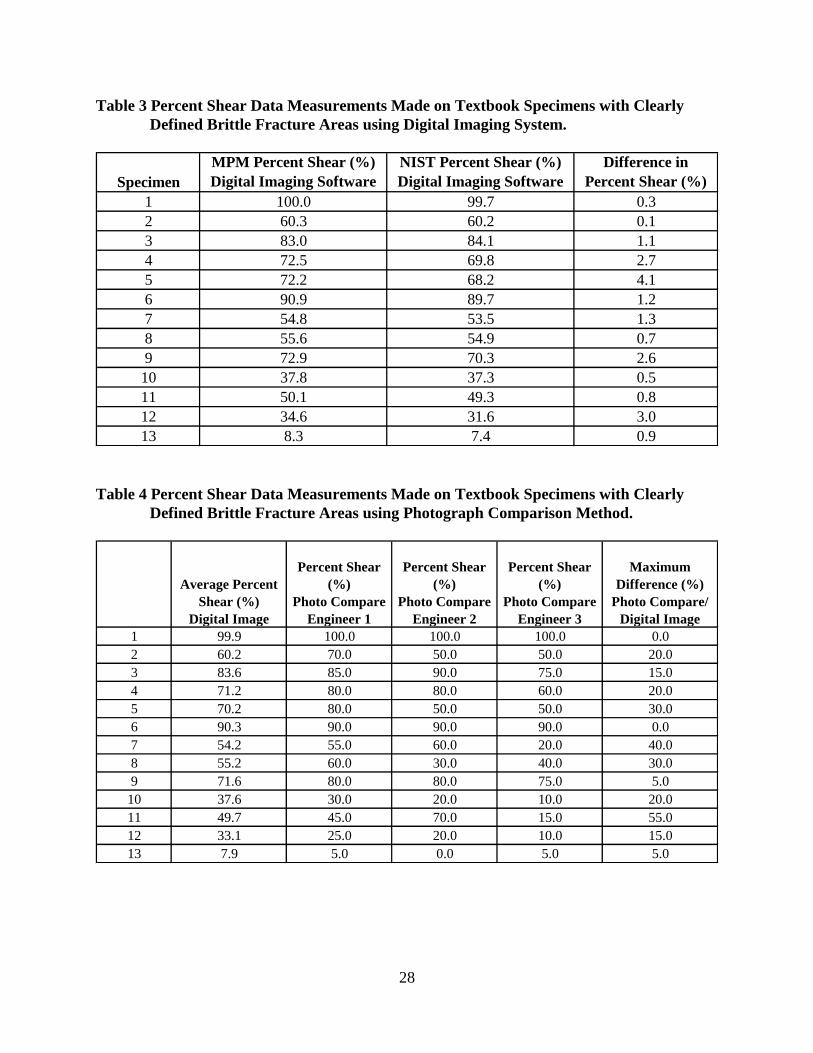
The results are shown in Figure 8 and in Tables 3 and 4. These results show that, when
trained personnel in the Charpy impact testing field use digital imaging to analyze specimens
containing definitive brittle areas, the percent shear values are precise. The average percent
difference between laboratories was only 1.5 % shear, and the maximum difference for any single
measurement was only 4 % shear. Since three engineers were able to obtain very similar results,
and since the previous section of the paper has established that any region that can be seen can be
accurately integrated to obtain the correct area, we have concluded that the image analysis results
are also accurate.
A comparison of the digital imaging data and the photograph comparison method is
shown in Table 4. The average difference between the digital imaging result and the photograph
comparison method is 19.6 % shear, with the largest deviation being 55 % shear. Figure 8 also
shows that photograph comparison results, at percent shear levels over about 90 %, are accurate
for all participants. In the lower data range, the Engineer 3 seems to display a bias using the
photograph comparison method toward low values. On the other hand, Engineers 2 and 3 tend to
be biased high for percent shear measurements in the 50 to 90 % shear range.
Based on experience and these results, it is recommended that the E 23 standard should
only allow the photograph comparison method as a qualitative approximation of SFA.
Furthermore, techniques such as digital imaging and the use of a planimeter should be
recommended as the primary SFA measurement methods.
Analysis of Subsize Round Robin Specimens
In many industrial applications, the conditions for measurement of SFA are far from
ideal. Therefore, in the third phase of this study, we measured a set of specimens used in a round
robin organized by Lake Erie Steel (LES). In the round robin, five companies analyzed the
percent shear of 87 specimens of various sizes (mostly subsized Charpy specimens). These
participants used either the photograph comparison method or the planimeter method.
MPM Technologies, Inc. (MPM) and the National Institute for Standards and
Technology (NIST) used digital imaging to evaluate the 87 round robin specimens. Unlike the
actual participants in the round robin, however, we established several criteria for analyzing the
specimens. This allowed us to better classify the various abnormalities as either ductile or brittle.
Further details concerning the fracture surface interpretation guidelines are discussed later.
Therefore, comparisons between MPM/NIST data and the data of the five participants should be
made with these in mind.
Digital Analysis Procedures for the LES Round Robin
As previously discussed, percent shear measurement via digital image analysis involves
capturing the image of the fracture surface, outlining the brittle area and outside region of the
fracture surface, and automatic software integration of the areas to determine SFA. Typically,
specimens exhibit clear indications of brittle and ductile fracture areas. However, the LES round

9
robin specimens were mostly subsize specimens with extensive plastic deformation. In addition,
the materials tested showed mixed ductile/brittle fracture (localized mixed mode fracture), and,
in several specimens, cracks normal to the main crack plane were observed (Figure 9). These
characteristics on the fracture surface necessitate special analysis procedures. To ensure
consistent data measurement, the following procedure was used for determining percent shear:
1. Calibration with a precision target was done before each day of measurements.
2. Each specimen was examined under a stereoscope and notes were taken on its fracture
appearance.
3. As shown in the examples in Figures 9 and 10, a digital photograph of each specimen was
taken at approximately 11X magnification, and the brittle area and outer area were
outlined in the software.
4. The percent shear and areas were calculated and recorded in a spreadsheet and plotted
(Figure 11).
5. A screen shot of the photographed specimen and outline was printed on a color printer for
reference.
Several assumptions were made pertaining to the calculation of the brittle area. These
assumptions are important since the atypical fracture surfaces of these specimens are not
anticipated in the ASTM E 23 standard. The irregularities include, but are not limited to: large
amounts of plastic deformation; localized mixed mode fracture (that is, an area with evidence of
ductile fracture with ―pockets‖ of brittle fracture); fracture surface oxidation; and cracks
perpendicular to the main fracture plane. These characteristics make the fracture surface analysis
difficult as they extend beyond the scope of the E 23 standard. With these factors involved, we
developed a set of criteria to make our analysis more consistent between operators:
First, the analysis was performed in accordance with the guidance of E 23
whenever possible.
Second, the total area was measured based on the post-fracture total area. That is,
plastic deformation is included in the total outer area calculation (not the
uncracked ligament area).
Third, all flat regions characteristic of fast fracture (especially on oxidized
specimens where there were no decisive faceted, bright, shiny regions) were
considered to be brittle areas.
Fourth, crack openings perpendicular to the fracture surface, even if shiny on the
inner crack enclave region, were not included in the brittle area calculation. This
is because the opening of the crack is due to plasticity and its area should not be in
the brittle are calculation.
Finally, any area with localized mixed mode fracture (―pockets‖ of brittle fracture
in the ductile region) was said to be 100 % brittle. This criterion was adopted in
order to provide conservative results for end users in terms of safety of structures.
Each of these assumptions is consistent with the spirit of the E 23 standard and provides a
conservative measurement of the undesirable brittle fracture percentage. With these factors in
mind, each specimen required careful examination.

10
Round Robin Digital Analysis Results
The digital analysis results are plotted in Figure 11. The data for the two independent
imaging systems shows an average difference of 4.3 % SFA over the 87 specimens measured,
and a maximum difference of 15.1 % SFA. It is interesting to note that the agreement is very
close for SFA measurement up to about 50%, and it is again close at levels in excess of about 80
%. The transition region SFA data in the 55 % to 80 % range are more scattered due to mixed
mode fracture regions and other effects such as cracking normal to the main crack front. In this
region of SFA, differences between results were as large as 15 % SFA. These differences are due
to the bias of the operator since the brittle area for some specimens was very difficult to see.
Reference [9] reported results for a round robin using conventional Charpy specimens. In that
study it was concluded that the level of agreement among participants was greatest when they
were experienced, the results were close to the FATT, and simple, two-dimensional photographs
were used for comparison. The Reference [9] work, as well as the current study, point to the
importance of having well established rules for interpretation of the fracture surface features to
minimize bias of the analyst.
Round Robin Results for the Five Participants
As shown in Figure 12, data obtained via a combination of the photograph comparison
method and the planimeter method resulted in a great deal of scatter. Differences as large as 70
% shear were reported, and the average maximum to minimum difference of the 5 participants
for the 87 specimens was 41.9 % SFA. The subsized specimens are even more difficult to
accurately analyze using photograph comparison, since only full-sized specimens are pictured in
the current ASTM E23 standard. In addition, the specimens were hard to analyze due to the
presence of oxidation, splits, and high levels of plastic deformation. Further discussion of the
round robin results follows.
Figure 12 shows a much larger amount of scatter from lab-to-lab in the determination of
percent shear by the five LES round robin participants. At this poor level of accuracy and low
precision, further analysis of the round robin data is not meaningful other than to note that the
scatter for the five labs is somewhat lower at the low and high SFA levels.
As discussed earlier, two sources for the scatter have been identified: operator bias and
method error. The error caused by an operator’s bias is simply an operator’s inability to see
and/or to physically interpret what is seen as either brittle area or ductile area. Method error, on
the other hand, varies significantly according to the method used. To date, the two methods that
are most commonly used are the photograph comparison method (method 2 listed in the ASTM
E 23 standard) and the digital imaging method (method 5 listed in the E 23 standard). Between
these, the photograph comparison method is the most widely used in the steel industry.
However, this method, according to the E 23 standard, is the second least accurate method in
measuring percent shear. Therefore, given the difficulty in comparing subsize photographs to
full size, and the highly deformed fracture surfaces, it would be relatively easy to be three or four
photographs away from the correct photograph, resulting in error bands on the order of 40 to 70%
shear, which is observed in Figure 12. The digital imaging method, as shown in the earlier

11
portions of the paper, however, nearly eliminates the method error. The much closer agreement
of the MPM and NIST digital imaging results demonstrates this point.
Percent Shear Area from Instrumented Data
Most industries that use Charpy parameters for regulation or as a material acceptance
criterion use the absorbed energy parameter. It is likely that absorbed energy was selected over
percent shear because, in the past, accurate measurement of percent shear was tedious since it
required area integration using a planimeter. Additionally, some materials do not exhibit clear
evidence of brittle fracture on the fracture plane. For these materials analysis using optical
imaging is not possible. However, there are available today, optical imaging systems which are
easy to use and which give very accurate results. Modern instrumented striker test systems can
provide very accurate Charpy load-deflection data that can be used to determine the brittle crack
initiation load and crack arrest loads. These critical loads can be put into physically based
models for calculation of the percent shear fracture area. This solves the problem of not being
able to see brittle areas on the fracture surface, especially known to occur for some weld metals.
The use of critical loads to determine SFA can also be used in conjunction with optical methods
to check results of the direct measurements. Although the use of instrumented data in
conjunction with digital imaging to determine SFA has not been used much in the past, we
recognize this as a powerful tool for the future determination of accurate SFA data.
Recommendations for ASTM E 23 Standard
It is recommended that the E 23 standard be amended to include important changes
related to SFA measurement and reporting. A summary of the proposed changes to the standard
follows:
The standard should clearly define the total area for percent shear calculation as
the total post-fracture area measured on the sample. The standard does not
currently state that the plastically deformed area is included in the total outer area
calculation. Some users may incorrectly assume that the outer area is calculated
based on the uncracked ligament area.
All flat regions (where there is no decisive faceted, bright, shiny regions) should
be considered to be brittle (fast fracture) area.
Any crack perpendicular to the main fracture surface, even if shiny on the inner
crack enclave region, must not be included in the brittle area calculation. This is
because the opening of the crack is due to plasticity, and its area should not be in
the brittle area calculation.
Any area with localized mixed mode fracture (that is, an area with evidence of
ductile fracture with ―pockets‖ of brittle fracture) should be defined to be a 100%
brittle region on the fracture surface. This criterion is recommended in order to
provide conservative results for end users in terms of safety of structures.
The ranking of methods should be carefully quantified to indicate the expected
accuracy and precision that can be obtained. Based on experience and the results

12
from the textbook specimen analysis of this study, it is recommended that the E
23 standard should only allow the photograph comparison method as a qualitative
approximation of SFA.
Direct area measurement techniques such as digital imaging and the use of a
planimeter should be recommended as the primary, recommended methods.
Other methods, such as photograph comparison, should be moved to an Annex
and defined as alternative methods of low accuracy for use only in cases where
area measurement techniques are not available.
Additional recommendations to reduce operator bias should be added to the
standard. For example, these recommendations can include analyst training,
establishing rules to define the brittle fracture region, the use of optical
stereoscopes, and the use of instrumented striker data.
Percent shear should only be measured on samples tested in the transition region
where mixed mode fracture occurs. For example, a material showing a fully
ductile instrumented curve (no sudden drop due to fast fracture) is by definition
100 % ductile. Users should not be confused by the fact that upper and lower
shelf SFA can be determined without the need for image analysis, and
instrumented striker data can be used exclusively to define this behavior.
The use of supplementary techniques should be recommended to check the digital
imaging whenever possible. Examples include load-deflection data from
instrumented strikers and examination under the stereoscope. In cases where
instrumented striker data are available, the 100 % shear and 100 % brittle
specimens can be identified based solely on the instrumented striker signal. The
instrumented striker is extremely accurate for determination of whether there are
brittle fracture events during a test. Further, equations have been developed that
use the instrumented striker brittle fracture initiation and arrest loads to calculate
SFA, and these can be used to check the digital image measurements.
Summary and Conclusions
In summary, the accuracy and precision of digital analysis methods are found to be more
than adequate, as expected, with results similar to the planimetric methods previously used. Our
overall conclusion is that if the brittle fracture area can be seen optically, percent shear fracture
area can be measured precisely with high accuracy. As the complexity of the fracture surface
features increase, however, error due to operator bias is expected to increase. Digital analysis
results from the ―textbook‖ series, compared with the subsized round robin samples, quantify this
observation well: an average difference of 1.5 % SFA and a maximum difference of 4 % SFA for
―textbook‖ fracture surfaces defines the low bias case; an average difference of 4.3 % SFA, and a
maximum difference of 15.1 % SFA for the round robin specimens defines the high bias case.
The low bias case for the textbook specimens is likely fixed since the measurements were made
on pristine fracture surfaces using state-of-the-art digital imaging technology. However, the
precision for the high bias case can be improved in the future with the development of better
procedures for interpretation of the fracture surface features, including the E 23
recommendations given in this paper, to reduce bias between operators. Further improvements

13
can be realized in the future by using SFA results determined from instrumented striker data to
check the digital analysis results.
Our study has also led to the conclusion that the widely used photograph comparison
method is neither accurate, nor precise and should be abandoned for quantitative measurements.
In particular, the results for the photograph comparison method on the ―textbook‖ specimens
gave an average difference between the digital imaging result and the photograph comparison
method of 19.6 % shear, with the largest deviation being 55 % shear. This alone is sufficient to
demonstrate that the photograph comparison method is inaccurate and imprecise. However, the
LES round robin even more dramatically demonstrates the limitations of the photograph
comparison method. Variations as large as 70 % shear were reported on individual specimen
measurements, and the average maximum to minimum deviation of the 5 participants over the
entire range of 87 specimens was 41.9 % SFA.
Recommendations for changes to the E 23 standard have been made. These proposed
changes will make measurement of percent shear for typical specimens more clearly defined and
will give the user of the standard better guidance on the error to be expected for the various
methods. In addition, changes to the standard to address fracture surface features not currently
anticipated by the current standard must be included.
The general conclusion from this work is that SFA can be accurately measured and
should be considered as a useful Charpy parameter. In typical Charpy specimen applications,
accuracies of about 1 % to 5 % shear area should be expected. Arguments have been put forward
in support of the idea that the SFA parameter, and the FATT index, are more fundamental and
physically based for the determination of the safe temperature for operation of structures and
components than an energy based parameter and index such as 30 ft-lbs.

14
Acknowledgements
The authors are grateful to Lake Erie Steel, and the five round robin participants, for
providing the round robin test specimens and for assistance in the literature survey. The authors
are also grateful to Mr. Bob Carter of the Electric Power Research Institute (EPRI) and the
members of the BWR Vessel Internals Project (BWRVIP) for providing the irradiated Charpy
test data for use in this work.
References
1. Russell, S.B., ―Experiments with a New Machine for Testing Materials by Impact,‖
Transactions ASCE, Vol. 39, June 1898, pp. 237 - 250.
2. Charpy, M.G., ―Note sur l’Essai des Metaux a la Flexion par Choc de Barreau Entailles, Soc.
Ing. Francais, June, 1901, p. 848.
3. Manahan, Sr., M.P., ―Advances in Notched Bar Impact Testing: A 100 Year Old Test Gets a
Face Lift‖, ASTM Standardization News, October 1996, pages 23 – 29.
4. Siewert, T.A., Manahan, Sr., M.P., et. al., ―The History and Importance of Impact Testing‖,
Pendulum Impact Testing: A Century of Progress, STP 1380, T. A. Siewert and M. P.
Manahan, Sr. Eds., American Society for Testing and Materials, West Conshohocken, PA,
2000.
5. Tests of Met’s, Vol. I, 1881, pp 122-146, 212-222, ―Results of impact tests of various grades
of wrought iron with photographs of fractures.
6. Proceedings of ASTM, Vol. 2, 1902, Appendix II, Bibliography on Impact Tests and Impact
Testing Machines, W. K. Hatt, and E. Marburg.
7. Bibliography of Impact Testing, September, 1925, White, A.E., Clark, C.L.
8. ASTM E23-07, ―Standard Test Methods for Notched Bar Impact Testing of Metallic
Materials‖.
9. Dixon, B.F., ―Reliability of Fracture Appearance Measurement in the Charpy Test‖, Welding
Journal, August 1, 1994.

15
Figure 1 Textbook Fracture Surface Showing Bright Shiny Faceted Region Surrounded by
Ductile Fracture Area.

16
Figure 2 Example of an Instrumented Striker Signal for a Charpy Test Conducted in the
Transition Region, Showing Sudden Drop in Load Associated with Brittle or Fast
Fracture Event.

17
0.0
10.0
20.0
30.0
40.0
50.0
60.0
70.0
80.0
0.00E+00 5.00E+17 1.00E+18 1.50E+18 2.00E+18 2.50E+18 3.00E+18 3.50E+18
Sh
ear
Fra
ctu
re A
pp
eara
nce
at 3
0 f
t-lb
In
dex
(%
)
Fast Fluence (E>1MeV) (n/cm^2)
Boiling Water Reactor Surveillance Data
ΔT30=169.2 F,ΔUSE=-23.0
ΔT30=122.1 F,ΔUSE=-21.6
ΔT30=189.3 F,
ΔUSE=-27.5
ΔT30=124.5 F,ΔUSE=-30.0
ΔT30=61.9 F,ΔUSE=-25.1 ΔT30=139.0 F,ΔUSE=-17.4
ΔT30=22.5 F,ΔUSE=-3.8
ΔT30=4.1 F,ΔUSE=-3.0
ΔT30=103.5 F,ΔUSE=-35.5
ΔT30=80.6 F,ΔUSE=-9.6
ΔT30=76.3 F,
ΔUSE=-13.3
ΔT30=53.7 F, ΔUSE=-20.0
Case 1
Case 2
Figure 3 Plot of SFA at 30 ft-lb as a Function of Fast Neutron Fluence for Irradiated
Welds. Note that the Charpy Shift Measured at the 30 ft-lb Index does not
Increase Proportionally with SFA.

18
0
25
50
75
100
125
150
-300 -200 -100 0 100 200 300 400 500 600
CV
N E
ner
gy
(ft
-lb
)
Test Temperature (F)
Charpy Data for Irradiated Weld 1
Unirradiated Data
Irradiated
Unirradiated Fit
Irradiated Fit
30 ft-lb
index
30 ft-lb
index
Figure 4 Plot of the Case 1 Charpy Energy-Temperature Curve. For this Material, the
Change in Slope Results in a Large Change (~45%) in SFA at the 30 ft-lb Index.

19
0
25
50
75
100
125
150
-300 -200 -100 0 100 200 300 400 500 600
CV
N E
ner
gy
(ft
-lb
)
Test Temperature (F)
Charpy Data for Irradiated Weld 2
Unirradiated Data
Irradiated
Unirradiated Fit
Irradiated Fit
30 ft-lb
index
30 ft-lb
index
Figure 5 Plot of the Case 2 Charpy Energy-Temperature Curve. For this Material, the
Change in Slope Results in Only a Modest Change (~18%) in SFA at the 30 ft-lb
Index.

20
Figure 6 Image Acquisition and Analysis Hardware for Accurate Percent Shear
Measurement.

21
Figure 7 Enlarged Image of CAD Drawings used for Precision Study.

22
0
10
20
30
40
50
60
70
80
90
100
0 2 4 6 8 10 12 14
Per
cen
t S
hea
r (%
)
Specimen Number
Comparison between Digital Imaging and Photograph Comparison
Photo Comparison MPM Engineer 1Photo Comparison MPM Engineer 2Photo Comparison NIST Engineer 1MPM Digital Imaging
NIST Digital Imaging
Figure 8 Comparison of Percent Shear Data Obtained Using Digital Imaging with Results
Obtained Using Photograph Comparison Method.

23
Figure 9 Example Showing Outlines for the Brittle and Total Fracture Areas for a Test
Specimen with a Large Crack Normal to the Primary Plane of Fracture.

24
Figure 10 Example Showing the Outlined Brittle and Total Fracture Areas, for a Sample
Having a Flat Fast Fracture Region Lacking Clear Brittle Facets Throughout.

25
0.0
5.0
10.0
15.0
20.0
25.0
30.0
35.0
40.0
45.0
50.0
55.0
60.0
65.0
70.0
75.0
80.0
85.0
90.0
95.0
100.0
0 10 20 30 40 50 60 70 80 90
Per
cen
t S
hea
r (%
)
Specimen Number
(arranged in order of increasing average shear measurement)
MPM & NIST Data for Lake Erie Steel
Round Robin
MPM
NIST
Figure 11 Comparison of Round Robin Percent Shear Data Obtained by NIST and MPM
using Digital Imaging Technology.

26
0
10
20
30
40
50
60
70
80
90
100
0 20 40 60 80
% S
hea
r M
easu
rem
ent
Specimen Number(arranged in increasing order of average shear measurement)
% Shear Measurements for Lake Erie Steel Round Robin
Lab-to-Lab Comparison
LES Data Bounds (photo
comparison/planimeter)
MPM (digital imaging)
NIST (digital imaging)
Figure 12 Comparison of Data from the LES Round Robin with Results using Digital
Analysis Technology from this Study. The Round Robin Data were Collected
using Photograph Comparison and Planimeter Methods.

27
Table 1 Area Measurements Made Using a Calibration Reticle.
Non-technical
Assistant
Trained
Engineer
Target Value 0.0400 0.0400
Measured Average 0.0400 0.0400
% Difference 0.0631 0.0193
Std Dev 3.479E-05 1.131E-06
Precison Reticle Area (in2)
Table 2 Percent Shear Data Measurements Made on CAD Drawings using Digital Imaging
System.
Trained Engineer 1
Target 20.0 40.0 60.0 80.0 40.0 60.0 80.0
Average 20.9 40.4 60.1 79.5 41.1 60.2 79.8
Std Dev 0.249 0.309 0.082 0.047 0.170 0.170 0.082
Trained Engineer 2
Target 20.0 40.0 60.0 80.0 40.0 60.0 80.0
Average 20.2 39.8 59.5 80.1 40.1 59.8 79.9
Std Dev 0.090 0.073 0.051 0.045 0.232 0.093 0.194
Non-technical Staff Member
Target 20 40 60 80 40 60 80
Average 20.00 40.00 59.60 80.30 40.53 60.43 80.27
Std Dev 0.0816 0.0816 0.0000 0.0816 0.0471 0.0943 0.0471
Percent Shear - Square Percent Shear - Round
Percent Shear - Square Percent Shear - Round
Percent Shear - RoundPercent Shear - Square
28
Table 3 Percent Shear Data Measurements Made on Textbook Specimens with Clearly
Defined Brittle Fracture Areas using Digital Imaging System.
Specimen
MPM Percent Shear (%)
Digital Imaging Software
NIST Percent Shear (%)
Digital Imaging Software
Difference in
Percent Shear (%)
1 100.0 99.7 0.3
2 60.3 60.2 0.1
3 83.0 84.1 1.1
4 72.5 69.8 2.7
5 72.2 68.2 4.1
6 90.9 89.7 1.2
7 54.8 53.5 1.3
8 55.6 54.9 0.7
9 72.9 70.3 2.6
10 37.8 37.3 0.5
11 50.1 49.3 0.8
12 34.6 31.6 3.0
13 8.3 7.4 0.9
Table 4 Percent Shear Data Measurements Made on Textbook Specimens with Clearly
Defined Brittle Fracture Areas using Photograph Comparison Method.
Average Percent
Shear (%)
Digital Image
Percent Shear
(%)
Photo Compare
Engineer 1
Percent Shear
(%)
Photo Compare
Engineer 2
Percent Shear
(%)
Photo Compare
Engineer 3
Maximum
Difference (%)
Photo Compare/
Digital Image
1 99.9 100.0 100.0 100.0 0.0
2 60.2 70.0 50.0 50.0 20.0
3 83.6 85.0 90.0 75.0 15.0
4 71.2 80.0 80.0 60.0 20.0
5 70.2 80.0 50.0 50.0 30.0
6 90.3 90.0 90.0 90.0 0.0
7 54.2 55.0 60.0 20.0 40.0
8 55.2 60.0 30.0 40.0 30.0
9 71.6 80.0 80.0 75.0 5.0
10 37.6 30.0 20.0 10.0 20.0
11 49.7 45.0 70.0 15.0 55.0
12 33.1 25.0 20.0 10.0 15.0
13 7.9 5.0 0.0 5.0 5.0