Peptide binding characteristics of the non-classical class Ib MHC molecule HLA-E assessed by a...
12
BMC Immunology (2001) 2:5 http://www.biomedcentral.com/1471-2172/2/5 BMC Immunology (2001) 2:5 Research article Peptide binding characteristics of the non-classical class Ib MHC molecule HLA-E assessed by a recombinant random peptide approach James Stevens* 1,2 , Etienne Joly 1,3 , John Trowsdale 2 and Geoffrey W Butcher 1 Address: 1 Laboratory of Functional Immunogenetics, The Babraham Institute, Cambridge CB2 4AT, UK, 2 Department of Pathology, Division of Immunology, University of Cambridge, Tennis Court Road, Cambridge CB2 1QP, UK and 3 UPCM, CNRS UPS 2163, CHU Purpan, 31300 Toulouse, France E-mail: James Stevens* - [email protected]; Etienne Joly - [email protected]; John Trowsdale - [email protected]; Geoffrey W Butcher - [email protected] *Corresponding author Abstract Background: Increasing evidence suggests that the effect of HLA-E on Natural Killer (NK) cell activity can be affected by the nature of the peptides bound to this non-classical, MHC class Ib molecule. However, its reduced cell surface expression, and until recently, the lack of specific monoclonal antibodies hinder studying the peptide-binding specificity HLA-E. Results: An in vitro refolding system was used to assess binding of recombinant HLA-E to either specific peptides or a nonamer random peptide library. Peptides eluted from HLA-E molecules refolded around the nonamer library were then used to determine a binding motif for HLA-E. Hydrophobic and non-charged amino acids were found to predominate along the peptide motif, with a leucine anchor at P9, but surprisingly there was no methionine preference at P2, as suggested by previous studies. Conclusions: Compared to the results obtained with rat classical class Ia MHC molecules, RT1- A1 c and RT1-A u , HLA-E appears to refold around a random peptide library to reduced but detectable levels, suggesting that this molecule's specificity is tight but probably not as exquisite as has been previously suggested. This, and a previous report that it can associate with synthetic peptides carrying a viral sequence, suggests that HLA-E, similar to its mouse counterpart (Qa-1 b ), could possibly bind peptides different from MHC class I leader peptides and present them to T lymphocytes. Background Non-classical MHC class Ib molecules are closely homol- ogous to classical class Ia molecules but are distin- guished by their limited polymorphism and low cell surface expression. Contrary to some views expressed in the past, class Ib molecules are not just vestigial evolu- tionary remnants of classical class Ia molecules: rather some are endowed with important highly specialized roles, as testified by their conservation between different species. In this regard, the trio comprised of HLA-E in human, Qa-1 in mouse and RT.BM1 in rat constitutes the only group of class Ib molecules where clear homologues Published: 20 June 2001 BMC Immunology 2001, 2:5 Received: 15 March 2001 Accepted: 20 June 2001 This article is available from: http://www.biomedcentral.com/1471-2172/2/5 (c) 2001 Stevens et al, licensee BioMed Central Ltd.
-
Upload
independent -
Category
Documents
-
view
2 -
download
0
Transcript of Peptide binding characteristics of the non-classical class Ib MHC molecule HLA-E assessed by a...

BMC Immunology (2001) 2:5 http://www.biomedcentral.com/1471-2172/2/5
BMC Immunology (2001) 2:5Research articlePeptide binding characteristics of the non-classical class Ib MHC molecule HLA-E assessed by a recombinant random peptide approachJames Stevens*1,2, Etienne Joly1,3, John Trowsdale2 and
Geoffrey W Butcher1
Address: 1Laboratory of Functional Immunogenetics, The Babraham Institute, Cambridge CB2 4AT, UK, 2Department of Pathology, Division of Immunology, University of Cambridge, Tennis Court Road, Cambridge CB2 1QP, UK and 3UPCM, CNRS UPS 2163, CHU Purpan, 31300
Toulouse, France
E-mail: James Stevens* - [email protected]; Etienne Joly - [email protected]; John Trowsdale - [email protected];
Geoffrey W Butcher - [email protected]
*Corresponding author
AbstractBackground: Increasing evidence suggests that the effect of HLA-E on Natural Killer (NK) cellactivity can be affected by the nature of the peptides bound to this non-classical, MHC class Ibmolecule. However, its reduced cell surface expression, and until recently, the lack of specificmonoclonal antibodies hinder studying the peptide-binding specificity HLA-E.
Results: An in vitro refolding system was used to assess binding of recombinant HLA-E to eitherspecific peptides or a nonamer random peptide library. Peptides eluted from HLA-E moleculesrefolded around the nonamer library were then used to determine a binding motif for HLA-E.Hydrophobic and non-charged amino acids were found to predominate along the peptide motif,with a leucine anchor at P9, but surprisingly there was no methionine preference at P2, as suggestedby previous studies.
Conclusions: Compared to the results obtained with rat classical class Ia MHC molecules, RT1-A1c and RT1-Au, HLA-E appears to refold around a random peptide library to reduced butdetectable levels, suggesting that this molecule's specificity is tight but probably not as exquisite ashas been previously suggested. This, and a previous report that it can associate with syntheticpeptides carrying a viral sequence, suggests that HLA-E, similar to its mouse counterpart (Qa-1b),could possibly bind peptides different from MHC class I leader peptides and present them to Tlymphocytes.
BackgroundNon-classical MHC class Ib molecules are closely homol-
ogous to classical class Ia molecules but are distin-
guished by their limited polymorphism and low cell
surface expression. Contrary to some views expressed inthe past, class Ib molecules are not just vestigial evolu-
tionary remnants of classical class Ia molecules: rather
some are endowed with important highly specialized
roles, as testified by their conservation between different
species. In this regard, the trio comprised of HLA-E in
human, Qa-1 in mouse and RT.BM1 in rat constitutes theonly group of class Ib molecules where clear homologues
Published: 20 June 2001
BMC Immunology 2001, 2:5
Received: 15 March 2001Accepted: 20 June 2001
This article is available from: http://www.biomedcentral.com/1471-2172/2/5
(c) 2001 Stevens et al, licensee BioMed Central Ltd.

BMC Immunology (2001) 2:5 http://www.biomedcentral.com/1471-2172/2/5
have been identified in all three species. A major role of
this group of molecules has recently emerged in the reg-
ulation of Natural Killer (NK) cell activity, through inter-
action with both the inhibitory CD94-NKG2A receptorand the activatory CD94-NKG2C receptor [1,2,3]. For
cell surface expression, these MHC molecules preferen-
tially bind peptides derived from the signal peptides of
other MHC class I molecules by a TAP-dependent mech-
anism [4,5,6]. Hence, expression of other class I heavy
chain polypeptides regulate the expression of HLA-E and
it is thought that this in turn enables NK cells to monitor
the state of the MHC class I-dependent antigen presen-
tation pathway in the cells they inspect. Thus, the level of
cell surface HLA-E is critical for NK cell cytotoxicity to-
wards certain tumour and viral-infected cells, and a re-
cent report suggests that viruses that shut down MHC
class I expression may evolve mechanisms to maintain
HLA-E expression [7].
However, not all leader sequences from human class I
MHC molecules contain peptides that are able to bind to
HLA-E. For example, sequences derived from certain
HLA-B alleles, that contain a threonine for methionine
substitution at the P4 position of the leader peptide (P2
position of the processed peptide), were not able to bind
to HLA-E in an in vitro binding assay [3]. Furthermore,
transfectants of the HLA-B alleles carrying Thr at P4 into
721.221 cells could not inhibit killing by CD94/NKG2A
NK clones in contrast to those from other HLA-A, -B, -Cand -G alleles with a Met at P4. Analysis of the crystal
structure of HLA-E seemed to confirm this stringent
peptide requirement since it showed the occupation of all
the pockets and the involvement of all the peptide side
chains in burying the peptide deep in the groove [8].
However, recent results have shown that the sequence of
the bound peptide can influence binding to both the
CD94/NKG2A and CD94/NKG2C receptors in both cel-
lular and in vitro binding assays [9,10,11]. In addition,
multiple studies (reviewed in [12]) have shown that Qa-1
can carry out antigen presentation to γδ and/or CD8+ T
cells. This suggests that HLA-E, similar to its mouse
counterpart, could possibly bind antigenic peptides dif-
ferent from MHC class I leader peptides, and present
them to T lymphocytes.
In the present study, we aimed to determine the peptide
binding specificity of HLA-E via a purely biochemical ap-
proach based on an in vitro refolding system. Any such
study within a biological system requires not only access
to sufficient material, but also the availability of a specif-
ic antibody by which the class I molecule can be efficient-
ly purified from all other cellular components. For the
classical class Ia molecules, this is usually not a problem
since cell surface expression is high and specific antibod-ies are often available. For HLA-E, low expression and
the lack of a truly specific antibody has thus far hindered
attempts to obtain a peptide binding motif although a re-
cent report described the production and characterisa-
tion of a specific monoclonal antibody called V16 [13].
ResultsCloning and expression of HLA-EUsing PCR, we engineered an expression plasmid to pro-
duce a soluble form of the heavy chain of HLA-E
(E*0102) with a C-terminal His-Tag sequence. Under
IPTG induction, the heavy chain was expressed as inclu-
sion bodies and therefore required urea solubilization
before purification could be performed. Solubilized
heavy chain was purified on a Ni-NTA agarose column to
>95 % purity as determined by SDS-PAGE. The total
yield of HLA-E was approximately 88 mg/l of bacterial
culture. This eluted off the Ni-NTA column as two spe-
cies, one in pH 5.9 buffer and the other at pH 4.5. SDS-
PAGE in the presence/absence of reducing agent con-
firmed the mixture eluting in the pH 5.9 buffer to be
mainly composed of α chain monomers, whilst protein
eluting in the pH 4.5 buffer comprised large molecular
weight multimers (results not shown), similar to what we
had previously experienced with rat MHC class Ia mole-
cules [14]. Only the monomer fraction was used for re-
folding work (yield 21 mg/l). Protein sequencing of the
monomer fraction revealed that the bacteria had suc-
cessfully cleaved the initiation methionine to give the ex-
pected N-terminus (GSHSLKYFH). Human β2-microglobulin was expressed and purified, as described
previously for the rat form [14]. Elution of monomeric
human β2-microglobulin from the Q-Sepharose column
(>95 % pure by SDS-PAGE) was achieved with 100 mM
NaCl with a yield of approximately 23 mg/l of bacterial
culture.
Binding assay for specific peptidesWe have previously reported the successful use of in vit-
ro refolding of bacterially produced recombinant pro-
teins to study the peptide-binding properties of rat MHC
class I molecules [14]. To validate that a similar system
could be applied to HLA-E, small scale refolding experi-
ments were performed using recombinant human β2 mi-
croglobulin and a peptide derived from the HLA-A2
leader peptide, VMAPRTLVL, which is known to bind to
HLA-E [4]. Refolding of HLA-E to produce a monomeric
complex of correct molecular weight was assessed by gel
filtration, over a range of peptide concentrations. Quan-
titative comparisons between refolding experiments
could not be performed directly because loss of material
through precipitation was variable, as judged from the
total peak areas observed during gel filtration (results
not shown). The extent of refolding was therefore as-
sessed by expressing results as an induction ratio, calcu-lated as the proportion of monomers detected in the

BMC Immunology (2001) 2:5 http://www.biomedcentral.com/1471-2172/2/5
presence of peptide (relative to the combined area of
monomeric and aggregate material), compared to the
proportion of monomers recorded without peptide (see
Materials and Methods section for the exact calculation).
Results presented in Figure 1 show that a plateau of max-
imum induction was reached with 10 µM of the HLA-A2
leader peptide, whilst 0.1 µM of this peptide led to 50%
maximal induction.
For a negative control, we used a 13-mer peptide, ILFPS-
SERLISNR, derived from the rat mitochondrial A chain
of ATP synthase, which corresponds to the rat maternal-
ly transmitted minor histocompatibility peptide (MTF-
E) [15]. As expected, this peptide did not induce any sig-
nificant refolding (Fig.1). Six other peptides that were
identified as potential binders within our laboratory's
collection were also tested for binding to HLA-E andcompared to the HLA-A2 leader-derived peptide. To
simplify binding assays, all these were tested at 10 µM,
the concentration required to reach maximum induction
with the A2-derived control peptide, VMAPRTLVL. As
shown in Figure 2A, refolding efficiency similar to that of
the positive control was attained with AMAPRTLLL, the
corresponding peptide found in the leader sequence of
mouse H2-D and H2-L and in rat RT1-A class Ia mole-
cules. To assess the importance of the position of anchor
residues, we used two other nonamer peptides derived
from the murine leader sequence, but shifted by either
one or two residues. The fact that both peptides were
found to bind poorly to HLA-E, (MAPRTLLLL: 5 %,
APRTLLLLL: 23 %) suggests that the precise positioning
of anchor residues inside the groove is crucial for the ef-
ficient binding of peptides derived from class I leader se-
quences.
Figure 1Effect of peptide concentration on refolding recombinant HLA-E. Truncated heavy chain of HLA-E was refolded bythe dilution method [14, 30, 31] in the presence of light chain human β2-microglobulin and varying concentrations of peptides.Gel filtration was used to assess successful refolding by separating monomeric complexes from aggregate species. Refoldinglevels were calculated as induction ratios (see Materials and Methods). Results presented are representative of three independ-ent experiments.
1.0
1.2
1.4
1.6
1.8
2.0
2.2
Indu
ctio
n R
atio
0.01 0.1 1.0 10.0 100.0
[Peptide](µM)
Random Peptide Library
ILFPSSERLISNR
VMPTSNDPTL
VMAPRTLVL

BMC Immunology (2001) 2:5 http://www.biomedcentral.com/1471-2172/2/5
Within our lab's collection, three other peptides were
identified that shared Met at P2 and/or Leu as C terminal
anchor residues with the canonical A2 peptide. Some-
what surprisingly, all three peptides induced significant
refolding: SMFPVSENR (32 %), NPRKVTAYL (23 %)
and VMPTSNDPTL (78%). For the latter one, which is a
decamer, we had access to sufficient amounts to carry
out a dose-response curve (Fig. 1). Whilst maximum in-
duction was reached at 10 µM, similarly to the of the
leader-derived canonical peptide, 10-fold more (1 µM)
peptide was required to obtain 50 % refolding of its ownmaximum refolding value.
As an additional control three of the peptides tested were
also used to refold the rat classical class Ia MHC mole-
cule RT1-A1c. Results show that two of the peptides,
NPRKVTAYL (a synthetic peptide designed from the
published binding motif of RT1-A1c [14]) and APRTLLL-
LL, which only induced partial refolding with HLA-E, did
induce substantial refolding of the rat class I molecule,
whilst conversely, the mouse leader derived peptide
(AMAPRTLLL) was relatively inefficient.
Figure 2Effect of various peptides on refolding recombinant HLA-E. A Various specific peptides were tested for their abilityto induce and stabilize the refolding of recombinant HLA-E. Peptides were added to the binding assay at 10 µM, the concentra-tion required for maximal observed refolding for the known binding peptide, VMAPRTLVL (see Figure 1). The rat class I MHCmolecule RT1-A1c was also used as a control to assess the validity of this refolding assay. Results shown are the average of 3independent experiments. B HLA-E was refolded with 100 µM nonamer random peptide library and its induction ratio wasdetermined and compared to two rat TAP-B associated classical Ia MHC molecules, RT1-A1c and RT1-Au. Results shown arethe average of 3 independent experiments.
1
1.5
2
HL
A-E
RT
1-A
1c
RT
1-A
u
1
1.5
2
Indu
ctio
n R
atio
VM
APR
TL
VL
AM
APR
TL
LL
MA
PRT
LL
LL
APR
TL
LL
LL
SMFP
VSE
NR
NPR
KV
TA
YL
VM
PTSN
DPT
L
NPR
KV
TA
YL
AM
APR
TL
LL
APR
TL
LL
LL
BA
HLA-E RT1-A1c 100 µM RandomPeptide Library

BMC Immunology (2001) 2:5 http://www.biomedcentral.com/1471-2172/2/5
Determination of an HLA-E binding motif based on binding of HLA-E to random peptide libraryHaving validated that the extent of HLA-E refolding
could be assessed after in vitro refolding, we went on totest the nonamer random peptide library in this system.
In doing so, we found that HLA-E could be induced to re-
fold around random nonamer peptides, but this was only
seen when 100 µM of the random peptide library was
used (see Figure 1). For three rat MHC class Ia mole-
cules, refolding studies with recombinant protein and
random peptide libraries have already yielded binding
motif information, with a good correlation to the motifs
obtained from peptides eluted from naturally expressed
molecules [14]. In this previous study, we had found that
RT1-A1c and RT1-Au required 10 fold more peptide to
achieve refolding efficiencies comparable to those ob-
tained for RT1-Aa with a heavy chain:β2m:peptide ratio
of 1:2:10. This was interpreted as an indication that RT1-
A1c and RT1-Au had more stringent peptide require-
ments than RT1-Aa. To ascertain that, under the condi-
tions used here, the refolding seen for HLA-E did
correspond to true binding of the peptides in the random
library, we therefore used refolding of RT1-A1c and RT1-
Au as controls. Results in Figure 2B show that for all
three class I molecules, there was a specific increase in
production of refolded material, although the induction
ratio of the nonamer random peptide library for HLA-E
(ratio = 1.14) was reproducibly less than the ratios of
both RT1-A1c (ratio = 1.26) and RT1-Au (ratio = 1.28).
Whilst the reduced efficiency of refolding and the high
amounts of peptide library required suggest an even
more stringent specificity than that of rat classical class
Ia MHC molecules we had studied previously, we could
nevertheless proceed to the determination of a binding
motif for HLA-E. Successfully refolded monomeric com-
plexes were purified by gel filtration chromatography,
then acidified to disrupt the complexes and release the
bound peptides, which were subjected to further purifi-
cation by reversed phase chromatography over a 0-90 %
acetonitrile gradient (see Figure 3). For the binding mo-
tif determination of rat class Ia molecules [14, 16], we
had used fractions collected between 6.75 % and 40.5 %
of the acetonitrile gradient, but for HLA-E, we found that
more stringent conditions were necessary for the elution
of the peptides, probably because of their increased hy-
drophobicity. Fractions covering the large peak eluting
at 5.45 ml (fractions 33-35) were submitted for pool se-
quencing separately. Results (not shown) revealed that
the peak contained little or no peptidic material. Se-
quencing of the peptides released in all other fractions
pooled together yielded the results and motif presented
in Figure 4. Dominant anchor residues (boxed values) as
determined previously [14, 17], were observed at posi-tions 4 (leucine), 7 (asparagine) and 9 (leucine). The
amino acids glutamine and phenylalanine were also seen
as significant increases at positions 2 and 3, respectively,
but were not large enough increases (compared to the
previous cycle) to be considered as anchor residues.Along the length of the binding motif all except one of the
observed increases (lysine at P6) were uncharged or hy-
drophobic amino acids. This is in contrast to the binding
motif reported recently for the mouse homologue Qa-1b,
and obtained with an approach similar to ours, which re-
vealed basic preferences (Lys/Arg) at positions 3, 4 and
5 in the motif and acidic preferences (Asp/Glu) at 3,4
and 8 [12].
DiscussionIn this study we have successfully used a previously de-
scribed recombinant system [14] to assess the peptide
binding specificity of the human non-classical class Ib
MHC molecule, HLA-E. Truncated soluble HLA-E heavy
chain, containing only the α1, α2 and α3 domains, was
cloned, expressed and purified from E. coli. Engineering
a His-Tag at the C-terminal end of the heavy chain and
use of the Ni-NTA matrix permitted the enrichment of
monomers from aggregates (which was only 24 % of the
total yield of heavy chain protein), whilst leaving the pro-
tein denatured in 8 M urea and ready for refolding. Hu-
man β2-microglobulin was purified by urea
solubilization, renaturation by dialysis and subsequent
ion exchange chromatography to purify monomers from
aggregates.
Gel filtration chromatography was used to test whether
the protein could refold properly using a control peptide
derived from the HLA-A2 leader sequence, which has
previously been shown to bind to HLA-E [4, 5]. In the
presence of peptide, results showed a peptide-specific in-
crease in monomeric complexes eluting at the correct
molecular weight (approx. 45 kDa). By varying the con-
centration of test peptide, the affinity of HLA-E for the
peptide could be measured. As seen in Figure 1, the A2-
leader derived peptide refolded to a maximum induction
with 10 µM peptide and 50 % induction was observed
with 0.1 µM peptide. When a decamer synthetic peptide
carrying the 2 anchor residues M2 and L9 was used for
comparison, the maximum level of induction, seen with
10 µM, was 50% of that attained with the A2-derived
nonamer. This suggests that although successful refold-
ing is seen with this peptide, its affinity for HLA-E is not
optimal. This reduction in affinity with a peptide longer
than nine residues is in agreement with the results and
conclusions of a previous study [4].
Other peptides were also tested for their ability to bind to
HLA-E. Three overlapping peptides of the mouse H2-D
and H2-L leader peptide were tested along with two syn-thetic peptides, SMFPVSENR and NPRKVTAYL. The

BMC Immunology (2001) 2:5 http://www.biomedcentral.com/1471-2172/2/5
former has the reported preferred methionine at P2 and
what should be a non-binding arginine at P9 and the lat-
ter a non-binding proline at P2 and reported preferred
leucine a P9. As shown in Figure 2A, binding comparable
to that of the A2-leader derived peptide was only ob-
served with AMAPRTLLL, the equivalent peptide found
in mouse and rat class Ia leader sequences. The use of the
rat class Ia MHC molecule, RT1-A1c, served to compare
the binding of the same peptides to a different classical
class I molecule. Results in Figure 2A show that success-
ful refolding in this system is dependent on the specifici-
ty of the peptide for the class I molecule being tested.
AMAPRTLLL, which bound to give the same induction
ratio as the A2-leader derived peptide, bound with a
much reduced efficiency to RT1-A1c, whereas the con-
verse was true for two other peptides, NPRKVTAYL and
APRTLLLLL, which produced a higher induction ratio
for RT1-A1c than HLA-E.
Next, the same system was used to test the affinity of a
nonamer random peptide library to bind to HLA-E [14].
Results show that HLA-E did refold with the library to
produce monomeric complexes (Figure 2B). However,
the observed induction was repeatedly less (approxi-
mately 13 % compared to the A2 leader derived nonamer
peptide, VMAPRTLVL) but significantly higher than the
level observed with another closely related nonamer pep-
tide, MAPRTLLLL (5 %). In comparison with the rat
molecules tested previously [14], the observed induction
ratio was lower, suggesting that HLA-E may have an
even more stringent peptide requirement than both the
rat molecules, RT1-Au and RT1-A1c. However, the fact
that binding to this random library could be detected at
all suggests the HLA-E peptide binding groove might not
impose as high a peptide specificity as has been suggest-
ed by other workers [4, 8].
Having observed a specific increase in refolding with the
random peptide library, refolding experiments were
scaled up (from 2 ml to 40 ml) in an attempt to obtain
sufficient material for binding motif determination. Ini-
tial experiments using the same gradient as used previ-
ously for the binding motif determination of rat class Ia
molecules [14, 16] failed to yield a motif. However, by
changing the gradient conditions to collect fractions
across a 0-90 % acetonitrile gradient (Figure 3), peptides
Figure 3Elution profile of HLA-E bound peptides by reverse phase chromatography. Buffers and elution conditions are asdescribed in Experimental Procedures. Sixty fractions (42 µl) were collected, pooled and subjected to protein sequence analy-sis. Any fractions covering any large peaks were omitted from the pool and submitted separately to protein sequence analysis.
0
25
50
75
100
[Ace
toni
trile
](%
)
0
0.1
0.2
0.3
0.4
Abs
orba
nce
at 2
14 n
m
4.0 4.5 5.0 5.5 6.0 6.5
Retention (ml)
1 5 6055504540353025201510
BMC Immunology (2001) 2:5 http://www.biomedcentral.com/1471-2172/2/5
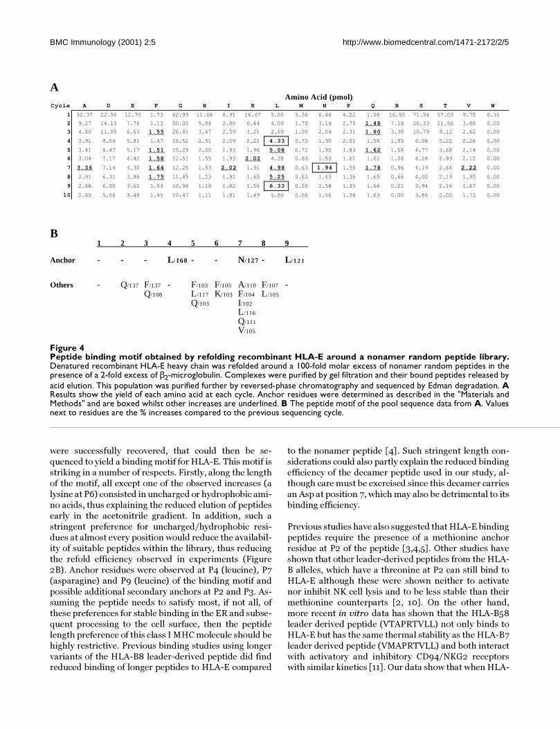
were successfully recovered, that could then be se-
quenced to yield a binding motif for HLA-E. This motif is
striking in a number of respects. Firstly, along the length
of the motif, all except one of the observed increases (a
lysine at P6) consisted in uncharged or hydrophobic ami-
no acids, thus explaining the reduced elution of peptides
early in the acetonitrile gradient. In addition, such a
stringent preference for uncharged/hydrophobic resi-
dues at almost every position would reduce the availabil-
ity of suitable peptides within the library, thus reducing
the refold efficiency observed in experiments (Figure
2B). Anchor residues were observed at P4 (leucine), P7
(asparagine) and P9 (leucine) of the binding motif and
possible additional secondary anchors at P2 and P3. As-
suming the peptide needs to satisfy most, if not all, of
these preferences for stable binding in the ER and subse-
quent processing to the cell surface, then the peptide
length preference of this class I MHC molecule should be
highly restrictive. Previous binding studies using longer
variants of the HLA-B8 leader-derived peptide did findreduced binding of longer peptides to HLA-E compared
to the nonamer peptide [4]. Such stringent length con-
siderations could also partly explain the reduced binding
efficiency of the decamer peptide used in our study, al-
though care must be exercised since this decamer carries
an Asp at position 7, which may also be detrimental to its
binding efficiency.
Previous studies have also suggested that HLA-E binding
peptides require the presence of a methionine anchor
residue at P2 of the peptide [3,4,5]. Other studies have
shown that other leader-derived peptides from the HLA-
B alleles, which have a threonine at P2 can still bind to
HLA-E although these were shown neither to activate
nor inhibit NK cell lysis and to be less stable than their
methionine counterparts [2, 10]. On the other hand,
more recent in vitro data has shown that the HLA-B58
leader derived peptide (VTAPRTVLL) not only binds to
HLA-E but has the same thermal stability as the HLA-B7
leader derived peptide (VMAPRTVLL) and both interact
with activatory and inhibitory CD94/NKG2 receptorswith similar kinetics [11]. Our data show that when HLA-
Figure 4Peptide binding motif obtained by refolding recombinant HLA-E around a nonamer random peptide library.Denatured recombinant HLA-E heavy chain was refolded around a 100-fold molar excess of nonamer random peptides in thepresence of a 2-fold excess of β2-microglobulin. Complexes were purified by gel filtration and their bound peptides released byacid elution. This population was purified further by reversed-phase chromatography and sequenced by Edman degradation. AResults show the yield of each amino acid at each cycle. Anchor residues were determined as described in the "Materials andMethods" and are boxed whilst other increases are underlined. B The peptide motif of the pool sequence data from A. Valuesnext to residues are the % increases compared to the previous sequencing cycle.
AAmino Acid (pmol)
Cycle A D E F G H I K L M N P Q R S T V W
1 32.37 22.50 12.70 1.73 62.99 11.68 6.91 16.67 5.00 3.36 6.46 4.22 1.08 16.55 71.54 57.03 9.75 0.31
2 9.27 14.13 7.76 1.13 50.00 5.04 2.88 6.44 4.09 1.79 3.14 2.75 1.48 7.18 26.33 21.56 3.88 0.00
3 4.80 11.05 6.63 1.55 26.81 3.67 2.59 3.21 2.69 1.00 2.04 2.31 1.60 3.30 10.79 9.12 2.62 0.00
4 3.91 9.54 5.81 1.47 18.52 2.51 2.09 2.21 4.33 0.75 1.90 2.01 1.58 1.95 6.08 5.22 2.26 0.00
5 3.41 8.47 5.17 1.51 15.29 2.00 1.93 1.96 5.08 0.72 1.90 1.83 1.62 1.54 4.77 3.68 2.14 0.00
6 3.04 7.17 4.42 1.58 12.51 1.55 1.93 2.02 4.28 0.63 1.53 1.61 1.61 1.06 4.24 2.93 2.12 0.00
7 3.36 7.14 4.30 1.64 12.25 1.53 2.02 1.91 4.98 0.63 1.94 1.55 1.78 0.96 4.19 2.64 2.22 0.00
8 2.91 6.31 3.86 1.75 11.85 1.23 1.92 1.65 5.25 0.63 1.63 1.36 1.65 0.66 4.00 2.19 1.95 0.00
9 2.88 6.05 3.61 1.53 10.98 1.18 1.82 1.56 6.33 0.55 1.58 1.35 1.64 0.21 3.94 2.16 1.87 0.00
10 2.65 5.56 3.48 1.45 10.47 1.11 1.81 1.49 5.66 0.50 1.56 1.34 1.63 0.00 3.86 2.02 1.73 0.00
B1 2 3 4 5 6 7 8 9
Anchor - - - L/ 160 - - N/ 127 - L/ 1 2 1
Others - Q/137 F/137 - F/103 F/105 A/110 F/107 -Q/108 L/117 K/103 F/104 L/105
Q/103 I/102
L/116
Q/111
V/105

BMC Immunology (2001) 2:5 http://www.biomedcentral.com/1471-2172/2/5
E was refolded around a random peptide library, it did
not appear to have a strong preference for either methio-
nine or threonine at the P2 position. This cannot be due
to a deficiency of the system used here, since this sameapproach with the same library revealed a clearly detect-
able methionine anchor at P2 in RT1-Aa [14]. For HLA-
E, instead of methionine, there was a glutamine increase,
although this was not significant enough to be consid-
ered an anchor (by the criteria used during data analy-
sis).
Whilst we would not have expected to detect the exact se-
quence derived from a leader peptide (since any one giv-
en peptide would only be represented once for every 919
peptides in the random mix, i.e. never), one could have
predicted (and we were in fact expecting) to identify an-
chor residues matching those found in the sequence of
the VMAPRTLVL leader peptide. We do not have an ex-
planation for the divergence of our findings from those of
others. It is interesting to note that Ulbrecht et al. [18]
reported on virally-derived HLA-E stabilising peptides
with glutamine or leucine at the P2 position. These au-
thors suggested that HLA-E may have a lower than aver-
age affinity for β2-microglobulin, so requiring tighter
peptide binding to achieve overall tri-molecular stability
in vivo.
Is there a special mechanism required to bring about the preferential association of class I leader peptides with HLA-E/Qa-1b?Our results raise two important points which must be ad-
dressed before the pathways involved in antigen process-
ing and association to HLA-E can be fully understood.
Firstly, other workers have suggested that the peptide
binding groove of HLA-E has evolved to bind only a
tightly defined set of hydrophobic leader-derived pep-
tides [4, 8]. However, from our present studies and oth-
ers [18], it is obvious that HLA-E has affinity for peptides
other than those derived from class I leader peptides.
Secondly, our results show that the rat classical class Ia
molecule RT1-A1c exhibits low affinity for binding the
mouse leader-derived peptide, AMAPRTLLL, but has a
higher affinity for the overlapping peptide, APRTLLLLL
(See Figure 2A). The possibility that other, more highly
expressed classical class Ia molecules could compete
with HLA-E to bind to leader-derived peptides, increases
the conceptual requirement for specific delivery of these
peptides to the HLA-E assembly complexes. In addition,
these results also draw attention to the possibility that
class I leader-peptides could be processed by other
routes to derive peptides such as the overlapping pep-
tide, APRTLLLLL, of the mouse H2-D and H2-L leader
sequence used here. If this occurs, then even less materi-
al will be available for processing and binding to HLA-E.
Assembly of HLA-E has been shown to be TAP-depend-
ent [6], although it has not been unequivocally proven to
require the TAP-transporter for delivery of peptides
from the cytoplasm. If such peptides are supplied by theTAP transporter, then they represent only a very small
fraction of the total pool of peptides transported, of
which a much bigger fraction will be susceptible to bind
to classical class Ia molecules than to HLA-E. Thus,
whether the high rate of association of class I leader de-
rived peptides with HLA-E, and its counterparts in
mouse (Qa-1b, [19]) and presumably rat (RT.BM1), could
be brought about in vivo in free competition with cyto-
plasmically derived peptides is still open to question.
Specialized mechanisms might therefore be operating to
favour the association of HLA-E with these peptides.
Three possible mechanisms can be envisaged:
1) The incident concentration of these peptides could be
effectively increased if the proximity of the translocon
(site of translation of the heavy chain and possible
processing of the signal peptide) is close to the site of as-
sembly of HLA-E. However, there is no evidence to sug-
gest that the site of MHC class I heavy chain translation
is in close proximity to the TAP assembly complex and
that the translocon complex is capable of processing
leader peptides to yield the mature HLA-E binding pep-
tide.
2) Leader-derived peptides might be released from thetranslocon complex into the ER lumen and processed
through the ER directly. Signal-derived peptides have
been shown to be delivered in a TAP-independent man-
ner for binding to HLA-A2 [20]. It is possible that some
as yet unknown mechanisms for specific enrichment are
operating, such as chaperones which could shuttle the
peptides from the translocon complex to the TAP trans-
porter. If the peptides are processed and delivered via
the ER lumen, calreticulin could act as such a chaperone,
as it is known to bind antigenic peptides and elicit tu-
mour immunity [21]. ER-resident proteases might also
be present to process signal peptides correctly, since
such activity has been reported [22].
3) Alternatively, the peptides could in fact be released
from the translocon complex and associated with chap-
erones on the cytoplasmic side of the ER membrane. Re-
cent work by Paz et al. revealed that antigenic peptides
were not free in the cytoplasm, but bound to high molec-
ular weight material which were different from the previ-
ously described heat shock proteins [22]. A distinct
cytoplasmic chaperone might therefore perform the role
of delivering leader-derived peptides preferentially to a
transporter complex with HLA-E molecules primed for
peptide loading, perhaps via specific recognition of aunique binding motif in the cytoplasmic tail of HLA-E.

BMC Immunology (2001) 2:5 http://www.biomedcentral.com/1471-2172/2/5
Any peptide binding study of a non-classical class Ib
MHC molecule is technically limited by the amount of
material that can be purified due to its low surface ex-
pression, which is often less than 10 % compared to classIa MHC molecules. For example previous studies on
HLA-E required 20-50 grams of cell pellets for immuno-
precipitation experiments using cell lines deficient in
class Ia expression, in order to detect bound leader pep-
tides derived from constructs transfected into the cell
lines under strong promoters to drive expression [5].
Availability of specific antibodies poses an additional
problem, since even weak cross-reactivity to a class Ia
molecule, expressed at much higher levels (10-20x),
would result in a contaminated purified peptide pool,
thus affecting the motif.
Therefore, binding motif determination of a non-classi-
cal MHC class I molecule using a recombinant protein
and a fixed length random peptide library has some clear
advantages over immunoprecipitation of the same mole-
cule expressed in mammalian cells. Unlike class II MHC
molecules, peptides are usually locked into the groove of
class I molecules at their N- and C-termini. In a natural
system, peptides of different lengths can still bind to
class I molecules [15, 23,24,25] and when eluted and
subjected to pool sequencing, the yield of a C-terminal
anchor can be 'diluted' due to the different lengths of the
population. This should occur much less significantly
with a random peptide library of a given length. Havingaccess to large amounts of purified recombinant protein
also removes the need for large amounts of biological
material, and for a specific antibody, to purify the class I
molecule of interest. Finally, an in vitro derived binding
motif is free from the influences of cellular machinery
which might operate to restrict the supply of peptides
available to the class I molecule under study and select
only for high affinity species. The present motif was ob-
tained from a range of high and low affinity peptides and
is therefore closer to the true biochemical preference of
the molecule under study. For the three rat classical
MHC class I molecules studied [14], in vivo and in vitro
derived motifs were very similar and, for RT1-A1c, natu-
ral peptides were identified with the conserved anchors
[26]. By contrast, the known, natural HLA-E peptide lig-
ands do not match closely our in vitro motif. This finding
may suggest an as yet unrecognised and specialised in
vivo mechanism for peptide selection to be operating,
rather than being an issue of peptide affinities in the se-
lection process.
ConclusionsUsing recombinant bacterially produced HLA-E, we
have shown that it is possible to obtain a binding motif of
the non-classical class Ib MHC molecule. The same sys-tem can also be used to test the binding affinity of specif-
ic peptides when no cell-based assay is available. Whilst
the motif obtained confirmed a strong preference of
HLA-E for hydrophobic residues at most positions, the
fact that a library of random peptides could bind at alldemonstrates that this molecules requirements are not
as exquisite as previously suggested. If, as suggested by
these results and those of others [18], HLA-E has the ca-
pacity to present a range of different peptides, then pres-
entation of non-self antigens may have to be considered
as an important accessory role to its function in regulat-
ing NK function.
Materials and MethodsPeptidesThe HLA-A2 leader derived peptide (VMAPRTLVL) was
synthesized by Sigma-Genosys (Pampisford, UK). The
13-mer peptide, ILFPSSERLISNR, was synthesized by
Alta Biosciences, (The University of Birmingham, UK).
Among the collection of synthetic peptides available in
the laboratory, we identified the following which had
suitable anchor residues at positions P2 and/or the C-
terminus; VMPTSNDPTL, SMFPVSENR and
NPRKVTAYL. The overlapping mouse H2-D and H2-L
leader-derived peptides (AMAPRTLLL, MAPRTLLLL
and APRTLLLLL) and the synthetic nonamer random
peptide library used in this study were all purchased
from ECHAZ Microcollections (Tübingen, Germany).
The synthetic nonapeptide library was prepared by fully
automated solid phase peptide synthesis using Fmoc/tBu chemistry. The randomness of the peptide library
was ascertained by pool sequencing [27], electrospray
mass spectrometry [28] and amino acid analysis[29].
Prior to use, each of the specific peptides and the library
was dissolved in DMSO (Pierce, packed under nitrogen)
at a concentration of 10 mg/ml.
Cloning, expression and purification of HLA-EThe region coding for amino acids 1-276 of the HLA-E
heavy chain was amplified from a human T cell cDNA li-
brary (a kind gift from Dr. Martin Turner, Lymphocyte
Signalling and Development Laboratory, The Babraham
Institute) by polymerase chain reaction (PCR) using the
oligonucleotide prime pair: forward 5'-CGGGATC-
CCCATATGGGTTCACACTCCTTGAAGTA-TTTC-
CACACTT-3' and reverse 5'-
GAAGATCTCGAGCGGCTTCCATCTCAGGGTGACGG-
GCT-3' (restriction sites used are underlined). The re-
sulting product was digested with NdeI/XhoI, ligated
into the T7 expression plasmid, pET-22b(+) (Novagen
Inc., WI, USA) and transformed/ selected in XL2-Blue
(Stratagene). DNA sequences from these constructs were
checked (found to be identical to E*0102) and plasmids
were re-transformed into the Escherichia coli strain
BL21(DE3) (Novagen Inc., WI, USA).

BMC Immunology (2001) 2:5 http://www.biomedcentral.com/1471-2172/2/5
Bacteria were grown in LB containing 100 µg/ml ampi-
cillin. Protein expression was induced at mid-log phase
for 3 hours with 1 mM IPTG and the heavy chain protein
was found to be overexpressed as inclusion bodies. Bac-terial pellets were resuspended in 10 mM Tris-HCl; pH
7.5, 1 mM EDTA, 100 µg/ml PMSF, 0.1 % (v/v) Triton-
X100, disrupted by sonication and centrifuged (25,000
g). The cell pellet was washed twice with resuspension
buffer and then solubilized by resuspending and mixing
for 1 hour at room temperature in 8 M urea, 0.1 M
NaH2PO4, 0.01 M Tris-HCl; pH 8.0 (buffer A; pH 8.0).
The mixtures were then centrifuged (25,000 g) to re-
move any insoluble material. Urea-solubilized heavy
chain was purified in a denatured state using a Ni-NTA
agarose column (Qiagen Inc., CA, USA) according to the
manufacturer's instructions. Briefly, the urea-solubilized
protein was mixed with the nickel-charged matrix for 1
hour. The matrix was washed with buffer A; pH 8.0, fol-
lowed by buffer A; pH 6.3. Pure protein (>95% as deter-
mined by SDS-PAGE; results not shown) was eluted
from the column by washing with buffer A; pH 5.9 and
buffer A; pH 4.5. Protein concentration was determined
by the BCA assay (Pierce) and the sample was concen-
trated to approximately 1 mg/ml and stored at -70°C.
Expression of human β2-microglobulinBacteria expressing human β2-microglobulin [30] were a
kind gift of Professor Don Wiley. The bacteria were
grown and induced as for the heavy chain. β2-microglob-ulin was also overexpressed as inclusion bodies and was
isolated and solubilized and clarified as described above.
Urea-solubilized β2-microglobulin was refolded by ex-
tensive dialysis against 10 mM Tris-HCl; pH 7.5 at room
temperature. The dialysed mixture was centrifuged and
any precipitate formed was re-solubilized and re-dia-
lysed. Solutions were pooled and applied to a Q-Sepha-
rose column (Pharmacia Biotech) equilibrated with 10
mM Tris-HCl; pH 7.5. Pure monomeric human β2-mi-
croglobulin (>95 % purity as determined by SDS-PAGE;
results not shown) eluted from this column with 10 mM
Tris-HCl, 100 mM NaCl; pH 7.5. Protein concentration
was determined by the BCA assay (Pierce).
Expression and purification of rat class I molecules and β2-microglobulinExpression and purification of both rat heavy and light
chains were performed as described previously [14].
Assembly assays and purification of complexesRefolding was performed by the dilution method [14, 30,
31]. Small scale refolds with specific peptides were per-
formed in a total volume of 2 ml. β2-microglobulin (48
µg, 4 nmol) and peptide (20 µg, 20 nmol) were added to
1.5 ml of refold buffer (50 mM Tris-HCl; pH 8.0, 400mM arginine, 0.1 mM EDTA, 0.1 mM PMSF). Denatured
heavy chain (62 µg; 2 nmol) was added in aliquots with
mixing and refold buffer was added to give a final volume
of 2 ml in each tube and final molar ratio of 1:2:10 (heavy
chain: β2-microglobulin:peptide). The amount of pep-tide added was varied from the value stated above when
studies on the effect of peptide concentration on refold-
ing were performed. After 24-48 hours at 4°C the refold-
ing mixture was concentrated down to 100 µl using a
Centricon-10 unit (Millipore), with the temperature
maintained at 18°C during the concentration step and re-
folded class I complex was purified by gel filtration on an
FPLC Superdex 75 10/30 column (Pharmacia Biotech)
equilibrated in 20 mM Tris-HCl; pH 8.0, 100 mM NaCl.
Results were calculated as the ratio of the peak area of
monomeric soluble class I complex in the presence of a
peptide compared to a control sample without added
peptide. Due to variable sample loss from increased pre-
cipitation of high multimeric forms in the absence of
peptide (or presence of low binding peptide), induction
of refolding was calculated by comparing proportionate
changes according to the following equation:
where Mon 1 = peak area of monomer with peptide; Agg
1 = peak area of aggregate with peptide; Mon = peak area
of monomer without peptide; Agg = peak area of aggre-
gate without peptide.
Peptide elution and pool sequencingFor binding motif determination, refolds were per-
formed in 40 ml incubations. Briefly, β2-microglobulin
(960 µg, 80 nmol) and nonamer random peptide (4 mg,4 µmol) were added to the refolding buffer and dena-
tured heavy chain (1.24 mg; 40 nmol) was added drop-
wise with constant mixing.
After 24-48 hours at 4°C the refolding mixture was con-
centrated down to 1 ml using Centriprep-10 and Centri-
con-10 units (Millipore) and refolded class I complexes
were purified by gel filtration on an FPLC Superdex 75
16/60 Hi-load column (Pharmacia Biotech) equilibrated
in 20 mM Tris-HCl; pH 8.0, 100 mM NaCl.
Fractions containing refolded monomeric complexes
were pooled and concentrated to 900 µl using Centricon-
10 units. The concentrated HLA-E complexes were acid-
ified by the addition of 100 µl acetic acid. After 5 minutes
incubation at room temperature, the mixture was spun

BMC Immunology (2001) 2:5 http://www.biomedcentral.com/1471-2172/2/5
through Centricon-3 units. The flow through was then
subjected to reversed phase chromatography using an
Applied Biosystems Aquapore (250 mm x 1 mm) Brown-
lee C18 column with an acetonitrile gradient (0-90 %) in0.025 % TFA. Material eluting in the acetonitrile gradi-
ent was pooled, concentrated and submitted for protein
sequencing by Technix™ (The Babraham Institute, UK).
One major peak (fractions 33-35) was submitted for se-
quencing separately. As described previously [14, 17], a
pmol yield >150 % of the previous cycle was considered
significant and is presented as boxed values in the results
(Figure 4). For cycles 6-10, this cut-off value was reduced
to 120 % to allow for cycle-to-cycle sample loss.
AcknowledgementsThis work was supported by a BBSRC Project Grant to JT/GWB, a BBSRC Project Grant to EJ and BBSRC CSG funding to the Laboratory of Function-al Immunogenetics, Babraham. The authors would like to thank Professor Don Wiley for kindly providing the bacterial strain expressing human β2-microglobulin and Dr. Martin Turner for providing the human cDNA li-brary.
References1. Lee N, Llano M, Carretero M, Ishitani A, Navarro F, López-Botet M,
Geraghty DE: HLA-E is a major ligand for the natural killer in-hibitory receptor CD94/NKG2A. Proc Natl Acad Sci U S A 1998,95:5199-5204
2. Borrego F, Ulbrecht M, Weiss EH, Coligan JE, Brooks AG: Recogni-tion of human histocompatibility leukocyte antigen (HLA)-Ecomplexed with HLA class I signal sequence-derived pep-tides by CD94/NKG2 confers protection from natural killercell-mediated lysis. J Exp Med 1998, 187:813-818
3. Braud VM, Allan DS, O'Callaghan CA, Soderstrom K, D'Andrea A,Ogg GS, Lazetic S, Young NT, Bell JI, Phillips JH, Lanier LL, McMichaelAJ: HLA-E binds to natural killer cell receptors CD94/NKG2A, B and C. Nature 1998, 391:795-799
4. Braud V, Jones EY, McMichael A: The human major histocompat-ibility complex class Ib molecule HLA-E binds signal se-quence-derived peptides with primary anchor residues atpositions 2 and 9. Eur J Immunol 1997, 27:1164-1169
5. Lee N, Goodlett DR, Ishitani A, Marquardt H, Geraghty DE: HLA-Esurface expression depends on binding of TAP-dependentpeptides derived from certain HLA class I signal sequences.J Immunol 1998, 160:4951-4960
6. Braud VM, Allan DS, Wilson D, McMichael AJ: TAP- and tapasin-dependent HLA-E surface expression correlates with thebinding of an MHC class I leader peptide. Curr Biol 1998, 8:1-10
7. Tomasec P, Braud VM, Rickards C, Powell MB, McSharry BP, GadolaS, Cerundolo V, Borysiewicz LK, McMichael AJ, Wilkinson GW: Sur-face expression of HLA-E, an inhibitor of natural killer cells,enhanced by human cytomegalovirus gpUL40. Science 2000,287:1031-1033
8. O'Callaghan CA, Tormo J, Willcox BE, Braud VM, Jakobsen BK, StuartDI, McMichael AJ, Bell JI, Jones EY: Structural features imposetight peptide binding specificity in the nonclassical MHCmolecule HLA-E. Mol Cell 1998, 1:531-541
9. Llano M, Lee N, Navarro F, Garcia P, Albar JP, Geraghty DE, López-Botet M: HLA-E-bound peptides influence recognition by in-hibitory and triggering CD94/NKG2 receptors: preferentialresponse to an HLA-G- derived nonamer. Eur J Immunol 1998,28:2854-2863
10. Brooks AG, Borrego F, Posch PE, Patamawenu A, Scorzelli CJ, Ulbre-cht M, Weiss EH, Coligan JE: Specific recognition of HLA-E, butnot classical, HLA class I molecules by soluble CD94/NKG2Aand NK cells. J Immunol 1999, 162:305-313
11. Valés-Gómez M, Reyburn HT, Erskine RA, López-Botet M,Strominger JL: Kinetics and peptide dependency of the bindingof the inhibitory NK receptor CD94/NKG2-A and the acti-vating receptor CD94/NKG2-C to HLA-E. EMBO J 1999,18:4250-4260
12. Kraft JR, Vance RE, Pohl J, Martin AM, Raulet DH, Jensen PE: Analysisof Qa-1b peptide binding specificity and the capacity ofCD94/NKG2A to discriminate between Qa-1-peptide com-plexes. J Exp Med 2000, 192:613-623
13. Pacasova R, Martinozzi S, Boulouis HJ, Szpak Y, Ulbrecht M, Sigaux F,Weiss EH, Pla M: Cell surface detection of HLA-E gene prod-ucts with a specific monoclonal antibody. J Reprod Immunol1999, 43:195-201
14. Stevens J, Wiesmüller K-H, Barker PJ, Walden P, Butcher GW, Joly E:Efficient generation of MHC class I-peptide complexes usingsynthetic peptide libraries. J Biol Chem 1998, 273:2874-2884
15. Bhuyan PK, Young LL, Lindahl KF, Butcher GW: Identification ofthe rat maternally transmitted minor histocompatibility an-tigen. J Immunol 1997, 158:3753-3760
16. Powis SJ, Young LL, Joly E, Barker PJ, Richardson L, Brandt RP, MeliefCJ, Howard JC, Butcher GW: The rat cim effect - TAP allele-de-pendent changes in a class-I MHC anchor motif and evidenceagainst C-terminal trimming of peptides in the ER. Immunity1996, 4:159-165
17. Falk K, Rötzschke O, Stevanovic S, Jung G, Rammensee HG: Allele-specific motifs revealed by sequencing of self-peptides elutedfrom MHC molecules. Nature 1991, 351:290-296
18. Ulbrecht M, Modrow S, Srivastava R, Peterson PA, Weiss EH: Inter-action of HLA-E with peptides and the peptide transporterin vitro: implications for its function in antigen presentation.J Immunol 1998, 160:4375-4385
19. Aldrich CJ, Decloux A, Woods AS, Cotter RJ, Soloski MJ, Forman J:Identification of a TAP-dependent leader peptide recog-nized by alloreactive T-cells specific for a class Ib antigen. Cell1994, 79:649-658
20. Wei ML, Cresswell P: HLA-A2 molecules in an antigen-process-ing mutant cell contain signal sequence-derived peptides. Na-ture 1992, 356:443-446
21. Basu S, Srivastava PK: Calreticulin, a peptide-binding chaperoneof the endoplasmic reticulum, elicits tumor- and peptide-specific immunity. J Exp Med 1999, 189:797-802
22. Paz P, Brouwenstijn N, Perry R, Shastri N: Discrete proteolytic in-termediates in the MHC class I antigen processing pathwayand MHC I-dependent peptide trimming in the ER. Immunity1999, 11:241-251
23. Guo HC, Jardetzky TS, Garrett TPJ, Lane WS, Strominger JL, WileyDC: Different length peptides bind to HLA-Aw68 similarly attheir ends but bulge out in the middle. Nature 1992, 360:364-366
24. Joyce S, Kuzushima K, Kepecs G, Angeletti RH, Nathenson SG: Char-acterization of an incompletely assembled major histocom-patibility class-I molecule (H-2Kb) associated with unusuallylong peptides - implications for antigen-processing and pres-entation. Proc Natl Acad Sci U S A 1994, 91:4145-4149
25. Urban RG, Chicz RM, Lane WS, Strominger JL, Rehm A, Kenter MJH,UytdeHaag FGCM, Ploegh H, Uchanska-Ziegler B, Ziegler A: A sub-set of HLA-B27 molecules contains peptides much longerthan nonamers. Proc Natl Acad Sci U S A 1994, 91:1534-1538
26. Stevens J, Jones RC, Bordoli RS, Trowsdale J, Gaskell SJ, Butcher GW,Joly E: Specificity of RT1-A1c, an Inhibitory Rat Major Histo-compatibility Complex Class I Natural Killer Cell Ligand. JBiol Chem 2000, 275:29217-29224
27. Stevanovic S, Wiesmüller K-H, Metzger J, Beckickinger AG, Jung G:Natural and synthetic peptide pools - characterization by se-quencing and electrospray mass-spectrometry. Bioorg MedChem Lett 1993, 3:431-436
28. Metzger JW, Kempter C, Wiesmüller K-H, Jung G: Electrospraymass-spectrometry and tandem mass-spectrometry of syn-thetic multicomponent peptide mixtures - determination ofcomposition and purity. Anal Biochem 1994, 219:261-277
29. Wiesmüller K-H, Feiertag S, Fleckenstein B, Kienle S, Stoll D, Her-rmann M, Jung G: Peptide and Cyclopeptide Libraries: Auto-mated Synthesis, Analysis and Receptor Binding Assays. In:Combinatorial Peptide and Nonpeptide Libraries-A Handbook for theSearch of Lead Structures. Edited by Jung G. pp. Weinheim, FRG: VerlagChemie; 1996203-246
30. Garboczi DN, Hung DT, Wiley DC: HLA-A2-peptide complexes- refolding and crystallization of molecules expressed in Es-cherichia coli and complexed with single antigenic peptides.Proc Natl Acad Sci U S A 1992, 89:3429-3433
http://www.ncbi.nlm.nih.gov/entrez/query.fcgi?cmd=Retrieve&db=PubMed&dopt=Abstract&list_uids=9560253
http://www.ncbi.nlm.nih.gov/entrez/query.fcgi?cmd=Retrieve&db=PubMed&dopt=Abstract&list_uids=9560253
http://www.ncbi.nlm.nih.gov/entrez/query.fcgi?cmd=Retrieve&db=PubMed&dopt=Abstract&list_uids=9480992
http://www.ncbi.nlm.nih.gov/entrez/query.fcgi?cmd=Retrieve&db=PubMed&dopt=Abstract&list_uids=9486650
http://www.ncbi.nlm.nih.gov/entrez/query.fcgi?cmd=Retrieve&db=PubMed&dopt=Abstract&list_uids=9486650
http://www.ncbi.nlm.nih.gov/entrez/query.fcgi?cmd=Retrieve&db=PubMed&dopt=Abstract&list_uids=9486650
http://www.ncbi.nlm.nih.gov/entrez/query.fcgi?cmd=Retrieve&db=PubMed&dopt=Abstract&list_uids=9174606
http://www.ncbi.nlm.nih.gov/entrez/query.fcgi?cmd=Retrieve&db=PubMed&dopt=Abstract&list_uids=9590243
http://www.ncbi.nlm.nih.gov/entrez/query.fcgi?cmd=Retrieve&db=PubMed&dopt=Abstract&list_uids=9427624
http://www.ncbi.nlm.nih.gov/entrez/query.fcgi?cmd=Retrieve&db=PubMed&dopt=Abstract&list_uids=9660937
http://www.ncbi.nlm.nih.gov/entrez/query.fcgi?cmd=Retrieve&db=PubMed&dopt=Abstract&list_uids=9660937
http://www.ncbi.nlm.nih.gov/entrez/query.fcgi?cmd=Retrieve&db=PubMed&dopt=Abstract&list_uids=9754572
http://www.ncbi.nlm.nih.gov/entrez/query.fcgi?cmd=Retrieve&db=PubMed&dopt=Abstract&list_uids=9754572
http://www.ncbi.nlm.nih.gov/entrez/query.fcgi?cmd=Retrieve&db=PubMed&dopt=Abstract&list_uids=9886400
http://www.ncbi.nlm.nih.gov/entrez/query.fcgi?cmd=Retrieve&db=PubMed&dopt=Abstract&list_uids=9886400
http://www.ncbi.nlm.nih.gov/entrez/query.fcgi?cmd=Retrieve&db=PubMed&dopt=Abstract&list_uids=9446598
http://www.ncbi.nlm.nih.gov/entrez/query.fcgi?cmd=Retrieve&db=PubMed&dopt=Abstract&list_uids=9446598
http://www.ncbi.nlm.nih.gov/entrez/query.fcgi?cmd=Retrieve&db=PubMed&dopt=Abstract&list_uids=9103440
http://www.ncbi.nlm.nih.gov/entrez/query.fcgi?cmd=Retrieve&db=PubMed&dopt=Abstract&list_uids=8624806
http://www.ncbi.nlm.nih.gov/entrez/query.fcgi?cmd=Retrieve&db=PubMed&dopt=Abstract&list_uids=8624806
http://www.ncbi.nlm.nih.gov/entrez/query.fcgi?cmd=Retrieve&db=PubMed&dopt=Abstract&list_uids=1709722
http://www.ncbi.nlm.nih.gov/entrez/query.fcgi?cmd=Retrieve&db=PubMed&dopt=Abstract&list_uids=9574542
http://www.ncbi.nlm.nih.gov/entrez/query.fcgi?cmd=Retrieve&db=PubMed&dopt=Abstract&list_uids=7525079
http://www.ncbi.nlm.nih.gov/entrez/query.fcgi?cmd=Retrieve&db=PubMed&dopt=Abstract&list_uids=7525079
http://www.ncbi.nlm.nih.gov/entrez/query.fcgi?cmd=Retrieve&db=PubMed&dopt=Abstract&list_uids=1557127
http://www.ncbi.nlm.nih.gov/entrez/query.fcgi?cmd=Retrieve&db=PubMed&dopt=Abstract&list_uids=1448153
http://www.ncbi.nlm.nih.gov/entrez/query.fcgi?cmd=Retrieve&db=PubMed&dopt=Abstract&list_uids=1448153
http://www.ncbi.nlm.nih.gov/entrez/query.fcgi?cmd=Retrieve&db=PubMed&dopt=Abstract&list_uids=8183884
http://www.ncbi.nlm.nih.gov/entrez/query.fcgi?cmd=Retrieve&db=PubMed&dopt=Abstract&list_uids=8108441
http://www.ncbi.nlm.nih.gov/entrez/query.fcgi?cmd=Retrieve&db=PubMed&dopt=Abstract&list_uids=8108441

BMC Immunology (2001) 2:5 http://www.biomedcentral.com/1471-2172/2/5
31. Reid SW, Smith KJ, Jakobsen BK, O'Callaghan CA, Reyburn H, HarlosK, Stuart DI, McMichael AJ, Bell JI, Jones EY: Production and crys-tallization of MHC class-Ib allele single peptide complexes.FEBS Lett 1996, 383:119-123
Publish with BioMedcentral and every scientist can read your work free of charge
"BioMedcentral will be the most significant development for disseminating the results of biomedical research in our lifetime."
Paul Nurse, Director-General, Imperial Cancer Research Fund
Publish with BMC and your research papers will be:
available free of charge to the entire biomedical community
peer reviewed and published immediately upon acceptance
cited in PubMed and archived on PubMed Central
yours - you keep the copyright
[email protected] your manuscript here:http://www.biomedcentral.com/manuscript/
BioMedcentral.com
http://www.ncbi.nlm.nih.gov/entrez/query.fcgi?cmd=Retrieve&db=PubMed&dopt=Abstract&list_uids=8612777