Core & AHL IB NOTES
102
1 IB BIOLOGY HIGHER LEVEL! CORE Topic 1: Statistical analysis 1.1.1 State that error bars are a graphical representation of the variability of data. Error bars can be used to show either the range of the data or the standard deviation. 1.1.2 Calculate the mean and standard deviation of a set of values. Mean: x = Σχ The sum of all the scores divided by the total number of scores. Standard deviation: = Σ 2 − 2 −1 ∑= ‘the sum of’ x= arithmetic mean n= number of data 1.1.3 State that the term standard deviation is used to summarize the spread of values around the mean, and that 68% of the values fall within one standard deviation of the mean. For normally distributed data, about 68% of all values lie within ±1 standard deviation (s or σ) of the mean. This rises to about 95% for ±2 standard deviations. 1.1.4 Explain how the standard deviation is useful for comparing the means and the spread of data between two or more samples. A small standard deviation indicates that the data is clustered closely around the mean value. Conversely, a large standard deviation indicates a wider spread around the mean. 1.1.5 Deduce the significance of the difference between two sets of data using calculated values for t and the appropriate tables. For the t-test to be applied, the data must have a normal distribution and a sample size of at least 10. The t-test can be used to compare two sets of data and measure the amount of
-
Upload
independent -
Category
Documents
-
view
2 -
download
0
Transcript of Core & AHL IB NOTES

1
IB BIOLOGY HIGHER LEVEL!
CORE
Topic 1:
Statistical analysis
1.1.1
State that error bars are a graphical representation of the variability of data.
Error bars can be used to show either the range of the data or the standard deviation.
1.1.2
Calculate the mean and standard deviation of a set of values.
Mean: x =Σχ
𝓃
The sum of all the scores divided by the total number of scores.
Standard deviation: 𝑠𝑑 = Σ𝑥2−𝓃𝑥2
𝓃−1
∑= ‘the sum of’
x= arithmetic mean
n= number of data
1.1.3
State that the term standard deviation is used to summarize the spread of values around the
mean, and that 68% of the values fall within one standard deviation of the mean.
For normally distributed data, about 68% of all values lie within ±1 standard deviation (s or
σ) of the mean. This rises to about 95% for ±2 standard deviations.
1.1.4
Explain how the standard deviation is useful for comparing the means and the spread of data
between two or more samples.
A small standard deviation indicates that the data is clustered closely around the mean
value. Conversely, a large standard deviation indicates a wider spread around the mean.
1.1.5
Deduce the significance of the difference between two sets of data using calculated values
for t and the appropriate tables.
For the t-test to be applied, the data must have a normal distribution and a sample size of at
least 10. The t-test can be used to compare two sets of data and measure the amount of

2
overlap. Students will not be expected to calculate values of t. Only a two-tailed, unpaired t-
test is expected.
1.1.6
Explain that the existence of a correlation does not establish that there is a causal
relationship between two variables.
Correlation often shows a casual relationship between two variables such as height and
weight. Taller people tend to be heavier and so we can see a correlation in these two sets of
data. However, some variables may show correlation when in fact there is no casual
relationship between them. The results may be correlated by chance. This means that even
the correlation is a useful tool for studying data, it is not always reliable.
Topic 2: Cells
2.1 Cell Theory:
2.1.1
Outline the cell theory.
All living things are composed of cells.
Cells are the building blocks of life.
All cells come from pre-existing cells.
2.1.2
Discuss the evidence for the cell theory
Every living thing consists of cells.
No living things are smaller than a call, a virus is smaller but it is non-living as it needs
a host body to carry out its functions.
All cells come from pre-existing cells.
Muscle cells are cells you just cannot tell them apart and the same goes for fungle
hyphae they do not look like structures of cells.
2.1.3
State that unicellular organisms carry out all the functions of life.
Functions of life:
Metabolism
Response to stimuli
Growth
Reproduction
Nutrition
Homeostasis
MTVBOM
How can we see cells? Are they really there?
-Yes, we can see them with a microscope.
TEM: SHOWS A SLICE OF SOMETHING
SEM: SHOWS YOU A 3D IMAGE

3
ICH MAG SIE SO! Mag=s.i (size of image)
s.o (size of object)
Cancer:
uncontrollable cell
division. See 2.5.2
2.1.4-2.1.6
Measuring and calculation of magnification
Molecules 1nm
Thickness of membrane 10nm
Virus 100nm
Bacteria 1ųm
Organelles 10ųm
Most cells 100ųm
The greater the surface area to volume ratio the greater the percentage (%) of diffusion, this
shows that the smaller the cell the more efficiently it can carry out its function.
2.1.7
State that multicellular organisms show emergent properties.
Multicellular organisms show emergent properties.
2.1.8
Explain that cells in multicellular organisms differentiate to carry out specialized functions by
expressing some of their genes but not others.
Every one of your cells have the same DNA because they all come from the same cell (except
for mutation) they have genetic instructions and develop in different ways, so depending on
the cell different genetic instructions get expressed.
2.1.9-2.1.10
Stem cells:
Are self-renewing
Can be used in treatment
They are preserved in the umbilical chord
It is like a blank cell, it can develop into other kinds of cells. They offer so much
variety.
USE:
Hair follicles contain stem cells, and research has shown that these follicle stem cells can lead
to successful treating in baldness through “hair multiplication”. It works by taking stem cells
from existing follicles multiplying them in culture, and implanting the new follicles in the
scalp.
2.2 Prokaryotic Cells:

4
2.2.1-2.2.3
Draw and label a diagram of the E. coli structure.
MUST INCLUDE:
Cell wall- maintains the cells structure,
protects bacteria from bursting, prevents
damaged from outside & it can’t change
shape easily.
Plasma membrane- controls what
substances enter & exit the cell.
Cytoplasm- contains enzymes that
catalyse the chemical reactions of
metabolism.
Pilli- allows the transfer of genetic
information of one cell to another.
Flagella- allow locomotion of the cell.
Ribosomes- make protein (synthesize).
Nucleoid region- naked DNA, stores genetic information that controls the cell & is
passed on to daughter cells.
Mesome- Provides more membrane (as it folds into the cell), this increases surface
area. ATP production (energy source).
2.2.4
State that prokaryotic cells divide by binary fission.
Prokaryotic cells divide by binary fission by passing on its information to its daughter cells.
2.3 Eukaryotic Cells:
2.3.1-2.3.3
Draw and label a diagram of ultrastructure of a liver cell as an example of an animal cell.
MUST INCLUDE:
Free ribosomes- main site of protein
synthesis.
Rough endoplasmic reticulum (rER) -
Packages the proteins synthesized in the
ribosomes.
Lysosome- Digests macromolecules &
contain digestive enzymes.
Golgi apparatus- Modifies; stores & routes
products of the endoplasmic reticulum.

5
Mitochondrion- Serves as the site of cellular respiration.
Nucleus- Contains a cell’s genetic material.
2.3.4
Compare prokaryotic and eukaryotic cells:
Prokaryotic Cell Eukaryotic Cell
Naked DNA DNA associated with proteins
DNA in cytoplasm DNA enclosed in a nuclear envelope
No mitochondria Mitochondria
70S ribosomes 80S ribosomes
Don’t have internal membranes that compartmentalize their functions
Have internal membranes that compartmentalize their functions
2.3.5
State 3 differences between plant and animal cells:
1. Plant cells have a cell wall
2. Plant cells have chloroplasts
3. Plants have a larger vacuole
2.3.6
Outline 2 roles of extracellular components.
1. The plant cell wall maintains cell shape, prevents excessive water uptake, and holds
the whole plant up against the force of gravity
2. Animal cells secrete glycoproteins that form the extracellular matrix. This functions in
support, adhesion and movement.
2.4 Membranes:
2.4.1
Draw and label a diagram to show the structure of membranes.
MUST INCLUDE:
Integral proteins are
embedded in the
phospholipid of the
membrane; the
peripheral proteins are
attached to its surface.

6
Phospholipid bilayer
Cholesterol
Glycoproteins
Integral proteins
Peripheral proteins
2.4.2
Explain how the hydrophobic and hydrophilic properties of phospholipids help maintain the
structure of cell membranes.
HEAD (glycerol and phosphate)
Hydrophilic
Polar (positive charge +)
TAILS (fatty acid)
Hydrophobic (not attracted to water)
Non polar (negative -)
H₂O
If the phospholipids are completely submerged in water they
form this.
They can fuse with each other or break off from one another
Cholesterol is in-between these phospholipids and it helps maintain
the shape of the membrane and it keeps the membrane fluid.
2.4.3
List the functions of membrane proteins.
Channels- allow passive transport of molecules.
Pump – moves substances across membranes using energy (active transport).
Hormone binding sites- Areas where hormones attach to bring about response.
Cell to cell communication- neurotransmitters attaching neurons (nerve cells).
Cell adhesion- cells sticking together.
Immobilized enzymes- speed up chemical reactions.
2.4.4
Define diffusion and osmosis.
AIR

7
I f y o u l e a v e 1
o u t y o u d o n ’ t
g e t f u l l m a r k s !
Diffusion is the passive movement of molecules from a region of high concentration to a
region of low concentration.
Osmosis is the passive movement of water molecules, across a partial permeable
membrane, from a region of lower solute concentration to a region of higher solute
concentration.
1. Diffusion of water
2. Diffusion of water across a partial permeable membrane.
TRASNPORT
Passive “with” Active “against” (concentration gradient)
Requires no energy Requires energy ATP
Highlow concentration Low high concentration
2.4.5
Explain passive transport across membranes by simple diffusion and facilitated diffusion.
Facilitated diffusion: something is helping the molecules diffuse through the membrane
(getting in or out)-
Membrane protein needed: channel highlow
2.4.6
Explain the role of protein pumps and ATP in active transport across membranes.
Active transport is the movement of substances across membranes using energy from ATP. It
can move substances against a concentration gradient. Protein pumps in the membrane are
used for active transport; each pump only transports particular substances so cells can
control what is absorbed and what is expelled.
2.4.7
Explain how vesicles are used to transport materials within a cell between the rough
endoplasmic reticulum, Golgi apparatus and plasma membrane.
1. Vesicle is made by pinching off a piece of membrane (the fluidity of the
membrane allows this)
2. Vesicles can be used to transport material around inside cells (proteins are
transported in vesicles)
3. From the rough endoplasmic reticulum to the Golgi apparatus
4. Then from the Golgi apparatus to the plasma membrane
5. The formation of vesicle from plasma membrane allows material to be taken in.
6. Endocytosis is absorption of material using a vesicle
7. Fusion of vesicle with plasma membrane allows material to be secreted/passed
out (exocytosis)

8
PMAT
2.4.8
Describe how the fluidity of the membrane allows it to change shape break and re-form
during endocytosis and exocytosis.
The phospholipids in the cell membrane are not solid but are in a fluid state allowing the
membrane to change its shape and also vesicles to fuse with it. This means substances can
enter the cell via endocytosis and exit the cell via exocytosis. The membrane then returns to
its original state. In exocytosis the vesicles fuse with the membrane expelling their content
outside the cell. The membrane then goes back to its original state.
2.5 Eukaryotic Cells
2.5.1
Outline the stages in the cell cycle, including interphase (G₁, S, and G₂) mitosis & cytokinesis.
Interphase: where many metabolic reactions
occur.
G₁ phase: growth, producing enzymes &
proteins.
S phase (DNA synthesis): synthesis=DNA
replication
G₂ phase: copying of organelles; preparation
for mitosis & an increase in the number of
mitochondria & chloroplasts.
Mitosis: nucleus is being split.
Prophase
Metaphase
Anaphase
Telophase
Cytokinesis: where the cells finally split; division of the cytoplasm.
2.5.2
State the tumour’s (cancers) are the result of uncontrolled cell division and that these can
occur in any organ or tissue.
Cancer is the result when there is uncontrollable cell division, this can happen in any organ
or tissue, and this is when something goes wrong during mitosis.

9
2.5.3
State that interphase is an active period in the life of a cell when many metabolic reactions,
including protein synthesis, DNA replication and an increase in the number of mitochondria
and chloroplasts.
Interphase is an active period in the life of a cell during which many metabolic reactions
occur such as protein synthesis, DNA replication and an increase in the number of
mitochondria and chloroplast.
2.5.4
Describe the events that occur in the four phases of mitosis (prophase,
metaphase, anaphase and telophase).
Prophase:
1. Breaking of nuclear membranes (disintegrates) 2. Centrioles form microtubules 3. DNA super coils (winds up/coils up) condenses and become visible 4. The chromosomes become visible (long section of DNA)
Metaphase:
1. Chromosomes line up along the
equatorial region of the cell
2. Attachment of microtubules to
centromeres (move apart)
Anaphase:
1. Splitting of centromeres
2. Movement of sister chromosomes to
opposite poles (as spindle microtubules
shorten)
3. Cell becomes elongated
Telophase:
1. Uncoiling of chromosomes
2. Reformation of nuclear membranes
3. DNA is not supercoiled any more
4. Cannot see chromosomes anymore
2.5.5
Explain how mitosis produces two genetically
identical nuclei.
10
CHON
CAFE
SNAP W E N EED T H EM
T O S U R V IV E!
Synthesis of identical chromosomes in interphase.
Lining up during mitosis ensures that each new cell gets a copy of each chromosome.
2.5.6
State that growth, embryonic development, tissue repair and asexual reproduction involve
mitosis.
Growth, embryonic development, tissue repair and asexual reproduction involve mitosis.
Topic 3: The chemistry of life
3.1 Chemical elements and water:
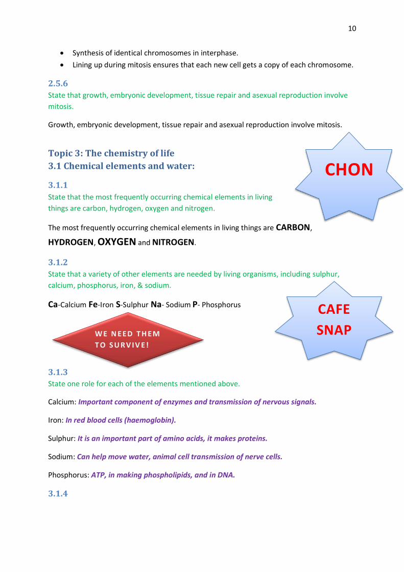
3.1.1
State that the most frequently occurring chemical elements in living
things are carbon, hydrogen, oxygen and nitrogen.
The most frequently occurring chemical elements in living things are CARBON,
HYDROGEN, OXYGEN and NITROGEN.
3.1.2
State that a variety of other elements are needed by living organisms, including sulphur,
calcium, phosphorus, iron, & sodium.
Ca-Calcium Fe-Iron S-Sulphur Na- Sodium P- Phosphorus
3.1.3
State one role for each of the elements mentioned above.
Calcium: Important component of enzymes and transmission of nervous signals.
Iron: In red blood cells (haemoglobin).
Sulphur: It is an important part of amino acids, it makes proteins.
Sodium: Can help move water, animal cell transmission of nerve cells.
Phosphorus: ATP, in making phospholipids, and in DNA.
3.1.4

11
H
O
H
Draw and label a diagram showing the structure of water molecules to show their polarity
and hydrogen bond formation.
3.1.5-3.1.6
Outline the thermal, cohesive and solvent properties of
water.
H₂O is a good solvent (ions and other polar compounds
dissolve easily in H₂O)
Carrying solutes in blood.
Carrying solvents through a plant
Biochemical reactions in cytoplasm
Cohesive properties (sticking together)
Allows water to travel up thin vessels (xylem vessels in plants)
Some organisms can walk on water
Adhesive properties (sticking to something else)
Allows water to travel up thin vessels (xylem vessels in plants)
Thermal properties
It takes a lot of energy to change the properties of water
It has a specific heat capacity
o Benefits organisms that live in water by providing stable temperature
o Evaporate= cooling effects (sweat)
3.2 Carbohydrates, lipids and proteins:
3.2.1
Distinguish between organic and inorganic compounds.
Organic Inorganic
Contain carbon Don’t have carbon in them
Are necessary for living things Carbon is inorganic
Made by living things
3.2.2
Identify amino acids, glucose, ribose and fatty acids from diagrams showing their structure.

12
Chart Carbohydrates Proteins Lipids Nucleic Acid
Functions Provide energy Membrane proteins Energy storage Stores genetic info
Structural support Enzymes Insulation membrane Examples Glucose Haemoglobin Phospholipids RNA Fructose Keratin Cholesterol DNA Lactose Collagen Triglycerides Sucrose Insulin Maltose Melanin
Starch Antibodies
Glycogen Actin
Cellulose
Subunits Monosaccharide Amino Acids Fatty acids (2) Nucleotides (5)
(simple sugars) (20) Glycerol (1)
Fatty Acid:
Amino Acid
Sugars
1. Ribose
2. Glucose
3. Maltose
4. Starch
BONDING RULES:
H- 1
O- 2
N- 3
C- 4

13
3.2.3
List three examples each of monosaccharide’s, disaccharides and polysaccharides.
1. Glucose, galactose and fructose
2. Maltose, lactose and sucrose
3. Starch, glycogen and cellulose
3.2.4
State one function of: glucose, lactose and glycogen in animals, and of fructose, sucrose and
cellulose in plants.
Animals: Glucose is used as an energy source for the body Lactose is the sugar found in milk which provides energy to new-born. Glycogen is used as an energy source (short term only) it is stored in muscles & the
liver. Plants
Fructose is what makes fruits taste sweet Sucrose is used as an energy source for the plant Cellulose fibres are what make the plant cell wall strong.
3.2.5
Outline the role of condensation and hydrolysis in the relationships between
monosaccharide’s; between fatty acids, glycerol and triglycerides; and between amino acids
and polypeptides.
Condensation:
Two molecules work together and form one big molecules and water is also formed.
2 monosaccharide’s disaccharide + H₂O
Hydrolysis:
When water is used to break apart the molecule formed
Disaccharide + H₂O glucose + glucose (monosaccharide)
3.2.6
State three functions of lipids.
1. Energy storage
2. Thermal insulation
3. Lipids allow buoyancy as they are less dense than water and so animals can float in water.
3.2.7
Compare the use of carbohydrates and lipids in energy storage.

14
Pure
Agony
3.3 DNA Structure
3.3.1
Outline DNA nucleotide structure in terms of sugar “deoxyribose”, base and phosphate.
DNA: deoxyribonucleic acid
RNA: ribonucleic acid SUGARS
All nucleic acids consist of nucleotides. (3 parts)
1. A pentose sugar (ribose and deoxyribose)
2. A nitrogen base (a base containing nitrogen)
3. A phosphate group (containing phosphate)
3.3.2
State the names of the four bases in DNA.
2 nitrogenous bases have this larger structure:
Adenine (A)
Guanine (G) These are called PURINES!
Carbohydrates Lipids
Energy storage is short term Energy storage is long term
Soluble in water Insoluble in water
Easy to transport around the body No so easy to transport around the body
Easily and more rapidly digested Not rapidly digested
Have an effect on osmosis Do not have an effect on osmosis prevents problems within the cells in the body
More energy per gram
Nitrogenous base Pentose
sugar
P
P

15
2 bases have a shorter structure:
1. Thymine (T) [only DNA]
2. Cytosine (C) These are the PYRIMIDINES!
3. Uracil (U) [only in RNA]
3.3.3
Outline how DNA nucleotides are linked together by covalent bonds into a single strand.
Condensation
reaction
This is the beginnings of a
polynucleotide.
P
CUTie
Py
A
P
G
P
P
T
P
C

16
AT
Grand
Canyo
3.3.4-3.3.5 and 7.1.1
Explain how a DNA double helix is formed using complementary base pairing and hydrogen
bonds. Describe the structure of DNA, including the antiparallel strands, 3’–5’ linkages and
hydrogen bonding between purines and pyrimidines.
5`end 3`end
3`end 5`end
Hydrogen bonds! They are formed between them as they are easily broken
and easily changed.
Antiparallel!
Base pairs (inside),
Sugar phosphate backbone (outside)
C pairs up with G because they are
complementary!
G
P
P
C
P
T
A
P
T
P
P
P
A
G
C
5
5
5
5
5
5
5
3
3
3
3
3
3
3
3
P
5

17
3.4-DNA Replication
3.4.1
Explain DNA replication in terms of unwinding the double helix and separation of the strands
by helicase, followed by formation of the new complementary strands by DNA polymerase.
The DNA unwinds and the hydrogen bonds between the two strands break as they form new
strands. The replication process has produced a new DNA molecule which is identical to the
initial one.
3.4.2
Explain the significance of complementary base pairing in the conservation of the base
sequence of DNA.
Complementary base pairing is significant as the DNA separates and form daughter
molecules the strands of DNA stay the same. Original double helix splits apart.
3.4.3
State that DNA replication is semi-conservative.
DNA replication is semi-conservative.
3.5 Transcription and Translation
3.5.1
Compare the structure of RNA and DNA.
DNA RNA
5-carbon sugar Deoxyribose Ribose
Strands Double Single
4 bases A, G, C, T A, G, C, U
3.5.2
Outline DNA transcription in terms of the formation of an RNA strand complementary to the
DNA strand by RNA polymerase.
DNA transcription is the formation of an RNA strand which is complementary to the DNA
strand. The first stage of transcription is the uncoiling of the DNA double helix. Then, the
free RNA nucleotides start to form an RNA strand by using one of the DNA strands as a
template. This is done through complementary base pairing, in the RNA chain, the base
thymine is replaced by uracil. RNA polymerase is the enzyme involved in the formation of
the RNA strand and the uncoiling of the double helix. The RNA strand then elongates and

18
then separates from the DNA template. The DNA strands then reform a double helix. The
strand of RNA formed is called messenger RNA. (mRNA)
3.5.3
Describe the genetic code in terms of codons composed of triplets of bases.
A triplet of bases (3 bases) forms a codon. Each codon codes for a particular amino acid.
Amino acids in turn link to form proteins. Therefore DNA and RNA regulate protein
synthesis. The genetic code is the codons within DNA and RNA, composed of triplets of bases
which eventually lead to protein synthesis.
3.5.4
Explain the process of translation, leading to polypeptide formation.
1. Translation starts at the START codon AUG
1. mRNA attaches to the small ribosomal subunit INITIATION
2. tRNA binds to the mRNA
3. Now the large ribosomal subunit can attach
4. tRNA is parked in the P site
5. a new tRNA arrives in the A site
6. the tRNA molecule P loses its amino acid to the tRNA molecule in site A
7. The ribosome moves along the mRNA strand, now the tRNA molecule in the P site is
in the E site and exits. ELONGATION
8. Another tRNA molecule joins to the A site.
9. The amino acids move to the tRNA molecule in the A site to form a peptide bond.
10. The growing polypeptide chain is constantly transferred to the new tRNA.
11. Finally the release factor attaches
12. The ribosome detaches from the mRNA strand TERMINATION
13. The polypeptide chain detaches.
3.5.5
Discuss the relationship between one gene and one polypeptide.
A polypeptide is formed by amino acids liking together through peptide bonds. There are 20
different amino acids so a wide range of polypeptides are possible. Genes store the
information required for making polypeptides. The information is stored in a coded form by
the use of triplets of bases which form codons. The sequence of bases in a gene codes for
the sequence of amino acids in a polypeptide. The information in the genes is decoded
during transcription and translation leading to protein synthesis.

19
3.6 Enzymes
3.6.1
Define enzyme and active site.
Enzymes: Globular proteins which act as catalysts of chemical reactions.
Active site: Region on the surface of an enzyme to which substrates bind and which catalyses a chemical reaction involving the substrates.
3.6.2
Explain enzyme–substrate specificity. The active site for an enzyme is very specific in shape, with very precise chemical properties. Active sites match the shape of their substrates. This enzyme is a lock, and the substrate is the key which can open it.
3.6.3
Explain the effects of temperature, pH and substrate concentration on enzyme activity. Temperature, pH and substrate concentration all affect the rate at which enzymes catalyse
chemical reactions.
Substrate concentration: At low S.C. the enzyme activity is proportional to the substrate
concentration, therefore the more substrate the higher the rate. However at a high
substrate concentration, at some point all active sites are occupied so raising the substrate
concentration has no effect.
Temperature: Enzyme activity increases as temperature increases. This is because collision
between substrate and active site happen more frequently at higher temperatures, due to
fast molecular movement. However at high temperatures enzymes denature and stop
working. This is because heat causes vibrations inside the enzyme, which break bonds
needed to maintain the structure.
pH: There is an optimum at which enzyme activity is fastest (mostly pH 7), and as pH
increases or decreases from its optimum, enzyme activity is reduced.
3.6.4
Define denaturation.
Denaturation: is a structural change in a protein that results in the loss (usually permanent) of its biological properties. It can no longer carry out its functions.
3.6.5
Explain the use of lactase in the production of lactose-free milk. Lactose- The sugar present in milk. Lactose can be converted to glucose and galactose using
the enzyme lactase (disaccharide). Biotechnology companies culture the yeast and extract
the enzyme in order to produce lactose free milk.
Advantages: Pectinase makes juice more fluid and easy to separate from pulp.

20
Fructose is widely used in food manufacturing because it is much sweeter than
glucose. It is made from starch, usually found in maize. Amylase is needed to
break down the starch into glucose.
Source of Enzyme: Amylase is obtained from fungi.
Use: Used to break Starch into glucose, which is then converted into fructose
using the enzyme glucose isomerase.
3.7 Cell respiration
3.7.1
Define cell respiration.
Cell respiration: is the controlled release of energy from organic compounds in cells to
form ATP.
3.7.2
State that-in cell respiration- glucose in the cytoplasm is broken down by glycolysis into
pyruvate, with a small yield of ATP.
Chemical reactions in the cytoplasm break down glucose into a simpler organic compound
called pyruvate. In these reactions a small amount of ATP is made using energy released
from glucose.
Glucose Pyruvate
Small amount of ATP
3.7.3
Explain that, during anaerobic cell respiration, pyruvate can be converted in the cytoplasm
into lactate, or ethanol and carbon dioxide, with no further yield of ATP.
If there is no oxygen available, the pyruvate remains in the cytoplasm and is converted into a
waste product that can be removed from the cell. No ATP is produce. The human waste
product is lactate (lactic acid) and in yeast the products are ethanol and carbon dioxide.
Pyruvate Humans Lactate
Pyruvate Yeast Ethanol
Carbon Dioxide
3.7.4
Explain that, during aerobic cell respiration, pyruvate can be broken down in the
mitochondrion into carbon dioxide and water with a large yield of ATP.

21
If oxygen is available the pyruvate is absorbed by the mitochondrion. Inside the
mitochondrion the pyruvate is broken down into carbon dioxide and water. A large amount
of ATP is produced as a result of these reactions.
Pyruvate Carbon dioxide
large amount of ATP
Water
3.8 Photosynthesis
3.8.1
State that photosynthesis involves the conversion of light energy into chemical energy.
Photosynthesis involves the conversion of light energy into chemical energy.
3.8.2
State that light from the Sun is composed of a range of wavelengths (colours).
The light from the sun is composed of a range of wavelengths (colours).
3.8.3
State that chlorophyll is the main photosynthetic pigment.
Chlorophyll is the main photosynthetic pigment.
3.8.4
Outline the differences in absorption of red, blue and green light by chlorophyll.
Chlorophyll can absorb red and blue light more than green. The green light that cannot be
absorbed is reflected giving the plants leaves its green colour.
3.8.5
State that light energy is used to produce ATP, and to split water molecules (photolysis) to
form oxygen and hydrogen.
Light energy is used to produce ATP and to split water molecules (photolysis) to form oxygen
and hydrogen.
3.8.6
State that ATP and hydrogen (derived from the photolysis of water) are used to fix carbon
dioxide to make organic molecules.
ATP and hydrogen derived from photolysis of water are used to fix carbon dioxide to make
organic molecules.

22
3.8.7
Explain that the rate of photosynthesis can be measured directly by the production of
oxygen or the uptake of carbon dioxide, or indirectly by an increase in biomass.
Photosynthesis can be measured in many ways as it involves the production of oxygen, the
uptake of carbon dioxide and an increase in biomass. For example, aquatic plants release
oxygen bubbles during photosynthesis and so these can be collected and measured. The
uptake of carbon dioxide is more difficult to measure so it is usually done indirectly. When
carbon dioxide is absorbed from water the pH of the water rises and so this can be
measured with pH indicators or pH meters. Finally, photosynthesis can be measured through
an increase in biomass. If batches of plants are harvested at a series of times and the
biomass of these batches is calculated, the rate increase in biomass gives an indirect
measure of the rate of photosynthesis in the plants.
3.8.8
Outline the effects of temperature, light intensity and carbon dioxide concentration on the
rate of photosynthesis.
As temperature increases, the rate of photosynthesis increases more and more steeply until
the optimum temperature is reached. If temperature keeps increasing above the optimum
temperature then photosynthesis starts to decrease very rapidly.
As light intensity increases so does photosynthesis until a certain point. At high light
intensities photosynthesis reaches a plateau and so does not increase any more. At low and
medium light intensity the rate of photosynthesis is directly proportional to the light
intensity.
As the carbon dioxide concentration increases so does the rate of photosynthesis. There is no photosynthesis at very low levels of carbon dioxide and at high levels the rate reaches a plateau.
Topic 4: Genetics
4.1 Chromosomes, genes, alleles and mutations
4.1.1
State that eukaryote chromosomes are made of DNA and proteins.
Eukaryote chromosomes are made of DNA and proteins.
4.1.2
Define gene, allele and genome.
Gene: a heritable factor that controls a specific characteristic.
23
Allele: one specific form of a gene, differing from other alleles by one or a few bases only
and occupying the same gene locus as other alleles of the gene.
Genome: the whole of the genetic information of an organism.
4.1.3
Define gene mutation.
Gene Mutation: The change to the base sequence of a gene.
4.1.4
Explain the consequence of a base substitution mutation in relation to the processes of
transcription and translation, using the example of sickle-cell anaemia.
Normal Gene Mutated Gene
Codon GAG GTG
Transcription GAG on mRNA GUG on mRNA
Phenotype Normal donut shaped red blood cells Sickle shaped red blood cells
Effects Carries oxygen efficiently but are
affected by malaria
Do not carry oxygen efficiently
but gives resistance to malaria
GAG has mutated to GTG causing glutamic acid to be replaced by valine, and hence sickle-
cell anaemia.
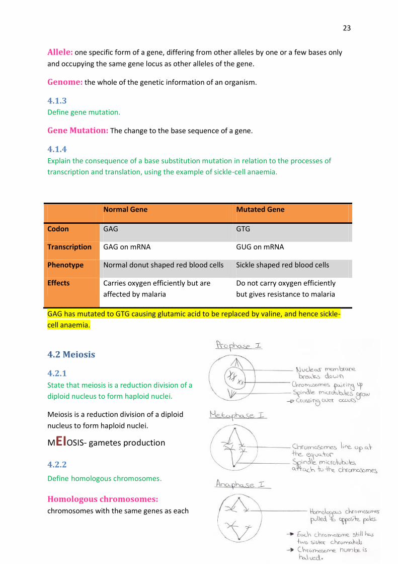
4.2 Meiosis
4.2.1
State that meiosis is a reduction division of a
diploid nucleus to form haploid nuclei.
Meiosis is a reduction division of a diploid
nucleus to form haploid nuclei.
MEIOSIS- gametes production
4.2.2
Define homologous chromosomes.
Homologous chromosomes:
chromosomes with the same genes as each

24
other, in the same sequence but do not necessarily have the same allele of those genes.
4.2.3
Outline the process of meiosis, including pairing of homologous chromosomes and crossing
over, followed by two divisions, which results in four haploid cells.
4.2.4
Explain that non-disjunction can lead to changes in chromosome number, illustrated by
reference to Down syndrome (trisomy 21).
A number of problems can arise during meiosis. A common problem is non-disjunction. This
is when the chromosomes do not separate properly during meiosis, metaphase I. This leads
the production of gametes that either have a chromosome too many or too few. Gametes
with a missing chromosome usually die quite fast however gametes with an extra
25
chromosome can survive. When a zygote is formed from the fertilization of these gametes
with an extra chromosome, three chromosomes of one type are present instead of two. An
example of this is Down syndrome. Down syndrome is a disease in which the chromosomes
failed to separate properly during meiosis leading to three chromosomes of type 21 instead
of two. A person with the condition therefore has a total of 47 chromosomes instead of 46.
The non-disjunction can take place either in the formation of the egg or the sperm. Down
syndrome leads to many complications and also the risk of having a child with the condition
increases with age.
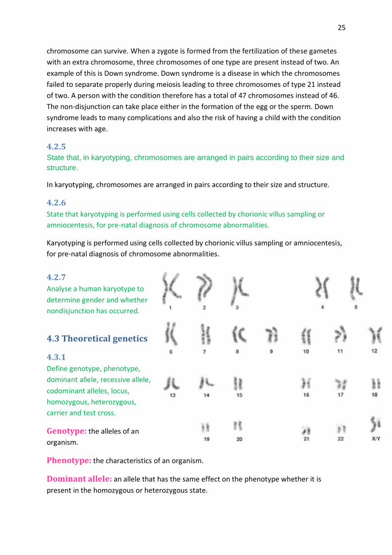
4.2.5 State that, in karyotyping, chromosomes are arranged in pairs according to their size and
structure.
In karyotyping, chromosomes are arranged in pairs according to their size and structure.
4.2.6
State that karyotyping is performed using cells collected by chorionic villus sampling or
amniocentesis, for pre-natal diagnosis of chromosome abnormalities.
Karyotyping is performed using cells collected by chorionic villus sampling or amniocentesis,
for pre-natal diagnosis of chromosome abnormalities.
4.2.7
Analyse a human karyotype to
determine gender and whether
nondisjunction has occurred.
4.3 Theoretical genetics
4.3.1
Define genotype, phenotype,
dominant allele, recessive allele,
codominant alleles, locus,
homozygous, heterozygous,
carrier and test cross.
the alleles of an Genotype:
organism.
the characteristics of an organism. Phenotype:
an allele that has the same effect on the phenotype whether it is Dominant allele:
present in the homozygous or heterozygous state.

26
an allele that only has an effect on the phenotype when present in the Recessive allele:
homozygous state.
pairs of alleles that both affect the phenotype when present in a Codominant alleles:
heterozygote.
the particular position on homologous chromosomes of a gene. Locus:
having two identical alleles of a gene. Homozygous:
having two different alleles of a gene. Heterozygous:
an individual that has one copy of a recessive allele that causes a genetic disease Carrier:
in individuals that are homozygous for this allele.
testing a suspected heterozygote by crossing it with a known homozygous Test cross:
recessive.
4.3.2
Determine the genotypes and phenotypes of the offspring of a monohybrid cross using a
Punnett grid.
4.3.3
State that some genes have more than
two alleles (multiple alleles).
Some genes have more than two
alleles, multiple alleles.
4.3.4
Describe ABO blood groups as an
example of codominance and multiple
alleles.
The ABO blood group is a good
example of codominance and multiple
alleles. There are three alleles that
control the ABO blood groups. If there
are more than two allele of a gene
then they are called multiple allele.
The allele IA corresponds to blood
group A (genotype IAIA) and the allele
IB corresponds to blood group B
(genotype IBIB). Both of these are
dominant and so if IA and IB are
present together they form blood
27
group AB (genotype IAIB). Both allele affect the phenotype since they are both codominant.
Codominant alleles are a pairs of allele that both affect the phenotype when present
together in a heterozygote. The allele i is recessive to both IA and IB so if you have the
genotype IA i you will have blood group A and if you have the genotype IB i you will have
blood group B. However if you have the genotype ii then you are homozygous for i and will
be of blood group O. Below is a table to summaries which genotypes give which
phenotypes.
Phenotype Genotype
A IAIA or IAi
B IBIB or IBi
AB IAIB
O ii
4.3.5
Explain how the sex chromosomes control gender by referring to the inheritance of X and Y
chromosomes in humans.
There are two chromosomes which determine gender. These are called the sex
chromosomes and there are two types, the X and the Y chromosome. Females have two X
chromosomes whereas males have one X and one Y chromosome. The X chromosome is
relatively large compared to the Y (which is much smaller) and contains many genes. The Y
chromosome on the other hand only contains a few genes. The female always passes on to
her offspring the X chromosome from the egg (female gamete). The male can pass on either
the Y or the X chromosome from the sperm (male gamete). If the male passes on the X
chromosome then the growing embryo will develop into a girl. If the male passes on the Y
chromosome then the growing embryo will develop into a boy. Therefore gender depends
on whether the sperm which fertilizes the egg is carrying an X or a Y chromosome.
4.3.6
State that some genes are present on the X chromosome and absent from the shorter Y
chromosome in humans.
Some genes are present on the X chromosome and absent from the shorter Y chromosome
in humans.
4.3.7
Define sex linkage.
Sex linkage: when the gene controlling the characteristic is located on the sex
chromosome and so we associate the characteristic with gender.
28
4.3.8
Describe the inheritance of colour blindness and haemophilia as examples of sex linkage.
Most of the time sex-linked genes
are carried on the X chromosome.
Since females have two X
chromosomes they have two
copies of the sex-linked gene
whereas males only have one
since they only have one X
chromosome. Hemophilia and
colour blindness are both
examples of sex linkage.
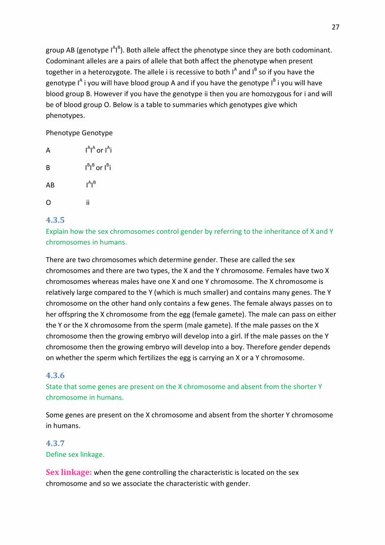
Hemophilia
XH is the allele for normal blood
clotting and is dominate
over Xh which is recessive and
causes hemophilia. If a mother is heterozygous she is a carrier of the disease but does not
have hemophilia as the dominate allele is present. She can however pass the disease on to
her offspring. Below is a punnett showing how a carrier mother and an unaffected father can
pass the disease on to their offspring.
From our four possible outcomes we can see that a female child cannot get hemophilia but
can be a carrier. This is because the father will always pass on the dominate allele (XH) on the
X chromosome in females. Depending on whether the mother passes on the dominant or
recessive allele will determine if the female child is a carrier or is unaffected by the
hemophilia. If the child is a boy then the father has passed on the Y chromosome which does
not contain the allele of the gene. So whether the child has the disease or is unaffected
depends on which allele the mother had passed on. If she has passed on the recessive allele
(Xh) then the male child will have hemophilia, however if she has passed on the dominate
allele (XH) then the child will be unaffected.
So there is a 50% chance of the child having hemophilia if it is male as half of the eggs
produced by the mother will carry the recessive allele. The chance of a female offspring
having hemophilia is 0% since the father always passes on the dominant allele on the X
chromosome. Finally there is a 25% chance overall that the offspring will be affected.
4.3.9
State that a human female can be homozygous or heterozygous with respect to sex-linked
genes.
A human female can be homozygous or heterozygous with respect to sex-linked genes.

29
4.3.10
Explain that female carriers are heterozygous for X-linked recessive alleles.
Female carriers for X-linked recessive alleles are always heterozygous since they require a
dominant allele and a recessive allele to be carriers. They inherit the recessive allele from
one parent and the dominate allele from the other. For example hemophilia is a sex-linked
disease. If a carrier mother and an unaffected father have offspring then the unaffected
father will always pass on his dominate allele to his female offspring. The carrier mother can
either pass on the dominate or recessive allele. If she passes on the recessive allele to her
female offspring than the female offspring will be a carrier as well.
4.3.11
Predict the genotypic and phenotypic ratios of offspring of monohybrid crosses involving any
of the above patterns of inheritance.
4.3.12
Deduce the genotypes and phenotypes of individuals in pedigree charts.
For dominant and recessive alleles, upper-case and lower-case letters, respectively, should
be used. Letters representing alleles should be chosen with care to avoid confusion between
upper and lower case.
For codominance, the main letter should relate to the gene and the suffix to the allele, both
upper case. For example, red and white codominant flower colours should be represented as
CR and Cw, respectively. For sickle-cell anaemia, HbA is normal and Hbs is sickle cell.
4.4 Genetic engineering and biotechnology
4.4.1
Outline the use of polymerase chain reaction (PCR) to copy and amplify minute quantities of
DNA.
Polymerase chain reaction is used to copy and amplify minute quantities of DNA. It can be
useful when only a small amount of DNA is available but a large amount is required to
undergo testing. We can use DNA from blood, semen, tissues and so on from crime scenes
for example. The PCR requires high temperature and a DNA polymerase enzyme from
Thermus aquaticus (a bacterium that lives in hot springs).
4.4.2
State that, in gel electrophoresis, fragments of DNA move in an electric field and are
separated according to their size.
In gel electrophoresis, fragments of DNA move in an electric field and are separated
according to their size.

30
4.4.3
State that gel electrophoresis of DNA is used in DNA profiling.
Gel electrophoresis of DNA is used in DNA profiling.
4.4.4
Describe the application of DNA profiling to determine paternity and also in forensic
investigations.
Organisms have short sequences of bases which are repeated many times. These are called
satellite DNA. These repeated sequences vary in length from person to person. The DNA is
copied using PCR and then cut up into small fragments using restriction enzymes. Gel
electrophoresis separates fragmented pieces of DNA according to their size and charge. This
gives a pattern of bands on a gel which is unlikely to be the same for two individuals. This is
called DNA profiling. DNA profiling can be used to determine paternity and also in forensic
investigations to get evidence to be used in a court case for example.
4.4.5
Analyse DNA profiles to draw conclusions about paternity or forensic investigations.
The outcomes of this analysis could include knowledge of the number of human genes, the
location of specific genes, discovery of proteins and their functions, and evolutionary
relationships.
For a suspect look for similarities between the DNA found at the crime scene and the
suspect. For a paternity test, look for similarities between the child and the possible father.
4.4.6
Outline three outcomes of the sequencing of the complete human genome.
It is now easier to study how genes influence human development.
It helps identify genetic diseases.
It allows the production of new drugs based on DNA base sequences of genes or the
structure of proteins coded for by these genes.
It will give us more information on the origins, evolution and migration of humans.
4.4.7
State that, when genes are transferred between species, the amino acid sequence of
polypeptides translated from them is unchanged because the genetic code is universal.
When genes are transferred between species, the amino acid sequence of a polypeptides
translated from them is unchanged because the genetic code is universal.
4.4.8
Outline a basic technique used for gene transfer involving plasmids, a host cell (bacterium,
yeast or other cell), restriction enzymes (endonucleases) and DNA ligase.

31
The use of E. coli in gene technology is well documented. Most of its DNA is in one circular
chromosome, but it also has plasmids (smaller circles of DNA). These plasmids can be
removed and cleaved by restriction enzymes at target sequences. DNA fragments from
another organism can also be cleaved by the same restriction enzyme, and these pieces can
be added to the open plasmid and spliced together by ligase. The recombinant plasmids
formed can be inserted into new host cells and cloned.
4.4.9
State two examples of the current uses of genetically modified crops, or animals.
1. The transfer of a gene for factor IX which is a blood clotting factor, from humans to
sheep so that this factor is produced in the sheep’s milk.
2. The transfer of a gene that gives resistance to the herbicide glyphosate from
bacterium to crops so that the crop plants can be sprayed with the herbicide and not
be affected by it.
4.4.10
Discuss the potential benefits and possible harmful effects of one example of genetic
modification.
It is quite common to see genetic modifications in crop plants. An example of this is the
transfer of a gene that codes for a protein called Bt toxin from the bacterium Bacillus
thuringiensis to maize crops. This is done because maize crops are often destroyed by insects
that eat the corn and so by adding the Bt toxin gene this is prevented as the toxin kills the
insects. However this is very controversial as even though it has many positive advantages, it
can also have some harmful consequences.
Benefits Harmful Effects
Since there is less damage to the maize
crops, there is a higher crop yield which can
lessen food shortages.
We are not sure of the consequences of humans
& animals eating the modified crops. The
bacterial DNA or the Bt toxin itself could be
harmful to human as well as animal health.
Since there is a higher crop yield, less land
is needed to grow more crops. Instead the
land can become an area for wild life
conservation.
Other insects which are not harmful to the crops
could be killed. The maize pollen will contain the
toxin and so if it is blown onto nearby plants it
can kill the insects feeding on these plants.

32
There is a reduction in the use of pesticides
which are expensive and may be harmful to
the environment, wild life & farm workers.
Cross pollination can occur, which results in some
wild plants being genetically modified as they will
contain the Bt gene. These plants will have an
advantage over others as they will be resistant to
certain insects & so some plants may become
endangered. This will have significant
consequences on the population of wild plants.
4.4.11
Define clone.
a group of genetically identical organisms or a group of cells derived from a single Clone:
parent cell.
4.4.12
Outline a technique for cloning using differentiated animal cells.
Dolly the sheep was clones by taking udder cells from a donor sheep. These cells were then
cultured in a low nutrient medium to make the genes switch off and become dormant. Then
an unfertilized egg was taken from another sheep. The nucleus of this egg cell was removed
by using a micropipette and then the egg cells were fused with the udder cells using a pulse
of electricity. The fused cells developed like normal zygotes and became embryos. These
embryos were then implanted into another sheep whose role was to be the surrogate
mother. One lamb was born successfully, called Dolly. Dolly was genetically identical to the
sheep from which the udder cells were taken.
4.4.13
Discuss the ethical issues of therapeutic cloning in humans.
Therapeutic cloning is the creation of an embryo to supply embryonic stem cells for medical
use.
Arguments For Arguments Against
Embryonic stem cells can be used for therapies that
save lives & reduce pain for patients. Since a stem cell
can divide + differentiate into any cell type, they can
be used to replace tissues or organs required by
patients.
Every human embryo is a potential human
being and should be given the chance of
developing.
Cells can be taken from embryos that have stopped
developing & so these cells would have died anyway.
More embryos are generally produced
than are needed & so many are killed.

33
Cells are taken at a stage when the embryos have no
nerve cells & so they cannot feel pain.
There is a risk of embryonic stem cells
developing into tumour cells.
Topic 5: Ecology and evolution
5.1 Communities and ecosystems
5.1.1
Define species, habitat, population, community, ecosystem and ecology.
Species: a group of organisms that can interbreed and produce fertile offspring.
Habitat: the environment in which a species normally lives or the location of a living
organism.
Population: a group of organisms of the same species who live in the same area at the
same time.
Community: a group of populations living and interacting with each other in an area.
Ecosystem: a community and its abiotic environment.
Ecology: the study of relationships between living organisms and between organisms and
their environment.
5.1.2
Distinguish between autotroph and heterotroph.
Autotroph: an organism that synthesizes its organic molecules from simple inorganic
substances.
Heterotroph: an organism that obtains organic molecules from other organisms.
5.1.3
Distinguish between consumers, detritivores and saprotrophs.
Consumer: an organism that ingests other organic matter that is living or recently killed.
Detritivore: an organism that ingests non-living organic matter.
Saprotroph: an organism that lives on or in non-living organic matter, secreting digestive
enzymes into it and absorbing the products of digestion.
5.1.4

34
Describe what is meant by a food chain, giving three examples, each with at least three
linkages (four organisms).
1. Carrot plantcarrot fly fly catcher
sparrow hawk
2. Bush grass Impala CheetahLions
3. Buckwheat Gopher Gopher snake
Red Tailed Kite
5.1.5
Describe what is meant by a food web.
The elaborate interconnected relationships within an
ecosystem based on feeding and energy transfer.
5.1.6
Define trophic level.
Trophic level: of an organism is its position in the
food chain.
5.1.7
Deduce the trophic level of organisms in a food chain and a food web.
Plants or any other photosynthetic organisms are the producers. Primary consumers are the
species that eat the producers. Secondary consumers are the species that eat the primary
consumers and tertiary consumers in turn eat the secondary consumers.
5.1.8
Construct a food web containing up to 10 organisms, using appropriate information.
See above.
5.1.9
State that light is the initial energy source for almost all communities.
Light is the initial energy source for almost all communities.
5.1.10
Explain the energy flow in a food chain.
Energy flows from producers to primary consumers, to secondary consumers, to
tertiary consumers.
Energy is lost between trophic levels in the form of heat through cell respiration,
faeces, tissue loss and death.

35
CO2
PLANTS
ORGANISMS
FOSSILE
FULES
Photosynthesis
Cell respiration
Combustion
Fossilisation
Some of this lost energy is used by detritivores and saprotrophs. These in turn also
lose energy in the form of heat through cell respiration.
5.1.11
State that energy transformations are never 100% efficient.
Energy transformations are never 100% efficient.
5.1.12
Explain reasons for the shape of pyramids of energy.
Energy is not recycled. Constantly being supplied to the ecosystem through light energy.
Energy is lost from the ecosystem in the form of heat through cell respiration.
Nutrients must be recycled as there is only a limited supply of them.
They are absorbed by the environment, used by organisms & then returned to the environment.
5.1.13
Explain that energy enters and leaves ecosystems, but nutrients must be recycled.
Energy enters and leaves ecosystems as the producers absorb sunlight and then convert it
into energy using photosynthesis. The primary consumers then eat the producers however
energy is lost through repatriation and heat. The consumers then die and the decomposers
then eat them and therefore the nutrients are recycled and used again for the producers as
energy in the soil.
5.1.14
State that saprotrophic bacteria and fungi (decomposers) recycle nutrients.
Saprotrophic bacteria and fungi recycle nutrients.
5.2 The greenhouse effect
5.2.1
Draw and label a diagram of the carbon cycle to show the processes involved.

36
5.2.2
Analyse the changes in concentration of atmospheric carbon dioxide using historical records.
Summer=increase in photosynthesis less CO2 (in the northern hemisphere)
5.2.3
Explain the relationship between rises in concentrations of atmospheric carbon dioxide,
methane and oxides of nitrogen and the enhanced greenhouse effect.
1. The incoming radiation from the sun is short wave ultraviolet and visible radiation. 2. Some of this radiation is absorbed by the earth’s atmosphere. 3. Some of the radiation is reflected back into space by the earth’s surface. 4. The radiation which is reflected back into space is infrared radiation and has a longer
wavelength. 5. The greenhouse gases in the atmosphere absorb some of this infrared radiation & re-
reflect it back towards the earth. 6. This causes the greenhouse effect and results in an increase in average mean
temperatures on earth. 7. A rise in greenhouse gases results in an increase of the greenhouse effect which can
be disastrous for the planet.
5.2.4
Outline the precautionary principle.
The precautionary principle holds that, if the effects of a human-induced change would be
very large, perhaps catastrophic, those responsible for the change must prove that it will not
do harm before proceeding. This is the reverse of the normal situation, where those who are
concerned about the change would have to prove that it will do harm in order to prevent
such changes going ahead.
5.2.5
Evaluate the precautionary principle as a justification for strong action in response to the
threats posed by the enhanced greenhouse effect.
There is strong evidence that shows that greenhouse gases are causing global warming. This
is very worrying as global warming has so many consequences on ecosystems. If nothing is
done, and the greenhouse gases are in fact causing the enhanced greenhouse effect, by the
time we realize it, it will probably be too late and result in catastrophic consequences. So
even though there is no proof for global warming, the strong evidence suggesting that it is
linked with an increase in greenhouse gases is something we cannot ignore. Global warming
is a global problem. It affects everyone. For these reasons, the precautionary principle
should be followed. Anyone supporting the notion that we can continue to emit same
amounts or more of the greenhouse gases should have to provide evidence that it will not
cause a damaging increase in the greenhouse effect.

37
5.2.6
Outline the consequences of a global temperature rise on arctic ecosystems.
Global warming could have a number of disastrous consequences largely affecting the arctic ecosystems:
The arctic ice cap may disappear as glaciers start to melt and break up into icebergs.
Permafrost will melt during the summer season which will increase the rate of decomposition of trapped organic matter, including peat and detritus. This in turn will increase the release of carbon dioxide which will increase the greenhouse effect even further.
Species adapted to temperature conditions will migrate north which will alter food chains and have consequences on the animals in the higher trophic levels.
Marine species in the arctic water may become extinct as these are very sensitive to temperature changes within the sea water.
Polar bears may face extinction as they lose their ice habitat and therefore can no longer feed or breed as they normally would.
Pests and diseases may become quite common with rises in temperature.
As the ice melts, sea levels will raise and flood low lying areas of land.
Extreme weather events such as storms might become common and have disastrous effects on certain species.
5.3 Populations
5.3.1
Outline how population size is affected by natality, immigration, mortality and emigration.
Natality: increases population size as offspring are added to the population.
Immigration: increases population size as individuals have moved into the area from somewhere else and so this adds to the population.
Mortality: decreases the population as some individuals get eaten die of old age or get sick.
Emigration: decreases the population as individuals have moved out of the area to go live somewhere else.
5.3.2
Draw and label a graph showing a
sigmoid (S-shaped) population growth
curve.
5.3.3
Explain the reasons for the exponential
growth phase, the plateau phase and
the transitional phase between these
two phases.

38
Exponential phase:
1. Rapid increase in population growth. 2. Natality rate exceeds mortality rate. 3. Abundant resources available. (food, water, shelter) 4. Diseases and predators are rare.
Traditional phase:
1. Natality rate starts to fall and/or mortality rate starts to rise. 2. There is a decrease in the number of resources. 3. An increase in the number of predators and diseases. 4. Population still increasing but at a slower rate.
Plateau phase:
1. No more population growth, population size is constant. 2. Natality rate is equal to mortality rate. 3. The population has reached the carrying capacity of the environment. 4. The limited resources & the common predators & diseases keep the population
numbers constant.
5.3.4
List three factors that set limits to population increase.
1. Shortage of resources (e.g. food)
2. Increase in predators
3. Increase in diseases and parasites
5.4 Evolution
5.4.1
Define evolution.
Evolution: is the cumulative change in the heritable characteristics of a population.
5.4.2
Outline the evidence for evolution provided by the fossil record, selective breeding of
domesticated animals and homologous structures.
Fossils, selective breeding and homologous structures have provided scientists with evidence
that support the theory of evolution. As they started to study fossils they realized that these
were not identical but had similarities with existing organisms. This suggested that
organisms changed over time. Selective breeding of domesticated animals also provides this
evidence as the domestic breeds have similar characteristics to the wild ones and can still
breed with them. As selected wild individuals with desirable characteristics were bred, over
time this resulted in a more desirable species from a human point of view. An example of
this is the taming of wild wolves and their selective breeding in order to produce the
39
domestic dogs we know today. This suggests that not only have these animals evolved but
also that they can evolve rapidly. Finally scientists have found a number of homologous
structures within different species. Many bones in the limbs are common to a number of
species and therefore it suggests that these have evolved from one common ancestor.
5.4.3
State that populations tend to produce more offspring than the environment can support.
Populations tend to produce more offspring than the environment can support.
5.4.4
Explain that the consequence of the potential overproduction of offspring is a struggle for
survival.
If the mortality rate remains lower than the natality rate then a population will keep
growing. As more offspring are produced, there will be fewer resources available to other
members of the population. If there is an over production of offspring this will result in a
struggle for survival within the species as the resources become scarce and individuals in the
population will start to compete for these. This results in an increase in mortality rate as the
weaker individuals in the population will lose out on these vital resources that are essential
for their survival.
5.4.5
State that the members of a species show variation.
Members of a species show variation.
5.4.6
Explain how sexual reproduction promotes variation in a species.
Sexual reproduction is important for promoting variation as even though mutations form
new genes or alleles, sexual reproduction forms a new combination of alleles. There are two
stages in sexual reproduction that promote variation in a species. The first one is during
meiosis during which a large variety of genetically different gametes are produced by each
individual. The second stage is fertilization. Here, alleles from two different individuals are
brought together to form one new individual.
5.4.7
Explain how natural selection leads to evolution.
Greater survival and reproductive success of individuals with favourable heritable variations
can lead to change in the characteristics of a population.
5.4.8
Explain two examples of evolution in response to environmental change; one must be
antibiotic resistance in bacteria.

40
Antibiotic resistance in bacteria is a common problem. It results from the transfer of a gene
that gives resistance to a specific antibiotic usually by means of a plasmid to a bacterium.
Some bacteria will then have this gene and become resistant to the specific antibiotic while
others will lack the gene and so will die if exposed to the antibiotic. Over time, the non-
resistant ones will all die off as doctors vaccinate patients, but the resistant ones will survive.
Eventually, the resistant ones will be the only ones left as a result of natural selection and so
a new antibiotic must be created. However, this has to be done on a regular basis as the
bacteria keep evolving and become resistant to multiple antibiotics.
The Peppered Moth is another example of evolution in response to environmental change.
There are two types of these moths; one species has a light colour while the other one is
darker. When Britain begun industrializing, the soot from the factories would land on trees
and so the darker moths then had an advantage over the light ones as they could easily hide
from predators. Before the soot, both types of moths were eaten by predators however now
that the darker ones were able to hide the lighter ones got eaten more often. The
population of the darker moths rapidly increased while that of the lighter ones rapidly
decreased until only the dark moths were left. All the lighter moths were less adapted to the
environmental change and so they could no longer survive in that new environment.
5.5 Classification
5.5.1
Outline the binomial system of nomenclature.
Species are a group of organisms with similar characteristics which can interbreed and
produce fertile offspring whereas a genus is a group of similar species.
Species need an international name and so biologists name them using the binomial system
of nomenclature. Each species is given two names. The first is the genus name and is given
an upper case first letter. The second is the species name and is given a lower case first
letter. If the name is printed, italics are used. If on the other hand the name is hand-written,
it is underlined.
5.5.2
List seven levels in the hierarchy of taxa —kingdom, phylum, class, order, family, genus and
species— using an example from two different kingdoms for each level.
Red Kangaroo:
Taxa Human Garden Pea
Kingdom Animalia Plantae
Phylum Chordata Angiospermae
Class Mammalia Dicotyledoneae
Order Primates Rosales
41
Family Hominidae Papilionaceae
Genus Homo Pisum
Species Sapiens Sativum
5.5.3
Distinguish between the following phyla of plants, using simple external recognition
features: bryophyta, filicinophyta, coniferophyta and angiospermophyta.
Physical Attributes Example
Bryophyta Very short stature, non-vascular plants Moss
Filicinophyta Vascular plant Ferns & horsetails
Coniferophyta Woody stems leaves are in the form of needles
or scales
Fir & pine trees
Angiospermophyta All plants which make flowers & their seeds
surrounded by fruit
5.5.4
Distinguish between the following phyla of animals, using simple external recognition
features: porifera, cnidaria, platyhelminthes, annelida, mollusca and arthropoda.
Porifera:
no clear symmetry
attached to a surface
pores through body
no mouth or anus
example: sponges
Cnidaria:
radially symmetric
tentacles
stinging cells
mouth but no anus
example: jellyfish
Platyhelminths:
bilaterally symmetrical
flat bodies
unsegmented
mouth but no anus
example: tapeworm
Annelida:
bilaterally symmetrical
bristles often present
segmented
mouth and anus
example: earthworm
Mollusca:
muscular foot and mantle
shell may be present
segmentation not visible
mouth and anus
example: slugs and snails
Arthropoda:
bilaterally symmetric

42
exoskeleton
segmented
jointed appendages
example: spiders and insect
5.5.5
Apply and design a key for a group of up to eight organisms.
1. Vascular Tissue
Does not have vascular tissue 2
Contains vascular tissue for conducting fluids 3
2. Presence of lobes on leaves
Does not possess lobes on leaves moss
Possesses lobe son leaves liverwort
3. Seeds or spores
Produces seeds 4
Produces spores 7
4. Seed covering
Seeds encased in sweet fruit 5
Seeds encased in a cone 6
5. Sweet fruit
Fruit contains many small seeds apple
Fruit contains one large pit cherry
6. Seeds in a cone
Long needles in a brush-like formation pine tree
Leaves are flat scales cedar
7. Spore-producing plants
Has many small flat leaves fern
Has no flat leaves horsetail
Topic 6: Human health and physiology
6.1 Digestion
6.1.1
Explain why digestion of large food molecules is essential.
1. The food we eat is made up of many compounds made by other organisms which are
not all suitable for human tissues and therefore these have to be broken down and
reassembled so that our bodies can use them.
2. The food molecules have to be small enough to be absorbed by the villi in the
intestine through diffusion, facilitated diffusion or active transport and so large food
molecules need to be broken down into smaller ones for absorption to occur.

43
6.1.2
Explain the need for enzymes in digestion.
1. Enzymes break down large food molecules into smaller ones
2. Speed up the process of digestion by lowering the activation energy for the reaction
3. Work at body temperature
6.1.3
State the source, substrate, products and optimum pH conditions for one amylase, one
protease and one lipase.
Amylase Protease Lipase
Enzyme Salivary Amylase Pepsin Pancreatic Lipase
Source Salivary Glands Stomach lining Pancreas
Substrate Starch Proteins Triglycerides (fats & oils)
Products Maltose Polypeptides Fatty acids & glycerol
Optimum pH 7 1.5-2 7
6.1.4
Draw and label a diagram of the digestive
system.
6.1.5
Outline the function of the stomach, small
intestine and large intestine.
Stomach:
Secretes HCL which kills bacteria
HCL provides optimum pH for pepsin
Secretes pepsin for protein digestion
Small intestine
Intestinal wall secrets enzymes
Receives enzymes from the pancreas
Has villi for absorption of food particles
Large intestine:
Moves material that has not been
digested along

44
Absorbs water
Produces faeces
6.1.6
Distinguish between absorption and assimilation.
Absorption: villi take in digested food through the cells.
Assimilation: taking molecules and making them a part of you (liver).
6.1.7
Explain how the structure of the villus is related to its role in absorption and transport of the
products of digestion.
Villi create an increased surface area for absorption
o Absorb small food molecules
Micro-villi are located on the villi and provide for even greater surface area
The wall of the villi is one cell thick (shorter distance for diffusion)
o Protein channels allow for facilitated diffusion (highlow) of food into villi
o Active transport; protein pumps=increased number of mitochondria
o Blood vessels are close to the wall of the villi & help move away the molecules
& maintaining a concentration gradient.
o Lipids are transported by the lacteal
6.2 The transport system
6.2.1
Draw and label a diagram of the heart
showing the four chambers, associated
blood vessels, valves and the route of
blood through the heart.
6.2.2
State that the coronary arteries supply
heart muscle with oxygen and
nutrients.
Coronary arteries supply heart muscle
with oxygen and nutrients.
6.2.3
Explain the action of the heart in terms
of collecting blood, pumping blood, and
opening and closing of valves.

45
The right atrium collects blood from the superior and inferior vena cava and the left atrium
collects blood from the pulmonary veins. This blood then flows into the right and left
ventricle which pump the blood into the arteries. The direction of the blood flow is
controlled by the atrioventricular valves and semilunar valves. When the atria contract the
blood flows through the atrioventricular valves which are open, into the ventricle. At this
stage the semilunar valves are closed so the ventricle fills with blood. The ventricles then
contract which causes a rise in pressure. This rise in pressure first causes the atrioventricular
valves to close preventing back flow of blood into the atria. Then the semilunar valves open
allowing the expulsion of blood into the arteries. As this happens, the atria start to fill with
blood again. The ventricles stop contracting leading to a fall in pressure which causes the
semilunar valves to close, preventing back flow of blood from the arteries. When the
ventricular pressure drops below the atrial pressure the atrioventricular valves open again
and the cycle repeats.
6.2.4
Outline the control of the heartbeat in terms of myogenic muscle contraction, the role of the
pacemaker, nerves, the medulla of the brain and epinephrine (adrenaline).
The heart muscle can contract by itself, without the stimulation of a nerve. This is called
myogenic muscle contraction. The region that initiates each contraction is found in the wall
of the right atrium and is called the pacemaker. Every time the pacemaker sends out a
signal, a heartbeat results. The pacemaker is under the influence of nerves and adrenaline.
One nerve carries messages from the medulla of the brain to the pacemaker and speeds up
the beating of the heart. Another nerve carries messages from the medulla of the brain to
the pacemaker and slows down the beating of the heart. Finally, adrenaline (epinephrine) is
carried by the blood and once it reaches the pacemaker it signals it to increase the beating
of the heart.
6.2.5
Explain the relationship between the structure and function of arteries, capillaries and veins.
Arteries have a thick outer layer of longitudinal collagen and elastic fibres to avoid leaks
and bulges. They have a thick wall which is essential to withstand the high pressures. They
also have thick layers of circular elastic fibres and muscle fibres to help pump the blood
through after each contraction of the heart. In addition the narrow lumen maintains the high
pressure inside the arteries.
Veins are made up of thin layers with a few circular elastic fibres and muscle fibres. This is
because blood does not flow in pulses and so the vein walls cannot help pump the blood on.
Veins also have thin walls which allow the nearby muscles to press against them so that they
become flat. This helps the blood to be pushed forwards towards the heart. There is only a
thin outer layer of longitudinal collagen and elastic fibres as there is low pressure inside the
46
vein and so little chance of bursting. Finally, a wide lumen is needed to accommodate the
slow flowing blood due to the low pressure.
Capillaries are made up of a wall that is only one cell layer thick and results in the
distance for diffusion in and out of the capillary being very small so that diffusion can occur
rapidly. They also contain pores within their wall which allow some plasma to leak out and
form tissue fluid. Phagocytes can also pass through these pores to help fight infections. In
addition, the lumen of the capillaries is very narrow. This means that many capillaries can fit
in a small space, increasing the surface area for diffusion.
6.2.6
State that blood is composed of plasma, erythrocytes, leucocytes (phagocytes and
lymphocytes) and platelets.
Blood is composed of plasma, erythrocytes, leucocytes and platelets.
6.2.7
State that the following are transported by the blood: nutrients, oxygen, carbon dioxide,
hormones, antibodies, urea and heat.
Blood transports: nutrients, oxygen, carbon dioxide, hormones, antibodies, urea and heat
6.3 Defence against infectious disease
6.3.1
Define pathogen.
an organism or virus that causes a disease. Pathogen:
6.3.2
Explain why antibiotics are effective against bacteria but not against viruses.
Antibiotics are produced by microorganisms to kill or control the growth of other
microorganisms by blocking specific metabolic pathways within the cell. Since bacteria are so
different to human cells, antibiotics can be taken by humans to kill bacteria without harming
the human cells. Viruses on the other hand are different as they do not carry out many
metabolic processes themselves. Instead they rely on a host cell (a human cell) to carry out
these processes for them. Therefore viruses cannot be treated with antibiotics as it is
impossible to harm the virus without harming the human cells.
6.3.3
Outline the role of skin and mucous membranes in defence against pathogens.
The skin forms a physical barrier that prevents pathogens from entering the body as the
outer layer is very tough. In addition the skin contains sebaceous glands which secret lactic
47
acid and fatty acids which creates an acidic environment on the surface of the skin
preventing the growth of pathogens.
Mucous membranes form another type of barrier against pathogens. Mucous membranes
are soft and moist areas of skin found in the trachea, nose, vagina and urethra. These
membranes are not strong enough to create a physical barrier but they do have mucus
which contains lysozyme enzymes that digest the phagocytes. Also, the mucus can be sticky
such as in the trachea, and trap the pathogens which are then expelled up the trachea and
out of the body by muscles within the trachea.
6.3.4
Outline how phagocytic leucocytes ingest pathogens in the blood and in body tissues.
Phagocytes are found in the blood and ingest pathogens. They do so by recognizing
pathogens and engulfing them by endocytosis. Enzymes within the phagocytes called
lysosomes then digest the pathogens. Phagocytes can ingest pathogens in the blood but also
within body tissue as they can pass through the pores of capillaries and into these tissues.
6.3.5
Distinguish between antigens and antibodies.
Antigens: are foreign substances which stimulate the production of antibodies.
Antibodies: are proteins that defend the body against pathogens by binding to antigens
on the surface of these pathogens and stimulating their destruction.
6.3.6
Explain antibody production.
Lymphocytes are a type of leukocyte which makes antibodies. Each lymphocyte makes only
one specific antibody. A large amount of different lymphocytes are needed so that the body
can produce different types of antibodies. The antibodies are found on the surface of the
plasma membrane of these lymphocytes with the antigen-combining site projecting
outwards. Pathogens have antigens on their surface which bind to the antigen-combining
site of the antibodies of a specific lymphocyte. When this happens the lymphocyte becomes
active and starts to make clones of itself by dividing by mitosis. These clones then start to
make more of this specific antibody needed to defend the body against the pathogen.
6.3.7
Outline the effects of HIV on the immune system.
The HIV virus (which causes AIDS) destroys a type of lymphocyte which has a vital role in
antibody production. Over the years this results in a reduced amount of active lymphocytes.
Therefore, less antibodies are produced which makes the body very vulnerable to
pathogens. A pathogen that could easily be controlled by the body in a healthy individual can
cause serious consequences and eventually lead to death for patients affected by HIV.

48
6.3.8
Discuss the cause, transmission and social implications of AIDS.
Cause: HIV causes AIDS (acquired immunodeficiency syndrome). A syndrome is a group of
symptoms that are found together. HIV destroys a type of lymphocyte which is vital for
antibody production. Over the years, less active lymphocytes are produced which leads to a
fall in the amount of antibodies. Pathogens that would normally be easily controlled by the
body in healthy individuals can cause serious consequences and eventually lead to death for
patients affected by HIV. The immune system is considerably weakened.
Transmission: HIV is transmitted through body fluids from an infected person to an
uninfected one. This can occur through vaginal and anal intercourse as well as oral sex if
there are cuts or tears in the vagina, penis, mouth or intestine. It can also be transmitted by
hypodermic needles that are shared by intravenous drug abusers. The small amount of
blood present on these needles after their use may contain the virus and is enough to infect
another person. Another way of transmission is through the placenta from mother to child,
or through cuts during childbirth or in milk during breast feeding. Finally there is a risk of
transmission in transfused blood or with blood products such as Factor VIII used to treat
haemophiliacs.
Social implications: Relatives and friends suffer grief. Families can also suffer from a loss of
income as the person infected by HIV can lose their wage if they are unable to work and are
refused life insurance. Also, HIV patients may find it hard to find partners, employment and
even housing. Finally, AIDS can cause fear in a population and reduce sexual activity.
6.4 Gas exchange
6.4.1
Distinguish between ventilation, gas exchange and cell respiration.
Ventilation: is the process of bringing fresh air into the alveoli and removing the stale air.
It maintains the concentration gradient of carbon dioxide and oxygen between the alveoli
and the blood in the capillaries (vital for oxygen to diffuse into the blood from the alveoli
and carbon dioxide out of the blood into the alveoli).
Gas Exchange: is the process of swapping one gas for another. It occurs in the alveoli of
the lungs. Oxygen diffuses into the capillaries from the air in the alveoli and carbon dioxide
diffuses out of the capillaries and into the air in the alveoli.
Cell Respiration: Cell respiration releases energy in the form of ATP so that this energy
can be used inside the cell. Cell respiration occurs in the mitochondria and cytoplasm of
cells. Oxygen is used in this process and carbon dioxide is produced.
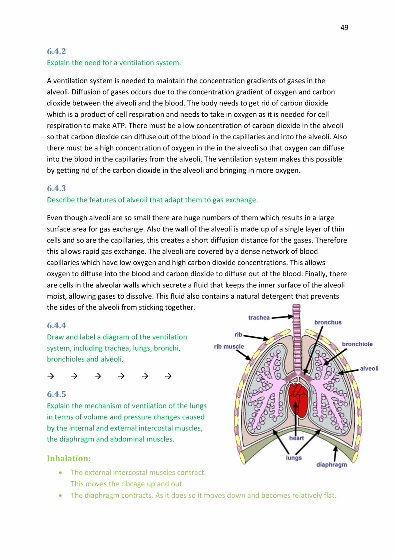
49
6.4.2
Explain the need for a ventilation system.
A ventilation system is needed to maintain the concentration gradients of gases in the
alveoli. Diffusion of gases occurs due to the concentration gradient of oxygen and carbon
dioxide between the alveoli and the blood. The body needs to get rid of carbon dioxide
which is a product of cell respiration and needs to take in oxygen as it is needed for cell
respiration to make ATP. There must be a low concentration of carbon dioxide in the alveoli
so that carbon dioxide can diffuse out of the blood in the capillaries and into the alveoli. Also
there must be a high concentration of oxygen in the in the alveoli so that oxygen can diffuse
into the blood in the capillaries from the alveoli. The ventilation system makes this possible
by getting rid of the carbon dioxide in the alveoli and bringing in more oxygen.
6.4.3
Describe the features of alveoli that adapt them to gas exchange.
Even though alveoli are so small there are huge numbers of them which results in a large
surface area for gas exchange. Also the wall of the alveoli is made up of a single layer of thin
cells and so are the capillaries, this creates a short diffusion distance for the gases. Therefore
this allows rapid gas exchange. The alveoli are covered by a dense network of blood
capillaries which have low oxygen and high carbon dioxide concentrations. This allows
oxygen to diffuse into the blood and carbon dioxide to diffuse out of the blood. Finally, there
are cells in the alveolar walls which secrete a fluid that keeps the inner surface of the alveoli
moist, allowing gases to dissolve. This fluid also contains a natural detergent that prevents
the sides of the alveoli from sticking together.
6.4.4
Draw and label a diagram of the ventilation
system, including trachea, lungs, bronchi,
bronchioles and alveoli.
6.4.5
Explain the mechanism of ventilation of the lungs
in terms of volume and pressure changes caused
by the internal and external intercostal muscles,
the diaphragm and abdominal muscles.
Inhalation:
The external intercostal muscles contract.
This moves the ribcage up and out.
The diaphragm contracts. As it does so it moves down and becomes relatively flat.

50
Both of these muscle contractions result in an increase in the volume of the thorax
which in turn results in a drop in pressure inside the thorax.
Pressure eventually drops below atmospheric pressure.
Air then flow into the lungs from outside the body, through the mouth or nose,
trachea, bronchi and bronchioles.
Air continues to enter the lungs until the pressure inside the lungs rises to the
atmospheric pressure.
Exhalation:
The internal intercostal muscles contract. This moves the ribcage down and in.
The abdominal muscles contract. This pushes the diaphragm up, back into a dome
shape.
Both of these muscle contractions result in a decrease in the volume of the thorax.
As a result of the decrease in volume, the pressure inside the thorax increases.
Eventually the pressure rises above atmospheric pressure.
Air then flows out of the lungs to outside of the body through the nose or mouth.
Air continues to flow out of the lungs until the pressure in the lungs has fallen back to
atmospheric pressure.
6.5 Nerves, hormones and homeostasis
6.5.1
State that the nervous system consists of the central nervous system (CNS) and peripheral
nerves, and is composed of cells called neurons that can carry rapid electrical impulses.
The nervous system consists of the central nervous system (CNS) and peripheral nerves and
is composed of cells called neurons that can carry rapid
electrical impulses.
6.5.2
Draw and label a diagram of the structure of a motor
neuron.
6.5.3
State that nerve impulses are conducted from receptors
to the CNS by sensory neurons, within the CNS by relay
neurons, and from the CNS to effectors by motor neurons.
Nerve impulses are conducted from receptors to the CNS
by sensory neurons within the CNS by relay neurons and
from the CNS to effectors by motor neurons.
6.5.4
51
Define resting potential and action potential (depolarization and repolarization).
Resting Potential: is the electrical potential across the cell membrane of a cell that is not
conducting an impulse. When a neuron is resting (i.e. not conducting an impulse), it has a
membrane potential equal to about –65mV.
Action Potential: The rapid change in the electrical potential across a cell membrane of
an excitable cell caused by stimulus-triggered, selective opening and closing of voltage gated
ion channels. An action potential begins with depolarization, which is the change of charge
inside an axon from negative (-65mV) to positive (40mV). Depolarization is followed by
repolarization, which is the change of charge inside an axon from positive (40mV) to
negative (at least -65mV). Neurons are the only cells that can change their membrane
potentials in response to stimuli.
6.5.5
Explain how a nerve impulse passes along a non-myelinated neuron.
A nerve impulse is the way a neuron transmits information. Nerve impulses are conducted
from receptors to the CNS by sensory neurons, within the CNS by relay neurons, and from
the CNS to effectors by motor neurons.
1. An action potential in one part of a neuron causes an action potential to develop in
the next section of the neuron. This is due to diffusion of sodium ions between the
region with an action potential and the region at the resting potential. These ion
movements, local currents, reduce the resting potential. If the potential rises above
the threshold level, voltage-gated channels open.
2. Na+ channels open very quickly and Na+ diffuse into the neuron down the
concentration gradient. This reduces the membrane potential and causes more Na+
channels to open. The entry of positively charged Na+ causes the inside of the neuron
to develop a net positive charge compared to the outside- the potential across the
membrane is reversed. This is called DEPOLARIZATION.
3. Potassium channels open after a short delay. K+ diffuse out of the neuron down the
concentration gradient through the opened channels. The exit of positively charged
K+ cause the inside of the neuron to develop a net negative charge again compared
with the outside- the potential across the membrane is restored. This is called
REPOLARIZATION.

52
4. Concentration gradients of sodium and potassium across the membrane are restored
by the active transport of Na+ out of the neuron and K+ into the neuron. This restores
the resting potential and the neuron is then ready to conduct another nerve impulse.
As before, sodium ions diffuse along inside the neuron from an adjacent region that
has already depolarized and initiate depolarization.
6.5.6
Explain the principles of synaptic transmission.
Nerve impulses are
passed from one neuron
to another across a
fluid-filled space called
the synaptic cleft.
1. The action
potential arrives
at the terminal
knob and causes
the Ca2+ channels
to open.
2. Ca2+ diffuse into
the terminal
knob
3. The vesicles
containing
ACETYLCHOLINE move to the plasma membrane to preform exocytosis; fusing with
the membrane and secreting the ACETYLCHOLINE.
4. ACETYLCHOLINE attaches to the receptor proteins of the post-synaptic neuron thus
opening the sodium-channels enabling the Ca2+ to diffuse into the next neuron.
53
6.5.7State that the endocrine system consists of glands that release hormones that are
transported in the blood.
The endocrine system consists of glands that release hormones that are transported in the
blood.
6.5.8
State that homeostasis involves maintaining the internal environment between limits,
including blood pH, carbon dioxide concentration, blood glucose concentration, body
temperature and water balance.
Homeostasis involves maintaining the internal environment between limits; including blood
pH, carbon dioxide concentration, blood glucose concentration, body temperature and
water balance.
6.5.9
Explain that homeostasis involves monitoring levels of variables and correcting changes in
levels by negative feedback mechanisms.
Homeostasis involves maintaining the internal environment between limits, including blood
pH, carbon dioxide concentration, blood glucose concentration, body temperature and
water balance. Blood and tissue fluid (derived from blood) make up the internal
environment. This internal environment varies very little compared to the external
environment which varies greatly. Negative feedback is used to keep the internal
environment between limits. It uses the nervous and endocrine system to do so. It has a
stabilising effect as any change from a set point level will result in an opposite change. The
levels of production of for example blood glucose, feedback to affect the rate of production.
If blood glucose levels rise above the set point, this will feed back to decrease production
and reduce the level back around the set point. A decrease in blood glucose levels below the
set point will result in an increase in production so that the levels increase back to the set
point. Small fluctuations around the set point will not cause any response. Negative
feedback is only triggered when there are significant increases or decreases from the set
point.
6.5.10
Explain the control of body temperature, including the transfer of heat in blood, and the
roles of the hypothalamus, sweat glands, and skin arterioles and shivering.
The hypothalamus is responsible for monitoring the temperature of the blood which is
normally close to 37°C. If there are significant fluctuations from this set point, the
hypothalamus sends signals (messages carried by neurons) to different parts of the body to
restore the temperature back to the set point. This is done through negative feedback.

54
INCREASES ABOVE THE SET POINT DROPS BELOW THE SET POINT
Skin arterioles increase in diameter so that
more blood flows to the skin. Transferring heat
from the core of the body to the skin & this
heat is then lost to the external environment,
cooling down the body in the process.
Skin arterioles decrease in diameter so that
less blood flows to the skin. The diameter of
the capillaries in the skin cannot change but
less blood flows through them; prevents heat
loss to the external environment as the
temperature of the skin falls.
Skeletal muscle stays relaxed so that heat is not
generated.
Shivering occurs; skeletal muscles make many
small rapid contractions generating heat.
Sweat glands secrete large amounts of sweat
which makes the surface of the skin moist.
When water evaporates from the moist skin it
cools down the body.
Sweat glands to not secrete sweat and so no
water evaporation can occur as skin stays dry.
6.5.11
Explain the control of blood glucose concentration, including the roles of glucagon, insulin
and α and β cells in the pancreatic islets.
Blood glucose concentration does not have a specific set point like blood temperature. Blood
glucose levels drop and rise through the day and so the body usually tries to keep blood
glucose levels around 4 to 8 millimoles per dm3 of blood. Once again, negative feedback is
used to do so. There are responses by target organs which affect the rate at which glucose is
taken up from the blood or loaded into the blood.
Response to blood glucose levels above the set
point
Response to blood glucose levels below the
set point
β cells in the pancreatic islets produce insulin.
Insulin stimulates muscle cells and the liver cells
to take up glucose from the blood and convert it
into glycogen. These are then stored in the form
of granules in the cytoplasm of cells. Also, other
types of cells are stimulated to take up glucose
and use it for cell respiration instead of fat. All of
these processes lower the levels of glucose in the
blood.
α cells in the pancreatic islets produce
glucagon. Glucagon stimulates the liver cells to
convert glycogen back into glucose and release
this glucose into the blood. This raises the
glucose levels in the blood.
6.5.12Distinguish between . type I and type II diabetes
Type I diabetes Type II diabetes

55
The onset is usually early, during childhood. The onset is usually late, after childhood.
β cells do not produce enough insulin. Target cells become insensitive to insulin.
Diet by itself cannot be used to control the condition.
Insulin injections are needed to control glucose levels.
Insulin injections are not usually needed. Low
carbohydrate diet can control the condition.
6.6 Reproduction
6.6.1
Draw and label diagrams of the adult male and female reproductive systems.
6.6.2
Outline the role of hormones in the menstrual cycle, including FSH (follicle stimulating
hormone), LH (luteinizing hormone), estrogen and progesterone.
The menstrual cycle:
1. FSH is secreted by the pituitary gland and its levels start to rise. This stimulates the
follicle to develop and the follicle cells to secret estrogen.
2. Estrogen then causes the follicle cells to make more FSH receptors so that these can
respond more strongly to the FSH.
3. This is positive feedback and causes the estrogen levels to increase and stimulate the
thickening of the endometrium (uterus lining).
4. Estrogen levels increase to a peak and by doing so it stimulates LH secretion from the
pituitary gland.
5. LH then increases to its peak and causes ovulation (release of egg from the follicle).
6. LH then stimulates the follicle cells to secrete less estrogen and more progesterone.
Once ovulation has occurred, LH stimulated the follicle to develop into the corpus
luteum.
7. The corpus luteum then starts to secrete high amounts of progesterone. This prepares
the uterine lining for an embryo.
8. The high levels of estrogen and progesterone then start to inhibit FSH and LH.

56
9. If no embryo develops the levels of estrogen and progesterone fall. This stimulates
menstruation (break down of the uterine lining). When the levels of these two hormones
are low enough FSH and LH start to be secreted again.
10. FSH levels rise once again and
a new menstrual cycle begins.
6.6.3
Annotate a graph showing hormone
levels in the menstrual cycle,
illustrating the relationship between
changes in hormone levels and
ovulation, menstruation and
thickening of the endometrium.
6.6.4
List three roles of testosterone in
males.
1. Pre-natal development of
male genitalia
2. Development of secondary
sexual characteristics
3. Maintenance of sex drive.
6.6.5
Outline the process of in vitro
fertilization (IVF).
Process:
1. For a period of three weeks, the woman has to have a drug injected to stop her normal
menstrual cycle.
2. After these three weeks, high doses of FSH are injected once a day for 10-12 days so that
many follicles develop in the ovaries of the women.
3. HCG (another hormone) is injected 36 hours before the collection of the eggs. HCG
loosens the eggs in the follicles and makes them mature.
4. The man needs to ejaculate into a jar so that sperm can be collected from the semen.
The sperm are processed to concentrate the healthiest ones.
5. A device that is inserted through the wall of the vagina is used to extract the eggs from
the follicles.
6. Each egg is then mixed with sperm in a shallow dish. The dishes are then put into an
incubator overnight.
7. The next day the dishes are looked at to see if fertilization has happened.
57
8. If fertilization has been successful, two or three of the embryos are chosen to be placed
in the uterus by the use of a long plastic tube.
9. A pregnancy test is done a few weeks later to find out if any of the embryos have
implanted.
10. A scan is done a few weeks later to find out if the pregnancy is progressing normally.
6.6.6
Discuss the ethical issues associated with IVF.
Arguments for IVF Arguments against IVF
Many types of infertility are due to environmental factors rather than genetic which means that the offspring would not inherit the infertility.
The infertility of the parents may be inherited
by their offspring passing on the suffering to
the next generation.
The embryos that are killed during the IVF process cannot feel pain or suffering as they do not have a developed nervous system.
More embryos are produced than needed &
the ones that remain are usually killed which
denies them the chance of a life.
Suffering caused by genetic diseases can be decreases by screening the embryos before placing them into the uterus.
Embryologists select which embryos will be
placed into the uterus. Therefore they decide
the fate of new individuals as they choose
which ones will survive & which ones won’t.
Since the IVF process is not an easy one emotionally & physically; costly, takes time, no guarantees. Parents who are willing to go through it must have a strong desire to have children & are likely to be loving parents.
IVF is not a natural process which takes place in
a laboratory compared to natural conception
which occurs as a result of an act of love.
Infertility can cause emotional suffering to couples who want to have children. IVF can take away this suffering for some of those couples.
Infertility should be accepted as God’s will & to
go against it by using IVF procedures would be
wrong.
AHL
Topic 7: Nucleic acids and proteins
7.1 DNA structure
7.1.1
Describe the structure of DNA, including the antiparallel strands, 3’–5’ linkages and
hydrogen bonding between purines and pyrimidines.
SEE PAGE 16!

58
7.1.2
Outline the structure of nucleosomes.
A nucleosome consists of DNA wrapped around eight histone proteins and held together by
another histone protein.
7.1.3
State that nucleosomes help to supercoil chromosomes and help to regulate transcription.
Nucleosomes help to supercoil chromosomes and help regulate transcription.
7.1.4
Distinguish between unique or single-copy genes and highly repetitive sequences in nuclear
DNA.
Not all of the base sequences in DNA are translated. Highly repetitive base sequences are
not translated. They consist of sequences of between 5 and 300 bases that may be repeated
up to 10 000 times. They constitute 5-45% of eukaryotic DNA. Single-copy genes or unique
genes are translated and constitute a surprisingly small proportion of eukaryotic DNA.
7.1.5
State that eukaryotic genes can contain exons and introns.
Eukaryotic genes can contain exons and introns.
7.2 DNA replication
7.2.1
State that DNA replication occurs in a 5’→ 3’ direction.
The 5’ end of the free DNA nucleotide is added to the 3’ end of the chain of nucleotides that
is already synthesized.
7.2.2
Explain the process of DNA replication in prokaryotes, including the role of enzymes
(helicase, DNA polymerase, RNA primase and DNA ligase), Okazaki fragments and
deoxynucleoside triphosphates.
Enzymes involved in the process of DNA replication:
Helicase- unwinds in the double stranded DNA. (separates the 2 strands)
REPLICATION BUBBLES!
RNA primase (RNA polymerase) - attaches RNA nucleotides complementary to the
opened DNA strands. (=primer)
DNA polymerase III- attaches the DNA nucleotides to an existing chain in a 5’ to 3’
direction. (synthesizing the new complementary strand)

59
DNA polymerase I- removes the RNA primer and replaces it with DNA.
DNA ligase- joins the Okazaki fragments on the lagging strand.
The process:
1. DNA replication begins when helicase unwinds a segment of the DNA and breaks the
hydrogen bonds between the two complementary strands of DNA.
2. DNA polymerase can only add new nucleotides to a free 3’ end of a growing chain.
Synthesis of one strand of the DNA, called the leading stand, proceeds continuously
in the 5’ to 3’direction.
3. Synthesis of the lagging stand is more complex. DNA polymerase can add new
deoxyribonucleotides only to a free 3’OH. It is synthesizes discontinuously.
4. To provide a free 3’ OH starting point on the lagging stand, RNA primase attaches to
the DNA and synthesizes a short RNA primer. DNA polymerase III then adds
deoxyribonucleotides to the 3’ end of the RNA primer.
5. DNA polymerase I
replaces DNA
polymerase III,
removes the RNA
and replaces it
with DNA.
6. Finally, the
enzyme DNA
ligase joins the
Okazaki
fragments on the
lagging strand.
7.2.3
State that DNA
replication is initiated at
many points in
eukaryotic
chromosomes.
DNA replication is initiated at many points in eukaryotic chromosomes as they have more
than one replication bubble-where the DNA is partially opened- whereas prokaryotic
chromosomes only have one replication bubble in them.
DNA replication is when the helicase unzips a molecule of DNA breaking the hydrogen bonds
between the strands. There are two strands, the leading and the lagging strand, the lagging
strand synthesizes in a discontinuous manner as it is more complex, while the leading strand
synthesizes in a continuous manner. Synthesis goes in a 5’ to 3’ direction. RNA primase is
used for this. As the lagging strand is more complex RNA primase creates an RNA primer.
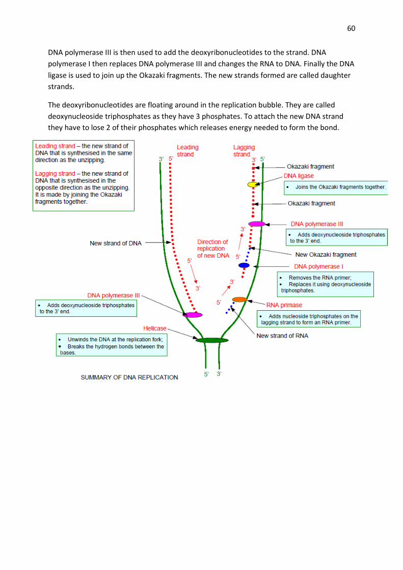
60
DNA polymerase III is then used to add the deoxyribonucleotides to the strand. DNA
polymerase I then replaces DNA polymerase III and changes the RNA to DNA. Finally the DNA
ligase is used to join up the Okazaki fragments. The new strands formed are called daughter
strands.
The deoxyribonucleotides are floating around in the replication bubble. They are called
deoxynucleoside triphosphates as they have 3 phosphates. To attach the new DNA strand
they have to lose 2 of their phosphates which releases energy needed to form the bond.

61
7.3 Transcription
7.3.1
State that transcription is carried out in a 5’ 3’ direction.
The 5’ end of the free RNA nucleotide is added to the 3’ end of the RNA molecule that is
already synthesized.
7.3.2
Distinguish between the sense and antisense strands of DNA.
Sense strand: (coding strand) has the same base sequence as mRNA with uracil instead of
thymine.
Antisense: (template) strand is transcribed.
7.3.3
Explain the process of transcription in prokaryotes, including the role of the promoter
region, RNA polymerase, nucleoside triphosphates and the terminator.
1. RNA polymerase binds to the promoter region
2. This initiates transcription
3. RNA polymerase uncoils the DNA
4. Only one strand is used, the template strand
5. Free nucleoside triphosphates bond to their complementary bases on the template
strand
6. Adenine binds to uracil instead of thymine
7. As the nucleoside triphosphates bind they become nucleotides and release energy by
losing two phosphate groups
8. The mRNA is built in a 5'→3' direction
9. RNA polymerase forms covalent bonds between the nucleotides and keeps moving
along the DNA until it reaches the terminator
10. The terminator signals the RNA polymerase to stop transcription
11. RNA polymerase is released and mRNA separates from the DNA
12. The DNA rewinds
7.3.4
State that eukaryotic RNA needs the removal of introns to
form mature mRNA.
Eukaryotic RNA needs the removal of introns to form mature
mRNA.
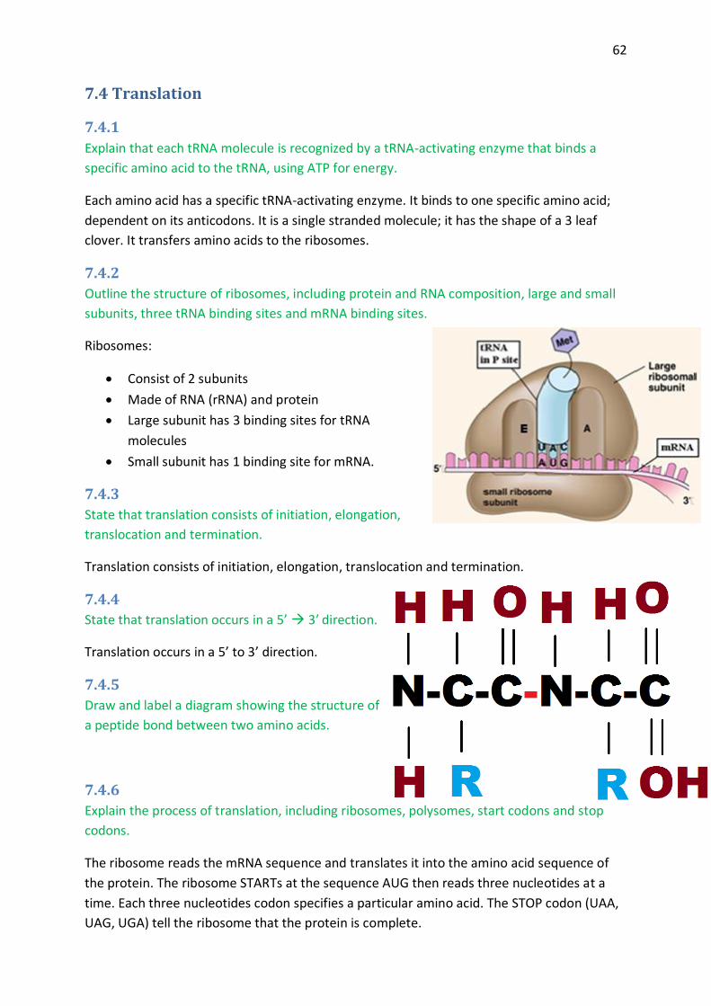
62
7.4 Translation
7.4.1
Explain that each tRNA molecule is recognized by a tRNA-activating enzyme that binds a
specific amino acid to the tRNA, using ATP for energy.
Each amino acid has a specific tRNA-activating enzyme. It binds to one specific amino acid;
dependent on its anticodons. It is a single stranded molecule; it has the shape of a 3 leaf
clover. It transfers amino acids to the ribosomes.
7.4.2
Outline the structure of ribosomes, including protein and RNA composition, large and small
subunits, three tRNA binding sites and mRNA binding sites.
Ribosomes:
Consist of 2 subunits
Made of RNA (rRNA) and protein
Large subunit has 3 binding sites for tRNA
molecules
Small subunit has 1 binding site for mRNA.
7.4.3
State that translation consists of initiation, elongation,
translocation and termination.
Translation consists of initiation, elongation, translocation and termination.
7.4.4
State that translation occurs in a 5’ 3’ direction.
Translation occurs in a 5’ to 3’ direction.
7.4.5
Draw and label a diagram showing the structure of
a peptide bond between two amino acids.
7.4.6
Explain the process of translation, including ribosomes, polysomes, start codons and stop
codons.
The ribosome reads the mRNA sequence and translates it into the amino acid sequence of
the protein. The ribosome STARTs at the sequence AUG then reads three nucleotides at a
time. Each three nucleotides codon specifies a particular amino acid. The STOP codon (UAA,
UAG, UGA) tell the ribosome that the protein is complete.

63
7.4.7
State that free ribosomes synthesize proteins for use primarily within the cell, and that
bound ribosomes synthesize proteins primarily for secretion or for lysosomes.
Free ribosomes synthesize proteins for use within the cell.
Bound ribosomes synthesize proteins for secretion or for lysosomes.
7.5 Proteins
7.5.1
Explain the four levels of protein structure, indicating the significance of each level. Structure Type
Description Held together by
Primary Is the sequence of amino acids in its peptide chain(s)
Peptide bonds
Secondary Interactions between amino acids cause the chain to form a 3D shape. Alpha helix or beta sheet.
Hydrogen bonds
Tertiary Folds up even more. Achieves its overall 3D shape. Conformation.
Ionic bonds Hydrogen bonds HYDROPHOBIC INTERACTIONS! DESULFIDE BONDS!
Quaternary More than one polypeptide chain. The arrangement of these proteins is a quaternary structure. HAEMOGLOBIN!
7.5.2 Outline the difference between fibrous and globular proteins, with reference to two examples of each protein type. FIBROUS: Proteins are formed from parallel polypeptide chains held together by cross-links; rope-like fibres, which are generally insoluble in water. STRUCTURAL!
Collagen: main component of connective tissue, ligaments, tendons and cartilage.
Keratin: main component of hard structures such as hair, nails, claws and hooves.
GLOBULAR: Proteins usually have a spherical/rounded shape. Hydrophobic groups are on the inside, hydrophilic groups on the outside. They are soluble in water.
Enzymes-lipase & DNA polymerase

64
Transport proteins- haemoglobin, myoglobin and those embedded in proteins.
7.5.3 Explain the significance of polar and non-polar amino acids. Amino acids have different R groups. Some of these R groups will be hydrophilic, making the
amino acid polar, while others will be hydrophobic, making the amino acid non-polar. The
distribution of the polar and non-polar amino acids in a protein influences the function and
location of the protein within the body. Non-polar amino acids are found in the center of
water soluble proteins while the polar amino acids are found at the surface.
Examples of how the distribution of non-polar and polar amino acids affects protein function
and location:
Controlling the position of proteins in membranes: non-polar amino acids cause proteins to
be embedded in membranes, polar amino acids cause portions of the proteins to protrude
from the membrane.
Creating hydrophilic channels through membranes: Polar amino acids are found inside
membrane proteins and create a channel through which hydrophilic molecules can pass
through.
Specificity of active site in enzymes: If the amino acids in the active site of an enzyme are
non-polar then it makes this active site specific to a non-polar substance. On the other hand,
if the active site is made up of polar amino acids then the active site is specific to a polar
substance.
7.5.4 State four functions of proteins, giving a named example of each.
1. Helps movement (muscle) 2. Immunological (antibodies are proteins) 3. Transport 4. Structural 5. Some are hormones (signalling)
7.6 Enzymes
7.6.1
State that metabolic pathways consist of chains and cycles of enzyme catalysed reactions.
Metabolic pathways consist of chains and cycles of enzyme catalysed reactions.
7.6.2
Describe the induced-fit model.

65
Until the substrate binds, the active site does not fit the substrate precisely. As the substrate
approaches the active site and binds to it, the shape of the active site changes and only then
does it fit the substrate. The substrate induces the active site to change, weakening bonds in
the substrate during the process and thus reducing the activation energy.
7.6.3
Explain that enzymes lower the activation energy of the chemical reactions that they
catalyse.
Reactants of a chemical reaction need to gain energy before they can undergo the reaction.
This required energy is called the activation energy of the reaction and it is needed to break
bonds within the reactants. At a later stage in the reaction energy will be released as new
bonds form. The majority of biological reactions are exothermic. In exothermic reactions the
energy released by the new bonds formed is greater than the activation energy. In other
words, the reaction releases energy. Enzymes make it easier for reactions to occur by
decreasing the activation energy required in the reactions that they catalyse.
7.6.4
Explain the difference between competitive and non-competitive inhibition, with reference
to one example of each.
Competitive inhibition is the situation when an inhibiting molecule that is structurally similar
to the substrate molecule binds to the active site, preventing substrate binding. Limit non-
competitive inhibition to an inhibitor binding to an enzyme (not to its active site) that causes
a conformational change in its active site, resulting in a decrease in activity.
7.6.5
Explain the control of metabolic pathways by end-product inhibition, including the role of
allosteric sites.
If there is an excess of the end product the end product can bind to the enzyme at the
allosteric site and can alter the shape of the enzyme so that substrates no longer can bind to
it. No more products are produced. Tells it to slow down or speed up reaction, or stop it all
together.

66
Leo says ger lose e- gain O
gains e- reduction
OIL RIG Oxidation is loss,
reduction is gain
Topic 8: Cell respiration and photosynthesis
8.1 Cell respiration
8.1.1
State that oxidation involves the loss of electrons from an element, whereas reduction
involves a gain of electrons; and that oxidation frequently involves gaining oxygen or losing
hydrogen, whereas reduction frequently involves losing oxygen or gaining hydrogen.
Oxidation Reduction
Loss of electrons (e-) Gain of electrons (e-)
Loss of Hydrogen (H) Gain of Hydrogen (H)
Gain of Oxygen (O) Loss of Oxygen (O)
8.1.2
Outline the process of glycolysis, including phosphorylation, lysis, oxidation and ATP
formation.
In the cytoplasm, one hexose sugar is converted into two three-carbon atom compounds
(pyruvate) with a net gain of two ATP and two NADH + H+.
Phosphorylation: phosphate is added. 2ATP<
2ADP<
Lysis: 2 (G3P’s)
P P
P P
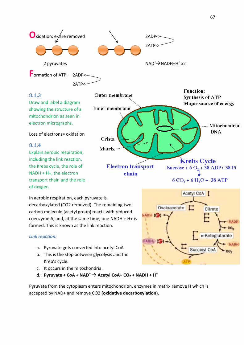
67
Oxidation: e- are removed 2ADP<
2ATP<
2 pyruvates NAD+NADH+H+ x2
Formation of ATP: 2ADP<
2ATP<
8.1.3
Draw and label a diagram
showing the structure of a
mitochondrion as seen in
electron micrographs.
Loss of electrons= oxidation
8.1.4
Explain aerobic respiration,
including the link reaction,
the Krebs cycle, the role of
NADH + H+, the electron
transport chain and the role
of oxygen.
In aerobic respiration, each pyruvate is
decarboxylated (CO2 removed). The remaining two-
carbon molecule (acetyl group) reacts with reduced
coenzyme A, and, at the same time, one NADH + H+ is
formed. This is known as the link reaction.
Link reaction:
a. Pyruvate gets converted into acetyl CoA
b. This is the step between glycolysis and the
Kreb’s cycle.
c. It occurs in the mitochondria.
d. Pyruvate + CoA + NAD+ Acetyl CoA+ CO2 + NADH + H+
Pyruvate from the cytoplasm enters mitochondrion, enzymes in matrix remove H which is
accepted by NAD+ and remove CO2 (oxidative decarboxylation).

68
In the Krebs cycle first Acetyl CoA gives its acetate away to combine with a C4 compound, left
over from the last cycle. This then forms citrate a C6 compound. C6 then loses a CO2 and is
then oxidized, reducing NAD+ to NADH + H+, forming C5. This then loses another CO2 and is
oxidized reducing NAD+ to NADH + H+. ADP is then converted into to ATP. And C4 is oxidized
yet again reducing FAD to FADH2. And it is oxidized again reducing NAD+ to NADH + H+,
creating C4 again.
Electron transport chain:
To use the energy stored in
NADH+ and FADH2 (e-) to produce
ATP (ATP + Pi)
1. H+ is pumped from the
matrix to the inter
membrane space using
energy from e-.
2. As H+ concentration
increases in the inter
membrane space, the H+ diffuse back into the matrix through ATP synthesis- this
allows ADP + Pi ATP.
3. Oxygen is the final electron acceptor! O2+4e-+4H+ 2H2O
8.1.5
Explain oxidative phosphorylation in terms of chemiosmosis.
The actual production of ATP in cellular respiration takes place through the process of
chemiosmosis. Chemiosmosis involves the pumping of protons through special channels in
the membranes of mitochondria from the inner to the outer compartment as a result of
electrons flowing along the electron transport chain. The pumping establishes a proton
gradient. After the gradient is established, protons pass down the gradient through particles
designated F1 (ATP Synthase). In these particles, the energy of the protons generates ATP,
using ADP and phosphate ions as the starting points.
Phosphorylation:
Substrate level
Oxidative
Occurs during Krebs cycle and glycolysis
ATP is being produced by phosphorylation.
Oxidative phosphorylation is the result of chemiosmosis. (Process with H+)
(ADPATP)
8.1.6
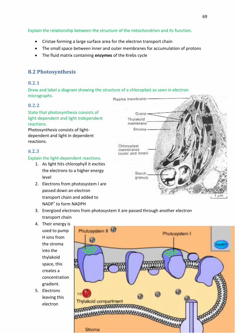
69
Explain the relationship between the structure of the mitochondrion and its function.
Cristae forming a large surface area for the electron transport chain
The small space between inner and outer membranes for accumulation of protons
The fluid matrix containing enzymes of the Krebs cycle
8.2 Photosynthesis
8.2.1
Draw and label a diagram showing the structure of a chloroplast as seen in electron micrographs.
8.2.2
State that photosynthesis consists of light-dependent and light independent reactions. Photosynthesis consists of light-dependent and light in dependent reactions.
8.2.3
Explain the light-dependent reactions. 1. As light hits chlorophyll it excites
the electrons to a higher energy
level
2. Electrons from photosystem I are
passed down an electron
transport chain and added to
NADP+ to form NADPH
3. Energized electrons from photosystem II are passed through another electron
transport chain
4. Their energy is
used to pump
H ions from
the stroma
into the
thylakoid
space, this
creates a
concentration
gradient.
5. Electrons
leaving this
electron

70
transport chain enter photosystem I replenishing the lost electrons.
6. Photosystem II replenishes its electrons by splitting water.
7. H+, ½ O2 are released into the thylakoid compartment.
8. The build-up of hydrogen ions in the thylakoid compartment stores potential energy.
This is harvested by ATP synthase.
9. As the ions diffuse through the enzyme it uses the energy of the moving ions to make
ATP.
8.2.4
Explain photophosphorylation in terms of chemiosmosis. Photophosphorylation converts light energy into chemical energy. Then Photosystem 2
absorbs the light energy, bringing electrons to a higher energy level. The energy is passed
along antenna pigments until it reaches aP680 molecule. The energy excites an electron on
the P680 molecule which is transferred to the reaction centre and electron transport chain.
To replace the lost electron, an electron is taken from the photolysis of water,
creating oxygen as a by-product. As the electron passes along the chain, it gives energy to
the protein pumps, causing the pumps to force protons into the confined thylakoid space.
These protons then diffuse out of the thylakoid through ATP synthase proton
channels, producing ATP.
8.2.5
Explain the light-independent reactions. The light-independent reactions of photosynthesis occur in the stroma of the chloroplast and
involve the conversion of carbon dioxide and other compounds into glucose. The light-
independent reactions can be split into three stages; these are carbon fixation, the reduction
reactions and finally the regeneration of ribulose bisphosphate. Collectively these stages are
known as the Calvin Cycle.
During carbon fixation, carbon dioxide
in the stroma (which enters the
chloroplast by diffusion) reacts with a
five-carbon sugar called ribulose
bisphosphate (RuBP) to form a six-
carbon compound. This reaction is
catalysed by an enzyme called ribulose
bisphosphate carboxylase (large
amounts present within the stroma),
otherwise known as rubisco. As soon as
the six-carbon compound is formed, it
splits to form two molecules of
glycerate 3-phosphate. Glycerate 3-
phosphate is then used in the
reduction reactions.

71
Glycerate 3-phosphate is reduced during the reduction reactions to a three-carbon sugar
called triose phosphate. Energy and hydrogen is needed for the reduction and these are
supplied by ATP and NADPH + H+ (both produced during light-dependent reactions)
respectively. Two triose phosphate molecules can then react together to form glucose
phosphate. The condensation of many molecules of glucose phosphate forms starch which is
the form of carbohydrate stored in plants. However, out of six triose phosphates produced
during the reduction reactions, only one will be used to synthesise glucose phosphate. The
five remaining triose phosphates will be used to regenerate RuBP.
The regeneration of RuBP is essential for carbon fixation to continue. Five triose phosphate
molecules will undergo a series of reactions requiring energy from ATP, to form three
molecules of RuBP. RuBP is therefore consumed and produced during the light-independent
reactions and therefore these reactions form a cycle which is named the Calvin cycle.
8.2.6
Explain the relationship between the structure of the chloroplast and its function. 1. Limit this to the large surface area of thylakoids for light absorption 2. The small space inside thylakoids for accumulation of protons 3. The fluid stroma for the enzymes of the Calvin cycle.
8.2.7
Explain the relationship between the action spectrum and the absorption spectrum of photosynthetic pigments in green plants. An absorption spectrum shows the quantity of each
wavelength of light absorbed by a specific pigment
each photosynthetic pigment absorbs
specific wavelengths of light
a photosystem is composed of a variety of
photosynthetic pigments
photosynthetic pigments cooperate within
a photosystem to increase the quantity of
light absorbed
An action spectrum is the summation the individual
absorption spectra of the various pigments.
8.2.8
Explain the concept of limiting factors in photosynthesis, with reference to light intensity, temperature and concentration of carbon dioxide. Light:
rate of photosynthesis increases as light intensity increase because more photons are
absorbed to make ATP and NADPH + H+, and drive Calvin cycle
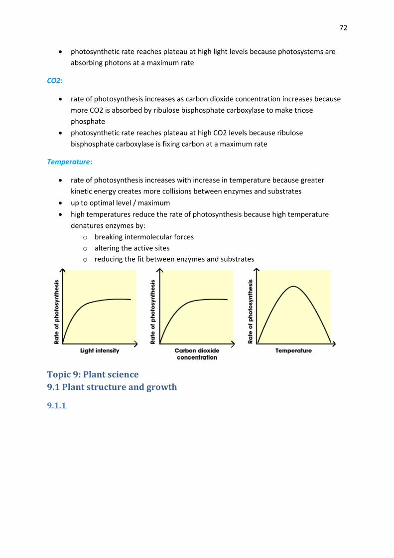
72
photosynthetic rate reaches plateau at high light levels because photosystems are
absorbing photons at a maximum rate
CO2:
rate of photosynthesis increases as carbon dioxide concentration increases because
more CO2 is absorbed by ribulose bisphosphate carboxylase to make triose
phosphate
photosynthetic rate reaches plateau at high CO2 levels because ribulose
bisphosphate carboxylase is fixing carbon at a maximum rate
Temperature:
rate of photosynthesis increases with increase in temperature because greater
kinetic energy creates more collisions between enzymes and substrates
up to optimal level / maximum
high temperatures reduce the rate of photosynthesis because high temperature
denatures enzymes by:
o breaking intermolecular forces
o altering the active sites
o reducing the fit between enzymes and substrates
Topic 9: Plant science
9.1 Plant structure and growth
9.1.1

73
Draw and label plan diagrams to show the distribution of tissues in the stem and leaf of a
dicotyledonous plant.
9.1.2
Outline three differences between the structures of dicotyledonous and monocotyledonous
plants.
Monocotyledonous Dicotyledonous
1 cotyledon 2 cotyledons
Parallel veins Net-like veins
Scattered vascular bundles Vascular bundles in a ring
Fibrous roots Taproots
Floral parts in multiples of 3 Floral parts in multiples of 4 or 5
9.1.3
Explain the relationship between the distribution of tissues in the leaf and the functions of
these tissues.
Waxy cuticle: secreted by epidermis, covers top and bottom leaf surfaces, reduces
water loss, as it is impermeable to water.

74
Epidermis (upper and lower): cover surfaces of leaf, secrete cuticle, protect
against infection, conserves water.
Palisade mesophyll: tightly packed cells in upper region of leaf, rich in
chloroplasts, absorption of light, primary site of photosynthesis.
Spongy mesophyll: loosely packed cells in lower region of leaf, rich in air spaces,
allowing for easy diffusion of gases, less rich in chloroplasts, absorption of light and
secondary site of photosynthesis.
Xylem: distributes water and minerals to cells throughout leaf.
Phloem: collects sucrose (produced by chloroplasts throughout mesophyll) for
distribution of photosynthetic products to non-photosynthetic parts of plant.
Guard cells (stoma): specialized epidermal cells associated in pairs and forming
the borders of the stomata, opening which open or close allowing gas exchange
when open, or water retention when closed.
9.1.4
Identify modifications of roots, stems and leaves for different functions: bulbs, stem tubers,
storage roots and tendrils.
Bulbs: modified leaves, e.g. onion, garlic
Tubers: modified stems, e.g. potato, gladiola
Storage root: e.g. carrot
Tendril: modified leaf: e.g. ivy
9.1.5
State that dicotyledonous plants have apical and lateral meristems.
Dicotyledonous plants have apical and lateral meristems.
9.1.6
Compare growth due to apical and lateral meristems in dicotyledonous plants.
Meristems are regions where cells continue to divide and grow, often throughout the
life of the plant.
Apical meristems are located at the tip of the root and stem, increasing the length of
the plant, and also producing new leaves and flowers.
Lateral meristems, or cambium, are found in vascular bundles and increase the
diameter of the plant by producing xylem and phloem.
In the stems of younger plants, the vascular cambium is located discretely in bundles.
In the stems of older plants, the vascular cambium is a complete ring.
Lateral meristems also increase the root diameter.
9.1.7
Explain the role of auxin in phototropism as an example of the control of plant growth.

75
Auxin is a plant hormone that stimulates cell elongation.
One of the processes that auxin controls is phototropism: directional growth toward
the source of light.
In shoot tips, proteins called phototropins absorb light, changing shape in response
to certain light wavelengths.
Phototropins in a light-induced conformation bind to receptors which stimulate
transcription/translation of genes producing glycoproteins.
9.2 Transport in angiospermophytes
9.2.1
Outline how the root system provides a large surface area for mineral ion and water uptake
by means of branching and root hairs.
Branching: extensive branching of roots greatly increases overall root surface area
exposed to extracellular fluid
Root hairs: individual root epidermal cells grow extensive elongations greatly increasing
the surface area of individual root epidermal cells to extracellular fluid
9.2.2
List ways in which mineral ions in the soil move to the root.
1. diffusion of mineral ions down concentration gradients
2. mass flow of water in the soil carrying ions, when water drains through the soil
3. mineral ions move into fungal hyphae, which grow around plant roots in a
mutualistic relationship, and then from the hyphae into the root
9.2.3
Explain the process of mineral ion absorption from the soil into roots by active transport.
Roots are adapted for their function:
Contain numerous fine extensions (root hairs) which increase the surface area.
Highly branched or deep taproot for maximum absorption.
Roots are often associated with fungal hyphae which aid in water absorption by
spreading out over a larger area.
Ions move from soil into root:
Diffusion- as long as ion concentration outside the root is higher.
Active transport- requires pumping ions across call wall using ATP. (associated with
H+ transport)
This creates a high ion (solute) concentration inside the root.

76
9.2.4
State that terrestrial plants support themselves by means of thickened cellulose, cell turgor
and lignified xylem.
Terrestrial plants support themselves by means of thickened cellulose cell turgor and
lignified xylem.
9.2.5
Define transpiration.
Transpiration: is the loss of water vapour from the leaves and stems of plants.
9.2.6
Explain how water is carried by the transpiration stream, including the structure of xylem
vessels, transpiration pull, cohesion, adhesion and evaporation.
Structure of xylem vessels:
thick-walled, elongated vascular tissue cells
o arranged end-to-end
o connected by perforated end-plates
Evaporation:
1. water vapour diffuses from the moist air spaces of the spongy mesophyll where
water potential is higher
2. to the drier air outside where water potential is lower
3. via stomata
Cohesion:
1. as the spongy mesophyll air spaces lose water by evaporation its water potential
decreases
2. water flows from the xylem, where water potential is higher
3. through the mesophyll to the air spaces, down its water potential gradient
4. the cohesion of water molecules, due to hydrogen bonding
5. enables transpiration to pull water up the narrow xylem vessels
6. without these columns of water breaking apart
Adhesion:
1. the cell walls of xylem vessels are charged, attracting water molecules
2. the adhesive attraction of water to xylem vessel walls moves them up the stem
against gravity
3. adhesion is important when sap starts to rise in plants that were leafless through the
winter
4. adhesion also helps prevent the column of water-filled xylem vessels from breaking

77
Transpiration:
1. solar-powered evaporation from the leaves
2. creates a continuous transpiration pull
3. transmitted all the way from the leaves to the roots
9.2.7
State that guard cells can regulate transpiration by opening and closing stomata.
Guard cells can regulate transpiration by opening and closing stomata.
9.2.8
State that the plant hormone abscisic acid causes the closing of stomata.
The plant hormone abscisic acid causes the closing of the stomata.
9.2.9
Explain how the abiotic factors light, temperature, wind and humidity, affect the rate of
transpiration in a typical terrestrial plant.
Light:
guard cells close the stomata in darkness, so transpiration is much greater in light
open stomata increases the rate of diffusion of CO2 needed for photosynthesis
also increasing transpiration water loss through stomata
Temperature:
rate of transpiration water loss through stomata is doubled for every 10°C increase in
temperature
higher temperature also increases the rate of diffusion
reduces the relative humidity in the air outside the leaf
Wind:
removes water vapour from leaf, reducing water potential around leaf
thus increasing the water potential gradient between the leaf and its surroundings
and therefore increasing the rate of transpiration water loss
Humidity:
as humidity decreases, water potential around leaf is reduced,
thus increasing the water potential gradient between the leaf and its surroundings
and therefore increasing the rate of transpiration water loss
9.2.10
Outline four adaptations of xerophytes that help to reduce transpiration.

78
1. reduced leaves: minimizes water loss by reducing leaf surface area
2. thickened waxy cuticle: minimizes water loss by limiting water loss through epidermis
3. reduced number of stomata: minimizes water loss through leaves
4. succulence: stems specialized for water storage maximizes retention of water
available during infrequent rains
9.2.11
Outline the role of phloem in active translocation of sugars (sucrose) and amino acids from
source (photosynthetic tissue and storage organs) to sink (fruits, seeds, roots).
Translocation=the movement of substances from one part of a plant to another in the
phloem
Symplastic route: sucrose manufactured in mesophyll cells travels intracellular to phloem
sieve-tube members (STMs)
Apoplastic route: sucrose manufactured in mesophyll cells travels extracellular to companion
cells and STMs
o proton pumps: driven by ATP, pump H+s into extracellular environment
o sucrose enters companion cells and STMs by co-transport
o as H+ moves down its concentration gradient back into companion cells and STMs
Pressure flow in a sieve tube:
loading of sucrose into the STMs at the source
reduces the water potential inside STMs, causing water to enter by osmosis
absorption of water generates hydrostatic pressure
that forces the phloem sap to flow along the tube
gradient of pressure in the tube is reinforced by the unloading of sucrose
and the consequent loss of water, from the sieve tube at its sink
9.3 Reproduction in angiospermophytes
9.3.1
Draw and label a diagram showing the structure of a
dicotyledonous animal-pollinated flower.
9.3.2
Distinguish between pollination, fertilization and seed
dispersal.
Pollination: the transfer of pollen grains from the
anther to the stigma.

79
Fertilization: fusion of male & female gametes.
Seed dispersal: mechanisms for distributing seeds away from the parent plant.
9.3.3
Draw and label a diagram showing the
external and internal structure of a named
dicotyledonous seed.
9.3.4
Explain the conditions needed for the
germination of a typical seed.
Germination is the emerging and growth of
an embryonic plant from a seed. It requires
certain conditions, such as water, heat and oxygen. If conditions are not favourable then the
seed may remain dormant. This way the seed can survive adverse conditions and only start
to germinate when conditions become more favourable.
Water is needed to rehydrate the cells of the seed. This is vital for the activation of certain
enzymes which start the metabolism of the seed. Without water the embryo root and shoot
are not able to grow. Also water causes the seed to swell and this leads to the bursting of
the seed coat which enables the plant to emerge from the seed. In addition, heat is needed
for germination as the enzyme activity inside the seed depends on it. However, appropriate
temperatures are needed. If it is too hot or too cold the enzyme activity will be too low for
germination. Therefore, seeds usually remain dormant if heat conditions are not favourable.
Finally, oxygen is needed for metabolism. It is used in aerobic cell respiration to provide the
energy for the growth of the plant until the first leaves emerge. Once the leaves emerge,
photosynthesis can then provide the energy needed for growth.
9.3.5
Outline the metabolic processes during germination of a starchy seed.
Absorption of water precedes the formation of gibberellin in the embryo’s cotyledon. This
stimulates the production of amylase, which catalyses the breakdown of starch to maltose.
This subsequently diffuses to the embryo for energy release and growth.
9.3.6
Explain how flowering is controlled in long-day and short-day plants, including the role of
phytochrome.
Phytochrome is a pigment that exists in plants in two forms:
Pr, absorbs white/red light
o in white or red light Pr is converted to Pfr
Pfr, absorbs dark/far-red light
80
o in far-red light or in darkness, Pfr gradually reverts to Pr
o Pfr acts as a promoter of flowering in long-day plants
o Pfr acts as an inhibitor of flowering in short-day plants
Topic 10: Genetics
10.1 Meiosis
10.1.1
Describe the behaviour of the chromosomes in the phases of meiosis.
Meiosis 1:
Prophase I - homologous chromosomes are paired up tightly into tetrads, then crossing over,
the exchange of genetic material between the DNA in these tetrads occurs, forming a
chiasmata, an x-shaped structure.
Metaphase I - paired chromosomes line up along the equator of a cell, the metaphase plate
as the spindle microtubules apparatus pulls them.
Anaphase I - The spindle microtubules pull homologous chromosomes to opposite sides of
the cell, causing them to separate.
Telophase I - The spindle microtubule apparatus begins to disappear/disintegrates, the
nucleus reforms around chromosomes
Meiosis 2:
Prophase II - sister chromatids pair up and attach to the spindle microtubule apparatus.
Metaphase II - sister chromatids line up at equator of cell due to the movement of the
spindle microtubule apparatus.
Anaphase II - sister chromatids separate as spindle fibres pull them in opposite directions.
Telophase II - sister chromatids are on opposite sides of cell, spindle fibres disappear.
10.1.2
Outline the formation of chiasmata in the process of crossing over.
During Prophase 1, homologous chromosomes are paired up very closely, creating a tetrad.
Then crossing over occurs, in which genetic information in the form of DNA is exchanged
between the homologous chromosomes of the tetrad. The site where crossing over occurs is
called chiasmata, and it is an x-shaped structure.
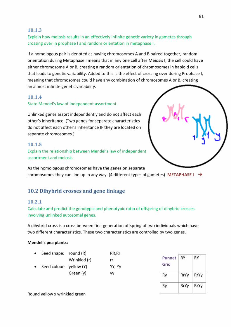
81
10.1.3
Explain how meiosis results in an effectively infinite genetic variety in gametes through
crossing over in prophase I and random orientation in metaphase I.
If a homologous pair is denoted as having chromosomes A and B paired together, random
orientation during Metaphase I means that in any one cell after Meiosis I, the cell could have
either chromosome A or B, creating a random orientation of chromosomes in haploid cells
that leads to genetic variability. Added to this is the effect of crossing over during Prophase I,
meaning that chromosomes could have any combination of chromosomes A or B, creating
an almost infinite genetic variability.
10.1.4
State Mendel’s law of independent assortment.
Unlinked genes assort independently and do not affect each
other’s inheritance. (Two genes for separate characteristics
do not affect each other’s inheritance IF they are located on
separate chromosomes.)
10.1.5
Explain the relationship between Mendel’s law of independent
assortment and meiosis.
As the homologous chromosomes have the genes on separate
chromosomes they can line up in any way. (4 different types of gametes) METAPHASE I
10.2 Dihybrid crosses and gene linkage
10.2.1
Calculate and predict the genotypic and phenotypic ratio of offspring of dihybrid crosses
involving unlinked autosomal genes.
A dihybrid cross is a cross between first generation offspring of two individuals which have
two different characteristics. These two characteristics are controlled by two genes.
Mendel’s pea plants:
Seed shape: round (R) RR,Rr
Wrinkled (r) rr
Seed colour- yellow (Y) YY, Yy
Green (y) yy
Round yellow x wrinkled green
Punnet
Grid
RY RY
Ry RrYy RrYy
Ry RrYy RrYy

82
(RY) (ry)
Phenotype ratio:
Round yellow: 9
Round green: 3
Wrinkled yellow: 3
Wrinkled green: 1
10.2.2
Distinguish between autosomes and sex chromosomes.
non-sex chromosomes; in humans, chromosomes 1 through 22 Autosome:
: those chromosomes which help determine the sex of an individual; in Sex chromosomes
humans, the X and Y chromosomes
10.2.3
Explain how crossing over between non-sister chromatids of a homologous pair in prophase I
can result in an exchange of alleles.
The diagram shows the loci of genes A and B.
The alleles A is linked to B, is linked to b.
The homologous pairs are held together like this during prophase I.
The crossover point occurs above the loci for gene A and below the gene loci for A.
The position at which the exchange occurs is called the chiasma.
The recombinants will form between non-sister chromatids which are crossing over.
The homologous pairs remain attached at the chiasma until anaphase I when they are pulled apart.
After anaphase II, the chromatids are separated.
The linked genes are:
A with B a with b recombinant a with B
RY Ry rY Ry
RY RRYY RRYy RrYY RrYy
Ry RRYy RRyy RrYy Rryy
rY RrYY RrYy rrYY rrYy
ry RrYy Rryy rrYy rryy

83
recombinant A with b
Genetic crosses:
The genotype of the gametes from meiosis above would be AB and ab. After cross over there would also be Ab and aB.
However most cells undergoing meiosis will not cross over between these two points.
Therefore:
Gametes Ab and ab will be high frequencies. Gametes Ab and aB will be low frequency.
When cross over occurs there will be low frequency of recombinants produced.
10.2.4
Define linkage group.
Linkage group: a group of genes whose loci are on the
same chromosome.
10.2.5
Explain an example of a cross between two linked genes.
Alleles are usually shown side by side in dihybrid crosses, for
example, TtBb. In representing crosses involving linkage, it is
more common to show them as vertical pairs, for example
T B
t b
This format will be used in examination papers, or students
will be given sufficient information to allow them to deduce
which alleles are linked.
10.2.6
Identify which of the offspring are recombinants in a dihybrid cross involving linked genes.
Recombination = the re-assortment of alleles into combinations different from those of the parents, as a result of: independent assortment, crossing over, fertilization
Parents: Aabb x aaBb Offspring: Aabb aaBb AaBb aabb parental parental recombinant recombinant
84
10.3 Polygenic inheritance
10.3.1
Define polygenic inheritance.
Polygenic inheritance: “a single characteristic that is controlled by two or more genes”.
Each allele of a polygenic character often contributes only a small amount to the overall
phenotype, this makes studying the individual alleles difficult. In addition the environment
effects smooth out the genotypic variation, to give continuous distribution curves.
10.3.2
Explain that polygenic inheritance can contribute to continuous variation using two
examples, one of which must be human skin colour.
Skin colour in humans
The colour of human skin depends on the amount of the black pigment melanin in it. At
least four and possible more genes are involved, each with alleles that promote melanin
production and alleles that do not. There is a wide range of possible genotypes with anything
from no alleles promoting melanin too many. Environmental factors can also lead to the
increased production of melanin (eg. sun tanning).
Grain colour in wheat
Wheat grains vary from white to dark red, depending upon the amount of a red
pigment they contain. Three genes control the colour. Each gene has two alleles, one that
causes pigment production and one that does not.
10.3.2
Explain that polygenic inheritance can contribute to continuous variation using two
examples, one of which must be human skin colour.
Grain colour in wheat
Wheat grains vary in colour from white to dark red, depending on the amount of red
pigment they contain. Three genes control colour. Each gene has two alleles, one that causes
pigment production and one that does not. Wheat grains can therefore have between 0 and
6 alleles for pigment production.
Skin colour in humans
The colour of human skin depends on the amount of the black pigment melanin in it. There
is a continuous distribution of skin colour from very pale (little melanin) to black (much
melanin). At least four and possibly more genes are involved, each with alleles that promote
melanin production and alleles that do not. There is therefore a wide range of possible
genotypes with anything from no alleles promoting melanin production to many.
85
Topic 11: Human health and physiology
11.1 Defence against infectious disease
11.1.1
Describe the process of blood clotting.
Platelets are small fragments that circulate along with erythrocytes (red blood cells) and
leukocytes (white blood cells) in blood plasma.
1. Clotting process begins with the release of incomplete fragments of cells from the
damaged tissue, resulting in the formation of thrombin.
2. Thrombin converts fibrinogen (always in the bloodstream) into the fibrous
protein fibrin.
3. Fibrin captures red blood cells and immobilizes the fluid portion of the blood so as to
provide the impetus for clotting.
4. Blood becomes slightly solidified (similar to a gelatine-like substance) until the
platelets reach this fibrous mass and send out sticky extensions to each other.
5. The platelets then contract, forcing out the liquid and scabbing over the wound!
11.1.2
Outline the principle of challenge and response, clonal selection and memory cells as the
basis of immunity.
B cells make antibodies.
The immune system can make different types of antibodies (but not all at once).
A few of each type of B cell are produced and they wait until the body is infected with an
antigen.
When this occurs, they multiply to form many clones; this is called Clonal Selection.
A clone of B cells can produce large amounts of antibodies quickly and give immunity to a
disease, only after the immune system is challenged by a disease -- this is called the
challenge and response system. The immune system needs to be "challenged" by a disease,
usually in the form of an antigen present upon it, and then the immune system responds by
producing a clone of "B" cells which produce large amounts of antibodies to fight and
eliminate the pathogen.
11.1.3
Define active and passive immunity.
: immunity due to the production of antibodies by the organism itself Active immunity
after the body’s defence mechanisms have been stimulated by antigens.
86
: immunity due to the acquisition of antibodies from another organism Passive immunity
in which active immunity has been stimulated, including via the placenta, colostrum, or by
injection of antibodies.
11.1.4
Explain antibody production.
1. Macrophages consume bacteria with antigen molecules in their membranes.
2. Macrophages present these antigens on their membranes with the help of special
protein structures.
3. Helper T-cells come in contact with macrophages, pick up the antigens, and
incorporate them into their own protein structures - this will allow them to present
the antigens to B-cells. This also causes the activation of the Helper t-cells.
4. Activated helper-T-cells activate B-cells by passing their antigen to B-cell receptors.
5. The B-cells then divide to form clones of antibody-secreting plasma cells and memory
cells.
11.1.5
Describe the production of monoclonal antibodies and their use in diagnosis and in
treatment.
Monoclonal antibodies - large quantities of a single type of antibody, produced using the
procedure outlined below:
Production:
Antigens that correspond to a desired antibody are injected into an animal.
B-cells producing the desired antibody are extracted.
Tumour cells are obtained from another source (tumour cells grow and divide
endlessly).
B-cells are fused with tumour cells, producing hybridoma cells that divide endlessly,
providing the desired antibodies.
The hybridoma cells are cultured and antibodies they produce are extracted and
purified.
Treatment of rabies
Rabies usually causes death in humans before the immune system can control it.
Injecting monoclonal antibodies when a person gets infected will control the virus
and at the same time, the person's body begins making its own antibodies.
Diagnosis of malaria
Monoclonal antibodies are made to bind to antigens in malarial parasites.
A test plate is covered with antibodies.

87
The sample to be tested is left on the plate long enough for malaria antigens (if
present) to bind to antibodies.
The sample is rinsed off and any bound antigens are detected using more monoclonal
antibodies with attached colour-changing enzyme.
Colour-changing enzyme can be used to measure the level of infection and
distinguish between different strains of malaria.
Production should be limited to the fusion of tumour and B-cells and their
subsequent proliferation and production of antibodies. Detection of antibodies to HIV is
one example in diagnosis. Others are detection of a specific cardiac isoenzyme in suspected
case of heart attack and detection of HCG in pregnancy test kits. Examples of the use of
these antibodies for treatment include targeting of cancer cells with drugs attached to
monoclonal antibodies, emergency treatment of rabies or cancer, blood and tissue tying for
transplant compatibility and purification of industrially made interferon.
11.1.6
Explain the principle of vaccination.
Weakened or dead version of a pathogen is injected into the body, causing the immune
system to mount a primary response.
This results in the production of B memory cells.
The B-cells "remember" the antibodies to produce in response to the pathogen.
When the real pathogen strikes, a secondary response occurs, aided by the memory cell
production of pathogen-specific antibodies.
This response is much stronger than the primary response and prevents any ill effects.
11.1.7
Discuss the benefits and dangers of vaccination.
Benefits:
total elimination of diseases
prevention of pandemics and epidemics
decreased health-care costs
Prevention of harmful side-effects of diseases
Dangers:
possible toxic effects of mercury in vaccines,
possible overload of the immune system
possible links with autism

88
11.2 Muscles and movement
11.2.1
State the roles of bones, ligaments, muscles, tendons and nerves in human movement.
Bones: used for structure & movement it is the framework that the muscles are attached to.
Ligaments: connect bone-to-bone
Muscles: apply effort and force; provide the force needed for muscle contraction.
Tendons: attach muscles to bone
Nerves: stimulate muscles to contract at a precise time and extent.
11.2.2
Label a diagram of the human elbow joint, including cartilage, synovial fluid, joint capsule,
named bones and antagonistic muscles (biceps and triceps).
11.2.3
Outline the functions of the structures in the human elbow joint named in 11.2.
Structure Function
Cartilage A cushion to absorb shock and reduce friction
Synovial fluid A lubricant for the joint
Joint capsule Produces and contains the synovial fluid
Biceps Bends the arm by pulling the radius towards the humerus

89
Triceps Straightens the arm by pulling the ulna towards the humerus
11.2.4
Compare the movements of the hip joint and the knee joint.
Knee Joint Hip Joint
Compound condyloid joint Ball & socket joint
Low range of motion when extended; high range
of motion when flexed.
Greater range of motion (leg can flex, extend,
move sideways & rotate)
11.2.5
Describe the structure of striated muscle fibres, including the myofibrils with light and dark
bands, mitochondria, the sarcoplasmic reticulum, nuclei and the sarcolemma.
The muscles that attach to,
and move, the skeleton are
called skeletal muscles.
A skeletal muscle is
composed of cells called
muscle fibres.
Muscle fibres run in parallel
along the entire length of
the muscle.
Each muscle fibre consists of
a bundle of myofibrils.
The cell membrane of a muscle fibre is called the sarcolemma.
The cytoplasm is called the sarcoplasm.
The endoplasmic reticulum is called the sarcoplasmic reticulum.
Muscle fibres have multiple nuclei and numerous mitochondria.
Muscle fibres have T-tubules that spread impulses.

90
A myofibril is made up of units called sarcomeres.
Sarcomeres line up end-to-end along the length of a myofibril.
A sarcomere is contained within two Z lines.
A sarcomere contains many protein filaments, in a parallel array.
Some filaments are thin (actin), and the others are thick (myosin).
The orderly array of actin and myosin filaments in the sarcomeres gives skeletal muscles a
striated appearance (light and dark bands). Myosin molecules have long tails and two round
heads. The tails are packed together in parallel and the heads stick out to the sides.
11.2.6
Draw and label a diagram to show the structure of a sarcomere, including Z lines, actin
filaments, myosin filaments with heads, and the resultant light and dark bands.
11.2.7
Explain how skeletal muscle contracts, including the
release of calcium ions from the sarcoplasmic
reticulum, the formation of cross-bridges, the sliding
of actin and myosin filaments, and the use of ATP to
break cross-bridges and re-set myosin heads.
A muscle shortens when its cells shorten; and
muscle cells shorten when the sarcomeres of
the myofibrils shorten. Thus, the sarcomeres
are the basic unit of contraction.
When a sarcomere shortens, the thin filaments slide past the thick filaments. The
thin filaments attached to one Z line move towards the thin filaments attached to the
other Z line.
In the presence of calcium ions, thousands of myosin heads bend backward and then
attach to the thin filaments, forming cross-bridges. After forming cross bridges, the
myosin heads bend forward and
the thin filaments are pulled along.
ATP is required for the release of
the myosin heads from the thin
filaments. When an animal dies, its
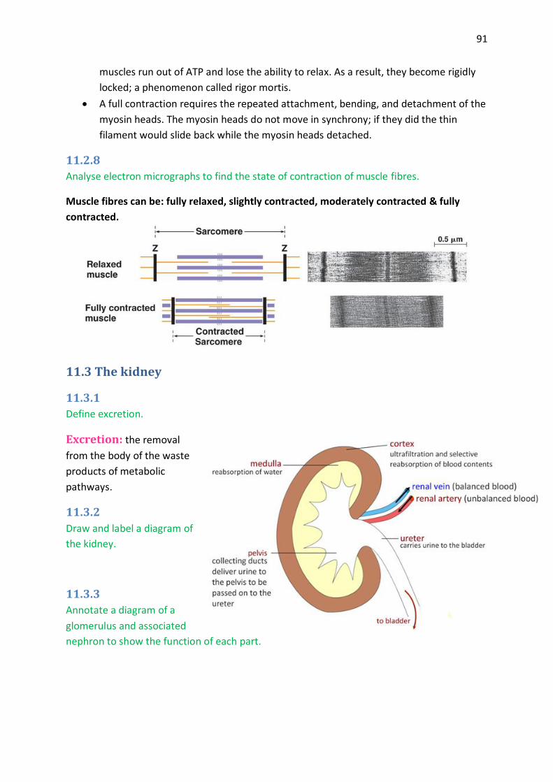
91
muscles run out of ATP and lose the ability to relax. As a result, they become rigidly
locked; a phenomenon called rigor mortis.
A full contraction requires the repeated attachment, bending, and detachment of the
myosin heads. The myosin heads do not move in synchrony; if they did the thin
filament would slide back while the myosin heads detached.
11.2.8
Analyse electron micrographs to find the state of contraction of muscle fibres.
Muscle fibres can be: fully relaxed, slightly contracted, moderately contracted & fully
contracted.
11.3 The kidney
11.3.1
Define excretion.
the removal Excretion:
from the body of the waste
products of metabolic
pathways.
11.3.2
Draw and label a diagram of
the kidney.
11.3.3
Annotate a diagram of a
glomerulus and associated
nephron to show the function of each part.

92
11.3.4
Explain the process of ultrafiltration, including blood pressure, fenestrated blood capillaries
and basement membrane.
Microscopically, the kidney
is composed of over one
million nephrons.
Each nephron is made up
of several parts. The blind
end of the nephron is
pushed in on itself to form
a cup-like structure called
Bowman’s capsule.
Blood from the renal
artery flows through an
afferent arteriole, into the
glomerulus, a capillary bed
that is situated inside the
Bowman’s capsule.
The blood in the glomerulus is under pressure and the capillary walls are fenestrated
(perforated) to allow blood plasma through.
The basement membrane of the Bowman’s capsule has an irregular network of slits,
so that much of the fluid from the blood filters into the capsule, leaving behind large
proteins and whole cells, which are too big to pass through. Thus, the basement
membrane acts as the dialysis membrane where ultrafiltration occurs.
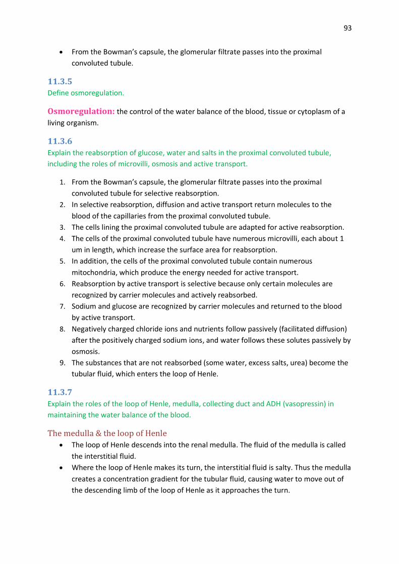
93
From the Bowman’s capsule, the glomerular filtrate passes into the proximal
convoluted tubule.
11.3.5
Define osmoregulation.
the control of the water balance of the blood, tissue or cytoplasm of a Osmoregulation:
living organism.
11.3.6
Explain the reabsorption of glucose, water and salts in the proximal convoluted tubule,
including the roles of microvilli, osmosis and active transport.
1. From the Bowman’s capsule, the glomerular filtrate passes into the proximal
convoluted tubule for selective reabsorption.
2. In selective reabsorption, diffusion and active transport return molecules to the
blood of the capillaries from the proximal convoluted tubule.
3. The cells lining the proximal convoluted tubule are adapted for active reabsorption.
4. The cells of the proximal convoluted tubule have numerous microvilli, each about 1
um in length, which increase the surface area for reabsorption.
5. In addition, the cells of the proximal convoluted tubule contain numerous
mitochondria, which produce the energy needed for active transport.
6. Reabsorption by active transport is selective because only certain molecules are
recognized by carrier molecules and actively reabsorbed.
7. Sodium and glucose are recognized by carrier molecules and returned to the blood
by active transport.
8. Negatively charged chloride ions and nutrients follow passively (facilitated diffusion)
after the positively charged sodium ions, and water follows these solutes passively by
osmosis.
9. The substances that are not reabsorbed (some water, excess salts, urea) become the
tubular fluid, which enters the loop of Henle.
11.3.7
Explain the roles of the loop of Henle, medulla, collecting duct and ADH (vasopressin) in
maintaining the water balance of the blood.
The medulla & the loop of Henle
The loop of Henle descends into the renal medulla. The fluid of the medulla is called
the interstitial fluid.
Where the loop of Henle makes its turn, the interstitial fluid is salty. Thus the medulla
creates a concentration gradient for the tubular fluid, causing water to move out of
the descending limb of the loop of Henle as it approaches the turn.
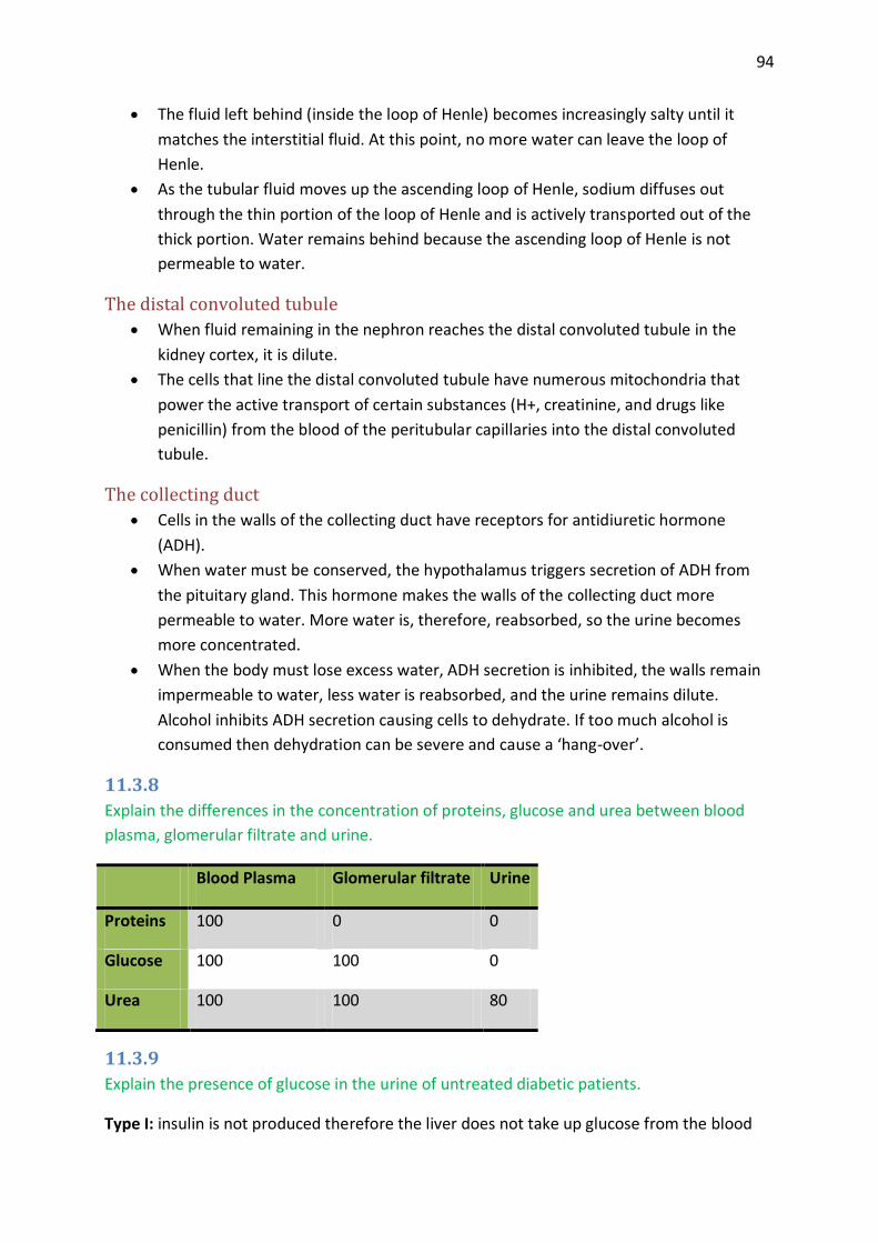
94
The fluid left behind (inside the loop of Henle) becomes increasingly salty until it
matches the interstitial fluid. At this point, no more water can leave the loop of
Henle.
As the tubular fluid moves up the ascending loop of Henle, sodium diffuses out
through the thin portion of the loop of Henle and is actively transported out of the
thick portion. Water remains behind because the ascending loop of Henle is not
permeable to water.
The distal convoluted tubule
When fluid remaining in the nephron reaches the distal convoluted tubule in the
kidney cortex, it is dilute.
The cells that line the distal convoluted tubule have numerous mitochondria that
power the active transport of certain substances (H+, creatinine, and drugs like
penicillin) from the blood of the peritubular capillaries into the distal convoluted
tubule.
The collecting duct
Cells in the walls of the collecting duct have receptors for antidiuretic hormone
(ADH).
When water must be conserved, the hypothalamus triggers secretion of ADH from
the pituitary gland. This hormone makes the walls of the collecting duct more
permeable to water. More water is, therefore, reabsorbed, so the urine becomes
more concentrated.
When the body must lose excess water, ADH secretion is inhibited, the walls remain
impermeable to water, less water is reabsorbed, and the urine remains dilute.
Alcohol inhibits ADH secretion causing cells to dehydrate. If too much alcohol is
consumed then dehydration can be severe and cause a ‘hang-over’.
11.3.8
Explain the differences in the concentration of proteins, glucose and urea between blood
plasma, glomerular filtrate and urine.
Blood Plasma Glomerular filtrate Urine
Proteins 100 0 0
Glucose 100 100 0
Urea 100 100 80
11.3.9
Explain the presence of glucose in the urine of untreated diabetic patients.
Type I: insulin is not produced therefore the liver does not take up glucose from the blood

95
Type II: insulin receptors are inactive therefore the lover does not take up glucose from the
blood.
END RESULT: Blood sugar concentration remains high.
In the Kidney:
All glucose is passed into renal filtrate through ULTRAFILTRATION; there is too much glucose
to be processed by active transport in selective reabsorption in the proximal convoluted
tubule. So some glucose continues through the nephron and is excreted in urine.
11.4 Reproduction
11.4.1
Annotate a light
micrograph of testis
tissue to show the
location and function of
interstitial cells (Leydig
cells), germinal
epithelium cells,
developing
spermatozoa and Sertoli cells.
11.4.2
Outline the processes involved in spermatogenesis within
the testis, including mitosis, cell growth, the two divisions
of meiosis and cell differentiation.
Spermatogonium (2n) are found at or near the
basement membrane.
They have a high rate of cell division by mitosis to
produce spermatogonia.
The spermatogonium grow to form Primary
Spermatocytes which have completed S-phase.
The Primary spermatocytes separate the
homologous pairs of chromosomes in meiosis
I(reduction division) to form the haploid Secondary
Spermatocytes.
The spermatids are formed from the separation of the sister chromatids in meiosis II.
The spermatids are found in association with the sertoli cells which nourish the
spermatids as they differentiate into spermatozoa.
The rate of spermatozoa is high and continuous throughout the life on the sexually
mature male.

96
11.4.3
State the role of LH, testosterone and FSH in spermatogenesis.
There are two hormones secreted from the anterior pituitary FSH and LH.
FSH stimulates the primary Spermatocytes
which carry out meiosis I (reduction division)
to separate homologous pairs of
chromosomes and produce haploid
secondary spermatocytes.
LH stimulates the interstitial cells to
produce testosterone
Testosterone stimulates the maturation
of secondary spermatocytes through meiosis and differentiation to spermatozoa.
11.4.4
Annotate a diagram of the ovary to show the location and function of germinal epithelium,
primary follicles, mature follicle and secondary oocyte.
11.4.5
Outline the processes involved in oogenesis within the ovary, including mitosis, cell growth,
and the two divisions of meiosis, the unequal division of cytoplasm and the degeneration of
polar body.
Oogenesis is the production of haploid eggs from diploid cells in the ovaries.
By the time a female fetus is three months old she has already begun the process of
Meiosis 1 to produce developing eggs.
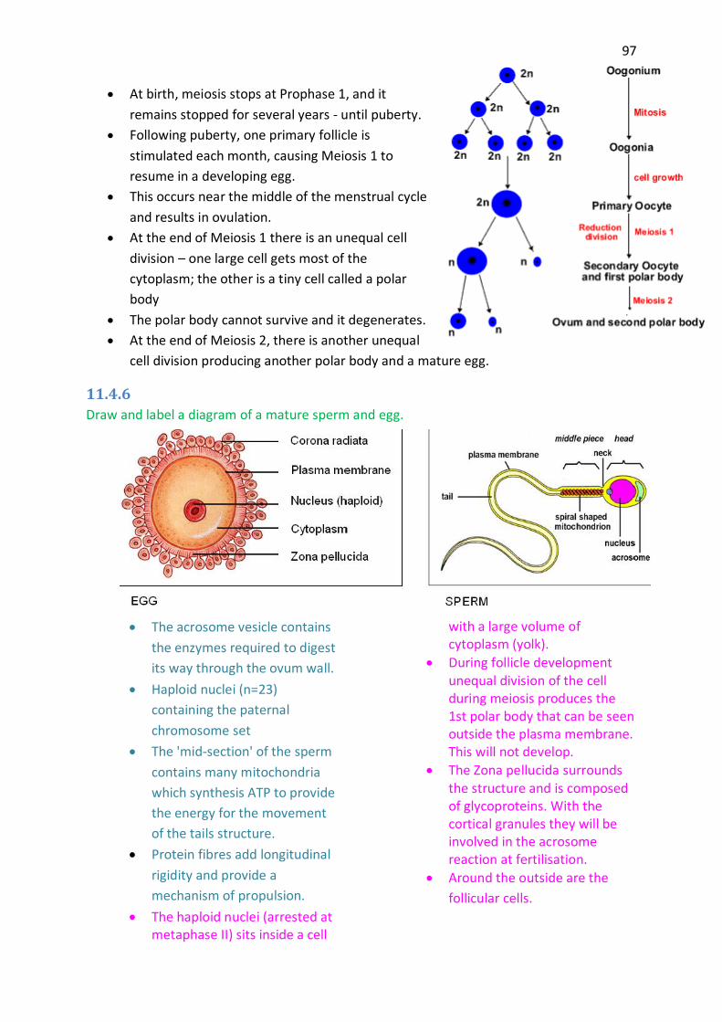
97
At birth, meiosis stops at Prophase 1, and it
remains stopped for several years - until puberty.
Following puberty, one primary follicle is
stimulated each month, causing Meiosis 1 to
resume in a developing egg.
This occurs near the middle of the menstrual cycle
and results in ovulation.
At the end of Meiosis 1 there is an unequal cell
division – one large cell gets most of the
cytoplasm; the other is a tiny cell called a polar
body
The polar body cannot survive and it degenerates.
At the end of Meiosis 2, there is another unequal
cell division producing another polar body and a mature egg.
11.4.6
Draw and label a diagram of a mature sperm and egg.
The acrosome vesicle contains
the enzymes required to digest
its way through the ovum wall.
Haploid nuclei (n=23)
containing the paternal
chromosome set
The 'mid-section' of the sperm
contains many mitochondria
which synthesis ATP to provide
the energy for the movement
of the tails structure.
Protein fibres add longitudinal
rigidity and provide a
mechanism of propulsion.
The haploid nuclei (arrested at metaphase II) sits inside a cell
with a large volume of cytoplasm (yolk).
During follicle development unequal division of the cell during meiosis produces the 1st polar body that can be seen outside the plasma membrane. This will not develop.
The Zona pellucida surrounds the structure and is composed of glycoproteins. With the cortical granules they will be involved in the acrosome reaction at fertilisation.
Around the outside are the
follicular cells.

98
11.4.7
Outline the role of the epididymis, seminal vesicle and prostate gland in the production of
semen.
Epididymis:
testicle fluids are removed and the sperm
concentrated
sperm mature here and develop the ability to
swim
Seminal vesicles:
adds nutrients that include fructose sugar for
respiration
mucus to protect sperm in the cell
Prostate:
adds fluids that neutralise the vaginal acids and minerals ions
mineral ions
11.4.8
Compare the processes of spermatogenesis and oogenesis, including the number of gametes
and the timing of the formation and release of gametes.
Spermatogenesis Oogenesis
Location Testes Ovaries
Onset Production being in a female fetus Production begins at puberty
Duration Meiosis 1 & 2 are continuous Meiosis stops for many years
Final Products 1 diploid cell; 4 sperm cells 1 diploid cell; 1 egg & 3 polar bodies
Quantity Several million sperm per day One egg per month
Mobility Self-propelling/have flagellum Do not have flagellum
Size Sperm is much smaller than egg Egg is much larger than sperm
11.4.9
Describe the process of fertilization, including the acrosome reaction, penetration of the egg
membrane by a sperm and the cortical reaction.
When the sperm comes in contact with the zona pellucida, the enzymes from the acrosome
are released and this acrosome reaction the zona pellucida is broken down so the sperm can
come in contact with the plasma membrane of the egg. Sperm membrane and egg

99
membrane fuse and the nucleus of the sperm enters the egg. This initiates the cortical
reaction were the cortical granules fuse with the plasma membrane releasing their contents
outside of the egg. This makes the zona pellucida into a fertilization membrane,
impermeable to other sperm. The nucleus of the egg now completes meiosis II and then the
two haploid nuclei (sperm egg) fuse to form diploid nucleus = a zygote.
11.4.10
Outline the role of HCG in early pregnancy.
HCG= (Human Chorionic Gondadothropin) is a hormone produced by the embryo,
production starts early after fertilization. HCG maintains the corpus luteum and due to this
the levels of estrogen and progesterone remains high. Low levels of FSH, no follicle will
develop, the endometrium will be maintained) Later in pregnancy HCG is produced from the
placenta. HCG is used in pregnancy test; it can be detected in the urine as soon as 7 days
after fertilization. The main function of HCG is to maintain the high levels of estrogen and
progesterone to maintain the endometrium for the embryo to implant.
11.4.11
Outline early embryo development up to the implantation of the blastocyst.
Cell division in embryonic
development is called cleavage.
Mammals show rotational
cleavage in which the plane of
division; for successive divisions
are at right angles.
At around the eight cell stage all
the cells maximise their surface
contact with each other and the
ball of cells becomes tight.
The process outlined occurs as
the cell mass moves down the oviduct.
Implantation occurs around day 6 in the uterus. Pre-implantation in the tubular walls is
prevented by the zona pellucida (outer grey circle).
The blastocyst is a hollow ball of cells with an inner cell mass (embryo) at the base.
11.4.12
Explain how the structure and functions of the placenta, including its hormonal role in
secretion of estrogen and progesterone, maintain pregnancy.

100
The placenta is formed by fetal tissue growing into the uterine wall here the blood from the
fetus comes in close contact but not mixed with the maternal blood. Nutrients oxygen,
antibodies travel from the maternal blood to the fetal blood. CO2 and urea travel from the
fetal blood to the maternal blood.
The umbilical cord connected the fetus to the placenta and contains the umbilical artery and
veins, transporting the blood to and from the placenta. The placenta also produces estrogen
and progesterone to maintain the endometrium and thereby maintain the pregnancy,
(negative feedback to FSH & LH) when the progesterone level from the birth process
starts.
11.4.13
State that the fetus is supported and protected by the amniotic sac and amniotic fluid.
The fetus is supported and protected by the amniotic sac and amniotic fluid.
11.4.14
State that materials are exchanged between the maternal and fetal blood in the placenta.
Materials are exchanged between the maternal and fetal blood in the placenta.
11.4.15
Outline the process of birth and its hormonal control, including the changes in progesterone
and oxytocin levels and positive feedback.
Parturition:
At the end of gestation it is believed that there is a drop in the high level of
progesterone.
In recent years it has been shown that the progesterone receptor is also block.
Therefore the progesterone is not effective.
With the fall in progesterone the pituitary secretes this small polypeptide called
oxytocin.
Stretching of the lower uterus walls by the foetus and its production of
prostaglandin’s added to the stimulus for the pituitary to secrete oxytocin.
The oxytocin causes the smooth muscle in the walls of the uterus to contract and
labour has begun.
After nine months in the uterus the foetus is fully grown and takes up all the space available.
These cramped conditions push
the baby down stretching the
lower walls of the uterus. This

101
sends impulses to the mother brain.
The foetus responds to the cramped conditions by producing hormones from the
placenta (prostaglandin) which causes myometrial contraction
Progesterone is the hormone of pregnancy and at this stage the high levels of this
hormone become less active.
All these changes stimulate the secretion of oxytocin from the pituitary and this causes the
myometrial contractions of labour
Positive feedback:
In this system the stimuli to the brain increases the oxytocin production
In turn the oxytocin stimulate myometrial contraction
Myometrial contraction further stimulates the pituitary of the mother to release
more oxytocin
The strength and frequency of the myometrial contractions is further increased.
In turn this further stimulates more oxytocin production
The process builds with stronger and stronger contractions
Final the child passes though the cervix and vagina to be born
Contractions continue for a further period until the placenta is delivered (after birth).

102
Biology Higher Level Exams:
Paper 1: 1 hour long (Day 1)
40 multiple choice questions
Worth 40 points
Core and AHL (topic 1-6 and 7-11)
NO CALCULATOR
Paper 2: 2 ¼ hours long (Day 1)
Section A:
o 32 questions that are data based
o Worth 16-19 points
o Short response questions
o Worth 13-16 points
o NEED A CALCULATOR AND RULER
Section B:
o Extended response
o Worth 40 points (2 more points quality points; clarity and connections)
o Choose 2 out of 4 questions and answer all parts
Paper 3: 1 ¼ hours long (Day 2)
2 options studied
NO multiple choice questions
Worth 40 points
Paper: 1- 40 20%
2- 72 36%
3- 40 20%
IA 48__ 24%
200
80%= 7
50%= 4 (pass)


![Topic 11 Revision [129 marks] - IB Questionbank](https://static.fdokumen.com/doc/165x107/6315a7f3b1e0e0053b0f274d/topic-11-revision-129-marks-ib-questionbank.jpg)