for the IB Diploma - PapaCambridge
414
Biology for the IB Diploma SECOND EDITION Brenda Walpole with additional online material
-
Upload
khangminh22 -
Category
Documents
-
view
1 -
download
0
Transcript of for the IB Diploma - PapaCambridge

Biologyfor the IB DiplomaSECOND EDITION
Biology for th
e IB D
iplom
aB. W
alpole
Biology for the IB Diploma Brenda Walpole
This lively coursebook offers complete and thorough coverage of the Biology syllabus for the International Baccalaureate (IB) Diploma Programme, including the Standard and Higher Level topics and all eight Options.
The book clearly links to the syllabus by following the same division into topics and prominently displaying the assessment statements associated with each section. Students’ interest is stimulated by extra snippets of information displayed alongside the core text, offering deeper understanding and links with everyday life. Clear, simple language makes the text accessible to students of all abilities. Easy navigation is ensured with Standard and Higher Level material clearly marked in all chapters.
The coursebook contains:• short-answer questions throughout the chapters to test
knowledge, incorporating command terms as used in IB examinations to cultivate familiarity with the terms and develop skill in answering questions appropriately
• exam-style questions at the end of each chapter, offering thorough practice for the examination
• definitionsofkeytermsdisplayedalongsidethetextfor easy reference
• links to Theory of Knowledge concepts alongside appropriate topics, to stimulate thought and discussion
• clear, well-labelled illustrations and photos to help make concepts easy to understand.
Brenda Walpole has 20 years’ experience of teaching IB Biology and has written over 30 science books. She is currently an independent consultant in science education.
Ashby Merson-Davies has taught IB Biology for over 20 years. He has written a series of books which support the programme.
Leighton Dann has taught Biology at school and has been instrumental in developing new practical work for GCSE and post-16 courses in schools and colleges.
Course consultant: Peter Hoeben has taught IB Biology.
Other titles available: ISBN 978-0-521-18294-2
ISBN 978-0-521-13821-5
Brenda Walpolewith additional online material

Biologyfor the IB DiplomaSecond edition
Cambridge University Press’s mission is to advance learning, knowledge and research worldwide.
Our IB Diploma resources aim to:
• encourage learners to explore concepts, ideas and topics that have local and global significance
• help students develop a positive attitude to learning in preparation for higher education
• assist students in approaching complex questions, applying critical-thinking skills and forming reasoned answers.
Brenda Walpole
withAshby Merson-DaviesLeighton Dann

notice to teachers in the ukIt is illegal to reproduce any part of this book in material form (including photocopying and electronic storage) except under the following circumstances: (i) where you are abiding by a licence granted to your school or institution by the
Copyright Licensing Agency;(ii) where no such licence exists, or where you wish to exceed the terms of a licence,
and you have gained the written permission of Cambridge University Press;(iii) where you are allowed to reproduce without permission under the provisions
of Chapter 3 of the Copyright, Designs and Patents Act 1988, which covers, for example, the reproduction of short passages within certain types of educational anthology and reproduction for the purposes of setting examination questions.
The website accompanying this book contains further resources to support your IB Biology studies. Visit education.cambridge.org/ibsciences and register for access.
Separate website terms and conditions apply.
University Printing House, Cambridge cb2 8bs, United Kingdom
Cambridge University Press is part of the University of Cambridge.
It furthers the University’s mission by disseminating knowledge in the pursuit of education, learning and research at the highest international levels of excellence.
www.cambridge.org Information on this title: www.cambridge.org/9781107654600
© Cambridge University Press 2011, 2014
This publication is in copyright. Subject to statutory exception and to the provisions of relevant collective licensing agreements, no reproduction of any part may take place without the written permission of Cambridge University Press.
First published 2011 Second edition 2014
Printed in the United Kingdom by Latimer Trend
A catalogue record for this publication is available from the British Library
isbn 978-1-107-65460-0 Paperback
Additional resources for this publication at education.cambridge.org/ibsciences
Cambridge University Press has no responsibility for the persistence or accuracy of URLs for external or third-party internet websites referred to in this publication, and does not guarantee that any content on such websites is, or will remain, accurate or appropriate. Information regarding prices, travel timetables, and other factual information given in this work is correct at the time of first printing but Cambridge University Press does not guarantee the accuracy of such information thereafter.
The material has been developed independently by the publisher and the content is in no way connected with nor endorsed by the International Baccalaureate Organization.

ContentsIntroduction v
1 Cell biology 1Introduction 11.1 Introduction to cells 11.2 Ultrastructure of cells 121.3 Membrane structure 191.4 Membrane transport 231.5 The origin of cells 301.6 Cell division 33 Exam-style questions 41
2 Molecular biology 43Introduction 432.1 Molecules to metabolism 432.2 Water 492.3 Carbohydrates and lipids 542.4 Proteins 592.5 Enzymes 622.6 Structure of DNA and RNA 682.7 DNA replication, transcription and translation 712.8 Cell respiration 782.9 Photosynthesis 82 Exam-style questions 89
3 Genetics 93Introduction 933.1 Genes 933.2 Chromosomes 993.3 Meiosis 1033.4 Inheritance 1103.5 Genetic modification and biotechnology 124 Exam-style questions 133
4 Ecology 136Introduction 1364.1 Species, communities and ecosystems 1364.2 Energy flow 1414.3 Carbon recycling 1474.4 Climate change 151 Exam-style questions 157
5 Evolution and biodiversity 159Introduction 1595.1 Evidence for evolution 1595.2 Natural selection 1645.3 Classification of biodiversity 1695.4 Cladistics 178 Exam-style questions 185
6 Human physiology 187Introduction 1876.1 Digestion and absorption 1876.2 The blood system 1936.3 Defence against infectious disease 2026.4 Gas exchange 2096.5 Neurons and synapses 2156.6 Hormones, homeostasis and reproduction 220 Exam-style questions 232
7 Nucleic acids (HL) 235Introduction 2357.1 DNA structure and replication 2357.2 Transcription and gene expression 2457.3 Translation 251 Exam-style questions 259
8 Metabolism, cell respiration and photosynthesis (HL) 262Introduction 2628.1 Metabolism 2628.2 Cell respiration 2688.3 Photosynthesis 276 Exam-style questions 283
9 Plant biology (HL) 285Introduction 2859.1 Transport in the xylem of plants 2859.2 Transport in the phloem of plants 2939.3 Growth in plants 2979.4 Reproduction in plants 300 Exam-style questions 305
III

Iv
10 Genetics and evolution (HL) 308Introduction 30810.1 Meiosis 30810.2 Inheritance 31310.3 Gene pools and speciation 327
Exam-style questions 335
11 Animal physiology (HL) 337Introduction 33711.1 Antibody production and vaccination 33711.2 Movement 34611.3 The kidney and osmoregulation 35311.4 Sexual reproduction 365
Exam-style questions 377
Answers to test yourself questions 380
Glossary 386
Index 401
Acknowledgements 407
The website accompanying this book contains further resources to support your IB Biology studies. Visit education.cambridge.org/ibsciences and register to access these resources:
Options
Option A Neurobiology and behaviour
Option B Biotechnology and bioinformatics
Option C Ecology and conservation
Option D Human physiology
Free online material
Self-test questions
Assessment guidance
Model exam papers
Nature of Science
Answers to exam-style questions
Answers to Options questions

INTRODUCTION v
IntroductionBiology has advanced at a rapid rate over recent decades and is truly the science of the 21st century. Advances in genetics, biochemistry, medicine and cell biology have kept the subject in the forefront of international news. To keep pace with new developments, the IB Biology course is regularly updated so that IB students can understand not only the principles of modern science but also the processes and the ethical implications that go with them. The latest revision of the IB Biology syllabus will be examined in the years 2016–2022, and this second edition of Biology for the IB Diploma is fully updated to cover the content of that syllabus.
Biology may be studied at Standard Level (SL) or Higher Level (HL). Both share a common core, which is covered in Topics 1–6. At HL the core is extended to include Topics 7–11. In addition, at both levels, students then choose one Option to complete their studies. Each option consists of common core and additional Higher Level material. You can identify the HL content in this book by ‘HL’ included in the topic title (or section title in the Options), and by the red page border. The Options are included in the free online material that is accessible with the code available in this book.
The structure of this book follows the structure of the IB Biology syllabus. Each topic in the book matches a syllabus topic, and the sections within each topic mirror the sections in the syllabus. Each section begins with learning objectives as starting and reference points. Test yourself questions appear throughout the text so students can check their progress and become familiar with the style and command terms used, and examination style questions appear at the end of each topic.
Theory of Knowledge (TOK) provides a cross-curricular link between different subjects. It stimulates thought about critical thinking and how we can say we know what we claim to know. Throughout this book, TOK features highlight concepts in Biology that can be considered from a TOK perspective. These are indicated by the ‘TOK’ logo, shown here.
Science is a truly international endeavour, being practised across all continents, frequently in international or even global partnerships. Many problems that science aims to solve are international, and will require globally implemented solutions. Throughout this book, International-Mindedness features highlight international concerns in Biology. These are indicated by the ‘International-Mindedness’ logo, shown here.
Nature of Science is an overarching theme of the Biology course. The theme examines the processes and concepts that are central to scientific endeavour, and how science serves and connects with the wider community. At the end of each section in this book, there is a ‘Nature of Science’ paragraph that discusses a particular concept or discovery from the point of view of one or more aspects of Nature of Science. A chapter giving a general introduction to the Nature of Science theme is available in the free online material.

vI
Free online materialAdditional material to support the IB Biology Diploma course is available online. Visit education.cambridge.org/ibsciences and register to access these resources.
Besides the Options and Nature of Science chapter, you will find a collection of resources to help with revision and exam preparation. This includes guidance on the assessments, interactive self-test questions and model exam papers. Additionally, answers to the exam-style questions in this book and to all the questions in the Options are available.

1 CELL BIOLOGY 1
IntroductionIn the middle of the seventeenth century, one of the pioneers of microscopy, Robert Hooke (1635–1703), decided to examine a piece of cork tissue with his home-built microscope. He saw numerous box-shaped structures that he thought resembled monks’ cells or rooms in a monastery, so he called them ‘cells’. As microscopes became more sophisticated, other scientists observed cells and found that they occurred in every organism. No organism has yet been discovered that does not have at least one cell. Living things may vary in shape and size but scientists agree that they are all composed of cells. The study of cells has enabled us to learn more about how whole organisms function.
1.1 Introduction to cells
The cell theoryToday, scientists agree that the cell is the fundamental unit of all life forms. Cell theory proposes that all organisms are composed of one or more cells and, furthermore, that cells are the smallest units of life. An individual cell can perform all the functions of life and anything that is not made of cells, such as viruses, cannot be considered living.
One of the key life processes of all living organisms is reproduction. Therefore, one of the first principles of the cell theory is that cells can only come from pre-existing cells. Louis Pasteur (1822–1895) carried out experiments that provided evidence for this.
Extensive examination of many organisms and millions of different types of cell supports the cell theory, although a few examples have been found that do not fit the theory perfectly. One example is fungi, whose structures consist of long threads called hyphae (Figure 1.1), which have many nuclei but are not divided into separate cells by cell walls. Another example is skeletal muscle, which is composed of muscle fibres that are much larger than a single cell and contain several hundred nuclei. Cells of some large algae are somewhat anomalous because their single cells are undifferentiated but are attached to chains of identical cells or surrounded by a matrix of extra cellular material so that they form large structures, and mammalian erythrocytes (red blood cells) do not contain nuclei once they have matured and been released into the bloodstream, which means at this stage of their life cycle they cannot carry out all the functions of life so they too depart from cell theory.
Unicellular organismsBy definition, a living organism comprising just one cell has to perform all the necessary functions for survival.
Cell biology 1Learning objectives
You should understand that:
• Cell theory explains that living organisms are composed of cells.
• Unicellular organisms carry out all the functions of life.
• Surface area to volume ratio is an important factor in limiting cell size.
• Interactions between their cellular components lead to new emergent properties in multicellular organisms.
• Multicellular organisms have specialised tissues, which develop as a result of cell differentiation.
• Cell differentiation results from the expression of some genes but not others.
• Stem cells are able to divide and differentiate along different pathways and are essential for embryonic development. This ability makes them suitable for therapeutic uses.
Figure 1.1 Fungal hyphae grow through material that nourishes the fungus. In this scanning electron micrograph the thread like structures are the hyphae and the pale pink spheres are the reproductive spores (× 2000).

2
The functions of life are:
• metabolism
• growth
• response (or sensitivity)
• homeostasis
• nutrition
• reproduction
• excretion
A unicellular organism such as Paramecium (Figure 1.2) needs to metabolise organic materials in order to make the chemicals needed to sustain life. It must also be able to excrete waste produced during metabolism and dispose of it. It must be able to detect changes in its environment, so it can respond to more favourable or less favourable conditions. Some unicellular organisms photosynthesise and they have a light spot that enables them to move to a brighter environment to maximise photosynthesis. A unicellular organism must also be able to control its internal environment (homeostasis), as large changes in water or salt concentrations may have a detrimental effect on metabolism and other cellular functions. It must also obtain food, whether produced from simple inorganic substances through photosynthesis (as in Chlorella, Figure 1.3) or ingested as complex organic materials from outside as a source of nutrition. If the species is to survive, an organism must be able to reproduce. This could be either asexual or sexual reproduction.
Investigations of some life processes in Paramecium and ChlorellaParamecium can be observed under a light microscope. Paramecia have cilia, which they flick in rhythmic waves to move about in water, and they also have a row of specialised cilia that waft food particles towards the
Key principles of the cell theory:
• living organisms are composed of cells
• cells are the smallest units of life
• all cells come from pre-existing cells.
Figure 1.2 Paramecium carries out all the life functions within its single cell (× 323).Figure 1.3 Chlorella is a unicellular organism containing a chloroplast (× 1200).
Drawing cell structures
When you draw cells as they appear under a microscope, always use a sharp pencil and draw single lines to show the relative sizes and positions of the structures you can see. Do not use shading or cross-hatching on your diagram. Label each structure with a straight line so that the name of each part appears at the side of your diagram. Always include a title and the magnification of your drawing. You can see an example of how to do this in Figure 1.2.
ciliacontractilevacuole
cytoplasmplasmamembrane
oral groovenucleusfoodvacuoles
35 µm

1 CELL BIOLOGY 3
oral groove. If stained yeast cells are added to the culture of Paramecium it is possible to observe the path taken by food particles through the body of the organism and the food vacuoles that are formed. If Paramecium is placed in water of different salinities from distilled water – 0.1%, 0.2%, 0.4% and 0.8% sodium chloride solution, for example – the contractile vacuole, which controls the water balance of the cell, can be seen forming and emptying.
Chlorella is a photosynthetic organism with a rapid growth rate. Although its cells are small and must be viewed with a microscope, it can quickly produce large numbers of individuals, which turn water green and opaque. This is most likely to happen when Chlorella grows in water that is rich in nitrates or phosphates. The organism has been used in many scientific experiments; the Nobel prize winner Otto Warburg published his pioneering work on cellular metabolism following intensive experiments on Chlorella in 1919, and in 1961 Melvin Calvin carried out his experiments on photosynthesis using Chlorella (Subtopic 8.3).
Cell sizeOne of the few cells large enough to be visible to the unaided eye is the mature human ovum, which has a diameter of approximately 150 μm. However, most cells are much smaller than this, and can only be seen using a microscope. Light microscopes, which can magnify up to 2000 times, reveal some internal structures such as the nucleus, but greater detail requires the use of more powerful microscopes such as the electron microscope, which magnifies cell structures up to 500 000 times. Viruses can only be seen with these microscopes, so the structure of viruses was unknown until the invention of these microscopes in the 20th century.
Electron microscopes use a beam of electrons, instead of light, to produce an image. The resolution of an electron microscope is much better than that of a light microscope because of the shorter wavelength of electrons. Resolving power is the ability of the microscope to separate objects that are close together so that more detail can be seen. Only non-living material can be observed in an electron microscope and specimens must be prepared with heavy metals or coated with carbon or gold. There are two types of electron microscope: the TEM (transmission electron microscope) and SEM (scanning electron microscope). A TEM produces clear images of thin sections of material while in an SEM electrons are bounced off objects to produce detailed images of their external appearance. Both types of microscope produce black and white images but these are often artificially coloured so that certain features can be seen more clearly. Table 1.1 compares the different types of microscope.
Even the electron microscope cannot distinguish individual molecules. Other techniques such as X-ray crystallography are needed to do this. Figure 1.4. indicates the relative sizes of some biological structures.
0.5–1 µm(500–1000 nm)
thickness of blade of a leaf
Paramecium (unicellular organism)
diameter of sharp end of pin and smallest object visible with eye only
diameter of plant cell
width of very fine human hair
diameter of animal cell
diameter of mitochondrion
diameter of bacterium
diameter of ribosome
thickness of membrane
diameter of DNA molecule
diameter of hydrogen atom(the smallest atom)
40 µm
30 µm
20 µm
1 µm
smallest object visible withlight microscope
0.2 µm(200 nm)
500 µm
200 µm
100 µm
20 nm
7 nm
2 nm
smallest object visiblewith electron microscope
0.5 nm
invi
sib
levi
sib
le w
ith a
n el
ectr
on m
icro
scop
evi
sib
le w
ith a
ligh
t mic
rosc
ope
visi
ble
with
the
nake
d ey
e
0.04 nm
Figure 1.4 The sizes of some biological structures.

4
Magnification and scaleKnowing the sizes of objects viewed under the microscope can be very useful (Figure 1.5). For example, a plant scientist might want to compare the relative sizes of pollen grains from plants in the same genus to help identify different species.
Magnification is defined as the ratio of the size of the image to the size of the object:
magnification = size of imagesize of object
Figure 1.5 Typical compound light microscope.
eyepiece lens
focusingknobs
objectivelenses
stage
mirror todirect light
slide
Light microscope TEM SEM
uses light to produce images uses electron beams to produce images
uses electron beams to produce images
Maximum
resolution
200 nm 1 nm 1 nm
Maximum
magnification
× 2000 up to × 1 000 000 × 200 000
Preparation
of material
thin sections of material mounted on slidesliving organisms can be examined
very thin sections of material supported on metal gridsliving organisms cannot be examined
very thin sections of material supported on metal gridsliving organisms cannot be examined
Stain used coloured dyes heavy metals carbon or gold coating
Image viewed directly through eyepiece lens
viewed on a screen or photographic plate
viewed on a screen or photographic plate
Table 1.1 Comparison of light microscopes with transmission electron microscope (TEM) and scanning elelctron microscope (SEM).

1 CELL BIOLOGY 5
With a compound microscope, the magnification is the product of both lenses, so if a microscope has a × 10 eyepiece and × 40 objective, the total magnification is × 400.
Printed images of structures seen with a microscope usually show a scale bar or give the magnification, so that the size of an object can be calculated. For example, the magnification of the micrograph in Figure 1.6 is given as × 165.
In Figure 1.6, there are three spherical glomeruli present. In the image, each one is approximately 25 mm across. You can check this using a ruler. Thus:
actual size of glomerulus = size of imagemagnification
=
25 mm
165
= 0.15 mm
In electron micrographs, most measurements are expressed in micrometres. A micrometre (μm) is 10–3 mm, so 1 mm is 1000 μm.
So the diameter of the glomerulus = 0.15 × 1000 = 150 μm.
Figure 1.6 Coloured light micrograph of a section through the cortex of a kidney (× 165).
glomerulus

6
Worked example1.1 This image shows a red blood cell. The scale bar shows 2 μm. From this, you can calculate both the size of the
cell and the magnification of the image.
Size of the cellStep 1 Use a ruler to measure the length of the cell (its diameter in this case). This is 30 mm.Step 2 Use a ruler to measure the length of the scale bar. This is 9 mm.Step 3 Use the ratio of these two values to work out the actual length of the cell.
2 μm =
actual length of cell 9000 μm 30 000 μm (Remember to convert all the units to μm. 1 mm = 1000 μm.) Rearranging the equation:
actual length of the cell = 2 μm × 30 000 μm
9000 μm = 6.7 μm
Magnification of the imageUse the formula:
magnification = measured length of the cell
actual length of the cellSo in this case:
magnification = 30 000 μm
6.7 μm= × 4500
If you are given a value for the magnification, you can measure the length of the object in the image and then rearrange the equation to work out the actual length of the object.
Becoming multicellular
Surface area to volume ratioCells are very small, no matter what the size of the organism that they are part of. Cells do not and cannot grow to be very large and this is important in the way living organisms are built and function. The volume of a cell determines the level of metabolic activity that takes place within it. The surface area of a cell determines the rate of exchange of materials with the outside environment. As the volume of a cell increases, so does its surface area, but not in the same proportion, as Table 1.2 shows for a theoretical cube-shaped cell. So as a cell grows larger, it has proportionately less surface area to obtain the materials it needs and to dispose of waste. The rate of exchange of materials across the outer membrane becomes limiting and cannot keep up with the cell’s requirements. Some cells have specialised structures, such as folds and microvilli, to provide a larger surface area relative to their volume but nevertheless there is a limit to the size of a single cell. Beyond this limit, a cell must divide and an organism must become multicellular.
2 µm
SI units – International System
1 metre (m) = 1 m
1 millimetre (mm) = 10–3 m
1 micrometre (μm) = 10– 6 m
1 nanometre (nm) = 10–9 m
1 centimetre cubed = 1 cm3
1 decimetre cubed = 1 dm3
1 second = 1 s
1 minute = 1 min
1 hour = 1 h
concentration is measured in mol dm−3
Aysel
Highlight
Aysel
Highlight
Aysel
Highlight

1 CELL BIOLOGY 7
Becoming multicellular has enormous advantages. An organism can grow in size and its cells can differentiate – that is, they can take on specific functions, so the organism can grow in complexity as well as size. A multicellular organism may have specialised nerve cells for communication and interaction with the outside, and muscle cells for movement. It may also have special reproductive cells and secretory cells that produce enzymes for digestion. Differentiation allows for new properties to emerge as different cell types interact with each other to allow more complex functions to take place. For example, nerve cells may interact with muscle cells to stimulate movement.
Side of cube/mm Surface area/mm2 volume/mm3 Ratio of surface area : volume
1 6 1 6 : 1
2 24 8 3 : 1
3 54 27 2 : 1
Table 1.2 Surface area to volume ratios for a cube.
Take a 2 cm cube of modelling clay. Change its shape so that it becomes a cuboid, a thin cylinder or a sphere. Calculate its surface area each time. Try creating folds in the surface. Which shape produces the greatest surface area?
1 Many cells are roughly spherical in shape. The volume of a sphere is 43 πr 3 and its surface area is 4πr 2. Make a table similar to Table 1.2, this time for a sphere using a different radii as a starting point. Describe the relationship between surface area and volume in this case.
Emergent propertiesOne person playing the flute can produce a simple, recognisable tune but if several musicians with other instruments join in and play together as a group, they produce a wide variety of sounds and many different effects. New properties emerge in the cells of multicellular organisms in a similar way. Their cellular components interact so that the organism can carry out a range of more complicated functions. One cell can function on its own, but with other cells in a group, it can produce tissues and organs that carry out a range of roles in the organism. For example, lungs are made of many cells – it is only when all these cells work as a unit that the lungs are able to perform their function. Cells form tissues, tissues form organs, organs form organ systems and organ systems work in synergy so that the whole organism can carry out a complex range of tasks and is greater than the composition of its parts.
Test yourself?
Aysel
Highlight

8
DifferentiationHow do cells in the same organism behave in different ways when they all arose from the same parent cell and so have the same genome (genetic make-up)? In a particular organism, nerve cells and muscle cells all have the same genes but look and behave very differently. The logical answer is that in some cells particular genes are expressed that are not expressed in other cells, and vice versa. For example, a pancreatic cell will express genes for the production of digestive enzymes or insulin, but a skin cell will not. Differentiation involves the expression of some genes from the organism’s genome in the cell, but not others.
Stem cellsThe fertilised egg of any organism contains all the information needed for developing that single cell into a complex organism consisting of many different types of cell. This information is all within the genes, inherited from the maternal and paternal DNA as fine threads called chromosomes. (There is more information on DNA and chromosome structure in Subtopics 2.6 and 3.2.) A fertilised egg divides rapidly and produces a ball of cells called a blastocyst in which all the cells are alike. Gradually, after this stage, the cells differentiate and become destined to form specialised tissues such as muscle or liver. The process of differentiation produces
The systems approachA system is defined as an assemblage of parts and the
relationships between them that enable them to work together as a functioning whole. The systems approach has long been used in engineering but for many years natural systems were examined from a reductionist point of view. We can see how the two approaches differ if we consider the study of a pond. A reductionist study of the pond would describe the organisms found there in terms of their features and characteristics; for example, whether they are vertebrates or invertebrates, plant or animal. But a reductionist study would not try to consider how the pond worked as a dynamic system.
A systems approach would take a holistic view of the pond and consider interrelationships such as food chains and nutrient cycling that occur between the various components of the pond. In this way a picture of the interdependence of the different parts of the pond – that is, the system’s structure – could be built up.
In a study of cells and their components, the systems approach would consider a single cell in terms of the flows of energy and materials between the various structures within in it. On a larger scale, groups of cells, an organ or even a whole organism can be studied using the systems approach so that the parts and the interactions between them can be viewed as a complete functioning entity. Emergent properties in any system can only be studied by means of a systems approach.
Questions to consider
• What are the advantages and disadvantages of the systems approach compared with the reductionist approach to the study of cells?
• In science, the reductionist and the systems approach may use similar methods of study. What is the most important difference between the philosophies of the two approaches?
Aysel
Highlight
Aysel
Highlight
Aysel
Highlight
Aysel
Highlight
1 CELL BIOLOGY 9
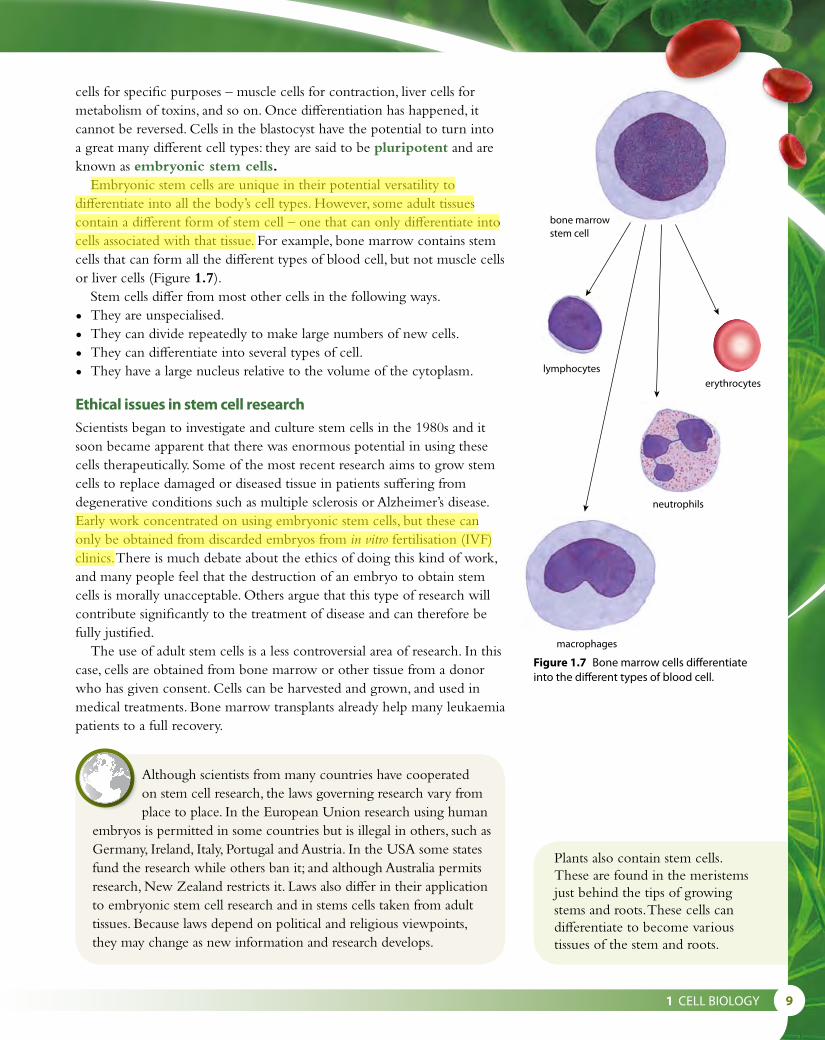
cells for specific purposes – muscle cells for contraction, liver cells for metabolism of toxins, and so on. Once differentiation has happened, it cannot be reversed. Cells in the blastocyst have the potential to turn into a great many different cell types: they are said to be pluripotent and are known as embryonic stem cells.
Embryonic stem cells are unique in their potential versatility to differentiate into all the body’s cell types. However, some adult tissues contain a different form of stem cell – one that can only differentiate into cells associated with that tissue. For example, bone marrow contains stem cells that can form all the different types of blood cell, but not muscle cells or liver cells (Figure 1.7).
Stem cells differ from most other cells in the following ways.
• They are unspecialised.
• They can divide repeatedly to make large numbers of new cells.
• They can differentiate into several types of cell.
• They have a large nucleus relative to the volume of the cytoplasm.
Ethical issues in stem cell researchScientists began to investigate and culture stem cells in the 1980s and it soon became apparent that there was enormous potential in using these cells therapeutically. Some of the most recent research aims to grow stem cells to replace damaged or diseased tissue in patients suffering from degenerative conditions such as multiple sclerosis or Alzheimer’s disease. Early work concentrated on using embryonic stem cells, but these can only be obtained from discarded embryos from in vitro fertilisation (IVF) clinics. There is much debate about the ethics of doing this kind of work, and many people feel that the destruction of an embryo to obtain stem cells is morally unacceptable. Others argue that this type of research will contribute significantly to the treatment of disease and can therefore be fully justified.
The use of adult stem cells is a less controversial area of research. In this case, cells are obtained from bone marrow or other tissue from a donor who has given consent. Cells can be harvested and grown, and used in medical treatments. Bone marrow transplants already help many leukaemia patients to a full recovery.
erythrocytes
neutrophils
macrophages
bone marrow stem cell
lymphocytes
Figure 1.7 Bone marrow cells differentiate into the different types of blood cell.
Plants also contain stem cells. These are found in the meristems just behind the tips of growing stems and roots. These cells can differentiate to become various tissues of the stem and roots.
Although scientists from many countries have cooperated on stem cell research, the laws governing research vary from place to place. In the European Union research using human
embryos is permitted in some countries but is illegal in others, such as Germany, Ireland, Italy, Portugal and Austria. In the USA some states fund the research while others ban it; and although Australia permits research, New Zealand restricts it. Laws also differ in their application to embryonic stem cell research and in stems cells taken from adult tissues. Because laws depend on political and religious viewpoints, they may change as new information and research develops.
Aysel
Highlight
Aysel
Highlight

10
Therapeutic use of stem cellsAnother source of stem cells, which has been successfully used in medical treatments, is the blood in the umbilical cord of a newborn baby (Figure 1.8). These stem cells can divide and become any type of blood cell. Cord blood can be used to treat certain types of leukaemia, a cancer that causes overproduction of white blood cells in the bone marrow. Cells from the cord blood are collected and their tissue type is determined. After chemotherapy to destroy the patient’s own bone marrow cells, stem cells which are the correct match to the patient’s tissue are given by transfusion. They become established in the person’s bone marrow and start producing blood cells as normal.
This treatment can work well in young children, but there are not enough cells in a single cord to meet the needs of an adult patient. Scientists have been looking for ways to either combine the cells from more than one baby, or to increase the number of cells in the laboratory. Allowing the stem cells to divide in the laboratory produces many blood cells, but not more stem cells. In 2010, scientists at the Fred Hutchinson Cancer Research Center in Seattle, USA, managed to alter a signalling pathway in the stem cells so they could increase in number without losing stem cell properties. As a result of this process, known as therapeutic cloning, umbical cord blood may prove to be an even more valuable source of stem cells in the future.
One of the most recent areas of stem cell research has been in the treatment of Stargardt’s disease using retinal pigment epithelium (RPE). Stargardt’s disease is an inherited condition, which begins in childhood and leads to macular degeneration (a gradual destruction of cells in the centre of the retina) and eventually causes blindness. Retinal cells can be made from embryonic stem cells. In 2012, as part of a larger trial, the first patients were given transplants of retinal cells developed from human embryonic stem cells, to treat their condition. The cells were injected directly into the retina and researchers found that not only did the stem cells survive but the number of cells increased over a period of 3 months. The cells began to develop important visual pigment and the patients noticed improvement in their vision. Scientists hope that in the future, stem cells will restore some sight not only to people with Stargardt’s disease but also many millions of older people suffering from age-related macular degeneration, the most common cause of blindness. Stem cell therapy has also been successfully used in the treatment of type I diabetes, and research is continuing into therapies to treat a range of conditions involving neurological damage, such as multiple sclerosis and Alzheimer’s disease.
Nature of scienceLooking for trends and discrepancies – developing theoriesIn order to explain aspects of the natural world, scientists develop theories. A theory is a well-established and widely accepted principle that arises from extensive observation of trends and discrepancies, and incorporates
Figure 1.8 This technician is collecting blood from an umbilical cord. This blood is a rich source of stem cells.
Aysel
Highlight
Aysel
Highlight
Aysel
Highlight
Aysel
Highlight
Aysel
Highlight
Aysel
Highlight
Aysel
Highlight
Aysel
Highlight

1 CELL BIOLOGY 11
facts, laws, predictions and tested hypotheses. A hypothesis is a speculative, specific and testable prediction about what is expected to happen in an investigation. So, a theory predicts events in general terms, while a hypothesis makes a specific prediction about a narrowly defined set of circumstances. If any evidence is collected which contradicts a hypothesis, the hypothesis must be rejected and a new hypothesis formulated.
In developing a theory, it is important to consider not only the main trends of an idea – those observations that are in general agreement – but also to seek out discrepancies which might go against the trend, and perhaps suggest testable hypotheses leading to a change in the theory. So, for example, with regard to cell theory, scientists might ask if observations suggest that the structure of all parts of all organisms conform to the theory, or if there are any discrepancies.
As we have seen, many millions of cells have been examined and the majority do conform to the principles of cell theory. But fungal hyphae and the fibres in muscles cells could be considered a challenge to it. Mammalian red blood cells have no nucleus (even though similar cells in reptiles and other vertebrates do), which means they cannot carry out all the functions of life and depart from cell theory in that respect. Single-celled protoctista such as Amoeba could be thought of as a challenge to the idea that every cell has a specialised function as these organisms carry out all the functions of life. Science must consider observations like these from many different fields of study and be prepared to revise accepted theories if it is necessary.
Questions to consider
• How can evidence be obtained for the principles of the cell theory? Can we prove that cells always arise from pre-existing cells?
• Do the examples of fungal hyphae and muscle cells disprove the cell theory?
• What should happen if evidence is collected that cannot be explained by a theory?
• What happens if evidence is collected that disproves a hypothesis?
2 Calculate how many cells of 100 μm diameter will fit along a 1 mm line.
3 List examples of where the concept of emergent properties can be found in a multicellular animal, such as a bird or a flowering plant.
4 State one therapeutic use of stem cells.5 Explain how cells in multicellular organisms are able to carry out
specialised functions.6 Use the scale bar on Figure 1.2 to calculate the length of the
Paramecium in the photograph.
Test yourself?

12
1.2 Ultrastructure of cellsLiving things are divided into two types – prokaryotes and eukaryotes – according to the structure of their cells. Prokaryotic cells are usually much smaller than eukaryotic cells and have a much simpler structure. Prokaryotes are thought to be the first cells to have evolved. Bacteria are all prokaryotic cells.
Prokaryotic cellsProkaryotic cells are so called because they have no nucleus (‘prokaryote’ comes from the Greek, meaning ‘before the nucleus’). They also have no organelles (internal structures), so cell functions do not take place in separate compartments within the cytoplasm. From the mid-20th century, when the electron microscope was developed, it became possible to study the internal detail of cells. Figures 1.9 and 1.10 show the main features of a typical prokaryotic cell.
Learning objectives
You should understand that:
• Prokaryotes have a simple cell structure with no compartmentalisation.
• Eukaryotes have a compartmentalised cell structure with membrane-bound organelles present in the cytoplasm.
• The resolution of electron microscopes is much higher than that of light microscopes, which allows identification of cell structures and organelles.
plasmid
cytoplasm
plasma membrane
cell wall
capsule
pili
70S ribosomes
nucleoidnaked DNA
flagellum
Figure 1.10 The structure of a typical prokaryotic cell.
Figure 1.9 The bacterium Escherichia coli is a typical prokaryotic cell. (Coloured TEM × 60 000).
• The cell wall surrounds the cell. It protects the cell from bursting and is composed of peptidoglycan, which is a mixture of carbohydrate and amino acids.
• The plasma membrane controls the movement of materials into and out of the cell. Some substances are pumped in and out using active transport.
• Cytoplasm inside the membrane contains all the enzymes for the chemical reactions of the cell. It also contains the genetic material.
• The chromosome is found in a region of the cytoplasm called the nucleoid. The DNA is not contained in a nuclear envelope and it is also ‘naked’ – that is, not associated with any proteins. Bacteria also contain additional small circles of DNA called plasmids. Plasmids replicate independently and may be passed from one cell to another.
• Ribosomes are found in all prokaryotic cells, where they synthesise proteins. They can be seen in very large numbers in cells that are actively producing protein. Prokaryotes have 70S ribosomes, which are smaller than those found in eukaryotes.
Aysel
Highlight
Aysel
Highlight

1 CELL BIOLOGY 13
• A flagellum is present in some prokaryotic cells. A flagellum, which projects from the cell wall, enables a cell to move.
• Some bacteria have pili (singular pilus). These structures, found on the cell wall, can connect to other bacterial cells, drawing them together so that genetic material can be exchanged between them.
Prokaryotic cells are usually much smaller in volume than more complex cells because they have no nucleus. Their means of division is also simple. As they grow, their DNA replicates and separates into two different areas of the cytoplasm, which then divides into two. This is called binary fission. It differs slightly from mitosis in eukaryotic cells (Subtopic 1.6).
Eukaryotic cellsEukaryotic organisms have cells that contain a nucleus. Animals, plants, fungi and protoctista all have eukaryotic cells.
The complexity of a eukaryotic cell cannot be fully appreciated using a compound light microscope. But in images made using an electron microscope, which has a much higher resolution, the fine details of many different organelles are visible. Figure 1.11 shows what can be seen of animal and plant cells using a light microscope – compare these images with the electron micrographs and interpretive drawings in Figures 1.12 to 1.15.
nuclear envelope chromatin nucleolus
nucleus
plasma membrane cytoplasm mitochondrion
glycogen granules
vesicle
plasma membrane
cytoplasm
chloroplast
cell wall of adjacent cell
vacuole
cell wall
tonoplast
nucleus
nuclearenvelope
Photograph of a stained animal cell (× 1100) Photograph of a cell in a moss leaf (× 450)10 µm 10 µm
Figure 1.11 Photographs and interpretive drawings to show typical animal and plant cells as they appear using a light microscope.
Ribosome sizes
The ‘S’ unit used to ‘measure’ ribosomes is a Svedberg unit. It is a measure of the behaviour of particles during sedimentation. 70S and 80S ribosomes are different sizes and so take different times to sediment when they are centrifuged. They are said to have different sedimentation coefficients.
Aysel
Highlight
Aysel
Highlight

14
Figure 1.12 Electron micrograph of an exocrine cell from the pancreas (× 12 000).
plasma membrane
lumen
secretory vesicle
secretory vesicle pouring its contents into lumen
Golgi apparatus
mitochondrion
nucleus, containing DNA associated with histones
nuclear envelope
80S ribosomes
rough endoplasmic reticulum
Figure 1.13 Interpretive drawing of some of the cell structures visible in Figure 1.12.
80S ribosomes
rough endoplasmic reticulum
nuclear envelope
mitochondrion
nucleus, containing DNA associated with histones
plasma membrane
Golgi apparatus
lumen
secretory vesicle

1 CELL BIOLOGY 15
Figure 1.15 Drawing of a palisade mesophyll plant cell made from the electron micrograph in Figure 1.14.
Figure 1.14 Electron micrograph of a palisade mesophyll plant cell (× 5600).
plasma membrane
nucleolus
80S ribosomes
mitochondrion
endoplasmic reticulum
vacuole
nucleus
nuclear envelope
cell wall
chloroplasts
cytoplasm
plasma membrane
nucleolus
80S ribosomes
mitochondrion
endoplasmic reticulum
vacuole
nucleus
nuclear envelope
cell wall
chloroplasts
cytoplasm
plasma membrane
nucleolus
80S ribosomes
mitochondrion
endoplasmic reticulum
vacuole
nucleus
nuclear envelope
cell wall
chloroplasts
cytoplasm
plasma membrane
nucleolus
80S ribosomes
mitochondrion
endoplasmic reticulum
vacuole
nucleus
nuclear envelope
cell wall
chloroplasts
cytoplasm

16
Eukaryotic cells contain structures called organelles, each of which forms a ‘compartment’ in which specific functions take place. This compartmentalisation enables a eukaryotic cell to carry out various chemical reactions or processes in separate parts of the cell, which all form part of the same system. Different types of cell have different organelles in different proportions, depending on the role of the cell.
The largest and most obvious structure in a eukaryotic cell is the nucleus, which contains the cell’s chromosomes. Chromosomes are composed of DNA combined with histone protein, forming a material known as chromatin. The nucleus is surrounded by a double-layered membrane, the nuclear envelope. Small gaps in the envelope, called nuclear pores, are visible and it is through these that material passes between the nucleus and the rest of the cell. A distinctive feature of the nucleus is the darkly staining nucleolus. This is the site of ribosome production.
Associated with the nuclear envelope is a series of membranes known as the endoplasmic reticulum (ER). Ribosomes attach to this network to form rough endoplasmic reticulum (rER), the site of protein synthesis. As proteins are produced, they collect in the spaces between the membranes, known as the cisternae. From here they can be transported in vesicles to other parts of the cell such as the Golgi apparatus. ER that has no ribosomes attached is known as smooth endoplasmic reticulum (sER). The membranes of sER have many enzymes on their surfaces. Smooth ER has different roles in different types of cell – in liver cells, it is where toxins are broken down; in the ovaries, it is the site of estrogen production. Smooth ER also produces phospholipids for the construction of membranes and lipids for use in the cell.
The Golgi apparatus is similar in appearance to the sER, composed of stacks of flattened, folded membranes. It processes proteins made in the rER, collecting, packaging and modifying them, and then releasing them in vesicles for transport to various parts of the cell or for secretion from the cell. The pancreas contains many secretory cells, which have large areas of Golgi apparatus (Figures 1.12 and 1.13).
Eukaryotic cells also contain mitochondria (singular mitochondrion). These are elongated structures surrounded by a double membrane that are found throughout the cytoplasm. Mitochondria are known as the cell’s ‘powerhouses’ because they are the site of aerobic respiration. The inner membrane is folded to form cristae, which greatly increase the surface area for the production of ATP in the cell. Cells that respire rapidly, such as muscle cells, have numerous mitochondria.
Lysosomes are spherical organelles with little internal structure, which are made by the Golgi apparatus. They contain hydrolytic enzymes for breaking down components of cells. They are important in cell death, in breaking down old organelles and, in white blood cells, digesting bacteria that have been engulfed by phagocytosis. Plant cells do not normally contain lysosomes.
Ribosomes are the site of protein synthesis in cells. They may be free in the cytoplasm or attached to the rER. They are made of RNA and
Aysel
Highlight
Aysel
Highlight

1 CELL BIOLOGY 17
protein but they do not have a membrane around them. Eukaryotic cells contain 80S ribosomes, which are larger than those found in prokaryotes.
As in prokaryotic cells, the plasma membrane controls the movement of materials into and out of the cell, and the gel-like cytoplasm, which fills much of the volume of the cell, provides a medium for many metabolic reactions.
Plant cells have three additional structures. All plant cells have an outer cellulose cell wall and most have a large central vacuole. Some plant cells, such as palisade mesophyll cells (Figures 1.14 and 1.15), contain chloroplasts. The chloroplasts are found in cells exposed to the light, as they are the sites of photosynthesis. Chloroplasts have a double membrane and are about the same size as bacteria. Both chloroplasts and mitochondria have their own DNA and ribosomes and are able to reproduce independently of the cell.
The large central vacuole contains water and salts. The membrane that surrounds it is under pressure from within and exerts a force on the cytoplasm, which in turn exerts a force on the cell wall, making the cell turgid and firm. The outer cell wall is composed of cellulose and other carbohydrates such as lignin and pectin, giving plant cells further support and a more rigid structure than animal cells. The cell walls and turgidity of plant cells give strength and support to tissues like leaves, holding them in the optimum position to catch the energy from sunlight for photosynthesis.
Although they are both eukaryotic cells, there are several key differences between animal and plant cells. These are summarised in Table 1.3.
Animal cells Plant cells
cell wall absent cell wall present
small vacuoles sometimes present large central vacuole present in mature cells
no chloroplasts chloroplasts often present
cholesterol in plasma membrane no cholesterol in plasma membrane
centrioles present (see page 36) centrioles absent
stores glycogen stores starch
Table 1.3 Differences between animal and plant cells.
Differences between prokaryotic and eukaryotic cellsComparisons of images of prokaryotic and eukaryotic cells show numerous differences between them. These are summarised in Table 1.4. Note, for example, the difference in size of ribosomes between prokaryotic and eukaryotic cells.
Aysel
Highlight

18
Structure Eukaryotic cell Prokaryotic cell
nucleus usually present, surrounded by a nuclear envelope and containing chromosomes and a nucleolus
no nucleus, and therefore no nuclear envelope or nucleolus
mitochondria usually present never present
chloroplasts present in some plant cells never present
endoplasmic reticulum
usually present never present
ribosomesrelatively large, about 30 nm in diameter, or 80S
relatively small, about 20 nm in diameter, or 70S
chromosomes DNA arranged in long strands, associated with histone proteins
DNA present, not associated with proteins, circular plasmids may also be present
cell wall always present in plant cells, made of cellulose, never present in animal cells
always present, made of peptidoglycan
flagella sometimes present some have flagella, but these have a different structure from those in eukaryotic cells
Table 1.4 Differences between prokaryotic and eukaryotic cells. The unit ‘S’ is a Svedberg unit, used to compare sizes of cell organelles.
Nature of scienceScientific advance follows technical innovation – the electron microscopeA typical animal cell is 10–20 μm in diameter, which is about one-fifth the size of the smallest particle visible to the naked eye. Robert Hooke was the first scientist to see and describe cells, although he didn’t know what they were. Later, Anton van Leeuwenhoek, who built one of the first microscopes in 1674, was able to see living cells of Spirogyra and bacteria.
Can we believe our eyes?Our own perception is a crucial source of knowledge. The
way we see things depends on the interaction between our sense organs and our mind, and what we perceive is a selective interpretation.
When studying material that has been prepared for microscopic examination, we must always bear in mind that staining and cutting cells will alter their appearance. Interpreting images requires care, and what we perceive in a particular image is likely to be influenced by these techniques as well as our own expectations.
Questions to consider
• Consider the shapes of mitochondria in Figure 1.12. Why do some mitochondria appear cylindrical and others circular?
• Plant cells have a single central vacuole. Examine the plant cell in Figure 1.14. How many vacuoles can you see? How can you explain this?
1 CELL BIOLOGY 19
It was not until good light microscopes became available in the early part of the 19th century that plant and animal tissues were seen as groups of individual cells and Schleiden and Schwann in 1838 were able to see sufficient structure to propose the cell theory, which incorporated the work of their predecessors (page 1).
Animal cells are tiny and colourless so it was not until the end of the 19th century, when staining techniques were first used, that it was possible to see a little more detail of cell contents. In the early 1940s, far more powerful electron microscopes were used for the first time and organelles and greater complexity of cell structure could be studied. Developments proceeded more rapidly in the 20th century because international communication allowed for more efficient collaboration not only in the designing and building of scientific instrumentation but also in the discussion and understanding of what could be observed.
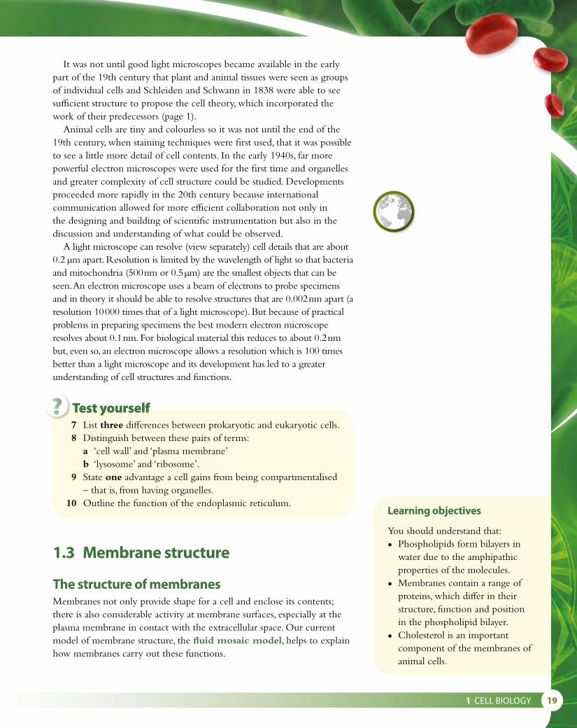
A light microscope can resolve (view separately) cell details that are about 0.2 μm apart. Resolution is limited by the wavelength of light so that bacteria and mitochondria (500 nm or 0.5 μm) are the smallest objects that can be seen. An electron microscope uses a beam of electrons to probe specimens and in theory it should be able to resolve structures that are 0.002 nm apart (a resolution 10 000 times that of a light microscope). But because of practical problems in preparing specimens the best modern electron microscope resolves about 0.1 nm. For biological material this reduces to about 0.2 nm but, even so, an electron microscope allows a resolution which is 100 times better than a light microscope and its development has led to a greater understanding of cell structures and functions.
Learning objectives
You should understand that:
• Phospholipids form bilayers in water due to the amphipathic properties of the molecules.
• Membranes contain a range of proteins, which differ in their structure, function and position in the phospholipid bilayer.
• Cholesterol is an important component of the membranes of animal cells.
1.3 Membrane structure
The structure of membranesMembranes not only provide shape for a cell and enclose its contents; there is also considerable activity at membrane surfaces, especially at the plasma membrane in contact with the extracellular space. Our current model of membrane structure, the fluid mosaic model, helps to explain how membranes carry out these functions.
7 List three differences between prokaryotic and eukaryotic cells. 8 Distinguish between these pairs of terms:
a ‘cell wall’ and ‘plasma membrane’b ‘lysosome’ and ‘ribosome’.
9 State one advantage a cell gains from being compartmentalised – that is, from having organelles.
10 Outline the function of the endoplasmic reticulum.
Test yourself?

20
All membranes, wherever they occur in cells, have the same basic structure. Membranes are usually between 7 and 10 nm thick, and are composed of two layers of phospholipid, which form a bilayer. Phospholipids are made up of a polar, hydrophilic area containing a phosphate group bonded to glycerol, and a non-polar, lipophilic area containing fatty acids. In the bilayer, the lipophilic or hydrophobic (water-hating) parts all point towards each other, and the hydrophilic (water-loving) areas point outwards, as Figure 1.16 shows. It is the different properties of each end of the molecule that cause the phospholipids to arrange themselves in this way. The hydrophilic ‘heads’ of the molecules always appear on the outside of the membrane where water is present, while the hydrophobic ‘tails’ orientate inside the double layer, away from water. The structure is called a ‘mosaic’ because, just as a mosaic picture is made up of many small, separate pieces, so the surface of the membrane is composed of the heads of many separate phospholipid molecules. The whole structure is flexible or ‘fluid’ because the phospholipids can float into a position anywhere in the membrane. Research using radioactively labelled phospholipids shows that these molecules move not only within their own layer, but also between the two layers of the membrane.
Embedded in the bilayer are different molecules that contribute to the functions of membranes. Cholesterol is often present in mammal cells and is most commonly found in the plasma membrane. One end of the cholesterol molecule associates with the polar heads of phospholipid molecules while other parts of it are embedded in the membrane next to the non-polar fatty acid chains. This interaction makes the membrane less ‘fluid’, more rigid, and less permeable to water-soluble molecules.
There are also different types of protein in the bilayer. Integral proteins are embedded in the bilayer, whereas peripheral proteins are attached to the surface. Many of the proteins on the outer surface are glycoproteins – that is, they have carbohydrate groups attached to them. Some of these serve as hormone binding sites and have special shapes to recognise the specific hormones to which the cell will respond. Others are important in cell-to-cell communication and adhesion. Some integral
hydrophobic layer
hydrophilic layer
glycerol water
water
fatty acid
In a watery environment, the phospholipids become arranged in a bilayer, because of the hydrophilic and hydrophobic properties of the ‘heads’ and ‘tails’ of the molecules.
phospholipid bilayer
Figure 1.16 A phospholipid molecule includes a phosphate, glycerol and two fatty acids but in diagrams (such as Figure 1.17) the molecule is often simplified and shown as a circle with two tails.
Amphipathic compounds possess both hydrophilic (water-loving, polar) and lipophilic (fat-loving) properties. The amphipathic properties of phospholipids in a membrane explain the way a membrane structure forms. Phospholipids arrange themselves into bilayers, with their polar groups facing the surrounding aqueous (watery) medium, and their hydrophobic chains facing towards the inside of the bilayer. In this way, a non-polar region is formed between two polar ones. Phospholipids are principal constituents of biological membranes, but cholesterol and glycolipids and glycoproteins are also amphipathic and their presence in the bilayer gives membranes different physical and biological properties. You can find out more about the importance of polar groups in Subtopic 2.2.
High levels of certain types of cholesterol in the blood have been associated with heart disease. You can find out more about this on page 199. The body produces its own cholesterol in the liver, but it is also found in many foods that we eat.
Aysel
Highlight
Aysel
Highlight

1 CELL BIOLOGY 21
proteins are enzymes immobilised within the membrane structure and perfectly placed to carry out sequences of metabolic reactions. Finally, there are proteins that span the bilayer acting as channels for ions and molecules to pass by passive transport, or forming pumps that use active transport to move molecules into or out of the cell.
Models of membrane structureOur current understanding of the structure of the membrane has arisen from the work of a number of scientists, over many years. Each group refined previous knowledge of membranes, rejecting theories that were not supported by evidence and working to gather new data as microscopy and other techniques improved. The existence of a lipid bilayer was originally proposed and outlined by Gorter and Grendel, in 1925. Their ideas were developed and improved by Hugh Davson and James Danielli, who proposed in 1935 a model of a phospholipid bilayer between two layers of globular protein, the so-called ‘fat sandwich’ model. The Davson–Danielli model was new and it attempted to explain their observations of the surface tension of lipid bilayers. Since that time, the phenomenon of surface tension in bilayers has been better explained by studying the properties of the phospholipid heads. Nevertheless, the Davson–Danielli model predominated, and was supported by observations using the electron microscope, until 1972 when Singer and Nicolson described the ‘fluid mosaic’ model. The fluid mosaic model included descriptions of integral proteins that were sited through the membrane, and it rejected the idea of a ‘sandwich-like’ globular protein layer because it was no longer well supported by experimental evidence (see ‘Nature of science’, below). Fresh observations obtained using a new technique called freeze-etching were also important.
The model of membrane structure accepted today is based on the Singer and Nicolson fluid mosaic model, illustrated in Figure 1.17, and has been supported by more recent research, with only minor modifications.
Nature of scienceFalsification of theories – developing a model of membrane structureScientific theories embody our current understanding of aspects of the real world, and may include models to represent those aspects. However, any scientific theory or model can only exist until it is disproved. The Davson–Danielli model of membrane structure was accepted until new evidence called the model into question. The Davson–Danielli model was very close to what is now accepted, except that it proposed that all membranes are alike. This was disproved, though, when it was found that different organisms transport very different substances across their membranes, using different proteins. For example, mammalian cells transport sodium and potassium ions across their plasma membranes via special protein channels, while methane-producing bacteria move
Freeze-etching is a method of preparing membranes to give a three-dimensional view of the surface and detail of the membrane’s structures. Cells are rapidly frozen and fractured by breaking them in liquid nitrogen. The fractured surface is shadowed with evaporated heavy metal under vacuum and stabilised. The replicated surface is floated onto fine metal grids so that it can be viewed in the electron microscope to reveal the 3D arrangement of lipids and proteins that are present.
As you read the evidence that has accumulated and
helped our understanding of the structure of membranes, consider the following questions.• Why is it important for
scientists to put forward their ideas in the form of theories?
• How useful are models in developing ideas of biological structures?
• Is it important to learn about theories that have been discredited or superseded?
Aysel
Highlight
Aysel
Highlight
Aysel
Highlight
Aysel
Highlight

22
methane out of their cells via different protein channels. Evidence such as this showed that all membranes are not alike, and falsified the Davson–Danielli model, which was superseded by our current model.
11 Suggest why the term ‘fluid mosaic’ is used to describe membrane structure.
12 Suggest why the fatty acid ‘tails’ of the phospholipid molecules always align themselves in the middle of the membrane.
13 Outline the difference between integral membrane proteins and peripheral membrane proteins.
Test yourself?
outside
glycolipid
protein glycoprotein phospholipid channel proteininside
carbohydrate part of glycoprotein
Outside
protein glycoprotein cholesterol phospholipid transport protein
InsideA 3D representation of membrane structure
hydrophilic head
peripheral protein
hydrophobic tails
phospholipidbilayer
branching carbohydrateattached to a protein toform a glycoprotein
integral protein
one phospholipid molecule
cholesterolin both layers
Outer surface
Inner surface
transport protein has hydrophilic interior for ions and hydrophilic molecules
A 2D representation of membrane structure
Figure 1.17 Diagrams to show the fluid mosaic model of membrane structure.

1 CELL BIOLOGY 23
1.4 Membrane transport
Diffusion, facilitated diffusion and osmosisMany molecules pass across the plasma membrane. Water, oxygen, carbon dioxide, excretory products, nutrients and ions are continuously exchanged and many cells also secrete products such as hormones and enzymes through the plasma membrane.
The simplest way in which molecules can move into or out of a cell is by simple diffusion through the plasma membrane. Diffusion is a passive process, which takes place as molecules move randomly. No energy input is required, and movement occurs by way of a simple concentration gradient. A concentration gradient is a difference in concentration of a substance between two regions and diffusion will always occur where such a gradient exists until particles of the substances are evenly distributed and equilibrium is reached. One important example of simple diffusion is its role in the process of cell respiration. Oxygen is needed by cells as it is continuously used up in respiration. As a cell respires, the oxygen concentration inside becomes less than the concentration outside, so oxygen molecules diffuse in. In a similar way, as carbon dioxide is continuously formed during respiration, its concentration builds up inside the cell and it diffuses out through the plasma membrane to an area where the concentration is lower. Simple diffusion occurs where the membrane is fully permeable to the substance or where channel proteins in the membrane are large enough for the substance to pass through.
Large molecules, and charged particles such as chloride ions (Cl–) and potassium ions (K+), cannot pass through the membrane by simple diffusion so certain proteins form channels through which they can travel. As in simple diffusion, no energy is used by the cell and the transport relies on the kinetic energy of the particles moving down their concentration gradient. Channel proteins have an interior which is hydrophilic (Figure 1.18) so water-soluble materials can pass though them, and they are specific – that is, they only allow a particular substance to move through. Some of these channels are permanently open, whereas others are gated and only open to allow certain ions to pass when they are stimulated to do so. For example, gated channels in the axons of nerve cells open when there is a change in the voltage (potential difference) across the membrane. Gated potassium channels only allow K+ ions to pass out through the membrane after a nerve impulse has passed along the axon. You can read more about nerve impulses in Subtopic 6.5.
Other channel proteins allow the movement of substances such as glucose and amino acids, which are polar and cannot diffuse though the lipid layer of the membrane. Substances like these are transported across membranes by facilitated diffusion. In this case, a carrier protein first combines with the diffusing molecules on one side of the membrane, carries them through the channel protein and then releases them on the other side (Figure 1.18). Facilitated diffusion allows a faster diffusion rate
Learning objectives
You should understand that:
• Particles move across membranes by osmosis, active transport, simple diffusion and facilitated diffusion.
• Materials can be taken into cells by endocytosis and leave cells by exocytosis due to the fluid nature of the membrane.
• Within a cell, vesicles move materials around.
Passive transport the movement of substances down a concentration gradient from an area of high concentration to an area of lower concentration without the need for energy to be usedDiffusion one example of passive transport; many molecules pass into and out of cells by diffusion e.g. oxygen, carbon dioxide and glucoseOsmosis another example of passive transport but the term is used only in the context of water molecules; osmosis is the movement of water molecules across a partially permeable membrane from a region of lower solute concentration, where there is a high concentration of water molecules, to a region of higher solute concentration, where the concentration of water molecules is lowerActive transport the movement of substances against the concentration gradient, which always involves the expenditure of energy in the form of ATP
Aysel
Highlight

24
for molecules that particular cells need – for example, the diffusion of glucose into active muscle cells. No energy input is required because the molecules move down their concentration gradient.
A special case of diffusion is osmosis (Figure 1.19). This is the passive movement of water across a partially permeable membrane from an area of lower solute concentration to an area of higher solute concentration.
Large or chargedsubstances such as K+ andCl– ions cannot pass easilythrough membranes. Theycan pass through specialchannel proteins if theycome in contact with thechannel. Only speci�c ionsor molecules can pass andno energy input is required. channel protein
openchannel protein
closedlowconcentration
highconcentration
concentrationgradient
lowconcentration
highconcentration
concentrationgradient
Carrier proteins assist somemolecules through themembrane, down theirconcentration gradient,combining with moleculeson one side of themembrane and releasingthem on the other side.Again, no energy input isrequired.
Di�usion through a protein channel
Facilitated di�usion via a carrier protein
Two solutions are separated by a partially permeable membrane. B has a higher solute concentration than A. The solute molecules are too large to pass through the pores in the membrane but the water molecules are small enough.
As the arrows in the left diagram indicate, more water molecules moved from A to B than from B to A, so the net movement has been from A to B, raising the level to the solution in B and lowering it in A. The water potentials (the tendency of water molecules to move in each direction) in A and B are now the same.
Before osmosis At equilibrium
Figure 1.18 Some large or charged ions and molecules pass through the membrane via special channel proteins.
Figure 1.19 Osmosis.

1 CELL BIOLOGY 25
high water potential outside, lower inside
equal water potential outside and inside
low water potential outside, higher inside
osmotic uptake of water
A cell contains many substances dissolved in its cytoplasm so when placed in pure water (which is hypotonic in relation to the cell) it will tend to take in water by osmosis. The cell will swell and eventually burst and die.
A cell placed in water with the same concentration of solutes as the cell (an isotonic solution) will remain in equilibrium.
Cells placed in a solution containing a higher concentration of dissolved solutes than the cell contents (a hypertonic solution) will lose water by osmosis and shrink.
osmotic loss of water
no net movement of water
Figure 1.20 Responses of animal cells to solutions of different concentrations (see also Figure 1.21).
When the solute concentrations inside and outside a cell are the same, the same number of water molecules will pass across the membrane into the cell as those that leave. An animal cell that is placed in pure water will take in water by osmosis until eventually it may burst (Figure 1.20). Placed in a solution with a very high concentration of solutes, the cell will shrink or ‘crenate’ as water leaves the cell by osmosis. In either situation, animal cells will not function properly and their metabolism will be affected.
In medical procedures, tissues and organs are bathed in a solution of ‘normal saline’, which has exactly the same osmolarity (a measure of the solute concentration in a solution) as human cell cytoplasm and is said to be isotonic with the cytoplasm. In this case osmosis does not occur and cells are not damaged.

26
A plant cell in a solution that is less concentrated than the cell solution absorbs water by osmosis
water in
pressure ofcell contents
pressure of wall resisting uptake of water (pressure potential)
Water di�uses into the cell through the partially permeable plasma membrane. The cell contents expand. The contents will push out on the wall.
The wall pushes back on the cell contents. The force of the wall pushing on the cell contents is called the pressure potential. The cell is turgid.
Some parts of the membrane are resistant to pulling away from the wall. If the membrane is torn at these points, the now �lls the gap between wallcell dies.
The plasma membrane is eventually pulled away from the wall in places. The cell is plasmolysed. External solution
and plasma membrane.
The contents do not push out on the walls. The cell is said to be �accid.
A plant cell in a solution that is more concentrated than the cell solution loses water by osmosis
Water di�uses out of the cell through the partially permeable plasma membrane.
pressure ofcell contents
water out water out water out
1 2 3
1 2 3 4
water potential outside equals water potential inside
As more water enters, water potential of the cell solution rises. Eventually, water potential inside equals water potential outside and there is no further net uptake of water. The cell has reached equilibrium. The cell is fully turgid.
Figure 1.21 Responses of plant cells to solutions of different concentrations. Plant cells are not damaged as water enters by osmosis because their cell wall protects them.
Normal saline is a solution of 0.90% w/v (weight by volume) of sodium chloride and is isotonic with human cells. It is used frequently in intravenous drips (IVs) for patients who cannot take fluids orally and are in danger of becoming dehydrated.
Plant cells are also affected by the movement of water into and out of their cells but the presence of a cell wall prevents plant cells being damaged or bursting. If a plant cell is put into water, water will enter by osmosis but the plant cell wall resists the entry of further water once the cell is full. A plant cell that is full becomes firm and rigid – a condition known as turgor (Figure 1.21).
Active transportMany of the substances a cell needs occur in low concentrations in the surroundings outside the plasma membrane. For example, plants must take in nitrate ions from very dilute solutions in the soil to build their proteins, and muscle cells actively take in calcium ions to enable them to contract. To move these substances into the cell against a concentration gradient, the cell must use metabolic energy released from the breakdown of ATP to ADP and Pi (Subtopic 2.8). This is called active transport (Figure 1.22). Specific proteins in the plasma membrane act as transporters or ‘carriers’ to move substances through. Many of the carrier proteins are specific to particular molecules or ions so that these can be selected for transport into the cell.
Water potential the tendency of water molecules to move from an area of higher concentration to an area of lower concentration.
Aysel
Highlight
Aysel
Highlight

1 CELL BIOLOGY 27
Step 1sodium ions
Step 3potassium ions
Step 3sodium ions
Step 2potassium ions
The breakdown of ATPprovides energy for theproteins to change shapeand to allow the ions to move.
ATP ADP + Pi + energy
Step 1potassium ions
Step 2sodium ions
insidethe cell
outside
Figure 1.23 illustrates a very important example of active transport. The sodium–potassium pump maintains the concentration of sodium and potassium ions in the cells and extracellular fluid. Cells are able to exchange sodium ions for potassium ions against concentration gradients using energy provided by ATP. Sodium ions are pumped out of the cell and potassium ions are pumped into the cell.
Outside the cell
Inside the cell
molecules or ions to be transported
carrier protein in lipid bilayer
carrier protein activated by reaction with ATP
ATP
change in shape and postion of carrier protein
ADP and Pi released from carrier protein, which reverts to the receptive state
Figure 1.22 Active transport of a single substance.
Figure 1.23 An example of active transport – the sodium–potassium pump. Start at step 1 for each ion in turn and work round clockwise.

28
Exocytosis and endocytosisCells often have to transport large chemical molecules or material in bulk across the plasma membrane. Neither diffusion nor active transport will work here. Instead, cells can release or take in such materials in vesicles, as shown in Figure 1.24. Uptake is called endocytosis and export is exocytosis. Both require energy from ATP.
During endocytosis, part of the plasma membrane is pulled inward and surrounds the liquid or solid that is to be moved from the extracellular space into the cell. The material becomes enclosed in a vesicle, which pinches off from the plasma membrane and is drawn into the cell. This is how white blood cells take in bacteria (Figure 1.24).
There are two types of endocytosis. If the substances being taken in are particles, such as bacteria, the process is called phagocytosis. If the substances are in solution, such as the end products of digestion, then it is called pinocytosis.
Figure 1.24 Examples of endocytosis and exocytosis.
Materials for export, such as digestive enzymes, are made in the rER and then transported to the Golgi apparatus to be processed. From here they are enclosed within a membrane-bound package known as a vesicle, and moved to the plasma membrane along microtubules. The arrangement of molecules in the membrane of a vesicle is very similar to that in the plasma membrane. As a vesicle approaches the plasma membrane, it is able to fuse with it and in doing so release its contents to the outside. The flexibility and fluidity of the plasma membrane are essential to enable both endocytosis and exocytosis to happen. Vesicles also help to transfer and organise substances in the cell. They are involved in metabolism, transport and enzyme storage and some chemical reactions also occur inside them.
Nerve impulses are able to pass across synapses (the tiny gaps between one nerve cell and the next) due to exocytosis and endocytosis. Neurotransmitters are secreted at the end of a nerve cell fibre by exocytosis. They stimulate the adjacent nerve and are then reabsorbed by endocytosis to be recycled and reused. You can find out more about the transmission of nerve impulses in Subtopic 6.5.
Aysel
Highlight

1 CELL BIOLOGY 29
Nature of scienceExperimental design – accurate quantitative measurementWhenever experiments are designed, accuracy is important so that the experimenter can be sure that results are valid. Measurements should be obtained using the most suitable equipment with the correct degree of accuracy for the task. In the experiment described below, for example, measurements of the mass of small samples of potato and sucrose crystals are needed. A balance that provides readings accurate to 0.05 g would be most appropriate. Similarly, when measuring a small volume of liquid, a 25 cm3 measuring cylinder would be more appropriate than a 250 cm3 cylinder.
This experiment can be used investigate the process of osmosis and identify the solute concentration (or water potential) of cells. Cubes or chips of potato tissue are weighed accurately and placed in test tubes each containing a sucrose solution of a different solute concentration (molarity) – for example, between 0 mol dm–3 and 0.6 mol dm–3 – for a suitable period of time. The potato samples are then removed and reweighed and the percentage change in their mass is recorded.
% change = change in mass ÷ original mass × 100
A solution that causes no change in mass of the potato has the same solute concentration as the tissue and the same water potential. It is said to be isotonic. (Solutions of greater solute concentration than the tissue are hypertonic, while solutions whose solute concentration is lower than that of the tissue are known as hypotonic.)
Figure 1.25 shows the results of such an experiment. From the graph it can be seen that the molarity that causes no change in mass is approximately 0.35 mol dm3. At this concentration, the water potentials inside and outside the cell are equal so there is no net movement of water by osmosis.
Concentration ofsucrose solution / mol dm−3
0.2 0.4 0.6 0.8 1.0
Perc
enta
ge c
hang
e in
mas
s of
pot
ato
sam
ples
/ %
–30
–20
–10
0
10
20
30
Figure 1.25 Graph to show the results of an experiment with potato samples placed in different sucrose solutions.

30
1.5 The origin of cells
How are new cells formed?Cell theory proposes that all organisms are composed of one or more cells, which are the smallest units of life. One of the functions carried out by all living organisms is reproduction. Therefore, the first principle of the cell theory is that cells can only come from pre-existing cells. Louis Pasteur (1822–1895) carried out experiments that provided evidence for this. He showed that bacteria could not grow in a sealed, sterilised container of chicken broth. Only when living bacteria were introduced would more cells appear in the broth. Figure 1.26 summarises Pasteur’s experiment.
Learning objectives
You should understand that:
• Cells form by the division of pre-existing cells.
• Non-living material must have given rise to the first cells.
• Endosymbiosis is a theory that explains the origin of eukaryotic cells.
wait
break stem so thatair can enter
boil no growth
wait
boil
microbialgrowth
If the neck of the flask is broken it is possible for bacteria to enter the broth where they reproduce to produce more cells.
Boiling the flask kills any bacteria present in the broth. The curved neck of the flask prevents the entry of any new organisms from the atmosphere.
Figure 1.26 Pasteur’s experiment demonstrating that living cells cannot ‘spontaneously generate’, but must originate from pre-existing living cells.
14 Outline the difference between simple diffusion and facilitated diffusion.
15 List three ways that substances move from one side of a membrane to the other.
16 State one transport mechanism across a membrane that requires energy from ATP and one that does not.
17 State one difference and one similarity between exocytosis and endocytosis.
Test yourself?

1 CELL BIOLOGY 31
How did the first cells originate?All prokaryotic and eukaryotic organisms alive today are made up of cells. The very first prokaryotes are thought to have appeared around 3.5 billion years ago and the structures in these first cells must have originated from chemicals present on the Earth at that time. For these structures to have formed, four essential steps must have occurred.
• Living things are made of organic molecules, so simple organic molecules such as amino acids, sugars, fatty acids, glycerol and bases must have formed.
• Organic molecules in living organisms (such as triglycerides, phospholipids, polypeptides and nucleic acids) are large, so single molecules must have been assembled to make these more complex molecules.
• All living things reproduce, so molecules must have formed that could replicate themselves and control other chemical reactions. This is the basis of inheritance.
• Finally, cells have membranes, so the mixtures of these molecules must have been enclosed within membrane-bound vesicles.
The endosymbiotic theoryThe endosymbiotic theory explains how eukaryotic cells could have developed from a simple cell or prokaryote. The theory suggests that some organelles found inside eukaryotes were once free-living prokaryotes. There is evidence to suggest that some prokaryotes were engulfed by larger cells, and were retained inside their membranes where they provided some advantages to the larger cell (Figure 1.27).
Evidence for this theory includes the fact that two important organelles, mitochondria and chloroplasts, share many characteristics with prokaryotic cells. Both chloroplasts and mitochondria:
• contain ribosomes that are smaller than those found in other parts of eukaryotic cells but are identical in size to those found in bacteria
• contain small circular pieces of DNA resembling bacterial plasmids in their basic structure
smaller prokaryotic cell
host cell now containingmembrane-boundprokaryotic cell
larger bacterial cell
Figure 1.27 Organelles such as chloroplasts may have originated from free-living prokaryotes that were engulfed by larger cells.
Aysel
Highlight

32
• have their own envelope surrounding them, on the inner membrane of which are proteins synthesised inside the organelle, suggesting that they may have used this ability long ago when they were independent organisms
• can replicate themselves by binary fission.This evidence supports the theory that these organelles are modified bacteria that were taken in by phagocytosis, early in the evolution of eukaryotic cells. Here they became useful inclusions. The double outer envelope of chloroplasts and mitochondria may have originated from the bacterial plasma membrane together with the membrane of an engulfing phagocytic vesicle. Perhaps some of the enclosed bacteria carried pigment molecules on their membranes and used light energy to make organic molecules and release oxygen – these may have become chloroplasts. It may be that others became efficient at using the oxygen molecules for aerobic energy production, and these became mitochondria.
Critics of the endosymbiotic theory might argue that, even if prokaryotes were engulfed by larger cells, there is no certainty that they could be passed on to both daughter cells when the larger cell divided, because there is no special mechanism to ensure this. However, when a cell divides by binary fission each daughter cell contains some cytoplasm from the parent and so at least one of the daughter cells would contain the engulfed prokaryotes. Both mitochondria and chloroplasts have retained the ability to self-replicate and so their numbers can increase in the cytoplasm prior to cell division, which increases the chance of both daughter cells containing some. Critics also note that mitochondria and chloroplasts are not able to survive on their own if they are isolated from a cell, which they might be expected to do if they originated from free-living cells. But perhaps over time they have lost the ability to synthesise one or more essential molecules and have come to depend on the ‘host’ cell to provide them.
Nature of scienceGeneral principles underlying the natural world – evidence of the unbroken chain of life from the first cellsFurther evidence for a common origin for all life on Earth comes from the genetic material, in the form of chromosomes, which is inherited by every cell. Chromosomes are made from nucleic acids, such as DNA (deoxyribonucleic acid), and built into the chromosomes is a code, which is used by the cell to assemble all the molecules it needs to live.
A sequence of four key molecules, known as bases, along a DNA molecule forms this code. The bases are adenine (A), cytosine (C), guanine (G) and thymine (T). The code determines the sequence of the amino acid components that are bonded together as protein molecules are synthesised (Subtopic 2.7). The code is ‘read’ in sets of three bases known as ‘codons’ and 64 different codons can be made from combinations of the four bases A, C, G and T. A single codon represents the code for one amino acid unit in a protein.
Endosymbiosis
Symbiosis means ‘life together’. Endo means ‘inside’ and so endosymbiosis describes a relationship taking place inside a cell.
gene a length of DNA at a specific location on a chromosome that controls a specific heritable characteristic
Aysel
Highlight

1 CELL BIOLOGY 33
The genetic code is said to be universal because all living organisms share the same genetic code, with only a few minor variations, which are due to mutations that have occurred over the millions of years of evolution. All life – from plants and animals to bacteria and fungi – relies on the same 64 codons to carry the biological instructions for their bodies. What varies from one organism to another is not the code’s structure or the way in which it is translated, but the individual genes which are formed from lengths of DNA (Subtopic 3.1). The Human Genome Project and similar projects have deciphered gene sequences and compared sequences and codes in different species. Scientists have found whole ‘sentences’ of identical DNA ‘text’.
The study of the genetic code, together with studies of molecular processes such as respiration, photosynthesis and protein production, show that these vital processes are similar in all living cells. This provides strong evidence that all life on Earth had a common origin billions of years ago.
Learning objectives
You should understand that:
• Different stages of the cell cycle can be identified. One of these is interphase in which many process occur in the cytoplasm and nucleus.
• Mitosis describes the division of the nucleus in a cell into two genetically identical daughter nuclei.
• During mitosis chromosomes condense by supercoiling.
• Cytokinesis is a phase that occurs after mitosis. It is different in plant and animal cells.
• Cyclins are involved in controlling the cell cycle.
• The development of primary and secondary tumours can involve mutagens, oncogenes and metastasis.
S phase – DNA replication
G2 phaseG1 phase
mitosiscytokinesis
interphase
mitosis
Figure 1.28 The cell cycle.
18 Explain how Pasteur’s experiment supports the idea that life does not arise by spontaneous generation of cells.
19 Define the term ‘endosymbiosis’.
1.6 Cell divisionNew cells are needed to replace cells that have died or to allow an organism to grow. The nucleus and cytoplasm of a cell divide in processes known as mitosis and cytokinesis, which are phases in a series of events known as the cell cycle.
The cell cycleThe cycle of a cell’s life can be divided into three stages, as shown in Figure 1.28:1 interphase2 mitosis (division of the nucleus)3 cytokinesis (division of the cytoplasm).
Test yourself?

34
InterphaseDuring most of the life of a cell, it performs the task for which it has been pre-programmed during differentiation. This period is called interphase. Part of interphase is spent in preparation for cell division (the G2 phase) and part of it is the period immediately after division (the G1 phase). The two stages of cell division are the separation and division of the chromosomes (mitosis), and the division of the cell into two daughter cells (cytokinesis).
If a cell is examined during interphase using a light microscope, little appears to be happening, but this is a very active phase of the cell cycle when the cell carries out its normal activities and also prepares itself for mitosis. In the nucleus, the DNA in the chromosomes is replicated (S phase) so that after cell division there will be exactly the same number of chromosomes in the two daughter cells. During interphase many proteins necessary for the division need to be synthesised at the ribosomes in the cytoplasm. The number of mitochondria increases so that the respiratory rate can be rapid enough to provide energy for cell division. In the case of plant cells with chloroplasts, the number of chloroplasts increases so there are sufficient for each daughter cell.
MitosisThe two new cells that will be formed after mitosis and cytokinesis are genetically identical. These processes allow an organism to grow more cells, or to repair injured tissue by replacing damaged cells, or to make new cells to replace old ones. Mitosis is also the way in which an embryo grows from a fertilised egg during development. Many organisms reproduce themselves using mitosis; examples include the unicellular organisms such as Amoeba, Paramecium and yeast. Reproducing in this way is known as asexual reproduction as no gametes (Subtopic 3.3) are involved and the offspring are genetically identical to the parent.
There are four distinct stages in mitosis, though the process is continuous, with each stage running into the next. There are no intervals in between stages. Figures 1.29 and 1.30 show in detail the stages of mitosis.
Summary of the cell cycle
G1, S and G2 are the three stages of the part of the cell cycle known as interphase.
G1 phase• cell grows• DNA is transcribed• protein is synthesisedS phase• DNA is replicatedG2 phase• cell prepares for divisionmitosis• cell nucleus dividescytokinesis• cytoplasm divides
Root tip squash preparations
Squash preparations of onion cell root tips where cells are actively dividing can be prepared by softening the tissue in 1 mol dm–3 hydrochloric acid. One drop of toluedine blue stain is added and the cells are squashed by placing them between a cover slip and microscope slide, which is gently tapped with a pencil. The root tip will spread out as a mass on the slide and cells will separate from one another so that stages of mitosis can be seen under high power using a light microscope.
Figure 1.29 Stages of mitosis in stained onion cells, as seen in a root squash preparation (× 900 at 10 centimetres wide each).
prophase metaphase cytokinesistelophaseanaphase
Aysel
Highlight
Aysel
Highlight
Aysel
Highlight

1 CELL BIOLOGY 35
interphase
cytokinesis
late interphase
‘invisible’ chromosomes - each containing twoidentical chromatids
nucleolus
centrioles duplicated(centrioles are normally notvisible in the light microscope)
nuclear envelope is formedaround each group ofchromosomes
chromosomeunravels
telophase
two chromatids ofone chromosome
equator
microtubules attachedto each centromeremove the chromosomesso that each centromerelies on the equator
metaphase
centromereshave separated
polemicrotubule pulls onthe centromere,moving the chromatidto the pole
anaphase
prophase
chromosomescondense and arevisible as threads,but individualchromatids arenot often visible
centrioles move apartand a spindle begins tobe formed (normallynot visible)
nucleolusdisappears
microtubulesforming part ofthe developingspindle
chromatid
ProphaseDuring prophase, the chromosomes become visible using a microscope. During interphase they have been drawn out into long threads, allowing the cellular machinery access to the genes but now the chromosomes coil round themselves several times to produce a supercoil (Figure 1.31). Supercoiling not only makes the chromosomes shorter and thicker, it also
Figure 1.30 The stages of the cell cycle, including mitosis. Note that the cells are shown with just four chromosomes here, to make it easier to understand the process.
Aysel
Highlight
Aysel
Highlight
Aysel
Highlight

36
reduces the space that they take up and enables them to take part in the processes that follow. We can follow these processes because supercoiled chromosomes can be seen using a microscope. Because the DNA was replicated during interphase, at this stage each chromosome consists of two identical copies. These two copies are called the sister chromatids and are attached to each other at a place called the centromere. Also visible at this time are structures known as centrioles, which move to opposite sides of the cell as microtubules form between them. This microtubule structure is called the spindle. As prophase draws to a close, the nuclear envelope breaks down.
DNA double helix
histone protein
nucleosomes
cylindricalcoil
metaphasechromosome
sisterchromatids
chromatinfibre
centromere
Figure 1.31 Supercoiling produces condensed, compact chromosomes in preparation for the next stages of mitosis.
1 CELL BIOLOGY 37
MetaphaseMetaphase begins when the nuclear envelope has broken down. As it disappears, more space is created so that the chromosomes can move into position during their division. The sister chromatids align themselves on the microtubules in the middle, or equator, of the spindle and are attached by their centromeres.
AnaphaseDuring anaphase, the centromeres split and the sister chromatids are pulled apart and move towards the centrioles at opposite sides, or poles, of the cell as the spindle fibres shorten. Each sister chromatid is now called a chromosome again.
TelophaseOnce the two sets of chromosomes reach their opposite poles, the spindle fibres break down and a nuclear envelope forms around each set of chromosomes. At the same time, the chromosomes uncoil and become invisible through a light microscope.Following telophase, in animal cells, the plasma membrane pinches in and the two new nuclei become separated. Eventually, during cytokinesis, the two sides of the plasma membrane meet and two completely new cells are formed. Each has a complete set of chromosomes, cytoplasm, organelles and a centriole.
In plant cells, the cytoplasm divides in a slightly different way. Firstly, a cell plate forms along the centre of the cell, separating the cytoplasm into two regions. Vesicles accumulate at the edges of the cell plate and release cellulose and pectins, which are needed to form a new cell wall. Gradually a cell wall builds up along the cell plate separating the two nuclei and dividing the cytoplasm to form two new cells.
CyclinsCyclins are compounds that are involved in the control of the cell cycle. Cyclins interact with other proteins called CDKs (cyclin-dependent kinases) to form enzymes that direct cells through the cell cycle and control specific events such as microtubule formation and chromatid alignment. Cyclins were discovered by Timothy Hunt in 1982 when he was studying the cell cycle of sea urchins. In 2001, Hunt, together with Lee Hartwell and Paul Nurse who also contributed to the discovery, were awarded the Nobel Prize in Physiology or Medicine for their work. Understanding factors that control the cell cycle is important in the study of cancer and the way cell division can be disrupted.
Cyclins are divided into four types based on their behaviour in vertebrate and yeast cells (Figure 1.32) but some cyclins have different functions in different types of cell.
Mitotic index
When studying cells under the microscope, the ratio of the number of cells undergoing mitosis to the total number of cells in view is called the mitotic index. The mitotic index in an important prognostic tool used in predicting the response of cancer cells to chemotherapy – a low mitotic index indicates a longer survival time, and suggests that the treatment is working. It can be less accurate when used with elderly patients, whose cells divide more slowly. In such patients, a low mitotic index may not indicate that a treatment is working.
In the laboratory, it is possible to work out the mitotic index of growing and dividing cells from an electron micrograph.
Aysel
Highlight
Aysel
Highlight
Aysel
Highlight
Aysel
Highlight

38
Primary and secondary tumoursIn most cases, mitosis continues until a tissue has grown sufficiently or repairs have been made to damaged areas. Most normal cells also undergo a programmed form of death known as apoptosis as tissues develop. But sometimes mitosis does not proceed normally. Cell division may continue unchecked and produce an excess of cells, which clump together. This growth is called a tumour. Tumours can be either benign, which means they are restricted to that tissue or organ, or malignant (cancerous), where some of the abnormal cells migrate to other tissues or organs and continue to grow further tumours there. If they are allowed to grow without treatment, tumours can cause obstructions in organs or tissues and interfere with their functions.
Cancer occurs when cells from a primary tumour (Figure 1.33) migrate to other tissues and form new secondary tumours in a process known as metastasis. Cancer is caused by damage to genetic material, producing cells that undergo uncontrolled, abnormal mitosis, but it cannot be thought of as a single disease. Cancer can take different forms in different tissues and the DNA damage that leads to cancer can be caused by a range of factors. DNA may be modified by physical, chemical and biological agents known as mutagens. Mutagens include ionising radiation – such as X-rays, gamma rays and ultraviolet light – and also chemical compounds, such as those found in tobacco smoke and aflatoxins produced by certain fungi. The DNA changes caused by mutagens are called mutations, and not all are harmful. However, because some of them cause cancer, some mutagens are said to be carcinogens (cancer causing) (Subtopic 3.4). The development of a primary tumour can also
G1/S cyclins start the initial processes of DNA replication, and also promotecentrosome duplication in vertebrates.
S cyclins induce DNA replication. The levels of Scyclins remain high throughout S phase, G2 and the first part of mitosis to promote early events in mitosis.
M cyclin influences the formation of mitotic spindles and alignmentof sister chromatids along them.
G1cyclin levels graduallyrise and then fall throughout the cell cycle, influenced bycell growth and growth-regulating signals from outside the cell. G1 cyclins coordinate cell growth andthe start of a new cell cycle.
Con
cent
ratio
n
G1 phase G2 phaseS phase mitosis
a tumour is forming
normal cellscancer cells dividing
Figure 1.32 Cyclins can be divided into four types, which are important at different stages of the cell cycle: G1/S cyclins (green), S cyclins (blue), M cyclins (orange) and G1 cyclins (red).
Figure 1.33 Formation of a primary tumour. If cells from a primary tumour become detached and form a new tumour in another part of the body, then the cells are said to be cancerous.
Aysel
Highlight
Aysel
Highlight
Aysel
Highlight

1 CELL BIOLOGY 39
be caused by mistakes in copying DNA, or a genetic predisposition as a result of inheritance.
Many tumours are caused by activated oncogenes, which are special genes with the potential to cause cancer. Oncogenes may either be normal genes that have become altered, or they may be genes that are expressed at abnormally high levels. Activated oncogenes can cause cells that should die during apoptosis to survive and divide instead. Most oncogenes become active as a result of some additional process such as mutation in another gene (often those which regulate cell growth or differentiation), direct exposure to a mutagen or another environmental factor such as a viral infection.
Relative risk of lung cancer, according to duration and intensity of smoking, for men
<20 20–29
<10 cigarettes per day10–1920+
30–39
Years of smoking
40–49 50+
Rela
tive
risk
0
25
30
35
40
45
5
10
15
20
non-smoker
currentsmoker
<30years
40years
50years
60years
0
25
30
35
40
45
5
10
15
20
Cumulative risk of lung cancer for men at age 75, according to age at which they stopped smoking
Age at which smoking stopped
Cum
ulat
ive
risk
of lu
ng c
ance
r / %
Figure 1.34 Graphs to show the relationship between smoking and lung cancer, and the cumulative risk of lung cancer among men in the UK at age 75 according to the age at which they stopped smoking (data from Cancer Research UK).
Because of their importance in human cancers, oncogenes are specifically targeted in many new cancer treatments that are being developed in laboratories all over the world.
Smoking and cancerSmoking is a major cause of several types of cancer. There is strong evidence to show that it increases the risk of cancer of the bladder, cervix, kidney, larynx and stomach, and smokers are seven times more likely to die of these cancers than non-smokers. In the UK, approximately 85% of lung cancers in both men and women are related to smoking.
The risk of contracting lung cancer increases with the number of cigarettes that a person smokes and the number of years that they continue to smoke. If a person gives up smoking, their risk of developing cancer decreases (Figure 1.34).

40
Lung cancer develops slowly and it takes years before the effects of smoking become obvious. The number of men who suffered lung cancer in the UK was at its highest levels in the early 1970s. This was as a result of a peak in smoking 20–30 years earlier. Cancer in women increased through the 1970s and 1980s and numbers are now stable. Statisticians predict that cancer in women will increase to reach the same levels as those in men over the next decade but that if people give up smoking as a result of new laws and health campaigns the number of deaths in both groups should decrease.
Serendipity could also be called luck, chance or even fluke. To
what extent might serendipitous discoveries be the result of intuition rather than luck?
20 State three substances that can act as carcinogens. 21 State the result of uncontrolled cell divisions. 22 List in order the four stages of mitosis. 23 State what is meant by ‘apoptosis’.
Nature of scienceSerendipity in science – the discovery of cyclinsSerendipity is a term derived from an old name for Sri Lanka. It is said to come from a Persian fairy tale about ‘The Three Princes of Serendip’ who made discoveries by accident. It has come to describe the role of chance in science and indicate how unexpected discoveries are sometimes made. Working scientifically, researchers often benefit from serendipity or ‘happy accidents’ as new discoveries are made by chance or from apparently unrelated findings.
The discovery of cyclins is one example of a serendipitous discovery. Hunt, Hartwell and Nurse were all working on separate areas of the cell cycle and with different organisms but by chance the three strands of their work coincided. Hartwell worked with baker’s yeast in the 1970s and discovered ‘checkpoint’ genes, which seemed to start the cell cycle. In the early 1980s Nurse, working with a different species of yeast, found a gene that if mutated stopped the cell cycle or initiated early cell division, and he identified CDK. In 1982 Hunt, who worked with sea urchin eggs, discovered the other key factor that drives the cell cycle, the protein cyclin. Cyclin regulates the function of the CDK molecule and increases and decreases as cell division occurs. You can read more about their discoveries on the Nobel Prizes website by visiting www.nobelprize.org and searching for ‘cyclins’.
Test yourself?

1 CELL BIOLOGY 41
Exam-style questions
1 Prokaryotic cells differ from eukaryotic cells because prokaryotic cells:
A have larger ribosomesB have smaller ribosomesC contain mitochondriaD have more than one nucleus [1]
2 The correct order of the stages in the cell cycle is:
A cytokinesis → mitosis → interphase B interphase → cytokinesis → prophaseC mitosis → prophase → cytokinesisD cytokinesis → interphase → mitosis [1]
3 Explain how the properties of phospholipids help to maintain the structure of the plasma membrane. [2]
4 Outline the evidence for the theory of endosymbiosis. [3]
5 a Some ions can move across the membrane by passive or active transport. Distinguish between active transport and facilitated diffusion of ions. [2]
b Digestive enzymes leave the cell by exocytosis. Describe the process of exocytosis. [2]
6 a The mitotic index is defined as the total number of cells in mitosis in an observed sample divided by the total number of cells in the sample.
Calculate the mitotic index for a sample of onion cells, using the data in the table below.
Show your working.
Stage Number of cells
[2]
interphase 460
prophase 21
metaphase 24
anaphase 7
telophase 17
b Outline the role of cyclins in the control of cell division. [3]

42
7 a Name the cell organelle shown in the micrograph below. [1]
b Explain what is meant by ‘compartmentalisation’ in a cell’s structure. [3]
c Electron microscopes have a higher resolution than light microscopes. Outline what is meant by the term ‘resolution’ in relation to microscope images. [2]

2 MOLECULAR BIOLOGY 43
Molecular biology 2IntroductionMolecular biology considers the chemical substances that are important to life and explains living processes in terms of these chemicals. Living things are built up of many chemical elements, the majority of which are bonded together in organic, carbon-containing compounds. Most organic compounds in living things are carbohydrates, proteins, nucleic acids or lipids. Other inorganic, non-carbon-containing substances are also important but are present in much smaller quantities.
2.1 Molecules to metabolism
Elements in living thingsCarbon, hydrogen, oxygen and nitrogen are the four most common elements found in living organisms.
Carbon, hydrogen and oxygen are found in all the key organic molecules – proteins, carbohydrates, nucleic acids and lipids. Proteins and nucleic acids also contain nitrogen.
Any compound that does not contain carbon is said to be inorganic. A variety of inorganic substances are found in living things and are vital to both the structure and functioning of different organisms. Some important roles of inorganic elements are shown in Table 2.1. Molecular biology explains the life processes that we observe in terms of all the chemical substances that are involved and the reactions that occur between them.
Carbon atomsCarbon is found in all organic molecules and forms a wide range of diverse compounds. Figure 2.1 shows how other elements can be added to a carbon atom in one of four different directions so that complex 3D molecules can be built up.
Learning objectives
You should understand that:
• Molecular biology explains the roles of the chemical substances involved in life processes.
• Carbon atoms are important because they form four covalent bonds and allow the formation of many different, stable compounds.
• Carbon compounds including carbohydrates, lipids, proteins and nucleic acids form the basis for life.
• Metabolism is defined as the series of reactions, catalysed by enzymes, which occur in an organism.
• Macromolecules are built up from monomers by condensation during anabolic reactions.
• Macromolecules are broken down to monomers by hydrolysis during catabolic reactions.
Element Example of role in prokaryotes Example of role in plants Example of role in animals
sulfur (S) a component of two amino acids a component of two amino acids
a component of two amino acids, needed to make some antibodies
calcium (Ca) co-factor in some enzyme reactions
co-factor in some enzyme reactions
important constituent of bones, needed for muscle contraction
phosphorus (P) a component of ATP and DNA a component of ATP and DNA a component of ATP and DNA
iron (Fe) a component of cytochrome pigments
a component of cytochrome pigments
a component of hemoglobin and cytochrome pigments
sodium (Na) important in membranes, changes solute concentration and affects osmosis
important in membranes, changes solute concentration and affects osmosis
important in membranes, changes solute concentration and affects osmosis; also important in transmission of nerve impulses
Table 2.1 Roles of inorganic elements in living things.

44
Organic compounds are a vast group of compounds that includes gases, liquids and solid substances. Every organic compound contains two or more atoms of carbon. As carbon atoms can easily bond with each other, organic compounds can be formed from carbon chains that differ in shape and length. Carbon atoms can also form double and triple bonds with other atoms, so increasing the variety in the molecular structure of organic compounds. Carbohydrates, proteins, lipids and nucleic acids are the main types of carbon-containing molecules on which life is based.
Carbon compounds – the building blocks of lifeCarbon compounds form the basic molecules for life – carbohydrates, lipids, proteins and nucleic acids. Carbohydrates are compounds that contain only the elements carbon, hydrogen and oxygen and are the most abundant group of biological molecules. Lipids contain the same three elements but with much less oxygen than a carbohydrate of the same size. Lipids may also contain small amounts of other elements such as phosphorus. Proteins, unlike carbohydrates and lipids, always contain nitrogen. Sulfur, phosphorus and other elements are also often present.
Many organic molecules are very large and complex but they are built up of small subunits, which can be relatively simple. Figure 2.2 shows some of these building blocks. Small subunits called monomers are built into larger complex molecules called polymers in a process known as polymerisation.
H
H
OH
O
C C C C C C C C C C C C C C
fatty acid
alpha-D-glucose amino acid
amino group carboxyl group
ribose
C
OH
HO
H
OH
H
C C
H
C
H
OH
H
H
H
H
H
H
H
H
H
H
H
H
H
H
H
H
H
H
H
H
H
H
H
H
H
H
C
CH2OH
H
O
beta-D-glucose
C
OH
HO
H
H
OH
C C
H
C
H
OH
C
CH2OH
H
O
C CN
R
H
C
CH2
H
C
OH
H
The fourth bond in an amino acid can be toany one of a whole range of different groups.The letter R is used to show this group.
C
O
H
OH
C
H
OH
HO
H
H
O
OH
The alpha and beta forms of glucose are readily interconvertible, but molecules madefrom the different forms have different properties. Cellulose is made of beta glucosemolecules while amylose is a polymer of alpha glucose molecules.
This fatty acid is saturated becausethere are no double bonds betweenthe 14 carbon atoms in the chain.
Most naturally occurring saturatedfatty acids have between 4 and 28carbon atoms.
Figure 2.2 The basic structures of glucose, amino acids, fatty acids and ribose – the building blocks of organic molecules.
the angles betweenall the bonds are the
same – 109.5°
hydrogen
carbon – formsfour bonds withother atoms
Figure 2.1 Carbon atoms can form four covalent bonds. Here carbon is bonded to four hydrogen atoms producing methane but many other atoms bond with carbon to produce a wide range of diverse compounds.

2 MOLECULAR BIOLOGY 45
CarbohydratesCarbohydrates are the most abundant category of molecule in living things. In both plants and animals they have an important role as a source of energy, and in plants they also have a structural function. Carbohydrates occur in different forms. Monosaccharides, with the general formula (CH2O)n where n = the number of carbon atoms in the molecule, are monomers – single sugars made up of just one subunit. Disaccharides are sugars that have two subunits joined together by a condensation reaction (see page 46); and polysaccharides are long molecules consisting of a chains of monosaccharides linked together.
LipidsLipids are insoluble in water but do dissolve in organic solvents. Lipids are used as energy storage molecules in plants and animals. One group known as triglyceride lipids includes fats and oils. Those that are solid are generally referred to as fats, while those that are liquid are known as oils. Animals store energy as fat whereas plants store oils – familiar examples include linseed oil and olive oil. Lipid contains about twice as much energy per gram as carbohydrate (Table 2.2) but each type of storage molecule has its own advantages (Subtopic 2.3). The second group of lipids includes steroids (Figure 2.3), which consist of four interlinked rings of carbon atoms. Vitamin D and cholesterol are the two best-known examples of steroids.
Molecule Approximate energy
content per gram / kJ
carbohydrate 17
lipid 39
protein 18
Table 2.2 Energy content of carbon compounds.
CH2CH3
CH
CH3
CH3
CH2
CH2
CH3
CH3
CH
HO
Figure 2.3 Like all steroids, cholesterol has four rings of carbon atoms. Other steroids differ in the side groups attached to them.
ProteinProteins are built up of building blocks called amino acids. The atoms occurring at the fourth bond (shown as the R group in Figure 2.2) differ in different amino acids and give each one its own properties. The simplest amino acid is glycine, in which R is a hydrogen atom, while the R group in the amino acid alanine is CH3. There are more than 100 naturally occurring amino acids but only 20 are used in building the bodies of living things. Proteins are built up of amino acids in condensation reactions (see page 46).
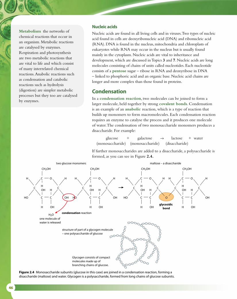
46
Nucleic acidsNucleic acids are found in all living cells and in viruses. Two types of nucleic acid found in cells are deoxyribonucleic acid (DNA) and ribonucleic acid (RNA). DNA is found in the nucleus, mitochondria and chloroplasts of eukaryotes while RNA may occur in the nucleus but is usually found mainly in the cytoplasm. Nucleic acids are vital to inheritance and development, which are discussed in Topics 3 and 7. Nucleic acids are long molecules consisting of chains of units called nucleotides. Each nucleotide consists of a pentose sugar – ribose in RNA and deoxyribose in DNA – linked to phosphoric acid and an organic base. Nucleic acid chains are longer and more complex than those found in proteins.
CondensationIn a condensation reaction, two molecules can be joined to form a larger molecule, held together by strong covalent bonds. Condensation is an example of an anabolic reaction, which is a type of reaction that builds up monomers to form macromolecules. Each condensation reaction requires an enzyme to catalyse the process and it produces one molecule of water. The condensation of two monosaccharide monomers produces a disaccharide. For example:
glucose + galactose → lactose + water(monosaccharide) (monosaccharide) (disaccharide)
If further monosaccharides are added to a disaccharide, a polysaccharide is formed, as you can see in Figure 2.4.
condensation reaction
glycosidic bond
one molecule ofwater is released
two glucose monomers
C
OH
HO
H
OH
H
C C
H
C
H
OH
C
CH2OH
H
O
C
OH
HO
H
OH
H
C C
H
C
H
OH
C
CH2OH
H
O
maltose – a disaccharide
C
OH
HO
H
O
H
C C
H
H
C
H
OH
C
CH2OH
H
O
C
OH
OH
H
C C
H
C
H
OH
C
CH2OH
H
O
H2O
structure of part of a glycogen molecule– one polysaccharide of glucose
Glycogen consists of compactmolecules made up ofbranching chains of glucose.
Figure 2.4 Monosaccharide subunits (glucose in this case) are joined in a condensation reaction, forming a disaccharide (maltose) and water. Glycogen is a polysaccharide, formed from long chains of glucose subunits.
Metabolism the networks of chemical reactions that occur in an organism. Metabolic reactions are catalysed by enzymes. Respiration and photosynthesis are two metabolic reactions that are vital to life and which consist of many interrelated chemical reactions. Anabolic reactions such as condensation and catabolic reactions such as hydrolysis (digestion) are simpler metabolic processes but they too are catalysed by enzymes.

2 MOLECULAR BIOLOGY 47
amino acid
one molecule ofwater is released
amino acid dipeptide
condensation reaction peptide bond
H2O
C CN
R
H
H
H
C CN
R O
H
C
R
H
N
H
H
H
O
OH
C CN
R
H
H
H
O
OH
C
O
OH
Figure 2.5 Two amino acids combine to form a dipeptide.
In a similar way, two amino acids can be linked to form a dipeptide (Figure 2.5):
amino acid + amino acid → dipeptide + water
When more than two amino acids are joined in this way, a polypeptide is formed. Polypeptide chains form protein molecules.
H
HC OH
O
C
O
C
O
C
O
fatty acidglycerol
one molecule of water isreleased for each fatty acidthat combines with glycerol
Each of the –OH groups in glycerol is able tocombine with the –COOH group of a fatty acid.
CHO H
HH
H
C
H
H
C
H
H
C
H
H
C
H
H
C
H
H
C
H
H
C
H
H
C
H
H
C
H
H
C
H
H
C
H
H
C
H
H
C
CHO H
CHO H
H
CO H
H
CO H
CO H
H
H2O
condensationreaction
triglyceride lipid molecule
ester bond
Figure 2.6 How a triglyceride lipid is formed from glycerol and three fatty acids in a condensation reaction.
In another condensation reaction, glycerol links to fatty acids to produce triglyceride lipid molecules (Figure 2.6):
glycerol + 3 fatty acids → triglyceride lipid + water

48
HydrolysisHydrolysis reactions occur every time food is digested. These reactions involve breaking down polysaccharides, polypeptides and triglycerides into the smaller units of which they are made. Hydrolysis is an example of a catabolic reaction, in which macromolecules are broken down into monomers. Water molecules are used in hydrolysis reactions which are the reverse of condensation reactions. Once again, enzymes are required to catalyse the reactions.
• Hydrolysis of starch (a polysaccharide) uses water and produces many molecules of glucose.
• Hydrolysis of protein (made of polypeptide chains) uses water and produces many amino acids.
• Hydrolysis of a triglyceride (a lipid) uses water and produces fatty acids and glycerol molecules.
Nature of scienceFalsification of theories – how the synthesis of urea helped to falsify the theory of vitalismVitalism is a belief that living things have a distinctive ‘spirit’ contained in their bodies, which gives them life. The theory proposes that living organisms are fundamentally different from non-living things because they contain this ‘life force’, and that the organic substances upon which life is built cannot be synthesised artificially from inorganic components. Vitalism dates back to Aristotle and Galen in the 3rd century BCE and also forms part of traditional healing that views illness as an imbalance in ‘vital forces’. Hippocrates associated vital forces with four ‘temperaments’ and in Eastern philosophy the imbalance was said to block the body’s ‘qi’.
Vitalism became less accepted from the 19th century as microscopy revealed the structure of cells and their components, and showed that they obeyed the laws of physics and chemistry. In addition, in 1828 Friedrich WÖhler successfully synthesised the organic molecule urea (Figure 2.7), thus disproving the vitalist theory, which held that organic substances could not be synthesised from inorganic components.
No modern discovery has disproved the observation that parts of an organism behave in a co-ordinated way and that this is different from the way the parts behave in isolation. For example, a single cell behaves differently when it is within an organism from the way it behaves when alone. In an organism it is in direct contact with other parts of the body, which in turn interact with other parts. Furthermore, no evidence has yet contradicted the laws of physical and chemical interaction between cellular components. So far, no observations suggest that we need another ‘force’ to account for biological phenomena. In the 21st century, vitalism is no longer a generally accepted idea.
H2N NH2
C
O
Figure 2.7 Urea is produced from the breakdown of amino acids in the mammalian liver. It is excreted in urine.

2 MOLECULAR BIOLOGY 49
Figure 2.8 The structure of a water molecule.
1 State the number of covalent bonds formed by a carbon atom.2 Define anabolism.3 State what is meant by the term ‘monomer’.
2.2 Water
Structure and properties of waterWater is the main component of living things. Most human cells are approximately 80% water. Water provides the environment in which the biochemical reactions of life can occur. It also takes part in and is produced by many reactions. Its many important properties described below are due to its molecular structure, which consists of two hydrogen atoms each bonded to an oxygen atom by a covalent bond (Figure 2.8).
Hydrogen bondsThe water molecule is unusual because it has a small positive charge on the two hydrogen atoms and a small negative charge on the oxygen atom. Because of this arrangement, water is said to be a polar molecule. Polar molecules are those that have an unevenly distributed electrical charge so that there is a positive region and a negative region. Sugars and amino acids are also polar molecules.
A weak bond can form between the negative charge of one water molecule and the positive charge of another, as shown in Figure 2.9. This type of bond, known as a hydrogen bond, is responsible for many of the properties of water.
Cohesion and adhesionHydrogen bonds between water molecules hold them together in a network, resulting in a phenomenon known as cohesion. Cohesive forces give water many of its biologically important properties. For example, they enable water to be drawn up inside the xylem of a plant stem in a continuous column. Strong pulling forces, produced as water evaporates from the leaves at the top of tall trees, draw water and dissolved minerals up great distances to the tips of branches high above the ground. Cohesion is also responsible for surface tension, which enables some small organisms to ‘walk on water’, and contributes to the thermal properties of water too.
Water also tends to be attracted and adhere to the walls of its container. There are forces of attraction, known as adhesive forces, which occur between water molecules and different molecules in vessels that contain the water. Adhesion attracts water molecules to the sides of the xylem and is important as water is drawn up the stem of a plant. Because adhesive
In a water molecule, the two hydrogenatoms are found to one side of theoxygen atom.
The oxygen atom pulls the bonding electronstowards it, which makes the oxygen slightlynegatively charged. The hydrogen atoms havesmall positive charges.
small negative charge
small positive charges
δ–
H H
O
δ+ δ+
Learning objectives
You should understand that:
• Hydrogen bonds form between water molecules, because they are polar.
• The cohesive, adhesive, thermal and solvent properties of water can be explained by hydrogen bonding.
• A substance may be classified as hydrophilic or hydrophobic depending on its solubility in water.
Test yourself?
covalentbonds
hydrogenbonds
δ–
δ–
δ–
δ–
δ+
δ+δ+
δ+
δ+
δ+
δ+
δ+
Figure 2.9 Hydrogen bonding in water.

50
forces are greater in a narrow tube where relatively more water molecules are in contact with the sides, adhesive forces are able to ‘hold up’ and support a substantial mass of water in the fine xylem vessels. The water column is held together by cohesive forces.
Thermal propertiesWater also has unusual thermal properties. It is unusual among small molecules because it is a liquid at a normal range of temperatures. A large amount of energy is needed to break the many weak hydrogen bonds between the water molecules. This gives water a high specific heat capacity – it can absorb or give out a great deal of heat energy without its temperature changing very much. A stable temperature is important to living things because the range of temperatures in which biological reactions can occur is quite narrow. The thermal properties of water allow it to keep an organism’s temperature fairly constant. Within the body, water can act as a temperature regulator – for example, water, which is a major component of blood, carries heat from warmer parts of the body such as the liver, to cooler parts such as the feet.
In order for liquid water to evaporate and become vapour, many hydrogen bonds between the molecules must be broken, so evaporation requires a lot of energy. As a result, water is a liquid at most temperatures found on Earth, and it has a high boiling point. When it evaporates, it carries a great deal of heat with it – so, for example, when sweat evaporates from the skin surface of a mammal it acts as a coolant for the body. The properties of water are summarised in Table 2.3.
Methane – a compound without hydrogen bonds
Methane is the smallest and simplest hydrocarbon, consisting of one carbon atom bonded to four hydrogen atoms (Figure 2.1). Unlike water, a methane molecule does not have hydrogen bonding between its H atoms and those of nearby molecules – as a result, very little energy is needed to separate its molecules, which move freely apart. Methane therefore exists as a gas at room temperature and standard pressure – its boiling point is −161°C. Water, on the other hand, is liquid at room temperature and standard pressure, even though its molecules are a similar size to those of methane. Its boiling point is much higher, at 100 °C, because of the large input of energy needed to break the many hydrogen bonds between its molecules, and convert it from a liquid to a vapour (gas).
Property Reason Consequence / Benefits to living organisms
cohesion Hydrogen bonds hold water molecules together. Water can travel in continuous columns – for example, in the stems of plants – and act as a transport medium.
adhesion Water molecules are attracted to other different molecules.
A column of water can be held up in the narrow xylem of a plant.
solvent The polar molecules of water can interact with other polar molecules.
Ions dissolve easily. Large molecules with polar side groups, such as carbohydrates and proteins, can also dissolve. So water acts as an excellent transport medium and as a medium for metabolic reactions.
thermal Water has a high heat capacity. Large amounts of energy are needed to break hydrogen bonds and change its temperature.
The temperature of organisms tends to change slowly. Fluids such as blood can transport heat round their bodies.
Water has a high boiling point compared with other solvents because hydrogen bonds need large amounts of energy to break them.
Water is liquid at most temperatures at which life exists, so is a useful medium for metabolic reactions.
Water evaporates as hydrogen bonds are broken and heat from water is used.
Sweating and transpiration enable animals and plants to lose heat. Water acts as a coolant.
Table 2.3 Summary of the properties of water.

2 MOLECULAR BIOLOGY 51
Solvent propertiesWater is sometimes known as a universal solvent. Its polarity makes it an excellent solvent for other polar molecules. Most inorganic ions, such as sodium, potassium and chloride ions, dissolve well as their positive or negative charges are attracted to the charges of water molecules (Figure 2.10). Polar organic molecules, such as amino acids and sugars, are also soluble in water. Water is the medium in which most biochemical reactions take place since almost all the substances involved dissolve well in it. Protein synthesis and most of the reactions of photosynthesis and respiration take place in an aqueous (water) solution.
The solvent properties of water also make it an excellent medium for transporting substances around the bodies of all organisms. In plants, the xylem carries dissolved minerals from the roots to the leaves, while the phloem transports soluble sugars up and down the plant. Many animals have blood as their transport medium. Blood is predominantly water, and the blood plasma carries many dissolved solutes including amino acids, sodium and chloride ions, sugars such as glucose, and carbon dioxide.
Substances are classified into two groups according to their solubility in water. Hydrophilic substances such as sugars and salts dissolve well, as do amino acids with polar side groups. Hydrophobic or ‘water-hating’ substances do not dissolve in water. They are usually uncharged, and examples include fats and oils, and large proteins that do not carry any polar groups.
Uncharged and non-polar substances are not very soluble in water because water molecules would rather remain hydrogen-bonded to each other than allow such molecules to come between them. Various gases, such as oxygen and carbon dioxide, are not very soluble in water because they are essentially non-polar. Oxygen can dissolve in water (and does so to sustain aquatic life) but its solubility is very low. Almost all oxygen carried in the blood is bound to hemoglobin, and not in solution. Most of the carbon dioxide in the blood, on the other hand, is carried in the form of bicarbonate ions (HCO3
–) dissolved in the plasma (Option D).Many large molecules such as lipids and proteins are also largely
non-polar but are sufficiently soluble to be carried through the aqueous environment of the blood because they have some polar groups exposed. On the outside of soluble proteins, for example, are polar groups that are able to interact with the polar water molecules and make the entire protein soluble.
Cholesterol is only slightly soluble in water and dissolves in the blood in very small amounts. For this reason, cholesterol is transported in the circulatory system within lipoproteins, which have an outer surface made up of amphipathic proteins and lipids. The outward-facing surfaces of these molecules are water-soluble (hydrophilic) and their inward-facing surfaces are lipid-soluble (lipophilic). Triglycerides (fats) are carried inside such molecules, while phospholipids and cholesterol, being amphipathic themselves, are transported in the surface layer of lipoprotein particles. There are different types of lipoproteins in blood – low-density
+
–
positive ion
negative ion
water molecules
Figure 2.10 The positive and negative charges of water molecules attract ions with negative or positive charges, which means that the ions dissolve.

52
Nature of scienceUsing theories to explain natural phenomena – hydrogen bonds and the properties of waterThe nature of liquid water and how the molecules in it interact are questions that have been studied by scientists for many years. Techniques including infrared absorption, neutron scattering and nuclear magnetic resonance imaging (NMRI) have been used to study the structure of water, and the results – along with data from theoretical calculation – have led to the development of a number of models, which try to describe the structure of water and explain its properties.
Water is a small, simple molecule in which each hydrogen atom is covalently bonded to the central oxygen atom by a pair of electrons that are shared between them (Figure 2.11). Only two of the six outer-shell electrons of each oxygen atom are used to form these covalent bonds, leaving four electrons in two non-bonding pairs. These non-bonding pairs remain closer to the oxygen atom and exert a strong repulsion against the two covalently bonded pairs so that the two hydrogen atoms are pushed closer together. Overall, water molecules are electrically neutral, but this model of the water molecule results in small positive and negative charges unevenly distributed over the molecule. When the H2O molecules are crowded together in liquid water, the forces between the atoms produce the effects that give water the properties we observe.
lipoprotein (LDL) and high-density lipoprotein (HDL). The more lipid and less protein a lipoprotein has, the lower its density.
Memory of waterThe ‘memory of water’ is a phrase that is usually associated
with homeopathy. It was coined by Jacques Benveniste (1935–2004) who claimed that water retains a ‘memory’ of substances that have once been dissolved in it. Homeopathic remedies are prepared by diluting solutions to such an extent that, in some cases, no molecules of the original solute are found in them. Nevertheless, healing effects are claimed for these remedies, based on the idea that the water retains properties from the original substance. There is no scientific evidence to support the claim that water has such a ‘memory’ and the subject has drawn a lot of controversy, with many scientists rejecting it completely.
Question to consider
• What criteria can be used to distinguish scientific claims from pseudoscientific claims?

2 MOLECULAR BIOLOGY 53
Scientists continue to test theories and refine their models, deepening our understanding of the world around us. About 50 years ago it was assumed that water consisted of hydrogen-bonded clusters aggregated together, but more recent evidence from modelling does not support this view. Present-day thinking based on computer-generated molecular modelling is that, for very short time periods of time (less than a picosecond), water has a gel-like structure that consists of a single, huge hydrogen-bonded cluster.
The most stable state for an atom is to have 8 electrons in its outer shell. In oxygen, the outer shell contains 6 electrons. In a water molecule, two of these electrons pair up with the single electron of a hydrogen atom, so that there are effectively 8 electrons in the outer shell.
electrons in the outer shell of an
oxygen atom
two hydrogen atoms,each with one electron
Where a pair of electrons is shared between two atoms, the atoms are held together in a strong covalent bond.
Each oxygen atom has 8 electrons in total, which we can imagine orbiting the nucleus in concentric ‘shells’. The 6 electrons in the ‘outer shell’ are the ones involved in bonding with other atoms.
O
H H
Figure 2.11 The two hydrogen atoms in a water molecule are pushed together on one side because of the repulsive effect of the two pairs of non-bonding electrons in the outer shell of the oxygen atom.
4 Explain why water makes a good coolant for animals.5 Define the term ‘hydrophilic’.6 Explain how hydrogen bonding affects the force of cohesion.
Test yourself?

54
2.3 Carbohydrates and lipids
CarbohydratesCarbohydrates are the most abundant category of molecule in living things. Different types of carbohydrate are produced by linking together monosaccharide monomers to build up polymers. The condensation reactions involved in this process result in the production of either disaccharides, which consist of two monomers, or polysaccharides, formed from long chains of monosaccharide monomers (Figure 2.4). The covalent bond between two monomers in a carbohydrate is known as a glycosidic link and a water molecule is released in the condensation reaction.
Glucose is the most common monosaccharide and it has the chemical formula C6H12O6. The structures of alpha-D-glucose and beta-D-glucose are shown in Figure 2.2. These two forms of the molecule (known as isomers) have slightly different arrangements of the side groups, giving them slightly different properties.
When a bond is formed between two glucose monomers, a disaccharide called maltose is produced. Maltose is found in seeds such as barley. Other monosaccharides include fructose, found in fruits, and galactose, which is present in milk. Different combinations of these monomers produce a range of disaccharides, which are shown in Table 2.4.
Form of carbohydrate Examples Example of use in plants Example of use in animals
monosaccharide glucose, galactose, fructose
fructose is a component of fruits, making them taste sweet and attracting animals to eat them, thereby dispersing the seeds inside
glucose is the source of energy for cell respiration – it is obtained from the digestion of carbohydrate foods
disaccharide maltose, lactose, sucrose
sucrose is transported from leaves to storage tissues and other parts of the plant to provide an energy source
lactose is found in milk and provides energy for young mammals
polysaccharide starch, glycogen, cellulose
cellulose is a structural component of plant cell wallsstarch is used as a food store
glycogen is the storage carbohydrate of animals, found in the liver and muscles
Table 2.4 Examples and roles of carbohydrates.
Learning objectives
You should understand that:
• Monosaccharides are linked together by condensation reactions to form polymers such as disaccharides and polysaccharides.
• A fatty acid molecule may be saturated, monounsaturated or polyunsaturated.
• Unsaturated fatty acids occur in two forms, or isomers: either cis or trans.
• Triglycerides are formed by the condensation of three fatty acid molecules and one glycerol molecule.
Polysaccharides may contain from 40 to over 1000 monomers. Starch, glycogen and cellulose are all polymers of glucose monomers. Glycogen and starch are storage carbohydrates – glycogen in animals and starch in plants. They both have a compact shape and are insoluble. Glycogen is made of branching chains of glucose monomers (Figure 2.4) while starch has long chains of glucose that coil into a helical shape (Figure 2.12). Cellulose is used to build the cell walls of plants and is made up of long, straight chains of glucose molecules. The arrangement of the molecules means that hydrogen bonds can form cross-links between adjacent, parallel chains, which gives the polysaccharide its structural properties (Figure 2.12).

2 MOLECULAR BIOLOGY 55
LipidsFats and oils are one of the two main groups of lipid and are compounds of glycerol and fatty acids (Figure 2.6). Glycerol has just one structural form but fatty acids have a wide variety of structures, which give the lipids that contain them their different physical and chemical properties. These molecules have important roles in the bodies of plants and animals and these are summarised in Table 2.5), with the properties of lipids that make them suitable for these functions.
Starch Starch is made up of alpha-glucose units, linked by 1–4 glycosidic bonds, which causes the molecule to form a helical shape.
CelluloseCellulose is made up of straight chains of beta-glucose units, with OH groups forming hydrogen bonds between chains.
OH OH OH OH
OH OH OH OH
OH OH OH OH
OH OH OH OH
OH OH OH OH
OH OH OH OH
The hydrogen bonding between chains in cellulose causes the formation of strong, straight fibres.
There are two forms of starch. Amylose contains only 1–4 glycosidic bonds and forms linear helices.Amylopectin also contains some 1–6 glycosidic bonds, which causes branching. amylopectin amylose
CH2OH
O
O
OHH
HOH
CH2 OHOOH
HH
OH
CH2 OH
O
O
O
OHHH
OH
CH 2OH
OO
O
OHH
H
OHCH2O
HO
OHH
HOH
CH2OH
O
O
OH H
HOH
O
Figure 2.12 Starch and cellulose are two polysaccharides found in plants.
Key properties of lipids
energy content Lipids contain more energy per gramme than carbohydrates, so lipid stores are lighter than carbohydrates storing an equivalent amount of energy.
density Lipids are less dense than water, so fat stores help large aquatic animals to float.
solubility Lipids are non-polar, insoluble molecules so they do not affect the movement of water in and out of cells by osmosis.
insulation Lipids are also important in providing heat insulation. Fat stored under the skin reduces heat loss and is vital for animals such as seals, polar bears and whales, which live in cold conditions.
Table 2.5 The important properties of lipids that suit them to particular roles in living organisms.

56
Fatty acidsFatty acids consist of a long chain of carbon atoms that are joined to hydrogen atoms (Figure 2.6). Chains like this are called hydrocarbon chains. The number of carbon atoms in a fatty acid is usually an even number most commonly between 14 and 22 although shorter and longer chain fatty acids are found. At one end of the chain is a carboxyl group (COOH) and at the other a methyl (CH3) group. If the carbon chain is linked to the maximum number of H atoms with no double bonds it is said to be saturated because no more H atoms can be added. If the chain contains a double bond between two of the carbon atoms it is said to be unsaturated (Figure 2.13). A chain with just one double bond is monounsaturated, while one with two or more double bonds is said to be polyunsaturated. Polyunsaturated fatty acids tend to be liquids at 20° C and are mainly derived from plant sources. Examples are sunflower oil, corn oil and olive oil.
Unsaturated fatty acids may be either a cis or a trans configuration. If the ‘spaces’ where additional hydrogen atoms could bond are both on the same side of the hydrocarbon chain, the fatty acid is known as a cis fatty acid and the carbon chain is slightly bent. If the spaces are on opposite sides, it is a trans fatty acid, which has a straight chain (Figure 2.14).
saturated fatty acid
unsaturated fatty acid
every carbon atom in the hydrocarbon chain hasthe maximum number of hydrogen atoms bonded
this hydrocarbon chain includes three double bonds,which means the carbon atoms do not have themaximum number of hydrogen atoms bonded
CH
H
H
C
H
H
C
H
H
C
H
H
C
H
H
C
H
H
C
H
H
C
H
H
C
H
H
C
H
H
C
H
H
O
OH
C
C
H
H
H
C
H
C
H
C
H
H
C
H
C
H
C
H
H
C
H
C
H
C
H
HH
C
H
O
OH
C
Figure 2.13 Saturated and polyunsaturated fatty acids.
straight trans fatty acid – one hydrogen atom isabsent from each side of the hydrocarbon chain
bent cis fatty acid – two hydrogen atoms areabsent from the same side of the hydrocarbon chain
C
H
H
C
H
H
C
H
H
C
H
C
H
C
H
H
C
H
H
C
H
H
C
H
H
C
H
H
C
H
H
C
H
C C
H
H
C
H
H
H
C
H
H
Figure 2.14 Cis and trans fatty acids.

2 MOLECULAR BIOLOGY 57
One type of cis fatty acid is the omega-3 group. These have a double bond at the third bond from the CH3 end of the molecule. Omega-3 fatty acids in our diet come from eating fish such as salmon and pilchards, and from walnuts and flax seeds. Another group, the omega-6 fatty acids, have a double bond at the sixth position and come from vegetable oils.
The relative amounts of different types of fatty acid in a person’s diet can, in some cases, be correlated with health issues. Eating a diet that is high in saturated fatty acids, such as is prevalent in some western countries, has been shown to have a positive correlation with an increased risk of coronary heart disease (CHD). Saturated fatty acids can be deposited inside the arteries, and if the deposits combine with cholesterol they may lead to atherosclerosis, a condition that reduces the diameter of the arteries and leads to high blood pressure (Subtopic 6.2). Reliable evidence suggests that in countries where the diet is high in saturated fatty acids and many high-fat foods, animal products and processed foods are eaten there is likely to be a high incidence of CHD. Since all fatty acids are high in energy, an excess of these foods in the diet can also lead to obesity, which places a further strain on the heart.
On the other hand, people who eat a Mediterranean-style diet, rich in unsaturated fatty acids from olive oil and fresh vegetables, tend to have a low incidence of CHD. These fats do not combine with cholesterol and so arteries tend to remain unblocked and healthy.
Some polyunsaturated fats are modified or ‘hydrogenated’ so they can be used in processed foods. These hydrogenated fats become trans fatty acids. There is a positive link between a high intake of these trans fatty acids and CHD, but many other factors must also be considered.
In animals, omega-3 fatty acids are used to synthesise long-chain fatty acids found in the nervous system. It has been suggested that a lack of omega-3 fatty acids could affect brain and nerve development but no conclusive evidence has yet been found.
Health issues – trans fatty acids and saturated fat
Artificial trans fats are formed when vegetable oil is hydrogenated, a process that solidifies the oil. The substance produced is known as hydrogenated fat and can be used for frying or in the manufacture of processed foods. Trans fats are included in biscuits and cakes to extend their shelf life but in recent years many food manufacturers have removed trans fats from their products.
Eating a diet containing high levels of trans fats has been shown to lead to high cholesterol levels, which in turn can lead to CHD and strokes. But most people do not eat large amounts of trans fats. In the UK, for example, it has been estimated that most people eat only about half the maximum recommended level of these fats. Most health professionals advise that saturated fats are a greater risk to health because of their contribution to atherosclerosis.
Question to consider
• How do we decide between different views about the relative harms and benefits of foods in our diets?
58
TriglyceridesTriglycerides are formed by condensation reactions between three fatty acids and one glycerol molecule (Figure 2.6). Three molecules of water are released and the bonds between the fatty acids and the glycerol are known as ester bonds. Triglycerides play a major role in the structure of membranes (Subtopic 1.3) when they combine with a phosphate group in the form of phosphoric acid to form phospholipids. The phosphoric acid combines with one of the three OH (hydroxyl) groups of glycerol and two fatty acid chains attach to the other two (Figure 1.16).
Nature of scienceEvaluating claims – cause and correlation in health statisticsLooking for correlation is one of the most common and useful statistical analysis techniques. Correlation describes the degree of relationship between two variables. For example, in the last 30 years, the number of people taking a holiday each year has increased. In the last 30 years, there has also been an increase in the number of hotels at holiday resorts. This data shows a positive correlation.
We could also consider annual deaths from influenza and the number of influenza vaccines given. In this case, there is a negative correlation. With these examples, we might feel safe to say that one set of data is linked to the other and that there is a causal relationship – because there are more tourists, more hotels have been built; greater use of the influenza vaccine has resulted in fewer deaths from influenza.
However, just because the data shows a trend, it does not necessarily mean that there is a causal relationship. If we consider the number of people using mobile phones in the last 10 years against the area of Amazon rainforest cut down there would be a positive correlation. But this does not mean that the use of mobile phones has caused rainforest to be cut down – nor does it mean that a reduction in rainforest area results in more mobile phone use.
Observations without experiments can show a correlation but usually experiments must be used to provide evidence to show the cause of the correlation. It is not ethically possible to conduct experiments to find evidence for correlation between diet and human health. We cannot restrict different groups of subjects to diets containing different amounts of saturated fats to assess the effects on their health and so observational or epidemiological data is all we have to go on. We must think about how the data is gathered and what other variables, such as lifestyle and genetics and family history, are important. There are difficulties in collecting objective data that accounts for all possible variables – indeed, it may never be possible to say that one type of diet or fatty acid is ‘good’ and another is ‘bad’ because individual subjects vary in so many different ways.
Correlation and cause
When studying the occurrence of medical conditions that may be related to diet, it is important to distinguish between correlation and cause. A correlation between two variables, such as a high incidence of CHD and a high intake of saturated fatty acids, does not mean that the CHD is caused by the fat intake.
7 State two examples of disaccharides.8 Outline the difference between a saturated and an
unsaturated fatty acid.
9 State the type of reaction that leads to the formation of triglycerides.
Test yourself?

2 MOLECULAR BIOLOGY 59
2.4 Proteins
PolypeptidesPolypeptides are built up from amino acid monomers during condensation reactions (Figure 2.5). Two amino acids are joined with a reaction between the amino (NH2) group of one amino acid and the carboxyl (COOH) group of the other forming a peptide bond and producing a dipeptide. If further condensation reactions occur, a series of amino acids can become joined to form a polypeptide. The covalent bonds linking the amino acids produce what is known as the primary structure of any protein that is formed from the polypeptide.
In living cells, polypeptides are synthesised by ribosomes in the cytoplasm. Twenty different amino acids are used to construct polypeptides. Other amino acids do exist but these are not used in the biosynthesis of protein. Polypeptides can consist of up to 400 amino acids and, because these can be linked together in any sequence, there is a huge range of possible polypeptides. Some amino acids may also be modified once the polypeptide has been incorporated into a protein molecule so that even more different structures can be formed.
The sequence of amino acids in a polypeptide is coded for by an organism’s genes. Genes consist of a series of codons, each of which carries the specific code for one amino acid (Subtopic 2.7). The sequence of codons in a gene is used as a template to direct the sequence in which the amino acids will be assembled.
Building a proteinA protein may either consist of one polypeptide or several linked together. The basic chain of amino acids in a polypeptide folds and becomes a 3D shape once it is complete. The shape, known as secondary structure, results from the formation of hydrogen bonds between different parts of the chain. The most common shape is an alpha helix, held together by weak hydrogen bonds between the amino acids that form the turns in the structure. Keratin, a structural protein found in hair, is an alpha helix. In other proteins, such as silk, polypeptides in parallel chains are linked to form flat, folded shapes known as beta pleated sheets.
Further folding of polypeptides can occur due to the interactions between the R groups of the amino acids present. A complex three-dimensional shape known as tertiary structure results and is held together by ionic bonds and disulfide bridges. Tertiary structure is important in enzymes because the shape of the molecule determines where substrate molecules can bind to them.
Some proteins are composed of two or more polypeptides linked together and are said to have quaternary structure. The pigment hemoglobin found in red blood cells has four subunits and the positioning of these subunits is very important for the role of hemoglobin in carrying oxygen (Figure 7.20, Subtopic 7.3).
Learning objectives
You should understand that:
• Amino acids are linked in condensation reactions to form polypeptide chains.
• Twenty different amino acids found in polypeptides, which are synthesized by ribosomes.
• Because amino acids can be linked in any sequence it is possible to make a huge range of different polypeptides.
• The sequence of amino acids in a polypeptide is determined by the genetic code.
• Proteins consist of one or more polypeptides linked together.
• The sequence of amino acids in a polypeptide determines the three-dimensional shape of a protein.
• Living things synthesise many different proteins with many different functions.
• Every individual organism has a unique proteome.

60
DenaturationDenaturation destroys the complex structure of a protein. A protein’s structure is determined by the different types of bonds it contains. Heat or the presence of strong acids or alkalis can all disturb the bonds between the different parts of a protein molecule and disrupt its structure. The primary structure of the protein will remain but secondary, tertiary and quaternary structures are usually lost.
A denatured protein has different properties from the origin molecule. For example, enzymes are easily denatured by extremes of pH or temperature (Subtopic 2.5) and lose the ability to function as catalysts. Some proteins lose their solubility or aggregate to form clumps as they denature and this can be observed during cooking. The heat used to cook meat denatures the proteins found in it so that its texture is changed, and eggs become hardened as they cook and their proteins are denatured.
Functions of proteinsThe function of a protein is determined by the shape of its molecule. Proteins are divided into two main types: globular and fibrous proteins.
Fibrous proteins are long, insoluble molecules made up of parallel polypeptide chains. The chains are cross-linked along their lengths. Keratin found in hair, nails and hooves and collagen found in bones, muscles and tendons are two abundant fibrous proteins.
Globular proteins have polypeptide chains that are folded into compact, almost spherical shapes. The hormone insulin is a globular protein and so are enzymes. Each enzyme has its own 3D shape, which enables it to work as a catalyst for a particular reaction.
Examples of some important proteins and their functions are summarised in Table 2.6.
As Table 2.6 shows, a wide range of proteins is found in different organisms and each protein has its own structural or biochemical function. Every individual organism has its own unique proteins, which are determined by its unique genome. The proteins found in an organism are known as its proteome, a term derived from a combination of the words ‘protein’ and ‘genome’.
Protein Function
Rubisco an enzyme involved in carbon fixation in photosynthesis
insulin a hormone produced by the pancreas, which stimulates the liver to take up glucose from the blood and store it as glycogen
immunoglobulin a large Y-shaped protein (antibody) produced by the immune system to fight infection
rhodopsin a protein linked to a pigment found in the photoreceptor cells in the retina of the eye
collagen a structural protein which builds muscle, tendons, ligaments and the skin of vertebrates
spider silk a protein fibre spun by spiders, which is tough and elastic and used for constructing a spider’s web
Table 2.6 The function of some different proteins.

2 MOLECULAR BIOLOGY 61
Nature of scienceLooking for trends and discrepancies – do all organisms use only 20 amino acids?Humans can produce 10 of the 20 amino acids we need to build proteins but we do not have the enzymes needed for the biosynthesis of the others. Plants and other autotrophs, on the other hand, must be able to make all the amino acids they require.
Researchers have investigated the trends in amino acid compositions of proteins found in species of the important kingdoms of Archaea, Bacteria and Eukaryotes. The international databases ‘Proteomes’ and ‘Swiss-Prot’ (which contain information about the structure and composition of proteins) can be used to compare amino acid frequencies for 195 known proteomes and all recorded sequences of proteins. Such comparisons have shown that the amino acid compositions of proteins do differ substantially for different kingdoms.
In addition to variations in amino acid sequence in proteins, some microorganisms and plants are able to make so called ‘non-standard’ amino acids by modifying standard amino acids. Some species are also able to synthesise many uncommon amino acids. For example, some microbes synthesise lanthionine, which is a modified version of the amino acid alanine. Many other proteins are modified after they have been produced. This ‘post-translational modification’ involves the addition of extra side groups to the amino acids in a protein.
Considering all the evidence, it seems that, although we can observe many similar proteins in different species, we cannot always say that the same amino acids are used in their construction. The range of amino acids in proteins can vary considerably from species to species.
10 Distinguish between hydrolysis and condensation reactions. 11 State how the amino acid sequence in polypeptide is
determined. 12 Define the term ‘proteome’.
Test yourself?

62
2.5 Enzymes
Enzymes and active sitesAn enzyme is a biological catalyst. Like all catalysts, enzymes speed up biochemical reactions, such as digestion and respiration, but they remain unchanged at the end of the process. All enzymes are proteins with long polypeptide chains that are folded into 3D shapes. The arrangement of these shapes is very precise and gives each enzyme the ability to catalyse one specific reaction. If the 3D shape of an enzyme is destroyed or damaged, it can no longer carry out its job and is said to be denatured. Extremes of temperature, heavy metals and, in some cases, pH changes can cause permanent changes in an enzyme.
The three-dimensional shape of an enzyme is crucial to the way it works. In the structure of every enzyme is a specially shaped region known as an active site (Figure 2.15). It is here that the substrate and enzyme bind together. The substrates are the chemicals involved in the reaction catalysed by the enzyme. The shapes of the enzyme and substrate are complementary, so that they fit together perfectly like a key fits into a lock. The ‘lock-and-key hypothesis’ is a way of explaining how each enzyme can be so specific. To unlock a door requires just one special key. To catalyse a reaction requires one special enzyme. Just as only one key fits perfectly into the lock, only one substrate fits perfectly into the active site of an enzyme.
Enzyme a globular protein that functions as a biological catalyst of chemical reactionsDenaturation irreversible changes to the structure of an enzyme or other protein so that it can no longer functionActive site region on the surface of an enzyme molecule where a substrate molecule binds and which catalyses a reaction involving the substrate
active site
substrate
enzyme
enzymeenzyme–substratecomplex
substrateenzyme
products
active site +
a An enzyme has a cleft in its surface called the active site. The substrate molecule has a complementary shape.
b Random movement of enzyme and substrate brings the substrate into the active site. An enzyme–substrate complex is temporarily formed. The R groups of the amino acids in the active site interact with the substrate.
c The interaction of the substrate with the active site breaks the substrate apart. The two product molecules leave the active site, leaving the enzyme molecule unchanged and ready to bind with another substrate molecule.
enzyme–substrate complex products
Figure 2.15 How an enzyme catalyses the breakdown of a substrate molecule into two product molecules.
Learning objectives
You should understand that:
• Enzymes have an area on their molecule, known as the active site, to which specific substrates bind.
• During enzyme-catalysed reactions, molecules move about, and substrate molecules collide with the active sites on enzyme molecules.
• The rate of enzyme activity is influenced by temperature, pH and substrate concentration.
• Enzyme molecules can be denatured.
• Immobilised enzymes are used in many industrial processes.
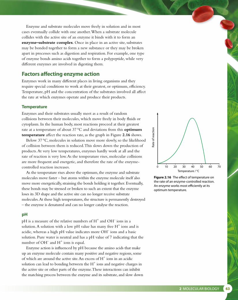
2 MOLECULAR BIOLOGY 63
Enzyme and substrate molecules move freely in solution and in most cases eventually collide with one another. When a substrate molecule collides with the active site of an enzyme it binds with it to form an enzyme–substrate complex. Once in place in an active site, substrates may be bonded together to form a new substance or they may be broken apart in processes such as digestion and respiration. For example, one type of enzyme bonds amino acids together to form a polypeptide, while very different enzymes are involved in digesting them.
Factors affecting enzyme actionEnzymes work in many different places in living organisms and they require special conditions to work at their greatest, or optimum, efficiency. Temperature, pH and the concentration of the substrates involved all affect the rate at which enzymes operate and produce their products.
TemperatureEnzymes and their substrates usually meet as a result of random collisions between their molecules, which move freely in body fluids or cytoplasm. In the human body, most reactions proceed at their greatest rate at a temperature of about 37 °C and deviations from this optimum temperature affect the reaction rate, as the graph in Figure 2.16 shows.
Below 37 °C, molecules in solution move more slowly, so the likelihood of collision between them is reduced. This slows down the production of products. At very low temperatures, enzymes hardly work at all and the rate of reaction is very low. As the temperature rises, molecular collisions are more frequent and energetic, and therefore the rate of the enzyme-controlled reaction increases.
As the temperature rises above the optimum, the enzyme and substrate molecules move faster – but atoms within the enzyme molecule itself also move more energetically, straining the bonds holding it together. Eventually, these bonds may be stressed or broken to such an extent that the enzyme loses its 3D shape and the active site can no longer receive substrate molecules. At these high temperatures, the structure is permanently destroyed – the enzyme is denatured and can no longer catalyse the reaction.
pHpH is a measure of the relative numbers of H+ and OH– ions in a solution. A solution with a low pH value has many free H+ ions and is acidic, whereas a high pH value indicates more OH– ions and a basic solution. Pure water is neutral and has a pH value of 7 indicating that the number of OH– and H+ ions is equal.
Enzyme action is influenced by pH because the amino acids that make up an enzyme molecule contain many positive and negative regions, some of which are around the active site. An excess of H+ ions in an acidic solution can lead to bonding between the H+ ions and negative charges in the active site or other parts of the enzyme. These interactions can inhibit the matching process between the enzyme and its substrate, and slow down
0 10 20 30 40
Temperature / °C
Rate
of r
eact
ion
50 60 70
Figure 2.16 The effect of temperature on the rate of an enzyme-controlled reaction. An enzyme works most efficiently at its optimum temperature.

64
or even prevent enzyme activity. A similar effect occurs if a solution becomes too basic – the excess of negative ions upsets the enzyme in the same way. At extremes of pH, the enzyme may even lose its shape and be denatured.
Not all enzymes have the same optimum pH. Proteases (protein-digesting enzymes) in the stomach have an optimum pH of 2 and work well in the acidic conditions there, but proteases in the small intestine have an optimum of pH 8. Most enzymes that work in the cytoplasm of body cells have an optimum pH of about 7. The graph in Figure 2.17 shows how reaction rate varies with pH for this type of enzyme.
Concentration of substrateIf there is a set concentration of enzyme present in a reaction mixture, and the concentration of substrate increases, the rate of production of the products will increase because of the greater chance of collisions between substrate and enzyme molecules. More collisions mean that the enzyme is able to process or ‘turn over’ more substrate molecules. But there is a limit to this increase in reaction rate. If the concentration of substrate increases too much, it will exceed the maximum rate at which the enzyme can work. When this happens, at any one moment all the active sites are occupied by substrate or product molecules, and so adding further substrate has no effect. The rate reaches its limit – you can see this as the plateau in the graph in Figure 2.18.
Enzymes in industryEnzymes work as catalysts in biological reactions but they are not used up so they can work over and over again. People have used enzymes from microorganisms for thousands of years in baking, cheese production and brewing. Today, many microbes are used as a source of enzymes for a wide range of industrial processes. Enzymes from fungi are used to produce biological detergents and in the textile industry to smooth fabrics. Bacterial enzymes are used in the leather industry to soften hides, in brewing and in the production of medicines.
Industrial enzymes are mainly isolated from microbes that are grown on a large scale in fermenters. The enzymes are separated from the microbial culture and purified before they are used. Once enzymes have been produced they can be used in processes such as those shown in Table 2.7. Using purified enzymes improves the efficiency of reactions and makes it easier to provide optimum conditions for enzyme activity.
Substrate concentration
Maximumrate
Rate
of r
eact
ion
Figure 2.18 The effect of substrate concentration on the rate of an enzyme-catalysed reaction.
0 1 3 5 7pH
Rate
of r
eact
ion
9 11 13
Figure 2.17 The effect of pH on the rate of an enzyme-controlled reaction.

2 MOLECULAR BIOLOGY 65
Immobilised enzymesFor the greatest efficiency in producing the required products, enzymes are ‘immobilised’ by attaching them to a substance that remains stationary in a column or other container. This method allows the enzyme to be retained so that it can be re-used many times with fresh batches of substrate.
Enzymes are immobilised by adsorption on to solid resins, charcoal or similar substances, or by bonding them to collagen or synthetic polymers. Another method that can be used, and one which is easy to replicate on a laboratory scale, is to enclose the enzyme in inert, insoluble calcium alginate beads. In this method, the enzyme is mixed with a suspension of sodium alginate and calcium chloride. As the mixture turns from a liquid to a gel, it can be formed into small spheres, which contain the enzyme. The alginate beads are usually enclosed in a vertical column so that substrate can be poured in at the top and products collected at the base (Figure 2.19). The size of the beads and the rate of flow of the substrate through them are crucial factors that determine the maximum enzyme efficiency and production of products. The substrate must be able to diffuse freely through the beads and in and out of the lattice that they form. The rate of flow of the substrate is very important to optimise the rate of reaction. Enzyme and substrate must remain in contact for sufficient time for reactions to occur but not too long so that the production process is too slow to be economically viable.
Advantages of using immobilised enzymes include:
• ease of harvesting the product – after the reaction the output usually contains only solvent and products
• ease of recovering the enzyme – the immobilised enzyme can easily be collected and re-used
• continuous production is possible – immobilised enzymes are more stable and less sensitive to variation in temperature than enzyme solutions
• immobilisation extends production time especially when using proteases, which might digest each other in reactions in solution.
Industry Enzymes Uses
biological detergents protease and amylase removal of protein and starch stains
baking amylase allows continuous dough production, converts starch to sugar
protease breaks down gluten to produce gluten-free products
biosensors glucose oxidase testing for glucose in blood samples
dairy rennin forms curd in cheese production
lactase production of lactose-free foods
confectionery invertase smoothing agent in confectionery production
medicine streptokinase treating bruises and blood clots
fruit juice production cellulase and pectinase speed the extraction of juice from fruit and prevent cloudiness
Table 2.7 Some industrial applications of enzymes.
substrate
screwclip
product
nylon gauzeor muslin
syringe containingimmobilised enzyme beads
Figure 2.19 The alginate beads are enclosed in a 20 cm3 syringe in a laboratory demonstration, such as that shown here. An industrial process uses the same principles but on a much larger scale.

66
Production of lactose-free milkLactose-free milk is produced using immobilised enzymes. Milk contains the sugar lactose, which is digested in the intestine by an enzyme called lactase. This produces two simple sugars (glucose and galactose) that can be absorbed into the body. Lactase can be obtained commercially from yeast that grows in milk and this enzyme is used to produce lactose-free milk.
Lactose-free milk is useful because some people are lactose intolerant and cannot digest lactose properly. If they take more than a small amount of milk, they suffer symptoms such as cramps or diarrhoea. Milk that contains glucose and galactose rather than lactose also tastes sweeter and so manufacturers need to add less sweetener to milk products such as yogurt made with lactose-free milk. Glucose and galactose are also more soluble than lactose, so they produce smoother textures in dairy products such as ice cream.
Nature of scienceExperimental design – accuracy and reliabilityThe accuracy of each measurement in any experiment depends on the quality of the measuring apparatus and the skill of the scientist taking the measurement. Faulty or inappropriate apparatus, or mistakes in using it, can produce inaccurate measurements. The level of accuracy of a measuring instrument determines the closeness of its measurement to the quantity’s true value. For example, a micrometer measures length to a greater level of accuracy than a ruler.
For the data to be reliable, the variation within the values of the results must be small. There is always some variation in any set of measurements, whatever is being measured. These may be due to small variations in the substances used in an experiment or the way the measuring apparatus is used.
When designing experiments with immobilised enzymes, such as the one shown in Figure 2.19, accurate measurement of volumes of enzyme solutions, alginate and calcium chloride are important in producing the alginate beads. For example, it would not be appropriate to use a 20 cm3 syringe to measure 2 cm3 of solution because the degree of error would be too great. The smallest possible syringe should be used so that the error is ±0.05 cm3, and not ±0.5 cm3.
Also important in all experimental design is reliability. Reliable results are those that can be accepted as being trustworthy, with no readings that are erroneous. Unreliable data may be due to misreading a stopwatch or a probe or recording a value incorrectly. To minimise the risk of errors like these it is important to take a number of readings (replicates) of the results for each value of the controlled (independent) variable. Three similar results can usually be accepted as reliable and an average value calculated from them. But if the results differ widely, further replicates must be carried out. Repeating the whole experiment with a different batch of the same chemicals is a good way of testing reliability further – if the repeat experiment produces very similar results, they can be said to be reliable.

2 MOLECULAR BIOLOGY 67
Development of techniques that benefit some people more than others
The distribution of lactose intolerance around the globe shows considerable variation. Only 4% of the Scandinavian population is affected, while countries around the Mediterranean have incidences of the order of 50–75% and in Africa the figure reaches 80%. Asia is affected even more with about 90% of the population suffering from lactose intolerance.
The control of lactase production by the human digestive system was disputed by scientists for many years. Some researchers in the 1960s argued that lactase production was stimulated in the presence of its substrate, lactose from milk. They proposed that populations that did not use milk as adults lost the ability to produce lactase, whereas groups that did consume milk continued to make the enzyme. More recent studies have cast doubt on this theory and shown that lactase production is controlled by a gene that is located on chromosome 2.
Development of lactose-free products, which required significant financial investment, occurred in western, developed countries despite the fact that the benefits were more widely applicable in other parts of the world.
Questions to consider
• Should scientific knowledge always be shared, even if it involved significant financial investment, and there are likely to be substantial rewards in keeping control of it?
• Should techniques developed in one part of the world be freely shared when they are more applicable to another?
• How could international cooperation benefit the efforts to combat diseases such as malaria?
13 Define the ‘active site’. 14 Outline the effect of increasing temperature on an enzyme-
catalysed reaction. 15 State one use of immobilised enzymes in industry.
Test yourself?

68
2.6 Structure of DNA and RNADNA (deoxyribonucleic acid) molecules make up the genetic material of living organisms. DNA is an extremely long molecule but, like proteins and carbohydrates, it is built up of many monomer subunits. The subunits of DNA are called nucleotides. RNA (ribonucleic acid) is also built up of many nucleotides but these differ from DNA nucleotides in the type of pentose sugar they contain and the bases that are attached to them.
Each nucleotide consists of three parts – a pentose (five-carbon) sugar (deoxyribose or ribose), a phosphate group and a nitrogenous base (Figure 2.20). DNA contains four different bases: adenine, guanine, cytosine and thymine. These are usually known by their initial letters: A, G, C and T (Figure 2.21). RNA also contains four bases but in an RNA molecule cytosine is not present and is replaced by uracil (U).
To form a DNA molecule, nucleotide monomers are linked together. The phosphate group of one nucleotide links to the deoxyribose of the next molecule to form a chain of nucleotides, as shown in Figure 2.21. The sugar and phosphate groups are identical all the way along the chain and form the ‘backbone’ of the DNA molecule. The sequence of bases in the chain will vary and it is this sequence that forms the genetic code determining the characteristics of an organism.
Two strands of nucleotides are linked by hydrogen bonds that form between the bases and this two-stranded structure makes up the double helix of a complete DNA molecule. Adenine always pairs with thymine and is bonded with two hydrogen bonds, while cytosine is paired with guanine by three hydrogen bonds. The arrangement is known as complementary base pairing. Notice that the two DNA chains run in opposite directions and are said to be antiparallel.
You can imagine the molecule rather like a rope ladder with the sugar–phosphate backbone being the sides of the ladder and the rungs being formed by the hydrogen-bonded base pairs. To form the characteristic double helix of a DNA molecule, the ladder must be twisted to resemble a spiral staircase.
To form a molecule of RNA, nucleotide monomers are linked in a similar way to those of DNA. In the case of RNA the molecule remains single stranded and the bases it contains do not bond with bases in other RNA molecules.
Learning objectives
You should understand that:
• DNA and RNA are nucleic acids, which are polymers of nucleotides.
• There are three essential differences between DNA and RNA: the number of strands, the composition of bases and the type of pentose sugar present in the molecule (Table 2.8).
• The DNA molecule is a double helix with nucleotides arranged in two antiparallel strands that are linked by hydrogen bonds between complementary pairs of bases.
phosphate
ribose
base
phosphate
deoxyribose
base
DNA nucleotide
RNA nucleotide
phosphate
ribose
base
phosphate
deoxyribose
base
DNA nucleotide
RNA nucleotide
Figure 2.20 The general structure of DNA and RNA nucleotides.

2 MOLECULAR BIOLOGY 69
How nucleotides join together in both DNA and RNA.Each nucleotide is linked to the next by covalent bonds beween phosphates and sugars.
Part of an RNA molecule,which is a single strand
Part of a DNA molecule. Two DNA strands, running in opposite directions, are held together by hydrogen bonds between the bases.
An RNA single helix, with a sugar–phosphate backbone and bases projecting inwards
C links to G by threehydrogen bonds
A links to T by twohydrogen bonds
oneDNAstrand
oneDNAstrand
complementarybase pair
sugar–phosphate‘backbone’
G
A
G
A
T
C
G
A
U
C
C
T
The DNA double helix. It has a ‘backbone’ of alternating sugar–phosphate units, with the bases projecting into the centre creating base pairs.
Figure 2.21 The structure of the bases in DNA and RNA.
DNA RNA
contains the five-carbon sugar deoxyribose contains the five-carbon sugar ribose
contains the bases adenine, guanine, cytosine and thymine (A, G, C, T)
contains the bases adenine, guanine, cytosine and uracil (instead of thymine) (A, G, C, U)
a double-stranded molecule a single-stranded molecule
Table 2.8 A comparison of the structure of DNA and RNA.

70
Nature of scienceCareful observation – the discovery of DNAJames Watson and Frances Crick, together with Maurice Wilkins, were awarded the Nobel Prize in 1962 for their discovery of the structure of DNA (Figure 2.22). Watson and Crick put forward their theory for DNA structure in 1953, basing their ideas on the work of an American chemist, Erwin Chargaff, who calculated the proportions of the bases in DNA. Watson and Crick suggested that DNA was composed of two parallel strands held together by pairs of bases, A pairing with T, and C with G. At the same time, in different laboratories, other researchers were trying to work out DNA’s three-dimensional structure using X-ray diffraction. Rosalind Franklin (Figure 2.23) and Maurice Wilkins spent many hours trying to interpret photographs of diffraction patterns produced by DNA. (You can read more about their work in Subtopic 7.1.) From careful observation and calculation of the positions of certain markers on the X-ray photographs, Watson and Crick finally worked out that the two
Figure 2.22 Watson and Crick built a 3D model to help formulate their proposal for the structure of DNA.
Figure 2.23 Rosalind Franklin was an expert in the field of X-ray crystallography. Her skill and careful observations enabled her to work out that the phosphate groups of DNA are found on the outside of the molecule. She died at the age of 37 before the Nobel Prize was awarded to Watson, Crick and Wilkins. Nobel prizes cannot be awarded posthumously.
Collaboration versus competitionThe story of the discovery of DNA structure illustrates how
important collaboration can be in scientific discovery. Cooperation and competition can both occur between research groups.
Questions to consider:
• To what extent is keeping research discoveries secret ‘anti-scientific’?
• How are shared and personal knowledge related in scientific research?
2 MOLECULAR BIOLOGY 71
chains of DNA were wrapped into a double helix, linked at regular intervals by the bases of the nucleotides. Furthermore they were able to see from a model of DNA that there are 10 nucleotides per turn of the helix. It is important to remember that their achievement would not have been possible without the data which was provided by the other scientists working on DNA structure at the time.
16 State two differences between DNA and RNA. 17 Outline the structure of a nucleotide.
Test yourself?
2.7 DNA replication, transcription and translation
Learning objectives
You should understand that:
• DNA replication is semi-conservative and depends on complementary base pairing.
• The first stage of the replication process involves the enzyme helicase, which unwinds the DNA molecule, separating the strands by breaking hydrogen bonds.
• DNA polymerase joins nucleotides to form new strands using each original strand as a template.
• The synthesis of mRNA (messenger RNA) copies of DNA sequences, by the enzyme RNA polymerase, is called transcription.
• The synthesis of polypeptides by ribosomes is called translation.
• The sequence of amino acids in a polypeptide is determined by the sequence of bases in the mRNA, according to the genetic code.
• Three bases (a codon) on a mRNA molecule correspond to one amino acid in a polypeptide.
• The process of translation depends on complementary base pairing between mRNA codons and anticodons on tRNA (transfer RNA) molecules.
DNA replicationAn essential feature of DNA is that it must be able to replicate itself accurately, so that when a cell divides, the genetic code it carries can be passed on to the daughter cells. DNA replication copies DNA precisely so that new molecules are produced with exactly the same sequence of bases as the original strands. DNA replication takes place in the nucleus during interphase of the cell cycle when DNA is not tightly coiled (Subtopic 1.6).

72
two new strands
1 hydrogen bondsbetween base pairsare broken - DNA‘unzips’
2 free nucleotidesdi�use into position
3 base pairing between the bases on oppositestrands, and condensation reactions betweenpentose and phosphate in the new strand makenew polynucleotide strands - one strand acting asa template for the other
T
A
T
A
T
A
C
G
C
GC
G
C
G
T
A
T
A
T
AC
G
G
G
A
C
T
A
C
GC
CC
C
G
T
A
A
T
A
G
T
TC
G
G
Figure 2.24 DNA replication.
As Figure 2.24 shows, this process does not occur in a haphazard manner. An enzyme called helicase unzips one region of the DNA molecule and nucleotides are added in a step-by-step process that links them to one another and to their complementary bases in an area known as the replication fork.1 The first step in the process is the ‘unzipping’ of the two strands. DNA
helicase moves along the double helix, unwinding the two strands, which separate from one another as the relatively weak hydrogen bonds between the bases are broken.
2 The unpaired nucleotides are exposed and each single strand now acts as a template for the formation of a new complementary strand. Free nucleotides move into place: C pairs with G and A pairs with T.
3 The free nucleotide bases form complementary pairs with the bases on the single DNA strands. DNA polymerase is the enzyme involved in linking the new nucleotides into place. Finally, the two new DNA molecules are rewound, each one forming a new double helix.
The two new DNA strands that are produced are absolutely identical to the original strands. Complementary base pairing between the template strand and the new strand ensures that an accurate copy of the original DNA is made every time replication occurs. DNA replication is said to be semi-conservative because no DNA molecule is ever completely new. Every double helix contains one ‘original’ and one ‘new’ strand.
Taq DNA polymerase is used in the polymerase chain reaction (PCR) to produce multiple copies of DNA for forensic examination of small DNA samples (Subtopic 3.5). Taq polymerase is used because it is stable at high temperatures. It is named after the thermophilic (heat tolerant) bacterium Thermus aquaticus from which it was originally isolated.

2 MOLECULAR BIOLOGY 73
Nature of scienceObtaining evidence – Meselson and Stahl’s experimentThe research of Meselson and Stahl demonstrates the importance of making and testing a hypothesis in science. They investigated the two hypotheses about DNA replication that were current in the 1950s. The first hypothesis proposed that when DNA is replicated the original helix is conserved unchanged and the newly produced helix contains all new material. This conservative hypothesis was in contrast to the semi-conservative hypothesis, which proposed that one of the original DNA strands from a helix would always be found as one half of the new double helix produced after replication. Meselson and Stahl designed their experiments using Escherichia. coli. The bacteria were grown on a medium containing nitrogen 15N, which is a heavy isotope of the normal 14N. After many generations the bacteria incorporated 15N into their cells so that their DNA became ‘labelled’ with the heavy isotope and could be identified easily. The bacteria were then transferred to a new medium containing the lighter isotope 14N, and allowed to grow for a period of time that corresponded to the length of a generation. Figure 2.25 shows how the labelled DNA would be distributed among the daughter molecules after one and two replications, according to the semi-conservative theory and the conservative theory. Meselson and Stahl’s careful measurements of the amounts of 15N in the daughter molecules after one replication showed that all the helices contained one strand of labelled DNA and one strand of normal DNA. Their results therefore supported the theory of semi-conservative replication.
original parentDNA molecule
first-generationdaughtermolecules
second-generationdaughter
labelled DNA
unlabelled DNA
distribution of labelledstrands with semi-conservative
replication
distribution of labelled strandswith conservative replication
Figure 2.25 The distribution of labelled DNA in daughter molecules after replication, according to the semi-conservative theory and the conservative theory of replication.

74
Transcription and translationDuring DNA replication, genetic material is accurately copied but the main role of DNA is to direct the activities of the cell. It does this by controlling the proteins that the cell produces. Enzymes, hormones and many other important biochemical molecules are proteins, which control what the cell becomes, what it synthesises and how it functions. Protein synthesis can be divided into two sets of reactions: the first is transcription and the second, translation. In eukaryotes, transcription occurs in the nucleus and translation in the cytoplasm.
The sections of DNA that code for particular proteins are known as genes. Genes contain specific sequences of bases in sets of three, called triplets. Some triplets control where transcription begins and ends.
TranscriptionThe first stage in the synthesis of a protein is the production of an intermediate molecule that carries the coded message of DNA from the nucleus into the cytoplasm where the protein can be produced. This intermediate molecule is called messenger RNA or mRNA. RNA (ribonucleic acid) has similarities and differences with DNA and these are shown in Table 2.8.
As we have seen, the building blocks for RNA are the RNA nucleotides that are found in the nucleus. Complementary base pairing of RNA to DNA occurs in exactly the same way as in the replication process but this time uracil (U) pairs with adenine since the base thymine (T) is not found in RNA. Transcription results in the copying of one section of the DNA molecule, not its entire length. Figure 2.26 describes the process.
1 hydrogen bondsbetween base pairsbroken - ‘unzipping’
2 free nucleotidesdi�use into position
3 base pairing reactions form an mRNA strand– the reference strand of the DNA acts as a template
mRNAstrand
referencestrand of DNA
A
T
C
G G
C
C
GA
T A
C
TG
C
G
C
G
G
C
A
T
A
A
T
TG
C
CA G
G
U UA
UG
CA
T C T
GT
A G A
Figure 2.26 Transcription.

2 MOLECULAR BIOLOGY 75
1 DNA is unzipped by the enzyme RNA polymerase and the two strands uncoil and separate.
2 Free nucleotides move into place along one of the two strands.3 The same enzyme, RNA polymerase, assembles the free nucleotides
in the correct places using complementary base pairing. As the RNA nucleotides are linked together, a single strand of mRNA is formed. This molecule is much shorter than the DNA molecule because it is a copy of just one section – a gene. The mRNA separates from the DNA and the DNA double helix is zipped up again by RNA polymerase.
Once an mRNA molecule has been transcribed, it moves via the pores in the nuclear envelope to the cytoplasm where the process of translation can take place.
TranslationDue to complementary base pairing, the sequence of bases along the mRNA molecule corresponds to the sequence on the original DNA molecule. Each sequence of three bases, called a triplet, corresponds to a specific amino acid, so the order of these triplets determines how amino acids will be assembled into polypeptide chains in the cytoplasm.
The mRNA codons that code for each amino acid are shown in Table 3.1. From this table you should be able to deduce which amino acid corresponds to any codon.
Translation is the process by which the coded information in mRNA strands is used to construct polypeptide chains, which in turn make functioning proteins. Each triplet of mRNA bases is called a codon and codes for one amino acid. Translation is carried out in the cytoplasm by structures called ribosomes (Figure 2.27) and molecules of another type of RNA known as transfer RNA or tRNA (Figure 2.28).
Ribosomes have binding sites for both the mRNA molecule and tRNA molecules. The ribosome binds to the mRNA and then draws in specific tRNA molecules with anticodons that match the mRNA codons. Only two tRNA molecules bind to the ribosome at once. Each one carries with it the amino acid specified by its anticodon. The anticodon of the tRNA binds to the complementary codon of the mRNA molecule with hydrogen bonds.
When two tRNA molecules are in place on the ribosome, a peptide bond forms between the two amino acids they carry to form a dipeptide. Once a dipeptide has been formed, the first tRNA molecule detaches from both the amino acid and the ribosome. The ribosome moves along the mRNA one triplet to the next codon.
These processes, shown in Figure 2.27, are repeated over and over again until the complete polypeptide is formed. The final codon that is reached is a ‘stop’ codon, which does not code for an amino acid but tells the ribosome to detach from the mRNA. As it does so, the polypeptide floats free in the cytoplasm.

76
CAG UUA U G C CU G
CAG
UUA
UG
C CUG
UA A
AU A
AU C
CAG UUA U G C CU G AU A
U
C
A
AU C
CAG UUA U G C CU G AU AUC A
AU
C
CAG UUA U G C CU G AU AUC A
G
A
G
The genetic code in part of an mRNA moleculethe mRNA molecule is read in this direction
Translation on a ribosome1 Complementary base pairing between codon and anticodon.
2 Another amino acid is brought in attached to its tRNA.
the genetic code in the mRNA molecule
Met Asp
This codon representsthe amino acidmethionine; ‘start’codon.
This codon representsthe amino acidaspartate.
This codon representsthe amino acid serine.
This codon representsthe amino acidcysteine.
This codon represents‘end’; ‘stop’ codon.
3 A condensation reaction forms a peptide bond.
5 Another amino acid is brought in.
AspMet
Asp SerMet
4 The ribosome moves along the mRNA by one triplet and a tRNA is released.
Figure 2.27 Translation.

2 MOLECULAR BIOLOGY 77
The genetic code that is responsible for the production of polypeptides in all organisms is said to be ‘universal’. This means that the mRNA codons (shown in Table 3.1, page 94) used by bacteria are exactly the same as those used in humans or any other species. A gene from one species can be transferred into another to create a transgenic organism, which will transcribe and translate the inserted gene. Transgenic microorganisms are used as ‘biofactories’ to produce human proteins. For example, transgenic bacteria are used to produce a range of medicinal proteins, including insulin and growth hormones (Subtopic 3.5).
Transfer RNA (tRNA) is a single strand of RNA that is folded into a ‘clover leaf ’ shape (Figure 2.28). Within the molecule, sections are bonded together by complementary base pairing but one particular area is exposed to reveal a triplet of bases called an anticodon. This triplet corresponds to one of the codons found in mRNA. At the opposite end of the tRNA molecule is a binding site for one amino acid, which corresponds to the codon on mRNA that matches the anticodon of the tRNA.
‘One gene, one polypeptide’ hypothesisIn the 1940s, scientists proposed that each gene was responsible for the production of one protein. Later, the hypothesis was modified to state that one gene produces one polypeptide, when it was discovered that some proteins are composed of more than one polypeptide subunit and that each subunit is coded for by its own specific gene. An example of this is hemoglobin, which is composed of two pairs of subunits and is coded for by two genes.
Today, it is generally agreed that each gene does code for a single polypeptide, but that there are some exceptions to the rule. For example, some DNA sequences act as regulators for the expression of other genes and are not transcribed or translated themselves. Others code for mRNA or tRNA but not for proteins. Most recently, researchers have found that some genes code for single mRNA strands which are then modified in the cytoplasm. Variations in the modifications can lead to the production of different polypeptides when the mRNA is translated. For example, when antibodies are produced, lymphocytes splice together sections of RNA in different ways to make a range of antibody proteins. In a few cells, there is differential expression of certain genes, influenced by the type of tissue in which the cells are found. One example of this is the expression of genes that produce the insulin-like growth factors (IGF-1 and IGF-2) in the liver.
point of attachmentof amino acid
nucleotides
hydrogen bonds
the anticodon
Met
This tRNA is linkedto methionine.
The anticodon on this tRNA is UAC.
Figure 2.28 The structure of a tRNA molecule.
18 State what is meant by ‘semi-conservative’ replication of DNA. 19 Outline the function of DNA polymerase. 20 Define the term ‘transcription’ of DNA. 21 Outline what is meant by ‘complementary base pairing’. 22 Where in a cell does translation take place?
Test yourself?

78
2.8 Cell respiration
Cell respiration and ATPAll living cells need energy to stay alive. The energy is used to power all the activities of life including digestion, protein synthesis and active transport. A cell’s energy sources are the sugars and other substances derived from nutrients, which can be broken down to release the energy that holds their molecules together.
Cell respiration is the gradual breakdown of nutrient molecules such as glucose and fatty acids in a series of reactions that ultimately release energy in the form of ATP.
Glucose is the most commonly used source of energy. Each glucose molecule is broken down by enzymes in a number of stages, which release energy in small amounts as each covalent bond is broken. If there is insufficient glucose available, fatty acids or amino acids can be used instead.
GlycolysisThe first stage in cell respiration is glycolysis. Glucose that is present in the cytoplasm of a cell is broken down by a series of enzymes, to produce two molecules of a simpler compound called pyruvate. As this occurs, there is a net production of two molecules of ATP (Figure 2.29).
glucose → 2 pyruvate + 2 ATP six-carbon sugar 2 × three-carbon sugar
Aerobic and anaerobic respirationThe next stage of cell respiration depends on whether or not oxygen is available. In the presence of oxygen, aerobic respiration can take place; without it, respiration must be anaerobic.
Aerobic respiration is the most efficient way of producing ATP. Aerobic respiration is carried out by cells that have mitochondria and it produces a great deal of ATP. Pyruvate molecules produced by glycolysis enter the mitochondria and are broken down, or oxidised, in a series of reactions that release carbon dioxide and water and produce ATP.
The two pyruvate molecules from glycolysis first lose carbon dioxide and become two molecules of acetyl CoA in the link reaction, as shown in Figure 2.29. Acetyl CoA then enters a stage called the Krebs cycle and is modified still further, releasing more carbon dioxide. Finally, products of the cycle react directly with oxygen and the result is the release of large amounts of ATP. The original glucose molecule is completely broken down to carbon dioxide and water so the equation for aerobic respiration is often shown as:
glucose + oxygen → carbon dioxide + water + 38 ATP C6H12O6 + 6O2 → 6CO2 + 6H2O + 38 ATP
Learning objectives
You should understand that:
• Cell respiration is the controlled release of energy from organic substances to produce ATP (adenosine triphosphate).
• ATP from respiration is an immediately available source of energy in the cell.
• During anaerobic cell respiration, a small amount of ATP is produced from the breakdown of glucose.
• During aerobic respiration, a process which requires oxygen, there is a large yield of ATP from the breakdown of glucose.
Cell respiration the controlled release of energy in the form of ATP from organic compounds in a cell
ATP (adenosine triphosphate) is the immediately available energy currency of a cell. It is needed for every activity that requires energy. Cells make their own ATP in mitochondria. When energy is used, ATP is broken down to ADP (adenosine diphosphate) and inorganic phosphate. This conversion releases energy for use and a cyclic process will reform the ATP during respiration.
during respiration
ADP + Pi ATP + H2O during metabolic activity

2 MOLECULAR BIOLOGY 79
Anaerobic respiration occurs in the cytoplasm of cells. In animal cells, the pyruvate produced by glycolysis is converted to lactate (Figure 2.30), which is a waste product and is taken out of the cells. In humans, anaerobic respiration occurs if a person is doing vigorous exercise and their cardiovascular system is unable to supply sufficient oxygen for aerobic respiration to provide ATP at the necessary rate. Although anaerobic respiration releases far less energy per molecule of glucose than aerobic respiration, the extra ATP enables the person to continue exercising for a short period, at a time of great exertion, so as to maximise power output. One consequence of the build-up of lactate in the muscles that occurs during anaerobic respiration is the sensation of cramp, so this type of respiration cannot be sustained for very long.
pyruvate → lactate
In other organisms, such as yeast, anaerobic respiration is also known as fermentation, and produces a different outcome. The pyruvate molecules from glycolysis are converted to ethanol (alcohol) and carbon dioxide (Figure 2.31).
pyruvate → ethanol + carbon dioxide
glycolysis
In cytoplasm
In mitochondria
link reaction
Krebs cycle
acetyl CoA
glucose
triosephosphate
pyruvate
reducedNAD
reducedNAD
reducedNAD
reducedNAD
reducedFAD
CoA
reducedNAD
ATP
ATP
ATP
citrate
CO2
Figure 2.29 Summary of glycolysis, the link reaction and the Krebs cycle.
Glycolysis actually uses two molecules of ATP to get the process underway, but produces four molecules of ATP in total, per molecule of glucose. Thus, we say there is a net production of two ATPs.
Lactate is taken by the blood to the liver, where it is converted back to pyruvate. This may either be used as a fuel, producing carbon dioxide and water, or be converted back to glucose using energy.

80
glucose
triosephosphate
pyruvate ethanal
CO2
ethanol
oxidisedNAD
oxidisedNAD
reducedNAD
Figure 2.31 Anaerobic respiration in yeast cells.
No further ATP is produced by the anaerobic respiration of pyruvate, so this type of respiration gives only a small yield of ATP from glucose. Respiration is described in greater detail in Subtopic 8.2.
Anaerobic respiration in food productionAnaerobic respiration of yeast has been used by people in baking and brewing for thousands of years. Today, many different types of yeast are used in the production of bread, wine and beer. The strains of yeast used for baking and brewing are different and each has been selected for its different characteristics. Baking yeasts feed on sugar and flour in bread dough and grow more quickly than brewing yeasts, which are slow growing but able to tolerate higher alcohol concentrations. In bread making, the yeast initially respires aerobically, releasing carbon dioxide gas and water into the dough in a very short period of time. Carbon dioxide in the dough causes it to rise as the gas becomes trapped in pockets between gluten fibres in the flour. When oxygen in the dough has been depleted the yeast continues to respire anaerobically, producing ethanol which evaporates during baking, the yeast is also killed by the high temperature of the oven.
Nature of scienceAssessing ethics in science – using invertebrates in a respirometerA simple respirometer, such as the one shown in Figure 2.32, can be used to monitor respiration in small organisms such as woodlice or in germinating seeds. The apparatus can demonstrate that oxygen is used and carbon dioxide produced during respiration. Test organisms are placed in two large boiling tubes as shown, so that one contains living
Anaerobic respiration can only be sustained for short periods of time. A 100 m sprinter can run an entire race anaerobically, but a long-distance runner will use only aerobic respiration for maximum efficiency.
glucose
triosephosphate
pyruvate lactate
oxidisedNAD
oxidisedNAD
reducedNAD
Figure 2.30 Anaerobic respiration in animal cells.

2 MOLECULAR BIOLOGY 81
syringe
meshcontainer
woodlice orother smallinvertebrates
potassiumhydroxide or otheralkali that absorbscarbon dioxide (inboth tubes)
filter paper(in both tubes)
tap (closedduringexperiment)
calibratedscale
manometer (capillaryU-tube containercoloured liquid)
Tube A Tube B
Figure 2.32 A simple respirometer.
organisms (tube A) and the other, which acts as a control, contains either dead organisms or is left empty (tube B). Soda lime or another alkali such as potassium hydroxide absorbs carbon dioxide. As oxygen is used by the living things in tube A, the level of liquid rises in the arm of the manometer attached to tube A. If required, measurements of time can be made so that the rate of respiration can be estimated. The temperature in the apparatus is kept constant by immersing the tubes in a water bath. This minimises any change in volume due to temperature change.
Questions to consider
• How can we ensure that the invertebrates used in experiments like this are treated ethically?
• What measures would you use to minimise the distress and disturbance to the organisms and also to the habitat from which they are taken?
• How can we know whether the organisms are experiencing distress?
23 State the two products of anaerobic respiration in muscles. 24 State the site of aerobic respiration in a eukaryotic cell. 25 State the site of glycolysis in a cell. 26 Outline the role of anaerobic respiration in baking.
Test yourself?

82
2.9 Photosynthesis
Photosynthesis and lightThe Sun is the source of energy for almost all life on Earth. Light energy from the Sun is captured by plants and other photosynthetic organisms, and converted into stored chemical energy. Photosynthesis uses light energy to combine simple inorganic compounds – water and carbon dioxide – to produce carbon-containing molecules such as glucose, which contain stored chemical energy. Oxygen is released as a waste product. The series of reactions that occurs during photosynthesis is summarised as:
carbon dioxide + water → glucose + oxygen 6CO2 + 6H2O → C6H12O6 + 6O2
The energy stored in molecules such as glucose provides a source of food for organisms that cannot use light energy directly.
Visible light is composed of a spectrum of colours, which can be separated using a prism (Figure 2.33). A prism bends rays of light and separates the colours because each one has a slightly different wavelength and is refracted (bent) to a slightly different degree. Visible light has a range of wavelengths that are between 400 and 700 nm. Violet light has the shortest wavelength and red the longest, but the most important regions of the spectrum for photosynthesis are red and blue.
The colour of any object is determined by the wavelength of the light that it reflects back into our eyes. A blue shirt appears blue because it reflects blue light, which our eyes can perceive, but the shirt absorbs other wavelengths that fall on it and we do not see those colours. A black object absorbs all wavelengths of light, while something white reflects them all.
Most plants have green leaves. This tells us that they do not absorb the green part of the spectrum well – green light is reflected and makes the leaf appear green. Looking closely at the structure of plant cells such as those shown in Figure 1.14 we can see that the green colour is due to the chloroplasts, which contain a green pigment called chlorophyll. Chlorophyll is unable to absorb green light, which it reflects, but it does absorb other wavelengths well. Red and blue light are absorbed particularly well and provide the energy needed for photosynthesis. The top graph in Figure 2.34 shows that the red and blue ends of the visible spectrum are the wavelengths that the photosynthetic pigments in plants absorb most efficiently. The bottom graph shows that the rate of photosynthesis is highest when plants absorb these wavelengths.
Learning objectives
You should understand that:
• Photosynthesis uses light energy to make carbon compounds within cells.
• Light from the Sun is composed of a range of wavelengths (colours): violet is the shortest and red the longest.
• Chlorophyll is the main photosynthetic pigment and it absorbs most red and blue light, but reflects green light more than the other colours.
• Oxygen is released during photosynthesis as a result of the splitting (photolysis) of water molecules.
• Energy is needed to produce carbohydrates from the fixation of carbon dioxide.
• Temperature, light intensity and carbon dioxide concentration affect the rate of photosynthesis and can be limiting factors.
Photosynthesis means ‘making things with light’. Glucose is the molecule most commonly made.
Figure 2.33 ‘White light’, such as sunlight, is composed of a range of wavelengths, which become separated as they pass through a glass prism.

2 MOLECULAR BIOLOGY 83
Two techniques are commonly used: paper chromatography, which uses a special high-grade paper with carefully controlled spaces between the cellulose fibres, and thin-layer chromatography (TLC), which is carried out on a thin plate of glass or plastic coated with a layer of adsorbent material such as silica gel or cellulose (known as the stationary phase).
During chromatography a solvent moves up the paper or plate by capillary action and carries pigments with it by mass flow. Smaller molecules are able to move more easily and so can travel further than larger molecules. After a period of time, photosynthetic pigments from chloroplast extracts become separated (Figure 2.35) and can be compared and measured.
Absorption spectra of chlorophylls a and b, and carotenoid pigments
Photosynthetic action spectrum
chlorophyll achlorophyll b
carotenoids
400 450 500 550
Wavelength of light / nmbluelight
redlight
Ab
sorb
ance
Rate
of p
hoto
synt
hesi
s
600 650 700
400 450 500 550
Wavelength of light / nm
600 650 700
Figure 2.34 These graphs show the wavelengths (colours) of light absorbed by plants and the rate of photosynthesis that occurs at each wavelength.
originalconcentratedspot of plantpigment
solvent
chlorophyll b
chlorophyll a
xanthophyllpigment
carotene pigment
solvent front
glass jar
paper
support rod
Figure 2.35 A chromatogram can be used to identify different photosynthetic pigments. Different pigments are found in different plants and the pigments may vary with the seasons.
Photosynthetic pigmentsChloroplasts contain a number of different pigments that are associated with light absorption. Figure 2.34 shows absorption spectra for two types of chlorophyll and carotenoid pigments found in green plants. Chromatography is a simple technique used to separate different substances in a mixture and it can be used to separate the pigments in extracts from plant leaves.
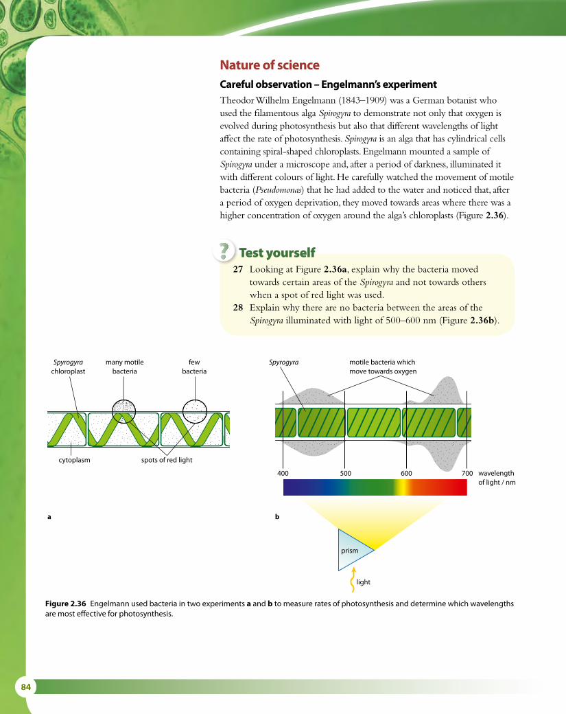
84
Figure 2.36 Engelmann used bacteria in two experiments a and b to measure rates of photosynthesis and determine which wavelengths are most effective for photosynthesis.
Spyrogyra motile bacteria whichmove towards oxygen
wavelengthof light / nm
prism
light
400 500 600 700
Spyrogyrachloroplast
many motilebacteria
fewbacteria
cytoplasm
ba
spots of red light
27 Looking at Figure 2.36a, explain why the bacteria moved towards certain areas of the Spirogyra and not towards others when a spot of red light was used.
28 Explain why there are no bacteria between the areas of the Spirogyra illuminated with light of 500–600 nm (Figure 2.36b).
Test yourself?
Nature of scienceCareful observation – Engelmann’s experimentTheodor Wilhelm Engelmann (1843–1909) was a German botanist who used the filamentous alga Spirogyra to demonstrate not only that oxygen is evolved during photosynthesis but also that different wavelengths of light affect the rate of photosynthesis. Spirogyra is an alga that has cylindrical cells containing spiral-shaped chloroplasts. Engelmann mounted a sample of Spirogyra under a microscope and, after a period of darkness, illuminated it with different colours of light. He carefully watched the movement of motile bacteria (Pseudomonas) that he had added to the water and noticed that, after a period of oxygen deprivation, they moved towards areas where there was a higher concentration of oxygen around the alga’s chloroplasts (Figure 2.36).

2 MOLECULAR BIOLOGY 85
The chemistry of photosynthesisPhotosynthesis is a complex series of reactions catalysed by a number of different enzymes. To aid understanding, we can consider photosynthesis in two stages: the light-dependent reactions and the light-independent reactions.
Light-dependent reactionsThe first stage is known as the ‘light-dependent reactions’ because light is essential for them to occur.
Chlorophyll absorbs light energy and this energy is used to produce ATP. The energy is also used to split water molecules into hydrogen and oxygen in a process called photolysis. Hydrogen ions and electrons (from the hydrogen part of water) and oxygen are released. Oxygen is a waste product of photosynthesis but is vital to sustain the lives of aerobic organisms once it has been released into the atmosphere. The ATP, hydrogen ions and electrons are used in the light-independent reactions.
Light-independent reactionsATP, hydrogen ions and electrons are used in the second stage of photosynthesis, the ‘light-independent reactions’.
During the ‘light-independent reactions’, carbon dioxide, taken in from the environment, is combined with hydrogen and ATP to form a range of organic molecules for the plant. The conversion of inorganic carbon dioxide to organic molecules such as glucose is known as carbon fixation. ATP provides the energy for the process.
The series of reactions that occurs during photosynthesis is summarised as:
carbon dioxide + water → glucose + oxygen 6CO2 + 6H2O → C6H12O6 + 6O2
Measuring the rate of photosynthesisThe equation above shows that when photosynthesis occurs, carbon dioxide is used and oxygen is released. The mass of the plant (its biomass) will also increase as glucose is used to produce other plant materials. Any of these three factors can be used to measure how quickly the reactions of photosynthesis are occurring.
Aquatic plants release bubbles of oxygen as they photosynthesise and if the volume of these bubbles is measured for a period of time, the rate of photosynthesis can be determined directly (Figure 2.37).
Aquatic plants also remove carbon dioxide from their environment, causing the pH of the water to rise. Carbon dioxide dissolves in water to form a weak acid so as it is removed, the pH will go up. Therefore, another way of determining the rate of photosynthesis experimentally is to monitor the change in pH of the water surrounding an aquatic plant over a period of time.
Terrestrial plants also remove carbon dioxide from their surroundings but this is difficult to measure. It can be done experimentally by supplying a confined plant with radioactive carbon dioxide, which can be measured as it is taken up and released from the plant.
Figure 2.37 The rate of oxygen production can be used as a direct measure of the rate of photosynthesis.

86
A third method of measuring the rate of photosynthesis in plants is to determine their biomass at different times. This is an indirect method. Samples of the plants can be collected and measured at different times and the rate of increase in their biomass calculated to determine their rate of photosynthesis.
Limits to photosynthesisThe rate at which a plant can photosynthesise depends on factors in the environment that surrounds it. On a warm, sunny afternoon, photosynthesis will be more rapid than on a cool, shady morning. More oxygen will be produced and more carbon dioxide used. Temperature, light intensity and carbon dioxide concentration are all possible limiting factors on the rate of photosynthesis. But photosynthesis cannot increase beyond certain limits. The effect of light, temperature and carbon dioxide in the environment can be measured experimentally, varying one factor while keeping the others the same, and graphs such as those in Figure 2.38 can be drawn.
An increase in light intensity, when all other variables are unchanging, will produce an increase in the rate of photosynthesis that is directly proportional to the increase in light intensity. However, at a certain light intensity, enzymes will be working at their maximum rate, limited by
Light intensity
experiment 325°C; 0.4% CO
2
experiment 225°C; 0.04% CO
2
experiment 115°C; 0.04% CO
2
(0.04% CO2 is about
atmospheric concentration)
Rate
of p
hoto
synt
hesi
s
Light intensity
Rate
of p
hoto
synt
hesi
s
0 5 10 15
Temperature (°C)
c The rate of photosynthesis at different temperatures, carbon dioxide concentrations and light intensities
a The rate of photosynthesis at different light intensities and constant temperature
b The rate of photosynthesis at different temperatures and constant light intensities
Rate
of p
hoto
synt
hesi
s
20 25
low light intensity
high light intensity
Light intensity
experiment 325°C; 0.4% CO
2
experiment 225°C; 0.04% CO
2
experiment 115°C; 0.04% CO
2
(0.04% CO2 is about
atmospheric concentration)
Rate
of p
hoto
synt
hesi
s
Light intensity
Rate
of p
hoto
synt
hesi
s
0 5 10 15
Temperature (°C)
c The rate of photosynthesis at different temperatures, carbon dioxide concentrations and light intensities
a The rate of photosynthesis at different light intensities and constant temperature
b The rate of photosynthesis at different temperatures and constant light intensities
Rate
of p
hoto
synt
hesi
s
20 25
low light intensity
high light intensity
Figure 2.38 These graphs show the effects on photosynthesis of varying light intensity, carbon dioxide concentration and temperature.

2 MOLECULAR BIOLOGY 87
temperature and the availability of carbon dioxide. At very high light intensities, light absorption (and therefore the rate of photosynthesis) reaches its maximum and cannot increase further. At this point, the graph reaches a plateau (Figure 2.38a).
Increasing temperature also increases the rate of photosynthesis as the frequency and energy of molecular collision increases (Figure 2.38b). Photosynthesis has an optimum temperature above which the rate will decrease sharply as enzymes are denatured, or the plant wilts and is unable to take in carbon dioxide.
An increase in the concentration of carbon dioxide causes the rate of photosynthesis to increase, as carbon dioxide is a vital raw material for the process. At very high concentrations, the rate will plateau as other factors such as light and temperature limit the rate of reaction (Figure 2.38c).
The effects of temperature, light and carbon dioxide concentration are well known to horticulturalists who grow crops in glasshouses. Commercial producers of cucumbers and tomatoes keep their glasshouses warm and well lit. They may also introduce carbon dioxide to boost photosynthesis to its maximum rate, thereby increasing crop production and profits.
Photosynthesis is described in more detail in Subtopic 8.3.
Influence of photosynthesis on the Earth’s atmosphere, oceans and rocksLiving organisms started to have a major impact on the Earth’s environment after the evolution of photosynthetic organisms. Today, most scientists agree that the first oxygen-producing photosynthesis evolved about 2.4 billion years ago.
These early photosynthetic life forms reduced the carbon dioxide content of the atmosphere and they also started to produce oxygen. At first, oxygen was taken up by rocks in the Earth’s mantle, where oxygen combined with reduced iron compounds to form iron oxides. It was probably only about 1 billion years ago that these rocks became saturated with oxygen and free oxygen remained in the atmosphere. The Earth’s rocks still contain huge reservoirs of oxygen in the form of oxides.
As the level of free oxygen built up and became sufficient to support life, a period known as the Biological Era in the evolution of the atmosphere began, which had three important consequences.
• Once the level of oxygen reached about 1% of its current level, the metabolism of eukaryotes became possible. According to the fossil record, this probably began to happen about 2 billion years ago.
• With greater availability of oxygen, metabolic pathways became more efficient, and aerobic organisms proliferated in the oceans. Carbon dioxide removed from the atmosphere ultimately ended up in the shells and decomposed remains of sea creatures, and built up marine sediments, which eventually became incorporated in sedimentary and, later, metamorphic rocks.
• As oxygen accumulated in the upper atmosphere, sunlight would have been able to act on it and form ozone. The ozone layer helped
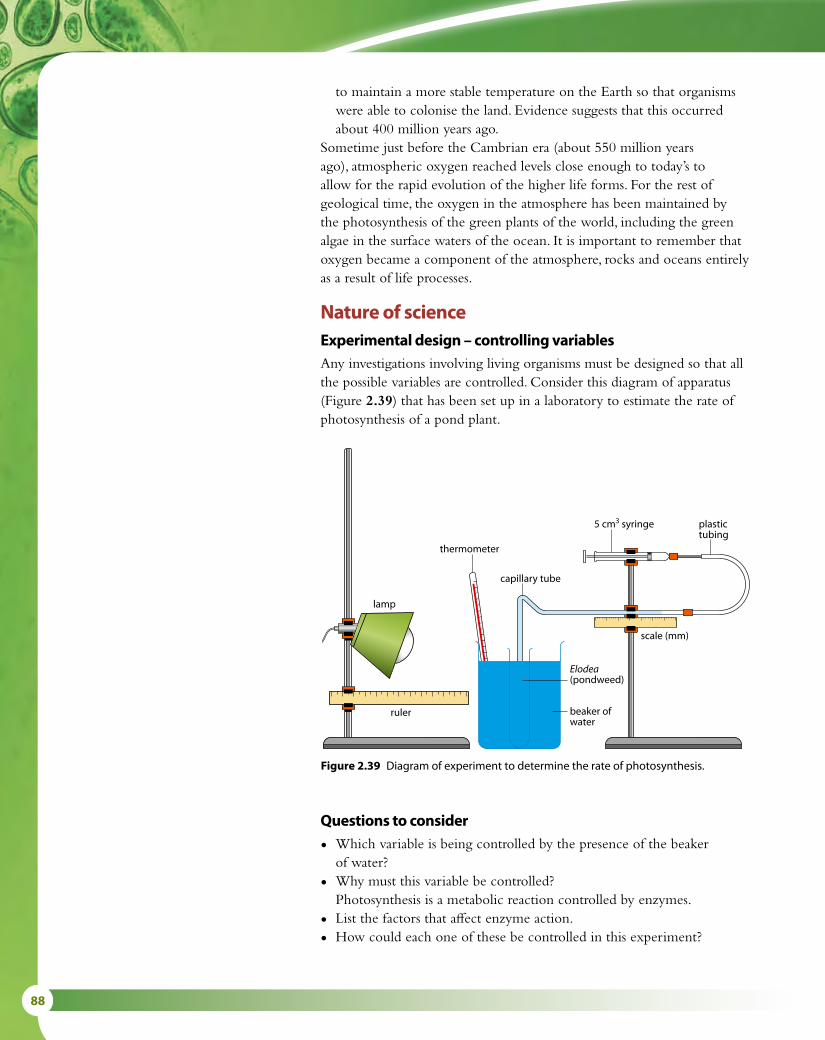
88
to maintain a more stable temperature on the Earth so that organisms were able to colonise the land. Evidence suggests that this occurred about 400 million years ago.
Sometime just before the Cambrian era (about 550 million years ago), atmospheric oxygen reached levels close enough to today’s to allow for the rapid evolution of the higher life forms. For the rest of geological time, the oxygen in the atmosphere has been maintained by the photosynthesis of the green plants of the world, including the green algae in the surface waters of the ocean. It is important to remember that oxygen became a component of the atmosphere, rocks and oceans entirely as a result of life processes.
Nature of scienceExperimental design – controlling variablesAny investigations involving living organisms must be designed so that all the possible variables are controlled. Consider this diagram of apparatus (Figure 2.39) that has been set up in a laboratory to estimate the rate of photosynthesis of a pond plant.
lamp
ruler
scale (mm)
plastictubing
beaker ofwater
Elodea(pondweed)
5 cm3 syringe
thermometer
capillary tube
Figure 2.39 Diagram of experiment to determine the rate of photosynthesis.
Questions to consider
• Which variable is being controlled by the presence of the beaker of water?
• Why must this variable be controlled?
Photosynthesis is a metabolic reaction controlled by enzymes.
• List the factors that affect enzyme action.
• How could each one of these be controlled in this experiment?

2 MOLECULAR BIOLOGY 89
29 If you wanted to make plants grow as well as possible, state which colour of light you should shine on them.
30 Describe what would happen to a plant’s growth if it were kept in green light.
31 State the colours of the visible spectrum that are used in photosynthesis.
32 Describe two ways in which the rate of photosynthesis can be measured.
Test yourself?
Exam-style questions
1 Which of the following statements is correct?
A The most frequently occurring chemical elements in living organisms are carbon, hydrogen, oxygen and calcium.
B A water molecule can form hydrogen bonds with other water molecules due to its polarity.C Sweating cools an animal because body heat is required to break the covalent bonds in the
water in the sweat, allowing it to evaporate.D Increasing substrate concentration causes enzymes to denature. [1]
2 Which of the following statements is correct?
A The components of a nucleotide are a sugar molecule attached to two phosphate groups and a base.
B In a molecule of DNA, the bases thymine and uracil are held together by hydrogen bonds.C During the process of transcription, tRNA molecules bond to mRNA using
complementary base pairing.D During DNA replication, new nucleotides are added using the enzyme DNA polymerase. [1]
3 Outline how monosaccharides are converted into polysaccharides. [2]
4 State why each step in a biochemical pathway often requires a separate enzyme. [2]

90
5 Study the graph below which shows the rate of enzyme activity for pepsin, a protease enzyme found in the stomach. Enzyme activity is measured by the rate of breakdown of protein to amino acids.
a What is the optimum pH of the enzyme pepsin? [1]
b Within what range does pepsin act at over 50% of its maximum rate? [2]
c What would you expect the pH of the stomach contents to be? [1]
6 a Draw a nucleotide of DNA to show its structure [2]
b State three differences in the structure of a DNA and RNA molecule [3]
c Outline what is meant by ‘complementary base pairs’ in a DNA molecule. [2]
The table below shows the mRNA codons for some amino acids.
mRNA codon Amino acid
AGU serine
GCU alanine
UGC cysteine
ACG threonine
GUC valine
CUA leucine
d State the DNA sequence for the amino acid cysteine. [1]
e Name the amino acid coded for by the DNA sequence TCA. [1]
1 2 3 4 5 6 7 8 9 10 11 12 13 14pH of solution
pepsin salivary amylase
Rate
of s
ubst
rate
bre
akdo
wn
Pepsin and salivary amylase activity at di�erent pH values

2 MOLECULAR BIOLOGY 91
12
34
56
78
9
solvent line
carotene (yellow orange)
xanthophyll (yellow)
chlorophyll a (blue green)
chlorophyll b (greenish yellow)
pigment extract (origin)
The distance moved by each pigment divided by the distance moved by the solvent is unique for each pigment and is known as the Rf value.
Rf = distance moved by pigment/distance moved by solvent
a Calculate the Rf value for i chlorophyll a [1] ii carotene. [1]
b The graph below shows the absorption spectrum for the total plant extract. i State which wavelengths and colours are absorbed best. [2] ii Outline the relationship between the absorption and action spectra. [2]
7 The pigments found in a leaf can be extracted with a solvent and separated using thin layer chromatography to produce a chromatogram shown in the diagram below.
400 500 600 700
Wavelength of light / nm
viol
et
blu
e
gree
n
yello
w
oran
ge
red
Ab
unda
nce
of p
lant
ext
ract
Rate
of p
hoto
synt
hesi
s

92
Suggest a role of carotene in capturing light energy for photosynthesis. [2]
8 Body mass index (BMI) is a measure used to indicate whether a person has an appropriate body mass. The table below indicates the range of BMI measurements for adults over the age of 20 years.
BMI = mass in kg ÷ (height in m)2
BMI Status
Below 18.5 underweight
18.5 – 24.9 normal weight
25.0 – 29.9 overweight
30.0 and above obese
i Calculate the BMI of a male Olympic weight lifter who has a body mass of 99 kg and is 1.75 m tall [1] ii Calculate the BMI of a female office worker who has a body mass of 70 kg and is 1.5 m tall. [1]
BMI is used to indicate whether a person is overweight and likely to be storing excess fat in their body, but fitness training builds up large muscles which weigh more than fat.
iii Use this information to comment on the status of the weight lifter and the office worker. [2]
c The graph below shows the absorption spectrum for pure solutions of chlorophyll a and carotene.
400 450 500 550 600 650 700
Wavelength of light / nm
viol
et
blu
e
gree
n
yello
w
oran
ge
red
Ab
sorb
ance
of c
hlor
ophy
y a
Ab
sorb
ance
of c
arot
ene

3 GENETICS 93
Genetics 3IntroductionChimpanzees are set apart from all other organisms because their parents were chimpanzees and their offspring will also be chimpanzees. Every living organism inherits its own blueprint for life in the chromosomes and genes that are passed to it from its parents. The study of genetics attempts to explain this process of heredity and it also plays a very significant role in the modern world, from plant and animal breeding to human health and disease.
3.1 Genes
Chromosomes, genes and mutationsA DNA molecule comprises a pair of strands, each strand consisting of a linear sequence of nucleotides, with weak hydrogen bonds between the bases holding the two strands together. This linear sequence of bases contains the genetic code in the form of triplets of bases. A gene is a particular section of a DNA strand that, when transcribed and translated, forms a specific polypeptide. Some of the polypeptides will form structural proteins such as collagen while others become enzymes or pigments such as hemoglobin (Subtopic 2.4) and it is the translation of the genes which gives each individual organism its own specific characteristics. (Transcription and translation are described in Topic 2 and in more detail in Topic 7.) Each gene is found at a specific position on a chromosome and so, for example, it is possible to say that the gene for human insulin is always found on chromosome number 11.
Organisms that reproduce sexually almost always have pairs of chromosomes, one of each pair coming from each parent. The members of the pair carry equivalent genes, so that – for example – in humans, both versions of chromosome number 11 carry the insulin gene. But there may be slight differences in the version of the gene on each chromosome. These slightly different forms of the gene are known as alleles. Alleles differ from one another by one or only a few bases and it is these differences in alleles that give rise to the variation we observe in living organisms.
When the genetic code is used to build proteins, a triplet of bases in the DNA molecule is transcribed into a triplet of bases in the mRNA molecule (also called a codon – Subtopic 2.7), which is then translated into a specific amino acid, as shown in Figure 3.1.
The process of DNA replication is complex and mistakes sometimes occur – a nucleotide may be left out, an extra one may be added, or the wrong one inserted. These mistakes are known as gene mutations. Mutations may occur spontaneously, as a result of errors in copying
Learning objectives
You should understand that:
• Genes are heritable factors that each consist of a section of DNA and influence a specific characteristic.
• Each gene occupies a specific position on one chromosome.
• Alleles are the various specific forms of a gene.
• Alleles differ from one another by only a few bases.
• Mutation causes the formation of new alleles.
• The whole of the genetic information of an organism is known as its genome.
• The Human Genome Project worked out the entire base sequence of human genes.
C A G
transcription
DNA triplet
mRNA codon
amino acid
translation
glutamine
G T C
C A G
Figure 3.1 The base sequence in DNA is decoded via transcription and translation.

94
DNA, or they can be caused by factors in the environment known as mutagens (Subtopic 1.6). The insertion of an incorrect nucleotide is called a base substitution mutation. When the DNA containing an incorrect nucleotide is transcribed and translated, errors may occur in the polypeptide produced.
Table 3.1 shows the amino acids that are specified by different mRNA codons. Most amino acids are coded for by more than one codon and so many substitution mutations have no effect on the final polypeptide that is produced. These are said to be neutral mutations. For example, a mutation in the DNA triplet CCA into CCG would change the codon in the mRNA from GGU to GGC but it would still result in the amino acid glycine being placed in a polypeptide. Some substitution mutations, however, do have serious effects – for example, one important human condition that results from a single base substitution is sickle-cell anemia.
Sickle-cell anemia – the result of a base substitution mutationSickle-cell anemia is a blood disorder in which red blood cells become sickle shaped and cannot carry oxygen properly (Figure 3.2). It occurs most frequently in people with African ancestry – about 1% suffer from the condition and between 10% and 40% are carriers of it. Sickle-cell anemia is due to a single base substitution mutation in one of the genes that make hemoglobin, the oxygen-carrying pigment in red blood cells.
Hemoglobin is made up of four subunits, as shown in Figure 3.3 – two α-chains and two β-chains. The β-chains are affected by the sickle-cell
Gene a heritable factor that controls a specific characteristic, or a section of DNA that codes for the formation of a polypeptideAllele a specific form of a gene occupying the same gene locus or position as other alleles of that gene, but differing from other alleles by small variations in its base sequenceGenome the whole of the genetic information of an organismGene mutation a change in the sequence of bases in a gene
There are several thousand human disorders that are caused by mutations in single genes and about 100 ‘syndromes’ associated with chromosome abnormalities.
Second base
U C A G
Firs
t bas
e
U
UUUphenylalanine
UCU
serine
UAUtyrosine
UGUcysteine
U
Third
base
UUC UCC UAC UGC C
UUAleucine
UCA UAA‘stop’
UGA ‘stop’ A
UUG UCG UAG UGG tryptophan G
C
CUU
leucine
CCU
proline
CAUhistidine
CGU
arginine
U
CUC CCC CAC CGC C
CUA CCA CAAglutamine
CGA A
CUG CCG CAG CGG G
A
AUU
isoleucine
ACU
threonine
AAUasparagine
AGUserine
U
AUC ACC AAC AGC C
AUA ACA AAA
lysine
AGA
arginine
A
AUGmethionine or ‘start’
ACG AAG AGG G
G
GUU
valine
GCU
alanine
GAUaspartic acid
GGU
glycine
U
GUC GCC GAC GGC C
GUA GCA GAAglutamic acid
GGA A
GUG GCG GAG GGG G
Table 3.1 Amino acids and their associated mRNA codons.

3 GENETICS 95
Only one strand of DNA is transcribed into mRNA for any gene. The transcribed strand is called the coding strand.
mutation. To form a normal β-chain, the particular triplet base pairing in the DNA is:
G A G
C T C
The C–T–C on the coding strand of the DNA (in blue here) is transcribed into the mRNA triplet G–A–G, which in turn is translated to give glutamic acid in the polypeptide chain of the β-chain.
If the sickle-cell mutation occurs, the adenine base A is substituted for thymine T on the coding strand, so the triplet base pairing becomes:
G T G
C A C
C–A–C on the coding strand of the DNA is now transcribed into the mRNA triplet G–U–G, which in turn is translated to give the amino acid valine. Valine replaces glutamic acid in the β-chain.
Valine has different properties from glutamic acid and so this single change in the amino acid sequence has very serious effects. The resulting hemoglobin molecule is a different shape, it is less soluble and when in low oxygen concentrations, it deforms the red blood cells to give them a sickle shape. Sickle cells carry less oxygen, which results in anemia. They are also rapidly removed from the circulation, leading to a lack of red blood cells and other symptoms such as jaundice, kidney problems and enlargement of the spleen.
The genome and Human Genome ProjectThe genome of an organism is defined as the whole of its genetic information and every cell has a complete copy of the organism’s genome. Genome analysis is an important field of modern biological research.
Figure 3.2 Coloured scanning electron micrograph showing a sickle cell and normal red blood cells (× 7400).
the two α-chainsare shown in blue
the two β-chainsare shown in red
Figure 3.3 The structure of a hemoglobin molecule showing the 3D arrangement of the subunits that makes it up.

96
Comparative genomics is an area that analyses genomes from different species. Genomes are compared to gain a better understanding of how species have evolved and to work out the functions of genes and also of the non-coding regions of the genome. Researchers look at many different features such as sequence similarity, gene location, the length and number of coding regions within genes and the amount of DNA that does not code for proteins. They use computer programs to line up genomes from different organisms and look for regions of similarity. There are many databases that store this information on DNA base sequences (Table 3.3). Sequences for commonly occurring proteins such as cytochrome c, which is involved in cell respiration in many organisms, can be compared and have been useful for establishing the evolutionary relationships between different species.
The Human Genome Project, which has sequenced all the bases in the entire complement of a human’s genetic information, was the first step in attempting to understand the human genome. The project revealed that all humans, from all over the world, share the vast majority of their base sequences, regardless of their race. At the same time, every human being (apart from identical twins) is genetically unique due to differences in single nucleotides, which together account for the enormous human diversity we can see. Although the project is now complete, there remain many unanswered questions. The functions of most human genes and the roles of non-coding regions and repeated sequences are still unknown.
Humans and most mammals have roughly the same number of nucleotides (about 3 billion base pairs) in their genomes. Although the number of genes is not necessarily proportional to the total amount of DNA present in a genome, the work of many scientists has provided evidence to support the theory that most mammal genomes do in fact contain more or less the same number of genes. In addition to the human genome, the genomes of about 800 organisms have been sequenced in recent years. These include the mouse Mus musculus, the fruit fly Drosophila melanogaster, the worm Caenorhabditis elegans, the bacterium Escherichia coli, the yeast Saccharomyces cerevisiae, the plant Arabidopsis thaliana, and many microbes. Some information about the genomes of these organisms is shown in Table 3.2.
Organism Estimated number of genes Number of Chromosomes
Mus musculus (mouse) 25 000 40
Arabidopsis thaliana (plant) 23 000–25 000 10
Homo sapiens (human) 22 000–25 000 46
Caenorhabditis elegans (roundworm) 19 000 12
Drosophila melanogaster (fruit fly) 13 000–14 000 8
Saccharomyces cerevisiae (yeast) 6000 32
Escherichia coli (bacterium) 1700–3000 1
Table 3.2 Comparison of the estimated numbers of genes in different organisms. The number of genes shown in this table is only approximate as data is being refined all the time. Note that the number of genes does not correlate with evolutionary status, nor is the number of genes proportional to the total amount of DNA present in an organism.

3 GENETICS 97
Nature of scienceScientific advance follows technical innovation – gene sequencers and databasesDifferent methods of DNA sequencing are used to work out either the sequence of individual genes, groups of genes or even entire chromosomes and genomes. Since the advances in technology made during the Human Genome Project many new and automated methods of sequencing have been developed.
Some processes involve cutting DNA into short segments using restriction enzymes, and cloning the resulting pieces using the polymerase chain reaction (PCR). This technique has been used by forensic scientists to compare repeated sequences in DNA for use in criminal investigations or for establishing family relationships (see Subtopic 3.5).
Different techniques are used to identify genes that have not been recognised before. These are known as ‘de novo’ methods. Many of the new sequencing methods use indirect approaches based on tagging nucleotides with fluorescent markers. Others use ‘nanopore’ methods in which the strand of DNA to be analysed passes through a protein pore and data is recorded on a microchip. DNA can be analysed rapidly in this way and it may soon be possible to sequence a human genome in just a few hours. The field of DNA sequencing is advancing rapidly and some of the newest methods have the potential to be adapted to recognise other molecules such as drugs or other chemicals.
Databases have also been invaluable in genome research and are used for storing long sequences of data about proteins. One example of an institution that builds and maintains extensive biological databases is the European Bioinformatics Institute (EBI). This research institute is located on the Wellcome Trust Genome Campus near Cambridge in the UK, where large parts of the original Human Genome Project were carried
Sickle-cell anemia – cause and correlationThe global distribution of sickle-cell anemia is correlated
with the incidence of malaria. In parts of Africa, up to 1 in 25 children are born with sickle-cell anemia and its geographical distribution follows the pattern of prevalence of malaria.
We might expect that the allele responsible for sickle-cell anaemia would die out since people who suffer from the condition tend to die younger and have few, if any, children. But the evidence shows that individuals who are heterozygous for sickle-cell anemia (that is, they have one sickle-cell allele and one normal allele) have an advantage over both those without the sickle-cell allele and those with two copies of it, who are sufferers. Heterozygotes are less susceptible to the malaria parasite, which spends part of its life cycle living in red blood cells.
Questions to consider
• Can we establish whether there is a causal link between cases of sickle-cell anemia and prevalence of malaria?
• Is the relationship simple a correlation?
• What do you understand by the term ‘heterozygous advantage’?
• How has the world distribution of sickle-cell anemia changed with international travel as populations have become more mobile?

98
out. It is part of the European Molecular Biology Laboratory (EMBL) and provides data to the scientific community and industry. The first completed genomes from viruses, phages and organelles were deposited in the EMBL database in the early 1980s. Since then hundreds of complete genome sequences have been added to the database.
There are many other databases, some of which are outlined here in Table 3.3. Most are freely available online to scientists and students so that all of us can use them to compare DNA and protein sequences – you can find them by typing the database name into any search engine.
Database Description
ENA Genomes Server overview of all complete genomes deposited in the European Nucleotide Archive
Ensembl a joint project between EMBL–EBI and the Sanger Institute to produce and maintain automatic annotation of eukaryotic genomes
Ensembl Genomes provides access to genomes of non-vertebrate species
GenBank (US National Center for Biotechnology Information)
a collection of all publicly available DNA sequences, providing the scientific community with up-to-date and comprehensive DNA sequence information
Table 3.3 Some databases that hold information on genomes.
1 The β-chain of the hemoglobin molecule contains 146 amino acids. State the number of nucleotides that are needed to code for this protein.
2 Define the following terms: a gene b allele c genome d gene mutation.3 Outline the meaning of the term ‘base substitution mutation’.
Test yourself?

3 GENETICS 99
3.2 Chromosomes
Chromosome structureChromosomes are made of DNA molecules and carry the genetic code for each organism but prokaryotic and eukaryotic chromosomes are different in their structure. Prokaryotes have a much simpler chromosome than eukaryotes. Prokaryotes contain a circle of DNA that is often concentrated in one area of the cell, whereas eukaryotes have linear DNA that is associated with histone proteins. Some of these proteins are structural and others regulate the activities of the DNA. Prokaryotes have additional genetic material in the form of small circular structures known as plasmids. Prokaryotes are much simpler organisms and so require fewer genes to maintain themselves. The differences between prokaryotic and eukaryotic genetic material are summarised in Table 3.4.
Prokaryotic DNA Eukaryotic DNA
cell contains a circular chromosome, sometimes called a nucleoid
chromosomes are made of linear DNA molecules enclosed in a nucleus, bound by a double membrane
cell contains additional genetic material as small circular plasmids
no plasmids
DNA is ‘naked’ and is not associated with proteins
DNA is associated with histone proteins
cell contains just one circular chromosome
cell contains two or more chromosome types
Table 3.4 A comparison of prokaryotic and eukaryotic genetic material.
During the phase of the cell cycle known as interphase, eukaryotic chromosomes are in the form of long, very thin threads, which cannot be seen with a simple microscope. As the nucleus prepares to divide, these threads undergo repeated coiling and become much shorter and thicker (Figure 3.4). When stained, they are clearly visible even at low microscope magnifications.
Chromosome structure is described in greater detail in Topic 7.
Eukaryotic chromosomesEukaryotic species have two or more chromosome types. Their chromosomes form pairs, which are known as homologues. Homologous pairs are about the same length and carry the same sequence of genes at the same locations along their length. The form of the genes (alleles) on each of the pair are not necessarily the same because, in sexually reproducing organisms, one chromosome will have been inherited from each of the two parents. So a gene that determines flower colour in a plant would be at the same location on each chromosome in a homologous pair but the allele on the maternal chromosome might not be the same as that on the paternal chromosome.
Learning objectives
You should understand that:
• Prokaryotes have one chromosome, which is a circular DNA molecule.
• Some prokaryotes have plasmids as well as a chromosome but eukaryotes do not.
• Eukaryotes have chromosomes that are linear DNA molecules associated with histone proteins.
• Eukaryotes have different chromosomes that carry different genes.
• Each species has a characteristic number of chromosomes.
• Haploid nuclei have one of each type of chromosome.
• Diploid nuclei have two of each type of chromosome, called homologous pairs of chromosomes.
• Homologous chromosomes carry the same genes but not necessarily the same alleles.
• A karyogram shows an organism’s chromosomes in homologous pairs arranged in order of decreasing length.
• Sex chromosomes determine the sex of an organism; autosomes are the chromosomes that do not determine sex.

100
Homologous chromosomes are found in the nuclei in the cells of diploid organisms. But if each chromosome exists alone with no partner, the cell is said to be haploid. Human somatic cells are diploid and contain 46 chromosomes in 23 homologous pairs; human gametes are haploid and contain only 23 chromosomes, one of each pair found in the body cells.
The number of chromosomes found in the cells of an organism is a characteristic feature of that organism. The diploid numbers of chromosomes in some well-studied species are shown in Table 3.5.
Organism Diploid number of chromosomes
Canis familiaris (domestic dog) 78
Pan troglodytes (chimpanzee) 48
Homo sapiens (human) 46
Mus muscularis (house mouse) 40
Oryza sativa (rice) 24
Drosophila melangosta (fruit fly) 8
Parascaris equorum (parasitic worm) 2
Table 3.5 The diploid number of different species.
Figure 3.4 Coloured scanning electron micrograph of human chromosomes. They have replicated prior to cell division and so consist of two identical copies (chromatids) linked at the centromere (× 7080).
Diploid a diploid nucleus contains two copies of each chromosome, in homologous pairsHaploid a haploid nucleus contains one chromosome of each homologous pairHomologous chromosomes a pair of chromosomes with the same genes but not necessarily the same alleles of those genesSomatic cell a body cell that is not a gamete
In 2005, the International Rice Genome Sequencing Project, which began in 1998, succeeded in making rice the first crop plant whose genome had been mapped. Because
rice is such an important staple food worldwide, an understanding of its genetic make-up is a key tool for researchers working on improved strains to feed a burgeoning human population. The sequencing of the rice genome involved cooperation between biologists in ten different countries – the project was led by scientists in Japan but involved teams from the United States, China, France, Taiwan, India, Thailand, Korea, Brazil and the UK.
Genome sizeGenome size is the total number of nucleotide base pairs within one copy of a single genome. Measurements are often made in numbers of base pairs. It is interesting to note that an organism’s complexity is not proportional to its genome size (Table 3.6) – many organisms have more DNA than humans. Nor is variation in genome size proportional to the number of genes. This is due to the high proportion of non-coding DNA that is found in some organisms.
KaryotypingChromosomes have unique banding patterns that are revealed if they are stained with specific dyes. These patterns enable us to study the structure and type of chromosomes present in an organism. The technique has been used in prenatal diagnosis and in forensic science. Chromosomes are

3 GENETICS 101
prepared from the nuclei of dividing cells. If human cells are being studied, a sample of cells may be taken from amniotic fluid, in the case of a fetus, or from a blood sample. Division is halted at the end of prophase when chromosomes are condensed. A karyogram is a photograph or diagram of the stained chromosomes. Each chromosome is a characteristic length and each one has a homologous partner. The karyogram image is organised so that each chromosome is separated from the others and they are arranged in order of their size, as shown in Figure 3.5. A karyogram shows the karyotype of the cell – that is, the number and types of chromosomes present in its nucleus. It indicates the sex of an individual because it shows the sex chromosomes, and in prenatal diagnosis it is possible to check for chromosome abnormalities such as those leading to Down syndrome. You can read more about prenatal diagnosis in Subtopic 3.3.
Sex chromosomes and autosomesThe sex chromosomes are the last two chromosomes shown in the human karyograms in Figure 3.5 (in the bottom right hand corner, in
Organism Genome size in
base pairs
Notes
T2 phage (a virus which infects bacteria) 3569 first RNA genome sequenced
E. coli (a bacterium) 4.6 × 10⁶
Drosophila melanogaster (fruit fly) 130 × 106
Homo sapiens (human) 3200 × 106
Paris japonica (Japanese native pale-petal) 150 000 × 106 largest known plant genome
Protopterus aethiopicus (marbled lungfish) 130 000 × 106 largest known vertebrate genome
Table 3.6 Genome sizes of some organisms.
1 2 3 4
6 7 8 9 10 11 12
13 14 15 16 17 18 19 20
21 22X X
5 1 2 3 4
6 7 8 9 10 11 12
13 14 15 16 17 18 19 20
21 22XY
5
chromosome chromatid
a b
centromere – the point at which the two chromatids are held together
The X and Y chromosomes differ in length.
A female has two X chromosomes.
Two chromatids within one chromosome are identical copies produced by DNA replication.
Figure 3.5 Karyograms for a human male a and a human female b.
Karyogram a diagram or photograph of the chromosomes from an organismKaryotype the number and type of chromosomes present in a nucleus

102
each case). These chromosomes determine the sex of an individual – a female has two X chromosomes (XX) while a male has one X and one smaller Y chromosome (XY). The other 44 chromosomes in a human karyotype are known as autosomes and they determine the other characteristics of the individual.
Nature of scienceScientific advance follows technical innovation – Cairns’ techniqueHugh John Forster Cairns (1922– ) is a British molecular biologist who demonstrated by autoradiography that the bacterium E. coli contains a single, circular molecule of DNA.
A prediction of the Watson–Crick model of DNA replication was that there would be a replication fork formed as DNA copied itself. John Cairns tested this by allowing replicating DNA in E. coli bacteria to incorporate a radioactive marker in the form of tritiated thymidine. Cells cultured in tritiated thymidine take up the radioactive hydrogen isotope 3H (tritium) and the thymine nucleotides become labelled. As cells undergo mitosis, the radioactive 3H in the labelled thymine nucleotides is incorporated into the new cells that are produced. As 3H decays, it emits a beta particle, which produces a dot on a photographic plate, so that the location of the radioactivity can be identified (Figure 3.6). The image produced on a photographic plate by radioactivity is called an autoradiograph.
replication fork unlabelled DNA strand
labelled DNA strand
replicatedDNA – bothstrandslabelled
replication fork
image of an autoradiographproduced by Cairns’ method
explanation ofautoradiograph image
Figure 3.6 The replication of E. coli chromosome.
Cairns observed rings of dots in his autoradiograph after one replication cycle. As he observed further cycles of replication, he observed replication forks (the Y-shaped junctions where double-stranded DNA splits apart into two single strands) around the ring so he was able to conclude that the bacterial chromosome is circular. Cairns’ techniques have also been used to measure the length of DNA molecules.
Since Cairns’ experiments in 1963, further research has enabled scientists to observe replication of chromosomes and support his conclusions without the use of radioactivity.

3 GENETICS 103
How long is the DNA in a human body?
The human genome is the information contained in one set of human chromosomes, which are built up of about 3 billion base pairs (bp) of DNA in 46 chromosomes (22 autosome pairs + 2 sex chromosomes). The total length of DNA present in one adult human is calculated by multiplying:
(length of 1 bp) × (number of bp per cell) × (number of cells in the body)
So total length = (0.34 × 10⁻⁹ m) × (6 × 109) × 1013
= 2.0 × 10¹³ metres
That is about the distance of travelling 70 times from the Earth to the Sun and back.
3.3 Meiosis
4 State two differences between prokaryotic and eukaryotic chromosomes.
5 Define what is meant by a ‘karyogram’.6 Define the term ‘autosome’.
Learning objectives
You should understand that:
• When a diploid nucleus divides by meiosis, four haploid nuclei are produced.
• Halving the number of chromosomes in gametes allows for sexual reproduction in a lifecycle, because when two gametes fuse the diploid number is restored.
• DNA replication occurs before meiosis so that all chromosomes consist of two sister chromatids.
• Early in meiosis homologous chromosomes pair up and crossing over and condensation follow.
• Orientation of the pairs of homologous chromosomes before they separate is random.
• The chromosome number is halved in the first division of meiosis by separation of homologous chromosomes.
• Crossing over and random orientation lead to increased genetic variation.
• Genetic variation is also increased by the fusion of gametes from different parents.
Test yourself? Exam tipMake sure you can define pairs of terms which are used in the same topics. For example: karyogram and karyotype, proteome and genome, chromosome and chromatid, diploid and haploid.
104
Meiosis is a reduction divisionMeiosis is a type of cell division that produces gametes (sex cells). In any organism, each cell that is produced as a result of meiosis has half the number of chromosomes of other cells in the body.
Eukaryotic body cells have a diploid nucleus, which contains two copies of each chromosome, in homologous pairs (Subtopic 3.2). As we have seen, humans have a diploid number of 46 chromosomes in 23 pairs, mangos and soybean both have 40 chromosomes in 20 pairs, and the camel has 70 chromosomes in 35 pairs.
During sexual reproduction, two gametes fuse together so, in order to keep the chromosome number correct in the offspring that are produced, each gamete must contain only one of each chromosome pair. That is, it must contain half the diploid number of chromosomes, which is called the haploid number. During gamete formation, meiosis reduces the diploid number to the haploid number – for this reason, meiosis is called a reduction division. At the moment of fertilisation, the normal diploid number is restored as the gametes fuse.
So, in the camel, the haploid sperm (35 chromosomes) and haploid egg (35 chromosomes) fuse at fertilisation to form the diploid zygote, with 70 chromosomes.
The process of meiosisMeiosis occurs in a series of stages, as illustrated in Figure 3.7, which result in the production of four cells.
Whereas mitosis, used to replace or repair cells, is achieved with one cell division, meiosis involves two divisions – the first reduces the number of chromosomes by half and the second produces four gametes each containing the haploid number of chromosomes. Exactly the same terms are used for the names of the stages, but since meiosis involves two divisions, the phases are numbered I and II.
The first division is very similar to mitosis and the second division is exactly the same as mitosis.
Prophase IDuring interphase, before the start of prophase, chromosomes are replicated and consist of two identical sister chromatids joined at the centromere. In prophase I, these chromosomes now supercoil and the homologous pairs of chromosomes line up side by side.
Although the genes carried by each chromosome pair are identical, the alleles may not be. Exchange of genetic material between the pair can occur at this point. Chromatids may become entangled, break and re-join so that alleles are exchanged between homologous chromosomes during a process called crossing over. New combinations of alleles are formed and genetic variety among the resulting gametes increases.
The final step in prophase I is the formation of spindle microtubules and the breakdown of the nuclear envelope.
There are a few organisms that are haploid – for example, the male honey bee, Apis mellifera. This means it produces gametes through mitosis. Female worker bees have 32 chromosomes in their body cells but male drones have only 16.
In each homologous pair, one chromosome is a maternal chromosome and the other a paternal chromosome. After crossing over, the chromatids recombine to produce new and unique combinations of alleles, different from both the maternal and the paternal arrangements. This is called recombination.

3 GENETICS 105
Metaphase IChromosomes line up on the equator at the centre of the cell. Each one attaches by a centromere to the spindle microtubules. The alignment of the chromosomes is random so that maternal and paternal chromosomes can appear on either side of one another on the equator. This means that either chromosome from a pair may move into each daughter cell during the first division at anaphase I, which results in increased genetic variety among the gametes.
Anaphase IThe microtubules now contract towards opposite poles. The pairs of sister chromatids remain together but the homologous pairs are separated. This is the reduction division where the chromosome number is halved from diploid to haploid.
Telophase INow spindles break down and a new nuclear envelope forms around each new nucleus. Cytokinesis follows and the cell splits into two cells, each containing only one chromosome of each homologous pair. Each chromosome, however, still consists of two sister chromatids at this point.
The second division of meiosis now follows to separate the two sister chromatids.
Prophase IIIn each of the two cells resulting from meiosis I, new spindle microtubules start to form, the chromosomes re-coil and the nuclear envelope begins to break down.
Metaphase IIThe nuclear envelope is broken down and individual chromosomes line up on the equator of each cell. Spindle fibres from opposite ends of the cell attach to each chromatid at the centromere.
Anaphase IISister chromatids are separated as the centromere splits and spindle fibres pull the chromatids to opposite ends of the cell.
Telophase IINuclear envelopes form around the four new haploid nuclei and the chromosomes now uncoil. A second cytokinesis occurs, resulting in four cells.

106
Figure 3.7 The stages of meiosis in an animal cell. Note that the cells are shown with just two homologous pairs of chromosomes to make it easier to understand the process.

3 GENETICS 107
Non-disjunctionNon-disjunction is a failure of homologous pairs of chromosomes to separate properly during meiosis. It results in gametes that contain either one too few or one too many chromosomes. Those with too few seldom survive, but in some cases a gamete with an extra chromosome does survive and after fertilisation produces a zygote with three chromosomes of one type, as shown in Figure 3.8. This is called a trisomy.
Trisomy in chromosome 21 results in the human condition known as Down syndrome (Figure 3.9). A gamete, usually the female one, receives 24 chromosomes instead of 23 and a baby with 47 instead of the usual 46 chromosomes in each cell is born.
Karyotyping is used when there is concern about potential chromosome abnormalities. Cells from an unborn child are collected
normalsperm cell
abnormalegg cell
abnormal cell with no copyof this chromosome
meiosis
zygote has three copies ofthis chomosome
abnormal cell with twocopies of this chromosome
fertilisation
Figure 3.8 Non-disjunction at anaphase II of meiosis. Non-disjunction can also occur at anaphase I.
Figure 3.9 People with Down syndrome have characteristic physical features.
Figure 3.10 Karyogram showing chromosomes which are stained and photographed for a person with a normal chromosome complement and for a person with trisomy 21 (Down syndrome).

108
in one of two ways – chorionic villus sampling (CVS) or amniocentesis (Subtopic 11.4). The cells are grown in the laboratory and a karyogram is prepared (Subtopic 3.2). This is checked for extra or missing chromosomes (Figure 3.10). The procedure is normally used if the mother is over the age of 35 years because Down syndrome is more common in babies of older mothers and can be detected using this method.
Meiosis and variationMeiosis not only halves the chromosome number, it also promotes genetic variation in the gametes and individuals that are produced from them. There are several reasons for this:
• Each chromosome of the homologous pair carries different genetic information so that the gametes formed are genetically different.
• Different homologous pairs arrange themselves independently on the spindle and also separate independently so that gametes contain different combinations of each chromosome pair.
• Crossing over during prophase I means that genetic material is exchanged between homologous chromosomes producing entirely new combinations.
• At fertilisation, gametes from different parents fuse together promoting yet more genetic variation among the offspring produced.
Nature of scienceCareful observation – discovery of meiosis by microscopic examinationThe first observations of meiosis were made in 1876 by a German biologist, Oscar Hertwig (1849–1922), as he studied the eggs of sea urchins under an early microscope. He made careful, accurate drawing of his observations of gametes from several species including the salamander (Figure 3.11). Later the Belgian biologist Eduord van Beneden (1846–1910) observed the behaviour of chromosomes during meiosis in the eggs of a parasitic worm. But it was not until 1890 that August Weismann (1834–1914) realised that two cell divisions were needed to produce haploid gametes from diploid cells so that the correct number of chromosomes would be maintained. The term ‘meiosis’ was first used in 1905.
Figure 3.11 Drawings by Oscar Hertwig, showing the nuclei from the aquatic salamander previously known as Triton but now part of the genus Eurycea.

3 GENETICS 109
7 State phase or phases of meiosis in which the following events occur:a homologous chromosomes separateb the nuclear envelope breaks downc the centromere splitsd the sister chromatids line up on the equatore the chromosomes uncoilf reduction division occurs.
8 Define the term ‘homologous chromosomes’. 9 Meiosis is a reduction division. Outline what this means. 10 State the number of cells formed when meiosis in one parent
cell is completed. 11 Outline what you understand a trisomy to be. 12 State one example of non-disjunction in humans. 13 State two characteristics of chromosomes that are used to
identify them when karyotyping. 14 The soybean has 40 chromosomes in its root cells. State the
name given to this number of chromosomes.
Test yourself?
Prenatal screeningObtaining fetal cells for karyotyping by
amniocentesis involves taking a sample of amniotic fluid from the mother between weeks 14 and 16 of her pregnancy. Chorionic villus sampling (CVS) means taking a sample of cells from the chorionic villi, which are the fine projections of the placenta embedded in the lining of the uterus. This can be done 8–10 weeks into the pregnancy. Both methods carry a small risk of damaging the fetus or even causing a miscarriage. If the results of the test reveal abnormalities, the parents may be offered the option to terminate the pregnancy. The test may show an abnormality but it cannot give any indication about the likely severity of the condition.
Questions to consider
• Karyotyping is a procedure involving medical and ethical decisions. Who should make the decision to carry out the procedure – the parents or health care staff? How important are legal and religious arguments?
• Both procedures carry the risk of a miscarriage. How can this potential risk to the unborn child be balanced with the parents’ desire for information? What safeguards should be in place when the karyotyping procedure is used?
• Does the information that can be obtained from the karyogram outweigh the risk to the unborn child?
• If the karyogram indicates a genetic abnormality, should the parents be permitted to consider a termination of the pregnancy?

110
3.4 InheritanceLearning objectives
You should understand that:
• The principles of inheritance were determined experimentally by Mendel, who crossed large numbers of pea plants.
• Gametes are haploid and so contain only one allele of each gene. During meiosis, the two alleles of each gene in a diploid cell separate to different haploid daughter nuclei.
• Haploid gametes containing one allele of each gene fuse to produce diploid zygotes with two alleles, which may be the same or different.
• Dominant alleles mask the effects of recessive alleles. Codominant alleles have combined effects.
• Recessive alleles of autosomal genes account for many genetic disorders in humans.
• Dominant or codominant alleles account for some genetic disorders. Others disorders are sex linked.
• Due to their location on sex chromosomes, sex-linked genes show different inheritance patterns to autosomal genes.
• Most genetic disorders in humans are very rare but many are known.
• Mutation rate is increased by radiation and mutagenic chemicals, which can lead to cancer and genetic disease.
In the study of genetics, there are a number of specialist terms used. It will be helpful to understand and remember these key words.Genotype the alleles possessed by an organism – each allele is represented by a letter; chromosomes come in pairs and so alleles come in pairs – a genotype is therefore represented by a pair of letters, such as TT or TtPhenotype the characteristics of an organism; a characteristic may be an external feature, such as the colour of flower petals, or internal, such as sickle-cell anemiaDominant allele an allele that has the same effect on the phenotype when in either the homozygous or heterozygous state; the dominant allele is always given a capital letter – for example, TRecessive allele an allele that only has an effect on the phenotype when in the homozygous state; a recessive allele is always given the lower case of the same letter given to the dominant allele – for example, tCodominant alleles pairs of alleles that both affect the phenotype when present in the heterozygous state; these alleles are represented in a different way in genetics – a capital letter is chosen to represent the gene and then other (superscript) letters represent the alleles (for example, in human blood grouping, A and B are codominant alleles and are represented as IA and IB)Locus the specific position of a gene on a homologous chromosome; a gene locus is fixed for a species – for example, the insulin gene is always found at the same position on chromosome 11 in humansHomozygous having two identical alleles at a gene locus; the alleles may both be dominant or both recessive – for example, TT or ttHeterozygous having two different alleles at a gene locus – for example, TtTest cross testing a dominant phenotype to determine if it is heterozygous or homozygous – for example, crossing either TT or Tt with tt; if there are any offspring with the recessive phenotype, then the parent with the dominant phenotype must be heterozygous (Tt)Carrier an individual with one copy of a recessive allele that causes a genetic disease in individuals that are homozygous for this allelePure-breeding individuals of the same phenotype that, when crossed with each other, produce offspring which also all have that same phenotype

3 GENETICS 111
Discovering the principles of inheritanceThe study of genetics aims to explain similarities and differences between parents and their offspring. Today we discuss genes, chromosomes and DNA but the study of inheritance began in the 19th century with the work of a monk, Gregor Mendel, who lived in what is now the Czech Republic. Mendel knew nothing of DNA or chromosomes but his studies of plant breeding are crucial to our understanding of inheritance. From about 1856 onward he carried out hundreds of painstaking experiments with pea plants, which he grew at his monastery. He chose plants with characteristics he wanted to study – such as those with short or long stems, or with wrinkled or smooth seeds – and he made crosses between them. He pollinated selected plants with characteristics of interest, using pollen from specially chosen plants, and grew new plants from the seeds that were produced. He observed, counted and recorded the different characteristics of all the plants that he grew.
Mendel’s early studies involved the inheritance of just one pair of characteristics. He conducted what is now called a monohybrid cross using pure-breeding plants, which when crossed with each other always produce offspring that resemble their parents. Mendel crossed pure-breeding short pea plants with pure-breeding tall plants and grew seeds that were produced from the crosses (Figure 3.12). In the new generation of peas (known as the first filial or F1 generation) he noted that all the plants were tall.
Mendel self-pollinated these tall F1 plants and grew seeds from the cross to produce a second filial or F2 generation. He discovered that in this generation both tall and short plants were present (Figure 3.12). Mendel recorded the results from 1064 plants. He found 787 tall plants and 277 short plants. Approximately three quarters of the plants were tall, or put another way, the ratio of tall : short plants was 3 : 1
Mendel carried out other monohybrid crosses with several other characteristics and obtained similar results. These are shown in Table 3.7.
The results showed that inheritance did not produce a blending between the characteristics of the parent plants – so, for example, in the first cross there were no plants of medium height – and inheritance is described as being ‘particulate’. We call the inherited particles genes or alleles but Mendel called them ‘factors’. He understood that factors were transmitted to offspring in gametes.
Mendel also noted that short plants ‘reappeared’ in the F2 generation despite the fact that there were no short plants in the F1 generation.
Mendel used thousands of pea plants in his experiments. Without this amount of data he would have been unable to establish the ratios he observed with any certainty. If fewer replicates are used in any trial, the degree of uncertainty increases – the results are less reliable.
Characteristic Cross made Numbers produced in F2 generation Ratio calculated
height of stem tall × short 787 tall, 277 short 2.84 : 1
petal colour purple × white 704 purple, 244 white 3.15 : 1
seed shape smooth × wrinkled 5474 smooth, 1859 wrinkled 2.96 : 1
seed colour yellow × green 6022 yellow, 2001 green 3.01 : 1
Table 3.7 Mendel’s experimental results.

112
Although all the F1 plants are tall they must contain a ‘factor’ from their short parent that is ‘masked’ and does not reappear until the F2 generation. Today, the factor for tallness is described as being dominant to the factor for shortness, which is said to be recessive. If a dominant and a recessive allele are present together the dominant allele masks the recessive allele.
Mendel proposed that factors were transmitted to offspring via gametes and came to the conclusion that gametes contain only one of the factors for height, for example, while the seeds and plants that grow from them contain both. Put another way, we would now say that gametes are haploid and seeds are diploid.
Without knowledge of genes and alleles, Mendel demonstrated that gametes contain one allele of each gene – nowadays we know that the two alleles of each gene separate into different haploid daughter nuclei during meiosis. Furthermore, his experiments showed that the fusion of gametes at fertilisation produces diploid zygotes (which in Mendel’s experiments developed into seeds) with two alleles of each gene. The reappearance of short plants in the F2 generation proves that the two alleles may be the same or different.
parental generation, Ptall ×TT
tall
gametes
gam
etes
TT
tall
Tt
tall
787 tall
tall34
277 short
Tt
short
tt
shorttt
all tall
alleles ingametes
Tt
T t
T
T
t
t
(self pollinated)
F1 generation
F2 generation
short14
Figure 3.12 Diagrams to show Mendel’s crosses of tall and short pea plants, with modern knowledge of alleles included.

3 GENETICS 113
Determining allele combinations (genotypes) and characteristics (phenotypes) in genetic crosses
Using a Punnett gridA genetic diagram called a Punnett grid can be used to work out all the possible combinations of alleles that can be present in the offspring of two parents whose genetic constitutions (genotypes) are known. Punnett grids show the combinations and also help to deduce the probabilities of each one occurring.
When working out a problem, it is helpful to follow a few simple steps.1 Choose a letter to represent the gene. Choose one that has a distinctly
different upper and lower case for the alleles – so for example O, P and W would not be good choices. It is useful to base the letter on the dominant phenotype – so for example R = red could be used for petal colour.
2 Represent the genotype of each parent with a pair of letters. Use a single letter surrounded by a circle to represent the genotype of each gamete.
3 Combine pairs of the letters representing the gametes to give all the possible genotypes of the offspring. A Punnett grid provides a clear way of doing this.
4 From the possible genotypes, work out the possible phenotypes of the offspring.
Worked examples 3.1 and 3.2 show how to tackle genetics problems using these steps.
Worked examples3.1 Suppose that fur colour in mice is determined by a single gene. Brown fur is dominant to white. A mouse
homozygous for brown fur was crossed with a white mouse. Determine the possible genotypes and phenotypes of the offspring.
Step 1 Choose a letter. Brown is dominant so let B = brown fur and b = white fur.
Step 2 We are told the brown mouse is homozygous so its genotype must be BB. Since white is recessive, the genotype of the white mouse can only be bb. If a B were present, the mouse would have brown fur.
Step 3 Set out the diagram as shown.Step 4 The Punnett grid shows that all the
offspring will be phenotypically brown and their genotype will be Bb.
Parental generation the original parent individuals in a series of experimental crossesF1 generation the offspring of the parental generationF2 generation the offspring of a cross between F1 individuals
parental phenotypes: brown white
parental genotypes:
BB bb
gametes: B
B
b
b
Punnett grid for F1:gametes from brown parent
B B
gam
etes
from
w
hite
par
ent b Bb
brownBb
brown
b Bbbrown
Bbbrown

114
3.2 Seed shape in the pea plant is controlled by a single gene. Smooth shape is dominant to wrinkled shape. A plant that was heterozygous for smooth seeds was crossed with a plant that had wrinkled seeds. Determine the possible genotypes of the offspring and the phenotype ratio.
Step 1 Choose a letter. Smooth is the dominant trait but S and s are hard to distinguish so use another letter, such as T.
Step 2 We are told the smooth plant is heterozygous so its genotype must be Tt. Since ‘wrinkled’ is a recessive trait, the genotype of the wrinkled seed plant must be tt.
Step 3 Set out the diagram as shown, in exactly the same way as before. Notice that, in this case, the smooth-seeded parent produces two different types of gamete because it is heterozygous.
Step 4 Here the Punnett grid shows us that half of the offspring will have smooth seeds with the genotype Tt and half will have wrinkled seeds with the genotype tt.
15 Define each of the following terms. a genotype b phenotype c dominant allele
d recessive allele e homozygous f heterozygous
16 If red R is dominant to yellow r, state the phenotype of each of the following genotypes.
a RR b Rr c rr 17 State the gametes produced by a parent with each of the
following genotypes. a RR b rr c Rr 18 Copy and complete the
Punnett grid. Green seed colour G is dominant to purple seed colour g.
gametes from green parent
G g
gam
etes
from
gr
een
par
ent G GG
green
g
Test yourself?
parental phenotypes: smooth wrinkled
parental genotypes:
Tt tt
gametes: T
t
t
t
Punnett grid for F1:gametes from
smooth-seed parent
T t
gam
etes
from
w
rinkl
ed-s
eed
par
ent
t Ttsmooth
ttwrinkled
t Ttsmooth
ttwrinkled

3 GENETICS 115
Codominance and multiple allelesIn Worked examples 3.1 and 3.2, one of the alleles completely dominates the other, so in a heterozygous genotype the phenotype is determined solely by the dominant allele. In codominance, both alleles have an effect on the phenotype.
The examples of the mouse coat colour and the smooth and wrinkled peas are both known as monohybrid crosses because they involve just one gene with two alleles: brown B and white b, or smooth T and wrinkled t. There are many other cases in which genes have more than two alleles. One example of this is human blood groups.
The ABO human blood grouping is an example of both codominance and multiple alleles. There are three alleles – IA, IB and i.
IA and IB are codominant and both are dominant to i. This results in four different phenotypes or blood groups.
A person’s blood group depends on which combination of alleles he or she receives. Each person has only two of the three alleles and they are inherited just as though they are alternative alleles of a pair. Table 3.8 shows the possible combinations of alleles and the resulting phenotypes.
Worked examples3.3 Celia is blood group A and her husband Sanjeev is blood group B. Their daughter Sally is blood group O.
Determine the genotypes of Celia and Sanjeev.
Step 1 The alleles are represented by IA, IB and i.Step 2 To be blood group A, Celia could have genotype IAIA or IAi. To be blood group B, Sanjeev could have genotype IBIB or IBi. To be blood group O, Sally could only have the genotype ii.Step 3 Each of Sally’s two alleles have come from her parents, so she must have received one i from her
mother and one i from her father, as shown in the Punnett grid.
gametes from Sanjeev
IB i
gam
etes
from
C
elia
IA
iii
Sally group O
Genotype Phenotype or blood group
IAIA A
IAi A
IBIB B
IBi B
IAIB AB
ii O
Table 3.8 Human blood groups and their genotypes.

116
3.4 Hair shape in humans is a codominant characteristic. Straight hair and curly hair are codominant alleles and the heterozygote has wavy hair. Daryll and Shaniqua both have wavy hair. Deduce the probabilities that their children will have straight hair, curly hair or wavy hair.
HS = straight hair and HC = curly hair.
Since both Daryll and Shaniqua have wavy hair, their genotypes must both be HSHC.
The Punnett grid shows that the probabilities of a child inheriting each hair type are:
• straight hair 25% ( 14 )
• curly hair 25% ( 14 )
• wavy hair 50% ( 12 ).
Sex chromosomesHumans have one pair of chromosomes that determine whether the person is male or female. These chromosomes are called the sex chromosomes. Each person has one pair of sex chromosomes, either XX or XY, along with 22 other pairs known as autosomes. The X chromosome is longer than the Y and carries more genes. Human females have two X chromosomes and males have one X and one Y.
Sex chromosomes are inherited in the same way as other chromosomes.The ratio of phenotypes female : male is 1 : 1. This means, at fertilisation,
there is always a 50% chance that a child will be a boy and 50% that it will be a girl.
Sex chromosomes and genesThe sex chromosomes not only carry the genes that control gender, the X chromosome also carries genes called sex-linked or X-linked genes. These genes occur only on the X chromosome and not on the Y chromosome, which is much shorter (Figure 3.13). The Y chromosome carries alleles that are mainly concerned with male structures and functions.
Sex linkage has a significant effect on genotypes. Females have two X chromosomes, so they have two alleles for each gene and may be homozygous or heterozygous. In a female, a single recessive allele will be masked by a dominant allele on her other X chromosome. Males only have one allele on their X chromosome with no corresponding allele on the Y chromosome, so a recessive allele will always be expressed in a male.
A female who is heterozygous for a sex-linked recessive characteristic that does not affect her phenotype is called a carrier.
X Y
testisdeterminingfactor
factor VIII(important inblood clotting)
homologousregion
Figure 3.13 X and Y chromosomes.
Sex linkage the pattern of inheritance that is characteristic for genes located on the X chromosome
parental phenotypes: wavy wavy
parental genotypes:
HSHC HSHC
gametes: HS
HC
HS
HC
Punnett grid:gametes from
Shaniqua
HS HC
gam
etes
from
Dar
yll
HSHSHS
straight hair
HSHC
wavy hair
HCHSHC
wavy hair
HCHC
curly hair

3 GENETICS 117
Examples of sex-linked characteristicsTwo examples of sex-linked human characteristics are hemophilia and red–green colour blindness.
Hemophilia is a condition in which the blood of an affected person does not clot normally. It is a sex-linked condition because the genes controlling the production of the blood-clotting protein factor VIII are on the X chromosome. A female who is XHXh will be a carrier for hemophilia. A male who has the recessive allele XhY will be a hemophiliac. Figure 3.14 is a pedigree chart showing how a sex-linked condition like hemophilia may be inherited. Notice that hemophilia seldom occurs in females, who would have to be homozygous for the recessive allele XhXh. This condition is usually fatal in utero, resulting in a miscarriage. Today, hemophilia is treated by giving the affected person the clotting factor they cannot produce.
A person with red–green colour blindness has difficulty distinguishing between red and green. Red–green colour blindness is inherited in a similar way to hemophilia. A female who is XBXb is a carrier for colour blindness and a male with just one copy of the recessive allele will be colour blind. Remember that a man cannot be a carrier for a sex-linked gene.
hemophiliac male normal male normal female normal female (carrier)
Figure 3.14 Pedigree for a sex-linked recessive disease, such as hemophilia.

118
Pedigree chartsPedigree charts, like the ones shown in Figures 3.14 and 3.15, are a way of tracing the pattern of inheritance of a genetic condition through a family. Specific symbols are always used, and the chart is set out in a standard way. The horizontal lines linking the male and female in a generation indicate a marriage (or mating) and the vertical lines indicate their offspring. Offspring are shown in the order of their birth. For example, in the family shown in Figure 3.15, the oldest individual affected with the genetic condition is II2, who is a male.
heterozygote(carrier)male
heterozygote(carrier)female
these numbersrepresent thegeneration these numbers represent the
individuals in a generation
a�ectedmale
a�ectedfemale
una�ectedmale
1
1 2 3 4
1 2 3 4 5 6 7 8
5 6
2
una�ectedfemale
Key
I
II
III
Figure 3.15 This pedigree chart shows the occurrence of a genetic condition known as brachydactyly (short fingers) in a family.
Worked example3.5 A woman who is homozygous for normal vision married a man who is red–green colour blind. Determine
the possible types of vision inherited by their two children, one girl and one boy.
Step 1 Standard letters are used for these alleles – normal vision is XB and colour blind is Xb. The X is always included.
Step 2 The woman is homozygous for normal vision so her genotype must be XBXB.
Since the man is colour blind, his genotype must be XbY.
Step 3 Set out the diagram as shown.Step 4 The Punnett grid shows that a daughter
will have normal vision, but be a carrier for red–green colour blindness. A son will have normal vision.
parental phenotypes: woman man
parental genotypes:
XBXB XbY
gametes: XB
XB
Xb
Y
Punnett grid for F1:gametes from man
Xb Y
gam
etes
from
wom
an
XBXBXb
girl, normal vision, carrier
XBYboy, normal
vision
XBXBXb
girl, normal vision, carrier
XBYboy, normal
vision

3 GENETICS 119
Genetic diseasesAs well as the inherited genetic diseases that are carried on the sex chromosomes and inherited with the sex of an individual, there are many other inherited disorders that occur due to mutations in single genes on the autosomal chromosomes (numbers 1–22). Most are caused by the presence of two recessive alleles, though some – such as Huntington’s disease – are caused by a dominant allele (Table 3.9). Genetic ‘diseases’ are unlike other diseases such as a cold or flu. They cannot be ‘caught’ from another person but they may be passed through families from parent to child. More than 6000 physiological ‘diseases’, caused by mutations to single genes, are known. Most are very rare (Table 3.9) and there is a wide variation in the occurrence of some genetic disorders between different racial types and geographic locations of people or their ancestors.
Genetic disease Frequency Cause Gene location
(chromosome
number)
Cystic fibrosis (CF) Variable. 1 in 25 caucasians are carriers of disease. In Europe, about 1 in 2000 babies are born with CF. In the USA, the incidence is 1 in 3500.
Autosomal recessive allele – more than 500 different mutations of the CF gene have been found.
7
β thalassemia Most common in Asia, Middle East and Mediterranaean areas where malaria was or is endemic. 1% of people here may be affected. α and β thalassemias are the world’s most common single-gene disorders.
Autosomal recessive allele of the gene that codes for the beta chain of hemoglobin.
11
Sickle-cell disease Most common in people whose ancestors come from sub-Saharan Africa but also in South and Central America, Mediterranean countries and India. In the USA, 1 in 500 African-American babies and 1 in 1000 Hispanic-American babies are affected. 1 in 12 African-Americans and up to 4 in 10 West Africans are carriers of the allele. In West Africa, 1% of the population are affected.
Autosomal recessive mutation, which causes substitution of one amino acid in the beta chain of hemoglobin.
11
Huntington’s disease (HD)
Rare disease affecting about 1 in 20 000 people. Males and females are equally affected and the disease occurs in all ethnic and racial groups.
Autosomal dominant mutation of the HD gene, which increases the length of a repeated sequence of CAG.
4
Table 3.9 Some information on four well-documented genetic diseases that affect human metabolism (data from World Health Organization).

120
Worked examples3.6 Cystic fibrosis (CF) is a genetic disorder that causes the excessive
production of thick sticky mucus. It is due to a recessive allele that is not sex linked. The pedigree chart shows two generations of a family. A filled-in symbol represents an individual who has cystic fibrosis.
Deduce the genotypes of the parents I1 and I2.Deduce the probability that II1 is heterozygous.
Step 1 Cystic fibrosis is a recessive disorder so the ‘normal’ condition, without cystic fibrosis, is dominant. It is useful to choose N to represent the normal allele.
Step 2 Neither of the parents, I1 and I2, have cystic fibrosis so both must have at least one normal allele N. Since cystic fibrosis is recessive and II2 has the condition, she must have the genotype nn.
II2 received one allele from each of her parents so both of them must have passed one n allele to her. Both parents must have one n but they do not have cystic fibrosis so their genotype must be heterozygous Nn.
The pedigree chart could now be redrawn, to show that the parents are heterozygous carriers.
Step 3 Now that both parents are known to be heterozygous, a Punnett grid can be drawn.
Step 4 Person II1 does not have cystic fibrosis and so could have the allele combination shown by any of the shaded boxes. The probability of being heterozygous is 2 out of 3, or 23
, or 66%.
3.7 The pedigree chart shows the family history of a recessive human condition called woolly hair. A filled-in symbol indicates that the person has woolly hair. Deduce whether this condition is sex linked or not.
Step 1 Remember that in a sex-linked condition, the allele occurs only on the X chromosome and males only have one X chromosome.
Step 2 Using N to represent the condition, we can see that female III1 must be nn as she has the condition and thus has inherited one n from each parent.
1 2
1 2
II
I
gametes from I1
N nga
met
es fr
om I2 N NN
normalNn
normal
n Nnnormal
nnII2 with CF
III
II
I
21 3
21 3
21
1 2
1 2
II
I

3 GENETICS 121
Ethics of identification of genotype
Although the sequencing of the bases of the human genome has been completed (Subtopic 3.1), the task of identifying all of the genes is on going. As these genes are found, it may be possible for a person to be screened for particular alleles of genes that could affect them – for example, by increasing their susceptibility to cancer or the likelihood that they will develop Alzheimer’s disease in later life. Alleles for genetic diseases such as Huntington’s disease, whose onset is typically later in life, can already be identified.
Questions to consider• Does simply knowing the sequence of the three
billion base pairs of the human chromosomes tell us anything about what it means to be human?
• Should third parties such as health insurance companies have the right to see genetic test results or demand that a person is screened before offering insurance cover or setting the level of premiums?
• If treatment is unavailable, is it valuable to provide knowledge of a genetic condition that a person may carry?
• Knowledge of an individual’s genome has implications for other members of their families. Should their rights be protected?
Worked example3.8 The pedigree chart below shows the inheritance of a particular genetic condition in a family. A filled in circle
or square means that the individual is affected – that is, shows the genetic condition. The condition is not sex linked. Deduce whether the characteristic is dominant or recessive.
We can see that the genetic condition is dominant for the following reasons:
• affected individuals occur in every generation
• every affected individual has at least one affected parent.
II
III
I
2
1 2 4
1
3
65
5 6
43
21
Step 3 If woolly hair is not sex linked, both her parents would be Nn as they have normal hair.Step 4 If it is sex linked, her mother (II2) would be XNXn and her father (II3) would be XnY. This would
mean he has the recessive allele and no dominant allele. If the condition is sex linked, he would have woolly hair, which he does not. This proves that it is not sex linked.

122
1986 1988 1990 1992 1994 1996 1998 2000 2002
9
8
7
6
5
4
3
2
1
0
Year
Ukraine
Increased radiation dose across Europe – 3 May 1986
UkraineChernobyl
Belarus
Belarus
Inci
denc
e of
thyr
oid
canc
er in
chi
ldre
n an
d yo
ung
adul
ts, p
er 1
00 0
00
Dose – multiplesof normal rate
No detectable rise10–2–11–55–1010–2020–4040–100100+
Figure 3.16 The explosion at the Chernobyl nuclear reactor in Ukraine in 1986 resulted in increased radiation doses across Europe. In the years that followed, the numbers of children in Ukraine and neighbouring Belarus suffering thyroid cancer increased significantly because of exposure to radioactive fallout.
Mutation ratesMutations are spontaneous, permanent changes in the base sequence of DNA – they can occur at any time in any organism. Usually the natural rate of mutations is very low but some environmental factors can significantly increase the number and frequency of mutations. Ionising radiation from nuclear plants, ultraviolet light (UV), X-rays and mutagenic chemicals such as formaldehyde, benzene and tobacco tar are mutagens, which increase mutation rate.
Scientists tracking mutation rates after events such as the nuclear bombing of Hiroshima and Nagasaki in Japan during the Second World War, and nuclear accidents at Three Mile Island in the USA in 1979 and Chernobyl in Ukraine in 1986, noted significant increases in the rates of cancer among people who were affected by radioactive fallout (Figure 3.16). Mutations may cause the onset of a cancer if cell division is disrupted by the mutation (Subtopic 1.6).
Mutations caused by mutagens that affect body cells are not usually inherited – only mutations occurring in cells that produce gametes will be passed on to the next generation. This fact has been used by researchers investigating the inheritance of genes in species such as the fruit fly Drosophila which can be studied in the laboratory.
Nature of scienceExperimental design – quantitative measurements with replicates lead to increased reliabilityIn the study of statistics, replication is defined as the repetition of an experiment in the same or similar conditions. Repeating or replicating experiments to generate larger amounts of data increases the reliability of the conclusions that can be drawn. The concept of reliability requires that the results must be more than a ‘one-off ’ finding, and must be repeatable.

3 GENETICS 123
Genetics pioneers rejected and ignoredGregor Mendel (1822–1884) was an Augustinian monk
in the Abbey of St Thomas in Brno, a town in what is now the Czech Republic. Over a period of 7 years, he cultivated and tested different pea plants and studied their visible characteristics. In 1866, he published a paper on the inheritance of characteristics in pea plants, which he called ‘Experiments on plant hybridization’, in The Proceedings of the Natural History Society of Brünn. In it, he set out his two laws of inheritance – the ‘law of segregation’ and the ‘law of independent assortment of characteristics’. Although Mendel sent copies to well-known biologists, his ideas were rejected. For the next 35 years, his paper was effectively ignored yet, as scientists later discovered, it contained the entire basis of modern genetics.
In 1951, 2 years before the structure of DNA was determined, Dr Barbara McClintock presented a paper on ‘jumping genes’ at a symposium at the Cold Spring Harbor research laboratories in the USA. She explained how genes could be transferred from one chromosome to another. Her work was rejected by fellow scientists and ignored for 30 years until rediscovered. Now she is recognised as a pioneer in the field and her work is acknowledged as a cornerstone of modern genetics. In 1983, Dr McClintock received a Nobel Prize for her work.
Questions to consider
• Why is the work of some scientists ignored while that of others becomes readily accepted?
• How important is it for a new theory to be presented in a famous journal or at a well-attended meeting?
• Do you think Mendel’s work was ignored because it was not widely published, or because he was not a well-known scientist, or for some other reason?
• Barbara McClintock was a young woman when she presented her work. Her work represented a completely different view of how genes might behave. How significant is this type of paradigm shift in gaining acceptance for a new theory or discovery?
19 A single gene controls the synthesis of the protease enzyme pepsin in the camel. State how many alleles controlling pepsin synthesis will be present in the following cell types in the camel:
a a stomach cell b a brain cell c an egg cell. 20 State which phenotype (blood group) in the ABO human
blood grouping system always has a homozygous genotype. 21 State two sex-linked characteristics in humans. 22 State the standard symbol in a pedigree chart for a woman who
shows a particular genetic condition. 23 State the term used to describe alleles if they both have an
effect on the phenotype in a heterozygous genotype. 24 State the name given to an individual who has one copy of
a recessive allele that would cause a genetic disease when homozygous.
Test yourself?
Mendel made thousands of individual crosses (or replicates) as he carried out his pea plant experiments and obtained a large number of numerical results. Other researchers who have performed similar crosses under the same conditions have obtained similar results. Replicating an experiment in this way reinforces the findings and increases the likelihood of the wider scientific community accepting any hypothesis that is proposed.

124
3.5 Genetic modification and biotechnologyGenetic modification and biotechnology have opened up new opportunities in forensic science, agriculture, medicine and food technology. As knowledge has grown, science has enabled people to manipulate the unique genetic identity of organisms. Gene transfer, cloning and stem cell research have raised questions about the safety and ethics of techniques that have been unknown to previous generations.
DNA profilingAt a crime scene, forensic scientists check for fingerprints because a person’s fingerprint is unique and can be used to identify them. Forensic scientists also collect samples of hair, skin, blood and other body fluids left at a crime scene because they all contain a person’s DNA and that too is a unique record of their presence.
Matching the DNA from a sample to a known individual is called DNA profiling. In forensic science, DNA profiles from crime scenes can be used to establish the possibility of guilt or prove a suspect innocent (Figure 3.17). DNA profiling can also be used to determine paternity. For example, a woman might believe that a particular man is the father of her child. By comparing DNA samples from all three individuals – the woman, the man and the child – paternity can be established.
The polymerase chain reaction (PCR)DNA profiles can only be done if there is sufficient DNA to complete the procedure. Sometimes, at a crime scene or when a body is found after a very long time, only a minute amount can be collected. The polymerase chain reaction (PCR) is a simple method that makes millions of copies of tiny amounts of DNA so there is sufficient to produce a profile. This is done at high temperature using a special type of DNA polymerase enzyme. Samples of DNA are treated at 95 °C so that their two strands are separated. After cooling to 60 °C, DNA primers are attached to the strands and free nucleotides are added, together with DNA polymerase. Complementary strands of DNA are synthesised on the two strands so that after one cycle of PCR two copies of the original DNA are produced. The process is then repeated more than 20 times until sufficient DNA has accumulated. Technicians must take great care when handling the original sample so that it is not contaminated with their own or other DNA.
Gel electrophoresisGel electrophoresis is a method used to separate fragments of DNA on the basis of size and the electric charge they carry. It can identify natural variations found in every individual’s DNA.
Any DNA sample usually contains long molecules that are too large to be used for profiling. Enzymes, called restriction enzymes, are used to cut DNA into fragments at very precise points in the base sequences.
Learning objectives
You should understand that:
• In gel electrophoresis, fragments of DNA or proteins move in an electric field and are separated according to their size.
• The polymerase chain reaction (PCR) is used to copy and amplify minute quantities of DNA.
• DNA profiling involves comparison of DNA from different individuals.
• Genetic modification (GM) involves the transfer of genes between species.
• A clone is defined as a group of genetically identical organisms derived from a single original parent cell.
• Natural methods of cloning are found in many plant species and some animal species.
• Animals can be cloned from embryos that are broken up into more than one group of cells.
• Adult animals can be cloned from differentiated cells in some cases.
blood stain at scene of crime
1 2 3 4 5 6 7
Figure 3.17 DNA profile of a blood stain found at the scene of a crime compared with profiles from seven suspects. Which suspect was at the scene of the crime? What is the evidence to support your answer?

3 GENETICS 125
Since each individual has a unique DNA sequence, the positions of these cutting sites will vary, giving a mixture of different fragment sizes. DNA profiling often examines repetitive sequences of so-called ‘satellite’ DNA that vary in their degree of repetitiveness from person to person. These are called variable number tandem repeats (VNTRs) and short tandem repeats (STRs). These regions have repeated sequences of DNA, which are very similar in close relatives but so variable in unrelated people that non-relatives are extremely unlikely to have the same repeated sequences.
The DNA fragments are placed in a well in a plate of gel (a jelly-like material) and an electric field is applied. Each DNA fragment has a small negative charge and so will move in the electric field, through the gel. The distance a fragment can move depends on its size – smaller fragments move most easily through the gel matrix and travel further, while larger fragments are left behind close to their starting point. After the fragments have been separated in the gel, they are stained and produce a unique pattern of bands called a DNA profile (Figures 3.17 and 3.18).
Gene technologyGene technology, which is also called genetic modification (GM) or genetic engineering, involves the transfer of genes from one species to another in order to produce new varieties of organisms with useful or desirable characteristics.
Selective plant and animal breeding has been carried out by humans for thousands of years as people tried to develop cattle that produced high milk yields or crops with better resistance to disease, for example. In these cases, animals or plants of the same species were chosen for breeding because of their particular characteristics. Over many generations of selection, the desired characteristics increase in frequency in the population.
Short tandem repeats
Each STR is shared by between 5 and 20% of unrelated people but forensic scientists can examine many STRs at the same time by using sequence-specific fragments of DNA primers to find the repeated sections. The pattern of repeats can identify an individual to a high degree of accuracy so STR analysis is an excellent forensic tool. The more STR regions that are examined the more accurate the test becomes.
Figure 3.18 Scientist examining an agarose electrophoresis gel used to prepare a DNA profile. The sample of DNA is marked with a radioactive substance, so the DNA banding pattern appears pink under ultraviolet light. The pattern is preserved by applying radiographic film to the gel.

126
DNA profile databasesIn the USA, the FBI has a national database
of DNA profiles from convicted criminals, suspects, missing persons and crime scenes. The data that is held may be used in current investigations and to solve unsolved crimes. There are many commercial laboratories that carry out DNA profiling analysis on behalf of law enforcement agencies. Many of them check 13 key STR sequences in DNA samples, which vary considerably between individuals. The FBI has recommended that these should be used because they provide odds of one in a thousand million that two people will have the same results.
CODIS is a computer software program that operates the national database of DNA profiles. Every American State has a statutory right to establish a DNA database that holds DNA profiles from offenders convicted of particular crimes. CODIS software enables laboratories to compare DNA profiles
electronically, linking serial crimes to each other and identifying suspects from profiles of convicted offenders. CODIS has contributed to thousands of cases that have been solved by matching crime scene evidence to known convicted offenders.
Questions to consider
• DNA profiles do not show individual base sequences but only identify repeated sequences. How much confidence should be placed on DNA evidence?
• How secure is DNA profiling?• What are the implications for society if the authorities
were to hold a DNA profile for every person?• What safeguards should be in place to protect the
rights of individuals whose DNA profiles have been placed on a database but who have not been convicted of a crime?
• Is it right to convict a person on DNA evidence alone?
Gene technology gives us the new ability to transfer genes from one species to another completely different species in just one generation. For example, bacterial genes have been transferred to plants, human genes transferred to bacteria and spider genes transferred to a goat.
Gene transfer is possible because the genetic code is universal. No matter what the species, the genetic code in the DNA spells out the same information and produces an amino acid sequence in one species that is exactly the same in any other species.
Usually, in gene transfer, only single genes are used – for example, the gene for producing human blood-clotting factor XI has been transferred to sheep, which then produce the factor in their milk.
The technique of gene transferOne of the first important uses of gene transfer was to produce insulin for diabetic patients whose own bodies do not produce insulin properly. Many years ago, insulin was obtained from cow or pig pancreases but the process was difficult and the insulin was likely to be contaminated. Today, diabetics inject themselves with human insulin that has been made by modified E. coli bacteria (Figure 3.19).
There are key three steps in the process:
• obtaining the desired human insulin gene in the form of a piece of DNA
• attaching this DNA to a vector, which will carry it into the host cell (E. coli) – the vector used is the plasmid found inside the bacterium
• culturing E. coli bacteria so that they translate the DNA and make insulin, which is collected.
GM sheep benefit humans
Factor XI was first recognised in 1953. Factor XI is part of the cascade of blood clotting factors that form the chain leading to a protective clot. The incidence of factor XI deficiency is estimated at 1 in 100 000. In some people, the symptoms are similar to hemophilia, but it is not a sex-linked condition and it affects men and women equally. In many countries, treatment involves obtaining factor XI from fresh-frozen plasma and since factor XI is not concentrated in plasma, considerable amounts of it are needed. It is hoped that the production of clotting factors in GM sheep’s milk will lead to new and better treatments.

3 GENETICS 127
Isolation of human gene
Formation of recombinant DNA, identification and cloning
Manufacture
Preparation of vector
human pancreasbacteria
restrictionendonucleaseenzyme
Plasmids from bacteriaare cut with the samerestriction endonucleaseenzyme to producesticky ends.
The gene is cloned bygrowing the bacterium.
The transformed bacteria,containing the insulin gene,can produce insulin. Thesebacteria are now grown atan industrial scale in largefermenters. Insulin is extractedfrom the bacteria.
fermenter
pure human insulin
extraction andpurification of insulin
A transformedbacterium isidentified.
The recombinantplasmid isintroducedinto bacteria.
DNA ligase enzyme
The insulin DNA andplasmid DNA are mixedtogether with a ligaseenzyme. The sticky-endbases form hydrogenbonds. The ligase joinsthe DNA backbone anda recombinant plasmidis produced.
plasmids frombacteria
free nucleotides DNA polymerase
DNAcomplementaryto the mRNAstrand
cDNA
restrictionenzymereverse
transcriptaseenzyme
mRNA from β cells, some withthe code for human insulin
mRNA with the code for humaninsulin identified and isolated
mRNA is taken from β cells and used to make DNA withthe help of reverse transcriptase. It is then cut to leavesticky ends.
β cells
Figure 3.19 Stages in producing a transgenic bacterium.

128
Genetically modified organisms (GMOs)More than 100 plant species have been genetically modified and many trials have taken place to assess their usefulness. In comparison, there are very few examples of genetically modified animal species. Most genetic engineering has involved commercial crops such as maize, potatoes, tomatoes and cotton. Plants have been modified to make them resistant to pests and disease and tolerant to herbicides. Genetically engineered animals, on the other hand, have mainly been farmed for the products of the inserted genes – most common are proteins such as factor XI, used to treat hemophilia in humans, and α1 antitrypsin, a shortage of which can lead to lung and liver problems.
Herbicide toleranceHerbicides are used to kill weeds in crop fields but they are expensive and can affect local ecosystems as well as cultivated areas. One commonly sprayed and very powerful herbicide is glyphosate, which is rapidly broken down by soil bacteria. For maximum crop protection, farmers needed to spray several times a year. But now, the genes from soil bacteria have been successfully transferred into both maize and soy bean plants making them resistant to the herbicide. Farmers can now plant the modified seeds, which germinate along with the competing weeds. Spraying once with glyphosate kills the weeds and leaves the crops unaffected. The maize and soy bean then grow and out-compete any weeds that grow later when the glyphosate has broken down in the soil. Yields are improved and less herbicide has to be used.
Reducing pollutionPigs fed on grains and soybean meals produce a lot of phosphate in their manure. Phosphate causes pollution and eutrophication in the environment. Genetically modified pigs have been developed with a gene from the bacterium E. coli. The bacteria make an enzyme, phytase, which releases the digestible phosphorus found in grains and soybeans. Genetically modified pigs produce this enzyme in their saliva and so digest their food better. More phosphorus becomes available to them and less goes undigested. The pigs absorb the nutrients into their blood, so they grow better, and much less phosphate is released in their manure.
How do scientists assess the risks associated with genetically modified organisms?Genetic modification (GM) of plants and animals is potentially enormously helpful to the human race but it raises ethical and social questions, which are the source of heated debate. Assessing the risks posed by genetically modified organisms is a difficult task because scientists at the cutting edge of research cannot have full knowledge of the potential of the organisms they produce. All genetically modified plants and animals are subjected to safety testing but that is not to say that all possible risks can be assessed. Some of the possible benefits for the future are listed below.
The first GM plant was produced in 1982. It was a tobacco plant containing bacterial genes that gave it antibiotic resistance.
New developments in genetic modification
While strawberries, pineapples, sweet peppers and bananas have all been genetically modified to remain fresh for longer, a new variety of golden-coloured rice has been genetically modified so that it contains high levels of a substance called beta-carotene. Beta-carotene gives carrots their orange colour but, more importantly, the body converts it to vitamin A, which is essential for the development of pigments in the retina of the eye. Three genes had to be introduced into rice so it could produce beta-carotene. Two of these came from daffodils and the third was taken from a bacterium.
Enriched Golden RiceTM is a valuable dietary supplement for people whose diet is low in vitamin A and who might otherwise suffer vision problems or blindness. Since Golden RiceTM was developed in the 1990s, research has continued into its use and help in enriching diets. Some interesting and controversial issues have arisen about the ownership of the rights to the seeds, the flavour of the rice and the publication of research data from experimental studies.

3 GENETICS 129
• As our population increases and more people need feeding, modifying plants and animals to increase yield or to be able to grow in places where they previously could not, will provide more food. Plants can be made tolerant to drought or salt water so that food can be grown in difficult areas.
• Crop plants that are disease resistant not only increase yields but also reduce the need for applying potentially harmful pesticides.
• Many substances, such as human growth hormone, a blood-clotting factor, antibodies and vitamins, are already being made by genetically modified organisms to improve human health.
On the other hand, there are those who are greatly concerned by the use of genetically modified plants and animals.
• They argue that animals could be harmed by having these genes inserted.
• There is concern that people consuming genetically modified plants and animals could be harmed.
• The long-term effects of genetically modified crops in the environment are not known. Plants or animals could ‘escape’ into the environment and their genes might become incorporated into wild populations, with unknown effects.
• Human food crops could become controlled by a small number of biotechnology companies.
• GM seeds/plants may be more expensive, preventing poorer farmers from buying them. Wealth might become concentrated in a smaller percentage of the population, which might damage the local economy.
• More genetically modified organisms might lead to a reduction in natural biodiversity.
Ethics and genetic modification
There is much discussion in the media about genetic modification. On the one hand, some see it as ‘the next green revolution, capable of saving the world from starvation’, while others are extremely concerned about ‘unleashed genes having catastrophic effects on the environment’.
Questions to consider
• Keeping in mind that the genetic code is universal, what do you consider to be the differences between genetic modification and standard plant and animal breeding?
• Read about the precautionary principle, which is explained in Subtopic 4.4. How should the precautionary principle be applied to genetic modification of farmed plants and animals?
• At what level should gene technology be controlled – laboratory, government or international?
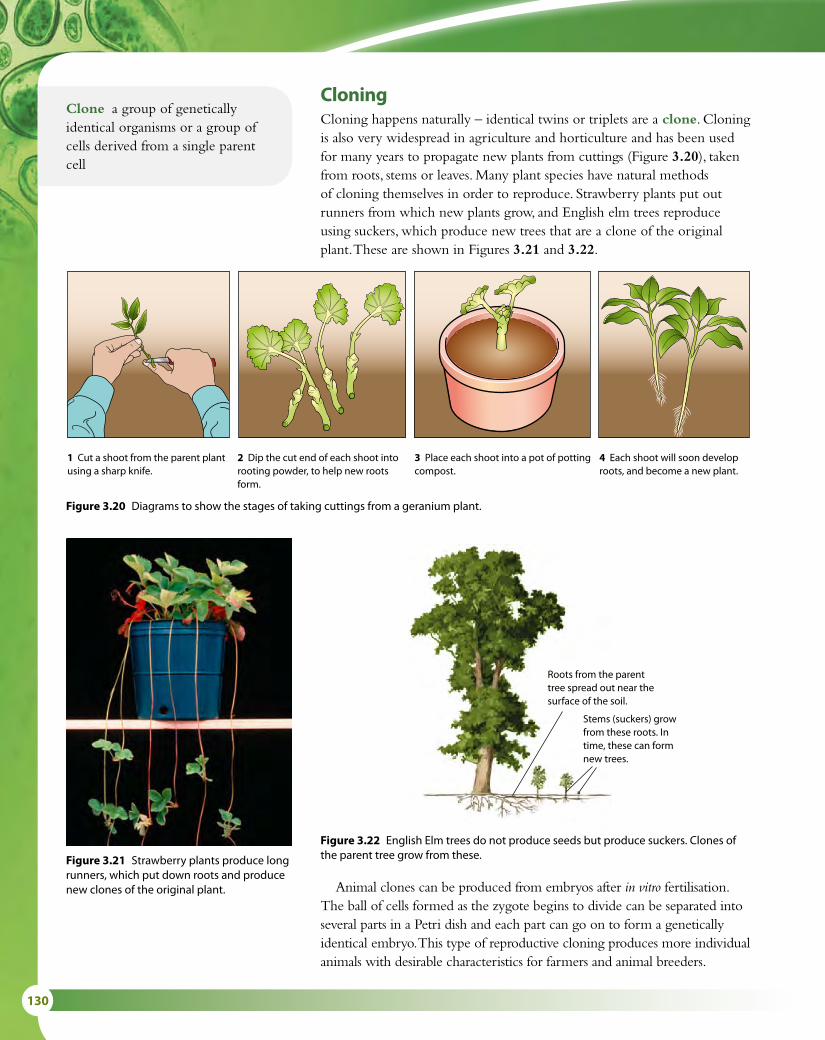
130
Animal clones can be produced from embryos after in vitro fertilisation. The ball of cells formed as the zygote begins to divide can be separated into several parts in a Petri dish and each part can go on to form a genetically identical embryo. This type of reproductive cloning produces more individual animals with desirable characteristics for farmers and animal breeders.
Roots from the parent tree spread out near the surface of the soil.
Stems (suckers) grow from these roots. In time, these can form new trees.
1 Cut a shoot from the parent plantusing a sharp knife.
2 Dip the cut end of each shoot intorooting powder, to help new rootsform.
3 Place each shoot into a pot of pottingcompost.
4 Each shoot will soon developroots, and become a new plant.
Figure 3.20 Diagrams to show the stages of taking cuttings from a geranium plant.
Figure 3.21 Strawberry plants produce long runners, which put down roots and produce new clones of the original plant.
Figure 3.22 English Elm trees do not produce seeds but produce suckers. Clones of the parent tree grow from these.
Clone a group of genetically identical organisms or a group of cells derived from a single parent cell
CloningCloning happens naturally – identical twins or triplets are a clone. Cloning is also very widespread in agriculture and horticulture and has been used for many years to propagate new plants from cuttings (Figure 3.20), taken from roots, stems or leaves. Many plant species have natural methods of cloning themselves in order to reproduce. Strawberry plants put out runners from which new plants grow, and English elm trees reproduce using suckers, which produce new trees that are a clone of the original plant. These are shown in Figures 3.21 and 3.22.

3 GENETICS 131
Clones from differentiated animal cellsCells from a newly fertilised egg are not differentiated and have not specialised into the different cells they will become. Until recently, cloning an animal from another animal was impossible because the cells in an animal’s body had already become nerves, skin, muscles and so on. Differentiated cells have many of their genes switched off, so before they can be used for cloning, the genes have to be switched back on.
The first successful clone made from adult, differentiated animal cells was Dolly the sheep (Figure 3.23). The breakthrough was made in 1997 by Sir Ian Wilmut and his team at a laboratory at the Roslin Institute in Edinburgh, Scotland, where Dolly was created. Unlike previous clones, Dolly was created from the fusion of an ovum with a nucleus from a mammary gland cell of an adult sheep, creating a genetic replica of the original adult animal. The procedure is known as somatic-cell nuclear transfer because the nucleus used comes from a body, or somatic, cell – in this case a mammary gland cell.
A cell was taken from the udder of a ewe of the Finn Dorset breed.
An egg cell was taken from a Scottish Blackface ewe.
The nucleus was removed.
The two cells were fused together.
The egg grew to become an embryo.
The embryo was put into the uterus of a Scottish Blackface ewe.
Dolly was born – a clone of the Finn Dorset ewe.
Figure 3.23 Stages in the creation of Dolly, the first successful clone.
Nature of scienceAssessing risk in science – GM maize and the monarch butterflyThe monarch butterfly is native to the USA (Figures 3.24 and 3.25). In the winter, it hibernates along the coast of southern California or in the forests of central Mexico. As spring arrives, thousands of butterflies begin to head northwards in one of the most spectacular insect migrations in the world. By the time the butterflies reach their breeding ground, in the American corn belt, they may have flown as far as 4800 kilometres (3000 miles).

132
One commonly grown GM crop in this area is maize (corn), which contains a gene from the soil bacterium Bacillus thuringiensis. Maize carrying the Bt gene produces its own toxin to kill insect pests so that fewer pesticides need to be sprayed on it to keep it pest free. Controversy about the crop and its possible threat to monarch butterfly populations began in 1999 when a paper in the journal Nature suggested that the crop might be killing monarch butterfly caterpillars. Entomologist John Losey suggested that dustings of pollen from Bt maize which fell on milkweed plants (the main food source of the butterflies) growing in a maize field might harm the butterflies. Researchers at Iowa State University said they had found monarch butterfly caterpillars were seven times more likely to die when they ate milkweed plants dusted with pollen from Bt-corn rather than unmodified maize in the laboratory.
This study was discredited in 2001 in a study published in the Proceedings of the National Academy of Sciences, which investigated the effects of GM maize on wild butterflies, and demonstrated what happens in the real environment. The team included not only industry-sponsored scientists but also independent researchers. For butterflies to be at risk, they would have to be present in the maize fields at the time that pollen was shed and they would also need to be exposed to enough pollen for it to be harmful. The team found that this was not the case – significant numbers of butterflies were not present in the maize fields at the time when pollen was shed, and therefore massive exposure did not occur in nature.
Monarch butterfly populations are declining but face many threats to their survival, including natural events. For example, in 1995 a snowstorm killed more than 5 million butterflies as they travelled north. Estimates show that in an average year less than 5% of the caterpillars that hatch survive to adulthood. Human activities such as deforestation and development destroy their habitat and pesticides used in maize fields destroy milkweeds, the sole food source for the caterpillars. Nevertheless, headlines in the popular press still claim that ‘GM Crops Kill Butterflies’. In fact, butterflies may be safer in a Bt field than they are in a conventional field, where they may be exposed to chemical pesticides that kill not just caterpillars but most other insects as well.
Figure 3.24 Monarch butterfly caterpillar feeding on milkweeds. Figure 3.25 Monarch butterfly.

3 GENETICS 133
Exam-style questions
1 Which of the following statements is correct?
A Both men and women can be carriers of a sex-linked allele.B Mitosis reduces the number of chromosomes in a cell from the diploid to the haploid number.C All of the genetic information in an organism is called a genome.D The polymerase chain reaction separates fragments of DNA in a gel. [1]
2 The fruit fly, Drosophila, has sex chromosomes X and Y, the same as humans. ‘White eye’ is caused by a sex-linked allele, which is recessive. In the pedigree chart shown, which two individuals must be carriers of the white allele?
A I4 and II3B I4 and II2C II1 and II2D II2 and III2 [1]
25 Explain the purpose of the polymerase chain reaction. 26 State the characteristic of DNA fragments that causes them to
separate during electrophoresis. 27 State two uses of DNA profiling. 28 State the characteristic of the genetic code that allows a gene to
be transferred between species. 29 State the name of the rings of DNA found in prokaryotic cells
that can act as gene vectors. 30 State two enzymes used to make a recombinant plasmid. 31 State two cell types used to make Dolly the sheep.
Test yourself?
Questions to consider
• Why is it important that laboratory-based experiments are supported by observations in the natural world?
• What is the significance of including both independent and industry-sponsored scientists in research into the monarch butterfly?
• How important are stories published in the press to our understanding of scientific issues?
III
II
I
this fly has white eye
1
1
1 2 3
2 3
2 3 4

134
3 The karyogram shows:
A Down syndromeB a sex chromosome trisomyC a normal human femaleD a normal human male [1]
1 2 3 4
child 1
child 2 father
mother
child 4
child 3
origin
5 6
III
IV
I
II
4 Which of the following describes the behaviour of chromosomes during prophase I and metaphase II of meiosis?
Prophase I Metaphase II
[1]
A Chromosomes undergo supercoiling. Sister chromatids are separated.
B Homologous chromosomes pair up together. Homologous pairs of chromosomes line up on the equator.
C Homologous chromosomes pair up together. Chromosomes line up on the equator.
D The nuclear envelope reforms. Chromosomes line up on the equator.
5 The diagram shows a DNA electrophoresis gel. The results are from a single probe showing a DNA profile for a man, a woman and their four children.
1
6
13
21 22
14 15 16 17 18 19 20
7 8 9 10 11 12
2 3 4 5
Which child is least likely to be the biological offspring of the father?
A Child 1B Child 2C Child 3D Child 4 [1]

3 GENETICS 135
6 The following is a sequence of bases in a DNA molecule that has been transcribed into an RNA molecule.
CGGTAAGCCTA
Which is the correct sequence of bases in the RNA molecule?
A CGGTAAGCCTAB GCCAUUCGGAU C CGGUAAGCCUAD GCCUTTCGGAU [1]
7 a In the normal allele for the β-chain of hemoglobin, there is a triplet on the coding strand of the DNA that is C–T–C. In people suffering from sickle-cell anemia, what has this triplet mutated to? [1]
b Because of this mutation, one amino acid in the polypeptide chain of the β-subunits in hemoglobin is abnormal. Name the normal amino acid and also the amino acid resulting from the sickle-cell mutation. [1]
c Explain why this mutation leads to sickle-cell anemia. [3]
8 Ludovica is blood group AB and is expecting a baby with her husband Mikhail who is blood group A. Mikhail’s mother was group O. Deduce the possible genotypes and phenotypes of their baby using a Punnett grid. [4]
9 Genetic modification involves the transfer of DNA from one species to another. Discuss the potential benefits and possible harmful effects of genetic modification. Include a named example. [8]
10 Outline how a DNA profile is obtained, including one way in which it has been used. [5]
11 Karyotyping involves arranging the chromosomes of an individual into pairs. Describe one application of this process, including the way in which the chromosomes are obtained. [5]

136
4 EcologyIntroductionAlmost the entire surface of the Earth – the land, rivers, lakes, seas and oceans – is home to organisms of one kind or another. It has been estimated that there are as many as 10 million different species on Earth and understanding where and how they live and interact is a branch of biology known as ecology. Humans are not the most numerous species on Earth (there are many more bacteria and insects, for example), but humankind is having a disproportionate effect on the world’s ecosystems as damage is caused by pollution, rainforest destruction and global warming.
4.1 Species, communities and ecosystems
‘A true conservationist is a man who knows that the world is not given by his father but borrowed from his children.’
John James Audubon (1785–1851)
The concept of an ecosystemThe idea of the ecosystem provides a convenient way for us to consider and describe the interactions between groups of organisms in their natural environment: an ecosystem is defined as all the groups of organisms living in an area plus the non-living environment with which they interact. One of the most important and recognisable groups in any ecosystem is a species. We define a species as a group of organisms that are able to interbreed and produce fertile offspring. So, lions are a species because they breed with other lions and produce more lions. But if lions are separated from one another by long distances and exist in isolated populations, two individual lions may not actually interbreed even though they remain members of the same species and retain the potential do so.
Learning objectives
You should understand that:
• A species is a group of organisms that have the potential to interbreed to produce fertile offspring.
• If members of a species are in different, separated populations, they may not be able to interbreed.
• Species are defined as autotrophs or heterotrophs depending on their source of food. (Some species are both.)
• Consumers are defined as heterotrophs that feed on living organisms by ingestion.
• Detritivores are defined as heterotrophs that feed on dead organic matter (detritus) by internal digestion.
• Saprotrophs are heterotrophs that obtain nutrients from dead organic matter by external digestion.
• A community is a group of populations living and interacting with each other in a specific area.
• A community interacting with its abiotic environment forms an ecosystem.
• The abiotic environment is the non-living part of an ecosystem, which supplies inorganic materials to autotrophs.
• Nutrient cycling in an ecosystem maintains the supply of inorganic nutrients.
• Ecosystems have the potential to be indefinitely sustainable.

4 ECOLOGY 137
If species are separated in different populations for long periods of time, over many generations the two groups may evolve and become different from one another. You can find out more about this process on Topic 5.
Populations of different species that live together in the same ecosystem form a larger group known as a community. A community comprises all the organisms in the ecosystem. So in a pond ecosystem, the community includes populations of fish, frogs, pond snails, water beetles, pond algae and so on.
The community is affected by the abiotic (non-living) factors in the environment. In a pond, these might include the pH of the water, the nature of the underlying rocks or the amount of sunlight. In a terrestrial ecosystem, climate and physical conditions such as availability of water and type of soil are important. These non-living factors in any ecosystem affect the survival and growth of living things found there, as well as their ability to reproduce and their distribution in the ecosystem (Figure 4.1).
Autotrophs and heterotrophsSpecies are divided into types depending on their method of obtaining food. Autotrophs are species that are able to make their own food from basic inorganic materials and this group includes all plants that can photosynthesise. Autotrophs (which means ‘self feeding’) use light energy to synthesise sugars, amino acids, lipids and vitamins, using simple inorganic substances such as water, carbon dioxide and minerals. Heterotrophs are consumers – species that obtain their food from organic matter. This group includes herbivorous and carnivorous animals, which feed on living organisms. A few organisms, such as Euglena, are capable of feeding both autotrophically and heterotrophically (see Nature of science, page 140).
In order to understand the topics in this chapter, it is important to remember the following terms, which are used in the study of ecology.Species a group of organisms that can interbreed and produce fertile offspringPopulation a group of organisms of the same species who live in the same area at the same timeCommunity a group of populations living and interacting with each other in an areaEcosystem a community and its abiotic environmentEcology the study of relationships between living organisms and between organisms and their environmentTrophic level the position of an organism in a food chain
Sun – solarenergy
abiotic factors including soil, rock
and inorganicnutrients
abiotic factors including wind, temperature, O2,
CO2, water
autotrophs –populations of planets
and photosyntheticspecies (producers)
heterotrophs –populations of herbivores and
carnivores (consumers)
detritivores and saprotrophs –decomposition
Figure 4.1 The components of an ecosystem.
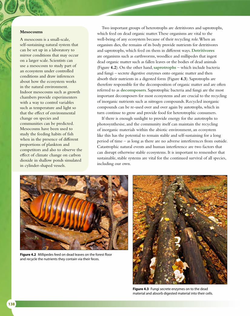
138
Two important groups of heterotrophs are detritivores and saprotrophs, which feed on dead organic matter. These organisms are vital to the well-being of any ecosystem because of their recycling role. When an organism dies, the remains of its body provide nutrients for detritivores and saprotrophs, which feed on them in different ways. Detritivores are organisms such as earthworms, woodlice and millipedes that ingest dead organic matter such as fallen leaves or the bodies of dead animals (Figure 4.2). On the other hand, saprotrophs – which include bacteria and fungi – secrete digestive enzymes onto organic matter and then absorb their nutrients in a digested form (Figure 4.3). Saprotrophs are therefore responsible for the decomposition of organic matter and are often referred to as decomposers. Saprotrophic bacteria and fungi are the most important decomposers for most ecosystems and are crucial to the recycling of inorganic nutrients such as nitrogen compounds. Recycled inorganic compounds can be re-used over and over again by autotrophs, which in turn continue to grow and provide food for heterotrophic consumers.
If there is enough sunlight to provide energy for the autotrophs to photosynthesise, and the community itself can maintain the recycling of inorganic materials within the abiotic environment, an ecosystem like this has the potential to remain stable and self-sustaining for a long period of time – as long as there are no adverse interferences from outside. Catastrophic natural events and human interference are two factors that can disrupt otherwise stable ecosystems. It is important to remember that sustainable, stable systems are vital for the continued survival of all species, including our own.
Mesocosms
A mesocosm is a small-scale, self-sustaining natural system that can be set up in a laboratory to mirror conditions that may occur on a larger scale. Scientists can use a mesocosm to study part of an ecosystem under controlled conditions and draw inferences about how the ecosystem works in the natural environment. Indoor mesocosms such as growth chambers provide experimenters with a way to control variables such as temperature and light so that the effect of environmental change on species and communities can be predicted. Mesocosms have been used to study the feeding habits of fish when in the presence of different proportions of plankton and competitors and also to observe the effect of climate change on carbon dioxide in shallow ponds simulated in cylinder-shaped vessels.
Figure 4.2 Millipedes feed on dead leaves on the forest floor and recycle the nutrients they contain via their feces.
Figure 4.3 Fungi secrete enzymes on to the dead material and absorb digested material into their cells.

4 ECOLOGY 139
Sustainable development – international measures to conserve fish stocks
In recent years, there have been several alarming reports on declining fish populations worldwide (Figure 4.4). In 2003, 29% of open-sea fisheries were in a state of collapse, defined as a decline to less than 10% of their original yield.
To make fishing sustainable in the future, fish populations must be monitored and quotas or closed seasons agreed internationally to reduce fishing during the breeding seasons so that fish numbers can recover after an area has been fished. Some agreements are already in place to control net sizes so that smaller immature fish can be left in the water to breed.
In many parts of the world, bigger vessels, bigger nets and new technology for locating fish are no longer improving catches, simply because there are fewer fish to catch. But where fishing is banned or regulated by international treaties, biodiversity can improve and fish populations may be restored. International cooperation is essential if measures like these are to be successful because neither fish nor trawlers are limited to the jurisdiction of just one country.
The European Union (EU) has enacted several measures to try to ensure a sustainable fishing industry in the North Sea. These include restricting sizes of nets, a ban on drift nets (which catch many different species together) and the imposition of quotas for different fish. Recently, following a public outcry, new rules have been implemented to prevent the return of excess fish to the sea. Until now, fishing boats often had to throw away perfectly good fish if they had exceeded their legally allowed quotas or had caught prohibited species. Sometimes, they also threw back fish that were small, or not popular for human consumption, and therefore of limited value. Some boats discarded as much as two-thirds of their catch. But once bony fish are brought to the surface, their swim bladders are damaged and the fish die – throwing them back does not conserve fish stocks. Fish stocks can only be restored by changing the way the quota rules work and are monitored – so that the number of fish caught and killed is limited, rather than the number brought to shore to sell – and by encouraging selective methods of fishing (for example, by restricting the sizes of nets and banning drift nets). International agreements to support such efforts, such as the new EU rules, are the only way to save fish stocks and maintain sustainable fishing industries for the future.
North Sea cod
1960 1964 1968 1972 1976 1980 1984 1988 1992 1996 2000 2004 2008
fish caughtadult fish in stockssafe stock
Year
Num
ber
of N
orth
Sea
cod
/ ×
1000
tonn
es p
er y
ear
0
250
300
350
400
50
100
150
200
Figure 4.4 Stocks and fishing mortality in North Sea cod between 1963 and 2008. The population of North Sea cod has fallen greatly since the end of the 1960s, almost certainly as a direct result of over-fishing. The horizontal line shows the minimum stock size that has been calculated will allow the cod population to be maintained at a viable level.

140
Nature of scienceLooking for trends and discrepancies – are all plants and algae autotrophic?Autotrophs or ‘self-feeders’ are organisms that produce complex organic compounds from simple substances in their environment. In the majority of cases, autotrophs are plants and algae, which use light from the Sun as a source of energy.
One organism that is neither plant nor alga but which was once classified with the plants because it can photosynthesise is Euglena. Euglena is a unicellular organism with chloroplasts within its cell that enable it to feed autotrophically, like a plant. But Euglena can also feed heterotrophically, taking in organic materials as animals do. When Euglena was first discovered, organisms were classified into just two kingdoms – the plant kingdom and the animal kingdom – and Euglena was impossible to place. Then in the 19th century, Ernst Haeckel added a third kingdom, which he called the Protista, to accommodate organisms like Euglena that display characteristics of both plants and animals.
Some plants exist that do not photosynthesise for their entire life cycles. So far about 3000 have been recognised and all are vascular flowering plants (angiosperms). Some of these plants were once classified as saprophytes because they have no chlorophyll but it is now known that they exist in symbiotic relationships with certain fungi, which supply them with nutrients. This relationship is known as myco-heterotrophy. Plants such as the bird’s nest orchid (Neottia nidus-avis) and the ghost pipe (Monotropa uniflora) are obligate myco-heterotrophs for part of their life cycle and photosynthetic for the rest. The bird’s nest orchid is leafless and grows on the roots of trees, taking nutrients from decaying organic material with the help of fungal mycorrhizae. Its thick fleshy roots resemble a bird’s nest and contain the fungus that helps the plant to feed.
A second group of non-photosynthetic plants are called holoparasites. These plants take water, sugars and other nutrients from a host plant through direct contact with it. One example is the dodder (Cuscata sp.). Dodder has thin stems with leaves that are reduced to tiny scales. Climbing over other plants, it attaches itself to their stems by extensions called haustoria that penetrate the vascular bundles of the host (Figure 4.5). Once it is established, the root of the dodder dies and it is entirely dependent on its host.
Perhaps these plants should lead us to the conclusion that not all plants are always autotrophs and that there are exceptions to the general rule.
Figure 4.5 Haustoria of dodder, or strangleweed, winding around the host plant.
1 Define the following terms. a species b ecology c population
d community e ecosystem.
2 Distinguish between an autotroph and a heterotroph.
Test yourself?
4 ECOLOGY 141
4.2 Energy flow
Food chainsEvery organism needs food to survive but eventually it too is eaten. In any ecosystem, there is a hierarchy of feeding relationships that influences how nutrients and energy pass through it. The sequence of organisms that provide food for one another is known as a food chain.
Green plants are autotrophs, which start food chains because they are able to capture light energy from the Sun. Plants are called producers because they ‘produce’ organic compounds by photosynthesis (Subtopic 2.9). These organic compounds contain chemical energy that has been converted from light energy by the process of photosynthesis. Every other organism in a food chain gets its organic compounds from its food and so is called a heterotroph or consumer.
Consider a hyena eating a cheetah. The cheetah could have eaten an antelope, which in turn had eaten leaves from a plant. Thus, the four organisms are interrelated by their food and form a food chain:
plant → antelope → cheetah → hyena
Every organism fits somewhere in a food chain, and although the organisms that make up the food chain will vary from place to place, almost every food chain starts with a green plant. It may be any part of the plant – the leaves, roots, stems, fruits, flowers or nectar.
Figure 4.6 shows three examples of food chains from different ecosystems. Notice that the arrows in a food chain always point in the direction in which the energy and nutrients flow.
When an organism dies, it provides nutrients for a series of detritivores and saprotrophs, which play a vital role in returning inorganic nutrients to the abiotic environment, from which they are re-used by autotrophic organisms.
Trophic levelsEvery ecosystem has a structure that divides organisms into trophic levels on the basis of their food sources. Trophic means ‘feeding’ and every organism in a food chain is on a particular feeding level.
Green plants are producers and are at the lowest trophic level. Above them come all the consumer levels. The first consumers, or primary consumers, are always herbivores. Any organism above the herbivores will
3 Distinguish between consumers, detritivores and saprotrophs.4 State the name of the feeding group that these organisms belong to: a a Peruvian condor feeding on dead sea lions b an American alligator feeding on a fish c an Australia koala feeding on eucalyptus leaves.
Learning objectives
You should understand that:
• Most ecosystems rely on light energy from the Sun.
• Light energy is converted to chemical energy in carbon compounds during photosynthesis.
• Chemical energy in carbon compounds passes through food chains as organisms feed.
• Energy released during respiration is converted to heat.
• Heat cannot be converted to any other form of energy by living organisms.
• Heat is lost from ecosystems.
• Energy losses at each trophic level restrict the biomass of higher trophic levels and also the length of food chains.

142
Food websFew consumers feed on only one source of food. For example, this food chain describes one set of feeding relationships:
grass → beetle → tree creeper → sparrowhawk
But beetles eat a wide range of plants, tree creepers eat other types of insect and sparrowhawks eat other birds. So this food chain could be interlinked with many others. A food web like the one shown in Figure 4.7 shows
grass
A food chain from the Kalahari desert
grasshopper meerkat eagle
alga
A food chain from a freshwater lake
mayfly nymph dragonfly nymph stickleback
phytoplankton
A food chain from the Antarctic ocean
krill chinstrappenguin
leopard seal
Figure 4.6 Grass, algae and phytoplankton are all examples of photosynthesising producers, which use light as their source of energy. Almost all food chains start with light as the initial source of energy.
be a carnivore and these can be listed as secondary consumer, tertiary consumer and so on. A food chain can therefore be summarised as:
producer → primary consumer → secondary consumer → tertiary consumer

4 ECOLOGY 143
a much more realistic picture of the feeding relations of the organisms in a habitat. Notice how organisms change trophic levels depending on what they are eating at any particular time. In Figure 4.7, for example, the fox is a primary consumer when it is eating a crab apple but a secondary consumer when it is eating a woodmouse.
sparrowhawk
tree creeper
energy from allorganisms
decomposers
caterpillar beetle wood mouse rabbit
crab apple treegrassbluebelloak tree
great tit
fox
tertiary consumer
primary consumers
producers
secondary consumers
5 In the food web in Figure 4.7, state the trophic level of: a the tree creeper when eating a caterpillar b the fox when eating a great tit.
Energy and food chainsArrows in a food chain show the direction of flow of both the energy and nutrients that keep organisms alive. Energy flow through an ecosystem can be quantified and analysed. These studies reveal that, at each step in the food chain, energy is lost from the chain in various ways. Some is not consumed, some leaves the food chain as waste or when an organism dies, and some is used by living organisms as they respire (Figure 4.8). In all three cases, the lost energy cannot be passed to the next trophic level.
Consider an area of African savannah where grass, antelopes and cheetahs form a simple food chain.
• Energy loss 1 – not consumed. The grass stores energy from photosynthesis but the antelopes only eat some parts of the grass, so they do not consume all the energy it has stored.
• Energy loss 2 – not assimilated, or lost through death. The grass that is eaten passes through the digestive system of the antelope but not
Figure 4.7 A food web in oak woodland.
Test yourself?

144
all of it is digested and absorbed, so some passes out in the feces. If an antelope dies and it not eaten by a predator, but decayed by detritivores and saprotrophs, the energy in its body is lost to this food chain.
• Energy loss 3 – cell respiration. The antelope uses energy to move and to keep its body temperature constant. As a result, some energy is lost to the environment as heat.
The assimilated energy remaining after respiration goes into building the antelope’s body and this energy becomes available to the cheetah when it eats the antelope.
Ecologists represent the transfer of energy between trophic levels in diagrams called energy pyramids. The width of each of the layers in the pyramid is proportional to the amount of energy it represents. So the antelope → cheetah energy transfer would appear as in Figure 4.9. This section of an energy pyramid shows that only about 10% of energy from the antelope passes to the cheetah and about 90% has been lost.
Energy losses occur at every step in a food chain, as the energy pyramid in Figure 4.10 illustrates. One consequence of this is that the quantity of biomass at each trophic level decreases due to the loss of waste products at each transfer of energy. Every link in the chain results in losses, so that eventually there will be insufficient energy to support any further trophic levels. Most food chains commonly contain between three and five species, and seldom more than six. The energy that enters an ecosystem as light is converted to stored chemical energy and finally lost as heat.
Energy flow and nutrient recyclingAll the organic matter from an organism, including everything from living or dead material to waste, is eventually consumed by other organisms.
heatenergy
light energy
respiration
waste elimination and death
digestion, assimilation and growth
photosynthesis
resp
iratio
n
det
riti
vore
s
primary consumers
Processesphotosyntheticplants (producers)
tertiaryconsumers
secondaryconsumers
Figure 4.8 Energy losses at each trophic level of a food chain.
energy incheetah
energy in antelope
Figure 4.9 A simple energy pyramid for a single energy transfer.

4 ECOLOGY 145
All these organisms respire and release energy as heat. All the energy that enters ecosystems as light energy, and is trapped by photosynthesis, will eventually be converted to heat and become unavailable to be used again by living things.
Nutrients, on the other hand, are continually recycled. A nitrogen atom may be absorbed as nitrate by a plant root and used to make an amino acid. The amino acid may pass into an animal when the plant material is eaten, and then pass out of the animal’s body during excretion. Soil bacteria may convert urea in the excreted material back into nitrate and the cycle begins again.
Decomposers in the soil, the saprotrophic bacteria and fungi, are essential for the recycling of nutrients. You can find out more about nutrient cycles in Subtopic 4.3.
sunlight
ener
gy tr
ansf
er
producers
primary consumers
secondary consumers
tertiary consumers
Figure 4.10 A generalised energy pyramid.
Feeding the worldDifferent types of farming and food production can be
compared with respect to their trophic levels and efficiency of energy conservation. As arable crops are grown or animals produced, a certain amount of energy is used in the growth of biomass and production of offspring. In a terrestrial ecosystem food is usually harvested at trophic levels 1 and 2 (producers and herbivores). From an energy point of view, farming systems that produce crops are much more efficient than those that produce animals because the energy has passed through only one trophic level.
One hectare of land can produce approximately 7.5 tonnes of wheat (equivalent to 11 000 loaves of bread) or 0.3 tonnes of beef (1200 steaks). Many more people can be provided with food from the wheat rather than the beef.
A steadily rising world population has increased the demand for food and led to environmental problems including loss of forests and soil degradation and, as newly emerging economies worldwide have seen incomes rise, the demand for meat has also increased.
Questions to consider
• Why is arable farming more efficient than farming animals?
• How important are other aspects of animal farms – for example, milk production, wool and leather?
• Do you think there may be a limit to the amount of meat that farmers can produce?

146
Nature of scienceUsing theories to explain natural phenomena – the concept of energy flow and food chainsPlants are the primary source of energy in nearly all ecosystems, but energy capture and photosynthesis are not efficient processes so not all of the energy of the sunlight is used. When herbivores feed, the energy transferred from plant to herbivore is also not 100% efficient. Not all of the plant material is eaten and absorbed, and some energy is lost in movement and respiration. The same is true for carnivores eating prey animals. Only about 10% of the energy in producers is passed to herbivores and a similar low percentage of energy is passed from herbivores to carnivores (Figure 4.11).
All along a food chain or food web, energy is lost at each trophic level through respiration and waste. This is why ecosystems rarely contain more than four or five trophic levels. There is simply not enough energy to support another level and understanding the concept of energy flow explains why food chains are limited in length.
not used inphotosynthesis
respiration
to decomposers
grasses
respiration
to decomposers
woodmice
respiration
to decomposers
foxessunlight
Figure 4.11 Energy losses in a food chain (not to scale). Energy is measured in kJ m–2 y–1. Only a small percentage of the energy in each level is transferred to the next.
6 Explain what the arrows represent in a food chain. 7 Describe what is meant by a food web. 8 State the initial energy source for most food chains. 9 Define ‘trophic level’. 10 List the three ways energy is lost when moving from one
trophic level to the next. 11 The leaves of a tree store 20 000 J m−2 y−1 of energy. Estimate
the amount of energy stored by the caterpillars that feed on the leaves.
Test yourself?

4 ECOLOGY 147
4.3 Carbon recycling
The carbon cycleCarbon is one of the most important elements that are recycled in an ecosystem (Figure 4.12). Inorganic carbon dioxide in the atmosphere is trapped or ‘fixed’ as organic carbon compounds during photosynthesis. Carbon dioxide gas needed for photosynthesis passes by diffusion from the atmosphere into land-dwelling autotrophs and dissolved carbon dioxide diffuses from water into aquatic organisms. Some of this carbon is soon returned to the atmosphere or the water as the plants respire. The other steps in the carbon cycle follow the same path as food chains. As herbivores eat plants, and carnivores eat herbivores, the carbon compounds move from plants to animals. Respiration by any organism in this sequence returns carbon to the environment as carbon dioxide. When a plant or animal dies, carbon compounds in their bodies provide nutrition for detritivores and saprotrophs and may also be respired, returning carbon dioxide to the atmosphere.
Methane, peat and fossil fuelsIn some conditions, plants and animals do not decay completely when they die and organic carbon in their bodies may not be released directly as carbon dioxide as they decompose. In wetlands, where the soil is waterlogged and the concentration of oxygen is very low, organic material is broken down by anaerobic methanogenic bacteria (Achaeans), which produce methane as a byproduct. Peat bogs represent the largest natural source of atmospheric methane, a greenhouse gas many times more harmful than carbon dioxide. A large proportion of this methane is recycled by methane-eating bacteria (methanotrophs) in the peat bog, which form a symbiotic relationship with peat-bog moss. The bacteria supply additional carbon dioxide to the moss, which in turn provides the bacteria with a habitat.
Methane is oxidised to carbon dioxide and water released into the atmosphere. Thus, eventually, carbon dioxide rejoins the carbon cycle and contributes to the carbon dioxide in the atmosphere, although some remains in the ground.
fossil fuels
photosynthesis
aqueouscarbon dioxide
in oceans and lakes
combustion
decaydeath
feedingfeeding
fossilisation
respirat ion
gaseous carbon dioxide in atmosphere
Figure 4.12 The carbon cycle.
Learning objectives
You should understand that:
• In the atmosphere, carbon is present as carbon dioxide gas, while in aquatic environments, carbon is present as dissolved carbon dioxide and hydrogen carbonate ions.
• Carbon dioxide is converted into carbohydrates and other carbon compounds by autotrophs.
• Carbon dioxide enters the cells of autotrophs from the air or water by the process of diffusion.
• Carbon dioxide produced during respiration diffuses out of organisms into the surrounding air or water.
• In anaerobic conditions, methane is produced from organic matter by methanogenic bacteria (Achaeans), and diffuses into the atmosphere or accumulates in the ground.
• In the atmosphere, methane is oxidised to carbon dioxide and water.
• When organic matter is not fully decomposed because of acidic or anaerobic conditions in waterlogged soils, peat forms.
• Oil, gas and coal are formed from partially decomposed organic matter from past geological eras. Oil and gas accumulate in porous rocks.
• Combustion of biomass and fossilised organic matter produces carbon dioxide.
• Some animals, such as corals and molluscs, have parts composed of calcium carbonate and they can become fossilised in limestone.

148
Peat is also produced in wetlands where partly decayed vegetation accumulates and flooding prevents oxygen flow in the soil, so that anaerobic and sometimes acidic conditions persist. For peat to form, the production of biomass must be greater than its chemical breakdown. The first stage in peat formation takes place when water levels are low and aerobic micro-organisms act on decaying vegetation in surface layers. As this layer becomes covered by further layers of vegetation and thus subjected to anaerobic conditions in wetter, deeper layers, it becomes preserved and changes very little with time. Depending on the local conditions, the types of peat from different areas can be quite different in their degree of decomposition. Areas where peat is found also have specific kinds of plants, such as heather, sphagnum moss and sedges. Since organic matter accumulates over thousands of years, peat deposits also provide records of past vegetation and climates.
Peat is harvested as a fuel in some parts of the world as it can be compressed into bricks and unlike coal, burns without producing smoke (Figure 4.13). But peat is not classified as a renewable energy resource because its rate of extraction is far greater than its very slow rate of formation which can take between 1000 and 5000 years.
Methane as a greenhouse gas
Methane has a greater potential to cause global warming than carbon dioxide (Subtopic 4.4) but it is present in much smaller amounts in the atmosphere. It is produced naturally by termites as they digest their food and significant amounts are also released from the oceans. Human activity has increased the release of methane during the extraction of gas, oil and coal, and in farming. Cattle and other ruminants produce methane as part of their digestive process while waste treatment plants and rice fields are also additional sources of methane.
Figure 4.13 Peat is cut to be used as a fuel. Some farmers use it as an additive to improve soil structure.
Some partially decomposed organic matter from past geological eras has become compressed and fossilised in a process that has taken millions of years. Conditions on Earth at the time prevented decomposers from feeding on this material and over time it was fossilised to become fossil fuels. Vast coal, oil and natural gas deposits have been formed deep under the ground where they contain reserves of carbon that are locked in and excluded from the carbon cycle for very long periods of time. The carbon trapped in these fuels cannot return to the atmosphere unless the fuels are burned, when carbon in them combines with oxygen in the air to form carbon dioxide. Over a very long period of geological time, fossil fuel formation has gradually lowered the carbon dioxide level of the Earth’s atmosphere, but in more recent times this balance has been upset. As people burn wood, peat, coal, oil or gas, the carbon molecules locked up in them for thousands or millions of years are released back into the atmosphere as carbon dioxide. As carbon dioxide re-enters the carbon cycle it has the potential to cause global warming (Subtopic 4.4).

4 ECOLOGY 149
Carbon fluxCarbon flux is defined as the flow of carbon from one ‘carbon pool’ to another. It is the net difference between the carbon removal and the carbon addition. For the Earth’s atmosphere, carbon is removed by plant growth, mineral formation and dissolving in the ocean, while it is added through respiration, fossil-fuel burning and volcanic activity. Scientists monitor carbon flux in order to build up a picture of changes and disturbances in the balance of the carbon cycle (Figure 4.14).
eros
ion
rivers
net terre
strial u
ptake 1.4
land-use change 0.7–1.6
land sink 2–4
vegetation 610soils and detritus 1580
2190
atmosphere778
fossil fuels
surface ocean1020
dissolved organiccarbon<700
surface sediment150
intermediateand deep ocean
38 000
marine biota3
weathering
0.2
91.6
40 50 100
92
90.3
0.8
82
2
6.3
Figure 4.14 Diagram showing carbon fluxes in a natural system. Carbon stores are shown in Gt (gigatonnes) and carbon fluxes are shown in gigatonnes per year (Gt C yr–1).
Nature of scienceObtaining evidence – how is reliable data on carbon dioxide and methane concentrations obtained?The level of carbon dioxide in the atmosphere has been increasing since the end of the 19th century. The first measurements of the gas were made by Charles Keeling (1928–2005) who took careful measurement of samples from Mauna Loa in Hawaii and the Antarctica in 1957 and 1958. Since then records of the gas have been kept continuously and the
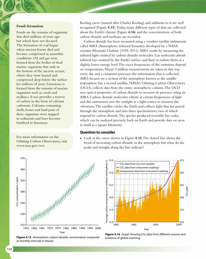
150
1970 1975 1980 1985 1990
Year
1995
370
1960 1965 2000
360
350
340
330
320
3101955
CO
2 co
ncen
trat
ion
/ pp
m
15.5
15.0
14.5
14.0
13.5
13.0Ave
rage
sur
face
tem
per
atur
es in
nor
ther
n he
mis
phe
re °C
375
350
325
300
275
250
CO
2 p
pm
2004
Year
195019001860
CO2 data from instrument readingstemperature data from instrument readings
CO2 data from ice-core samples
Figure 4.15 Atmospheric carbon dioxide concentration measured at monthly intervals in Hawaii.
Figure 4.16 Graph showing CO2 data from different sources and evidence of global warming.
Keeling curve (named after Charles Keeling) and additions to it are well recognised (Figure 4.15). Today, many different types of data are collected about the Earth’s climate (Figure 4.16) and the concentrations of both carbon dioxide and methane are recorded.
Carbon dioxide has been measured using a weather satellite instrument called AIRS (Atmospheric Infrared Sounder) developed by a NASA scientist Moustafa Chahine (1935–2011). AIRS works by measuring the infrared light emitted by carbon dioxide molecules. Gas molecules absorb infrared rays emitted by the Earth’s surface and then re-radiate them at a slightly lower energy level. The exact frequencies of the emissions depend on temperatures. About 3 million measurements are taken in this way every day and a computer processes the information that is collected. AIRS focuses on a section of the atmosphere known as the middle troposphere but a second satellite, NASA’s Orbiting Carbon Observatory (OCO), collects data from the entire atmospheric column. The OCO uses optical properties of carbon dioxide to measure its presence using an AIRS. Carbon dioxide molecules vibrate at certain frequencies of light and this instrument uses the sunlight as a light source to monitor the vibrations. The satellite circles the Earth and collects light that has passed through the atmosphere and into three spectrometers, two of which respond to carbon dioxide. The spectra produced resemble bar codes, which can be analysed precisely back on Earth and provide data on areas as small as a square kilometre.
Question to consider
• Look at the curve shown in Figure 4.15. The dotted line shows the trend of increasing carbon dioxide in the atmosphere but what do the peaks and troughs along the line indicate?
For more information on the Orbiting Carbon Observatory, visit www.nasa.gov/oco.
Fossil formation
Fossils are the remains of organisms that died millions of years ago but which have not decayed. The formation of coal began when ancient forests died and became compressed in anaerobic conditions. Oil and gas were formed from the bodies of dead marine organisms that sank to the bottom of the ancient oceans, where they were heated and compressed deep below the surface for millions of years. Limestone is formed from the remains of marine organisms such as corals and molluscs. It too provides a reserve of carbon in the form of calcium carbonate. Calcium-containing shells, bones and hard parts of these organisms were trapped in sediments and have become fossilised in limestone.

4 ECOLOGY 151
Earth
atmosphere
Some longwave radiationis absorbed and re-emittedby greenhouse moleculesin the atmosphere. Thiswarms the Earth’s surfaceand lower atmosphere.
Shortwave solarradiation passessthrough theatmosphere.
Longwave (infrared)solar radiation isreflected back by theEarth.
4.4 Climate change
How the greenhouse effect worksCertain gases, the most important of which are carbon dioxide and water vapour, enable the atmosphere to retain heat. Without these gases in the atmosphere, the Earth’s temperature would be too low to support life. The warming effect of these gases is known as the greenhouse effect because it is caused in a similar way to the warming of a greenhouse.
A greenhouse is made of glass, which allows shortwave radiation from the Sun to pass through it. As the sunlight passes through the glass, the radiation is absorbed, changed into heat – which has a longer wavelength – and re-radiated. Glass is less transparent to these long wavelengths and heat is trapped in the greenhouse, making it warmer inside. So-called ‘greenhouse gases’ in the Earth’s atmosphere (such as carbon dioxide, methane and water vapour) act in a similar way to the greenhouse glass. They trap heat that is radiated from the Earth’s surface and keep the Earth at a comfortable temperature for life to exist (Figure 4.17).
12 State the difference between energy movement and nutrient movement in an ecosystem.
13 State two ways in which the formation of peat differs from the formation of coal.
Learning objectives
You should understand that:
• The two most significant greenhouse gases are carbon dioxide and water vapour.
• Methane and oxides of nitrogen are also greenhouse gases but they have less impact.
• Longer wavelength radiation is emitted by the warmed Earth.
• Greenhouse gases re-absorb longer wavelength radiation, retaining heat in the atmosphere and thus reducing heat loss from Earth.
• The impact of a greenhouse gas on global temperatures and climate patterns depends on how well it absorbs long wavelength radiation and on its concentration in the atmosphere.
• Concentrations of carbon dioxide in the atmosphere have been increasing since the start of the Industrial Revolution 200 years ago. Average global temperatures have also been rising over the same period.
• Increases in carbon dioxide concentration in recent years are mainly due to increases in the combustion of fossil fuels.
Figure 4.17 Greenhouse gases trap heat, warming the atmosphere.
Test yourself?

152
Greenhouse gases, global warming and climate patternsCarbon dioxide currently forms only 0.04% of the atmospheric gases but it plays a significant part in the greenhouse effect. Other greenhouse gases include water vapour, methane, oxides of nitrogen and fluorocarbons (FCs).
The human population has increased dramatically in recent history, with a consequent increase in demand for energy in industry, transport and homes. Most of this energy demand has been met by burning fossil fuels, mainly oil, coal and gas. Burning fossil fuels releases both carbon dioxide and oxides of nitrogen. This activity has raised the concentration of carbon dioxide in the Earth’s atmosphere significantly since the mid-1800s, a period which has coincided with increasing industrialisation (Figure 4.18), and by more than 20% since 1959 (Figure 4.15 and Table 4.1). In the tropical regions of the world huge rainforests trap carbon dioxide through photosynthesis and have been important in maintaining the low level of atmospheric carbon dioxide. Humans have upset this balance by deforesting vast areas of forest for agriculture and timber production. Forest destruction has multiple effects, but the most important for the atmosphere are the loss of carbon dioxide uptake by photosynthesis and the increase in carbon dioxide released from the rotting or burnt vegetation.
Methane is another important greenhouse gas. It is produced by human activity when organic waste decomposes in waste tips. It also comes from rice paddies and from cattle farming. More rice is being planted as the human population increases and more cattle are being farmed for meat. Cattle release methane from their digestive systems as they process their food.
The influence of increased concentrations of greenhouse gases has produced changes in global temperatures and climate patterns. Rising levels of greenhouse gases are believed to be causing an enhancement
HFCs as greenhouse gases
Chlorofluorocarbons (CFCs) were used in aerosols and as refrigerants but were found to damage the ozone layer when released into the atmosphere. They are being replaced by hydrofluorcarbons (HFCs), but this is leading to an additional problem because HFCs are greenhouse gases. Note that damage to the ozone layer is not a cause of the greenhouse effect or increased global temperatures.
350
340
330
320
310
300
290
280
270
Car
bon
dio
xide
/ pp
m
Year
1000 1200 1400 1600 1800 2000
Figure 4.18 Graph to show the increase in carbon dioxide concentration in the atmosphere since the Industrial Revolution.

4 ECOLOGY 153
of the natural greenhouse effect. Scientists have shown that the Earth is experiencing a rise in average global temperature, known as global warming (Figure 4.16) which is thought to be happening because of this enhanced greenhouse effect. Climatologists are concerned that, as a result of all this activity, humans are adversely affecting our atmosphere.
Some possible results of global warming might be:
• melting of ice caps and glaciers
• a rise in sea levels, causing flooding to low-lying areas
• changes in the pattern of the climate and winds – climate change – leading to changes in ecosystems and the distributions of plants and animals
• increases in photosynthesis as plants receive more carbon dioxide.Some may argue, for political or other reasons, that it is hard to justify action to reduce the impact of the release of greenhouse gases, but there are many compelling arguments that individuals and societies should consider.
• Global warming has consequences for the entire human race and an international solution is needed to tackle the problems. It is not always the case that those who produce the most greenhouse gases suffer the greatest harm so it is essential that measures to reduce emissions are taken with full international cooperation.
• If industries and farmers in one area invest money to reduce their greenhouse gases while those in other areas do not, an economic imbalance may be created in favour of the more polluting enterprises, who can offer services more cheaply.
• Consumers can be encouraged to use more environmentally friendly goods and services.
Year Carbon dioxide concentration /
parts per million (ppm)
% increase from the
previous value
% increase from 1959
1959 316
1965 320 1.3 1.3
1970 326 1.9 3.2
1975 331 1.5 4.7
1980 339 2.4 7.3
1985 346 2.1 9.5
1990 354 2.3 12.0
1995 361 2.0 14.2
2000 369 2.2 16.8
2002 373 1.1 18.0
2004 378 1.3 19.6
2006 382 1.1 20.9
2008 386 1.0 22.2
Table 4.1 The atmospheric carbon dioxide levels monitored at the Mauna Loa laboratory in Hawaii since 1959. Although the percentage increase from the previous year tends to show a downward trend from 2000, the percentage increase from 1959 continues to rise (source: NOAA Earth System Research Laboratory).
Climate change
The term ‘global warming’ is somewhat simplistic because some areas of the world will be colder as a result of an increase in greenhouse gases. A better term is ‘climate change’. Climatologists try to make predictions about changes in weather patterns using computer-generated climate models.

154
• Scientists can argue that it is better to invest in a sustainable future and prevent further harm.
• It is unethical for one generation to cause harm to future generations by not taking action to address the problem of greenhouse gases.
Increasing carbon dioxide levels and fragile ecosystems
Coral reefsCoral reefs are fragile ecosystems, which are very sensitive to rising carbon dioxide levels. They are sometimes known as the ‘rainforests of the sea’ because of their extensive biodiversity. Coral polyps extract calcium from seawater and use it to build the elaborate limestone skeletons that make up a coral reef. As well as large numbers of fish and other species, coloured species of algae live within a reef and give it its colourful appearance. Corals respond to the stress of higher sea temperatures which result from global warming by expelling these algae – an event known as coral bleaching (Figure 4.19). Some coral can recover from bleaching but often the coral dies, and the entire reef ecosystem disappears.
Ocean acidification occurs when higher CO2 levels in the atmosphere cause the oceans to absorb more carbon dioxide. Acidic oceans may inhibit the growth of producers and so affect food chains generally, but it is also a direct threat to corals. In a more acidic ocean, corals cannot form their skeletons properly and their growth slows down. It has been estimated that a doubling of atmospheric carbon dioxide will reduce calcification in some corals by as much as 50%. Predicted rises in sea level caused by melting sea ice could also affect some reefs by making the water too deep to allow adequate sunlight to reach them.
Figure 4.19 Bleached coral.

4 ECOLOGY 155
The ArcticThe ecosystems of the Arctic include the tundra, permafrost and the sea ice, a huge floating ice mass surrounding the North Pole (Figure 4.20). Scientists studying these regions have recorded considerable changes in recent years. Average annual temperatures in the Arctic have increased by approximately double the increase in global average temperatures and the direct impacts of this include melting of sea ice and glaciers.
Trouble in Greenland
The snow in Greenland is not all pristine white. There are patches of brown and black dotted all over the landscape. This is cryconite, a mixture of desert sand, volcanic ash and soot from burnt fossil fuels carried by the wind to Greenland from hundreds or thousands of kilometres away. The more soot there is, the blacker the snow becomes. Black objects absorb the Sun’s heat more quickly than white ones do, so the sooty covering makes the underlying snow melt faster. As it melts, the previous year’s layer of cryconite is exposed, which makes the layer even blacker. This positive feedback is causing rapid melting of the Greenland ice sheets.
0°
10°
20°
30°
40°
50°
60°
70°
80°
90°
100°
110°
120°
130°
140°
150°
160° 170°
180°
170°
160°
150°
140°
130°
120°
110°
100°
90°
80°
70°
60°
50°
40°
30°
20°10
°Arctic Circle
70°
80°
0 20001000 1500500 km
Ice shelf
Ice cap
Polar pack ice
Drifting iceEUROPE
ASIA
RUSSIA
USA
CAN
AD
A
NORTHAMERICA
SWED
ENN
ORW
AY
FINLAND
ICELAND
GREENLAND
A r c t i c O c e a n
NorwegianSea
BarentsSea
KaraSea
LaptevSea
East SiberianSea
BeaufortSea
BeringSea
Baffin Bay
EllesmereIsland
ParryIslands
VictoriaIsland
WrangelIsland
New SiberianIslands
BaffinIsland
BrooksRange
KolymaRange
VerkhoyanskRange
MackenzieMts
CentralSiberianPlateau
WestSiberian
Plain
FranzJosefLand
SevernayaZemlya
NovayaZemlya
Spitzbergen
NORTH POLEPeary 1909
Melting sea ice affects many species. Algae, which are important producers in Arctic food chains, are found just beneath the sea ice. As the ice disappears, so do the algae and this affects numerous other organisms that use them as food. Populations of marine mammals, caribou and polar bears are also affected and have already been forced to adapt to changes in their habitats. Polar bear numbers are decreasing at an alarming rate.
Forest and tundra ecosystems are also important features of the Arctic environment. In Alaska, substantial changes in forest life, including increases in insect pests, have been observed as some species from temperate climates are extending their ranges to the north. Detritus trapped in frozen tundra is also released as the ground thaws. The detritus decomposes and the carbon dioxide and methane produced are released into the atmosphere, contributing to rising greenhouse gas levels.
Figure 4.20 Extent of the Arctic region.

156
Nature of scienceAssessing risk in science – the precautionary principleThe precautionary principle suggests that if the effect of a change caused by humans may be harmful to the environment, action should be taken to prevent it, even though there may not be sufficient data to prove that the activity will cause harm. The precautionary principle is most often applied to the impact of human actions on the environment and human health, as both are complex and the consequences of actions may be unpredictable.
One of the cornerstones of the precautionary principle, and a globally accepted definition, comes from the Earth Summit held in Rio in 1992.
‘In order to protect the environment, the precautionary approach shall be widely applied by States according to their capabilities. Where there are threats of serious or irreversible damage, lack of full scientific certainty shall not be used as a reason for postponing cost-effective measures to prevent environmental degradation.’
There are many warning signs to indicate that climate change will have a serious effect on ecosystems across the world. It is clear that global temperatures are increasing and there is a significant probability that this is caused by human activities. It is also likely that weather patterns will alter and cause changes in sea levels and the availability of land for farming. The challenge to governments, industries and consumers is to take action without waiting for definitive scientific proof to be forthcoming.
Question to consider
• Are there any reasons to doubt that climate change is occurring?
14 List three groups of organisms in the carbon cycle that transfer carbon dioxide to the atmosphere.
15 State the process in the carbon cycle by which carbon dioxide is fixed as organic carbon-containing compounds.
16 List three greenhouse gases. 17 List five human activities that are causing an increase in the
levels of greenhouse gases in the atmosphere. 18 State two ways in which a rise in average global temperature is
having an effect on coral ecosystems.
Test yourself?

4 ECOLOGY 157
Exam-style questions
1 Which of the following statements is correct?
A A community is the place where several different species live.B Heterotrophs are organisms that feed off organic matter.C Decomposer bacteria are detritivores.D A habitat is a community and its abiotic environment. [1]
2 Which of the following statements is correct?
A The precautionary principle states that if the effects of a human-induced change would be very large, those responsible for the change must prove that it will not be harmful before proceeding.
B The greenhouse gases methane and oxides of nitrogen have increased global temperatures by converting shortwave radiation to longwave radiation.
C In the carbon cycle, the process of complete decomposition of plant remains stores carbon in the form of fossil fuels.
D Coral can grow faster in oceans which have more carbon dioxide dissolved in them because they are warmer. [1]
3 Which substances increase the greenhouse effect by the greatest amount?
A nitrogen and methaneB sulfur dioxide and nitrogenC methane and HFCsD oxygen and HFCs [1]
4 The diagram below shows part of a carbon cycle involving only plants. What are the processes labelled 1 and 2?
1 2
A photosynthesis combustion
B decomposition respiration
C respiration decomposition
D respiration photosynthesis
CO2 in the air
1 2
Carboncompounds in
plants
5 The total energy in an area of grassland was analysed and found to be 400 kJ m–2 y–1. Construct a labelled pyramid of energy for this grassland for the first three trophic levels, assuming an energy loss of 90% at each level. It is not necessary to draw it to scale. [3]

158
6 According to data given by Kleiber (1967), a cow may weigh as much as 300 rabbits. The following data are also relevant:
1 cow 300 rabbits
Total body mass/kg 600 600
Daily food intake (kg hay) 7.5 30.0
Daily heat loss/kJ 82 000 329 000
Daily mass increase/kg 0.9 3.6
a i Calculate the increase in mass per tonne of hay consumed for both the cow and the rabbits (tonne =1000 kg). Show your working. [4]
ii Explain why this increase in mass is different from the daily increase in mass shown in the table. [2]
b Suggest a reason why the rabbits have a higher daily heat loss than the cow. [2]
7 An island off the coast of South America is the breeding ground for sea lions (Otaria flavescens), which give birth on the island. The island has a plentiful supply of algae, which supplies food for fish. There are large numbers of animals present some of which occupy precarious positions on the rocks. After giving birth adult sea lions return to the sea to feed on fish.
a Draw a food chain to represent the feeding relationships described. [3]
b Label the trophic level of each organism you have included. [2]
Abandoned and dead sea lion pups are found on a beach on the mainland. Here, there are many vultures, which feed on the dead pups.
c State the feeding group of a vulture which feeds on a dead sea lion pup. [1]
8 Charles Darwin wrote: ‘It is interesting to contemplate an entangled bank at the side of a meadow, clothed with plants of many kinds with birds singing on the bushes, various insects flying about and worms crawling through the damp earth …’
With reference to the ‘entangled bank at the side of a meadow’:
a i State whether the ‘bank’ is an ecosystem or a community. [1]
ii Give a reason for your choice. [1]
b State two ways in which birds and plants may interact with one another. [2]
c Name one organism that is mentioned which is a secondary consumer. [1]
d Outline how nutrients are recycled in this area. [3]

5 EVOLUTION AND BIODIVERSITY 159
Evolution and biodiversity 5IntroductionOver long periods of time and many generations, the genetic make-up of species may change as they become adapted to new surroundings or altered conditions. One result of these changes may be the evolution of new varieties and species. There is strong evidence for the evolution of life of Earth, both from the fossil record and from organisms that are alive today. Natural selection provides an explanation of how evolution might have occurred. Classification of organisms helps us to understand their ancestry and our observations of biodiversity, as well as providing further evidence for the mechanism of evolution.
5.1 Evidence for evolution
What is evolution?Life on Earth is always changing. Just by looking at any group of individuals of any species – whether humans, cats or sunflowers – you can see that individuals are not all the same. For example, the people in Figure 5.1 vary in height, hair colour, skin tone and in many other ways. How do these differences within a species occur? How do different species arise?
Variation within a species is a result of both genetic and environmental factors. We say that selection pressures act on individuals and because of variation, some may be better suited to their environment than others. These are likely to survive longer and have more offspring.
The characteristics of a species are inherited and passed on to succeeding generations. The cumulative change in these heritable characteristics over generations is called evolution. If we go back in time, then existing species must have evolved by divergence from pre-existing ones. All life forms can therefore be said to be linked in one vast family tree with a common origin.
What evidence is there for evolution?
The fossil recordFossils, such as the one shown in Figure 5.2, are the preserved remains of organisms that lived a long time ago. They are often formed from the hard parts of organisms, such as shell, bone or wood. Minerals seep into these tissues and become hardened over time. As the living tissue decays, the minerals form a replica that remains behind. Soft tissue can sometimes be preserved in the same way, as can footprints and animal droppings. Most fossils become damaged over time or are crushed through land or sea movement, but some are discovered remarkably well preserved. The earliest
Learning objectives
You should understand that:
• Evolution takes place when heritable characteristics change, over many generations.
• Evidence for evolution comes from the fossil record.
• Artificial selection can result in evolution, as the selective breeding of domesticated animals shows.
• Evolution of homologous structures by adaptive radiation explains similarities in structure with differences in function.
• Populations of a species can diverge and this can eventually lead to the evolution of separate species.
• The continuous variation observed across the geographical range of related populations supports the idea of gradual divergence.
Evolution cumulative change in the heritable characteristics of a population

160
fossils found date from over 3 billion years ago, so the time scale of the fossil record is immense. Most fossils are of species that died out long ago, because they did not adapt to new environmental conditions.
The study of fossils is called palaeontology. Palaeontologists have been collecting and classifying fossils for over 200 years, but they have only been able to date them since the 1940s. Scientists do this by studying the amount of radioactivity in a fossil specimen. Over time the amount of radioactivity decreases because radioactive elements decay. The rate of decay is fixed for each element, so it is possible to date a fossil by measuring the amount of radioactivity present in it. Carbon-14 is used to study material up to 60 000 years old. For older material, other elements are used.
Although the fossil record is incomplete and fossils are very rare, it is possible to show how modern plants and animals might have evolved from previous species that existed hundreds or thousands of millions of years ago. For example, fossil sequences suggest how modern horses may have evolved from earlier species (Figure 5.3). It is important to recognise that we can never say that ‘this species evolved into that species’, based on a fossil sequence – even when we have many fossils. All that we can say is that they appear to be related – that they probably share a common ancestor. Other species could well have existed too, for which no fossils have ever been found.
A few organisms seem to have changed very little. The horseshoe crab we see today is very similar to fossil specimens a million years old. This would seem to suggest that there has been little selection pressure on these crabs.
Observations of fossils provide evidence that life on Earth changes and that many of the changes occur over millions of years.
Selective breedingFurther evidence for the way evolution might occur comes from observations of selective breeding. People have altered certain domesticated species by breeding selected individuals in a process called artificial selection. Plants or animals with favourable characteristics are chosen and bred together, to increase the number of offspring in the next generation that have the favourable characteristics. Those individuals that do not have the desired features are not allowed to breed. People have domesticated and bred plants and animals in this way for thousands of years and, over many generations, this has resulted in the evolution of numerous breeds and varieties, which differ from each other and from the original wild ancestors.
Modern varieties of wheat, barley, rice and potatoes produce higher yields and are more resistant to pests and disease than ever before. Wheat and rice plants are shorter and stronger than varieties of a hundred years ago, so that they are less likely to be damaged by wind and rain and are easier to harvest. The plants of a hundred years ago were also very different from the original grasses that wheat was bred from 10 000 years ago. Many plants are also bred for their appearance, and ornamental varieties have
Figure 5.1 Most of the variation between humans is continuous variation, and is influenced by the environment as well as genes.
Remains of organisms discovered preserved in ice, tar and tree sap have also yielded important information about the evolution of species.
Figure 5.2 A fossil of Archaeopteryx, which is seen as an evolutionary link between reptiles and birds. It looked like a small dinosaur, but had feathers and could fly.

5 EVOLUTION AND BIODIVERSITY 161
different petal shapes and colours from the original parent stock. Animals are chosen and bred by farmers and animal breeders for special
characteristics such as high milk yield in a cow, or good-quality wool in a sheep. Individuals with these characteristics are selected to breed, so that more of the next generation have these useful features than if the parents had not been artificially selected (Figure 5.4).
Although the driving force for artificial selection is human intervention, which is quite different from natural evolution, selective or artificial breeding does show that species can change over generations.
Homologous structuresThe existence of homologous structures provides another strand of evidence for evolution. Homologous structures are anatomical features showing similarities in shape, though not necessarily in function, in different organisms. Their presence suggests that the species possessing them are closely related and derived from a common ancestor. A good example is the vertebrate pentadactyl limb. This is found in a large range of animals including bats, whales and humans, as shown in Figure 5.5. In each group, limbs have the same general structure and arrangement of bones but each one is adapted for different uses. Bird wings and reptile
years before present
2 million
5 million
24 million
34 million
55 million
Hyracotherium
Mesohippus
Merychippus
Pliohippus
Equus – the modern horse
Figure 5.3 Some of the many species of fossil horses, and the modern horse, Equus. The fossil sequence shows that, over time, horses have developed single-toed hooves, longer legs and longer faces with larger teeth for grazing.
Figure 5.4 Selective breeding of cows over many centuries has produced many breeds including the Guernsey, bred for the production of large quantities of fat-rich milk. Other breeds have been produced with flat backs to facilitate birthing, and longer legs for easier milking.

162
limbs are also homologous structures. Even though a bird uses its wings for flying and reptiles use their limbs for walking, they share a common arrangement of bones.
variation and divergenceFor almost every species, there is genetic variation between individuals in a population and between populations in different areas further differences may be visible. For example, some groups of birds may have brighter plumage than others, some troupes of howler monkeys may be able to call more loudly than others and some groups of mice may have slightly darker fur than others giving them better camouflage. This continuous variation between populations, some very slight and some very obvious, provides support for the proposal that populations have the potential to
Reptile
Bird
HumanBat
Whale
55
44
3
3
32
2
2
11
1
5 4 32
1
5
5
4
4
3
32
2
1
1
Frog
humerus
radius
carpal
ulna
humerus
ulnaradius
carpalulna
humerus
radius
carpal
humerus
ulnaradius
carpal
humerus
ulna
carpal
radius
humerus
ulna
carpal
radius
Figure 5.5 The forelimbs of animals with pentadactyl limbs all have a clearly visible humerus, radius, ulna and carpals.
Adaptive radiation is a term used to explain how organisms diverge into a range of new forms from a single common ancestor. It can occur if the environment changes and new sources of food or new habitats become available. The pentadactyl limb demonstrates adaptive radiation in the vertebrates, and Darwin’s finches (Subtopic 5.2) are an example of how one species adapts to exploit new resources.

5 EVOLUTION AND BIODIVERSITY 163
diverge from one another and evolve into separate species. If this does happen, there is also the possibility that new species could be formed (Subtopic 5.2).
Beyond reasonable doubt?Evolutionary history is a difficult area of science. It is not
possible to go back in time or conduct experiments to establish past events or what may have caused them. Nevertheless, in some cases, modern scientific methods can help establish beyond reasonable doubt what has taken place. Science is used to demonstrate the existence of phenomena we cannot observe directly. Many crucial scientific discoveries – such as atoms, electrons, viruses, bacteria, genes and DNA – have been made and accepted using indirect observation and the scientific method of ‘inference’. The theory of evolution has been considered using multiple lines of research, including studies of anatomy, biochemistry and palaeontology, which provide empirical data consistent with the theory.
New methodologies that have been used include carbon dating of fossils, the study of DNA, comparison of exons and introns (Subtopic 7.1) from existing organisms and their fossil ancestors, and the establishment of endosymbiosis with the discovery of rRNA sequences using PCR (Subtopics 1.5 and 2.7). All of the data from these techniques have provided evidence of relationships between species that are consistent with the relationships suggested by the theory of evolution.
Questions to consider
• Do modern methods establish the theory of evolution beyond reasonable doubt?
• Will it ever be possible to ‘prove’ that evolution occurred?
• How do the techniques used in science differ from those that a historian might use to establish what has taken place in the past?
1 Define the term ‘evolution’.2 Outline what is meant by a homologous structure.3 Outline the evidence for natural selection provided by selective
breeding.
Test yourself?

164
5.2 Natural selection
A mechanism for evolutionThe theory of evolution by means of natural selection was proposed by Charles Darwin and Alfred Wallace. Darwin explained his ideas in a book called On the Origin of Species by Means of Natural Selection, published in 1859. The explanation remains a theory because it can never be completely proved but there is an abundance of evidence to support the key ideas, which are based on the following observations and deductions. Some terms we use now were not used by Darwin, who had no knowledge of genes or alleles. However, the fundamental basis of his argument was the same as outlined here.
1 Populations are generally stable despite large numbers of offspring
Organisms are potentially capable of producing large numbers of offspring and far more than the environment can support. Trees can produce thousands of seeds and fish hundreds of eggs. Yet few of these survive to maturity and we rarely see population explosions in an ecosystem.
2 Better adapted individuals have a competitive advantageBoth plants and animals in a growing population will compete for resources. These may be food, territory or even the opportunity to find a mate. In addition, predators and disease will take their toll. This competition will bring about a struggle for survival between the members of a population. Organisms that are well adapted to the conditions will be good at competing and will tend to survive to reproduce, passing on heritable traits to their offspring, while others die.
3 There is heritable variation within speciesDifferent members of the same species are all slightly different and this variation is due to the mechanism of sexual reproduction. The process of meiosis produces haploid gametes and furthermore the genes in the gametes an individual produces may be present in different forms or alleles. When an egg is fertilised, the zygote contains a unique combination of genetic material from its two parents. Sexual reproduction gives an enormous source of genetic diversity, which gives rise to a wide variation among the individuals of a species.
4 Advantageous heritable traits become more frequent over generations
As a result of variation, some members of a population may be better suited (better adapted) to their surroundings than others. They may have keener eyesight, or have better camouflage to avoid predators. These individuals will out-compete others; they will survive better, live longer, and pass on the genes for their advantageous characteristics to more
Learning objectives
You should understand that:
• Variation between individuals is required for natural selection to occur.
• Variation can be caused by mutation, meiosis and sexual reproduction.
• Adaptations are features that make an individual better suited to its environment and way of life.
• More offspring tend to be produced than the limited resources in the environment can support, which means there is a struggle for survival between individuals.
• Better adapted individuals tend to survive and produce more offspring while less well-adapted individuals tend to die or produce fewer offspring.
• When individuals reproduce, they pass on their heritable characteristics to their offspring.
• Natural selection increases the frequency of well-adapted individuals in a population and decreases the frequency of individuals without the adaptations, and thus leads to changes in the species.
Characteristics that an organism acquires during its lifetime, such as large muscles or special skills resulting from training, immunity to a disease or scars on its skin, cannot be passed on to its offspring. Only heritable characteristics, which are determined by genes, can be passed on to the next generation via the parents’ gametes.

5 EVOLUTION AND BIODIVERSITY 165
offspring. Gradually, as the process is repeated generation after generation, the proportion of these genes in the population as a whole increases. This is called natural selection, and it occurs as the ‘fittest’ (best adapted) survive to reproduce.
Natural selection and evolutionOnce species have evolved to become well adapted to conditions in a stable environment, natural selection tends to keep things much the same. However, if the environment changes, a population will only survive if some individuals have heritable traits that suit them to the new conditions, and these then become more frequent in the population, because of natural selection. Three examples of how this can happen in a relatively short period of time are the beak adaptations of Galapagos finches after a change in food availability, the response of a moth population to pollution, and the emergence of new strains of bacteria following the introduction of antibiotics.
Darwin’s finchesThe finches living on the Galapagos Islands (Figure 5.6), about 900 km off the coast of Ecuador, were important in shaping Darwin’s ideas about natural selection. Studies of the birds, now known as Darwin’s finches, continue to this day and modern DNA analysis indicates that all 13 species now found on the islands probably evolved from a small flock of about 30 birds that became established there around 1 million years ago.
When the birds first arrived, the Galapagos Islands were probably free of predators and initially the resources were sufficient for all the individuals. As the population grew, the finches started to adapt their feeding habits to avoid competition and as each group selected different
Sexual reproduction promotes variation
Mutations in genes cause new variations to arise, but sexual reproduction also increases variation in a population by forming new combinations of alleles.• During meiosis, crossing over
at prophase I and random assortment in metaphase I produce genetically different gametes (Subtopic 3.3).
• Different alleles are also brought together at fertilisation, promoting more variation.
In species that reproduce asexually, variation can arise only by mutation.
SOUTHAMERICA
ATLANTICOCEAN
P ACIFICOCEAN
GalapagosIslands
Equator
0km
1000
500km
FernandinaSanCristobal
Isabela Santa Cruz
Daphne Major
The Galapagos Islands
Santiago
Pinta
Marchena
FloreanaEspaniola
Figure 5.6 The Galapagos Islands.

166
foods, they developed differently until eventually a number of separate species were established. Today we recognise 13 different species including the cactus finch which has a long beak that reaches into blossoms, the ground finch with a short stubby beak adapted for eating seeds buried under the soil, and the tree finch with a parrot-shaped beak suited for stripping bark to find insects (Figure 5.7). Darwin’s finches provide one of the best-known examples of adaptive radiation.
One island that has been extensively studied is Daphne Major, a tiny volcanic island just north of Santa Cruz. Biologists Peter and Rosemary Grant have collected data on the medium ground finch (Geospiza fortis) on this island for more than 30 years. In 1977, there was a serious drought on the island and the small seeds that the birds feed on were in short supply. Some of the birds, which had slightly larger beaks, were able to open larger seeds and were able to survive but birds with small beaks died. The following year, the Grants measured the offspring of the survivors and found that their beaks were about 3% larger, on average, than those of earlier pre-drought generations they had studied. They concluded that natural selection had acted on the population and larger-beaked birds had survived and reproduced more successfully. Females also chose their mates based on the size of their beaks so these two factors – ability to find food and ability to attract a mate – seem to be key influences in the eventual development of a new species. The Grants have continued to study the finches of Daphne Major and gathered more data on other factors to provide further evidence of natural selection.
Figure 5.7 Photographs of some of Darwin’s finches from the Galapagos Islands: the tree finch, ground finch and cactus finch.
Industrial melanismThe peppered moth (Biston betularia) is a night-flying moth that rests during the day on the bark of trees, particularly on branches that are covered with grey–green lichen. It is a light speckled grey, and relies on camouflage against the tree branches to protect it from predatory birds.
In Britain in the mid-19th century, a black form of the moth was noticed (Figure 5.8). The appearance of this new colour coincided with the period of the Industrial Revolution when many factories were built and contributed to growing pollution in the atmosphere. This pollution killed the lichens that grow on the bark of trees, which became blackened with particles of soot.

5 EVOLUTION AND BIODIVERSITY 167
The colour of the moth is due to a single gene, which can be present in two forms. The common recessive form gives rise to a light speckled colour. The much less common dominant form gives rise to the black, melanic moth.
In the polluted areas, the speckled form was no longer camouflaged on the blackened tree bark, and was easily seen by birds that ate speckled moths. The black moths were better suited to the changed environment as they were camouflaged. Black moths survived and bred and the proportion of black moths with the dominant allele grew in the population.
In 1956, the Clean Air Act became law in Britain and restricted air pollution. Lichen grew back on trees and their bark became lighter. As a consequence, the speckled form of the peppered moth has increased in numbers again in many areas, and the black form has become less frequent.
Antibiotic resistanceAntibiotics are drugs that kill or inhibit bacterial growth. Usually, treating a bacterial infection with an antibiotic kills every invading cell. But, because of variation within the population, there may be a few bacterial cells that can resist the antibiotic. These individuals will survive and reproduce. Because they reproduce asexually, all offspring of a resistant bacterium are also resistant, and will survive in the presence of the antibiotic. In these conditions, the resistant bacteria have enormous selective advantage over the normal susceptible strain, and quickly out-compete them.
Treating a disease caused by resistant strains of bacteria becomes very difficult. Doctors may have to prescribe stronger doses of antibiotic or try different antibiotics to kill the resistant bacteria.
The problem of antibiotic resistance is made more complex because bacteria frequently contain additional genetic information in the form of plasmids, which they can transfer or exchange with other bacteria, even those from different species. Genes for enzymes that can inactivate antibiotics are often found on plasmids, so potentially dangerous bacteria can become resistant to antibiotics by receiving a plasmid from a relatively harmless species. Many bacteria are now resistant to several antibiotics
Figure 5.8 Light and melanic forms of peppered moths on light and dark tree bark.
Figure 5.9 The grey–green areas on the agar jelly in this Petri dish are colonies of the bacterium Escherichia coli. The white card discs are impregnated with different antibiotics. This strain of E. coli is resistant to the antibiotics at the bottom left and has been able to grow right up to the discs, while the other discs have a ‘zone of inhibition’ around them.

168
Nature of scienceUsing theories to explain natural phenomena – the evolution of antibiotic resistance in bacteriaRead the information in the preceding paragraphs and use it to formulate an answer to this question:
• How important is it for scientists to develop theories to explain their proposals to a wider audience?
4 Explain why sexual reproduction is important for evolution.5 Individuals in a population are often said to be ‘struggling for
survival’. State the key fact that causes this struggle.6 If an environment changes, individuals with particular
combinations of genes are more likely to survive. State the name given to this phenomenon.
7 Describe two examples of evolution in response to environmental change.
Test yourself?
Scientific theories and evidenceNatural selection is a theory. In science, the term ‘theory’ has
a very specific meaning. Scientific theories require a hypothesis that can be tested by gathering evidence. If any piece of evidence does not fit in with the theory, a new hypothesis must be put forward and more scientific evidence gathered. It is important to recognise the difference between a theory and a dogma, which is a statement of beliefs that are not subject to scientific tests. Since Darwin’s time evidence has been collected to support his theory. With new technologies such as DNA profiling and carbon dating, further evidence continues to accumulate.
Questions to consider
• How much evidence is needed to support a theory?
• What kind of evidence is needed to refute a theory?
• Will it ever be possible to prove that evolution has taken place?
(Figure 5.9), so pharmaceutical companies are constantly trying to develop new antibiotics to treat infections caused by these multiple resistance forms of bacteria.
So-called ‘superbugs’, such as MRSA (methicillin-resistant Staphylococcus aureus) and Clostridium difficile, are bacteria that are resistant to many antibiotics. They have arisen partly as a result of overuse of antibiotics. Antibiotics used incorrectly, or too frequently, help to ‘select’ the resistant individuals, which then increase in numbers. Patients failing to take a complete course of medication can also encourage the survival of slightly resistant bacteria that might have been killed if the antibiotic had been taken properly.

5 EVOLUTION AND BIODIVERSITY 169
5.3 Classification of biodiversity
Learning objectives
You should understand that:
• The binomial system for naming species is used universally by biologists and has been agreed at many scientific meetings.
• Each new species that is discovered is given a two-part name using the binomial system.
• Taxonomists classify species using a hierarchy of eight taxa: domain, kingdom, phylum, class, order, family, genus and species.
• All organisms are classified into one of three domains: Archaea, Eubacteria and Eukarya.
• In a natural classification, each genus and higher taxon contains all the species evolved from one common ancestor.
• Taxonomists reclassify groups of species when evidence shows that species have evolved from different ancestors.
• Natural classification helps to identify new species and predict characteristics shared by species within a group.
Figure 5.10 Carolus Linnaeus, also known as Carl Linnaeus, was a Swedish botanist, physician and zoologist, who laid the foundations for the modern scheme of binomial nomenclature.
Natural (biological) classification attempts to arrange living organisms into groups that enable them to be identified easily and that show evolutionary links between them. The system of classification we use today has its origins in a method devised by the Swedish scientist Carolus Linnaeus (1707–1778).
The binomial system of classificationThe classification of living organisms is simply a method of organising them into groups to show similarities and differences between them. More than 2000 years ago, the Greek philosopher Aristotle (384–322 BCE) classified organisms into two groups – plants and animals. This was useful as a starting point, but as the two main groups were sub-divided, problems started to appear. At that time, organisms were seen to be unchanging, so there was no understanding of evolutionary relationships. Many organisms discovered later did not fit into the scheme very well.
Birds were separated into a group defined as ‘Feathered animals that can fly’ so no place could be found for the flightless cormorant, a bird that does not fly. Bacteria, which were unknown at the time, were not included at all.
In 1735, Carolus Linnaeus (Figure 5.10) adapted Aristotle’s work, and his system forms the foundation of modern taxonomy. Taxonomy is the science of identifying, naming and grouping organisms.
Linnaeus gave each organism two Latin names – the first part of the name is a genus name and the second part a species name. Thus the binomial, or two-part, name for the American grizzly bear is Ursus americanus whereas a polar bear is Ursus maritimus. Linnaeus used Latin for
By convention, the genus name starts with a capital, while the species does not. Both are written in italic or underlined. Once an organism has been referred to by its full Latin name in a piece of text, further references abbreviate the genus to the first letter only – for example, U. maritimus (Table 5.1).

170
his names. Latin has long been the language of medicine and science, and it is unchanging. If Ursus maritimus is mentioned anywhere in the world, scientists will know that polar bears are being discussed.
• The genus part of the name indicates a group of species that are very closely related and share a common ancestor.
• The species is usually defined as a group of individuals that are capable of interbreeding to produce fertile offspring.
Linnaeus developed structure in his classification system – for example, he grouped birds into birds of prey, wading birds and perching birds. Although it is possible to group living things in many different ways, over the last 200 years a hierarchical classification system has emerged, through agreement at many scientific meetings, which is now used by biologists everywhere. Modern taxonomists all over the world classify species using a hierarchy of groups called taxa (singular: taxon). There are eight levels to the hierarchy:
• domain
• kingdom
• phylum (plural: phyla)
• class
• order
• family
• genus (plural: genera)
• species.Two examples of how species are classified are shown in Table 5.1.
Aristotle’s original grouping of organisms into just two kingdoms has also been refined. Today the most widely accepted method of classification uses three domains – the Archaea, Eubacteria and Eukarya – divided into six kingdoms.
• Archaea:
Kingdom Archaebacteria (ancient bacteria) – methanogens, halophiles and thermoacidophiles
• Eubacteria:
Kingdom Eubacteria (true bacteria) – bacteria and cyanobacteria
• Eukarya:
Kingdom Plantae – plants
Kingdom Animalia – animals
Kingdom Fungi – fungi
Kingdom Protista – red algae and dinoflagellates
Polar bear Lemon tree
Domain Eukarya Eukarya
Kingdom Animalia Plantae
Phylum Chordata Angiospermata
Class Mammalia Dicoyledoneae
Order Carnivora Geraniales
Family Ursidae Rutaceae
Genus Ursus Citrus
Species maritimus limonia
Table 5.1 The taxonomic hierarchy for a plant species and for an animal species.
Thermophilic bacteria inhabit hot sulfur springs and hydrothermal vents and survive at temperatures in excess of 70 °C and up to 100 °C in some cases.
Halophilic bacteria live in very salty environments such as the Dead Sea where the Sun has evaporated much of the water. They are also found in salt mines.
Methanogenic bacteria are anaerobes found in the gut of ruminants, as well as in waste landfills, paddy fields and marshland. They produce methane as a waste product of respiration.
You will notice that viruses are not included in the classification of living things. They are not considered to be living because they cannot reproduce independently and need to take over a host cell to do so. Viruses differ widely in shape and appearance and are usually divided into groups and described by the shape of their protein coat.

5 EVOLUTION AND BIODIVERSITY 171
Why is classification important?Natural classifications group together organisms with many of the same characteristics and are predictive, so that by studying the characteristics of an organism it is possible to predict the natural group it belongs to.
Phylogenetic classifications are natural classifications that attempt to identify the evolutionary history of species. The role of taxonomy is to group species that are related by common ancestry. You can find out more about this in Subtopic 5.4.
A natural classification such as the one devised by Linnaeus is based on identification of homologous structures that indicate a common evolutionary history. If these characteristics are shared by organisms then it is likely that those organisms are related. So the binomial system can be both a natural and a phylogenetic classification.
In summary, then, there are four main reasons why organisms need to be classified:1 to impose order and organisation on our knowledge2 to give each species a unique and universal name, because common
names vary from place to place around the world3 to identify evolutionary relationships – if two organisms share particular
characteristics then it is likely that they are related to each other, and the more characteristics they share then the closer the relationship
4 to predict characteristics – if members of a particular group share characteristics then it is likely that other newly discovered members of that group will have at least some of those same characteristics.
Classifications changeNew species are being found and new discoveries are being made about existing species all the time. Such discoveries may force us to rethink the way we classify living things. For example, in the past, the name ‘bacteria’ was given to all microscopic single-celled prokaryotes. But recent molecular studies have shown that prokaryotes can be divided into two separate domains, the Eubacteria and the Archaea, which evolved independently from a common ancestor. Molecular biology, genetics and studies of cell ultrastructure have shown that the Archaea and Eukarya (eukaryotes) are in fact more closely related to one another than either is to the Eubacteria. Similar principles are applied to all levels of classification. Taxonomists reclassify organisms when new evidence shows that they have evolved from different ancestral species.
The Kingdom Protista, which is not accepted as a true taxon by all taxonomists, includes eukaryotes that are either unicellular or, where they do form colonies, are not differentiated into tissues. The taxonomy of this group is likely to change because the group is paraphyletic, which means that all of its members may not share a common ancestor. A paraphyletic group is defined by what it does not have and may consist of an ancestor but not all of its descendants.
In 2013, previously unknown bacteria were found living in a deep lake nearly 500 m below the ice in Antarctica. These organisms have new characteristics unlike archaeans and other bacteria. Discoveries like this may mean that further changes will have to be made to our current system of classification in the future.
Carl Woese
Carl Woese introduced the three domains of life – the Archaea, Eubacteria and Eukarya – in 1977. In proposing these new classifications, he separated the prokaryotes into two groups – the Eubacteria and the Archaea – based on discoveries about the structure of their ribosomal RNA. His classification reflects the current view of the ancestry of the three major groups of organisms, but it was not accepted and used widely until the 1990s, when scientists’ knowledge about RNA structure increased. (Note that the name Bacteria is now often used for the domain comprising the true bacteria, rather than Woese’s original name, Eubacteria.)

172
The main phyla of the plant kingdomMembers of the plant kingdom are eukaryotic, have cellulose cell walls and carry out photosynthesis. The kingdom is divided into several different phyla based on other similarities (Figure 5.11).
capsulecontainingspores
leaf
5 mm
rhizoids
Figure 5.12 A moss, Grimmia pulvinata.
Figure 5.11 A cladogram showing the relationship between the main plant phyla. You can find out more about cladograms in Subtopic 5.4.
BryophytaPlants in this phylum include the mosses (Figure 5.12) and liverworts. These are the simplest land plants and are probably similar to the first plants to colonise the land some 400 million years ago.
• Bryophyta are usually small and grow in damp places because they have no vascular system to carry water.
• They reproduce by way of spores and these are contained in capsules on small stalks held above the plants.
• They have no roots, just thin filamentous outgrowths called rhizoids. They have no cuticle and absorb water across their whole surface.
• Liverworts have a flattened structure called a thallus but mosses have small simple leaves.
FilicinophytaThis group includes the club mosses, horsetails and ferns (Figure 5.13).
• Filicinophyta have roots, stems and leaves and possess internal structures.
• Because of the support from woody tissue, some tree ferns grow to over 5 m in height.
moss fern conifer �owering plant
�owers
seeds
vascular tissue
Figure 5.13 Club moss hanging from a maple tree in Washington, USA.
Acceptance of a new paradigmSeveral respected scientists objected to Woese’s
division of the prokaryotes into two domains, including microbiologist Salvador Luria (1912–1991) and evolutionary biologist Ernst Mayr (1904–2005).
Questions to consider• To what extent is conservatism in science desirable?• How much evidence must be presented before a
new theory can be accepted?

5 EVOLUTION AND BIODIVERSITY 173
• Some have fibrous roots, while others produce an underground stem called a rhizome.
• Like the bryophyta, they also reproduce by producing spores. In the ferns, these are found in clusters called sori on the undersides of the leaves.
ConiferophytaConiferophyta include shrubs and trees, such as pine trees, fir and cedar, which are often large and evergreen (Figure 5.14). Some of the world’s largest forests are comprised of conifers.
• Coniferophyta produce pollen rather than spores, often in huge amounts, as conifers are wind-pollinated plants.
• They produce seeds, which are found in cones on the branches.
• Most have needle-like leaves to reduce water loss.
AngiospermophytaThis group includes all the flowering plants, which are pollinated by wind or animals (Figure 5.15). They range from small low-lying plants to large trees. Many of them are important crop plants.
• Angiospermophyta all have flowers, which produce pollen.
• They all produce seeds, which are associated with a fruit or nut.
Some phyla of the animal kingdomOrganisms in the animal kingdom are characterised by being able to move and getting their nutrition by eating plants, other animals or both. Animals are divided into two groups – those that have a backbone (vertebrates) and those that do not (invertebrates).
PoriferaThis group contains the sponges (Figure 5.16). They have different types of cell, but no real organisation into tissues and no clear symmetry. All
�ower
fruit
leaf
root
Figure 5.15 Shepherd’s purse, Capsella bursa-pastoris – an example of a flowering plant.
Figure 5.14 A western white pine tree (Pinus monticola) in the Sierra Nevada, USA.
Figure 5.16 A giant barrel sponge (Xestospongia testudinaria) in the Sulawesi Sea of Indonesia is around 10–20 cm in diameter and 10–20 cm tall.

174
sponges are aquatic and many produce a skeleton of calcium carbonate or silicon. They pump water through the numerous pores in the body wall and filter out food. Sponges have no nerves or muscular tissue.
CnidariaThese are the sea anemones, corals and jellyfish (Figure 5.17). Almost all are marine and have cells organised into tissues in two body layers. They feed on other animals by stinging them with special cells called nematocysts and trapping them in their tentacles. They have a mouth to take in food and use the same opening to get rid of waste. The box jellyfish and the Portuguese man-of-war are two of the most venomous animals on Earth.
PlatyhelminthesThese have three layers of cells and have a body cavity with a mouth and an anus. Some are free living in water while others are parasites, living inside other organisms. They have a flattened appearance, hence their common name ‘flatworms’ (Figure 5.18). Most flatworms are small, but the tapeworm found in the intestines of animals may grow to several metres long.
AnnelidaThis group, known as the ‘segmented worms’, contains lugworms, earthworms and leeches. Some are aquatic, living in rivers, estuaries and mud, and others inhabit soil. All annelids have bodies that are divided into sections called segments. All of them have a simple gut with a mouth at one end and an anus at the other. Earthworms are important in agriculture because their burrowing aerates the soil and brings down organic matter from the surface, which helps to fertilise it.
MolluscaThis is the second largest animal phylum, containing over 80 000 species. The group includes small organisms like slugs and snails, as well as large marine creatures like the giant squid and octopuses (Figure 5.19). Many produce an outer shell of calcium carbonate for protection.
ArthropodaThe arthropods comprise the largest animal phylum, and include aquatic animals such as the crustaceans (the crabs and lobsters), and terrestrial animals such as insects, spiders and scorpions. All have an exoskeleton made of chitin. They have segmented bodies and jointed limbs for walking, swimming, feeding or sensing. An exoskeleton places a restriction on their size: arthropods are never very big because they must shed their exoskeleton and produce a new, larger one in order to grow. The largest arthropod is the Japanese spider crab, which can be 4 m long. These crabs are marine so water gives their bodies buoyancy, enabling them to move. Well over 1 million arthropods are known and it is estimated that there may be at least as many more that have not yet been identified.
Figure 5.17 Dendrophyllia coral polyps in the Red Sea. These polyps are 2–4 cm in diameter.
Figure 5.18 The Hawaiian spotted flatworm (Pseudobiceros sp.) is about 5 cm long.
Figure 5.19 The giant octopus (Enteroctopus sp.) is one of the largest invertebrates and can be up to 5 m long.

5 EVOLUTION AND BIODIVERSITY 175
ChordataThe phylum Chordata includes humans and other vertebrates, but not all chordates are vertebrates. The features shared by all chordates are:
• a dorsal nerve cord, which is a bundle of nerve fibres connecting the brain to the organs and muscles
• a notochord, which is a rod of cartilage supporting the nerve cord
• a post-anal tail
• pharyngeal slits, which are openings connecting the inside of the pharynx (throat) to the outside. These may be used as gills.
All chordates have these four features at some stage in their lives but, in the case of most vertebrates, they may only be seen in the embryo. The phylum includes the vertebrates (mammals, birds, amphibians, reptiles and fish) as wells the tunicates (sea squirts and salps) and the cephalochordates (lancelets). About half the phylum is made up of the bony fish (Class Osteichthyes).
vertebratesVertebrate groups can be quickly identified from their external features if their skin and method of breathing are examined (Figure 5.20). Another important feature is their method of reproduction. The young of mammals develop inside the body of the mother and are fed with milk from mammary glands after they are born. Birds lay hard-shelled eggs, while the eggs of reptiles are covered by a leathery shell. Bird and reptile eggs contain nutrients for the development of their embryos. Both fish and amphibians must reproduce in water – their eggs are released into the water and fertilised outside the female’s body.
Designing a dichotomous keyA dichotomous key is a series of steps, each involving a decision, which can be used to identify unknown organisms. The key prompts us to decide, through careful observation, whether or not a specimen displays particular visible features, and allows us to distinguish between specimens on this basis.
The Platyhelminthes, Annelida, Mollusca, Arthropoda and Chordata are animals that are ‘bilaterally symmetrical’. They have a definite front and back end, and a dorsal (back) and ventral (belly) side.
All vertebrates have cartilage in addition to, or instead of, bone. Cartilage can either be flexible, like the cartilage in the human nose and ears, or hard and firm, like the cartilage in the larynx (voicebox). Cartilage also covers surfaces of bones at joints to ease movement. Calcified cartilage makes up the vertebrae and teeth of sharks. This is not true bone as it is dead material, whereas bone is a living tissue.
vertebrates
reptilesmammals birds amphibiansfish
hair orfeathers
hair feathers nostrils gills
smooth skinscales
no hair orfeathers
Figure 5.20 A simple key like this can be used to identify the vertebrate groups.

176
Start
fly
locust
crab
prawncentipedebutterflydragonfly
spider
wings present
2 pairs of wings 1 pair of wings
legs not all same length legs all about same length claws present
wings transparent wings with scales equal-sized body segmentation segmentation in hind regiononly
claws absent
more than 4 pairs of legs 4 pairs of legs
wings absent
Figure 5.21 A dichotomous tree diagram distinguishing eight organisms.
Constructing a dichotomous keyWhen constructing a key to identify organisms such as those shown in Figure 5.21, first examine each specimen in the set carefully, and choose a characteristic that is present in about half of the individuals and absent in the others.

5 EVOLUTION AND BIODIVERSITY 177
For example, the presence of wings could be this first distinguishing characteristic, which effectively divides the specimens into two smaller groups.
Now for each group, another diagnostic feature must be chosen whose presence or absence divides the specimens into two further groups. A branching tree diagram can be constructed, as shown in Figure 5.21, progressively dividing the specimens into smaller and smaller groups, until at the end of each branch a single individual is identified.
Finally, the tree diagram is ‘translated’ into a written key, in which the branch points are expressed as alternative statements. Each alternative either names the identified specimen or leads the user to a subsequent pair of statements, until an identification is reached. A well-written key is composed of a series of questions or steps, such that an organism that is being studied can only be placed in one of two groups. The style of the questions is therefore very important in the design of a good key.
So, for example, the dichotomous key arising from the tree diagram in Figure 5.21 would be as follows.1 Wings present go to 2 No wings go to 52 Two pairs of wings go to 3 One pair of wings fly3 Legs all approximately the same length go to 4 Hind pair of legs much longer than front two pairs locust4 Wings covered in scales butterfly Wings transparent, not covered in scales dragonfly5 Four pairs of legs spider More than four pairs of legs go to 6 6 Pair of claws present crab No claws go to 7 7 Body clearly divided into equal-sized segments centipede Body in two regions, segments only clear on hind region prawn
Nature of scienceCooperation and collaboration – an international naming systemLocal names for different species can cause confusion. What do you think is being described here: armadillo bug, cafner, wood bug, butchy boy, gamersow, chiggley pig, sow bug, chuggypig and pill bug?
All these terms are local names for the woodlouse Porcellio scaber or its relative Armadillidium vulgare and are used in different parts of Europe and North America.
Cooperation and collaboration between international scientists provided an agreed binomial name for the woodlouse so that wherever these organisms are studied, information about them can be attributed to the correct species.

178
5.4 Cladistics
The universality of DNA and protein structuresDespite the incredible complexity of life, the building components of living organisms are not only simple in structure but are also universal.
• All living organisms use DNA built from the same four bases to store their genetic information and most use the same triplet code during translation. The few exceptions include mitochondria, chloroplasts and a group of bacteria.
• Proteins are built up from amino acids and living organisms make use of the same 20. In most cases, if a gene from one organism is transferred into another, it will produce the same polypeptide (if the introns have been removed from it – Subtopic 7.1).
These facts indicate a common origin of life and provide evidence to support the view that all organisms have evolved from a common ancestor. Study of the genetic code and amino acids of an organism can provide evidence that links it to its close relatives and enables us to build up diagrams called cladograms, which show how species are related to one another in clades.
Clades and cladisticsCladistics is a method of classification that groups organisms together according to the characteristics that have evolved most recently. Diagrams called cladograms divide groups into separate branches known as clades (Figure 5.22 and 5.28).
One branch ends in a group that has characteristics the other group does not share. A clade contains the most recent common ancestor of the group and its descendants, so a clade contains all the organisms that have evolved from a common ancestor.
8 List in order the levels in the hierarchy of taxa. 9 State the two names from the hierarchy of taxa that are used in
the binomial system. 10 Identify the group of plants that is characterised by producing
pollen and having seeds in cones. 11 State which group of plants is characterised by producing
spores, having no root system and no cuticle. 12 State which group of animals is characterised by having jointed
limbs and an exoskeleton. 13 State which group of animals is characterised by having
segmented bodies with a mouth at one end and an anus at the other.
14 If you were making a dichotomous key to identify leaves, explain why the question ‘Is the leaf large?’ would not be useful.
Learning objectives
You should understand that:
• A group of organisms that have evolved from a common ancestor is called a clade.
• Evidence used to place a species in a clade can come from base sequences of genes or corresponding amino acid sequences in proteins.
• Differences in base sequences accumulate gradually so there is a positive correlation between the number of differences between the sequence of a gene in two species and the time since they diverged from a common ancestor.
• Characteristics (traits) can be analogous or homologous.
• A cladogram is a tree diagram that shows the most likely sequence of divergence of clades.
• Evidence from cladistics has shown that the classification of some groups based on their structure does not correspond with their evolutionary origins.
Test yourself?

5 EVOLUTION AND BIODIVERSITY 179
Figure 5.22 shows five organisms forming part of an evolutionary tree.
• Organisms 1, 2, 3, 4 and 5 belong to the yellow clade.
• Organisms 1 and 2 belong to the blue clade.
• Organisms 3, 4 and 5 belong to the green clade.
• Organisms 4 and 5 belong to the red clade.
• The common ancestor for each clade is shown by the coloured spot at the branch point, or node.
Why do biologists need cladistics?There are three important reasons for using cladistics to organise and discuss organisms.
• It is useful for creating systems of classification so that biologists can communicate their ideas about species and the history of life.
• Cladograms are used to predict the properties of organisms. A cladogram is a model that not only describes what has been observed but also predicts what might not yet have been observed.
• Cladistics can help to explain and clarify mechanisms of evolution by looking at similarities between the DNA and proteins of different species.
Finding evidence for clades and constructing cladogramsPhylogenetics is the study of how closely related organisms are and it is used to establish clades and construct cladograms. The modern approach is to use molecular phylogenetics, which examines the sequences of DNA bases or of amino acids in the polypeptides of different species to establish the evolutionary history of a group of organisms. Species that are the most genetically similar are likely to be more closely related. Genetic changes are brought about by mutation and, provided a mutation is not harmful, it will be retained within the genome. Differences in DNA accumulate over time at an approximately even rate so that the number of differences between genomes (or the polypeptides that they specify) can be used as an approximate evolutionary clock. This information can tell us how far back in time species split from their common ancestor. A greater number of differences in a polypeptide indicates that there has been more time
Clade a group of organisms, both living and extinct, that includes an ancestor and all the descendants of that ancestorCladistics a method of classifying organisms using cladograms to analyse a range of their characteristicsCladogram a diagram that shows species’ evolutionary relationships to one another
1 2 3 4 5
Figure 5.22 A cladogram with four clades.

180
for DNA mutations to accumulate than if the number is smaller. There is a positive correlation between the number of differences between two species and the time they evolved from a common ancestor.
Evidence from amino acidsWe can expect that related organisms will have the same molecules carrying out particular functions and that these molecules will have similar structures. So by comparing proteins in different groups of organisms and checking them for similarities in amino acid sequences, it is possible to trace their ancestry. Chlorophyll, hemoglobin, insulin and cytochrome c, which are found in many different species, have all been studied in this way. Cytochrome c is found in the electron transport chain in mitochondria, where it plays a vital role in cell respiration. Its primary structure contains between 100 and 111 amino acids and the sequence has been determined for a great many plants and animals.
Below are the amino acid sequences of corresponding parts of the cytochrome c molecule from five animal species. Each letter represents one amino acid. Humans and chimpanzees have identical molecules, indicating that the two species are closely related. There is only one difference (shown in red) between the human cytochrome c and that of a rhesus monkey but rabbits and mice have nine differences when compared with humans, which indicates they are less closely related. This biochemical evidence supports the classification of the animals that has been made from morphological observations.
Human:mgdvekgkki fimkcsqcht vekggkhktg pnlhglfgrk tgqapgysyt aanknkgiiw gedtlmeyle npkkyipgtk mifvgikkke eradliaylk katne
Chimpanzee:mgdvekgkki fimkcsqcht vekggkhktg pnlhglfgrk tgqapgysyt aanknkgiiw gedtlmeyle npkkyipgtk mifvgikkke eradliaylk katne
Rhesus monkey:mgdvekgkki fimkcsqcht vekggkhktg pnlhglfgrk tgqapgysyt aanknkgitw gedtlmeyle npkkyipgtk mifvgikkke eradliaylk katne
Rabbit:mgdvekgkki fvqkcaqcht vekggkhktg pnlhglfgrk tgqavgfsyt danknkgitw gedtlmeyle npkkyipgtk mifagikkkd eradliaylk katne
Mouse:mgdvekgkki fvqkcaqcht vekggkhktg pnlhglfgrk tgqaagfsyt danknkgitw gedtlmeyle npkkyipgtk mifagikkkg eradliaylk katne
The differences in the amino acid sequences in cytochrome c represented by the letters shown above can be tabulated as shown in Table 5.2. From these data, it is possible to construct a cladogram showing the relationships between these five organisms, as shown in Figure 5.23. There are no differences between rabbit and mouse so they have to be drawn together at the end of a branch, and the same applies to the chimpanzee and human. Rhesus monkey differs from chimpanzee and human by only one amino acid and so the branch point must be one unit from the end. Rabbit and mouse differ by nine amino acids and so the branch point must be nine units further down. Biochemical analysis of other molecules
rabbit,mouse
rhesusmonkey
chimpanzee,human
9
1
Figure 5.23 A cladogram for five mammal species.

5 EVOLUTION AND BIODIVERSITY 181
or comparison of DNA sequences would be needed to complete the separation of rabbit from mouse and human from chimpanzee.
Organism Number of amino acid differences in cytochrome c
compared with human
human –
chimpanzee 0
rhesus monkey 1
rabbit 9
mouse 9
Table 5.2 Table comparing cytochrome c in five species.
Evidence from DNADNA molecules can be compared in a technique known as hybridisation. If a specific DNA sequence from an insect, a reptile and a mammal are compared in this way, the number of differences between the mammal and the reptile might be found to be 40, and between the insect and the mammal 72. This provides evidence that the reptile is more closely related to the mammal than the insect is and we can construct a cladogram that shows the relationship between these three species (Figure 5.24).
The diagram in Figure 5.25 has been constructed in a similar way from DNA analysis of the Canidae (dog and wolf family). It shows that the domestic dog and the grey wolf are very closely related, but the grey wolf and Ethiopian wolf are more distantly related. The black-backed jackal and golden jackal are also very distantly related. Complex diagrams like this are usually constructed using specially designed computer software.
insect
reptile
mammal
black-backed jackal
domesticated dog
golden jackal
grey wolf
coyote
Ethiopian wolf
African wild dog
Key
mya = million years ago67.5 mya 34 mya
Figure 5.24 A cladogram showing the phylogenetic relationship between insects, reptiles and mammals.
Figure 5.25 A cladogram showing the phylogenetic relationships in the Canidae family.

182
Worked example5.1 Which apes are the closest living relatives of Homo sapiens?
Gibbons, orangutans, gorillas and chimpanzees have many physical similarities to human beings. For example human beings, chimpanzees and gorillas all have a cavity in the skull just above the eyes known as a frontal sinus. Gibbons, orangutans and other primates do not have this, so the physical evidence suggests that chimpanzees and gorillas are more closely related to human beings than gibbons and orangutans are. Evidence from the analysis of blood proteins also suggests that orangutans are more closely related to humans than gibbons are. This evidence can be shown as in Figure 5.26.
Chimpanzees and gorillas are more closely related to humans than other living animals are but which are our closest living relatives?
To sort out the relationships between human beings, chimpanzees and gorillas, we must assess the evidence and check which features are shared. We can construct a table to summarise the evidence (Table 5.3). Consider which of the three cladograms shown in Figure 5.27 is supported by most evidence.
Characteristic Other primates
such as baboons
Gorillas Human beings Chimpanzees Cladogram
supported by
evidence
DNA evidence:
number of chromosomes
42 or more 48 46 48 C
structure of chromosomes 5 and 12
different from other primates
like other primates different from other primates
C
chromosome Y and 13
same as human beings
same as gorillas like other primates A
% genetic difference from humans
orangutan 3.1%rhesus monkey 7%
1.6% – 1.2% B
Molecular evidence:
alpha hemoglobin compared with human
several differences one amino acid difference
– identical to humans
B
protein factor in blood
not variable not variable same variability as chimpanzees
same variability as humans
B
amino acid sequence in myoglobin
like chimpanzees like other primates same variability as humans
C
Table 5.3 Summary of molecular and DNA evidence for the relatedness of primates.
humans, gorillas,chimpanzees
orangutans
gibbons
Figure 5.26 A cladogram showing the relationship between five apes.

5 EVOLUTION AND BIODIVERSITY 183
None of the cladograms can be proved to be correct from this evidence but cladogram B is the best supported based on the data and is therefore hypothesised to best reflect our current understanding of the evolutionary relationships of human beings. If further evidence is collected in future the hypothesis may be changed.
Cladograms and the classification of living organismsCladograms have been produced using information from fossils, morphology, physiology and molecular data. In most cases, they agree with each other, or the differences are small. When there is uncertainty, a second cladogram, built up using a different feature, can be constructed for comparison. Each cladogram can be thought of as a hypothesis about the relationships of the organisms it contains.
baboon chimpanzee human
Cladogram CCladogram BCladogram A
gorillababoon chimpanzee humangorillababoon chimpanzee humangorilla
Figure 5.27 Three possible cladograms to show the relationship between human beings, chimpanzees and gorillas.
The shapes of cladograms
Cladograms can be drawn in one of two ways, as shown in Figure 5.28, which shows two formats for a cladogram of living vertebrate animals. By looking at the upper diagram we can see that the organism with the greatest number of differences from mammals branches off first. These organisms are the least closely related to mammals.
The relationship between reptiles and birds is the subject of much debate amongst scientists. Some reptiles (e.g. crocodiles and dinosaurs) are more closely related to birds than other reptiles (e.g. lizards and turtles). Cladists have suggested ‘reptiles’ is not a clade and a better grouping would be Archosaurs (crocodiles, dinosaurs and birds) Lepidosaurs (snakes and lizards) and Testudines (turtles and tortoises).
sharks
reptilesamphibianssharks birds mammals
amphibians reptiles birds mammals
vertebrae
bony skeleton
amniotic egg andinternal fertilisation
hair
placentafeathers
Figure 5.28 A cladogram shown in two different formats.

184
Occasionally, a cladogram based on molecular data indicates an evolutionary relationship that disagrees with other or older classifications. But where the different methods support each other, the evidence is more robust and where there is disagreement, it provokes scientific debate and the search for further evidence to support either one argument or the other. Cladistics is an objective method of classification and is a good example of the scientific method of studying organisms. Using cladistics, it is possible to determine whether our understanding of relationships is correct or whether it should be abandoned in favour of a competing hypothesis. For example, it was thought for a long time that orb-weaving spiders, which build intricate, orderly webs, had evolved from cobweb-weaving spiders. Cladistic analysis disproved this hypothesis and showed that cobweb-weaving spiders had evolved from spiders with more orderly webs. Many other groups and many other characteristics – including parasites, distribution and pollination – have been scrutinised in this way.
Homologous and analogous characteristics
Homologous structures are those that have evolved from a common ancestral form. The pentadactyl limb (Figure 5.5) is an example of a homologous structure. As new species have evolved, exploiting new environmental conditions, the features of the basic structure of the limb have become adapted to each species’ particular habitat.
The wing of a bat and the wing of an insect are structurally very different even though they perform a similar function. The two wings are said to be analogous characteristics and are not useful in establishing relationships between the two species (Figure 5.29).
bat wing insect wing
Figure 5.29 A bat’s wing and an insect’s wing are analogous structures.
Nature of scienceFalsification of theories – re-classifying the figwort familyFigwort is a family of flowering plants that includes black root, Culver’s root and mulleins (Verbascum sp.). In the past the family was named Scrophulariaceae and contained about 275 genera and 5000 different species. Since the early 1990s, research into the DNA sequences of three plastid genes has revealed at least five separate clades. The traditional group Scrophulariaceae was found to be an unnatural group, which was not made up of clades. There were few, clear distinguishing physical characteristics to separate members of the old group which meant that taxonomists were unable to identify clades until molecular evidence became available. The new classification now places some genera into completely different families as the molecular studies have shown that they are not closely related to the figwort family after all.

5 EVOLUTION AND BIODIVERSITY 185
Exam-style questions
1 Which of the following is the correct sequence for the hierarchy of taxa?
A kingdom, domain, phylum, family, order, speciesB kingdom, domain, order, class, genus, speciesC domain, kingdom, class, phylum, order, speciesD domain, kingdom, phylum, family, genus, species. [1]
2 If two organisms are members of the same order, then they are also members of the same:
A genusB classC familyD species. [1]
3 Outline the role of variation in evolution. [3]
4 Why should analogous characteristics not be used when constructing a cladogram? [2]
5 Discuss how the analysis of biochemical molecules can be used to investigate the common ancestry of living organisms. [6]
6 There are a number of differences between the β chain of hemoglobin of humans and other vertebrates. These are shown in the table below.
Organism Number of amino acid differences in β-chain
of hemoglobin compared to humans
chimpanzee 0
gorilla 0
rhesus monkey 8
horse 25
chicken 45
frog 67
15 Define the term ‘clade’. 16 Outline the difference between analogous and homologous
characteristics. 17 Explain how evidence from cladistics can lead to new
classifications.
Test yourself?

186
a Construct a cladogram to represent the relationships between the organisms in the table. [5]
b The organisms in the table all belong to the Phylum Chordata. List three features of the Phylum Chordata. [3]
c i Identify the organisms in the table which are mammals. [1] ii Outline two features which you used to make your choice. [2]
7 A common species of British snail occurs in two forms which vary in the pattern of bands found on the surface of their shells. Figure 5.30 shows a banded snail and an unbanded snail. Birds, especially song thrushes, feed on the snails by breaking them open to eat the soft snail body inside. The shells are broken on a stone known as an anvil. Shell fragments are found on the ground near these anvil stones.
banded snail unbanded snail
In an investigation, two areas of woodland A and B were cleared of all snails. Area A was a habitat for thrushes, but no thrushes lived in area B. Equal numbers of banded and unbanded snails were placed in areas A and B. After a period of one month in autumn, the number of snails eaten in area A were estimated by counting the snail shell fragments found around the anvil stones and the numbers compared with the shells of living snails found in area B. The data are shown in the table.
Area Numbers of each snail type found
Unbanded Banded
area A (dead snails) 74 130
area B (living snails) 103 107
Using the data in the table:
a Describe the effect of predation by thrushes on the snails in this investigation. [3]
b The banding on the shells the snails is controlled by a single gene with two alleles.
Explain how natural selection might account for different proportions of these alleles found in snail populations in different habitats in the UK. [4]
Figure 5.30 Banded snails.

6 HUMAN PHYSIOLOGY 187
Human physiology 6
Learning objectives
You should understand that:
• Contractions of the circular and longitudinal muscles in the small intestine mix food with enzymes and move it along the gut.
• Enzymes are secreted from the pancreas into the lumen of the small intestine.
• It is the function of the enzymes in the small intestine to digest most macromolecules in food into monomers.
• Folds in the epithelium of the small intestine, known as villi, increase the surface area over which absorption is carried out.
• Mineral ions, vitamins and the monomers produced by digestion are absorbed into the villi.
• Different methods of transport are required to absorb different nutrients through membranes.
Digestion a series of biochemical reactions that converts large ingested molecules into small, soluble molecules
IntroductionHuman physiology is the study of the organs and organ systems of the body and how they interact to keep us alive. Physiologists examine anatomy, which is the physical structure of the organs, and investigate the biochemical processes occurring inside cells and tissues to keep our organs working efficiently.
6.1 Digestion and absorption
The digestive systemThe digestive system consists of a long, muscular tube, also called the gut or alimentary canal (Figure 6.1). Associated with it are a number of glands that secrete enzymes and other digestive juices. The gut extends from the mouth to the anus and is specialised for the movement, digestion and absorption of food.
Digestion is the biochemical breakdown of large, insoluble food molecules into small, soluble molecules and is an example of hydrolysis (Subtopic 2.1). This process is essential because only small molecules can enter cells and be used in the body. Digestion of large molecules occurs very slowly at body temperature so enzymes are essential to speed up the rate of digestion so that it is fast enough to process nutrients to supply our needs. There are many different enzymes in the human digestive system. Different enzymes are released in different sections of the digestive system and each one is specific for one food type. All digestive enzymes help to catalyse hydrolysis reactions and work most efficiently at about 37 °C. Examples of some important enzymes are shown in Table 6.1.
Enzyme
type
Example Source Substrate Products Optimum
pH
amylase salivary amylase
salivary glands
starch maltose 7
pancreatic amylase
pancreas maltose glucose 7–8
protease pepsin gastric glands in stomach wall
protein polypeptides 2
endopeptidase pancreas polypeptides amino acids 8
lipase pancreatic lipase
pancreas triglycerides (fats and oils)
fatty acids and glycerol
7
Table 6.1 Different types of enzyme digest different types of food molecule.

188
Mouth, oesophagus and stomachIn the mouth, food is broken into small pieces by the jaws and teeth, and mixed with saliva containing the enzyme salivary amylase, which begins the digestion of any starch the food contains.
Next food passes down the oesophagus to the stomach moved by a sequence of muscle contractions known as peristalsis. The stomach holds the food for up to four hours while digestion proceeds inside it. As muscles of the stomach contract, food and enzymes are mixed – this gives maximum contact between food and enzyme molecules, and speeds up the digestive process.
Digestion of protein begins in the stomach, catalysed by the enzyme pepsin, which is secreted in gastric juice from the gastric glands in the stomach wall. Hydrochloric acid activates the pepsin and maintains a pH of 1.5–2.0 in the stomach. This is the optimum pH for protein digestion and the acid also kills many of the bacteria present in the food we eat. Goblet cells in the stomach lining secrete mucus to protect the interior of the stomach from the acid and enzymes, which might otherwise digest it.
Food is transformed in the stomach to a semi-liquid called chyme and is then ready to move on to the next stage of digestion in the small intestine.
mouthtonguesalivary gland
oesophagus
stomach
pancreas
colon
pancreatic duct
rectum
anusappendix
smallintestine
bile duct
gall bladder
liver
largeintestine
Figure 6.1 The human digestive system.

6 HUMAN PHYSIOLOGY 189
Roles of the small intestine
DigestionLittle by little, chyme leaves the stomach via a valve at the lower end and moves into the 5 m long small intestine. As in all parts of the alimentary canal, food is moved along the small intestine by peristalsis, which involves the two layers of muscles that make up the intestine wall. Longitudinal muscles run along the length of the intestine and circular muscles lie beneath them and encircle the intestine (Figure 6.2). Contraction of the bands of circular muscle squeezes the area of the intestine behind a portion of food, while longitudinal muscles relax and extend to accommodate it. The two sets of muscles then relax and contract respectively so that food is gradually pushed along the intestine. As waves of peristalsis move along the intestine, food is mixed with enzymes that are secreted into it.
Digestion is completed in the first section of the small intestine (Figure 6.1). Digestive juices are secreted into the lumen of the intestine from the liver, gall bladder, pancreas and the mucosa of the intestine walls. Bile is added from the liver and gall bladder, and the pancreas secretes pancreatic juice containing endopeptidase (a protease), lipase, amylase and bicarbonate ions. The acidity of the chyme is reduced by these ions, allowing the enzymes to work at their optimum pH (Subtopic 2.5).
The digestion of starch, which began with the conversion of starch to maltose in the mouth, continues here as maltose is converted to glucose, which can be absorbed through the intestine walls.
Three main types of food molecule that must be digested in the small intestine are carbohydrates, polypeptides and lipids. Table 6.2 shows how these molecules are ingested and what is produced when they are digested.
peritoneum(epitheliumlining theabdominalcavity)
circularmusclelayer
longitudinalmusclelayer
food bolusis pushedforward
direction ofmovementof food
epitheliallayerwith villi
circular musclecontracts, longitudinalmuscle relaxes
circular muscle relaxes,longitudinal musclecontracts
Figure 6.2 The actions of longitudinal and circular muscles in the intestine help to push food along, in a processes called peristalsis.
Ingestion the act of eating

190
AbsorptionThe inner surface of the small intestine is greatly folded to form thousands of tiny villi (Figure 6.3). Each villus contains a network of capillaries and a lacteal. (A lacteal is a small vessel of the lymphatic system.) Villi greatly increase the surface area of the small intestine and improve its efficiency as an absorbing surface. Digested material must pass through the folded membranes (microvilli) of the epithelial cells in order to reach a capillary or lacteal vessel.
As monomers produced by digestion, such as glucose, amino acids, fatty acids and glycerol, as well as mineral ions and vitamins (which are already very small molecules), come into contact with a villus, they are absorbed. The molecules may pass through the wall of the intestine by diffusion, facilitated diffusion or active transport. From here, they enter the bloodstream and travel to the cells, where they are reassembled into new structures. Cells of the villi contain structures that are vital to the processes of absorption. The cells have many mitochondria, indicating that some absorption occurs using active transport, and requires energy. In addition, many vesicles are present and these structures show that some materials are taken in from the intestine by the process of pinocytosis.
Digested molecules are small enough to pass through the epithelial cells and into the bloodstream. Movement can occur by a number of means.
• Simple diffusion can occur if molecules are small and can pass through the hydrophobic part of the plasma membrane (Subtopic 1.3).
• Facilitated diffusion occurs in the case of monomers such as fructose, which are hydrophilic. Protein channels in the epithelial cell membrane enable these molecules to move, provided they are small enough and there is a concentration gradient, which permits diffusion.
• Active transport is used to transport molecules that do not have a sufficiently high concentration gradient to pass by diffusion. Glucose, amino acids and mineral ions can all be absorbed by this method. Mitochondria produce the ATP needed for active transport by the membrane pumps (Subtopic 1.4).
• Pinocytosis also draws in small drops of liquid from the small intestine. Each droplet is surrounded by small sections of membrane that invaginate to form a vesicle. The vesicles are taken into the cytoplasm where their contents can be released.Amino acids and glucose enter the capillaries and are carried away in
the bloodstream. Fatty acids and glycerol are taken into the lacteals and travel in the lymphatic system.
Type of molecule Form of the molecule in ingested food Monomers produced by digestion
carbohydratesmonosaccharides, disaccharides, polysaccharides (e.g. starch, glycogen)
monosaccharides (e.g. glucose)
proteins proteins amino acids
lipids triglycerides fatty acids and glycerol
nucleic acids DNA, RNA nucleotides
Table 6.2 Large food molecules (macromolecules) are broken down in digestion into small molecules (monomers), which can be absorbed.
Many enzymes are used in industrial processes (Section 2.5). For example, amylase is of great economic importance because of its uses in brewing and in the production of sugars from starch.

6 HUMAN PHYSIOLOGY 191
Blood from the capillaries in the walls of the small intestine first travels to the liver, which absorbs glucose and stores it so that it will be able to maintain a constant level of blood sugar when levels fall. Amino acids form part of the reserve of amino acids used to build new proteins in cells all over the body, and fatty acids and glycerol enter the bloodstream from lymph vessels near the heart to be used as an energy source or to build larger molecules.
Role of the large intestineBy the time food reaches the end of the small intestine, most useful substances have been removed from it. Any remaining undigested material, including fibre (cellulose), passes into the large intestine, which also contains mucus, dead cells from the intestine lining and large numbers of naturally occurring bacteria. Bacteria living here are mutualistic organisms, gaining nutrients and a suitable habitat, while synthesising vitamin K which benefits their human host.
The main role of the large intestine is reabsorbing water and mineral ions such as sodium (Na+) and chloride (Cl−). What remains of the original food is now referred to as feces and is egested, or eliminated from the body, via the anus. After digested food has been absorbed, it is carried around the body and enters cells to become part of the body’s tissues or reserves. This is called assimilation.
Villus(enlarged view)
columnar epithelium cells
capillarynetwork
lacteal
smoothmuscle
venulearteriole
Small intestine with villi
Microvilli on surface of epithelial cells
This is a light micrograph of a transverse section through the small intestine. Look back at Figure 6.2, and see if you can identify the tissue layers.
Figure 6.3 The inner surface of the small intestine is highly folded, with millions of finger-like villi. Each epithelial cell is covered in minute microvilli, so the total surface area for absorption is vast.
Absorption the process by which small molecules are taken through the cells of the intestine and pass into the bloodstreamAssimilation the process by which products of digestion are used or stored by body cells

192
CelluloseHumans cannot digest cellulose, which makes up the walls of plant cells. Cellulose is a complex carbohydrate requiring the enzyme cellulase to catalyse its digestion. Humans and other mammals are unable to produce this enzyme so cellulose (also known as fibre) passes undigested out of the body in feces.
Nature of scienceUsing theories to explain natural phenomena – modelling the intestine using dialysis tubingDialysis tubing is made of a thin layer of cellulose and is used in experiments to demonstrate absorption because it is partially permeable. It will only allow small molecules to pass through the pores in its structure.
If knotted dialysis tubing is filled with starch and glucose solutions and placed in a beaker containing water, only the glucose molecules, which are small enough to pass through the tubing, will be able to leave the tubing bag (Figure 6.4). The process can be monitored by testing the solutions inside and outside the tubing for the presence of starch and glucose (using iodine solution for starch and Benedict’s reagent for glucose). What control experiment would you do to ensure your findings are valid?
The experiment can also be carried out with other polymers and monomers that you might wish to investigate.
Egestion the process by which undigested material leaves the body at the end of the gut
threads tightlytied at thetwo ends
starch and glucosesolution
distilled water
beaker
dialysis tubing
glass rod
Figure 6.4 Modelling absorption in the intestine using dialysis tubing. Water molecules are also able to pass through dialysis tubing by osmosis.What effect will this have on the appearance of the dialysis tubing bag?
1 Explain why circular and longitudinal muscles are needed to move food along the intestine.
2 Distinguish between ‘absorption’ and ‘assimilation’.3 State why enzymes are needed in digestion.4 List the ways in which a villus is adapted to increase the
efficiency of absorption of nutrients.
Test yourself?

6 HUMAN PHYSIOLOGY 193
6.2 The blood system
Learning objectives
You should understand that:
• Arteries carry blood under high pressure from the ventricles of the heart to the body’s tissues.
• Arteries have elastic and muscle fibres in their walls that help to maintain blood pressure between pumping cycles of the heart.
• Blood flows through tissues in capillaries. The permeable walls of the capillaries allow exchange of materials between the bloodstream and the cells in the tissue.
• Blood from capillaries in the tissues is collected at low pressure into veins, which return it to the atria of the heart.
• Valves in the veins and in the heart prevent backflow, thereby ensuring that the blood keeps circulating in one direction.
• Like all mammals, humans have a double circulation of blood, with a separate circulation for the lungs.
• The heart beat is initiated by specialised muscle cells in the right atrium called the sinoatrial node. The sinoatrial node acts as a pacemaker.
• An electrical signal from the sinoatrial node stimulates contraction of the heart muscle as it is propagated through the walls of the atria and then the ventricles.
• Heart rate can be increased or decreased by impulses to the heart from two nerves from the medulla of the brain.
• The hormone epinephrine increases heart rate.
Our blood system provides a delivery and collection service for the whole body. The heart, blood and blood vessels make up a most efficient transport system that reaches all cells, bringing the substances they need and taking away their waste. Humans and other mammals have what is known as a closed circulatory system with blood contained inside a network of arteries, veins and capillaries.
Blood vesselsArteries are blood vessels that carry blood away from the ventricles of the heart. They branch and divide many times forming arterioles and eventually the tiny capillaries that reach all our tissues. Arteries have thick outer walls of collagen and elastic fibres (Figure 6.5), which withstand high blood pressure and prevent vessels becoming overstretched or bursting. Just beneath the outer covering is a ring of circular smooth muscle that contracts with each heart beat to maintain blood pressure and keep blood moving along. Inside an artery, the lumen is narrow to keep blood pressure high. The lumen’s lining of smooth epithelial cells reduces friction and keeps blood flowing smoothly.
Capillaries are the smallest vessels – the lumen of a capillary is only about 10 μm in diameter and some are so small that red blood cells

194
must fold up in order to pass along. Networks of these tiny capillaries reach almost every cell in the body. Blood flow here is very slow, at less than 1 mm per second, and capillary walls are only one cell thick so the distance for diffusion of materials in and out of them is as small as possible. Some capillary walls have spaces between their cells enabling plasma and phagocytes (white blood cells) to leak out into the tissues.
Veins carry blood back towards the atria of the heart from body tissues. Small veins called venules join up to form large veins, which can be distinguished from arteries by their much thinner walls, which contain few elastic and muscle fibres. Blood inside a vein does not pulse along and the lumen is large to hold the slow-moving flow. The relatively thin walls can be compressed by adjacent muscles and this helps to squeeze blood along and keep it moving. Many veins contain valves to prevent blood flowing backwards, a problem which can arise if flow is sluggish.
Table 6.3 summarises some differences and similarities between the three types of blood vessel.
Small artery
Capillary
Small vein
endothelium: a very smooth, single layer of cells
lumen, just big enoughfor a red cell to squeezethrough
wall made ofendotheliumone cell thick
endothelium
relatively large lumen
very thin layer containing somesmooth muscle and elastic �bres
collagen �bres
10 μm
0.7 mm
relatively narrow lumen
elastic �bres, collagen �bres and smooth muscle
collagen �bres and some elastic �bres
Figure 6.5 Diagrams of transverse sections of an artery, a vein and a capillary.
Plasma that leaks out of tiny capillaries bathes the nearby tissues and supplies oxygen and nutrients to the cells. Once out of the capillary, the fluid is known as tissue fluid.
Lymphatic system collects tissue fluid that leaks from the capillaries and returns it to the large veins close to the heart
Artery vein Capillary
thick walls thin walls walls one cell thick
no valves (except in aorta and pulmonary artery)
valves sometimes present
no valves
blood pressure high blood pressure low blood pressure low
carries blood from the heart
carries blood to the heart
links small arteries to small veins
Table 6.3 Comparing arteries, veins and capillaries.

6 HUMAN PHYSIOLOGY 195
The heart and circulationIn the human circulatory system, blood is kept on the move by the pumping action of the powerful heart muscle. It has been estimated that a normal human heart beats more than 2.5 × 10⁹ times in a lifetime, sending a total of more than 1.5 million dm3 of blood from each ventricle.
aorta
pulmonary artery
vena cava fromupper body
vena cava fromlower regionsof body
atrioventricular valve right ventricle
tendon papillary muscle
septum
thick muscular wall
left ventricle
atrioventricular valve
semilunar valve
left atrium
pulmonary vein
semilunar valve
right atrium
Figure 6.6 Diagram of the human heart, in longitudinal section, showing the direction of blood flow.
pulmonarycirculation
pulmonaryartery
bodycirculation
vein
pulmonaryvein
lungs
body
artery
Figure 6.7 Diagram to show the double circulation of blood through the heart.
A human heart is about the size of a clenched fist. It is a double pump with two separate sides (Figure 6.6). The right-hand side receives deoxygenated blood from all over the body and pumps it to the lungs via the pulmonary artery to pick up more oxygen. The left-hand side receives oxygenated blood from the lungs via the pulmonary vein and pumps it to cells all over the body where the oxygen is unloaded. So humans, like all mammals, have a double circulation: a pulmonary circulation between the heart and lungs and a larger circulation that carries blood from the heart to the rest of the body and back again (Figure 6.7). On any complete journey round the body, blood passes through the heart twice.
The heart has four chambers – two smaller atria (singular atrium) at the top and two larger ventricles below. The right- and left-hand sides are completely separated from one another. Atria have thin walls as the blood they receive from the veins is under relatively low pressure. Ventricles are stronger and more muscular as their job is to pump blood out of the heart. Both ventricles hold the same volume of blood but the left ventricle wall

196
is thicker than the right as it must generate enough pressure to pump blood all round the body. The right ventricle pumps blood a much shorter distance to the lungs.
Atria are separated from ventricles by atrioventricular valves, which prevent the blood flowing backwards into the atria. A second set of valves in the aorta and pulmonary arteries – the semilunar valves – prevent backflow into the ventricles as they relax after a contraction.
Heart muscle works continuously, beating about 75 times per minute when a person is resting, and so it has a large demand for oxygen. Coronary arteries extend over the surface of the heart and penetrate deep into the muscle fibres to supply oxygen and nutrients for this unremitting activity (Figure 6.8).
Control of the heart beatHeart tissue is made of a special type of muscle that is different from other muscles in our bodies. Cardiac muscle is unique because it contracts and relaxes without stimulation from the nervous system. It is said to be myogenic. Natural myogenic contractions are initiated at an inbuilt pacemaker, which keeps cardiac muscle working in a coordinated, controlled sequence. The pacemaker, or sinoatrial node (SAN), is a special region of muscle cells in the right atrium that sets the basic pace of the heart. The rate set by the SAN is also influenced by stimulation from the nervous system and by hormones.
At the start of every heart beat, the SAN produces an impulse that stimulates both atria to contract. A second structure, the atrioventricular node (AVN) at the base of the right atrium, is also stimulated. It delays the impulse briefly until the atrial contraction finishes and then transmits it on down a bundle of modified muscle fibres – the bundle of His and Purkinje fibres – to the base of the ventricles. Impulses radiate up through the ventricles, which contract simultaneously about 0.1 s after the atria (Figure 6.9).
The natural rhythm of the pacemaker is modulated by the nervous system so that the heart rate is adjusted to our activity levels. It speeds up when we are exercising and need extra oxygen and nutrients, and slows down as we sleep. Changes to our heart rate are not under our conscious control but result from impulses sent from a control centre in the part of the brain stem known as the medulla. Impulses to speed up the heart pass along the sympathetic nerve, which stimulates the pacemaker to increase its rate. Impulses sent along the parasympathetic (vagus) nerve cause the heart rate to slow down. The medulla monitors blood pressure and carbon dioxide levels using information it receives from receptors in arteries.
Emotions such as stress, as well as increases in activity level, can cause an increase in heart rate. During periods of excitement, fear or stress the adrenal glands release the hormone epinephrine (also called adrenalin), which travels in the blood to the pacemaker and stimulates it to increase the heart rate.
Figure 6.8 A human heart. Clearly visible are the coronary arteries, which supply oxygen to the heart muscle.
Heart transplantation
The first adult heart transplant operation was carried out by Christiaan Barnard on 3 December 1967, in Cape Town, South Africa. The recipient was Louis Washkansky, who was 54 at the time. Unfortunately, Washkansky died from pneumonia 18 days after the operation because his immune system was weakened by the immunosuppressant drugs used to avoid rejection of the ‘foreign’ organ. However, the transplant was considered a success, because the new heart was beating in Washkansy’s body without external stimulation, and now heart transplant operations are considered routine, with about 3500 performed each year around the world.
Cardiac muscle will contract rhythmically in tissue culture if it is supplied with oxygen and glucose. Its normal resting rate in culture is about 50 beats per minute.

6 HUMAN PHYSIOLOGY 197
The cardiac cycleThe cardiac cycle is the sequence of events that takes place during one heart beat (Figure 6.10). As the heart’s chambers contract, blood inside them is forced on its way. Valves in the heart and arteries stop the blood flowing backwards through the heart. The pressure and volume of blood in each of the chambers of the heart change during the cardiac cycle. Figure 6.11 shows these changes for one complete cycle.
Figure 6.9 How electrical impulses move through the heart.
1 Each cardiac cycle begins in the right atrium in a small patch of muscle tissue in the right atrium wall, called the sinoatrial node (SAN). The SAN is the pacemaker because it sets the pace at which the whole heart beats.
2 The SAN produces an electrical impulse which passes through all of the muscle in the atria of the heart. This impulse stimulates the atrial walls to contract.
3 The impulse travels to another patch of cells called the atrioventricular node (AVN) which delays the impulse for a fraction of a second, before it travels down into the ventricles. This delay means that the ventricles receive the signal to contract after the atria.
5 The ventricles then relax, and the SAN sends another impulse so that the whole sequence is repeated.
4 The impulse passes along the bundle of His to the Purkinje fibres. The impulse arrives at the base of the ventricles and
stimulates them to contract.

198
Coronary heart diseaseThree large coronary arteries branch from the aorta and supply heart muscle with oxygen-rich blood (Figure 6.8). If any of these arteries is blocked, an area of the heart muscle will receive less oxygen and cells in that region may stop contracting or even die. A blockage in a coronary artery or one of its branches is known as a coronary thrombosis or heart attack.
One common cause of coronary heart disease (CHD) is atherosclerosis, a slow degeneration of the arteries caused by a build-up of material known as plaque inside them. Plaque becomes attached to the smooth endothelium lining where it can accumulate. Over time, the diameter of the artery becomes restricted so that blood cannot flow along it properly, and it loses elasticity (Figure 6.12). As the rate of flow slows down, blood may clot in the artery, further restricting the movement of blood along it. Clots may also break free and travel to block another smaller artery elsewhere in the body. If this artery is in the brain, the clot may cause a stroke.
pulmonary artery
left atrium
left ventricle
semilunarvalves
atrioventricular valve
1 The muscles of the atrium wall contract, pushing blood through the atrioventricular valves into the ventricles. Both atria contract at the same time. This is called atrial systole.
5 The whole cycle is repeated when the atria contract again.
2 Blood forced into the ventricles causes the blood pressure inside them to rise, so the atrioventricular valves snap closed. When the ventricles are full, ventricle muscles contract, generating the pressure that drives blood through the semilunar valves into the aorta and the pulmonary artery. This is ventricular systole. A pulse is produced that can be felt in arteries in other parts of the body.
4 Blood �ows into the atria from the veins, opens the atrioventricular valves, and begins to �ll the ventricles. Blood from the body enters the right atrium via the vena cava. Blood from the lungs enters the left atrium from the pulmonary artery.
3 Ventricles and atria now relax, and the pressure inside them is low. The semilunar valves are closed by the back pressure of blood in the arteries. This part of the cycle is called diastole.
Keypressure exerted by contraction of muscle
movement of blood
Figure 6.10 The events of a heart beat – the cardiac cycle. The heart normally beats about 75 times per minute, and a complete heart beat takes about 0.8 seconds.

6 HUMAN PHYSIOLOGY 199
Sympathetic and parasympathetic nerves
Sympathetic and parasympathetic nerves are part of the autonomic nervous system, which controls activities, such as heart rate, that are not under our conscious control.
Eating a diet that is high in saturated fatty acids has been shown to have a positive correlation with an increased risk of CHD. Saturated fatty acids can be deposited inside the arteries, and if the deposits combine with cholesterol they may lead to plaque and atherosclerosis, which reduces the diameter of the lumen and leads to high blood pressure. Reliable evidence suggests that in countries where the diet is high in saturated fatty acids and many high-fat foods, animal products and processed foods are eaten there is likely to be a high incidence of CHD. Since all fatty acids are high in energy, an excess of these foods in the diet can also lead to obesity, which places a further strain on the heart.
arterywall
a plaqueis rigid
blood inlumen
protectiveinner liningof the artery
plaque normal arterywall is elastic
bloodclots
1 Early damage occurs to the lining of the artery.
2 Gradually, over time, there is a build-up of lipids.
3 Exercise or stress can make the plaque break. Blood enters the crack. Platelets in the blood are activated and a clot forms. Part of the clot may break o�.
Figure 6.12 The development of atherosclerosis.
0 0.1
electrocardiogram(ECG)
ventricular pressureatrial pressure
atrium fillingwith blood
‘lub’ ‘dub’
aortic pressure
volume ofventriclesincreasing
volume ofventriclesdecreasing
atrialsystole
0.2 0.3 0.4 0.5
P
Q
R
S
T
0.6Time / s
ventricular systole diastole
0
5
10
Pres
sure
/ kP
a
15
20
Figure 6.11 Pressure and volume changes in the heart during the cardiac cycle.

200
Nature of scienceFalsification of theories – Harvey overturned the theories of GalenWilliam Harvey (1578–1657), an English physician, is widely acknowledged as being the first man to describe accurately how blood flows in a continuous circulation round the body. Before Harvey, few people questioned the classical writings of authors such as Aristotle (384–322 BCE), Hippocrates (460–c.370 BCE) and Galen (c.130–c.200 CE). Galen believed that two types of blood existed. Darker, venous blood formed in the liver and red, arterial blood flowed from the heart. Blood was thought to be ‘consumed’ by different organs and converted to ‘vital spirits’ in the lungs. Harvey was unwilling to believe this doctrine and sought evidence to produce and support his own theories. He carefully
Substance transported Source and destination
nutrients glucose, amino acids, vitamins and minerals carried in plasma from the small intestine to the cells
oxygen carried by red blood cells from the lungs to all tissues
carbon dioxide returned to the lungs in plasma and red blood cells from all respiring tissues
urea carried in plasma from cells to the kidneys for disposal
hormones transported in plasma from glands to target cells
antibodies protein molecules produced by certain lymphocytes to fight infection; distributed in plasma
heat distributed from warm areas to cooler ones to maintain core temperature
Table 6.4 Some important substances that are transported by the blood.
The blood
Composition of bloodBlood plasma is a pale yellow watery liquid that makes up 50–60% of our blood volume. Suspended in plasma are three important groups of cells:
• erythrocytes (red blood cells), whose job is to carry oxygen
• leucocytes (white blood cells), which fight disease
• platelets (cell fragments), which are needed for blood clotting.Figure 6.13 shows the appearance of blood when examined under a light microscope.
Functions of bloodBlood has two important roles: it is a vital part of the body’s transport network, carrying dissolved materials to all cells, and it helps to fight infectious disease. Table 6.4 summarises the important substances the blood carries. Its role in infection control will be explored in the next section.
Figure 6.13 Stained light micrograph of a smear of healthy human blood. Two types of leucocyte can be seen – a lymphocyte (lower left) and a phagocyte (upper right). Lymphocytes produce antibodies, and phagocytes engulf foreign particles, including bacteria, that enter the body. Many erythrocytes can also be seen.

6 HUMAN PHYSIOLOGY 201
recorded all his observations and carried out dissections on which to base his conclusions. He calculated the volume of blood that might pass through the heart in one day to disprove Galen’s suppositions. In his famous 1628 publication, Exercitatio Anatomica de Motu Cordis et Sanguinis in Animalibus (An Anatomical Study of the Motion of the Heart and of the Blood in Animals), Harvey proposed that blood circulated in two separate, closed circuits. Illustrations in the book also showed his demonstration of the existence and function of valves in veins (Figure 6.14).
Sir William Osler (1849–1919), a famous Canadian physician, later wrote of Harvey’s book:
‘It marks the break of the modern spirit with the old traditions … here for the first time a great physiological problem was approached from the experimental side by a man with a modern scientific mind, who could weigh evidence and not go beyond it …’
Questions to consider
• Why did people in the 1600s still believe writings of Greek and Roman authors from more than 1400 years earlier?
• Harvey proposed the existence of capillaries but was never able to prove his theory. Why not?
• Harvey would not accept doctrines without evidence. Can doctrines ever be accepted if the evidence is provided by authority?
• Some 21 years after the original publication of his book, Harvey produced a volume of responses to those who had made comments and criticisms of it. Why was this important?
• What did Osler mean by the phrase ‘a modern scientific mind’? How could this be defined today?
Figure 6.14 A woodcut from William Harvey’s book, showing the veins in the forearm.
5 List the structures on the route taken by a red blood cell on a journey from the vena cava to the aorta. Name all the chambers of the heart, valves and blood vessels it passes through.
6 Outline the role of the sinoatrial node. 7 Explain why arteries have thicker walls than veins. 8 Explain why the left-hand side of the heart has thicker walls
that the right-hand side. 9 Outline the role of the atrioventricular valves. 10 State two reasons why the heart rate increases when we exercise. 11 State the function of epinephrine.
Test yourself?
202
6.3 Defence against infectious diseaseA pathogen is a living organism or virus that invades the body and causes disease. Most pathogens are bacteria and viruses, but protozoa, parasitic worms and fungi can also be pathogenic.
Relatively few bacteria and fungi are pathogens, but no virus can function outside the cell of its host organism so all viruses have the potential to be pathogenic. A virus takes over the nucleic acid and protein synthesis mechanisms of its host cell and directs them to make more viruses.
The body’s first line of defenceDespite the fact that we come into contact with many pathogens every day, we are seldom ill. This is due to our effective immune system, which both prevents pathogens entering the body and also deals with any that do.
The first line of defence against infection is our skin. Unbroken skin is a tough barrier to any potential invaders. It is waterproof and its secretions repel bacteria. Openings in the skin, such as eyes and nose, can provide entry points for pathogens but these are protected by mucous membranes, which line the respiratory, urinary, reproductive and intestinal tracts. Secretions such as tears, mucus and saliva all contain the enzyme lysozyme, which attacks the cell walls of bacteria. In addition, if pathogens are swallowed in food or water, the acidic environment of the stomach helps to kill them.
Blood clottingIf the protective layer of skin is broken or cut and blood vessels are broken, pathogens have a route into the bloodstream. To prevent blood loss and the entry of pathogens, any blood that escapes from a damaged vessel quickly forms a clot, which plugs the gap.
Platelets, erythrocytes (red blood cells) and leucocytes (a type of white blood cell) are all important in the clotting process. Platelets are small cell fragments, which form in the bone marrow and circulate in the bloodstream. Also important are two plasma proteins, which are present in the blood in their inactive forms until they are activated when needed (Figure 6.15). These two inactive proteins are prothrombin and fibrinogen.
If a small blood vessel is damaged, injured cells or platelets release clotting factors, which cause platelets to stick to the area. These factors activate prothrombin, which is converted to its active form, thrombin. Thrombin, in turn activates the soluble protein fibrinogen, converting it to active fibrin, which is insoluble and forms long threads. This cascade of reactions ensures a speedy response to any damage. Fibrin forms a mesh of fibres that covers the damaged area and traps passing blood cells, forming a soft clot (Figure 6.16). If a clot is exposed to air, it dries and forms a scab, which will protect the area until the tissue beneath has been repaired.
Learning objectives
You should understand that:
• The skin and mucous membranes form the first line of defence against pathogens that cause infectious disease.
• Cuts in the skin are sealed as the blood forms clots.
• Clotting occurs in a cascade of reactions which begin with the release of clotting factors from platelets and end in the conversion of fibrinogen to fibrin by thrombin.
• Phagocytic leucocytes ingest pathogens and give non-specific immunity to diseases.
• Lymphocytes produce antibodies in response to particular pathogens and give specific immunity.
• Antibiotics can block process that occur in bacterial cells but not in human cells and are used to treat bacterial infections.
• Antibiotics cannot be used to treat viral diseases because viruses use metabolic processes of the host cell.
• Some strains of bacteria have evolved with genes giving them resistance to one or more types of antibiotic.

6 HUMAN PHYSIOLOGY 203
Non-specific immunityPathogens that do enter the body are soon recognised by phagocytic leucocytes, which form a vital part of the body’s immune system. These specialised white blood cells circulate in the blood system and, because they are easily able to change their shape, can also squeeze in and out of capillaries. Phagocytic leucocytes respond to invaders by engulfing and destroying them in a process called phagocytosis (Figure 6.17).
This type of response provides non-specific immunity, which is so called because the phagocytes respond in the same way no matter what the pathogen.
Specific immunity and antibody productionAntigens (antibody generating substances) are proteins found embedded in the plasma membranes or cell walls of bacteria or in the protein coat of a virus. These antigens enable the body to recognise a pathogen as being ‘not self ’ – that is, not a part of the body – and they give a clear signal to switch on the immune response, with the rapid production of antibodies.
Antibodies are protein molecules that are produced by lymphocytes in response to any antigen that enters the body. There are millions of
clotting factors(released from platelets and
damaged tissue cells)
fibrinogen(soluble – dissolved
in plasma)
fibrin(insoluble – formsmesh of threads)
prothrombin(inactive – circulating
in plasma)
thrombin(active)
Figure 6.15 The sequence of reactions in the blood-clotting cascade.
Figure 6.16 False-colour TEM showing red blood cells and threads of fibrin forming a clot (× 3600).
1 Phagocytic leucocyte detects a bacterium and moves towards it. The bacterium attaches to receptors on the cell’s plasma membrane.
2 Bacterium is engulfed by phagocytosis into a vacuole.
3 Lysosomes inside the cell fuse with the vacuole and release hydrolytic enzymes.
4 Bacterium is destroyed and any chemicals that are not absorbed into the cell are egested.
Figure 6.17 Phagocytosis of a pathogen.

204
different antibodies and each one is specific to an antigen. For example, the antibodies that lymphocytes produce in response to infection by an influenza virus are quite different from those produced by different lymphocytes in response to a tuberculosis bacterium. Even fragments of pathogens, or their toxins, can stimulate the release of antibodies. After an infection has passed, some of the lymphocytes giving rise to antibodies specific to the infecting antigen remain in the bloodstream as memory cells. This means that the immune system can respond quickly if the same antigen enters the body again later, by producing antibodies and preventing a widespread infection. The person is said to have acquired immunity to the antigen. Figure 6.18 explains how antibodies are made.
Each antibody molecule has a basic Y shape but the arrangement of molecules at the top of the Y form specific binding sites that give every antibody its unique properties (Figure 6.19). These specific binding sites
memorycells
antibody-secreting
cells
antibody molecules
Stem cells in the bone marrow give rise to lymphocytes,one of the two important types of white blood cell.Lymphocytes produce antibodies.
This lymphocyte encountersand binds its specific antigen, which activates it.
Activated lymphocytes dividerepeatedly by mitosis to producea clone of identical cells.
Some of the cloned lymphocytes remain inthe bloodstream as memory cells, which give protection against infection by the same antigen at a later date.
Some of the cloned lymphocytes produce and release antibodiesinto the bloodstream. The antibodies circulate and reach the antigen they match, helping to destroy the pathogen.
Each lymphocyte can recognise only one specific antigen.Many different types of lymphocyte are present in the blood,each giving protection against one type of antigen.
Figure 6.18 Antibody production.
A vaccination protects us from a specific disease by stimulating our immune system to produce antibodies against antigens carried by the disease-causing agent. Memory cells then remain in our bloodstream so that if the actual disease-causing agent is encountered later, antibodies can be released quickly and an infection avoided (Subtopic 11.1).

6 HUMAN PHYSIOLOGY 205
attach to the corresponding antigen site on the surface of the pathogen or its toxin. Once an antibody has bound to an antigen, it can destroy it in one of a number of ways. Some cause bacterial cells to clump together, making the job of phagocytes easier. Others cause cell walls to rupture, deactivate toxins, or act as recognition signals for phagocytes, giving a clear indication that action is needed (Figure 6.20).
AntibioticsIf the body’s natural defences are unable to deal with an infection, medical intervention may be needed. Most bacterial infections can be treated with antibiotics. Antibiotics are natural substances that slow the growth of bacteria. Since the discovery of penicillin in 1928, many antibiotics have been isolated and about 50 are now manufactured for medical use. These antibiotics work in different ways but are effective because prokaryotic and eukaryotic cells (Subtopic 1.2) have different metabolic pathways. Some antibiotics block the protein synthesis mechanism in bacteria while not affecting the process in human cells. Others interfere with the formation of the bacterial cell wall and prevent bacteria growing and dividing.
Viruses are not living and have no metabolic pathways of their own. Since they use their human host’s metabolism to build new viruses, antibiotics have no effect against viral infections.
Antibiotic resistanceAntibiotics kill or block the growth of bacteria but not all bacteria are susceptible to them. In any population of bacteria some individuals will have a natural resistance to the antibiotic used to kill them, which may arise spontaneously by mutations. Resistant strains multiply along with susceptible strains but if antibiotics are used, only the sensitive bacteria will be killed while the resistant ones survive. Resistant bacteria are also able to pass on their resistance to other bacteria via their plasmids (Subtopic 1.2). Treating a disease caused by resistant strains of bacteria becomes very
antibody
pathogenicbacteria
toxin producedby bacteria
Some antibodies bind andstimulate phagocytes to ingestand kill the pathogen.
Some antibodiesneutralise toxinsproduced bypathogens.
Some antibodies neutralise apathogen by binding to it.
lightchain
disul�delinks
heavy chain
polysaccharidechains
polypeptides
Variable regionof the fourpolypeptides;this is the partthat binds tothe antigen.
Figure 6.19 The basic structure of an antibody molecule.
Figure 6.20 The various ways in which different antibodies can destroy bacteria or their toxins.

206
difficult. Doctors may have to prescribe stronger doses of antibiotic or try different antibiotics to kill the resistant bacteria (Subtopic 5.2).
The more often antibiotics are used, and the more different types that are used, the greater the risk that resistance will develop so over-use and the improper use of antibiotics are thought to have contributed to the development of resistance. Bacteriologists are concerned that some diseases will become untreatable with currently available antibiotics. The so-called superbug MRSA now has multiple resistance to many antibiotics and recently strains of the bacteria that cause tuberculosis and the sexually transmitted disease gonorrhoea have been found to be resistant to all the antibiotics which have been used to treat them.
HIv and AIDSHuman immunodeficiency virus (HIV, Figure 6.21), first identified in the early 1980s, causes the series of symptoms together known as acquired immune deficiency syndrome, or AIDS. HIV infects only the helper T-cells, a type of lymphocyte that is important in maintaining communication between cells of the immune system. After a latent period of months or years, helper T-cells are gradually destroyed and, as their numbers fall, so does the body’s ability to fight infection. Helper T-cells instruct other lymphocytes to clone and generate antibodies, and without them an infected person can no longer fight off pathogens. Secondary infections result and the person is said to be suffering from AIDS.
Cause and consequences of AIDSHIV is transmitted in blood, vaginal secretions, semen, breast milk and sometimes across the placenta. In some countries, HIV has been transmitted in blood transfusions but in most places with medical care facilities, blood for transfusion is now screened for the virus. The virus is most frequently passed from person to person in bodily fluids during sex and also when non-sterile syringe needles are used to administer either legal or illegal drugs.
capsid
reversetranscriptase
RNAmolecule
envelope
120 nm
glycoprotein
phospholipid
core
Figure 6.21 HIV viruses consist of a spherical glycoprotein and lipid coat enclosing two strands of RNA. The virus is 60 times smaller than a red blood cell.
HIV is a retrovirus, which means it can insert its DNA into that of a host cell using a protein called reverse transcriptase. Even if all the viruses in the body could be removed, the infected T-cells would continue to make new viruses.
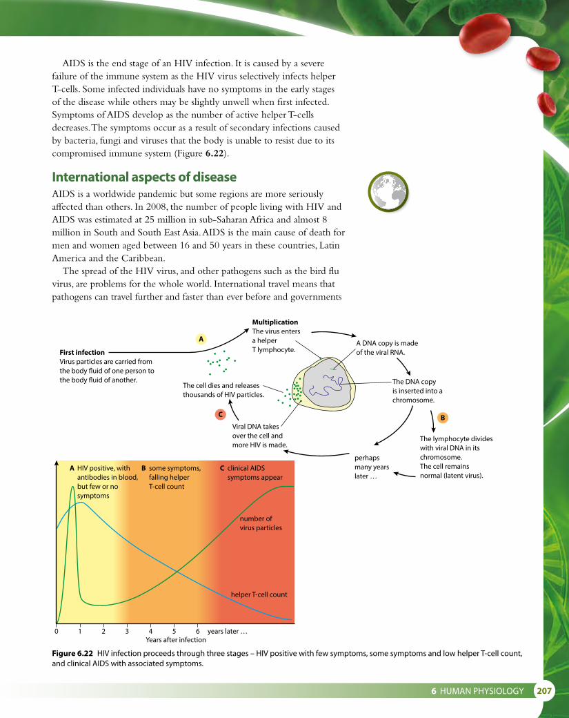
6 HUMAN PHYSIOLOGY 207
AIDS is the end stage of an HIV infection. It is caused by a severe failure of the immune system as the HIV virus selectively infects helper T-cells. Some infected individuals have no symptoms in the early stages of the disease while others may be slightly unwell when first infected. Symptoms of AIDS develop as the number of active helper T-cells decreases. The symptoms occur as a result of secondary infections caused by bacteria, fungi and viruses that the body is unable to resist due to its compromised immune system (Figure 6.22).
International aspects of diseaseAIDS is a worldwide pandemic but some regions are more seriously affected than others. In 2008, the number of people living with HIV and AIDS was estimated at 25 million in sub-Saharan Africa and almost 8 million in South and South East Asia. AIDS is the main cause of death for men and women aged between 16 and 50 years in these countries, Latin America and the Caribbean.
The spread of the HIV virus, and other pathogens such as the bird flu virus, are problems for the whole world. International travel means that pathogens can travel further and faster than ever before and governments
First infectionVirus particles are carried fromthe body �uid of one person tothe body �uid of another.
MultiplicationThe virus entersa helperT lymphocyte.
A
C B
A DNA copy is madeof the viral RNA.
The DNA copyis inserted into achromosome.
The lymphocyte divideswith viral DNA in itschromosome.The cell remainsnormal (latent virus).
perhapsmany yearslater …
Viral DNA takesover the cell andmore HIV is made.
The cell dies and releasesthousands of HIV particles.
0
A HIV positive, with antibodies in blood, but few or no symptoms
B some symptoms, falling helper T-cell count
C clinical AIDS symptoms appear
1 2 3Years after infection
number ofvirus particles
helper T-cell count
4 5 6 years later …
Figure 6.22 HIV infection proceeds through three stages – HIV positive with few symptoms, some symptoms and low helper T-cell count, and clinical AIDS with associated symptoms.

208
and health authorities must work together to co-ordinate their responses.As new viruses and antibiotic-resistant bacterial strains evolve, scientists
have to develop new vaccines and medication to try and combat them. Research and development may take a long time and there is a constant battle to keep up. The importance of this research is crucial to international health, which in turn can affect not only the life of citizens but also the economy of countries. Family incomes and national productivity decline if people of working age fall ill. Food production is affected and causes more problems. Caring for the sick and providing medical care form an expensive burden for individual families, companies and governments.
Nature of scienceAssessing risk in science – collaboration, safety and new medicines The discovery of antibiotics began by accident. On 3 September 1928, Professor Alexander Fleming was examining a batch of culture plates on which he had grown Staphylococcus bacteria. He noticed that one of the plates had a green mould growing on it. The mould was Penicillium notatum. The mould was circular in shape, and the area around it seemed to be free of Staphylococcus. On other areas of the plate, the bacteria were continuing to grow well. Fleming deduced that the bacteria around the circular mould had been killed off by a substance produced by the mould.
Fleming discovered that the mould could kill other bacteria and that it could be given to small animals without any harmful effects. However, he then moved on to other research and it was not until 10 years later that Howard Florey and Ernst Chain, working at Oxford University, isolated the bacteria-killing substance, penicillin, produced by the mould. Chain was a German chemist and Florey an Australian pathologist. It was Chain who isolated and purified penicillin and Florey who tested its safety to use on animals and humans. The first tests the team carried out on mice in 1940 would not have met the stringent standards for testing on animals today. Eight mice were given lethal doses of streptococci bacteria. Half the mice were then given injections of penicillin. The following day all the untreated mice were found to be dead but those that had been given penicillin survived.
One of the first uses of penicillin was in 1941, when Dr Charles Fletcher gave it to patient at a hospital in Oxford who was near to death as a result of bacterial infection in a wound. Fletcher used some penicillin on the patient and the wound made a spectacular recovery. Unfortunately, Fletcher did not have sufficient penicillin to clear the patient’s body of bacteria and he died a few weeks later as the pathogen regained a hold.
An American brewing company began mass production of penicillin and soon sufficient was available to treat all the bacterial infections among the troops fighting in World War II. Penicillin was nicknamed ‘the wonder drug’ and in 1945 Fleming, Chain and Florey shared the Nobel Prize for Medicine, for its discovery and development.
Deductive reasoning occurs when a scientist works from general
information to the more specific. It is known as a ‘top-down’ approach because it begins with a very broad spectrum of information and leads to a specific conclusion.
Inductive reasoning works the opposite way, moving from specific observations to broader theories. It is known as a ‘bottom up’ approach. A scientist begins with specific observations, starts to look for patterns and then formulates a hypothesis to investigate which may lead to more general conclusions or theories. Inductive reasoning is a method of thinking that involves using a set of specific facts to come to a general conclusion.
Questions to consider• Was the discovery of penicillin
an example of inductive reasoning? If not, why not?
• How useful are the two types of reasoning in science?

6 HUMAN PHYSIOLOGY 209
Questions to consider
• Why did the discovery of penicillin have such a profound effect on people at the time?
• Why was the collaboration of three scientists vital to the discovery of penicillin?
• What are the ethical issues involved in using a new drug for the first time? Was Fletcher right to use penicillin on his patient?
• The test on the safety of penicillin used by Florey would not be accepted today. What are the risks associated with the development of new medicines?
12 Define the term ‘pathogen’. 13 Describe what is meant by the term ‘antigen’. 14 State why antibiotics are not effective in treating viral diseases. 15 Distinguish between the roles of a ‘phagocyte’ and a ‘lymphocyte’.
6.4 Gas exchangeAll living cells need energy for their activities. Energy is released from the breakdown of glucose and other substances during the process of cell respiration. Respiration is a chemical reaction that occurs in mitochondria and the cytoplasm and releases energy as ATP, a form that can be used inside cells (Subtopics 1.2 and 8.2).
Our cells use oxygen to carry out aerobic respiration and produce carbon dioxide as a waste product. Oxygen is taken in from the air and carbon dioxide is returned to it in a passive process known as gas exchange. Gas exchange occurs in the alveoli of the lungs where oxygen from the air diffuses into blood capillaries, and carbon dioxide passes in the opposite direction. Gases are also exchanged in the tissues where oxygen diffuses into respiring cells and is exchanged for carbon dioxide.
Whenever diffusion occurs, there must always be a concentration gradient with a higher level of the diffusing substance in one area than in another. Air inside the alveoli contains a higher concentration of oxygen than the blood, so oxygen diffuses into the blood. Blood contains a higher level of carbon dioxide than inhaled air, so carbon dioxide diffuses into the alveoli.
For gas exchange to continue, these concentration gradients must be maintained. As oxygen diffuses out of the alveoli, the level of oxygen inside them gradually falls and the level of carbon dioxide rises. Stale air with high levels of carbon dioxide and low levels of oxygen must be expelled regularly and replaced with a fresh supply to restore the concentration gradients of the two gases. This is achieved by breathing in and out, a process known as ventilation.
Learning objectives
You should understand that:
• Ventilation of the lungs maintains a concentration gradient of both oxygen and carbon dioxide between air in the alveoli and blood in adjacent capillaries.
• Thin alveolar cells (type I pneumocytes) are adapted to carry out gas exchange.
• Type II pneumocytes create a moist surface inside the alveoli by producing a solution containing surfactant, which reduces surface tension and ensures that the sides of alveoli do not adhere to each other.
• Air is carried to the lungs and alveoli via the trachea, bronchi and bronchioles.
• Muscle contractions allow ventilation of the lungs by causing pressure changes in the thorax that force air in and out.
• Different muscles are used for inspiration and expiration because muscles only do work as they contract.
Test yourself?

210
The human ventilation systemOur lungs are protected inside the thorax in an air-tight cavity formed by the ribs and diaphragm. The inside of the ribcage is lined with membranes that secrete fluid to lubricate the lungs, making them slippery and reducing friction during breathing. Air is drawn in through the nose and passes down the trachea to the two bronchi (singular bronchus), one of which passes to each lung (Figure 6.23). Bronchi divide into smaller and smaller tubes, called bronchioles, which end in tiny air sacs or alveoli (singular alveolus). The alveoli are covered with a network of capillaries and together provide us with a very large surface area for the exchange of oxygen and carbon dioxide.
trachea
rib
externalintercostal muscle
internalintercostalmuscle
position of heartlung diaphragm
alveolus
bronchiole
bronchus
Figure 6.23 The ventilation system.
Measuring changes in lung volumeA spirometer (Figure 6.24) is used to measure the amount of air that is exchanged during breathing. It can also measure the rate of breathing – for example, when a person is at rest or during or after exercise. A simple spirometer has a chamber filled with oxygen or air, which is inverted over a container of water. The subject is connected to the chamber via a tube and mouthpiece (the nostrils are closed with a nose clip). As the subject inhales and exhales a trace is produced on a rotating drum or computer monitor. Inhalation causes the chamber to fall, producing a falling line on the trace. Exhalation causes the chamber to rise and produces a rising line. The trace is called a spirogram and various volume measurements can be made from it (Figure 6.24). Usually soda lime is used to absorb carbon dioxide that is exhaled.
Tidal volume volume of air breathed in and out in a single breathInspiratory reserve volume breathed in by a maximum inhalation at the end of a normal inhalationExpiratory reserve volume exhaled by a maximum effort after a normal exhalationResidual volume volume of air remaining in the lungs at the end of a maximum exhalation

6 HUMAN PHYSIOLOGY 211
5
4
Volu
me
/ dm
3 3
2
1
00 5 10 15 20 25
Time / seconds
residualvolume
expiratoryreservevolume
restingtidalvolume
inspiratoryreservevolume
maximumexpiration
maximuminspiration
vitalcapacity
period ofexercise
30 35 40 45 50 55 60
spirometerchamber
counterpoise
mouthpiece
carbon dioxideabsorber
water level
spirometertrace
nose clip
Figure 6.24 A spirometer produces a trace that can be used to measure various aspects of a person’s breathing.

212
Mechanism of ventilationLungs have no muscles and cannot move by themselves. Breathing is brought about by two sets of intercostal muscles between the ribs, and by the diaphragm, the sheet of muscle separating the thorax from the abdomen (Figure 6.25).
During inhalation, contraction of the external intercostal muscles raises the ribs and contraction of the diaphragm lowers the floor of the thorax. These movements increase the volume of the chest cavity and lower the pressure on the lungs to below that of the air outside. As a result, air is drawn down the trachea to fill the lungs.
Gentle exhalation occurs as the intercostal and diaphragm muscles relax, reducing the volume of the chest cavity. Elastic fibres around the alveoli return to their original length and pressure forces air out of the lungs.
Long or forced exhalations involve the internal intercostal muscles, which contract to lower the ribs. Muscles in the abdominal wall also contract and push the relaxed diaphragm upward. Pressure inside the chest cavity increases and air is forced out of the lungs.
External and internal intercostal muscles are an example of antagonistic muscles. The work of external intercostals produces an opposite effect to the contraction of the internal intercostals. The muscles of the diaphragm are also antagonistic to the abdominal muscles. Remember that muscles can only produce an effect and do work when they contract.
Importance of alveoliAlveoli are the body’s gas exchange surfaces. Formed in clusters at the ends of the smallest bronchioles, more than 300 million alveoli in each lung together provide a surface area of about 75 m2. Alveoli are roughly spherical in shape and made of pneumocyte cells less than 5 μm thick (Figure 6.26). The capillaries that wrap around them also have thin walls
Blowing a trumpet
External and internal intercostal muscles are antagonistic muscles because they have opposite effects when they contract. During relaxed breathing, internal intercostal muscles do not contract, but they are used to force air out of the lungs when we sing or play a wind instrument.
Inhalation ExhalationPressure on the lungs is reduced as thorax volume increases, creating apartial vacuum. Air is drawn in andthe lungs in�ate.
Pressure on thelungs increases andair is forced out.
External intercostalmuscles contract toraise the ribs up andout. Volume of thethorax increases.
External intercostal muscles relax. Volume of thoraxdecreases as ribcage lowers.
During long exhalations, the internal intercostal muscles contract. Muscles in the abdomen also contract andpush the relaxed diaphragmupward.
Diaphragm contracts into a �attened position. Volume of thorax increases.
Diaphragm relaxes. Volume of thoraxdecreases.
Figure 6.25 The mechanism of ventilation.
Figure 6.26 Healthy human lung tissue, showing many alveoli (× 34).

6 HUMAN PHYSIOLOGY 213
of single epithelial cells. These two thin layers make the distance for diffusion of gases as small as possible. Oxygen diffuses through the alveolus and capillary into the blood and carbon dioxide diffuses in the opposite direction (Figure 6.27). So long as the diffusion gradient is maintained by regular breathing, diffusion continues.
The walls of the alveoli contain two types of cells called pneumocytes. Type I pneumocytes cover most of the surface (97%) of the air sac and are responsible for gas exchange. Type I cells are about twice as numerous as type II pneumocytes, which are larger, rounder cells that produce and secrete a liquid containing a surfactant. The surfactant reduces surface tension and prevents the sides of the alveolus sticking to one another. Type I pneumocytes cannot divide but if they are damaged type II cells can divide to replace them (see Option D, Figure D.27).
Table 6.5 summarises ways in which the alveoli are well adapted for their role in gas exchange.
Feature of alveoli Importance
many small, spherical alveoli provide a large area for gas exchange
thin walls of flattened single cells short diffusion distance
rich blood supply from capillaries maintains concentration gradient and carries absorbed gases away rapidly
Table 6.5 Adaptations of alveoli for gas exchange.
Emphysema
Emphysema is a condition in which the surface area of the alveoli available for gas exchange is reduced. It can be caused by environmental factors including cigarette smoking. Chemicals in tobacco smoke cause white blood cells, known as neutrophils, to secrete an enzyme called neutrophil elastase, which breaks down elastic fibres in the alveoli. The walls of the alveoli may break down so that millions of tiny alveoli are replaced with larger air spaces. The area for gas exchange decreases and a person suffering from emphysema has difficulty getting sufficient oxygen into their blood. Alveoli also become less elastic and so it becomes even more difficult to ventilate the lungs. The damage is irreversible and patients with emphysema may need to rely on a supply of oxygen from a cylinder to help them walk and carry out everyday tasks without becoming breathless.
Premature babies have immature type II pneumocytes, which only begin to develop fully after 24 weeks. These infants are likely to suffer with Infant Respiratory Distress syndrome due to the lack of surfactant in their alveoli. After about 35 weeks’ gestation the production of surfactant is sufficient. A gene MUC1 which is associated with type II pneumocytes has also been identified as a marker in lung cancer.
air moves in and out
wall of capillary
moist surface
wall of alveolus
carbon dioxidediffuses out ofblood
oxygendiffuses intoblood
red blood cell
Figure 6.27 Gas exchange in the alveolus.

214
Nature of scienceObtaining evidence – understanding the causes of lung cancerEpidemiology is defined as the study of the state of health of a population and the use of the information that is collected to address health-related issues. Our understanding of lung cancer has grown as a result of pioneering epidemiological studies, which began in the 1950s and continued until the early part of this century.
Lung cancer was uncommon before cigarette smoking became popular and in 1912 only 374 deaths from lung cancer were recorded worldwide. But additional data from post mortems later revealed that the incidence of lung cancer increased from 0.3% in 1852 to approximately 6% in 1952. Fritz Lickint, a German doctor, was one of the first to consider a link between smoking and lung cancer in 1929, but his evidence was largely circumstantial.
In 1951 the Medical Research Council in the UK set up a study, known as The British Doctors’ survey, to try to understand why rates of lung cancer had risen to such an extent. The study was run by Dr Richard Doll and Austin Bradford Hill and involved the collection of data and medical information from 34 439 male doctors. The doctors responded to questionnaires and the study was one of the first to demonstrate the importance of epidemiology and statistics. The doctors who replied were divided into categories based on the decade of their birth, as well as general physical health and smoking habits and their cause-specific mortality. The study continued, using the same subjects, for 50 years and additional data was collected using further questionnaires in 1957, 1966, 1971, 1978, 1991 and finally in 2001.
The preliminary results of the study were published in 1956 at a time when smoking was not considered a serious health problem, but the true understanding of the link between smoking and lung cancer and other respiratory diseases only followed later as additional data was added.
A major conclusion of the study is that smoking decreases life expectancy up to 10 years. Men born between 1900 and 1930, who smoked, died on average 10 years younger than non-smokers. Their deaths were significantly more likely to be caused by diseases such as lung cancer and diseases of the cardiovascular system.
You can read Richard Doll’s summary of the study ‘Mortality in relation to smoking: 50 years’ observations on male British doctors’ in the British Medical Journal – use a search engine to find the article using key words such as Richard Doll smoking study BMJ.
When a person has lung cancer, the processes that usually control cell division by mitosis (Subtopic 1.6) break down, so that cells in the lung tissue grow and divide uncontrollably. This causes a tumour to develop, which occupies space and gradually inhibits the ability of the lungs to exchange gases, putting a strain on the heart and eventually causing premature death.
16 List the structures that a molecule of oxygen would pass on its way into an alveolus from the atmosphere.
17 List the three key characteristics of a gas exchange surface. 18 List the muscles involved in inhalation. 19 Distinguish between ‘gas exchange’ and ‘ventilation’.
Test yourself?

6 HUMAN PHYSIOLOGY 215
centralnervoussystem
brain
spinalcord
peripheralnervoussystem(only largernerves shown)
6.5 Neurons and synapses
The human nervous systemThe nervous system consists of neurons, or nerve cells, which transmit information in the form of nerve impulses. Figure 6.28 shows the basic layout of the human nervous system.
The central nervous system, or CNS, is made up of the neurons of the brain and the spinal cord. The CNS receives information from sensory receptors all over the body. Information is processed and interpreted before the CNS initiates suitable responses.
The peripheral nerves are the network of neurons that carry information to and from the CNS. Peripheral nerves include sensory neurons, which carry information to the CNS, and motor neurons, which transmit impulses from the CNS to muscles and glands that then cause a response.
Three types of neuron are found in the nervous system. Sensory and motor neurons transmit information to and from the CNS, while relay neurons within the CNS form connections between them.
The structure of a motor neuron is shown in Figure 6.29. Many small dendrites receive information from relay neurons and transmit the impulses to the cell body. One long axon then carries impulses away. The cell body contains the nucleus and most of the cytoplasm of the cell. The axon is covered by a myelin sheath formed from Schwann cells, which wrap themselves around it. Myelin has a high lipid content and forms an electrical insulation layer that speeds the transmission of impulses along the axon.
Transmission of nerve impulsesNeurons transmit information in the form of impulses, which are short-lived changes in electrical potential across the membrane of a neuron. All neurons contain sodium (Na+) and potassium (K+) ions. Impulses occur as these important ions move in and out through the plasma membrane.
Learning objectives
You should understand that:
• Cells called neurons transmit electrical impulses.
• Nerve fibres that are covered with myelin sheaths allow salutatory conduction.
• Sodium and potassium ions are pumped across the membranes of neurons to generate a resting potential.
• An action potential consists of the rapid depolarisation and repolarisation of the neuron membrane.
• Nerve impulses are action potentials that are propagated along the axons of neurons.
• A nerve impulse is transmitted as a result of local currents that cause each successive part of the axon to reach the threshold potential.
• A synapse is the junction between two neurons or between neurons and receptor or effector cells.
• Presynaptic neurons release a neurotransmitter into the synapse when they are depolarised.
• A nerve impulse is only initiated if the threshold potential is reached.
Figure 6.28 The human nervous system.

216
When a neuron is not transmitting an impulse, it is said to be at its resting potential. The resting potential is the potential difference across the plasma membrane when it is not being stimulated – for most neurons, this potential is –70 mV. The inside of the axon is negatively charged with respect to the outside (Figure 6.30).
As a nerve impulse occurs, the distribution of charge across the membrane is reversed. For a millisecond, the membrane is said to be depolarised. As charge is reversed in one area of the axon, local currents depolarise the next region so that the impulse spreads along the axon (Figure 6.31). An impulse that travels in this way is known as an action potential.
Figure 6.32 explains what is happening at the plasma membrane of the neuron as an action potential is generated.1 When a neuron is stimulated, gated sodium channels in the membrane
open and sodium ions (Na+) from the outside flow in. They follow both the electrical gradient and the concentration gradient, together known as the electrochemical gradient, to move into the cell. The neuron is now said to be depolarised.
dendrite
nucleus of motor neuron
axon
node of Ranvier
Schwann cell
terminalbranchesof the axon
cell body
Figure 6.29 A motor neuron.
sodium ions are constantlypumped out and potassiumions are pumped in
Na+
K+ K+K+
protein molecules making upthe sodium–potassium pump
Na+
K+
Na+
K+
axon
+ + + + + + ++ + + + +
+ + + + + + ++ + + + +
––– – –– – – – – – –
––– – –– – – – – – –
Figure 6.30 At rest, sodium ions are pumped out of the neuron and potassium ions are pumped in, to establish the resting potential. Inside the neuron is negatively charged because of the presence of chloride and other negative ions.

6 HUMAN PHYSIOLOGY 217
2 For a very brief period of time, the inside of the axon becomes positively charged with respect to the outside as sodium ions enter. At this point, the sodium channels close.
3 Now, gated potassium channels open and potassium ions (K+) begin to leave the axon, moving down their electrochemical gradient to re-establish the resting potential, a process known as repolarisation.
4 Because so many potassium ions start to move, the potential difference falls below the resting potential. At this point, both sodium and potassium channels close. The resting potential is re-established by the action of sodium–potassium pumps, which move ions back across the membrane.
An action potential in one part of an axon causes the depolarisation of the adjacent section of the axon. This occurs because local currents are set up between adjacent regions and these cause ion channels to open, allowing sodium ions in and potassium ions out of the axon and cause each successive part of the axon to reach its threshold potential and become depolarised.
The action potential travels along the neuron rather like a ‘Mexican wave’. The impulse can only pass in one direction because the region behind it is still in the recovery phase of the action potential and is temporarily unable to generate a new action potential. The recovery phase is known as the refractory period.
The speed of conduction along an axon is affected by the diameter of the axon. A larger diameter means faster conduction. Larger axons are myelinated while smaller ones are not. At intervals along myelinated axons are gaps between the myelin covering known as nodes of Ranvier (Figure 6.29). The sheath prevents the flow of ions across the membrane so the current must jump from node to node – a process known as saltatory conduction, which speeds up the transmission of the nerve impulse.
The synapseA synapse is the place where two neurons meet. Two neurons do not touch one another and the tiny gap of about 20 nm between them is known as the synaptic cleft. Action potentials must be transmitted across this gap for the impulse to pass on its way and this is achieved by the presence of chemicals known as neurotransmitters. Neurotransmitters are held in vesicles in the pre-synaptic cell until an action potential arrives. They are then released into the synaptic cleft, and diffuse across to the post-synaptic membrane. There they can cause another action potential to be produced and a nerve impulse to be initiated, provided the threshold potential is reached.
The synapse shown in Figure 6.33 uses the neurotransmitter acetylcholine (ACh) and is a cholinergic synapse. ACh binds to receptors and causes depolarisation of the post-synaptic membrane and the initiation of an action potential. Once an action potential is generated in the post-synaptic membrane, ACh in the synaptic cleft is deactivated by acetylcholinesterase enzymes and the products are reabsorbed by the pre-
impulse travels in this direction
Na+ Na+Na+Na+
area of impulsenext area to bestimulated
area returningto rested state
+ + + + +
+ + + + +
+ + +– – – – –
+ + +– – – – –
+++ + + – – –
+++ + +– – – – –
– – – – –
– – –
Figure 6.31 When an impulse passes along the neuron, sodium ions diffuse via ion channels and the potential is reversed. This process is called an action potential (only a small part of a long neuron is shown).
Resting potential the electrical potential across the plasma membrane of a neuron that is not conducting an impulseAction potential the reversal and restoration of the resting potential across the plasma membrane of a neuron as an electrical impulse passes along itThreshold potential the electrical potential across the plasma membrane of a neuron that is required in order to trigger an action potential

218
synaptic membrane to be remade and repackaged in vesicles.There are more than 40 different neurotransmitters in the body.
Acetylcholine and noradrenalin are found throughout the nervous system, others (for example, dopamine) are found only in the brain.
Many drugs and toxins affect synapses and influence the way nerve impulses are transmitted. Nicotine is an excitatory drug. It has a similar molecular shape to acetylcholine and affects the post-synaptic membrane so that it transmits an action potential. Cocaine and amphetamines are also
CI–
CI–
CI–
CI– CI–
CI–
K+
K+
K+
K+
K+
K+
K+
Na+
Na+Na+
Na+
Na+Na+ Na+
Na+
CI–
CI–
CI–
CI– CI–
CI–
K+
K+
K+
K+K+
K+
K+Na+
Na+
Na+
Na+
Na+Na+ Na+
CI–
CI–
CI–
CI– CI–
CI–
K+
K+
K+
K+
K+
K+
K+Na+
Na+
Na+
Na+Na+
Na+
Na+
Na+
Inside of neuron
Outside of neuron
When stimulatedNa+ channels openand Na+ ions flowinto the neuron.
1
Na+ flow causes rapiddepolarisation ofthe membrane.
2
Gated K+ channels openand K+ flows out, causingrepolarisation.
3
direction of impulse
refractory periodthreshold potential
resting potential
Elapsed time / ms0 1 2 3 4 5
+40
0
–70
Mem
bra
ne p
oten
tial /
mV
closed K+
channel
Na+/K+
pump
openNa+
channel
open K+
selectivechannel
Inside of neuron
Outside of neuron
open K+
channel
Na+/K+
pump
closedNa+
channel
open K+
selectivechannel
Inside of neuron
Outside of neuron
closed K+
channel
Na+/K+
pump
closedNa+
channel
open K+
selectivechannel
At rest, most K+ ions areinside the axon, but itremains negatively chargeddue to the presence of negativeions. As K+ ions are pumpedinto the axon by the Na+/K+
pump, they need to diffuseback out in order to maintainthis negative resting potential.
Figure 6.32 The action potential.

6 HUMAN PHYSIOLOGY 219
excitatory drugs, which stimulate synaptic transmission. Benzodiazepenes, cannabis and alcohol suppress the activity of the nervous system. Cannabis binds to receptors in the brain and blocks synaptic transmission. These drugs affect a person’s behaviour and some are highly addictive. Used incorrectly they can cause social problems and may result in long-term damage to the body.
Neonicotinoids are chemical pesticides used in insecticides. They are similar in structure to nicotine and block transmission at the synapses of insects by binding to acetylcholine receptors. Neonicotinoid pesticides have been linked to the decline of bee populations throughout the world. In 2013, the European Food Safety Authority declared that these insecticides pose an unacceptable high risk to bees and the European Commission imposed restrictions on their use.
1 A nerve impulse arrives at the end of the pre-synaptic axon and causes calcium ions to enter the synaptic knob.
axon
mitochondriaprovideenergy foractive transport pre-synaptic
membranepost-synapticmembrane
synapticknob
synaptic vesicle 3 ACh di�uses across the synaptic cleft and reaches receptors on the post-synaptic membrane.
4 ACh fuses with receptors on the post-synaptic membrane.
5 Sodium ion channels in the membrane open and the post-synaptic membrane is depolarised.
synaptic cleft
2 Vesicles containing neurotransmitter (ACh) move towards the pre-synaptic membrane, fuse with it and release their contents into the synaptic cleft.
Ca2+ ions
Na+
Figure 6.33 A cholinergic synapse. The whole sequence of events of transmission at a synapse takes 5–10 ms.
20 Explain how sodium and potassium ions establish a resting potential.
21 Define the term ‘action potential’. 22 List the key events of synaptic transmission. 23 State what is meant by the term ‘saltatory conduction’. 24 Name the substance released by a pre-synaptic membrane.
Test yourself?
Tetanus toxin is a neurotoxin produced by the tetanus bacterium Clostridium tetani. The disease tetanus affects the muscles causing them to go into spasm as the toxin binds irreversibly to acetylcholine receptors in post-synaptic membranes and prevents the transmission of nerve impulses at synapses at neuromuscular junctions. Another neurotoxin produced by the bacterium Clostridium botulinum is one of the most toxic substances known. Nevertheless it has been used in recent years for cosmetic and medical procedures under the trade name Botox. In the early 1980s, it was used to treat muscle spasm, strabismus (squint) and uncontrollable blinking. More recently it has been used to induce temporary muscle paralysis and conceal the appearance of wrinkles.

220
6.6 Hormones, homeostasis and reproduction
HomeostasisThe internal environment of the body remains constant, within certain limits, despite changes that occur in the external environment. The control process that maintains conditions within these limits is known as homeostasis.
The factors that are controlled include water balance, blood glucose concentration, blood pH, carbon dioxide concentration and body temperature.
Each of these has a ‘normal’ or set point although they may vary slightly above or below it. For example, the normal body temperature for humans is about 37 °C, which is optimum for the efficient functioning of the body’s enzymes and cell processes.
Both the nervous system and the endocrine system are involved in homeostasis. The endocrine system consists of ductless endocrine glands, which release different hormones (Figure 6.34). Hormones circulate in the bloodstream but each one is a chemical messenger that only affects the metabolism of specific target cells.
Insulin and glucagon, and control of blood glucoseBlood glucose level is the concentration of glucose dissolved in blood plasma. It is expressed as millimoles per decimetre cubed (mmol dm–3). Normally blood glucose level stays within narrow limits, between 4 mmol dm–3 and 8 mmol dm–3, so that the osmotic balance (Subtopic 1.4) of the blood remains constant and body cells receive sufficient glucose for respiration. Levels are higher after meals as glucose is absorbed into the blood from the intestine and usually lowest in the morning as food has not been consumed overnight.
Glucose levels are monitored by cells in the pancreas. If the level is too high or too low, α and β cells in regions of the pancreas known as the islets of Langerhans produce hormones that turn on control mechanisms to correct it. This is an example of negative feedback. Table 6.6 summarises these responses.
Learning objectives
You should understand that:
• α and β cells in the pancreas secrete insulin and glucagon to control blood glucose.
• Thyroxin, secreted by the thyroid gland, regulates the metabolic rate and helps control body temperature.
• Leptin is a hormone secreted by adipose cells that acts on the hypothalamus in the brain to control appetite.
• Melatonin, secreted by the pineal gland, controls circadian rhythms.
• A gene on the Y chromosome causes the development of gonads in a male embryo so that they form testes and secrete testosterone.
• Testosterone causes the development of male genitalia before birth and also sperm production and development of male secondary sexual characteristics at puberty.
• Estrogen and progesterone cause the development of female genitalia before birth and development of female secondary sexual characteristics at puberty.
• The menstrual cycle is controlled by positive and negative feedback involving hormones from the ovary and pituitary gland.
One decimetre cubed is the same volume as one litre.
Negative feedback occurs when a deviation to the normal level is detected and corrective mechanisms act to return the system to normal. Many homeostatic mechanisms work by negative feedback.
Responses to a rise in blood
glucose above normal
Responses to a fall in blood
glucose below normal
Pancreas β cells in the pancreas produce the hormone insulin
α cells in the pancreas produce the hormone glucagon
Glucose
uptake or
release
insulin stimulates cells in the liver and muscles to take in glucose and convert it to glycogen and fat, which can be stored inside the cells – blood glucose levels fall
glucagon stimulates the hydrolysis of glycogen to glucose in liver cells – glucose is released into the blood
Table 6.6 The body’s responses to changes in blood glucose.

6 HUMAN PHYSIOLOGY 221
DiabetesDiabetes is the inability of the body to control blood glucose level. A diabetic person will experience wide fluctuations in their blood glucose above and below the normal limits (Figure 6.35).
In Type I diabetes, the β cells in the pancreas do not produce insulin. Without insulin, glucose is not taken up by body cells so blood levels remain high, a condition known as hyperglycemia (Figure 6.36). Excess glucose is excreted in urine and its presence is used to diagnose diabetes. About 10% of diabetics have type I diabetes. Symptoms usually begin in childhood, which is why type I diabetes is sometimes known as ‘early onset’ diabetes.
Type II diabetes is the most common form of diabetes, accounting for nine out of ten cases worldwide. The pancreas does produce insulin although levels may fall as the disease progresses. Type II diabetes occurs when body cells fail to respond to the insulin that is produced. Again, the result is that blood glucose levels remain too high. This form of diabetes is also known as late-onset diabetes or non-insulin-dependent diabetes mellitus. Individuals who have the condition develop insulin resistance, which means that the receptor cells that normally respond to insulin fail
pituitary gland(secretes manyhormones, e.g. ADH)
thyroid gland(secretes thyroxine)
adrenal gland(secretes epinephrine)
islets of Langerhansin pancreas(secrete insulinand glucagon)
ovary (in female)(secretes estrogenand progesterone)
testis (in male)(secretes testosterone)
Figure 6.34 The positions of some endocrine glands in the human body. Endocrine glands have no ducts and secrete hormones directly into the bloodstream, which carries them to target cells.

222
glucose ingestedTime / hours
blood insulin in untreated diabetic person
blood insulin in normal person
blood glucose in normal person
blood glucose in untreateddiabetic person
00
0.02
0.04
0.06
Bloo
d gl
ucos
e co
ncen
trat
ion
/ g d
m–3
Bloo
d in
sulin
con
cent
ratio
n (a
rbitr
ary
units
)
0.08
0.10
0.12
0.14
0.16
0.18
0.20
1 2 3
Figure 6.36 Blood glucose and insulin levels following intake of glucose in a normal person and a person with untreated type I diabetes.
to be stimulated by it, even though the β cells in the pancreas still produce insulin. This type of diabetes is often associated with obesity, age, lack of exercise and genetic factors. Diabetics must monitor their blood glucose level carefully so that they can control it, since the body’s internal control mechanism is not working properly.
If blood glucoserises too high, glucoseappears in urine andcoma may result.
Insulin secretedinto circulation.
Liver and musclecells take up extraglucose from blood.
Glucagonsecreted intocirculation.
Liver cells breakdown glycogenand release glucose.
Release of glucoseraises bloodglucose level.
Uptake of glucoselowers bloodglucose level.
Rise in blood glucoseafter food ingestion orglucose release from liver.
Fall in blood glucoseafter glucose uptake by cells.
SENSORHigh
Normal bloodglucose level
If blood glucosefalls too low,coma may result.
Low
Bloo
d gl
ucos
e le
vel
STIMULUS
SENSOR
STIMULUS
EFFECTOR
EFFECTOR
EFFECT
EFFECT
β cells in islets ofLangerhans in pancreas
α cells in islets ofLangerhans in pancreas
Figure 6.35 The control mechanism for blood glucose.

6 HUMAN PHYSIOLOGY 223
Causes and symptoms of diabetesType I diabetes is caused when the β cells in the pancreas do not produce insulin. This can be a result of autoimmune disease in which the body’s immune system destroys its own β cells.
The causes of type II diabetes are not fully understood but there is a strong correlation of risk with weight and diet. High levels of fatty acids in the blood may be a factor causing the condition and people whose diets are high in fat but low in fibre seem to be most at risk. Obesity, associated with a lack of exercise or a genetic makeup that influences fat metabolism, is a key risk factor. The condition is more common in older people but there are an increasing number of cases in overweight children.
Studies of ethnic groups worldwide have shown that some are more likely to develop type II diabetes, and this provides evidence for a genetic predisposition to the condition. Aboriginal Australians, people of Asian and Afro-Caribbean origin, Native Americans and Polynesian Maori peoples all have a higher incidence of diabetes than would occur by chance.
The symptoms of diabetes include:
• high glucose levels in the blood
• glucose in the urine
• frequent need to urinate, which leads to dehydration and increased thirst
• tiredness and fatigue
• some loss of weight.In type II diabetes, these symptoms tend to develop slowly.
Treatment of diabetesType I diabetes must be controlled by regular insulin injections, but many people who have type II diabetes are advised to control their blood sugar levels by following a healthy diet, taking exercise and losing weight. They are advised to eat foods that are low in saturated fat and salt but high in fibre and complex (slowly absorbed) carbohydrates, such as wholegrain cereals, pulses, beans and lentils. These foods, especially if they are taken at regular intervals during the day, help to keep blood sugar levels steady. Foods that should be avoided include sugary snack foods and drinks, and food with a high level of saturated fat. These foods cause a rapid rise in blood sugar level that the diabetic person is unable to deal with.
If left untreated, type II diabetes can lead to long-term health problems such as kidney disease, retinal damage, high blood pressure, stroke and heart attack.
Thyroxin and control of metabolic rateThyroxin is a hormone produced by the thyroid gland situated in the neck (Figure 6.34). Thyroxin contains iodine, which is a dietary requirement for a healthy thyroid gland. Thyroxin controls the body’s basal metabolic rate (the rate of the body’s metabolism at rest) and so the hormone is also important in controlling growth. If a person has insufficient thyroxin present during childhood, their physical and mental development does not proceed properly and a condition known as cretinism may result. A person with this condition will be short in stature
In the last decade, there has been a large increase in the number of people in industrialised countries affected by type II diabetes. Can you suggest a reason for this?
Glycaemic index
Some health professionals recommend that people who have or are at risk from type II diabetes follow a low GI (glycaemic index) diet. Low GI foods allow the body to absorb carbohydrates more slowly, which helps to stabilise glucose levels through the day. Low GI foods include apples, oranges, pasta, sweet potato, sweetcorn and noodles.

224
Figure 6.37 The hypothalamus and pituitary gland. The hypothalamus links the hormonal and nervous systems by producing releasing hormones that regulate pituitary gland secretions.
and have severe mental retardation. In adults, a shortage of thyroxin caused by an underactive thyroid (hypothyroidism) leads to a decreased metabolic rate and an accumulation in fat, as well as general decrease in physical and mental activity. In contrast, an overactive thyroid gland (hyperthyroidism) can cause greatly increased metabolic activity, loss of body mass, an increased heart rate and a general increase in physical and mental activity. Both hypothyroidism and hyperthyroidism can lead to a condition known as goitre, which is a swelling of the thyroid gland in the neck.
The majority of cases of goitre used to result from thyroxin deficiency caused by a lack of iodine in the diet, but a UNICEF programme promoting the use of iodised salt has reduced the incidence of the condition worldwide over recent years.
Normal levels of thyroxin are maintained by homeostasis, in a negative feedback system (Figure 6.37). In normal conditions, thyroxin secretion is triggered by a stimulating hormone produced by the pituitary gland. If too much thyroxin is present, it inhibits the pituitary gland and production is decreased. The stimulating hormone from the pituitary gland is in turn influenced by the hypothalamus. The hypothalamus responds to other influences such as temperature so that, in some animals, metabolism can be aligned to environmental factors such as the seasons. Thyroxin produced as a result of stimulation by the hypothalamus can speed up metabolism of protein, fat and carbohydrates and increase body temperature by generating metabolic heat.
Control of body temperatureBody temperature is monitored and controlled by the hypothalamus in the brain. The ‘set point’ for body temperature is 36.7 °C. The hypothalamus responds to nerve impulses from receptors in the skin and also to changes in the body’s core temperature. If body temperature fluctuates above or below the set point, the hypothalamus coordinates responses to bring it back to normal. This is another example of negative feedback. Nerve messages are carried from the hypothalamus to organs that bring about warming or cooling of the body. Table 6.7 lists some of the body’s responses to changes in temperature.
right cerebral hemisphere(left hemisphere notvisible here)
medulla oblongata
cerebellum
hypothalamus
pituitarygland
spinal cord
Responses to a rise in body temperature Responses to a fall in body temperature
Arterioles in the skin dilate (widen) so that more blood flows to skin capillaries and excess heat is lost from the skin
narrow to restrict flow of warm blood to the skin capillaries – heat is retained in the body
Sweat glands produce more sweat, which evaporates from the skin surface to cool it
cease production of sweat
Muscles remain relaxed muscular activity such as shivering generates heat
Metabolic rate may decrease to minimise heat production thyroxin increases metabolic rate
Table 6.7 The body’s responses to changes in core temperature.

6 HUMAN PHYSIOLOGY 225
Leptin and control of appetiteLeptin is a protein hormone that is important in regulating energy intake and expenditure, including appetite and metabolism. Human leptin is a protein of 167 amino acids. It is manufactured mainly by cells in white adipose (fat) tissue, and the level of circulating leptin is directly proportional to the total amount of fat in the body. It can also be produced by brown adipose tissue, cells in the placenta, ovaries, skeletal muscle and gastric cells in the stomach.
Leptin acts on receptor cells in the hypothalamus and it inhibits appetite by:
• counteracting the effects of chemicals secreted by the gut and hypothalamus that stimulate feeding
• promoting the synthesis of α-MSH, a long-term appetite suppressant.
Medical use of leptinLeptin signals to the brain that the body has had enough to eat. The absence of leptin, or the receptor for it, leads to uncontrolled food intake. A very small group of humans possess homozygous mutations (Subtopic 3.4) for the leptin gene and this leads to a constant desire for food, resulting in severe obesity. Attempts have been made to control the condition by giving patients doses of human leptin. However, clinical trials have been inconclusive. It was found that large and frequent doses were needed and only provided small improvements because leptin is quickly broken down in the blood stream and has low potency and poor solubility.
The effects of leptin were observed by studying mutant obese mice that appeared within a laboratory mouse colony in 1950. These mice were massively obese (Figure 6.38) and excessively voracious. Since that time several strains of laboratory mice have been found to be homozygous for single-gene mutations that cause them to become grossly obese, and they fall into two classes: those having mutations in the gene for the protein hormone leptin, and those having mutations in the gene that encodes the receptor for leptin. When mice from the first group are treated with injections of leptin, they lose their excess fat and return to normal body weight. Leptin itself was discovered by Jeffrey M. Friedman in 1994 through the study of such mice.
Melatonin and control of circadian rhythmsMelatonin is a hormone produced by the pineal gland, a pea-sized gland located just above the middle of the brain. One of the key influences of melatonin is to maintain the body’s circadian rhythms and especially sleep–wake cycles. A vital factor in human sleep regulation is the exposure to light or darkness. Exposure to light stimulates a nerve pathway from the retina in the eye to the hypothalamus. Cells in the hypothalamus (the supra-chiasmatic nucleus, SCN) send signals to parts of the brain that control hormones, body temperature and other functions that have a role in our feelings of sleep or wakefulness. The SCN produces a signal that can keep the body on an approximately 24-hour cycle of activity. But the ‘internal clock’ is not exactly set to 24 hours and environmental clues, the most important of which is light, are needed to reset the clock each morning and keep a person in step with their external environment.
Figure 6.38 Mice with a mutation in the gene for leptin production are hugely obese compared to a normal mouse.
A number of hormones are used in medical treatments to correct imbalances that may occur.
Hormone replacement therapy (HRT) – given to some women to alleviate the symptoms caused by the sudden change in hormone levels at menopause
Thyroxin – given to patients suffering from hypothyroidism and a shortage of the hormone.
Human Growth Hormone – given to children to treat growth disorders.

226
During the day, the pineal gland is inactive, but during the hours of darkness, it is ‘turned on’ by the SCN and begins to produce melatonin, which is released into the blood. Rising levels of melatonin cause our feelings of sleepiness. The level of melatonin remains high for about 12 hours until the following morning when light causes it fall to a minimal level in the blood.
Even if the pineal gland is stimulated, it will not produce melatonin unless a person is in a dimly lit environment – even artificial indoor lighting can be bright enough to prevent the release of melatonin. The amount of melatonin released at night also varies between individuals, but on average children secrete more melatonin than adults.
Jet lagJet lag is caused by the disruption of the body’s day–night rhythms caused by long-distance travel and arrival in a new time zone. After a long journey the day–night patterns may no longer correspond to the new environment and some people need many days to adjust.
Jet lag upsets the body clock because the expected patterns of light and darkness are out of alignment. Light is the strongest stimulus for the sleep–wake pattern so jet lag can be controlled by avoiding bright light so that the body clock is reset. Melatonin tablets are sometimes used to adjust a person’s body clock but the effectiveness of these treatments is not proved. The effect of melatonin may be very short term and the correct doses and times to take the hormone are not easy to determine. Melatonin is not approved for sale in some countries. In the USA is can be bought in pharmacies but in other countries it is only available with a doctor’s prescription.
Hormones and reproductionReproduction is one of the important characteristics of living things. Human male and female reproductive systems produce the gametes (the sperm cell and egg cell) that must come together to begin a new life. The two reproductive systems enable the gametes to meet and the female reproductive system provides a suitable place for fertilisation to occur and an embryo to develop (Figures 6.39 and 6.40). The ovaries and testes also produce hormones that regulate sexual development and reproduction.
In the first weeks after conception, the sex of a fetus cannot be identified from its appearance. Only its karyotype (Subtopic 3.2) will distinguish a male from a female fetus. The formation of the testes from undifferentiated gonadal tissue is dependent on SRY gene on the Y chromosome. From about 8 weeks after fertilisation, the fetal testes form and begin to secrete testosterone which causes the formation of the male genitalia. If no Y chromosome (and so no SRY gene) is present, female reproductive organs and ovaries form during the period between the second and sixth month of fetal development and ovarian follicles develop.
Circadian rhythms biological processes that vary naturally in a cycle lasting about 24 hours; these rhythms indicate the right times for eating and sleeping, and also regulate hormone production

6 HUMAN PHYSIOLOGY 227
Front view
Reproductive systemUrinary system
right kidneyseminal vesicle
prostate gland
vas deferens (sperm duct)
epididymis
testis
scrotum
penis
foreskin – may have been surgically removed in circumcision
ureter
bladder
urethra
right kidney
ureter
bladder
urethra
oviduct
ovary(produces femalegametes)
uterus
endometrium(lining of uterus)
cervix(neck of uterus)
vagina
Front view
Figure 6.39 The male reproductive system. (The diagram also shows the organs of the urinary system.)
Figure 6.40 The female reproductive system. (The diagram also shows the organs of the urinary system – the bladder has been drawn to one side, to reveal the uterus.)

228
Roles of testosteroneThe hormone testosterone, which is produced by the testes, has important roles in the sexual development and reproductive behaviour in males.
• During fetal development, testosterone causes the development of the male genitalia.
• At puberty, levels of testosterone rise and cause the development of male secondary sexual characteristics including growth of muscle, deepening of the voice, enlargement of the penis and growth of body hair.
• Testosterone stimulates the continuous production of sperm and behaviour associated with the sex drive.
Female sex hormones and the menstrual cycleOvaries produce two hormones, estrogen and progesterone. These hormones stimulate the development of female genitalia before birth and at puberty are responsible for the development of female secondary sexual characteristics including the onset of the menstrual cycle, development of breasts, growth of body hair and widening of the hips. The two hormones also influence the changes in the uterus lining during the menstrual cycle and pregnancy. The pituitary gland in the brain produces two further hormones, luteinising hormone (LH) and follicle-stimulating hormone (FSH). FSH stimulates the development of immature follicles in the ovary, one of which will come to contain a mature egg cell. LH stimulates the follicle to release the egg and subsequently to form the corpus luteum.
Production of female gametes is a cyclical process, which lasts approximately 28 days. During the first half of this menstrual cycle the egg cell is produced and in the second half the uterus lining thickens to prepare for implantation of a fertilised egg. The cycle involves hormones that are released by the ovaries and the pituitary gland.
The sequence of events begins at the start of menstruation, which is often called a period (Figure 6.41). During the first four or five days of the cycle, the endometrium (lining) of the uterus is shed and leaves the body through the vagina. This indicates that fertilisation has not occurred during the previous month.
In this early part of the cycle, the pituitary gland secretes FSH, which stimulates the development of an immature follicle in the ovary. The follicle then secretes estrogen, which enhances the follicle’s response to FSH. At this stage of the cycle, estrogen has a positive feedback relationship with FSH (Figure 6.42). Increasing levels of estrogen cause an increase in the level of FSH released by the pituitary gland. As the level of estrogen rises, it also stimulates the repair of the uterus lining.
As the follicle grows, estrogen levels rise to a peak at around day 12, when they stimulate the release of LH from the pituitary gland. As LH levels reach their highest point, ovulation – the release of the egg cell from the follicle – takes place. Ovulation usually occurs at around the day 14 of the cycle. Immediately afterwards, LH stimulates the empty follicle to form the corpus luteum. Levels of estrogen fall and as a result FSH and LH levels fall.

6 HUMAN PHYSIOLOGY 229
The corpus luteum secretes progesterone, which stimulates the thickening of the endometrium and prepares the uterus to receive an embryo. It also inhibits the production of FSH and LH.
If the egg cell is not fertilised, the corpus luteum degenerates and progesterone and estrogen levels fall. The fall in progesterone stimulates the breakdown of the uterus lining. FSH is no longer inhibited, so a new follicle is stimulated and the cycle begins again.
blood levelsof pituitaryhormones
blood levelsof hormones fromthe ovary
ovary
estrogen
follicle corpus luteum
LH
FSHprogesterone
thickness of liningof uterus
time in daysmenstruation menstruation
development of follicle development of corpus luteum
2 4 6 8 10 12 14 16 18 20 22 24 26 28
ovulation start of new cycle
Figure 6.41 The menstrual cycle lasts an average of 28 days and involves changes in hormone levels that influence the follicles and lining of the uterus.
Figure 6.42 Negative and positive feedback in the menstrual cycle.
estrogenand
progesterone
LH/FSH
negative feedbackover most of cycle
positive feedbackover days 12–14
pituitary
ovary
uterus

230
In vitro fertilisationIn vitro fertilisation (IVF) is a technique used to help couples who have been unable to conceive naturally. There are many reasons for infertility. Males may have a low sperm count, blocked or damaged sperm ducts or be unable to achieve an erection. Females may fail to ovulate or have blocked or damaged oviducts, or produce antibodies in cervical mucus that destroy sperm.
The first step in IVF treatment is an assessment of whether the couple are suitable for treatment. If so, the woman may first be injected with hormones to suppress her natural hormones before being given injections of FSH for about 10 days. In some treatments FSH may be given alone. This hormone causes a number of egg cells to mature at the same time in her ovaries (superovulation). Just before the egg cells are released from the follicles, they are collected using a laparoscope (a thin tubular instrument that is inserted through an incision in the abdominal wall). The egg cells are ‘matured’ in culture medium for up to 24 hours before sperm cells are added to fertilise them. Fertilised egg cells are incubated for about three days until they have divided to form a ball of cells. These embryos are checked to make sure they are healthy and developing normally. Usually two will be selected and placed into the woman’s uterus for implantation. The pregnancy is then allowed to continue in the normal way. Any remaining embryos can be frozen and stored for use later. Figure 6.43 summarises the stages in IVF treatment.
Ethical issues associated with IvF treatmentIVF has enabled men and women who would naturally be infertile to have children but it has also produced some serious ethical issues. Some of these are outlined in Table 6.8. Each society needs to think about these issues and decide what should be done about them.
The first successful IVF baby was Louise Brown, born in 1978 following her mother’s IVF treatment. Professor Robert Edwards, who pioneered the technique, was awarded the Nobel Prize in Physiology or Medicine in 2010.
Arguments in favour of IvF Arguments against IvF
• Enables infertile couples to have a family.• Couples willing to undergo IVF treatment must
have determination to become parents.• Embryos used in IVF treatment can be screened to
ensure they are healthy and do not have certain genetic conditions that would be inherited.
• IVF techniques have led to further understanding of human reproductive biology.
• Unused embryos produced by IVF are frozen for a limited period and then destroyed.
• Multiple births often result from IVF and this increases the risks to mother and babies.
• Infertility is natural whereas IVF is not, some religions object to it on this basis.
• Some causes of infertility are due to genetic conditions, which may be passed on to children born as a result of IVF.
• There may be risks to the health of women who are treated with hormones during IVF.
Table 6.8 Arguments for and against IVF treatment.

6 HUMAN PHYSIOLOGY 231
Normal menstrual cycle isinterrupted by suppressinghormones from thepituitary gland.
1 Synthetic FSH is injected – ovariesare stimulated to develop manyegg cells.
2 Male (partner or donor)provides semen sample.Sample is processed toconcentrate the healthiestsperms.
3
Several egg cells areremoved from theovaries using a laparoscope positionedwith the aid of ultrasound.
4
Eggs are mixed withsperms in a shallowdish. Checked bymicroscopic examinationto ensure fertilisation.
5
The fertilised eggs (zygotes)are then incubated at bodytemperaturefor 2–3 days.
6
Microscopic examinationto confirm that embryoshave reached the 4- to-8-cell stage.
7
Up to three embryos aretransferred into the uterusin the expectation that onewill implant successfully.
Alternatively, embryos canbe frozen for future use.
8
Figure 6.43 The stages in IVF treatment.
Born to giveMy Sister’s Keeper is a novel by Jodi Picoult, published in
2004. The book, which was made into a film in 2009, follows the life of 13-year-old Anna, who enlists the help of an attorney to sue her parents for rights to her own body. Anna’s older sister Kate suffers from leukemia and Anna was conceived through IVF to be a genetic match and donor for her sister. Anna donates bone marrow and blood for her sister throughout her life. As Kate’s condition worsens, their parents want Anna to donate a kidney to Kate after she goes into renal failure, but Anna files a lawsuit against her parents for medical emancipation from them despite the consequences for her sister’s health.
This novel is a fictional story but already there have been real examples of children being conceived by IVF and born to provide stem cells for siblings who would otherwise die.
Questions to consider1 Should IVF conceptions such as
Anna’s be permitted?2 Who should have rights over a
child’s genetic material in cases like this?
3 Is it right that a child should be created simply to keep another person alive?

232
Nature of scienceScientific advance follows technical innovation – why was William Harvey unsuccessful in his research into reproduction?William Harvey (1578–1657) was the leading anatomist of his day. As well as his discoveries on the circulation of blood (Section 6.2), he also studied reproduction. He believed that humans and other mammals must reproduce through the joining of an egg and sperm in the same way as the chickens that he was able to observe. Before Harvey’s research, scientists believed in a theory known as ‘pre-formation’, which assumed an embryo was a fully formed tiny version of a mature individual that simply grew in size in the mother’s womb.
Harvey studied the development of hens’ eggs and recorded his observations of the fetus, which he saw began as what looked like a single drop of blood and then further differentiated into an embryo which later became the chick. He rejected the idea that an exact replica of the organism could be found in reproductive material of either the male or the female.
William Harvey carried out meticulous experiments. He used hen’s eggs, because they were inexpensive and abundant, but he also carried out detailed examinations of deer. Harvey was physician to King Charles I, who was fascinated by his research and supplied deer carcasses from his hunting trips for Harvey to dissect. This support enabled Harvey to develop his concepts of reproduction in mammals. He was never able to prove his theories and observe eggs and sperm because sufficiently powerful microscopes were not available at the time. But he was the first to suggest that humans and other mammals reproduced via the fertilisation of an egg by sperm. It took a further 200 years and advances in the production of microscopes before a mammalian egg was finally observed, but nonetheless Harvey’s stature and experimental evidence meant that his theory was generally accepted during his lifetime.
25 State two hormones that help regulate blood glucose level. 26 State the site of action of the hormone leptin. 27 State the location of the gene that controls the development of
sex in the fetus. 28 Outline the functions of the hormone progesterone in the
menstrual cycle.
Test yourself?
Exam-style questions
1 Which of the following statements is correct?
A Large food molecules need to be digested so that they can be absorbed through the gut wall.B The liver and pancreas both release their digestive enzymes into the small intestine.C The approximate pH of the stomach is 8.D Assimilation is the transfer of the end products of digestion from the gut lumen to the blood. [1]

6 HUMAN PHYSIOLOGY 233
2 Which of the following statements is correct?
A Blood passes from the atria to the ventricles through the semilunar valves.B The coronary and pulmonary arteries supply oxygenated blood to the heart muscle.C The pacemaker, brain and adrenal gland are all involved in the regulation of the heart beat.D A red blood cell travels from the lungs to the stomach via the right atrium, right ventricle
and aorta. [1]
3 Which of the following statements is correct?
A Antibiotics are effective against viruses but not bacteria.B Antigens are made by lymphocytes.C Phagocytes are white blood cells that can be found in the tissue fluid around cells.D HIV is a bacterium that reduces the number of active lymphocytes in the blood. [1]
4 Which of the following statements is correct?
A During breathing in, the rib cage moves up and out and the pressure inside the thorax increases.B Alveoli are small sac-like structures that have walls only two cells thick.C To assist with breathing out, the abdominal and external intercostal muscles contract.D The purpose of ventilation is to increase the rate of gas exchange in the alveoli. [1]
5 Which of the following statements is correct?
A In a motor neuron, myelin always covers the axon.B A nerve impulse travels from a receptor to the CNS via a sensory neuron and from the
CNS to an effector via a motor neuron.C The endocrine system produces hormones, which pass from the endocrine glands to the blood
through ducts.D The resting potential is divided into two parts, depolarisation and repolarisation. [1]
6 Which of the following statements is correct?
A The sex of a fetus is determined by the mother’s hormones.B During the process of IVF, a woman is injected with FSH to stimulate egg cells to mature.C FSH and estrogen are hormones released from the ovaries whereas LH and
progesterone are hormones released from the pituitary gland.D In the menstrual cycle, the levels of progesterone begin to rise around day 5. [1]
7 Outline the functions of the hormones shown here and complete the table below. [4]
Hormone Where produced Main effects
estrogen ovaries
pancreas
melatonin
adipose cells

234
8 Explain the relationship between the structure and function of arteries, veins and capillaries. [9]
9 Explain antibody production. [3]
10 Explain how the skin and mucous membranes prevent entry of pathogens into the body. [3]
11 Explain how blood glucose concentration is controlled in humans. [8]
12 Summarise the stages needed for IVF in humans. [6]
13 The diagram A below shows a cross-section through the small intestine and diagram B shows a longitudinal section through a single villus.
A B
a i Label the following structures on diagram A: longitudinal muscle, circular muscle [1] ii Outline how these muscles move food along the gut. [2]
b i Label the following structures on diagram B: epithelium, capillaries [1] ii Outline the importance of these two structures to the absorption of digested food. [2]
14 a Which of the following is most likely to cause the heart sounds heard through a stethoscope?
A contraction of the atria and ventriclesB contraction of ventriclesC heart valves closingD ventricles filling [1]
b Suggest a reason for the answer you have chosen. [2]
c Explain how the rhythmical action of the human heart is converted into a steady flow of blood to the muscles. [3]

7 NUCLEIC ACIDS (HL) 235
Nucleic acids (HL) 7IntroductionNucleic acids are very large macromolecules composed of a backbone of sugar and phosphate molecules each with a nitrogenous base attached to the sugar. In Subtopics 2.6 and 2.7, the role of DNA and RNA is outlined. In this topic, the detailed structure of different nucleic acids is considered, as well as the vital role of nucleic acids in converting the genetic code contained in the chromosomes into the protein molecules that are needed to make all cells function.
7.1 DNA structure and replication
DNA structure
The 3'–5' linkageA DNA nucleotide consists of the sugar deoxyribose to which are attached a phosphate group and a nitrogenous base. The carbons in the sugar are numbered from 1 to 5 in a clockwise direction starting after the oxygen at the apex (Figure 7.1).
• The base is attached to carbon 1.
• Carbon 2 has just a hydrogen attached instead of an OH group – this is the reason the sugar is called deoxyribose.
• Carbon 3 is where the next nucleotide attaches in one direction.
• Carbon 5 has a phosphate group attached to it, which is where the next nucleotide attaches in the other direction.
This means that each nucleotide is linked to those on either side of it through carbons 3 and 5. The linkages are called 3'–5' linkages.
Antiparallel strandsLook back at Figure 2.21. This shows part of a DNA molecule, in which two polynucleotide strands, running in opposite directions, are held together by hydrogen bonds between pairs of bases. Notice that the deoxyribose molecules are orientated in opposite directions. Figure 7.2 also shows this – at one end of each DNA strand there is a free 3' carbon and at the other there is a free 5' carbon. (Ignore the fact that there is a phosphate group attached to this 5' carbon.) One strand runs in a 5' → 3' direction whereas the other runs in a 3' → 5' direction. The strands are described as being antiparallel.
The bases and hydrogen bondingThe four DNA bases are cytosine, thymine, adenine and guanine, and they fall into two chemical groups called pyrimidines and purines. Cytosine and thymine are pyrimidines, and adenine and guanine are purines.
Learning objectives
You should understand that:
• Nucleosomes are structures that help to supercoil DNA and regulate transcription.
• Understanding the structure of DNA suggested a mechanism for DNA replication.
• During replication, DNA polymerase enzymes can only add nucleotides to the 3' end of a primer.
• DNA replication is continuous on the leading strand but discontinuous on the lagging strand.
• DNA replication is achieved by means of a complex system of enzymes.
• Some regions of DNA do not code for proteins but have other vital functions.
O
C1C4
C5
C2C3
Figure 7.1 The structure of a nucleotide.
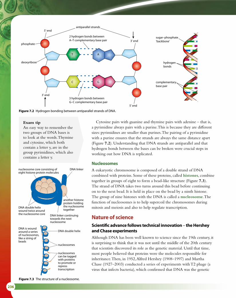
236
Exam tipAn easy way to remember the two groups of DNA bases is to look at the words. Thymine and cytosine, which both contain a letter y, are in the group pyrimidines, which also contains a letter y.
Cytosine pairs with guanine and thymine pairs with adenine – that is, a pyrimidine always pairs with a purine. This is because they are different sizes: pyrimidines are smaller than purines. The pairing of a pyrimidine with a purine ensures that the strands are always the same distance apart (Figure 7.2). Understanding that DNA strands are antiparallel and that hydrogen bonds between the bases can be broken were crucial steps in working out how DNA is replicated.
NucleosomesA eukaryotic chromosome is composed of a double strand of DNA combined with proteins. Some of these proteins, called histones, combine together in groups of eight to form a bead-like structure (Figure 7.3). The strand of DNA takes two turns around this bead before continuing on to the next bead. It is held in place on the bead by a ninth histone. The group of nine histones with the DNA is called a nucleosome. The function of nucleosomes is to help supercoil the chromosomes during mitosis and meiosis and also to help regulate transcription.
Nature of scienceScientific advance follows technical innovation – the Hershey and Chase experimentsAlthough DNA has been well known to science since the 19th century, it is surprising to think that it was not until the middle of the 20th century that scientists discovered its role as the genetic material. Until that time, most people believed that proteins were the molecules responsible for inheritance. Then, in 1952, Alfred Hershey (1908–1997) and Martha Chase (1927–2003) conducted a series of experiments with T2 phage (a virus that infects bacteria), which confirmed that DNA was the genetic
3' end
5' end
3' end
5' end
phosphate
deoxyribose
2 hydrogen bonds between A–T complementary base pair
3 hydrogen bonds between G–C complementary base pair
antiparallel strands
complementarybase pair
sugar–phosphate‘backbone’
hydrogenbonds
T A
C G
nucleosome core consisting ofeight histone protein molecules
another histoneprotein holdingthe nucleosometogether
DNA double helixwound twice aroundthe nucleosome core
DNA is woundaround a seriesof nucleosomes,like a string ofbeads
DNA double helix
nucleosomes
nucleosomescan be taggedwith proteinsto promote orrepresstranscription
DNA linker continuingtowards the nextnucleosome
DNA linker
Figure 7.2 Hydrogen bonding between antiparallel strands of DNA.
Figure 7.3 The structure of a nucleosome.

7 NUCLEIC ACIDS (HL) 237
material. They were able to carry out these investigations thanks to two relatively new techniques – electron microscopy and radioactive labelling.
The structure of the T2 phage had recently been revealed using the electron microscope. The virus injects its DNA into the cell it infects, but leaves behind its protein coat. Hershey and Chase labelled the viral DNA with radioactive 32P. Phosphorus is present in DNA but not in amino acids, so as they followed the transfer of the labelled material into the cytoplasm of the bacterium, Hershey and Chase knew that it was only DNA that was being transferred.
They then labelled viruses with 35S (sulfur is present in amino acids but not in DNA). After these viruses had infected the bacterial cells, Hershey and Chase examined the discarded protein coats and found that they contained radioactive sulfur, but the bacterial cytoplasm did not. These results supported their hypothesis that DNA is the genetic material that infects the bacteria, and protein (found in the protein coats) was not (Figure 7.4).
bacteriophage
infection
blending
centrifugationno sulfurdetected incells
sulfur detectedin supernatant(liquid)
sulfur-labelledprotein capsule
bacteriophage
infection
blending
centrifugationphosphorusdetected incells
no phosphorusdetected in supernatant(liquid)
phosphorus-labelled DNA
Figure 7.4 The Hershey and Chase experiment showed that the genetic material transferred to bacterial cells by infecting T2 phages is DNA, and not protein as previously believed.
When Watson and Crick proposed their double-helix model for the structure of DNA in 1953, one of the most striking things they realised was that it immediately suggested a mechanism for replication – if the two strands were unwound, each one could provide a template for the synthesis of a new strand.
The process of DNA replicationDNA replication ensures that exact copies of existing molecules are produced before a cell divides. The process is said to be semi-conservative and each strand of an existing DNA molecule acts as a template for the production of a new strand.

238
Most prokaryotes continuously copy their DNA, while in eukaryotes, replication is controlled through interactions between proteins, including cyclins and CDKs (Subtopic 1.6). Unlike prokaryotes, which can duplicate their DNA in as little as 20 minutes, the process in a eukaryotic cell takes up to 24 hours to complete. There are a number of similarities between prokaryotic and eukaryotic replication:
• both are bi-directional processes
• both require primers to start the process
• DNA polymerase enzymes work from the direction of the 5' end of the strand towards the 3' end in both cases, so that new nucleotides are added to the 3' end of a primer
• both have leading and lagging strands.The process of replication is semi-conservative – that is, each original
strand acts as a template to build up a new strand (Figure 7.5). The DNA double helix is unwound to expose the two strands for replication by the enzyme helicase, at a region known as a replication fork. The action of helicase creates single-stranded regions, which are less stable than the double-stranded molecule. To stabilise these single strands, single-stranded binding proteins (SSBs) are needed. SSBs protect the single-stranded DNA and allow other enzymes involved in replication to function effectively upon it.
Replication must occur in the 5' → 3' direction (and also in transcription and translation, described in Subtopics 7.2 and 7.3), because the enzymes involved only work in a 5' → 3' direction (adding new nucleotides to the 3' end of the newly forming molecule). As the two strands are antiparallel, replication has to proceed in opposite directions on the two strands. However, the replication fork where the double helix unwinds moves along in one direction only. This means that on one of the strands replication can proceed in a continuous way, following the replication fork along, but on the other strand the process has to happen in short sections, each moving away from the replication fork (Figure 7.6). The strand undergoing continuous synthesis is called the leading strand. The other strand, in which the new DNA is built up in short sections, is known as the lagging strand.
Leading strandReplication to produce the leading strand begins at a point on the molecule known as the ‘origin of replication’ site. First RNA primase adds a short length of RNA, attached by complementary base pairing, to the template DNA strand. This acts as a primer, allowing the enzyme DNA polymerase III to bind. DNA polymerase III adds free building units called deoxynucleoside triphosphates (dNTPs) to the 3' end of the primer and then to the forming strand of DNA. In this way the new molecule grows in a 5' → 3' direction, following the progress of helicase as it moves the replication fork along the DNA double helix. The RNA primer is later removed by DNA polymerase I.
originalDNA
newDNA
new DNA strands
old DNA strands
Figure 7.5 DNA replication is semi-conservative. As it is copied one original strand becomes paired with one new strand. One out of the two strands in each new DNA molecule is conserved, hence ‘semi-conservative’.
SSB proteins bind to and modulate the function of numerous proteins involved in replication, recombination and repair. They protect single-stranded DNA from being digested by nucleases and they also remove secondary structure from the DNA – that is, they prevent hydrogen bonds forming between the bases along the single strands, which would cause them to coil back on themselves.

7 NUCLEIC ACIDS (HL) 239
The dNTPs have two extra phosphate groups attached, and are said to be ‘activated’. They pair up with their complementary bases on the exposed DNA strand and DNA polymerase III then links together the sugar and the innermost phosphate groups of adjacent nucleotides. The two extra phosphate groups are broken off and released. In this way, a continuous new DNA strand is built up on the leading strand.
Lagging strandSynthesis of the lagging strand is a little more complicated, as it has to occur in discontinuous sections, which are then joined together.1 As for the leading strand, RNA primase first synthesises a short RNA
primer, complementary to the exposed DNA. This happens close to the replication fork.
2 DNA polymerase III starts replication by attaching at the 3' end of the RNA primer and adding dNTPs in a 5' → 3' direction. As it does so, it moves away from the replication fork on this strand.
replicationfork
helicase
RNA primase addsshort RNA primer.
1
DNA polymerase III startsreplication next to primer,and adds dNTPs in a5’ 3’ direction.
phosphates arereleased as dNTPsare added to thegrowing DNAstrand
deoxynucleosidetriphosphates (dNTPs)
DNA polymerase III adds dNTPs in a5’ 3’ direction, moving in the samedirection as the replication fork.
2
DNA polymerase Ireplaces RNA primerwith DNA.
short length of DNAare called Okazakifragments
RNAprimer
3
DNA ligase seals upbreaks between DNAfragments.
4
lagging strand
leading strand
5’
5’
5’
5’
3’
3’
Figure 7.6 DNA replication.
Replication fork the point where the DNA double helix is being separated to expose the two strands as templates for replicationLeading strand the new strand that is synthesised continuously and follows the replication forkLagging strand the new strand that is synthesised in short fragments in the opposite direction to the movement of the replication forkOkazaki fragments short fragments of a DNA strand formed on the lagging strand

240
3 DNA polymerase I now removes the RNA primer and replaces it with DNA using dNTPs. Short lengths of new DNA called Okazaki fragments are formed from each primer. The new fragment grows away from the replication fork until it reaches the next fragment.
4 Finally, DNA ligase seals up each break between the Okazaki fragments by making sugar–phosphate bonds so that a continuous strand of new DNA is created.
As DNA replication proceeds, supercoiling in the region of the molecule that is ahead of the replication fork is affected by the uncoiling process controlled by helicase. DNA gyrase is an enzyme that relieves the tension put upon a DNA molecule as it is being unwound. DNA gyrase is able to create negative supercoils in bacterial DNA. This is important during the replication process because DNA is a ‘right handed’ helix and without gyrase positive supercoils would accumulate in advance of DNA polymerase and prevent replication continuing. Gyrase is also important during transcription (Subtopic 7.2) because without it, portions of the circular bacterial chromosome could be prevented from re-attaching to one another.
Bacterial DNA gyrase is the target for a number of antibiotics. Without gyrase, the replication of bacterial DNA cannot occur, so an antibiotic that destroys gyrase can slow down bacterial growth that is causing an infection.
Sanger’s method of stopping replication
DNA samples are prepared for profiling (Subtopic 3.5) using the polymerase chain reaction (PCR), which replicates tiny amounts of DNA until there is sufficient to test. In this process, and in the preparation of DNA samples for sequencing, special chain-terminating nucleotides that inhibit DNA polymerase are used to stop the replication process when required. These dideoxynucleotides (ddNTPs) have no 3' hydroxyl group (Figure 7.7) so that after they have been added by DNA polymerase to a nucleotide chain no further nucleotides can be added and no further chain elongation can occur. ddNTPs were first used by Frederick Sanger in 1977 and his method, used in sequencing, is known as the dideoxy chain-termination method.
P O–O
O–
P O–O
O
CH2
O
H
base
H
P O–O
O
OH OH
O
H
ribonucleosidetriphosphate
(NTP)
RNA
H
P O–O
O–
P O–O
O
CH2
O
H
base
H
P O–O
O
OH H H H
O
H
deoxyribonucleosidetriphosphate
(dNTP)
DNA
H
P O–O
O–
P O–O
O
CH2
O
H
base
H
P O–O
O
O
H
dideoxyribonucleosidetriphosphate
(ddNTP)
ddNTPcauses termination of
DNA replication
H
Figure 7.7 Structure of ddNTP molecule.

7 NUCLEIC ACIDS (HL) 241
Non-coding regions of DNADNA molecules are very long but every strand has regions that do not code for proteins. For many years these regions were poorly understood but many of them have now been found to have important functions in the regulation of gene expression and other cell activities.
Regulator genesRegulator genes are present in DNA to control the expression of one or more other genes. Some may code for protein but many work at the level of RNA. Prokaryotic regulator genes often code for repressor proteins that bind to sections of DNA known as promoters and operators. Here they block the transcription of RNA by inhibiting RNA polymerase. Some regulator genes code for activator proteins that bind to DNA and cause a gene to be transcribed. One well-studied activator protein (known as an inducer) is involved in switching on the LAC operon (see box). Activators and repressors are both important in controlling gene expression. You can read more about them in Subtopic 7.2.
IntronsIntrons (or intragenic regions) are sequences of nucleotides within genes (they ‘intervene’ in genes). The corresponding sequences in the mRNA transcribed from these genes are removed in the nucleus before the mRNA moves to the cytoplasm for translation (Subtopic 7.2). Introns were discovered in 1977 by Phillip Allen and Richard Sharp who shared a Nobel prize for their work. Introns occur in many genes in all organisms and the number of introns per gene varies considerably between species. The human genome has been found to have about 8 introns per gene but simpler organisms such as fungi may have fewer than 20 in their entire genome. It seems that there are more introns in larger species with smaller populations, and evolutionary and biological factors are thought to influence this.
Once introns have been removed by RNA splicing, the remaining RNA is known as mature RNA. Mature RNA contains exons, which are the sequences that will be translated (or ‘expressed’). Introns may be spliced out in different ways so that a number of different, but similar, protein sequences can be produced from a single gene. Splicing is controlled by molecules that respond to signals from both inside and outside the cell.
TelomeresTelomeres are regions of repeated nucleotide sequences that are found at each end of the chromatids of chromosomes. Prokaryotes, which do not have linear chromosomes, do not have telomeres. Telomeres protect the ends of chromosomes from damage during cell division. Without them chromosomes would gradually become shorter and important genes might be lost. Telomeres are needed because, as Okazaki fragments are synthesised, RNA primers must attach in advance of the replication site on the lagging strand. But enzymes involved in this process cannot
Prokaryotic gene control – the LAC operon in Escherichia coli
The operon model explains how prokaryotic cells can regulate gene activity and respond to changes in their environment. An operon is a group of genes that act together to code for the enzymes needed for a particular metabolic pathway. The structural genes coding for the enzyme (called a transcription unit) are controlled by a promoter sequence (which is the site of attachment for RNA polymerase and the starting point for mRNA formation) and a region just before the structural genes, which is called the operator. In the case of the LAC operon, a regulator gene elsewhere on the DNA molecule produces a repressor molecule that interacts with the operator region to switch off the genes for lactose metabolism, because lactose is not usually an energy source for E. coli. If enzymes to metabolise lactose should be needed, the repressor is removed from the operator region by the action of an activator molecule, known as an inducer, which binds to it and alters its shape. This means that RNA polymerase can bind and the transcription of enzymes for lactose metabolism can begin. (Eukaryotes do not have operons.)

242
continue all the way to the end of the chromosome. With every cell division, the telomere becomes shorter, but the DNA replicates without causing harm to genes. Telomeres can be reconstructed by the enzyme telomerase reverse transcriptase.
Genes for transfer RNA (tRNA)In humans, the genes that code for tRNA molecules (Subtopic 7.3) are found on all chromosomes except 22 and Y. These genes do not code for protein but code either for cytoplasmic tRNA or for mitochondrial tRNA. The number of genes that code for tRNA is related to evolutionary history so that organisms in the Domains Archaea and Eubacteria have fewer than those in the Domain Eukarya. This seems to be due to the duplication of the genes over time.
Tandem repeatsDNA profiling is the technique used by forensic scientists to identify a person from their DNA. It is important in criminal investigations, paternity testing and in establishing family relationships. Samples of DNA for profiling are replicated using the polymerase chain reaction (PCR), fragmented using restriction enzymes and then electrophoresis is used to separate the fragments and produce banded patterns (Subtopic 3.5). The DNA sequences that are important in creating an individual’s unique profile, or in determining the degree of relatedness between two individuals, are repeated sequences of non-coding DNA called variable number tandem repeats (VNTRs), which are very similar in close relatives but very different in unrelated individuals.
Nature of scienceCareful observation – Rosalind Franklin’s meticulous workRosalind Franklin and Maurice Wilkins worked on the structure of DNA at King’s College London in the 1950s (Subtopic 2.6, Figure 2.23). They used the technique of X-ray crystallography, which was developed in the early 1900s by the physicist Sir Lawrence Bragg. The process involves directing a beam of X-rays at the atoms in a molecule. The beam is deflected so that it produces an X-ray diffraction pattern, which is unique to the molecule. The molecule is rotated several times and X-rays directed towards it from different angles so that its three dimensional structure can be studied. Large molecules containing many thousands of atoms produce very complex patterns and considerable skill is required to interpret them. Today this is done by computers, which can locate the positions of atoms within the molecules with precision and compile 3D images, but in the 1950s, Franklin and Wilkins did not have the help of a computer. This makes their achievements all the more impressive. It was Franklin who showed in 1952 that phosphate groups in a DNA molecule must be positioned on the outside of its structure. Without this knowledge, Watson and Crick might not have been able to establish the structure of DNA. Although Watson, Crick and Wilkins were awarded a Nobel Prize
As a person ages, their telomeres become shorter and cell division is less efficient.

7 NUCLEIC ACIDS (HL) 243
for their work, Franklin – who died in 1958 – did not receive the award because Nobel Prizes are not awarded posthumously.
X-ray crystallography has established the structure of many complex biological molecules including the triple helix of collagen, the structure of antibodies and the respiratory pigments, myoglobin and hemoglobin. Our understanding of the structure of these molecules has enabled biochemists to work out not only their properties but also how they function.
Exam tipThere are several chemical names involved in replication, so it is helpful to list them.Nucleoside triphosphate (NTP) a building unit for RNA – a ribose nucleotide with two additional phosphates, which are chopped off during the RNA-synthesis processDeoxynucleoside triphosphate (dNTP) the same as NTP but with deoxyribose instead of ribose, so it is used to build DNA rather than RNA through base-pairing to the parent DNA strandHelicase an enzyme found at the replication fork, with two functions – to unwind the two DNA strands, and to separate them by breaking the hydrogen bondsSingle-stranded binding proteins (SSBs) bind to single-stranded regions of DNA to stabilise and protect their structureDNA gyrase relieves the tension put upon a DNA molecule as it is being unwoundRNA primase this enzyme adds NTPs to the single-stranded DNA that has been unzipped by helicase, in a 5' → 3' direction, to make a short length of RNA (a primer) base-paired to the parent DNA strandDNA polymerase III this enzyme adds dNTPs in a 5' → 3' direction where RNA primase has added a short length of complementary RNA as a primer; it is unable to add dNTPs directly to the single-stranded parent DNA that has been unzipped by helicaseDNA polymerase I this enzyme removes the RNA nucleotides of the primers on the lagging strand in a 5' → 3' direction and replaces them with DNA nucleotides using dNTPsDNA ligase this enzyme joins adjacent Okazaki fragments by forming a covalent bond between the deoxyribose and the phosphate of adjacent nucleotides
1 Outline what is meant by the term ‘antiparallel’.2 Outline the structure of a nucleosome.3 State the direction in which DNA replication occurs.4 Explain why Okazaki fragments must be produced on one of the
DNA strands.5 Outline the role of telomeres.
Test yourself?

244
What is junk?The nucleus of a human cell contains nearly 2 m of DNA,
but genes make up only a small proportion of this. The DNA in a eukaryotic cell can be divided up into two types:
• unique or single-copy genes, which make up 55–95% of the total
• highly repetitive sequences, or satellite DNA, which account for 5–45%.
The repetitive sequences are typically between 5 and 300 base pairs long and may be duplicated as many as 100 000 times in a genome.
Many genes include sections called introns that are transcribed but not translated. Only sections of genes known as exons are both transcribed and translated.
When the highly repetitive sequences of DNA were first discovered, they appeared to have no function. Scientists at the time called them ‘junk DNA’ and thought they were simply ‘excess baggage’. Before scientists began mapping several animal genomes, they had a rather restricted view about which parts of the genome were important. According to the traditional viewpoint, the really crucial things were genes and a few other sections that regulate gene function were also considered useful.
But new findings suggest that this interpretation was not correct. In 2004, David Haussler’s team at the University of California, Santa Cruz, USA, compared human, mouse and rat genome sequences. They were astonished to find that several long sequences of repeated DNA were identical across the three species. As David Haussler exclaimed:
‘It absolutely knocked me of my chair.’
When the Human Genome Project was planned, there were calls from some people to map only the bits of genome that coded for protein – mapping the rest was thought to be a waste of time. Luckily, entire genomes were mapped and have proved vital to the study of so-called ‘junk DNA’. Most geneticists now accept that ‘junk DNA’ regulates and controls the activity of vital genes and possibly embryo development.
Questions to consider
• Do you think it is appropriate to label something as ‘junk’ simply because it is thought to have no function?
• Does such labelling hinder scientific progress?
• Should experiments be carried out to answer fundamental questions even if they have no obvious application?
• Who should decide which research is most likely to be valuable?

7 NUCLEIC ACIDS (HL) 245
7.2 Transcription and gene expressionThe process by which the DNA code is used to build polypeptides occurs in two stages. The first is transcription, which transfers sections of the genetic code from DNA to an mRNA molecule. Transcription happens in the nucleus. The second stage, known as translation, occurs in the cytoplasm and uses the mRNA, together with ribosomes, to construct the polypeptide. Translation is discussed in Subtopic 7.3.
Transcribing DNA into mRNAAt the start of transcription, the DNA molecule is separated into two strands by the enzyme RNA polymerase, which binds to the DNA near the beginning of a gene. Hydrogen bonds between the bases are broken and the double helix unwinds (Figure 7.8).
Transcription begins at a specific point on the DNA molecule called the promoter region, which is a short sequence of non-coding DNA just before the start of the gene. Only one of the two strands is used as a template for transcription and this is called the antisense strand. The other DNA strand is called the sense strand. RNA polymerase uses free nucleoside triphosphates (NTPs) to build the RNA molecule, using complementary base pairing to the DNA and condensation reactions between the nucleotides. This produces a primary mRNA molecule that is complementary to the antisense strand being transcribed, and has the same base sequence as the sense strand (except that it contains the base U in place of T). RNA polymerase moves along the antisense DNA strand in a 3' → 5' direction. As it does so, the 5' end of a nucleotide is added to the 3' end of the mRNA molecule so that the construction of the mRNA proceeds in a 5' → 3' direction.
RNA polymerase checks the mRNA molecule as it forms to ensure that bases are paired correctly. As the mRNA molecule is extended, the DNA is rewound into a helix once a section has been transcribed. Eventually RNA polymerase reaches another specific sequence on the DNA called the terminator region, which indicates the end of the gene. The RNA polymerase releases the completed RNA strand and finishes rewinding the DNA before breaking free.
Regulation of transcription (gene expression) in eukaryotesGene expression in eukaryotes is controlled by several regulatory processes including:
• transcriptional regulation – mechanisms that prevent transcription
• post-transcriptional regulation – mechanisms that control or regulate mRNA after it has been produced
• translational regulation – mechanisms that prevent translation (Subtopic 7.3).
Learning objectives
You should understand that:
• Transcription is carried out in a 5' → 3' direction.
• In eukaryotes, nucleosomes help to regulate transcription.
• In eukaryotes, mRNA is modified after transcription.
• Splicing of mRNA increases the number of different proteins that a cell can produce.
• Gene expression is related by proteins that attach to specific base sequences in DNA.
• Gene expression is affected by the environment of a cell and of the organism.
A single chromosome contains DNA that codes for many proteins. During transcription, genes (short lengths of DNA that code for single polypeptides) are used to produce mRNA. Most genes are about 1000 nucleotides long, a few are longer and a very small number are less than 100 nucleotides. The size of the gene corresponds to the size of the polypeptide it codes for.
Nucleoside triphosphates (NTPs) are the molecules used by RNA polymerase to build mRNA molecules during transcription (Figure 7.7). As they move into place, two phosphates are removed from them so they become converted into nucleotides.

246
Transcriptional regulation
Regulation by nucleosomesDNA in eukaryotes is incorporated into nucleosomes so that the genetic material can be stored in a compact form (Figure 7.3). But in order to transcribe genes, activators and enzymes involved in transcription must be able to gain access to DNA. It has been found that in all eukaryotic species, the regions of DNA that contain promoters and regulators have fewer nucleosomes than other areas, allowing greater access for binding proteins, while regions that are transcribed have a higher density of nucleosomes. This suggests that nucleosomes have an important role in determining which genes are transcribed and this in turn can influence other factors such as cell variation and development.
Researchers have shown that DNA in nucleosomes may be either ‘wrapped’ or ‘unwrapped’ and suggest that DNA does not need to be completely released from a nucleosome to be transcribed, but that there is a significant period of time during which DNA is accessible. Although nucleosomes are very stable protein–DNA complexes, they are not static and can undergo different structural rearrangements including so-called ‘nucleosome sliding’ and DNA site exposure. Nucleosomes are important because they can either inhibit or allow transcription by controlling whether the necessary molecules can bind to DNA.
Regulation by binding proteinsRNA polymerase requires the presence of a class of proteins known as ‘general transcription factors’ before transcription can begin. Interactions between the transcription factors, RNA polymerase, and the promoter region of the DNA molecule allow the polymerase to move along the
RNApolymeraseunbinds andthe RNAmolecule isreleased at theterminatorregion
pool of freenucleosidetriphosphates
free nucleosidetriphosphates align bybase pairing and are heldby hydrogen bonds
antisense strand read inthe 3’ 5’ direction
direction of mRNA synthesis
RNA polymeraseenzyme
RNApolymerasebinds at apromoterregion
helix re-formsas the RNApolymerasemoves along
sense strand
A C
G
G
G
G
G
CCA
C
C
U
antisense strand
RNA polymerase removes two phosphates from nucleosidetriphosphate and links the 5’ end of the nucleotide to the3’ end of the growing RNA molecule
5’
5’
3’
mRNA strand
C C
C
C C C C C CG GU U UA
C C C CG G G G G G GT T TA
C C C C CC C G G G GT T TA A A A
A A
A
C
AA
UG
G
GG 5’
3’
3’
Figure 7.8 Transcription.

7 NUCLEIC ACIDS (HL) 247
gene so that transcription can occur. There is greater transcription of certain genes when specific transcription factors are present. Many different transcription factors have been found and each one is able to recognise and bind to a specific nucleotide sequence in DNA. A specific combination of transcription factors is necessary to activate a particular gene.
The role of activators is shown in Figure 7.9. These proteins bind to a region of the DNA called the enhancer, which may be some distance from the gene. ‘Bending proteins’ may then assist in bending the DNA so that the enhancer region is brought close to the promoter. Activators, transcription factors and other proteins attach, so that an ‘initiation complex’ is formed and transcription can begin.
Transcription factors are regulated by signals produced from other molecules. For example, hormones are able to activate transcription factors and thus control transcription of certain genes. Many other molecules in the environment of a cell or an organism can also have an impact on gene expression and protein production. Understanding these molecules and the way they work will add to our understanding of problems arising when proteins are produced incorrectly.
General transcription factors (GTFs)
GTFs are proteins that bind to particular sites on DNA and activate transcription. Together with RNA polymerase and other proteins (activators), GTFs form the transcription apparatus and have a key role in regulating genes. Bacteria need RNA polymerase and just one GTF for their transcription apparatus, while in eukaryotes (and archaeans) RNA polymerase and several GTFs are needed.
One important feature of transcription factors is that they have the ability to bind to specific enhancer or promoter sequences of DNA. Some will bind to a DNA promoter sequence near the transcription start site and form part of the transcription initiation complex. Others bind to regulatory sequences, such as enhancer sequences, and can either stimulate or repress transcription of the related gene.
Figure 7.9 Gene expression is controlled by proteins that bind to specific sequences in DNA.
activators
enhancer
bending protein
RNA polymerase
transcriptioninitiation complex
promoter
gene
Post-transcriptional modificationIn eukaryotes, many genes contain sequences of DNA that are transcribed but not translated. These sequences, which appear in mRNA, are known as introns. After transcription of a gene, the introns are removed in a process known as post-transcriptional modification. The sequences of bases that remain are known as exons and these are spliced together to form the mature mRNA that is then translated (Figure 7.10). Mature mRNA leaves the nucleus via the nuclear pores and moves to the cytoplasm.
RNA splicing can result in variations in the mature mRNA that is produced, depending on which introns are removed – so the removal of
intron exon
mRNA
post-translationalmodi�cation
Figure 7.10 Introns and exons in mRNA.

248
introns enables a gene to code for more than one polypeptide. An average human gene is thought to code for three different proteins.
Environment and gene expressionThe expression of genes can be influenced by the environment – not only the organism’s external environment, but also its internal environment, which is affected by chemicals such as hormones and various products of metabolism. Temperature, light, chemicals including drugs and the organism’s sex are just some of the environmental factors that can cause some genes to be turned on or off and influence how an organism functions or develops.
‘Sex-influenced’ characteristics are those which are expressed differently in the two sexes. They are genes found on autosomes, not on the sex chromosomes, and one example is male baldness. The baldness allele is influenced by high levels of two hormones, testosterone and dihydrotestosterone. Males usually have much higher levels of these hormones than females, so the baldness allele is more active in men, who are more likely to go bald than women. But sometimes, in very stressful conditions, a woman’s adrenal glands may produce testosterone, which can be converted to dihydrotestosterone. In this case the genes for baldness may be expressed so that women may also suffer from hair loss.
The presence of drugs or chemicals in the external or internal environment can also influence gene expression. One example is the drug thalidomide, which was used from the mid 1950s to treat morning sickness during pregnancy. It has no effect on gene expression in adults but has severe effects on limb development in fetuses.
Temperature is an environmental factor that has been known to influence gene expression for many years. A well-studied example is the Himalayan rabbit, which has a gene for the development of pigment in its fur. The gene remains inactive above 35 °C, but becomes very active between 15 °C and 25 °C. In the warm parts of the rabbit’s body, the gene is inactive so that the animal’s fur is white, but in parts of the body where the temperature is lower, such as ears, feet and nose, the gene becomes active and pigment is produced making the fur in these areas black.
Nature versus nurtureFrancis Galton (1822–1911) was one of the first scientists to
consider the question of ‘nature versus nurture’ – that is, the relative importance of an individual’s genetic makeup versus the influence of the experiences and environment they are exposed to during their life. The philosopher John Locke (1632–1704) had previously proposed that humans develop as a result of their economic, educational and social status. Today, studies of identical twins raised separately counter Locke’s simple viewpoint. But the relative importance of a person’s innate characteristics and their personal experiences is still widely discussed today.
Question to consider
• Is it important that science considers the relative importance of innate genetic characteristics and personal experiences in the development of an individual?
Exam tipRemember: introns ‘intervene’ in genes, but only exons are ‘expressed’.

7 NUCLEIC ACIDS (HL) 249
Me
MeMe
Me
histone proteindi�erent molecules can attach to the ‘tails’ of histone proteins, altering the activity of the DNA wrapped round them
chromosome
histone tail
adenine
thymine
methyl groups (CH3) added to certain cytosine bases repress gene activity
cytosine
guanine
Nature of scienceLooking for patterns and trendsEpigenetics is usually defined as heritable changes in gene expression function that occur without a change in the nucleotide sequence of the DNA molecule. These changes may remain through cell divisions for the remainder of the cell’s life. Since there is no change in the DNA sequence, non-genetic factors must influence how genes come to be expressed differently.
We know that an organism’s development is influenced by genes being switched on or off at specific times and there has been much debate about how and whether environmental factors can lead to such epigenetic modifications. If so, the environment could have an important influence on gene expression. Pollutants, diet, temperature and stress all have effects on development. It is also known that these factors can lead to changes in histones or the methylation of DNA (Figure 7.11). So perhaps these changes could cause epigenetic modifications to gene expression, which affect development.
DNA methylation is a process that involves the addition of a methyl group to cytosine. It is an example of an epigenetic marker. The presence of the methyl group changes the way that the DNA sequence is read. In most cases methylation of a section of DNA turns off a gene, leading to reduced gene expression (Figure 7.11).
Figure 7.11 Methylation of DNA is one epigenetic factor affecting gene expression; histone modification is another.
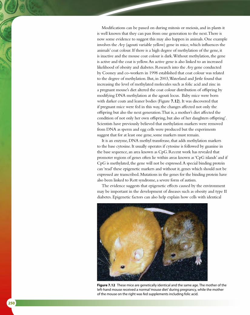
250
Modifications can be passed on during mitosis or meiosis, and in plants it is well known that they can pass from one generation to the next. There is now some evidence to suggest this may also happen in animals. One example involves the Avy (agouti variable yellow) gene in mice, which influences the animals’ coat colour. If there is a high degree of methylation of the gene, it is inactive and the mouse coat colour is dark. Without methylation, the gene is active and the coat is yellow. An active gene is also linked to an increased likelihood of obesity and diabetes. Research into the Avy gene conducted by Cooney and co-workers in 1998 established that coat colour was related to the degree of methylation. But, in 2003, Waterland and Jirtle found that increasing the level of methylated molecules such as folic acid and zinc in a pregnant mouse’s diet altered the coat colour distribution of offspring by modifying DNA methylation at the agouti locus. Baby mice were born with darker coats and leaner bodies (Figure 7.12). It was discovered that if pregnant mice were fed in this way, the changes affected not only the offspring but also the next generation. That is, a mother’s diet affected the condition of not only her own offspring, but also of her daughters offspring’. Scientists have previously believed that methylation markers were removed from DNA as sperm and egg cells were produced but the experiments suggest that for at least one gene, some markers must remain.
It is an enzyme, DNA methyl transferase, that adds methylation markers to the base cytosine. It usually operates if cytosine is followed by guanine in the base sequence, an area known as CpG. Recent work has revealed that promoter regions of genes often lie within areas known as ‘CpG islands’ and if CpG is methylated, the gene will not be expressed. A special binding protein can ‘read’ these epigenetic markers and without it, genes which should not be expressed are transcribed. Mutations in the genes for the binding protein have also been linked to Rett syndrome, a severe form of autism.
The evidence suggests that epigenetic effects caused by the environment may be important in the development of diseases such as obesity and type II diabetes. Epigenetic factors can also help explain how cells with identical
Figure 7.12 These mice are genetically identical and the same age. The mother of the left-hand mouse received a normal ‘mouse diet’ during pregnancy, while the mother of the mouse on the right was fed supplements including folic acid.

7 NUCLEIC ACIDS (HL) 251
Learning objectives
You should understand that:
• Initiation of translation involves the assembly of components that carry out the process.
• Polypeptide synthesis involves a repeated cycle of events.
• Components used in the process are disassembled when translation is terminated.
• Free ribosomes synthesise proteins for use within the cell, while bound ribosomes synthesise proteins primarily for secretion or inclusion in lysosomes.
• In prokaryotes, translation can occur immediately after transcription because there is no nuclear membrane for mRNA to move through.
• The number and sequence of amino acids in a protein make up its primary structure.
• Secondary structure consists of α helices and β pleated sheets, stabilised by hydrogen bonding.
• Tertiary structure is folding of the secondary structure stabilised by interactions between R groups of amino acids.
• Quaternary structure occurs in proteins with more than one polypeptide chain.
6 State what is meant by an ‘exon’.7 Distinguish between the ‘sense’ and ‘antisense’ strands of DNA.8 Outline the role of the promoter region in transcription.9 Outline the importance of the environment to the expression of
any named gene.
7.3 TranslationTranslation is the process by which the information carried by mRNA is decoded and used to build the sequence of amino acids that eventually forms a protein molecule. During translation, amino acids are joined together in the order dictated by the sequence of codons on the mRNA to form a polypeptide. This polypeptide eventually becomes the protein coded for by the original gene.
Transfer RNA (tRNA)The process of translation requires another type of nucleic acid known as transfer RNA or tRNA. tRNA is made of a single strand of nucleotides that is folded and held in place by base pairing and hydrogen bonds (Figure 7.13). There are many different tRNA molecules but they all have a characteristic ‘clover leaf ’ appearance with some small differences between them.
At one position on the molecule is a triplet of bases called the anticodon, which pairs by complementary base pairing with a codon on the mRNA strand. At the 3' end of the tRNA molecule is a base sequence CCA, which is the attachment site for an amino acid.
An amino acid is attached to the specific tRNA molecule that has its corresponding anticodon, by an activating enzyme. As there are 20 different amino acids, there are also 20 different activating enzymes in the cytoplasm. The tRNA-activating enzymes are substrate-specific and recognise the correct tRNA molecules by their shapes. Energy for the attachment of an amino acid comes from ATP. A tRNA molecule with its attached amino acid is called a charged tRNA.
RibosomesRibosomes are the site of protein synthesis. Some ribosomes occur free in the cytoplasm and these synthesise proteins that will be used within the cell. Others are bound to the endoplasmic reticulum, forming rough endoplasmic reticulum, and these synthesise proteins for secretion from the cell or for use within lysosomes.
Test yourself?
DNA can differentiate into different cell types with different appearances. Recent data also suggests that epigenetic patterns may change during the course of life, so that key genes in vital processes may be affected with age.
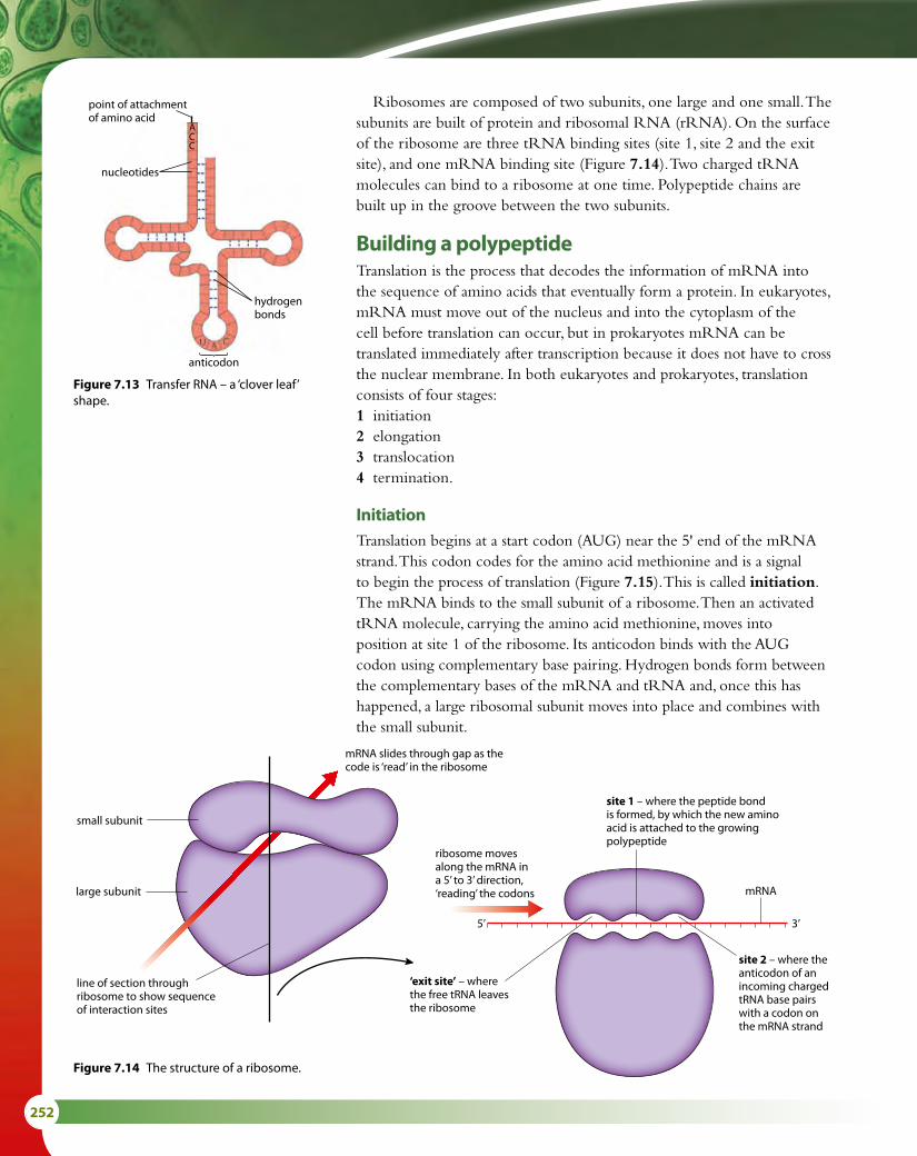
252
Ribosomes are composed of two subunits, one large and one small. The subunits are built of protein and ribosomal RNA (rRNA). On the surface of the ribosome are three tRNA binding sites (site 1, site 2 and the exit site), and one mRNA binding site (Figure 7.14). Two charged tRNA molecules can bind to a ribosome at one time. Polypeptide chains are built up in the groove between the two subunits.
Building a polypeptideTranslation is the process that decodes the information of mRNA into the sequence of amino acids that eventually form a protein. In eukaryotes, mRNA must move out of the nucleus and into the cytoplasm of the cell before translation can occur, but in prokaryotes mRNA can be translated immediately after transcription because it does not have to cross the nuclear membrane. In both eukaryotes and prokaryotes, translation consists of four stages:1 initiation2 elongation3 translocation4 termination.
InitiationTranslation begins at a start codon (AUG) near the 5' end of the mRNA strand. This codon codes for the amino acid methionine and is a signal to begin the process of translation (Figure 7.15). This is called initiation. The mRNA binds to the small subunit of a ribosome. Then an activated tRNA molecule, carrying the amino acid methionine, moves into position at site 1 of the ribosome. Its anticodon binds with the AUG codon using complementary base pairing. Hydrogen bonds form between the complementary bases of the mRNA and tRNA and, once this has happened, a large ribosomal subunit moves into place and combines with the small subunit.
hydrogenbonds
anticodon
nucleotides
ACC
point of attachmentof amino acid
Figure 7.13 Transfer RNA – a ‘clover leaf’ shape.
mRNA slides through gap as thecode is ‘read’ in the ribosome
line of section throughribosome to show sequenceof interaction sites
ribosome movesalong the mRNA in a 5’ to 3’ direction,‘reading’ the codons
site 1 – where the peptide bondis formed, by which the new aminoacid is attached to the growingpolypeptide
site 2 – where theanticodon of anincoming chargedtRNA base pairswith a codon onthe mRNA strand
‘exit site’ – where the free tRNA leaves the ribosome
mRNA
3’5’
small subunit
large subunit
Figure 7.14 The structure of a ribosome.
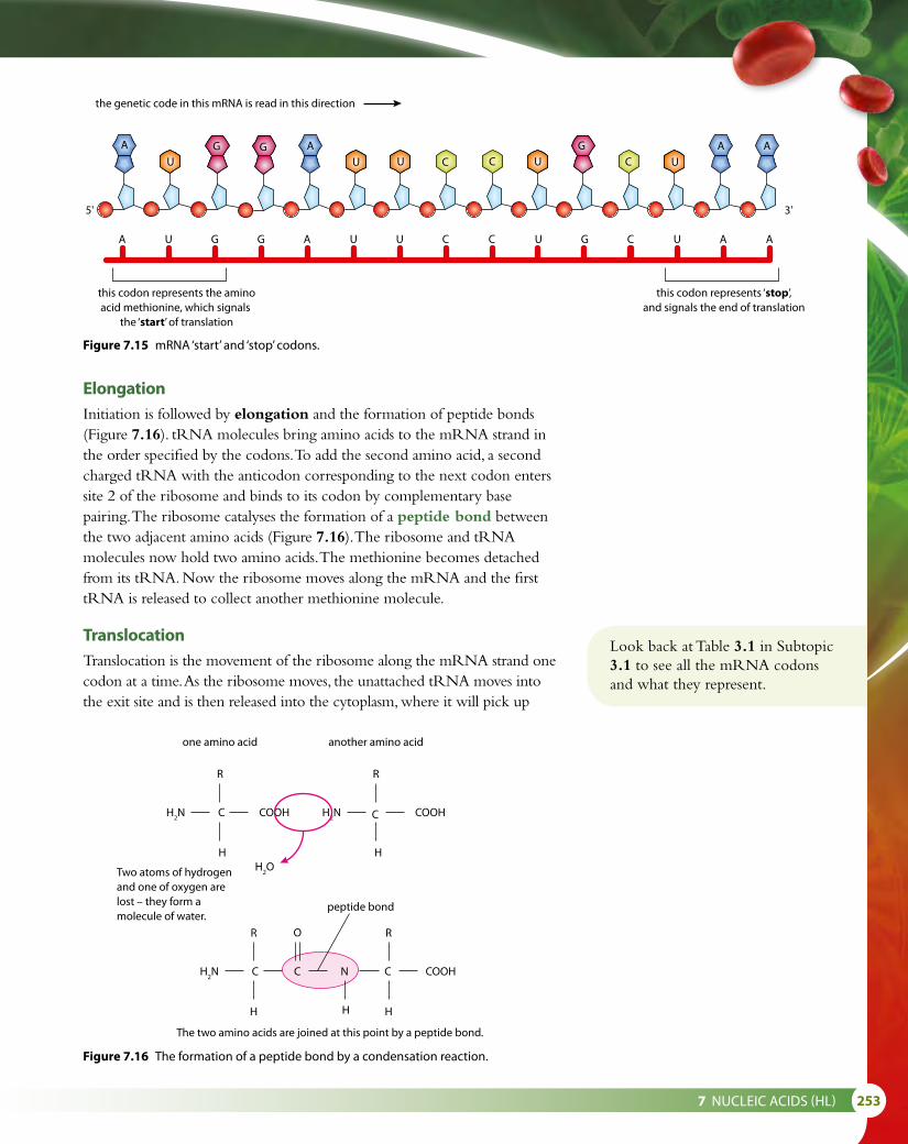
7 NUCLEIC ACIDS (HL) 253
ElongationInitiation is followed by elongation and the formation of peptide bonds (Figure 7.16). tRNA molecules bring amino acids to the mRNA strand in the order specified by the codons. To add the second amino acid, a second charged tRNA with the anticodon corresponding to the next codon enters site 2 of the ribosome and binds to its codon by complementary base pairing. The ribosome catalyses the formation of a peptide bond between the two adjacent amino acids (Figure 7.16). The ribosome and tRNA molecules now hold two amino acids. The methionine becomes detached from its tRNA. Now the ribosome moves along the mRNA and the first tRNA is released to collect another methionine molecule.
TranslocationTranslocation is the movement of the ribosome along the mRNA strand one codon at a time. As the ribosome moves, the unattached tRNA moves into the exit site and is then released into the cytoplasm, where it will pick up
the genetic code in this mRNA is read in this direction
A U G G A U U C C U G C U A A
this codon represents the aminoacid methionine, which signals
the ‘start’ of translation
this codon represents ‘stop’,and signals the end of translation
5' 3'
CAG
UU
A
UG
C CUG
UA A
one amino acid
CH2N
H2O
COOH
R
H
another amino acid
CH2N COOH
R
H
CH2N C
R
H
CN COOH
R
H
Two atoms of hydrogenand one of oxygen arelost – they form amolecule of water.
The two amino acids are joined at this point by a peptide bond.
peptide bond
O
H
Figure 7.15 mRNA ‘start’ and ‘stop’ codons.
Figure 7.16 The formation of a peptide bond by a condensation reaction.
Look back at Table 3.1 in Subtopic 3.1 to see all the mRNA codons and what they represent.

254
another amino acid molecule. The growing peptide chain is now positioned in site 1, leaving site 2 empty and ready to receive another charged tRNA molecule to enter and continue the elongation process. Figure 7.17 shows how initiation, elongation and translocation occur as mRNA is translated.
growingpolypeptide
tRNAleaving
Lys
tRNAmoving in
Pro
exitsite site 1 site 2
peptide bond
Translocation
codonAU
UA
GC
AU
G
AU
G
AU
G
C C C3’5’
ribosome moves ina 5’ 3’ direction alongthe mRNA, to the nextcodon
large subunitof ribosome
mRNA attached tosmall subunit ofribosome
exitsite site 1 site 2
Elongation
Initiation
codon
mRNA
Met
AU
UA
GC
AU
AU
AU
C C C3’5’
exitsite site 1 site 2
codon
mRNA
AU
UA
GC
A A A C C C3’5’
Met Lys
tRNA bringssecond aminoacid intoposition
peptide bondformed
Met
Figure 7.17 The stages of translation.

7 NUCLEIC ACIDS (HL) 255
TerminationTranslocation and elongation are repeated until one of the three ‘stop’ codons aligns with site 2, which acts as a signal to end translocation. There are no tRNA molecules with anticodons corresponding to these stop codons. The polypeptide chain and the mRNA are released from the ribosome and the ribosome separates into its two subunits. This final stage of translation is called termination.
PolysomesTranslation occurs at many places along an mRNA molecule at the same time. The electron micrograph in Figure 7.18 shows transcription and translation occurring simultaneously in a bacterium. A polysome is a group of ribosomes along one mRNA strand (Figure 7.19). Part of the bacterial chromosome can be seen as the fine pink line running horizontally along the bottom of the micrograph and two growing polypeptide chains are shown forming above it. DNA is being transcribed by RNA polymerase and the newly formed mRNA is being immediately translated by the ribosomes. In eukaryotes, the two processes occur in the nucleus and cytoplasm, respectively, and so are separated not only in time but also in location.
small andlarge subunitsof ribosome+polypeptide
growingpolypeptidechains
ribosomes movingalong mRNA
mRNA incytoplasm
Figure 7.19 Diagram of a polysome.
Figure 7.18 Electronmicrograph of polysomes in a bacterium (× 150 000).
Protein structureProteins are large, complex molecules, usually made up of hundreds of amino acid subunits. The way these subunits fit together is highly specific to each type of protein, and is vital to its function. Figure 7.20 illustrates the structure of the protein hemoglobin.1 The first stage of protein production is the assembly of a sequence
of amino acid molecules that are linked by peptide bonds formed by condensation reactions. This sequence forms the primary structure of a protein. There are many different proteins and each one has different numbers and types of amino acids arranged in a different order, coded for by a cell’s DNA.

256
2 The secondary structure of a protein is formed when the polypeptide chain takes up a permanent folded or twisted shape. Some polypeptides coil to produce an α helix, others fold to form β pleated sheets. The shapes are held in place by many weak hydrogen bonds. Depending on the sequence of amino acids, one section of a polypeptide may become an α helix while another takes up a β pleated form. This is the case in hemoglobin.
3 The tertiary structure of a protein forms as the molecule folds still further due to interactions between the R groups of the amino acids (Figure 7.21) and within the polypeptide chain. The protein takes up a three-dimensional shape, which is held together by ionic bonds between particular R groups, disulfide bridges (covalent bonds) between sulfur atoms of some R groups, and by weaker interactions between hydrophilic and hydrophobic side chains. Figure 7.22 shows the different types of bond involved in maintaining the tertiary structure of proteins. Tertiary structure is very important in enzymes because the shape of an enzyme molecule gives it its unique properties and determines which substrates can fit into its active site.
4 The final level of protein structure is quaternary structure, which links two or more polypeptide chains to form a single, large, complex protein. The structure is held together by all the bonds that are important in the previous levels of structure. Examples of proteins that have quaternary structure are collagen (which has three polypeptide chains), hemoglobin (which has four), antibodies (which also have four) and myosin (which has six).
In addition, many proteins contain prosthetic groups and are called conjugated proteins. Prosthetic groups are not polypeptides but they are able to bind to different proteins or parts of them. For example, hemoglobin is a conjugated protein, with four polypeptide chains, each containing a prosthetic heme group.
amino acidsequence
Primarystructure
the helixshape ismaintainedwithhydrogenbonds
disulfidebridge
beta chain(146 aminoacids)
alpha chain(141 aminoacids)
prosthetic hemegroup
hemoglobinmolecule
polypeptidechain
hydrogenbonds
twopolypeptidechains
helix pleatedsheet
Secondarystructure
Tertiarystructure
Quaternarystructure
1
2
3
4
PheGlu
TyrSer
IsoMet
aminoacids
peptidebonds
Figure 7.20 The structure of hemoglobin.
General structure of an amino acidThis group varies indi�erent amino acids.It is known as the R groupor side chain.
amine group carboxylic acid group
CN
R
H
H
H
O
OH
C
Structure of the simplest amino acid, glycine
CN
H
H
H
H
O
OH
C
R group is hydrogen
Figure 7.21 The general structure of an amino acid and the structure of glycine.

7 NUCLEIC ACIDS (HL) 257
Hydrogen bonds form between strongly polar groups.They can be broken by high temperature or by pH changes.
NH OCδ–
δ–
δ+δ+
bond to restof molecule
bond to restof molecule
shared electronsspend more timearound N
shared electronsspend more timearound O
hydrogen bond
Ionic bonds form betweenionised amine and carboxylicacid groups. They can bebroken by pH changes. CH2
NH2+
C
C
O
O
asparagine
glutamic acid
ionic bond
CH2
CH2
O–
CH2
CH2
SH
SH
Disul�de bonds form between cysteine molecules.The bonds can be broken by reducing agents.
disul�de bond
cysteine
cysteine
CH2
CH2
S
S
Hydrophobic interactions occur between non-polarside chains.
CH2
CH3
CH3
HC
OH
tyrosine
valine
Figure 7.22 Types of bond that are important in protein structure.
Fibrous and globular proteinsProtein molecules are categorised into two major types by their shape. Fibrous proteins are long and narrow and include collagen (found in skin, tendons and cartilage), keratin (in hair and nails) and silk. Fibrous proteins are usually insoluble in water and in general have secondary structure. Globular proteins have a more rounded, 3D shape and have either tertiary or quaternary structure. Most globular proteins are soluble in water. Globular proteins include enzymes, such as pepsin, and antibodies. Myoglobin and hemoglobin are also globular proteins.
Polar and non-polar amino acidsAmino acids are divided into two groups according to the chemical properties of their side chains or R groups (Figure 7.21). Polar and non-polar amino acids have different properties and their positions in a molecule affect the behaviour and function of the whole protein.

258
Amino acids with non-polar side chains are hydrophobic. Those with polar side chains are hydrophilic. Non-polar amino acids are found in parts of proteins that are in hydrophobic areas, while polar amino acids are in areas that are exposed to an aqueous environment such as cytoplasm or blood plasma.
For membrane proteins, the polar hydrophilic amino acids are found on the outer and inner surfaces in contact with the aqueous environment, while the non-polar hydrophobic amino acids are embedded in the core of the membrane in contact with the hydrophobic tails of the phospholipid bilayer (Figure 7.23). This helps to hold the protein in place in the membrane. Some integral proteins act as channels, and the pore is lined with hydrophilic amino acids to enable polar substances to pass through.
Key
outside of cell
inside of cell
phospholipid bilayer
channel protein with pore
protein that traverses membrane
protein on one side of membrane
position of non-polar amino acid side chains – inside the protein
position of polar amino acid side chains – on the exposed faces of the protein
Figure 7.23 In membrane proteins, polar (hydrophilic) amino acids are found on the surfaces in contact with the aqueous environment, while non-polar (hydrophobic) amino acids are embedded inside the phospholipid bilayer.
Polar and non-polar amino acids are also important in enzymes, where they assist in the binding of substrates. An enzyme that acts on a polar substance (for example, amylase) has polar amino acids in its active site, whereas lipases have non-polar amino acids in the active site.
Polar amino acids on the surface of a protein increase its solubility while non-polar amino acids help a protein maintain its structure. Lipases are proteins that have polar groups on the outside so they are soluble in the gut contents, but non-polar groups in their active site so that lipids can bind to them.

7 NUCLEIC ACIDS (HL) 259
Nature of scienceScientific advance follows technical innovation – computers help locate genes and identify conserved sequencesWhen the Human Genome Project was first begun, scientists could only deal with and analyse small sequences of DNA at any one time. But as the data began to accumulate to hundreds of thousands of sequences it became necessary to record how many genes there were, whether there was redundancy in the code and whether there were patterns in the sequencing. Mathematicians and computer scientists built algorithms to do this. One of the best known is the Tiger Assembler, which put together the sequences to try to work out how many genes there are in a human genome.
The DNA sequences are stored in databases such as GenBank at the US National Centre for Biotechnology Information and other organisations in Europe and Asia. The data are analysed by computer programs that determine the boundaries between genes, a process known as gene annotation, and help to identify sequences. Bioinformatics speeds up the process and uses statistical models to annotate and predict. The most recent methods for doing this use ‘formal grammars’ which derived from computer science. Without these tools, DNA research could not have proceeded as rapidly as it has.
10 Distinguish between ‘transcription’ and ‘translation’. 11 State where the protein that is synthesised by free ribosomes is
used.
Test yourself?
Exam-style questions
1 The components of a nucleosome are:
A ribosomal RNA and DNAB eight histone proteins and DNAC eight histones proteins in a ball + one further histoneD nine histone proteins and DNA [1]
2 Which of the following statements is correct about the structure of DNA?
A The purine base cytosine is linked to the pyrimidine base guanine through three hydrogen bonds.B The sugar–phosphate strands are antiparallel and linked by complementary base pairing.C The bases are linked to each other through a 3'–5' linkage.D Complementary base pairing of guanine with cytosine and adenine with uracil means
that the two sugar–phosphate strands lie parallel. [1]

260
3 Which of the following statements is correct about DNA replication?
A The enzymes DNA ligase and RNA primase can be found on the lagging strand.B Okazaki fragments are produced by DNA polymerase I and DNA polymerase III on the
leading strand.C On the lagging strand, the RNA primer is synthesised by RNA primase and then converted
into a DNA strand with the enzyme DNA polymerase III.D The enzyme DNA polymerase III uses deoxynucleoside triphosphates to build a new
DNA strand only on the leading strand. [1]
4 Which of the following statements is correct about transcription?
A The enzyme RNA polymerase moves along the antisense strand in a 3' → 5' direction.B The sequence of bases in the strand of RNA being synthesised is the same as the sequence
of bases in the sense strand of DNA.C In eukaryotic cells, exons are removed from the primary RNA in the nucleus to make mature
RNA.D Messenger RNA is synthesised by RNA polymerase in a 3' → 5' direction. [1]
5 Which of the following statements is correct about translation?
A Ribosomes that are free in the cytoplasm synthesise proteins that are primarily for lysosome manufacture and exocytosis.
B Ribosomes are made of two subunits and mRNA binds to the larger one.C On the larger ribosome subunit there are three binding sites that can be occupied by tRNA
molecules.D During polypeptide synthesis, the ribosome moves along the mRNA strand in a 3' → 5'
direction until it reaches a stop codon. [1]
6 Which of the following statements about protein structure is correct?
A There are four levels of protein structure. The primary level is held together by covalent and hydrogen bonding.
B Enzymes have an active site that is a 3D structure produced by secondary level folding of the protein.
C The α helix and β pleated sheet are both types of tertiary level folding.D Both tertiary and quaternary level proteins can form conjugated proteins. [1]
7 Explain the process of translation. [9]

7 NUCLEIC ACIDS (HL) 261
8 In a study of a gene containing 5 exons, mature mRNA produced by the gene was isolated. The two strands of the original DNA were then separated to produce a single strand. In the next step of the experiment the single-stranded DNA and mRNA were mixed. Some of the single-stranded DNA hybridised (paired) with the complementary mRNA.
Draw a simple diagram to show the appearance of the DNA–RNA hybrids as seen under an electron microscope. No details of structure are required, use single lines to represent DNA and RNA and indicate complementary base pairing. [4]
9 The diagram below shows a short stretch of replicating DNA from the bacterium E. coli and shows 4 Okazaki fragments (labelled 1, 2, 3 and 4).
The upper strand represents the DNA template. The grey boxes indicate RNA primers.
A B
3′ 5′
1 2 3 45′ 3′
a Which of the fragments was first to be made? [1]
b Make a copy of the diagram and indicate on it the primer that will be the first to be removed. [1]
When the primer in the middle is removed and replaced by DNA, the fragments must be joined together.
c Name the enzyme that removes the RNA primer. [1]
d Name the enzyme that joins the fragments. [1]
e State where the final connection is made, at point A or point B. [1]

262
8 Metabolism, cell respirationand photosynthesis (HL)
IntroductionMetabolic reactions are chemical processes that occur in all cells to keep them alive. Respiration and photosynthesis are two key metabolic pathways in ecosystems. Light energy from the Sun is trapped as chemical energy in photosynthesis and then the energy is transferred through food chains and released back to the environment as heat energy from respiration. The two pathways can be simply written as:
photosynthesis6CO2 + 6H2O + energy C6H12O6 + 6O2 respiration
8.1 Metabolism
Metabolic pathwaysMetabolic pathways consist of chains or cycles of reactions that are catalysed by enzymes. Metabolism includes all the chemical activities that keep organisms alive. Metabolic pathways may be very complex, but most consist of a series of steps, each controlled by an enzyme. Simple pathways involve the conversion of substrates to a final product:
enzyme 1 enzyme 2 enzyme 3substrate X ⎯⎯→ substrate Y ⎯⎯→ substrate Z ⎯⎯→ end product
Each arrow represents the specific enzyme needed to catalyse the conversion of one substrate to the next.
Other metabolic pathways, such as photosynthesis or respiration, involve chains of reactions and cycles of reactions.
Activation energyEnzymes work by lowering the activation energy of the substrate or substrates. In order for a metabolic reaction to occur, the substrate has to reach an unstable, high-energy ‘transition state’ where the chemical bonds are destabilised, and this requires an input of energy, which is called the activation energy. When the substrate reaches this transition stage, it can then immediately form the product. Enzymes can make reactions occur more quickly because they reduce the activation energy of reactions they catalyse to bring about a chemical change (Figure 8.1). Most biological reactions release more energy than they take as activation energy and are said to be exothermic. This is shown as energy change on the graph.
Learning objectives
You should understand that:
• Metabolic pathways are made up of chains and cycles of enzyme-catalysed reactions.
• Enzymes lower the activation energy of chemical reactions.
• Enzyme inhibitors can be competitive or non-competitive.
• End-product inhibition can control metabolic pathways.
uncatalysedreaction
catalysedreaction
2
1
Course of reaction
Key
1 = activation energy without catalyst
2 = activation energy with catalyst
overall energy change after reaction –most biological reactions release moreenergy than they take in as activationenergy and are said to be exothermic
energychange
energychange
Ener
gy le
vel
=Figure 8.1 Graph to show activation energy for an exothermic reaction with and without a catalyst.

8 METABOLISM, CELL RESPIRATION AND PHOTOSYNTHESIS (HL) 263
Metabolic reactions that occur in living organisms have to occur at the body temperature of the organism, which is never high enough to bring substrates to their transition state. The active site of an enzyme is very important because it can lower the amount of energy needed to reach a transition state, so the reaction can occur at the temperature of the organism.
Induced-fit model of enzyme actionThe lock-and-key hypothesis discussed in Subtopic 2.5 explains enzyme action by suggesting that there is a perfect match between the shape of the active site of an enzyme and the shape of its substrate. This theory was proposed in 1890 by Emil Fischer.
In the last century, research published by Daniel Koshland in 1958 suggested that the process is not quite this straightforward. The lock-and-key hypothesis cannot account for the binding and simultaneous change that is seen in many enzyme reactions, nor the fact that some enzymes can bind to more than one similarly shaped substrate.
A more likely explanation of enzyme action is that the shape of an enzyme is changed slightly as a substrate binds to its active site (Figure 8.2). The substrate causes or induces a slight change in the shape of the active site so it can fit perfectly. As the enzyme changes shape, the substrate molecule is activated so that it can react and the resulting product or products are released. The enzyme is left to return to its normal shape, ready to receive another substrate molecule.
This hypothesis is known as the induced-fit model of enzyme action.
enzymeenzyme–substratecomplex
productsactive site
substrate
+
A substrate molecule is drawninto the active site of the enzyme.
The enzyme changes shape when thesubstrate �ts into the active site, forcing the substrate to react.
The products are released, and the enzymereturns to its normal shape, ready to receiveanother substrate molecule.
Figure 8.2 The induced-fit model of enzyme action.
Enzymes do not change the quantity of product that is formed, only the rate at which the product is formed.
substrate competitiveinhibitor
active site
Figure 8.3 Competitive inhibition.
Competitive and non-competitive inhibitionEnzyme inhibitors are substances that reduce or prevent an enzyme’s activity. Some inhibitors are competitive and others non-competitive.
Competitive inhibitors have molecules whose structure is similar to that of the substrate molecule that normally binds to the active site. They compete with the substrate to occupy the active site of the enzyme, and prevent the substrate molecules from binding (Figure 8.3). The inhibitors are not affected by the enzyme and do not form products, so they tend to remain in the active site. This means that the rate of reaction is lower because substrate molecules cannot enter the active sites of enzyme molecules that are blocked by an inhibitor. At low concentrations of substrate, competitive inhibitors have a more significant effect than at higher concentrations, when the substrate can out-compete the inhibitor (Figure 8.4).
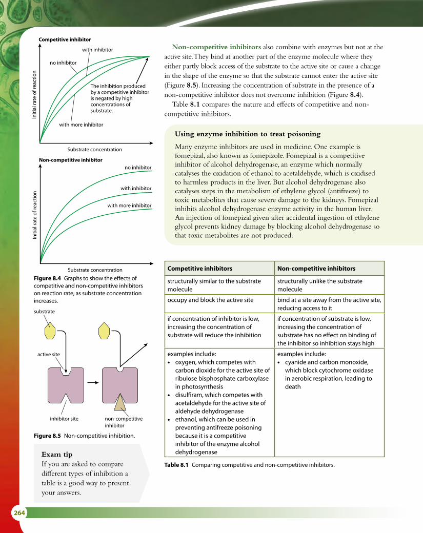
264
Non-competitive inhibitors also combine with enzymes but not at the active site. They bind at another part of the enzyme molecule where they either partly block access of the substrate to the active site or cause a change in the shape of the enzyme so that the substrate cannot enter the active site (Figure 8.5). Increasing the concentration of substrate in the presence of a non-competitive inhibitor does not overcome inhibition (Figure 8.4).
Table 8.1 compares the nature and effects of competitive and non-competitive inhibitors.
Competitive inhibitor
The inhibition producedby a competitive inhibitoris negated by highconcentrations ofsubstrate.
no inhibitor
no inhibitor
with inhibitor
with more inhibitor
with inhibitor
with more inhibitor
Substrate concentration
Initi
al ra
te o
f rea
ctio
n
Non-competitive inhibitor
Substrate concentration
Initi
al ra
te o
f rea
ctio
nCompetitive inhibitor
The inhibition producedby a competitive inhibitoris negated by highconcentrations ofsubstrate.
no inhibitor
no inhibitor
with inhibitor
with more inhibitor
with inhibitor
with more inhibitor
Substrate concentration
Initi
al ra
te o
f rea
ctio
nNon-competitive inhibitor
Substrate concentration
Initi
al ra
te o
f rea
ctio
n
substrate
active site
inhibitor site non-competitive inhibitor
Figure 8.5 Non-competitive inhibition.
Figure 8.4 Graphs to show the effects of competitive and non-competitive inhibitors on reaction rate, as substrate concentration increases.
Using enzyme inhibition to treat poisoning
Many enzyme inhibitors are used in medicine. One example is fomepizal, also known as fomepizole. Fomepizal is a competitive inhibitor of alcohol dehydrogenase, an enzyme which normally catalyses the oxidation of ethanol to acetaldehyde, which is oxidised to harmless products in the liver. But alcohol dehydrogenase also catalyses steps in the metabolism of ethylene glycol (antifreeze) to toxic metabolites that cause severe damage to the kidneys. Fomepizal inhibits alcohol dehydrogenase enzyme activity in the human liver. An injection of fomepizal given after accidental ingestion of ethylene glycol prevents kidney damage by blocking alcohol dehydrogenase so that toxic metabolites are not produced.
Competitive inhibitors Non-competitive inhibitors
structurally similar to the substrate molecule
structurally unlike the substrate molecule
occupy and block the active site bind at a site away from the active site, reducing access to it
if concentration of inhibitor is low, increasing the concentration of substrate will reduce the inhibition
if concentration of substrate is low, increasing the concentration of substrate has no effect on binding of the inhibitor so inhibition stays high
examples include:• oxygen, which competes with
carbon dioxide for the active site of ribulose bisphosphate carboxylase in photosynthesis
• disulfiram, which competes with acetaldehyde for the active site of aldehyde dehydrogenase
• ethanol, which can be used in preventing antifreeze poisoning because it is a competitive inhibitor of the enzyme alcohol dehydrogenase
examples include:• cyanide and carbon monoxide,
which block cytochrome oxidase in aerobic respiration, leading to death
Table 8.1 Comparing competitive and non-competitive inhibitors.
Exam tipIf you are asked to compare different types of inhibition a table is a good way to present your answers.

8 METABOLISM, CELL RESPIRATION AND PHOTOSYNTHESIS (HL) 265
Controlling metabolic pathways by end-product inhibitionEnd-product inhibition means that an enzyme in a pathway is inhibited by the product of that pathway. This prevents a cell over-producing a substance it does not need at the time. Many products may be needed by the cell at a specific time or in specific amounts and over-production not only wastes energy but may also become toxic if the product accumulates.
In an assembly-line reaction, such as those described in Figure 8.6, each step is controlled by a different enzyme. If the end-product begins to accumulate because it is not being used, it inhibits an enzyme earlier in the pathway to switch off the assembly line. In most cases, the inhibiting effect is on the first enzyme in a process, but in other cases it can act at a branch point to divert the reaction along another pathway.
When the end-product starts to be used up, its inhibiting effect reduces, the inhibited enzyme is reactivated and production begins again. This is an example of negative feedback (Subtopic 6.6).
End-product inhibition may be competitive or non-competitive. Competitive inhibition will only work if the product is a similar shape to the normal substrate and there can be an induced fit of the product or inhibitor onto the enzyme. In most cases, the product will be a different shape and therefore this has to be non-competitive inhibition. In this case, the enzyme is known as an allosteric enzyme, the product is called an allosteric inhibitor and the place where it binds to the enzyme is called the allosteric site (Figure 8.7).
initial substrate
intermediate A
enzyme 1
enzyme 2
enzyme 3
intermediate B
The end-product inhibits the enzyme catalysing the first reaction in the series, so all the subsequent reactions stop.
end-product
initial substrate
intermediate A
intermediate B
The end-product inhibits an enzyme in the pathway, which causes a different enzyme to come into play and the pathway is diverted down a different route.
end-product 1
intermediate C
end-product 2
substrate
products
active site
allosteric site
allosteric non-competitive inhibitor
Figure 8.6 End-product inhibition.
Figure 8.7 Allosteric control. Allosteric inhibitors prevent the active site functioning.
Disulfiram is a competitive inhibitor of alcohol dehydrogenase which is used as a deterrent to drinking for people with alcohol dependency. It causes headaches and feelings of nausea if alcohol is drunk by a person who has taken it.

266
An example of end-product inhibitionThreonine is converted to isoleucine in a series of five enzyme-controlled stages. Isoleucine, as the end product of threonine metabolism, can inhibit threonine deaminase, the first of the five enzymes in the process (Figure 8.8). Isoleucine inhibits the enzyme by binding to the molecule at a site away from the active site. When it is attached, the active site of the enzyme is changed so that no further substrate can bind to it. As isoleucine concentration increases, more and more isoleucine molecules attach to this inhibition site on enzyme molecules and therefore inhibit further production of isoleucine. As their concentration falls, isoleucine molecules detach from the threonine deaminase enzyme molecules and are used in the cell. Once the inhibitor has been removed, the active site can bind new substrate and the pathway is reactivated. This mechanism makes the metabolic pathway self-regulating so that there is always sufficient isoleucine present in the cell.
threonine
final product E
intermediate D
intermediate C
intermediate B
intermediate A
isoleucine
enzyme 1threoninedeaminase
enzyme 2
enzyme 3
enzyme 4
enzyme 5
Figure 8.8 The pathway that converts threonine to isoleucine – a specific example of end-product inhibition.

8 METABOLISM, CELL RESPIRATION AND PHOTOSYNTHESIS (HL) 267
Nature of scienceScientific advance follows technical innovation – biochemical databases facilitate researchNow that large amounts of data can be stored and curated in databases, known and predicted biochemical pathways can be catalogued and updated by scientists who are researching specific organisms. Two important databases, which have enhanced the knowledge of a vital staple food and a killer disease, are RiceCyc (which stores information on the biochemical pathways in rice) and PlasmoDB (which stores information on the genetics and biochemistry of the malarial parasite Plasmodium falciparum). Databases like these are regularly updated so that researchers can find new discoveries in biochemical pathways in a matter of minutes, as well as using the pathways that are known, in their research programmes – in the development of disease-resistant strains of rice, for example, or new antimalarial drugs. In this way, the data stored in a database can be turned into biologically meaningful information.
1 Outline what is meant by ‘activation energy’.2 Explain how an enzyme pathway can be switched off by an
accumulation of the end product of the pathways.
Test yourself?
Studying metabolic pathwaysMetabolic pathways have been studied for centuries but one
of the most significant advances were made by Eduard Buchner (1860–1917) who discovered enzymes at the start of the 20th century. At first, studies of whole animals were made, but more recently it has been possible to analyse metabolic pathways and their component reactions using modern techniques such as chromatography, X-ray diffraction, spectroscopy and radioactive isotopes. For example, in the mid-20th century, the citric acid cycle – often called the Krebs cycle after its discoverer (Subtopic 8.2) – and the glyoxylate cycle were discovered by Hans Krebs (1900–1981) and Hans Kornberg (1928–). But metabolic pathways are very elaborate. There are many pathways that are interrelated and together make up a complex metabolic network in a cell. These pathways are vital to homeostasis and cell function. Pathways may be connected by intermediate products, and products of one pathway may be substrates for another.
Question to consider
• Most biochemical studies are made using carefully controlled experiments that look at one part of a pathway. To what extent can looking at component parts of a complex system give us knowledge of the whole?

268
8.2 Cell respiration
Oxidation and reductionCell respiration involves several oxidation and reduction reactions. Such reactions are common in biochemical pathways. When two molecules react, one of them starts in the oxidised state and becomes reduced, and the other starts in the reduced state and becomes oxidised, as shown in Figure 8.9.
Learning objectives
You should understand that:
• In cell respiration, electron carriers are oxidised and reduced.
• Molecules that have been phosphorylated are less stable.
• Glucose is converted to pyruvate during glycolysis, which takes place in the cytoplasm.
• Glycolysis does not use oxygen and produces a small net gain of ATP.
• In aerobic respiration, pyruvate is decarboxylated and oxidised. In the link reaction, it is converted to an acetyl compound, then attached to coenzyme A to form acetyl coenzyme A.
• During the Krebs cycle, the oxidation of acetyl groups is coupled with the reduction of hydrogen carriers and carbon dioxide is released.
• Energy released during oxidation reactions is carried by reduced NAD and FAD to the cristae of mitochondria.
• The transfer of electrons between carrier molecules in the electron transport chain (ETC) in the membrane of the cristae is coupled to proton pumps.
• During chemiosmosis protons diffuse across the membrane down a concentration gradient via ATP synthase to produce ATP.
• Oxygen binds with free protons to form water, thus maintaining the hydrogen (proton) gradient.
• The structure of mitochondria is closely linked to their function.
molecule 1reduced
molecule 1oxidised
molecule 2oxidised
molecule 2reduced
There are three different ways in which a molecule can be oxidised or reduced, as outlined in Table 8.2. In biological oxidation reactions, addition of oxygen atoms is an alternative to removal of hydrogen atoms. Since a hydrogen atom consists of an electron and a proton, losing hydrogen atoms (oxidation) involves losing one or more electrons.
Oxidation Reduction
loss of electrons gain of electrons
loss of hydrogen gain of hydrogen
gain of oxygen loss of oxygen
Table 8.2 Changes involved in oxidation and reduction.
Oxidation and reduction occur together in biochemical reactions. As one compound loses electrons, for example. In the simple equation for respiration, glucose is oxidised as hydrogen atoms, and therefore electrons, are gradually removed from it and added to hydrogen acceptors (the oxygen atoms on the left side of the equation), which become reduced.
C6H12O6 + 6O2 → 6CO2 + 6H2O + energy
Chemical reactions like this are referred to as redox reactions. In redox reactions, the reduced molecule always has more potential energy than the oxidised form of the molecule. Electrons passing from one molecule to another carry energy with them.
Figure 8.9 Oxidation and reduction are linked processes – as one molecule is reduced another is oxidised in a redox reaction.
8 METABOLISM, CELL RESPIRATION AND PHOTOSYNTHESIS (HL) 269
RespirationCell respiration is the controlled breakdown of food molecules such as glucose or fat to release energy, which can be stored for later use. The energy is most commonly stored in the molecule adenosine triphosphate, or ATP. The respiration pathway can be divided into four parts:
• glycolysis
• link reaction
• Krebs cycle
• electron transfer chain and chemiosmosis.
GlycolysisGlycolysis is the first stage in the series of reactions that make up respiration. It literally means ‘breaking apart glucose’. The glycolysis pathway occurs in the cytoplasm of the cell. It is anaerobic (that is, it can proceed in the absence of oxygen) and produces pyruvate and a small amount of ATP. One molecule of the hexose sugar glucose is converted to two molecules of the three-carbon molecule called pyruvate with the net gain of two molecules of ATP and two molecules of NADH + H+. The process is shown in detail in Figure 8.10.1 The first steps are to add two phosphate groups from ATP, in a process
called phosphorylation. A hexose bisphosphate molecule is produced. (This appears contrary to the purpose of respiration, which is to make ATP, but the two lost ATPs are recovered later.)
2 The hexose bisphosphate is now split into two triose phosphates in a reaction called lysis.
3 Now, another phosphorylation takes place but this time an inorganic phosphate ion, Pi, is used and not ATP. Two triose bisphosphates are formed. The energy to add the Pi comes from an oxidation reaction. The triose bisphosphate is oxidised and at the same time NAD+ is reduced to NADH + H+.
4 There now follows a series of reactions in which the two phosphate groups from each triose bisphosphate are transferred onto two molecules of ADP, to form two molecules of ATP – this is ATP formation. A pyruvate molecule is also produced.
Four molecules of ATP are formed by converting one molecule of glucose to two molecules of pyruvate. However, two molecules of ATP were required to start the pathway and so there is a net gain of two molecules of ATP per glucose. In addition two NADH + H+ are formed.
To summarise, the net products of glycolysis per glucose molecule are:
• 2 ATP
• 2 NADH + H+
• 2 molecules of pyruvate.
NAD+ is a hydrogen carrier that accepts hydrogen atoms removed during the reactions of respiration. During glycolysis, two hydrogen atoms are removed and NAD+ accepts the protons from one of them and the electrons from both of them.
NAD+ + 2H → NADH + H+
Note that NADH + H+ must be written in this way and should not be simplified to NADH2.
Exam tipAn easy way to remember oxidation and reduction is to think of the words OIL RIG:OxidationIsLoss of electronsReductionIsGain of electrons

270
P
P P
P
Pi Pi
P P P
P
glucose
2× pyruvate
2× ATPrelatively unreactive glucose isactivated by reaction with ATP
phosphorylated 6-carbon sugar splits toform two 3-carbon sugar phosphates
Energy released as hydrogen is transferred to the hydrogen acceptor and used to form ATP molecules.
3-carbon sugarphosphate moleculesare converted to pyruvate
phosphorylated 6-carbon sugar(hexose bisphosphate)
2× 3-carbon sugar phosphate(2× triose phosphate)
2× NAD+
4× ATP
2× NADH + H+
1 Phosphorylation
2 Lysis
3 Oxidation
4 ATP formation
C C C C C C
C C C
C C C C C C
C C C C C C
C C C C C C
C C C
Figure 8.10 The stages of glycolysis. Note that for each molecule of glucose, two molecules of ATP are used and four are formed, so there is a net gain of two ATPs.
Phosphorylation
Phosphorylation of glucose is the first stage in its breakdown and involves the addition of phosphate groups from ATP. This turns the relatively unreactive glucose into a more unstable, phosphorylated compound, which can be split to form two three-carbon sugars.
Energy coupling
Energy coupling involves a sequence of reactions in which energy from an energy-releasing process is used to drive an energy-requiring process. Phosphorylation is an example of energy coupling – the transport of a phosphate group from ATP to a reactant molecule in the coupled reaction supplies energy for that reaction. The reactant molecule becomes a phosphorylated intermediate, an unstable molecule compared to the unphosphorylated state.
Bisphosphate and diphosphate
Although both these molecules contain two phosphate groups, the phosphates are joined in different ways. In a diphosphate molecule such as ADP, the two phosphates are joined to each other; in a bisphosphate molecule such as hexose bisphosphate, each phosphate is joined to a different part of the hexose molecule.
Exam tipThink of your own acronym such as People Love Outdoor Activities to help you recall the steps in glycolysis.
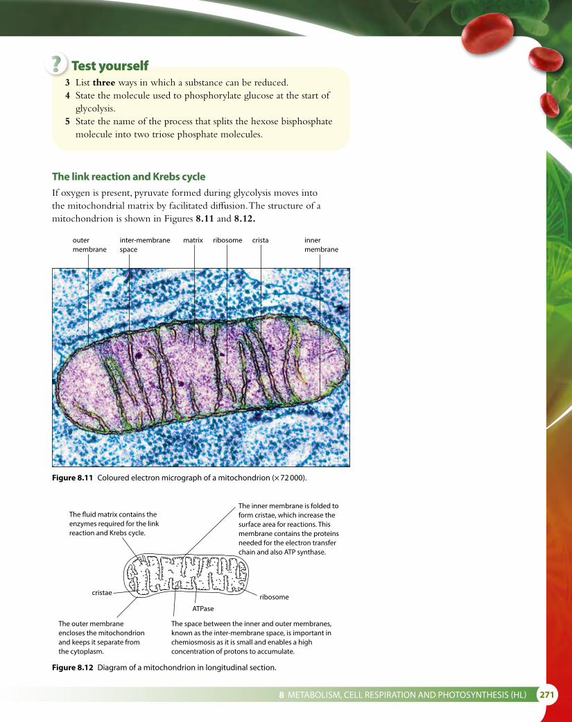
8 METABOLISM, CELL RESPIRATION AND PHOTOSYNTHESIS (HL) 271
The link reaction and Krebs cycleIf oxygen is present, pyruvate formed during glycolysis moves into the mitochondrial matrix by facilitated diffusion. The structure of a mitochondrion is shown in Figures 8.11 and 8.12.
3 List three ways in which a substance can be reduced.4 State the molecule used to phosphorylate glucose at the start of
glycolysis.5 State the name of the process that splits the hexose bisphosphate
molecule into two triose phosphate molecules.
Figure 8.11 Coloured electron micrograph of a mitochondrion (× 72 000).
The inner membrane is folded toform cristae, which increase thesurface area for reactions. Thismembrane contains the proteinsneeded for the electron transferchain and also ATP synthase.
The �uid matrix contains theenzymes required for the linkreaction and Krebs cycle.
cristae
ATPase
ribosome
The outer membraneencloses the mitochondrionand keeps it separate fromthe cytoplasm.
The space between the inner and outer membranes,known as the inter-membrane space, is important inchemiosmosis as it is small and enables a highconcentration of protons to accumulate.
Figure 8.12 Diagram of a mitochondrion in longitudinal section.
Test yourself?
outer membrane
inter-membrane space
matrix ribosome crista inner membrane

272
The link reaction and the Krebs cycle pathways occur in the mitochondrial matrix (Figure 8.13).1 The link reaction converts pyruvate to acetyl CoA using coenzyme
A, and a carbon atom is removed as carbon dioxide. This is called a decarboxylation reaction. At the same time as the carbon dioxide is removed, pyruvate is oxidised by the removal of hydrogen. The hydrogen atoms are removed by NAD+ to form NADH + H+.
2 Acetyl CoA now enters the Krebs cycle to continue the processes of aerobic respiration. Immediately, the coenzyme A is removed to be recycled. The acetyl component of the acetyl CoA combines with a four-carbon compound to form the six-carbon compound, citrate.
3, 4 The acetyl (two-carbon) groups are dehydrogenated to release four pairs of hydrogen atoms and decarboxylated to form two molecules of carbon dioxide so that the two carbons that enter with acetyl CoA leave as carbon dioxide.
5 One molecule of ATP is formed.6 Hydrogen is removed during oxidation reactions to the two hydrogen
carriers NAD+ and FAD+.7 Since the Krebs cycle is a cyclic process, what enters must eventually
leave so that the cycle begins and ends with the same substances.
Electron tomography and mitochondria
Electron tomography enables a 3D image of a mitochondrion to be built up from a series of two-dimensional images each 0.5 μm thick. The structure is viewed from many different angles and the 3D structure is calculated. Different membranes can be traced and changes that take place in active or even dying mitochondria can be followed.
citrate
C C C C C C
5-carbon acid
C C C C C
acetyl CoA
C C
4-carbon acid
(also known as the citric acid cycle)
Krebscycle
CC C C
4-carbon acid
C C C C
pyruvate
coenzyme A1 Decarboxylation
and oxidationreactions
2 Coenzyme A isrecycled7 Cycle begins
again
6 Oxidationreactions
5 ATPformation
3 Decarboxylationand oxidationreactions
coenzyme A
facilitated di�usionfrom the cytoplasm
NAD+
NADH + H+
NADH + H+
NADH + H+
NAD+
NAD+
FAD+
FADH2
ATP
ADP + Pi
the link reactionCO2
CO2
NADH + H+
NAD+
CO2
C C C
4 Decarboxylationand oxidationreactions
Figure 8.13 The link reaction and Krebs cycle.
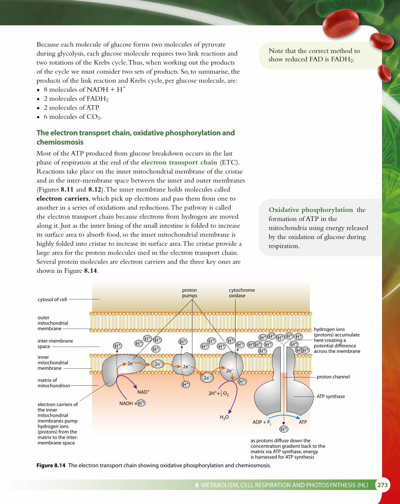
8 METABOLISM, CELL RESPIRATION AND PHOTOSYNTHESIS (HL) 273
Because each molecule of glucose forms two molecules of pyruvate during glycolysis, each glucose molecule requires two link reactions and two rotations of the Krebs cycle. Thus, when working out the products of the cycle we must consider two sets of products. So, to summarise, the products of the link reaction and Krebs cycle, per glucose molecule, are:
• 8 molecules of NADH + H+
• 2 molecules of FADH2
• 2 molecules of ATP
• 6 molecules of CO2.
The electron transport chain, oxidative phosphorylation and chemiosmosisMost of the ATP produced from glucose breakdown occurs in the last phase of respiration at the end of the electron transport chain (ETC). Reactions take place on the inner mitochondrial membrane of the cristae and in the inter-membrane space between the inner and outer membranes (Figures 8.11 and 8.12). The inner membrane holds molecules called electron carriers, which pick up electrons and pass them from one to another in a series of oxidations and reductions. The pathway is called the electron transport chain because electrons from hydrogen are moved along it. Just as the inner lining of the small intestine is folded to increase its surface area to absorb food, so the inner mitochondrial membrane is highly folded into cristae to increase its surface area. The cristae provide a large area for the protein molecules used in the electron transport chain. Several protein molecules are electron carriers and the three key ones are shown in Figure 8.14.
Oxidative phosphorylation the formation of ATP in the mitochondria using energy released by the oxidation of glucose during respiration.
Note that the correct method to show reduced FAD is FADH2.
hydrogen ions(protons) accumulatehere creating apotential differenceacross the membrane
electron carriers ofthe innermitochondrialmembranes pumphydrogen ions(protons) from thematrix to the inter-membrane space
matrix ofmitochondrion
innermitochondrialmembrane
inter-membranespace
outermitochondrialmembrane
cytosol of cell
as protons diffuse down theconcentration gradient back to thematrix via ATP synthase, energyis harnessed for ATP synthesis
proton channel
ATP synthase
protonpumps
cytochromeoxidase
NADH +
NAD+
ADP + Pi ATP
2e– 2e–
H+H+
H+H+
H+
H+
H+
H+
H+
H2O
H+ H+
H+H+H+
H+ H+ H+ H+
H+
H+ H+H+
H+
H+H+
H+ H+
2e–
2e–2e–
2H++ O212
Figure 8.14 The electron transport chain showing oxidative phosphorylation and chemiosmosis.

274
Electrons from NADH + H+ are transferred onto the first electron carrier. As they pass through the carrier, they lose energy and this is used to pump a proton (H+) from the matrix to the inter-membrane space, lowering the pH of the space. The electrons are then transferred to two further carriers and the process is repeated. As the electrons from one NADH + H+ pass along the chain, a total of nine protons are pumped into the inter-membrane space. At the end of the chain, the electrons are combined with protons and oxygen atoms to make water, in the oxidative part of oxidative phosphorylation, completing the release of energy from the oxidation of glucose to produce ATP. The formation of water ensures that the H+ gradient is maintained.
The space between the membranes is very narrow and allows for a rapid increase in the concentration of the protons that are pumped into it during the electron transfer reactions. The protons in the inter-membrane space create a concentration gradient between the space and the matrix. These protons can now flow passively down this concentration gradient back into the matrix, through a very large integral protein. This is called chemiosmosis. The large protein contains the enzyme ATP synthase, which joins ADP and Pi to form ATP. Three protons flowing through this enzyme result in one ATP being formed. Since the electrons from one NADH + H+ pump nine protons into the inter-membrane space, each NADH + H+ results in the formation of three ATP. This is the phosphorylation part of oxidative phosphorylation.
FADH2 also supplies electrons to the electron transport chain but further down the chain than NADH + H+, missing the first protein pump. FADH2 allows the production of just two ATPs.
Overall ATP production during aerobic respiration
Stage ATP use ATP yield
glycolysis 2 ATP used at the start −2 ATP
2 NADH + H+ +4 ATP
ATP formation +4 ATP
link reaction 2 NADH + H+ +6 ATP
Krebs cycle ATP formation +2 ATP
6 NADH + H+ +18 ATP
2 FADH2 +4 ATP
net energy yield +36 ATP
Table 8.3 Together, glycolysis, the link reaction and the Krebs cycle yield 36 ATP molecules for each molecule of glucose broken down by aerobic respiration.
Chemiosmosis
Osmosis is the passive flow of water molecules down a concentration gradient through a partially permeable membrane. Chemiosmosis is similar but instead of water moving, it is protons that pass down a concentration gradient.
As Table 8.3 shows, the net production of ATP from one molecule of glucose is, in theory, 36. Biochemists have discovered that the actual production is closer to 30 ATPs and propose that this discrepancy occurs because some protons are used to transfer ATP from the matrix to the cytoplasm. There are also losses such as the cost of moving pyruvate, phosphate and ADP (for ATP synthesis) into the mitochondria.

8 METABOLISM, CELL RESPIRATION AND PHOTOSYNTHESIS (HL) 275
Nature of scienceParadigm shift – chemiosmosis theory required a significant change of viewThe chemiosmosis hypothesis was proposed in 1961 by Peter Mitchell (1920–1992) to explain how the mitochondria convert ADP to ATP. At the start of the 1960s, scientists did not understand the exact mechanisms by which electron transfer is coupled to ATP synthesis. Various hypotheses current at the time proposed a direct chemical relationship between oxidising and phosphorylating enzymes and proposed that a high-energy intermediate compound was formed. Mitchell’s theory was completely new and proposed an indirect interaction between these enzymes with no intermediate compound. He suggested that ATP synthesis is driven by a reverse flow of protons down a concentration gradient, the so-called the ‘chemiosmotic theory’. This theory was first received with scepticism as his work was considered to be radical and outside the popularly held view. Mitchell struggled to persuade his contemporaries to reject the more accepted theories because his theory used a completely different approach. After several years of research, he published detailed evidence to support his theory, both in a pamphlet in 1966 and also in further publications in 1968, which were known as ‘the little grey books’ because of their bland covers. Eventually in the early 1970s, Mitchell’s chemiosmosis theory gained scientific acceptance, and scientists conceded that no high-energy intermediate compounds were likely to be found. Mitchell was awarded the Nobel Prize for Chemistry in 1978.
Despite Peter Mitchell’s strong evidence for chemiosmosis, which
falsified previous theories, he struggled to have his work accepted.
Question to consider
• Why is it often difficult for a paradigm shift to gain acceptance?
6 State the precise sites of the link reaction and the reactions of Krebs cycle.
7 State the precise site of the reactions of the electron transport chain.
8 State the name of the molecule that enters the Krebs cycle. 9 Explain the purpose of the folding of the inner mitochondrial
membrane. 10 Explain what happens to the pH of the inter-membrane space
as electrons move along the ETC. 11 State the name of the molecule that the protons pass through
going from the inter-membrane space to the matrix.
Test yourself?

276
8.3 Photosynthesis
Learning objectives
You should understand that:
• Light-dependent reactions occur in the inter membrane space of the thylakoids of a chloroplast.
• Light-independent reactions occur in the stroma of a chloroplast.
• Light-dependent reactions lead to the production of reduced NADP+ (NADPH + H+) and ATP.
• As photosystems absorb light, excited (high-energy) electrons are generated.
• Photolysis of water generates electrons, which are used in the light-dependent reactions.
• Excited electrons are transferred between carriers in the thylakoid membranes.
• Excited electrons from photosystem II are used to generate a proton gradient.
• ATP synthase in thylakoids generates ATP using this proton gradient.
• Excited electrons from photosystem I reduce NADP+ to NADPH + H+.
• A carboxylase catalyses the carboxylation of ribulose bisphosphate (RuBP) in the light-independent reactions.
• Reduced NADP+ (NADPH + H+) and ATP are used to reduce glycerate 3-phosphate to triose phosphate.
• Triose phosphate is used to produce carbohydrates and regenerate RuBP.
• ATP is used to re-form RuBP.
• The structure of a chloroplast is closely linked to its function.
The reactions of photosynthesisPhotosynthesis is the process by which light energy is harvested and stored as chemical energy, primarily in sugars but also in other organic molecules such as lipids. It occurs in green plants, algae and some bacteria. All these organisms are known as autotrophs, which means they can make their own food.
Photosynthesis can be divided into two parts:
• the light-dependent reactions
• the light-independent reactions.The light-dependent reactions produce compounds that are used in the light-independent reactions.
Both the light-dependent and the light-independent reactions take place in the chloroplasts of plant cells (Figures 8.15 and 8.16). The stroma contains the enzymes required for the light-independent reactions and the stacks of thylakoid membranes increase the surface area for the light-dependent reactions.
The light-dependent and light-independent reactions
Both of these sets of reactions are part of photosynthesis and can only occur when there is sufficient light. Light-dependent reactions can only take place in light, and although light-independent reactions do not require light directly – and can take place when it is dark – they do require the products of the light-dependent reactions.

8 METABOLISM, CELL RESPIRATION AND PHOTOSYNTHESIS (HL) 277
The light-dependent reactionsThe light-dependent reactions occur on the thylakoid membranes of the chloroplast and are powered by light energy from the Sun. Each thylakoid is a flattened sac so the space in the middle is narrow. The thylakoid membranes form stacks called grana, which may be joined together by inter-granal membranes. Light is absorbed by photosynthetic pigments such as chlorophyll, which are found on the granal membranes. There are several pigments found in plants and each one absorbs light of a slightly different wavelength. The pigments are associated with proteins that are involved in electron transport, proton pumping and chemiosmosis.
The photosynthetic pigments are combined into two complex groups called photosystems I and II, which absorb the light energy and use this to boost electrons to a higher energy level so that they become ‘excited’, as shown in Figure 8.17.
starch grain
ribosomes
outer membrane
inner membrane
chloroplastenvelope
lipid droplet
stroma
lamella
thylakoid
granum (stack of thylakoids)
Figure 8.15 Coloured electron micrograph of a chloroplast (× 20 000).
Figure 8.16 Diagram of a chloroplast.
granum
stroma
lamella chloroplast envelope
starch grain

278
1 The first step in the light-dependent reaction is the photoactivation of photosystem II. Pigment molecules in the photosystem absorb light energy and boost electrons in a molecule of chlorophyll to a higher energy level. The electrons are accepted by a carrier protein molecule at the start of the electron transport chain.
2 Photosystem II has to replace these lost electrons and it does this by taking them from water. Water is split into electrons, protons (hydrogen ions) and an oxygen atom. Since the splitting is brought about by light energy, it is called photolysis. The oxygen is released as an excretory product.
3 Excited electrons travel along the electron transport chain into photosystem I. As they do this, they lose energy but this is used to pump protons into the thylakoid interior (in a similar way as occurs in the electron transport chain in the mitochondrion). The thylakoid interior is small and so a proton concentration gradient builds up quickly. The protons then flow out through a large channel protein, almost identical to the one in mitochondria, which contains the enzyme ATP synthase. This time though, the formation of ATP is called photophosphorylation and it occurs between photosystems II and I (Figure 8.18).
4 Absorption of light energy causes photoactivation in photosystem I, boosting more electrons to an even higher energy level. The electrons that arrive from photosystem II replace those that are displaced. The electrons at the higher energy level are combined with protons in the hydrogen carrier NADP+ to form NADPH + H+.
The two products of the light-dependent reaction, ATP and NADPH + H+, are used to drive the light-independent reaction.
ener
gy le
vel e–
e–
pho
tosy
stem
II
e–
e–
e–
pho
tosy
stem
I
H2O
O2 2H+ 2e–
2e–lightabsorbed
ADP + Pi
high-energyelectron
1 Photoactivation
2 Photolysis
3 Photophosphorylation
4 Reduction of NADP+
ATPlightabsorbed
high-energyelectron
chain ofelectron carriers
NADPH + H+
change in energyof electrons
movement of electronsbetween electron carriers
Key
Non-cyclic photophosphorylation
ener
gy le
vel
e–
e–
pho
tosy
stem
I
e–
lightabsorbed
ADP + Pi
high-energyelectron
ATPchain ofelectroncarriers
Cyclic photophosphorylation
NADP+
12
Figure 8.17 The light-dependent pathway of photosynthesis.
Exam tipNADP+ is very similar to NAD+ – it simply has a phosphate group attached. An easy way to remember that photosynthesis uses NADP+ is to note that they both have a letter ‘P’.

8 METABOLISM, CELL RESPIRATION AND PHOTOSYNTHESIS (HL) 279
Cyclic and non-cyclic photophosphorylationWhen ATP is produced using energy from excited electrons flowing from photosystem II through photosynthesis I and on to NADP+, the process is called non-cyclic photophosphorylation.
When light is not a limiting factor, the light-independent reactions may proceed more slowly than the light-dependent reaction, so that the supply of NADP+ runs out. This means the electrons boosted up from photosystem I have no acceptor available to take them. They are sent back to photosystem II and rejoin the electron transport chain near the start, generating more ATP. This alternative pathway is called cyclic photophosphorylation (Figure 8.17) – it produces neither O2 nor NADPH + H+.
The light-independent reactionsThe light-independent reactions occur in the stroma of the chloroplast and comprise a cyclic pathway called the Calvin cycle. The pathway is shown in Figure 8.19. ATP and NADPH + H+ formed during the light-dependent stage supply energy and reducing power for the Calvin cycle. The final product of the cycle is carbohydrate.
During each turn of the Calvin cycle, one molecule of carbon dioxide is used so Figure 8.19 shows three cycles combined together. As this is a cycle, what goes in must leave, so three carbons enter in three molecules of carbon dioxide and three carbons leave in one molecule of triose phosphate, which can be used to form glucose or other organic compounds.
thylakoid interior
ADP + Pi ATP
NADPHNADP+
+
H+
H+H+ H+H+ H+ H+
H+
H+
H2OO2
2H+ 12
thylakoid interior
H+
ATP synthase
photosystem II photosystem I
stroma
Figure 8.18 Chemiosmosis in photosynthesis.

280
1 At the start of the cycle, the acceptor molecule ribulose bisphosphate (RuBP) combines with incoming carbon dioxide from the air to form glycerate 3-phosphate (GP). This reaction is called carbon fixation. It is catalysed by RuBP carboxylase, an enzyme that is sometimes called Rubisco.
2 The ATP and NADPH + H+ from the light-dependent reaction convert the glycerate 3-phosphate into triose phosphate (TP). Glycerate 3-phosphate therefore becomes reduced to triose phosphate. No more phosphate is added so the only input from ATP is energy.
3 Six molecules of triose phosphate are produced but only five are needed to reform the ribulose bisphosphate to keep the cycle going. The extra triose phosphate leaves the cycle and is used to synthesise organic molecules such as glucose or amino acids.
4 Since the triose phosphate that leaves the cycle takes a phosphate with it, this is replaced in the cycle with a phosphate from ATP, as the five remaining triose phosphates are converted back to three ribulose bisphosphate molecules, and the cycle begins again.
Six ‘turns’ of the Calvin cycle produces two triose phosphate molecules, which can be combined to form the final product, glucose. Some triose phosphate molecules will follow other pathways to make other organic carbohydrate molecules, such as sucrose or cellulose, or other molecules that the plant needs, such as amino acids, fatty acids or vitamins.
Calvin cycle
6 NADPH + H+
6 NADP+
6 ADP + 6 Pi
6 ATP
3 One triose phosphate (TP)leaves the cycle and isused in the synthesis ofglucose and otherorganic molecules suchas amino acids.
1 Carbon fixation – eachCO2 combines withribulose bisphosphateto make 2 moleculesof glycerate 3-phosphate (GP).
4 The five remainingtriose phosphates areconverted back tothree ribulosebisphosphates withthe addition ofone phosphatefrom ATP.
2 ATP and NADPH + H+
from the light-dependent reactionconvert the glycerate3-phosphate (GP) intotriose phosphate (TP).
6 glycerate 3-phosphate, GP
3 ribulose bisphosphate, RuBP
6 triose phosphate, TP
5 triose phosphate, TP
ADP + PiATP
3 CO2 triose phosphate, TP
Figure 8.19 The light-independent pathway of photosynthesis – the Calvin cycle.

8 METABOLISM, CELL RESPIRATION AND PHOTOSYNTHESIS (HL) 281
Nature of scienceScientific advance follows technical innovation – Melvin Calvin’s experiments with 14CMelvin Calvin (1911–1997) was awarded a Nobel Prize in 1961 for experiments leading to our understanding of the Calvin cycle. He used the green alga Chlorella and radioactively labelled carbon dioxide, 14CO2, to follow the path of carbon in photosynthesis.
The Chlorella was exposed to light – in a flat, round flask resembling a lollipop – and photosynthesis stabilised at a steady rate. Radioactive carbon dioxide was added for different periods of time, from a second to a few minutes, so that different products of photosynthesis became labelled. Then the Chlorella was killed right away with boiling ethanol so that all enzymatic reactions stopped. The labelled compounds were separated from one another using chromatography (Figure 8.20). As different compounds contained different amounts of radioactivity, Calvin and his team were able to identify glycerate 3-phosphate and other sugar phosphates as intermediates that form at different stages in the cycle (Figure 8.19).
alanine
alanineglycine
sucrose
serine
glutamic acid
aspartic acid
citric acid
5 s photosynthesis with C14O2 Chlorella
30 s photosynthesis with C14O2 Chlorella
malic acid
malic acid
triosephosphate
sugar phosphates
sugar phosphates
sugar diphosphates
sugar diphosphates
PEPA
PEPA
PGA
PGA
UDPG
Figure 8.20 Representations of autoradiograms showing the labelling of carbon compounds in the alga Chlorella after exposure to 14CO2. After 5 s, and after 30 s, the reaction was terminated and the labelled compounds in the cell homogenates were separated by paper chromatography. Notice that glycerate 3-phosphate (PGA) appears very quickly, because it is the first stable intermediate of the Calvin cycle.

282
By exposing the cells to 14CO2 for progressively shorter periods of time, Calvin identified glycerate 3-phosphate as the first stable intermediate in the process and subsequently other labelled sugar phosphates were found to be produced as a result of its reduction.
From the distribution of 14C in all the labelled compounds, ribulose bisphosphate was eventually identified as the acceptor of carbon dioxide. This final piece of the cycle explained the labelling patterns of the all the other intermediates.
Creative endeavourOccasionally, the answer to a question about an unknown
biochemical pathway is discovered as a result of designing an investigation that is simple and elegant. To find the steps involved in the light-independent reactions, a team led by Melvin Calvin designed what he called the ‘lollipop’ apparatus (Figure 8.21).
Question to consider
• To what extent could an elegant scientific protocol be considered a work of art?
Figure 8.21 Calvin’s apparatus for investigating the light-independent reactions of photosynthesis.
12 State the site of the light-independent reaction 13 State the colour of the light spectrum that is not absorbed by plants. 14 Explain what the light energy is used for when it is absorbed by the photosystems. 15 State the name of the waste product from the light-dependent reaction. 16 State the names of two products from the light-dependent reaction that are needed for the light-
independent reactions. 17 State the name of the starting acceptor molecule in the Calvin cycle, which reacts with carbon dioxide.
Test yourself?

8 METABOLISM, CELL RESPIRATION AND PHOTOSYNTHESIS (HL) 283
Exam-style questions
1 Which of the following statements is correct about enzymes?
A Most enzymes use the lock-and-key model of substrate interaction as it is the most stable.B In allosteric control of metabolic pathways, a product within the pathway can act as a non-
competitive inhibitor of an enzyme earlier in the pathway.C Increasing the concentration of substrate has no effect on the rate of a reaction being inhibited
by a competitive inhibitor.D Competitive inhibitors bind to an allosteric site and alter the shape of the active site. [1]
2 Which of the following statements is correct?
A Oxidation can involve the removal of oxygen from a compound.B The solution inside a mitochondrion is called the matrix and the solution inside a chloroplast is
called the stroma.C The folds of the inner membrane of a mitochondrion are called grana.D The photosynthetic pigments in a chloroplast are found on the cristae. [1]
3 Which of the following statements about glycolysis is correct?
A It is anaerobic, occurs in the cytoplasm and includes at least one phosphorylation reaction.B It is aerobic and includes a lysis reaction.C In the final stages, two molecules of ATP are used in the formation of pyruvate.D It is aerobic and does not include a lysis reaction. [1]
4 Which of the following statements about the link reaction and Krebs cycle is correct?
A Pyruvate in the mitochondrion matrix is oxidised to acetyl CoA.B At the end of the link reaction, coenzyme A is recycled back into the cytoplasm to combine with
another pyruvate molecule.C During one rotation of the Krebs cycle there are three decarboxylation reactions.D The link reaction occurs in the mitochondrial matrix and Krebs cycle occurs on the
mitochondrial cristae. [1]
5 Which of the following statements about the electron transport chain is correct?
A As electrons flow along the chain, protons are pumped into the mitochondrial matrix.B ATP is formed as electrons flow through the enzyme ATP synthase.C The flow of electrons along the chain causes the pH of the inter-membrane space to increase.D When protons diffuse through ATP synthase into the matrix, ATP is formed. [1]
284
6 Which of the following statements about the light-dependent reaction is correct?
A Photoactivation of photosystem I causes photolysis.B Cyclic photophosphorylation involves both photosystems I and II.C Electrons from the photolysis of water are transferred to NADP+ from photosystem I.D NADP+ becomes oxidised when it combines with electrons and protons. [1]
7 Which of the following statements about the light-independent reaction is correct?
A The enzyme RuBP carboxylase is used to convert ribulose bisphosphate into triose phosphate.B The conversion of glycerate 3-phosphate to triose phosphate requires ATP and NADP+ from
the light-dependent reaction.C At the start of the Calvin cycle, carbon dioxide combines with glycerate 3-phosphate to form
ribulose bisphosphate.D The formation of ribulose bisphosphate from triose phosphate requires a phosphorylation
reaction. [1]
8 Which of the following statements is correct?
A A limiting factor is one that determines what the end-product of a reaction is.B A limiting factor is one that determines the rate of a reaction.C Limiting factors for photosynthesis include light intensity, carbon dioxide concentration and
glucose concentration.D An absorption spectrum can be obtained by measuring the release of oxygen gas from
pondweed at different wavelengths of light. [1]
9 Explain the reasons for:
a many grana in the chloroplast [2]
b poor growth of plants growing beneath trees [2]
c large amounts of RuBP carboxylase in the chloroplast. [2]
10 Explain the process of aerobic respiration in a cell starting at the end of glycolysis and including oxidative phosphorylation. [8]
11 Explain how the light-independent reaction of photosynthesis relies on the light-dependent reaction. [5]
12 a Draw an annotated diagram of a chloroplast and indicate how the structure of the chloroplast is adapted to the function it performs. [5]
b Describe how solar energy is converted into the chemical energy of ATP during the light-dependent stage of photosynthesis. [5]
c Name the products of the light-dependent stage and state their importance to biological systems. [4]

9 PLANT BIOLOGY (HL) 285
Plant biology (HL) 9IntroductionPlants are a vital part of almost every ecosystem. As autotrophs, they start food chains and produce carbohydrates and other organic molecules that are needed by heterotrophs. The oxygen they release and the carbon dioxide they absorb are important in maintaining the balance of gases in the atmosphere. When plants die, they contribute to the formation of humus, an important part of the structure of soil.
9.1 Transport in the xylem of plants
TranspirationTranspiration is the loss of water vapour from the leaves and stems of plants. Water is absorbed into the roots, travels up the stem in the xylem vessels in the vascular bundles to the leaves, and is lost by evaporation through stomata, which open to allow the exchange of oxygen and carbon dioxide in the leaf (Figure 9.1).
Most plants grow in areas where the amount of water in the air, the humidity, is less than in the leaves. During the day, water vapour leaves the air spaces in the spongy mesophyll through open stomata in the lower epidermis of the leaf, and in the stem. The evaporating water is drawn from the xylem in the vascular bundles in the leaf and stem. The vascular bundles are continuous with those in the roots so a column of water is formed connecting the roots, stem, leaves and air spaces. This is known as the transpiration stream.
Transpiration also carries minerals through the plant, and serves to cool leaves in warm conditions.
Cohesion–tension theoryThe movement of water in the xylem can be explained by the cohesion–tension theory.1 Loss of water vapour from the stomata in the leaves results in ‘tension’
or negative pressure in the xylem vessels.2 Water vapour enters the air spaces in the leaf from the xylem vessels to
replace water lost through the stomata.3 Continuous columns of water are drawn up the xylem due to cohesion
among water molecules in the xylem and forces of adhesion between the water molecules and the xylem vessel walls. Cohesion is due to hydrogen bonding among water molecules and adhesion is caused by the hydrogen bonds between water molecules and molecules in the walls of the xylem vessels (Subtopic 2.2).
4 The tension in the xylem is strong due to loss of water and there would be a tendency for xylem vessels to collapse inwards. The thickening provided by lignin prevents this happening (Figure 9.2).
Learning objectives
You should understand that:
• Transpiration occurs as a consequence of gas exchange in the leaf.
• Water is transported from the roots to the leaves to replace losses from transpiration.
• The structure of the xylem and cohesion among water molecules allow transport to occur under tension.
• Evaporation and adhesion between water molecules and the surfaces they contact lead to tension forces in the walls of plant cells.
• As minerals are taken up by active transport by the roots, water enters by osmosis.
Transpiration the loss of water vapour from the aerial parts of plants (the leaves and stems)

286
5 Water is drawn in from the cortex in the roots to replace water that is lost in transpiration.
6 The tension caused by transpiration also causes water to be drawn into the roots from the soil.
1 water uptake near root tip
2 water enters xylem
3 water moves up xylem
4 water moves from xylem to leaf cells
5 evaporation of water into leaf air spaces
6 transpiration of water vapour through open stomata into air
water evaporates into airwhich has a lower
concentration of water vapour
column of wateris continuous dueto adhesion andcohesion
water is pulled into roots asvapour is lost from leaves
Figure 9.1 The movements of water through a plant: overall, water moves from the soil to the air (from where there is more water to where there is less water).
TS xylem vessel element Types of ligni�ed cellwall thickenings
LS xylem vessel
cellulosewall
ligni�edcellulose wall
10 μm
pit
lumen
pit
remains ofend wallsof adjacentxylemelements
vesselelement
spiral annular reticulate pittedlumen
Figure 9.2 Xylem vessels are not alive and have no plasma membrane, so water can easily move in and out of them.

9 PLANT BIOLOGY (HL) 287
Transpiration is largely controlled by the pairs of guard cells that surround each stoma. Guard cells have unevenly shaped cell walls with more cellulose on the side adjacent to the stoma. This inner part of the cell wall is less elastic, so that when guard cells take up water and become turgid, they take on a sausage-like shape and an opening – the stoma – is formed between them (Figure 9.3). When the guard cells lose water, the cell walls relax and the stoma closes.
water enters guard cells by osmosis;
guard cells become turgid, opening stoma
epidermis cells surrounding guard cells
water leaves guard cells by osmosis;
guard cells become flaccid, closing stoma
Figure 9.3 The opening and closing of stomata. Gases can diffuse in and out of open stomata. When stomata are closed, water loss is minimised.
The opening and closing of stomata is controlled by the concentration of potassium ions. In darkness, these ions move out of the guard cells into surrounding cells. In light conditions, potassium ions are actively pumped into the vacuoles of guard cells. This creates an increased solute concentration so that water enters by osmosis, making the cells turgid and opening the stomata. A plant hormone called abscisic acid, produced in the roots during times of drought, affects potassium ion movement in guard cells. When abscisic acid is present, potassium ions leak out and water follows by osmosis. This means that the guard cells lose turgor and stomata close, thus conserving water.
Factors affecting transpirationSeveral abiotic environmental factors (notably light, temperature, humidity and wind speed) influence the rate of transpiration in plants.
• Light affects transpiration directly by controlling the opening and closing of stomata. As light intensity increases, stomata open, speeding up the rate of transpiration. In darkness, stomata close, thus restricting transpiration.
• Temperature affects transpiration because heat energy is needed for the evaporation of water. As the temperature rises, the rate of transpiration also rises as water evaporates from the air spaces in the spongy mesophyll and diffuses out of the stomata.

288
• An increase in atmospheric humidity reduces the rate of transpiration. Air in the mesophyll air spaces tends to be saturated with water vapour so if atmospheric air becomes more humid, the concentration gradient between the air space and the atmosphere is reduced and transpiration is slowed down.
• An increase in wind speed increases the rate of transpiration because it blows away the air just outside the stomata, which is saturated with water vapour. Reduced humidity near the stomata enables water vapour to diffuse more readily from the spongy mesophyll, where the air is very humid, to the air just outside the leaf, which has lower humidity.
Transpiration in xerophytesXerophytes are plants that live in arid climates – an example is shown in Figure 9.4. Some xerophytes grow in areas where there is very little rainfall all the year, while others live in places where rainfall is intense but short lived, with long, dry periods for the rest of the year. In both cases, plant species have evolved specialisations that enable them to survive shortages of water by reducing water loss.
Marram grass is well adapted to survive in the dry conditions found in sand dunes. Its leaves are rolled into tube-like shapes, which are protected on the outside by a thick waxy cuticle. Stomata of marram grass are protected deep inside pits (Figure 9.5) which themselves are rolled up inside the leaf. A lining of hairs on the inner side of the leaf keeps humid air trapped inside the rolled-up leaves. Hairs prevent water loss by diffusion. When the humidity of the air rises and water loss is not a problem, marram grass is able to unroll its leaves using specialised hinge cells and maximise photosynthesis. When the leaf is unrolled, leaf hairs help conserve a supply of water by trapping moist air. This air remains inside the leaf when it rolls up again.
The Crassulaceae, a group of succulent plants, have evolved a mechanism allowing them to keep their stomata closed during the heat of the day, to reduce water loss. At night, the stomata open and carbon dioxide diffuses into the leaf. It is fixed temporarily in cells, and then is released for photosynthesis during the day when the stomata are closed. This is called crassulacean acid metabolism (CAM).
Halophytes (‘salt plants’)Halophytes are plants that live on salt marshes or mud flats. Their roots are surrounded by water that is salty and has a higher concentration of salt than the plants’ cells. Halophytes such as glasswort (Salicornia europaea) (Figure 9.6) actively absorb salt into their roots so that the concentration in root tissue is higher than that of the water. This ensures that water enters the roots, but the excess salt could become toxic if it was not removed. Some halophytes dispose of the excess salt by storing it, while others secrete it from salt glands on their leaves.
Figure 9.4 Opuntia, or prickly pear, is a cactus with flattened photosynthetic stems that store water. The leaves are modified as spines, and this minimises water loss by reducing the surface area from which transpiration can take place. The spines also protect the plant from being eaten by animals.

9 PLANT BIOLOGY (HL) 289
The leaves can rollup, exposing a tough,waterproof cuticule tothe air outside the leaf.
Marram grass is one of the few plants that can thrive in mobile sand dunes, surviving the very dry conditions that are found there.
A coloured light micrograph of a transverse section through a marram grass leaf (× 137).
A stained scanning electron micrograph of a transverse section through part of a rolled leaf of marram grass (×90).
Specialised cells – hinge cells – cause the opening and closing of the marram grass leaf in response to air humidity. Leaf rolling traps air inside the rolled leaf. This air can remain humid even is the air outside is very dry.
The stomata are found deep in the grooves and open into the enclosed humid air space inside a rolled leaf. Water has to diffuse a long way before it reaches moving air outside the leaf, slowing water loss.
Hairs help to keep the humid air trapped inside a rolled leaf. When the lead is unrolled, the hairs help to trap a thick layer of moist air close to the leaf surface, reducing air movement. Water vapour has to diffuse through this layer before it can be carried away in air movements. The thicker the layer, the more slowly water is lost by transpiration.
Figure 9.5 Adaptations of the xerophyte marram grass (Ammophila arenaria) to dry conditions.

290
RootsRoots are responsible for absorbing water and mineral ions from the soil. Many plants develop an extensive, branching root system in order to increase the surface area of root in contact with the soil. In addition, as new roots grow, numerous root hairs develop to increase the surface area even more (Figure 9.7). Root hairs are temporary and die away to be replaced by new ones near the growing tip.
Plants require a number of minerals to make a variety of substances necessary for growth. A few of these are listed in Table 9.1.
Mineral ion Importance
calcium constituent of cell walls
magnesium needed to make chlorophyll
iron required as a cofactor for many enzymes
Table 9.1 How plants use some important mineral ions.
Minerals are present in the soil as salts – for example, calcium occurs in the form of calcium carbonate. These dissolve in soil water and the dissolved ions can move into root cells in different ways.
• Dissolved minerals may move into the root by mass flow of water carrying the ions, or by facilitated diffusion of ions from the soil water into root hairs, down their concentration gradient (Figure 9.8). Both these processes are passive – that is, they do not require energy in the form of ATP.
Figure 9.6 Salicornia europaea grows in saline water and actively absorbs salt into its roots.
Figure 9.7 A root of a young radish showing the root hairs.

9 PLANT BIOLOGY (HL) 291
• Where the concentration of a mineral is lower in the soil water than in plant cells, active transport is needed to take it up. Potassium, nitrate and phosphate are usually absorbed by active transport. Root hair cells contain mitochondria to provide ATP and most roots can only take in minerals if oxygen is available for aerobic respiration, to provide sufficient ATP. Experiments have shown that potassium ions stop moving into root cells from the soil when potassium cyanide is added. Cyanide is a potent blocker of respiration as it inhibits enzyme action, and so it prevents active transport.
• In other cases, there may be a close association, known as a mutualistic relationship, between the roots and a fungus (a mycorrhiza). Roots become covered with an extensive network of hyphae, which increase the surface area for absorption of both water and minerals. Minerals can pass directly from fungal hyphae to root cells.
• Active uptake of minerals into roots leads to an increase in the solute concentration inside root cells. This in turn causes the absorption of water by osmosis. The water then travels to the leaves in the transpiration stream.
Modelling water transport in plantsSome of the principles of water transport in plants can be demonstrated with simple experiments.
Root pressureAs water is absorbed rapidly in a root, it creates a strong upward pressure in the xylem. To demonstrate this, a stem from a potted plant can be cut off about 1 cm above the soil. A narrow glass tube approximately 1 m long is then fixed to the stem using rubber tubing and supported vertically in a stand. The tube is half filled with water, covered by a thin layer of oil to prevent evaporation, and the surface level is marked. After the soil is watered, and the apparatus left at room temperature, water is observed rising up the tube as a result of root pressure (Figure 9.9).
water movementxylem vessel
root hair
water
cortex
epidermis
endodermis
pericycle
phloem
Key
Figure 9.8 The pathway of water movement from root hair to xylem. The water may carry dissolved mineral ions.
glass tube
scale
water level
cut stem
Figure 9.9 Apparatus used to demonstrate root pressure.
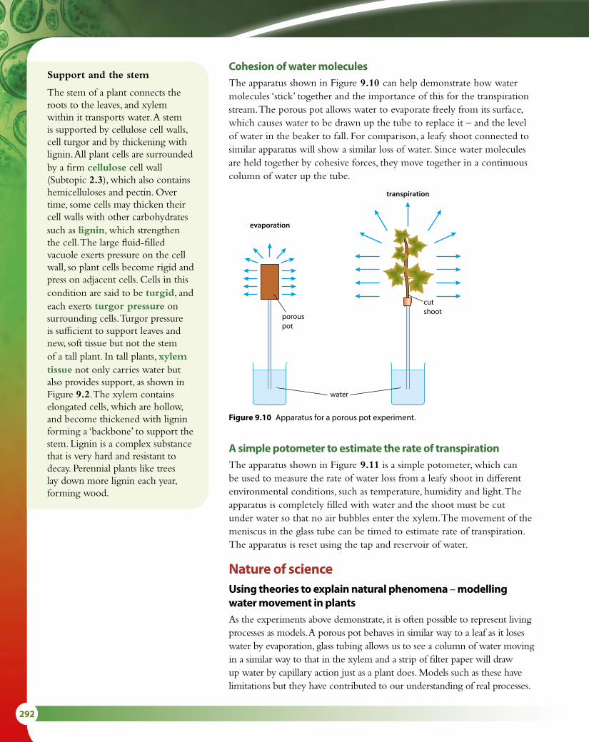
292
Support and the stem
The stem of a plant connects the roots to the leaves, and xylem within it transports water. A stem is supported by cellulose cell walls, cell turgor and by thickening with lignin. All plant cells are surrounded by a firm cellulose cell wall (Subtopic 2.3), which also contains hemicelluloses and pectin. Over time, some cells may thicken their cell walls with other carbohydrates such as lignin, which strengthen the cell. The large fluid-filled vacuole exerts pressure on the cell wall, so plant cells become rigid and press on adjacent cells. Cells in this condition are said to be turgid, and each exerts turgor pressure on surrounding cells. Turgor pressure is sufficient to support leaves and new, soft tissue but not the stem of a tall plant. In tall plants, xylem tissue not only carries water but also provides support, as shown in Figure 9.2. The xylem contains elongated cells, which are hollow, and become thickened with lignin forming a ‘backbone’ to support the stem. Lignin is a complex substance that is very hard and resistant to decay. Perennial plants like trees lay down more lignin each year, forming wood.
water
porouspot
evaporation
transpiration
cutshoot
Cohesion of water moleculesThe apparatus shown in Figure 9.10 can help demonstrate how water molecules ‘stick’ together and the importance of this for the transpiration stream. The porous pot allows water to evaporate freely from its surface, which causes water to be drawn up the tube to replace it – and the level of water in the beaker to fall. For comparison, a leafy shoot connected to similar apparatus will show a similar loss of water. Since water molecules are held together by cohesive forces, they move together in a continuous column of water up the tube.
Figure 9.10 Apparatus for a porous pot experiment.
A simple potometer to estimate the rate of transpirationThe apparatus shown in Figure 9.11 is a simple potometer, which can be used to measure the rate of water loss from a leafy shoot in different environmental conditions, such as temperature, humidity and light. The apparatus is completely filled with water and the shoot must be cut under water so that no air bubbles enter the xylem. The movement of the meniscus in the glass tube can be timed to estimate rate of transpiration. The apparatus is reset using the tap and reservoir of water.
Nature of scienceUsing theories to explain natural phenomena – modelling water movement in plantsAs the experiments above demonstrate, it is often possible to represent living processes as models. A porous pot behaves in similar way to a leaf as it loses water by evaporation, glass tubing allows us to see a column of water moving in a similar way to that in the xylem and a strip of filter paper will draw up water by capillary action just as a plant does. Models such as these have limitations but they have contributed to our understanding of real processes.

9 PLANT BIOLOGY (HL) 293
air/water meniscus
airtight seal
water
reservoir containing water
shoot of plant, with its cut end in contact with the water inside the tubing
capillary tubing
Figure 9.11 A simple potometer.
1 List three ways in which a xerophyte is adapted to survive in dry conditions.
2 Explain how mineral ions enter a plant root.
9.2 Transport in the phloem of plants
Translocation in the phloemTranslocation is the movement of organic molecules through the phloem tissue of plants. The phloem consists of two types of living cell: sieve tube cells, which are perforated to allow the movement of solutes through them, and companion cells, which are connected to the sieve tube cells as shown in Figure 9.12.
Whereas the xylem carries water and mineral salts in one direction from the roots to the leaves, the phloem can transport materials in either direction through the plant. Translocation moves materials from a source, where they are made or stored, to a sink, where they are used, as shown in Figure 9.13.
The products of photosynthesis, including sugars and amino acids, move from leaf cells, which are a source, into the phloem. Once in the
Learning objectives
You should understand that:
• Plants transport organic compounds from a source to a sink.
• The incompressibility of water allows plants to transport substances along hydrostatic pressure gradients.
• Active transport is used to load organic compounds into the sieve tubes of the phloem at the source.
• High concentrations of solutes in the phloem at a source lead to water uptake by osmosis.
• High hydrostatic pressure causes the phloem to flow toward a sink.
Test yourself?Exam tipMake sure you can describe using a potometer to investigate effect of wind, temperature or humidity on transpiration.

294
Storage structures such as seeds and bulbs are sinks during the growing season but may also act as sources when they begin to sprout.
plasmodesmata
cellulosecell wall
cellulosecell wall
mitochondrion
mitochondrion
sieve plate
sieve pore
roughendoplasmicreticulum
plasmamembrane
plasmamembrane
vacuole
tonoplast
middlelamella
cytoplasm
cytoplasm
Sieve elements have nonucleus, tonoplast or ribosomes.
endoplasmicreticulum nucleus
Companioncell
Sieveelement
Figure 9.12 A phloem sieve tube element and its companion cell.
sugar loaded intosieve tube, usingATP
source cell e.g.photosynthesisingmesophyll cell
lowhydrostaticpressure
sink cell e.g.starch storage cell
highhydrostatic
pressure
phloem moves bymass flow fromhigh to lowhydrostaticpressure areas
water leavesplant byevaporation
water entersplant at roothairs
transpirationstream
xylem
starch grain
sugar convertedto starch or usedin respiration
Figure 9.13 Sources, sinks and mass flow in phloem.

9 PLANT BIOLOGY (HL) 295
phloem, they are translocated to sink regions, such as growing tissue in the meristems of roots, buds and stems, or storage organs like fruits and seeds.
All the materials that are moved by translocation are dissolved in water to form a solution called ‘sap’, which also carries plant hormones. Sugar is usually carried as sucrose, which enters and leaves the phloem by active transport using energy provided by the companion cells. High concentrations of sucrose and other solutes in the phloem at sources, such as the leaves, lead to the uptake of water by osmosis. Once materials have entered the phloem, they move passively throughout the plant towards sinks by mass flow as a result of raised hydrostatic pressure in the phloem.
Hydrostatic pressure is defined as the pressure exerted by a liquid. In plants, a hydrostatic pressure gradient occurs in the phloem as sugar is loaded and unloaded at sources and sinks. In the leaves, sugar enters the phloem and is followed by water entering by osmosis. This creates a pressure gradient, which ‘pushes’ the content of the phloem to other parts of the plant. At a sink, the hydrostatic pressure is lower because sugar is removed from the phloem and converted to starch or used in respiration. In this way, the solute concentration of the phloem is reduced and the hydrostatic pressure gradient is maintained.
Tissues in the root and stemVascular bundles in the roots, stems and leaves of plants are made up of both xylem and phloem. Xylem tissue is made up of long series of cells joined end to end, which form a continuous fine tube once the cells have stopped growing and their end walls have broken down. The phloem, on the other hand, is composed of living cells with perforated end walls known as sieve plates. When viewed under a microscope, the xylem and phloem can easily be distinguished in both cross sections and longitudinal sections through stems (Figure 9.14). Table 9.2 summarises the relationships between structure and function for xylem and phloem tissues.
In a physical or mechanical situation, the hydrostatic pressure in a column of liquid depends on gravity, and on the height of the column – the greater the height of liquid above a certain point, the greater the hydrostatic pressure at that point.
Xylem Phloem
composed of a column of dead cells, once mature – cell end walls removed
composed of a column of living cells with perforated walls between them
continuous tube of cells enables an unbroken column of water (held together by cohesive forces) to move inside the xylem
living cells enable substances to be loaded by active transport
thickened with lignin to withstand negative pressure as water vapour is lost in transpiration
associated with companion cells which carry out cell functions and supply energy for active transport into the phloem
transports water and minerals passively from roots to leaves
transports sugars, amino acids, hormones to all parts of the plant by mass flow
Table 9.2 Structure and function in the xylem and phloem.

296
Nature of scienceScientific advance follows technical innovation – radioactive labellingDetails of the substances transported in the phloem were elucidated as radioactively labelled isotopes enabled scientists to follow the route of various substances through the plant. Radioactive isotopes became more widely available from the 1950s onwards and by the 1970s labelled carbon dioxide was being used in experiments into transport in the phloem.
Carbon dioxide labelled with 14C can be introduced to the leaves, which incorporate it into sugars and other products. As the labelled substances move into the phloem, they can be followed with the help of aphids. Aphids are small insects that use needle-like mouthparts known as stylets to puncture a single sieve tube element of the phloem and feed on the contents (Figure 9.15). High pressure in the phloem causes the sap to enter the insect’s digestive system through its stylet. If an insect that has attached itself to a phloem element is anesthetised and its stylet then severed, the sap will exude from the cut end of the stylet, and it is possible to analyse the contents, providing an accurate record of the substances in the phloem and how far they have travelled.
Figure 9.14 Stained micrographs showing transverse sections through a stem (× 4.2) and through a root (× 20), with diagrams showing the distribution of vascular tissue.
lignified fibres(provide support)
vascular cambium
cortex
phloem
xylem
vascular bundle
epidermispith
Tissue map of a transverse section through ayoung stem to show the distribution of tissues.
epidermis
phloem
xylem
endodermis
cortex
Tissue map of transverse section through a root.The epidermis protects the outer layer of the root. The cortex carries water to the xylem, which transportsit to the rest of the plant. The phloem carries nutrientsand minerals around the plant.

9 PLANT BIOLOGY (HL) 297
9.3 Growth in plants
MeristemsMeristems are the growing parts of a flowering plant where cells may divide by mitosis throughout the life of the plant. In a dicotyledonous plant (Subtopic 9.4), there are two types of meristem – the apical meristems found at the tip of the root and the shoot, and the lateral meristems found in the vascular bundles of the stem. Growth in the main stem occurs at the apical or primary meristem, as cells divide by mitosis. New tissue forms the cells needed to elongate the stem and to form leaves at the growing tip (Figure 9.16). This allows a plant to grow upwards from the soil and towards light so that its leaves can obtain sufficient light for photosynthesis. In the root, growth of the apical meristem extends the root into the soil.
Many plants also grow at lateral meristems in the vascular cambium. This makes stems and roots thicker, and is known as secondary growth. Side growth may develop from the main stem as shoots or branches to take advantage of favourable conditions and to avoid competition from other plants.
If plants are damaged at the apical meristems, often the first lateral meristem is induced to grow and take over the role of the apical
stylet
phloem
aphid
stylet
sap
stem
phloem
stem
Figure 9.15 Aphid stylets can be used in the analysis of phloem.
3 Draw a plan of the structure of a typical stem to show the arrangement of tissues inside.
4 Compare the structure of xylem and phloem.5 State the substances that are carried in the xylem.6 List the factors that affect the rate of transpiration.7 Explain why hydrostatic pressure is important in translocation.
Learning objectives
You should understand that:
• Cells in the meristems of plants are undifferentiated and allow indeterminate growth.
• Mitosis and cell division in the shoot provide cells needed for elongation of the stem and the development of leaves.
• Growth in the shoot apex is controlled by plant hormones.
• Plants respond to the environment with growth movements known as tropisms.
• Concentration gradients of auxin in plant tissue are set up by auxin efflux pumps.
• Auxin changes the pattern of gene expression to influence cell growth rates.
Test yourself?Exam tipPractise drawing xylem and phloem side by side to compare the structures.

298
meristem. If a flower on the apical meristem is cut off, this will induce the lateral meristems to switch to producing flowers.
MicropropagationCells in the meristems of plants are undifferentiated and so small pieces of tissue from meristems can be used to grow whole plants in a technique known as micropropagation (Figure 9.17). A few cells or small pieces of tissue are grown in sterile conditions on special agar growth medium, which contains an energy source such as sucrose, and minerals, as well as plant hormones and other substances such as amino acids or vitamins. The tissue will differentiate into a range of cells, depending on the medium used.
Micropropagation and plant tissue culture are important in horticulture and conservation. Many identical (cloned) plants can be grown from a single cell or small sample of tissue. Botanic gardens propagate rare and endangered plants or those that are difficult to grow. In horticulture, large numbers of ready-rooted, disease-resistant plants can be produced in a relatively short period of time, which saves time and money.
Plant hormones and the growth of shootsPlants produce growth-regulating substances that act to control growth and development. These substances are sometimes called plant hormones.
The existence of these growth substances was noted by Charles Darwin in a report of his experiments that he published in 1880. He
The terminal bud is showing apical dominance inthis example. Growth of the terminal bud preventsstem growth from lateral buds.
terminal budcontaining theapical meristem
lateral budcontaininglateralmeristem
node
internode
Removal of the terminal bud removes apical dominance.Lateral buds can now grow stems.
Figure 9.16 Apical meristems exert an inhibitory effect on the lateral meristems, called apical dominance.
Figure 9.17 Cereal plants being grown from tissue culture.

9 PLANT BIOLOGY (HL) 299
observed that oat shoots grew towards light because of some ‘influence’, which he proposed was transmitted from the shoot tip to the area immediately below. We now know that the substance that causes shoots to bend towards the light is auxin and the response it causes is called phototropism. Auxin is found in the embryos of seeds and in apical meristems, where it controls several growth responses, or tropisms.
The exact mechanism of auxin action is still the subject of much research. It has been suggested that auxin is redistributed to the side of a shoot tip that is away from a light source. The uneven distribution of auxin allows cell elongation on the shaded side of a shoot, which in turn causes bending towards light (Figure 9.18).
Auxins influence and coordinate the development of plants by influencing the expression of genes. Auxin may activate certain genes, leading to a rapid response, or it may inhibit other sets of genes. Auxin affects both cell division and cell enlargement. In a shoot, auxin promotes the elongation of cells at the shoot apex. It seems to act by stimulating factors such as elastins, which loosen the bonds between cellulose fibres in cell walls, thereby making their
Plant communicationsPlants use chemicals such as auxin to coordinate the
activities of their cells and communicate internally. Externally, plants produce substances such as ethylene to control ripening of fruits. Ethylene accelerates the normal process of fruit maturation and senescence (dying or becoming dormant) and causes ripening of bananas, mangoes, apples and pineapples.
Question to consider
• To what extent can plants be said to have ‘language’?
Receptors detect lightfrom one side, causing auxin to be transportedfrom cell to cell to theshaded side.
direction of lightKey
Receptorsin plasmamembranesdetect lightequally on each side.
More auxin is transporteddown the shaded side,causing the cells toelongate more on this side.
active transport of auxin
Figure 9.18 Some proteins in the plasma membranes of certain cells in plant shoots are sensitive to light. When light falls on them, they cause auxin to be transported to the shaded side of the shoot, which in turn causes the shoot to bend towards the light.
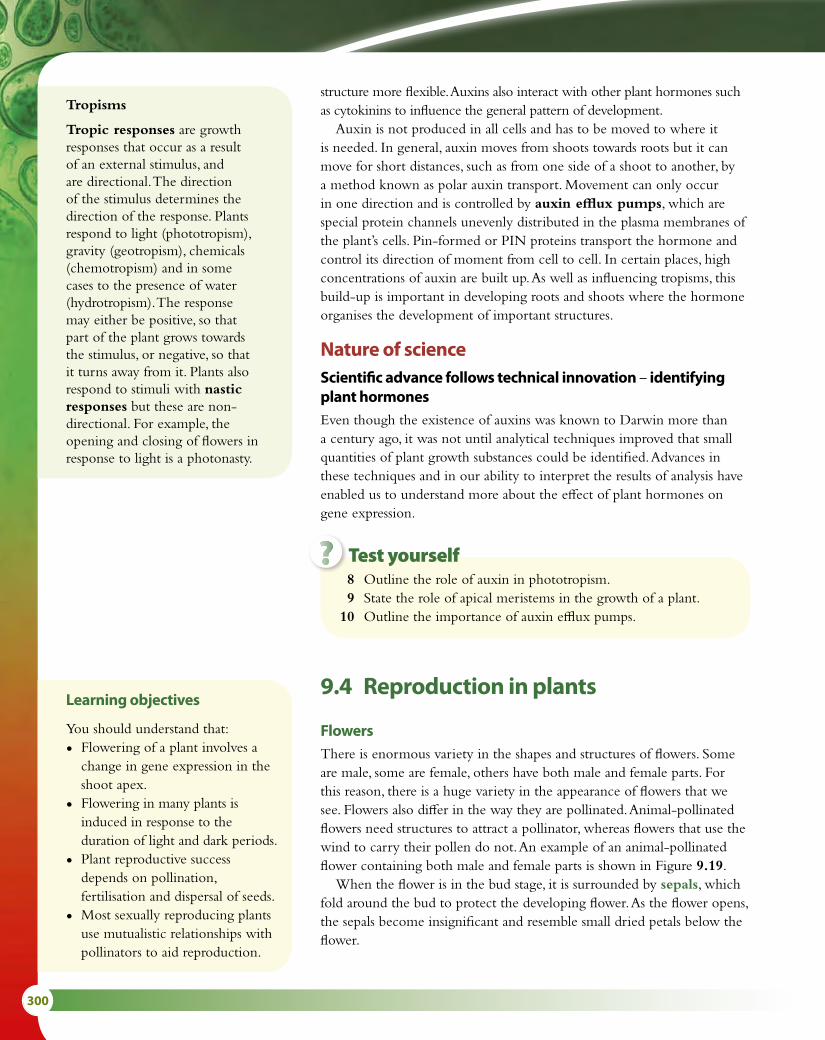
300
structure more flexible. Auxins also interact with other plant hormones such as cytokinins to influence the general pattern of development.
Auxin is not produced in all cells and has to be moved to where it is needed. In general, auxin moves from shoots towards roots but it can move for short distances, such as from one side of a shoot to another, by a method known as polar auxin transport. Movement can only occur in one direction and is controlled by auxin efflux pumps, which are special protein channels unevenly distributed in the plasma membranes of the plant’s cells. Pin-formed or PIN proteins transport the hormone and control its direction of moment from cell to cell. In certain places, high concentrations of auxin are built up. As well as influencing tropisms, this build-up is important in developing roots and shoots where the hormone organises the development of important structures.
Nature of scienceScientific advance follows technical innovation – identifying plant hormonesEven though the existence of auxins was known to Darwin more than a century ago, it was not until analytical techniques improved that small quantities of plant growth substances could be identified. Advances in these techniques and in our ability to interpret the results of analysis have enabled us to understand more about the effect of plant hormones on gene expression.
Tropisms
Tropic responses are growth responses that occur as a result of an external stimulus, and are directional. The direction of the stimulus determines the direction of the response. Plants respond to light (phototropism), gravity (geotropism), chemicals (chemotropism) and in some cases to the presence of water (hydrotropism). The response may either be positive, so that part of the plant grows towards the stimulus, or negative, so that it turns away from it. Plants also respond to stimuli with nastic responses but these are non-directional. For example, the opening and closing of flowers in response to light is a photonasty.
8 Outline the role of auxin in phototropism. 9 State the role of apical meristems in the growth of a plant. 10 Outline the importance of auxin efflux pumps.
9.4 Reproduction in plants
FlowersThere is enormous variety in the shapes and structures of flowers. Some are male, some are female, others have both male and female parts. For this reason, there is a huge variety in the appearance of flowers that we see. Flowers also differ in the way they are pollinated. Animal-pollinated flowers need structures to attract a pollinator, whereas flowers that use the wind to carry their pollen do not. An example of an animal-pollinated flower containing both male and female parts is shown in Figure 9.19.
When the flower is in the bud stage, it is surrounded by sepals, which fold around the bud to protect the developing flower. As the flower opens, the sepals become insignificant and resemble small dried petals below the flower.
Learning objectives
You should understand that:
• Flowering of a plant involves a change in gene expression in the shoot apex.
• Flowering in many plants is induced in response to the duration of light and dark periods.
• Plant reproductive success depends on pollination, fertilisation and dispersal of seeds.
• Most sexually reproducing plants use mutualistic relationships with pollinators to aid reproduction.
Test yourself?

9 PLANT BIOLOGY (HL) 301
Petals of animal-pollinated flowers are often brightly coloured to attract insects or other animals that may visit. Many have nectar guides, which are markings on the petals that tempt pollinators deep into the flower.
Pollen, containing the male gametes, is produced in the anthers, which are held up on long filaments in many flowers, so that as pollinators enter they brush past the anthers and are dusted with pollen.
The female organs are the stigma, style and ovary. The stigma receives pollen grains, which arrive with pollinators as they delve into a flower to obtain nectar. The sticky stigma has sugars present on its surface that cause pollen grains to germinate.
Pollination and fertilisationPollination is the transfer of pollen (containing the male gametes) from the anther to the stigma. Pollen may be carried by insects, other animals (such as birds, bats or mice), or by the wind. If pollen travels from the anther of one plant to the stigma of another plant, the process is known as cross-pollination. If pollen is deposited on the stigma of the same plant that produced it, self-pollination occurs. Self-pollination produces less genetic variation than cross-pollination.
Most flowering plants use a mutualistic relationship with a pollinator to enable them to reproduce. Both the plant and pollinator benefit in this type of relationship. For example, a bee that visits a flower benefits by receiving food in the form of nectar, while the flower benefits as it receives pollen to fertilise its ovules. The pollen is carried on the bee’s body from flowers it has already visited.
Fertilisation occurs when male and female gametes fuse to form a zygote. This occurs in the ovule of the flower. When pollen grains from a plant of the right species arrive on the stigma, they germinate and each
anther
�lament
whole �ower
petal
sepal
stigma
style
ovaryovule
stalkreceptacle
nectary
Figure 9.19 Half-flower of wallflower (Cheiranthus cheiri). The flower is about 2.5 cm in diameter. It is pollinated by bees and hoverflies. Its petals are usually brightly coloured and fragrant.
Aysel
Sticky Note
The apex of the pistil of a flower, on which pollen grains are deposited and germinate
Aysel
Sticky Note
The usually slender part of a pistil, connecting the ovary and the stigma
Aysel
Sticky Note
The expanded tip of a flower stalk or axis that bears the floral parts or the florets of the flower head
Aysel
Sticky Note
the pollen-bearing part of a stamen.

302
produce a pollen tube, which grows down the style to the ovary (Figure 9.20). The tube enters the ovary and a pollen nucleus passes down the tube to fuse with and fertilise the nucleus of the female gamete in the ovule.
Seed structure and dispersalFertilised ovules develop over time into seeds, which protect the developing embryo inside. Seeds are held within a seed pod, fruit or nut, which can be dispersed to new locations so that when they germinate, the new plants that develop do not compete with their parents.
Plants have evolved many ingenious means by which to bring about seed dispersal. A few are listed below.
• Some seed pods, such as those in the pea family, mature and dry out, so they eventually snap, causing the seed pod to open quite suddenly, ejecting the seeds some distance from the parent plant.
• Fruits containing seeds are frequently eaten by birds or animals. A tasty fruit tempts an animal to eat and digest it, but the tough seeds inside it pass through the digestive tract and emerge in the feces. These seeds may appear long distances from the parent plant and are contained within a rich fertiliser.
• Nuts are collected by animals like squirrels, which bury them as a reserve of food for the winter. They may bury several groups of nuts and fail to dig them all up during the winter. The nuts remain in the soil ready to germinate when conditions are favourable.
Seeds have all the necessary components to ensure successful germination and the growth of a new plant. Within every seed is an embryo root and shoot, ready to develop when the time is right. Once a seed has been formed in the ovary, it loses water so that it can enter a dormant phase and not develop further until conditions for growth are favourable.
Inside their seeds, dicotyledonous plants have two seed leaves, or cotyledons, which store food reserves needed for germination (Figure 9.21). The cotyledons are surrounded by a hard protective seed coat called the testa. Many seeds have to endure quite harsh environmental conditions, so the testa protects the delicate tissues inside. In the wall of the testa is a pore called the micropyle through which water is absorbed to begin the process of germination.
GerminationGermination is the development of the seed into a new plant (Figure 9.22). A dormant seed needs three vital factors to be in place for germination to occur.
• Temperature – A suitable temperature is essential for the enzymes in a seed to become active. They cannot work in cold conditions, and very high temperatures also inhibit their activity. Many seeds remain dormant until the temperature is at a particular level so that they germinate when the seedling will have the best chance of survival.
• Water – Most seeds contain only about 10% water, so water must be taken in to start the germination process. Water rehydrates the seed and
germinatingpollen grain
stigma
style
pollen tube
malegametes
micropyle
ovule
malegametes
Figure 9.20 Fertilisation of an ovule in the ovary of a plant.
testa
cotyledons
micropyle
embryo shoot
embryo root
Figure 9.21 Two halves of a broad bean seed showing the main parts of a dicotyledonous plant seed.

9 PLANT BIOLOGY (HL) 303
the enzymes contained within it. The enzymes break down food stores to provide energy for the emerging root and stem.
• Oxygen – This is essential to provide energy for aerobic respiration.Germination begins as water is absorbed by the seed in a process
known as imbibition. Water enters through the micropyle of the testa.Water rehydrates stored food reserves in the seed and, in a starchy
seed such as a barley grain, it triggers the embryo plant to release a plant growth hormone called gibberellin (Figure 9.23). The gibberellin in turn stimulates the synthesis of amylase by the cells in the outer aleurone layer of the seed. The amylase hydrolyses starch molecules in the endosperm (food store), converting them to soluble maltose molecules. These are converted to glucose and are transported to the embryo, providing a source of carbohydrate that can be respired to provide energy as the radicle (embryo root) and plumule (embryo shoot) begin to grow, or used to produce other materials needed for growth, such as cellulose.
Absorption of water by the seed splits the testa, so that the radicle and plumule can emerge and grow. When the leaves of the seedling have grown above ground, they can begin to photosynthesise and take over from the food store in the seed in supplying the needs of the growing plant.
Control of floweringAt certain times of the year, shoot meristems produce flowers. Flowering involves a change in the expression of genes in the cells of the shoot apex. New proteins are produced and the cells and tissues that make up flowers rather than leaves begin to develop from the meristems. Nearly 100 years ago, plant scientists discovered that light influences the timing of flowering in many plants, although they did not know that the reason for this is a change in gene expression. In the middle of the 20th century, scientists showed that the period of darkness is the critical factor in controlling flowering. Photoperiodism is the term given to plants’ responses to the relative periods of darkness and light.
plumule with leaves
embryoplant
radicle
seed
cotyledon
testa
cotyledons
Figure 9.22 Germination and early growth in a dicotyledonous plant.
1 Water uptake initiates germination.
embryo
gibberellin amylase
starch maltosetesta
3 Aleurone layer synthesises amylase in response to gibberellin.
4 Starch is digested to maltose then glucose to supply the embryo.
aleurone layer
endosperm tissuecontaining starchreserves
2 Embryo synthesises gibberellin in response to water uptake.
Figure 9.23 Longitudinal section through a barley seed, showing how secretion of gibberellin by the embryo results in the mobilisation of starch reserves during germination.

304
• Long-day plants flower when days are longest and the nights are short. A long-day plant requires less than a certain number of hours of darkness in each 24-hour period to induce flowering. In the northern hemisphere, these plants flower during late spring or early summer (April to July) as days become longer and periods of darkness decrease. This situation is reversed in the southern hemisphere, where long-day plants flower between September and December. Examples of long-day plants are carnations, clover and ryegrass.
• Short-day plants flower as nights become longer and days are shorter. In the northern hemisphere this is during the late summer or autumn (fall). They require a continuous period of darkness before flowering and the dark period must be longer than a critical length. The length of the dark period differs between species. Examples of short-day plants are chrysanthemum, poinsettia, coffee and tobacco, which require 10–11 hours of darkness in a 24-hour period before they will flower. Short-day plants cannot flower if the period of darkness is interrupted by a pulse of artificial light shone on them even for just a few minutes.
Plants respond to periods of darkness using a leaf pigment called phytochrome. This pigment can exist in two inter-convertible forms – inactive Pr and active Pfr.
Active Pfr is produced from inactive Pr during daylight hours. The conversion occurs rapidly in response to an increase in red light (wavelength 660 nm), which is absorbed by Pr. In darkness, Pfr reverts slowly to the more stable Pr.
red light (660 nm)Pr Pfr → response
far-red light (730 nm)
darkness
During long days, increased amounts of Pfr promote flowering in long-day plants. Flowering in short-day plants is inhibited by Pfr, but during long nights, sufficient Pfr is removed to allow them to flower.
The exact mechanism of phytochrome action is not fully understood but it is thought that it causes changes in the expression of genes concerned with flowering.
So-called ‘short-day’ plants are stimulated by long nights rather than by short days. Some short-day plants, such as poinsettias or chrysanthemums, are commercially produced so that they flower at any time of year. Horticulturalists grow them in shaded glasshouses where an extended period of darkness induces them to produce flowers.
Recent research by German scientists found that 87 of 115 common crop plants worldwide depend on bees or other animal pollinators for their reproduction. Our dependence on
such pollinators is an important reason why bee keepers and farmers have been very concerned, in recent years, about the death of bees due to Colony Collapse Disorder. Whole colonies have been wiped out and various causes including the varroa mite, habitat loss, pesticides and poor nutrition have been blamed. In 2013, the European Union passed legislation to restrict the use of certain neonicotinoid pesticides in the hope of restoring bee populations. In the USA, farmers have been renting hives of bees from across the country to pollinate their crops and make up for the shortage of bees.

9 PLANT BIOLOGY (HL) 305
Exam-style questions
1 Which of the following statements concerning transport in plants is correct?
A A plant loses support if its cells increase their turgor.B Fungal hyphae can play an important part in the uptake of mineral ions into the root of a plant.C Mineral ions can only move into plant root hairs as a result of active transport.D Transpiration is the flow of water through the phloem tissue of a plant. [1]
2 Which of the following statements concerning transport in plants is correct?
A Cohesion is important in maintaining a column of water in xylem vessels when transpiration causes transpiration pull.
B Plant hormones cause an increase in transpiration.C An increase in humidity will lead to an increase in transpiration.D Phloem is involved in the transpiration of sugar. [1]
3 Which of the following statements concerning reproduction in plants is correct?
A Transfer of pollen from the stigma to the anther results in pollination.B In a dicotyledonous seed, the micropyle is part of the embryo root.C During germination, production of gibberellin in the cotyledons opens the micropyle to
allow water absorption.D In a long-day plant, flowering is promoted as a result of an increase in the proportion of
Pfr compared to Pr. [1]
11 Explain why temperature is an important factor in the process of germination.
12 Distinguish between ‘pollination’ and ‘fertilisation’. 13 State the name of the male parts of a flower. 14 Outline why it is important for seeds to be dispersed.
Test yourself?
Nature of scienceParadigm shift – a new approach to conservationIn the past, conservation efforts have often focused on saving one or a few species, such as a rare orchid or bird. Today it is far more common for conservationists to adopt a systems approach which aims to protect entire ecosystems rather than separate species (see Option C). This paradigm shift has occurred because studies have revealed how vital the interdependence of species is. For example, scientists have discovered that the majority of the quarter of a million species of flowering plants on Earth depend on pollinators for their reproduction. These relationships are crucial for plant survival and conserving a single species cannot happen in isolation.

306
4 Explain the role of auxin in phototropism. [3]
5 Outline the movement of water in plants from root to leaf, including the effects of environmental abiotic factors on the rate of transport. [6]
6 Explain how manipulating day length is used by commercial flower growers. [6]
7 Describe the metabolic events of germination in a starchy seed. [4]
8 Outline the role of the phloem in the active translocation of food molecules. [6]
9 In 1893, Joseph Boehm (who proposed the cohesion–tension theory) constructed the apparatus shown in the diagram below to model the movement of water through plants.
If air is drawn out of the tube of mercury so that a vacuum is created, mercury inside the tube will rise to 76 cm. In his experiment Bohm forced water into the capillary tube by placing the porous clay vessel in boiling water (diagram A). The water formed a continuous stream with the mercury. The beaker of hot water was then removed and the porous clay pot exposed to the air so that water could evaporate from it (diagram B).
water movementreverses, asevaporationoccurs throughporous pot
watermoves outof pot,down tube
hotwater
water
Heat
mercury
diagram A diagram B
porouspot
porouspot
evaporation
water
mercury0 cm
76 cm
100 cm
a Describe the conclusions that could be drawn from this experiment. [2]
Bohm discovered that if an air bubble entered the apparatus anywhere, the height of the mercury fell to around 76 cm.
b Outline the forces which hold the column of water and mercury together. [2]
c Suggest why the mercury did not always fall to exactly 76 cm if an air bubble entered the apparatus. [1]

9 PLANT BIOLOGY (HL) 307
10 The graph shows the result of an experiment in which roots and shoots were exposed to different concentrations of auxin.
1000.01 0.1
shoots
roots
1 10
50
0
50
100
150
200
Percentagestimulationof growth
Percentageinhibitionof growth
Auxin concentration / ppm
auxin 2,4-D
C
HN
H
C
O
OH
C
ClCl H
H
O C
O
OH
a Compare the effect of different concentrations of auxin on roots and shoots. [4]
The diagrams show the structure of auxin and a herbicide called 2,4-D. The herbicide has the same effect on cell walls as auxin but it is not broken down by plants.
b Outline the effect of auxin on plant cell walls and suggest why the herbicide has a similar effect. [2]
When the herbicide is used on plants they grow rapidly but in a distorted form and eventually die.
c Suggest why the plants grow rapidly but are distorted. [2]

308
10 Genetics and evolution (HL)IntroductionTopic 3 dealt with meiosis, how gametes are produced and how the genes they carry affect human characteristics. Single genes and monohybrid genetic crosses produce variation, and multiple alleles, such as those that control blood groups, increase the possible variety still further. Here, we consider dihybrid crosses – the simultaneous inheritance of two pairs of characteristics, which involve more than one gene. We see how the processes of random assortment and crossing over of homologous chromosomes result in enormous variety among individuals. Very few characteristics are controlled by single genes and when a number of genes control a characteristic, the phenotype is determined by their combined effect. These groups of genes are known as polygenes, and help produce the variety that is essential for evolution.
10.1 Meiosis
Functions of meiosisMeiosis is essential for gamete production in all organisms that reproduce sexually because at fertilisation, when two haploid gametes meet, the chromosome number of the zygote becomes the diploid number for the organism. Remember that chromosomes replicate during interphase before meiosis begins.
Meiosis has two functions:
• Halving the chromosome number – Meiosis consists of two nuclear divisions (meiosis I and II, Subtopic 3.3) but the chromosomes replicate only once, so the four resulting daughter cells each have half the chromosome number of the parent cell.
• Producing genetic variety – This occurs in two ways: through crossing over during prophase I, and through random assortment during metaphase I. In addition, random fertilisation also produces variety since any gamete has an equal chance of combining with any other gamete.
Behaviour of chromosomes during meiosis
Crossing overDuring prophase I, chromosomes shorten and coil. Homologous pairs of chromosomes come together to form a bivalent so that maternal and paternal chromosomes are next to one another (Figure 10.1). Homologous chromosomes contain the same genes, but since they came from different parents, they can have different alleles. As they line up together, the non-sister chromatids may touch and break. The two
Learning objectives
You should understand that:
• Chromosomes are replicated in interphase before meiosis.
• Crossing over is the exchange of genetic material between non-sister homologous chromatids.
• Crossing over produces new combinations of alleles in the chromosomes of the haploid cells which are produced as a result of meiosis.
• Formation of chiasmata between non-sister chromatids in a bivalent can result in an exchange of alleles.
• Homologous chromosomes separate in meiosis I.
• Sister chromatids separate in meiosis II.
• Independent assortment of genes is due to the random orientation of homologous pairs of chromosomes during meiosis I.
Look back at Figure 3.7 to remind yourself of the stages of meiosis. Note that homologous chromosomes separate in the first division of meiosis while sister chromatids separate in meiosis II.

10 GENETICS AND EVOLUTION (HL) 309
segments may then re-join at the corresponding position on the other chromatid. In this way, chromatids are formed that are a mixture of paternal and maternal alleles. The region where this happens is called a chiasma (plural chiasmata).
The point at which a chiasma forms is largely random. However, not all chromatids will form chiasmata and they never occur at all on some chromosomes. In this way, some chromatids will retain their full complement of paternal or maternal alleles. This is shown in Figure 10.1 for two pairs of alleles (for a dopamine receptor gene and a hair colour gene) on human chromosome 4.
As well as the red hair locus,chromosome 4 also has a locus for agene coding for dopamine receptors.Imagine that there are two different allelesof this gene.
The chromosomes could do this …
The result of thisis that red hair anddopamine receptorallele 1 wouldbe inheritedtogether.
The breakage andrejoining of chromatids inthis crossing over allowsnew combinations of thealleles to be produced.
Two newcombinationsof alleles on achromosome.
or their chromatids could cross over like this …
chromosome 4
dopaminereceptorallele 1
dopaminereceptorallele 2
red hair allele not red hair allele
chiasma
Figure 10.1 How crossing over produces variation. If a single cross-over occurs in a pair of chromosomes, four different daughter chromatids are produced instead of two.
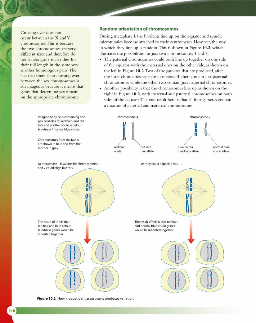
310
Random orientation of chromosomesDuring metaphase I, the bivalents line up on the equator and spindle microtubules become attached to their centromeres. However, the way in which they line up is random. This is shown in Figure 10.2, which illustrates the possibilities for just two chromosomes, 4 and 7.
• The paternal chromosomes could both line up together on one side of the equator with the maternal ones on the other side, as shown on the left in Figure 10.2. Two of the gametes that are produced, after the sister chromatids separate in meiosis II, then contain just paternal chromosomes while the other two contain just maternal chromosomes.
• Another possibility is that the chromosomes line up as shown on the right in Figure 10.2, with maternal and paternal chromosomes on both sides of the equator. The end result here is that all four gametes contain a mixture of paternal and maternal chromosomes.
Crossing over does not occur between the X and Y chromosomes. This is because the two chromosomes are very different sizes and therefore do not sit alongside each other for their full length in the same way as other homologous pairs. The fact that there is no crossing over between the sex chromosomes is advantageous because it means that genes that determine sex remain on the appropriate chromosome.
Imagine body cells containing onepair of alleles for red hair / not redhair and another for blue colourblindness / normal blue vision.
chromosome 4
red hairallele
not redhair allele
blue colourblindness allele
normal bluevision allele
chromosome 7
At metaphase I, bivalents for chromosomes 4and 7 could align like this …
or they could align like this …
The result of this is thatred hair and blue colourblindness genes would beinherited together
The result of this is that red hairand normal blue vision geneswould be inherited together.
Chromosomes from the fatherare shown in blue and from themother in grey.
Figure 10.2 How independent assortment produces variation.

10 GENETICS AND EVOLUTION (HL) 311
Look carefully at the eight gametes in Figure 10.2. How many different gametes are there? Your answer should be that there are four different gametes. Now consider what happens if a third pair of chromosomes is added. The number of genetically different gametes will increase to eight.
The number of possible genetic combinations that can occur as a result of random orientation of chromosomes is vast. If only chromosome combinations in a haploid cell are considered, the figure is 2n where n is the haploid number. For humans, n = 23 so the possible combinations are over 8 million for just one gamete. The formula can be used to work out the combinations in Figure 10.2 – for two pairs of chromosomes, n = 2, so the number of possible combinations = 22 = 4. With three pairs of chromosomes, the number of possible combinations = 23 = 8.
Meiosis and varietyThe calculation of the number of genetic possibilities in gametes using the formula 2n can only be an approximation. It does not account for the fact that there are two types of gametes – male and female. Also it does not include the extra variation that arises from crossing over. If a single cross-over occurs then the number of combinations is doubled, as shown in Figure 10.1. Since several crossovers normally occur during a meiotic division, it is clear that the amount of genetic variety is almost infinite even in organisms that only have a small haploid number.
Meiosis and Mendel’s law of independent assortmentGregor Mendel’s ‘law of independent assortment’ states that:
When gametes are formed, the separation of one pair of alleles into the new cells is independent of the separation of any other pair of alleles.
Or:
Either of a pair of alleles is equally likely to be inherited with either of another pair.
Random assortment during metaphase I can produce variety in the gametes as shown in Figure 10.2. How this relates to Mendel’s law of independent assortment for two pairs of chromosomes carrying the alleles Aa and Bb is shown in Figure 10.3.
As a result of the random alignment, the A allele has an equal chance of being in a gamete with either the B allele or the b allele, and similarly the a allele has an equal chance of being in a gamete with either the B allele or the b allele. Thus, four types of gamete can be produced carrying the alleles AB, Ab, aB or ab. They will occur in the ratio 1 : 1 : 1 : 1.

312
Nature of scienceCareful observation – were Mendel’s results ‘too good to be true’?In Mendel’s time, statistical analyses were not routinely used to test the validity of scientific results. Analysis of the ratios that Mendel published in 1866 suggests that they may be too close to the expected 3 : 1 ratio. Some have suggested that Mendel selected the data to present or that, having obtained a 3 : 1 ratio in some experiments, he persisted with counting until he achieved his expectations in subsequent experiments.
Mendel considered seven genes in his monohybrid experimental crosses, and also carried out dihybrid crosses involving pairs of these same genes. Crosses involving a pair of genes on different chromosomes produce the ratios Mendel reported but he would have obtained unexpected results had he used linked genes – that is, genes that occur on the same chromosome. It is not likely that the seven genes Mendel investigated would each occur on a different chromosome by chance (the pea plant only has seven pairs of chromosomes) so perhaps some of his crosses did involve linked genes. He would not have been able to explain the results from such crosses since he did not know about chromosomes, so it may be that he only published results from crosses that met his expectations.
Later work by Thomas Hunt Morgan (1866–1945) involving meticulous observation and record keeping revealed anomalous data in dihybrid crosses similar to those which Mendel had performed. As a result, Hunt developed his ideas of gene linkage to explain the results (Subtopic 10.2).
Figure 10.3 Meiosis and Mendel’s law of independent assortment.
A cell could go throughmeiosis and produce thesegametes, if both paternalhomologues go to the samepole in meiosis I, and bothmaternal homologues go tothe opposite pole.
gametes
AB ab
AB ab
parental genotype AaBb
equator
A a
B b
Or it could produce thesegametes if each paternalhomologue and eachmaternal homologue goesto an opposite pole inmeiosis I.
gametes
Ab aB
Ab aB
equator
maternal
paternal
A
Key
a
Bb

10 GENETICS AND EVOLUTION (HL) 313
10.2 Inheritance
Genotypes and gametesDihybrid crosses involve characteristics that are controlled by two genes. In a dihybrid genotype, there are two pairs of alleles to consider. Figure 10.3 shows what happens during meiosis in a parent with the genotype AaBb. Each pair of alleles can be combined in a mixture of dominant and recessive and some examples of genotypes and gametes are given in Table 10.1.
Genotype Gametes
AABB all AB
aaBB all aB
AaBB AB and aB in a ratio of 1 : 1
Aabb Ab and ab in a ratio of 1 : 1
Table 10.1 Some examples of diploid genotypes and the haploid gametes they produce.
There are more possible dihybrid genotypes than those shown in Table 10.1, but each genotype can only produce either one type of gamete, or two, or four. No genotype produces three types of gamete and only the double heterozygous genotype (as in Figure 10.3, for example) can produce four types of gamete. It is helpful to keep this in mind when working out the possible offspring of dihybrid crosses.
The dihybrid crossAlthough the dihybrid cross involves two pairs of genes instead of just one, the principles of setting out a genetic cross diagram to predict the offspring that will be produced are exactly the same as for the monohybrid crosses you saw in Subtopic 3.4. The genetic diagrams should include parental phenotypes, parental genotypes, gametes in circles and a Punnett grid for the F1 or F2 generation.
Learning objectives
You should understand that:
• Gene loci on the same chromosome are said to be linked.
• Unlinked genes segregate independently as a result of meiosis.
• Variation in a species may be continuous or discrete.
• Phenotypes of polygenic characteristics tend to show continuous variation.
• The chi-squared test is used to determine whether the differences between observed and expected frequency distributions are statically significant.
Parental generation the original parent individuals in a series of experimental crossesF1 generation the offspring of the parental generationF2 generation the offspring of a cross between F1 individuals
1 Define ‘crossing over’.2 State when sister chromatids separate during meiosis.3 Outline the reasons for independent assortment of genes.
Test yourself?
Exam tipRemind yourself of the correct way of writing dominant, recessive and co-dominant alleles in genetics problems.

314
Worked examples10.1 Fur colour in mice is determined by a single gene. Brown fur is dominant to white. Ear size is also
determined by a single gene. Rounded ears are dominant to pointed ears.
A mouse homozygous for brown fur and rounded ears was crossed with a white mouse with pointed ears.
Determine the possible phenotypes and genotypes of the offspring.
Step 1 Choose suitable letters to represent the alleles. Brown fur is dominant, so let B = brown fur and b = white fur. Rounded ears is dominant, so let R = rounded ears and r = pointed ears.
Step 2 The brown mouse with rounded ears is homozygous so its genotype must be BBRR.
Since white and pointed are recessive, the genotype of the white mouse with pointed ears must be bbrr.
Step 3 Set out the genetic diagram as shown.Step 4 All the F1 mice have brown fur and
rounded ears.
10.2 Mendel carried out genetic studies with the garden pea. Tall plants are dominant to short, and green seed pods are dominant to yellow. A homozygous tall plant with yellow seed pods was crossed with a short plant homozygous for green seed pods. Determine the possible genotypes and phenotypes in the offspring.
Step 1 Tall is dominant to short, so T = tall and t = short. Green pod is dominant to yellow so G = green and g = yellow.
Step 2 Each parent has one dominant and one recessive characteristic but we are told the dominant characteristic is homozygous. The tall plant with yellow seed pods therefore has genotype TTgg, and the short, green-podded plant is ttGG.
Step 3 Set out the genetic diagram as shown.Step 4 All the offspring are tall plants with green seed pods.
It was fortunate that Mendel chose these two characteristics for his crosses. Seed pod colour and height are unlinked genes on different chromosomes. Had they been on the same chromosome and linked, the results would have been different.
parental phenotypes: brown fur, white fur, rounded ears pointed ears
parental genotypes:
BBRR bbrr
gametes: all
BR
all
br
Punnett grid for F1:gametes from brown, round-eared parent
gam
etes
from
w
hite
, poi
nted
-ea
red
par
ent BR
br
BbRrbrown,
rounded ears
parental phenotypes: tall, yellow short, green seed pods seed pods
parental genotypes:
TTgg ttGG
gametes: all
Tg
all
tG
Punnett grid for F1:gametes from tall,
yellow-seeded parent
gam
etes
from
sh
ort,
gree
n-se
eded
par
ent Tg
tGTtGg
tall, green seeds

10 GENETICS AND EVOLUTION (HL) 315
Chromosomes and genesThe human genome contains between 25 000 and 30 000 genes but there are only 23 pairs of chromosomes. This means that each chromosome must carry very many genes. Chromosome 1 contains over 3000 genes but the much smaller chromosome 21 contains only around 400 genes. Any two genes on the same chromosome are said to be linked. Linked genes are usually passed on together. The genes on any chromosome form a linkage group, so a human has 23 linkage groups. The difference between unlinked and linked genes is shown in Figure 10.4
10.3 One of the heterozygous F1 mice with brown fur and rounded ears from the cross in Worked example 10.1 was crossed with a mouse with white fur and rounded ears. Some of the offspring had pointed ears. Deduce the genotype of the second mouse and state the phenotype ratio of the offspring.
Step 1 Use the same letters as in Worked example 10.1: B = brown fur and b = white fur, R = rounded ears and r = pointed ears.
Step 2 The first mouse has the genotype BbRr. We are told the second mouse is white, so it must have the alleles rr. It has rounded ears but we are
not told if this is homozygous or heterozygous, so the alleles could be RR or Rr. Reading on, we find that there are some offspring with pointed ears, so they must have the
genotype rr. This means that the unknown parent genotype must have been heterozygous, Rr. If the parent was RR, no recessive allele would have been present so that a homozygous genotype could not occur in the offspring, and none of them would have had pointed ears.
Step 3 Having written down your reasoning, as above, now set out the usual genetic diagram.
Step 4 The phenotypes produced are: 3 brown fur, rounded ears 3 white fur, rounded ears 1 brown fur, pointed ears 1 white fur, pointed ears. This produces a ratio of phenotypes of 3 : 3 : 1 : 1, which is an important Mendelian ratio.
parental phenotypes: brown, white, rounded ears rounded ears
parental genotypes:
BbRr bbRr
gametes: BR
Br
bR
br
bR
br
Punnett grid for F1: gametes from brown, rounded-eared parent
BR Br bR br
gam
etes
from
whi
te,
roun
ded-
eare
d p
aren
t
bRBbRR
brown, rounded ears
BbRrbrown,
rounded ears
bbRRwhite,
rounded ears
bbRrwhite,
rounded ears
brBbRr
brown, rounded ears
Bbrrbrown,
pointed ears
bbRrwhite,
rounded ears
bbrrwhite,
pointed ears

316
Breaking the lawMendel’s law of independent assortment
was found to have exceptions, which geneticist T. H. Morgan explained by using the idea of linked genes, described on page 325 at the end of Topic 10.2.
Questions to consider
• Is it correct to call Mendel’s proposals a ‘law’?• What is the difference between a law and a theory
in science?
Linkage and genesIf alleles are linked together on a chromosome, then it follows that they will be inherited together because during meiosis they will move together to the same pole as the cell divides. In genetics problems, dihybrid crosses involving linked genes do not produce Mendelian ratios. Linked genes do not follow Mendel’s law of independent assortment – they are not inherited independently and can give a variety of different ratios.
Writing a linkage genotypeIn the dihybrid crosses considered so far, genotypes have been written in the form AABB. With linked genes, a different notation has to be used because, although there are still four alleles to be considered, they are found on only one pair of chromosomes. The genotype is therefore always written as shown in Figure 10.5. The horizontal lines signify that the two genes occur on the same chromosome.
Mendelian ratios
In monohybrid crosses, two ratios for the offspring of a genetic cross are possible.
The first is 1 : 1 if a heterozygous individual (Aa) and a homozygous recessive individual (aa) are crossed. The second is 3 : 1 when two heterozygous individuals are crossed (Aa × Aa). These are called Mendelian ratios.
Mendelian ratios also occur in dihybrid crosses, but with more gametes there are more possibilities. A heterozygous individual (AaBb) crossed with a homozygous recessive (aabb) produces a 1 : 1 : 1 : 1 ratio and the ratio produced by crossing two double heterozygous (AaBb) individuals is 9 : 3 : 3 : 1. The 3 : 3 : 1 : 1 ratio in Worked example 10.3 is another Mendelian ratio. It is helpful to be familiar with these ratios.
Linkage group all the genes that have their loci on a particular chromosome
Genes A and B are on separatechromosomes and so are not linked.They will obey Mendel’s law ofindependent assortment and beinherited independently.
Genes D and G are on the samechromosomes and so are linked.They will not follow Mendel’s law ofindependent assortment. Genes Dand G form a linkage group.
Unlinked genes
A a
B B
Linked genes
D D
G G
Figure 10.4 The difference between unlinked and linked genes.
Here, D is linked to G and d is linked to g.Therefore D and G are inherited togetherand d and g are inherited together.
D G
d gHere, D is linked to g and d is linked to G.Therefore D and g are inherited togetherand d and G are inherited together.
D g
d G
Figure 10.5 This shows the two possible linkage patterns for the four alleles. The difference between the two linkage patterns makes a very big difference in the ratios of the phenotypes in the offspring of a cross.

10 GENETICS AND EVOLUTION (HL) 317
Worked example10.4 In the fruit fly, Drosophila, red eye colour is dominant to purple eyes and long wings is dominant to dumpy
wings. These genes are linked on chromosome 2. A fly that was homozygous for red eyes and long wings was crossed with a fly that had purple eyes and dumpy wings. Determine the ratios of genotypes and phenotypes of the F2 offspring by using a full genetic diagram.
Step 1 Red eye colour is dominant so R = red eye and r = purple eye. Long wings is dominant so N = long wings and n = dumpy wings.
Step 2 The fly with the dominant characteristics is homozygous and the other fly shows both recessive characteristics. The parental genotypes are:
R NR N
and r nr n
Step 3 Set out the genetic diagram as below.
All the F1 have red eyes and long wings. Now, the F2 generation is obtained
by crossing two offspring from the F1 generation.
Step 4 In the F2 generation, the ratio of phenotypes is: 3 red eye, long wing : 1 purple eye, dumpy wing. Note that this 3 : 1 ratio is what you would expect in a
monohybrid cross. The reason for this is that the two genes are linked and so there is only one pair of chromosomes involved, as in monohybrid crosses.
parental phenotypes: red eyes, purple eyes, long wings dumpy wings
parental genotypes:
R NR N
r nr n
gametes: all
RN
all
rn
Punnett grid for F1:gametes from red-eyed, long-winged
parent
gam
etes
from
p
urp
le-e
yed,
dum
py-
win
ged
par
ent RN
rn
R Nr n
red eyes, long wings
parental phenotypes: red eyes, red eyes, long wings long wings
parental genotypes:
R Nr n
R Nr n
gametes: RN
rn
RN
rn
Punnett grid for F2:gametes from red-eyed,
long-winged parent
RN rn
gam
etes
from
red-
eyed
, lo
ng-w
inge
d p
aren
t
RN
R NR N
red eyes, long wings
R Nr n
red eyes, long wings
rn
R Nr n
red eyes, long wings
r nr n
purple eyes, dumpy wings
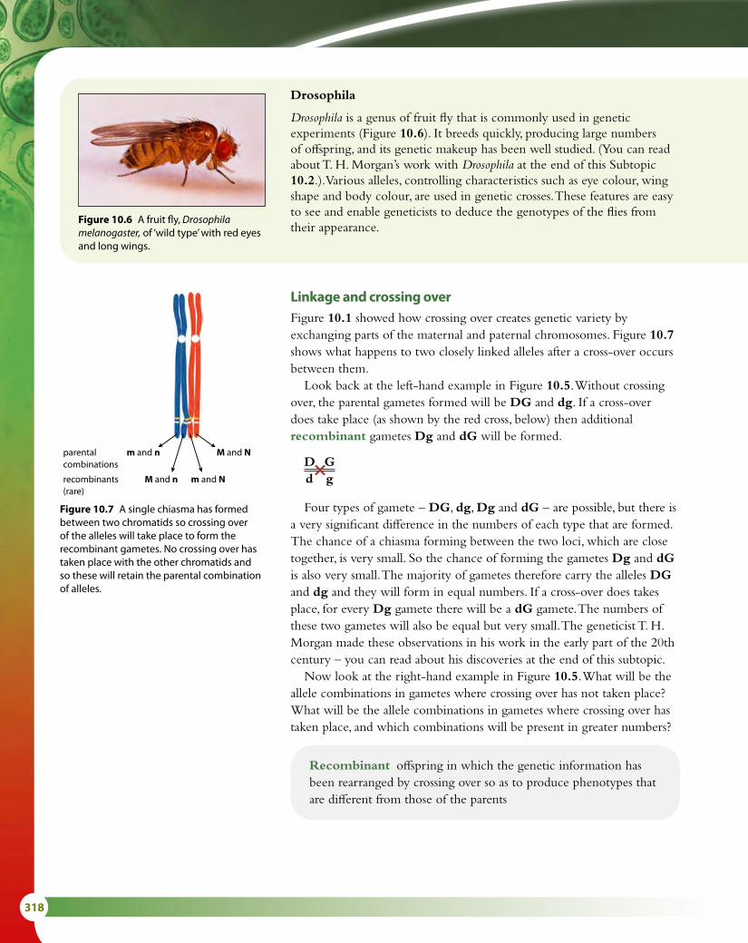
318
Linkage and crossing overFigure 10.1 showed how crossing over creates genetic variety by exchanging parts of the maternal and paternal chromosomes. Figure 10.7 shows what happens to two closely linked alleles after a cross-over occurs between them.
Look back at the left-hand example in Figure 10.5. Without crossing over, the parental gametes formed will be DG and dg. If a cross-over does take place (as shown by the red cross, below) then additional recombinant gametes Dg and dG will be formed.
D G d g
Four types of gamete – DG, dg, Dg and dG – are possible, but there is a very significant difference in the numbers of each type that are formed. The chance of a chiasma forming between the two loci, which are close together, is very small. So the chance of forming the gametes Dg and dG is also very small. The majority of gametes therefore carry the alleles DG and dg and they will form in equal numbers. If a cross-over does takes place, for every Dg gamete there will be a dG gamete. The numbers of these two gametes will also be equal but very small. The geneticist T. H. Morgan made these observations in his work in the early part of the 20th century – you can read about his discoveries at the end of this subtopic.
Now look at the right-hand example in Figure 10.5. What will be the allele combinations in gametes where crossing over has not taken place? What will be the allele combinations in gametes where crossing over has taken place, and which combinations will be present in greater numbers?
Drosophila
Drosophila is a genus of fruit fly that is commonly used in genetic experiments (Figure 10.6). It breeds quickly, producing large numbers of offspring, and its genetic makeup has been well studied. (You can read about T. H. Morgan’s work with Drosophila at the end of this Subtopic 10.2.). Various alleles, controlling characteristics such as eye colour, wing shape and body colour, are used in genetic crosses. These features are easy to see and enable geneticists to deduce the genotypes of the flies from their appearance.
Figure 10.6 A fruit fly, Drosophila melanogaster, of ‘wild type’ with red eyes and long wings.
parentalcombinations
m and n M and N
recombinants(rare)
M and n m and N
Figure 10.7 A single chiasma has formed between two chromatids so crossing over of the alleles will take place to form the recombinant gametes. No crossing over has taken place with the other chromatids and so these will retain the parental combination of alleles.
Recombinant offspring in which the genetic information has been rearranged by crossing over so as to produce phenotypes that are different from those of the parents

10 GENETICS AND EVOLUTION (HL) 319
Worked example10.5 Grey body and red eyes are dominant to stripe body and cardinal eye in Drosophila. They are autosomal,
linked genes on chromosome 3. Homozygous grey flies with red eyes were crossed with stripe flies with cardinal eyes. No crossing over occurred.
Then the F1 flies were crossed with stripe, cardinal flies.
• If no crossing over occurs between the two loci, what phenotypes would be expected in the offspring of this second cross?
• If crossing over did occur between the loci, what phenotypes would be expected this time?
First cross, with no crossing over:Step 1 Grey body is dominant, so G = grey body and g = stripe body. Red eye is dominant so
R = red eye and r = cardinal eye.Step 2 The fly with the dominant characteristics is homozygous and the other fly shows both recessive
characteristics. The parental genotypes are G RG R
and g rg r
.Step 3 Set out the diagram as shown.
Step 4 All the F1 flies have a grey body and red eyes.
Second cross, with no crossing over:
parental phenotypes: grey body, stripe body, red eyes cardinal eyes
parental genotypes:
G RG R
g rg r
gametes: all
GR
all
gr
Punnett grid for F1:gametes from grey-
bodied, red-eyed parent
gam
etes
from
st
ripe-
bod
ied,
ca
rdin
al-e
yed
par
ent
GR
gr
G Rg r
grey body, red eyes
parental phenotypes: grey body, stripe body, red eyes cardinal eyes
parental genotypes:
G Rg r
g rg r
gametes: GR
gr
all
gr
Punnett grid for F1:gametes from grey-bodied,
red-eyed parent
gam
etes
from
st
ripe-
bod
ied,
ca
rdin
al-e
yed
par
ent
GR gr
gr
G Rg r
grey body, red eyes
g rg r
stripe body, cardinal eyes

320
variationVariation is defined as the differences that exist between individuals. We can identify two types – discrete variation (also known as discontinuous variation) and continuous variation.
Discrete (or discontinuous) variation is where clearly distinguishable categories can be identified and involves features that cannot be measured. Examples include blood groups and left- or right-handedness. This type of variation is controlled by the alleles of one or a few genes and it is not affected by environmental factors.
Continuous variation is not categorical. It is possible to make a range of measurements of continuous variation from one extreme to another. Human height, hand span and skin colour are all examples of continuous variations. The yield of wheat from crop plants or milk from dairy cattle is also continuously variable. These variations are controlled by genes but are also affected by environmental factors such as soil quality, weather and – in the case of the cattle – diet.
Continuous variation can be shown on a graph, which produces an even distribution of frequencies of the characteristic, called a normal distribution. The bell-shaped curves in Figure 10.8 represent continuous variation. Discrete variation, such as that observed in human blood groups, produces a bar graph, with each distinct category represented separately.
Second cross, with crossing over:
Step 4 The four F1 phenotypes are:
• grey body and red eyes
• stripe body and cardinal eyes
• grey body and cardinal eyes
• stripe body and red eyes
The ‘grey, cardinal’ and ‘stripe, red’ (shown in red type) flies are recombinants as they have a phenotype that is different from the parental phenotypes. These recombinant phenotypes will occur in approximately equal numbers. The parental phenotypes (shown in black type) will also be in approximately equal numbers among the offspring, but the recombinant phenotypes will be very few in number compared to the parental phenotypes.
parental phenotypes: grey body, stripe body, red eyes cardinal eyes
parental genotypes:
G Rg r
g rg r
gametes: GR
gr
Gr *
gR *
all
gr
Punnett grid for F1:gametes from grey-bodied, red-eyed parent
gam
etes
from
st
ripe-
bod
ied
card
inal
-eye
d p
aren
t
GR gr Gr gR
gr
G Rg r
grey body, red eyes
g rg r
stripe body, cardinal eyes
G rg r
grey body, cardinal eyes
g Rg r
stripe body, red eyes
Exam tipLinked alleles will always be represented like thisT B
t b
in examination papers

10 GENETICS AND EVOLUTION (HL) 321
PolygenesIn the genetic examples considered so far, a particular characteristic is controlled by one gene, which can have different alleles at a specific locus on a pair of chromosomes. There is a clear difference between organisms with different alleles. An organism either has the characteristic or it does not – there are no intermediate forms. These phenotypes are examples of discrete (or discontinuous) variation.
Very few characteristics are controlled by single genes. Most are controlled by groups of genes, which together are known as polygenes. The genes that form polygenes are often unlinked – that is, they are located on different chromosomes. When two or more genes, each with multiple alleles, are responsible for a characteristic the number of possible phenotypes is greatly increased. Each gene separately may have little impact but their combined effect produces a whole variety of phenotypes. Unlinked polygenes result in a range of degrees of the characteristic from one extreme to another – that is, continuous variation.
Human skin colourHuman skin colour depends on the amount of the pigment melanin that is produced in the skin. Melanin synthesis is controlled by genes. The degree of pigmentation can range from the very dark skin of people originating from regions such as Namibia in southern Africa, through to the very pale skin of native Scandinavian people.
Melanin protects the skin from the harmful UV rays from the Sun. In parts of the world close to the equator, the Sun’s rays are particularly intense so people need more protection from sunburn. Dark-skinned people have a high concentration of melanin, which protects them, while fair-skinned people have much less. Although skin colour is genetically determined, environmental factors also influence it. Fair-skinned people who are exposed to sunlight produce extra melanin and develop a protective suntan. Exposure to sunlight also allows vitamin D to be produced in the skin.
Several genes are involved in determining skin colour and they produce the almost continuous variation that can be seen in the global human population. In Figure 10.9 only three genes are shown. Each gene has two alleles. One allele, M, contributes to melanin production and the other, m, does not. These three genes, each with two alleles, give rise to seven possible skin tones – numbered 0 to 6 below. The number refers to the number of M alleles, which determine the level of skin pigmentation. A person with the genotype MMMMMM will have very dark skin (6), while a person with mmmmmm will have very pale skin (0). The Punnett grid shows the possible combinations of skin colour in children from two parents, both heterozygous for all three genes. The parents’ phenotype is light brown skin (3).
Although in this simplified example there are only seven categories of pigmentation, nevertheless you can see that if the frequencies of the skin colour varieties in the Punnett grid are plotted on a histogram, as
Polygenic inheritance the inheritance of a characteristic that is controlled by two or more genes; polygenic inheritance accounts for the continuous variation that occurs in characteristics such as human height, body mass and skin colour
mean
Value (for example, height)
Freq
uenc
y (fo
r exa
mp
le, n
umb
er
of s
tude
nts
at e
ach
heig
ht)
20
15
10
5
0 0 1 2 3 4 5 6Degree of pigmentation
Freq
uenc
y
Figure 10.8 Human height demonstrates continuous variation – when frequency is plotted against height, a normal distribution is obtained (top). Frequency of skin variation is shown on bar graph variation.

322
in Figure 10.8, it produces a normal distribution. In the case of human skin colour, it is known that more than three genes are involved and the number of categories exceeds seven. The result is a wider distribution curve and more ‘continuous’ variation.
Many other examples of polygenic inheritance are known. In wheat plants, three genes interact to control the colour of seeds. Human height and body mass are also examples of polygenic characteristics, but both can be influenced by environmental factors such as nutrients in a person’s diet or the quantity of food that they consume.
parental phenotypes: light brown skin light brown skin
3 3
parental genotypes: M1m1M2m2M3m3 M1m1M2m2M3m3
gametes: M1M2M3
M1M2m3
m1m2M3
M1m2M3
M1M2M3
M1M2m3
m1m2M3
M1m2M3
m1M2m3
M1m2m3
m1M2M3
m1m2m3
m1M2m3
M1m2m3
m1M2M3
m1m2m3
Punnett grid for F1:gametes from light-brown-skinned parent
gam
etes
from
ligh
t-b
row
n-sk
inne
d p
aren
t
M1M2M3 M1M2m3 m1m2M3 M1m2M3 m1M2m3 M1m2m3 m1M2M3 m1m2m3
M1M2M3 6 5 4 5 4 4 5 3
M1M2m3 5 4 3 4 3 3 4 2
m1m2M3 4 3 2 3 2 2 3 1
M1m2M3 5 4 3 4 3 3 4 2
m1M2m3 4 3 2 3 2 2 3 1
M1m2m3 4 3 2 3 2 2 3 1
m1M2M3 5 4 3 4 3 3 4 2
m1m2m3 3 2 1 2 1 1 2 0
Figure 10.9 Punnett grid show the possible combinations of skin colour in children from two heterozygous parents.

10 GENETICS AND EVOLUTION (HL) 323
The chi-squared testThe chi-squared (χ2) test is a statistical test that can be used in cases where variation is discrete. In genetic or ecological investigations, the chi-squared test is useful to compare your observed results with the results that you would expect if your theory about how the system works were correct. The test tells you whether or not any difference between your observed results and your expected results is significant. If the difference is significant, that means the results do not fit well with your theory – so you may need to revise your theory. If the difference is not significant – that is, it’s so small that it could have occurred by chance – then you can say that your results do support your theory.
The chi-squared value is calculated like this:
χ2 = Σ (O – E )2
E
where Σ means ‘the sum of ’, O = observed frequency and E = expected frequency.
The greater the value of chi-squared you calculate, the greater the difference between your observed and expected results. To find out whether the difference is significant or not, you must compare your chi-squared value with a table of ‘critical values’ like the one in Table 10.2. The null hypothesis states that there is no significant difference between the observed and expected results – that is, that the results fit the expected pattern, and therefore support your theory.
The chi-squared test is used in genetics and in ecology to compare observed numbers of organisms to expected numbers. It can be used to test the outcome of monohybrid or dihybrid crosses – to see if the observed ratios fit the expected pattern. The worked examples that follow use data that were produced as a result of dihybrid crosses made by Mendel.
Deg
rees
of
free
do
m
p
0.995 0.99 0.975 0.95 0.90 0.10 0.05 0.025 0.01 0.005
1 --- --- 0.001 0.004 0.016 2.706 3.841 5.024 6.635 7.879
2 0.010 0.020 0.051 0.103 0.211 4.605 5.991 7.378 9.210 10.597
3 0.072 0.115 0.216 0.352 0.584 6.251 7.815 9.348 11.345 12.838
4 0.207 0.297 0.484 0.711 1.064 7.779 9.488 11.143 13.277 14.860
5 0.412 0.554 0.831 1.145 1.610 9.236 11.070 12.833 15.086 16.750
6 0.676 0.872 1.237 1.635 2.204 10.645 12.592 14.449 16.812 18.548
7 0.989 1.239 1.690 2.167 2.833 12.017 14.067 16.013 18.475 20.278
8 1.344 1.646 2.180 2.733 3.490 13.362 15.507 17.535 20.090 21.955
9 1.735 2.088 2.700 3.325 4.168 14.684 16.919 19.023 21.666 23.589
10 2.156 2.558 3.247 3.940 4.865 15.987 18.307 20.483 23.209 25.188
Table 10.2 Critical values of the chi-squared distribution, showing how to read across the appropriate ‘degrees of freedom’ row to find the critical chi-squared value at the 0.05 level (p).
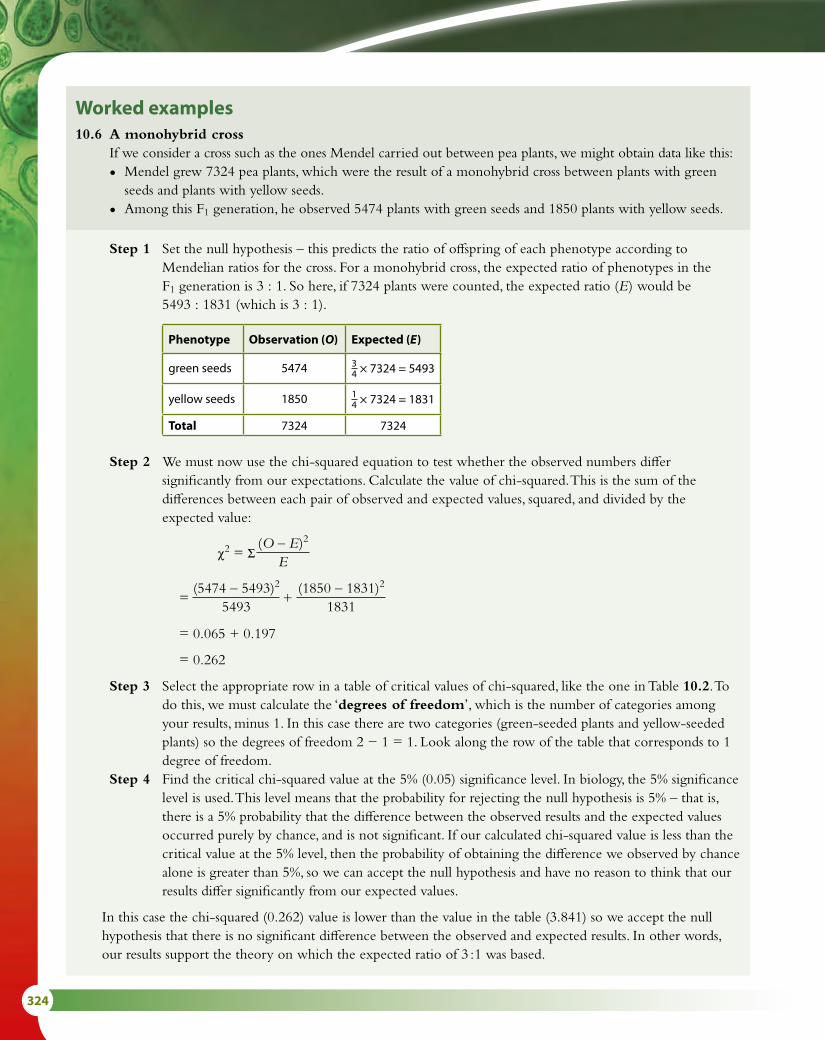
324
Worked examples10.6 A monohybrid cross If we consider a cross such as the ones Mendel carried out between pea plants, we might obtain data like this:
• Mendel grew 7324 pea plants, which were the result of a monohybrid cross between plants with green seeds and plants with yellow seeds.
• Among this F1 generation, he observed 5474 plants with green seeds and 1850 plants with yellow seeds.
Step 1 Set the null hypothesis – this predicts the ratio of offspring of each phenotype according to Mendelian ratios for the cross. For a monohybrid cross, the expected ratio of phenotypes in the F1 generation is 3 : 1. So here, if 7324 plants were counted, the expected ratio (E ) would be 5493 : 1831 (which is 3 : 1).
Phenotype Observation (O) Expected (E )
green seeds 5474 34 × 7324 = 5493
yellow seeds 1850 14 × 7324 = 1831
Total 7324 7324
Step 2 We must now use the chi-squared equation to test whether the observed numbers differ significantly from our expectations. Calculate the value of chi-squared. This is the sum of the differences between each pair of observed and expected values, squared, and divided by the expected value:
χ2 = Σ (O – E )2
E
= (5474 – 5493)2
5493 +
(1850 – 1831)2
1831
= 0.065 + 0.197
= 0.262
Step 3 Select the appropriate row in a table of critical values of chi-squared, like the one in Table 10.2. To do this, we must calculate the ‘degrees of freedom’, which is the number of categories among your results, minus 1. In this case there are two categories (green-seeded plants and yellow-seeded plants) so the degrees of freedom 2 − 1 = 1. Look along the row of the table that corresponds to 1 degree of freedom.
Step 4 Find the critical chi-squared value at the 5% (0.05) significance level. In biology, the 5% significance level is used. This level means that the probability for rejecting the null hypothesis is 5% – that is, there is a 5% probability that the difference between the observed results and the expected values occurred purely by chance, and is not significant. If our calculated chi-squared value is less than the critical value at the 5% level, then the probability of obtaining the difference we observed by chance alone is greater than 5%, so we can accept the null hypothesis and have no reason to think that our results differ significantly from our expected values.
In this case the chi-squared (0.262) value is lower than the value in the table (3.841) so we accept the null hypothesis that there is no significant difference between the observed and expected results. In other words, our results support the theory on which the expected ratio of 3 : 1 was based.

10 GENETICS AND EVOLUTION (HL) 325
Nature of scienceLooking for trends and discrepancies – the work of T. H. MorganThomas Hunt Morgan (1866–1945) was a pioneering geneticist who studied the inheritance of mutations in the fruit fly Drosophila melanogaster. After the rediscovery of Mendelian genetics in 1900, Morgan worked to show that genes are carried on chromosomes and provide the basis for inheritance. He induced mutations in his flies using chemicals and radiation and began cross-breeding experiments to find mutations that were inherited. Despite the difficulty of spotting mutations in the tiny flies, he eventually noticed a white-eyed mutant male among the typical ‘wild type’ red-eyed flies. He bred white-eyed male flies with red-eyed females and all the offspring were red-eyed. The F2 (second generation) cross produced white-eyed males so Morgan concluded that the white-eye mutation was a sex-linked recessive trait (Figure 10.10). He also discovered a pink-eyed mutant which was not sex linked.
10.7 A dihybrid cross using Mendel’s dataIn this dihybrid cross between heterozygous round and yellow-seeded pea plants (Rr Yy). A total of 556 seeds are counted, the expected ratios of phenotypes in the F1 generation are 9 : 3 : 3 : 1.
Phenotype Observed numbers (O) Expected numbers (E)
round yellow seed 315 916 × 556 = 321.72
round green seeds 101 316 × 556 = 104.25
wrinkled yellow seeds 108 316 × 556 = 104.25
wrinkled green seeds 32 116 × 556 = 34.75
Total number of seeds 556 556
To calculate the chi-squared value:
χ2 = (315 – 312.75)2
312.75 +
(101 – 104.25)2
104.25 +
(108 – 104.25)2
104.25 +
(32 – 34.75)2
34.75
= 0.016 + 0.101 + 0.135 + 0.218
= 0.470
Degrees of freedom: 4 − 1 = 3
From Table 10.2, the critical value at the 5% (0.05) level, for 3 degrees of freedom = 7.815The observed value is less than the critical value so we can accept the null hypothesis that there is no significant difference between our observations and expectations. We can accept Mendel’s conclusion that a 9 : 3 : 3 : 1 ratio is the result of this type of cross.
R = roundr = wrinkledY = yellowy = green

326
Morgan reported his discoveries of sex linkage and autosomal inheritance in the journal Science in 1911. Morgan’s team discovered more mutants amongst thousands of flies they studied and also identified flies with multiple mutations. They studied more complex patterns of inheritance, finding more examples of crosses that did not fit the pattern of simple Mendelian ratios. To explain these discrepancies, Morgan went on to suggest that genes could be linked and inherited together. Morgan proposed the hypothesis of crossing over (Subtopic 3.3) and the relationship between crossing over and linked genes. He suggested that cross-over frequency gave a measure of the distance separating genes on a chromosome.
His book The Mechanism of Mendelian Heredity, published in 1915, was a foundation for modern genetics.
P generation
F1 generationall red-eyed
F2 generationcontains red-eyedand white-eyed males
×
×
red-eyedfemale
sex chromosomes x x
white-eyedmale
x y
Figure 10.10 Inheritance of a sex-linked characteristic in Drosophila.
4 What are the gametes produced by the genotypes Ttrr and HhGg?5 What is the name of the grid used to determine offspring
genotypes and phenotypes?6 State the generation from which individuals are crossed in order
to produce an F2 generation.
Test yourself?

10 GENETICS AND EVOLUTION (HL) 327
no selection –original distribution
num
ber
of i
divi
dual
s
stabilising selection
num
ber
of i
divi
dual
s
disruptive selection
num
ber
of i
divi
dual
s
directional selection
num
ber
of i
divi
dual
s
10.3 Gene pools and speciation
Allele frequency and evolutionEvolution is defined as the cumulative change in the heritable characteristics of a population. The ‘heritable characteristics’ referred to in this definition are all the alleles in the gene pool of a population. So, if the frequencies of these alleles in the gene pool do not change, then the population is not evolving. Allele frequencies in a population are always fluctuating, in fact, because they depend on the reproductive success of individuals. But if just a single allele shows a change in frequency over a prolonged period of time, then we can say that the population has evolved. Any process that allows the passing on of favourable alleles or prevents the transmission of unfavourable alleles can contribute to evolution.
Natural selection and changeIn any population, an individual can have any combination of the alleles present in the gene pool. This gives rise to variation in the population. In most cases, a population will be well adapted to its environment and so the same alleles will be selected, maintaining a stable population. This is known as stabilising selection (Figure 10.11).
If the environment changes the population may also change. Some individuals may have alleles that are more favourable in the new conditions and these alleles will provide an advantage to those individuals, making them more likely to survive and reproduce successfully. This will
Learning objectives
You should understand that:
• A gene pool consists of all the genes and their different alleles that are present in an interbreeding population.
• As species evolve with time, allele frequencies in a population change.
• Reproductive isolation of populations may be temporal, spatial or behavioural.
• Speciation due to the divergence of isolated populations can be gradual.
• Speciation may occur abruptly.
Gene pool all the different genes in an interbreeding population at a given timeAllele frequency the frequency of a particular allele as a proportion of all the alleles of that gene in a population
Figure 10.11 In these examples three kinds of selection change the colour of beetles — a feature that is controlled by several genes. Stabilising selection eliminates extreme forms, directional selection favours dark coloured beetles and disruptive selection eliminates the 'average' form and leads to two distinct forms.
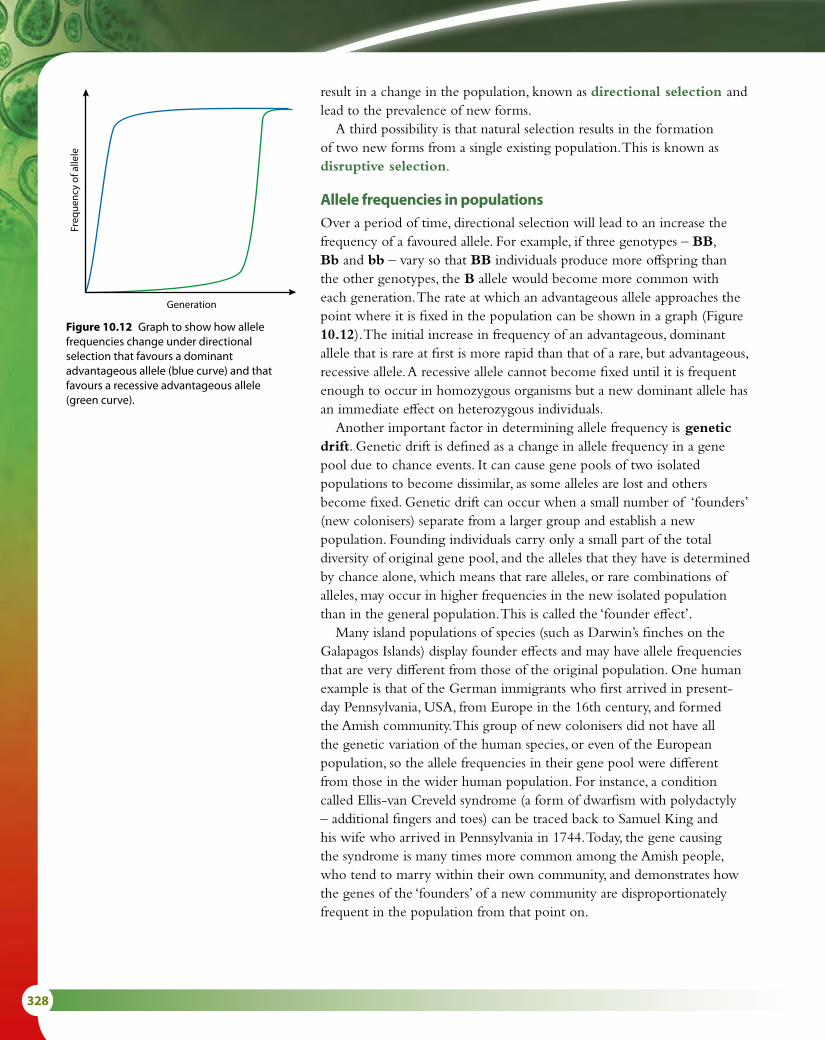
328
result in a change in the population, known as directional selection and lead to the prevalence of new forms.
A third possibility is that natural selection results in the formation of two new forms from a single existing population. This is known as disruptive selection.
Allele frequencies in populationsOver a period of time, directional selection will lead to an increase the frequency of a favoured allele. For example, if three genotypes – BB, Bb and bb – vary so that BB individuals produce more offspring than the other genotypes, the B allele would become more common with each generation. The rate at which an advantageous allele approaches the point where it is fixed in the population can be shown in a graph (Figure 10.12). The initial increase in frequency of an advantageous, dominant allele that is rare at first is more rapid than that of a rare, but advantageous, recessive allele. A recessive allele cannot become fixed until it is frequent enough to occur in homozygous organisms but a new dominant allele has an immediate effect on heterozygous individuals.
Another important factor in determining allele frequency is genetic drift. Genetic drift is defined as a change in allele frequency in a gene pool due to chance events. It can cause gene pools of two isolated populations to become dissimilar, as some alleles are lost and others become fixed. Genetic drift can occur when a small number of ‘founders’ (new colonisers) separate from a larger group and establish a new population. Founding individuals carry only a small part of the total diversity of original gene pool, and the alleles that they have is determined by chance alone, which means that rare alleles, or rare combinations of alleles, may occur in higher frequencies in the new isolated population than in the general population. This is called the ‘founder effect’.
Many island populations of species (such as Darwin’s finches on the Galapagos Islands) display founder effects and may have allele frequencies that are very different from those of the original population. One human example is that of the German immigrants who first arrived in present-day Pennsylvania, USA, from Europe in the 16th century, and formed the Amish community. This group of new colonisers did not have all the genetic variation of the human species, or even of the European population, so the allele frequencies in their gene pool were different from those in the wider human population. For instance, a condition called Ellis-van Creveld syndrome (a form of dwarfism with polydactyly – additional fingers and toes) can be traced back to Samuel King and his wife who arrived in Pennsylvania in 1744. Today, the gene causing the syndrome is many times more common among the Amish people, who tend to marry within their own community, and demonstrates how the genes of the ‘founders’ of a new community are disproportionately frequent in the population from that point on.
Generation
Freq
uenc
y of
alle
le
Figure 10.12 Graph to show how allele frequencies change under directional selection that favours a dominant advantageous allele (blue curve) and that favours a recessive advantageous allele (green curve).

10 GENETICS AND EVOLUTION (HL) 329
SpeciationSpeciation is the formation of new species from an existing population. Members of the same species and, therefore, the same gene pool can fail to reproduce as a result of a barrier that separates them. New species appear as a result of the population of a single species splitting into two or more new ones each with its own gene pool.
Speciation can only occur if there is a barrier dividing the population (Figure 10.13). The barrier may take different forms, such as geographical separation, or temporal or behavioural differences. When one part of the divided population is isolated from the other, mutation and selection can occur independently in the two populations so that each has the potential to become a new species.
Speciation is said to be sympatric or allopatric.
• Allopatric speciation occurs in different geographical areas.
• Sympatric speciation occurs in the same geographical area.
single gene pool gene pool separated into tworegions and gene flow betweenthem prevented – differences inselection pressure may result indifferent allele frequencies ineach new gene pool so theybecome genetically different
gene pool splits into tworegions but gene flowbetween them means theyremain a single species
barrier barrier
Geographic (allopatric) speciationAllopatric speciation occurs when a physical barrier separates a species into two geographically isolated populations, which then develop independently under the different conditions in the two separated areas, and eventually become unable to interbreed. The barrier might be a natural feature such as a mountain range or a body of water, or it could be a result of human intervention in an environment, such as a major road system or a large conurbation. Many examples of this type of speciation occur on islands such as the Galapagos Islands or the islands of Hawaii.
One example of allopatric speciation resulting from a natural geographical barrier involves salamanders of the genus Ensatina, found on the west coast of the USA. Members of this genus all descended from a founder species that spread southward down each side of the San Joaquin Valley (Figure 10.14). Conditions were slightly different on the east and west sides of the valley and the separated populations adapted to their own
Figure 10.13 Speciation may occur when some kind of barrier divides the gene pool.

330
particular environments. The valley in between isolated them from one another. Eventually the two populations met at the southern end of the valley in Southern California but they could no longer interbreed. Two new species, E. klauberi and E. eschscholtzi, had formed and are said to be reproductively isolated from one another.
Sympatric speciationA gene pool may become divided without the population being geographically split. Other factors may lead to groups within the population becoming reproductively separated within the same physical environment.
Temporal and behavioural isolationTemporal isolation occurs when the time of reproduction or behaviour of members of one population of a species is incompatible with that of another. An example is the goldenrod gall fly (Eurosta solidaginis) found in eastern and midwestern North America. This fly causes the formation of galls on goldenrod plants (Solidago sp.) (Figure 10.15). Over most of its range, the fly lays its eggs in S. altissima but in some places it uses S. gigantea as its host. Flies associated with S. gigantea emerge earlier in the season than flies associated with S. altissima so the males and females of the two forms of fly are ready to mate at slightly different times. This suggests the beginning of a temporal barrier to interbreeding between the two populations.
The two emerging groups of flies seem to actively prefer their own species of goldenrod plant and lay their eggs much more frequently in those plants. In addition, females prefer to mate with males of their own group and both of these factors indicate the development of additional behavioural barriers to interbreeding.
intermediatespecies ofEnsatina
intermediatespecies ofEnsatina
founder species of Ensatina
E. klauberiE. eschscholtzi
Sa
n J
oa
qu
i n V
al l e y
Figure 10.14 Speciation in salamanders, in the San Joaquin Valley, western USA.
Figure 10.15 Goldenrod gall fly and the gall it produces.
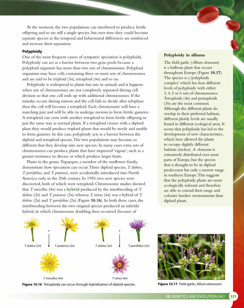
10 GENETICS AND EVOLUTION (HL) 331
T. dubius (2n)
T. miscellus (4n) T. mirus (4n)
T. dubius (2n) T. porrifolius (2n)T. pratensis (2n)
× ×
At the moment, the two populations can interbreed to produce fertile offspring and so are still a single species, but over time they could become separate species as the temporal and behavioural differences are reinforced and increase their separation.
PolyploidyOne of the most frequent causes of sympatric speciation is polyploidy. Polyploidy can act as a barrier between two gene pools because a polyploid organism has more than two sets of chromosomes. Polyploid organisms may have cells containing three or more sets of chromosomes and are said to be triploid (3n), tetraploid (4n) and so on.
Polyploidy is widespread in plants but rare in animals and it happens when sets of chromosomes are not completely separated during cell division so that one cell ends up with additional chromosomes. If the mistake occurs during mitosis and the cell fails to divide after telophase then the cell will become a tetraploid. Each chromosome will have a matching pair and will be able to undergo meiosis to form fertile gametes. A tetraploid can cross with another tetraploid to form fertile offspring in just the same way as normal plants. If a tetraploid crosses with a diploid plant they would produce triploid plants that would be sterile and unable to form gametes. In this case, polyploidy acts as a barrier between the diploid and tetraploid species. The two populations may become so different that they develop into new species. In many cases extra sets of chromosomes can produce plants that have improved ‘vigour’, such as a greater resistance to disease or which produce larger fruits.
Plants in the genus Tragopogon, a member of the sunflower family, demonstrate how speciation can occur. Three diploid species, T. dubius, T. porrifolius, and T. pratensis, were accidentally introduced into North America early in the 20th century. In 1950, two new species were discovered, both of which were tetraploid. Chromosome studies showed that T. miscellus (4n) was a hybrid produced by the interbreeding of T. dubius (2n) and T. pratensis (2n) whereas T. mirus (4n) was a hybrid of T. dubius (2n) and T. porrifolius (2n) (Figure 10.16). In both these cases, the interbreeding between the two original species produced an infertile hybrid, in which chromosome doubling then occurred (because of
Figure 10.16 Tetraploidy can occur through hybridisation of diploid species.
Polyploidy in alliums
The field garlic (Allium oleraceum) is a bulbous plant that occurs throughout Europe (Figure 10.17). The species is a ‘polyploidy complex’ which has four different levels of polyploidy with either 3, 4, 5 or 6 sets of chromosomes. Tetraploids (4n) and pentaploids (5n) are the most common. Although the different plants do overlap in their preferred habitats, different ploidy levels are usually found in different ecological areas. It seems that polyploidy has led to the development of new characteristics, which have allowed the plants to occupy slightly different habitats (niches). A. oleraceum is extensively distributed over most parts of Europe, but the species that is thought to be its diploid predecessor has only a narrow range in southern Europe. This suggests that the polyploidy plants are more ecologically tolerant and therefore are able to extend their range and colonise harsher environments than diploid plants.
Figure 10.17 Field garlic, Allium oleraceum.

332
mistakes during mitosis), giving rise to a tetraploid hybrid that could produce viable gametes, and was therefore fertile.
A summary of the two types of speciation is shown in Table 10.3.
Sympatric speciation Allopatric speciation
A new species arises from an existing species that is living in the same area.
A new species arises because a geographic barrier separates it from other members of an existing species.
Temporal or behavioural isolation can produce significant changes in the genetic make-up within a species so that a new species is formed.
Geographic barriers may include mountain ranges, valleys or bodies of water, or human-made features such as roads, canals or built-up areas.
Table 10.3 Comparison of sympatric and allopatric speciation.
species 1
species 3
species 2
Gradualism Punctuated equilibrium
time
Figure 10.18 A gradualism view and a punctuated equilibrium view of evolution.
The pace of evolution: gradualism and punctuated equilibriumDarwin viewed evolution as a slow, steady process called gradualism, whereby changes slowly accumulate over many generations and lead to speciation. For many species, this seems to be true. A good example of gradualism is the evolution of the horse’s limbs, which fossils indicate took around 43 million years to change from the ancestral form to the modern one (Figure 5.3, Subtopic 5.1).
In some cases, the fossil record does not contain any intermediate stages between one species and another. A suggested explanation is that fossilisation is such a rare event that fossils of intermediate forms simply did not form, or else have not been discovered. In 1972, Stephen J. Gould (1941–2002) and Niles Eldredge (b. 1943) suggested that such fossils have not been found because these intermediate forms never existed – that evolution had allowed an ancestral form to develop into a quite different descendent without any intermediate. To explain how this might happen, they proposed an additional mechanism for evolution called punctuated equilibrium. The driving force for evolution is selection pressure, so if the selection pressure is very mild or non-existent then species will tend to remain the same – that is, in equilibrium. When there is a sudden, dramatic change in the environment, there will also be new, intense selection pressures and therefore rapid development of new species (Figure 10.18).

10 GENETICS AND EVOLUTION (HL) 333
Paradigm shiftA paradigm shift is a change in the core
beliefs or assumptions of an accepted scientific theory. It occurs when scientists are faced with anomalies that cannot be explained by the accepted paradigm. In modern science, a number of paradigm shifts have taken place in recent times. These include the acceptance of plate tectonics to explain large-scale changes in the continents and the replacement of Newtonian mechanics with quantum mechanics. In biology ‘pangenesis’ (Darwin’s provisional theory that a reproductive cell contained ‘gemmules’ from every part of an organism in order to produce a new individual) was replaced with an acceptance of Mendelian genetics.
Two competing theories – gradualism and punctuated equilibrium – attempt to explain the appearance of new species and the absence of intermediate forms in the fossil record. The Darwinian view assumes that species gradually change over long periods of time while the theory of punctuated equilibrium proposes short periods of rapid evolution interspersed with long periods of equilibrium.
Some scientists reject punctuated equilibrium as being counter-Darwinian, but as it could be explained in terms of natural selection it is possible that both processes may have occurred. On the other hand, new evidence and analysis of gene sequences has lent support to the gradualism viewpoint. It was long thought that it was the mass extinction of the dinosaurs 65 million years ago that led to the rise of the mammals. This view was apparently supported by the fossil evidence. But recent genetic analysis using the Genbank database has indicated that early mammals were present at least 100 million years ago, 35 million years before the extinction of the dinosaurs and furthermore that their evolution followed a gradualism path, not a pattern of punctuated equilibrium.
Questions to consider1 How does a paradigm shift take place in science?2 What factors are involved in the acceptance of a
paradigm shift?
A good example of intense selection pressure in modern times is the use of antibiotics, which has resulted in the appearance of resistant species of bacteria in very short periods of time. Methicillin-resistant Staphylococcus aureus (MRSA) is an example of a bacterial pathogen resistant to several antibiotics. It has become adapted to new conditions in which there are many antibiotics in use and has developed new strategies to resist them (Subtopic 5.2).

334
Nature of scienceLooking for trends and discrepancies – patterns of chromosome numbers and polyploidyDoubling of chromosome number happens naturally in plants and has resulted in the formation of several new species. Bread wheat (Triticum aestivum), macaroni wheat (T. durum) and hempnettle (Gaelopsis tetrahit) are all examples of new polyploid species. Swedes are a polyploid species formed by the hybridisation of a type of cabbage and a turnip. Looking for patterns in chromosome numbers has led scientists to conclude that a new species of cord grass, Townsend’s cord grass (Spartina townsendii), arose in the 1870s when a native British species hybridised with an introduced American species (Figure 10.19). Analysis of the chromosome number in the original species, the sterile hybrid between the two species and the new, fertile species enabled scientists to deduce the pattern of hybridisation and polyploidy.
UK species US species
hybridisation
chromosomesdouble
Spartina maritima(60 chromosomes)
2n = 60 chromosomes
Spartina alterniflora(62 chromosomes)
2n = 62 chromosomes
Spartina townsendii(2n = 61 chromosomes sterile hybrid)
Spartina townsendii(4n = 122 chromosomes fertile hybrid)
Figure 10.19 Chromosome doubling of the infertile hybrid of the two original species has given rise to a completely new tetraploid species, which is fertile.
7 State one difference between sympatric and allopatric speciation.8 Outline the effect of directional selection on the features of a
population.9 Define ‘gene pool’.
Test yourself?
Polyploidy can be induced in a plant by treating cells with colchicine during mitosis. This chemical prevents a spindle from forming. Chromosomes replicate but do not separate at anaphase. Many of the new cells are polyploid and contain twice the normal number of chromosomes.

10 GENETICS AND EVOLUTION (HL) 335
Exam-style questions
1 The genotypes AABb and AaBb were crossed. What would be the ratio of genotypes in the offspring?
A 1 AABB : 1 AaBB :1 AAbb : 1 Aabb : 2 AABb : 2 AaBbB 1 AABB : 1 AaBb : 1 Aabb : 1 aaBbC 2 AABB : 2 aaBB : 2 AAbb : 2 AabbD 1 AABb : 1 AaBb : 1 AABB : 1 AaBB : 2 AAbb : 2 Aabb [1]
2 A maize plant homozygous for the genes for green leaves and round stem was crossed with a plant with yellow leaves and square stem. All the offspring had green leaves and round stem. The F1 plants were crossed among themselves and the 480 F2 generation individuals were of four different phenotypes.
How many of the F2 would be expected to have yellow leaves and round stem?
A 60B 30C 120D 90 [1]
3 A fruit fly of genotype GgTt was test crossed. The offspring genotype ratio was 1 : 1. The reason for this was:
A The genes assorted independently.B The genes were linked and crossing over occurred.C The genes were linked and no crossing over occurred.D Non-disjunction had taken place. [1]
4 A fruit fly of the genotype RrBb was crossed with another of the same genotype. The genes are linked. The offspring genotype ratio could be:
A 1 : 1B 9 : 3 : 3 : 1C 1 : 1 : 1 : 1D 7 : 7 : 1 : 1 [1]
5 Outline how meiotic division results in almost infinite genetic variation in the gametes produced. [2]
6 Using a specific example, explain a cross between two autosomal linked genes, including the way in which recombinants are produced. You are advised to use a test cross. [9]

336
7 In the fruit fly, Drosophila melanogaster, the allele for dark body (D) is dominant over the allele for ebony body (d). The allele for straight bristles (T) is dominant over the allele for dichaete bristles (t). Pure-breeding flies with dark body and straight bristles were crossed with pure-breeding flies with ebony body and dichaete bristles.
a State the genotype and the phenotype of the F1 individuals produced as a result of this cross. [2]
b The F1 flies were crossed with flies that had the genotype ddtt. Determine the expected ratio of phenotypes in the F2 generation, assuming that there is independent assortment. [3]
The observed percentages of phenotypes in the F2 generation are shown below.
dark body, straight bristles 37% ebony body, straight bristles 14% dark body, dichaete bristles 16% ebony body, dichaete bristles 33%
The observed results differ significantly from the results expected on the basis of independent assortment.
c Explain the reasons for the observed results of the cross differing significantly from the expected results. [2]
8 Pure-breeding pea plants with round, yellow seeds were crossed with pure-breeding pea plants with wrinkled, green seeds.
All the offspring had round, yellow seeds.
These seeds were grown and their offspring were allowed to self-pollinate.
The resulting offspring had the following characteristics.
Seed type Number
Round yellow seeds 629
Round green seeds 202
Wrinkled yellow seeds 216
Wrinkled green seeds 63
Total number 1109
a The expected ratio for this cross was 9 : 3 : 3 : 1
The result of a chi-squared analysis produced a value for χ2 of 0.47.
Use Table 10.2 in Subtopic 10.2 to explain how the value calculated in the chi-squared test supports the hypothesis that these are two separate pairs of alleles. [2]
b Explain, giving your reasons, how these two characteristics are inherited. [7]

11 ANIMAL PHYSIOLOGY (HL) 337
Animal physiology (HL) 11IntroductionPhysiology is the study of the functioning of a living system. It examines how organ systems, organs, cells and molecules are integrated to carry out all the physical and biochemical functions in a living body. Much of our knowledge of human physiology has been derived from studies of animals. Whereas anatomy is the study of the structure of an organ, in this topic we will examine the function of both organs and the organ systems involved in immunity, movement, osmoregulation and reproduction.
11.1 Antibody production and vaccinationImmunity is based on the fact that an animal’s body can recognise cells and proteins that are of its ‘self ’ and distinguish those that are foreign or ‘non-self ’. Foreign material can be identified and destroyed.
Immunity: challenge and responseResistance to an infection is known as immunity. Immunity is acquired from infancy onwards as the body is exposed to, and learns to recognise, many different types of pathogen that have the potential to cause disease. We become able to distinguish between cells that are our own ‘self ’ and those that are ‘non-self ’ and are therefore likely to be pathogens or cause harm. Cells are recognised by the proteins on their plasma membranes (Subtopic 1.3).
Certain leucocytes (a type of white blood cell) are able to recognise ‘non-self ’ proteins, or antigens. Antigens may be on the surface of a pathogen, or may have been secreted by a pathogen in a toxin. Antigens are also likely to be present on the cell surfaces of transplanted tissues or organs.
If a pathogen enters the body, the immune system is stimulated to respond. As it is ‘challenged’ by the pathogen, it ‘responds’ by setting in motion processes that will destroy it. The first line of defence is phagocytic leucocytes. These are non-specific and will consume bacteria, viruses and other pathogens, as well as dead cells and cell fragments that might accumulate, for example, in a wound. The second line of defence is a specific response to antigens, involving antibodies. These proteins are the key to the body’s immune response, and producing them effectively requires interaction between three types of cell: macrophages, B-lymphocytes and helper T-lymphocytes (also called B-cells and T-cells).
The immune response takes several days to become fully active and in the meantime we may become ill. Sometimes symptoms are mild, such as with the common cold, but sometimes they are severe, leading to permanent disability or even death.
Learning objectives
You should understand that:
• Every organism has unique molecules on its cell surfaces.
• Some pathogens are specific to one species while others can cross species barriers.
• In mammals, B lymphocytes are activated by T lymphocytes.
• When B lymphocytes are activated, they multiply to form a clone of plasma cells and memory cells.
• Antibodies are secreted by plasma cells.
• Antibodies help to destroy pathogens.
• White blood cells secrete histamine in response to allergens.
• Histamines cause allergic reactions.
• Immunity is dependent upon the persistence of memory cells in the body.
• Vaccines contain antigens that can trigger immunity but do not cause disease.
• Hybridoma cells are created by the fusion of a tumour cell with an antibody-producing plasma cell.
• Hybridoma cells are used to produce monoclonal antibodies.

338
Pathogens and species specificityPathogens that cause disease in one species do not always affect other species. For example, the pathogens responsible for syphilis, gonorrhoea, measles and polio infect humans, whereas canine distemper virus does not. Shigella, a bacterium that causes dysentery in humans and baboons, does not affect chimpanzees. The exact reasons for these differences are not fully understood, but it may be that cells in non-susceptible species do not have suitable receptors on their plasma membranes for the pathogens to bind to them. The temperature of the host organism may also be important: birds cannot be infected with mammalian tuberculosis because the bacteria that cause the disease cannot survive at the higher core temperatures of birds’ bodies. Similarly, frogs are unaffected by anthrax-causing bacteria because their body temperatures are too low.
Occasionally, however, a disease does cross the species barrier. Most newly emerging diseases crossing the barrier from animal to human are caused by viruses. Crossing to a new species is a rare occurrence, but viruses that do so can cause severe outbreaks of disease, especially if they develop the ability to pass from human to human, rather than just from animal to human. The spread of the virus through human populations can then be rapid, exacerbated in modern times by increased international travel and trade.
For a virus to infect a new species, genetic adaptations must occur within the virus. Avian flu, commonly called ‘bird flu’, arose in this way. The expansion of both human and farm populations has made close interaction between birds and humans more common and enabled the virus to transfer from infected birds to humans via the birds’ saliva, feces or nasal secretions. Many strains of bird flu have emerged but one of the most widely publicised is the H5N1 virus, a highly pathogenic Asian strain that caused a pandemic in birds in 2003. This virus can cause severe illness in humans who are infected by direct contact with infected birds. It does not appear that H5N1 can be spread by human-to-human contact at present, but because viruses can adapt and change quickly, it may evolve this ability at some point and would then have the potential to cause a human pandemic. Health agencies have instigated the preparation of vaccines in case this should happen.
Clonal selectionB-cells are antibody-producing lymphocytes, but each B-cell can only produce one particular type of antibody. Since the antibody–antigen response is highly specific, there must be a great many types of B-cell in order to be able to respond to all the possible types of antigen. At any time, there can only be a very few of each type of B-cell in the bloodstream because most of the blood volume is taken up with red cells. Look back at the micrograph of a stained blood smear in Figure 6.13 (Subtopic 6.2) – this shows clearly how red cells heavily outnumber white cells in the blood.

11 ANIMAL PHYSIOLOGY (HL) 339
In simple terms, when a pathogen enters the bloodstream, its surface antigen molecules are exposed to the antibodies attached to different B-cells in the blood. If there is a match between an antigen and an antibody, the B-cell with the matching antibody becomes ‘selected’ while all the other B-cells are rejected. The selected B-cell is stimulated to divide and produces a clone of antibody-secreting cells, in a process known as clonal selection.
It is likely that any pathogen will have many different antigenic molecules on its surface so several different types of B-cell will probably be selected. Each of these will result in clone of antibody-secreting B-cells. This is therefore called a polyclonal response and it will result in a more efficient destruction of the pathogen.
Antibody productionIn reality, the response to pathogens is more complex than simple clonal selection and it involves two types of lymphocyte: B-cells and T-cells.1 When a pathogen enters the bloodstream it is consumed by a
macrophage, partly digested and antigen proteins from it are placed on the outer surface of the macrophage. This is called antigen presentation because the proteins are being ‘presented’ to other cells.
2 Helper T-cells with receptors matching the presented antigens bind to the macrophages and are activated.
3 Activated helper T-cells then start dividing into two clones of cells. One clone is of active helper T-cells, which are required for the next step in the process, and the other clone is of memory cells, which will be used if the same pathogen ever invades the body again.
4 B-cells with the matching antibody also take in and process antigen proteins from the pathogen and place them on their outer surface.
5 Active helper T-cells bind to these B-cells and, in turn, activate them.6 Just like the T-cells, the B-cells now divide into two clones of cells.
One is made up of active B-cells, or plasma cells, which secrete huge quantities of antibodies into the bloodstream. The second clone is made up of memory cells.
7 Antibodies in the bloodstream destroy pathogens and also help the macrophages to detect and consume more pathogens.
8 Memory cells remain and allow the body to make a large and rapid response should the same pathogen invade again. It is the persistence of memory cells that gives the organism immunity to that pathogen in the future.
Figure 11.1 summarises the process of antibody production.
Active and passive immunityAs we have seen, immunity develops as a result of exposure to a pathogen. This in turn causes the production of antibodies, but developing immunity to a disease can occur either actively or passively.
• Active immunity develops when an individual is exposed to an antigen and produces antibodies after their immune system has been

340
Macrophage(phagocyte)ingestspathogenand presents antigen on its surface.
1
Specific helper T-cellbinds and is activated.
memory cells
memory cells
antibody-secretingplasma cells
active helper T-cells
2
Activated helper T-cellis stimulated to divideinto two clones of cells.
3
B-cell withmatching antibodyingests pathogenand presentsantigen.
4
Active T-cell bindsto presentedantigen andactivates B-cell.
5
Activated B-cellis stimulated todivide into twoclone cells.
Antibodiesreleased intobloodstreamto destroypathogens.
6
7
Memory cellsconfer immunityto the pathogenin future.
8
Figure 11.1 The production of antibodies, involving macrophages, helper T-cells and B-cells. The plasma cells contain large amounts of rER to synthesise the protein antibodies.
stimulated. The antigens may be present in the body as a result of infection or be intentionally introduced during vaccinations. In both cases, the body produces antibodies and specialised lymphocytes.
• Passive immunity is acquired when antibodies are transferred from one person (or other organism) to another. The antibodies will have been produced as a result of active immunity. Antibodies pass from mother to baby across the placenta and are also transferred in colostrum

11 ANIMAL PHYSIOLOGY (HL) 341
(a special form of milk) in the first few days of suckling after birth. Pre-formed antibodies can be injected into a person in the form of a serum – for example, to treat a snake bite. Passive immunity is relatively short-lived. It lasts as long as the antibodies are present in the blood and the recipient does not produce any antibodies of their own.
AllergiesSometimes when microorganisms enter the body, or if the skin is injured, the immune system may trigger an inflammatory response. Inflammation is either a general response to an injury or a reaction that occurs in an area where phagocytes are destroying pathogens. The inflammatory response is brought about by two types of cell – these are basophils, which are a type of white blood cell, and mast cells, found beneath the skin and around blood vessels. Both types of cell can be stimulated to release a substance known as histamine into the affected area. Histamine relaxes the muscle in the walls of arterioles so that blood flow to the affected area is increased and it also loosens the cells in the capillary walls so that they become ‘leaky’. Plasma can then escape from capillaries into the surrounding tissue causing swelling (known as oedema) as well as a slight increase in temperature. Histamine also stimulates sensory neurons leading to pain or itching, Figure 11.2.
Active immunity resistance to the onset of a disease due to the production of antibodies in the body after stimulation of the immune system by disease antigens; active immunity may occur as a result either of exposure to a disease or vaccinationPassive immunity resistance to the onset of a disease due to antibodies acquired from another organism in which active immunity has been stimulated
Figure 11.2 An allergic reaction occurs when plasma cells release antibodies against an allergen, which leads to the release of histamine.
reagins (allergy antibodies)vesicles containinghistamine
reagin binding site
histaminereleased
plasma cell
reagin binds tohistamine-containing cell
allergenenters body
antigens on allergen surface binds to reagin(antibody)
histamine-containing cell

342
In some cases, histamine release can lead to an excessive immune response known as an allergy. An allergy is an immune response to an antigen (known as an allergen) to which most people show no reaction. Asthma, eczema and hayfever are common allergic disorders. Allergens include substances such as pollen grains, animal fur, house dust and certain foods. These substances have proteins on their surfaces which act as antigens and stimulate plasma cells to produce antibodies called reagins. Unlike normal antibodies, reagins circulate in the blood and bind to cells that contain histamine, especially the mast cells in the skin and mucus membranes in the respiratory system. (These tissues are said to be hypersensitive.) Reagins cause the mast cells to release histamine, which binds to receptors on cells nearby and leads to inflammation and other symptoms of an allergy (Figure 11.2). In the bronchi, inflammation can lead to constriction and breathing difficulties as well as the secretion of excess mucus.
Monoclonal antibodiesThe body normally produces a polyclonal response to an invasion by a pathogen. A monoclonal antibody is artificially produced to target one specific antigen. This type of antibody is used both in commercial applications and for laboratory research. Figure 11.3 shows how monoclonal antibodies are obtained.
Mice are injected with thespecific antigen, whichstimulates production ofplasma cells (active B-cells).
1
After a few days, theplasma cells are removedfrom the spleen of themouse and cultured.
2Myeloma cells frommice are also culturedseparately. These arecancerous plasma cells.
3
The plasma cellsand myeloma cellsare mixed togetherin a culture.
hybridoma cell
4
Some fuse to formhybridoma cells.
5
Each of the hybridoma cells iscultured separately in a mediumthat allows only hybrid cellsto grow.
6
They are screened for purity and thoseselected are cultured to produce largequantities of required antibody.
7
Figure 11.3 Formation of monoclonal antibodies.

11 ANIMAL PHYSIOLOGY (HL) 343
A hybridoma cell is created from the fusion of a cancer cell with an antibody-producing plasma cell. These cells grow well in tissue culture and produce antibodies.
Key
HCG (in urine sample)
protective case
test stick
sample �ow
absorbent tip(below cap)
negative
testcontrol
positive
test area
mobile antibody for HCG
immobilised anti-HCG antibody
coloured latex bead
Figure 11.4 Pregnancy test sticks use monoclonal antibodies. The control band show that the test is working correctly. A second blue band in the window indicates a positive result.
B-cells are short lived and therefore of little value for commercial antibody production. Hybridoma cells, however, have characteristics of both B-cells and cancer cells: they produce antibodies and they are almost immortal if they are kept in culture medium. Monoclonal antibodies are produced by clones so they are all identical, they are highly specific and they can be produced in very large quantities.
Monoclonal antibodies are used in diagnosis to detect the presence of pathogens such as streptococcal bacteria and herpes virus. They are also used in pregnancy testing. Monoclonal antibodies can recognise HCG (human chorionic gonadotrophin), which is secreted by the early embryo to maintain the corpus luteum and progesterone production, and is therefore present in the bloodstream and the urine of a pregnant woman. In the pregnancy test kit, the glycoprotein HCG is detected using a simple ‘dipstick’ detector (Figure 11.4). HCG in the urine first binds to mobile antibodies attached to coloured latex beads near the base of the stick. The coloured antibodies then travel up the stick, carried by the capillary action of the liquid, where they encounter a band of immobilised anti-HCG antibodies. If HCG is bound to the coloured antibodies, they combine with the immobilised anti-HCG antibodies here and a coloured (often blue) band shows in the test window to indicate a positive test. If the coloured antibodies do not have HCG bound to them (that is, there is no HCG in the urine) they are not immobilised at the band of anti-HCG antibodies, on a control band appears in the window and the test is negative.
Monoclonal antibodies may also prove to be invaluable in the treatment of cancer. Cancer cells carry specific antigens on their cell surfaces and if monoclonal antibodies can recognise these, they can be used to target these cancer cells and carry cytotoxic drugs to them. It is hoped that

344
these treatments, which have been called ‘magic bullets’, could reduce the amounts of drugs that need to be taken during chemotherapy treatment. One disease, called mantle cell lymphoma, has already been treated in this way. Mantle cell lymphoma is a cancer of the B-cells (B-lymphocytes) and accounts for about 1 in 20 of all cases of the group of cancers known as non-Hodgkin lymphomas. This disease has been treated with manufactured monoclonal antibodies, with the generic name of rituximab. The antibodies are used in conjunction with chemotherapy and stick to particular surface proteins on the cancer cells, which they stimulate the body’s immune system to destroy.
vaccinationImmunity develops when a person has been exposed to a pathogen. For most mild illnesses, such as the common cold or tonsillitis, this happens naturally as a person comes into contact with the viruses or bacteria that cause them. But some pathogens cause diseases that have dangerous or life-threatening symptoms. For these diseases, which include tetanus, tuberculosis, cholera, poliomyelitis and measles, vaccines have been developed to provide a safe first exposure so that a vaccinated person will develop immunity but not the disease.
Vaccines are modified forms of the disease-causing pathogens. A vaccine may contain either weakened (attenuated) or dead pathogens, or their toxins. Vaccines are often produced by treating pathogens with heat or chemicals.
Most vaccines are injected into a person’s body, although some, such as polio vaccine, can be taken orally. Antigens in the vaccine stimulate the immune response and the formation of sufficient memory cells to produce antibodies very quickly if the person is infected with the real pathogen later on.
A first vaccination produces a primary response but many vaccinations are followed up with another some time later. The second or ‘booster’ dose of vaccine causes a greater and faster production of antibodies and memory cells, known as a secondary response (Figure 11.5), and provides long-term protection. The time that antibodies and memory cells persist depends on the disease. Rubella vaccination can provide protection for up to 20 years, while vaccinations for tetanus should be repeated every 10 years. Vaccines do not prevent infection by pathogens but they do enable the body to respond quickly to them and prevent serious illness.
Nature of scienceAssessing ethics in science – the case of smallpoxSmallpox was a serious disease caused by the Variola virus, which killed thousands of people every year. It was transmitted by droplet infection. Edward Jenner (1749–1823) was a British scientist who developed a vaccine to protect people against the disease. He knew that dairymaids who had suffered from a similar but mild disease, called cowpox, were
Matching blood groups
When blood transfusions are given, blood types must be carefully matched, because antigens on the surface of red blood cells stimulate antibody production in a person with a different blood group. Blood cells from people with blood group A or B have antigen A and antigen B, respectively, and people with group AB have both antigens. People with blood group O have no antigens on their red cells – these people are said to be universal donors because their blood can be given to anyone. The distribution of the four blood groups O, A, B and AB varies throughout the world. It is due to the presence of three alleles (Subtopic 3.4). Type O is the most common and AB is the rarest.

11 ANIMAL PHYSIOLOGY (HL) 345
protected against smallpox. In 1796, Jenner isolated pus from the lesions of a dairymaid with cowpox, and applied it to a cut in the skin of an 8-year-old boy, James Phipps. The boy caught cowpox and recovered. Next, Jenner inoculated the boy with smallpox viruses and discovered that James had developed immunity to the disease after his exposure to cowpox. Jenner also tested his ideas on himself and his family (Figure 11.6). He named the procedure ‘vaccination’ from the Latin word vacca (cow).
Jenner was criticised and ridiculed for his ideas but Pasteur (1822–1895) later supported him and went on to investigate the use of vaccines for other diseases. Even today, many human vaccines are produced using the immune responses and antibodies produced by other animals. For example, polio, Japanese encephalitis and rotavirus vaccines have all been developed using cell lines from African green monkeys.
In 1967, the World Health Organization began a programme to eradicate smallpox from the world through systematic vaccination. By 1977 they had succeeded. The last recorded case of naturally occurring smallpox was in was detected in 1975 in Bangladesh. Smallpox is the first infectious disease of humans to have been eradicated by a campaign of vaccination.
Questions to consider
• Jenner carried out his research on himself, his family and a young boy, James Phipps. If he were carrying out such experiments today, how might the scientific community react to Jenner’s work?
• Jenner’s discovery has prevented suffering and saved the lives of millions of people since the 18th century. Were the risks he took with the lives of his subjects worth it?
first vaccinationLe
vel o
f ant
ibod
y
booster vaccinationa few weeks later
booster vaccination at somepoint in the future
protective level
0 2 4Time / months Time
secondary response
6 8
Figure 11.5 Antibody levels after vaccination. The persistence of antibodies varies and depends on the vaccine used.
Figure 11.6 Jenner was so confident in his ideas about vaccination that he experimented on his own son.

346
11.2 Movement
JointsA joint is a place where two or more bones meet. Joints between bones in the human body, together with the muscles that are attached to them, enable us to move and also support the body. Most joints involve bones, muscles, cartilage, tendons, ligaments and nerves.
• Bones provide a framework that supports the body. They protect vital organs such as the brain and the lungs. Blood cells are formed within bones, which contain bone marrow. Bones also act as a site for the storage of calcium and phosphate. At moveable joints such as the knee and elbow, bones act as levers so that forces generated in the muscles are able to cause movement.
• Ligaments attach bones to one another at a joint. Some strap joints together while others form a protective capsule around a joint. They are tough and fibrous and provide strength and support so that joints are not dislocated.
• Tendons attach muscles to bones. They are formed of tough bands of connective tissue made of collagen fibres and are capable of withstanding tension as muscles contract.
• Muscles provide the force needed for movement. They are able to contract in length and as they do so they move the joint into new positions. Muscles only cause movement by contraction, so they occur in antagonistic pairs – one muscle of the pair causes a movement in one direction while the other returns it to its original position.
• Motor neurons stimulate muscle contraction. Sensory neurons transmit information from proprioceptors (position sensors) in the muscles so that movements can be coordinated and monitored.
The elbow jointThe elbow is a hinge joint, so-called because it moves in a manner resembling the opening and closing of a door hinge (Figure 11.7). It is an example of a synovial joint. The capsule that seals the joint is lined by a membrane that secretes lubricating synovial fluid so that the bones move smoothly against one another and friction is reduced. Smooth cartilage covers the ends of the bones at the joint and also helps to reduce friction and absorb pressure as the joint moves.
1 Suggest two reasons why pathogens do not often cross from one species to another.
2 Outline the difference between an antibody and an antigen.3 Outline the properties of a hybridoma cell.4 Explain briefly how a vaccination can provide immunity to disease.
Learning objectives
You should understand that:
• Bones and exoskeletons provide anchorage for muscles and act as levers.
• Synovial joints allow a certain range of movements.
• Muscles work in antagonistic pairs to move the body.
• Skeletal muscle fibres contain specialised endoplasmic reticulum and are multinucleate.
• Muscles fibres contain many myofibrils.
• Each myofibril is composed of contractile sarcomeres.
• Skeletal muscle contracts as actin and myosin filaments slide past one another.
• ATP hydrolysis and the formation of cross-bridges are needed for the filaments to slide.
• Calcium ions and two proteins, tropomyosin and troponin, control muscle contraction.
Test yourself?

11 ANIMAL PHYSIOLOGY (HL) 347
The elbow joint is formed of three bones – the radius and ulna in the lower arm and the humerus in the upper arm. Tendons attach the biceps and triceps muscles to these bones. The biceps is attached to the radius and the shoulder blade. When it contracts the arm bends. The triceps is attached to the ulna, humerus and shoulder blade and it contracts to straighten the arm. The biceps and triceps are an example of an antagonistic pair of muscles.
The knee and hip jointsThe knee joint is another example of a hinge joint and it moves in a similar way to the elbow, allowing movement in only one direction. The hip joint is a ball-and-socket joint with the ball-shaped head of the thigh bone (the femur) fitting into a socket in the hip. Ball-and-socket joints allow movement in more than one direction and also permit rotational movements (Figure 11.8).
Exoskeletons and musclesLike vertebrates, insects and other arthropods also have a complex system of muscles and joints that allow them to flex their bodies and move. However, their muscles are not attached to bones but instead to the inside of the hard exoskeleton that encloses the whole body. Arthropod muscles work in particular segments of their bodies. The muscles of an insect’s jointed legs are arranged in antagonistic pairs, just as in vertebrate bodies (Figure 11.9).
humerus bone – providesa firm anchorage for themuscles
biceps – the flexormuscle, used tobend the arm at theelbow
triceps – the extensormuscle, used tostraighten the arm
synovial fluid – lubricates the jointand reduces friction
synovialmembrane
tendon – attachesmuscle to bone
radius – bonethat transmitsforces from thebiceps throughthe forearm
ulna – bonethat transmitsforces from thetriceps throughthe forearm
cartilage – a layer of smooth andtough tissue that covers the ends ofthe bones where they meet, toreduce friction
ligaments – form acapsule which surroundsthe joints
Figure 11.7 The hinge joint of the elbow.

348
cartilage
pelvicgirdle
socket
extensionflexion
head offemur(ball)
ligament
joint capsule
joint capsule
synovial membrane
Hip joint and movement at the hip Movement
Movement
Knee joint and movement at the knee
circular
synovial fluid
femur
femur
patella (knee cap)
synovial membranesynovial fluid
ligaments
joint capsule
tibia
muscle tendon(attached tobone)
cartilage
Figure 11.8 Longitudinal sections of the knee and hip joints, and the degree of movement they allow.
this muscle contracts to extend the leg
this muscle contracts to bend the leg
exoskeleton
Figure 11.9 Insect muscles are attached to the exoskeleton, rather than to bones, but work in antagonistic pairs just as in vertebrates.
Exam tipMake sure you can label all the parts of a muscle fibre during the various stages of contraction.
The human body contains three types of muscle: skeletal or striated muscle shown in Figure 11.10 is attached to the skeleton and enables us to move, cardiac muscle found only in the heart, and smooth or non-striated muscle, makes up the walls of hollow organs such as the intestines and bladder. Striated muscle can be controlled voluntarily and enables us to move.

11 ANIMAL PHYSIOLOGY (HL) 349
actin �lament
sarcomere
Banding of myo�brils and �laments within myo�brils
Highly magni�ed edge of a muscle �bre
Short length of a muscle �bre
myo�bril
sarcolemma
sarcolemmamyo�bril sarcomere
nucleus
bone
A tendon attaches amuscle to a bone.
A muscle is a block of manythousands of muscle �bres.
a muscle �bre
mitochondria T-tubule sarcoplasmic reticulum(only a small amountshown in this diagram)
thin �lament
thick �lament
Z line
thick�lament(myosin)
myosin �lament
dark band light band
thin�lament(actin)
Figure 11.10 The structure of skeletal muscle.

350
MusclesSkeletal or striated muscle is the muscle that causes the movement of our joints. Viewed under the light microscope (Figure 11.11) it has a striped appearance made up of multinucleate cells known as muscle fibres. Surrounding the muscle fibre is a plasma membrane called the sarcolemma. Each fibre is made up of many myofibrils running parallel to one another (Figure 11.10).
If skeletal muscle is examined with an electron microscope, it is possible to see that surrounding each myofibril is a system of membranes called the sarcoplasmic reticulum (which resembles smooth endoplasmic reticulum) and between the closely packed myofibrils are many mitochondria (Figure 11.10). Myofibrils are made up of repeating subunits called sarcomeres, which produce the striped appearance of a muscle fibre and are responsible for muscle contraction. The ends of a sarcomere are called the Z lines.
There are two types of filament that form the striped pattern of a muscle. These filaments are formed from the contractile proteins actin and myosin. The narrow filaments of actin are attached to the Z lines and extend into the sarcomere. Thicker filaments of myosin run between them. Where myosin is present, the myofibril has a dark appearance and a light band is seen where only actin is present. Myosin filaments have ‘heads’ which protrude from their molecules and are able to bind to special sites on the actin filaments.
Muscle contractionMuscle contraction is explained by the ‘sliding filament theory’, which describes how actin and myosin filaments slide over one another to shorten the muscle. Contraction is initiated by the arrival of a nerve impulse from a motor neuron, which stimulates the sarcolemma of the muscle fibre. This, in turn, causes the release of calcium ions (Ca2+) from the sarcoplasmic reticulum and begins the process that causes actin filaments to slide inward towards the centre of the sarcomere. The series of events is shown in Figure 11.12.1 Nerve impulses (action potentials) travel along the muscle fibre
membrane, or sarcolemma, and are carried down into the fibre through infoldings called T-tubules. The impulses then spread along the membrane of the sarcoplasmic reticulum, causing Ca2+ ions to be released.
2 Before contraction, binding sites for myosin heads on the actin filaments are covered by two molecules, troponin and tropomyosin. The myosin heads are prepared in an erect position as ATP binds to them.
3 Now Ca2+ ions bind to the actin filaments, causing the troponin and tropomyosin to change shape and expose the myosin binding sites. The myosin heads bind to the actin filaments at the exposed binding sites, forming cross-bridges.
Figure 11.11 Stained light micrograph of striated muscle, stained to show the banding in muscle fibres.
Muscle tone
Contraction of a muscle causes shortening, and this in turn moves bones into a new position. If only a few fibres in a muscle contract, the muscle tightens but does not cause movement. Partial contraction produces muscle tone, which is important in maintaining posture and body shape.
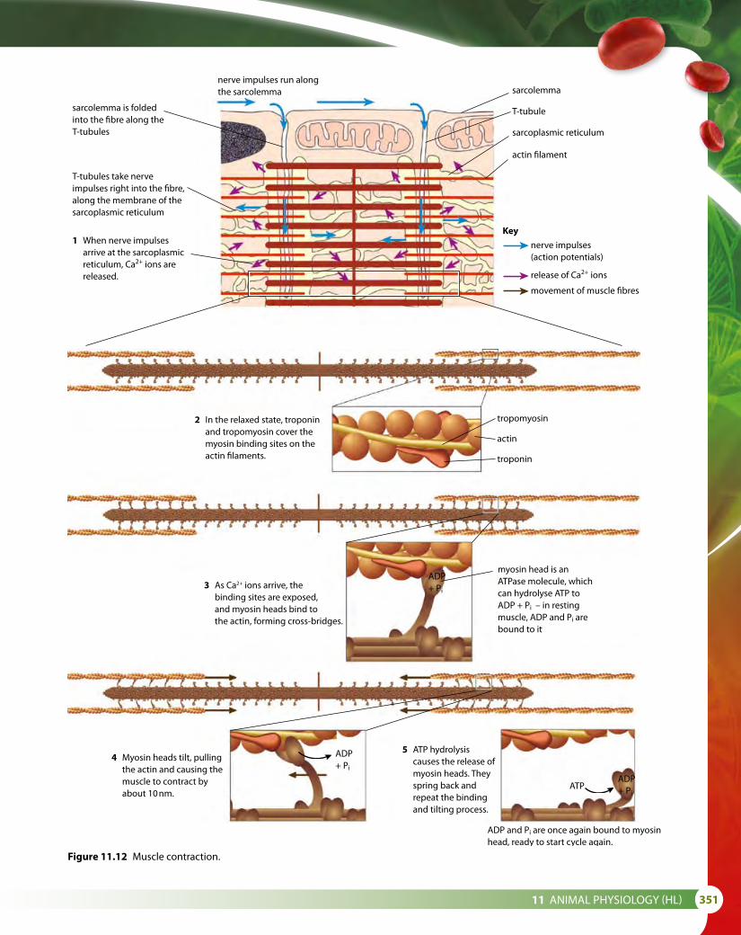
11 ANIMAL PHYSIOLOGY (HL) 351
Figure 11.12 Muscle contraction.
sarcolemma is foldedinto the �bre along theT-tubules
nerve impulses run alongthe sarcolemma
T-tubules take nerveimpulses right into the �bre,along the membrane of thesarcoplasmic reticulum
1 When nerve impulses arrive at the sarcoplasmic reticulum, Ca2+ ions are released.
2 In the relaxed state, troponin and tropomyosin cover the myosin binding sites on the actin �laments.
3 As Ca2+ ions arrive, the binding sites are exposed, and myosin heads bind to the actin, forming cross-bridges.
myosin head is anATPase molecule, whichcan hydrolyse ATP toADP + Pi – in restingmuscle, ADP and Pi arebound to it
ADP+ Pi
ADP+ Pi
ADP+ Pi
ATP
4 Myosin heads tilt, pulling the actin and causing the muscle to contract by about 10 nm.
5 ATP hydrolysis causes the release of myosin heads. They spring back and repeat the binding and tilting process.
ADP and Pi are once again bound to myosinhead, ready to start cycle again.
nerve impulses(action potentials)
release of Ca2+ ions
movement of muscle �bres
sarcolemma
T-tubule
sarcoplasmic reticulum
actin �lament
tropomyosin
actin
troponin
Key

352
4 This causes inorganic phosphate (Pi) to be released and, as each cross-bridge forms, ADP is also released. The myosin heads bend towards the centre of the sarcomere, pulling the actin filaments inward past the myosin filaments, by about 10 nm. This produces a ‘power stroke’.
5 New ATP molecules bind to the myosin heads, breaking the cross-bridges and detaching them from the actin filaments. ATP is used and the myosin heads return to the start position. If the muscle receives further stimulation, the process is repeated and the myosin heads attach further along the actin filaments.
Although the actin and myosin filaments do not change in length when a muscle contracts, the appearance of the banding patterns in the sarcomere is changed. The light bands become reduced, and as the overall length of the sarcomere decreases the dark bands take up a greater proportion of the length (Figure 11.13).
uncontractedmuscle
contractedmuscle
sarcomere
light band dark band
lightband
darkband
in uncontracted muscle, the dark bandsand light bands are about the same width
in contracted muscle, the darkbands are relatively much widerthan the light bands
sarcomere
Z line
light band light banddark band
Z line
Figure 11.13 a When muscle contracts, the interleaved fibres slide inward, past each other. This makes the light bands appear narrower, but the dark bands remain the same width. b Coloured electron micrograph of a longitudinal section through striated muscle. (× 16 300)
Rigor mortis
Rigor mortis is a partial contraction of muscles that occurs after a person has died. The muscles become stiff and locked in position. It is caused because ATP production stops and myosin heads are unable to detach from actin filaments. It lasts for about 24 hours, at which time decomposition begins to break down the muscle tissues.
a
b

11 ANIMAL PHYSIOLOGY (HL) 353
Nature of scienceScientific advance follows technical innovation – using radioactive isotopes to track ion movementsIn the 1940s, Alan Hodgkin, Andrew Huxley and Bernard Katz first explained action potentials and resting potentials in terms of the movement of potassium, sodium and chloride ions across nerve cell membranes.
It was not until radioactive ions were used to track movements of both potassium ions across nerve axons and calcium ions in muscles in the mid-1950s that Hodgkin and Huxley were able to explain the existence of ion channels. In the years that followed, the role of Ca2+ ions in muscle contraction has been extensively studied and has been shown to regulate the initiation of contraction. Calcium-dependent mechanisms in the membrane, such as ion channels, pumps and enzymes, have been studied, as well as the role of calcium ions in activating the enzymes that enable myosin to interact with actin and induce contractions. Our knowledge of these processes is largely due to the use of radioactively labelled calcium ions.
5 Explain why muscles occur in antagonistic pairs.6 Outline the functions of cartilage and synovial fluid in the elbow
joint.7 Compare the movement of a hinge joint and a ball-and-socket joint.8 Explain how actin and myosin filaments produce the striped
appearance of skeletal muscle.9 Describe the role of ATP in muscle contraction.
11.3 The kidney and osmoregulation
Learning objectives
You should understand that:
• Animals are either osmoregulators or osmoconformers.
• The kidney and the Malpighian tubule system in insects remove nitrogenous waste and carry out osmoregulation.
• Blood composition in the renal artery is different from that in the renal vein.
• The structure of the glomerulus and Bowman’s capsule enable ultrafiltration to take place.
• Selective reabsorption of useful substances occurs by active transport in the proximal convoluted tubule.
• The loop of Henle maintains hypertonic conditions in the medulla of the kidney.
• Antidiuretic hormone (ADH) controls the reabsorption of water in the collecting duct.
• There is a positive correlation between the length of the loop of Henle and the need for water conservation in animals.
• The type of nitrogenous waste an animal species produces depends on its habitat and its evolutionary history.
Test yourself?

354
OsmoregulationOsmoregulation is the control of the water potential of body fluids to maintain a constant internal environment in the blood, tissue fluid and cytoplasm. This is essential to ensure that all cell processes occur effectively. Osmoregulation is achieved by regulating the water and salt balance of body fluids.
Animals may either be ‘osmoregulators’, which means they are able to osmoregulate in this way, or ‘osmoconformers’, which means they cannot. Most marine invertebrates are osmoconformers. For example, jellyfish and echinoderms have body fluids with the same solute concentration as sea water. Fish, on the other hand, are osmosregulators – although marine and freshwater fish have different problems in controlling the water potential of their body fluids, and different strategies to overcome them.
A freshwater fish placed in sea water will die because the salt concentration of sea water is approximately twice as high as that of its blood. This means that the fish will lose water by osmosis via its gills and mouth, which are permeable to water. The same fish living in freshwater, has the opposite problem – water will enter its body by osmosis. To overcome this, freshwater fish have very efficient kidneys that remove water and reabsorb vital salts. Their gills are also able to absorb sodium and chloride ions from the water against their concentration gradient.
In the sea, a marine fish has an internal solute concentration lower than that of sea water. This means that water will tend to leave its body by osmosis and so it must conserve water and get rid of salts. Marine fish have secretory cells in their gills that expel chloride ions, and they excrete nitrogenous waste in the form of urea, which uses only a little water as it is removed.
On land, animals tend to lose water by evaporation from skin and any other surfaces, such as the mouth and nose, which are open to the atmosphere. Different animals use different methods to conserve water. Some, such as reptiles, have a waterproof skin, insects have a waxy, waterproof cuticle and many terrestrial animals, including humans, have complex, efficient kidneys. Many animals behave in a way that ensures they avoid the hottest part of the day by burrowing, or by being active at night, so that evaporation losses are reduced to a minimum.
The kidneyMany animals carry out osmoregulation – that is, they control the solute concentration of their blood and tissue fluid – by filtering out unwanted substances at the kidneys, thereby regulating the composition and volume of urine produced. Mammalian kidneys are among the most complex, but fish and birds also have an efficient filtering system in their kidneys. Insects, too, remove excess water and metabolic waste in simple structures known as Malpighian tubules.
The many metabolic processes occurring in mammalian cells result in waste substances that must be removed. For example, urea is produced as a waste product from the metabolism of amino acids. Waste products
Marine birds and marine reptiles, such as the turtle, have to get rid of excess salt. Turtles have salt-secreting glands near their eyes and are sometimes said to cry salt tears. Marine iguanas on the Galapagos Islands snort out the salty spray from their salt glands, which runs into their nose. Marine birds such as penguins and cormorants also lose salty liquid through their nasal passages making them look as though they have a runny nose.

11 ANIMAL PHYSIOLOGY (HL) 355
such as this are carried away from cells by the bloodstream, but they must be continuously removed from the blood so that they do not reach toxic levels. One of the main functions of the kidneys is to act as filters, removing waste molecules from the blood passing through them. This process is called excretion.
Structure of the kidneyIn humans, the kidneys are situated in the lower back, one on either side of the spine, as shown in Figure 11.14. Each receives a blood supply from a renal artery, which is a branch of the main aorta. After filtration, blood leaves the kidney via a renal vein that joins the vena cava. Because of the processes occurring in the kidney, the composition of the blood in the renal vein is quite different from that in the renal artery. Urea, water content and salt levels are adjusted by the kidney so that they are at the correct levels as blood leaves the kidney, but glucose, protein and the cellular content of blood remain unchanged. Figure 11.15 shows a kidney in longitudinal section. Three regions are visible – the outer cortex, the central medulla and the inner renal pelvis. Urine produced by the kidney collects in the renal pelvis and is carried down to the bladder in the ureter.
Each kidney is made up of more than one million tiny structures called nephrons. These are the functional units of the kidney, selectively filtering and reabsorbing substances from the blood. Figure 11.16 shows the structure of a nephron, which consists of a filtering unit (a complex of capillaries called a glomerulus surrounded by a Bowman’s capsule) together with a tube that extends from the filtering unit to the renal pelvis. This tube is divided into four regions – the proximal convoluted tubule, the loop of Henle, the distal convoluted tubule and finally a collecting duct. Each of these regions has a specific role to play in urine formation.
Excretion the removal from the body of the waste products of metabolic pathwaysOsmoregulation control of the water potential of body fluids by the regulation of water and salt content
vena cava
aorta
renal artery
renal vein
kidney
ureter
bladder
urethraureter
pelvis
branch ofrenal artery
branch ofrenal vein
medulla
cortex
capsule
Figure 11.14 Location of the kidneys in the human body. Figure 11.15 Longitudinal section of a human kidney.
Kidney functions at a glance
• excretion of waste products, water-soluble toxic substances and drugs
• regulation of the water and salt content of the body
• retention of substances vital to the body such as protein and glucose
• maintenance of pH balance
• endocrine functions

356
Bowman’s (renal)capsule
proximal convolutedtubule
e�erentarteriole
Bowman’s(renal)capsule
proximalconvolutedtubule
distalconvolutedtubule
collectingduct
pelvis
ascendinglimb of loopof Henle
descendinglimb of loopof Henle
cortex
medulla
a�erentarteriole
glomerulus
from renalartery
distal convolutedtubule
loop of Henle
collecting duct
cortex
medulla
pelvis
e�erentarteriole
a�erentarteriole
glomerulus
fromrenalartery
torenalvein
ureter
Position of a nephron
Blood supply associated with a nephron
Structure of a nephron
Figure 11.16 The location, structure and blood supply of a nephron.

11 ANIMAL PHYSIOLOGY (HL) 357
How the kidney worksThe kidney’s complex structure allows it to carry out its functions with amazing efficiency. The process begins as blood from the renal artery reaches each glomerulus.
Ultrafiltration and the glomerulusUltrafiltration occurs in the glomerulus, as various small molecules leave the blood.
Because the incoming afferent arteriole (a branch of the renal artery) has a wider diameter than the outgoing efferent arteriole, blood pressure in the glomerulus capillaries is very high – so high, in fact, that about 20% of the blood plasma leaves the capillaries in the glomerulus and passes into the Bowman’s capsule.
The blood plasma passes through three layers – the wall of the capillary in the glomerulus, the basement membrane (which acts as a molecular filter) and finally the epithelium of the Bowman’s capsule. It leaves the capillaries through small pores or fenestrations, which the high blood pressure causes to open in the capillary walls. The fenestrations allow all molecules to pass through easily, but filtration occurs at the basement membrane, which is made of a glycoprotein. Only molecules with a molecular mass smaller than 68 000 are able to pass through. So water, salts, glucose, amino acids and small proteins can all pass through the basement membrane, but it effectively prevents blood cells and large molecules such as plasma proteins from leaving the blood (Figure 11.17).
Blood supply to the kidney is normally about 20% of the heart’s output. Approximately 99% of this blood flow goes to the cortex.
pressure gradient
The arrows show how the net e�ect of higher pressure in the capillary and lower solute concentrationin the renal capsule is the movement of �uid out of the capillary and into the lumen of the capsule.
red blood cell blood plasma pore or fenestration incapillary endothelium
basementmembrane(molecular �lter)
podocyte – modi�edcell of Bowman’scapsule wall
glomerular �ltratein lumen ofBowman’s capsule
solute concentration gradient
Figure 11.17 Detail of the Bowman’s capsule showing the basement membrane, fenestrated capillary and podocytes. Podocytes are supportive, foot-shaped cells of the capsule wall that form a network of slits. Filtrate passes through these slits, and into the capsule.
Exam tipRemember the Afferent vessel Approaches the glomerulus but the Efferent vessel Exits it.

358
The blood plasma that has passed through the basement membrane is now known as filtrate. It now passes through the epithelium of the Bowman’s capsule into the nephron, and enters the proximal convoluted tubule. Blood cells and large molecules remain in the blood in the glomerulus capillaries and flow on into the efferent arteriole.
Reabsorption in the proximal convoluted tubuleAlong with the unwanted molecules, many useful substances (water, glucose and ions that the body needs) enter the Bowman’s capsule during ultrafiltration. These must be reabsorbed into the bloodstream. Between 80% and 90% of the filtrate is reabsorbed in the proximal convoluted tubule of the nephron (Figure 11.18). The wall of the tubule is a single layer of cells and each one has a border of microvilli to increase its surface area. The cells have many mitochondria fuelling active transport through membrane pumps that selectively reabsorb ions and glucose from the tubular fluid. All the glucose in the filtrate is actively reabsorbed together with almost 80% of sodium (Na+), potassium (K+), magnesium (Mg2+) and calcium (Ca2+) ions. Chloride ions (Cl–) are absorbed passively and water follows by osmosis as the solute concentration of the cells rises due to the active uptake of ions and glucose.
The remaining filtrate now moves into the loop of Henle.
The loop of HenleThe filtrate that enters the loop of Henle still contains a good deal of the water that was filtered from the blood. The wall of the descending limb of the loop is permeable to water but relatively impermeable to salts, whereas the ascending limb is impermeable to water, but allows salt to be passed
bloodplasma
3 The blood plasma rapidly removes absorbed Na+ , Cl– , glucose and amino acids. This helps further uptake from the lumen of the tubule.
2 Na+/K+ pump in proximal tubule cell membrane uses ATP made by numerous mitochondria. The membrane is folded to increase surface area. The pump lowers the concentration of Na+ in the cell.
1 Microvilli increase surface area, helping uptake of solutes. Na+ moves into the cell using protein molecules in the membrane, which bring in glucose and amino acids at the same time.
activeKey
passive
endotheliumof capillary
mitochondria
nucleus
ADP +Pi
K+
Na+
ATPNa+
glucoseandaminoacids
glucoseand amino acids
proximal convolutedtubule wall
proximaltubule lumen
Figure 11.18 Reabsorption in the proximal convoluted tubule.

11 ANIMAL PHYSIOLOGY (HL) 359
through its walls – Na+ and Cl– ions move by active transport out of the ascending limb into the tissue fluid of the medulla, creating hypertonic conditions there (a high salt concentration). This means that as the descending limb of the loop of Henle passes down into the medulla, water leaves passively by osmosis and enters the surrounding blood capillaries (Figure 11.19). The hypertonic environment in the medulla of the kidney produced by the loop of Henle is also essential for the fine-tuning of the water content of the blood by the collecting duct at a slightly later stage.
Despite the loss of water from the loop of Henle, the filtrate that enters the next section of the tubule still has relatively high water content. The length of the loop of Henle is different in different species and its length is related to an animal’s need to conserve water. Terrestrial animals, such as camels, that live in dry environments and need to conserve water produce small volumes of very concentrated urine. Relatively speaking, these animals have a longer loop of Henle than species such as otters and beavers, which live in places where dehydration is not a problem. Animals that live in wet environments tend to have very short loops of Henle and excrete dilute urine.
1 Na+ and Cl– are actively transported out of the ascending limb.
2 This raises the concentration of Na+
and Cl– in the tissue �uid.
3 This in turn causes the loss of water from the descending limb.
4 The loss of water concentrates Na+
and Cl– in the descending limb.
5 Na+ and Cl– ions di�use out of this concentrated solution in the lower part of the ascending limb.
wall impermeable to Na+,permeable to water
wall permeable to Na+,impermeable to water
Key
H2O
H2O
Na+
Cl–
H2O
H2O
Na+
Cl–
descending limb ascending limb
12
3
4
5
Figure 11.19 The counter-current mechanism in the loop of Henle builds up a high Na+ ion and Cl– ion concentration in the tissue fluid of the medulla.
The distal convoluted tubule and the collecting ductIons are exchanged between the filtrate and the blood in the distal convoluted tubule. Na+, Cl– and Ca2+ ions are reabsorbed into the blood while H+ and K+ ions may be actively pumped into the tubule.
The last portion of the nephron is the collecting duct where the final adjustment of water is made (Figure 11.20). The permeability of the duct depends on the presence or absence of antidiuretic hormone (ADH). If ADH is present, the duct develops membrane channels called aquaporins
The kidneys secrete several hormones including erythropoietin which stimulates red blood cell production.
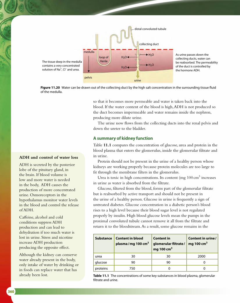
360
so that it becomes more permeable and water is taken back into the blood. If the water content of the blood is high, ADH is not produced so the duct becomes impermeable and water remains inside the nephron, producing more dilute urine.
The urine now flows from the collecting ducts into the renal pelvis and down the ureter to the bladder.
A summary of kidney functionTable 11.1 compares the concentration of glucose, urea and protein in the blood plasma that enters the glomerulus, inside the glomerular filtrate and in urine.
Protein should not be present in the urine of a healthy person whose kidneys are working properly because protein molecules are too large to fit through the membrane filters in the glomerulus.
Urea is toxic in high concentrations. Its content (mg 100 cm3 increases in urine as water is absorbed from the filtrate.
Glucose, filtered from the blood, forms part of the glomerular filtrate but is reabsorbed by active transport and should not be present in the urine of a healthy person. Glucose in urine is frequently a sign of untreated diabetes. Glucose concentration in a diabetic person’s blood rises to a high level because their blood sugar level is not regulated properly by insulin. High blood glucose levels mean the pumps in the proximal convoluted tubule cannot remove it all from the filtrate and return it to the bloodstream. As a result, some glucose remains in the
distal convoluted tubule
collecting duct
H2O
H2O
H2O
H2O
medulla
pelvisurine
loop ofHenle
As urine passes down thecollecting ducts, water canbe reabsorbed. The permeabilityof the duct is controlled bythe hormone ADH.
The tissue deep in the medullacontains a very concentrated solution of Na+, Cl– and urea.
Figure 11.20 Water can be drawn out of the collecting duct by the high salt concentration in the surrounding tissue fluid of the medulla.
ADH and control of water loss
ADH is secreted by the posterior lobe of the pituitary gland, in the brain. If blood volume is low and more water is needed in the body, ADH causes the production of more concentrated urine. Osmoreceptors in the hypothalamus monitor water levels in the blood and control the release of ADH.
Caffeine, alcohol and cold conditions suppress ADH production and can lead to dehydration if too much water is lost in urine. Stress and nicotine increase ADH production producing the opposite effect.
Although the kidney can conserve water already present in the body, only intake of water by drinking or in foods can replace water that has already been lost.
Substance Content in blood
plasma / mg 100 cm3
Content in
glomerular filtrate /
mg 100 cm3
Content in urine /
mg 100 cm3
urea 30 30 2000
glucose 90 90 0
proteins 750 0 0
Table 11.1 The concentrations of some key substances in blood plasma, glomerular filtrate and urine.

11 ANIMAL PHYSIOLOGY (HL) 361
Urine tests
Doctors regularly carry out urine tests to check for the presence of substances that may indicate illness or infection. A urine test can indicate the presence of protein, glucose, nitrites, ketones, bilirubin and blood cells, as well as measuring pH. High protein levels can indicate that the kidneys are inflamed, ketone and glucose in urine are signs of uncontrolled diabetes and leucocytes or nitrite in urine can suggest a bacterial infection. Urine may also contain residues of hormones (for example, HCG used in pregnancy testing) and drugs, such as steroids or alcohol. For this reason, urine tests are used by athletics authorities and the police checking for illegal use of certain substances.
nephron and is lost in the urine because it cannot be reabsorbed in any other regions of the tubule.
Osmoregulation in insectsInsects and some other arthropods have an excretory and osmoregulatory system made up of Malpighian tubules. The tubules are branches extending from the posterior region of the gut of the animal (Figure 11.21). They absorb water, waste and solutes from the insect’s hemolymph (the fluid that fills the body cavity). Each of the slender tubules is blind ending and its walls are made up of a single layer of cells. The cells lining the tubules contain the protein actin to give them structural support, and have microvilli that move material along the tubules.
Urine formation begins as salts and nitrogenous waste move into the tubules either by diffusion (in the case of urea and amino acids) or by active transport (for Na+ and K+). Water follows by osmosis. The mixture that is produced is known as ‘pre-urine’ and it enters the hind gut of the insect and mixes with digested food. Here uric acid precipitates out and ions and water are reabsorbed. Uric acid is mixed with feces and is excreted in a solid form.
Types of nitrogenous wasteThe type of nitrogenous waste an organism produces correlates with the habitat in which it lives and also with its evolutionary history (Table 11.2).
More energy is needed to produce uric acid than the other forms of nitrogenous waste but uric acid has the lowest toxicity and little water is lost as it is excreted.
As animals evolved and moved from an aquatic to a terrestrial environment, the need to conserve water became more and more important. Vertebrates produce either uric acid or urea as these substances require less water to excrete from the body.
hindgut Malpighian tubules
midgut
rectum colon
K+ ions
water
nitrogenouswaste hemolymph
Malpighiantubules
digested food
midgut
pre-urine
hindgut
final urine
feces
pre-urine
Figure 11.21 Diagram to show the Malpighian tubules of a typical insect.

362
DehydrationDehydration occurs if the body loses more fluid than it takes in. If this happens, the balance of salts and sugar in the blood will be upset and metabolic processes will not take place efficiently. Early signs of dehydration are extreme thirst, light-headedness and concentrated, dark urine. Dehydration may occur as a result of excessive sweating – for example, during vigorous exercise on a hot day – or due to the loss of water in cases of diarrhoea and vomiting. Children and infants are particularly vulnerable to this type of dehydration, which may leave them with an imbalance of salt and water in their blood.
During sporting events, participants are advised to drink water or drinks containing glucose and salts (isotonic drinks), which ensure that salt and water balance are maintained (Figure 11.22). In cases of diarrhoea in young children, oral rehydration therapy may be used to replace lost water and salts (sometimes called electrolytes in this context). A typical recipe for oral rehydration therapy is six teaspoons of sugar + half teaspoon of salt + one dm3 of water. This simple remedy can save the life of a child who has suffered dehydration due to diarrhoea.
OverhydrationIn normal circumstances, drinking too much water is unlikely to happen. Most cases in which it has occurred have happened during endurance events such as a marathon, where runners have drunk too much simple water to try to avoid dehydration and upset the natural balance of salts in the blood. Overhydration can be potentially fatal and cause a dangerous disruption of brain function. Long distance runners are usually advised to drink no more than 0.5 dm3 of water per hour and to replenish their levels of salts and glucose by taking isotonic drinks, which contain a similar balance of ingredients to oral rehydration mixtures.
Treating kidney failureIf a person’s kidneys fail, they will be unable to remove toxic substances from their blood or regulate the water potential of their body fluids, which is ultimately a fatal condition. However, they can be kept alive if their blood is filtered by hemodialysis, also known as renal dialysis. The patient is connected to a dialysis machine, which acts as an artificial
Organism Habitat Type of nitrogenous waste Reasons
aquatic invertebrates, most fish aquatic ammonia ammonia is very toxic but is quickly diluted in water
birds, insects, land snails, some reptiles
terrestrial uric acid uric acid has low solubility and is non-toxic
mammals, some fish, amphibians terrestrial and some aquatic
urea urea has low toxicity and is soluble; it can be concentrated for excretion
Table 11.2 Excretory products of different organisms.
Figure 11.22 When water has been lost from the body, it is vital to replace it to maintain the correct balance of water and salts in body fluids.

11 ANIMAL PHYSIOLOGY (HL) 363
kidney. Blood is withdrawn from a vein in the patient’s arm and passed through narrow tubes made of a partially permeable dialysis membrane (Figure 11.23). The tubes are surrounded in dialysis fluid, which contains the components of blood plasma – including sodium ions and glucose – at the correct, healthy levels, but it contains no urea. As blood passes through the tubes, large proteins and blood cells are held inside but small molecules of unwanted substances such as urea and excess salts diffuse through the membrane into the dialysis fluid. Desirable substances are present in the same concentrations on each side of the membrane so there is no net movement. The dialysis fluid is renewed at regular intervals so that a steep concentration gradient of the unwanted substances is maintained across the dialysis membrane. In addition to salts and urea, water can also be drawn out of the blood if necessary by increasing the solute concentration of the dialysis fluid – water will then pass into the fluid by osmosis. Dextran is a solute that is ideally suited to this purpose because it increases the concentration of the fluid but will not pass through the membrane. After blood has passed through the machine, it is returned to the patient’s vein. Dialysis patients may have to be connected to a dialysis machine for up to 8 hours at a time and as often as three times a week. This indicates just how efficient a properly working human kidney is and how important our kidneys are.
Although a person can receive dialysis treatment for many years, a kidney transplant using a donated kidney from another person is the only permanent solution to kidney failure.
dialyser
pressuremonitor
pump
urea glucosedialysis fluidmoves this way
no netmovementof glucose
urea diffusesout of blood
air trapanddetector
fresh dialysissolution in
used dialysissolution out
blood movesthis way
dialysismembrane
Figure 11.23 Dialysis machine.

364
Nature of scienceUsing theories to explain natural phenomena – investigating desert animalsScientists have been interested in the adaptations of desert animals for a long time and have investigated the structure of the kidney and urine content of many species. Different animals adopt different strategies to conserve water. Some adaptations are physiological and others are structural or behavioural. Unexpected adaptations have been found in some species. Some of these are summarised in the Table 11.3.
Animal Adaptations
camel • extracts water from food• produces very concentrated urine (long loop of Henle)• metabolises fat stores from the hump to provide
metabolic water• tissues are adapted to tolerate dehydration
American kangaroo rat
• does not sweat or pant • produces very dry feces and concentrated urine• burrows during the day to avoid water loss from the
lungs
Australian desert frogs
• glomerulus is slow to filter• urine is retained in a bladder for use in the dry season
(frogs may swell up like a ball and have been used as a source of water by aboriginal Australians)
• burrowing behaviour reduces water loss
Andean chinchilla • does not sweat or pant• produces very dry feces and concentrated urine• obtains metabolic water from seeds• hairs in the nostrils cause water from respiration to
condense so that it can be reabsorbed
desert scorpions and insects
• waxy, waterproof covering• some can absorb water from the atmosphere
Table 11.3 Adaptations of some desert species to conserve water.
Ethical issues and kidney donationToday many organs can be transplanted between well-matched
human donors and recipients. Kidneys, corneas, bone marrow and skin are all transplanted regularly for certain medical conditions. Donors usually carry a card or express the wish that they will donate their organs should they die – for example, in an accident. Successful kidney transplants and the drugs needed to prevent rejection of a donated kidney cost less than keeping a person alive using renal dialysis and the quality of life of the recipient is also better. Nevertheless, there is still a shortage of people willing to donate a kidney after their death.
Questions to consider
• Should governments adopt policies that make organ donation compulsory rather than voluntary unless an individual has strong moral objection?

11 ANIMAL PHYSIOLOGY (HL) 365
11.4 Sexual reproduction
SpermatogenesisSpermatogenesis is the production of mature sperm cells (spermatozoa) in the testis. More than 100 million sperm cells are produced each day in a process that takes place in the narrow seminiferous tubules making up each testis (Figures 11.24 and 11.25).
Sperm production and development takes place from the outer part of the seminiferous tubules towards the central lumen, where sperm cells are eventually released. Each tubule is enclosed in a basement membrane beneath which is an outer layer of germinal epithelium cells. These diploid cells (2n) divide regularly by mitosis to produce more diploid cells, which enlarge and are known as primary spermatocytes.
Primary spermatocytes divide by meiosis and their first division produces two haploid (n) cells called secondary spermatocytes. The second division of these two cells results in four spermatids (n).
Developing sperm are attached to Sertoli cells (Figure 11.25), which are also called nurse cells. These large cells assist the differentiation of immature spermatids into spermatozoa and provide nourishment for them.
Spermatozoa that have developed their tails (Figure 11.26) detach from the Sertoli cells and are carried down the lumen of the tubule to the epididymis of the testis.
Hormones and sperm productionSperm production is controlled by three hormones – follicle-stimulating hormone (FSH) and luteinising hormone (LH) from the pituitary gland, and testosterone produced by the testes.
• FSH stimulates meiosis in spermatocytes, to produce haploid cells.
• Testosterone stimulates the maturation of secondary spermatocytes into mature sperm cells.
• LH stimulates the secretion of testosterone by the testis.
10 Define the terms ‘excretion’ and ‘osmoregulation’. 11 Draw and label the functional sections that make up the nephron. 12 Outline the role of the loop of Henle in regulating the content of urine. 13 Outline the role of the efferent and afferent arterioles in ultrafiltration. 14 Explain the role of microvilli in the cells of the proximal convoluted tubule. 15 List the layers through which a glucose molecule passes during ultrafiltration. 16 Outline the role of ADH in controlling the water content of urine. 17 Suggest why a diabetic person may produce urine containing glucose.
Learning objectives
You should understand that:
• Spermatogenesis and oogenesis involve mitosis, cell growth, the two divisions of meiosis and differentiation.
• Spermatogenesis and oogenesis result in the production of different numbers of gametes with different amounts of cytoplasm.
• Fertilisation in animals may be external or internal.
• Mechanisms during fertilisation prevent polyspermy.
• For pregnancy to occur, it is essential that the blastocyst implants in the endometrium.
• HCG stimulates the ovary to secrete progesterone in the early stages of pregnancy.
• The placenta enables mother and fetus to exchange materials. It secretes estrogen and progesterone once it is fully formed.
• Birth is mediated by positive feedback mechanisms involving estrogen and oxytocin.
Test yourself?

366
Epididymis, seminal vesicles and semen productionSperm cells are stored and mature in the epididymis, where they also develop the ability to swim. Sperm cells are released at ejaculation in a nutrient-rich fluid known as semen. Semen is produced by two seminal vesicles and the prostate gland. It is mixed with the sperm cells as they leave the epididymis and move along the vas deferens (sperm duct). Fluid from the seminal vesicles makes up about 70% of semen. It is rich in fructose, which provides energy for the sperm cells to swim, and it also contains protective mucus. The prostate gland produces an alkaline fluid that helps the sperm cells to survive in the acidic conditions of the vagina.
spermgerminal
epithelium
lumen of a seminiferous tubule
interstitial cell
sperm duct
epididymisconnective tissue,blood capillaries andinterstitial cells
seminiferous tubules– site of spermproduction
Testis in section
Group of threeseminiferous tubules
Enlarged portion of the wall of a seminiferoustubule showing stages in sperm development
interstitial cells (cells of Leydig)
seminiferous tubule
germinal epithelium
lumen (central space)
blood capillary
sperm tail in lumen of tubule
head of sperm embedded in Sertoli cell
spermatocyte dividing
primary spermatocyte Sertoli cell
secondary spermatocyte
spermatids
nucleus of Sertoli cell
germinal epithelium
2.5 μm
nucleuscentriole
mitochondria
filament
head middle piece tail
acrosome
Figure 11.25 Structure of the testis.
Figure 11.24 Coloured light micrograph of transverse section of a testis showing seminiferous tubules with interstitial cells between them (× 170).
Figure 11.26 Structure of a human sperm cell. Total length is 60 μm.

11 ANIMAL PHYSIOLOGY (HL) 367
OogenesisOogenesis produces female gametes, the ova. Unlike spermatogenesis, which takes place in an adult male, oogenesis begins in the ovaries of a female when she is still a fetus. Oogonia, the germinal epithelial cells within the ovaries of the female fetus, divide by mitosis to produce more diploid (2n) cells. These enlarge to form primary oocytes, which are also diploid.
Primary oocytes undergo the first stages of meiosis but this stops during prophase I, leaving the primary oocyte surrounded by a layer of follicle cells in a structure known as the primary follicle. Development now ceases but the ovaries of a baby girl contain around 300 000 primary follicles at birth. The remaining stages of oogenesis are shown in Figure 11.27.
At puberty, development of the primary follicles continues. During each menstrual cycle, a few follicles proceed to complete the first division of meiosis, although usually just one will complete its development. Two haploid cells (n) are produced but the cytoplasm divides unequally so that one cell is much larger than the other. The larger cell is known as the secondary oocyte (n) and the smaller cell is the polar body (n). The polar body degenerates and does not develop further.
The secondary oocyte, protected within its follicle, begins meiosis II but stops in prophase II. At the same time, the follicle cells divide and produce a fluid that causes the follicle to swell. At the point of ovulation, the follicle bursts, releasing the secondary oocyte, which floats towards the oviduct (Fallopian tube). Although ovulation is often described as the release of the
To remind yourself of the structure of the male reproductive system, look back at Figure 6.39 in Subtopic 6.6.
germinal epithelium
stroma –contains connectivetissue and blood vessels
degeneratingcorpus luteum
corpus luteum
rupturedovarian follicle
release of secondaryoocyte = ovulation
large �uid-�lledspace
secondaryoocyte
�uid
secondary follicle (growing)
maturing folliclesFSHand LHrequired
primary follicle
primary oocyte
oogonium
follicle cellssurroundingthe oocyte
ovarian follicle causes swellingat surface of ovary
nucleus
Figure 11.27 Stages in the development of one follicle in a human ovary. The arrows show the sequence of events.
Exam tipMake sure you can draw and label diagrams of the male and female reproductive systems.
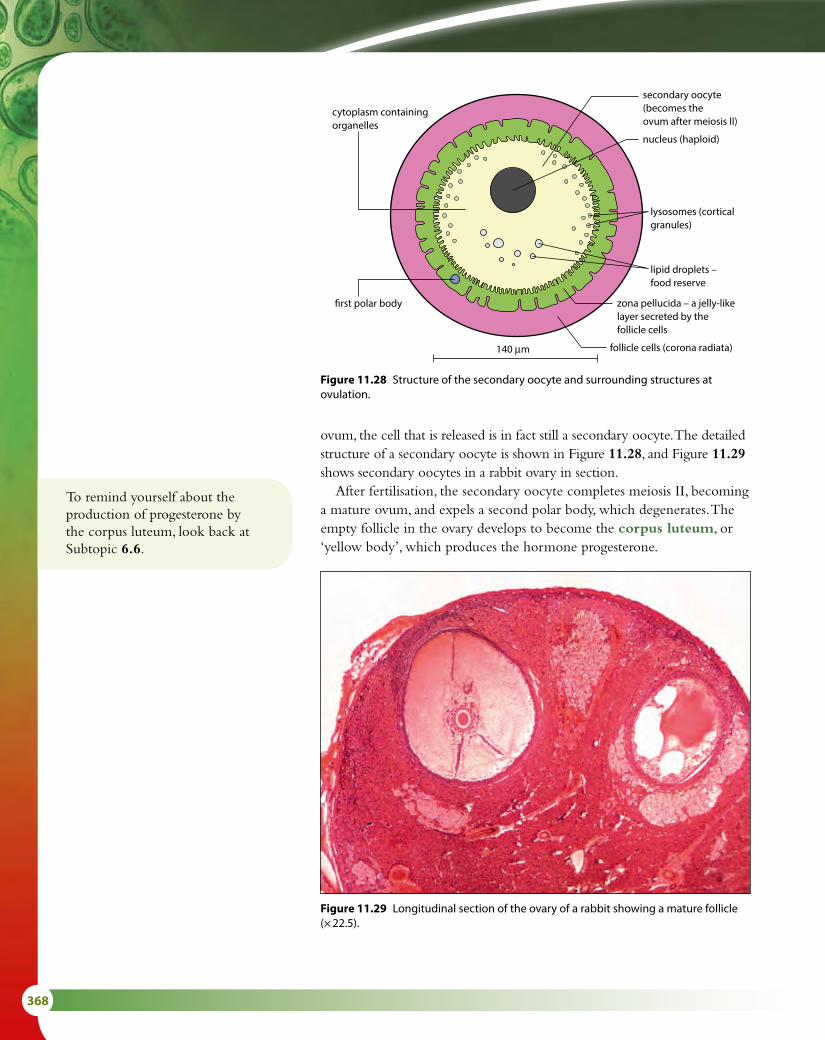
368
ovum, the cell that is released is in fact still a secondary oocyte. The detailed structure of a secondary oocyte is shown in Figure 11.28, and Figure 11.29 shows secondary oocytes in a rabbit ovary in section.
After fertilisation, the secondary oocyte completes meiosis II, becoming a mature ovum, and expels a second polar body, which degenerates. The empty follicle in the ovary develops to become the corpus luteum, or ‘yellow body’, which produces the hormone progesterone.
secondary oocyte(becomes theovum after meiosis II)
lysosomes (cortical granules)
lipid droplets –food reserve
zona pellucida – a jelly-like layer secreted by the follicle cells
140 μm
�rst polar body
cytoplasm containing organelles
nucleus (haploid)
follicle cells (corona radiata)
Figure 11.28 Structure of the secondary oocyte and surrounding structures at ovulation.
Figure 11.29 Longitudinal section of the ovary of a rabbit showing a mature follicle (× 22.5).
To remind yourself about the production of progesterone by the corpus luteum, look back at Subtopic 6.6.

11 ANIMAL PHYSIOLOGY (HL) 369
diploid
diploid
spermatogonia
growth
primary spermatocyte diploid
(stop)
(stop)
primary oocyte(produced in embryo)
haploidsecondary spermatocyte haploid
haploid
polar body
polar body
secondary oocyte(after puberty)
secondary oocyte(one a month)
maturation of oocyte(after ovulation)
one ovum
spermatid
maturation of sperm
haploidmany spermatozoa
Mitosis diploid
oogonia(produced in embryo)
growth
Mitosis
Meiosis Meiosis
Figure 11.30 A comparison of spermatogenesis and oogenesis.
Comparing spermatogenesis and oogenesisThere are a number of similarities and also several differences between the processes of spermatogenesis and oogenesis, as shown in Figure 11.30. Both involve the division of cells in the germinal epithelium by mitosis, and the growth of cells before they undergo meiosis and differentiation. In both cases, meiosis produces haploid gametes. Table 11.4 summarises the differences and similarities in the two processes.
Oogenesis Spermatogenesis
Similarities both begin with production of cells by mitosis
in both, cells grow before meiosis
in both, two divisions of meiosis produce the haploid gamete
Differences usually only one secondary oocyte is produced per menstrual cycle
millions of sperm cells are produced continuously
only one large gamete is produced per meiosis four small gametes are produced per meiosis
occurs in ovaries, which tend to alternate oocyte production occurs in testes, which both produce sperm cells
early stages occur during fetal development process begins at puberty
ova released at ovulation during the menstrual cycle sperm cells released at ejaculation
ceases at menopause continues throughout an adult male’s life
Table 11.4 Oogenesis and spermatogenesis compared.

370
FertilisationGametes produced during oogenesis and spermatogenesis come together and fuse in the process of fertilisation. Fertilisation may take place either inside or outside the body of the female.
External fertilisation occurs in many aquatic species including invertebrates and fish, and also frogs and toads, which return to the water to breed (Figure 11.31). These animals release both male and female gametes into the water, where fertilisation takes place. To ensure the best chance for gametes to meet, species with external fertilisation often take part in courtship rituals so that spawning (the release of gametes) occurs at the same time. They produce large numbers of gametes because many will be wasted in the water.
Animals including reptiles, birds and mammals have internal fertilisation. Sperm cells are transferred into the female body during copulation (sexual intercourse in humans). Internal fertilisation has two main advantages: it increases the chance that gametes will meet so species with internal fertilisation produce fewer gametes and it means that the fertilised ovum can be enclosed in a protective covering such as a shell. In mammals, the embryo remains inside the female’s body where it develops until birth.
Figure 11.31 Female frogs lay their ova in water where they are fertilised by sperm cells from the male.
Human fertilisationFertilisation usually occurs in one of the oviducts and is the moment when one sperm cell fuses with the secondary oocyte to form a zygote. The sequence of events is summarised in Figure 11.32.
During sexual intercourse, millions of sperm cells are ejaculated into the vagina and some of them make their way through the cervix and uterus towards the oviducts. Only a very small number of the ejaculated

11 ANIMAL PHYSIOLOGY (HL) 371
sperm will complete the journey, which is a considerable distance for the tiny cells. As sperm cells approach the zona pellucida, they go through a process known as the acrosome reaction. The contents of their acrosomes, which include many enzymes, are released to penetrate the outer layers of follicle cells covering the secondary oocyte and allow the sperm cells through. For fertilisation to be successful, many sperm cells must be present to release the contents of their acrosomes, but only one sperm cell will eventually break through and reach the plasma membrane, where the membrane of its empty acrosome fuses with it.
After fusion has occurred, changes known as the cortical reaction take place in the membrane of the oocyte, which modify the zona pellucida and also prevent polyspermy (the entry of more than one sperm cell nucleus). Cortical granules, the enzyme-containing vesicles found just inside the oocyte, fuse with the plasma membrane in wavelike fashion, away from the point of fusion of the sperm, and release their contents. Some of the enzymes digest away the sperm cell receptor proteins on the oocyte plasma membrane so that sperm cells are no longer able to attach and fuse.
nucleus of secondary oocyte or egg
secondary oocyteor egg
zona pellucida
second polar body
follicle cells
1 Many sperm cells arrive at the secondary oocyte.
2 Sperm pass through the follicle cells.
3 Sperm reach the zona pellucida and bind to it.
4 The acrosome reaction occurs. Enzymes from inside the acrosome are released and proteases digest a pathway through the zona pellucida.
5 The membranes of the sperm and oocyte fuse, triggering the cortical reaction and the sperm nucleus enters.
6 Cortical granules fuse with the plasma membrane and release their contents. Enzymes cause the zona pellucida to harden so that no further sperm can enter.
7 The secondary oocyte completes meiosis II and a second polar body is expelled.
8 When the sperm and ovum nuclei have merged, the ovum is fertilised and known as a zygote.
sperm bindingreceptor
acrosome
eggmembrane
corticalgranule
perivitellinespace
eggcytoplasm
spermnucleus
first polar bodymay be present
Figure 11.32 The stages of fertilisation.
To remind yourself of the structure of the female reproductive system, look back at Figure 6.40 in Subtopic 6.6.

372
PregnancyApproximately 24 hours after fertilisation, the zygote begins to divide by mitosis. Mitosis continues and, after about five days of division, produces ball of around 100 cells known as a blastocyst, as shown in Figure 11.33. As these divisions are occurring, the ball of cells is moved down the oviduct towards the uterus. After about seven days, it reaches the uterus and settles in the endometrium lining, where it implants itself and continues to divide and develop into an embryo.
Once the blastocyst has become established in the endometrium, it begins to secrete the hormone human chorionic gonadotrophin (HCG). HCG travels in the bloodstream to the ovary, where its role is to maintain the corpus luteum, the mass of cells that developed from the empty follicle. The corpus luteum produces progesterone and estrogen, which – in a non-pregnant woman – maintain the endometrium until the end of the menstrual cycle, when the corpus luteum degenerates. During pregnancy, it is important that the lining remains in place. HCG stimulates the corpus luteum so that it grows and continues to produce its hormones for the first three months (trimester) of pregnancy. Thereafter, the placenta is fully formed and produces placental progesterone and estrogen, so the corpus luteum degenerates.
The embryo grows and develops. After about one month, it is only 5 mm long but has a beating heart and the beginnings of a nervous system. From two months onwards, it is known as a fetus. The fetus at this stage is 30–40 mm long and has recognisable limbs with developing bones. The uterus lining provides nourishment for the early embryo but the placenta soon forms from the endometrium and fetal membranes and by about 12 weeks it is fully functioning. The fetus is connected to the placenta by the umbilical cord and is surrounded by a fluid-filled
HCG is excreted in the urine of a pregnant woman and it is this hormone that is detected in a pregnancy test with the use of monoclonal antibodies (Subtopic 11.1).
2-cell stage
oviduct
fertilisedegg cell
ovulationoccurredhere
egg cellsurrounded byfollicle cells
developingfollicle
blastocystmay beimplantedhere
inner cellmass
blastocyst
fluid-filledmass
corpusluteum
ovary
8-cell stage ball of cells
Figure 11.33 The blastocyst consists of an outer layer of cells enclosing an inner cell mass and a fluid-filled space. The outer layer forms part of the placenta and the inner cell mass develops to become the body of the embryo.

11 ANIMAL PHYSIOLOGY (HL) 373
sac called the amnion, which contains amniotic fluid. The fetus is supported in this fluid throughout its development and is protected by it from bumps and knocks, as the fluid is an effective shock absorber. Amniotic fluid also enables the growing fetus to move and develop its muscles and skeleton.
The placentaThe developing fetus depends on its mother for all its nutrients and oxygen and for the disposal of its waste carbon dioxide and urea. The placenta allows these materials to be exchanged between the mother and the fetus and also acts as an endocrine gland, producing estrogen, progesterone and other hormones that maintain the pregnancy.
The placenta is a disc-shaped structure, about 180 mm in diameter and weighing about 1 kg when it is fully developed. It is made up of the maternal endometrium and small projections, or villi, from the outer layers of the chorion, which surrounds the embryo. These chorionic villi, which are rich in capillaries, grow out into the endometrium to produce a very large surface area for the exchange of gases and other materials. Fetal blood remains inside these capillaries, which penetrate the endometrium tissue until they are surrounded by maternal blood flowing into blood sinuses (spaces) around them. In this way, the mother’s blood is brought as close as possible to the fetal blood to allow for efficient diffusion without the two ever mixing. These features are shown in Figure 11.34.
Exchange of materialsFetal blood is carried to the placenta in two umbilical arteries, which divide to form capillaries in the villi. Nutrients and oxygen from the mother’s blood diffuse into the fetal capillaries and are carried back to the fetus in a single umbilical vein. Waste products and carbon dioxide are carried to the placenta in the two umbilical arteries and diffuse into the mother’s blood.
Many materials pass to the fetus from its mother. Some of these – such as drugs (both prescription and illegal), nicotine and alcohol – have the potential to seriously harm the fetus, which is why pregnant women are encouraged not to smoke or drink alcohol during pregnancy and to be careful with any medicines they may take.
Table 11.5 lists some of the main substances that are exchanged between mother and fetus.
Substances that pass from mother to fetus include Substances that pass from fetus to mother include
• oxygen• nutrients including glucose, amino acids, vitamins, minerals• water• hormones• alcohol, nicotine and other drugs• some viruses, including rubella and sometimes HIV
• carbon dioxide• urea• water• some hormones, such as HCG
Table 11.5 Substances exchanged between mother and fetus.

374
blood-filled space
chorion
endometrium
muscle ofuterus wall
fetal capillaries
chorionic villi
mother’svein
mother’sartery
fetal vein
fetal artery
muscle of uterus wall
endometrium
uteruswall
chorionic villi – manyfinger-like projectionsinto the uterus wall –they contain the fetalcapillaries
blood-filled spaceformed byenlargement ofmother’s bloodvessels – containsmother’s blood
blood to mother’sheart
blood frommother’s heart
chorion – developsfrom fetus
fetal vein carryingoxygenated bloodfrom placenta toheart of fetus
umbilical cord
two fetal arteries carryingdeoxygenated blood fromfetal heart to placenta
oxygenated blood
deoxygenated blood
Fetus in the uterus, showing placenta and umbilical cord Placenta, showing chorionic villi and fetal and maternal blood supplies
Detail of chorionic villi showing capillaries
12-weekfetus
cervix
amnioticfluid amnion placenta
endometrium
muscle of uterus wall
umbilical cord
Figure 11.34 The placenta.

11 ANIMAL PHYSIOLOGY (HL) 375
Hormonal changes and childbirthFor the first 12 weeks of pregnancy, the corpus luteum produces progesterone to maintain the uterus lining (endometrium). After this, the placenta takes over and produces progesterone and estrogen, which suppress the menstrual cycle and promote the growth of breast tissue for lactation (milk production).
After about 9 months, as the end of pregnancy approaches, the levels of progesterone and estrogen produced by the placenta fall (Figure 11.35) and this signals the onset of the uterine contractions known as labour. At this time, the endometrium secretes a group of hormones known as prostaglandins, which initiate the contraction of the uterus. Stretch receptors in the cervix then stimulate the hypothalamus, which triggers the release of the hormone oxytocin, secreted by the posterior lobe of the pituitary gland in the brain. Oxytocin stimulates the uterus muscles to continue their contractions. At first, the contractions are mild and infrequent but oxytocin is a hormone that is controlled by positive feedback. A small contraction of the uterus muscle stimulates the release of further oxytocin, which in turn stimulates more and stronger contractions. As the uterus contracts, the cervix widens and the amniotic sac breaks, releasing the amniotic fluid. Contractions continue for several hours and the baby is pushed through the cervix and out of the mother’s body down the vagina. Gentle contractions continue until the placenta, now known as the afterbirth, is also expelled from the uterus.
After birth, blood levels of the hormone prolactin, from the anterior pituitary gland, increase. This hormone stimulates milk production by the mammary glands. As a baby suckles, prolactin secretion is maintained and oxytocin is also released from the posterior pituitary gland. Oxytocin causes milk to be released from milk ducts.
Prenatal testing – amniocentesis and CVS
Amniocentesis is one of a number of techniques used in prenatal testing to check human fetuses for abnormalities. A fine needle is inserted through the mother’s abdomen into the amniotic sac and a small sample of amniotic fluid is taken. The fluid contains fetal cells, which can be cultured for 3–4 weeks until the cells divide and chromosomes become visible. The chromosomes are stained to produce a karyogram (Subtopic 3.3), which can be checked for mutations.
An alternative procedure, which can be done earlier in the pregnancy, is chorionic villus sampling (CVS). In this case, a sample of cells is taken for examination from the chorionic villi, via the cervix. More fetal cells are obtained in this way and the results are produced more quickly. However, CVS does have a greater risk of inducing a miscarriage than amniocentesis.

376
Nature of scienceAssessing risk in science – effects of oral contraceptive residuesIn recent years, some scientists have raised concerns about the apparent decline in human male fertility. This has been attributed in part to the excretion of residues from oral contraceptives into drinking water supplies. There is some evidence that synthetic estrogens such as estradiol (or EE2), used in contraceptive pills and patches, can affect the ‘maleness’ of fish in rivers and streams – intuitively, some people fear its presence in drinking water may have similar effects on humans.
Understanding the possible effects of such compounds in the water supply is clearly important and further study into their effects is ongoing. But it is also important to note that the source of these estrogenic compounds is not only oral contraceptives but also veterinary contraceptives and residues from other industrial processes. EE2 is part of a larger group of chemicals known as endocrine disrupters, which alter hormonal balance in humans and other animals. When these chemicals were first discovered, risks to human fertility were unknown and could not be adequately assessed. It is important now to weigh such risks against the benefits that the use of oral contraceptives has brought to the human population as a whole.
Con
cent
ratio
n of
hor
mon
e in
the
blo
od
0 4 8 12 16 20Weeks of pregnancy
HCG
estrogen
prolactinprogesterone
birth
ovulation
fertilisation
Weeks after birth24 28 32 36 40 4 8 12 16 20 24
Figure 11.35 Changes in the levels of hormones during pregnancy and after the birth.
Is intuition a reliable way of knowing?Some people intuitively feel that if synthetic estogens are
harming fish, they will also be harmful to humans. Others are sceptical of intuition because it means drawing a conclusion without facts to back it up.
Questions to consider
• Why is it difficult to trust intuition to provide reliable knowledge?
• Can the reasoning behind intuitive feelings ever be rationalised?

11 ANIMAL PHYSIOLOGY (HL) 377
Exam-style questions
1 Which of the following statements about defence is correct?
A Passive immunity occurs as a result of the body being challenged by antigens.B Active immunity occurs as a result of the body being challenged by antigens.C Monoclonal antibodies are produced by fusing a hybridoma cell with a myeloma cell.D As a result of vaccination, the primary response of the body will be greater when it is
invaded by an antigen. [1]
2 Which of the following statements about muscles and movement is correct?
A During muscle contraction, the distance between Z lines decreases, and the dark bands get wider but the light bands get narrower.
B Bones are held together at a joint by tendons, which also protect the joint.C Synovial fluid, secreted by the cartilage, lubricates the joint to prevent friction.D The elbow is an example of a ball-and-socket joint. [1]
3 Which of the following statements about the kidney is correct?
A Osmoregulation is the maintenance of the correct concentration of water in the urine.B Ultrafiltration occurs in the Bowman’s capsules situated in the medulla region of the kidney.C Glucose is sometimes present in the urine of people with diabetes because the lack of insulin
prevents its reabsorption in the loop of Henle.D Human urine can become more concentrated than blood plasma as a result of water reabsorption
due to the release of ADH from the pituitary gland. [1]
4 Which of the following statements about reproduction is correct?
A The hormones LH and FSH from the pituitary gland and testosterone from the Sertoli cells are involved in spermatogenesis.
B During oogenesis, polar bodies are formed by meiosis in the follicle cells of the germinal epithelial layer of the ovary.
C The role of the hormone HCG early in pregnancy is to prevent further ovulation by blocking the production of estrogen.
D During the process of fertilisation, substances released from the cortical granules result in the formation of the fertilisation membrane. [1]
18 State the role of testosterone in spermatogenesis. 19 Explain why unequal divisions of cytoplasm are necessary in
oogenesis. 20 List three differences between spermatogenesis and oogenesis. 21 Describe the role of the amnion in the development of the fetus. 22 Describe the role of oxytocin at birth.
Test yourself?

378
5 Explain antibody production. [5]
6 Outline how the fluid in the proximal convoluted tubule is produced by the process of ultrafiltration. [4]
7 Explain how the cells of the proximal convoluted tubule are adapted to carry out selective reabsorption. [3]
8 Outline how muscle contraction is brought about, starting with the arrival of a nerve impulse at the muscle. [5]
9 Outline the process of birth, including hormonal control. [5]
10 The graph shown below shows data on a kidney patient who received dialysis as a result of kidney failure. Dialysis began on 18th October and was needed for two weeks.
020 22 24 26 28 30 1 3 5
October
Bloo
d ur
ea /
g dm
−3
November7 9 11
0.5
1.0
1.5
2.0
2.5
3.0
0 020 22 24 26 28
urinaryurea
urinevolume
30 1 3 5October
Urin
ary
urea
/ g
per
24
hour
s
November7 9 11
0.5
1.0
1.5
2.0
2.5
3.0
0.5
1.0
1.5
2.0
2.5
3.0
3.5
4.0
a State how many times the patient received dialysis. [1]
b Explain your reasoning. [1]
c Identify the date when the patient began to produce urine again. [1]
d The body mass of the patient fell by 6 kg between 30th October and 11th November. Outline two reasons for this change. [2]

11 ANIMAL PHYSIOLOGY (HL) 379
11 An investigation was carried out into the relationship between the birth weight of babies and the concentrations of the elements zinc and lead in the mother’s placenta. Results are shown in the histogram below. The standard deviation of each value is given at the top of each bar.
0
10
20
30
40
50
60
zinc
4.50−4.994.00−4.493.50−3.99
Birth weight range / kg
Plac
enta
l con
cent
ratio
n / a
rbira
ry u
nits
3.00−3.492.50−2.992.00−2.49
lead
0.154.45
0.92
13.55
0.46
15.10
0.42
14.52
0.38
15.29
0.15
6.39
a i Describe the relationships between levels of lead and zinc and birth weight, as shown in the histograms. [2] ii Suggest reasons for these relationships. [2]
b State the two ranges of birth weight that appear to show the lowest overall variability in placental metal concentration. [2]
c Explain how the efficiency of the human placenta is increased by the presence of villi. [2]
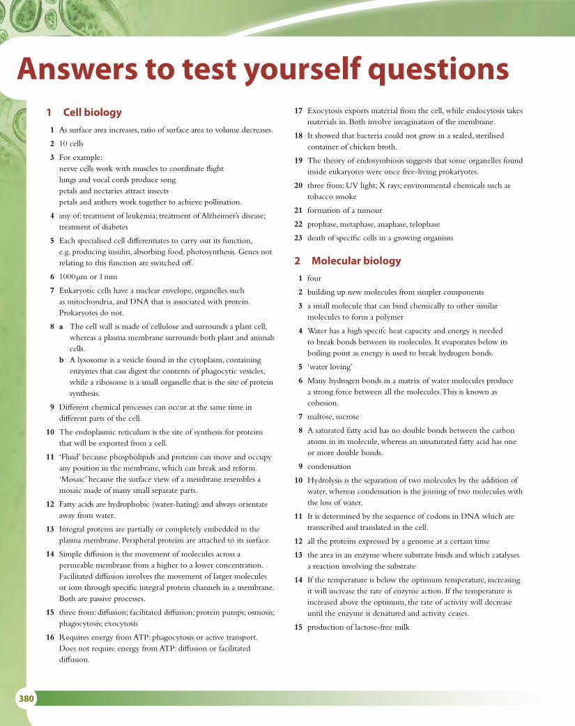
380
Answers to test yourself questions1 Cell biology
1 As surface area increases, ratio of surface area to volume decreases.
2 10 cells
3 For example: nerve cells work with muscles to coordinate flight lungs and vocal cords produce song petals and nectaries attract insects petals and anthers work together to achieve pollination.
4 any of: treatment of leukemia; treatment of Alzheimer’s disease; treatment of diabetes
5 Each specialised cell differentiates to carry out its function, e.g. producing insulin, absorbing food, photosynthesis. Genes not relating to this function are switched off.
6 1000 μm or 1 mm
7 Eukaryotic cells have a nuclear envelope, organelles such as mitochondria, and DNA that is associated with protein. Prokaryotes do not.
8 a The cell wall is made of cellulose and surrounds a plant cell, whereas a plasma membrane surrounds both plant and animals cells.
b A lysosome is a vesicle found in the cytoplasm, containing enzymes that can digest the contents of phagocytic vesicles, while a ribosome is a small organelle that is the site of protein synthesis.
9 Different chemical processes can occur at the same time in different parts of the cell.
10 The endoplasmic reticulum is the site of synthesis for proteins that will be exported from a cell.
11 ‘Fluid’ because phospholipids and proteins can move and occupy any position in the membrane, which can break and reform. ‘Mosaic’ because the surface view of a membrane resembles a mosaic made of many small separate parts.
12 Fatty acids are hydrophobic (water-hating) and always orientate away from water.
13 Integral proteins are partially or completely embedded in the plasma membrane. Peripheral proteins are attached to its surface.
14 Simple diffusion is the movement of molecules across a permeable membrane from a higher to a lower concentration. Facilitated diffusion involves the movement of larger molecules or ions through specific integral protein channels in a membrane. Both are passive processes.
15 three from: diffusion; facilitated diffusion; protein pumps; osmosis; phagocytosis; exocytosis
16 Requires energy from ATP: phagocytosis or active transport. Does not require energy from ATP: diffusion or facilitated diffusion.
17 Exocytosis exports material from the cell, while endocytosis takes materials in. Both involve invagination of the membrane.
18 It showed that bacteria could not grow in a sealed, sterilised container of chicken broth.
19 The theory of endosymbiosis suggests that some organelles found inside eukaryotes were once free-living prokaryotes.
20 three from: UV light; X rays; environmental chemicals such as tobacco smoke
21 formation of a tumour
22 prophase, metaphase, anaphase, telophase
23 death of specific cells in a growing organism
2 Molecular biology
1 four
2 building up new molecules from simpler components
3 a small molecule that can bind chemically to other similar molecules to form a polymer
4 Water has a high specifc heat capacity and energy is needed to break bonds between its molecules. It evaporates below its boiling point as energy is used to break hydrogen bonds.
5 ‘water loving’
6 Many hydrogen bonds in a matrix of water molecules produce a strong force between all the molecules. This is known as cohesion.
7 maltose, sucrose
8 A saturated fatty acid has no double bonds between the carbon atoms in its molecule, whereas an unsaturated fatty acid has one or more double bonds.
9 condensation
10 Hydrolysis is the separation of two molecules by the addition of water, whereas condensation is the joining of two molecules with the loss of water.
11 It is determined by the sequence of codons in DNA which are transcribed and translated in the cell.
12 all the proteins expressed by a genome at a certain time
13 the area in an enzyme where substrate binds and which catalyses a reaction involving the substrate
14 If the temperature is below the optimum temperature, increasing it will increase the rate of enzyme action. If the temperature is increased above the optimum, the rate of activity will decrease until the enzyme is denatured and activity ceases.
15 production of lactose-free milk

ANSWERS TO TEST YOURSELF QUESTIONS 381
5 a diagram or photograph of the chromosomes from an organism
6 a chromosome that does not carry genes determining the sex of an organism
7 a anaphase I b prophase I and II c anaphase II d metaphase II e prophase f anaphase I
8 chromosomes with the same genes but not necessarily the same alleles
9 The number of chromosomes is halved as a result of meiosis.
10 four cells
11 a nucleus that contains three copies of one chromosome instead of two
12 Down syndrome
13 any two of: banding pattern; position of centromere; length
14 diploid number
15 a the alleles possessed by an organism b the characteristics of an organism c an allele that has the same effect on the phenotype whether in
homozygous or heterozygous state d an allele that only has an effect on the phenotype in the
homozygous state e having two identical alleles at a gene locus f having two different alleles at a gene locus
16 a red b red c yellow
17 a R b r c R and r
18
16 two from:
DNA RNA
double stranded single stranded
contains bases A, C, G and T contains bases A, C, G and U
sugar = deoxyribose sugar = ribose
17 phosphate
deoxyribose
base
18 No DNA molecule is ever completely new. Every double helix contains one ‘original’ and one ‘new’ strand.
19 the enzyme involved in linking the new nucleotides into place during replication
20 production of a strand of mRNA from a DNA template
21 specific pairing of base A with base T or base U, and base C with base G
22 in the cytoplasm
23 ATP and lactic acid (lactate)
24 cytoplasm in mitochondria
25 cytoplasm
26 Yeast ferment sugar in flour to produce alcohol, which evaporates, and carbon dioxide, which causes the bread dough to rise.
27 Red light was used for photosynthesis by Spirogyra, which produced oxygen. The bacteria moved towards the oxygen.
28 Light of this wavelength was not used for photosynthesis, so no oxygen was produced.
29 blue or red
30 Growth would be inhibited; it might become thin and weak.
31 All except green light, but mainly red and blue.
32 directly by measuring oxygen production or carbon dioxide release and indirectly by measuring the increase in biomass
3 Genetics
1 438 nucleotides
2 a a heritable factor that controls a specific characteristic b one specific form of a gene, differing from other alleles by
one or a few bases only and occupying the same gene locus as other alleles of the gene
c the whole of the genetic information of an organism d a change in the sequence of bases in a gene
3 the replacement of a base in a gene with a different base
4 DNA in a eukaryote is enclosed in a nuclear membrane. Prokaryotes do not have protein associated with their DNA.
gametes from green parent
G g
gam
etes
from
gr
een
par
ent
GGG
greenGg
green
gGg
greengg
purple
19 a 2 b 2 c 1
20 blood group O
21 colour blindness and hemophilia
22
23 codominant
24 carrier
25 to amplify small quantities of DNA

382
26 their size and charge
27 any two of: paternity testing, forensic examination of crime, scenes, animal pedigree testing
28 the fact that the code is universal
29 plasmids
30 restriction enzyme and DNA ligase
31 mammary gland cell and egg cell
4 Ecology
1 a a group of organisms that can interbreed and produce fertile offspring
b the study of relationships between living organisms and between organisms and their environment
c a group of organisms of the same species which live in the same area at the same time
d a group of populations of different species living and interacting with each other in an area
e a community and its abiotic environment
2 An autotroph makes its own food/organic compounds; a heterotroph takes in organic materials as food.
3 A consumer ingests other organic matter that is living or recently dead; a detritivore is an organism that ingests non-living organic matter; a saprotroph lives on or in non-living organic matter and absorbs digested food.
4 a detritivore b carnivore c herbivore
5 a trophic level 3/secondary consumer b trophic level 4/tertiary consumer
6 the transfer of nutrients and energy
7 a series of interlinking food chains
8 the Sun
9 the position of an organism in a food chain
10 not consumed, heat, excretion
11 2000 J m–2 y–1
12 Energy enters and leaves the ecosystem but nutrients cycle within the ecosystem
13 Peat is produced in wetlands from accumulation of partly decayed vegetation in the absence of oxygen. Coal is produced as a result of compression and fossilisation of organic material over millions of years.
14 any three of: plants; animals; bacteria; fungi
15 photosynthesis
16 any three of: carbon dioxide; water vapour; methane; oxides of nitrogen; fluorocarbons
17 burning fossil fuels in factories and vehicles; rice farming; cattle farming; rainforest destruction; HFCs from refrigerants
18 changing the temperature of the oceans, acidification of the ocean
5 Evolution and biodiversity
1 a cumulative change in the heritable characteristics of a population
2 anatomical features showing similarities in shape (but not necessarily function) in different organisms
3 Individuals with specific characteristics are selected to breed, so that more of the next generation have these useful features than if the parents had not been artificially selected.
4 It ensures that offspring are varied and have different combinations of characteristics from one another and their parents.
5 competition
6 survival of the fittest
7 for example: resistance to antibiotics in bacteria; change in phenotype of the peppered moth
8 domain; kingdom; phylum; class; order; family; genus; species
9 genus and species
10 conifers/Coniferophyta
11 Bryophyta
12 Arthropoda
13 Annelida
14 ‘Large’ is a subjective term and not suitable for use in a key.
15 group of organisms that have evolved from a common ancestor
16 Homologous structures have evolved from a common ancestral form and may have different function, while analogous structures are structurally very different even though they perform a similar function.
17 Cladistics is an objective method of classification; using it, existing classifications can be tested to determine if relationships are correct or whether they should be abandoned in favour of a competing hypothesis.
6 Human physiology
1 Contraction of the bands of circular muscle squeezes the intestine behind food, while longitudinal muscles relax and extend to receive the food.
2 Absorption is the uptake of nutrients into blood or cells, while assimilation is the use of absorbed molecules by the body.
3 Enzymes speed up chemical reactions so that food can be digested as it passes through the intestine.
4 Its microvilli provide a large surface area, it has a rich blood supply for absorption, and the diffusion distance between the intestine and the blood is very small.
5 vena cava, right atrium, atrioventricular valve, right ventricle, semilunar valve, pulmonary artery, lung capillaries, pulmonary vein, left atrium, atrioventricular valve, left ventricle, semilunar valve, aorta

ANSWERS TO TEST YOURSELF QUESTIONS 383
4 because DNA replication can only occur in a 5' to 3' direction
5 to protect the ends of chromosomes from damage during cell division
6 a portion of the primary RNA transcript that codes for part of a polypeptide in eukaryotes (as compared with introns, which are part of the transcript but are not translated to form part of a polypeptide)
7 The sense strand has the same base sequence as mRNA (with thymine instead of uracil). The antisense strand is the one that is transcribed to produce mRNA.
8 The promoter region contains specific DNA sequences that are the initial binding site for RNA polymerase during transcription.
9 External environmental factors such as drugs, chemicals, temperature and light can cause some genes to be turned on or off and influence development, e.g. temperature affects the fur colour of Himalayan rabbits.
10 Transcription is the production of mRNA from DNA, while translation is the production of a polypeptide from mRNA.
11 It is used in the cell; it is not exported for use elsewhere.
8 Metabolism, cell respiration and photosynthesis
1 the amount of energy required to destabilise chemical bonds in a substrate so that a chemical reaction can occur
2 If the end product begins to accumulate because it is not being used, it inhibits an enzyme earlier in the pathway to switch off the assembly line. Usually, the first enzyme in a process is inhibited.
3 gain of electrons; loss of oxygen; gain of hydrogen
4 phosphate from ATP
5 lysis
6 in the matrix of mitochondria
7 on the membranes (cristae) of mitochondria
8 acetyl CoA
9 to provide a large surface area for the enzymes involved in respiration
10 the pH decreases
11 ATP synthase
12 in the stroma of the chloroplast
13 green
14 to boost electrons to a higher energy level
15 oxygen
16 ATP and NADPH+
17 ribulose bisphosphate
6 It sets the pace of the heart beat.
7 Arteries must resist higher blood pressure than veins.
8 The left ventricle pumps blood all round the body, whereas the right ventricle only needs to drive blood the short distance to the lungs, so the left ventricle must be stronger and thicker.
9 They prevent blood flowing back from the ventricles to the atria as the heart relaxes.
10 to supply additional oxygen to working muscles and to carry away the carbon dioxide they produce
11 it stimulates the sinoatrial node to increase heart rate
12 an organism or a virus that causes disease
13 a protein that provokes an immune response
14 Antibiotics interfere with bacterial metabolism. Viruses do not have metabolism of their own.
15 Phagocytes engulf bacteria and cell fragments, whereas lymphocytes secrete antibodies.
16 atmosphere, nasal passages or mouth, trachea, bronchi, bronchioles, alveolus
17 rich blood supply; large surface area; short diffusion distance
18 diaphragm and intercostal muscles (external)
19 Gas exchange is the exchange of oxygen and carbon dioxide between the alveoli and the blood; ventilation is the movement of air into and out of the lungs.
20 Sodium ions are pumped out of the neuron and potassium ions are pumped in.
21 the depolarisation and repolarisation of a nerve cell membrane
22 calcium ions enter the pre-synaptic neuron, vesicles fuse with the pre-synaptic membrane, neurotransmitter is released into synaptic cleft, neurotransmitter binds to receptor on post-synaptic membrane, post-synaptic membrane is depolarised
23 An action potential must jump from node to node along a myelinated axon.
24 neurotransmitter
25 insulin and glucagon
26 leptin acts on receptor cells in the hypothalamus
27 Y chromosome
28 It stimulates the thickening of the endometrium and also inhibits the production of FSH and LH.
7 Nucleic acids
1 the two strands of DNA in the double helix run in opposite directions
2 consists of two twists of DNA wound around eight histone proteins, which are held in place by an additional histone
3 5' to 3'

384
9 Plant biology
1 stomata are found deep in grooves; rolled leaf; thick cuticles
2 mass flow of water carrying the ions, or by facilitated diffusion of ions
3
10 Genetics and evolution
1 During meiosis, non-sister chromatids may touch and break, and the two segments re-join at the corresponding position on the other chromatid.
2 anaphase 2
3 During metaphase 1, bivalents line up on the equator, but the way they line up is random for each chromosome.
4 Ttrr: Tr and tr in a ratio of 1 : 1
HhGg: HG, Hg, hG and hg in a ratio of 1 : 1 : 1 : 1
5 Punnett grid
6 members of the F1 generation
7 In sympatric speciation, a gene pool becomes divided without the population being geographically split. In allopatric speciation, a physical barrier separates a species into two geographically isolated populations.
8 leads to the prevalence of one form from a varied population
9 all the different genes in an interbreeding population at a given time
11 Animal physiology
1 Natural immunity or resistance to a particular pathogen is related to the genetics of the host organism, which differ between species. For a virus to infect a new species, genetic adaptations must occur within the pathogen.
2 Antibody is a substance produced by lymphocytes to counteract an antigen. Antigen is a protein that stimulates an immune response.
3 It has characteristics of both B-cells and cancer cells: it produces antibodies and cells are immortal if kept in culture medium.
4 It stimulates the immune response and the formation of memory cells to produce antibodies very quickly if the person is infected with the real pathogen later on.
5 One member of the pair causes flexing of a joint and the other causes extension of it.
6 Cartilage reduces friction between two bones in a joint and synovial fluid acts as a lubricant.
7 A hinge joint (e.g. the elbow joint) moves in one direction, whereas a ball-and-socket joint can allow movement in a circle.
8 Actin is made up of thin fibres, whereas myosin fibres are thicker. Each type of filament is arranged in bands that alternate in skeletal muscle. The lighter bands of thin myosin alternate with darker bands of thicker myosin, which produces the striped appearance.
9 ATP provides the energy for muscle contraction. ATP molecules bind to myosin heads; the energy from the ATP breaks cross-bridges and detaches the myosin heads from the actin filaments.
10 Excretion is the removal from the body of waste products produced as a result of metabolic activity. Osmoregulation is the maintenance of the correct level of water in the body.
lignified fibres(providesupport)
vascular cambium
cortex
phloem
xylem
vascular bundle
epidermispith
4 Xylem is composed of dead cells whose walls are thickened with lignin, whereas phloem is made of living cells and adjacent companion cells. Both xylem and phloem are found in vascular bundles in the stem and leaves. The xylem carries water and salts up the plant, whereas phloem carries sugars, vitamins and other nutrients both up and down.
5 water and mineral salts
6 Transpiration is increased by an increase in temperature, wind or light intensity and by a decrease in humidity.
7 Sugar enters the phloem in leaves and is followed by water. The pressure gradient created pushes water to other parts of the plant.
8 The presence of auxin on the shaded side of a shoot causes the shoot to bend towards a source of light as cells on the shaded side elongate.
9 Apical meristems occur at the end of a shoot and growth here results in a taller plant.
10 These pumps are special transport proteins in plasma membranes, which allow auxin to move in one direction only.
11 Germination requires activation of enzymes to digest food reserves and initiate respiration. All enzyme-controlled reactions are temperature sensitive and so a suitable temperature (which varies from species to species) is needed for germination.
12 Pollination is the transfer of pollen from anthers to stigma, whereas fertilisation is the fusion of the male and female gamete in the ovary.
13 anthers and filaments (together known as the stamen)
14 Seeds that germinate at some distance from the parent plant do not have to compete with it for water, light or nutrients and so are more likely to survive.

ANSWERS TO TEST YOURSELF QUESTIONS 385
efferentarteriole
renalcapsule
proximalconvolutedtubule
distalconvolutedtubule
collectingduct
pelvis
ascendinglimb of loopof Henle
descendinglimb of loopof Henle
cortex
medulla
afferentarteriole
glomerulus
from renalartery
11 20 Spermatogenesis produces four gametes, begins at puberty and continues throughout a man’s life and produces sperm continuously, whereas oogenesis produces one gamete, occurs from puberty to menopause and produces one gamete each month.
21 The amnion forms a protective, fluid-filled sac around the fetus that protects it from pressure and allows it to move.
22 Oxytocin stimulates the contraction of the muscles in the wall of the uterus. It works by positive feedback so that more hormone is produced as the muscles contract more strongly.
12 The descending limb of the loop of Henle is permeable to sodium and chloride ions, which leave the tubule and are concentrated in the medulla region of the kidney. The high level of salt in the medulla causes water to enter the capillaries in the medulla from the descending limb, increasing the concentration of the solution in the loop of Henle.
13 The afferent arteriole is wider in diameter than the efferent arteriole. This causes a build-up of pressure in the glomerulus that enables ultrafiltration of molecules.
14 Microvilli increase the surface area of the cells in the proximal convoluted tubule, which is the region where useful molecules are reabsorbed into the bloodstream from the kidney tubule. The large surface area ensures this process occurs efficiently.
15 capillary endothelium, basement membrane, cell of Bowman’s capsule wall
16 ADH influences the permeability to water of the cell wall in the collecting duct. If ADH is released, the collecting duct is more permeable and water is taken back into the blood so that more concentrated urine is produced.
17 A diabetic person cannot regulate the amount of glucose in their blood. If blood glucose is very high, not all glucose can be reabsorbed from the kidney tubule and some passes out of the body in the urine.
18 Testosterone stimulates secondary spermatocytes to mature into sperm cells.
19 An ovum must contain sufficient nutrients to provide fuel for the first few divisions after fertilisation. Unequal cell division during oogenesis produces one large cell that contains these nutrients and three small polar bodies that do not become gametes.

386
abiotic factor aspect of the environment that is not living – for example, humidity, temperature, salinity, wind, soil particles
absorption the taking up of a substance by a tissue; for example, the process by which nutrients pass from the lumen of the intestine into the lymph or blood, or by which the intensity of a beam of light energy is reduced as it passes through a leaf
absorption spectrum the range of wavelengths of light that a pigment is able to absorb
acinus berry-shaped termination of an exocrine gland where its secretion is produced
actin globular protein that polymerises to form the thin filaments in muscle myofibrils
action potential a rapid wave of depolarisation at a cell surface causing an impulse in a neuron
activation energy the energy a substrate molecule must achieve before it can change chemically
active immunity immunity due to the production of antibodies by an organism in response to an antigen
active site the region on the surface of an enzyme to which the substrate molecule binds
active transport transport of a substance across a membrane against the concentration gradient, involving a carrier protein and energy expenditure
addiction regular or poorly controlled use of a psychoactive substance, despite adverse physical, psychological or social consequences, often with the development of physiological tolerance and withdrawal symptoms
adhesion a force that attracts water molecules to a surface by hydrogen bonding
aerobic respiration respiration that requires oxygen, and produces carbon dioxide and water from the oxidation of glucose
aggregate a group of distinct organisms, such as bacteria, living together to form a colony that has characteristics not displayed by the individual organisms
aleurone layer layer of specialised cells between the seed coat and endosperm of grass seeds, which produces enzymes that help to digest the food reserves for the developing embryo
algal bloom rapid increase or accumulation in the population of algae in an aquatic ecosystem often due to the run-off of agricultural fertiliser
alimentary canal the gut – a tube that runs from the mouth to the anus of a vertebrate in which food is digested and absorbed
allele an alternative form of a gene found at a specific locus on a chromosome
allele frequency the commonness of occurrence of any particular allele in a population
allergy a disorder of the immune system in which the immune system overreacts to a substance (known as an allergen) which is usually harmless
GlossaryTerms in bold italic refer to keywords from online material.
allopatric speciation separating a species into two geographically isolated groups by a physical barrier so eventually they can no longer interbreed
allosteric regulation of an enzyme by a molecule that does not have the same structure as the substrate, binding at a site away from the active site
allosteric site the region on the surface of an enzyme to which an allosteric effector binds
altruist an organism that performs actions benefitting other individuals rather than itself
alveolus (alveoli) an air sac in the lungsamino acid one of the building blocks of protein; each amino acid
has an amine group –NH2 and an acid carboxyl group –COOH, and the general formula NH2-CHR-COOH, where R is one of 20 side groups
amnion a membrane which surrounds the embryo of reptiles, birds and mammals during their development
amniotic fluid the fluid contained within the amnion in which the embryo is suspended.
amphipathic an unsymmetrical molecule which has one hydrophilic end and one hydrophobic end
amylase an enzyme that digests starchanabolism an anabolic reaction is one in which large molecules
are built up from small onesanaerobic respiration respiration that occurs in the absence
of oxygen in which glucose is broken down to lactic acid or to ethanol and carbon dioxide
analgesic a drug or other treatment that relieves or reduces painanalogous structures structures with similar functions but which
have different evolutionary originsanaphase the stage in cell division in which homologous
chromosomes (in meiosis I) or chromatids (meiosis II and mitosis) separate and go to opposite poles
anesthetic a drug or other treatment that produces a loss of feeling or sensation
anorexia nervosa severe malnutrition brought on by a psychological condition that causes an aversion to food
antagonistic pair a pair of muscles that work in opposition to each other at a joint
anther pollen-bearing structures on the stamens of flowersantibiotic organic compound produced by microorganisms to kill
or inhibit other microorganismsantibody one of millions of blood proteins produced by plasma
cells in response to specific antigens, which are then neutralised or destroyed
anticodon a triplet of bases in tRNA that pair with a complementary triplet (codon) in mRNA
antidiuretic hormone (ADH) hormone secreted by the posterior pituitary gland to control the water permeability of the collecting ducts in the kidney (also called vasopressin)

387GLOSSARY
antigen a substance that stimulates the production of antibodyantigen presentation the process by which a cell takes in a
pathogen, or molecules from it, and holds them in its plasma membrane where they may be encountered by a lymphocyte
antiparallel running in opposite direction; the two polynucleotide strands in a DNA molecule are antiparallel
antisense strand the strand of DNA that is transcribed during transcription
apical meristem the actively growing tissue at the tip of a stem or root
apoptosis the natural destruction of certain cells in a growing organism
aquaporins proteins embedded in a plasma membrane that regulate the movement of water through the membrane
arteriole a small arteryartery a muscular blood vessel that carries blood away from the
heart under high pressureasexual reproduction reproduction that does not involve gametes
or fertilisation assimilation uptake and use of nutrients by cells and tissuesassociation areas parts of the cerebrum that integrate information
from the primary sensory areas and other parts of the brainatherosclerosis hardening and loss of elasticity in the arteriesATP (adenosine triphosphate) a universal energy storage
nucleotide formed in photosynthesis and respiration from ADP and Pi; when it is formed, energy is stored; when it is broken down, energy is released
ATP synthase/synthetase an enzyme that catalyses the production of ATP
atrioventricular node (AVN) small patch of tissue in the right atrium which acts as a pacemaker in the heart
atrioventricular valve valve between the atrium and the ventricleatrium (atria) one of the two upper chambers of the heart that
receive blood from veinsautonomic nervous system (ANS) the part of the nervous system
comprised of the sympathetic and parasympathetic nervous systems, consisting only of motor neurons; it controls involuntary functions (such as activities of the glands and digestive system, and blood flow) without our awareness
autosome any chromosome other than a sex chromosomeautotroph organism able to make its own food from simple
inorganic materials and an energy sourceauxin plant growth substance (also known as indoleacetic acid, IAA)axon a cytoplasmic process that transmits action potentials away
from the cell body of a neuronB-cell a type of lymphocyte involved in the immune response;
B-cells develop into plasma cells, which secrete antibodyball-and-socket joint a joint such as the hip, which permits
movement in more than one planebasal metabolic rate the amount of energy used by the body per
day when at restbasement membrane thin layer of cells which separates
epithelium from underlying tissuebatch culture large-scale culture of microorganisms in a closed
fermenter, to which nothing is added during the fermentation;
cells initially grow at a maximum rate, but as nutrients are used up and waste products accumulate, growth slows and eventually stops, at which time the product is harvested, the fermenter cleaned, and a fermentation is set up
behaviour the manner in which an organisms acts under specified conditions or circumstances, or in relation to other things
binary fission cell division producing two genetically identical daughter cells, as in prokaryote reproduction
binomial the two-part name that gives the genus and the species of an organism in Latin – for example, Homo sapiens
biochemical oxygen demand (BOD) the uptake rate of dissolved oxygen by the biological organisms in a body of water
biofilm colony of bacteria living in a layer on a surface, in a matrix of extracellular polysaccharides (EPS), which is resistant to removal by physical or antimicrobial agents, and which has emergent properties including quorum sensing
biofuel any fuel derived from biomass e.g. bioethanol, biodieselbiogas fuel gas obtained typically by the bacterial decomposition of
animal or plant waste in the absence of oxygen, which produces mainly methane
biogeography the spatial distribution of organisms, species and ecosystems, or the study of these
bioinformatics the use of databases – searchable, up-to-date electronic records of information from experiments, scientific papers and computer analyses of data, such as information about gene function, structure and location
biomagnification the process by which chemical substances accumulate at progressively more concentrated levels at each trophic level in a food chain
biomarker any measurable characteristic that may be used as an indicator of some biological state or condition
biomass the total dry mass of organisms in a given areabiopharming the use of genetic engineering to add genes to
animals or plants so that they produce useful pharmaceuticalsbioreactor a vessel in which microorganisms can be cultured in
order to harvest useful products bioremediation the process by which microorganisms are used
to treat areas of land or sea that have been contaminated by pesticides, oil or solvents, in order to remove the contaminants and restore the environment
biotechnology commercial application of biology, particularly the industrial use of microorganisms, enzymes and genetic engineering
biotic index a measure of the biodiversity, or level of pollution, of a given ecosystem or habitat obtained by surveying the types of organisms present in it
bipolar cells cells in the retina that transmit signals between the photoreceptors (rod and cone cells) and ganglion cells
blastocyst early stage in embryo development – a hollow ball of cells, which implants into the endometrium
Bohr shift the reduction in hemoglobin’s affinity for oxygen caused by an increase in the partial pressure of carbon dioxide, which means the dissociation curve shifts to the right
Bowman’s capsule the cup-shaped part at the beginning of a nephron, into which ultrafiltration takes place (also known as renal capsule)

388
centromere the region where sister chromatids are joined and where the spindle microtubule attaches during cell division
cerebellum part of the hind brain concerned with posture, movement and muscle tone
cerebral cortex a highly folded layer of nerve cell bodies that forms the surface of the cerebrum
cerebral hemispheres (cerebrum) the main part of the human brain, the coordinating system of the nervous system consisting of two hemispheres made of neurons and nerve fibres
channel protein proteins found in a membrane which form a pathway through which specific ions or molecules can pass in and out of a cell
chemiosmosis the passive flow of protons down a concentration gradient from the intermembrane space of mitochondria or from the thylakoid interior of chloroplasts through a protein channel
chemoautotroph an organism that uses energy from chemical reactions to generate ATP and produce organic compounds from inorganic raw materials
chemoheterotroph an organism that uses energy from chemical reactions to generate ATP and obtains organic compounds from other organisms
chemoreceptor sensory cell that responds to pH or the concentration of a chemical such as carbon dioxide
chiasma (chiasmata) point of crossing over between homologous chromosomes in prophase 1 of meiosis
chloride shift process in which bicarbonate (HCO3–) and chloride (Cl–) are exchanged across the membrane of red blood cells
cholesterol a lipid formed in the liver and carried in the blood as lipoprotein; it is a precursor of steroid hormones, and is also found in the plasma membrane of animal cells
chlorophyll the most important photosynthetic pigments of green plants, found in the grana of chloroplasts and responsible for trapping light energy (some bacteria have a chemically different form called bacteriochlorophyll)
chloroplast organelle found in some plant cells that is the site of photosynthesis
chorion outermost membrane that protects mammal embryos; it forms part of the placenta
chromatid one of the two copies of a chromosome after it has replicated and before the centromeres separate at anaphase
chromosome in eukaryotes, a structure consisting of a long thread of DNA and protein that carries the genetic information of the cell; in bacteria, the DNA molecule that contains the genetic information of the cell
circadian rhythm a cycle of activity or physiology which is repeated, usually every 24 hours
cirrhosis disease caused by excessive alcohol consumption, in which liver tissue is replaced by fibrous scar tissue leading to loss of liver function
cisternae space enclosed by the membranes of the endoplasmic reticulum
clade all the organisms both living and fossil descended from a particular common ancestor
cladistics a way of classifying organisms which uses lines of descent rather than physical (phenotypic) similarities
Broca’s area a part of the left cerebral hemisphere involved in speaking and writing
bronchiole a small branch of a bronchusbronchus (bronchi) one of the major tubes which branch from
the tracheabuffering keeping pH at a nearly constant valueCalvin cycle cycle of light-independent reactions in the stroma
of the chloroplast in which carbon dioxide reacts with RuBP, producing GP, TP and regenerating RuBP
cambium a cylinder of cells in plant stems and roots – the cells are able to divide and produce new cells so that the stem or root grows wider
capillary tiny, thin-walled blood vessels that form a network which links arteries and veins
captive-breeding programme the process of breeding threatened species in controlled ex situ conservation environments, such as wildlife reserves or zoos, often with a view to releasing organisms back into the wild when appropriate
carbohydrate organic compound usually containing only C, H and O – examples include sugars, starch, cellulose and glycogen
carbon fixation the conversion of carbon atoms into a combined organic form
carcinogen a substance that causes the transformation of cells to form malignant tumours (cancer)
cardiac arrest when the heart muscle stops contracting so that normal circulation of the blood is prevented
cardiac cycle sequence of events in the heart during one complete heart beat
cardiac muscle specialised muscle found only in the heartcarrier an individual who has one copy of a recessive allele that
causes a genetic condition in individuals who are homozygous for the allele
carrying capacity the maximum population size that can be sustained within an area
catabolism the breaking down of complex molecules in the biochemistry of cells
catalyst a substance that changes the rate of a chemical reaction, by lowering the activation energy, but remains unchanged at the end; enzymes are biological catalysts
cell cycle the sequence of events that takes place from one cell division until the next; it is made up of interphase, mitosis and cytokinesis
cell respiration controlled release of energy from organic compounds in cells to form ATP
cell theory the theory that organisms consist of cells and that all cells come from pre-existing cells
cell wall firm structure that surrounds the plasma membrane of cells of plants, fungi and bacteria; it gives cells their shape and limits their expansion
cellulose a polymer of glucose; it is the primary constituent of most plant cell walls
central nervous system (CNS) the brain and spinal cord of a vertebrate
centriole an organelle in an animal cell that forms and organises the spindle microtubules in cell division

GLOSSARY 389
contralateral processing process in which the right brain processes information from the left visual field and vice versa
coronary thrombosis a blockage in one of the arteries that supplies the heart
corpus callosum a band of tissue containing neurons, which connects the two sides of the cerebrum
corpus luteum mass of cells which develops from an ovarian follicle after the release of the oocyte
cortical reaction release of cortical granules at fertilisation so that the plasma membrane of the oocyte is changed to prevent polyspermy
cotyledon a ‘seed leaf ’ found in a plant embryo which stores food reserves and may become the first leaf when a seed germinates
covalent bond a bond between atoms in which electrons are shared
cristae folds of the inner membrane of mitochondria, where oxidative phosphorylation takes place
critical period the limited time period in which a young organism is able to learn a particular skill, given the appropriate stimulus
crossing over exchange of genetic material between homologous chromosomes during meiosis
cytokinesis division of cytoplasm after the nucleus has dividedcytoplasm contents of a cell enclosed by the plasma membrane,
not including the nucleusdecarboxylation reaction removal of carbon dioxide during a
chemical reactiondecomposer organism that feeds on dead plant and animal matter
so that it can be recycled; most decomposers are microorganismsdeficiency when a person does not have enough of a particular
nutrient and suffers health problems as a resultdenaturation a change in the structure of a protein that results in
a loss (usually permanent) of its functiondendrite a short cytoplasmic process of a neuron, which conducts
action potentials towards the cell bodydenitrification metabolic activity of some soil bacteria by which
nitrogen containing ions are reduced to nitrogen gasdeoxynucleoside triphosphate (dNTP) a building block for
DNA – deoxyribose, three phosphate groups and one of the four bases
depolarise to temporarily reverse the membrane potential of an axon as an impulse is transmitted
detoxification the removal of toxic substances from the body of an organism
detritivore an organism that feeds on dead organic matterdiabetes a condition in which the blood sugar level is not
maintained within normal limits either because the body does not produce sufficient insulin or because cells do not respond to insulin properly
diaphragm sheet of fibrous and muscular tissue that separates the thorax from the abdomen in mammals
diastole relaxation of the heart muscle during the cardiac cycle dichotomous key a key in which organisms are separated into
pairs of smaller and smaller groups by observation of their characteristics
cladogram diagrammatic representation of cladistic relationshipsclassical conditioning a type of learning in which an animal learns
to respond to a stimulus that is different from the one that normally elicits that response
classification the system of arranging species into categories based on physical and evolutionary relationships
climax community the stable stage at the end of a succession of communities in an ecosystem
clonal selection the way in which exposure to antigen results in activation of selected T-cell or B-cell clones producing an immune response
clone genetically identical cells or organisms produced from a common ancestor by asexual reproduction
cochlea spiral shaped tube in the inner ear, which contains sensory cells involved with hearing
codominant alleles pairs of alleles that both affect the phenotype when present in a heterozygous state
codon a triplet of three nucleotides in mRNA that specify the position of an amino acid in a polypeptide
cohesion forces of attraction between water molecules, which enable them to stick together in a lattice
community a group of populations of organisms living and interacting within a habitat
companion cell specialised cell found adjacent to a sieve tube cell in the stem of flowering plants
competition the requirement of a limited resource by two or more organisms in the same place at the same time
competitive exclusion the principle that two species competing for the same resources cannot coexist indefinitely – if one species has even a slight competitive advantage, then it will come to exclude the other in the long term
competitive inhibitor substance similar to the substrate of an enzyme, which binds to the active site and inhibits a reaction
complementary base pairing pairing of bases A–T and G–C in double stranded DNA, and of A–U and C–G between DNA and RNA during transcription, and between tRNA and mRNA during translation
concentration gradient a difference in concentration of a substance between one area and another
condensation reaction a reaction in which two molecules become bonded by a covalent bond and a molecule of water is released
cone cells photoreceptors in the retina, which are not very sensitive to light; however, three different types of cone are sensitive to three different wavelengths of light, which enables us to see in colour
conjugated protein protein combined with a non-protein groupconsumer an organism that feeds on another organismcontinuous culture large-scale culturing of microorganisms in a
fermenter to which nutrients are supplied continuously at a steady rate, so that the cells are kept in an exponential growth phase; an uninterrupted supply of product is harvested and equipment can be used without breaks for cleaning and recharging
continuous variation variation in a species which is controlled by several genes producing features that cannot be divided into discrete categories e.g. human height

390
ecology the study of the relationships between living organisms and their environment, including both the physical environment and the other organisms that live in it
ecosystem the organisms of a particular habitat together with the physical environment in which they live – for example, a tropical rain forest
ectoderm the outer of the three layers of cells in a very early animal embryo, which differentiates to form the nervous system, tooth enamel, the linings of the mouth, anus, nostrils and sweat glands, and the hair and nails
edema the build-up of tissue fluid in the tissues, caused by poor drainage through the lymphatic system
effector an organ or cell that responds to a stimulus – for example, a muscle that contracts or a gland that produces a secretion
egestion removal of waste from the body during defecationelectron transport chain (ETC) a series of carriers that transfer
electrons along a redox pathway, enabling the synthesis of ATP during respiration
electrophoresis separating components in a mixture of chemicals – for example, lengths of DNA – by means of an electric field
embryo the earliest stage of development of a young animal or plant that is still contained in a protective structure such as a seed, egg or uterus
embryonic stem cells cells derived from an embryo that retain the potential to differentiate into any other cell of the organism
emergent property a property of a complex system that arises from the relatively simple interactions of its individual component parts
emigration deliberate departure of an organism from the environment in which it has been living
emphysema a disorder caused by the breakdown of the alveolar walls, making it difficult to obtain sufficient oxygen in the blood
endemic a species that is found naturally in only one geographic area
end-product inhibition control of a metabolic pathway in which a product within or at the end of the pathway inhibits an enzyme found earlier in the pathway
endocrine gland a ductless hormone-producing gland that secretes its products into the bloodstream
endocytosis the movement of bulk liquids or solids into a cell, by the indentation of the plasma membrane to form vesicles containing the substance; endocytosis is an active process requiring ATP
endoderm the inner of the three layers of cells in a very early animal embryo, which forms the epithelial lining of multiple systems
endometrium lining of the uterus of a mammalendoparasite a parasite, such as a tapeworm, that lives inside another
organismendoplasmic reticulum a folded system of membranes within
the cytoplasm of a eukaryotic cell; may be smooth (sER), or rough (rER) if ribosomes are attached
endosperm the food reserves found in a seed of a monocotyledonous flowering plant
endosymbiosis an organism that lives inside the cell or body of another organism; also, a theory which proposes that mitochondria and chloroplasts evolved from endosymbiotic bacteria
dietary fibre the indigestible parts of plant-based foods such as cereals, fruits and vegetables, which pass relatively unchanged through alimentary canal, and are important in a healthy diet: soluble fibre lowers LDL (‘bad’) cholesterol, while insoluble fibre helps to reduce the risk of colon cancer
differentiation a process by which originally similar cells follow different developmental pathways because particular genes are activated and others are switched off
diffusion passive, random movement of molecules (or other particles) from an area of high concentration to an area of lower concentration
digestion enzyme-catalysed process by which larger molecules are hydrolysed to smaller, soluble molecules
dipeptide molecule composed of two amino acids joined by a peptide bond
diploid organisms whose cells have two sets of chromosomes in their nuclei
directional selection natural selection that favours a single phenotype and causes a change in allele frequency in one direction
disaccharide molecule composed of two monosaccharide molecules linked in a condensation reaction
discrete (or discontinuous) variation variation in characteristics so that types can be grouped into specific categories with no intermediate phenotypes
disruptive selection natural selection that leads to the formation of two separate phenotypes
DNA (deoxyribonucleic acid) the fundamental heritable material, contained in the nucleus in eukaryotes; DNA consists of two strands of nucleotide subunits containing the bases adenine, thymine, guanine and cytosine
DNA gyrase an enzyme that relieves stress in a DNA molecule by adding negative supercoils as it is being unwound
DNA helicase an enzyme that unwinds and separates the two strands of a DNA molecule
DNA ligase enzyme that links Okazaki fragments (strands of DNA) during DNA replication; it is also used in biotechnology to join sticky ends of double-stranded DNA fragments
DNA microarray a glass or silicon chip with DNA probes attached to its surface, which allows scientists to carry out thousands of genetic tests at the same time – each spot is used to hybridise a different target sequence of DNA
DNA polymerases a group of enzymes that catalyse the formation of DNA strands from a DNA template
dogma a principle or belief that is not subject to scientific testingdomain the highest rank of taxonomy of organisms; there are three
domains – Archaea, Eubacteria and Eukaryotadominant allele an allele that has the same effect on a phenotype
whether it is present in the homozygous or heterozygous condition
ear drum a thin membrane (also called the tympanic membrane) that separates the external ear from the middle ear; it transmits sound waves from the air to the ossicles inside the middle ear, helping to amplify the vibrations

GLOSSARY 391
facilitated diffusion diffusion across a membrane through specific protein channels in the membrane, with no energy cost
fatty acid molecule which has a long hydrocarbon tail and a carboxyl group
feces waste that leaves the digestive system by egestionfenestration space between the cells of a capillary, which enable
materials to pass throughfermenter an enclosed vessel in which microorganisms are cultured;
industrial fermenters are usually very large and made of stainless steel
fertilisation the fusion of male and female gametes to form a zygote
fetus a human embryo from 7 weeks after fertilisationfibrin a plasma protein that polymerises to form long threads,
which provide the structure of a blood clotfibrinogen the soluble plasma protein, produced by the liver,
which is converted to fibrin during the blood-clotting processfilament a long protein molecule in a myofibril; thin filaments are
made of actin and thick filaments are made of myosinflagellum a long, thin structure used to propel unicellular
organisms; the structure of a flagellum in a prokaryote is different from that in a eukaryote
fluid mosaic model the generally accepted model of the structure of a membrane that includes a phospholipid bilayer in which proteins are embedded or attached to the surface
fluxomics the study of the flow of fluids and molecules within cellsfollicle-stimulating hormone (FSH)
hormone produced by the anterior pituitary gland that stimulates the growth of follicles at the start of the menstrual cycle
food chain a sequence of organisms in a habitat, beginning with a producer, in which each obtains nutrients by eating the organism preceding it
food web a series of interconnected food chainsfovea the point on the retina, directly behind the pupil, where
vision is most distinct; contains only cone cellsfundamental niche the potential mode of existence of a species,
given its adaptationsG1 and G2 phases of the cell cyclegametes haploid sex cells – for example, sperm, ovum or pollenganglion cell a type of neuron in the retina of the eye, which
receives impulses from the photoreceptors via bipolar cells and transmits them to the brain via the optic nerve
gas exchange the exchange of oxygen and carbon dioxide between an organism or its cells and the environment
gene a heritable factor that controls a specific characteristicgene cluster two or more genes that code for the same or similar
productsgene mutation a change in the base sequence of a gene that may
or may not result in a change in the characteristics of an organism or cell
gene pool all the genes and their alleles present in a breeding population
genetic modification introduction or alteration of genes, often using genes from a different species, in order to modify an organism’s characteristics
energy pyramid diagram that shows the total energy content at different trophic levels in an ecosystem
enzyme a protein that functions as a biological catalystepidemiology the study of the occurrence, distribution and
control of diseaseepididymis coiled tubules in the testes that store sperm cells and
carry them from the seminiferous tubules to the vas deferensepigenetics changes in gene expression or phenotype not caused
by changes in the basic DNA sequenceepinephrine (adrenalin) the ‘flight or fight’ hormone produced
by the medulla of the adrenal glanderythrocyte a red blood cellestrogen female sex hormone produced in the ovariesethology the study of the behaviour of an animal in its environment eukaryotic an organism whose cells contain a membrane-bound
nucleusEustachian tube a tube about 35 mm long linking the middle ear
to the back of the nose and throateutrophication an increase in the concentration of nutrients such
as nitrate in an aquatic environment so that primary production increases, which can lead to depletion of oxygen
evolution the cumulative change in the heritable characteristics of a population
ex situ conservation conservation of species away from their natural habitat, e.g. in a zoo breeding programme
excitatory synapse synapse in which, when a neurotransmitter is released from the pre-synaptic membrane, the post-synaptic membrane is depolarised as positive ions enter the cell and stimulate an action potential
excretion the removal of waste products of metabolism (such as urea or carbon dioxide) from the body
exhalation breathing outexocrine gland a gland whose secretion is released via a ductexocytosis the movement of bulk liquids or solids out of a cell by
the fusion of vesicles containing the substance with the plasma membrane; exocytosis is an active process requiring ATP
exon portion of the primary RNA transcript that codes for part of a polypeptide in eukaryotes (compare with intron)
exoparasite organism that lives as a parasite on the outside of the body of another organism e.g. fleas, lice
exoskeleton inextensible outer casing of arthropods, providing support for locomotion, as well as protection
expiratory reserve the additional volume of air which can be expelled from the lungs after normal exhalation by additional effort
expressed sequence tag a short nucleotide sequence from one or both ends of a known gene, which computers can use to scan databases to help make a match with unknown genes and to map their positions within a genome, assisting in the construction of genome maps
F1 generation the immediate offspring of a mating; the first filial generation
F2 generation the immediate offspring of a mating between members of the F1 generation

392
hepatocyte liver cellherbivory the consumption of plant tissue for the purposes of
nutritionheterotroph an organism that feeds on organic moleculesheterozygous having two different copies of an allele of a genehinge joint a joint such as the knee, which permits movement in
one planehistamine a molecule released during allergic reactions which
causes smooth muscles to contract and capillaries to dilatehistone one of a group of basic proteins that form nucleosomes
and act as scaffolding for DNAhomeostasis maintenance of a constant internal environmenthomologous chromosomes chromosomes in a diploid cell that
contain the same sequences of genes but which are derived from different parents
homologous structures similar structures due to common ancestry
homozygous having two identical copies of an allelehormone a chemical substance produced by an endocrine
gland, which is transported in the blood and which affects the physiology or biochemistry of specific target cells
host an organism that harbours a parasite and provides it with nourishment
human chorionic gonadotrophin (HCG) hormone released by an implanted blastocyst, which maintains the corpus luteum so that progesterone is produced during early pregnancy; used in pregnancy testing kits
hybrid the offspring of a cross between genetically dissimilar parents
hybridoma a cell formed by the fusion of a plasma cell and a cancer cell; it can both secrete antibodies and divide to form other cells like itself
hydrocarbon chain a linear arrangement of carbon atoms combined with hydrogen atoms, forming a hydrophobic tail to many large organic molecules
hydrogen bond weak bond found in biological macromolecules in large numbers, formed by the attraction of a small positive charge on a hydrogen atom and a slight negative charge on an oxygen or nitrogen atom
hydrolysis reactions reaction in which hydrogen and hydroxyl ions (water) are added to a large molecule to cause it to split into smaller molecules
hydrophilic water lovinghydrophobic water hatinghydroponic culture a way of growing plants with their roots
submerged in nutrient solutions, which provide all the inorganic ions they need, rather than in soil
hyperglycemia an excess of glucose in the bloodhypertension high blood pressurehypertonic a more concentrated solution (one with a less negative
water potential) than the cell solutionhypothalamus control centre of the autonomic nervous system and
site of release of releasing factors for the pituitary hormones; it controls water balance, temperature regulation and metabolism
genetic predisposition a susceptibility to a particular disease due to genetic factors, which makes it more likely that the person will suffer the disease than a person without the predisposition, given the same environmental conditions
genome the complete genetic information of an organism or an individual cell
genomics the study of the genomes of organismsgenotype the exact genetic constitution of an individual feature of
an organism; the alleles of an organismgermination the growth of an embryo plant using stored food in
a seedgibberellin plant growth substance important in elongation of
stems and in seed germinationglobal warming the hypothesis that the average temperature of
the Earth is increasing due to an increase in carbon dioxide and other greenhouse gases in the atmosphere
glomerulus network of capillaries within the Bowman’s (renal) capsule
glucagon hormone released by the pancreas, which stimulates the breakdown of glycogen in the liver and brings about an increase in blood glucose
glycogen polymer of glucose used as a storage carbohydrate in liver and muscle tissue
glycolysis the first (anaerobic) stage of respiration during which glucose is converted to pyruvate
goitre deficiency disease leading to enlargement of the thyroid gland and mental impairment; caused by a lack of iodine
Golgi apparatus a series of flattened membranes in the cytoplasm important in the modification of proteins produced by the rER
gradualism a theory of evolution which suggests that most speciation occurs in a slow and gradual way
granum (grana) layers of discs of membranes (thylakoids) in the chloroplast, which contain photosynthetic pigments; the site of the light-dependent reactions of photosynthesis
gross primary production the total amount of energy that flows through the producers in an ecosystem as they make carbohydrates during photosynthesis; it is measured in kilojoules of energy per square metre per year (kJ m–2 y–1)
guard cell one of a pair of modified epidermis cells that control the opening of a stoma
habitat the locality or surroundings in which an organism usually lives
halophile an organism that lives in an environment with a high salt concentration
haploid cells containing one set of chromosomeshelicase (see DNA helicase)helper T-cells cells that activate B cells and other T cells in the
immune response; target of the HIV virushemodialysis a method of treating kidney failure by filtering
blood through a machine which removes urea and other unwanted substances
hemoglobin protein found in red blood cells that combines with and carries oxygen from areas of high partial pressure to areas of low partial pressure

GLOSSARY 393
ionic bond a bond formed by the attraction between oppositely charged ions (for example, NH3+ and COO–), important in tertiary and quaternary protein structure
isomers chemical compounds of the same chemical formula but different structural formulae
isotonic being of the same osmotic concentration and therefore of the same water potential
isotope different isotopes of an element contain the same number of protons in the nucleus of each atom but different numbers of neutrons – they therefore have a different atomic mass
jaundice yellow colouration of the skin and whites of the eyes caused by increased levels of bilirubin in the blood, which may be due to abnormal functioning of the liver
karyogram a diagram or photographic image showing the number, shape and types of chromosomes in a cell
karyotype the number, shape and types of chromosomes in a cellkeystone species a species that is important in maintaining
community structure in an ecosystem because it reduces competition in other trophic levels, so that community diversity is sustained
kinesis orientation behaviour in which an organism moves at an increasing or decreasing rate, which is not directional, in response to a stimulus
Krebs cycle a cycle of biochemical changes that occur in the mitochondrial matrix during aerobic respiration
lactation secretion of milk in mammary glandslagging strand the daughter strand that is synthesised
discontinuously in DNA replicationlateral meristem tissues in the side shoots of plants which have
the capacity to divide and produce new cellsleaching the loss of soluble nutrients from the soil as rain or
irrigation water passes through itleading strand the daughter strand that is synthesised
continuously in DNA replicationlearned behaviour behaviour patterns that are acquired during
an animal’s lifetime as a result of experience, which are not genetically determined
leucocyte white blood celllight-dependent reactions series of stages in photosynthesis that
occur on the grana of the chloroplasts in which light is used to split water, and ATP and NADPH + H+ are produced
light-independent reactions series of stages in photosynthesis that take place in the stroma and use the products of the light-dependent reactions to produce carbohydrate
lignin chemical found in the cellulose walls of certain plant cells, such as xylem, which gives strength and support
limiting factor a resource that influences the rate of processes (such as photosynthesis) if it is in short supply; with respect to populations, any factor that prevents the growth of the population above a certain value
limits of tolerance upper and lower limits of the range of an environmental factor within which an organism can survive; outside this range are the ‘zones of intolerance’ in which conditions are too extreme for the organism to survive
linkage group in genetics, the genes carried on one chromosome that do not show random or independent assortment
hypotonic a less concentrated solution (one with a more negative water potential) than the cell solution
hypothesis a testable explanation of an observed event or phenomenon
imbibition the process by which a seed takes in water to bind to dry starch and protein
immigration the movement of organisms into an area, augmenting the population there, or beginning a new population
immune response the production of antibodies in response to the presence of antigen
immune system the organs and cells in the body that help to destroy pathogens
immunity resistance to the onset of a disease after infection by the agent causing the disease
imprinting a type of learning in which, during a particular critical period early in its development, an organism comes to treat a particular object or organism as something to which it should remain near, as though it were its mother
in situ conservation conservation of a species in its natural habitatindicator species species that, by their presence, abundance or lack
of abundance, demonstrate some aspect of a habitatinflammatory response the series of events that take place when
tissues are injured by bacteria, toxins or other traumaingestion taking in food, eatinginhalation taking in air, breathing ininhibitor a molecule that slows or blocks enzyme action, either
by competition for the active site (competitive inhibitor) or by binding to another part of the enzyme (non-competitive inhibitor)
inhibitory synapse synapse at which the pre-synaptic neuron releases neurotransmitters that cause the hyperpolarisation of the post-synaptic membrane, therefore making it harder to trigger an action potential
innate behaviour behaviour that is genetically determined and common to all members of a species
inorganic compounds of mineral, not biological, origininspiratory reserve the additional volume of air which can be
inhaled after a normal inhalationinsulin hormone produced by the pancreas that lowers blood
glucose levelsintercostal muscles muscles between the ribs capable of raising
and lowering the rib cage during breathinginterphase the period between successive nuclear divisions when
the chromosomes are extended; the period when the cell is actively transcribing and translating genetic material and carrying out other biochemical proc
intertidal zone the area of the seashore between the low tide and high tide marks
intraspecific competition competition between members of the same species (as opposed to interspecific competition, which occurs between different species)
intron a non-coding sequence of nucleotides in primary RNA, in eukaryotes (compare with exon)
invasive species an alien species that grows rapidly in its new, non-native habitat, becoming a threat to endemic (native) species, which it out-competes and may eventually eliminate

394
metabolic pathway a series of chemical reactions that are catalysed by enzymes
metabolism integrated network of all the biochemical reactions of life
metabolomics the analysis of metabolites in a blood or urine samplemetaphase stage in nuclear division at which chromosomes
become arranged on the equator of the spindlemicropyle opening in a plant ovule through which a pollen grain
entersmicrovilli folded projections of epithelial cells, such as those lining
the small intestine, that increase cell surface areamitochondrion (mitochondria) organelle in the cytoplasm of
eukaryotic cells; the site of respiration reactions, the Krebs cycle and the electron transport chain
mitotic index a measure of cell proliferation; it is the ratio between the number of cells undergoing mitosis and the total number of cells observed in the sample
mitosis cell division that produces two daughter cells with the same chromosome compliment as the parent cell
model organism a non-human species used for study and experimentation in order to understand particular pathways or processes
monoclonal antibody antibody produced in the laboratory by a single clone of B cells, which gives rise to many identical antibody molecules
monohybrid cross a breeding experiment that involves one pair of different characteristics from two homozygous parents
monomer a small molecule which can link with other identical molecules to form a polymer
monosaccharide a simple sugar (monomer) that cannot be hydrolysed
mortality a measure of the number of deaths in a population in a certain amount of time; the death rate
motor areas parts of the cerebrum generating nerve impulses that are sent to effectors, via motor neurons
motor neuron nerve cell that carries impulses away from the brain
muscle fibre a single muscle cell that is multinucleate in striated muscle
mutagen an agent that causes mutationmutation a permanent change in the base sequence of DNAmutualism a type of symbiotic relationship in which both
organisms benefitmyelin sheath a fatty covering around the axons of nerve fibres,
which provides insulationmyofibril a unit or contractile filament making up musclemyogenic a contraction of heart muscle that originates in heart
muscle cellsmyosin one of the two proteins found in muscle; it makes up thick
filamentsnatality a measure of the number of births in a population in a
certain mount of time; the birth ratenatural selection the mechanism of evolution proposed by
Charles Darwin in which various genetic types make different contributions to the next generation
lipid a fat, oil, wax or steroid; organic compound that is insoluble in water but soluble in organic solvents such as ethanol
lipophilic having an affinity for lipidslipoprotein a complex of lipid and protein that can be classified by
density e.g. LDL (low density lipoprotein) and HDL (high density lipoprotein); the form in which cholesterol and other lipids are transported in the blood
liposome tiny vesicles with a phospholipid bilayer around them, which contains other substances such as DNA or proteins
locus the specific location on a chromosome of a genelong-day plant a plant that requires fewer than a certain number
of hours of darkness in each 24-hour period to induce floweringloop of Henle the section of a nephron between the proximal
and distal convoluted tubules that dips down into the medulla and then back up into the cortex of the kidney
luteinising hormone (LH) a hormone produced by the anterior pituitary gland that stimulates the production of sex hormones by ovaries and testes
lymphatic system network of fine capillaries throughout the body of vertebrates, which drain lymph and return it to the blood circulation
lymphocyte a type of white blood cell that is involved in the immune response; unlike phagocytes they become active only in the presence of a particular antigen that ‘matches’ their specific receptors or antibodies
lysis breakdown of cellsmalnutrition condition(s) caused by a diet which is not balanced
and may be lacking in, or have an excess of, one or more nutrientsmarker (see biomarker)marker genes genes linked to sequences of DNA that are being
transferred to new organisms so that researchers can check that the insertion has been successful
maximum sustainable yield the largest proportion of fish that can be caught without endangering the population
medulla oblongata the part of the brain stem that connects to the spinal cord and controls breathing and other reflex actions
meiosis a nuclear division that produces cells containing half the number of chromosomes of the parent cell
memory the process of encoding, storing and accessing informationmemory cells cloned lymphocytes which remain in the blood
stream after an infection to give protection against the same infection (antigen) at a later date
menstrual cycle the process of shedding the lining of the uterus at monthly intervals if fertilisation does not occur
menstruation the shedding of the lining of the uterus during the menstrual cycle
meristem area of plant tissue that divides to produce new cells and tissues
mesoderm the middle of the three layers of cells in a very early animal embryo, which forms the connective tissue, muscles and part of the gonads
messenger RNA (mRNA) a single-stranded transcript of one strand of DNA, which carries a sequence of codons for the production of protein

GLOSSARY 395
nucleoside triphosphate (NTP) a building unit for RNA – a ribose nucleotide with two additional phosphates, which are chopped off during the synthesis process
nucleosome a part of a eukaryotic chromosome made up of DNA wrapped around histone molecules and held in place by another histone protein
nucleotide the basic chemical unit of a nucleic acid – an organic base combined with pentose sugar (either ribose or deoxyribose) and phosphate
nucleus organelle found in eukaryotic cells that controls and directs cell activities; it is bounded by a double membrane (envelope) and contains chromosomes
nucleus accumbens the ‘pleasure centre’ in each hemisphere of the cerebrum, which has a role in pleasure, addiction, fear, laughter and reinforcement learning
null hypothesis a hypothesis used in statistics that proposes there is no statistical significance between two measured sets of observations
nutrient a substance taken in by an organism that is required for its metabolism
Okazaki fragments newly formed DNA fragments that form part of the lagging strand during replication and which are linked by DNA ligase to produce a continuous strand
oncogene a cancer-initiating geneoogenesis female gamete productionoogonia cells in which an egg is produced in some fungi and algaeopen reading frame a significant length of DNA beginning at a
start codon and ending with a stop codon; it has no codons that terminate transcription within it
operant conditioning a type of trial-and-error learning that develops as a result of the association of reinforcement (reward) with a particular response
optic chiasma region of the brain near the thalamus and hypothalamus at which portions of each optic nerve cross
organelle a cell structure that carries out a specific function – for example, ribosome, nucleus, chloroplast
organic compounds of carbon, excluding carbon dioxide and carbonates
osmoregulation control of the water balance of the blood, tissues or cytoplasm of a living organism
osmosis the diffusion of water molecules from an area where they are in high concentration (low solute concentration) to a area where they are in a lower concentration (high solute concentration) across a partially permeable membrane
osteomalacia softening or malformation of the bones due to a lack of calcium and phosphorus, often caused by deficiency of vitamin D; in children the condition it known as rickets
ovary organ in the female body in which female gametes are formed
oviduct (Fallopian tube) the tube with an opening close to the ovary and connects to the uterus
ovulation the release of ova from the ovaryoxidation gain of oxygen or loss of electrons in a chemical
reaction, usually associated with release of energyoxidative phosphorylation ATP formation in the mitochondria
as electrons flow through the electron transfer chain
negative feedback a regulating mechanism in which a change in a sensed variable results in a correction that opposes the change
nephron the functional unit of the kidneynet primary production the amount of energy in plants that is
available to herbivores, after some energy has been lost by the plants through respiration; it is measured in kilojoules of energy per square metre per year (kJ m–2 y–1)
neural groove cells of the neural plate that gradually develop into upward folds, eventually meeting and closing over to form the neural tube
neural network series of interconnected neurons in the brainneural patterning a process in embryonic development during
which neurons take up their positions and start to establish connections
neural plate an area of embryonic ectoderm cells that develops and becomes the neural groove; these cells eventually become the brain and spinal cord
neural tube embryonic structure whose outer cells form the foundation of the nervous system – cells at the anterior (front) end become the brain while those in the posterior (back) region become the spinal cord
neuro-adaptation the brain’s attempt to counteract the effect of a psychoactive drug when used frequently and in excess, which is the basis of addiction and drug tolerance
neurogenesis the origination and proliferation of new neurons in the brain
neuron a nerve cell that can carry action potentials and which makes connections to other neurons, muscles or glands by means of synapses
neurotransmitter a substances produced and released by a neuron, which passes across a synapse and affects a post-synaptic membrane
niche the habitat an organism occupies, its feeding activities and its interactions with other species
nitrification oxidation of ammonia to nitrites and nitrates by nitrifying bacteria
nitrogen fixation conversion of nitrogen gas into nitrogen compounds by nitrogen fixing bacteria
nodes of Ranvier gaps in the myelin sheath of an axon where the membrane can initiate action potentials
non-competitive inhibitor an inhibitor of an enzyme that binds at a site away from the active site
non-disjunction failure of sister chromatids to separate in mitosis or meiosis II, or of homologous chromosomes to separate in meiosis I
non-striated muscle (see smooth muscle)normal distribution a frequency distribution represented by a
symmetrical bell shaped curvenotochord a supporting dorsal rod found in the embryos of all
chordatesnuclear envelope a double membrane that surrounds the nucleus
in a eukaryotic cellnucleolus a small body found in the nucleus of eukaryotic cells
where ribosomal RNA is synthesised

396
photoheterotroph an organism that uses light energy to generate ATP and obtains organic compounds from other organisms
photolysis the splitting of water molecules in the light-dependent stage of photosynthesis
photoperiodism the control of flowering in plants in response to day length
photophosphorylation the formation of ATP using light energy in the grana of chloroplasts
phototropism the tropic response of plants to lightphylogenetics a classification system based on evolutionary
relationshipsphysiomics the study of an organism’s physiome, the
interconnections of aspects of physiology that result from genes and proteins in the organism
phytochrome a pigment found in plants that regulates several processes including the flowering pattern in response to day length
pilus (plural pili) an extension of the surface of some bacteria, used to attach to another bacterial cell during conjugation
pinna the external part of the ear; a sound-collecting device that in many animals can be rotated by muscles to pick up sounds from all directions
pinocytosis ‘cell drinking’, a form of endocytosis, taking extra cellular fluids into a cell by means of vesicles
pituitary gland so-called ‘master gland’ whose hormones control the activities of other glands
placenta a structure of maternal and fetal tissues on the lining of the uterus; the site of exchange of materials between maternal and fetal blood systems
plaque the build up of fatty deposits in the wall of an arteryplasma cells antibody-secreting cells that develop from a B cellplasma membrane lipoprotein bilayer that surrounds and
encloses a cellplasmid an independent chromosome; a small circle of DNA
found in bacteriaplasticity the ability of the nervous system to change in both
structure and function over an organism’s life, as it reacts to the changes in its environment
platelets cell fragments found in the blood that are concerned with blood clotting
plumule part of an embryo plant that will become the shootpluripotent a cell that is able to differentiate into many different
types of cell in the correct conditionspneumocyte Either of two types of cell (type I and type II)
found in the alveoli responsible for gas exchange or secretion of surfactant
polar body a non-functioning nucleus produced during meiosis; three are produced during human oogenesis
pollen the fertilising element of flowering plants; a microspore containing the male gamete, formed in the anthers
pollen tube grows out of a pollen grain attached to a stigma, and down through the style tissue to the embryo sac
pollination transfer of pollen from the anther to the stigma of the ovary in plants
polyclonal response activation of many different B-cells in response to an infection
oxytocin hormone produced by the pituitary gland which stimulates the contraction of the uterus during the birth of a baby
ovum (ova) a female gameteparasite an organism that lives on or in a host for most of its
lifecycle and derives its nutrients from it, causing the host harmparasympathetic nervous system (PNS) part of the autonomic
(involuntary) nervous system which produces effects such as decreased blood pressure and heart rate
parental generation in a genetic cross, the first pair of animals or plants which are mated
parietal cells stomach epithelial cells that secrete gastric acidpartial pressure the proportion of the total pressure that is due to
one component of a mixture of gasespassive immunity immunity due to the acquisition of antibodies
from another organism in which active immunity has been stimulated
passive transport the diffusion of substances across a plasma membrane without the expenditure of energy
pathogen an organism or virus that causes diseasepathway engineering a way of manipulating the regulatory and
genetic processes that occur in microorganisms so that particular metabolites of interest are produced in optimal quantities
peat an organic substance made up of partially decomposed plant material found in wetlands such as swamps and bogs
pentadactyl having limbs that end in five digitspentose a five-carbon monosaccharidepeptide bond a covalently bonded linkage between two amino
acids in which the α amino group of one links to the carboxyl group of the next
peripheral nerves parts of the nervous system not including the brain and spinal cord, which transmit information to and from the CNS
peripheral nervous system (PNS) all the nerves that do not form the central nervous system (brain and spinal cord) – that is, the sensory neurons and the autonomic nervous system (ANS)
peristalsis wave-like contractions that propel food along the alimentary canal
petal modified leaf, often brightly coloured, found in angiosperm flowers
phagocyte a type of white blood cell which removes harmful particles by engulfing them
phagocytosis the process of modifying the shape of a phagocytic cell so that it can engulf bacteria or other particles
phenotype the characteristic or appearance or an organism which may be physical or biochemical
phenylketonuria an autosomal recessive metabolic genetic disorder characterised by a lack of enzyme needed to utilise the amino acid phenylalanine
phloem tissue that carries food in the stem of a plantphospholipid important constituent molecules of membranes,
formed from a triglyceride in which one fatty acid is replaced by a phosphate group
phosphorylation the addition of a phosphate group to a moleculephotoautotroph an organism that uses light energy to produce ATP
and produces organic compounds from inorganic molecules

GLOSSARY 397
pupil reflex a constriction of the pupils, caused by contraction of the circular muscles in the iris, which occurs when bright light shines into the eye; it is used to test the functioning of the brain stem and its loss is a key indicator of brain death
pure breeding an organism that is homozygous for a specified gene or genes
purine a nitrogenous base such as adenine or guanine found in nucleic acids
pyramid of energy diagram which shows the total energy content at different trophic levels in an ecosystem
pyrimidine a nitrogenous base such as cytosine, thymine or uracil found in nucleic acids
quadrat a frame which encloses a sampling areaquorum sensing method by which microorganisms within a biofilm
communicate through the release of signalling molecules into the environment, allowing the cells to coordinate their behaviour according to their local density
R group any of a number of side chains that may be attached to an amino acid
radicle part of an embryo plant that will become the rootrealised niche the actual niche occupied by an organism, differing
from the fundamental niche because of competition with other species or predation
receptor specialised cell or neuron ending which receives a stimulusrecessive allele an allele that has an effect on the phenotype only
when present in the homozygous staterecombinant a chromosome (cell, or organism) in which DNA
has been rearranged so that genes originally present in two individuals end up in the same haploid cell
recombinant DNA DNA that has been artificially changed, often involving the joining of genes from different species
recombination the rearrangement of genetic material, or of the corresponding heritable characteristics, by crossing over during meiosis between pairs of homologous chromosomes derived from each parent, so as to produce phenotypes which are different from both parents
redox reaction a reaction in which reduction and oxidation occur simultaneously
reduction gain of electrons; the opposite of oxidationreduction division meiosis, when the chromosome number of a
diploid cell is halvedreflex action a rapid automatic responsereflex arc a response which automatically follows a stimulus and
usually involves a small number of neuronsrefractory period the time after an action potential during which
another action potential cannot occurregulator gene a gene determining the production of a protein
that regulates or suppresses the activity of one or more structural genes
relay neuron neuron through which a connection between a sensory and a motor neuron can be made
replication fork the point at which DNA is replicatingrepolarisation reestablishment of the resting potential following
depolarisation of a neuronresidual volume volume of air in the lungs after a maximum
exhalation
polygene multiple loci whose alleles affect a continuously variable phenotypic characteristic, such as height in humans
polygenic inheritance inheritance of phenotypic characteristics that are determined by the collective effects of several different genes
polymerase chain reaction (PCR) process by which small quantities of DNA are multiplied for forensic or other examination
polymer a substance built up from a series of monomerspolypeptide a chain of amino acids linked by peptide bondspolysaccharide large carbohydrates formed by condensation
reactions between large numbers of monosaccharides – for example, cellulose, glycogen and starch
polysome an arrangement of many ribosomes along a molecule of mRNA
population a group of organisms of the same species that live in the same area at the same time
positive feedback a control mechanism in which a deviation from the normal level stimulates an increase in the deviation
precautionary principle principle that those responsible for change must prove that it will not do harm before they proceed
precursor an inactive form of a molecule, such as an enzyme, which is converted to its active form at the site when required
predation the killing and eating of animals for the purposes of nutrition
prey value the ratio of the energy content of a prey item to the energy a particular predator expends in catching and eating it; prey value varies both with the size of the prey item and the size of the predator
primary consumer an organism that feeds on a primary producerprimer short strand of nucleic acid that forms a starting point for
DNA synthesis.
probe a specific DNA sequence attached to the surface of a DNA microarray, which is used to hybridise samples of target DNA
producer an autotrophic organismprogesterone a female sex hormone that maintains pregnancyprokaryote an organism whose genetic material is not contained
in a nucleusprolactin a hormone released by the pituitary gland that stimulates
lactationpromoter region a sequence of DNA that initiated transcription
of a particular gene and is needed to turn a gene on or offprophase first stage in cell division by meiosis or mitosisprostate gland male gland that produces a white alkaline secretion
making up to 30% of semen volumeprosthetic group a non-protein part of an enzyme that often
forms part of the active siteproteome the complete set of proteins expressed by a genomeproteomics the study of the structure and functions of proteinsprothrombin a glycoprotein found in blood plasma essential for
blood clottingpunctuated equilibrium a form of evolutionary change whereby
species remain stable for long periods of time followed by brief periods of rapid change in response to significant environmental change; leading to the formation of new species

398
semi-conservative replication each of two partner strands of DNA in a double helix acts as a template for a new strand; after replication each double helix consists of one old and one new strand
semicircular canals three fluid-filled tubes in the inner ear, connected at right angles to one another, which enable the detection of movements of the head, thereby allowing a sense of position and balance
semilunar valves half-moon shaped valves in the arteries leaving the heart, also found in some large veins
seminal vesicle part of the male reproductive system in which components of semen are produced
seminiferous tubule tubules in the testes in which sperm production takes place
sense strand the coding strand of DNA, which is not transcribed but which has the same base sequence as mRNA produced during transcription (though mRNA has uracil instead of thymine)
sensory areas parts of the cerebrum that receive information from various sense organs, via sensory neurons
sensory neuron nerve cell that carries impulses to the central nervous system
sepal the outermost parts of a flower that protect the budseral stage one of the communities that exists during a successionsere (see seral stage)Sertoli cell cells in the seminiferous tubules that protect and
nurture developing sperm cellssex chromosomes chromosomes that determine the sex of an
individualsex linkage the pattern of inheritance of genes carried on only
one of the sex chromosomes, which therefore show a different pattern of inheritance in crosses where the male carries the gene from those where the female carries the gene
short-day plant plants that only flower when the night is longer than a critical length
sieve tube cell an element of the phloem containing cytoplasm but few organelles and having perforated ends known as sieve plates
simple diffusion (see diffusion)single-stranded binding protein protein which binds to single-
stranded regions of DNA to protect them from digestion and remove secondary structure
sinoatrial node (SA node) the pacemaker cells in the wall of the right atrium, which initiate the heart beat
sinusoids small blood vessel found in the liver, similar to a capillary but with fenestrated endothelium cells
sister chromatids vtwo joined copies of a chromosome after it has replicated and before the centromeres separate at anaphase
skeletal (striated) muscle voluntary muscle tissue, which has multinucleated cells with arrangements of actin and myosin microfilaments
smooth muscle sheets of mononucleate cells that are stimulated by the autonomic nervous system
resolving power the capacity of an instrument, such as a microscope, to allow two points that are close together to be distinguished as separate
respiration (see cell respiration, aerobic respiration, anaerobic respiration)response a reaction to a specific stimulusresting potential the potential difference across the membrane of
a neuron when it is not being stimulatedrestriction enzyme (endonuclease) one of several enzymes that
cut nucleic acids at specific sequences of basesretina the light-sensitive layer at the back of the eyeribosome a small organelle that is the site of protein synthesisrickets (see osteomalacia)RNA (ribonucleic acid) a nucleic acid that contains the pentose
sugar ribose and bases adenine, guanine, cytosine and uracilRNA polymerase an enzyme that catalyses the formation of
RNA from a DNA templateRNA primase an enzyme that catalyses the synthesis of RNA
primers as the starting point for DNA synthesisRNA splicing modification of RNA to remove introns and rejoin
the remaining exonsrod cells photoreceptors in the retina, which are more sensitive to
light than cones, absorbing all wavelengths and functioning well at low light intensities; however, all rod cells are of the same type, so in dim light we cannot differentiate colour
RuBP carboxylase the five-carbon acceptor molecule for carbon dioxide found in the light-independent reactions of photosynthesis
S phase in the cell cycle, the period during interphase when DNA is replicated
saltatory conduction impulse conduction ‘in jumps’, between nodes of Ranvier
sample a small part of the whole, taken to be representative of the whole in investigations
saprotroph organism that feeds on dead organic mattersarcolemma membranous covering of a muscle fibresarcomere contractile unit of skeletal muscle between two Z-linessarcoplasmic reticulum network of membranes surrounding
myofibrils of a muscle fibresaturated hydrocarbon a compound in which all carbon atoms
are linked by single bonds to other carbon atoms or hydrogen atoms
scurvy a deficiency disease caused by a lack of vitamin Csecondary consumer an animal that feeds on a primary
consumer, a carnivoresecondary growth in plants, growth of the vascular and cork
cambium, which leads to an increase in girthsecondary metabolite a substance produced by an organism only at
certain stages of its growth or development, which is not required for normal day-to-day metabolism
seed structure formed from a fertilised ovule and containing an embryo plant together with a food store within a seed coat
selection pressure varying survival or reproductive ability of different organisms in a breeding population due to the influence of the environment
semen thick, whitish liquid produced by male mammals, which contains sperm cells

GLOSSARY 399
systole contraction of the chambers of the heart during the cardiac cycle
target cell specific cell upon which a hormone will act and elicit a response; or a cell that is the recipient of recombinant DNA in biotechnology
target DNA DNA sequences in a sample that are identified using DNA microarray tests – the target DNA hybridises with probe sequences on the surface of a chip, and may be detected and quantified using luminescent labels
target gene in recombinant DNA technology, the gene for the desired protein, which must be isolated in order for it to be inserted into the cells of the organism to be genetically modified
taxis the movement of an organism in a particular direction in response to a stimulus
taxonomy the science of classificationtelomere region of repeated nucleotide sequences at the end of a
chromatid which protect the chromosome from damagetelophase the phase of cell division when daughter nuclei formtertiary consumer consumer that feeds on a secondary consumer,
often a top carnivoretest cross testing a suspected heterozygote by crossing with a
known homozygous recessivetesta seed coattestosterone the main sex hormone of male mammalstheory a scientific proposal which is based on observable,
repeatable experimentationthreshold potential the potential difference beyond which an
action potential must take placethrombin enzyme that converts fibrinogen to fibrin and triggers
blood clottingthrombosis the blockage of a blood vessel by a blood clotthylakoid membranes membrane system of a chloroplast where
the light dependent reactions take placethyroxin hormone produced by the thyroid gland which
influences the rate of metabolismtidal volume volume of air normally exchanged in breathingtight junction junction between epithelial cells where plasma
membranes of adjacent cells are bonded together by integral proteins, which prevents molecules passing between the cells
tolerance the desensitisation experienced by frequent users of psychoactive drugs, so that more and more of the drug is required to produce the same feelings of well being (see also neuro-adaptation)
top-down control control of community structure by keystone species that reduce competition in lower trophic levels
total lung capacity volume of air in the lungs after a maximum inhalation
trachea the windpipetransect a line or belt through a habitat in order to sample the
organisms presenttranscription copying a sequence of DNA bases to mRNAtransfer RNA (tRNA) short lengths of RNA that carry specific
amino acids to ribosomes during protein synthesistransferrin receptor (TfR) a glycoprotein in cell membranes that is
involved in the cellular uptake of iron from transferrin and also with the regulation of cell growth
somal migration the way in which a neuron moves in the embryonic brain by extending a long process from its cell body to the outer region that will later become the cortex
somatic cell body cell – not a cell producing gametes (sex cell)spatial habitat the space in an ecosystem occupied by an organismspeciation the evolution of new speciesspecies a group of individuals of common ancestry that closely
resemble each other and are normally capable of interbreeding to produce fertile offspring
spermatogenesis the production of sperm cellsspindle structure formed of microtubules to which centromeres
attach during meiosis and mitosisstabilising selection selection against extreme phenotypes in a
populationstarvation when an individual simply does not have enough to
eat – there is insufficient energy in the diet for respiration (as opposed to malnutrition, which results from a nutritionally unbalanced diet)
stigma part of the female reproductive organs of a plant, which receives pollen grains
stimulus a change in the environment that is detected by a receptor and leads to a response
stoma (stomata) pore in the epidermis of a leaf, surrounded by two guard cells
striated muscle see skeletal musclestroke damage to the brain caused by a burst or blocked blood
vessel, which starves brain cells of oxygenstyle female part of a flower, which links the stigma to the ovarysuccession the sequence of different communities that appears in a
given habitat over a period of timesupercoiling additional coiling of the DNA helix that reduces the
space needed for the molecule and allows the chromosomes to move easily during nuclear division
sustainable a resource or system that can continue indefinitely because it is replenished at the same rate as it is used
symbiosis ‘living together’ – includes commensalism, mutualism and parasitism
sympatric speciation separation of a species into two groups within the same geographic area; caused by some form of isolation, such as a behavioural, ecological or genetic barrier
sympathetic nervous system part of the autonomic nervous system; effects include increasing blood pressure and heart rate
synapse the connection between two nerve cells – a small gap that is bridged by a neurotransmitter
synaptic plasticity the changes that take place in neural pathways as an organism undergoes life experiences, so that some connections are enhanced through regular use and others that are not used are lost through neural pruning
synovial fluid lubricating fluid secreted by the synovial membrane at a joint
synovial joint a joint that allows movement between two bones; it is lined by a synovial membrane, which produces synovial fluid for lubrication
systems approach study of a system as a whole, rather than examining individual parts within it
400
visual processing the way the brain interprets the impulses it receives from the inverted and reversed images on the retinas of the eyes, via the optic nerve, so that we perceive the world ‘the right way up’ and ‘the right way round’
vital capacity the total possible change in lung volume; the maximum volume of air that can be exhaled after a maximum inhalation
water potential the tendency of water molecules to moveWernicke’s area a part of the left cerebral hemisphere responsible
for understanding of languagexerophyte a plant that is adapted to withstand drought conditionsxylem tissue water-carrying vessels of plants that transport up the
stem onlyZ lines the border between one sarcomere and the next in skeletal
musclezygote the cell produced by the fusion of two gametes
transferrin glycoproteins found in the blood, which bind to iron and control the level of free iron in the plasma
transgenic containing recombinant DNA incorporated into an organism’s genetic material
translation decoding of mRNA at a ribosome to produce an amino acid sequence
translocation transport of food via the phloem of a planttranspiration loss of water vapour from the leaves and stem of
plantstrisomy containing three rather than two members of a
chromosome pairtrophic level a group of organisms that obtain their food from
the same part of a food web and which are all the same number of energy transfers from the source of energy (photosynthesis)
tropism a growth response of plants in which the direction of growth is determined by the direction of the stimulus
tropomyosin a protein that, together with actin, makes up the thin filaments in myofibrils
troponin a protein that, together with actin and tropomyosin, makes up the thin filaments in myofibrils
tumour a disorganised mass of cells; a malignant tumour grows out of control
turgid having a high internal pressure; the turgidity of plant cells allows them to remain firm
turgor pressure the internal pressure of fluid inside a plant cell which pushes the membrane against the cell wall
type II diabetes diabetes which results from the body developing an insensitivity to insulin over a long time period
ultrafiltration process that occurs through tiny pores in the capillaries of the glomerulus
umbilical cord tissues derived from the embryo that contain and carry blood vessels between the embryo and the placenta
unsaturated unsaturated fats contain at least one double bond between carbon atoms in their molecules
urea NH2CONH2 – a molecule formed of amino groups deaminated from excess amino acids in the liver; the main form in which nitrogen is excreted in humans
vaccination injection of an antigen to induce antibody production before a potential infection
vacuole a liquid-filled cavity in a cell enclosed by a single membrane; usually small in animals
variation differences in the phenotype of organisms of the same species
vascular bundles a length of vascular tissue in plants consisting of xylem and phloem
vector a plasmid or virus that carries a piece of DNA into a bacterium during recombinant DNA technology; or an organism, such as an insect, that transmits a disease-causing organism to another species
vein a vessel that returns blood to the heartventricle muscular lower chamber of the heartvesicle a membrane-bound sacvillus (villi) a fold in the lining of the small intestine where
absorption occurs

INDEX 401
Indexcancer 38, 343–44cannabis 219capillaries 193–94, 196carbohydrates 44–45, 54–55carbon compounds 43–44carbon cycle 147–48carbon dioxide 149–150, 153carbon fixation 85, 280carbon flux 149carcinogens 38–39cardiac cycle 197–98, 199cardiac muscle 196, 348carrier proteins 23–24carriers, genetic 110, 116cartilage 175catabolic reactions 46, 48causal relationships 58cell biology 1–40cell cycle 33–37cell differentiation 7, 8cell division 33–40cell respiration 78–81, 268–274cell size 3–5cell theory 1, 30cell walls 12, 17cellulose 54–55, 192central nervous system (CNS) 215centrioles 36centromere 36, 37CFCs (chlorofluorocarbons) 152channel proteins 23–24charged tRNA 251Chase, Martha 236–37CHD (coronary heart disease) 57, 198–99chemiosmosis 273–75, 279chi-squared test 323chiasmata 309Chlorella 2–3chlorofluorocarbons (CFCs) 152chlorophyll 82chloroplasts 17, 31–32, 83, 276–77cholesterol 20, 51cholinergic synapses 217, 219Chordata 175chorionic villi 373–74chorionic villus sampling (CVS) 109, 375chromatids 36, 308–10chromatography 83chromosomes 12, 99–103, 308–11, 315–320chyme 188circadian rhythms 225–26circulation, blood 195–99cis fatty acids 56–57cisternae 16clades 178–79
antisense strands 245apoptosis 38appetite 225aquaporins 359–360Arctic ecosystem 155arteries 193arthropod muscles 347–49Arthropoda 174artificial selection 160–61assimilation (digestion) 190atherosclerosis 198, 199ATP (adenosine triphosphate) 78–80, 269,
273–75atria 195, 196atrioventricular node (AVN) 197autosomes 102autotrophs 137, 140auxin efflux pumps 300auxins 299–300AVN (atrioventricular node) 197Avy (agouti variable yellow) gene 250axons 215–17B-cells 338–39bacteriaantibiotic resistant 167–68transgenic 126–27baldness 248ball-and-socket joints 347base substitution mutation 94basophils 341bees 304binary fusion 13binding proteins 246–47binomial classification 169–170biodiversity 169–178biotechnology 124–132bird flu 338blastocysts 372blood 193–201clotting 202glucose levels 220–22plasma 200blood groups 344blood vessels 193–94body temperature 224bones 346Botox 219Bowman’s capsule 355, 357–58Bryophyta 172butterflies, GM crops and 131–32Cairns, Hugh John Forster 102calcium-containing organic compounds 43Calvin cycle 279–282CAM (crassulacean acid metabolism) 288cambium 297
abiotic factors 137absorption (digestion) 190–91acetylcholine (ACh) 217acidification, ocean 154acrosome reaction 371actin 350action potential 216, 217, 218activating enzymes 251activation energy 262–63activator proteins 246active immunity 339–340, 341active sites 62active transport 23, 26–27, 190, 291adaptive radiation 162adenine 235–36adenosine triphosphate (ATP) 78–80, 269,
273–75ADH (antidiuretic hormone) 359–360adhesive forces 49–50adrenalin (epinephrine) 196aerobic respiration 78, 209agouti variable yellow (Avy) gene 250AIDS (acquired immune deficiency
syndrome) 206–7alimentary canal 187allele frequencies 327–28alleles 93, 94allergies 341–42Allium oleraceum 331allopatric (geographic) speciation 329–330,
332allosteric control 265alveoli 209, 212–13amino acids 44, 45, 59, 61, 94, 257–58amniocentesis 109, 375amniotic fluid 372–73amphipathic compounds 20, 51–52amylopectin 55amylose 55anabolic reactions 46anaerobic respiration 79–80analogous characteristics 184anaphase 37, 105, 106, 107Angiospermophyta 173animal cells 17animal physiology 337–375Annelida 174antagonistic pairs 346anthers 301antibiotics 167–68, 205–6, 208antibodies 203–5, 337–345anticodons 75–76, 251antidiuretic hormone (ADH) 359–360antigen presentation 339antigens 203, 337

402
energy coupling 270energy pyramids 144–45enhancer region 247Ensatina 329–330enzymes 62–67, 263–66epidemiology 58, 214epididymis 366epigenetics 249–251epinephrine (adrenalin) 196erythrocytes (red blood cells) 200, 202Escherichia coli 102, 241estrogen 228, 375, 376ethical issues 9, 80–81, 121, 129, 230–31,
344–45, 364Euglena 140eukaryotic cells 13–18, 31–33, 99, 238eukaryotic chromosomes 99–100evolution 159–168, 327–28excretion 355, 361–62exhalation 212exocytosis 28exons 241, 244, 247exoskeletons 347–49experimental design 29, 66, 88, 122–23expiratory reserve 210external fertilisation 370facilitated diffusion 23–24, 190, 290factor XI deficiency 126fats 45fatty acids 44, 56–57fenestrations 357fertilisationin animals 370–71in plants 301–2fetus 372–75fibrin 202, 203fibrinogen 202, 203fibrous proteins 60, 257field garlic 331filaments 301Filicinophyta 172–73filtrate 358finches, Darwin’s 165–66fishosmoregulation 354populations 139flagella 13flowering 303–4flowers 300–301fluid mosaic model 19–20, 21, 22follicle-stimulating hormone (FSH) 228,
230, 365fomepizal (fomepizole) 264food chains 141–44, 146food webs 142–43forest ecosystems 155fossil fuels 148, 150, 152fossil record 159–160‘founder effect’ 328Franklin, Rosalind 242–43
detritivores 138diabetes 221–23, 360–61dialysis 362–63dialysis tubing 192diaphragm, thoracic 212dichotomous keys 175–77dideoxynucleotides (ddNTPs) 240differentiation, cell 7, 8diffusion 23–24digestion 187–192dihybrid crosses 313–15, 325dihydrotestosterone 248dipeptides 47diploid cells 100directional selection 327–28disaccharides 45, 46, 54discrete variation 320dispersal, seed 302disruptive selection 327–28distal convoluted tubule 356, 358–59disulfide bonds 257disulfiram 265DNAmethylation 249profiling 124–26, 242replication 71–73, 237–241sequencing 97–98structure 68–70, 235–37transcription and translation 74–77DNA gyrase 240, 243DNA ligase 240, 243DNA methyl transferase 250DNA polymerase 72, 238–39, 243dNTPs (deoxynucleoside triphosphates)
238–39, 243Dolly the sheep 131dominant alleles 110, 112Down syndrome 107–8Drosophila 317, 318, 319, 325–26E. coli 102, 241ecology 136–156ecosystems 136–37egestion 191, 192elbow joint 346–47electron carriers 273electron microscopes 3–4, 18–19electron tomography 272electron transport chain 273–74elongation stage of translation 253embryo 372embryonic stem cells 9emphysema 213end-product inhibition 265–66endocrine disrupters 376endocrine system 220–21endocytosis 28endometrium 372endoplasmic reticulum 16endosperm 303endosymbiosis 31–32
cladistics 178–184cladograms 178–79, 182–84classification of living organisms 169–178climate change 151–55clonal selection 338–39cloning 130–32Clostridium botulinum 219Clostridium tetani 219clotting factors 202Cnidaria 174CNS (central nervous system) 215codominant alleles 110, 115codons 75–76, 93cohesion-tension theory 285–87cohesive forces 49–50colour blindness 117communities (ecosystems) 137companion cells 293competitive inhibitors 263complementary base pairing 68concentration gradients 23condensation reactions 46–47Coniferophyta 173conjugated proteins 256conservation 304consumers (food chains) 141continuous variation 162–63, 320contraceptives, oral 376contraction, muscle 350–53coral reefs 154coronary heart disease (CHD) 57, 198–99coronary thrombosis 198corpus luteum 228–29, 368correlation 58cortical reaction 371cotyledons 302CpG islands 250crassulacean acid metabolism (CAM) 288cristae 16crops, genetically modified 128–132cross-pollination 301CVS (chorionic villus sampling) 109, 375cyclic phosphorylation 279cyclins 37–38, 40cystic fibrosis 119–120cytochrome c 180–81cytoplasm 12, 17cytosine 235–36Darwin’s finches 165–66Davson-Danielli model 21ddNTPs (dideoxynucleotides) 240decarboxylation reactions 272decomposers 138deductive reasoning 208dehydration 362denaturation 60, 62deoxynucleoside triphosphates (dNTPs)
238–39, 243deoxyribonucleic acid (DNA) see DNA desert animals 364
INDEX 403
industrial enzymes 64–65industrial melanism 166–67infectious diseases 202–8inflammatory response 341inhalation 212inheritance 110–18, 313–325inhibitors, enzyme 263–64initiation stage of translation 252inorganic compounds 43insectsmuscles 347–49osmoregulation 361inspiratory reserve 210insulin 220–23integral proteins 20–21intercostal muscles 212internal fertilisation 370interphase 34intestines 189–192introns 241, 244, 247ionic bonds 257iron-containing organic compounds 43isoleucine 266isotonic solutions 25, 29IVF (in vitro fertilisation) 230–31Jenner, Edward 344–45jet lag 226joints 346–47‘junk DNA’ 244karyograms 101karyotyping 100–102, 107–8kidney 353–361kidney failure 362–63knee joint 347, 348Krebs cycle 271–73LAC operon 241lactate 79lactation 375lactose 66, 67lagging strands 239–240large intestine 191–92leading strands 238–39leptin 225leucocytes (white blood cells) 200, 202, 337leukaemia 10LH (luteinising hormone) 228, 365ligaments 346light-dependent reactions 277–78light-independent reactions 279–282light spectrum 82link reaction 271–73linkage groups 315linked genes 315–18lipids 44, 45, 47, 55–58lipophilic properties 20lipoproteins 51–52Locke, John 248locus, gene 110long-day plants 304loop of Henle 355, 358–59
transplantation 196helicase 72, 238, 243helper T-cells 339hemodialysis 362–63hemoglobin 94–95, 256hemophilia 117herbicide-tolerant crops 128Hershey, Alfred 236–37heterotrophs 137–38, 141heterozygous alleles 110HFCs (hydrofluorocarbons) 152hip joint 347, 348His fibres 196, 197histamine 341histones 236HIV (human immunodeficiency virus)
206–7holoparasites 140homeopathic remedies 52homeostasis 220homologous chromosomes 99–100homologous structures 161–62, 184homozygous alleles 110Hooke, Robert 1hormone replacement therapy (HTR) 225hormones 220–230plant 298–300human chorionic gonadotrophin (HCG)
343, 372Human Genome Project 96, 259human growth hormone 225human immunodeficiency virus (HIV)
206–7human physiology 187–232human skin colour 321–22Huntington’s disease 119hybridisation 181hybridoma cells 343hydrocarbon chains 56hydrofluorocarbons (HFCs) 152hydrogen bonds 49, 52–53, 235–36, 257hydrogenated fats 57hydrolysis reactions 48hydrophilic properties 20, 51, 258hydrophobic properties 20, 51, 257, 258hydrostatic pressure 295hyperglycemia 221hypertonic solutions 29hyphae 1hypotheses, scientific 10–11hypotonic solutions 29ice sheets 155imbibition 303immobilised enzymes 65immune response 337immune system 202immunity 204, 337, 339–341in vitro fertilisation (IVF) 230–31induced-fit enzyme action model 263inductive reasoning 208
freeze-etching 21FSH (follicle-stimulating hormone) 228,
230, 365fungi 1Galton, Francis 248gametes 112, 226, 318garlic, field 331gas exchange 209–13gated channels 23gel electrophoresis 124–25gene pools 327–28general transcription factors (GTFs) 246–47genes 77, 93–98, 315–320expression 245–48mutations 93–94transfer 126–27genetic code 32–33genetic diseases 119genetic drift 328genetic engineering 124–132genetically modified organisms (GMOs)
128–29genetics 93–132genome 94, 95–96, 100genotypes 110, 113genus 170geographic (allopatric) speciation 329–330,
332germination 302–3gibberellin 303glasswort 288, 290global warming 153globular proteins 60, 257glomerulus 355, 357–58glucagon 220glucose 44, 78, 360glycaemic index 223glycogen 46, 54glycolysis 78, 79, 269–270GMOs (genetically modified organisms)
128–132goitre 224goldenrod gall fly 330–32Golgi apparatus 16gradualism 332grana 277greenhouse effect 151–55growth, plant 297–300GTFs (general transcription factors) 246–47guanine 235–36guard cells 287halophilic bacteria 170halophytes 288haploid cells 100Harvey, William 200–201, 232HCG (human chorionic gonadotrophin)
343, 372heartdisease 57, 198–99physiology 195–98

404
palaeontology 160pancreas 189paradigm shifts 333Paramecium 2–3parasympathetic nerves 199parental generation 113passive immunity 340–41passive transport 23pathogens 202, 337, 338PCR (polymerase chain reaction) 97, 124peat 147–48pedigree charts 118, 120–21penicillin 208pentadactyl limbs 161–62pentose 46peppered moths 166–67pepsin 188peptide bonds 75, 253peripheral nerves 215peripheral proteins 20–21peristalsis 188, 189petals 301phagocytosis 28, 32, 203phenotypes 110, 113phloem 293–97phospholipids 20phosphorus-containing organic compounds
43phosphorylation 269, 270photoactivation 278photolysis 85, 278photoperiodism 303photophosphorylation 278–79photosynthesis 82–88, 276–282phototropism 299phylogenetics 171, 179–180physiology 187–232, 337–375phytochromes 304pigs, genetically modified 128pili 13pineal gland 226pinocytosis 28, 190pituitary gland 224, 228placenta 373–74plant biology 285–304plant cells 17plant growth 297–300plant tissue culture 298plaque 198plasma, blood 200plasma cells 339plasma membrane 12, 17plasmids 12, 99platelets 200, 202Platyhelminthes 174pluripotent cells 9pneumocytes 213polar amino acids 257–58polar molecules 49pollen 301
muscles 346, 349–352mutagens 38mutations, gene 93–94, 122mutualistic relationships 291, 301myco-heterotrophy 140myofibrils 350myogenic contractions 196myosin 350NAD+ 269nastic responses 300natural selection 164–68, 327–28negative feedback 220, 265neonicotinoids 219nephrons 355, 356, 358–59nerve impulses 215–16nervous system 215–19neurons 215–19neurotransmitters 217–18nicotine 218nitrogenous waste 361–62nodes of Ranvier 217non-competitive inhibitors 264non-cyclic phosphorylation 279non-disjunction 107non-polar amino acids 258non-specific immunity 203non-striated muscle 348normal distribution 320NTPs (nucleoside triphosphates) 243, 245nuclear envelope 16nucleic acids 46, 235–258nucleolus 16nucleoside triphosphates (NTPs) 243, 245nucleosomes 236, 246nucleotides 46, 68–69nucleus, cell 16null hypothesis 323nutrient recycling 144–45ocean acidification 154oils 45Okazaki fragments 239, 240omega-3 fatty acids 57oncogenes 39oocytes 367oogenesis 367–69operons 241Opuntia 288organ transplantation 364organelles 16organic compounds 43–44osmolarity 25osmoregulation 353–361osmosis 23, 24–26, 29ovaries 301overhydration 362ovulation 228, 367oxidation reactions 268oxidative phosphorylation 273–74oxytocin 375ozone layer 152
lung cancer 39–40, 214lungs 210, 212luteinising hormone (LH) 228, 365lymphatic system 194lymphocytes 203–4, 338–39lysis 269lysosomes 16magnification 4–6maize, genetically modified 131–32Malpighian tubules 361mantle cell lymphoma 344marram grass 288–89mast cells 341mature mRNA 247mature RNA 241meiosis 103–9, 164, 308–12melanin 321–22melanism, industrial 166–67melatonin 225–26membranes 19–22, 23–30, 258memory cells 204, 339Mendel, Gregor 111–12, 123, 311–12Mendelian ratios 316menstrual cycle 228–29meristems 297–98mesocosms 138messenger RNA (mRNA) 74–76metabolic pathways 262metabolic rate 223–24metabolism 46, 262–67metaphase 37, 105, 106metastasis 38methane 44, 50, 147–48, 152methanogenic bacteria 170micrometres 5micropropagation 298micropyles 302microscopes 3–4, 18–19milk, lactose-free 66mineral ions 290Mitchell, Peter 275mitochondria 16, 31–32, 271–72mitosis 34–37, 38mitotic index 37molecular biology 43–92Mollusca 174monoclonal antibodies 342–44monohybrid crosses 115, 324monomers 44monosaccharides 45, 46, 54monounsaturated fatty acids 56Morgan, Thomas Hunt 325–26moths, peppered 166–67motor neurons 215, 216, 346movement, animal 346–352mRNA (messenger RNA) 74–76multicellular organisms 6–8multiple alleles 115muscle fibres 350muscle tone 350

INDEX 405
sinoatrial node (SAN) 196, 197sister chromatids 36, 37, 308–10skeletal muscle 348, 349, 350skin colour 321–22small intestine 189–191smallpox 344–45smoking, and cancer 39–40smooth muscle 348sodium-containing organic compounds 43solvents 51–52somatic cells 100source cells 293–94speciation 329–332species 136, 170species barrier 338specific immunity 203–5sperm cells 366spermatocytes 365spermatogenesis 365–66, 369spermatozoa 365spindles, chromosome 36Spirogyra 84spirometers 210–11SSBs (single-stranded binding proteins) 238,
243stabilising selection 327starch 54–55Stargardt’s disease 10stem cells 8–10stems 292steroids 45stigma 301stomach 188stomata 285–87striated muscle 348, 350style 301sulfur-containing organic compounds 43supercoiling 35–36Svedberg unit 13sympathetic nerves 199sympatric speciation 329, 330–32synapses 217–19synovial joints 346systems approach 8T-cells 339T2 phages 236–37tandem repeats 242Taq DNA polymerase 72taxonomy 170telomeres 241–42telophase 37, 105, 106TEMs (transmission electron microscopes)
3–4tendons 346termination stage of translation 255terminator region 245test crosses 110testa 302testis 365–66testosterone 228, 248, 365
plants 300–304residual volume 210resolution, microscope 3resolving power 3respiration 209–13respirometers 80–81resting potential 216, 217retinal pigment epithelium (RPE) 10ribonucleic acid (RNA) 68–77ribose 44ribosomes 12, 13, 16–17, 75–76, 251–52ribulose biphosphate (RuBP) 280rice genome 100rigor mortis 352risk assessment 128–29, 131–32, 156, 208,
376RNA (ribonucleic acid) 68–77RNA polymerase 75, 245RNA primase 238, 239, 243RNA splicing 241root pressure 291roots 290–91RPE (retinal pigment epithelium) 10RuBP carboxylase (Rubisco) 280salamanders 329–330Salicornia europaea 288, 290saltatory conduction 217SAN (sinoatrial node) 196Sanger, Frederick 240saprotrophs 138sarcolemma 350sarcomeres 350sarcoplasmic reticulum 350saturated fatty acids 56scanning electron microscopes (SEMs) 3–4secondary growth 297secondary tumours 38–39seeds 302selection pressures 159selective breeding 160–61self-pollination 301semi-conservative replication 72–73, 238seminiferous tubules 365SEMs (scanning electron microscopes) 3–4sense strands 245sepals 300serendipity 40Sertoli cells 365sex chromosomes 101–2, 116–17sex linkage 116, 117sexual reproduction 165, 365–375short-day plants 304short tandem repeats 125SI units 6sickle-cell anemia 94–95, 97, 119sieve tube cells 293simple diffusion 23, 190single-stranded binding proteins (SSBs) 238,
243sink cells 293–94
pollen tubes 302pollination 301, 304polyclonal response 338–39polygenes 308, 321–22polygenic inheritance 321–22polymerase chain reaction (PCR) 97, 124polymers 44polypeptides 47, 59, 252–55polyploidy 331–32, 334polysaccharides 45, 46, 54–55polysomes 255polyspermy 371polyunsaturated fatty acids 56populations 136–37Porifera 173–74positive feedback 375post-transcriptional modification 247–48potometers 292, 293precautionary principle 156pregnancy 343, 372–75prenatal screening 109, 375prickly pear 288primary follicles 367primary tumours 38–39producers (food chains) 141progesterone 228, 375prokaryotic cells 12–13, 17–18, 31–33, 99,
238prolactin 375promoter region 245prophase 35–36, 104–5, 106prostate gland 366prosthetic groups 256protein channels 23–24proteins 59–61digestion 188in urine 360structure 45, 255–59prothrombin 202, 203proximal convoluted tubule 356, 358punctuated equilibrium 332–33Punnett grids 113pure-breeding 110purines 235–36Purkinje fibres 196, 197pyrimidines 235–36pyruvate 78, 79radioactive labelling 296, 353rainforests 152reabsorption 358recessive alleles 110, 112recombinant gametes 318redox reactions 268reduction reactions 268refractory period 217regulator genes 241reliable data 66, 122–23, 149–150replication forks 238, 239reproductionhuman 226–232

406
vitalism theory 48water 49–53water potential 26white blood cells (leucocytes) 200, 202, 337Woese, Carl 171X-ray crystallography 242–43xerophytes 288xylem 285–293, 295yeast 80Z lines 350zygotes 301–2, 370
tetanus 219tetraploids 331β thalassemia 119thalidomide 248theories, scientific 10–11, 21, 168thermophilic bacteria 170threonine 266threshold potential 217thrombin 202, 203thylakoid membranes 277thymine 235–36thyroxin 223–24, 225tidal volume 210tissue culture, plant 298Tragopogon 331–32trans fats 57trans fatty acids 56–57transcription 245–47transcription factors 246–47transcriptional regulation 246–47transfer RNA (tRNA) 77, 242, 251transgenic microorganisms 77, 126–27translation 251–58translocation (plants) 293translocation (stage of translation) 253–54transmission electron microscopes (TEMs)
3–4transpiration 285–88transplantation, organ 364triglycerides 47, 57trisomy 107tRNA (transfer RNA) 77, 242, 251trophic levels 137, 141–42tropisms 299–300tropomyosin 350–51troponin 350–51tumours 38–39tundra 155turgor 26, 292ultrafiltration 357umbilical cord 10, 372unicellular organisms 1–3unsaturated fatty acids 56urea 48, 360uric acid 361urine 360–61vaccination 204, 337–345vacuoles 17variation 159, 308–11continuous 162–63, 320discrete 320meiosis and 108vascular bundles 285veins 194ventilation 209–13ventricles 195–96vertebrates 175vesicles 28villi 190–91viruses 202

ACKNOWLEDGEMENTS 407
AcknowledgementsThe authors and publishers acknowledge the following sources of copyright material and are grateful for the permissions granted. While every effort has been made, it has not always been possible to identify the sources of all the material used, or to trace all copyright holders. If any omissions are brought to our notice, we will be happy to include the appropriate acknowledgements on reprinting.
Artwork illustrations throughout © Cambridge University Press
• The chapter on Nature of Science was prepared by Dr. Peter Hoeben.• The publisher would like to thank Caryn Obert of Curtis High School, Staten Island, New York for reviewing the content of this second edition.
Cover image: Tischenko Irina/Shutterstock; p. 1 Dr Kari Lounatmaa/SPL; p. 2r M.I. Walker/SPL; pp. 2l, 138l Sinclair Stammers/SPL; pp. 5, 212, 366 Astrid & Hanns-Freider Michler/SPL; p. 10 Tek Image/SPL; p. 12 BSIP, SERCOMI/SPL; p. 13l Phototake Inc./Alamy; p. 13r Herve Conge/ISM/Phototake Inc.; p. 14 Moran & Rowley/Visualhistology.com; p. 15 Anton Page, Biomedical Imaging Unit, University of Southampton; p. 34 all Michael Abbey/SPL; pp. 42, 203, 271 CNRI/SPL; p. 70l A. Barrington Brown/SPL; pp. 70r, 196, 201, 345 Image source/SPL; p. 82 GIPHOTOSTOCK/SPL; p. 85 Nigel Cattlin/Alamy; p. 95 Eye of Science/SPL; pp. 100, 289b Power & Syred/SPL; p. 107t Denys Kuvaiev/Alamy; p. 107bl, br Soverign, ISM/SPL; p. 108 British Library/SPL; p. 125 J.C. Revy/ISM/SPL; p. 130 Jerome Wexler/SPL; p.132l, r Frans Lanting, Mint Images/SPL; pp. 138r, 331 Bob Gibbons/SPL; p. 140 Dr Morley Read/SPL; p. 148 Robert Harding World Imagery/Alamy; p. 154 Georgette Douwma/SPL; p. 160t Image Source/Alamy; p. 160b Chris Hellier/SPL; p. 161 J.S & S. Bottomley/ardea.com; p. 166l Chris Howarth/Galapagos/Alamy; p. 166m Dr P. MarazzI/SPL; p. 166r blickwinkel/Alamy; p. 167tl, tr John Mason/ardea.com; p. 167b John Durham/SPL; p. 169 Sheila Terry/SPL; p. 172 Greg Vaughn/Alamy; p. 172t Bob Gibbons/Alamy; p. 173b Matthew Oldfield/SPL; p. 174t Peter Scoones/SPL; p. 174m David Fleetham, Visuals Unlimited/SPL; p. 174b David Fleetham/Alamy; p. 191l Steve Gschmeissner/SPL; p. 191r Dennis Kunkel Microscopy, Inc./Phototake Inc.; p. 200 Biophoto Associates/SPL; p. 225 Oak Ridge National Laboratory/US Department Of Energy/SPL; p. 250 Randy L’ Jirtle at the University of Bedfordshire and the University of Wisconsin; p. 255 Dr Elena Kiseleva/SPL; pp. 277, 290b Dr Jeremy Burgess/SPL; p. 282 Roy Kaltschmidt/Lawrence Berkeley National Lab Photographer; p. 288 Dirk Wiersma/SPL; p. 289tl Science Pictures Limited/SPL; p. 289m Sidney Moulds/SPL; p. 290t Adrian Bicker/SPL; pp. 296tl, 370 Dr Keith Wheeler/SPL; pp. 296bl, 368 Garry Delong/SPL; p. 298 Rosenfeld Images Ltd/SPL; p. 316 Hermann Eisenbeiss/SPL; p. 330 Valerie Giles/SPL; p. 350 Manfred Kage/SPL; p. 352 Dr Rosalind King/SPL; p. 362 Purestock/Alamy
Keyl = left, r = right, t = top, b = bottom, c = centreSPL = Science Photo Library