pep27 and lytA in Vancomycin-Tolerant Pneumococci
7
J. Microbiol. Biotechnol. (2011), 21(12), 1345–1351 http://dx.doi.org/10.4014/jmb.1105.05045 First published online 25 October 2011 pep 27 and lytA in Vancomycin-Tolerant Pneumococci Olivares, Alma 1,2 , José Olivares Trejo 2 , José Arellano-Galindo 1 , Gerardo Zuñiga 3 , Gerardo Escalona 1 , Juan Carlos Vigueras 1 , Paula Marín 1,3 , Juan Xicohtencatl 1 , Pedro Valencia 1 , and Norma Velázquez-Guadarrama 1 * Bacteriology Laboratory, Federico Gómez Children's Hospital of México, Dr. Marquez 162, México Genomics Science Department, Autonomous University of Mexico City, San Lorenzo 290, México National School of Biological Science, National Polytechnic Institute, Plan de Ayala S/N, México Received: May 23, 2011 / Revised: August 13, 2011 / Accepted: August 30, 2011 Vancomycin therapy failure due to the emergence of tolerance in pneumococci is increasing. The molecular mechanism of tolerance is not clear, but lytA and pep 27 are known to be involved. Our aim was to evaluate the expression of both genes in vancomycin-tolerant Streptococcus pneumoniae (VTSP) strains. Eleven VTSP strains from a total of 309 clinical isolates of S. pneumoniae from 1997 to 2006 were classified according to the criteria of Liu and Tomasz. All VTSP strains were evaluated for susceptibility according to CLSI criteria, serotype by the Quellung test, and clonality by PFGE. The expressions of lytA and pep 27 were analyzed in different growth phases by RT-PCR with and without vancomycin. Eighty-two percent of VTSP strains showed resistance to penicillin, and 100% were sensitive to vancomycin and cefotaxime. The most frequent serotypes of VTSP strains were 23F (4/11) and 6B (3/11). Clonal relationship was observed in only two strains. No significant changes were observed in pep 27 expression in the three phases of growth in VTSP strains with and without vancomycin. Interestingly, pep 27 expression in the stationary phase in the non-tolerant reference strain R6 was significantly higher. However, no significant differences in lytA expression were observed between VTSP and R6 strains during the phases of growth analyzed. The absence of changes in pep 27 expression in VTSP strains in the stationary phase may be related to their ability to tolerate high antibiotic concentrations, and thus, they survive and remain in the host under the antibiotic selective pressure reflected in therapeutic failure. Keywords: Tolerance, vancomycin, Streptococcus pneumoniae, pep 27 , lytA Streptococcus pneumoniae is the most common cause of acute meningitis and respiratory tract infections in the young and elderly [8]. According to the World Health Organization, it is involved in approximately one million deaths annually in children worldwide [18]. For several years, penicillin has been successfully used to treat infections caused by S. pneumoniae, but therapy with this antibiotic has been compromised by the increasing prevalence of penicillin-resistant pneumococci. The appearance of strains with multiple antibiotic resistance has forced in some cases the inclusion of vancomycin as an initial treatment for meningitis, in spite of guidelines suggesting it as an antibiotic of last resort [3]. Currently, there are no reports of S. pneumoniae strains resistant to vancomycin, making the antibiotic an excellent alternative for the treatment of severe infections caused by this organism [4]. Before the antibiotic era, populations of antibiotic-sensitive bacteria contained a very small fraction (approximately 10 –6 ) of antibiotic-tolerant cells [7]. At present, there is selective pressure on bacterial populations, due to the daily use of antibiotics such as vancomycin, resulting in the selection of tolerant bacteria. Bacterial tolerance to antibiotics is the ability to survive when the bacteria are under antibiotic selective pressure and do not show any apparent growth [14]. It was first described for β-lactam antibiotics such as penicillin and later for vancomycin. Bacterial tolerance has been proposed as a phenotype that could be a precursor to the phenotype of resistance [16]. In the last few years, tolerance to vancomycin has been described in clinical isolates of S. pneumoniae [11, 13]. R. Novak et al. [14] have explained that the molecular mechanism of tolerance to penicillin and vancomycin involves a defect in the activation of LytA, a murein hydrolase that mediates an endogenous process of death leading to cellular lysis. This defect involves a loss of enzymatic function due to a mutation in vncS, which *Corresponding author Phone: +52 55 52 28 99 17/2081; Fax: +52 55 55 88 45 49; E-mail: [email protected]
-
Upload
khangminh22 -
Category
Documents
-
view
0 -
download
0
Transcript of pep27 and lytA in Vancomycin-Tolerant Pneumococci
J. Microbiol. Biotechnol. (2011), 21(12), 1345–1351http://dx.doi.org/10.4014/jmb.1105.05045First published online 25 October 2011
pep27
and lytA in Vancomycin-Tolerant Pneumococci
Olivares, Alma1,2
, José Olivares Trejo2, José Arellano-Galindo
1, Gerardo Zuñiga
3, Gerardo Escalona
1,
Juan Carlos Vigueras1, Paula Marín
1,3, Juan Xicohtencatl
1, Pedro Valencia
1, and
Norma Velázquez-Guadarrama1*
1Bacteriology Laboratory, Federico Gómez Children's Hospital of México, Dr. Marquez 162, México2Genomics Science Department, Autonomous University of Mexico City, San Lorenzo 290, México3National School of Biological Science, National Polytechnic Institute, Plan de Ayala S/N, México
Received: May 23, 2011 / Revised: August 13, 2011 / Accepted: August 30, 2011
Vancomycin therapy failure due to the emergence of
tolerance in pneumococci is increasing. The molecular
mechanism of tolerance is not clear, but lytA and pep27
are
known to be involved. Our aim was to evaluate the
expression of both genes in vancomycin-tolerant Streptococcus
pneumoniae (VTSP) strains. Eleven VTSP strains from a
total of 309 clinical isolates of S. pneumoniae from 1997 to
2006 were classified according to the criteria of Liu and
Tomasz. All VTSP strains were evaluated for susceptibility
according to CLSI criteria, serotype by the Quellung test,
and clonality by PFGE. The expressions of lytA and pep27
were analyzed in different growth phases by RT-PCR with
and without vancomycin. Eighty-two percent of VTSP
strains showed resistance to penicillin, and 100% were
sensitive to vancomycin and cefotaxime. The most frequent
serotypes of VTSP strains were 23F (4/11) and 6B (3/11).
Clonal relationship was observed in only two strains. No
significant changes were observed in pep27
expression in
the three phases of growth in VTSP strains with and
without vancomycin. Interestingly, pep27
expression in the
stationary phase in the non-tolerant reference strain R6
was significantly higher. However, no significant differences
in lytA expression were observed between VTSP and R6
strains during the phases of growth analyzed. The absence
of changes in pep27
expression in VTSP strains in the
stationary phase may be related to their ability to tolerate
high antibiotic concentrations, and thus, they survive and
remain in the host under the antibiotic selective pressure
reflected in therapeutic failure.
Keywords: Tolerance, vancomycin, Streptococcus pneumoniae,
pep27, lytA
Streptococcus pneumoniae is the most common cause of
acute meningitis and respiratory tract infections in the
young and elderly [8]. According to the World Health
Organization, it is involved in approximately one million
deaths annually in children worldwide [18].
For several years, penicillin has been successfully used
to treat infections caused by S. pneumoniae, but therapy
with this antibiotic has been compromised by the increasing
prevalence of penicillin-resistant pneumococci. The appearance
of strains with multiple antibiotic resistance has forced in
some cases the inclusion of vancomycin as an initial
treatment for meningitis, in spite of guidelines suggesting
it as an antibiotic of last resort [3]. Currently, there are no
reports of S. pneumoniae strains resistant to vancomycin,
making the antibiotic an excellent alternative for the
treatment of severe infections caused by this organism [4].
Before the antibiotic era, populations of antibiotic-sensitive
bacteria contained a very small fraction (approximately
10–6) of antibiotic-tolerant cells [7]. At present, there is
selective pressure on bacterial populations, due to the daily
use of antibiotics such as vancomycin, resulting in the
selection of tolerant bacteria. Bacterial tolerance to
antibiotics is the ability to survive when the bacteria are
under antibiotic selective pressure and do not show any
apparent growth [14]. It was first described for β-lactam
antibiotics such as penicillin and later for vancomycin.
Bacterial tolerance has been proposed as a phenotype that
could be a precursor to the phenotype of resistance [16]. In
the last few years, tolerance to vancomycin has been
described in clinical isolates of S. pneumoniae [11, 13].
R. Novak et al. [14] have explained that the molecular
mechanism of tolerance to penicillin and vancomycin
involves a defect in the activation of LytA, a murein
hydrolase that mediates an endogenous process of death
leading to cellular lysis. This defect involves a loss of
enzymatic function due to a mutation in vncS, which
*Corresponding authorPhone: +52 55 52 28 99 17/2081; Fax: +52 55 55 88 45 49;E-mail: [email protected]
1346 Olivares et al.
encodes a two-component sensor system that may regulate
a basic pathway triggering autolysis. Sung et al. [23] have
also reported the lack of LytA and microbial tolerance in
clinical strains.
On the other hand, R. Novak et al. [15] have observed a
27-amino-acid peptide, Pep27, that is secreted by the ABC
system and has an important role in controlling bacterial
death. Pep27 also offers an alternative way of explaining
cell death by lysis, due to its responsibility for triggering
the expression of lytA. In the current work, we investigated
the effect of a high concentration of vancomycin on pep27
and lytA expression in clinical isolates of S. pneumoniae
with proven tolerance to vancomycin and determined their
clonal relationship.
MATERIALS AND METHODS
Bacterial Strains
S. pneumoniae isolates were obtained from children aged from >3
to 144 months old (median=30 months) with different infectious
diseases. S. pneumoniae strain R6 was employed as a vancomycin-
intolerant reference strain [20]. All of the isolates were cultured on
5% sheep’s blood agar plates (BBL, Franklin Lakes, NJ, USA) at
37oC in 5% CO2.
Bacteriological Identification
Validation of isolates was accomplished through conventional
bacteriological methods including colony and microscopic morphology,
catalase test, optochin sensitivity (Taxo P), bile solubility, and
positive coagglutination test (Phadebact; Pharmacia Diagnostics,
Uppsala, Sweden). The strains were stored at -70oC in skim milk
(Difco, Lawrence, KS, USA) until use.
Antimicrobial Susceptibility
All isolates were tested for susceptibility to vancomycin (Sigma-
Aldrich, St. Louis, MO, USA) and penicillin G (Sigma-Aldrich). The
minimum inhibitory concentration (MIC) was determined by the micro
dilution method, using Mueller-Hinton broth (BBL) supplemented
with 5% sheep’s blood. Susceptibility was interpreted according to
the criteria of the Clinical Laboratory and Standards Institute (CLSI)
using S. pneumoniae ATCC49619 as a control [19].
Detection of Vancomycin-Tolerant S. pneumoniae (VTSP) Clinical
Isolates
Selection of tolerance to vancomycin in all strains was determined
according to the criteria established by Liu and Tomasz [10].
Bacterial Lysis
Four to five colonies of each clinical isolate were inoculated into
10 ml of Todd-Hewitt broth (BBL) supplemented with 1% yeast
extract and incubated overnight at 37oC in a 5% CO2 atmosphere.
Vancomycin was added when the optical density (OD) of each
culture reached 0.17 at 600 nm (approximately 106-107 CFU/ml) at
a concentration of 5 µg/ml (10× MIC value). The OD was measured
each hour for 4 h. Isolates that showed a decrease in OD of >50%
after 2 h were defined as fast lysis, those with a decrease in OD of
>50% at 4 h were defined as moderate lysis, and those with a
decrease in OD of <50% after 4 h were defined as negative lysis
isolates.
Logarithmic Death Quantification
Serial dilutions of the S. pneumoniae strains showing negative and
moderate lysis were made from the cultures exposed to vancomycin,
and each dilution was grown on blood agar plates for 24 h at 37oC
and in 5% CO2. Logarithmic death was evaluated in triplicate as the
log10 decrease in counted viable cells. The tolerance breakpoint
value was obtained by 15 independent determinations of the lysis
pattern of the R6 strain. Logarithmic death of the R6 strain had a
median value of 3.9 [standard deviation (SD), ± 0.5], whereas the
limit defined for a moderately tolerant strain was >2 SD below that
of R6 (logarithmic death <1.53) and the limit for a highly tolerant
single strain was >3 SD below that of R6 (logarithmic death <1.13).
Characterization of Vancomycin-Tolerant Streptococcus pneumoniae
Strains
Antimicrobial susceptibility. VTSP strains were tested for
susceptibility to cefotaxime (Sigma-Aldrich), meropenem (AstraZeneca,
Cheshire, UK), rifampicin (Sigma-Aldrich), erythromycin (MP
Biomedical, Solon, OH, USA), trimethroprim (MP Biomedicals)
with sulfamethoxazole (MP Biomedicals), clindamycin (Sigma-
Aldrich), and linezolid (Pfizer Central Research, Groton, CT, USA),
by MIC according to the CLSI guidelines [19].
Autolysin activity by deoxycholate technique (DOCT). Autolysin
activity in VTSP strains was determined according to a previously
described technique [17]. The strains were considered to be positive
for autolysin activity (DocT
+) when the OD of the bacterial
suspension decreased by more than 50% of the initial value.
Serotyping. VTSP strains were serotyped by the capsular Quellung
method with commercial antisera (Statens Serum Institute, Copenhagen,
Denmark).
Clonal relationship by pulsed-field gel electrophoresis (PFGE).
The clonal relationship of the strains was determined by PFGE of
chromosomal DNA digested with SmaI as described by McEllistrem
et al. [12], with modifications. Digestion was performed in a volume
of 100 µl with 1× enzyme buffer and 30 U SmaI at 30oC for 4 h.
The fragments were resolved by PFGE in two runs; the first in 1.3%
agarose and the second in 1.6% agarose in 0.5× Tris-borate-EDTA
buffer at 14oC and 6 V/cm in a CHEF Mapper system (Bio-Rad
Laboratories). The parameters of the first run in block 1 were an
initial pulse time of 1 s increased to 30 s for 19 h, and in block 2,
5 s increased to 9 s for 5 h. The parameters of the second run in
block 1 were pulse times ramped from 2 to 20 s for 38 h. Both gels
were stained with ethidium bromide at 0.5 µg/ml and then observed
under UV light.
Growth curve. Growth curves for VTSP and R6 strains were
determined to establish logarithmic, stationary, and death phase times
using Todd-Hewitt broth supplemented with 1% yeast extract. The
strains were incubated at 37oC in a 5% CO2 atmosphere. The OD
was measured each hour at a wavelength of 600 nm. VTSP strains
were incubated to obtain a bacterial culture at the middle of each
growth phase.
lytA and pep27 expression analysis by RT-PCR endpoint. All
VTSP strains were subjected to RNA extraction by the TRIzol-
method (Invitrogen, Carlsbad, CA, USA) during the three different
growth phases described above. On the other hand, RNA from the

PEP27 AND LYTA IN VANCOMYCIN-TOLERANT PNEUMOCOCCI 1347
same VTSP strains was extracted after the addition of vancomycin
at 5 µg/ml (10× MIC value) after 20 min of each growth phase. RT-
PCR was performed using a GeneAmp AccuRT RNA PCR kit
(Applied Biosystems, Foster City, CA, USA) with the following
primers: pep27-1 (5'-ATGAGAAAGGAATTTCACAACG-3') and
pep27-2 (5'-TCACGGATCATCTCTT CATC-3'), designed for this
work. The lytA gene was amplified using primers previously
reported by Sung et al. [23]. As a constitutive control, 16S ribosomal
RNA gene was used as previously described by Li-Korotky et al.
[9]. RT-PCR products were separated on a 2% agarose gel, and the
band density was measured using commercial software (Quantity
One 4.1.1 Image Analyzer; Bio-Rad).
Statistical Analysis
The Statistic Package for the Social Sciences program (SPSS,
version 10) was used for a non-parametric test (Wilcoxon and
Friedman test). Categorical variables were described as percentages,
and for continuous quantitative variables, median and minimum-
maximum values were used.
PFGE analysis was done by considering the presence or absence
of specific bands to obtain an estimate of similarity for each pair of
isolates. The similarity was calculated using the Dice coefficient.
The dendrogram was obtained by UPGMA and the relationship was
supported by the cophenetic correlation coefficient using Mantel and
a bootstrap test with 10,000 randomizations [22]. Multivariate
statistical methods were carried out with the NTSYS-PC program
(version 2.0; Exeter Software) [21].
RESULTS
A total of 309 strains of S. pneumoniae were collected as
follows: 55 from cerebrospinal fluid (CSF), 71 from blood
cultures, 47 from lower respiratory tract specimens, 64
from middle ear aspirates, and 110 from other specimens.
All isolates were from patients no more than 5 years old.
One hundred percent of the S. pneumoniae strains were
susceptible to vancomycin, whereas 63% were resistant to
penicillin. The origin of the isolates is described in Table 1.
Vancomycin Tolerance in S. pneumoniae
All S. pneumoniae strains were classified according to the
criteria of Liu and Tomasz [10] using a decrease in OD.
Two hundred forty-one strains showed rapid lysis with a
decrease in OD of ≥77.2% (SD, ± 13.3%) 2 h after
exposure to the antibiotic; 42 strains showed moderate
lysis with a decrease of ≤19.76% (SD, ± 15.8%) at 2 h and
≥67.83% (SD, ± 15.1%) after 4 h of exposure; and 26
strains showed negative lysis without a significant decrease
in OD (≤10.61% and SD ±9.04% at 2 h and ≤15.59% and
SD ±11.04%) 4 h after exposure. Logarithmic death was
assessed in the 68 strains that showed moderate and
negative lysis. Eleven (3.6%) S. pneumoniae strains were
tolerant to vancomycin. Five of these were moderately
tolerant and six were highly tolerant, as shown in the
scatter plot (Fig. 1).
Strain 261D, isolated from an eye discharge, showed a
29.67% increase in OD after 4 h of vancomycin exposure.
This strain showed atypical features of phenotype (susceptible
to taxo P and negative bile solubility). However, the strain
was positive for lytA and 16S rRNA genes, so we conclude
that this is an atypical S. pneumoniae strain [17].
Characterization of VTSP Strains
The average age of patients with VTSP isolates was 2.5
years, and 72% were male. Two of 11 strains were isolated
from CSF with a diagnosis of meningitis, 2/11 with media
otitis, and 3/11 were from blood with primary bacteremia
(Table 1). Moreover, 54.5% cases had received at least one
β-lactam antibiotic treatment in the last 3 months. None of
the patients were attending kindergarden. VTSP strains
showed resistance to penicillin, erythromycin, sulfamethoxazole-
Table 1. General characteristics of vancomycin-tolerant S. pneumoniae isolates from children, 1997 to 2006.
StrainYear of
isolationOrigin Tolerance
Phenotype
Serotype DocT
Resistance
35 1998 Blood High 7F + Sensitive
36 1998 CSF High 23F + SXT
43 1998 Otitis media High 23F + PEN
55 1999 Otitis media Moderate 28F + Sensitive
69 2000 CSF Moderate 23F + PEN
103 2001 Blood High 19F + PEN
134 2003 Conjunctive High 15B + PEN
141 2003 Peritoneal fluid High 23F + PEN, ERY, SXT, CLI a
167 2003 Blood Moderate 6B + PEN
173 2004 Empyema Moderate 6B + PEN, SXT
261 2004 eye discharge Moderate 6B - ERY
STX: Trimethoprim-Sulfamethoxazole; PEN: Penicillin; ERY: Erythromycin; CLI: Clindamycin.a
Isolate multiresistant.
1348 Olivares et al.
trimethoprim, and clindamycin (82%, 18%, 27%, and 9%,
respectively). One hundred percent of VTSP strains were
susceptible to cefotaxime, meropenem, rifampicin, vancomycin,
and linezolid. Ninety point nine percent of VTSP strains
showed the DocT+ phenotype (rapid lysis), the same as
strain R6, while only strain 261D showed the DocT
-
phenotype. The serotypes of the VTSP strains were 23F
(4/11), 6B (3/11), and 7F, 28F, 19F, and 15F with one case
each.
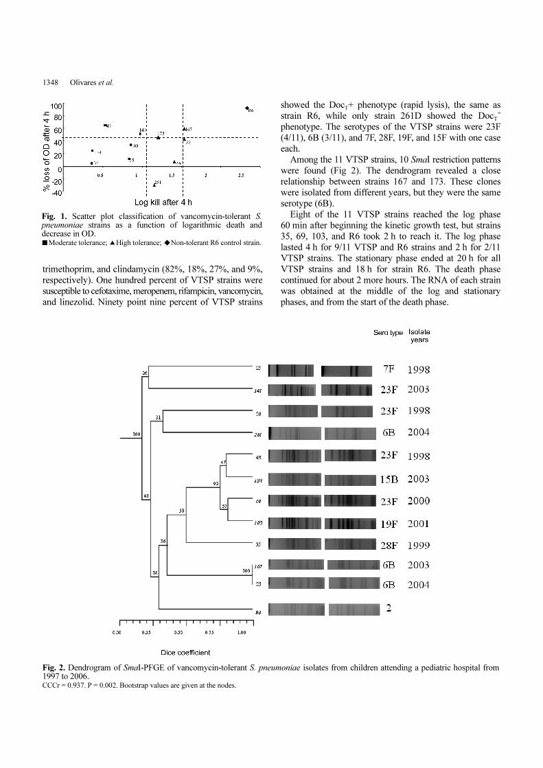
Among the 11 VTSP strains, 10 SmaI restriction patterns
were found (Fig 2). The dendrogram revealed a close
relationship between strains 167 and 173. These clones
were isolated from different years, but they were the same
serotype (6B).
Eight of the 11 VTSP strains reached the log phase
60 min after beginning the kinetic growth test, but strains
35, 69, 103, and R6 took 2 h to reach it. The log phase
lasted 4 h for 9/11 VTSP and R6 strains and 2 h for 2/11
VTSP strains. The stationary phase ended at 20 h for all
VTSP strains and 18 h for strain R6. The death phase
continued for about 2 more hours. The RNA of each strain
was obtained at the middle of the log and stationary
phases, and from the start of the death phase.
Fig. 1. Scatter plot classification of vancomycin-tolerant S.pneumoniae strains as a function of logarithmic death anddecrease in OD.■Moderate tolerance; ▲High tolerance; ◆Non-tolerant R6 control strain.
Fig. 2. Dendrogram of SmaI-PFGE of vancomycin-tolerant S. pneumoniae isolates from children attending a pediatric hospital from1997 to 2006. CCCr = 0.937. P = 0.002. Bootstrap values are given at the nodes.
PEP27 AND LYTA IN VANCOMYCIN-TOLERANT PNEUMOCOCCI 1349
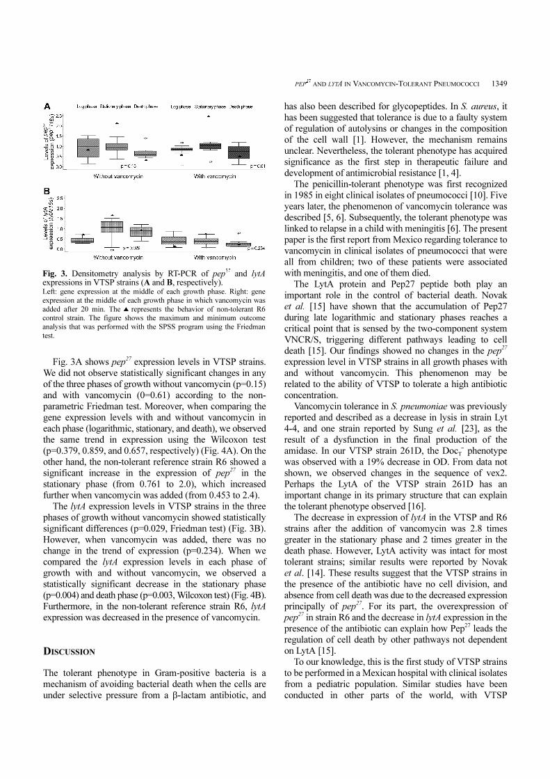
Fig. 3A shows pep27 expression levels in VTSP strains.
We did not observe statistically significant changes in any
of the three phases of growth without vancomycin (p=0.15)
and with vancomycin (0=0.61) according to the non-
parametric Friedman test. Moreover, when comparing the
gene expression levels with and without vancomycin in
each phase (logarithmic, stationary, and death), we observed
the same trend in expression using the Wilcoxon test
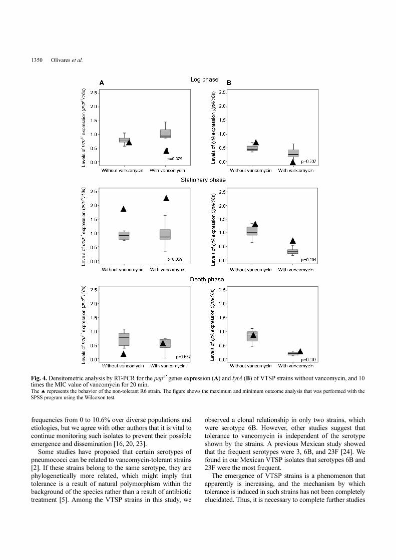
(p=0.379, 0.859, and 0.657, respectively) (Fig. 4A). On the
other hand, the non-tolerant reference strain R6 showed a
significant increase in the expression of pep27 in the
stationary phase (from 0.761 to 2.0), which increased
further when vancomycin was added (from 0.453 to 2.4).
The lytA expression levels in VTSP strains in the three
phases of growth without vancomycin showed statistically
significant differences (p=0.029, Friedman test) (Fig. 3B).
However, when vancomycin was added, there was no
change in the trend of expression (p=0.234). When we
compared the lytA expression levels in each phase of
growth with and without vancomycin, we observed a
statistically significant decrease in the stationary phase
(p=0.004) and death phase (p=0.003, Wilcoxon test) (Fig. 4B).
Furthermore, in the non-tolerant reference strain R6, lytA
expression was decreased in the presence of vancomycin.
DISCUSSION
The tolerant phenotype in Gram-positive bacteria is a
mechanism of avoiding bacterial death when the cells are
under selective pressure from a β-lactam antibiotic, and
has also been described for glycopeptides. In S. aureus, it
has been suggested that tolerance is due to a faulty system
of regulation of autolysins or changes in the composition
of the cell wall [1]. However, the mechanism remains
unclear. Nevertheless, the tolerant phenotype has acquired
significance as the first step in therapeutic failure and
development of antimicrobial resistance [1, 4].
The penicillin-tolerant phenotype was first recognized
in 1985 in eight clinical isolates of pneumococci [10]. Five
years later, the phenomenon of vancomycin tolerance was
described [5, 6]. Subsequently, the tolerant phenotype was
linked to relapse in a child with meningitis [6]. The present
paper is the first report from Mexico regarding tolerance to
vancomycin in clinical isolates of pneumococci that were
all from children; two of these patients were associated
with meningitis, and one of them died.
The LytA protein and Pep27 peptide both play an
important role in the control of bacterial death. Novak
et al. [15] have shown that the accumulation of Pep27
during late logarithmic and stationary phases reaches a
critical point that is sensed by the two-component system
VNCR/S, triggering different pathways leading to cell
death [15]. Our findings showed no changes in the pep27
expression level in VTSP strains in all growth phases with
and without vancomycin. This phenomenon may be
related to the ability of VTSP to tolerate a high antibiotic
concentration.
Vancomycin tolerance in S. pneumoniae was previously
reported and described as a decrease in lysis in strain Lyt
4-4, and one strain reported by Sung et al. [23], as the
result of a dysfunction in the final production of the
amidase. In our VTSP strain 261D, the DocT- phenotype
was observed with a 19% decrease in OD. From data not
shown, we observed changes in the sequence of vex2.
Perhaps the LytA of the VTSP strain 261D has an
important change in its primary structure that can explain
the tolerant phenotype observed [16].
The decrease in expression of lytA in the VTSP and R6
strains after the addition of vancomycin was 2.8 times
greater in the stationary phase and 2 times greater in the
death phase. However, LytA activity was intact for most
tolerant strains; similar results were reported by Novak
et al. [14]. These results suggest that the VTSP strains in
the presence of the antibiotic have no cell division, and
absence from cell death was due to the decreased expression
principally of pep27. For its part, the overexpression of
pep27 in strain R6 and the decrease in lytA expression in the
presence of the antibiotic can explain how Pep27 leads the
regulation of cell death by other pathways not dependent
on LytA [15].
To our knowledge, this is the first study of VTSP strains
to be performed in a Mexican hospital with clinical isolates
from a pediatric population. Similar studies have been
conducted in other parts of the world, with VTSP
Fig. 3. Densitometry analysis by RT-PCR of pep27 and lytAexpressions in VTSP strains (A and B, respectively). Left: gene expression at the middle of each growth phase. Right: gene
expression at the middle of each growth phase in which vancomycin was
added after 20 min. The ▲ represents the behavior of non-tolerant R6
control strain. The figure shows the maximum and minimum outcome
analysis that was performed with the SPSS program using the Friedman
test.
1350 Olivares et al.
frequencies from 0 to 10.6% over diverse populations and
etiologies, but we agree with other authors that it is vital to
continue monitoring such isolates to prevent their possible
emergence and dissemination [16, 20, 23].
Some studies have proposed that certain serotypes of
pneumococci can be related to vancomycin-tolerant strains
[2]. If these strains belong to the same serotype, they are
phylogenetically more related, which might imply that
tolerance is a result of natural polymorphism within the
background of the species rather than a result of antibiotic
treatment [5]. Among the VTSP strains in this study, we
observed a clonal relationship in only two strains, which
were serotype 6B. However, other studies suggest that
tolerance to vancomycin is independent of the serotype
shown by the strains. A previous Mexican study showed
that the frequent serotypes were 3, 6B, and 23F [24]. We
found in our Mexican VTSP isolates that serotypes 6B and
23F were the most frequent.
The emergence of VTSP strains is a phenomenon that
apparently is increasing, and the mechanism by which
tolerance is induced in such strains has not been completely
elucidated. Thus, it is necessary to complete further studies
Fig. 4. Densitometric analysis by RT-PCR for the pep27 genes expression (A) and lytA (B) of VTSP strains without vancomycin, and 10times the MIC value of vancomycin for 20 min. The ▲ represents the behavior of the non-tolerant R6 strain. The figure shows the maximum and minimum outcome analysis that was performed with the
SPSS program using the Wilcoxon test.
PEP27 AND LYTA IN VANCOMYCIN-TOLERANT PNEUMOCOCCI 1351
on this type of bacteria and on the involvement of its genes
to provide therapies that eliminate these strains or prevent
their emergence. pep27 is indeed a gene involved in several
processes in these bacteria, and our results are related to
the tolerance phenomenon.
Acknowledgments
The authors thank Gabriela Echaniz for the donation of the
Streptococcus pneumoniae strain R6. This work was
supported by federal resources (HIM/2006/021) from SSa,
México and SALUD-2010-01-139945.
REFERENCES
1. Bourgeois, I., M. Pestel-Caron, J. F. Lemeland, J. L. Pons, andF. Caron. 2007. Tolerance to the glycopeptides vancomycin andteicoplanin in coagulase-negative staphylococci. Antimicrob.
Agents Chemother. 51: 740-743.2. Fernebro, J., I. Andersson, J. Sublett, E. Morfeldt, R. Novak, E.
Tuomanen, et al. 2004. Capsular expression in Streptococcus
pneumoniae negatively affects spontaneous and antibiotic-induced lysis and contributes to antibiotic tolerance. J. Infect.
Dis. 189: 328-338.3. Gundián, G. P., P. J. Barreto, M. Á. Rodríguez, R. A. Machado,
E. Mora, and M. Lescay. 1998. Glicopeptidos. Acta Méd. 8:
54-57.4. Haas, W., J. Sublett, D. Kaushal, and E. I. Tuomanen. 2004.
Revising the role of the pneumococcal vex-vncRS locus invancomycin tolerance. J. Bacteriol. 186: 8463-8471.
5. Henriques Normark, B., R. Novak, A. Ortqvist, G. Lallenius, E.Tuomanen, and S. Normark. 2001. Clinical isolates of Streptococcus
pneumoniae that exhibit tolerance of vancomycin. Clin. Infect.
Dis. 32: 552-558.6. Henriques Normark, B. and S. Normark. 2002. Antibiotic
tolerance in pneumococci. Clin. Microbiol. Infect. 8: 613-622.7. Jayaraman, R. 2008. Bacterial persistence: Some new insights
into an old phenomenon. J. Biosci. 33: 795-805.8. Lanie, J. A., W. L. Ng, K. M. Kazmierczak, T. M. Andrzejewski,
T. M. Davidsen, K. J. Wayne, et al. 2007. Genome sequence ofAvery’s virulent serotype 2 strain D39 of Streptococcus
pneumoniae and comparison with that of unencapsulatedlaboratory strain R6. J. Bacteriol. 189: 38-51.
9. Li-Korotky, H. S., L. A. Kelly, O. Piltcher, P. A. Hebda, and W.J. Doyle. 2007. Evaluation of microbial RNA extractions fromStreptococcus pneumoniae. J. Microbiol. Methods 68: 342-348.
10. Liu, H. H. and A. Tomasz. 1985. Penicillin tolerance inmultiply drug-resistant natural isolates of Streptococcus
pneumoniae. J. Infect. Dis. 152: 365-372.
11. McCullers, J. A., B. K. English, and R. Novak. 2000. Isolationand characterization of vancomycin-tolerant Streptococcus
pneumoniae from the cerebrospinal fluid of a patient whodeveloped recrudescent meningitis. J. Infect. Dis. 181: 369-373.
12. McEllistrem, M. C., J. Stout, and L. H. Harrison. 2000.Simplified protocol for pulsed-field gel electrophoresis analysisof Streptococcus pneumoniae. J. Clin. Microbiol. 38: 351-353.
13. Mitchell, L. and E. Tuomanen. 2001. Vancomycin-tolerantStreptococcus pneumoniae and its clinical significance. Pediatr.
Infect. Dis. J. 20: 531-533.14. Novak, R., B. Henriques, E. Charpentier, S Normark, and E.
Tuomanen. 1999. Emergence of vancomycin tolerance inStreptococcus pneumoniae. Nature 399: 590-593.
15. Novak, R., E. Charpentier, J. S. Braun, and E Tuomanen. 2000.Signal transduction by a death signal peptide: Uncovering themechanism of bacterial killing by penicillin. Mol. Cell 5: 49-57.
16. Ortega, M., F. Marco, A. Soriano, E. García, J. A. Martínez,and J. Mensa. 2003. Lack of vancomycin tolerance inStreptococcus pneumoniae strains isolated in Barcelona, Spain,from 1999 to 2001. Antimicrob. Agents Chemother. 47: 1976-1978.
17. Obregón, V., P. García, E. García, A. Fenoll, R. López, and J.L. García. 2002. Molecular peculiarities of the lytA geneisolated from clinical pneumococcal strains that are bileinsoluble. J. Clin. Microbiol. 40: 2545-2554.
18. Word Health Organization. 2003. Pneumococcal vaccines. Wkly.
Epidemiol. Rec. 78: 110-119.19. National Committee for Clinical Laboratory Standards. 2005.
Performance Standards for Susceptibility Testing; Fifteenth
Informational Supplement M100-S15. NCCLS, Wayne, PA. 20. Rodriguez, C. A., R. Atkinson, W. Bitar, C. G. Whitney, K. M.
Edwards, L. Mitchell, et al. 2004. Tolerance to vancomycin inpneumococci: Detection with a molecular marker andassessment of clinical impact. J. Infect. Dis. 190: 1481-1487.
21. Rohlf, F. J. 1998. Numerical taxonomy and multivariateanalysis system. Exeter Software Inc., New York, N.Y
22. Sneath, P. H. A. and R. R. Sokal. 1973. Taxonomic structure,pp. 188-305. In: Numerical Taxonomy. W. H. Freeman & Co.,San Francisco, CA.
23. Sung, H., H. B. Shin, M. N. Kim, K. Lee, E. C. Kim, W. Song,et al. 2006. Vancomycin-tolerant Streptococcus pneumoniae inKorea. J. Clin. Microbiol. 44: 3524-3528.
24. Villaseñor-Sierra, A., M. Lomas-Bautista, S. Aguilar-Benavides,and G. Martinez-Aguilar. 2008. Serotypes and susceptibility ofStreptococcus pneumoniae strains isolated from children inMexico. Salud Pública Méx. 50: 330-333.