(PDF) Cerebral blood vessel damage in traumatic brain injury
25
1 Cerebral Blood Vessel Damage in Traumatic Brain Injury Kenneth L. Monson, 1,2,* Matthew Converse, 1 Geoffrey T. Manley 3 1 Department of Mechanical Engineering, University of Utah 2 Department of Bioengineering, University of Utah 3 Department of Neurological Surgery, University of California, San Francisco * Corresponding author Contact information: Kenneth L. Monson, Ph.D. Associate Professor, Mechanical Engineering Adjunct Associate Professor, Bioengineering 1495 E. 100 S., MEK 1550 Salt Lake City, UT 84112 USA [email protected] Abstract Traumatic brain injury is a devastating cause of death and disability. Although injury of brain tissue is of primary interest in head trauma, nearly all significant cases include damage of the cerebral blood vessels. Because vessels are critical to the maintenance of the healthy brain, any injury or dysfunction of the vasculature puts neural tissue at risk. It is well known that these vessels commonly tear and bleed as an immediate consequence of traumatic brain injury. It follows that other vessels experience deformations that are significant though not severe enough to produce bleeding. Recent data show that such subfailure deformations damage the microstructure of the cerebral vessels, altering both their structure and function. Little is known about the prognosis of these injured vessels and their potential contribution to disease development. The objective of this review is to describe the current state of knowledge on the mechanics of cerebral vessels during head trauma and how they respond to the applied loads. Further research on these topics will clarify the role of blood vessels in the progression of traumatic brain injury and is expected to provide insight into improved strategies for treatment of the disease. Keywords: traumatic brain injury; cerebral blood vessel damage; biomechanics Note: This is the accepted manuscript version of the paper. The final paper is available through Clinical Biomechanics here.
-
Upload
khangminh22 -
Category
Documents
-
view
2 -
download
0
Transcript of (PDF) Cerebral blood vessel damage in traumatic brain injury

1
Cerebral Blood Vessel Damage in Traumatic Brain Injury
Kenneth L. Monson,1,2,* Matthew Converse,1 Geoffrey T. Manley3 1 Department of Mechanical Engineering, University of Utah
2 Department of Bioengineering, University of Utah 3 Department of Neurological Surgery, University of California, San Francisco
* Corresponding author Contact information: Kenneth L. Monson, Ph.D. Associate Professor, Mechanical Engineering Adjunct Associate Professor, Bioengineering 1495 E. 100 S., MEK 1550 Salt Lake City, UT 84112 USA [email protected]
Abstract
Traumatic brain injury is a devastating cause of death and disability. Although injury of brain tissue is of primary interest in head trauma, nearly all significant cases include damage of the cerebral blood vessels.
Because vessels are critical to the maintenance of the healthy brain, any injury or dysfunction of the vasculature puts neural tissue at risk. It is well known that these vessels commonly tear and bleed as an
immediate consequence of traumatic brain injury. It follows that other vessels experience deformations that are significant though not severe enough to produce bleeding. Recent data show that such subfailure deformations
damage the microstructure of the cerebral vessels, altering both their structure and function. Little is known about the prognosis of these injured vessels and their potential contribution to disease development. The objective of this review is to describe the current state of knowledge on the mechanics of cerebral vessels
during head trauma and how they respond to the applied loads. Further research on these topics will clarify the role of blood vessels in the progression of traumatic brain injury and is expected to provide insight into
improved strategies for treatment of the disease.
Keywords: traumatic brain injury; cerebral blood vessel damage; biomechanics
Note: This is the accepted manuscript version of the paper. The final paper is available through Clinical Biomechanics here.

2
1. Introduction 1.1 Significance of Vessel Injury in TBI Traumatic brain injury (TBI) is a devastating cause of death and disability, affecting approximately 1.7 million victims each year in the United States, approximately 50,000 of whom die;1 at the same time, global incidence of TBI is increasing dramatically.2 Patients who survive the initial trauma are frequently left with debilitating neurologic deficits. Total annual costs of TBI in the United States have been estimated at $60 billion.3 TBI is initiated by mechanical forces that cause abrupt head motion. This motion produces deformation of the underlying brain and commonly results in axonal injury, contusion, and hematoma.4 Mechanical strain and damage of brain tissue initiates a multifaceted cascade of biochemical reactions that frequently lead to ischemia, hypoxia, brain swelling, and edema.5 The initial insult severity and subsequent dynamic interplay between primary and secondary injuries determine a patient's prognosis and underscore the need for a better understanding of the relationship between mechanics and biology. The cerebral vessels are critical to the maintenance of the healthy brain. Although injury of brain tissue is of primary interest in TBI, nearly all significant cases include some element of injury to the blood vessels,6-9 and any injury or dysfunction of the vasculature puts neural tissue at risk. Vessel disruption often inhibits effective perfusion and leads to dangerous increases in intracranial pressure, in addition to exposing neural tissue to the relatively unregulated composition of whole blood which can disrupt central nervous system (CNS) homeostasis.10 Pathophysiological alterations of cerebral blood flow (CBF), even in the absence of hemorrhage, are also common.7 While the post-trauma biochemical cascade is a known cause of vessel dysfunction,5,7,11 mechanical forces below the threshold of hemorrhage may also contribute to dysfunction through microstructural damage to vascular cells and extracellular matrix (ECM). Recent clinical studies have identified TBI as a risk factor for stroke.12,13 Although the mechanisms behind this connection have not been defined, a more complete understanding of TBI-induced vessel damage will elucidate contributing factors. This information may also lend insight into why some traumatic lesions progress14,15 and whether some persistent conditions, such as mild TBI16 and second impact syndrome,17 may be partially exacerbated by ongoing cerebrovascular dysfunction. Despite the prominence of vascular injury in TBI and its central role in determining clinical outcome, little is known about the local loading conditions that cerebral vessels experience during trauma. Similarly, vessel response to these forces has not been effectively defined. Further definition of the nature and magnitude of deformations that lead to various levels of vascular impairment is a key to improved prevention, since models incorporating this information will more accurately predict vascular injury. Deeper understanding of subtle vascular injury may also promote more effective treatment strategies. The objective of this review is to delineate the current state of knowledge on vascular damage in TBI. Following this introduction, the paper provides a brief background of cerebral vessel anatomy and morphology. This is followed by observations of vessel loading and injury in TBI, including both clinical and experimental investigations, with the goal of gaining a more complete understanding of what blood vessels experience during head trauma. The final section reviews what is known about cerebral vessel response to extreme deformations, including discussions of passive properties, structural failure, and subfailure damage and dysfunction. 1.2 Cerebral Vessel Morphology and Organization To properly consider the mechanics of the cerebral vessels, it is important to understand their environment and structure. The cerebral vascular system includes features not seen in any other organ,18,19 in part due to the layered membrane structure surrounding the brain. Tissue from the arachnoid membrane outward is largely perfused by blood from the external carotid artery. The anatomy of the dural vessels and sinuses is described elsewhere.20-23 Inside the arachnoid, the brain is supplied by the internal carotid and vertebral arteries that pass through the base of the skull, enter the subarachnoid space where they are bathed in cerebrospinal fluid, and anastomose to form the Circle of Willis. Six main conducting arteries leave the Circle and branch to form a complex network of smaller distributing arteries over and between the surfaces of the various lobes of the brain, some following natural paths along and within sulci but others crossing over them.24,25 Small penetrating arteries leave these surface vessels and enter the cortex at right angles, continuing to subdivide until they join the capillary plexus25-29 (Figure 1). Some of these vessels pass through the cortex and into the white matter, though the gray matter is reported to be at least twice as vascular as the white matter.18,30,31 Numerous anastomoses have been found in both the subarachnoid and parenchymal arteries. Some have reported such connections in arteries as large as 1 mm in diameter,32 while others have reported not finding anastomosis in vessels larger than 90 µm.24

3
Figure 1 Scanning electron microscope image of a vascular cast including the subarachnoid and cortical regions of the
temporal lobe (modified from 27 with permission). Large vessels at the top of the image lie within the subarachnoid
space and give off small penetrating arteries that move downward into the cortex to join the cortical capillary
plexus (scale bar = 0.375 mm). The cortical capillaries drain into small veins or venules that unite on the brain surface to become the cortical veins within the subarachnoid space, while return from the deep circulation occurs via the Great Vein of Galen.33 The veins come together to form large draining or “bridging” veins that cross the arachnoid membrane to exit the subarachnoid space and enter the dural venous sinuses.34-36 The venous sinuses ultimately unite and drain through the base of the skull via the two internal jugular veins. When the internal carotid and vertebral arteries enter the subarachnoid space, they acquire an additional layer of pia-arachnoid tissue.37,38 This pial sheath remains with the arteries as they leave the subarachnoid space and penetrate the cortex, forming the Virchow-Robin spaces, but it begins to disappear when branching approaches the capillary level.39 Within the subarachnoid space, some of the collagenous trabeculae that bridge the gap between the pia and arachnoid membranes also attach to this outer vascular coat, thus providing intermittent tethering of the vessels to the pia and arachnoid. The same extra layer of tissue also accompanies veins between their entry into and exit from the subarachnoid space, but it is not found around veins in the cortex.39 The microstructure of the cerebral vessel wall is similar to that of extracranial vessels, but there are a number of distinctions.18,19,40-43 Cerebral arteries are generally categorized as muscular and contain a thicker internal elastic lamina (IEL) than most other vessels, though there is less elastin present in the relatively thin medial and adventitial layers. The external elastic lamina is present in infant cerebral vessels but disappears in the adult.44 As in other muscular arteries, smooth muscle cells are primarily oriented circumferentially.45 Cerebral veins have particularly thin walls and are composed mostly of collagen, having little elastic tissue or smooth muscle. Brain capillaries consist of one layer of tightly packed endothelial cells surrounded by astrocyte end

4
feet. Together, these maintain the blood-brain barrier that protects brain tissue from blood proteins while allowing support of cellular metabolic requirements.
2. Vessel Loading in TBI 2.1 Clinical and Pathological Observations Clinical manifestations of vessel injury in TBI are largely limited to hemorrhage, though pathophysiology associated with vessel dysfunction is also commonly observed. The presence of bleeding suggests that vessels commonly experience deformations that lead to rupture, but clinical observations reveal little about more subtle damage that may occur. Further, details associated with a given TBI incident are typically limited, so the forces and accelerations responsible for the injury are difficult to estimate. Nevertheless, clinical data are fundamental in focusing research in meaningful directions. Because of boundaries defined by the membranes surrounding the brain, blood escaping damaged vessels is commonly limited to spreading within the space between neighboring membranes, as long as they remain intact. Consequently, hemorrhage is categorized by the compartment into which blood leaks – as epidural, subdural, subarachnoid, parenchymal, etc. While contusions also involve vascular damage and are commonly associated with subarachnoid bleeding, they are distinct from other descriptions of hemorrhage in that they also involve mechanical damage of cortical brain tissue. Because of the unique anatomy of the regions in which these different types of injury occur, vessel-level loading likely varies considerably between them. Clinical and pathological manifestations of cerebral vessel injury have been described in detail elsewhere (e.g.46); we here briefly review some observations associated with epidural and subdural hematoma, contusion, and vascular dysfunction to gain insight into the role of mechanics in vessel injury. Epidural, or extradural, hematoma (EDH) refers to blood between the skull and the dura mater. While it is most commonly associated with skull fracture and disruption of the middle meningeal vessels, skull fracture is not required.47 EDH is most common following impact to the temporal bone, but blows to other locations have also been implicated.46-48 Treatment is often delayed since lucid intervals are common, and events are thus believed to be inconsequential.47 Mechanisms of EDH remain largely undetermined, but the middle meningeal vessels course through grooves on the inner surface of the skull and are thus directly exposed to any skull deflections. The dura is believed to separate from the skull beneath the impact site, creating a space that can fill with blood if the vessels are ruptured.47 Subdural hematoma (SDH) refers to extravascular blood between the dura and arachnoid membranes. While the subdural region has traditionally been considered an existing space, anatomical studies have demonstrated that there is no such space in the normal brain.49-51 Rather, the dura and arachnoid are contiguous, but the boundary layer at the dura-arachnoid junction is easily separated such that a “subdural space” between the two membranes can form and fill with blood as a result of trauma.52 Because the dura is supplied by branches of the external carotid artery and the arachnoid is avascular, the only vessels that normally pass through this subdural border region are the bridging veins as they drain blood from the cortex into the sinuses. As a result, subdural bleeding is often a result of bridging vein failure due to relative motion between the brain and skull. Although it is not immediately clear why these vessels would tend to fail in the dura-arachnoid region, rather than within the subarachnoid space, Yamashima and Friede noted more uniform vessel wall structure and greater external support in the subarachnoid region.38 While ruptured bridging veins appear to have been confirmed as the source of SDH in many cases,53-55 subdural bleeding may also arise from the cortical arteries,53 implying arachnoid laceration or avulsion of anomalous arterial twigs that pass through the subdural space from their connection to the cortical arteries.56 However, the complexity of head trauma, with multiple injuries commonly occurring together, often makes it difficult to identify the source of hemorrhage. Contusion is considered the hallmark of head injury57,58 and includes damage to both blood vessels and surrounding parenchyma, including primarily cortical but sometimes also sub-cortical tissue. Such injury commonly also includes bleeding from pial, or subarachnoid, vessels. Clinical observation has shown that the site of contusion, while often appearing at the frontal and temporal lobes, is also a function of impact location.59-61 Specifically, occipital impacts most commonly led to contrecoup contusions in the absence of any coup injury, while frontal impacts most often resulted in coup injury only. Coup and contrecoup injury of the temporal lobe occurred at about the same frequency in lateral impacts. Given the occurrence of coup and contrecoup injuries under different conditions, mechanisms responsible for the production of contusion have been the subject of much discussion.61-64 However, the common involvement of the frontal and temporal lobes,

5
regardless of impact location, suggests that the irregular surfaces of the inner skull in these regions play an important role. Even in cases where the cerebral vessels aren’t damaged severely enough to bleed, trauma may impair the function of the vessels in maintaining homeostasis of the CNS. Recent excellent reviews address the complex physiological response of the circulation to trauma, both mild and severe.5,7,11,16,65 A major consequence is alteration of cerebral blood flow (CBF). Because the brain is sensitive to small changes in perfusion, the vascular network is designed to control CBF over a wide range of intracranial pressure through autoregulation. However, this mechanism is commonly disrupted following TBI, leading to loss of homeostasis and deterioration of CNS function. While the milieu of the injured brain promotes vascular dysfunction, it is not currently known whether direct vessel trauma – below levels producing hemorrhage – may also contribute.7 Another critical function of the cerebral vasculature known to be impaired as a result of trauma is maintenance of the blood-brain barrier (BBB).5 While the division between brain and blood is essentially impassable in large, intact vessels, the BBB refers to the highly regulated exchange of nutrients and waste that occurs at the capillary level. Performance of this thin layer is not grossly visible clinically, but animal models8,66-69 show a nearly immediate increase in permeability with trauma that subsides and then increases again approximately 48 hours later; this dysfunction is the fundamental mechanism for vasogenic edema. The immediate response suggests a direct role for mechanics, but relationships between force, or deformation, and capillary function have not been defined. 2.2 Experimental Investigations As noted above, clinical observations demonstrate the types of vascular injuries that occur in TBI, but they generally lack any definition of associated loading conditions. Experiments are thus necessary to characterize relationships between mechanics and injury. Unfortunately, experiments are necessarily conducted on models that either don’t accurately represent all characteristics of the human system (e.g. cadavers, animals) or are limited in the magnitude of force applied to avoid injury (e.g. human volunteers). Nevertheless, such experiments have led to important insights into mechanisms of TBI. 2.2.1 Physical Models Numerous body-level investigations have been conducted to better understand TBI, including tests on volunteers, human cadavers and animals. Some of these experiments have specifically aimed to elucidate mechanisms and thresholds of cerebrovascular injury, but those with the more general objective of describing brain motion during trauma are also helpful in understanding vessel loading conditions in TBI. Our description here focuses on experiments capturing key features of human and sub-human primate cranial anatomy. 2.2.1.1 General Brain Motion Two early studies are particularly helpful in describing motion of the brain during trauma. In the first, Holbourn70 estimated brain tissue strains due to trauma using two-dimensional, brain-simulating gel models of sagittal, transverse, and coronal sections of the skull subjected to sudden rotation (with no skull deformation). In forward rotation of the sagittal section (as in occipital impact), the largest strains were found in the anterior temporal and inferior frontal lobe regions, believed to be due to interactions between the sphenoid ridge separating the anterior and middle fossae (Figure 2). Other parts of the brain moved more freely relative to the skull so that tissue strains were not as high, with the greatest relative motion occurring at the vertex; other physical models developed since (reviewed elsewhere71) provide additional insight into brain-skull interactions. Despite the simplicity and limitations of such models,72,73 observed high-strain regions clearly correspond with clinical and pathological observations of both contusion and SDH. Based on his experiments, Holbourn concluded that contrecoup injury is due to rotation rather than cavitation.

6
Figure 2 Shear strain distribution in a gelatin model
undergoing clockwise acceleration caused by a blow to the occiput (from 70); strains are largest where colors are
darkest Motivated primarily by a desire to better define mechanisms of contrecoup injury, Pudenz and Shelden64 reported on experiments where macaque primates were fitted with a Lucite calvarium74 to allow observation of brain surface motion resulting from subconcussive blows at frontal, occipital, parietal, and temporal locations. Similar to Holbourn’s predictions, they observed considerable relative motion between the brain and skull (apparently at the arachnoid-dura, or arachnoid-Lucite, interface) near the vertex, though this motion was primarily limited to the sagittal plane, believed to be due to constraints imposed by the falx. While much of the brain couldn’t be visualized, displacement was greatest in the parietal and occipital regions and least in the frontal lobes. The authors opined that the latter was consistent with constraints imposed at the anterior fossa, similar to what Holbourn had observed. The relative motion at the vertex led to massive bleeding due to rupture of bridging veins in two cases. When the head was fixed in place, the blow produced little motion of the brain, consistent with previous observations that head motion is required for impact to produce concussion.75 While the experiments did not allow visualization of regions where contrecoup injury is found, the importance of rotation in their experiments led the authors to conclude that contrecoup injury was best explained by the rotational theory presented by Holbourn. While recent efforts to better define the motion of the brain have used more advanced technology to better quantify motion, findings have been remarkably consistent with the early studies. A group at Wayne State University76-78 observed deep brain tissue displacements on the order of ±5 mm (strains around 0.09) for low severity head impacts (up to 150 G and 8 krad/s2) by tracking radio-opaque neutral density targets embedded in the brains of re-pressurized cadavers. Unexpectedly, displacement measurements near the brain-skull interface, where most vascular injury occurs, suggested less motion than in the deeper tissue, but the investigators expressed less confidence in the accuracy of measurements near the skull. Observed brain-skull dynamics appear to be consistent with that reported by Pudenz and Shelden. In another recent set of studies offering greater resolution but conducted at limited head accelerations (1-3 G and 3 krad/s2), brain deformation was characterized in volunteers using magnetic resonance image tagging.79-
81 Despite the very low accelerations, maximum relative brain-skull displacement was determined to be 2-3 mm, with strains as high as 0.06 for rotations in the transverse plane and 0.05 for mild occipital and frontal impacts. Sagittal plane strains resulting from occipital impacts were strongly influenced by constraints at the skull base in the middle fossa region (Figure 3), qualitatively similar to what was predicted by Holbourn. This tethering produced a complex strain distribution, with the largest strains appearing in regions of brain-skull interaction where contusions are common. Interestingly, the magnitudes of compressive and tensile strains were comparable to those of shear strains in these regions.

7
Figure 3 First principal Lagrangian strain at various times after mild occipital impact in human (sagittal plane; tensile
strains vary from positive to negative, from red to blue); data provided by author of 81 Overall, models exploring brain motion show that constraint of the base of the brain and lack of constraint at the vertex work together to produce a pattern of injury consistent with what has been observed at autopsy and in experiments. This includes the frequent production of contusion on the inferior frontal and temporal lobes, likely due to brain deformation from interactions with the interior skull, and observations of bleeding at the vertex, probably a result of large relative displacements between the brain and skull that may injure bridging veins and other superficial vessels. 2.2.1.2 Cerebral Vessel Injury While the cited studies of general brain motion provide valuable insight into the nature of tissue deformation during TBI, other experimental efforts have focused on specifically understanding the development of vascular lesions. In general, these investigations may be grouped into the categories of free impact and non-deforming forced motion. The former is defined as impact to the freely movable head, while the latter uses a mechanical device to produce rapid, constrained motion in the absence of any significant skull distortion, in order to allow study of the influence of acceleration in the absence of contact phenomena. The number of experiments addressing vessel injury in primate systems is limited, but the resulting findings help clarify mechanisms of injury and patterns of distribution. 2.2.1.2.1 Free Impact Contusion: In one of the most comprehensive of the free impact investigations, visible lesions were studied in macaque primates subjected to either frontal or occipital impact severe enough to produce concussion in over 80% of animals in an earlier study.82 Contusions followed patterns observed in human clinical and pathological studies,60-62 with non-fracture occipital blows leading to both coup (cerebellum) and contrecoup (frontal and temporal lobes) injury, the latter being more extensive. The investigators found it nearly impossible to produce contusion in non-fracture frontal blows. In cases of skull fracture, contusions were primarily limited to coup regions for occipital impacts and were only found in coup regions in frontal cases. Other impact testing on subhuman primates,83,84 including Chimpanzees,85 demonstrated a similar distribution of contusion following both frontal and occipital impact, but Shatsky et al83 additionally reported that temporoparietal blows most commonly led to coup injuries. Since skull deformation was relatively large in these side impact cases, the contusions were believed to be a result of direct parenchymal loading by skull deformation, compared to occipital impacts in the same group where skull deformation and coup injury were both minimal. These primate experiments together suggest that both head rotation and skull distortion are important in the production of contusion. Additionally, while Ommaya et al made little mention of subarachnoid hemorrhage (SAH) in their experiments, Shatsky and company reported SAH following relatively mild impacts at locations where contusions were found in more severe loading cases, suggesting that SAH precedes contusion. While models involving living animals are generally preferred in the study of vascular injury, visible lesions have also been reported in a number of impact experiments involving repressurized, unembalmed human cadavers with India ink injected into the vascular system to identify sites of disruption.86-92 Subarachnoid hemorrhage and contusion have both been reported following various levels of loading, with injuries commonly found at locations consistent with both clinical experience and findings from animal experiments. In the Ommaya macaque primate study mentioned,82 pathological analysis revealed no morphological difference between coup and contrecoup lesions, potentially suggesting that both regions were damaged

8
through a similar mechanism. Compression of occipital lobe or cerebellar tissue would naturally occur with skull distortion from occipital impact. Compression of tissue in the contrecoup regions would similarly result as a consequence of the brain base constraints described by Bayly and company.79-81 Together, these observations suggest that regional compression may be a significant mode of loading in contusion. Subdural Hematoma: In addition to investigating contusion, Ommaya et al82 also reported results on impact-induced SDH. This type of bleeding was most common over the anterior half of the brain, independent of whether impact was occipital or frontal. Incidence was approximately equal for frontal and occipital impacts not producing fracture, but it increased dramatically in cases of occipital, but not frontal, fracture. The group also found that SDH was generally more difficult to produce than contusion, except in cases of non-fracture, frontal impact. A recent repressurized human cadaver experiment also aimed to define tolerance values for SDH.92 Cadavers were subjected to occipital blows (some repeated) of various magnitudes and durations. Bridging vein rupture was detected by fluoroscopy after retrogradely injecting contrast media into the superior sagittal sinus (SSS). Based on the presence of intradural fluid channels connecting to the SSS,20 this technique could lead to false positives, but the production of SDH was consistent with Wayne State Tolerance Curve93 patterns (Figure 4). Specifically, loading leading to SDH ranged from 13.4 krad/s2 for a relatively short duration of 5.2 ms to 5.3 krad/s2 for durations greater than 10 ms. The figure suggests that the threshold for SDH may be reasonably estimated using peak rotational acceleration and peak change in rotational velocity for shorter and longer pulses, respectively, as proposed by Lowenhielm.94,95
Figure 4 Occurrence of SDH as a function of peak
rotational acceleration and duration in repressurized human cadavers subjected to occipital (circles) blows
(reproduced from 92). The figure also references SDH data from frontal blows (squares). Filled and open symbols refer to cases with and without SDH, respectively. The included injury tolerance curves (dashed lines) were proposed by
Lowenhielm based on analytical modeling.94,95
Other Vascular Findings: Other research has demonstrated changes in CBF and breakdown of the BBB following occipital and temporal head impact in the monkey.96 Angiography revealed that some of the concussed animals showed a definite slowing of CBF after impact, and one had clearly smaller arteries and veins after the trauma. Breakdown of the BBB was found at coup locations in both temporal and occipital impacts. Extravasation was also observed on the posterior parasagittal and medial surfaces of the hemispheres and in the brain stem and upper cervical cord following occipital impact. However, little was reported with respect to visible lesions, so it is not clear whether there was any correlation between BBB disruption and overt hemorrhage. Additional research considering CBF changes and BBB disruption in the primate system has been limited (or non-existent). As previously indicated, most work has been accomplished in other animal systems and has been reviewed elsewhere (e.g. 5,7). These have clearly shown changes in CBF and disruption of the BBB after trauma, but modes of loading are not easily compared to those typical for primates. 2.2.1.2.2 Non-Deforming Forced Motion

9
In order to better distinguish the relative significance of skull distortion and head rotation, a number of investigators have conducted experiments using devices that produce impact-like accelerations in the absence of skull deformation, similar to the approach used by Holbourn. Although such high accelerations would be unlikely in the absence of impact, the technique allows investigation of the influence of acceleration independent of contact phenomena. The approach also provides control over the path of the head, allowing examination of the relative effects of rotation and translation. Forced forward (sagittal plane) rotation experiments conducted on primates (including both squirrel and macaque monkeys) showed, similar to impact experiments, that injury progresses from SAH to contusion to SDH with increasing angular acceleration.97-99 The mildest injuries reported following rotations were small contusions and SAH, usually occurring near exposure levels producing unconsciousness. Increasing severity led to the formation of larger contusions, often at locations where SAH was found in milder exposures, and sometimes in the development of small SDH. The most severe exposures produced brainstem hemorrhage and large, frontal SDH with obvious disruption of bridging veins. SDH incidence and size were identified as the most effective predictors of survivability. The incidence of both SDH and concussion correlated well with tangential acceleration in the cases reported by Abel et al.99 In nearly identical subsequent work, this group also reported that the threshold for frontal contusion was lower than that for temporal contusion, with onsets of injury at tangential and rotational accelerations of 500 G and 65 krad/s2 for frontal contusion and 800 G and 85 krad/s2 for temporal lobe injury.100 Contrary to the observed pattern of injury progression in these earlier experiments, some of the same investigators were later able to produce severe neurological injury in the absence of hemorrhage using the same loading mechanism. Similar to what was found in the earlier efforts, increasing acceleration for a given pulse duration eventually produced concussion in the absence of SDH (there was no mention of SAH or contusion), followed by SDH with additional increases in acceleration.101 Interestingly, if they then increased the pulse duration while keeping acceleration constant (thereby decreasing the acceleration rise time), SDH was no longer produced, suggesting an important role for jerk, the time derivative of acceleration, in the production of SDH. Using this approach of relatively long durations and low rise times, the investigators were able to produce severe coma in the absence of bleeding.101,102 In order to explain the disappearance of SDH with lower rise times, the authors argued that their results were consistent with rate dependence in bridging vein failure. However, subsequent work disputing rate dependence of the veins led to the suggestion that Gennarelli and Thibault had mistakenly neglected the positive phase of the acceleration-deceleration loading sequence in their analysis.103,104 Whether or not this is true, the debate highlights a difficulty of the forced motion methodology, since a fixed angular displacement requires that the two loading phases are not independent of one another. The forced motion approach has also demonstrated differences in injury susceptibility due to translational versus rotational loading. A study of sagittal plane motion in the squirrel monkey showed that SAH was absent following pure translation but widespread following rotations having comparable linear acceleration levels (both motions yielded accelerations of 300-1200 G and durations of 5-8 ms).105 Extravasation of Evans blue dye was twice as common in rotation as translation and most frequently occurred in the frontotemporal areas following rotation and in the mid-frontal, parasagittal region with translation. Contusion was seen with both types of insults, but was reportedly more common after translation, different from a similar study where contusion could not be produced in pure translation.84 Contrary to previous reports,98 Gennarelli et al also found no apparent difference in contusion morphology for the two modes of loading. While some translated animals suffered SDH, it was more frequent and severe following rotation. Similarly, none of the translated animals were concussed, but all of the rotated animals were clearly concussed. Although it doesn’t fall into the category of forced motion as the previous investigations do, another macaque primate study lends significant insight into the production of cerebrovascular lesions in the absence of significant head impact. Animals secured to a chair having no head or neck support were rapidly accelerated forward to produce severe whiplash as in a rear impact collision.106 The resulting motion was similar to the discussed forced motion investigations except that rearward, instead of forward, head rotation was superposed on the forward translation. The authors note that head motion was ultimately stopped by the chair but that the associated loading was below the 10% concussion threshold and was thus considered insignificant. Under these conditions, there were no visible lesions in the absence of concussion, but 15 of 19 concussed animals suffered SDH and/or contusion with a distribution similar to that observed in the previously discussed cases involving rotation. Concussion – and accompanying vessel injury – typically occurred above angular

10
accelerations approximated by the nonlinear curve shown in Figure 5, similar qualitatively to that found for SDH in human cadavers (Figure 4). However, in the case of the monkeys – and brains with lower mass, accelerations required for injury were approximately five times higher, with the longest duration cases of 10 ms requiring more than 40 krad/s2 for injury. Notably, the quantitative differences can be reasonably estimated by applying brain mass scaling.107
Figure 5 Whiplash-induced incidence of concussion (and
cerebral hemorrhage) as a function of rotational acceleration and impulse duration in macaque primates,
with estimated injury risk function; data from 106
2.2.1.3 Summary of Physical Model Findings In summary, experimental efforts have demonstrated that there is often significant relative motion between the brain and skull during head trauma. In sagittal plane loading, this motion tends to be largest at the vertex and smallest at the brain base. Constraints at the base of the brain complicate brain dynamics and can cause brain rotation (even when the skull is primarily translating) that pushes the parietal cortex into the skull, leading to both compression and tension at different locations along the parietal cortex, with possible respective contusion and SDH. These constraints also generate interactions between the inferior frontal and temporal lobes with adjacent skull structures, apparently leading to contusion. Experiments further suggest that both rotation and skull deformation are important in the production of visible lesions and show that rotation is generally more damaging than translation. Findings also indicate that the location of contusion is dependent upon head impact site, while SDH tends to occur at the vertex regardless of the direction of loading (though research suggests increased susceptibility to SDH in occipital impact103). Additionally, results suggest that SAH and contusion may result from the same loading mechanism, with SAH alone appearing in mild cases but coupled with contusion at the same site in more severe injuries. SDH tends to require even more severe loading, but the mechanism of its production is typically also different from that of SAH and contusion. Finally, the threshold for any visible lesion tends to be similar to or above that required for minor concussion. All findings considered, however, it should be understood that there are myriad variations of loading sites and directions that have not been explored experimentally; thus, while helpful, these observations are likely not universally applicable. 2.2.2 Computational Efforts The described physical model experiments lend significant insight into how the brain deforms and where vessels fail during trauma, but local mechanisms responsible for vessel injury remain undefined; i.e. the link between head loading and vessel loading has not been established. Although lack of definition of local loading conditions would not matter for cases exactly matching experimental scenarios, it is clear that experiments cannot be performed on living humans at injurious levels. While mass scaling techniques can roughly estimate human brain injury based on findings from monkeys,107 injury tests on monkeys are no longer performed; regardless, more accurate approaches are desirable. Additionally, given the complexity of head anatomy and its variation between people, along with myriad possible loading conditions, it is not feasible to experimentally simulate all possible head injury scenarios. Computer models are unique in their ability to link exterior loading conditions with the internal, tissue-level deformations that directly correspond with injury. With such models in place, injury criteria can be more accurately based on tissue deformation rather than external loading conditions.108,109

11
Computer models of TBI have been in use for over three decades and, based on comparison to experimental results, provide increasingly accurate predictions of brain deformation for many loading scenarios,110-113 though validation is an ongoing challenge.114 Despite this progress, few investigators have constructed models that explicitly account for blood vessels, at least aside from relatively simple representations of bridging veins. An exception to this is a combination of two-dimensional physical115,116 and computational117 models investigating the structural influence of blood vessels on surrounding brain tissue deformation. These models were motivated by observations that brain-skull coupling was more realistic in cadaver experiments where the vascular system was re-pressurized88,91 and by tests showing that cerebral arteries are considerably stiffer than surrounding parenchyma.118-121 As expected, the resulting models showed differences in tissue strains with incorporation of vasculature. However, a subsequent 3-D computational model showed only small differences in tissue strains between models including and not including major vessels.122 The authors of the latter study concluded that vessel material nonlinearity and convoluted vascular geometry limit contributions from the vessels to overall brain deformations. However, it is still necessary to use artificially high values of brain stiffness to account for contributions from the vessels. Regardless, neither modeling approach investigated vessel loading conditions and how they might correlate with hemorrhage or other vascular injury. A more recent investigation of blast TBI argues again that vessels should be included to accurately capture local brain deformations.123 In the end, whether vessels should be included in a given model surely depends on the questions asked, but if accurate local response is desired, vessels are likely needed. In studies having vessel injury as their focus, the production of SDH has received a relatively large amount of attention from modelers, perhaps because the mechanism of injury appears to be comparatively simple, with a vein bridging a mostly fluid-filled space between attachments to the brain and dura. Using this concept, Lowenhielm94 used a mathematical model of brain-skull displacement, in conjunction with experiments on cadavers and reported bridging vein mechanical property data, to predict SDH (and gliding contusion124) in humans when head angular acceleration and angular velocity change exceeded 4500 rad/s2 and 50 rad/s, respectively (see Figure 4). He later reduced the required angular velocity change to 30 rad/s after reconstructing a confirmed accidental fall.95 These values compare well to thresholds estimated in cadaver experiments,92 but more rigor is needed for confidence in application to living humans. Computational models of varying levels of complexity have similarly investigated SDH by quantifying relative motion at the brain-skull interface.104,112,125 In some cases, bridging veins have been modeled explicitly to account for the significance of vessel orientation,110-113,126 but, as MRI studies show,81 the mechanics of the brain-skull interface are complex, and challenges still exist in defining interactions between the brain, the meninges, cerebrospinal fluid, and the cerebral blood vessels. In general, it is not feasible to carefully model these complexities at the macro scale, but multiscale approaches, as illustrated by recent studies of both the brain-skull interface127,128 and the brain itself,129 hold significant promise in overcoming this limitation. The mechanics of contusion has also been investigated computationally, though less work has been done to understand this injury in comparison to SDH. Computer models of typical contusion patterns are most commonly based on predictions of pressure, stress, or strain within a homogeneous gray matter, with no explicit simulation of vessels.110,130,131 Such modeling essentially quantifies the qualitative findings of Holbourn and provides the capability to predict the occurrence of contusion. However, the models are typically validated using data from cadaver experiments where contusion is difficult to study. In addition, their accuracy is dependent upon accurate simulation of mechanics at the brain-skull interface. As an alternative approach, contusion mechanics is also investigated in animal (most commonly rodents) experiments where a portion of the skull is removed, and force is applied directly to the surface of the exposed brain or dura. Controlled cortical impact (CCI), producing compression injury with a piston, is a widely used animal model of contusion;132-134 vacuum loading has also been applied to create a focal, contusion-type injury.69,135-137 Both approaches have been studied with accompanying computational models,135,138-141 and mechanical measures have been correlated with contusion and BBB disruption. As with other computer models, however, blood vessels have not been modeled explicitly. One advantage of these direct brain loading approaches is that the animals are alive and have intact, functioning vascular systems. A disadvantage is that the application of a defined deformation to the stationary, exposed dura is also not a common head trauma scenario; rather, as the foregoing discussion has shown, a typical impact includes shearing at the brain-skull interface. However, while shearing plays a role, the discussed MRI volunteer results81 suggest that normal strains may also be significant to injury. The CCI model, while limited, is thus considered useful since it provides the ability to study the high-rate compression of the cortex without the confounding contributions of the brain-skull interface problem. Disassembling the larger problem into smaller, separate ones may be a key to improved understanding. An

12
additional disadvantage of these models is that they are typically applied to lissencephalic animals. At a small enough scale, however, the vascular microstructure of the cortex is similar to that of the human, making it useful in better understanding microscale mechanics, though the role of convolutions in the more advanced brain is surely also significant to cortical deformation and needs to be better understood. Computational models of CCI in the rodent demonstrate high stresses and strains in the region of contact, roughly corresponding to injuries observed in experiments.141 However, acute animal CCI results show a non-uniform distribution of displaced blood not locally consistent with model predictions142 (Figure 6). This patchy hemorrhaging appears to be associated with penetrating vessels and is consistent with the histological observations of others describing bleeding oriented perpendicular to the cortex.143 In experiments not investigating head trauma, the insertion of a microscopic neuroprosthetic probe into the cortex of rats resulted in vessel rupture at the branch points of penetrating vessels,144 apparently due to fluid entrapment and excess pressure. Similar conditions may be relevant in high-velocity interactions between the cortex and skull during trauma.
Figure 6 Displaced blood proteins (IgG) in a coronal section of cortex immediately following CCI in the mouse (left) with computationally predicted strain contours (red is most severe,
blue is least severe) for the same loading conditions (right) in a model not explicitly accounting for vasculature; scale bar = 1 mm
Each of the described approaches to studying cerebrovascular loading conditions has yielded important insights, and further progress will continue to require integrated application of a variety of model types. It is also clear, however, that modeling approaches that explicitly account for blood vessels are needed. Multiscale computational techniques will surely contribute to progress in this area, but accompanying innovative approaches to validate these models are also required.
3. Vessel Response to Extreme Deformations In addition to defining the loading conditions blood vessels experience during trauma, it is important to understand how vessels respond to these conditions. Because head trauma commonly leads to structural disruption of cerebral vessels, it is critical to define their mechanical properties and failure values. However, it is also important to characterize the response of vessels that are deformed severely without structural disruption. Little is currently known about how such subtle injury may influence progression of TBI or other related problems like second impact syndrome and stroke. 3.1 Passive Properties and Structural Failure The passive mechanical characteristics of blood vessels have been under study for many years, and fundamental concepts related to testing methods and tissue response have been effectively reviewed elsewhere.145-147 While these reviewed concepts apply generally to blood vessels, the specific properties of cerebral vessels have received limited attention. However, tests on large arteries leaving the Circle of Willis and on their branches on the surface of the cortex show that, like other vessels, they are nonlinear and anisotropic, and interactions between loading directions are significant.148,149 Relative to vessels elsewhere in the circulation, however, these cerebral arteries are particularly stiff in both the axial and circumferential directions.119,149,150 On the other hand, the axial response of veins on the surface of the cortex is similar to that of extracranial vessels119 (Figure 7). The mechanical properties of smaller arteries and veins, including arterioles and venules, have not been defined, despite their presumed role in contributing to the mechanics of the brain parenchyma. Because of the difficulty of reliably isolating such vessels, a computational approach modeling experiments on a brain sample including small vessels may be the most promising approach to such

13
definition. It is also notable that the properties of the meningeal arteries and veins have also not been characterized, especially given their significance in epidural hematoma.
Figure 7 Axial stress-stretch response of cerebral arteries relative to arteries and veins elsewhere in the circulation; compares cortical (Cort) artery and vein, middle cerebral
artery (MCA), common carotid artery (CCA), femoral (Fem) artery and vein, and popliteal (Pop) artery
Failure characteristics of the cerebral vessels have particular relevance to TBI-induced hemorrhage. Most efforts to define the mechanical properties of human cerebral blood vessels have been limited to large cerebral arteries and bridging veins. Chalupnik et al118 subjected perfused major branches of the middle cerebral artery to longitudinal extensions over a wide range of strain rates. Only subfailure properties are provided in their report, but no rate dependence was observed. In recent work from our group, human pial arteries obtained from the surface of the temporal lobe during surgeries displayed loading characteristics similar to the large autopsy vessels tested by Chalupnik et al.119,151 We similarly also found no rate dependence over a strain rate range of more than four orders of magnitude. Failure of pial arteries typically occurred at stretch ratios and First Piola-Kirchhoff stresses around 1.4 and 4 MPa, respectively. Steiger et al152 reported similar failure stretch values for arteries from the Circle of Willis. To our knowledge, no other failure experiments have been reported for human cerebral arteries, including tests evaluating response in the circumferential direction. Because ruptured bridging veins are believed to be the primary source of traumatic SDH, a small number of studies have addressed their failure characteristics, again in the longitudinal direction since they are believed to be stretched axially with relative motion between the brain and skull. Bridging vein characteristics have been reviewed in detail elsewhere,153 but briefly, early tests by Löwenhielm154 concluded that ultimate stress and stretch were both strongly dependent on strain rate (failure stretch and First Piola-Kirchhoff stress both decreased, from 1.8 to 1.1 and from 2.0 to 0.25 MPa, respectively, with increases in strain rate from 1 to 1000 s-1). A later investigation by Lee and Haut103 found an absence of rate dependence for both quantities, reporting average values of the same measures of approximately 1.5 and 3.3 MPa. Tests by our group included only a small number of bridging veins, but failure stretch ratios were comparable to those of Lee and Haut,151 and we similarly found no rate dependence. More recent experiments on bridging veins produced a stiffer response than what had previously been reported for these vessels, with failure stretch values around 1.25.155 By design, their tests included the bridging vein and a portion of the superior sagittal sinus in order to allow testing of the junction between the two, suggesting that the transition region between the vein and sinus is stiffer than the vein itself. They similarly found no dependence upon rate in their results. Consistent with the apparent increase in stiffness with proximity to the sinus, our experiments on pial veins showed these vessels to be considerably less stiff than the already mentioned bridging veins, though there was no clear histological reason for the difference.151 Nevertheless, along with the data from Delye et al, it appears that vein extensibility may decrease between the pial surface and the sinus. Considering tests on pial arteries and veins together, it is interesting to note that the arteries, on average, failed at stretch values half those of cerebral veins (with respective stretch ratios of 1.42 v. 1.88).151 Despite the fact that the arteries withstood larger stresses before failure, this observation suggests that a deformation involving both vessel types would be expected to produce failure of arteries before veins. However, this expectation does not take into account potential differences in the in vivo lengths of the two vessel types, which were not defined in the cited work.
14
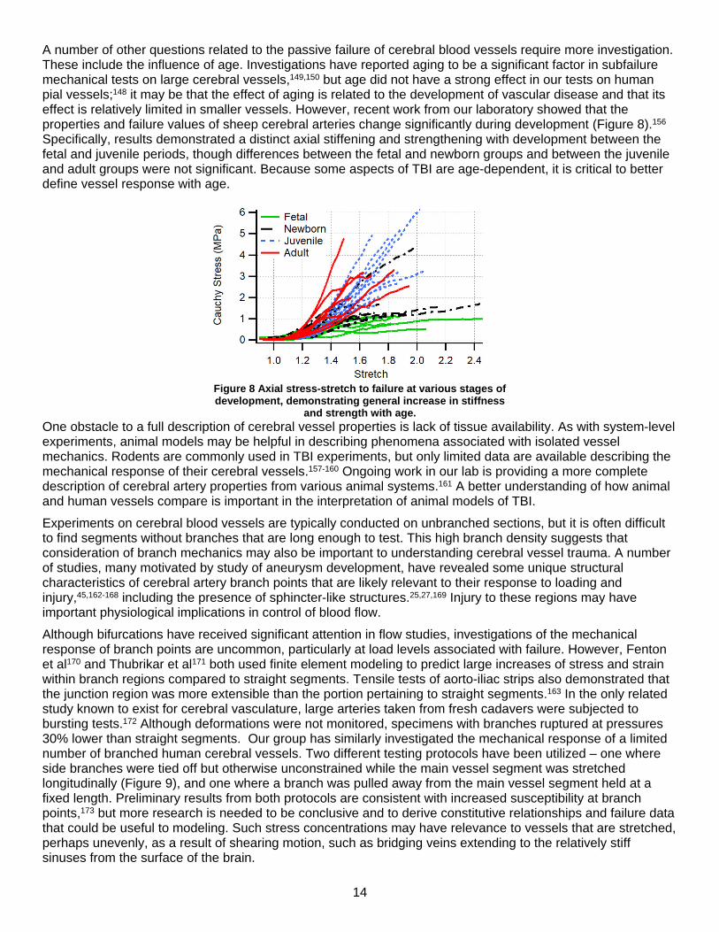
A number of other questions related to the passive failure of cerebral blood vessels require more investigation. These include the influence of age. Investigations have reported aging to be a significant factor in subfailure mechanical tests on large cerebral vessels,149,150 but age did not have a strong effect in our tests on human pial vessels;148 it may be that the effect of aging is related to the development of vascular disease and that its effect is relatively limited in smaller vessels. However, recent work from our laboratory showed that the properties and failure values of sheep cerebral arteries change significantly during development (Figure 8).156 Specifically, results demonstrated a distinct axial stiffening and strengthening with development between the fetal and juvenile periods, though differences between the fetal and newborn groups and between the juvenile and adult groups were not significant. Because some aspects of TBI are age-dependent, it is critical to better define vessel response with age.
Figure 8 Axial stress-stretch to failure at various stages of development, demonstrating general increase in stiffness
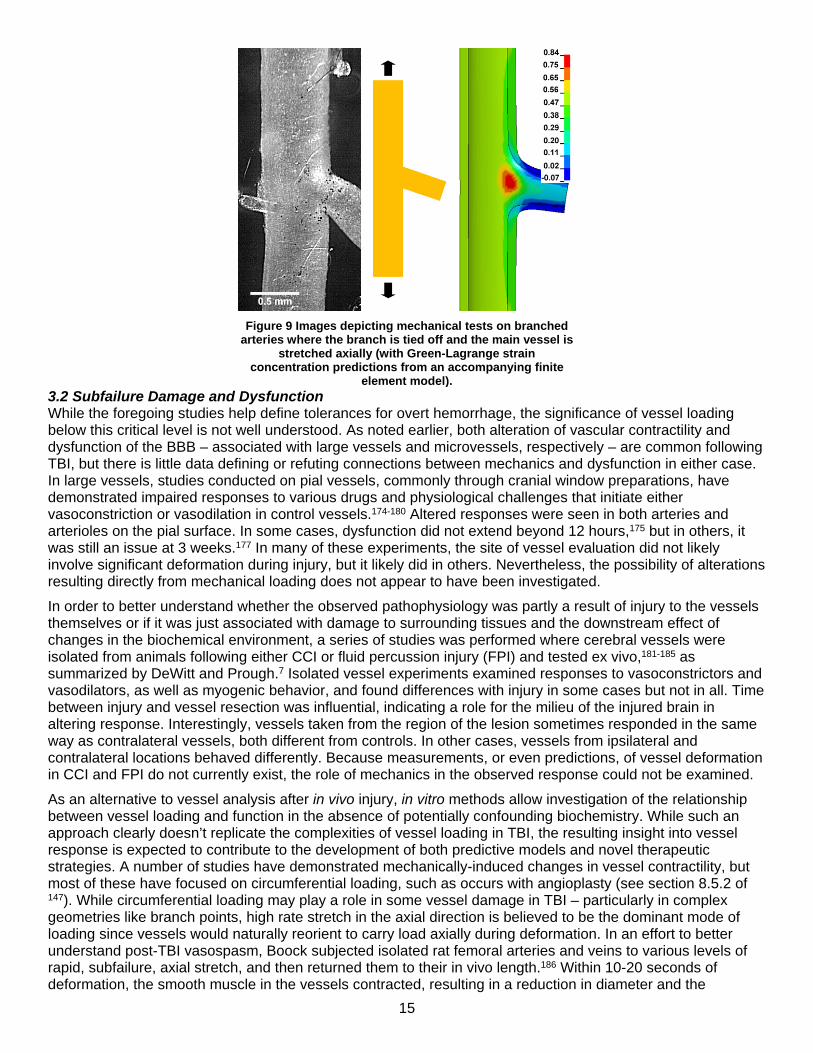
and strength with age. One obstacle to a full description of cerebral vessel properties is lack of tissue availability. As with system-level experiments, animal models may be helpful in describing phenomena associated with isolated vessel mechanics. Rodents are commonly used in TBI experiments, but only limited data are available describing the mechanical response of their cerebral vessels.157-160 Ongoing work in our lab is providing a more complete description of cerebral artery properties from various animal systems.161 A better understanding of how animal and human vessels compare is important in the interpretation of animal models of TBI. Experiments on cerebral blood vessels are typically conducted on unbranched sections, but it is often difficult to find segments without branches that are long enough to test. This high branch density suggests that consideration of branch mechanics may also be important to understanding cerebral vessel trauma. A number of studies, many motivated by study of aneurysm development, have revealed some unique structural characteristics of cerebral artery branch points that are likely relevant to their response to loading and injury,45,162-168 including the presence of sphincter-like structures.25,27,169 Injury to these regions may have important physiological implications in control of blood flow. Although bifurcations have received significant attention in flow studies, investigations of the mechanical response of branch points are uncommon, particularly at load levels associated with failure. However, Fenton et al170 and Thubrikar et al171 both used finite element modeling to predict large increases of stress and strain within branch regions compared to straight segments. Tensile tests of aorto-iliac strips also demonstrated that the junction region was more extensible than the portion pertaining to straight segments.163 In the only related study known to exist for cerebral vasculature, large arteries taken from fresh cadavers were subjected to bursting tests.172 Although deformations were not monitored, specimens with branches ruptured at pressures 30% lower than straight segments. Our group has similarly investigated the mechanical response of a limited number of branched human cerebral vessels. Two different testing protocols have been utilized – one where side branches were tied off but otherwise unconstrained while the main vessel segment was stretched longitudinally (Figure 9), and one where a branch was pulled away from the main vessel segment held at a fixed length. Preliminary results from both protocols are consistent with increased susceptibility at branch points,173 but more research is needed to be conclusive and to derive constitutive relationships and failure data that could be useful to modeling. Such stress concentrations may have relevance to vessels that are stretched, perhaps unevenly, as a result of shearing motion, such as bridging veins extending to the relatively stiff sinuses from the surface of the brain.
15
Figure 9 Images depicting mechanical tests on branched
arteries where the branch is tied off and the main vessel is stretched axially (with Green-Lagrange strain
concentration predictions from an accompanying finite element model).
3.2 Subfailure Damage and Dysfunction While the foregoing studies help define tolerances for overt hemorrhage, the significance of vessel loading below this critical level is not well understood. As noted earlier, both alteration of vascular contractility and dysfunction of the BBB – associated with large vessels and microvessels, respectively – are common following TBI, but there is little data defining or refuting connections between mechanics and dysfunction in either case. In large vessels, studies conducted on pial vessels, commonly through cranial window preparations, have demonstrated impaired responses to various drugs and physiological challenges that initiate either vasoconstriction or vasodilation in control vessels.174-180 Altered responses were seen in both arteries and arterioles on the pial surface. In some cases, dysfunction did not extend beyond 12 hours,175 but in others, it was still an issue at 3 weeks.177 In many of these experiments, the site of vessel evaluation did not likely involve significant deformation during injury, but it likely did in others. Nevertheless, the possibility of alterations resulting directly from mechanical loading does not appear to have been investigated. In order to better understand whether the observed pathophysiology was partly a result of injury to the vessels themselves or if it was just associated with damage to surrounding tissues and the downstream effect of changes in the biochemical environment, a series of studies was performed where cerebral vessels were isolated from animals following either CCI or fluid percussion injury (FPI) and tested ex vivo,181-185 as summarized by DeWitt and Prough.7 Isolated vessel experiments examined responses to vasoconstrictors and vasodilators, as well as myogenic behavior, and found differences with injury in some cases but not in all. Time between injury and vessel resection was influential, indicating a role for the milieu of the injured brain in altering response. Interestingly, vessels taken from the region of the lesion sometimes responded in the same way as contralateral vessels, both different from controls. In other cases, vessels from ipsilateral and contralateral locations behaved differently. Because measurements, or even predictions, of vessel deformation in CCI and FPI do not currently exist, the role of mechanics in the observed response could not be examined. As an alternative to vessel analysis after in vivo injury, in vitro methods allow investigation of the relationship between vessel loading and function in the absence of potentially confounding biochemistry. While such an approach clearly doesn’t replicate the complexities of vessel loading in TBI, the resulting insight into vessel response is expected to contribute to the development of both predictive models and novel therapeutic strategies. A number of studies have demonstrated mechanically-induced changes in vessel contractility, but most of these have focused on circumferential loading, such as occurs with angioplasty (see section 8.5.2 of 147). While circumferential loading may play a role in some vessel damage in TBI – particularly in complex geometries like branch points, high rate stretch in the axial direction is believed to be the dominant mode of loading since vessels would naturally reorient to carry load axially during deformation. In an effort to better understand post-TBI vasospasm, Boock subjected isolated rat femoral arteries and veins to various levels of rapid, subfailure, axial stretch, and then returned them to their in vivo length.186 Within 10-20 seconds of deformation, the smooth muscle in the vessels contracted, resulting in a reduction in diameter and the

16
development of axial force. Force persisted for 120-150 seconds. Force level correlated linearly with the extent of the imposed stretch. Interestingly, veins responded to a variety of stretch inputs, while arteries only contracted following very large strains. Similarly, recent work from our lab shows that smooth muscle cell contractility to K+ is reduced following overstretch of cerebral arteries.187 Rat middle cerebral artery (MCA) response to K+ was evaluated both before and after overstretch of either 1.2 or 1.3 times the in vivo length of the vessel and at strain rates of either 0.2 or 20 s-1. Not surprisingly, the most dramatic changes in function occurred following the largest overstretch at the highest rate. This work demonstrates that mechanical trauma directly alters cerebral artery function, even when applied strains are axial – perpendicular to the largely circumferential smooth muscle cells. It is not yet known if the applied loads damaged vessel cells or ECM, or both, to elicit this change. Damage to vessel ECM resulting from subfailure loading has been investigated in a number of contexts, including angioplasty and stenting. Results show that large deformations produce tissue softening, such that less force is required to produce the same level of deformation with reloading (e.g. 188,189). We recently demonstrated softening after axial loading of cerebral arteries.190 Following overstretch, the stress-stretch curve shifted to the right, with slopes of the lower and upper portions of the altered curve decreasing and increasing, respectively (Figure 10). Not surprisingly, larger deformations led to increased softening, but changes were only significant with overstretch values above 1.2 times the in vivo length of the vessel. Alterations were persistent for the hour following injury during which they were monitored, thus ruling out viscoelastic stress relaxation as the responsible mechanism. Continuing investigation in our laboratory demonstrates that axial overstretch and damage also leads to softening in the circumferential response; however, greater axial deformations are required for these changes to be significant. Greater laxity in the circumferential direction could explain our previous finding that MCA contractility was reduced following overstretch. Such changes in vivo could alter vessel hemodynamics and potentially lead to conditions promoting thrombosis and stroke. However, our observations of softening result from quasi-static overstretches, so additional research is needed to characterize rates of loading relevant to TBI.
Figure 10 Ovine cerebral artery softening demonstrated by
axial stress-stretch curves during and after an axial overstretch of approximately 1.6 times the in vivo length
Because passive mechanical properties are a direct reflection of tissue structure, it is clear that overstretch alters vessel ECM. A better understanding of the nature of such damage may provide a pathway for treatment of vessel injury. Collagen and elastin have been identified as the primary load-bearing constituents of the ECM, with collagen structure defining the high stiffness region of the nonlinear stress-stretch curve of vessels.191 Recent work shows that vessel softening may be effectively predicted by accounting for damaged collagen fibers alone.192 Using an approach recently applied to tendon,193 research in our laboratory shows that collagen hybridizing peptide (CHP), with an attached fluorophore, can be used to detect mechanical failure of collagen fibers in cerebral arteries (Figure 11).194 Results show that overstretch damages fibers aligned with the direction of loading, that damage increases with overstretch severity, and that the onset of this damage coincides with tissue-level yielding. While damage is currently detected under a confocal microscope, work is being done to tag CHP with a radio-opaque marker for improved in vivo detection.

17
Figure 11 CHP fluorescence in control (top left) and axially overstretched (bottom left) vessel
segments. Collagen damage vs. axial overstretch (right), with regression line and 95% confidence intervals. Collagen damage was measured following CHP staining as the percentage of pixels above a control-specific intensity threshold. Loading produced an increase in damage
with overstretch severity above a threshold value. Stretch is measured relative to the in vivo configuration.
In addition to collagen fiber damage, research on aorta and carotid artery has shown that large deformations typically produce intimal disruption before failure of other tissue layers.195-199 While this has been primarily demonstrated in experiments on isolated vessel tissue, carotid and vertebral arteries are often similarly damaged as a consequence of neck deformation. This phenomenon, defined clinically as blunt cerebrovascular injury (BCVI), is characterized by dissection, followed by clotting and potential stroke, sometimes after a significant delay.200,201 Work in our lab shows that it is common for internal layers of cerebral arteries, including the internal elastic lamina (IEL), to rupture during isolated vessel overstretch. Despite this internal layer failure, vessels remain intact and continue to maintain pressure with additional stretch (Figure 12). Intimal rupture would be expected to lead to functional impairment due to disruption of endothelial signaling as well as thrombosis at the site of the defect. While such endothelial layer disruption would be expected to eventually heal, rupture of the IEL likely poses a longer-term problem, at least in adults, given the downregulation of the elastin gene after adolescence.202-204 Improved understanding of such subfailure damage of the ECM may have important implications to the progression of TBI.
Figure 12 Failure of intima precedes that of surrounding media and adventitia in axial
stretch testing of middle cerebral artery. Left: vessel maintains pressure even with intimal disruption (arrow); intima was stained intraluminally with Nigrosin dye for contrast; scale
bar = 100 µm. Right: Fluorescence microscopy image of ruptured IEL following axial overstretch, exposing the intact media and adventitia; scale bar = 50 µm.
Loading that produces direct cellular injury in TBI is largely unexplored, in part because any deformation that produces vascular dysfunction is likely also damaging the ECM, so it is unknown whether resulting impairment is due to cell or ECM injury. Studying cells in isolation provides some insight. Alford et al showed that rapid

18
stretch of cultured smooth muscle cells produced hypersensitivity to contractile stimulation by endothelin-1; the changes induced by the loading persisted for over 24 hours.205 Mechanical vascular cell injury may also be manifest by alteration of gene and protein expression.206 Again, however, it is often difficult to differentiate changes induced by the biochemical cascade and direct mechanical damage. In addition to vessel contractility impairment, there is evidence that mechanical insults may play a direct role in the barrier function of cerebral microvessels. Using a fluid percussion approach, Rinder and Olsson applied pressure pulses to the exposed parietal dura of rabbits and produced marked increases in vascular permeability in the lateral brainstem and cervical spinal cord without producing any visible vessel damage.207-
210 Low severity pulses allowed fluorescent indicators to progress into the vessel wall but not into the surrounding tissue, while more severe loading produced transport through the vessel wall. Other experiments with similar loading conditions have demonstrated the presence of shear strain in the brainstem,211-213 thus suggesting a mechanism for injury of the vessels there and implicating subfailure mechanical loading in the generation of increased vascular permeability. The rabbit study further revealed an important role of time in vascular permeability. Extravasation did not progress beyond one hour after the pulse was applied. In animals injected with the fluorescent indicators post-injury, extravasation was clearly visible in surrounding tissue if the injection was performed within 1 hour of injury. No extravasation was seen with injection at 12-18 hours, though the markers did transfer into the vessel wall. No progress of the indicators into the vessel wall was found after 24-48 hours. Other animal experiments have similarly demonstrated acute BBB breakdown following both brain and spinal cord deformation and the dependence of its extent on severity,69,214,215 suggesting a direct role for mechanical loading in vessel permeability. These experiments have also shown that the increased permeability is transient, though also biphasic with a secondary opening at 1-2 days67 that is clearly not a direct result of externally applied loads. Rinder and Olssen209 discuss whether the permeability observed in their experiments could be a result of dramatic changes in hemodynamics rather than deformation, but conclude that such alterations would not likely produce injuries only in regions of greatest strain. On the other hand, others have linked acute microvascular pathophysiology to changes in blood pressure.174 Although the mechanisms producing microvessel dysfunction are not year clear, transient ultrastructural changes in microvessel walls following TBI have been described by a number of studies.9 These include disruption of endothelium and swelling of perivascular astrocytes,216 distinct changes in a number of endothelial activation markers,217 and migration of pericytes.218 Others have reported endothelial lesions in the form of vacuolization or crater formation174,219,220 in the absence of any morphologically apparent smooth muscle injury.174 It is not known whether any of these changes directly resulted from mechanical loading, but alterations were often widespread and didn’t appear to correlate with regions of large tissue deformation. These subfailure studies together indicate that vessel structure and function are altered by mechanical loading. It follows, then, that such changes would be predictable by computational models of tissue mechanics and could thus add to understanding of tolerance and prediction of injury and disease progression. For models to be meaningful, however, the relationships between traumatic loads – including loading type, direction, and rate – and vessel function need to be more completely characterized.
4. Closure Vascular injury is a common consequence of TBI and plays a significant role in patient outcome. While there appears to be an accurate qualitative understanding of how various types of cerebrovascular injury occur, the mechanisms of these injuries have not yet been satisfactorily quantified. Further, the relationship between mechanical force and vessel response needs to be more completely characterized. Improved ability to accurately predict vessel loading and response during TBI will significantly advance understanding of mechanical and physiological injury thresholds. This knowledge is expected to advance prevention efforts and to additionally support improvements in treatment by lending insight into the status of the vasculature at the initiation of biochemical cascades following trauma.
Acknowledgements We gratefully acknowledge the work of students and postdocs that have contributed to progress in the areas reviewed. Those who contributed to yet unpublished research mentioned herein include David Bell, Justin Ingram, Huijae Kim, Vishwas Mathur, Kevin Nye, Joshua Smith, Tessa Sommer, Ray Walther, Stewart Yeoh, and Brad Zentgraf.

19
References 1. Faul M, Xu L, Wald MM, Coronado VG. Traumatic Brain Injury in the United States: Emergency Department Visits, Hospitalizations and Deaths 2002-2006. Atlanta, GA: Centers for Disease Control and Prevention, National Center for Injury Prevention and Control; 2010. 2. Maas AIR, Stocchetti N, Bullock R. Moderate and severe traumatic brain injury in adults. The Lancet Neurology 2008;7:728-41. 3. Finkelstein E, Corso PS, Miller TR. The incidence and economic burden of injuries in the United States. New York: Oxford University Press; 2006. 4. Melvin JW, Lighthall JW, Ueno K. Brain Injury Biomechanics. In: Nahum AM, Melvin JW, eds. Accidental Injury: Biomechanics and Prevention. New York: Springer-Verlag; 1993. 5. Golding EM. Sequelae following traumatic brain injury. The cerebrovascular perspective. Brain Res Brain Res Rev 2002;38:377-88. 6. Graham DI. Neuropathology of head injury. In: Nrayan RK, Wilberger JE, Povlishock JT, eds. Neurotrauma. New York: McGraw-Hill; 1996:43-59. 7. DeWitt D, Prough, DS. Traumatic cerebral vascular injury: the effects of concussive brain injury on the cerebral vasculature. Journal of Neurotrauma 2003;20:795-825. 8. Jullienne A, Obenaus A, Ickova A, Savona-Baron C, Pearce WJ, Badaut J. Chronic cerebrovascular dysfunction after traumatic brain injury. Journal of Neuroscience Research 2016;94:609-22. 9. Kenney K, Amyot F, Haber M, et al. Cerebral vascular injury in traumatic brain injury. Experimental Neurology 2016;275, Part 3:353-66. 10. Daneman R, Prat A. The Blood–Brain Barrier. Cold Spring Harbor Perspectives in Biology 2015;7. 11. Werner C, Engelhard K. Pathophysiology of traumatic brain injury. Br J Anaesth 2007;99:4-9. 12. Chen Y-H, Kang J-H, Lin H-C. Patients With Traumatic Brain Injury: Population-Based Study Suggests Increased Risk of Stroke. Stroke 2011;42:2733-9. 13. Burke JF, Stulc JL, Skolarus LE, Sears ED, Zahuranec DB, Morgenstern LB. Traumatic brain injury may be an independent risk factor for stroke. Neurology 2013;81:33-9. 14. Narayan RK, Maas AI, Servadei F, et al. Progression of traumatic intracerebral hemorrhage: a prospective observational study. J Neurotrauma 2008;25:629-39. 15. Simard JM, Kilbourne M, Tsymbalyuk O, et al. Key role of sulfonylurea receptor 1 in progressive secondary hemorrhage after brain contusion. J Neurotrauma 2009;26:2257-67. 16. Len TK, Neary JP. Cerebrovascular pathophysiology following mild traumatic brain injury. Clinical Physiology and Functional Imaging 2011;31. 17. Cantu R, Gean A. Second impact syndrome in a small SDH: an uncommon catastrophic result of repetitive head injury with a characteristic imaging appearance. J Neurotrauma 2010;27:1557-64. 18. Stephens RB, Stilwell DL. Arteries and veins of the human brain. Springfield, Ill.,: Thomas; 1969. 19. Wolff HG. The Cerebral Circulation. Physiol Rev 1936;16:545-96. 20. Fox RJ, Walji AH, Mielke B, Petruk KC, Aronyk KE. Anatomic details of intradural channels in the parasagittal dura: a possible pathway for flow of cerebrospinal fluid. Neurosurgery 1996;39:84-90; discussion -1. 21. Mack J, Squier W, Eastman JT. Anatomy and development of the meninges: implications for subdural collections and CSF circulation. Pediatr Radiol 2009;39:200-10. 22. Martins C, Yasuda A, Campero A, Ulm AJ, Tanriover N, Rhoton A, Jr. Microsurgical anatomy of the dural arteries. Neurosurgery 2005;56:211-51; discussion -51. 23. Tubbs RS, Loukas M, Louis RG, Jr., et al. Anatomy of the falcine venous plexus. J Neurosurg 2007;107:155-7. 24. Duvernoy HM, Delon S, Vannson JL. Cortical blood vessels of the human brain. Brain Res Bull 1981;7:519-79. 25. Nonaka H, Akima M, Nagayama T, Hatori T, Zhang Z, Ihara F. Microvasculature of the human cerebral meninges. Neuropathology 2003;23:129-35. 26. Akima M, Nonaka H, Kagesawa M, Tanaka K. A study on the microvasculature of the cerebral cortex. Fundamental architecture and its senile change in the frontal cortex. Lab Invest 1986;55:482-9. 27. Reina-De La Torre F, Rodriguez-Baeza A, Sahuquillo-Barris J. Morphological characteristics and distribution pattern of the arterial vessels in human cerebral cortex: a scanning electron microscope study. The Anatomical Record 1998;251:87-96. 28. Cassot F, Lauwers F, Fouard C, Prohaska S, Lauwers-Cances V. A novel three-dimensional computer-assisted method for a quantitative study of microvascular networks of the human cerebral cortex. Microcirculation 2006;13:1-18. 29. Cassot F, Lauwers F, Lorthois S, Puwanarajah P, Cances-Lauwers V, Duvernoy H. Branching patterns for arterioles and venules of the human cerebral cortex. Brain Res 2010;1313:62-78. 30. Nonaka H, Akima M, Hatori T, Nagayama T, Zhang Z, Ihara F. Microvasculature of the human cerebral white matter: arteries of the deep white matter. Neuropathology 2003;23:111-8. 31. Nonaka H, Akima M, Hatori T, Nagayama T, Zhang Z, Ihara F. The microvasculature of the cerebral white matter: arteries of the subcortical white matter. J Neuropathol Exp Neurol 2003;62:154-61. 32. Brozici M, van der Zwan A, Hillen B. Anatomy and functionality of leptomeningeal anastomoses: a review. Stroke 2003;34:2750-62.

20
33. Meder J, Chiras J, Roland J, Guinet P, Bracard S, Bargy F. Venous territories of the brain. J Neuroradiol 1994;21:118-33. 34. Andrews BT, Dujovny M, Mirchandani HG, Ausman JI. Microsurgical anatomy of the venous drainage into the superior sagittal sinus. Neurosurgery 1989;24:514-20. 35. Han H, Tao W, Zhang M. The dural entrance of cerebral bridging veins into the superior sagittal sinus: an anatomical comparison between cadavers and digital subtraction angiography. Neuroradiology 2007;49:169-75. 36. Oka K, Rhoton A, Barry M, Rodriguez R. Microsurgical anatomy of the superficial veins of the cerebrum. Neurosurgery 1985;17:711-48. 37. Alcolado R, Weller RO, Parrish EP, Garrod D. The cranial arachnoid and pia mater in man: anatomical and ultrastructural observations. Neuropathology and Applied Neurobiology 1988;14:1-17. 38. Yamashima T, Friede RL. Why do bridging veins rupture into the virtual subdural space? J Neurol Neurosurg Psychiatry 1984;47:121-7. 39. Zhang E, Inman C, Weller R. Interrelationships of the pia mater and the perivascular (Virchow-Robin) spaces in the human cerebrum. Journal of Anatomy 1990;170:111-23. 40. Hayashi K, Nagasawa S, Naruo Y, Okumura A, Moritake K, Handa H. Mechanical properties of human cerebral arteries. Biorheology 1980;17:211-8. 41. Finlay HM, McCullough L, Canham PB. Three-dimensional collagen organization of human brain arteries at different transmural pressures. Journal of Vascular Research 1995;32:301-12. 42. Holzapfel GA, Gasser TC, Ogden RW. A new constitutive framework for arterial wall mechanics and a comparative study of material models. Journal of Elasticity 2000;61:1-48. 43. Lee RMKW. Morphology of cerebral arteries. Pharmacology & Therapeutics 1995;66:149-73. 44. Hassler O, Larsson SE. The external elastic layer of the cerebral arteries in different age-groups. Acta anatomica 1962;48:1-6. 45. Takahashi A, Ushiki T, Abe K, Houkin K, Abe H. Scanning electron microscopic studies of the medial smooth muscles in human major intracranial arteries. Archives of Histology and Cytology 1994;57:341-50. 46. Crooks DA. Pathogenesis and biomechanics of traumatic intracranial haemorrhages. Virchows Arch A Pathol Anat Histopathol 1991;418:479-83. 47. Gallagher JP, Browder EJ. Extradural hematoma. Experience with 167 patients. J Neurosurg 1968;29:1-12. 48. McKissock W, Till K, Bloom WH, Taylor JC. Extradural haematoma observations on 125 cases. Lancet 1960;2:167-72. 49. Haines DE. On the question of a subdural space. Anat Rec 1991;230:3-21. 50. Haines DE, Harkey HL, al-Mefty O. The "subdural" space: a new look at an outdated concept. Neurosurgery 1993;32:111-20. 51. Schachenmayr W, Friede RL. The origin of subdural neomembranes. I. Fine structure of the dura-arachnoid interface in man. Am J Pathol 1978;92:53-68. 52. Miller JD, Nader R. Acute subdural hematoma from bridging vein rupture: a potential mechanism for growth. J Neurosurg 2014;120:1378-84. 53. Maxeiner H, Wolff M. Pure subdural hematomas: a postmortem analysis of their form and bleeding points. Neurosurgery 2002;50:503-8; discussion 8-9. 54. Voigt GE, Lowenhielm CG, Ljung CB. Rotational cerebral injuries near the superior margin of the brain. Acta Neuropathol 1977;39:201-9. 55. Maxeiner H. Detection of ruptured cerebral bridging veins at autopsy. Forensic Sci Int 1997;89:103-10. 56. O'Brien PK, Norris JW, Tator CH. Acute subdural hematomas of arterial origin. J Neurosurg 1974;41:435-9. 57. Adams JH, Graham DI, Scott G, Parker LS, Doyle D. Brain damage in fatal non-missile head injury. Journal of clinical pathology 1980;33:1132-45. 58. Adams JH, Scott G, Parker LS, Graham DI, Doyle D. The contusion index: a quantitative approach to cerebral contusions in head injury. Neuropathology and Applied Neurobiology 1980;6. 59. Vance BM. Fractures of the skull: complications and causes of death: a review of 512 necropsies and of 61 cases studied clinically. Arch Surg 1927;14:1023-92. 60. Sano K, Nakamura N, Hirakawa K, Masuzawa H, Hashizume K. Mechanism and dynamics of closed head injuries (preliminary report). Neurol Med Chir (Tokyo) 1967;9:21-33. 61. Courville CB. The mechanism of coup-contrecoup injuries of the brain; a critical review of recent experimental studies in the light of clinical observations. Bulletin of the Los Angeles Neurological Society 1950;15:72-86. 62. Lindenberg R, Freytag E. The mechanism of cerebral contusions. A pathologic-anatomic study. Archives of Pathology 1960;69:440-69. 63. Goggio AF. The mechanism of contre-coup injury. J Neurol Psychiatry 1941;4:11-22. 64. Pudenz R, Shelden C. The lucite calvarium—A method for direct observation of the brain. Journal of Neurosurgery 1946;3:487-505. 65. Golding EM, Robertson CS, Bryan RM, Jr. The consequences of traumatic brain injury on cerebral blood flow and autoregulation: a review. Clinical and experimental hypertension (New York, NY : 1993) 1999;21:299-332. 66. Cernak I. Animal models of head trauma. NeuroRx 2005;2:410-22.

21
67. Baldwin SA, Fugaccia I, Brown DR, Brown LV, Scheff SW. Blood-brain barrier breach following cortical contusion in the rat. J Neurosurg 1996;85:476-81. 68. Başkaya MK, Muralikrishna Rao A, Doğan A, Donaldson D, Dempsey RJ. The biphasic opening of the blood–brain barrier in the cortex and hippocampus after traumatic brain injury in rats. Neuroscience Letters 1997;226:33-6. 69. Shreiber DI, Smith DH, Meaney DF. Immediate in vivo response of the cortex and the blood-brain barrier following dynamic cortical deformation in the rat. Neurosci Lett 1999;259:5-8. 70. Holbourn AHS. Mechanics of head injuries. The Lancet 1943;242:438-41. 71. Goldsmith W, Monson KL. The state of head injury biomechanics: past, present, and future part 2: physical experimentation. Crit Rev Biomed Eng 2005;33:105-207. 72. Holbourn AHS. Mechanics of head injuries. The Lancet 1944;243:483. 73. Gurdjian ES. Mechanics of head injuries. The Lancet 1944;243:389-90. 74. Shelden CH, Pudenz RH, Restarski JS, Craig WM. The Lucite Calvarium - A Method for Direct Observation of the Brain. Journal of Neurosurgery 1944;1:67-75. 75. Denny-Brown D, Russell W. Experimental cerebral concussion. Brain 1941;64:93-164. 76. King AI, Yang KH, Hardy WN. Recent firsts in cadaveric impact biomechanics research. Clin Anat 2011;24:294-308. 77. Hardy WN, Foster CD, Mason MJ, Yang KH, King AI, Tashman C. Investigation of head injury mechanisms using neutral density technology and high-speed biplanar x-ray. Stapp car crash journal 2001;45:337-68. 78. Hardy WN, Mason MJ, Foster CD, et al. A study of the response of the human cadaver head to impact. Stapp car crash journal 2007;51:17-80. 79. Feng Y, Abney TM, Okamoto RJ, Pless RB, Genin GM, Bayly PV. Relative brain displacement and deformation during constrained mild frontal head impact. J R Soc Interface 2010;7:1677-88. 80. Sabet AA, Christoforou E, Zatlin B, Genin GM, Bayly PV. Deformation of the human brain induced by mild angular head acceleration. J Biomech 2008;41:307-15. 81. Bayly PV, Cohen TS, Leister EP, Ajo D, Leuthardt EC, Genin GM. Deformation of the human brain induced by mild acceleration. J Neurotrauma 2005;22:845-56. 82. Ommaya AK, Grubb RL, Jr., Naumann RA. Coup and contre-coup injury: observations on the mechanics of visible brain injuries in the rhesus monkey. J Neurosurg 1971;35:503-16. 83. Shatsky SA, Alter WA, Evans DE, Armbrustmacher VW, Clark G, Earle KM. Traumatic distortions of the primate head and chest: correlation of biomechanical radiological and pathological data. Stapp Car Crash Conference; 1974. p. 351-81. 84. Ono K, Kikuchi A, Nakamura M, Kobayashi H, Nakamura H. Human head tolerance to sagittal impact reliable estimation deduced from experimental head injury using sub-human primates and human cadaver skulls. 24th Stapp Car Crash Conference; 1980. p. 101-60. 85. Ommaya A, Corrao P, Letcher F. Head injury in the chimpanzee. 1. Biodynamics of traumatic unconsciousness. Journal of Neurosurgery 1973;39:152-66. 86. Stalnaker RL, Roberts VL, McElhaney JR. Side impact tolerance to blunt trauma. 17th Stapp Car Crash Conference; 1973: Society of Automotive Engineers. 87. Nahum AM, Smith R. Experimental model for closed head impact injury. Stapp Car Crash Conference 1976:785-814. 88. Nahum AM, Smith R, Ward CC. Intracranial pressure dynamics during head impact. Stapp Car Crash Conference 1977:339-66. 89. Nahum A, Schneider D, Adams S, Raasch F, Ward C. Experimental studies of side impact to the human head. 24th Stapp Car Crash Conference; 1980: Society of Automotive Engineers. p. 43-62. 90. Nusholtz GS, Lux P, Kaiker P, Janicki MA. Head impact response - skull deformation and angular accelerations. 28th Stapp Car Crash Conference; 1984. p. 41-74. 91. Stalnaker RL, Melvin JW, Nusholtz GS, Alem NM, Benson JB. Head impact response. Stapp Car Crash Conference; 1977. p. 303-35. 92. Depreitere B, Van Lierde C, Sloten JV, et al. Mechanics of acute subdural hematomas resulting from bridging vein rupture. J Neurosurg 2006;104:950-6. 93. Gurdjian ES, Lissner HR, Patrick LM. Protection of the Head and Neck in Sports. JAMA 1962;182:509-12. 94. Lowenhielm P. Strain tolerance of the vv. cerebri sup. (bridging veins) calculated from head-on collision tests with cadavers. Zeitschrift für Rechtsmedizin Journal of legal medicine 1974;75:131-44. 95. Lowenhielm P. Tolerance level for bridging vein disruption calculated with a mathematical model. Journal of Bioengineering 1978;2:501-7. 96. Ommaya AK, Rockoff SD, Baldwin M. Experimental Concussion; a First Report. J Neurosurg 1964;21:249-65. 97. Unterharnscheidt F, Higgins LS. Pathomorphology of experimental head injury due to rotational acceleration. Acta Neuropathol 1969;12:200-4. 98. Unterharnscheidt F, Higgins LS. Traumatic lesions of brain and spinal cord due to nondeforming angular acceleration of the head. Tex Rep Biol Med 1969;27:127-66. 99. Abel JM, Gennarelli TA, Segawa H. Incidence and severity of cerebral concussion in the Rhesus monkey following sagittal plane angular acceleration. 22nd Stapp Car Crash Conference1978.

22
100. Gennarelli TA, Abel JM, Adams H, Graham D. Differential tolerance of frontal and temporal lobes to contusion induced by angular acceleration. Stapp Car Crash Conference; 1979. p. 563-86. 101. Gennarelli TA, Thibault LE. Biomechanics of acute subdural hematoma. J Trauma 1982;22:680-6. 102. Gennarelli TA, Thibault LE, Adams JH, Graham DI, Thompson CJ, Marcincin RP. Diffuse axonal injury and traumatic coma in the primate. Ann Neurol 1982;12:564-74. 103. Lee MC, Haut RC. Insensitivity of tensile failure properties of human bridging veins to strain rate: implications in biomechanics of subdural hematoma. Journal of Biomechanics 1989;22:537-42. 104. Lee MC, Melvin JW, Ueno K. Finite element analysis of traumatic subdural hematoma 31st Stapp Car Crash Conference; 1987; New Orleans, Louisiana: SAE International. 105. Gennarelli TA, Thibault LE, Ommaya AK. Pathophysiologic responses to rotational and translational accelerations of the head. Stapp Car Crash Conference; 1972. p. 296-308. 106. Ommaya AK, Faas F, Yarnell P. Whiplash injury and brain damage: an experimental study. Journal of the American Medical Association 1968;204:285-9. 107. Ommaya AK, Harris EH, Hirsch AE, Yarnell P. Scaling of experimental data on cerebral concussion in sub-human primates to concussion threshold for man. 11th Stapp Car Crash Conference. Anaheim, CA: Society of Automotive Engineers; 1967. 108. Goldsmith W. Current controversies in the stipulation of head injury criteria. J Biomech 1981;14:883-4. 109. Goldsmith W. Meaningful concepts of head injury criteria. Proceedings of the 1989 International IRCOBI Conference on the Biomechanics of Impacts 1989:1-12. 110. Zhang L, Yang KH, Dwarampudi R, et al. Recent advances in brain injury research: a new human head model development and validation. Stapp car crash journal 2001;45. 111. Takhounts EG, Eppinger RH, Campbell JQ, Tannous RE, Power ED, Shook LS. On the Development of the SIMon Finite Element Head Model. Stapp car crash journal 2003;47:107-33. 112. Kleiven S. Influence of impact direction on the human head in prediction of subdural hematoma. J Neurotrauma 2003;20:365-79. 113. Mao H, Zhang L, Jiang B, et al. Development of a Finite Element Human Head Model Partially Validated With Thirty Five Experimental Cases. Journal of Biomechanical Engineering 2013;135:111002-. 114. Bayly PV, Clayton EH, Genin GM. Quantitative Imaging Methods for the Development and Validation of Brain Biomechanics Models. Annual Review of Biomedical Engineering 2012;14:369-96. 115. Omori K, Zhang L, Yang KH, King AI. Effect of cerebral vasculatures on the mechanical response of brain tissue: a preliminary study. In: Mahmood HF, Barbat SD, Baccouche MR, eds. Crashworthiness, Occupant Protection, and Biomechanics in Transportation Systems, ASME Mechanical Engineering Congress and Exposition. Orlando, FL: ASME; 2000:167-74. 116. Parnaik Y, Beillas P, Demetropoulos CK, Hardy WN, Yang KH, King A. I. The influence of surrogate blood vessels on the impact response of a physical model of the brain. Stapp car crash journal 2004;48:259-77. 117. Zhang L, Bae J, Hardy WN, et al. Computational study of the contribution of the vasculature on the dynamic response of the brain. Stapp car crash journal 2002;46:145-64. 118. Chalupnik J, Daly C, Merchant H. Material Properties of Cerebral Blood Vessels. Seattle: University of Washington; 1971. 119. Monson KL, Goldsmith W, Barbaro NM, Manley GT. Axial mechanical properties of fresh human cerebral blood vessels. Journal of Biomechanical Engineering 2003;125:288-94. 120. Bandak FA, Eppinger RH. A three-dimensional finite element analysis of the human brain under combined rotational and translational accelerations. 38th Stapp Car Crash Conference; 1994: SAE. p. 145-63. 121. Vossoughi J, Bandak FA. Mechanical characteristics of vascular tissue and their role in brain injury modeling: a review. In: Bandak FA, Eppinger R, H., Ommaya AK, eds. Traumatic Brain Injury: Bioscience and Mechanics: Mary Ann Liebert, Inc.; 1996:207-15. 122. Ho J, Kleiven S. Dynamic response of the brain with vasculature: a three-dimensional computational study. J Biomech 2007;40:3006-12. 123. Hua Y, Lin S, Gu L. Relevance of Blood Vessel Networks in Blast-Induced Traumatic Brain Injury. Comput Math Methods Med 2015;2015:928236. 124. Löwenhielm P. Mathematical simulation of gliding contusions. Journal of Biomechanics 1975;8:351-6. 125. Huang HM, Lee MC, Chiu WT, Chen CT, Lee SY. Three-dimensional finite element analysis of subdural hematoma. J Trauma 1999;47:538-44. 126. Takhounts EG, Ridella SA, Hasija V, et al. Investigation of traumatic brain injuries using the next generation of simulated injury monitor (SIMon) finite element head model. Stapp car crash journal 2008;52:1-31. 127. Zoghi-Moghadam M, Sadegh AM. Global/local head models to analyse cerebral blood vessel rupture leading to ASDH and SAH. Comput Methods Biomech Biomed Engin 2009;12:1-12. 128. Scott GG, Margulies SS, Coats B. Utilizing multiple scale models to improve predictions of extra-axial hemorrhage in the immature piglet. Biomechanics and Modeling in Mechanobiology 2015:1-19. 129. Cloots RJH, Dommelen JAW, Geers MGD. Micromechanics of diffuse axonal injury: influence of axonal orientation and anisotropy. Biomechanics and Modeling in Mechanobiology 2011;10:413-22. 130. Chu CS, Lin MS, Huang HM, Lee MC. Finite element analysis of cerebral contusion. J Biomech 1994;27:187-94.

23
131. Huang HM, Lee MC, Lee SY, Chiu WT, Pan LC, Chen CT. Finite element analysis of brain contusion: an indirect impact study. Med Biol Eng Comput 2000;38:253-9. 132. Lighthall JW, Dixon CE, Anderson TE. Experimental models of brain injury. J Neurotrauma 1989;6:83-97. 133. Dixon CE, Clifton GL, Lighthall JW, Yaghmai AA, Hayes RL. A controlled cortical impact model of traumatic brain injury in the rat. J Neurosci Methods 1991;39:253-62. 134. Sutton RL, Lescaudron L, Stein DG. Unilateral cortical contusion injury in the rat: vascular disruption and temporal development of cortical necrosis. J Neurotrauma 1993;10:135-49. 135. Shreiber DI, Bain AC, Meaney DF. In vivo thresholds for mechanical injury to the blood-brain barrier. Proceedings 41st Stapp Car Crash Conference 1997:277-91. 136. Shreiber DI, Bain AC, Ross DT, et al. Experimental investigation of cerebral contusion: histopathological and immunohistochemical evaluation of dynamic cortical deformation. J Neuropathol Exp Neurol 1999;58:153-64. 137. Mathew P, Bullock R, Graham DI, Maxwell WL, Teasdale GM, McCulloch J. A new experimental model of contusion in the rat. Histopathological analysis and temporal patterns of cerebral blood flow disturbances. J Neurosurg 1996;85:860-70. 138. Ueno K, Melvin JW, Li L, Lighthall JW. Development of tissue level brain injury criteria by finite element analysis. J Neurotrauma 1995;12:695-706. 139. Mao H, Zhang L, Yang KH, King AI. Application of a finite element model of the brain to study traumatic brain injury mechanisms in the rat. Stapp car crash journal 2006;50:583-600. 140. Mao H, Jin X, Zhang L, et al. Finite element analysis of controlled cortical impact-induced cell loss. J Neurotrauma 2010;27:877-88. 141. Mao H, Yang KH, King AI, Yang K. Computational neurotrauma - design, simulation, and analysis of controlled cortical impact model. Biomechanics and Modeling in Mechanobiology 2010;9:763-72. 142. Yeoh S, Mathur V, Monson KL. Vascular injury and cortical deformation in a model of brain contusion. ASME 2011 Summer Bioengineering Conference; 2011; Farmington, PA. 143. Liu HM, Sturner WQ. Extravasation of plasma proteins in brain trauma. Forensic Sci Int 1988;38:285-95. 144. Bjornsson CS, Oh SJ, Al-Kofahi YA, et al. Effects of insertion conditions on tissue strain and vascular damage during neuroprosthetic device insertion. J Neural Eng 2006;3:196-207. 145. Hayashi K. Experimental approaches on measuring the mechanical properties and constitutive laws of arterial walls. J Biomech Eng 1993;115:481-8. 146. Humphrey JD. Mechanics of the arterial wall: review and directions. Crit Rev Biomed Eng 1995;23:1-162. 147. Humphrey JD. Cardiovascular Solid Mechanics : Cells, Tissues, and Organs. New York: Springer; 2002. 148. Monson KL, Barbaro NM, Manley GT. Biaxial response of passive human cerebral arteries. Annals of Biomedical Engineering 2008;36:2028-41. 149. Busby D, Burton A. The effect of age on the elasticity of the major brain arteries. Canadian Journal of Physiology and Pharmacology 1965;43:185-202. 150. Hayashi K, Handa H, Nagasawa S, Okumura A, Moritake K. Stiffness and elastic behavior of human intracranial and extracranial arteries. Journal of Biomechanics 1980;13:175-9. 151. Monson KL, Goldsmith W, Barbaro NM, Manley GT. Significance of source and size in the mechanical response of human cerebral blood vessels. Journal of Biomechanics 2005;38:737-44. 152. Steiger H, Aaslid R, Keller S, Reulen H. Strength, elasticity and viscoelastic properties of cerebral aneurysms. Heart and Vessels 1989;5:41-6. 153. Famaey N, Cui ZY, Musigazi GU, et al. Structural and mechanical characterisation of bridging veins: A review. Journal of the mechanical behavior of biomedical materials 2015;41:222-40. 154. Löwenhielm P. Dynamic Properties of the Parasagittal Bridging Veins. Zeitschrift für Rechtsmedizin Journal of legal medicine 1974;74:55-62. 155. Delye H, Goffin J, Verschueren P, et al. Biomechanical properties of the superior sagittal sinus-bridging vein complex. Stapp car crash journal 2006;50:625-36. 156. Nye KS, Converse MI, Dahl MJ, Albertine KH, Monson KL. Development of mechanical and failure properties in sheep cerebral arteries. Annals of Biomedical Engineering 2017;45:1101-10. 157. Hajdu M, Heistad D, Siems J, Baumbach G. Effects of aging on mechanics and composition of cerebral arterioles in rats. Circulation Research 1990;66:1747-54. 158. Hajdu MA, Baumbach GL. Mechanics of large and small cerebral arteries in chronic hypertension. American Journal of Physiology 1994;266:H1027-33. 159. Coulson RJ, Chesler NC, Vitullo L, Cipolla MJ. Effects of ischemia and myogenic activity on active and passive mechanical properties of rat cerebral arteries. Am J Physiol Heart Circ Physiol 2002;283:H2268-75. 160. Coulson RJ, Cipolla MJ, Vitullo L, Chesler NC. Mechanical properties of rat middle cerebral arteries with and without myogenic tone. Journal of Biomechanical Engineering 2004;126:76-81. 161. Bell ED, Kunjir RS, Monson KL. Biaxial and failure properties of passive rat middle cerebral arteries. Journal of Biomechanics 2013;46:91-6. 162. MacFarlane TW, Roach MR, Chan K. The geometry of human cerebral bifurcations: effect of static distending pressure. J Biomech 1980;13:265-77. 163. Roach MR. The structure and elastic properties of arterial junctions. Connective Tissue Research 1986;15:77-84.
24
164. Campbell G, Roach M. Fenestrations in the internal elastic lamina at bifurcations of human cerebral arteries. Stroke 1981;12:489-96. 165. Rowe AJ, Finlay HM, Canham PB. Collagen biomechanics in cerebral arteries and bifurcations assessed by polarizing microscopy. Journal of Vascular Research 2003;40:406-15. 166. Finlay HM, Whittaker P, Canham PB. Collagen organization in the branching region of human brain arteries. Stroke 1998;29:1595-601. 167. Walmsley J, Campling M, Chertkow H. Interrelationships among wall structure, smooth muscle orientation, and contraction in human major cerebral arteries. Stroke 1983;14:781-90. 168. Shiraishi T, Sakaki S, Uehara Y. Architecture of the medial smooth muscle of the arterial vessels in the normal human brain: a scanning electron-microscopic study. Scanning Microscopy 1990;4:191-9. 169. Nakai K, Naka Y, Yokote H, et al. Vascular "sphincter" and microangioarchitecture in the central nervous system: constriction of intraparenchymal blood vessels following a treatment of vasoconstrictive neurotransmitter. Scanning Microsc 1989;3:337-41. 170. Fenton TR, Gibson WG, Taylor JR. Stress analysis of vasoconstriction at arterial branch sites. Journal of Biomechanics 1986;19:501-9. 171. Thubrikar MJ, Roskelley SK, Eppink RT. Study of stress concentration in the walls of the bovine coronary arterial branch. Journal of Biomechanics 1990;23:15-26. 172. Mitchell P, Jakubowski J. Failure testing cerebral arteries: are branch points weaker than unbranched vessels? British Journal of Neurosurgery 2002;16:578-82. 173. Monson K, Barbaro N, Manley G. Cerebrovascular injury in head trauma - susceptibility of branch points. Journal of Neurotrauma 2008;25:864. 174. Wei EP, Dietrich WD, Povlishock JT, Navari RM, Kontos HA. Functional, morphological, and metabolic abnormalities of the cerebral microcirculation after concussive brain injury in cats. Circulation Research 1980;46:37-47. 175. Ellison M, Erb D, Kontos H, Povlishock J. Recovery of impaired endothelium-dependent relaxation after fluid- percussion brain injury in cats. Stroke 1989;20:911-7. 176. Kontos HA, Wei EP. Endothelium-Dependent Responses After Experimental Brain Injury. Journal of Neurotrauma 1992;9:349-54. 177. Wei EP, Hamm RJ, Baranova AI, Povlishock JT. The long-term microvascular and behavioral consequences of experimental traumatic brain injury after hypothermic intervention. J Neurotrauma 2009;26:527-37. 178. Golding EM, Robertson CS, Bryan RM, Jr. L-arginine partially restores the diminished CO2 reactivity after mild controlled cortical impact injury in the adult rat. Journal of Cerebral Blood Flow and Metabolism 2000;20:820-8. 179. Suehiro E, Ueda Y, Wei EP, Kontos HA, Povlishock JT. Posttraumatic Hypothermia followed by Slow Rewarming Protects the Cerebral Microcirculation. Journal of Neurotrauma 2003;20:381-90. 180. Ueda Y, Wei EP, Kontos HA, Suehiro E, Povlishock JT. Effects of delayed, prolonged hypothermia on the pial vascular response after traumatic brain injury in rats. Journal of Neurosurgery 2003;99:899-906. 181. Bukoski RD, Wang SN, Bian K, DeWitt DS. Traumatic brain injury does not alter cerebral artery contractility. Am J Physiol 1997;272:H1406-11. 182. Mathew BP, DeWitt DS, Bryan RM, Bukoski RD, Prough DS. Traumatic brain injury reduces myogenic responses in pressurized rodent middle cerebral arteries. Journal of Neurotrauma 1999;16:1177-86. 183. Golding EM, Contant CF, Jr., Robertson CS, Bryan RM, Jr. Temporal effect of severe controlled cortical impact injury in the rat on the myogenic response of the middle cerebral artery. J Neurotrauma 1998;15:973-84. 184. Golding EM, Steenberg ML, Cherian L, Marrelli SP, Robertson CS, Bryan RM. Endothelial-mediated dilations following severe controlled cortical impact injury in the rat middle cerebral artery. Journal of Neurotrauma 1998;15:635-44. 185. Golding EM, Steenberg ML, Contant CF, Jr., Krishnappa I, Robertson CS, Bryan RM, Jr. Cerebrovascular reactivity to CO(2) and hypotension after mild cortical impact injury. Am J Physiol 1999;277:H1457-66. 186. Boock R. Vascular Response to Mechanical Deformations [Dissertation]. Philadephia: University of Pennsylvania; 1991. 187. Bell ED, Donato AJ, Monson KL. Cerebrovascular dysfunction following subfailure axial stretch. Journal of the Mechanical Behavior of Biomedical Materials 2017;65:627-33. 188. Maher E, Early M, Creane A, Lally C, Kelly DJ. Site specific inelasticity of arterial tissue. Journal of Biomechanics 2012;45:1393-9. 189. Horný L, Gultova E, Chlup H, et al. Mullins effect in aorta and limiting extensibility evolution. Bulletin of Applied Mechanics 2010;6:1-5. 190. Bell ED, Sullivan JW, Monson KL. Subfailure overstretch induces persistent changes in the passive mechanical response of cerebral arteries. Frontiers in Bioengineering and Biotechnology 2015;3:2. 191. Roach MR, Burton AC. The reason for the shape of the distensibility curves of arteries. Canadian Journal of Biochemistry and Physiology 1957;35:681-90. 192. Weisbecker H, Pierce DM, Regitnig P, Holzapfel GA. Layer-specific damage experiments and modeling of human thoracic and abdominal aortas with non-atherosclerotic intimal thickening. Journal of the Mechanical Behavior of Biomedical Materials 2012;12:93-106. 193. Zitnay JL, Li Y, Qin Z, et al. Molecular level detection and localization of mechanical damage in collagen enabled by collagen hybridizing peptides. Nature Communications 2017;8:14913.

25
194. Converse MI, Walther RG, Ingram JT, Li Y, Yu SM, Monson KL. Detection and characterization of molecular-level collagen damage in overstretched cerebral arteries. Acta Biomaterialia 2017. 195. Stemper BD, Yoganandan N, Pintar FA. Methodology to study intimal failure mechanics in human internal carotid arteries. J Biomech 2005;38:2491-6. 196. Stemper BD, Yoganandan N, Sinson GP, Gennarelli TA, Stineman MR, Pintar FA. Biomechanical characterization of internal layer subfailure in blunt arterial injury. Ann Biomed Eng 2007;35:285-91. 197. Stemper BD, Yoganandan N, Pintar FA. Mechanics of arterial subfailure with increasing loading rate. J Biomech 2007;40:1806-12. 198. Stemper BD, Yoganandan N, Pintar FA, Brasel KJ. Multiple subfailures characterize blunt aortic injury. J Trauma 2007;62:1171-4. 199. Cohen J, Litwin SB, Aaron A, Fine S. The rupture force and tensile strength of canine aortic tissue. The Journal of surgical research 1972;13:321-33. 200. Sliker CW. Blunt Cerebrovascular Injuries: Imaging with Multidetector CT Angiography. RadioGraphics 2008;28:1689-708. 201. Pozzati E, Giuliani G, Poppi M, Faenza A. Blunt traumatic carotid dissection with delayed symptoms. Stroke 1989;20:412-6. 202. Wagenseil JE, Mecham RP. Vascular extracellular matrix and arterial mechanics. Physiological reviews 2009;89:957-89. 203. Kelleher CM, McLean SE, Mecham RP. Vascular Extracellular Matrix and Aortic Development. In: Gerald PS, ed. Current Topics in Developmental Biology: Academic Press; 2004:153-88. 204. Fung YC. Biomechanics: mechanical properties of living tissues. New York: Springer-Verlag; 1993. 205. Alford PW, Dabiri BE, Goss JA, Hemphill MA, Brigham MD, Parker KK. Blast-induced phenotypic switching in cerebral vasospasm. Proc Natl Acad Sci U S A Proceedings of the National Academy of Sciences of the United States of America 2011;108:12705-10. 206. Guo S, Lok J, Zhao S, et al. Effects of controlled cortical impact on the mouse brain vasculome. Journal of Neurotrauma 2015;33:1303-16. 207. Olsson Y, Rinder L, Lindgren S, Stalhammar D. Studies on vascular permeability changes in experimental brain concussion. 3. A comparison between the effects of single and repeated sudden mechanical loading of the brain. Acta Neuropathol 1971;19:225-33. 208. Rinder L, Olsson Y. Studies on vascular permeability changed in experimental brain concussion. II. Duration of altered permeability. Acta Neuropathol 1968;11:201-9. 209. Rinder L, Olsson Y. Studies on vascular permeability changes in experimental brain concussion. I. Distribution of circulating fluorescent indicators in brain and cervical cord after sudden mechanical loading of the brain. Acta Neuropathol 1968;11:183-200. 210. Rinder L, Olsson Y, Lindgren S, Stalhammar D. Comparison of effects from single and repeated trauma to the animal brain. Scandinavian Journal of Rehabilitation Medicine 1972;4:97-9. 211. Gurdjian E, Lissner M. Photoelastic confirmation of the presence of shear strains at the craniospinal junction in closed head injury. Journal of Neurosurgery 1961;18:58-60. 212. Hodgson VR, Thomas LM. Comparison of head acceleration injury indices in cadaver skull fracture. 15th Stapp Car Crash Conference; 1971. 213. Hodgson VR, Thomas LM, Gurdjian ES, Fernando OU, Greenberg SW, Chason JL. Advances in understanding of experimental concussion mechanisms. 13th Stapp Car Crash Conference; 1969. 214. Maikos JT, Shreiber DI. Immediate damage to the blood-spinal cord barrier due to mechanical trauma. J Neurotrauma 2007;24:492-507. 215. Smith DR, Ducker TB, Kempe LG. Experimental in vivo microcirculatory dynamics in brain trauma. Journal of Neurosurgery 1969;30:664-72. 216. Maxwell WL, Irvine A, Adams JH, Graham DI, Gennarelli TA. Response of cerebral microvasculature to brain injury. J Pathol 1988;155:327-35. 217. Balabanov R, Goldman H, Murphy S, et al. Endothelial cell activation following moderate traumatic brain injury. Neurological Research 2001;23:175-82. 218. Dore-Duffy P, Owen C, Balabanov R, Murphy S, Beaumont T, Rafols JA. Pericyte migration from the vascular wall in response to traumatic brain injury. Microvasc Res 2000;60:55-69. 219. Rodriguez-Baeza A, Reina-de la Torre F, Poca A, Marti M, Garnacho A. Morphological features in human cortical brain microvessels after head injury: a three-dimensional and immunocytochemical study. Anat Rec A Discov Mol Cell Evol Biol 2003;273:583-93. 220. Sangiorgi S, De Benedictis A, Protasoni M, et al. Early-stage microvascular alterations of a new model of controlled cortical traumatic brain injury: 3D morphological analysis using scanning electron microscopy and corrosion casting. Journal of Neurosurgery 2013;118:763-74.