body temperature studies in south african lizards - FileList ...

General and Comparative Endocrinology 140 (2005) 14–24
www.elsevier.com/locate/ygcen
Patterns of peripheral steroid metabolism vary with sex, season,and tissue type in blotched blue-tongued lizards (Tiliqua nigrolutea)
Ashley Edwardsa,¤, Susan M. Jonesa, Noel W. Daviesb
a School of Zoology, University of Tasmania, Locked Bag 5, Hobart, Tasmania 7001, Australiab Central Science Laboratory, University of Tasmania, Locked Bag 74, Hobart, Tasmania 7001, Australia
Received 25 March 2004; revised 21 June 2004; accepted 5 October 2004Available online 24 November 2004
Abstract
We examined sexual and seasonal variation in the ability of reproductively relevant tissues (liver, skin, adrenal gland, cloaca, kid-ney, renal sexual segment, epididymis, oviduct, muscle, testis, and ovary) to metabolise a primary steroid [testosterone (T) or estra-diol (E2)] in the scincid lizard, Tiliqua nigrolutea. We observed considerable variation between sexes and across seasons in thepatterns of conjugation and derivatisation of the primary steroids by these tissues. All tissues demonstrated the ability to conjugatethe relevant primary steroid. Other general trends included increased conjugation by all tissues of gestating females, reduced metab-olism of E2 by female tissues during late vitellogenesis, and reduced metabolism of T by males during early spermatogenesis. 5�-Dihydrotestosterone was the most commonly detected derivative in males, and production varied with season and tissue type. Wesuggest that seasonal variation in the ability of reproductively relevant tissues may be important in the physiological regulation ofreproduction in this species. 2004 Elsevier Inc. All rights reserved.
Keywords: Conjugation; Derivatisation; Peripheral metabolism; Reproductive steroid; Reptile; Tiliqua nigrolutea
1. Introduction
Following biosynthesis in the vertebrate gonad theprimary steroids, testosterone (T) and 17�-estradiol(E2), are released into the circulatory system, via whichthey travel to peripheral (extragonadal) target tissues(Ozon, 1972a,b) to induce a biological eVect (Evans,1988). Many studies of steroid hormones in vertebrateshave, therefore, measured concentrations of these hor-mones in plasma, and considered their correlations withphysiological changes. However, this approach, whileuseful, overlooks a key factor in steroid biochemistryand action: while a primary steroid may bind directlywith a speciWc cytoplasmic or nuclear receptor within a
* Corresponding author. Fax +61 3 6226 2745.E-mail address: [email protected] (A. Edwards).
0016-6480/$ - see front matter 2004 Elsevier Inc. All rights reserved.doi:10.1016/j.ygcen.2004.10.001
target cell, alternatively, it may Wrst be modiWed throughfurther metabolism (Kime, 1987).
The mode of modiWcation depends on the speciWcenzymes located within the cells of the target tissues(Baker, 2004). These modiWcations are usually conservedwithin a tissue type, within a particular vertebrate class,or both (Kime, 1987). Peripheral metabolism of T andE2 may involve either derivatisation or conjugation. Inmammals, derivatisation, such as the reduction of T to5�-dihydrotestosterone (5�-DHT), or aromatization ofT to E2, usually results in the formation of a more bio-logically active steroid with greater aYnity for its speciWcreceptor (Johnson and Everitt, 1988). Although in non-mammalian vertebrates it has generally been assumedthat T or E2, rather than their metabolites, are responsi-ble for the induction of observed physiological changes,links between primary gonadal steroids and their puta-tive eVects on peripheral tissues are often inferred rather

A. Edwards et al. / General and Comparative Endocrinology 140 (2005) 14–24 15
than proven. For example, hemipenal tissue in Lacertavivipara is insensitive to T in adult animals (Dufaure andChambon, 1978), but hemipenal hypertrophy in Calotesversicolor is regulated by androgens (Ananthalakshmi etal., 1991). DiVerent metabolites of testosterone maytherefore act upon this tissue in diVerent species.
A steroid may be conjugated to either a sulphate or aglucuronic acid moiety. In mammals, conjugation occursmost extensively in the liver to deactivate and solubilisesteroids (Kime, 1987; Norris, 1997) for excretion in fae-ces or urine (Heistermann et al., 1993; Velloso et al.,1998; Wasser et al., 1996). Non-mammalian vertebratesalso conjugate some steroids. In teleosts, for example,the role of conjugated steroids as pheromones has beenwell documented (Scott and Vermeirssen, 1994; Staceyand Sorensen, 1986; Stacey et al., 1994). In male T. nigro-lutea, conjugated T forms the majority (80%) of detect-able T in faeces during spring (Atkins et al., 2002), butsimilar studies have not been undertaken using females.
In vitro derivatisation and/or conjugation of steroidshave, indeed, been observed in a wide variety of verte-brate body tissues including muscle (birds: Fennell andScanes, 1992a,b), skin (Wsh: Pottinger and Pickering,1985), brain (birds: Schlinger et al., 1989; Schlinger andCallard, 1990; reptiles: Callard et al., 1977), gonads and/or fat bodies (lungWsh: Joss et al., 1996; amphibians:Lupo Di Prisco et al., 1971, 1972; mammals: Folmanet al., 1973), blood (Wsh: Schulz, 1986; mammals: Mile-wich et al., 1982), and liver (Wsh: Kime and Saksena,1980; Snowberger and Stegeman, 1987; mammals:Payne, 1980). Moreover, in vitro and in vivo studies ofvertebrate steroid metabolism have demonstrated varia-tions in the patterns of production of conjugates andderivatives according to the reproductive condition ofthe animal (Borg et al., 1992; Kime, 1987; Ozon andFouchet, 1972; Schlinger et al., 1989). In ectothermic ver-tebrates, such variations may, at least partly, be medi-ated by the eVects of temperature on enzymic activity(Kime, 1979; Kime and Hyder, 1983; Lofts, 1987; Man-ning and Kime, 1985).
Very few studies, to our knowledge, acknowledge thesigniWcance of variation in the patterns of peripheral ste-roid metabolism with sex and reproductive condition ina reptile (e.g., Ananthalakshmi et al., 1991; Dufaure andChambon, 1978), and none of those studies have investi-gated a broad spectrum of relevant tissues. The currentstudy examines the metabolism of a primary steroid (Tor E2 in males and females, respectively) in a range ofreproductively relevant tissues in the viviparous, cooltemperate-zone lizard Tiliqua nigrolutea. A large body ofinformation is already available about endocrine aspectsof reproduction in this species, including plasma steroidconcentrations (Edwards and Jones, 2001a,b); patternsof reproduction (Edwards et al., 2002a); and patterns ofgonadal steroid biosynthesis: steroid biosynthetic path-way preference, the extent of steroid conjugation by the
gonads and the end-products of biosynthesis all varywith sex and season in T. nigrolutea (Edwards et al.,2002b, 2003). The present study considers variations inpatterns of steroid metabolism between sexes and repro-ductive states.
2. Materials and methods
2.1. Incubations
Animals were maintained as previously described(Edwards and Jones, 2001a,b). Lizards were killed by ket-amine injection [0.4 ml im; (1 ml kg¡1)] and simultaneousinhalation of Halothane gas, in accordance with ANZC-CART recommendations for reptile euthanasia (ANZC-CART, 1993). All procedures were approved by theUniversity of Tasmania Animal Ethics Committee(Approval numbers 95046 and 98015). Peripheral tissues(see below) were collected from male and female T. nigro-lutea at autopsy of freshly killed individuals betweenApril 1995 and February 1996. Two male and two or fourfemale lizards were sampled at each of three times of yearthat corresponded to distinct phases of the annual repro-ductive cycle (summarised in Table 1). Tissues collectedfor incubation were skin (lateral abdominal body sur-face), muscle (abdominal wall), liver, cloaca (surroundingthe cloacal opening), adrenal gland, kidney, ovary(including corpora atretica (CA) in mid April and cor-pora lutea (CL) in mid February), oviduct, testis, epididy-mis, and sexual segment of the kidney (SS) (males only).Kidney tissue was not collected from early spermato-genic-stage males in autumn. Oviductal tissue was col-lected in gestating females from regions adjacent todeveloping embryos, and from a similar position in post-parturient and non-virginal preovulatory animals (asdetermined by palpating females of known reproductivehistories). In male T. nigrolutea, the sexual segment of thekidney is not clearly distinguishable during the reproduc-tive season, but in males of the sympatric species, Niveo-
Table 1Sampling regime for collection of peripheral tissues from male andfemale Tiliqua nigrolutea for in vitro incubation
ES, early spermatogenic; LS, late spermatogenic; Q, reproductive andgonadal quiescence; PP, post parturient; LV, late vitellogenic; and G,late gestation.
Male Female
Reproductive stage (Autumn)
Regressed testes,ES mid April (N D 2)
Regressed ovaries, PP mid April (N D 4)
Reproductive stage (Spring)
Hypertrophied testes,LS mid September(N D 2)
Hypertrophied ovaries, LV early October (N D 2)
Reproductive stage (Summer)
Regressed testes, Q late December (N D 2)
Regressed ovaries, G mid February (N D 2)

16 A. Edwards et al. / General and Comparative Endocrinology 140 (2005) 14–24
scincus metallicus, the SS is clearly identiWable (byhypertrophy and colour change) during the autumn mat-ing period as the anterior third of each kidney (Edwardsand Jones, pers. obs.). The corresponding region wasselected in males of T. nigrolutea. Skeletal muscle wasused as non-reproductively relevant control tissue.
Although not peripheral tissue, gonadal tissue fromeach sex was included, because of the potential for post-biosynthetic modiWcation of primary gonadal steroids(Cuevas et al., 1992; Joss et al., 1996; Scott and Vermeirs-sen, 1994). Yolk was expressed from all vitellogenic folli-cles, and follicles were rinsed with incubation mediumprior to incubation.
All tissues from each animal were incubated sepa-rately. At autopsy, samples of each tissue type (200 mg,with the exception of adrenal tissue: as adrenal glandswere small and mass varied between 50 and 100 mg, eachincubation comprised a single minced gland to allowmeaningful comparisons) were collected and mincedWnely with scissors. Duplicates of tissues were preparedwhenever suYcient material was available. Minced tis-sues were added to individual Xasks each containing 5 mlof Hepes-buVered Leibovitz culture medium (pH 7.6).Tissues from males and females were provided with[3H]T (5 �Ci) or [3H]E2 (5 �Ci), respectively, as substrate.Each Xask was held on ice until all Xasks were prepared.Samples were then incubated for 180 min at 35 °C in anair environment in a gently rocking waterbath. The incu-bation temperature reXected the preferred body temper-ature of this species (34.8 °C: Rawlinson, 1974).Incubations were terminated by rapid freezing, and sam-ples were stored at ¡20 °C until analysis.
Free (non-conjugated) steroids were later extractedfrom thawed incubation media in 2 £ 10 ml of dichlo-romethane (DCM). These DCM washes were combinedand stored at ¡20 °C until further analysis as describedbelow. A 100�l aliquot of the original medium fromeach incubation was then assayed for radioactivity in3 ml scintillation cocktail. This allowed the determina-tion of the proportion of tritiated substrate that becameconjugated during the incubation.
2.2. Thin layer chromatography
Steroids synthesized from [3H]T or [3H]E2 were WrstquantiWed and identiWed tentatively using sequential thinlayer chromatography (TLC) solvent systems, as previ-ously described in detail (Edwards et al., 2002b, 2003).Any regions of radioactivity that were located behind themost polar standard steroid (E2) in the TLC system usedwere grouped as polar steroids for further analysis. Freesteroids were deWned as those that were not conjugated toa glucuronic acid or sulphate moiety and which were,therefore, successfully extracted from the aqueous incuba-tion medium into DCM following the incubations. Thisproportion was also calculated as a percentage of the
original tritiated substrate. The proportion of substratethat was derivatised in the incubations of each tissueincludes all products of the conversion of either [3H]T or[3H]E2 to other free steroids. This was also calculated as apercentage of the total original tritiated substrate.
2.3. High-performance liquid chromatography with radiometric detection
Steroids isolated using TLC were next presumptivelyidentiWed using high-performance liquid chromatogra-phy (HPLC) with on-line radiometric detection asdescribed in detail in Edwards et al., 2003, 2002b. Thosesteroids for which radioactive peaks coeluted withauthentic standards using TLC were regarded as pre-sumptively identiWed if the HPLC elution time (§1 SE)also corresponded to that of the authentic standard. Theunmetabolised [3H]T or [3H]E2 recovered from eachincubation served as a positive control. The chemicalproperties of 5�-DHT in our TLC and HPLC systems[TLC Rf in DCM:DEE (5:2 v/v D 0.60), HPLC retentiontime D 6.400 § 0.03 (N D 15)] were established using thesetechniques; the characteristics of other steroids used (i.e.,pregnenolone, progesterone, dehydroepiandrosterone,testosterone, androstendione, 17�-estradiol, estriol, 6�-,and 6�-hydroxyestradiol) have been previously published(Edwards et al., 2002b, 2003). Given the extensive suite offree steroid metabolites, particularly polar steroids, andthe very small amounts of the individual steroids pro-duced in incubations of some tissues, it was not possibleto identify every metabolite. Instead, the relative propor-tions of conjugates and derivatives produced, and of thesubstrate remaining after incubation, were used as indica-tors of the overall activity of each tissue. As in Edwardset al. (2003), results were not analysed statistically due tosmall sample sizes.
3. Results
3.1. Liver
There was no distinct seasonal or sexual variation inpatterns of steroid ([3H]T, males; [3H]E2, females) metab-olism by the liver. In all incubations, the majority (mean75.2%, range 63.4–93.2%) of the tritiated substrate becameconjugated, with only small proportions of tritiated ste-roids remaining as substrate (mean 14.5%, range 3.8–25.5%) or converted to free derivatives (mean 10.3%,range 3.1–16.0%) (Fig. 1). The derivatised steroid fractionwas comprised of several highly polar metabolites.Androstenedione was detected in incubation media fromall three incubations with [3H]T, while 5�-DHT was pro-duced liver from early spermatogenic-stage males. Severalpolar metabolites were detected, but no E2 was producedas a metabolite of [3H]T in any incubation with liver.

A. Edwards et al. / General and Comparative Endocrinology 140 (2005) 14–24 17
3.2. Skin
Incubations of skin with tritiated substrate (Fig. 2)resulted in the detection in the media of similar propor-tions of derivatised, conjugated, and unmetabolised ste-roids to those observed using muscle tissue (control) (seebelow). Only 22.1% (range 16.2–27.1%) of the substratebecame derivatised in any sample: the derivatised frac-tions included small amounts of 5�-DHT in all incuba-tions of male skin. A range of polar metabolites wasproduced in all incubations of skin from both sexes.With the exception of skin from gestating females, 31.4%(range 25.1–35.4%) of the original substrate was conju-gated and 46.3% (range 38.6–58.7%) remained unmetab-olised. Skin collected from gestating females produced agreater proportion (68.9%) of conjugated steroids with
Fig. 1. Proportions of original tritiated substrate (males: [3H]T,females: [3H]E2) which was derivatised (black bars), conjugated (greybars), or remained unmetabolised (white bars) following 180 min of invitro incubation with liver tissue from male and female Tiliqua nigrolu-tea at three diVerent stages of the reproductive cycle. [M, male; F,female; ES, early spermatogenic (mid autumn); LS, late spermatogenic(early spring); Q, quiescent (mid summer); PP, post-parturient (midautumn); LV, late vitellogenic (mid spring); and G, gestating (midsummer).]
Fig. 2. Proportions of original tritiated substrate (males: [3H]T,females: [3H]E2) which was derivatised (black bars), conjugated (greybars), or remained unmetabolised (white bars) following 180 min of invitro incubation with skin tissue from male and female Tiliqua nigrolu-tea at three diVerent stages of the reproductive cycle. [M, male; F,female; ES, early spermatogenic (mid autumn); LS, late spermatogenic(early spring); Q, quiescent (mid summer); PP, post parturient (midautumn); LV, late vitellogenic (mid spring); and G, gestating (midsummer).]
correspondingly lower proportions of unmetabolised(10.6%) substrate and derivatised products (20.8%) pres-ent. No E2 or AD was identiWed in medium from anyincubation in which [3H]T was the substrate.
3.3. Adrenal gland
In vitro incubation with [3H]T of male adrenal tissuefrom all three reproductive conditions sampled resultedin the production of approximately equal proportions ofderivatives (mean 36.7%, range 36.2–37.3%), conjugates(mean 33.5%, range 26.7–38.4%) and unmetabolised triti-ated substrate (mean 29.8%, range 25.4–36.0%) beingpresent after 180 min (Fig. 3). Free 5�-DHT was identi-Wed in media from incubations of tissue from early sper-matogenic-stage and quiescent males, and AD waspresent only in incubations of tissue from late spermato-genic-stage males. Numerous polar metabolites weredetected, but no E2 was identiWed as a product of anyincubation using male adrenal tissue. In contrast, incu-bations using female tissue resulted in the production ofonly a small proportion of derivatised steroids (mean10.6%, range 7.9–14.7%) (Fig. 3), which consisted entirelyof a number of polar metabolites. Adrenals collectedfrom late vitellogenic-stage females produced the small-est proportion of conjugated steroids (17.6%) and, corre-spondingly, returned the greatest proportion ofunmetabolised substrate (73.1%). The largest proportion(48.8%) of conjugated steroids was produced by tissuefrom gestating females.
3.4. Cloaca
Following incubation of male or female cloacal tissue(Fig. 4), 40.3% (range 31.6–64.1%) of the tritiated sub-strate was conjugated. When the tissue collected from
Fig. 3. Proportions of original tritiated substrate (males: [3H]T,females: [3H]E2) which was derivatised (black bars), conjugated (greybars) or remained unmetabolised (white bars) following 180 min of invitro incubation with adrenal tissue from male and female Tiliquanigrolutea at three diVerent stages of the reproductive cycle. [M, male;F, female; ES, early spermatogenic (mid autumn); LS, late spermato-genic (early spring); Q, quiescent (mid summer); PP, post-parturient(mid autumn); LV, late vitellogenic (mid spring); and G, gestating(mid summer).]

18 A. Edwards et al. / General and Comparative Endocrinology 140 (2005) 14–24
gestating females (in which the proportion of substrateconjugated was substantially greater (64.0%)) areexcluded, the results appear more uniform (mean 35.5%,range 31.6–39.2%). Cloacal tissue from early spermato-genic males produced a smaller proportion of steroidderivatives (28.2%) than at other stages of the reproduc-tive cycle (mean 44.1%, range 43.1–45.0%), while tissuefrom females at all three stages of reproduction pro-duced low proportions of derivatives (mean 15.6%, range5.5–20.2%). The derivatised fractions from all incuba-tions from both sexes consisted largely of a range ofpolar metabolites. However, 5�-DHT was produced bylate-spermatogenic and quiescent males. No E2 wasdetected in any samples.
3.5. Kidney and SS
Only kidney tissue from late-spermatogenic and qui-escent males was incubated with tritiated substrate (Fig.5): 10.1% (range 9.7–10.6%) remained unmetabolised,while approximately 50.0% (range 48.7–51.2%) becamederivatised. Numerous polar metabolites and smallamounts of 5�-DHT were detected in the derivatisedfractions of media from both incubations with male tis-sue. Approximately 40.0% (range 39.1–40.8%) of the sub-strate became conjugated by male kidney tissue. In vitroincubations using kidney tissue from females resulted invariation in the patterns of metabolism across diVeringreproductive conditions. The greatest proportion ofconjugates was produced in incubations of kidney fromgestating females (55.0%), while only 22.9% (range 13.5–36.4%) of the original tritiated substrate remained unme-tabolised. There was markedly less derivatisation by kid-ney tissue from females (mean 22.0%, range 9.3–33.3%)of all three reproductive conditions than by either male
Fig. 4. Proportions of original tritiated substrate (males: [3H]T,females: [3H]E2) which was derivatised (black bars), conjugated (greybars) or remained unmetabolised (white bars) following 180 min of invitro incubation with cloacal tissue from male and female Tiliqua nigr-olutea at three diVerent stages of the reproductive cycle. [M, male; F,female; ES, early spermatogenic (mid autumn); LS, late spermatogenic(early spring); Q, quiescent (mid summer); PP, post parturient (midautumn); LV, late vitellogenic (mid spring); and G, gestating (midsummer).]
kidney sample. Polar metabolites were produced duringall incubations.
In vitro incubation of SS tissue (males only) with[3H]T resulted in the production of proportions of deri-vatised (mean 48.6%, range 40.3–53.6%), conjugated(mean 38.5%, range 34.2–41.0%), and unmetabolised(mean 12.9%, range 7.2–19.2%) steroids (Fig. 6) similarto those seen in incubations of kidney tissue from males.Free 5�-DHT and several polar metabolites weredetected in the derivatised fractions of media from allthree incubations of SS tissue; no E2 or AD was identi-Wed.
3.6. Epididymis and oviduct
The results for incubations of epididymal (Fig. 7) andoviductal (Fig. 8) tissues are discussed together because
Fig. 5. Proportions of original tritiated substrate (males: [3H]T,females: [3H]E2) which was derivatised (black bars), conjugated (greybars) or remained unmetabolised (white bars) following 180 min of invitro incubation with kidney tissue from male and female Tiliqua nigr-olutea at diVerent stages of the reproductive cycle. [M, male; F, female;ES, early spermatogenic (mid autumn); LS, late spermatogenic (earlyspring); Q, quiescent (mid summer); PP, post-parturient (mid autumn);LV, late vitellogenic (mid spring); and G, gestating (mid summer).]
Fig. 6. Proportions of original tritiated substrate (males: [3H]T) whichwas derivatised (black bars), conjugated (grey bars) or remainedunmetabolised (white bars) following 180 min of in vitro incubationwith renal sexual segment tissue from male Tiliqua nigrolutea at threediVerent stages of the reproductive cycle. [M, male; ES, early sper-matogenic (mid autumn); LS, late spermatogenic (early spring); and Q,quiescent (mid summer).]
A. Edwards et al. / General and Comparative Endocrinology 140 (2005) 14–24 19
they are both accessory reproductive tissues (Ana-nthalakshmi et al., 1991; Fox, 1977; Norris, 1987; Per-kins and Palmer, 1996). However, while they arehomologous structures of coelomoductal origin (Fox,1977), the epididymis and oviduct are embryologicallyderived from WolYan (epididymis) and Müllerian (ovi-duct) ducts, respectively. Thus, these results are notdirectly comparable.
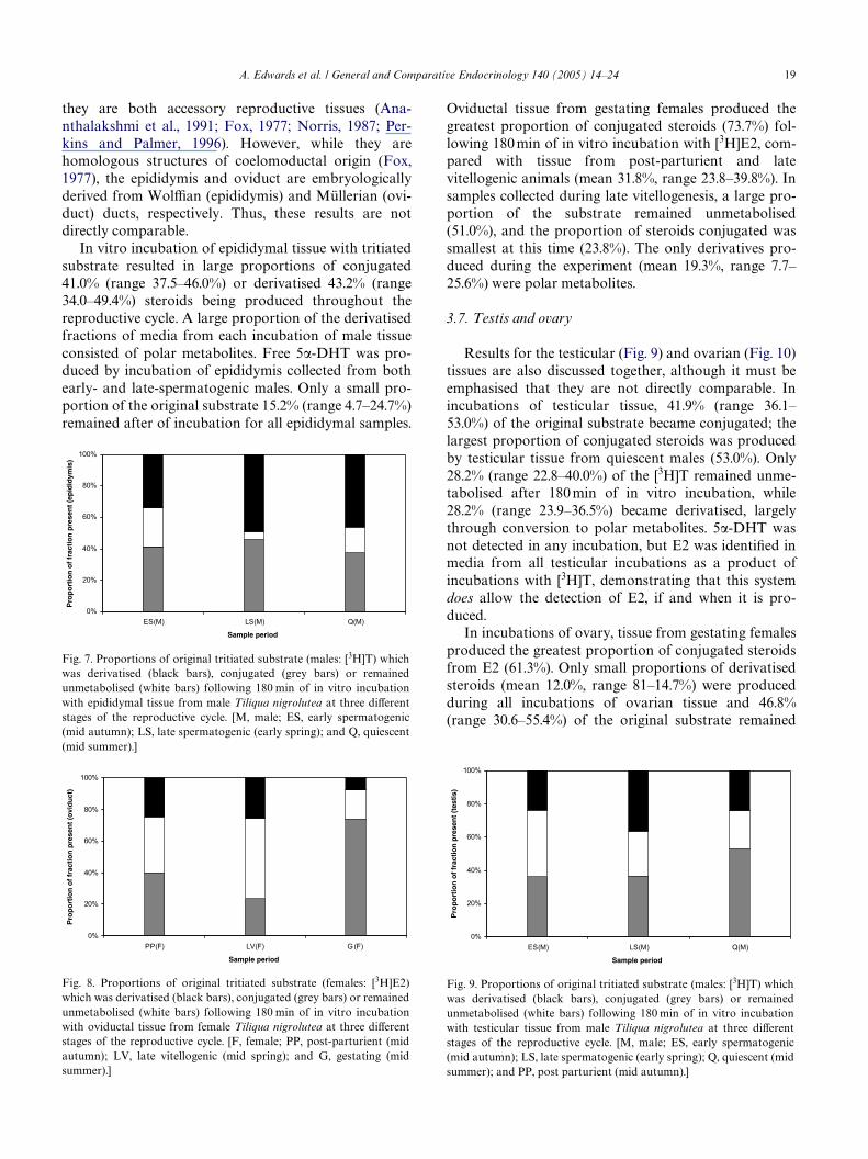
In vitro incubation of epididymal tissue with tritiatedsubstrate resulted in large proportions of conjugated41.0% (range 37.5–46.0%) or derivatised 43.2% (range34.0–49.4%) steroids being produced throughout thereproductive cycle. A large proportion of the derivatisedfractions of media from each incubation of male tissueconsisted of polar metabolites. Free 5�-DHT was pro-duced by incubation of epididymis collected from bothearly- and late-spermatogenic males. Only a small pro-portion of the original substrate 15.2% (range 4.7–24.7%)remained after of incubation for all epididymal samples.
Fig. 7. Proportions of original tritiated substrate (males: [3H]T) whichwas derivatised (black bars), conjugated (grey bars) or remainedunmetabolised (white bars) following 180 min of in vitro incubationwith epididymal tissue from male Tiliqua nigrolutea at three diVerentstages of the reproductive cycle. [M, male; ES, early spermatogenic(mid autumn); LS, late spermatogenic (early spring); and Q, quiescent(mid summer).]
Fig. 8. Proportions of original tritiated substrate (females: [3H]E2)which was derivatised (black bars), conjugated (grey bars) or remainedunmetabolised (white bars) following 180 min of in vitro incubationwith oviductal tissue from female Tiliqua nigrolutea at three diVerentstages of the reproductive cycle. [F, female; PP, post-parturient (midautumn); LV, late vitellogenic (mid spring); and G, gestating (midsummer).]
Oviductal tissue from gestating females produced thegreatest proportion of conjugated steroids (73.7%) fol-lowing 180 min of in vitro incubation with [3H]E2, com-pared with tissue from post-parturient and latevitellogenic animals (mean 31.8%, range 23.8–39.8%). Insamples collected during late vitellogenesis, a large pro-portion of the substrate remained unmetabolised(51.0%), and the proportion of steroids conjugated wassmallest at this time (23.8%). The only derivatives pro-duced during the experiment (mean 19.3%, range 7.7–25.6%) were polar metabolites.
3.7. Testis and ovary
Results for the testicular (Fig. 9) and ovarian (Fig. 10)tissues are also discussed together, although it must beemphasised that they are not directly comparable. Inincubations of testicular tissue, 41.9% (range 36.1–53.0%) of the original substrate became conjugated; thelargest proportion of conjugated steroids was producedby testicular tissue from quiescent males (53.0%). Only28.2% (range 22.8–40.0%) of the [3H]T remained unme-tabolised after 180 min of in vitro incubation, while28.2% (range 23.9–36.5%) became derivatised, largelythrough conversion to polar metabolites. 5�-DHT wasnot detected in any incubation, but E2 was identiWed inmedia from all testicular incubations as a product ofincubations with [3H]T, demonstrating that this systemdoes allow the detection of E2, if and when it is pro-duced.
In incubations of ovary, tissue from gestating femalesproduced the greatest proportion of conjugated steroidsfrom E2 (61.3%). Only small proportions of derivatisedsteroids (mean 12.0%, range 81–14.7%) were producedduring all incubations of ovarian tissue and 46.8%(range 30.6–55.4%) of the original substrate remained
Fig. 9. Proportions of original tritiated substrate (males: [3H]T) whichwas derivatised (black bars), conjugated (grey bars) or remainedunmetabolised (white bars) following 180 min of in vitro incubationwith testicular tissue from male Tiliqua nigrolutea at three diVerentstages of the reproductive cycle. [M, male; ES, early spermatogenic(mid autumn); LS, late spermatogenic (early spring); Q, quiescent (midsummer); and PP, post parturient (mid autumn).]

20 A. Edwards et al. / General and Comparative Endocrinology 140 (2005) 14–24
unmetabolised. Polar metabolites were produced in incu-bations carried out in all three sample periods.
3.8. Muscle
Only 15.0% (range 11.4–20.3%) of original substratewas converted to free steroid derivatives during in vitroincubation of male or female muscle tissue (Fig. 11), butAD and 5�-DHT were not detected in media from anyincubation with male muscle. A range of polar metabo-lites was detected in media from all incubations fromboth sexes. With the exception of gestating females,52.9% (range 45.8–62.6%) of original substrate remainedunmetabolised, and 38.1% (range 23.3–39.1%) becameconjugated. Muscle tissue from gestating females conju-
Fig. 10. Proportions of original tritiated substrate (females: [3H]E2)which was derivatised (black bars), conjugated (grey bars) or remainedunmetabolised (white bars) following 180 min of in vitro incubationwith ovarian tissues from female Tiliqua nigrolutea at three diVerentstages of the reproductive cycle. [F, female; PP, post parturient (midautumn); LV, late vitellogenic (mid spring); and G, gestating (midsummer).]
Fig. 11. Proportions of original tritiated substrate (males: [3H]T,females: [3H]E2) which was derivatised (black bars), conjugated (greybars) or remained unmetabolised (white bars) following 180 min of invitro incubation with control (skeletal muscle) tissue from male andfemale Tiliqua nigrolutea at three diVerent stages of the reproductivecycle. [M, male; F, female; ES, early spermatogenic (mid autumn); LS,late spermatogenic (early spring); Q, quiescent (mid summer); PP, postparturient (mid autumn); LV, late vitellogenic (mid spring); and G,gestating (mid summer).]
gated a greater proportion (64.3%) of the originaltritiated substrate than muscle from late vitellogenic orpost-parturient females, with 21.6% remaining as unme-tabolised substrate.
4. Discussion
All tissues examined in this study, including testis andovary, displayed the ability to metabolise a relevant pri-mary steroid (T in males and E2 in females) in vitro.Baseline levels of steroid metabolism in all tissues proba-bly incorporate a contribution from blood, which hasbeen reported in some vertebrates to possess steroid met-abolising ability (Jakob et al., 1997; Milewich et al., 1982,1983 ; Schulz, 1986). However, most tissues showed con-siderable variation in patterns of substrate derivatisationand conjugation with sex and time of year, although, asin previous studies Edwards et al., 2002a,b, 2003, thesedata were not analysed statistically.
Steroid conjugates were produced by all tissues,including skeletal muscle. Conjugation by the vertebrateliver plays an important role in solubilising reproductivesteroids for excretion in either urine or faeces (Kime,1987; Payne, 1980; Vermeirssen and Scott, 1996). In T.nigrolutea liver produced a greater proportion of conju-gates than other tissues with the least variation acrossreproductive states. In the closely related lizard, Tiliquarugosa, the liver is also an important site of steroid con-jugation (Bourne, 1981; Huf et al., 1987) for excretionvia the gut and kidney (Bourne, 1981). Conjugate forma-tion may also help to control the availability of biologi-cally active steroids in the body (Huf et al., 1987).However, in T. nigrolutea, there was little variation inthe proportion of original substrate conjugated withchanging reproductive condition, suggesting that thefunction of conjugation by the liver is to aid excretion,rather than to synthesise biologically active molecules asoccurs in Wsh (Scott and Vermeirssen, 1994; Stacey andSorensen, 1986; Stacey et al., 1994). All kidney incuba-tions also produced conjugated steroids, probably as anaid to excretion. Similarly, the kidneys are known to be amajor route of excretion of sulphated steroids in therelated lizard T. rugosa (Bourne, 1981; Huf et al., 1987).It is not clear from those studies whether the kidneysthemselves acted as the site of conjugation; however, thekidneys of the lizard Lacerta vivipara are thought toboth metabolise and excrete androgens (Dufaure andChambon, 1978).
Samples from gestating females metabolised E2 toproduce the greatest proportion of conjugated steroids,regardless of tissue type. This could be a mechanism forremoving E2 from the maternal system during gestation,as circulating estrogen concentrations are low at thistime (Edwards and Jones, 2001a). In contrast, weobserved that for the majority of female tissues, the
A. Edwards et al. / General and Comparative Endocrinology 140 (2005) 14–24 21
largest proportion of unmetabolised E2 remained afterincubations with tissues from the late vitellogenic stage:published literature documents a causal relationshipbetween E2 and vitellogenesis in vertebrates (Ho, 1987;Kime, 1987) and plasma estrogen is elevated during thisperiod (Edwards and Jones, 2001a). In males, a similar,but less pronounced trend was apparent: tissues from theearly spermatogenic-stage metabolised the lowest pro-portion of T substrate. This result was unexpected: it hasbeen suggested that elevated plasma T (which mayreXect decreased metabolism) is responsible for the later,rather than the earlier, stages of spermatogenesis in rep-tiles (Saint Girons, 1985). Indeed, circulating T concen-trations in male T. nigrolutea rise markedly betweenearly and late spermatogenesis (Edwards and Jones,2001b).
The detection of 5�-DHT in incubations of both latespermatogenic-stage cloacal tissue and male skin sam-ples from all three reproductive stages implies the pres-ence of the 5�-reductase enzyme in these tissues. Theconversion of T to 5�-DHT occurs in the skin of otherreptiles (Fergusson et al., 1985; Hews and Moore, 1995),and 5�-DHT is generally accepted to be the hormonallyactive metabolite of T in many vertebrates (Kime, 1987;Norris, 1997). Reptiles operate in a largely terrestrialenvironment, and many use chemical signals releasedfrom cloacal glands or across the epidermis to communi-cate with conspeciWcs; non-volatile lipids can be releasedby one sex to create scent trails for the other (Cooperand Vitt, 1986). For example, males of the lizard genusEumeces respond strongly to the cloacal region of con-speciWc females, or to cloacal odours from them (Cooperand Garska, 1987; Cooper et al., 1986; Trauth et al.,1987). Chemical communication is highly developed inskinks (Cooper and Trauth, 1992) and products of cloa-cal or epidermal steroid metabolism in T. nigrolutea maytherefore carry information relevant to reproductivecondition or identiWcation of individuals. Adult Tiliquascincoides use cloacal and skin odours to distinguishconspeciWcs from self (Graves and Halpern, 1991) and T.rugosa males and females are able to locate partner liz-ards by following scent trails and sniYng for airbornechemical signals (Bull et al., 1993), suggesting the use ofboth volatile and non-volatile compounds. Whether 5�-DHT of either cloacal or epidermal origin is used to con-vey similar information to, or to stimulate a responsefrom, females of T. nigrolutea, is currently unknown, butthe seasonal and sexual variation in derivative formationobserved in this study suggests that such functions arepossible. 5�-Dihydrotestosterone or one of the unidenti-Wed polar metabolites (see Edwards et al., 2002b, 2003)or conjugates produced in incubations of skin tissue mayprovide such information.
5�-Dihydrotestosterone was also detected in the incu-bations of male adrenal tissue from the early spermato-genic stage. Adrenal tissue can synthesise androgens in
males of the snake Naja naja, although production is lowand does not vary seasonally (Tam et al., 1972). In malesof T. nigrolutea, plasma T concentrations are low duringearly spermatogenesis; however, the Wnal stages of sper-matogenesis and the display of reproductive behavioursare correlated with elevated plasma T and E concentra-tions (Edwards and Jones, 2001b). The presence of extra-gonadal 5�-DHT may serve to prevent the inappropriatestimulation of estrogen-induced reproductive behav-iours because 5�-DHT cannot be aromatised (Adkins-Regan, 1981). Examples of similar diVerential roles forandrogens have been documented in Anolis carolinensis(Crews et al., 1978) and Urosaurus ornatus (Hews andMoore, 1995), but require additional study in T. nigrolu-tea.
Epididymal tissue in T. nigrolutea appears to be oneof the most metabolically active of all tissues examined.The production of 5�-DHT by epididymal tissue fromearly and late spermatogenic, but not quiescent, malesmay indicate a role for 5�-DHT in sperm production, orpossibly sperm maintenance. Accessory sex organs inother male vertebrates are also known to metabolisereproductive steroids (Hay et al., 1976; Ozon and Fou-chet, 1972; Schoonen and Lambert, 1986, 1987). In rep-tiles, epididymal function is known to be regulated byandrogens, although any distinction between theactions of T and of its metabolites is rarely considered(Dufaure and Chambon, 1978; Morel et al., 1993; Shiva-nandappa and Devaraj Sarkar, 1987). In the lizardLacerta vivipara epididymal epithelial cells are secretoryin response to androgens during the Wnal stages ofsperm maturation, which occur in the epididymis(Morel et al., 1993). The demonstrated ability of this tis-sue to metabolise androgens could well demonstratelocalised production for localised function and war-rants further investigation.
The oviduct of T. nigrolutea actively metabolises E2,producing both steroid conjugates and free derivatives.However, E2 itself is probably the most important reg-ulator of oviductal function in reptiles (Mead et al.,1981). In the lizard Xantusia vigilis (Yaron, 1972) andthe snake Thamnophis elegans (Mead et al., 1981), P4and E2 stimulate the maturation of the pre-ovulatorygenital tract. Correspondingly, in T. nigrolutea thegreatest proportion of unmetabolised E2 was detectedin incubations of oviduct from late vitellogenic(pre-ovulatory) females. This suggests that in T. nigro-lutea unmetabolised E2 has an important direct eVecton the oviduct at this time of year. While the functionof the numerous steroid conjugates and derivativesproduced by the oviduct is not certain, T. nigroluteafemales do not store sperm (Edwards, 1999), so the ste-roids are unlikely to function in sperm maintenance byfemales.
In vitro conjugation of T by T. nigrolutea was greaterin testicular tissue of quiescent males than in those

22 A. Edwards et al. / General and Comparative Endocrinology 140 (2005) 14–24
undergoing spermatogenesis. This may simply indicate achanging seasonal requirement for T in a non-conju-gated form. In the related lizard T. rugosa steroid conju-gation in the testes was described as low, and less thanoccurs in the liver or kidney (Huf et al., 1987), althoughseasonal variation has not been examined. Testicularderivatisation of T in T. nigrolutea was greatest in incu-bations of testis collected in spring, at which time testesare maximally hypertrophied (Edwards and Jones,2001b). Interestingly, no 5�-DHT or E2 were detected.In the testis of T. rugosa, androgens are metabolised tothe unusual androgen epitestosterone (epiT), which mayact as a mechanism controlling androgen availability(Huf et al., 1989). However, T is clearly the major andro-gen in T. nigrolutea (Bourne et al., 1985; Edwards et al.,2002b, 2003).
17�-Estradiol is the major end-product of ovariansteroid biosynthesis in many vertebrates (Kime, 1987;Norris, 1997) and few authors report the further metab-olism of E2 by the ovary. Ovarian steroid metabolism inT. nigrolutea resulted, with the exception of gestatingfemales, in only a small proportion of the original E2substrate becoming derivatised or conjugated, comparedwith other tissues. It is quite likely that E2 is required toinduce activity in this tissue, as in most vertebrates(Kime, 1987), and that further metabolism is largelyunrequired. 17�-Estradiol has been isolated from ovar-ian extracts in the lizard Lacerta sicula (Lupo Di Priscoet al., 1968). However, it is possible that an alternativeestrogen, and not E2, is the end-product of steroid bio-synthesis in the ovary of T. nigrolutea (Edwards et al.,2002b, 2003). If an alternative estrogen predominates inthis species, it may act on the oviduct without peripheralmetabolism.
These results demonstrate the extent to which sex,season and tissue type can inXuence steroid metabolismin a reptile from a temperate environment. The patternsillustrated here highlight the importance of consideringthese variables in exploring possible functions for circu-lating primary steroids in experimental design and incomparisons with published studies. The results alsosuggest that steroid derivatives and/or conjugates mayhave a role in physiological regulation in this reptilianspecies.
Acknowledgments
This research was funded by a University ofTasmania, School of Zoology postgraduate scholar-ship to A.E., and a University of Tasmania Small ARCgrant to S.M.J. This work was conducted underUniversity of Tasmania Animal Ethics Approval Num-bers 95046 and 98015. We thank two anonymousreviewers for some very helpful comments on themanuscript.
References
Adkins-Regan, E., 1981. Hormone speciWcity, androgen metabolismand social behaviour. Am. Zool. 21, 257–271.
Ananthalakshmi, M.N., Devaraj Sarkar, H.B., Shivabasavaiah, 1991.Experimental demonstration of androgen regulation of hemipenisin the lizard, Calotes versicolor. Zool. Sci. 8, 561–566.
ANZCCART 1993. Euthanasia of animals used for scientiWc purposes.In: Reilly, J.S. (Ed.), ANZCCART, Australia.
Atkins, A., Jones, S.M., Edwards, A., 2002. Fecal testosterone concen-trations may not be useful for monitoring reproductive status inmale blue-tongued lizards (Tiliqua nigrolutea: Scincidae). J. Herpe-tol. 36, 106–109.
Baker, M.E., 2004. Co-evolution of steroidogenic and steroid-inactivat-ing enzymes and adrenal and sex steroid receptors. Gen. Comp.Endocrinol. 215, 55–62.
Borg, B., Mayer, I., Lambert, J., Granneman, J., Schulz, R., 1992.Metabolism of androstenedione and 11-ketotestosterone in the kid-ney of the three-spined stickleback, Gasterosteus aculeatus. Gen.Comp. Endocrinol. 86, 248–256.
Bourne, A.R., 1981. Blood metabolites of injected [14C]-progesterone inthe lizard Tiliqua rugosa. Comp. Biochem. Physiol. B 70, 661–664.
Bourne, A.R., Taylor, J.L., Watson, T.G., 1985. IdentiWcation of epites-tosterone in the plasma and testis of the lizard Tiliqua (Trachydo-saurus) rugosa. Gen. Comp. Endocrinol. 58, 394–401.
Bull, C.M., Bedford, G.S., Schulz, B.A., 1993. How do sleepy lizardsWnd each other? Herpetologica 49, 294–300.
Callard, G.V., Petro, Z., Ryan, K.J., 1977. IdentiWcation of aromatasein the reptilian brain. Endocrinology 100, 1214–1218.
Cooper Jr., W.E., Garska, W.R., 1987. Lingual responses to chemicalfractions of urodaeal glandular pheromone of the skink Eumeceslaticeps. J. Exp. Zool. 242, 249–253.
Cooper Jr., W.E., Trauth, S.E., 1992. Discrimination of conspeciWcmale and female cloacal chemical stimuli by males and possessionof a probable pheromone gland by females in a cordylid lizard,Gerrhosaurus nigrolineatus. Herpetologica 48, 229–236.
Cooper Jr., W.E., Vitt, L.J., 1986. Tracking of female conspeciWc odortrails by male broad-headed skinks (Eumeces laticeps). Ethology 71,242–248.
Cooper Jr., W.E., Garska, W.R., Vitt, L.J., 1986. Female sex pheromonein the lizard Eumeces laticeps. Herpetologica 42, 361–366.
Crews, D., Traina, V., Wetzel, F.T., Muller, C., 1978. Hormonal controlof male reproductive behavior in the lizard, Anolis carolinensis: roleof testosterone, dihydrotestosterone, and estradiol. Endocrinology103, 1814–1820.
Cuevas, M.E., Miller, W., Callard, G., 1992. Sulfoconjugation of ste-roids and the vascular pathway of communication in dogWsh testis.J. Exp. Zool. 264, 119–129.
Dufaure, J.P., Chambon, M., 1978. Uptake of [3H]-testosterone in sev-eral organs of the male viviparous lizard (Lacerta vivipara Jacquin)and selective retention by the epididymis. Gen. Comp. Endocrinol.36, 23–29.
Edwards, A., 1999. Steroids and reproduction in Tiliqua nigrolutea.Unpublished Ph.D thesis, University of Tasmania, Tasmania, Aus-tralia.
Edwards, A., Jones, S.M., 2001a. Changes in plasma progesterone,estrogen and testosterone concentrations throughout the reproduc-tive cycle in female viviparous blue-tongued skinks, Tiliqua nigrolu-tea, (Scincidae), in Tasmania. Gen. Comp. Endocrinol. 122,260–269.
Edwards, A., Jones, S.M., 2001b. Changes in plasma testosterone,estrogen and progesterone concentrations throughout the annualreproductive cycle in male viviparous blue-tongued skinks, Tiliquanigrolutea, (Scincidae), in Tasmania. J. Herpetol. 35, 293–299.
Edwards, A., Jones, S.M., Wapstra, E., 2002a. Multiennial reproduc-tion in females of a viviparous, temperate-zone skink, Tiliqua nigro-lutea. Herpetologica 58, 407–414.

A. Edwards et al. / General and Comparative Endocrinology 140 (2005) 14–24 23
Edwards, A., Jones, S.M., Davies, N.W., 2002b. A possible alternativeto 17�-estradiol in a viviparous lizard, Tiliqua nigrolutea. Gen.Comp. Endocrinol. 129, 114–121.
Edwards, A., Jones, S.M., Davies, N.W., 2003. Sex and season inXuencegonadal steroid biosynthetic pathways, end-product productionand steroid conjugation in blotched blue-tongued lizards (Tiliquanigrolutea). Gen. Comp. Endocrinol. 134, 131–138.
Evans, R.M., 1988. The steroid and thyroid hormone receptor super-family. Science 240, 889–895.
Fennell, M.J., Scanes, C.G., 1992a. EVects of androgen (testosterone,5�-dihydrotestosterone, 19-nortestosterone) administration ongrowth in turkeys. Poultry Sci. 71, 539–547.
Fennell, M.J., Scanes, C.G., 1992b. Inhibition of growth in chickens bytestosterone, 5�-dihydrotestosterone, and 19-nortestosterone. Poul-try Sci. 71, 357–366.
Fergusson, B., Bradshaw, S.D., Cannon, J.R., 1985. Hormonal controlof femoral gland secretion in the lizard, Amphibolurus ornatus. Gen.Comp. Endocrinol. 57, 371–376.
Folman, Y., Ahmad, N., Sowell, J.G., Eik-Nes, K.B., 1973. Formationin vitro of 5�-dihydrotestosterone and other 5�-reduced metabo-lites of 3H-testosterone by the seminiferous tubules and interstitialtissue from immature and mature rat testes. Endocrinology 92, 41–47.
Fox, H., 1977. The urogenital system of reptiles. In: Gans, C. (Ed.),Biology of the Reptilia, vol 6. Academic Press, London and NewYork, pp. 1–157.
Graves, B.M., Halpern, M., 1991. Discrimination of self from conspe-ciWc chemical cues in Tiliqua scincoides (Sauria: Scincidae). J. Her-petol. 25, 125–126.
Hay, J.B., Hodgins, M.B., Roberts, R.J., 1976. Androgen metabolism inskin and skeletal muscle of the rainbow trout (Slamo gairdnerii)and in accessory sexual organs of the spur dogWsh (Squalus acanth-ias). Gen. Comp. Endocrinol. 29, 402–413.
Heistermann, M., Tari, S., Hodges, J.K., 1993. Measurement of fecalsteroids for monitoring ovarian function in new world primates,Callitrichidae. J. Reprod. Fertil. 99, 243–251.
Hews, D.K., Moore, M.C., 1995. InXuence of androgens on diVerentia-tion of secondary sex characters in tree lizards, Urosaurus ornatus.Gen. Comp. Endocrinol. 97, 86–102.
Ho, S.-M., 1987. Endocrinology of vitellogenesis. In: Norris, D.O.,Jones, R.E. (Eds.), Hormones and Reproduction in Fishes Amphib-ians and Reptiles. Plenum Press, New York, pp. 145–169.
Huf, P.A., Bourne, A.R., Watson, T.G., 1989. The in vitro biosynthesisof epitestosterone and testosterone from C19 steroid precursors inthe testis of the lizard Tiliqua rugosa. Gen. Comp. Endocrinol. 75,280–286.
Huf, P.A., Bourne, A.R., Watson, T.G., 1987. Metabolism of andro-gens in the brain of the lizard Tiliqua rugosa, in vitro. Proc. AustSoc. Reprod. Biol. P79. 19th Annual Conference, Sydney,Australia.
Jakob, F., Siggelkow, H., Homann, D., Koehrle, J., Adamski, J., Schu-etze, N., 1997. Local estradiol metabolism in osteoblast- and ostoe-clast-like cells. J. Steroid Biochem. Molec. Biol. 61, 167–174.
Johnson, M.H., Everitt, B.J., 1988. Essential Reproduction, third ed.Blackwell ScientiWc Publications, Great Britain.
Joss, J.M.P., Edwards, A., Kime, D.E., 1996. In vitro biosynthesis ofandrogens in the Australian lungWsh, Neoceratodus forsteri. Gen.Comp. Endocrinol. 101, 256–263.
Kime, D.E., 1979. The eVect of temperature on the testicular steroido-genic enzymes of the rainbow trout, Salmo gairdneri. Gen. Comp.Endocrinol. 39, 290–296.
Kime, D.E., 1987. The steroids. In: Chester-Jones, I., Ingleton, P.M.,Philips, J.G. (Eds.), Fundamentals of Comparative VertebrateEndocrinology. Plenum Press, New York, pp. 3–56.
Kime, D.E., Hyder, M., 1983. The eVect of temperature and gonadotro-pin on testicular steroidogenesis in Sarotherodon (Tilapia) mossam-bicus in vitro. Gen. Comp. Endocrinol. 50, 105–115.
Kime, D.E., Saksena, D.N., 1980. The eVect of temperature on thehepatic catabolism of testosterone in the rainbow trout (Salmogairdneri) and the goldWsh (Carassius auratus). Gen. Comp. Endo-crinol. 42, 228–234.
Lofts, B., 1987. Testicular function. In: Norris, D.O., Jones, R.E. (Eds.),Hormones and Reproduction in Fishes, Amphibians and Reptiles.Plenum Press, New York, pp. 283–325.
Lupo Di Prisco, C., Delrio, G., ChieY, G., 1968. Sex hormones in theovaries of the lizard Lacerta sicula. Gen. Comp. Endocrinol. 10,292–295.
Lupo Di Prisco, C., Delrio, G., ChieY, G., Cardellini, L.B., Magni, A.P.,1971. IdentiWcation and biosynthesis of steroid hormones in theovary and fat bodies of female Triturus cristatus carnifex. Comp.Biochem. Physiol. 40B, 53–60.
Lupo Di Prisco, C., Baslie, C., Delrio, G., ChieY, G., 1972. In vitrometabolism of cholesterol-4-14C and testosterone-4-14C in testesand fat bodies of Triturus cristatus carnifex. Comp. Biochem. Phys-iol. 41B, 245–249.
Manning, N.J., Kime, D.E., 1985. The eVect of temperature on testicu-lar steroid production in the rainbow trout, Salmo gairdneri, in vivoand in vitro. Gen. Comp. Endocrinol. 57, 377–382.
Mead, R.A., Eroschenko, V.P., HighWll, D.R., 1981. EVects of proges-terone and estrogen on the histology of the oviduct of the gartersnake, Thamnophis elegans. Gen. Comp. Endocrinol. 45, 345–354.
Milewich, L., Whisenant, M.G., Sawyer, M.K., 1982. Androstenedionemetabolism by human lymphocytes. J. Steroid Biochem. 16, 81–85.
Milewich, L., Lipscomb, M.F., Whisenant, M.G., MacDonald, P.C.,1983. Conversion of androstenedione to testosterone and otherandrogens in guinea-pig alveolar macrophages. J. Steroid Biochem.19, 1611–1615.
Morel, L., Dufaure, J.P., Depeiges, A., 1993. LESP, an androgen-regu-lated lizard epithelial secretory protein family identiWed as a newmember of the lipocalin superfamily. J. Biol. Chem. 268, 10274–10281.
Norris, D.O., 1987. Regulation of male gonaducts and sex accessorystructures. In: Norris, D.O., Jones, R.E. (Eds.), Hormones andReproduction in Fishes, Amphibians and Reptiles. Plenum Press,New York, pp. 327–354.
Norris, D.O., 1997. Vertebrate Endocrinology, third ed. AcademicPress, USA.
Ozon, R., 1972a. Androgens in Wshes, amphibians, reptiles and birds.In: Idler, D.R. (Ed.), Steroids in Nonmammalian Vertebrates. Aca-demic Press, New York and London, pp. 328–389.
Ozon, R., 1972b. Estrogens in Wshes, amphibians, reptiles and birds. In:Idler, D.R. (Ed.), Steroids in Nonmammalian Vertebrates. Aca-demic Press, New York and London, pp. 390–413.
Ozon, R., Fouchet, C., 1972. Action des androgènes chez l’ AmphibienAnoure Discoglossus pictus I. Métabolisme in vitro de la testosté-rone par les vésicules séminales. Gen. Comp. Endocrinol. 19, 484–493.
Payne, A.H., 1980. Testicular steroid sulfotransferases: comparison toliver and adrenal steroid sulfotransferases of the mature rat. Endo-crinology 106, 1365–1370.
Perkins, M.J., Palmer, B.D., 1996. Histology and functional morphol-ogy of the oviduct of an oviparous snake, Diadophis punctatus. J.Morphol. 227, 67–79.
Pottinger, T.G., Pickering, A.D., 1985. The eVects of11-ketotestoster-one and testosterone on the skin structure of brown trout, Salmotrutta L. Gen. Comp. Endocrinol. 59, 335–342.
Rawlinson, P.A., 1974. Biogeography and ecology of the reptiles ofTasmania and the Bass Strait area. In: Williams, W.D. (Ed.), Bioge-ography and Ecology in Tasmania. W. Junk, The Hague, pp. 230–269.
Saint Girons, H., 1985. Comparative data on lepidosaurian reproduc-tion and some time tables. In: Gans, C., Billet, F. (Eds.), Biology ofthe Reptilia. Vol 15. Development B. Wiley, New York, USA, pp.35–58.

24 A. Edwards et al. / General and Comparative Endocrinology 140 (2005) 14–24
Schlinger, B.A., Callard, G.V., 1990. Aromatization mediates aggres-sive behavior in quail. Gen. Comp. Endocrinol. 79, 39–53.
Schlinger, B.A., Fivizzani, A.J., Callard, G.V., 1989. Aromatase, 5�- and5�-reductase in brain, pituitary and skin of the sex-reversed Wil-sonís pharalope. J. Endocrinol. 122, 573–581.
Schoonen, W.G.E.J., Lambert, J.G.D., 1986. Steroid metabolism in theseminal vesicles of the African catWsh, Clarias gariepinus (Burchell),during the spawning season, under natural conditions, and kept inponds. Gen. Comp. Endocrinol. 61, 355–367.
Schoonen, W.G.E.J., Lambert, J.G.D., 1987. Gas chromatographic-mass spectrometric analysis of steroids and steroid glucuronides inthe seminal vesicle Xuid of the African catWsh, Clarias gariepinus.Gen. Comp. Endocrinol. 68, 375–386.
Schulz, R., 1986. In vitro metabolism of steroid hormones in the liverand in the blood cells of male rainbow trout (Salmo gairdneri Rich-ardson). Gen. Comp. Endocrinol. 64, 312–319.
Scott, A.P., Vermeirssen, E.L.M., 1994. Production of conjugated ste-roids by teleost gonads and their role as pheromones. In: Perspec-tives in Comparative Endocrinology. National research Council ofCanada, Canada, pp. 645–654.
Shivanandappa, T., Devaraj Sarkar, H.B., 1987. Androgenic regulationof epididymal function in the skink, Mabuya carinata (Schn). J.Exp. Zool. 241, 369–376.
Snowberger, E.A., Stegeman, J.J., 1987. Patterns and regulationof estradiol metabolism by hepatic microsomes from twospecies of marine teleosts. Gen. Comp. Endocrinol. 66, 256–265.
Stacey, N.E., Sorensen, P.W., 1986. 17�,20�-Dihydroxy-4-pregnen-3-one: a steroidal primer pheromone increasing milt volume in thegoldWsh, Cassius auratus. Can. J. Zool. 64, 2412–2417.
Stacey, N.E., Cardwell, J.R., Liley, N.R., Scott, A.P., Sorensen P.W.,1994. Hormones as sex pheromones in Wsh. In: Perspectives inComparative Endocrinology. National research Council of Can-ada, Canada, pp. 438–448.
Tam, W.H., Phillips, J.G., Lofts, B., 1972. Seasonal changes in the secre-tory activity of the adrenal gland of the cobra (Naja naja, Lin.).Gen. Comp. Endocrinol. 19, 218–224.
Trauth, S.E., Cooper Jr., W.E., Vitt, L.J., Perrill, S.A., 1987. Cloacal anat-omy of the broad-headed skink, Eumeces laticeps, with a descriptionof a female pheromonal gland. Herpetologica 43, 458–466.
Velloso, A.L., Wasser, S.K., Monfort, S.L., Dietz, J.M., 1998. Longitu-dinal fecal steroid excretion in maned wolves (Chrysocyon brachyu-rus). Gen. Comp. Endocrinol. 112, 96–107.
Vermeirssen, E.L.M., Scott, A.P., 1996. Excretion of free and conjugatedsteroids in rainbow trout (Oncorhynchus mykiss): evidence for bran-chial excretion of the maturation-inducing steroid, 17,20�-dihy-droxy-4-pregnen-3-one. Gen. Comp. Endocrinol. 101, 180–194.
Wasser, G.K., Papageorge, S., Foley, C., Brown, J.L., 1996. Excretory fateof estradiol and progesterone in the African elephant (Loxodontaafricana) and patterns of fecal steroid concentrations throughout theestrous cycle. Gen. Comp. Endocrinol. 102, 255–262.
Yaron, Z., 1972. EVects of ovariectomy and steroid replacement on thegenital tract of the viviparous lizard, Xantusia vigilis. J. Morphol.136, 313–326.
Copyright © 2022 FDOKUMEN