Past, present and future of A 2A adenosine receptor antagonists in the therapy of Parkinson's...
46
Past, present and future of A 2A adenosine receptor antagonists in the therapy of Parkinson’s disease Marie Therese Armentero *,1 , Annalisa Pinna *,2 , Sergi Ferré 3 , José Luis Lanciego 4 , Christa E. Müller 5 , and Rafael Franco 4,6 1 Laboratory of Functional Neurochemistry, Interdepartmental Research Centre for Parkinson’s Disease, IRCCS National Institute of Neurology “C. Mondino”, Pavia. Italy 2 CNR Institute of Neuroscience, Cagliari, Italy 3 National Institute on Drug Abuse, IRP, NIH, DHHS, Baltimore, Maryland, United States of America 4 Neurosciences Division, Center for Applied Medical Research (CIMA & CIBERNED), University of Navarra, Pamplona, Spain 5 PharmaCenter Bonn, Pharmaceutical Institute, Pharmaceutical Chemistry I, University of Bonn, Germany 6 Department of Biochemistry & Molecular Biology, University of Barcelona, Spain Abstract Several selective antagonists for adenosine A 2A receptors (A 2A R) are currently under evaluation in clinical trials (phases I to III) to treat Parkinson’s disease, and they will probably soon reach the market. The usefulness of these antagonists has been deduced from studies demonstrating functional interactions between dopamine D 2 and adenosine A 2A receptors in the basal ganglia. At present it is believed that A 2A R antagonists can be used in combination with the dopamine precursor L-DOPA to minimize the motor symptoms of Parkinson’s patients. However, a considerable body of data indicates that in addition to ameliorating motor symptoms, adenosine A 2A R antagonists may also prevent neurodegeneration. Despite these promising indications, one further issue must be considered in order to develop fully optimized anti-parkinsonian drug therapy, namely the existence of receptor (hetero)dimers/oligomers of G protein-coupled © 2011 Elsevier Inc. All rights reserved. Correspondence address: Rafael Franco, Dept. Biochemistry and Molecular Biology, School of Biology, Universitat de Barcelona, Avda. Diagonal 645, 08028 Barcelona, Spain. Tel +34934021208 Fax +34934021255, [email protected]. * These authors contributed equally to the present work Website list www.biotie.com/en/recearch_and_development/central_nervous_system_disorders/syn115 http://www.clinicaltrials.gov/ct2/results?term=preladenant www.clinicaltrials.gov/ct2/results?term=P04938 www.clinicaltrials.gov/ct2/results?term=P05664 www.clinicaltrials.gov/ct2/results?term=P07037 www.clinicaltrials.gov/ct2/results?term=P06153 www.istradefylline.com/fda.html www.sigma-tau.it/eng/areediricerca.asp www.vernalis.com/media-centre/latest-releases/2010-releases/584 www.vernalis.com/media-centre/latest-releases/596 Publisher's Disclaimer: This is a PDF file of an unedited manuscript that has been accepted for publication. As a service to our customers we are providing this early version of the manuscript. The manuscript will undergo copyediting, typesetting, and review of the resulting proof before it is published in its final citable form. Please note that during the production process errors may be discovered which could affect the content, and all legal disclaimers that apply to the journal pertain. NIH Public Access Author Manuscript Pharmacol Ther. Author manuscript; available in PMC 2012 December 1. Published in final edited form as: Pharmacol Ther. 2011 December ; 132(3): 280–299. doi:10.1016/j.pharmthera.2011.07.004. NIH-PA Author Manuscript NIH-PA Author Manuscript NIH-PA Author Manuscript
-
Upload
independent -
Category
Documents
-
view
0 -
download
0
Transcript of Past, present and future of A 2A adenosine receptor antagonists in the therapy of Parkinson's...

Past, present and future of A2A adenosine receptor antagonistsin the therapy of Parkinson’s disease
Marie Therese Armentero*,1, Annalisa Pinna*,2, Sergi Ferré3, José Luis Lanciego4, ChristaE. Müller5, and Rafael Franco4,6
1Laboratory of Functional Neurochemistry, Interdepartmental Research Centre for Parkinson’sDisease, IRCCS National Institute of Neurology “C. Mondino”, Pavia. Italy2CNR Institute of Neuroscience, Cagliari, Italy3National Institute on Drug Abuse, IRP, NIH, DHHS, Baltimore, Maryland, United States ofAmerica4Neurosciences Division, Center for Applied Medical Research (CIMA & CIBERNED), Universityof Navarra, Pamplona, Spain5PharmaCenter Bonn, Pharmaceutical Institute, Pharmaceutical Chemistry I, University of Bonn,Germany6Department of Biochemistry & Molecular Biology, University of Barcelona, Spain
AbstractSeveral selective antagonists for adenosine A2A receptors (A2AR) are currently under evaluationin clinical trials (phases I to III) to treat Parkinson’s disease, and they will probably soon reach themarket. The usefulness of these antagonists has been deduced from studies demonstratingfunctional interactions between dopamine D2 and adenosine A2A receptors in the basal ganglia. Atpresent it is believed that A2AR antagonists can be used in combination with the dopamineprecursor L-DOPA to minimize the motor symptoms of Parkinson’s patients. However, aconsiderable body of data indicates that in addition to ameliorating motor symptoms, adenosineA2AR antagonists may also prevent neurodegeneration. Despite these promising indications, onefurther issue must be considered in order to develop fully optimized anti-parkinsonian drugtherapy, namely the existence of receptor (hetero)dimers/oligomers of G protein-coupled
© 2011 Elsevier Inc. All rights reserved.Correspondence address: Rafael Franco, Dept. Biochemistry and Molecular Biology, School of Biology, Universitat de Barcelona,Avda. Diagonal 645, 08028 Barcelona, Spain. Tel +34934021208 Fax +34934021255, [email protected].*These authors contributed equally to the present workWebsite listwww.biotie.com/en/recearch_and_development/central_nervous_system_disorders/syn115http://www.clinicaltrials.gov/ct2/results?term=preladenantwww.clinicaltrials.gov/ct2/results?term=P04938www.clinicaltrials.gov/ct2/results?term=P05664www.clinicaltrials.gov/ct2/results?term=P07037www.clinicaltrials.gov/ct2/results?term=P06153www.istradefylline.com/fda.htmlwww.sigma-tau.it/eng/areediricerca.aspwww.vernalis.com/media-centre/latest-releases/2010-releases/584www.vernalis.com/media-centre/latest-releases/596Publisher's Disclaimer: This is a PDF file of an unedited manuscript that has been accepted for publication. As a service to ourcustomers we are providing this early version of the manuscript. The manuscript will undergo copyediting, typesetting, and review ofthe resulting proof before it is published in its final citable form. Please note that during the production process errors may bediscovered which could affect the content, and all legal disclaimers that apply to the journal pertain.
NIH Public AccessAuthor ManuscriptPharmacol Ther. Author manuscript; available in PMC 2012 December 1.
Published in final edited form as:Pharmacol Ther. 2011 December ; 132(3): 280–299. doi:10.1016/j.pharmthera.2011.07.004.
NIH
-PA Author Manuscript
NIH
-PA Author Manuscript
NIH
-PA Author Manuscript

receptors, a topic currently the focus of intense debate within the scientific community. DopamineD2 receptors (D2Rs) expressed in the striatum are known to form heteromers with A2A adenosinereceptors. Thus, the development of heteromer-specific A2A receptor antagonists represents apromising strategy for the identification of more selective and safer drugs.
1. IntroductionAdenosine receptors (AR) are members of the G protein-coupled receptor superfamily thathave long been considered potential targets for the treatment of a variety of diseases,although to date adenosine (Adenocard® or Adenoscan®) is the only commerciallyavailable therapeutic drug acting on AR. Adenocard® is used clinically to revert paroxysmalsupraventricular tachycardia, while Adenoscan® is also used for cardiac imaging due to itsvasodilatory effects mediated by A2A receptors in blood vessels. Recently, the A2A-selectiveagonist regadenoson (Lexiscan®) was approved for the same indication. Despite the poorselection of available compounds, it is still believed that drugs acting on adenosine receptorswill be therapeutically useful. Indeed, five clinical trials are currently underway (phases I toIII) to analyze the therapeutic potential of adenosine A2A receptor (A2AR) antagonists in thetreatment of Parkinson’s disease (PD). Novel adenosine antagonists may thus soon reach themarket. The potential of these antagonists has been deduced from considerable investigationof the functional interactions between dopamine and adenosine receptors in the basalganglia. The use of A2AR antagonists in Parkinson’s disease (PD) is based on solidpreclinical data showing that adenosinergic neuromodulation antagonizes dopaminergicneurotransmission in aspects relevant to motor control. Adenosine receptor antagonist-basedtherapy was initially founded on the hypothesis that preventing such antagonism could beuseful in situations of dopamine deficit, such as occurs in Parkinson’s disease. Notableefforts in medicinal chemistry have sought to develop A2AR antagonists. While the firstapproaches focused on xanthine derivatives, the current portfolio also includes highlypromising non-xanthine drugs.
The use of A2AR antagonists in PD is not exclusively dependent on the outcome of theongoing clinical trials with structurally distinct molecules. This is due to a shift in emphasisfrom simply improving the motor symptoms of the patients to developing strategies toprevent disease progression. Given the established efficacy of L-DOPA, and for ethicalreasons, the main approach currently used in clinical trials involves the co-administration ofA2AR antagonists with L-DOPA. The proposed advantage of this strategy is a reduction inthe required dose of L-DOPA, with concomitant reductions in the associated side effects,consisting mainly of dyskinesias and progressive cognitive impairment. Preclinical findingsalso indicated potential neuroprotective effects of A2AR antagonists, an aspect highlyrelevant to PD treatment. Thus, in addition to improving motor symptoms whenadministered in combination with L-DOPA, A2AR antagonists may also exhibit true disease-modifying activity, delaying the progression of disease. Whether all A2AR antagonists beingcurrently assayed in clinical trials are equally effective as co-adjuvants remains to bedetermined. However, the development of A2AR antagonists for the treatment basal gangliadisorders should focus on optimizing both their effects against acute symptoms and theirneuroprotective activity.
An additional and important consideration for the development of A2AR antagonistsconcerns the novel pharmacological effects derived from G protein-coupled receptorheteromerization. The existence of receptor heteromers has had a strong impact on the fieldof G protein-coupled receptors, raising important questions as to whether the real therapeutictargets are receptor monomers, homodimers or heteromers. A2AR and dopamine D2receptors (D2R) were among the first G protein-coupled receptor heteromers identified, and
Armentero et al. Page 2
Pharmacol Ther. Author manuscript; available in PMC 2012 December 1.
NIH
-PA Author Manuscript
NIH
-PA Author Manuscript
NIH
-PA Author Manuscript

have been detected in both transfected cells and brain striatal tissue (Soriano et al., 2009).Since receptor pharmacology is modified by heteromerization, the screening of givenreceptors in different heteromeric contexts should be incorporated into future drug discoveryprogrammes. Promising results have been obtained relating to A2AR heteromers (Orrú et al.,2011), which are implicated in Parkinson’s and Huntington’s diseases (HD), among others.As structurally distinct A2AR antagonists may exert differential effects on distinct A2AR-containing heteromers, different A2AR antagonists may be useful for the treatment ofspecific neurological disorders, depending on the heteromer preferentially targeted by thedrug. In this review, we aim to address all these past-, present- and future aspects of theA2ARs and their antagonists.
2. Normal and abnormal basal ganglia functionPD is a basal ganglia-associated disorder that affects 1-2% of individuals over 60 years ofage. The main symptoms of the disease are motor-related, including reduced spontaneousmovement, akinesia (lack of movement), bradykinesia (slowness of movements), rigidity(due to increased muscular tone), as well as the characteristic resting tremor. Theintroduction of the dopamine precursor L-DOPA in the late 1960’s, later followed by anumber of dopamine agonists, has revolutionized the clinical management of PD (at leastwithin a period of 5-7 years after the first diagnosis, often referred to as the“pharmacological honeymoon”). These therapeutic tools have proven efficient in restoringmost of the motor-related deficits characterizing PD. However, even the most effective anti-parkinsonian drugs only provide symptomatic relief. Furthermore, as the disease continuesto progress, troublesome side effects generally appear several years after initiation ofpharmacological treatment, including motor fluctuations (on-off phenomena) and L-DOPA-induced dyskinesias. In parallel with the appearance of long-term motor disturbances,patients begin to develop non-motor problems, resulting in a marked decline in quality oflife. At this stage of disease progression, symptoms like mild cognitive impairment(including dementia in later disease stages), difficulties swallowing and speaking, autonomicdysfunction, gait disturbances and loss of balance (including frequent falls), represent themajor causes of disability for PD patients. At present, it seems clear that PD cannot solely beattributed to nigrostriatal dopaminergic deficits. As such the involvement of non-dopaminergic systems, particularly in relation to the appearance of non-motor symptoms,needs to be better defined. In this scenario, current strategies aim to either: (i) slow down thenatural progression of PD through disease-modifying therapies, or (ii) better manage theside-effects that appear after long-term treatment with L-DOPA, particularly ‘on-off’phenomena and abnormal involuntary movements (i.e.: L-DOPA-induced dyskinesia).
The basal ganglia are a group of subcortical nuclei involved in the planning and initiation ofmovement, and are anatomically and functionally organized into parallel circuits thatprocess different types of information. All basal ganglia-related nuclei are connectedthrough well-established neurochemical circuits. When considering the activity of theseganglia, one should bear in mind that after release, the effects of neurotransmittersultimately depend on the pre- and post-synaptic localization of a number of receptors, aswell as on the relationships established with other inputs received by a given neuron.Briefly, the basal ganglia nuclei are divided into: (i) input nuclei that receive informationfrom the cortex and thalamus, consisting of the caudate, putamen and accumbens nuclei; (ii)output nuclei that send information from the basal ganglia to the thalamus, comprised of theinternal division of the globus pallidus (GPi) and the substantia nigra pars reticulata (SNr);and (iii) intrinsic nuclei, composed of the external division of the globus pallidus (GPe), thesubthalamic nucleus (STN) and the substantia nigra pars compacta (SNc). In addition tothese input, output and intrinsic nuclei, several other nuclei are tightly linked to the basalganglia system, including the tegmental pedunculopontine nucleus (PPN), the caudal
Armentero et al. Page 3
Pharmacol Ther. Author manuscript; available in PMC 2012 December 1.
NIH
-PA Author Manuscript
NIH
-PA Author Manuscript
NIH
-PA Author Manuscript

intralaminar nuclei (CM-Pf complex), and to some extent, the deep cerebellar nuclei thatalso fulfil motor-related functions.
The current model of basal ganglia function and dysfunction (Albin et al., 1989; DeLong,1990) was postulated in the late 1980’s. This proposes that under tonic dopaminergicconditions, the striatum (known as the caudate-putamen) integrates cortical and thalamicinformation that then reaches the output nuclei, finally arriving to the cerebral cortex via athalamic relay. There are two main ways by which striatal information can reach the basalganglia output nuclei: firstly, via a monosynaptic projection called the “direct pathway” thatoriginates in striatal medium-sized spiny neurons (MSNs) expressing dopamine type 1receptor (D1R) and ends in GPi/SNr nuclei; secondly, through a multisynaptic circuit calledthe “indirect pathway”, which begins in striatal MSNs expressing type 2 dopamine receptors(D2R) that project to the GPe. From the GPe, this latter pathway reaches the output nucleiafter establishing a synaptic relay in the STN nucleus. One of the cornerstones of this modelis the so-called “dual effect” of dopamine at the striatal level, based on the complementaryexpression of D1 and D2 receptors by striatal MSNs that project through the direct and theindirect pathways, respectively. Accordingly, dopamine exerts D1-mediated excitation ofstriatal MSNs projecting to GPi/SNr targets, and D2-mediated inhibition of striatal MSNsinnervating the GPe nucleus. Thus, dopamine exerts a dichotomous effect at the striatal leveldepending on the type of dopaminergic receptor (D1R or D2R) located post-synaptically instriatofugal MSNs. Based on this dichotomous effect of dopamine acting on D1R and D2R,activation of the direct pathway is proposed to facilitate movement, whereas activation ofthe indirect pathway is thought to result in movement inhibition. Although anatomical andfunctional evidence has challenged this hypothesis (Kawaguchi et al., 1990; Surmeier et al.,2005; Gatev et al., 2006), it has been recently validated through the use of optogenetictechniques (Kravitz et al., 2010).
Following dopaminergic denervation, the aforementioned model predicts that the reducedactivation of dopaminergic receptors will decrease the excitation of the D1R-containingstriatofugal neurons of the direct pathway, along with a concomitant disinhibition of D2R-containing striatal neurons of the indirect pathway. This sequence of events is proposed toprovoke exacerbated GABAergic flux from basal ganglia output neurons, ultimately leadingto excessive inhibition of thalamocortical projections (Crossmann, 1987; Albin et al., 1989;DeLong, 1990; Obeso et al., 1997). The reverse of this phenomenon is predicted to accountfor the dyskinetic state associated with continuous L-DOPA treatment. This induces anumber of downstream changes in basal ganglia circuits, ultimately resulting in hypoactivityof the basal ganglia output, reducing the inhibition of thalamocortical neurons and leading toexcessive activation of cortical motor areas. This view is strongly supported by previousstudies (Filion et al., 1991; Lozano et al., 2000). In summary, although the current basalganglia model served as a good starting point, it is important to stress that basal gangliaorganization is far more elaborate than assumed in this model (reviewed in Obeso et al.,2000a; Bar-Gad and Bergman, 2001). Indeed, a number of anatomical, electrophysiologicaland clinical findings are poorly explained by the current model (Obeso et al., 1997, 2000b;Wichmann and DeLong, 2000; Nambu, 2008). Many of the inherent limitations of thismodel were imposed by the manner in which it was originally conceived, which wassomewhat biased by the preponderance of anatomical and neurochemical data available atthe time. The basal ganglia is thus a much more sophisticated and dynamic network thanthat reflected in the basal ganglia model. In addition to new insights into the function of thebasal ganglia at the systems level, recent findings detailing neurotransmission throughoutbasal ganglia networks should be taken into consideration for a more completeunderstanding of its organization and activity. This is particularly relevant to the role playedby G protein-coupled receptor heteromers in the modulation of striatal neurotransmission, in
Armentero et al. Page 4
Pharmacol Ther. Author manuscript; available in PMC 2012 December 1.
NIH
-PA Author Manuscript
NIH
-PA Author Manuscript
NIH
-PA Author Manuscript

normal conditions, under circumstances of dopaminergic depletion and in the dyskineticstate associated with continuous administration of L-DOPA and/or dopamine agonists.
3. The role of A2A receptors (A2ARs) and A2AR heteromers in the modulationof striatal neurotransmission
The A2ARs are highly expressed in the basal ganglia and depend on Gs and other interactingproteins for correct transduction of their signals (Burgueño et al., 2003). The striatum is theanatomical region in mammals that most strongly expresses A2ARs, which are thought tofulfil an important role in the regulation of dopaminergic transmission in the basal ganglia(see Morelli et al., 2009). For instance, A2ARs co-localize postsynaptically with D2Rs inGABAergic striatopallidal enkephalinergic MSNs. Stimulation of these postsynaptic A2ARcounteracts the inhibitory modulation of NMDA receptor activity mediated by D2Rs, whichincludes regulating Ca2+ influx, transition to the firing “up” state and modulation ofneuronal firing in the “up” state (Azdad et al., 2009; Higley and Sabatini, 2010). Thisinteraction appears to be responsible for most of the locomotor depression and activationprovoked by A2AR agonists and antagonists, respectively (Ferré et al., 2008). AdenosineA2AR-mediated activity is usually antagonistic to that mediated by striatal D2R in MSNs.Indeed, functional antagonism between A2A and D2 receptors was recently reported instriatal cholinergic interneurons (Tozzi et al., 2011). Overall, adenosine-dopamineantagonism underlies the potential therapeutic benefits of A2AR-selective antagonists in PD.The well known regulation of motor control mediated by A2ARs under conditions ofdopamine depletion is sufficiently solid to merit the clinical trials currently underway (seesection 7), which aim to demonstrate the therapeutic efficacy of A2AR antagonists in PD.
The existence of A2AR-containing heteromers was first reported at the beginning of the 21st
century (Hillion et al., 2002; Fuxe et al., 2003; Canals et al., 2003; Torvinen et al., 2005).Since then, accumulating evidence suggests that they may be the real targets of therapeuticagents used to combat basal ganglia disorders. Receptor heteromers are the focus of intenseresearch since through heteromerization, receptors become unique functional entities withdifferent properties from those of each of the receptors involved (Ferré et al., 2009). ThusA2AR in A2AR-containing heteromers are different therapeutic targets to A2ARs that do notform heteromers. The allosteric interaction between different receptors in a given heteromer,e.g., A2AR and D2R in the A2AR-D2R heteromer, may modify the pharmacologicalparameters of A2AR or D2R-selective radioligands. In fact, as indicated in section 8.1, agiven A2AR-selective antagonist may display quite different affinities for the “same” A2ARin different heteromeric contexts. When these allosteric changes are measurable, they maybe considered as a biochemical fingerprint of the heteromer. Identification of biochemicalfingerprints is one of the few procedures available for the detection of heteromers in nativetissues. Using this approach and also by the use of bivalent ligands, A2AR-D2R heteromershave been detected in brain striatum (Soriano et al., 2009). Moreover, the A2AR-D2Rheteromer has been reported in animal models of PD (data in preparation). Taken togetherthese findings suggest that dopamine-based therapies for PD (such as L-DOPA) may targetD2R-containing heteromers, while the A2AR antagonists proposed for the treatment of PDmay target A2AR-containing heteromers (reviewed in Franco, 2009 and Casadó et al., 2009).
When attempting to define the physiological significance of the diverse receptor heteromers,it should be noted that the composition of A2AR-containing presynaptic and postsynapticheteromers differs. Postsynaptically, A2AR may not only form heteromers with D2R but alsowith cannabinoid CB1 receptors (CB1R). The apparent need for A2AR activation to mediatethe motor-depressant effects of endocannabinoids, which occurs tonically in the presence ofsignificant levels of extracellular adenosine, appears to be dependent on the existence ofA2AR-CB1R heteromers. This “property” of the CB1R-A2AR heteromer (i.e.: the
Armentero et al. Page 5
Pharmacol Ther. Author manuscript; available in PMC 2012 December 1.
NIH
-PA Author Manuscript
NIH
-PA Author Manuscript
NIH
-PA Author Manuscript

dependence on A2AR activation for CB1R engagement, Carriba et al., 2007), predicts thatA2AR antagonists produce effects similar to CB1R antagonists. Indeed, motor depressioninduced by the bilateral striatal infusion of a CB1R agonist in rats is completely counteractedby systemic administration of either CB1R or A2AR antagonists (Carriba et al., 2007).Similarly, genetic inactivation of A2AR in mice significantly decreases the cataleptic andrewarding effects of systemically administered CB1R agonists in a conditioned placepreference paradigm (Andersson et al., 2005; Soria et al., 2006). The existence ofpostsynaptic CB1R heteromers provides new indications regarding the potential mechanismsunderlying the endocannabinoid-mediated control of striatal function, as these heteromerscannot just be considered as retrograde effectors that inhibit neurotransmitter release.
Given the colocalization of CB1, A2A and D2 receptors in striatal neurons and the reportedinterplay between A2AR and CB1R, A2AR and D2R, and CB1R and D2R (Hillion et al.,2002; Tebano et al., 2009; Navarro et al., 2009; Ferré et al., 2010), the existence of striatalheterotrimers is a clear possibility. It is important to consider that at the structural level,based on the characterization of the trimer in a heterologous system, a trimer’s function isdetermined by its quaternary structure. Indeed, the structural alterations produced in mutantreceptors result in a distinct signalling fingerprint in the trimer. Remarkably, the cross-talkderived from the co-activation of A2AR and D2R depends on a correct trimer structure,which requires the presence of the 3 receptors. Indeed, this CB1R-dependent adenosine-dopamine cross-talk may be considered the fingerprint of the heteromer. The existence ofA2AR-CB1R-D2R heteromers in the striatum is based on the detection of the trimerfingerprint in slices from wild type but not CB1R KO animals (for details, see Carriba et al.,2008 and Navarro et al., 2010). Further studies are required to determine the exact role ofA2AR-CB1R-D2R heteromers in striatal function, particularly in states of dopaminedepletion.
Striatal A2ARs are localized both pre and postsynaptically in glutamatergic terminals, wherethey heteromerize with A1 receptors (A1Rs) and fine-tune glutamate release (Ciruela et al.,2006; Quiroz et al., 2009). These A1R-A2AR heteromers appear to work as concentration-dependent switches (Ferré et al., 2007), with adenosine acting primarily at A1Rs at lowconcentrations but at both A1Rs and A2ARs at higher concentrations. Activation of A1R inthe A1R-A2AR heteromer inhibits glutamate release, while the additional activation of theA2AR produces the opposite effect, acting through a mechanism that seems to involveallosteric modulation of the receptor heteromer and interactions at the G protein level(Ciruela et al., 2006, Ferré et al., 2007). Interestingly, presynaptic A2ARs are preferentiallylocalized in glutamatergic terminals of corticostriatal afferents to the dynorphinergic MSNs(Quarta et al., 2004; Quiroz et al., 2009). Apart from morphological evidence provided byimmunohistochemistry and electron microscopy, patch-clamp experiments inenkephalinergic and dynorphinergic MSNs have functionally demonstrated the segregationof striatal presynaptic A2ARs. Thus, an A2AR agonist and an A2AR antagonist increase anddecrease, respectively, the amplitude of excitatory post-synaptic currents induced by theintrastriatal stimulation of glutamatergic afferents measured in enkephalinergic, but notdynorphinergic MSNs. Indeed, a mean-variance analysis indicates a presynaptic locus forthe effect mediated by A2AR (Quiroz et al., 2009). These findings suggest a selective A2AR-mediated modulation of glutamate release to dynorphinergic MSNs, a scenario whichcontrasts with the recently proposed role of post-synaptic A2ARs in the modulation ofglutamate release to enkephalinergic MSNs (Lerner et al., 2010).
The data presented in this section indicate that heteromers containing A2AR must beconsidered when attempting to understand the effects of selective A2AR agonists andantagonists, and when developing compounds to compensate for basal ganglia dysfunction.Two main types of A2AR-containing heteromers exist, namely pre- and postsynaptic. The
Armentero et al. Page 6
Pharmacol Ther. Author manuscript; available in PMC 2012 December 1.
NIH
-PA Author Manuscript
NIH
-PA Author Manuscript
NIH
-PA Author Manuscript

prototypic presynaptic striatal receptor is the A1R-A2AR found in glutamatergic terminals.Post-synaptic striatal heteromers containing A2AR in the spines of GABAergicenkephalinergic neurons are formed with D2R and/or CB1R. Targeting pre- versuspostsynaptic A2AR, or vice versa, may represent a useful approach to differentially combatdisorders affecting the basal ganglia.
4. OVERVIEW OF IMPORTANT A2AR ANTAGONISTSSeveral review articles on adenosine receptor ligands in general (Müller & Jacobson, 2011a;Müller & Jacobson, 2011b; Fredholm et al., 2011) and A2AR antagonists in particular(Müller & Ferré, 2010; Clementina & Giuseppe, 2010; Shah & Hodgson, 2010; Cristalli etal., 2009) have appeared recently. The first adenosine receptor antagonists described in theliterature were the plant alkaloids caffeine (1) and theophylline (2), which are characterizedby their core xanthine structure (Figure 1). These compounds are non-selective, relativelyweak antagonists with Ki values in the micromolar concentration range for the four humanreceptor subtypes (see Table 1) and are approximately equally potent at rat A1, A2A and A2Breceptors, though do not act at the rat A3 receptor subtype.
5. Rationale for the screening of adenosine A2AR antagonists to preventneurodegeneration in Parkinson’s disease5.1. Neuroprotection and Parkinson’s disease - a general outlook
Replenishing the depleted dopamine stores with L-DOPA, its immediate precursor, thusmimicking dopamine-mediated neurotransmission, remains the basis of current PDtreatment. Although this replacement therapy offers immediate and effective symptomaticrelief, especially in the early stages of the disease, it does not have any influence on theunderlying neurodegenerative processes. As a consequence, neuronal cell death progressesover time and is paralleled by a gradual loss of drug efficacy. Thus, steady adaptation isnecessary to maintain adequate symptomatic relief, mainly by increasing the doses ofdopaminergic drugs, thereby favouring the emergence of side effects, such as dyskinesia andpsychiatric disturbances. The discovery of new drugs, or the combined use of knowncompounds that not only alleviate motor symptoms but that also delay or even halt the lossof dopaminergic neurons, are fundamental issues in generating alternative therapeuticstrategies for PD.
The search for neuroprotective therapies in PD has continued for more than a quarter of acentury, involving an impressive number of patients thus far (Voss & Ravina, 2008). Someencouraging results have been obtained using rasagiline, a potent MAO-B inhibitor (Olanowet al., 2008; Olanow et al., 2009; Weinreb et al., 2010), which produces disease-modifyingeffects in PD patients in early treatment stages. Nevertheless, to date no drug demonstratingneuroprotective effects has been unequivocally approved for use in humans. A majordrawback in the development of a neuroprotective therapy is the multifactorial nature of PD.The primary causes of the degenerative process underlying the disease remain unclear.Aside from several gene mutations that have been shown to cause familiar PD (Nuytemanset al., 2010), extensive studies using experimental models of PD, as well as large post-mortem studies have identified the presence of numerous cellular and molecular defects. Inparticular, oxidative stress and excitotoxic mechanisms, mitochondrial and ubiquitin/proteasomal system (UPS) dysfunction, increased MAO (MAO-A and MAO-B) activity, aswell as significant neuroinflammatory processes have all been associated with PD (Cookson& Bandmann, 2010). Any of these alterations could, in principle, represent a potentialtherapeutic target and many have indeed been singularly targeted (Schapira, 2009). It hasbecome evident over the years however, that pathogenetic pathways do not contribute todisease severity in a linear fashion but rather are highly interconnected and act in parallel.
Armentero et al. Page 7
Pharmacol Ther. Author manuscript; available in PMC 2012 December 1.
NIH
-PA Author Manuscript
NIH
-PA Author Manuscript
NIH
-PA Author Manuscript

Therefore, processes leading to nigral cell death are likely to represent the reciprocalcumulative interaction of factors that have little effect on their own. Similarly, mechanismsthat sustain ongoing cell loss might differ from initial triggers and are likely to change asdopaminergic neurodegeneration progresses. This strongly suggests that drugs with a singletarget will be unable to compensate for, or correct, complex alterations. To restore alteredactivities and potentially slow down disease progression, any therapeutic strategy for PDwill likely have to: i) act on different pathways simultaneously; and ii) adapt and evolve asthe disease progresses. Thus, as the causes of PD are manifold and interconnected, it is clearthat a multilateral approach is needed to effectively treat this complex neurodegenerativedisease, using a combination of molecules acting on different pathways. Alternatively,different compounds with promiscuous activity could be developed that operate at multipletargets, providing both symptomatic and neuroprotective benefits.
Antagonists of A2AR have proven highly efficient in restoring motor function in animalmodels of PD and, as described below (see section 7), have produced some encouragingresults in clinical trials. Blockade of A2AR may confer neuroprotection against a largespectrum of brain insults in animal models of ischemia, epilepsy, PD, HD and Alzheimer’sdisease (AD: Gomes et al., 2010). We will review how A2AR antagonists, aside from theirwell-documented effects on the motor symptoms characteristic of PD, can target severalcellular mechanisms implicated in the underlying neurodegenerative process and possiblyconfer significant neuroprotection in PD.
5.2. Caffeine, A2AR antagonists and PD – when the past meets the presentIn past decades, several large convergent follow-up studies involving more than 150,000subjects, both male and female, have demonstrated an inverse relationship between theconsumption of coffee and the risk of developing PD (Costa et al., 2010). In particular,consumption of caffeinated but not decaffeinated coffee was correlated with a reduced riskof developing the disease (Ascherio et al., 2001; Powers et al., 2008; Ross et al., 2000a;Ross et al., 2000b; Saaksjarvi et al., 2008), indicating that caffeine, a non selective A1/A2Areceptor antagonist, is directly implicated in neuroprotection. Indeed, a large body ofevidence continues to support the neuroprotective potential of caffeine both in vitro and invivo.
In vitro, caffeine (1) can protect rat mesencephalic cells from 6-hydroxydopamine (6-OHDA) toxicity, increasing cell viability, reducing the number of apoptotic cells andcompletely blocking toxin-induced lipid peroxidation (Nobre et al., 2010). Importantly, inthese cell cultures caffeine also reduces the number of activated astrocytes and microgliainduced by the neurotoxin. Caffeine also protects against 1-methyl-4-phenylpyridinium(MPP+)-induced mitochondrial complex I inhibition in cerebellar granule neurons (Alvira etal., 2007). Recently, the development of a rapid assay to evaluate neuroprotective agents hasconfirmed that caffeine can efficiently reduce toxin-induced PD-related abnormalities inSH-SY5Y cells (Yong-Kee et al., 2010). Analogously, caffeine prevented apoptotic celldeath and induced phosphorylation of protein kinase B (Akt) in a dose-dependent manner,suggesting that it exerts a protective effect through activation of the PI3K/Akt pathway(Nakaso et al., 2008), a pathway known to be altered in PD (Armentero et al., 2010;Morisette et al., 2010).
In vivo, administration of caffeine in different experimental paradigms (acute vs chronic,pre- vs. post-treatment) protected against nigrostriatal degeneration in 1-methyl-4-phenyl-1,2,3,6-tetrahydropyridine (MPTP)-treated mice (Chen et al., 2001b; Kalda et al.,2006; Singh et al., 2009; Xu et al., 2002), a well characterized animal model of PD.Recently, caffeine has also been shown to attenuate the loss of dopaminergic neurons inmice treated with a combination of paraquat and maneb (Kachroo et al., 2010), two
Armentero et al. Page 8
Pharmacol Ther. Author manuscript; available in PMC 2012 December 1.
NIH
-PA Author Manuscript
NIH
-PA Author Manuscript
NIH
-PA Author Manuscript

pesticides linked to increased risk of developing PD (Costello et al., 2009). Notably, caffeinetreatment reduced neuronal cell death and glial activation in lipopolysaccharide (LPS)-treated mice (Brothers et al., 2010), an animal model of dopaminergic degeneration. Arecent exhaustive effort to identify genes involved in the molecular mechanisms of caffeine-mediated neuroprotection towards MPTP-induced alterations in mice clearly demonstratedthat caffeine can modulate the expression of proteins involved in PD pathogenesis, includingUPS related genes, cytochrome oxidase (subunit VIIc) gene and genes involved in theregulation of microglial activation (Singh et al., 2010). Interestingly, recent evidenceindicates that caffeine can also counteract alterations to the blood-brain barrier (BBB)known to occur in neurodegenerative disease, including PD (Chen et al., 2010).
Although caffeine can act on both A1R and A2AR, converging evidence demonstrates thatblockade of A2AR but not A1R contributes to its actions in the brain (Fredholm et al., 1999).Indeed, the effects of caffeine per se appear to be largely abolished in A2AR knockout mice(El Yacoubi et al., 2000; Huang et al., 2005). Since the initial discovery of theneuroprotective potential of caffeine, numerous A2AR antagonists have been developed,mainly belonging to two chemical classes (see section 4 above; Müller & Ferré, 2010).Xanthine-based antagonists, such as CSC (6), DMPX (4), and KW-6002 (5), or non-xanthine-based A2AR antagonists including SCH-58261 (10), all demonstrate a clearpotential to reduce toxin-induced neuronal cell death in rodent models of PD (Carta et al.,2009; Chen et al., 2001b; Ikeda et al., 2002; Joghataie et al., 2004; Pierri et al., 2005).Conversely, A1R antagonists such as DPCPX show no such protective effect (Chen et al.,2001b). Recent evidence has further demonstrated that other methylxanthines thateffectively block A2AR, such as theophylline (2) and paraxanthine (3: Xu et al., 2010), canalso provide neuroprotection in MPTP mice.
The mechanisms by which A2AR antagonists may attenuate the demise of dopaminergicneurons are still unknown. However, the effects of A2AR antagonists extend beyond thosedemonstrated in PD models. A2AR antagonists have been shown to decrease excitotoxiclesions in the hippocampus and striatum (Jones et al., 1998a, 1998b; Popoli et al., 2008;Popoli et al., 2002), as well as cerebral and striatal damage associated with ischemia(Monopoli et al., 1998; Phillis, 1995). In A2AR knockout mice, the volume of infarctioninduced by transient occlusion of the middle cerebral artery is significantly reduced bycaffeine (Chen et al., 1999). A2AR antagonists also offer protection in a mouse model of HD(Chen et al., 2007). These converging findings, along with evidence that genetic deletion ofA2AR in mice almost completely prevents MPTP-induced nigrostriatal degeneration (Chenet al., 2001a), strongly suggests that caffeine and A2AR antagonists counteract a range ofnoxious insults affecting different brain regions. Considering the multifactorial andprogressive nature of PD, antagonists of A2AR that can act at multiple cellular levels may begood candidates to intervene, slow down or even halt the underlying neurodegenerativeprocess in this disease.
5.3 A2AR antagonists and glutamate toxicityGlutamate is the major excitatory neurotransmitter in the central nervous system (CNS) andit is involved in many essential brain functions, including synaptic plasticity, memory andlearning, and repair. In the basal ganglia, glutamate mediates excitatory transmission atcrucial points and is instrumental in the mechanisms that underlie motor symptoms in PD.Under specific conditions glutamate exerts excitotoxic effects due to glutamate-triggeredoverload of intracellular calcium. Although compensatory mechanisms allow neurons todeal with contained, self-limited increases in calcium influx, they cannot cope withprotracted noxious stimuli. Excitotoxicity is considered a crucial component of numerouspathological conditions in the CNS, including PD in which it may contribute to and/orsustain the inherent neurodegeneration (Blandini, 2010). Reducing glutamate overdrive thus
Armentero et al. Page 9
Pharmacol Ther. Author manuscript; available in PMC 2012 December 1.
NIH
-PA Author Manuscript
NIH
-PA Author Manuscript
NIH
-PA Author Manuscript

represents an important strategy to favour neuroprotection in PD (Blandini et al., 2001;Piallat et al., 1996).
As mentioned above, A2AR antagonists protect against a wide range of excitotoxic braininsults, from ischemia to HD. About one third of the A2AR immunoreactivity in the brain isobserved in cortico-striatal glutamatergic terminals, and under both normal and pathologicalconditions, adenosine is known to modulate the pre-synaptic release of glutamate via A1R(inhibition) or A2AR (facilitation). A2AR antagonists can thus reduce glutamate release byenhancing the inhibitory activity of A1R and therefore may partially exert neuroprotectiveeffects against excitotoxity by modulating (inhibiting) glutamate release from pre-synapticterminals (Chen et al., 2007; Cunha, 2005).
The SNc is vulnerable to small, non-toxic changes of glutamate levels. In PD, hyperactivityof GABAergic enkephalinergic neurons induced by the release of D2R-mediated tonicinhibition causes functional alterations of basal ganglia circuits and triggers hyperactivity inthe STN. As well as projecting to the GP and SNr, the STN also targets the SNc (Smith etal., 1996). Glutamatergic over-stimulation that may initially enhance the activity ofdopaminergic neurons and compensate for their loss, may over time act as a noxiousstimulus, further sustaining neuronal death (Shimo & Wichmann, 2009). A2AR antagonistsmay potentially decrease the glutamate-dependent excitation of GABAergic enkephalinergicneurons through pre- and post-synaptic mechanisms (see above), thereby reducing the STN-dependent glutamate overstimulation of the SNc, further sustaining their proposed anti-excitotoxic effect in PD. Recently, effects on A2ARs expressed by astrocytes has emerged asan additional mechanism by which A2AR antagonists can modulate glutamate release andconsequently, cell death (see below section 5.4.2).
5.4 A2AR antagonists and modulation of neuroinflammatory processesIn the past decade, neuroinflammation has emerged as an important substrate for PD (Hirsch& Hunot, 2009). Indeed, several epidemiological studies have reported an inversecorrelation between the chronic consumption of non-steroid anti-inflammatory drugs and therisk of developing PD (Chen et al., 2003). Although inflammatory reactions initially aim torestore physiological tissue function, chronic stimuli, resulting from overt tissue damageand/or protein aggregation may result in a persistent inflammatory response, provoking afeed-forward loop that overwhelms normal mechanisms of control. As such, uncontrolledinflammation may produce toxic factors that amplify underlying disease states. There issignificant evidence demonstrating that neuroinflammatory processes participate in thepathophysiology of PD, and gliosis and lymphocyte infiltration have been consistentlyreported in the SNc of PD patients (McGeer & McGeer, 1998; McGeer et al., 1988; McGeer& McGeer, 2002, 2008; McGeer et al., 2001). Neuroinflammation is also associated withsoluble factors - protective or toxic - that are produced and secreted not only by residentcells in the brain (astrocytes and microglia) but also, by peripheral immune cells that cantraffic to the brain parenchyma (Koistinaho & Koistinaho, 2005; McGeer & McGeer, 2004;Minghetti, 2005; Perry & Gordon, 1988; Raivich et al., 1999; Tuppo & Arias, 2005).Similarly, the nigrostriatal neurodegeneration caused by all major neurotoxins used toreproduce PD features in animal models (6-OHDA, rotenone, MPTP) is invariablyassociated with intense glial cell activation (Abbott, 2000; Armentero et al., 2006b; Carta etal., 2009; Sherer et al., 2003), lymphocyte infiltration (Brochard et al., 2009), and theproduction of soluble inflammatory factors (Armentero et al., 2006a; McGeer et al., 2001).The levels of neuroinflammatory mediators, such as tumor necrosis factor (TNF)-alpha andinterleukin-1, are elevated in the brain of PD patients, and in parkinsonian rodents and non-human primates (Barcia et al., 2005; Hirsch et al., 1998; Nagatsu & Sawada, 2005).Importantly, most studies in animal models of PD have demonstrated that neuroprotectivestrategies that effectively reduce nigrostriatal degeneration are consistently associated with a
Armentero et al. Page 10
Pharmacol Ther. Author manuscript; available in PMC 2012 December 1.
NIH
-PA Author Manuscript
NIH
-PA Author Manuscript
NIH
-PA Author Manuscript

reduction in neuroinflammatory processes and vice versa, highlighting the fundamental linkbetween neuroinflammation and neurodegeneration.
Aside from its well documented distribution in basal ganglia nuclei, A2AR is also expressedby cells associated with the neuroinflammatory process, namely astrocytes (Brambilla et al.,2003; Fiebich et al., 1996; Lee et al., 2003; Nishizaki et al., 2002; Wittendorp et al., 2004),microglia (Fiebich et al., 1996; Hasko et al., 2005) and oligodendrocytes (Stevens et al.,2002). Accumulating evidence indicates that A2AR ligands may modulateneuroinflammatory responses associated with neurodegeneration. Caffeine significantlyreduces the number of activated hippocampal microglia in aged LPS-treated rats (Brothers etal., 2010) and pre-treatment of MPTP-mice with A2AR antagonists is associated with weakerglial activation in both the SNc and striatum (Carta et al., 2009; Ikeda et al., 2002; Pierri etal., 2005; Pinna et al., 2010; Yu et al., 2008)
Recently, studies using a conditional mouse knockout of forebrain A2AR (fb-A2AR KOmice: Yu et al., 2008) have emphasised the importance of glial A2AR expression innigrostriatal degeneration. These mice do not suffer the well-characterized motor effectsproduced by the A2AR antagonist KW-6002. Moreover, fb-A2AR KO mice show the samesusceptibility towards elevated acute MPTP neurotoxicity as their wild type littermates (Yuet al., 2008), and both nigrostriatal degeneration and glial activation can be reduced byadditional A2AR blockade with KW-6002 (5). Interestingly, the dopaminergic striatalterminals and nigral neurons of fb-A2AR KO mice exposed to weak chronic MPTPintoxication suffer little or no damage, though reduced glial activation is evident (Carta etal., 2009). Importantly, the antagonist doses used to achieve neuroprotection were severaltimes lower than those required to induce motor effects. Hence, the cellular and molecularmechanisms involved in A2AR-mediated neuroprotection are likely to be distinct from thoserequired mediating motor effects, and likely occur through glial cells.
5.4.1 A2AR antagonists and microglial cells—Microglia are among the main residentimmune cells in the brain. Under physiological conditions, microglial cells exhibit what hasbeen defined as a “surveying” phenotype, characterized by a small soma and long processes.These cells constantly and randomly scan their surrounding microenvironment, rapidlyreacting to any local disturbances by producing soluble factors that influence nearby neuronsand astrocytes. It is now well established that microglia continuously engage in minortransient and self-limiting repairs that rapidly resolve such situations and generally gounnoticed (Hanisch & Kettenmann, 2007). Within minutes of brain damage these cells sendmicroglial processes to the site of injury, guided by specific local chemoattractants releasedfrom affected tissue (such as ATP or ADP), a response that is dependent on stimulation ofthe purine receptor P2Y12 (P2Y12R) expressed by microglia (Davalos et al., 2005).However, this situation changes under conditions of prolonged chronic brain damage, suchas that observed in neurodegenerative diseases like PD. Following chronic activation,microglia typically assume an amoeboid morphology with highly retracted processes (Blocket al., 2007; Hanisch & Kettenmann, 2007; Kreutzberg, 1996; Stence et al., 2001). Whilethis retraction is also driven by ATP, it is characterized by the repulsion from ATP, andcorrelates with the down-regulation of P2Y12R and up-regulation of A2AR by the activatedcells (Haynes et al., 2006; Moller et al., 2000; Orr et al., 2009).
The use of transgenic mice that express the enhanced green fluorescent protein (eGFP)under the control of A2AR promoter has further confirmed that inflammatory stimuli, suchas intracerebral LPS injection, can induce strong up-regulation of A2AR in microglial cells.Furthermore, inducers of brain inflammation such as TNF-alpha and beta-amyloid candown- and up-regulate of P2Y12R and A2AR expression, respectively (Orr et al., 2009), aphenomenon that has also been described in human macrophages and brain tissues from AD
Armentero et al. Page 11
Pharmacol Ther. Author manuscript; available in PMC 2012 December 1.
NIH
-PA Author Manuscript
NIH
-PA Author Manuscript
NIH
-PA Author Manuscript

patients (Angulo et al., 2003). Interestingly, when A2AR is activated in induced microgliausing selective agonists (5’-N-ethylcarboxamide adenosine, NECA, and its C2-substitutedderivative, CGS-21680), withdrawal of the cellular processes is stimulated and thecharacteristic highly activated amoeboid phenotype of microglial cells is adopted (Fredholmet al., 2001; Ongini & Fredholm, 1996). Conversely, when LPS-injected Cx3Crl-eGFPtransgenic mice that specifically express the eGFP protein in microglia (Davalos et al., 2005;Haynes et al., 2006; Nimmerjahn et al., 2005) are treated with the A2AR antagonistSCH-58261(10), the repulsion of activated microglial processes from ATP at the inflamedsite is attenuated, and the reversion of microglial cells from a highly activated amoeboidphenotype towards a less activated phenotype with processes is impaired (Orr et al., 2009).These results are in agreement with the observation that reduced nigrostriatal degenerationin MPTP mice treated with the A2AR antagonist KW-6002 (5) is accompanied by areduction in neuroinflammation, characterized by a specific decrease in the number of large,activated amoeboid microglial cells (Yu et al., 2008). These data clearly indicate that A2ARplays a fundamental role in the stepwise activation of microglia. A2AR antagonists may helplimit the development of a fully activated, amoeboid, noxious microglial phenotype, therebybreaking a vicious circle in which neurodegeneration and neuroinflammation sustain eachother. Interestingly, it can be inferred that treatment with A2AR antagonists may be “useful”at later stages of the neuroinflammatory process when advanced microglial activation isevident.
The functional plasticity of microglial cells is not only evident through changes inmorphology and the expression of cell surface receptors, but also through modifiedproduction and release of soluble factors. Microglia represent a source of neurotoxic factorsthat can further drive neuronal damage, and the extreme vulnerability of the SNc tooxidative stress is likely correlated with a greater microglial density as compared with inother brain regions (Lawson et al., 1990). Alterations in the levels of soluble modulatorssuch as nitric oxide (Knott et al., 2000), IL-1beta, IL-6, IFN-gamma, and TNF-alpha havebeen detected in the SNc of PD patients (Marchetti & Abbracchio, 2005; McGeer &McGeer, 2008; Tansey & Goldberg, 2010; Whitton, 2007). Moreover, A2AR stimulation canenhance nitric oxide release by activated microglia, while blockade of the receptor with theantagonist ZM-241385 (9) significantly suppresses the release of this noxious molecule(Saura et al., 2005). Thus, attenuation of nitric oxide production could contribute in part tothe neuroprotective effect afforded by A2AR antagonists. Similarly, ZM-241385 (9) reducedIL-1beta activation in quinolic acid-treated rats (Stone & Behan, 2007), SCH-58261 (10)prevented cyclooxygenase (COX)-2 expression by striatal microglial cells (Chen et al.,2007), and blockade of A2AR with caffeine (1) diminished TNF-alpha release from LPS-activated monocytes (Chavez-Valdez et al., 2009).
Activation of the mitogen-activated protein kinase (MAPK) p38 signalling cascade causescytokine release and has been implicated in PD pathogenesis. Inhibitors of the MAPK p38pathway thus represent potential therapeutic agents for the treatment of neurodegenerativediseases (Yasuda et al., 2010). Recently, repeated administration of the selective A2ARantagonist SCH-58261 (10) was shown to reduce MAPK p38 activation in microglial cells(Melani et al., 2006). These data suggest that blocking A2AR may impedeneurodegeneration by modulating the release of noxious factors by activated microglia.
5.4.2. Effects of A2AR antagonists on astrocytes—Astrocytes are strategicallylocalized in close contact with neuronal structures in all regions of the brain. It has nowbecome evident that astrocytes do not solely endow a structural support to neurons but alsoplay a fundamental role in maintaining their environment. Astrocytes are considered anessential part of a neuronal-glial-vascular unit, strictly controlling local blood flow,
Armentero et al. Page 12
Pharmacol Ther. Author manuscript; available in PMC 2012 December 1.
NIH
-PA Author Manuscript
NIH
-PA Author Manuscript
NIH
-PA Author Manuscript

supplying neurons with energy substrates and representing a fundamental homeostaticelement in the brain (Magistretti, 2006; Mulligan & MacVicar, 2004; Zonta et al., 2003).
Like microglia, astrocytes react rapidly to CNS injury and their activation has been reportedconsistently in PD patients (McGeer & McGeer, 2008) and in toxin-induced animal modelsof the disease (Armentero et al., 2006a; Carta et al., 2009). Nevertheless, the preciseneuroprotective and/or neurotoxic influence of activated astrocytes in PD remains poorlyunderstood, and appears to rely on the molecules released into and taken up from theextracellular space. Beneficial effects of astrocytes include the support and sustenance ofcorrect neural function, derived from the initial release of neurotrophic factors, includingglial-derived neurotrophic factor (GDNF) and nerve growth factor (NGF), as well as ofantioxidant molecules like glutathione. However, protracted astrogliosis may be detrimentaldue to the secretion of neurotoxic substances that hinder functional recovery or inducedamage. Astrocytes normally support axonal growth (Privat, 2003), although prolongedactivation can lead to formation of a glial scar at the site of lesion and inhibit axonregeneration (Rolls et al., 2009). Significant and persistent phenotypic changes, such ashypertrophy and up-regulation of glial fibrillary acidic protein (GFAP), have been observedin activated astrocytes. Inflammation per se has been shown to enhance the density ofA2ARs on astrocytes, leading to an increase in proliferation and activation of the cells.Stimulation of A2AR can induce the secretory activity of the cells (DeLeo & Yezierski,2001; Fiebich et al., 1996) and further enhance their proliferation and activation (Brambillaet al., 2003; Bura et al., 2008; Minghetti et al., 2007). Conversely, the A2AR antagonistsDPMX (4), SCH-58261 (10) and KW-6002 (5), have opposite effects (Brambilla et al.,2003), and A2AR blockade downregulates GFAP immunoreactivity in primary astrocytesand rodent models of neurodegeneration (Brambilla et al., 2003; Ke et al., 2009; Minghettiet al., 2007). Consistent with these findings, A2AR knockout mice display limited astroglialgrowth (Bura et al., 2008).
The blockade of A2AR in astrocytes appears to reduce noxious astrogliosis andneurodegeneration. Indeed, astrocytes fulfil an important function by bufferingneurotransmitter activity, which can be modulated through A2AR. There is also evidencethat the A2AR modulates glutamate release and uptake from astrocytes (Cunha, 2005) and isinvolved in intracellular calcium release in astrocytes (Doengi et al., 2008). Chronicneuroinflammation is associated with inhibition of glutamate uptake and enhanced release ofglutamate by astrocytes (Bezzi et al., 2001), a phenomenon further amplified by activatedmicroglia (Rothwell et al., 1997). Indeed, A2AR agonists can enhance glutamate efflux incultured astrocytes while receptor blockade significantly reduces the levels of extracellularglutamate (Chen & Pedata, 2008; Li et al., 2001; Nishizaki et al., 2002). Thus, A2ARantagonists could potentially modulate astrogliosis to reduce inflammatory burden in PD andalleviate the excitotoxicity associated with the incorrect handling of extracellular glutamateby activated astrocytes.
5.4.3. Effect of A2AR antagonists in the blood-brain-barrier and peripheralimmune system—The blood-brain barrier (BBB) is a fundamental separation between theCNS and systemic circulation that regulates and protects the brain microenvironment. BBBintegrity favours proper CNS functioning and its alteration leads to changes in neuronalbehaviour and survival (Zlokovic, 2008). The stability of the BBB depends mainly on thepresence of the tight junctions that form between adjacent endothelial cells, which aretightly regulated at both the protein level and by multiple cell signalling pathways (Abbott &Revest, 1991; Ishizaki et al., 2003; Stelzner et al., 1989). BBB disruption has been reportedto contribute to PD progression in patients and various animal models of the disease(Kortekaas et al., 2005; Carvey et al., 2005; Zhao et al., 2007; Stolp & Dziegielewska, 2009;Weiss et al., 2009). In particular, endothelial alterations in the SNc of PD patients have been
Armentero et al. Page 13
Pharmacol Ther. Author manuscript; available in PMC 2012 December 1.
NIH
-PA Author Manuscript
NIH
-PA Author Manuscript
NIH
-PA Author Manuscript

detected (Faucheux et al., 1999). In addition, disruption of BBB permeability per se caninduce dopaminergic neurodegeneration (Rite et al., 2007), while both neuroinflammationand oxidative stress compromise BBB permeability.
In MPTP-treated mice, chronic ingestion of caffeine (1) protects against toxin-induced BBBdysfunction. In these mice, caffeine significantly reduced the disease-related Evan’s blueand albumin leakage in the striatum, as well as the diminished expression of tight junctionprotein, indicative of BBB dysfunction (Chen et al., 2008). The A2AR is also stronglyexpressed by brain endothelial cells (Phillis, 1989; Schaddelee et al., 2003) in whichcaffeine likely modulates cAMP levels and affects the release of calcium from intracellularstores (Chen et al., 2010). Interestingly, by acting through A2AR caffeine can also protectthe BBB by modulating lipid and/or cholesterol metabolism (Reiss et al., 2004). Activatedglial cells release pro-inflammatory and neurotoxic factors, including TNF-alpha, reactiveoxygen species and interleukins, all of which alter the BBB (Abbott, 2000). Therefore,caffeine may also help maintain BBB integrity by modulating other cell types includingastrocytes, microglia and neurons (see above).
If the BBB is disrupted, peripheral immune cells can cross into the brain parenchyma,further enhancing neuroinflammatory processes, thereby creating a noxious feed-forwardloop. In chimeric mice in which A2AR can be selectively inactivated in bone marrow-derived cells (Dai et al., 2010), important cellular regulators of inflammation in the CNS(Yu et al., 2004) attenuate neurological deficits and reduce cell apoptosis in mice followingtraumatic brain injury. This protective effect is probably linked to the inhibition of glutamateand inflammatory cytokine release. Recent work has indicated that peripheral cells,including lymphocytes, also express detectable levels of A2AR, the expression of which isaugmented in PD patients (Varani et al., 2010). Thus, modulation of BBB permeability andthe circulation of immune cells from the periphery to the brain parenchyma followingmodulation of A2ARs may be an important therapeutic intervention in PD.
5.5 A2AR antagonists and MAO inhibitionMAOs (MAO-A and MAO-B) constitute the major catabolic pathway for dopamine in thestriatum and they have been considered an important target for PD treatment. MAO-B ispredominantly expressed by glial cells (Levitt et al., 1982), and its activity increases bothwith age and glial activation (Fowler et al., 1997; Youdim & Bakhle, 2006). Inhibition ofMAO-B may slow the depletion of dopamine stores and elevate the levels of bothendogenous dopamine and dopamine produced from exogenously administered L-DOPA(Finberg et al., 1998). Furthermore, oxidation of dopamine by MAO-B leads to thegeneration of H2O2, which can readily react with free iron (II) ions, exacerbatingneurodegeneration. Inhibitors of MAO-B may confer neuroprotection by decreasingpotentially hazardous by-products in the brain (Sagi et al., 2007). Accordingly, rasagiline, apotent selective inhibitor of MAO-B, exhibits significant neuroprotective activity in variousanimal models of PD (Blandini et al., 2004; Sagi et al., 2007) and has demonstrated somebenefits in slowing disease progression in a phase III delayed-start clinical study (Olanow etal., 2008; Olanow et al., 2009; Weinreb et al., 2010). Reversible inhibition of MAO-Bactivity has been reported for several A2AR antagonists (Castagnoli et al., 2003; Chen et al.,2001; Petzer et al., 2009; Petzer et al., 2003; Pretorius et al., 2008; Vlok et al., 2006),including very high concentrations of KW 6002 (5: Ki = 28 μM) and CSC (6, Ki = 80.6 nM),two antagonists that significantly reduce nigrostriatal degeneration in the MPTP mousemodel. These results indicate that the neuroprotective properties of CSC may partly involveMAO-B inhibition in the brain, in synergy with A2AR antagonism.
Armentero et al. Page 14
Pharmacol Ther. Author manuscript; available in PMC 2012 December 1.
NIH
-PA Author Manuscript
NIH
-PA Author Manuscript
NIH
-PA Author Manuscript

5.6 Future directionsThe past two decades have provided substantial information regarding the neuroprotectivepotential of caffeine and A2AR antagonists. The selective blockade of A2AR has been shownto benefit many brain disorders caused by a wide spectrum of insults, though themechanisms underlying their neuroprotective potential in PD remain to be established. Asindicated above, accumulated data suggests that this neuroprotective influence may differfrom, and/or be complementary to the well-characterized motor stimulatory effects of thesecompounds. Indeed, the mechanisms underlying the neuroprotective role of A2AR againstnigrostriatal degeneration appear to be complex, requiring further study. A2AR may affordneuroprotection by modulating glutamate release and uptake, thereby reducing theexcitotoxic burden, as well as by diminishing the production of toxic metabolites throughthe inhibition of MAO-B. A2AR antagonists may also alter the behaviour of the BBB and thetransit of peripheral immune cells to the damaged brain parenchyma. Importantly, A2ARantagonists may affect glial activation and limit, or even revert, the development of fullyactivated noxious microglia and astrocytes, thereby influencing neurodegeneration andneuroinflammation. The existence of heteromerization and crosstalk between A2AR andother G protein-coupled receptors has also been demonstrated, whereby blockade of A2ARcan modulate the activity of companion receptors and further enhance their positive effectsagainst dopaminergic cell loss.
Although it is well accepted in animal models of PD that A2AR expression increases inneurons (Rebola et al., 2005) and glia (Yu et al., 2008) under noxious conditions (Cunha,2005), better definition of the pharmacological properties, localization and disease-relatedmodifications of A2AR in the human brain is required, both in terms of anatomicalexpression and heteromerization. It is evident that the phase of the disease at which anantagonist is administered is a critical factor in terms of the magnitude of the effect and thefinal outcome. Hence, the development of new compounds acting on receptor heteromerscontaining A2AR represents an innovative and potentially beneficial therapeutic strategy forthe treatment of PD.
6. Rationale behind the screening of adenosine A2AR antagonists toimprove motor symptoms in animal models and Parkinson’s disease
The use of A2AR agonists has been associated with side effects, with 184 out of 334 patients(20 out of 170 in the placebo group) reporting adverse effects in the clinical trialNCT00863707 of regadenoson administered by intravenous bolus injection (0.4 mg/5 ml:available at http://clinicaltrials.gov), including headache, nausea, chest discomfort, dyspnoeaor dizziness. Nevertheless, A2AR agonists like regadenoson are safe and well tolerated whenapplied as pharmacological stress agents for myocardial perfusion imaging, even after hearttransplant (Cavalcante et al., 2011). By contrast, A2AR antagonists are generally quite safe,as demonstrated in clinical trials with KW-6002 and preladenant (see below) in whichtreated patients do not display stable alterations in blood pressure or signs of irritability oranxiety. Therefore, the phenotype described for A2AR KO mice does not occur in humanstreated with A2AR antagonists. Thus, while A2AR agonists may be suitable for use in acutecontrolled (myocardial perfusion imaging) or topical interventions, antagonists appear to besafe even when administered by chronic oral treatment. In reviewing the preclinical andclinical data from the use of A2AR antagonists, and the acute and chronic effects of differentA2AR antagonists in rodent and non-human primate models of PD, their potential as non-dopaminergic medication in the therapy of parkinsonian patients can be assessed.
Armentero et al. Page 15
Pharmacol Ther. Author manuscript; available in PMC 2012 December 1.
NIH
-PA Author Manuscript
NIH
-PA Author Manuscript
NIH
-PA Author Manuscript
6.1. Reduction of motor symptoms by A2AR antagonistsThe essential role of A2AR in modulating motor activity has been established in behaviouralstudies employing selective ligands. Indeed, administration of the A2AR agonist CGS-21680inhibits motor behaviour (Janusz & Berman, 1992; Barraco et al., 1993; Karcz-Kubicha etal., 2003), whereas the A2AR antagonist SCH-58261 stimulates motor activity (10:Svenningsson et al., 1997; Halldner et al., 2000; Lindskog et al., 2002). Consequently,beneficial effects of A2AR blockade on motor deficits have been demonstrated in a numberof animal models of PD, including reversion of catalepsy induced by haloperidol or ofhypomotility by reserpine and modulation of turning behaviour in unilateral 6-OHDA-lesioned rodents, as well as attenuation of motor impairment in MPTP-treated non-humanprimates (Xu et al., 2005; Simola et al., 2008).
Several A2AR antagonists effectively counteract catalepsy in rodents, reducing its durationand severity, thereby improving parkinsonian motor impairment (Kanda et al., 1994;Shiozaki et al., 1999; Villanueva-Toledo, 2003; Pinna et al., 2005; Stasi et al., 2006;Gillespie et al., 2009; Hodgson et al., 2009). Furthermore, the co-administration of L-DOPAwith A2AR antagonists such as KW-6002 (5), KF 17837 or ST-1535 (14) strengthens theanticataleptic effect of the former, indicating the existence of a synergistic interactionbetween L-DOPA and A2AR antagonists (Kanda et al., 1994; Shiozaki et al., 1999; Stasi etal., 2006). As in the catalepsy model, acute administration of several A2AR antagonists tounilateral 6-OHDA-lesioned rats significantly potentiates turning behaviour induced by L-DOPA or apomorphine, and by D1R or D2R agonists (Vellucci et al., 1993; Pollack & Fink,1996; Pinna et al., 1996, 2005, 2010; Fenu et al., 1997; Koga et al., 2000; Rose et al., 2007;Tronci et al., 2007; Hodgson et al., 2009). In addition to turning behaviour, finer features ofPD symptoms resulting from neuron degeneration have been assessed in unilateral 6-OHDA-lesioned rats, including forelimb akinesia, gait impairment and sensory-motorintegration deficits. These deficits are considered similar to PD-associated symptoms inhumans and have been measured in specific tests, such as initiation of stepping time,adjusting step counting and vibrissae-elicited forelimb placing tests (Olsson et al., 1995;Schallert et al., 2000). Like the L-DOPA effect, A2AR blockade reversed the impairmentobserved in these tests, suggesting that in PD patients not afflicted by L-DOPA related side-effects, A2AR antagonists may ameliorate diverse parkinsonian symptoms, even whenadministered as a monotherapy (Pinna et al., 2007, 2010).
Other studies have indicated that A2AR antagonists exert beneficial effects in rat models ofparkinsonian rigidity and resting tremor, which can be as disabling as bradykinesia andakinesia. The clinical muscular rigidity, characterized by increased resistance to passivemovement, can be mimicked in rodents with adequate doses of haloperidol or reserpine,inducing muscle rigidity with mechanographic and electromyographic features similar tothose observed in PD patients (Lorenc-Koci et al., 1996). These effects are reversed byA2AR blockade with SCH-58261 (10), (Wardas et al., 2001; Wardas, 2003). Moreover,combined administration of SCH-58261 with L-DOPA, which alone has no effect onhaloperidol- or reserpine-induced muscle rigidity, induced pronounced synergistic effectsand marked alleviation of the symptoms (Wardas et al., 2001; Wardas, 2003). Thesebeneficial effects on parkinsonian-like muscular rigidity of A2AR antagonists are likelymediated by the facilitation of postsynaptic dopamine transmission (Wardas et al., 2001;Wardas, 2003).
The antitremorigenic effects of drugs can be evaluated in rodents by measuring their abilityto reverse tremulous jaw movements induced by several pharmacological agents, includingthe cholinesterase inhibitor tacrine, the muscarinic agonist pilocarpine, haloperidol and theneurotoxin 6-OHDA (Salamone et al., 1998). Acute administration of A2AR antagonistssignificantly reversed jaw tremor stimulated by tacrine, haloperidol or pimozide in rats,
Armentero et al. Page 16
Pharmacol Ther. Author manuscript; available in PMC 2012 December 1.
NIH
-PA Author Manuscript
NIH
-PA Author Manuscript
NIH
-PA Author Manuscript

suggesting these compounds may be useful to specifically combat this parkinsoniansymptom (Correa et al., 2004; Simola et al., 2004, 2006; Tronci et al., 2007; Salamone et al.,2008; Pinna et al., 2010). Moreover, infusion of the A2AR antagonist SCH-BT2 in differentregions of striatum provided evidence that the ventrolateral but not the dorsolateral striatumwas critical to completely reverse tacrine-induced tremulous jaw movements (Simola et al.,2004). On the basis of the critical role of increases in striatal acetylcholine in the genesis oftremulous jaw movements (Salamone et al., 1998), modulation of cholinergic transmissionby A2AR antagonists may underlie the anti-tremorigenic effects of these drugs. Indeed, inline with results obtained in rodents, acute administration of A2AR antagonists increasedlocomotor activity in a dose-dependent manner and they reversed motor disabilities in non-human primates previously rendered parkinsonian with the dopaminergic neurotoxin MPTP(Kanda et al., 1998, 2000; Grondin et al., 1999; Rose et al., 2006; Hodgson et al., 2010).Moreover, A2AR antagonists act synergistically with L-DOPA to restore motor deficits, aswell as with dopamine D1R and D2R agonists, in MPTP-treated non-human primates(Kanda et al., 2000; Rose et al., 2006; Hodgson et al., 2010).
To summarize, findings obtained in animal models of PD strongly indicate that acuteadministration of A2AR antagonists not only ameliorate motor impairment but alsoeffectively counteract parkinsonian-like muscle rigidity and resting tremor. Notably, theselatter effects are of particular interest for clinical application of A2AR antagonists since inparkinsonian patients, muscle rigidity and resting tremor are often resistant to commonlyprescribed antiparkinsonian drugs. Moreover, synergistic interactions between L-DOPA andA2AR antagonists have been described in different experimental models, suggesting thatthey may be co-administered to potentiate the motor stimulant effects.
6.2. Reduction of dyskinesia by A2AR antagonistsAs seen for acute administration, chronic administration of A2AR antagonists effectivelyimproves motor deficits in animal models of PD and does not produce tolerance to the motorstimulant effects. By contrast, the non-specific adenosine antagonist caffeine loses its motorstimulant effect after repeated administration (Fredholm et al., 1999; Halldner et al., 2000).In unilateral 6-OHDA-lesioned rats, the potentiation of the intensity of L-DOPA-inducedturning behaviour elicited by acute SCH-58261 (10) administration can be observed evenafter two weeks of repeated daily treatment with this A2AR antagonist (Pinna et al., 2001).Similar results were reported following the combined administration of KF-17837 orKW-6002 (5) and apomorphine, which specifically increased the duration rather than theintensity of turning behaviour (Koga et al., 2000). In MPTP-treated common marmosets,chronic treatment with KW-6002 (5) also attenuated parkinsonian motor disability with nosign of tolerance (Kanda et al., 1998). Moreover, like the increased duration ofapomorphine-induced turning behaviour produced by acute treatment with A2ARantagonists, co-administration of KW-6002 (5) and L-DOPA prevents the shortening ofturning behaviour induced by chronic L-DOPA, reflecting a possible benefit of A2ARblockade on the L-DOPA-induced “wearing off” phenomenon observed in PD patients(Koga et al., 2000; Oh & Chase, 2002; Bibbiani et al., 2003). However, this effect on“wearing off” was not confirmed with the A2AR antagonist CSC (6), which appears toreverse but not prevent the decrease in motor response duration induced by repeated L-DOPA administration (Bové et al., 2002).
Interesting results have been obtained concerning the modulation of dyskinesia by A2ARblockade when comparing the sensitization of turning behaviour and/or the development ofabnormal involuntary movements (AIMs) elicited by long-term treatment of a full dose of L-DOPA (rodent models of dyskinesia) with an equipotent combination of a lower dose of L-DOPA plus a A2AR antagonist (Pinna et al., 2001; Tronci et al., 2007; Hodgson et al., 2009).While treatment with L-DOPA (high dose) and L-DOPA (lower dose) plus SCH-58261 (10),
Armentero et al. Page 17
Pharmacol Ther. Author manuscript; available in PMC 2012 December 1.
NIH
-PA Author Manuscript
NIH
-PA Author Manuscript
NIH
-PA Author Manuscript

preladenant (12) or ST-1535 (14) produced a comparable degree of turns upon the firstadministration, sensitization of turning behaviour and/or AIMs was observed in response tochronic L-DOPA alone but not when administered with an A2AR antagonist (Pinna et al.,2001; Tronci et al., 2007; Hodgson et al., 2009).
The stable response observed after long-term administration of L-DOPA with an A2ARantagonist suggests that the association between the two drugs represents a treatment withlower dyskinetic potential. Interestingly, this hypothesis was supported by studies showingthat genetic deletion of the A2AR prevents the sensitization of turning behaviour and AIMsstimulated by L-DOPA in 6-OHDA-lesioned mice (Fredduzzi et al., 2002; Xiao et al.,2006). Indeed, 6-OHDA-lesioned rats treated with KW-6002 (5) did not develop any AIMs(Lundblad et al., 2003), while motor disabilities assessed with a rotarod test improved.However, KW-6002 (5) did not prevent the severity of AIMs induced by L-DOPA in thisstudy when the two drugs were chronically co-administered and L-DOPA given at a fulldose. Hence, co-treatment with an A2AR antagonist and L-DOPA did not prevent thedevelopment of AIMs if L-DOPA was given at a full dose to severely dopamine-denervatedrats (Lundblad et al., 2003).
The findings from rodent models of dyskinesia have been confirmed and expanded inMPTP-treated marmoset and cynomolgus monkeys. Firstly, in MPTP-treated non-humanprimates previously rendered dyskinetic by chronic L-DOPA, A2AR antagonists induced nodyskinesia per se (Kanda et al., 1998; Grondin et al., 1999; Hodgson et al., 2010).Furthermore, no sign of dyskinesia was observed in parkinsonian cynomolgus monkeyschronically treated with apomorphine and KW-6002 (5: Bibbiani et al., 2003). In fact, indyskinetic MPTP-treated common marmosets the relief of motor impairment produced by anoptimal dose of L-DOPA, which presented a high dyskinetic potential, was similar to that ofthe combination of KW-6002 (5) or preladenant (12) plus a suboptimal dose of L-DOPA,which was associated with poor induction of dyskinesia (Kanda et al., 2000; Hodgson et al.,2010). Interestingly, no exacerbation of dyskinetic movements was observed after briefadministration of KW-6002 (5) plus L-DOPA to dyskinetic MPTP-treated marmosets,further supporting the potential of A2AR antagonists in palliating dyskinesia (Kanda et al.,2000).
Taken together, data from preclinical studies indicates that chronic A2AR antagonistadministration has beneficial effects on PD motor impairment and on motor complicationsproduced by long-term L-DOPA treatment. These effects are of considerable interest giventhat motor complications represent an intrinsic limitation of L-DOPA therapy that is ofteninsensitive to pharmacological manipulation. Moreover, since little tolerance to the motoreffects of A2AR antagonists is evident, these drugs may be used successfully over longperiods, rendering them particularly suitable for a prolonged pharmacological treatment suchas that required in PD.
7. Effects of A2AR antagonists in parkinsonian patients: results from clinicaltrials
The development of new highly selective adenosine A2AR antagonists, and theirencouraging antiparkinsonian responses in animal models of PD, has provided a rationalefor clinical trials to evaluate the therapeutic potential and the safety of these agents in PDpatients.
Armentero et al. Page 18
Pharmacol Ther. Author manuscript; available in PMC 2012 December 1.
NIH
-PA Author Manuscript
NIH
-PA Author Manuscript
NIH
-PA Author Manuscript

7.1. KW-6002 (istradefylline)Although early clinical trials were prompted by several Pharmaceutical Companies, the vastmajority of data available on the effectiveness of A2AR antagonists in PD patients has beencollected using the xanthine derived compound istradefylline (also named KW-6002: 5)produced by Kyowa Hakko Kyogo (Jenner, 2005).
In early phase IIa clinical trials with KW-6002 (at doses between 20-80 mg/day), patientswith moderate-to-severe PD and overt motor complications were evaluated using the motorUnified PD Rating Scale (UPDRS motor: Bara-Jimenez et al., 2003; Hauser et al., 2003).These studies demonstrated that when administered alone, KW-6002 (40 or 80 mg/day) hasno effect on either motor impairment or dyskinesia in PD patients (Bara-Jimenez et al.,2003), whereas in conjunction with a low dose of L-DOPA (which alone has noantiparkinsonian effect), KW-6002 (80 mg/day) significantly improves motor impairment toa similar extent to an optimal dose of L-DOPA, while eliciting weaker dyskinetic effects.Indeed, co-administration led to a beneficial effect on all cardinal symptoms in PD patients,particularly on resting tremor (Bara-Jimenez et al., 2003). When co-administered with astandard amount of L-DOPA, all doses of KW-6002 prolonged the half-life of the optimalL-DOPA dose, indicating that KW-6002 reduced the time spent in the L-DOPA off state(OFF Time: Bara-Jimenez et al., 2003; Hauser et al., 2003). Moreover, while the severity ofdyskinesia remained unchanged, the ON Time with dyskinesia increased followingKW-6002 administration (Bara-Jimenez et al., 2003; Hauser et al., 2003).
In two large phase IIb trials (US-005 and US-006) and a phase III (US-013) trial carried outon advanced PD patients suffering “wearing-off” (with or without dyskinesia), treatmentwith L-DOPA alone or in combination with other PD medications confirmed that KW-6002(at all doses between 20-60 mg/day) produced a significant decrease in L-DOPA OFF Timein PD patients (LeWitt et al., 2008; Stacy et al., 2008; Hauser et al., 2008). Importantly,these studies included an essential measure to differentiate between “troublesome” and“non-troublesome” dyskinesia during L-DOPA ON Time, demonstrating that KW-6002significantly increased L-DOPA ON Time with non-troublesome dyskinesia, whereas L-DOPA ON Time with troublesome dyskinesias remained unchanged (LeWitt et al., 2008;Stacy et al., 2008; Hauser et al., 2008). Interestingly, a long phase III clinical study(US-007) of PD patients who had previously been involved in other studies (US-001,US-005 and US-006) showed that the efficacy of KW-6002 in reducing the OFF Time atdoses of between 20 and 60 mg/day was maintained in patients who were already taking thedrug at the onset of the study, providing evidence of consistent and sustained effects (Factoret al., 2010). Similar efficacy of KW-6002 was confirmed in a Japanese phase III study(6002-0608) in which the OFF Time was not only significantly reduced at doses of 20 and40 mg/day but also, UPDRS motor scores were seen to improve (Mizuno et al., 2010).Notably, KW-6002 (40 mg/day) was recently assessed as a monotherapy in early PDpatients (US-051 trial: Fernandez et al., 2010); albeit the improvement was not statisticallysignificant across groups, UPDRS motor scores were better at all time points (andsignificantly better at week two) (Fernandez et al., 2010). The inability of KW-6002monotherapy to reverse parkinsonian disability in humans contrasts with findings in animalmodels of PD (Kanda et al., 1998, 2000; Grondin et al., 1999; Shiozaki et al., 1999; Pinna etal., 2007). This discrepancy may be explained by the low doses used in clinical trials. Higherdoses of KW-6002 may be required when administered alone to elicit beneficial effects uponparkinsonian symptoms.
Some safety concerns were raised following observations in A2AR knockout mice ofincreased blood pressure, as well as irritability and anxiety. However, in all clinical trialsKW-6002 has appeared to be well tolerated and safe. No notable differences were detectedin systolic/diastolic blood pressure, heart rate or respiration rate in patients treated with
Armentero et al. Page 19
Pharmacol Ther. Author manuscript; available in PMC 2012 December 1.
NIH
-PA Author Manuscript
NIH
-PA Author Manuscript
NIH
-PA Author Manuscript

KW-6002 (Hauser et al., 2003), and although anxiety was reported by a small percentage ofpatients (3 out of 54) treated with the antagonist, the effect was not dose-dependent and onlyone such case received the highest dose (Hauser et al., 2003). Similarly, the irritabilitytypically observed in A2AR KO mice was not reported by any patient treated with KW-6002(Bara-Jimenez et al., 2003; Hauser et al., 2003; LeWitt et al., 2008; Stacy et al., 2008;Hauser et al., 2008; Mizuno et al., 2010). The most common adverse effects (AEs) appear tobe nausea, aggravation of dyskinesia, dizziness and insomnia, although discontinuation dueto AEs was no more frequent than in placebo groups (Bara-Jimenez et al., 2003; Hauser etal., 2003, 2008; LeWitt et al., 2008; Stacy et al., 2008; Factor et al., 2010). Although theclinical effects obtained with KW-6002 are not always statistically significant, they clearlyindicate that doses of 20-80 mg/day of this drug reduce the OFF Time by 0.7–1.2 h in PDpatients. The drug leads to an increase in ON Time with dyskinesia, though most of thisincrease can be attributed to non-troublesome dyskinesia. Nevertheless, KW-6002 did notreceive the approval by the Food and Drug Administration (FDA) in 2008 despite thepositive findings, with the agency expressing concerns as to whether the results from clinicaltrials supported the clinical utility for KW-6002 and demanding more thorough clinicalinvestigations (www.istradefylline.com/fda.html). Considering the positive clinical resultsobtained in PD patients, Kyowa decided to perform a further thorough study to elucidate thefull potential of KW-6002 as a treatment for PD. Importantly, in PD patients this drug hasyet to be tested under similar circumstances to those which revealed positive effects inrodent and primate studies. For example, KW-6002 may be co-administered with a sub-optimal dose of dopamine agonists or L-DOPA, instead of with the optimal doses used sofar in clinical trials.
Besides KW-6002, a number of other A2AR antagonists have also entered clinical trials.Accordingly, we will summarize the data recently made available for: preladenant(SCH-420814, 12 from Merck & Co Inc. following its acquisition of Schering-PloughCorp); SYN-115 (15: from Roche-Synosia Therapeutics); vipadenant (BIIB014/V2006, 13:from Vernalis plc-Biogen Idec); and ST-1535 (14: from Sigma-Tau. Pinna, 2009; Shah &Hodgson, 2010). Other companies are also working in this field (Adenosine Therapeutics,Palo Biofarma, Neurocrine Biosciences, Almirall Prodesfarma, and Lundbeck) but have yetto disclose significant information related to specific drug development programmes (Pinna,2009; Shah & Hodgson, 2010).
7.2. PreladenantPreladenant (previously known as SCH-420814: 12) is a non-xanthine adenosine A2ARantagonist currently under development for PD treatment by Merck & Co Inc. (following itsacquisition of Schering-Plough Corp). It was derived from the well-known SCH-58261 (10),a pharmacological tool widely used to characterize the A2AR subtype (Neustadt et al., 2007).Preladenant has good oral bioavailability, it is very selective for A2AR with goodpharmacokinetic (PK) properties, and it has excellent in vivo activity against parkinsoniansymptoms (Neustadt et al., 2007; Hodgson et al., 2009).
A phase I positron emission tomography (PET) trial in healthy humans using the[11C]SCH-442416 (11) radiotracer has been undertaken to correlate plasma concentration ofpreladenant (after 10, 50 and 200 mg) and neural-mediated effects. Accordingly, receptoroccupancy in the human brain was maximal at low concentrations of preladenant and theduration occupancy was dose-dependent (Brooks et al., 2009), which supports the possibilityof developing preladenant under a twice daily (BID) administration regime. In parallel, theefficacy of the molecule was initially investigated in two phase IIa studies that employedshort-term administration of this compound (BID, dosing over 1-3 days), concomitant to L-DOPA, and measuring motor function with the UPDRS motor score. These phase IIa studies
Armentero et al. Page 20
Pharmacol Ther. Author manuscript; available in PMC 2012 December 1.
NIH
-PA Author Manuscript
NIH
-PA Author Manuscript
NIH
-PA Author Manuscript

demonstrated that short-term treatment with preladenant plus L-DOPA produced animprovement in motor function in PD patients (Hunter, 2006).
A more extensive international phase II clinical trial (P04501) on preladenant as adjunctivetherapy evaluated the efficacy and safety of four different doses (1, 2, 5 or 10 mg BID, 12weeks) in patients with moderate-to-severe PD that experienced dyskinesia and motorfluctuations (Hauser et al., 2011). All patients were on a stable regime of standard treatmentswith L-DOPA and other adjunctive medications, such as dopamine agonists and/orentacapone. At doses of 5 and 10 mg BID, preladenant was significantly more effective inreducing the OFF Time than the placebo. In addition, at both doses preladenant significantlyincreased the ON Time without producing a proportional overall increase in troublesome ornon-troublesome dyskinesia, (Hauser et al., 2011).
A further phase II trial (P05175), a 36 week extension to the P04501 trial, was undertaken toassess the long-term safety of preladenant at a dose of 5 mg BID administered incombination with L-DOPA or dopamine agonists (Pinna, 2009; Shah & Hodgson, 2010).The results of this phase II trial in moderate-to-severe PD patients who participated in themain study (P04501) have not yet been disseminated. In addition, a phase II trial (P06402)to administer preladenant (2, 5 or 10 mg BID, 12 weeks) in combination with L-DOPA inJapanese patients with moderate-to-severe PD is currently at the recruitment stage(www.clinicaltrials.gov/ct2/results?term=preladenant). Moreover, four phase III trials(P05664, P04938, P07037 and P06153) with preladenant (2, 5 and 10 mg BID) areunderway to evaluate its efficacy in early PD patients as monotherapy over 52 weeks(www.clinicaltrials.gov/ct2/results?term=P05664) in moderate-to-severe PD patients, asadjunctive therapy for 12 weeks(www.clinicaltrials.gov/ct2/results?term=P04938;www.clinicaltrials.gov/ct2/results?term=P07037) and, a 40 week extension of the P04938 and P07037 studies(www.clinicaltrials.gov/ct2/results?term=P06153). From the information available to datefrom trials, preladenant has been demonstrated to be safe and well tolerated at all doses, witha similar incidence of AEs and discontinuation rates between groups that receivedpreladenant or the placebo (Hauser et al., 2011). A small increase in systolic and diastolicblood pressure was initially observed in patients administered preladenant groups, but thisreturned to baseline values when measured from 2 to 12 weeks after beginning the treatment(Hauser et al., 2011). Indeed, no dose-dependent effect on blood pressure was detected uponvisual inspection of the data. The most frequently AEs reported were worsening PD,dyskinesia and somnolence, which arose at approximately the same frequency inpreladenant and placebo groups. There was no clinically significant drug effect on pulse,respiration, or other laboratory and ECG parameters (Hauser et al., 2011).
7.3. SYN-115Another promising, potent and selective non-xanthine A2AR antagonist is SYN-115 (15),which is under development to treat PD by Synosia Therapeutics (acquired in February 2011by Biotie Therapies) and UCB Pharma. SYN-115 was originally developed by Roche andlicensed to Synosia in 2007(www.biotie.com/en/recearch_and_development/central_nervous_system_disorders/syn115). On the basis of very promising results obtained in preclinical studies, the companydecided to perform a phase IIa study with SYN-115 in patients with mild-to-moderate PD.In this study, administration of oral SYN-115 (20 or 60 mg BID, one week), alone or incombination with an infusion of low dose of L-DOPA, was evaluated using a number oftechniques, including functional magnetic resonance imaging (fMRI, a tool to rapidlyevaluate the pharmacodynamic effects of drugs in the brain), and clinical ratings such as theUPDRS motor scores and tapping speed (Black et al., 2010a, 2010b). SYN-115 produced adose-responsive decrease in cerebral blood flow in regions of the brain known to be
Armentero et al. Page 21
Pharmacol Ther. Author manuscript; available in PMC 2012 December 1.
NIH
-PA Author Manuscript
NIH
-PA Author Manuscript
NIH
-PA Author Manuscript

sensitive to drugs used to treat PD (Black et al., 2010a). Moreover, at 60 mg BID thiscompound significantly improved tapping speed, both with and without a sub-therapeuticinfusion of L-DOPA (Black et al., 2010b). Compared to the placebo, total UPDRS motorscores fell by 20% with SYN-115 when administered with L-DOPA (Black et al., 2010b).Moreover, when considered individually, 10 of 13 items were better with SYN-115 thanwith placebo; in particular, there were significant improvement in two UPDRS measures ofbradykinesia (finger tapping and rapidly alternating hand movements: Black et al., 2010b).These effects were less efficacious at a dose of 20 mg. SYN-115 was well tolerated at bothdoses and no serious AEs occurred (Black et al., 2010b). On the basis of these promisingresults, Biotie and UCB are planning to start a phase IIb study over a 12-week treatmentperiod to evaluate the effects of four doses of SYN-115 versus placebo as a L-DOPAadjunctive therapy in PD patients experiencing “wearing off”(www.biotie.com/en/recearch_and_development/central_nervous_system_disorders/syn115).
7.4. VipadenantThe non-xanthine compound vipadenant (13), previously known as BIIB014/V2006, wassynthesized by Vernalis plc, who have an agreement with Biogen Idec to develop andcommercialise the drug. On the basis of its pharmacological and PK profile, vipadenant wasselected for clinical antiparkinsonian evaluation (Gillespie et al., 2009) and indeed, thiscompound has a high affinity and good selectively for A2AR, as well as good oralbioavailability, a long plasma half-life and good brain penetration (Gillespie et al., 2009).These preclinical PK results have been confirmed in healthy humans, showing thatvipadenant is appropriate for further development as a single daily treatment (He et al.,2010). Moreover, a PET receptor occupancy study in humans demonstrated that vipadenat isdelivered to the brain and that A2AR occupancy is related to both dose and plasma levels(Brooks et al., 2010).
The pharmacological efficacy and safety of vipadenant as an antiparkinsonian drug wereinvestigated in two clinical phase II trials. In the first phase II trial, oral administration ofvipadenant (daily, 8 weeks) showed dose-dependent efficacy as an adjunct therapy inassociation with the habitual L-DOPA treatment of patients with moderate-to-severe PDdisplaying motor fluctuations; the treatment increased the total ON Time withouttroublesome dyskinesia and decreasing the OFF Time (Papapetropoulos et al., 2010a). Thesecond phase II study evaluated the effect of vipadenant as monotherapy in patients withearly-stage PD (Pinna, 2009), demonstrating that this compound produces a clinicallyrelevant decrease in UPDRS motor scores in a dose-dependent manner (Pinna, 2009).Moreover, Biogen Idec reported that vipadenant was well tolerated in phase II clinical trialsin which there was a low incidence of AEs in vipadenant-treated PD patients(Papapetropoulos et al., 2010b). Despite these promising results in phase II clinical studies,development of vipadenant was discontinued in June 2010(www.vernalis.com/media-centre/latest-releases/2010-releases/584), based on a review ofpreclinical toxicology findings. As an alternative approach, the two companies areadvancing preclinical studies of V81444 to file a phase I clinical trial in 2011. V81444 isconsidered by the pharmaceutical company as a “next-generation compound” designed toaddress the chemical structural liability that may have led to the concerns regarding thetoxicity of vipadenant (www.vernalis.com/media-centre/latest-releases/596).
7.5. ST-1535ST-1535 (14) was developed by Sigma-Tau with the aim of generating new non-xanthineA2AR antagonists on the basis of A2AR ligand structure-function information (Minetti et al.,2005). After several preclinical studies in rodent and non-human primate models of PD, in
Armentero et al. Page 22
Pharmacol Ther. Author manuscript; available in PMC 2012 December 1.
NIH
-PA Author Manuscript
NIH
-PA Author Manuscript
NIH
-PA Author Manuscript

which ST-1535 displayed clear efficacy as an antiparkinsonian drug (Pinna, 2009), a phase Iclinical study was designed to ascertain the safety and tolerability of the compound, as wellas the most convenient dose. All single doses of ST-1535 (50, 100, 200, 300 and 450 mg)were generally well tolerated and no haematological, biochemical or urinary laboratoryabnormalities noted. Based on results of this ongoing phase I clinical study(www.sigma-tau.it/eng/areediricerca.asp), an evaluation of the safety and PK profile ofST-1535 at multiple doses (50, 100, 150 and 200 mg/day, two weeks) has been planned.Moreover, the company has started to investigate the antiparkinsonian activity of twometabolites of ST-1535, ST-3932 and ST-4206, which have good efficacy in rodent modelsof PD (Vertecchi et al., 2010).
7.6 Future directionsThe positive clinical effects displayed by these A2AR antagonists strongly supports theintroduction of anti-A2AR medications in the management of parkinsonian patients. Indeed,the observed reduction of L-DOPA OFF Time is of particular interest, since “wearing off”(progressive shortening of L-DOPA motor effects) is one of the major disadvantages oflong-term use of this drug. Nevertheless, further studies are required to improvemanagement with A2AR antagonists, particularly in combination with a sub-optimal dose ofL-DOPA, as suggested by preclinical trials. Additional results from patients with early stageof PD are expected to elucidate whether A2AR antagonists are suitable for administration asmonotherapy against motor symptoms. Finally, evaluation of these drugs in patients devoidof L-DOPA motor complications should be performed in order to assess in more detail theeffect of A2AR antagonists on the onset and progress of human dyskinesia.
8. Rationale for developing safer and more effective antiparkinsonian drugsbased on targeting A2AR heteromers8.1. Targeting striatal pre- or postsynaptic A2ARs
The powerful capacity of presynaptic A2ARs to modulate striatal glutamate release was firstdemonstrated through in vivo microdialysis experiments (Popoli et al., 1995), whichrevealed that striatal perfusion of an A2AR agonist produced a very pronounced increase inthe basal concentrations of extracellular striatal glutamate. Similarly, intra-striatal perfusionof an A2AR antagonist through a microdialysis probe significantly counteracted striatalglutamate release induced by cortical electrical stimulation in the orofacial premotor cortex(Quiroz et al., 2009). Strikingly, an unexpected finding was that this counteracting ofglutamate release was accompanied by a complete impairment of the jaw movementsinduced by the cortical electrical stimulation, demonstrating the crucial role of presynapticA2ARs in the control of cortico-striatal glutamatergic neurotransmission. By combiningcortical electrical stimulation and recording of EMG activity of the mastication muscles, aPower Correlation Coefficient (PCC) can be used as a quantitative in vivo measure ofcortico-striatal neurotransmission (Quiroz et al., 2009). PCC was shown to be significantlydiminished by systemic administration of an A2AR receptor antagonist in a dose dependentmanner. Therefore, the PCC could be used to screen the presynaptic effect of A2ARantagonists.
A recent surprising yet fundamental finding is that several A2AR antagonists previouslyconsidered to be pharmacologically similar in fact present different striatal pre- andpostsynaptic profiles (Orrú et al., 2011). Six compounds already known as selective A2ARantagonists were first screened for their ability to block striatal pre- and postsynaptic A2ARsin in vivo models. Locomotor activation was used to evaluate postsynaptic activity whilecounteracting the PCC was used to determine presynaptic activity (see above). SCH-442416(11) and KW-6002 (5) preferentially acted pre- and postsynaptically, respectively, and four
Armentero et al. Page 23
Pharmacol Ther. Author manuscript; available in PMC 2012 December 1.
NIH
-PA Author Manuscript
NIH
-PA Author Manuscript
NIH
-PA Author Manuscript

compounds had mixed pre-postsynaptic profiles: MSX-3 (7a), preladenant (12), SCH-58261(10) and ZM-241385 (9). The combination of in vivo microdialysis with cortical electricalstimulation was used as an additional means to evaluate presynaptic activity of A2ARantagonists in vivo. In agreement with its preferential presynaptic profile, SCH-442416 (11)counteracted striatal glutamate release induced by cortical stimulation at a dose that stronglycounteracted the PCC but did not induce locomotor activation. In accordance with itspreferential postsynaptic profile, KW-6002 (5) did not modify striatal glutamate releaseinduced by cortical stimulation at a dose that produced pronounced locomotor activation butdid not counteract PCC.
Importantly, at least some of the pharmacological differences between A2AR antagonistsmay be explained by the ability of pre- and postsynaptic A2ARs to form different receptorheteromers with A1Rs and D2Rs, respectively (Orrú et al., 2011). Differences in the affinityof these compounds for different A2AR heteromers were assessed in cells stably expressingA2ARs, A2AR-D2R heteromers or A1R-A2AR heteromers by radioligand-binding. Co-expression with A1R did not significantly modify the affinity of A2AR for the differentligands, although co-expression with D2R decreased the affinity of all compounds, with theexception of KW-6002 (5: Orrú et al., 2011). The structural changes in A2AR induced byheteromerization with D2R were not only detected by antagonists but also by agonistbinding. Indeed, the affinity of the selective A2AR agonist CGS-21680 was reduced in cellsco-transfected with D2Rs. In attempting to explain the differential effects of SCH-442416(11) observed in vivo, it is interesting to note that this compound in particular exhibited amuch higher affinity for the A2AR in a presynaptic versus postsynaptic-like context. In fact,the affinity of A2AR for SCH-442416 (11) in cells expressing A2AR-D2R heteromers wasmarkedly reduced (40-fold higher B50 values in competitive-inhibition experiments with[3H]ZM-241385 in cells expressing A2AR-D2R than A1R-A2AR heteromers).
The decrease in affinity upon co-expression with D2R was much less pronounced forZM-241385, SCH-58261 (10), MSX2 (7) or preladenant, for which the affinity fell 2-9 fold(Orrú et al., 2011). Considering that these A2AR antagonists behaved qualitatively similar tothe A2AR agonist CGS-21680 in terms of binding to A1R-A2AR and A2AR-D2R heteromers,it was expected that these four compounds would compete equally for the binding of theendogenous agonist at pre- and at postsynaptic sites. This would fit with the in vivo data,which showed that these compounds do not prefer pre-postsynaptic profiles. However,KW-6002 (5) was the only antagonist whose affinity was not significantly different in cellsexpressing A2AR, A1R-A2AR heteromers or A2AR-D2R heteromers. Thus, KW-6002 (5)exhibited the best relative affinity for A2AR-D2R heteromers of all compounds, which maypartially explain its preferential postsynaptic profile. Experiments performed with the non-selective adenosine receptor antagonist caffeine also revealed a good correlation between thein vivo data and the in vitro preference for postsynaptic A2AR-containing heteromers. Intransfected mammalian cells, the affinity of A2AR for the non-selective adenosine receptorantagonist caffeine did not change when co-transfected with D2R, although it wassignificantly decreased (about 10 times) when co-transfected with A1R (Ciruela et al., 2006).As predicted, caffeine did not significantly reduce PCC at doses that produce pronouncedmotor activation (Zanoveli et al., in the press).
8.2. A2AR heteromers as targets for drug developmentThe results described above support the proposal that receptor heteromers may be used asselective targets for drug development, particularly given the very specific neuronallocalization of receptor heteromers (even more specific than the receptor subtypesthemselves) and the distinct ligand affinity of a receptor depending on its partner (orpartners) in the heteromer. Striatal A2AR-containing heteromers are particularly interestingtargets, and especially relevant for a variety of neuropsychiatric disorders. Blocking
Armentero et al. Page 24
Pharmacol Ther. Author manuscript; available in PMC 2012 December 1.
NIH
-PA Author Manuscript
NIH
-PA Author Manuscript
NIH
-PA Author Manuscript

postsynaptic A2ARs in the enkephalinergic MSNs may be beneficial for PD, as this strategyshould decrease the activity of the indirect striatal efferent pathway. Potentiating the effectof L-DOPA or other dopamine receptor agonists on D2R-mediated signalling in the A2AR-D2R heteromer would exert beneficial effects. However, it should be noted that blockade ofpresynaptic A2ARs (that form heteromers with A1Rs or not) in glutamatergic terminalscontacting dynorphinergic MSNs will decrease glutamatergic transmission through thedirect striatal efferent pathway, thereby diminishing motor activity and consequently, theantiparkinsonian efficacy of A2AR antagonists. The most convenient A2AR antagonist totreat Parkinson’s disease patients should ideally exhibit a higher affinity for post-synapticversus pre-synaptic receptors. However, selective blockade of presynaptic A2AR is likely tobe useful in dyskinetic disorders such as HD, as well as in obsessive-compulsive disordersand drug addiction. It should also be possible to explore whether “pre-synaptic” A2ARantagonists effectively ameliorate L-DOPA-induced dyskinesia.
The antiparkinsonian activity of KW-6002 (5) can be explained mechanistically byheteromer–related findings (Orrú et al., 2011), suggesting that SCH-442416 (11) may beuseful in the treatment of dyskinetic disorders, obsessive-compulsive disorders and drugaddiction. Medicinal chemistry and in silico modelling should help elucidate the molecularproperties that determine the particular pharmacological profile of SCH-442416 (11) andKW-6002 (5), which may serve as lead compounds to develop more effective antidyskineticand antiparkinsonian compounds, respectively.
AcknowledgmentsThis research was funded within the frame of the Era-NET Neuron programme by the following grants:SAF2008-03229-E (RF), SAF2009-07276 (RF) and SAF2008-03118-E (JLL) from the Spanish Ministry of Scienceand Innovation; 01EW0911 from the German Federal Ministry for Education and Research (BMBF) Germany(CM); and RC2008MinSal/Era-NET from the Italian Ministry of Health (MTA, AP). Support was also obtainedthrough intramural funds of the National Institute in Drug Abuse (SF).
ReferencesAbbott NJ. Inflammatory mediators and modulation of blood-brain barrier permeability. Cell Mol
Neurobiol. 2000; 20:131–147. [PubMed: 10696506]Abbott NJ, Revest PA. Control of brain endothelial permeability. Cerebrovasc Brain Metab Rev. 1991;
3:39–72. [PubMed: 2036300]Albin RL, Young AB, Penney JB. The functional anatomy of basal ganglia disorders. Trends
Neurosci. 1989; 12:366–375. [PubMed: 2479133]Alvira D, Yeste-Velasco M, Folch J, Verdaguer E, Canudas AM, Pallas M, Camins A. Comparative
analysis of the effects of resveratrol in two apoptotic models: inhibition of complex I and potassiumdeprivation in cerebellar neurons. Neuroscience. 2007; 147:746–756. [PubMed: 17583434]
Andersson M, Usiello A, Borgkvist A, Pozzi L, Dominguez C, Fienberg AA, Svenningsson P,Fredholm BB, Borrelli E, Greengard P, Fisone G. Cannabinoid action depends on phosphorylationof dopamine- and cAMP-regulated phosphoprotein of 32 kDa at the protein kinase A site in striatalprojection neurons. J Neurosci. 2005; 25:8432–8. [PubMed: 16162925]
Angulo E, Casado V, Mallol J, Canela EI, Vinals F, Ferrer I, Lluis C, Franco R. A1 adenosinereceptors accumulate in neurodegenerative structures in Alzheimer disease and mediate bothamyloid precursor protein processing and tau phosphorylation and translocation. Brain Pathol.2003; 13:440–451. [PubMed: 14655750]
Armentero MT, Fancellu R, Nappi G, Bramanti P, Blandini F. Prolonged blockade of NMDA ormGluR5 glutamate receptors reduces nigrostriatal degeneration while inducing selective metabolicchanges in the basal ganglia circuitry in a rodent model of Parkinson’s disease. Neurobiol Dis.2006a; 22:1–9. [PubMed: 16289868]
Armentero MT, Levandis G, Nappi G, Bazzini E, Blandini F. Peripheral inflammation andneuroprotection: systemic pretreatment with complete Freund’s adjuvant reduces 6-
Armentero et al. Page 25
Pharmacol Ther. Author manuscript; available in PMC 2012 December 1.
NIH
-PA Author Manuscript
NIH
-PA Author Manuscript
NIH
-PA Author Manuscript

hydroxydopamine toxicity in a rodent model of Parkinson’s disease. Neurobiol Dis. 2006b; 24:492–505. [PubMed: 17023164]
Armentero MT, Sinforiani E, Ghezzi C, Bazzini E, Levandis G, Ambrosi G, Zangaglia R, Pacchetti C,Cereda C, Cova E, Basso E, Celi D, Martignoni E, Nappi G, Blandini F. Peripheral expression ofkey regulatory kinases in Alzheimer’s disease and Parkinson’s disease. Neurobiol Aging. 2010Epub ahead of print.
Arnaud MJ. Pharmacokinetics and metabolism of natural methylxanthines in animal and man.Handbook Exp Pharmacol. 2011; 220:33–91.
Ascherio A, Zhang SM, Hernan MA, Kawachi I, Colditz GA, Speizer FE, Willett WC. Prospectivestudy of caffeine consumption and risk of Parkinson’s disease in men and women. Ann Neurol.2001; 50:56–63. [PubMed: 11456310]
Azdad K, Gall D, Woods AS, Ledent C, Ferré S, Schiffmann SN. Dopamine D2 and adenosine A2Areceptors regulate NMDA-mediated excitation in accumbens neurons through A2A-D2 receptorheteromerization. Neuropsychopharmacology. 2009; 34:972–86. [PubMed: 18800071]
Bar-Gad I, Bergman H. Stepping out of the box: information processing in the neural networks of thebasal ganglia. Curr Opin Neurobiol. 2001; 11:689–695. [PubMed: 11741019]
Bara-Jimenez W, Sherzai A, Dimitrova T, Favit A, Bibbiani F, Gillespie M, et al. Adenosine A(2A)receptor antagonist treatment of Parkinson’s disease. Neurology. 2003; 61:293–296. [PubMed:12913186]
Barcia C, de Pablos V, Bautista-Hernández V, Sánchez-Bahillo A, Bernal I, Fernández-Villalba E,Martín J, Bañón R, Fernández-Barreiro A, Herrero M. ncreased plasma levels of TNF-alpha butnot of IL1-beta in MPTP-treated monkeys one year after the MPTP administration. ParkinsonismRelat Disord. 2005; 11:435–439. [PubMed: 16154791]
Barraco RA, Martens KA, Parizon M, Normile HJ. Adenosine A2a receptors in the nucleus accumbensmediate locomotor depression. Brain Res Bull. 1993; 31:397–404. [PubMed: 8490738]
Bezzi P, Domercq M, Brambilla L, Galli R, Schols D, De Clercq E, Vescovi A, Bagetta G, Kollias G,Meldolesi J, Volterra A. CXCR4-activated astrocyte glutamate release via TNFalpha:amplification by microglia triggers neurotoxicity. Nat Neurosci. 2001; 4:702–710. [PubMed:11426226]
Bibbiani F, Oh JD, Petzer JP, Castagnoli N Jr, Chen JF, Schwarzschild MA, et al. A2A antagonistprevents dopamine agonist-induced motor complications in animal models of Parkinson’s disease.Exp Neurol. 2003; 184:285–294. [PubMed: 14637099]
Bilkei-Gorzo A, Abo-Salem OM, Hayallah AM, Michel K, Müller CE, Zimmer A. Adenosine receptorsubtype-selective antagonists in inflammation and hyperalgesia. Naunyn-Schmiedeberg’s ArchPharmacol. 2008; 377:65–76.
Black KJ, Koller JM, Campbell MC, Bandak SI. Quantification of indirect pathway inhibition by theadenosine A2a antagonist SYN115 in Parkinson’s disease. J Neurosci. 2010a; 30:16284–16292.[PubMed: 21123574]
Black KJ, Campbell MC, Dickerson W, Creech ML, Koller JM, Chung S, et al. A randomized, double-blind, placebo-controlled cross-over trial of the adenosine 2a antagonist SYN115 in Parkinsondisease. Presented at the annual meetings of the American Academy of Neurology, Toronto(Canada). Neurology. 2010b; 74(Suppl 2):A317.
Blandini F. An update on the potential role of excitotoxicity in the pathogenesis of Parkinson’sdisease. Funct Neurol. 2010; 25:65–71. [PubMed: 20923603]
Blandini F, Nappi G, Greenamyre JT. Subthalamic infusion of an NMDA antagonist prevents basalganglia metabolic changes and nigral degeneration in a rodent model of Parkinson’s disease. AnnNeurol. 2001; 49:525–529. [PubMed: 11310632]
Blandini F, Armentero MT, Fancellu R, Blaugrund E, Nappi G. Neuroprotective effect of rasagiline ina rodent model of Parkinson’s disease. Exp Neurol. 2004; 187:455–459. [PubMed: 15144871]
Block ML, Zecca L, Hong JS. Microglia-mediated neurotoxicity: uncovering the molecularmechanisms. Nat Rev Neurosci. 2007; 8:57–69. [PubMed: 17180163]
Blum D, Galas MC, Pintor A, Brouillet E, Ledent C, Müller CE, Bantubungi K, Galluzzo M, Gall D,Cuvelier L, Rolland AS, Popoli P, Schiffmann SN. A dual role of adenosine A2A receptors in 3-
Armentero et al. Page 26
Pharmacol Ther. Author manuscript; available in PMC 2012 December 1.
NIH
-PA Author Manuscript
NIH
-PA Author Manuscript
NIH
-PA Author Manuscript

nitropropionic acid-induced striatal lesions: implications for the neuroprotective potential of A2Aantagonists. J Neurosci. 2003; 23:5361–5369. [PubMed: 12832562]
Bové J, Marin C, Bonastre M, Tolosa E. Adenosine A2A antagonism reverses levodopa-induced motoralterations in hemiparkinsonian rats. Synapse. 2002; 15:251–257.
Brambilla R, Cottini L, Fumagalli M, Ceruti S, Abbracchio MP. Blockade of A2A adenosine receptorsprevents basic fibroblast growth factor-induced reactive astrogliosis in rat striatal primaryastrocytes. Glia. 2003; 43:190–194. [PubMed: 12838511]
Brochard V, Combadiere B, Prigent A, Laouar Y, Perrin A, Beray-Berthat V, Bonduelle O, Alvarez-Fischer D, Callebert J, Launay JM, Duyckaerts C, Flavell RA, Hirsch EC, Hunot S. Infiltration ofCD4+ lymphocytes into the brain contributes to neurodegeneration in a mouse model of Parkinsondisease. J Clin Invest. 2009; 119:182–192. [PubMed: 19104149]
Brooks DJ, Warrington S, Tendolkar A, Cutler DL, Hunter J. Positron emission tomography (PET)study of preladenant in healthy male subjects. Mov Disorders. 2009; 24(Suppl 1):S257.
Brooks DJ, Papapetropoulos S, Vandenhende F, Tomic D, He P, Coppell A, et al. An open-label,positron emission tomography study to assess adenosine A2A brain receptor occupancy ofvipadenant (BIIB014) at steady-state levels in healthy male volunteers. Clin Neuropharmacol.2010; 33:55–60. [PubMed: 20375654]
Brothers HM, Marchalant Y, Wenk GL. Caffeine attenuates lipopolysaccharide-inducedneuroinflammation. Neurosci Lett. 2010; 480:97–100. [PubMed: 20541589]
Bura SA, Nadal X, Ledent C, Maldonado R, Valverde O. A 2A adenosine receptor regulates gliaproliferation and pain after peripheral nerve injury. Pain. 2008; 140:95–103. [PubMed: 18768260]
Burgueño J, Blake DJ, Benson MA, Tinsley CL, Esapa CT, Canela EI, Penela P, Mallol J, Mayor F Jr,Lluis C, Franco R, Ciruela F. The adenosine A2A receptor interacts with the actin-binding proteinalpha-actinin. J Biol Chem. 2003; 278:37545–52. [PubMed: 12837758]
Canals M, Marcellino D, Fanelli F, Ciruela F, de Benedetti P, Goldberg SR, Neve K, Fuxe K, AgnatiLF, Woods AS, Ferré S, Lluis C, Bouvier M, Franco R. Adenosine A2A-dopamine D2 receptor-receptor heteromerization: qualitative and quantitative assessment by fluorescence andbioluminescence energy transfer. J Biol Chem. 2003; 278:46741–9. [PubMed: 12933819]
Carriba P, Ortiz O, Patkar K, Justinova Z, Stroik J, Themann A, Müller C, Woods AS, Hope BT,Ciruela F, Casadó V, Canela EI, Lluis C, Goldberg SR, Moratalla R, Franco R, Ferré S. Striataladenosine A2A and cannabinoid CB1 receptors form functional heteromeric complexes thatmediate the motor effects of cannabinoids. Neuropsychopharmacol. 2007; 32:2249–59.
Carriba P, Navarro G, Ciruela F, Ferré S, Casado V, Agnati LF, Cortes A, Mallol J, Fuxe K, CanelaEI, Lluis C, Franco R. Detection of heteromerization of more than two proteins by sequentialBRET-FRET. Nat Methods. 2008; 5:727–733. [PubMed: 18587404]
Carta AR, Kachroo A, Schintu N, Xu K, Schwarzschild MA, Wardas J, Morelli M. Inactivation ofneuronal forebrain A receptors protects dopaminergic neurons in a mouse model of Parkinson’sdisease. J Neurochem. 2009; 111:1478–1489. [PubMed: 19817968]
Carvey PM, Zhao CH, Hendey B, Lum H, Trachtenberg J, Desai BS, Snyder J, Zhu YG, Ling ZD. 6-Hydroxydopamine-induced alterations in blood-brain barrier permeability. Eur J Neurosci. 2005;22:1158–1168. [PubMed: 16176358]
Casadó V, Cortés A, Mallol J, Pérez-Capote K, Ferré S, Lluis C, Franco R, Canela EI. GPCRhomomers and heteromers: a better choice as targets for drug development than GPCR monomers?Pharmacol Ther. 2009; 124:248–57. [PubMed: 19664655]
Castagnoli N Jr, Petzer JP, Steyn S, Castagnoli K, Chen JF, Schwarzschild MA, Van der Schyf CJ.Monoamine oxidase B inhibition and neuroprotection: studies on selective adenosine A2Areceptor antagonists. Neurology. 2003; 61:S62–68. [PubMed: 14663013]
Cavalcante JL, Barboza J, Ananthasubramaniam K. Regadenoson is a safe and well-toleratedpharmacological stress agent for myocardial perfusion imaging in post-heart transplant patients. JNucl Cardiol. 2011 May 28. In the press. Epub ahead of print.
Chavez-Valdez R, Wills-Karp M, Ahlawat R, Cristofalo EA, Nathan A, Gauda EB. Caffeinemodulates TNF-alpha production by cord blood monocytes: the role of adenosine receptors.Pediatr Res. 2009; 65:203–208. [PubMed: 19047957]
Armentero et al. Page 27
Pharmacol Ther. Author manuscript; available in PMC 2012 December 1.
NIH
-PA Author Manuscript
NIH
-PA Author Manuscript
NIH
-PA Author Manuscript
Chen H, Zhang SM, Hernan MA, Schwarzschild MA, Willett WC, Colditz GA, Speizer FE, AscherioA. Nonsteroidal anti-inflammatory drugs and the risk of Parkinson disease. Arch Neurol. 2003;60:1059–1064. [PubMed: 12925360]
Chen JF, Huang Z, Ma J, Zhu J, Moratalla R, Standaert D, Moskowitz MA, Fink JS, SchwarzschildMA. A(2A) adenosine receptor deficiency attenuates brain injury induced by transient focalischemia in mice. J Neurosci. 1999; 19:9192–9200. [PubMed: 10531422]
Chen JF, Moratalla R, Impagnatiello F, Grandy DK, Cuellar B, Rubinstein M, Beilstein MA, HackettE, Fink JS, Low MJ, Ongini E, Schwarzschild MA. The role of the D(2) dopamine receptor(D(2)R) in A(2A) adenosine receptor (A(2A)R)-mediated behavioral and cellular responses asrevealed by A(2A) and D(2) receptor knockout mice. Proc Natl Acad Sci U S A. 2001a; 98:1970–1975. [PubMed: 11172060]
Chen JF, Pedata F. Modulation of ischemic brain injury and neuroinflammation by adenosine A2Areceptors. Curr Pharm Des. 2008; 14:1490–1499. [PubMed: 18537672]
Chen JF, Sonsalla PK, Pedata F, Melani A, Domenici MR, Popoli P, Geiger J, Lopes LV, deMendonca A. Adenosine A2A receptors and brain injury: broad spectrum of neuroprotection,multifaceted actions and “fine tuning” modulation. Prog Neurobiol. 2007; 83:310–331. [PubMed:18023959]
Chen JF, Xu K, Petzer JP, Staal R, Xu YH, Beilstein M, Sonsalla PK, Castagnoli K, Castagnoli N Jr,Schwarzschild MA. Neuroprotection by caffeine and A(2A) adenosine receptor inactivation in amodel of Parkinson’s disease. J Neurosci. 2001b; 21:RC143. [PubMed: 11319241]
Chen X, Gawryluk JW, Wagener JF, Ghribi O, Geiger JD. Caffeine blocks disruption of blood brainbarrier in a rabbit model of Alzheimer’s disease. J Neuroinflammation. 2008; 5:12. [PubMed:18387175]
Chen X, Ghribi O, Geiger JD. Caffeine protects against disruptions of the blood-brain barrier in animalmodels of Alzheimer’s and Parkinson’s diseases. J Alzheimers Dis. 2010; 20(Suppl 1):S127–141.[PubMed: 20164568]
Ciruela F, Casado V, Rodrigues RJ, Luján R, Burgueno J, Canals M, Borycz J, Rebola N, GoldbergSR, Mallol J, Cortés A, Canela EI, López-Giménez JF, Milligan G, Lluis C, Cunha RA, Ferré S,Franco R. Presynaptic control of striatal glutamatergic neurotransmission by adenosine A1-A2Areceptor heteromers. J Neurosci. 2006; 26:2080–2087. [PubMed: 16481441]
Clementina M, Giuseppe S. A2A receptor ligands: past, present and future trends. Curr Top MedChem. 2010; 10:902–922. [PubMed: 20370660]
Collins LE, Galtieri DJ, Collins P, Jones SK, Port RG, Paul NE, Hockemeyer J, Müller CE, SalamoneJD. Interactions between adenosine and dopamine receptor antagonists with different selectivityprofiles: effects on locomotor activity. Behav Brain Res. 2010; 21:148–155. [PubMed: 20211657]
Cookson M, Bandmann O. Parkinson’s disease: insights from pathways. Hum Mol Genet. 2010;19(R1):R21–27. [PubMed: 20421364]
Correa M, Wisniecki A, Betz A, Dobson DR, O’Neill MF, O’Neill MJ, et al. The adenosine A2Aantagonist KF17837 reverses the locomotor suppression and tremulous jaw movements induced byhaloperidol in rats: possible relevance to parkinsonism. Behav Brain Res. 2004; 148:47–54.[PubMed: 14684247]
Costa J, Lunet N, Santos C, Santos J, Vaz-Carneiro A. Caffeine exposure and the risk of Parkinson’sdisease: a systematic review and meta-analysis of observational studies. J Alzheimers Dis. 2010;20(Suppl 1):S221–238. [PubMed: 20182023]
Costello S, Cockburn M, Bronstein J, Zhang X, Ritz B. Parkinson’s disease and residential exposure tomaneb and paraquat from agricultural applications in the central valley of California. Am JEpidemiol. 2009; 169:919–926. [PubMed: 19270050]
Cristalli, G.; Müller, CE.; Volpini, R. Recent developments in adenosine A2A receptor ligands. In:Wilson, CN.; Mustafa, SJ., editors. Handbook of Experimental Pharmacology, Adenosinereceptors in health and disease. Vol. 193. 2009. p. 59-98.
Crossman AR. Primate models of dyskinesia: the experimental approach to the study of basal ganglia-related involuntary movement disorders. Neuroscience. 1987; 21:1–40. [PubMed: 2955248]
Cunha RA. Neuroprotection by adenosine in the brain: From A(1) receptor activation to A (2A)receptor blockade. Purinergic Signal. 2005; 1:111–134. [PubMed: 18404497]
Armentero et al. Page 28
Pharmacol Ther. Author manuscript; available in PMC 2012 December 1.
NIH
-PA Author Manuscript
NIH
-PA Author Manuscript
NIH
-PA Author Manuscript

Dai SS, Li W, An JH, Wang H, Yang N, Chen XY, Zhao Y, Li P, Liu P, Chen JF, Zhou YG.Adenosine A2A receptors in both bone marrow cells and non-bone marrow cells contribute totraumatic brain injury. J Neurochem. 2010; 113:1536–1544. [PubMed: 20367749]
Davalos D, Grutzendler J, Yang G, Kim JV, Zuo Y, Jung S, Littman DR, Dustin ML, Gan WB. ATPmediates rapid microglial response to local brain injury in vivo. Nat Neurosci. 2005; 8:752–758.[PubMed: 15895084]
DeLeo JA, Yezierski RP. The role of neuroinflammation and neuroimmune activation in persistentpain. Pain. 2001; 90:1–6. [PubMed: 11166964]
DeLong M. Primate models of movement disorders of basal ganglia origin. Trends Neurosci. 1990;13:281–285. [PubMed: 1695404]
Doengi M, Deitmer JW, Lohr C. New evidence for purinergic signaling in the olfactory bulb: A2A andP2Y1 receptors mediate intracellular calcium release in astrocytes. Faseb J. 2008; 22:2368–2378.[PubMed: 18310463]
El Yacoubi M, Ledent C, Ménard J, Parmentier M, Costentin J, Vaugeois J. The stimulant effects ofcaffeine on locomotor behaviour in mice are mediated through its blockade of adenosine A(2A)receptors. Br J Pharmacol. 2000; 129:1465–1473. [PubMed: 10742303]
Factor S, Mark MH, Watts R, Struck L, Mori A, Ballerini R, et al. A long-term study of istradefyllinein subjects with fluctuating Parkinson’s disease. Parkinsonism Relat Disord. 2010; 16:423–426.[PubMed: 20338800]
Faucheux BA, Bonnet AM, Agid Y, Hirsch EC. Blood vessels change in the mesencephalon ofpatients with Parkinson’s disease. Lancet. 1999; 353:981–982. [PubMed: 10459912]
Fenu S, Pinna A, Ongini E, Morelli M. Adenosine A2A receptor antagonism potentiates L-DOPA-induced turning behaviour and c-fos expression in 6-hydroxydopamine-lesioned rats. Eur JPharmacol. 1997; 321:143–147. [PubMed: 9063681]
Fernandez HH, Greeley DR, Zweig RM, Wojcieszek J, Mori A, Sussman NM. Istradefylline asmonotherapy for Parkinson disease: results of the 6002-US-051 trial. Parkinsonism Relat Disord.2010; 16:16–20. [PubMed: 19616987]
Ferré S, Ciruela F, Woods AS, Lluis C, Franco R. Functional relevance of neurotransmitter receptorheteromers in the central nervous system. Trends Neurosci. 2007; 30:440–446. [PubMed:17692396]
Ferré S, Goldberg SR, Lluis C, Franco R. Looking for the role of cannabinoid receptor heteromers instriatal function. Neuropharmacol. 2009; 56(Suppl 1):226–34.
Ferré S, Quiroz C, Woods AS, Cunha R, Popoli P, Ciruela F, Lluis C, Franco R, Azdad K, SchiffmannSN. An update on adenosine A2A-dopamine D2 receptor interactions. Implications for thefunction of G protein-coupled receptors. Curr Pharm Des. 2008; 14:1468–1474. [PubMed:18537670]
Ferré S, Baler R, Bouvier M, Caron MG, Devi LA, Durroux T, Fuxe K, George SR, Javitch JA, LohseMJ, Mackie K, Milligan G, Pfleger KDG, Pin J-P, Volkow N, Waldhoer M, Woods AS, Franco R.Building a new conceptual framework for receptor heteromers. Nat Chem Biol. 2009; 5:131–134.[PubMed: 19219011]
Ferré S, Lluis C, Justinova Z, Quiroz C, Orru M, Navarro G, Canela EI, Franco R, Goldberg SR.Adenosine-cannabinoid receptor interactions. Implications for striatal function. Br J Pharmacol.2010; 160:443–453. [PubMed: 20590556]
Fiebich BL, Biber K, Lieb K, van Calker D, Berger M, Bauer J, Gebicke-Haerter PJ.Cyclooxygenase-2 expression in rat microglia is induced by adenosine A2a-receptors. Glia. 1996;18:152–160. [PubMed: 8913778]
Filion M, Tremblay L, Bédard PJ. Effects of dopamine agonists on the spontaneous activity of globuspallidus neurons in monkeys with MPTP-induced parkinsonism. Brain Res. 1991; 547:152–161.[PubMed: 1677608]
Finberg JP, Wang J, Bankiewicz K, Harvey-White J, Kopin IJ, Goldstein DS. Increased striataldopamine production from L-DOPA following selective inhibition of monoamine oxidase B byR(+)-N-propargyl-1-aminoindan (rasagiline) in the monkey. J Neural Transm Suppl. 1998;52:279–285. [PubMed: 9564628]
Armentero et al. Page 29
Pharmacol Ther. Author manuscript; available in PMC 2012 December 1.
NIH
-PA Author Manuscript
NIH
-PA Author Manuscript
NIH
-PA Author Manuscript

Fowler JS, Volkow ND, Wang GJ, Logan J, Pappas N, Shea C, MacGregor R. Age-related increases inbrain monoamine oxidase B in living healthy human subjects. Neurobiol Aging. 1997; 18:431–435. [PubMed: 9330975]
Franco R. G protein-coupled receptor heteromers or how neurons can display differently flavouredpatterns in response to the same neurotransmitter. Br J Pharmacol. 2009; 158:3–31.
Fredduzzi S, Moratalla R, Monopoli A, Cuellar B, Xu K, Ongini E, et al. Persistent behavioralsensitization to chronic L-DOPA requires A2A adenosine receptors. J Neurosci. 2002; 22:1054–1062. [PubMed: 11826134]
Fredholm BB, Battig K, Holmen J, Nehlig A, Zvartau EE. Actions of caffeine in the brain with specialreference to factors that contribute to its widespread use. Pharmacol Rev. 1999; 51:83–133.[PubMed: 10049999]
Fredholm BB, Battig K, Holmen J, Nehlig A, Zvartau EE. Actions of caffeine in the brain with specialreference to factors that contribute to its widespread use. Pharmacol Rev. 1999; 51:83–133.[PubMed: 10049999]
Fredholm BB, Irenius E, Kull B, Schulte G. Comparison of the potency of adenosine as an agonist athuman adenosine receptors expressed in Chinese hamster ovary cells. Biochem Pharmacol. 2001;61:443–448. [PubMed: 11226378]
Fredholm BB, IJzerman A, Jacobson KA, Linden J, Müller CE. International Union of Basic andClinical Pharmacology. LXXXI. Nomenclature and classification of adenosine receptors – anupdate. Pharmacol Rev. 2011; 63:1–34. [PubMed: 21303899]
Fuxe K, Agnati LF, Jacobsen K, Hillion J, Canals M, Torvinen M, Tinner-Staines B, Staines W, RosinD, Terasmaa A, Popoli P, Leo G, Vergoni V, Lluis C, Ciruela F, Franco R, Ferré S. Receptorheteromerization in adenosine A2A receptor signaling: relevance for striatal function andParkinson’s disease. Neurology. 2003; 61:S19–23. [PubMed: 14663004]
Gatev P, Darbin O, Wichmann T. Oscillations in the basal ganglia under normal conditions and inmovement disorders. Mov Disord. 2006; 21:1566–1577. [PubMed: 16830313]
Gillespie RJ, Bamford SJ, Botting R, Comer M, Denny S, Gaur S, et al. Antagonists of the HumanA2A Adenosine Receptor. 4. Design, Synthesis, and Preclinical Evaluation of 7-Aryltriazolo[4,5-d]pyrimidines. J Med Chem. 2009; 52:33–47. [PubMed: 19072055]
Gomes CV, Kaster MP, Tome AR, Agostinho PM, Cunha RA. Adenosine receptors and braindiseases: Neuroprotection and neurodegeneration. Biochim Biophys Acta. 2010
Grondin R, Bedard PJ, Hadj Tahar A, Gregoire L, Mori A, Kase H. Antiparkinsonian effect of a newselective adenosine A2A receptor antagonist in MPTP-treated monkeys. Neurology. 1999;52:1673–1677. [PubMed: 10331698]
Halldner L, Lozza G, Lindström K, Fredholm BB. Lack of tolerance to motor stimulant effects of aselective adenosine A(2A) receptor antagonist. Eur J Pharmacol. 2000; 406:345–354. [PubMed:11040341]
Hanisch UK, Kettenmann H. Microglia: active sensor and versatile effector cells in the normal andpathologic brain. Nat Neurosci. 2007; 10:1387–1394. [PubMed: 17965659]
Hasko G, Pacher P, Vizi ES, Illes P. Adenosine receptor signaling in the brain immune system. TrendsPharmacol Sci. 2005; 26:511–516. [PubMed: 16125796]
Hauber W, Nagel J, Sauer R, Müller CE. Motor effects induced by a blockade of adenosine A2Areceptors in the caudate-putamen. Neuroreport. 1998; 9:1803–1806. [PubMed: 9665604]
Hauser RA, Hubble JP, Truong DD. Randomized trial of the adenosine A(2A) receptor antagonististradefylline in advanced PD. Neurology. 2003; 61:297–303. [PubMed: 12913187]
Hauser RA, Shulman LM, Trugman JM, Roberts J, Mori A, Ballerini R, et al. Study of istradefylline inpatients with Parkinson’s disease on levodopa with motor fluctuations. Mov Disord. 2008;23:2177–2185. [PubMed: 18831530]
Hauser RA, Cantillon M, Pourcher E, Micheli F, Mok V, Onofrj M, et al. Preladenant in patients withParkinson’s disease and motor fluctuations: a phase 2, double-blind, randomised trial. LancetNeurol. 2011; 10(3):221–229. [PubMed: 21315654]
Haynes SE, Hollopeter G, Yang G, Kurpius D, Dailey ME, Gan WB, Julius D. The P2Y12 receptorregulates microglial activation by extracellular nucleotides. Nat Neurosci. 2006; 9:1512–1519.[PubMed: 17115040]
Armentero et al. Page 30
Pharmacol Ther. Author manuscript; available in PMC 2012 December 1.
NIH
-PA Author Manuscript
NIH
-PA Author Manuscript
NIH
-PA Author Manuscript

He P, Papapetropoulos S, O’Neill GN, Wade A, Kwiatkowski K, Donaldson K. Pharmacokineticprofile of the adenosine A2A receptor antagonist BIIB014 in healthy volunteers. Mov Disorders.2010; 25(Suppl 2):S298.
Higley MJ, Sabatini BL. Competitive regulation of synaptic Ca2+ influx by D2 dopamine and A2Aadenosine receptors. Nat Neurosci. 2010; 13:958–66. [PubMed: 20601948]
Hillion J, Canals M, Torvinen M, Casado V, Scott R, Terasmaa A, Hansson A, Watson S, Olah ME,Mallol J, Canela EI, Zoli M, Agnati LF, Ibanez CF, Lluis C, Franco R, Ferré S, Fuxe K.Coaggregation, cointernalization and codesensitization of adenosine A2A receptors anddopamine D2 receptors. J Biol Chem. 2002; 277:18091–18097. [PubMed: 11872740]
Hirsch E, Hunot S, Damier P, Faucheux B. Glial cells and inflammation in Parkinson’s disease: a rolein neurodegeneration? Ann Neurol. 1998; 44(suppl 1):S115–120. [PubMed: 9749582]
Hirsch EC, Hunot S. Neuroinflammation in Parkinson’s disease: a target for neuroprotection? LancetNeurol. 2009; 8:382–397. [PubMed: 19296921]
Hockemeyer J, Burbiel JC, Müller CE. Multigram-scale syntheses, stability, and photoreactions ofA2A adenosine receptor antagonists with 8-styrylxanthine structure: potential drugs forParkinson’s disease. J Org Chem. 2004; 69:3308–3318. [PubMed: 15132536]
Hodgson RA, Bertorelli R, Varty GB, Lachowicz JE, Forlani A, Fredduzzi S, et al. Characterization ofthe Potent and Highly Selective A2A Receptor Antagonists Preladenant and SCH 412348 inRodent Models of Movement Disorders and Depression. J Pharmacol Exp Ther. 2009; 330:294–303. [PubMed: 19332567]
Hodgson RA, Bedard PJ, Varty GB, Kazdoba TM, Di Paolo T, Grzelak ME, et al. Preladenant, aselective A(2A) receptor antagonist, is active in primate models of movement disorders. ExpNeurol. 2010; 225:384–390. [PubMed: 20655910]
Huang ZL, Qu WM, Eguchi N, Chen JF, Schwarzschild MA, Fredholm BB, Urade Y, Hayaishi O.Adenosine A2A, but not A1, receptors mediate the arousal effect of caffeine. Nat Neurosci. 2005;8:858–859. [PubMed: 15965471]
Hunter, J. SCH 420814: a novel Adenosine A2a antagonist. Exploring Parkinson’s Disease andbeyond. Presented at International research conference “Targeting adenosine A2A receptors inPD and other CNS Disorders”; Boston, USA. 2006 May 17-19. Available from:http://handle.dtic.mil/100.2/ADA452764
Huyck SB, Wolski K, Cantillon M. Impact of A2A receptor antagonist preladenant on dyskinesia inmoderate to severe Parkinson’s disease: Post-hoc analysis of dose-finding study. Mov Disorder.2009; 24(Suppl 1):S266.
Ikeda K, Kurokawa M, Aoyama S, Kuwana Y. Neuroprotection by adenosine A2A receptor blockadein experimental models of Parkinson’s disease. J Neurochem. 2002; 80:262–267. [PubMed:11902116]
Ishizaki T, Chiba H, Kojima T, Fujibe M, Soma T, Miyajima H, Nagasawa K, Wada I, Sawada N.Cyclic AMP induces phosphorylation of claudin-5 immunoprecipitates and expression ofclaudin-5 gene in blood-brain-barrier endothelial cells via protein kinase A-dependent and -independent pathways. Exp Cell Res. 2003; 290:275–288. [PubMed: 14567987]
Jaakola VP, Griffith MT, Hanaon MA, Cherezov V, Chien EY, Lane JR, IJzerman AP, Stevens RC.Science. 2008; 322:1211–1217. [PubMed: 18832607]
Jacobson KA, Gallo-Rodriguez C, Melman N, Fischer B, Maillard M, van Bergen A, van Galen PJ,Karton Y. Structure-activity relationships of 8-styrylxanthines as A2-selective adenosineantagonists. J Med Chem. 1993; 36:1333–1342. [PubMed: 8496902]
Jenner P. Istradefylline, a novel adenosine A2A receptor antagonist, for the treatment of Parkinson’sdisease. Expert Opin Investig Drugs. 2005; 14:729–738.
Janusz CA, Berman RF. The A2-selective adenosine analog, CGS 21680, depresses locomotor activitybut does not block amygdala kindled seizures in rats. Neurosci Lett. 1992; 141:247–250.[PubMed: 1436642]
Joghataie M, Roghani M, Negahdar F, Hashemi L. Protective effect of caffeine againstneurodegeneration in a model of Parkinson’s disease in rat: behavioral and histochemicalevidence. Parkinsonism Relat Disord. 2004; 10:465–468. [PubMed: 15542005]
Armentero et al. Page 31
Pharmacol Ther. Author manuscript; available in PMC 2012 December 1.
NIH
-PA Author Manuscript
NIH
-PA Author Manuscript
NIH
-PA Author Manuscript

Jones PA, Smith RA, Stone TW. Protection against hippocampal kainate excitotoxicity byintracerebral administration of an adenosine A2A receptor antagonist. Brain Res. 1998a;800:328–335. [PubMed: 9685693]
Jones PA, Smith RA, Stone TW. Protection against kainate-induced excitotoxicity by adenosine A2Areceptor agonists and antagonists. Neuroscience. 1998b; 85:229–237. [PubMed: 9607714]
Kachroo A, Irizarry MC, Schwarzschild MA. Caffeine protects against combined paraquat and maneb-induced dopaminergic neuron degeneration. Exp Neurol. 2010; 223:657–661. [PubMed:20188092]
Kalda A, Yu L, Oztas E, Chen JF. Novel neuroprotection by caffeine and adenosine A(2A) receptorantagonists in animal models of Parkinson’s disease. J Neurol Sci. 2006; 248:9–15. [PubMed:16806272]
Kanda T, Shiozaki S, Shimada J, Suzuki F, Nakamura J. KF17837: a novel selective adenosine A2Areceptor antagonist with anticataleptic activity. Eur J Pharmacol. 1994; 256:263–268. [PubMed:8045270]
Kanda T, Jackson MJ, Smith LA, Pearce RK, Nakamura J, Kase H, et al. Adenosine A2A antagonist: anovel antiparkinsonian agent that does not provoke dyskinesia in parkinsonian monkeys. AnnNeurol. 1998; 43:507–513. [PubMed: 9546333]
Kanda T, Jackson MJ, Smith LA, Pearce RK, Nakamura J, Kase H, et al. Combined use of theadenosine A(2A) antagonist KW-6002 with L-DOPA or with selective D1 or D2 dopamineagonists increases antiparkinsonian activity but not dyskinesia in MPTP-treated monkeys. ExpNeurol. 2000; 162:321–327. [PubMed: 10739638]
Karcz-Kubicha M, Antoniou K, Terasmaa A, Quarta D, Solinas M, Justinova Z, et al. Involvement ofadenosine A1 and A2A receptors in the motor effects of caffeine after its acute and chronicadministration. Neuropsychopharmacology. 2003; 28:1281–1291. [PubMed: 12700682]
Kase H. The adenosine A2A receptor selective antagonist KW6002: research toward a novelnondopaminergic therapy for Parkinson’s disease. Neurology. 2003; 61:S97–S100. [PubMed:14663020]
Kawaguchi Y, Wilson CJ, Emson PC. Projection subtypes of rat neostriatal matrix cells revealed byintracellular injection of biocytin. J Neurosci. 1990; 10:3421–3438. [PubMed: 1698947]
Ke RH, Xiong J, Liu Y, Ye ZR. Adenosine A2a receptor induced gliosis via Akt/NF-kappaB pathwayin vitro. Neurosci Res. 2009; 65:280–285. [PubMed: 19666061]
Knott C, Stern G, Wilkin GP. Inflammatory regulators in Parkinson’s disease: iNOS, lipocortin-1, andcyclooxygenases-1 and -2. Mol Cell Neurosci. 2000; 16:724–739. [PubMed: 11124893]
Koga K, Kurokawa M, Ochi M, Nakamura J, Kuwana Y. Adenosine A(2A) receptor antagonistsKF17837 and KW-6002 potentiate rotation induced by dopaminergic drugs in hemi-Parkinsonianrats. Eur J Pharmacol. 2000; 408:249–255. [PubMed: 11090641]
Koistinaho M, Koistinaho J. Interactions between Alzheimer’s disease and cerebral ischemia--focus oninflammation. Brain Res Brain Res Rev. 2005; 48:240–250. [PubMed: 15850663]
Kortekaas R, Leenders KL, van Oostrom JC, Vaalburg W, Bart J, Willemsen AT, Hendrikse NH.Blood-brain barrier dysfunction in parkinsonian midbrain in vivo. Ann Neurol. 2005; 57:176–179. [PubMed: 15668963]
Kravitz AV, Freeze BS, Parker PR, Kay K, Thwin MT, Deisseroth K, Kreitzer AC. Regulation ofparkinsonian motor behaviours by optogenetic control of basal ganglia circuitry. Nature. 2010;466:622–626. [PubMed: 20613723]
Kreutzberg GW. Microglia: a sensor for pathological events in the CNS. Trends Neurosci. 1996;19:312–318. [PubMed: 8843599]
Lawson LJ, Perry VH, Dri P, Gordon S. Heterogeneity in the distribution and morphology of microgliain the normal adult mouse brain. Neuroscience. 1990; 39:151–170. [PubMed: 2089275]
Lee YC, Chien CL, Sun CN, Huang CL, Huang NK, Chiang MC, Lai HL, Lin YS, Chou SY, WangCK, Tai MH, Liao WL, Lin TN, Liu FC, Chern Y. Characterization of the rat A2A adenosinereceptor gene: a 4.8-kb promoter-proximal DNA fragment confers selective expression in thecentral nervous system. Eur J Neurosci. 2003; 18:1786–1796. [PubMed: 14622213]
Lerner TN, Horne EA, Stella N, Kreitzer AC. Endocannabinoid signaling mediates psychomotoractivation by adenosine A2A antagonists. J Neurosci. 2010; 30:2160–4. [PubMed: 20147543]
Armentero et al. Page 32
Pharmacol Ther. Author manuscript; available in PMC 2012 December 1.
NIH
-PA Author Manuscript
NIH
-PA Author Manuscript
NIH
-PA Author Manuscript

Levitt P, Pintar JE, Breakefield XO. Immunocytochemical demonstration of monoamine oxidase B inbrain astrocytes and serotonergic neurons. Proceedings of the National Academy of Sciences ofthe United States of America. 1982; 79:6385–6389. [PubMed: 6755469]
LeWitt PA, Guttman M, Tetrud JW, Tuite PJ, Mori A, Chaikin P, et al. Adenosine A2A receptorantagonist istradefylline (KW-6002) reduces OFF time in Parkinson’s disease: a double-blind,randomized, multicenter clinical trial (6002-US-005). Ann Neurol. 2008; 63:295–302. [PubMed:18306243]
Li XX, Nomura T, Aihara H, Nishizaki T. Adenosine enhances glial glutamate efflux via A2aadenosine receptors. Life Sci. 2001; 68:1343–1350. [PubMed: 11388687]
Lindskog M, Svenningsson P, Pozzi L, Kim Y, Fienberg AA, Bibb JA, et al. Involvement ofDARPP-32 phosphorylation in the stimulant action of caffeine. Nature. 2002; 418:774–778.[PubMed: 12181566]
Lorenc-Koci E, Wolfarth S, Ossowska K. Haloperidol-increased muscle tone in rats as a model ofparkinsonian rigidity. Exp Brain Res. 1996; 109:268–276. [PubMed: 8738375]
Lozano AM, Lang AE, Lewy R, Hutchinson W, Dostrowsky J. Neuronal recordings in Parkinson’sdisease patients with dyskinesias induced by apomorphine. Ann Neurol. 2000; 47:S141–S146.[PubMed: 10762141]
Lundblad M, Vaudano E, Cenci MA. Cellular and behavioural effects of the adenosine A2a receptorantagonist KW-6002 in a rat model of l-DOPA-induced dyskinesia. J Neurochem. 2003;84:1398–1410. [PubMed: 12614340]
Magistretti PJ. Neuron-glia metabolic coupling and plasticity. J Exp Biol. 2006; 209:2304–2311.[PubMed: 16731806]
Marchetti B, Abbracchio MP. To be or not to be (inflamed)--is that the question in anti-inflammatorydrug therapy of neurodegenerative disorders? Trends Pharmacol Sci. 2005; 26:517–525.[PubMed: 16126283]
McGeer P, McGeer E. Glial cell reactions in neurodegenerative diseases: pathophysiology andtherapeutic interventions. Alzheimer Dis Assoc Disord. 1998; 12(Suppl 2):S1–6. [PubMed:9769023]
McGeer PL, Itagaki S, Boyes BE, McGeer EG. Reactive microglia are positive for HLA-DR in thesubstantia nigra of Parkinson’s and Alzheimer’s disease brains. Neurology. 1988; 38:1285–1291.[PubMed: 3399080]
McGeer PL, McGeer EG. Innate immunity, local inflammation, and degenerative disease. Sci AgingKnowledge Environ. 2002; 2002:re3. [PubMed: 14602998]
McGeer PL, McGeer EG. Inflammation and neurodegeneration in Parkinson’s disease. ParkinsonismRelat Disord. 2004; 10(Suppl 1):S3–7. [PubMed: 15109580]
McGeer PL, McGeer EG. Glial reactions in Parkinson’s disease. Mov Disord. 2008; 23:474–483.[PubMed: 18044695]
McGeer PL, Yasojima K, McGeer EG. Inflammation in Parkinson’s disease. Adv Neurol. 2001;86:83–89. [PubMed: 11554012]
Melani A, Gianfriddo M, Vannucchi MG, Cipriani S, Baraldi PG, Giovannini MG, Pedata F. Theselective A2A receptor antagonist SCH 58261 protects from neurological deficit, brain damageand activation of p38 MAPK in rat focal cerebral ischemia. Brain Res. 2006; 1073-1074:470–480. [PubMed: 16443200]
Minetti P, Tinti MA, Carminati P, Castorina M, Di Cesare MA, Di Serio S, et al. 2-n-Butyl-9-8-[1,2,3]triazol-2-yl-9H-purin-6-ylamine and analogues as A2A adenosine receptor antagonists.Design, synthesis, and pharmacological characterization. J Med Chem. 2005; 48:6887–6896.[PubMed: 16250647]
Minghetti L. Role of inflammation in neurodegenerative diseases. Curr Opin Neurol. 2005; 18:315–321. [PubMed: 15891419]
Minghetti L, Greco A, Potenza RL, Pezzola A, Blum D, Bantubungi K, Popoli P. Effects of theadenosine A2A receptor antagonist SCH 58621 on cyclooxygenase-2 expression, glial activation,and brain-derived neurotrophic factor availability in a rat model of striatal neurodegeneration. JNeuropathol Exp Neurol. 2007; 66:363–371. [PubMed: 17483693]
Armentero et al. Page 33
Pharmacol Ther. Author manuscript; available in PMC 2012 December 1.
NIH
-PA Author Manuscript
NIH
-PA Author Manuscript
NIH
-PA Author Manuscript

Mizuno Y, Hasegawa K, Kondo T, Kuno S, Yamamoto M. Clinical efficacy of istradefylline(KW-6002) in Parkinson’s disease: a randomized, controlled study. Mov Disord. 2010; 25:1437–1443. [PubMed: 20629136]
Moller T, Kann O, Verkhratsky A, Kettenmann H. Activation of mouse microglial cells affects P2receptor signaling. Brain Res. 2000; 853:49–59. [PubMed: 10627307]
Monopoli A, Lozza G, Forlani A, Mattavelli A, Ongini E. Blockade of adenosine A2A receptors bySCH 58261 results in neuroprotective effects in cerebral ischaemia in rats. Neuroreport. 1998;9:3955–3959. [PubMed: 9875735]
Morelli M, Carta AR, Jenner P. Adenosine A2A receptors and Parkinson’s disease. Handb ExpPharmacol. 2009; 193:589–615. [PubMed: 19639294]
Morissette M, Samadi P, Hadj Tahar A, Bélanger N, Di Paolo T. Striatal Akt/GSK3 signaling pathwayin the development of L-Dopa-induced dyskinesias in MPTP monkeys. ProgNeuropsychopharmacol Biol Psychiatry. 2010; 34:446–54. [PubMed: 20026151]
Mott AM, Nunes EJ, Collins LE, Port RG, Sink KS, Hockemeyer J, Müller CE, Salamone JD. Theadenosine A2A antagonist MSX-3 reverses the effects of the dopamine antagonist haloperidol oneffort-related decision making in a T-maze cost/benefit procedure. Psychopharmacology. 2009;204:103–112. [PubMed: 19132351]
Müller CE, Ferré S. Blocking striatal adenosine A2A receptors: a new strategy for basal gangliadisorders. Frontiers in CNS Drug Discov. 2010; 1:304–341.
Müller, CE.; Jacobson, KA. Xanthines as adenosine receptor antagonists. In: Fredholm, BB., editor.Handbook of Experimental Pharmacology, Methylxanthines. Vol. 200. 2011a. p. 151-199.
Müller CE, Jacobson KA. Recent development in adenosine receptor ligands and their potential asnovel drugs. Biochim Biophys Acta. 2011b; 1808:1290–1308.
Mulligan SJ, MacVicar BA. Calcium transients in astrocyte endfeet cause cerebrovascularconstrictions. Nature. 2004; 431:195–199. [PubMed: 15356633]
Nagatsu T, Sawada M. Inflammatory process in Parkinson’s disease: role for cytokines. Curr PharmDes. 2005; 11:999–1016. [PubMed: 15777250]
Nakaso K, Ito S, Nakashima K. Caffeine activates the PI3K/Akt pathway and prevents apoptotic celldeath in a Parkinson’s disease model of SH-SY5Y cells. Neurosci Lett. 2008; 432:146–150.[PubMed: 18201823]
Nambu A. Seven problems on the basal ganglia. Curr Opin Neurbiol. 2008; 18:595–604.Navarro G, Aymerich MS, Marcellino D, Cortes A, Casado V, Mallol J, Canela EI, Agnati LF, Woods
AS, Fuxe K, Lluis C, Lanciego JL, Ferré S, Franco R. Interactions between calmodulin,adenosine A2A and dopamine D2 receptors. J Biol Chem. 2009; 284:28058–28068. [PubMed:19632986]
Navarro G, Ferré S, Cordomi A, Moreno E, Mallol J, Casadó V, Cortés A, Hoffmann H, Ortiz J,Canela EI, Lluís C, Pardo L, Franco R, Woods AS. Interactions between intracellular domains askey determinants of the quaternary structure and function of receptor heteromers. J Biol Chem.2010; 285:27346–59. [PubMed: 20562103]
Neustadt BR, Hao J, Lindo N, Greenlee WJ, Stamford AW, Tulshian D, et al. Potent, selective, andorally active adenosine A2A receptor antagonists: Arylpiperazine derivatives of pyrazolo[4,3-e]-1,2,4-triazolo[1,5-c]pyrimidines. Bioorg Med Chem Lett. 2007; 17:1376–1380. [PubMed:17236762]
Nimmerjahn A, Kirchhoff F, Helmchen F. Resting microglial cells are highly dynamic surveillants ofbrain parenchyma in vivo. Science. 2005; 308:1314–1318. [PubMed: 15831717]
Nishizaki T, Nagai K, Nomura T, Tada H, Kanno T, Tozaki H, Li XX, Kondoh T, Kodama N,Takahashi E, Sakai N, Tanaka K, Saito N. A new neuromodulatory pathway with a glialcontribution mediated via A(2a) adenosine receptors. Glia. 2002; 39:133–147. [PubMed:12112365]
Nobre HV Jr, Cunha GM, de Vasconcelos LM, Magalhaes HI, Oliveira Neto RN, Maia FD, de MoraesMO, Leal LK, Viana GS. Caffeine and CSC, adenosine A2A antagonists, offer neuroprotectionagainst 6-OHDA-induced neurotoxicity in rat mesencephalic cells. Neurochem Int. 2010; 56:51–58. [PubMed: 19782116]
Armentero et al. Page 34
Pharmacol Ther. Author manuscript; available in PMC 2012 December 1.
NIH
-PA Author Manuscript
NIH
-PA Author Manuscript
NIH
-PA Author Manuscript

Nuytemans K, Theuns J, Cruts M, Van Broeckhoven C. Genetic etiology of Parkinson diseaseassociated with mutations in the SNCA, PARK2, PINK1, PARK7, and LRRK2 genes: a mutationupdate. Hum Mutat. 2010; 31:763–780. [PubMed: 20506312]
Obeso JA, Doríguez MC, DeLong MR. Basal ganglia pathophysiology. A critical review. Adv Neurol.1997; 74:3–18. [PubMed: 9348398]
Obeso JA, Rodríguez-Oroz MC, Rodríguez M, Lanciego JL, Artieda J, Gonzalo N, Olanow CW.Pathophysiology of the basal ganglia in Parkinson’s disease. Trends Neurosci. 2000a; 23:S8–S19. [PubMed: 11052215]
Obeso JA, Rodríguez-Oroz MC, DeLong MR, Olanow CW. Pathophysiology of levodopa-induceddyskinesias in Parkinson’s disease: problems with the current model. Ann Neurol. 2000b;47:S32–S34.
Oh JD, Chase TN. Glutamate-mediated striatal dysregulation and the pathogenesis of motor responsecomplications in Parkinson’s disease. Amino Acids. 2002; 23:133–139. [PubMed: 12373527]
Olanow CW, Hauser RA, Jankovic J, Langston W, Lang A, Poewe W, Tolosa E, Stocchi F, MelamedE, Eyal E, Rascol O. A randomized, double-blind, placebo-controlled, delayed start study toassess rasagiline as a disease modifying therapy in Parkinson’s disease (the ADAGIO study):rationale, design, and baseline characteristics. Mov Disord. 2008; 23:2194–2201. [PubMed:18932271]
Olanow CW, Rascol O, Hauser R, Feigin PD, Jankovic J, Lang A, Langston W, Melamed E, PoeweW, Stocchi F, Tolosa E. A double-blind, delayed-start trial of rasagiline in Parkinson’s disease. NEngl J Med. 2009; 361:1268–1278. [PubMed: 19776408]
Olsson M, Nikkhah G, Bentlage C, Bjorklund A. Forelimb Akinesia in the Rat Parkinson Model:Differential Effects of Dopamine Agonists and Nigral Transplants as Assessed by a NewStepping Test. J Neuroscience. 1995; 15:3863–3875.
Ongini E, Fredholm BB. Pharmacology of adenosine A2A receptors. Trends Pharmacol Sci. 1996;17:364–372. [PubMed: 8979771]
Orr AG, Orr AL, Li XJ, Gross RE, Traynelis SF. Adenosine A(2A) receptor mediates microglialprocess retraction. Nat Neurosci. 2009; 12:872–878. [PubMed: 19525944]
Orrú M, Bakešová J, Brugarolas M, Quiroz C, Beaumont V, Goldberg SR, Lluís C, Cortés A, FrancoR, Casadó V, Canela EI, Ferré S. Striatal Pre- and Postsynaptic Profile of Adenosine A(2A)Receptor Antagonists. PLoS One. 2011; 6:e16088. [PubMed: 21264319]
Papapetropoulos S, Borgohain R, Kellet M, Giladi N, Tomic D, Coppell A, et al. The adenosine A2Areceptor antagonist BIIB014 is effective in improving ON-time in Parkinson’s disease (PD)patients with motor fluctuations. Mov Disorders. 2010a; 25(Suppl 2):S305.
Papapetropoulos S, Borgohain R, Kellet M, Giladi N, Tomic D, Coppell A, et al. Safety andtolerability profile of the adenosine A2A receptor antagonist BIIB014 in Parkinson’s disease:Pooled analysis of two placebo-controlled 8-week studies. Mov Disorders. 2010b; 25(Suppl2):S304.
Perry VH, Gordon S. Macrophages and microglia in the nervous system. Trends Neurosci. 1988;11:273–277. [PubMed: 2465626]
Petzer JP, Castagnoli N Jr, Schwarzschild MA, Chen JF, Van der Schyf CJ. Dual-target-directed drugsthat block monoamine oxidase B and adenosine A(2A) receptors for Parkinson’s disease.Neurotherapeutics. 2009; 6:141–151. [PubMed: 19110205]
Petzer JP, Steyn S, Castagnoli KP, Chen JF, Schwarzschild MA, Van der Schyf CJ, Castagnoli N.Inhibition of monoamine oxidase B by selective adenosine A2A receptor antagonists. Bioorganic& medicinal chemistry. 2003; 11:1299–1310. [PubMed: 12628657]
Phillis J. The effects of selective A1 and A2a adenosine receptor antagonists on cerebral ischemicinjury in the gerbil. Brain Res. 1995:705.
Phillis JW. Adenosine in the control of the cerebral circulation. Cerebrovasc Brain Metab Rev. 1989;1:26–54. [PubMed: 2701369]
Piallat B, Benazzouz A, Benabid AL. Subthalamic nucleus lesion in rats prevents dopaminergic nigralneuron degeneration after striatal 6-OHDA injection: behavioural and immunohistochemicalstudies. Eur J Neurosci. 1996; 8:1408–1414. [PubMed: 8758948]
Armentero et al. Page 35
Pharmacol Ther. Author manuscript; available in PMC 2012 December 1.
NIH
-PA Author Manuscript
NIH
-PA Author Manuscript
NIH
-PA Author Manuscript
Pierri M, Vaudano E, Sager T, Englund U. KW-6002 protects from MPTP induced dopaminergictoxicity in the mouse. Neuropharmacology. 2005; 48:517–524. [PubMed: 15755479]
Pinna A, Di Chiara G, Wardas J, Morelli M. Blockade of A2a adenosine receptors positivelymodulates turning behaviour and c-Fos expression induced by D1 agonists in dopamine-denervated rats. Eur J Neurosci. 1996; 8:1176–1181. [PubMed: 8752587]
Pinna A, Fenu S, Morelli M. Motor stimulant effects of the adenosine A(2A) receptor antagonist SCH58261 do not develop tolerance after repeated treatments in 6-hydroxydopamine-lesioned rats.Synapse. 2001; 39:233–238.
Pinna A, Volpini R, Cristalli G, Morelli M. New adenosine A2A receptor antagonists: actions onParkinson’s disease models. Eur J Pharmacol. 2005; 512:157–164. [PubMed: 15840400]
Pinna A, Pontis S, Borsini F, Morelli M. Adenosine A2A receptor antagonists improve deficits ininitiation of movement and sensory motor integration in the unilateral 6-hydroxydopamine ratmodel of Parkinson’s disease. Synapse. 2007; 61:606–614. [PubMed: 17476684]
Pinna A. Novel investigational adenosine A2A receptor antagonists for Parkinson’s disease. ExpertOpin Investig Drugs. 2009; 18:1619–1631.
Pinna A, Tronci E, Schintu N, Simola N, Volpini R, Pontis S, et al. A new ethyladenine antagonist ofadenosine A(2A) receptors: behavioral and biochemical characterization as an antiparkinsoniandrug. Neuropharmacology. 2010; 58:613–623. [PubMed: 19951715]
Pollack AE, Fink JS. Synergistic interaction between an adenosine antagonist and a D1 dopamineagonist on rotational behaviour and striatal c-Fos induction in 6-hydroxydopamine-lesioned rats.Brain Res. 1996; 743:124–130. [PubMed: 9017239]
Popoli P, Betto P, Reggio R, Ricciarello G. Adenosine A2A receptor stimulation enhances striatalextracellular glutamate levels in rats. Eur J Pharmacol. 1995; 287:215–217. [PubMed: 8749040]
Popoli P, Blum D, Domenici MR, Burnouf S, Chern Y. A critical evaluation of adenosine A2Areceptors as potentially “druggable” targets in Huntington’s disease. Curr Pharm Des. 2008;14:1500–1511. [PubMed: 18537673]
Popoli P, Pintor A, Domenici MR, Frank C, Tebano MT, Pezzola A, Scarchilli L, Quarta D, Reggio R,Malchiodi-Albedi F, Falchi M, Massotti M. Blockade of striatal adenosine A2A receptor reduces,through a presynaptic mechanism, quinolinic acid-induced excitotoxicity: possible relevance toneuroprotective interventions in neurodegenerative diseases of the striatum. J Neurosci. 2002;22:1967–1975. [PubMed: 11880527]
Powers KM, Kay DM, Factor SA, Zabetian CP, Higgins DS, Samii A, Nutt JG, Griffith A, Leis B,Roberts JW, Martinez ED, Montimurro JS, Checkoway H, Payami H. Combined effects ofsmoking, coffee, and NSAIDs on Parkinson’s disease risk. Mov Disord. 2008; 23:88–95.[PubMed: 17987647]
Pretorius J, Malan SF, Castagnoli N Jr, Bergh JJ, Petzer JP. Dual inhibition of monoamine oxidase Band antagonism of the adenosine A(2A) receptor by (E,E)-8-(4-phenylbutadien-1-yl)caffeineanalogues. Bioorg Med Chem. 2008; 16:8676–8684. [PubMed: 18723354]
Privat A. Astrocytes as support for axonal regeneration in the central nervous system of mammals.Glia. 2003; 43:91–93. [PubMed: 12761872]
Quarta D, Borycz J, Solinas M, Patkar K, Hockemeyer J, Ciruela F, Lluis C, Franco R, Woods AS,Goldberg SR, Ferré S. Adenosine receptor-mediated modulation of dopamine release in thenucleus accumbens depends on glutamate neurotransmission and N-methyl-D-aspartate receptorstimulation. J Neurochem. 2004; 91:873–80. [PubMed: 15525341]
Quiroz C, Lujan R, Uchigashima M, Simoes AP, Lerner TN, Borycz J, Kachroo A, Canas PM, OrruM, Schwarzschild MA, Rosin DL, Kreitzer AC, Cunha RA, Watanabe M, Ferré S. KeyModulatory Role of Presynaptic Adenosine A2A Receptors in Cortical Neurotransmission to theStriatal Direct Pathway. TheScientificWorldJournal. 2009; 9:1321–1344.
Raivich G, Jones LL, Werner A, Bluthmann H, Doetschmann T, Kreutzberg GW. Molecular signalsfor glial activation: pro- and anti-inflammatory cytokines in the injured brain. Acta NeurochirSuppl. 1999; 73:21–30. [PubMed: 10494337]
Randall PA, Nunes EJ, Janniere SL, Stopper CM, Farrar AM, Sager TN, Baqi Y, Hockemeyer J,Müller CE, Salamone JD. Stimulant effects of adenosine antagonists on operant behavior:differential actions of selective A2A and A1 antagonists. Psychopharmacology. 2011 in press.
Armentero et al. Page 36
Pharmacol Ther. Author manuscript; available in PMC 2012 December 1.
NIH
-PA Author Manuscript
NIH
-PA Author Manuscript
NIH
-PA Author Manuscript
Rebola N, Porciúncula L, Lopes L, Oliveira C, Soares-da-Silva P, Cunha R. Long-term effect ofconvulsive behavior on the density of adenosine A1 and A 2A receptors in the rat cerebral cortex.Epilepsia. 2005; 46:159–165. [PubMed: 15987272]
Reiss AB, Rahman MM, Chan ES, Montesinos MC, Awadallah NW, Cronstein BN. Adenosine A2Areceptor occupancy stimulates expression of proteins involved in reverse cholesterol transportand inhibits foam cell formation in macrophages. J Leukoc Biol. 2004; 76:727–734. [PubMed:15197231]
Rite I, Machado A, Cano J, Venero JL. Blood-brain barrier disruption induces in vivo degeneration ofnigral dopaminergic neurons. J Neurochem. 2007; 101:1567–1582. [PubMed: 17437543]
Rolls A, Shechter R, Schwartz M. The bright side of the glial scar in CNS repair. Nat Rev Neurosci.2009; 10:235–241. [PubMed: 19229242]
Rose S, Jackson MJ, Smith LA, Stockwell K, Johnson L, Carminati P, et al. The novel adenosine A2areceptor antagonist ST1535 potentiates the effects of a threshold dose of L-DOPA in MPTPtreated common marmosets. Eur J Pharmacol. 2006; 546:82–87. [PubMed: 16925991]
Rose S, Ramsay Croft N, Jenner P. The novel adenosine A2a antagonist ST1535 potentiates the effectsof a threshold dose of l-dopa in unilaterally 6-OHDA-lesioned rats. Brain Res. 2007; 1133:110–114. [PubMed: 17196564]
Ross G, Abbott R, Petrovitch H, White L, Tanner C. Relationship between caffeine intake andparkinson disease. JAMA. 2000a; 284:1378–1379. [PubMed: 10989386]
Ross GW, Abbott RD, Petrovitch H, White LR, Tanner CM. Relationship between caffeine intake andparkinson disease. Jama. 2000b; 284:1378–1379. [PubMed: 10989386]
Rothwell N, Allan S, Toulmond S. The role of interleukin 1 in acute neurodegeneration and stroke:pathophysiological and therapeutic implications. J Clin Invest. 1997; 100:2648–2652. [PubMed:9389726]
Saaksjarvi K, Knekt P, Rissanen H, Laaksonen MA, Reunanen A, Mannisto S. Prospective study ofcoffee consumption and risk of Parkinson’s disease. Eur J Clin Nutr. 2008; 62:908–915.[PubMed: 17522612]
Sagi Y, Mandel S, Amit T, Youdim MB. Activation of tyrosine kinase receptor signaling pathway byrasagiline facilitates neurorescue and restoration of nigrostriatal dopamine neurons in post-MPTP-induced parkinsonism. Neurobiol Dis. 2007; 25:35–44. [PubMed: 17055733]
Salamone JD. Preladenant, a novel adenosine A2A receptor antagonist for the potential treatment ofparkinsonism and other disorders. IDrugs. 2010; 13:723–731. [PubMed: 20878595]
Salamone JD, Mayorga AJ, Trevitt JT, Cousins MS, Conlan A, Nawab A. Tremulous jaw movementsin rats: a model of parkinsonian tremor. Prog Neurobiol. 1998; 56:591–611. [PubMed: 9871939]
Salamone JD, Betz AJ, Ishiwari K, Felsted J, Madson L, Mirante B, et al. Tremorolytic effects ofadenosine A2A antagonists: implications for parkinsonism. Front Biosci. 2008; 13:3594–3605.[PubMed: 18508458]
Sauer R, Maurinsh J, Reith U, Fülle F, Klotz K-N, Müller CE. Water-soluble phosphate prodrugs of 1-propargyl-8-styrylxanthine derivatives, A2A-selective adenosine receptor antagonists. J MedChem. 2000; 43:440–448. [PubMed: 10669571]
Saura J, Angulo E, Ejarque A, Casado V, Tusell JM, Moratalla R, Chen JF, Schwarzschild MA, LluisC, Franco R, Serratosa J. Adenosine A2A receptor stimulation potentiates nitric oxide release byactivated microglia. J Neurochem. 2005; 95:919–929. [PubMed: 16092928]
Schaddelee MP, Voorwinden HL, van Tilburg EW, Pateman TJ, Ijzerman AP, Danhof M, de Boer AG.Functional role of adenosine receptor subtypes in the regulation of blood-brain barrierpermeability: possible implications for the design of synthetic adenosine derivatives. Eur J PharmSci. 2003; 19:13–22. [PubMed: 12729857]
Schallert T, Fleming SM, Leasure JL, Tillerson JL, Bland ST. CNS plasticity and assessment offorelimb sensorimotor outcome in unilateral rat models of stroke, cortical ablation, parkinsonismand spinal cord injury. Neuropharmacology. 2000; 39:777–787. [PubMed: 10699444]
Schindler CW, Karcz-Kubicha M, Thorndike EB, Müller CE, Tella SR, Ferré S, Goldberg SR. Role focentral and peripheral adenosine receptors in the cardiovascular responses to intraperitonealinjections of adenosine A1 and A2A subtype receptor agonists. Br J Pharmacol. 2005; 144:642–650. [PubMed: 15678095]
Armentero et al. Page 37
Pharmacol Ther. Author manuscript; available in PMC 2012 December 1.
NIH
-PA Author Manuscript
NIH
-PA Author Manuscript
NIH
-PA Author Manuscript

Shah U, Hodgson R. Recent progress in the discovery of adenosine A(2A) receptor antagonists for thetreatment of Parkinson’s disease. Curr Opin Drug Discov Devel. 2010; 13:466–480.
Schapira A. Progress in neuroprotection in Parkinson’s disease. Eur J Neurol. 2009; 15(suppl. 1):5–13.[PubMed: 18353131]
Seale TW, Abla KA, Shamim MT, Carney JM, Daly JW. 3,7-Dimethyl-1-propargylxanthine: a potentand selective in vivo antagonist of adenosine analogs. Life Sci. 1988; 43:1671–1684. [PubMed:3193854]
Sherer TB, Betarbet R, Kim JH, Greenamyre JT. Selective microglial activation in the rat rotenonemodel of Parkinson’s disease. Neurosci Lett. 2003; 341:87–90. [PubMed: 12686372]
Shimo Y, Wichmann T. Neuronal activity in the subthalamic nucleus modulates the release ofdopamine in the monkey striatum. Eur J Neurosci. 2009; 29:104–113. [PubMed: 19087163]
Singh K, Singh S, Singhal NK, Sharma A, Parmar D, Singh MP. Nicotine- and caffeine-mediatedchanges in gene expression patterns of MPTP-lesioned mouse striatum: Implications inneuroprotection mechanism. Chem Biol Interact. 2010; 185:81–93. [PubMed: 20230807]
Singh S, Singh K, Gupta SP, Patel DK, Singh VK, Singh RK, Singh MP. Effect of caffeine on theexpression of cytochrome P450 1A2, adenosine A2A receptor and dopamine transporter incontrol and 1-methyl 4-phenyl 1, 2, 3, 6-tetrahydropyridine treated mouse striatum. Brain Res.2009; 1283:115–126. [PubMed: 19520064]
Shiozaki S, Ichikawa S, Nakamura J, Kitamura S, Yamada K, Kuwana Y. Actions of adenosine A2Areceptor antagonist KW-6002 on drug-induced catalepsy and hypokinesia caused by reserpine orMPTP. Psychopharmacology. 1999; 147:90–95. [PubMed: 10591873]
Simola N, Fenu S, Baraldi PG, Tabrizi MA, Morelli M. Blockade of adenosine A2A receptorsantagonizes parkinsonian tremor in the rat tacrine model by an action on specific striatal regions.Exp Neurol. 2004; 189:182–188. [PubMed: 15296848]
Simola N, Fenu S, Baraldi PG, Tabrizi MA, Morelli M. Dopamine and adenosine receptor interactionas basis for the treatment of Parkinson’s disease. J Neurol Sci. 2006; 248:48–52. [PubMed:16780890]
Simola N, Morelli M, Pinna A. Adenosine A2A receptor antagonists and Parkinson’s disease: state ofthe art and future directions. Curr Pharm Des 2008. 2008; 14(15):1475–89.
Smith Y, Charara A, Parent A. Synaptic innervation of midbrain dopaminergic neurons by glutamate-enriched terminals in the squirrel monkey. J Comp Neurol. 1996; 364:231–253. [PubMed:8788247]
Soria G, Castañé A, Ledent C, Parmentier M, Maldonado R, Valverde O. The lack of A2A adenosinereceptors diminishes the reinforcing efficacy of cocaine. Neuropsychopharmacology. 2006;31:978–87. [PubMed: 16123743]
Soriano A, Ventura R, Molero A, Hoen R, Casadó V, Cortés A, Fanelli F, Albericio F, Lluís C, FrancoR, Royo M. Adenosine A2A receptor-antagonist/dopamine D2 receptor-agonist bivalent ligandsas pharmacological tools to detect A2A-D2 receptor heteromers. J Med Chem. 2009; 5218:5590–602. [PubMed: 19711895]
Stacy M, Silver D, Mendis T, Sutton J, Mori A, Chaikin P, et al. A 12-week, placebo-controlled study(6002-US-006) of istradefylline in Parkinson disease. Neurology. 2008; 70:2233–2240.[PubMed: 18519872]
Stasi MA, Borsini F, Varani K, Vincenzi F, Di Cesare MA, Minetti P, et al. ST 1535: a preferentialA2A adenosine receptor antagonist. Int J Neuropsychopharmacol. 2006; 9:575–584. [PubMed:16867213]
Stelzner TJ, Weil JV, O’Brien RF. Role of cyclic adenosine monophosphate in the induction ofendothelial barrier properties. J Cell Physiol. 1989; 139:157–166. [PubMed: 2540209]
Stence N, Waite M, Dailey ME. Dynamics of microglial activation: a confocal time-lapse analysis inhippocampal slices. Glia. 2001; 33:256–266. [PubMed: 11241743]
Stevens B, Porta S, Haak LL, Gallo V, Fields RD. Adenosine: a neuron-glial transmitter promotingmyelination in the CNS in response to action potentials. Neuron. 2002; 36:855–868. [PubMed:12467589]
Armentero et al. Page 38
Pharmacol Ther. Author manuscript; available in PMC 2012 December 1.
NIH
-PA Author Manuscript
NIH
-PA Author Manuscript
NIH
-PA Author Manuscript

Stolp HB, Dziegielewska KM. Review: Role of developmental inflammation and blood-brain barrierdysfunction in neurodevelopmental and neurodegenerative diseases. Neuropathol ApplNeurobiol. 2009; 35:132–146. [PubMed: 19077110]
Stone TW, Behan WM. Interleukin-1beta but not tumor necrosis factor-alpha potentiates neuronaldamage by quinolinic acid: protection by an adenosine A2A receptor antagonist. J Neurosci Res.2007; 85:1077–1085. [PubMed: 17304576]
Surmeier DJ, Mercer JN, Chan SS. Autonomous pacemakers in the basal ganglia: who needsexcitatory synapses anyway? Curr Opin Neurobiol. 2005; 15:312–318. [PubMed: 15916893]
Svenningsson P, Nomikos GG, Ongini E, Fredholm BB. Antagonism of adenosine A2A receptorsunderlies the behavioural activating effect of caffeine and is associated with reduced expressionof messenger RNA for NGFI-A and NGFI-B in caudate-putamen and nucleus accumbens.Neuroscience. 1997; 79:753–764. [PubMed: 9219939]
Tansey MG, Goldberg MS. Neuroinflammation in Parkinson’s disease: its role in neuronal death andimplications for therapeutic intervention. Neurobiol Dis. 2010; 37:510–518. [PubMed:19913097]
Tebano MT, Martire A, Chiodi V, Pepponi R, Ferrante A, Domenici MR, Frank C, Chen JF, Ledent C,Popoli P. Adenosine A2A receptors enable the synaptic effects of cannabinoid CB1 receptors inthe rodent striatum. J Neurochem. 2009; 110:1921–1930. [PubMed: 19627447]
Torvinen M, Marcellino D, Canals M, Agnati LF, Lluis C, Franco R, Fuxe K. Adenosine A2A receptorand dopamine D3 receptor interactions: evidence of functional A2A/D3 heteromeric complexes.Mol Pharmacol. 2005; 67:400–7. [PubMed: 15539641]
Tozzi A, de Iure A, Di Filippo M, Tantucci M, Costa C, Borsini F, Ghiglieri V, Giampà C, Fusco FR,Picconi B, Calabresi P. The distinct role of medium spiny neurons and cholinergic interneuronsin the D2/A2A receptor interaction in the striatum: implications for Parkinson’s disease. JNeurosci. 2011; 31:1850–62. [PubMed: 21289195]
Tronci E, Simola N, Borsini F, Schintu N, Frau L, Carminati P, et al. Characterization of theantiparkinsonian effects of the new adenosine A2A receptor antagonist ST1535: acute andsubchronic studies in rats. Eur J Pharmacol. 2007; 566:94–102. [PubMed: 17445798]
Tuppo EE, Arias HR. The role of inflammation in Alzheimer’s disease. Int J Biochem Cell Biol. 2005;37:289–305. [PubMed: 15474976]
Varani K, Vincenzi F, Tosi A, Gessi S, Casetta I, Granieri G, Fazio P, Leung E, MacLennan S,Granieri E, Borea PA. A2A adenosine receptor overexpression and functionality, as well as TNF-alpha levels, correlate with motor symptoms in Parkinson’s disease. Faseb J. 2010; 24:587–598.[PubMed: 19776336]
Vellucci SV, Sirinathsinghji DJ, Richardson PJ. Adenosine A2 receptor regulation of apomorphine-induced turning in rats with unilateral striatal dopamine denervation. Psychopharmacology. 1993;111:383–388. [PubMed: 7870978]
Vertechy, M.; Di Serio, S.; Stasi, MA.; Riccioni, T.; Minetti, P.; Piovesan, P., et al. Caratterizzazione“in vivo” dei metaboliti dell’antagonista dei recettori adenosinici A2a, ST1535, per il trattamentodel morbo di Parkinson. Presented at XVII Congresso Nazionale della Società Italiana diNeuroPsicoFarmacologia (SINPF); Cagliari (Italy). 2010 september 22-25. Abstract book pag.103.Available:www.sinpf.it/prjadmin/images/fckimages/Abstract%20Book%20SINPF%202010(1).pdf
Villanueva-Toledo J, Moo-Puc RE, Góngora-Alfaro JL. Selective A2A, but not A1 adenosineantagonists enhance the anticataleptic action of trihexyphenidyl in rats. Neurosci Lett. 2003;346:1–4. [PubMed: 12850533]
Vlok N, Malan SF, Castagnoli N Jr, Bergh JJ, Petzer JP. Inhibition of monoamine oxidase B byanalogues of the adenosine A2A receptor antagonist (E)-8-(3-chlorostyryl)caffeine (CSC).Bioorg Med Chem. 2006; 14:3512–3521. [PubMed: 16442801]
Vollmann K, Qurishi R, Hockemeyer J, Müller CE. Synthesis and properties of a new water-solubleprodrug of the adenosine A2A receptor antagonist MSX-2. Molecules. 2008; 13:348–359.[PubMed: 18305423]
Voss T, Ravina B. Neuroprotection in Parkinson’s disease: myth or reality? Curr Neurol Neurosci Rep.2008; 8:304–309. [PubMed: 18590614]
Armentero et al. Page 39
Pharmacol Ther. Author manuscript; available in PMC 2012 December 1.
NIH
-PA Author Manuscript
NIH
-PA Author Manuscript
NIH
-PA Author Manuscript

Wardas J, Konieczny J, Lorenc-Koci E. SCH 58261, an A(2A) adenosine receptor antagonist,counteracts parkinsonian-like muscle rigidity in rats. Synapse. 2001; 41:160–171. [PubMed:11400182]
Wardas J. Synergistic effect of SCH 58261, an adenosine A2A receptor antagonist, and L-DOPA onthe reserpine-induced muscle rigidity in rats. Pol J Pharmacol. 2003; 55:155–164. [PubMed:12926542]
Weinreb O, Amit T, Bar-Am O, Youdim MB. Rasagiline: a novel anti-Parkinsonian monoamineoxidase-B inhibitor with neuroprotective activity. Prog Neurobiol. 2010; 92:330–344. [PubMed:20600573]
Weiss N, Miller F, Cazaubon S, Couraud PO. The blood-brain barrier in brain homeostasis andneurological diseases. Biochim Biophys Acta. 2009; 1788:842–857. [PubMed: 19061857]
Whitton PS. Inflammation as a causative factor in the aetiology of Parkinson’s disease. Br JPharmacol. 2007; 150:963–976. [PubMed: 17339843]
Wichmann T, DeLong MR. Functional and pathophysiological models of the basal ganglia. Curr OpinNeurobiol. 2000; 6:751–758. [PubMed: 9000030]
Wittendorp MC, von Frijtag Drabbe Kunzel J, Ijzerman AP, Boddeke HW, Biber K. The mouse brainadenosine A1 receptor: functional expression and pharmacology. Eur J Pharmacol. 2004;487:73–79. [PubMed: 15033378]
Xiao D, Bastia E, Xu YH, Benn CL, Cha JH, Peterson TS, et al. Forebrain adenosine A2A receptorscontribute to L-3,4-dihydroxyphenylalanine-induced dyskinesia in hemiparkinsonian mice. JNeurosci. 2006; 26:13548–13555. [PubMed: 17192438]
Xu F, Wu H, Katritch V, Han GW, Jacobson KA, Gao ZG, Cherezov V, Stevens RC. Structure of anagonist-bound human A2A adenosine receptors. Science. 2011; 332:322–327. [PubMed:21393508]
Xu K, Xu YH, Chen JF, Schwarzschild MA. Caffeine’s neuroprotection against 1-methyl-4-phenyl-1,2,3,6-tetrahydropyridine toxicity shows no tolerance to chronic caffeine administrationin mice. Neurosci Lett. 2002; 322:13–16. [PubMed: 11958832]
Xu K, Bastia E, Schwarzschild M. Therapeutic potential of adenosine A(2A) receptor antagonists inParkinson’s disease. Pharmacol Ther. 2005; 105:267–310. [PubMed: 15737407]
Xu K, Xu YH, Chen JF, Schwarzschild MA. Neuroprotection by caffeine: time course and role of itsmetabolites in the MPTP model of Parkinson’s disease. Neuroscience. 2010; 167:475–481.[PubMed: 20167258]
Yasuda S, Sugiura H, Tanaka H, Takigami S, Yamagata K. p38 Map Kinase Inhibitors as PotentialTherapeutic Drugs for Neural Diseases. Cent Nerv Syst Agents Med Chem. 2010
Yong-Kee CJ, Salomonczyk D, Nash JE. Development and Validation of a Screening Assay for theEvaluation of Putative Neuroprotective Agents in the Treatment of Parkinson’s Disease.Neurotox Res. 2010
Youdim MB, Bakhle YS. Monoamine oxidase: isoforms and inhibitors in Parkinson’s disease anddepressive illness. British journal of pharmacology. 2006; 147(Suppl 1):S287–296. [PubMed:16402116]
Yu L, Huang Z, Mariani J, Wang Y, Moskowitz M, Chen JF. Selective inactivation or reconstitution ofadenosine A2A receptors in bone marrow cells reveals their significant contribution to thedevelopment of ischemic brain injury. Nat Med. 2004; 10:1081–1087. [PubMed: 15448683]
Yu L, Shen H, Coelho J, Araújo I, Huang Q, Day Y, Rebola N, Canas P, Rapp E, Ferrara J, Taylor D,Müller C, Linden J, Cunha R, Chen J. Adenosine A2A receptor antagonists exert motor andneuroprotective effects by distinct cellular mechanisms. Ann Neurol. 2008; 63:338–346.[PubMed: 18300283]
Zhao C, Ling Z, Newman MB, Bhatia A, Carvey PM. TNF-alpha knockout and minocycline treatmentattenuates blood-brain barrier leakage in MPTP-treated mice. Neurobiol Dis. 2007; 26:36–46.[PubMed: 17234424]
Zlokovic BV. The blood-brain barrier in health and chronic neurodegenerative disorders. Neuron.2008; 57:178–201. [PubMed: 18215617]
Armentero et al. Page 40
Pharmacol Ther. Author manuscript; available in PMC 2012 December 1.
NIH
-PA Author Manuscript
NIH
-PA Author Manuscript
NIH
-PA Author Manuscript

Zonta M, Angulo MC, Gobbo S, Rosengarten B, Hossmann KA, Pozzan T, Carmignoto G. Neuron-to-astrocyte signaling is central to the dynamic control of brain microcirculation. Nat Neurosci.2003; 6:43–50. [PubMed: 12469126]
Abbreviations
AR adenosine receptor(s)
A2AR adenosine A2A receptor(s)
AD Alzheimer’s disease
AE adverse effects
AIMs abnormal involuntary movements
Akt protein kinase B
BBB Blood brain barrier
CB1R cannabinoid CB1 receptor(s)
CNS central nervous system
DA dopamine
COX cyclooxygenase
D1R dopamine D1 receptor(s)
D2R dopamine D2 receptor(s)
L-DOPA L-3,4-dihydroxyphenylalanine
eGFP enhanced green fluorescent protein
FDA Food and Drug Administration
GABA gamma-amino butyric acid
GDNF glial-derived neurotrophic factor
GFAP glial fibrillary acidic protein
GP Globus pallidus
HD Huntington’s disease
KW 6002 istradefylline
LPS lipopolysaccharide
MAO monoamine oxidase
MAPK mitogen-activated protein kinase
mGlu5R metabotropic glutamate receptor, subtype number 5
MPP+ 1-methyl-4-phenylpyridinium
MSN medium spiny neurons
MPTP 1-methyl-4-phenyl-1,2,3,6-tetrahydropyridine
NECA 5’-N-ethylcarboxamidoadenosine
NGF nerve growth factor
NMDA N-methyl-D-aspartate
6-OHDA 6-hydroxydopamine
Armentero et al. Page 41
Pharmacol Ther. Author manuscript; available in PMC 2012 December 1.
NIH
-PA Author Manuscript
NIH
-PA Author Manuscript
NIH
-PA Author Manuscript

PCC Power correlation coefficient
PD Parkinson’s disease
PET Positron emission tomography
SNc substantia nigra pars compacta
SNr substantia nigra pars reticolata
STN subthalamic nucleus
TNF tumor necrosis factor
UPDRS motor motor unified PD rating scale
UPS ubiquitin/proteasomal system
Armentero et al. Page 42
Pharmacol Ther. Author manuscript; available in PMC 2012 December 1.
NIH
-PA Author Manuscript
NIH
-PA Author Manuscript
NIH
-PA Author Manuscript

Figure 1. A2A adenosine receptor antagonists: xanthine derivativesA major metabolite of caffeine in humans, paraxanthine (3) is an A2AR antagonist as potentas caffeine that contributes to the in vivo activity of caffeine (Arnaud, 2011). A syntheticanalogue of caffeine, 3,7-dimethyl-1-propargylxanthine (DMPX, 4) exhibits a somewhathigher affinity for the A2A versus the A1 receptor, and has thus been used in vitro and invivo as the first “A2A-selective” AR antagonist (Seale et al., 1988). However, DMPX is onlyvery moderately selective with respect to A1 and it is not selective for the A2B receptor (seeTable 1). Truly A2AR-selective xanthine derivatives include the 8-styrylxanthine derivativesistradefylline (KW6002, 5: Kase, 2003), 8-(m-chlorostyryl) caffeine (CSC, 6: Jacobson etal., 1993), and MSX-2 (7: Sauer et al., 2000; Hockemeyer et al., 2004). In contrast to the 8-unsubstituted xanthine derivatives (1-4), which exhibit acceptable to good water-solubility,the 8-styrylxanthines (5-7) are relatively insoluble in water. Therefore, water solubleprodrugs of MSX-2 have been developed, such as the phosphate prodrug MSX-3 (7a:Hockemeyer et al., 2004) and the L-valine ester prodrug MSX-4 (7b: Vollmann et al., 2008),which represent very valuable pharmacological tools, particularly for in vivo studies (e.g.,Randall et al., 2011; Collins et al., 2010; Mott et al., 2009; Bilkei-Gorzo et al., 2008;Schindler et al., 2005; Blum et al., 2003; Hauber et al., 1998). These drugs are very water-soluble but readily cleaved by enzymatic hydrolysis. Both compounds can be applied byinjection, but are also bioavailable after peroral administration (unpublished results). CSC(5) acts as a dual compound, inhibiting monoamine oxidase B (MAO-B) and blocking theA2AR to a similar extent (Ki A2A: 54 nM, Ki MAO-B: 80.6 nM). By contrast, istradefylline(5) and MSX-2 (7) do not inhibit MAO-B, or only at concentrations that are >100-foldhigher than those required for A2A receptor blockade (see Table 1). Istradefylline is the onlyA2AR -selective xanthine derivative being evaluated in clinical trials (see Figure 2).
Armentero et al. Page 43
Pharmacol Ther. Author manuscript; available in PMC 2012 December 1.
NIH
-PA Author Manuscript
NIH
-PA Author Manuscript
NIH
-PA Author Manuscript

Figure 2. A2AR antagonists: non-xanthine derivativesSeveral classes of non-xanthine A2AR antagonists have been developed (see Figure 2 andTable 1). The antagonists frequently used in pharmacological experiments include the non-selective CGS-15943 (8), and the A2AR -selective compounds ZM-241385 (9) andSCH-58261 (10). All of these amino-substituted heterobi- or –tricyclic compounds arestructurally related to adenosine, although they lack the ribose sugar moiety (see Figure 2).The X-ray structure of the A2AR protein complexed with antagonist 9 has been obtained,revealing the binding site for antagonists (Jaakola et al., 2008). Very recently an agonist-bound X-ray structure of the A2AR was published showing that the agonist, an adenosinederivative, occupies virtually the same binding site as antagonist 9 (Xu et al., 2011). Afurther optimized compound derived from SCH-58261 (10) has been generated,SCH-442416 (11), which exhibits improved selectivity and is used in a 11C-labelled form asa tracer for positron emission tomography (PET: see below). Another analogue of 10 ispreladenant (12), which is currently being evaluated in clinical trials for the treatment of PD(see below: Salamone, 2010). Two additional aminopurine or aminoazapurine derivativesthat have been tested in clinical trials are vipadenant (BII014, V2006, 13 : Gillespie et al.,2009) and ST-1535 (14: Stasi et al., 2006). While preladenant is highly A2AR -selective, thelatter compounds (13 and 14) are less selective (see Table 1). The first selective A2ARantagonist to be tested clinically that is not structurally related to xanthine or adenine wasthe benzothiazole derivative SYN-115 (15: Black et al., 2010). Compound 15 also showshigh selectivity for the A2AR.
Armentero et al. Page 44
Pharmacol Ther. Author manuscript; available in PMC 2012 December 1.
NIH
-PA Author Manuscript
NIH
-PA Author Manuscript
NIH
-PA Author Manuscript
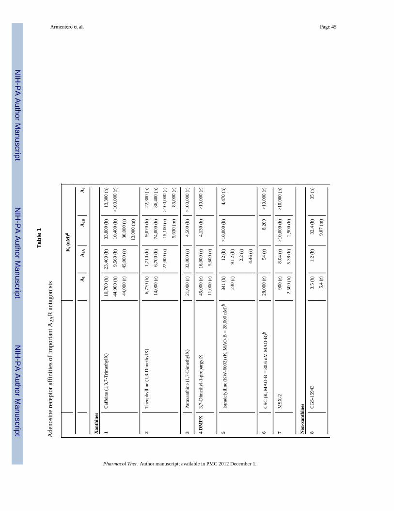
NIH
-PA Author Manuscript
NIH
-PA Author Manuscript
NIH
-PA Author Manuscript
Armentero et al. Page 45
Tabl
e 1
Ade
nosi
ne re
cept
or a
ffin
ities
of i
mpo
rtant
A2A
R a
ntag
onis
ts
Ki (
nM)a
A1
A2A
A2B
A3
Xan
thin
es
1C
affe
ine
(1,3
,7-T
rimet
hylX
)10
,700
(h)
23,4
00 (h
)33
,800
(h)
13,3
00 (h
)
44,9
00 (h
)9,
560
(h)
10,4
00 (h
)>1
00,0
00 (r
)
44,0
00 (r
)45
,000
(r)
30,0
00 (r
)
13,0
00 (m
)
2Th
eoph
yllin
e (1
,3-D
imet
hylX
)6,
770
(h)
1,71
0 (h
)9,
070
(h)
22,3
00 (h
)
14,0
00 (r
)6,
700
(h)
74,0
00 (h
)86
,400
(h)
22,0
00 (r
)15
,100
(r)
>100
,000
(r)
5,63
0 (m
)85
,000
(r)
3Pa
raxa
nthi
ne (1
,7-D
imet
hylX
)21
,000
(r)
32,0
00 (r
)4,
500
(h)
>100
,000
(r)
4 D
MPX
3,7-
Dim
ethy
l-1-p
ropa
rgyl
X45
,000
(r)
16,0
00 (r
)4,
130
(h)
>10,
000
(r)
11,0
00 (r
)5,
600
(r)
5Is
trade
fylli
ne (K
W-6
002)
(Ki M
AO
-B =
28,
000
nM)b
841
(h)
12 (h
)>1
0,00
0 (h
)4,
470
(h)
230
(r)
91.2
(h)
2.2
(r)
4.46
(r)
6C
SC (K
i MA
O-B
= 8
0.6
nM M
AO
-B)b
28,0
00 (r
)54
(r)
8,20
0>1
0,00
0 (r
)
7M
SX-2
900
(r)
8.04
(r)
>10,
000
(h)
>10,
000
(h)
2,50
0 (h
)5.
38 (h
)2,
900
(h)
Non
-xan
thin
es
8C
GS-
1594
33.
5 (h
)1.
2 (h
)32
.4 (h
)35
(h)
6.4
(r)
9.07
(m)
Pharmacol Ther. Author manuscript; available in PMC 2012 December 1.

NIH
-PA Author Manuscript
NIH
-PA Author Manuscript
NIH
-PA Author Manuscript
Armentero et al. Page 46
Ki (
nM)a
A1
A2A
A2B
A3
9ZM
-241
385
774
(h)
1.6
(h)
75 (h
)74
3 (h
)
10SC
H-5
8261
725
(h)
5.0
(h)
1110
(h)
1200
(h)
11SC
H-4
4241
611
10 (h
)4.
1 (h
)>1
0,00
0 (h
)>1
0,00
0 (h
)
12Pr
elad
enan
t (SC
H-4
2081
4)>1
,000
(h)
0.9
(h)
>1,0
00 (h
)>1
,000
(h)
13V
ipad
enan
t (B
IIB
014,
V20
06)
68 (h
)1.
3 (h
)63
(h)
1,00
5 (h
)
14ST
-153
571
.8 (h
)6.
6 (h
)35
2.3
(h)
>1,0
00 (h
)
15SY
N-1
151,
320
(h)
4.9
(h)
ndnd
a h =
hum
an; m
= m
ouse
; r =
rat;
a fe
w A
2B d
ata
are
from
func
tiona
l (cA
MP)
stud
ies (
Mül
ler &
Jaco
bson
, 201
1a; M
ülle
r & Ja
cobs
on 2
011b
); nd
= n
o da
ta a
vaila
ble
b dete
rmin
ed w
ith b
aboo
n liv
er e
nzym
e.
Pharmacol Ther. Author manuscript; available in PMC 2012 December 1.


![Crítica à Execução Penal [2a edição]](https://static.fdokumen.com/doc/165x107/631ae641d43f4e176304a750/critica-a-execucao-penal-2a-edicao.jpg)
![Corrigendum to “1-, 3- and 8-substituted-9-deazaxanthines as potent and selective antagonists at the human A2B adenosine receptor” [Bioorg. Med. Chem. 16 (2008) 2852–2869]](https://static.fdokumen.com/doc/165x107/63357c0ca1ced1126c0ad0ad/corrigendum-to-1-3-and-8-substituted-9-deazaxanthines-as-potent-and-selective.jpg)