Pancreas Development in Zebrafish: Early Dispersed Appearance of Endocrine Hormone Expressing Cells...
15
Pancreas Development in Zebrafish: Early Dispersed Appearance of Endocrine Hormone Expressing Cells and Their Convergence to Form the Definitive Islet Fre ´de ´ ric Biemar,* , ‡ Francesco Argenton,² Regine Schmidtke,* Simone Epperlein,* Bernard Peers,‡ and Wolfgang Driever* ,1 *Institut fu ¨ r Biologie I, Abt. Entwicklungsbiologie, Universita ¨ t Freiburg, Hauptstrasse 1, D-79104 Freiburg, Germany; ²Dipartimento di Biologia, Universita di Padova, Via U. Bassi 58/B, I-35131 Padova, Italy; and ‡Laboratoire de Biologie Mole ´culaire et de Ge ´nie Ge ´ne ´ tique, Institut de Chimie, Ba ˆ timent B6, Universite ´ de Lie ` ge, B-4000 Sart Tilman, Belgium To begin to understand pancreas development and the control of endocrine lineage formation in zebrafish, we have examined the expression pattern of several genes shown to act in vertebrate pancreatic development: pdx-1, insulin (W. M. Milewski et al., 1998, Endocrinology 139, 1440 –1449), glucagon, somatostatin (F. Argenton et al., 1999, Mech. Dev. 87, 217–221), islet-1 (Korzh et al., 1993, Development 118, 417– 425), nkx2.2 (Barth and Wilson, 1995, Development 121, 1755–1768), and pax6.2 (Nornes et al., 1998, Mech. Dev. 77, 185–196). To determine the spatial relationship between the exocrine and the endocrine compartments, we have cloned the zebrafish trypsin gene, a digestive enzyme expressed in differentiated pancreatic exocrine cells. We found expression of all these genes in the developing pancreas throughout organogenesis. Endocrine cells first appear in a scattered fashion in two bilateral rows close to the midline during mid-somitogenesis and converge during late-somitogenesis to form a single islet dorsal to the nascent duodenum. We have examined development of the endocrine lineage in a number of previously described zebrafish mutations. Deletion of chordamesoderm in floating head (Xnot homolog) mutants reduces islet formation to small remnants, but does not delete the pancreas, indicating that notochord is involved in proper pancreas development, but not required for differentiation of pancreatic cell fates. In the absence of knypek gene function, which is involved in convergence movements, the bilateral endocrine primordia do not merge. Presence of trunk paraxial mesoderm also appears to be instrumental for convergence since the bilateral endocrine primordia do not merge in spadetail mutants. We discuss our findings on zebrafish pancreatogenesis in the light of evolution of the pancreas in chordates. © 2001 Academic Press Key Words: zebrafish; pancreas; endoderm; insulin. INTRODUCTION The vertebrate pancreas exerts its exocrine and endocrine functions through two distinct tissue components. The exocrine component is composed of acinar glands that release digestive enzymes into the intestine, whereas the endocrine component is composed of four distinct cell types that secrete hormones into the bloodstream in order to control glucose homeostasis (reviewed by Slack, 1995). Dysfunction of endocrine b-cells may lead to diabetes mellitus, a widespread disease affecting more than 150 million people worldwide. Despite the availability of insu- lin as a treatment to temporarily restore glucostasis, diabe- tes is still incurable. Future regenerative or restorative therapies will depend on a better understanding of the development of the endocrine pancreas, and b-cells specifi- cally. The recent emergence of the zebrafish as a highly suitable genetic model organism for vertebrate develop- ment (Driever et al., 1996; Haffter et al., 1996) prompted us to investigate its potential to study pancreas organogenesis. In higher vertebrates, the Islets of Langerhans constitute the endocrine tissue compartment of the pancreas and are found embedded in the exocrine tissue. Every islet is composed of four major cell types: insulin-producing b-cells form the core while somatostatin-producing d-cells, 1 To whom correspondence should be addressed. Fax: (1149)-761 203 2597. E-mail: [email protected]. Developmental Biology 230, 189 –203 (2001) doi:10.1006/dbio.2000.0103, available online at http://www.idealibrary.com on 0012-1606/01 $35.00 Copyright © 2001 by Academic Press All rights of reproduction in any form reserved. 189
Transcript of Pancreas Development in Zebrafish: Early Dispersed Appearance of Endocrine Hormone Expressing Cells...

Te
1edomectpesp
m
Developmental Biology 230, 189–203 (2001)doi:10.1006/dbio.2000.0103, available online at http://www.idealibrary.com on
Pancreas Development in Zebrafish: Early DispersedAppearance of Endocrine Hormone Expressing Cellsand Their Convergence to Form the Definitive Islet
Frederic Biemar,*,‡ Francesco Argenton,† Regine Schmidtke,*Simone Epperlein,* Bernard Peers,‡ and Wolfgang Driever*,1
*Institut fur Biologie I, Abt. Entwicklungsbiologie, Universitat Freiburg, Hauptstrasse 1,D-79104 Freiburg, Germany; †Dipartimento di Biologia, Universita di Padova, Via U. Bassi58/B, I-35131 Padova, Italy; and ‡Laboratoire de Biologie Moleculaire et de Genie Genetique,Institut de Chimie, Batiment B6, Universite de Liege, B-4000 Sart Tilman, Belgium
o begin to understand pancreas development and the control of endocrine lineage formation in zebrafish, we havexamined the expression pattern of several genes shown to act in vertebrate pancreatic development: pdx-1, insulin (W. M.
Milewski et al., 1998, Endocrinology 139, 1440–1449), glucagon, somatostatin (F. Argenton et al., 1999, Mech. Dev. 87,217–221), islet-1 (Korzh et al., 1993, Development 118, 417–425), nkx2.2 (Barth and Wilson, 1995, Development 121,755–1768), and pax6.2 (Nornes et al., 1998, Mech. Dev. 77, 185–196). To determine the spatial relationship between thexocrine and the endocrine compartments, we have cloned the zebrafish trypsin gene, a digestive enzyme expressed inifferentiated pancreatic exocrine cells. We found expression of all these genes in the developing pancreas throughoutrganogenesis. Endocrine cells first appear in a scattered fashion in two bilateral rows close to the midline duringid-somitogenesis and converge during late-somitogenesis to form a single islet dorsal to the nascent duodenum. We have
xamined development of the endocrine lineage in a number of previously described zebrafish mutations. Deletion ofhordamesoderm in floating head (Xnot homolog) mutants reduces islet formation to small remnants, but does not deletehe pancreas, indicating that notochord is involved in proper pancreas development, but not required for differentiation ofancreatic cell fates. In the absence of knypek gene function, which is involved in convergence movements, the bilateralndocrine primordia do not merge. Presence of trunk paraxial mesoderm also appears to be instrumental for convergenceince the bilateral endocrine primordia do not merge in spadetail mutants. We discuss our findings on zebrafishancreatogenesis in the light of evolution of the pancreas in chordates. © 2001 Academic Press
Key Words: zebrafish; pancreas; endoderm; insulin.
mlttd
INTRODUCTION
The vertebrate pancreas exerts its exocrine and endocrinefunctions through two distinct tissue components. Theexocrine component is composed of acinar glands thatrelease digestive enzymes into the intestine, whereas theendocrine component is composed of four distinct celltypes that secrete hormones into the bloodstream in orderto control glucose homeostasis (reviewed by Slack, 1995).Dysfunction of endocrine b-cells may lead to diabetes
ellitus, a widespread disease affecting more than 150
1 To whom correspondence should be addressed. Fax: (1149)-761
f203 2597. E-mail: [email protected].0012-1606/01 $35.00Copyright © 2001 by Academic PressAll rights of reproduction in any form reserved.
illion people worldwide. Despite the availability of insu-in as a treatment to temporarily restore glucostasis, diabe-es is still incurable. Future regenerative or restorativeherapies will depend on a better understanding of theevelopment of the endocrine pancreas, and b-cells specifi-
cally. The recent emergence of the zebrafish as a highlysuitable genetic model organism for vertebrate develop-ment (Driever et al., 1996; Haffter et al., 1996) prompted usto investigate its potential to study pancreas organogenesis.
In higher vertebrates, the Islets of Langerhans constitutethe endocrine tissue compartment of the pancreas and arefound embedded in the exocrine tissue. Every islet iscomposed of four major cell types: insulin-producing b-cells
orm the core while somatostatin-producing d-cells,189

a
190 Biemar et al.
glucagon-producing a-cells, and pancreatic polypeptide se-creting PP-cells are located at the periphery. Both exocrineand endocrine pancreatic tissues are of endodermal originand develop from dorsal and ventral epithelial evaginations,arising from the gut tube caudal to the stomach (reviewedin Slack, 1995).
The analysis of pancreas development in various fisheshas raised questions concerning the physiological signifi-
FIG. 1. Morphology of the exocrine and endocrine pancreas in thshowing the overall organization of the endocrine (i, arrow) and exoimages of pancreatic islet tissue showing whole-mount immunoflu3-day-old embryos with a combination of (C) anti-carboxypeptianti-insulin and anti-glucagon; (F) anti-somatostatin and anti-glucae; exocrine tissue; i, islet; li, liver; o, otolith; pf, pectoral fin; sb, swnd 25 mm in F (same magnification as D and E).
cance of the close spatial association between the endocrine
Copyright © 2001 by Academic Press. All right
and exocrine pancreatic tissue. In agnathans, both tissuesare separate from each other, while in gnathostomes, al-though always associated, their distribution is variable. Inteleosts, highly evolved fishes, the endocrine islets may befound scattered among the exocrine tissue as in mammals,or in a few large islets named the Brockmann bodies or“principal islets.” In most cases, teleost islet tissue con-tains all four types of hormone producing cells, whereas
rafish larva. (A) Sagittal and (B) cross sections of 6-day-old larvaee pancreatic tissues (e in A, and e, arrowhead in B). (C–F) Confocalcence staining performed on (C) 5.5-day-old, (D) 4-days, and (E, F)A and anti-insulin; (D) anti-insulin and anti-somatostatin; (E)
ntisera. In A and C, anterior is oriented to the left. da, dorsal aorta;ladder; sc, spinal cord; s1–8, somite 1 to 8. Scale bar, 50 mm in C
e zebcrinores
dasegon aim b
only one (b-cells), two (b- and d-cells), or three (b-, d-, and
s of reproduction in any form reserved.
tt1(g(omm1MBle
elmta
s“Hta
1sa
191Zebrafish Pancreas Development
PP-cells) cell types are usually found in the pancreas ofmore primitive fishes (Youson and Al-Mahrouki, 1999, andreferences therein).
Gene targeting studies in mice have revealed the func-tional importance of several transcription factors at variousstages during endocrine pancreas development. These in-clude the homeodomain protein Pdx-1 (Jonsson et al., 1994),he Lim homeodomain protein Isl-1 (Ahlgren et al., 1997),he paired box genes pax4 and pax6 (Sosa-Pineda et al.,997; St-Onge et al., 1997), the bHLH gene beta2/neuroDMalecki et al., 1999; Naya et al., 1997), the NK2 homeoboxene nkx2.2 (Sussel et al., 1998), the homeobox gene hb9Harrison et al., 1999; Li et al., 1999), and the cut home-domain hnf6 (Jacquemin et al., 2000). Numerous signalingolecules also act during pancreatic endocrine develop-ent, including Sonic hedgehog (Shh) (Apelqvist et al.,
997; Hebrok et al., 1998; Kim et al., 1997a; Kim andelton, 1998), TGF-bs (Miralles et al., 1998a,b), FGFs (Le
ras et al., 1998; Miralles et al., 1999), and the Notch/Deltaateral inhibition system (Apelqvist et al., 1999; Gradwohlt al., 2000; Jensen et al., 2000a,b).Beyond two pioneer studies (Pack et al., 1996; Milewski
t al., 1998), zebrafish pancreatic development has receivedittle attention. Study of both the cellular and molecular
echanisms that regulate early endoderm development andhose that control organogenesis of the gut and its associ-ted organs has only recently begun (Alexander et al., 1999;
Alexander and Stainier, 1999; Reiter et al., 1999; Warga andNusslein-Volhard, 1999; Kikuchi et al., 2000). To begin tounderstand pancreas development and the control of endo-crine lineage formation in zebrafish, we have assayed theexpression of the following genes in the developing ze-brafish pancreas: pdx-1, insulin (Milewski et al., 1998),
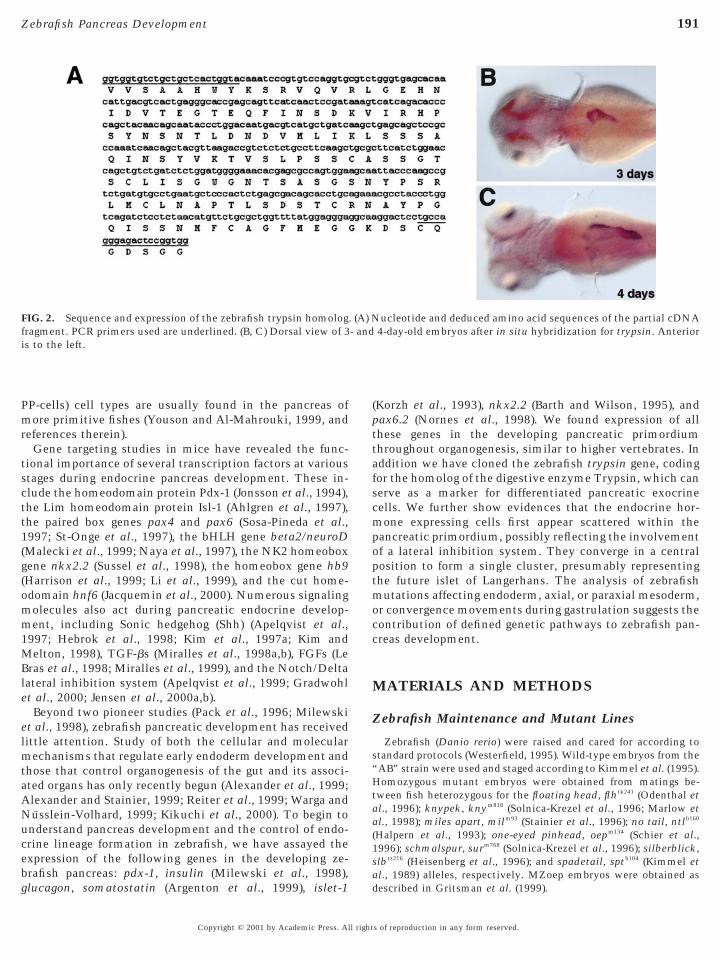
FIG. 2. Sequence and expression of the zebrafish trypsin homologfragment. PCR primers used are underlined. (B, C) Dorsal view of 3is to the left.
glucagon, somatostatin (Argenton et al., 1999), islet-1 d
Copyright © 2001 by Academic Press. All right
(Korzh et al., 1993), nkx2.2 (Barth and Wilson, 1995), andpax6.2 (Nornes et al., 1998). We found expression of allthese genes in the developing pancreatic primordiumthroughout organogenesis, similar to higher vertebrates. Inaddition we have cloned the zebrafish trypsin gene, codingfor the homolog of the digestive enzyme Trypsin, which canserve as a marker for differentiated pancreatic exocrinecells. We further show evidences that the endocrine hor-mone expressing cells first appear scattered within thepancreatic primordium, possibly reflecting the involvementof a lateral inhibition system. They converge in a centralposition to form a single cluster, presumably representingthe future islet of Langerhans. The analysis of zebrafishmutations affecting endoderm, axial, or paraxial mesoderm,or convergence movements during gastrulation suggests thecontribution of defined genetic pathways to zebrafish pan-creas development.
MATERIALS AND METHODS
Zebrafish Maintenance and Mutant Lines
Zebrafish (Danio rerio) were raised and cared for according totandard protocols (Westerfield, 1995). Wild-type embryos from theAB” strain were used and staged according to Kimmel et al. (1995).omozygous mutant embryos were obtained from matings be-
ween fish heterozygous for the floating head, flhtk241 (Odenthal etl., 1996); knypek, knym818 (Solnica-Krezel et al., 1996; Marlow et
al., 1998); miles apart, milm93 (Stainier et al., 1996); no tail, ntlb160
(Halpern et al., 1993); one-eyed pinhead, oepm134 (Schier et al.,996); schmalspur, surm768 (Solnica-Krezel et al., 1996); silberblick,lbtz216 (Heisenberg et al., 1996); and spadetail, sptb104 (Kimmel etl., 1989) alleles, respectively. MZoep embryos were obtained as
ucleotide and deduced amino acid sequences of the partial cDNA4-day-old embryos after in situ hybridization for trypsin. Anterior
. (A) N- and
escribed in Gritsman et al. (1999).
s of reproduction in any form reserved.
A
ifiacoc
iaptst
192 Biemar et al.
Histology and Immunohistochemistry
Methacrylate sections were prepared using the JB-4 plus resin(Polyscience Inc.) according to the manufacturer’s protocol. The4-mm serial sections were stained with methylene blue–azure II.Whole-mount immunohistochemistry was performed as describedpreviously (Argenton et al., 1999) using rabbit anti-bovine car-boxypeptidase A (Chemicon), guinea pig anti-porcine insulin(Linco Inc.), rabbit anti-somatostatin-14 (Biotrend), mouse anti-porcine glucagon (Sigma), rabbit anti-porcine glucagon (Biotrend),and rabbit anti-human pancreatic polypeptide (Scytek) antisera.Appropriate combinations of Alexa FluorTM 488 goat anti-rabbitIgG conjugate, Alexa FluorTM 546 goat anti-rabbit IgG conjugate,
lexa FluorTM 488 goat anti-guinea pig IgG conjugate, AlexaFluorTM 546 goat anti-guinea pig IgG conjugate, and Alexa FluorTM
488 goat anti-mouse IgG conjugate (Molecular Probes) were used assecondary antisera. Confocal images were taken on a Zeiss LSM510 microscope.
Whole-Mount in Situ Hybridization
Whole-mount in situ mRNA hybridizations were performed asdescribed by Hauptmann and Gerster (1994). The following probeswere used: pdx-1, insulin (Milewski et al., 1998), glucagon, soma-tostatin (Argenton et al., 1999), islet-1 (Korzh et al., 1993), nkx2.2(Barth and Wilson, 1995), pax6.2 (Nornes et al., 1998), FoxA2/axial/fkd1 (Odenthal and Nusslein-Volhard, 1998; Strahle et al., 1993),and FoxA3/fkd2/Zffkh1 (Dirksen and Jamrich, 1995; Odenthal andNusslein-Volhard, 1998). For Fig. 3, panel O, two images weretaken at the same focal plane, using a DIC filter transmitted lightfor the first one (black staining, pdx-1) and epifluorescence with aRhodamine filter for the second one (fast-red fluorescent staining,insulin). The two pictures were then superimposed and processedusing the OpenLab software (Improvision).
RESULTS
General Morphology of the Larval ZebrafishPancreas
The pancreas of the zebrafish larva is composed of onesingle islet embedded in exocrine tissue. At 6 days postfertilization (dpf; Figs. 1A and 1B), the pancreas is locatedasymmetrically on the right side of the body. This asym-metry is established by 48 h post fertilization (hpf, data notshown). Differentiated exocrine cells, identified by immu-noreactivity to Carboxypeptidase A, are first detected in theexocrine component of the pancreas at around 72 hpf (datanot shown). By 5.5 dpf, exocrine tissue extends from thefirst to the sixth or seventh somite. Within this, theendocrine islet is located in the anterior-most portion, atthe level of the third and fourth somite (Figs. 1A and 1C). Asproposed for humans (reviewed in Slack, 1995) and chick(Kim et al., 1997b), the zebrafish pancreas can be dividednto a “head” region followed by a “neck” region and,nally a “tail” region (Fig. 1C). While the endocrine isletsre distributed throughout the pancreas of human andhick, in zebrafish, the head region contains the single isletf Langerhans. The zebrafish larval endocrine islet has a
ore composed of insulin- and somatostatin-Copyright © 2001 by Academic Press. All right
mmunoreactive cells (Fig. 1D) surrounded by glucagon-nd pancreatic polypeptide-immunoreactive cells at theeriphery (Figs. 1E, 1F, and data not shown). This organiza-ion is similar to mammalian islets, but differs in thatomatostatin immunoreactive cells tend to be located athe periphery in mammals.
Trypsin Expression Marks the Exocrine Pancreas
Since so far no robust marker has been available tovisualize the zebrafish exocrine pancreas at the RNA ex-pression level, we cloned the zebrafish homolog of thetrypsin gene. To design conserved oligonucleotides suitablefor the amplification of zebrafish trypsin cDNA, the se-quences of rat, chicken, Xenopus, sole, and salmon werealigned. Four strictly conserved regions were used to pre-pare two 59 and two 39 primers to be used in a set of nestedPCRs. To amplify the core of zebrafish trypsin cDNA, weused mRNA isolated from pancreas and reverse-transcribedwith AMV-reverse transcriptase. A 440-bp fragment ob-tained from the second (nested) PCR was cloned and se-quenced. The translated cDNA sequence encodes an openreading frame with homology to Trypsin sequences in othervertebrate species. The overall homology at the amino acidlevel ranges from 76.6% for the rat sequence up to 81.4%for the Xenopus sequence. Expression analysis by whole-mount in situ hybridization of 24 and 48 (not shown) aswell as 72 and 96 hpf old embryos (Fig. 2) revealed thattrypsin expressing exocrine cells can first be detected be-tween 48 and 72 hpf. At this time, the zebrafish pancreas isalready located on the right side of the embryo, dorsal to thegut tube.
Zebrafish Pancreatic Primordia First Appearin Two Stripes Adjacent to the Midline
In mammals, birds, reptiles, and amphibians, the pan-creas forms from dorsal and ventral evaginations of theposterior foregut that later join together (Pictet and Rutter,1972). Soon after these two buds have formed, endodermalcells committed toward pancreatic or duodenal cell fatesexpress the homeodomain transcription factor pdx-1 (Guzet al., 1995; Ahlgren et al., 1996; Offield et al., 1996). Thezebrafish homolog of pdx-1 was cloned recently (Milewskiet al., 1998) but little was revealed about its expressionearly in development. We therefore performed a detailedanalysis of pdx-1 expression to precisely determine the timeof its initiation in the developing pancreas. pdx-1 mRNAexpression is first detected at the 10-somite stage in twobilateral rows of cells adjacent to the midline, locatedimmediately above the large syncytial yolk cell at the levelof the first two or three somites (Fig. 3A). Starting at the14-somite stage, the rows of cells approach each other andeventually fuse to form a single field of pdx-1 expressingcells (Fig. 3E). The convergence of the two bilateral stripesappears to be initiated at their posterior end. From the
18-somite stage, the field of pdx-1 expressing cells is con-s of reproduction in any form reserved.

Tasie
1tNz
mht
193Zebrafish Pancreas Development
tinuous and broadens as the number of pdx-1 expressingcells increases (Fig. 3G). As development proceeds, theanterioposterior extent of the field of pdx-1 expressing cellsappears to shorten, and a group of pdx-1 expressing cellscondenses at the posterior and dorsal margin of the originalpdx-1 expression domain (compare Figs. 3I, 3K, and 3M).
hus, in contrast to mammals and birds, zebrafish pancre-tic primordia do not appear as dorsal and ventral protru-ions of the gut tube but rather at left and right positionsmmediately adjacent to the midline within a field ofndodermal cells.
Insulin Is the First Hormone Expressedwithin the Pancreatic Anlage
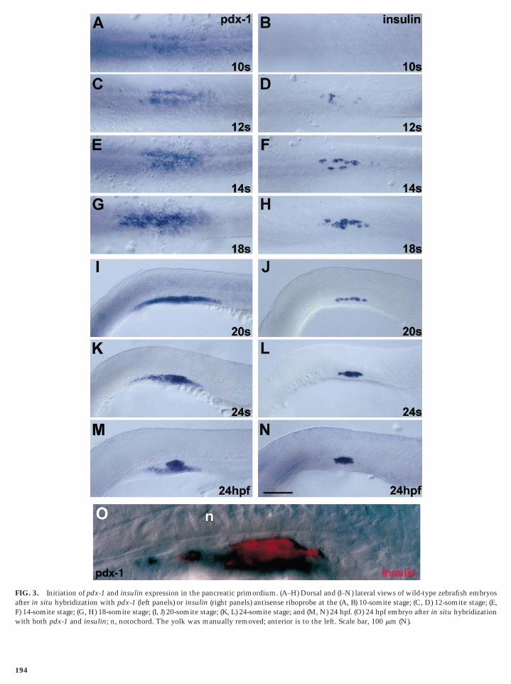
We investigated the initiation of pancreatic hormoneexpression in zebrafish, using the zebrafish insulin(Milewski et al., 1998), glucagon, and somatostatin (Ar-genton et al., 1999) cDNA probes. At the 10-somite stage,while pdx-1 is already expressed in bilateral domains (Fig.3A), insulin expression cannot be detected (Fig. 3B). Atthe 12-somite stage, while the pdx-1 expression domainsremain well separated (Fig. 3C), the first insulin express-ing cells appear, scattered within the domains of pdx-1expression (Fig. 3D). As mentioned above, the fusion ofthe two pdx-1 stripes begins at the 14-somite stage,starting from the posterior margin (Fig. 3E). A similarprocess is observed for the insulin expressing cells (Fig.3F), which have already increased in number. At the18-somite stage, the developing pancreas, represented bythe pdx-1 and insulin expression domains, is located inthe midline, immediately dorsal to the yolk (Figs. 3G and3H). By the 20-somite stage, the insulin positive cellshave formed a one-cell-thick layer immediately aboveand adjacent to the yolk (Fig. 3J). From then on, the cellsaggregate posteriorly into a cluster, presumably repre-senting the prospective endocrine islet (Fig. 3L). By 24hpf, the presumptive islet has formed and is located at themidline, dorsal to the yolk (Fig. 3N). The comparisonwith pdx-1 expression at the 20-somite stage (Fig. 3I),24-somite stage (Fig. 3K), and 24 hpf (Fig. 3M) suggeststhat a portion of the pdx-1 expressing cells aggregatesposteriorly into a compact cluster. To test whether thisposterior cluster of pdx-1 expressing cells corresponds tothe forming endocrine islet, we performed double-labeling in situ hybridization using digoxigenin-labeledpdx-1 and fluorescein-labeled insulin antisense ribo-probes (Fig. 3O). As expected, insulin expression (red) isrestricted to dorsal posterior pdx-1 positive cells (black,see experimental procedures for details). The layer ofpdx-1 positive cells located immediately dorsal to theyolk is devoid of insulin staining. This indicates that theformation of the Islet of Langerhans in zebrafish beginsby 24 hpf.
The expression of somatostatin and glucagon mRNAin the developing zebrafish was reported previously (Ar-
genton et al., 1999). However, since only three differentCopyright © 2001 by Academic Press. All right
stages were examined, we decided to investigate moretime points in order to define the initiation of theirtranscription. We detected somatostatin expression atthe 16-somite stage (Fig. 4A), while glucagon is onlyexpressed later, at the 24-somite stage (Fig. 4D). By 24hpf, both hormones are expressed in the presumptiveislet (Figs. 4E and 4F).
Early Commitment to Endocrine Cell Fatein the Developing Zebrafish Pancreas
When expression of the zebrafish homolog of nkx2.2 wascharacterized, Barth and Wilson (1995) noticed a “patch ofnkx2.2 positive cells ventral to the hypocord at thehindbrain/spinal cord boundary.” Here we show thatnkx2.2 expressing cells can be detected as early as the10-somite stage within the pancreatic primordium (Fig. 5Aand A9) and is maintained during development (Fig. 5B at 24hpf). Mice lacking Nkx2.2 do not develop properly differen-tiated a-, b-, and PP-cells and die of diabetes (Sussel et al.,998). The conservation of pancreas specific expression ofhe zebrafish nkx2.2 homolog indicates that the function ofkx2.2 observed in mice may also have been conserved in
ebrafish.Early in pancreatic endocrine cell differentiation, post-itotic islet cells in mice initiate expression of the LIM
omeodomain protein Isl1 (Ahlgren et al., 1997). We de-ected islet-1 expression in the zebrafish pancreatic anlage
as early as the 12-somite stage (Fig. 5C and C9) thatcontinued past 24 hpf (Fig. 5D). These data suggest thatcommitment to endocrine cell fate, and the differentiationof at least one part of the endocrine cells is initiatedrelatively early in zebrafish, soon after the onset of pdx-1expression.
Targeted mutation of the pax6 gene in mouse causesabsence of glucagon producing cells, suggesting an im-portant role of this protein in islets morphogenesis and inthe differentiation of a-cells (Sander et al., 1997; St-Ongeet al., 1997). It has recently been shown that the zebrafishgenome contains two genes homologous to pax6 (Norneset al., 1998). We found initiation of pax6.2 expression inthe developing zebrafish pancreas at the 12-somite stage(Fig. 5E and E9). Similar to other pancreatic markers atthis stage, the pax6.2 expressing cells form a one-cell-thick layer immediately above the yolk, and expression ismaintained throughout somitogenesis, still clearly vis-ible by 24 hpf (Fig. 5F). We did not detect expression ofthe zebrafish pax6.1 homolog in the pancreatic primordiaat any stage.
Pancreatic Defects in Zebrafish Mutants AffectingMidline Specification
As experimental embryological studies in chick re-vealed an important inductive role for the notochord inpancreas development (Kim et al., 1997a), we investi-
gated the influence on pancreas formation of variouss of reproduction in any form reserved.
aFw
1
FIG. 3. Initiation of pdx-1 and insulin expression in the pancreatic primordium. (A–H) Dorsal and (I–N) lateral views of wild-type zebrafish embryosfter in situ hybridization with pdx-1 (left panels) or insulin (right panels) antisense riboprobe at the (A, B) 10-somite stage; (C, D) 12-somite stage; (E,) 14-somite stage; (G, H) 18-somite stage; (I, J) 20-somite stage; (K, L) 24-somite stage; and (M, N) 24 hpf. (O) 24 hpf embryo after in situ hybridization
ith both pdx-1 and insulin; n, notochord. The yolk was manually removed; anterior is to the left. Scale bar, 100 mm (N).94

ahet
mw
d; an
195Zebrafish Pancreas Development
mutations affecting zebrafish axial and nonaxial meso-derm. The no tail (ntl), floating head (flh), schmalspur(sur), and one-eyed-pinhead (oep) mutations all affectmidline development, albeit to various degrees. Muta-tions at the ntl/Brachyury locus lead to defects in noto-chord differentiation, but during gastrulation chordame-sodermal cells are present (Halpern et al., 1993; Schulte-Merker et al., 1994). In 24 hpf ntl mutant embryos, bothpdx-1 and insulin expression resemble that of wild-typeembryos (data not shown). In contrast, flh/Xnot mutantembryos, in which the notochord (axial mesoderm) isreplaced by muscle cells (paraxial mesoderm) (Halpern etal., 1995; Talbot et al., 1995), exhibit a much strongerpancreatic phenotype. While the duodenal, ventrally situ-ated stripe of pdx-1 expression is not affected, we foundthat the posterior cluster of pdx-1 expressing cells, rep-resenting the presumptive islet, is absent (Fig. 6, compareA and E). insulin expressing cells are almost completelymissing in the flh mutant pancreatic anlage (Fig. 6,compare B and F). Neither sur nor oep mutants specifi-
FIG. 4. Expression of somatostatin and glucagon in the developinin situ hybridization with somatostatin (left panels) or glucagon (24-somite stage; and (E, F) 24 hpf. The yolk was manually remove
cally lack the notochord. However, both oep/EGF-CFC, t
Copyright © 2001 by Academic Press. All right
the homolog of mouse cripto and cryptic genes (Zhang etal., 1998), and sur affect nodal signaling required forproper specification of endoderm and midline tissue,including notochord and floorplate (Schier et al., 1997;Gritsman et al., 1999; Pogoda et al., 2000). The pancre-tic phenotype displayed by sur mutant embryos at 24pf is intermediate between that of ntl and flh mutantmbryos: a very small islet can be distinguished withinhe pdx-1 positive cells and the number of insulin posi-
tive cells is only slightly reduced (data not shown). Incontrast, in oep mutants which lack both maternal andzygotic expression of oep neither pdx-1 or insulin ex-pressing cells are found at 24 hpf. In some embryoslacking zygotic oep only, however, a few pdx-1 positivecells are observed, indicating that some endoderm maystill be formed in oep zygotic mutant embryos (Figs. 6C,6D, 6G, and 6H). The differences in sur and oep pheno-types may be explained since sur predominantly affects
aintenance of Nodal signaling (Pogoda et al., 2000),hile in the absence of maternal and zygotic oep func-
rafish pancreas. Dorsal views of wild-type zebrafish embryos afterpanels) antisense riboprobes at the (A, B) 16-somite stage; (C, D)
terior is to the left. Scale bar, 100 mm.
g zebright
ion, Nodal signals cannot be perceived by responding
s of reproduction in any form reserved.
tttatsty
ma
e
M
196 Biemar et al.
cells at all. Taken together, our data suggest that, whilemidline signaling is required for proper development ofthe pancreas, it may not be required to initiate a pancre-atic domain within the forming gut epithelium in ze-brafish (see discussion).
Pancreas Development in Mutants AffectingConvergence Movements
Pancreatic endocrine precursor cells in zebrafish initiallyappear as left and right bilateral rows of cells immediatelyadjacent to the midline that converge at the midline duringlate somitogenesis (see above). Thus, we investigatedwhether any of the known genetic components that con-tribute to convergence movements in zebrafish might beinvolved in pancreatic development. Mutations affectingfour different aspect of convergence toward the dorsalmidline were analyzed. The spadetail (spt) mutation affectsformation and convergence of paraxial mesoderm in thetrunk, causing severe defects in trunk somitic mesoderm(Ho and Kane, 1990; Amacher and Kimmel, 1998; Wargaand Nusslein-Volhard, 1998; Griffin et al., 1998). spt en-codes a T-box gene that may be involved in controlling bothmorphogenesis, by its effect on convergence, as well as celldifferentiation of paraxial mesoderm. We find that thebilateral pancreas primordia fail to converge in spt mutantembryos: two clusters of insulin expressing cells remain oneither side of the midline at 24 hpf (compare Figs. 6I and6M, 6J and 6N).
Three other genetic loci have been shown to be involvedin the control of convergence during gastrulation, or ofspecific cell populations during somitogenesis. Mutationsat the knypek locus (kny; Solnica-Krezel et al., 1996;Marlow et al., 1998) result in embryos with a shortenedbody axis and broadened axial and paraxial mesoderm. Wefind that in kny mutants, the pdx-1 domains fail to merge,and two separate, bilateral stripes of insulin expressing cellsare maintained during development (compare at 24 hpf:Figs. 6K and 6O, 6L and 6P). However, similar to thevariability in phenotype reported for the trunk defects inkny mutants, we observe variable expressivity of the phe-notype affecting pancreas development. A second gene thathas been demonstrated to contribute toward the orchestra-tion of convergent extension movements during gastrula-tion is silberblick (slb; coding for wnt11; Heisenberg et al.,2000). In slb mutants expression of pdx-1 and insulinappears normal at 24 hpf (data not shown), so slb is notrequired for pancreas development. The mutation milesapart (mil) causes cardia bifida and has recently been showno encode a sphingosin-2-phosphate receptor involved inhe control of convergence of the bilateral heart primordiao the definitive heart (Kupperman et al., 2000). Since milffects convergence of cardiac precursors around the sameime and only slightly anterior to where pancreatic precur-ors converge, we were curious about a potential contribu-ion of mil to pancreatic development. However, our anal-
sis revealed that convergence of pancreatic precursors inCopyright © 2001 by Academic Press. All right
il mutants, as judged from pdx-1 and insulin expression atbout 24 hpf, is not affected (data not shown).
DISCUSSION
The Zebrafish Pancreas: Lower versus HigherVertebrate
Although previous studies reported the presence of sev-eral endocrine islets in the pancreatic parenchyma of adultzebrafish (Milewski et al., 1998; Pack et al., 1996), ourresults show no evidence for the existence of secondary,accessory islets during the first six days of larval develop-ment. These additional islets therefore most likely differ-entiate later during development. This would argue in favorof the persistence of a pool of endocrine precursor cells inthe adult, as suggested in rat (Kaung, 1994; reviewed inSlack, 1995). The zebrafish larval islet exhibits the typicalendocrine cell type distribution and hormone expressionobserved in other teleost species (Youson and Al-Mahrouki,1999). The core of the islet is composed of both b- andd-cells, whereas a- and PP-cells are found at the periphery.However, although the general organization of the endo-crine cell types seems different in fish compared to othervertebrate models, where d-cells may also be found at theperiphery, the lineage relationships between the differentendocrine cell types may be conserved. Gene targetingstudies of pax4 and pax6 (Sosa-Pineda et al., 1997; St-Onget al., 1997) in mouse suggest a close relationship between
b- and d-cells, while previous reports had proposed that a-and b-cell lineages are independent (Herrera et al., 1998).
oreover, a recent study shows elegantly that a-and b-cellsmost likely belong to independent lineages (Herrera, 2000).The observation that endocrine cells of the 48 hpf (Argen-ton et al., 1999) and 72 hpf (this study) developing isletsnever co-express insulin and glucagon or somatostatin andglucagon supports the current model of separate a and blineages proposed for mammals (Herrera, 2000). Absence ofco-expression of insulin with glucagon or PP was alsoobserved for Xenopus (Kelly and Melton, 2000). A true celllineage analysis would still be needed to confirm whether aand b lineages are also separated early in zebrafish.
A noticeable difference between zebrafish and other ver-tebrates concerns the initiation of pancreas developmentwith the formation of the so called “pancreatic buds” inhigher vertebrates. Developing zebrafish embryos do notappear to form equivalent bud structures during the devel-opmental phase when expression of endocrine hormonesbegins. This may be explained by differences in gut tubemorphogenesis between zebrafish and other vertebrates. Infact, as a result of gastrulation in zebrafish, a sheet ofendodermal cells forms rather than a proper endodermaltube like in higher vertebrates. There is evidence that thegut lumen forms rather late, at around 36 hpf, most likelyby a cavitation process (Pack et al., 1996; data not shown),similar to the secondary formation of the lumen of the
neural tube (Papan and Campos-Ortega, 1994). Hence, ats of reproduction in any form reserved.
1cwndlais
aredidamgidsbipeflgWc1aite
1
hifatcg1m(llee
ptesof(ie1z
ppe
197Zebrafish Pancreas Development
the time of the onset of pdx-1 expression, the endodermaltissue constitutes a single sheet of cells dorsal to the largesyncytial yolk cell. Thus, at this time no morphologicalstructure like the gut tube exists as the basis for a mammal-or birdlike formation of pancreatic buds in zebrafish. Bud-ding of epithelial sheets may, however, contribute to laterpancreas morphogenesis in zebrafish.
In zebrafish, as in other vertebrates, the pancreatic pri-mordia first appear in two distinct locations and fuse duringembryogenesis to form a single pancreatic primordium bylate-somitogenesis. The identity of the cells located be-tween the two bilateral domains of pdx-1 expression at the0-somite stage is unclear. Whether these are endodermalells not yet specified toward a pancreatic fate or cells thatill acquire another developmental fate is unknown. Alter-atively they may be nonpancreatic (endodermal or meso-ermal) cells that will relocate to another position duringate somitogenesis and the elongation of the body axis. Thenswer to this question awaits histological analysis, thedentification of new molecular markers, and cell lineagetudies.
An alternate interpretation for the observed pattern ofppearance of pancreatic endocrine cells would be that itesembles to some degree early stages of evolution of thendocrine system. In protochordates, endocrine cells areispersed in the epithelium of the simple gut tube (reviewedn Youson and Al-Mahrouki, 1999). This is similar to theistribution of endocrine cells in zebrafish between the 12-nd 18-somite stages. During evolution, endocrine cellsay have acquired the ability to detach from and exit the
ut epithelium and enter a migratory phase. Then, “migrat-ng” endocrine precursors, based on local guidance cues andevelopment of novel cell adhesion properties, may havetarted to organize into islets. Migration and assembly maye represented in larval lampreys, in which “one-hormoneslets” form, containing exclusively cells of the insulin-roducing lineage. Over evolution, cells of the other lin-ages may have then assembled around this core, as re-ected by the predominantly peripheral location oflucagon and PP producing cells in higher vertebrates.hether the mediolateral convergence and anterioposterior
ondensation of endocrine cells that we observe between8-somite stage and 24 hpf resemble events during pancre-tic evolution remains to be determined. In light of thenvasive character of pancreatic cancer, it may be of interesto learn more about such postulated migratory states ofndocrine cells or their precursors.
Pancreatic Gene Expression in Zebrafish:Evidence for Conserved Functions
The cloning and developmental expression of pancreatichormones has been described previously (Milewski et al.,998; Argenton et al., 1999). In this study, we have reas-
sessed the initiation of expression of the mRNAs encodingInsulin, Glucagon, and Somatostatin. We narrowed down
the time frame of the earliest detectable expression for eachCopyright © 2001 by Academic Press. All right
ormone. Our results confirmed previous observations thatnsulin is the first pancreatic hormone to be expressedollowed by somatostatin and glucagon (for Xenopus: Kellynd Melton, 2000). The late appearance of trypsin, after thehree endocrine hormones analyzed in this work, is wellorrelated with a corresponding delayed onset of exocrineene expression in the murine pancreas (Gittes and Rutter,992). While insulin and glucagon are first expressed in theouse pancreatic primordium in a premorphogenetic phase
20-somite stage), exocrine specific products are expressedater, 24 h after the formation of the pancreatic diverticu-um. In zebrafish, food uptake and requirement for digestivenzymes begin only at four dpf, presenting no need forarlier maturation of the pancreatic exocrine tissue.Insulin expressing cells appear scattered within the
repatterned, pdx-1 positive endodermal epithelium. Allhe pancreatic hormone-encoding mRNAs examined so farxhibit a similar scattered initiation of expression. Thisuggests that the zebrafish islet arises from the aggregationf differentiated cells rather than by monoclonal growthrom individual progenitors, as described for mammalsDeltour et al., 1991; Percival and Slack, 1999). This alsondicates that the process of lateral inhibition, involved inndocrine cell differentiation in mice (Apelqvist et al.,999; Jensen et al., 2000b), may also be taking place inebrafish.Zebrafish homologs for many transcription factors ex-
ressed and required in the mouse pancreas all show ex-ression in the pancreatic primordium defined by pdx-1xpression: islet-1, nkx2.2, pax6.2 (this report), FoxA2
/axial, and neuroD (Korzh et al., 1998; see also Table 1).This suggests evolutionary conservation of the develop-mental mechanisms for pancreatic cell specification anddifferentiation and recommends the zebrafish as a valuablemodel for the study of pancreatic organogenesis. The ex-pression of pax6.2 but not pax6.1 in the zebrafish pancreasprovides us with a striking example of fractionation ofexpression domains (and of gene function) following geneduplication in the fish lineage leading to teleosts. Similarfunctional fractionation has already been reported in ze-brafish for transcription factors of the engrailed (Amores etal., 1998) and vsx (Passini et al., 1998) families.
Mutant Analysis Identifies Steps in PancreaticDevelopment
Experimental work in chick has revealed a requirementfor notochord derived signals in pancreas formation (Kim etal., 1997a). The availability of zebrafish mutations affectingaxial mesoderm provides an opportunity to use genetic teststo assay for a role of notochord in pancreatic development.Our finding that the endocrine pancreas develops in ntlmutant embryos, in which chordamesoderm forms but doesnot differentiate properly, indicates that any signaling re-quired for pancreas differentiation can already be accom-plished by the chordamesoderm cells and does not require
differentiated notochord. In flh mutant embryos, chordame-s of reproduction in any form reserved.

am
ssesao
198 Biemar et al.
soderm does not form, and paraxial mesoderm is continu-ous in the midline of the embryo. Thus, notochord as asource for potential signals to pattern the endoderm doesnot exist in flh mutant embryos. Even without notochord,in flh mutants, we find both duodenal expression of pdx-1nd small numbers of insulin expressing cells at proper
FIG. 5. Pancreatic specific expression of the zebrafish nkx2.2, isletzebrafish embryos at the (A, A9) 10-somite stage; (C, C9, E, E9) 12-sC9, D) islet-1, and (E, E9, F) pax6.2 antisense mRNA probes. LaterPancreas-specific expression domain are indicated (arrowheads).
orphological locations. This indicates that signaling z
Copyright © 2001 by Academic Press. All right
ources other than the notochord might contribute topecification of the pancreatic primordium. We cannotxclude that floorplate cells, which appear dispersed in thepinal cord of flh mutants, share some of the inductivectivities of notochord and are responsible for the inductionf the small number of insulin expressing cells observed. In
d pax6.2 mRNAs. Whole-mount in situ hybridization of wild-typee stage, and (B, D, F) 24 hpf performed with (A, A9, B) nkx2.2, (C,ws, dorsal to the right (A, A9, C, C9, E, E9) or to the top (B, D, F).
-1, anomit
al vie
ebrafish, other than in chick for example, endoderm and
s of reproduction in any form reserved.

pm
pptdgtN
Em
has
199Zebrafish Pancreas Development
chordamesoderm are in close contact along a large portionof the anterioposterior axis during the developmental stagecorrelating with pancreas induction in chick. This observa-tion makes it difficult to imagine a model in which localcontact between notochord and endoderm induces pancre-atic development. It has been suggested that a major func-tion of notochord in pancreatic development is repressionof hedgehog signaling (Kim et al., 1997a; Kim and Melton,1998). A detailed analysis of the role of the hedgehogsignaling pathway during the ontogeny of the pancreas inzebrafish will be presented elsewhere.
Together these data indicate that patterning of the ze-brafish endoderm and pancreas induction can proceed in theabsence of axial mesoderm, although it is required fornormal pancreatic development. In zebrafish, a prepatternmight already exist in the endoderm, possibly established
FIG. 6. Development of the endocrine pancreas in zebrafish muxpression of pdx-1 (A, C, E, G, I, K, M, O) and insulin (B, D, F, Hutations are analyzed: (E, F) floating head; (G, H) one-eyed pinhe
in A, E, C, G, and dorsal views in all the other panels; anterior is tothe posterior end of the hindbrain and about segment 10. The yolk
during gastrulation, or other tissues might be involved in t
Copyright © 2001 by Academic Press. All right
ancreas induction. To test for involvement of paraxialesoderm, we analyzed pancreas development in spt mu-
tant embryos, which lack somitic mesoderm in the trunk.We found that duodenal as well as pancreatic primordia arelocated at the proper anterioposterior position in spt mu-tant embryos, indicating that paraxial mesoderm is notrequired for pancreas induction. However, the bilateraldomains of pdx-1 and insulin expression established duringearly somitogenesis fail to converge in spt mutants. Thus,araxial mesoderm may be required for convergence ofancreatic precursors: It could provide a substrate on whichhe precursor cells converge to the midline, or it may beirectly involved in signaling events leading to conver-ence. The spt mutation is also involved in the control ofrunk paraxial mesoderm convergence (Warga andusslein-Volhard, 1998). Since spt mRNA is expressed
ns affecting midline development and convergence movements., N, P) in wild-type and mutant embryos at 24 hpf. The followingep); (M, N) spade tail (spt); and (O, P) knypek (kny). Lateral viewseft in all panels. All panels show the trunk of the embryo, betweenbeen removed from the embryos.
tatio, J, Lad (othe l
hroughout the nonaxial marginal region during gastrula-
s of reproduction in any form reserved.
eipwir(atpplewppdrfcgph
ts
Fu
i
A
A
A
A
TE
p , HN
200 Biemar et al.
tion, we cannot exclude that tissues other than paraxialmesoderm contribute to the pancreatic phenotype observedin spt.
We investigated other mutations affecting convergence:slb (coding for Wnt11, Heisenberg et al., 2000), mil (codingfor a sphingosine-1-phosphate receptor, Kupperman et al.,2000), and kny (not cloned; Marlow et al., 1998), which areinvolved in specific aspects of convergence to the embry-onic midline during gastrulation and organogenesis. Bothmutants in slb and mil do not affect pancreatic develop-ment. However, kny mutant embryos, which have broad-ned axial and paraxial mesoderm and neural plate, suggest-ng a deficiency in convergence, fail to merge bilateralancreatic primordia at the midline. Defined genetic path-ays control different aspects of convergence. The pathway
n which kny is involved controls convergence of a broadange of tissues including mesoderm and neurectodermMarlow et al., 1998) and endodermal tissue in the pancreas,s this study shows. Observation of the convergence mu-ants suggests that merging of the bilateral pancreaticrimordia results from active convergence of the pancreaticrecursor cells, but not from a process that removes cellsocated between the primordia. Thus, at embryonic stages,ndocrine pancreatic cells go through a migratory phase,hich may relate to their proneness to invasive behavior inancreatic cancer. Whether the convergence of pancreaticrimordia we observe in zebrafish relates to the fusion oforsal and ventral pancreatic buds in other vertebratesemains to be determined. However, we think that budusion, which involves repositioning of a whole organomponent rather than that of individual cells or cellroups, might be controlled by mechanisms distinct fromancreatic primordia convergence in zebrafish and mayave evolved separately in higher vertebrates.Results from the analysis of zebrafish mutations indicate
hat genetics provides powerful tools to characterize crucial
ABLE 1xpression of Transcription Factors in the Zebrafish Pancreas
Transcription factorsexpressed in
the mouse pancreas Zebrafish homologExpression in tzebrafish pancr
Pdx-1 pdx-1 Yes, this studyIsl-1 islet-1 Yes, this studyPax6 pax6.1 & pax6.2 Yes, this study; pax6.2
but not pax6.1Nkx2.2 NK2.2 Yes, this studyNeuroD/Beta2 nrd YesFoxa2 (HNF-3b) FoxA2 (fkd1/axial) Yes, this study, data nFoxa3 (HNF-3g) FoxA3 (fkd2/Zffkh1) Yes, this study, data nProx-1 prox-1 Not determined
Note. Zebrafish homologs for the following transcription factorsax4, ngn3, hes1, nkx6.1, cdx-2/3, cdx-4, HNF6, HNF-4a, HNF-4g
teps in the formation of the endocrine pancreatic lineages.
Copyright © 2001 by Academic Press. All right
urther genetic studies in zebrafish will hopefully contrib-te to our understanding of pancreas organogenesis.
ACKNOWLEDGMENTS
The authors thank M. Milewski, A. Barth, H. Okamoto, T.Johansen, J. Odenthal, A. Fjose, and U. Strahle for providing probes,Didier Stainier for providing mil embryos, Dirk Meyer for provid-ng oep and sur embryos, Gerlinde Wussler for animal care, and
Annette Bodenhausen for technical assistance. We thank KarenLunde, Lara Gnugge, and Elin Ellertsdottir for critical comments onthe manuscript. F.B. holds a doctoral fellowship from the “Fondspour le Formation a la Recherche dans l’Industrie et dansl’Agriculture (F.R.I.A).” This work was supported by grants fromthe EC Fifth Framework (QLRT-1999–00149 to W.D., B.P., andF.A.), Telethon (E0941 to F.A.), and DeveloGen AG (W.D.).
REFERENCES
Ahlgren, U., Jonsson, J., and Edlund, H. (1996). The morphogenesisof the pancreatic mesenchyme is uncoupled from that of thepancreatic epithelium in IPF1/PDX1-deficient mice. Develop-ment 122, 1409–1416.hlgren, U., Pfaff, S. L., Jessell, T. M., Edlund, T., and Edlund, H.(1997). Independent requirement for ISL1 in formation of pancre-atic mesenchyme and islet cells. Nature 385, 257–260.lexander, J., Rothenberg, M., Henry, G. L., and Stainier, D. Y.(1999). casanova plays an early and essential role in endodermformation in zebrafish. Dev. Biol. 215, 343–357.
Alexander, J., and Stainier, D. Y. (1999). A molecular pathwayleading to endoderm formation in zebrafish. Curr. Biol. 9, 1147–1157.macher, S. L., and Kimmel, C. B. (1998). Promoting notochord fateand repressing muscle development in zebrafish axial mesoderm.Development 125, 1397–1406.mores, A., Force, A., Yan, Y. L., Joly, L., Amemiya, C., Fritz, A.,Ho, R. K., Langeland, J., Prince, V., Wang, Y. L., Westerfield, M.,Ekker, M., and Postlethwait, J. H. (1998). Zebrafish hox clusters
References
Milewski et al., 1998Korzh et al., 1993Krauss et al., 1991;Nornes et al., 1998Barth and Wilson, 1995Korzh et al., 1998
own Strahle et al., 1993; Odenthal and Nusslein-Volhard, 1998own Dirksen et al., 1995; Odenthal and Nusslein-Volhard, 1998
Glasgow and Tomarev, 1998
essed in the mouse pancreas are not, to our knowledge, available:F-1a, HNF-1b, and PTF1-p48.
heeas
ot shot sh
expr
and vertebrate genome evolution. Science 282, 1711–1714.
s of reproduction in any form reserved.

G
G
G
H
H
H
H
H
H
H
H
H
H
H
J
J
J
J
K
K
K
K
K
201Zebrafish Pancreas Development
Apelqvist, A., Ahlgren, U., and Edlund, H. (1997). Sonic hedgehogdirects specialised mesoderm differentiation in the intestine andpancreas. Curr. Biol. 7, 801–804.
Apelqvist, A., Li, H., Sommer, L., Beatus, P., Anderson, D. J., Honjo,T., Hrabe de Angelis, M., Lendahl, U., and Edlund, H. (1999).Notch signalling controls pancreatic cell differentiation. Nature400, 877–881.
Argenton, F., Zecchin, E., and Bortolussi, M. (1999). Early appear-ance of pancreatic hormone-expressing cells in the zebrafishembryo. Mech. Dev. 87, 217–221.
Barth, K. A., and Wilson, S. W. (1995). Expression of zebrafish nk2.2is influenced by sonic hedgehog/vertebrate hedgehog-1 and de-marcates a zone of neuronal differentiation in the embryonicforebrain. Development 121, 1755–1768.
Deltour, L., Leduque, P., Paldi, A., Ripoche, M. A., Dubois, P., andJami, J. (1991). Polyclonal origin of pancreatic islets in aggrega-tion mouse chimaeras. Development 112, 1115–1121.
Dirksen, M. L., and Jamrich, M. (1995). Differential expression offork head genes during early Xenopus and zebrafish development.Dev. Genet. 17, 107–116.
Driever, W., Solnica-Krezel, L., Schier, A. F., Neuhauss, S. C.,Malicki, J., Stemple, D. L., Stainier, D. Y., Zwartkruis, F.,Abdelilah, S., Rangini, Z., Belak, J., and Boggs, C. (1996). Agenetic screen for mutations affecting embryogenesis in ze-brafish. Development 123, 37–46.
Gittes, G. K., and Rutter, W. J. (1992). Onset of cell-specific geneexpression in the developing mouse pancreas. Proc. Natl. Acad.Sci. USA 89, 1128–1132.
Glasgow, E., and Tomarev, S. I. (1998). Restricted expression of thehomeobox gene prox 1 in developing zebrafish. Mech. Dev. 76,175–178.
Gradwohl, G., Dierich, A., LeMeur, M., and Guillemot, F. (2000).neurogenin3 is required for the development of the four endo-crine cell lineages of the pancreas. Proc. Natl. Acad. Sci. USA 97,1607–1111.riffin, K. J., Amacher, S. L., Kimmel, C. B., and Kimelman, D.(1998). Molecular identification of spadetail: Regulation of ze-brafish trunk and tail mesoderm formation by T-box genes.Development 125, 3379–3388.ritsman, K., Zhang, J., Cheng, S., Heckscher, E., Talbot, W. S., andSchier, A. F. (1999). The EGF-CFC protein one-eyed pinhead isessential for nodal signaling. Cell 97, 121–132.uz, Y., Montminy, M. R., Stein, R., Leonard, J., Gamer, L. W.,Wright, C. V., and Teitelman, G. (1995). Expression of murineSTF-1, a putative insulin gene transcription factor, in beta cells ofpancreas, duodenal epithelium and pancreatic exocrine and en-docrine progenitors during ontogeny. Development 121, 11–18.affter, P., Granato, M., Brand, M., Mullins, M. C., Hammer-schmidt, M., Kane, D. A., Odenthal, J., van Eeden, F. J., Jiang,Y. J., Heisenberg, C. P., Kelsh, R. N., Furutani-Seiki, M., Vogel-sang, E., Beuchle, D., Schach, U., Fabian, C., and Nusslein-Volhard, C. (1996). The identification of genes with unique andessential functions in the development of the zebrafish, Daniorerio. Development 123, 1–36.alpern, M. E., Ho, R. K., Walker, C., and Kimmel, C. B. (1993).Induction of muscle pioneers and floor plate is distinguished bythe zebrafish no tail mutation. Cell 75, 99–111.alpern, M. E., Thisse, C., Ho, R. K., Thisse, B., Riggleman, B.,Trevarrow, B., Weinberg, E. S., Postlethwait, J. H., and Kimmel,C. B. (1995). Cell-autonomous shift from axial to paraxial meso-dermal development in zebrafish floating head mutants. Devel-
opment 121, 4257–4264.Copyright © 2001 by Academic Press. All right
arrison, K. A., Thaler, J., Pfaff, S. L., Gu, H., and Kehrl, J. H. (1999).Pancreas dorsal lobe agenesis and abnormal islets of Langerhansin Hlxb9-deficient mice. Nat. Genet. 23, 71–75.auptmann, G., and Gerster, T. (1994). Two-color whole-mount insitu hybridization to vertebrate and Drosophila embryos. TrendsGenet. 10, 266.ebrok, M., Kim, S. K., and Melton, D. A. (1998). Notochordrepression of endodermal Sonic hedgehog permits pancreas de-velopment. Genes & Dev. 12, 1705–1713.eisenberg, C. P., Brand, M., Jiang, Y. J., Warga, R. M., Beuchle, D.,van Eeden, F. J., Furutani-Seiki, M., Granato, M., Haffter, P.,Hammerschmidt, M., Kane, D. A., Kelsh, R. N., Mullins, M. C.,Odenthal, J., and Nusslein-Volhard, C. (1996). Genes involved inforebrain development in the zebrafish, Danio rerio. Develop-ment 123, 191–203.eisenberg, C. P., Tada, M., Rauch, G.-J., Saude, L., Concha, M. L.,Geisler, R., Stemple, D. L., Smith, J. C., and Wilson, S. W. (2000).Silberblick/Wnt11 mediates convergent extension movementsduring zebrafish gastrulation. Nature 405, 76–81.errera, P. L. (2000). Adult insulin- and glucagon-producing cellsdifferentiate from two independent cell lineages. Development127, 2317–2322.errera, P. L., Orci, L., and Vassalli, J. D. (1998). Two transgenicapproaches to define the cell lineages in endocrine pancreasdevelopment. Mol. Cell. Endocrinol. 140, 45–50.o, R. K., and Kane, D. A. (1990). Cell-autonomous action ofzebrafish spt-1 mutation in specific mesodermal precursors.Nature 348, 728–730.
acquemin, P., Durviaux, S. M., Jensen, J., Godfraind, C., Gradwohl,G., Guillemot, F., Madsen, O. D., Carmeliet, P., Dewerchin, M.,Collen, D., Rousseau, G. G., and Lemaigre, F. P. (2000). Tran-scription factor hepatocyte nuclear factor 6 regulates pancreaticendocrine cell differentiation and controls expression of theproendocrine gene ngn3. Mol. Cell. Biol. 20, 4445–4454.
ensen, J., Heller, R. S., Funder-Nielsen, T., Pedersen, E. E., Lind-sell, C., Weinmaster, G., Madsen, O. D., and Serup, P. (2000a).Independent development of pancreatic alpha- and beta-cellsfrom neurogenin3-expressing precursors: a role for the notchpathway in repression of premature differentiation. Diabetes 49,163–176.
ensen, J., Pedersen, E. E., Galante, P., Hald, J., Heller, R. S.,Ishibashi, M., Kageyama, R., Guillemot, F., Serup, P., and Mad-sen, O. D. (2000b). Control of endodermal endocrine develop-ment by Hes-1. Nat. Genet. 24, 36–44.
onsson, J., Carlsson, L., Edlund, T., and Edlund, H. (1994). Insulin-promoter-factor 1 is required for pancreas development in mice.Nature 371, 606–609.aung, H. L. C. (1994). Growth dynamics of pancreatic islet cellpopulations during fetal and neonatal development of the rat.Dev. Dyn. 200, 163–175.elly, O. G., and Melton, D. A. (2000). Development of thepancreas in Xenopus laevis. Dev. Dyn. 218, 615–627.ikuchi, Y., Trinh, L. A., Reiter, J. F., Alexander, J., Yelon, D., andStainier, D. Y. (2000). The zebrafish bonnie and clyde geneencodes a Mix family homeodomain protein that regulates thegeneration of endodermal precursors. Genes Dev. 14, 1279–1289.im, S. K., Hebrok, M., and Melton, D. A. (1997a). Notochord toendoderm signaling is required for pancreas development. Devel-opment 124, 4243–4252.im, S. K., Hebrok, M., and Melton, D. A. (1997b). Pancreasdevelopment in the chick embryo. Cold Spring Harb. Symp.
Quant. Biol. 62, 377–383.s of reproduction in any form reserved.

K
K
K
L
L
M
M
M
M
M
M
N
N
O
O
O
P
P
P
P
P
P
R
S
S
S
S
S
S
202 Biemar et al.
Kim, S. K., and Melton, D. A. (1998). Pancreas development ispromoted by cyclopamine, a hedgehog signaling inhibitor. Proc.Natl. Acad. Sci. USA 95, 13036–13041.immel, C. B., Kane, D. A., Walker, C., Warga, R. M., andRothman, M. B. (1989). A mutation that changes cell movementand cell fate in the zebrafish embryo. Nature 337, 358–362.immel, C. B., Ballard, W. W., Kimmel, S. R., Ullmann, B., andSchilling, T. F. (1995). Stages of embryonic development of thezebrafish. Develop Dynam 203, 253–310.
Korzh, V., Edlund, T., and Thor, S. (1993). Zebrafish primaryneurons initiate expression of the LIM homeodomain proteinIsl-1 at the end of gastrulation. Development 118, 417–425.
Korzh, V., Sleptsova, I., Liao, J., He, J., and Gong, Z. (1998).Expression of zebrafish bHLH genes ngn1 and nrd defines distinctstages of neural differentiation. Dev. Dyn. 213, 92–104.
Krauss, S., Johansen, T., Korzh, V., and Fjose, A. (1991). Expressionof the zebrafish paired box gene pax[zf-b] during early neurogen-esis. Development 113, 1193–1206.
upperman, E., An, S., Osborne, N., Waldron, S., and Stainier, D. Y.(2000). A sphingosine-1-phosphate receptor regulates cell migra-tion during vertebrate heart development. Nature 406, 192–195.
e Bras, S., Miralles, F., Basmaciogullari, A., Czernichow, P., andScharfmann, R. (1998). Fibroblast growth factor 2 promotespancreatic epithelial cell proliferation via functional fibroblastgrowth factor receptors during embryonic life. Diabetes 47,1236–1242.
i, H., Arber, S., Jessell, T. M., and Edlund, H. (1999). Selectiveagenesis of the dorsal pancreas in mice lacking homeobox geneHlxb9. Nat. Genet. 23, 67–70.alecki, M. T., Jhala, U. S., Antonellis, A., Fields, L., Doria, A.,Orban, T., Saad, M., Warram, J. H., Montminy, M., andKrolewski, A. S. (1999). Mutations in NEUROD1 are associatedwith the development of type 2 diabetes mellitus. Nat. Genet.23, 323–328.arlow, F., Zwartkruis, F., Malicki, J., Neuhauss, S.C.F., Abbas, L.,Weaver, M., Driever, W., and Solnica-Krezel, L. (1998). Func-tional interactions of genes mediating convergent extension,knypek and trilobite, during the partitioning of the eye primor-dium in zebrafish. Dev. Biol. 203, 382–399.ilewski, W. M., Duguay, S. J., Chan, S. J., and Steiner, D. F. (1998).Conservation of PDX-1 structure, function, and expression inzebrafish. Endocrinology 139, 1440–1449.iralles, F., Battelino, T., Czernichow, P., and Scharfmann, R.(1998a). TGF-beta plays a key role in morphogenesis of thepancreatic islets of Langerhans by controlling the activity of thematrix metalloproteinase MMP-2. J. Cell. Biol. 143, 827–836.iralles, F., Czernichow, P., and Scharfmann, R. (1998b). Follista-tin regulates the relative proportions of endocrine versus exo-crine tissue during pancreatic development. Development 125,1017–1024.iralles, F., Czernichow, P., Ozaki, K., Itoh, N., and Scharfmann,R. (1999). Signaling through fibroblast growth factor receptor 2bplays a key role in the development of the exocrine pancreas.Proc. Natl. Acad. Sci. USA 96, 6267–6272.aya, F. J., Huang, H. P., Qiu, Y., Mutoh, H., DeMayo, F. J., Leiter,A. B., and Tsai, M. J. (1997). Diabetes, defective pancreaticmorphogenesis, and abnormal enteroendocrine differentiation inBETA2/neuroD-deficient mice. Genes Dev. 11, 2323–2334.ornes, S., Clarkson, M., Mikkola, I., Pedersen, M., Bardsley, A.,Martinez, J. P., Krauss, S., and Johansen, T. (1998). Zebrafishcontains two pax6 genes involved in eye development. Mech.
Dev. 77, 185–196.Copyright © 2001 by Academic Press. All right
denthal, J., Haffter, P., Vogelsang, E., Brand, M., van Eeden, F. J.,Furutani-Seiki, M., Granato, M., Hammerschmidt, M., Heisen-berg, C. P., Jiang, Y. J., Kane, D. A., Kelsh, R. N., Mullins, M. C.,Warga, R. M., Allende, M. L., Weinberg, E. S., and Nusslein-Volhard, C. (1996). Mutations affecting the formation of thenotochord in the zebrafish, Danio rerio. Development 123,103–115.denthal, J., and Nusslein-Volhard, C. (1998). fork head domaingenes in zebrafish. Dev. Genes Evol. 208, 245–258.ffield, M. F., Jetton, T. L., Labosky, P. A., Ray, M., Stein, R. W.,Magnuson, M. A., Hogan, B. L., and Wright, C. V. (1996). PDX-1is required for pancreatic outgrowth and differentiation of therostral duodenum. Development 122, 983–995.
ack, M., Solnica-Krezel, L., Malicki, J., Neuhauss, S. C., Schier,A. F., Stemple, D. L., Driever, W., and Fishman, M. C. (1996).Mutations affecting development of zebrafish digestive organs.Development 123, 321–328.
apan, C., and Campos-Ortega, J. A. (1994). On the formation of theneural keel and neural tube in the zebrafish Danio (Brachydanio)Rerio. Roux. Arch. Dev. Biol 203, 178–186.
assini, M. A., Kurtzman, A. L., Canger, A. K., Asch, W. S., Wray,G. A., Raymond, P. A., and Schechter, N. (1998). Cloning ofzebrafish vsx1: Expression of a paired-like homeobox gene duringCNS development. Dev. Genet. 23, 128–141.
ercival, A. C., and Slack, J. M. (1999). Analysis of pancreaticdevelopment using a cell lineage label. Exp. Cell. Res. 247,123–132.
ictet, R., and Rutter, W. J. (1972). Development of the embryonicendocrine pancreas. In “Handbook of Physiology” (D. F. Steiner.and N. Frenkel., Eds.), Vol. 1, pp. 25–66. American PhysiologicalSociety, Washington, DC.
ogoda, H.-M., Solnica-Krezel, L., Driever, W., and Meyer, D.(2000). Zebrafish Fast1/FoxH1 is a modulator of Nodal signalingrequired for organizer formation. Curr. Biol., in press.eiter, J. F., Alexander, J., Rodaway, A., Yelon, D., Patient, R.,Holder, N., and Stainier, D. Y. (1999). Gata5 is required for thedevelopment of the heart and endoderm in zebrafish. Genes Dev.13, 2983–2995.
ander, M., Neubuser, A., Kalamaras, J., Ee, H. C., Martin, G. R.,and German, M. S. (1997). Genetic analysis reveals that PAX6 isrequired for normal transcription of pancreatic hormone genesand islet development. Genes Dev. 11, 1662–1673.
chier, A. F., Neuhauss, S. C., Harvey, M., Malicki, J., Solnica-Krezel, L., Stainier, D. Y., Zwartkruis, F., Abdelilah, S., Stemple,D. L., Rangini, Z., Yang, H., and Driever, W. (1996). Mutationsaffecting the development of the embryonic zebrafish brain.Development 123, 165–178.
chier, A. F., Neuhauss, S.C.F., Helde, K. A., Talbot, W. S., andDriever, W. (1997). The one-eyed pinhead gene functions inmesoderm and endoderm formation in zebrafish and interactswith no tail. Development 124, 327–342.
chulte-Merker, S., van Eeden, F. J., Halpern, M. E., Kimmel, C. B.,and Nusslein-Volhard, C. (1994). no tail (ntl) is the zebrafishhomologue of the mouse T (Brachyury) gene. Development 120,1009–1015.
lack, J.M.W. (1995). Developmental biology of the pancreas.Development 121, 1569–1580.
olnica-Krezel, L., Stemple, D. L., Mountcastle-Shah, E., Rangini,Z., Neuhauss, S. C., Malicki, J., Schier, A. F., Stainier, D. Y.,Zwartkruis, F., Abdelilah, S., and Driever, W. (1996). Mutationsaffecting cell fates and cellular rearrangements during gastrula-
tion in zebrafish. Development 123, 67–80.s of reproduction in any form reserved.

T
W
W
W
Y
Z
203Zebrafish Pancreas Development
Sosa-Pineda, B., Chowdhury, K., Torres, M., Oliver, G., and Gruss,P. (1997). The Pax4 gene is essential for differentiation ofinsulin-producing beta cells in the mammalian pancreas. Nature386, 399–402.
Stainier, D. Y., Fouquet, B., Chen, J. N., Warren, K. S., Weinstein,B. M., Meiler, S. E., Mohideen, M. A., Neuhauss, S. C., Solnica-Krezel, L., Schier, A. F., Zwartkruis, F., Stemple, D. L., Malicki,J., Driever, W., and Fishman, M. C. (1996). Mutations affectingthe formation and function of the cardiovascular system in thezebrafish embryo. Development 123, 285–292.
St-Onge, L., Sosa-Pineda, B., Chowdhury, K., Mansouri, A., andGruss, P. (1997). Pax6 is required for differentiation of glucagon-producing alpha-cells in mouse pancreas. Nature 387, 406–409.
Strahle, U., Blader, P., Henrique, D., and Ingham, P. W. (1993).Axial, a zebrafish gene expressed along the developing body axis,shows altered expression in cyclops mutant embryos. GenesDev. 7, 1436–1446.
Sussel, L., Kalamaras, J., Hartigan-O’Connor, D. J., Meneses, J. J.,Pedersen, R. A., Rubenstein, J. L., and German, M. S. (1998). Micelacking the homeodomain transcription factor Nkx2.2 havediabetes due to arrested differentiation of pancreatic beta cells.
Development 125, 2213–2221.Copyright © 2001 by Academic Press. All right
albot, W. S., Trevarrow, B., Halpern, M. E., Melby, A. E., Farr, G.,Postlethwait, J. H., Jowett, T., Kimmel, C. B., and Kimelman, D.(1995). A homeobox gene essential for zebrafish notochord de-velopment. Nature 378, 150–157.arga, R. M., and Nusslein-Volhard, C. (1998). spadetail-dependent cell compaction of the dorsal zebrafish blastula. Dev.Biol. 203, 116–121.arga, R. M., and Nusslein-Volhard, C. (1999). Origin and devel-opment of the zebrafish endoderm. Development 126, 827–838.esterfield, M. (1995). “The Zebrafish Book,” Univ. of OregonPress, Eugene, Oregon.ouson, J. H., and Al-Mahrouki, A. A. (1999). Ontogenetic andphylogenetic development of the endocrine pancreas (islet organ)in fish. Gen. Comp. Endocrinol. 116, 303–335.hang, J., Talbot, W. S., and Schier, A. F. (1998). Positionalcloning identifies zebrafish one-eyed pinhead as a permissiveEGF-related ligand required during gastrulation. Cell 92, 241–251.
Received for publication September 13, 2000Accepted November 1, 2000
Published online January 10, 2001
s of reproduction in any form reserved.