Paediatric Over-the-Counter (OTC) Oral Liquids Can Soften ...
13
dentistry journal Article Paediatric Over-the-Counter (OTC) Oral Liquids Can Soften and Erode Enamel Dan Zhao 1,2 , James Kit-Hon Tsoi 1, *, Hai Ming Wong 3 , Chun Hung Chu 4 and Jukka P. Matinlinna 1 1 Dental Materials Science, Applied Oral Sciences, Faculty of Dentistry, the University of Hong Kong, Hong Kong SAR, China; [email protected] (D.Z.); [email protected] (J.P.M.) 2 School of Stomatology, Zhejiang Chinese Medical University, Hangzhou 310053, Zhejiang, China 3 Paediatric Dentistry, Faculty of Dentistry, the University of Hong Kong, Hong Kong SAR, China; [email protected] 4 Operative Dentistry, Faculty of Dentistry, the University of Hong Kong, Hong Kong SAR, China; [email protected] * Correspondence: [email protected]; Tel.: +852-2859-0515 Academic Editor: Jeffrey A. Banas Received: 22 November 2016; Accepted: 9 May 2017; Published: 11 May 2017 Abstract: This study investigated the softening and erosive effects of various paediatric over-the-counter (OTC) oral liquids on deciduous teeth. Twenty sectioned and polished deciduous enamel blocks were ground on the buccal surface (2 × 2 mm 2 ) and randomly divided into five groups, immersed into four commercially-available paediatric OTC oral liquids (two for paracetamol, both sugared; and two for chlorpheniramine, one sugared and one sugar-free), with deionized water as control. The pH of the oral liquids ranged from 2.50 to 5.77. Each block was immersed into the test or control groups for 15 s, rinsed with deionized water, and Vickers micro-hardness (n = 5) was measured. After twenty cycles of immersion and hardness measurements, Scanning Electron Microscope (SEM) and Energy Dispersive X-ray Spectrometry (EDS) were used to evaluate the surface morphology and chemistry of the tooth blocks, respectively. The pH values of the liquids were also recorded. Rapidly descending trends in the micro-hardness ratios of the four test groups were observed that were statistically different from the control group (p < 0.001). EDS showed an increase of Ca/C ratio after drug immersion, whereas SEM showed an enamel loss in all the test groups. Paediatric OTC oral liquids could significantly soften the enamel and render them more susceptible to caries, such that the formulation of the oral liquids is the major factor. Keywords: OTC drugs; paediatrics; enamel; hardness; pH; oral liquids 1. Introduction Dental erosion is defined as the “irreversible loss of tooth structure due to chemical dissolution by acids without the involvement of bacteria” [1], and may combine with mechanical activities such as abrasion and attrition [2]. The sources could be intrinsic or extrinsic. Intrinsic sources include acid reflux, emesis or regurgitation due to some gastrointestinal diseases; on the other hand, extrinsic sources of acids are derived from acidic foods, beverages such as sport drinks and wines, and acidic medications [3]. Erosion begins on the enamel surfaces and then proceeds to the underlying dentine if no timely intervention is instituted. In brief, the initial stage is softening of enamel surface and the degree varies with the immersion time and the type of acids involved. Subsequently, if the erosive attack continues, dissolution of enamel crystals takes place, which is a permanent loss with a rough layer on top of the remaining tissue [4]. Indeed, in a chemical sense, there is a “critical pH of enamel”, which is defined as the pH at which a solution is just saturated with respect to mineral of enamel, and the enamel Dent. J. 2017, 5, 17; doi:10.3390/dj5020017 www.mdpi.com/journal/dentistry
-
Upload
khangminh22 -
Category
Documents
-
view
0 -
download
0
Transcript of Paediatric Over-the-Counter (OTC) Oral Liquids Can Soften ...
dentistry journal
Article
Paediatric Over-the-Counter (OTC) Oral Liquids CanSoften and Erode Enamel
Dan Zhao 1,2, James Kit-Hon Tsoi 1,*, Hai Ming Wong 3, Chun Hung Chu 4 andJukka P. Matinlinna 1
1 Dental Materials Science, Applied Oral Sciences, Faculty of Dentistry, the University of Hong Kong,Hong Kong SAR, China; [email protected] (D.Z.); [email protected] (J.P.M.)
2 School of Stomatology, Zhejiang Chinese Medical University, Hangzhou 310053, Zhejiang, China3 Paediatric Dentistry, Faculty of Dentistry, the University of Hong Kong, Hong Kong SAR, China;
[email protected] Operative Dentistry, Faculty of Dentistry, the University of Hong Kong, Hong Kong SAR, China;
[email protected]* Correspondence: [email protected]; Tel.: +852-2859-0515
Academic Editor: Jeffrey A. BanasReceived: 22 November 2016; Accepted: 9 May 2017; Published: 11 May 2017
Abstract: This study investigated the softening and erosive effects of various paediatricover-the-counter (OTC) oral liquids on deciduous teeth. Twenty sectioned and polished deciduousenamel blocks were ground on the buccal surface (2 × 2 mm2) and randomly divided into five groups,immersed into four commercially-available paediatric OTC oral liquids (two for paracetamol, bothsugared; and two for chlorpheniramine, one sugared and one sugar-free), with deionized water ascontrol. The pH of the oral liquids ranged from 2.50 to 5.77. Each block was immersed into the test orcontrol groups for 15 s, rinsed with deionized water, and Vickers micro-hardness (n = 5) was measured.After twenty cycles of immersion and hardness measurements, Scanning Electron Microscope (SEM)and Energy Dispersive X-ray Spectrometry (EDS) were used to evaluate the surface morphologyand chemistry of the tooth blocks, respectively. The pH values of the liquids were also recorded.Rapidly descending trends in the micro-hardness ratios of the four test groups were observed thatwere statistically different from the control group (p < 0.001). EDS showed an increase of Ca/C ratioafter drug immersion, whereas SEM showed an enamel loss in all the test groups. Paediatric OTCoral liquids could significantly soften the enamel and render them more susceptible to caries, suchthat the formulation of the oral liquids is the major factor.
Keywords: OTC drugs; paediatrics; enamel; hardness; pH; oral liquids
1. Introduction
Dental erosion is defined as the “irreversible loss of tooth structure due to chemical dissolutionby acids without the involvement of bacteria” [1], and may combine with mechanical activities such asabrasion and attrition [2]. The sources could be intrinsic or extrinsic. Intrinsic sources include acid reflux,emesis or regurgitation due to some gastrointestinal diseases; on the other hand, extrinsic sources of acidsare derived from acidic foods, beverages such as sport drinks and wines, and acidic medications [3].
Erosion begins on the enamel surfaces and then proceeds to the underlying dentine if no timelyintervention is instituted. In brief, the initial stage is softening of enamel surface and the degree varieswith the immersion time and the type of acids involved. Subsequently, if the erosive attack continues,dissolution of enamel crystals takes place, which is a permanent loss with a rough layer on top of theremaining tissue [4]. Indeed, in a chemical sense, there is a “critical pH of enamel”, which is definedas the pH at which a solution is just saturated with respect to mineral of enamel, and the enamel
Dent. J. 2017, 5, 17; doi:10.3390/dj5020017 www.mdpi.com/journal/dentistry
Dent. J. 2017, 5, 17 2 of 13
on tooth surface will be in equilibrium with no dissolution or mineral precipitation occurring [5].That means below the critical pH, the solution is going to be under-saturated and the potential forenamel dissolution exists.
Epidemiological surveys about dental erosion in preschool children have been conductedworldwide. The occurrence of dental erosion indeed varies from 25% to 75% due to the different factorsincluding but not limited to countries, dietary habits and the lack of a standardised diagnosis guideline.For example, one survey [6] in Hong Kong was conducted to assess the prevalence of erosion among12-year-old children in seven primary schools, and it was found that most children (75%) had at leastsome sign of erosion. It may thus be inferred that the occurrence of dental erosion in children is quitehigh, which should raise a concern among paedodontists.
With a considerably increasing intake of beverages such as soft drinks, energy drinks and soda,which contain a high concentration of fermentable carbohydrates [7], dental erosion among childrenbecomes a public health concern due to its high prevalence [6,8,9]. Clinically, the early stages of erosionare often overlooked by the children themselves and doctors, possibly due to a lack of perceivableclinical signs and symptoms [10]. Thus, if there is no timely intervention, children may suffer fromsevere enamel surface loss, tooth sensitivity, poor aesthetics or even dental pulpitis, which willnecessitate complicated treatment and lead to a compromised dentition for the entire life [10]. While insome cases dental erosion can be controlled through modification of the children’s dietary habits, otherchildren may be on long term medication which predisposes them to dental erosion, in particular thosewho are suffering from chronic diseases like respiratory allergies, asthma, cardiopathy and epilepsy,and it is in these cases where the possible incidence of dental erosion cannot be neglected [11].
As a matter of fact, some drugs, e.g., respiratory drugs, are commonly prescribed to childrenbefore two years of age [9]. Although many solid forms of oral medication such as pills or capsuleshave a coating to mask their bitter tastes, such methods are impractical for many children since thechildren are too young to swallow the pills and capsules. The situation varies greatly among olderchildren [12]. Consequently, the most common choice of formulations for children are in liquid form.One of the biggest challenges of administering medicine in children is a “matter of taste”, as drugsoften taste bitter due to their chemical nature. In order to minimise the unpleasant taste of bitterness,paediatric medications are usually coloured, flavoured and sweetened with various excipients besidescontaining the main active ingredients. These additives include bulk materials (such as for thickening),flavourings, sweeteners, buffers, acids, preservatives and colouring agents, which are prevalent amongformulations for children [12]. The frequent use of acids in paediatric medicine is associated withan improvement in flavour [13,14]. Meanwhile, proper liquid formulations have high requirementsregarding the maintenance of solubility, chemical stability, taste masking and preservation. For liquiddrugs, a suitable pH is needed at which the drug is both sufficiently soluble and chemically stable.Acidic contents are added into formulations as buffering agents, and are responsible for controllingtonicity and ensuring the drugs’ physiological compatibility [13].
However, frequent use of syrups sweetened with sucrose or fructose or with a combination ofboth has been linked to dental caries and a drop of plaque pH in the long term [15–20], and thecariogenic potential of paediatric liquid medicines is probably the result of a high concentration offermentable carbohydrates and their acidogenicity [21]. It has been reported in one study that abouthalf of 97 regularly-used paediatric medication formulations have an endogenous pH below 5.5 andthus are capable of damaging tooth enamel [13]. In addition, consumption of such drugs at night couldaggravate the dental caries and dental erosion, because during this period the salivary flow rate isreduced and the time of elimination from the oral cavity increases [22].
Nowadays, pharmaceutical companies recommend using sugar-free medicines that are oral liquidformulations without fructose, glucose, or sucrose. It seems to be a plausible option to replace thesugar-containing medications, but there is still no consensus as to whether they have true benefits ornot. According to one report, it was found that the replacement sugar-free medicines might themselvesdamage teeth, which was not desirable. In fact, a reduction of the sugary ingredients in the medicines
Dent. J. 2017, 5, 17 3 of 13
might entail the addition of weak acids to make up for their palatability concerns and optimiseformulation properties, which would bring up dental erosion [20]. A comprehensive review [23]by Strickley et al. has illustrated various functions of the excipients in paediatric drug formulation,whilst a recent report [24] has expressed a very great concern about the potential risk of drug-inducedcaries from sugar or its carbohydrates and their negative effects on the oral health (and the futuregrowth of adult dentition) of children. However, data about erosive potential and enamel softening ofcommonly-used over-the-counter (OTC) paediatric oral liquids is lacking.
The aim of this study was to evaluate the erosive effect of the paediatric oral liquid medicines ondeciduous teeth. The null hypothesis for the study was that there was no difference of enamel between thetest and control groups after immersion challenge, which means the liquids would not cause erosive damage.
2. Results
2.1. pH Values
Table 1 shows the pH value of the test groups. The pH values ranged from 2.50 (group GC) to 7.17(group GE) with similar endogenous pH among paracetamol groups and only one medication thatwas slightly higher than 5.5 (group GD). These measurements indicate that all the tested paediatricOTC medications exhibited an acidic pH.
Table 1. The information of test medicines and their pH in this study.
TestGroup
Main Componentand Concentration Brand Manufacturer of Drugs pH (± SD);
(n = 3)
GA Paracetamol(120 mg per 5 mL)
Jean-Marie Paracetamol syrup Jean-Marie Pharmcal, Hong Kong 4.97 ± 0.01
GB Uni-Febrin syrup Universal Pharm, Hong Kong 4.74 ± 0.01
GC Chlorpheniramine(2 mg per 5 mL)
Jean-Marie Chlorpheniramine syrup Jean-Marie Pharmcal, Hong Kong 2.50 ± 0.01
GD Allerief syrup Percuro Medica Ltd, UK 5.77 ± 0.01
GE Control group Deionised water 7.17 ± 0.06
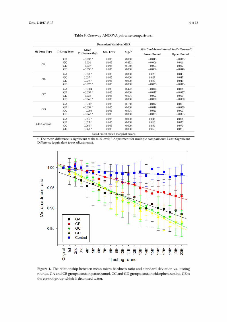
2.2. Surface Micro-Hardness
The mean micro-hardness ratios (MHR) in each group, as a function of testing round k, are shownin Figure 1. In general, the MHR decreased with the number of drug immersion in all experimentalgroups despite variations in the magnitude. In the control group, only a small fluctuation (<6%) onMHR was observed after 20 rounds of immersion. However, there were drastic decreases of around15% in the MHR in both the GC and GD groups, whilst the GB group only exhibited a 7% decrease inMHR. One-way ANCOVA revealed that test groups collectively have statistically significant differencesfrom one another (Table 2), and the control group has a statistically significantly different regressionslope from all others (p < 0.001), whilst GA, GC and GD has no statistical significant difference betweeneach other (p > 0.05) (Table 3).
Table 2. One-way ANCOVA results of micro-hardness ratios (MHR).
Dependent Variable: MHR
Source Type III Sumof Squares df Mean
Square F Sig. Partial EtaSquared
Noncent.Parameter
ObservedPower b
Corrected Model 0.158 a 5 0.032 119.792 0.000 0.858 598.961 1.000Intercept 27.991 1 27.991 106,059.503 0.000 0.999 106,059.503 1.000Round 0.093 1 0.093 354.279 0.000 0.782 354.279 1.000
Drug Type 0.064 4 0.016 60.926 0.000 0.711 243.706 1.000Error 0.026 99 0.000Total 94.592 105
Corrected Total 0.184 104a R Squared = 0.858 (Adjusted R Squared = 0.851); b Computed using alpha = 0.05.
Dent. J. 2017, 5, 17 4 of 13
Table 3. One-way ANCOVA pairwise comparisons.
Dependent Variable: MHR
(I) Drug Type (J) Drug Type MeanDifference (I–J) Std. Error Sig. b
95% Confidence Interval for Difference b
Lower Bound Upper Bound
GA
GB −0.033 * 0.005 0.000 −0.043 −0.023GC 0.004 0.005 0.422 −0.006 0.014GD 0.007 0.005 0.180 −0.003 0.017GE −0.056 * 0.005 0.000 −0.066 −0.046
GB
GA 0.033 * 0.005 0.000 0.023 0.043GC 0.037 * 0.005 0.000 0.027 0.047GD 0.039 * 0.005 0.000 0.030 0.049GE −0.023 * 0.005 0.000 −0.033 −0.013
GC
GA −0.004 0.005 0.422 −0.014 0.006GB −0.037 * 0.005 0.000 −0.047 −0.027GD 0.003 0.005 0.604 −0.007 0.013GE −0.060 * 0.005 0.000 −0.070 −0.050
GD
GA −0.007 0.005 0.180 −0.017 0.003GB −0.039 * 0.005 0.000 −0.049 −0.030GC −0.003 0.005 0.604 −0.013 0.007GE −0.063 * 0.005 0.000 −0.073 −0.053
GE (Control)
GA 0.056 * 0.005 0.000 0.046 0.066GB 0.023 * 0.005 0.000 0.013 0.033GC 0.060 * 0.005 0.000 0.050 0.070GD 0.063 * 0.005 0.000 0.053 0.073
Based on estimated marginal means
*. The mean difference is significant at the 0.05 level; b Adjustment for multiple comparisons: Least SignificantDifference (equivalent to no adjustments).
Dent. J. 2017, 5, 17 4 of 13
Table 3. One-way ANCOVA pairwise comparisons.
Dependent Variable: MHR
(I) drug type (J) drug type Mean Difference
(I–J) Std. Error Sig. b 95% Confidence Interval for Difference b
Lower Bound Upper Bound
GA
GB −0.033 * 0.005 0.000 −0.043 −0.023 GC 0.004 0.005 0.422 −0.006 0.014 GD 0.007 0.005 0.180 −0.003 0.017 GE −0.056 * 0.005 0.000 −0.066 −0.046
GB
GA 0.033 * 0.005 0.000 0.023 0.043 GC 0.037 * 0.005 0.000 0.027 0.047 GD 0.039 * 0.005 0.000 0.030 0.049 GE −0.023 * 0.005 0.000 −0.033 −0.013
GC
GA −0.004 0.005 0.422 −0.014 0.006 GB −0.037 * 0.005 0.000 −0.047 −0.027 GD 0.003 0.005 0.604 −0.007 0.013 GE −0.060 * 0.005 0.000 −0.070 −0.050
GD
GA −0.007 0.005 0.180 −0.017 0.003 GB −0.039 * 0.005 0.000 −0.049 −0.030 GC −0.003 0.005 0.604 −0.013 0.007 GE −0.063 * 0.005 0.000 −0.073 −0.053
GE (Control)
GA 0.056 * 0.005 0.000 0.046 0.066 GB 0.023 * 0.005 0.000 0.013 0.033 GC 0.060 * 0.005 0.000 0.050 0.070 GD 0.063 * 0.005 0.000 0.053 0.073
Based on estimated marginal means
*. The mean difference is significant at the 0.05 level; b Adjustment for multiple comparisons: Least Significant Difference (equivalent to no adjustments).
Figure 1. The relationship between mean micro-hardness ratio and standard deviation vs. testing rounds. GA and GB groups contain paracetamol, GC and GD groups contain chlorpheniramine, GE is the control group which is deionised water.
Figure 1. The relationship between mean micro-hardness ratio and standard deviation vs. testingrounds. GA and GB groups contain paracetamol, GC and GD groups contain chlorpheniramine, GE isthe control group which is deionised water.

Dent. J. 2017, 5, 17 5 of 13
2.3. Scanning Electron Microscope (SEM) / Energy Dispersive X-Ray Spectrometry (EDS) Analysis
The median values for the three repeated EDS measurements in each sample before and after theexperiment were calculated and analysed in the Kruskal–Wallis test (Table 4). Generally, the Ca/Cratio decreased after the drug immersion, while the Ca/P ratio and Mg, Na weight percentages mostlyremained stable. However, no statistically significant differences have been found. Moreover, the datarevealed that, before the immersion, all enamel surfaces appeared to be flat (Figure 2). After the twentyrounds of immersion, the surfaces of the GA group displayed a distinctive enamel loss with irregularcraters. The surfaces of the GB group showed a corroded surface and the fracture lines were along theborder of prism heads. In group GC, the surfaces presented a remarkable acid-etched prism-sheathstructure of enamel, with the prisms surrounded by a wide sheath region. Group GD also exhibiteddisorganised enamel loss with increased porosity on the surface. No distinctive microstructure losswas observed in control group (group GE).
Dent. J. 2017, 5, 17 5 of 13
2.3. Scanning Electron Microscope (SEM) / Energy Dispersive X-Ray Spectrometry (EDS) Analysis
The median values for the three repeated EDS measurements in each sample before and after the experiment were calculated and analysed in the Kruskal–Wallis test (Table 4). Generally, the Ca/C ratio decreased after the drug immersion, while the Ca/P ratio and Mg, Na weight percentages mostly remained stable. However, no statistically significant differences have been found. Moreover, the data revealed that, before the immersion, all enamel surfaces appeared to be flat (Figure 2). After the twenty rounds of immersion, the surfaces of the GA group displayed a distinctive enamel loss with irregular craters. The surfaces of the GB group showed a corroded surface and the fracture lines were along the border of prism heads. In group GC, the surfaces presented a remarkable acid-etched prism-sheath structure of enamel, with the prisms surrounded by a wide sheath region. Group GD also exhibited disorganised enamel loss with increased porosity on the surface. No distinctive microstructure loss was observed in control group (group GE).
Figure 2. Scanning electron microscope (SEM) micrographs (×1200) of deciduous enamel before and after immersion 20 times in drugs containing paracetamol (groups GA and GB), chlorpheniramine (groups GC and GD), or deionized water (Control group).
Figure 2. Scanning electron microscope (SEM) micrographs (×1200) of deciduous enamel before andafter immersion 20 times in drugs containing paracetamol (groups GA and GB), chlorpheniramine(groups GC and GD), or deionized water (Control group).

Dent. J. 2017, 5, 17 6 of 13
Table 4. Energy dispersive x-ray spectrometry (EDS) measurements of Ca/P and Ca/C ratios, and Mgand Na wt % in the test groups before immersion and after 20 rounds of immersion. Kruskal–Wallistest revealed no statistical significance before and after the immersion for each group at α = 0.05.
Ca/P Ca/C Mg (wt %) Na (wt %)
BeforeImmersion
After 20Rounds
BeforeImmersion
After 20Rounds
BeforeImmersion
After 20Rounds
BeforeImmersion
After 20Rounds
GA 1.95 1.96 10.45 8.26 1.32 1.56 2.10 2.10GB 1.99 1.92 10.65 8.71 1.26 1.48 2.02 2.00GC 1.97 1.98 10.21 9.13 1.50 1.41 2.09 1.83GD 1.94 1.94 9.79 8.63 1.46 1.55 2.10 1.95
GE (control) 1.80 1.76 7.85 6.76 1.62 1.52 2.46 2.25
3. Discussion
In this in vitro study, we found that the OTC paediatric oral liquids could significantly lower theVickers micro-hardness of enamel, i.e., had a softening effect, causing damage on the surface of enamelas seen from SEM after successive immersion cycles. It seems that the enamel of deciduous teeth issusceptible to acid erosion caused by the oral liquids, while the presence of sugar or pH of the oralliquids were not the major factors.
Three of the tested medicines showed a pH lower than the “critical pH of enamel”, i.e., 5.5.However, impurities in the dental mineral may increase its solubility, thus the relative rates ofdissolution do not truly reflect the solubility [25]. It was difficult to evaluate the solubility productsbased on the actual mineral composition, since it has been demonstrated that the minerals in biologicalsamples were heterogeneous [26]. Accordingly, the critical pH for enamel was typically consideredto be in a range of 4.5 to 5.5 [27]. Even so, the current study confirmed that liquid medication withan endogenous pH of 5.7 might also cause erosive damage to the enamel surface. In fact, the rate ofdissolution of enamel is strongly influenced by other physical factors (e.g., temperature, consumptionfrequency) and biological factors (e.g., saliva in the formation of the acquired pellicle, as a lubricant,buffer and ion reservoir). For example, solubility is dependent on temperature and heat is required tobreak the bonds holding the molecules in a solid [28]. Thus, erosion is predicted to be more severeat high temperatures and less at low temperatures, which has been shown by Banan et al. thatdairy beverages caused less decrease of pH in plaque and saliva at temperatures lower than roomtemperature [29,30].
In addition, from a biological perspective, the presence of saliva is greatly beneficial to an oralcavity under erosive attack. Saliva is required to form the acquired pellicle, which is defined asa proteinaceous layer on all solid surfaces exposed to the oral cavity [31–33]. It is an organic filmcomposed of glycoproteins and proteins, including several enzymes, without the involvement ofbacteria [31,32]. It works as a protective membrane to prevent direct attack from acidic substances,thus lowering the rate of demineralisation [33]. Furthermore, bicarbonate is the most importantcomponent in saliva to protect against acidic products generated by dental plaque or extrinsic acids [34].The presence of calcium and phosphate, as well as an alkaline or neutral environment is essentialfor remineralisation. Preserving calcium and phosphate in saliva allows them to penetrate into theminerals of enamel, slowing down the rate of mineral dissolution [35]. One research study has shownthat salivary proteins would bind to demineralised sites and cover exposed crystals by a specificadsorption mechanism [36]. In the oral cavity, the contact of the enamel with acidic substances isusually only transient before clearance by saliva. Consequently, under in vivo conditions, an earlystage of erosion is limited to a very insignificant loss of mineral and erosive craters on a nanometerscale or even near atomic level [37]. In this sense, there might be discrepancies between the currentin vitro study and other in vivo studies.
Since there was no involvement of saliva in this study, it is possible that the erosive potential ofmedications was overestimated. For example, we have not included the buffer capacity of saliva which
Dent. J. 2017, 5, 17 7 of 13
could alter the final pH in the oral cavity and hence lower the dissolution rate of enamel [38], possiblydue to the high bicarbonate concentration in saliva [39]. Nevertheless, significantly different bufferingcapacities have been found between genders (e.g., male and female), as well as the method of collection(e.g., unstimulated or stimulated) [38]. Thus, in most of the in vitro studies, various formulationsof artificial saliva were employed. However, most simple simulated saliva compositions have noproteins and do not form acquired pellicles, which play an important protective role against erosion.Moreover, collection of natural saliva in a restricted time frame is not a simple task, particularly withthe quick deterioration of saliva [40]. Therefore, more in vivo studies are necessary to reliably evaluatethe erosive effect under the influence of saliva and masticatory activities.
Deciduous tooth enamel is softer than permanent tooth enamel. A study has found that deciduousteeth had a lower prismatic density, which meant a smaller number of prisms per unit area, and ashorter prismatic diameter compared to permanent teeth despite having the same morphology [41].The higher percentage of minerals makes the enamel more resistant to masticatory forces, butits extreme hardness makes it more prone to fractures and more reliant on the presence of thedentine—which is more resilient—to maintain its integrity [42]. Nevertheless, some investigators foundthat deciduous enamel was softer and more prone to fracture than permanent enamel [43]. Furthermore,the lessened mineralisation and the smaller thickness in deciduous enamel make primary teeth moreprone to erosion and decay [44–46]. Generally speaking, the erosive potential of primary enamel isgreater and its weaker mechanical properties could be explained by the reduced mineralisation indeciduous enamel tissue.
In the current study, the deciduous teeth were challenged by 20 rounds of 15 s drug immersiontime. Fifteen seconds was chosen for the immersion time so as to reflect the drug taking habits ofchildren and the residence time of drugs on enamel. The twenty rounds were to mimic the times ofconsumption (quater die sumendus for five days). In addition, since the initial values of micro-hardnessdiffered among the specimens, ratios (MHR) were calculated instead and corresponding trend lineswere drawn in order to allow for better data comparison among the groups.
Liquid formulations are usually complex compositions containing many other componentsbesides the main active ingredients; the additives include bulk materials, flavourings, sweeteners,buffers, preservatives and colouring agents [12]. Although we do not know the exact formulationof the purchased medicinal liquids, acidic contents were common to all the liquids and acted asbuffering agents, and they were responsible for improving palatability, maintaining chemical stability,controlling tonicity and ensuring the physiological compatibility [13]. For example, citric acid was themost frequently added acid, yet it has been linked to tooth erosion due to its ability to dissolve thehydroxyapatite of tooth enamel and dentine [47]. It has been found that citric acid acts as a chelatorthat binds to the minerals of the hydroxyapatite, reduces saliva supersaturation and increases thedissolution rate of hydroxyapatite crystals [48]. Other acids have also demonstrated various erosiveeffects on enamel; it has been reported that lactic acid at low pH was more erosive than both citric andmaleic acids [49].
In the chlorpheniramine groups, a sugar-free formulation group GD having a higher pH valueexhibited a similar micro-hardness decreasing trend as group GC. It might be due to the additionof “sugar-free” excipients, i.e. artificial sweeteners, are acidic or chemically stable under acidicenvironment [50]. This result was further confirmed by another study of comparing the erosivepotential between sugar-free medicine and sugar-containing ones. They found that the sugar-freemedications still carried a similar erosive potential as the sugar-containing syrups [13]. In addition, theactive ingredient of these two drugs was chlorpheniramine maleate, which in fact would be dissociatedinto chlorpheniramine free base and maleic acid in aqueous conditions [51]. Indeed, despite maleicacid being a weak acid, it has the potential to etch [52] or chemisorb [53] on enamel, so much so that itcould be utilised for bonding orthodontic brackets onto enamel [54]. It seems to be that simply shiftingto sugar-free medications is not as beneficial as once thought, and a fundamental understanding of theformulation science and chemistry is essential.
Dent. J. 2017, 5, 17 8 of 13
With respect to the elemental analysis by EDS, semi-quantitative data were presented to show thechemical changes of enamel surface after liquid medicine immersion. A previous study has examinedthe Ca/P and Ca/C ratio to compare the elemental difference before and after demineralisation,since it was thought that the ratios were altered from calcium hydroxyapatite due to the alterationor substitution of mineral phase [55]. However, in the present study, the Ca/P ratio before drugimmersion determined by using the EDS was found to be around 1.9, which was not significantlydifferent from that of enamel which experienced drug immersion. Accordingly, we speculate thatthe components of Ca and P change together (i.e., dissolved in the oral liquids) in the same direction.Besides, several other studies were unable to demonstrate any significant difference in the Ca/P ratio incompromised enamel [55,56]. Multiple studies on developing enamel have shown that the Ca/P ratiowas constant during tooth development despite variations among individuals [57,58]. Nevertheless,Jälevik and co-workers did find that median Ca/P ratio of hypo-mineralised enamel was significantlylower than sound enamel [59]. Therefore, the teeth collected in various stages in different countriesmight have high variations which eventually led to the scattering of results.
One study also revealed a relation between micro-hardness and the Ca/C ratio in demineralisedenamel [56]. As the crystals were dissolved with more organic components exposed, it seemedplausible that the Ca/C ratio would increase. However, in this study, despite a Ca/C ratio decreaseafter drug challenge, the difference had no statistical significance. The limited sample size in this studyand variations among individuals could contribute to the contrasting result.
Given the findings of this study, paediatricians and patients should realise the risk of erosionduring the consumption of liquid medicines by children. Dentists should not overlook the early stagesof erosion, regarding minor tooth surface loss as a normal occurrence of daily life, since early diagnosisand appropriate intervention are seldom reached in time [60]. Children should also visit dentistsregularly and oral hygiene education in preschool is necessary. Other methods such as an adequateuse of fluoride-containing mouth-rinses, toothpastes, tablets and lozenges have been demonstrated toremineralise enamel effectively [61]. In future, more clinical studies or in vivo studies are needed, sincethe limits of in vitro studies do not allow simulation of all the complex biological effects. We anticipatethis study could raise awareness of the erosion susceptibility from paediatric OTC oral liquids.
4. Materials and Methods
This study was approved by the Institutional Review Board of Hong Kong (IRB UW15-172).The procedure has been illustrated as a flowchart in Figure 3.

Dent. J. 2017, 5, 17 9 of 13Dent. J. 2017, 5, 17 9 of 13
Figure 3. The flowchart of experimental design.
4.1. Teeth and Specimen Preparation
Twenty sectioned enamel blocks, with 2 × 2 mm2 buccal enamel surface, were obtained from ten sound extracted primary incisors. The incisors were stored in distilled water at 4 °C prior to use for no longer than one week. Each incisor was sliced longitudinally parallel to its long axis to get two enamel blocks by a cutting machine under running water. All enamel blocks were embedded in light-cured resin (Filtek Z250, 3M ESPE) onto an acrylic plate. The buccal surfaces were faced upwards to make sure most regions of the surface are parallel to the plate.
After embedding the enamel into the resin, the buccal surfaces of the enamel were polished using 1000- and 2000-grit SiC abrasive paper by a manually water-cooled low-speed polishing machine to obtain a flat surface without dentine exposure, and the specimen underwent ultrasonic washing in deionised water before and after polishing. Then, the twenty blocks were randomly divided into five groups (four experimental groups GA, GB, GC, GD, and one control group GE; see Table 1), i.e., four blocks in one group, to immerse into the oral liquids or deionised water.
4.2. Oral Liquids
Figure 3. The flowchart of experimental design.
4.1. Teeth and Specimen Preparation
Twenty sectioned enamel blocks, with 2 × 2 mm2 buccal enamel surface, were obtained from tensound extracted primary incisors. The incisors were stored in distilled water at 4 ◦C prior to use for nolonger than one week. Each incisor was sliced longitudinally parallel to its long axis to get two enamelblocks by a cutting machine under running water. All enamel blocks were embedded in light-curedresin (Filtek Z250, 3M ESPE) onto an acrylic plate. The buccal surfaces were faced upwards to makesure most regions of the surface are parallel to the plate.
After embedding the enamel into the resin, the buccal surfaces of the enamel were polished using1000- and 2000-grit SiC abrasive paper by a manually water-cooled low-speed polishing machine toobtain a flat surface without dentine exposure, and the specimen underwent ultrasonic washing indeionised water before and after polishing. Then, the twenty blocks were randomly divided into fivegroups (four experimental groups GA, GB, GC, GD, and one control group GE; see Table 1), i.e., fourblocks in one group, to immerse into the oral liquids or deionised water.

Dent. J. 2017, 5, 17 10 of 13
4.2. Oral Liquids
As the most commonly used paediatric OTC drugs to relieve respiratory symptoms were usuallyfor acute occasions [9], two commonly-used single component formulations, i.e., paracetamol andchlorpheniramine, were used (Table 1). Paracetamol (in US: acetaminophen) is a major ingredient innumerous cold and flu remedies and it is also classified as a mild analgesic to relieve headaches, feverand other minor aches and pains. Chlorpheniramine is an antihistamine used to relieve the symptomsof allergic conditions and the common cold, such as rhinorrhoea. Two different brands of paediatricOTC oral liquids containing one of each of the drugs were selected to make a comparison. In thechlorpheniramine group, the Jean-Marie brand contains sugar, while the Allerief belongs to a sugar-freetype. All of the OTC drugs were purchased from local pharmacies and no further information frommanufacturers was acquired.
4.3. pH Measurement
The endogenous pH of the testing medications was determined at room temperature by using adigital pH meter (CyberScan pH500, Eutech Instruments, UK) in triplicate to get a mean value. The pHmeter was calibrated before measurement according to the manufacturer’s instructions, using bufferstandards of pH 4 and pH 7. Then, 10 mL of the testing medicine was placed in one beaker, the probeof the pH meter was immersed directly into the syrup and the value was recorded with the precisionof 0.01.
4.4. Micro-Hardness Measurement
To evaluate and compare the erosive potentials of four medications on a continuous scale, eachspecimen was subjected to twenty rounds of immersion in one of the drugs, followed by micro-hardnessmeasurements. To set a baseline of the mean initial micro-hardness (MH0), each enamel block wasimmersed into deionised water (DI) for 15 s, and then five random spots were picked and tested usinga micro-hardness tester (Leica DC 300, Leitz, Germany) with a Vickers tip and load of 200 g (1.962 N)lasting for 15 s. Then, in each round k, the enamel block was immersed into 10 mL of the correspondingtest group for 15 s and followed by rinsing with DI water for 15 s. Again, the average micro-hardnessvalues (MHk) of the enamel surfaces were measured immediately from five random spots using thesame micro-hardness tester and parameters. The same immersion and measuring procedures wererepeated for twenty rounds (i.e., k = 1–20). Fresh drug or DI water was used for each new roundof immersion. As a whole, each sample experienced 21 (one initial + twenty after drug immersion)micro-hardness measurements. All the values were then plotted as the ratio (MHR) vs. round ofimmersion to give a trend (Figure 1) on how the surface hardness changes following a continuousdrug attack, i.e.,:
MHR = MHk/MH0 (1)
where MHk is the mean micro-hardness at k = 0–20 rounds, and MH0 is the mean initial micro-hardness.The MHR and slope of regression lines of Figure 1 was statistically analysed by one-way ANCOVA atα = 0.05 (SPSS v.22, IBM, New York, NY, USA).
4.5. Scanning Electron Microscope Analysis
The surface topography of the enamel specimens at 1200× magnification was studied under ascanning electron microscope (Hitachi SU-1510 Variable Pressure SEM, Hitachi Ltd., Tokyo, Japan) at15 kV in high-vacuum mode. The SEM micrographs of all enamel blocks were taken on two occasions:(1) before the immersion procedures, where the enamel blocks were mounted onto an aluminium stubseparately to assess their surface topography without coating, and (2) after 20 rounds of immersion,where all the specimens were sputter-coated with carbon at 15 kV and the entire buccal surfacewas scanned. The SEM micrographs provided visual and illustrative comparisons of the surfacemorphologic changes of specimens before and after the drug/water challenge.

Dent. J. 2017, 5, 17 11 of 13
4.6. Energy Dispersive X-Ray Spectrometry Analysis
The same samples that had been observed in SEM were also used for the EDS analysis on thesame occasions, except the EDS was done prior to the coating. The EDS system comprised of an X-raydetector system (Model 550i, IXRF-Systems, Austin, TX, USA) attached to the SEM operated at 15 kVwith a spot size of 5 nm, specimen tilt of 35◦, working distance of 15 mm and a counting time of 20 s.Three counts from each enamel sample were measured to give a mean weight percentage of calcium(Ca), phosphorus (P), sodium (Na), magnesium (Mg), carbon (C) and oxygen (O). The mean wt %ratios of Ca/P and Ca/C, and the wt % of Mg and Na before and after tests were statistically evaluatedby Kruskal–Wallis test at α = 0.05 (SPSS v.22).
5. Conclusions
Within the limitations of this laboratory study, the four tested paediatric OTC oral liquids werefound to significantly soften enamel of primary teeth and deem them susceptible to caries. There is apossible association between drug formulations and their erosive potentials.
Acknowledgments: The authors deny any conflicts of interest related to this study. This work was done inpartial fulfilment of the requirements of the degree of MSc (DMS) for the first author at the Faculty of Dentistry,The University of Hong Kong. We thank Madeline J.Y. Yon for proofreading the article.
Author Contributions: D.Z. and J.K.-H.T. conceived and designed the experiments; D.Z. performed theexperiments; D.Z., J.K.-H.T. and J.P.M. analysed the data; H.M.W. and C.H.C. contributed reagents/materials/analysis tools; D.Z., J.K.-H.T., H.M.W., C.H.C. and J.P.M. wrote the paper.
Conflicts of Interest: The authors declare no conflict of interest.
References
1. Bahal, P.; Djemal, S. Dental erosion from an excess of vitamin C. Case Rep. Dent. 2014, 2014, 5. [CrossRef][PubMed]
2. Bartlett, D.; Dugmore, C. Pathological or physiological erosion—Is there a relationship to age?Clin. Oral Investig. 2008, 12, 27–31. [CrossRef] [PubMed]
3. Scheutzel, P. Etiology of dental erosion–intrinsic factors. Eur. J. Oral Sci. 1996, 104, 178–190. [CrossRef][PubMed]
4. Lussi, A.; Schlueter, N.; Rakhmatullina, E.; Ganss, C. Dental erosion—An overview with emphasis onchemical and histopathological aspects. Caries Res. 2011, 45, 2–12. [CrossRef] [PubMed]
5. Dawes, C. What is the critical ph and why does a tooth dissolve in acid? Can. Dent. Assoc. 2003, 69, 722–724.6. Zhang, S.; Chau, A.M.H.; Lo, E.C.M.; Chu, C.-H. Dental caries and erosion status of 12-year-old Hong Kong
children. BMC Public Health 2014, 14, 7. [CrossRef] [PubMed]7. Tahmassebi, J.F.; Duggal, M.S.; Malik-Kotru, G.; Curzon, M.E.J. Soft drinks and dental health: A review of
the current literature. J. Dent. 2006, 34, 2–11. [CrossRef] [PubMed]8. Mantonanaki, M.; Koletsi-Kounari, H.; Mamai-Homata, E.; Papaioannou, W. Dental erosion prevalence and
associated risk indicators among preschool children in Athens, Greece. Clin. Oral Investig. 2013, 17, 585–593.[CrossRef] [PubMed]
9. Sturkenboom, M.; Verhamme, K.; Nicolosi, A.; Murray, M.; Neubert, A.; Caudri, D.; Picelli, G.; Sen, E.;Giaquinto, C.; Cantarutti, L.; et al. Drug use in children: Cohort study in three European countries. Br. Med. J.2008, 337, 1338–1341. [CrossRef] [PubMed]
10. Lussi, A.; Schaffner, M.; Jaeggi, T. Dental erosion-diagnosis and prevention in children and adults. Int. Dent. J.2007, 57, 385–398. [CrossRef]
11. Neves, B.G.; Farah, A.; Lucas, E.; de Sousa, V.P.; Maia, L.C. Are paediatric medicines risk factors for dentalcaries and dental erosion? Commun. Dent. Health 2010, 27, 46–51.
12. Mennella, J.A.; Spector, A.C.; Reed, D.R.; Coldwell, S.E. The bad taste of medicines: Overview of basicresearch on bitter taste. Clin. Ther. 2013, 35, 1225–1246. [CrossRef] [PubMed]

Dent. J. 2017, 5, 17 12 of 13
13. Maguire, A.; Baqir, W.; Nunn, J.H. Are sugars-free medicines more erosive than sugars-containing medicines?An in vitro study of paediatric medicines with prolonged oral clearance used regularly and long-term bychildren. Int. J. Paediatr. Dent. 2007, 17, 231–238. [CrossRef] [PubMed]
14. Liem, D.G.; Mennella, J.A. Heightened sour preferences during childhood. Chem. Senses 2003, 28, 173–180.[CrossRef] [PubMed]
15. Marquezan, M.; Pozzobon, R.; Oliveira, M. Medicines used by pediatric dentistry patients and its cariogenicpotential. RPG Rev. Pos. Grad. 2007, 13, 334–339.
16. Durward, C.; Thou, T. Dental caries and sugar-containing liquid medicines for children in new zealand. N. Z.Dent. J. 1997, 93, 124–129. [PubMed]
17. Feigal, R.J.; Jensen, M.E.; Mensing, C.A. Dental caries potential of liquid medications. Pediatrics 1981, 68,416–419. [PubMed]
18. Babu, K.L.; Doddamani, G.M.; Naik, L.R.; Jagadeesh, K.N. Pediatric liquid medicaments—Are theycariogenic? An in vitro study. J. Int. Soc. Prev. Commun. Dent. 2014, 4, 108–112. [CrossRef] [PubMed]
19. Arora, R.; Mukherjee, U.; Arora, V. Erosive potential of sugar free and sugar containing pediatric medicinesgiven regularly and long term to children. Indian J. Pediatr. 2012, 79, 759–763. [CrossRef] [PubMed]
20. Babu, K.L.G.; Rai, K.; Hedge, A.M. Pediatric liquid medicaments—Do they erode the teeth surface?An in vitro study: Part I. J. Clin. Pediatr. Dent. 2008, 32, 189–194. [CrossRef] [PubMed]
21. Sahgal, J.; Sood, P.B.; Raju, O.S. A comparison of oral hygiene status and dental caries in children on longterm liquid oral medications to those not administered with such medications. J. Indian Soc. Pedod. Prev. Dent.2002, 20, 144–151. [PubMed]
22. Cavalcanti, A.; de Oliveira, K.; Xavier, A.; Pinto, D.; Vieira, F. Evaluation of total soluble solids content (TSSC)and endogenous ph in antimicrobials of pediatric use. Indian J. Dent. Res. 2013, 24, 498–501. [CrossRef] [PubMed]
23. Strickley, R.G.; Iwata, Q.; Wu, S.; Dahl, T.C. Pediatric drugs-a review of commercially available oralformulations. J. Pharm. Sci. 2008, 97, 1731–1774. [CrossRef] [PubMed]
24. Donaldson, M.; Goodchild, J.H.; Epstein, J.B. Sugar content, cariogenicity, and dental concerns withcommonly used medications. J. Am. Dent. Assoc. 2015, 146, 129–133. [CrossRef] [PubMed]
25. Featherstone, J.; Lussi, A. Understanding the chemistry of dental erosion. Dent. Eros. 2006, 20, 66–76.26. Shellis, R.P. A scanning electron-microscopic study of solubility variations in human enamel and dentine.
Arch. Oral Biol. 1996, 41, 473–484. [CrossRef]27. Axelsson, P.D.D.S. Diagnosis and Risk Prediction of Dental Caries; Quintessence Publishing: Chicago, IL,
USA, 2000.28. Pinto, S.C.S.; Batitucci, R.G.; Pinheiro, M.C.; Zandim, D.L.; Spin-Neto, R.; Sampaio, J.E.C. Effect of an acid
diet allied to sonic toothbrushing on root dentin permeability: An in vitro study. Braz. Dent. J. 2010, 21, 390.[CrossRef] [PubMed]
29. Ferreira, F.V.; Pozzobon, R.T. Processed dairy beverages ph evaluation: Consequences of temperaturevariation. J. Clin. Pediatr. Dent. 2009, 33, 319. [CrossRef] [PubMed]
30. Banan, L.K.; Hegde, A.M. Plaque and salivary ph changes after consumption of fresh fruit juices. J. Clin.Pediatr. Dent. 2005, 30, 9. [CrossRef] [PubMed]
31. Hannig, C.; Hannig, M.; Attin, T. Enzymes in the acquired enamel pellicle. Eur. J. Oral Sci. 2005, 113, 2–13.[CrossRef] [PubMed]
32. Lendenmann, U.; Grogan, J.; Oppenheim, F.G. Saliva and dental pellicle—A review. Adv. Dent. Res. 2000, 14,22–28. [CrossRef] [PubMed]
33. Hannig, M.; Balz, M. Influence of in vivo formed salivary pellicle on enamel erosion. Caries Res. 1999, 33,372–379. [CrossRef] [PubMed]
34. Edgar, W.M.; Dawes, C.; Mullane, D.M. Saliva and Oral Health; British Dental Association: London, UK, 2004.35. Meurman, J.H.; Ten Cate, J.M. Pathogenesis and modifying factors of dental erosion. Eur. J. Oral Sci. 1996,
104, 199–206. [CrossRef] [PubMed]36. Voegel, J.C.; Belcourt, A.; Gillmeth, S. Dissolution of hydroxyapatites treated with salivary glycoproteins
and fluoride. Caries Res. 1981, 243–249. [CrossRef]37. Attin, T.; Wegehaupt, F.J. Methods for Assessment of Dental Erosion; Karger: Basel, Switzerland, 2014.38. Gittings, S.; Turnbull, N.; Henry, B.; Roberts, C.J.; Gershkovich, P. Characterisation of human saliva as a platform
for oral dissolution medium development. Eur. J. Pharm. Biopharm. 2015, 91, 16–24. [CrossRef] [PubMed]

Dent. J. 2017, 5, 17 13 of 13
39. Izutsu, K.T. Theory and measurement of the buffer value of bicarbonate in saliva. J. Theor. Biol. 1981, 90,397–403. [CrossRef]
40. Amaechi, B.T.; Higham, S.M. Eroded enamel lesion remineralization by saliva as a possible factor in thesite-specificity of human dental erosion. Arch. Oral Biol. 2001, 46, 697–703. [CrossRef]
41. Gentile, E.; Di Stasio, D.; Santoro, R.; Contaldo, M.; Salerno, C.; Serpico, R.; Lucchese, A. In vivomicrostructural analysis of enamel in permanent and deciduous teeth. Ultrastruct. Pathol. 2015, 39, 131–134.[CrossRef] [PubMed]
42. Low, I.M.; Duraman, N.; Davies, I.J. Microstructure-property relationships in human adult and baby canineteeth. Key Eng. Mater. 2006, 309, 23–26. [CrossRef]
43. De Menezes Oliveira, M.A.H.; Torres, C.P.; Gomes-silva, J.M.; Chinelatti, M.A.; De Menezes, F.C.H.;Palma-dibb, R.G.; Borsatto, M.C. Microstructure and mineral composition of dental enamel of permanentand deciduous teeth. Microsc. Res. Tech. 2010, 73, 572–577. [CrossRef] [PubMed]
44. Wang, L.J.; Tang, R.; Bonstein, T.; Bush, P.; Nancollas, G.H. Enamel demineralization in primary andpermanent teeth. J. Dent. Res. 2006, 85, 359. [CrossRef] [PubMed]
45. Johansson, A.K.; Sorvari, R.; Birkhed, D.; Meurman, J.H. Dental erosion in deciduous teeth—An in vivo andin vitro study. J. Dent. 2001, 29, 333–340. [CrossRef]
46. Hunter, M.L.; West, N.X.; Hughes, J.A.; Newcombe, R.G.; Addy, M. Erosion of deciduous and permanentdental hard tissue in the oral environment. J. Dent. 2000, 28, 257–263. [CrossRef]
47. Grenby, T.H.; Phillips, A.; Desai, T.; Mistry, M. Laboratory studies of the dental properties of soft drinks.Br. J. Nutr. 1989, 62, 451–464. [CrossRef] [PubMed]
48. Lussi, A.; Jaeggi, T. Erosion—Diagnosis and risk factors. Clin. Oral Investig. 2008, 12, 5–13. [CrossRef][PubMed]
49. Hughes, J.A.; West, N.X.; Parker, D.M.; van den Braak, M.H.; Addy, M. Effects of ph and concentration ofcitric, malic and lactic acids on enamel, in vitro. J. Dent. 2000, 28, 147–152. [CrossRef]
50. Chattopadhyay, S.; Raychaudhuri, U.; Chakraborty, R. Artificial sweeteners–a review. J. Food Sci. Technol.2014, 51, 611–621. [CrossRef] [PubMed]
51. Pandit, N.K. Introduction to the Pharmaceutical Sciences An Integrated Approach; Soltis, R.P., Ed.; LippincottWilliams and Wilkins: Baltimore, MD, USA, 2012.
52. Hermsen, R.J.; Vrijhoef, M.M. Loss of enamel due to etching with phosphoric or maleic acid. Dent. Mater.1993, 9, 332–336. [CrossRef]
53. Fu, B.P.; Yuan, J.; Qian, W.X.; Shen, Q.Y.; Sun, X.M.; Hannig, M. Evidence of chemisorption of maleic acid toenamel and hydroxyapatite. Eur. J. Oral Sci. 2004, 112, 362–367. [CrossRef] [PubMed]
54. Olsen, M.E.; Bishara, S.E.; Damon, P.; Jakobsen, J.R. Evaluation of scotchbond multipurpose and maleic acidas alternative methods of bonding orthodontic brackets. Am. J. Orthod. Dentofac. Orthop. 1997, 111, 498–501.[CrossRef]
55. Mahoney, E.K.; Rohanizadeh, R.; Ismail, F.S.M.; Kilpatrick, N.M.; Swain, M.V. Mechanical properties andmicrostructure of hypomineralised enamel of permanent teeth. Biomaterials 2004, 25, 5091–5100. [CrossRef][PubMed]
56. Fagrell, T.G.; Dietz, W.; Jälevik, B.; Norén, J.G. Chemical, mechanical and morphological properties ofhypomineralized enamel of permanent first molars. Acta Odontol. Scand. 2010, 68, 215. [CrossRef] [PubMed]
57. Kodaka, T.; Debari, K.; Kuroiwa, M. Mineral content of the innermost enamel in erupted human teeth.J. Electron. Microsc. 1991, 40, 19–23.
58. Sasaki, T.; Debrari, K.; Garant, P.R. Ameloblast modulation and changes in the Ca, P, and S content ofdeveloping enamel matrix revealed by SEM-EDX. J. Dent. Res. 1987, 66, 778–786. [CrossRef] [PubMed]
59. Jälevik, B.; Odelius, H.; Dietz, W.; Norén, J. Secondary ion mass spectrometry and X-ray microanalysis ofhypomineralized enamel in human permanent first molars. Arch. Oral Biol. 2001, 46, 239–247. [CrossRef]
60. Larsen, M.J. Chemical events during tooth dissolution. J. Dent. Res. 1990, 69, 575–580. [CrossRef] [PubMed]61. Edgar, W.M. Sugar substitutes, chewing gumand dental caries-a review. Br. Dent. J. 1998, 184, 29–32.
[CrossRef] [PubMed]
© 2017 by the authors. Licensee MDPI, Basel, Switzerland. This article is an open accessarticle distributed under the terms and conditions of the Creative Commons Attribution(CC BY) license (http://creativecommons.org/licenses/by/4.0/).