The role of p21-activated kinase in the initiation of atherosclerosis
Upload
sungkyunkwanCategory
view
1download
0
Jpn. J. Cancer Res. 91, 164–173, February 2000
164
p53-independent Induction of p21 (WAF1/CIP1), Reduction of Cyclin B1 and G2/M Arrest by the Isoflavone Genistein in Human Prostate Carcinoma Cells
Yung Hyun Choi,1, 4 Won Ho Lee,1 Kun-Young Park2 and Lijuan Zhang3
1Department of Biology, College of Natural Sciences, 2Department of Food Science and Nutrition, PusanCancer Research Center, Pusan National University, Pusan 609-735, Korea, and 3Pediatric OncologyBranch, National Cancer Institute, National Institutes of Health, Bethesda, MD 20892, USA
Genistein, a natural isoflavonoid phytoestrogen, is a strong inhibitor of protein tyrosine kinase andDNA topoisomerase II activities. Genistein has been shown to have anticancer proliferation, differ-entiation and chemopreventive effects. In the present study, we have addressed the mechanism ofaction by which genistein suppressed the proliferation of p53-null human prostate carcinoma cells.Genistein significantly inhibited the cell growth, which effect was reversible, and induced dendrite-like structure. The inhibitory effects of genistein on cell growth proliferation were associated with aG2/M arrest in cell cycle progression concomitant with a marked inhibition of cyclin B1 and aninduction of Cdk inhibitor p21 (WAF1/CIP1) in a p53-independent manner. Following genisteintreatment of cells, an increased binding of p21 with Cdk2 and Cdc2 paralleled a significantdecrease in Cdc2 and Cdk2 kinase activity with no change in Cdk2 and Cdc2 expression. Genisteinalso induced the activation of a p21 promoter reporter construct, utilizing a sequence distinct fromthe p53-binding site. Analysis of deletion constructs of the p21 promoter indicated that theresponse to genistein could be localized to the 300 base pairs proximal to the transcription startsite. These data suggest that genistein may exert a strong anticarcinogenic effect, and that thiseffect possibly involves an induction of p21, which inhibits the threshold kinase activities of Cdksand associated cyclins, leading to a G2/M arrest in the cell cycle progression.
Key words: Genistein — Cell cycle — G2/M arrest — p21
Epidemiological data have demonstrated that diet maybe the most important environmental factor involved in theetiology of some of the most prevalent forms of cancer.1)
Therefore natural products have been a good source ofnovel chemotherapeutic compounds. Soy consumption hasbeen reported as a candidate to reduce the risk of acquisi-tion of cancer.2) Several compounds in soybeans havedemonstrated anti-cancer activity. One of the candidatescompounds against malignancy in soybeans is genistein(4′,5,7-trihydroxyisoflavone), the most abundant isofla-vone present in soybeans and soy-based products, whichwas originally isolated from fermentation broth ofPseudomonas spp.3) Several mechanisms have been pro-posed for the effects of genistein. Akiyama et al.3) werethe first to describe the ability of genistein to inhibit spe-cifically the protein tyrosine kinase (PTK) activity. PTKsseem to play a key role not only in tumorigenesis, but alsoin differentiation.4) They are also known to be associatedwith both growth control receptors5) and several oncogeneproducts.6) These data suggested that genistein may have
important anticancer properties. Other investigators havesuggested that genistein may exert its effect through inhi-bition of DNA topoisomerase II7) and ribosomal S6 kinaseactivities,8) which may lead to protein-linked DNA strandbreaks.9) In addition, genistein has been observed to act invitro as a potent inhibitor of angiogenesis10) and of thedevelopment of metastases.11) In vivo studies also demon-strated that genistein is effective in reducing tumorigenesisand carcinogenesis.12, 13) Although a specific mechanism ofaction has not been identified, previous studies haveshown that genistein inhibited the growth of wide range ofcultured cancer cells,14–19) induced differentiation of sev-eral malignant cell lines,9, 17, 20) and caused apoptotic celldeath.14, 15, 18, 21) To date, despite these accumulated data,the molecular mechanism of its antiproliferative action oncancer cells is poorly understood.
Progression through the cell cycle is regulated bysequential activation and subsequent inactivation of aseries of cyclin-dependent kinases (Cdks).22, 23) The activi-ties of Cdks are positively regulated by cyclins, which actat different checkpoints of the cell cycle. The D-typecyclins and cyclin E are required for progression throughG1. As cells enter G1, the cyclin D/Cdk4 (and/or Cdk6)complex appears to be necessary for transition throughearly G1,24, 25) whereas cyclin E/Cdk2 complex is requiredin transition from late G1 into S phase.26, 27) Cyclin A isproduced in late G1 and it accumulates during S and G2
2 To whom correspondence and requests for reprints should beaddressed.E-mail: [email protected]
4 Present address: Vincent T. Lombardi Cancer Center andDepartment of Radiation Medicine, Georgetown University Med-ical Center, Washington, DC 20007, USA.
Induction of p21 and Cell Cycle Arrest by Genistein
165
phase, while expression of B-type cyclins is typicallymaximal during the G2 to M phase transition and controlspassage through the M phase. Cyclin A associates withand activates primarily Cdk2,28, 29) whereas B-type cyclinsare the major activators of Cdc2 (also called as Cdk1).30, 31)
The activity of Cdks is negatively regulated by binding toCdk inhibitors in response to a variety of antiproliferativesignals.32, 33) Among several Cdk inhibitors, p21 (WAF1/CIP1), the archetypal member of this family, which wasisolated as a Cdk2-associated protein and an inhibitor ofCdk2,34, 35) is an important mediator of cell cycle arrestimposed by the tumor suppressor p53 in response to DNAdamage.36–39) Recent studies implicate growth arrestaccompanied by up-regulation of p21 not only in inhibit-ing proliferation, but also in promoting differentiation.37, 40)
p21 is also known to be an inhibitor of DNA replicationby inhibiting proliferating-cell nuclear antigen, which isalso required for cell cycle progression.41) In addition tobeing induced by p53, p21 is also induced by other factorsin the p53-independent pathway.38, 40, 42–45) Several studieshave demonstrated that relative levels of p21 may be criti-cal in determining the threshold kinase activity of variouscyclin/Cdk complexes,32, 33, 35–37, 46) suggesting that appro-priate levels of p21 may be critical in the regulation of cellgrowth.
In the present study, we examined the effect of genisteinon the growth of human prostate carcinoma PC-3-M cellsthat lack wild-type p53. The present results demonstratedthat genistein was able to inhibit growth of cells andinduced morphological changes, which effects werereversible. Genistein treatment resulted in arrest at the G2/M check point of the cell cycle, which was related to up-regulating the Cdk-inhibitor p21, and down-regulating theintracellular levels of cyclin B1. This study also showedthat the up-regulation of p21 was associated withenhanced binding of p21 with Cdk2 and Cdc2. In addition,using deletion constructs of the p21 promoter, we foundthat the response to genistein could be localized to the 300base pairs proximal to the transcription start site. There-fore we suggest that genistein inhibits cell growth byinducing p53-independent transcriptional regulation ofp21, leading to down-regulation of the activity of Cdk2and Cdc2 kinase activity.
MATERIALS AND METHODS
Cell culture and treatments Human PC-3-M prostatecarcinoma cells which are a metastatic variant of PC3cells, as described by Kozlowski et al.,47) were grown inRPMI 1640 (Gibco BRL, Grand Island, NY) supple-mented with 10% heat-inactivated fetal bovine serum(FBS), penicillin and streptomycin (Biofluids, Rockville,MD) in a humidified 5% CO2 incubator at 37°C. Genistein(Sigma Chemical Co., St. Louis, MO) was dissolved in
dimethyl sulfoxide (DMSO, Sigma) for all experiments.The final DMSO concentration did not exceed 0.05%.Growth study and morphology For growth inhibitionanalysis, cells were plated at 0.5×10⁴ cells per 10-mmplate, and incubated for 24 h. Cells were cultured in thepresence or absence of variable concentrations of genisteinin culture medium enriched with 10% FBS. After 72 h ofculture, cells were trypsinized and the viable cells werescored using the Trypan Blue method. For the morpholog-ical study, cells were grown on coverslips and treated with100 µM genistein for 36 h. Cells were Wright-stained(Fisher Scientific, Pittsburgh, PA), as recommended by themanufacturer, and photographed.DNA flow-cytometric analysis Cells were harvested bygentle scraping at indicated time points, pelleted by low-speed centrifugation, resuspended in 200 µl of citratebuffer (250 mM sucrose, 40 mM trisodium citrate, 5%Me2SO, pH 7.6), and frozen at −80°C. Before staining,cells were thawed quickly and treated with RNase A (0.1mg/ml). Nuclei were stained with propidium iodide (PI).All solutions were prepared in a stock solution contain-ing 3.4 mM trisodium citrate, 0.1% NP-40, 1.5 mMspermine×4 HCl, 0.5 mM Tris-base, pH 7.6. DNA contentin each cell nucleus was determined in a FACScan flowcytometer (Becton-Dickinson, San Jose, CA).Immunoprecipitation and western blot analysis Totalcell lysates were incubated for 1 h at 4°C with anti-Cdk2or anti-Cdc2 antibody. The immuno-complex was col-lected on protein A-Sepharose beads (Sigma) for 1 h andwashed 5 times with TNN buffer [40 mM Tris (pH 8.0),120 mM NaCl, 0.5% NP-40, 0.1 mM sodium orthovana-date, 2 µg/ml aprotinin, 2 µg/ml leupeptin and 100 µg/mlphenylmethylsulfonyl fluoride] prior to boiling in sodiumdodecyl sulfate (SDS) sample buffer. Immunoprecipitatedproteins were separated on SDS-polyacrylamide gels andtransferred to nitrocellulose membranes (Schleicher &Schuell, Keene, NH). Western blot analysis was performedas described.19) Monoclonal antibodies to Cdc2, cyclin A,cyclin B1 and polyclonal antibody to Cdk2 were pur-chased from Santa Cruz Biotechnology Inc. (Santa Cruz,CA). Monoclonal anti-p21 antibody was obtained fromTransduction Lab. (Lexington, KY). Monoclonal antibodyto p53 was purchased from Calbiochem (Cambridge, MA).Peroxidase-labeled donkey anti-rabbit immunoglobulinand peroxidase-labeled sheep anti-mouse immunoglobulinwere purchased from Amersham Corp. (Arlington Heights,IL).Immune-complex kinase assay Cell lysates fromuntreated control and genistein-treated cells were incu-bated with primary antibody for 1 h at 4°C. Immuno-com-plexes were collected on protein A-Sepharose beads. Thebeads were washed three times with TNN buffer and threetimes in reaction buffer [20 mM Tris-HCl (pH 7.5), 4 mMMgCl2]. The beads were then resuspended in reaction
Jpn. J. Cancer Res. 91, February 2000
166
buffer with [γ-32P]ATP (adenosine triphosphate, ICN, Ir-vine, CA) and histone H1 (Sigma) as a substrate. Afterincubation at 37°C for 30 min, the reaction was stoppedby the addition of the same amount of 2× SDS samplebuffer. After boiling and spinning, the samples were sepa-rated on 10% SDS-polyacrylamide gels. The gels weredried, and bands were detected by autoradiography.Northern blot analysis Total cellular RNA was extractedusing an RNAzol B RNA isolation kit (Tel-Test, Inc.,Friendswood, TX), following the procedure described bythe manufacturer. Twenty micrograms of total cellularRNA was denatured, electrophoresed on 1% agarose-formaldehyde gels and blotted onto nylon hybridizationmembranes (Hybond-N, Amersham Corp.). After prehy-bridization, membranes were hybridized overnight with anexcess of 32P-labeled (ICN) cDNA probes of p21, and thenwashed under highly stringent conditions. The hybridizedmembrane was exposed to X-ray film for 2 days.p21 promoter-luciferase constructs and transfectionassay Three constructs, the full-length 2.4-kb human p21promoter constructs as well as p21 promoter deletion con-structs, in which 300 base pairs were progressivelyremoved from the 3′ terminus, were tested as follows: a2.4-kb p21 promoter fragment containing the p21 tran-scription initiation start site at its 3′ end and two p53responsive element at the 5′ end, a 1.8-kb fragment con-sisting of the 2.4-kb fragment from which 600 base pairs,including a p53 response element, had been deleted fromthe 5′ end, and a 0.3-kb fragment consisting of the 300base pairs most proximal to the transcription start site.39, 45)
Cells were transiently transfected with p21 promoter-luciferase reporter constructs using LipofectAMINE(Gibco BRL), as recommended by the manufacturer. Fol-lowing transfection, the cells were incubated for 12 h, themedium was exchanged, and the cells were incubated foran additional 36 h in the presence or absence of 100 µMgenistein. The cells were then lysed, and luciferase activityin the lysates was assayed using a Dynatech ML1000luminometer (Dynatech Laboratories, Chantilly, VA).
RESULTS
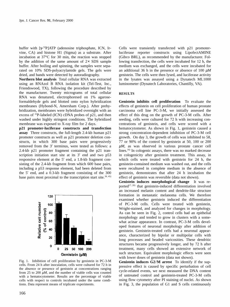
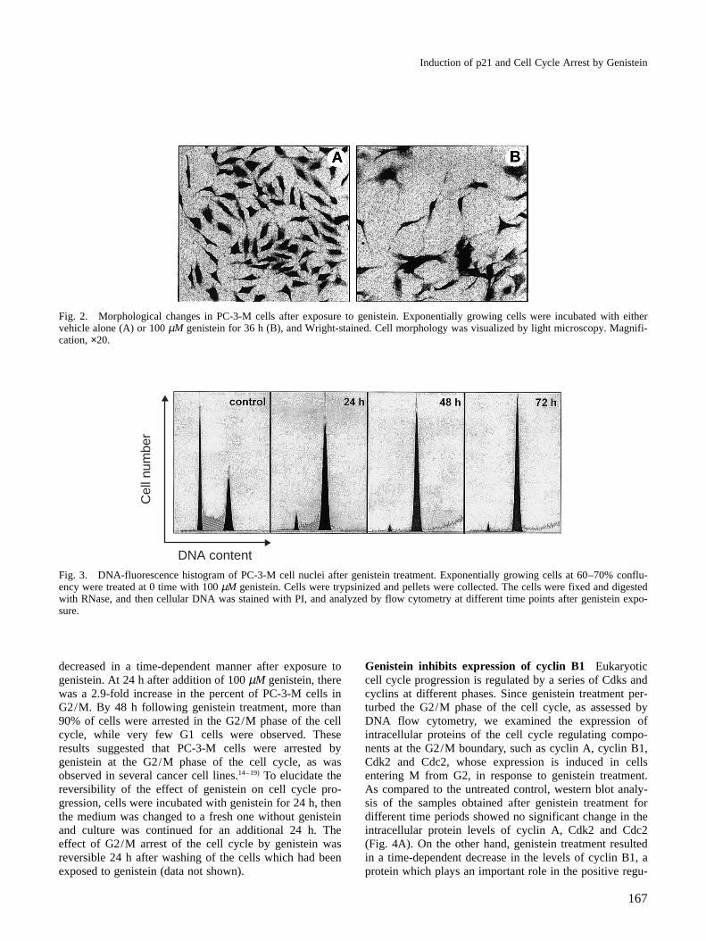
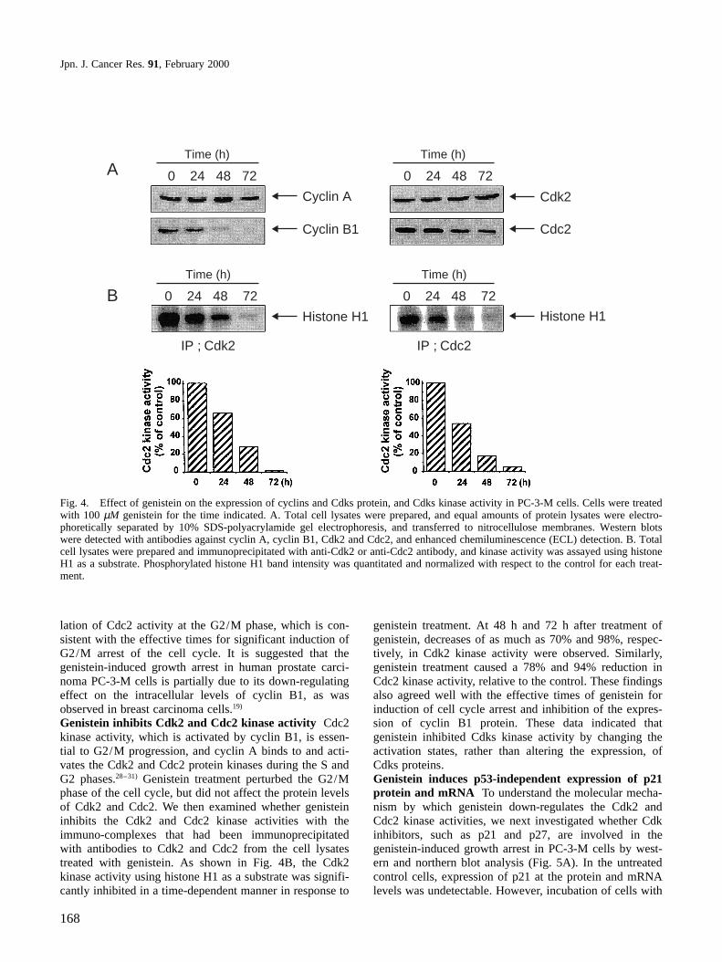
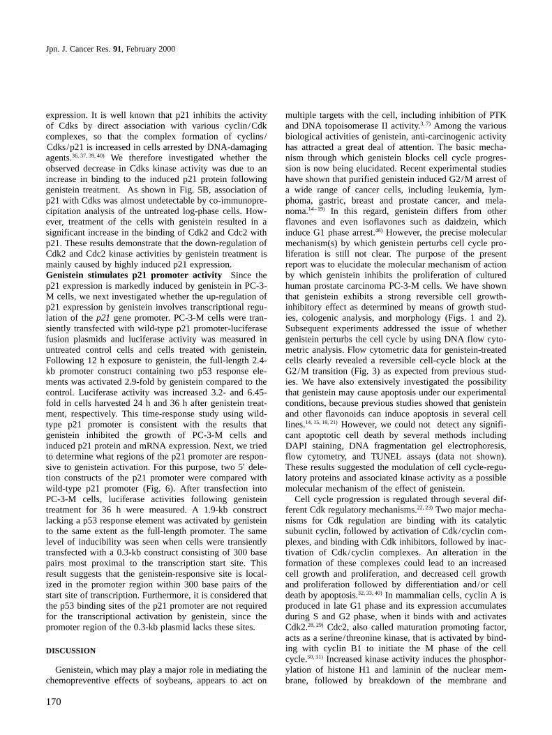
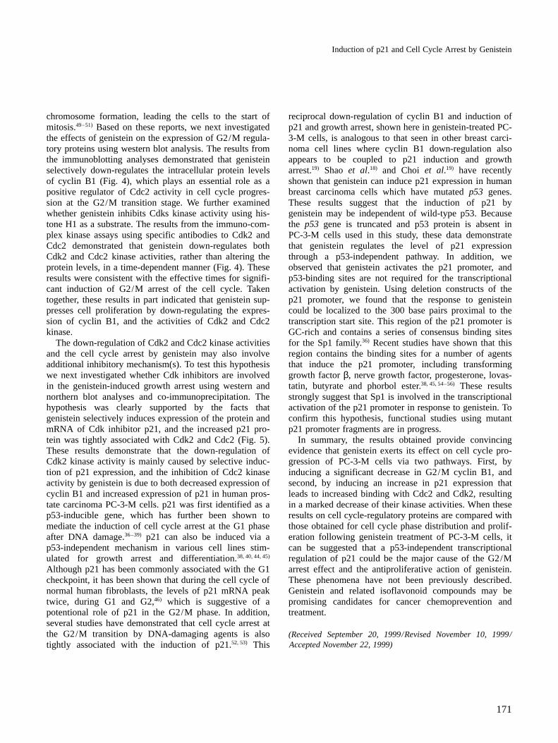
Genistein inhibits cell proliferation To evaluate theeffects of genistein on cell proliferation of human prostatecarcinoma cell line PC-3-M, we initially assessed theeffect of this drug on the growth of PC-3-M cells. Afterseeding, cells were cultured for 72 h with increasing con-centrations of genistein, and cells were scored with ahematocytometer. As shown in Fig. 1, genistein caused astrong concentration-dependent inhibition of PC-3-M cellgrowth. On day 3, the growth of cells was inhibited to 58,77 or 90% of the control by genistein at 50, 100 or 200µM, as was observed in various prostate cancer celllines.16) In cologenic assays, there was no marked decreasein cologenicity after genistein treatment. This assay, inwhich cells were treated with genistein for 24 h, thegenistein-contained medium was washed out, and the cellswere recultured in complete medium in the absence ofgenistein, demonstrates that after 24 h incubation theeffect of genistein was reversible (data not shown).Genistein induces morphological change It was re-ported17, 20) that genistein-induced differentiation involvedan increased melanin content and dendrite-like structureformation in metastatic melanoma cells. We thereforeexamined whether genistein induced the differentiationof PC-3-M cells. Cells were treated with genistein,Wright-stained, and analyzed for changes in morphology.As can be seen in Fig. 2, control cells had an epithelialmorphology and tended to grow in clusters with a some-what acinar appearance. In contrast, PC-3-M cells devel-oped features of neuronal morphology after addition ofgenistein. Genistein-treated cells had a neuronal appear-ance, characterized by bipolar or multipolar cells withlong processes and beaded varicosities. These dendriticstructures became progressively longer, and by 72 h aftertreatment, many cells showed an extensive network ofsuch structure. Equivalent morphologic effects were seenwith lower doses of genistein (data not shown).Genistein induces G2/M arrest To identify if the sup-pressive effect is caused by specific perturbation of cellcycle-related events, we next measured the DNA contentof untreated control and genistein-treated PC-3-M cellsusing flow cytometry after PI staining of nuclei. As shownin Fig. 3, the population of G1 and S cells continuously
Fig. 1. Inhibition of cell proliferation by genistein in PC-3-Mcells. From 24 h after inoculation, cells were cultured for 72 h inthe absence or presence of genistein at concentrations rangingfrom 25 to 200 µM, and the number of viable cells was countedwith a hematocytometer. Results are the percentage of viablecells with respect to controls incubated under the same condi-tions. Data represent means of triplicate experiments.
Induction of p21 and Cell Cycle Arrest by Genistein
167
decreased in a time-dependent manner after exposure togenistein. At 24 h after addition of 100 µM genistein, therewas a 2.9-fold increase in the percent of PC-3-M cells inG2/M. By 48 h following genistein treatment, more than90% of cells were arrested in the G2/M phase of the cellcycle, while very few G1 cells were observed. Theseresults suggested that PC-3-M cells were arrested bygenistein at the G2/M phase of the cell cycle, as wasobserved in several cancer cell lines.14–19) To elucidate thereversibility of the effect of genistein on cell cycle pro-gression, cells were incubated with genistein for 24 h, thenthe medium was changed to a fresh one without genisteinand culture was continued for an additional 24 h. Theeffect of G2/M arrest of the cell cycle by genistein wasreversible 24 h after washing of the cells which had beenexposed to genistein (data not shown).
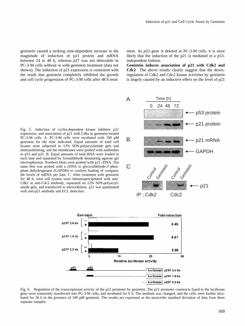
Genistein inhibits expression of cyclin B1 Eukaryoticcell cycle progression is regulated by a series of Cdks andcyclins at different phases. Since genistein treatment per-turbed the G2/M phase of the cell cycle, as assessed byDNA flow cytometry, we examined the expression ofintracellular proteins of the cell cycle regulating compo-nents at the G2/M boundary, such as cyclin A, cyclin B1,Cdk2 and Cdc2, whose expression is induced in cellsentering M from G2, in response to genistein treatment.As compared to the untreated control, western blot analy-sis of the samples obtained after genistein treatment fordifferent time periods showed no significant change in theintracellular protein levels of cyclin A, Cdk2 and Cdc2(Fig. 4A). On the other hand, genistein treatment resultedin a time-dependent decrease in the levels of cyclin B1, aprotein which plays an important role in the positive regu-
Fig. 2. Morphological changes in PC-3-M cells after exposure to genistein. Exponentially growing cells were incubated with eithervehicle alone (A) or 100 µM genistein for 36 h (B), and Wright-stained. Cell morphology was visualized by light microscopy. Magnifi-cation, ×20.
Cel
l num
ber
DNA contentFig. 3. DNA-fluorescence histogram of PC-3-M cell nuclei after genistein treatment. Exponentially growing cells at 60–70% conflu-ency were treated at 0 time with 100 µM genistein. Cells were trypsinized and pellets were collected. The cells were fixed and digestedwith RNase, and then cellular DNA was stained with PI, and analyzed by flow cytometry at different time points after genistein expo-sure.
Jpn. J. Cancer Res. 91, February 2000
168
lation of Cdc2 activity at the G2/M phase, which is con-sistent with the effective times for significant induction ofG2/M arrest of the cell cycle. It is suggested that thegenistein-induced growth arrest in human prostate carci-noma PC-3-M cells is partially due to its down-regulatingeffect on the intracellular levels of cyclin B1, as wasobserved in breast carcinoma cells.19)
Genistein inhibits Cdk2 and Cdc2 kinase activity Cdc2kinase activity, which is activated by cyclin B1, is essen-tial to G2/M progression, and cyclin A binds to and acti-vates the Cdk2 and Cdc2 protein kinases during the S andG2 phases.28–31) Genistein treatment perturbed the G2/Mphase of the cell cycle, but did not affect the protein levelsof Cdk2 and Cdc2. We then examined whether genisteininhibits the Cdk2 and Cdc2 kinase activities with theimmuno-complexes that had been immunoprecipitatedwith antibodies to Cdk2 and Cdc2 from the cell lysatestreated with genistein. As shown in Fig. 4B, the Cdk2kinase activity using histone H1 as a substrate was signifi-cantly inhibited in a time-dependent manner in response to
genistein treatment. At 48 h and 72 h after treatment ofgenistein, decreases of as much as 70% and 98%, respec-tively, in Cdk2 kinase activity were observed. Similarly,genistein treatment caused a 78% and 94% reduction inCdc2 kinase activity, relative to the control. These findingsalso agreed well with the effective times of genistein forinduction of cell cycle arrest and inhibition of the expres-sion of cyclin B1 protein. These data indicated thatgenistein inhibited Cdks kinase activity by changing theactivation states, rather than altering the expression, ofCdks proteins.Genistein induces p53-independent expression of p21protein and mRNA To understand the molecular mecha-nism by which genistein down-regulates the Cdk2 andCdc2 kinase activities, we next investigated whether Cdkinhibitors, such as p21 and p27, are involved in thegenistein-induced growth arrest in PC-3-M cells by west-ern and northern blot analysis (Fig. 5A). In the untreatedcontrol cells, expression of p21 at the protein and mRNAlevels was undetectable. However, incubation of cells with
ATime (h)
0 24 48 72
B
Cyclin A
Time (h)
IP ; Cdk2
0 24 48 72
Histone H1
Cyclin B1
Time (h)
0 24 48 72
Cdk2
Time (h)
IP ; Cdc2
0 24 48 72
Histone H1
Cdc2
Fig. 4. Effect of genistein on the expression of cyclins and Cdks protein, and Cdks kinase activity in PC-3-M cells. Cells were treatedwith 100 µM genistein for the time indicated. A. Total cell lysates were prepared, and equal amounts of protein lysates were electro-phoretically separated by 10% SDS-polyacrylamide gel electrophoresis, and transferred to nitrocellulose membranes. Western blotswere detected with antibodies against cyclin A, cyclin B1, Cdk2 and Cdc2, and enhanced chemiluminescence (ECL) detection. B. Totalcell lysates were prepared and immunoprecipitated with anti-Cdk2 or anti-Cdc2 antibody, and kinase activity was assayed using histoneH1 as a substrate. Phosphorylated histone H1 band intensity was quantitated and normalized with respect to the control for each treat-ment.
Induction of p21 and Cell Cycle Arrest by Genistein
169
genistein caused a striking time-dependent increase in themagnitude of induction of p21 protein and mRNAbetween 24 to 48 h, whereas p27 was not detectable inPC-3-M cells without or with genistein treatment (data notshown). The induction of p21 expression is consistent withthe result that genistein completely inhibited the growthand cell cycle progression of PC-3-M cells after 48 h treat-
ment. As p53 gene is deleted in PC-3-M cells, it is mostlikely that the induction of the p21 is mediated in a p53-independent fashion.Genistein induces association of p21 with Cdk2 andCdc2 The above results clearly suggest that the down-regulation of Cdk2 and Cdc2 kinase activities by genisteinis largely caused by an inductive effect on the level of p21
Fig. 5. Induction of cyclin-dependent kinase inhibitor p21expression, and association of p21 with Cdks in genistein-treatedPC-3-M cells. A. PC-3-M cells were incubated with 100 µMgenistein for the time indicated. Equal amounts of total celllysates were subjected to 12% SDS-polyacrylamide gels andimmunoblotting, and the membranes were probed with antibodiesto p53 and p21. B. Equal amounts of total RNA were loaded ineach lane and separated by formaldehyde denaturing agarose gelelectrophoresis. Northern blots were probed with p21 cDNA. Thesame blot was probed with a cDNA to glyceraldehyde-3′-phos-phate dehydrogenase (GAPDH) to confirm loading of compara-ble levels of mRNA per lane. C. After treatment with genisteinfor 48 h, total cell lysates were immunoprecipitated with anti-Cdk2 or anti-Cdc2 antibody, separated on 12% SDS-polyacryl-amide gels, and transferred to nitrocellulose. p21 was quantitatedwith anti-p21 antibody and ECL detection.
A
B
C
Time (h)
IP ; Cdk2 Cdc2
Con
trol
Gen
iste
in
Con
trol
Gen
iste
in
0 24 48 72
p53 protein
p21 protein
p21 mRNA
GAPDH
p21
Fig. 6. Regulation of the transcriptional activity of the p21 promoter by genistein. The p21 promoter constructs fused to the luciferasegene were transiently transfected into PC-3-M cells, and incubated for 6 h. The medium was changed, and the cells were further incu-bated for 36 h in the presence of 100 µM genistein. The results are expressed as the mean±the standard deviation of data from threeseparate samples.
Jpn. J. Cancer Res. 91, February 2000
170
expression. It is well known that p21 inhibits the activityof Cdks by direct association with various cyclin/Cdkcomplexes, so that the complex formation of cyclins/Cdks/p21 is increased in cells arrested by DNA-damagingagents.36, 37, 39, 40) We therefore investigated whether theobserved decrease in Cdks kinase activity was due to anincrease in binding to the induced p21 protein followinggenistein treatment. As shown in Fig. 5B, association ofp21 with Cdks was almost undetectable by co-immunopre-cipitation analysis of the untreated log-phase cells. How-ever, treatment of the cells with genistein resulted in asignificant increase in the binding of Cdk2 and Cdc2 withp21. These results demonstrate that the down-regulation ofCdk2 and Cdc2 kinase activities by genistein treatment ismainly caused by highly induced p21 expression.Genistein stimulates p21 promoter activity Since thep21 expression is markedly induced by genistein in PC-3-M cells, we next investigated whether the up-regulation ofp21 expression by genistein involves transcriptional regu-lation of the p21 gene promoter. PC-3-M cells were tran-siently transfected with wild-type p21 promoter-luciferasefusion plasmids and luciferase activity was measured inuntreated control cells and cells treated with genistein.Following 12 h exposure to genistein, the full-length 2.4-kb promoter construct containing two p53 response ele-ments was activated 2.9-fold by genistein compared to thecontrol. Luciferase activity was increased 3.2- and 6.45-fold in cells harvested 24 h and 36 h after genistein treat-ment, respectively. This time-response study using wild-type p21 promoter is consistent with the results thatgenistein inhibited the growth of PC-3-M cells andinduced p21 protein and mRNA expression. Next, we triedto determine what regions of the p21 promoter are respon-sive to genistein activation. For this purpose, two 5′ dele-tion constructs of the p21 promoter were compared withwild-type p21 promoter (Fig. 6). After transfection intoPC-3-M cells, luciferase activities following genisteintreatment for 36 h were measured. A 1.9-kb constructlacking a p53 response element was activated by genisteinto the same extent as the full-length promoter. The samelevel of inducibility was seen when cells were transientlytransfected with a 0.3-kb construct consisting of 300 basepairs most proximal to the transcription start site. Thisresult suggests that the genistein-responsive site is local-ized in the promoter region within 300 base pairs of thestart site of transcription. Furthermore, it is considered thatthe p53 binding sites of the p21 promoter are not requiredfor the transcriptional activation by genistein, since thepromoter region of the 0.3-kb plasmid lacks these sites.
DISCUSSION
Genistein, which may play a major role in mediating thechemopreventive effects of soybeans, appears to act on
multiple targets with the cell, including inhibition of PTKand DNA topoisomerase II activity.3, 7) Among the variousbiological activities of genistein, anti-carcinogenic activityhas attracted a great deal of attention. The basic mecha-nism through which genistein blocks cell cycle progres-sion is now being elucidated. Recent experimental studieshave shown that purified genistein induced G2/M arrest ofa wide range of cancer cells, including leukemia, lym-phoma, gastric, breast and prostate cancer, and mela-noma.14–19) In this regard, genistein differs from otherflavones and even isoflavones such as daidzein, whichinduce G1 phase arrest.48) However, the precise molecularmechanism(s) by which genistein perturbs cell cycle pro-liferation is still not clear. The purpose of the presentreport was to elucidate the molecular mechanism of actionby which genistein inhibits the proliferation of culturedhuman prostate carcinoma PC-3-M cells. We have shownthat genistein exhibits a strong reversible cell growth-inhibitory effect as determined by means of growth stud-ies, cologenic analysis, and morphology (Figs. 1 and 2).Subsequent experiments addressed the issue of whethergenistein perturbs the cell cycle by using DNA flow cyto-metric analysis. Flow cytometric data for genistein-treatedcells clearly revealed a reversible cell-cycle block at theG2/M transition (Fig. 3) as expected from previous stud-ies. We have also extensively investigated the possibilitythat genistein may cause apoptosis under our experimentalconditions, because previous studies showed that genisteinand other flavonoids can induce apoptosis in several celllines.14, 15, 18, 21) However, we could not detect any signifi-cant apoptotic cell death by several methods includingDAPI staining, DNA fragmentation gel electrophoresis,flow cytometry, and TUNEL assays (data not shown).These results suggested the modulation of cell cycle-regu-latory proteins and associated kinase activity as a possiblemolecular mechanism of the effect of genistein.
Cell cycle progression is regulated through several dif-ferent Cdk regulatory mechanisms.22, 23) Two major mecha-nisms for Cdk regulation are binding with its catalyticsubunit cyclin, followed by activation of Cdk/cyclin com-plexes, and binding with Cdk inhibitors, followed by inac-tivation of Cdk/cyclin complexes. An alteration in theformation of these complexes could lead to an increasedcell growth and proliferation, and decreased cell growthand proliferation followed by differentiation and/or celldeath by apoptosis.32, 33, 40) In mammalian cells, cyclin A isproduced in late G1 phase and its expression accumulatesduring S and G2 phase, when it binds with and activatesCdk2.28, 29) Cdc2, also called maturation promoting factor,acts as a serine/threonine kinase, that is activated by bind-ing with cyclin B1 to initiate the M phase of the cellcycle.30, 31) Increased kinase activity induces the phosphor-ylation of histone H1 and laminin of the nuclear mem-brane, followed by breakdown of the membrane and
Induction of p21 and Cell Cycle Arrest by Genistein
171
chromosome formation, leading the cells to the start ofmitosis.49–51) Based on these reports, we next investigatedthe effects of genistein on the expression of G2/M regula-tory proteins using western blot analysis. The results fromthe immunoblotting analyses demonstrated that genisteinselectively down-regulates the intracellular protein levelsof cyclin B1 (Fig. 4), which plays an essential role as apositive regulator of Cdc2 activity in cell cycle progres-sion at the G2/M transition stage. We further examinedwhether genistein inhibits Cdks kinase activity using his-tone H1 as a substrate. The results from the immuno-com-plex kinase assays using specific antibodies to Cdk2 andCdc2 demonstrated that genistein down-regulates bothCdk2 and Cdc2 kinase activities, rather than altering theprotein levels, in a time-dependent manner (Fig. 4). Theseresults were consistent with the effective times for signifi-cant induction of G2/M arrest of the cell cycle. Takentogether, these results in part indicated that genistein sup-presses cell proliferation by down-regulating the expres-sion of cyclin B1, and the activities of Cdk2 and Cdc2kinase.
The down-regulation of Cdk2 and Cdc2 kinase activitiesand the cell cycle arrest by genistein may also involveadditional inhibitory mechanism(s). To test this hypothesiswe next investigated whether Cdk inhibitors are involvedin the genistein-induced growth arrest using western andnorthern blot analyses and co-immunoprecipitation. Thehypothesis was clearly supported by the facts thatgenistein selectively induces expression of the protein andmRNA of Cdk inhibitor p21, and the increased p21 pro-tein was tightly associated with Cdk2 and Cdc2 (Fig. 5).These results demonstrate that the down-regulation ofCdk2 kinase activity is mainly caused by selective induc-tion of p21 expression, and the inhibition of Cdc2 kinaseactivity by genistein is due to both decreased expression ofcyclin B1 and increased expression of p21 in human pros-tate carcinoma PC-3-M cells. p21 was first identified as ap53-inducible gene, which has further been shown tomediate the induction of cell cycle arrest at the G1 phaseafter DNA damage.36–39) p21 can also be induced via ap53-independent mechanism in various cell lines stim-ulated for growth arrest and differentiation.38, 40, 44, 45)
Although p21 has been commonly associated with the G1checkpoint, it has been shown that during the cell cycle ofnormal human fibroblasts, the levels of p21 mRNA peaktwice, during G1 and G2,46) which is suggestive of apotentional role of p21 in the G2/M phase. In addition,several studies have demonstrated that cell cycle arrest atthe G2/M transition by DNA-damaging agents is alsotightly associated with the induction of p21.52, 53) This
reciprocal down-regulation of cyclin B1 and induction ofp21 and growth arrest, shown here in genistein-treated PC-3-M cells, is analogous to that seen in other breast carci-noma cell lines where cyclin B1 down-regulation alsoappears to be coupled to p21 induction and growtharrest.19) Shao et al.18) and Choi et al.19) have recentlyshown that genistein can induce p21 expression in humanbreast carcinoma cells which have mutated p53 genes.These results suggest that the induction of p21 bygenistein may be independent of wild-type p53. Becausethe p53 gene is truncated and p53 protein is absent inPC-3-M cells used in this study, these data demonstratethat genistein regulates the level of p21 expressionthrough a p53-independent pathway. In addition, weobserved that genistein activates the p21 promoter, andp53-binding sites are not required for the transcriptionalactivation by genistein. Using deletion constructs of thep21 promoter, we found that the response to genisteincould be localized to the 300 base pairs proximal to thetranscription start site. This region of the p21 promoter isGC-rich and contains a series of consensus binding sitesfor the Sp1 family.36) Recent studies have shown that thisregion contains the binding sites for a number of agentsthat induce the p21 promoter, including transforminggrowth factor β, nerve growth factor, progesterone, lovas-tatin, butyrate and phorbol ester.38, 45, 54–56) These resultsstrongly suggest that Sp1 is involved in the transcriptionalactivation of the p21 promoter in response to genistein. Toconfirm this hypothesis, functional studies using mutantp21 promoter fragments are in progress.
In summary, the results obtained provide convincingevidence that genistein exerts its effect on cell cycle pro-gression of PC-3-M cells via two pathways. First, byinducing a significant decrease in G2/M cyclin B1, andsecond, by inducing an increase in p21 expression thatleads to increased binding with Cdc2 and Cdk2, resultingin a marked decrease of their kinase activities. When theseresults on cell cycle-regulatory proteins are compared withthose obtained for cell cycle phase distribution and prolif-eration following genistein treatment of PC-3-M cells, itcan be suggested that a p53-independent transcriptionalregulation of p21 could be the major cause of the G2/Marrest effect and the antiproliferative action of genistein.These phenomena have not been previously described.Genistein and related isoflavonoid compounds may bepromising candidates for cancer chemoprevention andtreatment.
(Received September 20, 1999/Revised November 10, 1999/Accepted November 22, 1999)
Jpn. J. Cancer Res. 91, February 2000
172
REFERENCES
1) Amstrong, B. and Doll, R. Environmental factors and can-cer incidence and mortality in different countries, with spe-cial reference to dietary practices. Int. J. Cancer, 15, 617–631 (1975).
2) Messina, M. and Barnes, S. The role of soy products inreducing risk of cancer. J. Natl. Cancer Inst., 83, 541–545(1991).
3) Akiyama, T., Ishida, J., Nakagawa, S., Ogawara, H.,Watanabe, S., Itoh, N., Shibuya, M. and Fukami, Y.Genistein, a specific inhibitor of tyrosine-specific proteinkinase. J. Biol. Chem., 262, 5592–5595 (1987).
4) Hunter, T. and Cooper, J. A. Protein tyrosine kinases.Annu. Rev. Biochem., 54, 897–930 (1985).
5) Ullrich, A. and Schlessinger, J. Signal transduction byreceptors with tyrosine kinase activity. Cell, 61, 203–212(1990).
6) Cantley, L. C., Auger, K. R., Carpenter, C., Duckworth, B.,Graziani, A., Kapeller, R. and Soltoff, S. Oncogenes andsignal transduction. Cell, 64, 281–302 (1991).
7) Markovits, J., Linassier, C., Fosse, P. H., Couprie, J.,Jacquemin-Cablon, A., Saucier, J. M., Le Pecq, J. B. andLarsen, A. Inhibitory effects of the tyrosine kinase inhibitorgenistein on mammalian DNA topoisomerase. Cancer Res.,49, 5111–5117 (1989).
8) Linassier, C., Pierre, M., Le Peco, J.-B. and Pierre, J.Mechanisms of action in NIH-3T3 cells of genistein, aninhibitor of EGF receptor tyrosine kinase activity. Bio-chem. Pharmacol., 39, 187–193 (1990).
9) Constantinou, A., Kiguchi, K. and Huberman, E. Inductionof differentiation and DNA strand breakage in human HL-60 and K-562 leukemia cells by genistein. Cancer Res., 50,2618–2624 (1990).
10) Fotsis, T., Pepper, M., Adlercreutz, H., Fleishmann, G.,Hase, T., Montesano, R. and Schweigerer, L. Genistein, adietary-derived inhibitor of in vitro angiogenesis. Proc.Natl. Acad. Sci. USA, 90, 2690–2694 (1993).
11) Mueller, S. C., Yeh, Y. and Chen, W. T. Tyrosine phos-phorylation of membrane proteins mediates cellular inva-sion by transformed cells. J. Cell Biol., 119, 1309–1325(1992).
12) Steele, V. E., Pereira, M. A., Sigman, C. C. and Kelloff, G.J. Cancer chemoprevention agent development strategiesfor genistein. J. Nutr., 125, 713S–716S (1995).
13) Cai, Q. and Wei, H. Effect of dietary genistein on antioxi-dant enzyme activities in Sencar mice. Nutr. Cancer, 25,1–7 (1996).
14) Traganos, F., Ardelt, B., Halko, N., Bruno, S. andDarzynkiewicz, Z. Effects of genistein on the growth andcell cycle progression of normal human lymphocytes andhuman leukemic MOLT-4 and HL-60 cells. Cancer Res.,52, 6200–6208 (1992).
15) Spinozzi, F., Pagliacci, C., Graziella, M., Rasalba, M.,Grignani, F., Riccardi, C. and Nicoletti, I. The naturaltyrosine kinase inhibitor genistein produces cell cycle arrest
and apoptosis in Jurkat T-leukemia cells. Leuk. Res., 18,431–439 (1994).
16) Santibanez, J. F., Navapro, A. and Martinez, J. Genisteininhibits proliferation and in vitro invasive potential ofhuman prostate cancer cell lines. Anticancer Res., 17,1199–1204 (1997).
17) Rauth, S., Kichina, J. and Green, A. Inhibition of growthand induction of differentiation of metastatic melanomacells in vitro by genistein: chemosensitivity is regulated bycellular p53. Br. J. Cancer, 75, 1559–1566 (1997).
18) Shao, Z.-M., Alpaugh, M. L., Fontana, J. A. and Barsky, S.H. Genistein inhibits proliferation similarly in estrogenreceptor-positive and negative human breast carcinoma celllines characterized by p21WAF1/CIP1 induction, G2/M arrest,and apoptosis. J. Cell. Biochem., 69, 44–54 (1998).
19) Choi, Y. H., Zhang, L., Lee, W. H. and Park, K. Y.Genistein-induced G2/M arrest is associated with the inhi-bition of cyclin B1 and the induction of p21 in humanbreast carcinoma cells. Int. J. Oncol., 13, 391–396 (1998).
20) Kiguchi, K., Constantinou, A. I. and Hubeman, E.Genistein-induced cell differentiation and protein-linkedDNA strand breakage in human melanoma cells. CancerCommun., 2, 271–278 (1990).
21) McCab, M. J., Jr. and Orrenius, S. Genistein induces apo-ptosis in mature human thymocytes by inhibiting topoi-somerase II. Biochem. Biophys. Res. Commun., 194, 944–950 (1992).
22) Sherr, C. J. Mammalian G1 cyclins. Cell, 73, 1059–1065(1993).
23) Weinberg, R. A. The retinoblastoma protein and cell cyclecontrol. Cell, 81, 323–330 (1995).
24) Matsushime, H., Quelle, D. E., Shurtleff, S. A., Shibuya,M., Sherr, C. J. and Kato, J. Y. D-type cyclin-dependentkinase activity in mammalian cells. Mol. Cell. Biol., 14,2066–2076 (1994).
25) Meyerson, M. and Harlow, E. Identification of G1 kinaseactivity for Cdk6, a novel cyclin D partner. Mol. Cell.Biol., 14, 2077–2086 (1994).
26) Koff, A., Giordano, A., Desai, D., Yamashita, K., Harper, J.W., Elledge, S., Nishimoto, T., Morgan, D. O., Franza, B.R. and Roberts, J. M. Formation and activation of a cyclinE-Cdk2 complex during the G1 phase of the human cellcycle. Science, 257, 1689–1694 (1992).
27) Ohtsubo, M. and Roberts, J. M. Cyclin-dependent regula-tion of G1 in mammalian fibroblasts. Science, 259, 1908–1912 (1993).
28) Girard, F., Strausfeld, U., Fernandez, A. and Lamb, N. J. C.Cyclin A is required for the onset of DNA replication inmammalian fibroblasts. Cell, 67, 1169–1179 (1991).
29) Guadagno, T. M., Ohtsubo, M., Roberts, J. M. and Assoian,R. K. A link between cyclin A expression and adhesion-dependent cell cycle progression. Science, 262, 1572–1575(1993).
30) Walker, D. H. and Maller, J. L. Role for cyclin A in the
Induction of p21 and Cell Cycle Arrest by Genistein
173
dependence of mitosis on comparison of DNA replication.Nature, 354, 314–317 (1991).
31) Pagano, M., Pepperkok, R., Verde, F., Ansorge, W. andDraetta, G. Cyclin A is required at two points in the humancell cycle. EMBO J., 11, 961–971 (1992).
32) Elledges, S. J. and Harper, J. W. Cdk inhibitor: on thethreshold of checkpoints and development. Curr. Opin.Cell Biol., 6, 847–852 (1994).
33) Morgan, D. O. Principles of CDK regulation. Nature, 374,131–134 (1995).
34) Gu, Y., Turck, C. W. and Morgan, D. O. Inhibition ofCdk2 activity in vivo by an associated 20 K regulatory sub-unit. Nature, 366, 707–710 (1993).
35) Harper, J. W., Adami, G. R., Wei, N., Keyomarsi, K. andElledge, S. J. The p21 Cdk-interacting protein Cip1 is apotent inhibitor of G1 cyclin-dependent kinases. Cell, 75,805–816 (1993).
36) El-Deiry, W. S., Tokino, T., Velculesco, V. E., Levy, D. B.,Parsons, R., Trent, J. M., Lin, D., Mercer, E. W., Kinzler,K. W. and Vogelstain, B. WAF1, a potential mediator ofp53 suppression. Cell, 75, 817–825 (1993).
37) Xiong, Y., Hannon, G., Zhang, H., Casso, D., Kobayashi,R. and Beach, D. p21 is a universal inhibitor of cyclinkinases. Nature, 366, 701–704 (1993).
38) Datto, M. B., Yu, Y. and Wang, X.-F. Functional analysisof the transforming growth factor β responsive elements inthe WAF/Cip1/p21 promoter. J. Biol. Chem., 270, 28623–28628 (1995).
39) Waldman, T., Kinzler, K. W. and Vogelstain, B. p21 isnecessary for the p53-mediated G1 arrest in human cancercells. Cancer Res., 55, 5187–5190 (1995).
40) Jiang, H., Lin, J., Su, Z. Z., Collart, F. R., Huberman, E.and Fisher, P. B. Induction of differentiation in human pro-myelocytic HL-60 leukemia cells activates p21, WAF/CIP1, expression in the absence of p53. Oncogene, 9,3397–3406 (1994).
41) Waga, S., Hannon, G. J., Beach, D. and Stillman, B. Thep21 inhibitor of cyclin-dependent kinases controls DNAreplication by interaction with PCNA. Nature, 369, 574–578 (1994).
42) Michieli, P., Chedid, M., Lin, D., Pierce, J. H., Mercer, W.E. and Givol, D. Induction of WAF/CIP1 by a p53-inde-pendent pathway. Cancer Res., 54, 3391–3395 (1994).
43) Zhang, W., Grasso, L., McClain, C. D., Gambel, A. M.,Cha, Y., Travali, S., Deisseroth, A. B. and Mercer, W. E.p53-independent induction of WAF1/CIP1 in human leuke-mia cells is correlated with growth arrest accompanyingmonocyte/marcrophage differentiation. Cancer Res., 55,668–674 (1995).
44) Zeng, Y. X. and El-Deiry, W. S. Regulation of p21WAF1/CIP1
expression by p53-independent pathways. Oncogene, 12,1557–1564 (1996).
45) Lee, S. J., Ha, M. J., Lee, J., Nguyen, P., Choi, Y. H.,
Pirnia, F., Kang, W. K., Wang, X. F., Kim, S. J. and Trepel,J. B. Inhibition of the 3-hydroxy-3-methylglutaryl-coen-zyme A reductase pathway induces p53-independent tran-scriptional regulation of p21WAF1/CIP1 in human prostatecarcinoma cells. J. Biol. Chem., 273, 10618–10623 (1998).
46) Li, Y., Jenkins, C. W., Nichols, M. A. and Xiong, Y. Cellcycle expression and p53 regulation of the cyclin-dependentkinase p21. Oncogene, 9, 2261–2268 (1994).
47) Kozlowski, J. M., Isaiah, J. F., Campbell, D., Xu, Z. L.,Kaighn, M. E. and Hart, I. R. Metastatic behavior ofhuman tumor cell lines grown in the nude mouse. CancerRes., 44, 3522–3529 (1984).
48) Zi, X., Feyes, D. K. and Agarwal, R. Anticarcinogeniceffect of a flavonoid antioxidant, silymarin, in human breastcancer cells MDA-MB 468: induction of G1 arrest throughan increase in Cip1/p21 concomitant with a decrease inkinase activity of cyclin-dependent kinase and associatedcyclins. Clin. Cancer Res., 4, 1055–1064 (1998).
49) Krek, W. and Nigg, E. A. Differential phosphorylation ofvertebrate p34Cdc2 kinase at the G1/S and G2/M transitionsof the cell cycle: identification of major phosphorylationsites. EMBO J., 10, 305–316 (1991).
50) Ohsumi, K., Katagiri, C. and Kishimoto, T. Chromosomecondensation in Xenopus mitotic extracts without histoneH1. Science, 262, 2033–2035 (1993).
51) Ookata, K., Hisanaga, S., Bulinski, J. C., Murofushi, H.,Aizawa, H., Itoh, T. J., Hotani, H., Okumura, E., Tachibana,K. and Kishimoto, T. Cyclin B1 interaction with microtu-bule-associated protein 4 (MAP4) targets p34Cdc2 kinase tomicrotubules and is a potential regulator of M-phase micro-tubule dynamics. J. Cell Biol., 128, 849–862 (1995).
52) Tchou, W.-W., Rom, W. N. and Tchou-Wong, K.-M.Novel form of p21WAF1/CIP1 /SDI1 protein in phorbol ester-induced G2/M arrest. J. Biol. Chem., 271, 29556–29560(1996).
53) Dulic, V., Stein, G. H., Far, D. F. and Reed, S. I. Nuclearaccumulation of p21Cip1 at the onset of mitosis: a role at theG2/M-phase transition. Mol. Cell. Biol., 18, 546–557(1998).
54) Nakano, K., Mizuno, T., Sowa, Y., Orita, T., Yoshino, T.,Okuyama, Y., Fujita, T., Ohtani-Fujita, N., Matsukawa, Y.,Tokino, T., Yamagishi, H., Oka, T., Nomura, H. and Sakai,T. Butyrate activates the WAF1/Cip1 gene promoterthrough Sp1 sites in a p53-negative human colon cancer cellline. J. Biol. Chem., 272, 22199–22206 (1997).
55) Yand, G.-Z. and Ziff, E. B. Never growth factor inducestranscription of the p21WAF/CIP1 and cyclin D1 genes in PC12cells by activating the Sp1 transcription factor. J. Neuro-sci., 17, 6122–6132 (1997).
56) Owen, G. I., Richer, J. K., Tung, L., Takimoto, G. andHorwitz, K. B. Progesterone regulates transcription of thep21WAF1 cyclin-dependent kinase inhibitor gene through Sp1and CBP/p300. J. Biol. Chem., 273, 10696–10701 (1998).
Copyright © 2022 FDOKUMEN


![The effect of dibenzo[a,l]pyrene and benzo[a]pyrene on human diploid lung fibroblasts: the induction of DNA adducts, expression of p53 and p21 WAF1 proteins and cell cycle distribution](https://static.fdokumen.com/doc/165x107/63336211b6829c19b80c63af/the-effect-of-dibenzoalpyrene-and-benzoapyrene-on-human-diploid-lung-fibroblasts.jpg)