Complex balanced chromosomal translocation t(2;5;13) (p21;p15;q22) in a woman with four reproductive...
6
CASE REPORT Open Access Complex balanced chromosomal translocation t(2;5;13) (p21;p15;q22) in a woman with four reproductive failures Ewelina Lazarczyk 1* , Malgorzata Drozniewska 1,2 , Magdalena Pasinska 1 , Beata Stasiewicz-Jarocka 3 , Alina T Midro 3 and Olga Haus 1,4 Abstract Background: Balanced complex translocations (BCTs) are rare events, they may result in reproductive failures: spontaneous abortions, missed abortions, stillbirths, congenital malformations in children, and male infertility. BCTs belong to the group of complex chromosome rearrangements (CCRs) – up to date about 260 cases were described. Results: The described patient and her husband were referred to genetic counseling clinic because of four reproductive failures. GTG-banded chromosome analysis revealed presence of apparently balanced complex translocation t(2;5;13), which was verified and confirmed by molecular cytogenetics with single copy probes. This complex aberration was most likely responsible for reproductive failures in our patient. Since no high resolution molecular karyotyping (microarrays) was used, this rearrangement can only be considered to be balanced at cytogenetic level. Discussion: Due to small number of reported cases of CCRs/BCTs and individual as well as unique character of such rearrangements, genetic counseling for CCRs carriers is complex and requires detailed pedigree analysis, as well as extended clinical and genetic testing. Keywords: Balanced complex translocation (BCT), Complex chromosome rearrangement (CCR), Reciprocal chromosomal translocation (RCT), Reproductive failure, Conventional cytogenetics (CC), Fluorescence in situ hybridization (FISH) Background Reciprocal chromosomal translocations (RCTs) are struc- tural aberrations which occur as a result of exchange of chromosome fragments, usually between two nonhomolo- gous chromosomes. When the amount of genetic compo- nent is balanced the aberration usually has no influence on patient’ s phenotype [1]. Balanced complex translocation (BCT) occurs when more than two chromosomes are in- volved in the translocation [2,3]. BCTs belong to the group of complex chromosome rearrangements (CCRs) [4]. In general population BCTs occur rarely, that is why every new described case can bring more information on possible consequences of carrying this rearrangement [3,5]. About 260 cases of CCRs have been reported up to date [6-10]. In most of the carriers of such complex translocations, repro- ductive failures, including spontaneous abortions, still- births, delivering children with congenital malformations, and male infertility were present [1,3,4,6,11-13]. There are several different definitions and classifica- tions of CCRs used in the literature, most of which base on the number of chromosomes and the number of breaks involved. Most of them originate de novo, how- ever they can be also hereditary, in both balanced and unbalanced forms. The carrier status is typically revealed due to pregnancy failures. In 2012 Madan divided CCRs into four groups [14]. In the type I of CCRs number of chromosomal breaks equals number of chromosomes in- volved in an aberration and the exchange can be three- * Correspondence: [email protected] 1 Department of Clinical Genetics, Collegium Medicum, Nicolaus Copernicus University, Sklodowskiej-Curie 9, Bydgoszcz 85-094, Poland Full list of author information is available at the end of the article © 2014 Lazarczyk et al.; licensee BioMed Central Ltd. This is an Open Access article distributed under the terms of the Creative Commons Attribution License (http://creativecommons.org/licenses/by/4.0), which permits unrestricted use, distribution, and reproduction in any medium, provided the original work is properly credited. The Creative Commons Public Domain Dedication waiver (http://creativecommons.org/publicdomain/zero/1.0/) applies to the data made available in this article, unless otherwise stated. Lazarczyk et al. Molecular Cytogenetics 2014, 7:83 http://www.molecularcytogenetics.org/content/7/1/83
-
Upload
independent -
Category
Documents
-
view
2 -
download
0
Transcript of Complex balanced chromosomal translocation t(2;5;13) (p21;p15;q22) in a woman with four reproductive...

Lazarczyk et al. Molecular Cytogenetics 2014, 7:83http://www.molecularcytogenetics.org/content/7/1/83
CASE REPORT Open Access
Complex balanced chromosomal translocationt(2;5;13) (p21;p15;q22) in a woman with fourreproductive failuresEwelina Lazarczyk1*, Malgorzata Drozniewska1,2, Magdalena Pasinska1, Beata Stasiewicz-Jarocka3,Alina T Midro3 and Olga Haus1,4
Abstract
Background: Balanced complex translocations (BCTs) are rare events, they may result in reproductive failures:spontaneous abortions, missed abortions, stillbirths, congenital malformations in children, and male infertility. BCTsbelong to the group of complex chromosome rearrangements (CCRs) – up to date about 260 cases weredescribed.
Results: The described patient and her husband were referred to genetic counseling clinic because of fourreproductive failures. GTG-banded chromosome analysis revealed presence of apparently balanced complextranslocation t(2;5;13), which was verified and confirmed by molecular cytogenetics with single copy probes. Thiscomplex aberration was most likely responsible for reproductive failures in our patient. Since no high resolutionmolecular karyotyping (microarrays) was used, this rearrangement can only be considered to be balanced atcytogenetic level.
Discussion: Due to small number of reported cases of CCRs/BCTs and individual as well as unique character ofsuch rearrangements, genetic counseling for CCRs carriers is complex and requires detailed pedigree analysis, aswell as extended clinical and genetic testing.
Keywords: Balanced complex translocation (BCT), Complex chromosome rearrangement (CCR), Reciprocalchromosomal translocation (RCT), Reproductive failure, Conventional cytogenetics (CC), Fluorescence in situhybridization (FISH)
BackgroundReciprocal chromosomal translocations (RCTs) are struc-tural aberrations which occur as a result of exchange ofchromosome fragments, usually between two nonhomolo-gous chromosomes. When the amount of genetic compo-nent is balanced the aberration usually has no influence onpatient’s phenotype [1]. Balanced complex translocation(BCT) occurs when more than two chromosomes are in-volved in the translocation [2,3]. BCTs belong to the groupof complex chromosome rearrangements (CCRs) [4]. Ingeneral population BCTs occur rarely, that is why everynew described case can bring more information on possible
* Correspondence: [email protected] of Clinical Genetics, Collegium Medicum, Nicolaus CopernicusUniversity, Sklodowskiej-Curie 9, Bydgoszcz 85-094, PolandFull list of author information is available at the end of the article
© 2014 Lazarczyk et al.; licensee BioMed CentrCommons Attribution License (http://creativecreproduction in any medium, provided the orDedication waiver (http://creativecommons.orunless otherwise stated.
consequences of carrying this rearrangement [3,5]. About260 cases of CCRs have been reported up to date [6-10]. Inmost of the carriers of such complex translocations, repro-ductive failures, including spontaneous abortions, still-births, delivering children with congenital malformations,and male infertility were present [1,3,4,6,11-13].There are several different definitions and classifica-
tions of CCRs used in the literature, most of which baseon the number of chromosomes and the number ofbreaks involved. Most of them originate de novo, how-ever they can be also hereditary, in both balanced andunbalanced forms. The carrier status is typically revealeddue to pregnancy failures. In 2012 Madan divided CCRsinto four groups [14]. In the type I of CCRs number ofchromosomal breaks equals number of chromosomes in-volved in an aberration and the exchange can be three-
al Ltd. This is an Open Access article distributed under the terms of the Creativeommons.org/licenses/by/4.0), which permits unrestricted use, distribution, andiginal work is properly credited. The Creative Commons Public Domaing/publicdomain/zero/1.0/) applies to the data made available in this article,

Lazarczyk et al. Molecular Cytogenetics 2014, 7:83 Page 2 of 6http://www.molecularcytogenetics.org/content/7/1/83
or four-directional. In the type II number of breaks isone more than number of involved chromosomes; thistype also contains inversion. In the type III, number ofbreaks is greater than number of involved chromosomes,with the presence of at least one insertion. In the typeIV, apart from number of breaks greater than number ofchromosomes the occurrence of ‘middle segment’ is ob-served. ‘Middle segment’ means fragment of a chromo-some located in the middle of derivative chromosome,flanked bilaterally by fragments of different chromo-somes. In this type of CCRs at least one of derivativechromosomes is composed of three different chromo-somes. This type also includes more complex rearrange-ments, with multiple breakpoints and more than one‘middle segment’ [14]. In 2013 Madan proposed a newapproach to CCRs’ classification. She concluded that inde novo cases in phenotypically abnormal individuals thesignificance of the detected imbalance and its pheno-typic effect should be emphasized. However in familialcases it is still important to describe number of chromo-somes and breaks involved [8].Most of the cases of CCRs occur de novo (~70%) and
the remaining ones are usually transmitted by mothers [7].
Case presentation25-year old woman and her husband were referred toclinical genetics unit due to four pregnancy failures. Firstpregnancy (anencephalic) was terminated at 19th week ofgestation. At 6th week of the second pregnancy blightedovum was found. Third pregnancy underwent spontan-eous abortion at 6th week of gestation. The fourth preg-nancy was extrauterine.The physical examination did not reveal any pheno-
typic abnormalities or any congenital malformations ineither partner.A history of reproductive problems was reported in ma-
ternal family members (see Figure 1 showing pedigree).Our patient’s mother had difficulties to conceive, her sec-ond pregnancy was spontaneously aborted. She also gave
Figure 1 Patient’s pedigree. Arrow indicates proband. Proband’smother and husband are indicated by an asterisk. Only these familymembers were tested. ‘N’ means normal karyotypes. Only inproband both cytogenetic and FISH testing were performed.
birth to a girl who died before the age of one month dueto congenital malformations. The only sister of patient’smother has one son with heart defect and one healthydaughter. The wife of the brother of patient’s mother hadtwo spontaneous abortions. Maternal grandmother hadone spontaneous abortion.Patient’s father has two healthy sons from his second
relationship. Wife of father’s brother underwent a spon-taneous abortion of her only pregnancy. Paternal historywas otherwise unremarkable.Family of patient’s husband did not have any history of
reproductive health problems.
ResultsClassical cytogenetic examination revealed translocationinvolving chromosomes 2, 5 and 13. Karyotype of the pa-tient was established as 46, XX, t(2;5;13) (p21;p15.1;q22)(Figure 2). Karyotypes of patient’s husband and motherwere normal (data not shown).FISH technique with whole chromosome painting
(wcp) probes: wcp2, wcp5, wcp13, and specific probes:D13S1825, N-MYC, DLEU1, CTNND2 performed in ourpatient confirmed the presence of complex translocationinvolving three chromosomes.Combined GTG-banded metaphase spreads and FISH
images illustrating the complex character of this re-arrangement are presented in Figures 3 and 4.Probability of unbalanced karyotype in a child was es-
timated as 2% (low risk) to 13% (high risk), dependingon the type of imbalance. Risk of miscarriages was esti-mated at around 30%.No other genetic testing was performed due to the
lack of microarray technology in our laboratory.
DiscussionThe aberration found in our patient was most likely re-sponsible for her reproductive health problems. Literature
Figure 2 Karyogram of the patient in GTG-banding showing t(2;5;13) (p21;p15.1;q22). Arrows indicate abnormal chromosomes.

Figure 3 Combined images of CC and FISH with painting probes. A. Metaphase spread in GTG-banding obtained from patient’s bloodlymphocytes showing t(2;5;13) (p21;p15.1;q22). Arrows show abnormal chromosomes. B. The same metaphase as in Figure 3A in FISH techniquewith painting probes: chromosome 2-green, 5-red. Material from der(2) is present on der(5) while material from der(5) is present on der(13).Arrows show abnormal chromosomes. C. The same metaphase as in Figure 3A and 3B in FISH technique with painting probes: 13-green, 5-red.Material from der(13) is present on der(2), while material from der(5) on der(13). Arrows show abnormal chromosomes.
Lazarczyk et al. Molecular Cytogenetics 2014, 7:83 Page 3 of 6http://www.molecularcytogenetics.org/content/7/1/83
data indicate that the risk of spontaneous abortions inBCT carriers is higher than in carriers of RCT [2-4].Presence of abnormal phenotypic features could beassociated with microdeletions or microduplicationsaccompanying BCT or could be a position effect of
Figure 4 Combined images of CC and FISH with single copy probes. AArrows show abnormal chromosomes. B. The same metaphase as 4A withof N-MYC signals is present on der(5). Arrows show abnormal chromosomecritical region probe – CTNND2 (5p15.2) – red. Control region, 5q13 – green. Ochromosomes 5 and 13. D. The same metaphase as 4A, 4B and 4C, with DLEUpresent on normal 13 chromosome. The second DLEU1 signal is present on dchromosomes 2 and 13.
genes located at or flanking the breakpoints involved inaberrations [15].Gorski et al. estimated the risk of spontaneous abor-
tions in BCT carriers at 48.3% and the risk of child mal-formations at 18.4% [16]. These data are cited by most
. Metaphase spread in GTG banding showing t(2;5;13) (p21;p15.1;q22).N-MYC (2p24) probe in red (control gene – LAF (2q11) – green). Ones 2 and 5. C. The same metaphase as 4A and 4B, with cri-du-chatne of the CTNND2 signals is visible on der(13). Arrows show abnormal1 probe (13q14.3) – red. Control region, 13qter, is green. Both signals areer(13), and the second control signal on der(2). Arrows show abnormal
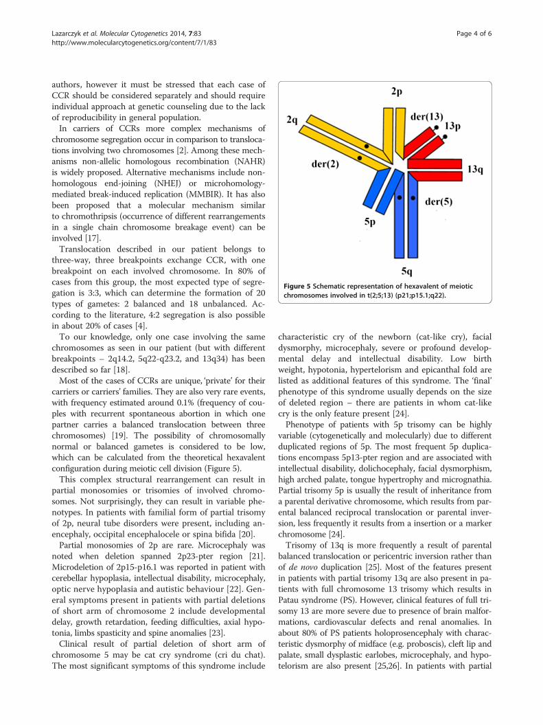
Figure 5 Schematic representation of hexavalent of meioticchromosomes involved in t(2;5;13) (p21;p15.1;q22).
Lazarczyk et al. Molecular Cytogenetics 2014, 7:83 Page 4 of 6http://www.molecularcytogenetics.org/content/7/1/83
authors, however it must be stressed that each case ofCCR should be considered separately and should requireindividual approach at genetic counseling due to the lackof reproducibility in general population.In carriers of CCRs more complex mechanisms of
chromosome segregation occur in comparison to transloca-tions involving two chromosomes [2]. Among these mech-anisms non-allelic homologous recombination (NAHR)is widely proposed. Alternative mechanisms include non-homologous end-joining (NHEJ) or microhomology-mediated break-induced replication (MMBIR). It has alsobeen proposed that a molecular mechanism similarto chromothripsis (occurrence of different rearrangementsin a single chain chromosome breakage event) can beinvolved [17].Translocation described in our patient belongs to
three-way, three breakpoints exchange CCR, with onebreakpoint on each involved chromosome. In 80% ofcases from this group, the most expected type of segre-gation is 3:3, which can determine the formation of 20types of gametes: 2 balanced and 18 unbalanced. Ac-cording to the literature, 4:2 segregation is also possiblein about 20% of cases [4].To our knowledge, only one case involving the same
chromosomes as seen in our patient (but with differentbreakpoints – 2q14.2, 5q22-q23.2, and 13q34) has beendescribed so far [18].Most of the cases of CCRs are unique, ‘private’ for their
carriers or carriers’ families. They are also very rare events,with frequency estimated around 0.1% (frequency of cou-ples with recurrent spontaneous abortion in which onepartner carries a balanced translocation between threechromosomes) [19]. The possibility of chromosomallynormal or balanced gametes is considered to be low,which can be calculated from the theoretical hexavalentconfiguration during meiotic cell division (Figure 5).This complex structural rearrangement can result in
partial monosomies or trisomies of involved chromo-somes. Not surprisingly, they can result in variable phe-notypes. In patients with familial form of partial trisomyof 2p, neural tube disorders were present, including an-encephaly, occipital encephalocele or spina bifida [20].Partial monosomies of 2p are rare. Microcephaly was
noted when deletion spanned 2p23-pter region [21].Microdeletion of 2p15-p16.1 was reported in patient withcerebellar hypoplasia, intellectual disability, microcephaly,optic nerve hypoplasia and autistic behaviour [22]. Gen-eral symptoms present in patients with partial deletionsof short arm of chromosome 2 include developmentaldelay, growth retardation, feeding difficulties, axial hypo-tonia, limbs spasticity and spine anomalies [23].Clinical result of partial deletion of short arm of
chromosome 5 may be cat cry syndrome (cri du chat).The most significant symptoms of this syndrome include
characteristic cry of the newborn (cat-like cry), facialdysmorphy, microcephaly, severe or profound develop-mental delay and intellectual disability. Low birthweight, hypotonia, hypertelorism and epicanthal fold arelisted as additional features of this syndrome. The ‘final’phenotype of this syndrome usually depends on the sizeof deleted region – there are patients in whom cat-likecry is the only feature present [24].Phenotype of patients with 5p trisomy can be highly
variable (cytogenetically and molecularly) due to differentduplicated regions of 5p. The most frequent 5p duplica-tions encompass 5p13-pter region and are associated withintellectual disability, dolichocephaly, facial dysmorphism,high arched palate, tongue hypertrophy and micrognathia.Partial trisomy 5p is usually the result of inheritance froma parental derivative chromosome, which results from par-ental balanced reciprocal translocation or parental inver-sion, less frequently it results from a insertion or a markerchromosome [24].Trisomy of 13q is more frequently a result of parental
balanced translocation or pericentric inversion rather thanof de novo duplication [25]. Most of the features presentin patients with partial trisomy 13q are also present in pa-tients with full chromosome 13 trisomy which results inPatau syndrome (PS). However, clinical features of full tri-somy 13 are more severe due to presence of brain malfor-mations, cardiovascular defects and renal anomalies. Inabout 80% of PS patients holoprosencephaly with charac-teristic dysmorphy of midface (e.g. proboscis), cleft lip andpalate, small dysplastic earlobes, microcephaly, and hypo-telorism are also present [25,26]. In patients with partial

Lazarczyk et al. Molecular Cytogenetics 2014, 7:83 Page 5 of 6http://www.molecularcytogenetics.org/content/7/1/83
13q trisomy holoprosencephaly occurs rarely, most oftenwhen trisomic region includes 13q11-q14 [26].In partial 13q monosomy phenotypes vary in regard to
size and location of a deleted fragment. Severe mental re-tardation, growth retardation, microcephaly, micrognathia,microphthalmia, cleft palate, absent thumbs, and hypoplas-tic kidneys are the phenotypic features of this aberration.Postaxial polydactyly is associated to loss of 13q21-q32region [27].
ConclusionsThe genetic risk of having children with congenital anom-alies and the risk of pregnancy losses is in our patient atthe high level (2-13% and 30%, respectively). This resultsfrom the complexity of possible combinations of chromo-some losses and gains. She has a chance of having healthychild, because only one homolog of each chromosomepairs 3, 5 and 13 is involved in the translocation.It can be stated on cytogenetic/FISH level only that
the aberration present in our patient is balanced. It iscrucial to characterise and analyse the breakpoints withgreater details as some congenital malformations mayarise due to a disruption of key genes involved in devel-opment of pregnancy.It is difficult to predict the likely phenotypic outcome of
any future pregnancies or children of described patient, asmany different forms of chromosome imbalances mayoccur in her gametes. Thus, genetic counseling may bevery difficult and complex. The patient should be offeredinvasive prenatal diagnosis in future pregnancies.As the presence of any chromosomal rearrangement
was excluded in the patient’s mother by standard cyto-genetic analysis, reproductive failures in members of ma-ternal line are not relevant to this case. They, most likelycoincide with the carrying of t(2;5;13) by the patient.The origin of the translocation, paternal or de novo,
could not be established due to the lack of consent ofpatient’s father for the cytogenetic examination.Despite the wide usefulness of microarray technology in
detecting genome imbalances in apparently balancedchromosomal rearrangements, some laboratories still haveno access to this technology. The authors will continue toinvestigate this case using array CGH technique.This case does not provide any major breakthrough,
however we strongly believe that it is still worth to pub-lish every case of CCR due to its unique character asit has been proposed by Guilherme et al.: ‘a bettercharacterization of the CCRs is important for a betterknowledge of their mechanisms of formation and theirrelevance to phenotype’ [17].
Materials and methods5 ml of peripheral blood was taken from each: the pa-tient, her husband and mother. The patient’s father did
not give his consent for blood sample. Blood cells werecultured according to standard procedures. Cytogeneticslides were stained with GTG banding technique and de-scribed according to ISCN 2013.GTG-banded chromosomes analysis revealed trans-
location involving chromosomes 2, 5 and 13. In order toconfirm the three-way character of this abnormality,fluorescence in situ hybridization (FISH) was performed.The following molecular probes were used: whole chromo-some painting probes (wcp) for chromosomes 2, 5 and13 (Cytocell, UK), and specific probes – D13S1825 (Cyto-cell, UK), N-MYC (2p24) (Kreatech Diagnostics, Holland),critical region for cri-du-chat CTNND2 (5p15.2) (KreatechDiagnostics, Holland) and DLEU1 (13q14.3) (Cytocell,UK). FISH analyses were performed according to manu-facturers’ procedures. Images were analysed with SpectralImaging system with FISH module (Applied SpectralImaging, USA).
ConsentWritten informed consent was obtained from the patientfor publication of this case report and any accompanyingimages. A copy of the written consent is available for re-view by the Editor-in-Chief of this journal.
Competing interestsThe authors declare that they have no competing interests.
Authors’ contributionsEL carried out the cytogenetic and FISH studies and drafted the manuscript.MD significantly participated in preparation of the manuscript. MP andOH counseled the patient and her family. BS-J participated in hexavalentpreparation and estimation of genetic risk. ATM supported the preparationof manuscript. OH revised cytogenetic data, contributed significantly in thepreparation of the manuscript and revised it critically. All authors read andapproved the final version of the manuscript.
Author details1Department of Clinical Genetics, Collegium Medicum, Nicolaus CopernicusUniversity, Sklodowskiej-Curie 9, Bydgoszcz 85-094, Poland. 2West MidlandsRegional Genetics Laboratories, Birmingham Women’s Hospital NHS Trust,Edgbaston, Birmingham B15 2TG, UK. 3Department of Genetics, MedicalUniversity, Waszyngtona 13, Bialystok 15-089, Poland. 4Department ofHematology, Blood Malignancies and Bone Marrow Transplantation,University of Medicine, Pasteura 4, Wroclaw 52-367, Poland.
Received: 19 August 2014 Accepted: 30 October 2014
References1. Midro TA: Effect of reciprocal chromosomal translocation carriership of
human progeny. Post Biol Kom 1997, 24:203–226.2. Sawicka A, Leśniewicz R, Zawada M, Stasiewicz-Jarocka B, Midro TA: Familial
complex translocation t(1;4;10) (q21.3;q27;q26.1) verified by FISH.Gin Pol 1998, 69:200–206.
3. Madan K, Nieuwint AWM, Van Bever Y: Recombination in a balancedcomplex translocation of a mother leading to a balanced reciprocaltranslocation in the child. Review of 60 cases of balanced complextranslocations. Hum Genet 1997, 99:806–815.
4. Patsalis PC: Complex chromosomal rearrangements. Genet Couns 2007,18:57–69.
5. Ergul E, Liehr T, Mrasek K, Sazci A: A de novo complex chromosomerearrangement involving three chromosomes (2, 13 and 18) in anoligospermic male. Fertil Steril 2009, 92:391.e9–391.e12.

Lazarczyk et al. Molecular Cytogenetics 2014, 7:83 Page 6 of 6http://www.molecularcytogenetics.org/content/7/1/83
6. Pellestor F, Anahory T, Lefort G, Puechberty J, Liehr T, Hedon B, Sarda P:Complex chromosomal rearrangements: origin and meiotic behavior.Hum Rep Update 2011, 17:476–494.
7. Lopez-Exposito A, Ballesta-Martinez MJ, Bafalliu JA, Vera-Carbonell A,Domingo-Jimenez R, Lopez-Gonzales V, Fernandez A, Guillen-Navarro E:Array CGH detection of a novel cryptic deletion at 3q13 in a complexchromosome rearrangement. Genomics 2014, 103:288–291.
8. Madan K: What is a complex chromosome rearrangement? Am J MedGenet A 2013, 161A:1181–1184.
9. Nguyen MH, Morel F, Pennamen P, Parent P, Douet-Guilbert N, Le Bris MJ,Basinko A, Roche S, De Braekeleer M, Perrin A: Balanced complex chromosomerearrangement in male infertility: case report and literature review.Andrologia in press.
10. Liao Y, Wang L, Zhang D, Liu C: Identification of a balanced complexchromosomal rearrangement involving chromosomes 3, 18 and 21 withrecurrent abortion: case report. Mol Cytogenet 2014, 7:39.
11. Kim JW, Chang EM, Song SH, Park SH, Yoon TK, Shim SH: Complexchromosomal rearrangements in infertile males: complexity ofrearrangement affects spermatogenesis. Fertil Steril 2011, 95:349–352.
12. Bartels I, Starke H, Argyrio L, Sauter SM, Zol B, Liehr T: An exceptionalcomplex chromosomal rearrangement (CCR) with eight breakpointsinvolving four chromosomes (1;3;9;14) in an azoospermic male withnormal phenotype. Eur J Med Genet 2007, 50:133–138.
13. Priya AJ, Jaya CV, Prabhat R, Dhanajaya S: A de novo complexchromosomal rearrangement of 46, XX, t(7;15;13) (p15;q21;q31) ina female with an adverse obstetric history. Int J Hum Genet 2009,9:139–143.
14. Madan K: Balanced complex chromosome rearrangements: reproductiveaspects. A review. Am J Med Genet A 2012, 158A:947–963.
15. Midro AT, Stasiewicz-Jarocka B: Probability assessment of bearing a child withimbalanced karyotype in families of mutual chromosomal translocationscarriers. Part 1: cytogenetic diagnostics of translocations. Diagn Lab 2001,37:59–67.
16. Gorski JL, Kistenmacher ML, Punnett HH, Zackai EH, Emanuel BS:Reproductive risks for carriers of complex chromosome rearrangements:analysis of 25 families. Am J Med Genet 1988, 29:247–261.
17. Guilherme RS, Cernach MCSP, Sfakianakis TE, Takeno SS, Nardozza LMM,Rossi C, Bhatt SS, Liehr T, Melaragno MI: A complex chromosomerearrangement involving four chromosomes, nine breakpoints and acryptic 0.6-Mb deletion in a boy with cerebellar hypoplasia and defectsin skull ossification. Cytogenet Genome Res 2013, 141:317–323.
18. Evans MI, White BJ, Kent SG, Levine MA, Levin SW Jr, Larsen JW: Balancedrearrangement of chromosomes 2, 5 and 13 in a family with duplication5q and fetal loss. Am J Med Genet 1984, 19:783–790.
19. Nonaka T, Ooki I, Enomoto T, Takakuwa K: Complex chromosomalrearrangements in couples affected by recurrent spontaneous abortion.Int J Gynecol Obstet in press.
20. Lurie IW, Ilyina HG, Gurevich DB, Rumyantseva NV, Naumchik IV, Castellan C,Hoeller A, Schinzel A: Trisomy 2p: analysis of unusual phenotypicfindings. Am J Med Genet 1995, 55:229–236.
21. Francis GL, Flannery DB, Byrd JR, Fisher ST: An apparent de novoterminal deletion of chromosome 2 (pter→ p24:). J Med Genet 1990,27:137–138.
22. Rajcan-Separovic E, Harvard C, Liu X, McGillivray B, Hall JG, Qiao Y, Hurlburt J,Hildebrand J, Mickelso ECR, Holden JJA, Lewis MES: Clinical and molecularcytogenetic characterisation of a newly recognised microdeletionsyndrome involving 2p15-16.1. J Med Genet 2007, 44:269–276.
23. Amir IM, Al-Tawil KI, Al-Hathal MM: Deletion (2) (p14p15) in a child withsevere neurodevelopmental delay. J Med Genet 2000, 37:21–23.
24. De Carvalho AFL, Da Silva Bellucco FT, Kulikowski LD, Toralles MBP, MelaragnoMI: Partial 5p monosomy or trisomy in 11 patients from a family with a t(5;15) (p13.3;p12) translocation. Hum Genet 2008, 124:387–392.
25. Ribacoba R, Menendez-Gonzalez M, Hernando I, Salas J, Giros ML: Partial trisomy13q22-qter associated to leukoencephalopathy and late onset generalisedepilepsy. Int Arch Med 2008, 1:1–6.
26. Obersztyn E, Stankiewicz P, Bocian E, Stanczak H, Mazurczak T: Identification ofpartial trisomy 13q with the FISH technique. Pediatr Pol 1996, 71:247–252.
27. Patil SJ, Phadke SR: Pericentric inversion causing duplication and deletionof chromosome region 13q22→ qter in the offspring. Am J Med Genet A2007, 143A:82–84.
doi:10.1186/s13039-014-0083-6Cite this article as: Lazarczyk et al.: Complex balanced chromosomaltranslocation t(2;5;13) (p21;p15;q22) in a woman with four reproductivefailures. Molecular Cytogenetics 2014 7:83.
Submit your next manuscript to BioMed Centraland take full advantage of:
• Convenient online submission
• Thorough peer review
• No space constraints or color figure charges
• Immediate publication on acceptance
• Inclusion in PubMed, CAS, Scopus and Google Scholar
• Research which is freely available for redistribution
Submit your manuscript at www.biomedcentral.com/submit