High temperature positively modulates oxidative protection in salt-stressed cashew plants

The Arabidopsis Book ©2002 American Society of Plant Biologists
One of the most crucial functions of plant cells is their abil-ity to respond to fluctuations in their environment.Understanding the connections between a plant’s initialresponses and the downstream events that constitute suc-cessful adjustment to its altered environment is one of thenext grand challenges of plant biology. Oxidative stressfrom environmental sources and developmental transitionssuch as seed maturation involves the formation of reactiveoxygen species (ROS) in plant cells. The redox-modulatedchanges that follow are central events in cellular respons-es. Thiol redox regulation (Figure 1) partially mediatedthrough the redox state of the glutathione pool(GSH/GSSG), regulation of the glutathione biosyntheticpathway, and ROS themselves are each thought to haveimportant roles as environmental sensors and/or modula-tors of global patterns of gene expression in developmentand defense. Exposure of green tissue to potentially dam-aging light intensities involves redox sensing molecularevents throughout the plant, originating at the plasto-quinone (PQ) pool in the thylakoid membrane. Majordefense genes whose expression is affected by the redoxstate of the PQ pool include both cytosolic and chloroplastascorbate peroxidases (APX) ( Karpinska et al. 2000 ). The
superoxide dismutase (SOD) gene families appear to bespecialized in function with respect to subcellular locationand other as yet unknown factors (Alscher et al. 2002). Inthe case of peroxisomes, the imposition of oxidative stressgives rise to organelle proliferation, thus adding anotherlayer of complexity to stress responses (Lopez-Huertaset al. 2000). Defense mechanisms involving molecularchaperones and methionine sulfoxide reductase arebecoming recognized as important players in resistance tooxidative stress throughout the cell.
Intracellular origins of ROS and their multipledamaging effects
Any circumstance in which cellular redox homeostasis isdisrupted can lead to oxidative stress or the generationof ROS (Asada 1994). Production of ROS during envi-ronmental stress is one of the main causes for decreasesin productivity, injury, and death that accompany thesestresses in plants. ROS are produced in both unstressedand stressed cells, and in various locations (Halliwell andGutteridge 1989) (Figure 2). They are generated endoge-nously during certain developmental transitions such asseed maturation and as a result of normal, unstressed,photosynthetic and respiratory metabolism. An initialoxyradical product, the superoxide radical (O2
-.), uponfurther reaction within the cell, can form more ROSsuch as hydroxyl radicals and singlet oxygen.Superoxide is a charged molecule and cannot crossbiological membranes. Subcellular compartmentationof defense mechanisms is, therefore, crucial for effi-cient removal of superoxide anions at their sites ofgeneration throughout the cell. Hydrogen peroxide, onthe other hand, which is formed as a result of SODaction, is capable of diffusing across membranes and is
Oxidative Stress and Acclimation Mechanisms in Plants
Ruth Grene
Department of Plant Pathology, Physiology, and Weed Science, 435 Old Glade Road, Virginia Tech, Blacksburg, VA24061-0330; email: [email protected]
INTRODUCTION
Figure 1. Thiol redox control and stress defense

The Arabidopsis Book 2 of 20
thought to fulfil a signaling function in defense respons-es (Mullineaux et al. 2000).
ROS play an important role in endonuclease activationand consequent DNA damage (Hagar et al. 1996). In thepresence of metal ions such as Fe or Cu(II), hydroxyl radi-cals are formed very rapidly. Hydroxyl radicals can causedamage to all classes of biologically important macromol-ecules, especially nucleic acids. Hydroxyl radicals can alsomodify proteins so as to make them more susceptible toproteolytic attack. There is evidently considerable speci-ficity associated with this degradative process since pro-teins have widely differing susceptibilities to attack byROS (Davies 1987). Once damaged, proteins can be bro-ken down further by specific endopeptidases such as theone found bound to the thylakoid membrane (Casano et al.1994). A multicatalytic proteinase complex has beendemonstrated in plant systems, with the capacity to selec-tively break down oxidatively damaged proteins (VanNocker et al. 1996)
Metabolic defense mechanisms: limiting ROS-mediated damage
Plant cells respond defensively to oxidative stress byremoving the ROS and maintaining antioxidant defensecompounds at levels that reflect ambient environmentalconditions (Scandalios 1997). Metabolic containment
mechanisms for ROS involving antioxidant genes andassociated processes are likely to have predated or co-evolved with the appearance of aerobiosis and representfundamental adaptations of aerobic systems to an oxygendependent metabolism. The mechanisms that act to adjustantioxidant levels to afford protection include changes inantioxidant gene expression (Cushman and Bohnert 2000).
ROS themselves play a role in intracellular redox sens-ing, activating antioxidant resistance mechanisms, amongother adaptive processes (Toledano and Leonard 1991;Karpinski et al. 1997; May, et al. 1998a). A number of redoxsensitive transcription factors have been identified in ani-mal, bacterial, and plant cells (Pastori and Foyer 2001).
Functional roles of these responses include the protec-tion of redox-sensitive enzymatic processes, the preserva-tion of membrane integrity, and the protection of DNA andproteins (Scandalios 1997). Redox-sensitive regulatoryenzymes such as fructose-1,6-bisphosphatase (FbPase)can be protected from oxidation/inactivation by the actionof antioxidants such as glutathione. Under unstressedconditions, the formation and removal of O2 are in bal-ance. The defense system, when presented with increasedROS formation under stress conditions, can be over-whelmed when it is unable to remove the toxic molecularspecies with increased enzymatic or non-enzymaticantioxidant processes.
Organelles such as the peroxisome and the chloro-plast, where ROS are being produced at a relatively highrate, are especially at risk. In the case of the chloroplast,changes in light intensity and temperature or limitationsin the substrates of photosynthesis occur frequently,
Figure 2. Reactive oxygen species (ROS)arise throughout the cell.

Oxidative Stress and Acclimation Mechanisms in Plants 3 of 20
resulting in increased production of ROS (Alscher et al.1997; Karpinska and Karpinski 2000). ROS are producedat high levels in peroxisomes. Hydrogen peroxide is pro-duced in the peroxisomal respiratory pathway by flavinoxidase. Fatty acid beta oxidation and glycolate oxidaseaction are other sources of hydrogen peroxide produc-tion in the peroxisome. Developmental transitions suchas seed maturation, in which peroxisomes play an impor-tant role, also involve oxidative stress (Leprince et al.1990; Walters 1998).
Antioxidant defense molecules have several roles
Ascorbic acid, glutathione, and α-tocopherol have eachbeen shown to act as antioxidants in the detoxification ofROS. These compounds have central and interrelatedroles, acting both non-enzymatically and as substrates inenzyme-catalyzed detoxification reactions (Foyer 1993;Hess 1993; Hausladen and Alscher 1994; Winkler et al.1994; Chaudiere and Ferrari-Iliou 1999). An anti-ROS
response includes the induction of genes that belong to
ROS scavenging mechanisms.
Metabolic cycles located within the aqueous phase of
the peroxisome, chloroplast, cytosol, and the mitochondri-
on successively oxidize and re-reduce glutathione and
ascorbate, using NAD (P) H as the ultimate electron donor.
Ascorbate, reduced glutathione (GSH), ascorbate peroxi-
dase (APX), glutathione reductase (GR), superoxide dismu-
tase (SOD), and monodehydroascorbate reductase
(MDHAR) are involved in several contexts in antioxidant
regeneration throughout the plant cell. The enzymes
involved are hydrophilic in nature, although in some
instances they are known to be loosely associated with the
membranes where the ROS are generated. The version
that is found in the chloroplast is shown in Figure 3 includ-
ing a depiction of the role of the hydrophobic antioxidant
α-tocopherol. Different isoforms of the antioxidant
enzymes are located in different subcellular compartments
(see below). Evidence to date suggests a coordinated
response to ROS among different members of the different
SOD gene families.
Figure 3. The scavenging of activeoxygen species in the chloroplast inboth the lipid membrane phase andthe stroma, linked to redox cycles forascorbate and glutathione and theoxidation of α-tocopherol (Vitamin E).Abbreviations are as follows: P-LIPID-OO, phospholipid peroxy radical; P-LIPID-OOH, phospholipid peroxide;P-LIPID-OH, phospholipid alcohol;VIT-E(OH), α-tocopherol (vitamin E);VIT-E (O*), α-chromanoxyl radical;PHGPX, phospholipid hydroperoxide-dependent glutathione peroxidase;GSH, reduced glutathione; GSSG,glutathione disulfide; GR, glutathionereductase; DHAR, dehydroascorbatereductase; ASC, ascorbic acid; DHA,dehydroascorbate; MDA, monodehy-droascorbate free radical; MDAR,monodehydroascorbate free radicalreductase; APX, ascorbate reductase;SOD, superoxide dismutase; O2-,superoxide ion. Reaction 1 is thenon-enzyme-catalyzed spontaneousdismutation of two MDA molecules toone ASC and one DHA, respectively.From Mullineaux et al. (2000).
The Arabidopsis Book 4 of 20
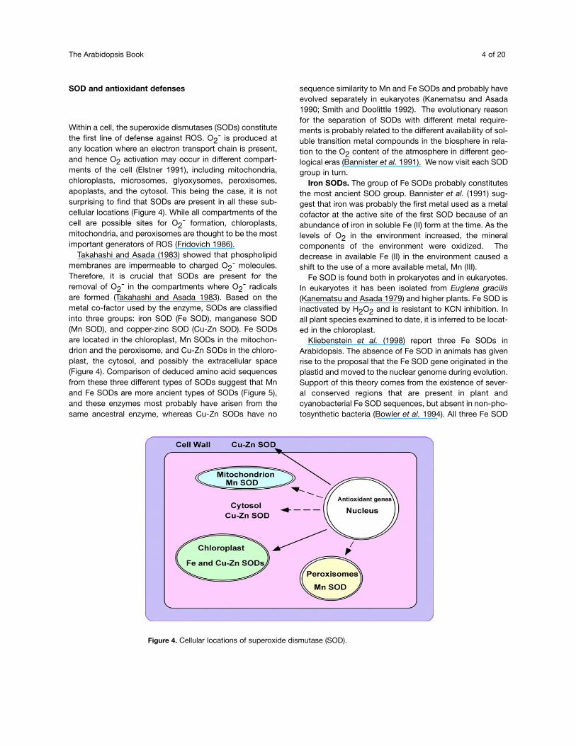
SOD and antioxidant defenses
Within a cell, the superoxide dismutases (SODs) constitutethe first line of defense against ROS. O2
- is produced atany location where an electron transport chain is present,and hence O2 activation may occur in different compart-ments of the cell (Elstner 1991), including mitochondria,chloroplasts, microsomes, glyoxysomes, peroxisomes,apoplasts, and the cytosol. This being the case, it is notsurprising to find that SODs are present in all these sub-cellular locations (Figure 4). While all compartments of thecell are possible sites for O2
- formation, chloroplasts,mitochondria, and peroxisomes are thought to be the mostimportant generators of ROS (Fridovich 1986).
Takahashi and Asada (1983) showed that phospholipidmembranes are impermeable to charged O2
- molecules.Therefore, it is crucial that SODs are present for theremoval of O2
- in the compartments where O2- radicals
are formed (Takahashi and Asada 1983). Based on themetal co-factor used by the enzyme, SODs are classifiedinto three groups: iron SOD (Fe SOD), manganese SOD(Mn SOD), and copper-zinc SOD (Cu-Zn SOD). Fe SODsare located in the chloroplast, Mn SODs in the mitochon-drion and the peroxisome, and Cu-Zn SODs in the chloro-plast, the cytosol, and possibly the extracellular space(Figure 4). Comparison of deduced amino acid sequencesfrom these three different types of SODs suggest that Mnand Fe SODs are more ancient types of SODs (Figure 5),and these enzymes most probably have arisen from thesame ancestral enzyme, whereas Cu-Zn SODs have no
sequence similarity to Mn and Fe SODs and probably haveevolved separately in eukaryotes (Kanematsu and Asada1990; Smith and Doolittle 1992). The evolutionary reasonfor the separation of SODs with different metal require-ments is probably related to the different availability of sol-uble transition metal compounds in the biosphere in rela-tion to the O2 content of the atmosphere in different geo-logical eras (Bannister et al. 1991). We now visit each SODgroup in turn.
Iron SODs. The group of Fe SODs probably constitutesthe most ancient SOD group. Bannister et al. (1991) sug-gest that iron was probably the first metal used as a metalcofactor at the active site of the first SOD because of anabundance of iron in soluble Fe (II) form at the time. As thelevels of O2 in the environment increased, the mineralcomponents of the environment were oxidized. Thedecrease in available Fe (II) in the environment caused ashift to the use of a more available metal, Mn (III).
Fe SOD is found both in prokaryotes and in eukaryotes.In eukaryotes it has been isolated from Euglena gracilis(Kanematsu and Asada 1979) and higher plants. Fe SOD isinactivated by H2O2 and is resistant to KCN inhibition. Inall plant species examined to date, it is inferred to be locat-ed in the chloroplast.
Kliebenstein et al. (1998) report three Fe SODs inArabidopsis. The absence of Fe SOD in animals has givenrise to the proposal that the Fe SOD gene originated in theplastid and moved to the nuclear genome during evolution.Support of this theory comes from the existence of sever-al conserved regions that are present in plant andcyanobacterial Fe SOD sequences, but absent in non-pho-tosynthetic bacteria (Bowler et al. 1994). All three Fe SOD
Figure 4. Cellular locations of superoxide dismutase (SOD).

Oxidative Stress and Acclimation Mechanisms in Plants 5 of 20
plant sequences encode a unique tripeptide (SRL for N.plumbaginifolia and G. max and ARL for A. thaliana) closeto the carboxyl terminus of the enzyme. Although thissequence has shown to direct the proteins to peroxisomesin other proteins, it has yet to be determined whether thisis a functional sequence or not. The conserved SRL/ARLsequence is not present in the prokaryotic Fe SOD pro-teins showing that it is not obligatory for enzyme function(Van Camp et al. 1994).
Manganese SODs. As the levels of O2 in the environ-ment increased, the amount of available Fe (II) in the envi-ronment decreased, causing a shift to the more availablemetal, Mn (III). As a consequence, Mn SODs are likely to besecond only to Fe SODs in antiquity and certainly evolvedfrom the ancestral Fe SODs, perhaps by way of the cam-bialistic SODs. Mn SODs occur in mitochondria and per-oxisomes. Mn SODs carry only one metal atom per sub-unit. These enzymes cannot function without the Mn atompresent at the active site. Even though Mn and Fe SODshave a high similarity in their primary, secondary, and terti-ary structure, these enzymes have diverged sufficientlythat Fe (II) could not restore the activity of Mn SOD andvice versa (Fridovich 1986). Catalysis by Mn SODs isthrough the attraction of negatively charged O2
- moleculesto a site formed from positively charged amino acids pres-ent at the active site of the enzyme. The metal present inthe active site then donates an electron directly to the O2
-,reducing one O2
- molecule, which in turn forms H2O2 byreacting with a proton (Asada 1994; Bowler et al. 1994).
Plant Mn SODs have approximately 65% sequence simi-larity to one another, and these enzymes also have highsimilarities to bacterial Mn SODs (Bowler et al. 1994).
Although Mn SOD is known as the mitochondrialenzyme of eukaryotes, an Mn-containing SOD has alsobeen located in the peroxisomes. del Rio et al. (1992)showed the presence of one peroxisomal and one mito-chondrial Mn SOD by using immunolocalization assays inwatermelon. Four genes that encode Mn SOD were report-ed in maize (Zea mays) (Zhu et al. 1999 ). Deduced aminoacid sequences from these four isoenzymes have a mito-chondrial targeting sequence, indicating that all are locat-ed in the mitochondria. In Nicotiana plumbaginifolis, twonuclear-encoded Mn SOD genes were isolated and the tis-sue-specific expression Mn SOD was shown by analyzingpromoter fusion with β-glucuronidase (GUS) in transgenicplants (Van Camp et al. 1996).
Copper-Zinc SODs. When the atmosphere was com-pletely replenished with oxygen, Fe (II) was almost com-pletely unavailable in the atmosphere and insoluble Cu (I)was converted into soluble Cu (II). At this stage, Cu (II)began to be used as the metal cofactor at the active sitesof SODs. Since Fe and Mn have similar electrical proper-ties, the transition from the use of iron to the use of man-ganese required little change in SOD protein structure.Thus, Mn and Fe SODs are structurally very similar. Theexistence of archaic Mn/Fe1-containing SODs supportsthis theory. However the electrical properties of Cu-Zn2
SODs differ greatly from those of Fe and Mn SODs.
Figure 5. Relatedness of SOD protein sequences in some plant cells.Tree was obtained using MegAlign in DNA Star.
1 Every molecule of Mn and Fe SOD contains either an atom of manganese or an atom of iron depending on the species or the availability
of the metal in archaic SODs. The potential use of either Fe or Mn is denoted as such by a slash, Mn/Fe.2 Every molecule of the Cu-Zn SOD enzyme contains both an atom of copper and an atom of zinc, as denoted by the hyphen.

The Arabidopsis Book 6 of 20
Therefore, a major change in the structure of the proteinoccurred after Cu became a metal cofactor (Bannister etal. 1991).
There are at least two forms of Cu-Zn SOD in plantcells, chloroplastic and cytosolic. Deduced amino acidsequences of these two isoforms show approximately68% similarity, whereas there is approximately 90% sim-ilarity among the chloroplastic Cu-Zn SODs (Cu-ZnSODchl) and 80-90% similarity among the cytosolic Cu-Zn SODs (Cu-Zn SODcyt) (Figure 6). Cu-Zn SODchl is asoluble enzyme and is localized in the stroma (Asada andKiso 1973; Asada et al. 1973). Localization studies per-formed with an immunogold-labeled antibody raisedagainst Cu-Zn SODchl from spinach leaves showed thatthis soluble enzyme is not uniformly distributed in thechloroplast but rather is localized mainly on the stromalface of thylakoid membranes (Ogawa et al. 1995) wherephotosystem I (PSI) is located. The two other Cu-ZnSODs are considered “cytoplasmic” because they havenot been detected in intact chloroplasts (Ogawa et al.1996). However, when immunogold-labeled antibodiesraised against “cytosolic” Cu-Zn SOD were used in local-ization experiments, it was shown that these enzymes
were located in the nucleus and apoplast (Ogawa et al.1996). More than 40% of the immunogold particles werefound in the apoplast and approximately 25% was foundin the nucleus (Ogawa et al. 1996). Researchers pro-posed that Cu-Zn SOD in the apoplast functions in ligni-fication and that in the nucleus it protects the cell againstfatal mutations caused by O2
- molecules (Ogawa et al.1996; Ogawa et al. 1997).
SOD expression and activity under oxidative stress.Kliebenstein et al. (1998) subjected Arabidopsis to a seriesof oxidative stresses and observed changes in the sevenSODs -- three Fe SODs denoted FSD1, FSD2, and FSD3;three Cu-Zn SODs denoted CSD1, CSD2, and CSD3; andone Mn SOD denoted MSD1 -- that are present in thatplant, both at the mRNA and the protein level. They report-ed increases in FSD2 in response to UV irradiation, and tohigh light at the mRNA level, but no response of FSD2mRNA to ozone exposure. They found that FSD1 is underthe control of a circadian clock at the mRNA level.Kliebenstein et al. (1998) did not report enzyme activities.However, it is of interest to note that they report FSD2responding to specific stresses. Thus, it appears that
Figure 6. A comparison of cytosolic, chloroplast, and peroxisomal Cu-Zn SODs from higher plants. Abbreviations are as follows:cyt, cytosolic; cp, chloroplast; prx, peroxisomal; ec, extracellular. From Kliebenstein et al. (1998).

Oxidative Stress and Acclimation Mechanisms in Plants 7 of 20
members of the Fe SOD family are specialized for specificresponses to stress.
Kliebenstein et al. (1998) report no effect of their seriesof oxidative stress treatments on Mn SOD in Arabidopsis.In contrast, Mn SOD was reported to respond positively tosalt stress (Hernandez et al. 1993; Hernandez et al. 1995;Gomez et al. 1999), manganese toxicity (Gonzalez et al.1998), chilling stress (Lee et al. 1999; Lee and Lee 2000),and drought (Wu et al. 1999). An explanation for the differ-ences in responses between the Mn and Fe SODs in plantscan be correlated with their disparate subcellular locationsand the sites of action of the various oxidative stressesthat were used. The stresses that did not affect Mn SODmay all have their site of action in the chloroplast.
Overexpression of SODs can lead to protection againstspecific stresses. The importance of subcellular location.Plants overexpressing SODs have been engineered withthe goal of increasing stress tolerance. Both successfuland unsuccessful results have been obtained fromattempts to create resistant plants (Tepperman andDunsmuir 1990; Perl et al. 1993; Slooten et al. 1995; VanCamp et al. 1996).
The site of action for ozone has been suggested to be inthe apoplast, but when Mn SOD was overexpressed in thechloroplasts of tobacco plants, less damage was observedin the leaves. When the overproduction of the enzyme wasin the mitochondria, less protection was observed. Also, itwas shown that high levels of chloroplastic Mn SOD activ-ity protected the plant from visible injury caused by ozone,suggesting that ozone may cause oxidative stress in thechloroplast, as well as the apoplast (Van Camp et al. 1994).When an A. thaliana Fe SOD gene was targeted into N.tabacum cv Petit Havana SR1 chloroplasts, an increasedprotection against O2 generated both in the plasmalemmaand photosystem II (PSII) was observed (Van Camp et al.1996). Arisi et al. (1998) overexpressed an Arabidopsis FeSOD in poplar and observed a protective effect on PSIIunder limiting carbon dioxide conditions. Van Camp et al.(1994) overexpressed Fe SOD in the chloroplast and foundthat induction of Cu-Zn SODs was suppressed, lendingsome credence to the hypothesis that a cross-family sig-naling pathway for SOD induction exists in the plant cell. Incontrast, when mitochondrial Mn SOD was targeted to thechloroplasts of tobacco plants, protection was onlyobserved against stress generated in the plasmalemma.An increase in the activity of APX, DHAR, and MDHAR,other scavenging pathway enzymes, was also observed. Itwas concluded that the protection provided by overpro-duction of Mn SOD was dependent on whether the otherenzymes — DHAR and MDAR — were or were not presentin elevated levels
Bowler et al. (1994) targeted N. plumbaginiafolis mito-chondrial Mn SOD sequence into N. tabacum mitochon-dria and chloroplasts. When the transformed plants were
treated with methyl viologen, it was observed that theplants that had Mn SOD expressed in their chloroplasthad remarkable protection, accompanied by anincreased SOD activity against methyl viologen stresscompared to control plants in light. This protective effectwas less observable in plants that were kept in the dark,since the H2O2 scavenging system is not activated with-out light (Foyer and Halliwell 1976; Nakano and Asada1980). Even though Mn SOD is not inactivated by H2O2,the balance between generated O2
- and H2O2 is dis-rupted in these transgenic plants, which may increase theformation of OH.. Although increased protection wasobserved in plants that had foreign Mn SOD targeted intothe mitochondria, this effect was not as remarkable com-pared to plants that had Mn SOD targeted into thechloroplasts. This may be due to the majority of the O2being generated in the chloroplast in plants exposed tomethyl viologen in light (Bowler et al. 1991).
Taken together, these results suggest that regulation ofexpression of plant Fe and Mn SODs may differ. The sub-cellular location of the protein also appears to play a role.In some instances, both chloroplastic and cytosolic Cu-ZnSODs afford protection against the same stresses as FeSOD. There appears, also, to be specialization within theFe SOD family. We have compared the upstreamsequences of all Arabidopsis SOD genes in order to deter-mine similarities or differences among their respective reg-ulatory regions.
Comparing the information in SOD genomicsequences. With the complete sequencing of theArabidopsis genome, it is a propitious time to ask what theappropriate application of computation to that genomemight reveal about the evolution and regulation of SODs inplants. The upstream regions of the seven ArabidopsisSOD open reading frames were extracted with the inten-tion of mining them for promoter sequences related to theregulation of gene expression under different stresses. Wedeveloped bioinformatics tools for searching an upstreamregion for particular promoter sequences that bind toknown transcription factors (Table 1). The area of compu-tational tools for analyzing promoter sequences is in itsinfancy (Prestridge 2000), and existing tools are limited intheir abilities to address our questions and to analyze anentire genome. The ABA responsive element (ABRE)appears to be associated with genes responding toosmotic stress (high osmoticum, salt, desiccation, andcold) and binds to several similar sequences of eightnucleotides (Guan and Scandalios 1998; Choi et al. 2000);we used the consensus sequence YACGTGGC. NF-κ β isa transcription factor that activates immunoglobulin-kgenes; we utilized the consensus sequence GGRNNYYCC(Smith et al. 2000). The heat shock protein gene promoterconsensus sequence is the palindromic sequence TTCN-NGAA (Santos et al. 1996). Finally, the Y-box motif has

The Arabidopsis Book 8 of 20
consensus sequence GATTGG and mediates redox-dependent transcription activation (Guan and Scandalios1998). The seven promoter regions were examined forthese four consensus sequences. Diamonds (♦) in Table 1summarize the exact or close matches found at plausibleupstream locations (within 1000 nucleotides of the ATGwhere transcription begins). The different patterns of con-sensus sequences found suggest that the phenomenon ofdifferential expression of the SOD genes under differentstresses can be explained, at least in part, through pro-moter sequence analysis. Further work will include extract-ing the upstream sequences for all known or putativeArabidopsis genes and comparing these sequences on agenome-wide basis with new computational tools we aredeveloping. Employing also information concerning co-regulated genes it will become possible to identify whatpromoter sequence combinations correspond to what kindof regulation.
Multiple defense roles for glutathione as antioxidantand redox sensor. Upon the imposition of oxidativestress, the existing pool of reduced glutathione (GSH) isconverted to oxidized glutathione (GSSG) and glutathionebiosynthesis is stimulated (May and Leaver 1993;Madamanchi et al. 1994). The rate-limiting step for glu-tathione synthesis is thought to be gamma-glutamylcys-teine synthetase, which is feedback regulated by GSH andis controlled primarily by the level of available L-cysteine(May et al.1998b). Increasing glutathione biosyntheticcapacity has been shown to enhance resistance to oxida-tive stress (Arisi et al. 1998; Zhu et al. 1999). Glutathionereductase (GR) activities increase as the glutathione poolincreases through a multi-level control mechanism, whichincludes coordinate activation of genes encoding glu-tathione biosynthetic enzymes and GR (Xiang and Oliver1998).
Glutathione acts as a redox sensor of environmentalcues and forms part of the multiple regulatory circuitrycoordinating defense gene expression. There are twoknown genes encoding GR (Madamanchi et al. 1992;Creissen et al. 1995a; Creissen et al. 1995b). The redoxstate of the GSH/GSSG couple may act as a direct linkbetween environmental cues and crucial molecular adap-
tive responses of plant cells (Hausladen and Alscher 1993;Broadbent et al. 1995; Roxas et al. 1997; May et al. 1998a;May et al. 1998b; Baginsky et al. 1999). Glutathione hasbeen reported to regulate rates of cell division (Sanchez-Fernandez et al. 1997) and the induction of antioxidantdefenses, as exemplified by the induction of Cu-Zn SOD(Herouart et al. 1993). Glutathione has been suggested asan intermediary in a redox sensing signaling pathway inplants involving the ROS-mediated oxidation of membranelipids to oxylipins as the initial step (Ball et al. 2001). It isalso thought to act specifically as a regulator of the tran-scription of chloroplast genes (Baginsky et al. 1999). Theantioxidant genes known to date are all nuclear-encoded,however.
Previous biochemical data demonstrated the existenceof multiple isoforms of GR, which have been assigned tovarious organelles, including the peroxisome, the chloro-plast, the mitochondrion and the peroxisome (Creissen etal. 1991). Only two Arabidopsis GR genes have been char-acterized, however (Stevens et al. 2000). A peroxisomalGR gene has not been characterized to date, although aperoxisomal GR protein has been described (Corpas et al.2001). Consequently, it is very likely that other members ofthe GR gene family exist.
Oxidative stress through impaired mitochondrial elec-tron transport in developing seeds. Role of glutathione.
Seeds develop through a series of stages that may betracked at morphological, physiological, and molecularlevels. Arabidopsis seeds are orthodox (desiccation toler-ant) and their ontogeny consists of some 20 distinct devel-opmental stages. Embryogenesis in Arabidopsis is com-pleted in approximately 9 days at 25°C. During the finalstage of embryogenesis, seeds must acquire desiccationtolerance to survive. As both the fruit (silique) and seedsdry, metabolism declines rapidly as water is lost. As respi-ratory activity slows during desiccation, electrons leak andreact with oxygen to generate reduced oxygen species, soantioxidants are necessary to protect mitochondria fromdamage (Vertucci and Farrant 1995). Desiccation toleranceis one of the unusual properties of seeds that, despiteintensive investigation, remains rather poorly understood.Free radical scavengers may provide additional protection
Table 1. A comparison of the upstream regions of the seven known Arabidopsis SODs using the identification of sequencesknown to bind to four transcription factors. (From Alscher et al. 2002.)

Oxidative Stress and Acclimation Mechanisms in Plants 9 of 20
during desiccation because the development of desicca-tion tolerance, which involves a period of water stress,coincides with an increase in free radical scavengers inseeds (Vertucci and Farrant 1995; Haslekas et al. 1998).Water stress involves the production of ROS and their con-tainment by antioxidants (Bohnert and Jensen 1996;Bohnert and Shevelava 1998). Thus, the availability ofantioxidants such as GSH may be essential for seed mat-uration. Since GR is a major determinant in the mainte-nance of reduced glutathione levels, it is not surprising tofind that seed aging has been linked to decreased GRactivity (De Vos et al. 1994; Bailly et al. 1996; DePaula et al.1996). Lipid peroxidation and autoxidation are oxidativereactions associated with seed deterioration and death(Walters 1998) that may be mitigated by radical scaveng-ing systems.
Transgenic Arabidopsis, antisense with respect to eitherone of the two known GR genes (GR1 and GR2), havebeen created to examine the protective role of theseantioxidant gene products against ROS throughout devel-opment. We have characterized transgenic antisenseArabidopsis thaliana (L) Heynh. plants that are depleted inthe expression of one or other of the two knownArabidopsis glutathione reductase genes (GR1, organellarand GR2, cytosolic), (Grene, unpublished). Our workinghypothesis is that activity levels of GR play a pivotal role inredox sensing and adjustment processes as well as adirect role in the maintenance of reduced glutathione.Abnormal seed morphologies and/or embryo abortionsfirst appeared in antisense GR2 plants (ANGR2) 8 daysafter flowering in at least four transformant lines. Seeds ofantisense GR1(ANGR1) lines showed 33% abnormalitiesat maturity. An altered phenotype expressed as a changein seedling growth habit was observed in ANGR2, but notin antisense ANGR1 lines. ANGR2 lines showed anincreased time to flowering. GR genes may play importantindividual roles in seed development, and subsequentseedling development related to the levels of glutathioneand effects of ROS. Since the peroxisome contains GR, itsROS sensing mechanism(s) of peroxisomes are likely torespond to alterations in glutathione metabolism. GR1 ispresent in the chloroplast where it protects photosynthe-sis, while GR2 is thought to be cytosolic. However, GR1may reside in the mitochondrion as well (Creissen et al.1995b). Our data to date suggest the importance of bothgenes for seed viability and for seedling development.
Increasing glutathione biosynthetic capacity has beenshown to enhance resistance to oxidative stress (Arisi et al.1998; Zhu et al. 1999). The ability of the plant cell to main-tain and increase reduced glutathione levels is an impor-tant factor in protecting photosynthesis against sulfurdioxide (Alscher et al. 1987), which depends at least in parton GR. Transgenic tobacco overexpressing (3x) an E. coligene (gor) encoding GR targeted to the plastid was more
resistant to paraquat and sulfur dioxide than were non-transformed plants (Aono et al. 1993). Over-expression ofgor in the chloroplasts of poplar led to increases in bothtotal glutathione pool sizes and in the ratio of GSH toGSSG. Transformed plants showed enhanced protectionagainst chloroplast-localized oxidative stress (Foyer et al.1995). Transgenic tobacco plants with increased levels ofGSSG were found to grow better under salinity and chillingstress than their nontransformed counterparts, suggestingthat resistance/adaptive pathways were stimulated byGSSG (Roxas et al. 1997).
Ascorbate and APX : Multiple roles in antioxidantdefenses
APX exists as a multigene family in Arabidopsis. APX 1and 2 are both cytosolic enzymes. A membrane associ-ated APX has been described in the peroxisome, andalso in the chloroplast (Mullen and Trelease 2000). Thechloroplast contains two distinct APX enzymes as well,one which is free in the stroma, and one which is associ-ated with the thylakoids. The expression of all of theknown members of the APX gene family are affected byevents originating in the chloroplast (Karpinski et al.1997) . When photosynthetic tissue is shifted from low tohigh light (EEE), ROS are produced, in particular hydro-gen peroxide. Reduced glutathione is oxidized to GSSG,and cytosolic APX2 mRNA becomes detectable. APX2mRNA is induced within seven minutes of exposure toEEE. The induction of both APX1 and APX2 is causallyrelated to the redox state of the plastoquinone pool(Pfannschmidt et al. 1999).
Sen Gupta et al. (1993) reported that 3-fold increasesin cytosolic APX activity and APX mRNA occur in trans-genic tobacco plants that overexpress chloroplast Cu-Zn SOD. This induction could be mimicked in non-transformed tobacco leaf discs with the addition ofH2O2. Van Camp et al. (1996) demonstrated the exis-tence of three separate APX isozymes in Arabiodopsis,two plastidic and one cytosolic, on activity gels. Thedata of Van Camp et al. (1996) showed that chloroplastAPX1 activity was higher in transgenic plants overex-pressing Fe SOD. These results suggest that increasingthe level of gene product in one part of the pathway canaffect other enzymes in the same pathway. This type ofco-regulation may be critical in interpreting results usingantisense plants where the cytosolic Cu-Zn SOD is sup-pressed. These data also suggest that H2O2 plays arole in a stress-responsive signal transduction pathway,since the presence of active Fe SOD enzyme in the

The Arabidopsis Book 10 of 20
overexpressors could lead to increased levels of cellu-lar H2O2. Prasad et al. (1994) present evidence that iscompatible with a signaling role for hydrogen peroxide inthe acclimation of maize seedlings to chilling stress.Recently a gene which acts to regulate APX gene expres-sion has been found, in which glutathione levels are lower,but the glutathione biosynthetic pathway itself is unaffect-ed (Mullineaux et al. 2000).
Stress-mediated changes in the abundance of a partic-ular transcript do not always correlate with correspondingchanges in antioxidant protein level and/or enzyme activi-ties (Williamson and Scandalios 1992; Mittler and Zilinskas1994). Paraquat-mediated increases in APX mRNA levelsin pea leaves were not reflected in correspondingly largeincreases in APX enzyme activities (Donahue et al. 1997).The data of Mittler and Zilinskas (1994) for APX responsesto drought also suggest the existence of stress-mediatedpost-transcriptional processes.
In addition to its role in the scavenging cycle, ascorbateacts as a reductant in the regeneration of α-tocopherol andin the zeaxanthin cycle (Foyer 1993) (Figure 3). A third roleof ascorbate is at the thylakoid surface within the chloro-plast where it acts as reductant in the APX-mediated scav-enging of H2O2. Ascorbate is reduced to the monodehy-droascorbate radical as a result of this thylakoid-associat-ed process (Grace et al. 1995). Ascorbic acid is regenerat-ed by a light-dependent process at the thylakoid that canutilize ferredoxin as the source of reductant. In caseswhere fully oxidized dehydroascorbic acid (DHA) is pro-
duced, reduced glutathione (GSH) is the source of reduc-tant for the regeneration of ascorbic acid. Since APXappears to play such an essential role in the scavengingprocess, the processes within the chloroplast associatedwith oxygen uptake and APX function and the reduction ofmolecular oxygen have been named “Mehler-ascorbateperoxidase photorespiration” (Mullineaux et al. 2000)(Figure 7).
Molecular chaperones
Four distinct functions have been assigned to molecularchaperones. They can act as repair proteins, they canremove proteins that are irretrievably damaged, and theycan facilitate the import of newly synthesized proteins intothe interior of organelles such as the peroxisome. Thefourth function is as antioxidant molecules themselves inconjunction with protein methionine-sulfoxide reductase.
Molecular chaperones interact to protect against heatand water stress through the repair of denatured proteins.Evidence is accumulating to suggest an important role forheat shock proteins/molecular chaperones in stress resist-ance in plant and animal systems (Gustavsson et al. 1999;Wehmeyer and Vierling 2000; Harndahl et al. 2001).Increased expression of HSPs of the 70, 101 and sHSPclasses were observed in drought acclimated rooted cut-
Figure 7. The Mehler peroxidase reaction (from Mullineaux et al. 2000). The APX-catalyzed reaction is the reduction of H2O2 towater using ascorbate as the electron donor. The oxidized ascorbate, monodehydroascorbate free radical or its disproportiona-tion product dehydroascorbate, are reduced back to ascorbate using two electrons ultimately derived from PS II.

Oxidative Stress and Acclimation Mechanisms in Plants 11 of 20
tings of loblolly pine. HSP70 is known to occur in gly-oxysomes; in fact, the glyoxysomal protein is encoded bythe same gene as the chloroplast form of the protein(Wimmer et al. 1997). The small heat shock proteins thatare localized to the cytosol appear to respond to specificdevelopmental signals associated with the acquisition ofdesiccation tolerance that occur during seed development(Wehmeyer and Vierling 2000). A small HSP (HSP18.1) hasbeen shown to interact with HSP70 to reactivate heat-denatured luciferase (Lee and Vierling 2000). HSP90 wasineffective in their reactivation system, and was also foundto be unresponsive to drought stress in loblolly pine (Heathet al. 2002). The cytosolic HSPs prevent heat-mediatedand water-stress-mediated aggregation of proteins. TheseHSPs may prevent loss of conformation in low-water con-ditions and may be important in peroxisomes. Denaturedsubstrate proteins are bound to the sHSP oligomers invitro presumably by hydrophobic regions (Lee and Vierling1998; Harndahl et al. 2001). It is thought that the sHSPsact to bind denatured proteins and to maintain them in astate that allows for ATP-dependent refolding by largerHSPs/molecular chaperones (Lee and Vierling 2000). The
small heat shock proteins that are localized to the cytosolappear to respond to specific developmental signals asso-ciated with the acquisition of desiccation tolerance thatoccur during seed development (Wehmeyer and Vierling2000). No small heat shock proteins have been specifical-ly associated with peroxisomes or glyoxysomes to date.However, since the HSP70 present in those organelles isso similar to the corresponding chloroplast HSP70, it ispossible that just such an interaction between sHSPs andthe larger molecules may in fact exist. A protein of theDnaK (J) class was found to be essential for the HSP70-mediated refolding/repair mechanism (Lee and Vierling2000). A DnaJ protein has been found in association withglyoxysomes (Diefenbach and Kindl 2000), albeit at themembrane surface. This protein reacts specifically with aparticular cytosolic isoform of HSP70, and not with otherforms of HSP70. Another DnaJ protein has been identifiedas a peroxin in a yeast system (Hettema et al. 1998).
Molecular chaperones and methionine sulfoxidation.Surface methionine residues are preferentially oxidized inproteins. Methionine residues act as an antioxidant pro-tein reservoir [Figure 8 adapted from Hoshi and
Figure 8. Oxidation and reduction of methionine residues as an antioxidant reservoir. (Adapted from Hoshi and Heinemann,2001.) *Cytosolic and chloroplast forms of PMSR are known in plants. The thioredoxin activation system in the chloroplast iswell known as a source of reductant for redox regulated stromal enzymes.

The Arabidopsis Book 12 of 20
Heinemann (2001)]. Amino acid residues in proteins areone of the major targets of ROS attack. The side chainsof methionine and cysteine are more sensitive to oxida-tion than the side chains of other amino acids. This dif-ferential sensitivity has been exploited by nature to createa protective mechanism against ROS-mediated proteindamage. Cysteine residues are maintained in a reducedstate through the action of glutathione. Surface-exposedmethionine residues are available for oxidation by mole-cules such as hydrogen peroxide, thus effectively lower-ing the degree of the threat. In the case of glutamine syn-thetase from E. coli, it was found that 8 of the 16 methio-nine residues present in the protein could be oxidizedwith little effect in overall enzyme activity (Levine et al.1999). They are re-reduced through the action of proteinmethionine sulfoxide reductase (PMSR), using thioredox-in as a source of reductant (Lowther et al. 2000; Hoshiand Heinemann 2001). PMSR is a highly conserved pro-tein from E.coli to human (Brot and Weissbach 2000).Combined with the reductive action of PMSR, whichrestores the methionines to their original state, a largermechanism within the cell is now able to maintain a sta-ble configuration of protein state. It is thought that thedegree to which methionine oxidation occurs may beunderestimated due to imprecision of assay methods(Squier and Bigelow 2000). A proposed overall schemefor redox regulation of cellular defense processes involv-ing thiol redox control is represented in Figure 1. sHSPscontain a unique methionine rich domain at its N termi-nus, which exists as an amphipathic alpha helix. Some ofthese met residues exist at the surface of the protein, andare oxidized very readily, with resultant loss of chaperoneactivity (Harndahl et al. 2001). The chloroplast HSP21 iseasily and reversibly oxidized by the same concentrationof hydrogen peroxide as that which brings about HSP21oxidation in vivo in Arabidopsis leaves (Harndahl et al.1999). The oxidation of surface methionine residues,which is mediated by the hydroxyl radical, ozone, andpoeroxynitrile, as well as hydrogen peroxide, has beenproposed to function in an antioxidant capacity in animaland yeast systems (Moskovitz et al. 1997; Preville et al.1999).
Molecular chaperones can interact with glutathione toprotect against oxidative stress. Mehlen et al. (1996) andGarrido et al. (1998) present evidence of an interactionbetween sHSPs and glutathione in mammalian cells thatresulted in increased resistance to cell death induced bytumor necrosis factor or by hydrogen peroxide.Resistance was dependent both on increases in reducedglutathione and on increases in expression of sHSPs, andwas shown to decrease the levels of cellular ROS. Previlleet al. (1999) demonstrated that sHSPS can protectagainst oxidative stress in L929 cells. This interaction hasnot yet been investigated in plant systems. However,
much evidence points to the importance of glutathionebiosynthesis in protection against ROS damage (Alscher1989; Noctor et al. 1998; Noctor and Foyer 1998; May etal. 1998a).
Redox sensing and photosynthetic function
Studies of the responses of foliar tissue to oxidativestress have focused primarily on the photosyntheticmachinery. Within the chloroplast, the rapid loss of oxy-gen evolution activity in the presence of Cu(II) was foundto be mainly due to the formation of OH. radicals fromsuperoxide ion via a Cu(II)-catalyzed Haber-Weiss mech-anism (Yruela et al. 1996). Hydrogen peroxide and lipidhydroperoxides, other potentially toxic ROS, are alsogenerated. Hydrogen peroxide can cause DNA breakage,as described above, and also can inactivate thiol-con-taining enzymes such as the thioredoxin-modulatedenzymes of the chloroplast stroma (Hagar et al. 1996). Animbalance in which the redox steady state of the cell isaltered in the direction of pro-oxidants can result in thepotentially dangerous univalent reduction of molecularoxygen to the superoxide anion radical described above.In the case of photosynthetic electron transport, oxygenuptake associated with the photoreduction of oxygen tosuperoxide is called the Mehler reaction, in honor of itsinitial discoverer (Figure 7). Changing environmental con-ditions such as vicissitudes of temperature, humidity,water availability, salt stress, or light intensity can lead toincreased production of ROS within the chloroplast.Damage of leaves due to air pollutants such as sulfurdioxide and ozone or photodynamic herbicides such asparaquat is also mediated through the production of ROS(Mehlhorn et al. 1990; Foyer and Mullineaux 1994;Okpodu et al. 1996). The site of action of sulfur dioxide,photodynamic herbicides, and high light is the chloro-plast, whereas ozone and most pathogens act in theextracellular space. Species, biotypes, or cultivars oftenshow cross-resistance to two or more oxidative stresses(e.g. paraquat, high light, sulfur dioxide). These observa-tions have been used to propose a common basis forresistance against ROS (Gressel and Galun 1994).However, ozone tolerance was not correlated with resist-ance to paraquat in tobacco. A first hypothesis might bethat cross-resistance occurs between stresses whichoriginate in the same subcellular compartment (e.g., thechloroplast in the case of paraquat and sulfur dioxide) butnot between stresses which have disparate sites ofaction within the plant leaf (e.g., ozone and paraquat).
Redox sensing plays a central role in the interaction of

Oxidative Stress and Acclimation Mechanisms in Plants 13 of 20
oxidative stress, in particular, excess excitation energy(EEE), with the photosynthetic machinery (Figure 9)(Karpinski et al. 1997; Karpinski et al. 1999; Pfannschmidtet al. 1999; Karpinska et al. 2000; Mullineaux et al. 2000).Under light saturating conditions, only 10% of absorbedlight energy is used to fix carbon (Mullineaux et al. 2000).The remaining energy must be dissipated in non-destruc-tive ways. Under these conditions, or under conditionswhere carbon dioxide or water are limiting, redoxchanges associated with the plastoquinones are thoughtto act as environmental sensors, inducing signal trans-duction pathways that result in the activation of defensegenes such as cytosolic APX, among others. Redoxsensing via the plastoquinone pool affects both chloro-plast and cytosolic gene expression.
Redox sensing: the central role of hydrogen peroxide
Exposure to hydrogen peroxide has been reported toresult in the induction of at least 100 genes inArabidopsis (Neill et al. 2001). Hydrogen peroxide accu-mulation in barley that had been inoculated with powderymildew was shown to induce glutathione biosynthesisduring the hypersensitive response (Vanacker et al.2000). Molecular chaperones, glutathione-S-transferas-es, various protein kinases, and redox-sensitive tran-
scription factors (Pastori and Foyer 2001) are knownROS-responsive genes. Some of these genes, e.g. themolecular chaperones, are also induced by long termexposure to drought (Heath et al. 2002). Hydrogen perox-ide is thought to exert its inductive effect through oxida-tion of cysteine and methionine residues, oxidation ofparticular membrane lipids which act as oxylipin recep-tors in a signaling cascade that involves glutathione(Hamberg 1999; Ball et al. 2001), and direct influences onprotein kinase cascades (Kovtun et al. 2000). Kovtun etal. (2000) demonstrated the existence of a hydrogen per-oxide mediated protein kinase cascade, which signalsthe activation of defense genes such as HSP18 (smallheat shock protein) and glutathione S-transferase.
Hydrogen peroxide has been shown to participate in thesystemic acclimation response demonstrated by Karpinskiet al. (1999) in which partial exposure of low light adaptedArabidopsis plants to EEE results in acclimation to highlight in unexposed leaves (Figure 10). Since more hydro-gen peroxide is produced in the peroxisome as photores-piration increases, the peroxisome may contain a redoxsensing mechanism in addition to the PQ redox- sensingmechanism in the chloroplast. If EEE-exposed leaves arepre-treated with glutathione or ascorbate, APX1 and 2induction is less, and the leaves become more susceptibleto oxidative stress. The application of catalase, but notSOD, leads to the disappearance of the systemic acclima-tion phenomenon, confirming the importance of hydrogenperoxide in the signaling pathway. Treatment of leaves with
Figure 9. Redox control of gene expression in green cells. The Mehler peroxidase reaction, which generates superoxide from O2
at PS I, is one possible source of ROS which, along with the PQ-associated redox sensor, regulates antioxidant defense geneexpression. (From Mullineaux et al. 2000.)

The Arabidopsis Book 14 of 20
hydrogen peroxide prior to their exposure to EEE results ingreater resistance to photooxidative damage, presumablybecause of the induction of defense responses prior toexposure to stress (Karpinska et al. 2000).
Hydrogen peroxide mediated peroxisomal biogenesis.Lopez-Huertas et al. (2000) reported a stimulatory effect ofhydrogen peroxide on peroxisomal biogenesis. Hydrogenperoxide is both a means of induction of stress resistance,and a product of stress imposition in the plant cell.Consequently, it has been widely used as a tool to investi-gate the mounting of defense mechanisms and theresponse of defense pathways. In the case of peroxi-somes, their response to the presence of hydrogen perox-ide consists in an increase in peroxisomal biogenesis. Amajor component of peroxisomal biogenesis is the importof cytosolic-synthesized proteins into the interior of the
organelle. Import of proteins into the peroxisome is knownto involve at least one molecular chaperone — an HSP70(Corpas et al. 2001). The HSP70 interacts with a DNA J-like protein at the peroxisomal surface. Since hydrogenperoxide was used as the only representative ROS in theperoxisomal proliferation experiments, there is no informa-tion as yet on effects of other ROS on peroxisomal bio-genesis. Charged species such as the superoxide anioncannot cross membranes, and thus are not so likely ascandidates for mediating signals across bounding mem-branes. The inhibition of catalase by the superoxide anionwould result in increased levels of hydrogen peroxide,however, which could constitute the initiation of a peroxi-some-specific signaling cascade. Peroxisomal prolifera-tion also occurs during senescence (Corpas et al. 2001)and may result from the well-documented increases inROS levels that occur with aging.
Prospects for improving stress resistance
The emerging picture of ROS-mediated cellular events andredox control of gene expression is complex (Figure 11).Glutathione remains a prime candidate for engineeringincreased stress resistance in plant cells, especially inorganelles such as the peroxisome where ROS are pro-duced at high levels. Since the ascorbate/glutathionescavenging pathway is present in peroxisomes, chloro-plasts, and mitochondria, it is likely that glutathione levelsare high in all these subcellular compartments, and that amechanism for providing GSH to the organelle exists.Transport of glutathione across the peroxisomal and mito-chondrial bounding membranes is one possible focus forengineering increased stress resistance. Engineeringincreased flux through the cytosolic glutathione biosyn-thetic pathway is another possibility; a strategy that hasproved to provide increased protection to photosynthesisin poplar (Foyer et al. 1995). HSP70s are known to functionin transport of proteins into the interior of the peroxisome,and a member of the HSP70 gene family has been identi-fied in the interior of the organelle. It is not yet known if theperoxisome contains sHSPs as well as HSP70s. Exposureto hydrogen peroxide results in the induction of peroxiso-mal biogenesis. Kovtun et al. (2000) demonstrated theexistence of a hydrogen peroxide mediated protein kinasecascade, which signals the activation of defense genessuch as HSP18 (small heat shock protein) and glutathioneS-transferase. The ROS-sensing mechanism that elicitsperoxisomal biogenesis is unknown, but it could alsoinvolve sHSPs and glutathione in a manner analogous tothe mammalian system.
Figure 10. Systemic acquired acclimation. Luciferase expres-sion in a detached wounded leaf from a five-week-old short-day-grown Arabidopsis rosette transgenic for an APX2-pro-moter-LUC fusion. Leaves were detached from the plant atthe petiole and were subjected to a series of parallel slashesin the lowest quarter of the leaf. EL was applied for 40 min tothe top one-quarter of the leaf, the remainder being shadedwith aluminum foil. At the end of the light stress period, theleaves were sprayed with luciferin and 30 min later an imagewas recorded using a charged couple device camera.Abbreviations are as follows: CW, control wounded leaf; EL,excess light; LL, low light; LUC, luciferase. (From Karpinski etal. 1999.)

Oxidative Stress and Acclimation Mechanisms in Plants 15 of 20
Although a cytosolic and a plastidic form of PMSR havebeen described in plants (Sadanandom et al. 2000), littleinformation concerning their respective roles in antioxidantdefense is yet available. A search for the in vivo substratesof PMSR in the peroxisome and the cytosol could yieldvaluable information. The PMSR mechanism may consti-tute an important additional antioxidant mechanism.Peroxisomal HSPs containing the methionine rich regionpresent in HSP21 found in plastids are good candidates
for PMSR substrates. This mechanism may also form partof the ROS sensing signaling process that gives rise toperoxisomal biogenesis. Taken together, the overall goalwould be to improve the interactions shown in Figure 11 soas to increase the speed and efficiency of the signalingpathways that give rise to the mobilization of antioxidantdefense mechanisms. Thiol redox control is proposed toplay an essential and central role in mediating plant cellantioxidant responses to the imposition of oxidative stress.
Figure 11. Redox regulation of gene expression. (1), (2) and (3) can function as redox sensors. (Adapted from Hoshi andHeinemann 2001)

The Arabidopsis Book 16 of 20
REFERENCES
Alscher, R., J. Bower and W. Zipfel (1987). “The basis for differ-ent sensitivities of photosynthesis to SO2 in two cultivars ofpea.” J. Exp. Bot. 38: 99-108.
Alscher, R.G. (1989). “Biosynthesis and antioxidant function ofglutathione in plants.” Physiol. Plant. 77: 457-464.
Alscher, R.G., J.L. Donahue and C.L. Cramer (1997). “Reactiveoxygen species and antioxidants: Relationships in green cells.”Physiol. Plant. 100: 224-233.
Alscher, R.G., N. Erturk and L.S. Heath (2002). “Role of super-oxide dismutases (SODs) in controlling oxidative stress inplants.” J. Exp. Biol. 53: 1331-1341.
Aono, M., A. Kubo, H. Saji, K. Tanaka and N. Kondo (1993).“Enhanced tolerance to photooxidative stress of transgenicNicotiana tabacum with high chloroplastic glutathione reduc-tase activity.” Plant Cell Physiol. 34: 129-135.
Arisi, A.M., G. Cornic, L. Jouanin and C.H. Foyer (1998).“Overexpression of iron superoxide dismutase in transformedpoplar modifies the regulation of photosynthesis at low CO2partial pressures or following exposure to the prooxidant herbi-cide methyl viologen.” Plant Physiol. 117(2): 565-74.
Asada, K. (1994). “Production and action of active oxygen speciesin photosynthetic tissue.” In Causes of Photooxidative Stressand Amelioration of Defense Systems in Plants. (C.H. Foyer andP.M. Mullineaux, eds), pp 77-104. Boca Raton, CRC Press.
Asada, K. and K. Kiso (1973). “Initiation of aerobic oxidation ofsulfite by illuminated spinach chloroplasts.” Eur. J. Biochem. 33:253-257.
Asada, K., M. Urano and M. Takahashi (1973). “Subcellular loca-tion of superoxide dismutase in spinach leaves and preparationand properties of crystalline spinach superoxide dismutase.”Eur. J. Biochem. 36: 257-266.
Baginsky, S., K. Tiller, T. Pfannschmidt and G. Link (1999).“PTK, the chloroplast RNA polymerase-associated proteinkinase from mustard (Sinapis alba), mediates redox control ofplastid in vitro transcription.” Plant Mol. Biol. 39(5): 1013-23.
Bailly, C., A. Benamaur, F. Corbineau and D. Come (1996).“Changes in malondialdehyde content and in superoxide dis-mutase, catalase and glutathione reductase activities in sun-flower seeds as related to deterioration during acceleratedaging.” Physiol. Plant. 97: 104-110.
Ball, L., O. Richard, U. Bechtold, C. Penkett, H. Reynolds, B.Kular, G. Creissen, S. Karpinski, W. Schuch and P.Mullineaux (2001). “Changes in Global Gene Expression inResponse to Excess Excitation Energy in Arabidopsis thaliana.”J. Exp. Bot. 52 (Special Issue).
Bannister, W.H., J.V. Bannister, D. Barra, J. Bond and F. Bossa(1991). “Evolutionary aspects of superoxide dismutase: Thecopper/zinc enzyme.” Free Radic. Res. Commun. 12-13 (Pt 1):349-61.
Bohnert, H. and E. Shevelava (1998). “Plant Stress Adaptations-making metabolism move.” Curr. Opin. Plant Biol. 3: 267-274.
Bohnert, H.J. and R.G. Jensen (1996). “Strategies for engineer-ing water-stress tolerance in plants.” Trends Biotech. 14: 89-97.
Bowler, C., L. Slooten, S. Vandenbranden, R. De Rycke, J.Botterman, C. Sybesma, M. Van Montagu and D. Inze (1991).“Manganese superoxide dismutase can reduce cellular damagemediated by oxygen radicals in transgenic plants.” EMBO J.10(7): 1723-32.
Bowler, C., W. Van Camp, M. Van Montagu and D. Inze (1994).“Superoxide dismutases in plants.” Crit. Rev. Plant Sci. 13: 199-218.
Broadbent, P., G.P. Creissen, B. Kular, A.R. Wellburn and P.M.Mullineaux (1995). “Oxidative stress responses in transgenictobacco containing altered levels of glutathione reductaseactivity.” Plant J. 8: 247-255.
Brot, N. and H. Weissbach (2000). “Peptide methionine sulfoxidereductase: biochemistry and physiological role.” Biopolymers55(4): 288-96.
Casano, L.M., H.R. Lascano and V.S. Trippi (1994). “Hydroxylradicals and a thylakoid-bound endopeptidase are involved inlight and oxygen-induced proteolysis in oat chloroplasts.” PlantCell Physiol. 35: 145-152.
Chaudiere, J. and R. Ferrari-Iliou (1999). “Intracellular antioxi-dants: from chemical to biochemical mechanisms.” FoodChem. Toxicol. 37(9-10): 949-62.
Choi, H., J. Hong, J. Ha, J. Kang and S.Y. Kim (2000). “ABFs, afamily of ABA-responsive element binding factors.” J. Biol.Chem. 275(3): 1723-30.
Corpas, F.J., J.B. Barroso and L.A. del Rio (2001). “Peroxisomesas a source of reactive oxygen species and nitric oxide signalmolecules in plant cells.” Trends Plant Sci. 6(4): 145-50.
Creissen, G., P. Broadbent, R. Stevens, A.R. Wellburn and P.Mullineaux (1995a). “Manipulation of glutathione metabolism intransgenic plants.” Biochem. Soc. Transac. 24: 465-469.
Creissen, G., E.A. Edwards, C. Enard, A. Wellburn and P.Mullineaux (1991). “Molecular characterization of glutathionereductase cDNAs from pea (Pisum sativum L.).” Plant J. 2:129-131.
Creissen, G., H. Reynolds, Y. Xue and P. Mullineaux (1995b).“Simultaneous targeting of pea glutathione reductase and of abacterial fusion protein to chloroplasts and mitochondria intransgenic tobacco.” Plant J. 8: 167-175.
Cushman, J.C. and H.J. Bohnert (2000). “Genomic approachesto plant stress tolerance.” Curr. Opin. Plant Biol. 3: 117-24.
Davies, K.J.A. (1987). “Protein damage and degradation by oxy-gen radicals.” J. Biol. Chem. 262: 9895-9901.
De Paula, M., M. Perez-Otaola, M. Darder, M. Torres, G. Frutosand C.J.Martinez-Honduvilla (1996). “Function of the ascor-bate-glutathione cycle in aged sunflower seeds.” Physiol. Plant.96: 543-555.
De Vos, C.H.R., H.L. Kraak and R.L. Bino (1994). “Aging of toma-to seeds involves glutathione oxidation.” Physiol. Plant. 92:131-139.
del Rio, L.A., L.M. Sandalio, J.M. Palma, P. Bueno and F.J.Corpas (1992). “Metabolism of oxygen radicals in peroxisomedand cellular implications.” Free Rad. Biol. Med. 13: 557-80.
Diefenbach, J. and H. Kindl (2000). “The membrane-bound DnaJprotein located at the cytosolic site of glyoxysomes specificallybinds the cytosolic isoform 1 of Hsp70 but not other Hsp70species.” Eur. J. Biochem. 267(3): 746-54.

Oxidative Stress and Acclimation Mechanisms in Plants 17 of 20
Donahue, J.L., C.M. Okpodu, C.L. Cramer, E.A. Grabau andR.G. Alscher (1997). “Responses of antioxidants to paraquat inpea leaves.” Plant Physiol. 113: 249-57.
Elstner, E.F. (1991). “Mechanisms of oxygen activation in differentcompartments of plant cells.” In Active Oxygen/OxidativeStress and Plant Metabolism. (E.J. Pell and K.L. Steffen, eds),pp 13-25. Rockville, MD, American Society of PlantPhysiologists Press.
Foyer, C.H. (1993). “Ascorbic acid.” In antioxidants in higherplants (R.G. Alscher and J.L. Hess, eds), pp. 31-58. CRC Press,Boca Raton, Fl.
Foyer, C.H. and B. Halliwell (1976). “The presence of glutathioneand glutathione reductase in chloroplasts: A proposed role inascorbic acid metabolism.” Planta 133: 21-25.
Foyer, C.H. and P. Mullineaux, Eds. (1994). Causes ofPhotooxidative Stress and Amelioration of Defense Systems inPlants. Boca Raton, CRC Press.
Foyer, C.H., N. Sourian, S. Perret, M. Lelandais, K.-J. Kunert, C.Pruvost and L. Jouanin (1995). “Overexpression of glutathionereductase but not glutathione synthetase leads to increases inantioxidant capacity and resistance to photoinhibition in poplartrees.” Plant Physiol. 109: 1047-1057.
Fridovich, I. (1986). “Superoxide dismutases.” Adv. Enzymol.Relat. Areas Mol. Biol. 58: 61-97.
Garrido, C., A. Fromentin, B. Bonnotte, N. Favre, M. Moutet, A.P. Arrigo, P. Mehlen and E. Solary (1998). “Heat shock protein27 enhances the tumorigenicity of immunogenic rat colon car-cinoma cell clones.” Cancer Res 58(23): 5495-9.
Gomez, J.M., J.A. Hernandez, A. Jimenez, L.A. del Rio and F.Sevilla (1999). “Differential response of antioxidative enzymesof chloroplasts and mitochondria to long-term NaCl stress ofpea plants.” Free Radic. Res. 31 Suppl: S11-8.
Gonzalez, A., K.L. Steffen and J.P. Lynch (1998). “Light andexcess manganese. Implications for oxidative stress in commonbean.” Plant Physiol. 118(2): 493-504.
Grace, S., R. Pace and T. Wydrzynski (1995). “Formation anddecay of monodehydroascorbate radicals in illuminated thy-lakoids as determined by EPR spectroscopy.” Biochim.Biophys. Acta 1229: 155-165.
Gressel, J. and E. Galun (1994). Genetic control of photooxidanttolerance.” In causes of photooxidative stress and ameliorationof defense systems in plants. (C. Foyer and P. Mullineaux, eds),pp 237-273. Boca Raton, CRC Press.
Guan, L. and J.G. Scandalios (1998). “Two structurally similarmaize cytosolic superoxide dismutase genes, Sod4 and Sod4A,respond differentially to abscisic acid and high osmoticum.”Plant Physiol. 117(1): 217-24.
Gustavsson, N., U. Harndahl, A. Emanuelsson, P. Roepstorffand C. Sundby (1999). “Methionine sulfoxidation of the chloro-plast small heat shock protein and conformational changes inthe oligomer.” Protein Sci. 8(11): 2506-12.
Hagar, H., N. Ueda and S.V. Shah (1996). “Role of reactive oxy-gen metabolites in DNA damage and cell death in chemicalhypoxic injury to LLC-PK1 cells.” Am. J. Physiol. 271: 209-215.
Halliwell, B. and J.M.C. Gutteridge (1989). Free Radicals inBiology and Medicine. Oxford, Clarendon Press.
Hamberg, M. (1999). “An epoxy alcohol synthase pathway in high-er plants: biosynthesis of antifungal trihydroxy oxylipins inleaves of potato.” Lipids 34(11): 1131-42.
Harndahl, U., R.B. Hall, K.W. Osteryoung, E. Vierling, J.F.Bornman and C. Sundby (1999). “The chloroplast small heatshock protein undergoes oxidation-dependent conformationalchanges and may protect plants from oxidative stress.” CellStress Chaperones 4(2): 129-38.
Harndahl, U., B.P. Kokke, N. Gustavsson, S. Linse, K.Berggren, F. Tjerneld, W.C. Boelens and C. Sundby (2001).“The chaperone-like activity of a small heat shock protein is lostafter sulfoxidation of conserved methionines in a surface-exposed amphipathic alpha-helix.” Biochim. Biophys. Acta1545(1-2): 227-37.
Haslekas, C., R. Stacy, V. Nygaard, F. Culianez-Macia and R.Aalen (1998). “The expression of a periredoxin antioxidantgene, AtPer1, in Arabidopsis thaliana is seed specific and relat-ed to dormancy.” Plant Mol. Biol. 36: 833-845.
Hausladen, A. and R. Alscher (1994). “Purification and charac-terization of glutathione reductase isozymes specific for thestate of cold hardiness of red spruce.” Plant Physiol. 105: 205-213.
Hausladen, A. and R.G. Alscher (1993). “Glutathione.” In antiox-idants in higher plants. (R.G. Alscher and J.L. Hess, eds) pp 1-30. CRC Press.
Heath, L.S., N. Ramakrishnan, R.R. Sederoff, R.W. Whetten,B.I. Chevone, C.A. Struble, V.Y. Jouenne, D. Chen, L. van Zyland R. Grene (2002). “ Studying the functional genomics of stressresponses in loblolly pine with the Expresso microarray experi-ment management system.” Comp. Funct. Genom. 3: 226-243.
Hernandez, J.A., F.J. Corpas, M. Gomez, L.A. del Rio and F.Sevilla (1993). “Salt-induced oxidative stress mediated by acti-vated oxygen species in pea leaf mitochondria.” Physiol. Plant.89: 103-10.
Hernandez, J.A., E. Olmos, F.J. Corpas, F. Sevilla and L.A. delRio (1995). “Salt-induced oxidative stress in chloroplasts of peaplants.” Plant Sci. 105: 151-67.
Herouart, D., M. Van Montagu and D. Inze (1993). “Redox-acti-vated expression of the cytosolic copper/zinc superoxide dis-mutase gene in Nicotiana.” Proc. Natl. Acad. Sci. USA. 90:3108-3112.
Hess, J.L. (1993). “Vitamin E, alpha-tocopherol.” In antioxidants inHigher Plants. (R.G. Alscher and J.L. Hess, eds) pp 111-134.Boca Raton, FL, CRC.
Hettema, E.H., C.C. Ruigrok, M.G. Koerkamp, M. van den Berg,H.F. Tabak, B. Distel and I. Braakman (1998). “The cytosolicDnaJ-like protein djp1p is involved specifically in peroxisomalprotein import.” J. Cell Biol. 142(2): 421-34.
Hoshi, T. and S. Heinemann (2001). “Regulation of cell function bymethionine oxidation and reduction.” J. Physiol. 531(Pt 1): 1-11.
Kanematsu, S. and K. Asada (1979). “Ferric and manganic super-oxide dismutases in Euglena gracilis.” Arch. Biochem. Biophys.195(2): 535-45.
Kanematsu, S. and K. Asada (1990). “Characteristic amino acidsequences of chloroplast and cytosol isozymes of CuZn-super-oxide dismutase in spinach, rice and horsetail.” Plant CellPhysiol. 31: 99-112.
Karpinska, B., G. Wingsle and S. Karpinski (2000). “Antagonisticeffects of hydrogen peroxide and glutathione on acclimation toexcess excitation energy in Arabidopsis.” IUBMB Life 50(1):21-6.
The Arabidopsis Book 18 of 20
Karpinski, S., C. Escobar, B. Karpinska, G. Creissen and P.M.Mullineaux (1997). “Photosynthetic electron transport regulatesthe expression of cytosolic ascorbate peroxidase genes inArabidopsis during excess light stress.” Plant Cell 9: 627-640.
Karpinski, S., H. Reynolds, B. Karpinska, G. Wingsle, G.Creissen and P. Mullineaux (1999). “Systemic signaling andacclimation in response to excess excitation energy inArabidopsis.” Science 284(5414): 654-7.
Kliebenstein, D.J., R. Monde and R.L. Last (1998). “Superoxidedismutase in Arabidopsis: An eclectic enzyme family with dis-parate regulation and protein localization.” Plant Physiol. 118(2):637-50.
Kovtun, Y., W.L. Chiu, G. Tena and J. Sheen (2000). “Functionalanalysis of oxidative stress-activated mitogen-activated proteinkinase cascade in plants.” Proc. Natl. Acad. Sci. USA 97(6):2940-5.
Lee, G.J. and E. Vierling (1998). “Expression, purification, andmolecular chaperone activity of plant recombinant small heatshock proteins.” Methods Enzymol. 290: 350-65.
Lee, G.J. and E. Vierling (2000). “A small heat shock proteincooperates with heat shock protein 70 systems to reactivate aheat-denatured protein.” Plant Physiol. 122(1): 189-98.
Lee, H., L. Xiong, M. Ishitani, B. Stevenson and J.K. Zhu (1999).“Cold-regulated gene expression and freezing tolerance in anArabidopsis thaliana mutant.” Plant J. 17(3): 301-8.
Lee, Y.B. and S.M. Lee (2000). “Effect of S-adenosylmethionineon hepatic injury from sequential cold and warm ischemia.”Arch. Pharm. Res. 23(5): 495-500.
Leprince, O., P.C. Thorpe, R. Deltour, N.M. Atherton and G.A.F.Hendry (1990). “The role of free radicals and radical processingsystems in loss of desiccation tolerance in germinating maize.”New Phytol. 116: 573-580.
Levine, R.L., B.S. Berlett, J. Moskovitz, L. Mosoni and E.R.Stadtman (1999). “Methionine residues may protect proteinsfrom critical oxidative damage.” Mech. Ageing Dev. 107(3):323-32.
Lopez-Huertas, E., W.L. Charlton, B. Johnson, I.A. Graham andA. Baker (2000). “Stress induces peroxisome biogenesisgenes.” EMBO J. 19(24): 6770-7.
Lowther, W.T., N. Brot, H. Weissbach and B.W. Matthews(2000). “Structure and mechanism of peptide methionine sul-foxide reductase, an “anti-oxidation” enzyme.” Biochemistry39(44): 13307-12.
Madamanchi, N., R. Alscher, K. Hatzios and C. Cramer (1994).“Acquired resistance to herbicides in pea cultivars by exposureto sulfur dioxide.” Pest. Biochem. Physiol. 48: 31-40.
Madamanchi, N., J. Anderson, R. Alscher, C. Cramer and J.Hess (1992). “Purification of multiple forms of glutathionereductase from pea (Pisum sativum L.) seedlings and enzymelevels in ozone fumigated pea leaves.” Plant Physiol. 100: 138-145.
May, M.J. and C.J. Leaver (1993). “Oxidative stimulation of glu-tathione synthesis in Arabidopsis thaliana suspension cultures.”Plant Physiol. 103: 621-627.
May, M.J., T. Vernoux, C. Leaver, M. Van Montagu and D. Inze(1998a). “Glutathione homeostasis in plants: implications forenvironmental sensing and plant development.” J. Exp. Bot. 49:649-667.
May, M.J., T. Vernoux, R. Sanchez-Fernandez, M. Van Montaguand D. Inze (1998b). “Evidence for posttranscriptional activa-tion of gamma-glutamylcysteine synthetase during plant stressresponses.” Proc. Natl. Acad. Sci. USA 95(20): 12049-54.
Mehlen, P., C. Kretz-Remy, X. Preville and A.P. Arrigo (1996).“Human HSP27, Drosophila HSP27 and human alphaB-crys-tallin expression-mediated increase in glutathione is essentialfor the protective activity of these proteins against TNFalpha-induced cell death.” EMBO J. 15(11): 2695-706.
Mehlhorn, H., B.J. Tabner and A.R. Wellburn (1990). “Electronspin resonance evidence for the formation of free radicals inplants exposed to ozone.” Physiol. Plant. 79: 377-383.
Mittler, R. and B. Zilinskas (1994). “Regulation of pea cytosolicascorbate peroxidase and other antioxidant enzymes during theprogression of drought stress and following recovery fromdrought.” Plant J. 5: 397-405.
Moskovitz, J., B.S. Berlett, J.M. Poston and E.R. Stadtman(1997). “The yeast peptide-methionine sulfoxide reductasefunctions as an antioxidant in vivo.” Proc. Natl. Acad. Sci. USA94(18): 9585-9.
Mullen, R.T. and R.N. Trelease (2000). “The sorting signals forperoxisomal membrane-bound ascorbate peroxidase are withinits C-terminal tail.” J. Biol. Chem. 275(21): 16337-44.
Mullineaux, P., L. Ball, C. Escobar, B. Karpinska, G. Creissenand S. Karpinski (2000). “Are diverse signalling pathways inte-grated in the regulation of arabidopsis antioxidant defence geneexpression in response to excess excitation energy?” Philos.Trans. R. Soc. Lond. B. Biol. Sci. 355(1402): 1531-40.
Nakano, Y. and K. Asada (1980). “Spinach chloroplasts scavengehydrogen peroxide on illumination.” Plant Cell Physiol. 21:1295-307.
Neill, S.J., R. Desikan, A. Clarke, R. Hurst and J.T. Hancock(2001). “Hydrogen Peroxide and Nitric Oxide as SignallingMolecules in Plants.” J. Exp. Bot. 52 (Special Issue).
Noctor, G., A.-C. Arisi, L. Joanin, K. Kunert, H. Rennenberg andC. Foyer (1998). “Glutathione: biosynthesis, metabolism andrelationship to stress tolerance explored in transformed plants.”J. Exp. Bot. 49: 623-647.
Noctor, G. and C.H. Foyer (1998). “Ascorbate and Glutathione:Keeping active oxygen under control.” Ann. Rev. Plant Physiol.Plant Mol. Biol. 49: 249-279.
Ogawa, K., S. Kanematsu and K. Asada (1996). “Intra- andextra-cellular localization of “cytosolic” CuZn superoxide dis-mutase in spinach leaf and hypocotyl.” Plant Cell Physiol. 37:790-9.
Ogawa, K., S. Kanematsu and K. Asada (1997). “Generation ofsuperoxide anion and localization of CuZn superoxide dismu-tase in the vascular tissue of spinach hypocotyls: Their associ-ation with lignification.” Plant Cell Physiol. 38: 1118-26.
Ogawa, K., S. Kanematsu, K. Takebe and K. Asada (1995).“Attachment of Cu, Zn SOD to thylakoid membranes at the siteof superoxide generation (PS1) in spinach chloroplasts:Detection by immuno-gold labeling after rapid freezing.” PlantCell Physiol. 36: 565-73.
Okpodu, C.M., R.G. Alscher, E.A. Grabau and C.L. Cramer(1996). “Physiological, Biochemical and Molecular Effects ofSulfur Dioxide.” J. Plant Physiol. 148: 309-316.

Oxidative Stress and Acclimation Mechanisms in Plants 19 of 20
Pastori, G.M. and C.H. Foyer (2001). “Identifying Oxidative StressResponsive Genes by Transposon Tagging.” J. Exp. Bot. 52(Special Issue).
Perl, A., R. Perl-Treves, S. Galili, D. Aviv, E. Shalgi, S. Malkinand E. Galun (1993). “Enhanced oxidative stress defence intransgenic potato expressing tomato Cu,Zn superoxide dismu-tases.” Theor. Appl. Genet. 85: 568-76.
Pfannschmidt, T., A. Nilsson, A. Tullberg, G. Link and J.F. Allen(1999). “Direct transcriptional control of the chloroplast genespsbA and psaAB adjusts photosynthesis to light energy distri-bution in plants.” IUBMB Life 48(3): 271-6.
Prasad, T.K., M.D. Anderson and C.R. Stewart (1994).“Acclimation, hydrogen peroxide, and abscisic acid protectmitochondria against irreversible chilling injury in maizeseedlings.” Plant Physiol. 105: 619-627.
Prestridge, D.S. (2000). “Computer software for eukaryotic pro-moter analysis.” Methods Mol. Biol. 130: 265-95.
Preville, X., F. Salvemini, S. Giraud, S. Chaufour, C. Paul, G.Stepien, M.V. Ursini and A.P. Arrigo (1999). “Mammalian smallstress proteins protect against oxidative stress through theirability to increase glucose-6-phosphate dehydrogenase activityand by maintaining optimal cellular detoxifying machinery.” Exp.Cell Res. 247(1): 61-78.
Roxas, V.P., J. Smith, K. Roger, E.R. Allen and R.D. Allen (1997).“Overexpression of glutathione S-transferase/glutathione per-oxidase enhances the growth of transgenic tobacco seedlingsduring stress.” Nature Biotech. 15: 988-991.
Sadanandom, A., Z. Poghosyan, D.J. Fairbairn and D.J.Murphy (2000). “Differential regulation of plastidial and cytoso-lic isoforms of peptide methionine sulfoxide reductase inArabidopsis.” Plant Physiol. 123(1): 255-64.
Sanchez-Fernandez, R., M. Fricker, L.B. Corben, N.S. White,N. Sheard, C.J. Leaver and M. Van Montagu (1997). “Cell pro-liferation and hair tip growth in the Arabidopsis root are undermechanistically different forms of redox control.” Proc. Natl.Acad. Sci. USA 94: 2745-2750.
Santos, M., H. Gousseau, C. Lister, C. Foyer, G. Creissen andP. Mullineaux (1996). “Cytosolic ascorbate peroxidase fromArabidopsis thaliana L. is encoded by a small multigene family.”Planta 198(1): 64-9.
Scandalios, J.G. (1997). “Molecular Genetics of SuperoxideDismutases in Plants.” In Oxidative Stress and the MolecularBiology of Antioxidative Defenses. (J.G. Scandalios, eds) pp527-568. Plainview, Cold Spring Harbor.
Sen Gupta, A., J.L. Heinen, A.S. Holaday, J.J. Burke and R.D.Allen (1993). “Increased resistance to oxidative stress in trans-genic plants that overexpress chloroplastic Cu/Zn superoxidedismutase.” Proc. Natl. Acad. Sci. USA 90: 1629-1633.
Slooten, L., K. Capiau, W. Van Camp, M. Van Montagu, C.Subesma and D. Inze (1995). “Factors affecting the enhance-ments of oxidative stress tolerance in transgenic tobacco over-expressing manganese superoxide dismutase in the chloroplas-ts.” Plant Physiol. 107: 737-50.
Smith, A.D., S.P. Datta, G.H. Smith, P.N. Campbell, R. Bentley,H.A. McKenzie, D.A. Bender, A.J. Harris, T.W. Goodwin, J.H.Parish and C. Stanford, Eds. (2000). Oxford Dictionary ofBiochemistry and Molecular Biology (Revised Edition). Oxford,Oxford University Press.
Smith, M.W. and R.F. Doolittle (1992). “A comparison of evolu-tionary rates of the two major kinds of superoxide dismutases.”J. Mol. Evol. 34: 175-84.
Squier, T.C. and D.J. Bigelow (2000). “Protein oxidation and age-dependent alterations in calcium homeostasis.” Front. Biosci. 5:D504-26.
Stevens, R.G., G.P. Creissen and P.M. Mullineaux (2000).“Characterisation of pea cytosolic glutathione reductaseexpressed in transgenic tobacco.” Planta 211(4): 537-45.
Takahashi, M.A. and K. Asada (1983). “Superoxide anion perme-ability of phospholipid membranes and chloroplast thylakoids.”Arch. Biochem. Biophys. 226(2): 558-66.
Tepperman, J.M. and P. Dunsmuir (1990). “Transformed plantswith elevated levels of chloroplastic SOD are not more resistantto superoxide toxicity.” Plant Mol. Biol. 14(4): 501-11.
Toledano, M.B. and W.J. Leonard (1991). “Modulation of tran-scription factor NF-kappa B binding activity by oxidation-reduc-tion in vitro.” Proc. Natl. Acad. Sci. USA 88: 4328-4332.
Van Camp, W., K. Capiau, M. Van Montagu, D. Inze and L.Slooten (1996). “Enhancement of oxidative stress tolerance intransgenic tobacco plants overproducing Fe-superoxide dismu-tase in chloroplasts.” Plant Physiol. 112: 1703-14.
Van Camp, W., H. Willekens, C. Bowler, M. Van Montagu, D.Inze, C. Langebartels and H. Sandermann (1994). “Elevatedlevels of superoxide dismutase protect transgenic plantsagainst ozone damage.” Bio/Technology 12: 165-8.
Van Nocker, S., Q. Deveraux, M. Rechsteiner and R.D. Vierstra(1996). “Arabidopsis MBP1 gene encodes a conserved ubiqui-tin recognition component of the 26S proteasome.” Proc. Natl.Acad. Sci. USA 93: 856-860.
Vanacker, H., T.L. Carver and C.H. Foyer (2000). “Early H(2)O(2)accumulation in mesophyll cells leads to induction of glu-tathione during the hyper-sensitive response in the barley-pow-dery mildew interaction.” Plant Physiol. 123(4): 1289-300.
Vertucci, C.W., and J.M. Farrant (1995). “Acquisition and loss ofdesiccation tolerance.” In Seed Development and Germination.(J. Kigal and G.Galili, eds) pp 237-271. New York, MarcelDekker.
Walters, C. (1998). “Understanding the mechanisms and kineticsof seed aging.” Seed Sci. Res. 8: 223-244.
Wehmeyer, N. and E. Vierling (2000). “The expression of smallheat shock proteins in seeds responds to discrete developmen-tal signals and suggests a general protective role in desiccationtolerance.” Plant Physiol. 122(4): 1099-108.
Williamson, J.D. and J.G. Scandalios (1992). “Differentialresponse of maize catalases and superoxide dismutases to thephotoactivated fungal toxin cercosporin.” Plant J. 2: 351-358.
Wimmer, B., F. Lottspeich, I. van der Klei, M. Veenhuis and C.Gietl (1997). “The glyoxysomal and plastid molecular chaper-ones (70-kDa heat shock protein) of watermelon cotyledons areencoded by a single gene.” Proc Natl Acad Sci USA 94(25):13624-9.
Winkler, B.S., S.M. Orselli and T.S. Rex (1994). “The RedoxCouple between Glutathione and Ascorbic Acid: A Chemicaland Physiological Perspective.” Free Radic. Biol. Med. 17: 333-349.

Oxidative Stress and Acclimation Mechanisms in Plants 20 of 20
Wu, G., R.W. Wilen, A.J. Robertson and L.V. Gusta (1999).“Isolation, chromosomal localization, and differential expressionof mitochondrial manganese superoxide dismutase and chloro-plastic copper/zinc superoxide dismutase genes in wheat.”Plant Physiol. 120(2): 513-20.
Xiang, C. and D. Oliver (1998). “Glutathione metabolic genescoordinately respond to heavy metals and jasmonic acid inArabidopsis.” Plant Cell 10: 1539-1550.
Yruela, I., J.J. Pueyo, P.J. Alonso and R. Picorel (1996).
“Photoinhibition of photosystem II from higher plants. Effect of
copper inhibition.” J. Biol. Chem. 271: 27408-27415.
Zhu, Y.L., E.A.H. Philon-Smits, A.S. Tarun, S.U. Weber, L.
Jouanin, N. Terry (1999). “Cadmium tolerance and accumula-
tion in Indian mustard is enhanced by overexpressing gamma-
glutamylcysteine synthetase.” Plant Physiol. 121: 1169-1178.
Copyright © 2022 FDOKUMEN