“Outsourcing” Diatoms in Fabrication of Metal-Doped 3D ...
15
Materials 2020, 13, 2576; doi:10.3390/ma13112576 www.mdpi.com/journal/materials Review “Outsourcing” Diatoms in Fabrication of Metal‐Doped 3D Biosilica Weronika Brzozowska 1 , Myroslav Sprynskyy 2, *, Izabela Wojtczak 2 , Przemysław Dąbek 1 , Andrzej Witkowski 1 and Bogusław Buszewski 2,3 1 Institute of Marine and Environmental Sciences, University of Szczecin, Mickiewicza 16, 70‐383 Szczecin, Poland; [email protected] (W.B.); [email protected] (P.D.); [email protected] (A.W.) 2 Department of Environmental Chemistry and Bioanalytics, Faculty of Chemistry, Nicolaus Copernicus University in Toruń, 7 Gagarina Str., 87‐100 Toruń, Poland; [email protected] (I.W.); [email protected] (B.B.) 3 Centre for Modern Interdisciplinary Technologies, Nicolaus Copernicus University, Wilenska 4, 87‐100 Torun, Poland * Correspondence: [email protected]; Tel.: +48‐566‐1144‐753; Fax: +48‐566‐114‐837 Received: 19 May 2020; Accepted: 3 June 2020; Published: 5 June 2020 Abstract: Diatoms have an ability that is unique among the unicellular photoautotrophic organisms to synthesize an intricately ornamented siliceous (biosilica) exoskeleton with an ordered, hierarchical, three‐dimensional structure on a micro‐ to nanoscale. The unique morphological, structural, mechanical, transport, photonic, and optoelectronic properties of diatomaceous biosilica make it a desirable material for modern technologies. This review presents a summary and discussion of published research on the metabolic insertion of chemical elements with specific functional activity into diatomaceous biosilica. Included in the review is research on innovation in methods of synthesis of a new generation of functional siliceous materials, where the synthesis process is “outsourced” to intelligent microorganisms, referred to here as microtechnologists, by providing them with appropriate conditions and reagents. Keywords: diatoms; diatomaceous biosilica; metal doping; metabolic inserting 1. Introduction The use of microorganisms, especially unicellular microalgae, is a novel approach in the design and synthesis of new inorganic composite nanomaterials [1,2]. Some microorganisms have the ability to synthesize unique mineral composites with complex, hierarchical structures on a micro‐ to nanoscale [3]. The single‐celled photoautotrophic microorganisms—diatoms (Bacillariophyceae)— have an astonishing variety of intricately ornamented siliceous exoskeletons, called frustules or valves, with a unique three‐dimensional structure (Figure 1 [4]) in more than 100,000 known species [5]. Such a variety of unique, precise siliceous structures with orderly pore (areola) systems makes them a desirable material for modern technologies [6–14]. The concept of using diatomaceous biosilica as an implementation material in modern technologies, especially in nanotechnology, is relative new and was proposed in 1994 by Gordon R. and Drum R. W. [15]. Since then, the phenomenal ability of diatoms to synthesize unique three‐ dimensional structures with specific physicochemical (optical, electrical, filtration, thermal, mechanical) properties from amorphous silica has held growing fascination for biologists, chemists, and physicists [6,16–21]. Currently, diatomaceous biosilica, due to its three‐dimensional, porous structure, wide availability, and the possibility of biosynthesis through the cultivation of diatoms
-
Upload
khangminh22 -
Category
Documents
-
view
0 -
download
0
Transcript of “Outsourcing” Diatoms in Fabrication of Metal-Doped 3D ...

Materials 2020, 13, 2576; doi:10.3390/ma13112576 www.mdpi.com/journal/materials
Review
“Outsourcing” Diatoms in Fabrication of
Metal‐Doped 3D Biosilica
Weronika Brzozowska 1, Myroslav Sprynskyy 2,*, Izabela Wojtczak 2, Przemysław Dąbek 1,
Andrzej Witkowski 1 and Bogusław Buszewski 2,3
1 Institute of Marine and Environmental Sciences, University of Szczecin, Mickiewicza 16,
70‐383 Szczecin, Poland; [email protected] (W.B.); [email protected] (P.D.);
[email protected] (A.W.) 2 Department of Environmental Chemistry and Bioanalytics, Faculty of Chemistry, Nicolaus Copernicus
University in Toruń, 7 Gagarina Str., 87‐100 Toruń, Poland; [email protected] (I.W.);
[email protected] (B.B.) 3 Centre for Modern Interdisciplinary Technologies, Nicolaus Copernicus University, Wilenska 4,
87‐100 Torun, Poland
* Correspondence: [email protected]; Tel.: +48‐566‐1144‐753; Fax: +48‐566‐114‐837
Received: 19 May 2020; Accepted: 3 June 2020; Published: 5 June 2020
Abstract: Diatoms have an ability that is unique among the unicellular photoautotrophic organisms
to synthesize an intricately ornamented siliceous (biosilica) exoskeleton with an ordered,
hierarchical, three‐dimensional structure on a micro‐ to nanoscale. The unique morphological,
structural, mechanical, transport, photonic, and optoelectronic properties of diatomaceous biosilica
make it a desirable material for modern technologies. This review presents a summary and
discussion of published research on the metabolic insertion of chemical elements with specific
functional activity into diatomaceous biosilica. Included in the review is research on innovation in
methods of synthesis of a new generation of functional siliceous materials, where the synthesis
process is “outsourced” to intelligent microorganisms, referred to here as microtechnologists, by
providing them with appropriate conditions and reagents.
Keywords: diatoms; diatomaceous biosilica; metal doping; metabolic inserting
1. Introduction
The use of microorganisms, especially unicellular microalgae, is a novel approach in the design
and synthesis of new inorganic composite nanomaterials [1,2]. Some microorganisms have the ability
to synthesize unique mineral composites with complex, hierarchical structures on a micro‐ to
nanoscale [3]. The single‐celled photoautotrophic microorganisms—diatoms (Bacillariophyceae)—
have an astonishing variety of intricately ornamented siliceous exoskeletons, called frustules or
valves, with a unique three‐dimensional structure (Figure 1 [4]) in more than 100,000 known species
[5]. Such a variety of unique, precise siliceous structures with orderly pore (areola) systems makes
them a desirable material for modern technologies [6–14].
The concept of using diatomaceous biosilica as an implementation material in modern
technologies, especially in nanotechnology, is relative new and was proposed in 1994 by Gordon R.
and Drum R. W. [15]. Since then, the phenomenal ability of diatoms to synthesize unique three‐
dimensional structures with specific physicochemical (optical, electrical, filtration, thermal,
mechanical) properties from amorphous silica has held growing fascination for biologists, chemists,
and physicists [6,16–21]. Currently, diatomaceous biosilica, due to its three‐dimensional, porous
structure, wide availability, and the possibility of biosynthesis through the cultivation of diatoms

Materials 2020, 13, 2576 2 of 15
under artificial conditions, is one of the most frequently used substitutes for mesoporous silica
materials in modern technologies. These materials, despite their biocompatibility and large specific
surface area [22], are difficult to synthesize because of the necessity of considerable financial input, a
large amount of energy, and an association of toxic materials using [23].
Figure 1. The unique structure of the diatom frustule. The images are 3D models [4].
The unique, hierarchically porous 3D structure of diatom frustules makes them an attractive
source of solutions for the development of modern material engineering. There are a wide range of
possibilities for the use of such materials, e.g., in the production of biosensors, optical devices,
catalysts, semiconductors, effective adsorbents, templates for nanolithography, and in designing
drug carriers or bone implants [6,11,17,18,20,23–25]. The range of perspectives for the use of
diatomaceous biosilica is shown in Figure 2.
Figure 2. A range of possibilities to use diatomaceous biosilica.

Materials 2020, 13, 2576 3 of 15
Diatomaceous biosilica can be successfully used as an electrode material for energy generation
and storage, or as photonic crystals [26–31]. Diatom frustules can be used as microlenses, as they are
able to focus light below the diffraction limit, and their ability to accumulate high‐intensity light can
lead to the development of modern solar cells [32–36]. High thermal and mechanical resistance as
well as unique optical properties make diatomaceous biosilica an inspiration in the development of
modern optoelectronic devices [23,24,30,37]. However, many of the possible applications for
diatomaceous biosilica as an industrial material are limited by the chemistry of the silica in diatom
frustules. For this reason, considerable efforts have been made recently to modify the structure of
diatom frustules to make them more technologically functional, whilst preserving their unique shape
and morphology [23,33,38–42]. An extremely exciting proposal for the modification of biosilica is its
reduction to pure silicon, without destroying its three‐dimensional structure, which would be
associated with new, broad possibilities in the field of microelectronics [18,38]. Promising results have
been obtained using diatomaceous biosilica as a matrix in the chemical synthesis of nanomaterials
[20,27,43,44]. Umernura et al. [41] proposed using fragmented diatomaceous biosilica as a matrix for
luminescence in the liquid phase. The potential for placing specific proteins, enzymes, or antibodies
within the structure of diatoms could allow for the production of microchip‐sized hybrid biosensors,
which would be a medical breakthrough [16,18,45].
Test results so far have indicated a great potential for the application of diatomaceous biosilica
as a component of solar cells, in place of expensive titanium dioxide [37,46–48]. An extremely
interesting but not yet fully developed idea is the ability to modify the structure of diatomaceous
biosilica. There are two basic methods for the functionalization of diatoms [49]. The first one is the in
vitro method involving the attachment, via a condensation reaction, of functional groups on the
surface of the diatomaceous frustule after its purification, i.e., the removal of the organic matrix of
the diatomaceous cell. The second one is the in vivo method based on the stable incorporation of the
modifying element into the nanostructural architecture of diatomaceous biosilica during cultivation
[50]. The in vitro method can be used to give magnetic properties to diatom frustules by adding iron
nanoparticles treated with dopamine [51], as well as to create antibody matrices that can be applied
in such techniques as immunoprecipitation [27]. The functionalization of diatoms in vivo is possible
when modifying elements are added to the culture medium. This enables the incorporation of the
doping element into the structure of the diatom frustules. So far, a few publications report the ability
of diatoms to metabolically introduce metal oxides such as titanium or germanium into the structure
of silica frustules [3,19,52–62]. There are also results of initial studies on the possibility of metabolic
substitution of silicon atoms with nickel, zirconium, tin, zinc, calcium, aluminum, iron, and europium
in diatomaceous biosilica [19,63–71].
In this review we summarize and discuss the research published to date on the metabolic
insertion of chemical elements with specific functional activity (metals or semimetals) into the
diatomaceous biosilica structure. Attention is drawn to the culture conditions (culture medium, type
of salt and concentration range of admixed elements, pH), physicochemical properties of the biosilica
obtained, the amount embedded and distribution of the element in the biosilica structure, and
prospects for the use of the doped biosilica. We hope this work will encourage interest in metabolic
insertion as a novel and innovative approach to the synthesis of new materials, where the synthesis
itself is “outsourced” to the microorganisms as “microtechnologists” who need only the appropriate
conditions and reagents.
2. Metabolic Insertion of Diatomaceous Biosilica with Titanium and Germanium Ions
2.1. Metabolic Insertion of Diatomaceous Biosilica with Titanium Ions
There is an outstanding interest in bioinspired approaches for the synthesis of semiconductors
and metal oxide, especially titanium dioxide nanomaterials, as they offer the opportunity for self‐
assembly into three‐dimensional, hierarchical structures. Cell culture systems have especially been
identified as a platform for the biosynthesis of photonic nanostructures [55].

Materials 2020, 13, 2576 4 of 15
A method for the metabolic insertion of titanium ions into diatom cells, whose scheme is shown
in Figure 3 [72], was first developed by C. Jeffryes et al. [55] using an unnamed species representing
the genus Pinnularia.
Figure 3. Alleged scheme of metabolic insertion of Ge or Ti into the diatom frustule during cultivation.
The doping process was carried out in two stages in a specially prepared photobioreactor. In the
first stage, diatoms were grown without the presence of a titanium precursor in a culture medium
until all silicon was taken up (the initial concentration of silicon was 0.5 mM). In the second stage, the
culture medium was enriched with a solution containing 30 mM of sodium metasilicate and 0.5–4.5
mM of the soluble titanium compound TiCl4, resulting from the dissolution of TiOSO4 and NaOH in
500 mM HCl. Skolem [58] followed the same pathway, using a two‐stage process to dope the siliceous
diatom frustule of Pinnularia sp. and Coscinodiscus sp. with titanium ions in a photobioreactor. A
series of experiments was conducted: The first phase involved testing the different combinations of
levels of silicon starvation, and the second stage consisted of adding a solution containing 3.6–8.9
mM Si and 0.36–0.62 mM Ti in the form of TiCl4 to the medium. Chauton et al. [57] also used a two‐
stage process of titanium ion doping on Pinnularia sp., and using the same titanium precursor, they
initiated the titanium uptake when the silicon concentration in the culture medium decreased to less
than 0.5 μm. In the study by Eynde et al. [56], the two‐stage scheme of the process of doping Pinnularia
sp. was analogous, differing only in the timing of the addition of the titanium precursor, which took
place at the end of cell growth instead of the time of silicon starvation. A study on the two‐stage
doping of Fistulifera solaris by Maeda et al. [59] used titanium(IV) bis(ammonium lactate)dihydroxide
(TiBALDH) as the precursor.
A one‐stage doping process has been used by other research groups. In Basharina’s work [19],
the culture of Synedra acus was carried out in microincubators in which 10 mM Na2SiO3 and 10 mM
TiCl4 were added simultaneously to a base solution. A similar approach was used by Lang et al. by
adding 0.2–2.0 mM TiBALDH to the culture medium of Thalassiosira weissflogii. A comparison of the
methods used, the culture parameters, and types of titanium ion precursors is shown in Table 1.
Table 1. Summary of methods used for the metabolic insertion of titanium ions to diatomaceous
biosilica.
Ref. a [19] [55] [56] [57] [58] [59] [60]
Species S.ac. b P. sp. c P. sp. P. sp. P. sp.;
Cos. sp. d F. sol. e T. weiss. f
Culture Medium DM f/2 WC f/2 f/2 f/2 f/2
Lux [μmol/m2∙s] 13–16 149 30 130 130 140 246
T [°C] 12 22 20 20 20 25 16–22

Materials 2020, 13, 2576 5 of 15
pH 7.4 8.4–8.6 7.6–8.4 8.0–8.4 8.0–8.35 6.4 No data
Process type I g II h II II II II I
Precursor TiCl4 TiOSO4 i TiBALDH TiOSO4 TiOSO4 TiBALDH TiBALDH
[Si] [mM] j 10 0.48 No data 6.2 3.6–8.9 No data 0.2
[Ti] [mM] k 10 0.0085–0.073 0.0–0.56 0.36 0.36–0.62 0.25–20 0.2–2.0
Notes: a References, b Synedra acus, c Pinnularia sp., d Coscinodiscus sp., e Fistulifera solaris, f Thalassiosira
weissflogii, g One‐stage process, h Two‐stage process, i TiOSO4+NaOH/HCl, j Si content in the culture
medium, k Ti content in the culture medium.
Works describing the effect of introducing titanium into the structure of diatomaceous biosilica
[57,58,60] have indicated a lack of toxic ion effect on diatom cells. There was also no evidence of
titanium ions interfering with the cell cycle of doped diatoms, and SEM and TEM studies conducted
on doped frustules showed properly developed structures without any aberration in the pore system.
Only in Basharina’s [19] work can we find information concerning a decrease in mechanical strength
of doped biosilica. In most cases, a significant increase in biomass is seen as a result of the metabolic
insertion of titanium ions. However, in the experiment conducted by Skolem [58] the yield of diatom
biomass was lower when compared with the blank. Research conducted by Eynde et al. [56] on
Pinnularia sp. cultures showed that inhibition of the cell growth process depends on the type of
titanium precursor used in the breeding medium (Figure 4).
Figure 4. The limit of inhibition concentration depending on the type of titanium precursor used: Ti–
H2O2: Ti–hydrogen peroxide; Ti–TEA: Ti–triethanolamine; TiBALDH: titanium(IV) bis(ammonium
lactate)dihydroxide; Ti–EDTA: Ti–ethylenediaminetetraacetic acid; Ti–HCl: acid digested hydrolyzed
titania; Ti–P25: titanium(IV) oxide.
Maeda et. al. [59] noted that the effect of the titanium precursor on diatom cell growth differs
with diatom species. When using TiBALDH as a precursor, the growth of Phaeodactylum tricornutum
and Thalassiosira pseudonana was completely inhibited at 2.0 mM TiBALDH, while the inhibition of F.
solaris growth at the same concentration of TiBALDH was insignificant. Statistically significant
inhibition of F. solaris cells growth occurred at 5.0 mM TiBALDH, while in P. tricornutum and T.
pseudonana, this occurred at 1.0 mM and 0.5 mM TiBALDH, respectively. According to Lang’s
research [60], the growth of T. weissflogii cells was inhibited by 2.0 mM TiBLADH.
Comparing the results of the studies on the incorporation of titanium into the diatom frustules,
it can be seen that in each experiment there was an uneven distribution of titanium in the biosilica
structure. It has been observed that a higher concentration of titanium is found near the pores than
near the rib of the frustule. In addition, the amount of titanium incorporated into doped diatom
frustules varies significantly between studies, even when using the same titanium ion precursor. In
terms of the atomic percentage, Ti:Si, Jeffryes et al. [51] achieved the largest incorporation of 0.6%,
but when considering the concentration of titanium incorporated into diatom frustules (mM Ti),
Maeda [59], Van Eynde [56], and Lang [60] all obtained higher values. The highest incorporated
0
5
10
15
20
25
30
35
Ti - H₂O₂ Ti - TEA TiBALDH Ti - EDTA Ti - HCl Ti - P25
Con
cen
trat
ion
[m
g/L
]
Type of precursor

Materials 2020, 13, 2576 6 of 15
concentrations of titanium have been achieved using TiBALDH as a precursor. The results of titanium
ion doping of diatomaceous biosilica are presented in Table 2.
Table 2. Summary of applied conditions for the cultivation of diatoms and the degree of incorporation
of titanium into diatomaceous biosilica.
Ref. [19] [55] [56] [57] [58] [59] [60]
Species S. ac. P. sp. P. sp. P. sp. P. sp.
Cos. sp. F. sol. T. weiss.
Precursor TiCl4 TiOSO4 TiBALDH TiOSO4 TiOSO4 TiBALDH TiBALDH
[Ti] [mM] 10 0.0085–0.073 0.0–0.56 0.36 0.36–0.62 0.25–20 0.2–2.0
Ti:Si [% atom] a 0.16 0.6 3.2 0.62 0.34 6.02 20
Ti:Si [%wt] b 0.6 2.3 10.4 2.37 0.93 10.6 34
Method of Analysis ICP‐MS ICP‐AES ICP‐MS ICP‐MS EDS ICP‐AES EDS
Notes: a percentage by atomic of biosilica, b percentage by weight of biosilica.
2.2. Metabolic Insertion of Diatomaceous Biosilica with Germanium Ions
There is a notable interest in imbedding nanoscale germanium into dielectric silica for
optoelectronic applications. The controlled metabolic insertion of germanium into the silica frustule
may produce a silicon/germanium nanocomposite imbedded into the exoskeleton microstructure.
This Si–Ge nanocomposite could impart optoelectronic properties to this three‐dimensional structure
and at the same time controllably alter the microconstruction [62]. Early research into the germanium
content in diatoms was focused on the toxicity of this element to diatom cells, and in particular its
inhibitory effect on diatom frustule formation [73–75]. Lewin [73] noted that a content of only 1.0 μM
GeO2 significantly inhibited diatom growth, but the diatom species least sensitive to the inhibitory
influence of GeO2 was P. tricornutum, the least silicified of the diatoms studied. It turned out that the
inhibitory effect of GeO2 on diatom growth can be reduced by adding a correspondingly larger
amount of SiO2 to the culture medium [73]. This conclusion is also in line with the assumption made
by Richter [76] that diatoms show absolute demand for SiO2 in their growth phase. These results, and
the chemical similarity of germanium and silicon, may suggest that the toxic effects of germanium
involve inhibition of the formation of siliceous frustules of diatoms. The influence of germanium on
metabolic processes of diatoms was demonstrated by Werner [74], who indicated that Ge(OH)4
completely inhibits the synthesis of chlorophyll in Cyclotella crypitica and, to a lesser extent, the
synthesis of proteins. Similar conclusions were drawn by Azam [75], who showed that high
concentrations of Ge(OH)4 inhibited the synthesis of chlorophyll and the photosynthetic carbon
fixation by diatoms. Basharina et al. [19] also confirmed the toxic effects of germanium ions on diatom
cells. The inclusion of germanium in the structure of diatom frustules resulted in various degrees of
irregularity; the shape and thickness of frustules was altered, and something resembling an
additional layer of silica could be detected. Mubarak Ali et al. [54] also demonstrated a positive
relationship between the concentration of germanium in the culture medium of Stauroneis sp. strain
and the degree of frustule aberration. Qin [52] reported that the metabolic insertion of germanium
reduced the pore diameter in diatom frustules. However, Gale et al. [53] showed that metabolic
doping of germanium resulted in smaller pores merging into larger ones, taking on the form of
fissures. According to Basharina [19], the toxic effect of Ge(OH)4 may be associated with the
premature condensation of Si(OH)4, which occurs without cellular control and causes solid silica
deposits to be formed in the wrong places.
In the studies conducted on the metabolic insertion of germanium ions into diatomaceous
biosilica, both two‐stage [52–54,61,62] and one‐stage diatom culture methods were used [3,19]. A
summary of the diatom culture conditions and the degree of incorporation of germanium is presented
in Table 3.
Materials 2020, 13, 2576 7 of 15
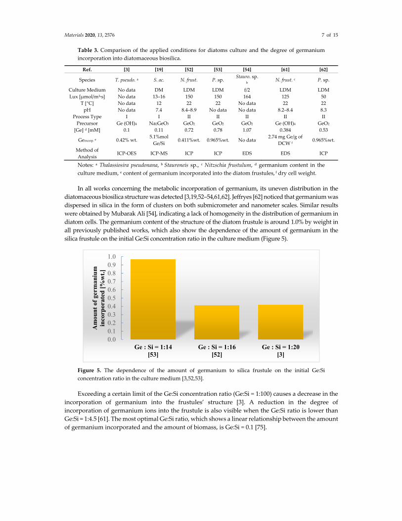
Table 3. Comparison of the applied conditions for diatoms culture and the degree of germanium
incorporation into diatomaceous biosilica.
Ref. [3] [19] [52] [53] [54] [61] [62]
Species T. pseudo. a S. ac. N. frust. P. sp. Stauro. sp.
b N. frust. c P. sp.
Culture Medium No data DM LDM LDM f/2 LDM LDM
Lux [μmol/m2*s] No data 13–16 150 150 164 125 50
T [°C] No data 12 22 22 No data 22 22
pH No data 7.4 8.4–8.9 No data No data 8.2–8.4 8.3
Process Type I I II II II II II
Precursor Ge (OH)4 Na2GeO3 GeO2 GeO2 GeO2 Ge (OH)4 GeO2
[Ge] d [mM] 0.1 0.11 0.72 0.78 1.07 0.384 0.53
Geincorp. e 0.42% wt. 5.1%mol
Ge/Si 0.411%wt. 0.965%wt. No data
2.74 mg Ge/g of
DCW f 0.965%wt.
Method of
Analysis ICP‐OES ICP‐MS ICP ICP EDS EDS ICP
Notes: a Thalassiosira pseudonana, b Stauroneis sp., c Nitzschia frustulum, d germanium content in the
culture medium, e content of germanium incorporated into the diatom frustules, f dry cell weight.
In all works concerning the metabolic incorporation of germanium, its uneven distribution in the
diatomaceous biosilica structure was detected [3,19,52–54,61,62]. Jeffryes [62] noticed that germanium was
dispersed in silica in the form of clusters on both submicrometer and nanometer scales. Similar results
were obtained by Mubarak Ali [54], indicating a lack of homogeneity in the distribution of germanium in
diatom cells. The germanium content of the structure of the diatom frustule is around 1.0% by weight in
all previously published works, which also show the dependence of the amount of germanium in the
silica frustule on the initial Ge:Si concentration ratio in the culture medium (Figure 5).
Figure 5. The dependence of the amount of germanium to silica frustule on the initial Ge:Si
concentration ratio in the culture medium [3,52,53].
Exceeding a certain limit of the Ge:Si concentration ratio (Ge:Si = 1:100) causes a decrease in the
incorporation of germanium into the frustules’ structure [3]. A reduction in the degree of
incorporation of germanium ions into the frustule is also visible when the Ge:Si ratio is lower than
Ge:Si = 1:4.5 [61]. The most optimal Ge:Si ratio, which shows a linear relationship between the amount
of germanium incorporated and the amount of biomass, is Ge:Si = 0.1 [75].
0.00.10.20.30.40.50.60.70.80.91.0
Ge : Si = 1:14[53]
Ge : Si = 1:16[52]
Ge : Si = 1:20[3]
Am
ount
of
germ
aniu
m
inco
rpor
ated
[%
wt.
]

Materials 2020, 13, 2576 8 of 15
3. Metabolic Insertion of Other Metals and Semimetals Ions
The works described above suggest that the concept of using the unique ability of diatom
cultures to take up soluble metals and incorporate them into the structure of their frustules can also
be applied to other metals, including nickel, europium, aluminum, zinc, iron, calcium, zirconium,
and tin [19,63–67,69–71].
A summary of the diatom culture conditions used and the degree of incorporation of the
indicated elements into diatomaceous biosilica in vivo is presented in Tables 4 (calcium) and 5 (other
elements).
3.1. Doping of Biosilica with Aluminium Ions
Machill et al. [67] conducted research on the introduction of aluminum into the frustule structure
of the marine diatom, Stephanopyxis turris, using artificial seawater containing different
concentrations of aluminum (10.5, 42.5, 105.5, and 1055 μm) in the form of AlCl3. These concentrations
correspond to Al:Si mass ratios of 1:10, 1:2.5, 1:1, and 10:1 respectively. It was observed that a
concentration of 10.5 μm avoids uncontrolled aluminum precipitation. A SEM analysis performed to
assess the influence of aluminum on the morphology of diatom frustules did not show any significant
morphological differences compared with diatoms grown without aluminum in the culture medium.
The same size and structure of frustules were detected for both alumina‐enriched and natural diatom
samples. The incorporation of aluminum into biosilica was detected by ICP‐OES analysis of extracted
frustules. Quantification has shown that the amount of aluminum embedded in frustules increased
rapidly in a medium enriched with aluminum, although no value for the content of aluminum is
given. It was observed that the greatest ratio of Al:Si obtained was 1:15. Diatomaceous biosilica doped
with aluminum ions can be very valuable material used in catalysis due to its high catalytic activity
[51].
3.2. Doping of Biosilica with Nickel Ions
Townley et al. [63] presented the results of studies on doping Coscinodiscus wailesii cells with
nickel ions to modify the optical properties of their frustules. Selected diatom species were cultured
in sterile filtered seawater containing an Alga‐Gro medium with nickel sulphate added at 5.0, 1.0, 0.5,
and 0.1 mg/L. It was observed that the maximum concentration of nickel, which had no significant
effect on the growth of diatoms, was 0.5 mg/L.
The SEM analysis of diatoms enriched in nickel showed that the pores in their frustules were
more irregular, larger, and less uniform in shape. Studies of the cytoplasmic morphology of C. wailesii
cultivated in the presence of nickel sulphate showed an interruption of thylakoid stacks and swelling
of mitochondria. As the nickel concentration in the culture medium increased, the
photoluminescence of silica frustules was also extinguished. The nickel content in the diatoms
frustules was about 0.1% by weight and was confirmed by the EDX method. Diatomaceous biosilica
doped with nickel ions can be used in biotechnology applications due to its unique optical properties.
3.3. Biosilica Doping with Europium Ions
Silicate‐based phosphors are promising luminescent materials because of their chemical
stability, moisture resistance, and low cost. These materials can be used in various display
technologies, such as fluorescent lamps, plasma display panels, field emission displays, and cathode‐
ray tubes. Zhang et al. [71] published results on the doping of diatomaceous biosilica with europium.
Doping was carried out by culturing Navicula sp. with the addition of europium hexavalent nitrate
(Eu (NO3)3∙6H2O) in the molar ratio 1:4 Eu:Si. The culture was carried out for 96 h, then the diatom
cells were extracted with ethanol to remove the alcohol‐soluble organic material, and the solid
residues were heat annealed in air at 1000 °C. An XRD analysis showed the presence of europium in
the form of Eu2O3 and Eu2SiO5. Europium‐doped biosilica exhibited photoluminescent properties
with red light emission (614 nm) and excitation at 394 nm corresponding to the wavelength of LED
emission.

Materials 2020, 13, 2576 9 of 15
3.4. Doping of Biosilica with Calcium Ions
Leone et al. [65] recently published results on the doping of diatomaceous biosilica with calcium
cations for biomedical materials. The idea for this work was based on the knowledge that fibroblasts
and osteoblasts grow very well on silica or ceramic substrates, and the presence of calcium ions
stimulates the growth of the cells. Calcium‐doped diatomaceous biosilica was obtained during the
cultivation of Thalassiosira weissflogii in autoclaved and ultrafiltered sea water at a controlled
temperature of 18–22 °C, with the addition of calcium in the form of CaCl2 (14 mM) to the culture
medium.
SEM showed that the addition of calcium to diatom culture does not affect their shape and
structure. FTIR showed that even though the calcium is not covalently bonded with the biosilica
obtained, it remains in the frustules despite the action of 30% hydrogen peroxide to remove organic
matter from diatom cells. The detected content of calcium in the doped frustules was in the range of
0.9 ± 0.05% weight. Cell viability studies also confirmed that calcium‐doped biosilica can serve as an
effective substrate for the growth of fibroblasts and osteoblasts with possible applications in
regenerative medicine.
Li et al. [66] also investigated the calcium ion doping of diatoms. Coscinodiscus sp. were cultured
in ultrafiltered and autoclaved sea water enriched with a f/2 Guillard medium at 21 °C, and calcium
inclusion in diatom frustules was achieved by introducing CaCl2 calcium chloride into the culture
medium at 0.125, 0.25, 0.50, 1.0, and 2.0 mM.
The presence of calcium ions in the structure of diatom frustules was confirmed by XRD and
EDXS, but no specific values were provided. Similar to Leone [65], the presence of calcium ions in the
culture medium in Li et al. [62] did not cause any significant changes in the morphology of diatoms,
and the authors indicated that the material obtained could be used as a haemostatic. Detailed data on
the conditions of doping of diatomaceous biosilica with calcium ions are given in Table 4.
Table 4. Summary of parameters for doping of diatomaceous biosilica with calcium ions.
Ref. [65] [66]
Species T. weiss. Cos. sp.
Precursor CaCl2 CaCl2
Culture Medium f/2 f/2
Lux [μmol/m2.s] No data 246
T [°C] 18–22 21
pH No data No data
Caincorp a [%wt] 0.9 No data
Method of Analysis EDX EDXS
Note: a content of calcium incorporated into the diatom frustules.
3.5. Doping of Biosilica with Zirconium and Tin Ions
The influence of zirconium and tin on the growth, morphology, and chemical composition of
the freshwater diatom Synedra acus was studied by Basharina et al. [19]. Microincubators containing
a culture medium with 10 mM Na2SiO3 and 10 mM Na2SnO3 or ZrCl4 were used. It was noted that
doping with zirconium and tin ions caused a slight decrease in growth rate, irregularity of frustules,
and a decrease in mechanical strength of the frustules. Analysis showed the presence of 3.4% mol
Zr/Si and 0.91% mol Sn/Si in the diatom biomass, and 0% Zr and 0.13% Sn in the purified
diatomaceous biosilica.
In order to obtain nanoporous composites of diatom‐ZrO2, Gannavarapu et al. [64] conducted
studies on the culture of the species Phaeodactylum tricornutum using artificial sea water at pH = 9
with the addition of 0.8 mM ZrOCl2*8H2O as a culture medium. The presence of ZrO2 zirconium
oxides on the surface of Phaeodactylum tricornutum frustules was confirmed by EDX analysis. A
significant decrease in the size of diatom cells was also observed. The obtained composite has been

Materials 2020, 13, 2576 10 of 15
successfully tested as an electrochemical sensor for the detection of methyl parathion—an
organophosphorus pesticide.
3.6. Biosilica Doping with Zinc and Iron Oons
In order to verify that diatoms were able to incorporate significant amounts of zinc and iron,
Ellwood and Hunter [70] grew the marine diatom Thalassiosira pseudonana in sea water with the
addition of Zn or Fe salts. A positive relationship was observed between the concentration of free
Zn2+ and the growth rate of T. pseudonana. It was also found that limiting access to free Zn2+ caused a
decrease in cell size in T. pseudonana in comparison with those in the free Zn2+ replete medium. An
analysis of the chemical composition of the biosilica obtained confirmed the inclusion of zinc and
iron ions into the structure of the diatom frustules. In the case of zinc ion incorporation, it was found
that the amount of Zn2+ uptake into the diatom was directly related to the amount of zinc absorbed
by the diatom (2 5 10 , which in turn was directly related to the concentration of free
Zn2+ in the culture medium. The relationship between Zn/Si and free Zn2+ suggests that the Zn content
of diatom shells may be useful in reconstructing changes in oceanic concentration of free Zn2+. In the
case of iron ion incorporation, no direct proportional relationship between the incorporated iron
content and its concentration in the culture medium was detected. Moreover, no specific value was
determined for the amount of iron incorporated into the diatom cell.
The studies reported in Ellwood and Hunter [70] were continued by Jaccard et al. [69]. They
cultivated the freshwater diatom Stephanodiscus hantzschii (UTCC 267) in a modified medium CHU‐
10 [77] with the addition of the Zn–EDTA complex, containing 10−10.6–10−7.6 M zinc ions. The presence
of zinc ions in the structure of diatom frustules was confirmed by ICP‐MS. No value was given for
the amount of zinc incorporated into the diatom frustules, but it was found that the highest degree
of zinc ion incorporation was achieved at a concentration of 10−8.5 M Zn2+.
The conditions used to grow diatoms and the degree of metabolic insertion of elements (Al, Ni,
Eu, Zr, Sn, Zn, Fe) into diatomaceous biosilica are summarized in Table 5.
Table 5. Summary of results of doping diatoms with metal ions.
Zr/Sn Ni Al Zn Fe Eu
Ref. [19] [64] [63] [67] [69] [70] [70] [71]
Species S. ac. P. trico. a Cos. S. tur.
b
S.
hanz. c T. pseudo.
T.
pseudo. Navi. sp. d
Culture
Medium DM Aquil Alga‐Gro ASW
CHU‐
10 f/2 f/2 „Ningbo 3”
Lux
[μmol/m2*s] 13–16 No data No data 82 50 120 120 246
T [ °C] 12 19 22 18 20 20–22 20–22 25
pH 7.4 9 No data 8.0–
8.2 6.4 7.2–8.3 8.0 No data
Process Type I I I I II II II II
Precursor ZrCl4 ZrOCl2*8H2O NiSO4 AlCl3 Zn–
EDTA Zn–EDTA
Fe–
EDTA
Eu
(NO3)3∙6H2O
Xincorp. e
3.4%mol
Zr/Si;
0.91%mol
Sn/Si
No data ~0.1%wag No
data
No
data
2–5 . 1017 mol
Zn/cell.day
No
data No data
Method of
Analysis ICP‐MS EDX EDX
ICP‐
OES
ICP‐
MS GFAAS GFAAS XRD
Notes: a Phaeodactylum tricornutum; b Stephanopyxis turris; c Stephanodiscus hantzschii; d Navicula sp.; e
the content of the element incorporated into the diatom frustule, where X = Al, Ni, Eu, Zr, Sn, Zn, Fe.
4. Conclusions
This analysis of the limited research available on the metabolic insertion of elements into the
structured diatomaceous biosilica indicates the emergence of a new trend in synthesis methods for
next‐generation functional materials. This new approach harnesses the ability of microorganisms to

Materials 2020, 13, 2576 11 of 15
act as microtechnologists for the synthesis of smart materials, in particular the diatoms in
synthesizing new silica materials with desirable properties.
There is evidence that many factors, such as the type of precursor, chemical composition and pH
of the culture medium, temperature, illumination, and aeration, can significantly impact the process
of metabolic insertion into diatomaceous biosilica, the growth and development of diatom cells, and
the structure and morphology of the frustules. Therefore, there is scope for optimizing the process of
metabolic insertion by manipulating these factors (e.g., by changing the Ge:Si ratio in the culture
medium). A further aspect is the variation in the influence of different factors and doping elements
on different diatom species, and therefore the choice of diatom species is also crucial. Finally, there is
further potential for metabolic insertion techniques by using a genetic modification of diatom cells
[20,78] in order to give them specific abilities for selective metabolic insertion of selected elements.
Author Contributions: Conceptualization, M.S.; Software, W.B. and I.W.; Writing, W.B. and M.S.; Original Draft
Preparation, W.B. and I.W.; Writing—Review & Editing, W.B., M.S., I.W., P.D., A.W. and B.B.; Funding
Acquisition, B.B. and A.W. All authors have read and agreed to the published version of the manuscript.
Funding: This work was supported by the project “Advanced biocomposites for tomorrow’s economy BIOG‐
NET” carried out within the TEAM‐NET programme of the Foundation for Polish Science co‐financed by the
European Union under the European Regional Development Fund.
Conflicts of Interest: The authors declare no conflict of interest.
References
1. Gross, M. The mysteries of the diatoms. Curr. Biol. 2012, 22, 581–585, doi:10.1016/j.cub.2012.07.041.
2. Görlich, S.; Pawolski, D.; Zlotnikov, I.; Kröger, N. Control of biosilica morphology and mechanical
performance by the conserved diatom gene Silicanin‐1. Commun. Biol. 2019, 2, 245, doi:10.1038/s42003‐019‐
0436‐0.
3. Davis, A.K.; Hildebrand, M. A self‐propagating system for Ge incorporation into nanostructured silica.
Chem. Commun. 2008, 4495–4497, doi:10.1039/b804955f.
4. Zhang, D.Y.; Wang, Y.; Cai, J.; Pan, J.F.; Jiang, X.G.; Jiang, Y.G. Bio‐manufacturing technology based on
diatom micro‐ and nanostructure. Chin. Sci. Bull. 2012, 57, 3836–3849, doi:10.1007/s11434‐012‐5410‐x.
5. Mann, D.G.; Vanormelingen, P. An inordinate fondness the number, distributions, and origins of diatom
species. J. Eukaryot. Microbiol. 2013, 60, 414–420, doi:10.1111/jeu.12047.
6. Wang, J.K.; Seibert, M. Prospects for commercial production of diatoms. Biotechnol. Biofuels 2017, 10, 1–13,
doi:10.1186/s13068‐017‐0699‐y.
7. Popovich, C.A.; Pistonesi, M.; Hegel, P.; Constenla, D.; Bielsa, G.B.; Martín, L.A.; Damiani, M.C.; Leonardi,
P.I. Unconventional alternative biofuels: Quality assessment of biodiesel and its blends from marine diatom
Navicula cincta. Algal Res. 2019, 39, 101438, doi:10.1016/j.algal.2019.101438.
8. Fu, W.; Nelson, D.R.; Mystikou, A.; Daakour, S.; Salehi‐Ashtiani, K. Advances in microalgal research and
engineering development. Curr. Opin. Biotechnol. 2019, 59, 157–164, doi:10.1016/j.copbio.2019.05.013.
9. Sasirekha, R.; Sheena, T.S.; Anitha, R.; Santhanam, P.; Kulandaivel, J. Characterizations and analysis of
genus Amphora diatom frustules: A promising biomaterial. Bioinspired Biomim. Nanobiomater. 2019, 8, 224–
230, doi:10.1680/jbibn.18.00026.
10. Panwar, V.; Dutta, T. Diatom Biogenic Silica as a Felicitous Platform for Biochemical Engineering:
Expanding Frontiers. ACS Appl. Bio Mater. 2019, 2, 2295–2316, doi:10.1021/acsabm.9b00050.
11. Maher, S.; Maher, S.; Aw, M.S.; Losic, D. Diatom Silica for Biomedical Applications. Diatoms Fundam. Appl.
2019, 511–536, doi:10.1002/9781119370741.ch20.
12. Vinayak, V.; Joshi, K.B.; Sarma, P.M. DiafuelTM (Diatom Biofuel) vs Electric Vehicles, a Basic Comparison:
A High Potential Renewable Energy Source to Make India Energy Independent. In Diatoms: Fundamentals
and Applications; Wiley: Hoboken, NJ, USA, 2019; pp. 537–582.
13. Gordon, R.; Merz, C.R.; Gurke, S.; Schoefs, B. Bubble Farming: Scalable Microcosms for Diatom Biofuel and
the Next Green Revolution. Diatoms Fundam. Appl. 2019, 583–654, doi:10.1002/9781119370741.ch22.
14. Vinayak, V.; Gautam, S. Diatoms in Forensics: A Molecular Approach to Diatom Testing in Forensic
Science. Diatoms Fundam. Appl. 2019, 435–470, doi:10.1002/9781119370741.ch18.

Materials 2020, 13, 2576 12 of 15
15. Gordon, R.; Drum, R.W. The Chemical Basis of Diatom Morphogenesis. Int. Rev. Cytol. 1994, 150, 243–372,
doi:10.1016/S0074‐7696(08)61544‐2.
16. Gordon, R.; Losic, D.; Tiffany, M.A.; Nagy, S.S.; Sterrenburg, F.A.S. The Glass Menagerie: Diatoms for novel
applications in nanotechnology. Trends Biotechnol. 2009, 27, 116–127, doi:10.1016/j.tibtech.2008.11.003.
17. Bozarth, A.; Maier, U.G.; Zauner, S. Diatoms in biotechnology: Modern tools and applications. Appl.
Microbiol. Biotechnol. 2009, 82, 195–201, doi:10.1007/s00253‐008‐1804‐8.
18. Nassif, N.; Livage, J. From diatoms to silica‐based biohybrids. Chem. Soc. Rev. 2011, 40, 849–859,
doi:10.1039/c0cs00122h.
19. Basharina, T.N.; Danilovtseva, E.N.; Zelinskiy, S.N.; Klimenkov, I.V.; Likhoshway, Y.V.; Annenkov, V.V.
The Effect of Titanium, Zirconium and Tin on the Growth of Diatom Synedra acus and Morphology of Its
Silica Valves. Silicon 2012, 4, 239–249, doi:10.1007/s12633‐012‐9119‐x.
20. Mishra, M.; Arukha, A.P.; Bashir, T.; Yadav, D.; Prasad, G.B.K.S. All new faces of diatoms: Potential source
of nanomaterials and beyond. Front. Microbiol. 2017, 8, 1–8, doi:10.3389/fmicb.2017.01239.
21. Athanasakoglou, A.; Kampranis, S.C. Diatom isoprenoids: Advances and biotechnological potential.
Biotechnol. Adv. 2019, 37, doi:10.1016/j.biotechadv.2019.107417.
22. Martínez‐Carmona, M.; Gun’ko, Y.K.; Vallet‐Regí, M. Mesoporous silica materials as drug delivery: “the
nightmare” of bacterial infection. Pharmaceutics 2018, 10, 279, doi:10.3390/pharmaceutics10040279.
23. Uthappa, U.T.; Brahmkhatri, V.; Sriram, G.; Jung, H.Y.; Yu, J.; Kurkuri, N.; Aminabhavi, T.M.; Altalhi, T.;
Neelgund, G.M.; Kurkuri, M.D. Nature engineered diatom biosilica as drug delivery systems. J. Control.
Release 2018, 281, 70–83, doi:10.1016/j.jconrel.2018.05.013.
24. De Tommasi, E.; Gielis, J.; Rogato, A. Diatom Frustule Morphogenesis and Function: A Multidisciplinary
Survey. Mar. Genom. 2017, 35, 1–18, doi:10.1016/j.margen.2017.07.001.
25. De Stefano, L.; Rotiroti, L.; De Stefano, M.; Lamberti, A.; Lettieri, S.; Setaro, A.; Maddalena, P. Marine
diatoms as optical biosensors. Biosens. Bioelectron. 2009, 24, 1580–1584, doi:10.1016/j.bios.2008.08.016.
26. Anderson, N.J. Miniview: Diatoms, temperature and climatic change. Eur. J. Phycol. 2000, 35, 307–314,
doi:10.1080/09670260010001735911.
27. Townley, H.E.; Parker, A.R.; White‐Cooper, H. Exploitation of diatom frustules for nanotechnology:
Tethering active biomolecules. Adv. Funct. Mater. 2008, 18, 369–374, doi:10.1002/adfm.200700609.
28. De Stefano, L.; Maddalena, P.; Moretti, L.; Rea, I.; Rendina, I.; De Tommasi, E.; Mocella, V.; De Stefano, M.
Nano‐biosilica from marine diatoms: A brand new material for photonic applications. Superlattices
Microstruct. 2009, 46, 84–89, doi:10.1016/j.spmi.2008.10.031.
29. He, J.; Chen, D.; Li, Y.; Shao, J.; Xie, J.; Sun, Y.; Yan, Z.; Wang, J. Diatom‐templated TiO2 with enhanced
photocatalytic activity: Biomimetics of photonic crystals. Appl. Phys. A Mater. Sci. Process. 2013, 113, 327–
332, doi:10.1007/s00339‐013‐7970‐2.
30. Nowak, A.P.; Sprynskyy, M.; Brzozowska, W.; Lisowska‐Oleksiak, A. Electrochemical behavior of a
composite material containing 3D‐structured diatom biosilica. Algal Res. 2019, 41, 101538,
doi:10.1016/j.algal.2019.101538.
31. Norberg, A.N.; Wagner, N.P.; Kaland, H.; Vullum‐Bruer, F.; Svensson, A.M. Silica from diatom frustules as
anode material for Li‐ion batteries. RSC Adv. 2019, 9, 41228–41239, doi:10.1039/c9ra07271c.
32. Toster, J.; Iyer, K.S.; Xiang, W.; Rosei, F.; Spiccia, L.; Raston, C.L. Diatom frustules as light traps enhance
DSSC efficiency. Nanoscale 2013, 5, 873–876, doi:10.1039/c2nr32716c.
33. Wang, Y.; Cai, J.; Jiang, Y.; Jiang, X.; Zhang, D. Preparation of biosilica structures from frustules of diatoms
and their applications: Current state and perspectives. Appl. Microbiol. Biotechnol. 2013, 97, 453–460,
doi:10.1007/s00253‐012‐4568‐0.
34. Chandrasekaran, S.; Sweetman, M.J.; Kant, K.; Skinner, W.; Losic, D.; Nann, T.; Voelcker, N.H. Silicon
diatom frustules as nanostructured photoelectrodes. Chem. Commun. 2014, 50, 10441–10444,
doi:10.1039/c4cc04470c.
35. Huang, D.R.; Jiang, Y.J.; Liou, R.L.; Chen, C.H.; Chen, Y.A.; Tsai, C.H. Enhancing the efficiency of dye‐
sensitized solar cells by adding diatom frustules into TiO2 working electrodes. Appl. Surf. Sci. 2015, 347, 64–
72, doi:10.1016/j.apsusc.2015.04.064.
36. Lin, K.B.; Shen, T.W.; Su, Y.H. Silicon‐Based Solar Cells: Emergent Upconversion Sustainable Micro‐Optical
Trapping Device (Part. Part. Syst. Charact. 7/2019). Part. Part. Syst. Charact. 2019, 36, 1970017,
doi:10.1002/ppsc.201970017.

Materials 2020, 13, 2576 13 of 15
37. Jeffryes, C.; Campbell, J.; Li, H.; Jiao, J.; Rorrer, G. The potential of diatom nanobiotechnology for
applications in solar cells, batteries, and electroluminescent devices. Energy Environ. Sci. 2011, 4, 3930–3941,
doi:10.1039/c0ee00306a.
38. Bao, Z.; Weatherspoon, M.R.; Shian, S.; Cai, Y.; Graham, P.D.; Allan, S.M.; Ahmad, G.; Dickerson, M.B.;
Church, B.C.; Kang, Z.; et al. Chemical reduction of three‐dimensional silica micro‐assemblies into
microporous silicon replicas. Nature 2007, 446, 172–175, doi:10.1038/nature05570.
39. Kroth, P. Molecular biology and the biotechnological potential of diatoms. Adv. Exp. Med. Biol. 2007, 616,
23–33, doi:10.1007/978‐0‐387‐75532‐8_3.
40. Leonardo, S.; Prieto‐Simón, B.; Campàs, M. Past, present and future of diatoms in biosensing. TrAC Trends
Anal. Chem. 2016, 79, 276–285, doi:10.1016/j.trac.2015.11.022.
41. Umemura, K.; Gao, Y.; Nishikawa, T. Preparation of photocatalyst using diatom frustules by liquid phase
deposition method. J. Nanosci. Nanotechnol. 2010, 10, 4883–4888, doi:10.1166/jnn.2010.2416.
42. Huang, W.; Daboussi, F. Genetic and metabolic engineering in diatoms. Philos. Trans. R. Soc. B Biol. Sci.
2017, 372, doi:10.1098/rstb.2016.0411.
43. Yang, W.; Lopez, P.J.; Rosengarten, G. Diatoms: Self assembled silica nanostructures, and templates for
bio/chemical sensors and biomimetic membranes. Analyst 2011, 136, 42–53, doi:10.1039/c0an00602e.
44. Irimia‐Vladu, M.D.; Glowacki, E.S.; Sariciftci, N.; Bauer, S.; Ragni, R.; Cicco, S.R.; Vona, D.; Farinola, G.M.
Nanostructured Silica from Diatoms Microalgae: Smart Materials for Photonics and Electronics. Green
Mater. Electron. 2017, 287–313, doi:10.1002/9783527692958.ch11.
45. Terracciano, M.; De Stefano, L.; Rea, I. Diatoms green nanotechnology for biosilica‐based drug delivery
systems. Pharmaceutics 2018, 10, 242, doi:10.3390/pharmaceutics10040242.
46. Ramachandra, T.V.; Mahapatra, D.M.; Karthick, B.; Gordon, R. Milking diatoms for sustainable energy:
Biochemical engineering versus gasoline‐secreting diatom solar panels. Ind. Eng. Chem. Res. 2009, 48, 8769–
8788, doi:10.1021/ie900044j.
47. Zglobicka, I.; Chmielewska, A.; Topal, E.; Kutukova, K.; Gluch, J.; Krüger, P.; Kilroy, C.; Swieszkowski, W.;
Kurzydlowski, K.J.; Zschech, E. 3D Diatom–Designed and Selective Laser Melting (SLM) Manufactured
Metallic Structures. Sci. Rep. 2019, 9, 1–9, doi:10.1038/s41598‐019‐56434‐7.
48. Bandara, T.M.W.J.; Furlani, M.; Albinsson, I.; Wulff, A.; Mellander, B.E. Diatom frustules enhancing the
efficiency of gel polymer electrolyte based dye‐sensitized solar cells with multilayer photoelectrodes.
Nanoscale Adv. 2020, 2, 199–209, doi:10.1039/c9na00679f.
49. Kröger, N.; Dubey, N.C.; Kumari, E. CHAPTER 6: Immobilization of Proteins on Diatom Biosilica. In RSC
Nanoscience and Nanotechnology; Royal Society of Chemistry: London, UK, 2018; Volume 2018‐January, pp.
126–149, ISBN 9781782624585.
50. Ragni, R.; Cicco, S.; Vona, D.; Leone, G.; Farinola, G.M. Biosilica from diatoms microalgae: Smart materials
from bio‐medicine to photonics. J. Mater. Res. 2017, 32, 279–291, doi:10.1557/jmr.2016.459.
51. Köhler, L.; Machill, S.; Werner, A.; Selzer, C.; Kaskel, S.; Brunner, E. Are Diatoms “Green” Aluminosilicate
Synthesis Microreactors for Future Catalyst Production? Molecules 2017, 22, 2232,
doi:10.3390/molecules22122232.
52. Qin, T.; Gutu, T.; Chang, C.H.; Jiao, J.; Rorrer, G.L. Biological fabrication of photoluminescent nanocomb
structures by metabolic incorporation of germanium into the biosilica of the diatom Nitzschia frustulum.
ACS Nano 2008, 2, 1296–1304, doi:10.1021/nn800114q.
53. Gale, D.K.; Jeffryes, C.; Gutu, T.; Jiao, J.; Chang, C.H.; Rorrer, G.L. Thermal annealing activates amplified
photoluminescence of germanium metabolically doped in diatom biosilica. J. Mater. Chem. 2011, 21, 10658–
10665, doi:10.1039/c1jm10861a.
54. Ali, D.M.; Divya, C.; Gunasekaran, M.; Thajuddin, N. Biosynthesis and Characterization of Silicon‐
Germanium Oxide Nanocomposite By Diatom. Dig. J. Nanomater. Biostruct. 2011, 6, 117–120.
55. Jeffryes, C.; Gutu, T.; Jiao, J.; Rorrer, G.L. Metabolic Insertion of Nanostructured TiO2 into the Patterned
Biosilica of the Diatom. ACS Nano 2008, 2, 2103–2112, doi:10.1021/nn800470x CCC:$40.75.
56. Van Eynde, E.; Hu, Z.Y.; Tytgat, T.; Verbruggen, S.W.; Watté, J.; Van Tendeloo, G.; Van Driessche, I.; Blust,
R.; Lenaerts, S. Diatom silica‐titania photocatalysts for air purification by bio‐accumulation of different
titanium sources. Environ. Sci. Nano 2016, 3, 1052–1061, doi:10.1039/c6en00163g.
57. Chauton, M.S.; Skolem, L.M.B.; Olsen, L.M.; Vullum, P.E.; Walmsley, J.; Vadstein, O. Titanium uptake and
incorporation into silica nanostructures by the diatom Pinnularia sp. (Bacillariophyceae). J. Appl. Phycol.
2015, 27, 777–786, doi:10.1007/s10811‐014‐0373‐8.

Materials 2020, 13, 2576 14 of 15
58. Skolem, L.M.B. Biosynthesis and Characterization of Ti‐ Doped Silica‐Based Nanostructures Formed by the Diatoms
Pinnularia sp. and Coscinodiscus wailesii; Norwegian University of Science and Technology: Trondheim,
Norway, 2011; Volume 801.
59. Maeda, Y.; Niwa, Y.; Tang, H.; Kisailus, D.; Yoshino, T.; Tanaka, T. Development of Titania‐Integrated Silica
Cell Walls of the Titanium‐Resistant Diatom, Fistulifera solaris. ACS Appl. Bio Mater. 2018, 1, 2021–2029,
doi:10.1021/acsabm.8b00520.
60. Lang, Y.; Monte, F.D.; Rodriguez, B.J.; Dockery, P.; Finn, D.P.; Pandit, A. Integration of TiO2 into the diatom
Thalassiosira weissflogii during frustule synthesis. Sci. Rep. 2013, 3, 1–11, doi:10.1038/srep03205.
61. Rorrer, G.L.; Chang, C.H.; Liu, S.H.; Jeffryes, C.; Jiao, J.; Hedberg, J.A. Biosynthesis of silicon‐germanium
oxide nanocomposites by the marine diatom Nitzschia frustulum. J. Nanosci. Nanotechnol. 2005, 5, 41–49,
doi:10.1166/jnn.2005.005.
62. Jeffryes, C.; Gutu, T.; Jiao, J.; Rorrer, G.L. Two‐stage photobioreactor process for the metabolic insertion of
nanostructured germanium into the silica microstructure of the diatom Pinnularia sp. Mater. Sci. Eng. C
2008, 28, 107–118, doi:10.1016/j.msec.2007.01.002.
63. Townley, H.E.; Woon, K.L.; Payne, F.P.; White‐Cooper, H.; Parker, A.R. Modification of the physical and
optical properties of the frustule of the diatom Coscinodiscus wailesii by nickel sulfate. Nanotechnology 2007,
18, 14–19, doi:10.1088/0957‐4484/18/29/295101.
64. Gannavarapu, K.P.; Ganesh, V.; Thakkar, M.; Mitra, S.; Dandamudi, R.B. Nanostructured Diatom‐ZrO2
composite as a selective and highly sensitive enzyme free electrochemical sensor for detection of methyl
parathion. Sens. Actuators B Chem. 2019, 288, 611–617, doi:10.1016/j.snb.2019.03.036.
65. Leone, G.; Vona, D.; Lo Presti, M.; Urbano, L.; Cicco, S.; Gristina, R.; Palumbo, F.; Ragni, R.; Farinola, G.M.
Ca2+‐in vivo doped biosilica from living Thalassiosira weissflogii diatoms: Investigation on Saos‐2
biocompatibility. Mater. Res. Soc. 2017, 2, 1047–1058.
66. Li, J.; Han, J.; Sun, Q.; Wang, Y.; Mu, Y.; Zhang, K.; Dou, X.; Kong, M.; Chen, X.; Feng, C. Biosynthetic
calcium‐doped biosilica with multiple hemostatic properties for hemorrhage control. J. Mater. Chem. B 2018,
6, 7834–7841, doi:10.1039/c8tb00667a.
67. Machill, S.; Kohler, L.; Ueberlein, S.; Hedrich, R.; Kunaschk, M.; Paasch, S.; Schulze, R.; Brunner, E.
Analytical studies on the incorporation of aluminium in the cell walls of the marine diatom Stephanopyxis
turris. BioMetals 2013, 26, 141–150, doi:10.1007/s10534‐012‐9601‐3.
68. Gehlen, M.; Beck, L.; Calas, G.; Flank, A.M.; Van Bennekom, A.J.; Van Beusekom, J.E.E. Unraveling the
atomic structure of biogenic silica: Evidence of the structural association of Al and Si in diatom frustules.
Geochim. Cosmochim. Acta 2002, 66, 1601–1609, doi:10.1016/S0016‐7037(01)00877‐8.
69. Jaccard, T.; Ariztegui, D.; Wilkinson, K.J. Incorporation of zinc into the frustule of the freshwater diatom
Stephanodiscus hantzschii. Chem. Geol. 2009, 265, 381–386, doi:10.1016/j.chemgeo.2009.04.016.
70. Ellwood, M.J.; Hunter, K.A. The incorporation of zinc and iron into the frustule of the marine diatom
Thalassiosira pseudonana. Limnol. Oceanogr. 2000, 45, 1517–1524, doi:10.4319/lo.2000.45.7.1517.
71. Zhang, G.; Jiang, W.; Wang, L.; Liao, X.; Liu, P.; Deng, X.; Li, J. Preparation of silicate‐based red phosphors
with a patterned nanostructure via metabolic insertion of europium in marine diatoms. Mater. Lett. 2013,
110, 253–255, doi:10.1016/j.matlet.2013.08.045.
72. Rorrer, G.L. CHAPTER 4: Functionalization of Frustules from Diatom Cell Culture for Optoelectronic
Properties. In RSC Nanoscience and Nanotechnology; Royal Society of Chemistry: London, UK, 2018; Volume
2018‐January, pp. 79–110, ISBN 9781782624585.
73. Lewin, J. Silicon Metabolism in Diatoms: 5. Germanium Dioxide, a Specific Inhibitor of Diatom Growth.
Phycologia 1966, 6, 1–12, doi:10.2216/i0031‐8884‐6‐1‐1.1.
74. Werner, D. Hemmung der Chlorophyllsynthese und der NADP+‐abhängigen Glycerinaldehyd‐3‐
Phosphat‐Dehydrogenase durch Germaniumsäure bei Cyclotella cryptica. Arch. Mikrobiol. 1967, 57, 51–60,
doi:10.1007/BF00405767.
75. Azam, F.; Hemmingsen, B.B.; Volcani, B.E. Germanium incorporation into the silica of diatom cell walls.
Arch. Mikrobiol. 1973, 92, 11–20, doi:10.1007/BF00409507.
76. Richter, O. Zur Physiologie der Diatomeen, I., III. Sitzungsberichte der Kais. Akad. der Wissenschaften, Math.
Klasse 1906, 115, 27–119.

Materials 2020, 13, 2576 15 of 15
77. Nichols, H.W. Growth media‐freshwater. In Handbook of Phycological Methods: Culture Methods and Growth
Measurements; Stein, J.R., Ed.; Cambridge University Press: Cambridge, UK, 1973; pp. 7–24, ISBN
9780521297479.
78. Delalat, B.; Sheppard, V.C.; Rasi Ghaemi, S.; Rao, S.; Prestidge, C.A.; McPhee, G.; Rogers, M.L.; Donoghue,
J.F.; Pillay, V.; Johns, T.G.; et al. Targeted drug delivery using genetically engineered diatom biosilica. Nat.
Commun. 2015, 6, 1–11, doi:10.1038/ncomms9791.
© 2020 by the authors. Licensee MDPI, Basel, Switzerland. This article is an open access
article distributed under the terms and conditions of the Creative Commons Attribution
(CC BY) license (http://creativecommons.org/licenses/by/4.0/).