Relationships between benthic diatoms and hydrozoans (Cnidaria
8
Relationships between benthic diatoms and hydrozoans (Cnidaria) C. Di Camillo, S. Puce,T. Romagnoli, S. Tazioli, C. Totti and G. Bavestrello* Dipartimento di Scienze del Mare, Universita' Politecnica delle Marche, Via Brecce Bianche, I-60131 Ancona, Italy. *Corresponding author, e-mail: [email protected] Some examples of relationships between hydroids and epibionthic diatoms from the Mediterranean Sea are described, verifying the kind of interaction existing between the two partners. The athecate Eudendrium racemosum hosts an extremely rich diatom assemblage, mainly comprising Licmophora spp., Amphora spp. and Cocconeis spp. On the contrary, only adnate growth forms ( Cocconeis pseudonotata, C. dirupta) were observed in diatom communities growing on the external side of thecate species Campanularia hincksii, Clytia linearis and Synthecium evansi. Some diatom species ( Cocconeis notata, Cylindrotheca sp. and Navicula sp.) are able to survive in the intrathecal microenvironment. They live in the narrow space between hydrotheca and polyp, receiving protection and probably using the nutrients produced by hydroid metabolism. Sunlight can penetrate through transparent thecae and reach the diatom layer, making photosynthesis possible. INTRODUCTION Several hydroid species have an epibiontic lifestyle, using many di¡erent phyla as their host organisms (Gili & Hughes, 1995). On the other hand, hydroids can also host dense associate vagile faunas, mainly composed of detritivorous organisms (Hancock et al., 1956; Round et al., 1961; Millard, 1973; Lagarde' re & Tardy, 1980; Marfenin, 1980; Blanco, 1984; Staples & Watson, 1987; Zamponi & Genzano, 1992; Bavestrello et al., 1996). In some cases, very peculiar symbiotic relationships between hydroids and associate species have been described (Bavestrello et al., 1996). The relationships of hydroids with marine macroalgae are important and widespread: i.e. several species of hydroids use seaweeds as substrata (Boero & Fresi, 1986). On the other hand, the relationships between benthic microalgae and hydroids are almost unexplored, except for the report of Siqueiros-Beltrones et al. (2001), which recorded the benthic diatom Cocconeis notata living inside the hydrothecae of Campanularia integra Mac Gillivray, 1842, Round et al. (1961) which observed that diatom assemblages associated with the thecate hydroid Sertularia ( ¼ Amphisbetia) operculata L., 1758 were a¡ected by the hydrodynamic conditions and Watson (2002) that found numerous ovoid diatoms along the inner abcauline wall of the hydrotheca. No data are avail- able on the relationships between hydroids and other kinds of algae. Nevertheless, our experience suggests that relationships between hydroids and microalgae are widespread and sometimes very selective. The aim of this work, therefore, is to present some examples of relationships between hydroids and epibionthic algae, diatoms from the Mediterranean Sea, verifying the kind of interaction existing between the two partners. J. Mar. Biol. Ass. U.K. (2005), 85, 1373^1380 Printed in the United Kingdom Journal of the Marine Biological Association of the United Kingdom (2005) Table 1. Distribution of most abundant diatoms along the di¡erent parts of hydroids. Hydroid species Hydroid colony portion Main diatom species Campunalaria hincksii inside of theca pedicels Cocconeis notata Petit Cocconeis pseudonotata De Stefano & Marino Clytia linearis outside of theca Cocconeis notata Petit Cocconeis pseudonotata De Stefano & Marino inside of theca Cylindrotheca sp. Cocconeis pseudonotata De Stefano & Marino pedicels Cocconeis dirupta Gregory Eudendrium racemosum structures enveloped by perisarc Licmophora oedipus (Ku«tzing) Grunow Licmophora £abellata (Carm.) Agardh Cocconeis stauroneiformis (Rabenhorst) Okuno Cocconeis scutellum Ehrenberg Cocconeis neothumensis var. marina De Stefano, Marino & Mazzella Amphora spp. Synthecium evansi inside of theca Navicula sp. Cocconeis neothumensis var. marina De Stefano, Marino & Mazzella Cocconeis placentula var. lineata (Ehrenberg) Van Heurck outside of colony other Cocconeis spp.
Transcript of Relationships between benthic diatoms and hydrozoans (Cnidaria

Relationships between benthic diatomsand hydrozoans (Cnidaria)
C. Di Camillo, S. Puce, T. Romagnoli, S. Tazioli, C. Totti and G. Bavestrello*
Dipartimento di Scienze del Mare, Universita' Politecnica delle Marche,Via Brecce Bianche, I-60131 Ancona, Italy.*Corresponding author, e-mail: [email protected]
Some examples of relationships between hydroids and epibionthic diatoms from the Mediterranean Seaare described, verifying the kind of interaction existing between the two partners.The athecate Eudendriumracemosum hosts an extremely rich diatom assemblage, mainly comprising Licmophora spp., Amphora spp. andCocconeis spp. On the contrary, only adnate growth forms (Cocconeis pseudonotata, C. dirupta) were observed indiatom communities growing on the external side of thecate species Campanularia hincksii, Clytia linearis andSynthecium evansi. Some diatom species (Cocconeis notata, Cylindrotheca sp. and Navicula sp.) are able tosurvive in the intrathecal microenvironment. They live in the narrow space between hydrothecaand polyp, receiving protection and probably using the nutrients produced by hydroid metabolism.Sunlight can penetrate through transparent thecae and reach the diatom layer, making photosynthesispossible.
INTRODUCTION
Several hydroid species have an epibiontic lifestyle,using many di¡erent phyla as their host organisms (Gili& Hughes, 1995). On the other hand, hydroids can alsohost dense associate vagile faunas, mainly composed ofdetritivorous organisms (Hancock et al., 1956; Roundet al., 1961; Millard, 1973; Lagarde' re & Tardy, 1980;Marfenin, 1980; Blanco, 1984; Staples & Watson, 1987;Zamponi & Genzano, 1992; Bavestrello et al., 1996). Insome cases, very peculiar symbiotic relationships betweenhydroids and associate species have been described(Bavestrello et al., 1996).
The relationships of hydroids with marine macroalgaeare important and widespread: i.e. several species ofhydroids use seaweeds as substrata (Boero & Fresi,1986). On the other hand, the relationships betweenbenthic microalgae and hydroids are almost unexplored,except for the report of Siqueiros-Beltrones et al. (2001),which recorded the benthic diatom Cocconeis notata
living inside the hydrothecae of Campanularia integra
Mac Gillivray, 1842, Round et al. (1961) which observedthat diatom assemblages associated with the thecatehydroid Sertularia (¼Amphisbetia) operculata L., 1758 werea¡ected by the hydrodynamic conditions and Watson(2002) that found numerous ovoid diatoms along theinner abcauline wall of the hydrotheca. No data are avail-able on the relationships between hydroids and other kindsof algae.
Nevertheless, our experience suggests that relationshipsbetween hydroids and microalgae are widespread andsometimes very selective. The aim of this work, therefore,is to present some examples of relationships betweenhydroids and epibionthic algae, diatoms from theMediterranean Sea, verifying the kind of interactionexisting between the two partners.
J. Mar. Biol. Ass. U.K. (2005), 85, 1373^1380Printed in the United Kingdom
Journal of the Marine Biological Association of the United Kingdom (2005)
Table 1. Distribution of most abundant diatoms along thedi¡erent parts of hydroids.
Hydroidspecies
Hydroid colonyportion
Main diatomspecies
Campunalaria
hincksii
inside of thecapedicels
Cocconeis notata PetitCocconeis pseudonotata
De Stefano & MarinoClytia linearis outside of theca Cocconeis notata Petit
Cocconeis pseudonotata
De Stefano & Marinoinside of theca Cylindrotheca sp.
Cocconeis pseudonotata
De Stefano & Marinopedicels Cocconeis dirupta Gregory
Eudendrium
racemosum
structuresenvelopedby perisarc
Licmophora oedipus
(Ku« tzing) GrunowLicmophora £abellata
(Carm.) AgardhCocconeis stauroneiformis
(Rabenhorst) OkunoCocconeis scutellum
EhrenbergCocconeis neothumensis
var. marina DeStefano, Marino &Mazzella
Amphora spp.Synthecium evansi inside of theca Navicula sp.
Cocconeis neothumensis
var. marina DeStefano, Marino &Mazzella
Cocconeis placentula var.lineata (Ehrenberg)Van Heurck
outside of colony other Cocconeis spp.

MATERIALS AND METHODS
Four Mediterranean hydrozoan species, Clytia linearis
(Thornely, 1899), Campanularia hincksii Alder, 1856,Synthecium evansi (Ellis & Solander, 1786) and Eudendrium
racemosum (Gmelin, 1791), were sampled to investigatethe associated microalgal community. Specimens werecollected by SCUBA diving in the period January^June2004attwodi¡erent sites:Clytialinearis,Campanulariahincksiiand S. evansi were sampled, near Otranto (Punta Palasciaand LeOrte, southern Adriatic Sea) at depths ranging from5 to 25mduring the periodMarch^April 2004.Eudendriumracemosum was collected at S. Margherita Ligureharbour (Ligurian Sea), at 5mdepthduringOctober 2002.
After sampling, hydroids were observed under themicroscope for identi¢cation. Some live samples werechosen to take pictures of hydranths, hydrothecae, pedicelsand epizoic diatoms. Other specimens were ¢xed using4% neutralized formaldehyde for diatom analysis. Forobservations under scanning electron microscope (SEM),¢xed samples were washed with distilled water and thendehydrated in a graded ethanol series (20, 30, 50, 70, 80,95 and 100%) and dried in a Critical Point Dryer. Finally,samples were coated with gold-palladium in a BalzerUnion evaporator. Samples were examined with a PhilipsEM 515 SEM.
For each species, some polyps were detached from colo-nies. Hydrothecae and pedicels were separated using a
1374 C. Di Camillo et al. Benthic diatoms and hydrozoans
Journal of the Marine Biological Association of the United Kingdom (2005)
Figure 1. (A) Scanning electron microscopy (SEM) image of Licmophora oedipus, the valve in an internal view; (B) SEM image ofLicmophora £abellata, the valve in an external view; (C^E) SEM image of three di¡erent species of Amphora; (F&G) Cocconeispseudonotata: (F) raphe-sternum valve in an internal view; (G) sternum valve in an internal view; (H& I) SEM image of Cocconeisdirupta: (H) raphe-sternum valve in an external view; (I) sternum valve in an internal view; (L&M) SEM image of Cocconeis notata:(L) sternum valve in an external view; (M) raphe-sternum valve in an external view; (N) SEM image of Navicula sp. Scale bars:A,M, 10 mm; B, 20 mm; C,D 2 mm; E^I,L,N, 5 mm.

Benthic diatoms and hydrozoans C. Di Camillo et al. 1375
Journal of the Marine Biological Association of the United Kingdom (2005)
Figure 2. Eudendrium racemosum. (A) Portion of colony showing a completely developed polyp, a female gonophore and a polypbud (arrow). Note that the diatom mat envelops the pedicels and the bud covered by the perisarc, but is completely absent on thenaked polyp and gonophore; (B) SEM image of a portion of a colony. The palmate colonies of Licmophora £abellata cover thebranches but are very rare on the pedicel and completely absent on the polyp; (C) SEM image of L. oedipus that covers a portion ofa branch; (D) young polyp buds (hypostome and tentacles are already visible) still enveloped by perisarc showing some cells ofLicmophora £abellata (arrow); (E&F) sessile ciliate protozoan of the genus Ephelota settled on the pedicels of the polyps. Scale bars:A, 1mm; B, 150 mm; C, 30 mm; D, 20 mm; E, 400 mm; F, 30 mm.

1376 C. Di Camillo et al. Benthic diatoms and hydrozoans
Journal of the Marine Biological Association of the United Kingdom (2005)
Figure 3. Clytia linearis. (A) Transparent theca showing the living polyp and the ¢rst phase of diatom colonization;(B) enlargement showing that initially the Cocconeis pseudonotata cells settle in the grooves in the teeth; (C) annuli of the branchesshowing Cocconeis spp. living in the grooves between annuli; (D) SEM image of a theca deeply colonized by C. pseudonotata;(E& F) enlargement showing the continuous sheet of diatom cells completely covering the theca, up to the teeth; (G) the trans-parent hydrotheca shows the population of Cylindrotheca sp. living in the inner side, close to diaphragm; (H) SEM image ofCylindrotheca sp. Scale bars: A^C,E,G, 100 mm; D, 300 mm; F, 40 mm; H, 25 mm.

lancet, and each part was put in a di¡erent graduated tubefor the cleaning procedure, in order to remove the organiccomponent. The cleaning procedure follows the vonStosch’s method, as outlined in Hasle & Syvertsen (1997).Hydroid samples were treated by adding an equal amountof HNO3 and H2SO4, boiled for approximately 3min andcooled and rinsed with distilled water until acid free.Finally, samples were adjusted to a ¢nal volume of 1ml byadding distilled water. The cleaning procedure resulted intotal digestion of the organic component. Diatoms wereplaced on stubs and coated with gold^palladium forobservation under the SEM.
RESULTS
Description of the hydroid species
Eudendrium racemosum
This athecate hydroid of the Eudendriidae family growsup to 160mm high, forming colonies composed of basallypolysiphonic and branched hydrocauli. Hydrocladia areroughly alternate, and not disposed in the same plane.The hydranths arise from a basally ringed pedicel; thestem is ringed at the origin of the hydrocladia: otherannuli occur at irregular intervals throughout the stem.Hydranths are reddish, the hypostome is trumpet-shaped,and there are 25^30 ¢liform tentacles. A characteristic
feature is the presence of a digitiform and naked cnido-phore on the body of some hydranths.
Campanularia hincksii
Campanularia hincksii is a hydroid of the Campanular-iidae family; it forms stolonal colonies with erect andunbranched pedicels. The perisarc of the pedicel usuallypresents several proximal annuli and a characteristicspherule below the hydrotheca. Hydrothecae are wide,with a cusped rim and a thick basal diaphragm. Thisspecies grows on di¡erent substrata, but it is often foundon the green alga Halimeda tuna.
Clytia linearis
Clytia linearis is also a thecate hydroid of the Campanu-lariidae family. Colonies are erect and branched;hydranths are surrounded by a very deep, cylindricaltheca that narrows at its base, with a thin diaphragm thatdivides the polyp from the coenosarc of the pedicel. Theedge of the theca has 10^12 sharp cusps characterized bylongitudinal central perisarcal grooves extending alongthe cusps. The perisarc of the pedicel usually presentsseveral annuli in the proximal and distal portion.
Synthecium evansi
Synthecium evansi belongs to the Syntheciidae family.Thisthecate hydroid forms erect colonies, with opposite hydro-cladia arranged in one plane. Hydrothecae are oppositeand cylindrical, with the distal part curved outwards; theadcauline wall is partially adnate to the axis. The rim iseven. This species is dark blue and lives in shaded zones.
Diatom assemblages
On the hydroids examined, the most abundant diatomsbelonged to ¢ve genera (Table 1, Figures 1 & 2).
The stem and the branches of Eudendrium racemosum arecompletely enveloped by thick perisarc tubes that repre-sent a suitable substrate for an extremely rich diatomassemblage (Figure 2A,B), which was mainly composedof Licmophora spp. (mainly L. oedipus and L. £abellata,Figure 1A,B), Amphora spp. (Figure 1C^E) and Cocconeis
spp. (C. stauroneiformis, C. scutellum, C. neotumensis). In parti-cular, the palmate colonies of Licmophora (Figure 2B,C)produced a dense mat that, at times, completelysurrounded the perisarc tubes. No diatoms were presenton the naked polyps, while diatoms sometimes occurredon the buds of the new polyps, still enveloped by the peri-sarc (Figure 2A,D). Generally, the diatom coveringstopped at the base of pedicels, which were colonized bythe sessile ciliate protozoan Ephelota sp. (Figure 2E,F). Asa rule, Licmophora spp. colonized the upper part of thehydroid, while Cocconeis spp. and Amphora spp. colonizedthe lower portion of the stem.
Several species of benthic diatoms lived associated withthe polyp of Clytia linearis, but Cocconeis pseudonotata (Figure1F,G), C. dirupta (Figure 1H,I) and Cylindrotheca sp. (Figure3H) were the most abundant. These diatom speciesshowed a peculiar distribution on the hydroid. Livingcells of Cocconeis pseudonotata were present mainly on theoutside of hydrothecae. In the initial stage of the coloniza-tion, the cells settled inside the longitudinal grooves foundin each tooth of the perisarcal hydrotheca (Figure 3A,B),
Benthic diatoms and hydrozoans C. Di Camillo et al. 1377
Journal of the Marine Biological Association of the United Kingdom (2005)
Figure 4. Campanularia hincksii. (A) Scanning electronmicroscopy image showing the living polyp and the smoothhydrotheca almost completely lacking diatoms. The thinperisarc makes it possible to see the cells inside the theca;(B) enlargement of the border of the theca showing the cells ofCocconeis notata settled in the inner part; (C) enlargement of abroken theca showing the diatoms inside; (D) an entirehydrotheca, arti¢cially opened by a lancet, showing the layerof diatoms completely covering the inner side; (E) enlargementof spherule and pedicel completely lacking diatoms. Scale bars:A,C,D, 100 mm; B,E, 50 mm.

1378 C. Di Camillo et al. Benthic diatoms and hydrozoans
Journal of the Marine Biological Association of the United Kingdom (2005)
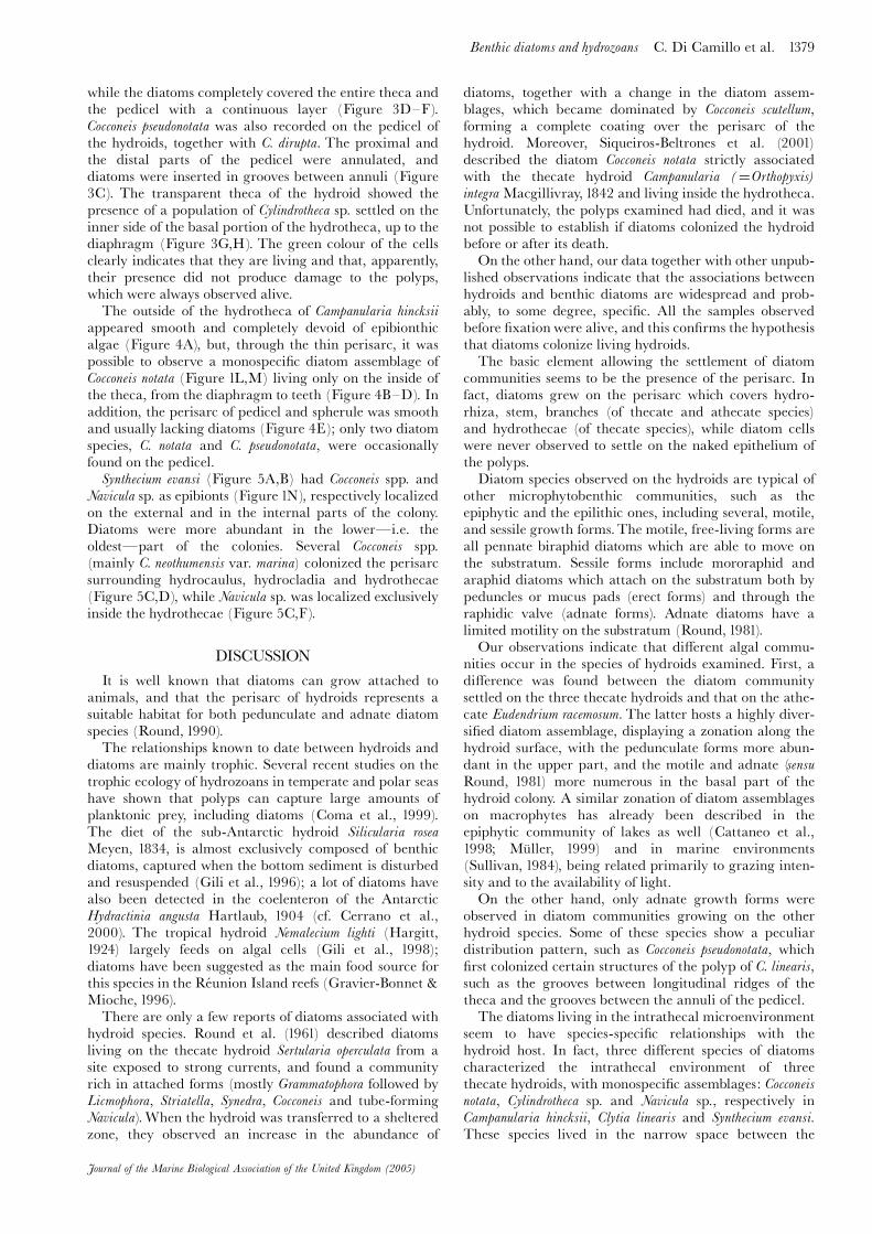
Figure 5. Synthecium evansi. (A) An entire colony with the hydrotheca and the living polyps inside; (B) enlargement of the previousphotograph; (C) SEM image of a hydrotheca showing Cocconeis and Navicula assemblages, respectively localized in the external andin the internal side of the perisarc; (D) external side of the perisarc showing the coverage of Cocconeis assemblage; (E) enlargementof the external side of the perisarc showing Cocconeis neothumensis var. marina; (F) enlargement of the internal side of the perisarcshowing Navicula sp. Scale bars: A, 1mm; B, C, 250 mm; D, 40 mm; E, 10 mm; F, 20 mm.
while the diatoms completely covered the entire theca andthe pedicel with a continuous layer (Figure 3D^F).Cocconeis pseudonotata was also recorded on the pedicel ofthe hydroids, together with C. dirupta. The proximal andthe distal parts of the pedicel were annulated, anddiatoms were inserted in grooves between annuli (Figure3C). The transparent theca of the hydroid showed thepresence of a population of Cylindrotheca sp. settled on theinner side of the basal portion of the hydrotheca, up to thediaphragm (Figure 3G,H). The green colour of the cellsclearly indicates that they are living and that, apparently,their presence did not produce damage to the polyps,which were always observed alive.
The outside of the hydrotheca of Campanularia hincksii
appeared smooth and completely devoid of epibionthicalgae (Figure 4A), but, through the thin perisarc, it waspossible to observe a monospeci¢c diatom assemblage ofCocconeis notata (Figure 1L,M) living only on the inside ofthe theca, from the diaphragm to teeth (Figure 4B^D). Inaddition, the perisarc of pedicel and spherule was smoothand usually lacking diatoms (Figure 4E); only two diatomspecies, C. notata and C. pseudonotata, were occasionallyfound on the pedicel.
Synthecium evansi (Figure 5A,B) had Cocconeis spp. andNavicula sp. as epibionts (Figure 1N), respectively localizedon the external and in the internal parts of the colony.Diatoms were more abundant in the lower�i.e. theoldest�part of the colonies. Several Cocconeis spp.(mainly C. neothumensis var. marina) colonized the perisarcsurrounding hydrocaulus, hydrocladia and hydrothecae(Figure 5C,D), while Navicula sp. was localized exclusivelyinside the hydrothecae (Figure 5C,F).
DISCUSSION
It is well known that diatoms can grow attached toanimals, and that the perisarc of hydroids represents asuitable habitat for both pedunculate and adnate diatomspecies (Round, 1990).
The relationships known to date between hydroids anddiatoms are mainly trophic. Several recent studies on thetrophic ecology of hydrozoans in temperate and polar seashave shown that polyps can capture large amounts ofplanktonic prey, including diatoms (Coma et al., 1999).The diet of the sub-Antarctic hydroid Silicularia rosea
Meyen, 1834, is almost exclusively composed of benthicdiatoms, captured when the bottom sediment is disturbedand resuspended (Gili et al., 1996); a lot of diatoms havealso been detected in the coelenteron of the AntarcticHydractinia angusta Hartlaub, 1904 (cf. Cerrano et al.,2000). The tropical hydroid Nemalecium lighti (Hargitt,1924) largely feeds on algal cells (Gili et al., 1998);diatoms have been suggested as the main food source forthis species in the Re¤ union Island reefs (Gravier-Bonnet &Mioche, 1996).
There are only a few reports of diatoms associated withhydroid species. Round et al. (1961) described diatomsliving on the thecate hydroid Sertularia operculata from asite exposed to strong currents, and found a communityrich in attached forms (mostly Grammatophora followed byLicmophora, Striatella, Synedra, Cocconeis and tube-formingNavicula).When the hydroid was transferred to a shelteredzone, they observed an increase in the abundance of
diatoms, together with a change in the diatom assem-blages, which became dominated by Cocconeis scutellum,forming a complete coating over the perisarc of thehydroid. Moreover, Siqueiros-Beltrones et al. (2001)described the diatom Cocconeis notata strictly associatedwith the thecate hydroid Campanularia (¼Orthopyxis)
integra Macgillivray, 1842 and living inside the hydrotheca.Unfortunately, the polyps examined had died, and it wasnot possible to establish if diatoms colonized the hydroidbefore or after its death.
On the other hand, our data together with other unpub-lished observations indicate that the associations betweenhydroids and benthic diatoms are widespread and prob-ably, to some degree, speci¢c. All the samples observedbefore ¢xation were alive, and this con¢rms the hypothesisthat diatoms colonize living hydroids.
The basic element allowing the settlement of diatomcommunities seems to be the presence of the perisarc. Infact, diatoms grew on the perisarc which covers hydro-rhiza, stem, branches (of thecate and athecate species)and hydrothecae (of thecate species), while diatom cellswere never observed to settle on the naked epithelium ofthe polyps.
Diatom species observed on the hydroids are typical ofother microphytobenthic communities, such as theepiphytic and the epilithic ones, including several, motile,and sessile growth forms.The motile, free-living forms areall pennate biraphid diatoms which are able to move onthe substratum. Sessile forms include mororaphid andaraphid diatoms which attach on the substratum both bypeduncles or mucus pads (erect forms) and through theraphidic valve (adnate forms). Adnate diatoms have alimited motility on the substratum (Round, 1981).
Our observations indicate that di¡erent algal commu-nities occur in the species of hydroids examined. First, adi¡erence was found between the diatom communitysettled on the three thecate hydroids and that on the athe-cate Eudendrium racemosum. The latter hosts a highly diver-si¢ed diatom assemblage, displaying a zonation along thehydroid surface, with the pedunculate forms more abun-dant in the upper part, and the motile and adnate (sensuRound, 1981) more numerous in the basal part of thehydroid colony. A similar zonation of diatom assemblageson macrophytes has already been described in theepiphytic community of lakes as well (Cattaneo et al.,1998; Mu« ller, 1999) and in marine environments(Sullivan, 1984), being related primarily to grazing inten-sity and to the availability of light.
On the other hand, only adnate growth forms wereobserved in diatom communities growing on the otherhydroid species. Some of these species show a peculiardistribution pattern, such as Cocconeis pseudonotata, which¢rst colonized certain structures of the polyp of C. linearis,such as the grooves between longitudinal ridges of thetheca and the grooves between the annuli of the pedicel.
The diatoms living in the intrathecal microenvironmentseem to have species-speci¢c relationships with thehydroid host. In fact, three di¡erent species of diatomscharacterized the intrathecal environment of threethecate hydroids, with monospeci¢c assemblages: Cocconeisnotata, Cylindrotheca sp. and Navicula sp., respectively inCampanularia hincksii, Clytia linearis and Synthecium evansi.
These species lived in the narrow space between the
Benthic diatoms and hydrozoans C. Di Camillo et al. 1379
Journal of the Marine Biological Association of the United Kingdom (2005)

hydrothecae and polyps, so they receive protection fromhydroids and probably use the nutrients produced byhydroid metabolism. Sunlight can penetrate throughtransparent thecae and reach the diatom layer, makingphotosynthesis possible.
Several studies of the epiphytic microphytobenthiccommunities indicate that no speci¢city between algaeand basiphyte occurs. Diatoms use macrophytes only forthe purpose of attachment (Sullivan, 1979), while the asso-ciations between benthic diatoms and hosting macro-phytes are the result of the physical environment ratherthan nutritional interactions (Main & McIntyre, 1974).On the other hand, the epizoic environment appears tobe more selective. The selectivity of diatom colonizationon marine animals has already been reported for manysponges, molluscs and crustacea, which are sometimescolonized by speci¢c diatom taxa (Round, 1990; Cerranoet al., 2004).
REFERENCESBavestrello, G., Cerrano, C., Cattaneo Vietti, R. & Sara' , M.,1996. Relation between Eudendrium glomeratum (Cnidaria,Hydromedusae) and its associated vagile fauna. In Advances in
hydrozoan biology (ed. J. Bouillon et al.), pp. 137^143. [ScientiaMarina, vol. 60.]
Blanco, O.M., 1984. Symplectoscyphus marionensis Millard, 1971(Hydroida, Thecata) y sus epizoicos. Revista del Museo de La
Plata, 13, Zoology, 146, 261^267.Boero, F. & Fresi, E., 1986. Zonation and evolution of a rockybottom hydroid community. P.S.Z.N.I. Marine Ecology, 7,123^150.
Bouillon, J., Medel, M.D., Page' s, F., Gili, J.M., Boero, F. &Gravili, C., 2004. Fauna of the Mediterranean Hydrozoa (ed. J.Bouillon et al.), pp. 449. [Scientia Marina, vol. 68, Supplement2.]
Cattaneo, A., Galanti, G., Gentinetta, S. & Romo, S., 1998.Epiphytic algae and macroinvertebrates on submerged and£oating-leaved macrophytes in an Italian lake. Freshwater
Biology, 39, 725^740.Cerrano, C., Bavestrello, G., Puce, S. & Chiantore, M., 2000.Unusual trophic strategies of Hydractinia angusta (Cnidaria,Hydrozoa) fromTerra Nova Bay, Antarctica. Polar Biology, 23,488^494.
Cerrano, C., Calcinai, B., Cucchiari, E., Di Camillo, C.,Totti, C.& Bavestrello, G., 2004. The diversity of relationships betweenAntarctic sponges and diatoms: the case of Mycale acerata
(Porifera, Demospongiae). Polar Biology, 27, 231^237.Coma, R., Ribes, M., Orejas, C. & Gili, J.M., 1999. Prey captureby a benthic coral reef hydrozoan. Coral Reefs, 18, 141^145.
Gili, J.M. & Hughes, R.G., 1995. The ecology of marine benthichydroids. Oceanography and Marine Biology. Annual Review, 33,351^426.
Gili, J.M. et al., 1998. The impact of small benthic passivesuspension feeders in shallow marine ecosystems: the hydroidsas an example. ZoologischeVerhandelingen, Leiden, 323, 99^105.
Gili, J.M., Alva' , V., Page' s, F., Klo« ser, H. & Arntz, W.E., 1996.Benthic diatoms as the major food source in the sub-Antarcticmarine hydroid Silicularia rosea. Polar Biology, 16, 507^512.
Gravier-Bonnet, N. & Mioche, D., 1996. Annual survey ofhydroids (Cnidaria, Hydrozoa) cohabiting in shrimp-creviceson a reef £at of La Re¤ union (Indian Ocean). In Advances in
hydrozoan biology (ed. J. Bouillon et al.), pp. 165^181. [ScientiaMarina, vol. 60.]
Hancock, D.A., Drinnan, R.E. & Harris, W.N., 1956. Notes onthe biology of Sertularia argentea L. Journal of the Marine
Biological Association of the United Kingdom, 35, 307^325.Hasle, G.R. & Syvertsen, E.E., 1997. Marine Diatoms. InIdentifying marine phytoplankton (ed. C.R. Tomas), pp. 5^385.San Diego: Academic Press.
Lagarde' re, F. & Tardy, J., 1980. Un facie' s d’e¤ pifaune nouveau: lafacie' s a' Ectopleura dumortieri (van Beneden) et Electra pilosa
(Linne¤ ). Faune associe¤ e, cartographie et e¤ volution saisonnie' re.Cahier de Biologie Marine, 21, 265^278.
Main, S.P. & McIntyre, C.D., 1974. The distribution of epiphyticdiatoms inYaquina estuary. Oregon (U.S.A.). Botanica Marina,17, 88^99.
Marfenin, N.N., 1980. Metod kartirovaniya prostranstvennoiorganizatsii kolonial’nykh Hydrozoa i ego znachenie priizuchenii chastei kolonii. A method for mapping of colonialHydrozoa spatial patterns and its use in studies of the parts ofthe colony. InTeoreticheskoe i prakticheskoe znachenie kishechnopolost-nykh. The theoretical and practical importance of the coelenterates (ed.D.V. Naumov and S.D. Stepanjants), pp. 66^69. Leningrad:Trudy Zoologicheskaya Instituta Akademia Nauk.
Millard, N.A.H., 1973. Auto-epizoism in South African hydroids.In Proceedings of the second international symposium on Cnidaria.
Recent trends in research in coelenterate biology. Publications of the SetoMarine Biological Laboratory, 20, 23^34.
Mu« ller, U., 1999. The vertical zonation of adpressed diatoms andother epiphytic algae on Phragmites australis. EuropeanJournal ofPhycology, 34, 487^496.
Round, F.E., 1981. The ecology of algae. Cambridge: CambridgeUniversity Press.
Round, F.E., Crawford, R.M. & Mann, D.G., 1990.The diatoms
biology and morphology of the genera. Cambridge: CambridgeUniversity Press.
Round, F.E., Sloane, J.F., Ebling, F.J. & Kitching, J.A., 1961.Theecology of Lough Ine. X. The hydroid Sertularia operculata (L.)and its associated £ora and fauna: e¡ects of transference tosheltered waters. Journal of Ecology, 49, 617^629.
Siqueiros-Beltrones, D.A., Serviere-Zaragoza, E. & Argumedo-Hernandez, U., 2001. First record of the diatom Cocconeis
notata Petit living inside the hydrotheca of a hydrozoanepiphyte of Macrocystis pyrifera (L.) C. Ag. Oceanides, 16,135^138.
Staples, D.A. & Watson, J.E., 1987. Associations between pycno-gonids and hydroids. In Modern trends in the systematics, ecology
and evolution of hydroids and hydromedusae (ed. J. Bouillon et al.),pp. 215^226. Oxford: Clarendon Place.
Sullivan, M.J., 1979. Epiphytic diatoms of three seagrass speciesin Mississippi Sound. Bulletin of Marine Science, 29, 459^464.
Sullivan, M.J., 1984. Community structure of epiphytic diatomsfrom the Gulf coast of Florida, U.S.A. In Proceedings of the
seventh Diatom Symposium, Philadelphia, 1982 (ed. D.G. Mann),pp. 373^384. Ko« nigstein: Koeltz Scienti¢c Books.
Watson, J.E., 2002. Hydroids (Cnidaria, Hydrozoa) fromSouthern Queensland. Memoirs of the Museum of Victoria, 59,337^354.
Zamponi, M.O. & Genzano, G.N., 1992. La fauna asociada aTubularia crocea (Agassiz, 1862) (Anthomedusae; Tubulariidae)y la aplicacion de un metodo de carti¢cacion. Hidrobiolo¤ gica,3/4, 35^42. [UniversidadNacional deMar del Plata, pp.1^90.]
Submitted 24 April 2005. Accepted 19 July 2005.
1380 C. Di Camillo et al. Benthic diatoms and hydrozoans
Journal of the Marine Biological Association of the United Kingdom (2005)