Evolutionary and Functional Properties of a Two-Locus -Globin Polymorphism in Indian House Mice

Origin and Ascendancy of a Chimeric Fusion Gene: The b/d-Globin Gene ofPaenungulate Mammals
Juan C. Opazo,*� Angela M. Sloan,� Kevin L. Campbell,�1 and Jay F. Storz*1
*School of Biological Sciences, University of Nebraska; �Instituto de Ecologıa y Evolucion, Facultad de Ciencias, UniversidadAustral de Chile, Valdivia, Chile; and �Department of Biological Sciences, University of Manitoba, Winnipeg, MB, Canada
The d-globin gene (HBD) of eutherian mammals exhibits a propensity for recombinational exchange with the closelylinked b-globin gene (HBB) and has been independently converted by the HBB gene in multiple lineages. Here we reportthe presence of a chimeric b/d fusion gene in the African elephant (Loxodonta africana) that was created by unequalcrossing-over between misaligned HBD and HBB paralogs. The recombinant chromosome that harbors the b/d fusiongene in elephants is structurally similar to the ‘‘anti-Lepore’’ duplication mutant of humans (the reciprocal exchangeproduct of the hemoglobin Lepore deletion mutant). However, the situation in the African elephant is unique in that thechimeric b/d fusion gene supplanted the parental HBB gene and is therefore solely responsible for synthesizing theb-chain subunits of adult hemoglobin. A phylogenetic survey of b-like globin genes in afrotherian and xenarthranmammals revealed that the origin of the chimeric b/d fusion gene and the concomitant inactivation of the HBB genepredated the radiation of ‘‘Paenungulata,’’ a clade of afrotherian mammals that includes three orders: Proboscidea(elephants), Sirenia (dugongs and manatees), and Hyracoidea (hyraxes). The reduced fitness of the human Hb Leporedeletion mutant helps to explain why independently derived b/d fusion genes (which occur on an anti-Leporechromosome) have been fixed in a number of mammalian lineages, whereas the reciprocal d/b fusion gene (which occurson a Lepore chromosome) has yet to be documented in any nonhuman mammal. This illustrates how the evolutionaryfates of chimeric fusion genes can be strongly influenced by their recombinational mode of origin.
Introduction
The complete duplication of protein-coding genes typ-ically results in a redundancy of function between the twoinitially identical daughter copies. This functional redun-dancy may often entail a relaxation of purifying selectionthat permits the accumulation of degenerative mutations inone or both gene copies (Ohno 1970; Lynch et al. 2001;Zhang 2003; Lynch and Katju 2004; Taylor and Raes2004). In the majority of cases, one of the two gene copieswill eventually be rendered functionless by inactivatingmutations. However, in a small minority of cases, the fix-ation of previously forbidden mutations may lead to the ac-quisition of novel function. The chances of evolving a novelfunction may be improved in cases where the duplicationproduces a chimeric fusion gene that combines distinctfunctional modules of two separate parent genes (Katjuand Lynch 2003, 2006). The products of chimeric duplica-tion events are structurally distinct at their inception, sothey may be especially likely to follow distinct evolutionarytrajectories. For this reason, it is important to characterizethe structural differences between chimeric fusion genesand their progenitor copies, and to identify the geneticand evolutionary mechanisms that have contributed to theirretention in the genome (Long and Langley 1993; Begun1997; Chen et al. 1997; Nurminsky et al. 1998; Thomsonet al. 2000; Jones and Begun 2005; Jones et al. 2005; Shihand Jones 2008).
The d-globin gene (HBD) of eutherian mammalsexhibits a propensity for recombinational exchange withthe closely linked b-globin gene (HBB) and has been inde-pendently converted by the HBB gene in multiple lineages
(Martin et al. 1983; Goodman et al. 1984; Hardies et al. 1984;Hardison 1984; Hardison and Margot 1984; Hutchison et al.1984; Koop et al. 1989; Tagle et al. 1991; Prychitko et al.2005; Hoffmann et al. 2008a; Opazo et al. 2008a). The5#-HBD–HBB-3#gene pair originated via duplication ofa proto b-globin gene in the common ancestor of eutherianmammals after the divergence from marsupials (Goodmanet al. 1984; Hardison 1984; Opazo et al. 2008a, 2008b).The HBD–HBB gene pair delimits the 3# end of the b-globingene cluster, and expression of both paralogs is restricted tofetal and adult erythroid cells. The remaining b-like globingenes at the 5# end of the gene cluster are typically expressedin embryonic erythroid cells derived from the yolk sac(Hardison 2001). During the course of eutherian evolution,the ontogenetically later-expressed HBD and HBB geneshave experienced very different fates. In the majority of spe-cies thathavebeenstudied todate, theb-chainsubunitsof fetaland adult hemoglobin (Hb) are exclusively encoded by one ormorecopiesof theHBBgene.TheHBDgenehasbeendeletedor inactivated in most eutherian lineages (e.g., rodents, lago-morphs, and many primates; Opazo et al. 2008a). In the fewspecies thathave retaineda transcriptionally activecopyof theHBDgene, it is typically expressed at amuch lower level thanHBB. For example, HBD is not transcribed in Old Worldmonkeys (Boyer et al. 1971; Barnicot and Hewett-Emmett1972; Martin et al. 1980; Martin et al. 1983; Vincent andWilson 1989), but in other primates, the fraction of adultHb tetramers that incorporate b-chain products of the HBDgene spans a wide range: 1% in hominoids (Boyer et al.1971), 6% in New World monkeys (Spritz and Giebel1988), 18% in the Philippine tarsier (Tarsius syrichta; Koopet al. 1989), and 40% in the galago (Otolemur crassicaudatus;Tagle et al. 1991).
In species that synthesize a detectable fraction ofHBD-derived b-chain Hb isoforms, the transcriptional ac-tivity of HBD always appears to be attributable to the ac-quisition of HBB-like promoter sequence via geneconversion or unequal crossing-over. As stated by Hardieset al. (1984: 3755), the HBD genes ‘‘. . .can be described as
1 These authors contributed equally to this work.
Key words: chimeric fusion gene, gene duplication, gene familyevolution, globin genes, hemoglobin, nonallelic homologous recombination.
E-mail: [email protected].
Mol. Biol. Evol. 26(7):1469–1478. 2009doi:10.1093/molbev/msp064Advance Access publication March 30, 2009
� The Author 2009. Published by Oxford University Press on behalf ofthe Society for Molecular Biology and Evolution. All rights reserved.For permissions, please e-mail: [email protected]

vestigial or partially inactivated genes whose structural in-tegrity may be maintained only by gene conversion.’’ Forexample, the galago synthesizes two adult b-chain Hb iso-forms, b1 and b2, which are distinguished by a single aminoacid substitution (Watanabe et al. 1985; Tagle et al. 1988).The b2 chain is the product of an HBD gene that has beenalmost completely overwritten by gene conversion from thedownstream HBB gene (Tagle et al. 1991). The HBD andHBB genes of the galago share a homology block that ex-tends from 800 bp upstream of the proximal CCAAT ele-ment to the 3# end of the third (and last) exon. After thisinitial recombination event, recurrent gene conversion be-tween the two paralogs continued to homogenize sequencevariation across the entire gene region with the exception ofintron 2 and the more distal 5# flanking region. This evo-lutionary pattern appears to be fairly general, as the 5# endof HBD (including exons 1 and 2) has undergone numerousindependent recombinational exchanges with the down-stream HBB paralog in multiple lineages of eutherian mam-mals (reviewed by Goodman et al. 1984; Hardison andMargot 1984; Opazo et al. 2008a).
Here we report the presence of a functional chimeric b/d fusion gene in the African elephant (Loxodonta africana)that originated via unequal crossing-over between mis-aligned HBD and HBB paralogs. The recombinant chromo-some that harbors the b/d fusion gene in elephants isstructurally similar to the ‘‘anti-Lepore’’ duplication mutantof humans (the reciprocal exchange product of the Hb Leporedeletion mutant; Forget 2001) in that the 5# portion is HBB-like and the 3# portion is HBD-like. To gain insight intothe evolutionary origin of this chimeric fusion gene, weinvestigated the genomic structure of the b-globin genecluster in representatives of two basal lineages of eutherianmammals: Afrotheria (which includes elephants, hyraxes,dugongs, manatees, aardvarks, tenrecs, and elephantshrews) and Xenarthra (which includes sloths, armadillos,and anteaters). Results of our comparative survey of b-like globin genes in basal eutherians revealed that the originof the chimeric b/d fusion gene and the concomitantinactivation of the HBB gene predated the radiation of‘‘Paenungulata’’ (Simpson 1945), a clade that containsa morphologically disparate group of afrotherian mammalsfrom three orders: Proboscidea (elephants), Sirenia, (seacows, including dugongs and manatees), and Hyracoidea(the rodent-like hyraxes).
Materials and MethodsGenomic Sequence Data and Annotation
We obtained genomic DNA sequence spanning the b-globin gene cluster of the African elephant (L. africana)from the High Throughput Genomic Sequences database(accession numbers: AC169166, AC167954) and fromthe super_3994 contig of the Broad Institute 2� elephantassembly loxAfr1 (May 2005; http://genome.ucsc.edu/).Recent phylogenomic studies have demonstrated thata superordinal clade (Atlantogenata) composed of Afrotheriaand Xenarthra is the sister group of all remaining membersof the eutherian crown group (Boreoeutheria) (Hallstromet al. 2007; Wildman et al. 2007). Thus, for the purpose
of making comparisons with the elephant b-globin genecluster, we obtained genomic contigs that spanned the en-tire b-globin gene cluster of seven other mammalian spe-cies, including one additional afrotherian (lesserhedgehog tenrec, Echinops telfairi [AC187664]), two xe-narthrans (nine-banded armadillo, Dasypus novemcinctus[AC151518] and Hoffmann’s two-toed sloth, Choloepusdidactylus [AC231564]), two representative boreoeuther-ian species (human, Homo sapiens [AC104389] and Euro-pean rabbit, Oryctalagus cuniculus [AC166202]), and twomarsupial species (the gray short-tailed opossum,Monodel-phis domestica [352230764–352330641] and the NorthAmerican opossum, Didelphis virginiana [AC140955]).The genomic sequences analyzed in this study were inphase 2, meaning that the order and orientation of the con-stituent sequence contigs were firmly established. For eachspecies, we identified b-like globin genes in the unanno-tated genomic sequence by using the program Genscan(Burge and Karlin 1997) and by comparing known exonsequences with genomic contigs using the program Blast2,version 2.2 (Tatusova and Madden 1999).
Inferring Orthologous Relationships
The genomic structure of the b-globin gene clusters ofatlantogenatan mammals was investigated by conductingpairwise comparisons of sequence similarity with the hu-man gene cluster. The comparisons of sequence similaritywere conducted with the program Pipmaker (Schwartzet al. 2000). Orthologous relationships among b-likeglobin genes were inferred from phylogeny reconstructionsof flanking sequence and intron 2 sequence (Storz et al.2007; Hoffmann et al. 2008a, 2008b; Opazo et al.2008a, 2008b). We conducted phylogeny reconstructionson three separate sequence alignments: 0.8 kb of flankingsequence immediately upstream of the initiation codon,5,028 bp of intron 2, and 1 kb of flanking sequence imme-diately downstream of the termination codon. Becauseprevious studies have documented that ectopic recombina-tional exchanges are typically restricted to the 5# end ofthe HBD gene (including exons 1 and 2) and the up-stream flanking region (Hardies et al. 1984; Hardison1984; Hardison and Margot 1984; Prychitko et al. 2005;Hoffmann et al. 2008a; Opazo et al. 2008a), sequence var-iation in intron 2 and the downstream flanking regionshould be especially useful for the purpose of assigning or-thologous relationships. For the phylogenetic analysis, weincluded b-like globin gene sequences from six eutherianspecies (African elephant, tenrec, armadillo, sloth, human,and rabbit) and two marsupial species (the North Americanopossum and the gray short-tailed opossum). Sequencealignments were carried out using the program MUSCLE(Edgar 2004), as implemented in the European Bioinfor-matics Institute web server (http://www.ebi.ac.uk). Phylo-genetic relationships were inferred in a maximumlikelihood framework using Treefinder, version October2008 (Jobb et al. 2004). For each data partition, we usedthe ‘‘propose model’’ tool of Treefinder to select thebest-fitting model of nucleotide substitution. Model selec-tion was based on the Akaike information criterion with
1470 Opazo et al.

correction for small sample size. For the upstream flankingsequence, intron 2 sequence, and downstream flanking se-quence, we used the GTR þ C5 (Rodriguez et al. 1990),HKY þ C5 (Hasegawa et al. 1985), and J3 þ C5 models,respectively. The J3 þ C5 model accounts for variation intransversion rates in which TA 5 CG and TG 5 CA, andrate variation among sites follows a discrete gamma distri-bution with five categories (Jobb et al. 2004). We evaluatedsupport for the nodes of each tree with 1,000 bootstrappseudoreplicates.
Phylogenetic Survey of b-Like Globin Genes inAtlantogenata
In addition to the b-like globin gene sequences that weretrieved from the full genome assemblies, we also clonedand sequenced the adult b-like globin genes from nine ad-ditional afrotherian species (Asian elephant, Elephas maxi-mus; West Indian manatee, Trichechus manatus; Dugong,Dugong dugon; western tree hyrax, Dendrohyrax dorsalis;yellow-spotted hyrax, Heterohyrax brucei; rock hyrax, Pro-cavia capensis; cape golden mole, Chyrosochloris asiatica;bushveld elephant shrew, Elephantulus intufi; and aardvark,Orycteropus afer) and one additional xenarthran species(pale-throated three-toed sloth, Bradypus tridactylus). Wealso retrieved sequences of the rock hyrax b-like globingenes from Whole Genome Sequence files on GenBank(HBD: ABRQ01367090, ABRQ01559481; HBB/D:ABRQ01298789, ABRQ01298790; exon 3 of HBBps:ABRQ01298787).
For each of the atlantogenatan species, genomic DNAwas extracted from whole blood or liver tissue usingDNeasy kits (Qiagen, Valencia, CA). We designed poly-merase chain reaction (PCR) primers for the HBD andHBB genes using an alignment of orthologous sequencesfrom the available atlantogenatan species as well as moredistantly related eutherian mammals. PCRs were conductedon 100 ng of template DNA (2 ll) in 0.2-ml tubes contain-ing 48 ll of reaction mixture (1.0 ll of each dNTP (2.5mM), 5.0 ll 10� PCR Reaction Buffer, 3.0–4.5 ll of 50mM MgCl2, 5.0 ll of each primer (10.0 pmol/ll), 0.5 llTaq polymerase (5 U/ll), and 24.0–25.5 ll ddH2O), usingPTC-0220 DNA Engine Dyad (GMI, Ramsey, MN) andMastercycler Gradient (Eppendorf, Hamburg, Germany)thermocyclers. The b-like globin genes were amplified us-ing a 30-cycle PCR protocol (94 �C for 30 s; 50 �C for 15 s;and 72 �C for 30–75 s) followed by a 10-min final extensionat 72 �C. PCR products were isolated using Montage PCRCentrifugal Filter Devices (Millipore, Billerica, MA). Afterobtaining DNA sequences of the resulting amplicons, weused a genome-walking protocol to obtain sequences ofthe upstream and downstream flanking regions (APAgeneGenome Walking Kit; Bio S&T Inc., Montreal, Canada).All PCR primer combinations used in this study are listedin supplementary table S1, Supplementary Material online,and sequences for all primers are provided in supplemen-tary table S2 (Supplementary Material online).
Target bands were excised and purified using a Min-Elute Gel Extraction Kit (Qiagen), and the extracts wereligated using a Qiagen PCR Cloningplus Kit. Transforma-tion products were incubated on agar plates containing
0.1% v/v ampicillin (100 mg/ml), X-GAL (40 mg/ml),and Isopropyl b-D-1-thiogalactopyranoside (100 mM) for18 h at 37 �C. Plasmid DNA was extracted from positiveclones using a QIAprep Spin Miniprep Kit. Plasmids con-taining globin gene inserts were sequenced in both direc-tions using M-13(F)-40 and M-13(R) sequencingprimers, and samples were run on a 3739 ABI PRISM Ge-netic Analyzer.
Multiple sequence alignments and maximum likeli-hood phylogeny reconstructions were carried out usingmethods described above. Phylogeny reconstructions of in-tron 2 sequences (5,395 bp) were conducted using a J1þC5
model of nucleotide substitution. This model accounts forvariation in transversion rates in which TA5 TG and CA5CG, and rate variation among sites follows a discrete gammadistribution with five categories (Jobb, von Haeseler, andStrimmer 2007).
ResultsGenomic Structure of the b-Globin Gene Cluster inAtlantogenata
In the African elephant, the adult portion of the b-globingene cluster contains three genes (from 5# to 3#): 1 d-likepseudogene (HBDps), 1 chimeric b/d fusion gene (HBB/D), and 1 b-globin pseudogene (HBBps; fig. 1). Followingthe nomenclature of Aguileta et al. (2006), the ‘‘ps’’suffixindicates that HBDps and HBBps are pseudogenes. A dotplot of pairwise sequence similarity between the b-globingene clusters of African elephant and human clearly demon-strates that the 5# flanking sequence and 5# end of the ele-phant HBB/D fusion gene is HBB-like, whereas the 3#flanking sequence and 3# end of the gene are HBD-like(fig. 2). This pattern of pairwise sequence matches indicatesthat the fusion gene was created by unequal crossing-overbetween a misaligned HBD–HBB gene pair (fig. 3).
In African elephants, the HBB/D fusion gene appearsto be the only b-like globin gene expressed during adult-hood, as a conceptual translation of the coding sequenceprecisely matches the reported b-chain amino acid se-quence of purified Hb from this species (Braunitzer et al.1984; Kleinschmidt et al. 1986). The HBDps and HBB/D sequences are distinguished from one another by 42amino acid substitutions and a number of insertion/deletiondifferences (including a ;3.2 kb insertion in intron 2 ofHBDps). HBDps and HBBps are clearly not functional,as the initiation codon of HBDps was eliminated by a singlenucleotide substitution (ATG/ATC) and the entire 5# por-tion of HBBps has been deleted (including exon 1 androughly half of exon 2).
Because the tenrec has four putatively functional HBBgenes and no HBD gene (fig. 1), the HBB/D fusion gene ofelephant does not appear to be shared by all members of theafrotherian clade. In the case of the xenarthrans, both arma-dillo (Dasypus) and sloth (Choloepus) possess single copiesof the HBD and HBB genes. As a result of inactivations ofthe embryonic c-globin (HBG) and g-globin (HBH) genesand the ontogenetically later-expressed HBD gene, theb-globin gene cluster of xenarthans has reverted back tothe ancestral 5#-HBE-HBB-3# structure that is characteris-tic of marsupials (fig. 1).
Origin of a Chimeric Hemoglobin Gene 1471

Identification of the Crossover Breakpoint in theElephant HBB/D Fusion Gene
To localize the crossover breakpoint in the elephantHBB/D fusion gene, we searched for abrupt spatial transi-tions in levels of pairwise sequence identity between thefusion gene and the human HBD and HBB genes. We rea-soned that the portion of the elephant HBB/D fusion gene
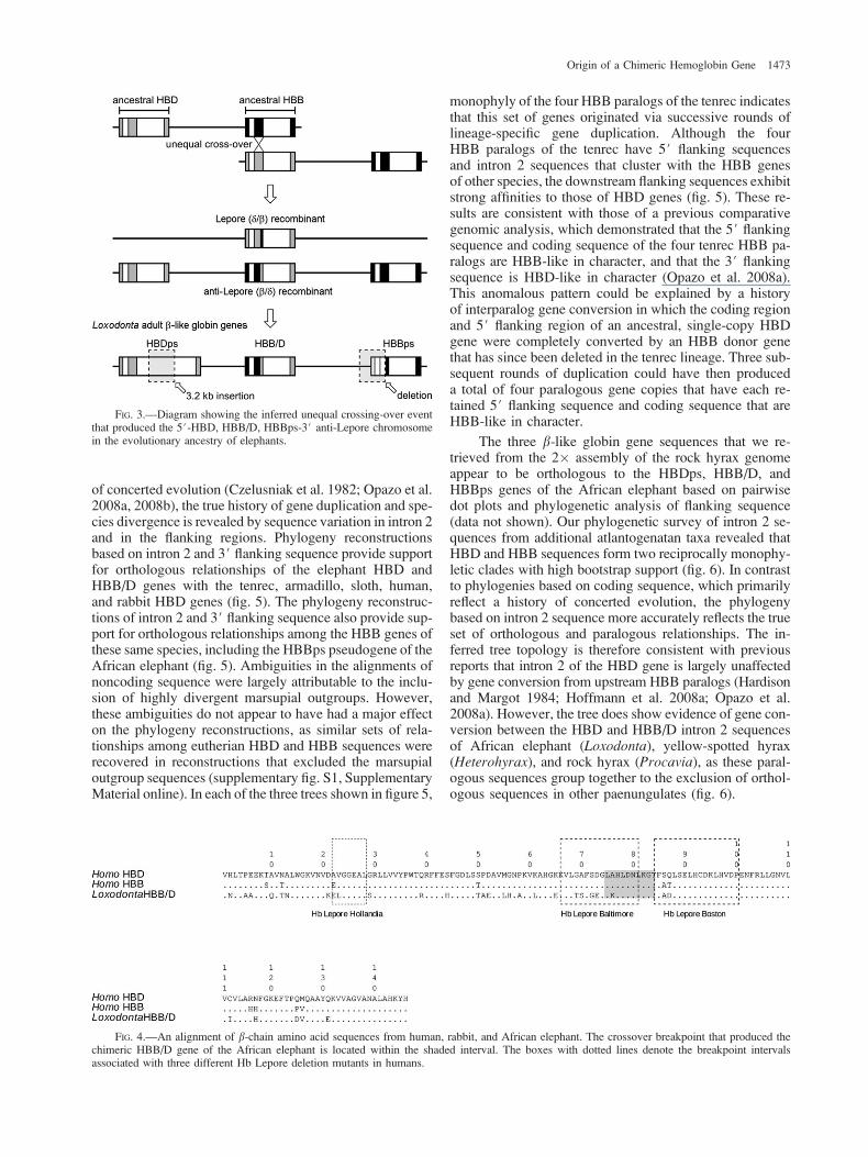
that is located upstream of the breakpoint should be moresimilar to human HBB than to human HBD, whereas theportion downstream of the breakpoint should be more sim-ilar to HBD than to HBB. In comparisons of exon 1 andexon 2 sequences, the elephant HBB/D fusion gene ex-hibited slightly lower levels of sequence identity to the hu-man HBD gene than to the HBB gene (exon 1: 78.3% and79.4%, respectively; exon 2: 81.2% and 84.3%, respec-tively). By contrast, in comparisons of exon 3 sequences,the elephant HBB/D fusion gene exhibited a higher levelof sequence identity to the human HBD gene (89.2%) thanto the human HBB gene (84.5%). Thus, exon 3 of the el-ephant HBB/D fusion gene appears to have retained a dis-tinctive HBD-like character that distinguishes it fromparalogous HBB-like sequence. The similar level of pair-wise sequence identity between the elephant HBB/D fusiongene and the human HBD and HBB genes in the first 2 exonsmay be attributable to the fact that the HBD gene has beenindependently converted by the HBB gene in that samegene region in both species (Martin et al. 1983; Goodmanet al. 1984). By contrast, exon 3 of HBD typically lies out-side of HBB-derived conversion tracts (Martin et al. 1983;Hardison andMargot 1984; Prychitko et al. 2005; Hoffmannet al. 2008a). Based on alignments of nucleotide and aminoacid sequences of the HBD and HBB genes (fig. 4), it ap-pears that the crossover breakpoint of the elephant HBB/Dfusion gene is located between residue positions 78 and 87of the polypeptide (encoded by exon 2). Studies of humanHb Lepore and anti-Lepore mutants indicate that the cross-over breakpoints are typically located between residue po-sitions 69 and 104. The inferred crossover breakpoint in theelephant HBB/D fusion gene closely coincides with thoseof three well-characterized Hb Lepore mutants in humans:Hb Baltimore, Hb Boston, and Hb Hollandia (Mavilio et al.1983; Metzenberg et al. 1991; fig. 4).
Inferring Orthologous Relationships among b-LikeGlobin Genes
Although phylogeny reconstructions based on codingsequence indicate that orthologous relationships amongHBD and HBB genes have been obscured by a history
FIG. 2.—Dot plot of pairwise sequence similarity between the 3#ends of the b-globin gene clusters of African elephant, Loxodontaafricana, and human.
FIG. 1.—Genomic structure of the b-globin gene cluster in afrotherians (African elephant and lesser hedgehog tenrec), xenarthrans (nine-bandedarmadillo and two-toed sloth), two representative boreoeutherian species (human and European rabbit), and two marsupial outgroups (gray short-tailedopossum and North American opossum).
1472 Opazo et al.
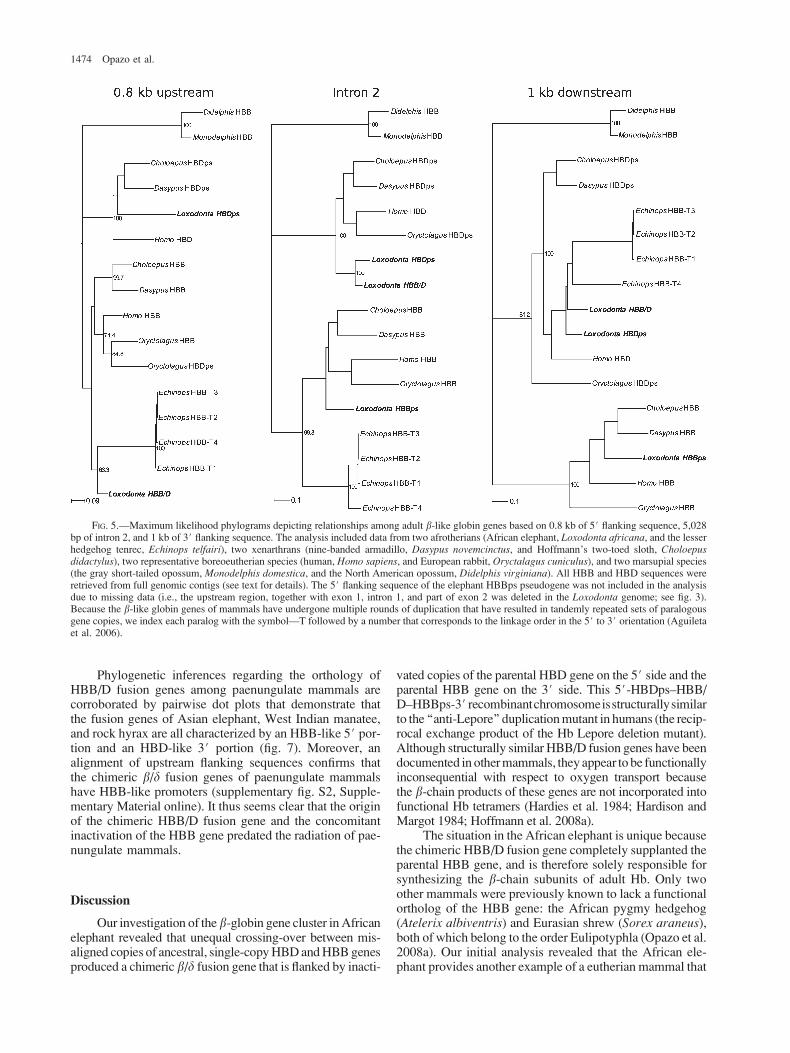
of concerted evolution (Czelusniak et al. 1982; Opazo et al.2008a, 2008b), the true history of gene duplication and spe-cies divergence is revealed by sequence variation in intron 2and in the flanking regions. Phylogeny reconstructionsbased on intron 2 and 3# flanking sequence provide supportfor orthologous relationships of the elephant HBD andHBB/D genes with the tenrec, armadillo, sloth, human,and rabbit HBD genes (fig. 5). The phylogeny reconstruc-tions of intron 2 and 3# flanking sequence also provide sup-port for orthologous relationships among the HBB genes ofthese same species, including the HBBps pseudogene of theAfrican elephant (fig. 5). Ambiguities in the alignments ofnoncoding sequence were largely attributable to the inclu-sion of highly divergent marsupial outgroups. However,these ambiguities do not appear to have had a major effecton the phylogeny reconstructions, as similar sets of rela-tionships among eutherian HBD and HBB sequences wererecovered in reconstructions that excluded the marsupialoutgroup sequences (supplementary fig. S1, SupplementaryMaterial online). In each of the three trees shown in figure 5,
monophyly of the four HBB paralogs of the tenrec indicatesthat this set of genes originated via successive rounds oflineage-specific gene duplication. Although the fourHBB paralogs of the tenrec have 5# flanking sequencesand intron 2 sequences that cluster with the HBB genesof other species, the downstream flanking sequences exhibitstrong affinities to those of HBD genes (fig. 5). These re-sults are consistent with those of a previous comparativegenomic analysis, which demonstrated that the 5# flankingsequence and coding sequence of the four tenrec HBB pa-ralogs are HBB-like in character, and that the 3# flankingsequence is HBD-like in character (Opazo et al. 2008a).This anomalous pattern could be explained by a historyof interparalog gene conversion in which the coding regionand 5# flanking region of an ancestral, single-copy HBDgene were completely converted by an HBB donor genethat has since been deleted in the tenrec lineage. Three sub-sequent rounds of duplication could have then produceda total of four paralogous gene copies that have each re-tained 5# flanking sequence and coding sequence that areHBB-like in character.
The three b-like globin gene sequences that we re-trieved from the 2� assembly of the rock hyrax genomeappear to be orthologous to the HBDps, HBB/D, andHBBps genes of the African elephant based on pairwisedot plots and phylogenetic analysis of flanking sequence(data not shown). Our phylogenetic survey of intron 2 se-quences from additional atlantogenatan taxa revealed thatHBD and HBB sequences form two reciprocally monophy-letic clades with high bootstrap support (fig. 6). In contrastto phylogenies based on coding sequence, which primarilyreflect a history of concerted evolution, the phylogenybased on intron 2 sequence more accurately reflects the trueset of orthologous and paralogous relationships. The in-ferred tree topology is therefore consistent with previousreports that intron 2 of the HBD gene is largely unaffectedby gene conversion from upstream HBB paralogs (Hardisonand Margot 1984; Hoffmann et al. 2008a; Opazo et al.2008a). However, the tree does show evidence of gene con-version between the HBD and HBB/D intron 2 sequencesof African elephant (Loxodonta), yellow-spotted hyrax(Heterohyrax), and rock hyrax (Procavia), as these paral-ogous sequences group together to the exclusion of orthol-ogous sequences in other paenungulates (fig. 6).
FIG. 3.—Diagram showing the inferred unequal crossing-over eventthat produced the 5#-HBD, HBB/D, HBBps-3# anti-Lepore chromosomein the evolutionary ancestry of elephants.
FIG. 4.—An alignment of b-chain amino acid sequences from human, rabbit, and African elephant. The crossover breakpoint that produced thechimeric HBB/D gene of the African elephant is located within the shaded interval. The boxes with dotted lines denote the breakpoint intervalsassociated with three different Hb Lepore deletion mutants in humans.
Origin of a Chimeric Hemoglobin Gene 1473
Phylogenetic inferences regarding the orthology ofHBB/D fusion genes among paenungulate mammals arecorroborated by pairwise dot plots that demonstrate thatthe fusion genes of Asian elephant, West Indian manatee,and rock hyrax are all characterized by an HBB-like 5# por-tion and an HBD-like 3# portion (fig. 7). Moreover, analignment of upstream flanking sequences confirms thatthe chimeric b/d fusion genes of paenungulate mammalshave HBB-like promoters (supplementary fig. S2, Supple-mentary Material online). It thus seems clear that the originof the chimeric HBB/D fusion gene and the concomitantinactivation of the HBB gene predated the radiation of pae-nungulate mammals.
Discussion
Our investigation of the b-globin gene cluster inAfricanelephant revealed that unequal crossing-over between mis-aligned copies of ancestral, single-copyHBDandHBBgenesproduced a chimeric b/d fusion gene that is flanked by inacti-
vated copies of the parental HBD gene on the 5# side and theparental HBB gene on the 3# side. This 5#-HBDps–HBB/D–HBBps-3# recombinantchromosomeisstructurallysimilarto the ‘‘anti-Lepore’’ duplicationmutant in humans (the recip-rocal exchange product of the Hb Lepore deletion mutant).Although structurally similar HBB/D fusion genes have beendocumented in othermammals, they appear to be functionallyinconsequential with respect to oxygen transport becausethe b-chain products of these genes are not incorporated intofunctional Hb tetramers (Hardies et al. 1984; Hardison andMargot 1984; Hoffmann et al. 2008a).
The situation in the African elephant is unique becausethe chimeric HBB/D fusion gene completely supplanted theparental HBB gene, and is therefore solely responsible forsynthesizing the b-chain subunits of adult Hb. Only twoother mammals were previously known to lack a functionalortholog of the HBB gene: the African pygmy hedgehog(Atelerix albiventris) and Eurasian shrew (Sorex araneus),both of which belong to the order Eulipotyphla (Opazo et al.2008a). Our initial analysis revealed that the African ele-phant provides another example of a eutherian mammal that
FIG. 5.—Maximum likelihood phylograms depicting relationships among adult b-like globin genes based on 0.8 kb of 5# flanking sequence, 5,028bp of intron 2, and 1 kb of 3# flanking sequence. The analysis included data from two afrotherians (African elephant, Loxodonta africana, and the lesserhedgehog tenrec, Echinops telfairi), two xenarthrans (nine-banded armadillo, Dasypus novemcinctus, and Hoffmann’s two-toed sloth, Choloepusdidactylus), two representative boreoeutherian species (human, Homo sapiens, and European rabbit, Oryctalagus cuniculus), and two marsupial species(the gray short-tailed opossum, Monodelphis domestica, and the North American opossum, Didelphis virginiana). All HBB and HBD sequences wereretrieved from full genomic contigs (see text for details). The 5# flanking sequence of the elephant HBBps pseudogene was not included in the analysisdue to missing data (i.e., the upstream region, together with exon 1, intron 1, and part of exon 2 was deleted in the Loxodonta genome; see fig. 3).Because the b-like globin genes of mammals have undergone multiple rounds of duplication that have resulted in tandemly repeated sets of paralogousgene copies, we index each paralog with the symbol—T followed by a number that corresponds to the linkage order in the 5# to 3# orientation (Aguiletaet al. 2006).
1474 Opazo et al.

does not possess an intact, functional HBB gene. Compar-isons of DNA sequence data with previously publishedamino acid sequences further indicate that the HBB/D fu-sion gene supplanted the parental HBB gene not just in theAfrican elephant but in all extant members of the paenun-gulate clade. The HBB/D sequence of Asian elephant is100% identical to the b-globin amino acid sequence re-ported from the purified Hb of this species (Braunitzeret al. 1984; Kleinschmidt et al. 1986). Likewise, theHBB/D sequence of West Indian manatee is 100% identicalto the b-globin amino acid sequence of the closely relatedAmazonian manatee (Trichechus inunguis; Kleinschmidtet al. 1986). The HBB/D sequences that we independentlyobtained from the rock hyrax are 97–100% identical to theb-globin amino acid sequence reported for this species(Kleinschmidt and Braunitzer 1983; Braunitzer et al.1984; Kleinschmidt et al. 1986), whereas the HBD geneis only 79% identical. It thus appears that, in all paenungu-late mammals, the b-chain subunits of the major adult Hbisoform are encoded by orthologous copies of the sameHBB/D fusion gene.
The observation that the b-type Hb chains of elephantsand other paenungulates are hybrid polypeptides character-ized by a b-like N-terminal portion and a d-like C-terminalportion is especially surprising because previous workershad suggested that the HBD gene was little more than a relic
gene that had been evolving under relaxed functional con-straints even before the diversification of eutherian mam-mals. As stated by Hardies et al. (1984: 3755): ‘‘Theoverall poor evolutionary performance of d-like genesamong the mammals suggests that the proto-d was alreadydestined for disposal prior to the mammalian radiation.’’Thus, if the Hbs of paenungulate mammals are character-ized by any unusual functional properties, it would be ofinterest to know whether they are attributable to amino acidsubstitutions in the C-terminal, HBD-encoded region of theb-type Hb chain that occurred during a prior history of re-laxed functional constraint.
One especially noteworthy substitution in the HBD-derived portion of the b-type Hb chain of African elephant,Asian elephant, and dugong is b131Gln/Glu, which islocated at a highly conserved a1b1 intersubunit contact sur-face. The b131Glu mutation has also been described inhumans (Hb Camden; Wade-Cohen et al. 1973; Hubbardet al. 1975). Protein engineering experiments have demon-strated that theb131Gln/Glusubstitutiondisruptshydrogenbonds between a103His and b131Gln at the a1b1-subunitinterface, resulting ina slightly reducedoxygen-bindingaffin-ity (Chang et al. 2002). The substitution also diminishes thereactivity of the sulfhydryl group ofb93Cys, a residue locatedin close proximity to the a1b2-subunit interface, and increasestheprotonexchangeratesofa1b1-interfacehistidylresiduesonthe a-chain (a103His and a122His; Chang et al. 2002).
The Role of Unequal Crossing-Over in CreatingChimeric Fusion Genes
When the breakpoint of an unequal crossover eventinterrupts the coding regions of a misaligned gene pair, bothreciprocal exchange products will contain a chimeric fusiongene (fig. 3). For example, in humans, crossovers betweenmisaligned copies of the closely linked HBD and HBBgenes result in a solitary d/b fusion gene on one recombi-nant chromosome (the Hb Lepore deletion mutant) and thereciprocal b/d fusion gene on the other recombinant chro-mosome (the ‘‘anti-Lepore’’ duplication mutant; Forget2001). In the former case, the d/b fusion gene on the Leporechromosome is solely responsible for the synthesis of theb-chain subunits of adult Hb. In the latter case, the b/d fu-sion gene on the anti-Lepore chromosome is flanked byfully functional copies of the parental HBD gene on the5# side and the parental HBB gene on the 3# side. Hetero-zygotes for the Hb Lepore deletion produce adult red bloodcells that contain normal a2b2 Hb tetramers (HbA), as wellas lesser quantities of a2(d/b)2 tetramers (Hb Lepore) thatincorporate products of the d/b fusion gene. The HbA andHb Lepore isoforms are not present in equal concentrationsin circulating red blood cells because the d/b fusion gene isunder the control of a weak HBD promoter, and is thereforetranscribed at a much lower rate than the normal HBB gene.The difference in relative isoform concentrations is alsopartly attributable to reduced stability of the d/b mRNAtranscript (Forget 2001).
Whereas heterozygotes for the Lepore d/b fusion genetypically suffer a relatively mild form of d/b thalassemia(a type of hemolytic anemia caused by reduced synthesis
Fig. 6.—Maximum likelihood phylogram depicting relationshipsamong adult b-like globin genes based on 5,510 bp of intron 2 sequence.The analysis included sequence data from 15 atlantogenatan species inaddition to two boreoeutherian outgroups (Oryctolagus and Homo) andtwo marsupial outgroups (Monodelphis and Didelphis).
Origin of a Chimeric Hemoglobin Gene 1475

of b-globin subunits), homozygotes for the d/b fusion genesuffer more severe forms of erythrocytic dysfunction, withclinical manifestations equivalent to the ‘‘b thalassemia in-termedia’’ or ‘‘b thalassemiamajor’’ disease phenotypes (Oli-vieri and Weatherall 2001). In the b thalassemia syndromes,the imbalance of a- and b-globin synthesis leads to an excessof oxidized a-chain monomers that aggregate and precipitatein bone marrow normoblasts and in circulating red bloodcells. The insoluble a-chain aggregates and their associatedbreakdown products (iron, heme, and hemichrome) contrib-ute to oxidative damage of the cytoskeleton membrane pro-teins, which eventually results in the destruction of theaffected erythroid precursor cells in the bone marrow andpremature hemolysis of mature red blood cells in the periph-eral circulation (Rachmilewitz and Schrier 2001).
In contrast to the complications associated with inher-itance of the Hb Lepore deletion chromosome (and the as-sociated d/b fusion gene), the inheritance of the reciprocalexchange product of the same crossover event—the 5#HBD–HBB/D–HBB 3# anti-Lepore chromosome—is notassociated with any hematological pathology because the re-combinant chromosome retains a transcriptionally active
HBB gene in addition to the HBD and HBB/D genes.The difference in fitness between the Hb Lepore deletionmu-tant and the anti-Lepore duplicationmutant may explain whythe two reciprocal b/d and d/b fusion genes are not equallycommon among contemporary mammals. Independently de-rived b/d fusion genes (which occur on an anti-Lepore chro-mosome) have been fixed in a number of mammalianlineages, whereas transcriptionally active copies of the recip-rocal d/b fusion gene (which occurs on the Lepore chromo-some) have yet to be documented in the b-globin gene clusterof any nonhuman mammal. This illustrates how the evolu-tionary fates of chimeric fusion genes can be strongly influ-enced by their recombinational mode of origin.
Supplementary Material
Supplementary figures S1 and S2, supplementary tablesS1 and S2, and all sequence alignments associated with thismanuscript are available atMolecular Biology and Evolutiononline (http://www.mbe.oxfordjournals.org/). Sequence datafrom this article have been submitted to GenBank under
Fig. 7.—Dot plots of sequence similarity between the chimeric HBB/D (b/d) fusion genes of paenungulate mammals and the HBD and HBB genesof human. Top row: Asian elephant b/d fusion gene versus human HBD and HBB; Middle row: Dugong b/d fusion gene versus human HBD and HBB;Bottom row: Rock hyrax b/d fusion gene versus human HBD and HBB.
1476 Opazo et al.

accession nos. DQ091202–DQ091208, DQ091210,DQ091212, DQ091214, and FJ588200–FJ588203.
Acknowledgments
We thank S.V. Edwards and two anonymous re-viewers for helpful suggestions on the manuscript andA. Signore for technical assistance in the laboratory. Thiswork was funded by grants to J.F.S. from the National In-stitutes of Health, the National Science Foundation, and theNebraska Research Council, and by grants to K.L.C. by theNational Sciences and Engineering Research Council ofCanada, the Winnipeg Foundation, and the University ofManitoba Research Grant Program. A.M.S. received sup-port from a University of Manitoba Graduate Fellowship,and J.C.O. received support from the Oliver P. PearsonAward from the American Society of Mammalogists.
Literature Cited
Aguileta G, Bielawski JP, Yang Z. 2006. Evolutionary ratevariation among vertebrate b-globin genes: implications fordating gene family duplication events. Gene. 380:21–29.
Barnicot NA, Hewett-Emmett D. 1972. Red cell and serumproteins of Cercocebus, Presbytis, Colobus and certain otherspecies. Folia Primatol (Basel). 17:442–457.
Begun DJ. 1997. Origin and evolution of a new gene descendedfrom alcohol dehydrogenase in Drosophila. Genetics.145:375–382.
Boyer SH, Crosby EF, Noyes AN, Fuller GF, Leslie SE,Donaldson LJ, Vrablik GR, Schaefer EW Jr, Thurmon TF.1971. Primate hemoglobins: some sequences and someproposals concerning the character of evolution and mutation.Biochem Genet. 5:405–448.
Braunitzer G, Stangl A, Schrank B, Krombach C, Wiesner H.1984. Phosphate–haemoglobin interaction. The primarystructure of the haemoglobin of the African elephant(Loxodonta africana, Proboscidea): asparagine in position 2of the b-chain. Hoppe Seylers Z Physiol Chem. 365:743–749.
Burge C, Karlin S. 1997. Prediction of complete gene structuresin human genomic DNA. J Mol Biol. 268:78–94.
Chang C-k, Simplaceanu V, Ho C. 2002. Effects of amino acidsubstitutions at B131 on the structure and properties ofhemoglobin: evidence for communication between a1b1- anda1b2-subunit interfaces. Biochemistry. 41:5644–5655.
Chen L, DeVries AL, Cheng CH. 1997. Convergent evolution ofantifreeze glycoproteins in Antarctic notothenioid fish andArctic cod. Proc Natl Acad Sci USA. 94:3811–3816.
Czelusniak J, Goodman M, Hewett-Emmett D, Weiss ML,Venta PJ, Tashian RE. 1982. Phylogenetic origins andadaptive evolution of avian and mammalian haemoglobingenes. Nature. 298:297–300.
Edgar RC. 2004. MUSCLE: multiple sequence alignment withhigh accuracy and high throughput. Nucleic Acids Res.32:1792–1797.
Forget BG. 2001. Molecular mechanisms of beta thalassemia. In:Steinberg M, Forget B, Higgs D, Nagel R, editors. Disoders ofhemoglobin: genetics, pathophysiology, and clinical manage-ment. Cambridge (UK): Cambridge University Press.p. 252–276.
Goodman M, Koop BF, Czelusniak J, Weiss ML. 1984. The g-globin gene. Its long evolutionary history in the b-globin genefamily of mammals. J Mol Biol. 180:803–823.
Hallstrom BM, Kullberg M, Nilsson MA, Janke A. 2007.Phylogenomic data analyses provide evidence that Xenarthraand Afrotheria are sister groups. Mol Biol Evol.24:2059–2068.
Hardies SC, Edgell MH, Hutchison CA 3rd. 1984. Evolution ofthe mammalian b-globin gene cluster. J Biol Chem.259:3748–3756.
Hardison R. 2001. Organization, evolution, and regulation of theglobin genes. In: Steinberg M, Forget B, Higgs D, Nagel R,editors. Disorders of hemoglobin: genetics, pathophysiology,and clinical management. Cambridge (UK): CambridgeUniversity Press. p. 95–115.
Hardison RC. 1984. Comparison of the b-like globin genefamilies of rabbits and humans indicates that the gene cluster5#-e-c-d-b-3# predates the mammalian radiation. Mol BiolEvol. 1:390–410.
Hardison RC, Margot JB. 1984. Rabbit globin pseudogene wb2is a hybrid of d- and b-globin gene sequences. Mol Biol Evol.1:302–316.
Hasegawa M, Kishino H, Yano T. 1985. Dating of the human-ape splitting by a molecular clock of mitochondrial DNA.J Mol Evol. 22:160–174.
Hoffmann FG, Opazo JC, Storz JF. 2008a. New genes originatedvia multiple recombinational pathways in the b-globin genefamily of rodents. Mol Biol Evol. 25:2589–2600.
Hoffmann FG, Opazo JC, Storz JF. 2008b. Rapid rates oflineage-specific gene duplication and deletion in the a-globingene family. Mol Biol Evol. 25:591–602.
Hubbard M, Wilson JB, Wrightstone RN, Efremov GD,Huisman TH. 1975. Hb Camden and Hb Hope found duringroutine testing. Acta Haematol. 54:53–58.
Hutchison CAI, Hardies SC, Padgett RW, Weaver S, Edgell MH.1984. The mouse globin pseudogene bh3 is descendedfrom a premammalian d-globin gene. J Biol Chem. 259:12881–12889.
Jobb G, von Haeseler A, Strimmer K. 2004. TREEFINDER:a powerful graphical analysis environment for molecularphylogenetics. BMC Evol Biol. 4:18.
Jones CD, Begun DJ. 2005. Parallel evolution of chimeric fusiongenes. Proc Natl Acad Sci USA. 102:11373–11378.
Jones CD, Custer AW, Begun DJ. 2005. Origin and evolutionof a chimeric fusion gene in Drosophila subobscura,D. madeirensis, and D. guanche. Genetics. 170:207–219.
Katju V, Lynch M. 2006. On the formation of novel genes byduplication in the Caenorhabditis elegans genome. Mol BiolEvol. 23:1056–1067.
Katju V, Lynch M. 2003. The structure and early evolution ofrecently arisen gene duplicates in the Caenorhabditis elegansgenome. Genetics. 165:1793–1803.
Kleinschmidt T, Braunitzer G. 1983. The primary structure ofhemoglobins of the rock hyrax (Procavia habessinica,Hyracoidea): insertion of glutamine in the a- chains. HoppeSeylers Z Physiol Chem. 364:1303–1313.
Kleinschmidt T, Czelusniak J, Goodman M, Braunitzer G. 1986.Paenungulata: a comparison of the hemoglobin sequencesfrom elephant, hyrax, and manatee. Mol Biol Evol. 3:427–435.
Koop BF, Siemieniak D, Slightom JL, Goodman M, Dunbar J,Wright PC, Simons EL. 1989. Tarsius d- and b-globin genes:conversions, evolution, and systematic implications. J BiolChem. 264:68–79.
Long M, Langley CH. 1993. Natural selection and the origin ofjingwei, a chimeric processed functional gene in Drosophila.Science. 260:91–95.
Lynch M, Katju V. 2004. The altered evolutionary trajectories ofgene duplicates. Trends Genet. 20:544–549.
Origin of a Chimeric Hemoglobin Gene 1477
Lynch M, O’Hely M, Walsh B, Force A. 2001. The probability ofpreservation of a newly arisen gene duplicate. Genetics.159:1789–1804.
Martin SL, Vincent KA, Wilson AC. 1983. Rise and fall of thed-globin gene. J Mol Biol. 164:513–528.
Martin SL, Zimmer EA, Kan YW, Wilson AC. 1980. Silentd-globin gene in Old World monkeys. Proc Natl Acad SciUSA. 77:3563–3566.
Mavilio F, Giampaolo A, Care A, Sposi NM, Marinucci M. 1983.The d/b crossover region in Lepore boston hemoglobinopathyis restricted to a 59 base pairs region around the 5# splicejunction of the large globin gene intervening sequence. Blood.62:230–233.
Metzenberg AB, Wurzer G, Huisman TH, Smithies O. 1991.Homology requirements for unequal crossing over in humans.Genetics. 128:143–161.
Nurminsky DI, Nurminskaya MV, De Aguiar D, Hartl DL. 1998.Selective sweep of a newly evolved sperm-specific gene inDrosophila. Nature. 396:572–575.
Ohno S. 1970. Evolution by gene duplication. New York:Springer-Verlag.
Olivieri NF, Weatherall DJ. 2001. Clinical aspects of bthalassemia. In: Steinberg MH, Higgs DR, Nagel RL, editors.Disorders of hemoglobin: genetics, pathophysiology, andclinical management. Cambridge (UK): Cambridge Univer-sity Press. p. 277–341.
Opazo JC, Hoffmann FG, Storz JF. 2008a. Differential loss ofembryonic globin genes during the radiation of placentalmammals. Proc Natl Acad Sci USA. 105:12950–12955.
Opazo JC, Hoffmann FG, Storz JF. 2008b. Genomic evidence forindependent origins of b-like globin genes in monotremes andtherian mammals. Proc Natl Acad Sci USA. 105:1590–1595.
Prychitko T, Johnson RM, Wildman DE, Gumucio D,Goodman M. 2005. The phylogenetic history of New Worldmonkey b-globin reveals a platyrrhine b to d gene conversionin the atelid ancestry. Mol Phylogenet Evol. 35:225–234.
Rachmilewitz E, Schrier S. 2001. Pathophysiology of bthalassemia. In: Steinberg MH, Forget FB, Higgs DR, NagelRL, editors. Disorders of hemoglobin: genetics, pathophysi-ology, and clinical management. Cambridge (UK): Cam-bridge University Press. p. 233–251.
Rodriguez F, Oliver JL, Marin A, Medina JR. 1990. The generalstochastic model of nucleotide substitution. J Theor Biol.142:485–501.
Schwartz S, Zhang Z, Frazer KA, Smit A, Riemer C, Bouck J,Gibbs R, Hardison R, Miller W. 2000. PipMaker–a webserver for aligning two genomic DNA sequences. GenomeRes. 10:577–586.
Shih HJ, Jones CD. 2008. Patterns of amino acid evolution inthe Drosophila ananassae chimeric gene, siren, parallel
those of other Adh-derived chimeras. Genetics. 180:1261–1263.
Simpson G. 1945. The principles of classification and a classifi-cation of mammals. Bull Am Mus Nat Hist. 85:1–350.
Spritz RA, Giebel LB. 1988. The structure and evolution of thespider monkey d-globin gene. Mol Biol Evol. 5:21–29.
Storz JF, Baze M, Waite JL, Hoffmann FG, Opazo JC, Hayes JP.2007. Complex signatures of selection and gene conversion inthe duplicated globin genes of house mice. Genetics.177:481–500.
Tagle DA, Koop BF, Goodman M, Slightom JL, Hess DL,Jones RT. 1988. Embryonic epsilon and gamma globin genesof a prosimian primate (Galago crassicaudatus). Nucleotideand amino acid sequences, developmental regulation andphylogenetic footprints. J Mol Biol. 203:439–455.
Tagle DA, Slightom JL, Jones RT, Goodman M. 1991.Concerted evolution led to high expression of a prosimianprimate d-globin gene locus. J Biol Chem. 266:7469–7480.
Tatusova TA, Madden TL. 1999. BLAST 2 Sequences, a newtool for comparing protein and nucleotide sequences. FEMSMicrobiol Lett. 174:247–250.
Taylor JS, Raes J. 2004. Duplication and divergence: theevolution of new genes and old ideas. Annu Rev Genet.38:615–643.
Thomson TM, Lozano JJ, Loukili N, et al. (13 co-authors). 2000.Fusion of the human gene for the polyubiquitinationcoeffector UEV1 with Kua, a newly identified gene. GenRes. 10:1743–1756.
Vincent KA, Wilson AC. 1989. Evolution and transcription ofold world monkey globin genes. J Mol Biol. 207:465–480.
Wade-Cohen PT, Yates A, Bellingham AJ, Huehns ER. 1973.Amino-acid Substitution in the a1b1 Intersubunit Contact ofHaemoglobin-Camden b131 (H9) Gln/Glu. Nature. 243:467–468.
Watanabe B, Fujii T, Nakashima Y, Maita T, Matsuda G. 1985.Amino-acid sequences of the alpha and beta chains of adulthemoglobins of the grand galago, Galago crassicaudatus.Biol Chem Hoppe Seyler. 366:265–269.
Wildman DE, Uddin M, Opazo JC, Liu G, Lefort V, Guindon S,Gascuel O, Grossman LI, Romero R, Goodman M. 2007.Genomics, biogeography, and the diversification ofplacental mammals. Proc Natl Acad Sci USA. 104:14395–14400.
Zhang J. 2003. Evolution by gene duplication: an update. TrendsEcol Evol. 18:292–298.
Scott Edwards, Associate Editor
Accepted March 15, 2009
1478 Opazo et al.
Copyright © 2022 FDOKUMEN