Organized Solid Thin Films of Gold Nanorods with Different Sizes for Surface-Enhanced Raman...
6
Organized Solid Thin Films of Gold Nanorods with Different Sizes for Surface-Enhanced Raman Scattering Applications Moritz Tebbe, † Max Maennel, † Andreas Fery, † Nicolas Pazos-Perez,* ,†,‡,§ and Ramon A. Alvarez-Puebla* ,‡,§,∥ † Physical Chemistry II, University of Bayreuth, Universitä tsstraße 30, Bayreuth, Germany ‡ Departamento de Quimica Fisica e Inorganica, Universitat Rovira i Virgili and Centro de Tecnologia Quimica de Cataluñ a, Carrer de Marcel·lí Domingo s/n, 43007 Tarragona, Spain § Medcom Advance SA, Viladecans Business Park - Edificio Brasil, Bertran i Musitu 83-85 08840, Viladecans − Barcelona, Spain ∥ ICREA, Passeig Lluís Companys 23, 08010 Barcelona, Spain ABSTRACT: Gold nanorods are among the most efficient structures for optical applications, especially in surface-enhanced Raman scattering applications. Notably, their use directly in colloidal solutions decreases their optical enhancing properties because of the impossibility of generating hot spots and the passivation of their surfaces by the surfactants used during their preparation, hindering the interaction of the analyte with the plasmonic surface. We present an easy and fast method for preparing organized lying-down nanorod films with particles of different aspect ratios, yielding efficient and clean plasmonic films populated with a large number of homogeneous hot spots. ■ INTRODUCTION Gold nanorods (AuNRs) are among the most efficient structures for nanooptical applications. 1 As anisotropic particles, their plasmonic response consist of two different bands corresponding to the transversal and longitudinal modes. 1 Notably, these elongated structures are also capable of concentrating the electromagnetic field at the nanoparticle tip when the incoming light is in resonance with the longitudinal plasmon mode. 2 Additionally, the coupling of two close-packed anisotropic nanoparticles in tip-to-tip geometry further amplifies this field focalization because of “hot-spot” formation and constraint fields. 3 These character- istics make AuNRs very suitable structures for applications based on surface-enhanced spectroscopies in general, and specially in surface-enhanced Raman scattering (SERS) spec- troscopy. 4−6 Thus, in recent years, a number of applications using AuNRs have been developed including their use in ultrasensitive biology, 7,8 diagnostics, 9−11 and environmental monitoring. 12,13 Although the synthesis of AuNRs with controlled dimensions (aspect ratio, AR) and crystallinity is well-established, 14 it requires the use of surfactants and directing agents (i.e., cetyltrimethylammonium bromide, CTAB) that adhere strongly to the plasmonic surfaces and are potentially cytotoxic. 15 This strong adhesion to the gold surface hinders the interaction of the particle with the target analyte. 6,16 Thus, for the maximization of the SERS signal, particle surfaces need to be cleaned before their use as an optical platform. Protocols for cleaning usually establish the centrifugation of samples several times for the removal of the excess of CTAB. 17 However, this process leads to the loss of colloidal stability (if samples are centrifuged in excess) 18 and may not allow for the complete elimination of the CTAB. Several alternative routes proposed solve this problem by immobilizing the particles in microsized substrates, which allow for further centrifugation without aggregation because the particles are already immobilized on the support material. 18,19 This approach works well for SERS analysis in solution but does not allow for a fine control of the aggregation and the subsequent formation of hot-spots between anisotropic particles (i.e., spatial regions where the electromagnetic field is maximized because of the plasmon intercoupling of two or more particles). Alternatively, several studies have shown that particles with CTAB as surfactant undergo a spontaneous self-assembly process into ordered colloidal supercrystals when deposited on a surface under controlled conditions (i.e., wetting properties, ambient humidity, and temperature to control the drying rate and colloidal concentration of the solution). 11,20−22 These supercrystal films can be easily processed by applying an etching plasma, giving rise to clean and optimal surfaces for SERS analysis. 11,22 In addition, it has been demonstrated that the organization of rods into crystals yields an extremely high Received: August 27, 2014 Revised: November 6, 2014 Published: November 7, 2014 Article pubs.acs.org/JPCC © 2014 American Chemical Society 28095 dx.doi.org/10.1021/jp5086685 | J. Phys. Chem. C 2014, 118, 28095−28100
Transcript of Organized Solid Thin Films of Gold Nanorods with Different Sizes for Surface-Enhanced Raman...

Organized Solid Thin Films of Gold Nanorods with Different Sizes forSurface-Enhanced Raman Scattering ApplicationsMoritz Tebbe,† Max Maennel,† Andreas Fery,† Nicolas Pazos-Perez,*,†,‡,§
and Ramon A. Alvarez-Puebla*,‡,§,∥
†Physical Chemistry II, University of Bayreuth, Universitatsstraße 30, Bayreuth, Germany‡Departamento de Quimica Fisica e Inorganica, Universitat Rovira i Virgili and Centro de Tecnologia Quimica de Cataluna, Carrer deMarcel·lí Domingo s/n, 43007 Tarragona, Spain§Medcom Advance SA, Viladecans Business Park - Edificio Brasil, Bertran i Musitu 83-85 08840, Viladecans − Barcelona, Spain∥ICREA, Passeig Lluís Companys 23, 08010 Barcelona, Spain
ABSTRACT: Gold nanorods are among the most efficient structures for opticalapplications, especially in surface-enhanced Raman scattering applications. Notably, theiruse directly in colloidal solutions decreases their optical enhancing properties because ofthe impossibility of generating hot spots and the passivation of their surfaces by thesurfactants used during their preparation, hindering the interaction of the analyte with theplasmonic surface. We present an easy and fast method for preparing organized lying-downnanorod films with particles of different aspect ratios, yielding efficient and clean plasmonicfilms populated with a large number of homogeneous hot spots.
■ INTRODUCTION
Gold nanorods (AuNRs) are among the most efficientstructures for nanooptical applications.1 As anisotropicparticles, their plasmonic response consist of two differentbands corresponding to the transversal and longitudinalmodes.1 Notably, these elongated structures are also capableof concentrating the electromagnetic field at the nanoparticletip when the incoming light is in resonance with thelongitudinal plasmon mode.2 Additionally, the coupling oftwo close-packed anisotropic nanoparticles in tip-to-tipgeometry further amplifies this field focalization because of“hot-spot” formation and constraint fields.3 These character-istics make AuNRs very suitable structures for applicationsbased on surface-enhanced spectroscopies in general, andspecially in surface-enhanced Raman scattering (SERS) spec-troscopy.4−6 Thus, in recent years, a number of applicationsusing AuNRs have been developed including their use inultrasensitive biology,7,8 diagnostics,9−11 and environmentalmonitoring.12,13
Although the synthesis of AuNRs with controlled dimensions(aspect ratio, AR) and crystallinity is well-established,14 itrequires the use of surfactants and directing agents (i.e.,cetyltrimethylammonium bromide, CTAB) that adherestrongly to the plasmonic surfaces and are potentiallycytotoxic.15 This strong adhesion to the gold surface hindersthe interaction of the particle with the target analyte.6,16 Thus,for the maximization of the SERS signal, particle surfaces needto be cleaned before their use as an optical platform. Protocols
for cleaning usually establish the centrifugation of samplesseveral times for the removal of the excess of CTAB.17
However, this process leads to the loss of colloidal stability (ifsamples are centrifuged in excess)18 and may not allow for thecomplete elimination of the CTAB. Several alternative routesproposed solve this problem by immobilizing the particles inmicrosized substrates, which allow for further centrifugationwithout aggregation because the particles are alreadyimmobilized on the support material.18,19 This approachworks well for SERS analysis in solution but does not allowfor a fine control of the aggregation and the subsequentformation of hot-spots between anisotropic particles (i.e.,spatial regions where the electromagnetic field is maximizedbecause of the plasmon intercoupling of two or more particles).Alternatively, several studies have shown that particles withCTAB as surfactant undergo a spontaneous self-assemblyprocess into ordered colloidal supercrystals when deposited ona surface under controlled conditions (i.e., wetting properties,ambient humidity, and temperature to control the drying rateand colloidal concentration of the solution).11,20−22 Thesesupercrystal films can be easily processed by applying anetching plasma, giving rise to clean and optimal surfaces forSERS analysis.11,22 In addition, it has been demonstrated thatthe organization of rods into crystals yields an extremely high
Received: August 27, 2014Revised: November 6, 2014Published: November 7, 2014
Article
pubs.acs.org/JPCC
© 2014 American Chemical Society 28095 dx.doi.org/10.1021/jp5086685 | J. Phys. Chem. C 2014, 118, 28095−28100
SERS signal due to the transport and accumulation of theplasmonic electromagnetic field to the last layer of thematerial,11,17,22−24 the only available surface to react with theanalyte. Although extremely efficient for SERS, these colloidalsuperstructures are not easy to obtain and require very highconcentrations of nanoparticles in solution, which hinders itsmass production. Other methods for the controlled immobi-lization of rods on a surface may imply the use of templates todrive nanorod lines, where the plasmon of the particles interacttip-to-tip, generating a very active hot spot.3,25 Still, thesemethods require an adequate template not accessible to a broadscientific community.We present the easy and fast preparation of organized lying-
down nanorod films with Au particles of different aspect ratios,studying the influence of the AR and the effect of plasmaetching as cleaning method to obtain efficient plasmonic filmspopulated with a large number of homogeneous hot-spots.
■ EXPERIMENTAL SECTIONMaterials. Silver nitrate (AgNO3, 99.9999%, 169.87 g/mol),
sodium borohydride (NaBH4, 99%, 37.83 g/mol), hydro-quinone (>99%, 110.11 g/mol), benzenethiol (BT), andhydrogen tetrachloroaurate (HAuCl4, >99.9%, 393.83 g/mol)were purchased from Sigma-Aldrich. CTAB (364.45 g/mol,0.359 mg/kg Iodine) was received from Merck KGaA.Gold Seeds. A 5 mL sample of an aqueous 0.5 mM
HAuCl4·3H2O solution was added to 5 mL of a 0.1 M CTABsolution in water. The mixture was gently mixed by hand.Subsequently, 0.6 mL of an aqueous 0.01 M NaBH4 solutionwas added quickly under vigorous stirring at 1200 rpm. Thesolution was stirred for another 2 min. The color changed fromdark yellow to brown. The solution was aged at roomtemperature without stirring for 30 min.Synthesis of AuNRs. For the synthesis of different AR
AuNRs with longitundinal localized surface plasmon resonance(LLSPR) over 900 nm, a modification of the procedurepublished by Vigderman and Zubarev was followed.26 Briefly,500 mL of a 0.1 M CTAB solution was prepared by dissolving18.22 g of CTAB in water via sonication in a tempered waterbath at 35 °C. Then, 2430 μL of an aqueous 0.103 M HAuCl4·3H2O solution was added. This solution was divided into fivebottles, and the final volume of each sample was 100 mL. Next,500 μL of 0.1 M AgNO3 solution was added to each bottle.Followed by the addition of 5 mL of an aqueous solution of 0.1M hydroquinone (final concentration was 5 mM). Theresulting mixture was hand-shaken until it became clear.Finally, various amounts of seed solution were added to eachmixture (1.5, 2.3, 3.1, 3.9, and 4.8 mL to obtain AR of 4.7, 5.2,5.8, 6.2, and 6.4, respectively) and the samples were stored at32 °C overnight.Film Fabrication and Plasma Cleaning. Because of their
narrow size distribution, the AuNRs self organize when aconcentrated drop of their aqueous suspension is cast on a flatsubstrate and allowed to dry very slowly (24 h) in a saturatedmoist atmosphere. In order to do that, the as-synthesized goldnanoparticle solutions (5 × 10⁻⁴ M, 10 mL) were centrifuged at6000 rpm for 20 min two times to remove the excess ofsurfactant and concentrate the solution. The supernatant wasdiscarded, and the precipitate after the second centrifugationwas redispersed in 1 mL of H2O. The final concentration of thegold dispersions before the assembly was 5 × 10⁻³ M and ofCTAB 1 mM. To produce the films, 10 μL of eachconcentrated gold suspension was deposited on a glass slide
and allowed to dry for 24 h in a saturated moist atmosphere.Dried samples on glass were cleaned with plasma beforescanning electron microscopy (SEM) characterization andSERS analysis (SEM characterization was also perform beforeplasma cleaning). Plasma was generated in oxygen atmosphereat 0.2 mbar using a plasma etcher operating at 0.1 kW (flecto10,Plasma Technology) and an exposition time of 2 min.
Characterization. Ultraviolet−visible−near-infrared (UV−vis−NIR) spectra were recorded using an Agilent 8453 diodearray spectrophotometer. SEM images were obtained with aJEOL JSM 6700F field-emission microscope. Transmissionelectron microscopy (TEM) was conducted with a LEO 922microscope operating at an acceleration voltage of 200 kV.
Surface-Enhanced Raman Scattering Spectroscopy.SERS experiments were performed with a micro-RenishawInVia Reflex system. The spectrograph uses a high-resolutiongrating (1800 or 1200 grooves cm−1 for the visible or NIR,respectively) with additional band-pass filter optics, a confocalmicroscope, a 2D-CCD camera, and an automatized stage of100 nm of spatial resolution. Excitation was carried out usinglaser lines at 633 or 785 nm. Average SERS analysis of colloidaldispersions was carried out in 1 mL of the particle solution witha fixed gold concentration of 5 × 10⁻⁴ M and an analyte(benzenethiol) concentration of 10−5 M. Measurements weremade by using a macrosampler accessory with integration timesof 10 s and a power at the sample of 10 mW. To characterizethe gold films, 10 μL of 10−5 M benzenethiol solution inethanol was cast onto the film surfaces. Surfaces were thenmapped using the Renishaw StreamLine accessory, with a stepsize of 0.6 nm. Acquisition times were set to 2 s with power atthe sample of 0.2 mW. The same protocol was followed to setdetection limits by diluting the molecular probe solution.
■ RESULTS AND DISCUSSIONOne of the key factors for the spontaneous organization ofparticles is their homogeneity in size and shape.11,27,28 Figure 1shows representative TEM images of different AuNRs and theirsize distributions plotted as histograms. AuNRs weresynthesized by varying the amount of seeds during theirgrowth, keeping all other parameters constant.26 The TEMimages clearly reveal the high quality of the anisotropicnanoparticles obtained without further purification. Localizedsurface plasmon resonance (LSPR) maxima are observedaround 510 nm for the transversal LSPR and between 940and 1040 nm for the longitudinal LSPR as a linear function oftheir aspect ratio (Figure 2A). The narrow distribution of theband and the large longitudinal-to-transversal LSPR peak ratioindicate high monodispersity and purity of all samples in goodagreement with the aspect ratio of the different samplesobtained through TEM and theoretically calculated by applyingthe equation ARcalculated = (λLLSPR − 420)/95 (Figure 2B).26,29
SERS response of these colloidal solutions was evaluated byadding a minute amount of benzenethiol to reach a finalconcentration of 10−5 M (Figure 3). Samples were studied asprepared (0.1 M CTAB) and after repeated washing (threetimes) by centrifugation and redispersion (1 mM CTAB).Notably, excitation with the 633 nm laser did not yield anydiscernible SERS signals, probably because of the complete lackof resonance between the longitudinal plasmon resonance andthe laser. However, upon excitation with the NIR laser source(785 nm), all the samples give rise to the characteristic spectralprofile of BT (ring stretching mode at 1573 cm−1, C−Hbending at 1022 cm−1, and ring breathing at 1073 and 999
The Journal of Physical Chemistry C Article
dx.doi.org/10.1021/jp5086685 | J. Phys. Chem. C 2014, 118, 28095−2810028096

cm−1).30 Remarkably, the SERS intensity decays as the ARincreases probably because of the loss of resonance with theoptical enhancer as the LSPR shifts to the red, as observed inthe case of the red excitation. Although as-prepared samplesshow clear SERS signals for the NIR excitation laser,31 theintensity is modest and consistently increases after reducing theCTAB concentration in solution by washing, which improves
the analyte accessibility to the AuNR surface. However, thedetermined final SERS intensity is not exceptional for therelatively high concentrations used for the experiment.Furthermore, the detection limit, after sequential dilution ofBT into AuNRs (AR 4.7), was set to ∼3 × 10⁻⁷ M. Theseobservations can be explained by the limited adsorption of theanalyte onto the nanorod surfaces hindered by the presence ofa dense CTAB double-layer, in good agreement with ourprevious results18 and those obtained by Murphy’s group.6
Organization of plasmonic particles into densely packed filmsis known to generate active optical hot-spots responsible for theenhancement of the measured SERS signal of several orders ofmagnitude.32,33 The classical transversal hot-spot generationfound for individual nanorods was described by Brus34,35 sometime ago. Currently, we know that in addition a hot-spot due tothe tip-to-tip interaction between AuNRs can be generated byappropriate organization of the rods. Notably, when CTAB-coated nanoparticles are dispersed in a good solvent, theyexperience a repulsive interaction force due to positive surfacecharges and short-range steric repulsion essential for keepingthem colloidally stable. On the other hand, if the nanoparticlesare highly concentrated and confined in small volumes, a dryingprocess will increase the concentration of nanoparticles at thethree-phase borderline because of convective flow. When theparticle concentration in solution exceeds a critical value, theyfinally undergo a spontaneous self-assembly process to
Figure 1. (A−J) TEM images of different AuNRs types withdecreasing AR and their corresponding size distribution plotted ashistograms.
Figure 2. (A) UV−vis spectra of AuNRs in solution normalized at the longitudinal LSPR maxima. (B) Dimensions (red) and aspect ratios (blue) ofthe different samples obtained by TEM evaluation. Circles and rectangles in red indicate the length and diameter, respectively. The measured andcalculated aspect ratios are plotted as triangles in dark blue and light blue, respectively.
Figure 3. (A) SERS spectra of BT (10−5 M) on AuNR (AR 4.7) uponexcitation with a 633 nm (yellow spectrum) and a NIR laser before(blue spectrum) and after (red spectrum) removing the excess ofCTAB by centrifugation (3 times). (B) SERS intensities obtained bymonitoring the ring breathing of BT (1073 cm−1, 10−5 M) on AuNRsamples with varied AR before (blue bars) and after (red bars)removing the excess of CTAB.
The Journal of Physical Chemistry C Article
dx.doi.org/10.1021/jp5086685 | J. Phys. Chem. C 2014, 118, 28095−2810028097
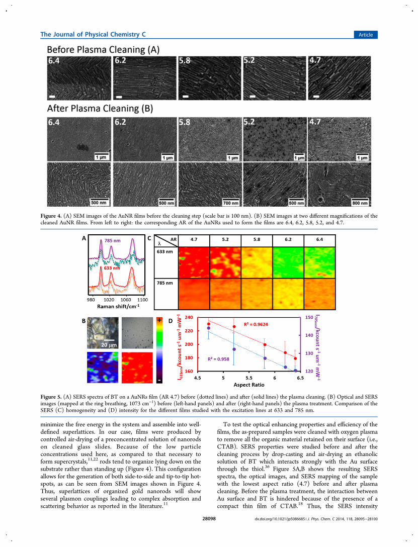
minimize the free energy in the system and assemble into well-defined superlattices. In our case, films were produced bycontrolled air-drying of a preconcentrated solution of nanorodson cleaned glass slides. Because of the low particleconcentrations used here, as compared to that necessary toform supercrystals,11,22 rods tend to organize lying down on thesubstrate rather than standing up (Figure 4). This configurationallows for the generation of both side-to-side and tip-to-tip hot-spots, as can be seen from SEM images shown in Figure 4.Thus, superlattices of organized gold nanorods will showseveral plasmon couplings leading to complex absorption andscattering behavior as reported in the literature.11
To test the optical enhancing properties and efficiency of thefilms, the as-prepared samples were cleaned with oxygen plasmato remove all the organic material retained on their surface (i.e.,CTAB). SERS properties were studied before and after thecleaning process by drop-casting and air-drying an ethanolicsolution of BT which interacts strongly with the Au surfacethrough the thiol.36 Figure 5A,B shows the resulting SERSspectra, the optical images, and SERS mapping of the samplewith the lowest aspect ratio (4.7) before and after plasmacleaning. Before the plasma treatment, the interaction betweenAu surface and BT is hindered because of the presence of acompact thin film of CTAB.18 Thus, the SERS intensity
Figure 4. (A) SEM images of the AuNR films before the cleaning step (scale bar is 100 nm). (B) SEM images at two different magnifications of thecleaned AuNR films. From left to right: the corresponding AR of the AuNRs used to form the films are 6.4, 6.2, 5.8, 5.2, and 4.7.
Figure 5. (A) SERS spectra of BT on a AuNRs film (AR 4.7) before (dotted lines) and after (solid lines) the plasma cleaning. (B) Optical and SERSimages (mapped at the ring breathing, 1073 cm−1) before (left-hand panels) and after (right-hand panels) the plasma treatment. Comparison of theSERS (C) homogeneity and (D) intensity for the different films studied with the excitation lines at 633 and 785 nm.
The Journal of Physical Chemistry C Article
dx.doi.org/10.1021/jp5086685 | J. Phys. Chem. C 2014, 118, 28095−2810028098

provided by the film is weak and inconsistent. On the contrary,after the cleaning process, the analyte can easily reach the Ausurface. Thus, a subsequent increase in both the SERS intensityand the point-to-point reproducibility is observed. Note thatafter plasma treatment no structural changes on the films or inthe morphology of the AuNRs forming them is observed(Figure 4).Figure 5C shows the SERS maps obtained for the two
excitation laser lines for the different cleaned AuNR films.Contrary to the results in solution, SERS signals obtained forthe organized films arise strongly from all samples withdetection limits for an aspect ratio of 4.7 of 2 × 10⁻⁹ M and 4 ×10⁻⁹ M for 633 and 785 nm, respectively. Furthermore, all thefilms show good point-to-point intensity reproducibility.Additionally, a linear tendency to increase SERS intensitieswith decreasing aspect ratio is observed for both laser lines(Figure 5D). In Au NRs, the linear relationship between theposition of the longitudinal plasmon and their aspect ratio iswell-known.26,37 Because of the close packing of the organizedAuNRs on the films, the consequent plasmon coupling verylikely displays the same trend. Our results show that theintensity obtained for excitation with the red laser line at 633nm is higher than that for the NIR laser at 785 nm. Althoughthere is not a remarkable difference between them, a morepronounced increase is observed for the red laser line. This maybe due to the close-packing of the organized AuNRs and theconsequent plasmon coupling. The strong coupling of theplasmon resonances of both transversal and longitudinal modesleads to a red-shift and broadening.38 Consequently, thelongitudinal LSPR loses resonance even with the NIR laser.This indicates that obtained SERS signals are provided mainlyfrom the hot-spot formed by the side-to-side nanorodinteractions rather than tip-to-tip, which also explains why theincrease in intensity is more pronounced for the 633 nm laser.Furthermore, in our experiments, the nanoparticles with
lower AR have larger dimensions resulting in lower hot-spotdensity on the surface. This effect is readily overcompensatedfor lower AR AuNRs by the red-shift of the plasmon resonancesdue to the strong coupling in the film and consequently betteroverlap of the excitation laser.
■ CONCLUSIONS
We have shown a fast and easy method of fabricating efficientoptical films based on the organization of nanorods for SERSapplications. Notably, the SERS efficiency of the prepared filmsstrongly depends on the surface cleaning procedure and on thenanoparticle aspect ratio. Because of the close-packing of theAuNRs, the plasmon resonances of both transversal andlongitudinal modes strongly couple and are strongly red-shifted; thus, the longitudinal LSPR loses resonance even withthe NIR laser, whereas for the 633 nm laser, intensity is morepronounced, indicating that obtained SERS signals are providedmainly by the side-to-side nanorod interactions.
■ AUTHOR INFORMATION
Corresponding Authors*E-mail: [email protected].*E-mail: [email protected].
NotesThe authors declare no competing financial interest.
■ ACKNOWLEDGMENTS
This work was funded by the Spanish Ministerio de Economiay Competitividad (CTQ2011-23167), the European ResearchCouncil (CrossSERS, FP7MC-IEF329131, PRIOSERSFP7MC-IEF-623527, and ERC-2012-StG 306686 META-MECH), NatoCBRN collaborative project CLG 984267, theGerman Research Foundation (DFG) within the collaborativeresearch center SFB 840, and Medcom Advance SA. M.T. wassupported by the Elite Network Bavaria in the frame of theElite Study Program “Macromolecular Science” and funded viaa grant for Ph.D. candidates according to Bavarian elitepromotion law (BayEFG).
■ REFERENCES(1) Myroshnychenko, V.; Rodriguez-Fernandez, J.; Pastoriza-Santos,I.; Funston, A. M.; Novo, C.; Mulvaney, P.; Liz-Marzan, L. M.; Garciade Abajo, F. J. Modelling the Optical Response of Gold Nanoparticles.Chem. Soc. Rev. 2008, 37, 1792−1805.(2) Bryant, G. W.; García De Abajo, F. J.; Aizpurua, J. Mapping thePlasmon Resonances of Metallic Nanoantennas. Nano Lett. 2008, 8,631−636.(3) Tebbe, M.; Cherepanov, P.; Skorb, E. V.; Poznyak, S. K.; deAbajo, J. G.; Fery, A.; Andreeva, D. V.; Puebla, R. A. A.; Pazos-Perez,N. SERS Platforms of Plasmonic Hydrophobic Surfaces for AnalyteConcentration: Hierarchically Assembled Gold Nanorods on Ano-dized Aluminum. Part. Part. Syst. Charact. 2014, DOI: 10.1002/ppsc.201400062.(4) Gou, L.; Murphy, C. J. Fine-Tuning the Shape of Gold Nanorods.Chem. Mater. 2005, 17, 3668−3672.(5) Orendorff, C. J.; Gearheart, L.; Jana, N. R.; Murphy, C. J. AspectRatio Dependence on Surface Enhanced Raman Scattering UsingSilver and Gold Nanorod Substrates. Phys. Chem. Chem. Phys. 2006, 8,165−170.(6) Sivapalan, S. T.; Devetter, B. M.; Yang, T. K.; Van Dijk, T.;Schulmerich, M. V.; Carney, P. S.; Bhargava, R.; Murphy, C. J. Off-Resonance Surface-Enhanced Raman Spectroscopy from Gold Nano-rod Suspensions as a Function of Aspect Ratio: Not What WeThought. ACS Nano 2013, 7, 2099−2105.(7) Zong, S.; Wang, Z.; Yang, J.; Cui, Y. Intracellular Ph SensingUsing P-Aminothiophenol Functionalized Gold Nanorods with LowCytotoxicity. Anal. Chem. 2011, 83, 4178−4183.(8) Huang, X.; El-Sayed, I. H.; Qian, W.; El-Sayed, M. A. CancerCells Assemble and Align Gold Nanorods Conjugated to Antibodies toProduce Highly Enhanced, Sharp, and Polarized Surface RamanSpectra: A Potential Cancer Diagnostic Marker. Nano Lett. 2007, 7,1591−1597.(9) Jokerst, J. V.; Cole, A. J.; Van De Sompel, D.; Gambhir, S. S. GoldNanorods for Ovarian Cancer Detection with Photoacoustic Imagingand Resection Guidance Via Raman Imaging in Living Mice. ACSNano 2012, 6, 10366−10377.(10) Song, J.; Pu, L.; Zhou, J.; Duan, B.; Duan, H. BiodegradableTheranostic Plasmonic Vesicles of Amphiphilic Gold Nanorods. ACSNano 2013, 7, 9947−9960.(11) Alvarez-Puebla, R. A.; et al. Gold Nanorods 3D-Supercrystals asSurface Enhanced Raman Scattering Spectroscopy Substrates for theRapid Detection of Scrambled Prions. Proc. Natl. Acad. Sci. U.S.A.2011, 108, 8157−8161.(12) Peng, B.; Li, G.; Li, D.; Dodson, S.; Zhang, Q.; Zhang, J.; Lee, Y.H.; Demir, H. V.; Yi Ling, X.; Xiong, Q. Vertically Aligned GoldNanorod Monolayer on Arbitrary Substrates: Self-Assembly andFemtomolar Detection of Food Contaminants. ACS Nano 2013, 7,5993−6000.(13) Saute, B.; Premasiri, R.; Ziegler, L.; Narayanan, R. GoldNanorods as Surface Enhanced Raman Spectroscopy Substrates forSensitive and Selective Detection of Ultra-Low Levels of Dithiocarba-mate Pesticides. Analyst (Cambridge, U.K.) 2012, 137, 5082−5087.
The Journal of Physical Chemistry C Article
dx.doi.org/10.1021/jp5086685 | J. Phys. Chem. C 2014, 118, 28095−2810028099
(14) Grzelczak, M.; Perez-Juste, J.; Mulvaney, P.; Liz-Marzan, L. M.Shape Control in Gold Nanoparticle Synthesis. Chem. Soc. Rev. 2008,37, 1783−1791.(15) Alkilany, A. M.; Nagaria, P. K.; Hexel, C. R.; Shaw, T. J.;Murphy, C. J.; Wyatt, M. D. Cellular Uptake and Cytotoxicity of GoldNanorods: Molecular Origin of Cytotoxicity and Surface Effects. Small2009, 5, 701−708.(16) Guerrini, L.; Jurasekova, Z.; del Puerto, E.; Hartsuiker, L.;Domingo, C.; Garcia-Ramos, J. V.; Otto, C.; Sanchez-Cortes, S. Effectof Metal−Liquid Interface Composition on the Adsorption of aCyanine Dye onto Gold Nanoparticles. Langmuir 2013, 29, 1139−1147.(17) Pazos-Perez, N.; Garcia De Abajo, F. J.; Fery, A.; Alvarez-Puebla,R. A. From Nano to Micro: Synthesis and Optical Properties ofHomogeneous Spheroidal Gold Particles and Their Superlattices.Langmuir 2012, 28, 8909−8914.(18) Spuch-Calvar, M.; Rodríguez-Lorenzo, L.; Morales, M. P.;Alvarez-Puebla, R. A.; Liz-Marzan, L. M. Bifunctional Nanocompositeswith Long-Term Stability as SERS Optical Accumulators forUltrasensitive Analysis. J. Phys. Chem. C 2009, 113, 3373−3377.(19) Sanles-Sobrido, M.; Correa-Duarte, M. A.; Carregal-Romero, S.;Rodríguez-Gonzalez, B.; Alvarez-Puebla, R. A.; Herves, P.; Liz-Marzan,L. M. Highly Catalytic Single-Crystal Dendritic Pt NanostructuresSupported on Carbon Nanotubes. Chem. Mater. 2009, 21, 1531−1535.(20) Kang, Y.; et al. Engineering Catalytic Contacts and ThermalStability: Gold/Iron Oxide Binary Nanocrystal Superlattices for CoOxidation. J. Am. Chem. Soc. 2013, 135, 1499−1505.(21) Ye, X.; Chen, J.; Diroll, B. T.; Murray, C. B. Tunable PlasmonicCoupling in Self-Assembled Binary Nanocrystal Superlattices Studiedby Correlated Optical Microspectrophotometry and Electron Micros-copy. Nano Lett. 2013, 13, 1291−1297.(22) Alba, M.; et al. Macroscale Plasmonic Substrates for HighlySensitive Surface-Enhanced Raman Scattering. Angew. Chem., Int. Ed.2013, 52, 6459−6463.(23) Solis, D.; Chang, W.-S.; Khanal, B. P.; Bao, K.; Nordlander, P.;Zubarev, E. R.; Link, S. Bleach-Imaged Plasmon Propagation (BlIPP)in Single Gold Nanowires. Nano Lett. 2010, 10, 3482−3485.(24) Ben, X.; Park, H. S. Strain Engineering Enhancement of SurfacePlasmon Polariton Propagation Lengths for Gold Nanowires. Appl.Phys. Lett. 2013, 102, 041909.(25) Kraus, T.; Brodoceanu, D.; Pazos-Perez, N.; Fery, A. ColloidalSurface Assemblies: Nanotechnology Meets Bioinspiration. Adv. Funct.Mater. 2013, 23, 4529−4541.(26) Vigderman, L.; Zubarev, E. R. High-Yield Synthesis of GoldNanorods with Longitudinal Spr Peak Greater Than 1200 nm UsingHydroquinone as a Reducing Agent. Chem. Mater. 2013, 25, 1450−1457.(27) Schweikart, A.; Pazos-Perez, N.; Alvarez-Puebla, R. A.; Fery, A.Controlling Inter-Nanoparticle Coupling by Wrinkle-Assisted Assem-bly. Soft Matter 2011, 7, 4093−4100.(28) Mueller, M.; Tebbe, M.; Andreeva, D. V.; Karg, M.; AlvarezPuebla, R. A.; Pazos Perez, N.; Fery, A. Large-Area Organization ofpNIPAM-Coated Nanostars as SERS Platforms for PolycyclicAromatic Hydrocarbons Sensing in Gas Phase. Langmuir 2012, 28,9168−9173.(29) Huang, X.; Neretina, S.; El-Sayed, M. A. Gold Nanorods: FromSynthesis and Properties to Biological and Biomedical Applications.Adv. Mater. (Weinheim, Ger.) 2009, 21, 4880−4910.(30) Jung, H. Y.; Park, Y.-K.; Park, S.; Kim, S. K. Surface EnhancedRaman Scattering from Layered Assemblies of Close-Packed GoldNanoparticles. Anal. Chim. Acta 2007, 602, 236−243.(31) Moskovits, M. Surface-Enhanced Spectroscopy. Rev. Mod. Phys.1985, 57, 783−826.(32) Braun, G.; Pavel, I.; Morrill, A. R.; Seferos, D. S.; Bazan, G. C.;Reich, N. O.; Moskovits, M. Chemically Patterned Microspheres forControlled Nanoparticle Assembly in the Construction of SERS HotSpots. J. Am. Chem. Soc. 2007, 129, 7760−7761.
(33) Qin, L.; Zou, S.; Xue, C.; Atkinson, A.; Schatz, G. C.; Mirkin, C.A. Designing, Fabricating, and Imaging Raman Hot Spots. Proc. Natl.Acad. Sci. U.S.A. 2006, 103, 13300−13303.(34) Bosnick, K. A.; Jiang; Brus, L. E. Fluctuations and LocalSymmetry in Single-Molecule Rhodamine 6G Raman Scattering onSilver Nanocrystal Aggregates. J. Phys. Chem. B 2002, 106, 8096−8099.(35) Jiang; Bosnick, K.; Maillard, M.; Brus, L. Single Molecule RamanSpectroscopy at the Junctions of Large Ag Nanocrystals. J. Phys. Chem.B 2003, 107, 9964−9972.(36) Haynes, C. L.; Van Duyne, R. P. Plasmon-Sampled Surface-Enhanced Raman Excitation Spectroscopy. J. Phys. Chem. B 2003, 107,7426−7433.(37) Huang, X.; Neretina, S.; El-Sayed, M. A. Gold Nanorods: FromSynthesis and Properties to Biological and Biomedical Applications.Adv. Mater. (Weinheim, Ger.) 2009, 21, 4880−4910.(38) Tritschler, U.; Zlotnikov, I.; Keckeis, P.; Schlaad, H.; Colfen, H.Optical Properties of Self-Organized Gold Nanorod-Polymer HybridFilms. Langmuir 2014, DOI: 10.1021/la503507u.
The Journal of Physical Chemistry C Article
dx.doi.org/10.1021/jp5086685 | J. Phys. Chem. C 2014, 118, 28095−2810028100