organic medicinal and pharmaceutical chemistry
1022
-
Upload
khangminh22 -
Category
Documents
-
view
1 -
download
0
Transcript of organic medicinal and pharmaceutical chemistry


Wilson and Gisvold’s Textbook of
ORGANIC MEDICINAL AND PHARMACEUTICAL
CHEMISTRYT W E L F T H E D I T I O N
Edited by
John M. Beale, Jr., PhDAssociate Professor of Medicinal Chemistry
Division of Basic and Pharmaceutical SciencesSt. Louis College of Pharmacy
Saint Louis, Missouri
John H. Block, PhD, RPhProfessor Emeritus, Medicinal ChemistryDepartment of Pharmaceutical Sciences
College of PharmacyOregon State University
Corvallis, Oregon
000i-000x_17865_FM.qxd 12/2/09 2:49 AM Page i
Editor: David B. TroyProduct Manager: Meredith L. BrittainVendor Manager: Kevin JohnsonDesigner: Holly McLaughlinCompositor: Absolute Service, Inc./Maryland Composition
12th Edition
Copyright © 2011 by Lippincott Williams & Wilkins, a Wolters Kluwer business.
First Edition, 1949 Fifth Edition, 1966 Ninth Edition, 1991Second Edition, 1954 Sixth Edition, 1971 Tenth Edition, 1998Third Edition, 1956 Seventh Edition, 1977 Eleventh Edition, 2004Fourth Edition, 1962 Eighth Edition, 1982
351 West Camden Street 530 Walnut StreetBaltimore, MD 21201 Philadelphia, PA 19106
Printed in The People’s Republic of China
All rights reserved. This book is protected by copyright. No part of this book may be reproduced or transmittedin any form or by any means, including as photocopies or scanned-in or other electronic copies, or utilized by anyinformation storage and retrieval system without written permission from the copyright owner, except for briefquotations embodied in critical articles and reviews. Materials appearing in this book prepared by individuals aspart of their official duties as U.S. government employees are not covered by the above-mentioned copyright. Torequest permission, please contact Lippincott Williams & Wilkins at 530 Walnut Street, Philadelphia, PA 19106,via email at [email protected], or via Web site at http://www.lww.com (products and services).
9 8 7 6 5 4 3 2 1
Library of Congress Cataloging-in-Publication Data
Wilson and Gisvold’s textbook of organic medicinal and pharmaceutical chemistry. — 12th ed. / edited by JohnM. Beale, Jr., John H. Block.
p. ; cm.Includes bibliographical references and index.ISBN 978-0-7817-7929-6
1. Pharmaceutical chemistry. 2. Chemistry, Organic. I. Wilson, Charles Owens, 1911- II. Beale, John Marlowe.III. Block, John H. IV. Title: Textbook of organic medicinal and pharmaceutical chemistry.
[DNLM: 1. Chemistry, Pharmaceutical. 2. Chemistry, Organic. QV 744 W754 2011]RS403.T43 2011615'.19—dc22
2009043714
DISCLAIMER
Care has been taken to confirm the accuracy of the information present and to describe generally accepted prac-tices. However, the authors, editors, and publisher are not responsible for errors or omissions or for any con-sequences from application of the information in this book and make no warranty, expressed or implied, withrespect to the currency, completeness, or accuracy of the contents of the publication. Application of this informa-tion in a particular situation remains the professional responsibility of the practitioner; the clinical treatmentsdescribed and recommended may not be considered absolute and universal recommendations.
The authors, editors, and publisher have exerted every effort to ensure that drug selection and dosage set forthin this text are in accordance with the current recommendations and practice at the time of publication. However,in view of ongoing research, changes in government regulations, and the constant flow of information relating todrug therapy and drug reactions, the reader is urged to check the package insert for each drug for any change inindications and dosage and for added warnings and precautions. This is particularly important when the recom-mended agent is a new or infrequently employed drug.
Some drugs and medical devices presented in this publication have Food and Drug Administration (FDA)clearance for limited use in restricted research settings. It is the responsibility of the healthcare provider to ascer-tain the FDA status of each drug or device planned for use in their clinical practice.
To purchase additional copies of this book, call our customer service department at (800) 638-3030 or fax ordersto (301) 223-2320. International customers should call (301) 223-2300.
Visit Lippincott Williams & Wilkins on the Internet: http://www.lww.com. Lippincott Williams & Wilkins cus-tomer service representatives are available from 8:30 am to 6:00 pm, EST.
000i-000x_17865_FM.qxd 12/2/09 2:49 AM Page ii
The 12th Edition of Wilson and Gisvold’s Textbook of Organic Medicinal and PharmaceuticalChemistry is dedicated to the memory of Robert F. Doerge.
Robert F. Doerge1915–2006
Robert Doerge—pharmacist, medicinal chemist, and educator—experienced the Depression andserved in the Civilian Conservation Corp in Sheridan, AR. Dr. Doerge received his B.S. in phar-
macy in 1943 and his PhD in pharmaceutical chemistry, both from the University of Minnesota in 1949.The latter was under the direction of Dr. Charles O. Wilson, who, with Dr. Ole Gisvold, started thiswell-respected medicinal chemistry textbook. Dr. Doerge began his professional career as an assistantprofessor in the University of Texas-Austin School of Pharmacy before becoming a research chemistwith the former Smith Kline and French Laboratories in Philadelphia. Beginning in 1960, he returnedto academia as professor and chair of the pharmaceutical chemistry department in Oregon StateUniversity’s College of Pharmacy. Prior to his retirement as professor emeritus in 1981, he was theassistant dean.
Dr. Doerge’s initial publications were on the topic of synthesis of anticonvulsants. At Smith Klineand French, his work included publications on vitamin stability, and at Oregon State University, his pa-pers focused on the heterocyclic phenylindolizines. Dr. Doerge was a volunteer abstractor for ChemicalAbstracts. As an educator, Dr. Doerge was an author of chapters in Wilson and Gisvold’s Textbook ofOrganic Medicinal and Pharmaceutical Chemistry, coeditor of the 6th and 7th editions, and editor ofthe 8th edition. His skill and dedication in the classroom were recognized by the students and univer-sity with several teaching awards.
We certainly miss this fine gentleman who put the students first and advanced the teaching of me-dicinal chemistry as a chapter author, coeditor, and editor of the Wilson and Gisvold textbook series.
John H. Block
iii
000i-000x_17865_FM.qxd 12/2/09 2:49 AM Page iii

PREFACE
For 6 decades, Wilson and Gisvold’s Textbook of Organic Medicinal and Pharmaceutical Chemistry hasbeen a standard in the literature of medicinal chemistry. Generations of students and faculty have de-pended on this textbook not only for undergraduate courses in medicinal chemistry but also as a supple-ment for graduate studies. Moreover, students in other health sciences have found certain chapters use-ful. The current editors and authors worked on the 12th edition with the objective of continuing thetradition of a modern textbook for undergraduate students and also for graduate students who need ageneral review of medicinal chemistry. Because the chapters include a blend of chemical and pharma-cological principles necessary for understanding structure–activity relationships and molecular mecha-nisms of drug action, the book should be useful in supporting courses in medicinal chemistry and incomplementing pharmacology courses.
ABOUT THE 12TH EDITION
The 12th edition follows in the footsteps of the 11th edition by reflecting the dynamic changes oc-curring in medicinal chemistry. With increased knowledge of the disease process and the identi-fication of the key steps in the biochemical process, the chapters have been updated, expanded,and reorganized. At the same time, to streamline the presentation of the content, some topics werecombined into existing chapters. For example, Chapter 2, “Drug Design Strategies,” incorporatesmaterial from 11th edition Chapters 2, 3, and 28, and Chapter 3, “Metabolic Changes of Drugs andRelated Organic Compounds,” includes the content from 11th edition Chapter 5, “Prodrugs andDrug Latentiation.” In addition, with the newer drugs that have entered the pharmaceutical arma-mentarium since publication of the 11th edition, coverage of the following topics has been ex-panded in the 12th edition: Central Dopaminergic Signaling Agents (Chapter 13), Anticonvulsants(Chapter 14), Hormone-Related Disorders: Nonsteroidal Therapies (Chapter 20), Agents TreatingBone Disorders (Chapter 21), and Anesthetics (Chapter 22).
New features of the 12th edition include a chapter overview at the beginning of each chapter to in-troduce material to be covered in the chapter and review questions at the end of each chapter to rein-force concepts learned in the chapter (answers to these questions are available to students on the book’scompanion Web site; see next section).
ADDITIONAL RESOURCES
Wilson and Gisvold’s Textbook of Organic Medicinal and Pharmaceutical Chemistry, 12th Edition, in-cludes additional resources for both instructors and students that are available on the book’s companionWeb site at http://www.thePoint.lww.com/Beale12e.
Instructors
Approved adopting instructors will be given access to the following additional resources:
• Image bank of all the figures and tables in the book
Students
Students who have purchased Wilson and Gisvold’s Textbook of Organic Medicinal and PharmaceuticalChemistry, 12th Edition, have access to the following additional resources:
• The answers to the review questions found in the book
In addition, purchasers of the text can access the searchable Full Text On-line by going to the Wilsonand Gisvold’s Textbook of Organic Medicinal and Pharmaceutical Chemistry, 12th Edition, Web siteat http://www.thePoint.lww.com/Beale12e. See the inside front cover of this text for more details,including the passcode you will need to gain access to the Web site.
iv
000i-000x_17865_FM.qxd 12/2/09 2:49 AM Page iv
ACKNOWLEDGMENTS
The editors welcome the new contributors to the 12th edition: Jeffrey J. Christoff, A. Michael Crider,Carolyn J. Friel, Ronald A. Hill, Shengquan Liu, Matthias C. Lu, Marcello J. Nieto, and Kenneth A.Witt. The editors extend thanks to all of the authors who have cooperated in the preparation of the cur-rent edition. Collectively, the authors represent many years of teaching and research experience in me-dicinal chemistry. Their chapters include summaries of current research trends that lead the reader to theoriginal literature. Documentation and references continue to be an important feature of the book.
We continue to be indebted to Professors Charles O. Wilson and Ole Gisvold, the originators of thebook and editors of five editions, Professor Robert Doerge, who joined Professors Wilson and Gisvoldfor the 6th and 7th editions and single-handedly edited the 8th edition, and Professors Jaime Delgado andWilliam Remers, who edited the 9th and 10th editions. They and the authors have contributed signifi-cantly to the education of countless pharmacists, medicinal chemists, and other pharmaceutical scientists.
John M. Beale, Jr.John H. Block
1st 1949 Wilson and Gisvold (OrganicChemistry in Pharmacy)
2nd 1954 Wilson and Gisvold3rd 1956 Wilson4th 1962 Wilson and Gisvold5th 1966 Wilson
6th 1971 Wilson, Gisvold, and Doerge7th 1977 Wilson, Gisvold, and Doerge8th 1982 Doerge9th 1991 Delgado and Remers10th 1998 Delgado and Remers11th 2004 Block and Beale
Preface v
000i-000x_17865_FM.qxd 12/2/09 2:49 AM Page v

John M. Beale, Jr., PhDAssociate Professor of Medicinal
ChemistryDivision of Basic and Pharmaceutical
SciencesSt. Louis College of PharmacySaint Louis, Missouri
John H. Block, PhD, RPhProfessor Emeritus, Medicinal
ChemistryDepartment of Pharmaceutical
SciencesCollege of PharmacyOregon State UniversityCorvallis, Oregon
Jeffrey J. Christoff, PhD, RPh
ProfessorDepartment of Pharmaceutical
and Biomedical SciencesCollege of Pharmacy, Ohio Northern
UniversityAda, Ohio
C. Randall Clark, PhDProfessorDepartment of Pharmaceutical
SciencesAuburn University School of
PharmacyAuburn, Alabama
A. Michael Crider, PhDChair and ProfessorDepartment of Pharmaceutical
SciencesSouthern Illinois University
EdwardsvilleEdwardsville, Illinois
Horace G. Cutler, PhDSenior Research ProfessorCollege of Pharmacy and Health
SciencesMercer UniversityAtlanta, Georgia
Stephen J. CutlerChair and ProfessorDepartment of Medicinal ChemistryUniversity of MississippiOxford, Mississippi
Michael J. Deimling, RPh, PhD
Department of PharmaceuticalSciences
School of PharmacyGeorgia Campus—Philadelphia
College of Osteopathic MedicineSuwanee, Georgia
Jack DeRuiter, MS, PhDProfessorDepartment of Pharmaceutical
SciencesAuburn University School of
PharmacyAuburn, Alabama
Carolyn J. Friel, RPh, PhD
Associate Professor of MedicinalChemistry
Department of PharmaceuticalSciences
Massachusetts College of Pharmacyand Health Sciences—Worcester
Worcester, Massachusetts
Ronald A. Hill, PhDAssociate ProfessorDepartment of Basic Pharmaceutical
SciencesThe University of Louisiana at
MonroeMonroe, Louisiana
Thomas J. Holmes, Jr.,PhD
ProfessorDepartment of Pharmaceutical
SciencesCampbell University College of
Pharmacy and the Health SciencesBuies Creek, New Carolina
M. O. Faruk Khan,BPharm, MPharm, PhD
Assistant Professor of MedicinalChemistry
Department of PharmaceuticalSciences
Southwestern Oklahoma StateUniversity College of Pharmacy
Weatherford, Oklahoma
Matthias C. Lu, PhDProfessor and Assistant Head for
Curricular AffairsDepartment of Medicinal Chemistry
and PharmacognosyCollege of Pharmacy, University of
Illinois at ChicagoChicago, Illinois
Shengquan Liu, PhDAssistant ProfessorDepartment of Medicinal ChemistryTouro University—CaliforniaVallejo, California
Marcello J. Nieto, PhDAssistant ProfessorDepartment of Pharmaceutical
SciencesSouthern Illinois UniversityEdwardsville, Illinois
Gustavo R. Ortega,RPh, PhD
Professor EmeritusDepartment of Pharmaceutical
SciencesSouthwestern Oklahoma State
University College of PharmacyWeatherford, Oklahoma
Philip J. Proteau, PhDAssociate Professor of Medicinal
ChemistryDepartment of Pharmaceutical
SciencesOregon State University College of
PharmacyCorvallis, Oregon
Forrest T. Smith, PhDAssociate ProfessorDepartment of Pharmaceutical
SciencesAuburn University School of
PharmacyAuburn, Alabama
Kenneth A. Witt, PhDAssistant ProfessorDepartment of Pharmaceutical
SciencesSouthern Illinois University
EdwardsvilleEdwardsville, Illinois
CONTRIBUTORS
vi
000i-000x_17865_FM.qxd 12/2/09 2:49 AM Page vi

CONTENTS
Preface . . . . . . . . . . . . . . . . . . . . . . . . . . . . . . . . . . . . . . . . ivContributors . . . . . . . . . . . . . . . . . . . . . . . . . . . . . . . . . . . . vi
C H A P T E R 1
Introduction . . . . . . . . . . . . . . . . . . . . . . 1John M. Beale, Jr. and John H. Block
C H A P T E R 2
Drug Design Strategies . . . . . . . . . . . . . 3John H. Block
Drug Distribution . . . . . . . . . . . . . . . . . . . . . . . . . . . . 3Acid–Base Properties . . . . . . . . . . . . . . . . . . . . . . . . 12Computer-Aided Drug Design: Early Methods . . . 17Computer-Aided Drug Design: Newer Methods . . 25Selected Web Pages. . . . . . . . . . . . . . . . . . . . . . . . . 40
C H A P T E R 3
Metabolic Changes of Drugs and Related Organic Compounds . . . . 43
Stephen J. Cutler and John H. Block
General Pathways of Drug Metabolism . . . . . . . . . 43Sites of Drug Biotransformation. . . . . . . . . . . . . . . 45Role of Cytochrome P450 Monooxygenases
in Oxidative Biotransformations. . . . . . . . . . . . . 45Oxidative Reactions . . . . . . . . . . . . . . . . . . . . . . . . . 47Reductive Reactions . . . . . . . . . . . . . . . . . . . . . . . . . 78Hydrolytic Reactions . . . . . . . . . . . . . . . . . . . . . . . . 86Phase II or Conjugation Reactions . . . . . . . . . . . . . 88Factors Affecting Drug Metabolism . . . . . . . . . . . 104
C H A P T E R 4
Biotechnology and Drug Discovery . . 119John M. Beale, Jr.
Biotechnology and Pharmaceutical Care . . . . . . . 119Literature of Biotechnology . . . . . . . . . . . . . . . . . 119Biotechnology and New Drug Development. . . . 119The Biotechnology of Recombinant DNA . . . . . . 121Some Types of Cloning . . . . . . . . . . . . . . . . . . . . . 126Expression of Cloned DNA . . . . . . . . . . . . . . . . . . 127Manipulation of DNA Sequence Information . . . 127New Biological Targets for Drug Development. . 128Novel Drug-Screening Strategies . . . . . . . . . . . . . 129Processing of the Recombinant Protein . . . . . . . . 131Pharmaceutics of Recombinant
DNA-Produced Agents. . . . . . . . . . . . . . . . . . . . 131Delivery and Pharmacokinetics
of Biotechnology Products . . . . . . . . . . . . . . . . 134Recombinant Drug Products . . . . . . . . . . . . . . . . . 134The Interleukins . . . . . . . . . . . . . . . . . . . . . . . . . . . 141Enzymes . . . . . . . . . . . . . . . . . . . . . . . . . . . . . . . . . 142Vaccines . . . . . . . . . . . . . . . . . . . . . . . . . . . . . . . . . 145Preparation of Antibodies. . . . . . . . . . . . . . . . . . . 146Genomics . . . . . . . . . . . . . . . . . . . . . . . . . . . . . . . . 150
Antisense Technology . . . . . . . . . . . . . . . . . . . . . . 152Gene Therapy . . . . . . . . . . . . . . . . . . . . . . . . . . . . . 153Afterword. . . . . . . . . . . . . . . . . . . . . . . . . . . . . . . . 153
C H A P T E R 5
Immunobiologicals . . . . . . . . . . . . . . . 156John M. Beale, Jr.
Cells of the Immune System . . . . . . . . . . . . . . . . . 156Immunity . . . . . . . . . . . . . . . . . . . . . . . . . . . . . . . . 159Acquistion of Immunity. . . . . . . . . . . . . . . . . . . . . 165New Vaccine Technologies: Adjuvant
Technology . . . . . . . . . . . . . . . . . . . . . . . . . . . . . 174New Vaccine Technologies: Nucleic Acid
Vaccines . . . . . . . . . . . . . . . . . . . . . . . . . . . . . . . 177
C H A P T E R 6
Anti-infective Agents . . . . . . . . . . . . . 179John M. Beale, Jr.
Evaluation of the Effectiveness of a Sterilant . . . 180Alcohols and Related Compounds . . . . . . . . . . . . 181Phenols and Their Derivatives. . . . . . . . . . . . . . . . 183Oxidizing Agents . . . . . . . . . . . . . . . . . . . . . . . . . . 185Halogen-Containing Compounds . . . . . . . . . . . . . 185Cationic Surfactants . . . . . . . . . . . . . . . . . . . . . . . . 186Dyes. . . . . . . . . . . . . . . . . . . . . . . . . . . . . . . . . . . . . 188Mercury Compounds (Mercurials). . . . . . . . . . . . . 189Preservatives. . . . . . . . . . . . . . . . . . . . . . . . . . . . . . 190Antifungal Agents . . . . . . . . . . . . . . . . . . . . . . . . . 191Synthetic Antibacterial Agents . . . . . . . . . . . . . . . 206Antiprotozoal Agents . . . . . . . . . . . . . . . . . . . . . . 220Anthelmintics . . . . . . . . . . . . . . . . . . . . . . . . . . . . . 224Antiscabious and Antipedicular Agents . . . . . . . . 227Antibacterial Sulfonamides . . . . . . . . . . . . . . . . . . 228Dihydrofolate Reductase Inhibitors . . . . . . . . . . . 239Sulfones . . . . . . . . . . . . . . . . . . . . . . . . . . . . . . . . . 239
C H A P T E R 7
Antimalarials . . . . . . . . . . . . . . . . . . . 242John H. Block
Stimulation of Antimalarial Research by War . . . 245Cinchona Alkaloids . . . . . . . . . . . . . . . . . . . . . . . . 245
C H A P T E R 8
Antibacterial Antibiotics. . . . . . . . . . . 258John M. Beale, Jr.
Historical Background . . . . . . . . . . . . . . . . . . . . . . 258Current Status . . . . . . . . . . . . . . . . . . . . . . . . . . . . 259Commercial Production . . . . . . . . . . . . . . . . . . . . . 259Spectrum of Activity . . . . . . . . . . . . . . . . . . . . . . . 259Mechanisms of Action . . . . . . . . . . . . . . . . . . . . . . 259Chemical Classification . . . . . . . . . . . . . . . . . . . . . 260Microbial Resistance . . . . . . . . . . . . . . . . . . . . . . . 260�-Lactam Antibiotics . . . . . . . . . . . . . . . . . . . . . . . 260
vii
000i-000x_17865_FM.qxd 12/2/09 2:50 AM Page vii
The Penicillins. . . . . . . . . . . . . . . . . . . . . . . . . . . . . 261�-Lactamase Inhibitors. . . . . . . . . . . . . . . . . . . . . . 274Cephalosporins. . . . . . . . . . . . . . . . . . . . . . . . . . . . 278Monobactams. . . . . . . . . . . . . . . . . . . . . . . . . . . . . 293Aminoglycosides . . . . . . . . . . . . . . . . . . . . . . . . . . 294Tetracyclines . . . . . . . . . . . . . . . . . . . . . . . . . . . . . . 301Macrolides . . . . . . . . . . . . . . . . . . . . . . . . . . . . . . . 308Lincomycins . . . . . . . . . . . . . . . . . . . . . . . . . . . . . . 313Polypeptides . . . . . . . . . . . . . . . . . . . . . . . . . . . . . . 315Unclassified Antibiotics . . . . . . . . . . . . . . . . . . . . . 320Newer Antibiotics . . . . . . . . . . . . . . . . . . . . . . . . . 324New Directions in Antibiotic Discovery . . . . . . . . 326
C H A P T E R 9
Antiviral Agents . . . . . . . . . . . . . . . . . 330John M. Beale, Jr.
The Classification and Biochemistry of Viruses . . 330Classification of Viruses . . . . . . . . . . . . . . . . . . . . . 330Targets for the Prevention of Viral
Infections—Chemoprophylaxis . . . . . . . . . . . . . 331The Infectious Process for a Virus . . . . . . . . . . . . . 333Nucleoside Antimetabolites: Inhibiting Viral
Replication . . . . . . . . . . . . . . . . . . . . . . . . . . . . . 339Newer Agents for the Treatment of
HIV Infection . . . . . . . . . . . . . . . . . . . . . . . . . . . 346
C H A P T E R 1 0
Antineoplastic Agents . . . . . . . . . . . . . 355Forrest T. Smith and C. Randall Clark
Introduction . . . . . . . . . . . . . . . . . . . . . . . . . . . . . . 355Drug Classes . . . . . . . . . . . . . . . . . . . . . . . . . . . . . . 358Antimetabolites . . . . . . . . . . . . . . . . . . . . . . . . . . . 372Antibiotics and Natural Products . . . . . . . . . . . . . 383Protein Kinase Inhibitors . . . . . . . . . . . . . . . . . . . . 400Miscellaneous Compounds . . . . . . . . . . . . . . . . . . 406
C H A P T E R 1 1
Agents for Diagnostic Imaging . . . . . 413Jeffrey J. Christoff
Radiopharmaceuticals . . . . . . . . . . . . . . . . . . . . . . 413Contrast Agents . . . . . . . . . . . . . . . . . . . . . . . . . . . 430
C H A P T E R 1 2
Central Nervous System Depressants . . . . . . . . . . . . . . . . . . . . . 443
Shengquan Liu
Anxiolytic, Sedative, and Hypnotic Agents . . . . . 443Antipsychotics . . . . . . . . . . . . . . . . . . . . . . . . . . . . 457Acknowledgment . . . . . . . . . . . . . . . . . . . . . . . . . 469
C H A P T E R 1 3
Central Dopaminergic Signaling Agents. . . . . . . . . . . . . . . . . 471
A. Michael Crider, Marcelo J. Nieto, and Kenneth A. Witt
Dopamine . . . . . . . . . . . . . . . . . . . . . . . . . . . . . . . . 471Parkinson Disease. . . . . . . . . . . . . . . . . . . . . . . . . . 473
Antipsychotic Drugs . . . . . . . . . . . . . . . . . . . . . . . . 478Future Directions . . . . . . . . . . . . . . . . . . . . . . . . . . 488
C H A P T E R 1 4
Anticonvulsants . . . . . . . . . . . . . . . . . 491Matthias C. Lu
Disease States Requiring Anticonvulsant Therapy. . . . . . . . . . . . . . . . . . . . . . . . . . . . . . . . 491
Mechanisms of Action of Anticonvulsants . . . . . . 492Clinically Important Anticonvulsants . . . . . . . . . . 494Future Development of Antiepileptic Drugs . . . . 501
C H A P T E R 1 5
Central Nervous System Stimulants . . . . . . . . . . . . . . . . . . . . . . 504
John M. Beale, Jr.
Analeptics . . . . . . . . . . . . . . . . . . . . . . . . . . . . . . . . 504Methylxanthines . . . . . . . . . . . . . . . . . . . . . . . . . . 505Central Sympathomimetic Agents
(Psychomotor Stimulants) . . . . . . . . . . . . . . . . . 506Antidepressants . . . . . . . . . . . . . . . . . . . . . . . . . . . 509Miscellaneous CNS-Acting Drugs . . . . . . . . . . . . . 515
C H A P T E R 1 6
Adrenergic Agents . . . . . . . . . . . . . . . 519Shengquan Liu
Adrenergic Neurotransmitters . . . . . . . . . . . . . . . 519Adrenergic Receptors . . . . . . . . . . . . . . . . . . . . . . 524Drugs Affecting Adrenergic
Neurotransmission . . . . . . . . . . . . . . . . . . . . . . . 528Sympathomimetic Agents . . . . . . . . . . . . . . . . . . . 531Adrenergic Receptor Antagonists (Blockers) . . . . 545Acknowledgment . . . . . . . . . . . . . . . . . . . . . . . . . 554
C H A P T E R 1 7
Cholinergic Drugs and Related Agents . . . . . . . . . . . . . . . . . . . . . . . . . 558
Stephen J. Cutler
Cholinergic Receptors . . . . . . . . . . . . . . . . . . . . . . 559Cholinergic Neurochemistry . . . . . . . . . . . . . . . . . 563Cholinergic Agonists . . . . . . . . . . . . . . . . . . . . . . . 564Cholinergic Receptor Antagonists . . . . . . . . . . . . 567Cholinergic Blocking Agents . . . . . . . . . . . . . . . . . 581Parasympathetic Postganglionic
Blocking Agents . . . . . . . . . . . . . . . . . . . . . . . . . 583Solanaceous Alkaloids and Analogs . . . . . . . . . . . 584Synthetic Cholinergic Blocking Agents. . . . . . . . . 588Ganglionic Blocking Agents . . . . . . . . . . . . . . . . . 596Neuromuscular Blocking Agents. . . . . . . . . . . . . . 599
C H A P T E R 1 8
Drugs Acting on the Renal System . . 607Stephen J. Cutler
Renin–Angiotensin System Inhibitors . . . . . . . . . . 609ACE-Inhibitor Prodrugs . . . . . . . . . . . . . . . . . . . . . 610Angiotensin Antagonists . . . . . . . . . . . . . . . . . . . . 612
viii Contents
000i-000x_17865_FM.qxd 12/2/09 2:50 AM Page viii

Angiotensin II Blockers . . . . . . . . . . . . . . . . . . . . . 613Renin Inhibitors . . . . . . . . . . . . . . . . . . . . . . . . . . . 614Aldosterone Antagonists. . . . . . . . . . . . . . . . . . . . 615
C H A P T E R 1 9
Cardiovascular Agents . . . . . . . . . . . . 617Stephen J. Cutler
Antianginal Agents and Vasodilators. . . . . . . . . . 617Antiarrhythmic Drugs . . . . . . . . . . . . . . . . . . . . . . 629Antihypertensive Agents . . . . . . . . . . . . . . . . . . . . 637Antihyperlipidemic Agents . . . . . . . . . . . . . . . . . . 647Anticoagulants . . . . . . . . . . . . . . . . . . . . . . . . . . . . 654Synthetic Hypoglycemic Agents . . . . . . . . . . . . . . 658Thyroid Hormones . . . . . . . . . . . . . . . . . . . . . . . . . 663Antithyroid Drugs . . . . . . . . . . . . . . . . . . . . . . . . . 663
C H A P T E R 2 0
Hormone-Related Disorders:Nonsteroidal Therapies . . . . . . . . . . . 666
Ronald A. Hill
Disorders of Glucose Metabolism: Diabetes and the Metabolic Syndrome . . . . . . . . . . . . . . 666
Gonadotropins, Gonadotrpoin-Releasing Hormone, and GNRH Receptor Agonists and Antagonists . . . . . . . . . . . . . . . . . . . . . . . . . 695
Concluding Remarks . . . . . . . . . . . . . . . . . . . . . . . 701
C H A P T E R 2 1
Agents Treating Bone Disorders . . . . 705John H. Block
Diseases of Bone Tissue Utilizing Approved Drug Therapies. . . . . . . . . . . . . . . . . . . . . . . . . . 705
Drugs Used to Treat Diseases of the Bone . . . . . . . . . . . . . . . . . . . . . . . . . . . . . . . 706
Hormone Therapy . . . . . . . . . . . . . . . . . . . . . . . . . 708Future Directions . . . . . . . . . . . . . . . . . . . . . . . . . . 710
C H A P T E R 2 2
Anesthetics . . . . . . . . . . . . . . . . . . . . . 711Carolyn J. Friel
The Inhaled General Anesthetics . . . . . . . . . . . . . 711The Injectable General Anesthetics . . . . . . . . . . . 716The Local Anesthetics . . . . . . . . . . . . . . . . . . . . . . 718Local Anesthetic Monographs, Individual
Products Including Adverse Reactions . . . . . . . 725
C H A P T E R 2 3
Histamine and Antihistaminic Agents . . . . . . . . . . . . . . . . . . . . . . . . . 733
Jack DeRuiter
Histamine Chemistry . . . . . . . . . . . . . . . . . . . . . . . 733Histamine as a Chemical Messenger. . . . . . . . . . . 733Antihistamines . . . . . . . . . . . . . . . . . . . . . . . . . . . . 737Inhibition of Histamine Release: Mast Cell
Stabilizers . . . . . . . . . . . . . . . . . . . . . . . . . . . . . . 757
Recent Antihistamine Developments: the “Dual-Acting” Antihistamines . . . . . . . . . . 759
Histamine H2-Antagonists . . . . . . . . . . . . . . . . . . . 760Histamine H3- and H4-Receptor
Ligands . . . . . . . . . . . . . . . . . . . . . . . . . . . . . . . . 773
C H A P T E R 2 4
Analgesics . . . . . . . . . . . . . . . . . . . . . . 776Carolyn J. Friel and Matthias C. Lu
Pain and Pain Management . . . . . . . . . . . . . . . . . 776Opioids . . . . . . . . . . . . . . . . . . . . . . . . . . . . . . . . . . 777Drug Monographs . . . . . . . . . . . . . . . . . . . . . . . . . 782Nonsteroidal Anti-inflammatory Drugs . . . . . . . . 792Disease-Modifying Antirheumatic Drugs . . . . . . . 806Drugs Used in the Management of Gout
and Hyperuricemia. . . . . . . . . . . . . . . . . . . . . . . 809Triptans. . . . . . . . . . . . . . . . . . . . . . . . . . . . . . . . . . 811
C H A P T E R 2 5
Steroid Hormones and Therapeutically Related Compounds . . . . . . . . . . . . . . . . . . . . . 819
Philip J. Proteau
Steroid Nomenclature, Stereochemistry, and Numbering . . . . . . . . . . . . . . . . . . . . . . . . . 819
Steroid Biosynthesis . . . . . . . . . . . . . . . . . . . . . . . . 819Chemical and Physical Properties of Steroids . . . 822Changes to Modify Pharmacokinetic
Properties of Steroids . . . . . . . . . . . . . . . . . . . . 822Steroid Hormone Receptors . . . . . . . . . . . . . . . . . 823Gonadotropin-Releasing Hormone
and Gonadotropins . . . . . . . . . . . . . . . . . . . . . . 826Sex Hormones. . . . . . . . . . . . . . . . . . . . . . . . . . . . . 827Chemical Contraceptive Agents . . . . . . . . . . . . . . 841Androgens . . . . . . . . . . . . . . . . . . . . . . . . . . . . . . . 847Adrenal Cortex Hormones. . . . . . . . . . . . . . . . . . . 853Neurosteroids . . . . . . . . . . . . . . . . . . . . . . . . . . . . . 864Acknowledgment. . . . . . . . . . . . . . . . . . . . . . . . . . 864
C H A P T E R 2 6
Prostaglandins, Leukotrienes, and Essential Fatty Acids. . . . . . . . . . 868
Thomas J. Holmes, Jr.
Essential Fatty Acids. . . . . . . . . . . . . . . . . . . . . . . . 868History of Eicosanoid Discovery . . . . . . . . . . . . . . 868Eicosanoid Biosynthesis . . . . . . . . . . . . . . . . . . . . . 869Drug Action Mediated by Eicosanoids . . . . . . . . . 872COX-2 Inhibitors. . . . . . . . . . . . . . . . . . . . . . . . . . . 872Design of Eicosanoid Drugs. . . . . . . . . . . . . . . . . . 872Eicosanoid Receptors . . . . . . . . . . . . . . . . . . . . . . . 875Commercially Available Essential Fatty Acid
Supplements. . . . . . . . . . . . . . . . . . . . . . . . . . . . 875Eicosanoids Approved for Human
Clinical Use . . . . . . . . . . . . . . . . . . . . . . . . . . . . . 876Prostaglandins for Ophthalmic Use . . . . . . . . . . . 878Veterinary Uses of Prostanoids . . . . . . . . . . . . . . . 878Eicosanoids in Clinical Development
for Human Treatment . . . . . . . . . . . . . . . . . . . . 879
Contents ix
000i-000x_17865_FM.qxd 12/2/09 2:50 AM Page ix

C H A P T E R 2 7
Proteins, Enzymes, and PeptideHormones . . . . . . . . . . . . . . . . . . . . . . 880
Stephen J. Cutler and Horace G. Cutler
Protein Hydrolysates . . . . . . . . . . . . . . . . . . . . . . . 880Amino Acid Solutions . . . . . . . . . . . . . . . . . . . . . . 881Proteins and Proteinlike Compounds . . . . . . . . . . 881Enzymes . . . . . . . . . . . . . . . . . . . . . . . . . . . . . . . . . 885Hormones . . . . . . . . . . . . . . . . . . . . . . . . . . . . . . . . 890Blood Proteins . . . . . . . . . . . . . . . . . . . . . . . . . . . . 906Impact of Biotechnology on the Development
and Commercial Production of Proteins and Peptides as Pharmaceutical Products . . . . 907
Biotechnology-Derived Pharmaceutical Products . . . . . . . . . . . . . . . . . . . . . . . . . . . . . . . 909
C H A P T E R 2 8
Vitamins . . . . . . . . . . . . . . . . . . . . . . . 915Michael J. Deimling, M. O. Faruk Khan, and Gustavo R. Ortega
Introduction . . . . . . . . . . . . . . . . . . . . . . . . . . . . . . 915Fat-Soluble Vitamins . . . . . . . . . . . . . . . . . . . . . . . 917Water-Soluble Vitamins. . . . . . . . . . . . . . . . . . . . . 935
C H A P T E R 2 9
An Introduction to the MedicinalChemistry of Herbs. . . . . . . . . . . . . . . 961
John M. Beale, Jr.
Historical Aspects . . . . . . . . . . . . . . . . . . . . . . . . . . 961What Is an Herb? . . . . . . . . . . . . . . . . . . . . . . . . . . 962Herbal Purity and Standardization . . . . . . . . . . . . 962An Herb Is a Drug . . . . . . . . . . . . . . . . . . . . . . . . . 962Types of Herbs . . . . . . . . . . . . . . . . . . . . . . . . . . . . 963
A P P E N D I X
Calculated Log P, Log D, and pKa . . 976
Index . . . . . . . . . . . . . . . . . . . . . . . . . . 984
x Contents
000i-000x_17865_FM.qxd 12/2/09 2:50 AM Page x

C H A P T E R 1
IntroductionJOHN M. BEALE, JR. AND JOHN H. BLOCK
based on anecdotal evidence, led to the use of such crudeplant drugs as opium, belladonna, and ephedrine that havebeen important for centuries. With the accidental discoveryof penicillin came the screening of microorganisms and thelarge number of antibiotics from bacterial and fungalsources. Many of these antibiotics provided the prototypi-cal structure that the medicinal chemist modified to obtainantibacterial drugs with better therapeutic profiles. Withthe changes in federal legislation reducing the efficacy re-quirement for “nutriceutical,” the public increasingly isusing so-called nontraditional or alternative medicinalsthat are sold over the counter, many outside of traditionalpharmacy distribution channels. It is important for thepharmacist and the public to understand the rigor that isrequired for prescription-only and Food and DrugAdministration (FDA)-approved nonprescription productsto be approved relative to the nontraditional products. It isalso important for all people in the healthcare field and thepublic to realize that whether these nontraditional productsare effective as claimed or not, many of the alternate medi-cines contain pharmacologically active agents that can po-tentiate or interfere with physician-prescribed therapy.
Hundreds of thousands of new organic chemicals are pre-pared annually throughout the world, and many of them areentered into pharmacological screens to determine whetherthey have useful biological activity. This process of randomscreening has been considered inefficient, but it has resultedin the identification of new lead compounds whose struc-tures have been optimized to produce clinical agents.Sometimes, a lead develops by careful observation of thepharmacological behavior of an existing drug. The discov-ery that amantadine protects and treats early influenza Acame from a general screen for antiviral agents. The use ofamantadine in long-term care facilities showed that it alsocould be used to treat parkinsonian disorders. More recently,automated high-throughput screening systems utilizing cellculture systems with linked enzyme assays and receptormolecules derived from gene cloning have greatly increasedthe efficiency of random screening. It is now practical toscreen enormous libraries of peptides and nucleic acids ob-tained from combinatorial chemistry procedures.
Rational design, the opposite approach to high-volumescreening, is also flourishing. Statistical methods based onthe correlation of physicochemical properties with biologicalpotency are used to explain and optimize biological activity.Significant advances in x-ray crystallography and nuclearmagnetic resonance have made it possible to obtain detailedrepresentations of enzymes and other drug receptors.The techniques of molecular graphics and computational
The discipline of medicinal chemistry is devoted to thediscovery and development of new agents for treating dis-eases. Most of this activity is directed to new natural orsynthetic organic compounds. Paralleling the developmentof medicinal agents has come a better understanding of thechemistry of the receptor. The latter has been greatly facil-itated by low-cost computers running software that calcu-lates molecular properties and structure and pictures itusing high-resolution graphics. Development of organiccompounds has grown beyond traditional synthetic meth-ods. It now includes the exciting field of biotechnologyusing the cell’s biochemistry to synthesize new com-pounds. Techniques ranging from recombinant DNA andsite-directed mutagenesis to fusion of cell lines havegreatly broadened the possibilities for new entities thattreat disease. The pharmacist now dispenses modifiedhuman insulins that provide more convenient dosingschedules, cell-stimulating factors that have changed thedosing regimens for chemotherapy, humanized monoclonalantibodies that target specific tissues, and fused receptorsthat intercept immune cell–generated cytokines.
This 12th edition of Wilson and Gisvold’s Textbook ofOrganic Medicinal and Pharmaceutical Chemistry contin-ues the philosophy of presenting the scientific basis of me-dicinal chemistry originally established by ProfessorsCharles Wilson and Ole Gisvold, describing the many as-pects of organic medicinals: how they are discovered, howthey act, and how they developed into clinical agents. Theprocess of establishing a new pharmaceutical is exceedinglycomplex and involves the talents of people from variousdisciplines, including chemistry, biochemistry, molecularbiology, physiology, pharmacology, pharmaceutics, andmedicine. Medicinal chemistry, itself, is concerned mainlywith the organic, analytical, and biochemical aspects of thisprocess, but the chemist must interact productively withthose in other disciplines. Thus, medicinal chemistry occu-pies a strategic position at the interface of chemistry andbiology. All of the principles discussed in this book arebased on fundamental organic chemistry, physical chem-istry, and biochemistry. To provide an understanding of theprinciples of medicinal chemistry, it is necessary to considerthe physicochemical properties used to develop new phar-macologically active compounds and their mechanisms ofaction, the drug’s metabolism, including possible biologicalactivities of the metabolites, the importance of stereochem-istry in drug design, and the methods used to determine what“space” a drug occupies.
The earliest drug discoveries were made by random sam-pling of higher plants. Some of this sampling, although
1
0001-0002_17865_Ch01.qxd 12/2/09 2:50 AM Page 1

chemistry have provided novel chemical structures thathave led to new drugs with potent medicinal activities.Development of human immunodeficiency virus (HIV) pro-tease inhibitors and angiotensin-converting enzyme (ACE)inhibitors came from an understanding of the geometry andchemical character of the respective enzyme’s active site.Even if the receptor structure is not known in detail, rationalapproaches based on the physicochemical properties of leadcompounds can provide new drugs. For example, the devel-opment of cimetidine involved a careful study of the changes
in antagonism of H2-histamine receptors induced by varyingthe physical properties of structures based on histamine.
As you proceed through the chapters, think of what prob-lem the medicinal chemist is trying to solve. Why were cer-tain structures selected? What modifications were made toproduce more focused activity or reduce adverse reactions orproduce better pharmaceutical properties? Was the prototypi-cal molecule discovered from random screens, or did the me-dicinal chemist have a structural concept of the receptor or anunderstanding of the disease process that must be interrupted?
2 Wilson and Gisvold’s Textbook of Organic Medicinal and Pharmaceutical Chemistry
0001-0002_17865_Ch01.qxd 12/2/09 2:50 AM Page 2

C H A P T E R 2
Drug Design StrategiesJOHN H. BLOCK
to the right. At the same time, the drug will be expected todissociate from the receptor and reenter the systemic circu-lation to be excreted. Major exceptions include the alkylat-ing agents used in cancer chemotherapy (see Chapter 10),a few inhibitors of the enzyme acetylcholinesterase (seeChapter 17), suicide inhibitors of monoamine oxidase(see Chapter 16), and the aromatase inhibitors 4-hydrox-yandrostenedione and exemestane (see Chapter 25). Thesepharmacological agents form covalent bonds with the re-ceptor, usually an enzyme’s active site. In these cases, thecell must destroy the receptor or enzyme, or, in the case ofthe alkylating agents, the cell would be replaced, ideallywith a normal cell. In other words, the usual use of drugsin medical treatment calls for the drug’s effect to last for afinite period of time. Then, if it is to be repeated, the drugwill be administered again. If the patient does not toleratethe drug well, it is even more important that the agent dis-sociate from the receptor and be excreted from the body.
Oral Administration
An examination of the obstacle course (Fig. 2.1) faced bythe drug will give a better understanding of what is involvedin developing a commercially feasible product. Assume thatthe drug is administered orally. The drug must go into solu-tion to pass through the gastrointestinal mucosa. Even drugsadministered as true solutions may not remain in solution asthey enter the acidic stomach and then pass into the alkalineintestinal tract. (This is explained further in the discussionon acid–base chemistry.) The ability of the drug to dissolveis governed by several factors, including its chemical struc-ture, variation in particle size and particle surface area, na-ture of the crystal form, type of tablet coating, and type oftablet matrix. By varying the dosage form and physical char-acteristics of the drug, it is possible to have a drug dissolvequickly or slowly, with the latter being the situation formany of the sustained-action products. An example is orallyadministered sodium phenytoin, with which variation ofboth the crystal form and tablet adjuvants can significantlyalter the bioavailability of this drug widely used in the treat-ment of epilepsy.
Chemical modification is also used to a limited extent tofacilitate a drug reaching its desired target (see Chapter 3).An example is olsalazine, used in the treatment of ulcera-tive colitis. This drug is a dimer of the pharmacologicallyactive mesalamine (5-aminosalicylic acid). The latter isnot effective orally because it is metabolized to inactiveforms before reaching the colon. The dimeric form passes
C H A P T E R O V E R V I E W
Modern drug design as compared with the classicalapproach—let’s make a change on an existing compound orsynthesize a new structure and see what happens—continues to evolve rapidly as an approach to solving a drugdesign problem. The combination of increasing power anddecreasing cost of desktop computing has had a major im-pact on solving drug design problems. Although drug designincreasingly is based on modern computational chemicaltechniques, it also uses sophisticated knowledge of diseasemechanisms and receptor properties. A good understandingof how the drug is transported into the body, distributedthroughout the body compartments, metabolically altered bythe liver and other organs, and excreted from the patient isrequired, along with the structural characteristics of thereceptor. Acid–base chemistry is used to aid in formulationand biodistribution. Structural attributes and substituent pat-terns responsible for optimum pharmacological activity canoften be predicted by statistical techniques such as re-gression analysis. Computerized conformational analysispermits the medicinal chemist to predict the drug’s three-dimensional (3D) shape that is seen by the receptor. Withthe isolation and structural determination of specific recep-tors and the availability of computer software that can esti-mate the 3D shape of the receptor, it is possible to designmolecules that will show an optimum fit to the receptor.
DRUG DISTRIBUTION
A drug is a chemical molecule. Following introduction intothe body, a drug must pass through many barriers, survive al-ternate sites of attachment and storage, and avoid significantmetabolic destruction before it reaches the site of action,usually a receptor on or in a cell (Fig. 2.1). At the receptor,the following equilibrium (Rx. 2.1) usually holds:
(Rx. 2.1)
The ideal drug molecule will show favorable bindingcharacteristics to the receptor, and the equilibrium will lie
3
0003-0042_17865_Ch02.qxd 12/4/09 2:51 AM Page 3

through a significant portion of the intestinal tract beforebeing cleaved by the intestinal bacteria to two equivalentsof mesalamine.
As illustrated by olsalazine, any compound passingthrough the gastrointestinal tract will encounter a large num-ber and variety of digestive and bacterial enzymes, which, intheory, can degrade the drug molecule. In practice, a newdrug entity under investigation will likely be dropped fromfurther consideration if it cannot survive in the intestinaltract or its oral bioavailability is low, necessitating par-enteral dosage forms only. An exception would be a drug forwhich there is no effective alternative or which is more ef-fective than existing products and can be administered by analternate route, including parenteral, buccal, or transdermal.
In contrast, these same digestive enzymes can be usedto advantage. Chloramphenicol is water soluble enough
(2.5 mg/mL) to come in contact with the taste receptors onthe tongue, producing an unpalatable bitterness. To maskthis intense bitter taste, the palmitic acid moiety is added asan ester of chloramphenicol’s primary alcohol. This reducesthe parent drug’s water solubility (1.05 mg/mL), enough sothat it can be formulated as a suspension that passes over thebitter taste receptors on the tongue. Once in the intestinaltract, the ester linkage is hydrolyzed by the digestive es-terases to the active antibiotic chloramphenicol and the verycommon dietary fatty acid palmitic acid.
Olsalazine and chloramphenicol palmitate are examplesof prodrugs. Most prodrugs are compounds that are inactivein their native form but are easily metabolized to the activeagent. Olsalazine and chloramphenicol palmitate are exam-ples of prodrugs that are cleaved to smaller compounds, oneof which is the active drug. Others are metabolic precursorsto the active form. An example of this type of prodrug ismenadione, a simple naphthoquinone that is converted in theliver to phytonadione (vitamin K2(20)).
4 Wilson and Gisvold’s Textbook of Organic Medicinal and Pharmaceutical Chemistry
Figure 2.1 Summary of drug distribution.
0003-0042_17865_Ch02.qxd 12/4/09 2:51 AM Page 4

Occasionally, the prodrug approach is used to enhancethe absorption of a drug that is poorly absorbed from thegastrointestinal tract. Enalapril is the ethyl ester ofenalaprilic acid, an active inhibitor of angiotensin-converting enzyme (ACE). The ester prodrug is muchmore readily absorbed orally than the pharmacologicallyactive carboxylic acid.
Unless the drug is intended to act locally in the gas-trointestinal tract, it will have to pass through the gastroin-testinal mucosal barrier into venous circulation to reachthe site of the receptor. The drug’s route involves distribu-tion or partitioning between the aqueous environment ofthe gastrointestinal tract, the lipid bilayer cell membrane ofthe mucosal cells, possibly the aqueous interior of the mu-cosal cells, the lipid bilayer membranes on the venous sideof the gastrointestinal tract, and the aqueous environmentof venous circulation. Some very lipid-soluble drugs mayfollow the route of dietary lipids by becoming part of themixed micelles, incorporating into the chylomicrons in themucosal cells into the lymph ducts, servicing the intes-tines, and finally entering venous circulation via the tho-racic duct.
The drug’s passage through the mucosal cells can bepassive or active. As is discussed later in this chapter,the lipid membranes are very complex with a highly or-dered structure. Part of this membrane is a series of chan-nels or tunnels that form, disappear, and reform. Thereare receptors that move compounds into the cell by aprocess called pinocytosis. Drugs that resemble a normalmetabolic precursor or intermediate may be actively trans-ported into the cell by the same system that transports theendogenous compound. On the other hand, most drug
molecules are too large to enter the cell by an activetransport mechanism through the passages. The latter,many times, pass into the patient’s circulatory system bypassive diffusion.
Parenteral Administration
Many times, there will be therapeutic advantages in bypass-ing the intestinal barrier by using parenteral (injectable)dosage forms. This is common in patients who, because ofillness, cannot tolerate or are incapable of accepting drugsorally. Some drugs are so rapidly and completely metabo-lized to inactive products in the liver (first-pass effect) thatoral administration is precluded. But that does not meanthat the drug administered by injection is not confronted byobstacles (Fig. 2.1). Intravenous administration places thedrug directly into the circulatory system, where it will berapidly distributed throughout the body, including tissuedepots and the liver, where most biotransformations occur(see later in this chapter), in addition to the receptors.Subcutaneous and intramuscular injections slow distribu-tion of the drug, because it must diffuse from the site of in-jection into systemic circulation.
It is possible to inject the drug directly into specific or-gans or areas of the body. Intraspinal and intracerebralroutes will place the drug directly into the spinal fluid orbrain, respectively. This bypasses a specialized epithelialtissue, the blood-brain barrier, which protects the brain fromexposure to a large number of metabolites and chemicals.The blood-brain barrier is composed of membranes oftightly joined epithelial cells lining the cerebral capillaries.The net result is that the brain is not exposed to the same va-riety of compounds that other organs are. Local anestheticsare examples of administration of a drug directly onto thedesired nerve. A spinal block is a form of anesthesia per-formed by injecting a local anesthetic directly into the spinalcord at a specific location to block transmission along spe-cific neurons.
Most of the injections a patient will experience in a life-time will be subcutaneous or intramuscular. These par-enteral routes produce a depot in the tissues (Fig. 2.1), fromwhich the drug must reach the blood or lymph. Once in sys-temic circulation, the drug will undergo the same distribu-tive phenomena as orally and intravenously administeredagents before reaching the target receptor. In general, thesame factors that control the drug’s passage through the gas-trointestinal mucosa will also determine the rate of move-ment out of the tissue depot.
The prodrug approach described previously can also beused to alter the solubility characteristics, which, in turn,can increase the flexibility in formulating dosage forms.The solubility of methylprednisolone can be altered fromessentially water-insoluble methylprednisolone acetate toslightly water-insoluble methylprednisolone to water-solublemethylprednisolone sodium succinate. The water-solublesodium hemisuccinate salt is used in oral, intravenous, andintramuscular dosage forms. Methylprednisolone itself isnormally found in tablets. The acetate ester is found in topi-cal ointments and sterile aqueous suspensions for intramus-cular injection. Both the succinate and acetate esters arehydrolyzed to the active methylprednisolone by the patient’sown systemic hydrolytic enzymes (esterases).
Chapter 2 Drug Design Strategies 5
0003-0042_17865_Ch02.qxd 12/4/09 2:51 AM Page 5

Another example of how prodrug design can significantlyalter biodistribution and biological half-life is illustrated bytwo drugs based on the retinoic acid structure used systemi-cally to treat psoriasis, a nonmalignant hyperplasia. Etretinatehas a 120-day terminal half-life after 6 months of therapy. Incontrast, the active metabolite, acitretin, has a 33- to 96-hourterminal half-life. Both drugs are potentially teratogenic.Women of childbearing age must sign statements that theyare aware of the risks and usually are administered a preg-nancy test before a prescription is issued. Acitretin, with itsshorter half-life, is recommended for a woman who wouldlike to become pregnant, because it can clear her body withina reasonable time frame. When effective, etretinate can keepa patient clear of psoriasis lesions for several months.
Protein Binding
Once the drug enters the systemic circulation (Fig. 2.1), itcan undergo several events. It may stay in solution, butmany drugs will be bound to the serum proteins, usually al-bumin (Rx. 2.2). Thus, a new equilibrium must be consid-ered. Depending on the equilibrium constant, the drug canremain in systemic circulation bound to albumin for a con-siderable period and not be available to the sites of biotrans-formation, the pharmacological receptors, and excretion.
(Rx. 2.2)
Protein binding can have a profound effect on the drug’seffective solubility, biodistribution, half-life in the body,and interaction with other drugs. A drug with such poorwater solubility that therapeutic concentrations of the un-bound (active) drug normally cannot be maintained still canbe a very effective agent. The albumin–drug complex acts asa reservoir by providing large enough concentrations of freedrug to cause a pharmacological response at the receptor.
Protein binding may also limit access to certain bodycompartments. The placenta is able to block passage of pro-teins from maternal to fetal circulation. Thus, drugs that nor-mally would be expected to cross the placental barrier andpossibly harm the fetus are retained in the maternal circula-tion, bound to the mother’s serum proteins.
Protein binding also can prolong the drug’s duration of ac-tion. The drug–protein complex is too large to pass throughthe renal glomerular membranes, preventing rapid excretionof the drug. Protein binding limits the amount of drug avail-able for biotransformation (see later in this chapter andChapter 3) and for interaction with specific receptor sites. For
6 Wilson and Gisvold’s Textbook of Organic Medicinal and Pharmaceutical Chemistry
0003-0042_17865_Ch02.qxd 12/4/09 2:51 AM Page 6
example, the large, polar trypanocide suramin remains in thebody in the protein-bound form for as long as 3 months (t1/2
� 50 days). The maintenance dose for this drug is based onweekly administration. At first, this might seem to be an ad-vantage to the patient. It can be, but it also means that, shouldthe patient have serious adverse reactions, a significant lengthof time will be required before the concentration of drug fallsbelow toxic levels.
The drug–protein binding phenomenon can lead to someclinically significant drug–drug interactions that result whenone drug displaces another from the binding site on albumin.A large number of drugs can displace the anticoagulant war-farin from its albumin-binding sites. This increases the ef-fective concentration of warfarin at the receptor, leading toan increased prothrombin time (increased time for clot for-mation) and potential hemorrhage.
Tissue Depots
The drug can also be stored in tissue depots. Neutral fat con-stitutes some 20% to 50% of body weight and constitutes adepot of considerable importance. The more lipophilic thedrug, the more likely it will concentrate in these pharmaco-logically inert depots. The ultra–short-acting, lipophilic bar-biturate thiopental’s concentration rapidly decreases belowits effective concentration following administration. It dis-appears into tissue protein, redistributes into body fat, andthen slowly diffuses back out of the tissue depots but in con-centrations too low for a pharmacological response. Thus,only the initially administered thiopental is present in highenough concentrations to combine with its receptors. The re-maining thiopental diffuses out of the tissue depots into sys-temic circulation in concentrations too small to be effective(Fig. 2.1), is metabolized in the liver, and is excreted.
In general, structural changes in the barbiturate series(see Chapter 12) that favor partitioning into the lipid tissuestores decrease duration of action but increase central ner-vous system (CNS) depression. Conversely, the barbiturateswith the slowest onset of action and longest duration of ac-tion contain the more polar side chains. This latter group ofbarbiturates both enters and leaves the CNS more slowlythan the more lipophilic thiopental.
Drug Metabolism
All substances in the circulatory system, including drugs,metabolites, and nutrients, will pass through the liver.Most molecules absorbed from the gastrointestinal tractenter the portal vein and are initially transported to theliver. A significant proportion of a drug will partition or be
transported into the hepatocyte, where it may be metabo-lized by hepatic enzymes to inactive chemicals during theinitial trip through the liver, by what is known as the first-pass effect (see Chapter 3).
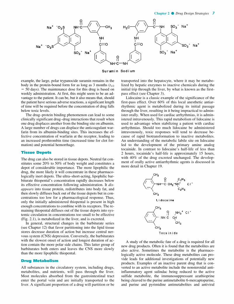
Lidocaine is a classic example of the significance of thefirst-pass effect. Over 60% of this local anesthetic antiar-rhythmic agent is metabolized during its initial passagethrough the liver, resulting in it being impractical to admin-ister orally. When used for cardiac arrhythmias, it is admin-istered intravenously. This rapid metabolism of lidocaine isused to advantage when stabilizing a patient with cardiacarrhythmias. Should too much lidocaine be administeredintravenously, toxic responses will tend to decrease be-cause of rapid biotransformation to inactive metabolites.An understanding of the metabolic labile site on lidocaineled to the development of the primary amine analogtocainide. In contrast to lidocaine’s half-life of less than2 hours, tocainide’s half-life is approximately 15 hours,with 40% of the drug excreted unchanged. The develop-ment of orally active antiarrhythmic agents is discussed inmore detail in Chapter 19.
A study of the metabolic fate of a drug is required for allnew drug products. Often it is found that the metabolites arealso active. Sometimes the metabolite is the pharmaco-logically active molecule. These drug metabolites can pro-vide leads for additional investigations of potentially newproducts. Examples of an inactive parent drug that is con-verted to an active metabolite include the nonsteroidal anti-inflammatory agent sulindac being reduced to the activesulfide metabolite, the immunosuppressant azathioprinebeing cleaved to the purine antimetabolite 6-mercaptopurine,and purine and pyrimidine antimetabolites and antiviral
Chapter 2 Drug Design Strategies 7
0003-0042_17865_Ch02.qxd 12/4/09 2:51 AM Page 7
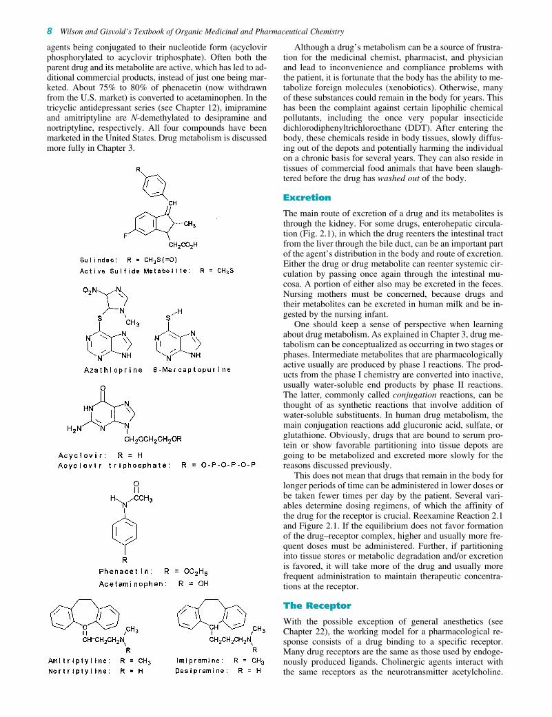
agents being conjugated to their nucleotide form (acyclovirphosphorylated to acyclovir triphosphate). Often both theparent drug and its metabolite are active, which has led to ad-ditional commercial products, instead of just one being mar-keted. About 75% to 80% of phenacetin (now withdrawnfrom the U.S. market) is converted to acetaminophen. In thetricyclic antidepressant series (see Chapter 12), imipramineand amitriptyline are N-demethylated to desipramine andnortriptyline, respectively. All four compounds have beenmarketed in the United States. Drug metabolism is discussedmore fully in Chapter 3.
Although a drug’s metabolism can be a source of frustra-tion for the medicinal chemist, pharmacist, and physicianand lead to inconvenience and compliance problems withthe patient, it is fortunate that the body has the ability to me-tabolize foreign molecules (xenobiotics). Otherwise, manyof these substances could remain in the body for years. Thishas been the complaint against certain lipophilic chemicalpollutants, including the once very popular insecticidedichlorodiphenyltrichloroethane (DDT). After entering thebody, these chemicals reside in body tissues, slowly diffus-ing out of the depots and potentially harming the individualon a chronic basis for several years. They can also reside intissues of commercial food animals that have been slaugh-tered before the drug has washed out of the body.
Excretion
The main route of excretion of a drug and its metabolites isthrough the kidney. For some drugs, enterohepatic circula-tion (Fig. 2.1), in which the drug reenters the intestinal tractfrom the liver through the bile duct, can be an important partof the agent’s distribution in the body and route of excretion.Either the drug or drug metabolite can reenter systemic cir-culation by passing once again through the intestinal mu-cosa. A portion of either also may be excreted in the feces.Nursing mothers must be concerned, because drugs andtheir metabolites can be excreted in human milk and be in-gested by the nursing infant.
One should keep a sense of perspective when learningabout drug metabolism. As explained in Chapter 3, drug me-tabolism can be conceptualized as occurring in two stages orphases. Intermediate metabolites that are pharmacologicallyactive usually are produced by phase I reactions. The prod-ucts from the phase I chemistry are converted into inactive,usually water-soluble end products by phase II reactions.The latter, commonly called conjugation reactions, can bethought of as synthetic reactions that involve addition ofwater-soluble substituents. In human drug metabolism, themain conjugation reactions add glucuronic acid, sulfate, orglutathione. Obviously, drugs that are bound to serum pro-tein or show favorable partitioning into tissue depots aregoing to be metabolized and excreted more slowly for thereasons discussed previously.
This does not mean that drugs that remain in the body forlonger periods of time can be administered in lower doses orbe taken fewer times per day by the patient. Several vari-ables determine dosing regimens, of which the affinity ofthe drug for the receptor is crucial. Reexamine Reaction 2.1and Figure 2.1. If the equilibrium does not favor formationof the drug–receptor complex, higher and usually more fre-quent doses must be administered. Further, if partitioninginto tissue stores or metabolic degradation and/or excretionis favored, it will take more of the drug and usually morefrequent administration to maintain therapeutic concentra-tions at the receptor.
The Receptor
With the possible exception of general anesthetics (seeChapter 22), the working model for a pharmacological re-sponse consists of a drug binding to a specific receptor.Many drug receptors are the same as those used by endoge-nously produced ligands. Cholinergic agents interact withthe same receptors as the neurotransmitter acetylcholine.
8 Wilson and Gisvold’s Textbook of Organic Medicinal and Pharmaceutical Chemistry
0003-0042_17865_Ch02.qxd 12/4/09 2:51 AM Page 8

Synthetic corticosteroids bind to the same receptors as cor-tisone and hydrocortisone. Often, receptors for the same li-gand are found in various tissues throughout the body. Thenonsteroidal anti-inflammatory agents (see Chapter 26) in-hibit the prostaglandin-forming enzyme cyclooxygenase,which is found in nearly every tissue. This class of drugs hasa long list of side effects with many patient complaints. Notein Figure 2.1 that, depending on which receptors containbound drug, there may be desired or undesired effects. Thisis because various receptors with similar structural require-ments are found in several organs and tissues. Thus, thenonsteroidal anti-inflammatory drugs combine with thedesired cyclooxygenase receptors at the site of the inflam-mation and the undesired cyclooxygenase receptors in thegastrointestinal mucosa, causing severe discomfort andsometimes ulceration. One of the second-generation antihis-tamines, fexofenadine, is claimed to cause less sedation be-cause it does not readily penetrate the blood-brain barrier.The rationale is that less of this antihistamine is available forthe receptors in the CNS, which are responsible for the se-dation response characteristic of antihistamines. In contrast,some antihistamines are used for their CNS depressant ac-tivity because a significant proportion of the administereddose is crossing the blood-brain barrier relative to binding tothe histamine H1 receptors in the periphery.
Although it is normal to think of side effects as undesir-able, they sometimes can be beneficial and lead to newproducts. The successful development of oral hypoglycemicagents used in the treatment of diabetes began when it wasfound that certain sulfonamides had a hypoglycemic effect.Nevertheless, a real problem in drug therapy is patient com-pliance in taking the drug as directed. Drugs that cause seri-ous problems and discomfort tend to be avoided by patients.
At this point, let us assume that the drug has entered thesystemic circulation (Fig. 2.1), passed through the lipid bar-riers, and is now going to make contact with the receptor. Asillustrated in Reaction 2.1, this is an equilibrium process. Agood ability to fit the receptor favors binding and the desiredpharmacological response. In contrast, a poor fit favors thereverse reaction. With only a small amount of drug bound tothe receptor, there will be a much smaller pharmacologicaleffect. If the amount of drug bound to the receptor is toosmall, there may be no discernible response. Many variablescontribute to a drug’s binding to the receptor. These includethe structural class, the 3D shape of the molecule, and thetypes of chemical bonding involved in the binding of thedrug to the receptor.
Most drugs that belong to the same pharmacologicalclass have certain structural features in common. The bar-biturates act on specific CNS receptors, causing depressanteffects; hydantoins act on CNS receptors, producing ananticonvulsant response; benzodiazepines combine with the�-aminobutyric acid (GABA) receptors, with resultinganxiolytic activity; steroids can be divided into such classesas corticosteroids, anabolic steroids, progestogens, and es-trogens, each acting on specific receptors; nonsteroidal anti-inflammatory agents inhibit enzymes required for theprostaglandin cascade; penicillins and cephalosporins in-hibit enzymes required to construct the bacterial cell wall;and tetracyclines act on bacterial ribosomes.
With the isolation and characterization of receptors be-coming a common occurrence, it is hard to realize that theconcept of receptors began as a postulate. It had been realized
early that molecules with certain structural features wouldelucidate a specific biological response. Very slight changesin structure could cause significant changes in biological ac-tivity. These structural variations could increase or decreaseactivity or change an agonist into an antagonist. This earlyand fundamentally correct interpretation called for the drug(ligand) to fit onto some surface (the receptor) that had fairlystrict structural requirements for proper binding of the drug.The initial receptor model was based on a rigid lock-and-keyconcept, with the drug (key) fitting into a receptor (lock). Ithas been used to explain why certain structural attributes pro-duce a predictable pharmacological action. This model still isuseful, although one must realize that both the drug and thereceptor can have considerable flexibility. Molecular graph-ics, using programs that calculate the preferred conformationsof drug and receptor, show that the receptor can undergo anadjustment in 3D structure when the drug makes contact.Using space-age language, the drug docks with the receptor.
More complex receptors now are being isolated, char-acterized, and cloned. The first receptors to be isolated andcharacterized were the reactive and regulatory sites onenzymes. Acetylcholinesterase, dihydrofolate reductase, an-giotensin, and human immunodeficiency virus (HIV)protease-converting enzyme are examples of enzymes whoseactive sites (the receptors) have been modeled. Most drug re-ceptors probably are receptors for natural ligands used to reg-ulate cellular biochemistry and function and to communicatebetween cells. Receptors include a relatively small region ofa macromolecule, which may be an isolatable enzyme, astructural and functional component of a cell membrane, or aspecific intracellular substance such as a protein or nucleicacid. Specific regions of these macromolecules are visual-ized as being oriented in space in a manner that permits theirfunctional groups to interact with the complementary func-tional groups of the drug. This interaction initiates changes instructure and function of the macromolecule, which lead ul-timately to the observable biological response. The conceptof spatially oriented functional areas forming a receptor leadsdirectly to specific structural requirements for functionalgroups of a drug, which must complement the receptor.
It now is possible to isolate membrane-bound receptors,although it still is difficult to elucidate their structural chem-istry, because once separated from the cell membranes,these receptors may lose their native shape. This is becausethe membrane is required to hold the receptor in its correcttertiary structure. One method of receptor isolation is affin-ity chromatography. In this technique, a ligand, often an al-tered drug molecule known to combine with the receptor, isattached to a chromatographic support phase. A solutioncontaining the desired receptor is passed over this column.The receptor will combine with the ligand. It is common toadd a chemically reactive grouping to the drug, resulting inthe receptor and drug covalently binding with each other.The drug–receptor complex is washed from the column andthen characterized further.
A more recent technique uses recombinant DNA. Thegene for the receptor is located and cloned. It is transferredinto a bacterium, yeast, or animal, which then produces thereceptor in large enough quantities to permit further study.Sometimes it is possible to determine the DNA sequence ofthe cloned gene. By using the genetic code for amino acids,the amino acid sequence of the protein component of thereceptor can be determined, and the receptor then modeled,
Chapter 2 Drug Design Strategies 9
0003-0042_17865_Ch02.qxd 12/4/09 2:51 AM Page 9

producing an estimated 3D shape. The model for the recep-tor becomes the template for designing new ligands.Genome mapping has greatly increased the information onreceptors. Besides the human genome, the genetic composi-tion of viruses, bacteria, fungi, and parasites has increasedthe possible sites for drugs to act. The new field of pro-teomics studies the proteins produced by structural genes.
The earlier discussion in this chapter emphasizes that thecell membrane is a highly organized, dynamic structure thatinteracts with small molecules in specific ways; its focus ison the lipid bilayer component of this complex structure.The receptor components of the membranes appear to bemainly protein. They constitute a highly organized, inter-twined region of the cell membrane. The same type of mo-lecular specificity seen in such proteins as enzymes andantibodies is also a property of drug receptors. The nature ofthe amide link in proteins provides a unique opportunity forthe formation of multiple internal hydrogen bonds, as wellas internal formation of hydrophobic, van der Waals, andionic bonds by side chain groups, leading to such organizedstructures as the �-helix, which contains about four aminoacid residues for each turn of the helix. An organized pro-tein structure would hold the amino acid side chains at rel-atively fixed positions in space and available for specificinteractions with a small molecule.
Proteins can potentially adopt many different conforma-tions in space without breaking their covalent amidelinkages. They may shift from highly coiled structures to par-tially disorganized structures, with parts of the molecule ex-isting in random chain or folded sheet structures, contingenton the environment. In the monolayer of a cell membrane,the interaction of a small foreign molecule with an organizedprotein may lead to a significant change in the structural andphysical properties of the membrane. Such changes couldwell be the initiating events in the tissue or organ response toa drug, such as the ion-translocation effects produced by in-teraction of acetylcholine and the cholinergic receptor.
The large body of information now available on relation-ships between chemical structure and biological activitystrongly supports the concept of flexible receptors. The fit ofdrugs onto or into macromolecules is rarely an all-or-none
process as pictured by the earlier lock-and-key concept of areceptor. Rather, the binding or partial insertion of groups ofmoderate size onto or into a macromolecular pouch appearsto be a continuous process, at least over a limited range, asindicated by the frequently occurring regular increase anddecrease in biological activity as one ascends a homologousseries of drugs. A range of productive associations betweendrug and receptor may be pictured, which leads to agonistresponses, such as those produced by cholinergic and adren-ergic drugs. Similarly, strong associations may lead tounproductive changes in the configuration of the macromol-ecule, leading to an antagonistic or blocking response, suchas that produced by anticholinergic agents and HIV proteaseinhibitors. Although the fundamental structural unit of thedrug receptor is generally considered to be protein, it may besupplemented by its associations with other units, such asmucopolysaccharides and nucleic acids.
Humans (and mammals in general) are very complex or-ganisms that have developed specialized organ systems. It isnot surprising that receptors are not distributed equallythroughout the body. It now is realized that, depending on theorgan in which it is located, the same receptor class may be-have differently. This can be advantageous by focusing drugtherapy on a specific organ system, but it can also causeadverse drug responses because the drug is exerting two dif-ferent responses based on the location of the receptors. An ex-ample is the selective estrogen receptor modulators (SERMs).They cannot be classified simply as agonists or antagonists.Rather, they can be considered variable agonists and antago-nists. Their selectivity is very complex because it depends onthe organ in which the receptor is located.
This complexity can be illustrated with tamoxifen andraloxifene (Fig. 2.2). Tamoxifen is used for estrogen-sensitivebreast cancer and for reducing bone loss from osteoporosis.Unfortunately, prolonged treatment increases the risk of en-dometrial cancer because of the response from the uterine es-trogen receptors. Thus, tamoxifen is an estrogen antagonist inthe mammary gland and an agonist in the uterus and bone. Incontrast, raloxifene does not appear to have much agonistproperty in the uterus but, like tamoxifen, is an antagonist inthe breast and agonist in the bone.
10 Wilson and Gisvold’s Textbook of Organic Medicinal and Pharmaceutical Chemistry
Figure 2.2 Selective SERMs.
0003-0042_17865_Ch02.qxd 12/4/09 2:52 AM Page 10

There are a wide variety of phosphodiesterases through-out the body. These enzymes hydrolyze the cyclic phosphateesters of adenosine monophosphate (cAMP) and guanosinemonophosphate (cGMP). Although the substrates for thisfamily of enzymes are cAMP and cGMP, there are differ-ences in the active sites. Figure 2.3 illustrates three drugsused to treat erectile dysfunction (sildenafil, tadalafil, andvardenafil). These three take advantage of the differences inactive site structural requirements between phosphodi-esterase type 5 and the other phosphodiesterases. They havean important role in maintaining a desired lifestyle: treatmentof erectile dysfunction caused by various medical conditions.The drugs approved for this indication were discovered byaccident. The goal was to develop a newer treatment ofangina. The approach was to develop phosphodiesterase in-hibitors that would prolong the activity of cGMP. The endresult was drugs that were not effective inhibitors of thephosphodiesterase that would treat angina, but were effectiveinhibitors of the one found in the corpus cavernosum. Thevasodilation in this organ results in penile erection.
Summary
One of the goals is to design drugs that will interact with re-ceptors at specific tissues. There are several ways to do this,including (a) altering the molecule, which, in turn, can changethe biodistribution; (b) searching for structures that show in-
creased specificity for the target receptor that will produce thedesired pharmacological response while decreasing the affin-ity for undesired receptors that produce adverse responses;and (c) the still experimental approach of attaching the drugto a monoclonal antibody (see Chapter 5) that will bind to aspecific tissue antigenic for the antibody. Biodistribution canbe altered by changing the drug’s solubility, enhancing itsability to resist being metabolized (usually in the liver), alter-ing the formulation or physical characteristics of the drug, andchanging the route of administration. If a drug molecule canbe designed so that its binding to the desired receptor is en-hanced relative to the undesired receptor and biodistributionremains favorable, smaller doses of the drug can be adminis-tered. This, in turn, reduces the amount of drug available forbinding to those receptors responsible for its adverse effects.
The medicinal chemist is confronted with several chal-lenges in designing a bioactive molecule. A good fit to aspecific receptor is desirable, but the drug would normallybe expected to dissociate from the receptor eventually. Thespecificity for the receptor would minimize side effects. Thedrug would be expected to clear the body within a reason-able time. Its rate of metabolic degradation should allowreasonable dosing schedules and, ideally, oral administra-tion. Many times, the drug chosen for commercial sales hasbeen selected from hundreds of compounds that have beenscreened. It usually is a compromise product that meets amedical need while demonstrating good patient acceptance.
Chapter 2 Drug Design Strategies 11
Figure 2.3 Examples of phosphodiesterasetype 5 inhibitors.
0003-0042_17865_Ch02.qxd 12/4/09 2:52 AM Page 11

ACID–BASE PROPERTIES
Most drugs used today can be classified as acids or bases.As is noted shortly, a large number of drugs can behaveas either acids or bases as they begin their journey intothe patient in different dosage forms and end up in systemiccirculation. A drug’s acid–base properties can greatly influ-ence its biodistribution and partitioning characteristics.
Over the years, at least four major definitions of acidsand bases have been developed. The model commonly usedin pharmacy and biochemistry was developed independentlyby Lowry and Brønsted. In their definition, an acid isdefined as a proton donor and a base is defined as a protonacceptor. Notice that for a base, there is no mention of thehydroxide ion. In the Brønsted-Lowry model, the acid plusbase reaction can be expressed as:
(Rx. 2.3)
Acid/Base–Conjugate Acid/ConjugateBase Pairs
Representative pairings of acids with their conjugatebases and bases with their conjugate acids are shown inTable 2.1. Careful study of this table shows water func-tioning as a proton acceptor (base) in reactions a, c, e, g,i, k, and m and a proton donor (base) in reactions b, d, f,h, j, l, and n. Hence, water is known as an amphotericsubstance. Water can be either a weak base accepting aproton to form the strongly acidic hydrated proton or hy-dronium ion H3O� (reactions a, c, e, g, i, k, and m), or aweak acid donating a proton to form the strongly basic(proton accepting) hydroxide anion OH� (reactions b, d,f, h, j, l, and n).
Also note the shift between un-ionized and ionized formsin Table 2.1. Examples of un-ionized acids donating theirprotons forming ionized conjugate bases are acetic acid-acetate (reaction g), phenobarbital-phenobarbiturate (reac-tion i), and saccharin-saccharate (reaction k). In contrast,examples of ionized acids yielding un-ionized conjugatebases are ammonium chloride-ammonia (reaction e) andephedrine hydrochloride (reaction m). A similar shift be-tween un-ionized and ionized forms is seen with bases andtheir conjugate acids. Examples of un-ionized bases yield-ing their ionized conjugate acids include ammonia andammonium (reaction f ) and ephedrine and protonatedephedrine (reaction n). Whereas examples of ionized basesyielding un-ionized conjugate acids are acetate formingacetic acid (reaction h), phenobarbiturate yielding pheno-barbital (reaction j), and saccharate yielding saccharin (reac-tion l). Complicated as it may seem at first, conjugate acidsand conjugate bases are nothing more than the products ofan acid–base reaction. In other words, they appear to theright of the reaction arrows.
Representative examples of pharmaceutically importantacidic drugs are listed in Table 2.1. Each acid, or protondonor, yields a conjugate base. The latter is the product afterthe proton is lost from the acid. Conjugate bases range fromthe chloride ion (reaction a), which does not accept a protonin aqueous media, to ephedrine (reaction h), which is an ex-cellent proton acceptor.
Acid Strength
While any acid–base reaction can be written as an equilib-rium reaction, an attempt has been made in Table 2.1 toindicate which sequences are unidirectional or show only asmall reversal. For hydrochloric acid (reaction a), the conju-gate base, Cl�, is such a weak base that it essentially doesnot function as a proton acceptor. In a similar manner, wateris such a weak conjugate acid that there is little reverse re-action involving water donating a proton to the hydroxideanion of sodium hydroxide (reaction b).
Two logical questions to ask at this point are how onepredicts in which direction an acid–base reaction lies and towhat extent the reaction goes to completion. The commonphysical chemical measurement that contains this informa-tion is known as the pKa. The pKa is the negative logarithmof the modified equilibrium constant, Ka (Eq. 2.1), for anacid–base reaction written so that water is the base or protonacceptor (reactions a, c, e, g, i, k, m, Table 2.1).
Ka � (Eq. 2.1)
Equation 2.1 is based on Rx. 2.3. The square bracketsindicate molar concentrations. Because the molar concentra-tion of water (the base in these acid–base reactions) is consid-ered constant in the dilute solutions used in pharmacy andmedicine, it is incorporated into the Ka. Rewriting Equation2.1 by taking the negative logarithm of the Ka, results in thefamiliar Henderson-Hasselbalch equation (Eq. 2.2).
pH � pKa � log (Eq. 2.2)
Warning! It is important to recognize that a pKa for abase is in reality the pKa of the conjugate acid (acid donoror protonated form, BH�) of the base. The pKa is listed inthe Appendix as 9.6 for ephedrine and as 9.3 for ammonia.In reality, this is the pKa of the protonated form, such asephedrine hydrochloride (reaction m in Table 2.1) and am-monium chloride (reaction e in Table 2.1), respectively.This is confusing to students, pharmacists, clinicians, andexperienced scientists. It is crucial that the chemistry of thedrug be understood when interpreting a pKa value. Whenreading tables of pKa values, such as those found in theAppendix, one must realize that the listed value is for theproton donor form of the molecule, no matter what form isindicated by the name. See Table 2.2 for several worked ex-amples of how the pKa is used to calculate pHs of solutions,required ratios of [conjugate base]/[acid], and percent ion-ization (discussed later) at specific pHs.
Just how strong or weak are the acids whose reactions inwater are illustrated in Table 2.1? Remember that the Kas orpKa’s are modified equilibrium constants that indicate theextent to which the acid (proton donor) reacts with water toform conjugate acid and conjugate base. The equilibrium fora strong acid (low pKa) in water lies to the right, favoringthe formation of products (conjugate acid and conjugatebase). The equilibrium for a weak acid (high pKa) in waterlies to the left, meaning that the conjugate acid is a betterproton donor than the parent acid is or that the conjugatebase is a good proton acceptor.
Refer back to Equation 2.1 and, using the Ka values inTable 2.3, substitute the Ka term for each of the acids. For
[conj. base][acid]
[conj. acid][conj. base]acid
12 Wilson and Gisvold’s Textbook of Organic Medicinal and Pharmaceutical Chemistry
0003-0042_17865_Ch02.qxd 12/4/09 2:52 AM Page 12

Chapter 2 Drug Design Strategies 13
TABLE 2.1 Examples of Acid–Base Reactions (with the Exception of Hydrochloric Acid, Whose Conjugate Base [Cl�]Has No Basic Properties in Water, and Sodium Hydroxide, which Generates Hydroxide, the Reaction of theConjugate Base in Water Is Shown for Each Acid)
Acid � Base Conjugate Acid � Conjugate Base
Hydrochloric acid(a) HCl � H2O H3O� � Cl�
Sodium hydroxide(b) H2O � NaOH H2O � OH�(Na�)a
Sodium dihydrogen phosphate and its conjugate base, sodium monohydrogen phosphate(c) H2PO4
�(Na�)a � H2O H3O� � HPO42�(Na�)a
(d) H2O � HPO42�(2Na�)a H2PO4
2�(Na�)a � OH�(Na�)a
Ammonium chloride and its conjugate base, ammonia(e) NH4
�(Cl�)a � H2O H3O�(Cl�)a � NH3
(f) H2O � NH3 NH4� � OH�
Acetic acid and its conjugate base, sodium acetate(g) CH3COOH � H2O H3O� � CH3COO�
(h) H2O � CH3COO�(Na�)a CH3COOH � OH�(Na�)a
Indomethacin and its conjugate base, indomethacin sodium, show the identical acid–base chemistry as acetic acid and sodium acetate, respectively.
Phenobarbital and its conjugate base, phenobarbital sodium
(i) � H2O H3O� �
(j) H2O � � OH�(Na�)a
Saccharin and its conjugate base, saccharin sodium
(k) � H2O H3O� �
(l) H2O � � OH�(Na�)a
Ephedrine HCl and its conjugate base, ephedrine
(m) � H2O H3O�(Cl�)a �
(n) H2O � � OH�
aThe chloride anion and sodium cation are present only to maintain charge balance. These anions play noother acid–base role.
0003-0042_17865_Ch02.qxd 12/4/09 2:52 AM Page 13

14 Wilson and Gisvold’s Textbook of Organic Medicinal and Pharmaceutical Chemistry
hydrochloric acid, a Ka of 1.26 � 106 means that the productof the molar concentrations of the conjugate acid, [H3O�],and the conjugate base, [Cl�], is huge relative to the denomi-nator term, [HCl]. In other words, there essentially is no un-reacted HCl left in an aqueous solution of hydrochloric acid.At the other extreme is ephedrine HCl with a pKa of 9.6 or aKa of 2.51 � 10�10. Here, the denominator representing theconcentration of ephedrine HCl greatly predominates overthat of the products, which, in this example, is ephedrine(conjugate base) and H3O� (conjugate acid). In other words,the protonated form of ephedrine is a very poor proton donor.It holds onto the proton. Free ephedrine (the conjugate base inthis reaction) is an excellent proton acceptor.
A general rule for determining whether a chemical isstrong or weak acid or base is
• pKa �2: strong acid; conjugate base has no meaningfulbasic properties in water
• pKa 4 to 6: weak acid; weak conjugate base• pKa 8 to 10: very weak acid; conjugate base getting stronger• pKa �12: essentially no acidic properties in water; strong
conjugate base
This delineation is only approximate. Other propertiesalso become important when considering cautions inhandling acids and bases. Phenol has a pKa of 9.9, slightlyless than that of ephedrine HCl. Why is phenol consideredcorrosive to the skin, whereas ephedrine HCl or freeephedrine is considered innocuous when applied to the skin?Phenol has the ability to partition through the normally pro-tective lipid layers of the skin. Because of this property, thisextremely weak acid has carried the name carbolic acid.Thus, the pKa simply tells a person the acid properties of theprotonated form of the chemical. It does not represent any-thing else concerning other potential toxicities.
Percent Ionization
Using the drug’s pKa, the formulation or compoundingpharmacist can adjust the pH to ensure maximum watersolubility (ionic form of the drug) or maximum solubility innonpolar media (un-ionic form). This is where understand-ing the drug’s acid–base chemistry becomes important.Note Reactions 2.4 and 2.5:
Conj. Conj.Acid Base Acid BaseHA(un-ionized) � H2O D H3O� � A�
(ionized) (Rx. 2.4)
Conj. Conj.Acid Base Acid BaseBH�
(ionized) � H2O D H3O� � B(un-ionized) (Rx. 2.5)
TABLE 2.2 Examples of Calculations Requiring the pKa
1. What is the ratio of ephedrine to ephedrine HCl (pKa 9.6)in the intestinal tract at pH 8.0? Use Equation 2.2.
8.0 � 9.6 � log � �1.6
� 0.025
The number whose log is �1.6 is 0.025, meaning thatthere are 25 parts ephedrine for every 1,000 partsephedrine HCl in the intestinal tract whose environmentis pH 8.0.
2. What is the pH of a buffer containing 0.1-M acetic acid(pKa 4.8) and 0.08-M sodium acetate? Use Equation 2.2.
pH � 4.8 � log � 4.7
3. What is the pH of a 0.1-M acetic acid solution? Use thefollowing equation for calculating the pH of a solutioncontaining either an HA or BH� acid.
pH � � 2.9
4. What is the pH of a 0.08-M sodium acetate solution?Remember, even though this is the conjugate base ofacetic acid, the pKa is still used. The pKw term in thefollowing equation corrects for the fact that a protonacceptor (acetate anion) is present in the solution. Theequation for calculating the pH of a solution containingeither an A� or B base is
pH � � 8.9
5. What is the pH of an ammonium acetate solution? ThepKa of the ammonium (NH4
�) cation is 9.3. Always bearin mind that the pKa refers to the ability of the protondonor form to release the proton into water to formH3O�. Since this is the salt of a weak acid (NH4
�) andthe conjugate base of a weak acid (acetate anion), thefollowing equation is used. Note that molarconcentration is not a variable in this calculation.
pH � � 7.1
6. What is the percentage ionization of ephedrine HCl (pKa
9.6) in an intestinal tract buffered at pH 8.0 (see example1)? Use Equation 2.4 because this is a BH� acid.
% ionization � � 97.6%
Only 2.4% of ephedrine is present as the un-ionizedconjugate base.
7. What is the percentage ionization of indomethacin(pKa 4.5) in an intestinal tract buffered at pH 8.0? UseEquation 2.3 because this is an HA acid.
% ionization � � 99.97%
For all practical purposes, indomethacin is present only asthe anionic conjugate base in that region of the intestinebuffered at pH 8.0.
1001 � 10(4.5�8.0)
1001 � 10(8.0�9.6)
pKa1� pKa2
2
pKw � pKa � log[base]2
pKa � log[acid]2
0.080.1
[ephedrine][ephedrine HCl]
[ephedrine][ephedrine HCl]
TABLE 2.3 Representative Ka and pKa Values from theReactions Listed in Table 2.1 (See the Appendix)
Hydrochloric acid 1.26 � 106 �6.1Dihydrogen phosphate 6.31 � 10�8 7.2Ammonia (ammonium) 5.01 � 10�10 9.3Acetic acid 1.58 � 10�5 4.8Phenobarbital 3.16 � 10�8 7.5Saccharin 2.51 � 10�2 1.6lndomethacin 3.16 � 10�5 4.5Ephedrine (as the HCI salt) 2.51 � 10�10 9.6
0003-0042_17865_Ch02.qxd 12/4/09 2:52 AM Page 14
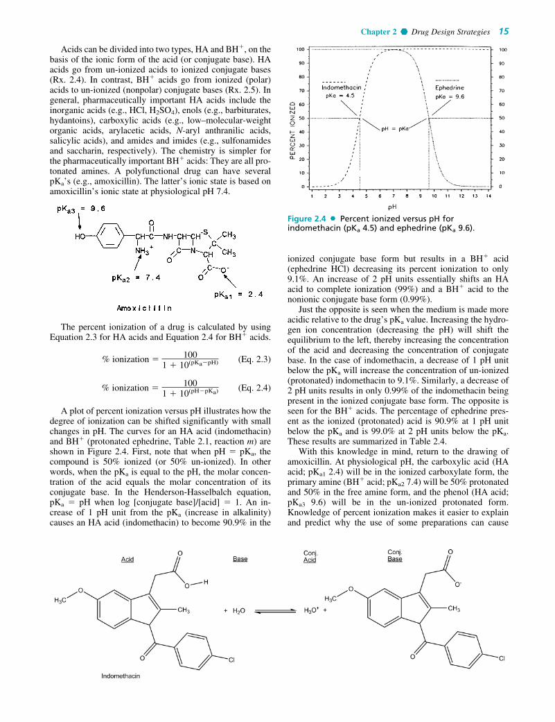
Acids can be divided into two types, HA and BH�, on thebasis of the ionic form of the acid (or conjugate base). HAacids go from un-ionized acids to ionized conjugate bases(Rx. 2.4). In contrast, BH� acids go from ionized (polar)acids to un-ionized (nonpolar) conjugate bases (Rx. 2.5). Ingeneral, pharmaceutically important HA acids include theinorganic acids (e.g., HCl, H2SO4), enols (e.g., barbiturates,hydantoins), carboxylic acids (e.g., low–molecular-weightorganic acids, arylacetic acids, N-aryl anthranilic acids,salicylic acids), and amides and imides (e.g., sulfonamidesand saccharin, respectively). The chemistry is simpler forthe pharmaceutically important BH� acids: They are all pro-tonated amines. A polyfunctional drug can have severalpKa’s (e.g., amoxicillin). The latter’s ionic state is based onamoxicillin’s ionic state at physiological pH 7.4.
The percent ionization of a drug is calculated by usingEquation 2.3 for HA acids and Equation 2.4 for BH� acids.
% ionization � (Eq. 2.3)
% ionization � (Eq. 2.4)
A plot of percent ionization versus pH illustrates how thedegree of ionization can be shifted significantly with smallchanges in pH. The curves for an HA acid (indomethacin)and BH� (protonated ephedrine, Table 2.1, reaction m) areshown in Figure 2.4. First, note that when pH � pKa, thecompound is 50% ionized (or 50% un-ionized). In otherwords, when the pKa is equal to the pH, the molar concen-tration of the acid equals the molar concentration of itsconjugate base. In the Henderson-Hasselbalch equation,pKa � pH when log [conjugate base]/[acid] � 1. An in-crease of 1 pH unit from the pKa (increase in alkalinity)causes an HA acid (indomethacin) to become 90.9% in the
1001 � 10(pH�pKa)
1001 � 10(pKa�pH)
ionized conjugate base form but results in a BH� acid(ephedrine HCl) decreasing its percent ionization to only9.1%. An increase of 2 pH units essentially shifts an HAacid to complete ionization (99%) and a BH� acid to thenonionic conjugate base form (0.99%).
Just the opposite is seen when the medium is made moreacidic relative to the drug’s pKa value. Increasing the hydro-gen ion concentration (decreasing the pH) will shift theequilibrium to the left, thereby increasing the concentrationof the acid and decreasing the concentration of conjugatebase. In the case of indomethacin, a decrease of 1 pH unitbelow the pKa will increase the concentration of un-ionized(protonated) indomethacin to 9.1%. Similarly, a decrease of2 pH units results in only 0.99% of the indomethacin beingpresent in the ionized conjugate base form. The opposite isseen for the BH� acids. The percentage of ephedrine pres-ent as the ionized (protonated) acid is 90.9% at 1 pH unitbelow the pKa and is 99.0% at 2 pH units below the pKa.These results are summarized in Table 2.4.
With this knowledge in mind, return to the drawing ofamoxicillin. At physiological pH, the carboxylic acid (HAacid; pKa1 2.4) will be in the ionized carboxylate form, theprimary amine (BH� acid; pKa2 7.4) will be 50% protonatedand 50% in the free amine form, and the phenol (HA acid;pKa3 9.6) will be in the un-ionized protonated form.Knowledge of percent ionization makes it easier to explainand predict why the use of some preparations can cause
Chapter 2 Drug Design Strategies 15
Figure 2.4 Percent ionized versus pH forindomethacin (pKa 4.5) and ephedrine (pKa 9.6).
0003-0042_17865_Ch02.qxd 12/4/09 2:52 AM Page 15

problems and discomfort as a result of pH extremes.Phenytoin (HA acid; pKa 8.3) injection must be adjusted topH 12 with sodium hydroxide to ensure complete ionizationand maximize water solubility. In theory, a pH of 10.3 willresult in 99.0% of the drug being an anionic water-solubleconjugate base. To lower the concentration of phenytoin inthe insoluble acid form even further and maintain excess al-kalinity, the pH is raised to 12 to obtain 99.98% of the drugin the ionized form. Even then, a cosolvent system of 40%propylene glycol, 10% ethyl alcohol, and 50% water forinjection is used to ensure complete solution. This highly al-kaline solution is irritating to the patient and generally can-not be administered as an admixture with other intravenousfluids that are buffered more closely at physiological pH7.4. This decrease in pH would result in the parent un-ionized phenytoin precipitating out of solution.
Tropicamide is an anticholinergic drug administered aseye drops for its mydriatic response during eye examina-tions. With a pKa of 5.2, the drug has to be buffered nearpH 4 to obtain more than 90% ionization. The acidic eyedrops can sting. Some optometrists and ophthalmologistsuse local anesthetic eye drops to minimize the patient’sdiscomfort. The only atom with a meaningful pKa is thepyridine nitrogen. The amide nitrogen has no acid–baseproperties in aqueous media.
Adjustments in pH to maintain water solubility cansometimes lead to chemical stability problems. An exampleis indomethacin (HA acid; pKa 4.5), which is unstable in al-kaline media. Therefore, the preferred oral liquid dosageform is a suspension buffered at pH 4 to 5. Because this is
near the drug’s pKa, only 50% will be in the water-solubleform. There is a medical indication requiring intravenousadministration of indomethacin to premature infants. Theintravenous dosage form is the lyophilized (freeze-dried)sodium salt, which is reconstituted just prior to use.
Drug Distribution and pKa
The pKa can have a pronounced effect on the pharmacoki-netics of the drug. As discussed previously, drugs are trans-ported in the aqueous environment of the blood. Thosedrugs in an ionized form will tend to distribute throughoutthe body more rapidly than will un-ionized (nonpolar) mol-ecules. With few exceptions, the drug must leave the polarenvironment of the plasma to reach the site of action. In gen-eral, drugs pass through the nonpolar membranes of capil-lary walls, cell membranes, and the blood-brain barrier inthe un-ionized (nonpolar) form. For HA acids, it is the par-ent acid that will readily cross these membranes (Fig. 2.5).The situation is just the opposite for the BH� acids. The un-ionized conjugate base (free amine) is the species most read-ily crossing the nonpolar membranes (Fig. 2.6).
Consider the changing pH environment experienced bythe drug molecule orally administered. The drug first en-counters the acidic stomach, where the pH can range from 2to 6 depending on the presence of food. HA acids with pKa’sof 4 to 5 will tend to be nonionic and be absorbed partiallythrough the gastric mucosa. (The main reason most acidicdrugs are absorbed from the intestinal tract rather than thestomach is that the microvilli of the intestinal mucosa pro-vide a large surface area relative to that found in the gastricmucosa of the stomach.) In contrast, amines (pKa 9–10) willbe protonated (BH� acids) in the acidic stomach and usuallywill not be absorbed until reaching the mildly alkaline intes-tinal tract (pH 8). Even here, only a portion of the amine-containing drugs will be in their nonpolar conjugate baseform (Fig. 2.4). Remember that the reactions shown inFigures 2.3 and 2.4 are equilibrium reactions with Ka values.Therefore, whenever the nonpolar form of either an HA acid(as the acid) or a B base (the conjugate base of the BH�
acid) passes the lipid barrier, the ratio of conjugate base toacid (percent ionization) will be maintained. Based onEquations 2.3 and 2.4, this ratio depends on the pKa (a con-stant) and the pH of the medium.
For example, once in systemic circulation, the plasmapH of 7.4 will be one of the determinants of whether thedrug will tend to remain in the aqueous environment ofthe blood or partition across lipid membranes into hepatictissue to be metabolized, into the kidney for excretion, intotissue depots, or to the receptor tissue. A useful exercise isto calculate either the [conjugate base]/[acid] ratio usingthe Henderson-Hasselbalch equation (Eq. 2.2) or percent
16 Wilson and Gisvold’s Textbook of Organic Medicinal and Pharmaceutical Chemistry
TABLE 2.4 Percentage Ionization Relative to the pKa
Ionization (%)
HA Acids BH Acids
pKa � 2 pH units 0.99 99.0pKa � 1 pH unit 9.1 90.9pKa � pH 50.0 50.0pKa � 1 pH unit 90.9 9.1pKa � 2 pH units 99.0 0.99
Figure 2.5 Passage of HA acids through lipid barriers.
0003-0042_17865_Ch02.qxd 12/4/09 2:52 AM Page 16

ionization for ephedrine (pKa 9.6; Eq. 2.4) and in-domethacin (pKa 4.5; Eq. 2.3) at pH 3.5 (stomach), pH 8.0(intestine), and pH 7.4 (plasma) (see examples 1, 6, and 7 inTable 2.2). Of course, the effect of protein binding, dis-cussed previously, can greatly alter any prediction of biodis-tribution based solely on pKa.
COMPUTER-AIDED DRUG DESIGN:EARLY METHODS
Initially, the design of new drugs was based on starting witha prototypical molecule, usually a natural product and mak-ing structural modifications. Examples include steroidalhormones based on naturally occurring cortisone, testos-terone, progesterone and estrogen; adrenergic drugs basedon epinephrine; local anesthetics based on cocaine; opiateanalgesics based on morphine; antibiotics based on peni-cillin, cephalosporin and tetracycline. Examples of proto-typical molecules that were not natural in origin include theantipsychotic phenothiazines, bisphosphonates for osteo-porosis, benzodiazepines indicated for various CNS treat-ments. Although prototypical molecules have producedsignificant advancements in treating diseases, this approachto drug development is limited to the initial discovery of theprototypical molecule. Today, it is more common to take aholistic approach that, where possible, involves understand-ing the etiology of the disease and the structure of the recep-tor where the ligand (drug) will bind. Increasing computerpower coupled with applicable software, both at reasonablecost, has lead to more focused approaches for the develop-ment of new drugs. Computational methodologies includemathematical equations correlating structure with biologicalactivity, searching chemical databases for leads and rapiddocking of ligand to the receptor. The latter requires 3Dstructure information of the receptor. Originally crystallizedenzymes were the common receptors, and their spatialarrangements determined by x-ray crystallography. Today’ssoftware can calculate possible 3D structures of proteinstarting with the amino acid sequence.
Statistical Prediction ofPharmacological Activity
Just as mathematical modeling is used to explain and modelmany chemical processes, it has been the goal of medicinalchemists to quantify the effect of a structural change on a de-fined pharmacological response. This would meet three goalsin drug design: (a) to predict biological activity in untestedcompounds, (b) to define the structural requirements requiredfor a good fit between the drug molecule and the receptor, and
(c) to design a test set of compounds to maximize the amountof information concerning structural requirements for activityfrom a minimum number of compounds tested. This aspect ofmedicinal chemistry is commonly referred to as quantitativestructure–activity relationships (QSAR).
The goals of QSAR studies were first proposed about1865 to 1870 by Crum-Brown and Fraser, who showed thatthe gradual chemical modification in the molecular structureof a series of poisons produced some important differencesin their action.1 They postulated that the physiological ac-tion, �, of a molecule is a function of its chemical constitu-tion, C. This can be expressed in Equation 2.5:
� � f(C) (Eq. 2.5)
Equation 2.5 states that a defined change in chemicalstructure results in a predictable change in physiological ac-tion. The problem now becomes one of numerically definingchemical structure. It still is a fertile area of research. Whathas been found is that biological response can be predictedfrom physical chemical properties such as vapor pressure,water solubility, electronic parameters, steric descriptors,and partition coefficients (Eq. 2.6). Today, the partition coef-ficient has become the single most important physical chem-ical measurement for QSAR studies. Note that Equation 2.6is the equation for a straight line (Y � mx � b).
log BR � a(physical chemical property) � c (Eq. 2.6)
where
BR � a defined pharmacological response usually ex-pressed in millimoles such as the inhibitory constantKi, the effective dose in 50% of the subjects (ED50),the lethal dose in 50% of the subjects (LD50), or theminimum inhibitory concentration (MIC). It is com-mon to express the biological response as a recipro-cal, 1/BR or 1/C
a � the regression coefficient or slope of the straight linec � the intercept term on the y axis (when the physical
chemical property equals zero)
To understand the concepts in the next few paragraphs, itis necessary to know how to interpret defined pharmacolog-ical concepts such as the ED50, which is the amount of thedrug needed to obtain the defined pharmacological responsein 50% of the test subjects. Let us assume that drug A’sED50 is 1 mmol and drug B’s ED50 is 2 mmol. Drug A istwice as potent as drug B. In other words, the smaller theED50 (or ED90, LD50, MIC, etc.), the more potent is the sub-stance being tested.
The logarithmic value of the dependent variable (concen-tration necessary to obtain a defined biological response) isused to linearize the data. As shown later in this chapter,QSARs are not always linear. Nevertheless, using logarithmsis an acceptable statistical technique (taking reciprocals ob-tained from a Michaelis-Menton study produces the linearLineweaver-Burke plots found in any biochemistry textbook).
Now, why is the biological response usually expressed asa reciprocal? Sometimes, one obtains a statistically morevalid relationship. More importantly, expressing the biolog-ical response as a reciprocal usually produces a positiveslope (Fig. 2.7). Let us examine the following publishedexample (Table 2.5). The BR is the LD100 (lethal dose in100% of the subjects). The mechanism of death is generaldepression of the CNS.
Chapter 2 Drug Design Strategies 17
Figure 2.6 Passage of BH� acids through lipid barriers.
0003-0042_17865_Ch02.qxd 12/4/09 2:52 AM Page 17
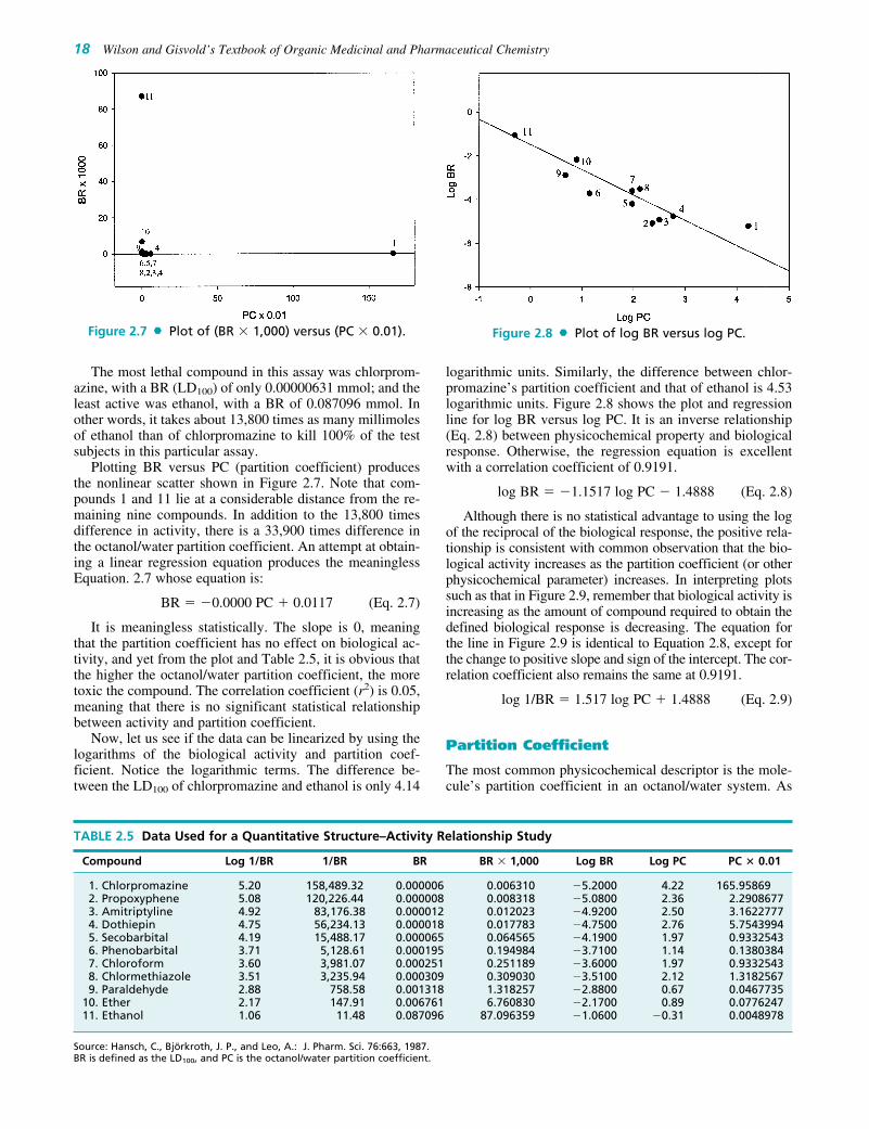
The most lethal compound in this assay was chlorprom-azine, with a BR (LD100) of only 0.00000631 mmol; and theleast active was ethanol, with a BR of 0.087096 mmol. Inother words, it takes about 13,800 times as many millimolesof ethanol than of chlorpromazine to kill 100% of the testsubjects in this particular assay.
Plotting BR versus PC (partition coefficient) producesthe nonlinear scatter shown in Figure 2.7. Note that com-pounds 1 and 11 lie at a considerable distance from the re-maining nine compounds. In addition to the 13,800 timesdifference in activity, there is a 33,900 times difference inthe octanol/water partition coefficient. An attempt at obtain-ing a linear regression equation produces the meaninglessEquation. 2.7 whose equation is:
BR � �0.0000 PC � 0.0117 (Eq. 2.7)
It is meaningless statistically. The slope is 0, meaningthat the partition coefficient has no effect on biological ac-tivity, and yet from the plot and Table 2.5, it is obvious thatthe higher the octanol/water partition coefficient, the moretoxic the compound. The correlation coefficient (r2) is 0.05,meaning that there is no significant statistical relationshipbetween activity and partition coefficient.
Now, let us see if the data can be linearized by using thelogarithms of the biological activity and partition coef-ficient. Notice the logarithmic terms. The difference be-tween the LD100 of chlorpromazine and ethanol is only 4.14
logarithmic units. Similarly, the difference between chlor-promazine’s partition coefficient and that of ethanol is 4.53logarithmic units. Figure 2.8 shows the plot and regressionline for log BR versus log PC. It is an inverse relationship(Eq. 2.8) between physicochemical property and biologicalresponse. Otherwise, the regression equation is excellentwith a correlation coefficient of 0.9191.
log BR � �1.1517 log PC � 1.4888 (Eq. 2.8)
Although there is no statistical advantage to using the logof the reciprocal of the biological response, the positive rela-tionship is consistent with common observation that the bio-logical activity increases as the partition coefficient (or otherphysicochemical parameter) increases. In interpreting plotssuch as that in Figure 2.9, remember that biological activity isincreasing as the amount of compound required to obtain thedefined biological response is decreasing. The equation forthe line in Figure 2.9 is identical to Equation 2.8, except forthe change to positive slope and sign of the intercept. The cor-relation coefficient also remains the same at 0.9191.
log 1/BR � 1.517 log PC � 1.4888 (Eq. 2.9)
Partition Coefficient
The most common physicochemical descriptor is the mole-cule’s partition coefficient in an octanol/water system. As
18 Wilson and Gisvold’s Textbook of Organic Medicinal and Pharmaceutical Chemistry
Figure 2.7 Plot of (BR � 1,000) versus (PC � 0.01).
TABLE 2.5 Data Used for a Quantitative Structure–Activity Relationship Study
Compound Log 1/BR 1/BR BR BR � 1,000 Log BR Log PC PC � 0.01
1. Chlorpromazine 5.20 158,489.32 0.000006 0.006310 �5.2000 4.22 165.958692. Propoxyphene 5.08 120,226.44 0.000008 0.008318 �5.0800 2.36 2.29086773. Amitriptyline 4.92 83,176.38 0.000012 0.012023 �4.9200 2.50 3.16227774. Dothiepin 4.75 56,234.13 0.000018 0.017783 �4.7500 2.76 5.75439945. Secobarbital 4.19 15,488.17 0.000065 0.064565 �4.1900 1.97 0.93325436. Phenobarbital 3.71 5,128.61 0.000195 0.194984 �3.7100 1.14 0.13803847. Chloroform 3.60 3,981.07 0.000251 0.251189 �3.6000 1.97 0.93325438. Chlormethiazole 3.51 3,235.94 0.000309 0.309030 �3.5100 2.12 1.31825679. Paraldehyde 2.88 758.58 0.001318 1.318257 �2.8800 0.67 0.0467735
10. Ether 2.17 147.91 0.006761 6.760830 �2.1700 0.89 0.077624711. Ethanol 1.06 11.48 0.087096 87.096359 �1.0600 �0.31 0.0048978
Source: Hansch, C., Björkroth, J. P., and Leo, A.: J. Pharm. Sci. 76:663, 1987.BR is defined as the LD100, and PC is the octanol/water partition coefficient.
Figure 2.8 Plot of log BR versus log PC.
0003-0042_17865_Ch02.qxd 12/4/09 2:52 AM Page 18

emphasized previously, the drug will go through a series ofpartitioning steps: (a) leaving the aqueous extracellular flu-ids, (b) passing through lipid membranes, and (c) enteringother aqueous environments before reaching the receptor(Fig. 2.1). In this sense, a drug is undergoing the same par-titioning phenomenon that happens to any chemical in a sep-aratory funnel containing water and a nonpolar solvent suchas hexane, chloroform, or ether. The partition coefficient (P)is the ratio of the molar concentration of chemical in thenonaqueous phase (usually 1-octanol) versus that in theaqueous phase (Eq. 2.10). For reasons already discussed, itis more common to use the logarithmic expression (Eq.2.11). The difference between the separatory funnel modeland what actually occurs in the body is that the partitioningin the funnel will reach an equilibrium at which the rate ofchemical leaving the aqueous phase and entering the organicphase will equal the rate of the chemical moving from theorganic phase to the aqueous phase. This is not the physio-logical situation. Refer to Figure 2.1 and note that dynamicchanges are occurring to the drug, such as it being metabo-lized, bound to serum albumin, excreted from the body, andbound to receptors. The environment for the drug is notstatic. Upon administration, the drug will be pushed throughthe membranes because of the high concentration of drug inthe extracellular fluids relative to the concentration in theintracellular compartments. In an attempt to maintain equi-librium ratios, the flow of the drug will be from systemiccirculation through the membranes onto the receptors. Asthe drug is metabolized and excreted from the body, it willbe pulled back across the membranes, and the concentrationof drug at the receptors will decrease.
P � (Eq. 2.10)
log P � log ( ) (Eq. 2.11)
Equations 2.10 and 2.11 assume that the drug is in the non-polar state. A large percentage of drugs are amines whose pKa
is such that at physiological pH 7.4, a significant percentageof the drug will be in its protonated, ionized form. A similarstatement can be made for the HA acids (carboxyl, sulfon-amide, imide) in that at physiological pH, a significant
[solute]oct
[solute]aq
[chemical]oct
[chemical]aq
percentage will be in their anionic forms. An assumption ismade that the ionic form is water-soluble and will remain inthe water phase of an octanol/water system. This reality hasled to the use of log D, which is defined as the equilibriumratio of both the ionized and un-ionized species of the mole-cule in an octanol/water system (Eq. 2.12). The percent ion-ization of ionized HA acids and BH protonated amines andacids can be estimated from Equations 2.3 and 2.4 and the logD from Equations 2.13 and 2.14, respectively.
log D � log( ) (Eq. 2.12)
log Dacids � log P � log[ ] (Eq. 2.13)
log Dbases � log P � log[ ] (Eq. 2.14)
Because much of the time the drug’s movement acrossmembranes is a partitioning process, the partition coef-ficient has become the most common physicochemicalproperty. The question that now must be asked is whatimmiscible nonpolar solvent system best mimics thewater/lipid membrane barriers found in the body? It is nowrealized that the n-octanol/water system is an excellent esti-mator of drug partitioning in biological systems. One couldargue that it was fortuitous that n-octanol was available inreasonable purity for the early partition coefficient determi-nations. To appreciate why this is so, one must understandthe chemical nature of the lipid membranes.
These membranes are not exclusively anhydrous fatty oroily structures. As a first approximation, they can be consid-ered bilayers composed of lipids consisting of a polar capand large hydrophobic tail. Phosphoglycerides are majorcomponents of lipid bilayers (Fig. 2.10). Other groups of bi-functional lipids include the sphingomyelins, galactocere-brosides, and plasmalogens. The hydrophobic portion iscomposed largely of unsaturated fatty acids, mostly with cisdouble bonds. In addition, there are considerable amounts ofcholesterol esters, protein, and charged mucopolysaccha-rides in the lipid membranes. The final result is that thesemembranes are highly organized structures composed ofchannels for transport of important molecules such asmetabolites, chemical regulators (hormones), amino acids,glucose, and fatty acids into the cell and removal of wasteproducts and biochemically produced products out of the
1
(1 � 10(pKa�pH))
1
(1 � 10(pH�pKa))
[solute]oct
[solute]aqionized � [solute]aq
nonionized
Chapter 2 Drug Design Strategies 19
Figure 2.9 Plot of log 1/BR versus log PC.
Figure 2.10 General structure of a bifunctionalphospholipid. Many of the fatty acid esters will be cisunsaturated.
0003-0042_17865_Ch02.qxd 12/4/09 2:52 AM Page 19

cell. The cellular membranes are dynamic, with the channelsforming and disappearing depending on the cell’s andbody’s needs (Fig. 2.11). So complex is this system that it isnot uncommon to have situations where there is poor corre-lation between the partition coefficient of a series of mole-cules and the biological response.2
In addition, the membranes on the surface of nucleatedcells have specific antigenic markers, major histocompatibil-ity complex (MHC), by which the immune system monitorsthe cell’s status. There are receptors on the cell surface wherehormones such as epinephrine and insulin bind, setting off aseries of biochemical events within the cell. Some of these re-ceptors are used by viruses to gain entrance into the cells,where the virus reproduces. As newer instrumental tech-niques are developed, and genetic cloning permits isolation ofthe genetic material responsible for forming and regulatingthe structures on the cell surface, the image of a passive lipidmembrane has disappeared to be replaced by a very complex,highly organized, dynamically functioning structure.
For purposes of the partitioning phenomenon, picture thecellular membranes as two layers of lipids (Fig. 2.9). The twoouter layers, one facing the interior and the other facing theexterior of the cell, consist of the polar ends of the bifunc-tional lipids. Keep in mind that these surfaces are exposed toan aqueous polar environment. The polar ends of the chargedphospholipids and other bifunctional lipids are solvated bythe water molecules. There are also considerable amounts ofcharged proteins and mucopolysaccharides present on the sur-face. In contrast, the interior of the membrane is populated bythe hydrophobic aliphatic chains from the fatty acid esters.
With this representation in mind, a partial explanation canbe presented as to why the n-octanol/water partitioningsystem seems to mimic the lipid membranes/water systemsfound in the body. It turns out that n-octanol is not as non-polar as initially might be predicted. Water-saturated octanolcontains 2.3 M water because the small water moleculeeasily clusters around octanol’s hydroxy moiety. n-Octanol–saturated water contains little of the organic phasebecause of the large hydrophobic 8-carbon chain of octanol.The water in the n-octanol phase apparently approximatesthe polar properties of the lipid bilayer, whereas the lack ofoctanol in the water phase mimics the physiological aqueouscompartments, which are relatively free of nonpolar compo-nents. In contrast, partitioning systems such as hexane/waterand chloroform/water contain so little water in the organicphase that they are poor models for the lipid bilayer/watersystem found in the body. At the same time, remember thatthe n-octanol/water system is only an approximation of theactual environment found in the interface between the cel-lular membranes and the extracellular/intracellular fluids.
Experimental determination of octanol/water partition coef-ficients is tedious and time consuming. Today, most arecalculated. The accuracy of these calculations is only as goodas the assumptions made by the writers of the software.These include atomic fragment values, correction factors,spatial properties, effects of resonance and induction, inter-nal secondary bonding forces, etc. There are over 30 differ-ent software packages for calculating a molecules partitioncoefficient, and their accuracy varies widely.3,4
Other Physicochemical and Descriptor Parameters
There is a series of other descriptors that measure the contri-bution by substituents to the molecule’s total physicochemi-cal properties. These include Hammett’s � constant; Taft’ssteric parameter, Es; Charton’s steric parameter, v; Verloop’smultidimensional steric parameters, L, B1, B5; and molar re-fractivity, MR, number of hydrogen bond donors and accep-tors, pKa, polar surface area, number of rotatable bonds,connectivity indices, and the list goes into the thousands.Although directories of these have been published, it is com-mon to calculate them. Table 2.6 lists a very small set andillustrates several items that must be kept in mind when se-lecting substituents to be evaluated in terms of the type offactors that influence a biological response. For electronic pa-rameters such as �, the location on an aromatic ring is impor-tant because of resonance versus inductive effects. Notice thetwofold differences seen between �meta and �para for the threealiphatic substituents and iodo, and severalfold difference formethoxy, amino, fluoro, and phenolic hydroxyl.
Selection of substituents from a certain chemical classmay not really test the influence of a parameter on biologicalactivity. There is little numerical difference among the �meta
or �para values for the four aliphatic groups or the four halo-gens. It is not uncommon to go to the tables and find missingparameters such as the Es values for acetyl and N-acyl.
20 Wilson and Gisvold’s Textbook of Organic Medicinal and Pharmaceutical Chemistry
TABLE 2.6 Sampling of Physicochemical ParametersUsed in Quantitative Structure–Activity RelationshipsInvestigations
Substituent Group � �meta �para Es MR
MH 0.00 0.00 0.00 0.00 1.03MCH3 0.56 �0.07 �0.17 �1.24 5.65MCH2CH3 1.02 �0.07 �0.15 �1.31 10.30MCH2CH2CH3 1.55 �0.07 �0.13 �1.60 14.96MC(CH3)2 1.53 �0.07 �0.15 �1.71 14.96MOCH3 �0.02 0.12 �0.27 �0.55 7.87MNH2 �1.23 �0.16 �0.66 �0.61 5.42MF 0.14 0.34 0.06 �0.46 0.92MCl 0.71 0.37 0.23 �0.97 6.03MBr 0.86 0.39 0.23 �1.16 8.88MI 1.12 0.35 0.18 �1.40 13.94MCF3 0.88 0.43 0.54 �2.40 5.02MOH �0.67 0.12 �0.37 �0.55 2.85MCOCH3 �0.55 0.38 0.50 11.18MNHCOCH3 �0.97 0.21 0.00 14.93MNO2 �0.8 0.71 0.78 �2.52 7.36MCN �0.57 0.56 0.66 �0.51 6.33
Reprinted with permission from Hansch, C., and Leo, A. J.: SubstituentConstants for Correlation Analysis in Chemistry and Biology. New York,John Wiley & Sons, 1979.
Figure 2.11 Schematic representation of the cellmembrane.
0003-0042_17865_Ch02.qxd 12/4/09 2:52 AM Page 20

Nevertheless, medicinal chemists can use informationfrom extensive tables of physicochemical parameters to min-imize the number of substituents required to find out if thebiological response is sensitive to electronic, steric, and/orpartitioning effects.5 This is done by selecting substituents ineach of the numerical ranges for the different parameters. InTable 2.6, there are three ranges of B values (�1.23 to�0.55, �0.28 to 0.56, and 0.71 to 1.55); three ranges of MRvalues (0.92–2.85, 5.02–8.88, and 10.30–14.96); and twomain clusters of � values, one for the aliphatic substituentsand the other for the halogens. In the ideal situation, sub-stituents are selected from each of the clusters to determinethe dependence of the biological response over the largestpossible variable space. Depending on the biological re-sponses obtained from testing the new compounds, it is pos-sible to determine if lipophilicity (partitioning), steric bulk(molar refraction), or electron-withdrawing/donating prop-erties are important determinants of the desired biologicalresponse.
With this background in mind, two examples of QSARequations taken from the medicinal chemistry literature arepresented. A study of a group of griseofulvin analogsshowed a linear relationship (Eq. 2.15) between the biolog-ical response and both lipophilicity (log P) and electroniccharacter (�).6 It was suggested that the antibiotic activitymay depend on the enone system facilitating the addition ofgriseofulvin to a nucleophilic group such as the SH moietyin a fungal enzyme.
log BR � (0.56)log P � (2.19)�x � 1.32 (Eq. 2.15)
A parabolic relationship (Eq. 2.16) was reported for aseries of substituted acetylated salicylates (substitutedaspirins) tested for anti-inflammatory activity.7 A nonlin-ear relationship exists between the biological responseand lipophilicity, and a significant detrimental steric effect
is seen with substituents at position 4. The two sterimolparameters used in this equation were L, defined as thelength of the substituent along the axis of the bond betweenthe first atom of the substituent and the parent molecule, andB2, defined as a width parameter. Steric effects were notconsidered statistically significant at position 3, as shown bythe sterimol parameters for substituents at position 3 notbeing part of Equation 2.16. The optimal partition coeffi-cient (log Po) for the substituted aspirins in this assay was2.6. At the same time, increasing bulk, as measured by thesterimol parameters, decreases activity.
log 1/ED50 � 1.03 log P � 0.20(log P)2
�0.05 L(4) � 0.24 B2(4) � 2.29 (Eq. 2.16)
At this point, it is appropriate to ask the question, “areall the determinations of partition coefficients and compi-lation of physical chemical parameters useful only when astatistically valid QSAR model is obtained?” The answeris a firm no. One of the most useful spinoffs from the fieldof QSAR has been the application of experimental designto the selection of new compounds to be synthesized andtested. Let us assume that a new series of drug moleculesis to be synthesized based on the following structure. Thegoal is to test the effect of the 16-substituents in Table 2.6at each of the three positions on our new series. The num-ber of possible analogs is equal to 163, or 4,096, com-pounds, assuming that all three positions will always besubstituted with one of the substituents from Table 2.7. Ifhydrogen is included when a position is not substituted,there are 173, or 4,913, different combinations. The prob-lem is to select a small number of substituents that repre-sent the different ranges or clusters of values forlipophilicity, electronic influence, and bulk. An initial de-sign set could include the methyl and propyl from the
Chapter 2 Drug Design Strategies 21
TABLE 2.7 Connectivity Table for Hydrogen-Suppressed Phenylalanine
Atom O-1 C-2 O-3 C-4 N-5 C-6 C-7 C-8 C-9 C-10 C-11 C-12
O-1 X
C-2 X X X
O-3 X
C-4 X X X
N-5 X
C-6 X X
C-7 X X X
C-8 X X
C-9 X X
C-10 X X
C-11 X X
C-12 X X
0003-0042_17865_Ch02.qxd 12/4/09 2:52 AM Page 21

aliphatic cluster, fluorine and chlorine from the halogencluster, N-acetyl and phenol from the substituents showinghydrophilicity, and a range of electronic and bulk values.Including hydrogen, there will be 73, or 343, differentcombinations. Obviously, that is too many for an initialevaluation. Instead, certain rules have been devised tomaximize the information obtained from a minimum num-ber of compounds. These include the following:
1. Each substituent must occur more than once at each posi-tion on which it is found.
2. The number of times that each substituent at a particularposition appears should be approximately equal.
3. No two substituents should be present in a constantcombination.
4. When combinations of substituents are a necessity, theyshould not occur more frequently than any othercombination.
Following these guidelines, the initial test set can be re-duced to 24 to 26 compounds. Depending on the precisionof the biological tests, it will be possible to see if the datawill fit a QSAR model. Even an approximate model usuallywill indicate the types of substituents to test further andwhat positions on the molecules are sensitive to substitutionand, if sensitive, to what degree variation in lipophilic, elec-tronic, or bulk character is important. Just to ensure that themodel is valid, it is a good idea to synthesize a couple ofcompounds that the model predicts would be inactive. Aseach group of new compounds is tested, the QSAR model isrefined until the investigators have a pretty good idea whatsubstituent patterns are important for the desired activity.These same techniques used to develop potent compoundswith desired activity also can be used to evaluate the influ-ence of substituent patterns on undesired toxic effects andpharmacokinetic properties.
Topological Descriptors
An alternate method of describing molecular structure isbased on graph theory, in which the bonds connecting theatoms is considered a path that is traversed from one atom toanother. Consider Figure 2.12 containing D-phenylalanine
and its hydrogen-suppressed graph representation. Thenumbering is arbitrary and not based on International Unionof Pure and Applied Chemistry (IUPAC) or ChemicalAbstracts nomenclature rules. A connectivity table, Table2.7, is constructed.
Table 2.7 is a two-dimensional connectivity table for thehydrogen-suppressed phenylalanine molecule. No 3D repre-sentation is implied. Further, this type of connectivity tablewill be the same for molecules with asymmetric atoms (Dvs. L) or for those that can exist in more than one conforma-tion (i.e., chair vs. boat conformation, anti vs. gauche vs.eclipsed).
Graph theory is not limited to the paths followed bychemical bonds. In its purest form, the atoms in the phenylring of phenylalanine would have paths connecting atom 7with atoms 9, 10, 11, and 12; atom 8 with atoms 10, 11, and12; atom 9 with atoms 11 and 12; and atom 10 with atom 12.Also, the graph itself might differentiate neither single, dou-ble, and triple bonds nor the type of atom (C, O, and N in thephenylalanine example). Connectivity tables can be codedto indicate the type of bond.
The most common application of graph theory used bymedicinal chemistry is called molecular connectivity. It lim-its the paths to the molecule’s actual chemical bonds. Table2.8 shows several possible paths for phenylalanine, includ-ing linear paths and clusters or branching. Numerical valuesfor each path or path-cluster are based on the number ofnonhydrogen bonds to each atom. Let us examine oxygenatom 1. There is only one nonhydrogen bond, and it con-nects oxygen atom 1 to carbon atom 2. The formula is thereciprocal square root of the number of bonds. For oxygen1, the connectivity value is 1. For carbonyl oxygen 2, it is2�1/2, or 0.707. Note that there is no difference betweenoxygen 1 and nitrogen 5. Both have only one nonhydrogenbond and a connectivity value of 1. Similarly, there is no dif-ference in values for a carbonyl oxygen and a methylenecarbon, each having two nonhydrogen bonds. The final con-nectivity values for a path are the reciprocal square roots ofthe products of each path. For the second-order path 2C-4C-6C, the reciprocal square root (3 � 3 � 2)�1/2 is 4.243. Thevalues for each path order are calculated and summed.
As noted previously, the method as described so farcannot distinguish between atoms that have the same num-ber of nonhydrogen bonds. A method to distinguish het-eroatoms from each other and carbon atoms is based on thedifference between the number of valence electrons andpossible hydrogen atoms (which are suppressed in thegraph). The valence connectivity term for an alcoholicoxygen would be 6 valence electrons minus 1 hydrogen,or 5. The valence connectivity term for a primary amine
22 Wilson and Gisvold’s Textbook of Organic Medicinal and Pharmaceutical Chemistry
Figure 2.12 Hydrogen-suppressedgraphic representation of phenylalanine.
0003-0042_17865_Ch02.qxd 12/4/09 2:52 AM Page 22

nitrogen would be 5 valence electrons minus 2 hydrogens,or 3. There are various additional modifications that aredone to further differentiate atoms and define their envi-ronments within the molecule.
Excellent regression equations using topological indiceshave been obtained. A problem is interpreting what theymean. Is it lipophilicity, steric bulk, or electronic terms thatdefine activity? The topological indices can be correlatedwith all of these common physicochemical descriptors.Another problem is that it is difficult to use the equation todecide what molecular modifications can be made to en-hance activity further, again because of ambiguities inphysicochemical interpretation. Should the medicinalchemist increase or decrease lipophilicity at a particular lo-cation on the molecule? Should specific substituents be in-creased or decreased? On the other hand, topological indicescan be very valuable in classification schemes that are de-scribed later in this chapter. They do describe the structurein terms of rings, branching, flexibility, etc.
Classification Methods
Besides regression analysis, there are other statistical tech-niques used in drug design. These fit under the classificationof multivariate statistics and include discriminant analysis,principal component analysis, and pattern recognition. Thelatter can consist of a mixture of statistical and nonstatisticalmethodologies. The goal usually is to try to ascertain whatphysicochemical parameters and structural attributes con-tribute to a class or type of biological activity. Then, thechemicals are classified into groupings such as carcino-genic/noncarcinogenic, sweet/bitter, active/inactive, and de-pressant/stimulant.
The term multivariate is used because of the wide varietyand number of independent or descriptor variables that maybe used. The same physicochemical parameters seen inQSAR analyses are used, but in addition, the software in thecomputer programs breaks the molecule down into sub-structures. These structural fragments also become vari-ables. Examples of the typical substructures used includecarbonyls, enones, conjugation, rings of different sizes andtypes, N-substitution patterns, and aliphatic substitution pat-terns such as 1,3- or 1,2-disubstituted. The end result is thatfor even a moderate-size molecule typical of most drugs,there can be 50 to 100 variables.
The technique is to develop a large set of chemicals wellcharacterized in terms of the biological activity that is goingto be predicted. This is known as the training set. Ideally, itshould contain hundreds, if not thousands, of compounds,divided into active and inactive types. In reality, sets smallerthan 100 are studied. Most of these investigations are retro-spective ones in which the investigator locates large datasets from several sources. This means that the biologicaltesting likely followed different protocols. That is whyclassification techniques tend to avoid using continuousvariables such as ED50, LD50, and MIC. Instead, arbitraryend points such as active or inactive, stimulant or depres-sant, sweet or sour, are used.
Once the training set is established, the multivariate tech-nique is carried out. The algorithms are designed to group theunderlying commonalities and select the variables that havethe greatest influence on biological activity. The predictiveability is then tested with a test set of compounds that havebeen put through the same biological tests used for the train-ing set. For the classification model to be valid, theinvestigator must select data sets whose results are notintuitively obvious and could not be classified by a trainedmedicinal chemist. Properly done, classification methods canidentify structural and physicochemical descriptors that can bepowerful predictors and determinants of biological activity.
There are several examples of successful applications ofthis technique.8 One study consisted of a diverse group of140 tranquilizers and 79 sedatives subjected to a two-wayclassification study (tranquilizers vs. sedatives). The ringtypes included phenothiazines, indoles, benzodiazepines,barbiturates, diphenylmethanes, and various heterocyclics.Sixty-nine descriptors were used initially to characterizethe molecules. Eleven of these descriptors were crucial to theclassification, 54 had intermediate use and depended on thecomposition of the training set, and 4 were of little use.The overall range of prediction accuracy was 88% to 92%.The results with the 54 descriptors indicate an important lim-itation when large numbers of descriptors are used. Theinclusion or exclusion of descriptors and parameters can de-pend on the composition of the training set. The training setmust be representative of the population of chemicals that aregoing to be evaluated. Repeating the study on different ran-domly selected training sets is important.
Classification techniques lend themselves to studies lack-ing quantitative data. An interesting classification problem
Chapter 2 Drug Design Strategies 23
TABLE 2.8 Examples of Paths Found in the Phenylalanine Molecule
1st Order Path 2nd Order Path 3rd Order Path 4th Order Path 5th Order Path Path-Cluster
1O-2C 1O-2C-3O 1O-2C-4C-6C 1O-2C-4C-6C-7C 1O-2C-4C-6C-7C-8C 1O-2C-3O-4C2C-3O 1O-2C-4C 3O-2C-4C-5N 3O-2C-4C-6C-7C 1O-2C-4O-6C-7C-12C 2C-4C-5N-6C2C-4C 3O-2C-4C 3O-2C-4C-6C 2C-4C-6C-7C-8C 2C-4C-6C-7C-8C-9C 6C-7C-8C-12C4C-5N 2C-4C-5N 1O-2C-4C-5N 2C-4C-6C-7C-12C 2C-4C-6C-7C-12C-11C4C-6C 2C-4C-6C 2C-4C-6C-7C 4C-6C-7C-8C-9C 3O-2C-4C-6C-7C-8C6C-7C 5N-4C-6C 5N-4C-6C-7C 4C-6C-7C-12C-11C 3O-2C-4C-6C-7C-12C7C-8C 4C-6C-7C 4C-6C-7C-8C 5N-4C-6C-7C-8C 4C-6C-7C-8C-9C-10C8C-9C 6C-7C-8C 4C-6C-7C-12C 5N-4C-6C-7C-12C 4C-6C-7C-12C-11C-10C7C-12C 6C-7C-12C 6C-7C-8C-9C 6C-7C-8C-9C-10C 5N-4C-6C-7C-8C-9C9C-10C 7C-8C-9C 6C-7C-12C-11C 6C-7C-12C-11C-10C 5N-4C-6C-7C-12C-11C10C-11C 7C-12C-11C 7C-8C-9C-10C 7C-8C-9C-10C-11C 6C-7C-8C-9C-10C-11C11C-12C 8C-9C-10C 7C-12C-11C-10C 7C-12C-11C-10C-9C 6C-7C-12C-11C-10C
9C-10C-11C 8C-9C-10C-11C 8C-9C-10C-11C-12C 7C-8C-9C-10C-11C-12C10C-11C-12C 9C-10C-11C-12C 7C-12C-11C-10C-9C-8C
0003-0042_17865_Ch02.qxd 12/4/09 2:52 AM Page 23

involved olfactory stimulants, in which the goal was to se-lect chemicals that had a musk odor. A group of 300 uniquecompounds was selected from a group of odorants that in-cluded 60 musk odorants plus 49 camphor, 44 floral, 32ethereal, 41 mint, 51 pungent, and 23 putrid odorants.Initially, 68 descriptors were evaluated. Depending on theapproach, the number of descriptors was reduced to 11 to16, consisting mostly of bond types. Using this small num-ber, the 60 musk odorants could be selected from the re-maining 240 compounds, with an accuracy of 95% to 97%.
The use of classification techniques in medicinal chem-istry has matured over years of general use. The types of de-scriptors have expanded to spatial measurements in 3Dspace similar to those used in 3D-QSAR (see discussion thatfollows). Increasingly, databases of existing compounds arescanned for molecules that possess what appear to be the de-sired parameters. If the scan is successful, compounds thatare predicted to be active provide the starting point for syn-thesizing new compounds for testing. One can see parallelsbetween the search of chemical databases and screeningplant, animal, and microbial sources for new compounds.Although the statistical and pattern recognition methodolo-gies have been in use for a very long time, there still needsto be considerable research into their proper use, and furthertesting of their predictive power is needed. The goal of scan-ning databases of already synthesized compounds to selectcompounds for pharmacological evaluation will requireconsiderable additional development of the various multi-variate techniques.
This chapter is limited to fairly simple computational tech-niques using readily available, low-cost software. The QSARapproach, including classification methods has at its disposalliterally thousands of different descriptors, each with its advo-cates, and many computational approaches starting with thepreviously discussed linear regression to neural networks, de-cision trees and support vector machines. Thus, it is fair to askif drug discoveries have been made with these computationaltechniques. The answer is in the ambiguous yes-and-no cate-gory depending on who is asked. There is general agreement
that QSAR provided the foundation to better understand therelationship between chemical space and pharmacologicalspace. Consider these two pairs of active molecules. For clas-sification purposes, acetylcholine and nicotine are nicotinicagonists, and dopamine and pergolide are dopaminergic ago-nists. Using the various measures of similarity and their de-scriptors, fingerprints, and fragments, these two pairs comeup as dissimilar. It is true that the pharmacological profiles ofacetylcholine—nicotine and dopamine—pergolide vary somuch that nicotine is not used as a nicotinic agonist andpergolide is falling into disuse.9,10 Without being trite, com-putational drug design techniques are not going to replace themedicinal chemist who has an open, inquiring mind.
Has QSAR Been Successful?
The answer to this question depends on what are the expec-tations.11 In their original development, it was hoped thatQSAR equations would lead to commercially successfuldrugs. This has not occurred. Over the years, methodologiesto develop these equations have changed. In the early devel-opment of QSAR equations, all of the compounds in a dataset were used followed by random holding out of com-pounds to see if the equation changed. If there are enoughcompounds, it is now more common to begin with a train-ing set and evaluate its validity with a test set. This has ledto recommendations on the most reliable statistical measure-ments of validity.12–14
Paralleling the evolution in the use of different statisticalmeasures of validity, deriving these equations has lead to thedevelopment of a wide variety of descriptors ranging fromdescriptive, physicochemical and topological. What can befrustrating is that the quality of predictions is dependent onthe descriptor set.15 It must be remembered that most of de-scriptors are only as good as the algorithms used to calculatethem. Further, it can be difficult to interpret exactly what thedescriptors are measuring in chemical space. QSAR equa-tions must explain physical reality if predictions for futurecompounds are to be made.16
24 Wilson and Gisvold’s Textbook of Organic Medicinal and Pharmaceutical Chemistry
0003-0042_17865_Ch02.qxd 12/4/09 2:52 AM Page 24

COMPUTER-AIDED DRUG DESIGN:NEWER METHODS
Because powerful computing power, high-resolution com-puter graphics, and applicable software has reached thedesktop, computational drug design methods are widelyused in both industrial and academic environments.Through the use of computer graphics, structures of or-ganic molecules can be entered into a computer and manip-ulated in many ways. Computational chemistry methodsare used to calculate molecular properties and generatepharmacophore hypotheses. Once a pharmacophore hy-pothesis has been developed, structural databases (com-mercial, corporate, and/or public) of 3D structures can besearched rapidly for hits (i.e., existing compounds that areavailable with the required functional groups and permissi-ble spatial orientations as defined by the search query). Ithas become popular to carry out in silico (computer as op-posed to biological) screening of drug–receptor candidateinteractions, known as virtual high-throughput screening(vHTS), for future development. The realistic goal of vHTSis to identify potential lead compounds. The drug–receptorfit and predicted physicochemical properties are used toscore and rank compounds according to penalty functionsand information filters (molecular weight, number of hy-drogen bonds, hydrophobicity, etc.). Although medicinalchemists have always been aware of absorption, distribu-tion, metabolism, elimination, and toxicity (ADMET orADME/Tox), in recent years, a much more focused ap-proach addresses these issues in the early design stages.Increased efforts to develop computer-based absorption,distribution, metabolism, and elimination (ADME) modelsare being pursued aggressively. Many of the predictiveADME models use QSAR methods described earlier inthis chapter. In general, understanding what chemicalspace descriptors are critical for druglike molecules helpsprovide insight into the design of chemical libraries forbiological evaluation.
Today’s computers and software give the medicinalchemist the ability to design the molecule on the basis of anestimated fit onto a receptor or have similar spatial charac-teristics found in the prototypical lead compound. Ofcourse, this assumes that the molecular structure of the re-ceptor is known in enough detail for a reasonable estimationof its 3D shape. When a good understanding of the geome-try of the active site is known, databases containing the 3Dcoordinates of the chemicals in the database can be searchedrapidly by computer programs that select candidates likelyto fit in the active site. As shown later, there have been somedramatic successes with use of this approach, but first onemust have an understanding of ligand (drug)–receptor inter-actions and conformational analysis.
Forces Involved with Drug–ReceptorInteractions
Keep in mind that a biological response is produced by theinteraction of a drug with a functional or organized group ofmolecules. This interaction would be expected to take placeby using the same bonding forces as are involved when sim-ple molecules interact. These, together with typical exam-ples, are collected in Table 2.9.
Most drugs do not possess functional groups of a typethat would lead to ready formation of strong and essentiallyirreversible covalent bonds between drug and biological re-ceptors. In most cases, it is desirable to have the drug leavethe receptor site when the concentration decreases in the ex-tracellular fluids. Therefore, most useful drugs are held totheir receptors by ionic or weaker bonds. When relativelylong-lasting or irreversible effects are desired (e.g., antibac-terial, anticancer), drugs that form covalent bonds with thereceptor are effective and useful. The alkylating agents,such as the nitrogen mustards used in cancer chemotherapy,furnish an example of drugs that act by formation of cova-lent bonds (see Chapter 10).
Covalent bond formation between drug and receptor isthe basis of Baker’s concept of active-site-directed irre-versible inhibition.17 Considerable experimental evidenceon the nature of enzyme inhibitors supports this concept.Compounds studied possess appropriate structural featuresfor reversible and highly selective association with an en-zyme. If, in addition, the compounds carry reactive groupscapable of forming covalent bonds, the substrate may be ir-reversibly bound to the drug–receptor complex by covalentbond formation with reactive groups adjacent to the activesite. The diuretic drug ethacrynic acid (see Chapter 19) isan �,�-unsaturated ketone, thought to act by covalent bondformation with sulfhydryl groups of ion transport systemsin the renal tubules. Another example of a drug that cova-lently binds to the receptor is selegiline (see Chapter 13), aninhibitor of monoamine oxidase-B. Other examples of co-valent bond formation between drug and biological recep-tor site include the reaction of arsenicals and mercurialswith cysteine sulfhydryl groups, the acylation of bacterialcell wall constituents by penicillin, and the phosphorylation
Chapter 2 Drug Design Strategies 25
TABLE 2.9 Types of Chemical Bonds
Bond Strength
Bond Type (kcal/mol) Example
Covalent 40–140
Reinforced ionic 10
Ionic 5
Hydrogen 1–7
Ion–dipole 1–7
Dipole–dipole 1–7
van der Waals 0.5–1
Hydrophobic 1 See text
Source: Albert, A.: Selective Toxicity. New York, John Wiley & Sons,1986, p. 183.
0003-0042_17865_Ch02.qxd 12/4/09 2:52 AM Page 25

of the serine hydroxyl moiety at the active site ofcholinesterase by organic phosphates.
Keep in mind that it is desirable to have most drug effectsreversible. For this to occur, relatively weak forces must beinvolved in the drug–receptor complex yet be strong enoughthat other binding sites will not competitively deplete thesite of action. Compounds with high structural specificitymay orient several weakly binding groups so that the sum-mation of their interactions with specifically oriented com-plementary groups on the receptor provides total bondstrength sufficient for a stable combination. Consequently,most drugs acting by virtue of their structural specificitywill bind to the receptor site by hydrogen bonds, ionicbonds, ion–dipole and dipole–dipole interactions, and vander Waals and hydrophobic forces.
Considering the wide variety of functional groups foundon a drug molecule and receptor, there will be a variety of sec-ondary bonding forces. Ionization at physiological pH wouldnormally occur with the carboxyl, sulfonamido, and aliphaticamino groups, as well as the quaternary ammonium group atany pH. These sources of potential ionic bonds are frequentlyfound in active drugs. Differences in electronegativity be-tween carbon and other atoms, such as oxygen and nitrogen,lead to an asymmetric distribution of electrons (dipoles) thatare also capable of forming weak bonds with regions of highor low electron density, such as ions or other dipoles.Carbonyl, ester, amide, ether, nitrile, and related groups thatcontain such dipolar functions are frequently found in equiv-alent locations in structurally specific drugs.
The relative importance of the hydrogen bond in the for-mation of a drug–receptor complex is difficult to assess.Many drugs possess groups such as carbonyl, hydroxyl,amino, and imino, with the structural capabilities of acting asacceptors or donors in the formation of hydrogen bonds.However, such groups would usually be solvated by water,as would the corresponding groups on a biological receptor.Relatively little net change in free energy would be expectedin exchanging a hydrogen bond with a water molecule forone between drug and receptor. However, in a drug–receptorcombination, several forces could be involved, including thehydrogen bond, which would contribute to the stability ofthe interaction. Where multiple hydrogen bonds may beformed, the total effect may be sizable, such as that demon-strated by the stability of the protein �-helix and by the sta-bilizing influence of hydrogen bonds between specific basepairs in the double-helical structure of DNA.
Van der Waals forces are attractive forces created by thepolarizability of molecules and are exerted when any two un-charged atoms approach each other very closely. Theirstrength is inversely proportional to the seventh power of thedistance. Although individually weak, the summation of theirforces provides a significant bonding factor in higher–molecular-weight compounds. For example, it is not possibleto distill normal alkanes with more than 80 carbon atoms, be-cause the energy of 80 kcal/mol required to separate the mol-ecules is approximately equal to the energy required to breaka carbon–carbon covalent bond. Flat structures, such as aro-matic rings, permit close approach of atoms. The aromatic ringis frequently found in active drugs, and a reasonable explana-tion for its requirement for many types of biological activitymay be derived from the contributions of this flat surface tovan der Waals binding to a correspondingly flat receptor area.
The hydrophobic bond is a concept used to explain attrac-tive interactions between nonpolar regions of the receptor andthe drug. Explanations such as the isopropyl moiety of thedrug fits into a hydrophobic cleft on the receptor composed ofthe hydrocarbon side chains of the amino acids valine,isoleucine, and leucine are commonly used to explain why anonpolar substituent at a particular position on the drug mol-ecule is important for activity. Over the years, the concept ofhydrophobic bonds has developed. There has been consider-able controversy over whether the bond actually exists.Thermodynamic arguments on the gain in entropy (decreasein ordered state) when hydrophobic groups cause a partialcollapse of the ordered water structure on the surface of thereceptor have been proposed to validate a hydrophobic bond-ing model. There are two problems with this concept. First,the term hydrophobic implies repulsion. The term for attrac-tion is hydrophilicity. Second, and perhaps more important,there is no truly water-free region on the receptor. This is trueeven in the areas populated by the nonpolar amino acid sidechains. An alternate approach is to consider only the conceptof hydrophilicity and lipophilicity. The predominating watermolecules solvate polar moieties, effectively squeezing thenonpolar residues toward each other.
STERIC FEATURES OF DRUGS
Regardless of the ultimate mechanism by which the drug andthe receptor interact, the drug must approach the receptor andfit closely to its surface. Steric factors determined by thestereochemistry of the receptor site surface and that of thedrug molecules are, therefore, of primary importance in de-termining the nature and the efficiency of the drug–receptorinteraction. With the possible exception of the general anes-thetics, such drugs must possess a high structural specificityto initiate a response at a particular receptor.
Some structural features contribute a high structuralrigidity to the molecule. For example, aromatic rings areplanar, and the atoms attached directly to these rings areheld in the plane of the aromatic ring. Hence, the quaternary
26 Wilson and Gisvold’s Textbook of Organic Medicinal and Pharmaceutical Chemistry
0003-0042_17865_Ch02.qxd 12/4/09 2:52 AM Page 26

nitrogen and carbamate oxygen attached directly to the ben-zene ring in the cholinesterase inhibitor neostigmine are re-stricted to the plane of the ring, and consequently, the spa-tial arrangement of at least these atoms is established.
The relative positions of atoms attached directly to mul-tiple bonds are also fixed. For the double bond, cis- andtrans-isomers result. For example, diethylstilbestrol existsin two fixed stereoisomeric forms: trans-diethylstilbestrol isestrogenic, whereas the cis-isomer is only 7% as active. Intrans-diethylstilbestrol, resonance interactions and minimalsteric interference tend to hold the two aromatic rings andconnecting ethylene carbon atoms in the same plane.
Geometric isomers, such as the cis- and the trans-isomers,hold structural features at different relative positions in space.These isomers also have significantly different physical andchemical properties. Therefore, their distributions in the bio-logical medium are different, as are their capabilities for inter-acting with a biological receptor in a structurally specificmanner. The United States Pharmacopeia recognizes thatthere are drugs with vinyl groups whose commercial formcontains both their E- and Z-isomers. Figure 2.13 providesfour examples of these mixtures.
More subtle differences exist for conformational iso-mers. Like geometric isomers, these exist as differentarrangements in space for the atoms or groups in a singleclassic structure. Rotation about bonds allows interconver-sion of conformational isomers. However, an energy barrierbetween isomers is often high enough for their independentexistence and reaction. Differences in reactivity of func-tional groups or interaction with biological receptors maybe caused by differences in steric requirements of the re-ceptors. In certain semirigid ring systems, conformationalisomers show significant differences in biological activi-ties. Methods for calculating these energy barriers are de-scribed next.
Open chains of atoms, which form an important part ofmany drug molecules, are not equally free to assume all pos-sible conformations; some are sterically preferred. Energybarriers to free rotation of the chains are present because ofinteractions of nonbonded atoms. For example, the atomstend to position themselves in space so that they occupystaggered positions, with no two atoms directly facing eachother (eclipsed). Nonbonded interactions in polymethylene
Chapter 2 Drug Design Strategies 27
Figure 2.13 Examples of E- and Z-isomers.
0003-0042_17865_Ch02.qxd 12/4/09 2:52 AM Page 27

chains tend to favor the most extended anti conformations,although some of the partially extended gauche conforma-tions also exist. Intramolecular bonding between substituentgroups can make what might first appear to be an unfavor-able conformation favorable.
The introduction of atoms other than carbon into a chainstrongly influences the conformation of the chain (Fig.2.14). Because of resonance contributions of forms inwhich a double bond occupies the central bonds of estersand amides, a planar configuration is favored in whichminimal steric interference of bulky substituents occurs.Hence, an ester may exist mainly in the anti, rather thanthe gauche, form. For the same reason, the amide linkageis essentially planar, with the more bulky substituents oc-cupying the anti position. Therefore, ester and amide link-ages in a chain tend to hold bulky groups in a plane and toseparate them as far as possible. As components of the sidechains of drugs, ester and amide groups favor fully ex-tended chains and also add polar character to that segmentof the chain.
In some cases, dipole–dipole interactions appear to influ-ence structure in solution. Methadone may exist partially ina cyclic form in solution because of dipolar attractive forcesbetween the basic nitrogen and carbonyl group or because ofhydrogen bonding between the hydrogen on the nitrogenand the carbonyl oxygen (Fig. 2.15). In either conformation,methadone may resemble the conformationally more rigidpotent analgesics including morphine, meperidine, and theiranalogs (see Chapter 24), and it may be this form that inter-acts with the analgesic receptor. Once the interactionbetween the drug and its receptor begins, a flexible drugmolecule may assume a different conformation than thatpredicted from solution chemistry.
An intramolecular hydrogen bond usually formed be-tween donor hydroxy and amino groups and acceptor oxy-gen and nitrogen atoms, might be expected to add stabilityto a particular conformation of a drug in solution.However, in aqueous solution, donor and acceptor groupstend to be bonded to water, and little gain in free energywould be achieved by the formation of an intramolecularhydrogen bond, particularly if unfavorable steric factorsinvolving nonbonded interactions were introduced in the
28 Wilson and Gisvold’s Textbook of Organic Medicinal and Pharmaceutical Chemistry
Figure 2.14 Effect of noncarbon atoms on a mole-cule’s configuration.
Figure 2.15 Stabilization of conforma-tions by secondary bonding forces.
0003-0042_17865_Ch02.qxd 12/4/09 2:52 AM Page 28

Figure 2.16 Acetylcholine conformations(only one each of the two possible trans-and cis-isomers is represented).
Chapter 2 Drug Design Strategies 29
tion and, in a different, more folded structure, with thenicotinic receptors at ganglia and at neuromuscular junc-tions (Fig. 2.16).
Conformationally rigid acetylcholine-like moleculeshave been used to study the relationships between these var-ious possible conformations of acetylcholine and their bio-logical effects (Fig. 2.16). (�)-trans-2-Acetoxycyclopropyltrimethylammonium iodide, in which the quaternary nitro-gen atom and acetoxyl groups are held apart in a conforma-tion approximating that of the extended conformation ofacetylcholine, was about five times more active than acetyl-choline in its muscarinic effect on dog blood pressure andwas as active as acetylcholine in its muscarinic effect on theguinea pig ileum.18 The (�)-trans-isomer was hydrolyzedby acetylcholinesterase at a rate equal to the rate of hydrol-ysis of acetylcholine. It was inactive as a nicotinic agonist.In contrast, the (�)-trans-isomer and the mixed (�)-cis-iso-mers were, respectively, 1/500 and 1/10,000 as active asacetylcholine in muscarinic tests on guinea pig ileum andwere inactive as nicotinic agonists. Similarly, the trans diax-ial relationship between the quaternary nitrogen and ace-toxyl group led to maximal muscarinic response and rate ofhydrolysis by true acetylcholinesterase in a series of iso-meric 3-trimethylammonium-2-acetoxydecalins.19 These re-sults could be interpreted as either that acetylcholine wasacting in a trans conformation at the muscarinic receptorand not acting in a cisoid conformation at the nicotinicreceptor or that the nicotinic response is highly sensitiveto steric effects of substituents being used to orient the
process. Therefore, internal hydrogen bonds likely playonly a secondary role to steric factors in determining theconformational distribution of flexible drug molecules.
Conformational Flexibility andMultiple Modes of Action
It has been proposed that the conformational flexibility ofmost open-chain neurohormones, such as acetylcholine,epinephrine, serotonin, histamine, and related physiologi-cally active biomolecules, permits multiple biologicaleffects to be produced by each molecule, by virtue of theirability to interact in a different and unique conformationwith different biological receptors. Thus, it has been sug-gested that acetylcholine may interact with the muscarinicreceptor of postganglionic parasympathetic nerves andwith acetylcholinesterase in the fully extended conforma-
0003-0042_17865_Ch02.qxd 12/4/09 2:52 AM Page 29

molecule. This approach in studying the cholinergic recep-tor is covered in more detail in Chapter 17.
Optical Isomerism and Biological Activity
The widespread occurrence of differences in biological ac-tivities for optical activities has been of particular impor-tance in the development of theories on the nature ofdrug–receptor interactions. Most commercial drugs areasymmetric, meaning that they cannot be divided intosymmetrical halves. Although D- and L-isomers have thesame physical properties, a large number of drugs arediastereomeric, meaning that they have two or more asym-metric centers. Diastereomers have different physical prop-erties. Examples are the diastereomers ephedrine andpseudoephedrine. The former has a melting point of 79° andis soluble in water, whereas pseudoephedrine’s melting
point is 118°, and it is only sparingly soluble in water. Keepin mind that receptors will be asymmetric because they aremostly protein, meaning that they are constructed from L-amino acids. A ligand fitting the hypothetical receptorshown in Figure 2.18 will have to have a positively chargedmoiety in the upper left corner and a hydrophobic region inthe upper right. Therefore, one would predict that opticalisomers will also have different biological properties. Well-known examples of this phenomenon include (�)-hyoscyamine, which exhibits 15 to 20 times more mydriaticactivity than (�)-hyoscyamine, and (�)-ephedrine, whichshows three times more pressor activity than (�)-ephedrine,five times more pressor activity than (�)-pseudoephedrine,and 36 times more pressor activity than (�)-pseu-doephedrine. All of ascorbic acid’s antiscorbutic propertiesreside in the (�) isomer. A postulated fit to epinephrine’sreceptor can explain why (�)-epinephrine exhibits 12 to 15times more vasoconstrictor activity than (�)-epinephrine.This is the classical three-point attachment model. For epi-nephrine, the benzene ring, benzylic hydroxyl, and proto-nated amine must have the stereochemistry seen with the(�) isomer to match up with the hydrophobic or aromaticregion, anionic site, and a hydrogen-bonding center on thereceptor. The (�) isomer (the mirror image) will not alignproperly on the receptor.
Frequently, the generic name indicates a specificstereoisomer. Examples include levodopa, dextroampheta-mine, dextromethorphan, levamisole, dexmethylphenidate,levobupivacaine, dexlansoprazole, and levothyroxine.Sometimes, the difference in pharmacological activity be-tween stereoisomers is dramatic. The dextrorotatory isomersin the morphine series are cough suppressants with less riskof substance abuse, whereas the levorotatory isomers (Fig.2.19) contain the analgesic activity and significant risk ofsubstance abuse. Although the direction of optical rotation isopposite to that of the morphine series, dextropropoxyphenecontains the analgesic activity, and the levo-isomer containsantitussive activity. More recently drugs originally mar-keted as racemic mixtures are reintroduced using the activeisomer. The generic name of the latter does not readily indi-cate that the new product is a specific stereoisomer of aproduct already in use. Examples include racemic citalo-pram and its S-enantiomer escitalopram; racemic omepra-zole and its S-enantiomer esomeprazole; and racemicmodafinil and its R-enantiomer armodafinil.
Figure 2.17 contains examples of drugs with asymmetriccarbons. Some were originally approved as racemic mix-tures, and later a specific isomer was marketed with claimsof having fewer adverse reactions in patients. An example ofthe latter is the local anesthetic levobupivacaine, which isthe S-isomer of bupivacaine. Both the R- and S-isomers havegood local anesthetic activity, but the R-isomer may causedepression of the myocardium leading to decreased cardiacoutput, heart block hypotension, bradycardia, and ventricu-lar arrhythmias. In contrast, the S-isomer shows lesscardiotoxic responses but still good local anesthetic activity.Escitalopram is the S-isomer of the antidepressant citalo-pram. There is some evidence that the R-isomer, whichcontains little of the desired selective serotonin reuptake in-hibition, contributes more to the adverse reactions than doesthe S-isomer.
As dramatic as the previous examples of stereoselectivitymay be, sometimes it may not be cost-effective to resolve
30 Wilson and Gisvold’s Textbook of Organic Medicinal and Pharmaceutical Chemistry
0003-0042_17865_Ch02.qxd 12/4/09 2:52 AM Page 30

the drug into its stereoisomers. An example is the calciumchannel antagonist verapamil, which illustrates why it is dif-ficult to conclude that one isomer is superior to the other. S-Verapamil is a more active pharmacological stereoisomerthan R-verapamil, but the former is more rapidly metabo-lized by the first-pass effect. First-pass refers to orally ad-ministered drugs that are extensively metabolized as theypass through the liver (see Chapter 3). S- and R-warfarin aremetabolized by two different cytochrome P450 isozymes.Drugs that either inhibit or induce these enzymes can signif-icantly affect warfarin’s anticoagulation activity.
Because of biotransformations after the drug is adminis-tered, it sometimes makes little difference whether aracemic mixture or one isomer is administered. The popularnonsteroidal anti-inflammatory drug (NSAID) ibuprofen issold as the racemic mixture. The S-enantiomer contains the
anti-inflammatory activity by inhibiting cyclooxygenase.The R-isomer does have centrally acting analgesic activity,but it is converted to the S form in vivo (Fig. 2.18).
In addition to the fact that most receptors are asymmet-ric, there are other reasons why stereoisomers show differ-ent biological responses. Active transport mechanismsinvolve asymmetric carrier molecules, which means thatthere will be preferential binding of one stereoisomer overothers. When differences in physical properties exist, thedistribution of isomers between body fluids and tissueswhere the receptors are located will differ. The enzymes re-sponsible for drug metabolism are asymmetric, whichmeans that biological half-lives will differ among possiblestereoisomers of the same molecule. The latter may be avery important variable because the metabolite may actuallybe the active molecule.
Figure 2.17 Examples of drugstereoisomers.
Chapter 2 Drug Design Strategies 31
0003-0042_17865_Ch02.qxd 12/4/09 2:52 AM Page 31

Calculated Conformations
It should now be obvious that medicinal chemists must ob-tain an accurate understanding of the active conformation ofthe drug molecule. Originally, molecular models were con-structed from kits containing various atoms of different va-lence and oxidation states. Thus, there would be carbonssuitable for carbon–carbon single, double, and triple bonds;carbon–oxygen bonds for alcohols or ethers and the car-bonyl moiety; carbon–nitrogen bonds for amines, amides,imines, and nitrites; and carbons for three-, four-, five-, andlarger-member rings. More complete sets include variousheteroatoms including nitrogen, oxygen, and sulfur in vari-ous oxidation states. These kits might be ball and stick, stickor wire only, or space filling. The latter contained attemptsat realistically visualizing the effect of a larger atom such assulfur relative to the smaller oxygen. The diameters of theatoms in these kits are proportional to the van der Waalsradii, usually corrected for overlap effects. In contrast, thewire models usually depict accurate intra-atomic distancesbetween atoms. A skilled chemist using these kits usuallycan obtain a reasonably accurate 3D representation. This isparticularly true if it is a moderately simple molecule withconsiderable rigidity. An extreme example is a steroid withthe relatively inflexible fused-ring system. In contrast, mol-ecules with chains consisting of several atoms can assumemany shapes. Yet, there will be a best shape or conforma-tion that can be expected to fit onto the receptor. The num-ber of conformers can be estimated from Equation 2.17.Calculating the global minimum, the lowest-energy confor-mation, can be a difficult computational problem. Assumethat there are three carbon–carbon freely rotatable singlebonds that are rotated in 10-degree increments. Equation2.17 states that there are 46,656 different conformations.
Number of conformers � ( )No. rotatable bonds
(Eq. 2.17)
There are three common quantitative ways to obtain esti-mations of preferred molecular shapes required for a good
360angle increment
fit at the receptor. The first, which is the oldest and consid-ered the most accurate, is x-ray crystallography. When prop-erly done, resolution down to a few angstrom units can beobtained. This permits an accurate mathematical descriptionof the molecule, providing atomic coordinates in 3D spacethat can be drawn by using a chemical graphics program. Aserious limitation of this technique is the requirement for acarefully grown crystal. Some chemicals will not form crys-tals. Others form crystals with mixed symmetries.Nevertheless, with the newer computational techniques, in-cluding high-speed computers, large databases of x-raycrystallographic data are now available. These databasescan be searched for structures, including substructures, sim-ilar to the molecule of interest. Depending on how close isthe match, it is possible to obtain a pretty good idea of thelow-energy conformation of the drug molecule. This is acommon procedure for proteins and nucleic acids afterobtaining the amino acid and nucleotide sequences, respec-tively. Obtaining these sequences is now largely an auto-mated process.
There also is the debate that asks if the conformationfound in the crystal represents the conformation seen by thereceptor. For rigid molecules, it probably is. The question isvery difficult to answer for flexible molecules. A commontechnique is to determine the crystal structure of a proteinaccurately and then soak the crystal in a nonaqueous solu-tion of the drug. This allows the drug molecules to diffuseinto the active site. The resulting crystal is reanalyzed usingdifferent techniques, and the bound conformation of thedrug can be determined rapidly without redoing the entireprotein. Often, the structure of a bound drug can be deter-mined in a day or less.
Because of the drawbacks to x-ray crystallography, twopurely computational methods that require only a knowl-edge of the molecular structure are used. The two ap-proaches are known as quantum mechanics and molecularmechanics. Both are based on assumptions that (a) a mole-cule’s 3D geometry is a function of the forces acting on themolecule and (b) these forces can be expressed by a set ofequations that pertain to all molecules. For the most part,both computational techniques assume that the molecule isin an isolated system. Solvation effects from water, whichare common to any biological system, tend to be ignored, al-though this is changing with increased computationalpower. Calculations now can include limited numbers ofwater molecules, where the number depends on the amountof available computer time. Interestingly, many crystalsgrown for x-ray analysis can contain water in the crystal lat-tice. High-resolution nuclear magnetic resonance (NMR)provides another means of obtaining the structures ofmacromolecules and drugs in solution.
There are fundamental differences between the quantumand molecular mechanics approaches, and they illustrate thedilemma that can confront the medicinal chemist. Quantummechanics is derived from basic theoretical principles at theatomic level. The model itself is exact, but the equationsused in the technique are only approximate. The molecularproperties are derived from the electronic structure of themolecule. The assumption is made that the distribution ofelectrons within a molecule can be described by a linear sumof functions that represent an atomic orbital. (For carbon,this would be s, px, py, etc.) Quantum mechanics is compu-
32 Wilson and Gisvold’s Textbook of Organic Medicinal and Pharmaceutical Chemistry
Figure 2.18 Metabolic interconversion of R- and S-ibuprofen.
0003-0042_17865_Ch02.qxd 12/4/09 2:52 AM Page 32

tation intensive, with the calculation time for obtaining anapproximate solution increasing by approximately N4 times,where N is the number of such functions. Until the advent ofthe high-speed supercomputers, quantum mechanics in itspure form was restricted to small molecules. In other words,it was not practical to conduct a quantum mechanical analy-sis of a drug molecule.
To make this technique more practical, simplifying tech-niques have been developed. Whereas the computing time isdecreased, the accuracy of the outcome is also lessened. Ingeneral, use of calculations of the quantum mechanics typein medicinal chemistry is a method that is still waiting tohappen. It is being used by laboratories with access to large-scale computing, but there is considerable debate about itsutility, because so many simplifying approximations mustbe made for larger molecules.
To overcome the limitation of quantum mechanics, therehas been motivation to develop alternative approaches tocalculation conformations of flexible molecules. The rea-son is that manipulation of computer models is much supe-rior to the use of traditional physical models. Mathematicalmodels using quantum mechanics or the more commonforce field methods (see later) better account for the inher-ent flexibility of molecules than do hard sphere physicalmodels. In addition, it is easy to superimpose one or moremolecular models on a computer and to color each structureseparately for ease of viewing. Medicinal chemists use thesuperimposed structures to identify the necessary structuralfeatures and the 3D orientation (pharmacophore) responsi-ble for the observed biological activity. The display of themultiple conformations available to a single molecule canprovide valuable information about the conformationalspace available to druglike molecules. Rather than measur-ing bond distances with a ruler, as was done years ago withhandheld models, it is relatively easy to query a computer-generated molecular display. Because the coordinates foreach atom are stored in computer memory, rapid data re-trieval is achieved. Moreover, the shape and size of a mo-lecular system can be visualized and quantified, unlike thesituation with handheld models, when only visual inspec-tions are possible. Exactly how much energy does it cost torotate torsion angles from one position to the next?Understanding drug volumes and molecular shapes is criti-cally important when defining the complementary (nega-tive volume image) receptor sites needed to accommodatethe drug molecule.
High-resolution computer graphics that accompany theconformation calculations have revolutionized the waydrug design is carried out. Once a molecular structure hasbeen entered into a molecular modeling software program,the structure can be viewed from any desired perspective.The dihedral angles can be rotated to generate new confor-mations, and functional groups can be eliminated ormodified almost effortlessly. As indicated previously, themolecular features (bond lengths, bond angles, nonbondeddistances, etc.) can be calculated readily from the stored 3Dcoordinates.
Because of ease of calculations relative to quantum me-chanics, medicinal chemists are embracing molecular me-chanics. Force field calculations rest on the fundamentalconcept that a ball-and-spring model may be used to approx-imate a molecule.20,21 That is, the stable relative positions of
the atoms in a molecule are a function of through-bond andthrough-space interactions, which may be described byrelatively simple mathematical relationships. The complex-ity of the mathematical equations used to describe the ball-and-spring model is a function of the nature, size, and shapeof the structures. Moreover, the fundamental equations usedin force fields are much less complicated than those foundin quantum mechanics. For example, small strained organicmolecules require greater detail than less strained systemssuch as peptides and proteins. Furthermore, it is assumedthat the total energy of the molecule is a summation of theindividual energy components, as outlined in Equation 2.18.In other words, the total energy (Etotal) is divided into energycomponents, which are attributed to bond stretching(Estretching), angle bending (Ebending), nonbonded interactions(Enonbonded), torsion interactions (Etorsion), and coupledenergy terms (Ecross-terms). The cross-terms combine twointerrelated motions (bend–stretch, stretch–stretch, tor-sion–stretch, etc.). The division of the total energy intoterms associated with distortions from equilibrium values isthe way most chemists and biological scientists tend to thinkabout molecules.
Etotal � bondsEstretching � anglesEbending
� nonbonded(EVDW � Eelectrostatics)
� dihedralsEtorsion � Ecross-terms (Eq. 2.18)
Each atom is defined (parameterized) in terms of theseenergy terms. What this means is that the validity of molec-ular mechanics depends on the accuracy of the parameteri-zation process. Historically, saturated hydrocarbons haveproved easy to parameterize, followed by selective het-eroatoms such as ether oxygens and amines. Unsaturatedsystems, including aromaticity, caused problems because ofthe delocalization of the electrons, but this seems to havebeen solved. Charged atoms such as the carboxylate anionand protonated amine can prove to be a real problem, par-ticularly if the charge is delocalized. Nevertheless, molecu-lar mechanics is being used increasingly by medicinalchemists to gain a better understanding of the preferredconformation of drug molecules and the macromoleculesthat compose a receptor. The computer programs are read-ily available and run on relatively inexpensive, but power-ful, desktop computers.
The only way to test the validity of the outcome fromeither quantum or molecular mechanics calculations is tocompare the calculated structure or property with actualexperimental data. Obviously, crystallographic data providea reliable measure of the accuracy of at least one of the low-energy conformers. Because that is not always feasible,other physical chemical measurements are used for compar-ison. These include comparing calculated vibrational ener-gies, heats of formation, dipole moments, and relativeconformational energies with measured values. When re-sults are inconsistent, the parameter values are adjusted.This readjustment of the parameters is analogous to the frag-ment approach for calculating octanol/water partition coef-ficients. The values for the fragments and the accompanyingcorrection factors are determined by comparing calculatedpartition coefficients with a large population of experimen-tally determined partition coefficients.
Chapter 2 Drug Design Strategies 33
0003-0042_17865_Ch02.qxd 12/4/09 2:52 AM Page 33

A simplified energy diagram for the hydrocarbon bu-tane is shown in Figure 2.19. It illustrates that the energyrises and falls during rotation around the central Csp3–Csp3
bond as a function of the relative positions of the methylgroups. The peaks on the curve correspond to energymaxima, whereas the valleys correspond to energy min-ima. For butane, there are two different types of minima:one is for the anti butane conformation and the other twocorrespond to the gauche butane conformations. The anticonformation is the global minimum, meaning it has theabsolute lowest energy of the three possible low-energyconformations. The differences in the conformational en-ergies cannot be attributed to steric interactions alone.Structures with more than one rotatable bond have multi-ple minima available. Knowing the permissible conforma-tions available to druglike molecules is important fordesign purposes.
A more typical energy diagram is shown in Figure 2.20.Notice that some of the minima are nearly equivalent, and itis easy to move from one minimum to another. From energydiagrams alone, it is difficult to answer the question, whichof the ligand’s low or moderately low conformations fitsonto the receptor? This question can be answered partiallyby assuming that lower-energy conformations are morehighly populated and thus more likely to interact with the re-ceptor. Nevertheless, specific interactions such as hydrogenbond formation and dipole–dipole interactions can affect theenergy levels of different conformations. Therefore, thebound conformation of a drug is seldom its lowest-energyconformation.
Because looking solely at the molecule can lead to am-biguous conclusions regarding the conformation whendocking at the receptor, this has lead to calculating confor-mations of the macromolecule that is the receptor andvisualizing the results. A capability that today’s graphicssoftware is representing molecular structures in many
different ways, depending on the properties one decides tohighlight. Dorzolamide is a good example of a drug thatinvolved computer-aided drug design (CADD) methods inits development.22 Figure 2.21 shows a standard represen-tation of dorzolamide from a molecular modeling softwarepackage. The atoms can be color coded in various waysaccording to the different properties that one might wantto highlight. As noted previously, however, it is importantto know the size and shape of the molecule. Variousrepresentations are possible. A convenient visualizationtechnique is to have the atoms and bonds displayed simul-taneously with the van der Waals surface represented byan even distribution of dots. These dot surfaces are con-venient, in that the atomic connectivity is shown alongwith the appropriate size and shape of the molecularsurface. As computer graphics technology has improved,it has become possible to represent the surface as atranslucent volume, shown in Figure 2.22, in which the
34 Wilson and Gisvold’s Textbook of Organic Medicinal and Pharmaceutical Chemistry
Figure 2.21 A computer-generated representation ofdorzolamide. The structure has been energy minimizedusing Spartan ‘08.
Figure 2.19 Potential energy for butane. The energy(kcal/mol) is plotted on the y-axis versus the torsionangle Csp3-Csp3-Csp3-Csp3, which is plotted on the x-axis.There are three minima. The two gauche conformers arehigher in energy that the anti conformer by approxi-mately 0.9 kcal/mol.
Figure 2.20 Diagram showing the energy maxima andminima as two substituted carbons connected by a singlebond are rotated 360 degrees relative to each other.
0003-0042_17865_Ch02.qxd 12/4/09 2:52 AM Page 34

molecular structure appears to be embedded in a clear gel-atin material.
Finally, computer graphics images of drug–receptor in-teractions, whether taken from x-ray crystal data or in silicogenerated, provide insight into the binding interactions, asshown in Figure 2.23. A full display of all the atomic cen-ters in a protein structure gives too much detail. Most com-monly, as shown in Figure 2.23, a ribbon structure traces thebackbone of the protein main chain.23 The Richardsonapproach is another commonly used display to highlightsecondary structural features, in which cylinders denote �-helices, arrows denote �-sheets, and tubes are used for coilsand turns.24
Because drug molecules make contact with solvents andreceptor sites through surface contacts, it is paramount tohave accurate methods to represent molecular surfaces cor-rectly. Algorithms have been developed for such purposes,and they continue to be improved. The most straightforwardway to represent a molecular shape is by the so-called vander Waals surface (Fig. 2.22), in which each constituentatom contributes its exposed surface to the overall molecu-lar surface. Each atom is assigned a volume correspondingto its van der Waals radius, and only the union of atomicspheres contributes. These van der Waals surfaces have
small crevices and pockets that cannot make contact withsolvent molecules. Another surface, known as the solventaccessible or Connolly surface, can be generated.25 The al-gorithm takes the van der Waals surface and rolls a sphere,having the volume of a water molecule with a radius of 1.4Å, across it. Wherever the sphere makes contact with theoriginal surface, a new surface is created. This expandedsurface is a more realistic representation of what water mol-ecules contact. Another similar solvent-accessible surface isknown as the Lee and Richards surface.26 This surface isconstructed in an analogous way, with a sphere rolled overthe van der Waals surface, but the boundary is taken as aline connecting the center of mass of the sphere from pointto point. Also, it is possible to calculate the solvent excludedsurface. The polar and nonpolar surface areas can be used asQSAR descriptors, and many computer models for solvationuse solvent-accessible surface areas (SASA). Commonly,the electrostatic density may be displayed on the surface ofa molecular structure, providing an easily recognized colorcoded grid that may be used to infer the complementarybinding functional groups of the putative receptor.
A goal of docking programs is to screen large dataset tolocate compounds that appear to have the atomic structureand conformation to dock readily at the receptor. Althoughthere are several software programs available, their ability todifferentiate between known ligands and decoys has notreached a level that this approach to searching databases hasbecome standard. In other words, the programs will selectedvalid ligands and show them docking accordingly with thereceptor, but they also docked the decoys and also usuallydo a poor job of predicting ligand binding affinity.27,28 Sochallenging is this problem that a database of docking de-coys has been created.29
Figure 2.23 A computer-generated representation ofa thienothipyran-2-sulfonamide bound to the active siteof carbonic anhydrase. Note that the ribbon has beentraced through the protein backbone. Proteins are com-monly displayed this way.
Figure 2.22 The ball-and-stick minimized model isdisplayed with superimposed translucent van der Waalssurface showing both atomic connectivity of themolecular structure and its 3D shape and size.
Chapter 2 Drug Design Strategies 35
0003-0042_17865_Ch02.qxd 12/4/09 2:52 AM Page 35

Three-Dimensional QuantitativeStructure–Activity Relationships
With molecular modeling becoming more common, theQSAR paradigm that traditionally used physicochemical de-scriptors on a two-dimensional molecule can be adapted to3D space. Essentially, the method requires knowledge of the3D shape of the molecule. Accurate modeling of the mole-cule is crucial. A reference (possibly the prototype molecule)or shape is selected against which all other molecules arecompared. The original method called for overlapping thetest molecules with the reference molecule and minimizingthe differences in overlap. Then, distances were calculatedbetween arbitrary locations on the molecule. These distanceswere used as variables in QSAR regression equations.Although overlapping rigid ring systems such as tetracy-clines, steroids, and penicillins are relatively easy, flexiblemolecules can prove challenging. Examine the following hy-pothetical molecule. Depending on the size of the various Rgroups and the type of atom represented by X, a family ofcompounds represented by this molecule could have variousconformations. Even when the conformations might beknown with reasonable certainty, the reference points crucialfor activity must be identified. Is the overlap involving thetetrahedral carbon important for activity? Or should the five-membered ring provide the reference points? And which wayshould it be rotated? Assuming that Rb is an important part ofthe pharmacophore, should the five-membered ring be ro-tated so that Rb is pointed down or up? These are not trivialquestions, and successful 3D-QSAR studies have dependedon just how the investigator positions the molecules relativeto each other. There are several instances in which appar-ently very similar structures have been shown to bind to agiven receptor in different orientations.
There are various algorithms for measuring the degree ofconformational and shape similarities, including molecularshape analysis (MSA),30 distance geometry,31 and molecu-lar similarity matrices.32,33 Many of the algorithms usegraph theory, in which the bonds that connect the atoms ofa molecule can be thought of as paths between specificpoints on the molecule. Molecular connectivity is a com-monly used application of graph theory.34–36
Besides comparing how well a family of moleculesoverlaps with a reference molecule, there are sophisticatedsoftware packages that determine the physicochemical pa-rameters located at specific distances from the surface of themolecule. An example of this approach is comparative mo-lecular field analysis (CoMFA).37,38 The hypothetical mole-cule is placed in a grid (Fig. 2.24) and its surface sampled ata specified distance. The parameter types include steric,Lennard-Jones potentials and other quantum chemical pa-rameters, electrostatic and steric parameters, and partitioncoefficients. The result is thousands of independent vari-ables. Standard regression analysis requires that the dimen-sionality be reduced and rigorous tests of validity be used.Partial least squares (PLS) has been the most commonstatistical method used. Elegant as the CoMFA algorithmis for explaining ligand–receptor interactions for a set ofmolecules, the method alone does not readily point the
36 Wilson and Gisvold’s Textbook of Organic Medicinal and Pharmaceutical Chemistry
Figure 2.24 Molecule situated in aCoMFA grid.
0003-0042_17865_Ch02.qxd 12/4/09 2:52 AM Page 36

investigator toward the next molecule that should be synthe-sized. To get around the problem of strict alignment of oneparticular conformation with another, there are methods thatsample several possible conformations of a set of possibleligands. This is called 4D-QSAR.39
The CoMFA methodology is used in similarity analysescomparing molecular conformers’ ability to bind to a recep-tor. This is called comparative molecular similarity indicesanalysis or CoMSIA.40 It is similar to CoMFA in that themolecules are aligned in a grid, but differs in the type of in-dices or descriptors with Gaussian functions producing de-scriptors that describe binding to the receptor being themost useful.
Database Searching and Mining
As pointed out previously, receptors can be isolated andcloned. This means that it is possible to determine theirstructures. Most are proteins and that means having to deter-mine their amino acid sequence. This can be done either bydegrading the protein or by obtaining the nucleotide se-quence of the structural gene coding for the receptor andusing the triplet genetic code to determine the amino acidsequence. The parts of the receptor that bind the drug (lig-and) can be determined by site-directed mutagenesis. Thisalters the nucleotide sequence at specific points on the geneand, therefore, changes specific amino acids. Also, keep inmind that many enzymes become receptors when the goal isto alter their activity. Examples of the latter include acetyl-cholinesterase, monoamine oxidase, HIV protease, rennin,ACE, and tetrahydrofolate reductase.
The starting point is a database of chemical structures.They may belong to large pharmaceutical or agrochemicalfirms that literally have synthesized the compounds in thedatabase and have them sitting on the shelf. Alternatively,the database may be constructed so that several differentchemical classes and substituent patterns are represented.(See discussion of isosterism in the next section.) The firststep is to convert the traditional or historical two-dimensional molecules into 3D structures whose intramole-cular distances are known. Keeping in mind the problems offinding the “correct” conformation for flexible molecule,false hits and misses might result from the search. Next, thedimensions of the active site must be determined. Ideally,the receptor has been crystallized, and from the coordinates,the intramolecular distances between what are assumed tobe key locations are obtained. If the receptor cannot becrystallized, there are methods for estimating the 3D shapebased on searching crystallographic databases and matchingamino acid sequences of proteins whose tertiary structurehas been determined.
Fortunately, the crystal structures of literally thousandsof proteins have been determined, and their structures havebeen stored in the Brookhaven Protein Databank. It is nowknown that proteins with similar functions have similaramino acid sequences in various regions of the protein.These sequences tend to show the same shapes in terms of�-helix, parallel and antiparallel �-pleated forms, turns inthe chain, etc. Using this information plus molecularmechanics parameters, the shape of the protein and the di-mensions of the active site can be estimated. Figure 2.25contains the significant components of a hypothetical ac-tive site. Notice that four amino acid residues at positions
25, 73, 102, and 147 have been identified as important ei-ther for binding the ligand to the site or for the receptor’sintrinsic activity. Keep in mind that Figure 2.25 is a two-dimensional representation of a 3D image. Therefore, thedistances between amino acid residues must take into ac-count the fact that each residue is above or below theplanes of the other three residues. For an artificial ligand to“dock,” or fit into the site, six distances must be consid-ered: (a) Lys–Glu, (b) Glu–Phe, (c) Phe–Ser, (d) Ser–Lys,(e) Glu–Phe, and (f) Lys–Phe. In reality, not all six dis-tances may be important. In selecting potential ligands,candidates might include a positively charged residue(protonated amine), aromatic ring, hydrogen bond donor oracceptor (hydroxy, phenol, amine, nitro), and hydrogenbond acceptor or a negatively charged residue (carboxyl-ate) that will interact with the aspartate, phenylalanine,serine, and lysine residues, respectively. A template is con-structed containing the appropriate residues at the properdistances with correct geometries, and the chemical data-base is searched for molecules that fit the template. A de-gree of fit or match is obtained for each “hit.” Their bio-logical responses are obtained, and the model for thereceptor is further refined. New, better-defined ligandsmay be synthesized.
In addition to the interatomic distances, the chemicaldatabases will contain important physicochemical values in-cluding partition coefficients, electronic terms, molar refrac-tivity, pKa’s, solubilities, and steric values. Arrangements ofatoms may be coded by molecular connectivity or othertopological descriptors. The result is a “flood of data” thatrequires interpretation, large amounts of data storage, andrapid means of analysis. Compounds usually must fit withindefined limits that estimate ADME.
Chemical databases can contain hundreds of thousandsof molecules that could be suitable ligands for a receptor.But, no matter how good the fit is to the receptor, the candi-date molecule is of no use if the absorption is poor or if thedrug is excreted too slowly from the body. An analysis of2,245 drugs has led to a set of “rules” called the Lipinski
Figure 2.25 Diagram of a hypothetical receptor site,showing distances between functional groups.
Chapter 2 Drug Design Strategies 37
0003-0042_17865_Ch02.qxd 12/4/09 2:52 AM Page 37

Rule of Five.41,42 A candidate molecule is more likely tohave poor absorption or permeability if:
1. The molecular weight exceeds 500.2. The calculated octanol/water partition coefficient ex-
ceeds 5.3. There are more than 5 H-bond donors expressed as the
sum of O–H and N–H groups.4. There are more than 10 H-bond acceptors expressed as
the sum of N and O atoms.
Because of misses when searching or mining databases,there have been two suggested changes. The first of these isto take into account the fact that many compounds will besignificantly ionized at physiological pH and, therefore, usethe distribution coefficient log D with an upper limit of 5.5rather than a log P of 5.43 Similar results have been foundwhen evaluating herbicides and pesticides.44 A second sug-gested modification of the Rule of Five gives specific rangesfor log P, molar refractivity, molecular weight and numberof atoms (Table 2.10).45
The rapid evaluation of large numbers of molecules issometimes called high-throughput screening (Fig. 2.26).The screening can be in vitro, often measuring how well thetested molecules bind to cloned receptors or enzyme activesites. Robotic devices are available for this testing. Based onthe results, the search for viable structures is narrowed, andnew compounds are synthesized. The criteria for activitywill be based on structure and physicochemical values.QSAR, including 3D-QSAR, models can be developed toaid in designing new active ligands.
Alternatively, the search may be virtual. Again startingwith the same type of database and the dimensions of theactive site, the ability of the compounds in the database to fitor bind is estimated. The virtual receptor will include both its
dimensions and physicochemical characteristic. Keeping inmind that the receptor is a protein, there will be hydrogenbond acceptors and donors (serine, threonine, tyrosine), posi-tively and negatively charged side chains (lysine, histidine,glutamic acid, aspartic acid), nonpolar or hydrophobic sidechains (leucine, isoleucine, valine, alanine), and induceddipoles (phenylalanine, tyrosine). The type of groups that willbe attracted or repulsed by the type of amino acid side chainis coded into the chemical database. The virtual screening willlead to development of a refined model for good binding, andthe search is repeated. When the model is considered valid, itmust be tested by actual screening in biological test systemsand by synthesizing new compounds to test its validity.
Another approach to searching a database of compoundsis the use of the chemical fragments described earlier.There is ongoing debate regarding what type of fragmentswill result in the most hits. Nevertheless, there now arefragment libraries constructed around the range of the frag-ments’ physicochemical properties, solubilities, moleculardiversity, and drug likeness based on their presence in ex-isting compounds.46
38 Wilson and Gisvold’s Textbook of Organic Medicinal and Pharmaceutical Chemistry
TABLE 2.10 Suggested Modifications to the LipinskiRule of Five
Druglike PreferredRange Range Mean
Log P �0.4–5.6 1.3–4.1 2.3Molar Refractivity 40–130 70–110 97Molecular Weight 160–480 230–390 360No. of Atom 20–70 30–55 48
Source: Ghose, A. K., Viswanadhan, V. N., and Wendoloski, J. J.: J. Comb.Chem. 1:55, 1999.
Figure 2.26 High-throughput screening.
0003-0042_17865_Ch02.qxd 12/4/09 2:52 AM Page 38

Isosterism
In the process of designing new pharmacologically activecompounds or searching databases, it is important to not re-strict the definition of the structures to specific atoms. An im-portant concept is isosterism, a term that has been used widelyto describe the selection of structural components—the steric,electronic, and solubility characteristics that make them inter-changeable in drugs of the same pharmacological class. Theconcept of isosterism has evolved and changed significantlyin the years since its introduction by Langmuir in 1919.47
Langmuir, while seeking a correlation that would explainsimilarities in physical properties for nonisomeric molecules,defined isosteres as compounds or groups of atoms having thesame number and arrangement of electrons. Isosteres thatwere isoelectric (i.e., with the same total charge as well as thesame number of electrons) would possess similar physicalproperties. For example, the molecules N2 and CO both pos-sess 14 total electrons and no charge and show similar physi-cal properties. Related examples described by Langmuir wereCO2, N2O, N3
�, and NCO� (Table 2.11).With increased understanding of the structures of mole-
cules, less emphasis has been placed on the number of elec-trons involved, because variations in hybridization duringbond formation may lead to considerable differences in theangles, lengths, and polarities of bonds formed by atoms withthe same number of peripheral electrons. Even the same atommay vary widely in its structural and electronic characteristicswhen it forms part of a different functional group. Thus, ni-trogen is part of a planar structure in the nitro group but formsthe apex of a pyramidal structure in ammonia and amines.
Groups of atoms that impart similar physical or chemicalproperties to a molecule because of similarities in size, elec-tronegativity, or stereochemistry are now frequently referredto by the general term of isostere. The early recognition thatbenzene and thiophene were alike in many of their properties(Fig. 2.27) led to the term ring equivalents for the vinylenegroup (—CHFCH—) and divalent sulfur (—S—). This con-cept has led to replacement of the sulfur atom in the phenothi-azine ring system of tranquilizing agents with the vinylene
group to produce the dibenzodiazepine class of antidepres-sant drugs. The vinylene group in an aromatic ring systemmay be replaced by other atoms isosteric to sulfur, such asoxygen (furan) or NH (pyrrole); however, in such cases, aro-matic character is significantly decreased (Fig. 2.27).
Examples of isosteric pairs that possess similar steric andelectronic configurations are the carboxylate (COO�) andsulfonamide (SO2NR�) ions; ketone (CFO) and sulfone(OFSFO); chloride (Cl�) and trifluoromethyl (CF3); hy-drogen (MH) and fluorine (MF); hydroxy (MOH) and amine(MNH2); hydroxy (MOH) and thiol (MSH). Divalent ether(MOM), sulfide (MSM), amine (MNHM), and methylene(MCH2M) groups, although dissimilar electronically, aresufficiently alike in their steric nature to be frequently inter-changeable in designing new drugs.48
Compounds may be altered by isosteric replacements ofatoms or groups, to develop analogs with select biologicaleffects or to act as antagonists to normal metabolites. Eachseries of compounds showing a specific biological effectmust be considered separately, because there are no generalrules that predict whether biological activity will be in-creased or decreased. Some examples of this type follow.
When a group is present in a part of a molecule in which itmay be involved in an essential interaction or may influence
TABLE 2.11 Commonly Used Alicyclic Chemical Isosteres
A. Univalent atoms and groups(1) MCH3 MNH2 MOH MF MCl(2) MCl MSH(3) MBr MiMPr
B. Bivalent atoms and groups(1) MCH2M MNHM MOM MSM(2) MCOCH2R MCONHR(3) MCO2R MCOSR
C. Trivalent atoms and groups(1) MCHF MNF
Source: Silverman, R. B.: The Organic Chemistry of Drug Design and DrugAction. New York, Academic Press, 1992.
Figure 2.27 Examples of isosteric ringsystems.
Chapter 2 Drug Design Strategies 39
0003-0042_17865_Ch02.qxd 12/4/09 2:52 AM Page 39

the reactions of neighboring groups, isosteric replacementsometimes produces analogs that act as antagonists. The 6-NH2 and 6-OH groups appear to play essential roles in the hy-drogen-bonding interactions of base pairs during nucleic acidreplication in cells (Fig. 2.28). Adenine, hypoxanthine and theantineoplastic 6-mercaptopurine illustrate how substitution ofthe significantly weaker hydrogen-bonding isostericsulfhydryl groups results in a partial blockage of this interac-tion and a decrease in the rate of cellular synthesis. Similarly,replacement of the hydroxyl group of pteroylglutamicacid (folic acid) by the isosteric amino group and addition ofthe methyl group to the p-aminobenzoate leads to the widelyused methotrexate, a folate antimetabolite. Replacement ofthe hydrogen at the 5-position of uracil with the isosteric flu-orine producing 5-fluorouracil blocks the methylation stepleading to thymine.
As a better understanding of the nature of the interac-tions between drug-metabolizing enzymes and biologicalreceptors develops, selection of isosteric groups withparticular electronic, solubility, and steric propertiesshould permit the rational preparation of drugs that actmore selectively. At the same time, results obtained by thesystematic application of the principles of isosteric re-placement are aiding in the understanding of the nature ofthese receptors.
Are There Drugs Developed Using theNewer Computer-Aided Drug DesignMethods?
The same question asked in the discussion of QSAR must beasked regarding the newer CADD methods. Are their com-mercial products that were discovered using these techniques?
Here, the answer is yes. The example of dorzolamide is one.Others include the ACE-inhibitor captopril and HIV proteaseinhibitors nelfinavir and amprenavir. In reality, modernCADD is used in combination with structure-based drug de-sign and QSAR. These methods permit the medicinal chemistto focus more quickly on the structural components that en-hance activity and provide receptor specificity.49,50
SELECTED WEB PAGES
The field of drug design, particularly those aspects that arecomputer intensive, is increasingly being featured on Webpages. Faculty and students might find it instructive tosearch the Web at regular intervals. Many university chem-istry departments have organized Web pages that provideexcellent linkages. Listed are a small number of representa-tive sites that feature drug design linkages. Some have ex-cellent illustrations. These listings should not be consideredany type of endorsement by the author, editors, or publisher.Indeed, some of these sites may disappear.
http://www.nih.gov/(Search terms: QSAR; molecular modeling)http://www.cooper.edu/engineering/chemechem/monte.htmlhttp://www.clunet.edu/BioDev/omm/gallery.htmhttp://www.netsci.org/Science/Compchem/feature19.htmlhttp://www.pomona.edu/(Search terms: QSAR; medicinal chemistry)http://www.umass.edu/microbio/rasmol/index2.htmhttp://www.webmo.net/http://www.molinspiration.com/cgi-bin/propertieshttp://www.pharma-algorithms.com/webboxes/
40 Wilson and Gisvold’s Textbook of Organic Medicinal and Pharmaceutical Chemistry
Figure 2.28 Examples of how isosterism produces drugs that inhibit the activity ofthe native metabolite.
0003-0042_17865_Ch02.qxd 12/4/09 2:52 AM Page 40

Chapter 2 Drug Design Strategies 41
R E V I E W Q U E S T I O N S
1. Name at least two ways that a drug’s pharmacologicalhalf-life may be shortened following administration.
2. Define conjugate acid and conjugate base.
3. Estimate the percent ionization of acetic acid (pKa 4.8) atpH 3.8 and at pH 5.8.
4. Would you expect selegiline to be more water soluble orinsoluble at physiological pH. (Look up selegiline’s pKa
in the Appendix.)
5. What does each of the following acronyms represent?QSAR, CADD, CoMFA, CoMSIA, Log P, Log D, HTS
6. Rank order the following bond types from weakest tostrongest: van der Waals, covalent, hydrogen, ionic, dipole–dipole.
7. What is Lipinski Rule of Five? Give one applicationwhere it can be applied.
REFERENCES1. Crum Brown, A., and Fraser, T.: R. Soc. Edinburgh 25:151,
1868–1869.2. Grime, J. M. A., Edwards, M. A., Rudd, N. C., et al.: Proc. Nat.
Acad. Sci. 105:14277, 2008.3. Benfenati, E., Gini, G., Piclin, N., et al.: Chemosphere 53:1155, 2003.4. Mannhold, R., Poda, G. I., Ostermann, C. E., et al.: J. Pharm. Sci.
98:861, 2009.5. Hansch, C., Leo, A., and Hoekman, D.: Exploring QSAR:
Hydrophobic, Electronic, and Steric Constants. Washington, DC,American Chemical Society, 1995.
6. Hansch, C., and Lien, E. J.: J. Med. Chem. 14:653, 1971.7. Dearden, J. C., and George, E.: J. Pharm. Pharmacol. 31:S45P, 1979.8. Stuper, A. J., Brügger, W. E., and Jurs, P. C.: Computer Assisted
Studies of Chemical Structure and Biological Function. New York,John Wiley & Sons, 1979.
9. Martin, Y. C., Kofron, J. L., and Traphagen, L. M.: J. Med. Chem.45:4350, 2002.
10. Maggiora, G. M.: J. Chem. Inf. Model. 46:1535, 2006.11. Doweyko, A. M.: J. Computer-Aided Molec. Design 22:81, 2008.12. Eriksson, L. E., Jaworska, J., Worth, A. P., et al.: Environmental
Health Perspectives 111:1361, 2003.13. Konovalov, D. A., Llewellyn, L. E., Heyden, Y. V., et al.: J. Chem.
Inf. Model. 48:2081, 2008.14. Roy, P. P., Paul, S., Mitra, I., et al.: Molecules 14:1660, 2009.15. Gedeck, P., Rohde, B., and Bartels, C.: J. Chem. Inf. Model. 46:1924,
2006.16. Johnson, S. R.: J. Chem. Inf. Model. 48:25, 2008.17. Baker, B. R.: J. Pharm. Sci. 53:347, 1964.18. Chiou, C. Y., Long, J. P., Cannon, J. G., et al.: J. Pharmacol. Exp.
Ther. 166:243, 1969.19. Smissman, E., Nelson, W., Day, J., et al.: J. Med. Chem. 9:458, 1966.20. Burkert, U., and Allinger, N. L.: Molecular Mechanics. ACS
Monograph 177. Washington, DC, American Chemical Society,1982.
21. Rappe, A. K., and Casewit, C.: Molecular Mechanics AcrossChemistry. Sausalito, CA, University Science Books, 1992.
22. Greer, J., Erickson, J. W., Baldwin, J. J., et al.: J. Med. Chem.37:1035, 1994.
23. Carson, M., and Bugg, C. E.: J. Mol. Graphics 4:121, 1986.24. Richardson, J. S., and Richardson, D. C.: Trends Biochem. Sci.
14:304, 1989.25. Connolly, M. L.: Science 221:709, 1983.26. Lee, B., and Richards, F. M.: J. Mol. Biol. 55:379, 1971.27. Warren, G. L., Andrews, C. W., Capelli, A., et al.: J. Med. Chem.
49:5912, 2006.28. Leach, A. R., Shoichet, B. K., and Peishoff, C. E.: J. Med. Chem.:
49: 5851, 2006.29. Huang, H., Shoichet, B. K., and Irwin, J. J.: J. Med. Chem.: 49:6789,
2006.30. Hopfinger, A. J., and Burke, B. J.: Molecular shape analysis: a
formalism to quantitatively establish spatial molecular similarity. InJohnson, M. A., and Maggiora, G. M. (eds.). Concepts andApplications of Molecular Similarity. New York, John Wiley &Sons, 1990.
31. Srivastava, S., Richardson, W. W., Bradely, M. P., et al.: Three di-mensional receptor modeling using distance geometry and Voronoipolyhydra. In Kubinyi, H. (ed.). 3D QSAR in Drug Design: Theory,Methods and Applications. Leiden, The Netherlands, ESCOM, 1993.
32. Good, A. C., Peterson, S. J., and Richards, W. G.: J. Med. Chem.36:2929, 1993.
33. Good, A. C., and Richards, W. G.: Drug Inf. J. 30:371, 1996.34. Kier, L. B., and Hall, L. H.: Molecular Connectivity in Chemistry
and Drug Research. New York, Academic Press, 1976.35. Kier, L. B., and Hall, L. H.: Molecular Connectivity in Structure
Activity Analysis. New York, Research Studies Press (Wiley), 1986.36. Bonchev, D.: Information Theoretic Indices for Characterization of
Chemical Structures. New York, Research Studies Press (Wiley),1983.
37. Cramer III, R. D., Patterson, D. E., and Bunce, J. D.: J. Am. Chem.Soc. 110:5959, 1988.
38. Marshall, G. R., and Cramer III, R. D.: Trends Pharmacol. Sci. 9:285,1988.
39. Krasowski, M. D., Hong, X., Hopfinger, A. J., et al.: J. Med. Chem.45:3210, 2002.
40. Kleve, G.: 3D QSAR in Drug Design. In Kubinyi, H., Folkers, G.,and Martin, Y. C. (eds.). Three-Dimensional Quantitative Structure-Activity Relationships, vol. 3. New York, Springer, 1998.
41. Lipinski, C. A.: J. Pharmacol. Toxicol. Methods 44:235, 2000.42. Lipinski, C. A., Lombardo, F., Dominy, B. W., et al.: Adv. Drug
Deliv. Rev. 46:3, 2001.43. Bhal, S. K., Kassam, K., Peirson, I. G., et al.: Mol. Pharm. 4:556,
2007.44. Kah, M., and Brown, C. D.: Chemosphere 72:1401, 2008.45. Ghose, A. K., Viswanadhan, V. N., and Wendoloski, J. J.: J. Comb.
Chem. 1:55, 1999.46. Congreve, M., Chessari, G., Tisi, D., et al.: J. Med. Chem. 51:3661,
2008.47. Langmuir, I.: J. Am. Chem. Soc. 41:1543, 1919.48. Patani, G. A., and LaVoie, E. J.: Chem Rev. 96:3147, 1996.49. Kubinyi, H.: Success Stories of Computer-Aided Design. In Ekins, S.
(ed.). Computer Applications in Pharmaceutical Research andDevelopment. New York, Wiley-Interscience, 2006, p. 377.
50. Boyd, D. B.: In Lipkowitz, K. B. and Cundari, T. R., (eds.). Reviewsin Computational Chemistry. 23:401, 2007.
SELECTED READINGAbraham, D. (ed.): Burger’s Medicinal Chemistry and Drug Discovery,
6th ed. New York, Wiley-Interscience, 2003.Albert, A.: Selective Toxicity, 7th ed. New York, Chapman & Hall, 1985.Cramer, C. J.: Essentials of Computational Chemistry: Theories and
Models. New York, Wiley, 2002.Dean, P. M. (ed.): Molecular Similarity in Drug Design. New York,
Chapman & Hall, 1995.Devillers, J., and Balaban, A. T., (eds.): Topological Indices and Related
Descriptors in QSAR and QSPR. Amsterdam, Gordon and Breach,1999.
Franke, R.: Theoretical drug design methods. In Nauta, W. T., andRekker, R. F. (eds.). Pharmacochemistry Library, vol. 7. New York,Elsevier, 1984.
0003-0042_17865_Ch02.qxd 12/4/09 2:52 AM Page 41

Güner, O. F. (ed.): Pharmacophore Perception, Development, and Use inDrug Design. La Jolla, CA, International University Line, 2000.
Hansch, C., and Leo, A.: Exploring QSAR, vol. 1. Fundamentals andApplications in Chemistry and Biology. Washington, DC, AmericanChemical Society, 1995.
Hehre, W. J.: A Guide to Molecular Mechanics and Quantum ChemicalCalculations. Irvine, CA, Wavefunction, 2003.
Höltje, H.-D., Sippl, W., Rognan, D., and Folkers, G.: MolecularModeling: Basic Principles and Applications. 2nd ed. New York,Wiley-VCH, 2003.
Keverling Buisman, J. A.: Biological activity and chemical structure. InNauta, W. T., Rekker, R. F. (eds.). Pharmacochemistry Library, vol. 2.New York, Elsevier, 1977.
Kier, L. B., and Hall, L. H.: Molecular Connectivity in Structure-ActivityAnalysis. New York, John Wiley & Sons, 1986.
Kier, L. B., and Hall, L. H.: Molecular Structure Description, theElectrotopological State. New York, Academic Press, 1999.
Leach, A. R.: Molecular Modeling Principles and Applications. Essex,England, Longman, 1996.
Leo, A., Hansch, C., and Hoekman, D.: Exploring QSAR, vol. 2.Hydrophobic, Electronic, and Steric Constants. Washington, DC,American Chemical Society, 1995.
Martin, Y. C.: Quantitative drug design. In Grunewald, G. (ed.).Medicinal Research, vol. 8. New York, Dekker, 1978.
Mutschler, E., and Windterfeldt, E. (eds.): Trends in Medicinal Chemistry.Berlin, VCH Publishers, 1987.
Olson, E. C., and Christoffersen, R. E.: Computer-assisted drug design. InComstock, M. J. (ed.). ACS Symposium Series, vol. 112. Washington,DC, American Chemical Society, 1979.
Rappé, A. K., and Casewit, C. J.: Molecular Mechanics Across Chemistry.Sausalito, CA, 1997.
Silverman, R. B.: The Organic Chemistry of Drug Design and DrugAction. New York, Academic Press, 1992.
Smith, H. J.: Smith and Williams’ Introduction to the Principles of DrugDesign and Action, 4th ed. New York, CRC Taylor & Francis, 2006.
Todeschini, R., and Consonni, V.: Handbook of Molecular Descriptors. InMannhold, R., Kubinyi, H., and Timmerman, H. (eds.). New York,Wiley-VCH, 2000.
Topliss, J. G.: Quantitative Structure-Activity Relationships of Drugs.Medicinal Chemistry, A Series of Monographs, vol. 19. New York,Academic Press, 1983.
Wermuth, C. G. (ed): The Practice of Medicinal Chemistry, 2nd ed. NewYork, Academic Press, 2003.
Witten, I. H., and Frank, E.: Data Mining: Practical Machine Learning Toolsand Techniques, 2nd ed. New York, Elsevier Morgan Kaufmann, 2005.
Young, D.: Computational Chemistry: A Practical Guide for ApplyingTechniques to Real World Problems. New York, Wiley-Interscience,2001.
42 Wilson and Gisvold’s Textbook of Organic Medicinal and Pharmaceutical Chemistry
0003-0042_17865_Ch02.qxd 12/4/09 2:52 AM Page 42

C H A P T E R 3
Metabolic Changes of Drugs andRelated Organic CompoundsSTEPHEN J. CUTLER AND JOHN H. BLOCK
activity that contributes substantially to the pharmacologi-cal or toxicological effect(s) ascribed to the parent drug.Occasionally, the parent compound is inactive when admin-istered and must be metabolically converted to a biologi-cally active drug (metabolite).2,3 These types of compoundsare referred to as prodrugs. In addition, it is becomingincreasingly clear that not all metabolites are nontoxic.Indeed, many adverse effects (e.g., tissue necrosis, carcino-genicity, teratogenicity) of drugs and environmental con-taminants can be attributed directly to the formation ofchemically reactive metabolites that are highly detrimentalto the body.4–6 This concept is more important when thepatient has a disease state that inhibits or expedites xenobi-otic metabolism. Also, more and more drug metabolites arebeing found in our sewage systems. These compounds maybe nontoxic to humans but harmful to other animals or theenvironment.
GENERAL PATHWAYS OF DRUGMETABOLISM
Drug metabolism reactions have been divided into twocategories: phase I (functionalization) and phase II(conjugation) reactions.1,7 Phase I, or functionalizationreactions, include oxidative, reductive, and hydrolyticbiotransformations (Table 3.1).8 The purpose of thesereactions is to introduce a functional polar group(s) (e.g.,OH, COOH, NH2, SH) into the xenobiotic molecule to pro-duce a more water-soluble compound. This can be achievedby direct introduction of the functional group (e.g., aro-matic and aliphatic hydroxylation) or by modifying or “un-masking” existing functionalities (e.g., reduction of ketonesand aldehydes to alcohols; oxidation of alcohols to acids;hydrolysis of ester and amides to yield COOH, NH2, andOH groups; reduction of azo and nitro compounds to giveNH2 moieties; oxidative N-, O-, and S-dealkylation to giveNH2, OH, and SH groups). Although phase I reactions maynot produce sufficiently hydrophilic or inactive metabo-lites, they generally tend to provide a functional group or“handle” on the molecule that can undergo subsequentphase II reactions.
The purpose of phase II reactions is to attach small,polar, and ionizable endogenous compounds such as gluc-uronic acid, sulfate, glycine, and other amino acids to thefunctional handles of phase I metabolites or parent com-pounds that already have suitable existing functionalgroups to form water-soluble conjugated products.Conjugated metabolites are readily excreted in the urine
C H A P T E R O V E R V I E W
Metabolic Changes of Drugs and Related OrganicCompounds describes the human metabolic processes ofvarious functional groups found in therapeutic agents. Theimportance of a chapter on metabolism lies in the fact thatdrug interactions are based on these processes. For phar-macists to be good practitioners, it is necessary for them tounderstand why certain drugs are contraindicated withother drugs. This chapter attempts to describe the variousphases of drug metabolism, the sites where these biotrans-formations will occur, the role of specific enzymes, metab-olism of specific functional groups, and several examplesof the metabolism of currently used therapeutic agents.
Metabolism plays a central role in the elimination ofdrugs and other foreign compounds (xenobiotics) from thebody. A solid understanding of drug metabolic pathways isan essential tool for pharmacists in their role of selectingand monitoring appropriate drug therapy for their patients.Most organic compounds entering the body are relativelylipid soluble (lipophilic). To be absorbed, they must tra-verse the lipoprotein membranes of the lumen walls of thegastrointestinal (GI) tract. Then, once in the bloodstream,these molecules can diffuse passively through other mem-branes and be distributed effectively to reach various tar-get organs to exert their pharmacological actions. Becauseof reabsorption in the renal tubules, lipophilic compoundsare not excreted to any substantial extent in the urine.Xenobiotics then meet their metabolic fate through variousenzyme systems that change the parent compound to ren-der it more water soluble (hydrophilic). Once the metabo-lite is sufficiently water soluble, it may be excreted fromthe body. The previous statements show that a workingknowledge of the ADME (absorption, distribution, metab-olism, and excretion) principles is vital for successful de-termination of drug regimens.
If lipophilic drugs, or xenobiotics, were not metabolizedto polar, readily excretable water-soluble products, theywould remain indefinitely in the body, eliciting their biolog-ical effects. Thus, the formation of water-soluble metabo-lites not only enhances drug elimination, but also leads tocompounds that are generally pharmacologically inactiveand relatively nontoxic. Consequently, drug metabolism re-actions have traditionally been regarded as detoxication (ordetoxification) processes.1 Unfortunately, it is incorrect toassume that drug metabolism reactions are always detoxify-ing. Many drugs are biotransformed to pharmacologicallyactive metabolites. These metabolites may have significant
43
0043-0118_17865_Ch03.qxd 12/4/09 3:48 AM Page 43

and are generally devoid of pharmacological activity andtoxicity in humans. Other phase II pathways, such asmethylation and acetylation, terminate or attenuate biolog-ical activity, whereas glutathione (GSH) conjugation pro-tects the body against chemically reactive compounds ormetabolites. Thus, phase I and phase II reactions comple-
ment one another in detoxifying, and facilitating the elim-ination of, drugs and xenobiotics.
To illustrate, consider the principal psychoactive con-stituent of marijuana, �9-tetrahydrocannabinol (�9-THC, alsoknown as �1-THC, depending on the numbering systembeing used). This lipophilic molecule (octanol/water partitioncoefficient �6,000)9 undergoes allylic hydroxylation to give11-hydroxy-�9-THC in humans.10,11 More polar than its par-ent compound, the 11-hydroxy metabolite is further oxidizedto the corresponding carboxylic acid derivative �9-THC-11-oic acid, which is ionized (pKa COOH �5) at physiologicalpH. Subsequent conjugation of this metabolite (either at theCOOH or phenolic OH) with glucuronic acid leads to water-soluble products that are readily eliminated in the urine.12
In the series of biotransformations, the parent �9-THCmolecule is made increasingly polar, ionizable, and hy-drophilic. The attachment of the glucuronyl moiety (with itsionized carboxylate group and three polar hydroxyl groups;see structure) to the �9-THC metabolites notably favors par-titioning of the conjugated metabolites into an aqueousmedium. This is an important point in using urinalysis toidentify illegal drugs.
The purpose of this chapter is to provide students with abroad overview of drug metabolism. Various phase I andphase II biotransformation pathways (see Table 3.1) are out-lined, and representative drug examples for each pathwayare presented. Drug metabolism examples in humans areemphasized, although discussion of metabolism in othermammalian systems is necessary. The central role of the cy-tochrome P450 (CYP) monooxygenase system in oxidativedrug biotransformation is elaborated. Discussion of otherenzyme systems involved in phase I and phase II reactionsis presented in their respective sections. In addition tostereochemical factors that may affect drug metabolism, bi-ological factors such as age, sex, heredity, disease state, andspecies variation are considered. The effects of enzyme in-duction and inhibition on drug metabolism and a section onpharmacologically active metabolites are included.
44 Wilson and Gisvold’s Textbook of Organic Medicinal and Pharmaceutical Chemistry
TABLE 3.1 General Summary of Phase I and Phase IIMetabolic Pathways
Phase I or Functionalization Reactions
Oxidative ReactionsOxidation of aromatic moietiesOxidation of olefinsOxidation at benzylic, allylic carbon atoms, and carbon
atoms � to carbonyl and iminesOxidation at aliphatic and alicyclic carbon atomsOxidation involving carbon–heteroatom systems:
Carbon–nitrogen systems (aliphatic and aromatic amines;includes N-dealkylation, oxidative deamination, N-oxide formation, N-hydroxylation)
Carbon–oxygen systems (O-dealkylation)Carbon–sulfur systems (S-dealkylation, S-oxidation, and
desulfuration)Oxidation of alcohols and aldehydesOther miscellaneous oxidative reactions
Reductive ReactionsReduction of aldehydes and ketonesReduction of nitro and azo compoundsMiscellaneous reductive reactions
Hydrolytic ReactionsHydrolysis of esters and amidesHydration of epoxides and arene oxides by epoxide hydrase
Phase II or Conjugation Reactions
Glucuronic acid conjugationSulfate conjugationConjugation with glycine, glutamine, and other amino acidsGlutathione or mercapturic acid conjugationAcetylationMethylation
0043-0118_17865_Ch03.qxd 12/4/09 3:48 AM Page 44

SITES OF DRUG BIOTRANSFORMATION
Although biotransformation reactions may occur in manytissues, the liver is, by far, the most important organ in drugmetabolism and detoxification of endogenous and exoge-nous compounds.13 Another important site, especially fororally administered drugs, is the intestinal mucosa. Thelatter contains the CYP3A4 isozyme (see discussion oncytochrome nomenclature) and P-glycoprotein that can cap-ture the drug and secrete it back into the intestinal tract. Incontrast, the liver, a well-perfused organ, is particularly richin almost all of the drug-metabolizing enzymes discussed inthis chapter. Orally administered drugs that are absorbedinto the bloodstream through the GI tract must pass throughthe liver before being further distributed into body compart-ments. Therefore, they are susceptible to hepatic metabo-lism known as the first-pass effect before reaching the sys-temic circulation. Depending on the drug, this metabolismcan sometimes be quite significant and results in decreasedoral bioavailability. For example, in humans, several drugsare metabolized extensively by the first-pass effect.14 Thefollowing list includes some of those drugs:
Isoproterenol Morphine PropoxypheneLidocaine Nitroglycerin PropranololMeperidine Pentazocine Salicylamide
Some drugs (e.g., lidocaine) are removed so effectivelyby first-pass metabolism that they are ineffective whengiven orally.15 Nitroglycerin is administered buccally tobypass the liver.
Because most drugs are administered orally, the intestineappears to play an important role in the extrahepatic metab-olism of xenobiotics. For example, in humans, orally admin-istered isoproterenol undergoes considerable sulfate conju-gation in the intestinal wall.16 Several other drugs (e.g.,levodopa, chlorpromazine, and diethylstilbestrol)17 are alsoreportedly metabolized in the GI tract. Esterases and lipasespresent in the intestine may be particularly18 important incarrying out hydrolysis of many ester prodrugs (see“Hydrolytic Reactions”). Bacterial flora present in the intes-tine and colon appear to play an important role in the reduc-tion of many aromatic azo and nitro drugs (e.g., sul-fasalazine).19,20 Intestinal �-glucuronidase enzymes canhydrolyze glucuronide conjugates excreted in the bile,thereby liberating the free drug or its metabolite for possiblereabsorption (enterohepatic circulation or recycling).21
Although other tissues, such as kidney, lungs, adrenalglands, placenta, brain, and skin, have some drug-metabolizing capability, the biotransformations that theycarry out are often more substrate selective and morelimited to particular types of reaction (e.g., oxidation, glu-curonidation).22 In many instances, the full metabolic ca-pabilities of these tissues have not been explored fully.
ROLE OF CYTOCHROME P450MONOOXYGENASES IN OXIDATIVEBIOTRANSFORMATIONS
Of the various phase I reactions that are considered in thischapter, oxidative biotransformation processes are, by far,the most common and important in drug metabolism. The
general stoichiometry that describes the oxidation of manyxenobiotics (R-H) to their corresponding oxidized metabo-lites (R-OH) is given by the following equation:23
RH � NADPH � O2 � H� ROH � NADP� � H2O
The enzyme systems carrying out this biotransformationare referred to as mixed-function oxidases or monooxygen-ases.24,25 There is a large family that carry out the samebasic chemical reactions. Their nomenclature is based onamino acid homology and is summarized in Table 3.2.There are four components to the name. CYP refers to thecytochrome system. This is followed by the Arabic numberthat specifies the cytochrome family (CYP1, CYP2, CYP3,etc.). Next is a capital letter that represents the subfamily(CYP1A, CYP1B, CYP2A, CYP2B, CYP3A, CYP3B, etc.).Finally, the cytochrome name ends with another Arabicnumber that specifies the specific enzyme responsible for aparticular reaction (CYP1A2, CYP2C9, CYP2C19,CYP3A4, etc.).
The reaction requires both molecular oxygen and the re-ducing agent NADPH (reduced form of nicotinamide adeno-sine dinucleotide phosphate). During this oxidative process,one atom of molecular oxygen (O2) is introduced into the sub-strate R-H to form R-OH and the other oxygen atom is incor-porated into water. The mixed-function oxidase system26 isactually made up of several components, the most importantbeing the superfamily of CYP enzymes (currently at 57 genes[http://drnelson.utmem.edu/CytochromeP450.html]), whichare responsible for transferring an oxygen atom to the sub-strate R-H. Other important components of this systeminclude the NADPH-dependent CYP reductase and theNADH-linked cytochrome b5. The latter two components,along with the cofactors NADPH and NADH, supply thereducing equivalents (electrons) needed in the overallmetabolic oxidation of foreign compounds. The proposedmechanistic scheme by which the CYP monooxygenasesystem catalyzes the conversion of molecular oxygen to an“activated oxygen” species is elaborated below.
The CYP enzymes are heme proteins.27 The heme por-tion is an iron-containing porphyrin called protoporphyrinIX, and the protein portion is called the apoprotein. CYP isfound in high concentrations in the liver, the major organ in-volved in the metabolism of xenobiotics. The presence ofthis enzyme in many other tissues (e.g., lung, kidney, intes-tine, skin, placenta, adrenal cortex) shows that these tissueshave drug-oxidizing capability too. The name cytochromeP450 is derived from the fact that the reduced (Fe2�)form of this enzyme binds with carbon monoxide to form a
Chapter 3 Metabolic Changes of Drugs and Related Organic Compounds 45
TABLE 3.2 Cytochrome P450 Enzymes Nomenclature
CYP-Arabic Number-Capital Letter-Arabic Number1. CYP: Cytochrome P450 enzymes2. Arabic number: Family (CYP1, CYP2, CYP3, etc.)
Must have more than 40% identical amino acid sequence3. Capital letter: Subfamily (CYP1A, CYP2C, CYP3A, etc.)
Must have more than 55% identical amino acid sequence4. Arabic number: Individual enzyme in a subfamily
(CYP1A2, CYP2C9, CYP2D6, CYP2E1, CYP3A4, etc.)Identity of amino acid sequences can exceed 90%
0043-0118_17865_Ch03.qxd 12/4/09 3:48 AM Page 45

complex that has a distinguishing spectroscopic absorptionmaximum at 450 nm.28
One important feature of the hepatic CYP mixed-function oxidase system is its ability to metabolize an al-most unlimited number of diverse substrates by variousoxidative transformations.29 This versatility is believed tobe a result of the substrate nonspecificity of CYP as wellas the presence of multiple forms of the enzyme.30 Someof these P450 enzymes are selectively inducible by variouschemicals (e.g., phenobarbital, benzo[�]pyrene, 3-methyl-cholanthrene).31 One of these inducible forms of the en-zyme (cytochrome P448)32 is of particular interest and isdiscussed later in this section.
The CYP monooxygenases are located in the endoplas-mic reticulum, a highly organized and complex networkof intracellular membranes that is particularly abundant intissues such as the liver.33 When these tissues are dis-rupted by homogenization, the endoplasmic reticulumloses its structure and is converted into small vesicularbodies known as microsomes. Mitochondria house manyof the cytochrome enzymes that are responsible for thebiosynthesis of steroidal hormones and metabolism of cer-tain vitamins.
Microsomes isolated from hepatic tissue appear to re-tain all of the mixed-function oxidase capabilities of intacthepatocytes; because of this, microsomal preparations(with the necessary cofactors, e.g., NADPH, Mg2�) areused frequently for in vitro drug metabolism studies.Because of its membrane-bound nature, the CYPmonooxygenase system appears to be housed in a lipoidalenvironment. This may explain, in part, why lipophilic
xenobiotics are generally good substrates for themonooxygenase system.34
The catalytic role that the CYP monooxygenase systemplays in the oxidation of xenobiotics is summarized in thecycle shown in Figure 3.1.35–37 The initial step of thiscatalytic reaction cycle starts with the binding of thesubstrate to the oxidized (Fe3�) resting state of CYP to forma P450-substrate complex. The next step involves thetransfer of one electron from NADPH-dependent CYP re-ductase to the P450-substrate complex. This one-electrontransfer reduces Fe3� to Fe2�. It is this reduced (Fe2�)P450-substrate complex that is capable of binding dioxygen(O2). The dioxygen–P450-substrate complex that is formedthen undergoes another one-electron reduction (by CYPreductase-NADPH and/or cytochrome b5 reductase-NADH)to yield what is believed to be a peroxide dianion–P450(Fe3�)-substrate complex. Water (containing one of theoxygen atoms from the original dioxygen molecule) isreleased from the latter intermediate to form an activatedoxygen–P450-substrate complex (Fig. 3.2). The activatedoxygen [FeO]3� in this complex is highly electron deficientand a potent oxidizing agent. The activated oxygen is trans-ferred to the substrate (R-H), and the oxidized substrateproduct (R-OH) is released from the enzyme complex to re-generate the oxidized form of CYP.
The key sequence of events appears to center around thealteration of a dioxygen–P450-substrate complex to an acti-vated oxygen–P450-substrate complex, which can then ef-fect the critical transfer of oxygen from P450 to the sub-strate.37,38 In view of the potent oxidizing nature of theactivated oxygen being transferred, it is not surprising CYP
46 Wilson and Gisvold’s Textbook of Organic Medicinal and Pharmaceutical Chemistry
Figure 3.1 Proposed catalytic reaction cycle involving cytochrome P450 in theoxidation of xenobiotics.
0043-0118_17865_Ch03.qxd 12/4/09 3:48 AM Page 46

can oxidize numerous substrates. The mechanistic details ofoxygen activation and transfer in CYP-catalyzed reactionscontinue to be an active area of research in drug metabo-lism.34 The many types of oxidative reaction carried out byCYP are enumerated in the sections below. Many of theseoxidative pathways are summarized schematically inFigure 3.3 (see also Table 3.1).37
The versatility of CYP in carrying out various oxida-tion reactions on a multitude of substrates may be attrib-uted to the multiple forms of the enzyme. Consequently,students must realize that the biotransformation of a par-ent xenobiotic to several oxidized metabolites is carriedout not just by one form of P450 but, more likely, by sev-eral different forms. Extensive studies indicate that theapoprotein portions of various CYPs differ from one an-other in their tertiary structure (because of differences inamino acid sequence or the makeup of the polypeptidechain).27,30,39–41 Because the apoprotein portion is impor-tant in substrate binding and catalytic transfer of activated
oxygen, these structural differences may account for somesubstrates being preferentially or more efficiently oxi-dized by one particular form of CYP. Finally, because ofthe enormous number of uncommon reactions that are cat-alyzed by P450, the reader is directed to other articles ofinterest.42
OXIDATIVE REACTIONS
Oxidation of Aromatic Moieties
Aromatic hydroxylation refers to the mixed-function oxida-tion of aromatic compounds (arenes) to their correspondingphenolic metabolites (arenols).43 Almost all aromatic hydrox-ylation reactions are believed to proceed initially through anepoxide intermediate called an “arene oxide,” which re-arranges rapidly and spontaneously to the arenol product inmost instances. The importance of arene oxides in the forma-tion of arenols and in other metabolic and toxicologic
Chapter 3 Metabolic Changes of Drugs and Related Organic Compounds 47
Figure 3.2 Simplified depiction of theproposed activated oxygen–cytochromeP450-substrate complex. Note thesimplified apoprotein portion and theheme (protoporphyrin IX) portion orcytochrome P450 and the proximity ofthe substrate R-H undergoing oxidation.
Figure 3.3 Schematic summary of cytochrome P450-catalyzed oxidation reactions.(Adapted from Ullrich, V.: Top. Curr. Chem. 83:68, 1979.)
0043-0118_17865_Ch03.qxd 12/4/09 3:48 AM Page 47

reactions is discussed below.44,45 Our attention now focuses onthe aromatic hydroxylation of several drugs and xenobiotics.
Most foreign compounds containing aromatic moietiesare susceptible to aromatic oxidation. In humans, aromatichydroxylation is a major route of metabolism for manydrugs containing phenyl groups. Important therapeuticagents such as propranolol,46,47 phenobarbital,48 phenyt-oin,49,50 phenylbutazone,51,52 atorvastatin,53 17�-ethinylestradiol,54,55 and (S)(�)-warfarin,56 among others,undergo extensive aromatic oxidation (Fig. 3.4 showsstructure and site of hydroxylation). In most of the drugsjust mentioned, hydroxylation occurs at the para posi-tion.57 Most phenolic metabolites formed from aromaticoxidation undergo further conversion to polar and water-soluble glucuronide or sulfate conjugates, which are read-ily excreted in the urine. For example, the major urinary
metabolite of phenytoin found in humans is theO-glucuronide conjugate of p-hydroxyphenytoin.49,50
Interestingly, the para-hydroxylated metabolite of phenyl-butazone, oxyphenbutazone, is pharmacologically activeand has been marketed itself as an anti-inflammatory agent(Tandearil, Oxalid).51,52 Of the two enantiomeric forms ofthe oral anticoagulant warfarin (Coumadin), only the moreactive S(�) enantiomer has been shown to undergo sub-stantial aromatic hydroxylation to 7-hydroxywarfarin inhumans.56 In contrast, the (R)(�) enantiomer is metabo-lized by keto reduction56 (see “Stereochemical Aspects ofDrug Metabolism”).
Often, the substituents attached to the aromatic ringmay influence the ease of hydroxylation.57 As a generalrule, microsomal aromatic hydroxylation reactions appearto proceed most readily in activated (electron-rich) rings,whereas deactivated aromatic rings (e.g., those containingelectron-withdrawing groups Cl, -N�R3, COOH,SO2NHR) are generally slow or resistant to hydroxyl-ation. The deactivating groups (Cl, -N�HBC) present inthe antihypertensive clonidine (Catapres) may explainwhy this drug undergoes little aromatic hydroxylation inhumans.58,59 The uricosuric agent probenecid (Benemid),with its electron-withdrawing carboxy and sulfamidogroups, has not been reported to undergo any aromatic hy-droxylation.60
48 Wilson and Gisvold’s Textbook of Organic Medicinal and Pharmaceutical Chemistry
Figure 3.4 Examples of drugs and xenobiotics that undergo aromatic hydroxylationin humans. Arrow indicates site of aromatic hydroxylation.
0043-0118_17865_Ch03.qxd 12/4/09 3:48 AM Page 48

In compounds with two aromatic rings, hydroxylationoccurs preferentially in the more electron-rich ring. Forexample, aromatic hydroxylation of diazepam (Valium)occurs primarily in the more activated ring to yield 4�-hydroxydiazepam.61 A similar situation is seen in the 7-hydroxylation of the antipsychotic agent chlorpromazine(Thorazine)62 and in the para-hydroxylation of p-chlorobiphenyl to p-chloro-p�-hydroxybiphenyl.63
Recent environmental pollutants, such as polychlorinatedbiphenyls (PCBs) and 2,3,7,8-tetrachlorodibenzo-p-dioxin(TCDD), have attracted considerable public concern overtheir toxicity and health hazards. These compounds appearto be resistant to aromatic oxidation because of the numer-ous electronegative chlorine atoms in their aromatic rings.The metabolic stability coupled to the lipophilicity of theseenvironmental contaminants probably explains their longpersistence in the body once absorbed.64–66
Arene oxide intermediates are formed when a doublebond in aromatic moieties is epoxidized. Arene oxides areof significant toxicologic concern because these intermedi-ates are electrophilic and chemically reactive (because ofthe strained three-membered epoxide ring). Arene oxidesare mainly detoxified by spontaneous rearrangement toarenols, but enzymatic hydration to trans-dihydrodiols andenzymatic conjugation with GSH also play very importantroles (Fig. 3.5).43,44 If not effectively detoxified by the firstthree pathways in Figure 3.5, arene oxides will bind cova-lently with nucleophilic groups present on proteins, de-oxyribonucleic acid (DNA), and ribonucleic acid (RNA),thereby leading to serious cellular damage.5,43 This, inpart, helps explain why benzene can be so toxic to mam-malian systems.
Quantitatively, the most important detoxification reac-tion for arene oxides is the spontaneous rearrangement tocorresponding arenols. Often, this rearrangement is ac-companied by a novel intramolecular hydride (deuteride)migration called the “NIH shift.”67 It was named after theNational Institutes of Health (NIH) laboratory in Bethesda,Maryland, where this process was discovered. The generalfeatures of the NIH shift are illustrated with the mixed-function aromatic oxidation of 4-deuterioanisole to 3-deuterio-4-hydroxyanisole in Figure 3.6.68
After its metabolic formation, the arene oxide ring opensin the direction that generates the most resonance-stabilizedcarbocation (positive charge on C-3 carbon is resonance sta-bilized by the OCH3 group). The zwitterionic species (posi-tive charge on the C-3 carbon atom and negative charge onthe oxygen atom) then undergoes a 1,2-deuteride shift (NIHshift) to form the dienone. Final transformation of thedienone to 3-deuterio-4-hydroxyanisole occurs with thepreferential loss of a proton because of the weaker bond en-ergy of the C–H bond (compared with the C–D bond). Thus,the deuterium is retained in the molecule by undergoing thisintramolecular NIH shift. The experimental observation ofan NIH shift for aromatic hydroxylation of a drug or xeno-biotic is taken as indirect evidence for the involvement of anarene oxide.
In addition to the NIH shift, the zwitterionic species mayundergo direct loss of D� to generate 4-hydroxyanisole, inwhich there is no retention of deuterium (Fig. 3.6). The alter-native pathway (direct loss of D�) may be more favorablethan the NIH shift in some aromatic oxidation reactions.Therefore, depending on the substituent group on the arene,some aromatic hydroxylation reactions do not display anyNIH shift.
Two extremely important enzymatic reactions also aid inneutralizing the reactivity of arene oxides. The first of theseinvolves the hydration (i.e., nucleophilic attack of water on the
Chapter 3 Metabolic Changes of Drugs and Related Organic Compounds 49
0043-0118_17865_Ch03.qxd 12/4/09 3:48 AM Page 49

50 Wilson and Gisvold’s Textbook of Organic Medicinal and Pharmaceutical Chemistry
Figure 3.6 A. General features of theNIH shift or 1,2-hydride (deuteride) shiftin the mixed-function oxidation of 4-deuterio-4-hydroxyanisole. B. Directloss of D� from zwitterionic species,leading to no retention of deuterium in4-hydroxyanisole.
Figure 3.5 Possible reaction pathways for arene oxides. (Data are from Daly, J. W.,et al.: Experientia 28:1129, 1972; Jerina, D. M., and Daly, J. W.: Science 185:573, 1974;and Kaminsky, L. S.: In Anders, M. W. [ed.]. Bioactivation of Foreign Compounds. NewYork, Academic Press, 1985, p. 157.)
0043-0118_17865_Ch03.qxd 12/4/09 3:48 AM Page 50

epoxide) of arene oxides to yield inactive trans-dihydrodiolmetabolites (Fig. 3.5). This reaction is catalyzed by microso-mal enzymes called epoxide hydrases.69,70 Often, epoxidehydrase inhibitors, such as cyclohexene oxide and 1,1,1-trichloropropene-2,3-oxide, have been used to demonstratethe detoxification role of these enzymes. Addition of these in-hibitors is accompanied frequently by increased toxicity of thearene oxide being tested, because formation of nontoxic dihy-drodiols is blocked. For example, the mutagenicity ofbenzo[�]pyrene-4,5-oxide, as measured by the AmesSalmonella typhimurium test system, is potentiated when cy-clohexene oxide is added.71 Dihydrodiol metabolites havebeen reported in the metabolism of several aromatic hydrocar-bons (e.g., naphthalene, benzo[�]pyrene, and other relatedpolycyclic aromatic hydrocarbons).43 A few drugs (e.g.,phenytoin,72 phenobarbital,73 glutethimide74) also yielddihydrodiol products as minor metabolites in humans.Dihydrodiol products are susceptible to conjugation withglucuronic acid, as well as enzymatic dehydrogenation to thecorresponding catechol metabolite, as exemplified by the me-tabolism of phenytoin.72
Chapter 3 Metabolic Changes of Drugs and Related Organic Compounds 51
A second enzymatic reaction involves nucleophilic ringopening of the arene oxide by the sulfhydryl (SH) grouppresent in GSH to yield the corresponding trans-1,2-dihydro-1-S-glutathionyl-2-hydroxy adduct, or GSHadduct (Fig. 3.5).44 The reaction is catalyzed by variousGSH S-transferases.75 Because GSH is found in practicallyall mammalian tissues, it plays an important role in thedetoxification not only of arene oxides but also of othervarious chemically reactive and potentially toxic interme-diates. Initially, GSH adducts formed from arene oxidesare modified in a series of reactions to yield “premercap-turic acid” or mercapturic acid metabolites.76 Because it isclassified as a phase II pathway, GSH conjugation is cov-ered in greater detail later in this chapter.
Because of their electrophilic and reactive nature,arene oxides also may undergo spontaneous reactionswith nucleophilic functionalities present on biomacromol-ecules.44,45 Such reactions lead to modified protein, DNA,and RNA structures and often cause dramatic alterationsin how these macromolecules function. Much of the cyto-toxicity and irreversible lesions caused by arene oxidesare presumed to result from their covalent binding to cel-lular components. Several well-established examples ofreactive arene oxides that cause serious toxicity are pre-sented below.
Administration of bromobenzene to rats causes severeliver necrosis.77 Extensive in vivo and in vitro studies in-dicate that the liver damage results from the interaction ofa chemically reactive metabolite, 4-bromobenzene oxide,with hepatocytes.78 Extensive covalent binding to hepatic
0043-0118_17865_Ch03.qxd 12/4/09 3:48 AM Page 51

tissue was confirmed by use of radiolabeled bromoben-zene. The severity of necrosis correlated well with theamount of covalent binding to hepatic tissue. Use of di-ethyl maleate or large doses of bromobenzene in ratsshowed that the depletion of hepatic GSH led to more se-vere liver necrosis.
Polycyclic aromatic hydrocarbons are ubiquitous envi-ronmental contaminants that are formed from auto emis-sion, refuse burning, industrial processes, cigarette smoke,and other combustion processes. Benzo[�]pyrene, a potentcarcinogenic agent, is perhaps the most extensively studiedof the polycyclic aromatic hydrocarbons.79 Inspection ofits structure reveals that aromatic hydroxylation ofbenzo[�]pyrene can occur at several positions. The identi-fication of several dihydrodiol metabolites is viewed asindirect evidence for the formation and involvement ofarene oxides in the metabolism of benzo[�]pyrene.Although certain arene oxides of benzo[�]pyrene (e.g., 4,5-oxide, 7,8-oxide, 9,10-oxide) appear to display somemutagenic and tumorigenic activity, it does not appearthat they represent the ultimate reactive species responsiblefor benzo[�]pyrene’s carcinogenicity. In recent years,extensive studies have led to the characterization of aspecific sequence of metabolic reactions (Fig. 3.7) that ge-nerate a highly reactive intermediate that covalently bindsto DNA. Metabolic activation of benzo[�]pyrene to the
ultimate carcinogenic species involves an initial epoxida-tion reaction to form the 7,8-oxide, which is then convertedby epoxide hydrase to (�)-7(R),8(R)-dihydroxy-7,8-dihydrobenzo[�]pyrene.80 The two-step enzymatic for-mation of this trans-dihydrodiol is stereospecific.Subsequent epoxidation at the 9,10-double bond of the lat-ter metabolite generates predominantly (�)-7(R),8(S)-dihy-droxy-9(R),10(R)-oxy-7,8,9,10-tetrahydrobenzo[�]pyreneor (�)7,8-diol-9,10-epoxide. It is this key electrophilic diolepoxide metabolite that readily reacts with DNA to formmany covalently bound adducts.81–83 Careful degradationstudies have shown that the principal adduct involves attackof the C-2 amino group of deoxyguanosine at C-10 of thediol epoxide. Clearly, these reactions are responsible forgenetic code alterations that ultimately lead to the malig-nant transformations. Covalent binding of the diol epoxidemetabolite to deoxyadenosine and to deoxycytidine alsohas been established.84
Another carcinogenic polycyclic aromatic hydrocarbon,7,12-dimethylbenz[�]anthracene, also forms covalentadducts with nucleic acids (RNA).85 The ultimate carcino-genic reactive species apparently is the 5,6-oxide that resultsfrom epoxidation of the 5,6-double bond in this aromatic hy-drocarbon. The arene oxide intermediate binds covalently toguanosine residues of RNA to yield the two adducts.
Oxidation of Olefins
The metabolic oxidation of olefinic carbon–carbon doublebonds leads to the corresponding epoxide (or oxirane).Epoxides derived from olefins generally tend to be some-what more stable than the arene oxides formed from aro-matic compounds. A few epoxides are stable enough to bedirectly measurable in biological fluids (e.g., plasma, urine).Like their arene oxide counterparts, epoxides are susceptibleto enzymatic hydration by epoxide hydrase to form trans-1,2-dihydrodiols (also called 1,2-diols or 1,2-dihydroxy
52 Wilson and Gisvold’s Textbook of Organic Medicinal and Pharmaceutical Chemistry
Figure 3.7 Metabolic sequence leading to the formation of the ultimate carcino-genic species of benzo[�]pyrene: (�)-7R, 8S-dihydroxy-9R, 10-oxy-7,8,9,10-tetrahy-drobenzo[�]pyrene or (�)-7,8-diol-9,10-epoxide.
0043-0118_17865_Ch03.qxd 12/4/09 3:48 AM Page 52
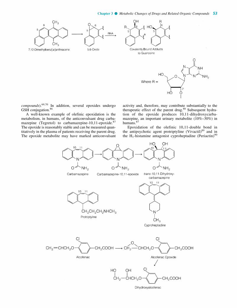
activity and, therefore, may contribute substantially to thetherapeutic effect of the parent drug.88 Subsequent hydra-tion of the epoxide produces 10,11-dihydroxycarba-mazepine, an important urinary metabolite (10%–30%) inhumans.87
Epoxidation of the olefinic 10,11-double bond inthe antipsychotic agent protriptyline (Vivactil)89 and inthe H1-histamine antagonist cyproheptadine (Periactin)90
Chapter 3 Metabolic Changes of Drugs and Related Organic Compounds 53
compounds).69,70 In addition, several epoxides undergoGSH conjugation.86
A well-known example of olefinic epoxidation is themetabolism, in humans, of the anticonvulsant drug carba-mazepine (Tegretol) to carbamazepine-10,11-epoxide.87
The epoxide is reasonably stable and can be measured quan-titatively in the plasma of patients receiving the parent drug.The epoxide metabolite may have marked anticonvulsant
0043-0118_17865_Ch03.qxd 12/4/09 3:48 AM Page 53

54 Wilson and Gisvold’s Textbook of Organic Medicinal and Pharmaceutical Chemistry
CC
CH2
H2CHO
OHH3C
CH3
CC
CH2
H2CHO
OHH3C
CH3
O
Diethylstilbestrol Diethylstilbestrol Epoxide
Possible covalent binding toproteins and/or nucleic acids
also occurs. Frequently, the epoxides formed from thebiotransformation of an olefinic compound are minorproducts, because of their further conversion to the corre-sponding 1,2-diols. For instance, dihydroxyalcofenac is amajor human urinary metabolite of the once clinically use-ful anti-inflammatory agent alclofenac.91 The epoxidemetabolite from which it is derived, however, is present inminute amounts. The presence of the dihydroxy metabolite(secodiol) of secobarbital, but not the epoxide product, hasbeen reported in humans.92
Indirect evidence for the formation of epoxides comesalso from the isolation of GSH or mercapturic acid metabo-lites. After administration of styrene to rats, two urinarymetabolites were identified as the isomeric mercapturic acidderivatives resulting from nucleophilic attack of GSH on theintermediate epoxide.93 In addition, styrene oxide cova-lently binds to rat liver microsomal proteins and nucleicacids.94 These results indicate that styrene oxide is relativelyreactive toward nucleophiles (e.g., GSH and nucleophilicgroups on protein and nucleic acids).
There are, apparently, diverse metabolically generatedepoxides that display similar chemical reactivity towardnucleophilic functionalities. Accordingly, the toxicity ofsome olefinic compounds may result from their metabolicconversion to chemically reactive epoxides.95 One examplethat clearly links metabolic epoxidation as a biotoxificationpathway involves aflatoxin B1. This naturally occurring car-cinogenic agent contains an olefinic (C2–C3) double bondadjacent to a cyclic ether oxygen. The hepatocarcinogenic-ity of aflatoxin B1 has been clearly linked to its metabolicoxidation to the corresponding 2,3-oxide, which is ex-tremely reactive.96,97 Extensive in vitro and in vivo meta-bolic studies indicate that this 2,3-oxide binds covalently toDNA, RNA, and proteins. A major DNA adduct has beenisolated and characterized as 2,3-dihydro-2-(N7-guanyl)-3-hydroxyaflatoxin B1.98,99
Other olefinic compounds, such as vinyl chloride,100
stilbene,101 and the carcinogenic estrogenic agent diethyl-stilbestrol (DES),102,103 undergo metabolic epoxidation.The corresponding epoxide metabolites may be the reactive
0043-0118_17865_Ch03.qxd 12/4/09 3:48 AM Page 54
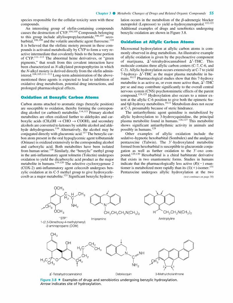
species responsible for the cellular toxicity seen with thesecompounds.
An interesting group of olefin-containing compoundscauses the destruction of CYP.104,105 Compounds belongingto this group include allylisopropylacetamide,106,107 seco-barbital,108,109 and the volatile anesthetic agent fluroxene.110
It is believed that the olefinic moiety present in these com-pounds is activated metabolically by CYP to form a very re-active intermediate that covalently binds to the heme portionof CYP.111–113 The abnormal heme derivatives, or “greenpigments,” that result from this covalent interaction havebeen characterized as N-alkylated protoporphyrins in whichthe N-alkyl moiety is derived directly from the olefin admin-istered.104,105,111–113 Long-term administration of the above-mentioned three agents is expected to lead to inhibition ofoxidative drug metabolism, potential drug interactions, andprolonged pharmacological effects.
Oxidation at Benzylic Carbon Atoms
Carbon atoms attached to aromatic rings (benzylic position)are susceptible to oxidation, thereby forming the correspon-ding alcohol (or carbinol) metabolite.114,115 Primary alcoholmetabolites are often oxidized further to aldehydes and car-boxylic acids (CH2OH → CHO → COOH), and secondaryalcohols are converted to ketones by soluble alcohol and alde-hyde dehydrogenases.116 Alternatively, the alcohol may beconjugated directly with glucuronic acid.117 The benzylic car-bon atom present in the oral hypoglycemic agent tolbutamide(Orinase) is oxidized extensively to the corresponding alcoholand carboxylic acid. Both metabolites have been isolatedfrom human urine.118 Similarly, the “benzylic” methyl groupin the anti-inflammatory agent tolmetin (Tolectin) undergoesoxidation to yield the dicarboxylic acid product as the majormetabolite in humans.119,120 The selective cyclooxygenase 2(COX-2) anti-inflammatory agent celecoxib undergoes ben-zylic oxidation at its C-5 methyl group to give hydroxycele-coxib as a major metabolite.121 Significant benzylic hydroxy-
lation occurs in the metabolism of the �-adrenergic blockermetoprolol (Lopressor) to yield �-hydroxymetoprolol.122,123
Additional examples of drugs and xenobiotics undergoingbenzylic oxidation are shown in Figure 3.8.
Oxidation at Allylic Carbon Atoms
Microsomal hydroxylation at allylic carbon atoms is com-monly observed in drug metabolism. An illustrative exampleof allylic oxidation is given by the psychoactive componentof marijuana, �1-tetrahydrocannabinol �1-THC. Thismolecule contains three allylic carbon centers (C-7, C-6, andC-3). Allylic hydroxylation occurs extensively at C-7 to yield7-hydroxy- �1-THC as the major plasma metabolite in hu-mans.10,11 Pharmacological studies show that this 7-hydroxymetabolite is as active as, or even more active than, �1-THCper se and may contribute significantly to the overall centralnervous system (CNS) psychotomimetic effects of the parentcompound.124,125 Hydroxylation also occurs to a minor ex-tent at the allylic C-6 position to give both the epimeric 6�-and 6�-hydroxy metabolites.10,11 Metabolism does not occurat C-3, presumably because of steric hindrance.
The antiarrhythmic agent quinidine is metabolized byallylic hydroxylation to 3-hydroxyquinidine, the principalplasma metabolite found in humans.126,127 This metaboliteshows significant antiarrhythmic activity in animals andpossibly in humans.128
Other examples of allylic oxidation include thesedative–hypnotic hexobarbital (Sombulex) and the analgesicpentazocine (Talwin). The 3�-hydroxylated metaboliteformed from hexobarbital is susceptible to glucuronide conju-gation as well as further oxidation to the 3�-oxo com-pound.129,130 Hexobarbital is a chiral barbiturate derivativethat exists in two enantiomeric forms. Studies in humansindicate that the pharmacologically less active (R)(�) enan-tiomer is metabolized more rapidly than its (S)(�)-isomer.131
Pentazocine undergoes allylic hydroxylation at the two
Chapter 3 Metabolic Changes of Drugs and Related Organic Compounds 55
Figure 3.8 Examples of drugs and xenobiotics undergoing benzylic hydroxylation.Arrow indicates site of hydroxylation.
(text continues on page 58)
0043-0118_17865_Ch03.qxd 12/4/09 3:48 AM Page 55

56 Wilson and Gisvold’s Textbook of Organic Medicinal and Pharmaceutical Chemistry
0043-0118_17865_Ch03.qxd 12/4/09 3:48 AM Page 56

Chapter 3 Metabolic Changes of Drugs and Related Organic Compounds 57
0043-0118_17865_Ch03.qxd 12/4/09 3:48 AM Page 57
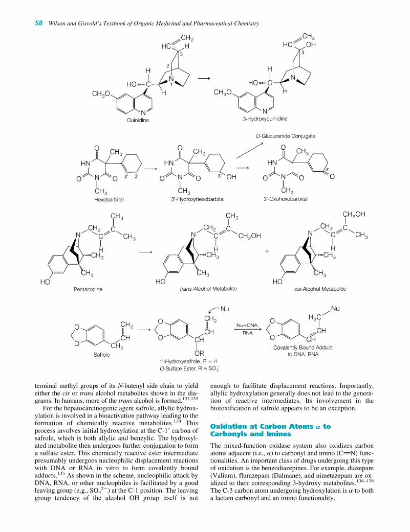
58 Wilson and Gisvold’s Textbook of Organic Medicinal and Pharmaceutical Chemistry
terminal methyl groups of its N-butenyl side chain to yieldeither the cis or trans alcohol metabolites shown in the dia-grams. In humans, more of the trans alcohol is formed.132,133
For the hepatocarcinogenic agent safrole, allylic hydrox-ylation is involved in a bioactivation pathway leading to theformation of chemically reactive metabolites.134 Thisprocess involves initial hydroxylation at the C-1� carbon ofsafrole, which is both allylic and benzylic. The hydroxyl-ated metabolite then undergoes further conjugation to forma sulfate ester. This chemically reactive ester intermediatepresumably undergoes nucleophilic displacement reactionswith DNA or RNA in vitro to form covalently boundadducts.135 As shown in the scheme, nucleophilic attack byDNA, RNA, or other nucleophiles is facilitated by a goodleaving group (e.g., SO4
2�) at the C-1 position. The leavinggroup tendency of the alcohol OH group itself is not
enough to facilitate displacement reactions. Importantly,allylic hydroxylation generally does not lead to the genera-tion of reactive intermediates. Its involvement in thebiotoxification of safrole appears to be an exception.
Oxidation at Carbon Atoms � toCarbonyls and Imines
The mixed-function oxidase system also oxidizes carbonatoms adjacent (i.e., �) to carbonyl and imino (CBN) func-tionalities. An important class of drugs undergoing this typeof oxidation is the benzodiazepines. For example, diazepam(Valium), flurazepam (Dalmane), and nimetazepam are ox-idized to their corresponding 3-hydroxy metabolites.136–138
The C-3 carbon atom undergoing hydroxylation is � to botha lactam carbonyl and an imino functionality.
0043-0118_17865_Ch03.qxd 12/4/09 3:48 AM Page 58

Aliphatic � and �–1 hydroxylations commonly take placein drug molecules with straight or branched alkyl chains.Thus, the antiepileptic agent valproic acid (Depakene) un-dergoes both � and �–1 oxidation to the 5-hydroxy and 4-hydroxy metabolites, respectively.142,143 Further oxidationof the 5-hydroxy metabolite yields 2-n-propylglutaric acid.
Numerous barbiturates and oral hypoglycemic sulfonyl-ureas also have aliphatic side chains that are susceptible tooxidation. Note that the sedative–hypnotic amobarbital(Amytal) undergoes extensive �–1 oxidation to the corre-sponding 3�-hydroxylated metabolite.144 Other barbitu-rates, such as pentobarbital,145,146 thiamylal,147 andsecobarbital,92 reportedly are metabolized by way of � and�–1 oxidation. The n-propyl side chain attached to the oralhypoglycemic agent chlorpropamide (Diabinese) under-goes extensive �–1 hydroxylation to yield the secondaryalcohol 2�-hydroxychlorpropamide as a major urinarymetabolite in humans.148
Omega and �–1 oxidation of the isobutyl moiety presentin the anti-inflammatory agent ibuprofen (Motrin) yieldsthe corresponding carboxylic acid and tertiary alcoholmetabolites.149 Additional examples of drugs reported to un-dergo aliphatic hydroxylation include meprobamate,150
glutethimide,140,141 ethosuximide,151 and phenylbutazone.152
The cyclohexyl group is commonly found in many me-dicinal agents, and is also susceptible to mixed-functionoxidation (alicyclic hydroxylation).114,115 Enzymatic intro-duction of a hydroxyl group into a monosubstituted cyclo-hexane ring generally occurs at C-3 or C-4 and can lead tocis and trans conformational stereoisomers, as shown in thediagrammed scheme.
An example of this hydroxylation pathway is seen in themetabolism of the oral hypoglycemic agent acetohexamide(Dymelor). In humans, the trans-4-hydroxycyclohexyl prod-uct is reportedly a major metabolite.153 Small amounts of theother possible stereoisomers (namely, the cis-4-, cis-3-, andtrans-3-hydroxycyclohexyl derivatives) also have been de-tected. Another related oral hypoglycemic agent, glipizide,is oxidized in humans to the trans-4- and cis-3-hydroxylcyclohexyl metabolites in about a 6:1 ratio.154
Chapter 3 Metabolic Changes of Drugs and Related Organic Compounds 59
For diazepam, the hydroxylation reaction proceeds withremarkable stereoselectivity to form primarily (90%) 3-hydroxydiazepam (also called N-methyloxazepam), withthe (S) absolute configuration at C-3.139 Further N-demethy-lation of the latter metabolite gives rise to the pharmacolog-ically active 3(S)(�)-oxazepam.
Hydroxylation of the carbon atom � to carbonyl func-tionalities generally occurs only to a limited extent in drugmetabolism. An illustrative example involves the hydroxyl-ation of the sedative–hypnotic glutethimide (Doriden) to 4-hydroxyglutethimide.140,141
Oxidation at Aliphatic and AlicyclicCarbon Atoms
Alkyl or aliphatic carbon centers are subject to mixed-function oxidation. Metabolic oxidation at the terminalmethyl group often is referred to as �-oxidation, and oxida-tion of the penultimate carbon atom (i.e., next-to-the-lastcarbon) is called �–1 oxidation.114,115 The initial alcoholmetabolites formed from these enzymatic � and �–1 oxida-tions are susceptible to further oxidation to yield aldehyde,ketones, or carboxylic acids. Alternatively, the alcoholmetabolites may undergo glucuronide conjugation.
0043-0118_17865_Ch03.qxd 12/4/09 3:48 AM Page 59

60 Wilson and Gisvold’s Textbook of Organic Medicinal and Pharmaceutical Chemistry
Two human urinary metabolites of phencyclidine (PCP)have been identified as the 4-hydroxypiperidyl and 4-hydroxycyclohexyl derivatives of the parent compound.155,156
Thus, from these results, it appears that “alicyclic” hydroxy-lation of the six-membered piperidyl moiety may parallelclosely the hydroxylation pattern of the cyclohexyl moiety.The stereochemistry of the hydroxylated centers in the twometabolites has not been clearly established. Biotrans-formation of the antihypertensive agent minoxidil (Loniten)yields the 4�-hydroxypiperidyl metabolite. In dogs, this prod-uct is a major urinary metabolite (29%–47%), whereas in hu-mans it is detected in small amounts (�3%).157,158
Oxidation InvolvingCarbon–Heteroatom Systems
Nitrogen and oxygen functionalities are commonly foundin most drugs and foreign compounds; sulfur function-alities occur only occasionally. Metabolic oxidation of
carbon–nitrogen, carbon–oxygen, and carbon–sulfur sys-tems principally involves two basic types of biotransfor-mation processes:
1. Hydroxylation of the �-carbon atom attached directly tothe heteroatom (N, O, S). The resulting intermediate isoften unstable and decomposes with the cleavage of thecarbon–heteroatom bond:
Oxidative N-, O-, and S-dealkylation as well as oxidativedeamination reactions fall under this mechanistic pathway.
0043-0118_17865_Ch03.qxd 12/4/09 3:48 AM Page 60

Chapter 3 Metabolic Changes of Drugs and Related Organic Compounds 61
2. Hydroxylation or oxidation of the heteroatom (N, S only,e.g., N-hydroxylation, N-oxide formation, sulfoxide, andsulfone formation).
Several structural features frequently determine whichpathway will predominate, especially in carbon–nitrogensystems. Metabolism of some nitrogen-containing com-pounds is complicated by the fact that carbon- or nitrogen-
hydroxylated products may undergo secondary reactions toform other, more complex metabolic products (e.g., oxime,nitrone, nitroso, imino). Other oxidative processes that donot fall under these two basic categories are discussed indi-vidually in the appropriate carbon–heteroatom section. Themetabolism of carbon–nitrogen systems will be discussedfirst, followed by the metabolism of carbon–oxygen andcarbon–sulfur systems.
0043-0118_17865_Ch03.qxd 12/4/09 3:48 AM Page 61

OXIDATION INVOLVING CARBON–NITROGEN SYSTEMS
Metabolism of nitrogen functionalities (e.g., amines, amides)is important because such functional groups are found inmany natural products (e.g., morphine, cocaine, nicotine)and in numerous important drugs (e.g., phenothiazines,antihistamines, tricyclic antidepressants, �-adrenergic agents,sympathomimetic phenylethylamines, benzodiazepines).159
The following discussion divides nitrogen-containing com-pounds into three basic classes:
1. Aliphatic (primary, secondary, and tertiary) and alicyclic(secondary and tertiary) amines
2. Aromatic and heterocyclic nitrogen compounds3. Amides
The susceptibility of each class of these nitrogen com-pounds to either �-carbon hydroxylation or N-oxidation andthe metabolic products that are formed are discussed.
The hepatic enzymes responsible for carrying out �-car-bon hydroxylation reactions are the CYP mixed-functionoxidases. The N-hydroxylation or N-oxidation reactions,however, appear to be catalyzed not only by CYP but alsoby a second class of hepatic mixed-function oxidases called
62 Wilson and Gisvold’s Textbook of Organic Medicinal and Pharmaceutical Chemistry
amine oxidases (sometimes called N-oxidases).160 Theseenzymes are NADPH-dependent flavoproteins and do notcontain CYP.161,162 They require NADPH and molecularoxygen to carry out N-oxidation.
Tertiary Aliphatic and Alicyclic Amines. The oxida-tive removal of alkyl groups (particularly methyl groups)from tertiary aliphatic and alicyclic amines is carried out byhepatic CYP mixed-function oxidase enzymes. This reac-tion is commonly referred to as oxidative N-dealkylation.163
The initial step involves �-carbon hydroxylation to form acarbinolamine intermediate, which is unstable and under-goes spontaneous heterolytic cleavage of the C–N bond togive a secondary amine and a carbonyl moiety (aldehyde orketone).164,165 In general, small alkyl groups, such asmethyl, ethyl, and isopropyl, are removed rapidly.163
N-dealkylation of the t-butyl group is not possible by thecarbinolamine pathway because �-carbon hydroxylationcannot occur. The first alkyl group from a tertiary amine isremoved more rapidly than the second alkyl group. In someinstances, bisdealkylation of the tertiary aliphatic amine tothe corresponding primary aliphatic amine occurs veryslowly.163 For example, the tertiary amine imipramine
0043-0118_17865_Ch03.qxd 12/4/09 3:48 AM Page 62

Chapter 3 Metabolic Changes of Drugs and Related Organic Compounds 63
(Tofranil) is monodemethylated to desmethylimipramine(desipramine).166,167 This major plasma metabolite is phar-macologically active in humans and contributes substan-tially to the antidepressant activity of the parent drug.168
Very little of the bisdemethylated metabolite of imipramineis detected. In contrast, the local anesthetic and antiarrhyth-mic agent lidocaine is metabolized extensively by N-deethy-lation to both monoethylglycylxylidine and glycyl-2,6-xyli-dine in humans.169,170
Numerous other tertiary aliphatic amine drugs are metab-olized principally by oxidative N-dealkylation. Some of theseinclude the antiarrhythmic disopyramide (Norpace),171,172 theantiestrogenic agent tamoxifen (Nolvadex),173 diphenhy-dramine (Benadryl),174,175chlorpromazine (Thorazine),176,177
and (�)-�-propoxyphene (Darvon).178 When the tertiaryamine contains several different substituents capable of un-dergoing dealkylation, the smaller alkyl group is removedpreferentially and more rapidly. For example, in benzpheta-mine (Didrex), the methyl group is removed much more rap-idly than the benzyl moiety.179
An interesting cyclization reaction occurs with methadoneon N-demethylation. The demethylated metabolitenormethadone undergoes spontaneous cyclization to form theenamine metabolite 2-ethylidene-1,5-dimethyl-3,3-diphenyl-pyrrolidine (EDDP).180 Subsequent N-demethylation ofEDDP and isomerization of the double bond leads to 2-ethyl-5-methyl-3,3-diphenyl-1-pyrroline (EMDP).
Many times, bisdealkylation of a tertiary amine leads tothe corresponding primary aliphatic amine metabolite,which is susceptible to further oxidation. For example, the
bisdesmethyl metabolite of the H1-histamine antagonistbrompheniramine (Dimetane) undergoes oxidative deami-nation and further oxidation to the corresponding propionicacid metabolite.181 Oxidative deamination is discussed ingreater detail when we examine the metabolic reactions ofsecondary and primary amines.
Like their aliphatic counterparts, alicyclic tertiary aminesare susceptible to oxidative N-dealkylation reactions. Forexample, the analgesic meperidine (Demerol) is metabo-lized principally by this pathway to yield normeperidine asa major plasma metabolite in humans.182 Morphine, N-eth-ylnormorphine, and dextromethorphan also undergo someN-dealkylation.183
Direct N-dealkylation of t-butyl groups, as discussedbelow, is not possible by the �-carbon hydroxylation path-way. In vitro studies indicate, however, that N-t-butyl-norchlorocyclizine is, indeed, metabolized to significantamounts of norchlorocyclizine, whereby the t-butyl group islost.184 Careful studies showed that the t-butyl group is re-moved by initial hydroxylation of one of the methyl groupsof the t-butyl moiety to the carbinol or alcohol product.185
Further oxidation generates the corresponding carboxylicacid that, on decarboxylation, forms the N-isopropyl deriv-ative. The N-isopropyl intermediate is dealkylated by thenormal �-carbon hydroxylation (i.e., carbinolamine) path-way to give norchlorocyclizine and acetone. Whether this isa general method for the loss of t-butyl groups from aminesis still unclear. Indirect N-dealkylation of t-butyl groups isnot observed significantly. The N-t-butyl group present inmany �-adrenergic antagonists, such as terbutaline and
0043-0118_17865_Ch03.qxd 12/4/09 3:48 AM Page 63

64 Wilson and Gisvold’s Textbook of Organic Medicinal and Pharmaceutical Chemistry
salbutamol, remains intact and does not appear to undergoany significant metabolism.186
Alicyclic tertiary amines often generate lactam metabo-lites by �-carbon hydroxylation reactions. For example, thetobacco alkaloid nicotine is hydroxylated initially at the ringcarbon atom � to the nitrogen to yield a carbinolamine inter-mediate. Furthermore, enzymatic oxidation of this cycliccarbinolamine generates the lactam metabolite cotinine.187,188
Formation of lactam metabolites also has been reportedto occur to a minor extent for the antihistamine cyprohep-
tadine (Periactin)189,190 and the antiemetic diphenidol(Vontrol).191
N-oxidation of tertiary amines occurs with severaldrugs.192 The true extent of N-oxide formation often is com-plicated by the susceptibility of N-oxides to undergo in vivoreduction back to the parent tertiary amine. Tertiary aminessuch as H1-histamine antagonists (e.g., orphenadrine, tripel-ennamine), phenothiazines (e.g., chlorpromazine), tricyclicantidepressants (e.g., imipramine), and narcotic analgesics(e.g., morphine, codeine, and meperidine) reportedly form
0043-0118_17865_Ch03.qxd 12/4/09 3:48 AM Page 64

Chapter 3 Metabolic Changes of Drugs and Related Organic Compounds 65
N-oxide products. In some instances, N-oxides possesspharmacological activity.193 A comparison of imipramineN-oxide with imipramine indicates that the N-oxide itselfpossesses antidepressant and cardiovascular activity similarto that of the parent drug.194,195
Secondary and Primary Amines. Secondary amines(either parent compounds or metabolites) are susceptible tooxidative N-dealkylation, oxidative deamination, and N-oxidation reactions.163,196 As in tertiary amines, N-dealkylation of secondary amines proceeds by thecarbinolamine pathway. Dealkylation of secondary aminesgives rise to the corresponding primary amine metabolite.For example, the �-adrenergic blockers propranolol46,47 and
oxprenolol197 undergo N-deisopropylation to the correspon-ding primary amines. N-dealkylation appears to be a signif-icant biotransformation pathway for the secondary aminedrugs methamphetamine198,199 and ketamine,200,201 yieldingamphetamine and norketamine, respectively.
The primary amine metabolites formed from oxidativedealkylation are susceptible to oxidative deamination. Thisprocess is similar to N-dealkylation, in that it involves aninitial �-carbon hydroxylation reaction to form a carbino-lamine intermediate, which then undergoes subsequentcarbon–nitrogen cleavage to the carbonyl metabolite and am-monia. If �-carbon hydroxylation cannot occur, then oxida-tive deamination is not possible. For example, deaminationdoes not occur for norketamine because �-carbon hydroxyla-tion cannot take place.200,201 With methamphetamine, oxida-tive deamination of primary amine metabolite amphetamineproduces phenylacetone.198,199
In general, dealkylation of secondary amines is believedto occur before oxidative deamination. Some evidence indi-cates, however, that this may not always be true. Directdeamination of the secondary amine also has occurred. Forexample, in addition to undergoing deamination through itsdesisopropyl primary amine metabolite, propranolol canundergo a direct oxidative deamination reaction (also by �-carbon hydroxylation) to yield the aldehyde metaboliteand isopropylamine (Fig. 3.9).202 How much direct oxida-tive deamination contributes to the metabolism of secondaryamines remains unclear.
Some secondary alicyclic amines, like their tertiaryamine analogs, are metabolized to their corresponding lac-tam derivatives. For example, the anorectic agent phen-metrazine (Preludin) is metabolized principally to the lactamproduct 3-oxophenmetrazine.203 In humans, this lactammetabolite is a major urinary product. Methylphenidate(Ritalin) also reportedly yields a lactam metabolite, 6-oxori-
Figure 3.9 Metabolism of propranolol to its aldehyde metabolite by direct deami-nation of the parent compound and by deamination of its primary amine metabolite,desisopropyl propranolol.
0043-0118_17865_Ch03.qxd 12/4/09 3:48 AM Page 65
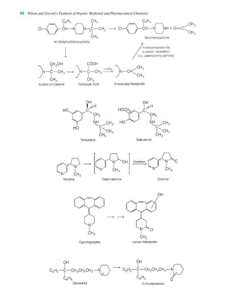
66 Wilson and Gisvold’s Textbook of Organic Medicinal and Pharmaceutical Chemistry
0043-0118_17865_Ch03.qxd 12/4/09 3:48 AM Page 66
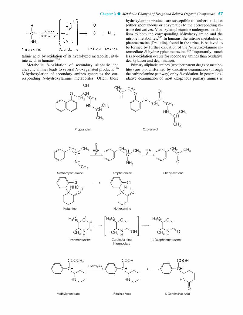
talinic acid, by oxidation of its hydrolyzed metabolite, rital-inic acid, in humans.204
Metabolic N-oxidation of secondary aliphatic andalicyclic amines leads to several N-oxygenated products.196
N-hydroxylation of secondary amines generates the cor-responding N-hydroxylamine metabolites. Often, these
hydroxylamine products are susceptible to further oxidation(either spontaneous or enzymatic) to the corresponding ni-trone derivatives. N-benzylamphetamine undergoes metabo-lism to both the corresponding N-hydroxylamine and thenitrone metabolites.205 In humans, the nitrone metabolite ofphenmetrazine (Preludin), found in the urine, is believed tobe formed by further oxidation of the N-hydroxylamine in-termediate N-hydroxyphenmetrazine.203 Importantly, muchless N-oxidation occurs for secondary amines than oxidativedealkylation and deamination.
Primary aliphatic amines (whether parent drugs or metabo-lites) are biotransformed by oxidative deamination (throughthe carbinolamine pathway) or by N-oxidation. In general, ox-idative deamination of most exogenous primary amines is
Chapter 3 Metabolic Changes of Drugs and Related Organic Compounds 67
0043-0118_17865_Ch03.qxd 12/4/09 3:48 AM Page 67

carried out by the mixed-function oxidases discussed previ-ously. Endogenous primary amines (e.g., dopamine, norepi-nephrine, tryptamine, and serotonin) and xenobiotics based onthe structures of these endogenous neurotransmitters are me-tabolized, however, via oxidative deamination by a specializedfamily of enzymes called monoamine oxidases (MAOs).206
MAO is a flavin (FAD)-dependent enzyme found in twoisozyme forms, MAO-A and MAO-B, and widely distributedin both the CNS and peripheral organs. In contrast, CYP ex-ists in a wide variety of isozyme forms and is an NADP-dependent system. Also the greatest variety of CYPisozymes, at least the ones associated with the metabolism ofxenobiotics, are found mostly in the liver and intestinal mu-cosa. MAO-A and MAO-B are coded by two genes, both onthe X-chromosome and have about 70% amino acid sequencehomology. Another difference between the CYP and MAO
families is cellular location. CYP enzymes are found on theendoplasmic reticulum of the cell’s cytosol, whereas theMAO enzymes are on the outer mitochondrial membrane. Inaddition to the xenobiotics illustrated in the reaction schemes,other drugs metabolized by the MAO system includephenylephrine, propranolol, timolol and other �-adrenergicagonists and antagonists, and various phenylethylamines.206
Structural features, especially the �-substituents of theprimary amine, often determine whether carbon or nitrogenoxidation will occur. For example, compare amphetaminewith its �-methyl homologue phentermine. In amphetamine,�-carbon hydroxylation can occur to form the carbinolamineintermediate, which is converted to the oxidatively deami-nated product phenylacetone.67 With phentermine, �-carbonhydroxylation is not possible and precludes oxidative deam-ination for this drug. Consequently, phentermine wouldbe expected to undergo N-oxidation readily. In humans, p-hydroxylation and N-oxidation are the main pathways forbiotransformation of phentermine.207
Indeed, N-hydroxyphentermine is an important (5%)urinary metabolite in humans.207 As discussed below, N-hydroxylamine metabolites are susceptible to further oxi-dation to yield other N-oxygenated products.
68 Wilson and Gisvold’s Textbook of Organic Medicinal and Pharmaceutical Chemistry
0043-0118_17865_Ch03.qxd 12/4/09 3:48 AM Page 68

Xenobiotics, such as the hallucinogenic agents mesca-line208,209 and 1-(2,5-dimethoxy-4-methylphenyl)-2-amino-propane (DOM or “STP”),210,211 are oxidatively deaminated.Primary amine metabolites arising from N-dealkylation ordecarboxylation reactions also undergo deamination. The ex-ample of the bisdesmethyl primary amine metabolite derivedfrom bromopheniramine is discussed previously in this chap-ter (see section on tertiary aliphatic and alicyclic amines).181
In addition, many tertiary aliphatic amines (e.g., antihista-mines) and secondary aliphatic amines (e.g., propranolol) aredealkylated to their corresponding primary amine metabo-lites, which are amenable to oxidative deamination. (S)(�)-�-Methyldopamine resulting from decarboxylation of the an-tihypertensive agent (S)(�)-�-methyldopa (Aldomet) isdeaminated to the corresponding ketone metabolite 3,4-dihy-droxyphenylacetone.212 In humans, this ketone is a majorurinary metabolite.
The N-hydroxylation reaction is not restricted to �-substi-tuted primary amines such as phentermine. Amphetamine hasbeen observed to undergo some N-hydroxylation in vitro toN-hydroxyamphetamine.213,214 N-Hydroxyamphetamine is,however, susceptible to further conversion to the imine or ox-idation to the oxime intermediate. Note that the oxime inter-mediate arising from this N-oxidation pathway can undergohydrolytic cleavage to yield phenylacetone, the same productobtained by the �-carbon hydroxylation (carbinolamine)
pathway.215,216 Thus, amphetamine may be converted tophenylacetone through either the �-carbon hydroxylation orthe N-oxidation pathway. The debate concerning the relativeimportance of the two pathways is ongoing.217–219 The con-sensus, however, is that both metabolic pathways (carbon andnitrogen oxidation) are probably operative. Whether �-carbonor nitrogen oxidation predominates in the metabolism of am-phetamine appears to be species dependent.
In primary aliphatic amines, such as phentermine,207
chlorphentermine (p-chlorphentermine),219 and amanta-dine,220 N-oxidation appears to be the major biotransforma-tion pathway because �-carbon hydroxylation cannotoccur. In humans, chlorphentermine is N-hydroxylated ex-tensively. About 30% of a dose of chlorphentermine isfound in the urine (48 hours) as N-hydroxychlorphenter-mine (free and conjugated) and an additional 18% as otherproducts of N-oxidation (presumably the nitroso and nitrometabolites).219 In general, N-hydroxylamines are chemi-cally unstable and susceptible to spontaneous or enzymaticoxidation to the nitroso and nitro derivatives. For example,the N-hydroxylamine metabolite of phentermine undergoesfurther oxidation to the nitroso and nitro products.207
The antiviral and antiparkinsonian agent amantadine(Symmetrel) reportedly undergoes N-oxidation to yield thecorresponding N-hydroxy and nitroso metabolites invitro.220
Chapter 3 Metabolic Changes of Drugs and Related Organic Compounds 69
0043-0118_17865_Ch03.qxd 12/4/09 3:48 AM Page 69

Aromatic Amines and Heterocyclic NitrogenCompounds. The biotransformation of aromatic aminesparallels the carbon and nitrogen oxidation reactions seenfor aliphatic amines.221–223 For tertiary aromatic amines,such as N,N-dimethylaniline, oxidative N-dealkylation aswell as N-oxide formation take place.224 Secondary aro-matic amines may undergo N-dealkylation or N-hydroxyl-ation to give the corresponding N-hydroxylamines. Furtheroxidation of the N-hydroxylamine leads to nitrone prod-ucts, which in turn may be hydrolyzed to primary hydrox-ylamines.225 Tertiary and secondary aromatic amines areencountered rarely in medicinal agents. In contrast, pri-mary aromatic amines are found in many drugs and areoften generated from enzymatic reduction of aromaticnitro compounds, reductive cleavage of azo compounds,and hydrolysis of aromatic amides.
N-oxidation of primary aromatic amines generates the N-hydroxylamine metabolite. One such case is aniline,which is metabolized to the corresponding N-hydroxy prod-uct.223 Oxidation of the hydroxylamine derivative to the ni-troso derivative also can occur. When one considers primaryaromatic amine drugs or metabolites, N-oxidation cons-titutes only a minor pathway in comparison with otherbiotransformation pathways, such as N-acetylation and aro-matic hydroxylation, in humans. Some N-oxygenatedmetabolites have been reported, however. For example, theantileprotic agent dapsone and its N-acetylated metaboliteare metabolized significantly to their corresponding N-hydroxylamine derivatives.226 The N-hydroxy metabolitesare further conjugated with glucuronic acid.
Methemoglobinemia toxicity is caused by several aro-matic amines, including aniline and dapsone, and is a result ofthe bioconversion of the aromatic amine to its N-hydroxy de-rivative. Apparently, the N-hydroxylamine oxidizes the Fe2�
form of hemoglobin to its Fe3� form. This oxidized (Fe3�)state of hemoglobin (called methemoglobin or ferrihemoglo-bin) can no longer transport oxygen, which leads to serioushypoxia or anemia, a unique type of chemical suffocation.227
Diverse aromatic amines (especially azoamino dyes) areknown to be carcinogenic. N-oxidation plays an important
role in bioactivating these aromatic amines to potentially re-active electrophilic species that covalently bind to cellularprotein, DNA, or RNA. A well-studied example is thecarcinogenic agent N-methyl-4-aminoazobenzene.228,229
N-oxidation of this compound leads to the correspondinghydroxylamine, which undergoes sulfate conjugation.Because of the good leaving-group ability of the sulfate(SO4
2�) anion, this conjugate can ionize spontaneously toform a highly reactive, resonance-stabilized nitreniumspecies. Covalent adducts between this species and DNA,RNA, and proteins have been characterized.230,231 The sulfateester is believed to be the ultimate carcinogenic species. Thus,the example indicates that certain aromatic amines can bebioactivated to reactive intermediates by N-hydroxylation andO-sulfate conjugation. Whether primary hydroxylamines canbe bioactivated similarly is unclear. In addition, it is notknown if this biotoxification pathway plays any substantialrole in the toxicity of aromatic amine drugs.
N-oxidation of the nitrogen atoms present in aromaticheterocyclic moieties of many drugs occurs to a minor ex-tent. Clearly, in humans, N-oxidation of the folic acid antag-onist trimethoprim (Proloprim, Trimpex) has yieldedapproximately equal amounts of the isomeric 1-N-oxide and3-N-oxide as minor metabolites.232 The pyridinyl nitrogenatom present in nicotinine (the major metabolite of nicotine)undergoes oxidation to yield the corresponding N-oxidemetabolite.233 Another therapeutic agent that has been ob-served to undergo formation of an N-oxide metabolite ismetronidazole.234
Amides. Amide functionalities are susceptible to ox-idative carbon–nitrogen bond cleavage (via �-carbon hy-droxylation) and N-hydroxylation reactions. Oxidativedealkylation of many N-substituted amide drugs andxenobiotics has been reported. Mechanistically, oxidativedealkylation proceeds via an initially formed car-binolamide, which is unstable and fragments to form theN-dealkylated product. For example, diazepam undergoesextensive N-demethylation to the pharmacologically activemetabolite desmethyldiazepam.235
70 Wilson and Gisvold’s Textbook of Organic Medicinal and Pharmaceutical Chemistry
0043-0118_17865_Ch03.qxd 12/4/09 3:48 AM Page 70

Chapter 3 Metabolic Changes of Drugs and Related Organic Compounds 71
Various other N-alkyl substituents present in benzodi-azepines (e.g., flurazepam)136–138 and in barbiturates (e.g.,hexobarbital and mephobarbital)128 are similarly oxidativelyN-dealkylated. Alkyl groups attached to the amide moiety ofsome sulfonylureas, such as the oral hypoglycemic chlor-propamide,236 also are subject to dealkylation to a minorextent.
In the cyclic amides or lactams, hydroxylation of the ali-cyclic carbon � to the nitrogen atom also leads to carbino-lamides. An example of this pathway is the conversion ofcotinine to 5-hydroxycotinine. Interestingly, the lattercarbinolamide intermediate is in tautomeric equilibriumwith the ring-opened metabolite �-(3-pyridyl)-�-oxo-N-methylbutyramide.237
Metabolism of the important cancer chemotherapeuticagent cyclophosphamide (Cytoxan) follows a hydroxylationpathway similar to that just described for cyclic amides. This
0043-0118_17865_Ch03.qxd 12/4/09 3:48 AM Page 71

72 Wilson and Gisvold’s Textbook of Organic Medicinal and Pharmaceutical Chemistry
drug is a cyclic phosphoramide derivative and, for the mostpart, is the phosphorous counterpart of a cyclic amide.Because cyclophosphamide itself is pharmacologically inac-tive,238 metabolic bioactivation is required for the drug tomediate its antitumorigenic or cytotoxic effects. The keybiotransformation pathway leading to the active metaboliteinvolves an initial carbon hydroxylation reaction at C-4 toform the carbinolamide 4-hydroxycyclophosphamide.239,240
4-Hydroxycyclophosphamide is in equilibrium with thering-opened dealkylated metabolite aldophosphamide.Although it has potent cytotoxic properties, aldophos-phamide undergoes a further elimination reaction (reverseMichael reaction) to generate acrolein and the phospho-ramide mustard N,N-bis(2-chloro-ethyl)phosphorodiamidicacid. The latter is the principal species responsible forcyclophosphamide’s antitumorigenic properties and
0043-0118_17865_Ch03.qxd 12/4/09 3:48 AM Page 72

chemotherapeutic effect. Enzymatic oxidation of 4-hydroxy-cyclophosphamide and aldophosphamide leads to the rela-tively nontoxic metabolites 4-ketocyclophosphamide andcarboxycyclophosphamide, respectively.
N-hydroxylation of aromatic amides, which occurs to aminor extent, is of some toxicological interest, because thisbiotransformation pathway may lead to the formation ofchemically reactive intermediates. Several examples ofcytotoxicity or carcinogenicity associated with metabolicN-hydroxylation of the parent aromatic amide have beenreported. For example, the well-known hepatocarcinogenic2-acetylaminofluorene (AAF) undergoes an N-hydroxyl-ation reaction catalyzed by CYP to form the correspondingN-hydroxy metabolite (also called a hydroxamic acid).241
Further conjugation of this hydroxamic acid produces thecorresponding O-sulfate ester, which ionizes to generate theelectrophilic nitrenium species. Covalent binding of this re-active intermediate to DNA is known to occur and is likelyto be the initial event that ultimately leads to malignanttumor formation.242 Sulfate conjugation plays an importantrole in this biotoxification pathway (see “SulfateConjugation,” for further discussion).
Acetaminophen is a relatively safe and nontoxic analgesicagent if used at therapeutic doses. Its metabolism illustratesthe fact that a xenobiotic commonly produces more than onemetabolite. Its metabolism also illustrates the effect of age,because infants and young children carry out sulfation ratherthan glucuronidation (see discussion at the end of this chap-ter). New pharmacists must realize that at one time acetanilideand phenacetin were more widely used than acetaminophen,even though both are considered more toxic because they pro-
duce aniline derivatives. Besides producing toxic aniline andp-phenetidin, these two analgesics also produce acetamino-phen. When large doses of the latter drug are ingested, exten-sive liver necrosis is produced in humans and animals.243,244
Considerable evidence argues that this hepatotoxicity de-pends on the formation of a metabolically generated reactiveintermediate.245 Until recently,246,247 the accepted bioactiva-tion pathway was believed to involve an initial N-hydroxyla-tion reaction to form N-hydroxyacetaminophen.248
Spontaneous dehydration of this N-hydroxyamide pro-duces N-acetylimidoquinone, the proposed reactive metabo-lite. Usually, the GSH present in the liver combines withthis reactive metabolite to form the corresponding GSH con-jugate. If GSH levels are sufficiently depleted by large dosesof acetaminophen, covalent binding of the reactive interme-diate occurs with macromolecules present in the liver,thereby leading to cellular necrosis. Studies indicate, how-ever, that the reactive N-acetylimidoquinone intermediate isnot formed from N-hydroxyacetaminophen.245–247 It proba-bly arises through some other oxidative process. Therefore,the mechanistic formation of the reactive metabolite of acet-aminophen remains unclear.
OXIDATION INVOLVING CARBON–OXYGENSYSTEMS
Oxidative O-dealkylation of carbon–oxygen systems (princi-pally ethers) is catalyzed by microsomal mixed function oxi-dases.163 Mechanistically, the biotransformation involves aninitial �-carbon hydroxylation to form either a hemiacetal ora hemiketal, which undergoes spontaneous carbon–oxygen
Chapter 3 Metabolic Changes of Drugs and Related Organic Compounds 73
0043-0118_17865_Ch03.qxd 12/4/09 3:48 AM Page 73

74 Wilson and Gisvold’s Textbook of Organic Medicinal and Pharmaceutical Chemistry
0043-0118_17865_Ch03.qxd 12/4/09 3:49 AM Page 74

bond cleavage to yield the dealkylated oxygen species (phe-nol or alcohol) and a carbon moiety (aldehyde or ketone).Small alkyl groups (e.g., methyl or ethyl) attached to oxygenare O-dealkylated rapidly. For example, morphine is themetabolic product of O-demethylation of codeine.249 The an-tipyretic and analgesic activities of phenacetin (see drawingof acetaminophen metabolism) in humans appear to be a con-sequence of O-deethylation to the active metabolite acetamin-ophen.250 Several other drugs containing ether groups, suchas indomethacin (Indocin),251,252 prazosin (Minipress),253,254
and metoprolol (Lopressor),122,123 have reportedly undergonesignificant O-demethylation to their corresponding phenolicor alcoholic metabolites, which are further conjugated. Inmany drugs that have several nonequivalent methoxy groups,one particular methoxy group often appears to be O-demethy-lated selectively or preferentially. For example, the 3,4,5-trimethoxyphenyl moiety in both mescaline255 and trimetho-prim232 undergoes O-demethylation to yield predominantlythe corresponding 3-O-demethylated metabolites. 4-O-demethylation also occurs to a minor extent for both drugs.The phenolic and alcoholic metabolites formed from oxida-tive O-demethylation are susceptible to conjugation, particu-larly glucuronidation.
OXIDATION INVOLVING CARBON–SULFURSYSTEMS
Carbon–sulfur functional groups are susceptible to me-tabolic S-dealkylation, desulfuration, and S-oxidationreactions. The first two processes involve oxidative
carbon–sulfur bond cleavage. S-dealkylation is analogousto O- and N-dealkylation mechanistically (i.e., it involves�-carbon hydroxylation) and has been observed forvarious sulfur xenobiotics.163,256 For example, 6-(methylthio)purine is demethylated oxidatively in rats to6-mercaptopurine.257,258 S-demethylation of methitural259
and S-debenzylation of 2-benzylthio-5-trifluoromethyl-benzoic acid also have been reported. In contrast to O- andN-dealkylation, examples of drugs undergoing S-dealkyla-tion in humans are limited because of the small number ofsulfur-containing medicinals and the competing metabolicS-oxidation processes (see diagram).
Oxidative conversion of carbon–sulfur double bonds(CBS) (thiono) to the corresponding carbon–oxygen doublebond (CBO) is called desulfuration. A well-known drug ex-ample of this metabolic process is the biotransformation ofthiopental to its corresponding oxygen analog pentobarbi-tal.260,261 An analogous desulfuration reaction also occurswith the PBS moiety present in several organophosphateinsecticides, such as parathion.262,263 Desulfuration ofparathion leads to the formation of paraoxon, which is theactive metabolite responsible for the anticholinesteraseactivity of the parent drug. The mechanistic details of desul-furation are poorly understood, but it appears to involve mi-crosomal oxidation of the CBS or PBS double bond.264
Organosulfur xenobiotics commonly undergo S-oxida-tion to yield sulfoxide derivatives. Several phenothiazinederivatives are metabolized by this pathway. For example,both sulfur atoms present in thioridazine (Mellaril)265,266 aresusceptible to S-oxidation. Oxidation of the 2-methylthio
Chapter 3 Metabolic Changes of Drugs and Related Organic Compounds 75
0043-0118_17865_Ch03.qxd 12/4/09 3:49 AM Page 75

group yields the active sulfoxide metabolite mesoridazine.Interestingly, mesoridazine is twice as potent an antipsy-chotic agent as thioridazine in humans and has been intro-duced into clinical use as Serentil.267
S-oxidation constitutes an important pathway in the me-tabolism of the H2-histamine antagonists cimetidine(Tagamet)268 and metiamide.269 The corresponding sulfox-ide derivatives are the major human urinary metabolites.
Sulfoxide drugs and metabolites may be further oxidizedto sulfones (-SO2-). The sulfoxide group present in the im-munosuppressive agent oxisuran is metabolized to a sulfonemoiety.270 In humans, dimethylsulfoxide (DMSO) is foundprimarily in the urine as the oxidized product dimethylsul-fone. Sulfoxide metabolites, such as those of thioridazine,reportedly undergo further oxidation to their sulfone -SO2-derivatives.265,266
Oxidation of Alcohols and Aldehydes
Many oxidative processes (e.g., benzylic, allylic, alicyclic,or aliphatic hydroxylation) generate alcohol or carbinolmetabolites as intermediate products. If not conjugated,these alcohol products are further oxidized to aldehydes (ifprimary alcohols) or to ketones (if secondary alcohols).Aldehyde metabolites resulting from oxidation of primaryalcohols or from oxidative deamination of primary aliphatic
amines often undergo facile oxidation to generate polar car-boxylic acid derivatives.116 As a general rule, primary alco-holic groups and aldehyde functionalities are quite vulnera-ble to oxidation. Several drug examples in which primaryalcohol metabolites and aldehyde metabolites are oxidizedto carboxylic acid products are cited in previous sections.
Although secondary alcohols are susceptible to oxida-tion, this reaction is not often important because the reversereaction, namely, reduction of the ketone back to the sec-ondary alcohol, occurs quite readily. In addition, the second-ary alcohol group, being polar and functionalized, is morelikely to be conjugated than the ketone moiety.
The bioconversion of alcohols to aldehydes and ketonesis catalyzed by soluble alcohol dehydrogenases present inthe liver and other tissues. NAD� is required as a coenzyme,although NADP� also may serve as a coenzyme. Thereaction catalyzed by alcohol dehydrogenase is reversiblebut often proceeds to the right because the aldehyde formed
76 Wilson and Gisvold’s Textbook of Organic Medicinal and Pharmaceutical Chemistry
0043-0118_17865_Ch03.qxd 12/4/09 3:49 AM Page 76

Chapter 3 Metabolic Changes of Drugs and Related Organic Compounds 77
0043-0118_17865_Ch03.qxd 12/4/09 3:49 AM Page 77

78 Wilson and Gisvold’s Textbook of Organic Medicinal and Pharmaceutical Chemistry
is further oxidized to the acid. Several aldehyde dehydroge-nases, including aldehyde oxidase and xanthine oxidase,carry out the oxidation of aldehydes to their correspondingacids.116,271–273
Metabolism of cyclic amines to their lactam metaboliteshas been observed for various drugs (e.g., nicotine, phen-metrazine, and methylphenidate). It appears that soluble ormicrosomal dehydrogenase and oxidases are involved inoxidizing the carbinol group of the intermediate carbino-lamine to a carbonyl moiety.273 For example, in the metab-olism of medazepam to diazepam, the intermediatecarbinolamine (2-hydroxymedazepam) undergoes oxida-tion of its 2-hydroxy group to a carbonyl moiety. A micro-somal dehydrogenase carries out this oxidation.274
Other Oxidative BiotransformationPathways
In addition to the many oxidative biotransformations dis-cussed previously in this chapter, oxidative aromatization ordehydrogenation and oxidative dehalogenation reactions alsooccur. Metabolic aromatization has been reported fornorgestrel. Aromatization or dehydrogenation of the A ringpresent in this steroid leads to the corresponding phenolicproduct 17�-ethinyl-18-homoestradiol as a minor metabolitein women.275 In mice, the terpene ring of �1-THC or �1,6-THC undergoes aromatization to give cannabinol.276,277
Many halogen-containing drugs and xenobiotics aremetabolized by oxidative dehalogenation. For example,the volatile anesthetic agent halothane is metabolizedprincipally to trifluoroacetic acid in humans.278,279 It hasbeen postulated that this metabolite arises from CYP-mediated hydroxylation of halothane to form an initialcarbinol intermediate that spontaneously eliminates hy-drogen bromide (dehalogenation) to yield trifluoroacetyl
chloride. The latter acyl chloride is chemically reactiveand reacts rapidly with water to form trifluoroacetic acid.Alternatively, it can acylate tissue nucleophiles. Indeed,in vitro studies indicate that halothane is metabolized to areactive intermediate (presumably trifluoroacetyl chlo-ride), which covalently binds to liver microsomal pro-teins.280,281 Chloroform also appears to be metabolizedoxidatively by a similar dehalogenation pathway to yieldthe chemically reactive species phosgene. Phosgene maybe responsible for the hepatotoxicity and nephrotoxicityassociated with chloroform.282
A final example of oxidative dehalogenation concernsthe antibiotic chloramphenicol. In vitro studies haveshown that the dichloroacetamide portion of the moleculeundergoes oxidative dechlorination to yield a chemicallyreactive oxamyl chloride intermediate that can react withwater to form the corresponding oxamic acid metaboliteor can acylate microsomal proteins.283,284 Thus, it appearsthat in several instances, oxidative dehalogenation canlead to the formation of toxic and reactive acyl halideintermediates.
REDUCTIVE REACTIONS
Reductive processes play an important role in the metabo-lism of many compounds containing carbonyl, nitro, andazo groups. Bioreduction of carbonyl compounds generatesalcohol derivatives,116,285 whereas nitro and azo reductionslead to amino derivatives.286 The hydroxyl and amino moi-eties of the metabolites are much more susceptible to con-jugation than the functional groups of the parent com-pounds. Hence, reductive processes, as such, facilitate drugelimination.
0043-0118_17865_Ch03.qxd 12/4/09 3:49 AM Page 78

Chapter 3 Metabolic Changes of Drugs and Related Organic Compounds 79
0043-0118_17865_Ch03.qxd 12/4/09 3:49 AM Page 79
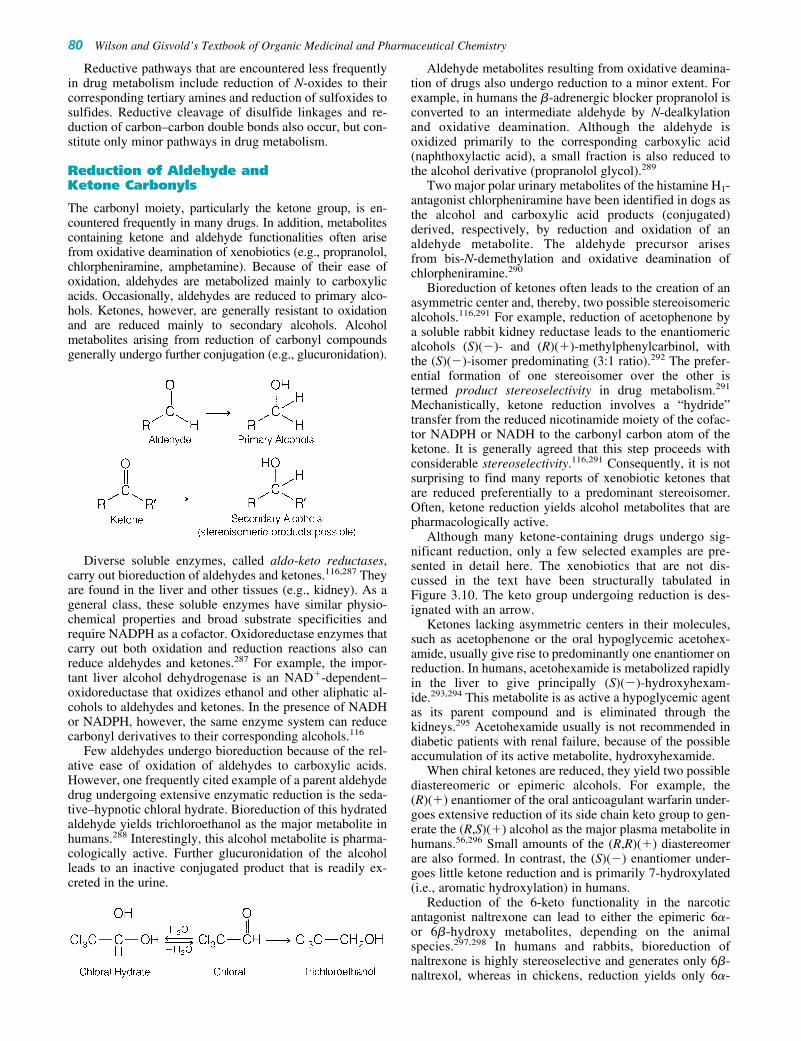
Reductive pathways that are encountered less frequentlyin drug metabolism include reduction of N-oxides to theircorresponding tertiary amines and reduction of sulfoxides tosulfides. Reductive cleavage of disulfide linkages and re-duction of carbon–carbon double bonds also occur, but con-stitute only minor pathways in drug metabolism.
Reduction of Aldehyde andKetone Carbonyls
The carbonyl moiety, particularly the ketone group, is en-countered frequently in many drugs. In addition, metabolitescontaining ketone and aldehyde functionalities often arisefrom oxidative deamination of xenobiotics (e.g., propranolol,chlorpheniramine, amphetamine). Because of their ease ofoxidation, aldehydes are metabolized mainly to carboxylicacids. Occasionally, aldehydes are reduced to primary alco-hols. Ketones, however, are generally resistant to oxidationand are reduced mainly to secondary alcohols. Alcoholmetabolites arising from reduction of carbonyl compoundsgenerally undergo further conjugation (e.g., glucuronidation).
Diverse soluble enzymes, called aldo-keto reductases,carry out bioreduction of aldehydes and ketones.116,287 Theyare found in the liver and other tissues (e.g., kidney). As ageneral class, these soluble enzymes have similar physio-chemical properties and broad substrate specificities andrequire NADPH as a cofactor. Oxidoreductase enzymes thatcarry out both oxidation and reduction reactions also canreduce aldehydes and ketones.287 For example, the impor-tant liver alcohol dehydrogenase is an NAD�-dependent–oxidoreductase that oxidizes ethanol and other aliphatic al-cohols to aldehydes and ketones. In the presence of NADHor NADPH, however, the same enzyme system can reducecarbonyl derivatives to their corresponding alcohols.116
Few aldehydes undergo bioreduction because of the rel-ative ease of oxidation of aldehydes to carboxylic acids.However, one frequently cited example of a parent aldehydedrug undergoing extensive enzymatic reduction is the seda-tive–hypnotic chloral hydrate. Bioreduction of this hydratedaldehyde yields trichloroethanol as the major metabolite inhumans.288 Interestingly, this alcohol metabolite is pharma-cologically active. Further glucuronidation of the alcoholleads to an inactive conjugated product that is readily ex-creted in the urine.
Aldehyde metabolites resulting from oxidative deamina-tion of drugs also undergo reduction to a minor extent. Forexample, in humans the �-adrenergic blocker propranolol isconverted to an intermediate aldehyde by N-dealkylationand oxidative deamination. Although the aldehyde isoxidized primarily to the corresponding carboxylic acid(naphthoxylactic acid), a small fraction is also reduced tothe alcohol derivative (propranolol glycol).289
Two major polar urinary metabolites of the histamine H1-antagonist chlorpheniramine have been identified in dogs asthe alcohol and carboxylic acid products (conjugated)derived, respectively, by reduction and oxidation of analdehyde metabolite. The aldehyde precursor arisesfrom bis-N-demethylation and oxidative deamination ofchlorpheniramine.290
Bioreduction of ketones often leads to the creation of anasymmetric center and, thereby, two possible stereoisomericalcohols.116,291 For example, reduction of acetophenone bya soluble rabbit kidney reductase leads to the enantiomericalcohols (S)(�)- and (R)(�)-methylphenylcarbinol, withthe (S)(�)-isomer predominating (3:1 ratio).292 The prefer-ential formation of one stereoisomer over the other istermed product stereoselectivity in drug metabolism.291
Mechanistically, ketone reduction involves a “hydride”transfer from the reduced nicotinamide moiety of the cofac-tor NADPH or NADH to the carbonyl carbon atom of theketone. It is generally agreed that this step proceeds withconsiderable stereoselectivity.116,291 Consequently, it is notsurprising to find many reports of xenobiotic ketones thatare reduced preferentially to a predominant stereoisomer.Often, ketone reduction yields alcohol metabolites that arepharmacologically active.
Although many ketone-containing drugs undergo sig-nificant reduction, only a few selected examples are pre-sented in detail here. The xenobiotics that are not dis-cussed in the text have been structurally tabulated inFigure 3.10. The keto group undergoing reduction is des-ignated with an arrow.
Ketones lacking asymmetric centers in their molecules,such as acetophenone or the oral hypoglycemic acetohex-amide, usually give rise to predominantly one enantiomer onreduction. In humans, acetohexamide is metabolized rapidlyin the liver to give principally (S)(�)-hydroxyhexam-ide.293,294 This metabolite is as active a hypoglycemic agentas its parent compound and is eliminated through thekidneys.295 Acetohexamide usually is not recommended indiabetic patients with renal failure, because of the possibleaccumulation of its active metabolite, hydroxyhexamide.
When chiral ketones are reduced, they yield two possiblediastereomeric or epimeric alcohols. For example, the(R)(�) enantiomer of the oral anticoagulant warfarin under-goes extensive reduction of its side chain keto group to gen-erate the (R,S)(�) alcohol as the major plasma metabolite inhumans.56,296 Small amounts of the (R,R)(�) diastereomerare also formed. In contrast, the (S)(�) enantiomer under-goes little ketone reduction and is primarily 7-hydroxylated(i.e., aromatic hydroxylation) in humans.
Reduction of the 6-keto functionality in the narcoticantagonist naltrexone can lead to either the epimeric 6�-or 6�-hydroxy metabolites, depending on the animalspecies.297,298 In humans and rabbits, bioreduction ofnaltrexone is highly stereoselective and generates only 6�-naltrexol, whereas in chickens, reduction yields only 6�-
80 Wilson and Gisvold’s Textbook of Organic Medicinal and Pharmaceutical Chemistry
0043-0118_17865_Ch03.qxd 12/4/09 3:49 AM Page 80

Chapter 3 Metabolic Changes of Drugs and Related Organic Compounds 81
0043-0118_17865_Ch03.qxd 12/4/09 3:49 AM Page 81

82 Wilson and Gisvold’s Textbook of Organic Medicinal and Pharmaceutical Chemistry
Figure 3.10 Additional examples of xenobiotics that undergo extensive ketone reduc-tion, not covered in the text. Arrow indicates the keto group undergoing reduction.
0043-0118_17865_Ch03.qxd 12/4/09 3:49 AM Page 82

Chapter 3 Metabolic Changes of Drugs and Related Organic Compounds 83
naltrexol.297–299 In monkeys and guinea pigs, however, bothepimeric alcohols are formed (predominantly 6�-naltrexol).300,301 Apparently, in the latter two species, reduc-tion of naltrexone to the epimeric 6�- and 6�-alcohols iscarried out by two distinctly different reductases found inthe liver.299–301
Reduction of oxisuran appears not to be an importantpathway by which the parent drug mediates its immunosup-pressive effects. Studies indicate that oxisuran has its great-est immunosuppressive effects in those species that formalcohols as their major metabolic products (e.g., human,rat).302–306 In species in which reduction is a minor pathway(e.g., dog), oxisuran shows little immunosuppressive activ-ity.304–306 These findings indicate that the oxisuran alcohols(oxisuranols) are pharmacologically active and contributesubstantially to the overall immunosuppressive effect of theparent drug. The sulfoxide group in oxisuran is chiral, byvirtue of the lone pair of electrons on sulfur. Therefore,reduction of oxisuran leads to diastereomeric alcohols.
Reduction of �,�-unsaturated ketones results in reduc-tion not only of the ketone group but of the carbon–carbondouble bond as well. Steroidal drugs often fall into thisclass, including norethindrone, a synthetic progestin foundin many oral contraceptive drug combinations. In women,the major plasma and urinary metabolite of norethindrone isthe 3�,5�-tetrahydro derivative.307
Ketones resulting from metabolic oxidative deaminationprocesses are also susceptible to reduction. For instance,rabbit liver microsomal preparations metabolize amphet-amine to phenylacetone, which is reduced subsequently to1-phenyl-2-propanol.308 In humans, a minor urinarymetabolite of (�)-ephedrine has been identified as the diolderivative formed from keto reduction of the oxidativelydeaminated product 1-hydroxy-1-phenylpropan-2-one.309
Reduction of Nitro andAzo Compounds
The reduction of aromatic nitro and azo xenobiotics leads toaromatic primary amine metabolites.286 Aromatic nitro com-pounds are reduced initially to the nitroso and hydroxylamineintermediates, as shown in the following metabolic sequence:
Azo reduction, however, is believed to proceed via ahydrazo intermediate (-NH-NH-) that subsequently is cleavedreductively to yield the corresponding aromatic amines:
Bioreduction of nitro compounds is carried out byNADPH-dependent microsomal and soluble nitro reductasespresent in the liver. A multicomponent hepatic microsomal
0043-0118_17865_Ch03.qxd 12/4/09 3:49 AM Page 83

reductase system requiring NADPH appears to be responsi-ble for azo reduction.310–312 In addition, bacterial reductasespresent in the intestine can reduce nitro and azo compounds,especially those that are absorbed poorly or excreted mainlyin the bile.313,314
Various aromatic nitro drugs undergo enzymatic reduc-tion to the corresponding aromatic amines. For example,the 7-nitro benzodiazepine derivatives clonazepam andnitrazepam are metabolized extensively to their respective7-amino metabolites in humans.315,316 The skeletal musclerelaxant dantrolene (Dantrium) also reportedly undergoesreduction to aminodantrolene in humans.317,318
For some nitro xenobiotics, bioreduction appears to bea minor metabolic pathway in vivo, because of competingoxidative and conjugative reactions. Under artificial anaer-obic in vitro incubation conditions, however, these samenitro xenobiotics are enzymatically reduced rapidly. Forexample, most of the urinary metabolites of metronidazolefound in humans are either oxidation or conjugation prod-ucts. Reduced metabolites of metronidazole have not beendetected.319 When incubated anaerobically with guinea pigliver preparations, however, metronidazole undergoesconsiderable nitro reduction.320
Bacterial reductase present in the intestine also tends tocomplicate in vivo interpretations of nitro reduction. Forexample, in rats, the antibiotic chloramphenicol is not re-duced in vivo by the liver but is excreted in the bile and,subsequently, reduced by intestinal flora to form the aminometabolite.321,322
The enzymatic reduction of azo compounds is best exem-plified by the conversion of sulfamidochrysoidine(Prontosil) to the active sulfanilamide metabolite in theliver.323 This reaction has historical significance, for it led to
the discovery of sulfanilamide as an antibiotic and eventu-ally to the development of many of the therapeutic sulfon-amide drugs. Bacterial reductases present in the intestineplay a significant role in reducing azo xenobiotics, particu-larly those that are absorbed poorly.313,314 Accordingly, thetwo azo dyes tartrazine324,325 and amaranth326 have poororal absorption because of the many polar and ionizedzsulfonic acid groups present in their structures.
Therefore, these two azo compounds are metabolizedprimarily by bacterial reductases present in the intestine.The importance of intestinal reduction is further revealed inthe metabolism of sulfasalazine (formerly salicylazosul-fapyridine, Azulfidine), a drug used in the treatment of ul-cerative colitis. The drug is absorbed poorly and undergoesreductive cleavage of the azo linkage to yield sulfapyridineand 5-aminosalicylic acid.327,328 The reaction occurs prima-rily in the colon and is carried out principally by intestinalbacteria. Studies in germfree rats, lacking intestinal flora,have demonstrated that sulfasalazine is not reduced to anyappreciable extent.329
Miscellaneous Reductions
Several minor reductive reactions also occur. Reduction ofN-oxides to the corresponding tertiary amine occurs to someextent. This reductive pathway is of interest because severaltertiary amines are oxidized to form polar and water-solubleN-oxide metabolites. If reduction of N-oxide metabolitesoccurs to a significant extent, drug elimination of the parenttertiary amine is impeded. N-Oxide reduction often isassessed by administering the pure synthetic N-oxide invitro or in vivo and then attempting to detect the formationof the tertiary amine. For example, imipramine N-oxideundergoes reduction in rat liver preparations.330
Reduction of sulfur-containing functional groups, suchas the disulfide and sulfoxide moieties, also constitutesa minor reductive pathway. Reductive cleavage of thedisulfide bond in disulfiram (Antabuse) yields N,N-diethyldithiocarbamic acid (free or glucuronidated) as amajor metabolite in humans.331,332 Although sulfoxidefunctionalities are oxidized mainly to sulfones (-SO2-),they sometimes undergo reduction to sulfides. The
84 Wilson and Gisvold’s Textbook of Organic Medicinal and Pharmaceutical Chemistry
0043-0118_17865_Ch03.qxd 12/4/09 3:49 AM Page 84

Chapter 3 Metabolic Changes of Drugs and Related Organic Compounds 85
0043-0118_17865_Ch03.qxd 12/4/09 3:49 AM Page 85

importance of this reductive pathway is seen in the metab-olism of the anti-inflammatory agent sulindac (Clinoril).Studies in humans show that sulindac undergoes reductionto an active sulfide that is responsible for the overall anti-inflammatory effect of the parent drug.333,334 Sulindac orits sulfone metabolite exhibits little anti-inflammatory ac-tivity. Another example of sulfide formation involves thereduction of DMSO to dimethyl sulfide. In humans,DMSO is metabolized to a minor extent by this pathway.The characteristic unpleasant odor of dimethyl sulfide isevident on the breath of patients who use this agent.335
HYDROLYTIC REACTIONS
Hydrolysis of Esters and Amides
The metabolism of ester and amide linkages in many drugsis catalyzed by hydrolytic enzymes present in various tis-sues and in plasma. The metabolic products formed (car-boxylic acids, alcohols, phenols, and amines) generally arepolar and functionally more susceptible to conjugation andexcretion than the parent ester or amide drugs. The enzymescarrying out ester hydrolysis include several nonspecificesterases found in the liver, kidney, and intestine as well asthe pseudocholinesterases present in plasma.336,337 Amidehydrolysis appears to be mediated by liver microsomalamidases, esterases, and deacylases.337
Hydrolysis is a major biotransformation pathway fordrugs containing an ester functionality. This is because ofthe relative ease of hydrolyzing the ester linkage. A classicexample of ester hydrolysis is the metabolic conversion ofaspirin (acetylsalicylic acid) to salicylic acid.338 Of the twoester moieties present in cocaine, it appears that, in gen-eral, the methyl group is hydrolyzed preferentially to yieldbenzoylecgonine as the major human urinary metabo-lite.339 The hydrolysis of cocaine to methyl ecgonine, how-ever, also occurs in plasma and, to a minor extent,blood.340,341 Methylphenidate (Ritalin) is biotransformedrapidly by hydrolysis to yield ritalinic acid as the majorurinary metabolite in humans.342 Often, ester hydrolysis ofthe parent drug leads to pharmacologically active metabo-lites. For example, hydrolysis of diphenoxylate in humansleads to diphenoxylic acid (difenoxin), which is, appar-ently, 5 times more potent an antidiarrheal agent than theparent ester.343 The rapid metabolism of clofibrate
86 Wilson and Gisvold’s Textbook of Organic Medicinal and Pharmaceutical Chemistry
(Atromid-S) yields p-chlorophenoxyisobutyric acid(CPIB) as the major plasma metabolite in humans.344
Studies in rats indicate that the free acid CPIB is responsi-ble for clofibrate’s hypolipidemic effect.345
Many parent drugs have been chemically modified or de-rivatized to generate so-called prodrugs to overcome someundesirable property (e.g., bitter taste, poor absorption, poorsolubility, irritation at site of injection). The rationale be-hind the prodrug concept was to develop an agent that, onceinside the biological system, would be biotransformed to theactive parent drug.18 The presence of esterases in many tis-sues and plasma makes ester derivatives logical prodrugcandidates, because hydrolysis would cause the esterprodrug to revert to the parent compound. Accordingly,antibiotics such as chloramphenicol and clindamycin havebeen derivatized as their palmitate esters to minimize theirbitter taste and to improve their palatability in pediatric liq-uid suspensions.346,347 After oral administration, intestinalesterases and lipases hydrolyze the palmitate esters to thefree antibiotics. To improve the poor oral absorption of car-benicillin, a lipophilic indanyl ester has been formulated(Geocillin).348 Once orally absorbed, the ester is hydrolyzedrapidly to the parent drug. A final example involves deriva-tization of prednisolone to its C-21 hemisuccinate sodiumsalt. This water-soluble derivative is extremely useful forparenteral administration and is metabolized to the parentsteroid drug by plasma and tissue esterases.349
Amides are hydrolyzed slowly in comparison to es-ters.337 Consequently, hydrolysis of the amide bond ofprocainamide is relatively slow compared with hydrolysis ofthe ester linkage in procaine.336,350 Drugs in which amidecleavage has been reported to occur, to some extent, includelidocaine,351 carbamazepine,87 indomethacin,251,252 andprazosin (Minipress).253,254 Amide linkages present in bar-biturates (e.g., hexobarbital)352,353 as well as in hydantoins(e.g., 5-phenylhydantoin)354,355 and succinimides (phensux-imide)354,355 are also susceptible to hydrolysis.
Miscellaneous Hydrolytic Reactions
Hydrolysis of recombinant human peptide drugs and hor-mones at the N- or C-terminal amino acids by carboxypep-tidase and aminopeptidase and proteases in blood and other
0043-0118_17865_Ch03.qxd 12/4/09 3:49 AM Page 86

Chapter 3 Metabolic Changes of Drugs and Related Organic Compounds 87
tissues is a well-recognized hydrolytic reaction.356,357
Examples of peptides or protein hormones undergoinghydrolysis include human insulin, growth hormone (GH),prolactin, parathyroid hormone (PTH), and atrial natriureticfactor (ANF).358
In addition to hydrolysis of amides and esters, hydrolyticcleavage of other moieties occurs to a minor extent in drugmetabolism,8 including the hydrolysis of phosphate esters(e.g., diethylstilbestrol diphosphate), sulfonylureas, cardiacglycosides, carbamate esters, and organophosphate com-pounds. Glucuronide and sulfate conjugates also can un-dergo hydrolytic cleavage by �-glucuronidase and sulfataseenzymes. These hydrolytic reactions are discussed in thefollowing section. Finally, the hydration or hydrolytic cleav-age of epoxides and arene oxides by epoxide hydrase is con-sidered a hydrolytic reaction.
Miscellaneous Bioactivation of Prodrugs
Throughout this chapter, the metabolism of produgs to an ac-tive form is presented. However, in the cases presented ear-lier, there was a chemical functional group that was subjectto phase I metabolism to release the active drug molecule.
0043-0118_17865_Ch03.qxd 12/4/09 3:49 AM Page 87

88 Wilson and Gisvold’s Textbook of Organic Medicinal and Pharmaceutical Chemistry
The metabolic reactions included oxidative activation, reduc-tive activation, hydrolytic activation, etc. One reaction that isof particular interest involves chemical activation as seenwith the proton pump inhibitors, which are clinically used totreat gastric ulceration. When administered to a patient, thedrug is dosed orally, allowing for systemic distribution.When the proton pump inhibitor arrives at an acidic region ofthe body, such as the parietal cells of the stomach, chemicalactivation occurs. The highly acidic environment in andaround the parietal cell allows for protonation of nitrogen onthe benzimidazole ring, followed by attachment of the pyri-dine nitrogen. Ring opening of the intermediate then yieldsthe sulfenic acid that subsequently cyclizes with the loss ofwater. This intermediate is highly susceptible to nucleophilicattack by the SH moieties of the cysteine residues associatedwith proteins, including the proton pump of the parietal cells.This pump is responsible for exchange of K� with H� fromparietal cell into gastric lumen, and through this inhibition,there is regulation of the acidic environment of stomach.
PHASE II OR CONJUGATIONREACTIONS
Phase I or functionalization reactions do not always pro-duce hydrophilic or pharmacologically inactive metabo-lites. Various phase II or conjugation reactions, however,
can convert these metabolites to more polar and water-soluble products. Many conjugative enzymes accomplishthis objective by attaching small, polar, and ionizable en-dogenous molecules, such as glucuronic acid, sulfate,glycine, and glutamine, to the phase I metabolite or parentxenobiotic. The resulting conjugated products are rela-tively water soluble and readily excretable. In addition,they generally are biologically inactive and nontoxic.Other phase II reactions, such as methylation and acetyla-tion, do not generally increase water solubility but mainlyserve to terminate or attenuate pharmacological activity.The role of GSH is to combine with chemically reactivecompounds to prevent damage to important biomacromol-ecules, such as DNA, RNA, and proteins. Thus, phase IIreactions can be regarded as truly detoxifying pathways indrug metabolism, with a few exceptions.
A distinguishing feature of most phase II reactions is thatthe conjugating group (glucuronic acid, sulfate, methyl, andacetyl) is activated initially in the form of a coenzyme be-fore transfer or attachment of the group to the accepting sub-strate by the appropriate transferase enzyme. In other cases,such as glycine and glutamine conjugation, the substrate isactivated initially. Many endogenous compounds, such asbilirubin, steroids, catecholamines, and histamine, also un-dergo conjugation reactions and use the same coenzymes,although they appear to be mediated by more specifictransferase enzymes. The phase II conjugative pathways
0043-0118_17865_Ch03.qxd 12/4/09 3:49 AM Page 88

discussed include those previously listed in this chapter.Although other conjugative pathways exist (e.g., conjuga-tion with glycosides, phosphate, and other amino acids andconversion of cyanide to thiocyanate), they are of minor im-portance in drug metabolism and are not covered in thischapter.
Glucuronic Acid Conjugation
Glucuronidation is the most common conjugative pathwayin drug metabolism for several reasons: (a) a readily avail-able supply of D-glucuronic acid (derived from D-glucose),(b) numerous functional groups that can combine enzymat-ically with glucuronic acid, and (c) the glucuronyl moiety(with its ionized carboxylate [pKa 3.2] and polar hydroxylgroups), which, when attached to xenobiotic substrates,greatly increases the water solubility of the conjugatedproduct.117,359–361 Formation of �-glucuronides involvestwo steps: synthesis of an activated coenzyme, uridine-5�-diphospho-�-D-glucuronic acid (UDPGA), and subsequenttransfer of the glucuronyl group from UDPGA to an appro-priate substrate.117,360,361 The transfer step is catalyzed bymicrosomal enzymes called UDP-glucuronyltransferases.They are found primarily in the liver but also occur inmany other tissues, including kidney, intestine, skin, lung,and brain.360,361 The sequence of events involved inglucuronidation is summarized in Figure 3.11.117,360,361
The synthesis of the coenzyme UDPGA uses �-D-glucose-1-phosphate as its initial precursor. Note that all glu-curonide conjugates have the �-configuration or �-linkageat C-1 (hence, the term �-glucuronides). In contrast, thecoenzyme UDPGA has an �-linkage. In the enzymatictransfer step, it appears that nucleophilic displacement ofthe �-linked UDP moiety from UDPGA by the substrateRXH proceeds with complete inversion of configuration atC-1 to give the �-glucuronide. Glucuronidation of onefunctional group usually suffices to effect excretion of theconjugated metabolite; diglucuronide conjugates do notusually occur.
The diversity of functional groups undergoing glu-curonidation is illustrated in Table 3.3 and Figure 3.12.
Metabolic products are classified as oxygen–, nitrogen–,sulfur–, or carbon–glucuronide, according to the het-eroatom attached to the C-1 atom of the glucuronyl group.Two important functionalities, the hydroxy and carboxy,form O-glucuronides. Phenolic and alcoholic hydroxylsare the most common functional groups undergoingglucuronidation in drug metabolism. As we have seen,phenolic and alcoholic hydroxyl groups are present inmany parent compounds and arise through various phase Imetabolic pathways. Morphine,362,363 acetaminophen,364
and p-hydroxyphenytoin (the major metabolite ofphenytoin)49,50 are a few examples of phenolic compoundsthat undergo considerable glucuronidation. Alcoholic
Chapter 3 Metabolic Changes of Drugs and Related Organic Compounds 89
Figure 3.11 Formation of UDPGAand �-glucuronide conjugates.
TABLE 3.3 Types of Compounds Forming Oxygen,Nitrogen, Sulfur, and Carbon Glucuronidesa
Oxygen Glucuronides
Hydroxyl compoundsPhenols: morphine, acetaminophen, p-hydroxyphenytoinAlcohols: tricholoroethanol, chloramphenicol, propranololEnols: 4-hydroxycoumarinN-Hydroxyamines: N-hydroxydapsoneN-Hydroxyamides: N-hydroxy-2-acetylaminofluorene
Carboxyl compoundsAryl acids: benzoic acid, salicylic acidArylalkyl acids: naproxen, fenoprofen
Nitrogen Glucuronides
Arylamines: 7-amino-5-nitroindazoleAlkylamines: desipramineAmides: meprobamateSulfonamides: sulfisoxazoleTertiary amines: cyproheptadine, tripelennamine
Sulfur Glucuronides
Sulfhydryl groups: methimazole, propylthiouracil, diethylthiocarbamic acid
Carbon Glucuronides
3,5-Pyrazolidinedione: phenylbutazone, sulfinpyrazone
aFor structures and site of �-glucuronide attachment, see Figure 4.12.
0043-0118_17865_Ch03.qxd 12/4/09 3:49 AM Page 89
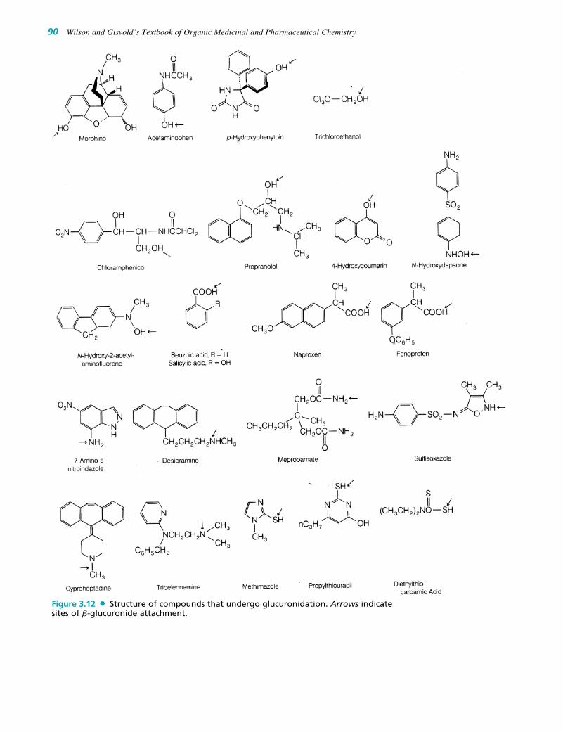
90 Wilson and Gisvold’s Textbook of Organic Medicinal and Pharmaceutical Chemistry
Figure 3.12 Structure of compounds that undergo glucuronidation. Arrows indicatesites of �-glucuronide attachment.
0043-0118_17865_Ch03.qxd 12/4/09 3:49 AM Page 90

uronide conjugates, which are excreted primarily in theurine. As the relative molecular mass of the conjugateexceeds 300 Da, however, biliary excretion may becomean important route of elimination.386 Glucuronides thatare excreted in the bile are susceptible to hydrolysis by �-glucuronidase enzymes present in the intestine. Thehydrolyzed product may be reabsorbed in the intestine, thusleading to enterohepatic recycling.22 �-Glucuronidases arealso present in many other tissues, including the liver, theendocrine system, and the reproductive organs. Althoughthe function of these hydrolytic enzymes in drug metabo-lism is unclear, it appears that, in terms of hormonal andccendocrine regulation, �-glucuronidases may liberateactive hormones (e.g., steroids) from their inactive gluc-uronide conjugates.22
In neonates and children, glucuronidating processes areoften not developed fully. In such subjects, drugs andendogenous compounds (e.g., bilirubin) that are metabo-lized normally by glucuronidation may accumulate andcause serious toxicity. For example, neonatal hyperbiliru-binemia may be attributable to the inability of newbornsto conjugate bilirubin with glucuronic acid.387 Similarly,the inability of infants to glucuronidate chloramphenicolhas been suggested to be responsible for the gray babysyndrome, which results from accumulation of toxic levelsof the free antibiotic.388
Sulfate Conjugation
Conjugation of xenobiotics with sulfate occurs primarilywith phenols and, occasionally, with alcohols, aromaticamines, and N-hydroxy compounds.389–391 In contrast toglucuronic acid, the amount of available sulfate is ratherlimited. The body uses a significant portion of the sulfatepool to conjugate numerous endogenous compounds suchas steroids, heparin, chondroitin, catecholamines, and thy-roxine. The sulfate conjugation process involves activationof inorganic sulfate to the coenzyme 3�-phosphoadenosine-5�-phosphosulfate (PAPS). Subsequent transfer of the sul-fate group from PAPS to the accepting substrate is cat-alyzed by various soluble sulfotransferases present in theliver and other tissues (e.g., kidney, intestine).392 The se-quence of events involved in sulfoconjugation is depictedin Figure 3.13. Sulfate conjugation generally leads towater-soluble and inactive metabolites. It appears, how-ever, that the O-sulfate conjugates of some N-hydroxycompounds give rise to chemically reactive intermediatesthat are toxic.241
Phenols compose the main group of substrates under-going sulfate conjugation. Thus, drugs containing pheno-lic moieties are often susceptible to sulfate formation. Forexample, the antihypertensive agent �-methyldopa(Aldomet) is metabolized extensively to its 3-O-sulfateester in humans.393,394 The �-adrenergic bronchodilatorssalbutamol (albuterol)395 and terbutaline (Brethine,Bricanyl)396 also undergo sulfate conjugation as their
Chapter 3 Metabolic Changes of Drugs and Related Organic Compounds 91
hydroxyls, such as those present in trichloroethanol (majormetabolite of chloral hydrate),288 chloramphenicol,365 andpropranolol,366,367 are also commonly glucuronidated.Less frequent is glucuronidation of other hydroxyl groups,such as enols,368 N-hydroxylamines,226 and N-hydroxyl-amides.241 For examples, refer to the list of glucuronidesin Table 3.3.
The carboxy group is also subject to conjugation withglucuronic acid. For example, arylaliphatic acids, such asthe anti-inflammatory agents naproxen369 and fenopro-fen,370,371 are excreted primarily as their O-glucuronide de-rivatives in humans. Carboxylic acid metabolites such asthose arising from chlorpheniramine290 and propranolol289
(see “Reduction of Aldehyde and Ketone Carbonyls,”) formO-glucuronide conjugates. Aryl acids (e.g., benzoic acid,372
salicylic acid373,374) also undergo conjugation with gluc-uronic acid, but conjugation with glycine appears to be amore important pathway for these compounds.
Occasionally, N-glucuronides are formed with aromaticamines, aliphatic amines, amides, and sulfonamides.Representative examples are found in the list of gluc-uronides in Table 3.3. Glucuronidation of aromatic andaliphatic amines is generally a minor pathway in compari-son with N-acetylation or oxidative processes (e.g., oxi-dative deamination). Tertiary amines, such as the anti-histaminic agents cyproheptadine (Periactin)375 andtripelennamine,376 form interesting quaternary ammoniumglucuronide metabolites.
Because the thiol group (SH) does not commonly occurin xenobiotics, S-glucuronide products have been reportedfor only a few drugs. For instance, the thiol groups presentin methimazole (Tapazole),377 propylthiouracil,378,379 andN,N-diethyldithiocarbamic acid (major reduced metaboliteof disulfiram, Antabuse)380 undergo conjugation with gluc-uronic acid.
The formation of glucuronides attached directly to a car-bon atom is relatively novel in drug metabolism. Studies inhumans have shown that conjugation of phenylbutazone(Butazolidin)381,382 and sulfinpyrazone (Anturane)383 yieldthe corresponding C-glucuronide metabolites:
Besides xenobiotics, several endogenous substrates,notably bilirubin384 and steroids,385 are eliminated as gluc-
0043-0118_17865_Ch03.qxd 12/4/09 3:49 AM Page 91
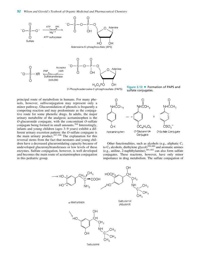
principal route of metabolism in humans. For many phe-nols, however, sulfoconjugation may represent only aminor pathway. Glucuronidation of phenols is frequently acompeting reaction and may predominate as the conjuga-tive route for some phenolic drugs. In adults, the majorurinary metabolite of the analgesic acetaminophen is theO-glucuronide conjugate, with the concomitant O-sulfateconjugate being formed in small amounts.364 Interestingly,infants and young children (ages 3–9 years) exhibit a dif-ferent urinary excretion pattern: the O-sulfate conjugate isthe main urinary product.397,398 The explanation for thisreversal stems from the fact that neonates and young chil-dren have a decreased glucuronidating capacity because ofundeveloped glucuronyltransferases or low levels of theseenzymes. Sulfate conjugation, however, is well developedand becomes the main route of acetaminophen conjugationin this pediatric group.
Other functionalities, such as alcohols (e.g., aliphatic C1
to C5 alcohols, diethylene glycol)399,400 and aromatic amines(e.g., aniline, 2-naphthylamine),401,402 can also form sulfateconjugates. These reactions, however, have only minorimportance in drug metabolism. The sulfate conjugation of
92 Wilson and Gisvold’s Textbook of Organic Medicinal and Pharmaceutical Chemistry
Figure 3.13 Formation of PAPS andsulfate conjugates.
0043-0118_17865_Ch03.qxd 12/4/09 3:49 AM Page 92

N-hydroxylamines and N-hydroxylamides takes place aswell, occasionally. O-Sulfate ester conjugates of N-hydroxycompounds are of considerable toxicological concern be-cause they can lead to reactive intermediates that are respon-sible for cellular toxicity. The carcinogenic agents N-methyl-4-aminoazobenzene and 2-AAF are believed to mediate theirtoxicity through N-hydroxylation to the corresponding N-hy-droxy compounds (see earlier section on N-hydroxylation ofamines and amides). Sulfoconjugation of the N-hydroxymetabolites yields O-sulfate esters, which presumably are theultimate carcinogenic species. Loss of SO4
2� from the fore-going sulfate conjugates generates electrophilic nitreniumspecies, which may react with nucleophilic groups (e.g.,NH2, OH, SH) present in proteins, DNA, and RNA to formcovalent linkages that lead to structural and functional alter-ation of these crucial biomacromolecules.403 The conse-quences of this are cellular toxicity (tissue necrosis) or alter-ation of the genetic code, eventually leading to cancer. Someevidence supporting the role of sulfate conjugation in themetabolic activation of N-hydroxy compounds to reactive in-termediates comes from the observation that the degree ofhepatotoxicity and hepatocarcinogenicity of N-hydroxy-2-acetylaminofluorene depends markedly on the level of sulfo-transferase activity in the liver.404,405
The discontinued analgesic phenacetin is metabolizedto N-hydroxyphenacetin and subsequently conjugated withsulfate.406 The O-sulfate conjugate of N-hydroxy-phenacetin binds covalently to microsomal proteins.407
This pathway may represent one route leading to reactiveintermediates that are responsible for the hepatotoxicityand nephrotoxicity associated with phenacetin. Other path-ways (e.g., arene oxides) leading to reactive electrophilicintermediates are also possible.6
Conjugation with Glycine, Glutamine,and Other Amino Acids
The amino acids glycine and glutamine are used by mam-malian systems to conjugate carboxylic acids, particularlyaromatic acids and arylalkyl acids.408,409 Glycine conjugationis common to most mammals, whereas glutamine conjugationappears to be confined mainly to humans and other primates.The quantity of amino acid conjugates formed from xenobi-otics is minute because of the limited availability of aminoacids in the body and competition with glucuronidation forcarboxylic acid substrates. In contrast with glucuronic acidand sulfate, glycine and glutamine are not converted to acti-vated coenzymes. Instead, the carboxylic acid substrate is ac-tivated with adenosine triphosphate (ATP) and coenzyme A(CoA) to form an acyl-CoA complex. The latter intermediate,in turn, acylates glycine or glutamine under the influence ofspecific glycine or glutamine N-acyltransferase enzymes. Theactivation and acylation steps take place in the mitochondriaof liver and kidney cells. The sequence of metabolic eventsassociated with glycine and glutamine conjugation of phenyl-acetic acid is summarized in Figure 3.14. Amino acid conju-
Chapter 3 Metabolic Changes of Drugs and Related Organic Compounds 93
Figure 3.14 Formation of glycine and glutamine conjugates of phenylacetic acid.
0043-0118_17865_Ch03.qxd 12/4/09 3:49 AM Page 93

gates, being polar and water soluble, are excreted mainly re-nally and, sometimes, in the bile.
Aromatic acids and arylalkyl acids are the major substratesundergoing glycine conjugation. The conversion of benzoicacid to its glycine conjugate, hippuric acid, is a well-knownmetabolic reaction in many mammalian systems.410 The ex-tensive metabolism of salicylic acid (75% of dose) to salicyl-uric acid in humans is another illustrative example.411,412
Carboxylic acid metabolites resulting from oxidation or hy-drolysis of many drugs are also susceptible to glycine conju-gation. For example, the H1-histamine antagonist bromphen-iramine is oxidized to a propionic acid metabolite that isconjugated with glycine in both human and dog.181 Similarly,p-fluorophenylacetic acid, derived from the metabolism of theantipsychotic agent haloperidol (Haldol), is found as theglycine conjugate in the urine of rats.413 Phenylacetic acid andisonicotinic acid, resulting from the hydrolysis of, respec-tively, the anticonvulsant phenacemide (Phenurone)414 and theantituberculosis agent isoniazid,415 also are conjugated withglycine to some extent.
Glutamine conjugation occurs mainly with arylacetic acids,including endogenous phenylacetic416 and 3-indolylaceticacid.417 A few glutamine conjugates of drug metabolites havebeen reported. For example, in humans, the 3,4-dihydroxy-5-methoxyphenylacetic acid metabolite of mescaline is found asa conjugate of glutamine.418 Diphenylmethoxyacetic acid, ametabolite of the antihistamine diphenhydramine (Benadryl),is biotransformed further to the corresponding glutamine de-rivative in the rhesus monkey.419
Several other amino acids are involved in the conjugationof carboxylic acids, but these reactions occur only occasion-ally and appear to be highly substrate and species depen-dent.409,420 Ornithine (in birds), aspartic acid and serine(in rats), alanine (in mouse and hamster), taurine(H2NCH2CH2SO3H) (in mammals and pigeons), and histi-dine (in African bats) are among these amino acids.420
GSH or Mercapturic Acid Conjugates
GSH conjugation is an important pathway for detoxifyingchemically reactive electrophilic compounds.421–428 It is now
generally accepted that reactive electrophilic species manifesttheir toxicity (e.g., tissue necrosis, carcinogenicity, muta-genicity, teratogenicity) by combining covalently with nucleo-philic groups present in vital cellular proteins and nucleicacids.4,429 Many serious drug toxicities may be explained alsoin terms of covalent interaction of metabolically generatedelectrophilic intermediates with cellular nucleophiles.5,6 GSHprotects vital cellular constituents against chemically reactivespecies by virtue of its nucleophilic SH group. The SH groupreacts with electron-deficient compounds to form S-substi-tuted GSH adducts (Fig. 3.15).421–428
GSH is a tripeptide (�-glutamyl-cysteinylglycine) found inmost tissues. Xenobiotics conjugated with GSH usually arenot excreted as such, but undergo further biotransformation togive S-substituted N-acetylcysteine products called mercap-turic acids.76,86,424–428 This process involves enzymatic cleav-age of two amino acids (namely, glutamic acid and glycine)from the initially formed GSH adduct and subsequentN-acetylation of the remaining S-substituted cysteine residue.The formation of GSH conjugates and their conversion tomercapturic acid derivatives are outlined in Figure 3.15.
Conjugation of a wide spectrum of substrates with GSHis catalyzed by a family of cytoplasmic enzymes known asGSH S-transferases.75 These enzymes are found in most tis-sues, particularly the liver and kidney. Degradation of GSHconjugates to mercapturic acids is carried out principally byrenal and hepatic microsomal enzymes (Fig. 3.15).76 Unlikeother conjugative phase II reactions, GSH conjugation doesnot require the initial formation of an activated coenzyme orsubstrate. The inherent reactivity of the nucleophilic GSHtoward an electrophilic substrate usually provides sufficientdriving force. The substrates susceptible to GSH conjuga-tion are quite varied and encompass many chemically differ-ent classes of compounds. A major prerequisite is that thesubstrate be sufficiently electrophilic. Compounds that reactwith GSH do so by two general mechanisms: (a) nucleo-philic displacement at an electron-deficient carbon or het-eroatom or (b) nucleophilic addition to an electron-deficientdouble bond.421–423
Many aliphatic and arylalkyl halides (Cl, Br, I), sulfates(OSO3
�), sulfonates (OSO2R), nitrates (NO2), andorganophosphates (O-P[OR]2) possess electron-deficientcarbon atoms that react with GSH (by aliphatic nucleophilicdisplacement) to form GSH conjugates, as shown:
94 Wilson and Gisvold’s Textbook of Organic Medicinal and Pharmaceutical Chemistry
0043-0118_17865_Ch03.qxd 12/4/09 3:49 AM Page 94

The carbon center is rendered electrophilic as a result ofthe electron-withdrawing group (e.g., halide, sulfate, phos-phate) attached to it. Nucleophilic displacement often isfacilitated when the carbon atom is benzylic or allylic orwhen X is a good leaving group (e.g., halide, sulfate). Manyindustrial chemicals, such as benzyl chloride (C6H5CH2Cl),allyl chloride (CH2BCHCH2Cl), and methyl iodide, are
known to be toxic and carcinogenic. The reactivity of thesethree halides toward GSH conjugation in mammalian sys-tems is demonstrated by the formation of the correspondingmercapturic acid derivatives.424–428 Organophosphate in-secticides, such as methyl parathion, are detoxified by twodifferent GSH pathways.430,431 Pathway “a” involvesaliphatic nucleophilic substitution and yields S-methylglu-
Chapter 3 Metabolic Changes of Drugs and Related Organic Compounds 95
Figure 3.15 Formation of GSH conjugates of electrophilic xenobiotics or metabolites(E) and their conversion to mercapturic acids.
0043-0118_17865_Ch03.qxd 12/4/09 3:49 AM Page 95
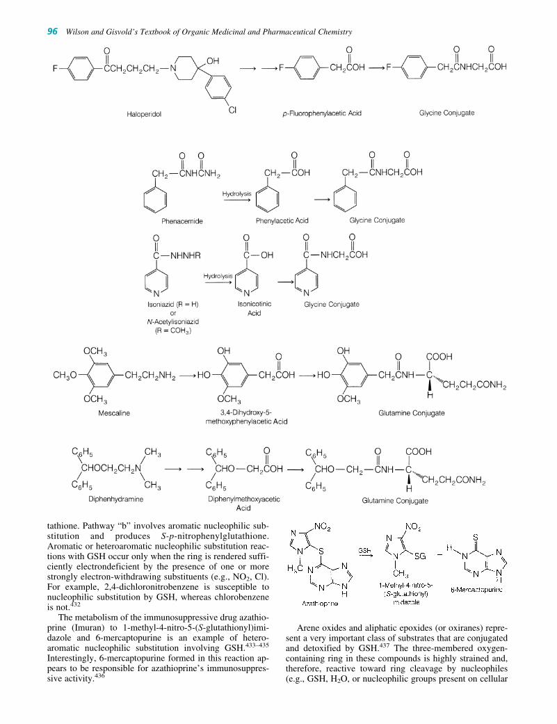
tathione. Pathway “b” involves aromatic nucleophilic sub-stitution and produces S-p-nitrophenylglutathione.Aromatic or heteroaromatic nucleophilic substitution reac-tions with GSH occur only when the ring is rendered suffi-ciently electrondeficient by the presence of one or morestrongly electron-withdrawing substituents (e.g., NO2, Cl).For example, 2,4-dichloronitrobenzene is susceptible tonucleophilic substitution by GSH, whereas chlorobenzeneis not.432
The metabolism of the immunosuppressive drug azathio-prine (Imuran) to 1-methyl-4-nitro-5-(S-glutathionyl)imi-dazole and 6-mercaptopurine is an example of hetero-aromatic nucleophilic substitution involving GSH.433–435
Interestingly, 6-mercaptopurine formed in this reaction ap-pears to be responsible for azathioprine’s immunosuppres-sive activity.436
Arene oxides and aliphatic epoxides (or oxiranes) repre-sent a very important class of substrates that are conjugatedand detoxified by GSH.437 The three-membered oxygen-containing ring in these compounds is highly strained and,therefore, reactive toward ring cleavage by nucleophiles(e.g., GSH, H2O, or nucleophilic groups present on cellular
96 Wilson and Gisvold’s Textbook of Organic Medicinal and Pharmaceutical Chemistry
0043-0118_17865_Ch03.qxd 12/4/09 3:49 AM Page 96
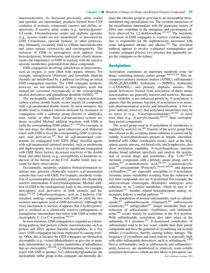
macromolecules). As discussed previously, arene oxidesand epoxides are intermediary products formed from CYPoxidation of aromatic compounds (arenes) and olefins, re-spectively. If reactive arene oxides (e.g., benzo[�]pyrene-4,5-oxide, 4-bromobenzene oxide) and aliphatic epoxides(e.g., styrene oxide) are not “neutralized” or detoxified byGSH S-transferase, epoxide hydrase, or other pathways,they ultimately covalently bind to cellular macromoleculesand cause serious cytotoxicity and carcinogenicity. Theisolation of GSH or mercapturic acid adducts frombenzo[�]pyrene, bromobenzene, and styrene clearly demon-strates the importance of GSH in reacting with the reactiveepoxide metabolites generated from these compounds.
GSH conjugation involving substitution at heteroatoms,such as oxygen, is seen often with organic nitrates. Forexample, nitroglycerin (Nitrostat) and isosorbide dinitrate(Isordil) are metabolized by a pathway involving an initialGSH conjugation reaction. The GSH conjugate products,however, are not metabolized to mercapturic acids butinstead are converted enzymatically to the correspondingalcohol derivatives and glutathione disulfide (GSSG).438
The nucleophilic addition of GSH to electron-deficientcarbon–carbon double bonds occurs mainly in compoundswith �,�-unsaturated double bonds. In most instances, thedouble bond is rendered electron deficient by resonance orconjugation with a carbonyl group (ketone or aldehyde),ester, nitrile, or other. Such �,�-unsaturated systems un-dergo so-called Michael addition reactions with GSH toyield the corresponding GSH adduct.421–428 For example, inrats and dogs, the diuretic agent ethacrynic acid (Edecrin)reacts with GSH to form the corresponding GSH or mercap-turic acid derivatives.439 Not all �,�-unsaturated com-pounds are conjugated with GSH. Many steroidal agentswith �,�-unsaturated carbonyl moieties, such as prednisoneand digitoxigenin, have evinced no significant conjugationwith GSH. Steric factors, decreased reactivity of the doublebond, and other factors (e.g., susceptibility to metabolic re-duction of the ketone or the CBC double bond) may ac-count for these observations.
Occasionally, metabolic oxidative biotransformation re-actions may generate chemically reactive �,�-unsaturatedsystems that react with GSH. For example, metabolic oxida-tion of acetaminophen presumably generates the chemicallyreactive intermediate N-acetylimidoquinone. Michael addi-tion of GSH to the imidoquinone leads to the correspondingmercapturic acid derivative in both animals and hu-mans.245,248 2-Hydroxyestrogens, such as 2-hydroxy-17�-estradiol, undergo conjugation with GSH to yield the twoisomeric mercapturic acid or GSH derivatives. Although theexact mechanism is unclear, it appears that 2-hydroxyestro-gen is oxidized to a chemically reactive orthoquinone orsemiquinone intermediate that reacts with GSH at either theelectrophilic C-1 or C-4 position.440,441
In most instances, GSH conjugation is regarded as a detox-ifying pathway that protects cellular macromolecules such asprotein and DNA against harmful electrophiles. In a fewcases, GSH conjugation has been implicated in causing toxic-ity. Often, this is because the GSH conjugates are themselveselectrophilic (e.g., vicinal dihaloethanes) or give rise to meta-bolic intermediates (e.g., cysteine metabolites of haloalkenes)that are electrophilic.424–428 1,2-Dichloroethane, for example,reacts with GSH to produce S-(2-chloroethyl)glutathione; thenucleophilic sulfur group in this conjugate can internally dis-
place the chlorine group to give rise to an electrophilic three-membered ring episulfonium ion. The covalent interaction ofthe episulfonium intermediate with the guanosine moiety ofDNA may contribute to the mutagenic and carcinogenic ef-fects observed for 1,2-dichloroethane.425–427 The metabolicconversion of GSH conjugates to reactive cysteine metabo-lites is responsible for the nephrotoxicity associated withsome halogenated alkanes and alkenes.428 The activationpathway appears to involve �-glutamyl transpeptidase andcysteine conjugate �-lyase, two enzymes that apparently tar-get the conjugates to the kidney.
Acetylation
Acetylation constitutes an important metabolic route fordrugs containing primary amino groups.408,442,443 This en-compasses primary aromatic amines (ArNH2), sulfonamides(H2NC6H4SO2NHR), hydrazines (—NHNH2), hydrazides(—CONHNH2), and primary aliphatic amines. Theamide derivatives formed from acetylation of these aminofunctionalities are generally inactive and nontoxic. Becausewater solubility is not enhanced greatly by N-acetylation, itappears that the primary function of acetylation is to termi-nate pharmacological activity and detoxification. A few re-ports indicate, however, that acetylated metabolites may beas active as (e.g., N-acetylprocainamide),444,445 or moretoxic than (e.g., N-acetylisoniazid),446,447 their correspon-ding parent compounds.
The acetyl group used in N-acetylation of xenobiotics issupplied by acetyl-CoA.408 Transfer of the acetyl group fromthis cofactor to the accepting amino substrate is carried out bysoluble N-acetyltransferases present in hepatic reticuloen-dothelial cells. Other extrahepatic tissues, such as the lung,spleen, gastric mucosa, red blood cells, and lymphocytes, alsoshow acetylation capability. N-Acetyltransferase enzymesdisplay broad substrate specificity and catalyze the acetyla-tion of several drugs and xenobiotics (Fig. 3.16).442,443
Aromatic compounds with a primary amino group, such asaniline,408 p-aminobenzoic acid,448,449 p-aminosalicylicacid,418 procainamide (Pronestyl),444,445,448,449 and dapsone(Avlosulfon),450 are especially susceptible to N-acetylation.Aromatic amine metabolites resulting from the reduction ofaryl nitro compounds also are N-acetylated. For example, theanticonvulsant clonazepam (Klonopin) undergoes nitroreduction to its 7-amino metabolite, which in turn is N-acetylated.315 Another related benzodiazepam analog, ni-trazepam, follows a similar pathway.316
The metabolism of several sulfonamides, such as sulfanil-amide,451 sulfamethoxazole (Gantanol),452 sulfisoxazole(Gantrisin),452 sulfapyridine453 (major metabolite from azoreduction of sulfasalazine, Azulfidine), and sulfameth-azine,408 occurs mainly by acetylation at the N-4 position.With sulfanilamide, acetylation also takes place at thesulfamido N-1 position.451 N-Acetylated metabolites ofsulfonamides tend to be less water soluble than their parentcompounds and have the potential of crystallizing out in renaltubules (crystalluria), thereby causing kidney damage. Thefrequency of crystalluria and renal toxicity is especially highwith older sulfonamide derivatives, such as sulfathiazole.1,420
Newer sulfonamides, such as sulfisoxazole and sulfamethox-azole, however, are metabolized to relatively water-solubleacetylated derivatives, which are less likely to precipitate out.
Chapter 3 Metabolic Changes of Drugs and Related Organic Compounds 97
(text continues on page 101)
0043-0118_17865_Ch03.qxd 12/4/09 3:49 AM Page 97

98 Wilson and Gisvold’s Textbook of Organic Medicinal and Pharmaceutical Chemistry
0043-0118_17865_Ch03.qxd 12/4/09 3:49 AM Page 98

Chapter 3 Metabolic Changes of Drugs and Related Organic Compounds 99
0043-0118_17865_Ch03.qxd 12/4/09 3:49 AM Page 99

100 Wilson and Gisvold’s Textbook of Organic Medicinal and Pharmaceutical Chemistry
Figure 3.16 Examples of different types of compound undergoing N-acetylation.Arrows indicate sites of N-acetylation.
0043-0118_17865_Ch03.qxd 12/4/09 3:49 AM Page 100

Chapter 3 Metabolic Changes of Drugs and Related Organic Compounds 101
The biotransformation of hydrazine and hydrazide deriv-atives also proceeds by acetylation. The antihypertensivehydralazine (Apresoline)454,455 and the MAO inhibitorphenelzine (Nardil)456 are two representative hydrazinecompounds that are metabolized by this pathway. The ini-tially formed N-acetyl derivative of hydralazine is unstableand cyclizes intramolecularly to form 3-methyl-s-tria-zolo[3,4-�]phthalazine as the major isolable hydralazinemetabolite in humans.454,455 The antituberculosis drug iso-niazid or isonicotinic acid hydrazide (INH) is metabolizedextensively to N-acetylisoniazid.446,447
The acetylation of some primary aliphatic amines such ashistamine,457 mescaline,208,209 and the bis-N-demethylatedmetabolite of �(�)-methadol458–460 also has been reported.In comparison with oxidative deamination processes, N-acetylation is only a minor pathway in the metabolism ofthis class of compounds.
The acetylation pattern of several drugs (e.g., isoniazid,hydralazine, procainamide) in the human population dis-plays a bimodal character in which the drug is conjugatedeither rapidly or slowly with acetyl-CoA.461,462 This phe-nomenon is termed acetylation polymorphism. Individualsare classified as having either slow or rapid acetylator phe-notypes. This variation in acetylating ability is genetic andis caused mainly by differences in N-acetyltransferaseactivity. The proportion of rapid and slow acetylatorsvaries widely among different ethnic groups throughoutthe world. Oddly, a high proportion of Eskimos andAsians are rapid acetylators, whereas Egyptians and someWestern European groups are mainly slow acetylators.462
Other populations are intermediate between these two
extremes. Because of the bimodal distribution of thehuman population into rapid and slow acetylators, thereappears to be significant individual variation in therapeu-tic and toxicological responses to drugs displaying acety-lation polymorphism.408,461,462 Slow acetylators seemmore likely to develop adverse reactions, whereas rapidacetylators are more likely to show an inadequate thera-peutic response to standard drug doses.
The antituberculosis drug isoniazid illustrates many ofthese points. The plasma half-life of isoniazid in rapid acety-lators ranges from 45 to 80 minutes; in slow acetylators, thehalf-life is about 140 to 200 minutes.463 Thus, for a givenfixed-dosing regimen, slow acetylators tend to accumulatehigher plasma concentrations of isoniazid than do rapidacetylators. Higher concentrations of isoniazid may explainthe greater therapeutic response (i.e., higher cure rate)among slow acetylators, but they probably also account forthe greater incidence of adverse effects (e.g., peripheral neu-ritis and drug-induced systemic lupus erythematosus syn-drome) observed among slow acetylators.462 Slow acetyla-tors of isoniazid apparently are also more susceptible tocertain drug interactions involving drug metabolism. For ex-ample, phenytoin toxicity associated with concomitant usewith isoniazid appears to be more prevalent in slow acetyla-tors than in rapid acetylators.464 Isoniazid inhibits themetabolism of phenytoin, thereby leading to an accumula-tion of high and toxic plasma levels of phenytoin.
Interestingly, patients who are rapid acetylators appear tobe more likely to develop isoniazid-associated hepatitis.446,447
This liver toxicity presumably arises from initial hydrolysis ofthe N-acetylated metabolite N-acetylisoniazid to acetylhy-
0043-0118_17865_Ch03.qxd 12/4/09 3:49 AM Page 101
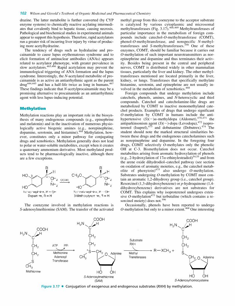
102 Wilson and Gisvold’s Textbook of Organic Medicinal and Pharmaceutical Chemistry
drazine. The latter metabolite is further converted (by CYPenzyme systems) to chemically reactive acylating intermedi-ates that covalently bind to hepatic tissue, causing necrosis.Pathological and biochemical studies in experimental animalsappear to support this hypothesis. Therefore, rapid acetylatorsrun a greater risk of incurring liver injury by virtue of produc-ing more acetylhydrazine.
The tendency of drugs such as hydralazine and pro-cainamide to cause lupus erythematosus syndrome and toelicit formation of antinuclear antibodies (ANAs) appearsrelated to acetylator phenotype, with greater prevalence inslow acetylators.465,466 Rapid acetylation may prevent theimmunological triggering of ANA formation and the lupussyndrome. Interestingly, the N-acetylated metabolite of pro-cainamide is as active an antiarrhythmic agent as the parentdrug444,445 and has a half-life twice as long in humans.467
These findings indicate that N-acetylprocainamide may be apromising alternative to procainamide as an antiarrhythmicagent with less lupus-inducing potential.
Methylation
Methylation reactions play an important role in the biosyn-thesis of many endogenous compounds (e.g., epinephrineand melatonin) and in the inactivation of numerous physio-logically active biogenic amines (e.g., norepinephrine,dopamine, serotonin, and histamine).468 Methylation, how-ever, constitutes only a minor pathway for conjugatingdrugs and xenobiotics. Methylation generally does not leadto polar or water-soluble metabolites, except when it createsa quaternary ammonium derivative. Most methylated prod-ucts tend to be pharmacologically inactive, although thereare a few exceptions.
The coenzyme involved in methylation reactions is S-adenosylmethionine (SAM). The transfer of the activated
methyl group from this coenzyme to the acceptor substrateis catalyzed by various cytoplasmic and microsomalmethyltransferases (Fig. 3.17).468,469 Methyltransferases ofparticular importance in the metabolism of foreign com-pounds include catechol-O-methyltransferase (COMT),phenol-O-methyltransferase, and nonspecific N-methyl-transferases and S-methyltransferases.358 One of theseenzymes, COMT, should be familiar because it carries outO-methylation of such important neurotransmitters as nor-epinephrine and dopamine and thus terminates their activ-ity. Besides being present in the central and peripheralnerves, COMT is distributed widely in other mammaliantissues, particularly the liver and kidney. The other methyl-transferases mentioned are located primarily in the liver,kidney, or lungs. Transferases that specifically methylatehistamine, serotonin, and epinephrine are not usually in-volved in the metabolism of xenobiotics.468
Foreign compounds that undergo methylation includecatechols, phenols, amines, and N-heterocyclic and thiolcompounds. Catechol and catecholamine-like drugs aremetabolized by COMT to inactive monomethylated cate-chol products. Examples of drugs that undergo significantO-methylation by COMT in humans include the anti-hypertensive (S)(�)�-methyldopa (Aldomet),470,471 theantiparkinsonism agent (S)(�)-dopa (Levodopa),472 isopro-terenol (Isuprel),473 and dobutamine (Dobutrex).474 Thestudent should note the marked structural similarities be-tween these drugs and the endogenous catecholamines suchas norepinephrine and dopamine. In the foregoing fourdrugs, COMT selectively O-methylates only the phenolicOH at C-3. Bismethylation does not occur. Catecholmetabolites arising from aromatic hydroxylation of phenols(e.g., 2-hydroxylation of 17�-ethinylestradiol)54,55 and fromthe arene oxide dihydrodiol–catechol pathway (see sectionon oxidation of aromatic moieties, e.g., the catechol metab-olite of phenytoin)475 also undergo O-methylation.Substrates undergoing O-methylation by COMT must con-tain an aromatic 1,2-dihydroxy group (i.e., catechol group).Resorcinol (1,3-dihydroxybenzene) or p-hydroquinone (1,4-dihydroxybenzene) derivatives are not substrates forCOMT. This explains why isoproterenol undergoes exten-sive O-methylation473 but terbutaline (which contains a re-sorcinol moiety) does not.396
Occasionally, phenols have been reported to undergo O-methylation but only to a minor extent.468 One interesting
Figure 3.17 Conjugation of exogenous and endogenous substrates (RXH) by methylation.
0043-0118_17865_Ch03.qxd 12/4/09 3:49 AM Page 102

Chapter 3 Metabolic Changes of Drugs and Related Organic Compounds 103
0043-0118_17865_Ch03.qxd 12/4/09 3:49 AM Page 103

104 Wilson and Gisvold’s Textbook of Organic Medicinal and Pharmaceutical Chemistry
example involves the conversion of morphine to its O-methylated derivative, codeine, in humans. This metabo-lite is formed in significant amounts in tolerant subjects andmay account for up to 10% of the morphine dose.476
Although N-methylation of endogenous amines (e.g., his-tamine, norepinephrine) occurs commonly, biotransforma-tion of nitrogen-containing xenobiotics to N-methylatedmetabolites occurs to only a limited extent. Some examplesreported include the N-methylation of the antiviral and an-tiparkinsonism agent amantadine (Symmetrel) in dogs477
and the in vitro N-methylation of norephedrine in rabbitlung preparations.468 N-methylation of nitrogen atoms pres-ent in heterocyclic compounds (e.g., pyridine derivatives)also takes place. For example, the pyridinyl nitrogens ofnicotine187,188 and nicotinic acid478 are N-methylated toyield quaternary ammonium products.
Thiol-containing drugs, such as propylthiouracil,479
2,3-dimercapto-1-propanol (BAL),480 and 6-mercaptopu-rine,481,482 also have been reported to undergo S-methylation.
FACTORS AFFECTING DRUGMETABOLISM
Drugs and xenobiotics often are metabolized by severaldifferent phase I and phase II pathways to give severalmetabolites. The relative amount of any particular metabo-lite is determined by the concentration and activity of theenzyme(s) responsible for the biotransformation. The rateof metabolism of a drug is particularly important for itspharmacological action as well as its toxicity. For exam-ple, if the rate of metabolism of a drug is decreased, thisgenerally increases the intensity and duration of the drugaction. In addition, decreased metabolic elimination maylead to accumulation of toxic levels of the drug.Conversely, an increased rate of metabolism decreases theintensity and duration of action as well as the drug’s effi-cacy. Many factors may affect drug metabolism, and theyare discussed in the following sections. These include age,species and strain, genetic or hereditary factors, sex, en-zyme induction, and enzyme inhibition.32,483–486
Age Differences
Age-related differences in drug metabolism are generallyquite apparent in the newborn.487,488 In most fetal andnewborn animals, undeveloped or deficient oxidative andconjugative enzymes are chiefly responsible for the reducedmetabolic capability seen. In general, the ability to carry outmetabolic reactions increases rapidly after birth and ap-proaches adult levels in about 1 to 2 months. An illustrationof the influence of age on drug metabolism is seen in the du-ration of action (sleep time) of hexobarbital in newborn andadult mice.489 When given a dose of 10 mg/kg of bodyweight, the newborn mouse sleeps more than 6 hours. In con-trast, the adult mouse sleeps for fewer than 5 minutes whengiven the same dose.
In humans, oxidative and conjugative (e.g., glucuronida-tion) capabilities of newborns are also low compared withthose of adults. For example, the oxidative (CYP) metabolismof tolbutamide appears to be markedly lower in newborns.490
Compared with the half-life of 8 hours in adults, the plasmahalf-life of tolbutamide in infants is more than 40 hours. As
discussed previously, infants possess poor glucuronidatingability because of a deficiency in glucuronyltransferase activ-ity. The inability of infants to conjugate chloramphenicol withglucuronic acid appears to be responsible for the accumula-tion of toxic levels of this antibiotic, resulting in the so-calledgray baby syndrome.388 Similarly, neonatal hyperbilirubin-emia (or kernicterus) results from the inability of newborn ba-bies to glucuronidate bilirubin.387
The effect of old age on drug metabolism has not been aswell studied. There is some evidence in animals and humansthat drug metabolism diminishes with old age.491,492 Muchof the evidence, however, is based on prolonged plasmahalf-lives of drugs that are metabolized totally or mainly byhepatic microsomal enzymes (e.g., antipyrine, phenobarbi-tal, acetaminophen). In evaluating the effect of age on drugmetabolism, one must differentiate between “normal” lossof enzymatic activity with aging and the effect of a diseasedliver from hepatitis, cirrhosis, etc., plus decreased renalfunction, because much of the water-soluble conjugationproducts are excreted in the liver.
Species and Strain Differences
The metabolism of many drugs and foreign compounds isoften species dependent. Different animal species maybiotransform a particular xenobiotic by similar or markedlydifferent metabolic pathways. Even within the same species,individual variations (strain differences) may result in sig-nificant differences in a specific metabolic pathway.493,494
This is a problem when a new drug is under development. Anew drug application requires the developer to account forthe product as it moves from the site of administration tofinal elimination from the body. It is difficult enough to findappropriate animal models for a disease. It is even harder tofind animal models that mimic human drug metabolism.
Species variation has been observed in many oxidativebiotransformation reactions. For example, metabolism of am-phetamine occurs by two main pathways: oxidative deamina-tion or aromatic hydroxylation. In human, rabbit, and guineapig, oxidative deamination appears to be the predominantpathway; in the rat, aromatic hydroxylation appears to be themore important route.495 Phenytoin is another drug that showsmarked species differences in metabolism. In the human,phenytoin undergoes aromatic oxidation to yield primarily(S)(�)-p-hydroxyphenytoin; in the dog, oxidation occurs togive mainly (R)(�)-m-hydroxyphenytoin.496 There is a dra-matic difference not only in the position (i.e., meta or para)of aromatic hydroxylation but also in which of the two phenylrings (at C-5 of phenytoin) undergoes aromatic oxidation.
Species differences in many conjugation reactionsalso have been observed. Often, these differences are causedby the presence or absence of transferase enzymes involvedin the conjugative process. For example, cats lack glu-curonyltransferase enzymes and, therefore, tend to conjugatephenolic xenobiotics by sulfation instead.497 In pigs, the sit-uation is reversed: pigs are not able to conjugate phenolswith sulfate (because of lack of sulfotransferase enzymes)but appear to have good glucuronidation capability.497 Theconjugation of aromatic acids with amino acids (e.g.,glycine, glutamine) depends on the animal species as well ason the substrate. For example, glycine conjugation is a com-mon conjugation pathway for benzoic acid in many animals.In certain birds (e.g., duck, goose, turkey), however, glycine
0043-0118_17865_Ch03.qxd 12/4/09 3:49 AM Page 104

Chapter 3 Metabolic Changes of Drugs and Related Organic Compounds 105
is replaced by the amino acid ornithine.498 Phenylacetic acidis a substrate for both glycine and glutamine conjugation inhumans and other primates. However, nonprimates, such asrabbit and rat, excrete phenylacetic acid only as the glycineconjugate.499 The metabolism of the urinary antiseptic,phenazopyridine (Pyridium) depends strongly on the animal.The diazo linkage remains intact in over half of the metabo-lites in humans, whereas 40% of the metabolites in theguinea pig result from its cleavage. The metabolic productpattern in human or guinea pig does not correlate with that ofeither rat or mouse (Fig. 3.18).500
Strain differences in drug metabolism exist, particularlyin inbred mice and rabbits. These differences apparently arecaused by genetic variations in the amount of metabolizingenzyme present among the different strains. For example, invitro studies indicate that cottontail rabbit liver microsomesmetabolize hexobarbital about 10 times faster than NewZealand rabbit liver microsomes.501 Interindividual differ-ences in drug metabolism in humans are considered in thesection that follows.
Hereditary or Genetic Factors
Marked individual differences in the metabolism of severaldrugs exist in humans.463 Many of these genetic or heredi-
tary factors are responsible for the large differences seen inthe rate of metabolism of these drugs. The frequently citedexample of the biotransformation of the antituberculosisagent isoniazid is discussed previously under acylation.Genetic factors also appear to influence the rate of oxidationof drugs such as phenytoin, phenylbutazone, dicumarol, andnortriptyline.502,503 The rate of oxidation of these drugsvaries widely among different individuals; however, thesedifferences do not appear to be distributed bimodally, as inacetylation. In general, individuals who tend to oxidize onedrug rapidly are also likely to oxidize other drugs rapidly.Numerous studies in twins (identical and fraternal) and infamilies indicate that oxidation of these drugs is under ge-netic control.503
Many patients state that they do not respond to codeineand codeine analogs. It now is realized that theirCYP2D6 isozyme does not readily O-demethylate codeineto form morphine. This genetic polymorphism is seen inabout 8% of Caucasians, 4% of African Americans, andless than 1% of Asians.504 Genetic polymorphism withCYP isozymes is well documented as evidenced by themany examples in this chapter. There is limited evidenceof polymorphism involving MAO-A and MAO-B. Thechemical imbalances seen with some mental diseases maybe the cause.206
0043-0118_17865_Ch03.qxd 12/4/09 3:49 AM Page 105

Sex Differences
The rate of metabolism of xenobiotics also varies accordingto gender in some animal species. A marked difference isobserved between female and male rats. Adult male rats me-tabolize several foreign compounds at a much faster ratethan female rats (e.g., N-demethylation of aminopyrine,hexobarbital oxidation, glucuronidation of o-aminophenol).Apparently, this sex difference also depends on the sub-strate, because some xenobiotics are metabolized at thesame rate in both female and male rats. Differences in mi-crosomal oxidation are under the control of sex hormones,particularly androgens; the anabolic action of androgensseems to increase metabolism.505
Sex differences in drug metabolism appear to be speciesdependent. Rabbits and mice, for example, do not show asignificant sex difference in drug metabolism.505 In humans,there have been a few reports of sex differences in metabo-lism. For instance, nicotine and aspirin seem to be metabo-lized differently in women and men.506,507 On the otherhand, gender differences can become significant in terms ofdrug–drug interactions based on the drug’s metabolism. Forwomen, the focus is on drugs used for contraception. Notein Table 3.4 that the antibiotic rifampin, a CYP3A4 inducer,can shorten the half-life of oral contraceptives.
Enzyme Induction
The activity of hepatic microsomal enzymes, such as theCYP mixed-function oxidase system, can be increased
markedly by exposure to diverse drugs, pesticides,polycyclic aromatic hydrocarbons, and environmentalxenobiotics. The process by which the activity of thesedrug-metabolizing enzymes is increased is termed enzymeinduction.508–511 The increased activity is apparentlycaused by an increased amount of newly synthesized en-zyme. Enzyme induction often increases the rate of drugmetabolism and decreases the duration of drug action. (SeeTable 3.4 for a list of clinically significant drug–druginteractions based on one drug inducing the metabolism ofa second drug.)
Inducing agents may increase the rate of their own metab-olism as well as those of other unrelated drugs or foreigncompounds (Table 3.4).32 Concomitant administration of twoor more drugs often may lead to serious drug interactions as aresult of enzyme induction. For instance, a clinically criticaldrug interaction occurs with phenobarbital and warfarin.512
Induction of microsomal enzymes by phenobarbital increasesthe metabolism of warfarin and, consequently, markedly de-creases the anticoagulant effect. Therefore, if a patient is re-ceiving warfarin anticoagulant therapy and begins taking phe-nobarbital, careful attention must be paid to readjustment ofthe warfarin dose. Dosage readjustment is also needed if a pa-tient receiving both warfarin and phenobarbital therapy sud-denly stops taking the barbiturate. The ineffectiveness of oralcontraceptives in women on concurrent phenobarbital or ri-fampin therapy has been attributed to the enhanced metabo-lism of estrogens (e.g., 17�-ethinylestradiol) caused by phe-nobarbital513 and rifampin514 induction.
106 Wilson and Gisvold’s Textbook of Organic Medicinal and Pharmaceutical Chemistry
Figure 3.18 Phenazopyridine metabolism in humans, guinea pigs, rats, and mice.
0043-0118_17865_Ch03.qxd 12/4/09 3:49 AM Page 106

Inducers of microsomal enzymes also may enhance themetabolism of endogenous compounds, such as steroidalhormones and bilirubin. For instance, phenobarbital canincrease the metabolism of cortisol, testosterone, vitamin D,and bilirubin in humans.508,509 The enhanced metabolism ofvitamin D3 induced by phenobarbital and phenytoin appearsto be responsible for the osteomalacia seen in patients onlong-term therapy with these two anticonvulsant drugs.515
Interestingly, phenobarbital induces glucuronyltransferaseenzymes, thereby enhancing the conjugation of bilirubinwith glucuronic acid. Phenobarbital has been used occasion-ally to treat hyperbilirubinemia in neonates.516
In addition to drugs, other chemicals, such as polycyclicaromatic hydrocarbons (e.g., benzo[�]pyrene, 3-methyl-cholanthrene) and environmental pollutants (e.g., pesticides,PCBs, TCDD), may induce certain oxidative pathways and,thereby, alter drug response.508,509,511 Cigarette smoke con-tains minute amounts of polycyclic aromatic hydrocarbons,such as benzo[�]pyrene, which are potent inducers of micro-somal CYP enzymes. This induction increases the oxidationof some drugs in smokers. For example, theophylline is me-tabolized more rapidly in smokers than in nonsmokers. Thisdifference is reflected in the marked difference in the plasmahalf-life of theophylline between smokers (t1/2 4.1 hours) and
Chapter 3 Metabolic Changes of Drugs and Related Organic Compounds 107
TABLE 3.4 Clinically Significant Cytochrome P450-Based Drug–Drug Interactions
Agent Substrates Inhibitors Inducers Agent Substrates Inhibitors Inducers
Amitriptyline
Clomipramine
Clozapine
Desipramine
Fluvoxamine
Haloperidol
Imipramine
Ropinirole
Tacrine
Theophylline
(R)-Warfarin
Diazepam
Phenytoin
(S)-Warfarin
Phenytoin
Thioridazine
Amitriptyline
Atomoxetine
Codeine
Desipramine
Dextromethorphan
Donepezil
Doxepin
Fentanyl
Flecainide
Haloperidol
Hydrocodone
Cimetidine
Ciprofloxacin
Clarithromycin
Enoxacin
Erythromycin
Fluvoxamine
Isoniazid
Nalidixic acid
Norfloxacin
Troleandomycin
Zileuton
Amiodarone
Chloramphenicol
Cimetidine
Fluconazole
Fluoxetine
Fluvoxamine
Isoniazid
Metronidazole
Voriconazole
Zafirlukast
Fluoxetine
Fluvoxamine
Modafinil
Omeprazole
Topiramate
Amiodarone
Cimetidine
Fluoxetine
Paroxetine
Quinidine
Ritonavir
Sertraline
CYP 1A2
CYP 2C9
CYP 2C19
CYP 2D6
Imipramine
Meperidine
Methadone
Mexiletine
Nortriptyline
Oxycodone
Propafenone
Propoxyphene
Thioridazine
Tramadol
Trazodone
Alfentanil
Alprazolam
Amlodipine
Atorvastatin
Busulfan
Carbamazepine
Cisapride
Clarithromycin
Cyclosporine
Dihydroergotamine
Disopyramide
Doxorubicin
Dronabinol
Ergotamine
Erythromycin
Estrogens, oral
contraceptives
Ethinyl estradiol
Ethosuximide
Etoposide
Felodipine
Fentanyl
Indinavir
Isradipine
Itraconazole
Amiodarone
Cimetidine
Ciprofloxacin
Clarithromycin
Cyclosporine
Delavirdine
Diltiazem
Efavirenz
Erythromycin
Fluconazole
Fluoxetine
Fluvoxamine
Grapefruit
Indinavir
Isoniazid
Itraconazole
Ketoconazole
Metronidazole
Miconazole
Nefazodone
Nelfinavir
Nifedipine
Norfloxacin
Quinine
Ritonavir
Carbamazepine
Efavirenz
Ethosuximide
Garlicsupplements
Modafinil
Nevirapine
Oxcarbazepine
Phenobarbital
Phenytoin
Primidone
Rifabutin
Rifampin
Rifapentine
St. John’s wort
CYP 3A4
Abstracted from Levien, T. L., and Baker, D. E.: Pharmacist’s Letter, December 2002, Detail-Document #150400. (Pharmacist’sLetter used as sources: Hansten, P. D., and Horn, J. R.: Drug Interactions Analysis and Management. Vancouver, WA, AppliedTherapeutics, 2002; and Tatro, D. S. [ed.]: Drug Interaction Facts. St. Louis, Facts & Comparisons, 2002.)
Carbamazepine
Phenobarbital
Phenytoin
Primidone
Rifampin
Ritonavir
Smoking
St. John’s wort
Carbamazepine
Phenobarbital
Phenytoin
Primidone
Rifampin
Rifapentine
Carbamazepine
Phenobarbital
Phenytoin
St. John’s wort
0043-0118_17865_Ch03.qxd 12/4/09 3:49 AM Page 107

nonsmokers (t1/2 7.2 hours).517 Other drugs, such asphenacetin, pentazocine, and propoxyphene, also reportedlyundergo more rapid metabolism in smokers than in non-smokers.518–520
Occupational and accidental exposure to chlorinatedpesticides and insecticides can also stimulate drug metabo-lism. For instance, the half-life of antipyrine in workersoccupationally exposed to the insecticides lindane anddichlorodiphenyltrichloroethane (DDT) is reportedly signif-icantly shorter (7.7 vs. 11.7 hours) than in control sub-jects.521 A case was reported in which a worker exposed tochlorinated insecticides failed to respond (i.e., decreasedanticoagulant effect) to a therapeutic dose of warfarin.522
As discussed previously in this chapter, multiple forms(isozymes) of CYP have been demonstrated.31,40–42 Manychemicals selectively induce one or more distinct forms ofCYP31 (see Table 3.4.) Enzyme induction also may affecttoxicity of some drugs by enhancing the metabolic forma-tion of chemically reactive metabolites. Particularly impor-tant is the induction of CYP enzymes involved in the oxida-tion of drugs to reactive intermediates. For example, theoxidation of acetaminophen to a reactive imidoquinonemetabolite appears to be carried out by a phenobarbital-inducible form of CYP in rats and mice. Numerous studiesin these two animals indicate that phenobarbital pretreat-ment increases in vivo hepatotoxicity and covalent bindingas well as increases formation of reactive metabolite inmicrosomal incubation mixtures.243–245,248 Induction of cy-tochrome P448 is of toxicological concern because this par-ticular enzyme is involved in the metabolism of polycyclicaromatic hydrocarbons to reactive and carcinogenic inter-mediates.80,523 Consequently, the metabolic bioactivation ofbenzo[�]pyrene to its ultimate carcinogenic diol epoxide in-termediate is carried out by cytochrome P448 (see sectionon aromatic oxidation for the bioactivation pathway ofbenzo[�]pyrene to its diol epoxide).523 Thus, it is becomingincreasingly apparent that enzyme induction may enhancethe toxicity of some xenobiotics by increasing the rate offormation of reactive metabolites.
Enzyme Inhibition
Several drugs, other xenobiotics including grapefruit,and possibly other foods can inhibit drug metabolism(Table 3.5).32,483–486 With decreased metabolism, a drugoften accumulates, leading to prolonged drug action andserious adverse effects. Enzyme inhibition can occur bydiverse mechanisms, including substrate competition, inter-ference with protein synthesis, inactivation of drug-metabolizing enzymes, and hepatotoxicity leading to im-pairment of enzyme activity. Some drug interactionsresulting from enzyme inhibition have been reported in hu-mans.524,525 For example, phenylbutazone (limited to veteri-nary use) stereoselectively inhibits the metabolism of themore potent (S)(�) enantiomer of warfarin. This inhibitionmay explain the excessive hypoprothrombinemia (increasedanticoagulant effect) and many instances of hemorrhagingseen in patients on both warfarin and phenylbutazone ther-apy.56 The metabolism of phenytoin is inhibited by drugssuch as chloramphenicol, disulfiram, and isoniazid.512
Interestingly, phenytoin toxicity as a result of enzyme inhi-bition by isoniazid appears to occur primarily in slow acety-lators.464 Several drugs, such as dicumarol, chlorampheni-
col, and phenylbutazone,512 inhibit the biotransformation oftolbutamide, which may lead to a hypoglycemic response.
The grapefruit–drug interaction is complex. It may becaused by the bioflavonoids or the furanocoumarins.Grapefruit’s main bioflavonoid, naringin, is a weak CYPinhibitor, but the product of the intestinal flora, naringenin,does inhibit CYP3A4. The literature is very confusing be-cause many of the studies were done in vitro, and theycannot always be substantiated under in vivo conditions. Inaddition, components in grapefruit also activate P-glyco-protein, which would activate the efflux pump in the gas-tric mucosa and thus interfere with oral absorption of thecertain drugs. The combination of CYP enzyme inhibitionand P-glycoprotein activation can lead to inconclusive re-sults.526 The general recommendation when a drug interac-tion is suspected is that the patient avoid grapefruit and itsjuice.
Miscellaneous Factors Affecting DrugMetabolism32,483–486
Other factors also may influence drug metabolism. Dietaryfactors, such as the protein-to-carbohydrate ratio, affect themetabolism of a few drugs. Indoles present in vegetablessuch as Brussels sprouts, cabbage, and cauliflower, andpolycyclic aromatic hydrocarbons present in charcoal-broiled beef induce enzymes and stimulate the metabolismof some drugs. Vitamins, minerals, starvation, and malnutri-tion also apparently influence drug metabolism. Finally,physiological factors, such as the pathological state of theliver (e.g., hepatic cancer, cirrhosis, hepatitis), pregnancy,hormonal disturbances (e.g., thyroxine, steroids), and circa-dian rhythm, may markedly affect drug metabolism.
Stereochemical Aspects of Drug Metabolism
Many drugs (e.g., warfarin, propranolol, hexobarbital,glutethimide, cyclophosphamide, ketamine, and ibuprofen)often are administered as racemic mixtures in humans. Thetwo enantiomers present in a racemic mixture may differ inpharmacological activity. Usually, one enantiomer tends tobe much more active than the other. For example, the (S)(�)
108 Wilson and Gisvold’s Textbook of Organic Medicinal and Pharmaceutical Chemistry
Abstracted from Kehoe, W. A.: Pharmacist’s Letter, 18, September 2002,Detail Document #180905.AUC, area under the curve.
TABLE 3.5 Potential Drug–Grapefruit Interactions Basedon Grapefruit Inhibition of CYP 3A4
Drug Result
Increased bioavailability
Increased AUC
Increased AUC, peak and troughplasma concentrations
Increased AUC
Increased AUC and serumconcentrations
Increased absorption and plasmaconcentrations
Increased absorption and plasmaconcentrations
Amiodarone
Diazepam
Carbamazepine
Cisapride
Cyclosporine, tacrolimus
Atorvastatin, simvastatin
Saquinavir
0043-0118_17865_Ch03.qxd 12/4/09 3:49 AM Page 108

enantiomer of warfarin is 5 times more potent as an oralanticoagulant than the (R)(�) enantiomer.527 In some in-stances, the two enantiomers may have totally different phar-macological activities. For example, (�)-�-propoxyphene(Darvon) is an analgesic, whereas (�)-�-propoxyphene(Novrad) is an antitussive.528 Such differences in activity be-tween stereoisomers should not be surprising, becauseChapter 2 explains that stereochemical factors generallyhave a dramatic influence on how the drug molecule inter-acts with the target receptors to elicit its pharmacological re-sponse. By the same token, the preferential interaction ofone stereoisomer with drug-metabolizing enzymes may leadone to anticipate differences in metabolism for the two enan-tiomers of a racemic mixture. Indeed, individual enan-tiomers of a racemic drug often are metabolized at differentrates. For instance, studies in humans indicate that theless active (�) enantiomer of propranolol undergoes morerapid metabolism than the corresponding (�) enantiomer.529
Allylic hydroxylation of hexobarbital occurs more rapidlywith the R(�) enantiomer in humans.530 The term substratestereoselectivity is used frequently to denote a preference forone stereoisomer as a substrate for a metabolizing enzyme ormetabolic process.291
Individual enantiomers of a racemic mixture also may bemetabolized by different pathways. For instance, in dogs,the (�) enantiomer of the sedative–hypnotic glutethimide(Doriden) is hydroxylated primarily � to the carbonyl toyield 4-hydroxyglutethimide, whereas the (�) enantiomerundergoes aliphatic �–1 hydroxylation of its C-2 ethyl
group.140,141 Dramatic differences in the metabolic profileof two enantiomers of warfarin also have been noted. In hu-mans, the more active (S)(�)-isomer is 7-hydroxylated (aro-matic hydroxylation), whereas the (R)(�)-isomer undergoesketo reduction to yield primarily the (R,S) warfarin alcoholas the major plasma metabolite.56,296 Although numerousother examples of substrate stereoselectivity or enantiose-lectivity in drug metabolism exist, the examples presentedshould suffice to emphasize the point.291,531
Drug biotransformation processes often lead to the cre-ation of a new asymmetric center in the metabolite (i.e.,stereoisomeric or enantiomeric products). The preferential
Chapter 3 Metabolic Changes of Drugs and Related Organic Compounds 109
0043-0118_17865_Ch03.qxd 12/4/09 3:49 AM Page 109

metabolic formation of a stereoisomeric product is calledproduct stereoselectivity.291 Thus, bioreduction of ketonexenobiotics, as a general rule, produces predominantlyone stereoisomeric alcohol (see “Reduction of KetoneCarbonyls”).116,291 The preferential formation of (S)(�)-hydroxyhexamide from the hypoglycemic agent acetohex-amide293,294 and the exclusive generation of 6�-naltrexolfrom naltrexone297,298 (see “Reduction of Ketone Carbonyls”for structure) are two examples of highly stereoselectivebioreduction processes in humans.
Oxidative biotransformations display product stereose-lectivity, too. For example, phenytoin contains two phenylrings in its structure, both of which a priori should besusceptible to aromatic hydroxylation. In humans, how-ever, p-hydroxylation occurs preferentially (�90%) atthe pro-(S)-phenyl ring to give primarily (S)(�)-5-(4-hydroxyphenyl)-5-phenylhydantoin. Although the otherphenyl ring also is p-hydroxylated, it occurs only to a minorextent (10%).496 Microsomal hydroxylation of the C-3 car-bon of diazepam and desmethyldiazepam (using mouseliver preparations) has been reported to proceed with re-markable stereoselectivity to yield optically active metabo-lites with the 3(S) absolute configuration.139 Interestingly,these two metabolites are pharmacologically active and oneof them, oxazepam, is marketed as a drug (Serax). The al-lylic hydroxylation of the N-butenyl side group of the anal-gesic pentazocine (Talwin) leads to two possible alcohols(cis and trans alcohols). In human, mouse, and monkey,pentazocine is metabolized predominantly to the transalcohol metabolite, whereas the rat primarily tends to formthe cis alcohol.129,130 The product stereoselectivity ob-served in this biotransformation involves cis and trans geo-metric stereoisomers.
The term regioselectivity532 has been introduced indrug metabolism to denote the selective metabolism oftwo or more similar functional groups (e.g., OCH3, OH,NO2) or two or more similar atoms that are positioned indifferent regions of a molecule. For example, of the fourmethoxy groups present in papaverine, the 4-OCH3 groupis regioselectively O-demethylated in several species
110 Wilson and Gisvold’s Textbook of Organic Medicinal and Pharmaceutical Chemistry
0043-0118_17865_Ch03.qxd 12/4/09 3:49 AM Page 110

(e.g., rat, guinea pig, rabbit, and dog).533 Trimethoprim(Trimpex, Proloprim) has two heterocyclic sp2 nitrogenatoms (N1 and N3) in its structure. In dogs, it appears thatoxidation occurs regioselectively at N3 to give the corre-sponding 3-N-oxide.232 Nitroreduction of the 7-nitrogroup in 5,7-dinitroindazole to yield the 7-aminoderivative in the mouse and rat occurs with high regiose-lectivity.534 Substrates amenable to O-methylation byCOMT appear to proceed with remarkable regioselectiv-ity, as typified by the cardiotonic agent dobutamine(Dobutrex). O-methylation occurs exclusively with thephenolic hydroxy group at C-3.474
Pharmacologically Active Metabolites
The traditional notion that drug metabolites are inactive andinsignificant in drug therapy has changed dramatically inrecent years. Increasing evidence indicates that many drugsare biotransformed to pharmacologically active metabolitesthat contribute to the therapeutic as well as toxic effects ofthe parent compound. Metabolites shown to have significanttherapeutic activity in humans are listed in Table 3.4.2,535
The parent drug from which the metabolite is derived andthe biotransformation process involved also are given.
How significantly an active metabolite contributes to thetherapeutic or toxic effects ascribed to the parent drug de-pend on its relative activity and quantitative importance(e.g., plasma concentration). In addition, whether themetabolite accumulates after repeated administration (e.g.,desmethyldiazepam in geriatric patients) or in patients withrenal failure is determinant.
From a clinical standpoint, active metabolites are espe-cially important in patients with decreased renal function.If renal excretion is the major pathway for eliminationof the active metabolite, then accumulation is likely to occurin patients with renal failure. Especially with drugs suchas procainamide, clofibrate, and digitoxin, caution shouldbe exercised in treating patients with renal failure.2,413
Many of the toxic effects seen for these drugs have been at-tributed to high-plasma levels of their active metabolites.For example, the combination of severe muscle weaknessand tenderness (myopathy) seen with clofibrate in renal fail-ure patients is believed to be caused by high levels of theactive metabolite chlorophenoxyisobutyric acid.536,537
Cardiovascular toxicity owing to digitoxin and pro-cainamide in anephric subjects has been attributed to highplasma levels of digoxin and N-acetylprocainamide, respec-tively. In such situations, appropriate reduction in dosageand careful monitoring of plasma levels of the parent drugand its active metabolite often are recommended.
The pharmacological activity of some metabolites has ledmany manufacturers to synthesize these metabolites and tomarket them as separate drug entities (Table 3.6). For exam-ple, oxyphenbutazone (Tandearil, Oxalid) is the p-hydroxyl-ated metabolite of the anti-inflammatory agent phenylbuta-zone (Butazolidin, Azolid), nortriptyline (Aventyl) is theN-demethylated metabolite of the tricyclic antidepressantamitriptyline (Elavil), oxazepam (Serax) is the N-demethyl-ated and 3-hydroxylated metabolite of diazepam (Valium),and mesoridazine (Serentil) is the sulfoxide metabolite of theantipsychotic agent thioridazine (Mellaril).
Antivirals that are used in treating herpes simplex virus,varicella-zoster virus, and/or human cytomegalovirus mustbe bioactivated.538 These include acyclovir, valacyclovir,penciclovir, famciclovir, and ganciclovir, which must bephosphorylated on the pentoselike side chain to the triphos-phate derivative to be effective in inhibiting the enzymeDNA polymerase. The antiviral cidovir is dispensed as amonophosphate and only needs to be diphosphylated for con-version to the active triphosphate metabolite. The nucleosideantivirals that are used in treating acquired immuno-deficiency syndrome/human immunodeficiency virus (AIDS/HIV) must also undergo a similar metabolic conversion to thetriphosphate metabolite.539 The triphosphate derivative actsas a competitive inhibitor of the enzyme, reverse transcrip-tase, which normally uses the triphosphorylated form of
Chapter 3 Metabolic Changes of Drugs and Related Organic Compounds 111
TABLE 3.6 Pharmacologically Active Metabolites in Humans
Parent Drug Metabolite Biotransformation Process
Acetohexamide Hydroxyhexamide Ketone reductionAcetylmethadol Noracetylmethadol N-DemethylationAmitriptyline Nortriptyline N-DemethylationAzathioprine 6-Mercaptopurine Glutathione conjugationCarbamazepine Carbamazepine-9,10-epoxide EpoxidationChloral hydrate Trichloroethanol Aldehyde reductionChlorpromazine 7-Hydroxychlorpromazine Aromatic hydroxylationClofibrate Chlorophenoxyisobutyric acid Ester hydrolysisCortisone Hydrocortisone Ketone reductionDiazepam Desmethyldiazepam and oxazepam N-Demethylation and 3-hydroxylationDigitoxin Digoxin Alicyclic hydroxylationDiphenoxylate Diphenoxylic acid Ester hydrolysisImipramine Desipramine N-DemethylationMephobarbital Phenobarbital N-DemethylationMetoprolol �-Hydroxymethylmetoprolol Benzylic hydroxylationPhenacetin Acetaminophen O-DeethylationPhenylbutazone Oxybutazone Aromatic hydroxylationPrednisone Prednisolone Ketone reductionPrimidone Phenobarbital Hydroxylation and oxidation to ketoneProcainamide N-Acetylprocainamide N-AcetylationPropranolol 4-Hydroxypropranolol Aromatic hydroxylationQuinidine 3-Hydroxyquinidine Allylic hydroxylationSulindac Sulfide metabolite of sulindac Sulfoxide reductionThioridazine Mesoridazine S-oxidationWarfarin Warfarin alcohols Ketone reduction
0043-0118_17865_Ch03.qxd 12/4/09 3:49 AM Page 111
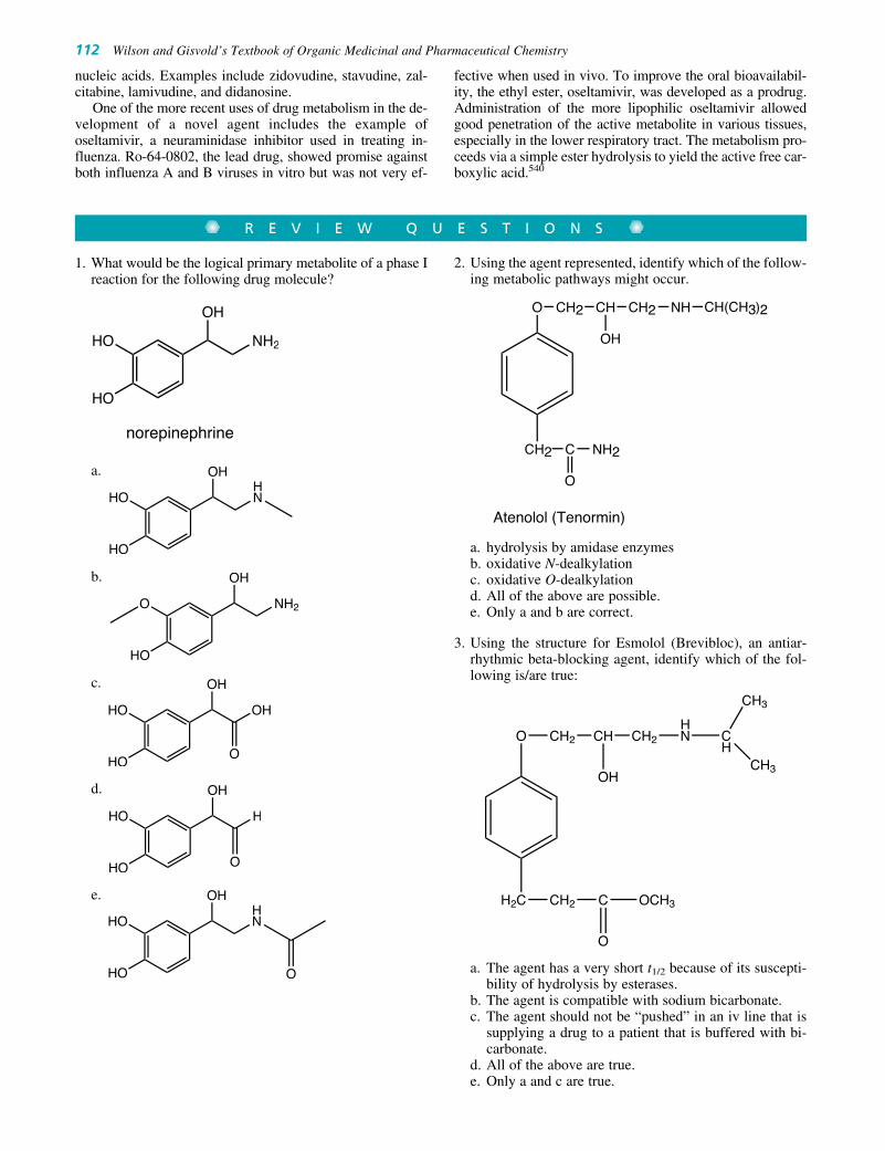
1. What would be the logical primary metabolite of a phase Ireaction for the following drug molecule?
a.
b.
c.
d.
e.
HO
HOHN
OH
O
HO
HO H
OH
O
HO
HO OH
OH
O
HO
O NH2
OH
HO
HOHN
OH
HO
HO NH2
OH
norepinephrine
112 Wilson and Gisvold’s Textbook of Organic Medicinal and Pharmaceutical Chemistry
R E V I E W Q U E S T I O N S
nucleic acids. Examples include zidovudine, stavudine, zal-citabine, lamivudine, and didanosine.
One of the more recent uses of drug metabolism in the de-velopment of a novel agent includes the example ofoseltamivir, a neuraminidase inhibitor used in treating in-fluenza. Ro-64-0802, the lead drug, showed promise againstboth influenza A and B viruses in vitro but was not very ef-
fective when used in vivo. To improve the oral bioavailabil-ity, the ethyl ester, oseltamivir, was developed as a prodrug.Administration of the more lipophilic oseltamivir allowedgood penetration of the active metabolite in various tissues,especially in the lower respiratory tract. The metabolism pro-ceeds via a simple ester hydrolysis to yield the active free car-boxylic acid.540
2. Using the agent represented, identify which of the follow-ing metabolic pathways might occur.
a. hydrolysis by amidase enzymesb. oxidative N-dealkylationc. oxidative O-dealkylationd. All of the above are possible.e. Only a and b are correct.
3. Using the structure for Esmolol (Brevibloc), an antiar-rhythmic beta-blocking agent, identify which of the fol-lowing is/are true:
a. The agent has a very short t1/2 because of its suscepti-bility of hydrolysis by esterases.
b. The agent is compatible with sodium bicarbonate.c. The agent should not be “pushed” in an iv line that is
supplying a drug to a patient that is buffered with bi-carbonate.
d. All of the above are true.e. Only a and c are true.
O CH2 CH
OH
CH2HN C
H
CH3
CH3
H2C CH2 C
O
OCH3
O CH2 CH
OH
CH2 NH CH(CH3)2
CH2 C
O
NH2
Atenolol (Tenormin)
0043-0118_17865_Ch03.qxd 12/4/09 3:49 AM Page 112
Chapter 3 Metabolic Changes of Drugs and Related Organic Compounds 113
4. Explain your answer(s) for question number 3.
5. A patient you are monitoring has been on Telmisartan(Cozaar), an angiotensin receptor blocker for the man-agement of their hypertension. The drug must be metab-
olized by CYP2C9 or CYP3A4 from a 5-methanol to a5-carboxylic acid to exhibit its effects. After 2 years ofsuccessful therapy, their blood pressure suddenly in-creases. Why might this occur and what would you sug-gest as a substitute?
REFERENCES1. Williams, R. T.: Detoxication Mechanisms, 2nd ed. New York, John
Wiley & Sons, 1959.2. Drayer, D. E.: Clin. Pharmacokinet. 1:426, 1976.3. Drayer, D. E.: Drugs 24:519, 1982.4. Jollow, D. J., et al.: Biological Reactive Intermediates. New York,
Plenum Press, 1977.5. Gillette, J. R., et al.: Annu. Rev. Pharmacol. 14:271, 1974.6. Nelson, S. D., et al.: In Jerina, D. M. (ed.). Drug Metabolism
Concepts. Washington, DC, American Chemical Society, 1977, p. 155.7. Testa, B., and Jenner, P.: Drug Metab. Rev. 7:325, 1978.8. Low, L. K., and Castagnoli, N., Jr.: In Wolff, M. E. (ed.). Burger’s
Medicinal Chemistry, Part 1, 4th ed. New York, Wiley-Interscience,1980, p. 107.
9. Gill, E. W., et al.: Biochem. Pharmacol. 22:175, 1973.10. Wall, M. E., et al.: J. Am. Chem. Soc. 94:8579, 1972.11. Lemberger, L.: Drug Metab. Dispos. 1:641, 1973.12. Green, D. E., et al.: In Vinson, J. A. (ed.). Cannabinoid Analysis in
Physiological Fluids. Washington, DC, American Chemical Society,1979, p. 93.
13. Williams, R. T.: In Brodie, B. B., and Gillette, J. R. (eds.). Conceptsin Biochemical Pharmacology, Part 2. Berlin, Springer-Verlag, 1971,p. 226.
14. Rowland, M.: In Melmon, K. L., and Morelli, H. F. (eds.). ClinicalPharmacology: Basic Principles in Therapeutics, 2nd ed. New York,Macmillan, 1978, p. 25.
15. Benowitz, N. L., and Meister, W.: Clin. Pharmacokinet. 3:177, 1978.16. Connolly, M. E., et al.: Br. J. Pharmacol. 46:458, 1972.17. Gibaldi, M., and Perrier, D.: Drug Metab. Rev. 3:185, 1974.18. Sinkula, A. A., and Yalkowsky, S. H.: J. Pharm. Sci. 64:181, 1975.19. Scheline, R. R.: Pharmacol. Rev. 25:451, 1973.20. Peppercorn, M. A., and Goldman, P.: J. Pharmacol. Exp. Ther.
181:555, 1972.21. Levy, G. A., and Conchie, J.: In Dutton, G. J. (ed.). Glucuronic Acid,
Free and Combined. New York, Academic Press, 1966, p. 301.22. Testa, B., and Jenner, P.: Drug Metabolism: Chemical and
Biochemical Aspects. New York, Marcel Dekker, 1976, p. 419.23. Powis, G., and Jansson, I.: Pharmacol. Ther. 7:297, 1979.24. Mason, H. S.: Annu. Rev. Biochem. 34:595, 1965.25. Hayaishi, O.: In Hayaishi, O. (ed.). Oxygenases. New York,
Academic Press, 1962, p. 1.26. Mannering, G. J.: In LaDu, B. N., et al. (eds.). Fundamentals of Drug
Metabolism and Disposition. Baltimore, Williams & Wilkins, 1971,p. 206.
27. Sato, R., and Omura, T. (eds.): Cytochrome P-450. New York,Academic Press, 1978.
28. Omura, T., and Sato, R.: J. Biol. Chem. 239:2370, 1964.29. Gillette, J. R.: Adv. Pharmacol. 4:219, 1966.30. Nelson, D. R., Koymans, L., Kamataki, T., et al.: Pharmacogenetics
6:1, 1996.31. Schuetz, E. G.: Curr. Drug Metab. 2:139, 2001.32. Claude, A.: In Gillette, J. R., et al. (eds.). Microsomes and Drug
Oxidations. New York, Academic Press, 1969, p. 3.33. Hansch, D.: Drug Metab. Rev. 1:1, 1972.34. Ortiz de Montellano, P. R.: Cytochrome P-450. New York, Plenum
Press, 1986, p. 217.35. Estabrook, R. W., and Werringloer, J.: In Jerina, D. M. (ed.). Drug
Metabolism Concepts. Washington, DC, American Chemical Society,1977, p. 1.
36. Trager, W. F.: In Jenner, P., and Testa, B. (eds.). Concepts in DrugMetabolism, Part A. New York, Marcel Dekker, 1980, p. 177.
37. Ullrich, V.: Top. Curr. Chem. 83:68, 1979.38. White, R. E., and Coon, M. J.: Annu. Rev. Biochem. 49:315, 1980.39. Guengerich, F. P. (ed.): Mammalian Cytochromes P-450, vols. 1 and
2. Boca Raton, FL, CRC Press, 1987.40. Schenkman, J. B., and Kupfer, D. (eds.): Hepatic Cytochrome P-450
Monooxygenase System. New York, Pergamon Press, 1982.
41. Ioannides, C. (ed.): Cytochromes P450: Metabolic and ToxicologicalAspects. Boca Raton, FL, CRC Press, 1996.
42. Guengerich, F. P.: Curr. Drug Metab. 2:93, 2001.43. Daly, J. W., et al.: Experientia 28:1129, 1972.44. Jerina, D. M., and Daly, J. W.: Science 185:573, 1974.45. Kaminsky, L. S.: In Anders, M. W. (ed.). Bioactivation of Foreign
Compounds. New York, Academic Press, 1985, p. 157.46. Walle, T., and Gaffney, T. E.: J. Pharmacol. Exp. Ther. 182:83,
1972.47. Bond, P.: Nature 213:721, 1967.48. Whyte, M. P., and Dekaban, A. S.: Drug Metab. Dispos. 5:63, 1977.49. Witkin, K. M., et al.: Ther. Drug Monit. 1:11, 1979.50. Richens, A.: Clin. Pharmacokinet. 4:153, 1979.51. Burns, J. J., et al.: J. Pharmacol. Exp. Ther. 113:481, 1955.52. Yü, T. F., et al.: J. Pharmacol. Exp. Ther. 123:63, 1958.53. Black A. E., Hayes R. N., Roth B. D., et al.: Drug Metab. Dispos.
27(8):916, 1999.54. Williams, M. C., et al.: Steroids 25:229, 1975.55. Ranney, R. E.: J. Toxicol. Environ. Health 3:139, 1977.56. Lewis, R. J., et al.: J. Clin. Invest. 53:1697, 1974.57. Daly, J.: In Brodie, B. B., and Gillette, J. R. (eds.). Concepts in
Biochemical Pharmacology, Part 2. Berlin, Springer-Verlag, 1971,p. 285.
58. Lowenthal, D. T.: J. Cardiovasc. Pharmacol. 2(Suppl. 1):S29, 1980.59. Davies, D. S., et al.: Adv. Pharmacol. Ther. 7:215, 1979.60. Dayton, P. G., et al.: Drug Metab. Dispos. 1:742, 1973.61. Schreiber, E. E.: Annu. Rev. Pharmacol. 10:77, 1970.62. Hollister, L. E., et al.: Res. Commun. Chem. Pathol. Pharmacol.
2:330, 1971.63. Safe, S., et al.: J. Agric. Food Chem. 23:851, 1975.64. Allen, J. R., et al.: Food Cosmet. Toxicol. 13:501, 1975.65. Vinopal, J. H., et al.: Arch. Environ. Contamin. Toxicol. 1:122, 1973.66. Hathway, D. E. (Sr. Reporter): Foreign Compound Metabolism in
Mammals, vol. 4. London, Chemical Society, 1977, p. 234.67. Guroff, G., et al.: Science 157:1524, 1967.68. Daly, J., et al.: Arch. Biochem. Biophys. 128:517, 1968.69. Oesch, F.: Prog. Drug Metab. 3:253, 1978.70. Lu, A. A. H., and Miwa, G. T.: Annu. Rev. Pharmacol. Toxicol.
20:513, 1980.71. Ames, B. N., et al.: Science 176:47, 1972.72. Maguire, J. H., et al.: Ther. Drug Monit. 1:359, 1979.73. Harvey, D. G., et al.: Res. Commun. Chem. Pathol. Pharmacol.
3:557, 1972.74. Stillwell, W. G.: Res. Commun. Chem. Pathol. Pharmacol. 12:25,
1975.75. Jakoby, W. B., et al.: In Arias, I. M., and Jakoby, W. B. (eds.).
Glutathione, Metabolism and Function. New York, Raven Press,1976, p. 189.
76. Boyland, E.: In Brodie, B. B., and Gillette, J. R. (eds.). Concepts inBiochemical Pharmacology, Part 2. Berlin, Springer-Verlag, 1971,p. 584.
77. Brodie, B. B., et al.: Proc. Natl. Acad. Sci. U. S. A. 68:160, 1971.78. Jollow, D. J., et al.: Pharmacology 11:151, 1974.79. Gelboin, H. V., et al.: In Jollow, D. J., et al. (eds.). Biological
Reactive Intermediates. New York, Plenum Press, 1977, p. 98.80. Thakker, D. R., et al.: Chem. Biol. Interact. 16:281, 1977.81. Weinstein, I. B., et al.: Science 193:592, 1976.82. Jeffrey A. M., et al.: J. Am. Chem. Soc. 98:5714, 1976.83. Koreeda, M., et al.: J. Am. Chem. Soc. 98:6720, 1976.84. Straub, K. M., et al.: Proc. Natl. Acad. Sci. U. S. A. 74:5285, 1977.85. Kasai, H., et al.: J. Am. Chem. Soc. 99:8500, 1977.86. Chausseaud, L. F.: Drug Metab. Rev. 2:185, 1973.87. Pynnönen, S.: Ther. Drug Monit. 1:409, 1979.88. Eadie, M. J., and Tyrer, J. H.: Anticonvulsant Therapy, 2nd ed.
Edinburgh, Churchill-Livingstone, 1980, p. 142.89. Hucker, H. B., et al.: Drug Metab. Dispos. 3:80, 1975.90. Hintze, K. L., et al.: Drug Metab. Dispos. 3:1, 1975.
0043-0118_17865_Ch03.qxd 12/4/09 3:49 AM Page 113

114 Wilson and Gisvold’s Textbook of Organic Medicinal and Pharmaceutical Chemistry
91. Slack, J. A., and Ford-Hutchinson, A. W.: Drug Metab. Dispos.8:84, 1980.
92. Waddell, W. J.: J. Pharmacol. Exp. Ther. 149:23, 1965.93. Seutter-Berlage, F., et al.: Xenobiotica 8:413, 1978.94. Marniemi, J., et al.: In Ullrich, V., et al. (eds.). Microsomes and
Drug Oxidations. Oxford, Pergamon Press, 1977, p. 698.95. Garner, R. C.: Prog. Drug Metab. 1:77, 1976.96. Swenson, D. H., et al.: Biochem. Biophys. Res. Commun. 60:1036,
1974.97. Swenson, D. H., et al.: Biochem. Biophys. Res. Commun. 53:1260,
1973.98. Essigman, J. M., et al.: Proc. Natl. Acad. Sci. U. S. A. 74:1870,
1977.99. Croy, R. G., et al.: Proc. Natl. Acad. Sci. U. S. A. 75:1745, 1978.
100. Henschler, D., and Bonser, G.: Adv. Pharmacol. Ther. 9:123, 1979.101. Watabe, T., and Akamatsu, K.: Biochem. Pharmacol. 24:442,
1975.102. Metzler, M.: J. Toxicol. Environ. Health 1(Suppl.):21, 1976.103. Neuman, H. G., and Metzler, M.: Adv. Pharmacol. Ther. 9:113,
1979.104. Ortiz de Montellano, P. R., and Correia, M. A.: Annu. Rev.
Pharmacol. Toxicol. 23:481, 1983.105. Ortiz de Montellano, P. R.: In Anders, M. W. (ed.). Bioactivation of
Foreign Compounds. New York, Academic Press, 1985, p. 121.106. DeMatteis, F.: Biochem. J. 124:767, 1971.107. Levin, W., et al.: Arch. Biochem. Biophys. 148:262, 1972.108. Levin, W., et al.: Science 176:1341, 1972.109. Levin, W., et al.: Drug Metab. Dispos. 1:275, 1973.110. Ivanetich, K. M., et al.: In Ullrich, V., et al. (eds.). Microsomes and
Drug Oxidations. Oxford, Pergamon Press, 1977, p. 76.111. Ortiz de Montellano, P. R., et al.: Biochem. Biophys. Res. Commun.
83:132, 1978.112. Ortiz de Montellano, P. R., et al.: Arch. Biochem. Biophys.
197:524, 1979.113. Ortiz de Montellano, P. R., et al.: Biochemistry 21:1331, 1982.114. Beckett, A. H., and Rowland, M.: J. Pharm. Pharmacol. 17:628,
1965.115. Dring, L. G., et al.: Biochem. J. 116:425, 1970.116. McMahon, R. E.: In Brodie, B. B., and Gillette, J. R. (eds.).
Concepts in Biochemical Pharmacology, Part 2. Berlin, Springer-Verlag, 1971, p. 500.
117. Dutton, G.: In Brodie, B. B., and Gillette, J. R. (eds.). Concepts inBiochemical Pharmacology, Part 2. Berlin, Springer-Verlag, 1971,p. 378.
118. Thomas, R. C., and Ikeda, G. J.: J. Med. Chem. 9:507, 1966.119. Selley, M. L., et al.: Clin. Pharmacol. Ther. 17:599, 1975.120. Sumner, D. D., et al.: Drug Metab. Dispos. 3:283, 1975.121. Tang C., Shou M., Mei Q., et al.: J. Pharmacol. Exp. Ther.
293(2):453, 2000.122. Borg, K. O., et al.: Acta Pharmacol. Toxicol. 36(Suppl. 5):125, 1975.123. Hoffmann, K. J.: Clin. Pharmacokinet. 5:181, 1980.124. Lemberger, L., et al.: Science 173:72, 1971.125. Lemberger, L., et al.: Science 177:62, 1972.126. Carroll, F. I., et al.: J. Med. Chem. 17:985, 1974.127. Drayer, D. E., et al.: Clin. Pharmacol. Ther. 27:72, 1980.128. Drayer, D. E., et al.: Clin. Pharmacol. Ther. 24:31, 1978.129. Bush, M. T., and Weller, W. L.: Drug Metab. Rev. 1:249, 1972.130. Thompson, R. M., et al.: Drug Metab. Dispos. 1:489, 1973.131. Breimer, D. D., and Van Rossum, J. M.: J. Pharm. Pharmacol.
25:762, 1973.132. Pittman, K. A., et al.: Biochem. Pharmacol. 18:1673, 1969.133. Pittman, K. A., et al.: Biochem. Pharmacol. 19:1833, 1970.134. Miller, J. A., and Miller, E. C.: In Jollow, D. J., et al. (eds.).
Biological Reactive Intermediates. New York, Plenum Press, 1977,p. 14.
135. Wislocki, P. G., et al.: Cancer Res. 36:1686, 1976.136. Garattini, S., et al.: In Usdin, E., and Forrest, I. (eds.).
Psychotherapeutic Drugs, Part 2. New York, Marcel Dekker, 1977,p. 1039.
137. Greenblatt, D. J., et al.: Clin. Pharmacol. Ther. 17:1, 1975.138. Yanagi, Y., et al.: Xenobiotica 5:245, 1975.139. Corbella, A., et al.: J. Chem. Soc. Chem. Commun. 721, 1973.140. Keberle, H., et al.: Arch. Int. Pharmacodyn. 142:117, 1963.141. Keberle, H., et al.: Experientia 18:105, 1962.142. Ferrandes, B., and Eymark, P.: Epilepsia, 18:169, 1977.143. Kuhara, T., and Matsumoto, J.: Biomed. Mass Spectrom. 1:291,
1974.
144. Maynert, E. W.: J. Pharmacol. Exp. Ther. 150:117, 1965.145. Palmer, K. H., et al.: J. Pharmacol. Exp. Ther. 175:38, 1970.146. Holtzmann, J. L., and Thompson, J. A.: Drug Metab. Dispos. 3:113,
1975.147. Carroll, F. J., et al.: Drug Metab. Dispos. 5:343, 1977.148. Thomas, R. C., and Judy, R. W.: J. Med. Chem. 15:964, 1972.149. Adams, S. S., and Buckler, J. W.: Clin. Rheum. Dis. 5:359, 1979.150. Ludwig, B. J., et al.: J. Med. Pharm. Chem. 3:53, 1961.151. Horning, M. G., et al.: Drug Metab. Dispos. 1:569, 1973.152. Dieterle, W., et al.: Arzneimittelforschung 26:572, 1976.153. McMahon, R. E., et al.: J. Pharmacol. Exp. Ther. 149:272, 1965.154. Fuccella, L. M., et al.: J. Clin. Pharmacol. 13:68, 1973.155. Lin, D. C. K., et al.: Biomed. Mass Spectrom. 2:206, 1975.156. Wong, L. K., and Biemann, K.: Clin. Toxicol. 9:583, 1976.157. Thomas, R. C., et al.: J. Pharm. Sci. 64:1360, 1366, 1975.158. Gottlieb, T. B., et al.: Clin. Pharmacol. Ther. 13:436, 1972.159. Gorrod, J. W. (ed.): Biological Oxidation of Nitrogen. Amsterdam,
Elsevier-North Holland, 1978.160. Gorrod, J. W.: Chem. Biol. Interact. 7:289, 1973.161. Ziegler, D. M., et al.: Drug Metab. Dispos. 1:314, 1973.162. Ziegler, D. M., et al.: Arch. Biochem. Biophys. 150:116, 1972.163. Gram, T. E.: In Brodie, B. B., and Gillette, J. R. (eds.). Concepts in
Biochemical Pharmacology, Part 2. Berlin, Springer-Verlag, 1971,p. 334.
164. Brodie, B. B., et al.: Annu. Rev. Biochem. 27:427, 1958.165. McMahon, R. E.: J. Pharm. Sci. 55:457, 1966.166. Crammer, J. L., and Scott, B.: Psychopharmacologia 8:461, 1966.167. Nagy, A., and Johansson, R.: Arch. Pharm. (Weinheim) 290:145,
1975.168. Gram, L. F., et al.: Psychopharmacologia 54:255, 1977.169. Collinsworth, K. A., et al.: Circulation 50:1217, 1974.170. Narang, P. K., et al.: Clin. Pharmacol. Ther. 24:654, 1978.171. Hutsell, T. C., and Kraychy, S. J.: J. Chromatogr. 106:151, 1975.172. Heel, R. C., et al.: Drugs 15:331, 1978.173. Adam, H. K., et al.: Biochem. Pharmacol. 27:145, 1979.174. Chang, T. K., et al.: Res. Commun. Chem. Pathol. Pharmacol.
9:391, 1974.175. Glazko, A. J., et al.: Clin. Pharmacol. Ther. 16:1066, 1974.176. Hammar, C. G., et al.: Anal. Biochem. 25:532, 1968.177. Beckett, A. H., et al.: J. Pharm. Pharmacol. 25:188, 1973.178. Due, S. L., et al.: Biomed. Mass Spectrom. 3:217, 1976.179. Beckett, A. H., et al.: J. Pharm. Pharmacol. 23:812, 1971.180. Pohland, A., et al.: J. Med. Chem. 14:194, 1971.181. Bruce, R. B., et al.: J. Med. Chem. 11:1031, 1968.182. Szeto, H. H., and Inturri, C. E.: J. Chromatogr. 125:503, 1976.183. Misra, A. L.: In Adler, M. L., et al. (eds.). Factors Affecting the
Action of Narcotics. New York, Raven Press, 1978, p. 297.184. Kamm, J. J., et al.: J. Pharmacol. Exp. Ther. 182:507, 1972.185. Kamm, J. J., et al.: J. Pharmacol. Exp. Ther. 184:729, 1973.186. Goldberg, M. E. (ed.): Pharmacological and Biochemical Properties
of Drug Substances, vol. 1. Washington, DC, AmericanPharmaceutical Association, 1977, pp. 257, 311.
187. Gorrod, J. W., and Jenner, P.: Essays Toxicol. 6:35, 1975.188. Beckett, A. H., and Triggs, E. J.: Nature 211:1415, 1966.189. Hucker, H. B., et al.: Drug Metab. Dispos. 2:406, 1974.190. Wold, J. S., and Fischer, L. J.: J. Pharmacol. Exp. Ther. 183:188,
1972.191. Kaiser, C., et al.: J. Med. Chem. 15:1146, 1972.192. Bickel, M. H.: Pharmacol. Rev. 21:325, 1969.193. Jenner, P.: In Gorrod, J. W. (ed.). Biological Oxidation of Nitrogen.
Amsterdam, Elsevier-North Holland, 1978, p. 383.194. Faurbye, A., et al.: Am. J. Psychiatry 120:277, 1963.195. Theobald, W., et al.: Med. Pharmacol. Exp. 15:187, 1966.196. Coutts, R., and Beckett, A. H.: Drug Metab. Rev. 6:51, 1977.197. Leinweber, F. J., et al.: J. Pharm. Sci. 66:1570, 1977.198. Beckett, A. H., and Rowland, M.: J. Pharm. Pharmacol. 17:109S,
1965.199. Caldwell, J., et al.: Biochem. J. 129:11, 1972.200. Wieber, J., et al.: Anesthesiology 24:260, 1975.201. Chang, T., and Glazko, A. J.: Anesthesiology 21:401, 1972.202. Tindell, G. L., et al.: Life Sci. 11:1029, 1972.203. Franklin, R. B., et al.: Drug Metab. Dispos. 5:223, 1977.204. Barlett, M. F., and Egger, H. P.: Fed. Proc. 31:537, 1972.205. Beckett, A. H., and Gibson, G. G.: Xenobiotica 8:73, 1978.206. Benedetti, M. S.: Fundam. Clin. Pharmacol. 15:75, 2001.207. Beckett, A. H., and Brookes, L. G.: J. Pharm. Pharmacol.
23:288, 1971.
0043-0118_17865_Ch03.qxd 12/4/09 3:49 AM Page 114
208. Charalampous, K. D., et al.: J. Pharmacol. Exp. Ther. 145:242,1964.
209. Charalampous, K. D., et al.: Psychopharmacologia 9:48, 1966.210. Ho, B. T., et al.: J. Med. Chem. 14:158, 1971.211. Matin, S., et al.: J. Med. Chem. 17:877, 1974.212. Au, W. Y. W., et al.: Biochem. J. 129:110, 1972.213. Parli, C. J., et al.: Biochem. Biophys. Res. Commun. 43:1204, 1971.214. Lindeke, B., et al.: Acta Pharm. Suecica 10:493, 1973.215. Hucker, H. B.: Drug Metab. Dispos. 1:332, 1973.216. Parli, C. H., et al.: Drug Metab. Dispos. 3:337, 1973.217. Wright, J., et al.: Xenobiotica 7:257, 1977.218. Gal, J., et al.: Res. Commun. Chem. Pathol. Pharmacol. 15:525,
1976.219. Beckett, A. H., and Bélanger, P. M.: J. Pharm. Pharmacol. 26:205,
1974.220. Bélanger, P. M., and Grech-Bélanger, O.: Can. J. Pharm. Sci. 12:99,
1977.221. Weisburger, J. H., and Weisburger, E. K.: Pharmacol. Rev. 25:1,
1973.222. Miller, E. C., and Miller, J. A.: Pharmacol. Rev. 18:805, 1966.223. Weisburger, J. H., and Weisburger, E. K.: In Brodie, B. B., and
Gillette, J. R. (eds.). Concepts in Biochemical Pharmacology, Part 2.Berlin, Springer-Verlag, 1971, p. 312.
224. Uehleke, H.: Xenobiotica 1:327, 1971.225. Beckett, A. H., and Bélanger, P. M.: Biochem. Pharmacol. 25:211,
1976.226. Israili, Z. H., et al.: J. Pharmacol. Exp. Ther. 187:138, 1973.227. Kiese, M.: Pharmacol. Rev. 18:1091, 1966.228. Lin, J. K., et al.: Cancer Res. 35:844, 1975.229. Poirer, L. A., et al.: Cancer Res. 27:1600, 1967.230. Lin, J. K., et al.: Biochemistry 7:1889, 1968.231. Lin, J. K., et al.: Biochemistry 8:1573, 1969.232. Schwartz, D. E., et al.: Arzneimittelforschung 20:1867, 1970.233. Dagne, E., and Castagnoli, N., Jr.: J. Med. Chem. 15:840, 1972.234. Essien E. E., Ogonor, J. I., Coker H. A., et al.: J. Pharm. Pharmacol.
39(10):843, 1987.235. Garrattini, S., et al.: Drug Metab. Rev. 1:291, 1972.236. Brotherton, P. M., et al.: Clin. Pharmacol. Ther. 10:505, 1969.237. Langone, J. J., et al.: Biochemistry 12:5025, 1973.238. Grochow, L. B., and Colvin, M.: Clin. Pharmacokinet. 4:380, 1979.239. Colvin, M., et al.: Cancer Res. 33:915, 1973.240. Connors, T. A., et al.: Biochem. Pharmacol. 23:115, 1974.241. Irving, C. C.: In Fishman, W. H. (ed.). Metabolic Conjugation and
Metabolic Hydrolysis, vol. 1. New York, Academic Press, 1970,p. 53.
242. Miller, J., and Miller, E. C.: In Jollow, D. J., et al. (eds.). BiologicalReactive Intermediates. New York, Plenum Press, 1970, p. 6.
243. Jollow, D. J., et al.: J. Pharmacol. Exp. Ther. 187:195, 1973.244. Prescott, L. F., et al.: Lancet 1:519, 1971.245. Hinson, J. A.: Rev. Biochem. Toxicol. 2:103, 1980.246. Hinson, J. A., et al.: Life Sci. 24:2133, 1979.247. Nelson, S. D., et al.: Biochem. Pharmacol. 29:1617, 1980.248. Potter, W. Z., et al.: J. Pharmacol. Exp. Ther. 187:203, 1973.249. Adler, T. K., et al.: J. Pharmacol. Exp. Ther. 114:251, 1955.250. Brodie, B. B., and Axelrod, J.: J. Pharmacol. Exp. Ther. 97:58, 1949.251. Duggan, D. E., et al.: J. Pharmacol. Exp. Ther. 181:563, 1972.252. Kwan, K. C., et al.: J. Pharmacokinet. Biopharm. 4:255, 1976.253. Brogden, R. N., et al.: Drugs 14:163, 1977.254. Taylor, J. A., et al.: Xenobiotica 7:357, 1977.255. Daly, J., et al.: Ann. N. Y. Acad. Sci. 96:37, 1962.256. Mazel, P., et al.: J. Pharmacol. Exp. Ther. 143:1, 1964.257. Sarcione, E. J., and Stutzman, L.: Cancer Res. 20:387, 1960.258. Elion, G. B., et al.: Proc. Am. Assoc. Cancer Res. 3:316, 1962.259. Taylor, J. A.: Xenobiotica 3:151, 1973.260. Brodie, B. B., et al.: J. Pharmacol. Exp. Ther. 98:85, 1950.261. Spector, E., and Shideman, F. E.: Biochem. Pharmacol. 2:182, 1959.262. Neal, R. A.: Arch. Intern. Med. 128:118, 1971.263. Neal, R. A.: Biochem. J. 103:183, 1967.264. Neal, R. A.: Rev. Biochem. Toxicol. 2:131, 1980.265. Gruenke, L., et al.: Res. Commun. Chem. Pathol. Pharmacol.
10:221, 1975.266. Zehnder, K., et al.: Biochem. Pharmacol. 11:535, 1962.267. Aguilar, S. J.: Dis. Nerv. Syst. 36:484, 1975.268. Taylor, D. C., et al.: Drug Metab. Dispos. 6:21, 1978.269. Taylor, D. C.: In Wood, C. J., and Simkins, M. A. (eds.).
International Symposium on Histamine H2-Receptor Antagonists.Welwyn Garden City, UK, Smith, Kline & French, 1973, p. 45.
270. Crew, M. C., et al.: Xenobiotica 2:431, 1972.271. Hucker, H. B., et al.: J. Pharmacol. Exp. Ther. 154:176, 1966.272. Hucker, H. B., et al.: J. Pharmacol. Exp. Ther. 155:309, 1967.273. Hathway, D. E. (Sr. Reporter): Foreign Compound Metabolism in
Mammals, vol. 3. London, Chemical Society, 1975, p. 512.274. Schwartz, M. A., and Kolis, S. J.: Drug Metab. Dispos. 1:322, 1973.275. Sisenwine, S. F., et al.: Drug Metab. Dispos. 3:180, 1975.276. McCallum, N. K.: Experientia 31:957, 1975.277. McCallum, N. K.: Experientia 31:520, 1975.278. Cohen, E. N., and Van Dyke, R. A.: Metabolism of Volatile
Anesthetics. Reading, MA, Addison-Wesley, 1977.279. Cohen, E. N., et al.: Anesthesiology 43:392, 1975.280. Van Dyke, R. A., et al.: Drug Metab. Dispos. 3:51, 1975281. Van Dyke, R. A., et al.: Drug Metab. Dispos. 4:40, 1976.282. Pohl, L.: Rev. Biochem. Toxicol. 1:79, 1979.283. Pohl, L., et al.: Biochem. Pharmacol. 27:335, 1978.284. Pohl, L., et al.: Biochem. Pharmacol. 27:491, 1978.285. Parke, D. V.: The Biochemistry of Foreign Compounds. Oxford,
Pergamon Press, 1968, p. 218.286. Gillette, J. R.: In Brodie, B. B., and Gillette, J. R. (eds.). Concepts
in Biochemical Pharmacology, Part 2. Berlin, Springer-Verlag,1971, p. 349.
287. Bachur, N. R.: Science 193:595, 1976.288. Sellers, E. M., et al.: Clin. Pharmacol. Ther. 13:37, 1972.289. Pritchard, J. F., et al.: J. Chromatogr. 162:47, 1979.290. Osterloh, J. D., et al.: Drug Metab. Dispos. 8:12, 1980.291. Jenner, P., and Testa, B.: Drug Metab. Rev. 2:117, 1973.292. Culp, H. W., and McMahon, R. E.: J. Biol. Chem. 243:848, 1968.293. McMahon, R. E., et al.: J. Pharmacol. Exp. Ther. 149:272, 1965.294. Galloway, J. A., et al.: Diabetes 16:118, 1967.295. Yü, T. F., et al.: Metabolism 17:309, 1968.296. Chan, K. K., et al.: J. Med. Chem. 15:1265, 1972.297. Pollock, S. H., and Blum, K.: In Blum, K., et al. (eds.). Alcohol and
Opiates. New York, Academic Press, 1977, p. 359.298. Chatterjie, N., et al.: Drug Metab. Dispos. 2:401, 1974.299. Dayton, H., and Inturrisi, C. E.: Drug Metab. Dispos. 4:474, 1974.300. Roerig, S., et al.: Drug Metab. Dispos. 4:53, 1976.301. Malsperis, L., et al.: Res. Commun. Chem. Pathol. Pharmacol.
14:393, 1976.302. Bachur, N. R., and Felsted, R. L.: Drug Metab. Dispos. 4:239, 1976.303. Crew, M. C., et al.: Clin. Pharmacol. Ther. 14:1013, 1973.304. DiCarlo, F. J., et al.: Xenobiotica 2:159, 1972.305. Crew, M. C., et al.: Xenobiotica 2:431, 1972.306. DiCarlo, F. J., et al.: J. Reticuloendothel. Soc. 14:387, 1973.307. Gerhards, E., et al.: Acta Endocrinol. 68:219, 1971.308. Wright, J., et al.: Xenobiotica 7:257, 1977.309. Kawai, K., and Baba, S.: Chem. Pharm. Bull. (Tokyo) 24:2728,
1976.310. Gillette, J. R., et al.: Mol. Pharmacol. 4:541, 1968.311. Fouts, J. R., and Brodie, B. B.: J. Pharmacol. Exp. Ther. 119:197,
1957.312. Hernandez, P. H., et al.: Biochem. Pharmacol. 16:1877, 1967.313. Scheline, R. R.: Pharmacol. Rev. 25:451, 1973.314. Walker, R.: Food Cosmet. Toxicol. 8:659, 1970.315. Min, B. H., and Garland, W. A.: J. Chromatogr. 139:121, 1977.316. Rieder, J., and Wendt, G.: In Garattini, S., et al. (eds.). The
Benzodiazepines. New York, Raven Press, 1973, p. 99.317. Conklin, J. D., et al.: J. Pharm. Sci. 62:1024, 1973.318. Cox, P. L., et al.: J. Pharm. Sci. 58:987, 1969.319. Stambaugh, J. E., et al.: J. Pharmacol. Exp. Ther. 161:373, 1968.320. Mitchard, M.: Xenobiotica 1:469, 1971.321. Glazko, A. J., et al.: J. Pharmacol. Exp. Ther. 96:445, 1949.322. Smith, G. N., and Worrel, G. S.: Arch. Biochem. Biophys.
24:216, 1949.323. Tréfouël, J., et al.: C. R. Seances Soc. Biol. Paris 190:756, 1935.324. Jones, R., et al.: Food Cosmet. Toxicol. 2:447, 1966.325. Jones, R., et al.: Food Cosmet. Toxicol. 4:419, 1966.326. Ikeda, M., and Uesugi, T.: Biochem. Pharmacol. 22:2743, 1973.327. Peppercorn, M. A., and Goldman, P.: J. Pharmacol. Exp. Ther.
181:555, 1972.328. Das, E. M.: Scand. J. Gastroenterol. 9:137, 1974.329. Schröder, H., and Gustafsson, B. E.: Xenobiotica 3:225, 1973.330. Bickel, M. H., and Gigon, P. L.: Xenobiotica 1:631, 1971.331. Eldjarn, L.: Scand. J. Clin. Lab. Invest. 2:202, 1950.332. Staub, H.: Helv. Physiol. Acta 13:141, 1955.333. Duggan, D. E., et al.: Clin. Pharmacol. Ther. 21:326, 1977.334. Duggan, D. E., et al.: J. Pharmacol. Exp. Ther. 20:8, 1977.
Chapter 3 Metabolic Changes of Drugs and Related Organic Compounds 115
0043-0118_17865_Ch03.qxd 12/4/09 3:49 AM Page 115

335. Kolb, H. K., et al.: Arzneimittelforschung 15:1292, 1965.336. LaDu, B. N., and Snady, H.: In Brodie, B. B., and Gillette, J. R.
(eds.). Concepts in Biochemical Pharmacology, Part 2. Berlin,Springer-Verlag, 1971, p. 477.
337. Junge, W., and Krisch, K.: CRC Crit. Rev. Toxicol. 3:371, 1975.338. Davison, C.: Ann. N. Y. Acad. Sci. 179:249, 1971.339. Kogan, M. J., et al.: Anal. Chem. 49:1965, 1977.340. Inaba, T., et al.: Clin. Pharmacol. Ther. 23:547, 1978.341. Stewart, D. J., et al.: Life Sci. 1557, 1977.342. Wells, R., et al.: Clin. Chem. 20:440, 1974.343. Rubens, R., et al.: Arzneimittelforschung 22:256, 1972.344. Gugler, L., and Jensen, C.: J. Chromatogr. 117:175, 1976.345. Houin, G., et al.: Eur. J. Clin. Pharmacol. 8:433, 1975.346. Sinkula, A. A., et al.: J. Pharm. Sci. 62:1106, 1973.347. Martin, A. R.: In Wilson, C. O., et al. (eds.). Textbook of Organic
Medicinal and Pharmaceutical Chemistry, 7th ed. Philadelphia, J. B.Lippincott, 1977, p. 304.
348. Knirsch, A. K., et al.: J. Infect. Dis. 127:S105, 1973.349. Stella, V.: In Higuchi, T., and Stella, V. (eds.): Prodrugs as Novel
Drug Delivery Systems. Washington, DC, American ChemicalSociety, 1975, p. 1.
350. Mark, L. C., et al.: J. Pharmacol. Exp. Ther. 102:5, 1951.351. Nelson, S. D., et al.: J. Pharm. Sci. 66:1180, 1977.352. Tsukamoto, H., et al.: Chem. Pharm. Bull. (Tokyo) 3:459, 1955.353. Tsukamoto, H., et al.: Chem. Pharm. Bull. (Tokyo) 3:397, 1955.354. Dudley, K. H., et al.: Drug Metab. Dispos. 6:133, 1978.355. Dudley, K. H., et al.: Drug Metab. Dispos. 2:103, 1974.356. Humphrey, M. J., and Ringrose, P. S.: Drug Metab. Rev. 17:283,
1986.357. Kompella, U. B., and Lee, V. H. L.: Adv. Parenteral Sci. 4:391,
1992.358. Moore, J. A., and Wroblewski, V. J.: In Ferraiolo, B. L., et al.
(eds.). Protein Pharmacokinetics and Metabolism. New York,Plenum Press, 1992, p. 93.
359. Dutton, G. J., et al.: In Parke, D. V., and Smith, R. L. (eds.). DrugMetabolism: From Microbe to Man. London, Taylor & Francis,1977, p. 71.
360. Dutton, G. J.: In Dutton, G. J. (ed.). Glucuronic Acid, Free andCombined. New York, Academic Press, 1966, p. 186.
361. Dutton, G. D., et al.: Prog. Drug Metab. 1:2, 1977.362. Brunk, S. F., and Delle, M.: Clin. Pharmacol. Exp. Ther. 16:51,
1974.363. Berkowitz, B. A., et al.: Clin. Pharmacol. Exp. Ther. 17:629, 1975.364. Andrews, R. S., et al.: J. Int. Med. Res. 4(Suppl. 4):34, 1976.365. Thies, R. L., and Fischer, L. J.: Clin. Chem. 24:778, 1978.366. Walle, T., et al.: Fed. Proc. 35:665, 1976.367. Walle, T., et al.: Clin. Pharmacol. Ther. 26:167, 1979.368. Roseman, S., et al.: J. Am. Chem. Soc. 76:1650, 1954.369. Segre, E. J.: J. Clin. Pharmacol. 15:316, 1975.370. Rubin, A., et al.: J. Pharm. Sci. 61:739, 1972.371. Rubin, A., et al.: J. Pharmacol. Exp. Ther. 183:449, 1972.372. Bridges, J. W., et al.: Biochem. J. 118:47, 1970.373. Gibson, T., et al.: Br. J. Clin. Pharmacol. 2:233, 1975.374. Tsuichiya, T., and Levy, G.: J. Pharm. Sci. 61:800, 1972.375. Porter, C. C., et al.: Drug Metab. Dispos. 3:189, 1975.376. Chaundhuri, N. K., et al.: Drug Metab. Dispos. 4:372, 1976.377. Sitar, D. S., and Thornhill, D. P.: J. Pharmacol. Exp. Ther. 184:432,
1973.378. Lindsay, R. H., et al.: Pharmacologist 18:113, 1976.379. Sitar, D. S., and Thornhill, D. P.: J. Pharmacol. Exp. Ther. 183:440,
1972.380. Dutton, G. J., and Illing, H. P. A.: Biochem. J. 129:539, 1972.381. Dieterle, W., et al.: Arzneimittelforschung 26:572, 1976.382. Richter, J. W., et al.: Helv. Chim. Acta 58:2512, 1975.383. Dieterle, W., et al.: Eur. J. Clin. Pharmacol. 9:135, 1975.384. Schmid, R., and Lester, R.: In Dutton, G. J. (ed.). Glucuronic Acid,
Free and Combined. New York, Academic Press, 1966, p. 493.385. Hadd, H. E., and Blickenstaff, R. T.: Conjugates of Steroid
Hormones. New York, Academic Press, 1969.386. Smith, R. L.: The Excretory Function of Bile: The Elimination of
Drugs and Toxic Substance in Bile. London, Chapman & Hall,1973.
387. Stern, L., et al.: Am. J. Dis. Child. 120:26, 1970.388. Weiss, C. F., et al.: N. Engl. J. Med. 262:787, 1960.389. Dodgson, K. S.: In Parke, D. V., and Smith, R. L. (eds.). Drug
Metabolism: From Microbe to Man. London, Taylor & Francis,1977, p. 91.
390. Roy, A. B.: In Brodie, B. B., and Gillette, J. R. (eds.). Concepts inBiochemical Pharmacology, Part 2. Berlin, Springer-Verlag, 1971,p. 536.
391. Williams, R. T.: In Bernfeld, P. (ed.). Biogenesis of NaturalProducts, 2nd ed. Oxford, Pergamon Press, 1967, p. 611.
392. Roy, A. B.: Adv. Enzymol. 22:205, 1960.393. Kwan, K. C., et al.: J. Pharmacol. Exp. Ther. 198:264, 1976.394. Stenback, O., et al.: Eur. J. Clin. Pharmacol. 12:117, 1977.395. Lin, C., et al.: Drug Metab. Dispos. 5:234, 1977.396. Nilsson, H. T., et al.: Xenobiotica 2:363, 1972.397. Miller, R. P., et al.: Clin. Pharmacol. Ther. 19:284, 1976.398. Levy, G., et al.: Pediatrics 55:818, 1975.399. James, S. P., and Waring, R. H.: Xenobiotica 1:572, 1971.400. Bostrum, H., and Vestermark, A.: Acta Physiol. Scand. 48:88, 1960.401. Boyland, E., et al.: Biochem. J. 65:417, 1957.402. Roy, A. B.: Biochem. J. 74:49, 1960.403. Irving, C. C.: In Gorrod, J. W. (ed.). Biological Oxidation of
Nitrogen. Amsterdam, Elsevier-North Holland, 1978, p. 325.404. Irving, C. C.: Cancer Res. 35:2959, 1975.405. Jackson, C. D., and Irving, C. C.: Cancer Res. 32:1590, 1972.406. Hinson, J. A., and Mitchell, J. R.: Drug Metab. Dispos. 4:430, 1975.407. Mulder, G. J., et al.: Biochem. Pharmacol. 26:189, 1977.408. Weber, W.: In Brodie, B. B., and Gillette, J. R. (eds.). Concepts in
Biochemical Pharmacology, Part 2. Berlin, Springer-Verlag, 1971,p. 564.
409. Williams, R. T., and Millburn, P.: In Blaschko, H. K. F. (ed.). MTPInternational Review of Science, Biochemistry Series One, vol. 12,Physiological and Pharmacological Biochemistry. Baltimore,University Park Press, 1975, p. 211.
410. Bridges, J. W., et al.: Biochem. J. 118:47, 1970.411. Wan, S. H., and Riegelman, S.: J. Pharm. Sci. 61:1284, 1972.412. Von Lehmann, B., et al.: J. Pharm. Sci. 62:1483, 1973.413. Braun, G. A., et al.: Eur. J. Pharmacol. 1:58, 1967.414. Tatsumi, K, et al.: Biochem. Pharmacol. 16:1941, 1967.415. Weber, W. W., and Hein, D. W.: Clin. Pharmacokinet. 4:401, 1979.416. James, M. O., et al.: Proc. R. Soc. Lond. B. Biol. Sci. 182:25, 1972.417. Smith, R. L., and Caldwell, J.: In Parke, D. V., and Smith, R. L.
(eds.). Drug Metabolism: From Microbe to Man. London, Taylor &Francis, 1977, p. 331.
418. Williams, R. T.: Clin. Pharmacol. Ther. 4:234, 1963.419. Drach, J. C., et al.: Proc. Soc. Exp. Biol. Med. 135:849, 1970.420. Caldwell, J.: In Jenner, P., and Testa, B. (eds.). Concepts in Drug
Metabolism, Part A. New York, Marcel Dekker, 1980, p. 211.421. Jerina, D. M., and Bend, J. R.: In Jollow, D. J., et al. (eds.).
Biological Reactive Intermediates. New York, Plenum Press, 1977,p. 207.
422. Chasseaud, L. F.: In Arias, I. M., and Jakoby, W. B. (eds.).Glutathione: Metabolism and Function. New York, Raven Press,1976, p. 77.
423. Ketterer, B.: Mutat. Res. 202:343, 1988.424. Mantle, T. J., Pickett, C. B., and Hayes, J. D. (eds.): Glutathione S-
Transferases and Carcinogenesis. London, Taylor & Francis, 1987.425. Foureman, G. L., and Reed, D. J.: Biochemistry 26:2028, 1987.426. Rannung, U., et al.: Chem. Biol. Interact. 20:1, 1978.427. Olson, W. A., et al.: J. Natl. Cancer Inst. 51:1993, 1973.428. Monks, T. J., and Lau, S. S.: Toxicology 52:1, 1988.429. Weisburger, E. K.: Annu. Rev. Pharmacol. Toxicol. 18:395, 1978.430. Hollingworth, R. M., et al.: Life Sci. 13:191, 1973.431. Benke, G. M., and Murphy, S. D.: Toxicol. Appl. Pharmacol.
31:254, 1975.432. Bray, H. G., et al.: Biochem. J. 67:607, 1957.433. Chalmers, A. H.: Biochem. Pharmacol. 23:1891, 1974.434. de Miranda, P., et al.: J. Pharmacol. Exp. Ther. 187:588, 1973.435. de Miranda, P., et al.: J. Pharmacol. Exp. Ther. 195:50, 1975.436. Elion, G. B.: Fed. Proc. 26:898, 1967.437. Jerina, D. M.: In Arias, I. M., and Jakoby, W. B. (eds.). Glutathione:
Metabolism and Function. New York, Raven Press, 1976, p. 267.438. Needleman, P.: In Needleman, P. (ed.). Organic Nitrates. Berlin,
Springer-Verlag, 1975, p. 57.439. Klaasen, C. D., and Fitzgerald, T. J.: J. Pharmacol. Exp. Ther.
191:548, 1974.440. Kuss, E., et al.: Hoppe Seyler Z. Physiol. Chem. 352:817, 1971.441. Nelson, S. D., et al.: Biochem. Biophys. Res. Commun. 70:1157,
1976.442. Weber, W.: In Fishman, W. H. (ed.). Metabolic Conjugation and
Metabolic Hydrolysis, vol. 3. New York, Academic Press, 1973,p. 250.
116 Wilson and Gisvold’s Textbook of Organic Medicinal and Pharmaceutical Chemistry
0043-0118_17865_Ch03.qxd 12/4/09 3:49 AM Page 116

443. Williams, R. T.: Fed. Proc. 26:1029, 1967.444. Elson, J., et al.: Clin. Pharmacol. Ther. 17:134, 1975.445. Drayer, D. E., et al.: Proc. Soc. Exp. Biol. Med. 146:358, 1974.446. Mitchell, J. R., et al.: Ann. Intern. Med. 84:181, 1976.447. Nelson, S. D., et al.: Science 193:193, 1976.448. Giardina, E. G., et al.: Clin. Pharmacol. Ther. 19:339, 1976.449. Giardina, E. G., et al.: Clin. Pharmacol. Ther. 17:722, 1975.450. Peters, J. H., and Levy, L.: Ann. N. Y. Acad. Sci. 179:660, 1971.451. Reimerdes, E., and Thumim, J. H.: Arzneimittelforschung 20:1171,
1970.452. Vree, T. B., et al.: In Merkus, F. W. H. M. (ed.). The Serum
Concentration of Drugs. Amsterdam, Excerpta Medica, 1980, p. 205.453. Garrett, E. R.: Int. J. Clin. Pharmacol. 16:155, 1978.454. Reidenberg, M. W., et al.: Clin. Pharmacol. Ther. 14:970, 1973.455. Israili, Z. H., et al.: Drug Metab. Rev. 6:283, 1977.456. Evans, D. A. P., et al.: Clin. Pharmacol. Ther. 6:430, 1965.457. Tabor, H., et al.: J. Biol. Chem. 204:127, 1953.458. Sullivan, H. R., and Due, S. L.: J. Med. Chem. 16:909, 1973.459. Sullivan, H. R., et al.: J. Am. Chem. Soc. 94:4050, 1972.460. Sullivan, H. R., et al.: Life Sci. 11:1093, 1972.461. Drayer, D. E., and Reidenberg, M. M.: Clin. Pharmacol. Ther.
22:251, 1977.462. Lunde, P. K. M., et al.: Clin. Pharmacokinet. 2:182, 1977.463. Kalow, W.: Pharmacogenetics: Heredity and the Response to Drugs.
Philadelphia, W. B. Saunders, 1962.464. Kutt, H., et al.: Am. Rev. Respir. Dis. 101:377, 1970.465. Alarcon-Segovia, D.: Drugs 12:69, 1976.466. Reidenberg, M. M., and Martin, J. H.: Drug Metab. Dispos. 2:71,
1974.467. Strong, J. M., et al.: J. Pharmacokinet. Biopharm. 3:233, 1975.468. Axelrod, J.: In Brodie, B. B., and Gillette, J. R. (eds.). Concepts in
Biochemical Pharmacology, Part 2. Berlin, Springer-Verlag, 1971,p. 609.
469. Mudd, S. H.: In Fishman, W. H. (ed.). Metabolic Conjugation andMetabolic Hydrolysis, vol. 3. New York, Academic Press, p. 297.
470. Young, J. A., and Edwards, K. D. G.: Med. Res. 1:53, 1962.471. Young, J. A., and Edwards, K. D. G.: J. Pharmacol. Exp. Ther.
145:102, 1964.472. Shindo, H., et al.: Chem. Pharm. Bull. (Tokyo) 21:826, 1973.473. Morgan, C. D., et al.: Biochem. J. 114:8P, 1969.474. Weber, R., and Tuttle, R. R.: In Goldberg, M. E. (ed.).
Pharmacological and Biochemical Properties of Drug Substances,vol. 1. Washington, DC, American Pharmaceutical Association,1977, p. 109.
475. Glazko, A. J.: Drug Metab. Dispos. 1:711, 1973.476. Börner, U., and Abbott, S.: Experientia 29:180, 1973.477. Bleidner, W. E., et al.: J. Pharmacol. Exp. Ther. 150:484, 1965.478. Komori, Y., and Sendju, Y.: J. Biochem. 6:163, 1926.479. Lindsay, R. H., et al.: Biochem. Pharmacol. 24:463, 1975.480. Bremer, J., and Greenberg, D. M.: Biochim. Biophys. Acta 46:217,
1961.481. Allan, P. W., et al.: Biochim. Biophys. Acta 114:647, 1966.482. Elion, G. B.: Fed. Proc. 26:898, 1967.483. Testa, B., and Jenner, P.: Drug Metabolism: Chemical and
Biochemical Aspects. New York, Marcel Dekker, 1976,pp. 329–418.
484. Testa, B., and Jenner, P.: Drug Metab. Rev. 12:1, 1981.485. Murray, M., and Reidy, G. F.: Pharmacol. Ther. 42:85, 1990.486. Murray, M.: Clin. Pharmacokinet. 23:132, 1992.487. Ward, R. M., et al.: In Avery, G. S. (ed.). Drug Treatment, 2nd ed.
Sydney, ADIS Press, 1980, p. 76.488. Morselli, P. L.: Drug Disposition During Development. New York,
Spectrum, 1977.489. Jondorf, W. R., et al.: Biochem. Pharmacol. 1:352, 1958.490. Nitowsky, H. M., et al.: J. Pediatr. 69:1139, 1966.491. Crooks, J., et al.: Clin. Pharmacokinet. 1:280, 1976.492. Crooks, J., and Stevenson, I. H. (eds.): Drugs and the Elderly.
London, Macmillan, 1979.493. Williams, R. T.: Ann. N. Y. Acad. Sci. 179:141, 1971.494. Williams, R. T.: In LaDu, B. N., et al. (eds.). Fundamentals of Drug
Metabolism and Disposition. Baltimore, Williams & Wilkins, 1971,p. 187.
495. Williams, R. T., et al.: In Snyder, S. H., and Usdin, E. (eds.).Frontiers in Catecholamine Research. New York, Pergamon Press,1973 p. 927.
496. Butler, T. C., et al.: J. Pharmacol. Exp. Ther. 199:82, 1976.497. Williams, R. T.: Biochem. Soc. Trans. 2:359, 1974.
498. Bridges, J. W., et al.: Biochem. J. 118:47, 1970.499. Williams, R. T.: Fed. Proc. 26:1029, 1967.500. Thomas, B. H., et al.: J. Pharm. Sci., 79:321, 1990.501. Cram, R. L., et al.: Proc. Soc. Exp. Biol. Med. 118:872, 1965.502. Kutt, H., et al.: Neurology 14:542, 1962.503. Vesell, E. S.: Prog. Med. Genet. 9:291, 1973.504. Pelkonen, O., et al.: In Pacifici, G. M., and Pelkonen, O. (eds.).
Interindividual Variability in Human Drug Metabolism. New York,Taylor & Francis, 2001, p. 269.
505. Kato, R.: Drug Metab. Rev. 3:1, 1974.506. Beckett, A. H., et al.: J. Pharm. Pharmacol. 23:62S, 1971.507. Menguy, R., et al.: Nature 239:102, 1972.508. Conney, A. H.: Pharmacol. Rev. 19:317, 1967.509. Snyder, R., and Remmer, H.: Pharmacol. Ther. 7:203, 1979.510. Parke, D. V.: In Parke, D. V. (ed.). Enzyme Induction. London,
Plenum Press, 1975, p. 207.511. Estabrook, R. W., and Lindenlaub, E. (eds.): The Induction of Drug
Metabolism. Stuttgart, Schattauer Verlag, 1979.512. Hansten, P. D.: Drug Interactions, 4th ed. Philadelphia, Lea &
Febiger, 1979, p. 38.513. Laenger, H., and Detering, K.: Lancet 600, 1974.514. Skolnick, J. L., et al.: JAMA 236:1382, 1976.515. Dent, C. E., et al.: Br. Med. J. 4:69, 1970.516. Yeung, C. Y., and Field, C. E.: Lancet 135, 1969.517. Jenne, J., et al.: Life Sci. 17:195, 1975.518. Pantuck, E. J., et al.: Science 175:1248, 1972.519. Pantuck, E. J., et al.: Clin. Pharmacol. Ther. 14:259, 1973.520. Vaughan, D. P., et al.: Br. J. Clin. Pharmacol. 3:279, 1976.521. Kolmodin, B., et al.: Clin. Pharmacol. Ther. 10:638, 1969.522. Jeffrey, W. H., et al.: JAMA 236:2881, 1976.523. Gelboin, H. V., and Ts’o, P. O. P. (eds.): Polycyclic Hydrocarbons
and Cancer: Environment, Chemistry, Molecular and Cell Biology.New York, Academic Press, 1978.
524. Vesell, E. S., and Passananti, G. T.: Drug Metab. Dispos. 1:402,1973.
525. Anders, M. W.: Annu. Rev. Pharmacol. 11:37, 1971.526. Kehoe, W. A.: Pharmacist’s Letter, 18:#180905, September 2002.527. Hewick, D., and McEwen, J.: J. Pharm. Pharmacol. 25:458, 1973.528. Casy, A. F.: In Burger, A. (ed.). Medicinal Chemistry, Part 1, 3rd
ed. New York, Wiley-Interscience, 1970, p. 81.529. George, C. F., et al.: Eur. J. Clin. Pharmacol. 4:74, 1972.530. Breimer, D. D., and Van Rossum, J. M.: J. Pharm. Pharmacol.
25:762, 1973.531. Low, L. K., and Castagnoli, N., Jr.: Annu. Rep. Med. Chem. 13:304,
1978.532. Testa, B., and Jenner, P.: J. Pharm. Pharmacol. 28:731, 1976.533. Belpaire, F. M., et al.: Xenobiotica 5:413, 1975.534. Woolhouse, N. M., et al.: Xenobiotica 3:511, 1973.535. Drayer, D. E.: US Pharm. (Hosp. Ed.) 5:H15, 1980.536. Pierides, A. M., et al.: Lancet 2:1279, 1975.537. Gabriel, R., and Pearce, J. M. S.: Lancet 2:906, 1976.538. Gallois-Montbrun, S., Schneider, B., Chen, Y., et al.: J. Biol. Chem.
277(42):39953, 2002.539. Stein, D. S., and Moore, K. H.: Pharmacotherapy 21(1), 11, 2001.540. Sweeny, D. J., Lynch, G., Bidgood, et al.: Drug Metab. Dispos.
28(7):737, 2000.
SELECTED READINGAitio, A. (ed.): Conjugation Reactions in Drug Biotransformation.
Amsterdam, Elsevier, 1978.Anders, M. W. (ed.): Bioactivation of Foreign Compounds. New York,
Academic Press, 1985.Baselt, R. C., and Cravey, R. H.: Disposition of Toxic Drugs and
Chemicals in Man. Foster City, CA, Chemical Toxicology Institute,1995.
Benford, D. J., Bridges, J. W., and Gibson, G. G. (eds.): DrugMetabolism—From Molecules to Man. London, Taylor & Francis, 1987.
Brodie, B. B., and Gillette, J. R. (eds.): Concepts in BiochemicalPharmacology, Part 2. Berlin, Springer-Verlag, 1971.
Caldwell, J.: Conjugation reactions in foreign compound metabolism.Drug Metab. Rev. 13:745, 1982.
Caldwell, J., and Jakoby, W. B. (eds.): Biological Basis of Detoxification.New York, Academic Press, 1983.
Caldwell, J., and Paulson, G. D. (eds.): Foreign Compound Metabolism.London, Taylor & Francis, 1984.
Creasey, W. A.: Drug Disposition in Humans. New York, OxfordUniversity Press, 1979.
Chapter 3 Metabolic Changes of Drugs and Related Organic Compounds 117
0043-0118_17865_Ch03.qxd 12/4/09 3:49 AM Page 117

DeMatteis, F., and Lock, E. A. (eds.): Selectivity and MolecularMechanisms of Toxicity. London, Macmillan, 1987.
Dipple, A., Micheijda, C. J., and Weisburger, E. K.: Metabolism of chem-ical carcinogens. Pharmacol. Ther. 27:265, 1985.
Drayer, D. E.: Pharmacologically active metabolites of drugs and otherforeign compounds. Drugs 24:519, 1982.
Dutton, G. J.: Glucuronidation of Drugs and Other Compounds. BocaRaton, FL, CRC Press, 1980.
Estabrook, R. W., and Lindenlaub, E. (eds.): Induction of DrugMetabolism. Stuttgart, Schattauer Verlag, 1979.
Ferraiolo, B. L., Mohler, M. A., and Gloff, C. A. (eds.): ProteinPharmacokinetics and Metabolism. New York, Plenum Press, 1992.
Gibson, G. G., and Skett, P.: Introduction to Drug Metabolism. London,Chapman & Hall, 1986.
Gorrod, J. W. (ed.): Drug Toxicity. London, Taylor & Francis, 1979.Gorrod, J. W., and Damani, L. A. (eds.): Biological Oxidation of
Nitrogen in Organic Molecules. Chichester, UK, Ellis Horwood, 1985.Gorrod, J. W., Oelschlager, H., and Caldwell, J. (eds.): Metabolism of
Xenobiotics. London, Taylor & Francis, 1988.Gram, T. E. (ed.): Extrahepatic Metabolism of Drugs and Other Foreign
Compounds. New York, SP Medical and Scientific, 1980.Guengerich, F. P.: Analysis and characterization of drug metabolizing en-
zymes. In Hayes, A. W. (ed.). Principles and Methods of Toxicology,3rd ed. New York, Raven Press, 1994.
Guengerich, F. P. (ed.): Mammalian Cytochromes P-450, vols. 1 and 2.Boca Raton, FL, CRC Press, 1987.
Hathway, D. E.: Mechanisms of Chemical Carcinogenesis. London,Butterworths, 1986.
Hodgson, E., and Levi, P. E. (eds.): A Textbook of Modern Toxicology.New York, Elsevier, 1987.
Humphrey, M. I., and Ringrose, P. S.: Peptides and related drugs: areview of their absorption, metabolism and excretion. Drug Metab.Rev. 17:283, 1986.
Jakoby, W. B. (ed.): Detoxification and drug metabolism. MethodsEnzymol. 77: 1981.
Jakoby, W. B. (ed.): Enzymatic Basis of Detoxification, vols. 1 and 2.New York, Academic Press, 1980.
Jakoby, W. B., Bend, J. R., and Caldwell, J. (eds.): Metabolic Basis ofDetoxification: Metabolism of Functional Groups. New York,Academic Press, 1982.
Jeffrery, E. H.: Human Drug Metabolism. From Molecular Biology toMan. Boca Raton, FL, CRC Press, 1993.
Jenner, P., and Testa, B.: The influence of stereochemical factors on drugdisposition. Drug Metab. Rev. 2:117, 1973.
Jenner, P., and Testa, B. (eds.): Concepts in Drug Metabolism, Parts Aand B. New York, Marcel Dekker, 1980, 1981.
Jerina, D. M. (ed.): Drug Metabolism Concepts. Washington, DC,American Chemical Society, 1977.
Jollow, D. J., et al. (eds.): Biological Reactive Intermediates: Formation,Toxicity and Inactivation. New York, Plenum Press, 1977.
Kauffman, F. C. (ed.): Conjugation–Deconjugation Reactions in DrugMetabolism and Toxicity. Berlin, Springer-Verlag, 1994.
Klaasen, C. D. (ed.): Casarett & Doull’s Toxicology, 5th ed. New York,McGraw-Hill, 1996.
La Du, B. N., Mandel, H. G., and Way, E. L. (eds.): Fundamentals ofDrug Metabolism and Drug Disposition. Baltimore, Williams &Wilkins, 1971.
Low, L. K., and Castagnoli, N.: Drug biotransformations. In Wolff, M. E.(ed.). Burger’s Medicinal Chemistry, Part 1, 4th ed. New York, Wiley-Interscience, 1980, p. 107.
Mitchell, J. R., and Horning, M. G. (eds.): Drug Metabolism and DrugToxicity. New York, Raven Press, 1984.
Monks, T. J., and Lau, S. S.: Reactive intermediates and their toxicologi-cal significance. Toxicology 52:1, 1988.
Mulder, G. J. (ed.): Sulfate Metabolism and Sulfate Conjugation. London,Taylor & Francis, 1985.
Nelson, S. D.: Chemical and biological factors influencing drug biotrans-formation. In Wolff, M. E. (ed.). Burger’s Medicinal Chemistry, Part 1,4th ed. New York, Wiley-Interscience, 1980, p. 227.
Nelson, S. D.: Metabolic activation and drug toxicity. J. Med. Chem.25:753, 1982.
Ortiz de Montellano, P. R. (ed.): Cytochrome P-450: Structure,Mechanism, and Biochemistry. New York, Plenum Press, 1986.
Pacifici, G. M., and Pelkonen O. (eds.): Interindividual Variability inHuman Drug Metabolism, New York, Taylor & Francis, 2001.
Parke, D. V.: The Biochemistry of Foreign Compounds. New York,Pergamon Press, 1968.
Parke, D. V., and Smith, R. L. (eds.): Drug Metabolism: From Microbe toMan. London, Taylor & Francis, 1977.
Paulson, G. D., et al. (eds.): Xenobiotic Conjugation Chemistry.Washington, DC, American Chemical Society, 1986.
Reid, E., and Leppard, J. P. (eds.): Drug Metabolite Isolation andDetection. New York, Plenum Press, 1983.
Sato, R., and Omura, T. (eds.): Cytochrome P-450. New York, AcademicPress, 1978.
Schenkman, J. B., and Greim, H. (eds.): Cytochrome P450. Berlin,Springer-Verlag, 1993.
Schenkman, J. B., and Kupfer, D. (eds.): Hepatic Cytochrome P-450Monooxygenase System. New York, Pergamon Press, 1982.
Siest, G. (ed.): Drug Metabolism: Molecular Approaches andPharmacological Implications. New York, Academic Press, 1985.
Singer, B., and Grunberger, D.: Molecular Biology of Mutagens andCarcinogens. New York, Plenum Press, 1983.
Snyder, R., et al. (eds.): Biological Reactive Intermediates II, Parts A andB. New York, Plenum Press, 1982.
Snyder, R., et al. (eds.): Biological Reactive Intermediates III: AnimalModels and Human Disease, Parts A and B. New York, Plenum Press,1986.
Tagashira, Y., and Omura, T. (eds.): P-450 and Chemical Carcinogenesis.New York, Plenum Press, 1985.
Testa, B., and Caldwell, J. (eds.): The Metabolism of Drugs and OtherXenobiotics. London, Academic Press, 1995.
Testa, B., and Jenner, P.: Drug Metabolism: Chemical and BiochemicalAspects. New York, Marcel Dekker, 1976.
Timbrell, J. A.: Principles of Biochemical Toxicology. London, Taylor &Francis, 1982.
Williams, R. T.: Detoxification Mechanisms, 2nd ed. New York, Wiley,1959.
118 Wilson and Gisvold’s Textbook of Organic Medicinal and Pharmaceutical Chemistry
0043-0118_17865_Ch03.qxd 12/4/09 3:49 AM Page 118

C H A P T E R 4
Biotechnology and Drug DiscoveryJOHN M. BEALE, JR.
BIOTECHNOLOGY AND PHARMACEUTICAL CARE
As it affects medicine and pharmaceutical care, biotechnol-ogy has forever altered the drug discovery process and thethinking about patient care. Extensive screening programsonce drove drug discovery on natural or synthetic com-pounds. Now, the recombinant DNA (rDNA)-driven drugdiscovery process is beginning to yield new avenues for thepreparation of some old drugs. For example, insulin, onceprepared by isolation from pancreatic tissue of bovine orporcine species, can now be prepared in a pure form identi-cal with human insulin. Likewise, human growth hormone(hGH), once isolated from the pituitary glands of the de-ceased, can now be prepared in pure form. Recombinantsystems, such as these, provide high-yielding, reproduciblebatches of the drug and uniform dosing for patients.
LITERATURE OF BIOTECHNOLOGY
Many good literature sources on biotechnology exist for thepharmacist and medicinal chemist. These cover topics suchas management issues in biotechnology,6–14 implementa-tion of instruction on biotechnology in education,15–22 costsof biotechnology drugs,23–26 implementation in a practicesetting,27–42 regulatory issues,43–46 product evaluation andformulation,47,48 patient compliance,49,50 and finding infor-mation.51–53 Additionally, there are a number of generaltexts,54–60 review articles,61–65 and a general resource refer-ence catalogue.66 Any good biochemistry textbook is also auseful resource.
BIOTECHNOLOGY AND NEW DRUGDEVELOPMENT
The tools of biotechnology are also being brought to bear inthe search for new biological targets for presently availabledrugs as well as for the discovery of new biological mole-cules with therapeutic utility. Molecular cloning of novelreceptors can provide access to tremendous tools for the test-ing of drugs (e.g., the adrenergic receptors), whereas cloningof a novel growth factor might potentially provide a newtherapeutic agent. Biotechnology is also being used to screencompounds for biological activity. By using cloned and
C H A P T E R O V E R V I E W
Developments in biotechnology in recent times have beenquite dramatic. The years between 1999 and 2001 witnesseda tremendous increase in the number of biotechnology-related pharmaceutical products in development, and anumber of important new drugs progressed through trialsand into the clinic. A good reflection of the impact ofbiotechnology is the GenBank database. GenBank is anelectronic repository of gene sequence information, specif-ically the nucleotide sequences of complementary DNA(cDNA), representing the messenger RNA (mRNA), andgenomic clones that have been isolated and sequenced byscientists worldwide.1,2 The growth of the GenBank data-base has been rapid, and it has been increasing steadilysince about 1992.3 Figures 4.1 and 4.2 graphically depictthese growth rates.
In October 2008, the Pharmaceutical Research andManufacturers Association (PhRMA) reported that 633biotechnology-derived medicines were in testing at variousstages and that nearly 100 diseases are being targeted by re-search conducted by 144 companies and the National CancerInstitute. Of these—all of which are in human trials orawaiting Food and Drug Administration (FDA) approval—244 are new drugs for cancer, 162 are new drugs for infec-tious diseases, 59 are new drugs for autoimmune diseases,18 are new drugs for neurological disorders, and 34 are newdrugs for human immunodeficiency virus (HIV) and ac-quired immunodeficiency syndrome (AIDS) and relatedconditions.4 PhRMA also reported nearly 200 new medi-cines targeted for pediatric use.5 Approved drugs derivedfrom biotechnology also treat or help prevent myocardialinfarction, stroke, multiple sclerosis (MS), leukemia, hepati-tis, rheumatoid arthritis, breast cancer, diabetes, congestiveheart failure, lymphoma, renal cancer, cystic fibrosis (CF),and other diseases. The number of biotechnology productsunder development for a wide variety of diseases are illus-trated in Figure 4.3.
The Human Genome Project, an international effort toobtain complete genetic maps, including nucleotide se-quences, of each of the 24 human chromosomes, hasspawned much new knowledge and technology. It is awe-some to consider that in the mere 30 years since 1972, thescience has reached the stage of attempting genetic curesfor some diseases, such as CF and immune deficiencydisorders.
119
0119-0155_17865_Ch04.qxd 12/4/09 4:05 AM Page 119

120 Wilson and Gisvold’s Textbook of Organic Medicinal and Pharmaceutical Chemistry
Figure 4.1 Yearly growthof GenBank in base pairs.
Figure 4.2 Yearly growthof GenBank in terms of genesequences.
0119-0155_17865_Ch04.qxd 12/4/09 4:05 AM Page 120

expressed genes, it is possible to generate receptor proteinsto facilitate high-throughput screening of drugs in vitro or incell culture systems rather than in animals or tissues.Biotechnology is being investigated in completely novel ap-proaches to the battle against human disease, including theuse of antisense oligonucleotides and gene replacement ther-apies for the treatment of diseases such as CF and the use ofmonoclonal antibodies (MAbs) for the treatment of cancer.
Biotechnology encompasses many subdisciplines, in-cluding genomics, proteomics, gene therapy, made-to-ordermolecules, computer-assisted drug design, and pharmacoge-nomics. A goal of biotechnology in the early 21st century isto eliminate the “one-drug-fits-all” paradigm for pharma-ceutical care.58
The drugs that are elaborated by biotechnological methodsare proteins and, hence, require special handling. There aresome basic requirements of pharmaceutical care for the phar-macist working with biotechnologically derived products41:
• An understanding of how the handling and stability of bio-pharmaceuticals differs from other drugs that pharmacistsdispense
• Knowledge of preparation of the product for patient use,including reconstitution or compounding if required
• Patient education on the disease, benefits of the prescribedbiopharmaceutical, potential side effects or drug interactionsto be aware of, and the techniques of self-administration
• Patient counseling on reimbursement issues involving anexpensive product
• Monitoring of the patient for compliance
The pharmacist must maintain an adequate knowledge ofagents produced through the methods of biotechnology andremain “in the loop” for new developments. The language ofbiotechnology encompasses organic chemistry, biochem-istry, physiology, pharmacology, medicinal chemistry, im-munology, molecular biology, and microbiology. A phar-macist has studied in all of these areas and is uniquely
poised to use these skills to provide pharmaceutical carewith biotechnological agents when needed.
The key techniques that unlocked the door to the biotech-nology arena are those of rDNA, also known as geneticengineering. rDNA techniques allow scientists to manipu-late genetic programming, create new genomes, and extractgenetic material (genes) from one organism and insert it intoanother to produce proteins.
THE BIOTECHNOLOGY OF RECOMBINANT DNA
Since its inception in the mid-1970s, recombinant DNA(rDNA)67–74 (genetic engineering) technology has drivenmuch of the fundamental research and practical develop-ment of novel drug molecules and proteins. rDNA technol-ogy provides the ability to isolate genetic material from anysource and insert it into cells (plant, fungal, bacterial, ani-mal) and even live animals and plants, where it is expressedas part of the receiving organism’s genome. Before dis-cussing techniques of genetic engineering, a review of someof the basics of cellular nucleic acid and protein chemistryis relevant.75–77
Most of the components that contribute to cellular homeo-stasis are proteins—so much so that more than half of the dryweight of a cell is protein. Histones, cellular enzymes, mem-brane transport systems, and immunoglobulins are just a fewexamples of the proteins that carry out the biological func-tions of a living human cell. Proteins are hydrated three-dimensional structures, but at their most basic level, they arecomposed of linear sequences of amino acids that fold to cre-ate the spatial characteristics of the protein. These linear se-quences are called the primary structure of the protein, andthey are encoded from DNA through RNA. The informationflow sequence DNA → RNA → protein has, for many years,been called the biological “Central Dogma.”78,79 The
Chapter 4 Biotechnology and Drug Discovery 121
Figure 4.3 Yearly approvalsof biotechnology-deriveddrugs and vaccines.
0119-0155_17865_Ch04.qxd 12/4/09 4:05 AM Page 121

specific sequence of amino acids is encoded in genes. Genesare discrete segments of linear DNA that compose the chro-mosomes in the nucleus of a cell. The Human GenomeProject has revealed that there are between 30,000 and35,000 functional genes in a human, encompassing about3,400,000,000 base pairs (bp).80
As depicted in Figure 4.4, in the nucleus of the cell,double-stranded DNA undergoes a process of transcription(catalyzed by RNA polymerase) to yield a single-strandedmolecule of pre-mRNA. Endonucleases then excise non-functional RNA sequences called introns, from the pre-mRNA, to yield functional mRNA. In the cytoplasm, mRNAcomplexes with the ribosomes and the codons are readand translated into proteins. The process of protein synthe-sis in Escherichia coli begins with the activation of aminoacids as aminoacyl-transfer RNA (tRNA) derivatives. All20 of the amino acids undergo this activation, an adenosine
triphosphate (ATP)-dependent step catalyzed by aminoacyl-tRNA synthetase. Initiation involves the mRNA template,N-formylmethionyl tRNA, the initiation codon (AUG),initiation factors, and the ribosomal subunits. Elongation oc-curs (using several elongation factors) with the aminoacyl-tRNAs being selected by recognition of their specificcodons and forming new peptide bonds with neighboringamino acids. When biosynthesis of the specific protein isfinished, a termination codon in the mRNA is recognized,and release factors disengage the protein from the elon-gation complex. Finally, the protein is folded and post-translational processing occurs.81,82 Processes that might beused in this step include removal of initiating residues andsignaling sequences, proteolysis, modification of terminalresidues, and attachment of phosphate, methyl, carboxyl,sulfate, carbohydrate, or prosthetic groups that help the pro-tein achieve its final three-dimensional shape. Specialized
122 Wilson and Gisvold’s Textbook of Organic Medicinal and Pharmaceutical Chemistry
Figure 4.4 Path from DNA toprotein.
0119-0155_17865_Ch04.qxd 12/4/09 4:05 AM Page 122

chaperone proteins can also direct the three-dimensionalformation. Posttranslational modifications occur in mam-malian cells in the endoplasmic reticulum or the Golgi appa-ratus before the protein is transported out of the cell. Mostposttranslational modifications occur only in higher organ-isms, not in bacteria. The three-base genetic codon system iswell known and has been conserved among all organisms.This allows rDNA procedures to work and facilitates the de-velopment of a model for the amino acid sequence of a pro-tein by correlation with the codon sequence of the genome.
Recombinant DNA Technology
The fundamental techniques involved in working withrDNA include isolating or copying a gene; inserting the pre-cise gene into a transmissible vector that can be transcribed,amplified, and propagated by a host cell’s biochemical ma-chinery; transferring it to that host cell; and facilitating thetranscription into mRNA and translation into proteins.Cloned DNA can also be removed or altered by using an ap-propriate restriction endonuclease. Because genes encodethe language of proteins, in theory, it is possible to createany protein if one can obtain a copy of the correspondinggene. rDNA methods require the following83:
• An efficient method for cleaving and rejoining phosphodi-ester bonds on fragments of DNA (genes) derived from anarray of different sources
• Suitable vectors or carriers capable of replicating boththemselves and the foreign DNA linked to them
• A means of introducing the rDNA into a bacterial, yeast,plant, or mammalian cell
• Procedures for screening and selecting a clone of cells thathas acquired the rDNA molecule from a large populationof cells
There are two primary methods for cloning DNA84 usinggenomic and cDNA libraries as the primary sources of DNAfragments, which, respectively, represent either the chromo-somal DNA of a particular organism or the cDNA preparedfrom mRNA present in a given cell, tissue, or organ. In thefirst method, a library of DNA fragments is created from acell’s genome, which represents all of the genes present.The library is then screened against special DNA probes.Lysing the genomic contents to generate fragments of dif-ferent sizes and compositions, some of which should containthe genetic sequences that encode the specific activity thatone is seeking, creates the library. With knowledge of theprotein sequence that the gene specifies, DNA probes can besynthesized that should hybridize with corresponding frag-ments in the library. By labeling the probes with fluorescentor radioactive tags, probe molecules that hybridize and formdouble-helical DNA can be identified and isolated elec-trophoretically. The DNA from the library can then be am-plified by a technique such as the polymerase chain reaction(PCR), inserted into a vector, and transferred into a host cell.A comparison of these methods is given in Table 4.1.
The second major method for cloning DNA representsonly genes that are being expressed84 at a given time and in-volves first the isolation of the mRNA that encodes the aminoacid sequence of the protein of interest. Treating the mRNAwith the viral enzyme reverse transcriptase in the presence ofnucleoside triphosphates (NTPs) causes a strand of DNA to besynthesized complementary to the mRNA matrix, affording
an RNA–DNA hybrid. The RNA strand is broken down inalkaline conditions, yielding a single-stranded molecule ofDNA. The DNA polymerase reaction affords a comple-mentary or copy strand of DNA (cDNA), which on fusion ofa promotor sequence can be attached to a transport vector.Figure 4.5 depicts these reactions.
If the amino acid sequence of a protein (and, hence, thecodon sequence) is known, automated synthesis of DNAthrough chemical or enzymatic means represents a third waythat genes can be engineered. This method is useful only forrelatively small proteins. In principle, for the preparation ofa genomic library, the cellular origin of the DNA is not anissue, whereas the cellular origin of mRNA is central to thepreparation of a cDNA library. Therefore, genomic librariesvary from species to species but not from tissue to tissuewithin that species. cDNA varies with tissue and the devel-opmental stages of cells, tissues, or species. Another impor-tant distinction is that the fragment of DNA from eukaryoticchromosomes will contain exons (protein-coding segments)and introns (noncoding segments between exons), whereasin cDNA, the introns are spliced out.
Chapter 4 Biotechnology and Drug Discovery 123
TABLE 4.1 Characteristics of Genomic versus cDNA Libraries
Characteristic Genomic cDNA
Source of genetic Genomic DNA Cell or tissuematerial mRNA
Complexity �100,000 5,000–20,000(independentrecombinants)
Size range of 1,000–50,000 30–10,000recombinants (bpa)
Presence of introns Yes NoPresence of regulatory Yes
elements MaybeSuitable for Maybe Yes
heterologous expression
abp, base pairs.
Figure 4.5 Method for preparation of cDNA from anmRNA transcript.
0119-0155_17865_Ch04.qxd 12/4/09 4:05 AM Page 123
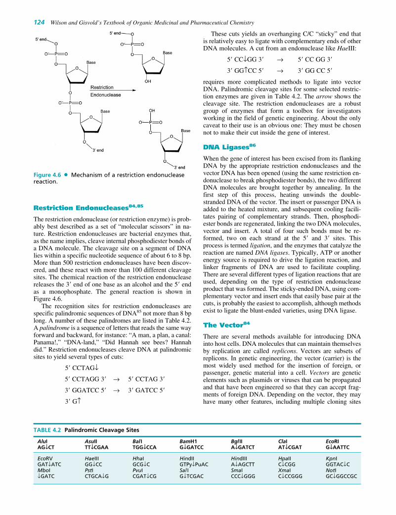
Restriction Endonucleases84,85
The restriction endonuclease (or restriction enzyme) is prob-ably best described as a set of “molecular scissors” in na-ture. Restriction endonucleases are bacterial enzymes that,as the name implies, cleave internal phosphodiester bonds ofa DNA molecule. The cleavage site on a segment of DNAlies within a specific nucleotide sequence of about 6 to 8 bp.More than 500 restriction endonucleases have been discov-ered, and these react with more than 100 different cleavagesites. The chemical reaction of the restriction endonucleasereleases the 3� end of one base as an alcohol and the 5� endas a monophosphate. The general reaction is shown inFigure 4.6.
The recognition sites for restriction endonucleases arespecific palindromic sequences of DNA85 not more than 8 bplong. A number of these palindromes are listed in Table 4.2.A palindrome is a sequence of letters that reads the same wayforward and backward, for instance: “A man, a plan, a canal:Panama!,” “DNA-land,” “Did Hannah see bees? Hannahdid.” Restriction endonucleases cleave DNA at palindromicsites to yield several types of cuts:
5� CCTAG↓
5� CCTAGG 3� → 5� CCTAG 3�
3� GGATCC 5� → 3� GATCC 5�
3� G↑
These cuts yields an overhanging C/C “sticky” end thatis relatively easy to ligate with complementary ends of otherDNA molecules. A cut from an endonuclease like HaeIII:
5� CC↓GG 3� → 5� CC GG 3�
3� GG↑CC 5� → 3� GG CC 5�
requires more complicated methods to ligate into vectorDNA. Palindromic cleavage sites for some selected restric-tion enzymes are given in Table 4.2. The arrow shows thecleavage site. The restriction endonucleases are a robustgroup of enzymes that form a toolbox for investigatorsworking in the field of genetic engineering. About the onlycaveat to their use is an obvious one: They must be chosennot to make their cut inside the gene of interest.
DNA Ligases86
When the gene of interest has been excised from its flankingDNA by the appropriate restriction endonucleases and thevector DNA has been opened (using the same restriction en-donuclease to break phosphodiester bonds), the two differentDNA molecules are brought together by annealing. In thefirst step of this process, heating unwinds the double-stranded DNA of the vector. The insert or passenger DNA isadded to the heated mixture, and subsequent cooling facili-tates pairing of complementary strands. Then, phosphodi-ester bonds are regenerated, linking the two DNA molecules,vector and insert. A total of four such bonds must be re-formed, two on each strand at the 5� and 3� sites. Thisprocess is termed ligation, and the enzymes that catalyze thereaction are named DNA ligases. Typically, ATP or anotherenergy source is required to drive the ligation reaction, andlinker fragments of DNA are used to facilitate coupling.There are several different types of ligation reactions that areused, depending on the type of restriction endonucleaseproduct that was formed. The sticky-ended DNA, using com-plementary vector and insert ends that easily base pair at thecuts, is probably the easiest to accomplish, although methodsexist to ligate the blunt-ended varieties, using DNA ligase.
The Vector84
There are several methods available for introducing DNAinto host cells. DNA molecules that can maintain themselvesby replication are called replicons. Vectors are subsets ofreplicons. In genetic engineering, the vector (carrier) is themost widely used method for the insertion of foreign, orpassenger, genetic material into a cell. Vectors are geneticelements such as plasmids or viruses that can be propagatedand that have been engineered so that they can accept frag-ments of foreign DNA. Depending on the vector, they mayhave many other features, including multiple cloning sites
124 Wilson and Gisvold’s Textbook of Organic Medicinal and Pharmaceutical Chemistry
Figure 4.6 Mechanism of a restriction endonucleasereaction.
TABLE 4.2 Palindromic Cleavage Sites
AluI AsuII Bal I BamH1 Bgl II ClaI EcoRIAG↓↓CT TT↓↓CGAA TGG↓↓CCA G↓↓GATCC A↓↓GATCT AT↓↓CGAT G↓↓AATTC
EcoRV HaeIII HhaI HindII HindIII HpaII KpnIGAT↓ATC GG↓CC GCG↓C GTPy↓PuAC A↓AGCTT C↓CGG GGTAC↓CMboI PstI PvuI Sal I SmaI XmaI NotI↓GATC CTGCA↓G CGAT↓CG G↓TCGAC CCC↓GGG C↓CCGGG GC↓GGCCGC
0119-0155_17865_Ch04.qxd 12/4/09 4:05 AM Page 124

(a region containing multiple restriction enzyme sites intowhich an insert can be installed or removed), selection mark-ers, and transcriptional promoters. The passenger DNA mustintegrate into the host cell’s DNA or be carried into the cellas part of a biologically active molecule that can replicate in-dependently. If this result is not achieved, the inserted genewill not be successfully transcribed. The most commonlyused biological agent for transporting genes into bacterialand yeast cells is the plasmid, such as the E. coli bacterialplasmid pBR322. A plasmid is a small, double-stranded,closed circular extrachromosomal DNA molecule. This plas-mid contains 4,361 bp and can transport relatively smallamounts of DNA. Plasmids occur in many species of bacte-ria and yeasts. Sometimes, plasmids carry their own genes(e.g., the highly transmissible genes for antibiotic resistancein some bacterial species). An important feature of a plasmidis that it has an origin of replication (ori) site that allows it tomultiply independently of a host cell’s DNA. Although therecan be more than one copy of a plasmid in a cell, the copynumber is controlled by the plasmid itself.
Another type of cloning vector is the bacteriophage(Fig. 4.7). Bacteriophage � (lambda) possesses a genomeof approximately 4.9 � 105 bp and can package largeamounts of genetic material without affecting the infec-tivity of the phage. A large DNA library can be created,packaged in bacteriophage �, and, when the virus infects,inserted into cells. Hybridization is then detected byscreening with DNA probes.87 In addition, there are spe-cial vectors called phagemids, vaccinia and adenovirus forcloning into mammalian cells, and yeast artificial chromo-somes (YACs) that facilitate cloning in yeasts.88 Differencesamong these vectors concern the size of the insert that theywill accept, the methods used in the selection of the clones,and the procedures for propagation.
Once the passenger DNA has been created and the plasmidvector cut (both with the same restriction enzyme), the insertis ligated into the plasmid along with a promoter (a shortDNA sequence that enhances the transcription of the adjacentgene). Often, a gene imparting antibiotic resistance linked tothe desired gene is inserted as a selection tool. The idea be-hind this is that if the gene is inserted in the proper location,the bacterial cell will grow on a medium containing the antibi-otic. Bacteria that do not contain the resistance gene and,hence, lack the required gene will not grow. This makes thetask of screening for integration of the desired gene easier.After the molecule is ligated, the vector is finally an rDNAmolecule that can be inserted into a host cell.
Host cells can be bacteria (e.g., E. coli), eukaryotic yeast(Saccharomyces cerevisiae), or mammalian cell lines, in-cluding Chinese hamster ovary (CHO), African green mon-key kidney (VERO), and baby hamster kidney (BHK). Itis easy to grow high concentrations of bacteria and yeastcells in fermenters to yield high protein concentrations.Mammalian cell culture systems typically give poorer pro-tein yields, but sometimes this is acceptable, especiallywhen the product demands the key posttranslational modi-fications that do not occur in bacteria. Host cells containingthe vector are grown in small-scale cultures and screenedfor the desired gene.89 When the clone providing the bestprotein yield is located, the organism is grown under care-fully controlled conditions and used to inoculate pilot-scalefermentations. Parameters such as production mediumcomposition, pH, aeration, agitation, and temperature areinvestigated at this stage to optimize the fermentation. Thehost cells divide, and the plasmids in them replicate, pro-ducing the desired “new” protein. The fermentation isscaled up into larger bioreactors for large-scale isolation ofthe recombinant protein. Obviously, the cultures secrete
Chapter 4 Biotechnology and Drug Discovery 125
Figure 4.7 Types of cloningvectors: a bacteriophage and aplasmid.
0119-0155_17865_Ch04.qxd 12/4/09 4:05 AM Page 125

their own natural proteins along with the cloned protein.Purification steps are required before the recombinant pro-tein is suitable for testing as a new, genetically engineeredpharmaceutical agent. Once the host cell line expressing therecombinant gene is isolated, it is essential to maintain se-lection pressure on it so that it does not spontaneously losethe plasmid. Typically, this pressure is applied by maintain-ing the cells on medium containing an antibiotic to whichthey bear a resistance gene.
SOME TYPES OF CLONING
A listing of some types of cloning is given in Table 4.3.
Functional Expression Cloning90
Functional expression cloning focuses on obtaining a spe-cific cDNA of known function. There are many variationson this approach, but they all rely on the ability to search forand isolate cDNAs based on some functional activity thatcan be measured (e.g., the electrophysiological measure-ment of ion conductances following expression of cDNAs infrog oocytes). By incrementally subdividing the cDNAs intopools and following the activity, it is possible to obtain asingle cDNA clone that encodes the functionality. The ad-vantage of functional expression cloning is that it does notrely on knowledge of the primary amino acid sequence. Thisis a definite advantage when attempting to clone proteins oflow abundance.
Positional Cloning90
Positional cloning can be used to localize fragments of DNArepresenting genes prior to isolating the DNA. An exampleof the use of positional cloning is the cloning of the gene re-sponsible for CF. By studying the patterns of inheritance of
the disease and then comparing these with known chromo-somal markers (linkage analysis), it was possible, withoutknowing the function of the gene, to locate the gene onhuman chromosome 7. Then, by using a technique known aschromosome walking, the gene was localized to a DNA se-quence that encodes a protein now known as the cystic fi-brosis transmembrane conductance regulator (CFTR). Thisprotein, previously unknown, was shown to be defective inCF patients and could account for many of the symptoms ofthe disease. Like functional cloning, positional cloning hasthe advantage that specific knowledge of the protein is notrequired. It is also directly relevant to the understanding ofhuman disease, and it can provide important new biologicaltargets for drug development and the treatment of disease.
Homology-Based Cloning
Another cloning strategy involves the use of previouslycloned genes to guide identification and cloning of evo-lutionarily related genes. This approach, referred to ashomology-based cloning, takes advantage of the fact that nu-cleotide sequences encoding important functional domainsof proteins tend to be conserved during the process of evolu-tion. Thus, nucleotide sequences encoding regions involvedwith ligand binding or enzymatic activity can be used asprobes that will hybridize to complementary nucleotide se-quences that may be present on other genes that bind similarligands or have similar enzymatic activity. This approachcan be combined with PCR91,92 to amplify the DNA se-quences. The use of homology-based cloning has the advan-tages that it can be used to identify families of related genes,does not rely on the purification or functional activity of agiven protein, and can provide novel targets for drug discov-ery. Its usefulness is offset by the possibility that the isolatedfragment may not encode a complete or functional protein orthat, in spite of knowledge of the shared sequence, the actualfunction of the clone may be difficult to identify.
126 Wilson and Gisvold’s Textbook of Organic Medicinal and Pharmaceutical Chemistry
TABLE 4.3 Cloning Strategies
Strategy Advantages Disadvantages Example
Positional Provides information underlying 3.3 � 109 bp, difficult to use Cystic fibrosis gene productthe genetic basis of known with diseases caused by diseases multiple interacting alleles
Protein purification Yields genetic information encoding Protein purification, especially �2-Adrenergic receptorproteins of known structure and for low-abundance proteins, function is exacting; availability of
appropriate libraries, incomplete coding sequences
Antibody based Yields genetic information encoding Involves protein purification, Vitamin D receptorproteins of known structure and unrecognized cross-reactivity,function incomplete coding sequences
Functional expression Yields genetic information encoding Function must be compatible Substance K receptora functionally active protein; does with existing library-screening not require protein purification technology
Homology based Identification of related genes or Depends on preexisting gene Muscarinic receptorgene families; relatively simple sequence; can yield incomplete
genes or genes of unknown function
Expressed sequence High throughput; identification of Incomplete coding sequences ortagging novel cDNAs genes of unknown function
Total genomic Knowledge of total genome; 3.3 � 109 bp, labor intensive; Haemophilus influenzaesequencing identification of all potential genes of unknown function
gene products
Source: PhRMA: Biotechnology Drug Development by Disease State, 2008–2009.
0119-0155_17865_Ch04.qxd 12/4/09 4:05 AM Page 126

EXPRESSION OF CLONED DNA
Once cloned, there are many different possibilities for theexpression and manipulation of DNA sequences. Because itconcerns the use of cloned genes in the process of drug dis-covery and development, there are many obvious ways inwhich the expression of DNA sequences can be applied.One of the most obvious is in replacement of older technolo-gies that involve the purification of proteins for human usefrom either animal sources or human byproducts, such asblood. An example of this is factor VIII, a clotting cascadeprotein used for the treatment of the genetically linkedbleeding disorder hemophilia. Until recently, the onlysource of purified factor VIII was human blood, and tragi-cally, before the impact of AIDS was fully appreciated,stocks of factor VIII had become contaminated with HIV-1,resulting in the infection of as many as 75% of the patientsreceiving this product. The gene encoding factor VIII hassince been cloned, and recombinant factor VIII is now avail-able as a product purified from cultured mammalian cells.Other recombinant clotting factors, including factors VIIaand IX, are under development and, together with recombi-nant factor VIII, will eliminate the risk of exposure tohuman pathogens.
Other examples in which the expression of cloned humangenes offers alternatives to previously existing products in-clude human insulin, which is now a viable replacement forpurified bovine and porcine insulin for the treatment of dia-betes, and hGH, which is used for the treatment of growthhormone deficiency in children (dwarfism). Unlike insulin,growth hormones from other animal species are ineffectivein humans; thus, until human recombinant growth hormonebecame available, the only source of hGH was the pituitaryglands of cadavers. This obviously limited the supply ofhGH and, like factor VIII, exposed patients to potential con-tamination by human pathogens. Recombinant hGH (rhGH)can now be produced by expression in bacterial cells.
The expression of cloned genes can be integrated into ra-tional drug design by providing detailed information aboutthe structure and function of the sites of drug action. Withthe cloning of a gene comes knowledge of the primaryamino acid sequence of an encoded receptor protein. Thisinformation can be used to model its secondary structureand in an initial attempt to define the protein’s functionaldomains, such as its ligand-binding site. Such a model canthen serve as a basis for the design of experiments that canbe used to test the model and facilitate further refinement.Of particular use are mutagenesis experiments that userDNA techniques to change a primary amino acid sequenceso that the consequences can be studied. In addition, expres-sion of a cloned target protein can be used to generate sam-ples for various biophysical determinations, such as x-raycrystallography. This technique, which can provide detailedinformation about the three-dimensional molecular structureof a protein, frequently requires large amounts of protein,which, in some cases, is only available with the use of re-combinant expression systems.
Like the many strategies used to clone genes, there aremany strategies for their expression, involving the use ofeither bacterial or eukaryotic cells and specialized vectorscompatible with expression in host cells. Since these cells donot normally express the protein of interest, this methodology
is often referred to as heterologous expression. It is alsopossible to prepare cRNA from rDNA, which can then beused for either in vitro expression or injection directly intocells. In the former situation, purified ribosomes are usedin the test tube to convert cRNA into protein; for the lattersituation, the endogenous cellular ribosomes make the pro-tein. A relatively new development for the expression ofcloned genes is the use of animals that have the cloned genestably integrated into their genome. Such transgenic animalscan potentially make very large amounts of recombinantprotein, which can be harvested from the milk, blood, andascites fluid.
The choice of a particular expression system depends onseveral factors. Protein yield, requirements for biologicalactivity, and compatibility of the expressed protein with thehost organism are a few. An example of the compatibilityissue is that bacteria do not process proteins in exactly thesame way as do mammalian cells, so that the expression ofhuman proteins in bacteria will not always yield an activeproduct or any product at all. Cases like these may requireexpression in mammalian cell cultures. The choice of anexpression system also reflects the available vectors andcorresponding host organisms. A basic requirement for theheterologous expression of a cloned gene is the presence ofa promoter that can function in the host organism and amechanism for introducing the cloned gene into the organ-ism. The promoter is the specific site at which DNA poly-merase binds to initiate transcription and is usually specificfor the host organism. As in gene cloning, the vectors areeither plasmids or viruses that have been engineered to ac-cept rDNA and that contain promoters that direct the expres-sion of the rDNA. The techniques for introducing the vectorinto the organism vary widely and depend on whether one isinterested in transient expression of the cloned gene or instable expression. In the latter case, integration into the hostgenome is usually required; transient expression simply re-quires getting the vector into the host cell.
MANIPULATION OF DNA SEQUENCE INFORMATION
Perhaps the greatest impact of rDNA technology lies in itsability to alter a DNA sequence and create entirely new mol-ecules that, if reintroduced into the genome, can be inheritedand propagated in perpetuity. The ability to alter a DNA se-quence, literally in a test tube, at the discretion of an individ-ual, corporation, or nation, brings with it important ques-tions about ownership, ethics, and social responsibility.There is no question, however, that potential benefits to thetreatment of human disease are great.
There are three principal reasons for using rDNA tech-nology to alter DNA sequences. The first is simply to clonethe DNA to facilitate subsequent manipulation. The secondis to intentionally introduce mutations so that the site-specificeffect on protein structure and function can be studied.93,94
The third reason is to add or remove sequences to obtainsome desired attribute in the recombinant protein. For exam-ple, recent studies with factor VIII show that the proteincontains a small region of amino acids that are the majordeterminant for the generation of anti–factor VIII antibodiesin a human immune system. This autoimmune responseof the patient inhibits the activity of factor VIII, which is
Chapter 4 Biotechnology and Drug Discovery 127
0119-0155_17865_Ch04.qxd 12/4/09 4:05 AM Page 127

obviously a serious therapeutic complication for patientswho are using factor VIII for the treatment of hemophilia.By altering the DNA sequence encoding, this determinant,however, the amino acid sequence can be changed bothto reduce the antigenicity of the factor VIII molecule and tomake it transparent to any existing anti–factor VIII anti-bodies (i.e., changing the epitope eliminates the existingantibody recognition sites).
It is possible to combine elements of two proteins intoone new recombinant protein. The resulting protein, re-ferred to as a chimeric or fusion protein, may then havesome of the functional properties of both of the originalproteins. This is illustrated in Figure 4.8 for two receptorslabeled A and B. Each receptor has functional domains thatare responsible for ligand binding, integration into theplasma membrane, and activation of intracellular signalingpathways. Using rDNA techniques, one can exchangethese functional domains to create chimeric receptors that,for example, contain the ligand-binding domain of recep-tor B but the transmembrane and intracellular signaling do-mains of receptor A. The application of the fusion proteinstrategy is discussed further in connection with the hGHreceptor (under “Novel Drug-Screening Strategies”) andwith denileukin diftitox.
Another reason for combining elements of two proteinsinto one recombinant protein is to facilitate its expressionand purification. For example, recombinant glutathione S-transferase (GST), cloned from the parasitic wormSchistosoma japonicum, is strongly expressed in E. coli andhas a binding site for glutathione. Heterologous sequencesencoding the functional domains from other proteins can befused, in frame, to the carboxy terminus of GST, and the re-sulting fusion protein is often expressed at the same levelsas GST itself. In addition, the resulting fusion protein retainsthe ability to bind glutathione, which means that affinitychromatography, using glutathione that has been covalentlybonded to agarose, can be used for a single-step purificationof the fusion protein. The functional activity of the heterol-ogous domains that have been fused to GST can then be
studied either as part of the fusion protein or separately fol-lowing treatment of the fusion protein with specific pro-teases that cleave at the junction between GST and the het-erologous domain. Purified fusion proteins can also be usedto generate antibodies to the heterologous domains and forother biochemical studies. Sometimes, fusion proteins aremade to provide a recombinant protein that can be easilyidentified. An example of this is a technique called epitopetagging, in which well-characterized antibody recognitionsites are fused with recombinant proteins. The resulting re-combinant protein can then be identified by immunofluores-cence or can be purified with antibodies that recognize theepitope.
NEW BIOLOGICAL TARGETS FORDRUG DEVELOPMENT
One of the outcomes of the progress that has been made inthe identification and cloning of genes is that many proteinsencoded by these genes represent entirely new targets fordrug development. In some cases, the genes themselves mayrepresent the ultimate target for the treatment of a disease inthe form of gene therapy. The cloning of the CF gene is anexample of both a new drug target and a gene that could po-tentially be used to treat the disease. The protein encoded bythis gene, CFTR, is a previously unknown integral mem-brane protein that functions as a channel for chloride ions.Mutations in CFTR underlie the pathophysiology of CF, andin principle, replacement or coexpression of the defectivegene with the healthy, nonmutated gene would cure the dis-ease. It is also possible, however, that by understanding thestructure and function of the healthy CFTR, drugs could bedesigned to interact with the mutated CFTR and improve itsfunction.
An important outgrowth of the study of new drug targetsis the recognition that many traditional targets, such as en-zymes and receptors, are considerably more heterogeneousthan previously thought. Thus, instead of one enzyme or re-ceptor, there may be several closely related subtypes, or iso-forms, each with the potential of representing a separatedrug target. This can be illustrated with the enzyme cy-clooxygenase (COX), which is pivotal to the formation ofprostaglandins and which is the target of aspirin and thenonsteroidal anti-inflammatory drugs (NSAIDs). Until re-cently, COX was considered to be a single enzyme, butpharmacological and gene cloning studies have revealedthat there are at least three enzyme forms, named COX-1,COX-2, and COX-3. Interestingly, they are differentiallyregulated. COX-1 is expressed constitutively in many tis-sues, whereas the expression of COX-2 is induced by in-flammatory processes. Thus, the development of COX-2 se-lective agents can yield NSAIDs with the same efficacy asexisting (nonselective) agents but with fewer side effects,such as those on the gastric mucosa.
The elucidation of the family of adrenergic receptors isanother example in which molecular cloning studies haverevealed previously unknown heterogeneity, with the conse-quence of providing new targets for drug development. Theadrenergic receptors mediate the physiological effects of thecatecholamines epinephrine and norepinephrine. They arealso the targets for many drugs used in the treatment of suchconditions as congestive heart failure, asthma, hypertension,
128 Wilson and Gisvold’s Textbook of Organic Medicinal and Pharmaceutical Chemistry
Figure 4.8 Chimeric receptors.
0119-0155_17865_Ch04.qxd 12/4/09 4:05 AM Page 128

glaucoma, and benign prostatic hypertrophy. Prior to themolecular cloning and purification of adrenergic receptors,the pharmacological classification of this family of recep-tors consisted of four subtypes: �1, �2, �1, and �2. The ini-tial cloning of the �-adrenergic receptor in 1986 and subse-quent gene cloning studies revealed at least nine subtypes:�1, �2, �3, �1A, �1B, �1D, �2A, �2B, and �2C (Chapter 16).
The evidence that there are nine subtypes of adrenergicreceptors is very important in terms of understanding thephysiology of the adrenergic receptors and of developingdrugs that can selectively interact with these subtypes. Forexample, in the case of the �2-agonist p-aminoclonidine, anagent used to lower intraocular pressure (IOP) in the treat-ment of glaucoma, it may now be possible to explain someof the drug’s pharmacological side effects (e.g., bradycardiaand sedation) by invoking interactions with the additional�2-adrenergic receptor subtypes. Of considerable interest isthe possibility that these pharmacological effects (i.e., low-ering of IOP, bradycardia, and sedation) are each mediatedby one of the three different �2-receptor subtypes. If this istrue, it might be possible to develop a subtype-selective �2-agonist that lowers IOP but does not cause bradycardia orsedation. Likewise, it might even be possible to take advan-tage of the pharmacology and develop �2-adrenergic agentsthat selectively lower heart rate or produce sedation.
The discovery of subtypes of receptors and enzymes bymolecular cloning studies seems to be the rule rather thanthe exception and is offering a plethora of potential newdrug targets (Table 4.4). To note just a few: 5 dopamine re-ceptor subtypes have been cloned, replacing 2 defined phar-macologically (Chapter 13); 7 serotonin receptor subtypeshave been cloned, replacing 3; 4 genes encoding receptorsfor prostaglandin E2 have been isolated, including 12 addi-tional alternative mRNA splice variants; and 3 receptors fornerve growth factor have been cloned, replacing 1.
NOVEL DRUG-SCREENING STRATEGIES
The combination of the heterologous expression of clonedDNA, the molecular cloning of new biological targets, andthe ability to manipulate gene sequences has created powerfulnew tools that can be applied to the process of drug discoveryand development. In its most straightforward application, the
ability to simply express newly identified receptor protein tar-gets offers a novel means of obtaining information that maybe difficult, or even impossible, to obtain from more complexnative biological systems. There is a reason for this. A newlyidentified protein can be expressed in isolation. Even forclosely related enzyme or receptor subtypes, heterologous ex-pression of the individual subtype can potentially providedata that are specific for the subtype being expressed, whereasthe data from native biological systems will reflect the sum-mation of the individual subtypes that may be present.
The potential advantage of heterologous expression is il-lustrated in Figure 4.9 for the interaction of a drug with mul-tiple binding sites. In panel A, which can represent the data
Chapter 4 Biotechnology and Drug Discovery 129
Figure 4.9 Convoluted data from binding to multiplereceptor subtypes versus classic mass action.
TABLE 4.4 Selected Examples of Receptor Subtype Heterogeneity
Receptor Superfamily Original Subtypes Present Subtypes
G-protein–coupledAdrenergic �1, �2, �1, �2 �1, �2, �3, �1A, �1B, �1D, �2A, �2B, �2CDopamine D1, D2 D1, D2
a, D3, D4, D5
Prostaglandin E2 EP1, EP2, EP3 EP1, EP2, EP3a, EP4
Receptor tyrosine kinase neurotrophins Nerve growth factor receptor TrkAa, TrkB, TrkCa
DNA bindingEstrogen Estrogen receptor ERR1, ERR2Thyroid hormone Thyroid hormone receptor TR�, TR�Retinoic acid Retinoic acid receptor RAR�, RAR�
Ligand-activated channelsGlycine Glycine and/or strychnine receptor �1, �2, �3 (multisubunit)b
GABAA GABA and/or benzodiazepine receptor �1, �2, �3, �4, �5, �6 (multisubunit)b
aAlternative mRNA splicing creates additional receptor heterogeneity.bOnly the heterogeneity of the ligand-binding subunit is listed; a multisubunit structure combined with the heterogeneity of the other subunits creates a very large number of potential subtypes.
0119-0155_17865_Ch04.qxd 12/4/09 4:05 AM Page 129

obtained from a native biological system, the data are com-plex, and the curve reflects interactions of the drug with twopopulations of receptors: one with high affinity, represent-ing 50% of the total receptor population, and one with lowaffinity, representing the remaining 50%. The individualcontributions of these two populations of receptors are indi-cated in panel B, which could also reflect the data obtainedif rDNA encoding these two receptors were expressed indi-vidually in a heterologous expression system. Although insome cases, the data, as in panel A, can be analyzed withsuccess, frequently they cannot, especially if more than twosubtypes are present or if any one subtype makes up lessthan 10% of the total receptor population or if the affinitiesof the drug for the two receptor populations differ by lessthan 10-fold.
Another important reason for integrating heterologous ex-pression into drug-screening strategies is that data can usuallybe obtained for the human target protein rather than an animalsubstitute. This does not mean that organ preparations or ani-mal models will be totally replaced. For the purposes of theidentification of lead compounds and the optimization of se-lectivity, affinity, etc., however, the use of recombinant ex-pression systems provides some obvious advantages.
By combining heterologous expression with novel func-tional assays, it is possible to increase both specificity andthroughput (the number of compounds that can be screenedper unit time). For example, reporter genes have been devel-oped that respond to various intracellular second messen-gers, such as the activation of guanine nucleotide–bindingproteins (G proteins), and levels of cyclic adenosinemonophosphate (cAMP), or calcium. One approach to thedevelopment of novel functional assays involves the use ofpromoter regions in DNA that control the transcription ofgenes. This approach is exemplified by the cAMP responseelement (CRE). This is a specifically defined sequence ofDNA that is a binding site for the cAMP response element-binding (CREB) protein. In the unstimulated condition, thebinding of CREB to the CRE prevents the transcription andexpression of genes that follow it (Fig. 4.10). When CREB
is phosphorylated by cAMP-dependent protein kinase A(PKA), however, its conformation changes, permitting thetranscription and expression of the downstream gene. Thus,increases in intracellular cAMP, such as those caused by re-ceptors that activate adenylyl cyclase (e.g., �-adrenergic,vasopressin, etc.), will stimulate the activity of PKA, which,in turn, results in the phosphorylation of CREB and the ac-tivation of gene transcription.
In nature, there are a limited number of genes whoseactivity is regulated by a CRE. Biologically, however, theexpression of almost any gene can be regulated in a cAMP-dependent fashion if it is placed downstream of a CRE,using rDNA techniques. If the products of the expression ofthe downstream gene can be easily detected, they can serveas reporters for any receptor or enzyme that can modulatethe formation of cAMP in the cell. The genes encodingchloramphenicol acetyl transferase (CAT), luciferase, and�-galactosidase are three examples of potential “reportergenes,” whose products can be easily detected. Sensitiveenzymatic assays have been developed for all of theseenzymes; thus, any changes in their transcription will bequickly reflected by changes in enzyme activity. By coex-pressing the reporter gene along with the genes encodingreceptors and enzymes that modulate cAMP formation, it ispossible to obtain very sensitive functional measures of theactivation of the coexpressed enzyme or receptor.
Another example of the use of a reporter gene for high-throughput drug screening is the receptor selection andamplification technology (r-SAT) assay. This assay takesadvantage of the fact that the activation of several differentclasses of receptors can cause cellular proliferation. If genesfor such receptors are linked with a reporter gene, suchas �-galactosidase, the activity of the reporter will be in-creased as the number of cells increase as a consequence ofreceptor activation. Initially, a limitation of this assay wasthat it only worked with receptors that normally coupledto cellular proliferation; by making a mutation in one ofthe second-messenger proteins involved with the prolifera-tive response, however, it was possible to get additional
130 Wilson and Gisvold’s Textbook of Organic Medicinal and Pharmaceutical Chemistry
Figure 4.10 Activation of tran-scription by a cAMP response ele-ment (CRE). CREB is phosphorylatedby cAMP-dependent protein kinase.
0119-0155_17865_Ch04.qxd 12/4/09 4:05 AM Page 130

receptors to work in this assay. This second-messenger pro-tein, Gq, was cloned, and a recombinant chimera was madethat included part of another second messenger known asGi. In native cells, receptors that activate Gi are not knownfor their stimulation of cell proliferation, but when suchreceptors are coexpressed in the r-SAT assay with thechimeric Gq, their activity can be measured.
A similar strategy involving chimeric proteins has beenused for receptors whose second-messenger signaling path-ways are not clearly understood. For example, the develop-ment of potential therapeutic agents acting on the hGH recep-tor has been difficult because of a lack of a good signalingassay. The functional activity of other receptors that arestructurally and functionally related to the growth hormonereceptor can be measured, however, in a cell proliferationassay. One such receptor that has been cloned is the murinereceptor for granulocyte colony-stimulating factor (G-CSF).By making a recombinant chimeric receptor containing theligand-binding domain of the hGH receptor with the second-messenger–coupling domain of the murein G-CSF receptor,it was possible to stimulate cellular proliferation with hGH.
In addition to providing a useful pharmacologicalscreen for hGH analogs, the construction of this chimericreceptor provides considerable insight into the mechanismof agonist-induced growth hormone receptor activation.The growth hormone–binding domain is clearly localizedto the extracellular amino terminus of the receptor,whereas the transmembrane and intracellular domains areimplicated in the signal transduction process. It was alsodetermined that successful signal transduction required re-ceptor dimerization by the agonist (i.e., simultaneous inter-action of two receptor molecules with one molecule ofgrowth hormone). Based on this information, a mecha-nism-based strategy was used for the design of potentialantagonists. Thus, hGH analogs were prepared, were inca-pable of producing receptor dimerization, and were foundto be potent antagonists.
PROCESSING OF THE RECOMBINANTPROTEIN
Processing the fermentation contents to isolate a recombi-nant protein is often a difficult operation, requiring as muchart as science. In the fermentation broth are whole bacterialcells, lysed cells, cellular fragments, nucleotides, normalbacterial proteins, the recombinant protein, and particulatemedium components. If a Gram-negative bacterium such asE. coli has been used, lipopolysaccharide endotoxins (pyro-gens) may be present. When animal cell cultures are used, itis commonly assumed that virus particles may be present.Viruses can also be introduced by the culture nutrients, gen-erated by an infected cell line, or introduced by animalserum. Purification of an rDNA protein while maintainingthe factors that keep it in its active three-dimensional con-formation from this mixture may be difficult, because eachstep must be designed to ensure that the protein remains in-tact and pharmacologically active. Assays must be designedthat allow the activity of the protein to be assessed at eachpurification step. Consequently, the structure and activity ofthe recombinant protein must be considered at all stages ofpurification, and assays must be conducted to measure theamount of purified, intact protein.
A general scheme for purification of an rDNA protein isas follows95:
• Particulate removal. Particulates may be removed by cen-trifugation, filtration, ultrafiltration, and tangential flowfiltration. Virus particles may be inactivated by heating ifthe rDNA peptide can tolerate the procedure.
• Concentration. The volume of the mixture is reduced,which increases the concentration of the contents. Often,concentration is achievable by the filtration step, espe-cially if ultrafiltration is used.
• Initial purification. The initial purification of the mixtureis sometimes accomplished by precipitation of the pro-teins, using a slow, stepwise increase of the ionic strengthof the solution (salting out). Ammonium sulfate is a typi-cal salt that can be used in cold, aqueous solutions. Water-miscible organic solvents such as trichloroacetic acid andpolyethylene glycol (PEG) change the dielectric constantof the solution and also effect precipitation of proteins.
• Intermediate purification. In this stage, the proteins maybe dialyzed against water to remove salts that were used inthe precipitation step. Ion exchange chromatography isused to effect a somewhat crude separation of the proteinsbased on their behavior in a pH or salt gradient on theresin. Another step that may be taken is size exclusion (gelfiltration) chromatography. Gels of appropriate molecularweight cutoffs can yield a somewhat low-resolution sepa-ration of proteins of a desired molecular weight. If a nativebacterial protein that has been carried this far is nearly thesame molecular weight as the rDNA protein, no separationwill occur.
• Final purification. Final purification usually involves theuse of high-resolution chromatography, typically high-performance liquid chromatography. An abundance ofcommercial stationary phases allows various types of ad-sorption chromatography (normal and reversed phase), ionexchange chromatography, immunoaffinity chromatogra-phy, hydrophobic interaction chromatography, and sizeexclusion chromatography. The protein fractions are sim-ply collected when they elute from the column and areconcentrated and assayed for activity.
• Sterilization and formulation. This step can be accom-plished by ultrafiltration to remove pyrogens or by heatingif the protein can withstand this. Formulation might in-volve reconstitution into stable solutions for administra-tion or determining the optimum conditions for stabilitywhen submitting for clinical trials.
Complicating factors include (a) proteins unfolding intoan inactive conformation during processing (it may not bepossible to refold the protein correctly) and (b) proteasesthat are commonly produced by bacterial, yeast, and mam-malian cells, which may partially degrade the protein.
PHARMACEUTICS OF RECOMBINANTDNA-PRODUCED AGENTS
rDNA methods have facilitated the production of very pure,therapeutically useful proteins. The physicochemical andpharmaceutical properties of these agents are those of pro-teins, which means that pharmacists must understand thechemistry (and the chemistry of instability) of proteins tostore, handle, dispense, reconstitute, and administer these
Chapter 4 Biotechnology and Drug Discovery 131
0119-0155_17865_Ch04.qxd 12/4/09 4:05 AM Page 131

protein drugs. Instabilities among proteins may be physicalor chemical. In the former case, the protein might stick toglass vessels or flocculate, altering the dose that the patientwill receive. In the latter case, chemical reactions takingplace on the protein may alter the type or stereochemistry ofthe amino acids, change the position of disulfide bonds,cleave the peptide chains themselves, and alter the chargedistribution of the protein. Any of these can cause unfolding(denaturation) of the protein and loss of activity, renderingthe molecule useless as a drug. Chemical instability can bea problem during the purification stages of a protein, whenthe molecule might be subjected to acids or bases, but insta-bility could occur at the point of administration when, forexample, a lyophilized protein is reconstituted. The pharma-cist must understand a few concepts of the chemical andphysical instability of proteins to predict and handle poten-tial problems.
Chemical Instability of Proteins67
See Figure 4.11.
• Hydrolysis. Hydrolytic reactions of the peptide bonds canbreak the polymer chain. Aspartate residues hydrolyze 100times faster in dilute acids than do other amino acids underthe same conditions. As a general rule of peptide hydroly-sis, Asp-Pro � Asp-X or X-Asp bonds. This property ofAsp is probably a result of an autocatalytic function of theAsp side chain carboxyl group. Asn, Asp, Gln, and Glu hy-drolyze exceptionally easily if they occur next to Gly, Ser,Ala, and Pro. Within these groupings, Asn and Gln accel-erate hydrolysis more at low pH, whereas Asp and Gluhydrolyze most readily at high pH, when the side chaincarboxyl groups are ionized.
• Deamidation. Gln and Asn undergo hydrolytic reactionsthat deamidate their side chains. These reactions convertneutral amino acid residues into charged ones. Gln is con-verted to Glu and Asn to Asp. The amino acid type ischanged, but the chain is not cleaved. This process is, ef-fectively, primary sequence isomerization, and it may in-fluence biological activity. The deamidation reaction ofAsn residues is accelerated under neutral or alkaline pHconditions. A five-membered cyclic imide intermediateformed by intramolecular attack of the nitrogen atom onthe carbonyl carbon of the Asn side chain is the accelerant.The cyclic imide spontaneously hydrolyzes to give a mix-ture of residues—the aspartyl peptide and an isoform.
• Racemization. Base-catalyzed racemization reactions canoccur in any of the amino acids except glycine, which isachiral. Racemizations yield proteins with mixtures of L-and D-amino acid configurations. The reaction occurs fol-lowing the abstraction of the �-hydrogen from the aminoacid to form a carbanion. As should be expected, the sta-bility of the carbanion controls the rate of the reaction.Asp, which undergoes racemization via a cyclic imide in-termediate, racemizes 105 times faster than free Asn. Bycomparison, other amino acids in a protein racemize about2 to 4 times faster than their free counterparts.
• �-Elimination. Proteins containing Cys, Ser, Thr, Phe, andLys undergo facile �-elimination in alkaline conditionsthat facilitate formation of an �-carbanion.
• Oxidation. Oxidation can occur at the sulfur-containingamino acids Met and Cys and at the aromatic amino acidsHis, Trp, and Tyr. These reactions can occur during protein
processing as well as in storage. Methionine (CH3MSMR)is oxidizable at low pH by hydrogen peroxide or molecularoxygen to yield a sulfoxide (RMSOMCH3) and a sulfone(RMSO2MCH3). The thiol group of Cys (RMSH) can un-dergo successive oxidation to the corresponding sulfenicacid (RMSOH), disulfide (RMSMSMR), sulfinic acid(RMSO2H), and sulfonic acid (RMSO3H). Several factors,including pH, influence these reactions. Free MSH groupscan be converted into disulfide bonds (MSMSM) and viceversa. In the phenomenon of disulfide exchange, disulfidebonds break and reform in different positions, causingincorrect folding of the protein. Major changes in the three-dimensional structure of the peptide can abolish activity.Oxidation of the aromatic rings of His, Trp, and Tyrresidues is believed to occur with various oxidizingenzymes.
Physical Instability of Proteins96
Chemical alterations are not the only source of protein insta-bility. A protein is a large, globular polymer that exists insome specific forms of secondary, tertiary, and quaternarystructure. A protein is not a fixed, rigid structure. The mol-ecule is in dynamic motion, and the structure samples anarray of three-dimensional space. During this motion, non-covalent intramolecular bonds can break, reform, and breakagain, but the overall shape remains centered around an en-ergy minimum that represents the most likely (and pharma-cologically active) conformer of the molecule. Any majorchange in the conformation can abolish the activity of theprotein. Small drug molecules do not demonstrate this prob-lem. A globular protein normally folds so that the hydropho-bic groups are directed to the inside and the hydrophilicgroups are directed to the outside. This arrangement facili-tates the water solubility of the protein. If the normal proteinunfolds, it can refold to yield changes in hydrogen bonding,charge, and hydrophobic effects. The protein loses its glob-ular structure, and the hydrophobic groups can be reposi-tioned to the outside. The unfolded protein can subsequentlyundergo further physical interactions. The loss of the globu-lar structure of a protein is referred to as denaturation.
Denaturation is, by far, the most widely studied aspect ofprotein instability. In the process, the three-dimensional fold-ing of the native molecule is disrupted at the tertiary and, pos-sibly, the secondary structure level. When a protein denatures,physical structure rather than chemical composition changes.The normally globular protein unfolds, exposing hydrophobicresidues and abolishing the native three-dimensional struc-ture. Factors that affect the denaturation of proteins aretemperature, pH, ionic strength of the medium, inclusion oforganic solutes (urea, guanidine salts, acetamide, and for-mamide), and the presence of organic solvents such asalcohols or acetone. Denaturation can be reversible or irre-versible. If the denatured protein can regain its native formwhen the denaturant is removed by dialysis, reversible denat-uration will occur. Denatured proteins are generally insolublein water, lack biological activity, and become susceptible toenzymatic hydrolysis. The air–water interface presents a hy-drophobic surface that can facilitate protein denaturation.Interfaces like these are commonly encountered in drug deliv-ery devices and intravenous (IV) bags.
Surface adsorption of proteins is characterized by adhesionof the protein to surfaces, such as the walls of the containers
132 Wilson and Gisvold’s Textbook of Organic Medicinal and Pharmaceutical Chemistry
0119-0155_17865_Ch04.qxd 12/4/09 4:05 AM Page 132

Chapter 4 Biotechnology and Drug Discovery 133
Figure 4.11 A. Protein decom-position reactions. B. �-Elimination.
0119-0155_17865_Ch04.qxd 12/4/09 4:05 AM Page 133

of the dosage form and drug delivery devices, ampuls, andIV tubing. Proteins can adhere to glass, plastics, rubber, poly-ethylene, and polyvinylchloride. This phenomenon is referredto as flocculation. The internal surfaces of IV delivery pumpsand IV delivery bags pose particular problems of this kind.Flocculated proteins cannot be dosed properly.
Aggregation results when protein molecules, in aqueoussolution, self-associate to form dimers, trimers, tetramers,hexamers, and large macromolecular aggregates. Self-association depends on the pH of the medium as well as sol-vent composition, ionic strength, and dielectric properties.Moderate amounts of denaturants (below the concentrationthat would cause denaturation) may also cause protein aggre-gation. Partially unfolded intermediates have a tendency toaggregate. Concentrated protein solutions, such as an im-munoglobulin for injection, may aggregate with storage timeon the shelf. The presence of particulates in the preparation isthe pharmacist’s clue that the antibody solution is defective.
Precipitation usually occurs along with denaturation.Detailed investigations have been conducted with insulin,which forms a finely divided precipitate on the walls of aninfusion device or its dosage form container. It is believedthat insulin undergoes denaturation at the air–water inter-face, facilitating the precipitation process. The concentra-tion of zinc ion, pH, and the presence of adjuvants such asprotamine also affect the precipitation reaction of insulin.
Immunogenicity of BiotechnologicallyProduced Drugs97,98
Proteins, by their very nature, are antigens. A human protein,innocuous at its typical physiological concentration, may ex-hibit completely different immunogenic properties when ad-ministered in the higher concentration that would be used asa drug. Unless a biotechnology-derived protein is engineeredto be 100% complementary to the human form, it will differamong several major epitopes. The protein may have modifi-cations of its amino acid sequence (substitutions of one aminoacid for another). There may be additions or deletions ofamino acids, N-terminal methionyl groups, incorrect or ab-normal folding patterns, or oxidation of a sulfur-containingside chain of a methionine or a cysteine. Additionally, whena protein has been produced by using a bacterial vector, a fi-nite amount of immunoreactive material may pass into thefinal product. All of these listed items contribute to the anti-genicity of a biotechnologically produced protein. When it isadministered to a human patient, the host’s immune systemwill react to the protein just as it would to a microbial attackand neutralize it. This is why research has been undertaken tocreate 100% human protein drugs, such as insulin, which pa-tients will need to take for a long time. In addition, some ofthe most promising biotechnology products, the MAbs, areproduced in mice by use of humanized genes to avoid humanreaction to the mouse antibody.
DELIVERY AND PHARMACOKINETICSOF BIOTECHNOLOGY PRODUCTS99
As with any drug class, the medicinal chemist and pharma-cist must be concerned with the absorption, distribution, me-tabolism, and excretion (ADME) parameters of proteindrugs. Biotechnology-produced drugs add complexities that
are not encountered with “traditional” low–molecular-weight drug molecules. ADME parameters are necessary tocompute pharmacokinetic and pharmacodynamic parame-ters for a given protein. As for any drug, these parametersare essential in calculating the optimum dose for a given re-sponse, determining how often to administer the drug to ob-tain a steady state, and adjusting the dose to obtain the bestpossible residence time at the receptor (pharmacodynamicparameters).
Delivery of drugs with the molecular weights and prop-erties of proteins into the human body is a complex task.The oral route cannot be used with a protein because theacidity of the stomach will catalyze its hydrolysis unless thedrug is enteric coated. Peptide bonds are chemically labile,and proteolytic enzymes that are present throughout thebody can attack and destroy protein drugs. Hydrolysis andpeptidase decomposition also occur during membrane trans-port through the vascular endothelium, at the site of admin-istration, and at sites of reaction in the liver, blood, kidneys,and most tissues and fluids of the body. It is possible to cir-cumvent these enzymes by saturating them with high con-centrations of drug or by coadministering peptidase in-hibitors. Oxidative metabolism of aromatic rings and sulfuroxidation can also occur. Proteins typically decompose intosmall fragments that are readily hydrolyzed, and the individ-ual amino acids are assimilated into new peptides. A poten-tially serious hindrance to a pharmacokinetic profile is thetendency of proteins administered as drugs to bind to plasmaproteins, such as serum albumin. If this happens, they entera new biodistribution compartment from which they mayslowly exit. Presently, the routes of administration that areavailable for protein drugs are largely subcutaneous (SC)and intramuscular (IM). Much ongoing research is targetedat making peptide drugs more bioavailable. An example ofthis is conjugation of interleukin-2 (IL-2) with PEG. Theseso-called pegylated proteins tend to have a slower elimina-tion clearance and a longer t1/2 than IL-2 alone. Anotherstrategy being used is the installation of a prosthetic sugarmoiety onto the peptide. The sugar moiety will adjust thepartition coefficient of the drug, probably making it morewater soluble.
RECOMBINANT DRUG PRODUCTS
Hormones
HUMAN INSULIN, RECOMBINANT100–102
Human insulin was the first pharmacologically active biolog-ical macromolecule to be produced through genetic engineer-ing. The FDA approved the drug in 1982 for the treatment oftype 1 (insulin-dependent) diabetes (see Chapter 27). The in-sulin protein is a two-chain polypeptide containing 51 aminoacid residues. Chain A is composed of 21 amino acids, andchain B contains 30. The human insulin molecule has threedisulfide linkages, CysA7 to CysB7, CysA20 to CysB19, andan intrachain linkage, CysA6 to CysA11. Insulin is secreted bythe �-cells of the pancreatic islets of Langerhans, initially asa single peptide chain called proinsulin. Enzymatic cleavageof the propeptide releases the insulin.
Historically, insulin was isolated from bovine or porcinesources. Using these agents was not without difficulty. Bothporcine and bovine insulin differ in amino acid sequence,
134 Wilson and Gisvold’s Textbook of Organic Medicinal and Pharmaceutical Chemistry
0119-0155_17865_Ch04.qxd 12/4/09 4:05 AM Page 134

with Ala replacing Thr at the C-terminus of the humanB chain (B30). Bovine insulin also differs in sequence fromhuman insulin, with Ala substituting for Thr at A8 and Valsubstituting for isoleucine at A10. These differences, smallthough they may seem, result in immunological reactions insome patients. Adjustments to the formulation of bovine andporcine insulin led to products that differed in time of onset,time to peak reduction in glucose, and duration of action.These parameters were varied by addition of protamine andzinc (which yielded a particulate insulin with a longer dura-tion of action), and adjustment of the pH to neutrality, whichstabilized the preparation. Insulins were characterized asregular (short-duration Iletin, 4–12 hours), semilente (ultra-short duration), lente (intermediate [1–3 hours to peak,24 hours duration]), and ultralente (extended duration). Anadditional form of insulin was NPH (neutral protamineHagedorn), which had an intermediate time of onset andtime to peak (1–3 hours) and a long duration of action(16–24 hours).
Producing a recombinant insulin that is chemically andphysically indistinguishable from the human pancreatic hor-mone was a major accomplishment. The problem with im-munoreactivity has been eliminated, the pyrogen content ofthe rDNA product is nil, the insulin is not contaminated withother peptides, and the hormone can be biosynthesized inlarger quantities. Human insulin (rDNA) is available asHumulin, Novolin, and several analogs that differ in theirpharmacokinetic profiles. Humulin is produced by using re-combinant E. coli; Novolin is prepared by using recombi-nant S. cerevisiae, a yeast. There have been modifications inthe production procedure since the initial successful biosyn-thesis. Prior to 1986, Humulin was produced by creatingtwo different vectors, one for the A chain and one for the Bchain, and inserting them into E. coli. The A chain and theB chain would be secreted into the medium, and the twowere joined chemically to form rDNA insulin. Today, theentire proinsulin gene is used to create a recombinant organ-ism, and the connecting peptide in proinsulin is cleaved bytwo enzymes (an endopeptidase and a carboxypeptidase B),yielding insulin (for details see Chapter 27).
Insulin rDNA is available in several103 forms. Insulinlispro (Humalog) has a more rapid (15–30 minutes) onsetand a shorter duration (3–6.5 hours) of action than regularhuman insulin (onset 30–60 minutes, duration 6–10 hours).It is effective when administered 15 minutes before a meal,unlike regular insulin, which must be injected 30 minutesbefore a meal. In lispro, the B-chain amino acids B28Proand B29Lys are exchanged. Insulin aspart (NovoLog), onset15 to 30 minutes, duration 3 to 6.5 hours, with a singleamino acid substitution of Asp for Pro at B28, is effectivewhen administered 5 to 10 minutes before a meal. Theultra–long-acting agent insulin glargine has the Asp at A21
replaced by Gly and has two Arg residues added at the C-terminus of the B chain. Insulin glargine, administeredsubcutaneously, has a duration of action of 24 to 48 hours.The alteration in basicity of this agent causes it to precipi-tate at neutral pH, creating a depot effect.
Insulin rDNA has been very successful. The only prob-lem has arisen in patients who have been using porcine orbovine insulin for a long time. Some patients who areswitched to rDNA human insulin report difficulty in “feel-ing their glucose level,” and these patients require extracounseling in the use of the recombinant hormone.
GLUCAGON104
The hormone glucagon (GlucaGen) is biosynthesized in thepancreas as a high–molecular-weight protein from whichthe active macromolecule is released by proteolytic cleav-age. Glucagon is a single chain of 29 amino acids and gen-erally opposes the actions of insulin. Bovine and porcineglucagons, which possess structures identical with humanglucagon, have been in use for years. The rDNA form hasbeen approved by the FDA for use in severe hypoglycemiaand as a radiological diagnostic aid. Glucagon relaxessmooth muscles in the gastrointestinal (GI) tract, decreasingGI motility and improving the quality of radiological exam-inations. In the treatment of severe hypoglycemia in insulin-dependent diabetics, GlucaGen causes the liver to convertglycogen to glucose. Left untreated, severe hypoglycemia(low–blood-sugar reactions) can cause prolonged loss ofconsciousness and may be fatal. The rDNA drug has thebenefit that there is no chance of acquiring bovine spongi-form encephalopathy from glucagon therapy. This condi-tion, also known as mad cow disease, is caused by a prionthat was suspected to infect animal pancreas tissue.
HUMAN GROWTH HORMONE,RECOMBINANT105,106
hGH is a protein that is essential for normal growth and de-velopment in humans. hGH affects many aspects of humandevelopment and metabolism including longitudinalgrowth, regulation (increase) of protein synthesis and lipol-ysis, and regulation (decrease) of glucose metabolism. hGHhas been used as a drug since the 1950s, and it has been ex-tremely successful in the treatment of classic growth hor-mone deficiency, chronic renal insufficiency, Turner syn-drome, failure to lactate in women, and Prader-Willisyndrome. In its long history, the hormone has been remark-ably successful and free of side effects.
The primary form of hGH in the circulation is a 22-kDa,nonglycosylated protein produced in the anterior pituitaryand composed of 191 amino acid residues linked by disulfidebridges in two peptide loops. The structure of hGH is glob-ular, with four antiparallel �-helical regions. EndogenoushGH is composed of about 85% of the 22-kDa monomer, 5%to 10% of a 20-kDa monomer, and 5% of a mixture of disul-fide-linked dimers, oligomers, and other modified forms.From the late 1950s, hGH was isolated from pituitary ex-tracts of cadavers. A prion associated with the preparationwas suspected to cause Creutzfeldt-Jakob disease, a fatal de-generative neurological disorder.
The first use of rhGH was reported in 1982. rhGH prepa-rations were first produced in E. coli. These preparationscontained a terminal methionine and 192 amino acids.Natural sequence rhGH has since been produced in mam-malian (mouse) cell culture.
Somatrem, the first recombinant preparation, introducedin 1985, contains the natural 191-amino acid primary se-quence plus one methionyl residue on the N-terminal end.The somatropin products all contain the 191-amino acidsequence and are identical with the hGH produced by thepituitary gland. The three-dimensional crystal structureshows that the protein is oblate, with most of its nonpolaramino acid side chains projecting toward the interior of themolecule. This rhGH is pharmacologically identical withnatural hGH.
Chapter 4 Biotechnology and Drug Discovery 135
0119-0155_17865_Ch04.qxd 12/4/09 4:05 AM Page 135
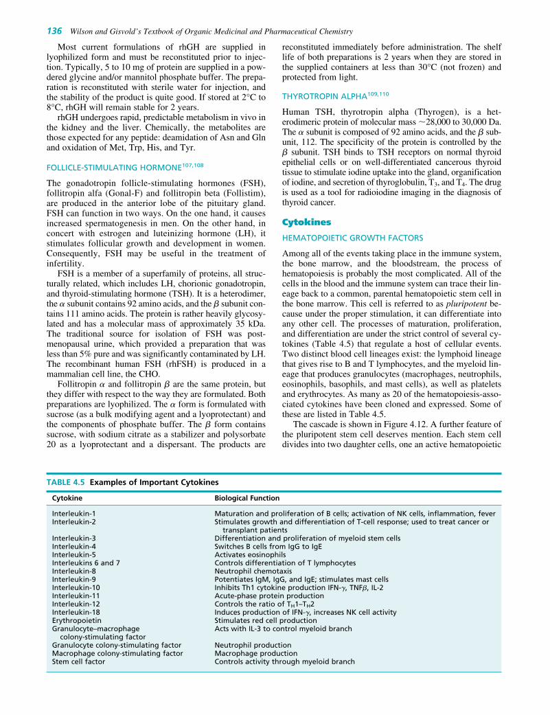
Most current formulations of rhGH are supplied inlyophilized form and must be reconstituted prior to injec-tion. Typically, 5 to 10 mg of protein are supplied in a pow-dered glycine and/or mannitol phosphate buffer. The prepa-ration is reconstituted with sterile water for injection, andthe stability of the product is quite good. If stored at 2°C to8°C, rhGH will remain stable for 2 years.
rhGH undergoes rapid, predictable metabolism in vivo inthe kidney and the liver. Chemically, the metabolites arethose expected for any peptide: deamidation of Asn and Glnand oxidation of Met, Trp, His, and Tyr.
FOLLICLE-STIMULATING HORMONE107,108
The gonadotropin follicle-stimulating hormones (FSH),follitropin alfa (Gonal-F) and follitropin beta (Follistim),are produced in the anterior lobe of the pituitary gland.FSH can function in two ways. On the one hand, it causesincreased spermatogenesis in men. On the other hand, inconcert with estrogen and luteinizing hormone (LH), itstimulates follicular growth and development in women.Consequently, FSH may be useful in the treatment ofinfertility.
FSH is a member of a superfamily of proteins, all struc-turally related, which includes LH, chorionic gonadotropin,and thyroid-stimulating hormone (TSH). It is a heterodimer,the � subunit contains 92 amino acids, and the � subunit con-tains 111 amino acids. The protein is rather heavily glycosy-lated and has a molecular mass of approximately 35 kDa.The traditional source for isolation of FSH was post-menopausal urine, which provided a preparation that wasless than 5% pure and was significantly contaminated by LH.The recombinant human FSH (rhFSH) is produced in amammalian cell line, the CHO.
Follitropin � and follitropin � are the same protein, butthey differ with respect to the way they are formulated. Bothpreparations are lyophilized. The � form is formulated withsucrose (as a bulk modifying agent and a lyoprotectant) andthe components of phosphate buffer. The � form containssucrose, with sodium citrate as a stabilizer and polysorbate20 as a lyoprotectant and a dispersant. The products are
reconstituted immediately before administration. The shelflife of both preparations is 2 years when they are stored inthe supplied containers at less than 30°C (not frozen) andprotected from light.
THYROTROPIN ALPHA109,110
Human TSH, thyrotropin alpha (Thyrogen), is a het-erodimeric protein of molecular mass �28,000 to 30,000 Da.The � subunit is composed of 92 amino acids, and the � sub-unit, 112. The specificity of the protein is controlled by the� subunit. TSH binds to TSH receptors on normal thyroidepithelial cells or on well-differentiated cancerous thyroidtissue to stimulate iodine uptake into the gland, organificationof iodine, and secretion of thyroglobulin, T3, and T4. The drugis used as a tool for radioiodine imaging in the diagnosis ofthyroid cancer.
Cytokines
HEMATOPOIETIC GROWTH FACTORS
Among all of the events taking place in the immune system,the bone marrow, and the bloodstream, the process ofhematopoiesis is probably the most complicated. All of thecells in the blood and the immune system can trace their lin-eage back to a common, parental hematopoietic stem cell inthe bone marrow. This cell is referred to as pluripotent be-cause under the proper stimulation, it can differentiate intoany other cell. The processes of maturation, proliferation,and differentiation are under the strict control of several cy-tokines (Table 4.5) that regulate a host of cellular events.Two distinct blood cell lineages exist: the lymphoid lineagethat gives rise to B and T lymphocytes, and the myeloid lin-eage that produces granulocytes (macrophages, neutrophils,eosinophils, basophils, and mast cells), as well as plateletsand erythrocytes. As many as 20 of the hematopoiesis-asso-ciated cytokines have been cloned and expressed. Some ofthese are listed in Table 4.5.
The cascade is shown in Figure 4.12. A further feature ofthe pluripotent stem cell deserves mention. Each stem celldivides into two daughter cells, one an active hematopoietic
136 Wilson and Gisvold’s Textbook of Organic Medicinal and Pharmaceutical Chemistry
TABLE 4.5 Examples of Important Cytokines
Cytokine Biological Function
Interleukin-1 Maturation and proliferation of B cells; activation of NK cells, inflammation, feverInterleukin-2 Stimulates growth and differentiation of T-cell response; used to treat cancer or
transplant patientsInterleukin-3 Differentiation and proliferation of myeloid stem cellsInterleukin-4 Switches B cells from IgG to IgEInterleukin-5 Activates eosinophilsInterleukins 6 and 7 Controls differentiation of T lymphocytesInterleukin-8 Neutrophil chemotaxisInterleukin-9 Potentiates IgM, IgG, and IgE; stimulates mast cellsInterleukin-10 Inhibits Th1 cytokine production IFN-�, TNF�, IL-2Interleukin-11 Acute-phase protein productionInterleukin-12 Controls the ratio of TH1–TH2Interleukin-18 Induces production of IFN-�, increases NK cell activityErythropoietin Stimulates red cell productionGranulocyte–macrophage Acts with IL-3 to control myeloid branch
colony-stimulating factorGranulocyte colony-stimulating factor Neutrophil productionMacrophage colony-stimulating factor Macrophage productionStem cell factor Controls activity through myeloid branch
0119-0155_17865_Ch04.qxd 12/4/09 4:05 AM Page 136

progenitor and one quiescent. The active precursor maturesto give hematopoietic progenitors and then circulating bloodcells. The quiescent stem cells rejoin the stem cell pool.Hence, the number of parental cells is always the same. Thisprocess is termed self-renewal.
PRODUCTS
Erythropoietin Alfa111–113
Erythropoietin alfa, recombinant Epoetin Alfa, Epogen,Procrit, is a glycoprotein that stimulates red blood cellproduction. It is produced in the kidney, and it activates theproliferation and differentiation of specially committed ery-throid progenitors in the bone marrow. Epoetin alfa (Epogen)is a 165-amino acid glycoprotein that is manufactured inmammalian cells by rDNA technology. The protein is heav-
ily glycosylated and has a molecular mass of approximately30,400 Da. Erythropoietin is composed of four antiparallel�-helices. The rDNA protein has the same amino acid se-quence as natural erythropoietin.
Epoetin is indicated to treat anemia of chronic renal fail-ure patients, anemia in zidovudine-treated HIV-infectedpatients, and in cancer patients taking chemotherapy. Theresults in these cases have been dramatic; most patients re-spond with a clinically significant increase in hematocrit.
Filgrastim114,115
Filgrastim, G-CSF, Neupogen, stimulates the proliferation ofgranulocytes (especially neutrophils) by mobilizinghematopoietic stem cells in the bone marrow. EndogenousG-CSF is a glycoprotein produced by monocytes, fibro-blasts, and endothelial cells. G-CSF is a protein of 174 amino
Chapter 4 Biotechnology and Drug Discovery 137
Figure 4.12 Cytokine-mediated cascade leading to different blood cell types. (EPO,erythropoietin; G-CSF, granulocyte colony-stimulating factor; GM-CSF,granulocyte–macrophage colony-stimulating factor; IL-X, interleukins; M-CSF,macrophage colony-stimulating factor; SCF, stem cell factor; TPO, thrombopoietin.)
0119-0155_17865_Ch04.qxd 12/4/09 4:05 AM Page 137

acids, with a molecular mass of approximately 18,800 Da.The native protein is glycosylated.
Filgrastim selectively stimulates proliferation and differ-entiation of neutrophil precursors in the bone marrow. Thisleads to the release of mature neutrophils into the circulationfrom the bone marrow. Filgrastim also affects mature neu-trophils by enhancing phagocytic activity, priming the cel-lular metabolic pathways associated with the respiratoryburst, enhancing antibody-dependent killing, and increasingthe expression of some functions associated with cell sur-face antigens.
In patients receiving chemotherapy with drugs such ascyclophosphamide, doxorubicin, and etoposide, the inci-dence of neutropenia accompanied by fever is rather high.Administration of G-CSF reduces the time of neutrophilrecovery and duration of fever in adults with acute mye-logenous leukemia. The number of infections, days thatantibiotics are required, and duration of hospitalization arealso reduced.
Filgrastim116 is identical with G-CSF in its amino acidsequence, except that it contains an N-terminal methioninethat is necessary for expression of the vector in E. coli. Theprotein is not glycosylated. Filgrastim is supplied in a 0.01M sodium acetate buffer containing 5% sorbitol and 0.004%polysorbate 80. It should be stored at 2°C to 8°C withoutfreezing. Under these conditions, the shelf life is 24 months.Avoid shaking when reconstituting; although the foamingwill not harm the product, it may alter the amount of drugthat is drawn into a syringe.
Sargramostim117,118
Sargramostim, granulocyte–macrophage colony-stimulatingfactor (GM-CSF), Leukine, is a glycoprotein of 127 aminoacids, consisting of three molecular subunits of 19,500,16,800, and 15,500 Da. The endogenous form of GM-CSFis produced by T lymphocytes, endothelial fibroblasts, andmacrophages. Recombinant GM-CSF, produced in S. cere-visiae, differs from native human GM-CSF only by substi-tution of a leucine for an arginine at position 23. This sub-stitution facilitates expression of the gene in the yeast. Thesite of glycosylation in the recombinant molecule may pos-sibly differ from that of the native protein.
Sargramostim binds to specific receptors on target cellsand induces proliferation, activation, and maturation.Administration to patients causes a dose-related increase inthe peripheral white blood cell count. Unlike G-CSF, GM-CSF is a multilineage hematopoietic growth factor that in-duces partially committed progenitor cells to proliferate anddifferentiate along the granulocyte and the macrophage path-ways. It also enhances the function of mature granulocytesand macrophages/monocytes. GM-CSF increases the chemo-tactic, antifungal, and antiparasitic activities of granulocytesand monocytes. It also increases the cytotoxicity of mono-cytes toward neoplastic cell lines and activates polymor-phonuclear leukocytes to inhibit the growth of tumor cells.
Sargramostim is used to reconstitute the myeloid tissueafter autologous bone marrow transplant and followingchemotherapy in acute myelogenous leukemia. The prepara-tion decreases the incidence of infection, decreases the num-ber of days that antibiotics are required, and decreases theduration of hospital stays.
Sargramostim is supplied as a solution or powder (for so-lution). Both forms should be stored at 2°C to 8°C without
freezing. The liquid and powder have expiration dates of 24months. The reconstituted powder and the aqueous solutionshould not be shaken.
Becaplermin119
Becaplermin, Regranex Gel, an endogenous polypeptidethat is released from cells that are involved in the healingprocess, is a recombinant human platelet–derived growthfactor (rhPDGF-BB). The “BB” signifies that becaplerminis the homodimer of the B chain. Becaplermin is producedby a recombinant strain of S. cerevisiae containing the genefor the B chain of PDGF. The protein has a molecular massof approximately 25 kDa and is a homodimer composed oftwo identical polypeptide chains that are linked by disulfidebonds. It is a growth factor that activates cell proliferation,differentiation, and function, and it is released from cells in-volved in the healing process.
Becaplermin is formulated as a gel recommended fortopical use in the treatment of ulcerations of the skin sec-ondary to diabetes.
Interferons
The interferons are a family of small proteins or glycopro-teins of molecular masses ranging from 15,000 to 25,000Da and 145 to 166 amino acids long. Eukaryotic cells se-crete interferons in response to viral infection. Their mech-anism of action is bimodal. The immediate effect is the re-cruitment of natural killer (NK) cells to kill the host cellharboring the virus (Fig. 4.13). Interferons then induce astate of viral resistance in cells in the immediate vicinity,preventing spread of the virus. Additionally, interferons in-duce a cascade of antiviral proteins from the target cell, oneof which is 2�,5�-oligoadenylate synthetase. This enzymecatalyzes the conversion of ATP into 2�,5�-oligoadenylate,which activates ribonuclease R, hydrolyzing viral RNA.Interferons can be defined as cytokines that mediate antivi-ral, antiproliferative, and immunomodulatory activities.
Three classes of interferon (IFN) have been character-ized: � (alpha), � (beta), and � (gamma) (see Table 4.6). �-Interferons are glycoproteins derived from human leuko-cytes. �-Interferons are glycoproteins derived from fibro-blasts and macrophages. They share a receptor with �-interferons. �-Interferons are glycoproteins derived fromhuman T lymphocytes and NK cells. These interferons areacid labile and used to be called “type 2 interferon.” The re-ceptor for IFN-� is smaller than that for IFN-� and IFN-�,90 to 95 kDa versus 95 to 110 kDa, respectively. The threeclasses are not homogeneous, and each may contain severaldifferent molecular species. For example, at least 18 genet-ically and molecularly distinct human �-interferons have
138 Wilson and Gisvold’s Textbook of Organic Medicinal and Pharmaceutical Chemistry
Figure 4.13 Antiviral mechanism of action of theinterferons.
0119-0155_17865_Ch04.qxd 12/4/09 4:05 AM Page 138

been identified, each differing in the amino acid substitu-tion at positions 23 and 34. Interferons alfa-2a, alfa-2b, andalfa-2c have been purified and are either in clinical use orin development. A listing of commercially available �-interferons is given in Table 4.7.
As a class, the interferons possess some common side ef-fects. These are flulike symptoms, headache, fever, muscleaches, back pain, chills, nausea and vomiting, and diarrhea.At the injection site, pain, edema, hemorrhage, and inflam-mation are common. Dizziness is also commonly reported.
For the pharmacist, when predicting drug interactionswith the interferons, cytochrome P450 metabolism shouldalways be a key consideration. Most of the interferons in-hibit cytochrome P450, causing drugs that are metabolizedby this route to reach higher-than-normal and, possibly,toxic concentrations in the blood and tissues.
PRODUCTS: �-INTERFERONS
Interferon Alfa-2a (Recombinant)120
Interferon alfa-2a (recombinant), Roferon A, is expressedin an E. coli system and purified by using high-affinitymouse MAb chromatography. The protein consists of 165amino acids with a molecular mass of approximately19,000 Da and contains lysine at position 23 and histidineat position 34.
Interferon alfa-2a is used in the treatment of hairy cellleukemia and AIDS-related Kaposi sarcoma in selected pa-tients older than 18 years of age. It is also used to treatchronic hepatitis C, and in patients with this disease, inter-feron alfa-2a can normalize serum alanine aminotransferase(ALT) levels, improve liver histology, and decrease viralload. The drug has a direct antiproliferative activity againsttumor cells. Modulation of the host immune response proba-bly plays a role in the antitumor activity of interferon alfa-2a.
The interferon is supplied as a solution or as a powder forsolution. The solution contains 0.72% NaCl. The powdercontains 0.9% NaCl, 0.17% human serum albumin, and0.33% phenol. The interferon vials, if properly stored at 2°Cto 8°C without freezing, expire in 30 months. Prefilled sy-ringes expire in 24 months. The solutions should not beshaken because the albumin will cause frothing.
Pegylated Interferon Alfa-2a121
Pegylated interferon alfa-2a, Pegasys, is a covalent conjugateof recombinant interferon alfa-2a (approximate molecularmass, 20 kDa) with a singly branched bis-monomethoxy PEGchain (approximate molecular mass, 40 kDa). The PEG moi-ety is linked at a single site to the interferon alfa moiety by astable amide bond to lysine. Peginterferon alfa-2a has anapproximate molecular mass of 60 kDa. Pegasys providessustained therapeutic serum levels for up to a full week(168 hours). The drug is approved for the treatment of adultswith chronic hepatitis C who have compensated liver diseaseand who have not been previously treated with interferon alfa.Efficacy has also been demonstrated in patients with compen-sated cirrhosis.
Interferon Alfa-2b (Recombinant)122
Interferon alfa-2b, Intron A, a water-soluble protein of165 amino acids and an approximate molecular mass of19,200 Da, is expressed from a recombinant strain of E. coli.This interferon molecule possesses an arginine at position23 and a histidine at position 34.
Interferon alfa-2b is a broad-spectrum agent. It is indicatedfor hairy cell leukemia, condyloma acuminata (genital or
Chapter 4 Biotechnology and Drug Discovery 139
TABLE 4.6 Interferons Used Therapeutically
Interferon Endogenous AvailableType Source Drug Products
Alpha Leukocytes Interferon alfa-2aInterferon alfa-2bInterferon alfa-2c
Beta Fibroblasts, Beta-1amacrophages Beta-1b
Gamma T Lymphocytes, Gamma-1bnatural killer cells
TABLE 4.7 Summary of the �-Interferons
Interferon Interferon Interferon Interferon InterferonAlfa-2a Alfa-2b Alfa-n1 Alfa-n3 Alfacon-1
Trade name Roferon A Intron A Wellferon Alferon N InfergenDosage form Solution, powder Solution, powder Solution Solution SolutionSolvent Sodium chloride, Buffered saline Buffered saline Buffered saline Phosphate-
excipients buffered saline
Indications Hairy cell leukemia, Hairy cell leukemia, Chronic hepatitis C Condylomata Chronic hepatitis CAIDS-related AIDS-related acuminataKaposi sarcoma, Kaposi sarcoma, chronic hepatitis C condylomata
acuminata, chronichepatitis B, chronichepatitis C
Routesa SC, IM, IV, infusion SC, IM, IV, infusion SC or IM Intralesional SCor intralesional or intralesional
Source Escherichia coli Human Human leukocytes E. colilymphoblastoid cell line
aSC, subcutaneously; IM, intramuscularly; IV, intravenously.
0119-0155_17865_Ch04.qxd 12/4/09 4:05 AM Page 139

venereal warts), AIDS-related Kaposi sarcoma, and chronichepatitis B and C infections.
Intron A can be administered by the SC, IM, or IV routes,by infusion or by intralesional routes. The dose is 1 to 35 mil-lion IU/day, depending on the application. The drug is sup-plied as a solution or as a powder for solution, and both formscontain albumin, glycine, and sodium phosphate buffer.Hence, they should not be shaken. Vials of solution should bestored at 2°C to 8°C without freezing. The powder is stablefor 18 months at room temperature or 7 days at 45°C.
Interferon Alfa-n1123
Interferon alfa-n1, Wellferon, is a mixture of �-interferonsisolated from a human lymphoblastoid cell line after induc-tion with mouse parainfluenza virus type 1 (Sendai strain).Each of the subtypes of IFN-� in this product consists of165 or 166 amino acids with an average molecular mass of26,000 Da. The product is a mixture of each of the nine pre-dominant subtypes of IFN-�.
Interferon alfa-n1 is indicated to treat chronic hepatitis Cin patients 18 years of age or older who have no decompen-sated liver disease. The exact mechanism of action for inter-feron alfa-n1 in the treatment of this disease has not beenelucidated.
This drug may be administered SC or IM, with a usualdose of 3 million IU 3 times per week. Interferon alfa-n1 issupplied as a solution containing tromethamine and bufferedsaline with human albumin as a stabilizer. Hence, the solutionshould not be shaken. The solution should be stored at 2°C to8°C without freezing, and should be discarded if freezingoccurs. Properly stored solution expires in 24 months.
Interferon Alfa-n3124
Interferon alfa-n3, Alferon N, is an �-interferon expressedfrom human leukocytes that are antigen-stimulated withavian Sendai virus. The Sendai virus is propagated inchicken eggs. The protein consists of at least 14 molecularsubtypes. The average chain length is 166 amino acids, andthe molecular mass range is 16,000 to 27,000 Da. The poly-disperse interferon alfa-n3 is extremely pure, because it isprocessed by affinity chromatography over a bed of mouseMAbs specifically raised for the protein.
Interferon alfa-n3 is indicated for intralesional treatment ofrefractory or recurrent condyloma acuminata (genital warts)in patients 18 years of age or older. These warts are associatedwith human papillomavirus (HPV). Interferon alfa-n3 is espe-cially useful in patients who have not responded well to othermodalities (podophyllin resin, surgery, laser, or cryotherapy).Interferon alfa-n3 is also being investigated for the treatmentof non-Hodgkin lymphoma, herpes simplex virus, rhinovirus,vaccinia, and varicella zoster. A usual dose in condylomaacuminata is 250,000 IU/wart, injected with a 30-gauge nee-dle around the base of the lesion.
Interferon alfa-n3 is contraindicated in persons sensitive tomouse immunoglobulin G (IgG), egg protein, and neomycin.
Interferon alfa-n3 is supplied as a solution with the pro-tein in phosphate-buffered saline with phenol as a preserva-tive. The solution should be stored at 2°C to 8°C withoutfreezing. Properly stored solution expires at 18 months.
Interferon Alfacon-1 (Recombinant)Interferon alfacon-1 (recombinant), Infergen,125 is a“consensus” interferon that shares structural elements ofIFN-� and several subtypes. The range of activity is about
the same as the other alpha species, but the specific activ-ity is greater.
The 166-amino acid sequence of alfacon-1 is synthetic. Itwas developed by comparing several natural IFN-� sub-types and assigning the most common amino acid to eachvariable position. Additionally, four amino acid changeswere made to facilitate synthesis. The DNA sequence is alsoconstructed by chemical synthesis. Interferon alfacon-1 dif-fers from interferon alfa-n2 at 20/166 amino acids, yielding88% homology. The protein has a molecular mass of ap-proximately 19,400 Da.
Interferon alfacon-1 is used in the treatment of chronichepatitis C virus (HCV) infection in patients 18 years of ageor older with compensated liver disease and who have anti-HCV serum antibodies or HCV RNA.
The drug is administered by the SC route in a dose of 9 �g3 times per week. Interferon alfacon-1 is supplied as a solu-tion in phosphate-buffered saline. It should be stored at 2°Cto 8°C without freezing. Avoid shaking the solution.
PRODUCTS: �-INTERFERONS
A listing of two commercially available IFN-�s is given inTable 4.8.
Interferon Beta-1a (Recombinant)126
Interferon beta-1a (recombinant), Avonex, is a glycoproteinwith 166 amino acids. It has a molecular mass of approxi-mately 22,000 Da. The site of glycosylation is at the as-paragine residue at position 80. Interferon beta-1a possesses acysteine residue at position 17, as does the native molecule.Natural IFN-� and interferon beta-1a are glycosylated, witheach containing a single carbohydrate moiety. The overallcomplex has 89% protein and 11% carbohydrate by weight.Recombinant interferon beta-1a is expressed in CHO cellscontaining the recombinant gene for human IFN-� and isequivalent to the human form secreted by fibroblasts.
Interferon beta-1a is indicated for the treatment of relaps-ing forms of MS. Patients treated with interferon beta-1ademonstrate a slower progression to disability and a less no-ticeable breakdown of the blood-brain barrier as observed ingadolinium-enhanced magnetic resonance imaging (MRI).
140 Wilson and Gisvold’s Textbook of Organic Medicinal and Pharmaceutical Chemistry
TABLE 4.8 �-Interferons
Interferon Beta-1a Interferon Beta-1b(Avonex) (Betaseron)
Recombinant CHO cells Escherichia colisystem
Type Asn80 complex Not glycosylatedcarbohydrate
Concentration 30 �g or 250 �g or 8 million6 million IU/mL IU/mL
Supplied form Powder for Powder for reconstitution reconstitution
Diluent Sterile water–no NaCl 0.54% without preservatives preservatives
Storage 2°C–8°C; do not 2°C–8°C; do not freeze freeze
Dosage 30 �g once a week 250 �g every other day
Route Intramuscular SubcutaneousNotable side Injection site Injection site
effects reactions, 3%; reactions, 85%;no necrosis necrosis, 5%
0119-0155_17865_Ch04.qxd 12/4/09 4:05 AM Page 140

Although the exact mechanism of action of interferonbeta-1a in MS has not been elucidated, it is known that thedrug exerts its biological effects by binding to specific re-ceptors on the surface of human cells. This binding initiatesa cascade of intracellular events that lead to the expressionof interferon-induced gene products. These include 2�,5�-oligoadenylate synthetase and �2 microglobulin. Theseproducts have been measured in the serum and in cellularfractions of blood collected from patients treated with inter-feron beta-1a. The functionally specific interferon-inducedproteins have not been defined for MS.
Adverse effects include flulike syndrome at the start oftherapy that decreases in severity as treatment progresses.Interferon beta-1a is a potential abortifacient and an in-hibitor of cytochrome P450.
The dosage form is a powder for solution that is reconsti-tuted in sterile water. Excipients are human albumin,sodium chloride, and phosphate buffer. The solution can bestored at 2°C to 8°C and should be discarded if it freezes.The lyophilized powder expires in 15 months. After recon-stitution, the solution should be used within 6 hours. The so-lution should not be shaken because of the albumin content.
Interferon Beta-1b (Recombinant)127
Interferon beta-1b, Betaseron, is a protein that is expressedin a recombinant E. coli. It is equivalent in type to the inter-feron that is expressed by human fibroblasts. Interferonbeta-1b possesses 165 amino acids and has an approximatemolecular mass of 18.5 kDa. The native form has 166 aminoacids and weighs 23 kDa. Interferon beta-1b contains a ser-ine residue at position 17 rather than the cysteine in nativeIFN-� and does not contain the complex carbohydrate sidechains found in the natural molecule. In addition to its an-tiviral activity, interferon beta-1b possesses immunomodu-lating activity.
Interferon beta-1b is administered SC to decrease the fre-quency of clinical exacerbation in ambulatory patients withrelapsing–remitting multiple sclerosis (RRMS). RRMS ischaracterized by unpredictable attacks resulting in neurolog-ical deficits, separated by variable periods of remission.
Although it is not possible to delineate the mechanismsby which interferon beta-1b exerts its activity in MS, it isknown that the interferon binds to specific receptors on cellsurfaces and induces the expression of several interferon-induced gene products, such as 2�,5�-oligoadenylate syn-thetase and protein kinase. Additionally, interferon beta-1bblocks the synthesis of INF-�, which is believed to be in-volved in MS attacks.
Interferon beta-1b is supplied as powder for solution withalbumin and/or dextrose as excipients. It should be stored at2°C to 8°C without freezing. After reconstitution, the solu-tion can be stored in the refrigerator for 3 hours. The solu-tion should not be shaken.
A major difference between interferon beta-1a and beta-1b is that beta-1b causes more hemorrhage and necrosis atthe injection site than does interferon beta-1a.
PRODUCTS: �-INTERFERON
Interferon Gamma-1b (Recombinant)128
Interferon gamma-1b, Actimmune, is a recombinant proteinexpressed in E. coli. IFN-� is the cytokine that is secreted byhuman T lymphocytes and NK cells. It is a single-chain gly-coprotein composed of 140 amino acids. The crystal structure
of the protein reveals several helical segments arranged toapproximate a toric shape.
Interferon gamma-1b is indicated for reducing the fre-quency and severity of serious infections associated withchronic granulomatous disease, an inherited disorder char-acterized by deficient phagocyte oxidase activity. In this dis-ease, macrophages try to respond to invading organisms butlack the key oxidative enzymes to dispose of them. To com-pensate, additional macrophages are recruited into the in-fected region and form a granulomatous structure around thesite. IFN-� can stimulate the oxidative burst in macrophagesand may reverse the situation.
Interferon gamma-1b is supplied as a solution in sterilewater for injection. The solution must be stored at 2°C to8°C, without freezing. The product cannot tolerate morethan 12 hours at room temperature.
THE INTERLEUKINS
Aldesleukin129
Aldesleukin, T-cell growth factor, thymocyte-stimulatingfactor, Proleukin, is recombinant IL-2 expressed in an engi-neered strain of E. coli containing an analog of the humanIL-2 gene. The recombinant product is a highly purified pro-tein of 133 amino acids with an approximate molecular massof 15,300 Da. Unlike native IL-2, aldesleukin is not glyco-sylated, has no N-terminal alanine, and has serine substitutedfor Cys at site 125. Aldesleukin exists in solution as biolog-ically active, noncovalently bound microaggregates with anaverage size of 27 IL-2 molecules. This contrasts with tradi-tional solution aggregates of proteins, which often form irre-versibly bound structures that are biologically inactive.
Aldesleukin enhances lymphocyte mitogenesis and stimu-lates long-term growth of human IL-2-dependent cell lines.IL-2 also enhances the cytotoxicity of lymphocytes. Inductionof NK cell and lymphocyte-activated killer (LAK) cell activ-ity occurs, as does induction of production. In mouse andhuman tumor cell lines, aldesleukin activates cellular immu-nity in patients with profound lymphocytosis, eosinophilia,and thrombocytopenia. Aldesleukin also activates the pro-duction of cytokines, including tumor necrosis factor (TNF),IL-1, and IFN-�. In vivo experiments in mouse tumor modelshave shown inhibition of tumor growth. The mechanism ofthe antitumor effect of aldesleukin is unknown.
Aldesleukin is indicated for the treatment of metastaticrenal cell carcinoma in adults. It is also indicated for thetreatment of metastatic melanoma in adults. Research isunder way on the use of aldesleukin for the treatment of var-ious cancers (including head and neck cancers), treatment ofacute myelogenous leukemia, and adjunct therapy in thetreatment of Kaposi sarcoma. Renal and hepatic function istypically impaired during therapy with aldesleukin, so inter-action with other drugs that undergo elimination by these or-gans is possible.
Aldesleukin is supplied as a powder for solution. After re-constitution, the solution should not be shaken. The prepara-tion is solubilized with sodium dodecyl sulfate (SDS) in aphosphate buffer. Aldesleukin should be stored as nonrecon-stituted powder at 2°C to 8°C and never frozen. Reconstitutedvials can be frozen and thawed once in 7 days without loss ofactivity. It expires over a period of 18 months.
Chapter 4 Biotechnology and Drug Discovery 141
0119-0155_17865_Ch04.qxd 12/4/09 4:05 AM Page 141

Denileukin Diftitox (Recombinant)130
Denileukin diftitox, recombinant, Ontak, is an example of adrug that acts like a Trojan horse. One part of the molecule isinvolved in recognition and binds selectively with the dis-eased cell, and a highly toxic second part of the molecule ef-fects a kill. Denileukin diftitox is a fusion protein expressedby a recombinant strain of E. coli. It is an rDNA-derivedcytotoxic protein composed of the amino acid sequences fordiphtheria toxin fragments A and B (Met1-Thr387)-His, fol-lowed by the sequences for IL-2 (Ala1-Thr133). The fusionprotein has a molecular mass of 58,000 Da. We can think ofthis large protein as a molecule of diphtheria toxin in whichthe receptor-binding domain has been replaced by IL-2sequences, thereby changing its binding specificity. Cells thatexpress the high-affinity (�, �, �) IL-2 receptor bind the pro-tein tightly. The IL-2 component is used as a director to bringthe cytotoxic species in contact with tumor cells. The diphthe-ria toxin inhibits cellular protein synthesis and the cells die.Malignant cells in certain leukemias and lymphomas, in-cluding cutaneous T-cell lymphoma, express the high-affinityIL-2 receptor on their cell surfaces. It is these cells thatdenileukin diftitox targets.
Denileukin diftitox is indicated for the treatment of persis-tent or recurrent cutaneous T-cell lymphoma whose malig-nant cells express the CD25 component of the IL-2 receptor.
Denileukin diftitox is supplied as a frozen solution inwater for injection. It should be stored at �10°C or colder.It is suggested that the vials be thawed in a refrigerator at2°C to 8°C for less than 24 hours or at room temperature for1 to 2 hours. Prepared solutions should be used within6 hours. The drug is administered by IV infusion from a bagor through a syringe pump.
Oprelvekin (Recombinant)131,132
Oprelvekin, Neumega, is recombinant human IL-11 that isexpressed in a recombinant strain of E. coli as a thioredoxinand/or rhIL-11 fusion protein. The fusion protein is cleavedand purified to obtain the rhIL-11 protein. The protein is177 amino acids in length and has a mass of approximately19,000 Da. Oprelvekin differs from the natural 178-aminoacid IL-11 by lacking an N-terminal proline. This alterationhas not resulted in differences in bioactivity either in vitroor in vivo.
IL-11 is a thrombopoietic growth factor. It directly stim-ulates the proliferation of hematopoietic stem cells as wellas megakaryocyte progenitor cells. This process inducesmegakaryocyte maturation and increased production ofplatelets. The primary hematopoietic activity of oprelvekinis stimulation of megakaryocytopoiesis and thrombopoiesis.Primary osteoblasts and mature osteoclasts express mRNAsfor both IL-11 and its receptor, IL-11R alpha. Hence, bothbone-forming and bone-resorbing cells are possible targetsfor IL-11.
Oprelvekin is indicated for the prevention of severethrombocytopenia. It reduces the need for platelet transfu-sions after myelosuppressive chemotherapy in patients withnonmyeloid malignancies who are at high risk for severethrombocytopenia. Efficacy has been demonstrated in per-sons who have experienced severe thrombocytopenia fol-lowing a previous chemotherapy cycle.
Oprelvekin causes many adverse reactions. Among theseare edema, neutropenic fever, headache, nausea and/or
vomiting, dyspnea, and tachycardia. Patients must be moni-tored closely.
Oprelvekin is supplied as a lyophilized powder for recon-stitution. Excipients include glycine and phosphate buffercomponents. The powder has a shelf life of 24 months. Itshould be stored at 2°C to 8°C. If it is frozen, thaw it beforereconstitution.
Tumor Necrosis Factor(Recombinant)133–135
The TNFs (Etanercept, Enbrel) are members of a family ofcytokines that are produced primarily in the innate immunesystem by activated mononuclear phagocytes. Along withIL-1, TNF is typically the first cytokine to be produced uponinfection, and its reactions can be both positive and nega-tive. On the one hand, TNF can cause cytotoxicity and in-flammation, and on the other hand, it serves as a signal tothe adaptive immune response. The TNFs are all endoge-nous pyrogens, and they cause chills, fever, and flulikesymptoms. There are two forms of TNF: TNF� (cachectin)and TNF� (lymphotoxin). Both bind to the same receptorand cause similar effects.
Etanercept is a dimeric fusion protein consisting of the ex-tracellular ligand-binding portion of the human 75-kDa (p75)TNF receptor (TNFR) linked to the Fc portion of human iso-type IgG1. The Fc component of etanercept contains the CH2
domain, the CH3 domain, and the hinge region, but not theCH1 domain of IgG1. These regions are responsible for thebiological effects of immunoglobulins. Etanercept is pro-duced in recombinant CHO cultures. It consists of a peptidechain of 934 amino acids and has a molecular mass of ap-proximately 150 kDa. It binds specifically to TNF and blocksits interaction with cell surface TNFRs. Each etanerceptmolecule binds specifically to two TNF molecules in the syn-ovial fluid of rheumatoid arthritis patients. It is equally effi-cacious at blocking TNF� and TNF�. The drug is indicatedfor reducing signs and symptoms and inhibiting the pro-gression of structural damage in patients with moderately toseverely active rheumatoid arthritis. Etanercept is also indi-cated for reducing signs and symptoms of moderately toseverely active polyarticular-course juvenile rheumatoidarthritis in patients 4 years of age and older who have had aninadequate response to one or more disease-modifying an-tirheumatic drugs (DMARDS). Etanercept is also indicatedfor reducing signs and symptoms of active arthritis in pa-tients with psoriatic arthritis.
ENZYMES
Blood-Clotting Factors
The blood clotting system of the human body is typically ina carefully balanced homeostatic state. If damage occurs to ablood vessel wall, a clot will form to wall off the damage sothat the process of regeneration can begin. Normally thisprocess is highly localized to the damaged region, so that thehemostatic response does not cause thrombi to migrate todistant sites or persist longer than it is needed. Lysis of bloodclots occurs through the conversion of plasminogen to plas-min, which causes fibrinolysis, converting insoluble fibrin tosoluble fibrinopeptides. The plasminogen–plasmin conver-sion is catalyzed by several blood and tissue activators,
142 Wilson and Gisvold’s Textbook of Organic Medicinal and Pharmaceutical Chemistry
0119-0155_17865_Ch04.qxd 12/4/09 4:05 AM Page 142

among them urokinase, kallikrein, plasminogen activators,and some undefined inhibitors. More specifically, the con-version of plasminogen to plasmin is catalyzed by two ex-tremely specific serine proteases: a urokinase plasminogenactivator (uPA) and a tissue plasminogen activator (tPA).This section focuses on tPA.
Human tPA is a serine protease that is synthesized in thevascular endothelial cells. It is a single-chain peptide com-posed of 527 amino acids and has a molecular mass ofapproximately 64,000 Da. About 7% of the mass of the mole-cule consists of carbohydrate. The molecule contains 35 Cysresidues. These are fully paired, giving the tPA molecule17 disulfide bonds. There are four N-linked glycosylation sitesrecognized by consensus sequences Asn-X-Ser/Thr at residues117, 184, 218, and 448. It is suspected that Thr61 bears an O-fucose residue. There are two forms of tPA that differ by thepresence or absence of a carbohydrate group at Asp184. Type ItPA is glycosylated at Asn117, Asn184, and Asn448, whereastype II tPA lacks a glycosyl group at Asn184. Asn218 is typi-cally unsubstituted in both forms. Asn117 contains a high-mannose oligosaccharide, whereas Asn substituents 184 and448 are complex carbohydrate substituted. During the processof fibrinolysis the single-chain protein is cleaved betweenArg275 and Ile276 by plasmin to yield two-chain tPA. Two-chain tPA consists of a heavy chain (the A chain, derived fromthe N-terminus) and a light chain (B chain), linked by a singledisulfide bond between Cys264 and Cys395. The A chain bearssome unique structural features: the finger region (residues6–36), the growth factor region (approximate residues 44–80),and two kringle domains. These domains are disulfide-closedloops, mostly � sheet in structure. The finger and kringle 2 areresponsible for tPA binding to fibrin and for the activation ofplasminogen. The function of kringle 1 is not known. The Bchain contains the serine protease domain that contains theHis-Asp-Ser unit that cleaves plasminogen.
TISSUE PLASMINOGEN ACTIVATOR,RECOMBINANT136,137
Recombinant tPA (rtPA), alteplase (Activase), is identicalwith endogenous tPA. rtPA lacks a glycosyl residue atAsn184. At one time, rtPA was produced in two-chain formin CHO cultures. Now, large-scale cultures of recombinanthuman melanoma cells in fermenters are used to produce aproduct that is about 80% single-chain rtPA.
Alteplase is used to improve ventricular function follow-ing an acute myocardial infarction, including reducing the in-cidence of congestive heart failure and decreasing mortality.The drug is also used to treat acute ischemic stroke aftercomputed tomography (CT) or other diagnostic imaging hasruled out intracranial hemorrhage. rtPA is also used in casesof acute pulmonary thromboembolism and is being investi-gated for unstable angina pectoris.
Alteplase is supplied as powder for injection, and in re-constituted form (normal saline or 5% dextrose in water) isintended for IV infusion only. The solution expires in 8 hoursat room temperature and must be prepared just before use.
RETEPLASE
Reteplase (Retavase) is a deletion mutant variant of tPAthat is produced in recombinant E. coli. The deletions arein domains responsible for half-life, fibrin affinity, andthrombolytic potency. It consists of the kringle-2 domain
and protease domain of tPA but lacks the kringle-1 domainand the growth factor domain. It is considered a third-generation thrombolytic agent and has a mechanism of ac-tion similar to that of alteplase. Reteplase acts directly bycatalyzing the cleavage of plasminogen and initiatingthrombolysis. It has high thrombolytic potency. A compar-ison of alteplase and reteplase is given in Table 4.9.
TENECTEPLASE137
Tenecteplase is a tPA produced by recombinant CHO cells.The molecule is a 527-amino acid glycoprotein developed byintroducing the following modifications to the cDNA con-struct: Thr103 to Asp, Asp117 to Gln, both within the kringle-1domain, and a tetra-alanine substitution at amino acids 296 to299 in the protease domain. The drug is a sterile, lyophilizedpowder recommended for single IV bolus administration afterreconstitution with sterile water. Tenecteplase should be ad-ministered immediately after reconstitution.
FACTOR VIII138
Antihemophilic factor VIII (recombinant), Recombinate,Kogenate, Bioclate, Helixate, is a plasma protein that func-tions in the normal blood-clotting cascade by increasing theVmax for the activation of clotting factor X by factor IXa inthe presence of calcium ions and negatively charged phos-pholipids. Factor VIII is used in the treatment of hemophiliaA. Hemophilia A is a congenital disorder characterized bybleeding. The introduction of factor VIII as a drug has im-proved the quality of life and the life expectancy of individ-uals with this disorder. Unfortunately, it has been necessaryto rely on an unsure source (human plasma) for the factor.Exposure of patients to alloantigens and viruses has been aconcern. Factor VIII derived from a recombinant source willpotentially eliminate many of these problems and provide anessentially unlimited supply of the drug.
Factor VIII is biosynthesized as a single-chain polypep-tide of 2,332 amino acids. The protein is very heavily glyco-sylated. Shortly after biosynthesis, peptide cleavage occursand plasma factor VIII circulates as an 80-kDa light chainassociated with a series of heavy chains of approximately210 kDa in a metal ion-stabilized complex. Factor VIII pos-sesses 25 potential N-linked glycosylation sites and 22 Cysresidues. The 210-kDa heavy chain is further cleaved byproteases to yield a series of proteins of molecular mass 90to 188 kDa. The 90- to 188-kDa protein molecules form ametal ion-stabilized complex with the light chain.
Recombinant factor VIII is produced in two recombinantsystems: in batch culture of transfected CHO cells or in con-tinuous culture of BHK cells. There are four types of recombi-nant factor VIII available. All four are produced by inserting acDNA construct encoding the entire peptide sequence into the
Chapter 4 Biotechnology and Drug Discovery 143
TABLE 4.9 Comparison of the PharmacokineticParameters of Alteplase and Reteplase
PharmacokineticParameter Alteplase Reteplase
Effective t1/2 (minutes) 5 13–16Volume of distribution (L) 8.1 6Plasma clearance (mL /min) 360–620 250–450
0119-0155_17865_Ch04.qxd 12/4/09 4:05 AM Page 143

CHO cell or BHK cell line. The CHO cell product contains aGal�[1→3]Gal unit, whereas the BHK enzyme does not.Recombinant factor VIII is polydisperse, containing multiplepeptide homologues including an 80-kDa protein and variousmodifications of an approximately 90-kDa subunit protein.The product contains no blood products and is free of mi-crobes and pyrogens.
Recombinant factor VIII is indicated for the treatment ofclassical hemophilia (hemophilia A) and for the preventionand treatment of hemorrhagic episodes and perioperativemanagement of patients with hemophilia A. The drug is alsoindicated for the treatment of hemophilia A in persons whopossess inhibitors to factor VIII.
Recombinant factor VIII is supplied in sterile, single-dose vials. The product is stabilized with human albuminand lyophilized. The product must be stored at 2°C to 8°C,without freezing. In some instances, the powder may bestored at room temperature for up to 3 months without lossof biological activity. Shaking of the reconstituted productshould be avoided because of the presence of the albumin.The drug must be administered by IV bolus or drip infusionwithin 3 hours of reconstitution.
Because trace amounts of mouse or hamster protein maycopurify with recombinant factor VIII, one should be cautiouswhen administering the drug to individuals with known hy-persensitivity to plasma-derived antihemophilic factor or withhypersensitivity to biological preparations with trace amountsof mouse or hamster proteins.
CLOTTING FACTOR IX (RECOMBINANT)139,140
When a person is deficient in clotting factor IX (Christmasfactor), hemophilia B results. Hemophilia B affects prima-rily males and accounts for about 15% of all cases of hemo-philia. Treatment involves replacement of factor IX so thatthe blood will clot. Recombinant coagulation factor IX(BeneFIX) is a highly purified protein produced in recombi-nant CHO cells, free of blood products. The product is a gly-coprotein of molecular mass approximately 55,000 Da. Itconsists of 415 amino acids in a single chain. The primaryamino acid sequence of BeneFIX is identical with the Ala148
allelic form of plasma-derived factor IX, and it has struc-tural and functional characteristics similar to those of the en-dogenous protein. The recombinant protein is purified bychromatography, followed by membrane filtration. SDS-polyacrylamide gel electrophoresis shows that the productexists primarily as a single component.
Clotting factor IX, recombinant, is indicated for the con-trol and prevention of hemorrhagic episodes in persons withhemophilia B (Christmas disease), including the control andprevention of bleeding in surgical procedures.
BeneFIX is supplied as a sterile lyophilized powder. Itshould be stored at 2°C to 8°C. The product will toleratestorage at room temperature not above 25°C for 6 months.The drug becomes unstable, following reconstitution, andmust be used within 3 hours.
DROTRECOGIN ALFA141
About 750,000 people are diagnosed with sepsis in the UnitedStates each year, and of these, an estimated 30% will die fromit, despite treatment with IV antibiotics and supportive care.Patients with severe sepsis often experience failures of vari-ous systems in the body, including the circulatory system, the
kidneys, and clotting. Drotrecogin alfa (activated), drotreco-gin alfa (activated; Xigris), is a recombinant form of humanactivated protein C. Activated protein C exerts an antithrom-botic effect by inhibiting factors Va and VIIIa. In vitro dataindicate that activated protein C has indirect profibrinolyticactivity through its ability to inhibit plasminogen activator in-hibitor-1 (PAI-1) and to limit generation of activated throm-bin-activatable fibrinolysis inhibitor. Additionally, in vitrodata indicate that activated protein C may exert an anti-inflammatory effect by inhibiting TNF production by mono-cytes, by blocking leukocyte adhesion to selectins, and bylimiting the thrombin-induced inflammatory responses withinthe microvascular epithelium.
Vials of drotrecogin alfa should be stored at 2°C to 8°Cwithout freezing. The reconstituted solution is stable for14 hours at 25°C.
Anticoagulant
LEPIRUDIN, RECOMBINANT142
Leeches (Hirudo medicinalis) have been used medicinallyfor centuries to treat injuries in which blood engorges thetissues. The logic behind this is solid: leeches produce anagent known as hirudin that is a potent, specific thrombininhibitor. Leeches have been used to prevent thrombosisin the microvasculature of reattached digits. Lepirudin(Refludan) is an rDNA-derived protein produced in yeast. Ithas a molecular mass of approximately 7,000 Da. Lepirudindiffers from the natural polypeptide, in that it has an N-terminal leucine instead of isoleucine and is missing a sul-fate function at Tyr63.
Other Enzymes
RECOMBINANT HUMAN DEOXYRIBONUCLEASE I(DNASE)143
DNAse is a human endonuclease, normally present insaliva, urine, pancreatic secretions, and blood. The enzymecatalyzes the hydrolysis of extracellular DNA into oligonu-cleotides. Aerosolized recombinant human deoxyribonu-clease I (rhDNAse), dornase alfa, Pulmozyme, has beenformulated into an inhalation agent for the treatment of pul-monary disease in patients with CF.
Among the clinical manifestations of CF are obstructionof the airways by viscous, dehydrated mucus. Pulmonaryfunction is diminished, and microbes can become entrappedin the viscid matrix. A cycle of pulmonary obstruction andinfection leads to progressive lung destruction and eventualdeath before the age of 30 for most CF patients. The im-mune system responds by sending in neutrophils, and theseaccumulate and eventually degenerate, releasing largeamounts of DNA. The high levels of extracellular DNA re-leased and the mucous glycoproteins are responsible for thedegenerating lung function. The DNA-rich secretions alsobind to aminoglycoside antibiotics typically used to treat theinfections. In vitro studies showed that the viscosity of thesecretions could be reduced by application of DNAse I.
Before DNAse was purified and sequenced from humansources, a partial DNA sequence from bovine DNAse (263amino acids) was used to create a library that could be used toscreen a human pancreatic DNA library. This facilitated thedevelopment of the human recombinant protein. The endoge-nous human and recombinant protein sequences are identical.
144 Wilson and Gisvold’s Textbook of Organic Medicinal and Pharmaceutical Chemistry
0119-0155_17865_Ch04.qxd 12/4/09 4:05 AM Page 144

rhDNAse was cloned, sequenced, and expressed to ex-amine the potential of DNAse I as a drug for use in CF. Ithas been shown that cleavage of high–molecular-weightDNA into smaller fragments by treatment with aerosolizedrhDNAse improves the clearance of mucus from the lungsand reduces the exacerbations of respiratory symptoms re-quiring parenteral antibiotics.
rhDNAse I is a monomeric glycoprotein consisting of260 amino acids produced in CHO cell culture. The mole-cule possesses four Cys residues and two sites that probablycontain N-linked glycosides. The molecular mass of themolecule is about 29 kDa. DNAse I is an endonuclease thatcleaves double-stranded DNA (and to some extent, single-stranded DNA) into 5�-phosphate-terminated polynu-cleotides. Activity depends on the presence of calcium andmagnesium ions.
Pulmozyme is approved for use in the treatment of CFpatients, in conjunction with standard therapies, to reducethe frequency of respiratory infections requiring parenteralantibiotics and to improve pulmonary function. The dose isdelivered at a level of 2.5 mg daily with a nebulizer.Pulmozyme is not a replacement for antibiotics, bron-chodilators, and daily physical therapy.
CEREZYME
Type 1 Gaucher disease is a hereditary condition occurringin about 1:40,000 individuals. It is characterized by a func-tional deficiency in �-glucocerebrosidase enzyme activityand the resulting accumulation of lipid glucocerebroside intissue macrophages, which become engorged and aretermed Gaucher cells. Gaucher cells typically accumulate inthe liver, spleen, and bone marrow and, occasionally, inlung, kidney, and intestine. Secondary hematological seque-lae include severe anemia and thrombocytopenia in additionto characteristic progressive hepatosplenomegaly. Skeletalcomplications are common and are frequently the most de-bilitating and disabling feature of Gaucher disease. Possibleskeletal complications are osteonecrosis, osteopenia withsecondary pathological fractures, remodeling failure, os-teosclerosis, and bone crises.
Cerezyme (Imiglucerase)144 is a recombinant, macrophage-targeted variant of human �-glucocerebrosidase, purified fromCHO cells. It catalyzes the hydrolysis of the glycolipid gluco-cerebroside to glucose and ceramide following the normaldegradation pathway for membrane lipids.
Cerezyme is supplied as a lyophilized powder for recon-stitution. The powder should be stored at 2°C to 8°C untilused. The reconstituted product for IV infusion is stable for12 hours at room temperature.
VACCINES
Vaccines and immunizing biologicals are covered thor-oughly in Chapter 5 of this text, so no lengthy discussion isgiven here. Vaccine production is a natural application ofrDNA technology, aimed at achieving highly pure and effi-cacious products. Currently, there are four rDNA vaccinesapproved for human use. A number of others are in clinicaltrials for some rather exotic uses. It would appear thatbiotechnological approaches to vaccines will bring aboutsome very useful drugs.
Products
RECOMBIVAX AND ENGERIX-B145
Recombivax and Engerix-B are interchangeable for immu-nization against hepatitis B virus (HBV, serum hepatitis).Both contain a 226-amino acid polypeptide composing 22-nm diameter particles that possess the antigenic epi-topes of the HBV surface coat (S) protein. The productsfrom two manufacturers are expressed from recombinantS. cerevisiae. It is recommended that patients receive3 doses, with the second dose 1 month after the first andthe third dose 6 months after the first. The route and siteof injection are IM in deltoid muscle or, for infants andyoung children, in the anterolateral thigh. The vaccinesachieve 94% to 98% immunogenicity among adults 20 to39 years of age, 1 to 2 months after the third dose. Adultsolder than 40 years of age reach 89% immunogenicity.Infants, young children, and adolescents achieve 96% to99% immunogenicity.
The vaccine is supplied as a suspension adsorbed to alu-minum hydroxide. The shelf life is 36 months. The vaccineshould be stored at 2°C to 8°C and should be discarded iffrozen. Freezing destroys potency.
LYMErix146
Lyme disease is caused by the spirochete Borreliaburgdorferi. The microorganism is transmitted primarilyby ticks and is endemic in heavily wooded areas andforests. The disease produces arthritis-like symptoms. Avaccine against Lyme disease was created by developinga recombinant E. coli that contains the gene for the bacte-rial outer surface protein. This protein (OspA) is a singlepolypeptide chain of 257 amino acids with covalentlybound lipids at the N-terminus. The vaccine is formulatedas a suspension with aluminum hydroxide as an adsorp-tion adjuvant. In testing, subjects between 15 and 70 yearsimmunized with 3 doses of LYMErix at 0, 1, and 12months demonstrated a 78% decrease in the likelihood ofinfection.
LYMErix has a shelf life of 24 months. It should bestored at 2°C to 8°C and must be discarded if frozen. If nec-essary, the vaccine can tolerate 4 days at room temperature.
COMVAX
Comvax is a combination of Haemophilus influenzae type bconjugate and hepatitis B (recombinant). It was recentlyapproved by the Advisory Committee on ImmunizationPractices (ACIP). Each 0.5-mL dose contains 7.5 �g ofH. influenzae type b polyribosylribitol phosphate (PRP),125 �g of Neisseria meningitidis outer membrane proteincomplex (OMPC), and 5 �g of hepatitis B surface antigen(HBsAg) on an aluminum hydroxide adjuvant. TheCommittee on Infectious Diseases, the American Academyof Pediatrics, and the Advisory Academy of FamilyPhysicians recommend that all infants receive the vaccine.Three doses should be administered at ages 2, 4, and 12 to15 months. The vaccine should not be administered to in-fants younger than 6 weeks because of potential suppressionof the immune response to PRP-OMPC with subsequentdoses of Comvax TM. The series should be completed by 12to 15 months.
Chapter 4 Biotechnology and Drug Discovery 145
0119-0155_17865_Ch04.qxd 12/4/09 4:05 AM Page 145

Vaccines in Development
Quite a number of biotechnology-generated vaccines are indevelopment (Table 4.10).
Some of them are in the category of “therapeutic vac-cines.” These vaccines are designed to bind to cellularreceptors, endogenous molecules, and so on, producing spe-cific pharmacological effects. For example, if a cell has aparticular receptor that binds a ligand to activate the cell,binding an antibody raised by a specific vaccine to the re-ceptor will prevent activation. If a tumor has a requirementfor such a receptor–ligand binding, using a vaccine to de-velop antibody to the receptor or the ligand should preventor slow cellular proliferation.
PREPARATION OF ANTIBODIES147–149
Hybridoma (Monoclonal Antibody)Techniques
In a humoral immune response, B-lymphocyte–derivedplasma cells produce antibodies with variations in chemicalstructure. Biologically, these variations extend the utility of thesecreted antibody. These variations are caused by affinity mat-uration, the tendency for the affinity of antibody for antigen toincrease with each challenge, and mutation at the time of so-matic recombination. These phenomena produce antibodieswith slightly different specificities. Because the clones of anti-body-producing cells provide more than one structural type ofantibody, they are called polyclonal antibodies. Another typeof antibody consists of highly homogeneous populations ofhybrid proteins produced by one clone of specially prepared Blymphocytes. These antibodies, lacking structural variations,are highly “focused” on their antigenic counterparts’ determi-nants or epitopes, and are called monoclonal.
A problem with creating MAbs is that one cannot simplyprepare an antibody-producing B lymphocyte and propagateit. Such cells live only briefly in the laboratory environment.Instead, antibody-producing cells are fused with an immor-tal (tumor) cell line to create hybridomas—long-lived, anti-body-secreting cells. The trick is to select the monoclonalcells that produce the desired antibody. The hybridoma tech-nique has opened the door to new therapeutic antibodies,imaging agents, radiological diagnostic test kits, targeted ra-dionuclide delivery agents, and home test kits.
In the hybridoma method (Fig. 4.14), a mouse or othersmall animal is sensitized with an antigen. When a highenough titer of antibody against the selected antigen hasbeen attained, the animal is sacrificed and its spleen cells arecollected. The spleen cells contain a large number of B lym-phocytes, and it is certain that some will be able to produceantigen-specific antibodies. Because the spleen cells arenormal B lymphocytes, they have a very short lifetime incell cultures. Therefore, a method must be used to extendtheir lifetime.
To produce MAbs, B cells are fused with immortalmyeloma cells in the presence of fusogens such as PEG.This procedure produces genetically half-normal and half-myeloma cells. Because the myeloma cells are immortal, thelongevity problem is solved. The selection process dependson two different myeloma cell lines: one lacking the en-zyme hypoxanthine-guanine phosphoribosyl transferase(HGPRT), a key enzyme in the nucleotide salvage path-ways, and the other lacking the Tk gene, a key gene in thepyrimidine biosynthetic pathways. The spleen B cells areHGPRT and Tk (�), whereas the myeloma cells are HGPRTand Tk (�). This myeloma cell line cannot survive in amedium containing aminopterin, a thymidylate synthetaseinhibitor, because it cannot synthesize pyrimidines. TheHGPRT (�) cell line cannot use the purine salvage path-ways to make nucleotides, forcing it to use thymidylatesynthetase. With thymidylate synthetase inhibited, the celldies. After fusion, cells are maintained on a medium con-taining hypoxanthine, aminopterin, and thymidine (HAT).Only cells that are “correctly” fused between one spleen cell(HGPRT [�]) and one myeloma cell (immortal), that is, ahybridoma, can survive in HAT medium. Fused myelomacells (myeloma–myeloma) lack the correct genes and cannotsurvive. Fused spleen cells (spleen–spleen) cannot grow inculture. Thus, only the fused hybridoma (myeloma–spleen)survives. Hypoxanthine and thymidine furnish precursorsfor the growth of HGPRT (�) cells. Aminopterin suppressescells that failed to fuse. Hybridomas can be isolated in a 96-well plate and transferred into larger cultures for prolifera-tion. The culture medium will eventually contain a highconcentration of MAb against the original antigen. This an-tibody can be purified to homogeneity.
MAbs, being proteins, tend to be highly immunogenicin humans. This is especially true of the MAbs producedin mouse culture. Humans begin to develop antibodies to
146 Wilson and Gisvold’s Textbook of Organic Medicinal and Pharmaceutical Chemistry
TABLE 4.10 Vaccines Developed Using Biotechnology
Vaccine Type Use
MAGE-12: 170–178 Vaccine Breast, colorectal, lung cancers; melanoma; sarcomaMART-1 melanoma vaccine Vaccine Metastatic melanomaMylovenge Vaccine Multiple myelomaMyeloma-derived idiotypic Ag vaccine Vaccine Multiple myelomaMelacine melanoma theraccine, Therapeutic vaccine Stage IV malignant melanoma
therapeutic vaccineNBI-6024 Peptide therapeutic vaccine Type 1 diabetesAntigastrin therapeutic vaccine Vaccine Gastroesophageal reflux diseaseHelivax Helicobacter pylori vaccine Cellular vaccine H. pylori infectionHepatitis E vaccine Recombinant subunit vaccine Hepatitis E prophylaxisStreptAvax Vaccine Group A streptococci, including necrotizing fasciitis,
strep throat, and rheumatic feverHepatitis B vaccine Recombinant vaccine Hepatitis B prophylaxis
0119-0155_17865_Ch04.qxd 12/4/09 4:05 AM Page 146

mouse MAbs after a single dose. This is natural. The humanhost is mounting an antibody response to a foreign antigen.The human antimouse antibody (known as HAMA) re-sponse has tended to limit the use of monoclonals in humantherapy.
In developing a method for making MAbs useful in hu-mans, it is necessary to remove the mouse immunogeniccharacteristics from the MAb. The antigen-recognition re-gion (Fab) of the MAb must retain its ability to bind to theantigen, however. If this feature is altered, the antibody willlikely be useless. Within the light and heavy chains of theFab portions of antibody molecules are regions that arecalled complementarity-determining regions or CDRs. Eachchain possesses three of these. One of the CDRs, CDR3, islocated at the juncture of the variable and common do-mains. CDR3 is also referred to as the hypervariable regionbecause most of the variability of the antibody moleculeis concentrated there. These must be intact for specificantigen–antibody binding. Immune responses againstmurine MAb are directed against not only the variable re-gions, but also the constant regions. Hence, to decrease the
immunogenicity of an MAb one must create antibodies thathave been “humanized.” In MAb production, usually the VH
and VL domains of a human antibody are replaced by thecorresponding regions from the mouse antibody, leaving thespecificity intact, but using human constant regions thatshould not be immunogenic. Antibodies like these are calledchimeric, and they are less immunogenic and have a longerhalf-life in human patients. Examples of chimeric MAbs areabciximab, rituximab, infliximab, and basiliximab.
Methods are available for the development of MAbs with95% to 100% human sequence. By using transgenic mice,all of the essential human antibody genes can be expressed.
Monoclonal Antibody Drugs
RITUXIMAB150,151
Rituximab (Rituxan, Chimeric) is an MAb directed againstthe CD20 antigen expressed on the surfaces of normal andmalignant B lymphocytes. The MAb is produced in mam-malian (CHO) suspension culture and is a chimeric(murine/human) MAb of the IgG1 � type. The protein is
Chapter 4 Biotechnology and Drug Discovery 147
Figure 4.14 General method forthe preparation of monoclonal anti-bodies, using hybridomas and HATmedium. (Ab, antibody; Ag, antigen.)
0119-0155_17865_Ch04.qxd 12/4/09 4:05 AM Page 147

composed of murine light and heavy chain variable regionsand human constant regions. Rituximab is indicated for thetreatment of patients with relapsed or refractory, low-gradeor follicular, CD20(�) B-cell non-Hodgkin lymphoma.Rituximab binds specifically to antigen CD20 (human B-lymphocyte–restricted differentiation antigen, a hydropho-bic transmembrane protein expressed on pre-B and matureB lymphocytes). CD20 is a protein of 35 to 37 kDa, and itmay play a role in B cell activation and regulation and maybe a calcium ion channel. The antigen is also expressed onmore than 90% of non-Hodgkin lymphoma B cells but is notfound on hematopoietic stem cells, pro-B cells, normalplasma cells, or other normal tissues. CD20 regulates theearly steps in the activation process for cell cycle initiationand differentiation.
GEMTUZUMAB OZOGAMICIN152,153
Gemtuzumab ozogamicin (Mylotarg, fusion molecule) isan MAb derived from the CD33 antigen, a sialic acid-dependent adhesion protein expressed on the surface ofleukemia blasts and immature normal cells of myelomono-cytic origin but not on normal hematopoietic stem cells.CD33 binds sialic acid and appears to regulate signaling inmyeloid cells. The antibody is recombinant, humanizedIgG4 �, linked with the cytotoxic antitumor antibioticozogamicin (from the calicheamicin family). More than98.3% of the amino acids of gemtuzumab are of humanorigin. The constant region of the MAb contains human se-quences, whereas the CDRs derive from a murine antibodythat binds CD33. The antibody is linked to N-acetyl-�-calicheamicin via a bifunctional linker.
Gemtuzumab ozogamicin is indicated for the treatmentof patients with CD33-positive acute myeloid leukemia infirst relapse among adults 60 years of age or older who arenot considered candidates for cytotoxic chemotherapy.
Gemtuzumab ozogamicin binds to the CD33 antigen ex-pressed by hematopoietic cells. This antigen is expressed onthe surface of leukemic blasts in more than 80% of patientswith acute myeloid leukemia. CD33 is also expressed onnormal and leukemic myeloid colony-forming cells, includ-ing leukemic clonogenic precursors, but it is not expressedon pluripotent hematopoietic stem cells or nonhematopoi-etic cells. Binding of the anti-CD33 antibody results in acomplex that is internalized. On internalization thecalicheamicin derivative is released inside the lysosomes ofthe myeloid cells. The released calicheamicin derivativebinds to the minor groove of DNA and causes double-strandbreaks and cell death.
ALEMTUZUMAB154,155
Alemtuzumab (Campath) is humanized MAb (Campath-1H)that is directed against the 21- to 28-kDa cell surface glyco-protein CD52. CD52 is expressed on the surface of normaland malignant B and T lymphocytes, NK cells, monocytes,macrophages, and tissues of the male reproductive system.The Campath-1H antibody is an IgG1 � form with human-ized variable and constant regions and CDRs from a ratMAb, Campath-1G.
Alemtuzumab is indicated for the treatment of B-cellchronic lymphocytic leukemia in patients who have beentreated with alkylating agents and who have failed on thistherapy. Alemtuzumab binds to CD52, a nonmodulating
antigen that is present on the surface of essentially all B andT lymphocytes; most monocytes, macrophages, and NKcells, and a subpopulation of granulocytes. The proposedmechanism of action is antibody-dependent lysis ofleukemic cells following cell surface binding.
BASILIXIMAB156–158
Basiliximab (Simulect, Chimeric) is an MAb produced by amouse monoclonal cell line that has been engineered to pro-duce the basiliximab IgG1 � antibody glycoprotein. Theproduct is chimeric (murine/human). Basiliximab is indi-cated for prophylaxis of acute organ rejection in patients re-ceiving renal transplantation when used as part of a regimenof immunosuppressants and corticosteroids. Basiliximab isalso indicated in pediatric renal transplantation.
Basiliximab specifically binds to the IL-2 receptor �-chain(the CD25 antigen, part of the three-component IL-2 receptorsite). These sites are expressed on the surfaces of activatedT lymphocytes. Once bound, it blocks the IL-2� receptor withextremely high affinity. This specific, high-affinity binding toIL-2� competitively inhibits IL-2-mediated activation of lym-phocytes, a critical event in the cellular immune response inallograft rejection.
DACLIZUMAB159,160
Molecularly, daclizumab (Zenapax, Chimeric) is an IgG1
MAb that binds specifically to the � subunit of the IL-2 re-ceptor (the complete, high-affinity activated IL-2 receptorconsists of interacting �, �, and � subunits). IL-2 receptorsare expressed on the surfaces of activated lymphocytes,where they mediate lymphocyte clonal expansion and dif-ferentiation. Daclizumab is a chimeric protein (90% humanand 10% mouse) IgG1. The MAb targets only recently acti-vated T cells that have interacted with antigen and have de-veloped from their naive form into their activated form. Itis at this time that the IL-2 receptors are expressed. Thehuman amino acid sequences of daclizumab derive fromconstant domains of human IgG, and the variable domainsare derived from the fused Eu myeloma antibody. Themurine sequences derive from CDRs of a mouse anti-IL2�antibody.
The indications for daclizumab are prophylaxis of acuteorgan rejection in patients receiving renal transplants, aspart of an immunosuppressant regimen including cy-closporine and corticosteroids. The mechanism of action isthe same as that of basiliximab.
MUROMONAB-CD3161–163
Muromonab-CD3 (murine, Orthoclone-OKT3) is an un-modified mouse immunoglobulin, an IgG2a, monoclonal. Itbinds a glycoprotein on the surface of mature T lympho-cytes. Mature T cells have, as part of the signal transductionmachinery of the T-cell receptor complex, a set of three gly-coproteins that are collectively called CD3. Together withthe protein zeta, the CD3 molecules become phosphorylatedwhen the T-cell receptor is bound to a peptide fragment andthe major histocompatibility complex. The phosphorylatedCD3 and zeta molecules transmit information into the cell,ultimately producing transcription factors that enter the nu-cleus and direct the T-cell activity. By binding to CD3,muromonab-CD3 prevents signal transduction into T cells.
148 Wilson and Gisvold’s Textbook of Organic Medicinal and Pharmaceutical Chemistry
0119-0155_17865_Ch04.qxd 12/4/09 4:05 AM Page 148

Muromonab-CD3 blocks the function of T cells thatare involved in acute renal rejection. Hence, it is indicatedfor the treatment of acute allograft rejection in heart andliver transplant recipients resistant to standard steroidtherapies.
ABCIXIMAB164,165
Abciximab (ReoPro, chimeric) is an MAb engineered from theglycoprotein IIb/IIIa receptor of human platelets. The prepara-tion is fragmented, containing only the Fab portion of theantibody molecule. This MAb is a chimeric human–mouseimmunoglobulin. The Fab fragments may contain mouse vari-able heavy- and light-chain regions and human constantheavy- and light-chain regions.
Abciximab is indicated as an adjunct to percutaneoustransluminal coronary angioplasty or atherectomy for theprevention of acute cardiac ischemic complications in pa-tients at high risk for abrupt closure of a treated coronaryvessel. Abciximab appears to decrease the incidence ofmyocardial infarction.
Abciximab binds to the intact GPIIb/GPIIIa receptor,which is a member of the integrin family of adhesion recep-tors and the major platelet-specific receptors involved in ag-gregation. The antibody prevents platelet aggregation bypreventing the binding of fibrinogen, the von Willebrandfactor, and other adhesion molecules on activated platelets.The inhibition of binding to the surface receptors may be aresult of steric hindrance or conformational effects prevent-ing large molecules from approaching the receptor.
TRASTUZUMAB166,167
Trastuzumab (Herceptin, humanized) is an MAb engineeredfrom the human epidermal growth factor receptor type 2(HER2) protein. This MAb is a human–murine immunoglob-ulin. It contains human structural domains (framework) andthe CDR of a murine antibody (4D5) that binds specifically toHER2. IgG1 � is the type structure, and the antibody is mono-clonal. The protein inhibits the proliferation of human tumorcells that overexpress HER2.
Trastuzumab is indicated for use as a single agent for thetreatment of patients with metastatic breast cancer whose tu-mors overexpress the HER2 protein and who have not re-ceived chemotherapy for their metastatic disease.
The HER2 proto-oncogene encodes a transmembrane re-ceptor protein of 185 kDa that is structurally related to theepidermal growth factor receptor HER2. Overexpression ofthis protein is observed in 25% to 30% of primary breastcancers. Trastuzumab binds with high affinity to the extra-cellular domain of HER2. It inhibits the proliferation ofhuman tumor cells that overexpress HER2. Trastuzumabalso mediates the process of antibody-mediated cellular cy-totoxicity (ADCC). This process, leading to cell death, ispreferentially exerted on HER2-overexpressing cancer cellsover those that do not overexpress HER2.
INFLIXIMAB168,169
The MAb infliximab (Remicade, chimeric) is produced fromcells that have been sensitized with human TNF�. The MAbis a chimeric human–mouse immunoglobulin. The constantregions are of human peptide sequence and the variable re-gions are murine. The MAb is of type IgG1 �.
Infliximab is indicated for the treatment of moderately toseverely active Crohn disease to decrease signs and symp-toms in patients who had an inadequate response to conven-tional treatments. Infliximab binds specifically to TNF�. Itneutralizes the biological activity of TNF� by binding withhigh affinity to soluble and transmembrane forms of theTNF. Infliximab destroys TNF�-producing cells. An addi-tional mechanism by which infliximab could work is asfollows: by inhibiting TNF�, pathways leading to IL-1 andIL-6 are inhibited. These interleukins are inflammatory cy-tokines. Inhibiting their production blocks some of theinflammation common to Crohn disease.
Monoclonal Antibody RadionuclideTest Kits
ARCITUMOMAB170
Arcitumomab (CEA-Scan) is a murine monoclonal Fab�fragment of IMMU-4, an MAb generated in murine ascitesfluid. Both IMMU-4 and arcitumomab react with carci-noembryonic antigen (CEA), a tumor-associated antigenwhose expression is increased in various carcinomas, espe-cially those of the GI tract. The preparation is a protein,murine Ig Fab fragment from IgG1, for chemical labelingwith Tc-99m.
Arcitumomab/Tc-99m is for use with standard diagnos-tic evaluations for detecting the presence, location, andextent of recurrent or metastatic colorectal carcinoma in-volving the liver, extrahepatic abdomen, and pelvis, with ahistologically confirmed diagnosis. IMMU-4 (and the Fab�fragments of arcitumomab) bind to carcinoembryonic anti-gen (CEA), whose expression is increased in carcinoma.Arcitumomab/Tc-99m is injected, and the radionuclidescan is read 2 to 5 hours later.
NOFETUMOMAB MERPENTAN171
Nofetumomab merpentan (Verluma Kit) is the Fab fragmentderived from the murine MAb NR-LU-10. The product is aprotein, IgG2b, monoclonal that has been fragmented fromNR-LU-10. Nofetumomab possesses only the Fab portion.NR-LU-10 and nofetumomab are directed against a 40-kDaprotein antigen that is expressed in various cancers andsome normal tissues.
Nofetumomab is indicated for the detection and evaluationof extensive-stage disease in patients with biopsy-confirmed,previously untreated small cell lung cancer by bone scan,CT scan (head, chest, abdomen), or chest x-ray.
Nofetumomab merpentan possesses a linker and a chelatorthat binds the technetium to the peptide. This is a phenthioateligand, 2,3,5,6-tetrafluorophenyl-4,5-bis-S-[1-ethoxyethyl]-thioacetoamidopentanoate, hence the name merpentan.
SATUMOMAB PENDETIDE172
Satumomab pendetide (OncoScint, murine) is a kit for In-111.Satumomab is prepared from a murine antibody raised to amembrane-enriched extract of human breast carcinoma he-patic metastasis. It is a protein, IgG1 � antibody, and mono-clonal. The MAb recognizes tumor-associated glycoprotein(TAG) 72, a mucinlike molecule with a mass greater than100,000 Da.
Satumomab is indicated as a diagnostic aid in determin-ing the extent and location of extrahepatic malignant disease
Chapter 4 Biotechnology and Drug Discovery 149
0119-0155_17865_Ch04.qxd 12/4/09 4:05 AM Page 149
in patients with known colorectal and ovarian cancer. Thisagent is used after standard diagnostic tests are completedand when additional information is needed. The cancer mustbe recurrent or previously diagnosed by other methods.
Satumomab localizes to TAG 72. The antibody is chem-ically modified so that it links to radioactive indium-111,which is mixed with the antibody just prior to injection.Imaging techniques will reveal the localization of thesatumomab as “hot spots.” To link the indium-111 to thesatumomab protein, a linker-chelator is used. This is gly-cyl-tyrosyl-(N,�-diethylenetriaminepentaacetic acid)-ly-sine hydrochloride.
IMCIROMAB PENTETATE173
Imciromab pentetate (murine; Myoscint Kit for the prepara-tion of indium-111 imciromab pentetate) is a murine im-munoglobulin fragment raised to the heavy chain of humanmyosin. The drug is a protein of the IgG2 � class. It is mono-clonal, consisting of the Fab-binding fragments only, and itis bound to the linker-chelator diethylenetriamine pen-taacetic acid for labeling with indium-111. Imciromab bindsto the heavy chain of human myosin, the intracellular pro-tein found in cardiac and skeletal muscle cells.
Imciromab pentetate is indicated for detecting the presenceand location of myocardial injury in patients after a suspectedmyocardial infarction. In normal myocardium, intracellularproteins such as myosin are isolated from the extravascularspace by the cell membrane and are inaccessible to antibodybinding. After myocyte injury, the cell membrane loses in-tegrity and becomes permeable to macromolecules, which al-lows Imciromab-In-111 to enter the cells, where it binds to in-tracellular myosin. The drug localizes in infarcted tissues,where radionuclide scanning can visualize it.
CAPROMAB PENDETIDE174
Capromab (ProstaScint Kit for the preparation of In-111capromab pendetide, murine) is an MAb (murine IgG1 �)that derives from an initial sensitization with a glycoproteinexpressed by prostate epithelium known as prostate surfacemembrane antigen (PSMA). The MAb recognizes PSMAspecifically and thus is specific for prostate adenocarcino-mas. The drug is used in newly diagnosed patients withproven prostate cancer who are at high risk for pelvic lymphmetastasis. PSMA has been found in many primary andmetastatic prostate cancer lesions. The cytoplasmic domainmarker 7E11-C5.3 reacts with more than 95% of adenocar-cinomas evaluated.
To join the indium-111 to the antibody, a linker-chelatoris used. This moiety is glycyltyrosyl-(N-ethylenetriamine-pentaacetic acid)-lysine HCl.
A Therapeutic RadionuclideMonoclonal Antibody175
IBRITUMOMAB TIUXETAN
Ibritumomab (Zevalin kits to prepare In-111 Zevalin and Y-90 Zevalin, murine) is an MAb derived from an initial sen-sitization with CD20 antigen, expressed on the surface ofnormal and malignant B cells. The antibody is a murineIgG1 � subtype, directed against CD20 antigen. It is pro-duced in a CHO cell line. Ibritumomab is indicated for useas a multistage regimen to treat patients with relapsed or
refractory low-grade, follicular, or transformed B-cell non-Hodgkin lymphoma, including patients with rituximab-refractory follicular non-Hodgkin lymphoma.
Ibritumomab tiuxetan binds specifically to CD20 antigen(human B-lymphocyte–restricted differentiation antigen).CD20 is expressed on pre-B and mature B lymphocytes andon more than 90% of B-cell non-Hodgkin lymphoma. Whenthe CDR of ibritumomab tiutuxan binds to the CD20 antigen,apoptosis is initiated. The tiutuxan chelate binds indium-111and yttrium-90 tightly. Beta emission induces cellular dam-age by forming free radicals in the target cells and neigh-boring cells. Tiutuxan is [N-[2-bis(carboxymethyl)amino]-3-(p-isothiocyanatophenyl)propyl]-[N-[2-bis(carboxymethyl)amino]2-(methyl)-ethyl]glycine.
In-Home Test Kits176
There are various MAb-based in-home test kits that are de-signed to detect pregnancy and ovulation. For example, apregnancy test kit targets the antigen human chorionic go-nadotropin and displays a certain sign if the test is positive.The other type of test kit predicts ovulation by targeting LHin the urine. Just before ovulation, LH surges. The test kit isdesigned to detect and signal the time of ovulation. Thesetest kits, based on the complex techniques of MAbs, are de-signed to be as simple and error-free as possible for patients.
GENOMICS
Genomics177 is a term that means “a study of genes and theirfunctions.” Currently, genomics is probably the central driv-ing force for new drug discovery and for novel treatments fordisease. Gene therapy is a concept that is often discussed.The human genome project, which was largely completed inthe year 2000, provided over 4 billion bp of data that havebeen deposited in public databases. Sequencing the genomeitself was an enormous task, but the correlation of genomicdata with disease states, sites of microbial attachment, anddrug receptor sites is still in its infancy. Once these problemsare solved, genomic data will be used to diagnose and treatdisease and to develop new drugs specifically for diseasestates (and possibly specific for a patient). Studying the ge-netics of biochemical pathways will provide an entry into en-zyme-based therapies. There will undoubtedly be a host ofnew targets for drug therapy. Because deciphering the infor-mation that the genomic sequence provides is a complex un-dertaking, these benefits are probably going to occur years inthe future.
Unraveling the Genomic Code toDetermine Structure–FunctionRelationships: Bioinformatics
When considering the topic of bioinformatics, one must rec-ognize that this is a broad term, covering many differentresearch areas. The roots of bioinformatics lie in the decades-old field of computational biology. Advances in computertechnology that yielded faster computations fueled the expan-sion of this field into many different scientific areas, as did thedevelopment of new mathematical algorithms that allowedhighly sophisticated problems to be solved quickly. For in-stance, with the computer technology available in 2003, it isrelatively straightforward to determine a three-dimensional
150 Wilson and Gisvold’s Textbook of Organic Medicinal and Pharmaceutical Chemistry
0119-0155_17865_Ch04.qxd 12/4/09 4:05 AM Page 150

structure of a protein by x-ray crystallography, by nuclearmagnetic resonance–distance geometry–molecular dynamicsapproaches, or to compare a large set of genes structurally.Out of bioinformatics have grown some new scientific disci-plines: functional genomics, structural genomics, and evo-lutionary genomics. The term bioinformatics is routinelyapplied to experiments in genomics that rely on sophisticatedcomputations.
One reason why computational methods are so criticalis that biological macromolecules must be simulated in athree-dimensional environment for realistic comparisonsand visualizations to be made. A typical molecule is repre-sented in a computer by a set of three Cartesian coordi-nates per atom that specify the position of each atom inspace. There are sets of three-atom and dihedral angles tospecify interatomic connectivity, and designations for thestart and end points of chains, where needed. Fortunately,it is a relatively simple matter to build or download mole-cules from databases. Although the Cartesian coordinatesystem provides the relative positions of the atoms inspace, the computer offers the opportunity to let the mole-cule evolve in the fourth dimension, time. This is whereenergy minimization routines and molecular dynamicssimulations are used.
Functional Genomics
Functional genomics seeks to make an inference about thefunction of a gene (Fig. 4.15). Given a novel gene sequenceto be tested, the functional genomics method starts bycomparing the new gene with genetic sequences in a data-base. In some cases, the name of the gene, a function, or aclose analogy to previously identified genes can be made.The test gene is then used as a template to construct, in acomputer, a three-dimensional model of the protein. Thethree-dimensional model is refined by searching databasesfor possible folded structures of the protein product. Lastly,the likely folded structure of the protein from the new geneis inferred by comparison with folding patterns of proteinsof known structure and function.
The aforementioned method is not without difficulty.There is a lot of genetic overlap between organisms of dif-ferent types. Usually, as a first step, the genomic sequencesof prokaryotes and Archaebacteria are subtracted out, butthere can still be overlap with organisms such as yeasts. Inaddition, the human genome possesses introns and exons,and the genetic components that encode one protein may lieon parts of one chromosome that are separated by a largenumber of nucleotides, or the gene may even be distributedover more than one chromosome.
DNA Microarrays178,179
Each cell in the human body contains a full set of chromo-somes and an identical complement of genes. At any giventime in a given cell, only a fraction of these genes may be ex-pressed. It is the expressed genes that give each cell itsuniqueness. We use the term gene expression to describe thetranscription of the information contained in the DNA codeinto mRNA molecules that are translated into the proteins thatperform the major functions of the cell. The amount and typeof mRNA that is produced by a cell provides information onwhich genes are being expressed and how the cell respondsdynamically to changing conditions (e.g., disease). Gene ex-pression can act as an “on/off” switch to control which genesare expressed, and a level regulator, somewhat like a volumecontrol, that increases or decreases the level of expression asnecessary. Thus, genes can be on or off, and low or high.
Historically, scientists wishing to study gene expressioncould analyze mRNA from cell lines, but the complexity ofthe task meant that only a few genes could be studied atonce. The advent of DNA microarray technology allows sci-entists to analyze expression of thousands of genes in a sin-gle experiment, quickly and efficiently. DNA microarraytechnology facilitates the identification and classification ofDNA sequence information and takes steps toward assign-ing functions to the new genes. The fundamental precept ofmicroarray technology is that any mRNA molecule can hy-bridize to the DNA template from which it originated.
In a typical microarray experiment, an array is con-structed of many DNA samples. Automation that was devel-oped for the silicon chip industry helps in this procedure,spotting thousands of different DNA samples on glassplates, silicon chips, or nylon wafers. mRNA isolated fromprobe cells is treated with a mixture of fluorescent-tagged(usually red, green, and yellow) nucleotides in the presenceof reverse transcriptase. This process generates fluorescent-tagged cDNA. The fluors will emit light when excited witha laser. The labeled nucleic acids are considered mobileprobes that, when incubated with the stationary DNA, hy-bridize or bind to the complementary molecules (sequencesthat can base pair with each other). After hybridization,bound cDNA is detected by use of a laser scanner. Data onthe presence or absence of fluorescence, the color, and theintensity at the various points in the array are acquired in acomputer for analysis.
As an example, we can consider two cells: cell type 1, ahealthy cell, and cell type 2, a diseased cell. Both cell typescontain an identical set of four genes: A, B, C, and D.mRNA is isolated from each cell type and used to createfluorescent-tagged cDNA. In this case, red and green areused. Labeled samples are mixed and incubated with a mi-croarray that contains the immobilized genes A, B, C, and D.
Chapter 4 Biotechnology and Drug Discovery 151
Figure 4.15 Steps in the practice of functionalgenomics.
0119-0155_17865_Ch04.qxd 12/4/09 4:05 AM Page 151

The tagged molecules bind to the sites on the array corre-sponding to the genes being expressed in each cell. A ro-botic scanner, also a product of silicon chip technology,excites the fluorescent labels, and images are stored in acomputer. The computer can compute the red-to-green fluo-rescence ratio, subtract out background noise, and so on.The computer creates a table of the intensity of red-to-greenfluorescence for every point in the matrix. Perhaps both cellsexpress the same levels of gene A, cell 1 expresses more ofgene B, cell 2 (the diseased cell) expresses more of gene C,and neither cell expresses gene D. This is a simplistic expla-nation; experiments have been reported in which as many as30,000 spots have been placed in the microarray.
DNA microarrays can detect changes in gene expressionlevels, expression patterns (e.g., the cell cycle), genomicgains and losses (e.g., lost or broken parts of chromosomesin cancer cells), and mutations in DNA (single-nucleotidepolymorphism [SNPs]). SNPs are also of interest becausethey may provide clues about how different people respondto a single drug in different ways.
Proteomics180,181
The word proteome describes protein expressed by a genome.Proteomics is a scientific endeavor that attempts to study thesum total of all of the proteins in a cell from the point of viewof their individual functions and how the interaction of spe-cific proteins with other cellular components affects the func-tion of these proteins. Not surprisingly, this is a very complextask. There are many more proteins than there are genes, andin biochemical pathways, a protein rarely acts by itself. Atpresent, we know that the expression of multiple genes is in-volved for any given disease process. “Simply” knowing thegene sequence rarely unmasks the function of the encodedprotein or its relevance to a disease. Consequently, the scienceof proteomics is not developed to the point at which drug dis-covery can be driven by gene sequence information. Therehave been, however, some significant technology-drivenapproaches to the field. High-throughput high-resolutionmass spectroscopy allows the amino acid sequences of pro-teins to be determined very quickly. The technique of two-dimensional gel electrophoresis has likewise advanced thescience of proteomics. Proteomics will, undoubtedly, eventu-ally provide targets for drug discovery and the detection ofdisease states.
Pharmacogenomics182
When pharmaceutical companies develop new drugs forany given disease state, they are limited by a lack of knowl-edge about how individual patients will respond to theagent. No simple algorithms exist that facilitate predictionof whether a patient will respond negatively (an adversedrug reaction), positively (the desired outcome), or not atall. Consequently, drugs are developed for an “average” pa-tient. The manufacturer relies on clinical studies to exposepotential adverse reactions and publishes them in statisticalformat to guide the physician. Nevertheless, when a physi-cian prescribes a drug to a patient he or she has no way ofknowing the outcome. Statistics show clearly that a singledrug does not provide a positive outcome in all patients.This “one-drug-does-not-fit-all” concept has its basis in thegenetics of a patient, and the science of studying these phe-nomena is called pharmacogenomics.
A patient’s response to a drug, positive or negative, is ahighly complex trait that may be influenced by the activitiesof many different genes. ADME, as well as the receptor-binding relationship, are all under the control of proteins,lipids, and carbohydrates, which are in turn under the con-trol of the patient’s genes. When the fact that a person’sgenes display small variations in their DNA base contentwas recognized, genetic prediction of response to drugs orinfectious microbes became possible. Pharmacogenomics isthe science that looks at the inherited variations in genes thatdictate drug response and tries to define the ways in whichthese variations can be used to predict if a patient will havea positive response to a drug, an adverse one, or none at all.
Cataloging the genetic variations is an important phase ofpresent research activity. Scientists look for SNPs in a per-son’s gene sequences. SNPs are viewed as markers for slightgenomic variation. Unfortunately, traditional gene sequenc-ing is slow and expensive, preventing for now the generaluse of SNPs as diagnostic tools. DNA microarrays maymake it possible to identify SNPs quickly in a patient’s cells.SNP screening may help to determine a response to a drugbefore it is prescribed. Obviously, this would be a tremen-dous tool for the physician.
ANTISENSE TECHNOLOGY
During the process of transcription, double-stranded DNA isseparated into two strands by polymerases. These strandsare named the sense (coding or [�] strand) and the antisense(template or [�] strand). The antisense DNA strand servesas the template for mRNA synthesis in the cell. Hence, thecode for ribosomal protein synthesis is normally transmittedthrough the antisense strand. Sometimes, the sense DNAstrand will code for a molecule of RNA. In this case, the re-sulting RNA molecule is called antisense RNA. AntisenseRNA sequences were first reported to be naturally occurringmolecules in which endogenous strands formed comple-mentarily to cellular mRNA, resulting in the repression ofgene expression. Hence, they may be natural control mole-cules. Rationally designed antisense oligonucleotide inter-actions occur when the base pairs of a synthetic, specificallydesigned antisense molecule align precisely with a series ofbases in a target mRNA molecule.
Antisense oligonucleotides may inhibit gene expressiontransiently by masking the ribosome-binding site on mRNA,blocking translation and thus preventing protein synthesis,or permanently by cross-linkage between the oligonu-cleotide and the mRNA. Most importantly, ribonuclease H(RNase H) can recognize the DNA–RNA duplex (antisenseDNA binding to mRNA), or an RNA–RNA duplex (anti-sense RNA interacting with mRNA), disrupting the basepairing interactions and digesting the RNA portion of thedouble helix. Inhibition of gene expression occurs becausethe digested mRNA is no longer competent for translationand resulting protein synthesis.
Antisense technology is beginning to be used to developdrugs that might be able to control disease by blocking thegenetic code, interfering with damaged or malfunctioninggenes. Among the possible therapeutic antisense agentsunder investigation are agents for chronic myelogenousleukemia, HIV infection and AIDS, cytomegalovirus retini-tis in AIDS patients, and some inflammatory diseases.
152 Wilson and Gisvold’s Textbook of Organic Medicinal and Pharmaceutical Chemistry
0119-0155_17865_Ch04.qxd 12/4/09 4:05 AM Page 152

GENE THERAPY
Gene therapy arguably represents the ultimate application ofrDNA technology to the treatment of disease. There are twoways to envision gene therapy: (a) the replacement of a defec-tive gene with a normal gene or (b) the addition of a genewhose product can help fight a disease such as a viral infec-tion or cancer. In the former case, replacement of a defectivegene, an actual cure can be effected instead of just treating thesymptoms. For example, in CF, a defective gene has beenclearly identified as the cause of the disease. It is possible thatreplacement of the defective gene with a corrected one couldproduce a cure. Similar possibilities exist for other inheritedgenetic disorders such as insulin-dependent diabetes, growthhormone deficiency, hemophilia, and sickle cell anemia.
The ability to transfer genes into other organisms has otherimportant applications, including the heterologous productionof recombinant proteins (discussed previously) and the devel-opment of animal models for the study of human diseases.Another area of exploration is the introduction of recombi-nant genes as biological response modifiers, for example, inpreventing rejection following organ transplantation. If genesencoding host major histocompatibility complexes could beintroduced into transplanted cells, the transplanted tissuemight be recognized as “self.” It might also be possible to in-troduce genes for substances such as transforming growthfactor-� that would decrease local cell-mediated immune
responses. An opposite strategy might be considered for thetreatment of cancer, whereby transplanted cells could be usedto target cancer cells, increasing local cell-mediated immuneresponses.
The transfer of genes from one organism to another istermed transgenics, and an animal that has received such atransgene is referred to as a transgenic animal. If the trans-gene is incorporated into the germ cells (eggs and sperm), itwill be inherited and passed on to successive generations. Ifthe transgene is incorporated into other cells of the body (so-matic cells), it will be expressed only as long as the newlycreated transgenic cells are alive. Hence, if a terminally dif-ferentiated, postmitotic cell receives a transgene it will notundergo further cell division, whereas if a transgene is cre-ated in an undifferentiated stem cell, the product will contin-uously be expressed in new cells.
AFTERWORD
Clearly, biotechnology has become an integral part of phar-maceutical care. Pharmacists need to become comfortablewith biotechnology and its language to deliver this kind ofcare to their patients. This chapter has tried to present anoverview of the major biotechnological arenas present in theyear 2009. The field is advancing rapidly, and every pharma-cist must stay current with the literature on biotechnology.
Chapter 4 Biotechnology and Drug Discovery 153
4. Describe pharmacogenomics. Describe proteomics. Howdo these two differ?
5. What are chimeric receptors?
1. Describe the pharmaceutics-related problems withbiotechnologically produced drugs.
2. Why cannot biotechnologically produced drugs be ad-ministered orally?
3. What is GenBank? Why is it important scientifically?
R E V I E W Q U E S T I O N S
REFERENCES1. NCBI, NLM, NIH: Bethesda, MD 20894. e-mail
[email protected]. Benson, D. A.: Nucl. Acids Res. 30(1):17–20, 2002.3. GenBank, NCBI: https://www.ncbi.nlm.nih.gov/GenBank/
genbankstats.html.4. Pharmaceutical Research and Manufacturers Association: New
Medicines in Biotechnology Survey. Washington, DC,Pharmaceutical Research and Manufacturers Association, 2002.
5. Pharmaceutical Research and Manufacturers Association: NewMedicines in Development for Pediatrics Survey. Washington, DC,Pharmaceutical Research and Manufacturers Association, 2002.
6. Akinwande, K., et al.: Hosp. Formul. 28(9):773–780, 1993.7. Mahoney, C. D.: Hosp. Formul. 27(Suppl. 2):2–3, 1992.8. Wordell, C. J.: Hosp. Pharm. 27(6):521–528, 1992.9. Seltzer, J. L., et al.: Hosp. Formul. 27(4):379–392, 1992.
10. Schneider, P. J.: Pharm. Pract. Manage. Q. 18(2):32–36, 1998.11. Huber, S. L.: Am. J. Hosp. Pharm. 50(7 Suppl. 3):531–533, 1993.12. Bleeker, G. C.: Am. J. Hosp. Pharm. 50(7 Suppl. 3):527–530, 1993.13. Herfindal, E. T.: Am. J. Hosp. Pharm. 46(12):2516–2520, 1989.14. Dasher, T.: Am. Pharm. NS35(2):9–10, 1995.15. Vizirianakis, I. S.: Eur. J. Pharm. Sci. 15(3):243–250, 2002.16. Baumgartner, R. P.: Hosp. Pharm. 26(11):939–946, 1991.17. Rospond, R. M., and Do, T. T.: Hosp. Pharm. 26(9):823–827, 1991.
18. Brodie, D. C., and Smith, W. E.: Am. J. Hosp. Pharm. 42(1):81–95,1985.
19. Slavkin, H. C.: Technol. Health Care 4(3):249–253, 1996.20. Stewart, C. F., and Fleming, R. A.: Am. J. Hosp. Pharm. 46(11
Suppl. 2):54–58, 1989.21. Tami, J., and Evens, R. P.: Pharmacotherapy 16(4):527–536, 1996.22. Koeller, J. M.: Am. J. Hosp. Pharm. 46(Suppl. 2):511–515, 1989.23. Wagner, M.: Mod. Healthcare 22(2):24–29, 1992.24. Dana, W. J., and Farthing, K.: Pharm. Pract. Manage. Q.
18(2):23–31, 1988.25. Santell, J. P.: Am. J. Health Syst. Pharm. 53(2):139–155, 1996.26. Reid, B.: Nat. Biotechnol. 20(4):322, 2002.27. Piascik, P.: Pharm. Pract. Manage. Q. 18(2):1–12, 1998.28. Piascik, P.: Am. Pharm. NS35(6):9–10, 1995.29. Roth, R.: Am. Pharm. NS34(4):31–33, 1994.30. Taylor, K. S.: Hosp. Health Netw. 67(17):36–38, 1993.31. Wade, D. A., and Levy, R. A.: Am. Pharm. NS32(9):33–37, 1992.32. Lumisdon, K.: Hospitals 65(23):32–35, 1991.33. Crane, V. S., Gilliland, M., and Trussell, R. G.: Top. Hosp. Pharm.
Manage. 10(4):23–30, 1991.34. Zilz, D. A.: Am. J. Hosp. Pharm. 47(8):1759–1765, 1990.35. Zarus, S. A.: Curr. Concepts Hosp. Pharm. Manage. 11(4):12–19, 1989.36. Schneider, P. J.: Pharm. Pract. Manage. Q. 18(2):32–36, 1998.37. Manuel, S. M.: Am. Pharm. NS34(11):14–15, 1994.
0119-0155_17865_Ch04.qxd 12/4/09 4:05 AM Page 153

38. Gillouzo, A.: Cell Mol. Biol. 47(8):1307–1308, 2001.39. Sindelar, R. D.: Drug Top. 137:66–78, 1993.40. McKinnon, B.: Drug Benefit Trends 9(12):30–34, 1997.41. Wolf, B. A.: Ther. Drug Monit. 18(4):402–404, 1996.42. Vermeij, P., and Blok, D.: Pharm. World Sci. 18(3):87–93, 1996.43. Lambert, K.: Curr. Opin. Biotechnol. 8(3):347–349, 1997.44. Beatrice, M.: Curr. Opin. Biotechnol. 8(3):370–378, 1997.45. Romac, D. R., et al.: Formulary 30(9):520–531, 1995.46. McGarity, T. O.: Issues Sci. Technol. 1(3):40–56, 1985.47. Columbo P., et al.: Eur. J. Drug Metab. Pharmacokinet. 21(2):87–91,
1996.48. Berthold, W., and Walter, J.: Biologicals 22(2):135–150, 1994.49. Roth, R., and Constantine, L. M.: Am. Pharm. NS35(7):19–21, 1995.50. Santell, J. P.: Am. J. Hosp. Pharm. 51(2):177–187, 1994.51. Piascek, P.: Am. Pharm. NS33(4):18–19, 1993.52. Piascek, M. M.: Am. J. Hosp. Pharm. 48(10 Suppl. 11):14–18, 1992.53. Piascek, P., and Alexander, S.: Am. Pharm. NS35(11):8–9, 1995.54. Glick, B. R., and Pasternak, J. J. (eds.): Molecular Biotechnology:
Principles and Applications of Recombinant DNA, 2nd ed.Washington, DC, ASM Press, 1998.
55. Franks, F. (ed.): Protein Biotechnology. Totowa, NJ, Humana Press,1993.
56. Bains, W.: Biotechnology from A to Z, 2nd ed. Oxford, England,Oxford University Press, 1998.
57. Alberts, B., Bray, D., Lewis, J., et al. (eds.): Molecular Biology ofthe Cell, 3rd ed. New York, NY, Garland Publishing, 1994.
58. Wolfe, S. L.: An Introduction to Cell and Molecular Biology.Belmont, CA, Wadsworth Publishing, 1995.
59. Williams, D. A., and Lemke, T. L. (eds.): Foye’s Principles ofMedicinal Chemistry, 5th ed. New York, Lippincott Williams &Wilkins, 2001, pp. 981–1015.
60. Sindelar, R. D.: Drug Top. Suppl:3–13, 2001.61. Fields, S.: Am. Pharm. NS33:4:28–29, 1993.62. Roth, R.: Am. Pharm. NS34(4):31–34, 1997.63. Allen, L. V.: Am. Pharm. NS34:31–33, 1994.64. Evens, R., Louie, S. G., Sindelar, R., et al.: Biotech. Rx:
Biotechnology in Pharmacy Practice: Science, ClinicalApplications, and Pharmaceutical Care—Opportunities in TherapyManagement. Washington, DC, American PharmaceuticalAssociation, 1997.
65. Zito, S. W. (ed.): Pharmaceutical Biotechnology: A ProgrammedText. Lancaster, PA, Technomic Publishing, 1992.
66. Biotechnology Resource Catalog. Philadelphia, Philadelphia Collegeof Pharmacy and Science, 1993.
67. Watson, J. D., Hopkins, N. H., Roberts, J. W., et al.: MolecularBiology of the Gene, 4th ed. Menlo Park, CA, Benjamin Cummings,1987.
68. Davis, L. G., Dibner, M. D., and Battey, J. F.: Basic Methods inMolecular Biology. New York, Elsevier, 1986.
69. Wu–Pong, S., and Rojanaskul, Y. (eds.): Biopharmaceutical DrugDesign and Development. Totowa, NJ, Humana Press, 1999.
70. Ausubel, F. M., Brent, R., et al.: Current Protocols in MolecularBiology. New York, Greene, Wiley-Interscience, 1988.
71. Davis, L. G.: Background to Recombinant DNA Technology. InPezzuto, J. M., Johnson, M. E., and Manasse, H. R. (eds.).Biotechnology and Pharmacy. New York, Chapman & Hall, 1993,pp. 1–38.
72. Watson, J. D., et al.: Recombinant DNA, 2nd ed. New York,Scientific American Books, 1992, pp. 13–32.
73. Greene, J. J., and Rao, V. B. (eds.): Recombinant DNA: Principlesand Methodologies. New York, Marcel Dekker, 1998.
74. Kreuzer, H., and Massey, A.: Recombinant DNA and Biotechnology.Washington, DC, ASM Press, 1996.
75. Nelson, D. L., and Cox, M. M. (eds.): Lehninger: Principles ofBiochemistry. New York, Worth Publishers, 2000, pp. 905–1119.
76. Devlin, T. M. (ed.): Textbook of Biochemistry with ClinicalCorrelations, 3rd ed. New York, John Wiley & Sons, 1992,pp. 607–766.
77. Horton, H. R., Moran, L. A., Ochs, R. S., et al. (eds.): Principles ofBiochemistry, 2nd ed. Upper Saddle River, NJ, Prentice-Hall, 1996,pp. 561–692.
78. Crick, F. H. C.: Symp. Soc. Exp. Biol. 12:128–163, 1958.79. Watson, J. D.: Molecular Biology of the Gene. New York, W. A.
Benjamin, 1970.80. Jordan, E.: Am. J. Hum. Genet. 51:1–6, 1992.81. Voet, D., and Voet, J. G.: Biochemistry, 2nd ed. New York, John
Wiley & Sons, 1995, pp. 944–945.
82. Johnson, M. E., and Kahn, M.: In Pezzuto, J. M., Johnson, M. E.,and Manasse, H. R. (eds.). Biotechnology and Pharmacy. NewYork, 1993, pp. 369–371.
83. Paolella, P.: Introduction to Molecular Biology. Boston, WCBMcGraw–Hill, 1988, p. 176.
84. Davis, L. G.: In Pezzuto, J. M., Johnson, M. E., and Manasse, H. R.(eds.). New York, Biotechnology and Pharmacy, 1993, pp. 10–11.
85. Rojanaskul, Y., and Dokka, S.: In Wu-Pong, S., and Rojanaskul, Y.(eds.). Biopharmaceutical Drug Design and Development. Totowa,NJ, Humana Press, 1999, pp. 39–40.
86. Paolella, P.: Introduction to Molecular Biology. Boston, WCBMcGraw–Hill, 1988, pp. 177–178.
87. Rojanaskul, Y., and Dokka, S.: In Wu–Pong, S., and Rojanaskul, Y.(eds.). Biopharmaceutical Drug Design and Development. Totowa,NJ, Humana Press, 1999, pp. 38–39.
88. Green, E. D., and Olson, M. V.: Proc. Natl. Acad. Sci. U. S. A.87:1213–1217, 1990.
89. Kadir, F.: Production of Biotech Compounds—Cultivation andDownstream Processing. In Crommelin, D. J. A., and Sindelar, R.D. (eds.). Pharmaceutical Biotechnology: An Introduction forPharmacists and Pharmaceutical Scientists. Amsterdam, TheNetherlands: Harwood Academic Publishers, 1997, pp. 53–70.
90. Riordan, J. R., et al.: Science 245:1066–1073, 1989.91. Rommens, J. M., et al.: Science 245:1059–1065, 1989.92. Heldebrand, G. E., et al.: In Pezzuto, J. M., Johnson, M. E., and
Manasse, H. R. (eds.). Biotechnology and Pharmacy. New York,1993, pp. 198–199.
93. Rojanaskul, Y., and Dokka, S.: In Wu-Pong, S., and Rojanaskul, Y.(eds.). Biopharmaceutical Drug Design and Development. Totowa,NJ, Humana Press, 1999, pp. 43–45.
94. Katz, E. D., and Dong, M. W.: BioTechniques 8:628–632, 1990.95. Kadir, F.: Production of Biotech Compounds—Cultivation and
Downstream Processing. In Crommelin, D. J. A., and Sindelar, R.D. (eds.). Pharmaceutical Biotechnology: An Introduction forPharmacists and Pharmaceutical Scientists. Amsterdam, TheNetherlands, Harwood Academic Publishers, 1997, pp. 61–65.
96. Burgess, D. J.: In Pezzuto, J. M., Johnson, M. E., and Manasse, H. R.(eds.). Biotechnology and Pharmacy. New York, 1993, pp. 118–122.
97. Dillman, R. O.: Antibody Immunoconj. Radiopharm. 1990:1–15.98. Wettendorff, M., et al.: Proc. Natl. Acad. Sci. U. S. A.
86:3787–3791, 1989.99. Burgess, D. J.: In Pezzuto, J. M., Johnson, M. E., and Manasse, H.
R. (eds.). Biotechnology and Pharmacy. New York, 1993,pp. 124–143.
100. Facts and Comparisons. St. Louis, MO, Facts and Comparisons,2000, pp. 287–290.
101. American Hospital Formulary Service Drug Information, 1989.Bethesda, MD, ASHP, 1989, pp. 2714–2728.
102. Beals, J. M., and Kovach, P. M.: In Crommelin, D. J. A., andSindelar, R. D. (eds.). Pharmaceutical Biotechnology: AnIntroduction for Pharmacists and Pharmaceutical Scientists.Amsterdam, The Netherlands, Harwood Academic Publishers, 1997,p. 220.
103. Riley, T. N., and DeRuiter, J.: U. S. Pharm. 25(10):56–64, 2000.104. Facts and Comparisons. St. Louis, MO, Facts and Comparisons,
2000, pp. 313–314.105. Facts and Comparisons. St. Louis, MO, Facts and Comparisons,
2000, pp. 344–346.106. Marian, M.: In Crommelin, D. J. A., and Sindelar, R. D. (eds.).
Pharmaceutical Biotechnology: An Introduction for Pharmacists andPharmaceutical Scientists. Amsterdam, The Netherlands, HarwoodAcademic Publishers, 1997, pp. 241–253.
107. Facts and Comparisons. St. Louis, MO, Facts and Comparisons,2000, pp. 247–250.
108. Sam, T., and DeBoer, W.: Follicle Stimulating Hormone. InCrommelin, D. J. A., and Sindelar, R. D. (eds.). PharmaceuticalBiotechnology: An Introduction for Pharmacists and PharmaceuticalScientists. Amsterdam, The Netherlands, Harwood AcademicPublishers, 1997, pp. 315–320.
109. Buckland, P. R., et al.: Biochem. J. 235:879–882, 1986.110. Pharmaceutical Research and Manufacturers Association:
Biotechnology Medicines in Development. PhRMA 2002 AnnualSurvey. Washington, DC, Pharmaceutical Research andManufacturers Association, 2002.
111. Graber, S. E., and Krantz, S. B.: Annu. Rev. Med. 29:51–66, 1978.112. Egric, J. C., Strickland, T. W., Lane J., et al.: Immunobiology
72:213–224, 1986.
154 Wilson and Gisvold’s Textbook of Organic Medicinal and Pharmaceutical Chemistry
0119-0155_17865_Ch04.qxd 12/4/09 4:05 AM Page 154

113. Eschenbach, J. W., et al.: Ann. Intern. Med. 111:992–1000, 1989.114. Zsebo, K. M., Cohen, A. M., et al.: Immunobiology 72:175–184,
1986.115. Souza, L. M., Boone, T. C., et al.: Science 232:61–65, 1986.116. Heil, G., Hoelzer, D., et al.: Blood 90:4710–4718, 1997.117. Metcalf, D.: Blood 67(2):257–267, 1986.118. Vadhan-Raj, S., Keating, M., et al.: N. Engl. J. Med. 317:1545–1552,
1987.119. Facts and Comparisons. St. Louis, MO, Facts and Comparisons,
2000, pp. 192–194.120. Grabenstein, J. D. (ed.): Immunofacts: Vaccines and Immunologic
Drugs. St. Louis, MO, Facts and Comparisons, 2002, pp. 695–704.121. Pegasys. Roche Pharmaceuticals Company Stat/Gram. Nutley, NJ,
Hoffman-LaRoche, 2003.122. Grabenstein, J. D. (ed.): Immunofacts: Vaccines and Immunologic
Drugs, St. Louis, MO, Facts and Comparisons, 2002, pp. 705–717.123. Grabenstein, J. D. (ed.): Immunofacts: Vaccines and Immunologic
Drugs, St. Louis, MO, Facts and Comparisons, 2002, pp. 737–740and references therein.
124. Grabenstein, J. D. (ed.): Immunofacts: Vaccines and ImmunologicDrugs, St. Louis, MO, Facts and Comparisons, 2002, pp. 741–745and references therein.
125. Grabenstein, J. D. (ed.): Immunofacts: Vaccines and ImmunologicDrugs, St. Louis, MO, Facts and Comparisons, 2002, pp. 746–752and references therein.
126. Grabenstein, J. D. (ed.): Immunofacts: Vaccines and ImmunologicDrugs, St. Louis, MO, Facts and Comparisons, 2002, pp. 756–764and references therein.
127. Grabenstein, J. D. (ed.): Immunofacts: Vaccines and ImmunologicDrugs, St. Louis, MO, Facts and Comparisons, 2002, pp. 765–770and references therein.
128. Grabenstein, J. D. (ed.): Immunofacts: Vaccines and ImmunologicDrugs, St. Louis, MO, Facts and Comparisons, 2002, pp. 771–775and references therein.
129. Grabenstein, J. D. (ed.): Immunofacts: Vaccines and ImmunologicDrugs, St. Louis, MO, Facts and Comparisons, 2002, pp. 776–787and references therein.
130. Grabenstein, J. D. (ed.): Immunofacts: Vaccines and ImmunologicDrugs, St. Louis, MO, Facts and Comparisons, 2002, pp. 788–794and references therein.
131. Grabenstein, J. D. (ed.): Immunofacts: Vaccines and ImmunologicDrugs, St. Louis, MO, Facts and Comparisons, 2002, pp. 795–802and references therein.
132. Murray, K. M., and Dahl, S. L.: Ann. Pharmacother.31(11):1335–1338, 1997.
133. Weinblatt, M., et al.: Arthritis Rheum. 40(Suppl):S126, 1997.134. Moreland, L. W., et al.: N. Engl. J. Med. 337:141–147, 1997.135. Verstraate, M., Lijnen, H., and Cullen, D.: Drugs 50(1):29–42, 1995.136. Facts and Comparisons. St. Louis, MO, Facts and Comparisons,
2000, pp. 183–189.137. Cannon, C. P., Gibson, C. M., et al.: Circulation 98:2805–2814, 1998.138. Facts and Comparisons. St. Louis, MO, Facts and Comparisons,
2000, p. 193.139. Shapiro, A. D., Ragni, M. V., Lusher, J. M., et al.: Thromb.
Haemost. 75(1):30–35, 1996.140. Facts and Comparisons. St. Louis, MO, Facts and Comparisons,
2000, p. 195.141. Bernard, G. R., et al.: N. Engl. J. Med. 344:699–709, 2001.142. Fabrizio, M.: J. Extra. Corpor. Technol. 331:117–125, 2001.143. Facts and Comparisons. St. Louis, MO, Facts and Comparisons,
2000, pp. 679–680.
144. Facts and Comparisons. St. Louis, MO, Facts and Comparisons,2000, pp. 355.
145. Facts and Comparisons. St. Louis, MO, Facts and Comparisons,2000, pp. 1529–1531.
146. Facts and Comparisons. St. Louis, MO, Facts and Comparisons,2000, pp. 1505–1508.
147. Reichmann, L., et al.: Nature 332:323–327, 1983.148. Cobbold, S. P., and Waldmann, H.: Nature 334:460–462, 1984.149. Adair, J. R., et al.: In Crommelin, D. J. A., and Sindelar, R. D.
(eds.). Pharmaceutical Biotechnology: An Introduction forPharmacists and Pharmaceutical Scientists. Amsterdam, TheNetherlands, Harwood Academic Publishers, 1997, pp. 279–287.
150. Coiffier, B., et al.: N. Engl. J. Med. 346(4):280–282, 2002.151. Maloney, D. G., et al.: Blood 90:2188–2195, 1997.152. Grabenstein, J. D. (ed.): Immunofacts: Vaccines and Immunologic
Drugs. St. Louis, MO, Facts and Comparisons, 2002, pp. 406–413and references therein.
153. Voliotis, D., et al.: Ann. Oncol. 11(4):95–100, 2000.154. Grabenstein, J. D. (ed.): Immunofacts: Vaccines and Immunologic
Drugs. St. Louis, MO, Facts and Comparisons, 2002, pp. 414–422and references therein.
155. McConnell, H.: Blood 100:768–773, 2002.156. Billaud, E. M.: Therapie 55(1):177–183, 2000.157. Ponticelli, C., et al.: Drugs 1(1):55–60, 1999.158. Kirkman, R. L.: Transplant. Proc. 31(1–2):1234–1235, 1999.159. Vincenti, F.: N. Engl. J. Med. 338:161–165, 1998.160. Oberholzer, J., et al.: Transplant. Int. 14(2):169–171, 2000.161. Chan, G. L. C., Gruber, S. A., et al.: Crit. Care Clin. 6:841–892,
1990.162. Hooks, M. A., Wade, C. S., and Milliken, W. J.: Pharmacotherapy
11:26–37, 1991.163. Todd, P. A., and Brogden, R. N.: Drugs 37:871–899, 1989.164. Topol, E. J., and Serruys, P. W.: Circulation 98:1802–1820, 1998.165. Grabenstein, J. D. (ed.): Immunofacts: Vaccines and Immunologic
Drugs. St. Louis, MO, Facts and Comparisons, 2002, pp. 455–462and references therein.
166. Gelmon, K., Arnold, A., et al.: Proc. Am. Soc. Clin. Oncol.20(69a):Abstr. 271, 2001.
167. Slamon, D. J., Leyland–Jones, B., et al.: N. Engl. J. Med.344(11):783–792, 2000.
168. Grabenstein, J. D. (ed.): Immunofacts: Vaccines and ImmunologicDrugs. St. Louis, MO, Facts and Comparisons, 2002, pp. 473–482and references therein.
169. Hanauer, S. B.: N. Engl. J. Med. 334:841, 1996.170. Bogard, W. C., Jr., et al.: Semin. Nucl. Med. 19(3):202–220, 1989.171. Kassis, A. I.: J. Nucl. Med. 32(9):1751–1753, 1991.172. Reilly, R. M.: Clin. Pharmacol. Ther. 10(5):359–375, 1991.173. Reilly, R. M., et al.: Clin. Pharmacokinet. 28:126–142, 1995.174. Grabenstein, J. D. (ed.): Immunofacts: Vaccines and Immunologic
Drugs. St. Louis, MO, Facts and Comparisons, 2002, pp. 535–543and references therein.
175. Grabenstein, J. D. (ed.): Immunofacts: Vaccines and ImmunologicDrugs. St. Louis, MO, Facts and Comparisons, 2002, pp. 544–554and references therein.
176. Quattrocchi, E., and Hove, I.: US Pharm. 23(4):54–63, 1998.177. Rios, M.: Pharm. Tech. 25(1):34–40, 2000.178. Ramsey, G.: Nat. Biotech. 16:40–44, 1998.179. Khan, J., et al.: Biochim. Biophys. Acta 1423:17–28, 1999.180. Persidis, A.: Nat. Biotech. 16:393–394, 1998.181. Borman, S.: Chem. Eng. News 78:31–37, 2000.182. Lau, K. F., and Sakul, H.: Annu. Rep. Med. Chem. 36:261–269, 2000.
Chapter 4 Biotechnology and Drug Discovery 155
0119-0155_17865_Ch04.qxd 12/4/09 4:05 AM Page 155

156
C H A P T E R 5
ImmunobiologicalsJOHN M. BEALE, JR.
pressed on the cell surfaces are class I MHCs and class IIMHCs. Both classes are highly polymorphic and so are highlyspecific to each individual. Class I MHCs can be found onvirtually all nucleated cells in the human body, whereas classII MHC molecules are associated only with B lymphocytesand macrophages. Class I MHCs are markers that are recog-nized by natural killer cells and cytotoxic T lymphocytes.When a class I MHC is coexpressed with viral antigens onvirus-infected cells, cytotoxic target cells are signaled. ClassII MHC molecules are markers indicating that a cooperativeimmune state exists between immunocompetent cells, such asbetween an antigen-presenting cell (APC) and a T-helper cellduring the induction of Ab formation.
Granulocytes4
If one views a granulocyte under a microscope, one canobserve dense intracytoplasmic granules. The granulescontain inflammatory mediators and digestive enzymesthat destroy invading pathogens, control the rate andpathway of migration of chemotactic cells, and cause dila-tion of blood vessels at the infected site. The increasedblood flow ensures that an ample supply of granulocytesand inflammatory mediators reaches the site of infection.There is a family of granulocytic cells, each member withits own specialized function. Under microscopic examina-tion, some granulocytes are seen to be multinuclear andsome mononuclear. The configuration of the nuclear re-gion and the staining behavior provide ways of classifyinggranulocytes.
Neutrophils4
Neutrophils are the primary innate defense against patho-genic bacteria. They make up most (50%–75%) of theleukocyte fraction in the blood. Microscopically, neu-trophils have multilobed nuclei. They respond to chemicalmotility factors, such as complement mediators releasedfrom infected or inflamed tissues, and migrate to a site of in-fection by the process of chemotaxis. There, they recognize,adhere to, and phagocytose invading microbes.
Phagocytes
The phagocytic process is initiated by contact and adhesionof an invading cell with a phagocyte cell membrane.Adhesion triggers a process whereby the phagocytic cell ex-trudes pseudopodia that surround the adhering microbe. Asthis process progresses, the microbe is actually surroundedby the phagocyte cell membrane. Then, invagination of themembrane fully engulfs the particle, and the membrane is
C H A P T E R O V E R V I E W
The immune system constitutes the body’s defense againstinfectious agents. It protects the host by identifying andeliminating or neutralizing agents that are recognized asnonself. The entire range of immunological responses af-fects essentially every organ, tissue, and cell of the body.Immune responses include, in part, antibody (Ab) produc-tion, allergy, inflammation, phagocytosis, cytotoxicity,transplant and tumor rejection, and the many signals thatregulate these responses.1 At its most basic, the human im-mune system can be described in terms of the cells that com-pose it. Every aspect of the immune system, whether innateand nonspecific or adaptive and specific, is controlled by aset of specialized cells. Thus, this discussion of some of thefundamentals of immunology begins with the cells of theimmune system.
CELLS OF THE IMMUNE SYSTEM
All immune cells derive from pluripotent stem cells in thebone marrow. These are cells that can differentiate into anyother cell type, given the right kind of stimulus (Scheme 5.1).Various modes of differentiation beyond the stem cell giverise to unique cellular types, each with a specific function inthe immune system. The first stage of differentiation givesrise to two intermediate types of stem cells and creates abranch point.2 These cells are the myeloid cells (myeloid lin-eage) and the lymphoid cells (lymphoid lineage). Carryingthe lineage further leads to additional branching. The myeloidcells differentiate into erythrocytes and platelets and alsomonocytes and granulocytes. The lymphoid cell differenti-ates into B cells and T cells, the cells that are at the center ofadaptive immunity. The switching system for each pathwayand cell type is governed by several colony-stimulating fac-tors, stem cell factors, and interleukins. These control prolif-eration, differentiation, and maturation of the cells.
Major Histocompatibility Antigens:Self versus Nonself
The development of most immune responses depends on therecognition of what is self and what is not self. This determi-nation must be clear and must be done in a very general way.This recognition is achieved by the expression of specializedsurface markers on human cells. The major group of markersinvolved in this recognition consists of surface proteins.These are referred to as the major histocompatibility complex3
(MHC) or major histocompatibility antigens. Proteins ex-
0156-0178_17865_Ch05.qxd 12/4/09 4:37 AM Page 156
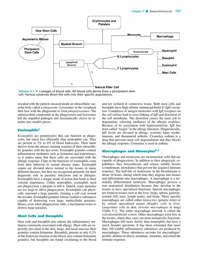
Chapter 5 Immunobiologicals 157
resealed with the particle encased inside an intracellular vac-uolar body called a phagosome. Lysosomes in the cytoplasmthen fuse with the phagosome to form phagolysosomes. Theantimicrobial compounds in the phagosomes and lysosomeskill the engulfed pathogen and enzymatically cleave its re-mains into smaller pieces.
Eosinophils5
Eosinophils are granulocytes that can function as phago-cytes, but much less efficiently than neutrophils can. Theyare present as 2% to 4% of blood leukocytes. Their namederives from the intense staining reaction of their intracellu-lar granules with the dye eosin. Eosinophil granules containinflammatory mediators such as histamine and leukotrienes,so it makes sense that these cells are associated with theallergic response. Clues to the functions of eosinophils comefrom their behavior in certain disease states. Eosinophilcounts are elevated above normal in the tissues in manydifferent diseases, but they are recognized primarily for theirdiagnostic role in parasitic infections and in allergies.Eosinophils have a unique mode of action that lends to theirextreme importance. Unlike neutrophils, eosinophils neednot phagocytose a parasite to kill it. Indeed, some parasitesare too large to allow phagocytosis. Eosinophils can physi-cally surround a large parasite, forming a cell coat aroundthe invader. Eosinophil granules release oxidative substancescapable of destroying even large, multicellular parasites.Hence, even when phagocytosis fails, a mechanism exists todestroy large parasites.
Mast Cells and Basophils
Mast cells and basophils also release the inflammatory me-diators commonly associated with allergy. Mast cells are es-pecially prevalent in the skin, lungs, and nasal mucosa; theirgranules contain histamine. Basophils, present at only 0.2%of the leukocyte fraction in the blood, also contain histaminegranules, but basophils are found circulating in the blood
and not isolated in connective tissue. Both mast cells andbasophils have high-affinity immunoglobulin E (IgE) recep-tors. Complexes of antigen molecules with IgE receptors onthe cell surface lead to cross-linking of IgE and distortion ofthe cell membrane. The distortion causes the mast cell todegranulate, releasing mediators of the allergic response.Because of its association with hypersensitivity, IgE hasbeen called “reagin” in the allergy literature. Diagnostically,IgE levels are elevated in allergy, systemic lupus erythe-matosus, and rheumatoid arthritis. Cromolyn sodium is adrug that prevents mast cell degranulation and thus blocksthe allergic response. Cromolyn is used in asthma.
Macrophages and Monocytes4,5
Macrophages and monocytes are mononuclear cells that arecapable of phagocytosis. In addition to their phagocytic ca-pabilities, they biosynthesize and release soluble factors(complement, monokines) that govern the acquired immuneresponse. The half-life of monocytes in the bloodstream isabout 10 hours, during which time they migrate into tissuesand differentiate into macrophages. A macrophage is a ter-minally differentiated monocyte. Macrophages possess atrue anatomical distribution because they develop in thetissues to have specialized functions. Special macrophagesare found in tissues such as the liver, lungs, spleen, gastroin-testinal (GI) tract, lymph nodes, and brain. These specificmacrophages are called either histiocytes (generic term) orby certain specialized names (Kupffer cells in liver,Langerhans cells in skin, alveolar macrophages in lung)(Table 5.1). The entire macrophage network is called thereticuloendothelial system. Other macrophages exist free inthe tissues, where they carry out more nonspecific functions.Macrophages kill more slowly than neutrophils but have amuch broader spectrum. It has been estimated that morethan 100 soluble inflammatory substances are produced bymacrophages. These substances account for macrophages’prolific abilities to direct, modulate, stimulate, and retard theimmune response.
Scheme 5.1 Lineages of blood cells. All blood cells derive from a pluripotent stemcell. Various cytokines direct the cells into their specific populations.
0156-0178_17865_Ch05.qxd 12/4/09 4:37 AM Page 157

ternalize an organism or particle and digest it into smallfragments still recognizable as antigen. The fragments areconjugated with molecules of the major histocompatibilitycomplex 2 (MHC-II). These complexes are responsible forself or nonself cell recognition and ascertain that cells beingprocessed are not self. MHCs also direct the binding of theantigenic determinant with immunoreactive cells. Once theantigen–MHC-II complex forms, it undergoes transcytosisto the macrophage’s cell surface, where B lymphocytes andT-helper cells recognize the antigen via the B surface Aband T-cell receptors. It is this step that transfers specificityand memory information from the determinant into the im-mune system through the modulation of B-cell differentia-tion. Under the regulatory influence of the T-helper cells,B cells are stimulated to differentiate into plasma cells thatproduce Ab. The T-helper cells accelerate and retard the
158 Wilson and Gisvold’s Textbook of Organic Medicinal and Pharmaceutical Chemistry
Figure 5.1 Antigen capture and presentation by a macrophage lead to clones ofAb-producing plasma cells and memory B cells.
TABLE 5.1 The Reticuloendothelial System
Tissue Cell
Liver Kupffer cellsLung Alveolar macrophages (dust cells)Peritoneum Peritoneal macrophagesSpleen Dendritic cellsSkin Langerhans cellsBrain Microglial cells
Macrophages possess a very specialized function; theyact as APCs (Fig. 5.1). APCs are responsible for the prepro-cessing of antigens, amplifying the numbers of antigenic de-terminant units, and presenting these determinant structuresto the programming cells of the immune system. APCs in-
0156-0178_17865_Ch05.qxd 12/4/09 4:37 AM Page 158

Chapter 5 Immunobiologicals 159
process as necessary. Thus, unlike the granulocytes, whichhave only destructive functions, monocytes and macro-phages regulate and program the immune response.
The Lymphoid Cell Line: B and T Cells2
The lymphoid cell line differentiates into two types oflymphocytes, the B lymphocytes and the T lymphocytes.These cells constitute only about 20% to 45% of bloodleukocytes. They are small cells, only slightly larger than anerythrocyte, but B and T cells can be identified microscopi-cally by large nuclei that occupy most of the cytoplasmicvolume. The nuclei are large to contain enough DNA to en-able the T and B cells to biosynthesize massive amounts ofprotein needed to carry out their immune functions. T lym-phocytes are involved in cell-mediated immunity (CMI); Blymphocytes differentiate into Ab-producing plasma cells.B lymphocytes express antibodies on their surfaces that bindantigens. T lymphocytes express specialized T-cell recep-tors on their surfaces that bind major histocompatibilitycomplex 1 (MHC-I) and 2 (MHC-II) complexed with anti-genic peptide fragments.
IMMUNITY
Immunity in humans can be conceptualized in several dif-ferent ways. If just the type and specificity of the immuneresponse are considered, the ideas of innate and acquiredimmunity are used. If only the components that are involvedin the immune response are considered, the processes can bedivided into humoral and cellular immunity. If the locationof the immune response is considered, we find that the im-mune system consists of serosal (in the serum) immunityand mucosal (on mucosal epithelium surfaces) immunity.
Innate Immunity
Innate immunity is the most basic form of immunity and in-cludes immune systems that are present in a human frombirth. A clear distinction must be made between innate im-munity and acquired (adaptive) immunity, which developsafter birth, and then only after an antigenic challenge (Table5.2). Innate immunity is the first line of defense against inva-sion by microbes and can be characterized as fast in re-sponse, nonspecific, and lacking in memory of the challenge.Acquired immunity develops through a complex system ofreactions that are triggered by invasion with an infectiousagent. It is slow in response to an infection, is highly specific,and has memory of previous infections. The memory, oranamnestic response, is responsible for the extremely rapiddevelopment of the immune response with subsequent chal-lenges and is a hallmark of acquired immunity.
There are three separate components of innate immu-nity that work in concert to provide the whole response.There are physical barriers, cellular barriers, and solublefactors. The physical barriers include the largest, most ex-posed organ of the body (the skin), the mucosa, and its as-sociated mucus. The keratinized layer of protein and lipidin the stratum corneum of the skin protects physicallyagainst various environmental, biological, and chemicalassaults. The protection afforded by mucosal surfaces,such as those that are found in the throat, mouth, nose, andGI tract, is due to a surface epithelium. The epitheliumconsists of single or multiple layers of epithelial cells withtight gap junctions between them. This type of structureprovides an impermeable physical barrier to microorgan-isms. Most of the time, epithelium is further protected bythe secretion of mucus, such as from goblet cells in the GImucosa. Mucus is a viscous layer consisting of glycopep-tide and an acidic glycoprotein called mucin. Mucus canprevent penetration of microbial cells into the epithelium,significantly decreasing the possibility of infection by themucosal route. Other components of the physical barriersin innate immunity are the tears (containing lysozyme), theacidic pH of the stomach, the low pH and flow of the urine,and the cilia in the lungs that constantly beat upward to re-move inspired particulates and microbes.
Two components of the cellular innate immune re-sponse have already been discussed, granulocytes in theblood and tissue macrophages. When an infection occursin the tissues, chemotactic factors liberated at the site mi-grate down a concentration gradient to the surroundingarea. These agents make the capillary beds porous.Neutrophils follow the concentration gradient across theendothelium to the site of infection. There are three typesof chemotactic factors: (a) formylmethionyl (f-Met) pep-tides released from the invading bacteria, (b) leukotrienessecreted by phagocytes, and (c) peptide fragments releasedfrom activated complement proteins such as C3a and C5a.Neutrophils and macrophages engulf and destroy microor-ganisms by phagocytosis. Nonphagocytic cells are also in-volved in the innate immune response, providing solublechemical factors that enhance the innate response.
Soluble factors of innate immunity include (a) bacteri-cidal factors, (b) complement, and (c) interferon. A bacte-ricidal factor (Table 5.3) is an agent that kills bacteria.Perhaps the most fundamental bactericidal factor is theacid in the stomach. Secreted by goblet cells in the mu-cosal epithelial lining, stomach acid is responsible for dis-posing of most of the microbes that are consumed orally.Phagocytes or hepatocytes produce the other bactericidalfactors. Most of these are directed toward the phago-some, where the predigested, phagocytically encapsulated
TABLE 5.2 Characteristics of Innate versus AcquiredImmunity
Innate Immunity Acquired Immunity
Present from birth Develops laterRapid SlowNonspecific SpecificNo memory Memory
TABLE 5.3 Bactericidal Factors
Factor Formation Site of Action
Oxygen ions and Induced Phagosomesradicals
Acid hydrolases Preformed LysosomesCationic proteins Preformed PhagosomesDefensins Preformed Phagosomes or
extracellularLactoferrin Preformed Phagosomes
0156-0178_17865_Ch05.qxd 12/4/09 4:37 AM Page 159

bacterial cell is enclosed. The antimicrobial factors kill theimmobilized microbes.
There are two types of antimicrobial factors, those thatare preformed inside the phagocyte and one that is inducedin response to the phagocytic process. The most impor-tant of the antimicrobial mechanisms is the respiratoryburst, which generates oxygen radicals—superoxide, hy-droxyl radicals, and hydrogen peroxide. The respiratoryburst is the only induced mechanism. All of the activeoxygen species are highly destructive to bacterial as wellas host cells, so they are not produced until they areneeded. The defensins are arginine- or cysteine-richbactericidal peptides that exhibit an extremely broad spec-trum of antimicrobial activity. The defensins will killbacteria (Gram-positive and Gram-negative), fungi, andeven some viruses. The mechanism of action of the de-fensins is unknown, but because the peptides are highlycharged in an opposite sense to bacterial cell membranes,an electrostatic, membrane-disruptive interaction might beinvolved. Bacteria have an absolute requirement for iron,and to compete with the host for this element, they secretehigh-affinity siderophore factors that scavenge iron fromthe host’s stores. Lactoferrin is a substance produced bythe host that binds iron more tightly than the bacterialchelator, preventing the invading organism’s access to acritical nutrient. Lysozyme is an important component ofthe antimicrobial system. This enzyme hydrolyzes [1–4]-glycosidic bonds, as in the peptidoglycan of bacterial cellwalls. Lysozyme is present in almost all body fluids, in-cluding tears and saliva.
Hepatocytes produce an array of acute phase proteins(Table 5.4) that are released into the serum during inflam-mation or infection. These proteins do not act directly onbacteria, but they augment the bactericidal activity of otherantimicrobial factors.
COMPLEMENT
Complement is a system of at least 20 separate proteins andcofactors that continuously circulate in the bloodstream.Complement acts to kill bacterial cells that are missed bythe neutrophils and the macrophages. There are actuallytwo separate complement pathways. One, the classicalpathway, operates in the adaptive or acquired immune re-sponse. The classical pathway has an absolute requirementfor an Ab–antigen complex as a trigger. The other, thealternative pathway, requires no Ab or antigen to initiateand is operative in innate immunity. Both pathways oper-ate in a tightly regulated cascade fashion. The proteinsnormally circulate as inactive proenzymes. When the path-ways are activated, the product of each step activates thesubsequent step.
THE ALTERNATIVE COMPLEMENT PATHWAY
In the alternative pathway, C3 is the initiating peptide (Fig.5.2). In the serum, C3 is somewhat unstable (it is sensitiveto proteases) and spontaneously decomposes into a large,active C3b fragment and a smaller, catalytically inactive
160 Wilson and Gisvold’s Textbook of Organic Medicinal and Pharmaceutical Chemistry
TABLE 5.4 Acute Phase Factors
Acute Phase Factor Function/Activity
C-reactive protein Chemotaxis and enhancement ofphagocytosis
�1-Antitrypsin Inhibition of proteasesComplement factors Control of the complement
cascadeFibrinogen Blood coagulation
Figure 5.2 Control of the alternative complementpathway by activating surfaces. When complement com-ponent C3b binds to a surface, there exist two possibleoutcomes. Under normal conditions, when no activatingsurface is present (e.g., if C3b has contacted normal tis-sue), sequential addition of blood cofactors H and I con-verts C3b into C3c, inactivating the complement protein.If an activating surface such as a microbe or damagedtissue is encountered, sequential addition of factors Band D drives the alternative pathway to the normalproperdin (P) intermediate, and the complement cas-cade is triggered. The properdin-containing component(C3bBbP) feeds back to the beginning of the pathway,generating more C3.
0156-0178_17865_Ch05.qxd 12/4/09 4:37 AM Page 160

Chapter 5 Immunobiologicals 161
C3a fragment. C3b now becomes bound to a surface, and ithas two fates. We can define two types of surfaces. One, thenonactivating surface, is a surface that contains sialic acid orother acidic polysaccharides. The other, an activating sur-face, contains none of the acidic polysaccharides or sialicacid. This type conforms to a bacterial cell surface. Undernormal circumstances, C3b will bind to a nonactivating sur-face. On binding, the C3b fragment becomes associatedwith factor H, a �-globulin that associates with an � chainon C3b. Sialic acid increases the affinity for factor H 100-fold. Factor H alters the shape of C3b in such a way that itbecomes susceptible to attack by factor I, a serine esterasethat cleaves the � chain of C3b, producing inactive iC3b.Attack by another protease produces a fragment designatedC3c. In this pathway, factor H accelerates the decay of C3b.When factors H and I work together, they destroy C3b asfast as it is produced and shut down the pathway.
If C3b binds to an activating surface, the ability to bindto factor H is reduced, and C3b binds to a protein called fac-tor B, forming C3bB. Bound factor B is cleaved by factor Dinto a fragment called Bb. The complex C3bBb has high C3-convertase activity and stimulates the pathway further.Factor P (properdin) binds to the complex, extending thehalf-life of C3bBbP. This fragment binds to the terminalcomplement components (C5–C9), creating a membrane at-tack complex and thus lysing the cell.
THE CLASSICAL COMPLEMENT PATHWAY
The classical complement pathway differs from thealternative pathway in that it requires a trigger in the formof an antigen–Ab complex. Only two antibodies can fix
complement—IgG and IgM. The classical pathway isshown in Figure 5.3. The small fragments that are cleavedfrom the proenzymes have activities such as chemotacticstimulation and anaphylaxis. The bar over the names ofsome components of the pathway denotes an active com-plex. Note that the classical pathway does not operate withC1 to C9 in sequence. Rather, the sequence is C1, C4, C2,C3, C5, C6, C7, C8, and C9.
INTERFERONS
An important antiviral system is provided by the interferons(Table 5.5 and Fig. 5.4). The interferons are peptides that,when viral infection occurs, carry out three distinct func-tions. First, they send a signal to a natural killer cell that es-sentially leads to the self-destruction of the infected cell.Second, they induce an antiviral state in neighboring cells,limiting the viral infection. Third, when interferon receptorsare bound on a target cell, the induction of the formation ofantiviral proteins occurs. One such protein is the enzyme2�,5�-oligoadenylate synthetase. This enzyme catalyzes thereaction that converts ATP into 2�,5�-oligoadenylate. Thiscompound activates ribonuclease R, which possesses thespecificity to hydrolyze viral RNA and thus can stop propa-gation of the virus inside the cell.
Acquired (Adaptive) Immunity
When the host is exposed to an antigen or organism thathas been contacted previously, the adaptive immune re-sponse ensues. The adaptive immune response worksthrough the B and T lymphocytes, which possess surface
Figure 5.3 The function of interferon. When a virus infects a host cell, the cell ex-presses interferon. Interferon activates natural killer cells, causing killing of the infectedhost cells and elimination of the reservoir of infection. At the same time, interferoninduces an antiviral state in neighboring cells, effectively breaking the cycle of infection.
TABLE 5.5 Interferons
Interferon Producing Cells Producing Mechanism Isotypes Molecular Mass Receptor
Type 1IFN-� Leukocytes 17 16–27 kDa 95–110 kDaIFN-� Fibroblasts Viral infection 1 20 kDa 95–110 kDaType 2IFN-� T lymphocytes Mitogen stimulation 1 20–24 kDa 90–95 kDa
0156-0178_17865_Ch05.qxd 12/4/09 4:37 AM Page 161

receptors specific for each invading organism. To accountfor all possible permutations of antigenic structure, naturaland synthetic, that the host might encounter, the adaptiveimmune system uses genetic recombination of DNA andRNA splicing as a way of encoding its antibodies.Lymphocytes can recognize an estimated 107 differenttypes of antigens through this genetic recombination mech-anism, far more than a person is likely to encounter duringa lifetime. Adaptive immunity is Ab-mediated immunity,based on circulating pools of antibodies that react with, and
inactivate, antigens. These antibodies are found in the glob-ulin fraction of the serum. Consequently, antibodies arealso referred to as immunoglobulins (Ig). The adaptive im-mune response has the property of memory. The sensitivity,specificity, and memory for a particular antigen are re-tained, and subsequent exposures stimulate an enhanced re-sponse. Hence, the adaptive immune response differs fromthe innate in two respects: specificity and memory.
The adaptive immune response, like the innate, can bedivided into two branches: humoral immunity and CMI.Humoral immunity is circulating immunity and is mediatedby B lymphocytes and differentiated B lymphocytes knownas plasma cells. CMI is controlled by the T lymphocytes.The immune function of T lymphocytes cannot be trans-ferred by serum alone; the T cells must be present, whereasthe immunity of the humoral system can be isolated fromthe serum and transferred. T cells are specially tailored todeal with intracellular infections (such as virus-infectedcells), whereas B cells secrete soluble antibodies that canneutralize pathogens before their entry into host cells. BothB and T cells possess specific receptors on their surfaces torecognize unique stimulatory antigens. When B cells arestimulated, they express specific Ig or surface antibodiesthat are capable of binding to the antigen. A fraction of theB-cell population does not differentiate into Ab-producingcells but forms a pool of cells that retain the immunologi-cal memory. T cells express a specific antigen receptor, theT-cell receptor, similar in structure to the surface Ig recep-tor of B cells. This receptor is activated by a piece ofprocessed antigen (presented with MHC-II). Activated Tcells release soluble factors such as interleukins, cytokines,interferons, lymphokines, and colony-stimulating factors,all of which regulate the immune response. Interactionswith some of these help to regulate the B-cell activity, di-recting the innate immune response.
IMMUNOGLOBULIN STRUCTURE AND FUNCTION
An Ab or Ig is composed of peptide chains with carbohy-drate pendant groups. A schematic of the Ab IgG is shownin Figure 5.5. The peptide chains form the quaternarystructure of the Ig, while the carbohydrate moieties serve
162 Wilson and Gisvold’s Textbook of Organic Medicinal and Pharmaceutical Chemistry
Figure 5.4 Classical complement pathway.
Figure 5.5 Structure of immunoglobulin G (IgG), showing antigen-binding regionsand key elements of the molecule.
0156-0178_17865_Ch05.qxd 12/4/09 4:37 AM Page 162

Chapter 5 Immunobiologicals 163
as antigen-recognition groups and probably as conforma-tion-stabilizing units. The general structure of the Ig lookssomething like a Y, with the antigen-binding regions at thebifurcated end. In this area are peptide sequences that are“programmable” by the immune system to allow the Ig torecognize a large number of antigens.
Treatment with either of two enzymes, papain or pepsin,digests an Ab into fragments that are useful in understand-ing its molecular structure. Papain clips the Ab into twofragments that contain the antigen-binding regions. Thesefragments have been termed the Fab, or antigen-binding,fragment. The remaining part of the Ab after papain diges-tion contains two peptide chains linked by a disulfide bond.Treatment of the same Ab with pepsin yields the two Fabunits joined by the disulfide bond, plus two of the distalpeptide chains. These distal units have been crystallizedand, hence, are termed the Fc fragment (for “crystalliz-able”). The disulfide bond, therefore, provides a demarca-tion between the two molecular regions. The nomenclatureof an Ab includes a high–molecular weight, or heavy, chainon the inside and a low–molecular weight, or light, chainon the outside.
IMPORTANT FEATURES OF ANTIBODYMOLECULAR STRUCTURE4,6
As stated previously, the tip end of the Fab region bindsantigen. There are two of these regions, so we say that theAb is bivalent and can bind two antigen molecules. Theoverall amino acid sequence of the Ab dictates its confor-mation. The peptide sequence for most antibodies is simi-lar, except for the hypervariable regions. The amino acidsequence at the end of the heavy chain (Fc) determines theclass of the Ig (i.e., IgG, IgM, etc.). All antibodies resem-ble each other in basic shape, but each has a unique aminoacid sequence that is complementary to the antigen in a“lock-and-key” interaction (antigen–Ab specificity).Some, such as IgM, are pentamers of IgG (Fig. 5.6). In re-ality, the lock-and-key model is too simplistic, and an in-duced fit model is preferred.
ANTIBODY PRODUCTION AND PROGRAMMING OFTHE IMMUNE SYSTEM
The main element of the programming portion of the im-mune system is the macrophage. A common property ofmacrophages is phagocytosis, the capacity to engulf a par-ticle or cell through invagination and sealing off of thecell membrane. The macrophages involved in the immuneresponse set in motion a unique amplification process, sothat a large response is obtained relative to the amount ofantigen processed. The macrophages engulf antigenic par-ticles and incorporate them into their cytoplasm, wherethe antigens are fragmented. The fragments are then com-bined with MHC-II, displayed on the cell membrane ofthe macrophage, and presented to the immune system.The presented antigens interact with B cells, causing dif-ferentiation to plasma cells and Ab secretion. T-helpercells also interact with the presented antigen and are stim-ulated to cause the B cells to proliferate and mature.Plasma cells are monoclonal (genetically identical) andproduce monoclonal Ab. The process, from the pluripo-tent stem cell, to the B and T cells, to the plasma cells, isshown in Figure 5.1.
ANTIBODY FORMATION5
Figure 5.1 also indicates the actual Ab-producing steps.Plasma cells are clones of Ab-producing cells, which am-plify the Ab response by their sheer numbers. The plasmacells can easily be regenerated if called on to do so by thememory functions. A population of plasma cells is shown atthe bottom of the diagram. These are identical and amplifyand produce large quantities of Ab, proportionally muchgreater than the amount of antigen that was initiallyprocessed.
THE ANAMNESTIC RESPONSE5
Because the programmed immune system has the propertyof memory, subsequent exposures to the same antigen areimmediately countered. The actual memory response is re-ferred to as the anamnestic response, a secondary responseof high Ab titer to a particular antigen. This is due to “mem-ory cell” formation as a result of the initial antigen stimulus(sensitization or immunization). The anamnestic response isdemonstrated in Figure 5.7.
ANTIGEN–ANTIBODY REACTIONS4
An Ab is bivalent, and an antigen is multivalent, so latticeformation can occur (Fig. 5.8). The complex may be fibrous,particulate, matrixlike, soluble, or insoluble. These charac-teristics dictate the means of its disposal. Four fundamentalreactions describe these processes: neutralization, precipita-tion, agglutination, and bacteriolysis.
NeutralizationNeutralization is an immunological disposal reaction forbacteria and for toxins (which are small and soluble). Oncethey bind the Ab, they are no longer toxic because their ac-tive site structures are covered and they cannot bind their
Figure 5.6 Pentameric structure of immunoglobulinM (IgM).
0156-0178_17865_Ch05.qxd 12/4/09 4:37 AM Page 163
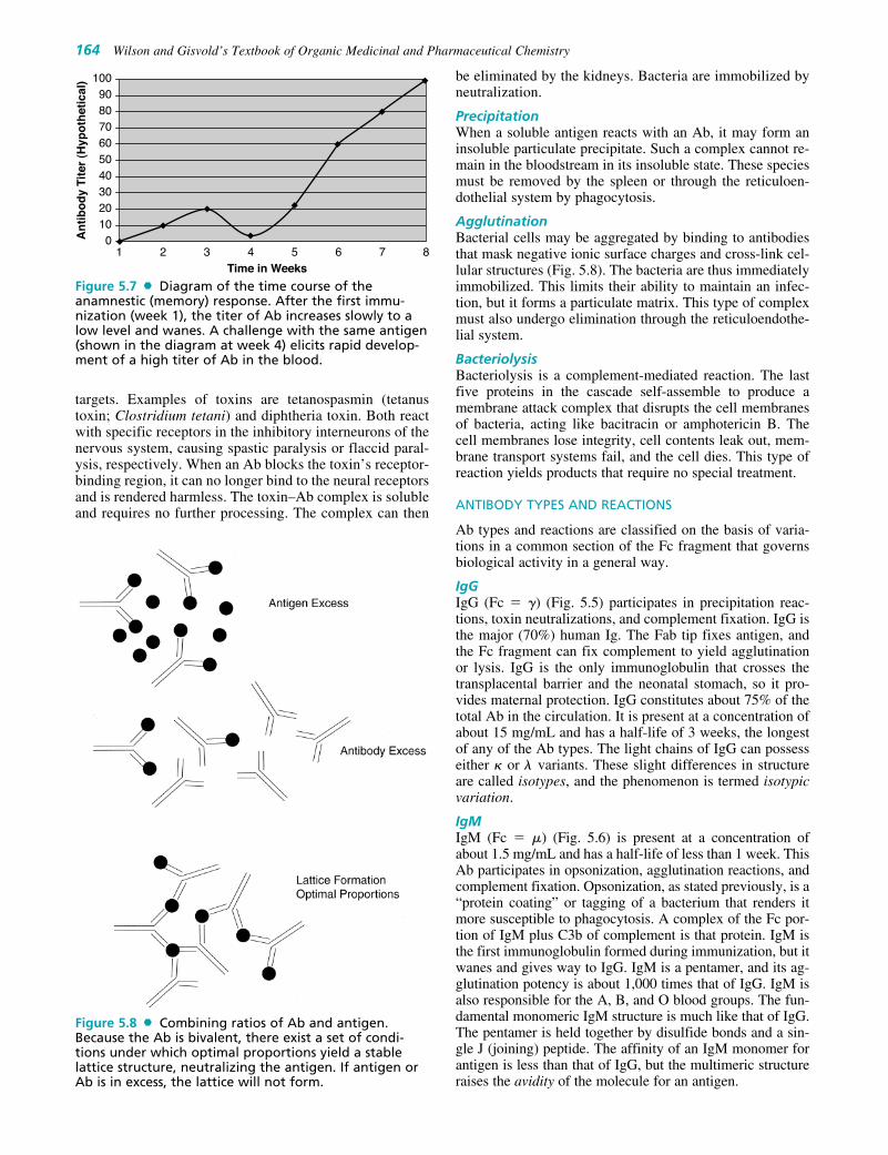
targets. Examples of toxins are tetanospasmin (tetanustoxin; Clostridium tetani) and diphtheria toxin. Both reactwith specific receptors in the inhibitory interneurons of thenervous system, causing spastic paralysis or flaccid paral-ysis, respectively. When an Ab blocks the toxin’s receptor-binding region, it can no longer bind to the neural receptorsand is rendered harmless. The toxin–Ab complex is solubleand requires no further processing. The complex can then
be eliminated by the kidneys. Bacteria are immobilized byneutralization.
PrecipitationWhen a soluble antigen reacts with an Ab, it may form aninsoluble particulate precipitate. Such a complex cannot re-main in the bloodstream in its insoluble state. These speciesmust be removed by the spleen or through the reticuloen-dothelial system by phagocytosis.
AgglutinationBacterial cells may be aggregated by binding to antibodiesthat mask negative ionic surface charges and cross-link cel-lular structures (Fig. 5.8). The bacteria are thus immediatelyimmobilized. This limits their ability to maintain an infec-tion, but it forms a particulate matrix. This type of complexmust also undergo elimination through the reticuloendothe-lial system.
BacteriolysisBacteriolysis is a complement-mediated reaction. The lastfive proteins in the cascade self-assemble to produce amembrane attack complex that disrupts the cell membranesof bacteria, acting like bacitracin or amphotericin B. Thecell membranes lose integrity, cell contents leak out, mem-brane transport systems fail, and the cell dies. This type ofreaction yields products that require no special treatment.
ANTIBODY TYPES AND REACTIONS
Ab types and reactions are classified on the basis of varia-tions in a common section of the Fc fragment that governsbiological activity in a general way.
IgGIgG (Fc � �) (Fig. 5.5) participates in precipitation reac-tions, toxin neutralizations, and complement fixation. IgG isthe major (70%) human Ig. The Fab tip fixes antigen, andthe Fc fragment can fix complement to yield agglutinationor lysis. IgG is the only immunoglobulin that crosses thetransplacental barrier and the neonatal stomach, so it pro-vides maternal protection. IgG constitutes about 75% of thetotal Ab in the circulation. It is present at a concentration ofabout 15 mg/mL and has a half-life of 3 weeks, the longestof any of the Ab types. The light chains of IgG can possesseither � or � variants. These slight differences in structureare called isotypes, and the phenomenon is termed isotypicvariation.
IgMIgM (Fc � �) (Fig. 5.6) is present at a concentration ofabout 1.5 mg/mL and has a half-life of less than 1 week. ThisAb participates in opsonization, agglutination reactions, andcomplement fixation. Opsonization, as stated previously, is a“protein coating” or tagging of a bacterium that renders itmore susceptible to phagocytosis. A complex of the Fc por-tion of IgM plus C3b of complement is that protein. IgM isthe first immunoglobulin formed during immunization, but itwanes and gives way to IgG. IgM is a pentamer, and its ag-glutination potency is about 1,000 times that of IgG. IgM isalso responsible for the A, B, and O blood groups. The fun-damental monomeric IgM structure is much like that of IgG.The pentamer is held together by disulfide bonds and a sin-gle J (joining) peptide. The affinity of an IgM monomer forantigen is less than that of IgG, but the multimeric structureraises the avidity of the molecule for an antigen.
164 Wilson and Gisvold’s Textbook of Organic Medicinal and Pharmaceutical Chemistry
0An
tib
od
y Ti
ter
(Hyp
oth
etic
al)
Time in Weeks1 4 852 3 6 7
2010
40
60
80
30
50
70
90100
Figure 5.7 Diagram of the time course of theanamnestic (memory) response. After the first immu-nization (week 1), the titer of Ab increases slowly to alow level and wanes. A challenge with the same antigen(shown in the diagram at week 4) elicits rapid develop-ment of a high titer of Ab in the blood.
Figure 5.8 Combining ratios of Ab and antigen.Because the Ab is bivalent, there exist a set of condi-tions under which optimal proportions yield a stablelattice structure, neutralizing the antigen. If antigen orAb is in excess, the lattice will not form.
0156-0178_17865_Ch05.qxd 12/4/09 4:37 AM Page 164

Chapter 5 Immunobiologicals 165
IgAIgA (Fc � �) (Fig. 5.9) is found in exocrine gland secretions(milk, saliva, tears), where it protects mucous membranes(e.g., in the respiratory tract). It is present in the serum as amonomer at a concentration of 1 to 2 mg/mL, but humans se-crete about 1 g of the dimer per day in the mucosal fluids.Secretory IgA consists of two IgG-like units linked togetherat the Fc regions by a peptide known as the secretory frag-ment and a J fragment. The secretory fragment is actually partof the membrane receptor for IgA. The IgA molecule on themucosal side of the membrane binds antigen, then binds to thereceptor. By a process of transcytosis, the IgA–antigen com-plex is moved from the mucosa to the bloodstream, whereIgG and IgM can react. Because it is distributed on the mu-cosa, IgA has an anatomically specific distribution, unlike theother antibodies. IgA is the mediator of oral polio vaccination(the mucosal reaction gives way to systemic protection).
IgDIgD (Fc � �) is present on the surface of B cells and, alongwith monomeric surface IgM, is an antigen receptor that ac-tivates Ig production. There is less than 0.1 mg/mL in thebloodstream, and the half-life is only 3 days.
IgEIgE (Fc � ) is the Ab responsible for hypersensitivity reac-tions. IgE complexes have a high affinity for host cell sur-faces and can damage the host. High levels of IgE are foundin persons with allergies of various types, as well as in au-toimmune diseases. The Fc fragment is responsible for theIg–cell reactivity. An Ab-plus-antigen reaction yields thetypical Ab–antigen complex. The Fc portion of the Ab is ac-tually part of the mast cell. When antigen binds to the Fabportion of the Ab, the IgE molecules become cross-linked.This probably distorts the membrane of the mast cells andstimulates them to release histamine, which causesbronchial constriction, itching, redness, and anaphylaxis.
ACQUISITION OF IMMUNITY
Several types of immunity must be considered when de-scribing vaccines and other immunobiologicals. Some areartificial and some are natural. Natural immunity is endowedby phagocytic white blood cells, lysozyme in tears, the skin,and so on. Acquired immunity is acquired after birth (or bypassage from mother to fetus). Thus, immunity may be clas-sified as follows:
• Active acquired immunity: The host produces his or herown Ab.
• Naturally acquired active immunity: Occurs on recoveryfrom a disease (or from antigen exposure).
• Artificially acquired active immunity: Occurs as a responseto sensitization by a vaccine or toxoid.
• Passive acquired immunity: The subject receives Ab froman outside source, such as a �-globulin injection, or bytransplacental transfer.
• Naturally acquired passive immunity: Temporary neonatalprotection from maternal IgG passes to the fetus in utero;this type of immunity is not long-lasting.
• Artificially acquired passive immunity: An Ab is given byinjection (e.g., by an antitoxin or a �-globulin injection).
Definitions of Immunobiologicals
Immunobiologicals include antigenic substances, such asvaccines and toxoids, or Ab-containing preparations, suchas globulins and antitoxins, from human or animal donors.These products are used for active or passive immunizationor therapy. All of the following are examples of immunobi-ologicals:
• Vaccine: A suspension of live (usually attenuated) or inacti-vated microorganisms (e.g., bacteria, viruses, or rickettsiae)or fractions thereof, administered to induce immunity andprevent infectious disease or its sequelae. Some vaccinescontain highly defined antigens (e.g., the polysaccharide ofHaemophilus influenzae type b [Hib] or the surface antigenof hepatitis B), others have antigens that are complex or in-completely defined (e.g., killed Bordetella pertussis or liveattenuated viruses).
• Toxoid: A modified bacterial toxin that has been madenontoxic but retains the ability to stimulate the formationof antitoxin.
• Immune globulin (IG): A sterile solution containing anti-bodies from human blood. It is obtained by cold ethanolfractionation of large pools of blood plasma and contains15% to 18% protein. Intended for intramuscular adminis-tration, IG is primarily intended for routine maintenance ofimmunity in certain immunodeficient persons and for pas-sive immunization against measles and hepatitis A. IG doesnot transmit hepatitis B virus (HBV), human immuno-deficiency virus (HIV), or other infectious diseases.
• Intravenous immune globulin (IGIV): A product derivedfrom blood plasma from a donor pool similar to the IGpool but prepared so it is suitable for intravenous use.IGIV does not transmit infectious diseases. It is primarilyused for replacement therapy in primary Ab deficiencydisorders and for the treatment of Kawasaki disease, im-mune thrombocytopenia purpura, hypogammaglobuline-mia in chronic lymphocytic leukemia, and some cases ofHIV infection.
• Specific immune globulin: Special preparations obtainedfrom blood plasma from donor pools preselected for a highAb content against a specific antigen (e.g., hepatitis B im-mune globulin, varicella-zoster immune globulin [VZIG],rabies immune globulin, tetanus immune globulin, vac-cinia immune globulin, and cytomegalovirus immuneglobulin). Like IG and IGIV, these preparations do nottransmit infectious disease.
• Antitoxin: A solution of antibodies (e.g., diphtheria anti-toxin and botulinum antitoxin) derived from the serum ofanimals immunized with specific antigens. Antitoxins areused to confer passive immunity and for treatment.
Figure 5.9 Structure of immunoglobulin A (IgA), themucosal Ab that protects the GI tract and the respiratorymucosa.
0156-0178_17865_Ch05.qxd 12/4/09 4:37 AM Page 165

Vaccination denotes the physical act of administeringa vaccine or toxoid. Immunization is a more inclusiveterm denoting the process of inducing or providing im-munity artificially by administering an immunobiological.Immunization may be active or passive.
Immunobiologicals (Vaccines andToxoids)6–10
A vaccine may be defined as a solution or suspension ofkilled or live/attenuated virus, killed rickettsia, killed orlive/attenuated bacteria, or antigens derived from thesesources, which are used to confer active, artificially ac-quired immunity against that organism or related organisms.When administered, the vaccine represents the initial expo-sure, resulting in the acquisition of immunity. A subsequentexposure or challenge (a disease) results in the anamnestic,or memory, response.
METHODS OF VACCINE PRODUCTION
Vaccine production methods have varied greatly over theyears and are best discussed according to a parallel chrono-logical and sophistication approach.
Killed (Inactivated) PathogenIn this method, the normal pathogen is treated with a strong,denaturing disinfectant like formaldehyde or phenol. Theprocess denatures the proteins and carbohydrates that are es-sential for the organism to live and infect a host, but iftreated properly, the surface antigens are left intact. Theprocess must be done carefully to control the unwinding ofproteins or carbohydrates by denaturation, because thepreparation must be recognized as the original antigen. Themain problems with killed pathogen vaccines are the follow-ing: (a) if the vaccine is not inactivated totally, disease canresult; (b) if the preparation is overtreated, vaccine failureusually results because of denaturation; (c) the productionlaboratory must grow the pathogen in large quantities to becommercially useful, putting laboratory technicians at risk;and (d) the patient may experience abnormal and harmfulresponses, such as fever, convulsions, and death. These vac-cines typically are viewed as “dirty” vaccines, and some,like the pertussis vaccine, have been associated with prob-lems serious enough to warrant their temporary removalfrom the market.
Live/Attenuated PathogensThe word attenuated for our purposes simply means “lowvirulence.” The true pathogen is altered phenotypically sothat it cannot invade the human host and cannot get aheadof the host’s immune system. Low-pathogenicity strainssuch as these were originally obtained by passage of the mi-crobes through many generations of host animals. The ideawas that the animal and the pathogen, if both were to sur-vive, needed to adapt to live with each other without eitherpartner being killed. Poliovirus is attenuated in this fashionin monkey kidney tissue. In a live/attenuated vaccine, anti-genicity is still required, as is infectivity (polio vaccineyields an infection), but the host’s immune system must beable to stay ahead of the infection. The key problems arethe following: (a) the vaccine cannot be used if the patientis immunocompromised, has fever or malignancy, or is tak-ing immunosuppressive drugs; (b) these vaccines shouldnot be used during pregnancy; and (c) the attenuated organ-
ism commonly reverts to the virulent strain, which was thereason for the failure of some early polio vaccines. Today,biological quality control is very stringent, and these prob-lems have been eliminated.
Live/Attenuated Related StrainThe live/attenuated related strain is antigenically related sothat it can provide cross-immunity to the pathogen. Forexample, cowpox virus can be used in place of smallpoxvirus. The strains are antigenically similar enough so thatthe host’s immune system reacts to the related strain toprovide protection against the normal pathogen. The mainadvantage is that a true pathogen is not being used so thatthe chance of contracting the actual disease is zero. Theproblem with such vaccines is that they cause an infec-tion. Cowpox is known to spread to the central nervoussystem in 1 in 105 cases, causing a potentially fatal form ofmeningitis.
Cellular Antigen from a PathogenThe surface antigen (i.e., what is recognized as foreign) isharvested from the pathogen, purified, and reconstitutedinto a vaccine preparation. These antigens can take severalforms, including the carbohydrate capsule, as in Neisseriameningitidis; pili, as in N. gonorrhoeae; flagella from motilebacteria (the basis for an experimental cholera vaccine); orthe viral protein coat, as in the vaccine for hepatitis B.Advantages of the method are that there is virtually nochance of disease, contamination, or reversion and there areno storage problems. This method is currently as close to a“perfect approach” as we have. A problem is that thepathogen must be grown under careful control or an unsuresource must be relied on. For example, hepatitis B vaccinewas originally prepared from the serum of a controlledpopulation of human carriers. Imagine the impact if oneof the carriers developed another blood-borne disease.Additionally, these are strain-specific antigens (e.g., N. gon-orrhoeae may require 1,500 different pilar antigens).Acellular vaccines may exhibit lower antigenicity in thevery young and may require several injections for full im-munological competence. To be safe and consistent, theantigenic component must be identified. Given the complexnature of biological materials, this is not always easy oreven possible.
Genetically Engineered Pathogens5
The techniques of genetic engineering have allowed thepharmaceutical industry to prepare absolutely pure surfaceantigens while totally eliminating the pathogenic organismfrom the equation. As shown in Figure 5.10, the virus con-tains surface antigens (designated by filled circles). Insidethe viral capsule is a circular piece of deoxyribonucleic acid(DNA) containing genes for the various biological mole-cules of the virus. The diagram shows, at about 3 o’clock, asmall piece of DNA that codes for the surface antigen. Thestrategy is to isolate this piece of DNA and insert it into arapidly growing expression vector for production of the sur-face protein. In this case, Escherichia coli serves as an ex-cellent vector. It contains a plasmid that can be removed,clipped open, and used as a cassette to carry the viral DNA.Additionally, E. coli can be grown in batch to produce theviral surface antigen. To begin, the DNA is removed fromthe virus and the plasmid is removed from the vector. Viraland bacterial nucleic acid is treated with a restriction
166 Wilson and Gisvold’s Textbook of Organic Medicinal and Pharmaceutical Chemistry
0156-0178_17865_Ch05.qxd 12/4/09 4:37 AM Page 166

Chapter 5 Immunobiologicals 167
endonuclease, which cleaves the DNA and plasmid at desig-nated restriction sites. The viral DNA is cleaved into severalfragments, each of which is ligated into the E. coli plasmidwith a ligase enzyme. Plasmids are inserted into E. coli, andthe organism is grown in batch fermentation. The organismscontaining the gene for the viral surface protein can be sep-arated by screening and purified to serve as the ultimateantigen producer—free of contamination or pathogenic viralparticles. The pure antigen may then be constituted into avaccine and used in human hosts.
USE OF VACCINES IN COMBINATION: DOSING5,11
Types of VaccinesThere are three basic types of vaccine preparations that areused clinically:
1. A simple vaccine contains one strain of a disease-causingorganism (e.g., plague vaccine, Pasteurella pestis, andsmallpox vaccine).
2. A multivalent vaccine is prepared from two or morestrains of an organism that cause the same disease (e.g.,polio is trivalent). Administration of the multiple strainsis required for full protection because their antigens arenot cross-immunizing. The immune system must mount aseparate immune response to each strain.
3. A polyvalent vaccine is prepared from two or more or-ganisms that cause different diseases. Polyvalent vac-cines are given for convenience, primarily so that a childcan be given one shot rather than several. The measles–mumps–rubella (MMR) vaccine is of the polyvalent type.
Types of DosingVaccines can be administered according to various dosingregimens, depending on the vaccine type and the purpose ofthe injection:
1. A single-dose vaccine is usually assumed to confer, withone shot, “lifetime immunity.” The smallpox vaccine wasa single-dose vaccine.
2. In a multiple-dosing regimen, several doses are given,spaced weeks or months apart, to get maximum immuno-genicity. Multiple dosing is usually done with inactivatedvaccines, which are less antigenic. Multiple dosing is notthe same as a booster dose.
3. A booster dose is administered years after the initial im-munization schedule (regardless of single or multiple firstdose). As a patient ages, Ab levels may wane. A boosteris used to bolster immunity. Also, boosters are used if apatient is known or suspected to have been exposed to apathogen (e.g., tetanus).
4. A coadministered vaccine is possible only if one vaccinedoes not interfere with another.
5. There are two physical forms of vaccines: A fluid vaccineis a solution or a suspension of the vaccine in saline ofan aqueous buffer; the solution or suspension in an ad-sorbed vaccine is adsorbed on a matrix of aluminum orcalcium phosphate. Like a sustained-release dosage form,in theory, there is longer exposure via a depot injection.The higher surface area of the matrix will be exposedto the immune system. Generally, adsorbed vaccines arepreferred.
Pharmaceutical Principles of VaccinesAs expected for a live biological preparation, heat destroyslive viral and bacterial vaccines. If the agent is not killed, theantigen may be altered. Like many biologicals, lyophilizedvaccines are unstable after reconstitution. Ice crystals formedinside the protein structure during freeze-drying expandduring thawing and disrupt the structure of the vaccine. Livevaccines can be inactivated by minute amounts of detergent.Detergent residue adhering to glassware is concentratedenough to act as a disinfectant. It is safe to use only plastic im-plements specified for the vaccine. The suspending mediummay be sterile water, saline, or more complex systems con-taining protein or other constituents derived from the mediumin which the vaccine is produced (e.g., serum proteins, eggantigens, and cell culture-derived antigens). Concentrated Absuspensions (�-globulins) are typical amphiphilic proteinsand aggregate on storage. If injected, the particulates maycause anaphylaxis. Preservatives may be components of vac-cines, antitoxins, and globulins. These components are pres-ent to inhibit or prevent bacterial growth in viral cultures orthe final product or to stabilize the antigens or antibodies.Allergic reactions can occur if the recipient is sensitive to oneof these additives (e.g., mercurial compounds [thimerosal],phenols, albumin, glycine, or neomycin).
Storage and Handling of ImmunobiologicalsFailure to follow the exact recommendations for storage andhandling of immunobiologicals can lead to an impotentpreparation. During reconstituting, storing, and handling ofimmunobiologicals, the most important recommendation isto follow the package insert exactly. Vaccines should al-ways be stored at their recommended temperature. Certainvaccines, such as polio vaccine, are sensitive to increased
Figure 5.10 Preparation of a geneticallyengineered Ab.
0156-0178_17865_Ch05.qxd 12/4/09 4:38 AM Page 167

temperature. Other vaccines, such as oral polio vaccine, diph-theria and tetanus toxoids, and acellular pertussis vaccine,hepatitis B vaccine, influenza vaccine, and Hib conjugatevaccine (Hib-CV; among others), are sensitive to freezing.
Viral Vaccines12
SMALLPOX VACCINE (DRYVAX)
Smallpox vaccine is live vaccinia (cowpox) virus grown onthe skin of a bovine calf. Smallpox is a highly lethal and dis-figuring disease that was common throughout history.Smallpox vaccine was used routinely in the United Statesbut today is no longer recommended. (There have been noreported cases of smallpox since the 1940s.) In 1982, small-pox was declared eradicated worldwide. With smallpox, therisks of the vaccine outweigh the benefits; the vaccine pen-etrates the central nervous system and potentially fatal en-cephalitis occurs in 1 of 105 patients. After exposure tosmallpox, the vaccine can be injected to lessen the severityof the disease.
INFLUENZA VACCINE13–17
Influenza vaccine is a multivalent inactivated influenza virusor viral subunits (split vaccine). The virus is grown on chickembryo and inactivated by exposure to ultraviolet (UV) lightor formaldehyde. The antigen type is protein. The vaccine inthe United States contains thimerosal, a mercurial, as a preser-vative. Influenza is a respiratory tract infection with a 2-dayincubation period. The disease may be devastating and canlead to pneumonia. Without the vaccine, influenza is commonin epidemics and pandemics. To clarify, the flu is a GI infec-tion with diarrhea and vomiting. Influenza requires weeks ofincubation. Influenza is caused by two main genetic strainseach year (A and B): type A is most common in humans; typeB is less common. The virus mutates very rapidly, and vac-cines must be tailored yearly. The World Health Organization(WHO) and the Centers for Disease Control and Prevention(CDC) monitor the migration of the disease from SoutheastAsia, type the strains causing the occurrences, and order avaccine to counter the organisms most likely to enter theUnited States. Influenza A viruses are categorized accordingto two cell surface protein antigens: hemagglutinin (H) andneuraminidase (N). Each of these is divided further into sub-types (H1, H2; N1, N2). Individual strains within a subtypeare named for the location, isolation sequence number, andyear of isolation (e.g., A/Beijing/2/90 [H1N1]). For example,the WHO-recommended formula for 2001 to 2002 in-cluded the following antigens: A/New Caledonia/20/99(H1N1), A/Moscow/10/99 (H3N2), B/Sichuan/379/99, 15 �geach per 0.5 mL. A typical vaccine will be a mixture of threestrains. Strains are selected each year in the spring on thebasis of the disease trends observed and are released in the au-tumn. In general, those patients who are at high risk for com-plications from influenza are the following:
• Persons 65 years of age or older• Residents of nursing homes and other chronic-care facili-
ties that house persons of any age who have chronic med-ical conditions
• Adults and children who have chronic disorders of the pul-monary or cardiovascular systems, including asthma
• Adults and children who have required medical follow-up or hospitalization during the preceding year because
of chronic metabolic diseases (including diabetes melli-tus), renal dysfunction, hemoglobinopathies, or immuno-suppression (including immunosuppression caused bymedications or HIV infection)
• Children and teenagers (aged 6 months–18 years) who arereceiving long-term aspirin therapy and, therefore, mightbe at risk for developing Reye syndrome after influenzainfection
• Women who will be in the second or third trimester ofpregnancy during the influenza season
• Healthcare workers and those in close contact with personsat high risk, including household members
• Household members (including children) of persons ingroups at high risk, including persons with pulmonary dis-orders, such as asthma, and healthcare workers who are athigher risk because of close contact
It is impossible to contract influenza from the vaccine.The only side effects may be local pain and tenderness atthe injection site, with low-grade fever in 3% to 5% of pa-tients. Aspirin and acetaminophen are effective in combat-ing these symptoms. Allergic reactions are rare but may beseen in persons allergic to eggs. Immunity to influenza vac-cine takes 2 weeks to develop. Some people fear the vac-cine because of reports of a strange paralysis and lack ofnerve sensation associated with the 1976 swine flu vaccine.This problem, Guillain-Barré syndrome, was associatedonly with this 1976 vaccine and has not been associatedwith vaccines since.17
POLIO VACCINES18–20
Polio is a dangerous viral infection that affects both musclemass and the spinal cord. Some children and adults whocontract polio become paralyzed, and some may die becauseof respiratory paralysis. Polio was the cause of the “infantileparalysis” epidemic of 1950 to 1953, which led to many par-alyzed children and the specter of patients spending theirlives in an iron lung. Serious cases of polio cause musclepain and may make movement of the legs and/or arms diffi-cult or impossible and, as stated previously, may makebreathing difficult. Milder cases last a few days and maycause fever, sore throat, headache, and nausea. Interest inpolio has increased because of recent local outbreaks; largenumbers of people are unimmunized. There are no drugs orspecial therapies to cure polio; treatment is only supportive.The symptoms of polio may reappear 40 to 50 years after asevere infection. This phenomenon is known as postpoliomuscle atrophy (PPMA). PPMA is not a reinfection or reac-tivation of the virus but is probably a form of rapid aging inpolio survivors. There are two types of polio vaccines.
Inactivated Polio Vaccine (IPV)There are several synonyms for the IPV vaccine: IPV, e-IPV,ep-IPV, and the Salk vaccine (1954 [IPOL, Aventis-Pasteur]). e-IPV is an enhanced potency poliovirus, morepotent and immunogenic than any of the previous IPV for-mulations. e-IPV is recommended for all four infant dosesbecause of the incidence of rare cases of oral polio vaccine(OPV)–associated paralytic poliomyelitis. e-IPV is also pre-ferred for adults for the same reason. IPV is a trivalent(strains 1, 2, 3) vaccine grown in monkey kidney culture andsubjected to elaborate precautions to ensure inactivation(typically, formaldehyde is used). The antigen form is whole
168 Wilson and Gisvold’s Textbook of Organic Medicinal and Pharmaceutical Chemistry
0156-0178_17865_Ch05.qxd 12/4/09 4:38 AM Page 168

Chapter 5 Immunobiologicals 169
virus. The antigen type is protein. The vaccine is injected tocause induction of active systemic immunity from polio butdoes not stop polio carriers, who shed the virus from the oraland nasal cavities.
Trivalent Oral Polio Vaccine (TOPV)TOPV (Sabin vaccine, 1960) is a live attenuated whole virusvaccine (antigen type, protein) containing polio strains 1, 2,and 3. The virus culture is grown on monkey kidney tissuewith the use of an elaborate attenuation protocol. Oral ad-ministration of the vaccine yields a local GI infection, andthe initial immune response is via IgA (mucosal, local to theGI tract). The IgA–antigen complex undergoes transcytosisacross the mucosal membrane, and systemic immunity is in-duced as IgM and IgG form. A major caution with TOPV isthat it is a live vaccine and must never be injected.Indications are the following:
• Mass vaccination campaigns to control outbreaks of para-lytic polio.
• Unvaccinated children who will travel in less than 4 weeksto areas where polio is endemic.
• Children of parents who do not accept the recommendednumber of vaccine injections. These children may receiveOPV only for the third or fourth dose or both. In suchcases, the healthcare provider should administer OPV onlyafter discussing the risk of OPV-associated paralytic po-liomyelitis with parents or caregivers.
• e-IPV is recommended for routine use in all four immuniz-ing doses in infants and children.
• The WHO has advocated giving children e-IPV instead ofTOPV to prevent exposure of others to virus shed throughthe nose and mouth.21,22
RUBELLA VACCINE23
German measles is a disease that was once called the “3-daymeasles” and was considered a normal childhood illness. Itis a mild disease with few consequences, except in the firsttrimester of pregnancy. In these mothers, rubella causesbirth defects in 50% of cases. Defects may include heartdisease, deafness, blindness, learning disorders, and sponta-neous abortion of the fetus. Symptoms of rubella are a low-grade fever, swollen neck glands, and a rash that lasts forabout 3 days. About 1 of every 10 women of childbearingage in the United States is not protected against rubella.Also, 20% of all adults escaped this normal childhood dis-ease or are not vaccinated.
Rubella vaccine (German measles vaccine, live, MeruvaxII, Merck) is a live, attenuated rubella virus produced inhuman diploid cell culture. The antigen form of the vaccine iswhole virus. The antigen type is protein. The vaccine is ad-ministered as part of the normal immunization schedule at 15months. Side effects are minimal, but there may be some sore-ness and pain at the site of injection and stiffness of the joints.
A problem with the vaccine is that administration of alive virus is contraindicated in pregnancy. Indications arethe following:
• Persons aged 12 months to puberty should be immunizedroutinely.
• Previously unimmunized children of susceptible pregnantwomen should receive the MMR vaccine. The trivalentvaccine is preferred for persons likely to be susceptible tomumps and rubella.
• Immunization of susceptible nonpregnant adolescent oradult women of childbearing potential is called for if pre-cautions to avoid pregnancy are observed.
• Almost all children and some adults require more than onedose of MMR vaccine.
• On the first routine visit to the obstetrician/gynecologist, theimmune status should be checked. If the woman is not im-munized against rubella, the physician should administer thevaccine and stress avoiding pregnancy for 3 months.
• If the patient is already pregnant, the physician should notadminister the vaccine.
• If exposure is suspected, the cord blood should be moni-tored for the presence of rubella antibodies.
• All unimmunized women should be vaccinated immedi-ately after delivery of the baby.
MEASLES VACCINE (ATTENUVAX, MERCK)23
Measles is a very serious, highly contagious disease. It causesa high fever, rash, and a cough lasting 1 to 2 weeks. Some pa-tients experience extreme sensitivity to light. The rash mayoccur inside the eyelids, producing a very painful condition.In the United States, between 3,000 and 28,000 cases occureach year, depending on factors such as weather and localizedoutbreaks. Outbreaks are very common in neighborhoods andschools. One of ten children contracting measles will developan ear infection or pneumonia. Measles may infect the brain(encephalitis) and lead to convulsions, hearing loss, and men-tal disability. In the United States, 1 of every 500 to 10,000children contracting measles dies from it. Severe sickness anddeath are more common in babies and adults than in elemen-tary schoolchildren or teenagers. Measles has been linked tomultiple sclerosis. In 1977, a severe epidemic occurred in theUnited States, and 50,000 cases were reported. Only 60% ofthe population was vaccinated.
Measles vaccine is composed of live/attenuated measlesvirus that is grown on chick embryo culture with an attenu-ation protocol. Indications are the following:
• Selective induction of active immunity against measlesvirus.
• Trivalent MMR vaccine is the preferred immunizing formfor most children and many adults.
• Almost all children and many adults require more than onedose of MMR.
• Prior to international travel, persons susceptible to any ofthe three viruses should receive the single-antigen vaccineor the trivalent vaccine, as appropriate.
• Most persons born before 1956 are likely to have contractedthe disease naturally and are not considered susceptible.
• Persons born after 1956 or those who lack adequate docu-mentation of having had the disease should be vaccinated.
The vaccine is required by law at 15 months and again at11 to 12 years of age. The vaccine can be administered afterexposure to measles to lessen the disease severity. This isbecause Ab to the vaccine develops in 7 days, while the in-cubation period for the disease is 11 days. The vaccineshould not be administered in pregnancy and should alwaysbe administered with great care to women of childbearingage. Because measles vaccine is cultivated in egg medium,care must be used in patients who are allergic to eggs andegg products. For this reason, a test dose regimen is used.The administration protocol is shown in Figure 5.11.
0156-0178_17865_Ch05.qxd 12/4/09 4:38 AM Page 169

MUMPS VACCINE24,25
Mumps virus causes fever, headache, and a painfulswelling of the parotid glands under the jaw. Mumps can beserious and is highly contagious. Prior to the vaccine, thedisease was passed from child to child with ease. The dis-ease runs its course over several days. Between 4,500 and13,000 cases of mumps occur as outbreaks in the UnitedStates every year. In severe cases, mumps may cause in-flammation of the coverings of the brain and spinal cord(meningitis); this occurs in about 10% of infected persons.Swelling of the brain itself occurs in 1 of 200 patients. Menmay experience a painful swelling of the testicles (orchitis),which may presage sterility. Women may experience a cor-responding infection of the ovaries. Male teens are oftensicker than other groups of either sex. Mumps early inchildhood has been linked to the development of juvenilediabetes.
The mumps vaccine (Mumpsvax, Merck) is a live, atten-uated virus grown on chick embryo culture with attenuationprotocols. The antigen form is whole virus. The antigen typeis protein. Indications are the following:
• Induction of artificially acquired active immunity againstmumps.
• Before international travel, immunize any susceptible indi-viduals with the single-antigen vaccine or the trivalentMMR vaccine, as appropriate.
• Most children and some adults need more than one dose ofMMR vaccine.
• Persons born prior to 1956 are generally considered immune.
Caution. Mumps vaccine is supplied with a diluent. Useonly this diluent for reconstitution. Addition of a diluentwith an antimicrobial preservative can render the vaccine in-active. The vaccine is normally administered to children at15 months of age and again at 11 to 12 years. Becausemumps vaccine is cultivated in egg medium, care used to beadvised in patients allergic to eggs and egg products. Recentdata show that persons who are allergic to egg and egg prod-ucts fail to react to the mumps vaccine.
COMBINATION PRODUCTS (POLYVALENT VIRAL VACCINES)
If two or more vaccines are free of interference with eachother, they can be administered as a mixture (polyvalent)for convenience. Examples of polyvalent viral vaccinesare measles–rubella (MR), rubella–mumps (RM), andMMR. MMR is indicated for routine immunization at 15months (not given at �1 year unless the child has been ex-posed or lacks immunocompetence). This is because ma-ternal Abs interfere with development of vaccine immu-nity in small children. If the MMR is given at less than 1year, revaccination is needed at 15 months of age.
CHICKENPOX VACCINE21,26–30
Chickenpox is caused by varicella-zoster virus. Every year,about 3.5 million people in the United States, mostly chil-dren, contract chickenpox. The incidence peaks between3 and 9 years of age. Chickenpox causes a generalizedrash, with 300 to 500 blisterlike lesions occurring onthe scalp, face, and trunk. Symptoms include loss of ap-petite, malaise, and headache. The disease is usually benign
but can lead to bacterial superinfection, pneumonia, en-cephalitis, and Reye syndrome. About 50 to 100 previouslyhealthy children die of the disease. About 2% of all casesoccur in adults, who have more serious symptoms than chil-dren have.
Varicella vaccine (Varivax, Merck) is derived from livevirus from a child with natural varicella. The virus has beenattenuated by passage through a series of guinea pig andhuman cell cultures. The final preparation is a lyophilizedlive, attenuated virus. The antigen form is whole virus. Theantigen type is protein. The vaccine is well tolerated, withpain and redness at the injection site as the only side effects.The vaccine has shown tremendous success in reducing in-fections. Indications are the following:
• The vaccine is recommended for children 12 months to 12years old as a single dose.
• Adults who are exposed to chickenpox should continue toreceive VZIG.
• In elderly persons, varicella vaccine can boost immunity tovaricella-zoster virus and may prevent or attenuate herpeszoster (shingles) attacks.
HEPATITIS VACCINES30–40
Hepatitis is a complex of diseases that causes fever, nausea,abdominal pain, jaundice, liver failure, and death. There arefive clinically recognized types (A, B, C, D, and E).
Hepatitis A VaccineHepatitis A virus (HAV; infectious hepatitis) causes an acutedisease with an abrupt onset. About 15 to 50 days of incuba-tion are required before the disease becomes clinically notice-able. The primary sign is jaundice. The disease lasts severalweeks and is followed by complete recovery. Hepatitis A istransmitted when the virus is taken in by mouth. Thefecal–oral route and close contact, unwashed food, and con-taminated water account for most of the routes of transmis-sion. The sexual anal–oral route is also a route of spread.Children under the age of 3 frequently have no symptoms butcan transmit the disease to adults in child care centers. An in-jection of hepatitis A immune globulin is one way of prevent-ing the disease but is only effective for about 30 days.
The hepatitis A vaccine (Havrix) is an inactivatedpreparation that is produced by propagation of the virus incultured human diploid cells and then is inactivated withformalin. The antigen form is lysed whole viruses. Theantigen type is protein. The course of immunization in-volves two injections over a 4-week period and a booster12 months after the first injection. Indications are thefollowing:
• Persons traveling outside the United States, except toAustralia, Canada, Japan, New Zealand, and WesternEurope
• Persons with chronic liver disease• Persons living in an outbreak zone• Persons who inject medications• Persons engaging in high-risk sexual activity• Child care workers caring for children younger than 2
years of age• Developing countries with poor sanitation
Side effects are minor and usually limited to soreness atthe injection site and fever.
170 Wilson and Gisvold’s Textbook of Organic Medicinal and Pharmaceutical Chemistry
0156-0178_17865_Ch05.qxd 12/4/09 4:38 AM Page 170

Chapter 5 Immunobiologicals 171
Hepatitis B VaccineHBV, the cause of serum hepatitis, is a much more insidious,chronic disease, transmitted by needles, mucosal contact,blood, or high-risk sexual activity. The highest risk for con-traction of hepatitis B is among intravenous drug abusers. Thedisease is linked to cirrhosis and liver cancer. There are about200,000 new cases reported per year in the United States; ofthese, 10% become carriers, one fifth die from cirrhosis, and1,000 die from liver cancer. The hepatitis B vaccine was firstintroduced in 1981. Initially, it was prepared as an inactivatedvaccine from the plasma of carefully screened human, high-titer carriers/donors. In 1986, the recombinant DNA (rDNA)vaccine (Engerix B, Recombivax) was introduced to the mar-ket. The rDNA vaccine contains only viral subunits and maybe used with hepatitis B immune globulin in a postexposuresetting to boost the ability of the host to resist the infection. Inadults, three doses should be given, at 0, 1, and 6 months. Inchildren, the vaccine is given at birth, 1 month, and 9 months.Administration may be delayed in premature infants whoseimmune systems are not fully developed. If not immunized atbirth, a child should receive three doses by 18 months. If themother tests positive for hepatitis B, the vaccine plus the im-mune globulin must be given at or shortly after birth. The vac-cine is 95% effective and is typically without side effects.Several high-risk groups have been identified: healthcareworkers, student healthcare workers, people living in high-risk environments, and dentists. They should receive a three-dose course of the vaccine. In most other cases, a physiciancan judge whether a patient is at high risk or not. Side effectsof the vaccine are minor.
Hepatitis C VaccineHepatitis C virus (HCV) was once called hepatitis non-A,non-B but has been recognized as a separate entity. HCVinfection is spread primarily by the parenteral route (trans-fusions), and unlike HBV, maternal–fetal and sexual trans-mission are uncommon. Acute infection may show fewsymptoms; fewer than 25% of patients develop full-blownhepatitis. Administration of interferon alpha (IFN-�) duringthe early acute phase can cure most patients. Unfortunately,50% to 60% of those with HCV infection develop chronichepatitis. This is often manifested by periodic increases inhepatic enzyme levels. Cirrhosis develops in 20% of chronicinfectees; this usually requires 15 to 20 years to develop.Patients with HCV are at risk for hepatocellular carcinoma.Estimates are that 150,000 to 170,000 new cases occur inthe United States per year. Intravenous drug users, transfu-sion patients, and healthcare workers are at highest risk.
Development of an HCV vaccine proved difficult butwas accomplished in 1998. There are 15 genotypes, and thevirus can modulate its antigens within the host’s body. Anew approach using genetic material from the virus, analo-gous to the approach to the influenza vaccine, is said to bepromising.41
Hepatitis EHepatitis E virus (HEV) causes disease clinically indistin-guishable from hepatitis A. Symptoms include malaise,anorexia, abdominal pain, arthritis-like symptoms, andfever. Distinguishing HEV from HAV must be done genet-ically. The incubation period is 2 to 9 weeks. The disease isusually mild and resolves in 2 weeks, with no sequelae. Thefatality rate is 0.1% to 1%, except in pregnant women wherethe rate soars to 20%. No outbreaks have been reported in
the United States as of 1996. There is currently no vaccineagainst HEV.
ROTAVIRUS VACCINE
There is a rotavirus vaccine included in the RecommendedChildhood Immunization Schedule. This vaccine is used toprovide immunity against rotavirus, the most common causeof severe diarrhea in children in the United States. All chil-dren have at least one rotavirus infection in the first 5 yearsof life, and there are about 20 deaths per year in this coun-try. Children between the ages of 3 and 24 months of agehave the highest rates of severe disease and hospitalization.The rotavirus vaccine is an oral vaccine, given as a series ofthree doses. It is recommended that the vaccine beadministered at 2, 4, and 6 months of age. The most com-mon side effect seems to be fever.
HUMAN PAPILLOMAVIRUS VACCINE
Human papillomavirus (HPV) is a sexually transmittedvirus. It is passed through genital contact (such as vaginaland anal sex). The virus is also able to be transmitted byskin-to-skin contact. At least 50% of people who have hadsex will have HPV at some time in their lives. Most con-tractees do not display any signs, and HPV may resolve onits own without causing any health problems.
Anyone who has ever had genital contact with another per-son may have HPV. Both men and women may contract andpass the virus often without knowing it. Because there maynot be any signs, a person may have HPV even if years havepassed since he or she had sex. A person is more likely to con-tract HPV if he or she has sex at an early age, has multiple sexpartners, or has sex with a person who has had many partners.
There are many varieties of HPV. Not all of them causehealth problems. Some strains of HPV cause genital warts orcervical cancer. HPV types 16 and 18 cause approximately70% of cervical cancers. HPV types 6 and 11 cause approxi-mately 90% of genital warts. There is no cure for the virus it-self. There are treatments for genital warts, cervical changes,and cervical cancers. In a patient with genital warts, even iftreated, the virus may still be present and may be passed ontosex partners. Even if not treated, warts may resolve, stay thesame, increase in size, or increase in number. Warts such asthese will not become cancerous. All women should get reg-ular Pap tests. The Pap test looks for cell changes caused byHPV. The test finds cell changes early—so the cervix can betreated before the cells become cancerous. The test can alsofind cancer in its early stages, so it can be treated before it be-comes too serious. Only rarely will a patient die from cervi-cal cancer if the disease is caught early. There is a diagnostictest for HPV. This test is approved by the U.S. Food and DrugAdministration (FDA) for HPV testing in women 30 yearsold or older. This test may be able locate HPV even beforethere are changes to the cervix. One can lower one’s chancesof getting HPV by abstinence, limiting the number of partnersand using condoms.
There is a vaccine for HPV. This vaccine, marketed asGardasil, prevents infection with HPV by virus subtypes 6,11, 16, and 18. The vaccine is highly safe, and the only sideeffect in a patient might be a slight fever. Redness and irrita-tion at the site of injection is also possible. The vaccinecontains no live HPV virus, so it is impossible to get the dis-ease from the vaccine. Gardasil is between 95% and 100%
0156-0178_17865_Ch05.qxd 12/4/09 4:38 AM Page 171

efficacious against HPV types 6, 11, 16, and 18. The vaccineis approved by the FDA for girls and women with the age of9 to 26 years. It is best to get the vaccine before becomingsexually active. The vaccine is given in a three-shot regimenafter the first injection. The second injection is given 2months later, and followed by a third injection 6 months afterthe first injection. The vaccine will not treat or cure HPV. Itmay help people who have one type of HPV from being in-fected with other types. For example, if the patient has type 6HPV, it may protect that patient from contracting type 16.
Bacterial Vaccines41–44
PERTUSSIS VACCINE
Pertussis, also known as whooping cough, is a highly com-municable infection caused by B. pertussis. B. pertussis pro-duces an endotoxin that causes a spectrum of symptoms in ahost. Pertussis occurs mainly in children, and there is no ef-fective treatment once the disease becomes manifest.Bordetella endotoxin attacks the tracheal mucosa and causesextreme irritation. The inflammatory responses produce thecharacteristic “whooping inspiration” associated with per-tussis. The swollen and irritated tissues may lead to chokingin children. The cough may last for months and is oftencalled the “hundred-day cough.” About 4,200 cases of per-tussis occur yearly in the United States. Pertussis is mostdangerous to babies (younger than 1 year old). Even withthe best supportive medical care, complications occur. Atleast 50% of pertussis patients must be hospitalized, 16%get pneumonia, 2% develop convulsions, and 1 in 200 ba-bies dies or has lifelong complications.
Pertussis vaccine has been highly controversial in recentyears. The original vaccine consisted of killed pertussis bacilli(B. pertussis) and was considered somewhat “dirty.” Side ef-fects such as fever and convulsions were common, and healthauthorities in the United States, Japan, and the UnitedKingdom decided that the risk of the vaccine outweighedthe risk of contracting the disease. In all three of these coun-tries, pertussis vaccine was removed from the routine immu-nization schedules. Almost immediately, pertussis, which hadbeen held in check, began to occur in epidemics. In 1992, anew vaccine was developed that consists of bacterial frac-tions, combined with tetanus and diphtheria toxoids. Thisvaccine, called Acell-Immune, or diphtheria–tetanus–acellular pertussis (DTaP), is safe and highly effective andhas been added to the routine immunization schedule. Thevaccine is adsorbed and is used for routine immunization asthe polyvalent preparation diphtheria–tetanus–pertussis(DTP) (at 2, 4, 6, and 15 months and at 4–6 years). Pertussisvaccination is recommended for most children. There is alsoa diphtheria–tetanus whole-cell pertussis (DTwP) vaccine onthe market, but it is considered to be higher in side effectsthan DTaP. Lastly, a DTaP/Hib vaccine preparation is on themarket and is recommended for use only as the fourth dose ofthe series. At present, the only indication is induction of ac-tive immunity against diphtheria and tetanus toxins and per-tussis from age 6 weeks up to the seventh birthday.
HAEMOPHILUS INFLUENZA TYPE BCONJUGATE VACCINE
Hib causes the most common type of bacterial meningitisand is a major cause of systemic disease in children younger
than 6 years old. The chances of contracting the disease areabout 1 in 200. Of these contractees, 60% of all patients de-velop meningitis, while 40% display systemic signs. Hib isa tremendous problem in daycare centers, where the risk ofcontracting the disease is 400 times greater than in the gen-eral population. Hib has approximately a 10% mortalityrate, and one third of all survivors have some sort of perma-nent damage, such as hearing loss, blindness, or impaired vi-sion. Hib can also cause a throat inflammation that results infatal choking or ear, joint, and skin infections.
Hib-CV is a sterile, lyophilized capsular polysaccharidefrom Hib vaccine, conjugated to various protein fragments.The antigen type is polysaccharide (phosphoribosyl ribitolphosphate) conjugated to protein. The conjugation producesa stronger, longer-lasting response through the adjuvant ef-fect. Hib-CV is safe and almost completely effective and isa mandatory part of the childhood immunization schedule.The various forms of Hib-CV on the market are not generi-cally equivalent. Indications are the following:
• Induction of artificially acquired active immunity againstinvasive disease caused by encapsulated Hib
• Routine immunization of all infants beginning at 2 monthsof age, recommended in the United States
• Immunization of risk groups, including children attendingdaycare centers, persons of low socioeconomic status, andhousehold contacts of Hib cases
TUBERCULOSIS VACCINE
Tuberculosis (TB) is a serious disease caused byMycobacterium tuberculosis. The organism becomes estab-lished in the lungs and forms walled-off abscesses thatshield the bacterium from the immune system. The diseaseis diagnosed by a chest radiograph. Until the 1940s, personswith TB were sent to sanatoria, special hospitals to isolateTB patients. The vaccine is referred to as the bacillusCalmette-Guérin (BCG) vaccine and is a live/attenuatedstrain of Mycobacterium bovis. The antigenic form is thewhole bacterium, and the antigen type is protein. The vac-cine is of questionable efficacy and has been judged only50% to 77% effective. The duration of protection is highlyquestionable. The incidence of TB in the United States is solow that the vaccine is not indicated in most cases.Indications are:
• Induction of artificially acquired active immunity againstM. tuberculosis var. hominis to lower the risk of seriouscomplications from TB
• Recommended for purified protein derivative of tuberculin(PPD) skin test—negative infants and children at high riskof intimate and prolonged exposure to persistently treated orineffectively treated patients with infectious pulmonary TB
• Persons who are continuously exposed to TB patients whohave mycobacteria resistant to isoniazid and rifampin andwho cannot be removed from the source of exposure
• Healthcare workers in an environment where a high pro-portion of M. tuberculosis isolates are resistant to bothisoniazid (INH) and rifampin, where there is a strong pos-sibility of transmission of infection, and where infectioncontrol procedures have failed
An adverse effect of the BCG vaccine includes a positiveTB skin test. A red blister forms within 7 to 10 days, thenulcerates and scars within 6 months. BCG is a live vaccine,
172 Wilson and Gisvold’s Textbook of Organic Medicinal and Pharmaceutical Chemistry
0156-0178_17865_Ch05.qxd 12/4/09 4:38 AM Page 172

Chapter 5 Immunobiologicals 173
so it cannot be administered to immunosuppressed patients,burn patients, or pregnant women unless exposed (and eventhen not in the first trimester).
CHOLERA VACCINE
Cholera is a disease caused by Vibrio cholerae that presentsas severe, watery diarrhea caused by an enterotoxin secretedby the 01-serotype of V. cholerae. The disease occurs in pan-demics in India, Bangladesh, Peru, and Latin America. Theorganisms never invade the enteric epithelium; instead, theyremain in the lumen and secrete their enterotoxin. There areabout 17 known virulence-associated genes necessary forcolonization and toxin secretion. Secretory diarrhea is causedby release of an enterotoxin called cholera toxin, which isnearly identical with E. coli enterotoxin. It is composed offive binding peptides B and a catalytic peptide A. The pep-tides B bind to ganglioside GM1 on the surface of the epithe-lial cells, setting in motion a series of events that causesdiarrhea. The vaccine consists of whole cells of V. cholerae01 that have been inactivated. The antigen form of the vac-cine is whole bacterium, and the antigen type is protein toxinand lipopolysaccharide. Indications are the following:
• Induction of active immunity against cholera, such as inindividuals traveling to or residing in epidemic or endemicareas
• Individuals residing in areas where cholera is endemic
MENINGOCOCCAL POLYSACCHARIDE VACCINE45
Meningococcal vaccine is an inactivated vaccine composedof capsular polysaccharide fragments of Neisseria meningi-tidis. There are four polysaccharide serotypes represented inthe vaccine: A, C, Y, and W-135. The type A polysaccharideconsists of a polymer of N-acetyl-O-mannosamine phos-phate; the group C polysaccharide is mostly N-acetyl-O-acetylneuraminic acid. The antigens are conjugated to diph-theria toxoid carrier. Indications are the following:
• Induction of active immunity against selected strains ofN. meningitidis
• Military recruits during basic training• College freshmen and those living in dormitories• Travelers to countries with epidemic meningococcal disease• Household or institutional contacts of those with meningo-
coccal disease• Immunosuppressed persons (HIV, Streptococcus pneu-
moniae)• To stop certain meningococcal group C outbreaks• Persons aged 2 through 55 years of age at risk for invasive
meningococcal disease
PNEUMOCOCCAL VACCINE
Pneumococcus is also known as S. pneumoniae or diplococ-cus. The microorganism protects itself from the immunesystem by producing a capsular polysaccharide that ishighly antigenic. This polysaccharide is used to prepare thevaccine. The antigen form of pneumococcal vaccine is cap-sular polysaccharide fragments, and the antigen type is apolysaccharide mixture. The antigen is 23-valent. The fol-lowing are indications:
• Induction of active immunity against pneumococcal dis-ease caused by the pneumococcal antigen types included
in the vaccine (the vaccine protects against pneumococcalpneumonia, pneumococcal bacteremia, and other pneumo-coccal infections)
• All adults at least 65 years old• All immunocompetent individuals who are at increased
risk of the disease because of pathological conditions• Children at least 2 years old with chronic illness associ-
ated with increased risk of pneumococcal disease or itscomplications
Toxoids
Toxoids are detoxified toxins used to initiate active immu-nity (i.e., create an antitoxin). They are typically producedby formaldehyde treatment of the toxin. They are safe andunquestionably efficacious.
DISEASE STATES
All of these diseases are produced not by a bacterium but byan exotoxin produced by that organism. For example, pow-erful exotoxins are produced by Corynebacterium diph-theriae and C. tetani. The exotoxins are the most seriouspart of the disease. In both of the previously described dis-ease states, survival does not confer immunity to subsequentinfections, so lifelong vaccine boosters are needed.
In diphtheria, the exotoxin causes production of apseudomembrane in the throat; the membrane then adheres tothe tonsils. The organism releases a potent exotoxin thatcauses headache, weakness, fever, and adenitis. Severe diph-theria carries a 10% fatality rate. Only a few cases per year arereported in the United States.
Tetanus is caused by a skin wound with anaerobic condi-tions at the wound site. A potent exotoxin (tetanospasmin) isproduced that attacks the nervous system. The first sign ofdisease is jaw stiffness; eventually, the jaw becomes fixed(lockjaw). The disease is essentially a persistent tonic spasmof the voluntary muscles. Fatality from tetanus is usuallythrough asphyxia. Even with supportive treatment, tetanus isabout 30% fatal in the United States. Recovery requires pro-longed hospitalization. There have been 50 to 90 reportedcases per year in the United States since 1975. There is nonatural immunity to the exotoxin. The general rule of thumbis to follow the childhood immunization schedule carefullyand immunize all persons of questionable immunization sta-tus. Adults require a booster every 10 years; patients whocannot remember their last one are due for another.
CLINICALLY USED TOXOIDS
Adsorbed Tetanus ToxoidTetanus is a disease that is also known as lockjaw. Thecausative organism is the anaerobic spore-forming bac-terium C. tetani. The organism in the toxoid, adsorbedtetanus toxoid (T, adsorbed), is designated inactivated. Theantigen form is toxoid, and the antigen type is protein. Thistoxoid lasts approximately 10 years. A booster is recom-mended if injured or every 5 years. Reactions other thanpain at the site of injection are rare. Fluid tetanus toxoid isrecommended only for the rare individual who is hypersen-sitive to the aluminum adjuvant.
Adsorbed Diphtheria and Tetanus ToxoidThis is recommended for children younger than 7 years oldwho should not get pertussis vaccine (designated DT).
0156-0178_17865_Ch05.qxd 12/4/09 4:38 AM Page 173
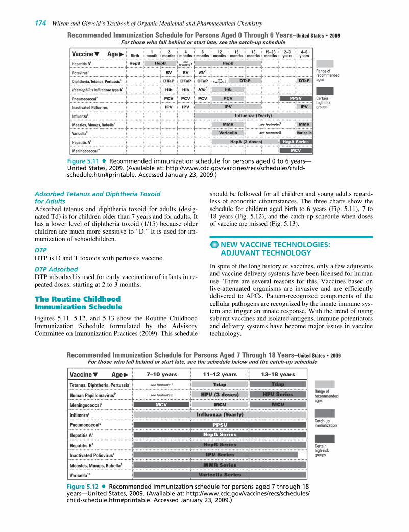
Adsorbed Tetanus and Diphtheria Toxoidfor AdultsAdsorbed tetanus and diphtheria toxoid for adults (desig-nated Td) is for children older than 7 years and for adults. Ithas a lower level of diphtheria toxoid (1/15) because olderchildren are much more sensitive to “D.” It is used for im-munization of schoolchildren.
DTPDTP is D and T toxoids with pertussis vaccine.
DTP AdsorbedDTP adsorbed is used for early vaccination of infants in re-peated doses, starting at 2 to 3 months.
The Routine ChildhoodImmunization Schedule
Figures 5.11, 5.12, and 5.13 show the Routine ChildhoodImmunization Schedule formulated by the AdvisoryCommittee on Immunization Practices (2009). This schedule
should be followed for all children and young adults regard-less of economic circumstances. The three charts show theschedule for children aged birth to 6 years (Fig. 5.11), 7 to18 years (Fig. 5.12), and the catch-up schedule when dosesof vaccine are missed (Fig. 5.13).
NEW VACCINE TECHNOLOGIES:ADJUVANT TECHNOLOGY
In spite of the long history of vaccines, only a few adjuvantsand vaccine delivery systems have been licensed for humanuse. There are several reasons for this. Vaccines based onlive-attenuated organisms are invasive and are efficientlydelivered to APCs. Pattern-recognized components of thecellular pathogens are recognized by the innate immune sys-tem and trigger an innate response. With the trend of usingsubunit vaccines and isolated antigens, immune potentiatorsand delivery systems have become major issues in vaccinetechnology.
174 Wilson and Gisvold’s Textbook of Organic Medicinal and Pharmaceutical Chemistry
Figure 5.11 Recommended immunization schedule for persons aged 0 to 6 years—United States, 2009. (Available at: http://www.cdc.gov/vaccines/recs/schedules/child-schedule.htm#printable. Accessed January 23, 2009.)
Figure 5.12 Recommended immunization schedule for persons aged 7 through 18years—United States, 2009. (Available at: http://www.cdc.gov/vaccines/recs/schedules/child-schedule.htm#printable. Accessed January 23, 2009.)
0156-0178_17865_Ch05.qxd 12/4/09 4:38 AM Page 174

Chapter 5 Immunobiologicals 175
There are a series of significant hurdles to the develop-ment of modern vaccines. Poor optimized vaccines mustbe improved. New vaccines for immunizing against dis-eases for which no vaccines exist are imperative. It is nec-essary to be able to respond quickly with vaccines tonewly developing pathogenic organisms. The primary ele-ments of useful vaccines are the antigen (against which theadaptive immune response is elicited), immunostimulantsthat activate the innate immune system, and delivery sys-tems that target the vaccines to the appropriate site in atimely fashion.
Antigens• Whole inactivated or attenuated organisms or a mixture of
various strains• Isolated and purified proteins, glycoproteins, and carbohy-
drates• Recombinant proteins and glycoproteins
Immune Potentiators• Bacterial products• Toxins and lipids• Nucleic acids• Peptidoglycans• Carbohydrates, peptides• Cytokines and hormones• Small molecules
Delivery Systems• Mineral salts• Surfactants• Synthetic microparticles• Oil-in-water emulsions• Liposome
In designing a vaccine, first, an antigen is required. Theantigen is the species against which an anamnestic immuneresponse is targeted. In many cases, antigen selection isconfounded by a multiplicity providing many potential anti-gens to choose from. Bacterial pathogens, often display thismultiplicity. The manner in which the antigen is presentedis also important. It may be necessary to maintain a spe-cific three-dimensional tertiary structure for the antigen tobe recognized.
It hasn’t always been appreciated at the innate immunesystem, must be stimulated to modulate the adaptive im-mune response. Adjuvants, which stimulate early innate im-mune responses, aid in stimulating a strong and extendedadaptive immune response. The nature of the vaccine deliv-ery systems that target the vaccines to the appropriate im-mune cells is important to gain the necessary stimulation ofthe immune system.
The functional immune system is composed of two pri-mary components. The innate immune system has evolved
Figure 5.13 Catch-up immunization schedule for persons aged 4 months through18 years who start late or who are more than 1 month behind. (Available at:http://www.cdc.gov/vaccines/recs/schedules/child-schedule.htm#printable. AccessedJanuary 23, 2009.)
0156-0178_17865_Ch05.qxd 12/4/09 4:38 AM Page 175

to respond within minutes, primarily to molecular patternsfound in microbial pathogens. The adaptive immune responsedevelops slowly, over days or weeks and generates highlyspecific responses based on antibodies targeted against apathogen’s antigens. The innate response is the first line ofdefense. Adaptive immunity uses selection and clonal ex-pansion of immune cells containing specific, somaticallyrearranged receptor genes (T- and B-cell receptors) thatrecognize antigen from the pathogen. This response is spe-cific and long-lasting (immunological memory). The innateimmune responses lead to a fast burst of inflammatory cy-tokines and activation of APCs such as macrophages and den-dritic cells (DCs). These responses are not clonal and lead toa conditioning of the immune system for later development ofspecific adaptive immune responses.
The innate immune response must be able to distinguishpathogenic microbes from self-components. To do this, theinnate immune system depends on some fairly constantreceptors that detect specific signatures from pathogens.These signatures have been highly conserved throughoutevolution. The signatures are referred to as pathogen-associated molecular patterns (PAMPs). It has been shownthat when one includes PAMP substances in experimentalvaccines, a strong and long-lasting adaptive immune re-sponse develops. PAMPs are recognized by specific patternrecognition receptors. A number of such receptors havebeen identified. They are expressed on neutrophils,macrophages, DCs, and natural killer cells, as well as B cellsin nonimmunological cells such as epithelial and endothelialcells. When PAMPs bind to pattern recognition receptorscells bearing the pattern recognition receptors are activatedand begin to secrete chemokines and cytokines, and inducematuration and chemotaxis of other immune cells. Taken to-gether, and inflammatory environment is created. This in-flammation supports development of the adaptive immuneresponses.
What are pattern recognition receptors? Basically, they arereceptors that do not induce phagocytosis such as Toll-like re-ceptors (TLRs) and nucleotide oligomerization domain(NOD) proteins. They can also be receptors that inducephagocytosis such as mannose receptors, scavenger receptors,and �-glucan receptors. These receptors directly recognizedligands on the surface of pathogenic microbes and cause themto be engulfed by phagocytosis capable cells such asmacrophages. The nonphagocytic receptors that recognizedPAMPs can be extracellular (TLRs) or intracellular (NODproteins). Activation of these receptors leads to a signal trans-duction cascade that activates transcription factors that resultin expression of inflammatory cytokines and other cellular ac-tivation processes. These processes have been reviewed pre-viously. It is now known that signaling of the innate immunesystem involves a highly complex effect of transmembraneand intracellular proteins. It is these that provide an extensiveset of possible targets for vaccine adjuvants.
How does innate immunity lead to the conditioning of theadaptive immune response? Pattern recognition receptors sig-naling causes the activation of transcription factors such asNF-�B and IRF3 (interferon regulatory factor 3). Activationof these pattern recognition receptors produces the necessaryinflammatory environment that supports fast development ofhost defenses. NF-�B controls the expression of proinflam-matory cytokines such as IL-1� and TNF-�, while IRF3causes production of antiviral type 1 interferon (IFN-� and
IFN-�). It has been shown that portions of the innate immuneresponse are absolutely essential for development of antigen-specific immunity. Therefore, elements of the innate immuneresponse are potential targets for vaccine antigens.
A suitable adjuvant can enhance the effectiveness of avaccine by accelerating the generation of a formidable im-mune response. Additionally, the adjuvant can causeveined responses for longer duration and can induce localmucosal immune responses all of which generate antibod-ies with increased avidity and neutralization capacity. Anadjuvant also can elicit cytotoxic T lymphocytes, enhanc-ing immune responses in people with weakened immunesystems, such as children, elderly, or immunocompro-mised adults. Adjuvants can be important in commercialproduction of vaccines by reducing the amount of antigenthat is required to generate a robust immune response, thusreducing the cost of the vaccine. A good functional defini-tion of an adjuvant is a component added to a vaccine andenhances the immunogenicity of the antigens in vivo. It isvery common to categorize adjuvants in terms of two typesof systems. Immune potentiators tend to activate innateimmunity directly. A good immune potentiator might fa-cilitate the development of cytokines or react through pat-tern recognition receptors (which can be bacterial cell wallcomponents). A delivery system such as an emulsion ormicroparticle can target vaccine antigens selectively toAPCs and may put the antigens and immune potentiatorsin the same location where they can do the most good. Ithas been shown that the choice of a good immune poten-tiators and delivery system can enhance antigen-specificimmune responses in vivo. This can be very important inthe development of subunit vaccines, which typically gen-erate a low-level immunity. A mixture of the immune po-tentiators, the delivery system, and the antigen, can opti-mize the immune response.
Current adjuvants that are licensed for use in humans arethe mineral salts such as aluminum and calcium. Severalbacterial and viral antigens have been adsorbed onto alu-minum and calcium salts. Other types of adjuvants includeemulsion and surfactant adjuvants, particulate delivery vehi-cles, microbial derivatives such as lipid A, and DCs and cy-tokines. These latter agents have not been licensed for use inhumans at the present time, but many of these are being usedin clinical trials at the present time. Indeed, vaccine devel-opment has historically focused on enhancing Ab responsesand not the innate immune system. So most of the com-monly used antigens elevate serum Ab titers, but do little toelicit the innate immune response. The requirement for vac-cines against chronic illnesses such as HIV, HCV, TB, andherpes simplex virus as well as cancer, has caused re-searchers to focus their efforts on generation of cellular im-mune responses. Adjuvants that elicit this effect are beingdeveloped and clinically tested at the present time. A newrecognition of the immunobiology of TLRs and patternrecognition receptors in general, immunoregulatory cells,and cells of innate and active immunity and their roles in re-sponding and clearing specific diseases provides the back-ground for their development.
Most of the research in vaccine development to the pres-ent time has been aimed at finding new antigens that can beused for protection against disease and ways that they can bepresented to the immune system. Because of this, only a fewdelivery systems such as aluminum salts and oil and water
176 Wilson and Gisvold’s Textbook of Organic Medicinal and Pharmaceutical Chemistry
0156-0178_17865_Ch05.qxd 12/4/09 4:38 AM Page 176

Chapter 5 Immunobiologicals 177
emulsions have been investigated. At the present time, noimmune potentiators have been approved for human use inprophylactic vaccines. There are several reasons for thissituation, but the main one seems to be concern over the tox-icity of these agents. Research over the last few years hascentered on developing strategies for immune potentiatordiscovery. Indeed, the methods of high throughput screen-ing that are used in the development of new drugs are beingapplied to immune potentiators and in the development ofvaccine adjuvants.
While it is true that most natural PAMPs are based onpolysaccharides, nucleic acids, or lipids, it has been shownthat peptides can activate various pattern recognition recep-tors. Therefore, immune modulators can be based on simplepeptides. This recognition opens the possibility of screeningpeptide libraries produced by combinatorial peptide synthe-sis for generation of new peptide ligands of pattern recogni-tion receptors. Of course, using peptides is not withoutdifficulty. Peptides display for pharmacokinetic profiles,low bioavailability, and degradation in vivo. As with otherbiological peptides that are being tested as drugs, wide-spread use might be restricted.
Another possibility for development of an adjuvant cen-ters on using DCs, which are known to provide potencylymphocyte responses. DCs are being investigated in vac-cines against various cancers. The difficulty and cost in-volved in preparing cell-based vaccine adjuvants probablywill restrict their use in prophylactic vaccines. They will,however, likely be used in therapeutic vaccinations.Several TLR ligands such as lipopolysaccharide, havebeen tested for their ability to activate DCs to producestimulatory molecules and chemotaxis. DCs have beenshown to increase T-cell responses and can block in-hibitory effects of regulatory cells. DCs also produce mi-gration and recruitment of natural killer cells to the lymphnodes to provide stimulation of interferon � necessary forT-helper cell polarization.
There are many barriers to be overcome in the develop-ment of adjuvants for vaccination. Because of the need forqualitatively specific immune responses, no single adju-vant will be broadly useful. Indeed, it will be necessary touse antigens on a tailor-made basis for different condi-tions. Dosing of the adjuvants according to one of the cur-rently available routes of administration may not work forsmall molecules, and it may be required that specializedformulations be developed for delivery of the adjuvant intothe cells and for retention at the site of injection. Thehighly potent activation of the innate immune responsemay cause toxic side effects, and hence may require meth-ods to prevent diffusion from the injection site. Becausesome of the targets for the innate immune system areintracellular potentiators may be required to be membranepermeable and metabolically stable. Lastly, regulatoryguidelines for licensing of new adjuvants are still being de-veloped. It is not known what the scope of three clinicaltesting and toxicity testing will be required for adjuvantapproval.
Although there is a growing acceptance by regulatoryagencies and commercial vaccine producers that improvedvaccine adjuvants are needed to meet the infectious diseasechallenges in future, new technologies carry the uncertaintyof unknown risks and present the safety and regulatoryhurdles that will be encountered with the additional novel
immune potentiators and delivery systems the final vaccineformulations may be significant and are still largely ill-defined. The key focus should be on separating the potentialincreases in immune toxicity from improved immunogenic-ity provided by vaccine adjuvants.
NEW VACCINE TECHNOLOGIES:NUCLEIC ACID VACCINES
Naked nucleic acids (divested of any component of protein,carbohydrate, or lipid) are immunostimulatory and in prin-ciple can be used as vaccines. Short molecules of DNA andRNA are relatively simple to synthesize and can be tailoredto immunize against bacterial and viral infections and can-cers. Following administration (from a “gene gun” or by in-jection into the skin or muscle in solution or suspension likeconventional vaccines) the genetic vaccines are taken upand translated into protein by the host cells. The protein,generated in situ, acts as an antigen and triggers the im-mune system. Because the naked nucleic acids lack coatprotein such as would be present in viruses, they are notsusceptible to neutralizing Ab reactions that could decreasetheir efficacy.
Entry into host cells presents a problem. On one hand,DNA can be spontaneously taken up by the cells, then tran-scribed and translated to produce protein. The uptakeprocess may be inefficient. Cationic lipids have been testedto facilitate movement of DNA (as a complex) across thecell and nuclear membranes. These have been found towork fairly well in some cases. Other avenues of adminis-tration focus on mucosal surfaces of the lungs, digestivetract, and reproductive systems. Because mucosal surfacesare important barriers to infection and transmission of in-fectious diseases, they offer important possibilities for ge-netic vaccine administration.
A typical description of how a genetic vaccine works in-volves the specialized, bone marrow–derived DCs. DCs arethe immune system’s most powerful APCs. Antigen can beacquired by DCs in three principal ways. They can be di-rectly transfected by nucleic acid vaccines. DCs are alsoknown to acquire soluble antigen from the interstitialspaces that has been secreted or released by transfectedcells. DCs can also engulf entire cells that have been in-jured or killed as a result of the vaccine or its function.Once the antigen has been taken up by the DCs, it is pre-sented on their cell surfaces. DCs then express and elabo-rate surface adhesion molecules and chemokine receptorsthat cause them to migrate to the lymphatic organs. It is inthese organs that the DCs are most effective at activatingimmune responses. It has been proposed that cell death in-duced by transfection of host cells by vaccines is the signalfor activation of DCs by providing “danger signals” tothose cells. The effect of these danger signals more stronglymodulates the immune response.
New research has suggested that �-virus replicon-basednaked DNA provides a stronger, more significant activationof the immune system than does naked DNA or RNA itself.
Despite a host of attractive possibilities presented by nu-cleic acid vaccines, no naked nucleic acid vectors have beenapproved for use in humans. Human data is lacking, andnucleic acid vaccines have not been unequivocally demon-strated to have significant efficacy in prevention or treatment
0156-0178_17865_Ch05.qxd 12/4/09 4:38 AM Page 177

(as therapeutic vaccines) of infectious diseases or cancers.Immunization with naked nucleic acids is relatively ineffi-cient, and virus vectors generally induce a much greater im-mune response than do nucleic acid vaccines. The viral coatprotein dramatically increases the virus’s replication
efficiency. Naked nucleic acids do not possess this feature.Despite being unproven in humans at the present time, ge-netic vaccines represent a fertile area for vaccine research.Presumably, as research proceeds the problems with usingnucleic acid vaccines will be surmounted.
178 Wilson and Gisvold’s Textbook of Organic Medicinal and Pharmaceutical Chemistry
R E V I E W Q U E S T I O N S
1. What is the chemical nature of an antigen?
2. What is an “antigenic determinant”?
3. What is the difference between T cells and B cells?
4. What are the functions of the major histocompatibilitycomplexes (MHC-I and MHC-II) in programming theimmune system?
5. What is the anamnestic response? How does the anamnes-tic response evolve with repeated challenges of vaccine orantigen?
6. Describe the various types of vaccines.
7. Describe the various ways that vaccines can be prepared.
8. Why is smallpox vaccine no longer recommended forgeneral use?
9. How do nucleic acid vaccines work?
10. What is an adjuvant? How do adjuvants modify the im-mune response to an antigen?
REFERENCES1. Grabenstein, J. D.: Immunofacts. St. Louis, MO, Facts and
Comparisons, 2002, pp. 3–8.2. Shen, W. G., and Louie, S. G.: Immunology for Pharmacy Students.
Newark, NJ, Harwood Academic Publishers, 2000, p. 2.3. Shen, W. G., and Louie, S. G.: Immunology for Pharmacy Students.
Newark, NJ, Harwood Academic Publishers, 2000, pp. 10–11.4. Hall, P. D., and Tami, J. A.: Function and evaluation of the immune
system. In Di Piro, J. T., et al. (eds.). Pharmacotherapy, aPathophysiologic Approach, 3rd ed. Norwalk, CT, Appleton &Lange, 1997, pp. 1647–1660 (and references therein).
5. Cremers, N.: Antigens, antibodies, and complement: Their nature andinteraction. In Burrows, W. (ed.). Textbook of Microbiology, 20th ed.Philadelphia, W. B. Saunders, 1973, pp. 303–347.
6. Leder, P.: Sci. Am. 247:72–83, 1982.7. Gilbert, P.: Fundamentals of immunology. In Hugo, W. B., and
Russell, A. D. (eds.). Pharmaceutical Microbiology, 4th ed. London,Blackwell, 1987.
8. Bussert, P. D.: Sci. Am. 246:82–95, 1981.9. Hood, L.: The immune system. In Alberts, B., et al. (eds.). The
Molecular Biology of the Cell. New York, Garland, 1983, pp.95–1012.
10. Sprent, J.: Cell 76:315–322, 1994.11. Plotkin, S. L., and Plotkin, S. A.: A short history of vaccination. In
Plotkin, S. A., and Mortimer, E. A., Jr. (eds.). Vaccines. Philadelphia,W. B. Saunders, 1988.
12. Hopkins, D. R.: Princes and Peasants: Smallpox in History. Chicago,University of Chicago Press, 1983, p. 1.
13. CDC: MMWR 43:1–3, 1994.14. CDC: MMWR 44:937–943, 1996.15. CDC: MMWR 41:103–107, 1992.16. Barker, W. H., and Mullolly, J. P.: JAMA 244:2547–2549, 1980.17. CDC: MMWR 40:700–708; 709–711, 1991.18. CDC: Immunization Information. Polio, March 9, 1995.19. CDC: MMWR 40:1–94, 1991.
20. CDC: MMWR 43:3–12, 1994.21. Hadler, S. C.: Ann. Intern. Med. 108:457–458, 1988.22. Revelle, A.: FDA Electronic Bull. Board, March 17, 1995, p. 95.23. CDC: MMWR 45:304–307, 1996.24. CDC: MMWR 31:617–625, 1982.25. CDC: MMWR 39:11–15, 1991.26. Varivax varicella virus vaccine live (OKA/Merck) prescribing
information. Darmstadt, Germany, Merck & Co., 1995.27. Varivax (recombinant OKA varicella zoster) vaccine ready for
prescribing. Merck & Co. Recommendations and Procedures. Merck& Co. House Organ, 1996.
28. White, C. J.: Pediatr. Infect. Dis. 11:19–23, 1992.29. Lieu, T. A., et al.: JAMA 271:375–381, 1980.30. Halloran, M. E.: Am. J. Epidemiol. 140:81–104, 1994.31. Grabenstein, J. D.: Immunofacts. St. Louis, MO, Facts and
Comparisons, 2002, pp. 152–169.32. Innis, B. L.: JAMA 271:1328–1334, 1994.33. Revelle, M.: FDA Electronic Bull. Board, Feb. 22, 1995. FDA
Reports, Feb. 27, 1995, pp. 5–6.34. Alter, M. J., et al.: JAMA 263:1218–1222, 1990.35. Interferon treatment for hepatitis B and C. American Liver
Foundation. http://www.gastro.com.36. Hepatitis C virus. Bug Bytes Newsletter 1, Dec. 27, 1994.37. Mast, E. E., and Alter, M. J.: Semin. Virol. 4:273–283, 1993.38. Hodder, S. L., and Mortimer, E. A.: Epidemiol. Rev. 14:243–267,
1992.39. Rappuoli, R., et al.: Vaccine 10:1027–1032, 1992.40. Englund, J., et al.: Pediatrics 93:37–43, 1994.41. CDC: MMWR CDC Surveill. Summ. 41:1–9, 1992.42. Fine, P., and Chen, R.: Am. J. Epidemiol. 136:121–135, 1992.43. American Academy of Pediatrics Committee on Infectious Diseases:
Pediatrics 92:480–488, 1993.44. Rietschel, E. T., and Brade, H.: Sci. Am. 257:54–61, 1992.45. Grabenstein, J.: Immunofacts. St. Louis, MO, Facts and
Comparisons, 2009, pp. 147–159.
0156-0178_17865_Ch05.qxd 12/4/09 4:38 AM Page 178

C H A P T E R 6
Anti-infective AgentsJOHN M. BEALE, JR.
Until the 1920s, most successful anti-infective agentswere based on the group-IIB element mercury and thegroup-VA elements arsenic and antimony. Atoxyl (sodiumarsanilate and arsphenamine) was used for sleeping sick-ness.4 Certain dyes, such as gentian violet and methyleneblue, were also found to be somewhat effective, as were afew chemical congeners of the quinine molecule. Some ofthese agents represented significant achievements in anti-infective therapy, but they also possessed some importantlimitations. Heavy metal toxicity after treatment with mer-cury, arsenic, and antimony severely limited the usefulnessof agents containing these elements.
Just prior to 1950, great strides were made in anti-infectivetherapy. The sulfonamides and sulfones (this chapter), moreeffective phenolic compounds such as hexachlorophene,
C H A P T E R O V E R V I E W
The history of work on the prevention of bacterial infectioncan be traced back to the 19th century when Joseph Lister (in1867) introduced antiseptic principles for use in surgery andposttraumatic injury.1 He used phenol (carbolic acid) as awash for the hands, as a spray on an incision site, and onbandages applied to wounds. Lister’s principles caused a dra-matic decrease in the incidence of postsurgical infections.
Around 1881 and continuing to 1900, microbiologistPaul Ehrlich, a disciple of Robert Koch, began work with aset of antibacterial dyes and antiparasitic organic arsenicals.His goal was to develop compounds that retained antimicro-bial activity at the expense of toxicity to the human host; hecalled the agents that he sought “magic bullets.” At the timethat Ehrlich began his experiments, there were only a fewcompounds that could be used in treating infectious dis-eases, and none was very useful in the treatment of severeGram-positive and Gram-negative infections. Ehrlich dis-covered that the dyes and arsenicals could stain target cellsselectively and that the antimicrobial properties of the dyesparalleled the staining activity. This discovery was the firstdemonstration of selective toxicity, the property of certainchemicals to kill one type of organism while not harminganother. Selective toxicity is the main tenet of modern an-timicrobial chemotherapy, and Ehrlich’s seminal discoverypaved the way for the development of the sulfonamides andpenicillins and the elucidation of the mechanisms of theirselective toxicity. Prior to Ehrlich’s studies, the local an-timicrobial properties of phenol and iodine were wellknown, but the only useful systemic agents were the herbalremedies cinchona for malaria and ipecac for amebic dysen-tery. Ehrlich’s discovery of compound 606, the effective an-tisyphilitic drug Salvarsan,2,3 was a breakthrough in thetreatment of a serious, previously untreatable disease.
179
0179-0241_17865_Ch06.qxd 12/8/09 10:54 PM Page 179

synthetic antimalarial compounds (Chapter 7), and severalantibiotics (Chapter 8) were introduced to the therapeuticarmamentarium.
Anti-infective agents may be classified according to var-ious schemes. The chemical type of the compound, the bio-logical property, and the therapeutic indication may be usedsingly or in combination to describe the agents. In this text-book, a combination of these classification schemes is usedto organize the anti-infective agents. When several chemi-cally divergent compounds are indicated for a specific dis-ease or group of diseases, the therapeutic classification isused, and the drugs are subclassified according to chemicaltype. When the information is best unified and presented ina chemical or biological classification system, as for the sul-fonamides or antibacterial antibiotics, then one of these clas-sification systems is used.
This chapter addresses an extremely broad base of anti-infective agents, including the local compounds (alcohols,phenols, oxidizing agents, halogen-containing compounds,cationic surfactants, dyes, and mercurials), preservatives,antifungal agents, synthetic antibacterial drugs, antitubercu-lar and antiprotozoal agents, and anthelmintics. Other chap-ters in this text are devoted to antibacterial antibiotics(Chapter 8), antiviral agents (Chapter 9), and antineoplasticantibiotics (Chapter 10).
Anti-infective agents that are used locally are called ger-micides, and within this classification, there are two pri-mary subtypes (see Table 6.1) and several other definitionsof sanitization. Antiseptics are compounds that kill (-cidal)or prevent the growth of (-static) microorganisms when ap-plied to living tissue. This caveat of use on living tissuepoints to the properties that the useful antiseptic must have.The ideal antiseptic must have low-enough toxicity that itcan be used directly on skin or wounds; it will exert a rapidand sustained lethal action against microorganisms (thespectrum may be narrow or broad depending on the use).The agent should have a low surface tension so that it willspread into the wound; it should retain activity in the pres-ence of body fluids (including pus), be nonirritating to tis-
sues, be nonallergenic, lack systemic toxicity when appliedto skin or mucous membranes, and not interfere with heal-ing. No antiseptic available today meets all of these crite-ria. A few antibiotics, such as bacitracin, polymyxin, silversulfadiazine, and neomycin, are poorly absorbed throughthe skin and mucous membranes and are used topically forthe treatment of local infections; they have been found veryeffective against infections such as these. In general, how-ever, the topical use of antibiotics has been restricted byconcern about the development of resistant microbialstrains and possible allergic reactions. These problems canreduce the usefulness of these antibiotics for more seriousinfections.
A disinfectant is an agent that prevents transmission ofinfection by the destruction of pathogenic microorganismswhen applied to inanimate objects. The ideal disinfectantexerts a rapidly lethal action against all potentially patho-genic microorganisms and spores, has good penetratingproperties into organic matter, shares compatibility with or-ganic compounds (particularly soaps), is not inactivated byliving tissue, is noncorrosive, and is aesthetically pleasing(nonstaining and odorless). Locally acting anti-infectivedrugs are widely used by the lay public and are prescribedby members of the medical profession (even though the ef-fectiveness of many of the agents has not been establishedcompletely). The germicide may be harmful in certain cases(i.e., it may retard healing). Standardized methods for eval-uating and comparing the efficacy of germicides have onlyrecently been developed.
Numerous classes of chemically divergent compoundspossess local anti-infective properties. Some of these areoutlined in Table 6.2.
The most important means of preventing transmission ofinfectious agents from person to person or from regions ofhigh microbial load, such as the mouth, nose, or gut, to po-tential sites of infection is simply washing the hands. In fact,one of the breakthroughs in surgical technique in the 1800swas the finding that the incidence of postsurgical infectiondecreased dramatically if surgeons washed their hands be-fore operating. Regular hand washing is properly done with-out disinfection to minimize drying, irritation, and sensitiza-tion of the skin. Simple soap and warm water removebacteria efficiently. Skin disinfectants along with soap andwater are usually used as preoperative surgical scrubs andsterilants for surgical incisions.
EVALUATION OF THE EFFECTIVENESSOF A STERILANT
Evaluation of the effectiveness of antiseptics, disinfectants,and other sterilants (Table 6.1), although seemingly simplein principle, is an extremely complex task. One must con-sider the intrinsic resistance of the microbe, the microbialload, the mixture of the population of microorganisms pres-ent, the amount and nature of organic material present (e.g.,blood, feces, tissue), the concentration and stability of thedisinfectant or sterilant, the time and temperature of expo-sure, the pH, and the hydration and binding of the agent tosurfaces. In summary, a host of parameters must be consid-ered for each sterilant, and experimental assays may be dif-ficult. Specific, standardized assays of activity are definedfor each use. Toxicity for human subjects must also be eval-uated. The Environmental Protection Agency (EPA)
180 Wilson and Gisvold’s Textbook of Organic Medicinal and Pharmaceutical Chemistry
TABLE 6.1 Definitions and Standards for RemovingMicroorganisms
Antisepsis Application of an agent to livingtissue for the purpose of preventinginfection
Decontamination Destruction or marked reduction in the number or activity of microorganisms
Disinfection Chemical or physical treatment that destroys most vegetative microbesor viruses, but not spores, in or oninanimate surfaces
Sanitization Reduction of microbial load on an inanimate surface to a levelconsidered acceptable for publichealth purposes
Sterilization A process intended to kill or remove all types of microorganisms,including spores, and usuallyincluding viruses with an acceptablylow probability of survival
Pasteurization A process that kills nonsporulating microorganisms by hot water orsteam at 65°C–100°C
0179-0241_17865_Ch06.qxd 12/8/09 10:54 PM Page 180

regulates disinfectants and sterilants and the Food and DrugAdministration (FDA) regulates antiseptics.
There are some problems with improper use of theseagents. Antiseptics and disinfectants may become contam-inated by resistant microorganisms (e.g., spores),Pseudomonas aeruginosa, or Serratia marcescens and mayactually transmit infection. Most topical antiseptics inter-fere with wound healing to some degree, so they should beused according to the proper directions and for a limitedlength of time.
ALCOHOLS AND RELATED COMPOUNDS
Alcohols and aldehydes have been used as antiseptics anddisinfectants for many years.5 Two of the most commonlyused antiseptics and disinfectants are ethyl and isopropylalcohol.
The antibacterial potencies of the primary alcohols(against test cultures of Staphylococcus aureus) increasewith molecular weight until the 8-carbon atom octanol isreached. In general, one oxygen atom is capable of solubi-lizing seven or eight carbon atoms in water. As the primaryalcohol chain length increases, van der Waals interactionsincrease, and the ability to penetrate microbial membranesincreases. As water solubility decreases, the apparent an-timicrobial potency diminishes with molecular weight.Branching of the alcohol chain decreases antibacterial po-tency; weaker van der Waals forces brought about bybranching do not penetrate bacterial cell membranes asefficiently. The isomeric alcohols’ potencies decrease inthe order primary > secondary > tertiary. Despite this fact,2-propanol (isopropyl alcohol) is used commercially insteadof n-propyl alcohol, because it is less expensive. Isopropylalcohol is slightly more active than ethyl alcohol againstvegetative bacterial growth, but both alcohols are largely in-effective against spores. The activity of alcohols against mi-croorganisms is the result of their ability to denature impor-tant proteins and carbohydrates.
AlcoholEthanol (ethyl alcohol, wine spirit) is a clear, colorless,volatile liquid with a burning taste and a characteristicpleasant odor. It is flammable, miscible with water in allproportions, and soluble in most organic solvents.Commercial ethanol contains approximately 95% ethanolby volume. This concentration forms an azeotrope withwater that distills at 78.2°C. Alcohol has been known forcenturies as a product of fermentation from grain and manyother carbohydrates. Ethanol can also be prepared syntheti-cally by the sulfuric-acid–catalyzed hydration of ethylene
The commerce in, and use of, alcohol in the United Statesis strictly controlled by the Treasury Department, which hasprovided the following definition for “alcohol”: “The termalcohol means that substance known as ethyl alcohol, hy-drated oxide of ethyl, or spirit of wine, from whateversource or whatever process produced, having a proof of 160or more and not including the substances commonly knownas whiskey, brandy, rum, or gin.”
Denatured alcohol is ethanol that has been rendered unfitfor use in intoxicating beverages by the addition of othersubstances. Completely denatured alcohol contains addedwood alcohol (methanol) and benzene and is unsuitable foreither internal or external use. Specially denatured alcoholis ethanol treated with one or more substances so that its usemay be permitted for a specialized purpose. Examples areiodine in alcohol for tincture of iodine, methanol, and othersubstances in mouthwashes and aftershave lotions, andmethanol in alcohol for preparing plant extracts.
The primary medicinal use of alcohol is external, as anantiseptic, preservative, mild counterirritant, or solvent.Rubbing alcohol is used as an astringent, rubefacient, and amild local anesthetic. The anesthetic effect is results fromthe evaporative refrigerant action of alcohol when applied tothe skin. Ethanol has even been injected near nerves andganglia to alleviate pain. It has a low narcotic potency andhas been used internally in diluted form as a mild sedative,a weak vasodilator, and a carminative.
Alcohol is metabolized in the human body by a series ofoxidations:
Chapter 6 Anti-infective Agents 181
TABLE 6.2 Common Sterilants and Their Range of Use
Bacteria Viruses Other
Gram- Gram- Amebicpositive negative Acid-fast Spores Lipophilic Hydrophilic Fungi Cysts Prions
Alcohols ��� ��� � � � ��� N/A N/A �(isopropanol, ethanol)
Aldehydes ��� ��� �� � � �� � N/A �(glutaraldehyde,formaldehyde)
Chlorhexidine ��� �� � � ��� � N/A N/A �gluconate
Sodium ��� ��� �� � (pH � � (high �� � ��hypochlorite, 7.6) conc.) (highchlorine dioxide conc.)
Hexachlorophene � � � � � � � � �Povidone-Iodine ��� ��� � � (high � � � � �
conc.)Phenols, ��� ��� ��� � � � N/A N/A �
quaternary ammonium
Strong oxidizing ��� ���� � � � � � � �agents, cresols
0179-0241_17865_Ch06.qxd 12/8/09 10:54 PM Page 181

Acetaldehyde causes nausea, vomiting, and vasodilatoryflushing. This fact has been used in aversion therapy withthe drug disulfiram, which blocks aldehyde dehydrogenase,allowing acetaldehyde to accumulate.
Alcohol is used in the practice of pharmacy for the prepa-ration of spirits, tinctures, and fluidextracts. Spirits arepreparations containing ethanol as the sole solvent, whereastinctures are hydroalcoholic mixtures. Many fluidextractscontain alcohol as a cosolvent.
The accepted bactericidal concentration of 70% alcoholis not supported by a study that discovered that the kill ratesof microorganisms suspended in alcohol concentrations be-tween 60% and 95% were not significantly different.6
Concentrations below 60% are also effective, but longercontact times are necessary. Concentrations above 70% canbe used safely for preoperative sterilization of the skin.7
Alcohols are flammable and must be stored in cool, well-ventilated areas.
Dehydrated EthanolDehydrated ethanol, or absolute ethanol, contains not lessthan 99% w/w of C2H5OH. It is prepared commercially byazeotropic distillation of an ethanol–benzene mixture, withprovisions made for efficient removal of water. Absoluteethanol has a very high affinity for water and must be storedin tightly sealed containers. This form of ethanol is used pri-marily as a chemical reagent or solvent but has been injectedfor the local relief of pain in carcinomas and neuralgias.Absolute alcohol cannot be ingested because there is alwayssome benzene remaining from the azeotropic distillationthat cannot be removed.
Isopropyl AlcoholIsopropanol (2-propanol) is a colorless, volatile liquid witha characteristic odor and a slightly bitter taste. It is consid-ered a suitable substitute for ethanol in most cases but mustnot be ingested. Isopropyl alcohol is prepared commerciallyby the sulfuric-acid–catalyzed hydration of propylene:
range of 50% to 95%. A 40% concentration is consideredequal in antiseptic efficacy to a 60% ethanol in water solution.Azeotropic isopropyl alcohol, United States Pharmacopeia(USP), is used on gauze pads for sterilization of the skin priorto hypodermic injections. Isopropyl alcohol is also used inpharmaceuticals and toiletries as a solvent and preservative.
Ethylene OxideEthylene oxide, C2H4O, is a colorless, flammable gas thatliquefies at 12°C. It has been used to sterilize temperature-sensitive medical equipment and certain pharmaceuticalsthat cannot be heat sterilized in an autoclave. Ethylene oxidediffuses readily through porous materials and very effec-tively destroys all forms of microorganisms at ambient tem-peratures.8
Ethylene oxide forms explosive mixtures in air at con-centrations ranging from 3% to 80% by volume. The explo-sion hazard is eliminated when the gas is mixed with suffi-cient concentrations of carbon dioxide. Carboxide is acommercial sterilant containing 10% ethylene oxide and90% carbon dioxide by volume that can be handled and re-leased in air without danger of explosion. Sterilization is ac-complished in a sealed, autoclave-like chamber or in gas-impermeable bags.
The mechanism of the germicidal action of ethyleneoxide probably involves the alkylation of functional groupsin nucleic acids and proteins by nucleophilic opening of theoxide ring. Ethylene oxide is a nonselective alkylatingagent, and for that reason is extremely toxic and potentiallycarcinogenic. Exposure to skin and mucous membranesshould be avoided, and inhalation of the gas should be pre-vented by use of an appropriate respiratory mask duringhandling and sterilization procedures.
Aldehydes
Formaldehyde SolutionFormalin is a colorless aqueous solution that officiallycontains not less than 37% w/v of formaldehyde (HCHO),with methanol added to retard polymerization. Formalin ismiscible with water and alcohol and has a characteristic pun-gent aroma. Formaldehyde readily undergoes oxidation andpolymerization, leading to formic acid and paraformalde-
182 Wilson and Gisvold’s Textbook of Organic Medicinal and Pharmaceutical Chemistry
The alcohol forms a constant-boiling mixture with waterthat contains 91% v/v of 2-propanol. Isopropyl alcohol is usedprimarily as a disinfectant for the skin and for surgical instru-ments. The alcohol is rapidly bactericidal in the concentration
0179-0241_17865_Ch06.qxd 12/8/09 10:54 PM Page 182

hyde, respectively, so the preparation should be stored intightly closed, light-resistant containers. Formalin must bestored at temperatures above 15°C to prevent cloudiness,which develops at lower temperatures.
cient is defined as the ratio of a dilution of a given test dis-infectant to the dilution of phenol that is required to kill (tothe same extent) a strain of Salmonella typhi under carefullycontrolled time and temperature conditions. As an example,if the dilution of a test disinfectant is 10-fold greater than thedilution of phenol, the phenol coefficient is 10. Obviously,the phenol coefficient of phenol itself is 1. The phenol coef-ficient test has many drawbacks. Phenols and other germi-cides do not kill microorganisms uniformly, so variations inthe phenol coefficient will occur. Moreover, the conditionsused to conduct the test are difficult to reproduce exactly, sohigh variability between different measurements and labora-tories is expected. Hence, the phenol coefficient may beunreliable.
Several phenols are actually more bactericidal than phe-nol itself. Substitution with alkyl, aryl, and halogen (espe-cially in the para position) groups increases bactericidal ac-tivity. Straight-chain alkyl groups enhance bactericidalactivity more than branched groups. Alkylated phenols andresorcinols are less toxic than the parent compounds whileretaining bactericidal properties. Phenols denature bacterialproteins at low concentrations, whereas lysis of bacterialcell membranes occurs at higher concentrations.
PhenolPhenol (carbolic acid) is a colorless to pale-pink crystallinematerial with a characteristic “medicinal odor.” It is solubleto the extent of 1 part to 15 parts water, very soluble in al-cohol, and soluble in methanol and salol (phenyl salicylate).
Phenol exhibits germicidal activity (general protoplas-mic poison), is caustic to skin, exerts local anesthetic ef-fects, and must be diluted to avoid tissue destruction anddermatitis.
Sir Joseph Lister introduced phenol as a surgical antisep-tic in 1867, and it is still used occasionally as an antipruriticin phenolated calamine lotion (0.1%–1% concentrations). A4% solution of phenol in glycerin has been used to cauterizesmall wounds. Phenol is almost obsolete as an antiseptic anddisinfectant.
Liquified PhenolLiquified phenol is simply phenol containing 10% water.The liquid form is convenient for adding phenol to variouspharmaceutical preparations because it can be measured andtransferred easily. The water content, however, precludes itsuse in fixed oils or liquid petrolatum, because the solution isnot miscible with lipophilic ointment bases.
p-Chlorophenolp-Chlorophenol is used in combination with camphor in liq-uid petrolatum as an external antiseptic and anti-irritant. Thecompound has a phenol coefficient of about 4.
Chapter 6 Anti-infective Agents 183
The germicidal action of formaldehyde is slow but pow-erful. The mechanism of action is believed to involve direct,nonspecific alkylation of nucleophilic functional groups(amino, hydroxyl, and sulfhydryl) in proteins and nucleicacids to form carbinol derivatives. The action of formalde-hyde is not confined to microorganisms. The compound isirritating to mucous membranes and causes hardening of theskin. Oral ingestion of the solution leads to severe gastroin-testinal distress. Contact dermatitis is common with forma-lin, and pure formaldehyde is suspected to be a carcinogen.
Glutaraldehyde Disinfectant SolutionGlutaraldehyde (Cidex, a 5-carbon dialdehyde) is used as adilute solution for sterilization of equipment and instru-ments that cannot be autoclaved. Commercial glutaralde-hyde is stabilized in alkaline solution. The preparation actu-ally consists of two components, glutaraldehyde and buffer,which are mixed together immediately before use. The acti-vated solution contains 2% glutaraldehyde buffered at pH7.5 to 8.0. Stabilized glutaraldehyde solutions retain morethan 80% of their original activity 30 days after prepara-tion,9 whereas the nonstabilized alkaline solutions loseabout 44% of their activity after 15 days. At higher pH(�8.5), glutaraldehyde rapidly polymerizes. Nonbufferedsolutions of glutaraldehyde are acidic, possibly because ofan acidic proton on the cyclic hemiacetal form. The acidicsolutions are stable but lack sporicidal activity.
PHENOLS AND THEIR DERIVATIVES
Phenol, USP, remains the standard to which the activity ofmost germicidal substances is compared. The phenol coeffi-
0179-0241_17865_Ch06.qxd 12/8/09 10:54 PM Page 183

p-Chloro-m-xylenolp-Chloro-m-xylenol (PC-MX; Metasep) is a nonirritatingantiseptic agent with broad-spectrum antibacterial and anti-fungal properties. It is marketed in a 2% concentration as ashampoo. It has also been used topically for the treatment oftinea (ringworm) infections such as athlete’s foot (tineapedis) and jock itch (tinea cruris).
HexachloropheneHexachlorophene, 2,2�-methylenebis (3,4,6-trichlorophe-nol); 2,2�-dihydroxy-3,5,6,3�,5�, 6�-hexachlorodiphenyl-methane (Gamophen, Surgicon, pHisoHex) is a white tolight-tan crystalline powder that is insoluble in water but issoluble in alcohol and most other organic solvents. A biphe-nol such as hexachlorophene will, in general, possessgreater potency than a monophenol. In addition, as ex-pected, the increased degree of chlorination of hexa-chlorophene increases its antiseptic potency further.
Hexachlorophene is easily adsorbed onto the skin andenters the sebaceous glands. Because of this, topical appli-cation elicits a prolonged antiseptic effect, even in low con-centrations. Hexachlorophene is used in concentrations of2% to 3% in soaps, detergent creams, lotions, and shampoosfor various antiseptic uses. It is, in general, effective againstGram-positive bacteria, but many Gram-negative bacteriaare resistant.
The systemic toxicity of hexachlorophene in animals afteroral and parenteral administration had been known for sometime, but in the late 1960s and early 1970s, reports of neuro-toxicity in infants bathed in hexachlorophene and in burn pa-tients cleansed with the agent prompted the FDA to ban itsuse in over-the-counter (OTC) antiseptic and cosmetic prepa-rations.10 Hexachlorophene is still available by prescription.
CresolCresol is actually a mixture of three isomeric methylphenols:
The mixture occurs as a yellow to brownish yellow liq-uid that has a characteristic odor of creosote. Cresol is ob-tained from coal tar or petroleum by alkaline extraction intoaqueous medium, acidification, and fractional distillation.The mixture is an inexpensive antiseptic and disinfectant. Itpossesses a phenol coefficient of 2.5. Cresol is sparinglysoluble in water, although alcohols and other organic sol-vents will solubilize it. The drawback to its use as an anti-septic is its unpleasant odor.
Chlorocresol4-Chloro-3-methylphenol occurs as colorless crystals.Chlorocresol is only slightly soluble in water. At the lowconcentration that can be achieved in aqueous media, thecompound is only useful as a preservative.
ThymolIsopropyl m-cresol is extracted from oil of Thymus vulgaris(thyme, of the mint family) by partitioning into alkalineaqueous medium followed by acidification. The crystals ob-tained from the mother liquor are large and colorless, with athymelike odor. Thymol is only slightly soluble in water,but it is extremely soluble in alcohols and other organic sol-vents. Thymol has mild fungicidal properties and is used inalcohol solutions and in dusting powders for the treatmentof tinea (ringworm) infections.
Eugenol4-Allyl-2-methoxyphenol is obtained primarily from cloveoil. It is a pale-yellow liquid with a strong aroma of clovesand a pungent taste. Eugenol is only slightly soluble in waterbut is miscible with alcohol and other organic solvents.Eugenol possesses both local anesthetic and antiseptic activ-ity and can be directly applied on a piece of cotton to relievetoothaches. Eugenol is also used in mouthwashes because ofits antiseptic property and pleasant taste. The phenol coeffi-cient of eugenol is 14.4.
184 Wilson and Gisvold’s Textbook of Organic Medicinal and Pharmaceutical Chemistry
0179-0241_17865_Ch06.qxd 12/8/09 10:54 PM Page 184

Resorcinolm-Dihydroxybenzene (resorcin), or resorcinol, is preparedsynthetically. It crystallizes as white needles or as an amor-phous powder that is soluble in water and alcohol.Resorcinol is light sensitive and oxidizes readily, so it mustbe stored in tight, light-resistant containers. It is much lessstable in solution, especially at alkaline pH. Resorcinol isonly a weak antiseptic (phenol coefficient 0.4). Nevertheless,it is used in 1% to 3% solutions and in ointments and pastesin concentrations of 10% to 20% for the treatment of skinconditions such as ringworm, eczema, psoriasis, and sebor-rheic dermatitis. In addition to its antiseptic action, resorcinolis a keratolytic agent. This property causes the stratumcorneum of the skin to slough, opening the barrier to penetra-tion for antifungal agents.
Hexylresorcinol4-Hexylresorcinol, or “hexylresorcinol,” is a white crys-talline substance with a faint phenolic odor. When appliedto the tongue it produces a sensation of numbness. It isfreely soluble in alcohol but only slightly soluble in water(1–20,000 parts). Hexylresorcinol is an effective antisep-tic, possessing both bactericidal and fungicidal properties.The phenol coefficient of hexylresorcinol against S. aureusis 98. As is typical for alkylated phenols, hexylresorcinolpossesses surfactant properties. The compound also haslocal anesthetic activity. Hexylresorcinol is formulatedinto throat lozenges because of its local anesthetic and an-tiseptic properties. These preparations are probably of lit-tle value. Hexylresorcinol (in the concentration in thelozenge) is probably not antiseptic, and the local anestheticproperty can anesthetize the larynx, causing temporarylaryngitis.
OXIDIZING AGENTS
In general, oxidizing agents that are of any value as germi-cidal agents depend on their ability to liberate oxygen in thetissues. Many of these agents are inorganic compounds, in-cluding hydrogen peroxide, several metal peroxides, andsodium perborate. All of these react in the tissues to gener-ate oxygen and oxygen radicals. Other oxidizing agents,such as KMnO4, denature proteins in microorganismsthrough a direct oxidation reaction. Oxidizing agents areespecially effective against anaerobic bacteria and can beused in cleansing contaminated wounds. The bubbles thatform during the liberation of oxygen help to dislodge debris.The effectiveness of the oxidizing agents is somewhat lim-ited by their generally poor penetrability into infected tis-sues and organic matter. Additionally, the action of the oxi-dizers is typically transient.
Carbamide Peroxide Topical SolutionCarbamide peroxide (Gly-Oxide) is a stable complex of ureaand hydrogen peroxide. It has the molecular formulaH2NCONH2 H2O2. The commercial preparation is a solu-tion of 12.6% carbamide peroxide in anhydrous glycerin.When mixed with water, hydrogen peroxide is liberated.Carbamide peroxide is used as both an antiseptic and disin-fectant. The preparation is especially effective in the treat-ment of oral ulcerations or in dental care. The oxygen bub-bles that are liberated remove debris.
Hydrous Benzoyl PeroxideHydrous benzoyl peroxide (Oxy-5, Oxy-10, Vanoxide) is awhite granular powder. In its pure powder form, it is explo-sive. The compound is formulated with 30% water to makeit safer to handle.
Compounded at 5% and 10% concentrations, benzoylperoxide is both keratolytic and keratogenic. It is used in thetreatment of acne. Benzoyl peroxide induces proliferation ofepithelial cells, leading to sloughing and repair.11
HALOGEN-CONTAINING COMPOUNDS
IODOPHORS
Elemental iodine (I2) is probably the oldest germicide still inuse today. It was listed in 1830 in USP-II as a tincture and aliniment. Iodine tincture (2% iodine in 50% alcohol withsodium iodide), strong iodine solution (Lugol’s solution, 5%iodine in water with potassium iodide), and iodine solution(2% iodine in water with sodium iodide) are currently offi-cial preparations in the USP. The iodide salt is admixed toincrease the solubility of the iodine and to reduce its volatil-ity. Iodine is one of the most effective and useful of the ger-micides. It probably acts to inactivate proteins by iodinationof aromatic residues (phenylalanyl and tyrosyl) and oxida-tion (sulfhydryl groups). Mixing with several nonionic andcationic surfactants can solubilize iodine. Complexes formthat retain the germicidal properties of the iodine while re-ducing its volatility and removing its irritant properties.12 Insome of the more active, nonionic surfactant complexes, itis estimated that approximately 80% of the dissolved iodineremains available in bacteriologically active form. These ac-tive complexes, called iodophors, are both bactericidal andfungicidal.
Povidone–IodinePovidone–iodine (Betadine, Isodine, polymer polyvinylpyrroli-done [PVP]–iodine) is a charge-transfer complex of iodine withthe nonionic surfactant PVP. The complex is extremely watersoluble and releases iodine very slowly. Hence, the preparationprovides a nontoxic, nonvolatile, and nonstaining form of io-dine that is not irritating to the skin or to wounds.Approximately 10% of the iodine in the complex is bioavail-able. Povidone–iodine is used as an aqueous solution forpresurgical disinfection of the incision site. It can also be used
Chapter 6 Anti-infective Agents 185
0179-0241_17865_Ch06.qxd 12/8/09 10:54 PM Page 185

to treat infected wounds and damage to the skin, and it is effec-tive for local bacterial and fungal infections. Several otherforms of PVP–iodine are available, including aerosols, foams,ointments, surgical scrubs, antiseptic gauze pads, sponges,mouthwashes, and a preparation that disinfects whirlpool bathsand hot tubs.
CHLORINE-CONTAINING COMPOUNDS
Chlorine and chlorine-releasing compounds have been usedin the disinfection of water supplies for more than a cen-tury. The discovery that hypochlorous acid (HClO) is theactive germicidal species that is formed when chlorine isdissolved in water led to the development and use of thefirst inorganic hypochlorite salts such as NaOCl andCa(OCl)2. Later, organic N-chloro compounds were devel-oped as disinfectants. These compounds release hypochlor-ous acid when dissolved in water, especially in the presenceof acid. Two equally plausible mechanisms have been pro-posed for the germicidal action of hypochlorous acid: thechlorination of amide nitrogen atoms and the oxidation ofsulfhydryl groups in proteins. Organic compounds thatform stable N-chloro derivatives include amides, imides,and amidines. N-Chloro compounds slowly release HOClin water. The antiseptic effect of these agents is optimal ataround pH 7.
Halazonep-Dichlorosulfamoylbenzoic acid is a white, crystalline,photosensitive compound with a faint chlorine odor.Halazone is only slightly soluble in water at pH 7 but be-comes very soluble in alkaline solutions. The sodium salt ofhalazone is used to disinfect drinking water.
ChloroazodinN,N-Dichlorodicarbonamidine (Azochloramid) is a brightyellow crystalline solid with a faint odor of chlorine. It ismostly insoluble in water and organic solvents and is unsta-ble to light or heat. Chloroazodin will explode if heatedabove 155°C. The compound is soluble enough in water tobe used in very dilute solution to disinfect wounds, as pack-ing for dental caries, and for lavage and irrigation. A glyc-eryltriacetate solution is used as a wound dressing. The an-tiseptic action of chloroazodin is long lasting because of itsextremely slow reaction with water.
Oxychlorosene SodiumOxychlorosene (Clorpactin) is a complex of the sodium saltof dodecylbenzenesulfonic acid and hypochlorous acid. Thecomplex slowly releases hypochlorous acid in solution.
Oxychlorosene occurs as an amorphous white powderthat has a faint odor of chlorine. It combines the germicidalproperties of HOCl with the emulsifying, wetting, and kera-tolytic actions of an anionic detergent. The agent has amarked and rapid-cidal action against most microorgan-isms, including both Gram-positive and Gram-negative bac-teria, molds, yeasts, viruses, and spores. Oxychlorosene isused to treat localized infections (especially when resistantorganisms are present), to remove necrotic tissue from mas-sive infections or radiation necrosis, to counteract odorousdischarges, to act as an irritant, and to disinfect cysts and fis-tulas. Oxychlorosene is marketed as a powder for reconsti-tution into a solution. A typical application uses a 0.1% to0.5% concentration in water. Dilutions of 0.1% to 0.2% areused in urology and ophthalmology.
CATIONIC SURFACTANTS
All of the cationic surfactants are quaternary ammoniumcompounds (Table 6.3). For that reason, they are alwaysionized in water and exhibit surface-active properties. Thecompounds, with a polar head group and nonpolar hydro-carbon chain, form micelles by concentrating at the inter-face of immiscible solvents. The surface activity of thesecompounds, exemplified by lauryl triethylammonium sul-fate, results from two structural moieties: (a) a cationichead group, which has a high affinity for water and (b) along hydrocarbon tail, which has an affinity for lipids andnonpolar solvents.
At the right concentration (the critical micelle concentra-tion), the molecules concentrate at the interface betweenimmiscible solvents, such as water and lipid, and water-in-oilor oil-in-water emulsions may be formed with the ammo-nium head group in the water layer and the nonpolar hydro-carbon chain associated with the oil phase. The synthesis andantimicrobial actions of the members of this class of com-
186 Wilson and Gisvold’s Textbook of Organic Medicinal and Pharmaceutical Chemistry
0179-0241_17865_Ch06.qxd 12/8/09 10:54 PM Page 186

pounds were first reported in 1908, but it was not until the pi-oneering work of Gerhard Domagk in 193513 that attentionwas directed to their usefulness as antiseptics, disinfectants,and preservatives.
The cationic surfactants exert a bactericidal action againsta broad spectrum of Gram-positive and Gram-negative bac-teria. They are also active against several pathogenic speciesof fungi and protozoa. All spores resist these agents. Themechanism of action probably involves dissolution of thesurfactant into the microbial cell membrane, destabilization,and subsequent lysis. The surfactants may also interfere withenzymes associated with the cell membrane.
The cationic surfactants possess several other properties.In addition to their broad-spectrum antimicrobial activity,they are useful as germicides. They are highly water solu-ble, relatively nontoxic, stable in solution, nonstaining, andnoncorrosive. The surface activity causes a keratolytic ac-tion in the stratum corneum and, hence, provides good tis-sue penetration. In spite of these advantages, the cationic
surfactants present several difficulties. Soaps and other an-ionic detergents inactivate them. All traces of soap must beremoved from skin and other surfaces before they are ap-plied. Tissue debris, blood, serum, and pus reduce the ef-fectiveness of the surfactants. Cationic surfactants are alsoadsorbed on glass, talc, and kaolin to reduce or preventtheir action. The bactericidal action of cationic surfactantsis slower than that of iodine. Solutions of cationic surfac-tants intended for disinfecting surgical instruments, gloves,etc. should never be reused because they can harbor infec-tious microorganisms, especially Pseudomonas andEnterobacter spp.
Benzalkonium ChlorideAlkylbenzyldimethylammonium chloride (Zephiran) is a mix-ture of alkylbenzyldimethylammonium chlorides of the gen-eral formula [C6H5CH2N(CH3)2R]�Cl�, where R represents amixture of alkyl chains beginning with C8H17 and extendingto higher homologues with C12H25, C14H29, and C16H33. The
Chapter 6 Anti-infective Agents 187
TABLE 6.3 Analogs of Dimethylbenzylammonium Chloride
0179-0241_17865_Ch06.qxd 12/8/09 10:54 PM Page 187

higher–molecular-weight homologues compose the majorfractions. Although variations in the physical and antimicro-bial properties exist between individual members of the mix-ture, they are of little importance in the chemistry of the over-all product. Benzalkonium chloride occurs as a white gel thatis soluble in water, alcohol, and organic solvents. Aqueous so-lutions are colorless, slightly alkaline, and very foamy.
Benzalkonium chloride is a detergent, an emulsifier, anda wetting agent. It is used as an antiseptic for skin and mu-cous membranes in concentrations of 1:750 to 1:20,000. Forirrigation, 1:20,000 to 1:40,000 concentrations are used. Forstorage of surgical instruments, 1:750 to 1:5,000 concentra-tions are used, with 0.5% NaNO3 added as a preservative.
Methylbenzethonium ChlorideBenzyldimethyl[2-[2-[[4-(1,1,3,3-tetramethylbutyl)tolyl]oxy]ethoxy]ethyl]ammonium chloride (Diaparene) is amixture of methylated derivatives of methylbenzethoniumchloride. It is used specifically for the treatment of diaperrash in infants, caused by the yeast Candida albicans, whichproduces ammonia. The agent is also used as a general anti-septic. Its properties are virtually identical to those of ben-zethonium chloride.
Benzethonium ChlorideBenzyldimethyl[2-[2-[p-(1,1,3,3-tetramethylbutyl)phe-noxy]ethoxy]ethyl]ammonium chloride (Phemerol chloride)is a colorless crystalline powder that is soluble in water, al-cohol, and most organic solvents. The actions and uses ofthis agent are similar to those of benzalkonium chloride. Itis used at a 1:750 concentration for skin antisepsis. For theirrigation of mucous membranes, a 1:5,000 solution is used.A 1:500 tincture is also available.
Cetylpyridinium Chloride1-Hexadecylpyridinium chloride is a white powder that isvery soluble in water and alcohol. In this compound, thequaternary nitrogen atom is a member of an aromatic pyri-dine ring.
The cetyl derivative is the most active of a series ofalkylpyridinium compounds. It is used as a general antisep-tic in concentrations of 1:100 to 1:1,000 for intact skin,1:1,000 for minor lacerations, and 1:2,000 to 1:10,000 forthe irrigation of mucous membranes. Cetylpyridinium chlo-ride is also available in the form of throat lozenges and amouthwash at a 1:20,000 dilution.
Chlorhexidine Gluconate1,6-Di(4�-chlorophenyldiguanido)hexane gluconate (Hibiclens)is the most effective of a series of antibacterial biguanides orig-inally developed in Great Britain.14
The antimicrobial properties of the biguanides were dis-covered as a result of earlier testing of these compounds aspossible antimalarial agents (Chapter 7). Although thebiguanides are technically not bisquaternary ammoniumcompounds and, therefore, should probably be classifiedseparately, they share many physical, chemical, and antimi-crobial properties with the cationic surfactants. Thebiguanides are strongly basic, and they exist as dications atphysiological pH. In chlorhexidine, the positive charges arecounterbalanced by gluconate anions (not shown). Likecationic surfactants, these undergo inactivation when mixedwith anionic detergents and complex anions such as phos-phate, carbonate, and silicate.
Chlorhexidine has broad-spectrum antibacterial activitybut is not active against acid-fast bacteria, spores, orviruses. It has been used for such topical uses as preopera-tive skin disinfection, wound irrigation, mouthwashes, andgeneral sanitization. Chlorhexidine is not absorbedthrough skin or mucous membranes and does not causesystemic toxicity.
DYES
Organic dyes were used very extensively as anti-infectiveagents before the discovery of the sulfonamides and the an-tibiotics. A few cationic dyes still find limited use as anti-infectives. These include the triphenylmethane dyes gentianviolet and basic fuchsin and the thiazine dye methylene blue.The dyes form colorless leucobase forms under alkaline con-ditions. Cationic dyes are active against Gram-positive bac-teria and many fungi; Gram-negative bacteria are generallyresistant. The difference in susceptibility is probably relatedto the cellular characteristics that underlie the Gram stain.
188 Wilson and Gisvold’s Textbook of Organic Medicinal and Pharmaceutical Chemistry
Chlorhexidine Gluconate
0179-0241_17865_Ch06.qxd 12/8/09 10:54 PM Page 188

Gentian VioletGentian violet is variously known as hexamethyl-p-rosani-line chloride, crystal violet, methyl violet, and methyl-rosaniline chloride. It occurs as a green powder or greenflakes with a metallic sheen. The compound is soluble inwater (1:35) and alcohol (1:10) but insoluble in nonpolar or-ganic solvents. Gentian violet is available in vaginal suppos-itories for the treatment of yeast infections. It is also used asa 1% to 3% solution for the treatment of ringworm and yeastinfections. Gentian violet has also been used orally as an an-thelmintic for strongyloidiasis (threadworm) and oxyuriasis.
Basic FuchsinBasic fuchsin is a mixture of the chlorides of rosaniline andp-rosaniline. It exists as a green crystalline powder with ametallic appearance. The compound is soluble in water andin alcohol but insoluble in ether. Basic fuchsin is a compo-nent of carbol–fuchsin solution (Castellani’s paint), which isused topically in the treatment of fungal infections, notablyringworm and athlete’s foot.
Methylene BlueMethylene blue is 3,7-bis(dimethylamino)-phenazathioniumchloride (Urised). The compound occurs as a dark greencrystalline powder with a metallic appearance that is solublein water (1:25) and alcohol (1:65).
Methylene blue has weak antiseptic properties that makeit useful for the treatment of cystitis and urethritis. The ac-tion of methylene blue is considered to be bacteriostatic.The compound colors the urine and stool blue green.
MERCURY COMPOUNDS (MERCURIALS)
Mercury and its derivatives have been used in medicine forcenturies. Elemental mercury incorporated into ointmentbases was used topically for the treatment of localized infec-tions and syphilis. Several inorganic salts of mercury, suchas mercuric chloride (HgCl2) and mercurous chloride(calomel, Hg2Cl2) were at one time widely used as antisep-tics. Ammoniated mercury [Hg(NH2)Cl] is still occasionallyused for skin infections such as impetigo, psoriasis, andringworm. Mercuric oxide is sometimes used to treat in-flammation resulting from infection of the eye. Although thepotential interaction of mercuric ion with the tissues isgreatly reduced by the low water solubility of these agents,they can be irritating and can cause hypersensitivity reac-tions; therefore, their use is not recommended.
The comparatively few organomercurials still in use areemployed as antiseptics, preservatives, or diuretics.Organomercurials can be grouped into two general classes:(a) compounds with at least one carbon–mercury bond thatdoes not ionize readily and (b) compounds with mercurybonded to heteroatoms (e.g., oxygen, nitrogen, sulfur) thationize partially or completely. In addition to its effect onionization, the organic moiety may increase the lipid solu-bility of an organomercurial compound, thereby facilitatingits penetration into microorganisms and host tissues.
The antibacterial action of mercury compounds is be-lieved to result from their reaction with sulfhydryl (-SH)groups in enzymes and other proteins to form covalent com-pounds of the type R-S-Hg-R�. This action is reversible bytreatment with thiol-containing compounds such as cysteineand dimercaprol (BAL); hence, organomercurials, reactingreversibly, are largely bacteriostatic. The antibacterial activ-ity of organomercurial antiseptics is greatly reduced inserum because of the presence of proteins that inactivatemercury compounds. Organomercurial antiseptics are notvery effective against spores.
The disadvantages of mercurials for antiseptic and disin-fectant uses far outweigh any possible advantages that theymight have. Hence, other more effective and less potentiallytoxic agents are preferable.
Nitromersol3-(Hydroxymercuri)-4-nitro-o-cresol inner salt (Metaphen)occurs as a yellow powder that is practically insoluble in
Chapter 6 Anti-infective Agents 189
0179-0241_17865_Ch06.qxd 12/8/09 10:54 PM Page 189

water and is sparingly soluble in alcohol and most organicsolvents. The sodium salt probably has the “inner salt”structure in which the inner shell electrons of mercury areoccupied.15 The bonding to mercury in this salt should becollinear, thus the following structure is somewhat improb-able. Nevertheless, this structure is shown in the USP andthe Merck Index.
Nitromersol is nonirritating to mucous membranes and isnonstaining. Therefore, at one time, it was a very popularantiseptic for skin and ocular infections. Nitromersol haslargely been replaced by superior agents.
Thimerosal[(o-Carboxyphenyl)-thio]ethylmercury sodium salt(Merthiolate) is a cream-colored, water-soluble powder. It isnonstaining and nonirritating to tissues. Thimerosal is aweakly bacteriostatic antiseptic that is applied topically inointments or aqueous solutions.
PRESERVATIVES
Preservatives are added to various dosage forms and cos-metic preparations to prevent microbial contamination. Inparenteral and ophthalmic preparations, preservatives areused to maintain sterility in the event of accidental contam-ination during use. An ideal preservative would be effectiveat low concentrations against all possible microorganisms,be nontoxic and compatible with other constituents of thepreparation, and be stable for the shelf life of the prepara-tion. The ideal preservative does not exist, but there is quitea bit of experience with some of them. In some cases, com-binations of preservative agents are used to approximate amixture of ideal features.
p-Hydroxybenzoic Acid Derivatives
Esters of p-hydroxybenzoic acid (parabens) have distinctantifungal properties. Their toxicity to the human host istypically low because they undergo rapid hydrolysis in vivoto p-hydroxybenzoic acid, which is quickly conjugated andexcreted. This property makes the parabens useful as preser-vatives for liquid dosage forms. The preservative activitygenerally increases with molecular weight, but the methylester is most effective against molds, whereas the propylester is most effective against yeasts. The more lipid-solublepropyl ester is the preferred preservative for drugs in oil orlipophilic bases.
MethylparabenMethyl p-hydroxybenzoate, or methylparaben, is a whitecrystalline powder. It is soluble in water and alcohol butonly slightly soluble in nonpolar organic solvents.Methylparaben is used as a safeguard against mold growth.
PropylparabenPropyl p-hydroxybenzoate, or propylparaben, occurs as awhite crystalline powder that is slightly soluble in water butsoluble in most organic solvents. It is used as a preservative,primarily to retard yeast growth. Propylparaben sodium is awater-soluble sodium salt of the 4-phenol group. The pH ofsolutions of propylparaben sodium is basic (pH �10).
Butylparabenn-Butyl p-hydroxybenzoate (butylparaben) occurs as a whitecrystalline powder that is sparingly soluble in water but verysoluble in alcohols and in nonpolar organic solvents.
EthylparabenEthyl p-hydroxybenzoate (ethylparaben) is a white crys-talline powder that is slightly soluble in water but soluble inalcohol and most organic solvents.
Other Preservatives
Chlorobutanol1,1,1-Trichloro-2-methyl-2-propanol is a white crystallinesolid with a camphorlike aroma. It occurs in an anhydrousform and a hemihydrate form, both of which sublime at roomtemperature and pressure. Chlorobutanol is slightly soluble inwater and soluble in alcohol and in organic solvents.
Chlorobutanol is used as a bacteriostatic agent in phar-maceuticals for injection, ophthalmic use, and intranasal ad-ministration. It is unstable when heated in aqueous solution,
190 Wilson and Gisvold’s Textbook of Organic Medicinal and Pharmaceutical Chemistry
0179-0241_17865_Ch06.qxd 12/8/09 10:55 PM Page 190
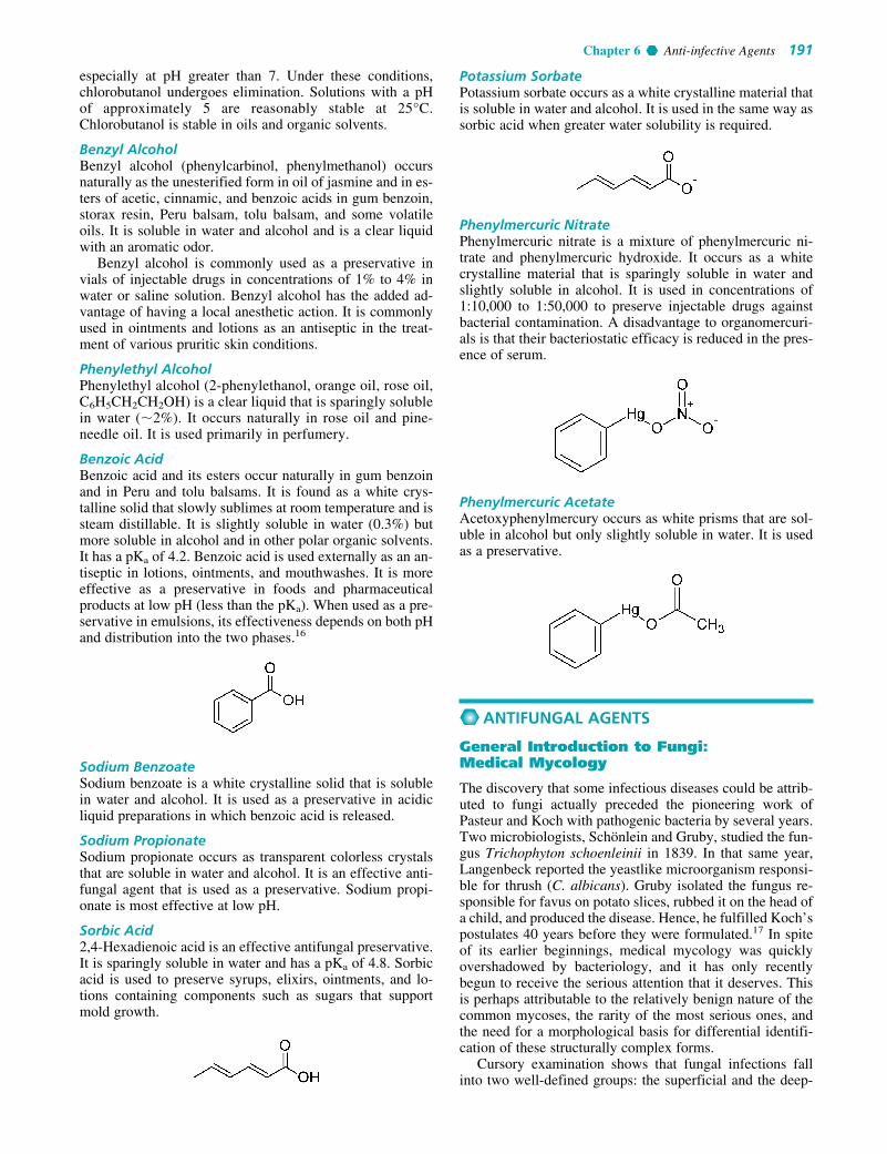
especially at pH greater than 7. Under these conditions,chlorobutanol undergoes elimination. Solutions with a pHof approximately 5 are reasonably stable at 25°C.Chlorobutanol is stable in oils and organic solvents.
Benzyl AlcoholBenzyl alcohol (phenylcarbinol, phenylmethanol) occursnaturally as the unesterified form in oil of jasmine and in es-ters of acetic, cinnamic, and benzoic acids in gum benzoin,storax resin, Peru balsam, tolu balsam, and some volatileoils. It is soluble in water and alcohol and is a clear liquidwith an aromatic odor.
Benzyl alcohol is commonly used as a preservative invials of injectable drugs in concentrations of 1% to 4% inwater or saline solution. Benzyl alcohol has the added ad-vantage of having a local anesthetic action. It is commonlyused in ointments and lotions as an antiseptic in the treat-ment of various pruritic skin conditions.
Phenylethyl AlcoholPhenylethyl alcohol (2-phenylethanol, orange oil, rose oil,C6H5CH2CH2OH) is a clear liquid that is sparingly solublein water (�2%). It occurs naturally in rose oil and pine-needle oil. It is used primarily in perfumery.
Benzoic AcidBenzoic acid and its esters occur naturally in gum benzoinand in Peru and tolu balsams. It is found as a white crys-talline solid that slowly sublimes at room temperature and issteam distillable. It is slightly soluble in water (0.3%) butmore soluble in alcohol and in other polar organic solvents.It has a pKa of 4.2. Benzoic acid is used externally as an an-tiseptic in lotions, ointments, and mouthwashes. It is moreeffective as a preservative in foods and pharmaceuticalproducts at low pH (less than the pKa). When used as a pre-servative in emulsions, its effectiveness depends on both pHand distribution into the two phases.16
Sodium BenzoateSodium benzoate is a white crystalline solid that is solublein water and alcohol. It is used as a preservative in acidicliquid preparations in which benzoic acid is released.
Sodium PropionateSodium propionate occurs as transparent colorless crystalsthat are soluble in water and alcohol. It is an effective anti-fungal agent that is used as a preservative. Sodium propi-onate is most effective at low pH.
Sorbic Acid2,4-Hexadienoic acid is an effective antifungal preservative.It is sparingly soluble in water and has a pKa of 4.8. Sorbicacid is used to preserve syrups, elixirs, ointments, and lo-tions containing components such as sugars that supportmold growth.
Potassium SorbatePotassium sorbate occurs as a white crystalline material thatis soluble in water and alcohol. It is used in the same way assorbic acid when greater water solubility is required.
Phenylmercuric NitratePhenylmercuric nitrate is a mixture of phenylmercuric ni-trate and phenylmercuric hydroxide. It occurs as a whitecrystalline material that is sparingly soluble in water andslightly soluble in alcohol. It is used in concentrations of1:10,000 to 1:50,000 to preserve injectable drugs againstbacterial contamination. A disadvantage to organomercuri-als is that their bacteriostatic efficacy is reduced in the pres-ence of serum.
Phenylmercuric AcetateAcetoxyphenylmercury occurs as white prisms that are sol-uble in alcohol but only slightly soluble in water. It is usedas a preservative.
ANTIFUNGAL AGENTS
General Introduction to Fungi:Medical Mycology
The discovery that some infectious diseases could be attrib-uted to fungi actually preceded the pioneering work ofPasteur and Koch with pathogenic bacteria by several years.Two microbiologists, Schönlein and Gruby, studied the fun-gus Trichophyton schoenleinii in 1839. In that same year,Langenbeck reported the yeastlike microorganism responsi-ble for thrush (C. albicans). Gruby isolated the fungus re-sponsible for favus on potato slices, rubbed it on the head ofa child, and produced the disease. Hence, he fulfilled Koch’spostulates 40 years before they were formulated.17 In spiteof its earlier beginnings, medical mycology was quicklyovershadowed by bacteriology, and it has only recentlybegun to receive the serious attention that it deserves. Thisis perhaps attributable to the relatively benign nature of thecommon mycoses, the rarity of the most serious ones, andthe need for a morphological basis for differential identifi-cation of these structurally complex forms.
Cursory examination shows that fungal infections fallinto two well-defined groups: the superficial and the deep-
Chapter 6 Anti-infective Agents 191
0179-0241_17865_Ch06.qxd 12/8/09 10:55 PM Page 191

seated mycoses.18 The superficial mycoses are, by far, themost common and are caused, for the most part, by a rela-tively homogeneous group of fungi, the dermatophytes.These include the various forms of tinea, or ringworm,which are infections of the hair or hair follicles, the superfi-cial infections of the intertriginous or flat areas of hairlessskin, and infections of the nails. As a rule, these lesions aremild, superficial, and restricted. The causative microbes arespecialized saprophytes with the unusual ability to digestkeratin. They have their ultimate reservoir in the soil. Unlikethe deep-seated mycoses, however, they are frequentlytransmitted from one host to another (e.g., athlete’s foot). Aspecies of yeast, Candida, also produces a dermatophyte-like disease.
Systemic Mycoses
The deep-seated, systemic mycoses have a sporadic distribu-tion,19 being common in some parts of the world and un-known in other geographical areas. These diseases have aheterogeneous etiology. Diseases caused by the systemic or-ganisms include histoplasmosis, sporotrichosis, blastomyco-sis, coccidioidomycosis, cryptococcosis, and paracoccid-ioidomycosis. The causative agents for these diseases aresoil-inhabiting saprophytes with the ability to adapt to the in-ternal environment of their host. These organisms share acommon route of infection. Fungal spores are inhaled intothe lung, and a mild, coldlike condition may result. This maybe the only symptom. In most cases, disease is inapparent. Inasymptomatic disease, diagnosis is often made serendipi-tously. Sensitization, which reflects present or previous ex-perience with the organism, may be detected by a skin test orother immunological procedure. The immune system dealswith these infections by walling them off or by producingthe giant cells that are common in type IV hypersensitivities.X-ray examination or autopsy frequently reveals these le-sions. As stated previously, the causative organisms of thesystemic infections are not typically transmitted from onehost to another, but infection by the organism in an endemicarea may be very common. Few infections develop into thesevere, deep, spreading, and often-fatal disease seen in somepersons. If the infection is symptomatic, the clinical signsmay be those of a mild, self-limiting disease; or the infectionmay become progressive, with severe symptoms, tissue andorgan damage, and, frequently, death. Recovery from adeep-seated infection of this type is accompanied by an un-certain anamnestic immune response.
Opportunistic Fungal Infections20,21
In recent years, because of overzealous use of antibacterialantibiotics, the use of immunosuppressive agents, cytotox-ins, irradiation, and steroids, a new category of systemicmycoses has become prominent. These are the opportunisticfungal infections. There has been a precipitous rise in the in-cidence of these diseases. The patient, as a result of drugtherapy, underlying disease, or medical manipulation, is de-prived of the normal defenses conferred by microbial flora.This allows organisms of normally low inherent virulence toexploit the host. Such infections include systemic candidia-sis, aspergillosis, and mucormycosis. Bacterial infectionssuch as Gram-negative septicemia, nocardiosis, andPseudomonas infection, fungal infections such as withPneumocystis carinii, and viral opportunists such as
cytomegalovirus also attack such patients. Multiple infec-tions with various microorganisms are common. C. albicansis a particularly common opportunist. This yeast is a mem-ber of the normal microbial flora of human hosts, especiallyin the vagina. Use of contraceptives often predisposes a pa-tient to infection by Candida spp. Fungal flora that inhabitthe bowel may develop into a superinfection with the use ofantibiotics to sterilize the bowel before surgery. Oral can-didiasis is common in poorly nourished persons, in patientson immunosuppressive drugs, and in persons with acquiredimmunodeficiency syndrome (AIDS). Opportunists cangrow in nearly every circumstance in which a patient’s im-mune system is compromised.
Cutaneous Infections(Dermatophytoses)22–24
By far, the most common types of human fungal disease areamong the dermatophytoses. These are superficial infectionsof the keratinized epidermis and keratinized epidermal ap-pendages (i.e., the hair and nails). The severity of an infectiondepends largely on the location of the lesion and the speciesof the fungus involved. Though certain other fungi, notablyCandida spp., produce clinically similar diseases, a some-what homogeneous group of fungi, termed the dermato-phytes, is responsible for most cases. The ability of these or-ganisms to invade and parasitize the cornified tissues of hair,skin, and nails is closely associated with, and dependent on,their common physiological characteristic—metabolic use ofthe highly insoluble scleroprotein keratin. The biochemicaluse of keratin is rare and is shared by the dermatophytespecies of the family Gymnoascaceae, with only a fewspecies of the family Onygenaceae, and certain tineae. In hu-mans, the genera Trichophyton (notably T. rubrum [nails,beard, smooth skin], T. tonsurans [scalp, beard, nails], T. vi-olaceum [scalp, skin nails], T. mentagrophytes [commonestcause of athlete’s foot], T. verrucosum [scalp, beard], and T.rubrum [psoriasis-like lesions of smooth skin, infections ofnails]), Microsporum (M. gypseum [scalp], M. fulvum [scalp,hairless skin], and M. canis [scalp, hairless skin]), andEpidermophyton (eczema) contain the most common der-matophytes. These organisms cause the conditions known astinea (ringworm). Some of the common tinea infections arelisted in Table 6.4. The fungus Pityrosporum orbicularecauses an additional type, tinea versicolor. This organism,called Malassezia furfur in older literature, causes yellow tobrown patches or continuous scaling over the trunk and occa-sionally the legs, face, and neck. The affected areas may beidentified by the inability to tan in the sun.
Regardless of the type of fungus that is causing an infec-tion (Table 6.5), treatment is extremely difficult becausefungi, like mammalians, are eukaryotes. Many biochemicalstructures, especially the cell membranes, are nearly identi-
192 Wilson and Gisvold’s Textbook of Organic Medicinal and Pharmaceutical Chemistry
TABLE 6.4 Locations of the Common Types of Tinea(Ringworm)
Type Location
Tinea manuum HandTinea cruris GroinTinea sycosis BeardTinea capitis ScalpTinea unguium Nails
0179-0241_17865_Ch06.qxd 12/8/09 10:55 PM Page 192

cal, as are many biochemical reactions. Consequently, drugsthat will kill a fungus will have a toxic effect on human cellsat normal doses.
A slight difference exists in the cell membranes. Lipid bi-layers by themselves are unstable and would be unable tohold their shape and support their functions. Sterols are em-bedded in the bilayers to act as stiffening agents. The 3-hy-droxyl group represents the polar “head” group, and the non-polar sterol skeleton and side chain align perfectly with thenonpolar chains of the bilayer. In human cells, the sterol in themembrane is cholesterol (Fig. 6.1). In fungi, the sterol is er-gosterol (Fig. 6.2). This difference amounts to the only sourceof selectivity that we have in treating fungal infections. Newantifungal drug development has focused on this difference asa way to achieve selectivity, creating highly potent antifungaldrugs that are much less toxic to the human host.
Subcutaneous Fungal Infections22
Subcutaneous mycosis refers to a group of fungal diseases inwhich both the skin and subcutaneous tissue are involvedbut typically no dissemination to the internal organs occurs.
The causative agents are classified among several unrelatedgenera. They have the following characteristics in common:(a) they are primarily soil saprophytes of very low-gradevirulence and invasive ability; and (b) in most human andanimal infections, they gain access as a result of a trauma tothe tissue. Many, if not all, organisms have the potential toestablish local infections under certain circumstances, de-pending on their adaptability and the response of the host.The tissue reaction in most cases varies with the agent inquestion but usually remains a localized lesion similar tothat elicited by a foreign body. The major disease types arechromomycosis, sporotrichosis, mycetoma, lobomycosis,and entomophthoromycosis. A type of dimorphism accom-panies infection by agents of some of these groups. The or-ganisms undergo a morphogenesis from their saprophyticform into a tissue or parasitic stage.
Tissue Reactions of Fungal Disease23
The tissue response of the host to the infecting fungusvaries widely and depends somewhat on various invasiveorganisms. In dermatophyte infections, erythema is gener-
Chapter 6 Anti-infective Agents 193
TABLE 6.5 Clinical Types of Fungal Infection
Type Disease State Causative Organism
Superficial infections Tinea versicolor Pityrosporum orbicularePiedra Trichosporon cutaneum (white)
Piedraia hortae (black)Cutaneous infections Ringworm of scalp, hairless skin, nails Dermatophytes, Microsporum, Trichophyton,
EpidermophytonCandidosis of skin, mucous membranes, Candida albicans and related forms
nails; sometimes generalizedSubcutaneous infections Chromomycosis Fonsecaea and related forms
Mycotic mycetoma Allescheria boydii, Madurella mycetoma, etc.Entomophthoromycosis Basidiobolus haptosporus,
Conidiobolus coronatusSystemic infections Histoplasmosis Histoplasma capsulatum
Blastomycosis Blastomyces dermatiditisParacoccidioidomycosis Paracoccidioides brasiliensisCoccidioidomycosis Coccidioides immitisCryptococcosis Cryptococcus neoformansSporotrichosis Sporothrix schenckiiAspergillosis Aspergillus fumigatesMucormycosis Mucor spp., Absidia spp., Rhizopus spp.Histoplasmosis duboisii Histoplasma capsulatum var. duboisii
Figure 6.1 Cholesterol embedded in alipid bilayer.
0179-0241_17865_Ch06.qxd 12/8/09 10:55 PM Page 193

ally produced and is a result of the irritation of the tissuesby the organism. Sometimes, severe inflammation, fol-lowed by scar tissue and keloid formation, occurs. This re-sults from an exaggerated inflammatory response and anallergic reaction to the organism and its products.
With organisms that invade living tissue, such as thoseresponsible for subcutaneous and systemic disease, there isgenerally a uniform acute pyogenic reaction that gives wayto various chronic disease outcomes. Granuloma withcaseation and fibrocaseous pulmonary granuloma are poten-tial outcomes of infection with Histoplasma capsulatum,and thrombotic arteritis, a thrombosis characterized by a pu-rulent coagulative necrosis and invasion of blood vessels,may be caused during aspergillosis and mucormycosis. Thelarge numbers of fungal species of many morphotypes, theirdisease etiology, and the diversity of outcomes make med-ical mycology a complex field.
Topical Agents for Dermatophytoses
Collectively, the dermatophytoses are called tinea, or ring-worm. Since these infections tend to be topical, their treat-ment has been directed to surface areas of the skin. The skinis a formidable barrier to drug penetration, and many of thetopical agents work best if an adjuvant is added that opensthe barrier function of the skin. Keratolytic agents such assalicylic acid or other �-hydroxy compounds perform thisfunction reasonably well.
FATTY ACIDS
Adults have an acidic, fatty substance in and on the skincalled sebum. Sebum functions as a natural antifungal agent,part of the innate immune system. Fatty acids have beenused for years with the idea that if a substance similar tosebum could be applied to the infected area, the effect of thesebum would be augmented and fungi could be eradicated.The application of fatty acids or their salts does in fact havean antifungal effect, albeit a feeble one.
The higher–molecular-weight fatty acids have the ad-vantage of having lower volatility. Salts of fatty acids arealso fungicidal and provide nonvolatile forms for topicalapplication.
Propionic AcidPropionic acid is an antifungal agent that is nonirritatingand nontoxic. After application, it is present in perspiration
in low concentration (�0.01%). Salt forms with sodium,potassium, calcium, and ammonium are also fungicidal.Propionic acid is a clear, corrosive liquid with a charac-teristic odor. It is soluble in water and alcohol. Thesalts are usually used because they are nonvolatile andodorless.
Zinc PropionateZinc propionate occurs as an anhydrous form and as amonohydrate. It is very soluble in water but only sparinglysoluble in alcohol. The salt is unstable to moisture, formingzinc hydroxide and propionic acid. Zinc propionate is usedas a fungicide, particularly on adhesive tape.
Sodium CaprylateSodium caprylate is prepared from caprylic acid, which is acomponent of coconut and palm oils. The salt precipitates ascream-colored granules that are soluble in water and spar-ingly soluble in alcohol.
Sodium caprylate is used topically to treat superficialdermatomycoses caused by C. albicans and Trichophyton,Microsporum, and Epidermophyton spp. The sodium saltcan be purchased in solution, powder, and ointment forms.
Zinc CaprylateZinc caprylate is a fine white powder that is insoluble inwater or alcohol. The compound is used as a topical fungi-cide. The salt is highly unstable to moisture.
Undecylenic Acid10-Undecenoic acid (Desenex, Cruex) obtained fromthe destructive distillation of castor oil. Undecylenic acidis a viscous yellow liquid. It is almost completely insolu-ble in water but is soluble in alcohol and most organicsolvents.
Undecylenic acid is one of the better fatty acids for useas a fungicide, although cure rates are low. It can be used inconcentrations up to 10% in solutions, ointments, powders,
194 Wilson and Gisvold’s Textbook of Organic Medicinal and Pharmaceutical Chemistry
Figure 6.2 Ergosterol embedded in alipid bilayer.
0179-0241_17865_Ch06.qxd 12/8/09 10:55 PM Page 194

and emulsions for topical administration. The preparationshould never be applied to mucous membranes because it isa severe irritant. Undecylenic acid has been one of theagents traditionally used for athlete’s foot (tinea pedis).Cure rates are low, however.
TriacetinGlyceryl triacetate (Enzactin, Fungacetin) is a colorless, oilyliquid with a slight odor and a bitter taste. The compound issoluble in water and miscible with alcohol and most organicsolvents.
The activity of triacetin is a result of the acetic acid re-leased by hydrolysis of the compound by esterases presentin the skin. Acid release is a self-limiting process becausethe esterases are inhibited below pH 4.
Salicylic Acid and ResorcinolSalicylic acid is a strong aromatic acid (pKa 2.5) with both an-tiseptic and keratolytic properties. It occurs as white, needle-like crystals or a fluffy crystalline powder, depending on howthe compound was brought out of solution. Salicylic acid isonly slightly soluble in water but is soluble in most organicsolvents. The greater acidity of salicylic acid and its lowersolubility in water compared with p-hydroxybenzoic acid arethe consequence of intramolecular hydrogen bonding.
Salicylic acid is used externally in ointments and solu-tions for its antifungal and keratolytic properties. By itself,salicylic acid is a poor antifungal agent.
m-Hydroxyphenol (resorcinol) possesses antiseptic andkeratolytic activity. It occurs as white, needlelike crystalsand has a slightly sweet taste. Resorcinol is soluble in water,alcohols, and organic solvents.
Benzoic AcidBenzoic acid possesses appreciable antifungal effects, but itcannot penetrate the outer layer of the skin in infected areas.Therefore, benzoic acid when used as an antifungal agentmust be admixed with a keratolytic agent. Suitable mixturesare benzoic acid and salicylic acid and benzoic acid and res-orcinol. An old preparation that is still in use is Whitfield’sOintment, USP. This ointment contains benzoic acid, 6%,and salicylic acid, 6%, in a petrolatum base. The cure ratesfrom preparations like these are low.
PHENOLS AND THEIR DERIVATIVES
Several phenols and their derivatives possess topical anti-fungal properties. Some of these, such as hexylresorcinolsand parachlorometaxylenol have been used for the treat-ment of tinea infections. Two phenolic compounds, clio-quinol and haloprogin, are still official in the USP. A thirdagent, ciclopirox olamine, is not a phenol but has proper-ties like those of phenols. All of these agents appear to in-terfere with cell membrane integrity and function in sus-ceptible fungi.
Haloprogin3-Iodo-2-propynyl-2,4,5-trichlorophenyl ether (Halotex)crystallizes as white to pale yellow forms that are sparinglysoluble in water and very soluble in ethanol. It is an etherealderivative of a phenol. Haloprogin is used as a 1% cream forthe treatment of superficial tinea infections.
Formulations of haloprogin should be protected fromlight because the compound is photosensitive. Haloprogin isavailable as a solution and a cream, both in a 1% concentra-tion. Haloprogin is probably not the first topical agent thatshould be recommended. Although the cure rates for topicalfungal infections are relatively high, they come at a highprice. The lesion typically worsens before it improves.Inflammation and painful irritation are common.
Clioquinol5-Chloro-7-iodo-8-quinolinol, 5-chloro8-hydroxy-7-iodoquinoline, or iodochlorhydroxyquin (Vioform) occursas a spongy, light-sensitive, yellowish white powder that isinsoluble in water. Vioform was initially used as a substitutefor iodoform in the belief that it released iodine in the tis-sues. It has been used as a powder for many skin conditions,such as atopic dermatitis, eczema, psoriasis, and impetigo.A 3% ointment or cream has been used vaginally as a treat-ment for Trichomonas vaginalis vaginitis. The best use forVioform is in the topical treatment of fungal infections suchas athlete’s foot and jock itch. A combination with hydro-cortisone (Vioform HC) is also available.
Chapter 6 Anti-infective Agents 195
0179-0241_17865_Ch06.qxd 12/8/09 10:55 PM Page 195

Ciclopirox Olamine25
6-Cyclohexyl-1-hydroxyl-4-methyl-2(1H)-pyridinoneethanolamine salt (Loprox) is a broad-spectrum antifungalagent intended only for topical use. It is active against der-matophytes as well as pathogenic yeasts (C. albicans) thatare causative agents for superficial fungal infections.
Ciclopirox is considered an agent of choice in the treat-ment of cutaneous candidiasis, tinea corporis, tinea cruris,tinea pedis, and tinea versicolor. It is a second-line agent forthe treatment of onychomycosis (ringworm of the nails).Loprox is formulated as a cream and a lotion, each contain-ing 1% of the water-soluble ethanolamine salt. Ciclopirox isbelieved to act on cell membranes of susceptible fungi atlow concentrations to block the transport of amino acids intothe cells. At higher concentrations, membrane integrity islost, and cellular constituents leak out.
Nucleoside Antifungals
Flucytosine26
5-Fluorocytosine, 5-FC, 4-amino-5-fluoro-2(1H)-pyrimidi-none, 2-hydroxy-4-amino-5-fluoropyrimidine (Ancobon).5-Fluorocytosine is an orally active antifungal agent with avery narrow spectrum of activity. It is indicated only for thetreatment of serious systemic infections caused by suscepti-ble strains of Candida and Cryptococcus spp.
The mechanism of action of 5-fluorocytosine (5-FC)has been studied in detail and is presented in Figure 6.3.The drug enters the fungal cell by active transport onATPases that normally transport pyrimidines. Once insidethe cell, 5-fluorocytosine is deaminated in a reaction cat-alyzed by cytosine deaminase to yield 5-fluorouracil (5-FU). 5-Fluorouracil is the active metabolite of the drug.5-Fluorouracil enters into pathways of both ribonucleotideand deoxyribonucleotide synthesis. The fluororibonu-cleotide triphosphates are incorporated into RNA, causingfaulty RNA synthesis. This pathway causes cell death. Inthe deoxyribonucleotide series, 5-fluorodeoxyuridinemonophosphate (F-dUMP) binds to 5,10-methylenete-trahydrofolic acid, interrupting the one-carbon pool sub-strate that feeds thymidylate synthesis. Hence, DNA syn-thesis is blocked.
Resistance to 5-FC is very common, and it occurs atseveral levels. A main one is at the step in which the drugis transported into the fungal cell. The transport systemsimply becomes impermeable to 5-FC. The cytosine deam-inase step is another point at which resistance occurs, andthe UMP pyrophosphorylase reaction is a third point atwhich fungal cells can become resistant. Regardless ofwhich of these mechanisms operates, fungal resistance de-velops rapidly and completely when 5-FC is administered.After a few dosing intervals, the drug is essentially useless.One strategy used to decrease resistance and to prolong theeffect of 5-FC is to administer it with the polyene antibi-otic amphotericin B. The antibiotic creates holes in thefungal cell membrane, bypassing the transport step and al-lowing 5-FC to enter. Additionally, a lower dose of 5-FCcan be used, preventing resistance by other mechanismsfor a longer period.
Antifungal Antibiotics27,28
The antifungal antibiotics make up an important group ofantifungal agents. All of the antibiotics are marked by theircomplexity. There are two classes: the polyenes, which con-tain a large number of agents with only a few being useful,and griseofulvin (one member of the class).
196 Wilson and Gisvold’s Textbook of Organic Medicinal and Pharmaceutical Chemistry
Figure 6.3 Mechanism of actionof 5-fluorocytosine.
0179-0241_17865_Ch06.qxd 12/8/09 10:55 PM Page 196
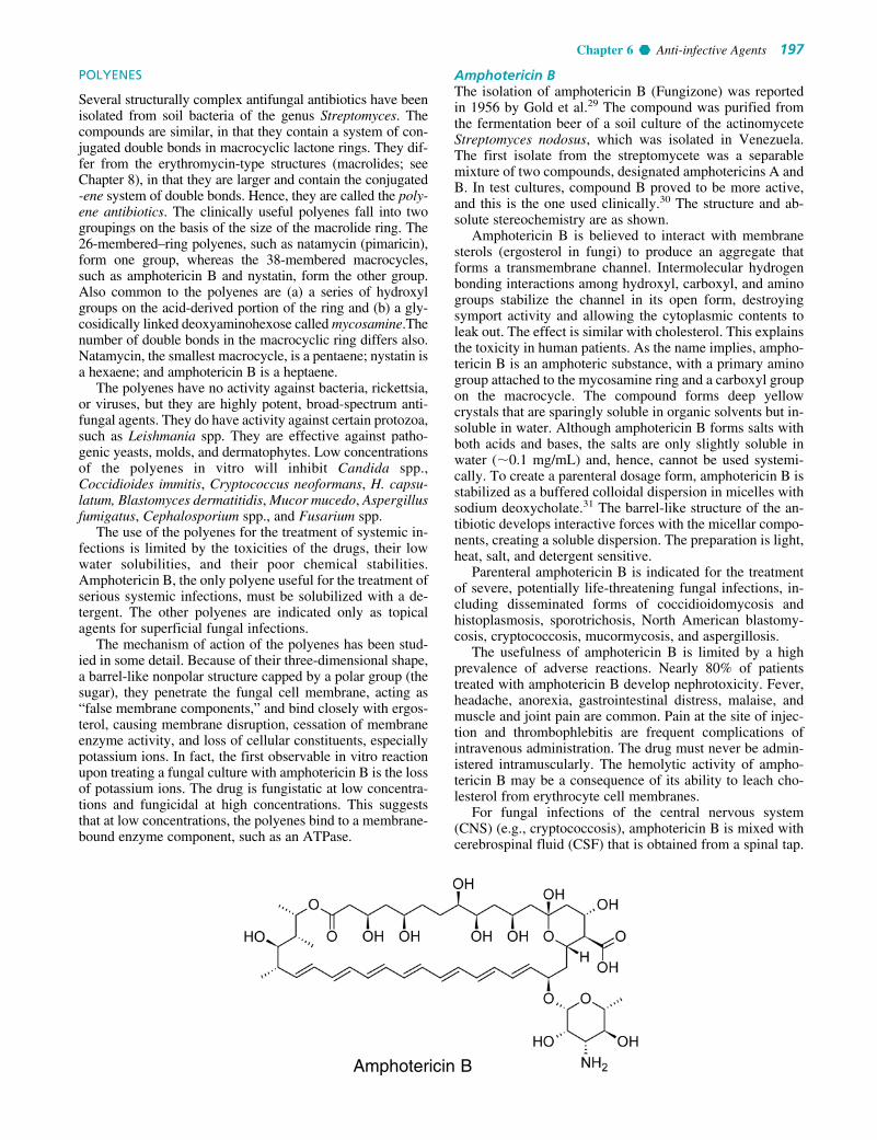
POLYENES
Several structurally complex antifungal antibiotics have beenisolated from soil bacteria of the genus Streptomyces. Thecompounds are similar, in that they contain a system of con-jugated double bonds in macrocyclic lactone rings. They dif-fer from the erythromycin-type structures (macrolides; seeChapter 8), in that they are larger and contain the conjugated-ene system of double bonds. Hence, they are called the poly-ene antibiotics. The clinically useful polyenes fall into twogroupings on the basis of the size of the macrolide ring. The26-membered–ring polyenes, such as natamycin (pimaricin),form one group, whereas the 38-membered macrocycles,such as amphotericin B and nystatin, form the other group.Also common to the polyenes are (a) a series of hydroxylgroups on the acid-derived portion of the ring and (b) a gly-cosidically linked deoxyaminohexose called mycosamine.Thenumber of double bonds in the macrocyclic ring differs also.Natamycin, the smallest macrocycle, is a pentaene; nystatin isa hexaene; and amphotericin B is a heptaene.
The polyenes have no activity against bacteria, rickettsia,or viruses, but they are highly potent, broad-spectrum anti-fungal agents. They do have activity against certain protozoa,such as Leishmania spp. They are effective against patho-genic yeasts, molds, and dermatophytes. Low concentrationsof the polyenes in vitro will inhibit Candida spp.,Coccidioides immitis, Cryptococcus neoformans, H. capsu-latum, Blastomyces dermatitidis, Mucor mucedo, Aspergillusfumigatus, Cephalosporium spp., and Fusarium spp.
The use of the polyenes for the treatment of systemic in-fections is limited by the toxicities of the drugs, their lowwater solubilities, and their poor chemical stabilities.Amphotericin B, the only polyene useful for the treatment ofserious systemic infections, must be solubilized with a de-tergent. The other polyenes are indicated only as topicalagents for superficial fungal infections.
The mechanism of action of the polyenes has been stud-ied in some detail. Because of their three-dimensional shape,a barrel-like nonpolar structure capped by a polar group (thesugar), they penetrate the fungal cell membrane, acting as“false membrane components,” and bind closely with ergos-terol, causing membrane disruption, cessation of membraneenzyme activity, and loss of cellular constituents, especiallypotassium ions. In fact, the first observable in vitro reactionupon treating a fungal culture with amphotericin B is the lossof potassium ions. The drug is fungistatic at low concentra-tions and fungicidal at high concentrations. This suggeststhat at low concentrations, the polyenes bind to a membrane-bound enzyme component, such as an ATPase.
Amphotericin BThe isolation of amphotericin B (Fungizone) was reportedin 1956 by Gold et al.29 The compound was purified fromthe fermentation beer of a soil culture of the actinomyceteStreptomyces nodosus, which was isolated in Venezuela.The first isolate from the streptomycete was a separablemixture of two compounds, designated amphotericins A andB. In test cultures, compound B proved to be more active,and this is the one used clinically.30 The structure and ab-solute stereochemistry are as shown.
Amphotericin B is believed to interact with membranesterols (ergosterol in fungi) to produce an aggregate thatforms a transmembrane channel. Intermolecular hydrogenbonding interactions among hydroxyl, carboxyl, and aminogroups stabilize the channel in its open form, destroyingsymport activity and allowing the cytoplasmic contents toleak out. The effect is similar with cholesterol. This explainsthe toxicity in human patients. As the name implies, ampho-tericin B is an amphoteric substance, with a primary aminogroup attached to the mycosamine ring and a carboxyl groupon the macrocycle. The compound forms deep yellowcrystals that are sparingly soluble in organic solvents but in-soluble in water. Although amphotericin B forms salts withboth acids and bases, the salts are only slightly soluble inwater (�0.1 mg/mL) and, hence, cannot be used systemi-cally. To create a parenteral dosage form, amphotericin B isstabilized as a buffered colloidal dispersion in micelles withsodium deoxycholate.31 The barrel-like structure of the an-tibiotic develops interactive forces with the micellar compo-nents, creating a soluble dispersion. The preparation is light,heat, salt, and detergent sensitive.
Parenteral amphotericin B is indicated for the treatmentof severe, potentially life-threatening fungal infections, in-cluding disseminated forms of coccidioidomycosis andhistoplasmosis, sporotrichosis, North American blastomy-cosis, cryptococcosis, mucormycosis, and aspergillosis.
The usefulness of amphotericin B is limited by a highprevalence of adverse reactions. Nearly 80% of patientstreated with amphotericin B develop nephrotoxicity. Fever,headache, anorexia, gastrointestinal distress, malaise, andmuscle and joint pain are common. Pain at the site of injec-tion and thrombophlebitis are frequent complications ofintravenous administration. The drug must never be admin-istered intramuscularly. The hemolytic activity of ampho-tericin B may be a consequence of its ability to leach cho-lesterol from erythrocyte cell membranes.
For fungal infections of the central nervous system(CNS) (e.g., cryptococcosis), amphotericin B is mixed withcerebrospinal fluid (CSF) that is obtained from a spinal tap.
Chapter 6 Anti-infective Agents 197
Amphotericin B
0179-0241_17865_Ch06.qxd 12/8/09 10:55 PM Page 197

The solution of amphotericin B is then reinjected throughthe tap. For severe infections, this procedure may need to berepeated many times.
Amphotericin B for injection is supplied as a sterilelyophilized cake or powder containing 50 mg of antibioticwith 41 mg of sodium deoxycholate to be dispersed in10 mL of water. The infusion, providing 0.1 mg/mL, is pre-pared by further dilution (1:50) with 5% dextrose for injec-tion. Normal saline cannot be used because it will break themicelles. The suspension should be freshly prepared andused within 24 hours. Even the powder should be refriger-ated and protected from light.
Several sterile dosage forms32 with amphotericin B ad-mixed with a lipid carrier have been developed with the goalof counteracting the dose-limiting toxicity of the drug fol-lowing parenteral administration. These include ampho-tericin B colloidal dispersion (Amphocil, Amphocyte),which contains nearly equal parts of the drug and choles-terol sulfate in a suspension of disklike particles; Abelcet, a1:1 combination of amphotericin B with L-�-dimyris-toylphosphatidylcholine (7 parts) and L-�-dimyristoylphos-phatidylglycerol (3 parts) to create a suspension of ribbon-like sheets; and liposomal amphotericin B (AmBisome), asmall laminar vesicular preparation consisting of an approx-imately 1:10 molar ratio of amphotericin B and lipid (hydro-genated soy phosphatidyl choline, cholesterol, and dis-tearoylphosphatidylcholine in a 10:5:4 ratio) for an aqueoussuspension.
The rationale behind these lipid preparations is simple:amphotericin B should have a greater avidity for the lipidvehicle than for cholesterol in cell membranes. Hence, tox-icity should be reduced. Lipid-associated amphotericin Bshould be drawn into the reticuloendothelial system, con-centrating in the lymphatic tissues, spleen, liver, and lungs,where infectious fungi tend to locate. Lipases elaboratedby the fungi and the host should release the drug from thelipid carrier, making it available to bind ergosterol in fun-gal cell membranes to exert its fungistatic and fungicidalactivities.
Clinical use of each of the approved lipid preparationshas shown reduced renal toxicity. Liposomal amphotericinB has been approved specifically for the treatment of pul-monary aspergillosis because of its demonstrated superior-ity to the sodium deoxycholate-stabilized suspension.
Amphotericin B is also used topically to treat cutaneousand mucocutaneous mycoses caused by C. albicans. Thedrug is supplied in various topical forms, including a 3%cream, a 3% lotion, a 3% ointment, and a 100-mg/mL oral
suspension. The oral suspension is intended for the treat-ment of oral and pharyngeal candidiasis. The patient shouldswish the suspension in his or her mouth and swallow it. Thesuspension has a very bad taste, so compliance may be aproblem. A slowly developing resistance to amphotericin Bhas been described. This is believed to relate to alterationsin the fungal cell membrane.
NystatinNystatin (Mycostatin) is a polyene antibiotic that was firstisolated in 1951 from a strain of the actinomyceteStreptomyces noursei by Hazen and Brown.33 It occurs as ayellow to light tan powder. Nystatin is very slightly solublein water and sparingly soluble in organic solvents. The com-pound is unstable to moisture, heat, and light.
The aglycone portion of nystatin is called nystatinolide.It consists of a 38-membered macrolide lactone ring con-taining single tetraene and diene moieties separated by twomethylene groups.34 The aglycone also contains eight hy-droxyl groups, one carboxyl group, and the lactone esterfunctionality. The entire compound is constructed by link-ing the aglycone to mycosamine. The complete structure ofnystatin has been determined by chemical degradation andx-ray crystallography.35
Nystatin is not absorbed systemically when adminis-tered by the oral route. It is nearly insoluble under all con-ditions. It is also too toxic to be administered parenterally.Hence, it is used only as a topical agent. Nystatin is a valu-able agent for the treatment of local and gastrointestinalmonilial infections caused by C. albicans and otherCandida species. For the treatment of cutaneous and mu-cocutaneous candidiasis, it is supplied as a cream, an oint-ment, and a powder. Vaginal tablets are available for thecontrol of vaginal candidiasis. Oral tablets and troches areused in the treatment of gastrointestinal and oral candidia-sis. Combinations of nystatin with tetracycline can be usedto prevent monilial overgrowth caused by the destructionof bacterial microflora of the intestine during tetracyclinetherapy.
Although nystatin is a pure compound of known struc-ture, its dosage is still expressed in terms of units. One mil-ligram of nystatin contains not less than 2,000 USP units.
Natamycin36,37
Natamycin (pimaricin; Natacyn) is a polyene antibiotic ob-tained from cultures of Streptomyces natalensis.
The natamycin structure consists of a 26-membered lactonering containing a tetraene chromophore, an �,�-unsaturatedlactone carbonyl group, three hydroxyl groups, a carboxyl
198 Wilson and Gisvold’s Textbook of Organic Medicinal and Pharmaceutical Chemistry
Nystatin
0179-0241_17865_Ch06.qxd 12/8/09 10:55 PM Page 198

group, a trans epoxide, and a glycosidically joined my-cosamine. Like the other polyene antibiotics, natamycin isamphoteric.
The mechanism action of the smaller polyenes differs fromthat of amphotericin B and nystatin. The 26-membered–ringpolyenes cause both potassium ion leakage and cell lysis atthe same concentration, whereas the 38-membered–ring poly-enes cause potassium leakage at low, fungistatic concentra-tions and cell lysis at high, fungicidal concentrations. Thesmaller polyenes are fungistatic and fungicidal within thesame concentration range.
Natamycin possesses in vitro activity against severalyeasts and filamentous fungi, including Candida,Aspergillus, Cephalosporium, Penicillium, and Fusariumspp. The drug is supplied as a 5% ophthalmic suspension in-tended for the treatment of fungal conjunctivitis, blepharitis,and keratitis.
Other Antifungal Antibiotics
GriseofulvinGriseofulvin (Grisactin, Gris-PEG, Grifulvin) was first re-ported in 1939 by Oxford et al.38 as an antibiotic obtainedfrom the fungus Penicillium griseofulvum. It was isolatedoriginally as a “curling factor” in plants. Application of ex-tracts containing the antibiotic to fungus-infected leaf partscaused the leaf to curl up. The drug has been used for manyyears for its antifungal action in plants and animals. In 1959,griseofulvin was introduced into human medicine for thetreatment of tinea infections by the systemic route.
Griseofulvin is an example of a rare structure in nature, aspiro compound. The structure of griseofulvin was deter-mined by Grove et al.39 to be 7-chloro-2�,4,6-trimethoxy-6�,�-methylspiro[benzofuran-2(3H)-1�-[2]cyclohexene]-3,4�-dione. The compound is a white, bitter, heat-stablepowder or crystalline solid that is sparingly soluble in waterbut soluble in alcohol and other nonpolar solvents. It is verystable when dry.
Griseofulvin has been used for a long time for the sys-temically delivered treatment of refractory ringworm infec-tions of the body, hair, nails, and feet caused by species ofdermatophytic fungi including Trichophyton, Microsporum,
and Epidermophyton. After systemic absorption, griseoful-vin is carried by the systemic circulation and capillary bedsto the skin, nails, and hair follicles, where it concentrates inkeratin precursor cells, which are gradually exfoliated andreplaced by healthy tissue. Griseofulvin is a fungistaticagent, and as the new, healthy tissue develops, the drug pre-vents reinfection. Treatment must be continued until all ofthe infected tissue has been exfoliated, because old tissueswill still support and harbor fungal growth. Therapy in slow-growing tissues, such as the nails, must be continued forseveral months. Compliance with the drug regimen ismandatory. In some cases, such as with the nails, it is possi-ble to observe new, healthy tissue growing in to replace theinfected tissue. Griseofulvin neither possesses antibacterialactivity nor is effective against P. obiculare, the organismthat causes tinea versicolor.
Few adverse effects have been reported for griseofulvin.The most common ones are allergic reactions such as rashand urticaria, gastrointestinal upset, headache, dizziness,and insomnia.
The oral bioavailability of griseofulvin is very poor. Thecompound is highly lipophilic with low water solubility.The most successful attempts at improving absorption havecentered on creating micronized (ultramicrosized, micro-sized) griseofulvin. Reducing the particle size, in theory,should improve dissolution in the stomach and absorption.The efficiency of gastric absorption of griseofulvin ultrami-crosized versus the microsized form is about 1.5, allowing adosage reduction of one third. Several structural derivativeshave been synthesized, but they have failed to improve ab-sorption. Perhaps the best advice that the pharmacist cangive a patient who is about to use griseofulvin is to take thedrug with a fatty meal, as with salad dressing.
Griseofulvin is a mitotic spindle poison.39,40 In vitro, itrapidly arrests cell division in metaphase. It causes a rapid,reversible dissolution of the mitotic spindle apparatus, prob-ably by binding with the tubulin dimer that is required formicrotubule assembly. The selective toxicity to fungi isprobably because of the propensity of the drug to concen-trate in tissues rich in keratin, where dermatophytes typi-cally establish infections.
Allylamines and Related Compounds
The allylamine class of antifungal agents was discovered asa result of random screening of a chemical inventory forcompounds with antifungal activity. Structure–activity stud-ies in the series subsequently led to the discovery of com-pounds with enhanced potency and potential oral activity,such as terbinafine.41,42 Investigation of the mechanism ofaction of the allylamines demonstrated that the compoundsinterfere with an early step in ergosterol biosynthesis,namely, the epoxidation of squalene catalyzed by squaleneepoxidase. Squalene epoxidase43 forms an epoxide at theC2–C3 position of squalene (Fig. 6.4). Opening of the epox-ide under acid catalysis yields a carbocation that initiates the“squalene zipper” reaction that forms the steroid nucleus.Inhibition of squalene epoxidase shuts down the biosynthe-sis of ergosterol and causes an accumulation of squalene,which destabilizes the fungal cell membrane. The allyl-amines exert a fungicidal action against dermatophytes andother filamentous fungi, but their action against pathogenicyeasts, such as Candida spp., is largely fungistatic.
Chapter 6 Anti-infective Agents 199
0179-0241_17865_Ch06.qxd 12/8/09 10:55 PM Page 199

Although mammalian squalene epoxidase is weakly inhib-ited by the allylamines, cholesterol biosynthesis does not ap-pear to be altered.
Two allylamines, naftifine and terbinafine, have been ap-proved as topical agents for the treatment of tinea pedis,tinea cruris, and tinea corporis caused by Trichophytonrubrum, Trichophyton mentagrophytes, or Epidermophytonfloccosum, respectively. The topical agent tolnaftate, al-though not an allylamine, inhibits squalene epoxidase andhas a spectrum of activity similar to that of the allylamines.Hence, tolnaftate is classified with the allylamines. The al-lylamines are weak bases that form hydrochloride salts thatare slightly soluble in water.
Naftifine HydrochlorideN-Methyl-N-(3-phenyl2-propenyl)-1-naphthalen-emethanamine hydrochloride (Naftin) is a white crys-talline powder that is soluble in polar solvents such asethanol and methylene chloride. It is supplied in a 1% con-centration in a cream and in a gel for the topical treatmentof ringworm, athlete’s foot, and jock itch. Although unap-proved for these uses, naftifine has shown efficacy fortreatment of ringworm of the beard, ringworm of the scalp,and tinea versicolor.
Terbinafine Hydrochloride(E)-N-(6,6-dimethyl-2-hepten-4-ynyl)-N-methyl-1-naphtha-lene-methanamine hydrochloride (Lamisil) is an off-whitecrystalline material that is soluble in polar organic solventssuch as methanol, ethanol, and methylene chloride but isonly slightly soluble in water. The highly lipophilic freebase is insoluble in water. Terbinafine hydrochloride isavailable in a 1% cream for topical administration for thetreatment of tinea pedis, tinea corporis, and tinea cruris.Terbinafine is more potent than naftifine and has alsodemonstrated oral activity against onychomycosis (ring-worm of the nails). It has not been approved in the UnitedStates for oral administration.
TolnaftateO,2-Naphthyl m,N-dimethylthiocarbanilate (Tinactin,Aftate, NP-27) is a white crystalline solid that is insoluble inwater, sparingly soluble in alcohol, and soluble in most or-ganic solvents. The compound, a thioester of �-naphthol, isfungicidal against dermatophytes, such as Trichophyton,Microsporum, and Epidermophyton spp., that cause superfi-cial tinea infections. Tolnaftate is available in a concentra-tion of 1% in creams, powders, aerosols, gels, and solutionsfor the treatment of ringworm, jock itch, and athlete’s foot.Tolnaftate has been shown to act as an inhibitor of squaleneepoxidase44 in susceptible fungi, so it is classified with theallylamine antimycotics. Tolnaftate is formulated into prepa-rations intended to be used with artificial fingernails to coun-teract the increased chance of ringworm of the nail beds.
Azole Antifungal Agents
The azoles represent a class of synthetic antifungal agentsthat possess a unique mechanism of action. With thesedrugs, one can achieve selectivity for the infecting fungusover the host. Depending on the azole drug used, one cantreat infections ranging from simple dermatophytoses tolife-threatening, deep systemic fungal infections. Researchcurrently under way in the United States is aimed at devel-oping more potent azoles and compounds that penetrate theblood-brain barrier more effectively. The first members ofthe class were highly substituted imidazoles, such as clotri-mazole and miconazole. Structure–activity studies revealedthat the imidazole ring could be replaced with a bioisosteric1,2,4-triazole ring without adversely affecting the antifungalproperties of the molecule. Hence, the more generic termazoles refers to this class of antifungal agents.
200 Wilson and Gisvold’s Textbook of Organic Medicinal and Pharmaceutical Chemistry
Figure 6.4 Squalene epoxidase reaction.
0179-0241_17865_Ch06.qxd 12/8/09 10:55 PM Page 200

ANTIFUNGAL SPECTRUM
The azoles tend to be effective against most fungi that causesuperficial infections of the skin and mucous membranes,including the dermatophytes such as Trichophyton,Epidermophyton, and Microsporum spp. and yeasts such asC. albicans. On the other hand, they also exhibit activityagainst yeasts that cause systemic infections, includingC. immitis, C. neoformans, Paracoccidioides brasiliensis,Petriellidium boydii, B. dermatitidis, and H. capsulatum.
MECHANISM OF ACTION
The effects of the azoles on fungal biochemistry have beenstudied extensively, but there is still much to be learned.45 At
high in vitro concentrations (micromolar), the azoles are fun-gicidal; at low in vitro concentrations (nanomolar), they arefungistatic. The fungicidal effect is clearly associated withdamage to the cell membrane, with the loss of essential cellu-lar components such as potassium ions and amino acids. Thefungistatic effect of the azoles at low concentration has beenassociated with inhibition of membrane-bound enzymes. Acytochrome P450-class enzyme, lanosterol 14�-demethylase,is the likely target for the azoles.46 P450 possesses a hememoiety as part of its structure (Fig. 6.5), and the basic electronpairs of the azole rings can occupy a binding site on P450,preventing the enzyme from turning over. The function oflanosterol 14�-demethylase is to oxidatively remove a methylgroup from lanosterol during ergosterol biosynthesis.
Chapter 6 Anti-infective Agents 201
Figure 6.5 The inhibitory action of azole antifungal agents on the lanosterol 14-�-demethylase reaction.
0179-0241_17865_Ch06.qxd 12/8/09 10:55 PM Page 201

When demethylation is inhibited, the 14�-sterol accumu-lates in the membrane, causing destabilization. As this hap-pens, repair mechanisms, such as chitin synthesis, are initi-ated to patch the damage. This degrades membrane functionfurther. Lanosterol 14�-demethylase is also required formammalian biosynthesis of cholesterol, and the azoles areknown to inhibit cholesterol biosynthesis.47 In general,higher concentrations of the azoles are needed to inhibit themammalian enzyme. This provides selectivity for antifungalaction. The 1,2,4-triazoles appear to cause a lower incidenceof endocrine effects and hepatotoxicity than the correspon-ding imidazoles, possibly because of a lower affinity for themammalian cytochrome P450 enzymes involved.48 The pri-mary mode of resistance to the triazoles and imidazoles inC. albicans is the development of mutations in ERG 11, thegene coding for C14-�-sterol demethylase. These mutationsappear to protect heme in the enzyme pocket from bindingto azole but allow access of the natural substrate of the en-zyme, lanosterol. Cross-resistance is conferred to all azoles.Increased azole efflux by the ATP-binding cassette (ABC-1,which normally transports cholesterol) and major facilitatorsuperfamily transporters can add to fluconazole resistance inC. albicans and C. glabrata. Increased production of C14-�-sterol demethylase could be another cause of resistance.
STRUCTURE–ACTIVITY RELATIONSHIPS
The basic structural requirement for members of the azoleclass is a weakly basic imidazole or 1,2,4-triazole ring (pKa
of 6.5–6.8) bonded by a nitrogen–carbon linkage to the restof the structure. At the molecular level, the amidine nitrogenatom (N-3 in the imidazoles, N-4 in the triazoles) is believedto bind to the heme iron of enzyme-bound cytochrome P450to inhibit activation of molecular oxygen and prevent oxida-tion of steroidal substrates by the enzyme. The most potentantifungal azoles possess two or three aromatic rings, atleast one of which is halogen substituted (e.g., 2,4-dichlorophenyl, 4-chlorophenyl, 2,4-difluorophenyl), andother nonpolar functional groups. Only 2, and/or 2,4 substi-tution yields effective azole compounds. The halogen atomthat yields the most potent compounds is fluorine, althoughfunctional groups such as sulfonic acids have been shown todo the same. Substitution at other positions of the ring yieldsinactive compounds. Presumably, the large nonpolar portionof these molecules mimics the nonpolar steroidal part of thesubstrate for lanosterol 14�-demethylase, lanosterol, inshape and size.
The nonpolar functionality confers high lipophilicity tothe antifungal azoles. The free bases are typically insolublein water but are soluble in most organic solvents, such asethanol. Fluconazole, which possesses two polar triazolemoieties, is an exception, in that it is sufficiently water solu-ble to be injected intravenously as a solution of the free base.
PRODUCTS
Clotrimazole1-(o-Chloro-�,�-diphenylbenzyl)imidazole (Lotrimin, Gyne-Lotrimin, Mycelex) is a broad-spectrum antifungal drug thatis used topically for the treatment of tinea infections and can-didiasis. It occurs as a white crystalline solid that is sparinglysoluble in water but soluble in alcohol and most organic sol-vents. It is a weak base that can be solubilized by dilute min-eral acids.
Clotrimazole is available as a solution in polyethyleneglycol 400, a lotion, and a cream in a concentration of 1%.These are all indicated for the treatment of tinea pedis, tineacruris, tinea capitis, tinea versicolor, or cutaneous candidia-sis. A 1% vaginal cream and tablets of 100 mg and 500 mgare available for vulvovaginal candidiasis. Clotrimazole isextremely stable, with a shelf life of more than 5 years.
Although clotrimazole is effective against various patho-genic yeasts and is reasonably well absorbed orally, itcauses severe gastrointestinal disturbances. It is also exten-sively protein bound and, hence, is not considered optimallybioavailable. Clotrimazole is not considered suitable for thetreatment of systemic infections.
Econazole Nitrate1-[2-[(4-Chlorophenyl)methoxy]-2-(2,4-dichlorophenyl)-ethyl]-1H-imidazole (Spectazole) is a white crystalline ni-tric acid salt of econazole. It is only slightly soluble in waterand most organic solvents.
Econazole is used as a 1% cream for the topical treatmentof local tinea infections and cutaneous candidiasis.
Butoconazole Nitrate1-[4-(4-Chlorophenyl)-2-[(2,6-dichlorophenyl)-thio]butyl]-1H-imidazole (Femstat) is an extremely broad-spectrum an-tifungal drug that is specifically effective against C. albi-cans. It is supplied as a vaginal cream containing 2% of thesalt. It is intended for the treatment of vaginal candidiasis.
Sulconazole Nitrate1-[2,4-Dichloro-�-[p-chlorobenzyl)thio]phenethyl]imida-zole mononitrate (Exelderm) is the white crystalline nitricacid salt of sulconazole. It is sparingly soluble in water butsoluble in ethanol. The salt is used in a solution and a cream
202 Wilson and Gisvold’s Textbook of Organic Medicinal and Pharmaceutical Chemistry
0179-0241_17865_Ch06.qxd 12/8/09 10:55 PM Page 202

in 1% concentration for the treatment of local tinea infec-tions, such as jock itch, athlete’s foot, and ringworm.
Oxiconazole Nitrate(Z)-1-(2,4-dichlorophenyl)-2-(1H-imidazol-1-yl)ethanone-O-[2,4-dichlorophenyl)methyl]oxime mononitrate (Oxistat)is a white crystalline nitric acid salt. It is used in cream andlotion dosage forms in 1% concentration for the treatment oftinea pedis, tinea corporis, and tinea capitis.
Tioconazole1-[2-[(2-chloro-3-thienyl)methoxy]2-(2,4-dichlorophenyl)-ethyl]-1H-imidazole (Vagistat) is used for the treatment ofvulvovaginal candidiasis. A vaginal ointment containing6.5% of the free base is available. Tioconazole is more ef-fective against Torulopsis glabrata than are other azoles.
Miconazole Nitrate1-[2-(2,4-Dichlorophenyl)-2-[2,4-dichlorophenyl]-methoxy]ethyl]-1H-imidazole mononitrate (Monistat,Micatin) is a weak base with a pKa of 6.65. The nitric acidsalt occurs as white crystals that are sparingly soluble inwater and most organic solvents.
The free base is available in an injectable form, solubi-lized with polyethylene glycol and castor oil, and intendedfor the treatment of serious systemic fungal infections, suchas candidiasis, coccidioidomycosis, cryptococcosis, petriel-lidiosis, and paracoccidioidomycosis. It may also be usedfor the treatment of chronic mucocutaneous candidiasis.Although serious toxic effects from the systemic administra-tion of miconazole are comparatively rare, throm-bophlebitis, pruritus, fever, and gastrointestinal upset arerelatively common.
Miconazole nitrate is supplied in various dosage forms(cream, lotion, powder, and spray) for the treatment of tineainfections and cutaneous candidiasis. Vaginal creams andsuppositories are also available for the treatment of vaginalcandidiasis. A concentration of 2% of the salt is used inmost topical preparations.
Ketoconazole1-Acetyl-4-[4-[[2-(2,4-dichlorophenyl)-2(1H-imidazole-1-ylmethyl)-1,3-dioxolan-4-yl]methoxy]phenyl]piperazine(Nizoral) is a broad-spectrum imidazole antifungal agentthat is administered orally for the treatment of systemic fun-gal infections. It is a weakly basic compound that occurs asa white crystalline solid that is very slightly soluble in water.
The oral bioavailability of ketoconazole depends on anacidic pH for dissolution and absorption. Antacids and drugssuch as H2-histamine antagonists and anticholinergics thatinhibit gastric secretion interfere with its oral absorption.Ketoconazole is extensively metabolized to inactivemetabolites, and the primary route of excretion is enterohe-patic. It is estimated to be 95% to 99% bound to protein inthe plasma.
Hepatotoxicity, primarily of the hepatocellular type, isthe most serious adverse effect of ketoconazole.Ketoconazole is known to inhibit cholesterol biosynthesis,47
suggesting that lanosterol 14�-demethylase is inhibited inmammals as well as in fungi. High doses have also been re-ported to lower testosterone and corticosterone levels, re-flecting the inhibition of cytochrome P450-requiring en-zymes involved in human steroid hormone biosynthesis.48
Cytochrome P450 oxidases responsible for the metabolismof various drugs may also be inhibited by ketoconazole tocause enhanced effects. Thus, ketoconazole causes clini-cally significant increases in plasma concentrations of cy-closporine, phenytoin, and terfenadine. It may also enhanceresponses to sulfonylurea hypoglycemic and coumarin anti-coagulant drugs.
Ketoconazole is a racemic compound, consisting of thecis-2S,4R and cis-2R,4S isomers. An investigation of the rel-ative potencies of the four possible diastereomers of keto-conazole against rat lanosterol 14�-demethylase49 indicatedthat the 2S,4R isomer was 2.5 times more active than its2R,4S enantiomer. The trans-isomers, 2S,4S and 2R,4R, aremuch less active.49
Chapter 6 Anti-infective Agents 203
0179-0241_17865_Ch06.qxd 12/8/09 10:55 PM Page 203

Ketoconazole is recommended for the treatment of thefollowing systemic fungal infections: candidiasis (includingoral thrush and the chronic mucocutaneous form), coccid-ioidomycosis, blastomycosis, histoplasmosis, chromomyco-sis, and paracoccidioidomycosis. It is also used orally totreat severe refractory cutaneous dermatophytic infectionsnot responsive to topical therapy or oral griseofulvin. Theantifungal actions of ketoconazole and the polyene antibi-otic amphotericin B are reported to antagonize each other.
Ketoconazole is also used topically in a 2% concentra-tion in a cream and in a shampoo for the management of cu-taneous candidiasis and tinea infections.
Terconazolecis-1-[4-[[2-(2,4-Dichlorophenyl)-2-(1H-1,2,4-triazol-1-yl-methyl)-1,3-dioxolan-4-yl)methoxy]phenyl]-4-(1methylethyl)piperazine (Terazol), or terconazole, is a tria-zole derivative that is used exclusively for the control ofvulvovaginal moniliasis caused by C. albicans and otherCandida species. It is available in creams containing 0.4%and 0.8% of the free base intended for 7-day and 3-day treat-ment periods, respectively. Suppositories containing 80 mgof the free base are also available.
Itraconazole4-[4-[4-[4-[[2-(2,4-Dichlorophenyl)-2-1H-1,2,4-triazol-1-ylmethyl)-1,3-dioxolan-4-yl]methoxy]phenyl]-1-piper-azinyl]phenyl]-2,4-dihydro-2-(1-methylpropyl)-3H-1,2,4-triazol-3-one (Sporanox) is a unique member of the azoleclass that contains two triazole moieties in its structure, aweakly basic 1,2,4-triazole and a nonbasic 1,2,4-triazol-3-one.
Itraconazole is an orally active, broad-spectrum antifun-gal agent that has become an important alternative to keto-conazole. An acidic environment is required for optimumsolubilization and oral absorption of itraconazole. Drugssuch as H2-histamine antagonists and antacids, which
reduce stomach acidity, reduce its gastrointestinal absorp-tion. Food greatly enhances the absorption of itraconazole,nearly doubling its oral bioavailability. The drug is avidlybound to plasma proteins (nearly 99% at clinically effectiveconcentrations) and extensively metabolized in the liver.Only one of the numerous metabolites, namely 1-hydroxyi-traconazole, has significant antifungal activity. Virtuallynone of the unchanged drug is excreted in the urine. Thus,the dosage need not be adjusted in patients with renal im-pairment. The terminal elimination half-life of itraconazoleranges from 24 to 40 hours.
The primary indications for itraconazole are for the treat-ment of systemic fungal infections including blastomycosis,histoplasmosis (including patients infected with human im-munodeficiency virus [HIV]), nonmeningeal coccid-ioidomycosis, paracoccidioidomycosis, and sporotrichosis.It may also be effective in the treatment of pergellosis, dis-seminated and deep organ candidiasis, coccidioidal menin-gitis, and cryptococcosis.
In general, itraconazole is more effective and better tol-erated than is ketoconazole. Unlike ketoconazole, it is nothepatotoxic and does not cause adrenal or testicular suppres-sion in recommended therapeutic doses.14 Nonetheless, itra-conazole can inhibit cytochrome P450 oxidases involved indrug and xenobiotic metabolism and is known to increaseplasma levels of the antihistaminic drugs terfenadine andastemizole.
Fluconazole�-(2,4-Difluorophenyl)-�-(1H-1,2,4-triazol-1-ylmethyl)-1H-1,2,4-triazole-1-ethanol or 2,4-difluoro-�,�-bis(1H-1,2,4-triazol-1-ylmethyl)benzyl alcohol (Diflucan) is a water-soluble bis-triazole with broad-spectrum antifungal proper-ties that is suitable for both oral and intravenous administra-tion as the free base. Intravenous solutions of fluconazolecontain 2 mg of the free base in 1 mL of isotonic sodiumchloride or 5% dextrose vehicle.
204 Wilson and Gisvold’s Textbook of Organic Medicinal and Pharmaceutical Chemistry
Itraconazole
0179-0241_17865_Ch06.qxd 12/8/09 10:55 PM Page 204

The oral bioavailability of fluconazole, following admin-istration of either tablet or oral suspension dosage forms, isexcellent. Apparently, the presence of two weakly basic tri-azole rings in the molecule confers sufficient aqueous solu-bility to balance the lipophilicity of the 2,4-difluorophenylgroup. The oral absorption of fluconazole, in contrast to theoral absorption of ketoconazole or itraconazole, is not af-fected by alteration in gastrointestinal acidity or the pres-ence of food.
Fluconazole has a relatively long elimination half-life,ranging from 27 to 34 hours. It penetrates well into allbody cavities, including the CSF. Plasma protein bindingof fluconazole is less than 10%; the drug is efficiently re-moved from the blood by hemodialysis. Fluconazole expe-riences little or no hepatic metabolism and is excreted sub-stantially unchanged in the urine. A small amount ofunchanged fluconazole (�10%) is excreted in the feces.Side effects of fluconazole are largely confined to minorgastrointestinal symptoms. Inhibition of cytochrome P450oxidases by fluconazole can give rise to clinically signifi-cant interactions involving increased plasma levels of cy-closporine, phenytoin, and the oral hypoglycemic drugs(tolbutamide, glipizide, and glyburide). Fluconazole doesnot appear to interfere with corticosteroid or androgenbiosynthesis in dosages used to treat systemic fungalinfections.
Fluconazole is recommended for the treatment andprophylaxis of disseminated and deep organ candidiasis.It is also used to control esophageal and oropharyngealcandidiasis. Because of its efficient penetration into CSF,fluconazole is an agent of choice for the treatment ofcryptococcal meningitis and for prophylaxis againstcryptococcosis in AIDS patients. Although fluconazoleis generally less effective than either ketoconazole oritraconazole against nonmeningeal coccidioidomycosis, itis preferred therapy for coccidioidal meningitis.Fluconazole lends itself to one-dose therapies for vaginalcandidiasis.
NEWER ANTIFUNGAL STRATEGIES
A newer azole is voriconazole.50
Unlike fluconazole, voriconazole has potent activityagainst a broad variety of fungi, including the clinically im-portant pathogens. Several publications have substantiatedthe use of voriconazole against some of the newer and rarerfungal pathogens. Voriconazole is more potent than itra-conazole against Aspergillus spp. and is comparable toposaconazole,50 another azole that is in clinical trials, in itsactivity against C. albicans. In general, Candida spp. thatare less susceptible to fluconazole possess higher MICs tovoriconazole. The in vitro activity of posaconazole appearsto be similar to that of voriconazole. Posaconazole is now inphase III clinical trials, and evidence of the efficacy ofposaconazole against various fungal models, especially therarer ones, continues to accumulate. Posaconazole exhibitshigh oral bioavailability, but its low water solubility makesits formulation into an intravenous solution impossible.
A search for potential prodrug forms of posaconazole hasyielded a possible candidate, SCH 59884. The compound isinactive in vitro but is dephosphorylated in vivo to yield theactive 4-hydroxybutyrate ester. This compound is hy-drolyzed to the parent compound in the serum. Posaconazoleundergoes extensive enterohepatic recycling, and most of thedose is eliminated in the bile and feces.
Syn2869 is a novel broad-spectrum compound that con-tains the piperazine-phenyl-triazolone side chain commonto itraconazole and posaconazole, and it displays potencyand an antifungal spectrum similar to those of the latter.Syn2869 demonstrates better activity than itraconazole inanimal models of C. albicans, C. glabrata, and C. neofor-mans. The oral bioavailability (F) is 60%, and higher
Chapter 6 Anti-infective Agents 205
SCH 59884
Syn2869
0179-0241_17865_Ch06.qxd 12/8/09 10:55 PM Page 205

tissue-to-serum ratios than those found for itraconazolewere claimed to contribute to the greater efficacy of thecompound in a model of invasive pulmonary aspergillosis.Syn2869 also demonstrates considerable activity againstthe common mold pathogens.
ECHINOCANADINS AND PNEUMOCANADINS
Echinocanadins51 and the closely related pneumocanadins52
are natural products that were discovered in the 1970s.They act as noncompetitive inhibitors of (1,3)-�-d-glucansynthase,53 an enzyme complex that forms stabilizing glucanpolymers in the fungal cell wall. Three water-soluble deriva-tives of the echinocanadins and pneumocanadins are in end-stage clinical development but have not yet been marketed.
LY 303366 is a pentyloxyterphenyl side chain derivativeof echinocanadin B that was discovered at Eli Lilly. It waslicensed for parenteral use in 2000. Studies have shown thatthe MICs of LY 303366 against Candida spp. range from0.08 to 5.12 �g/mL, and similar activity was obtainedagainst Aspergillus spp. Studies show highly potent activityof the compound in animal models of disseminated candidi-asis, pulmonary aspergillosis, and esophageal candidiasis.
AUREOBASIDINS54
Aureobasidin A is a cyclic depsipeptide that is produced byfermentation in cultures of Aureobasidium pullulan.Aureobasidin A acts as a tight-binding noncompetitive in-hibitor of the enzyme inositol phosphorylceramide synthase(IPC synthase55), which is an essential enzyme for fungalsphingolipid biosynthesis. A unique structural feature of theaureobasidins is the N-methylation of four of seven amide ni-trogen atoms. The lack of tautomerism dictated by N-methy-lation may contribute to forming a stable solution conformerthat is shaped somewhat like an arrowhead, the presumed bi-ologically active conformation of aureobasidin-A.
The pradimycins and benanomycins are naph-thacenequinones that bind mannan in the presence of Ca2�
to disrupt the cell membrane in pathogenic fungi. Both
demonstrate good in vitro and in vivo activity againstCandida spp. and C. neoformans clinical isolates.
SYNTHETIC ANTIBACTERIAL AGENTS
Several organic compounds obtained by chemical synthesison the basis of model compounds have useful antibacterialactivity for the treatment of local, systemic, and/or urinarytract infections. Some chemical classes of synthetic antibac-terial agents include the sulfonamides, certain nitrohetero-cyclic compounds (e.g., nitrofurans, metronidazole), and thequinolones. Some antibacterial agents that fail to achieveadequate concentrations in the plasma or tissues for thetreatment of systemic infections following oral or parenteraladministration are concentrated in the urine, where theycan be effective for eradicating urinary tract infections.Nitrofurantoin (a nitrofuran), nalidixic acid (a quinolone),and methenamine are examples of such urinary tract anti-infectives.
Quinolones
The quinolones comprise a series of synthetic antibacterialagents patterned after nalidixic acid, a naphthyridine deriv-ative introduced for the treatment of urinary tract infectionsin 1963. Isosteric heterocyclic groupings in this class in-clude the quinolones (e.g., norfloxacin, ciprofloxacin, lome-floxacin), the naphthyridines (e.g., nalidixic acid, enoxacin),and the cinnolines (e.g., cinoxacin). Up to the present time,the clinical usefulness of the quinolones has been largelyconfined to the treatment of urinary tract infections. For uri-nary tract infections, good oral absorption, activity againstcommon Gram-negative urinary pathogens, and compara-tively higher urinary (compared with plasma and tissue)concentrations are the key useful properties. As a result ofextensive structure–activity investigations leading to com-pounds with enhanced potency, extended spectrum of activ-ity, and improved absorption and distribution properties, theclass has evolved to the point that certain newer members
206 Wilson and Gisvold’s Textbook of Organic Medicinal and Pharmaceutical Chemistry
0179-0241_17865_Ch06.qxd 12/8/09 10:55 PM Page 206

Chapter 6 Anti-infective Agents 207
0179-0241_17865_Ch06.qxd 12/8/09 10:55 PM Page 207

are useful for the treatment of various serious systemic in-fections. In fact, these more potent analogs are sometimesclassified separately (from the urinary tract-specific agents)as the fluoroquinolones, because all members of the grouphave a 6-fluoro substituent in common.
Structure–activity studies have shown that the 1,4-dihy-dro-4-oxo-3-pyridinecarboxylic acid moiety is essentialfor antibacterial activity. The pyridone system must beannulated with an aromatic ring. Isosteric replacements ofnitrogen for carbon atoms at positions 2 (cinnolines), 5 (1,5-napthyridines), 6 (1,6-naphthyridines), and 8 (1,8-naph-thyridines) are consistent with retention of antibacterialactivity. Although the introduction of substituents at posi-tion 2 greatly reduces or abolishes activity, positions 5, 6, 7(especially), and 8 of the annulated ring may be substitutedwith good effects. For example, piperazinyl and 3-aminopy-rrolidinyl substitutions at position 7 have been shown toconvey enhanced activity on members of the quinoloneclass against P. aeruginosa. Fluorine atom substitution atposition 6 is also associated with significantly enhancedantibacterial activity. Alkyl substitution at the 1-position isessential for activity, with lower alkyl (methyl, ethyl, cyclo-propyl) compounds generally having progressively greaterpotency. Aryl substitution at the 1-position is also consistentwith antibacterial activity, with a 2,4-difluorophenyl groupproviding optimal potency. Ring condensations at the 1,8-,5,6-, 6,7-, and 7,8-positions also lead to active compounds.
The effective antibacterial spectrum of nalidixic acid andthe earliest members of the quinolone class (e.g., oxolinicacid, cinoxacin) are largely confined to Gram-negative bacte-ria, including common urinary pathogens such as Escherichiacoli, Klebsiella, Enterobacter, Citrobacter, and Proteusspp. Shigella, Salmonella, and Providencia are also suscep-tible. Strains of P. aeruginosa, Neisseria gonorrhoeae, andHaemophilus influenzae are resistant, as are the Gram-positive cocci and anaerobes. Newer members of the classpossessing 6-fluoro and 7-piperazinyl substituents exhibit anextended spectrum of activity that includes effectivenessagainst additional Gram-negative pathogens (e.g., P. aerugi-nosa, H. influenzae, N. gonorrhoeae), Gram-positive cocci(e.g., S. aureus), and some streptococci. The quinolones gen-erally exhibit poor activity against most anaerobic bacteria,including most Bacteroides and Clostridium species. In manycases, bacterial strains that have developed resistance to theantibacterial antibiotics, such as penicillin-resistant gono-cocci, methicillin-resistant S. aureus, and aminoglycoside-resistant P. aeruginosa are susceptible to the quinolones.
The bactericidal action of nalidixic acid and its congenersis known to result from the inhibition of DNA synthesis. Thiseffect is believed to be caused by the inhibition of bacterialDNA gyrase (topoisomerase II), an enzyme responsible forintroducing negative supercoils into circular duplex DNA.56
Negative supercoiling relieves the torsional stress of helicalDNA, facilitates unwinding, and, thereby, allows transcrip-tion and replication to occur. Although nalidixic acid inhibitsgyrase activity, it binds only to single-stranded DNA and notto either the enzyme or double-helical DNA.56 Bacterial DNAgyrase is a tetrameric enzyme consisting of two A and two Bsubunits, encoded by the gyrA and gyrB genes. Bacterialstrains resistant to the quinolones have been identified, withdecreased binding affinity to the enzyme because of aminoacid substitution in either A or B subunits resulting frommutations in either gyrA57 or gyrB58 genes.
The highly polar quinolones are believed to enter bacter-ial cells through densely charged porin channels in the outerbacterial membrane. Mutations leading to altered porin pro-teins can lead to decreased uptake of quinolones and causeresistance.59 Also, there is evidence for energy-dependentefflux of quinolones by some bacterial species. A quantita-tive structure–activity relationship (QSAR) study of bacter-ial cellular uptake of a series of quinolones60 revealed aninverse relationship of uptake versus log P (a measure oflipophilicity) for Gram-negative bacteria, on the one hand,but a positive correlation of quinolone uptake to log P inGram-positive bacteria, on the other. This result probablyreflects the observed differences in outer envelope struc-tures of Gram-negative and Gram-positive bacteria.61
The incidence (relatively low, �1%) of CNS effects as-sociated with the quinolones (e.g., irritability, tremor, sleepdisorders, vertigo, anxiety, agitation, convulsions) has beenattributed to antagonism of �-aminobutyric acid (GABA)receptors in the brain by the quinolones. Only fluoro-quinolones having a 1-piperidino, a 3-amino-1-pyrrolidino,or similar basic moiety at the 7-position appear to have thisproperty.61 The low incidence of CNS effects for mostquinolones is apparently because of their inability to pene-trate the blood-brain barrier.
Another property of the quinolone class is phototoxicity,extreme sensitivity to sunlight. Quinolones possessing ahalogen atom at the 8-position (e.g., lomefloxacin) have thehighest incidence of phototoxicity, whereas those having anamino (e.g., sparfloxacin) or methoxy group at either the 5-or 8-position have the lowest incidence.61
The antibacterial quinolones can be divided into twoclasses on the basis of their dissociation properties in phys-iologically relevant conditions. The first class, representedby nalidixic acid, oxolinic acid (no longer marketed in theUnited States), and cinoxacin, possesses only the 3-car-boxylic acid group as an ionizable functionality. The pKa
values for the 3-carboxyl group in nalidixic acid and otherquinolone antibacterial drugs fall in the range of 5.6 to 6.4(Table 6.6).62 These comparatively high pKa values relativeto the pKa of 4.2 for benzoic acid are attributed to the acid-weakening effect of hydrogen bonding of the 3-carboxylgroup to the adjacent 4-carbonyl group.62
The second class of antibacterial quinolones embracesthe broad-spectrum fluoroquinolones (namely, norfloxacin,enoxacin, ciprofloxacin, ofloxacin, lomefloxacin, andsparfloxacin), all of which possess, in addition to the 3-car-boxylic acid group, a basic piperazino functionality at the 7-position and a 6-fluoro substituent. The pKa values for the
208 Wilson and Gisvold’s Textbook of Organic Medicinal and Pharmaceutical Chemistry
TABLE 6.6 Dissociation and Isoelectric Constants forAntibacterial Quinolones
Quinolone pK1a pK2
a pIa QH�/�/QH0
Nalidixic acid 6.03 — — —Norfloxacin 6.39 8.56 7.47 118Enoxacin 6.15 8.54 7.35 238Ciprofloxacin 6.08 8.73 7.42 444Ofloxacin 5.88 8.06 6.97 146Lomefloxacin 5.65 9.04 7.35 3,018
Data are taken from Ross, D. L., and Riley, C. M.: J. Pharm. Biomed. Anal.12: 1325, 1994.aEach value represents an average of literature values.
0179-0241_17865_Ch06.qxd 12/8/09 10:55 PM Page 208

more basic nitrogen atom of the piperazino group fall in therange of 8.1 to 9.3 (Table 6.6).62 At most physiologicallyrelevant pH values, significant dissociation of both the 3-carboxylic acid and the basic 7-(1-piperazino) groups oc-curs, leading to significant fractions of zwitterionic species.As an example, the dissociation equilibria for norfloxacinare illustrated in Figure 6.6.62,63
The tendency for certain fluoroquinolones (e.g., nor-floxacin, ciprofloxacin) in high doses to cause crystalluria inalkaline urine is, in part, caused by the predominance of thecomparatively less soluble zwitterionic form. Solubility datapresented for ofloxacin in the 15th edition of the USP dra-matically illustrate the effect of pH on water solubility ofcompounds of the fluoroquinolone class. Thus, the solubil-ity of ofloxacin in water is 60 mg/mL at pH values rangingfrom 2 to 5, falls to 4 mg/mL at pH 7 (near the isoelectricpoint, pI), and rises to 303 mg/mL at pH 9.8.
The excellent chelating properties of the quinolonesprovide the basis for their incompatibility with antacids,
hematinics, and mineral supplements containing divalent ortrivalent metals. The quinolones may form 1:1, 2:1, or 3:1chelates with metal ions such as Ca�2, Mg�2, Zn�2, Fe�2,Fe�3, and Bi�3. The stoichiometry of the chelate formeddepends on various factors, such as the relative concentrationsof chelating agent (quinolone) and metal ion present, thevalence (or charge) on the metal ion, and the pH. Since suchchelates are often insoluble in water, coincidental oral admin-istration of a quinolone with an antacid, a hematinic, or a min-eral supplement can significantly reduce the oral bioavailabil-ity of the quinolone. As an example, the insoluble 2:1 chelateformed between ciprofloxacin and magnesium ion is shownin Figure 6.7. The presence of divalent ions (such as Mg�2)in the urine may also contribute to the comparatively lowersolubility of certain fluoroquinolones in urine than in plasma.
Nalidixic Acid1-Ethyl-1,4-dihydro-7-methyl-4-oxo-1,8-naphthyridine-3-carboxylic acid (NegGram) occurs as a pale buff crystalline
Chapter 6 Anti-infective Agents 209
Figure 6.6 Ionization equilibria in the quinolone antibacterial drugs.
0179-0241_17865_Ch06.qxd 12/8/09 10:55 PM Page 209

powder that is sparingly soluble in water and ether but solu-ble in most polar organic solvents.
Nalidixic acid is useful in the treatment of urinary tractinfections in which Gram-negative bacteria predominate.The activity against indole-positive Proteus spp. is particu-larly noteworthy, and nalidixic acid and its congeners repre-sent important alternatives for the treatment of urinary tractinfections caused by strains of these bacteria resistant toother agents. Nalidixic acid is rapidly absorbed, extensivelymetabolized, and rapidly excreted after oral administration.The 7-hydroxymethyl metabolite is significantly more ac-tive than the parent compound. Further metabolism of theactive metabolite to inactive glucuronide and 7-carboxylicacid metabolites also occurs. Nalidixic acid possesses at1/2elim of 6 to 7 hours. It is eliminated, in part, unchanged inthe urine and 80% as metabolites.
Cinoxacin1-Ethyl-1,4-dihydro-4-oxo[1,3]dioxolo[4,5g]cinnoline-3-carboxylic acid (Cinobac) is a close congener (isostere) ofoxolinic acid (no longer marketed in the United States) andhas antibacterial properties similar to those of nalidixic andoxolinic acids.
It is recommended for the treatment of urinary tract in-fections caused by strains of Gram-negative bacteria suscep-tible to these agents. Early clinical studies indicate that thedrug possesses pharmacokinetic properties superior to thoseof either of its predecessors. Thus, following oral admin-istration, higher urinary concentrations of cinoxacin thanof nalidixic acid or oxolinic acid are achieved. Cinoxacinappears to be more completely absorbed and less proteinbound than nalidixic acid.
Norfloxacin1-Ethyl-6-fluoro-1,4-dihydro-4-oxo-7-(1-piperazinyl)-3-quinolinecarboxylic acid (Noroxin) is a pale yellow crys-talline powder that is sparingly soluble in water. Thisquinoline has broad-spectrum activity against Gram-negativeand Gram-positive aerobic bacteria. The fluorine atom pro-vides increased potency against Gram-positive organisms,whereas the piperazine moiety improves antipseudomonalactivity. Norfloxacin is indicated for the treatment of uri-nary tract infections caused by E. coli, K. pneumoniae,Enterobacter cloacae, Proteus mirabilis, indole-positiveProteus spp., including P. vulgaris, Providencia rettgeri,Morganella morganii, P. aeruginosa, S. aureus, and S. epi-dermidis, and group-D streptococci. It is generally not ef-fective against obligate anaerobic bacteria. Norfloxacin ina single 800-mg oral dose has also been approved for thetreatment of uncomplicated gonorrhea. The oral absorp-tion of norfloxacin is about 40%. The drug is 15% proteinbound and is metabolized in the liver. The t1/2elim is 4 to8 hours. Approximately 30% of a dose is eliminated in theurine and feces.
210 Wilson and Gisvold’s Textbook of Organic Medicinal and Pharmaceutical Chemistry
Figure 6.7 A 2:1 chelate of a Mg2� ion byciprofloxacin.
0179-0241_17865_Ch06.qxd 12/8/09 10:55 PM Page 210

The oral absorption of norfloxacin is rapid and reason-ably efficient. Approximately 30% of an oral dose is ex-creted in the urine in 24 hours, along with 5% to 8% consist-ing of less active metabolites. There is significant biliaryexcretion, with about 30% of the original drug appearing inthe feces.
Enoxacin1-Ethyl-6-fluoro-1,4-dihydro-4-oxo-7-(1-piperazinyl)-1,8-naphthyridine-3-carboxylic acid (Penetrex) is a quinolonewith broad-spectrum antibacterial activity that is used pri-marily for the treatment of urinary tract infections and sex-ually transmitted diseases. Enoxacin has been approved forthe treatment of uncomplicated gonococcal urethritis andhas also been shown to be effective in chancroid caused byHaemophilus ducreyi. A single 400-mg dose is used forthese indications. Enoxacin is also approved for the treat-ment of acute (uncomplicated) and chronic (complicated)urinary tract infections.
Enoxacin is well absorbed following oral administra-tion. Oral bioavailability approaches 98%. Concentrationsof the drug in the kidneys, prostate, cervix, fallopian tubes,and myometrium typically exceed those in the plasma.More than 50% of the unchanged drug is excreted in theurine. Metabolism, largely catalyzed by cytochrome P450enzymes in the liver, accounts for 15% to 20% of the orallyadministered dose of enoxacin. The relatively short elimi-nation half-life of enoxacin dictates twice-a-day dosing forthe treatment of urinary tract infections.
Some cytochrome P450 isozymes, such as CYP 1A2,are inhibited by enoxacin, resulting in potentially impor-tant interactions with other drugs. For example, enoxacinhas been reported to decrease theophylline clearance, caus-ing increased plasma levels and increased toxicity.Enoxacin forms insoluble chelates with divalent metal ionspresent in antacids and hematinics, which reduce its oralbioavailability.
Ciprofloxacin1-Cyclopropyl0-6-fluoro-1,4-dihydro-4-oxo-7-(1-piper-azinyl)-3-quinolinecarboxylicacid (Cipro, Cipro IV) issupplied in both oral and parenteral dosage forms. Thehydrochloride salt is available in 250-, 500-, and 750-mgtablets for oral administration. Intravenous solutions con-taining 200 mg and 400 mg are provided in concentrationsof 0.2% in normal saline and 1% in 5% dextrose solutions.
The bioavailability of ciprofloxacin following oral ad-ministration is good, with 70% to 80% of an oral dose beingabsorbed. Food delays, but does not prevent, absorption.Significant amounts (20%–35%) of orally administeredciprofloxacin are excreted in the feces, in part because ofbiliary excretion. Biotransformation to less active metabo-lites accounts for about 15% of the administered drug.Approximately 40% to 50% of unchanged ciprofloxacin is ex-creted in the urine following oral administration. This valueincreases to 50% to 70% when the drug is injected intra-venously. Somewhat paradoxically, the elimination half-lifeof ciprofloxacin is shorter following oral administration (t1/2,4 hours) than it is following intravenous administration (t1/2,5–6 hours). Ciprofloxacin inhibits the P450 species CYP 1A2.
The oral dose of this quinolone is typically 25% higherthan the parenteral dose for a given indication. Probenecidsignificantly reduces the renal clearance of ciprofloxacin,presumably by inhibiting its active tubular secretion.Ciprofloxacin is widely distributed to virtually all parts ofthe body, including the CSF, and is generally consideredto provide the best distribution of the currently marketedquinolones. This property, together with the potency andbroad antibacterial spectrum of ciprofloxacin, accountsfor the numerous therapeutic indications for the drug.Ciprofloxacin also exhibits higher potency against mostGram-negative bacterial species, including P. aeruginosa,than other quinolones.
Ciprofloxacin is an agent of choice for the treatment ofbacterial gastroenteritis caused by Gram-negative bacillisuch as enteropathogenic E. coli, Salmonella spp. (includingS. typhi), Shigella spp., Vibrio spp., and Aeromonas hy-drophilia. It is widely used for the treatment of respiratorytract infections and is particularly effective for controllingbronchitis and pneumonia caused by Gram-negative bacte-ria. Ciprofloxacin is also used for combating infectionsof the skin, soft tissues, bones, and joints. Both uncompli-cated and complicated urinary tract infections causedby Gram-negative bacteria can be treated effectively withciprofloxacin. It is particularly useful for the control ofchronic infections characterized by renal tissue involve-ment. The drug also has important applications in control-ling venereal diseases. A combination of ciprofloxacin withthe cephalosporin antibiotic ceftriaxone is recommended asthe treatment of choice for disseminated gonorrhea, whereasa single-dose treatment with ciprofloxacin plus doxycycline,a tetracycline antibiotic (Chapter 8), can usually eradicategonococcal urethritis. Ciprofloxacin has also been used forchancroid. The drug has been approved for postexposuretreatment of inhalational anthrax.
Injectable forms of ciprofloxacin are incompatible withdrug solutions that are alkaline because of the reduced solu-bility of the drug at pH 7. Thus, intravenous solutions shouldnot be mixed with solutions of ticarcillin sodium, mezlocillinsodium, or aminophylline. Ciprofloxacin may also inducecrystalluria under the unusual circumstance that urinary pHrises above 7 (e.g., with the use of systemic alkalinizers or acarbonic anhydrase inhibitor or through the action of ureaseelaborated by certain species of Gram-negative bacilli).
Ofloxacin9-Fluoro-2,3-dihydro-3-methyl-10(4-methyl-1-piperazin-yl)-7-oxo-7H-pyrido[1,2,3-de]-1,4,-benzoxazine-6-carboxylicacid (Floxin, Floxin IV) is a member of the quinolone class of
Chapter 6 Anti-infective Agents 211
0179-0241_17865_Ch06.qxd 12/8/09 10:55 PM Page 211

antibacterial drugs wherein the 1- and 8-positions are joinedin the form of a 1,4-oxazine ring.
Ofloxacin resembles ciprofloxacin in its antibacterial spec-trum and potency. Like ciprofloxacin, this quinolone is alsowidely distributed into most body fluids and tissues. In fact,higher concentrations of ofloxacin are achieved in CSF thancan be obtained with ciprofloxacin. The oral bioavailability ofofloxacin is superior (95%–100%) to that of ciprofloxacin,and metabolism is negligible (�3%). The amount of an ad-ministered dose of ofloxacin excreted in the urine in a 24- to48-hour period ranges from 70% to 90%. There is relativelylittle biliary excretion of this quinolone. Although food canslow the oral absorption of ofloxacin, blood levels followingoral or intravenous administration are comparable. The elim-ination half-life of ofloxacin ranges from 4.5 to 7 hours.
Ofloxacin has been approved for the treatment of infec-tions of the lower respiratory tract, including chronic bron-chitis and pneumonia, caused by Gram-negative bacilli. It isalso used for the treatment of pelvic inflammatory diseaseand is highly active against both gonococci and chlamydia.In common with other fluoroquinolones, ofloxacin is not ef-fective in the treatment of syphilis. A single 400-mg oraldose of ofloxacin in combination with the tetracycline an-tibiotic doxycycline is recommended by the Centers forDisease Control and Prevention (CDC) for the outpatienttreatment of acute gonococcal urethritis. Ofloxacin is alsoused for the treatment of urinary tract infections caused byGram-negative bacilli and for prostatitis caused by E. coli.Infections of the skin and soft tissues caused by staphylo-cocci, streptococci, and Gram-negative bacilli may also betreated with ofloxacin.
Because ofloxacin has an asymmetric carbon atom in itsstructure, it is obtained and supplied commercially as aracemate. The racemic mixture has been resolved, and theenantiomers independently synthesized and evaluated forantibacterial activity.64 The 3S(–) isomer is substantiallymore active (8–125 times, depending on the bacterialspecies) than the 3R(�) isomer and has recently been mar-keted as levofloxacin (Levaquin) for the same indicationsas the racemate.
Lomefloxacin1-Ethyl-6,8-difluoro-1,4-dihydro-7-(3-methyl-1-piper-azinyl)-4-oxo-3-quinolinecarboxylic acid (Maxaquin) is adifluorinated quinolone with a longer elimination half-life(7–8 hours) than other members of its class. It is the onlyquinolone for which once-daily oral dosing suffices. The oralbioavailability of lomefloxacin is estimated to be 95% to98%. Food slows, but does not prevent, its oral absorption.The extent of biotransformation of lomefloxacin is only about5%, and high concentrations of unchanged drug, rangingfrom 60% to 80%, are excreted in the urine. The compara-tively long half-life of lomefloxacin is apparently because ofits excellent tissue distribution and renal reabsorption and not
because of plasma protein binding (only �10%) or entero-hepatic recycling (biliary excretion is estimated to be �10%).
Lomefloxacin has been approved for two primary indica-tions. First, it is indicated for acute bacterial exacerbationsof chronic bronchitis caused by H. influenzae or Moraxella(Branhamella) catarrhalis, but not if Streptococcus pneu-moniae is the causative organism. Second, it is used for pro-phylaxis of infection following transurethral surgery.Lomefloxacin also finds application in the treatment ofacute cystitis and chronic urinary tract infections caused byGram-negative bacilli.
Lomefloxacin reportedly causes the highest incidence ofphototoxicity (photosensitivity) of the currently availablequinolones. The presence of a halogen atom (fluorine, inthis case) at the 8-position has been correlated with an in-creased chance of phototoxicity in the quinolones.40
SparfloxacinSparfloxacin, (cis)-5-amino-1-cyclopropyl-7-(3,5-dimethyl)-1-piperazinyl)-6,8-difluoro-1,4-dihydro-4-oxo-3-quino-linecarboxylic acid, is a newer fluoroquinolone.
This compound exhibits higher potency against Gram-positive bacteria, especially staphylococci and streptococci,than the fluoroquinolones currently marketed. It is also moreactive against chlamydia and the anaerobe Bacteroides frag-ilis. The activity of sparfloxacin against Gram-negative bac-teria is also very impressive, and it compares favorably withciprofloxacin and ofloxacin in potency against Mycoplasmaspp., Legionella spp., Mycobacteria spp., and Listeria mono-cytogenes. Sparfloxacin has a long elimination half-life of18 hours, which permits once-a-day dosing for most indi-cations. The drug is widely distributed into most fluidsand tissues. Effective concentrations of sparfloxacin areachieved for the treatment of skin and soft tissue infections,lower respiratory infections (including bronchitis and bacte-rial pneumonias), and pelvic inflammatory disease causedby gonorrhea and chlamydia. Sparfloxacin has also beenrecommended for the treatment of bacterial gastroenteritisand cholecystitis. The oral bioavailability of sparfloxacinis claimed to be good, and sufficient unchanged drug isexcreted to be effective for the treatment of urinary tract in-fections. Nearly 20% of an orally administered dose is ex-creted as an inactive glucuronide.
212 Wilson and Gisvold’s Textbook of Organic Medicinal and Pharmaceutical Chemistry
0179-0241_17865_Ch06.qxd 12/8/09 10:55 PM Page 212

The incidence of phototoxicity of sparfloxacin is thelowest of the fluoroquinolones, because of the presenceof the 5-amino group, which counteracts the effect of the8-fluoro substituent.
Levofloxacin
Nitrofurans
The first nitroheterocyclic compounds to be introduced intochemotherapy were the nitrofurans. Three of these com-pounds—nitrofurazone, furazolidone, and nitrofurantoin—have been used for the treatment of bacterial infectionsof various kinds for nearly 50 years. A fourth nitrofuran,nifurtimox, is used as an antiprotozoal agent to treat try-panosomiasis and leishmaniasis. Another nitroheterocyclicof considerable importance is metronidazole, which is anamebicide (a trichomonicide) and is used for the treatmentof systemic infections caused by anaerobic bacteria. Thisimportant drug is discussed later in this chapter.
The nitrofurans are derivatives of 5-nitro-2-furaldehyde,formed on reaction with the appropriate hydrazine or aminederivative. Antimicrobial activity is present only when thenitro group is in the 5-position.
The mechanism of antimicrobial action of the nitrofu-rans has been extensively studied, but it still is not fullyunderstood. In addition to their antimicrobial actions, thenitrofurans are known to be mutagenic and carcinogenicunder certain conditions. It is thought that DNA damagecaused by metabolic reaction products may be involved inthese cellular effects.
Nitrofurazone5-Nitro-2-furaldehyde semicarbazone (Furacin) occurs asa lemon-yellow crystalline solid that is sparingly solublein water and practically insoluble in organic solvents.Nitrofurazone is chemically stable, but moderately lightsensitive.
It is used topically in the treatment of burns, especiallywhen bacterial resistance to other agents may be a concern.It may also be used to prevent bacterial infection associ-ated with skin grafts. Nitrofurazone has a broad spectrumof activity against Gram-positive and Gram-negative bac-teria, but it is not active against fungi. It is bactericidalagainst most bacteria commonly causing surface infec-tions, including S. aureus, Streptococcus spp., E. coli,Clostridium perfringens, Enterobacter (Aerobacter) aero-genes, and Proteus spp.; however, P. aeruginosa strainsare resistant.
Nitrofurazone is marketed in solutions, ointments, andsuppositories in a usual concentration of 0.2%.
Furazolidone3-[(5-Nitrofurylidene)amino]-2-oxazolidinone (Furoxone)occurs as a yellow crystalline powder with a bitter after-taste. It is insoluble in water or alcohol. Furazolidone hasbactericidal activity against a relatively broad range of in-testinal pathogens, including S. aureus, E. coli, Salmonella,Shigella, Proteus spp., Enterobacter, and Vibrio cholerae.It is also active against the protozoan Giardia lamblia. It isrecommended for the oral treatment of bacterial or proto-zoal diarrhea caused by susceptible organisms. The usualadult dosage is 100 mg 4 times daily.
Only a small fraction of an orally administered dose of fu-razolidone is absorbed. Approximately 5% of the oral dose isdetectable in the urine in the form of several metabolites.Some gastrointestinal distress has been reported with its use.Alcohol should be avoided when furazolidone is being usedbecause the drug can inhibit aldehyde dehydrogenase.
NitrofurantoinNitrofurantoin, 1-(5-nitro-2-furfurylidene)-1-aminohydan-toin (Furadantin, Macrodantin), is a nitrofuran derivativethat is suitable for oral use. It is recommended for the treat-ment of urinary tract infections caused by susceptible strainsof E. coli, enterococci, S. aureus, Klebsiella, Enterobacter,and Proteus spp. The most common side effects are gas-trointestinal (anorexia, nausea, and vomiting); however, hy-persensitivity reactions (pneumonitis, rashes, hepatitis, andhemolytic anemia) have occasionally been observed. Amacrocrystalline form (Macrodantin) is claimed to improve
Chapter 6 Anti-infective Agents 213
0179-0241_17865_Ch06.qxd 12/8/09 10:55 PM Page 213

Methenamine HippurateMethenamine hippurate (Hiprex) is the hippuric acid salt ofmethenamine. It is readily absorbed after oral administrationand is concentrated in the urinary bladder, where it exerts itsantibacterial activity. Its activity is increased in acid urine.
Urinary Analgesics
Pain and discomfort frequently accompany bacterial infec-tions of the urinary tract. For this reason, certain analgesicagents, such as the salicylates or phenazopyridine, whichconcentrate in the urine because of their solubility proper-ties, are combined with a urinary anti-infective agent.
Phenazopyridine Hydrochloride
Phenazopyridine hydrochloride, 2,6-diamino-3-(phenyla-zopyridine hydrochloride (Pyridium), is a brick-red, finecrystalline powder. It is slightly soluble in alcohol, in chlo-roform, and in water.
Phenazopyridine hydrochloride was formerly used asa urinary antiseptic. Although it is active in vitro againststaphylococci, streptococci, gonococci, and E. coli, it has nouseful antibacterial activity in the urine. Thus, its presentutility lies in its local analgesic effect on the mucosa of theurinary tract.
Usually, phenazopyridine is given in combination withurinary antiseptics. For example, it is available as Azo-Gantrisin, a fixed-dose combination with the sulfonamide an-tibacterial sulfisoxazole, and as Urobiotic, a combination withthe antibiotic oxytetracycline and the sulfonamide sulfamethi-zole (Chapter 8). The drug is rapidly excreted in the urine, towhich it gives an orange-red color. Stains in fabrics may beremoved by soaking in a 0.25% solution of sodium dithionite.
Antitubercular Agents
Ever since Koch identified the tubercle bacillus, Mycobac-terium tuberculosis, there has been keen interest in thedevelopment of antitubercular drugs. The first breakthroughin antitubercular chemotherapy occurred in 1938 with the
214 Wilson and Gisvold’s Textbook of Organic Medicinal and Pharmaceutical Chemistry
gastrointestinal tolerance without interfering with oralabsorption.
Methenamine and Its Salts
MethenamineThe activity of hexamethylenetetramine (Urotropin, Uritone)depends on the liberation of formaldehyde. The compound isprepared by evaporating a solution of formaldehyde andstrong ammonia water to dryness.
The free base exists as an odorless, white crystallinepowder that sublimes at about 260°C. It dissolves in waterto form an alkaline solution and liberates formaldehydewhen warmed with mineral acids. Methenamine is a weakbase with a pKa of 4.9.
Methenamine is used internally as a urinary antiseptic forthe treatment of chronic urinary tract infections. The freebase has practically no bacteriostatic power; formaldehyderelease at the lower pH of the kidney is required. To op-timize the antibacterial effect, an acidifying agent such assodium biphosphate or ammonium chloride generally ac-companies the administration of methenamine.
Certain bacterial strains are resistant to the action ofmethenamine because they elaborate urease, an enzyme thathydrolyzes urea to form ammonia. The resultant high uri-nary pH prevents the activation of methenamine, renderingit ineffective. This problem can be overcome by the coad-ministration of the urease inhibitor acetohydroxamic acid(Lithostat).
Methenamine MandelateHexamethylenetetramine mandelate (Mandelamine) is awhite crystalline powder with a sour taste and practically noodor. It is very soluble in water and has the advantage ofproviding its own acidity, although in its use, the custom isto carry out a preliminary acidification of the urine for 24 to36 hours before administration.
0179-0241_17865_Ch06.qxd 12/8/09 10:55 PM Page 214

observation that sulfanilamide had weak bacteriostatic prop-erties. Later, the sulfone derivative dapsone (4,4�-diamin-odiphenylsulfone) was investigated clinically. Unfortunately,this drug, which is still considered one of the most effectivedrugs for the treatment of leprosy and also has useful anti-malarial properties, was considered too toxic because of thehigh dosages used. The discovery of the antitubercular ac-tivity of the aminoglycoside antibiotic streptomycin byWaksman et al. in 1944 ushered in the modern era of tubercu-losis treatment. This development was quickly followed bydiscoveries of the antitubercular properties of p-aminosali-cylic acid (PAS) first and then, in 1952, of isoniazid. Later,the usefulness of the synthetic drug ethambutol and, eventu-ally, of the semisynthetic antibiotic rifampin was discovered.
Combination therapy, with the use of two or more antitu-bercular drugs, has been well documented to reduce the emer-gence of strains of M. tuberculosis resistant to individualagents and has become standard medical practice. The choiceof antitubercular combination depends on various factors, in-cluding the location of the disease (pulmonary, urogenital,gastrointestinal, or neural), the results of susceptibility testsand the pattern of resistance in the locality, the physical con-dition and age of the patient, and the toxicities of the individ-ual agents. For some time, a combination of isoniazid andethambutol, with or without streptomycin, was the preferredchoice of treatment among clinicians in this country.However, the discovery of the tuberculocidal properties of ri-fampin resulted in its replacement of the more toxic antibioticstreptomycin in most regimens. The synthetic drug pyrazi-namide, because of its sterilizing ability, is also considered afirst-line agent and is frequently used in place of ethambutolin combination therapy. Second-line agents for tuberculosisinclude the antibiotics cycloserine, kanamycin, and capre-omycin and the synthetic compounds ethionamide and PAS.
A major advance in the treatment of tuberculosis wassignaled by the introduction of the antibiotic rifampin intotherapy. Clinical studies indicated that when rifampin isincluded in the regimen, particularly in combination with iso-niazid and ethambutol (or pyrazinamide), the period requiredfor successful therapy is shortened significantly. Previoustreatment schedules without rifampin required maintenancetherapy for at least 2 years, whereas those based on the isoni-azid–rifampin combination achieved equal or better results in6 to 9 months.
Once considered to be on the verge of worldwide eradi-cation, as a result of aggressive public health measures andeffective chemotherapy, tuberculosis has made a comebackof alarming proportions in recent years.65 A combination offactors has contributed to the observed increase in tuber-culosis cases, including the worldwide AIDS epidemic,the general relaxation of public health policies in manycountries, the increased overcrowding and homelessness inmajor cities, and the increased emergence of multidrug-resistant strains of M. tuberculosis.
The development of drugs useful for the treatment of lep-rosy has long been hampered, in part, by the failure of thecausative organism, Mycobacterium leprae, to grow in cellculture. However, the recent availability of animal models,such as the infected mouse footpad, now permits in vivodrug evaluations. The increasing emergence of strains ofM. leprae resistant to dapsone, long considered the mainstayfor leprosy treatment, has caused public health officials toadvocate combination therapy.
Mycobacteria other than M. tuberculosis and M. leprae,commonly known as “atypical” mycobacteria, were firstestablished as etiological agents of diseases in the 1950s.Atypical mycobacteria are primarily saprophytic speciesthat are widely distributed in soil and water. Such organ-isms are not normally considered particularly virulent orinfectious. Diseases attributed to atypical mycobacteria areon the increase, however, in large part because of the in-creased numbers of immunocompromised individuals inthe population resulting from the AIDS epidemic and thewidespread use of immunosuppressive agents with organtransplantation.
The most common disease-causing species are Mycobac-terium avium and Mycobacterium intracellulare, which havesimilar geographical distributions, are difficult to distinguishmicrobiologically and diagnostically, and are thus considereda single complex (MAC). The initial disease attributed toMAC resembles tuberculosis, but skin and musculoskeletaltissues may also become involved. The association of MACand HIV infection is dramatic. An overwhelming dissemi-nated form of the disease occurs in severely immunocom-promised patients, leading to high morbidity and mortality.Another relatively common atypical mycobacterium, Myco-bacterium kansasii, also causes pulmonary disease and canbecome disseminated in immunocompromised patients.Patients infected with M. kansasii can usually be treated ef-fectively with combinations of antitubercular drugs. MACinfections, in contrast, are resistant to currently availablechemotherapeutic agents.
IsoniazidIsonicotinic acid hydrazide, isonicotinyl hydrazide, or INH(Nydrazid) occurs as a nearly colorless crystalline solid thatis very soluble in water. It is prepared by reacting the methylester of isonicotinic acid with hydrazine.
Isoniazid is a remarkably effective agent and continuesto be one of the primary drugs (along with rifampin, pyra-zinamide, and ethambutol) for the treatment of tuberculo-sis. It is not, however, uniformly effective against all formsof the disease. The frequent emergence of strains of the tu-bercle bacillus resistant to isoniazid during therapy wasseen as the major shortcoming of the drug. This problemhas been largely, but not entirely, overcome with the use ofcombinations.
The activity of isoniazid is manifested on the growing tu-bercle bacilli and not on resting forms. Its action, which isconsidered bactericidal, is to cause the bacilli to lose lipidcontent by a mechanism that has not been fully elucidated.The most generally accepted theory suggests that the princi-pal effect of isoniazid is to inhibit the synthesis of mycolicacids,66,67 high–molecular-weight, branched �-hydroxyfatty acids that constitute important components of the cellwalls of mycobacteria.
A mycobacterial catalase–peroxidase enzyme complexis required for the bioactivation of isoniazid.68 A reactive
Chapter 6 Anti-infective Agents 215
0179-0241_17865_Ch06.qxd 12/8/09 10:55 PM Page 215

species, generated through the action of these enzymes onthe drug, is believed to attack a critical enzyme required formycolic acid synthesis in mycobacteria.69 Resistance toINH, estimated to range from 25% to 50% of clinical iso-lates of INH-resistant strains, is associated with loss of cata-lase and peroxidase activities, both of which are encodedby a single gene, katG.70 The target for the action of INHhas recently been identified as an enzyme that catalyzesthe NADH-specific reduction of 2-trans-enoylacyl carrierprotein, an essential step in fatty acid elongation.71 This en-zyme is encoded by a specific gene, inhA, in M. tuberculo-sis.72 Approximately 20% to 25% of INH-resistant clinicalisolates display mutations in the inhA gene, leading to al-tered proteins with apparently reduced affinity for the activeform of the drug. Interestingly, such INH-resistant strainsalso display resistance to ethionamide, a structurally simi-lar antitubercular drug.72 On the other hand, mycobacterialstrains deficient in catalase–peroxidase activity are fre-quently susceptible to ethionamide.
Although treatment regimens generally require long-term administration of isoniazid, the incidence of toxiceffects is remarkably low. The principal toxic reactionsare peripheral neuritis, gastrointestinal disturbances(e.g., constipation, loss of appetite), and hepatotoxicity.Coadministration of pyridoxine is reported to prevent thesymptoms of peripheral neuritis, suggesting that this ad-verse effect may result from antagonism of a coenzymeaction of pyridoxal phosphate. Pyridoxine does not appearto interfere with the antitubercular effect of isoniazid.Severe hepatotoxicity rarely occurs with isoniazid alone;the incidence is much higher, however, when it is used incombination with rifampin.
Isoniazid is rapidly and almost completely absorbedfollowing oral administration. It is widely distributed toall tissues and fluids within the body, including the CSF.Approximately 60% of an oral dose is excreted in the urinewithin 24 hours in the form of numerous metabolites as wellas the unchanged drug. Although the metabolism of iso-niazid is very complex, the principal path of inactivationinvolves acetylation of the primary hydrazine nitrogen. Inaddition to acetylisoniazid, the isonicotinyl hydrazones ofpyruvic and �-ketoglutaric acids, isonicotinic acid, andisonicotinuric acid have been isolated as metabolites in hu-mans.73 The capacity to inactivate isoniazid by acetylationis an inherited characteristic in humans. Approximately halfof persons in the population are fast acetylators (plasmahalf-life, 45–80 minutes), and the remainder slow acetyla-tors (plasma half-life, 140–200 minutes).
Ethionamide2-Ethylthioisonicotinamide (Trecator SC) occurs as a yellowcrystalline material that is sparingly soluble in water. Thisnicotinamide has weak bacteriostatic activity in vitro but, be-cause of its lipid solubility, is effective in vivo. In contrast tothe isoniazid series, 2-substitution enhances activity in thethioisonicotinamide series.
Ethionamide is rapidly and completely absorbed follow-ing oral administration. It is widely distributed throughoutthe body and extensively metabolized to predominantly in-active forms that are excreted in the urine. Less than 1% ofthe parent drug appears in the urine.
Ethionamide is considered a secondary drug for the treat-ment of tuberculosis. It is used in the treatment of isoniazid-resistant tuberculosis or when the patient is intolerant toisoniazid and other drugs. Because of its low potency, thehighest tolerated dose of ethionamide is usually recom-mended. Gastrointestinal intolerance is the most commonside effect associated with its use. Visual disturbances andhepatotoxicity have also been reported.
PyrazinamidePyrazinecarboxamide (PZA) occurs as a white crystallinepowder that is sparingly soluble in water and slightly solu-ble in polar organic solvents. Its antitubercular propertieswere discovered as a result of an investigation of hetero-cyclic analogs of nicotinic acid, with which it is isosteric.Pyrazinamide has recently been elevated to first-line statusin short-term tuberculosis treatment regimens because of itstuberculocidal activity and comparatively low short-termtoxicity. Since pyrazinamide is not active against metabol-ically inactive tubercle bacilli, it is not considered suitablefor long-term therapy. Potential hepatotoxicity also obvi-ates long-term use of the drug. Pyrazinamide is maximallyeffective in the low pH environment that exists inmacrophages (monocytes). Evidence suggests bioactivationof pyrazinamide to pyrazinoic acid by an amidase presentin mycobacteria.74
Because bacterial resistance to pyrazinamide developsrapidly, it should always be used in combination with otherdrugs. Cross-resistance between pyrazinamide and eitherisoniazid or ethionamide is relatively rare. The mechanismof action of pyrazinamide is not known. Despite its struc-tural similarities to isoniazid and ethionamide, pyrazinamideapparently does not inhibit mycolic acid biosynthesis inmycobacteria.
Pyrazinamide is well absorbed orally and widely distrib-uted throughout the body. The drug penetrates inflamedmeninges and, therefore, is recommended for the treatmentof tuberculous meningitis. Unchanged pyrazinamide, thecorresponding carboxylic acid (pyrazinoic acid), and the 5-hydroxy metabolite are excreted in the urine. The elimina-tion half-life ranges from 12 to 24 hours, which allows thedrug to be administered on either once-daily or even twice-weekly dosing schedules. Pyrazinamide and its metabolitesare reported to interfere with uric acid excretion. Therefore,the drug should be used with great caution in patients withhyperuricemia or gout.
EthambutolEthambutol, (�)-2,2�-(ethylenediimino)-di-1-butanol dihy-drochloride, or EMB (Myambutol), is a white crystallinepowder freely soluble in water and slightly soluble in alcohol.
216 Wilson and Gisvold’s Textbook of Organic Medicinal and Pharmaceutical Chemistry
0179-0241_17865_Ch06.qxd 12/8/09 10:55 PM Page 216

Ethambutol is active only against dividing mycobacteria.It has no effect on encapsulated or other nonproliferatingforms. The in vitro effect may be bacteriostatic or bacterici-dal, depending on the conditions. Its selective toxicity to-ward mycobacteria appears to be related to the inhibition ofthe incorporation of mycolic acids into the cell walls ofthese organisms.
This compound is remarkably stereospecific. Tests haveshown that, although the toxicities of the dextro, levo, andmeso isomers are about equal, their activities vary consider-ably. The dextro isomer is 16 times as active as the mesoisomer. In addition, the length of the alkylene chain, the na-ture of the branching of the alkyl substituents on the nitro-gens, and the extent of N-alkylation all have a pronouncedeffect on the activity.
Ethambutol is rapidly absorbed after oral administration,and peak serum levels occur in about 2 hours. It is rapidlyexcreted, mainly in the urine. Up to 80% is excreted un-changed, with the balance being metabolized and excretedas 2,2�-(ethylenediimino)dibutyric acid and the correspon-ding dialdehyde.
Ethambutol is not recommended for use alone, but in com-binations with other antitubercular drugs in the chemotherapyof pulmonary tuberculosis.
Aminosalicylic Acid4-Aminosalicylic acid occurs as a white to yellowish whitecrystalline solid that darkens on exposure to light or air. It isslightly soluble in water but more soluble in alcohol. Alkalimetal salts and the nitric acid salt are soluble in water, butthe salts of hydrochloric acid and sulfuric acid are not. Theacid undergoes decarboxylation when heated. An aqueoussolution has a pH of approximately 3.2.
PAS is administered orally in the form of the sodium salt,usually in tablet or capsule form. Symptoms of gastrointesti-nal irritation are common with both the acid and the sodiumsalt. Various enteric-coated dosage forms have been used inan attempt to overcome this disadvantage. Other forms thatare claimed to improve gastrointestinal tolerance include thecalcium salt, the phenyl ester, and a combination with ananion exchange resin (Rezi-PAS). An antacid such as alu-minum hydroxide is frequently prescribed.
The oral absorption of PAS is rapid and nearly complete,and it is widely distributed into most of the body fluids andtissues, with the exception of the CSF, in which levels aresignificantly lower. It is excreted primarily in the urine asboth unchanged drug and metabolites. The N-acetyl deriva-tive is the principal metabolite, with significant amounts of
the glycine conjugate also being formed. When adminis-tered with isoniazid (which also undergoes N-acetylation),PAS increases the level of free isoniazid. The biologicalhalf-life of PAS is about 2 hours.
The mechanism of antibacterial action of PAS is similarto that of the sulfonamides. Thus, it is believed to preventthe incorporation of p-aminobenzoic acid (PABA) into thedihydrofolic acid molecule catalyzed by the enzyme dihy-drofolate synthetase. Structure–activity studies have shownthat the amino and carboxyl groups must be para to eachother and free; thus, esters and amides must readily undergohydrolysis in vivo to be effective. The hydroxyl group maybe ortho or meta to the carboxyl group, but optimal activityis seen in the former.
For many years, PAS was considered a first-line drug forthe chemotherapy of tuberculosis and was generally in-cluded in combination regimens with isoniazid and strepto-mycin. However, the introduction of the more effective andgenerally better tolerated agents, ethambutol and rifampin,has relegated it to alternative drug status.
Aminosalicylate SodiumSodium 4-aminosalicylate (sodium PAS), a salt, occurs inthe dihydrate form as a yellow-white powder or crystallinesolid. It is very soluble in water in the pH range of 7.0 to 7.5,at which it is the most stable. Aqueous solutions decomposereadily and darken. Two pH-dependent types of reactionsoccur: decarboxylation (more rapid at low pH) and oxida-tion (more rapid at high pH). Therefore, solutions should beprepared within 24 hours of administration.
ClofazimineClofazimine (Lamprene) is a basic red dye that exerts a slowbactericidal effect on M. leprae, the bacterium that causesleprosy. It occurs as a dark red crystalline solid that is insol-uble in water.
Clofazimine is used in the treatment of lepromatous lep-rosy, including dapsone-resistant forms of the disease. Inaddition to its antibacterial action, the drug appears to pos-sess anti-inflammatory and immune-modulating effects thatare of value in controlling neuritic complications and in sup-pressing erythema nodosum leprosum reactions associatedwith lepromatous leprosy. It is frequently used in combina-tion with other drugs, such as dapsone or rifampin.
Chapter 6 Anti-infective Agents 217
0179-0241_17865_Ch06.qxd 12/8/09 10:55 PM Page 217

The mechanisms of antibacterial and anti-inflammatoryactions of clofazimine are not known. The drug is known tobind to nucleic acids and concentrate in reticuloendothelialtissue. It can also act as an electron acceptor and may inter-fere with electron transport processes.
The oral absorption of clofazimine is estimated to be about50%. It is a highly lipid-soluble drug that is distributed intolipoidal tissue and the reticuloendothelial system. Urinaryexcretion of unchanged drug and metabolites is negligible.Its half-life after repeated dosage is estimated to be about70 days. Severe gastrointestinal intolerance to clofazimine isrelatively common. Skin pigmentation, ichthyosis and dry-ness, rash, and pruritus also occur frequently.
Clofazimine has also been used to treat skin lesions causedby Mycobacterium ulcerans.
Antitubercular Antibiotics
RIFAMYCINS
The rifamycins are a group of chemically related antibioticsobtained by fermentation from cultures of Streptomycesmediterranei. They belong to a class of antibiotics called theansamycins that contain a macrocyclic ring bridged acrosstwo nonadjacent positions of an aromatic nucleus. The termansa means “handle,” describing well the topography of thestructure. The rifamycins and many of their semisynthetic de-rivatives have a broad spectrum of antimicrobial activity.They are most notably active against Gram-positive bacteriaand M. tuberculosis. However, they are also active againstsome Gram-negative bacteria and many viruses. Rifampin, asemisynthetic derivative of rifamycin B, was released as anantitubercular agent in the United States in 1971. A secondsemisynthetic derivative, rifabutin, was approved in 1992 forthe treatment of atypical mycobacterial infections.
The chemistry of rifamycins and other ansamycins hasbeen reviewed.75 All of the rifamycins (A, B, C, D, and E) arebiologically active. Some of the semisynthetic derivatives ofrifamycin B are the most potent known inhibitors of DNA-directed RNA polymerase in bacteria,76 and their action isbactericidal. They have no activity against the mammalianenzyme. The mechanism of action of rifamycins as inhibitorsof viral replication appears to differ from that for their bacte-ricidal action. Their net effect is to inhibit the formation of thevirus particle, apparently by preventing a specific polypeptideconversion.77 Rifamycins bind to the � subunit of bacterialDNA-dependent RNA polymerases to prevent chain initia-tion.78 Bacterial resistance to rifampin has been associatedwith mutations leading to amino acid substitution in the� subunit.78 A high level of cross-resistance between variousrifamycins has been observed.
RifampinRifampin (Rifadin, Rimactane, Rifampicin) is the most ac-tive agent in clinical use for the treatment of tuberculosis.A dosage of as little as 5 �g/mL is effective against sensi-tive strains of M. tuberculosis. Rifampin is also highlyactive against staphylococci and Neisseria, Haemophilus,Legionella, and Chlamydia spp. Gram-negative bacilli aremuch less sensitive to rifampin. However, resistance to ri-fampin develops rapidly in most species of bacteria, includ-ing the tubercle bacillus. Consequently, rifampin is usedonly in combination with other antitubercular drugs, and itis ordinarily not recommended for the treatment of other
bacterial infections when alternative antibacterial agents areavailable.
Toxic effects associated with rifampin are relatively in-frequent. It may, however, interfere with liver function insome patients and should neither be combined with otherpotentially hepatotoxic drugs nor used in patients withimpaired hepatic function (e.g., chronic alcoholics). Theincidence of hepatotoxicity was significantly higher whenrifampin was combined with isoniazid than when eitheragent was combined with ethambutol. Allergic and sensi-tivity reactions to rifampin have been reported, but theyare infrequent and usually not serious. Rifampin is a pow-erful inducer of hepatic cytochrome P450 oxygenases. Itcan markedly potentiate the actions of drugs that are inac-tivated by these enzymes. Examples include oral anticoag-ulants, barbiturates, benzodiazepines, oral hypoglycemicagents, phenytoin, and theophylline.
Rifampin is also used to eradicate the carrier state inasymptomatic carriers of Neisseria meningitidis to preventoutbreaks of meningitis in high-risk areas such as military fa-cilities. Serotyping and sensitivity tests should be performedbefore its use because resistance develops rapidly. However,a daily dose of 600 mg of rifampin for 4 days suffices to erad-icate sensitive strains of N. meningitidis. Rifampin has alsobeen very effective against M. leprae in experimental animalsand in humans. When it is used in the treatment of leprosy,rifampin should be combined with dapsone or some other lep-rostatic agent to minimize the emergence of resistant strainsof M. leprae.
Other, nonlabeled uses of rifampin include the treatmentof serious infections such as endocarditis and osteomyelitiscaused by methicillin-resistant S. aureus or S. epidermidis,Legionnaires disease when resistant to erythromycin, andprophylaxis of H. influenzae–induced meningitis.
Rifampin occurs as an orange to reddish brown crys-talline powder that is soluble in alcohol but only sparinglysoluble in water. It is unstable to moisture, and a desiccant(silica gel) should be included with rifampin capsule con-tainers. The expiration date for capsules stored in this way is2 years. Rifampin is well absorbed after oral administrationto provide effective blood levels for about 8 hours. Food,however, markedly reduces its oral absorption, and rifampinshould be administered on an empty stomach. The drug isdistributed in effective concentrations to all body fluids andtissues except the brain, despite the fact that it is 70% to80% protein bound in the plasma. The principal excretoryroute is through the bile and feces, and high concentrationsof rifampin and its primary metabolite, deacetylrifampin,
218 Wilson and Gisvold’s Textbook of Organic Medicinal and Pharmaceutical Chemistry
0179-0241_17865_Ch06.qxd 12/8/09 10:55 PM Page 218
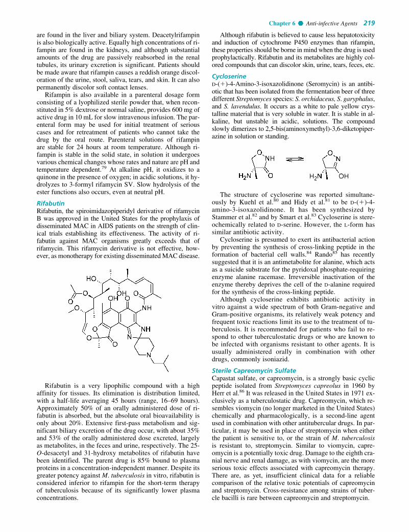
are found in the liver and biliary system. Deacetylrifampinis also biologically active. Equally high concentrations of ri-fampin are found in the kidneys, and although substantialamounts of the drug are passively reabsorbed in the renaltubules, its urinary excretion is significant. Patients shouldbe made aware that rifampin causes a reddish orange discol-oration of the urine, stool, saliva, tears, and skin. It can alsopermanently discolor soft contact lenses.
Rifampin is also available in a parenteral dosage formconsisting of a lyophilized sterile powder that, when recon-stituted in 5% dextrose or normal saline, provides 600 mg ofactive drug in 10 mL for slow intravenous infusion. The par-enteral form may be used for initial treatment of seriouscases and for retreatment of patients who cannot take thedrug by the oral route. Parenteral solutions of rifampinare stable for 24 hours at room temperature. Although ri-fampin is stable in the solid state, in solution it undergoesvarious chemical changes whose rates and nature are pH andtemperature dependent.79 At alkaline pH, it oxidizes to aquinone in the presence of oxygen; in acidic solutions, it hy-drolyzes to 3-formyl rifamycin SV. Slow hydrolysis of theester functions also occurs, even at neutral pH.
RifabutinRifabutin, the spiroimidazopiperidyl derivative of rifamycinB was approved in the United States for the prophylaxis ofdisseminated MAC in AIDS patients on the strength of clin-ical trials establishing its effectiveness. The activity of ri-fabutin against MAC organisms greatly exceeds that ofrifamycin. This rifamycin derivative is not effective, how-ever, as monotherapy for existing disseminated MAC disease.
Rifabutin is a very lipophilic compound with a highaffinity for tissues. Its elimination is distribution limited,with a half-life averaging 45 hours (range, 16–69 hours).Approximately 50% of an orally administered dose of ri-fabutin is absorbed, but the absolute oral bioavailability isonly about 20%. Extensive first-pass metabolism and sig-nificant biliary excretion of the drug occur, with about 35%and 53% of the orally administered dose excreted, largelyas metabolites, in the feces and urine, respectively. The 25-O-desacetyl and 31-hydroxy metabolites of rifabutin havebeen identified. The parent drug is 85% bound to plasmaproteins in a concentration-independent manner. Despite itsgreater potency against M. tuberculosis in vitro, rifabutin isconsidered inferior to rifampin for the short-term therapyof tuberculosis because of its significantly lower plasmaconcentrations.
Although rifabutin is believed to cause less hepatotoxicityand induction of cytochrome P450 enzymes than rifampin,these properties should be borne in mind when the drug is usedprophylactically. Rifabutin and its metabolites are highly col-ored compounds that can discolor skin, urine, tears, feces, etc.
CycloserineD-(�)-4-Amino-3-isoxazolidinone (Seromycin) is an antibi-otic that has been isolated from the fermentation beer of threedifferent Streptomyces species: S. orchidaceus, S. garyphalus,and S. lavendulus. It occurs as a white to pale yellow crys-talline material that is very soluble in water. It is stable in al-kaline, but unstable in acidic, solutions. The compoundslowly dimerizes to 2,5-bis(aminoxymethyl)-3,6-diketopiper-azine in solution or standing.
The structure of cycloserine was reported simultane-ously by Kuehl et al.80 and Hidy et al.81 to be D-(�)-4-amino-3-isoxazolidinone. It has been synthesized byStammer et al.82 and by Smart et al.83 Cycloserine is stere-ochemically related to D-serine. However, the L-form hassimilar antibiotic activity.
Cycloserine is presumed to exert its antibacterial actionby preventing the synthesis of cross-linking peptide in theformation of bacterial cell walls.84 Rando85 has recentlysuggested that it is an antimetabolite for alanine, which actsas a suicide substrate for the pyridoxal phosphate-requiringenzyme alanine racemase. Irreversible inactivation of theenzyme thereby deprives the cell of the D-alanine requiredfor the synthesis of the cross-linking peptide.
Although cycloserine exhibits antibiotic activity invitro against a wide spectrum of both Gram-negative andGram-positive organisms, its relatively weak potency andfrequent toxic reactions limit its use to the treatment of tu-berculosis. It is recommended for patients who fail to re-spond to other tuberculostatic drugs or who are known tobe infected with organisms resistant to other agents. It isusually administered orally in combination with otherdrugs, commonly isoniazid.
Sterile Capreomycin SulfateCapastat sulfate, or capreomycin, is a strongly basic cyclicpeptide isolated from Streptomyces capreolus in 1960 byHerr et al.86 It was released in the United States in 1971 ex-clusively as a tuberculostatic drug. Capreomycin, which re-sembles viomycin (no longer marketed in the United States)chemically and pharmacologically, is a second-line agentused in combination with other antitubercular drugs. In par-ticular, it may be used in place of streptomycin when eitherthe patient is sensitive to, or the strain of M. tuberculosisis resistant to, streptomycin. Similar to viomycin, capre-omycin is a potentially toxic drug. Damage to the eighth cra-nial nerve and renal damage, as with viomycin, are the moreserious toxic effects associated with capreomycin therapy.There are, as yet, insufficient clinical data for a reliablecomparison of the relative toxic potentials of capreomycinand streptomycin. Cross-resistance among strains of tuber-cle bacilli is rare between capreomycin and streptomycin.
Chapter 6 Anti-infective Agents 219
0179-0241_17865_Ch06.qxd 12/8/09 10:55 PM Page 219

Four capreomycins, designated IA, IB, IIA, and IIB, havebeen isolated from cultures of S. capreolus. The clinicalagent contains primarily IA and IB. The close chemical re-lationship between capreomycins IA and IB and viomycinwas established,87 and the total synthesis and proof of struc-ture of the capreomycins were later accomplished.88 Thestructures of capreomycins IIA and IIB correspond to thoseof IA and IB but lack the �-lysyl residue. The sulfate saltsare freely soluble in water.
ANTIPROTOZOAL AGENTS
In the United States and other countries of the temperate zone,protozoal diseases are of minor importance, whereas bacterialand viral diseases are widespread and are the cause of consid-erable concern. On the other hand, protozoal diseases arehighly prevalent in tropical Third World countries, wherethey infect both human and animal populations, causing suf-fering, death, and enormous economic hardship. Protozoaldiseases that are found in the United States are malaria,amebiasis, giardiasis, trichomoniasis, toxoplasmosis, and, asa direct consequence of the AIDS epidemic, P. carinii pneu-monia (PCP).
Although amebiasis is generally thought of as a tropicaldisease, it actually has a worldwide distribution. In someareas with temperate climates in which sanitation is poor, theprevalence of amebiasis has been estimated to be as high as20% of the population. The causative organism, Entamoebahistolytica, can invade the wall of the colon or other parts ofthe body (e.g., liver, lungs, skin). An ideal chemotherapeuticagent would be effective against both the intestinal and ex-traintestinal forms of the parasite.
Amebicides that are effective against both intestinal andextraintestinal forms of the disease are limited to the some-what toxic alkaloids emetine and dehydroemetine, the ni-troimidazole derivative metronidazole, and the antimalarialagent chloroquine (Chapter 7). A second group of amebi-cides that are effective only against intestinal forms ofthe disease includes the aminoglycoside antibiotic paro-momycin, the 8-hydroxyquinoline derivative iodoquinol,the arsenical compound carbarsone, and diloxanide.
Other protozoal species that colonize the intestinaltract and cause enteritis and diarrhea are Balantidium coliand the flagellates, G. lamblia and Cryptosporidium spp.Balantidiasis responds best to tetracycline. Metronidazoleand iodoquinol may also be effective. Giardiasis may betreated effectively with furazolidone, metronidazole, or theantimalarial drug quinacrine (Chapter 7). Cryptosporidiosisis normally self-limiting in immunocompetent patients andis not normally treated. The illness can be a serious problemin AIDS patients because no effective therapy is currentlyavailable.
Trichomoniasis, a venereal disease caused by the flagel-lated protozoan T. vaginalis, is common in the United Statesand throughout the world. Although it is not generally con-sidered serious, this affliction can cause serious physicaldiscomfort. Oral metronidazole provides effective treatmentagainst all forms of the disease. It is also used to eradicatethe organism from asymptomatic male carriers.
P. carinii is an opportunistic pathogen that may colonizethe lungs of humans and other animals and, under the rightconditions, can cause pneumonia. The organism has longbeen classified as a protozoan, but recent RNA evidence sug-gests that it may be more closely related to fungi. At one time,occasional cases of PCP were known to occur in premature,undernourished infants and in patients receiving immunosup-pressant therapy. The situation changed with the onset of theAIDS epidemic. It is estimated that at least 60% and possiblyas high as 85% of patients infected with HIV develop PCPduring their lifetimes.
The combination of the antifolate trimethoprim and thesulfonamide sulfamethoxazole constitutes the treatment ofchoice for PCP. Other effective drugs include pentamidine,atovaquone, and a new antifolate, trimetrexate.
Toxoplasma gondii is an obligate intracellular proto-zoan that is best known for causing blindness in neonates.Toxoplasmosis, the disseminated form of the disease inwhich the lymphatic system, skeletal muscles, heart, brain,eye, and placenta may be affected, has become increas-ingly prevalent in association with HIV infection. A com-bination of the antifolate pyrimethamine and the sulfa drugsulfadiazine constitutes the most effective therapy fortoxoplasmosis.
220 Wilson and Gisvold’s Textbook of Organic Medicinal and Pharmaceutical Chemistry
Capreomycin IA
0179-0241_17865_Ch06.qxd 12/8/09 10:55 PM Page 220

Various forms of trypanosomiasis, chronic tropicaldiseases caused by pathogenic members of the familyTrypanosomidae, occur both in humans and in livestock.The principal disease in humans, sleeping sickness, can bebroadly classified into two main geographic and etiologicalgroups: African sleeping sickness caused by Trypanosomagambiense (West African), Trypanosoma rhodesiense (EastAfrican), or Trypanosoma congolense; and South Americansleeping sickness (Chagas disease) caused by Trypanosomacruzi. Of the various forms of trypanosomiasis, Chagas dis-ease is the most serious and generally the most resistant tochemotherapy. Leishmaniasis is a chronic tropical diseasecaused by various flagellate protozoa of the genusLeishmania. The more common visceral form caused byLeishmania donovani, called kala-azar, is similar to Chagasdisease. Although these diseases are widespread in tropicalareas of Africa and South and Central America, they are ofminor importance in the United States, Europe, and Asia.
Chemotherapy of trypanosomiasis and leishmaniasis re-mains somewhat primitive and is often less than effective. Infact, it is doubtful that these diseases can be controlled bychemotherapeutic measures alone, without successful controlof the intermediate hosts and vectors that transmit them.Heavy metal compounds, such as the arsenicals and antimo-nials, are sometimes effective but frequently toxic. The oldstandby suramin appears to be of some value in long- andshort-term prophylaxis. The nitrofuran derivative nifurtimoxmay be a major asset in the control of these diseases, but itspotential toxicity remains to be fully determined.
Metronidazole2-Methyl-5-nitroimidazole-1-ethanol (Flagyl, Protostat,Metro IV) is the most useful of a group of antiprotozoal ni-troimidazole derivatives that have been synthesized in vari-ous laboratories throughout the world. Metronidazole wasfirst marketed for the topical treatment of T. vaginalisvaginitis. It has since been shown to be effective orallyagainst both the acute and carrier states of the disease. Thedrug also possesses useful amebicidal activity and is, in fact,effective against both intestinal and hepatic amebiasis. It hasalso been found of use in the treatment of such other proto-zoal diseases as giardiasis and balantidiasis.
More recently, metronidazole has been found to possessefficacy against obligate anaerobic bacteria, but it is ineffec-tive against facultative anaerobes or obligate aerobes. It isparticularly active against Gram-negative anaerobes, suchas Bacteroides and Fusobacterium spp. It is also effectiveagainst Gram-positive anaerobic bacilli (e.g., Clostridiumspp.) and cocci (e.g., Peptococcus, Peptidostreptococcusspp.). Because of its bactericidal action, metronidazole hasbecome an important agent for the treatment of serious infec-tions (e.g., septicemia, pneumonia, peritonitis, pelvic infec-tions, abscesses, meningitis) caused by anaerobic bacteria.
The common characteristic of microorganisms (bacteriaand protozoa) sensitive to metronidazole is that they areanaerobic. It has been speculated that a reactive intermediate
formed in the microbial reduction of the 5-nitro group ofmetronidazole covalently binds to the DNA of the microor-ganism, triggering the lethal effect.89 Potential reactive inter-mediates include the nitroxide, nitroso, hydroxylamine, andamine. The ability of metronidazole to act as a radiosensitiz-ing agent is also related to its reduction potential.
Metronidazole is a pale yellow crystalline substance thatis sparingly soluble in water. It is stable in air but is light sen-sitive. Despite its low water solubility, metronidazole is wellabsorbed following oral administration. It has a large appar-ent volume of distribution and achieves effective concentra-tions in all body fluids and tissues. Approximately 20% of anoral dose is metabolized to oxidized or conjugated forms.The 2-hydroxy metabolite is active; other metabolites areinactive.
Metronidazole is a weak base that possesses a pKa of 2.5.Although it is administered parenterally only as the free baseby slow intravenous infusion, metronidazole for injection issupplied in two forms: a ready-to-inject 100-mL solution con-taining 5 mg of base per mL; and a hydrochloride salt as500 mg of a sterile lyophilized powder. Metronidazole hy-drochloride for injection must first be reconstituted with ster-ile water to yield 5 mL of a solution having a concentration of100 mg/mL and a pH ranging from 0.5 to 2.0. The resultingsolution must then be diluted with either 100 mL of normalsaline or 5% dextrose and neutralized with 5 mEq of sodiumbicarbonate to provide a final solution of metronidazole basewith an approximate concentration of 5 mg/mL and a pH of 6to 7. Solutions of metronidazole hydrochloride are unsuitablefor intravenous administration because of their extreme acid-ity. Reconstituted metronidazole hydrochloride solutions arestable for 96 hours at 30°C, whereas ready-to-use solutions ofmetronidazole base are stable for 24 hours at 30°C. Both so-lutions should be protected from light.
DiloxanideFuramide, or eutamide, is the 2-furoate ester of 2,2-dichloro-4�-hydroxy-N-methylacetanilide. It was developed as a re-sult of the discovery that various �,�-dichloroacetamides possessed amebicidal activity in vitro. Diloxanide itself andmany of its esters are also active, and drug metabolism stud-ies indicate that hydrolysis of the amide is required for theamebicidal effect. Nonpolar esters of diloxanide are morepotent than polar ones. Diloxanide furoate has been used inthe treatment of asymptomatic carriers of E. histolytica. Itseffectiveness against acute intestinal amebiasis or hepaticabscesses, however, has not been established. Diloxanidefuroate is a white crystalline powder. It is administered orallyonly as 500-mg tablets and may be obtained in the UnitedStates from the CDC in Atlanta, Georgia.
8-HydroxyquinolineOxine, quinophenol, or oxyquinoline is the parent com-pound from which the antiprotozoal oxyquinolines havebeen derived. The antibacterial and antifungal properties of
Chapter 6 Anti-infective Agents 221
0179-0241_17865_Ch06.qxd 12/8/09 10:55 PM Page 221

oxine and its derivatives, which are believed to result fromthe ability to chelate metal ions, are well known. Aqueoussolutions of acid salts of oxine, particularly the sulfate(Chinosol, Quinosol), in concentrations of 1:3,000 to1:1,000, have been used as topical antiseptics. The substitu-tion of an iodine atom at the 7-position of 8-hydroxyquino-lines yields compounds with broad-spectrum amebicidalproperties.
Iodoquinol5,7-Diiodo-8-quinolinol, 5,7-diiodo-8-hydroxyquinoline,or diiodohydroxyquin (Yodoxin, Diodoquin, Diquinol) is ayellowish to tan microcrystalline, light-sensitive substancethat is insoluble in water. It is recommended for acute andchronic intestinal amebiasis but is not effective in extrain-testinal disease. Because a relatively high incidence of topicneuropathy has occurred with its use, iodoquinol should notbe used routinely for traveler’s diarrhea.
Emetine and DehydroemetineThe alkaloids emetine and dehydroemetine are obtained byseparation from extracts of ipecac. They occur as levorota-tory, light-sensitive white powders that are insoluble in water.The alkaloids readily form water-soluble salts. Solutions ofthe hydrochloride salts intended for intramuscular injectionshould be adjusted to pH 3.5 and stored in light-resistantcontainers.
Emetine and dehydroemetine exert a direct amebicidalaction on various forms of E. histolytica. They are proto-plasmic poisons that inhibit protein synthesis in protozoaland mammalian cells by preventing protein elongation.Because their effect in intestinal amebiasis is solely symp-tomatic and the cure rate is only 10% to 15%, they shouldbe used only in combination with other agents. The highconcentrations of the alkaloids achieved in the liver andother tissues after intramuscular injection provide the basisfor their high effectiveness against hepatic abscesses and
other extraintestinal forms of the disease. Toxic effectslimit the usefulness of emetine. It causes a high frequencyof gastrointestinal distress (especially nausea and diarrhea),cardiovascular effects (hypotension and arrhythmias), andneuromuscular effects (pain and weakness). A lower inci-dence of cardiotoxicity has been associated with the use ofdehydroemetine (Mebadin), which is available from theCDC and is also amebicidal.
Emetine and dehydroemetine have also been used to treatbalantidial dysentery and fluke infestations, such as fascio-liasis and paragonimiasis.
Pentamidine Isethionate4,4�-(Pentamethylenedioxy)dibenzamidine diisethionate(NebuPent, Pentam 300) is a water-soluble crystalline saltthat is stable to light and air. The principal use of pentami-dine is for the treatment of pneumonia caused by the oppor-tunistic pathogenic protozoan P. carinii, a frequent second-ary invader associated with AIDS. The drug may beadministered by slow intravenous infusion or by deep intra-muscular injection for PCP. An aerosol form of pentami-dine is used by inhalation for the prevention of PCP inhigh-risk patients infected with HIV who have a previoushistory of PCP infection or a low peripheral CD4� lympho-cyte count.
Both the inhalant (aerosol) and parenteral dosage formsof pentamidine isethionate are sterile lyophilized powdersthat must be made up as sterile aqueous solutions prior touse. Sterile water for injection must be used to reconstitutethe aerosol, to avoid precipitation of the pentamidine salt.Adverse reactions to the drug are common. These includecough and bronchospasm (inhalation) and hypertension andhypoglycemia (injection).
Pentamidine has been used for the prophylaxis and treat-ment of African trypanosomiasis. It also has some value fortreating visceral leishmaniasis. Pentamidine rapidly disap-pears from the plasma after intravenous injection and is dis-tributed to the tissues, where it is stored for a long period.This property probably contributes to the usefulness of thedrug as a prophylactic agent.
Atovaquone3-[4-(4-Chlorophenyl)-cyclohexyl]-2-hydroxy-1,4-naph-thoquinone (Mepron) is a highly lipophilic, water-insolubleanalog of ubiquinone 6, an essential component of the mito-chondrial electron transport chain in microorganisms. Thestructural similarity between atovaquone and ubiquinonesuggests that the former may act as an antimetabolite for thelatter and thereby interfere with the function of electrontransport enzymes.
222 Wilson and Gisvold’s Textbook of Organic Medicinal and Pharmaceutical Chemistry
0179-0241_17865_Ch06.qxd 12/8/09 10:55 PM Page 222

Atovaquone was originally developed as an antimalar-ial drug, but Plasmodium falciparum was found to developa rapid tolerance to its action. More recently, the effective-ness of atovaquone against P. carinii was discovered. Itis a currently recommended alternative to trimethoprim-sulfamethoxazole (TMP-SMX) for the treatment and pro-phylaxis of PCP in patients intolerant to this combination.Atovaquone was also shown to be effective in eradicatingT. gondii in preclinical animal studies.
The oral absorption of atovaquone is slow and incom-plete, in part because of the low water solubility of the drug.Aqueous suspensions provide significantly better absorptionthan do tablets. Food, especially if it has a high fat content,increases atovaquone absorption. Significant enterohepaticrecycling of atovaquone occurs, and most (nearly 95%) ofthe drug is excreted unchanged in the feces. In vivo, ato-vaquone is largely confined to the plasma, where it is exten-sively protein bound (�99.9%). The half-life of the drugranges from 62 to 80 hours. The primary side effect is gas-trointestinal intolerance.
EflornithineEflornithine is used for the treatment of West African sleep-ing sickness, caused by Trypanosoma brucei gambiense.It is specifically indicated for the meningoencephaliticstage of the disease. Eflornithine is a myelosuppressivedrug that causes high incidences of anemia, leukopenia,and thrombocytopenia. Complete blood cell counts must bemonitored during the course of therapy.
The irreversible inactivation of ornithine decarboxylaseby eflornithine is accompanied by decarboxylation andrelease of fluoride ion from the inhibitor,90 suggesting en-zyme-catalyzed activation of the inhibitor. Only the (�) iso-mer, stereochemically related to L-ornithine, is active.
Eflornithine is supplied as the hydrochloride salt. It may beadministered either intravenously or orally. Approximately80% of the unchanged drug is excreted in the urine.Penetration of eflornithine into the CSF is facilitated by in-flammation of the meninges.
NifurtimoxNifurtimox is 4-[(5-nitrofurfurylidene) amino]-3-methylth-iomorpholine-1,1-dioxide, or Bayer 2502 (Lampit). Theobservation that various derivatives of 5-nitrofuraldehydepossessed, in addition to their antibacterial and antifungal
properties, significant and potentially useful antiprotozoalactivity eventually led to discovery of particular nitrofuranswith antitrypanosomal activity.
The most important of such compounds is nifurtimox be-cause of its demonstrated effectiveness against T. cruzi, theparasite responsible for South American trypanosomiasis. Infact, use of this drug represents the only clinically proventreatment for both acute and chronic forms of the disease.Nifurtimox is available in the United States from the CDC.
Nifurtimox is administered orally. Oral bioavailability ishigh, but considerable first-pass metabolism occurs. Thehalf-life of nifurtimox is 2 to 4 hours. The drug is poorly tol-erated, with a high incidence of nausea, vomiting, abdomi-nal pain, and anorexia reported. Symptoms of central andperipheral nervous system toxicity also frequently occurwith nifurtimox.
BenznidazoleN-Benzyl-2-nitroimidazole-1-acetamide (Radanil, Rochagan)is a nitroimidazole derivative that is used for the treatment ofChagas disease. It is not available in the United States but isused extensively in South America. The effectiveness of ben-znidazole is similar to that of nifurtimox. Therapy forAmerican trypanosomiasis with oral benznidazole requiresseveral weeks and is frequently accompanied by adverse ef-fects such as peripheral neuropathy, bone marrow depression,and allergic-type reactions.
Melarsoprol2-p-(4,6-Diamino-s-triazin-2-yl-amino)phenyl-4-hydroxy-methyl-1,3,2-dithiarsoline (Mel B, Arsobal) is prepared byreduction of a corresponding pentavalent arsanilate to thetrivalent arsenoxide followed by reaction of the latter with2,3-dimercapto-1-propanol (British anti-Lewisite [BAL]). Ithas become the drug of choice for the treatment of thelater stages of both forms of African trypanosomiasis.Melarsoprol has the advantage of excellent penetration intothe CNS and, therefore, is effective against meningoen-cephalitic forms of T. gambiense and T. rhodesiense.Trivalent arsenicals tend to be more toxic to the host (as wellas the parasites) than the corresponding pentavalent com-pounds. The bonding of arsenic with sulfur atoms tends toreduce host toxicity, increase chemical stability (to oxida-tion), and improve distribution of the compound to the ar-senoxide. Melarsoprol shares the toxic properties of other
Chapter 6 Anti-infective Agents 223
0179-0241_17865_Ch06.qxd 12/8/09 10:55 PM Page 223

arsenicals, however, so its use must be monitored for signsof arsenic toxicity.
Sodium StibogluconateSodium antimony gluconate (Pentostam) is a pentavalent an-timonial compound intended primarily for the treatment ofvarious forms of leishmaniasis. It is available from the CDCas the disodium salt, which is chemically stable and freelysoluble in water. The 10% aqueous solution used for eitherintramuscular or intravenous injection has a pH of approxi-mately 5.5. Like all antimonial drugs, this drug has a lowtherapeutic index, and patients undergoing therapy with itshould be monitored carefully for signs of heavy metal poi-soning. Other organic antimonial compounds are used prima-rily for the treatment of schistosomiasis and other flukes.
The antileishmanial action of sodium stibogluconate re-quires its reduction to the trivalent form, which is believedto inhibit phosphofructokinase in the parasite.
Dimercaprol2,3-Dimercapto-1-propanol, BAL, or dithioglycerol is afoul-smelling, colorless liquid. It is soluble in water (1:20)and alcohol. It was developed by the British during WorldWar II as an antidote for “Lewisite,” hence the name Britishanti-Lewisite or BAL. Dimercaprol is effective topicallyand systematically as an antidote for poisoning caused byarsenic, antimony, mercury, gold, and lead. It can, therefore,also be used to treat arsenic and antimony toxicity associ-ated with overdose or accidental ingestion of organoarseni-cals or organoantimonials.
The antidotal properties of BAL are associated with theproperty of heavy metals to react with sulfhydryl (SH)groups in proteins (e.g., the enzyme pyruvate oxidase) andinterfere with their normal function. 1,2-Dithiol compoundssuch as BAL compete effectively with such proteins for themetal by reversibly forming metal ring compounds of thefollowing type:
224 Wilson and Gisvold’s Textbook of Organic Medicinal and Pharmaceutical Chemistry
These are relatively nontoxic, metabolically conjugated(as glucuronides), and rapidly excreted.
BAL may be applied topically as an ointment or injectedintramuscularly as a 5% or 10% solution in peanut oil.
Suramin SodiumSuramin sodium is a high–molecular-weight bisurea deriv-ative containing six sulfonic acid groups as their sodiumsalts. It was developed in Germany shortly after WorldWar I as a byproduct of research efforts directed towardthe development of potential antiparasitic agents fromdyestuffs.
The drug has been used for more than half a centuryfor the treatment of early cases of trypanosomiasis. Notuntil several decades later, however, was suramin discov-ered to be a long-term prophylactic agent whose effec-tiveness after a single intravenous injection is maintainedfor up to 3 months. The drug is tightly bound to plasmaproteins, causing its excretion in the urine to be almostnegligible.
Tissue penetration of the drug does not occur, apparentlybecause of its high molecular weight and highly ionic charac-ter. Thus, an injected dose remains in the plasma for a verylong period. Newer, more effective drugs are now availablefor short-term treatment and prophylaxis of African sleepingsickness. Suramin is also used for prophylaxis of onchocerci-asis. It is available from the CDC.
ANTHELMINTICS
Anthelmintics are drugs that have the capability of riddingthe body of parasitic worms or helminths. The prevalenceof human helminthic infestations is widespread through-out the globe and represents a major world health prob-lem, particularly in Third World countries. Helminths par-asitic to humans and other animals are derived from twophyla, Platyhelminthes and Nemathelminthes. Cestodes(tapeworms) and trematodes (flukes) belong to the for-mer, and nematodes or true roundworms belong to the lat-ter. The helminth infestations of major concern on theNorth American continent are caused by roundworms(i.e., hookworm, pinworm, and Ascaris spp.). Human
0179-0241_17865_Ch06.qxd 12/8/09 10:55 PM Page 224

tapeworm and fluke infestations are rarely seen in theUnited States.
Several classes of chemicals are used as anthelminticsand include phenols and derivatives, piperazine and relatedcompounds, antimalarial compounds (Chapter 7), variousheterocyclic compounds, and natural products.
PiperazineHexahydropyrazine or diethylenediamine (Arthriticine,Dispermin) occurs as colorless, volatile crystals of the hexa-hydrate that are freely soluble in water. After the discoveryof the anthelmintic properties of a derivative diethylcarba-mazine, the activity of piperazine itself was established.Piperazine is still used as an anthelmintic for the treatmentof pinworm (Enterobius [Oxyuris] vermicularis) and round-worm (Ascaris lumbricoides) infestations. It is available invarious salt forms, including the citrate (official in the USP)in syrup and tablet forms.
Piperazine blocks the response of the ascaris muscleto acetylcholine, causing flaccid paralysis in the worm,which is dislodged from the intestinal wall and expelled inthe feces.
Diethylcarbamazepine CitrateN,N-Diethyl-4-methyl-1-piperazinecarboxamide citrate or1-diethylcarbamyl-4-methylpiperazine dihydrogen citrate(Hetrazan) is a highly water-soluble crystalline compoundthat has selective anthelmintic activity. It is effective againstvarious forms of filariasis, including Bancroft, onchocer-ciasis, and laviasis. It is also active against ascariasis.Relatively few adverse reactions have been associated withdiethylcarbamazine.
Pyrantel PamoateTrans-1,4,5,6,-Tetrahydro-1-methyl-2-[2-(2�-thienyl)ethenyl]pyrimidine pamoate (Antiminth) is a depolarizing neuro-muscular blocking agent that causes spastic paralysis insusceptible helminths. It is used in the treatment of infesta-tions caused by pinworms and roundworms (ascariasis).Because its action opposes that of piperazine, the twoanthelmintics should not be used together. More than halfof the oral dose is excreted in the feces unchanged.Adverse effects associated with its use are primarilygastrointestinal.
Thiabendazole2-(4-Thiazolyl)benzimidazole (Mintezol) occurs as a whitecrystalline substance that is only slightly soluble in waterbut is soluble in strong mineral acids. Thiabendazole is abasic compound with a pKa of 4.7 that forms complexeswith metal ions.
Thiabendazole inhibits the helminth-specific enzymefumarate reductase.91 It is not known whether metal ionsare involved or if the inhibition of the enzyme is related tothiabendazole’s anthelmintic effect. Benzimidazole an-thelmintic drugs such as thiabendazole and mebendazolealso arrest nematode cell division in metaphase by interfer-ing with microtubule assembly.92 They exhibit a highaffinity for tubulin, the precursor protein for microtubulesynthesis.
Thiabendazole has broad-spectrum anthelmintic activity.It is used to treat enterobiasis, strongyloidiasis (threadworminfection), ascariasis, uncinariasis (hookworm infection), andtrichuriasis (whipworm infection). It has also been used torelieve symptoms associated with cutaneous larva migrans(creeping eruption) and the invasive phase of trichinosis. Inaddition to its use in human medicine, thiabendazole iswidely used in veterinary practice to control intestinalhelminths in livestock.
MebendazoleMethyl 5-benzoyl-2-benzimidazolecarbamate (Vermox) isa broad-spectrum anthelmintic that is effective against vari-ous nematode infestations, including whipworm, pinworm,roundworm, and hookworm. Mebendazole irreversiblyblocks glucose uptake in susceptible helminths, thereby de-pleting glycogen stored in the parasite. It apparently does notaffect glucose metabolism in the host. It also inhibits cell di-vision in nematodes.71
Chapter 6 Anti-infective Agents 225
0179-0241_17865_Ch06.qxd 12/8/09 10:55 PM Page 225

Mebendazole is poorly absorbed by the oral route.Adverse reactions are uncommon and usually consist of ab-dominal discomfort. It is teratogenic in laboratory animalsand, therefore, should not be given during pregnancy.
AlbendazoleMethyl 5-(propylthio)-2-benzimidazolecarbamate (Eskazole,Zentel) is a broad-spectrum anthelmintic that is not currentlymarketed in North America. It is available from the manufac-turer on a compassionate use basis. Albendazole is widelyused throughout the world for the treatment of intestinalnematode infection. It is effective as a single-dose treatmentfor ascariasis, New and Old World hookworm infections,and trichuriasis. Multiple-dose therapy with albendazole caneradicate pinworm, threadworm, capillariasis, clonorchiasis,and hydatid disease. The effectiveness of albendazole againsttapeworms (cestodes) is generally more variable and lessimpressive.
Albendazole occurs as a white crystalline powder that isvirtually insoluble in water. The oral absorption of albenda-zole is enhanced by a fatty meal. The drug undergoes rapidand extensive first-pass metabolism to the sulfoxide, whichis the active form in plasma. The elimination half-life ofthe sulfoxide ranges from 10 to 15 hours. Considerable bil-iary excretion and enterohepatic recycling of albendazolesulfoxide occurs. Albendazole is generally well toleratedin single-dose therapy for intestinal nematodes. The high-dose, prolonged therapy required for clonorchiasis orechinococcal disease therapy can result in adverse effectssuch as bone marrow depression, elevation of hepatic en-zymes, and alopecia.
Niclosamide
5-Chloro-N-(2-chloro-4-nitrophenyl)-2-hydroxybenzamideor 2,5�-dichloro-4�-nitrosalicylanilide (Cestocide, Mansonil,Yomesan) occurs as a yellowish white, water-insolublepowder. It is a potent taeniacide that causes rapid disintegra-tion of worm segments and the scolex. Penetration of thedrug into various cestodes appears to be facilitated by thedigestive juices of the host, in that very little of the drug isabsorbed by the worms in vitro. Niclosamide is well toler-ated following oral administration, and little or no systemicabsorption of it occurs. A saline purge 1 to 2 hours after
ingestion of the taeniacide is recommended to remove thedamaged scolex and worm segments. This procedure ismandatory in the treatment of pork tapeworm infestation toprevent possible cysticercosis resulting from release of liveova from worm segments damaged by the drug.
Bithionol2,2�-Thiobis(4,6-dichlorophenol), or bis(2-hydroxy-3,5-dichlorophenyl)sulfide (Lorothidol, Bithin), a chlorinatedbisphenol, was formerly used in soaps and cosmetics for itsantimicrobial properties but was removed from the marketfor topical use because of reports of contact photodermati-tis. Bithionol has useful anthelmintic properties and hasbeen used as a fasciolicide and taeniacide. It is still consid-ered the agent of choice for the treatment of infestationscaused by the liver fluke Fasciola hepatica and the lungfluke Paragonimus westermani. Niclosamide is believed tobe superior to it for the treatment of tapeworm infestations.
Oxamniquine1,2,3,4-Tetrahydro-2-[(isopropylamino)methyl]-7-nitro-6-quinolinemethanol (Vansil) is an antischistosomal agent thatis indicated for the treatment of Schistosoma mansoni (intes-tinal schistosomiasis) infection. It has been shown to inhibitDNA, RNA, and protein synthesis in schistosomes.93 The 6-hydroxymethyl group is critical for activity; metabolic ac-tivation of precursor 6-methyl derivatives is critical. Theoral bioavailability of oxamniquine is good; effectiveplasma levels are achieved in 1 to 1.5 hours. The plasmahalf-life is 1 to 2.5 hours. The drug is extensively metabo-lized to inactive metabolites, of which the principal one isthe 6-carboxy derivative.
The free base occurs as a yellow crystalline solid thatis slightly soluble in water but soluble in dilute aqueousmineral acids and soluble in most organic solvents. Itis available in capsules containing 250 mg of the drug.Oxamniquine is generally well tolerated. Dizziness anddrowsiness are common, but transitory, side effects. Seriousreactions, such as epileptiform convulsions, are rare.
Praziquantel2-(Cyclohexylcarbonyl)-1,2,3,6,7, 11b-hexahydro-4H-pyrazino[2,1-a]isoquinolin-4-one (Biltricide) is a broad-spectrum agent that is effective against various trematodes
226 Wilson and Gisvold’s Textbook of Organic Medicinal and Pharmaceutical Chemistry
0179-0241_17865_Ch06.qxd 12/8/09 10:55 PM Page 226

(flukes). It has become the agent of choice for the treatmentof infections caused by schistosomes (blood flukes).
The drug also provides effective treatment for fasciolop-siasis (intestinal fluke), clonorchiasis (Chinese liver fluke),fascioliasis (sheep liver fluke), opisthorchosis (liver fluke),and paragonimiasis (lung fluke). Praziquantel increases cellmembrane permeability of susceptible worms, resulting inthe loss of extracellular calcium. Massive contractions andultimate paralysis of the fluke musculature occurs, followedby phagocytosis of the parasite.
Following oral administration, about 80% of the doseis absorbed. Maximal plasma concentrations are achievedin 1 to 3 hours. The drug is rapidly metabolized in theliver in the first-pass. It is likely that some of the metabo-lites are also active. Praziquantel occurs as a white crys-talline solid that is insoluble in water. It is available as600-mg film-coated tablets. The drug is generally welltolerated.
IvermectinIvermectin (Cardomec, Eqvalan, Ivomec) is a mixtureof 22,23-dihydro derivatives of avermectins B1a and B1b
prepared by catalytic hydrogenation. Avermectins aremembers of a family of structurally complex antibioticsproduced by fermentation with a strain of Streptomycesavermitilis. Their discovery resulted from an intensivescreening of cultures for anthelmintic agents from naturalsources.94 Ivermectin is active in low dosage against awide variety of nematodes and arthropods that parasitizeanimals.95
The structures of the avermectins were established by acombination of spectroscopic96 and x-ray crystallographic97
techniques to contain pentacyclic 16-membered–ring agly-cones glycosidically linked at the 3-position to a disaccharide
that comprises two oleandrose sugar residues. The side chainat the 25-position of the aglycone is sec-butyl in avermectinB1a, whereas in avermectin B1b, it is isopropyl. Ivermectincontains at least 80% of 22,23-dihydroavermectin B1a and nomore than 20% 22,23-dihydroavermectin B1b.
Ivermectin has achieved widespread use in veterinarypractice in the United States and many countries through-out the world for the control of endoparasites and ectopar-asites in domestic animals.95 It has been found effective forthe treatment of onchocerciasis (“river blindness”) in hu-mans,98 an important disease caused by the roundwormOncocerca volvulus, prevalent in West and Central Africa,the Middle East, and South and Central America.Ivermectin destroys the microfilariae, immature forms ofthe nematode, which create the skin and tissue nodules thatare characteristic of the infestation and can lead to blind-ness. It also inhibits the release of microfilariae by theadult worms living in the host. Studies on the mechanismof action of ivermectin indicate that it blocks interneu-ron–motor neuron transmission in nematodes by stimulat-ing the release of the inhibitory neurotransmitter GABA.95
The drug has been made available by the manufacturer ona humanitarian basis to qualified treatment programsthrough the World Health Organization.
ANTISCABIOUS ANDANTIPEDICULAR AGENTS
Scabicides (antiscabious agents) are compounds used tocontrol the mite Sarcoptes scabiei, an organism thatthrives under conditions of poor personal hygiene. The in-cidence of scabies is believed to be increasing in theUnited States and worldwide and has, in fact, reached pan-demic proportions.99 Pediculicides (antipedicular agents)are used to eliminate head, body, and crab lice. Ideal scabi-cides and pediculicides must kill the adult parasites anddestroy their eggs.
Benzyl BenzoateBenzyl benzoate is a naturally occurring ester obtained fromPeru balsam and other resins. It is also prepared syntheti-cally from benzyl alcohol and benzoyl chloride. The ester isa clear colorless liquid with a faint aromatic odor. It is insol-uble in water but soluble in organic solvents.
Chapter 6 Anti-infective Agents 227
0179-0241_17865_Ch06.qxd 12/8/09 10:55 PM Page 227

Benzyl benzoate is an effective scabicide when appliedtopically. Immediate relief from itching probably resultsfrom a local anesthetic effect; however, a complete cureis frequently achieved with a single application of a 25%emulsion of benzyl benzoate in oleic acid, stabilized withtriethanolamine. This preparation has the additionaladvantage of being essentially odorless, nonstaining, andnonirritating to the skin. It is applied topically as a lotionover the entire dampened body, except the face.
LindaneLindane is 1,2,3,4,5,6-hexachlorocyclohexane, �-benzenehexachloride, or benzene hexachloride (Kwell, Scabene,Kwildane, G-Well). This halogenated hydrocarbon is pre-pared by the chlorination of benzene. A mixture of isomersis obtained in this process, five of which have been isolated:�, �, �, �, and �. The �-isomer, present to 10% to 13% inthe mixture, is responsible for the insecticidal activity. The�-isomer may be separated by various extraction and chro-matographic techniques.
Lindane occurs as a light buff to tan powder with a persist-ent musty odor, and it is bitter. It is insoluble in water butsoluble in most organic solvents. It is stable under acidic orneutral conditions but undergoes elimination reactions underalkaline conditions.
The action of lindane against insects is threefold: it is a di-rect contact poison, it has a fumigant effect, and it acts as astomach poison. The effect of lindane on insects is similar tothat of DDT. Its toxicity in humans is somewhat lower thanthat of DDT. Because of its lipid solubility properties, how-ever, lindane when ingested tends to accumulate in the body.
Lindane is used locally as a cream, lotion, or shampoo forthe treatment of scabies and pediculosis.
CROTAMITON
N-Ethyl-N-(2-methylphenyl)-2-butenamide, or N-ethyl-o-crotonotoluidide (Eurax), is a colorless, odorless oily liquid.It is virtually insoluble in water but soluble in most organicsolvents.
Crotamiton is available in 10% concentration in a lotionand a cream intended for the topical treatment of scabies.
Its antipruritic effect is probably because of a local anes-thetic action.
PermethrinPermethrin is 3-(2,2-Dichloroethenyl)-2,2-dimethyl-cyclopropanecarboxylic acid (3-phenoxyphenyl)methylester or 3-(phenoxyphenyl)methyl (�)-cis, trans-3-(2,2-dichloroethenyl)-2,2-dimethylcyclopropanecarboxylate(Nix). This synthetic pyrethrinoid compound is more stablechemically than most natural pyrethrins and is at least as ac-tive as an insecticide. Of the four isomers present, the1(R),trans and 1(R),cis-isomers are primarily responsiblefor the insecticidal activity. The commercial product is amixture consisting of 60% trans and 40% cis racemic iso-mers. It occurs as colorless to pale yellow low-melting crys-tals or as a pale yellow liquid and is insoluble in water butsoluble in most organic solvents.
Permethrin exerts a lethal action against lice, ticks, mites,and fleas. It acts on the nerve cell membranes of the para-sites to disrupt sodium channel conductance. It is used as apediculicide for the treatment of head lice. A single applica-tion of a 1% solution effects cures in more than 99% ofcases. The most frequent side effect is pruritus, which oc-curred in about 6% of the patients tested.
ANTIBACTERIAL SULFONAMIDES
The sulfonamide antimicrobial drugs were the first effectivechemotherapeutic agents that could be used systemically forthe cure of bacterial infections in humans. Their introduc-tion led to a sharp decline in the morbidity and mortality ofinfectious diseases. The rapid development of widespreadresistance to the sulfonamides soon after their introductionand the increasing use of the broader-spectrum penicillins inthe treatment of infectious disease diminished the usefulnessof sulfonamides. Today, they occupy a rather small place inthe list of therapeutic agents that can be used for infectiousdisease. They are not completely outmoded, however. In themid-1970s, the development of a combination of trimetho-prim and sulfamethoxazole and the demonstration of itsusefulness in the treatment and prophylaxis of certain op-portunistic microbial infections led to resurgence in the useof some sulfonamides.
Fritz Mietzsch and Joseph Klarer of the I. G.Farbenindustrie laboratories systematically synthesizeda series of azo dyes, each containing the sulfonamidefunctional group, as potential antimicrobial agents.Sulfonamide azo dyes were included in the test series be-cause they were readily synthesized and possessed superiorstaining properties. The Bayer pathologist–bacteriologistwho evaluated the new Mietzsch-Klarer dyes was a physi-cian named Gerhard Domagk.100–102 In 1932, Domagkbegan to study a brilliant red dye, later named Prontosil.Prontosil was found to protect against, and cure, strepto-coccal infections in mice.100 Interestingly, Prontosil was
228 Wilson and Gisvold’s Textbook of Organic Medicinal and Pharmaceutical Chemistry
0179-0241_17865_Ch06.qxd 12/8/09 10:55 PM Page 228

inactive on bacterial cultures. Domagk and others continuedto study Prontosil, and in 1933, the first of many cures of se-vere bacterial infections in humans was reported byFoerster,103 who treated a 10-month-old infant sufferingfrom staphylococcal septicemia and obtained a dramaticcure. The credit for most of the discoveries relating toProntosil belongs to Domagk, and for his pioneering work inchemotherapy he was awarded the Nobel Prize in medicineand physiology in 1938. The Gestapo prevented him fromactually accepting the award, but after the war, he receivedit in Stockholm in 1947.
Prontosil is totally inactive in vitro but possesses excellentactivity in vivo. This property of the drug attracted much at-tention and stimulated a large body of research activity intothe sulfonamides. In 1935, Trefouel and coinvestigators104
performed a structure–activity study on the sulfonamide azodyes and concluded that the azo linkage was reductivelycleaved to release the active antibacterial product, sulfanil-amide. This finding was confirmed in 1937 when Fuller105
isolated free sulfanilamide from the blood and urine ofpatients being treated with Prontosil. Favorable clinical re-sults were reported with Prontosil and the active metaboliteitself, sulfanilamide, in puerperal sepsis and meningococcal
infections. All of these findings ushered in the modern era ofchemotherapy and the concept of the prodrug.
Following the dramatic success of Prontosil, a host ofsulfanilamide derivatives was synthesized and tested. By1948, more than 4,500 compounds106 had been evaluated.Of these, only about two dozen have been used in clinicalpractice. In the late 1940s, broader experience with sulfon-amides had begun to demonstrate toxicity in some patients,and resistance problems brought about by indiscriminate useof sulfonamides limited their use throughout the world. Thepenicillins were excellent alternatives to the sulfonamides,and they largely replaced the latter in antimicrobialchemotherapy.
Today, there are a few sulfonamides (Table 6.7) andespecially sulfonamide-trimethoprim combinations that areused extensively for opportunistic infections in patients withAIDS.107 A primary infection that is treated with the combi-nation is PCP. The sulfonamide-trimethoprim combinationcan be used for treatment and prophylaxis. Additionally,cerebral toxoplasmosis can be treated in active infection orprophylactically. Urinary tract infections and burn ther-apy107–111 round out the list of therapeutic applications. Thesulfonamides are drugs of choice for a few other types ofinfections, but their use is quite limited in modern antimi-crobial chemotherapy.107–111
The sulfonamides can be grouped into three classes onthe basis of their use: oral absorbable agents, designed togive systemic distribution; oral nonabsorbable agents suchas sulfasalazine; and topical agents such as sodium sulfac-etamide ophthalmic drops.
Chapter 6 Anti-infective Agents 229
TABLE 6.7 Therapy with Sulfonamide Antibacterials
Disease/Infection Sulfonamides Commonly Used
Relatively Common Use
Treatment and prophylaxis of Pneumocystis carinii pneumonia Trimethoprim-sulfamethoxazoleTreatment and prophylaxis of cerebral toxoplasmosis Pyrimethamine-sulfadiazineFirst attack of urinary tract infection Trimethoprim-sulfamethoxazoleBurn therapy: prevention and treatment of bacterial infection Silver sulfadiazine and mafenideConjunctivitis and related superficial ocular infections Sodium sulfacetamideChloroquine-resistant malaria (Chapter 9) Combinations with quinine, others
SulfadoxineSulfalene
Less Common Infections/Diseases Drugs of Choice or Alternates
Nocardiosis Trimethoprim-sulfamethoxazoleSevere traveler’s diarrhea Trimethoprim-sulfamethoxasoleMeningococcal infections Sulfonamides, only if proved to be sulfonamide sensitive;
otherwise, penicillin G, ampicillin, or (for penicillin-allergicpatients) chloramphenicol should be used
Generally Not Useful
Streptococcal infections Most are resistant to sulfonamidesProphylaxis of recurrent rheumatic fever Most are resistant to sulfonamidesOther bacterial infections The low cost of penicillin and the widespread resistance to
sulfonamides limit their use; sulfonamides are still used in afew countries
Vaginal infections The FDA and USP-DI find no evidence of efficacyReduction of bowel flora Effectiveness not establishedUlcerative colitis Corticosteroid therapy often preferred
Relapses common with sulfonamidesSalicylazosulfapyridineSide effects of the sulfanilamides sometimes mimic ulcerative
colitis
0179-0241_17865_Ch06.qxd 12/8/09 10:55 PM Page 229

Nomenclature of the Sulfonamides
Sulfonamide is a generic term that denotes three differentcases:
1. Antibacterials that are aniline-substituted sulfonamides(the “sulfanilamides”).
2. Prodrugs that react to generate active sulfanilamides (i.e.,sulfasalazine).
3. Nonaniline sulfonamides (i.e., mafenide acetate).
There are also other commonly used drugs that are sul-fonamides or sulfanilamides. Among these are the oral hy-poglycemic drug tolbutamide, the diuretic furosemide, andthe diuretic chlorthalidone.
In pharmaceutical chemistry, pKb values are not used tocompare compounds that are Lewis bases. Instead, if a pKa
of an amine is given, it refers to its salt acting as the conju-gate acid. For example, aniline with a pKa of 4.6 refers to
It does not refer to
A negative charge on a nitrogen atom is typically not sta-ble unless it can be delocalized by resonance. This is what
happens with the sulfanilamides. Therefore, the single pKa
usually given for sulfanilamides refers to the loss of anamide proton (Fig. 6.8)
Mechanism of Action of theSulfonamides
Folinic acid (N5-formyltetrahydrofolic acid), N5,N10-methyl-enetetrahydrofolic acid, and N10-formyltetrahydrofolic acidare intermediates of several biosynthetic pathways that com-pose the one-carbon pool in animals, bacteria, and plants. Akey reaction involving folate coenzymes is catalyzed by theenzyme thymidylate synthase, which transfers a methyl groupfrom N5,N10-tetrahydrofolic acid to deoxyuridine monophos-phate to form deoxythymidine monophosphate, an importantprecursor to DNA (Fig. 6.9).
Another key reaction is the generation of formyl groupsfor the biosynthesis of formylmethionyl tRNA units, theprimary building blocks in protein synthesis. The sulfon-amides are structural analogs of PABA that competitivelyinhibit the action of dihydropteroate synthase, preventingthe addition of PABA to pteridine diphosphate and block-ing the net biosynthesis of folate coenzymes. This actionarrests bacterial growth and cell division. The competitivenature of the sulfonamides’ action means that the drugs dono permanent damage to a microorganism; hence, they arebacteriostatic. The sulfonamides must be maintained at aminimum effective concentration to arrest the growth ofbacteria long enough for the host’s immune system toeradicate them.
Folate coenzymes are biosynthesized from dietary folicacid in humans and other animals. Bacteria and protozoamust biosynthesize them from PABA and pteridine diphos-phate. Microbes cannot assimilate folic acid from thegrowth medium or from the host. The reasons for this arepoorly understood,102 but one possibility is that bacterialcell walls may be impermeable to folic acid.
Trimethoprim is an inhibitor of dihydrofolate reductase,which is necessary to convert dihydrofolic acid (FAH2) intotetrahydrofolic acid (FAH4) in bacteria (Fig. 6.10). Anand hasreviewed this biochemistry.102 Trimethoprim does not have a
230 Wilson and Gisvold’s Textbook of Organic Medicinal and Pharmaceutical Chemistry
0179-0241_17865_Ch06.qxd 12/8/09 10:55 PM Page 230

Chapter 6 Anti-infective Agents 231
Figure 6.9 The thymidylate synthetase reaction and other reactions representingthe one-carbon pool.
Figure 6.8 General nomenclature of the sulfonamides.
0179-0241_17865_Ch06.qxd 12/8/09 10:55 PM Page 231

232 Wilson and Gisvold’s Textbook of Organic Medicinal and Pharmaceutical Chemistry
high affinity for the malaria protozoan’s folate reductase, butit does have a high affinity for bacterial folate reductase.
The reverse situation exists for the antimalarial drugpyrimethamine.112 Trimethoprim does have some affinityfor human folate reductase, and this is the cause of some ofthe toxic effects of the drug.
Spectrum of Action of theSulfonamides
Sulfonamides inhibit Gram-positive and Gram-negative bacte-ria, nocardia, Chlamydia trachomatis, and some protozoa.Some enteric bacteria, such as E. coli and Klebsiella,Salmonella, Shigella, and Enterobacter spp. are inhibited.Sulfonamides are infrequently used as single agents. Oncedrugs of choice for infections such as PCP, toxoplasmosis, no-cardiosis, and other bacterial infections, they have been largelyreplaced by the fixed drug combination TMP–SMX and many
other antimicrobials. Many strains of once-susceptible species,including meningococci, pneumococci, streptococci, staphy-lococci, and gonococci are now resistant. Sulfonamides are,however, useful in some urinary tract infections because oftheir high excretion fraction through the kidneys.
Ionization of Sulfonamides
The sulfonamide group, SO2NH2, tends to gain stability if itloses a proton, because the resulting negative charge is res-onance stabilized.
Since the proton-donating form of the functional group isnot charged, we can characterize it as an HA acid, alongwith carboxyl groups, phenols, and thiols. The loss of a pro-ton can be associated with a pKa for all of the compounds inthe series. For example, the pKa of sulfisoxazole (pKa 5.0)indicates that the sulfonamide is a slightly weaker acid thanacetic acid (pKa 4.8).
Figure 6.10 Folatepathways in humans andbacteria and the sites ofinhibition by sulfonamidesand trimethoprim. (continueson next page)
0179-0241_17865_Ch06.qxd 12/8/09 10:55 PM Page 232

Chapter 6 Anti-infective Agents 233
Figure 6.10 (Continued)
0179-0241_17865_Ch06.qxd 12/8/09 10:55 PM Page 233

Crystalluria and the pKa
Despite the tremendous ability of sulfanilamide to effectcures of pathogenic bacteria, its benefits were often offsetby the propensity of the drug to cause severe renal damageby crystallizing in the kidneys. Sulfanilamides and theirmetabolites (usually acetylated at N4) are excreted almostentirely in the urine. The pKa of the sulfonamido group ofsulfanilamide is 10.4, so the pH at which the drug is 50%ionized is 10.4. Obviously, unless the pH is above the pKa,little of the water-soluble salt is present. Because the urineis usually about pH 6 (and potentially lower during bacter-ial infections), essentially all of the sulfanilamide is in therelatively insoluble, nonionized form in the kidneys. Thesulfanilamide coming out of solution in the urine and kid-neys causes crystalluria.
Early approaches to adjusting the solubility of sulfanil-amide in the urine were the following:
1. Greatly increasing the urine flow. During the early yearsof sulfonamide use, patients taking the drugs were cau-tioned to “force fluids.” The idea was that if the glomeru-lar filtration rate could be increased, there would be lessopportunity for seed crystals to form in the renal tubules.
2. Increasing the pH of the urine. The closer the pH of theurine is to 10.4 (for sulfanilamide itself), the more of thehighly water-soluble salt form will be present. Oralsodium bicarbonate sometimes was, and occasionallystill is, given to raise urine pH. The bicarbonate was ad-ministered before the initial dose of sulfanilamide andthen prior to each successive dose.
3. Preparing derivatives of sulfanilamide that have lowerpKa values, closer to the pH of the urine. This approachhas been taken with virtually all sulfonamides in clinicaluse today. Examples of the pKa values of some ionizablesulfonamides are shown in Table 6.8.
4. Mixing different sulfonamides to achieve an appropri-ate total dose. The solubilities of the sulfonamides areindependent of each other, and more of a mixture of sul-fanilamides can stay in water solution at a given pH thancan a single sulfonamide. Hence, trisulfapyrimidines,USP (triple sulfa), contain a mixture of sulfadiazine, sul-famerazine, and sulfamethazine. Such mixtures are sel-dom used today, however, because the individual agentshave sufficiently low pKa values to be partially ionizedand adequately soluble in the urine, providing that atleast the normal urine flow is maintained. Patients mustbe cautioned to maintain a normal fluid intake; forcingfluids, however, is no longer necessary.
The newer, semisynthetic sulfonamides possess lowerpKa values because electron-withdrawing, heterocyclicrings are attached to N1, providing additional stability forthe salt form. Hence, the drugs donate a proton more easily,and the pKa values are lowered. Simpler electron withdraw-ing groups were extensively investigated but were found tobe too toxic, poorly active, or both.
Metabolism, Protein Binding,and Distribution
Except for the poorly absorbed sulfonamides used for ulcer-ative colitis and reduction of bowel flora and the topicalburn preparations (e.g., mafenide), sulfonamides andtrimethoprim tend to be absorbed quickly and distributedwell. As Mandell and Petri noted, sulfonamides can befound in the urine “within 30 minutes after an oral dose.”108
The sulfonamides vary widely in plasma protein bind-ing; for example, sulfisoxazole, 76%; sulfamethoxazole,60%; sulfamethoxypyridazine, 77%; and sulfadiazine, 38%.(Anand102 has published an excellent table comparing thepercentage of protein binding, lipid solubility, plasma half-life, and N4 metabolites.) The fraction that is protein boundis not active as an antibacterial, but because the binding is re-versible, free, and therefore active, sulfonamide eventuallybecomes available. Generally, the more lipid soluble a sul-fonamide is, at physiological pH, the more of it will be pro-tein bound. Fujita and Hansch113 have found that amongsulfonamides with similar pKa values, the lipophilicity of the
234 Wilson and Gisvold’s Textbook of Organic Medicinal and Pharmaceutical Chemistry
0179-0241_17865_Ch06.qxd 12/8/09 10:55 PM Page 234
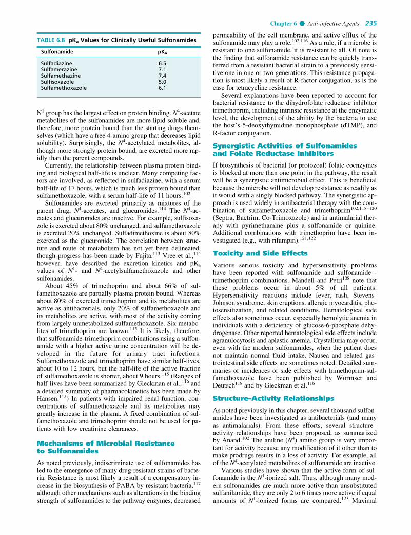
N1 group has the largest effect on protein binding. N4-acetatemetabolites of the sulfonamides are more lipid soluble and,therefore, more protein bound than the starting drugs them-selves (which have a free 4-amino group that decreases lipidsolubility). Surprisingly, the N4-acetylated metabolites, al-though more strongly protein bound, are excreted more rap-idly than the parent compounds.
Currently, the relationship between plasma protein bind-ing and biological half-life is unclear. Many competing fac-tors are involved, as reflected in sulfadiazine, with a serumhalf-life of 17 hours, which is much less protein bound thansulfamethoxazole, with a serum half-life of 11 hours.102
Sulfonamides are excreted primarily as mixtures of theparent drug, N4-acetates, and glucuronides.114 The N4-ac-etates and glucuronides are inactive. For example, sulfisoxa-zole is excreted about 80% unchanged, and sulfamethoxazoleis excreted 20% unchanged. Sulfadimethoxine is about 80%excreted as the glucuronide. The correlation between struc-ture and route of metabolism has not yet been delineated,though progress has been made by Fujita.113 Vree et al.,114
however, have described the excretion kinetics and pKa
values of N1- and N4-acetylsulfamethoxazole and othersulfonamides.
About 45% of trimethoprim and about 66% of sul-famethoxazole are partially plasma protein bound. Whereasabout 80% of excreted trimethoprim and its metabolites areactive as antibacterials, only 20% of sulfamethoxazole andits metabolites are active, with most of the activity comingfrom largely unmetabolized sulfamethoxazole. Six metabo-lites of trimethoprim are known.115 It is likely, therefore,that sulfonamide-trimethoprim combinations using a sulfon-amide with a higher active urine concentration will be de-veloped in the future for urinary tract infections.Sulfamethoxazole and trimethoprim have similar half-lives,about 10 to 12 hours, but the half-life of the active fractionof sulfamethoxazole is shorter, about 9 hours.115 (Ranges ofhalf-lives have been summarized by Gleckman et al.,116 anda detailed summary of pharmacokinetics has been made byHansen.115) In patients with impaired renal function, con-centrations of sulfamethoxazole and its metabolites maygreatly increase in the plasma. A fixed combination of sul-famethoxazole and trimethoprim should not be used for pa-tients with low creatinine clearances.
Mechanisms of Microbial Resistanceto Sulfonamides
As noted previously, indiscriminate use of sulfonamides hasled to the emergence of many drug-resistant strains of bacte-ria. Resistance is most likely a result of a compensatory in-crease in the biosynthesis of PABA by resistant bacteria,117
although other mechanisms such as alterations in the bindingstrength of sulfonamides to the pathway enzymes, decreased
permeability of the cell membrane, and active efflux of thesulfonamide may play a role.102,116 As a rule, if a microbe isresistant to one sulfonamide, it is resistant to all. Of note isthe finding that sulfonamide resistance can be quickly trans-ferred from a resistant bacterial strain to a previously sensi-tive one in one or two generations. This resistance propaga-tion is most likely a result of R-factor conjugation, as is thecase for tetracycline resistance.
Several explanations have been reported to account forbacterial resistance to the dihydrofolate reductase inhibitortrimethoprim, including intrinsic resistance at the enzymaticlevel, the development of the ability by the bacteria to usethe host’s 5-deoxythymidine monophosphate (dTMP), andR-factor conjugation.
Synergistic Activities of Sulfonamidesand Folate Reductase Inhibitors
If biosynthesis of bacterial (or protozoal) folate coenzymesis blocked at more than one point in the pathway, the resultwill be a synergistic antimicrobial effect. This is beneficialbecause the microbe will not develop resistance as readily asit would with a singly blocked pathway. The synergistic ap-proach is used widely in antibacterial therapy with the com-bination of sulfamethoxazole and trimethoprim102,118–120
(Septra, Bactrim, Co-Trimoxazole) and in antimalarial ther-apy with pyrimethamine plus a sulfonamide or quinine.Additional combinations with trimethoprim have been in-vestigated (e.g., with rifampin).121,122
Toxicity and Side Effects
Various serious toxicity and hypersensitivity problemshave been reported with sulfonamide and sulfonamide–-trimethoprim combinations. Mandell and Petri108 note thatthese problems occur in about 5% of all patients.Hypersensitivity reactions include fever, rash, Stevens-Johnson syndrome, skin eruptions, allergic myocarditis, pho-tosensitization, and related conditions. Hematological sideeffects also sometimes occur, especially hemolytic anemia inindividuals with a deficiency of glucose-6-phosphate dehy-drogenase. Other reported hematological side effects includeagranulocytosis and aplastic anemia. Crystalluria may occur,even with the modern sulfonamides, when the patient doesnot maintain normal fluid intake. Nausea and related gas-trointestinal side effects are sometimes noted. Detailed sum-maries of incidences of side effects with trimethoprim-sul-famethoxazole have been published by Wormser andDeutsch118 and by Gleckman et al.116
Structure–Activity Relationships
As noted previously in this chapter, several thousand sulfon-amides have been investigated as antibacterials (and manyas antimalarials). From these efforts, several structure–activity relationships have been proposed, as summarizedby Anand.102 The aniline (N4) amino group is very impor-tant for activity because any modification of it other than tomake prodrugs results in a loss of activity. For example, allof the N4-acetylated metabolites of sulfonamide are inactive.
Various studies have shown that the active form of sul-fonamide is the N1-ionized salt. Thus, although many mod-ern sulfonamides are much more active than unsubstitutedsulfanilamide, they are only 2 to 6 times more active if equalamounts of N1-ionized forms are compared.123 Maximal
Chapter 6 Anti-infective Agents 235
TABLE 6.8 pKa Values for Clinically Useful Sulfonamides
Sulfonamide pKa
Sulfadiazine 6.5Sulfamerazine 7.1Sulfamethazine 7.4Sulfisoxazole 5.0Sulfamethoxazole 6.1
0179-0241_17865_Ch06.qxd 12/8/09 10:55 PM Page 235

activity seems to be exhibited by sulfonamides between pKa
6.6 and 7.4.123–126 This reflects, in part, the need for enoughnonionized (i.e., more lipid soluble) drug to be present atphysiological pH to be able to pass through bacterial cellwalls.127 Fujita and Hansch113 also related pKa, partitioncoefficients, and electronic (Hammett) parameters withsulfonamide activity (Table 6.9).
Sulfamethizole4-Amino-N-(5-methyl-1,3,4-thiadiazole-2yl)benzenesulfon-amide; N1-(5-methyl-1,3,4-thiadiazol-2-yl)sulfanilamide; 5-methyl-2-sulfanilamido-1,3,4-thiadiazole. Sulfamethizole’splasma half-life is 2.5 hours. This compound is a white crys-talline powder soluble 1:2,000 in water.
Sulfisoxazole4-Amino-N-(3,4-dimethyl-5-isoxazolyl)benzenesulfon-amide; N1-(3,4-dimethyl-5-isoxazolyl) sulfanilamide; 5-sul-fanilamido-3,4-dimethylisoxazole. Sulfisoxazole’s plasmahalf-life is 6 hours. This compound is a white, odorless,slightly bitter, crystalline powder. Its pKa is 5.0. At pH 6,this sulfonamide has a water solubility of 350 mg in100 mL, and its acetyl derivative has a solubility of 110 mgin 100 mL of water.
Sulfisoxazole possesses the action and the uses of othersulfonamides and is used for infections involving sulfon-amide-sensitive bacteria. It is claimed to be effective in thetreatment of Gram-negative urinary infections.
Sulfisoxazole AcetylN-[(4-Aminophenyl)sulfonyl]-N-(3,4-dimethyl-5-isoxa-zolyl)acetamide; N-(3,4-dimethyl-5-isoxazolyl)-N-sulfanily-
lacetamide; N1-acetyl-N1-(3,4-dimethyl-5-isoxazolyl)sul-fanilamide. Sulfisoxazole acetyl shares the actions and usesof the parent compound, sulfisoxazole. The acetyl derivativeis tasteless and, therefore, suitable for oral administration, es-pecially in liquid preparations. The acetyl compound is splitin the intestinal tract and absorbed as sulfisoxazole; that is, itis a prodrug for sulfisoxazole.
Sulfisoxazole Diolamine4-Amino-N-(3,5-dimethyl-5-isoxazolyl)benzenesulfon-amide compound with 2,2�-iminobis[ethanol](1:1); 2,2�-iminodiethanol salt of N1-(3,4-dimethyl-5-isoxazolyl)sul-fanilamide. This salt is prepared by adding enoughdiethanolamine to a solution of sulfisoxazole to bring thepH to about 7.5. It is used as a salt to make the drug moresoluble in the physiological pH range of 6.0 to 7.5 and isused in solution for systemic administration of the drug byslow intravenous, intramuscular, or subcutaneous injectionwhen high enough blood levels cannot be maintained byoral administration alone. It also is used for instillation ofdrops or ointment in the eye for the local treatment of sus-ceptible infections.
Sulfamethazine4-Amino-N-(4,6-dimethyl-2-pyrimidinyl)benzenesul-fonamide; N1-(4,6-dimethyl-2-pyrimidinyl)sulfanilamide;2-sulfanilamido-4,6-dimethylpyrimidine. Sulfamethazine’splasma half-life is 7 hours. This compound is similar inchemical properties to sulfamerazine and sulfadiazine butdoes have greater water solubility than either. Its pKa is 7.2.Because it is more soluble in acid urine than sulfamerazineis, the possibility of kidney damage from use of the drug isdecreased. The human body appears to handle the drugunpredictably; hence, there is some disfavor to its use in thiscountry except in combination sulfa therapy (in trisul-fapyrimidines, USP) and in veterinary medicine.
SulfacetamideN-[(4-Aminophenyl)sulfonyl]-acetamide; N-sulfanilylac-etamide; N1-acetylsulfanilamide. Sulfacetamide’s plasmahalf-life is 7 hours. This compound is a white crystallinepowder, soluble in water (1:62.5 at 37°C) and in alcohol. It
236 Wilson and Gisvold’s Textbook of Organic Medicinal and Pharmaceutical Chemistry
TABLE 6.9 Characteristics of Absorbable Short- andIntermediate-Acting Sulfonamides
Sulfonamide Half-Life Oral Absorption
Sulfisoxazole Short (6 hours) Prompt (peak levelsin 1–4 hours)
Sulfamethizole Short (9 hours) PromptSulfadiazine Intermediate Slow (peak levels in
(10–17 hours) 4–8 hours)Sulfamethoxazole Intermediate Slow
(10–12 hours)Sulfadoxine Long (7–9 days) Intermediate
Pyrimidine
Trimethoprim Intermediate Prompt(11 hours)
0179-0241_17865_Ch06.qxd 12/8/09 10:55 PM Page 236

but higher than that of sulfadiazine, sulfamerazine, or sul-famethazine.
Following oral administration, sulfamethoxazole is notabsorbed as completely or as rapidly as sulfisoxazole, andits peak blood level is only about 50% as high.
Sulfadiazine4-Amino-N-2-pyrimidinyl-benzenesulfonamide; N1-2-pyrimidinylsulfanilamide; 2-sulfanilamidopyrimidine.Sulfadiazine’s plasma half-life is 17 hours. It is a white,odorless crystalline powder soluble in water to the extentof 1:8,100 at 37°C and 1:13,000 at 25°C, in human serumto the extent of 1:620 at 37°C, and sparingly soluble in al-cohol and acetone. It is readily soluble in dilute mineralacids and bases. Its pKa is 6.3.
Sulfadiazine SodiumSoluble sulfadiazine is an anhydrous, white, colorless, crys-talline powder soluble in water (1:2) and slightly soluble inalcohol. Its water solutions are alkaline (pH 9–10) and ab-sorb carbon dioxide from the air, with precipitation of sulfa-diazine. It is administered as a 5% solution in sterile waterintravenously for patients requiring an immediately highblood level of the sulfonamide.
Mixed Sulfonamides
The danger of crystal formation in the kidneys from adminis-tration of sulfonamides has been greatly reduced through theuse of the more soluble sulfonamides, such as sulfisoxazole.This danger may be diminished still further by administeringmixtures of sulfonamides. When several sulfonamides are ad-ministered together, the antibacterial action of the mixture isthe summation of the activity of the total sulfonamide concen-tration present, but the solubilities are independent of thepresence of similar compounds. Thus, by giving a mixtureof sulfadiazine, sulfamerazine, and sulfacetamide, the sametherapeutic level can be maintained with much less danger ofcrystalluria, because only one third of the amount of any onecompound is present. Descriptions of some of the mixturesused follow.
Trisulfapyrimidines, Oral SuspensionThe oral suspension of trisulfapyrimidines contains equalweights of sulfadiazine, USP; sulfamerazine, USP; and sul-famethazine, USP, either with or without an agent to raisethe pH of the urine.
Trisulfapyrimidines, TabletsTrisulfapyrimidine tablets contain essentially equal quanti-ties of sulfadiazine, sulfamerazine, and sulfamethazine.
is very soluble in hot water, and its water solution is acidic.It has a pKa of 5.4.
SulfachloropyridazineN1-(6-Chloro-3-pyridazinyl) sulfanilamide. Sulfachloro-pyridazine’s plasma half-life is 8 hours.
Sulfapyridine4-Amino-N-2-pyridinylbenzenesulfonamide; N1-2-pyridyl-sulfanilamide. Sulfapyridine’s plasma half-life is 9 hours.This compound is a white, crystalline, odorless, and taste-less substance. It is stable in air but slowly darkens on expo-sure to light. It is soluble in water (1:3,500), in alcohol(1:440), and in acetone (1:65) at 25°C. It is freely soluble indilute mineral acids and aqueous solutions of sodium andpotassium hydroxide. The pKa is 8.4. Its outstanding effectin curing pneumonia was first recognized by Whitby; how-ever, because of its relatively high toxicity, it has been sup-planted largely by sulfadiazine and sulfamerazine. Severalcases of kidney damage have resulted from acetylsulfapyri-dine crystals deposited in the kidneys. It also causes severenausea in most patients. Because of its toxicity, it is usedonly for dermatitis herpetiformis.
Sulfapyridine was the first drug to have an outstandingcurative action on pneumonia. It gave impetus to the studyof the whole class of N1 heterocyclically substituted deriva-tives of sulfanilamide.
Sulfamethoxazole4-Amino-N-(5-methyl-3-isoxazolyl)benzenesulfonamide;N1-(5-methyl-3-isoxazolyl) sulfanilamide (Gantanol).Sulfamethoxazole’s plasma half-life is 11 hours.
Sulfamethoxazole is a sulfonamide drug closely relatedto sulfisoxazole in chemical structure and antimicrobial ac-tivity. It occurs as a tasteless, odorless, almost white crys-talline powder. The solubility of sulfamethoxazole in the pHrange of 5.5 to 7.4 is slightly lower than that of sulfisoxazole
Chapter 6 Anti-infective Agents 237
0179-0241_17865_Ch06.qxd 12/8/09 10:55 PM Page 237

Sulfadoxine and PyrimethamineThe mixture of sulfadoxine and pyrimethamine (Fansidar) isused for the treatment of P. falciparum malaria in patients inwhom chloroquine resistance is suspected. It is also used formalaria prophylaxis for travelers to areas where chloro-quine-resistant malaria is endemic.
Topical Sulfonamides
Sulfacetamide SodiumN-Sulfanilylacetamide monosodium salt (Sodium Sulamyd)is obtained as the monohydrate and is a white, odorless, bit-ter, crystalline powder that is very soluble (1:2.5) in water.Because the sodium salt is highly soluble at the physio-logical pH of 7.4, it is especially suited, as a solution, forrepeated topical applications in the local management ofophthalmic infections susceptible to sulfonamide therapy.
Sulfisoxazole DiolamineSulfisoxazole diolamine is described with the short- and in-termediate-acting sulfonamides and also used in intravenousand intramuscular preparations.
Triple SulfaTriple sulfa (sulfabenzamide, sulfacetamide, and sulfathia-zole; Femguard) is used as a vaginal cream in the treatmentof Haemophilus vaginalis vaginitis.
Nonabsorbable Sulfonamides
TOPICAL SULFONAMIDES FOR BURN THERAPY
Mafenide Acetate4-(Aminomethyl)benzenesulfonamide acetate (Sulfamylon)is a homologue of the sulfanilamide molecule. It is not atrue sulfanilamide-type compound, as it is not inhibited byPABA. Its antibacterial action involves a mechanism thatdiffers from that of true sulfanilamide-type compounds.
This compound is particularly effective against Clostridiumwelchii in topical application and was used duringWorld War II by the German army for prophylaxis ofwounds. It is not effective orally. It is currently used aloneor with antibiotics in the treatment of slow-healing, infectedwounds.
Some patients treated for burns with large quantities ofthis drug have developed metabolic acidosis. To overcomethis adverse effect, a series of new organic salts was pre-pared.16 The acetate in an ointment base proved to be themost efficacious.
Silver Sulfadiazine (Silvadene)The silver salt of sulfadiazine applied in a water-misciblecream base has proved to be an effective topical antimicro-bial agent, especially against Pseudomonas spp. This is par-ticularly significant in burn therapy because pseudomonadsare often responsible for failures in therapy. The salt is onlyslightly soluble and does not penetrate the cell wall butacts on the external cell structure. Studies using radioactivesilver have shown essentially no absorption into body flu-ids. Sulfadiazine levels in the serum were about 0.5 to2 mg/100 mL.
This preparation is reported to be easier to use thanother standard burn treatments, such as application offreshly prepared dilute silver nitrate solutions or mafenideointment.
Sulfonamides for Intestinal Infections,Ulcerative Colitis, or Reduction ofBowel Flora
Each of the sulfonamides in this group is a prodrug, whichis designed to be poorly absorbable, though usually, in prac-tice, a little is absorbed. Therefore, usual precautions withsulfonamide therapy should be observed. In the large intes-tine, the N4-protecting groups are cleaved, releasing the free
238 Wilson and Gisvold’s Textbook of Organic Medicinal and Pharmaceutical Chemistry
0179-0241_17865_Ch06.qxd 12/8/09 10:55 PM Page 238

sulfonamide antibacterial agent. Today, only one example isused clinically, sulfasalazine.
Sulfasalazine
Sulfasalazine (2-hydroxy-5[[4-[(2-pyridinylamino)sulfonyl]phenyl]azo]benzoic acid or 5-[p-(2-pyridylsulfamoyl)phenylazo]salicylic acid) is a brownish yellow, odorlesspowder, slightly soluble in alcohol but practically insolublein water, ether, and benzene.
Sulfasalazine is broken down in the body to m-aminosal-icylic acid and sulfapyridine. The drug is excreted throughthe kidneys and is detectable colorimetrically in the urine,producing an orange-yellow color when the urine is alkalineand no color when the urine is acid.
DIHYDROFOLATE REDUCTASEINHIBITORS
TrimethoprimTrimethoprim (5-[(3,4,5-trimethoxyphenyl)methyl]-2,4-pyrimidinediamine or 2,4-diamino-5-(3,4,5-trimethoxyben-zyl)pyrimidine) is closely related to several antimalarialsbut does not have good antimalarial activity by itself; it is,however, a potent antibacterial. Originally introduced incombination with sulfamethoxazole, it is now available as asingle agent.
Approved by the FDA in 1980, trimethoprim as a sin-gle agent is used only for the treatment of uncomplicatedurinary tract infections. The argument for trimethoprim asa single agent was summarized in 1979 by Wormser andDeutsch.118 They point out that several studies comparingtrimethoprim with TMP–SMX for the treatment ofchronic urinary tract infections found no statistically rel-evant difference between the two courses of therapy.Furthermore, some patients cannot take sulfonamideproducts for the reasons discussed previously in this chap-ter. The concern is that when used as a single agent, bac-teria now susceptible to trimethoprim will rapidly developresistance. In combination with a sulfonamide, however,the bacteria will be less likely to do so. That is, they willnot survive long enough to easily develop resistance toboth drugs.
Sulfamethoxazole and TrimethoprimThe synergistic action of the combination of these two drugsis discussed previously in this chapter.
SULFONES
The sulfones are primarily of interest as antibacterial agents,though there are some reports of their use in the treatment ofmalarial and rickettsial infections. They are less effectivethan the sulfonamides. PABA partially antagonizes the ac-tion of many of the sulfones, suggesting that the mechanismof action is similar to that of the sulfonamides. Further, in-fections that arise in patients being treated with sulfones arecross-resistant to sulfonamides. Several sulfones haveproved useful in the treatment of leprosy, but among themonly dapsone is clinically used today.
It has been estimated that there are about 11 million casesof leprosy in the world, of which about 60% are in Asia(with 3.5 million in India alone). The first reports of dap-sone resistance prompted the use of multidrug therapy withdapsone, rifampin, and clofazimine combinations in somegeographic areas.128
The search for antileprotic drugs has been hampered bythe inability to cultivate M. leprae in artificial media andby the lack of experimental animals susceptible to humanleprosy. A method of isolating and growing M. leprae in thefootpads of mice and in armadillos has been reported andhas permitted a much wider range of research. Sulfoneswere introduced into the treatment of leprosy after it wasfound that sodium glucosulfone was effective in experimen-tal tuberculosis in guinea pigs.
The parent sulfone, dapsone (4,4�-sulfonyldianiline), isthe prototype for various analogs that have been widelystudied. Four variations on this structure have given activecompounds:
1. Substitution on both the 4- and 4�-amino functions2. Monosubstitution on only one of the amino functions3. Nuclear substitution on one of the benzenoid rings4. Replacement of one of the phenyl rings with a hetero-
cyclic ring
The antibacterial activity and the toxicity of the disubsti-tuted sulfones are thought to be chiefly caused by the forma-tion in vivo of dapsone. Hydrolysis of disubstituted deriva-tives to the parent sulfone apparently occurs readily in theacid medium of the stomach but only to a very limited ex-tent following parenteral administration. Monosubstitutedand nuclear-substituted derivatives are believed to act as en-tire molecules.
DapsoneDapsone (4,4�-sulfonylbisbenzeneamine; 4,4�-sulfonyldian-iline; p,p�-diaminodiphenylsulfone; or DDS [Avlosulfon])occurs as an odorless, white crystalline powder that is veryslightly soluble in water and sparingly soluble in alcohol.The pure compound is light stable, but traces of impurities,including water, make it photosensitive and thus susceptibleto discoloration in light. Although no chemical change is de-tectable following discoloration, the drug should be pro-tected from light.
Chapter 6 Anti-infective Agents 239
0179-0241_17865_Ch06.qxd 12/8/09 10:55 PM Page 239

REFERENCES1. Atlas, R. M.: Microbiology, Fundamentals and Applications. New
York, Macmillan, 1984, p. 19.2. Ehrlich, B.: United States Patent 986,148.3. Christiansen, A.: J. Am. Chem. Soc. 42:2402, 1920.4. Atlas, R. M.: Microbiology, Fundamentals and Applications. New
York, Macmillan, 1984, p. 20.5. Fleming, M., et al.: Ethanol. In Hardman, J. G., Limbird, L. E., and
Gilman, A. G. (eds.). Goodman and Gilman’s The PharmacologicalBasis of Therapeutics. New York, McGraw-Hill, 2001.
6. DuMez, A. G.: J. Am. Pharm. Assoc. 28:416, 1939.7. Gilbert, G. L.: Appl. Microbiol. 12:496, 1964.8. Leech, P. N.: J. Am. Med. Assoc. 109:1531, 1937.9. Miner, N. A., et al.: Am. J. Pharm. 34:376, 1977.
10. U.S. Food and Drug Administration: Hexachlorophene andNewborns. Bulletin, December 1971.
11. Vasarenesh, A.: Arch. Dermatol. 98:183, 1968.12. Gershenfeld, L.: Milk Food Technol. 18:233, 1955.13. Domagk, G.: Dtsch. Med. Wochenschr. 61:250, 1935.14. Rose, F. L., and Swain, G. J.: J. Chem. Soc. 442, 1956.15. Massey, A. G.: Main Group Elements. Chichester, England, Ellis
Horwood Ltd., 1990, p. 160.16. Garrett, E. R., and Woods, O. R.: J. Am. Pharm. Assoc. (Sci. Ed.) 42:
736, 1953.17. Rippon, J. W.: In Burrows, W. (ed.). Textbook of Microbiology.
Philadelphia, W. B. Saunders, 1973, p. 683.18. Friedman, L., et al.: J. Invest. Dermatol. 35:3–5, 1960.19. Rippon, J. W.: In Burrows, W. (ed.). Textbook of Microbiology.
Philadelphia, W. B. Saunders, 1973, p. 721.20. Rippon, J. W.: In Burrows, W. (ed.). Textbook of Microbiology.
Philadelphia, W. B. Saunders, 1973, p. 734.21. Goldman, R. C., and Klein, L. L.: Annu. Rep. Med. Chem. 29:155,
1994.22. Ajello, L.: Science 123:876, 1956.23. Ajello, L.: Mycopathologia 17:315, 1962.24. Rebell, G., and Taplin, D.: Dermatophytes. Miami, University of
Miami Press, 1970.25. Rieth, H.: Arzneim. Forsch. 31:1309, 1981.26. Polak, A., and Scholer, H. J.: Chemotherapy 21:113, 1975.27. Baginski, M., Resat, H., and McCammon, J. A.: Mol. Pharmacol.
52:560, 1997.28. Baginski, M., Gariboldi, P., Bruni, P., et al.: Biophys. Chem. 65:91,
1997.29. Gold, W., et al.: Antibiotics Annual 1955–1956. New York, Medical
Encyclopedia, 1956.30. Mechlinski, W., et al.: Tetrahedron Lett. 3873, 1970.
31. Graybill, J. R.: Ann. Intern. Med. 124:921, 1986.32. Ianknegkt, R., et al.: Clin. Pharmacokinet. 23:279, 1992.33. Hazen, E. L., and Brown, R.: Proc. Soc. Exp. Biol. Med. 76:93, 1951.34. Pandey, R. C., and Rinehart, K. L.: J. Antibiot. 29:1035, 1976.35. Lencelin, J. M., et al.: Tetrahedron Lett. 29:2827, 1988.36. Struyk, A. P., Hoette, I., Orost, G., et al.: Antibiot. Ann. 878,
1957–1958.37. Brik, H., et al.: Nystatin. In Florey, K. (ed.). Analytical Profiles of
Drug Substances, vol. 10. New York, Academic Press, 1981, p. 513.38. Oxford, A. E., et al.: Biochem. J. 33:240, 1939.39. Grove, J. F., et al.: J. Chem. Soc. 3977, 1952.40. Sloboda, R. D., Van Blaricom, G., Creasey, W. A., et al.: Biochem.
Biophys. Res. Commun. 105:882, 1982.41. Stütz, A., and Petranyi, G.: J. Med. Chem. 27:1539, 1984.42. Ryder, N. S.: Antimicrob. Agents Chemother. 27:252, 1985.43. Petranyi, G., et al.: Science 224:1239, 1984.44. Gupta, M. P., et al.: J. Vet. Med. Mycol. 29:45, 1991.45. Thomas, A. H.: Antimicrob. Chemother. 17:269, 1986.46. Hitchcock, C. A.: Biochem. Soc. Trans. 19:782, 1991.47. Pont, A., et al.: Arch. Intern. Med. 144:2150, 1984.48. Vanden Bossche, H.: Drug Dev. Res. 8:287, 1986.49. Rotstein, D. M., Kertesz, D. J., Walker, K. A. M., et al.: J. Med.
Chem. 35:2818, 1992.50. Dickinson, R. P., et al.: Med. Chem. Lett. 6:2031, 1996.51. DeBono, M., et al.: J. Med. Chem. 38:3271, 1995.52. Schwartz, R. E., Masurekar, P. S., and White, R. F.: Clin. Dermatol.
7:375, 1993.53. Balkovec, J. M.: Annu. Rep. Med. Chem. 33:175, 1998.54. Jao, E.: Tetrahedron Lett. 37:5661, 1996.55. Nagiec, M. M., Nagiec, E. E., et al.: J. Biol. Chem. 272:9809, 1997.56. Shen, L. L., Mitscher, L. A., Sharma, P. N., et al.: Biochemistry 28:
3886, 1989.57. Sreedharan, S., et al.: J. Bacteriol. 172:7260, 1990.58. Yoshida, H., et al.: Antimicrob. Agents Chemother. 35:1647, 1991.59. Yoshida, H., et al.: Antimicrob. Agents Chemother. 34:1273, 1990.60. Bazile, S., et al.: Antimicrob. Agents Chemother. 36:2622, 1992.61. Domagala, J. M.: J. Antimicrob. Chemother. 33:685, 1994.62. Ross, D. L., and Riley, C. M.: J. Pharm. Biomed. Anal. 12:1325, 1994.63. Lee, D. S., et al.: J. Pharm. Biomed. Anal. 12:157, 1994.64. Mitscher, L. A., et al.: J. Med. Chem. 30:2283, 1987.65. Bloom, B. R., and Murray, C. J. L.: Science 257:1055, 1992.66. Winder, F. G., and Collins, P. B.: J. Gen. Microbiol. 63:41, 1970.67. Quomard, A., Lacove, C., and LanPelle, G.: Antimicrob. Agents
Chemother. 35:1035, 1991.68. Youatt, J., and Tham, S. H.: Am. Rev. Respir. Dis. 100:25, 1969.69. Johnsson, K., and Schultz, P. G.: J. Am. Chem. Soc. 116:7425, 1994.
240 Wilson and Gisvold’s Textbook of Organic Medicinal and Pharmaceutical Chemistry
4. What is the most active agent for the clinical treatment oftuberculosis?
5. What is the function of dihydrofolate reductase in bacteria?
1. What is the definition of an antiseptic?
2. What happens to the antimicrobial activity of alcohols asthe carbon chain becomes more branched?
3. What kind of bond associates iodine with polyvinylpyrroli-done in PVP–iodine?
R E V I E W Q U E S T I O N S
Dapsone is used in the treatment of both lepromatous andtuberculoid types of leprosy. Dapsone is used widely for allforms of leprosy, often in combination with clofazimine andrifampin. Initial treatment often includes rifampin with dap-sone, followed by dapsone alone. It is also used to prevent
the occurrence of multibacillary leprosy when given pro-phylactically.
Dapsone is also the drug of choice for dermatitis herpeti-formis and is sometimes used with pyrimethamine for treat-ment of malaria and with trimethoprim for PCP.
Serious side effects can include hemolytic anemia,methemoglobinemia, and toxic hepatic effects. Hemolyticeffects can be pronounced in patients with glucose-6-phos-phate dehydrogenase deficiency. During therapy, all pa-tients require frequent blood counts.
0179-0241_17865_Ch06.qxd 12/8/09 10:55 PM Page 240

70. Zhang, Y., et al.: Nature 358:591, 1992.71. Dessen, A., et al.: Science 267:1638, 1995.72. Banerjee, A., et al.: Science 263:227, 1994.73. Boxenbaum, H. G., and Riegelman, S.: J. Pharm. Sci. 63:1191, 1974.74. Heifets, L. B., Flory, M. A., and Lindholm-Levy, P. J.: Antimicrob.
Agents Chemother. 33:1252, 1989.75. Rinehart, K. L.: Acc. Chem. Res. 5:57, 1972.76. Hartmann, G. R., et al.: Angew. Chem. Int. Ed. Engl. 24:1009,
1985.77. Katz, E., and Moss, B.: Proc. Natl. Acad. Sci. U. S. A. 66:677,
1970.78. Werli, W.: Rev. Infect. Dis. 5S:407, 1983.79. Gallo, G. G., and Radaelli, P.: Rifampin. In Florey, K. (ed.). Analytical
Profiles of Drug Substances. New York, Academic Press, 1976.80. Kuehl, F. A., et al.: J. Am. Chem. Soc. 77:2344, 1955.81. Hildy, P. H., et al.: J. Am. Chem. Soc. 77:2345, 1955.82. Stammer, C. H., et al.: J. Am. Chem. Soc. 77:2346, 1955.83. Smart, J., et al.: Experientia 13:291, 1957.84. Neuhaus, F. C., and Lynch, J. L.: Biochemistry 3:471, 1964.85. Rando, R. R.: Biochem. Pharmacol. 24:1153, 1975.86. Herr, E. B., Pittenger, G. E., and Higgens, C. E.: Indiana Acad. Sci.
69:134, 1960.87. Bycroft, B. W., et al.: Nature 231:301, 1971.88. Nomoto, S., et al.: J. Antibiot. 30:955, 1977.89. Knight, R. C., et al.: Biochem. Pharmacol. 27:2089, 1978.90. Metcalf, B. W., et al.: J. Am. Chem. Soc. 100:2551, 1978.91. Prichard, R. K.: Nature 228:684, 1970.92. Friedman, P. A., and Platzer, E. G.: Biochim. Biophys. Acta 544:
605, 1978.93. Pica-Mattoccia, L., and Cioli, D.: Am. J. Trop. Med. Hyg. 34:112,
1985.94. Burg, R. W., et al.: Antimicrob. Agents Chemother. 15:361, 1979.95. Campbell, W. C.: Science 221:823, 1983.96. Albers-Schonberg, G., et al.: J. Am. Chem. Soc. 103:4216, 1981.97. Springer, J. P., et al.: J. Am. Chem. Soc. 103:4221, 1981.98. Aziz, M. A., et al.: Lancet 2:171, 1982.99. Orkin, M., and Maibach, H. I.: N. Engl. J. Med. 298:496, 1978.
100. Domagk, G.: Dtsch. Med. Wochenschr. 61:250, 1935.101. Baumler, E.: In Search of the Magic Bullet. London, Thames
and Hudson, 1965.102. Anand, N.: Sulfonamides and sulfones. In Wolff, M. E. (ed.). Burger’s
Medicinal Chemistry, vol. 2, 5th ed. New York, Wiley-Interscience,1996, Chap. 33.
103. Foerster, J.: Z. Haut. Geschlechtskr. 45:459, 1933.104. Trefouel, J., et al.: C. R. Seances Soc. Biol. 120:756, 1935.105. Fuller, A. T.: Lancet 1:194, 1937.106. Northey, E. H.: The Sulfonamides and Allied Compounds. ACS
Monogr. Ser. Washington, DC, American Chemical Society, 1948.107. MacDonald, L., and Kazanijan, P.: Formulary 31:470, 1996.108. Petri, W. A.: Antimicrobial agents: Sulfonamides, trimethoprim-
sulfamethoxazole, quinolones, and agents for urinary tract infection.In Gilman, A. G., et al. (eds.). The Pharmacological Basis ofTherapeutics, 9th ed. New York, Macmillan, 1996.
109. Jawetz, E.: Principles of antimicrobial drug action. In Katzung, B. G.(ed.). Basic and Clinical Pharmacology, 6th ed. Norwalk, CT,Appleton & Lange, 1995.
110. Med. Lett. 30:33, 1988.111. FDA Drug Bulletin, US Department of Health, Education and
Welfare, Food and Drug Administration, Feb., 1980.112. Med. Lett. 29:53, 1987.113. Fujita, T., and Hansch, C.: J. Med. Chem. 10:991, 1967.114. Vree, T. B., et al.: Clin. Pharmacokinet. 4:310, 1979.115. Hansen, I.: Antibiot. Chemother. 25:217, 1978.116. Gleckman, R., et al.: Am. J. Hosp. Pharm. 36:893, 1979.117. Bushby, S. R., and Hitchings, G. H.: Br. J. Pharmacol. Chemother.
33:72, 1968.118. Wormser, G. P., and Deutsch, G. T.: Ann. Intern. Med. 91:420, 1979.119. Palminteri, R., and Sassella, D.: Chemotherapy 25:181, 1979.120. Harvey, R. J.: J. Antimicrob. Chemother. 4:315, 1978.121. Letters of Burchall, J. J., Then, R., and Poe, M.: Science
197:1300–1301, 1977.122. Werlin, S. L., and Grand, R. J.: J. Pediatr. 92:450, 1978.123. Fox, C. L., and Ross, H. M.: Proc. Soc. Exp. Biol. Med. 50:142, 1942.124. Yamazaki, M., et al.: Chem. Pharm. Bull. (Tokyo) 18:702, 1970.125. Bell, P. H., and Roblin, R. O.: J. Am. Chem. Soc. 64:2905, 1942.126. Cowles, P. B.: Yale J. Biol. Med. 14:599, 1942.127. Brueckner, A. H.: J. Biol. Med. 15:813, 1943.128. Shepard, C. C.: N. Engl. J. Med. 307:1640, 1982.
SELECTED READINGBloom, B. R. (ed.): Tuberculosis. Washington, DC, American Society for
Microbiology Press, 1994.Chu, D. T., and Fernandes, P. B.: Recent developments in the field of
quinolone antibacterial agents. In Testa, B. (ed.). Advances in DrugResearch, vol. 21. New York, Academic Press, 1991.
Como, J. A., and Dismukes, W. E.: Oral azoles as systemic antifungalchemotherapy. N. Engl. J. Med. 330:263, 1994.
Despommier, D. D., and Karapelou, J. W.: Parasitic Life Cycles. NewYork, Springer-Verlag, 1987.
Goldsmith, R. S., and Heyneman, D. (eds.): Tropical Medicine andParasitology. New York, Appleton & Lange, 1989.
Hastings, R. C., and Franzblau, S. G.: Chemotherapy of leprosy. Annu.Rev. Pharmacol. Toxicol. 28:231, 1988.
Hooper, D. C., and Wolfson, J. S. (eds.): Quinolone AntimicrobialAgents, 2nd ed. Washington, DC, American Society for MicrobiologyPress, 1993.
Houston, S., and Fanning, A.: Current and potential treatment of tubercu-losis. Drugs 48:689, 1994.
Jernigan, J. A., and Pearson, R. D.: Antiparasitic agents. In Mandell GL,Bennett J. E., and Dolin R. (eds.). Principles and Practice of InfectiousDiseases, vol. 1, 4th ed. New York, Churchill-Livingstone, 1995.
Kreier, J. B., and Baker, J. R. (eds.): Parasitic Protozoa, 2nd ed. SanDiego, Academic Press, 1991.
Reed, S.: Amebiasis: an update. Clin. Infect. Dis. 14:385, 1992.Singh, S. K., and Sherma, S.: Current status of medical research in
helminth disease. Med. Res. Rev. 11:581, 1991.Wang, C. C.: Molecular mechanisms and therapeutic approaches to the
treatment of African trypanosomiasis. Annu. Rev. Pharmacol. Toxicol.35:93, 1995.
Yamaguchi, H., Kobazaski, G. S., and Takahashi, H. (eds.): RecentAdvances in Antifungal Chemotherapy. New York, MarcelDekker, 1992.
Chapter 6 Anti-infective Agents 241
0179-0241_17865_Ch06.qxd 12/8/09 10:55 PM Page 241

242
C H A P T E R 7
AntimalarialsJOHN H. BLOCK
continues to be devastating. The role of diseases such assmallpox, plague, yellow fever, and polio on human historyis fascinating, but, fortunately, is mostly historical.Although smallpox has been eliminated, the latter three dis-eases do reappear, but the cases are isolated. Plague istreated effectively with antibiotics, and there are vaccinesfor yellow fever and polio.
There are three potential ways to control malaria: elimi-nation of the vector, drug therapy, and vaccination.Elimination of the vector is the simplest and most cost-effective. Drug therapy has the same challenges as those withdevelopment of antibiotics, resistance to the drug. The currentantimalarial drugs, although reasonably effective, also havesignificant adverse reactions, and resistance is increasing. Sofar, no effective vaccine has been developed that is effectivein vivo, but that may be changing because of a better under-standing how the human immune system interacts with theparasite. The malaria parasite does elicit an immune responseas evidenced by the fact that children with an initial exposureare more likely to die than adults who have recurring attacks.
Because malaria has been eliminated from NorthAmerica, Europe, and parts of Asia, it becomes a potentialproblem when citizens from a malaria-free area travel into anarea where malaria is endemic. It is common for these trav-elers going into areas where malaria is endemic to receiveprescriptions for antimalarial drugs and use them prophylac-tically.8 Many times, citizens of malaria-free countries re-turning from areas where malaria is endemic plus citizens ofthe latter countries who are coming to the malaria-free coun-tries will have contracted malaria and will require antimalar-ial drugs for several months following their return. Also,there will be restriction on their ability to be blood donors.Most prescriptions for antimalarial drugs in North Americaare indicated for prophylaxis of travelers going to and com-ing from areas of the world where malaria is endemic.
Malaria
Malaria is caused by four species of the one-cell protozoanof the Plasmodium genus. They are:
Plasmodium falciparum: This species is estimated to causeapproximately 50% of all malaria. It causes the mostsevere form of the disease and, because patients feelill between acute attacks, debilitating form of the disease.One of the reasons it leaves the patient so weak isbecause it infects up to 65% of the patient’s erythrocytes.
Plasmodium vivax: This species is the second mostcommon species causing about 40% of all malarialcases. It can be very chronic in recurrence because it canreinfect liver cells.
C H A P T E R O V E R V I E W
Malaria, one of the most widespread diseases, is caused by aPlasmodium parasite and is transmitted to humans by theAnopheles mosquito. It infects several hundred million peo-ple each year, results in several million deaths annually, andis a complex disease to treat. The causative agent is a groupof parasitical protozoa of the Plasmodium genus transmittedby the female Anopheles mosquito. Its original treatment wasquinine that became the prototypical molecule for the firstgeneration of synthetic antimalarial drugs. The target for drugtreatment and prophylaxis is the parasite, and each advance inthe drug treatment of this disease has resulted in the parasitedeveloping resistance. One of the most effective preventionsis controlling the mosquito population that is the vector car-rying the parasite to humans. The human immune systemdoes respond to the parasite, but the development of an effec-tive vaccine has been a challenge. Sequencing the plasmo-dium genome is providing information that may lead to otherapproaches to prevent and treat this debilitating disease.
History
Malaria’s name is derived from “mala aria” or bad air, andhas been called ague, intermittent fever, marsh fever, andThe Fever.1,2 The name is based on the early knowledge thatmalaria was associated with swamps and badly drainedareas. The use of quinine for treating malaria has beenknown since the 17th century. Although malaria is an an-cient disease, its upsurge seems to coincide with the adventof farming about 20,000 years ago. The clearing of land pro-vided areas for ponds containing still water. The Anophelesgambiae mosquito uses still water that sits in ponds and con-tainers to breed. The gathering of humans in farming com-munities provided the necessary concentration of people toform a reservoir of hosts for the parasite and “food” for themosquitoes breeding in the ponds.3–6
Proof that the Anopheles mosquito is the carrier of thecausative protozoa was obtained by Dr. Ronald Ross whowas recognized in 1902 by receipt of the Nobel Prize inMedicine. In a scenario somewhat similar to that where de-finitive proof that yellow fever was transmitted by theAedes aegypti mosquito was required, Dr. Ross strongly ar-gued that malaria was transmitted by an insect vector andfinally demonstrated that the parasite was carried in thestomach and salivary glands of the Anopheles mosquito.The latter discovery was important because it helped re-solve the dispute whether malaria was spread by the bite ofthe mosquito or drinking water containing mosquito eggsand larvae.7 The impact of malaria on the human species
0242-0257_17865_Ch07.qxd 12/4/09 4:51 AM Page 242
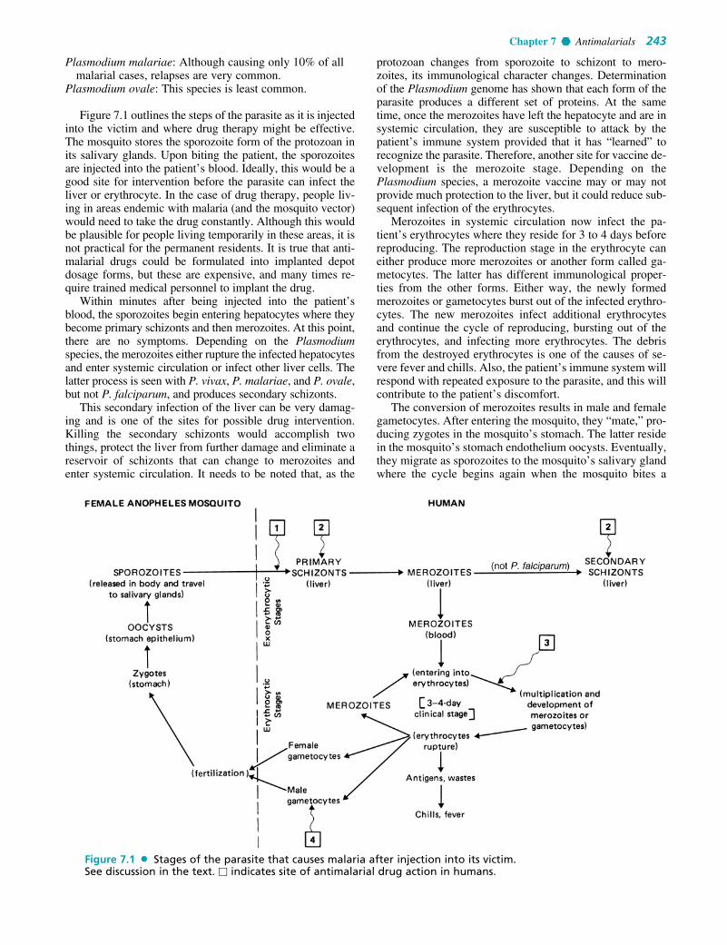
Chapter 7 Antimalarials 243
Plasmodium malariae: Although causing only 10% of allmalarial cases, relapses are very common.
Plasmodium ovale: This species is least common.
Figure 7.1 outlines the steps of the parasite as it is injectedinto the victim and where drug therapy might be effective.The mosquito stores the sporozoite form of the protozoan inits salivary glands. Upon biting the patient, the sporozoitesare injected into the patient’s blood. Ideally, this would be agood site for intervention before the parasite can infect theliver or erythrocyte. In the case of drug therapy, people liv-ing in areas endemic with malaria (and the mosquito vector)would need to take the drug constantly. Although this wouldbe plausible for people living temporarily in these areas, it isnot practical for the permanent residents. It is true that anti-malarial drugs could be formulated into implanted depotdosage forms, but these are expensive, and many times re-quire trained medical personnel to implant the drug.
Within minutes after being injected into the patient’sblood, the sporozoites begin entering hepatocytes where theybecome primary schizonts and then merozoites. At this point,there are no symptoms. Depending on the Plasmodiumspecies, the merozoites either rupture the infected hepatocytesand enter systemic circulation or infect other liver cells. Thelatter process is seen with P. vivax, P. malariae, and P. ovale,but not P. falciparum, and produces secondary schizonts.
This secondary infection of the liver can be very damag-ing and is one of the sites for possible drug intervention.Killing the secondary schizonts would accomplish twothings, protect the liver from further damage and eliminate areservoir of schizonts that can change to merozoites andenter systemic circulation. It needs to be noted that, as the
protozoan changes from sporozoite to schizont to mero-zoites, its immunological character changes. Determinationof the Plasmodium genome has shown that each form of theparasite produces a different set of proteins. At the sametime, once the merozoites have left the hepatocyte and are insystemic circulation, they are susceptible to attack by thepatient’s immune system provided that it has “learned” torecognize the parasite. Therefore, another site for vaccine de-velopment is the merozoite stage. Depending on thePlasmodium species, a merozoite vaccine may or may notprovide much protection to the liver, but it could reduce sub-sequent infection of the erythrocytes.
Merozoites in systemic circulation now infect the pa-tient’s erythrocytes where they reside for 3 to 4 days beforereproducing. The reproduction stage in the erythrocyte caneither produce more merozoites or another form called ga-metocytes. The latter has different immunological proper-ties from the other forms. Either way, the newly formedmerozoites or gametocytes burst out of the infected erythro-cytes. The new merozoites infect additional erythrocytesand continue the cycle of reproducing, bursting out of theerythrocytes, and infecting more erythrocytes. The debrisfrom the destroyed erythrocytes is one of the causes of se-vere fever and chills. Also, the patient’s immune system willrespond with repeated exposure to the parasite, and this willcontribute to the patient’s discomfort.
The conversion of merozoites results in male and femalegametocytes. After entering the mosquito, they “mate,” pro-ducing zygotes in the mosquito’s stomach. The latter residein the mosquito’s stomach endothelium oocysts. Eventually,they migrate as sporozoites to the mosquito’s salivary glandwhere the cycle begins again when the mosquito bites a
Figure 7.1 Stages of the parasite that causes malaria after injection into its victim.See discussion in the text. � indicates site of antimalarial drug action in humans.
0242-0257_17865_Ch07.qxd 12/4/09 4:51 AM Page 243

human. Note that, in effect, there are two reservoirs or vec-tors for the parasite: the mosquito that infects humans andhumans that infect mosquitoes. Consistent with the latter,there have been some attempts at developing prophylacticagents that would be in the blood ingested by the mosquito.These drugs would stop further development of the parasitein the mosquito, preventing the insect from being a carrier.
PLASMODIUM GENOME
There are at least two benefits to decoding the parasite’sgenome. One is to find a protein or biochemical reaction thatis unique to Plasmodium species such that a drug can be de-signed that is selectively toxic to the protozoan. A secondgoal is to understand the genetic changes that lead to drugresistance. One of the main reasons for the increase inmalaria since the late 1990s is the developing resistance tochloroquine, an antimalarial that has been widely usedagainst P. falciparum. This resistance is blamed for the in-creasing mortality rates in Africa and to the resurgence ofmalaria.
The P. falciparum genome consists of the 30 million basepairs, forming 5,000 to 6,000 genes. It is nearly 80% ade-nine (A)-thymine (T) rich, making it difficult to tally basepairs on the parasites’ 14 chromosomes.9,10 (In contrast, thehuman genome is about 59% A-T.) The high A-T concen-tration made determination of the Plasmodium genome dif-ficult to sequence because (a) it was more difficult to slicethe chromosomal deoxyribonucleic acid (DNA) strands intosmaller distinct segments that make it easier to sequence thenucleotides and then reassemble them into chromosome;and (b) the computer software would fail and had to be mod-ified.11 Because P. vivax is difficult to propagate in the lab-oratory, obtaining its genome has been a challenge. It turnsout that although it resembles P. falciparum’s genome, itpossesses novel gene families and potential alternative inva-sion pathways not previously recognized.12
Human Mutations that Protect against Malaria
There are at least six mutations in the human species thatprovide protection against malaria. These predominate inpopulations that historically lived and continue to live inareas endemic with malaria. The six mutations are sicklingdisease (formerly sickle cell anemia), glucose-6-phosphatedehydrogenase deficiency, hemoglobin C, various thal-assemias, increased production of nitric oxide (NO), andpyruvate kinase deficiency. Sickling disease can be fatal inhomozygotes although treatment has greatly improved.Heterozygotes usually are asymptomatic and show a 90%decrease in the chance of dying from P. falciparum.13
Homozygotes with hemoglobin C usually are asymptom-atic.14 Erythrocyte glucose-6-phosphate dehydrogenasedeficiency (actually 10%–15% of normal activity in theerythrocyte) can cause hemolytic anemia and prehepaticjaundice when the patient takes certain drugs or is exposedto some viral infections. Ironically, some of the antimalarialdrugs must be used with caution in patients with erythrocyteglucose-6-phosphate dehydrogenase deficiency to minimizethe risk of hemolytic anemia. The increased levels of oxi-dized glutathione in the erythrocyte that are deficient in thisenzyme may prevent the parasite from maturing in the
erythrocyte. The significance of thalassemia varies with thetype of anemia and whether or not the patient is homozygoteor heterozygote. The most recent identified mutation ispyruvate kinase deficiency. Because erythrocytes lack mito-chondria, this enzyme is key to the formation of adenosinetriphosphate (ATP) from phosphoenolpyruvate.15 Thesemutations produce an erythrocyte such that the parasite hasdifficulty reproducing.
Another mutation is the ability of certain populations toincrease their production of NO. The site is in the promoterregion of the gene for nitric oxide synthase 2 that generatesNO from arginine and involves a mutation where cytosine isreplaced by thymine. The result is higher circulating levelsof NO. Mechanistically, it is not known how increased NOprovides this protection, because there appears to be no sig-nificant difference between blood levels of the parasite inindividuals with the mutation compared with those with“normal” NO synthase. The protection may be from compli-cations seen with malaria, giving the patient’s immune sys-tem time to respond to the parasite.16
Controlling the Vector, the Anopheles Mosquito
The Anopheles mosquito has adapted very well to humanhabitats. As already pointed out, it requires still water to layits eggs, wait for them to hatch, and then let the larvae, whofeed on microscopic organisms in the still water, mature.Transient still water is ideal because it likely is not going tocontain predators that will feed on the eggs and larvae. Ingeneral, mosquitoes need 1 to 2 weeks to develop into ma-ture insects. This usually is enough time before predatorsbegin to populate the still water.
Currently, there are two ways to control the mosquitocarrier. One is to prevent contact between humans and theinsect. Because the Anopheles mosquito is a nocturnalfeeder, it is easier to control compared with the Aedesaegypti mosquito, which is a day feeder and carries bothdengue and yellow fever. Putting screens on windows andusing mosquito netting in bedrooms are very effective. Thelatter, when insecticide impregnated, has proven very effec-tive.17 As simple as the use of treated nets may seem, theyproduce a logistical challenge. It has been estimated that in2007, 130 to 264 million treated nets are required to reach80% coverage for 133 million children younger than 5 yearsand pregnant women living in 123 million households in riskareas in Africa.18
Second, elimination of the Anopheles mosquito, usually byapplication of insecticide and destroying its breeding areas,are the most effective ways to eliminate (as opposed to con-trol) malaria. Areas that have been successful at eliminatinginfected mosquitoes include North America, Europe, andRussia. To do this, the adult female mosquito must be killedand breeding areas (still water) must be drained. One of themost effective insecticides has been dichlorodiphenyl-trichloroethane (DDT). Dr. Paul Muller received the 1948Nobel Prize in Medicine for discovering that DDT kills themalaria-carrying Anopheles mosquito. DDT is very long last-ing and, unfortunately, accumulates in the environment.Although being long lasting is beneficial from the standpointof mosquito control, it also means that these insecticides getinto the food chain and can affect both animals, includingbirds, and humans. Indeed, use of DDT has been banned in
244 Wilson and Gisvold’s Textbook of Organic Medicinal and Pharmaceutical Chemistry
0242-0257_17865_Ch07.qxd 12/4/09 4:51 AM Page 244

Chapter 7 Antimalarials 245
most economically developed countries. Unfortunately, theareas of the world where malaria is endemic are economicallypoor and cannot (a) afford the newer insecticides that must bereapplied because they degrade, (b) fund and maintain the in-frastructure to eliminate breeding areas, and (c) provide med-ical facilities, staff, and drugs to treat their citizens.
POVERTY AND MALARIA
In general, malaria is endemic in areas of the world wherepoverty is rampant. There is not the infrastructure to eliminatethe mosquito-breeding ponds, provide the insecticides to killthe mosquito and screens to separate the population from themosquito, and provide the necessary drugs to treat the dis-ease. In addition to pharmacological treatment, proper nutri-tion is important in surviving a malaria attack. The patientmust replace the destroyed erythrocytes and, depending onthe species of Plasmodium, hepatocytes. This requires calo-ries from a high-quality diet that provides essential nutrientsincluding amino acids, lipids, and trace minerals. The WorldHealth Organization’s 2008 Report (see Selected Reading)has scored countries based on the populations’ lifestyles andavailability of resources. Lack of proper food is one of thoseresources. In other words, just as the pathology of malaria iscomplex, so is its control and treatment.
Development of Vaccines
The complex nature of the parasite and its interactions withhuman red blood cells provide a large number of antigenicsites for the immune system.19 A T-cell response that in-cludes both CD4� and CD8� T cells and production ofinterferon and nitric oxide synthase induction is additionalevidence that the human immune system does detect theparasite and responds accordingly.20 An ideal vaccineshould, at a minimum, be effective against both P. falcip-arum and P. vivax, the two species responsible for 90% ofmalarial cases. Currently, the RTS,S/AS01E vaccine isshowing promise (see Selected Reading in the References atthe end of the chapter). It contains surface proteins from thesporozoite stage, meaning it is designed for the immune sys-tem to stop the parasite as it enters the patient’s blood fromthe mosquito and before the parasite can reach the liverwhere it matures and multiplies.
Development of Antimalarial Drugs
New antimalarial drugs must be developed constantly be-cause the protozoa develop resistance by various mecha-nisms (see discussion of mechanisms with the differentdrugs), and there are a wide variety of adverse reactions.The combination of cost of drugs and their adverse reactionscan make patient compliance difficult. Because there arefour different species of protozoa that cause malaria, no oneantimalarial drug is effective against all four species. Thereis a tremendous need for effective antimalarial agents.
STIMULATION OF ANTIMALARIALRESEARCH BY WAR
From 1941 to 1946 (World War II), more than 15,000 sub-stances were synthesized and screened as possible antimalar-ial agents by the United States, Australia, and Great Britain.Activity increased again during the Vietnam War, especiallybecause of the increasing problem of resistance to commonlyused antimalarials. During the decade 1968 to 1978, morethan 250,000 compounds were investigated as part of a U.S.Army search program.21 Department of Defense funding ofthis research has continued.
Drugs Used to Prevent and Treat Malaria
There are four possible sites (Fig. 7.1) for drug therapy atthis stage of the disease.
1. Kill the sporozoites injected by the mosquito and/or pre-vent the sporozoites from entering the liver.
2. Kill the schizonts residing in hepatocytes and/or preventthem from becoming merozoites.
3. Kill the merozoites in the blood and/or prevent them fromdeveloping into gametocytes.
4. Kill the gametocytes before they can enter the mosquitoand reproduce into zygotes. Some have argued that thefocus at this stage should be on the male gametocytes.This would block the female gametocytes from mating.
Antimalarial drugs (Table 7.1) are good examples ofanti-infective agents with poor selective toxicity. Contrastthem with the antibiotics (Chapter 8). Tetracyclines, chlor-amphenicol, and aminoglycosides act against bacterialribosomes, but not mammalian. Penicillins and cy-closporins inhibit bacterial cell wall cross-linking, andmammals have cell membranes, not cell walls. The fluro-quinolones inhibit bacterial gyrase, but not mammaliantopoisomerases. The biochemistry of the Plasmodiumgenus is similar to mammals, making it difficult to designdrugs that will not adversely affect the patient. Some haveindications beyond that of treating and preventing malaria.Nearly all of the antimalarial drugs currently in use arebased on prototypical molecules isolated from plants (seeSelected Reading).
CINCHONA ALKALOIDS
The cinchona tree produces four alkaloids that function asprototypical molecules from which, until recently, mostantimalarial drugs were based. These alkaloids (Fig. 7.2) arethe enantiomeric pair quinine and quinidine and theirdesmethoxy analogs, cinchonidine (for quinine) and cin-chonine (for quinidine). (Unfortunately, the nomenclaturefor the two series of alkaloids is inconsistent.) Their num-bering system is based on rubane. The stereochemistrydiffers at positions 8 and 9 with quinine and cinchonidinebeing S,R and quinidine (cinchonine) being R,S.Historically, quinine has been the main treatment formalaria until the advent of World War II when battle inareas where malaria was endemic led to the search for moreeffective agents.
0242-0257_17865_Ch07.qxd 12/4/09 4:51 AM Page 245

246 Wilson and Gisvold’s Textbook of Organic Medicinal and Pharmaceutical Chemistry
TABLE 7.1 Summary of Current Drugs Used to Prevent and Treat Malaria
Dosing Ranges for Treatment andClass Generic Name Indications for Malaria Other Indications Prophylaxis of Malaria
Cinchona alkaloids Quinine Chloroquine-resistant Nocturnal leg cramps Adults: 260–650 mg t.i.d. forP. falciparum; (see FDA restrictions 6–12 dayscombination with other on over-the-counter Children: 10 mg/kg every 8 hoursantimalarials; not indi- sales) for 5–7 dayscated for prophylaxis
Quinidine Not indicated for malaria Cardiac arrhythmiasin the United States
4-Aminoquinolines Chloroquine Prophylaxis and treatment Extraintestinal (liver) Doses are expressed as chloro-of P. vivax, P. Malariae, amebiasis quine base equivalentP. ovale malaria, and The following Prophylaxis for Children: 5 mg/kg susceptible strains of P. indications are weekly up to a maximum offalciparum malaria not approved in 300 mg
the United States Prophylaxis for Adults: 300 mgForms of hypercalcemia weeklyRheumatoid arthritis See Centers for Disease ControlDiscoid and systemic and Prevention for current
lupus erythematosus recommendations for Porphyria cutanea treatment of acute attacks
tardaSolar urticaria
Hydroxy- Prophylaxis and treatment Rheumatoid arthritis Doses are expressed as hydroxy-chloroquine of P. vivax, P. Malariae, Discoid and systemic chloroquine base equivalent
P. ovale malaria, and lupus erythematosus Prophylaxis for Children: 5 mg/kg susceptible strains of The following weekly up to a maximumP. falciparum malaria indications are adult dose
not approved in Prophylaxis for Adults: 310 mgthe United States weekly on the same day; begin
Forms of hypercalcemia 1–2 weeks prior to exposurePorphyria cutanea and continue for 4 weeks after
tarda leaving the endemic areaSolar urticaria See Centers for Disease Control
and Prevention for currentrecommendations for
treatment of acute attacksMefloquine Prophylaxis of P. None Prophylaxis (CDC) Children:
falciparum (including 62–250 mg weekly based on chloroquine-resistant weight starting 1 week beforestrains) and P. vivax travel and continuing formalaria; treatment of 4 weeks after leaving theP. falciparum and endemic areaP. vivax malaria Prophylaxis for Adults: 250 mg
weekly for 4 weeks then250 mg every other weekstarting 1 week before traveland continuing for 4 weeksafter leaving the endemic area
Treatment for Adults: 1,250 mg as a single dose taken with food and at least 240-mL water
8-Aminoquinolines Primaquine Prophylaxis and treatment None Begin treatment during the lastof P. vivax 2 weeks or after stopping
therapy with chloroquine orother antimalarial drug
Children: 0.5 mg/kg/day for14 days
Adults: 26.3 mg daily for 14 daysPolycyclics Doxycycline Prophylaxis against P. Bacteria infections Prophylaxis: begin only 1–2 days
falciparum strains before travel and for 4 weeksresistant to chloroquine after leaving the area; totaland sulfadoxine– use normally does not exceedpyrimethamine 4 months
Children: 2 mg/kg/day up to100 mg/day
Adults: 100 mg once dailyHalfantrine Treatment of P. falciparum None Adults: 500 mg every 6 hours for
and P. vivax 3 doses (1,500 mg) repeated7 days later
Children: 250–375 mg based onbody weight; follow the sameschedule as adults
0242-0257_17865_Ch07.qxd 12/4/09 4:51 AM Page 246

Dosing Ranges for Treatment andClass Generic Name Indications for Malaria Other Indications Prophylaxis of Malaria
Artemisinin family Artemisinin Appears effective against None Adults: 20 mg/kg on day 1(usually used in all Plasmodium species followed by 10 mg/kg for nextcombination including P. falciparum 6 dayswith another and P. vivaxantimalarial) Artemether Similar profile as None Adults: 4 mg/kg for 3 days followed
artemisinin by 1.6 mg/kg for 3 daysArtesunate Similar profile as None Adults: 4 mg/kg for 3 days followed
artemisinin by 2 mg/kg for 2–4 daysDihydro- Similar profile as None Adults: 4 mg/kg on day 1
artemisinin artemisinin followed by 2 mg/kg for 6 daysArtemotil Similar profile as None Adults: 150 mg/kg by IM injection
artemisininFixed combinations Sulfadoxine and Prophylaxis and treatment None Each tablet contains 500-mg
pyrimethamine of chloroquine-resistant sulfadoxine and 25-mgP. falciparum pyrimethamine
Prophylaxis: Begin 1–2 days priorto departure and continue for4–6 weeks after return
Children: 1⁄4–11⁄2 tabletsbased on age and whetheradministered once or twice weekly
Adults: 1–2 tablets once or twiceweekly
Treatment for Children: 1⁄2–2tablets based on age duringan attack
Treatment for Adults: 2–3 tabletsduring an attack
Atovaquone and Prophylaxis and treatment None Adult Tablet: 250-mg atovaquoneproguanil of P. falciparum & 100-mg proguanil
resistant to other Pediatric Tablet: 62.5-mgantimalarial drugs atovaquone & 25-mg proguanil
Prophylaxis: Begin 1–2 days priorto departure and continue for7 days after return
Children: 1–3 pediatric tablets asa single daily dose based onweight
Adults: 1 adult tablet dailyTreatment: Take as a single dose
for 3 consecutive daysChildren: 1–3 pediatric tablets as
a single daily dose based onweight
Adults: 4 tablets as a single dailydose for 3 consecutive days
Artemether and Treatment of P. None Adult Tablet: 20-mg artemetherlumefantrine falciparum resistant to & 120-mg lumefantrine; 4-dose
other antimalarial drugs regimen over 48 hrs or 6-doseregimen over 3 days
Pediatric Tablet: 10-mgartemether & 60-mglumefantrine; 4-dose regimenover 48 hrs or 6-dose regimenover 3 days
Quinine and Quinidine. Quinine has been used for“fevers” in South America since the 1600s. The pure alka-loids, quinine, and cinchonine were isolated in 1820. Thestereoisomer, quinidine, is a more potent antimalarial, but itis also more toxic (less selectively toxic). Quinine is lethalfor all Plasmodium schizonts (site 2 in Fig. 7.1) and thegametocites (site 4) from P. vivax and P. malariae, but notfor P. falciparum. Today, quinine’s spectrum of activity isconsidered too narrow for prophylactic use relative to the
synthetic agents. The mechanism of action is discussed inthe chloroquine section. The mechanism of resistance to qui-nine is poorly understood and varies with the susceptibilityof the parasite to other aminoquinoline antimalarial drugs.Quinine is still indicated for malaria caused by P. falciparumresistant to other agents including chloroquine. Many timesit is administered in combination with pyrimethamine andsulfadoxine, doxycycline, or mefloquine depending the spe-cific form of malaria and geographical location.
Chapter 7 Antimalarials 247
0242-0257_17865_Ch07.qxd 12/4/09 4:51 AM Page 247
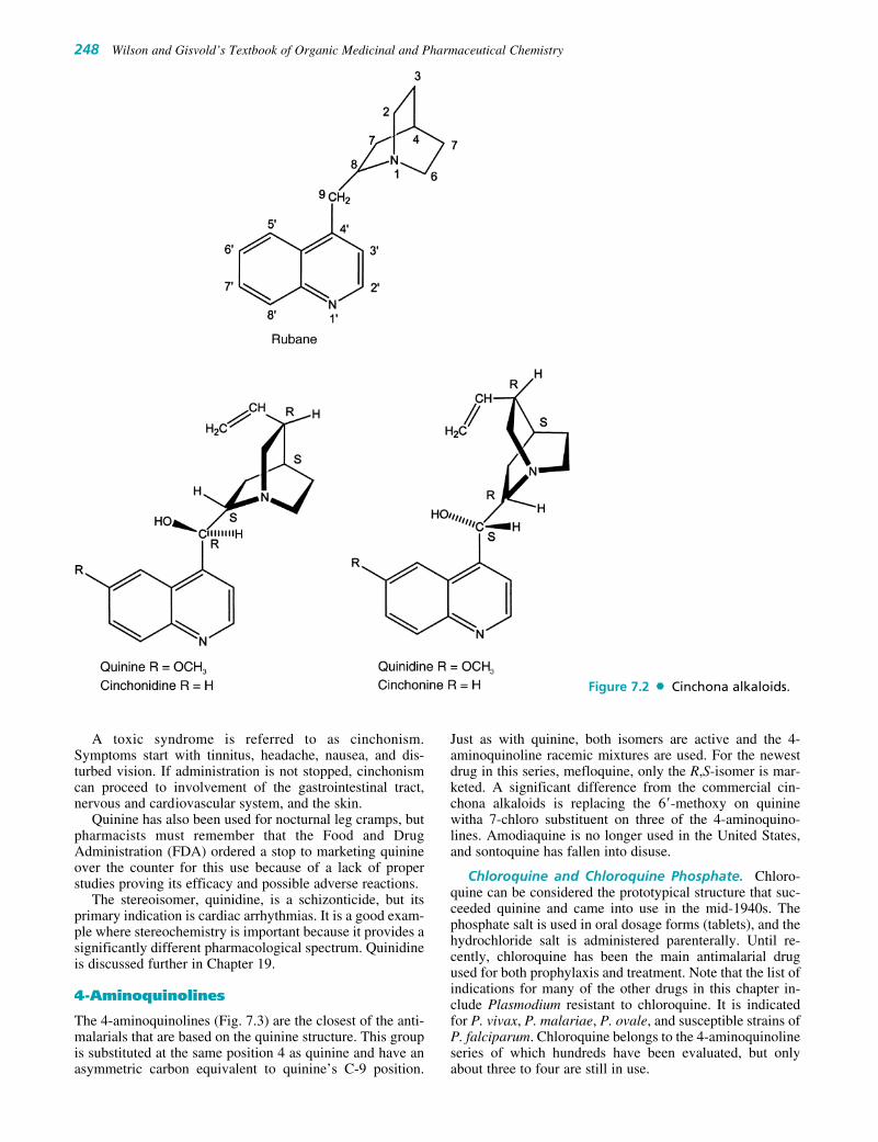
248 Wilson and Gisvold’s Textbook of Organic Medicinal and Pharmaceutical Chemistry
A toxic syndrome is referred to as cinchonism.Symptoms start with tinnitus, headache, nausea, and dis-turbed vision. If administration is not stopped, cinchonismcan proceed to involvement of the gastrointestinal tract,nervous and cardiovascular system, and the skin.
Quinine has also been used for nocturnal leg cramps, butpharmacists must remember that the Food and DrugAdministration (FDA) ordered a stop to marketing quinineover the counter for this use because of a lack of properstudies proving its efficacy and possible adverse reactions.
The stereoisomer, quinidine, is a schizonticide, but itsprimary indication is cardiac arrhythmias. It is a good exam-ple where stereochemistry is important because it provides asignificantly different pharmacological spectrum. Quinidineis discussed further in Chapter 19.
4-Aminoquinolines
The 4-aminoquinolines (Fig. 7.3) are the closest of the anti-malarials that are based on the quinine structure. This groupis substituted at the same position 4 as quinine and have anasymmetric carbon equivalent to quinine’s C-9 position.
Just as with quinine, both isomers are active and the 4-aminoquinoline racemic mixtures are used. For the newestdrug in this series, mefloquine, only the R,S-isomer is mar-keted. A significant difference from the commercial cin-chona alkaloids is replacing the 6�-methoxy on quininewitha 7-chloro substituent on three of the 4-aminoquino-lines. Amodiaquine is no longer used in the United States,and sontoquine has fallen into disuse.
Chloroquine and Chloroquine Phosphate. Chloro-quine can be considered the prototypical structure that suc-ceeded quinine and came into use in the mid-1940s. Thephosphate salt is used in oral dosage forms (tablets), and thehydrochloride salt is administered parenterally. Until re-cently, chloroquine has been the main antimalarial drugused for both prophylaxis and treatment. Note that the list ofindications for many of the other drugs in this chapter in-clude Plasmodium resistant to chloroquine. It is indicatedfor P. vivax, P. malariae, P. ovale, and susceptible strains ofP. falciparum. Chloroquine belongs to the 4-aminoquinolineseries of which hundreds have been evaluated, but onlyabout three to four are still in use.
Figure 7.2 Cinchona alkaloids.
0242-0257_17865_Ch07.qxd 12/4/09 4:51 AM Page 248

Even though this drug has been used for many years, itsmechanism of action is still not known. Its main site of ac-tion appears to be the parasite involving the erythrocyte’slysosome. The following actions have been suggestedbased on experimental evidence. A very complex mecha-nism is based on ferriprotoporphyrin IX, which is releasedby Plasmodium containing erythrocytes, acting as a chloro-quine receptor. The combination of ferriprotoporphyrin IXand chloroquine causes lysis of the parasite’s and/or theerythrocyte’s membrane. Finally, there is evidence thatchloroquine may interfere with Plasmodium’s ability to di-gest the erythrocyte hemoglobin or the parasite’s nucleo-protein synthesis. The mechanism is based on the drug en-tering the erythrocyte’s lysosome, which has an acidenvironment, where it becomes protonated. The protonated(positively charged) chloroquine is now trapped inside thelysosome because the pore that leads out of the lysosome isalso positively charged. This leaves chloroquine bound tothe patient’s hemoglobin preventing the parasite from pro-cessing it properly.24
In general, chloroquine and the other 4-aminoquinolinesare not effective against exoerythocytic parasites. Note thateach of the mechanisms require that the parasite be insidethe erythrocyte. Therefore, chloroquine does not prevent re-lapses of P. vivax or P. ovale malaria. The drug is also indi-cated for the treatment of extraintestinal amebiasis.
Effective such as chloroquine has been, it is a poor ex-ample of selective toxicity. Adverse reactions includeretinopathy, hemolysis in patients with glucose-6-phosphatedehydrogenase deficiency (same mutation that confers re-sistance against malaria), muscular weakness, exacerbationof psoriasis and porphyria, and impaired liver function.Further examples of poor selective toxicity include off-labelindications that include rheumatoid arthritis, systemic anddiscoid lupus erythemaosis (possibly as a immunosuppres-sant), and various dermatological conditions.
The increase in P. falciparum resistant to chloroquine isconsidered to be one of the main reasons for the increase inboth the incidence and deaths from malaria. The keyPlasmodium gene that confers resistance appears to be the
Figure 7.3 4-Aminoquinolines.
Chapter 7 Antimalarials 249
0242-0257_17865_Ch07.qxd 12/4/09 4:51 AM Page 249

250 Wilson and Gisvold’s Textbook of Organic Medicinal and Pharmaceutical Chemistry
pfcrt gene that codes for a transporter protein. The result ofthe changes in the gene is that the pore through whichchloroquine might exit the lysosome is no longer positivecharged, allowing protonated chloroquine to exit the lyso-some.35 There are at least eight mutations that have beenidentified in the pfcrt gene, and it is postulated that resis-tance occurs because of an accumulation of these mutations.Chloroquine remains effective when there are fewer muta-tions in the pfcrt gene. Once the critical number of mutationshas occurred, the parasite spreads over a broad geographicalarea rendering chloroquine ineffective.22–24
Hydroxychloroquine. In most ways, hydroxychloro-quine parallels chloroquine. Structurally, it differs solelywith a hydroxy moiety on one of the N-ethyl groups. Likechloroquine, it remains in the body for over a month, andprophylactic dosing is once weekly. The other indications,both FDA approved and off-label, are very similar.
Amodiaquine. Amodiaquine is no longer marketedin the United States, but it is available in Africa.Mechanistically, it is very similar to chloroquine and does nothave any advantages over the other 4-aminoquinoline drugs.When used for prophylaxis of malaria, it had a higher inci-dence of hepatitis and agranulocytosis than that was chloro-quine. There is evidence that the hydroquinone (phenol)amine system readily oxidizes to a quinone imine (Fig. 7.3)either autoxidatively and/or metabolically, and this productmay contribute to amodiaquine’s toxicity.
Mefloquine HCl. The newest of the 4-aminoquino-lines, mefloquine, is marketed as the R,S-isomer. It was de-veloped in the 1960s as part of the U.S. Army’s WalterReed Institute for Medical Research antimalarial researchprogram. It differs significantly from the other agents in
this class by having two trifluromethyl moieties at positions2� and 8� and no electronegative substituents at eitherpositions 6� (quinine) or 7� (chloroquine). Mefloquine alsodiffers from chloroquine and its analogs by being a schi-zonticide (site 2 in Fig. 7.1) acting before the parasite canenter the erythrocyte. There is some evidence that it acts byraising the pH in the parasite’s vesicles interfering with itsability to process heme. Mefloquine-resistant strains ofP. falciparum have appeared. Relapse can occur with acuteP. vivax that has been treated with mefloquine because thedrug does not eliminate the hepatic phase of this species,which can reinfect the liver.
Mefloquine is teratogenic in rats, mice, and rabbits. Thereis an FDA-required warning that this drug can cause exacer-bate mental disorders and is contraindicated in patients withactive depression, a recent history of depression, generalizedanxiety disorder, psychosis, schizophrenia, and other majorpsychiatric disorders or a history of convulsions.
8-Aminoquinolines
The other major group of antimalarial drugs based on thecinchona alkaloid quinoline moiety is the substituted 8-aminoquinolines (Fig. 7.4). The first compound introducedin this series was pamaquine. During World War II, pen-taquine, isopentaquine, and primaquine became available.Only primaquine, after being used during the Korean war, isin use today. All of the 8-aminoquinolines can cause he-molytic anemia in erythrocytic glucose-6-phosphate dehy-drogenase-deficient patients. As pointed out in the introduc-tion to this chapter, this is a common genetic trait found inpopulations living in areas endemic in malaria and providessome resistance to the parasite. Mechanism of action andspectrum of activity will be found in the primaquine section.
Figure 7.4 8-Aminoquinolines.
0242-0257_17865_Ch07.qxd 12/4/09 4:51 AM Page 250

Very little variations are seen in the structure–activity re-lationships in this series. The four agents in Figure 7.4 allhave a 6-methoxy moiety same as quinine, but the sub-stituents are on the quinoline are located at position 8 ratherthan carbon-4 as found on the cinchona alkaloids. All agentsin this series have a four to five carbon alkyl linkage orbridge between the two nitrogens. With the exception ofpentaquine, the other three 8-aminoquinolines have oneasymmetric carbon. Although some differences may be seenin the metabolism of each stereoisomer and type of adverseresponse, there is little difference in antimalarial activitybased on the compounds stereochemistry.
Primaquine. Primaquine is the only 8-aminoquinolinecurrently in use for the treatment of malaria. It is not usedfor prophylaxis. Its spectrum of activity is one of the nar-rowest of the currently used antimalarial drugs being indi-cated only for exoerythrocytic P. vivax malaria (site 2 inFig. 7.1). To treat endoerythrocytic P. vivax, chloroquineor a drug indicated for chloroquine-resistant P. vivax isused with primaquine. In addition to its approved indica-tion, it is also active against the exoerythrocytic stages ofP. ovale and primary exoerythrocytic stages of P. falcip-arum. Primaquine also inhibits the gameocyte stage (site 4in Fig. 7.1) that eliminates the form required to infect themosquito carrier. In vitro and in vivo studies indicate thatthe stereochemistry at the asymmetric is not important forantimalarial activity. There appears to be less toxicity withthe levorotatory isomer, but this is dose dependent andmay not be that important at the doses used to treatexoerythrocytic P. vivax malaria.
Although structurally related to the cinchona alkaloids,the 8-aminoquinolines act by a different mechanism of ac-tion. Primaquine appears to disrupt the parasite’s mitochon-dria. The result is disruption of several processes includingmaturation into the subsequent forms. An advantage is de-stroying exoerythrocytic forms before the parasite can infecterythrocytes. It is the latter step in the infectious process thatmakes malaria so debilitating.
Polycyclic Antimalarial Drugs
There are three antimalarial drugs that have, in common,polycyclic ring systems (Fig.7.5). The first is the commontetracycline antibiotic, doxycycline. The second is halo-fantrine, and the third is the discontinued agent that wasused in the South Pacific, quinacrine.
Doxycycline. Like the other tetracyclines, doxycycline(see Chapter 8) inhibits the pathogen’s protein synthesisby reversibly inhibiting the 30S ribosomal subunit.Bacteria and Plasmodium ribosomal subunits differ signif-icantly from mammalian ribosomes such that this group ofantibiotics do not readily bind to mammalian ribosomesand, therefore, show good selective toxicity. Althoughdoxycycline is a good antibacterial, its use for malaria islimited to prophylaxis against strains of P. falciparumn re-sistant to chloroquine and sulfadoxine–pyrimethamine.This use normally should not exceed 4 months. Becausethe tetracyclines chelate calcium, they can interfere withdevelopment of the permanent teeth in children. Therefore,their use in children definitely should be short term. Also,
Figure 7.5 Polycyclic antimalarial drugs.
Chapter 7 Antimalarials 251
0242-0257_17865_Ch07.qxd 12/4/09 4:51 AM Page 251
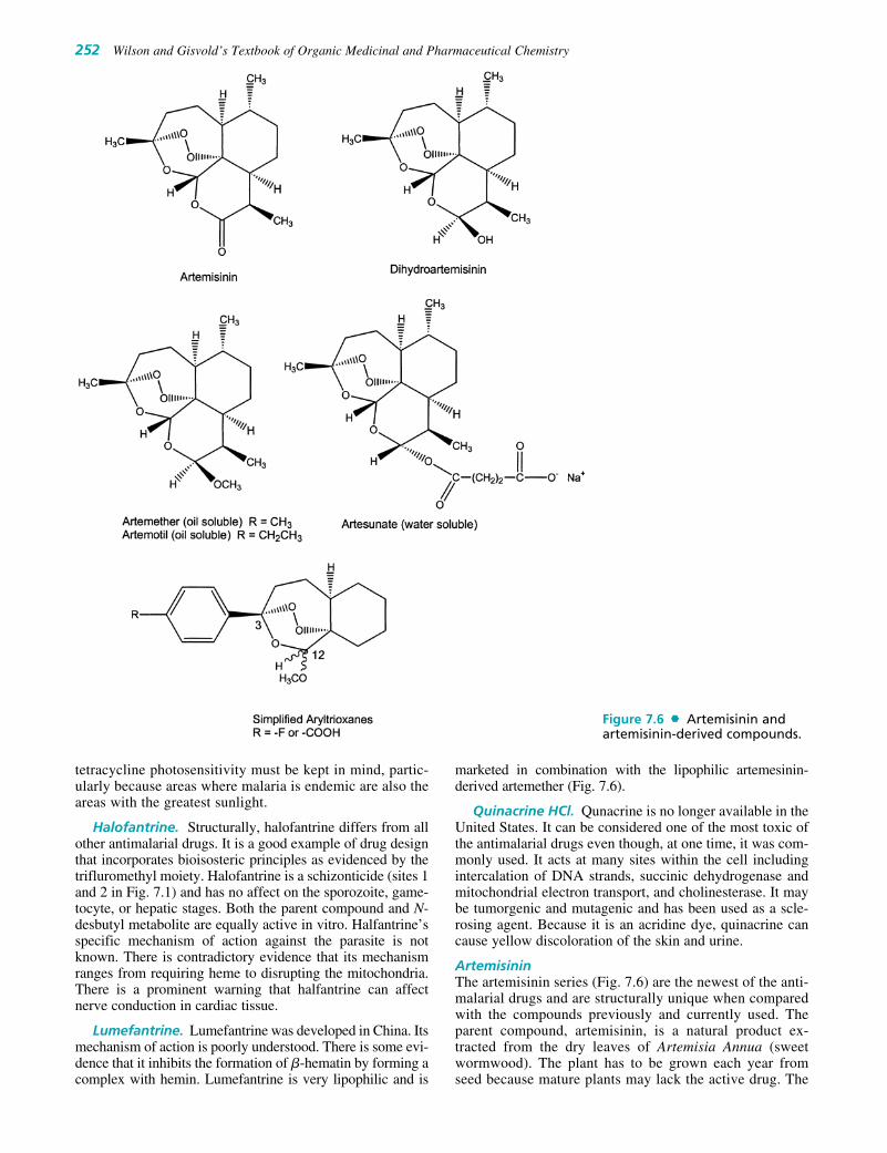
252 Wilson and Gisvold’s Textbook of Organic Medicinal and Pharmaceutical Chemistry
tetracycline photosensitivity must be kept in mind, partic-ularly because areas where malaria is endemic are also theareas with the greatest sunlight.
Halofantrine. Structurally, halofantrine differs from allother antimalarial drugs. It is a good example of drug designthat incorporates bioisosteric principles as evidenced by thetrifluromethyl moiety. Halofantrine is a schizonticide (sites 1and 2 in Fig. 7.1) and has no affect on the sporozoite, game-tocyte, or hepatic stages. Both the parent compound and N-desbutyl metabolite are equally active in vitro. Halfantrine’sspecific mechanism of action against the parasite is notknown. There is contradictory evidence that its mechanismranges from requiring heme to disrupting the mitochondria.There is a prominent warning that halfantrine can affectnerve conduction in cardiac tissue.
Lumefantrine. Lumefantrine was developed in China. Itsmechanism of action is poorly understood. There is some evi-dence that it inhibits the formation of �-hematin by forming acomplex with hemin. Lumefantrine is very lipophilic and is
marketed in combination with the lipophilic artemesinin-derived artemether (Fig. 7.6).
Quinacrine HCl. Qunacrine is no longer available in theUnited States. It can be considered one of the most toxic ofthe antimalarial drugs even though, at one time, it was com-monly used. It acts at many sites within the cell includingintercalation of DNA strands, succinic dehydrogenase andmitochondrial electron transport, and cholinesterase. It maybe tumorgenic and mutagenic and has been used as a scle-rosing agent. Because it is an acridine dye, quinacrine cancause yellow discoloration of the skin and urine.
ArtemisininThe artemisinin series (Fig. 7.6) are the newest of the anti-malarial drugs and are structurally unique when comparedwith the compounds previously and currently used. Theparent compound, artemisinin, is a natural product ex-tracted from the dry leaves of Artemisia Annua (sweetwormwood). The plant has to be grown each year fromseed because mature plants may lack the active drug. The
Figure 7.6 Artemisinin andartemisinin-derived compounds.
0242-0257_17865_Ch07.qxd 12/4/09 4:51 AM Page 252

growing conditions are critical to maximize artemisininyield. Thus far, the best yields have been obtained fromplants grown in North Vietnam, Chongqing province inChina, and Tanzania.25
All of the structures in Figure 7.6 are active against thePlasmodium genera that cause malaria. The key structurecharacteristic appears to be a “trioxane” consisting of theendoperoxide and dioxepin oxygens. This is shown by thesomewhat simpler series of 3-aryltrioxanes at the bottom ofFigure 7.6, which are active against the parasite. Note thatthe stereochemistry at position 12 is not critical.26 Althoughin the victim’s erythrocyte, the malaria parasite consumesthe hemoglobin consisting of ferrous (Fe�2) iron convertingit to toxic hematin containing ferric (Fe�3) and then reducedto heme with its ferrous iron. The heme iron reacts with thetrioxane moiety releasing reactive oxygen and carbon radi-cals and the highly reactive FeIV � O species. The latter ispostulated to be lethal to the parasite.27,28
With the reduction of artemisinin to dihydroartemisinin,an asymmetic carbon forms and it is possible to form oil sol-uble and water soluble prodrugs. Both stereoisomers are ac-tive just as is seen in the simpler aryltrioxanes. The chem-istry forming each of the artemisinin prodrugs results in thepredominance of one isomer. The �-isomer predominateswhen producing the nonpolar methyl and ethyl ethers,whereas the �-isomer is the predominate product whenforming the water-soluble hemisuccinate ester. The lattercan be administered as 10-mg rectal capsules for patentswho cannot take medication orally and parenteral treatmentis not available.
Fixed Combinations
Because resistance is a frequent problem in the prophylaxisand treatment of malaria, combination therapies that use twodistinctly different mechanisms have been developed. Onecombination inhibits folic acid biosynthesis and dihydrofo-late reductase, and another combination acts on the para-site’s mitochondria and its dihydrofolate reductase. Bothdrugs in the third combination act on hematin, but by twodifferent mechanisms.
Sulfadoxine and Pyrimethamine. This combination(Fig. 7.7) uses a drug from the sulfonamide antibacterialgroup and a pyrimidinediamine similar to trimethoprim (seeChapter 8). The combination is considered to a schizonto-cide (site 2 in Fig. 7.1). The sulfonamide, sulfadoxine, inter-feres with the parasite’s ability to synthesize folic acid, andthe pyrimidinediamine, pyrimethamine, inhibits the reduc-tion of folic acid to its active tetrahydrofolate coenzymeform. Sulfonamides block the incorporation of p-aminoben-zoic acid (PABA) forming dihydropteroic acid. Note thestructures of dihydrofolic acid and tetrahydrofolic acid andhow PABA is the central part of the folate structure (seeChapter 28). Normally, sulfonamides exhibit excellent se-lective toxicity because humans do not synthesize the vita-min folic acid. Nevertheless, there are warnings of severe tofatal occurrences of erythema multiforme, Stevens-Johnsonsyndrome, toxic epidermal necrolysis, and serum-sicknesssyndromes attributed to the sulfadoxine.
Pyrimethamine, developed in the 1950s, inhibits the re-duction of folic acid and dihydrofolic acid to the activetetrahydrofolate coenzyme form. Although the latter is re-quired for many fundamental reactions involving pyrimi-dine biosynthesis, the focus in the parasite is regenerationof N5,N10-methylene tetrahydrofolate from dihydrofolate.The synthesis of thymidine 5�-monophosphate from de-oxyuridine 5�-monophosphate is a universal reaction inall cells forming DNA. There are enough differences inthis enzyme and dihydrofolate reductase found in mam-malian, bacterial, and Plasmodium cells that folate reduc-tase inhibitors can be developed that show reasonable se-lective toxicity. In the case of the malaria parasite, theintimate relationship between thymidylate synthase and
Figure 7.7 Sulfadoxine and pyrimethamine.
Figure 7.8 Atovaquone and proguanil.
Chapter 7 Antimalarials 253
0242-0257_17865_Ch07.qxd 12/4/09 4:51 AM Page 253

254 Wilson and Gisvold’s Textbook of Organic Medicinal and Pharmaceutical Chemistry
dihydrofolate reductase is such that pyrimethamine in-hibits both enzymes.
This combination is indicated for prophylaxis and treat-ment of chloroquine resistance P. falciparum and may beused in combination with quinine. Although indicated onlyfor P. falciparum, the combination is active against all theasexual erythocytic forms. It has no activity against thesexual gametocyte form. The fixed combination contains500-mg sulfadoxine and 25-mg pyrimethamine. There is awide number of sulfonamides that could be used in combi-nation with pyrimethamine. The usual approach is to use asulfonamide that has similar pharmacokinetic propertieswith the dihydrofolate reductase inhibitor. For this combi-nation, the peak plasma sulfadoxine occurs in 2.5 to6 hours and pyrimethamine 1.5 to 8 hours. Resistance hasdeveloped with much of it involving mutations in either orboth of the genes coding for dihydrofolate reductase andthymidylate synthase.
Atovaquone and Proguanil HCl. Atovaquone andproguanil HCl (Fig. 7.8) are administered in combination inthe ratio of 2.5 atovaquone to 1 proguanil HCl measuredin mg (not mmoles). Proguanil, developed in 1945, is anearly example of a prodrug. It is metabolized to cycloguanil
(Fig. 7.9), primarily by CYP2C19. The polymorphic natureof this hepatic enzyme explains why certain subpopulationsdo not respond to proguanil. These groups cannot convertproguanil to the active cycloguanil.
The basis for this combination is two distinct andunrelated mechanisms of action against the parasite. Atova-quone is a selective inhibitor of the Plasmodium’s mito-chondrial electron transport system, and cycloguanil is adihydrofolate reductase inhibitor. Atovaquone’s chemistryis based on its being a naphthoquinone and participatingin oxidation–reduction reactions as part of its quinone-hydroquinone system. It is patterned after Coenzyme Qfound in the mitochondrial electron transport chains. Thedrug selectively interferes with mitochondrial electrontransport, particularly at the parasite’s cytochrome bc1 site.This deprives the cell of needed ATP and could cause thecell to become anaerobic. Resistance to this drug comesfrom a mutation in the parasite’s cytochrome.
Cycloguanil (Proguanil) interferes with deoxythymidylatesynthesis by inhibiting dihydrofolate reductase. Resistanceto proguanil/cycloguanil is attibuted to amino acid changesnear the dihydrofolate reductase binding site. Its eliminationhalf-life (48–72 hour) is much shorter than the other anti-malarial dihydrofolate reductase, pyrimethamine (mean
Figure 7.9 Conversion of proguanil to cycloguanil by CYP2C19.
0242-0257_17865_Ch07.qxd 12/4/09 4:51 AM Page 254

elimination half-life of 111 hours). The combination is ef-fective against both erythrocytic and exoerythrocyticPlasmodium. This drug combination is indicated for malariaresistant to chloroquine, halofantrine, mefloquine, and amodi-aquine. Its main site is the sporozoite stage (site 1 in Fig. 7.1).
Artemether and Lumefantrine. These two antimalari-als interfere with heme metabolism, thereby stopping devel-opment of the parasite in the erythrocyte states. Artemether,with its endoperoxide, acts oxidatively and lumefantrine mayform a complex with hemin.
Future Trends
VACCINES
The development of an effective vaccine has been one of themost frustrating aspects in the prevention of malaria. Thereis no question that the Plasmodium parasite invokes an im-mune response. The problem seems to be identifying theantigenic component or components that cause a strong im-mune response. As already described, the RTS,S/AS01Evaccine is the only one showing promise.
NEW DRUG COMBINATIONS
Because of the general success of the artemisinin-based anti-malarials, there have been several combinations evaluated. Asmentioned previously, the artemether and lumefantrine com-bination has been approved. Undergoing phase III studies is apyronaridine (Fig. 7.5) and artesunate (Fig. 7.6). Pyronaridineinhibits formation of hematin, possibly by a similar mecha-nism to that of chloroquine.29
Figure 7.10 Fosmidomycin and a fosmidomycinanalog.
Figure 7.11 Nonmevalonatepathway.
Chapter 7 Antimalarials 255
0242-0257_17865_Ch07.qxd 12/4/09 4:52 AM Page 255
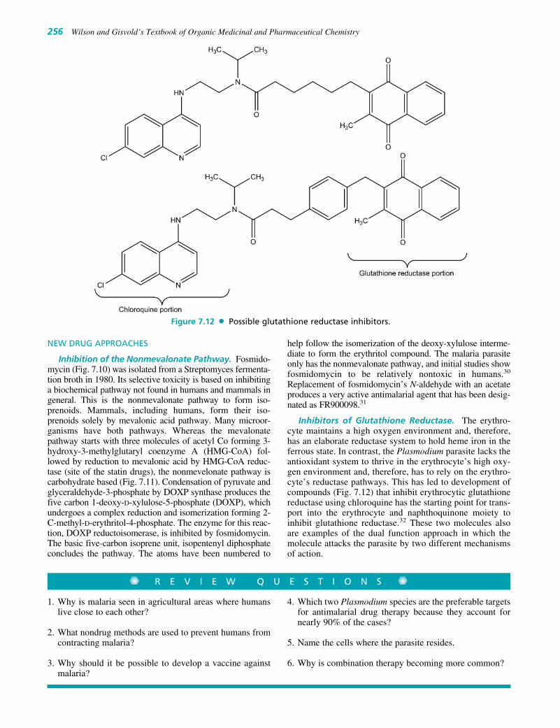
256 Wilson and Gisvold’s Textbook of Organic Medicinal and Pharmaceutical Chemistry
NEW DRUG APPROACHES
Inhibition of the Nonmevalonate Pathway. Fosmido-mycin (Fig. 7.10) was isolated from a Streptomyces fermenta-tion broth in 1980. Its selective toxicity is based on inhibitinga biochemical pathway not found in humans and mammals ingeneral. This is the nonmevalonate pathway to form iso-prenoids. Mammals, including humans, form their iso-prenoids solely by mevalonic acid pathway. Many microor-ganisms have both pathways. Whereas the mevalonatepathway starts with three molecules of acetyl Co forming 3-hydroxy-3-methylglutaryl coenzyme A (HMG-CoA) fol-lowed by reduction to mevalonic acid by HMG-CoA reduc-tase (site of the statin drugs), the nonmevelonate pathway iscarbohydrate based (Fig. 7.11). Condensation of pyruvate andglyceraldehyde-3-phosphate by DOXP synthase produces thefive carbon 1-deoxy-D-xylulose-5-phosphate (DOXP), whichundergoes a complex reduction and isomerization forming 2-C-methyl-D-erythritol-4-phosphate. The enzyme for this reac-tion, DOXP reductoisomerase, is inhibited by fosmidomycin.The basic five-carbon isoprene unit, isopentenyl diphosphateconcludes the pathway. The atoms have been numbered to
help follow the isomerization of the deoxy-xylulose interme-diate to form the erythritol compound. The malaria parasiteonly has the nonmevalonate pathway, and initial studies showfosmidomycin to be relatively nontoxic in humans.30
Replacement of fosmidomycin’s N-aldehyde with an acetateproduces a very active antimalarial agent that has been desig-nated as FR900098.31
Inhibitors of Glutathione Reductase. The erythro-cyte maintains a high oxygen environment and, therefore,has an elaborate reductase system to hold heme iron in theferrous state. In contrast, the Plasmodium parasite lacks theantioxidant system to thrive in the erythrocyte’s high oxy-gen environment and, therefore, has to rely on the erythro-cyte’s reductase pathways. This has led to development ofcompounds (Fig. 7.12) that inhibit erythrocytic glutathionereductase using chloroquine has the starting point for trans-port into the erythrocyte and naphthoquinone moiety toinhibit glutathione reductase.32 These two molecules alsoare examples of the dual function approach in which themolecule attacks the parasite by two different mechanismsof action.
Figure 7.12 Possible glutathione reductase inhibitors.
1. Why is malaria seen in agricultural areas where humanslive close to each other?
2. What nondrug methods are used to prevent humans fromcontracting malaria?
3. Why should it be possible to develop a vaccine againstmalaria?
4. Which two Plasmodium species are the preferable targetsfor antimalarial drug therapy because they account fornearly 90% of the cases?
5. Name the cells where the parasite resides.
6. Why is combination therapy becoming more common?
R E V I E W Q U E S T I O N S
0242-0257_17865_Ch07.qxd 12/4/09 4:52 AM Page 256

REFERENCES1. Editors: Sci. Am. 286(6):8, 14, 38–45, 102–103, 2002.2. Honigsbaum, M.: Men, Money and Malaria. New York, Farrar,
Straus & Giroux, 2002.3. Pennisi, E.: Science 293:416–417, 2001.4. Tishkoff, S. A., Varkonyi, R., Cahinhinan, N., et al.: Science
293:455–462, 2001.5. Luzzatto, L., and Notaro, R.: Science 293:442–443, 2001.6. Volkman, S., Barry, A. E., Lyons, E. J., et al.: Science 293:482–484,
2001.7. Bynum, W. F.: Science 295:47–48, 2002.8. www.cdc.gov/travel.9. Enserink, M., and Pennisi, E.: Science 295:1207, 2002.
10. Maher, B. A.: Scientist 16(6):28, 2002.11. Pennisi, E.: Science 298:33, 2002.12. Carlton, J. M., Adams, J. H., Silva, J. C., et al.: Nature 455:757–763,
2008, doi:10:1038/nature07327.13. Hoffman, S. L.: Science 290:1509, 2000.14. Pennisi, E.: Science 294:1439, 2001.15. Ayi, K., Min-Oo, G., Serghides, L., et al.: N. Eng. J. Med.
358(17):1805, 2008.16. Hobbs, M. R., Udhayakumar, V., Levesque, M. C., et al.: Lancet
360:1468, 2002.17. Insecticide-Treated Mosquito Nets: a WHO Position Statement,
http://www.who.int/malaria/docs/itn/ITNspospaperfinal.pdf.18. Miller, J., Korenromp, E. L., Nahlen, B. L., et al.: J. Am. Med.
Assoc. 297(20):2241, 2007.19. Rodriguez, L. E., Curtidor, H., Urquiza, M., et al.: Chem. Rev.
108:3656, 2008.20. Pombo, D. J., Lawrence, G., Hirunpetcharat, C., et al.: Lancet
360:610, 2002.21. Van den Bossche, H.: Nature 273:626, 1978.22. Hastings, I. M., Bray, P. G., and Ward, S. A.: Science 298:74,
2002.
23. Sidhu, A. B. S., Verider-Pinard, D., and Fidock, D. A.: Science298:210, 2002.
24. Warhurst, D. C., Craig, J. C., and Adagu, I. S.: Lancet 360:(9345),2002.
25. http://www.dafra.be/english/artemisinin.html.26. Posner, G. H., Jeon, H. B., Parker, M. H., et al.: J. Med. Chem.
44:3054, 2001.27. Posner, G. H., Cummings, J. N., Ploypradith, P., et al.: J. Am. Chem.
Soc. 117:5885, 1995.28. Posner, G. H., Park, S. B., González, L., et al.: J. Am. Chem. Soc.
118:3537, 1996.29. Auparakkitanon, S., Chapoomram, S., Kuaha, K., et al.: Agents
Chemother. 50:2197, 2006.30. Jomaa, H., Wiesner, J., Sanderbrand, S., et al.: Science 285:1573, 1999.31. Reichenberg, A., Wiesner, J., Weidemeyer, C., et al.: Bioorg. Med.
Chem. Lett. 11:833, 2001.32. Friebolin, W., Jannack, B., Wenzel, N., et al.: J. Med. Chem. 51:
1260, 2008.
SELECTED READINGHonigsbaum, M.: The fever trail. In Search for the Cure for Malaria. New
York, Farrar, Straux & Giroux, 2001.Kaur, K., Jain, M., Kaur, T., et al.: Antimalarials from nature. Biorg.
Med. Chem. 17:3229, 2009.N. Engl. J. Med. 359(24):2521–2532, 2533–2544, 2545–2557,
2599–2601, 2601–2603, 2008. (These issues includes three articles andtwo editorials.)
Science 298(5591), 2002. (This issue reports the genome of theAnopheles gambiae mosquito and includes discussion on how thisknowledge might be used to prevent malaria.)
World Health Organization (WHO): The World Health Report 2008,available as a downloadable PDF file at: http://www.who.int/malaria/wmr2008/. Additional information on malaria from the WHO athttp://www.who.int/topics/malaria/en/
Chapter 7 Antimalarials 257
0242-0257_17865_Ch07.qxd 12/4/09 4:52 AM Page 257

258
C H A P T E R 8
Antibacterial AntibioticsJOHN M. BEALE, JR.
the presence of certain bacteria, along with similar obser-vations by other microbiologists, led Vuillemin2 to defineantibiosis (literally “against life”) as the biological con-cept of survival of the fittest, in which one organism de-stroys another to preserve itself. The word antibiotic wasderived from this root. The use of the term by the lay pub-lic, as well as the medical and scientific communities, hasbecome so widespread that its original meaning has be-come obscured.
In 1942, Waksman3 proposed the widely cited definitionthat “an antibiotic or antibiotic substance is a substanceproduced by microorganisms, which has the capacity of in-hibiting the growth and even of destroying other microor-ganisms.” Later proposals4–6 have sought both to expandand to restrict the definition to include any substance pro-duced by a living organism that is capable of inhibiting thegrowth or survival of one or more species of microorgan-isms in low concentrations. The advances made by medici-nal chemists to modify naturally occurring antibiotics andto prepare synthetic analogs necessitated the inclusion ofsemisynthetic and synthetic derivatives in the definition.Therefore, a substance is classified as an antibiotic if thefollowing conditions are met:
1. It is a product of metabolism (although it may be dupli-cated or even have been anticipated by chemical synthesis).
2. It is a synthetic product produced as a structural analog ofa naturally occurring antibiotic.
3. It antagonizes the growth or survival of one or morespecies of microorganisms.
4. It is effective in low concentrations.
The isolation of the antibacterial antibiotic tyrocidinfrom the soil bacterium Bacillus brevis by Dubois sug-gested the probable existence of many antibiotic substancesin nature and provided the impetus for the search for them.An organized search of the order Actinomycetales ledWaksman and associates to isolate streptomycin fromStreptomyces griseus. The discovery that this antibiotic pos-sessed in vivo activity against Mycobacterium tuberculosisin addition to numerous species of Gram-negative bacilliwas electrifying. It was now evident that soil microorgan-isms would provide a rich source of antibiotics. Broadscreening programs were instituted to find antibiotics thatmight be effective in the treatment of infections hithertoresistant to existing chemotherapeutic agents, as well as toprovide safer and more effective chemotherapy. The discov-eries of broad-spectrum antibacterial antibiotics such aschloramphenicol and the tetracyclines, antifungal antibiotics
C H A P T E R O V E R V I E W
Since Alexander Fleming accidentally discovered peni-cillin in 1929, the numbers of antibiotics that have beenadded to our therapeutic armamentarium has growntremendously. Along with immunizing biologicals, antibi-otics have turned the tide in terms of the treatment of infec-tious disease. They are truly medical miracles. Yet, becauseof the overuse of many of these agents and the biochemicalfickleness of many bacteria, resistance to antibiotics hasbecome a serious problem in the 21st century. Indeed, thereare now organisms that cannot be arrested or killed by anyof the common antibiotics. Clearly, new approaches areneeded. This chapter will present each of the antibiotics aschemical classes, such as tetracyclines, aminoglycosides,macrolides, and �-lactam antibiotics. In each class, it isimportant to note the different ranges of bacteria that eachwill treat. Notably, it is difficult to define an absolute rangefor any antibiotic. Populations of bacteria differ in theircharacteristics among regions and hospitals in those region.So, sometimes, an antibiotic is listed as effective against aparticular bacterial strain, but the concentration needed tokill the organism is too high. The bottom line is that whendescribing efficacy of an antibiotic against a particular or-ganism, it is necessary to look at clinical results. This chap-ter will describe ranges of activity in a general sense. Thehistory of the antibiotics is an interesting one and is wellworth considering. Hence, the chapter will begin with abrief historical overview.
HISTORICAL BACKGROUND
Sir Alexander Fleming’s accidental discovery of the anti-bacterial properties of penicillin in 19291 is largely cred-ited with initiating the modern antibiotic era. Not until1938, however, when Florey and Chain introduced peni-cillin into therapy, did practical medical exploitation ofthis important discovery begin to be realized. Centuriesearlier, humans had learned to use crude preparations em-pirically for the topical treatment of infections, which wenow assume to be effective because of the antibiotic sub-stances present. As early as 500 to 600 BC, molded curd ofsoybean was used in Chinese folk medicine to treat boilsand carbuncles. Moldy cheese had also been used for cen-turies by Chinese and Ukrainian peasants to treat infectedwounds. The discovery by Pasteur and Joubert in 1877that anthrax bacilli were killed when grown in culture in
0258-0329_17865_Ch08.qxd 12/2/09 3:26 AM Page 258

Chapter 8 Antibacterial Antibiotics 259
such as nystatin and griseofulvin (see Chapter 6), and theever-increasing number of antibiotics that may be used totreat infectious agents that have developed resistance tosome of the older antibiotics, attest to the spectacularsuccess of this approach as it has been applied in researchprograms throughout the world.
CURRENT STATUS
Commercial and scientific interest in the antibiotic fieldhas led to the isolation and identification of antibiotic sub-stances that may be numbered in the thousands. Numeroussemisynthetic and synthetic derivatives have been added tothe total. Very few such compounds have found applica-tion in general medical practice, however, because in addi-tion to the ability to combat infections or neoplastic disease,an antibiotic must possess other attributes. First, it mustexhibit sufficient selective toxicity to be decisively effec-tive against pathogenic microorganisms or neoplastic tissue,on the one hand, without causing significant toxic effects,on the other. Second, an antibiotic should be chemically sta-ble enough to be isolated, processed, and stored for a rea-sonable length of time without deterioration of potency.The amenability of an antibiotic for oral or parenteral ad-ministration to be converted into suitable dosage forms toprovide active drug in vivo is also important. Third, therates of biotransformation and elimination of the antibioticshould be slow enough to allow a convenient dosing sched-ule, yet rapid and complete enough to facilitate removal ofthe drug and its metabolites from the body soon after admin-istration has been discontinued. Some groups of antibiotics,because of certain unique properties, have been designatedfor specialized uses, such as the treatment of tuberculosis(TB) or fungal infections. Others are used for cancerchemotherapy. These antibiotics are described along withother drugs of the same therapeutic class: antifungal and an-titubercular antibiotics are discussed in Chapter 6, and anti-neoplastic antibiotics are discussed in Chapter 10.
The spectacular success of antibiotics in the treatment ofhuman diseases has prompted the expansion of their use intoseveral related fields. Extensive use of their antimicrobialpower is made in veterinary medicine. The discovery thatlow-level administration of antibiotics to meat-producinganimals resulted in faster growth, lower mortality, and bet-ter quality has led to the use of these products as feed sup-plements. Several antibiotics are used to control bacterialand fungal diseases of plants. Their use in food preservationis being studied carefully. Indeed, such uses of antibioticshave necessitated careful studies of their long-term effectson humans and their effects on various commercialprocesses. For example, foods that contain low-levelamounts of antibiotics may be able to produce allergic reac-tions in hypersensitive persons, or the presence of antibi-otics in milk may interfere with the manufacture of cheese.
The success of antibiotics in therapy and related fieldshas made them one of the most important products of thedrug industry today. The quantity of antibiotics produced inthe United States each year may now be measured in sev-eral millions of pounds and valued at billions of dollars.With research activity stimulated to find new substances totreat viral infections that now are combated with only lim-ited success and with the promising discovery that some
antibiotics are active against cancers that may be viral inorigin, the future development of more antibiotics and theincrease in the amounts produced seem to be assured.
COMMERCIAL PRODUCTION
The commercial production of antibiotics for medicinaluse follows a general pattern, differing in detail for eachantibiotic. The general scheme may be divided into sixsteps: (a) preparation of a pure culture of the desired or-ganism for use in inoculation of the fermentation medium;(b) fermentation, during which the antibiotic is formed;(c) isolation of the antibiotic from the culture medium;(d) purification; (e) assays for potency, sterility, absence ofpyrogens, and other necessary data; and (f) formulationinto acceptable and stable dosage forms.
SPECTRUM OF ACTIVITY
The ability of some antibiotics, such as chloramphenicol andthe tetracyclines, to antagonize the growth of numerouspathogens has resulted in their designation as broad-spectrum antibiotics. Designations of spectrum of activityare of somewhat limited use to the physician, unless they arebased on clinical effectiveness of the antibiotic againstspecific microorganisms. Many of the broad-spectrumantibiotics are active only in relatively high concentrationsagainst some of the species of microorganisms oftenincluded in the “spectrum.”
MECHANISMS OF ACTION
The manner in which antibiotics exert their actions againstsusceptible organisms varies. The mechanisms of action ofsome of the more common antibiotics are summarized inTable 8.1. In many instances, the mechanism of action is notfully known; for a few (e.g., penicillins), the site of action isknown, but precise details of the mechanism are still underinvestigation. The biochemical processes of microorganismsare a lively subject for research, for an understanding ofthose mechanisms that are peculiar to the metabolic systemsof infectious organisms is the basis for the future develop-ment of modern chemotherapeutic agents. Antibiotics thatinterfere with the metabolic systems found in microorgan-isms and not in mammalian cells are the most successfulanti-infective agents. For example, antibiotics that interferewith the synthesis of bacterial cell walls have a high poten-tial for selective toxicity. Some antibiotics structurally re-semble some essential metabolites of microorganisms, whichsuggests that competitive antagonism may be the mechanismby which they exert their effects. Thus, cycloserine is be-lieved to be an antimetabolite for D-alanine, a constituent ofbacterial cell walls. Many antibiotics selectively interferewith microbial protein synthesis (e.g., the aminoglycosides,tetracyclines, macrolides, chloramphenicol, and lincomycin)or nucleic acid synthesis (e.g., rifampin). Others, such as thepolymyxins and the polyenes, are believed to interfere withthe integrity and function of microbial cell membranes. Themechanism of action of an antibiotic determines, in general,whether the agent exerts a bactericidal or a bacteriostatic
0258-0329_17865_Ch08.qxd 12/2/09 3:26 AM Page 259

260 Wilson and Gisvold’s Textbook of Organic Medicinal and Pharmaceutical Chemistry
action. The distinction may be important for the treatment ofserious, life-threatening infections, particularly if the naturaldefense mechanisms of the host are either deficient or over-whelmed by the infection. In such situations, a bactericidalagent is obviously indicated. Much work remains to be donein this area, and as mechanisms of action are revealed, thedevelopment of improved structural analogs of effective an-tibiotics probably will continue to increase.
CHEMICAL CLASSIFICATION
The chemistry of antibiotics is so varied that a chemicalclassification is of limited value. Some similarities can befound, however, indicating that some antibiotics may be theproducts of similar mechanisms in different organisms andthat these structurally similar products may exert their activ-ities in a similar manner. For example, several important an-tibiotics have in common a macrolide structure (i.e., a largelactone ring). This group includes erythromycin and olean-domycin. The tetracycline family comprises a group of com-pounds very closely related chemically. Several compoundscontain closely related amino sugar moieties, such as thosefound in streptomycins, kanamycins, neomycins, paro-momycins, and gentamicins. The antifungal antibiotics nys-tatin and the amphotericins (see Chapter 6) are examples ofa group of conjugated polyene compounds. The bacitracins,tyrothricin, and polymyxin are among a large group ofpolypeptides that exhibit antibiotic action. The penicillinsand cephalosporins are �-lactam ring–containing antibioticsderived from amino acids.
MICROBIAL RESISTANCE
The normal biological processes of microbial pathogens arevaried and complex. Thus, it seems reasonable to assume thatthere are many ways in which they may be inhibited and thatdifferent microorganisms that elaborate antibiotics antago-nistic to a common “foe” produce compounds that are chem-ically dissimilar and that act on different processes. In fact,nature has produced many chemically different antibiotics
that can attack the same microorganism by different path-ways. The diversity of antibiotic structure has proved to be ofreal clinical value. As the pathogenic cell develops drug re-sistance, another antibiotic, attacking another metabolicprocess of the resisting cell, remains effective. The develop-ment of new and different antibiotics has been very impor-tant in providing the means for treating resistant strains of or-ganisms that previously had been susceptible to an olderantibiotic. More recently, the elucidation of biochemicalmechanisms of microbial resistance to antibiotics, such asthe inactivation of penicillins and cephalosporins by �-lactamase–producing bacteria, has stimulated research in thedevelopment of semisynthetic analogs that resist microbialbiotransformation. The evolution of nosocomial (hospital-acquired) strains of staphylococci resistant to penicillin andof Gram-negative bacilli (e.g., Pseudomonas and Klebsiellaspp., Escherichia coli, and others) often resistant to severalantibiotics has become a serious medical problem. No doubt,the promiscuous and improper use of antibiotics has con-tributed to the emergence of resistant bacterial strains. Thesuccessful control of diseases caused by resistant strains ofbacteria will require not only the development of new andimproved antibiotics but also the rational use of availableagents.
�-LACTAM ANTIBIOTICS
Antibiotics that possess the �-lactam (a four-memberedcyclic amide) ring structure are the dominant class of agentscurrently used for the chemotherapy of bacterial infections.The first antibiotic to be used in therapy, penicillin (penicillinG or benzylpenicillin), and a close biosynthetic relative, phe-noxymethyl penicillin (penicillin V), remain the agents ofchoice for the treatment of infections caused by most speciesof Gram-positive bacteria. The discovery of a second majorgroup of �-lactam antibiotics, the cephalosporins, and chem-ical modifications of naturally occurring penicillins andcephalosporins have provided semisynthetic derivatives thatare variously effective against bacterial species known to beresistant to penicillin, in particular, penicillinase-producingstaphylococci and Gram-negative bacilli. Thus, apart from a
TABLE 8.1 Mechanisms of Antibiotic Action
Site of Action Antibiotic Process Interrupted Type of Activity
Cell wall Bacitracin Mucopeptide synthesis BactericidalCephalosporin Cell wall cross-linking BactericidalCycloserine Synthesis of cell wall peptides BactericidalPenicillins Cell wall cross-linking BactericidalVancomycin Mucopeptide synthesis Bactericidal
Cell membrane Amphotericin B Membrane function FungicidalNystatin Membrane function FungicidalPolymyxins Membrane integrity Bactericidal
Ribosomes Chloramphenicol Protein synthesis Bacteriostatic50S subunit Erythromycin Protein synthesis Bacteriostatic
Lincomycins Protein synthesis Bacteriostatic30S subunit Aminoglycosides Protein synthesis and fidelity Bactericidal
Tetracyclines Protein synthesis BacteriostaticNucleic acids Actinomycin DNA and mRNA synthesis Pancidal
Griseofulvin Cell division, microtubule assembly FungistaticDNA and/or RNA Mitomycin C DNA synthesis Pancidal
Rifampin mRNA synthesis Bactericidal
0258-0329_17865_Ch08.qxd 12/2/09 3:26 AM Page 260

Chapter 8 Antibacterial Antibiotics 261
few strains that have either inherent or acquired resistance,almost all bacterial species are sensitive to one or more of theavailable �-lactam antibiotics.
Mechanism of Action
In addition to a broad spectrum of antibacterial action, twoproperties contribute to the unequaled importance of �-lactam antibiotics in chemotherapy: a potent and rapid bacte-ricidal action against bacteria in the growth phase and a verylow frequency of toxic and other adverse reactions in thehost. The uniquely lethal antibacterial action of these agentshas been attributed to a selective inhibition of bacterial cellwall synthesis.7 Specifically, the basic mechanism involvedis inhibition of the biosynthesis of the dipeptidoglycan thatprovides strength and rigidity to the cell wall. Penicillins andcephalosporins acylate a specific bacterial D-transpeptidase,8
thereby rendering it inactive for its role in forming peptidecross-links of two linear peptidoglycan strands by transpep-tidation and loss of D-alanine. Bacterial D-alanine car-boxypeptidases are also inhibited by �-lactam antibiotics.
Binding studies with tritiated benzylpenicillin haveshown that the mechanisms of action of various �-lactamantibiotics are much more complex than previously as-sumed. Studies in E. coli have revealed as many as sevendifferent functional proteins, each with an important role incell wall biosynthesis.9 These penicillin-binding proteins(PBPs) have the following functional properties:
• PBPs 1a and 1b are transpeptidases involved in peptidogly-can synthesis associated with cell elongation. Inhibitionresults in spheroplast formation and rapid cell lysis,9,10
caused by autolysins (bacterial enzymes that create nicksin the cell wall for attachment of new peptidoglycan unitsor for separation of daughter cells during cell division10).
• PBP 2 is a transpeptidase involved in maintaining the rodshape of bacilli.11 Inhibition results in ovoid or roundforms that undergo delayed lysis.
• PBP 3 is a transpeptidase required for septum formationduring cell division.12 Inhibition results in the formation offilamentous forms containing rod-shaped units that cannotseparate. It is not yet clear whether inhibition of PBP 3 islethal to the bacterium.
• PBPs 4 through 6 are carboxypeptidases responsible forthe hydrolysis of D-alanine–D-alanine terminal peptidebonds of the cross-linking peptides. Inhibition of these en-zymes is apparently not lethal to the bacterium,13 eventhough cleavage of the terminal D-alanine bond is requiredbefore peptide cross-linkage.
The various �-lactam antibiotics differ in their affinitiesfor PBPs. Penicillin G binds preferentially to PBP 3, whereasthe first-generation cephalosporins bind with higher affinityto PBP 1a. In contrast to other penicillins and tocephalosporins, which can bind to PBPs 1, 2, and 3,amdinocillin binds only to PBP 2.
THE PENICILLINS
Commercial Production and Unitage
Until 1944, it was assumed that the active principle in peni-cillin was a single substance and that variation in activityof different products was because of the amount of inert
materials in the samples. Now we know that during thebiological elaboration of the antibiotic, several closely re-lated compounds may be produced. These compounds differchemically in the acid moiety of the amide side chain.Variations in this moiety produce differences in antibioticeffect and in physicochemical properties, including stability.Thus, one can speak of penicillins as a group of compoundsand identify each penicillin specifically. As each of the dif-ferent penicillins was first isolated, letter designations wereused in the United States; the British used Roman numerals.
Over 30 penicillins have been isolated from fermentationmixtures. Some of these occur naturally; others have beenbiosynthesized by altering the culture medium to providecertain precursors that may be incorporated as acyl groups.Commercial production of biosynthetic penicillins today de-pends chiefly on various strains of Penicillium notatum andPenicillium chrysogenum. In recent years, many more peni-cillins have been prepared semisynthetically and, undoubt-edly, many more will be added to the list in attempts to findsuperior products.
Because the penicillin first used in chemotherapy was nota pure compound and exhibited varying activity amongsamples, it was necessary to evaluate it by microbiologicalassay. The procedure for assay was developed at Oxford,England, and the value became known as the Oxford unit:1 Oxford unit is defined as the smallest amount of penicillinthat will inhibit, in vitro, the growth of a strain ofStaphylococcus in 50 mL of culture medium under specifiedconditions. Now that pure crystalline penicillin is available,the United States Pharmacopoeia (USP) defines unit as theantibiotic activity of 0.6 �g of penicillin G sodium referencestandard. The weight–unit relationship of the penicillinsvaries with the acyl substituent and with the salt formed ofthe free acid: 1 mg of penicillin G sodium is equivalent to1,667 units, 1 mg of penicillin G procaine is equivalent to1,009 units, and 1 mg of penicillin G potassium is equiva-lent to 1,530 units.
The commercial production of penicillin has increasedmarkedly since its introduction. As production increased,the cost dropped correspondingly. When penicillin was firstavailable, 100,000 units sold for $20. Currently, the samequantity costs less than a penny. Fluctuations in the produc-tion of penicillins through the years have reflected changesin the relative popularity of broad-spectrum antibiotics andpenicillins, the development of penicillin-resistant strains ofseveral pathogens, the more recent introduction of semisyn-thetic penicillins, the use of penicillins in animal feeds andfor veterinary purposes, and the increase in marketing prob-lems in a highly competitive sales area.
Table 8.2 shows the general structure of the penicillinsand relates the structure of the more familiar ones to theirvarious designations.
Nomenclature
The nomenclature of penicillins is somewhat complex andvery cumbersome. Two numbering systems for the fused bi-cyclic heterocyclic system exist. The Chemical Abstractssystem initiates the numbering with the sulfur atom and as-signs the ring nitrogen the 4-position. Thus, penicillins arenamed as 4-thia-l-azabicyclo[3.2.0]heptanes, according tothis system. The numbering system adopted by the USP isthe reverse of the Chemical Abstracts procedure, assigning
0258-0329_17865_Ch08.qxd 12/2/09 3:26 AM Page 261

TABLE 8.2 Structure of Penicillins
Generic Generic Name Chemical Name R Group Name Chemical Name R Group
CR
O
NH CH CHS4
32
CO N
C(CH3)2
CHCOOH
51
67
distinguishes compounds on the basis of the R group of theacyl portion of the molecule. Thus, penicillin G is namedbenzylpenicillin, penicillin V is phenoxymethylpenicillin,methicillin is 2,6-dimethoxyphenylpenicillin, and so on. Forthe most part, the latter two systems serve well for namingand comparing closely similar penicillin structures, but theyare too restrictive to be applied to compounds with unusualsubstituents or to ring-modified derivatives.
Stereochemistry
The penicillin molecule contains three chiral carbon atoms(C-3, C-5, and C-6). All naturally occurring and microbiolog-ically active synthetic and semisynthetic penicillins have the
262 Wilson and Gisvold’s Textbook of Organic Medicinal and Pharmaceutical Chemistry
Penicillin G Benzylpenicillin
Penicillin V Phenoxymethylpenicillin
Methicillin 2,6-Dimethoxyphenyl-penicillin
Nafcillin 2-Ethoxy-1-naphthyl-penicillin
Oxacillin 5-Methyl-3-phenyl-4-isoxazolylpenicillin
Cloxacillin 5-Methyl-3-(2-chlorophenyl)-4-isoxazolylpenicillin
Dicloxacillin 5-Methyl-3-(2,6-dichlorophenyl)-4-isoxazolylpenicillin
Ampicillin D-�-Aminobenzyl-penicillin
Amoxicillin D-�-Amino-p-hydroxybenzylpenicillin
Cyclacillin 1-Aminocyclohexyl-penicillin
Carbenicillin �-Carboxybenzyl-penicillin
Ticarcillin �-Carboxy-3-thienyl-penicillin
Piperacillin �-(4-Ethyl-2,3-dioxo-1-piperazinylcarbonyl-amino)benzylpenicillin
Mezlocillin �-(1-Methanesulfonyl-2-oxoimidazolidino-carbonylamino)benzyl-penicillin
CH
NH
N
N
SO2CH3
O
O
C
NH
CH
N
O N
CH2CH3
O
OC
CO2H
CH
S
CH
CO2H
NH2
NH2
CHHO
NH2
CH
CI
CH3
NCI
O
CH3
N
CI
O
CH3ON
OC2H5
OCH3
OCH3
O CH2
CH2
number 1 to the nitrogen atom and number 4 to the sulfuratom. Three simplified forms of penicillin nomenclaturehave been adopted for general use. The first uses the name“penam” for the unsubstituted bicyclic system, including theamide carbonyl group, with one of the foregoing numberingsystems as just described. Thus, penicillins generally aredesignated according to the Chemical Abstracts system as 5-acylamino-2,2-dimethylpenam-3-carboxylic acids. The sec-ond, seen more frequently in the medical literature, uses thename “penicillanic acid” to describe the ring system withsubstituents that are generally present (i.e., 2,2-dimethyl and3-carboxyl). A third form, followed in this chapter, uses triv-ial nomenclature to name the entire 6-carbonylaminopenicil-lanic acid portion of the molecule penicillin and then
0258-0329_17865_Ch08.qxd 12/2/09 3:26 AM Page 262

Chapter 8 Antibacterial Antibiotics 263
same absolute configuration about these three centers. Thecarbon atom bearing the acylamino group (C-6) has the L con-figuration, whereas the carbon to which the carboxyl group isattached has the D configuration. Thus, the acylamino andcarboxyl groups are trans to each other, with the former in the� and the latter in the � orientation relative to the penam ringsystem. The atoms composing the 6-aminopenicillanic acid(6-APA) portion of the structure are derived biosyntheti-cally from two amino acids, L-cysteine (S-1, C-5, C-6, C-7,and 6-amino) and L-valine (2,2-dimethyl, C-2, C-3, N-4, and3-carboxyl). The absolute stereochemistry of the penicillins isdesignated 3S:5R:6R, as shown below.
Synthesis
Examination of the structure of the penicillin moleculeshows that it contains a fused ring system of unusual design,the �-lactam thiazolidine structure. The nature of the �-lactam ring delayed elucidation of the structure of penicillin,but its determination resulted from a collaborative researchprogram involving groups in Great Britain and the UnitedStates during the years 1943 to 1945.14 Attempts to synthe-size these compounds resulted, at best, in only traceamounts until Sheehan and Henery-Logan15 adapted tech-niques developed in peptide synthesis to the synthesis ofpenicillin V. This procedure is not likely to replace the es-tablished fermentation processes because the last step in the
reaction series develops only 10% to 12% penicillin. It is ofadvantage in research because it provides a means of obtain-ing many new amide chains hitherto not possible to achieveby biosynthetic procedures.
Two other developments have provided additional meansfor making new penicillins. A group of British scientists,Batchelor et al.16 reported the isolation of 6-APA from aculture of P. chrysogenum. This compound can be con-verted to penicillins by acylation of the 6-amino group.Sheehan and Ferris17 provided another route to syntheticpenicillins by converting a natural penicillin, such as peni-cillin G potassium, to an intermediate (Fig. 8.1), from whichthe acyl side chain has been cleaved and which then can betreated to form biologically active penicillins with variousnew side chains. By these procedures, new penicillins, supe-rior in activity and stability to those formerly in wide use,were found, and no doubt others will be produced. The firstcommercial products of these research activities werephenoxyethylpenicillin (phenethicillin) (Fig. 8.2) anddimethoxyphenylpenicillin (methicillin).
Chemical Degradation
The early commercial penicillin was a yellow-to-brownamorphous powder that was so unstable that refrigeration wasrequired to maintain a reasonable level of activity for a shorttime. Improved purification procedures provided the white
0258-0329_17865_Ch08.qxd 12/2/09 3:26 AM Page 263
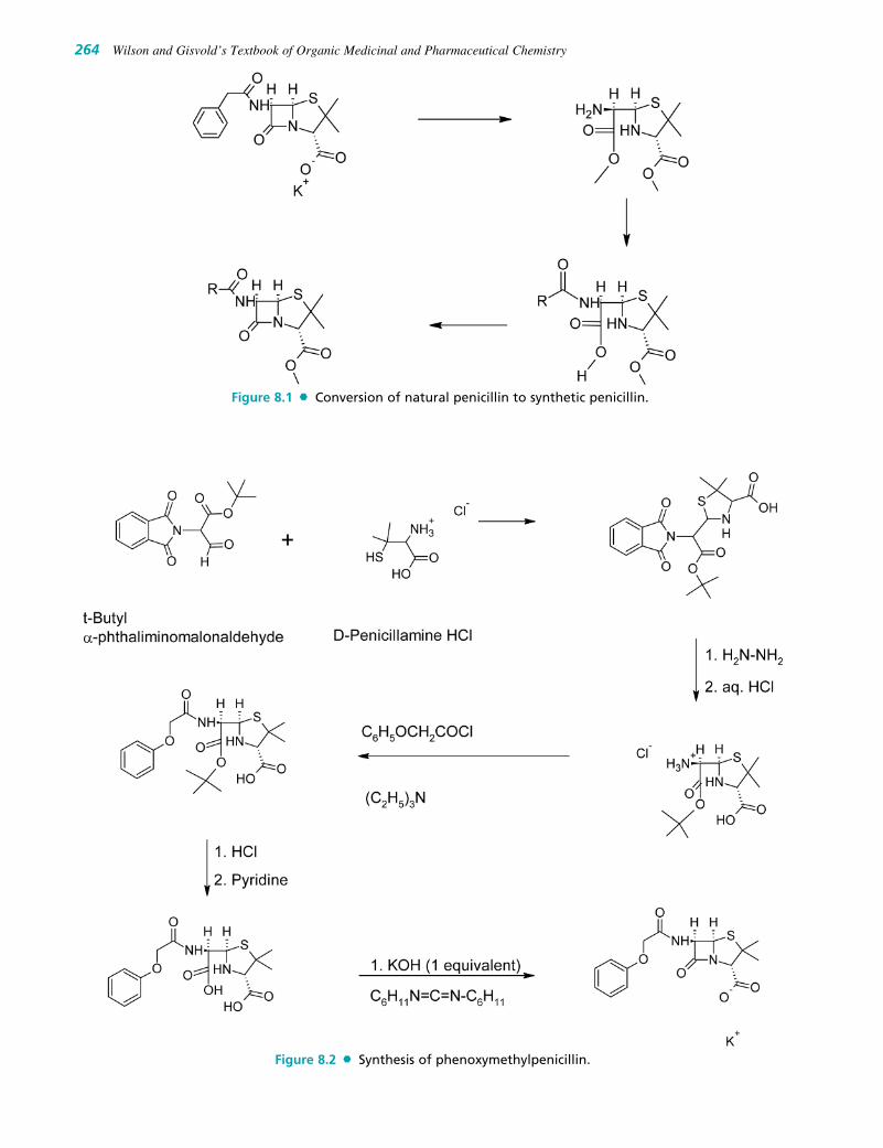
264 Wilson and Gisvold’s Textbook of Organic Medicinal and Pharmaceutical Chemistry
Figure 8.1 Conversion of natural penicillin to synthetic penicillin.
Figure 8.2 Synthesis of phenoxymethylpenicillin.
0258-0329_17865_Ch08.qxd 12/2/09 3:26 AM Page 264

Chapter 8 Antibacterial Antibiotics 265
crystalline material in use today. Crystalline penicillin mustbe protected from moisture, but when kept dry, the salts willremain stable for years without refrigeration. Many penicillinshave an unpleasant taste, which must be overcome in the for-mation of pediatric dosage forms. All of the natural penicillinsare strongly dextrorotatory. The solubility and other physico-chemical properties of the penicillins are affected by the na-ture of the acyl side chain and by the cations used to makesalts of the acid. Most penicillins are acids with pKa values inthe range of 2.5 to 3.0, but some are amphoteric. The freeacids are not suitable for oral or parenteral administration. Thesodium and potassium salts of most penicillins, however, aresoluble in water and readily absorbed orally or parenterally.Salts of penicillins with organic bases, such as benzathine,procaine, and hydrabamine, have limited water solubility andare, therefore, useful as depot forms to provide effective bloodlevels over a long period in the treatment of chronic infec-tions. Some of the crystalline salts of the penicillins are hygro-scopic and must be stored in sealed containers.
The main cause of deterioration of penicillin is the reac-tivity of the strained lactam ring, particularly to hydrolysis.The course of the hydrolysis and the nature of the degrada-tion products are influenced by the pH of the solution.18,19
Thus, the �-lactam carbonyl group of penicillin readilyundergoes nucleophilic attack by water or (especially) hy-droxide ion to form the inactive penicilloic acid, which isreasonably stable in neutral to alkaline solutions but readilyundergoes decarboxylation and further hydrolytic reactionsin acidic solutions. Other nucleophiles, such as hydroxyl-amines, alkylamines, and alcohols, open the �-lactam ringto form the corresponding hydroxamic acids, amides, andesters. It has been speculated20 that one of the causes ofpenicillin allergy may be the formation of antigenic penicil-loyl proteins in vivo by the reaction of nucleophilic groups(e.g., �-amino) on specific body proteins with the �-lactamcarbonyl group. In strongly acidic solutions (pH �3),penicillin undergoes a complex series of reactions leading tovarious inactive degradation products (Fig. 8.3).19 The firststep appears to involve rearrangement to the penicillanicacid. This process is initiated by protonation of the �-lactamnitrogen, followed by nucleophilic attack of the acyl oxygenatom on the �-lactam carbonyl carbon. The subsequentopening of the �-lactam ring destabilizes the thiazoline ring,which then also suffers acid-catalyzed ring opening to formthe penicillanic acid. The latter is very unstable and experi-ences two major degradation pathways. The most easily un-derstood path involves hydrolysis of the oxazolone ring toform the unstable penamaldic acid. Because it is an enam-ine, penamaldic acid easily hydrolyzes to penicillamine(a major degradation product) and penaldic acid. The secondpath involves a complex rearrangement of penicillanic acidto a penillic acid through a series of intramolecularprocesses that remain to be elucidated completely. Penillicacid (an imidazoline-2-carboxylic acid) readily decarboxyl-ates and suffers hydrolytic ring opening under acidic condi-tions to form a second major end product of acid-catalyzedpenicillin degradation—penilloic acid. Penicilloic acid, themajor product formed under weakly acidic to alkaline (aswell as enzymatic) hydrolytic conditions, cannot be detectedas an intermediate under strongly acidic conditions. It existsin equilibrium with penamaldic acid, however, and under-goes decarboxylation in acid to form penilloic acid. Thethird major product of the degradation is penicilloaldehyde,
formed by decarboxylation of penaldic acid (a derivative ofmalonaldehyde).
By controlling the pH of aqueous solutions within a rangeof 6.0 to 6.8 and refrigerating the solutions, aqueous prepara-tions of the soluble penicillins may be stored for up to severalweeks. The relationship of these properties to the pharmaceu-tics of penicillins has been reviewed by Schwartz andBuckwalter.21 Some buffer systems, particularly phosphatesand citrates, exert a favorable effect on penicillin stability, in-dependent of the pH effect. Finholt et al.22 showed that thesebuffers may catalyze penicillin degradation, however, if thepH is adjusted to obtain the requisite ions. Hydroalcoholic so-lutions of penicillin G potassium are about as unstable asaqueous solutions.23 Because penicillins are inactivated bymetal ions such as zinc and copper, it has been suggested thatthe phosphates and the citrates combine with these metals toprevent their existence as free ions in solution.
Oxidizing agents also inactivate penicillins, but reducingagents have little effect on them. Temperature affects therate of deterioration; although the dry salts are stable at roomtemperature and do not require refrigeration, prolonged heat-ing inactivates the penicillins.
Acid-catalyzed degradation in the stomach contributesstrongly to the poor oral absorption of penicillin. Thus, effortsto obtain penicillins with improved pharmacokinetic and mi-crobiological properties have focused on acyl functionalitiesthat would minimize sensitivity of the �-lactam ring to acidhydrolysis while maintaining antibacterial activity.
Substitution of an electron-withdrawing group in the �position of benzylpenicillin markedly stabilizes the penicillinto acid-catalyzed hydrolysis. Thus, phenoxymethylpenicillin,�-aminobenzylpenicillin, and �-halobenzylpenicillin aresignificantly more stable than benzylpenicillin in acidsolutions. The increased stability imparted by such electron-withdrawing groups has been attributed to decreasedreactivity (nucleophilicity) of the side chain amide carbonyloxygen atom toward participation in �-lactam ring opening toform penicillenic acid. Obviously, �-aminobenzylpenicillin(ampicillin) exists as the protonated form in acidic (as well asneutral) solutions, and the ammonium group is known to bepowerfully electron-withdrawing.
Bacterial Resistance
Some bacteria, in particular most species of Gram-negativebacilli, are naturally resistant to the action of penicillins.Other normally sensitive species can develop penicillin resis-tance (either through natural selection of resistant individualsor through mutation). The best understood and, probably, themost important biochemical mechanism of penicillin resis-tance is the bacterial elaboration of enzymes that inactivatepenicillins. Such enzymes, which have been given the non-specific name penicillinases, are of two general types: �-lactamases and acylases. By far, the more important of theseare the �-lactamases, enzymes that catalyze the hydrolyticopening of the �-lactam ring of penicillins to produce inac-tive penicilloic acids. Synthesis of bacterial �-lactamasesmay be under chromosomal or plasmid R factor control andmay be either constitutive or inducible (stimulated by thepresence of the substrate), depending on the bacterial species.The well-known resistance among strains of Staphylococcusaureus is apparently entirely because of the production of aninducible �-lactamase. Resistance among Gram-negative
0258-0329_17865_Ch08.qxd 12/2/09 3:26 AM Page 265

bacilli, however, may result from other poorly characterized“resistance factors” or constitutive �-lactamase elaboration.�-Lactamases produced by Gram-negative bacilli appear tobe cytoplasmic enzymes that remain in the bacterial cell,whereas those elaborated by S. aureus are synthesized in thecell wall and released extracellularly. �-Lactamases from dif-ferent bacterial species may be classified24–26 by their struc-ture, their substrate and inhibitor specificities, their physicalproperties (e.g., pH optimum, isoelectric point, molecularweight), and their immunological properties.
Specific acylases (enzymes that can hydrolyze the acyl-amino side chain of penicillins) have been obtained fromseveral species of Gram-negative bacteria, but their possi-
ble role in bacterial resistance has not been well defined.These enzymes find some commercial use in the prepara-tion of 6-APA for the preparation of semisynthetic peni-cillins. 6-APA is less active and hydrolyzed more rapidly(enzymatically and nonenzymatically) than penicillin.
Another important resistance mechanism, especially inGram-negative bacteria, is decreased permeability to peni-cillins. The cell envelope in most Gram-negative bacteria ismore complex than in Gram-positive bacteria. It contains anouter membrane (linked by lipoprotein bridges to the pepti-doglycan cell wall) not present in Gram-positive bacteria,which creates a physical barrier to the penetration ofantibiotics, especially those that are hydrophobic.27 Small
266 Wilson and Gisvold’s Textbook of Organic Medicinal and Pharmaceutical Chemistry
Figure 8.3 Degradation of penicillins.
0258-0329_17865_Ch08.qxd 12/2/09 3:27 AM Page 266

Chapter 8 Antibacterial Antibiotics 267
hydrophilic molecules, however, can traverse the outermembrane through pores formed by proteins called porins.28
Alteration of the number or nature of porins in the cell enve-lope28 also could be an important mechanism of antibioticresistance. Bacterial resistance can result from changes inthe affinity of PBPs for penicillins.29 Altered PBP bindinghas been demonstrated in non–�-lactamase-producingstrains of penicillin-resistant Neisseria gonorrhoeae30 andmethicillin-resistant S. aureus (MRSA).31
Certain strains of bacteria are resistant to the lytic prop-erties of penicillins but remain susceptible to their growth-inhibiting effects. Thus, the action of the antibiotic has beenconverted from bactericidal to bacteriostatic. This mecha-nism of resistance is termed tolerance and apparently resultsfrom impaired autolysin activity in the bacterium.
Penicillinase-Resistant Penicillins
The availability of 6-APA on a commercial scale made pos-sible the synthesis of numerous semisynthetic penicillinsmodified at the acylamino side chain. Much of the earlywork done in the 1960s was directed toward the preparationof derivatives that would resist destruction by �-lactamases,particularly those produced by penicillin-resistant strains ofS. aureus, which constituted a very serious health problem atthat time. In general, increasing the steric hindrance at the �-carbon of the acyl group increased resistance to staphylococ-cal �-lactamase, with maximal resistance being observedwith quaternary substitution.32 More fruitful from the stand-point of antibacterial potency, however, was the observationthat the �-acyl carbon could be part of an aromatic (e.g.,phenyl or naphthyl) or heteroaromatic (e.g., 4-isoxazoyl)system.33 Substitutions at the ortho positions of a phenylring (e.g., 2,6-dimethoxy [methicillin]) or the 2-position of a1-naphthyl system (e.g., 2-ethoxyl [nafcillin]) increasethe steric hindrance of the acyl group and confer more �-lactamase resistance than shown by the unsubstituted com-pounds or those substituted at positions more distant fromthe �-carbon. Bulkier substituents are required to confereffective �-lactamase resistance among five-membered–ringheterocyclic derivatives.34 Thus, members of the 4-isoxa-zoyl penicillin family (e.g., oxacillin, cloxacillin, and di-cloxacillin) require both the 3-aryl and 5-methyl (3-methyland 5-aryl) substituents for effectiveness against �-lactamase–producing S. aureus.
Increasing the bulkiness of the acyl group is not withoutits price, however, because all of the clinically availablepenicillinase-resistant penicillins are significantly less ac-tive than either penicillin G or penicillin V against mostnon–�-lactamase-producing bacteria normally sensitiveto the penicillins. The �-lactamase–resistant penicillins tendto be comparatively lipophilic molecules that do not pene-trate well into Gram-negative bacteria. The isoxazoyl peni-cillins, particularly those with an electronegative substituentin the 3-phenyl group (cloxacillin, dicloxacillin, andfloxacillin), are also resistant to acid-catalyzed hydrolysis ofthe �-lactam, for the reasons described previously. Stericfactors that confer �-lactamase resistance, however, do notnecessarily also confer stability to acid. Accordingly, methi-cillin, which has electron-donating groups (by resonance)ortho to the carbonyl carbon, is even more labile to acid-cat-alyzed hydrolysis than is penicillin G because of the morerapid formation of the penicillenic acid derivative.
Extended-Spectrum Penicillins
Another highly significant advance arising from the prepara-tion of semisynthetic penicillins was the discovery that the in-troduction of an ionized or polar group into the �-position ofthe side chain benzyl carbon atom of penicillin G confers ac-tivity against Gram-negative bacilli. Hence, derivatives withan ionized �-amino group, such as ampicillin and amoxicillin,are generally effective against such Gram-negative genera asEscherichia, Klebsiella, Haemophilus, Salmonella, Shigella,and non–indole-producing Proteus. Furthermore, activityagainst penicillin G–sensitive, Gram-positive species islargely retained. The introduction of an �-amino group inampicillin (or amoxicillin) creates an additional chiral center.Extension of the antibacterial spectrum brought about by thesubstituent applies only to the D-isomer, which is 2 to 8 timesmore active than either the L-isomer or benzylpenicillin(which are equiactive) against various species of the afore-mentioned genera of Gram-negative bacilli.
The basis for the expanded spectrum of activity associatedwith the ampicillin group is not related to �-lactamase inhi-bition, as ampicillin and amoxicillin are even more labilethan penicillin G to the action of �-lactamases elaborated byboth S. aureus and various species of Gram-negative bacilli,including strains among the ampicillin-sensitive group.Hydrophilic penicillins, such as ampicillin, penetrate Gram-negative bacteria more readily than penicillin G, penicillin V,or methicillin. This selective penetration is believed to takeplace through the porin channels of the cell membrane.35
�-Hydroxy substitution also yields “expanded-spectrum”penicillins with activity and stereoselectivity similar to thatof the ampicillin group. The �-hydroxybenzylpenicillinsare, however, about 2 to 5 times less active than their corre-sponding �-aminobenzyl counterparts and, unlike the latter,not very stable under acidic conditions.
Incorporation of an acidic substituent at the �-benzyl car-bon atom of penicillin G also imparts clinical effectivenessagainst Gram-negative bacilli and, furthermore, extends thespectrum of activity to include organisms resistant to ampi-cillin. Thus, �-carboxybenzylpenicillin (carbenicillin) is ac-tive against ampicillin-sensitive, Gram-negative species andadditional Gram-negative bacilli of the genera Pseudomonas,Klebsiella, Enterobacter, indole-producing Proteus, Serratia,and Providencia. The potency of carbenicillin against mostspecies of penicillin G-sensitive, Gram-positive bacteria isseveral orders of magnitude lower than that of either peni-cillin G or ampicillin, presumably because of poorer penetra-tion of a more highly ionized molecule into these bacteria.(Note that �-aminobenzylpenicillins exist as zwitterions overa broad pH range and, as such, are considerably less polarthan carbenicillin.) This increased polarity is apparently anadvantage for the penetration of carbenicillin through the cellenvelope of Gram-negative bacteria via porin channels.35
Carbenicillin is active against both �-lactamase–producing and non–�-lactamase-producing strains of Gram-negative bacteria. It is somewhat resistant to a few of the �-lactamases produced by Gram-negative bacteria, especiallymembers of the Enterobacteriaceae family.36 Resistance to�-lactamases elaborated by Gram-negative bacteria, there-fore, may be an important component of carbenicillin’s activ-ity against some ampicillin-resistant organisms. �-Lactamasesproduced by Pseudomonas spp., however, readily hydrolyzecarbenicillin. Although carbenicillin is also somewhat
0258-0329_17865_Ch08.qxd 12/2/09 3:27 AM Page 267

resistant to staphylococcal �-lactamase, it is considerably lessso than methicillin or the isoxazoyl penicillins, and itsinherent antistaphylococcal activity is less impressive thanthat of the penicillinase-resistant penicillins. The penicilli-nase-resistant penicillins, despite their resistance to most �-lactamases, however, share the lack of activity of penicillinG against Gram-negative bacilli, primarily because of an in-ability to penetrate the bacterial cell envelope.
Compared with the aminoglycoside antibiotics, the potencyof carbenicillin against such Gram-negative bacilli asPseudomonas aeruginosa, Proteus vulgaris, and Klebsiellapneumoniae is much less impressive. Large parenteral dosesare required to achieve bactericidal concentrations in plasmaand tissues. The low toxicity of carbenicillin (and the peni-cillins in general), however, usually permits (in the absence ofallergy) the use of such high doses without untoward effects.Furthermore, carbenicillin (and other penicillins), when com-bined with aminoglycosides, exerts a synergistic bactericidalaction against bacterial species sensitive to both agents, fre-quently allowing the use of a lower dose of the more toxicaminoglycoside than is normally required for treatment of alife-threatening infection. The chemical incompatibility ofpenicillins and aminoglycosides requires that the two antibi-otics be administered separately; otherwise, both are inacti-vated. Iyengar et al.37 showed that acylation of amino groupsin the aminoglycoside by the �-lactam of the penicillin occurs.
Unlike the situation with ampicillin, the introduction ofasymmetry at the �-benzyl carbon in carbenicillin impartslittle or no stereoselectivity of antibacterial action; the indi-vidual enantiomers are nearly equally active and readilyepimerized to the racemate in aqueous solution. Because it isa derivative of phenylmalonic acid, carbenicillin readilydecarboxylates to benzylpenicillin in the presence of acid;therefore, it is not active (as carbenicillin) orally and must beadministered parenterally. Esterification of the �-carboxylgroup (e.g., as the 5-indanyl ester) partially protects the com-pound from acid-catalyzed destruction and provides an orallyactive derivative that is hydrolyzed to carbenicillin in theplasma. The plasma levels of free carbenicillin achieved withoral administration of such esters, however, may not sufficefor effective treatment of serious infections caused by somespecies of Gram-negative bacilli, such as P. aeruginosa.
A series of �-acylureido–substituted penicillins, exem-plified by azlocillin, mezlocillin, and piperacillin, exhibitgreater activity against certain Gram-negative bacilli thancarbenicillin. Although the acylureidopenicillins are acyl-ated derivatives of ampicillin, the antibacterial spectrum ofactivity of the group is more like that of carbenicillin. Theacylureidopenicillins are, however, superior to carbenicillinagainst Klebsiella spp., Enterobacter spp., and P. aerugi-nosa. This enhanced activity is apparently not because of �-lactamase resistance, in that both inducible and plasmid-mediated �-lactamases hydrolyze these penicillins. Morefacile penetration through the cell envelope of these partic-ular bacterial species is the most likely explanation for thegreater potency. The acylureidopenicillins, unlike ampi-cillin, are unstable under acidic conditions; therefore, theyare not available for oral administration.
Protein Binding
The nature of the acylamino side chain also determines theextent to which penicillins are plasma protein bound.
Quantitative structure–activity relationship (QSAR) studiesof the binding of penicillins to human serum38,39 indicatethat hydrophobic groups (positive � dependence) in the sidechain appear to be largely responsible for increased bindingto serum proteins. Penicillins with polar or ionized sub-stituents in the side chain exhibit low-to-intermediate frac-tions of protein binding. Accordingly, ampicillin, amoxi-cillin, and cyclacillin experience 25% to 30% proteinbinding, and carbenicillin and ticarcillin show 45% to 55%protein binding. Those with nonpolar, lipophilic substituents(nafcillin and isoxazoyl penicillins) are more than 90% pro-tein bound. The penicillins with less complex acyl groups(benzylpenicillin, phenoxymethylpenicillin, and methi-cillin) fall in the range of 35% to 80%. Protein binding isthought to restrict the tissue availability of drugs if the frac-tion bound is sufficiently high; thus, the tissue distributionof the penicillins in the highly bound group may be inferiorto that of other penicillins. The similarity of biological half-lives for various penicillins, however, indicates that plasmaprotein binding has little effect on duration of action. All ofthe commercially available penicillins are secreted activelyby the renal active transport system for anions. The re-versible nature of protein binding does not compete effec-tively with the active tubular secretion process.
Allergy to Penicillins
Allergic reactions to various penicillins, ranging in severityfrom a variety of skin and mucous membrane rashes to drugfever and anaphylaxis, constitute the major problem associ-ated with the use of this class of antibiotics. Estimates placethe prevalence of hypersensitivity to penicillin G through-out the world between 1% and 10% of the population. Inthe United States and other industrialized countries, it isnearer the higher figure, ranking penicillin the most com-mon cause of drug-induced allergy. The penicillins that aremost frequently implicated in allergic reactions are peni-cillin G and ampicillin. Virtually all commercially avail-able penicillins, however, have been reported to cause suchreactions; in fact, cross-sensitivity among most chemicalclasses of 6-acylaminopenicillanic acid derivatives hasbeen demonstrated.40
The chemical mechanisms by which penicillin prepara-tions become antigenic have been studied extensively.20
Evidence suggests that penicillins or their rearrangementproducts formed in vivo (e.g., penicillenic acids)41 reactwith lysine �-amino groups of proteins to form penicilloylproteins, which are major antigenic determinants.42,43 Earlyclinical observations with the biosynthetic penicillins G andV indicated a higher incidence of allergic reactions with un-purified, amorphous preparations than with highly purified,crystalline forms, suggesting that small amounts of highlyantigenic penicilloyl proteins present in unpurified sampleswere a cause. Polymeric impurities in ampicillin dosageforms have been implicated as possible antigenic determi-nants and a possible explanation for the high frequency ofallergic reactions with this particular semisynthetic peni-cillin. Ampicillin is known to undergo pH-dependent poly-merization reactions (especially in concentrated solutions)that involve nucleophilic attack of the side chain aminogroup of one molecule on the �-lactam carbonyl carbonatom of a second molecule, and so on.44 The high frequencyof antigenicity shown by ampicillin polymers, together with
268 Wilson and Gisvold’s Textbook of Organic Medicinal and Pharmaceutical Chemistry
0258-0329_17865_Ch08.qxd 12/2/09 3:27 AM Page 268

Chapter 8 Antibacterial Antibiotics 269
their isolation and characterization in some ampicillinpreparations, supports the theory that they can contribute toampicillin-induced allergy.45
Classification
Various designations have been used to classify penicillins,based on their sources, chemistry, pharmacokinetic proper-ties, resistance to enzymatic spectrum of activity, and clini-cal uses (Table 8.3). Thus, penicillins may be biosynthetic,semisynthetic, or (potentially) synthetic; acid-resistant ornot; orally or (only) parenterally active; and resistant to �-lactamases (penicillinases) or not. They may have a narrow,intermediate, broad, or extended spectrum of antibacterialactivity and may be intended for multipurpose or limitedclinical use. Designations of the activity spectrum as narrow,intermediate, broad, or extended are relative and do not nec-essarily imply the breadth of therapeutic application. Indeed,the classification of penicillin G as a “narrow-spectrum” an-tibiotic has meaning only relative to other penicillins.Although the �-lactamase–resistant penicillins have a spec-trum of activity similar to that of penicillin G, they generallyare reserved for the treatment of infections caused by peni-cillin G–resistant, �-lactamase–producing S. aureus becausetheir activity against most penicillin G-sensitive bacteria issignificantly inferior. Similarly, carbenicillin and ticarcillinusually are reserved for the treatment of infections caused byampicillin-resistant, Gram-negative bacilli because theyoffer no advantage (and have some disadvantages) overampicillin or penicillin G in infections sensitive to them.
Products
Penicillin GFor years, the most popular penicillin has been penicillin G,or benzylpenicillin. In fact, with the exception of patients al-lergic to it, penicillin G remains the agent of choice for thetreatment of more different kinds of bacterial infection thanany other antibiotic. It was first made available as the water-soluble salts of potassium, sodium, and calcium. These saltsof penicillin are inactivated by the gastric juice and are noteffective when administered orally unless antacids, such ascalcium carbonate, aluminum hydroxide, and magnesiumtrisilicate; or a strong buffer, such as sodium citrate, isadded. Also, because penicillin is absorbed poorly from the
intestinal tract, oral doses must be very large, about fivetimes the amount necessary with parenteral administration.Only after the production of penicillin had increased enoughto make low-priced penicillin available did the oral dosageforms become popular. The water-soluble potassium andsodium salts are used orally and parenterally to achieve highplasma concentrations of penicillin G rapidly. The morewater-soluble potassium salt usually is preferred when largedoses are required. Situations in which hyperkalemia is adanger, however, as in renal failure, require use of thesodium salt; the potassium salt is preferred for patients onsalt-free diets or with congestive heart conditions.
The rapid elimination of penicillin from the bloodstreamthrough the kidneys by active tubular secretion and the needto maintain an effective concentration in blood have led to thedevelopment of “repository” forms of this drug. Suspensionsof penicillin in peanut oil or sesame oil with white beeswaxadded were first used to prolong the duration of injectedforms of penicillin. This dosage form was replaced by a sus-pension in vegetable oil, to which aluminum monostearateor aluminum distearate was added. Today, most repositoryforms are suspensions of high–molecular weight amine saltsof penicillin in a similar base.
Penicillin G ProcaineThe first widely used amine salt of penicillin G wasmade with procaine. Penicillin G procaine (Crysticillin,Duracillin, Wycillin) can be made readily from penicillin Gsodium by treatment with procaine hydrochloride. This saltis considerably less soluble in water than the alkali metalsalts, requiring about 250 mL to dissolve 1 g. Free penicillinis released only as the compound dissolves and dissociates.It has an activity of 1,009 units/mg. A large number ofpreparations for injection of penicillin G procaine are com-mercially available. Most of these are either suspensions inwater to which a suitable dispersing or suspending agent, a
TABLE 8.3 Classification and Properties of Penicillins
Oral Plasma �-LactamaseAcid Absorption Protein Resistance Spectrum
Penicillin Source Resistance (%) Binding (%) (S. aureus) of Activity Clinical Use
Benzylpenicillin Biosynthetic Poor Poor (20) 50–60 No Intermediate MultipurposePenicillin V Biosynthetic Good Good (60) 55–80 No Intermediate MultipurposeMethicillin Semisynthetic Poor None 30–40 Yes Narrow Limited useNafcillin Semisynthetic Fair Variable 90 Yes Narrow Limited useOxacillin Semisynthetic Good Fair (30) 85–94 Yes Narrow Limited useCloxacillin Semisynthetic Good Good (50) 88–96 Yes Narrow Limited useDicloxacillin Semisynthetic Good Good (50) 95–98 Yes Narrow Limited useAmpicillin Semisynthetic Good Fair (40) 20–25 No Broad MultipurposeAmoxicillin Semisynthetic Good Good (75) 20–25 No Broad MultipurposeCarbenicillin Semisynthetic Poor None 50–60 No Extended Limited useTicarcillin Semisynthetic Poor None 45 No Extended Limited useMezlocillin Semisynthetic Poor Nil 50 No Extended Limited usePiperacillin Semisynthetic Poor Nil 50 No Extended Limited use
0258-0329_17865_Ch08.qxd 12/2/09 3:27 AM Page 269

buffer, and a preservative have been added or suspensions inpeanut oil or sesame oil that have been gelled by theaddition of 2% aluminum monostearate. Some commercialproducts are mixtures of penicillin G potassium or sodiumwith penicillin G procaine; the water-soluble salt providesrapid development of a high plasma concentration of peni-cillin, and the insoluble salt prolongs the duration of effect.
Penicillin G BenzathineSince penicillin G benzathine, N,N�-dibenzylethylenediaminedipenicillin G (Bicillin, Permapen), is the salt of a diamine,2 moles of penicillin are available from each molecule. It isvery insoluble in water, requiring about 3,000 mL to dis-solve 1 g. This property gives the compound great stabilityand prolonged duration of effect. At the pH of gastric juice,it is quite stable, and food intake does not interfere with itsabsorption. It is available in tablet form and in several par-enteral preparations. The activity of penicillin G benzathineis equivalent to 1,211 units/mg.
Several other amines have been used to make penicillinsalts, and research is continuing on this subject. Otheramines that have been used include 2-chloroprocaine; L-N-methyl-1,2-diphenyl-2-hydroxyethylamine (L-ephenamine);dibenzylamine; tripelennamine (Pyribenzamine); and N,N�-bis-(dehydroabietyl)ethylenediamine (hydrabamine).
Penicillin VIn 1948, Behrens et al.46 reported penicillin V, phe-noxymethylpenicillin (Pen Vee, V-Cillin) as a biosyntheticproduct. It was not until 1953, however, that its clinicalvalue was recognized by some European scientists. Sincethen, it has enjoyed wide use because of its resistance tohydrolysis by gastric juice and its ability to produce uniformconcentrations in blood (when administered orally). Thefree acid requires about 1,200 mL of water to dissolve 1 g,
and it has an activity of 1,695 units/mg. For parenteralsolutions, the potassium salt is usually used. This salt is verysoluble in water. Solutions of it are made from the dry saltat the time of administration. Oral dosage forms of thepotassium salt are also available, providing rapid, effectiveplasma concentrations of this penicillin. The salt of phe-noxymethylpenicillin with N,N�-bis(dehydroabietyl)ethyl-enediamine (hydrabamine, Compocillin-V) provides a verylong-acting form of this compound. Its high water insolubil-ity makes it a desirable compound for aqueous suspensionsused as liquid oral dosage forms.
Methicillin SodiumDuring 1960, methicillin sodium, 2,6-dimethoxyphenylpeni-cillin sodium (Staphcillin), the second penicillin produced asa result of the research that developed synthetic analogs, wasintroduced for medicinal use.
Reacting 2,6-dimethoxybenzoyl chloride with 6-APAforms 6-(2,6-dimethoxybenzamido)penicillanic acid. Thesodium salt is a white, crystalline solid that is extremely sol-uble in water, forming clear, neutral solutions. As with otherpenicillins, it is very sensitive to moisture, losing about halfof its activity in 5 days at room temperature. Refrigerationat 5°C reduces the loss in activity to about 20% in the sameperiod. Solutions prepared for parenteral use may be kept aslong as 24 hours if refrigerated. It is extremely sensitive toacid (a pH of 2 causes 50% loss of activity in 20 minutes);thus, it cannot be used orally.
Methicillin sodium is particularly resistant to inactivationby the penicillinase found in staphylococci and somewhatmore resistant than penicillin G to penicillinase fromBacillus cereus. Methicillin and many other penicillinase-resistant penicillins induce penicillinase formation, an ob-servation that has implications concerning use of theseagents in the treatment of penicillin G-sensitive infections.Clearly, the use of a penicillinase-resistant penicillin shouldnot be followed by penicillin G.
The absence of the benzyl methylene group of penicillinG and the steric protection afforded by the 2- and 6-methoxygroups make this compound particularly resistant to enzy-matic hydrolysis.
Methicillin sodium has been introduced for use in thetreatment of staphylococcal infections caused by strains re-sistant to other penicillins. It is recommended that it not beused in general therapy, to avoid the possible widespreaddevelopment of organisms resistant to it.
270 Wilson and Gisvold’s Textbook of Organic Medicinal and Pharmaceutical Chemistry
0258-0329_17865_Ch08.qxd 12/2/09 3:27 AM Page 270

Chapter 8 Antibacterial Antibiotics 271
The incidence of interstitial nephritis, a probable hyper-sensitivity reaction, is reportedly higher with methicillinthan with other penicillins.
Oxacillin SodiumOxacillin sodium, (5-methyl3-phenyl-4-isoxazolyl)penicillinsodium monohydrate (Prostaphlin), is the salt of a semisyn-thetic penicillin that is highly resistant to inactivation bypenicillinase. Apparently, the steric effects of the 3-phenyland 5-methyl groups of the isoxazolyl ring prevent the bind-ing of this penicillin to the �-lactamase active site and,thereby, protect the lactam ring from degradation in muchthe same way as has been suggested for methicillin. It is alsorelatively resistant to acid hydrolysis and, therefore, may beadministered orally with good effect.
Oxacillin sodium, which is available in capsule form, isreasonably well absorbed from the gastrointestinal (GI)tract, particularly in fasting patients. Effective plasma levelsof oxacillin are obtained in about 1 hour, but despite exten-sive plasma protein binding, it is excreted rapidly throughthe kidneys. Oxacillin experiences some first-pass metabo-lism in the liver to the 5-hydroxymethyl derivative. Thismetabolite has antibacterial activity comparable to that ofoxacillin but is less avidly protein bound and more rapidlyexcreted. The halogenated analogs cloxacillin, dicloxacillin,and floxacillin experience less 5-methyl hydroxylation.
The use of oxacillin and other isoxazolylpenicillinsshould be restricted to the treatment of infections causedby staphylococci resistant to penicillin G. Although theirspectrum of activity is similar to that of penicillin G, theisoxazolylpenicillins are, in general, inferior to it and thephenoxymethylpenicillins for the treatment of infectionscaused by penicillin G-sensitive bacteria. Because isoxa-zolylpenicillins cause allergic reactions similar to thoseproduced by other penicillins, they should be used withgreat caution in patients who are penicillin sensitive.
Cloxacillin SodiumThe chlorine atom ortho to the position of attachment of thephenyl ring to the isoxazole ring enhances the activity ofcloxacillin sodium, [3-(o-chlorophenyl)-5-methyl-4-isoxa-zolyl]penicillin sodium monohydrate (Tegopen), over that ofoxacillin, not by increasing its intrinsic antibacterial activitybut by enhancing its oral absorption, leading to higher plasmalevels. In almost all other respects, it resembles oxacillin.
Dicloxacillin SodiumThe substitution of chlorine atoms on both carbons orthoto the position of attachment of the phenyl ring to the isox-azole ring is presumed to enhance further the stability ofthe oxacillin congener dicloxacillin sodium, [3-(2,6-dichlorophenyl)-5-methyl-4-isoxazolyl]penicillin sodiummonohydrate (Dynapen, Pathocil, Veracillin) and toproduce high plasma concentrations of it. Its medicinalproperties and use are similar to those of cloxacillinsodium. Progressive halogen substitution, however, alsoincreases the fraction bound to protein in the plasma, po-tentially reducing the concentration of free antibiotic inplasma and tissues. Its medicinal properties and use are thesame as those of cloxacillin sodium.
Nafcillin SodiumNafcillin sodium, 6-(2-ethoxy-1-naphthyl)penicillin sodium(Unipen), is another semisynthetic penicillin that resultedfrom the search for penicillinase-resistant compounds. Likemethicillin, nafcillin has substituents in positions ortho tothe point of attachment of the aromatic ring to the carbox-amide group of penicillin. No doubt, the ethoxy group andthe second ring of the naphthalene group play steric roles instabilizing nafcillin against penicillinase. Very similar struc-tures have been reported to produce similar results in somesubstituted 2-biphenylpenicillins.33
Unlike methicillin, nafcillin is stable enough in acid topermit its use by oral administration. When it is givenorally, its absorption is somewhat slow and incomplete,but satisfactory plasma levels may be achieved in about1 hour. Relatively small amounts are excreted throughthe kidneys; most is excreted in the bile. Even thoughsome cyclic reabsorption from the gut may occur, nafcillingiven orally should be readministered every 4 to 6 hours.This salt is readily soluble in water and may be adminis-tered intramuscularly or intravenously to obtain highplasma concentrations quickly for the treatment of seriousinfections.
Nafcillin sodium may be used in infections causedsolely by penicillin G-resistant staphylococci or whenstreptococci are present also. Although it is recommendedthat it be used exclusively for such resistant infections,
0258-0329_17865_Ch08.qxd 12/2/09 3:27 AM Page 271

nafcillin is also effective against pneumococci and groupA �-hemolytic streptococci. Because, like other penicillins,it may cause allergic side effects, it should be administeredwith care.
AmpicillinAmpicillin, 6-[D-�-aminophenylacetamido]penicillanic acid,D-�-aminobenzylpenicillin (Penbritn, Polycillin, Omnipen,Amcill, Principen), meets another goal of the research onsemisynthetic penicillins—an antibacterial spectrum broaderthan that of penicillin G. This product is active against thesame Gram-positive organisms that are susceptible to otherpenicillins, and it is more active against some Gram-negativebacteria and enterococci than are other penicillins.Obviously, the �-amino group plays an important role in thebroader activity, but the mechanism for its action isunknown. It has been suggested that the amino group confersan ability to cross cell wall barriers that are impenetrableto other penicillins. D-(�)-Ampicillin, prepared from D-(�)-�-aminophenylacetic acid, is significantly more active thanL-(�)-ampicillin.
Ampicillin is not resistant to penicillinase, and it pro-duces the allergic reactions and other untoward effectsfound in penicillin-sensitive patients. Because such reac-tions are relatively rare, however, it may be used to treat in-fections caused by Gram-negative bacilli for which abroad-spectrum antibiotic, such as a tetracycline or chlor-amphenicol, may be indicated but not preferred because ofundesirable reactions or lack of bactericidal effect.Ampicillin is not so widely active, however, that it shouldbe used as a broad-spectrum antibiotic in the same manneras the tetracyclines. It is particularly useful for the treat-ment of acute urinary tract infections caused by E. coli orProteus mirabilis and is the agent of choice againstHaemophilus influenzae infections. Ampicillin, togetherwith probenecid, to inhibit its active tubular excretion, hasbecome a treatment of choice for gonorrhea in recent years.�-Lactamase–producing strains of Gram-negative bacteriathat are highly resistant to ampicillin, however, appear to beincreasing in the world population. The threat from such re-sistant strains is particularly great with H. influenzae andN. gonorrhoeae, because there are few alternative therapiesfor infections caused by these organisms. Incomplete ab-sorption and excretion of effective concentrations in thebile may contribute to the effectiveness of ampicillin in thetreatment of salmonellosis and shigellosis.
Ampicillin is water soluble and stable in acid. Theprotonated �-amino group of ampicillin has a pKa of 7.3,46
and thus it is protonated extensively in acidic media, whichexplains ampicillin’s stability to acid hydrolysis and insta-bility to alkaline hydrolysis. It is administered orally andis absorbed from the intestinal tract to produce peak plasmaconcentrations in about 2 hours. Oral doses must be repeatedabout every 6 hours because it is excreted rapidly andunchanged through the kidneys. It is available as a white,
crystalline, anhydrous powder that is sparingly soluble inwater or as the colorless or slightly buff-colored crystallinetrihydrate that is soluble in water. Either form may be usedfor oral administration, in capsules or as a suspension.Earlier claims of higher plasma levels for the anhydrousform than for the trihydrate following oral administrationhave been disputed.47,48 The white, crystalline sodium salt isvery soluble in water, and solutions for injections should beadministered within 1 hour after being made.
Bacampicillin HydrochlorideBacampicillin hydrochloride (Spectrobid) is the hydrochlo-ride salt of the 1-ethoxycarbonyloxyethyl ester of ampicillin.It is a prodrug of ampicillin with no antibacterial activity.After oral absorption, bacampicillin is hydrolyzed rapidly byesterases in the plasma to form ampicillin.
Oral absorption of bacampicillin is more rapid and com-plete than that of ampicillin and less affected by food.Plasma levels of ampicillin from oral bacampicillin exceedthose of oral ampicillin or amoxicillin for the first 2.5 hoursbut thereafter are the same as for ampicillin and amoxi-cillin.49 Effective plasma levels are sustained for 12 hours,allowing twice-a-day dosing.
AmoxicillinAmoxicillin, 6-[D-(�)-�-amino-p- hydroxyphenylacetamido]penicillanic acid (Amoxil, Larotid, Polymox), a semisyn-thetic penicillin introduced in 1974, is simply the p-hydroxyanalog of ampicillin, prepared by acylation of 6-APA with p-hydroxyphenylglycine.
Its antibacterial spectrum is nearly identical with that ofampicillin, and like ampicillin, it is resistant to acid, sus-ceptible to alkaline and �-lactamase hydrolysis, andweakly protein bound. Early clinical reports indicated thatorally administered amoxicillin possesses significantadvantages over ampicillin, including more complete GIabsorption to give higher plasma and urine levels, lessdiarrhea, and little or no effect of food on absorption.50
Thus, amoxicillin has largely replaced ampicillin for thetreatment of certain systemic and urinary tract infectionsfor which oral administration is desirable. Amoxicillin isreportedly less effective than ampicillin in the treatment ofbacillary dysentery, presumably because of its greater GIabsorption. Considerable evidence suggests that oral ab-sorption of �-aminobenzyl–substituted penicillins (e.g.,
272 Wilson and Gisvold’s Textbook of Organic Medicinal and Pharmaceutical Chemistry
0258-0329_17865_Ch08.qxd 12/2/09 3:27 AM Page 272

Chapter 8 Antibacterial Antibiotics 273
ampicillin and amoxicillin) and cephalosporins is, at leastin part, carrier mediated,51 thus explaining their generallysuperior oral activity.
Amoxicillin is a fine, white to off-white, crystallinepowder that is sparingly soluble in water. It is available invarious oral dosage forms. Aqueous suspensions are stablefor 1 week at room temperature.
Carbenicillin Disodium, SterileCarbenicillin disodium, disodium �-carboxybenzylpeni-cillin (Geopen, Pyopen), is a semisynthetic penicillin re-leased in the United States in 1970, which was introduced inEngland and first reported by Ancred et al.52 in 1967.Examination of its structure shows that it differs from ampi-cillin in having an ionizable carboxyl group rather than anamino group substituted on the �-carbon atom of the benzylside chain. Carbenicillin has a broad range of antimicrobialactivity, broader than any other known penicillin, a propertyattributed to the unique carboxyl group. It has been pro-posed that the carboxyl group improves penetration of themolecule through cell wall barriers of Gram-negativebacilli, compared with other penicillins.
Carbenicillin is not stable in acids and is inactivated bypenicillinase. It is a malonic acid derivative and, as such, de-carboxylates readily to penicillin G, which is acid labile.Solutions of the disodium salt should be freshly prepared but,when refrigerated, may be kept for 2 weeks. It must be ad-ministered by injection and is usually given intravenously.
Carbenicillin has been effective in the treatment of sys-temic and urinary tract infections caused by P. aeruginosa,indole-producing Proteus spp., and Providencia spp., all ofwhich are resistant to ampicillin. The low toxicity of carbeni-cillin, with the exception of allergic sensitivity, permits theuse of large dosages in serious infections. Most cliniciansprefer to use a combination of carbenicillin and gentamicinfor serious pseudomonal and mixed coliform infections. Thetwo antibiotics are chemically incompatible, however, andshould never be combined in an intravenous solution.
Carbenicillin Indanyl SodiumEfforts to obtain orally active forms of carbenicillin led to theeventual release of the 5-indanyl ester carbenicillin indanyl,6-[2-phenyl-2-(5-indanyloxycarbonyl)acetamido]penicil-lanic acid (Geocillin), in 1972. Approximately 40% of theusual oral dose of indanyl carbenicillin is absorbed. After ab-sorption, the ester is hydrolyzed rapidly by plasma and tissueesterases to yield carbenicillin. Thus, although the highlylipophilic and highly protein-bound ester has in vitro activitycomparable with that of carbenicillin, its activity in vivo isdue to carbenicillin. Indanyl carbenicillin thus provides anorally active alternative for the treatment of carbenicillin-sensitive systemic and urinary tract infections caused byPseudomonas spp., indole-positive Proteus spp., and se-lected species of Gram-negative bacilli.
Clinical trials with indanyl carbenicillin revealed a rela-tively high frequency of GI symptoms (nausea, occasionalvomiting, and diarrhea). It seems doubtful that the highdoses required for the treatment of serious systemic infec-tions could be tolerated by most patients. Indanyl carbeni-cillin occurs as the sodium salt, an off-white, bitter powderthat is freely soluble in water. It is stable in acid. It should beprotected from moisture to prevent hydrolysis of the ester.
Ticarcillin Disodium, SterileTicarcillin disodium, �-carboxy-3-thienylpenicillin (Ticar),is an isostere of carbenicillin in which the phenyl group is re-placed by a thienyl group. This semisynthetic penicillinderivative, like carbenicillin, is unstable in acid and, there-fore, must be administered parenterally. It is similar tocarbenicillin in antibacterial spectrum and pharmacokineticproperties. Two advantages for ticarcillin are claimed:(a) slightly better pharmacokinetic properties, includinghigher serum levels and a longer duration of action; and(b) greater in vitro potency against several species of Gram-negative bacilli, most notably P. aeruginosa and Bacteroidesfragilis. These advantages can be crucial in the treatment ofserious infections requiring high-dose therapy.
Mezlocillin Sodium, SterileMezlocillin (Mezlin) is an acylureidopenicillin with an an-tibacterial spectrum similar to that of carbenicillin andticarcillin; however, there are some major differences. It ismuch more active against most Klebsiella spp., P. aerugi-nosa, anaerobic bacteria (e.g., Streptococcus faecalis andB. fragilis), and H. influenzae. It is recommended for thetreatment of serious infections caused by these organisms.
0258-0329_17865_Ch08.qxd 12/2/09 3:27 AM Page 273

Mezlocillin is not generally effective against �-lactamase–producing bacteria, nor is it active orally. It isavailable as a white, crystalline, water-soluble sodium saltfor injection. Solutions should be prepared freshly and, ifnot used within 24 hours, refrigerated. Mezlocillin and otheracylureidopenicillins, unlike carbenicillin, exhibit nonlinearpharmacokinetics. Peak plasma levels, half-life, and areaunder the time curve increase with increased dosage.Mezlocillin has less effect on bleeding time than carbeni-cillin, and it is less likely to cause hypokalemia.
Piperacillin Sodium, SterilePiperacillin (Pipracil) is the most generally useful of the ex-tended-spectrum acylureidopenicillins. It is more active thanmezlocillin against susceptible strains of Gram-negativeaerobic bacilli, such as Serratia marcescens, Proteus,Enterobacter, Citrobacter spp., and P. aeruginosa.Mezlocillin, however, appears to be more active againstProvidencia spp. and K. pneumoniae. Piperacillin is alsoactive against anaerobic bacteria, especially B. fragilis andS. faecalis (enterococcus). �-Lactamase–producing strainsof these organisms are, however, resistant to piperacillin,which is hydrolyzed by S. aureus �-lactamase. The �-lactamase susceptibility of piperacillin is not absolute be-cause �-lactamase–producing, ampicillin-resistant strainsof N. gonorrhoeae and H. influenzae are susceptible topiperacillin.
Piperacillin is destroyed rapidly by stomach acid; there-fore, it is active only by intramuscular or intravenousadministration. The injectable form is provided as the white,crystalline, water-soluble sodium salt. Its pharmacokineticproperties are very similar to those of the other acylurei-dopenicillins.
�-LACTAMASE INHIBITORS
The strategy of using a �-lactamase inhibitor in combinationwith a �-lactamase–sensitive penicillin in the therapy forinfections caused by �-lactamase–producing bacterial strainshas, until relatively recently, failed to live up to its obviouspromise. Early attempts to obtain synergy against such resist-ant strains, by using combinations consisting of a �-lactamase–resistant penicillin (e.g., methicillin or oxacillin)as a competitive inhibitor and a �-lactamase– sensitive peni-cillin (e.g., ampicillin or carbenicillin) to kill the organisms,met with limited success. Factors that may contribute to thefailure of such combinations to achieve synergy include(a) the failure of most lipophilic penicillinase-resistant peni-cillins to penetrate the cell envelope of Gram-negative bacilliin effective concentrations, (b) the reversible binding of
penicillinase-resistant penicillins to �-lactamase, requiringhigh concentrations to prevent substrate binding and hydroly-sis, and (c) the induction of �-lactamases by some penicillin-ase-resistant penicillins.
The discovery of the naturally occurring, mechanism-based inhibitor clavulanic acid, which causes potent andprogressive inactivation of �-lactamases (Fig. 8.4), has cre-ated renewed interest in �-lactam combination therapy.This interest has led to the design and synthesis of addi-tional mechanism-based �-lactamase inhibitors, such assulbactam and tazobactam, and the isolation of naturallyoccurring �-lactams, such as the thienamycins, which bothinhibit �-lactamases and interact with PBPs.
The chemical events leading to the inactivation of �-lactamases by mechanism-based inhibitors are very com-plex. In a review of the chemistry of �-lactamase inhibi-tion, Knowles53 has described two classes of �-lactamaseinhibitors: class I inhibitors that have a heteroatom leavinggroup at position 1 (e.g., clavulanic acid and sulbactam)and class II inhibitors that do not (e.g., the carbapenems).Unlike competitive inhibitors, which bind reversibly to theenzyme they inhibit, mechanism-based inhibitors reactwith the enzyme in much the same way that the substratedoes. With the �-lactamases, an acyl-enzyme intermediateis formed by reaction of the �-lactam with an active-siteserine hydroxyl group of the enzyme. For normal sub-strates, the acyl-enzyme intermediate readily undergoeshydrolysis, destroying the substrate and freeing theenzyme to attack more substrate. The acyl-enzyme inter-mediate formed when a mechanism-based inhibitor is at-tacked by the enzyme is diverted by tautomerism to a morestable imine form that hydrolyzes more slowly to eventu-ally free the enzyme (transient inhibition), or for a class Iinhibitor, a second group on the enzyme may be attackedto inactivate it. Because these inhibitors are also substratesfor the enzymes that they inactivate, they are sometimesreferred to as “suicide substrates.”
Because class I inhibitors cause prolonged inactivation ofcertain �-lactamases, they are particularly useful in combina-tion with extended-spectrum, �-lactamase–sensitive peni-cillins to treat infections caused by �-lactamase–producingbacteria. Three such inhibitors, clavulanic acid, sulbactam,and tazobactam, are currently marketed in the United Statesfor this purpose. A class II inhibitor, the carbapenem deriva-tive imipenem, has potent antibacterial activity in addition toits ability to cause transient inhibition of some �-lactamases.Certain antibacterial cephalosporins with a leaving group atthe C-3 position can cause transient inhibition of �-lactamases by forming stabilized acyl-enzyme intermediates.These are discussed more fully later in this chapter.
The relative susceptibilities of various �-lactamases toinactivation by class I inhibitors appear to be related to themolecular properties of the enzymes.25,54,55 �-Lactamasesbelonging to group A, a large and somewhat heterogenousgroup of serine enzymes, some with narrow (e.g., penicillin-ases or cephalosporinases) and some with broad (i.e., gen-eral �-lactamases) specificities, are generally inactivated byclass I inhibitors. A large group of chromosomally encodedserine �-lactamases belonging to group C with specificityfor cephalosporins are, however, resistant to inactivation byclass I inhibitors. A small group of Zn2�-requiring metallo-�-lactamases (group B) with broad substrate specificities56
are also not inactivated by class I inhibitors.
274 Wilson and Gisvold’s Textbook of Organic Medicinal and Pharmaceutical Chemistry
0258-0329_17865_Ch08.qxd 12/2/09 3:27 AM Page 274

Chapter 8 Antibacterial Antibiotics 275
Products
Clavulanate PotassiumClavulanic acid is an antibiotic isolated from Streptomycesclavuligeris. Structurally, it is a 1-oxopenam lacking the6-acylamino side chain of penicillins but possessing a 2-hydroxyethylidene moiety at C-2. Clavulanic acid exhibitsvery weak antibacterial activity, comparable with that of6-APA and, therefore, is not useful as an antibiotic. Itis, however, a potent inhibitor of S. aureus �-lactamaseand plasmid-mediated �-lactamases elaborated by Gram-negative bacilli.
Combinations of amoxicillin and the potassium saltof clavulanic acid are available (Augmentin) in variousfixed-dose oral dosage forms intended for the treatment ofskin, respiratory, ear, and urinary tract infections causedby �-lactamase–producing bacterial strains. These combi-nations are effective against �-lactamase–producingstrains of S. aureus, E. coli, K. pneumoniae, Enterobacter,H. influenzae, Moraxella catarrhalis, and Haemophilusducreyi, which are resistant to amoxicillin alone. The oral
bioavailability of amoxicillin and potassium clavulanate issimilar. Clavulanic acid is acid-stable. It cannot undergo peni-cillanic acid formation because it lacks an amide side chain.
Potassium clavulanate and the extended-spectrum peni-cillin ticarcillin have been combined in a fixed-dose, in-jectable form for the control of serious infections caused by�-lactamase–producing bacterial strains. This combinationhas been recommended for septicemia, lower respiratory tractinfections, and urinary tract infections caused by �-lactamase–producing Klebsiella spp., E. coli, P. aeruginosa,and other Pseudomonas spp., Citrobacter spp., Enterobacterspp., S. marcescens, and S. aureus. It also is used in bone andjoint infections caused by these organisms. The combinationcontains 3 g of ticarcillin disodium and 100 mg of potassiumclavulanate in a sterile powder for injection (Timentin).
SulbactamSulbactam is penicillanic acid sulfone or 1,1-dioxopenicillanicacid. This synthetic penicillin derivative is a potent inhibitorof S. aureus �-lactamase as well as many �-lactamaseselaborated by Gram-negative bacilli. Sulbactam has weak in-trinsic antibacterial activity but potentiates the activity ofampicillin and carbenicillin against �-lactamase–producingS. aureus and members of the Enterobacteriaceae family. Itdoes not, however, synergize with either carbenicillin or ticar-cillin against P. aeruginosa strains resistant to these agents.Failure of sulbactam to penetrate the cell envelope is a possi-ble explanation for the lack of synergy.
Figure 8.4 Mechanism-based inhibition of �-lactamases.
0258-0329_17865_Ch08.qxd 12/2/09 3:27 AM Page 275

Fixed-dose combinations of ampicillin sodium and sul-bactam sodium, marketed under the trade name Unasyn assterile powders for injection, have been approved for use inthe United States. These combinations are recommended forthe treatment of skin, tissue, intra-abdominal, and gyneco-logical infections caused by �-lactamase–producing strainsof S. aureus, E. coli, Klebsiella spp., P. mirabilis, B. frag-ilis, and Enterobacter and Acinetobacter spp.
TazobactamTazobactam is a penicillanic acid sulfone that is similar instructure to sulbactam. It is a more potent �-lactamaseinhibitor than sulbactam57 and has a slightly broader spectrumof activity than clavulanic acid. It has very weak antibacterialactivity. Tazobactam is available in fixed-dose, injectablecombinations with piperacillin, a broad-spectrum penicillinconsisting of an 8:1 ratio of piperacillin sodium to tazobactamsodium by weight and marketed under the trade name Zosyn.The pharmacokinetics of the two drugs are very similar. Bothhave short half-lives (t1/2 �1 hour), are minimally proteinbound, experience very little metabolism, and are excreted inactive forms in the urine in high concentrations.
Approved indications for the piperacillin–tazobactamcombination include the treatment of appendicitis, postpar-tum endometritis, and pelvic inflammatory disease caused by�-lactamase–producing E. coli and Bacteroides spp., skin andskin structure infections caused by �-lactamase–producingS. aureus, and pneumonia caused by �-lactamase–producingstrains of H. influenzae.
CARBAPENEMS
ThienamycinThienamycin is a novel �-lactam antibiotic first isolated andidentified by researchers at Merck58 from fermentation ofcultures of Streptomyces cattleya. Its structure and absoluteconfiguration were established both spectroscopically and bytotal synthesis.59,60 Two structural features of thienamycinare shared with the penicillins and cephalosporins: a fusedbicyclic ring system containing a �-lactam and an equiva-lently attached 3-carboxyl group. In other respects, the thien-amycins represent a significant departure from the established�-lactam antibiotics. The bicyclic system consists of a car-bapenem containing a double bond between C-2 and C-3(i.e., it is a 2-carbapenem, or �2-carbapenem, system). Thedouble bond in the bicyclic structure creates considerablering strain and increases the reactivity of the �-lactam to ring-opening reactions. The side chain is unique in two respects:
it is a simple 1-hydroxyethyl group instead of the familiaracylamino side chain, and it is oriented to the bicyclic ringsystem rather than having the usual � orientation of thepenicillins and cephalosporins. The remaining feature is a2-aminoethylthioether function at C-2. The absolute stereo-chemistry of thienamycin has been determined to be5R:6S:8S. Several additional structurally related antibioticshave been isolated from various Streptomyces spp., includingthe four epithienamycins, which are isomeric to thien-amycin at C-5, C-6, or C-8, and derivatives in which the2-aminoethylthio side chain is modified.
Thienamycin displays outstanding broad-spectrum anti-bacterial properties in vitro.61 It is highly active againstmost aerobic and anaerobic Gram-positive and Gram-negative bacteria, including S. aureus, P. aeruginosa, andB. fragilis. Furthermore, it is resistant to inactivation bymost �-lactamases elaborated by Gram-negative and Gram-positive bacteria and, therefore, is effective against manystrains resistant to penicillins and cephalosporins. Resistanceto lactamases appears to be a function of the �-1-hydroxyethyl side chain because this property is lost in the6-nor derivative and epithienamycins with S stereochemistryshow variable resistance to the different �-lactamases.
An unfortunate property of thienamycin is its chemical in-stability in solution. It is more susceptible to hydrolysis inboth acidic and alkaline solutions than most �-lactam antibi-otics, because of the strained nature of its fused ring systemcontaining an endocyclic double bond. Furthermore, at itsoptimally stable pH between 6 and 7, thienamycin undergoesconcentration-dependent inactivation. This inactivation isbelieved to result from intermolecular aminolysis of the �-lactam by the cysteamine side chain of a second molecule.Another shortcoming is its susceptibility to hydrolytic inac-tivation by renal dehydropeptidase-I (DHP-I),62 whichcauses it to have an unacceptably short half-life in vivo.
Imipenem–CilastatinImipenem is N-formimidoylthienamycin, the most success-ful of a series of chemically stable derivatives of thienamycinin which the primary amino group is converted to a nonnu-cleophilic basic function.63 Cilastatin is an inhibitor of DHP-I. The combination (Primaxin) provides a chemically and en-zymatically stable form of thienamycin that has clinicallyuseful pharmacokinetic properties. The half-life of the drugis nonetheless short (t1/2 �1 hour) because of renal tubularsecretion of imipenem. Imipenem retains the extraordinarybroad-spectrum antibacterial properties of thienamycin. Itsbactericidal activity results from the inhibition of cell wallsynthesis associated with bonding to PBPs 1b and 2.Imipenem is very stable to most �-lactamases. It is an in-hibitor of �-lactamases from certain Gram-negative bacteriaresistant to other �-lactam antibiotics (e.g., P. aeruginosa, S.marcescens, and Enterobacter spp.).
276 Wilson and Gisvold’s Textbook of Organic Medicinal and Pharmaceutical Chemistry
0258-0329_17865_Ch08.qxd 12/2/09 3:27 AM Page 276

Chapter 8 Antibacterial Antibiotics 277
Imipenem is indicated for the treatment of a wide variety ofbacterial infections of the skin and tissues, lower respiratorytract, bones and joints, and genitourinary tract, as well as ofsepticemia and endocarditis caused by �-lactamase–producingstrains of susceptible bacteria. These include aerobic Gram-positive organisms such as S. aureus, Staphylococcus epider-midis, enterococci, and viridans streptococci; aerobic Gram-negative bacteria such as E. coli, Klebsiella, Serratia,Providencia, Haemophilus, Citrobacter, and indole-positiveProteus spp., Morganella morganii, Acinetobacter andEnterobacter spp., and P. aeruginosa and anaerobes such as B.fragilis and Clostridium, Peptococcus, Peptidostreptococcus,Eubacterium, and Fusobacterium spp. Some Pseudomonasspp. are resistant, such as P. maltophilia and P. cepacia, as aresome methicillin-resistant staphylococci. Imipenem is effec-tive against non–�-lactamase-producing strains of these andadditional bacterial species, but other less expensive andequally effective antibiotics are preferred for the treatment ofinfections caused by these organisms.
The imipenem–cilastatin combination is marketed as asterile powder intended for the preparation of solutions for in-travenous infusion. Such solutions are stable for 4 hours at25°C and up to 24 hours when refrigerated. The concomitantadministration of imipenem and an aminoglycoside antibioticresults in synergistic antibacterial activity in vivo. The twotypes of antibiotics are, however, chemically incompatibleand should never be combined in the same intravenous bottle.
NEWER CARBAPENEMS
The extended spectrum of antibacterial activity associatedwith the carbapenems together with their resistance to inac-tivation by most �-lactamases make this class of �-lactamsan attractive target for drug development. In the design ofnew carbapenems, structural variations are being investi-gated with the objective of developing analogs with advan-tages over imipenem. Improvements that are particularlydesired include stability to hydrolysis catalyzed by DHP-I,62
stability to bacterial metallo-�-lactamases (“carbapene-mases”)56 that hydrolyze imipenem, activity againstMRSA,31 and increased potency against P. aeruginosa, espe-cially imipenem-resistant strains. Enhanced pharmacokineticproperties, such as oral bioavailability and a longer durationof action, have heretofore received little emphasis in car-bapenem analog design.
Early structure–activity studies established the critical im-portance of the �2 position of the double bond, the 3-carboxylgroup, and the 6-�-hydroxyethyl side chain for both broad-spectrum antibacterial activity and �-lactamase stability incarbapenems. Modifications, therefore, have concentrated onvariations at positions 1 and 2 of the carbapenem nucleus. Theincorporation of a �-methyl group at the 1-position gives thecarbapenem stability to hydrolysis by renal DHP-I.64,65
Substituents at the 2-position, however, appear to affect
primarily the spectrum of antibacterial activity of the car-bapenem by influencing penetration into bacteria. The capa-bility of carbapenems to exist as zwitterionic structures (asexemplified by imipenem and biapenem), resulting from thecombined features of a basic amine function attached to the2-position and the 3-carboxyl group, may enable these mole-cules to enter bacteria via their charged porin channels.
MeropenemMeropenem is a second-generation carbapenem that, todate, has undergone the most extensive clinical evaluation.66
It has recently been approved as Merrem for the treatmentof infections caused by multiply-resistant bacteria and forempirical therapy for serious infections, such as bacterialmeningitis, septicemia, pneumonia, and peritonitis.Meropenem exhibits greater potency against Gram-negativeand anaerobic bacteria than does imipenem, but it is slightlyless active against most Gram-positive species. It is not ef-fective against MRSA. Meropenem is not hydrolyzed byDHP-I and is resistant to most �-lactamases, including a fewcarbapenemases that hydrolyze carbapenem.
Like imipenem, meropenem is not active orally. It is pro-vided as a sterile lyophilized powder to be made up innormal saline or 5% dextrose solution for parenteral admin-istration. Approximately 70% to 80% of unchangedmeropenem is excreted in the urine following intravenous orintramuscular administration. The remainder is the inactivemetabolite formed by hydrolytic cleavage of the �-lactamring. The lower incidence of nephrotoxicity of meropenem(compared with imipenem) has been correlated with itsgreater stability to DHP-I and the absence of the DHP-Iinhibitor cilastatin in the preparation. Meropenem appears
0258-0329_17865_Ch08.qxd 12/2/09 3:27 AM Page 277

to be less epileptogenic than imipenem when the two agentsare used in the treatment of bacterial meningitis.
BiapenemBiapenem is a newer second-generation carbapenem withchemical and microbiological properties similar to those ofmeropenem.67 Thus, it has broad-spectrum antibacterial ac-tivity that includes most aerobic Gram-negative and Gram-positive bacteria and anaerobes. Biapenem is stable toDHP-I67 and resistant to most �-lactamases.68 It is claimedto be less susceptible to metallo-�-lactamases than eitherimipenem or meropenem. It is not active orally.
CEPHALOSPORINS
Historical Background
The cephalosporins are �-lactam antibiotics isolated fromCephalosporium spp. or prepared semisynthetically. Mostof the antibiotics introduced since 1965 have been semisyn-thetic cephalosporins. Interest in Cephalosporium fungibegan in 1945 with Giuseppe Brotzu’s discovery that cul-tures of C. acremonium inhibited the growth of a wide va-riety of Gram-positive and Gram-negative bacteria.Abraham and Newton68a in Oxford, having been suppliedcultures of the fungus in 1948, isolated three principal an-tibiotic components: cephalosporin Pl, a steroid with mini-mal antibacterial activity; cephalosporin N, later discoveredto be identical with synnematin N (a penicillin derivativenow called penicillin N that had earlier been isolated fromC. salmosynnematum); and cephalosporin C.
The structure of penicillin N was discovered to be D-(4-amino-4-carboxybutyl)penicillanic acid. The amino acidside chain confers more activity against Gram-negativebacteria, particularly Salmonella spp., but less activityagainst Gram-positive organisms than penicillin G. It hasbeen used successfully in clinical trials for the treatment oftyphoid fever but was never released as an approved drug.
Cephalosporin C turned out to be a close congener ofpenicillin N, containing a dihydrothiazine ring instead of thethiazolidine ring of the penicillins. Despite the observationthat cephalosporin C was resistant to S. aureus �-lactamase,
early interest in it was not great because its antibacterialpotency was inferior to that of penicillin N and other peni-cillins. The discovery that the �-aminoadipoyl side chaincould be removed to efficiently produce 7-aminocephalospo-ranic acid (7-ACA),69,70 however, prompted investigationsthat led to semisynthetic cephalosporins of medicinal value.The relationship of 7-ACA and its acyl derivatives to 6-APAand the semisynthetic penicillins is obvious. Woodward etal.71 have prepared both cephalosporin C and the clinicallyuseful cephalothin by an elegant synthetic procedure, but thecommercially available drugs are obtained from 7-ACA assemisynthetic products.
Nomenclature
The chemical nomenclature of the cephalosporins isslightly more complex than even that of the penicillins be-cause of the presence of a double bond in the dihydroth-iazine ring. The fused ring system is designated byChemical Abstracts as 5-thia-1-azabicyclo[4.2.0]oct-2-ene.In this system, cephalothin is 3-(acetoxymethyl)-7-[2-(thienylacetyl)amino]-8-oxo-5-thia-1-azabicyclo[4.2.0]oct-2-ene-2-carboxylic acid. A simplification that retains someof the systematic nature of the Chemical Abstracts proce-dure names the saturated bicyclic ring system with the lac-tam carbonyl oxygen cepham (cf., penam for penicillins).According to this system, all commercially availablecephalosporins and cephamycins are named 3-cephems (or�3-cephems) to designate the position of the double bond.(Interestingly, all known 2-cephems are inactive, presum-ably because the �-lactam lacks the necessary ring strain toreact sufficiently.) The trivialized forms of nomenclature ofthe type that have been applied to the penicillins are notconsistently applicable to the naming of cephalosporins be-cause of variations in the substituent at the 3-position.Thus, although some cephalosporins are named as deriva-tives of cephalosporanic acids, this practice applies only tothe derivatives that have a 3-acetoxymethyl group.
Semisynthetic Derivatives
To date, the more useful semisynthetic modifications of thebasic 7-ACA nucleus have resulted from acylations of the 7-amino group with different acids or nucleophilic substitutionor reduction of the acetoxyl group. Structure–activity rela-tionships (SARs) among the cephalosporins appear to paral-lel those among the penicillins insofar as the acyl group isconcerned. The presence of an allylic acetoxyl function inthe 3-position, however, provides a reactive site at whichvarious 7-acylaminocephalosporanic acid structures can eas-ily be varied by nucleophilic displacement reactions.Reduction of the 3-acetoxymethyl to a 3-methyl substituentto prepare 7-aminodesacetylcephalosporanic acid (7-ADCA)
278 Wilson and Gisvold’s Textbook of Organic Medicinal and Pharmaceutical Chemistry
0258-0329_17865_Ch08.qxd 12/2/09 3:27 AM Page 278

Chapter 8 Antibacterial Antibiotics 279
derivatives can be accomplished by catalytic hydrogenation,but the process currently used for the commercial synthesisof 7-ADCA derivatives involves the rearrangement of thecorresponding penicillin sulfoxide.72 Perhaps the most note-worthy development thus far is the discovery that 7-phenyl-glycyl derivatives of 7-ACA and especially 7-ADCA are ac-tive orally.
In the preparation of semisynthetic cephalosporins, thefollowing improvements are sought: (a) increased acidstability, (b) improved pharmacokinetic properties, partic-ularly better oral absorption, (c) broadened antimicrobialspectrum, (d) increased activity against resistant microor-ganisms (as a result of resistance to enzymatic destruction,improved penetration, increased receptor affinity, etc.), (e)decreased allergenicity, and (f) increased tolerance afterparenteral administration.
Structures of cephalosporins currently marketed in theUnited States are shown in Table 8.4.
Chemical Degradation
Cephalosporins experience various hydrolytic degradationreactions whose specific nature depends on the individualstructure (Table 8.4).73 Among 7-acylaminocephalospo-ranic acid derivatives, the 3-acetoxylmethyl group is themost reactive site. In addition to its reactivity to nucleophilicdisplacement reactions, the acetoxyl function of this groupreadily undergoes solvolysis in strongly acidic solutions toform the desacetylcephalosporin derivatives. The latter lac-tonize to form the desacetylcephalosporin lactones, whichare virtually inactive. The 7-acylamino group of somecephalosporins can also be hydrolyzed under enzymatic(acylases) and, possibly, nonenzymatic conditions to give 7-ACA (or 7-ADCA) derivatives. Following hydrolysis orsolvolysis of the 3-acetoxymethyl group, 7-ACA also lac-tonizes under acidic conditions (Fig. 8.5).
The reactive functionality common to all cephalosporins isthe �-lactam. Hydrolysis of the �-lactam of cephalosporinsis believed to give initially cephalosporoic acids (in which the
R� group is stable, [e.g., R� � H or S heterocycle]) or possi-bly anhydrodesacetylcephalosporoic acids (7-ADCA, for the7-acylaminocephalosporanic acids). It has not been possibleto isolate either of these initial hydrolysis products in aqueoussystems. Apparently, both types of cephalosporanic acidundergo fragmentation reactions that have not been charac-terized fully. Studies of the in vivo metabolism74 of orallyadministered cephalosporins, however, have demonstratedarylacetylglycines and arylacetamidoethanols, which arebelieved to be formed from the corresponding arylacetyl-aminoacetaldehydes by metabolic oxidation and reduction, re-spectively. The aldehydes, no doubt, arise from nonenzymatichydrolysis of the corresponding cephalosporoic acids. No ev-idence for the intramolecular opening of the �-lactam ring bythe 7-acylamino oxygen to form oxazolones of the penicil-lanic acid type has been found in the cephalosporins. At neu-tral to alkaline pH, however, intramolecular aminolysis of the�-lactam ring by the �-amino group in the 7-ADCA deriva-tives cephaloglycin, cephradine, and cefadroxil occurs, form-ing diketopiperazine derivatives.75,76 The formation of dimersand, possibly, polymers from 7-ADCA derivatives containingan �-amino group in the acylamino side chain may also occur,especially in concentrated solutions and at alkaline pH values.
Oral Cephalosporins
The oral activity conferred by the phenylglycyl substituentis attributed to increased acid stability of the lactam ring,resulting from the presence of a protonated amino groupon the 7-acylamino portion of the molecule. Carrier-mediated transport of these dipeptide-like, zwitterioniccephalosporins51 is also an important factor in their excel-lent oral activity. The situation, then, is analogous to thatof the �-aminobenzylpenicillins (e.g., ampicillin). Alsoimportant for high acid stability (and, therefore, good oralactivity) of the cephalosporins is the absence of the leav-ing group at the 3-position. Thus, despite the presenceof the phenylglycyl side chain in its structure, the
(text continues on page 282)
0258-0329_17865_Ch08.qxd 12/2/09 3:27 AM Page 279

TABLE 8.4 Structure of Cephalosporins
ORAL CEPHALOSPORINS
Generic Name R1 R2 R3 X
Cephalexin MCH3 MSM
Cephradine MCH3 MSM
Cefadroxil MCH3 MSM
Cefachlor MCl MSM
Cefprozil MCHBCHCH3 MSM
Loracarbef MCl MCH2M
Cefuroxime MSMaxetil
Cefpodoxime MCH2OCH3 MSMproxetil
Cefixime MCBCH2 MSM
PARENTERAL CEPHALOSPORINS
Generic Name R1 R2
Cephalothin
CephapirinCH2OCCH3
OCH2SN
CH2OCCH3
O 2
S
CH
H
S
N
H2N
NOCH2CO2H
C
CHOCOCH(CH3)2
O
CH3
S
N
H2N NOCH3
C
CHOCCH3
O
CH3CH2OCNH2
O
NOCH3
C
O
H
NH2
CH
H
NH2
CHHO
H
NH2
CH
H
NH2
CHHO
H
NH2
CH
H
NH2
CH
X
NR2O
CO OR3
O
C NHR1
S
NR2O
CO OH
O
C NHR1
280
0258-0329_17865_Ch08.qxd 12/2/09 3:27 AM Page 280

Chapter 8 Antibacterial Antibiotics 281
Generic Name R1 R2
Cefazolin
Cefamandole
Cefonicid
Ceforanide
Cefuroxime
Cefotaxime
Ceftizoxime
Ceftriaxone
Ceftazidime
CefoperazoneCH2S
N N
N
N
CH3
CHHO
NH
C
N
NO
O
C2H5
O
CH2 N�
C
S
N N
O
C CO2H
CH3
CH3
H2N
NN
N
CH3
CH2S O
OHS
N
H2N
NOCH3
C
H
S
N
H2N
NOCH3
C
CH2OCCH3
OS
N
H2N
NOCH3
C
CH2OCNH2
O
O NOCH3
C
N N
NCH2S
N
CH2CO2H
CH2
CH2NH2
N N
NCH2S
N
CH2SO3H
CH
OH
CH2S
N N
N
N
CH3
CH
OH
N N
CH2S CH3
SN
NN
CH2
(table continues on page 282)
0258-0329_17865_Ch08.qxd 12/2/09 3:27 AM Page 281
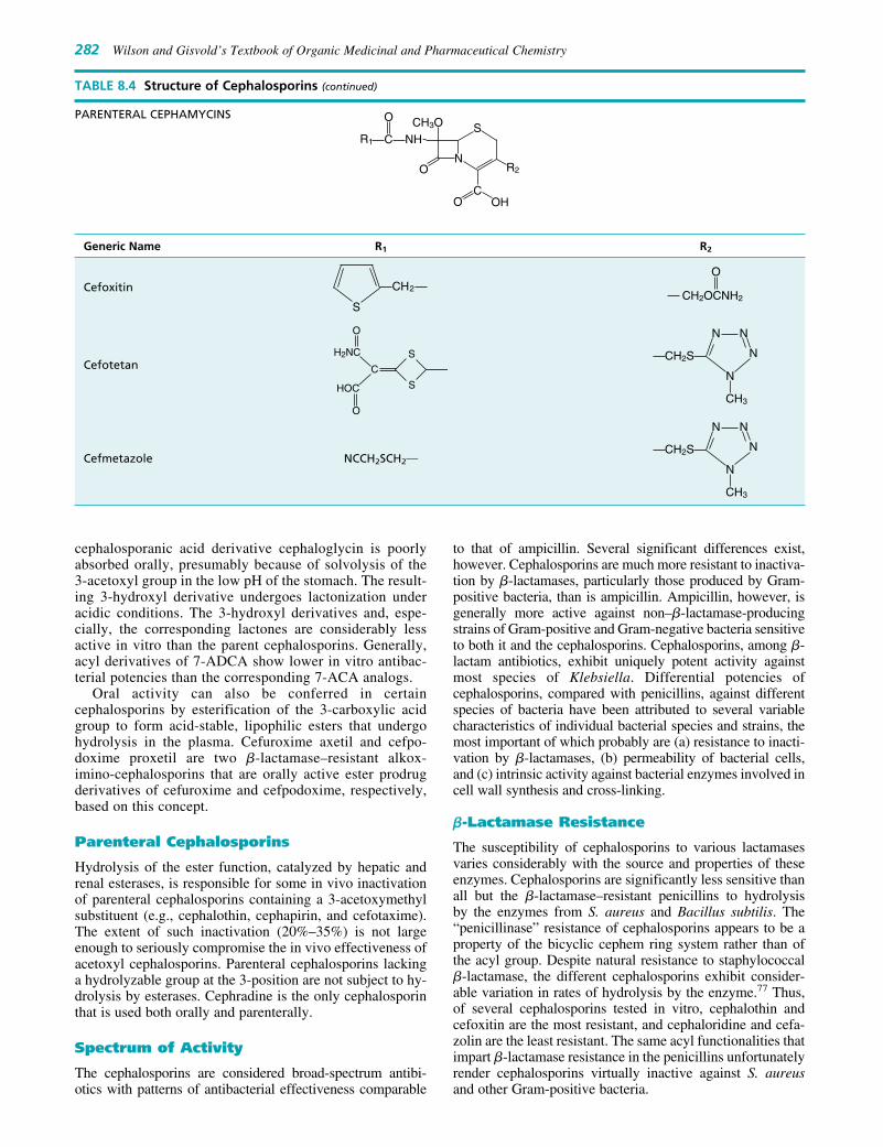
282 Wilson and Gisvold’s Textbook of Organic Medicinal and Pharmaceutical Chemistry
cephalosporanic acid derivative cephaloglycin is poorlyabsorbed orally, presumably because of solvolysis of the3-acetoxyl group in the low pH of the stomach. The result-ing 3-hydroxyl derivative undergoes lactonization underacidic conditions. The 3-hydroxyl derivatives and, espe-cially, the corresponding lactones are considerably lessactive in vitro than the parent cephalosporins. Generally,acyl derivatives of 7-ADCA show lower in vitro antibac-terial potencies than the corresponding 7-ACA analogs.
Oral activity can also be conferred in certaincephalosporins by esterification of the 3-carboxylic acidgroup to form acid-stable, lipophilic esters that undergohydrolysis in the plasma. Cefuroxime axetil and cefpo-doxime proxetil are two �-lactamase–resistant alkox-imino-cephalosporins that are orally active ester prodrugderivatives of cefuroxime and cefpodoxime, respectively,based on this concept.
Parenteral Cephalosporins
Hydrolysis of the ester function, catalyzed by hepatic andrenal esterases, is responsible for some in vivo inactivationof parenteral cephalosporins containing a 3-acetoxymethylsubstituent (e.g., cephalothin, cephapirin, and cefotaxime).The extent of such inactivation (20%–35%) is not largeenough to seriously compromise the in vivo effectiveness ofacetoxyl cephalosporins. Parenteral cephalosporins lackinga hydrolyzable group at the 3-position are not subject to hy-drolysis by esterases. Cephradine is the only cephalosporinthat is used both orally and parenterally.
Spectrum of Activity
The cephalosporins are considered broad-spectrum antibi-otics with patterns of antibacterial effectiveness comparable
to that of ampicillin. Several significant differences exist,however. Cephalosporins are much more resistant to inactiva-tion by �-lactamases, particularly those produced by Gram-positive bacteria, than is ampicillin. Ampicillin, however, isgenerally more active against non–�-lactamase-producingstrains of Gram-positive and Gram-negative bacteria sensitiveto both it and the cephalosporins. Cephalosporins, among �-lactam antibiotics, exhibit uniquely potent activity againstmost species of Klebsiella. Differential potencies ofcephalosporins, compared with penicillins, against differentspecies of bacteria have been attributed to several variablecharacteristics of individual bacterial species and strains, themost important of which probably are (a) resistance to inacti-vation by �-lactamases, (b) permeability of bacterial cells,and (c) intrinsic activity against bacterial enzymes involved incell wall synthesis and cross-linking.
�-Lactamase Resistance
The susceptibility of cephalosporins to various lactamasesvaries considerably with the source and properties of theseenzymes. Cephalosporins are significantly less sensitive thanall but the �-lactamase–resistant penicillins to hydrolysisby the enzymes from S. aureus and Bacillus subtilis. The“penicillinase” resistance of cephalosporins appears to be aproperty of the bicyclic cephem ring system rather than ofthe acyl group. Despite natural resistance to staphylococcal�-lactamase, the different cephalosporins exhibit consider-able variation in rates of hydrolysis by the enzyme.77 Thus,of several cephalosporins tested in vitro, cephalothin andcefoxitin are the most resistant, and cephaloridine and cefa-zolin are the least resistant. The same acyl functionalities thatimpart �-lactamase resistance in the penicillins unfortunatelyrender cephalosporins virtually inactive against S. aureusand other Gram-positive bacteria.
PARENTERAL CEPHAMYCINS
Generic Name R1 R2
Cefoxitin
Cefotetan
Cefmetazole NCCH2SCH2MCH2S
N N
N
N
CH3
CH2S
N N
N
N
CH3
CH2OCNH2
O 2
S
CH
S
N
CH3O
R2O
CO OH
O
C NHR1
S
S
O
C
H2NC
O
HOC
TABLE 8.4 Structure of Cephalosporins (continued)
0258-0329_17865_Ch08.qxd 12/2/09 3:27 AM Page 282
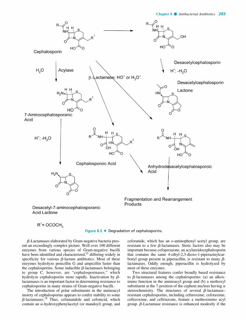
Chapter 8 Antibacterial Antibiotics 283
�-Lactamases elaborated by Gram-negative bacteria pres-ent an exceedingly complex picture. Well over 100 differentenzymes from various species of Gram-negative bacillihave been identified and characterized,25 differing widely inspecificity for various �-lactam antibiotics. Most of theseenzymes hydrolyze penicillin G and ampicillin faster thanthe cephalosporins. Some inducible �-lactamases belongingto group C, however, are “cephalosporinases,” whichhydrolyze cephalosporins more rapidly. Inactivation by �-lactamases is an important factor in determining resistance tocephalosporins in many strains of Gram-negative bacilli.
The introduction of polar substituents in the aminoacylmoiety of cephalosporins appears to confer stability to some�-lactamases.78 Thus, cefamandole and cefonicid, whichcontain an �-hydroxyphenylacetyl (or mandoyl) group, and
ceforanide, which has an o-aminophenyl acetyl group, areresistant to a few �-lactamases. Steric factors also may beimportant because cefoperazone, an acylureidocephalosporinthat contains the same 4-ethyl-2,3-dioxo-1-piperazinylcar-bonyl group present in piperacillin, is resistant to many �-lactamases. Oddly enough, piperacillin is hydrolyzed bymost of these enzymes.
Two structural features confer broadly based resistanceto �-lactamases among the cephalosporins: (a) an alkox-imino function in the aminoacyl group and (b) a methoxylsubstituent at the 7-position of the cephem nucleus having �stereochemistry. The structures of several �-lactamase–resistant cephalosporins, including cefuroxime, cefotaxime,ceftizoxime, and ceftriaxone, feature a methoximino acylgroup. �-Lactamase resistance is enhanced modestly if the
Figure 8.5 Degradation of cephalosporins.
0258-0329_17865_Ch08.qxd 12/2/09 3:27 AM Page 283

oximino substituent also features a polar function, as in cef-tazidime, which has a 2-methylpropionic acid substituent onthe oximino group. Both steric and electronic properties ofthe alkoximino group may contribute to the �-lactamaseresistance conferred by this functionality since syn-isomersare more potent than anti-isomers.78 �-Lactamase–resistant7�-methoxylcephalosporins, also called cephamycins be-cause they are derived from cephamycin C (an antibioticisolated from Streptomyces), are represented by cefoxitin,cefotetan, cefmetazole, and the 1-oxocephalosporin mox-alactam, which is prepared by total synthesis.
Base- or �-lactamase–catalyzed hydrolysis ofcephalosporins containing a good leaving group at the 3-position is accompanied by elimination of the leavinggroup. The enzymatic process occurs in a stepwise fashion,beginning with the formation of a tetrahedral transitionstate, which quickly collapses into an acyl-enzyme inter-mediate (Fig. 8.6). This intermediate can then either un-dergo hydrolysis to free the enzyme (path 1) or sufferelimination of the leaving group to form a relatively stableacyl-enzyme with a conjugated imine structure (path 2).Because of the stability of the acyl-enzyme intermediate,path 2 leads to transient inhibition of the enzyme. Faraci
and Pratt79,79a have shown that cephalothin and cefoxitininhibit certain �-lactamases by this mechanism, whereasanalogs lacking a 3� leaving group do not.
Antipseudomonal Cephalosporins
Species of Pseudomonas, especially P. aeruginosa, repre-sent a special public health problem because of their ubiq-uity in the environment and their propensity to developresistance to antibiotics, including the �-lactams. The pri-mary mechanisms of �-lactam resistance appear to involvedestruction of the antibiotics by �-lactamases and/or inter-ference with their penetration through the cell envelope.Apparently, not all �-lactamase–resistant cephalosporinspenetrate the cell envelope of P. aeruginosa, as only cefop-erazone, moxalactam, cefotaxime, ceftizoxime, ceftriaxone,and ceftazidime have useful antipseudomonal activity. Twocephalosporins, moxalactam and cefoperazone, contain thesame polar functionalities (e.g., carboxy and N-acylureido)that facilitate penetration into Pseudomonas spp. by thepenicillins (see carbenicillin, ticarcillin, and piperacillin).Unfortunately, strains of P. aeruginosa resistant to cefoper-azone and cefotaxime have been found in clinical isolates.
284 Wilson and Gisvold’s Textbook of Organic Medicinal and Pharmaceutical Chemistry
Figure 8.6 Inhibition of �-lactamases by cephalosporins.
0258-0329_17865_Ch08.qxd 12/2/09 3:27 AM Page 284

Chapter 8 Antibacterial Antibiotics 285
TABLE 8.5 Classification and Properties of Cephalosporins
Plasma Protein
Route of Acid Binding �-Lactamase Spectrum of AntipseudomonalCephalosporin Generation Admin. Resistant (%) Resistance Activity Activity
Cephalexin First Oral Yes 5–15 Poor Broad NoCephradine First Oral, Yes 8–17 Poor Broad No
parenteralCefadroxil First Oral Yes 20 Poor Broad NoCephalothin First Parenteral No 65–80 Poor Broad NoCephapirin First Parenteral No 40–54 Poor Broad NoCefazolin First Parenteral No 70–86 Poor Broad NoCefaclor Second Oral Yes 22–25 Poor Broad NoLoracarbef Second Oral Yes 25 Poor Broad NoCefprozil Second Oral Yes 36 Poor Broad NoCefamandole Second Parenteral No 56–78 Poor to avg. Extended NoCefonicid Second Parenteral No 99 Poor to avg. Extended NoCeforanide Second Parenteral No 80 Average Extended NoCefoxitin Second Parenteral No 13–22 Good Extended NoCefotetan Second Parenteral No 78–91 Good Extended NoCefmetazole Second Parenteral No 65 Good Extended NoCefuroxime Second Oral, Yes/No 33–50 Good Extended No
parenteralCefpodoxime Second Oral Yes 25 Good Extended NoCefixime Third Oral Yes 65 Good Extended NoCefoperazone Third Parenteral No 82–93 Avg. to good Extended YesCefotaxime Third Parenteral No 30–51 Good Extended YesCeftizoxime Third Parenteral No 30 Good Extended YesCeftriaxone Third Parenteral No 80–95 Good Extended YesCeftazidime Third Parenteral No 80–90 Good Extended YesCeftibuten Third Oral Yes ? Good Extended NoCefepime Fourth Parenteral No 16–19 Good Extended YesCefpirome Fourth Parenteral No – Good Extended Yes
Adverse Reactions and DrugInteractions
Like their close relatives, the penicillins, the cephalosporinantibiotics are comparatively nontoxic compounds that,because of their selective actions on cell wall cross-linkingenzymes, exhibit highly selective toxicity toward bacteria.The most common adverse reactions to the cephalosporinsare allergic and hypersensitivity reactions. These vary frommild rashes to life-threatening anaphylactic reactions.Allergic reactions are believed to occur less frequently withcephalosporins than with penicillins. The issue of cross-sensitivity between the two classes of �-lactams is verycomplex, but the incidence is considered to be very low(estimated between 3% and 7%). The physician faced withthe decision of whether or not to administer a cephalosporinto a patient with a history of penicillin allergy must weighseveral factors, including the severity of the illness beingtreated, the effectiveness and safety of alternative therapies,and the severity of previous allergic responses to penicillins.
Cephalosporins containing an N-methyl-5-thiotetrazole(MTT) moiety at the 3-position (e.g., cefamandole, cefote-tan, cefmetazole, moxalactam, and cefoperazone) havebeen implicated in a higher incidence of hypoprothrom-binemia than cephalosporins lacking the MTT group. Thiseffect, which is enhanced and can lead to severe bleedingin patients with poor nutritional status, debilitation, recentGI surgery, hepatic disease, or renal failure, is apparentlybecause of inhibition of vitamin K–requiring enzymesinvolved in the carboxylation of glutamic acid residuesin clotting factors II, VII, IX, and X to the MTT
group.80 Treatment with vitamin K restores prothrombintime to normal in patients treated with MTT-containingcephalosporins. Weekly vitamin K prophylaxis has beenrecommended for high-risk patients undergoing therapywith such agents. Cephalosporins containing the MTTgroup should not be administered to patients receiving oralanticoagulant or heparin therapy because of possible syn-ergism with these drugs.
The MTT group has also been implicated in the in-tolerance to alcohol associated with certain injectablecephalosporins: cefamandole, cefotetan, cefmetazole, andcefoperazone. Thus, disulfiram-like reactions, attributed tothe accumulation of acetaldehyde and resulting from theinhibition of aldehyde dehydrogenase–catalyzed oxidationof ethanol by MTT-containing cephalosporins,81 may occurin patients who have consumed alcohol before, during, orshortly after the course of therapy.
Classification
Cephalosporins are divided into first-, second-, third-, andfourth-generation agents, based roughly on their time ofdiscovery and their antimicrobial properties (Table 8.5).In general, progression from first to fourth generation isassociated with a broadening of the Gram-negative anti-bacterial spectrum, some reduction in activity againstGram-positive organisms, and enhanced resistance to �-lactamases. Individual cephalosporins differ in their phar-macokinetic properties, especially plasma protein bindingand half-life, but the structural bases for these differencesare not obvious.
0258-0329_17865_Ch08.qxd 12/2/09 3:27 AM Page 285

Products
CephalexinCephalexin, 7�-(D-amino-�-phenylacetamido)-3-methyl-cephemcarboxylic acid (Keflex, Keforal), was designed pur-posely as an orally active, semisynthetic cephalosporin. Theoral inactivation of cephalosporins has been attributed to twocauses: instability of the �-lactam ring to acid hydrolysis(cephalothin and cephaloridine) and solvolysis or microbialtransformation of the 3-methylacetoxy group (cephalothin,cephaloglycin). The �-amino group of cephalexin renders itacid stable, and reduction of the 3-acetoxymethyl to a methylgroup circumvents reaction at that site.
Cephalexin occurs as a white crystalline monohydrate. Itis freely soluble in water, resistant to acid, and absorbed wellorally. Food does not interfere with its absorption. Becauseof minimal protein binding and nearly exclusive renal excre-tion, cephalexin is recommended particularly for the treat-ment of urinary tract infections. It is also sometimes used forupper respiratory tract infections. Its spectrum of activity isvery similar to those of cephalothin and cephaloridine.Cephalexin is somewhat less potent than these two agentsafter parenteral administration and, therefore, is inferior tothem for the treatment of serious systemic infections.
CephradineCephradine (Anspor, Velosef) is the only cephalosporinderivative available in both oral and parenteral dosageforms. It closely resembles cephalexin chemically (it maybe regarded as a partially hydrogenated derivative ofcephalexin) and has very similar antibacterial and pharma-cokinetic properties.
It occurs as a crystalline hydrate that is readily soluble inwater. Cephradine is stable to acid and absorbed almostcompletely after oral administration. It is minimally proteinbound and excreted almost exclusively through the kidneys.It is recommended for the treatment of uncomplicated uri-nary tract and upper respiratory tract infections caused bysusceptible organisms. Cephradine is available in both oraland parenteral dosage forms.
CefadroxilCefadroxil (Duricef) is an orally active semisyntheticderivative of 7-ADCA, in which the 7-acyl group is the D-
hydroxylphenylglycyl moiety. This compound is absorbedwell after oral administration to give plasma levels that reach75% to 80% of those of an equal dose of its close structuralanalog cephalexin. The main advantage claimed for ce-fadroxil is its somewhat prolonged duration of action, whichpermits once-a-day dosing. The prolonged duration of actionof this compound is related to relatively slow urinary excre-tion of the drug compared with other cephalosporins, butthe basis for this remains to be explained completely. Theantibacterial spectrum of action and therapeutic indications ofcefadroxil are very similar to those of cephalexin and cephra-dine. The D-p-hydroxyphenylglycyl isomer is much moreactive than the L-isomer.
CefaclorCefaclor (Ceclor) is an orally active semisyntheticcephalosporin that was introduced in the American market in1979. It differs structurally from cephalexin in that the 3-methyl group has been replaced by a chlorine atom. It issynthesized from the corresponding 3-methylenecepham sul-foxide ester by ozonolysis, followed by halogenation of theresulting �-ketoester.82 The 3-methylenecepham sulfoxideesters are prepared by rearrangement of the corresponding 6-acylaminopenicillanic acid derivative. Cefaclor is moderatelystable in acid and achieves enough oral absorption to provideeffective plasma levels (equal to about two-thirds of thoseobtained with cephalexin). The compound is apparentlyunstable in solution, since about 50% of its antimicrobial ac-tivity is lost in 2 hours in serum at 37°C.83 The antibacterialspectrum of activity is similar to that of cephalexin, but it isclaimed to be more potent against some species sensitiveto both agents. Currently, the drug is recommended for thetreatment of non–life-threatening infections caused by H.influenzae, particularly strains resistant to ampicillin.
CefprozilCefprozil (Cefzil) is an orally active second-generationcephalosporin that is similar in structure and antibacterialspectrum to cefadroxil. Oral absorption is excellent (oralbioavailability is about 95%) and is not affected by antacidsor histamine H2-antagonists. Cefprozil exhibits greater invitro activity against streptococci, Neisseria spp., and S. au-reus than does cefadroxil. It is also more active than thefirst-generation cephalosporins against members of theEnterobacteriaceae family, such as E. coli, Klebsiella spp.,
286 Wilson and Gisvold’s Textbook of Organic Medicinal and Pharmaceutical Chemistry
0258-0329_17865_Ch08.qxd 12/2/09 3:27 AM Page 286

Chapter 8 Antibacterial Antibiotics 287
P. mirabilis, and Citrobacter spp. The plasma half-life of1.2 to 1.4 hours permits twice-a-day dosing for the treatmentof most community-acquired respiratory and urinary tractinfections caused by susceptible organisms.
LoracarbefLoracarbef (Lorabid) is the first of a series of carbacephemsprepared by total synthesis to be introduced.84 Carbacephemsare isosteres of the cephalosporin (or �3-cephem) antibioticsin which the 1-sulfur atom has been replaced by a methylene(CH2) group. Loracarbef is isosteric with cefaclor and hassimilar pharmacokinetic and microbiological properties.Thus, the antibacterial spectrum of activity resembles thatof cefaclor, but it has somewhat greater potency againstH. influenzae and M. catarrhalis, including �-lactamase–producing strains. Unlike cefaclor, which undergoes degra-dation in human serum, loracarbef is chemically stable inplasma. It is absorbed well orally. Oral absorption is delayedby food. The half-life in plasma is about 1 hour.
Cephalothin SodiumCephalothin sodium (Keflin) occurs as a white to off-white,crystalline powder that is practically odorless. It is freelysoluble in water and insoluble in most organic solvents.Although it has been described as a broad-spectrum anti-bacterial compound, it is not in the same class as the tetra-cyclines. Its spectrum of activity is broader than that ofpenicillin G and more similar to that of ampicillin. Unlikeampicillin, cephalothin is resistant to penicillinase pro-duced by S. aureus and provides an alternative to the use ofpenicillinase-resistant penicillins for the treatment of infec-tions caused by such strains.
Cephalothin is absorbed poorly from the GI tract andmust be administered parenterally for systemic infections. It
is relatively nontoxic and acid stable. It is excreted rapidlythrough the kidneys; about 60% is lost within 6 hours of ad-ministration. Pain at the site of intramuscular injection andthrombophlebitis following intravenous injection have beenreported. Hypersensitivity reactions have been observed,and there is some evidence of cross-sensitivity in patientsnoted previously to be penicillin sensitive.
Cefazolin Sodium, SterileCefazolin (Ancef, Kefzol) is one of a series of semisyntheticcephalosporins in which the C-3 acetoxy function has beenreplaced by a thiol-containing heterocycle—here, 5-methyl-2-thio-1,3,4-thiadiazole. It also contains the somewhatunusual tetrazolylacetyl acylating group. Cefazolin wasreleased in 1973 as a water-soluble sodium salt. It is activeonly by parenteral administration.
Cefazolin provides higher serum levels, slower renalclearance, and a longer half-life than other first-generationcephalosporins. It is approximately 75% protein bound inplasma, a higher value than for most other cephalosporins.Early in vitro and clinical studies suggest that cefazolin ismore active against Gram-negative bacilli but less activeagainst Gram-positive cocci than either cephalothin orcephaloridine. Occurrence rates of thrombophlebitis follow-ing intravenous injection and pain at the site of intramuscu-lar injection appear to be the lowest of the parenteralcephalosporins.
Cephapirin Sodium, SterileCephapirin (Cefadyl) is a semisynthetic 7-ACA derivativereleased in the United States in 1974. It closely resemblescephalothin in chemical and pharmacokinetic properties. Likecephalothin, cephapirin is unstable in acid and must beadministered parenterally in the form of an aqueous solutionof the sodium salt. It is moderately protein bound (45%–50%)in plasma and cleared rapidly by the kidneys. Cephapirin andcephalothin are very similar in antimicrobial spectrum andpotency. Conflicting reports concerning the relative occur-rence of pain at the site of injection and thrombophlebitis afterintravenous injection of cephapirin and cephalothin are diffi-cult to assess on the basis of available clinical data.
Cefamandole NafateCefamandole (Mandol) nafate is the formate ester ofcefamandole, a semisynthetic cephalosporin that incorporates
0258-0329_17865_Ch08.qxd 12/2/09 3:27 AM Page 287

288 Wilson and Gisvold’s Textbook of Organic Medicinal and Pharmaceutical Chemistry
D-mandelic acids as the acyl portion and a thiol-containingheterocycle (5-thio-1,2,3,4-tetrazole) in place of the acetoxylfunction on the C-3 methylene carbon atom. Esterification ofthe �-hydroxyl group of the D-mandeloyl function overcomesthe instability of cefamandole in solid-state dosage forms85
and provides satisfactory concentrations of the parent antibi-otic in vivo through spontaneous hydrolysis of the ester atneutral to alkaline pH. Cefamandole is the first second-gener-ation cephalosporin to be marketed in the United States.
The D-mandeloyl moiety of cefamandole appears toconfer resistance to a few �-lactamases, since some �-lactamase–producing, Gram-negative bacteria (particularlyEnterobacteriaceae) that show resistance to cefazolin andother first-generation cephalosporins are sensitive tocefamandole. Additionally, it is active against some ampi-cillin-resistant strains of Neisseria and Haemophilus spp.Although resistance to �-lactamases may be a factor in de-termining the sensitivity of individual bacterial strains tocefamandole, an early study86 indicated that other factors,such as permeability and intrinsic activity, are frequentlymore important. The L-mandeloyl isomer is significantlyless active than the D-isomer.
Cefamandole nafate is very unstable in solution andhydrolyzes rapidly to release cefamandole and formate.There is no loss of potency, however, when such solutionsare stored for 24 hours at room temperature or up to 96hours when refrigerated. Air oxidation of the released for-mate to carbon dioxide can cause pressure to build up in theinjection vial.
Cefonicid Sodium, SterileCefonicid Sodium (Monocid) is a second-generationcephalosporin that is structurally similar to cefamandole,except that it contains a methane sulfonic acid groupattached to the N-1 position of the tetrazole ring. Theantimicrobial spectrum and limited �-lactamase stabilityof cefonicid are essentially identical with those ofcefamandole.
Cefonicid is unique among the second-generationcephalosporins in that it has an unusually long serum half-life of approximately 4.5 hours. High plasma protein bind-ing coupled with slow renal tubular secretion are apparentlyresponsible for the long duration of action. Despite the high
fraction of drug bound in plasma, cefonicid is distributedthroughout body fluids and tissues, with the exception of thecerebrospinal fluid.
Cefonicid is supplied as a highly water-soluble disodiumsalt, in the form of a sterile powder to be reconstituted forinjection. Solutions are stable for 24 hours at 25°C and for72 hours when refrigerated.
Ceforanide, SterileCeforanide (Precef) was approved for clinical use in theUnited States in 1984. It is classified as a second-generationcephalosporin because its antimicrobial properties are simi-lar to those of cefamandole. It exhibits excellent potencyagainst most members of the Enterobacteriaceae family,especially K. pneumoniae, E. coli, P. mirabilis, andEnterobacter cloacae. It is less active than cefamandoleagainst H. influenzae, however.
The duration of action of ceforanide lies between those ofcefamandole and cefonicid. It has a serum half-life of about3 hours, permitting twice-a-day dosing for most indications.Ceforanide is supplied as the sterile, crystalline disodiumsalt. Parenteral solutions are stable for 4 hours at 25°C andfor up to 5 days when refrigerated.
Cefoperazone Sodium, SterileCefoperazone (Cefobid) is a third-generation, anti-pseudomonal cephalosporin that resembles piperacillinchemically and microbiologically. It is active against manystrains of P. aeruginosa, indole-positive Proteus spp.,Enterobacter spp., and S. marcescens that are resistant tocefamandole. It is less active than cephalothin againstGram-positive bacteria and less active than cefamandoleagainst most of the Enterobacteriaceae. Like piperacillin,cefoperazone is hydrolyzed by many of the �-lactamasesthat hydrolyze penicillins. Unlike piperacillin, however, itis resistant to some (but not all) of the �-lactamases thathydrolyze cephalosporins.
Cefoperazone is excreted primarily in the bile. Hepaticdysfunction can affect its clearance from the body. Althoughonly 25% of the free antibiotic is recovered in the urine,
0258-0329_17865_Ch08.qxd 12/2/09 3:27 AM Page 288

Chapter 8 Antibacterial Antibiotics 289
urinary concentrations are high enough to be effective in themanagement of urinary tract infections caused by suscepti-ble organisms. The relatively long half-life (2 hours) allowsdosing twice a day. Solutions prepared from the crystallinesodium salt are stable for up to 4 hours at room temperature.If refrigerated, they will last 5 days without appreciable lossof potency.
Cefoxitin Sodium, SterileCefoxitin (Mefoxin) is a semisynthetic derivative obtainedby modification of cephamycin C, a 7�-methoxy-substitutedcephalosporin isolated independently from variousStreptomyces by research groups in Japan87 and the UnitedStates. Although it is less potent than cephalothin againstGram-positive bacteria and cefamandole against most of theEnterobacteriaceae, cefoxitin is effective against certainstrains of Gram-negative bacilli (e.g., E. coli, K. pneumo-niae, Providencia spp., S. marcescens, indole-positiveProteus spp., and Bacteroides spp.) that are resistant to thesecephalosporins. It is also effective against penicillin-resistantS. aureus and N. gonorrhoeae.
The activity of cefoxitin and cephamycins, in general,against resistant bacterial strains is because of their resistanceto hydrolysis by �-lactamases conferred by the 7�-methoxylsubstituent.88 Cefoxitin is a potent competitive inhibitor ofmany �-lactamases. It is also a potent inducer of chromosom-ally mediated �-lactamases. The temptation to exploit the �-lactamase–inhibiting properties of cefoxitin by combining itwith �-lactamase–labile �-lactam antibiotics should be tem-pered by the possibility of antagonism. In fact, cefoxitin an-tagonizes the action of cefamandole against E. cloacae andthat of carbenicillin against P. aeruginosa.89 Cefoxitin aloneis essentially ineffective against these organisms.
The pharmacokinetic properties of cefoxitin resemblethose of cefamandole. Because its half-life is relativelyshort, cefoxitin must be administered 3 or 4 times daily.Solutions of the sodium salt intended for parenteral admin-istration are stable for 24 hours at room temperature and 1week if refrigerated. 7�-Methoxyl substitution stabilizes, tosome extent, the �-lactam to alkaline hydrolysis.
The principal role of cefoxitin in therapy seems to be forthe treatment of certain anaerobic and mixed aerobic–anaer-obic infections. It is also used to treat gonorrhea caused by�-lactamase–producing strains. It is classified as a second-generation agent because of its spectrum of activity.
Cefotetan DisodiumCefotetan (Cefotan) is a third-generation cephalosporin thatis structurally similar to cefoxitin. Like cefoxitin, cefotetanis resistant to destruction by �-lactamases. It is also a com-petitive inhibitor of many �-lactamases and causes transient
inactivation of some of these enzymes. Cefotetan is reportedto synergize with �-lactamase–sensitive �-lactams but, un-like cefoxitin, does not appear to cause antagonism.90
The antibacterial spectrum of cefotetan closely resemblesthat of cefoxitin. It is, however, generally more activeagainst S. aureus, and members of the Enterobacteriaceaefamily sensitive to both agents. It also exhibits excellentpotency against H. influenzae and N. gonorrhoeae, includ-ing �-lactamase–producing strains. Cefotetan is slightly lessactive than cefoxitin against B. fragilis and other anaerobes.Enterobacter spp. are generally resistant to cefotetan, andthe drug is without effect against Pseudomonas spp.
Cefotetan has a relatively long half-life of about 3.5hours. It is administered on a twice-daily dosing schedule. Itis excreted largely unchanged in the urine. Aqueoussolutions for parenteral administration maintain potency for24 hours at 25°C. Refrigerated solutions are stable for 4 days.
Cefotetan contains the MTT group that has been associ-ated with hypoprothrombinemia and alcohol intolerance.Another cephalosporin that lacks these properties should beselected for patients at risk for severe bleeding or alcoholism.
Cefmetazole SodiumCefmetazole (Zefazone) is a semisynthetic, third-generation,parenteral cephalosporin of the cephamycin group. Likeother cephamycins, the presence of the 7�-methoxyl groupconfers resistance to many �-lactamases. Cefmetazoleexhibits significantly higher potency against members ofthe Enterobacteriaceae family but lower activity againstBacteroides spp. than cefoxitin. It is highly active againstN. gonorrhoeae, including �-lactamase–producing strains. Incommon with other cephamycins, cefmetazole is ineffectiveagainst indole-positive Proteus, Enterobacter, Providencia,Serratia, and Pseudomonas spp. Cefmetazole has the MTTmoiety associated with increased bleeding in certain high-risk patients. It has a plasma half-life of 1.1 hours.
Cefuroxime SodiumCefuroxime (Zinacef) is the first of a series of �-methoximi-noacyl–substituted cephalosporins that constitute most of thethird-generation agents available for clinical use. A syn alkox-imino substituent is associated with �-lactamase stability in
0258-0329_17865_Ch08.qxd 12/2/09 3:27 AM Page 289

290 Wilson and Gisvold’s Textbook of Organic Medicinal and Pharmaceutical Chemistry
these cephalosporins.78 Cefuroxime is classified as a second-generation cephalosporin because its spectrum of antibacte-rial activity more closely resembles that of cefamandole. It is,however, active against �-lactamase–producing strains thatare resistant to cefamandole, such as E. coli, K. pneumoniae,N. gonorrhoeae, and H. influenzae. Other important Gram-negative pathogens, such as Serratia, indole-positive Proteusspp., P. aeruginosa, and B. fragilis, are resistant.
Cefuroxime is distributed throughout the body. It pene-trates inflamed meninges in high enough concentrations tobe effective in meningitis caused by susceptible organisms.Three-times-daily dosing is required to maintain effectiveplasma levels for most sensitive organisms, such asNeisseria meningitidis, Streptococcus pneumoniae, and H.influenzae. It has a plasma half-life of 1.4 hours.
Cefuroxime AxetilCefuroxime axetil (Ceftin) is the 1-acetyoxyethyl ester ofcefuroxime. During absorption, this acid-stable, lipophilic,oral prodrug derivative of cefuroxime is hydrolyzed to ce-furoxime by intestinal and/or plasma enzymes. The axetilester provides an oral bioavailability of 35% to 50% of ce-furoxime, depending on conditions. Oral absorption of theester is increased by food but decreased by antacids and his-tamine H2-antagonists. The latter effect may be because ofspontaneous hydrolysis of the ester in the intestine becauseof the higher pH created by these drugs. Axetil is used forthe oral treatment of non–life-threatening infections causedby bacteria that are susceptible to cefuroxime. The prodrugform permits twice-a-day dosing for such infections.
Cefpodoxime ProxetilCefpodoxime proxetil (Vantin) is the isopropyloxycar-bonylethyl ester of the third-generation cephalosporincefpodoxime. This orally active prodrug derivative is hy-drolyzed by esterases in the intestinal wall and in theplasma to provide cefpodoxime. Tablets and a powder forthe preparation of an aqueous suspension for oral pediatricadministration are available. The oral bioavailability of cef-podoxime from the proxetil is estimated to be about 50%.Administration of the prodrug with food enhances its ab-sorption. The plasma half-life is 2.2 hours, which permitsadministration on a twice-daily schedule.
Cefpodoxime is a broad-spectrum cephalosporin withuseful activity against a relatively wide range of Gram-positive and Gram-negative bacteria. It is also resistant tomany �-lactamases. Its spectrum of activity includes S.pneumoniae, Streptococcus pyogenes, S. aureus, H. influen-zae, M. catarrhalis, and Neisseria spp. Cefpodoxime is alsoactive against members of the Enterobacteriaceae family,including E. coli, K. pneumoniae, and P. mirabilis. It thusfinds use in the treatment of upper and lower respiratory in-fections, such as pharyngitis, bronchitis, otitis media, andcommunity-acquired pneumonia. It is also useful for thetreatment of uncomplicated gonorrhea.
CefiximeCefixime (Suprax) is the first orally active, third-generationcephalosporin that is not an ester prodrug to be approved fortherapy in the United States. Oral bioavailability is surpris-ingly high, ranging from 40% to 50%. Facilitated transportof cefixime across intestinal brush border membranes in-volving the carrier system for dipeptides may explain itssurprisingly good oral absorption.91 This result was not ex-pected because cefixime lacks the ionizable �-amino grouppresent in dipeptides and �-lactams previously known to betransported by the carrier system.51,91
Cefixime is a broad-spectrum cephalosporin that is resis-tant to many �-lactamases. It is particularly effective againstGram-negative bacilli, including E. coli, Klebsiella spp., P.mirabilis, indole-positive Proteus, Providencia, and someCitrobacter spp. Most Pseudomonas, Enterobacter, andBacteroides spp. are resistant. It also has useful activityagainst streptococci, gonococci, H. influenzae, and M. ca-tarrhalis. It is much less active against S. aureus. Cefixime isused for the treatment of various respiratory tract infections(e.g., acute bronchitis, pharyngitis, and tonsillitis) and otitismedia. It is also used to treat uncomplicated urinary tract in-fections and gonorrhea caused by �-lactamase–producingbacterial strains.
The comparatively long half-life of cefixime (t1/2 is 3–4 hours) allows it to be administered on a twice-a-dayschedule. Renal tubular reabsorption and a relatively high
0258-0329_17865_Ch08.qxd 12/2/09 3:27 AM Page 290

Chapter 8 Antibacterial Antibiotics 291
fraction of plasma protein binding (�65%) contribute to thelong half-life. It is provided in two-oral dosage forms: 200-or 400-mg tablets and a powder for the preparation of anaqueous suspension.
Cefotaxime Sodium, SterileCefotaxime (Claforan) was the first third-generationcephalosporin to be introduced. It possesses excellentbroad-spectrum activity against Gram-positive and Gram-negative aerobic and anaerobic bacteria. It is more activethan moxalactam against Gram-positive organisms. Many�-lactamase–producing bacterial strains are sensitive to ce-fotaxime, including N. gonorrhoeae, Klebsiella spp., H. in-fluenzae, S. aureus, and E. cloacae. Some, but not all,Pseudomonas strains are sensitive. Enterococci and Listeriamonocytogenes are resistant.
The syn-isomer of cefotaxime is significantly more activethan the anti-isomer against �-lactamase–producing bacte-ria. This potency difference is, in part, because of greaterresistance of the syn-isomer to the action of �-lactamases.78
The higher affinity of the syn-isomer for PBPs, however,may also be a factor.92
Cefotaxime is metabolized in part to the less active de-sacetyl metabolite. Approximately 20% of the metaboliteand 25% of the parent drug are excreted in the urine. Theparent drug reaches the cerebrospinal fluid in sufficient con-centration to be effective in the treatment of meningitis.Solutions of cefotaxime sodium should be used within 24hours. If stored, they should be refrigerated. Refrigerated so-lutions maintain potency up to 10 days.
Ceftizoxime Sodium, SterileCeftizoxime (Cefizox) is a third-generation cephalosporinthat was introduced in 1984. This �-lactamase–resistant agentexhibits excellent activity against the Enterobacteriaceae,especially E. coli, K. pneumoniae, E. cloacae, Enterobacteraerogenes, indole-positive and indole-negative Proteus spp.,and S. marcescens. Ceftizoxime is claimed to be more activethan cefoxitin against B. fragilis. It is also very active againstGram-positive bacteria. Its activity against P. aeruginosa issomewhat variable and lower than that of either cefotaxime orcefoperazone.
Ceftizoxime is not metabolized in vivo. It is excretedlargely unchanged in the urine. Adequate levels of the drugare achieved in the cerebrospinal fluid for the treatment ofGram-negative or Gram-positive bacterial meningitis. It mustbe administered on a thrice-daily dosing schedule because ofits relatively short half-life. Ceftizoxime sodium is very sta-ble in the dry state. Solutions maintain potency for up to 24hours at room temperature and 10 days when refrigerated.
Ceftriaxone Disodium, SterileCeftriaxone (Rocephin) is a �-lactamase–resistantcephalosporin with an extremely long serum half-life.Once-daily dosing suffices for most indications. Two fac-tors contribute to the prolonged duration of action ofceftriaxone: high protein binding in the plasma and slowurinary excretion. Ceftriaxone is excreted in both the bileand the urine. Its urinary excretion is not affected byprobenecid. Despite its comparatively low volume ofdistribution, it reaches the cerebrospinal fluid in concen-trations that are effective in meningitis. Nonlinear pharma-cokinetics are observed.
Ceftriaxone contains a highly acidic heterocyclic systemon the 3-thiomethyl group. This unusual dioxotriazine ringsystem is believed to confer the unique pharmacokineticproperties of this agent. Ceftriaxone has been associatedwith sonographically detected “sludge,” or pseudolithiasis,in the gallbladder and common bile duct.93 Symptoms ofcholecystitis may occur in susceptible patients, especiallythose on prolonged or high-dose ceftriaxone therapy. Theculprit has been identified as the calcium chelate.
Ceftriaxone exhibits excellent broad-spectrum antibacte-rial activity against both Gram-positive and Gram-negativeorganisms. It is highly resistant to most chromosomally andplasmid-mediated �-lactamases. The activity of ceftriaxoneagainst Enterobacter, Citrobacter, Serratia, indole-positiveProteus, and Pseudomonas spp. is particularly impressive. Itis also effective in the treatment of ampicillin-resistant gon-orrhea and H. influenzae infections but generally less activethan cefotaxime against Gram-positive bacteria and B.fragilis.
Solutions of ceftriaxone sodium should be used within 24hours. They may be stored up to 10 days if refrigerated.
Ceftazidime Sodium, SterileCeftazidime (Fortaz, Tazidime) is a �-lactamase–resistantthird-generation cephalosporin that is noted for its an-tipseudomonal activity. It is active against some strains of P.aeruginosa that are resistant to cefoperazone and ceftriax-one. Ceftazidime is also highly effective against �-lacta-mase–producing strains of the Enterobacteriaceae family. Itis generally less active than cefotaxime against Gram-posi-tive bacteria and B. fragilis.
0258-0329_17865_Ch08.qxd 12/2/09 3:27 AM Page 291

292 Wilson and Gisvold’s Textbook of Organic Medicinal and Pharmaceutical Chemistry
The structure of ceftazidime contains two noteworthyfeatures: (a) a 2-methylpropionicoxaminoacyl group thatconfers �-lactamase resistance and, possibly, increasedpermeability through the porin channels of the cell envelopeand (b) a pyridinium group at the 3-position that conferszwitterionic properties on the molecule.
Ceftazidime is administered parenterally 2 or 3 timesdaily, depending on the severity of the infection. Its serumhalf-life is about 1.8 hours. It has been used effectively forthe treatment of meningitis caused by H. influenzae andN. meningitidis.
NEWER CEPHALOSPORINS
Cephalosporins currently undergoing clinical trials or re-cently being marketed in the United States fall into two cate-gories: (a) orally active �-lactamase–resistant cephalosporinsand (b) parenteral �-lactamase–resistant antipseudomonalcephalosporins. The status of some of these compoundsawaits more extensive clinical evaluation. Nonetheless, itappears that any advances they represent will be relativelymodest.
CeftibutenCeftibuten (Cedax) is a recently introduced, chemicallynovel analog of the oximino cephalosporins in which anolefinic methylene group (CBCHCH2-) with Z stereo-chemistry has replaced the syn oximino (CBNO-) group.This isosteric replacement yields a compound that retainsresistance to hydrolysis catalyzed by many �-lactamases,has enhanced chemical stability, and is orally active. Oralabsorption is rapid and nearly complete. It has the highestoral bioavailability of the third-generation cephalosporins.94
Ceftibuten is excreted largely unchanged in the urine andhas a half-life of about 2.5 hours. Plasma protein binding ofthis cephalosporin is estimated to be 63%.
Ceftibuten possesses excellent potency against mostmembers of the Enterobacteriaceae family, H. influenzae,Neisseria spp., and M. catarrhalis. It is not active against S.aureus or P. aeruginosa and exhibits modest antistreptococ-
cal activity. Ceftibuten is recommended in the managementof community-acquired respiratory tract, urinary tract, andgynecological infections.
CefpiromeCefpirome (Cefrom) is a newer parenteral, �-lactamase–resistant cephalosporin with a quaternary ammonium groupat the 3-position of the cephem nucleus. Because its potencyagainst Gram-positive and Gram-negative bacteria rivals thatof the first-generation and third-generation cephalosporins,respectively, cefpirome is being touted as the first fourth-generation cephalosporin.95 Its broad spectrum includesmethicillin-sensitive staphylococci, penicillin-resistantpneumococci, and �-lactamase–producing strains of E. coli,Enterobacter, Citrobacter, and Serratia spp. Its efficacyagainst P. aeruginosa is comparable with that of ceftazidime.Cefpirome is excreted largely unchanged in the urine with ahalf-life of 2 hours.
CefepimeCefepime (Maxipime, Axepin) is a parenteral, �-lactamase–resistant cephalosporin that is chemically and mi-crobiologically similar to cefpirome. It also has a broadantibacterial spectrum, with significant activity against bothGram-positive and Gram-negative bacteria, including strep-tococci, staphylococci, Pseudomonas spp., and theEnterobacteriaceae. It is active against some bacterial isolatesthat are resistant to ceftazidime.96 The efficacy of cefepimehas been demonstrated in the treatment of urinary tract infec-tions, lower respiratory tract infections, skin and soft tissueinfections, chronic osteomyelitis, and intra-abdominal andbiliary infections. It is excreted in the urine with a half-life of2.1 hours. It is bound minimally to plasma proteins. Cefepimeis also a fourth-generation cephalosporin.
Future Developments inCephalosporin Design
Recent research efforts in the cephalosporin field havefocused primarily on two desired antibiotic properties: (a) in-creased permeability into Gram-negative bacilli, leading to
0258-0329_17865_Ch08.qxd 12/2/09 3:27 AM Page 292
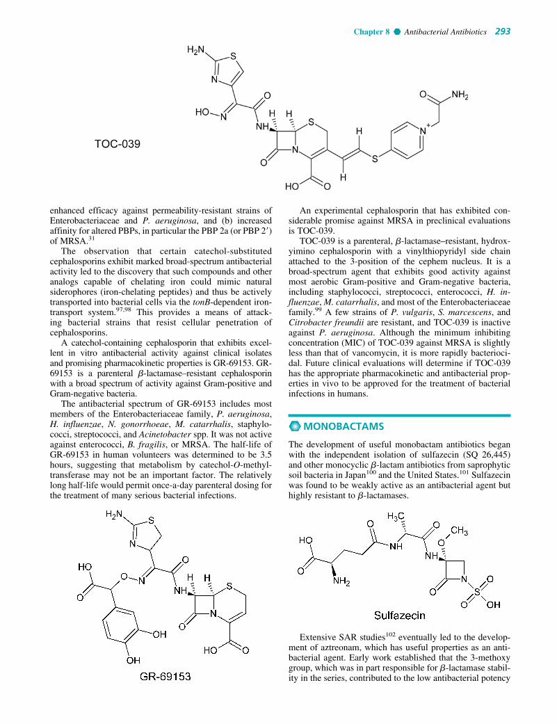
Chapter 8 Antibacterial Antibiotics 293
enhanced efficacy against permeability-resistant strains ofEnterobacteriaceae and P. aeruginosa, and (b) increasedaffinity for altered PBPs, in particular the PBP 2a (or PBP 2�)of MRSA.31
The observation that certain catechol-substitutedcephalosporins exhibit marked broad-spectrum antibacterialactivity led to the discovery that such compounds and otheranalogs capable of chelating iron could mimic naturalsiderophores (iron-chelating peptides) and thus be activelytransported into bacterial cells via the tonB-dependent iron-transport system.97,98 This provides a means of attack-ing bacterial strains that resist cellular penetration ofcephalosporins.
A catechol-containing cephalosporin that exhibits excel-lent in vitro antibacterial activity against clinical isolatesand promising pharmacokinetic properties is GR-69153. GR-69153 is a parenteral �-lactamase–resistant cephalosporinwith a broad spectrum of activity against Gram-positive andGram-negative bacteria.
The antibacterial spectrum of GR-69153 includes mostmembers of the Enterobacteriaceae family, P. aeruginosa,H. influenzae, N. gonorrhoeae, M. catarrhalis, staphylo-cocci, streptococci, and Acinetobacter spp. It was not activeagainst enterococci, B. fragilis, or MRSA. The half-life ofGR-69153 in human volunteers was determined to be 3.5hours, suggesting that metabolism by catechol-O-methyl-transferase may not be an important factor. The relativelylong half-life would permit once-a-day parenteral dosing forthe treatment of many serious bacterial infections.
An experimental cephalosporin that has exhibited con-siderable promise against MRSA in preclinical evaluationsis TOC-039.
TOC-039 is a parenteral, �-lactamase–resistant, hydrox-yimino cephalosporin with a vinylthiopyridyl side chainattached to the 3-position of the cephem nucleus. It is abroad-spectrum agent that exhibits good activity againstmost aerobic Gram-positive and Gram-negative bacteria,including staphylococci, streptococci, enterococci, H. in-fluenzae, M. catarrhalis, and most of the Enterobacteriaceaefamily.99 A few strains of P. vulgaris, S. marcescens, andCitrobacter freundii are resistant, and TOC-039 is inactiveagainst P. aeruginosa. Although the minimum inhibitingconcentration (MIC) of TOC-039 against MRSA is slightlyless than that of vancomycin, it is more rapidly bacterioci-dal. Future clinical evaluations will determine if TOC-039has the appropriate pharmacokinetic and antibacterial prop-erties in vivo to be approved for the treatment of bacterialinfections in humans.
MONOBACTAMS
The development of useful monobactam antibiotics beganwith the independent isolation of sulfazecin (SQ 26,445)and other monocyclic �-lactam antibiotics from saprophyticsoil bacteria in Japan100 and the United States.101 Sulfazecinwas found to be weakly active as an antibacterial agent buthighly resistant to �-lactamases.
Extensive SAR studies102 eventually led to the develop-ment of aztreonam, which has useful properties as an anti-bacterial agent. Early work established that the 3-methoxygroup, which was in part responsible for �-lactamase stabil-ity in the series, contributed to the low antibacterial potency
0258-0329_17865_Ch08.qxd 12/2/09 3:27 AM Page 293

294 Wilson and Gisvold’s Textbook of Organic Medicinal and Pharmaceutical Chemistry
In contrast to the poor oral bioavailability of aztre-onam, the oral absorption of tigemonam is excellent. Itcould become a valuable agent for the oral treatment ofurinary tract infections and other non–life-threatening in-fections caused by �-lactamase–producing Gram-negativebacteria.
AMINOGLYCOSIDES
The discovery of streptomycin, the first aminoglycoside an-tibiotic to be used in chemotherapy, was the result of aplanned and deliberate search begun in 1939 and brought tofruition in 1944 by Schatz and associates.104 This successstimulated worldwide searches for antibiotics fromthe actinomycetes and, particularly, from the genusStreptomyces. Among the many antibiotics isolated fromthat genus, several are compounds closely related in struc-ture to streptomycin. Six of them—kanamycin, neomycin,paromomycin, gentamicin, tobramycin, and netilmicin—currently are marketed in the United States. Amikacin, asemisynthetic derivative of kanamycin A, has been added,and it is possible that additional aminoglycosides will beintroduced in the future.
All aminoglycoside antibiotics are absorbed very poorly(less than 1% under normal circumstances) following oraladministration, and some of them (kanamycin, neomycin,and paromomycin) are administered by that route for thetreatment of GI infections. Because of their potent broad-spectrum antimicrobial activity, they are also used for thetreatment of systemic infections. Their undesirable sideeffects, particularly ototoxicity and nephrotoxicity, have re-stricted their systemic use to serious infections or infectionscaused by bacterial strains resistant to other agents. Whenadministered for systemic infections, aminoglycosides mustbe given parenterally, usually by intramuscular injection.An additional antibiotic obtained from Streptomyces,spectinomycin, is also an aminoglycoside but differs chem-ically and microbiologically from other members of thegroup. It is used exclusively for the treatment of uncompli-cated gonorrhea.
Chemistry
Aminoglycosides are so named because their structures con-sist of amino sugars linked glycosidically. All have at leastone aminohexose, and some have a pentose lacking an aminogroup (e.g., streptomycin, neomycin, and paromomycin).Additionally, each of the clinically useful aminoglycosidescontains a highly substituted 1,3-diaminocyclohexane centralring; in kanamycin, neomycin, gentamicin, and tobramycin, it
and poor chemical stability of these antibiotics. A 4-methylgroup, however, increases stability to �-lactamases andactivity against Gram-negative bacteria at the same time.Unfortunately, potency against Gram-positive bacteria de-creases. 4,4-Gem-dimethyl substitution slightly decreasesantibacterial potency after oral administration.
Products
Aztreonam DisodiumAztreonam (Azactam) is a monobactam prepared by totalsynthesis. It binds with high affinity to PBP 3 in Gram-negative bacteria only. It is inactive against Gram-positivebacteria and anaerobes. �-Lactamase resistance is like thatof ceftazidime, which has the same isobutyric acid oximi-noacyl group. Aztreonam does not induce chromosomallymediated �-lactamases.
Aztreonam is particularly active against aerobic Gram-negative bacilli, including E. coli, K. pneumoniae, Klebsiellaoxytoca, P. mirabilis, S. marcescens, Citrobacter spp., and P.aeruginosa. It is used to treat urinary and lower respiratorytract infections, intra-abdominal infections, and gynecologi-cal infections, as well as septicemias caused by these organ-isms. Aztreonam is also effective against, but is not currentlyused to treat, infections caused by Haemophilus, Neisseria,Salmonella, indole-positive Proteus, and Yersinia spp. It isnot active against Gram-positive bacteria, anaerobic bacteria,or other species of Pseudomonas.
Urinary excretion is about 70% of the administered dose.Some is excreted through the bile. Serum half-life is 1.7hours, which allows aztreonam to be administered 2 or 3times daily, depending on the severity of the infection. Lessthan 1% of an orally administered dose of aztreonam isabsorbed, prompting the suggestion that this �-lactam couldbe used to treat intestinal infections.
The disodium salt of aztreonam is very soluble in water.Solutions for parenteral administration containing 2% orless are stable for 48 hours at room temperature.Refrigerated solutions retain full potency for 1 week.
TigemonamTigemonam is a newer monobactam that is orally active.103
It is highly resistant to �-lactamases. The antibacterialspectrum of activity resembles that of aztreonam. It is veryactive against the Enterobacteriaceae, including E. coli,Klebsiella, Proteus, Citrobacter, Serratia, and Enterobacterspp. It also exhibits good potency against H. influenzae andN. gonorrhoeae. Tigemonam is not particularly activeagainst Gram-positive or anaerobic bacteria and is inactiveagainst P. aeruginosa.
0258-0329_17865_Ch08.qxd 12/2/09 3:27 AM Page 294

Chapter 8 Antibacterial Antibiotics 295
is deoxystreptamine, and in streptomycin, it is streptadine.The aminoglycosides are thus strongly basic compounds thatexist as polycations at physiological pH. Their inorganic acidsalts are very soluble in water. All are available as sulfates.Solutions of the aminoglycoside salts are stable to autoclav-ing. The high water solubility of the aminoglycosides nodoubt contributes to their pharmacokinetic properties. Theydistribute well into most body fluids but not into the centralnervous system, bone, or fatty or connective tissues. Theytend to concentrate in the kidneys and are excreted byglomerular filtration. Aminoglycosides are apparently notmetabolized in vivo.
Spectrum of Activity
Although the aminoglycosides are classified as broad-spectrum antibiotics, their greatest usefulness lies in thetreatment of serious systemic infections caused by aerobicGram-negative bacilli. The choice of agent is generally be-tween kanamycin, gentamicin, tobramycin, netilmicin, andamikacin. Aerobic Gram-negative and Gram-positive cocci(with the exception of staphylococci) tend to be less sensi-tive; thus, the �-lactams and other antibiotics tend to bepreferred for the treatment of infections caused by these or-ganisms. Anaerobic bacteria are invariably resistant to theaminoglycosides. Streptomycin is the most effective of thegroup for the chemotherapy of TB, brucellosis, tularemia,and Yersinia infections. Paromomycin is used primarily inthe chemotherapy of amebic dysentery. Under certain cir-cumstances, aminoglycoside and �-lactam antibiotics exert asynergistic action in vivo against some bacterial strainswhen the two are administered jointly. For example, carbeni-cillin and gentamicin are synergistic against gentamicin-sen-sitive strains of P. aeruginosa and several other species ofGram-negative bacilli, and penicillin G and streptomycin (orgentamicin or kanamycin) tend to be more effective than ei-ther agent alone in the treatment of enterococcal endocardi-tis. The two antibiotic types should not be combined in thesame solution because they are chemically incompatible.Damage to the cell wall caused by the �-lactam antibiotic isbelieved to increase penetration of the aminoglycoside intothe bacterial cell.
Mechanism of Action
Most studies concerning the mechanism of antibacterialaction of the aminoglycosides were carried out withstreptomycin. However, the specific actions of other amino-glycosides are thought to be qualitatively similar. The amino-glycosides act directly on the bacterial ribosome to inhibit theinitiation of protein synthesis and to interfere with the fidelityof translation of the genetic message. They bind to the 30S ri-bosomal subunit to form a complex that cannot initiate properamino acid polymerization.105 The binding of streptomycinand other aminoglycosides to ribosomes also causes misread-ing mutations of the genetic code, apparently resulting fromfailure of specific aminoacyl RNAs to recognize the propercodons on messenger RNA (mRNA) and hence incorporationof improper amino acids into the peptide chain.106 Evidencesuggests that the deoxystreptamine-containing aminoglyco-sides differ quantitatively from streptomycin in causingmisreading at lower concentrations than those requiredto prevent initiation of protein synthesis, whereas strepto-mycin is equally effective in inhibiting initiation and causing
misreading.107 Spectinomycin prevents the initiation of pro-tein synthesis but apparently does not cause misreading. Allof the commercially available aminoglycoside antibiotics arebactericidal, except spectinomycin. The mechanism for thebactericidal action of the aminoglycosides is not known.
Microbial Resistance
The development of strains of Enterobacteriaceae resistant toantibiotics is a well-recognized, serious medical problem.Nosocomial (hospital acquired) infections caused bythese organisms are often resistant to antibiotic therapy.Research has established clearly that multidrug resistanceamong Gram-negative bacilli to various antibiotics occursand can be transmitted to previously nonresistant strains ofthe same species and, indeed, to different species of bacteria.Resistance is transferred from one bacterium to another byextrachromosomal R factors (DNA) that self-replicate andare transferred by conjugation (direct contact). The amino-glycoside antibiotics, because of their potent bactericidal ac-tion against Gram-negative bacilli, are now preferred for thetreatment of many serious infections caused by coliform bac-teria. A pattern of bacterial resistance to each of the amino-glycoside antibiotics, however, has developed as their clini-cal use has become more widespread. Consequently, thereare bacterial strains resistant to streptomycin, kanamycin,and gentamicin. Strains carrying R factors for resistance tothese antibiotics synthesize enzymes that are capable ofacetylating, phosphorylating, or adenylylating key amino orhydroxyl groups of the aminoglycosides. Much of the recenteffort in aminoglycoside research is directed toward identify-ing new, or modifying existing, antibiotics that are resistantto inactivation by bacterial enzymes.
Resistance of individual aminoglycosides to specificinactivating enzymes can be understood, in large measure,by using chemical principles. First, one can assume that ifthe target functional group is absent in a position of thestructure normally attacked by an inactivating enzyme, thenthe antibiotic will be resistant to the enzyme. Second, stericfactors may confer resistance to attack at functionalitiesotherwise susceptible to enzymatic attack. For example,conversion of a primary amino group to a secondary amineinhibits N-acetylation by certain aminoglycoside acetyltransferases. At least nine different types of aminoglyco-side-inactivating enzymes have been identified and partiallycharacterized.108 The sites of attack of these enzymes andthe biochemistry of the inactivation reactions is describedbriefly, using the kanamycin B structure (which holds thedubious distinction of being a substrate for all of theenzymes described) for illustrative purposes (Fig. 8.7).
Aminoglycoside-inactivating enzymes include (a)aminoacetyltransferases (designated AAC), which acety-late the 6�-NH2 of ring I, the 3-NH2 of ring II, or the 2�-NH2 of ring I; (b) phosphotransferases (designated APH),which phosphorylate the 3�-OH of ring I or the 2�-OH ofring III; and (c) nucleotidyltransferases (ANT), whichadenylate the 2�-OH of ring III, the 4�-OH of ring I, or the4�-OH of ring III.
The gentamicins and tobramycin lack a 3�-hydroxylgroup in ring I (see the section on the individual products forstructures) and, consequently, are not inactivated by thephosphotransferase enzymes that phosphorylate that groupin the kanamycins. Gentamicin C1 (but not gentamicins Cla
0258-0329_17865_Ch08.qxd 12/2/09 3:27 AM Page 295
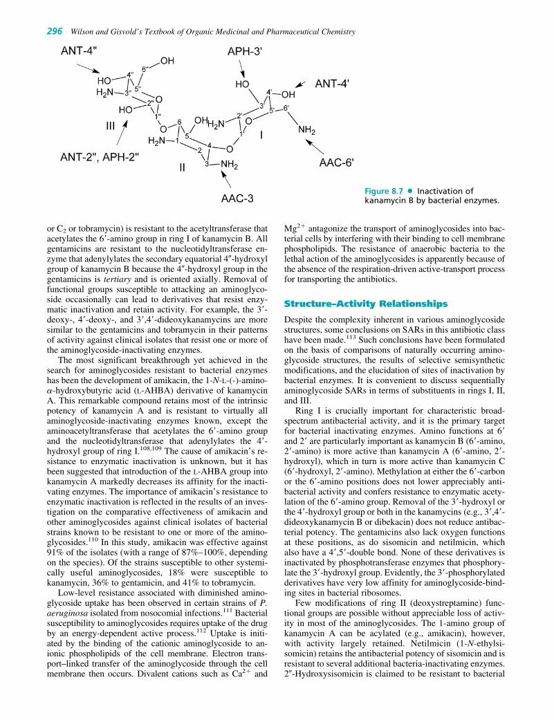
or C2 or tobramycin) is resistant to the acetyltransferase thatacetylates the 6�-amino group in ring I of kanamycin B. Allgentamicins are resistant to the nucleotidyltransferase en-zyme that adenylylates the secondary equatorial 4�-hydroxylgroup of kanamycin B because the 4�-hydroxyl group in thegentamicins is tertiary and is oriented axially. Removal offunctional groups susceptible to attacking an aminoglyco-side occasionally can lead to derivatives that resist enzy-matic inactivation and retain activity. For example, the 3�-deoxy-, 4�-deoxy-, and 3�,4�-dideoxykanamycins are moresimilar to the gentamicins and tobramycin in their patternsof activity against clinical isolates that resist one or more ofthe aminoglycoside-inactivating enzymes.
The most significant breakthrough yet achieved in thesearch for aminoglycosides resistant to bacterial enzymeshas been the development of amikacin, the 1-N-L-(-)-amino-�-hydroxybutyric acid (L-AHBA) derivative of kanamycinA. This remarkable compound retains most of the intrinsicpotency of kanamycin A and is resistant to virtually allaminoglycoside-inactivating enzymes known, except theaminoacetyltransferase that acetylates the 6�-amino groupand the nucleotidyltransferase that adenylylates the 4�-hydroxyl group of ring I.108,109 The cause of amikacin’s re-sistance to enzymatic inactivation is unknown, but it hasbeen suggested that introduction of the L-AHBA group intokanamycin A markedly decreases its affinity for the inacti-vating enzymes. The importance of amikacin’s resistance toenzymatic inactivation is reflected in the results of an inves-tigation on the comparative effectiveness of amikacin andother aminoglycosides against clinical isolates of bacterialstrains known to be resistant to one or more of the amino-glycosides.110 In this study, amikacin was effective against91% of the isolates (with a range of 87%–100%, dependingon the species). Of the strains susceptible to other systemi-cally useful aminoglycosides, 18% were susceptible tokanamycin, 36% to gentamicin, and 41% to tobramycin.
Low-level resistance associated with diminished amino-glycoside uptake has been observed in certain strains of P.aeruginosa isolated from nosocomial infections.111 Bacterialsusceptibility to aminoglycosides requires uptake of the drugby an energy-dependent active process.112 Uptake is initi-ated by the binding of the cationic aminoglycoside to an-ionic phospholipids of the cell membrane. Electron trans-port–linked transfer of the aminoglycoside through the cellmembrane then occurs. Divalent cations such as Ca2� and
Mg2� antagonize the transport of aminoglycosides into bac-terial cells by interfering with their binding to cell membranephospholipids. The resistance of anaerobic bacteria to thelethal action of the aminoglycosides is apparently because ofthe absence of the respiration-driven active-transport processfor transporting the antibiotics.
Structure–Activity Relationships
Despite the complexity inherent in various aminoglycosidestructures, some conclusions on SARs in this antibiotic classhave been made.113 Such conclusions have been formulatedon the basis of comparisons of naturally occurring amino-glycoside structures, the results of selective semisyntheticmodifications, and the elucidation of sites of inactivation bybacterial enzymes. It is convenient to discuss sequentiallyaminoglycoside SARs in terms of substituents in rings I, II,and III.
Ring I is crucially important for characteristic broad-spectrum antibacterial activity, and it is the primary targetfor bacterial inactivating enzymes. Amino functions at 6�and 2� are particularly important as kanamycin B (6�-amino,2�-amino) is more active than kanamycin A (6�-amino, 2�-hydroxyl), which in turn is more active than kanamycin C(6�-hydroxyl, 2�-amino). Methylation at either the 6�-carbonor the 6�-amino positions does not lower appreciably anti-bacterial activity and confers resistance to enzymatic acety-lation of the 6�-amino group. Removal of the 3�-hydroxyl orthe 4�-hydroxyl group or both in the kanamycins (e.g., 3�,4�-dideoxykanamycin B or dibekacin) does not reduce antibac-terial potency. The gentamicins also lack oxygen functionsat these positions, as do sisomicin and netilmicin, whichalso have a 4�,5�-double bond. None of these derivatives isinactivated by phosphotransferase enzymes that phosphory-late the 3�-hydroxyl group. Evidently, the 3�-phosphorylatedderivatives have very low affinity for aminoglycoside-bind-ing sites in bacterial ribosomes.
Few modifications of ring II (deoxystreptamine) func-tional groups are possible without appreciable loss of activ-ity in most of the aminoglycosides. The 1-amino group ofkanamycin A can be acylated (e.g., amikacin), however,with activity largely retained. Netilmicin (1-N-ethylsi-somicin) retains the antibacterial potency of sisomicin and isresistant to several additional bacteria-inactivating enzymes.2�-Hydroxysisomicin is claimed to be resistant to bacterial
296 Wilson and Gisvold’s Textbook of Organic Medicinal and Pharmaceutical Chemistry
Figure 8.7 Inactivation ofkanamycin B by bacterial enzymes.
0258-0329_17865_Ch08.qxd 12/2/09 3:27 AM Page 296

Chapter 8 Antibacterial Antibiotics 297
strains that adenylate the 2�-hydroxyl group of ring III,whereas 3-deaminosisomicin exhibits good activity againstbacterial strains that elaborate 3-acetylating enzymes.
Ring III functional groups appear to be somewhat lesssensitive to structural changes than those of either ring I orring II. Although the 2�-deoxygentamicins are signifi-cantly less active than their 2�-hydroxyl counterparts, the2�-amino derivatives (seldomycins) are highly active. The3�-amino group of gentamicins may be primary or second-ary with high antibacterial potency. Furthermore, the 4�-hydroxyl group may be axial or equatorial with littlechange in potency.
Despite improvements in antibacterial potency and spec-trum among newer naturally occurring and semisyntheticaminoglycoside antibiotics, efforts to find agents with im-proved margins of safety have been disappointing. The po-tential for toxicity of these important chemotherapeuticagents continues to restrict their use largely to the hospitalenvironment.
The discovery of agents with higher potency/toxicity ra-tios remains an important goal of aminoglycoside research.In a now somewhat dated review, however, Price114 ex-pressed doubt that many significant clinical breakthroughsin aminoglycoside research would occur in the future.
Products
Streptomycin Sulfate, SterileStreptomycin sulfate is a white, odorless powder that is hy-groscopic but stable toward light and air. It is freely solublein water, forming solutions that are slightly acidic or nearlyneutral. It is very slightly soluble in alcohol and is insolublein most other organic solvents. Acid hydrolysis yields strep-tidine and streptobiosamine, the compound that is a combi-nation of L-streptose and N-methyl-L-glucosamine.
Streptomycin acts as a triacidic base through the effectof its two strongly basic guanidino groups and the moreweakly basic methylamino group. Aqueous solutions maybe stored at room temperature for 1 week without any lossof potency, but they are most stable if the pH is between 4.5and 7.0. The solutions decompose if sterilized by heating,so sterile solutions are prepared by adding sterile distilledwater to the sterile powder. The early salts of streptomycincontained impurities that were difficult to remove andcaused a histamine-like reaction. By forming a complexwith calcium chloride, it was possible to free the strepto-mycin from these impurities and to obtain a product thatwas generally well tolerated.
The organism that produces streptomycin, S. griseus, alsoproduces several other antibiotic compounds: hydroxystrep-tomycin, mannisidostreptomycin, and cycloheximide (q.v.).
Of these, only cycloheximide has achieved importance as amedicinally useful substance. The term streptomycin A hasbeen used to refer to what is commonly called streptomycin,and mannisidostreptomycin has been called streptomycin B.Hydroxystreptomycin differs from streptomycin in having ahydroxyl group in place of one of the hydrogen atoms of thestreptose methyl group. Mannisidostreptomycin has a man-nose residue attached in glycosidic linkage through the hy-droxyl group at C-4 of the N-methyl-L-glucosamine moiety.The work of Dyer et al.115,116 to establish the stereochemicalstructure of streptomycin has been completed, and confirmedwith the total synthesis of streptomycin and dihydrostrepto-mycin by Japanese scientists.117
Clinically, a problem that sometimes occurs with the useof streptomycin is the early development of resistant strainsof bacteria, necessitating a change in therapy. Other factorsthat limit the therapeutic use of streptomycin are chronictoxicities. Neurotoxic reactions have been observed after theuse of streptomycin. These are characterized by vertigo, dis-turbance of equilibrium, and diminished auditory percep-tion. Additionally, nephrotoxicity occurs with some fre-quency. Patients undergoing therapy with streptomycinshould have frequent checks of renal monitoring parame-ters. Chronic toxicity reactions may or may not be re-versible. Minor toxic effects include rashes, mild malaise,muscular pains, and drug fever.
As a chemotherapeutic agent, streptomycin is activeagainst numerous Gram-negative and Gram-positive bacte-ria. One of the greatest virtues of streptomycin is its effec-tiveness against the tubercle bacillus, M. tuberculosis. By it-self, the antibiotic is not a cure, but it is a valuable adjunctto other treatment modalities for TB. The greatest drawbackto the use of streptomycin is the rather rapid development ofresistant strains of microorganisms. In infections that maybe because of bacteria sensitive to both streptomycin andpenicillin, the combined administration of the two antibi-otics has been advocated. The possible development of dam-age to the otic nerve by the continued use of streptomycin-containing preparations has discouraged the use of suchproducts. There has been an increasing tendency to reservestreptomycin products for the treatment of TB. It remainsone of the agents of choice, however, for the treatment ofcertain “occupational” bacterial infections, such as brucel-losis, tularemia, bubonic plague, and glanders. Becausestreptomycin is not absorbed when given orally or destroyedsignificantly in the GI tract, at one time it was used ratherwidely in the treatment of infections of the intestinal tract.For systemic action, streptomycin usually is given by intra-muscular injection.
Neomycin SulfateIn a search for antibiotics less toxic than streptomycin,Waksman and Lechevalier118 isolated neomycin (Mycifradin,Neobiotic) in 1949 from Streptomyces fradiae. Since then, theimportance of neomycin has increased steadily, and today, itis considered one of the most useful antibiotics for the treat-ment of GI infections, dermatological infections, and acutebacterial peritonitis. Also, it is used in abdominal surgery toreduce or avoid complications caused by infections from bac-terial flora of the bowel. It has broad-spectrum activityagainst various organisms and shows a low incidence of toxicand hypersensitivity reactions. It is absorbed very slightlyfrom the digestive tract, so its oral use ordinarily does not
0258-0329_17865_Ch08.qxd 12/2/09 3:27 AM Page 297

produce any systemic effect. The development of neomycin-resistant strains of pathogens is rarely reported in those organ-isms against which neomycin is effective.
Neomycin as the sulfate salt is a white to slightly yellow,crystalline powder that is very soluble in water. It is hygro-scopic and photosensitive (but stable over a wide pH rangeand to autoclaving). Neomycin sulfate contains the equiva-lent of 60% of the free base.
Neomycin, as produced by S. fradiae, is a mixture ofclosely related substances. Included in the “neomycin com-plex” is neamine (originally designated neomycin A) andneomycins B and C. S. fradiae also elaborates another an-tibiotic, the fradicin, which has some antifungal propertiesbut no antibacterial activity. This substance is not present in“pure” neomycin.
The structures of neamine and neomycins B and C areknown, and the absolute configurational structures ofneamine and neomycin were reported by Hichens andRinehart.119 Neamine may be obtained by methanolysis ofneomycins B and C, during which the glycosidic link betweendeoxystreptamine and D-ribose is broken. Therefore, neamineis a combination of deoxystreptamine and neosamine C,linked glycosidically (�) at the 4-position of deoxystrepta-
mine. According to Hichens and Rinehart, neomycin B dif-fers from neomycin C by the nature of the sugar attached ter-minally to D-ribose. That sugar, called neosamine B, differsfrom neosamine C in its stereochemistry. Rinehart et al.120
have suggested that in neosamine the configuration is 2,6-di-amino-2,6-dideoxy-L-idose, in which the orientation of the 6-aminomethyl group is inverted to the 6-amino-6-deoxy-D-glucosamine in neosamine C. In both instances, the glycosidiclinks were assumed to be �. Huettenrauch121 later suggested,however, that both of the diamino sugars in neomycin C havethe D-glucose configuration and that the glycosidic link is �in the one attached to D-ribose. The latter stereochemistry hasbeen confirmed by the total synthesis of neomycin C.122
Paromomycin SulfateThe isolation of paromomycin (Humatin) was reported in1956 from a fermentation with a Streptomyces sp. (PD04998), a strain said to resemble S. rimosus very closely.The parent organism had been obtained from soil samplescollected in Colombia. Paromomycin, however, moreclosely resembles neomycin and streptomycin in antibioticactivity than it does oxytetracycline, the antibiotic obtainedfrom S. rimosus.
298 Wilson and Gisvold’s Textbook of Organic Medicinal and Pharmaceutical Chemistry
0258-0329_17865_Ch08.qxd 12/2/09 3:27 AM Page 298

Chapter 8 Antibacterial Antibiotics 299
The general structure of paromomycin was reported byHaskell et al.123 as one compound. Subsequently, chromato-graphic determinations have shown paromomycin to consistof two fractions, paromomycin I and paromomycin II. Theabsolute configurational structures for the paromomycins,as shown in the structural formula, were suggested byHichens and Rinehart119 and confirmed by DeJongh et al.124
by mass spectrometric studies. The structure of paro-momycin is the same as that of neomycin B, except thatparomomycin contains D-glucosamine instead of the 6-amino-6-deoxy-D-glucosamine found in neomycin B. Thesame structural relationship is found between paromomycinII and neomycin C. The combination of D-glucosamine anddeoxystreptamine is obtained by partial hydrolysis of bothparomomycins and is called paromamine [4-(2-amino-2-deoxy-�-4-glucosyl)deoxystreptamine].
Paromomycin has broad-spectrum antibacterial activityand has been used for the treatment of GI infections causedby Salmonella and Shigella spp., and enteropathogenic E.coli. Currently, however, its use is restricted largely to thetreatment of intestinal amebiasis. Paromomycin is soluble inwater and stable to heat over a wide pH range.
Kanamycin SulfateKanamycin (Kantrex) was isolated in 1957 by Umezawaand coworkers125 from Streptomyces kanamyceticus. Its ac-tivity against mycobacteria and many intestinal bacteria, aswell as several pathogens that show resistance to other an-tibiotics, brought a great deal of attention to this antibiotic.As a result, kanamycin was tested and released for medicaluse in a very short time.
Research activity has been focused intensively on deter-mining the structures of the kanamycins. Chromatographyshowed that S. kanamyceticus elaborates three closely re-lated structures: kanamycins A, B, and C. Commerciallyavailable kanamycin is almost pure kanamycin A, the leasttoxic of the three forms. The kanamycins differ only in thesugar moieties attached to the glycosidic oxygen on the 4-position of the central deoxystreptamine. The absolute con-figuration of the deoxystreptamine in kanamycins reportedby Tatsuoka et al.126 is shown above. The chemical rela-tionships among the kanamycins, the neomycins, and theparomomycins were reported by Hichens and Rinehart.119
The kanamycins do not have the D-ribose molecule that ispresent in neomycins and paromomycins. Perhaps thisstructural difference is related to the lower toxicity ob-served with kanamycins. The kanosamine fragment linkedglycosidically to the 6-position of deoxystreptamine is 3-amino-3-deoxy-D-glucose (3-D-glucosamine) in all threekanamycins. The structures of the kanamycins have beenproved by total synthesis.127,128 They differ in the substi-tuted D-glucoses attached glycosidically to the 4-position of
the deoxystreptamine ring. Kanamycin A contains 6-amino-6-deoxy-D-glucose; kanamycin B contains 2,6-di-amino-2,6-dideoxy-D-glucose; and kanamycin C contains2-amino-2-deoxy-D-glucose (see preceding diagram).
Kanamycin is basic and forms salts of acids through itsamino groups. It is water soluble as the free base, but it isused in therapy as the sulfate salt, which is very soluble. Itis stable to both heat and chemicals. Solutions resist bothacids and alkali within the pH range of 2.0 to 11.0. Becauseof possible inactivation of either agent, kanamycin and peni-cillin salts should not be combined in the same solution.
The use of kanamycin in the United States usually is re-stricted to infections of the intestinal tract (e.g., bacillarydysentery) and to systemic infections arising from Gram-negative bacilli (e.g., Klebsiella, Proteus, Enterobacter, andSerratia spp.) that have developed resistance to other antibi-otics. It has also been recommended for preoperative anti-sepsis of the bowel. It is absorbed poorly from the intestinaltract; consequently, systemic infections must be treated byintramuscular or (for serious infections) intravenous injec-tions. These injections are rather painful, and the concomi-tant use of a local anesthetic is indicated. The use ofkanamycin in the treatment of TB has not been widely ad-vocated since the discovery that mycobacteria develop re-sistance very rapidly. In fact, both clinical experience andexperimental work129 indicate that kanamycin developscross-resistance in the tubercle bacilli with dihydrostrepto-mycin, viomycin, and other antitubercular drugs. Like strep-tomycin, kanamycin may cause decreased or complete lossof hearing. On development of such symptoms, its useshould be stopped immediately.
AmikacinAmikacin, 1-N-amino-�-hydroxybutyrylkanamycin A(Amikin), is a semisynthetic aminoglycoside first preparedin Japan. The synthesis formally involves simple acylationof the 1-amino group of the deoxystreptamine ring ofkanamycin A with L-AHBA. This particular acyl derivativeretains about 50% of the original activity of kanamycin Aagainst sensitive strains of Gram-negative bacilli. The L-AHBA derivative is much more active than the D-isomer.130
The remarkable feature of amikacin is that it resists attackby most bacteria-inactivating enzymes and, therefore, is ef-fective against strains of bacteria that are resistant to otheraminoglycosides,110 including gentamicin and tobramycin.In fact, it is resistant to all known aminoglycoside-inactivat-ing enzymes, except the aminotransferase that acetylates the6�amino group109 and the 4�-nucleotidyl transferase thatadenylylates the 4�-hydroxyl group of aminoglycosides.108
Preliminary studies indicate that amikacin may be lessototoxic than either kanamycin or gentamicin.131 Higherdosages of amikacin are generally required, however, for the
0258-0329_17865_Ch08.qxd 12/2/09 3:27 AM Page 299

treatment of most Gram-negative bacillary infections. Forthis reason, and to discourage the proliferation of bacterialstrains resistant to it, amikacin currently is recommended forthe treatment of serious infections caused by bacterialstrains resistant to other aminoglycosides.
Gentamicin SulfateGentamicin (Garamycin) was isolated in 1958 and reported in1963 by Weinstein et al.132 to belong to the streptomycinoid(aminocyclitol) group of antibiotics. It is obtained commer-cially from Micromonospora purpurea. Like the other mem-bers of its group, it has a broad spectrum of activity againstmany common pathogens, both Gram-positive and Gram-negative. Of particular interest is its strong activity againstP. aeruginosa and other Gram-negative enteric bacilli.
Gentamicin is effective in the treatment of various skin in-fections for which a topical cream or ointment may be used.Because it offers no real advantage over topical neomycin inthe treatment of all but pseudomonal infections, however, itis recommended that topical gentamicin be reserved for usein such infections and in the treatment of burns complicatedby pseudomonemia. An injectable solution containing 40 mgof gentamicin sulfate per milliliter may be used for serioussystemic and genitourinary tract infections caused by Gram-negative bacteria, particularly Pseudomonas, Enterobacter,and Serratia spp. Because of the development of strains ofthese bacterial species resistant to previously effective broad-spectrum antibiotics, gentamicin has been used for the treat-ment of hospital-acquired infections caused by such organ-isms. Resistant bacterial strains that inactivate gentamicin byadenylylation and acetylation, however, appear to be emerg-ing with increasing frequency.
Gentamicin sulfate is a mixture of the salts of compoundsidentified as gentamicins C1, C2, and Cla. These gentamicinswere reported by Cooper et al.133 to have the structuresshown in the diagram. The absolute stereochemistries of thesugar components and the geometries of the glycosidic link-ages have also been established.134
Coproduced, but not a part of the commercial product,are gentamicins A and B. Their structures were reported byMaehr and Schaffner135 and are closely related to those ofthe gentamicins C. Although gentamicin molecules are sim-ilar in many ways to other aminocyclitols such as strepto-mycins, they are sufficiently different that their medical ef-fectiveness is significantly greater. Gentamicin sulfate is awhite to buff substance that is soluble in water and insolu-ble in alcohol, acetone, and benzene. Its solutions are stableover a wide pH range and may be autoclaved. It is chemi-cally incompatible with carbenicillin, and the two should notbe combined in the same intravenous solution.
Tobramycin SulfateIntroduced in 1976, tobramycin sulfate (Nebcin) is the mostactive of the chemically related aminoglycosides called ne-bramycins obtained from a strain of Streptomyces tenebrar-
ius. Five members of the nebramycin complex have beenidentified chemically.136
Factors 4 and 4� are 6�-O-carbamoylkanamycin B andkanamycin B, respectively; factors 5� and 6 are 6�-O-car-bamoyltobramycin and tobramycin; and factor 2 isapramycin, a tetracyclic aminoglycoside with an unusual bi-cyclic central ring structure. Kanamycin B and tobramycinprobably do not occur in fermentation broths per se but areformed by hydrolysis of the 6-O�-carbamoyl derivatives inthe isolation procedure.
The most important property of tobramycin is its activityagainst most strains of P. aeruginosa, exceeding that of gen-tamicin by twofold to fourfold. Some gentamicin-resistantstrains of this troublesome organism are sensitive to to-bramycin, but others are resistant to both antibiotics.137 OtherGram-negative bacilli and staphylococci are generally moresensitive to gentamicin. Tobramycin more closely resembleskanamycin B in structure (it is 3�-deoxykanamycin B).
Netilmicin SulfateNetilmicin sulfate, 1-N-ethylsisomicin (Netromycin), is asemisynthetic derivative prepared by reductive ethylation138
of sisomicin, an aminoglycoside antibiotic obtained fromMicromonospora inyoensis.139 Structurally, sisomicin andnetilmicin resemble gentamicin Cla, a component of the gen-tamicin complex.
Against most strains of Enterobacteriaceae, P. aerugi-nosa, and S. aureus, sisomicin and netilmicin are compara-ble to gentamicin in potency.140 Netilmicin is active, how-ever, against many gentamicin-resistant strains, in particularamong E. coli, Enterobacter, Klebsiella, and Citrobacterspp. A few strains of gentamicin-resistant P. aeruginosa, S.marcescens, and indole-positive Proteus spp. are also sensi-tive to netilmicin. Very few gentamicin-resistant bacterialstrains are sensitive to sisomicin, however. The potency ofnetilmicin against certain gentamicin-resistant bacteria is at-tributed to its resistance to inactivation by bacterial enzymesthat adenylylate or phosphorylate gentamicin and sisomicin.Evidently, the introduction of a 1-ethyl group in sisomicinmarkedly decreases the affinity of these enzymes forthe molecule in a manner similar to that observed in the 1-N-�-amino-�-hydroxybutyryl amide of kanamycin A(amikacin). Netilmicin, however, is inactivated by most of
300 Wilson and Gisvold’s Textbook of Organic Medicinal and Pharmaceutical Chemistry
0258-0329_17865_Ch08.qxd 12/2/09 3:27 AM Page 300
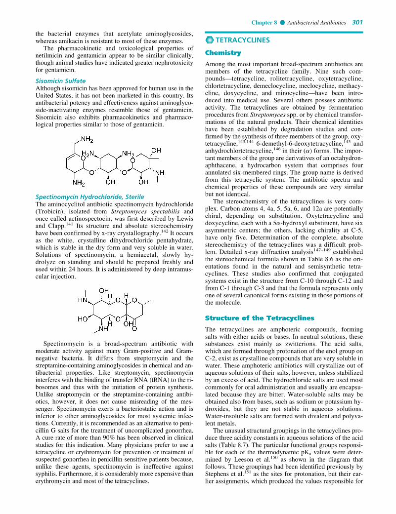
Chapter 8 Antibacterial Antibiotics 301
the bacterial enzymes that acetylate aminoglycosides,whereas amikacin is resistant to most of these enzymes.
The pharmacokinetic and toxicological properties ofnetilmicin and gentamicin appear to be similar clinically,though animal studies have indicated greater nephrotoxicityfor gentamicin.
Sisomicin SulfateAlthough sisomicin has been approved for human use in theUnited States, it has not been marketed in this country. Itsantibacterial potency and effectiveness against aminoglyco-side-inactivating enzymes resemble those of gentamicin.Sisomicin also exhibits pharmacokinetics and pharmaco-logical properties similar to those of gentamicin.
Spectinomycin Hydrochloride, SterileThe aminocyclitol antibiotic spectinomycin hydrochloride(Trobicin), isolated from Streptomyces spectabilis andonce called actinospectocin, was first described by Lewisand Clapp.141 Its structure and absolute stereochemistryhave been confirmed by x-ray crystallography.142 It occursas the white, crystalline dihydrochloride pentahydrate,which is stable in the dry form and very soluble in water.Solutions of spectinomycin, a hemiacetal, slowly hy-drolyze on standing and should be prepared freshly andused within 24 hours. It is administered by deep intramus-cular injection.
Spectinomycin is a broad-spectrum antibiotic withmoderate activity against many Gram-positive and Gram-negative bacteria. It differs from streptomycin and thestreptamine-containing aminoglycosides in chemical and an-tibacterial properties. Like streptomycin, spectinomycininterferes with the binding of transfer RNA (tRNA) to the ri-bosomes and thus with the initiation of protein synthesis.Unlike streptomycin or the streptamine-containing antibi-otics, however, it does not cause misreading of the mes-senger. Spectinomycin exerts a bacteriostatic action and isinferior to other aminoglycosides for most systemic infec-tions. Currently, it is recommended as an alternative to peni-cillin G salts for the treatment of uncomplicated gonorrhea.A cure rate of more than 90% has been observed in clinicalstudies for this indication. Many physicians prefer to use atetracycline or erythromycin for prevention or treatment ofsuspected gonorrhea in penicillin-sensitive patients because,unlike these agents, spectinomycin is ineffective againstsyphilis. Furthermore, it is considerably more expensive thanerythromycin and most of the tetracyclines.
TETRACYCLINES
Chemistry
Among the most important broad-spectrum antibiotics aremembers of the tetracycline family. Nine such com-pounds—tetracycline, rolitetracycline, oxytetracycline,chlortetracycline, demeclocycline, meclocycline, methacy-cline, doxycycline, and minocycline—have been intro-duced into medical use. Several others possess antibioticactivity. The tetracyclines are obtained by fermentationprocedures from Streptomyces spp. or by chemical transfor-mations of the natural products. Their chemical identitieshave been established by degradation studies and con-firmed by the synthesis of three members of the group, oxy-tetracycline,143,144 6-demethyl-6-deoxytetracycline,145 andanhydrochlortetracycline,146 in their (�) forms. The impor-tant members of the group are derivatives of an octahydron-aphthacene, a hydrocarbon system that comprises fourannulated six-membered rings. The group name is derivedfrom this tetracyclic system. The antibiotic spectra andchemical properties of these compounds are very similarbut not identical.
The stereochemistry of the tetracyclines is very com-plex. Carbon atoms 4, 4a, 5, 5a, 6, and 12a are potentiallychiral, depending on substitution. Oxytetracycline anddoxycycline, each with a 5�-hydroxyl substituent, have sixasymmetric centers; the others, lacking chirality at C-5,have only five. Determination of the complete, absolutestereochemistry of the tetracyclines was a difficult prob-lem. Detailed x-ray diffraction analysis147–149 establishedthe stereochemical formula shown in Table 8.6 as the ori-entations found in the natural and semisynthetic tetra-cyclines. These studies also confirmed that conjugatedsystems exist in the structure from C-10 through C-12 andfrom C-1 through C-3 and that the formula represents onlyone of several canonical forms existing in those portions ofthe molecule.
Structure of the Tetracyclines
The tetracyclines are amphoteric compounds, formingsalts with either acids or bases. In neutral solutions, thesesubstances exist mainly as zwitterions. The acid salts,which are formed through protonation of the enol group onC-2, exist as crystalline compounds that are very soluble inwater. These amphoteric antibiotics will crystallize out ofaqueous solutions of their salts, however, unless stabilizedby an excess of acid. The hydrochloride salts are used mostcommonly for oral administration and usually are encapsu-lated because they are bitter. Water-soluble salts may beobtained also from bases, such as sodium or potassium hy-droxides, but they are not stable in aqueous solutions.Water-insoluble salts are formed with divalent and polyva-lent metals.
The unusual structural groupings in the tetracyclines pro-duce three acidity constants in aqueous solutions of the acidsalts (Table 8.7). The particular functional groups responsi-ble for each of the thermodynamic pKa values were deter-mined by Leeson et al.150 as shown in the diagram thatfollows. These groupings had been identified previously byStephens et al.151 as the sites for protonation, but their ear-lier assignments, which produced the values responsible for
0258-0329_17865_Ch08.qxd 12/2/09 3:27 AM Page 301

pKa2 and pKa3, were opposite those of Leeson et al.150 Thislatter assignment has been substantiated by Rigler et al.152
The approximate pKa values for each of these groups inthe six tetracycline salts in common use are shown (Table8.7). The values are taken from Stephens et al.,151 Benet andGoyan,153 and Barringer et al.154 The pKa of the 7-dimethyl-amino group of minocycline (not listed) is 5.0.
An interesting property of the tetracyclines is their abil-ity to undergo epimerization at C-4 in solutions of interme-diate pH range. These isomers are called epitetracyclines.
Under acidic conditions, an equilibrium is established inabout 1 day and consists of approximately equal amounts ofthe isomers. The partial structures below indicate the twoforms of the epimeric pair. The 4-epitetracyclines have beenisolated and characterized. They exhibit much less activitythan the “natural” isomers, thus accounting for the de-creased therapeutic value of aged solutions.
Strong acids and strong bases attack tetracyclines with ahydroxyl group on C-6, causing a loss in activity throughmodification of the C ring. Strong acids produce dehydra-tion through a reaction involving the 6-hydroxyl group andthe 5a-hydrogen. The double bond thus formed between po-sitions 5a and 6 induces a shift in the position of the doublebond between C-11a and C-12 to a position between C-11and C-11a, forming the more energetically favored resonantsystem of the naphthalene group found in the inactive anhy-drotetracyclines. Bases promote a reaction between the 6-hydroxyl group and the ketone group at the 11-position,causing the bond between the 11 and 11a atoms to cleave,forming the lactone ring found in the inactive isotetracy-cline. These two unfavorable reactions stimulated researchthat led to the development of the more stable and longer-acting compounds 6-deoxytetracycline, methacycline,doxycycline, and minocycline.
302 Wilson and Gisvold’s Textbook of Organic Medicinal and Pharmaceutical Chemistry
TABLE 8.6 Structures of Tetracyclines
TABLE 8.7 pKa Values (of Hydrochlorides) in AqueousSolution at 25°C
pKa1 pKa2 pKa3
Tetracycline 3.3 7.7 9.5Chlortetracycline 3.3 7.4 9.3Demeclocycline 3.3 7.2 9.3Oxytetracycline 3.3 7.3 9.1Doxycycline 3.4 7.7 9.7Minocycline 2.8 7.8 9.3
7
R1 R2 R3
NH2
CH3
8
910
65a
H
11a11
OH OHOH
OH
O O
54a
12a12
R4
43
21
O
H
NH3C
R1 R2 R3 R4
Tetracycline H OH CH3 HChlortetracycline Cl OH CH3 HOxytetracycline H OH CH3 OHDemeclocycline Cl OH H HMethacycline H CH2 OHDoxycycline H CH3 H OHMinocycline N(CH3)2 H H H
0258-0329_17865_Ch08.qxd 12/2/09 3:27 AM Page 302

Chapter 8 Antibacterial Antibiotics 303
Stable chelate complexes are formed by the tetracyclineswith many metals, including calcium, magnesium, and iron.Such chelates are usually very insoluble in water, account-ing for the impaired absorption of most (if not all) tetracy-clines in the presence of milk; calcium-, magnesium-, andaluminum-containing antacids; and iron salts. Soluble alka-linizers, such as sodium bicarbonate, also decrease the GIabsorption of the tetracyclines.155 Deprotonation of tetracy-clines to more ionic species and the observed instability ofthese products in alkaline solutions may account for this ob-servation. The affinity of tetracyclines for calcium causesthem to be incorporated into newly forming bones andteeth as tetracycline–calcium orthophosphate complexes.Deposits of these antibiotics in teeth cause a yellow discol-oration that darkens (a photochemical reaction) over time.Tetracyclines are distributed into the milk of lactating moth-ers and will cross the placental barrier into the fetus. Thepossible effects of these agents on the bones and teeth of thechild should be considered before their use during preg-nancy or in children younger than 8 years of age.
Mechanism of Action and Resistance
The strong binding properties of the tetracyclines with metalions caused Albert156 to suggest that their antibacterial prop-erties may be because of an ability to remove essential metalions as chelated compounds. Elucidation of details of themechanism of action of the tetracyclines,157 however, hasdefined more clearly the specific roles of magnesium ions inmolecular processes affected by these antibiotics in bacteria.Tetracyclines are specific inhibitors of bacterial protein syn-thesis. They bind to the 30S ribosomal subunit and, thereby,prevent the binding of aminoacyl tRNA to the mRNA–ribo-some complex. Both the binding of aminoacyl tRNA and thebinding of tetracyclines at the ribosomal binding site requiremagnesium ions.158 Tetracyclines also bind to mammalianribosomes but with lower affinities, and they apparently donot achieve sufficient intracellular concentrations to inter-fere with protein synthesis. The selective toxicity of thetetracyclines toward bacteria depends strongly on the self-destructive capacity of bacterial cells to concentrate these
agents in the cell. Tetracyclines enter bacterial cells by twoprocesses: passive diffusion and active transport. The activeuptake of tetracyclines by bacterial cells is an energy-dependent process that requires adenosine triphosphate(ATP) and magnesium ions.159
Three biochemically distinct mechanisms of resistance totetracyclines have been described in bacteria160: (a) effluxmediated by transmembrane-spanning, active-transport pro-teins that reduces the intracellular tetracycline concentra-tion; (b) ribosomal protection, in which the bacterial proteinsynthesis apparatus is rendered resistant to the action oftetracyclines by an inducible cytoplasmic protein; and (c)enzymatic oxidation. Efflux mediated by plasmid or chro-mosomal protein determinants tet-A, -E, -G, -H, -K, and -L,and ribosomal protection mediated by the chromosomal pro-tein determinants tet-M, -O, and -S are the most frequentlyencountered and most clinically significant resistance mech-anisms for tetracyclines.
Spectrum of Activity
The tetracyclines have the broadest spectrum of activity ofany known antibacterial agents. They are active against awide range of Gram-positive and Gram-negative bacteria,spirochetes, mycoplasma, rickettsiae, and chlamydiae. Theirpotential indications are, therefore, numerous. Their bacterio-static action, however, is a disadvantage in the treatment oflife-threatening infections such as septicemia, endocarditis,and meningitis; the aminoglycosides and/or cephalosporinsusually are preferred for Gram-negative and the penicillinsfor Gram-positive infections. Because of incomplete absorp-tion and their effectiveness against the natural bacterial floraof the intestine, tetracyclines may induce superinfectionscaused by the pathogenic yeast Candida albicans. Resistanceto tetracyclines among both Gram-positive and Gram-nega-tive bacteria is relatively common. Superinfections caused byresistant S. aureus and P. aeruginosa have resulted from theuse of these agents over time. Parenteral tetracyclines maycause severe liver damage, especially when given in exces-sive dosage to pregnant women or to patients with impairedrenal function.
0258-0329_17865_Ch08.qxd 12/2/09 3:27 AM Page 303

Structure–Activity Relationships
The large amount of research carried out to prepare semi-synthetic modifications of the tetracyclines and to obtain in-dividual compounds by total synthesis revealed several in-teresting SARs. Reviews are available that discuss SARsamong the tetracyclines in detail,161–163 their molecular andclinical properties,164 and their synthesis and chemical prop-erties.162,163,165,166 Only a brief review of the salient struc-ture–activity features is presented here. All derivatives con-taining fewer than four rings are inactive or nearly inactive.The simplest tetracycline derivative that retains the charac-teristic broad-spectrum activity associated with this antibi-otic class is 6-demethyl-6-deoxytetracycline. Many of theprecise structural features present in this molecule must re-main unmodified for derivatives to retain activity. The in-tegrity of substituents at carbon atoms 1, 2, 3, 4, 10, 11, 11a,and 12, representing the hydrophilic “southern and eastern”faces of the molecule, cannot be violated drastically withoutdeleterious effects on the antimicrobial properties of the re-sulting derivatives.
A-ring substituents can be modified only slightly withoutdramatic loss of antibacterial potency. The enolized tricar-bonylmethane system at C-1 to C-3 must be intact for goodactivity. Replacement of the amide at C-2 with other func-tions (e.g., aldehyde or nitrile) reduces or abolishes activity.Monoalkylation of the amide nitrogen reduces activity pro-portionately to the size of the alkyl group. Aminoalkylationof the amide nitrogen, accomplished by the Mannich reac-tion, yields derivatives that are substantially more water sol-uble than the parent tetracycline and are hydrolyzed to it invivo (e.g., rolitetracycline). The dimethylamino group at the4-position must have the � orientation: 4-epitetracyclinesare very much less active than the natural isomers. Removalof the 4-dimethylamino group reduces activity even further.Activity is largely retained in the primary and N-methyl sec-ondary amines but rapidly diminishes in the higher alkyl-amines. A cis-A/B-ring fusion with a �-hydroxyl group atC-12a is apparently also essential. Esters of the C-12a hy-droxyl group are inactive, with the exception of the formylester, which readily hydrolyzes in aqueous solutions.Alkylation at C-11a also leads to inactive compounds,demonstrating the importance of an enolizable �-diketonefunctionality at C-11 and C-12. The importance of the shapeof the tetracyclic ring system is illustrated further by sub-stantial loss in antibacterial potency resulting from epimer-ization at C-5a. Dehydrogenation to form a double bond be-tween C-5a and C-11a markedly decreases activity, as doesaromatization of ring C to form anhydrotetracyclines.
In contrast, substituents at positions 5, 5a, 6, 7, 8, and 9,representing the largely hydrophobic “northern and west-ern” faces of the molecule, can be modified with varying de-grees of success, resulting in retention and, sometimes, im-provement of antibiotic activity. A 5-hydroxyl group, as inoxytetracycline and doxycycline, may influence pharmaco-kinetic properties but does not change antimicrobial activ-ity. 5a-Epitetracyclines (prepared by total synthesis), al-though highly active in vitro, are unfortunately much lessimpressive in vivo. Acid-stable 6-deoxytetracyclines and 6-demethyl-6-deoxytetracyclines have been used to preparevarious monosubstituted and disubstituted derivatives byelectrophilic substitution reactions at C-7 and C-9 of the D ring. The more useful results have been achieved with the
introduction of substituents at C-7. Oddly, strongly electron-withdrawing groups (e.g., chloro [lortetracycline] and nitro)and strongly electron-donating groups (e.g., dimethylamino[minocycline]) enhance activity. This unusual circumstanceis reflected in QSAR studies of 7- and 9-substituted tetracy-clines,162,167 which indicated a squared (parabolic) depen-dence on �, Hammet’s electronic substituent constant, andin vitro inhibition of an E. coli strain. The effect of introduc-ing substituents at C-8 has not been studied because this po-sition cannot be substituted directly by classic electrophilicaromatic substitution reactions; thus, 8-substituted deriva-tives are available only through total synthesis.168
The most fruitful site for semisynthetic modification of thetetracyclines has been the 6-position. Neither the 6�-methylnor the 6�-hydroxyl group is essential for antibacterial activ-ity. In fact, doxycycline and methacycline are more active invitro than their parent oxytetracycline against most bacterialstrains. The conversion of oxytetracycline to doxycycline,which can be accomplished by reduction of methacycline,169
gives a 1:1 mixture of doxycycline and epidoxycycline(which has a �-oriented methyl group); if the C-11a �-fluoroderivative of methacycline is used, the �-methyl epimer isformed exclusively.170 6-Epidoxycycline is much less activethan doxycycline. 6-Demethyl-6-deoxytetracycline, synthe-sized commercially by catalytic hydrogenolysis of the 7-chloro and 6-hydroxyl groups of 7-chloro-6-demethyltetracy-cline, obtained by fermentation of a mutant strain ofStreptomyces aureofaciens,171 is slightly more potent thantetracycline. More successful from a clinical standpoint, how-ever, is 6-demethyl-6-deoxy-7-dimethylaminotetracycline(minocycline)172 because of its activity against tetracycline-resistant bacterial strains.
6-Deoxytetracyclines also possess important chemicaland pharmacokinetic advantages over their 6-oxy counter-parts. Unlike the latter, they are incapable of forming anhy-drotetracyclines under acidic conditions because they cannotdehydrate at C-5a and C-6. They are also more stable in basebecause they do not readily undergo �-ketone cleavage, fol-lowed by lactonization, to form isotetracyclines. Although itlacks a 6-hydroxyl group, methacycline shares the instabilityof the 6-oxytetracyclines in strongly acetic conditions. It suf-fers prototropic rearrangement to the anhydrotetracycline inacid but is stable to �-ketone cleavage followed by lac-tonization to the isotetracycline in base. Reduction of the 6-hydroxyl group also dramatically changes the solubilityproperties of tetracyclines. This effect is reflected in signifi-cantly higher oil/water partition coefficients of the 6-de-oxytetracyclines than of the tetracyclines (Table 8.8).173,174
The greater lipid solubility of the 6-deoxy compounds hasimportant pharmacokinetic consequences.162,164 Hence,doxycycline and minocycline are absorbed more completelyfollowing oral administration, exhibit higher fractions ofplasma protein binding, and have higher volumes of distri-bution and lower renal clearance rates than the correspon-ding 6-oxytetracyclines.
Polar substituents (i.e., hydroxyl groups) at C-5 and C-6decrease lipid versus water solubility of the tetracyclines.The 6-position is, however, considerably more sensitivethan the 5-position to this effect. Thus, doxycycline (6-deoxy-5-oxytetracycline) has a much higher partition coef-ficient than either tetracycline or oxytetracycline. Nonpolarsubstituents (those with positive � values; see Chapter 2),
304 Wilson and Gisvold’s Textbook of Organic Medicinal and Pharmaceutical Chemistry
0258-0329_17865_Ch08.qxd 12/2/09 3:27 AM Page 304

Chapter 8 Antibacterial Antibiotics 305
for example, 7-dimethylamino, 7-chloro, and 6-methyl,have the opposite effect. Accordingly, the partition coeffi-cient of chlortetracycline is substantially greater than that oftetracycline and slightly greater than that of demeclocycline.Interestingly, minocycline (5-demethyl-6-deoxy-7-dimethy-laminotetracycline) has the highest partition coefficient ofthe commonly used tetracyclines.
The poorer oral absorption of the more water-solublecompounds tetracycline and oxytetracycline can be attributedto several factors. In addition to their comparative difficultyin penetrating lipid membranes, the polar tetracyclines prob-ably experience more complexation with metal ions in the gutand undergo some acid-catalyzed destruction in the stomach.Poorer oral absorption coupled with biliary excretion of sometetracyclines is also thought to cause a higher incidence of su-perinfections from resistant microbial strains. The more polartetracyclines, however, are excreted in higher concentrationsin the urine (e.g., 60% for tetracycline and 70% for oxytetra-cycline) than the more lipid-soluble compounds (e.g., 33%for doxycycline and only 11% for minocycline). Significantpassive renal tubular reabsorption coupled with higher frac-tions of protein binding contributes to the lower renal clear-ance and longer durations of action of doxycycline andminocycline compared with those of the other tetracyclines,especially tetracycline and oxytetracycline. Minocycline alsoexperiences significant N-dealkylation catalyzed by cy-tochrome P450 oxygenases in the liver, which contributes toits comparatively low renal clearance. Although all tetracy-clines are distributed widely into tissues, the more polar oneshave larger volumes of distribution than the nonpolar com-pounds. The more lipid-soluble tetracyclines, however, dis-tribute better to poorly vascularized tissue. It is also claimedthat the distribution of doxycycline and minocycline intobone is less than that of other tetracyclines.175
Products
TetracyclineChemical studies on chlortetracycline revealed that con-trolled catalytic hydrogenolysis selectively removed the 7-chloro atom and so produced tetracycline (Achromycin,Cyclopar, Panmycin, Tetracyn). This process was patentedby Conover176 in 1955. Later, tetracycline was obtainedfrom fermentations of Streptomyces spp., but the commer-cial supply still chiefly depends on hydrogenolysis of chlor-tetracycline.
Tetracycline is 4-dimethyl amino-1,4,4a,5,5a,6,11,12a-octahydro-3,6,10,12,12a-pentahydroxy-6-methyl-1,11-
dioxo-2-naphthacenecarboxamide. It is a bright yellow, crys-talline salt that is stable in air but darkens on exposure tostrong sunlight. Tetracycline is stable in acid solutions witha pH above 2. It is somewhat more stable in alkaline solu-tions than chlortetracycline, but like those of the other tetra-cyclines, such solutions rapidly lose potency. One gram ofthe base requires 2,500 mL of water and 50 mL of alcohol todissolve it. The hydrochloride salt is used most commonly inmedicine, though the free base is absorbed from the GI tractabout equally well. One gram of the hydrochloride salt dis-solves in about 10 mL of water and in 100 mL of alcohol.Tetracycline has become the most popular antibiotic of itsgroup, largely because its plasma concentration appears to behigher and more enduring than that of either oxytetracyclineor chlortetracycline. Also, it is found in higher concentrationin the spinal fluid than the other two compounds.
Several combinations of tetracycline with agents that in-crease the rate and the height of plasma concentrations areon the market. One such adjuvant is magnesium chloridehexahydrate (Panmycin). Also, an insoluble tetracyclinephosphate complex (Tetrex) is made by mixing a solution oftetracycline, usually as the hydrochloride, with a solution ofsodium metaphosphate. There are various claims concern-ing the efficacy of these adjuvants. The mechanisms of theiractions are not clear, but reportedly177,178 these agents en-hance plasma concentrations over those obtained whentetracycline hydrochloride alone is administered orally.Remmers et al.179,180 reported on the effects that selectedaluminum–calcium gluconates complexed with some tetra-cyclines have on plasma concentrations when administeredorally, intramuscularly, or intravenously. Such complexesenhanced plasma levels in dogs when injected but not whengiven orally. They also observed enhanced plasma levels inexperimental animals when complexes of tetracyclines withaluminum metaphosphate, aluminum pyrophosphate, or alu-minum–calcium phosphinicodilactates were administeredorally. As noted previously, the tetracyclines can form sta-ble chelate complexes with metal ions such as calcium andmagnesium, which retard absorption from the GI tract. Thecomplexity of the systems involved has not permitted
TABLE 8.8 Pharmacokinetic Propertiesa of Tetracyclines
Volume of Renal Kpc Octanol/ Absorbed Excreted Excreted Protein Distribution Clearance Half-Water Orally in Feces in Urine Bound (% body (mL/min/ life
Tetracycline pH 5.6b (%) (%) (%) (%) weight) 1.73 m2) (h)
Tetracycline 0.056 58 20–50 60 24–65 156–306 50–80 10Oxytetracycline 0.075 77–80 50 70 20–35 180–305 99–102 9Chlortetracycline 0.41 25–30 50 18 42–54 149 32 7Demeclocycline 0.25 66 23–72 42 68–77 179 35 15Doxycycline 0.95 93 20–40 27–39 60–91 63 18–28 15Minocycline 1.10 100 40 5–11 55–76 74 5–15 19
aValues taken from Brown, J. R., and Ireland, D. S.: Adv. Pharmacol. Chemother. 15:161, 1978.bValues taken from Colazzi, J. L., and Klink, P. R.: J. Pharm. Sci. 58:158, 1969.
0258-0329_17865_Ch08.qxd 12/2/09 3:27 AM Page 305

unequivocal substantiation of the idea that these adjuvantscompete with the tetracyclines for substances in the alimen-tary tract that would otherwise be free to complex with theseantibiotics and thereby retard their absorption. Certainly,there is no evidence that the metal ions per se act as buffers,an idea alluded to sometimes in the literature.
Tetracycline hydrochloride is also available in ointmentsfor topical and ophthalmic administration. A topical solu-tion is used for the management of acne vulgaris.
RolitetracyclineRolitetracycline, N-(pyrrolidinomethyl)tetracycline(Syntetrin), was introduced for use by intramuscular or in-travenous injection. This derivative is made by condensingtetracycline with pyrrolidine and formaldehyde in the pres-ence of tert-butyl alcohol. It is very soluble in water (1 g dis-solves in about 1 mL) and provides a means of injecting theantibiotic in a small volume of solution. It has been recom-mended for cases when the oral dosage forms are not suit-able, but it is no longer widely used.
Chlortetracycline HydrochlorideChlortetracycline (Aureomycin hydrochloride) was isolatedby Duggar181 in 1948 from S. aureofaciens. This compound,which was produced in an extensive search for new antibi-otics, was the first of the group of highly successful tetracy-clines. It soon became established as a valuable antibioticwith broad-spectrum activities.
It is used in medicine chiefly as the acid salt of the com-pound whose systematic chemical designation is 7-chloro-4-(dimethylamino)-1,4,4a,5,5a,6,11,12a-octahydro-3,6,10,12,12a-pentahydroxy-6-methyl-1,11-dioxo-2-naph-thacenecarboxamide. The hydrochloride salt is a crystallinepowder with a bright yellow color, which suggested itsbrand name, Aureomycin. It is stable in air but slightly pho-tosensitive and should be protected from light. It is odorlessand bitter. One gram of the hydrochloride salt will dissolvein about 75 mL of water, producing a pH of about 3. It isonly slightly soluble in alcohol and practically insoluble inother organic solvents.
Oral and parenteral forms of chlortetracycline are nolonger used because of the poor bioavailability and inferiorpharmacokinetic properties of the drug. It is still marketed inointment forms for topical and ophthalmic use.
Oxytetracycline HydrochlorideEarly in 1950, Finlay et al.182 reported the isolation of oxy-tetracycline (Terramycin) from S. rimosus. This compoundwas soon identified as a chemical analog of chlortetracy-
cline that showed similar antibiotic properties. The structureof oxytetracycline was elucidated by Hochstein et al.183 andthis work provided the basis for the confirmation of thestructure of the other tetracyclines.
Oxytetracycline hydrochloride is a pale yellow, bitter,crystalline compound. The amphoteric base is only slightlysoluble in water and slightly soluble in alcohol. It is odorlessand stable in air but darkens on exposure to strong sunlight.The hydrochloride salt is a stable yellow powder that ismore bitter than the free base. It is much more soluble inwater, 1 g dissolving in 2 mL, and more soluble in alcoholthan the free base. Both compounds are inactivated rapidlyby alkali hydroxides and by acid solutions below pH 2. Bothforms of oxytetracycline are absorbed rapidly and equallywell from the digestive tract, so the only real advantage thefree base offers over the hydrochloride salt is that it is lessbitter. Oxytetracycline hydrochloride is also used for par-enteral administration (intravenously and intramuscularly).
Methacycline HydrochlorideThe synthesis of methacycline, 6-deoxy-6-demethyl-6-meth-ylene-5-oxytetracycline hydrochloride (Rondomycin), re-ported by Blackwood et al.184 in 1961, was accomplished bychemical modification of oxytetracycline. It has an antibioticspectrum like that of the other tetracyclines but greater po-tency; about 600 mg of methacycline is equivalent to 1 g oftetracycline. Its particular value lies in its longer serum half-life; doses of 300 mg produce continuous serum antibacter-ial activity for 12 hours. Its toxic manifestations and con-traindications are similar to those of the other tetracyclines.
The greater stability of methacycline, both in vivo and invitro, results from modification at C-6. Removal of the 6-hydroxy group markedly increases the stability of ring C toboth acids and bases, preventing the formation of isotetracy-clines by bases. Anhydrotetracyclines still can form, how-ever, by acid-catalyzed isomerization under strongly acidicconditions. Methacycline hydrochloride is a yellow to darkyellow, crystalline powder that is slightly soluble in waterand insoluble in nonpolar solvents. It should be stored intight, light-resistant containers in a cool place.
DemeclocyclineDemeclocycline, 7-chloro-6-demethyltetracycline (Declo-mycin), was isolated in 1957 by McCormick et al.171 from amutant strain of S. aureofaciens. Chemically, it is 7-chloro-4-(dimethylamino)1,4,4a,5,5a,6, 11, 12a-octahydro-3, 6, 10, 12,12a-pentahydroxy1, 11-dioxo2-naphthacenecarboxamide.Thus, it differs from chlortetracycline only in the absence ofthe methyl group on C-6.
306 Wilson and Gisvold’s Textbook of Organic Medicinal and Pharmaceutical Chemistry
0258-0329_17865_Ch08.qxd 12/2/09 3:27 AM Page 306
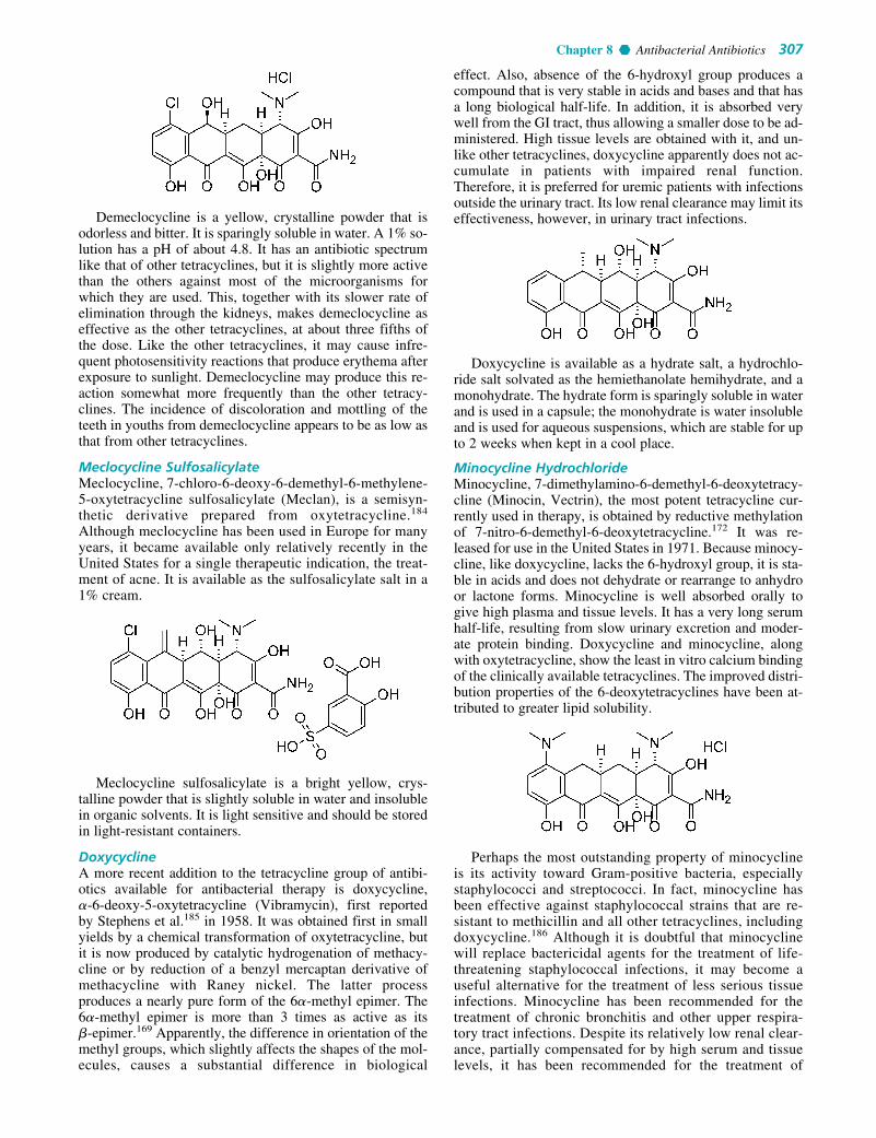
Chapter 8 Antibacterial Antibiotics 307
Demeclocycline is a yellow, crystalline powder that isodorless and bitter. It is sparingly soluble in water. A 1% so-lution has a pH of about 4.8. It has an antibiotic spectrumlike that of other tetracyclines, but it is slightly more activethan the others against most of the microorganisms forwhich they are used. This, together with its slower rate ofelimination through the kidneys, makes demeclocycline aseffective as the other tetracyclines, at about three fifths ofthe dose. Like the other tetracyclines, it may cause infre-quent photosensitivity reactions that produce erythema afterexposure to sunlight. Demeclocycline may produce this re-action somewhat more frequently than the other tetracy-clines. The incidence of discoloration and mottling of theteeth in youths from demeclocycline appears to be as low asthat from other tetracyclines.
Meclocycline SulfosalicylateMeclocycline, 7-chloro-6-deoxy-6-demethyl-6-methylene-5-oxytetracycline sulfosalicylate (Meclan), is a semisyn-thetic derivative prepared from oxytetracycline.184
Although meclocycline has been used in Europe for manyyears, it became available only relatively recently in theUnited States for a single therapeutic indication, the treat-ment of acne. It is available as the sulfosalicylate salt in a1% cream.
Meclocycline sulfosalicylate is a bright yellow, crys-talline powder that is slightly soluble in water and insolublein organic solvents. It is light sensitive and should be storedin light-resistant containers.
DoxycyclineA more recent addition to the tetracycline group of antibi-otics available for antibacterial therapy is doxycycline, �-6-deoxy-5-oxytetracycline (Vibramycin), first reportedby Stephens et al.185 in 1958. It was obtained first in smallyields by a chemical transformation of oxytetracycline, butit is now produced by catalytic hydrogenation of methacy-cline or by reduction of a benzyl mercaptan derivative ofmethacycline with Raney nickel. The latter process produces a nearly pure form of the 6�-methyl epimer. The6�-methyl epimer is more than 3 times as active as its �-epimer.169 Apparently, the difference in orientation of themethyl groups, which slightly affects the shapes of the mol-ecules, causes a substantial difference in biological
effect. Also, absence of the 6-hydroxyl group produces acompound that is very stable in acids and bases and that hasa long biological half-life. In addition, it is absorbed verywell from the GI tract, thus allowing a smaller dose to be ad-ministered. High tissue levels are obtained with it, and un-like other tetracyclines, doxycycline apparently does not ac-cumulate in patients with impaired renal function.Therefore, it is preferred for uremic patients with infectionsoutside the urinary tract. Its low renal clearance may limit itseffectiveness, however, in urinary tract infections.
Doxycycline is available as a hydrate salt, a hydrochlo-ride salt solvated as the hemiethanolate hemihydrate, and amonohydrate. The hydrate form is sparingly soluble in waterand is used in a capsule; the monohydrate is water insolubleand is used for aqueous suspensions, which are stable for upto 2 weeks when kept in a cool place.
Minocycline HydrochlorideMinocycline, 7-dimethylamino-6-demethyl-6-deoxytetracy-cline (Minocin, Vectrin), the most potent tetracycline cur-rently used in therapy, is obtained by reductive methylationof 7-nitro-6-demethyl-6-deoxytetracycline.172 It was re-leased for use in the United States in 1971. Because minocy-cline, like doxycycline, lacks the 6-hydroxyl group, it is sta-ble in acids and does not dehydrate or rearrange to anhydroor lactone forms. Minocycline is well absorbed orally togive high plasma and tissue levels. It has a very long serumhalf-life, resulting from slow urinary excretion and moder-ate protein binding. Doxycycline and minocycline, alongwith oxytetracycline, show the least in vitro calcium bindingof the clinically available tetracyclines. The improved distri-bution properties of the 6-deoxytetracyclines have been at-tributed to greater lipid solubility.
Perhaps the most outstanding property of minocyclineis its activity toward Gram-positive bacteria, especiallystaphylococci and streptococci. In fact, minocycline hasbeen effective against staphylococcal strains that are re-sistant to methicillin and all other tetracyclines, includingdoxycycline.186 Although it is doubtful that minocyclinewill replace bactericidal agents for the treatment of life-threatening staphylococcal infections, it may become auseful alternative for the treatment of less serious tissueinfections. Minocycline has been recommended for thetreatment of chronic bronchitis and other upper respira-tory tract infections. Despite its relatively low renal clear-ance, partially compensated for by high serum and tissuelevels, it has been recommended for the treatment of
0258-0329_17865_Ch08.qxd 12/2/09 3:27 AM Page 307

urinary tract infections. It has been effective in the eradi-cation of N. meningitidis in asymptomatic carriers.
NEWER TETRACYCLINES
The remarkably broad spectrum of antimicrobial activityof the tetracyclines notwithstanding, the widespread emer-gence of bacterial genes and plasmids encoding tetracy-cline resistance has increasingly imposed limitations onthe clinical applications of this antibiotic class in recentyears.164 This situation has prompted researchers atLederle Laboratories to reinvestigate SARs of tetracy-clines substituted in the aromatic (D) ring in an effort todiscover analogs that might be effective against resistantstrains. As a result of these efforts, the glycylcyclines,a class of 9-dimethylglycylamino-(DMG)-substitutedtetracyclines exemplified by DMG-minocycline (DMG-MINO), and DMG-6-methyl-6-deoxytetracycline (DMG-DMDOT) were discovered.187–189
The first of these to be marketed was tigecycline.
The glycylcyclines retain the broad spectrum of activityand potency exhibited by the original tetracyclines againsttetracycline-sensitive microbial strains and are highly activeagainst bacterial strains that exhibit tetracycline resistancemediated by efflux or ribosomal protection determinants. Ifongoing clinical evaluations of the glycylcyclines establishfavorable toxicological and pharmacokinetic profiles forthese compounds, a new class of “second-generation” tetra-cyclines could be launched.
MACROLIDES
Among the many antibiotics isolated from the actinomycetesis the group of chemically related compounds called themacrolides. In 1950, picromycin, the first of this group to beidentified as a macrolide compound, was first reported. In1952, erythromycin and carbomycin were reported as new an-tibiotics, and they were followed in subsequent years by othermacrolides. Currently, more than 40 such compounds areknown, and new ones are likely to appear in the future. Of allof these, only two, erythromycin and oleandomycin, have beenavailable consistently for medical use in the United States. Inrecent years, interest has shifted away from novel macrolidesisolated from soil samples (e.g., spiramycin, josamycin, androsamicin), all of which thus far have proved to be clinicallyinferior to erythromycin and semisynthetic derivatives oferythromycin (e.g., clarithromycin and azithromycin), whichhave superior pharmacokinetic properties because of their en-hanced acid stability and improved distribution properties.
308 Wilson and Gisvold’s Textbook of Organic Medicinal and Pharmaceutical Chemistry
0258-0329_17865_Ch08.qxd 12/2/09 3:27 AM Page 308

Chapter 8 Antibacterial Antibiotics 309
Chemistry
The macrolide antibiotics have three common chemicalcharacteristics: (a) a large lactone ring (which prompted thename macrolide), (b) a ketone group, and (c) a glycosidi-cally linked amino sugar. Usually, the lactone ring has 12,14, or 16 atoms in it, and it is often unsaturated, with anolefinic group conjugated with the ketone function. (Thepolyene macrocyclic lactones, such as natamycin and am-photericin B; the ansamycins, such as rifampin; and thepolypeptide lactones generally are not included amongthe macrolide antibiotics.) They may have, in addition tothe amino sugar, a neutral sugar that is linked glycosidi-cally to the lactone ring (see discussion that follows under“Erythromycin”). Because of the dimethylamino group onthe sugar moiety, the macrolides are bases that form saltswith pKa values between 6.0 and 9.0. This feature has beenused to make clinically useful salts. The free bases are onlyslightly soluble in water but dissolve in somewhat polar or-ganic solvents. They are stable in aqueous solutions at orbelow room temperature but are inactivated by acids, bases,and heat. The chemistry of macrolide antibiotics has beenthe subject of several reviews.190,191
Mechanism of Action and Resistance
Some details of the mechanism of antibacterial action oferythromycin are known. It binds selectively to a specificsite on the 50S ribosomal subunit to prevent the transloca-tion step of bacterial protein synthesis.192 It does not bind tomammalian ribosomes. Broadly based, nonspecific resist-ance to the antibacterial action of erythromycin amongmany species of Gram-negative bacilli appears to be largelyrelated to the inability of the antibiotic to penetrate the cellwalls of these organisms.193 In fact, the sensitivities ofmembers of the Enterobacteriaceae family are pH depend-ent, with MICs decreasing as a function of increasing pH.Furthermore, protoplasts from Gram-negative bacilli, whichlack cell walls, are sensitive to erythromycin. A highly spe-cific resistance mechanism to the macrolide antibiotics oc-curs in erythromycin-resistant strains of S. aureus.194,195
Such strains produce an enzyme that methylates a specificadenine residue at the erythromycin-binding site of the bac-terial 50S ribosomal subunit. The methylated ribosomalRNA remains active in protein synthesis but no longer bindserythromycin. Bacterial resistance to the lincomycins appar-ently also occurs by this mechanism.
Spectrum of Activity
The spectrum of antibacterial activity of the more potentmacrolides, such as erythromycin, resembles that of peni-cillin. They are frequently active against bacterial strainsthat are resistant to the penicillins. The macrolides aregenerally effective against most species of Gram-positivebacteria, both cocci and bacilli, and exhibit useful effective-ness against Gram-negative cocci, especially Neisseriaspp. Many of the macrolides are also effective againstTreponema pallidum. In contrast to penicillin, macrolidesare also effective against Mycoplasma, Chlamydia,Campylobacter, and Legionella spp. Their activity againstmost species of Gram-negative bacilli is generally low andoften unpredictable, though some strains of H. influenzaeand Brucella spp. are sensitive.
Products
ErythromycinEarly in 1952, McGuire et al.196 reported the isolation oferythromycin (E-Mycin, Erythrocin, Ilotycin) fromStreptomyces erythraeus. It achieved rapid early acceptanceas a well-tolerated antibiotic of value for the treatment ofvarious upper respiratory and soft-tissue infections causedby Gram-positive bacteria. It is also effective against manyvenereal diseases, including gonorrhea and syphilis, andprovides a useful alternative for the treatment of many in-fections in patients allergic to penicillins. More recently,erythromycin was shown to be effective therapy for Eatonagent pneumonia (Mycoplasma pneumoniae), venereal dis-eases caused by Chlamydia, bacterial enteritis caused byCampylobacter jejuni, and Legionnaires disease.
The commercial product is erythromycin A, whichdiffers from its biosynthetic precursor, erythromycin B,in having a hydroxyl group at the 12-position of theaglycone. The chemical structure of erythromycin A was re-ported by Wiley et al.197 in 1957 and its stereochemistry byCelmer198 in 1965. An elegant synthesis of erythronolide A,the aglycone present in erythromycin A, was described byCorey et al.199
The amino sugar attached through a glycosidic link to C-5 is desosamine, a structure found in several other macrolideantibiotics. The tertiary amine of desosamine (3,4,6-trideoxy-3-dimethylamino-D-xylo-hexose) confers a basiccharacter to erythromycin and provides the means by whichacid salts may be prepared. The other carbohydrate structurelinked as a glycoside to C-3 is called cladinose (2,3,6-trideoxy-3-methoxy-3-C-methyl-L-ribo-hexose) and isunique to the erythromycin molecule.
As is common with other macrolide antibiotics, com-pounds closely related to erythromycin have been obtainedfrom culture filtrates of S. erythraeus. Two such analogshave been found, erythromycins B and C. Erythromycin Bdiffers from erythromycin A only at C-12, at which a hydro-gen has replaced the hydroxyl group. The B analog is moreacid stable but has only about 80% of the activity of eryth-romycin. The C analog differs from erythromycin by thereplacement of the methoxyl group on the cladinose moietywith a hydrogen atom. It appears to be as active as erythro-mycin but is present in very small amounts in fermentationliquors.
Erythromycin is a very bitter, white or yellow-white,crystalline powder. It is soluble in alcohol and in the other
0258-0329_17865_Ch08.qxd 12/2/09 3:27 AM Page 309

common organic solvents but only slightly soluble in water.The free base has a pKa of 8.8. Saturated aqueous solutionsdevelop an alkaline pH in the range of 8.0 to 10.5. It isextremely unstable at a pH of 4 or below. The optimum pHfor stability of erythromycin is at or near neutrality.
Erythromycin may be used as the free base in oraldosage forms and for topical administration. To overcomeits bitterness and irregular oral absorption (resulting fromacid destruction and adsorption onto food), various enteric-coated and delayed-release dose forms of erythromycinbase have been developed. These forms have been fullysuccessful in overcoming the bitterness but have solvedonly marginally problems of oral absorption. Erythromycinhas been chemically modified with primarily two differentgoals in mind: (a) to increase either its water or its lipid sol-ubility for parenteral dosage forms and (b) to increase itsacid stability (and possibly its lipid solubility) for im-proved oral absorption. Modified derivatives of the antibi-otic are of two types: acid salts of the dimethylamino groupof the desosamine moiety (e.g., the glucoheptonate, the lac-tobionate, and the stearate) and esters of the 2�-hydroxylgroup of the desosamine (e.g., the ethylsuccinate and thepropionate, available as the lauryl sulfate salt and known asthe estolate).
The stearate salt and the ethylsuccinate and propionateesters are used in oral dose forms intended to improve ab-sorption of the antibiotic. The stearate releases erythromy-cin base in the intestinal tract, which is then absorbed. Theethylsuccinate and the estolate are absorbed largely intactand are hydrolyzed partially by plasma and tissue esterasesto give free erythromycin. The question of bioavailability ofthe antibiotic from its various oral dosage and chemicalforms has caused considerable concern and dispute over thepast two decades.200–205 It is generally believed that the 2�-esters per se have little or no intrinsic antibacterial activ-ity206 and, therefore, must be hydrolyzed to the parentantibiotic in vivo. Although the ethylsuccinate is hydrolyzedmore efficiently than the estolate in vivo and, in fact, pro-vides higher levels of erythromycin following intramuscularadministration, an equal dose of the estolate gives higherlevels of the free antibiotic following oral administra-tion.201,205 Superior oral absorption of the estolate is attrib-uted to its both greater acid stability and higher intrinsicabsorption than the ethylsuccinate. Also, oral absorption ofthe estolate, unlike that of both the stearate and the ethylsuc-cinate, is not affected by food or fluid volume content of thegut. Superior bioavailability of active antibiotic from oraladministration of the estolate over the ethylsuccinate,stearate, or erythromycin base cannot necessarily be as-sumed, however, because the estolate is more extensivelyprotein bound than erythromycin itself.207 Measured frac-tions of plasma protein binding for erythromycin-2�-propi-onate and erythromycin base range from 0.94 to 0.98 for theformer and from 0.73 to 0.90 for the latter, indicating amuch higher level of free erythromycin in the plasma.Bioavailability studies comparing equivalent doses of theenteric-coated base, the stearate salt, the ethylsuccinateester, and the estolate ester in human volunteers203,204
showed delayed but slightly higher bioavailability for thefree base than for the stearate, ethylsuccinate, or estolate.
One study, comparing the clinical effectiveness of rec-ommended doses of the stearate, estolate, ethylsuccinate,and free base in the treatment of respiratory tract infections,
failed to demonstrate substantial differences among them.208
Two other clinical studies, comparing the effectiveness ofthe ethylsuccinate and the estolate in the treatment ofstreptococcal pharyngitis, however, found the estolate to besuperior.209,210
The water-insoluble ethylsuccinate ester is also availableas a suspension for intramuscular injection. The gluco-heptonate and lactobionate salts, however, are highly water-soluble derivatives that provide high plasma levels of theactive antibiotic immediately after intravenous injection.Aqueous solutions of these salts may also be administeredby intramuscular injection, but this is not a commonpractice.
Erythromycin is distributed throughout the body water.It persists in tissues longer than in the blood. The antibioticis concentrated by the liver and excreted extensively intothe bile. Large amounts are excreted in the feces, partly be-cause of poor oral absorption and partly because of biliaryexcretion. The serum half-life is 1.4 hours. Some cy-tochrome P450-catalyzed oxidative demethylation to a lessactive metabolite may also occur. Erythromycin inhibitscytochrome P450-requiring oxidases, leading to variouspotential drug interactions. Thus, toxic effects of theo-phylline, the hydroxycoumarin anticoagulants, the benzodi-azepines alprazolam and midazolam, carbamazepine, cy-closporine, and the antihistaminic drugs terfenadine andastemizole may be potentiated by erythromycin.
The toxicity of erythromycin is comparatively low.Primary adverse reactions to the antibiotic are related to itsactions on the GI tract and the liver. Erythromycin maystimulate GI motility following either oral or parenteral ad-ministration.211 This dose-related, prokinetic effect cancause abdominal cramps, epigastric distress, and diarrhea,especially in children and young adults. Cholestatic hepati-tis occurs occasionally with erythromycin, usually in adultsand more frequently with the estolate.
Erythromycin StearateErythromycin stearate (Ethril, Wyamycin S, Erypar) is thestearic acid salt of erythromycin. Like erythromycin base,the stearate is acid labile. It is film coated to protect it fromacid degradation in the stomach. In the alkaline pH of theduodenum, the free base is liberated from the stearateand absorbed. Erythromycin stearate is a crystalline powderthat is practically insoluble in water but soluble in alcoholand ether.
310 Wilson and Gisvold’s Textbook of Organic Medicinal and Pharmaceutical Chemistry
0258-0329_17865_Ch08.qxd 12/2/09 3:27 AM Page 310

Chapter 8 Antibacterial Antibiotics 311
Erythromycin EthylsuccinateErythromycin ethylsuccinate (EES, Pediamycin, EryPed) isthe ethylsuccinate mixed ester of erythromycin in which the2�-hydroxyl group of the desosamine is esterified. It isabsorbed as the ester and hydrolyzed slowly in the body toform erythromycin. It is somewhat acid labile, and itsabsorption is enhanced by the presence of food. The ester isinsoluble in water but soluble in alcohol and ether.
Erythromycin EstolateErythromycin estolate, erythromycin propionate lauryl sul-fate (Ilosone), is the lauryl sulfate salt of the 2�-propionateester of erythromycin. Erythromycin estolate is acid stableand absorbed as the propionate ester. The ester undergoesslow hydrolysis in vivo. Only the free base binds to bacte-rial ribosomes. Some evidence, however, suggests that theester is taken up by bacterial cells more rapidly than thefree base and undergoes hydrolysis by bacterial esteraseswithin the cells. The incidence of cholestatic hepatitis is re-portedly higher with the estolate than with other erythromy-cin preparations.
Erythromycin estolate occurs as long needles that aresparingly soluble in water but soluble in organic solvents.
Erythromycin Gluceptate, SterileErythromycin gluceptate, erythromycin glucoheptonate(Ilotycin Gluceptate), is the glucoheptonic acid salt oferythromycin. It is a crystalline substance that is freelysoluble in water and practically insoluble in organic sol-vents. Erythromycin gluceptate is intended for intravenous
administration for the treatment of serious infections,such as Legionnaires disease, or when oral administrationis not possible. Solutions are stable for 1 week whenrefrigerated.
Erythromycin LactobionateErythromycin lactobionate is a water-soluble salt preparedby reacting erythromycin base with lactobiono-�-lactone. Itoccurs as an amorphous powder that is freely soluble inwater and alcohol and slightly soluble in ether. It is in-tended, after reconstitution in sterile water, for intravenousadministration to achieve high plasma levels in the treat-ment of serious infections.
ClarithromycinClarithromycin (Biaxin) is the 6-methyl ether of erythromy-cin. The simple methylation of the 6-hydroxyl group oferythromycin creates a semisynthetic derivative that fullyretains the antibacterial properties of the parent antibiotic,with markedly increased acid stability and oral bioavailabil-ity and reduced GI side effects associated with erythromy-cin.212 Acid-catalyzed dehydration of erythromycin in thestomach initiates as a sequence of reactions, beginning with�6,7-bond migration followed by formation of an 8,9-anhy-dro-6,9-hemiketal and terminating in a 6,9:9,12-spiroketal.Since neither the hemiketal nor the spiroketal exhibits sig-nificant antibacterial activity, unprotected erythromycin isinactivated substantially in the stomach. Furthermore, evi-dence suggests that the hemiketal may be largely responsi-ble for the GI (prokinetic) adverse effects associated withoral erythromycin.211
0258-0329_17865_Ch08.qxd 12/2/09 3:27 AM Page 311

312 Wilson and Gisvold’s Textbook of Organic Medicinal and Pharmaceutical Chemistry
Clarithromycin is well absorbed following oral adminis-tration. Its oral bioavailability is estimated to be 50% to55%. The presence of food does not significantly affect itsabsorption. Extensive metabolism of clarithromycin byoxidation and hydrolysis occurs in the liver. The majormetabolite is the 14-hydroxyl derivative, which retains anti-bacterial activity. The amount of clarithromycin excreted inthe urine ranges from 20% to 30%, depending on the dose,whereas 10% to 15% of the 14-hydroxy metabolite is ex-creted in the urine. Biliary excretion of clarithromycin ismuch lower than that of erythromycin. Clarithromycin iswidely distributed into the tissues, which retain much higherconcentrations than the plasma. Protein-binding fractions inthe plasma range from 65% to 70%. The plasma half-life ofclarithromycin is 4.3 hours.
Some of the microbiological properties of clarithromycinalso appear to be superior to those of erythromycin. It ex-hibits greater potency against M. pneumoniae, Legionellaspp., Chlamydia pneumoniae, H. influenzae, and M. ca-tarrhalis than does erythromycin. Clarithromycin also hasactivity against unusual pathogens such as Borrelia burgdor-feri (the cause of Lyme disease) and the Mycobacteriumavium complex (MAC). Clarithromycin is significantly moreactive than erythromycin against group A streptococci, S.pneumoniae, and the viridans group of streptococci in vivobecause of its superior oral bioavailability. Clarithromycin is,however, more expensive than erythromycin, which must beweighed against its potentially greater effectiveness.
Adverse reactions to clarithromycin are rare. The mostcommon complaints relate to GI symptoms, but these sel-dom require discontinuance of therapy. Clarithromycin,like erythromycin, inhibits cytochrome P450 oxidases and,thus, can potentiate the actions of drugs metabolized bythese enzymes.
Clarithromycin occurs as a white crystalline solid that ispractically insoluble in water, sparingly soluble in alcohol,and freely soluble in acetone. It is provided as 250- and 500-mg oral tablets and as granules for the preparation of aque-ous oral suspensions containing 25 or 50 mg/mL.
AzithromycinAzithromycin (Zithromax) is a semisynthetic derivative oferythromycin, prepared by Beckman rearrangement of thecorresponding 6-oxime, followed by N-methylation and re-duction of the resulting ring-expanded lactam. It is a proto-type of a series of nitrogen-containing, 15-membered ringmacrolides known as azalides.213 Removal of the 9-ketogroup coupled with incorporation of a weakly basic tertiary
amine nitrogen function into the macrolide ring increasesthe stability of azithromycin to acid-catalyzed degradation.These changes also increase the lipid solubility of the mole-cule, thereby conferring unique pharmacokinetic and micro-biological properties.214
The oral bioavailability of azithromycin is good, nearly40%, provided the antibiotic is administered at least 1 hourbefore or 2 hours after a meal. Food decreases its absorptionby as much as 50%. The pharmacokinetics of azithromycinare characterized by rapid and extensive removal of the drugfrom the plasma into the tissues followed by a slow release.Tissue levels far exceed plasma concentrations, leading to ahighly variable and prolonged elimination half-life of up to5 days. The fraction of azithromycin bound to plasma pro-teins is only about 50% and does not exert an important in-fluence on its distribution. Evidence indicates thatazithromycin is largely excreted in the feces unchanged,with a small percentage appearing in the urine. Extensiveenterohepatic recycling of the drug occurs. Azithromycinapparently is not metabolized to any significant extent. Incontrast to the 14-membered ring macrolides, azithromycindoes not significantly inhibit cytochrome P450 enzymes tocreate potential drug interactions.
The spectrum of antimicrobial activity of azithromycin issimilar to that observed for erythromycin and clar-ithromycin but with some interesting differences. In general,it is more active against Gram-negative bacteria and less ac-tive against Gram-positive bacteria than its close relatives.The greater activity of azithromycin against H. influenzae,M. catarrhalis, and M. pneumoniae coupled with its ex-tended half-life permits a 5-day dosing schedule for thetreatment of respiratory tract infections caused by thesepathogens. The clinical efficacy of azithromycin in the treat-ment of urogenital and other sexually transmitted infectionscaused by Chlamydia trachomatis, N. gonorrhoeae, H.ducreyi, and Ureaplasma urealyticum suggests that single-dose therapy with it for uncomplicated urethritis or cervici-tis may have advantages over use of other antibiotics.
DirithromycinDirithromycin (Dynabac) is a more lipid-soluble prodrug de-rivative of 9S-erythromycyclamine prepared by condensa-tion of the latter with 2-(2-methoxyethoxy)acetaldehyde.215
The 9N,11O-oxazine ring thus formed is a hemi-aminal thatis unstable under both acidic and alkaline aqueous conditionsand undergoes spontaneous hydrolysis to form erythromycy-clamine. Erythromycyclamine is a semisynthetic derivativeof erythromycin in which the 9-keto group of the erythrono-lide ring has been converted to an amino group.Erythromycyclamine retains the antibacterial properties of
0258-0329_17865_Ch08.qxd 12/2/09 3:27 AM Page 312

Chapter 8 Antibacterial Antibiotics 313
erythromycin in vitro but exhibits poor bioavailabilityfollowing oral administration. The prodrug, dirithromycin,is provided as enteric-coated tablets to protect it from acid-catalyzed hydrolysis in the stomach.
Orally administered dirithromycin is absorbed rapidly intothe plasma, largely from the small intestine. Spontaneoushydrolysis to erythromycyclamine occurs in the plasma. Oralbioavailability is estimated to be about 10%, but food doesnot affect absorption of the prodrug.
The low plasma levels and large volume of distributionof erythromycyclamine are believed to result from its rapiddistribution into well-perfused tissues, such as lungparenchyma, bronchial mucosa, nasal mucosa, and prostatictissue. The drug also concentrates in human neutrophils.The elimination half-life is estimated to be 30 to 44 hours.Most of the prodrug and its active metabolite (62%–81% innormal human subjects) are excreted in the feces, largely viathe bile, following either oral or parenteral administration.Urinary excretion accounts for less than 3%.
The incidence and severity of GI adverse effects associ-ated with dirithromycin are similar to those seen with oralerythromycin. Preliminary studies indicate that dirithromycinand erythromycyclamine do not interact significantly with cy-tochrome P450 oxygenases. Thus, the likelihood of interfer-ence in the oxidative metabolism of drugs such as phenytoin,theophylline, and cyclosporine by these enzymes may be lesswith dirithromycin than with erythromycin. Dirithromycin isrecommended as an alternative to erythromycin for the treat-ment of bacterial infections of the upper and lower respiratorytracts, such as pharyngitis, tonsillitis, bronchitis, and pneumo-nia, and for bacterial infections of other soft tissues and theskin. The once-daily dosing schedule for dirithromycin is ad-vantageous in terms of better patient compliance. Its place intherapy remains to be fully assessed.216
TroleandomycinOleandomycin, as its triacetyl derivative troleandomycin,triacetyloleandomycin (TAO), remains available as an alter-native to erythromycin for limited indications permitting useof an oral dosage form. Oleandomycin was isolated bySobin et al.217 The structure of oleandomycin was proposedby Hochstein et al.218 and its absolute stereochemistry elu-cidated by Celmer.219 The oleandomycin structure consistsof two sugars and a 14-member lactone ring designated anoleandolide. One of the sugars is desosamine, also presentin erythromycin; the other is L-oleandrose. The sugars arelinked glycosidically to the positions 3 and 5, respectively,of oleandolide.
Oleandomycin contains three hydroxyl groups that aresubject to acylation, one in each of the sugars and one in theoleandolide. The triacetyl derivative retains the in vivo anti-bacterial activity of the parent antibiotic but possesses supe-rior pharmacokinetic properties. It is hydrolyzed in vivo tooleandomycin. Troleandomycin achieves more rapid andhigher plasma concentrations following oral administrationthan oleandomycin phosphate, and it has the additionaladvantage of being practically tasteless. Troleandomycinoccurs as a white, crystalline solid that is nearly insoluble inwater. It is relatively stable in the solid state but undergoeschemical degradation in either aqueous acidic or alkalineconditions.
Because the antibacterial spectrum of activity of olean-domycin is considered inferior to that of erythromycin, thepharmacokinetics of troleandomycin have not been studiedextensively. Oral absorption is apparently good, and de-tectable blood levels of oleandomycin persist up to 12 hoursafter a 500-mg dose of troleandomycin. Approximately 20%is recovered in the urine, with most excreted in the feces, pri-marily as a result of biliary excretion. There is some epigas-tric distress following oral administration, with an incidencesimilar to that caused by erythromycin. Troleandomycin isthe most potent inhibitor of cytochrome P450 enzymes of thecommercially available macrolides. It may potentiate the he-patic toxicity of certain anti-inflammatory steroids and oralcontraceptive drugs as well as the toxic effects of theo-phylline, carbamazepine, and triazolam. Several allergic reac-tions, including cholestatic hepatitis, have also been reportedwith the use of troleandomycin.
Approved medical indications for troleandomycin arecurrently limited to the treatment of upper respiratory infec-tions caused by such organisms as S. pyogenes and S. pneu-moniae. It may be considered an alternative to oral forms oferythromycin. It is available in capsules and as a suspension.
LINCOMYCINS
The lincomycins are sulfur-containing antibiotics isolatedfrom Streptomyces lincolnensis. Lincomycin is the most ac-tive and medically useful of the compounds obtained fromfermentation. Extensive efforts to modify the lincomycinstructure to improve its antibacterial and pharmacologicalproperties resulted in the preparation of the 7-chloro-7-deoxyderivative clindamycin. Of the two antibiotics, clindamycin
0258-0329_17865_Ch08.qxd 12/2/09 3:27 AM Page 313

314 Wilson and Gisvold’s Textbook of Organic Medicinal and Pharmaceutical Chemistry
appears to have the greater antibacterial potency andbetter pharmacokinetic properties. Lincomycins resemblemacrolides in antibacterial spectrum and biochemical mech-anisms of action. They are primarily active against Gram-positive bacteria, particularly the cocci, but are also effectiveagainst non–spore-forming anaerobic bacteria, actino-mycetes, mycoplasma, and some species of Plasmodium.Lincomycin binds to the 50S ribosomal subunit to inhibitprotein synthesis. Its action may be bacteriostatic or bacteri-cidal depending on various factors, including the concentra-tion of the antibiotic. A pattern of bacterial resistance andcross-resistance to lincomycins similar to that observed withthe macrolides has been emerging.195
Products
Lincomycin HydrochlorideLincomycin hydrochloride (Lincocin), which differs chemi-cally from other major antibiotic classes, was isolated byMason et al.220 Its chemistry was described by Hoeksema etal.221 who assigned the structure, later confirmed by Slompand MacKellar,222 given in the diagram below. Total syn-theses of the antibiotic were accomplished independently in1970 in England and the United States.223,224 The structurecontains a basic function, the pyrrolidine nitrogen, by whichwater-soluble salts with an apparent pKa of 7.6 may beformed. When subjected to hydrazinolysis, lincomycin iscleaved at its amide bond into trans-L-4-n-propylhygric acid(the pyrrolidine moiety) and methyl �-thiolincosamide (thesugar moiety). Lincomycin-related antibiotics have beenreported by Argoudelis225 to be produced by S. lincolnensis.These antibiotics differ in structure at one or more of threepositions of the lincomycin structure: (a) the N-methyl ofthe hygric acid moiety is substituted by a hydrogen; (b) then-propyl group of the hygric acid moiety is substituted by anethyl group; and (c) the thiomethyl ether of the �-thiolin-cosamide moiety is substituted by a thioethyl ether.
Lincomycin is used for the treatment of infections causedby Gram-positive organisms, notably staphylococci, �-he-molytic streptococci, and pneumococci. It is absorbed mod-erately well orally and distributed widely in the tissues.Effective concentrations are achieved in bone for the treat-ment of staphylococcal osteomyelitis but not in the cere-brospinal fluid for the treatment of meningitis. At one time,lincomycin was considered a nontoxic compound, with alow incidence of allergy (rash) and occasional GI com-plaints (nausea, vomiting, and diarrhea) as the only adverseeffects. Recent reports of severe diarrhea and the develop-ment of pseudomembranous colitis in patients treated withlincomycin (or clindamycin), however, have necessitatedreappraisal of the role of these antibiotics in therapy. In anyevent, clindamycin is superior to lincomycin for thetreatment of most infections for which these antibiotics areindicated.
Lincomycin hydrochloride occurs as the monohydrate, awhite, crystalline solid that is stable in the dry state. It isreadily soluble in water and alcohol, and its aqueous solu-tions are stable at room temperature. It is degraded slowlyin acid solutions but is absorbed well from the GI tract.Lincomycin diffuses well into peritoneal and pleural fluidsand into bone. It is excreted in the urine and the bile. It isavailable in capsule form for oral administration and inampules and vials for parenteral administration.
Clindamycin HydrochlorideIn 1967, Magerlein et al.226 reported that replacement of the7(R)-hydroxy group of lincomycin by chlorine with inver-sion of configuration resulted in a compound with enhancedantibacterial activity in vitro. Clinical experience withthis semisynthetic derivative, clindamycin, 7(S)-chloro-7-deoxylincomycin (Cleocin), released in 1970, has estab-lished that its superiority over lincomycin is even greater invivo. Improved absorption and higher tissue levels of clin-damycin and its greater penetration into bacteria have beenattributed to a higher partition coefficient than that oflincomycin. Structural modifications at C-7 (e.g., 7(S)-chloroand 7(R)-OCH3) and of the C-4 alkyl groups of the hygricacid moiety227 appear to influence activity of congenersmore through an effect on the partition coefficient of themolecule than through a stereospecific binding role. Changesin the �-thiolincosamide portion of the molecule seem todecrease activity markedly, however, as evidenced by themarginal activity of 2-deoxylincomycin, its anomer, and 2-O-methyllincomycin.227,228 Exceptions to this are fatty acidand phosphate esters of the 2-hydroxyl group of lincomycinand clindamycin, which are hydrolyzed rapidly in vivo to theparent antibiotics.
Clindamycin is recommended for the treatment of a widevariety of upper respiratory, skin, and tissue infections causedby susceptible bacteria. Its activity against streptococci,staphylococci, and pneumococci is indisputably high, and it isone of the most potent agents available against somenon–spore-forming anaerobic bacteria, the Bacteroides spp.in particular. An increasing number of reports of clin-damycin-associated GI toxicity, which range in severity fromdiarrhea to an occasionally serious pseudomembranous coli-tis, have, however, caused some clinical experts to call for areappraisal of the role of this antibiotic in therapy.Clindamycin- (or lincomycin)-associated colitis may be par-ticularly dangerous in elderly or debilitated patients and hascaused deaths in such individuals. The colitis, which is usu-ally reversible when the drug is discontinued, is now believedto result from an overgrowth of a clindamycin-resistant strainof the anaerobic intestinal bacterium Clostridium difficile.229
The intestinal lining is damaged by a glycoprotein endotoxinreleased by lysis of this organism.
The glycopeptide antibiotic vancomycin has been effec-tive in the treatment of clindamycin-induced pseudomem-branous colitis and in the control of the experimentally
0258-0329_17865_Ch08.qxd 12/2/09 3:27 AM Page 314

Chapter 8 Antibacterial Antibiotics 315
induced bacterial condition in animals. Clindamycin shouldbe reserved for staphylococcal tissue infections, such as cel-lulitis and osteomyelitis, in penicillin-allergic patients andfor severe anaerobic infections outside the central nervoussystem. Ordinarily, it should not be used to treat upper res-piratory tract infections caused by bacteria sensitive to othersafer antibiotics or in prophylaxis.
Clindamycin is absorbed rapidly from the GI tract, evenin the presence of food. It is available as the crystalline,water-soluble hydrochloride hydrate (hyclate) and the 2-palmitate ester hydrochloride salts in oral dosage forms andas the 2-phosphate ester in solutions for intramuscular or in-travenous injection. All forms are chemically very stable insolution and in the dry state.
Clindamycin Palmitate HydrochlorideClindamycin palmitate hydrochloride (Cleocin Pediatric) isthe hydrochloride salt of the palmitic acid ester ofcleomycin. The ester bond is to the 2-hydroxyl group of thelincosamine sugar. The ester serves as a tasteless prodrugform of the antibiotic, which hydrolyzes to clindamycin inthe plasma. The salt form confers water solubility to theester, which is available as granules for reconstitution intoan oral solution for pediatric use. Although absorption of thepalmitate is slower than that of the free base, there is littledifference in overall bioavailability of the two preparations.Reconstituted solutions of the palmitate hydrochloride arestable for 2 weeks at room temperature. Such solutionsshould not be refrigerated because thickening occurs thatmakes the preparation difficult to pour.
Clindamycin PhosphateClindamycin phosphate (Cleocin Phosphate) is the 2-phos-phate ester of clindamycin. It exists as a zwitterionic struc-ture that is very soluble in water. It is intended for parenteral(intravenous or intramuscular) administration for the treat-ment of serious infections and instances when oral adminis-tration is not feasible. Solutions of clindamycin phosphateare stable at room temperature for 16 days and for up to 32days when refrigerated.
POLYPEPTIDES
Among the most powerful bactericidal antibiotics are thosethat possess a polypeptide structure. Many of them have
been isolated, but unfortunately, their clinical use has beenlimited by their undesirable side reactions, particularly renaltoxicity. Another limitation is the lack of systemic activityof most peptides following oral administration. A chiefsource of the medicinally important members of this classhas been Bacillus spp. The antitubercular antibiotics capre-omycin and viomycin (see Chapter 6) and the antitumorantibiotics actinomycin and bleomycin are peptides isolatedfrom Streptomyces spp. The glycopeptide antibiotic van-comycin, which has become the most important member ofthis class, is isolated from a closely related actinomycete,Amycolatopsis orientalis.
Polypeptide antibiotics variously possess several inter-esting and often unique characteristics: (a) they frequentlyconsist of several structurally similar but chemically dis-tinct entities isolated from a single source; (b) most ofthem are cyclic, with a few exceptions (e.g., the grami-cidins); (c) they frequently contain D-amino acids and/or“unnatural” amino acids not found in higher plants oranimals; and (d) many of them contain non–amino acidmoieties, such as heterocycles, fatty acids, sugars, etc.Polypeptide antibiotics may be acidic, basic, zwitterionic,or neutral depending on the number of free carboxyl andamino or guanidino groups in their structures. Initially, itwas assumed that neutral compounds, such as the grami-cidins, possessed cyclopeptide structures. Later, the gram-icidins were determined to be linear, and the neutrality wasshown to be because of a combination of the formylationof the terminal amino group and the ethanolamine amida-tion of the terminal carboxyl group.230
Antibiotics of the polypeptide class differ widely intheir mechanisms of action and antimicrobial properties.Bacitracin and vancomycin interfere with bacterial cell wallsynthesis and are effective only against Gram-positive bac-teria. Neither antibiotic apparently can penetrate the outerenvelope of Gram-negative bacteria. Both the gramicidinsand the polymyxins interfere with cell membrane functionsin bacteria. However, the gramicidins are effective primarilyagainst Gram-positive bacteria, whereas the polymyxins areeffective only against Gram-negative species. Gramicidinsare neutral compounds that are largely incapable of pene-trating the outer envelope of Gram-negative bacteria.Polymyxins are highly basic compounds that penetrate theouter membrane of Gram-negative bacteria through porinchannels to act on the inner cell membrane.231 The muchthicker cell wall of Gram-positive bacteria apparently barspenetration by the polymyxins.
Vancomycin HydrochlorideThe isolation of the glycopeptide antibiotic vancomycin(Vancocin, Vancoled) from Streptomyces orientalis (renamedA. orientalis) was described in 1956 by McCormick et al.232
The organism originally was obtained from cultures of anIndonesian soil sample and subsequently has been obtainedfrom Indian soil. Vancomycin was introduced in 1958 as anantibiotic active against Gram-positive cocci, particularlystreptococci, staphylococci, and pneumococci. It is not ac-tive against Gram-negative bacteria, with the exception ofNeisseria spp. Vancomycin is recommended for use wheninfections fail to respond to treatment with the more commonantibiotics or when the infection is known to be caused by aresistant organism. It is particularly effective for the treatmentof endocarditis caused by Gram-positive bacteria.
0258-0329_17865_Ch08.qxd 12/2/09 3:27 AM Page 315

316 Wilson and Gisvold’s Textbook of Organic Medicinal and Pharmaceutical Chemistry
et al.237 The action of vancomycin leads to lysis of the bac-terial cell. The antibiotic does not exhibit cross-resistanceto �-lactams, bacitracin, or cycloserine, from which it dif-fers in mechanism. Resistance to vancomycin amongGram-positive cocci is rare. High-level resistance in clini-cal isolates of enterococci has been reported, however.This resistance is in response to the inducible productionof a protein, encoded by vancomycin A, that is an alteredligase enzyme that causes the incorporation of a D-alanine-D-lactate depsipeptide instead of the usual D-alanine-D-ala-nine dipeptide in the peptidoglycan terminus.238 The re-sulting peptidoglycan can still undergo cross-linking butno longer binds vancomycin.
Vancomycin hydrochloride is always administered intra-venously (never intramuscularly), either by slow injectionor by continuous infusion, for the treatment of systemicinfections. In short-term therapy, the toxic side reactions areusually slight, but continued use may lead to impaired audi-tory acuity, renal damage, phlebitis, and rashes. Because itis not absorbed or significantly degraded in the GI tract,vancomycin may be administered orally for the treatment ofstaphylococcal enterocolitis and for pseudomembranous co-litis associated with clindamycin therapy. Some conversionto aglucovancomycin likely occurs in the low pH of thestomach. The latter retains about three fourths of the activ-ity of vancomycin.
TeicoplaninTeicoplanin (Teichomycin A2, Targocid) is a mixture offive closely related glycopeptide antibiotics produced bythe actinomycete Actinoplanes teichomyceticus.239,240
The teicoplanin factors differ only in the acyl group inthe northernmost of two glucosamines glycosidicallylinked to the cyclic peptide aglycone. Another sugar, D-mannose, is common to all of the teicoplanins. Thestructures of the teicoplanin factors were determined inde-pendently by a combination of chemical degradation241
and spectroscopic242,243 methods in three different groupsin 1984.
Vancomycin hydrochloride is a free-flowing, tan tobrown powder that is relatively stable in the dry state. It isvery soluble in water and insoluble in organic solvents.The salt is quite stable in acidic solutions. The free base isan amphoteric substance, whose structure was determinedby a combination of chemical degradation and nuclearmagnetic resonance (NMR) studies and x-ray crystallo-graphic analysis of a close analog.233 Slight stereochemi-cal and conformational revisions in the originally proposedstructure were made later.234,235 Vancomycin is a gly-copeptide containing two glycosidically linked sugars, glu-cose and vancosamine, and a complex cyclic peptide agly-con containing aromatic residues linked together in aunique resorcinol ether system.
Vancomycin inhibits cell wall synthesis by preventingthe synthesis of cell wall mucopeptide polymer. It does soby binding with the D-alanine-D-alanine terminus of theuridine diphosphate-N-acetylmuramyl peptides requiredfor mucopeptide polymerization.236 Details of the bindingwere elucidated by the elegant NMR studies of Williamson
0258-0329_17865_Ch08.qxd 12/2/09 3:27 AM Page 316

Chapter 8 Antibacterial Antibiotics 317
The teicoplanin complex is similar to vancomycin struc-turally and microbiologically but has unique physical proper-ties that contribute some potentially useful advantages.244
While retaining excellent water solubility, teicoplanin has sig-nificantly greater lipid solubility than vancomycin. Thus, te-icoplanin is distributed rapidly into tissues and penetratesphagocytes well. The complex has a long elimination half-life, ranging from 40 to 70 hours, resulting from a combina-tion of slow tissue release and a high fraction of protein bind-ing in the plasma (�90%). Unlike vancomycin, teicoplanin isnot irritating to tissues and may be administered by intramus-cular or intravenous injection. Because of its long half-life, te-icoplanin may be administered on a once-a-day dosing sched-ule. Orally administered teicoplanin is not absorbedsignificantly and is recovered 40% unchanged in the feces.
Teicoplanin exhibits excellent antibacterial activity againstGram-positive organisms, including staphylococci, strepto-cocci, enterococci, Clostridium and Corynebacterium spp.,Propionibacterium acnes, and L. monocytogenes. It is not ac-tive against Gram-negative organisms, including Neisseriaand Mycobacterium spp. Teicoplanin impairs bacterial cellwall synthesis by complexing with the terminal D-alanine-D-alanine dipeptide of the peptidoglycan, thus preventing cross-linking in a manner entirely analogous to the action ofvancomycin.
In general, teicoplanin appears to be less toxic than van-comycin. Unlike vancomycin, it does not cause histaminerelease following intravenous infusion. Teicoplanin appar-ently also has less potential for causing nephrotoxicity thanvancomycin.
BacitracinThe organism from which Johnson et al.245 produced baci-tracin in 1945 is a strain of B. subtilis. The organism hadbeen isolated from debrided tissue from a compound frac-ture in 7-year-old Margaret Tracy, hence the name “baci-tracin.” Bacitracin is now produced from the licheniformisgroup (B. subtilis). Like tyrothricin, the first useful antibi-otic obtained from bacterial cultures, bacitracin is a complex
mixture of polypeptides. So far, at least 10 polypeptideshave been isolated by countercurrent distribution tech-niques: A, A1, B, C, D, E, F1, F2, F3, and G. The commer-cial product known as bacitracin is a mixture of principallyA, with smaller amounts of B, D, E, and F1–3.
The official product is a white to pale buff powder that isodorless or nearly so. In the dry state, bacitracin is stable, butit rapidly deteriorates in aqueous solutions at room tempera-ture. Because it is hygroscopic, it must be stored in tight con-tainers, preferably under refrigeration. The stability of aque-ous solutions of bacitracin is affected by pH and temperature.Slightly acidic or neutral solutions are stable for as long as 1year if kept at a temperature of 0 to 5°C. If the pH rises above9, inactivation occurs very rapidly. For greatest stability, the
0258-0329_17865_Ch08.qxd 12/2/09 3:27 AM Page 317

pH of a bacitracin solution is best adjusted to 4 to 5 by thesimple addition of acid. The salts of heavy metals precipitatebacitracin from solution, with resulting inactivation.Ethylenediaminetetraacetic acid (EDTA) also inactivatesbacitracin, which led to the discovery that a divalent ion (i.e.,Zn2�) is required for activity. In addition to being water sol-uble, bacitracin is soluble in low-molecular-weight alcoholsbut insoluble in many other organic solvents, including ace-tone, chloroform, and ether.
The principal work on the chemistry of the bacitracinshas been directed toward bacitracin A, the component inwhich most of the antibacterial activity of crude bacitracinresides. The structure shown in the diagram was proposedby Stoffel and Craig246 and subsequently confirmed byRessler and Kashelikar.247
The activity of bacitracin is measured in units per mil-ligram. The potency per milligram is not less than40 units/mg except for material prepared for parenteral use,which has a potency of not less than 50 units/mg. It is a bac-tericidal antibiotic that is active against a wide variety ofGram-positive organisms, very few Gram-negative organ-isms, and some others. It is believed to exert its bactericidaleffect through inhibition of mucopeptide cell wall synthesis.Its action is enhanced by zinc. Although bacitracin hasfound its widest use in topical preparations for local infec-tions, it is quite effective in several systemic and local infec-tions when administered parenterally. It is not absorbedfrom the GI tract; accordingly, oral administration is with-out effect, except for the treatment of amebic infectionswithin the alimentary canal.
Polymyxin B Sulfate
Polymyxin (Aerosporin) was discovered in 1947 almost si-multaneously in three separate laboratories in the UnitedStates and Great Britain.248–250 As often happens whensimilar discoveries are made in widely separated laborato-ries, differences in nomenclature, referring to both theantibiotic-producing organism and the antibiotic itself, ap-peared in references to the polymyxins. Because the organ-isms first designated as Bacillus polymyxa and B.aerosporus Greer were found to be identical species, thename B. polymyxa is used to refer to all of the strains thatproduce the closely related polypeptides called polymyx-ins. Other organisms (e.g., see “Colistin” later) also pro-duce polymyxins. Identified so far are polymyxins A, B1,
B2, C, D1, D2, M, colistin A (polymyxin E1), colistin B(polymyxin E2), circulins A and B, and polypeptin. Theknown structures of this group and their properties havebeen reviewed by Vogler and Studer.251 Of these,polymyxin B as the sulfate usually is used in medicine be-cause, when used systemically, it causes less kidney dam-age than the others.
Polymyxin B sulfate is a nearly odorless, white to buffpowder. It is freely soluble in water and slightly soluble inalcohol. Its aqueous solutions are slightly acidic or nearlyneutral (pH 5–7.5) and, when refrigerated, stable for at least6 months. Alkaline solutions are unstable. Polymyxin B wasshown to be a mixture by Hausmann and Craig,252 who usedcountercurrent distribution techniques to obtain two frac-tions that differ in structure only by one fatty acid compo-nent. Polymyxin B1 contains (�)-6-methyloctan-1-oic acid(isopelargonic acid), a fatty acid isolated from all of theother polymyxins. The B2 component contains an isooc-tanoic acid, C8H16O2, of undetermined structure. The struc-tural formula for polymyxin B has been proved by the syn-thesis by Vogler et al.253
Polymyxin B sulfate is useful against many Gram-negative organisms. Its main use in medicine has been intopical applications for local infections in wounds andburns. For such use, it frequently is combined with baci-tracin, which is effective against Gram-positive organisms.Polymyxin B sulfate is absorbed poorly from the GI tract;therefore, oral administration is of value only in the treat-ment of intestinal infections such as pseudomonal enteritisor infections caused by Shigella spp. It may be given par-enterally by intramuscular or intrathecal injection for sys-temic infections. The dosage of polymyxin is measured inunits. One milligram contains not less than 6,000 units.Some additional confusion on nomenclature for this antibi-otic exists because Koyama et al.254 originally named theproduct colimycin, and that name is used still. Particularly,it has been the basis for variants used as brand names, suchas Coly-Mycin, Colomycin, Colimycin-E, and Colimicin-A.
Colistin Sulfate In 1950, Koyama et al.254 isolated an antibiotic fromAerobacillus colistinus (B. polymyxa var. colistinus) thatwas given the name colistin (Coly-Mycin S). It was used inJapan and in some European countries for several years be-fore it was made available for medicinal use in the UnitedStates. It is recommended especially for the treatment of
318 Wilson and Gisvold’s Textbook of Organic Medicinal and Pharmaceutical Chemistry
NH CO
CHC6H5CH2 CH CH2CH2NH2
CO NH
CH2CH2NH2 CH2CH2NH2NH CO CH3
(H3C)2CHCH2 CHCH2CH3NH NH NHCOC CO COCH CH CH (CH2)4CH NHCO
H CHOHCH3CO CH2
CH2NH
H2NCH2CH2 CH NH
CO CO
CO
CHOHCH3NH CH
H2NCH2CH2 CH NH
Polymyxin B1
0258-0329_17865_Ch08.qxd 12/2/09 3:27 AM Page 318

Chapter 8 Antibacterial Antibiotics 319
refractory urinary tract infections caused by Gram-negativeorganisms such as Aerobacter, Bordetella, Escherichia,Klebsiella, Pseudomonas, Salmonella, and Shigella spp.
Chemically, colistin is a polypeptide, reported by Suzukiet al.255 whose major component is colistin A. They pro-posed the structure shown below for colistin A, which dif-fers from polymyxin B only by the substitution of D-leucinefor D-phenylalanine as one of the amino acid fragments inthe cyclic portion of the structure. Wilkinson and Lowe256
have corroborated the structure and have shown that colistinA is identical with polymyxin E1.
Two forms of colistin have been prepared, the sulfate andmethanesulfonate, and both forms are available for use in theUnited States. The sulfate is used to make an oral pediatricsuspension; the methanesulfonate is used to make an intra-muscular injection. In the dry state, the salts are stable, andtheir aqueous solutions are relatively stable at acid pH from 2to 6. Above pH 6, solutions of the salts are much less stable.
Colistimethate Sodium, SterileIn colistin, five of the terminal amino groups of the �-aminobutyric acid fragment may be readily alkylated. In col-istimethate sodium, pentasodium colistinmethanesulfonate,sodium colistimethanesulfonate (Coly-Mycin M), the
methanesulfonate radical is the attached alkyl group, and asodium salt may be made through each sulfonate. This pro-vides a highly water-soluble compound that is very suitablefor injection. In the injectable form, it is given intramuscu-larly and is surprisingly free from toxic reactions comparedwith polymyxin B. Colistimethate sodium does not readilyinduce the development of resistant strains of microorgan-isms, and there is no evidence of cross-resistance with thecommon broad-spectrum antibiotics. It is used for the sameconditions mentioned for colistin.
GramicidinGramicidin is obtained from tyrothricin, a mixture ofpolypeptides usually obtained by extraction of cultures of B.brevis. Tyrothricin was isolated in 1939 by Dubos257 in aplanned search to find an organism growing in soil that wouldhave antibiotic activity against human pathogens. With onlylimited use in therapy now, it is of historical interest as thefirst in the series of modern antibiotics. Tyrothricin is a whiteto slightly gray or brown-white powder, with little or no odoror taste. It is practically insoluble in water and is soluble in al-cohol and dilute acids. Suspensions for clinical use can beprepared by adding an alcoholic solution to calculatedamounts of distilled water or isotonic saline solutions.
NH CO
CH(H3C)2CHCH2 CH CH2CH2NH2
CO NH
CH2CH2NH2 CH2CH2NH2NH CO CH3
(H3C)2CHCH2 CHCH2CH3NH NH NHCOCH CO COCH CH CH (CH2)4CH NHCO
CHOHCH3CO CH2
CH2NH
H2NCH2CH2 CH NH
CO CO
CO
CHOHCH3NH CH
H2NCH2CH2 CH NH
Colistin A (Polymyxin E1)
HC ¨O
L-Val-Gly- L-Ala- L-Ala- L-Val- L-Trp- L-Trp- L-Trp- L-Trp-NHD-Leu- D-Val- D-Val- D-Leu- D-Leu- D-Leu-
OH
(CH2)2
Valine - gramicidin A
HC ¨O
L-lleu-Gly- L-Ala- L-Ala- L-Val- L-Trp- L-Trp- L-Trp- L-Trp-NHD-Val-D-Val- D-Leu-D-Leu- D-Leu- D-Leu-
OH
(CH2)2
Isoleucine - gramicidin A
HC ¨O
L-lleu-Gly- L-Ala- L-Ala- L-Val- L-Trp- L-Phe- L-Trp- L-Trp-NHD-Val-D-Val- D-Leu-D-Leu- D-Leu- D-Leu-
OH
(CH2)2
Isoleucine - gramicidin B
HC ¨O
L-Val-Gly- L-Ala- L-Ala- L-Val- L-Trp- L-Phe- L-Trp- L-Trp-NHD-Val-D-Val- D-Leu-D-Leu- D-Leu- D-Leu-
OH
(CH2)2
Valine - gramicidin B
0258-0329_17865_Ch08.qxd 12/2/09 3:27 AM Page 319

Tyrothricin is a mixture of two groups of antibiotic com-pounds, the gramicidins and the tyrocidines. Gramicidinsare the more active components of tyrothricin, and this frac-tion, which is 10% to 20% of the mixture, may be separatedand used in topical preparations for the antibiotic effect.Five gramicidins, A2, A3, B1, B2, and C, have been identi-fied. Their structures have been proposed and confirmedthrough synthesis by Sarges and Witkop.230 The gramicidinsA differ from the gramicidins B by having a tryptophanmoiety substituted by an L-phenylalanine moiety. In grami-cidin C, a tyrosine moiety substitutes for a tryptophan moi-ety. In both of the gramicidin A and B pairs, the only differ-ence is the amino acid located at the end of the chain, whichhas the neutral formyl group on it. If that amino acid is va-line, the compound is either valine-gramicidin A or valine-gramicidin B. If that amino acid is isoleucine, the compoundis isoleucine-gramicidin, either A or B.
Tyrocidine is a mixture of tyrocidines A, B, C, and D,whose structures have been determined by Craig et al.258,259
The synthesis of tyrocidine A was reported by Ohno et al.260
Gramicidin acts as an ionophore in bacterial cell mem-branes to cause the loss of potassium ion from the cell.261 Itis bactericidal.
Tyrothricin and gramicidin are effective primarilyagainst Gram-positive organisms. Their use is restricted tolocal applications. Tyrothricin can cause lysis of erythro-cytes, which makes it unsuitable for the treatment of sys-temic infections. Its applications should avoid direct contactwith the bloodstream through open wounds or abrasions. Itis ordinarily safe to use tyrothricin in troches for throat in-fections, as it is not absorbed from the GI tract. Gramicidinis available in various topical preparations containing otherantibiotics, such as bacitracin and neomycin.
UNCLASSIFIED ANTIBIOTICS
Among the many hundreds of antibiotics that have beenevaluated for activity, several have gained significant clini-cal attention but do not fall into any of the previously con-sidered groups. Some of these have quite specific activitiesagainst a narrow spectrum of microorganisms. Some havefound a useful place in therapy as substitutes for other an-tibiotics to which resistance has developed.
ChloramphenicolThe first of the widely used broad-spectrum antibiotics,chloramphenicol (Chloromycetin, Amphicol) was isolated
Tyrocidine A:
Tyrocidine B:
Tyrocidine C:
X
D-Phe
D-Phe
D-Tyr
D-Tyr
Z
D-Phe
D-Phe
D-Phe
D-Tyr
Y
D-Phe
L-Tyr
L-Tyr
L-TyrTyrocidine D:
L-Val L-Om L-Leu L-ProX
L-Tyr Z YGlu
NH2 NH2
L-Asp
by Ehrlich et al.262 in 1947. They obtained it fromStreptomyces venezuelae, an organism found in a sample ofsoil collected in Venezuela. Since then, chloramphenicolhas been isolated as a product of several organisms found insoil samples from widely separated places. More impor-tantly, its chemical structure was established quickly, and in1949, Controulis et al.263 reported its synthesis. This openedthe way for the commercial production of chloramphenicolby a totally synthetic route. It was the first and still is theonly therapeutically important antibiotic to be so producedin competition with microbiological processes. Diverse syn-thetic procedures have been developed for chloramphenicol.The commercial process generally used starts with p-ni-troacetophenone.264
Chloramphenicol is a white, crystalline compound that isvery stable. It is very soluble in alcohol and other polar or-ganic solvents but only slightly soluble in water. It has noodor but has a very bitter taste.
Chloramphenicol possesses two chiral carbon atoms inthe acylamidopropanediol chain. Biological activity residesalmost exclusively in the D-threo isomer; the L-threo and theD- and L-erythro isomers are virtually inactive.
Chloramphenicol is very stable in the bulk state and insolid dosage forms. In solution, however, it slowly under-goes various hydrolytic and light-induced reactions.265 Therates of these reactions depend on pH, heat, and light.Hydrolytic reactions include general acid–base-catalyzedhydrolysis of the amide to give 1-(p-nitrophenyl)-2-amino-propan-1,3-diol and dichloroacetic acid and alkaline hydrol-ysis (above pH 7) of the �-chloro groups to form the corre-sponding �,�-dihydroxy derivative.
The metabolism of chloramphenicol has been investi-gated thoroughly.266 The main path involves formation ofthe 3-O-glucuronide. Minor reactions include reduction ofthe p-nitro group to the aromatic amine, hydrolysis of theamide, and hydrolysis of the �-chloracetamido group, fol-lowed by reduction to give the corresponding �-hydroxy-acetyl derivative.
Strains of certain bacterial species are resistant to chlo-ramphenicol by virtue of the ability to produce chloram-phenicol acetyltransferase, an enzyme that acetylates thehydroxy groups at the positions 1 and 3. Both the 3-ace-toxy and the 1,3-diacetoxy metabolites lack antibacterialactivity.
Numerous structural analogs of chloramphenicol havebeen synthesized to provide a basis for correlation of struc-ture to antibiotic action. It appears that the p-nitrophenylgroup may be replaced by other aryl structures without ap-preciable loss in activity. Substitution on the phenyl ringwith several different types of groups for the nitro group, avery unusual structure in biological products, does notgreatly decrease activity. All such compounds yet tested areless active than chloramphenicol. As part of a QSAR study,Hansch et al.267 reported that the 2-NHCOCF3 derivative is
320 Wilson and Gisvold’s Textbook of Organic Medicinal and Pharmaceutical Chemistry
0258-0329_17865_Ch08.qxd 12/2/09 3:27 AM Page 320

Chapter 8 Antibacterial Antibiotics 321
1.7 times as active as chloramphenicol against E. coli.Modification of the side chain shows that it possesses highspecificity in structure for antibiotic action. Conversion ofthe alcohol group on C-1 of the side chain to a keto groupcauses appreciable loss in activity. The relationship of thestructure of chloramphenicol to its antibiotic activity willnot be seen clearly until the mode of action of thiscompound is known. Brock268 reports on the large amountof research that has been devoted to this problem.Chloramphenicol exerts its bacteriostatic action by a stronginhibition of protein synthesis. The details of such inhibitionare as yet undetermined, and the precise point of action isunknown. Some process lying between the attachment ofamino acids to sRNA and the final formation of protein ap-pears to be involved.
The broad-spectrum activity of chloramphenicol and itssingular effectiveness in the treatment of some infectionsnot amenable to treatment by other drugs made it an ex-tremely popular antibiotic. Unfortunately, instances of se-rious blood dyscrasias and other toxic reactions have re-sulted from the promiscuous and widespread use ofchloramphenicol in the past. Because of these reactions, itis recommended that it not be used in the treatment of in-fections for which other antibiotics are as effective andless hazardous. When properly used, with careful observa-tion for untoward reactions, chloramphenicol providessome of the very best therapy for the treatment of seriousinfections.269
Chloramphenicol is recommended specifically for thetreatment of serious infections caused by strains of Gram-positive and Gram-negative bacteria that have developedresistance to penicillin G and ampicillin, such as H. in-fluenzae, Salmonella typhi, S. pneumoniae, B. fragilis, andN. meningitidis. Because of its penetration into the centralnervous system, chloramphenicol is a particularly impor-tant alternative therapy for meningitis. It is not recom-mended for the treatment of urinary tract infectionsbecause 5% to 10% of the unconjugated form is excretedin the urine. Chloramphenicol is also used for the treat-ment of rickettsial infections, such as Rocky Mountainspotted fever.
Because it is bitter, this antibiotic is administeredorally either in capsules or as the palmitate ester.Chloramphenicol palmitate is insoluble in water and maybe suspended in aqueous vehicles for liquid dosage forms.The ester forms by reaction with the hydroxyl group on C-3. In the alimentary tract, it is hydrolyzed slowly to theactive antibiotic. Chloramphenicol is administered par-enterally as an aqueous suspension of very fine crystals oras a solution of the sodium salt of the succinate ester ofchloramphenicol. Sterile chloramphenicol sodium succi-nate has been used to prepare aqueous solutions for intra-venous injection.
Chloramphenicol PalmitateChloramphenicol palmitate is the palmitic acid ester ofchloramphenicol. It is a tasteless prodrug of chlorampheni-col intended for pediatric use. The ester must hydrolyze invivo following oral absorption to provide the active form.Erratic serum levels were associated with early formula-tions of the palmitate, but the manufacturer claims that thebioavailability of the current preparation is comparable tothat of chloramphenicol itself.
Chloramphenicol Sodium SuccinateChloramphenicol sodium succinate is the water-solublesodium salt of the hemisuccinate ester of chloramphenicol.Because of the low solubility of chloramphenicol, the sodiumsuccinate is preferred for intravenous administration. Theavailability of chloramphenicol from the ester followingintravenous administration is estimated to be 70% to 75%; theremainder is excreted unchanged.269,270 Poor availability ofthe active form from the ester following intramuscular injec-tion precludes attaining effective plasma levels of the antibi-otic by this route. Orally administered chloramphenicol orits palmitate ester actually gives higher plasma levels of theactive antibiotic than does intravenously administered chlo-ramphenicol sodium succinate.270,271 Nonetheless, effectiveconcentrations are achieved by either route.
Novobiocin SodiumIn the search for new antibiotics, three different researchgroups independently isolated novobiocin, streptonivicin(Albamycin) from Streptomyces spp. It was reported first in1955 as a product of S. spheroides and S. niveus. Currently, itis produced from cultures of both species. Until the commonidentity of the products obtained by the different researchgroups was ascertained, the naming of this compound wasconfused. Its chemical identity was established as 7-[4-(car-bamoyloxy)tetrahydro-3-hydroxy-5-methoxy-6,6-di-methylpyran-2-yloxyl-4-hydroxy-3-[4-hydroxy-3-(3-methyl-2-butenyl)benzamido]-8-methylcoumarin by Shunk et al.272
and Hoeksema et al.273 and confirmed by Spencer et al.274,275
Chemically, novobiocin has a unique structure amongantibiotics, though, like several others, it possesses a glyco-sidic sugar moiety. The sugar in novobiocin, devoid of itscarbamate ester, has been named noviose and is an aldosewith the configuration of L-lyxose. The aglycon moiety hasbeen termed novobiocic acid.
0258-0329_17865_Ch08.qxd 12/2/09 3:27 AM Page 321

Novobiocin is a pale yellow, somewhat photosensitivecompound that crystallizes in two chemically identicalforms with different melting points (polymorphs). It is solu-ble in methanol, ethanol, and acetone but is quite insolublein less polar solvents. Its solubility in water is affected bypH. It is readily soluble in basic solutions, in which it dete-riorates, and is precipitated from acidic solutions. It behavesas a diacid, forming two series of salts.
The enolic hydroxyl group on the coumarin moiety be-haves as a rather strong acid (pKa 4.3) and is the group bywhich the commercially available sodium and calcium saltsare formed. The phenolic -OH group on the benzamido moi-ety also behaves as an acid but is weaker than the former,with a pKa of 9.1. Disodium salts of novobiocin have beenprepared. The sodium salt is stable in dry air but loses activ-ity in the presence of moisture. The calcium salt is quitewater insoluble and is used to make aqueous oral suspen-sions. Because of its acidic characteristics, novobiocin com-bines to form salt complexes with basic antibiotics. Some ofthese salts have been investigated for their combined antibi-otic effect, but none has been placed on the market, as theyoffer no advantage.
The action of novobiocin is largely bacteriostatic. Itsmode of action is not known with certainty, though it doesinhibit bacterial protein and nucleic acid synthesis. Studiesindicate that novobiocin and related coumarin-containingantibiotics bind to the subunit of DNA gyrase and possiblyinterfere with DNA supercoiling276 and energy transductionin bacteria.277 The effectiveness of novobiocin is confinedlargely to Gram-positive bacteria and a few strains of P. vul-garis. Its low activity against Gram-negative bacteria is ap-parently because of poor cellular penetration.
Although cross-resistance to other antibiotics is reportednot to develop with novobiocin, resistant S. aureus strainsare known. Consequently, the medical use of novobiocin isreserved for the treatment of staphylococcal infections re-sistant to other antibiotics and sulfas and for patients aller-gic to these drugs. Another shortcoming that limits the use-fulness of novobiocin is the relatively high frequency ofadverse reactions, such as urticaria, allergic rashes, hepato-toxicity, and blood dyscrasias.
MupirocinMupirocin (pseudomonic acid A, Bactroban) is the majorcomponent of a family of structurally related antibiotics,pseudomonic acids A to D, produced by the submergedfermentation of Pseudomonas fluorescens. Although theantimicrobial properties of P. fluorescens were recordedas early as 1887, it was not until 1971 that Fuller et al.278
identified the metabolites responsible for this activity. Thestructure of the major and most potent metabolite,pseudomonic acid A (which represents 90%–95% of the ac-tive fraction from P. fluorescens), was later confirmed bychemical synthesis279 to be the 9-hydroxynonanoic acidester of monic acid.
The use of mupirocin is confined to external applica-tions.280 Systemic administration of the antibiotic resultsin rapid hydrolysis by esterases to monic acid, which is in-active in vivo because of its inability to penetrate bacteria.Mupirocin has been used for the topical treatment ofimpetigo, eczema, and folliculitis secondarily infected bysusceptible bacteria, especially staphylococci and �-he-molytic streptococci. The spectrum of antibacterial activityof mupirocin is confined to Gram-positive and Gram-negative cocci, including staphylococci, streptococci,Neisseria spp., and M. catarrhalis. The activity of the an-tibiotic against most Gram-negative and Gram-positivebacilli is generally poor, with the exception of H. influen-zae. It is not effective against enterococci or anaerobicbacteria.
Mupirocin interferes with RNA synthesis and protein syn-thesis in susceptible bacteria.281,282 It specifically and re-versibly binds with bacterial isoleucyl tRNA synthase to pre-vent the incorporation of isoleucine into bacterial proteins.282
High-level, plasmid-mediated mupirocin resistance in S. au-reus has been attributed to the elaboration of a modifiedisoleucyl tRNA that does not bind mupirocin.283 Inherent re-sistance in bacilli is likely because of poor cellular penetrationof the antibiotic.284
Mupirocin is supplied in a water-miscible ointmentcontaining 2% of the antibiotic in polyethylene glycols 400and 3350.
Quinupristin/DalfopristinQuinupristin/dalfopristin (Synercid) is a combination of thestreptogramin B quinupristin with the streptogramin Adalfopristin in a 30:70 ratio.
Both of these compounds are semisynthetic derivativesof two naturally occurring pristinamycins produced in fer-mentations of Streptomyces pristinaspiralis. Quinupristinand dalfopristin are solubilized derivatives of pristinamycinIa and pristinamycin IIa, respectively, and therefore are suit-able for intravenous administration only.
The spectrum of activity of quinupristin/dalfopristin islargely against Gram-positive bacteria. The combination isactive against Gram-positive cocci, including S. pneumoniae,�-hemolytic and �-hemolytic streptococci, Enterococcusfaecium, and coagulase-positive and coagulase-negativestaphylococci. The combination is mostly inactive againstGram-negative organisms, although M. catarrhalis andNeisseria spp. are susceptible. The combination is bacterici-dal against streptococci and many staphylococci, but bacte-riostatic against E. faecium.
Quinupristin and dalfopristin are protein synthesisinhibitors that bind to the 50S ribosomal subunit.Quinupristin, a type B streptogramin, binds at the same siteas the macrolides and has a similar effect, resulting in in-hibition of polypeptide elongation and early termination ofprotein synthesis. Dalfopristin binds to a site near that ofquinupristin. The binding of dalfopristin results in a con-formational change in the 50S ribosomal subunit, synergis-tically enhancing the binding of quinupristin at its targetsite. In most bacterial species, the cooperative and syner-gistic binding of these two compounds to the ribosome isbactericidal.
Synercid should be reserved for the treatment of seriousinfections caused by multidrug-resistant Gram-positiveorganisms such as vancomycin-resistant E. faecium.
322 Wilson and Gisvold’s Textbook of Organic Medicinal and Pharmaceutical Chemistry
0258-0329_17865_Ch08.qxd 12/2/09 3:27 AM Page 322

Chapter 8 Antibacterial Antibiotics 323
LinezolidLinezolid (Zyvox) is an oxazolidinedione-type antibacterialagent that inhibits bacterial protein synthesis. It acts in theearly translation stage, preventing the formation of a func-tional initiation complex. Linezolid binds to the 30S and 70Sribosomal subunits and prevents initiation complexes involv-ing these subunits. Collective data suggest that the oxazo-lidindiones partition their ribosomal interaction between thetwo subunits. Formation of the early tRNAfMet-mRNA-70Sor 30S is prevented. Linezolid is a newer synthetic agent, andhence, cross-resistance between the antibacterial agent andother inhibitors of bacterial protein synthesis has notbeen seen.
Linezolid possesses a wide spectrum of activity againstGram-positive organisms, including MRSA, penicillin-resis-tant pneumococci, and vancomycin-resistant Enterococcusfaecalis and E. faecium. Anaerobes such as Clostridium,Peptostreptococcus, and Prevotella spp. are sensitive tolinezolid.
Linezolid is a bacteriostatic agent against most susceptibleorganisms but displays bactericidal activity against somestrains of pneumococci, B. fragilis, and Clostridiumperfringens.
The indications for linezolid are for complicated anduncomplicated skin and soft-tissue infections, community-and hospital-acquired pneumonia, and drug-resistant Gram-positive infections.
Fosfomycin TromethamineFosfomycin tromethamine (Monurol) is a phosphonic acidepoxide derivative that was initially isolated from fermenta-tions of Streptomyces spp. The structure of the drug is shownnext. Making the tromethamine salt greatly expanded the
0258-0329_17865_Ch08.qxd 12/2/09 3:27 AM Page 323

Tigecycline is recommended for the treatment of compli-cated skin and skin structure infections caused by E. coli, E. faecalis (vancomycin-susceptible isolates), S. aureus(methicillin-susceptible and methicillin-resistant isolates), S. pyogenes, and B. fragilis among others. Tigecycline is alsoindicated for complicated intra-abdominal infections causedby strains of Clostridium, Enterobacter, Klebsiella, andBacteroides. To reduce the development of resistance to tige-cycline, it is recommended that this antibiotic be used onlyfor those infections caused by proven susceptible bacteria.
Glycylcyclines are structurally similar to tetracyclines,and appear to have similar adverse effects. These mayinclude photosensitivity, pancreatitis, and pseudotumorcerebri. Nausea and vomiting have been reported.
AztreonamAzactam (aztreonam for injection, intravenous or intramas-cular) contains the active ingredient aztreonam, which is amember of the monobactam class of antibiotics. A true an-tibiotic, aztreonam was originally isolated from cultures ofthe bacterium Chromobacterium violaceum. Now, the an-tibiotic is prepared by total synthesis. Monobactams possessa unique monocyclic �-lactam nucleus, and are structurallyunlike other �-lactams like the penicillins, cephalosporins,carbapenems, and cephamycins. The �-lactam arrangementof aztreonam is unique, possessing an N-sulfonic acid func-tionality. This group activates the �-lactam ring towardattack. The side chain (3-position) aminothiazolyl oximemoiety and the 4-methyl group specify the antibacterialspectrum and �-lactamase resistance.
The mechanism of action of aztreonam is essentially iden-tical to that of other �-lactam antibiotics. The action of aztre-onam is inhibition of cell wall biosynthesis resulting from ahigh affinity of the antibiotic for penicillin binding protein 3(PBP-3). Unlike other �-lactam antibiotics, aztreonam doesnot induce bacterial synthesis of �-lactamases. The structureof aztreonam confers resistance to hydrolysis by penicilli-nases and cephalosporinases synthesized by most Gram-negative and Gram-positive pathogens. Because of theseproperties, aztreonam is typically active against Gram-nega-tive aerobic microorganisms that resist antibiotics hydrolyzedby �-lactamases. Aztreonam is active against strains that aremultiply-resistant to antibiotics such as cephalosporins, peni-cillins, and aminoglycosides. The antibacterial activity ismaintained over a broad pH range (6–8) in vitro, as well as inthe presence of human serum and under anaerobic conditions.
324 Wilson and Gisvold’s Textbook of Organic Medicinal and Pharmaceutical Chemistry
therapeutic utility of this antibacterial because water solubil-ity increased enough to allow oral administration.
Fosfomycin is a broad-spectrum, bactericidal antibacter-ial that inhibits the growth of E. coli, S. aureus, andSerratia, Klebsiella, Citrobacter, Enterococcus, andEnterobacter spp. at a concentration less than 64 mg/L.Currently fosfomycin is recommended as single-dose ther-apy for uncomplicated urinary tract infections. It possessesin vitro efficacy similar to that of norfloxacin and trimetho-prim-sulfamethoxazole.
Fosfomycin covalently inactivates the first enzyme in thebacterial cell wall biosynthesis pathway, UDP-N-acetylglu-cosamine enolpyruvyl transferase (MurA) by alkylation ofthe cysteine-115 residue. The inactivation reaction occursthrough nucleophilic opening of the epoxide ring. Resistanceto fosfomycin can occur through chromosomal mutationsthat result in reduced uptake or reduced MurA affinity for theinhibitor. Plasmid-mediated resistance mechanisms involveconjugative bioinactivation of the antibiotic with glutathione.The frequency of resistant mutants in in vitro studies hasbeen low, and there appears to be little cross-resistance be-tween fosfomycin and other antibacterials.
NEWER ANTIBIOTICS
TigecyclineTigecycline (Tygacil) is a first-in-class (a glycylcycline) in-travenous antibiotic that was designed to circumvent manyimportant bacterial resistance mechanisms. It is not affectedby resistance mechanisms such as ribosomal protection, ef-flux pumps, target site modifications, �-lactamases, or DNAgyrase mutations. Tigecycline binds to the 30S ribosomalsubunit and blocks peptide synthesis. The glycylcyclinesbind to the ribosome with five times the affinity of commontetracyclines. Tigecycline also possesses a novel mechanismof action, interfering with the mechanism of ribosomal pro-tection proteins. Tigecycline, unlike common tetracyclines,is not expelled from the bacterial cell by efflux pumpingprocesses.
0258-0329_17865_Ch08.qxd 12/2/09 3:27 AM Page 324

Chapter 8 Antibacterial Antibiotics 325
Aztreonam for injection is indicated for the treatment ofinfections caused by susceptible Gram-negative microorgan-ism, such as urinary tract infections (complicated and un-complicated), including pyelonephritis and cystitis(initial and recurrent) caused by E. coli, K. pneumoniae, P.mirabilis, P. aeruginosa, E. cloacae, K. oxytoca, Citrobactersp., and S. marcescens. Aztreonam is also indicated for lowerrespiratory tract infections, including pneumonia and bron-chitis caused by E. coli, K. pneumoniae, P. aeruginosa, H.influenzae, P. mirabilis, S. marcescens, and Enterobacterspecies. Aztreonam is also indicated for septicemia causedby E. coli, K. pneumoniae, P. aeruginosa, P. mirabilis, S.marcescens, and Enterobacter spp. Other infections respond-ing to aztreonam include skin and skin structure infections,including those associated with postoperative wounds andulcers and burns. These may be caused by E. coli, P.mirabilis, S. marcescens, Enterobacter species, P. aerugi-nosa, K. pneumoniae, and Citrobacter species. Intra-abdom-inal infections, including peritonitis caused by E. coli,Klebsiella species including K. pneumoniae, Enterobacterspecies including E. cloacae, P. aeruginosa, Citrobacterspecies including C. freundii, and Serratia species includingS. marcescens. Some gynecologic infections, including en-dometritis and pelvic cellulitis caused by E. coli, K. pneumo-niae, Enterobacter species including E. cloacae, and P.mirabilis also respond to aztreonam.
ErtapenemErtapenem (Invanz, for injection) is a synthetic 1-�-methylcarbapenem that is structurally related to �-lactam antibi-otics, particularly the thienamycin group. Its mechanism ofaction is the same as that of other �-lactam antibiotics. Thestructure resists �-lactamases and dehydropeptidases.
Ertapenem is indicated for the treatment of moderate tosevere infections caused by susceptible strains causing com-plicated intra-abdominal infections such as Escherichia,Clostridium, Peptostreptococcus, and Bacteroides. Theantibiotic is also indicated for complicated skin and skinstructure infections including diabetic foot infections (with-out osteomyelitis). Treatable strains include Staphylococcus(MSSA), Streptococcus, Escherichia, Klebsiella, Proteus,and Bacteroides. Ertapenem is also indicated for commu-nity-acquired pneumonia caused by S. pneumoniae,Haemophilus infljuenzae, and M. catarrhalis. Complicatedurinary tract infections and acute pelvic infections round outthe indications for ertapenem.
TelithromycinTelithromycin (Ketek) is an orally bioavailable macrolide.The antibiotic is semisynthetic. Telithromycin is classifiedas a ketolide, and it differs chemically from the macrolidegroup of antibacterials by the lack of �-L-cladinose at 3-position of the erythronolide A ring, resulting in a 3-ketofunction. It is further characterized by imidazolyl andpyridyl rings inked to the macrolide nucleus through a butylchain. The mechanism of action of telithromycin is the sameas that of the macrolide class.
Telithromycin causes a blockade of protein synthesis bybinding to domains II and V of 23S rRNA of the 50S ribo-somal subunit. Because telithromycin binds at domain II,activity against Gram-positive cocci is retained in the pres-ence of resistance mediated by methylases that alter thedomain V binding site. The antibiotic is also believed to in-hibit the assembly of ribosomes. Resistance to telithromycinoccurs because of riboprotein mutations.
Telithromycin is active against aerobic Gram-positivemicroorganisms such as S. pneumoniae (including mul-tidrug-resistant isolates), aerobic Gram-negative organismssuch as H. influenzae and M. catarrhalis, and M. pneumo-niae. Ketek is recommended for use in patients 18 years ofage or older.
Telithromycin is metabolized by cytochrome P450. Ittherefore possesses several drug–drug interactions becauseof its ability to interact with various P450 isoforms.CYP3A4 inhibitors such as itraconazole caused the Cmax oftelithromycin to increase by 22% and the area under thecurve (AUC) to increase by 54%. Ketoconazole and grape-fruit juice demonstrated similar patterns. CYP3A4 sub-strates such as cisapride caused a dramatic increase in
0258-0329_17865_Ch08.qxd 12/2/09 3:27 AM Page 325

6. How does spectinomycin differ from the other amino-glycoside antibiotics?
7. What bacterial genus synthesizes most of the clinicallyused antibiotics?
8. What are multidrug-resistant bacteria? Why are they ofconcern to medicine?
9. What is the unique mechanism of action of platensimycin?
10. Why do we say that cephalosporins possess “intrinsic �-lactamase resistance”?
telithromycin plasma concentration. Simvastatin and mida-zolam also demonstrated increases in AUC whentelithromycin was added. CYP2D6 substrates showed no ef-fect on drug kinetics when administered with telithromycin.Other drug–drug interactions with telithromycin includedigoxin (plasma peak and trough levels increased by 73%and 21%, respectively), theophylline (16% and 17% insteady-state Cmax and AUC, respectively), and some oralcontraceptives.
NEW DIRECTIONS IN ANTIBIOTICDISCOVERY
Considering the rapid development of multidrug resistanceto the antibiotics currently in our armamentarium, researchinto new agents is crucial. Multidrug resistant bacteria havebecome a major public health crisis because existing antibi-otics are no longer effective in many cases. Antibiotics likevancomycin that have traditionally been drugs of last resortare becoming the first line of treatment of resistant infec-tions. Unfortunately, in recent times very few novel antibi-otics have been reported, and the development of newcompounds by the pharmaceutical industry has been slow.Some consider that the reason for this situation is that in-dustry is more concerned with developing drugs for chronicuse in patients, instead of agents like antibiotics thatare used acutely. The situation may be getting better. ThePharmaceutical Research and Manufacturing Associationreports that in 2008, there are about 80 new antibiotics andantibacterial agents in various stages of development (itwasn’t specified how many of these 80 agents are actuallyantibiotics). It is safe to assume that screening in nature fornovel antibiotics is proceeding. Unfortunately, many agentsthat are isolated from nature are compounds that are mech-anistically the same as antibiotics currently on the market.
It is essential to discover antibiotics that act through thedisruption of a novel target. One success story is found inthe research of scientists at Merck, who conducted high-throughput screenings of specialized metabolites againstFabF, an enzyme that is involved in bacterial fatty acidbiosynthesis. These screenings led to the discovery of anew antibiotic hitting a new target, platensimycin, Isolated
from Streptomyces platensis cultures, platensimycin285,286
represents a new structural class of antibiotics with very po-tent broad-spectrum activity against Gram-positive bacteria.It is interesting that to date, no cross-resistance has been ob-served. This feature is probably because of platensimycin’sunique mechanism of action.
In nature there are two distinct types of fatty acid biosyn-thesis pathways. Type 1 is referred to as the associatedsystem, whereas type 2 is referred to as the dissociated system.Associated systems are found in higher organisms. These arecomposed of a large multidomain protein that is capable ofcatalyzing all of the steps of fatty acid biosynthesis.Dissociated systems are found in plants and bacteria. In thesesystems a set of discrete enzymes each catalyze a single stepin the biosynthetic pathway, Hence, type 2 biosynthesis rep-resents a good target for novel antibiotics. Moreover, two en-zymes of the dissociated pathway, FabH and FabF/B, arewell-conserved across many bacterial strains. This fact goeshand in hand with broad spectrum activity.
In vitro, platensimycin compares favorably with linezolid.No cross-resistance to MRSA, vancomycin-intermediate S.aureus, and vancomycin-resistant enterococci has been ob-served. An efflux mechanism arrears to preclude platen-simycin’s activity in Gram-negative bacteria.
The total synthesis of platensimycin has been reported287
and a congener, carbaplatensimycin,288 has been synthe-sized as well. The activity of carbaplatensimycin is similarto that of the parent platensimycin.
It is safe to assume that if any success is to be had againstthe rapidly developing multidrug-resistant bacteria, noveltargets will have to be found. The platensimycin story is justthe first case of this kind of antibiotic development.
326 Wilson and Gisvold’s Textbook of Organic Medicinal and Pharmaceutical Chemistry
1. Compare and contrast the mechanisms of action of thetetracyclines and the macrolides.
2. Give the correct definition of an “antibiotic.”
3. Can penicillins and aminoglycosides be used synergisti-cally? If so, how?
4. What are “PBPs”? How do they work in the mechanismof action of the penicillins?
5. What is an aglycone?
R E V I E W Q U E S T I O N S
0258-0329_17865_Ch08.qxd 12/2/09 3:27 AM Page 326

Chapter 8 Antibacterial Antibiotics 327
REFERENCES1. Fleming, A.: Br. J. Exp. Pathol. 10:226, 1929.2. Vuillemin, P.: Assoc. Fr. Avance Sc.: 2:525, 1889.3. Waksman, S. A.: Science 110:27, 1949.4. Benedict, R. G., and Langlykke, A. F.: Annu. Rev. Microbiol.
1:193, 1947.5. Baron, A. L.: Handbook of Antibiotics. New York, Reinhold,
1950, p. 5.6. Lancini, G., Parenti, F., and Gallo, G. G.: Antibiotics: An
Interdisciplinary Approach, 3rd ed. New York, Plenum Press,1995, p. 1.
7. Yocum, R. R., et al.: J. Biol. Chem. 255:3977, 1980.8. Waxman, D. J., and Strominger, J. L.: Annu. Rev. Pharmacol.
52:825, 1983.9. Spratt, B. G.: Proc. Natl. Acad. Sci. U. S. A. 72:2999, 1975.
10. Spratt, B. G.: Eur. J. Biochem. 72:341, 1977.11. Tomasz, A.: Annu. Rev. Microbiol. 33:113, 1979.12. Spratt, B. G., and Pardee A. B.: Nature 254:516, 1975.13. Suzuki, H., et al.: Proc. Natl. Acad. Sci. U. S. A. 75:664, 1978.14. Clarke, H. T., et al.: The Chemistry of Penicillin. Princeton, NJ,
Princeton University Press, 1949, p. 454.15. Sheehan, J. C., and Henery-Logan, K. R.: J. Am. Chem. Soc.
81:3089, 1959.16. Batchelor, F. R., et al.: Nature 183:257, 1959.17. Sheehan, J. C., and Ferris, J. P.: J. Am. Chem. Soc. 81:2912, 1959.18. Hou, J. P., and Poole, J. W.: J. Pharm. Sci. 60:503, 1971.19. Blaha, J. M., et al.: J. Pharm. Sci. 65:1165, 1976.20. Schwartz, M. A.: J. Pharm. Sci. 58:643, 1969.21. Schwartz, M. A., and Buckwalter, F. H.: J. Pharm. Sci. 51:1119, 1962.22. Finholt, P., Jurgensen, G., and Kristiansen, H.: J. Pharm. Sci.
54:387, 1965.23. Segelman, A. B., and Farnsworth, N. R.: J. Pharm. Sci. 59:725, 1970.24. Sykes, R. B., and Matthew, M.: J. Antimicrob. Chemother.
2:115, 1976.25. Bush, K., Jacoby, G. A., and Medeiros, A. A.: Antimicrob. Agents
Chemother. 39:1211, 1995.26. Ambler, R. P.: Phil. Trans. R. Soc. Lond. [B] 289:321, 1980.27. Zimmerman, W., and Rosselet, A.: Antimicrob. Agents
Chemother.12:368, 1977.28. Yoshimura, F., and Nikaido, H.: Antimicrob. Agents Chemother.
27:84, 1985.29. Malouin, F., and Bryan, L. E.: Antimicrob. Agents Chemother.
30:1, 1986.30. Dougherty, T. J., et al.: Antimicrob. Agents Chemother. 18:730, 1980.31. Hartman, B., and Tomasz, A.: Antimicrob. Agents Chemother.
19:726, 1981.32. Brain, E. G., et al.: J. Chem. Soc. 1445, 1962.33. Stedman, R. J., et al.: J. Med. Chem. 7:251, 1964.34. Nayler, J. H. C.: Adv. Drug Res. 7:52, 1973.35. Nikaido, H., Rosenberg, E. Y., and Foulds, J.: J. Bacteriol.
153:232, 1983.36. Matthew, M.: J. Antimicrob. Chemother. 5:349, 1979.37. Iyengar, B. S., et al.: J. Med. Chem. 29:611, 1986.38. Hansch, C., and Deutsch, E. W.: J. Med. Chem. 8:705, 1965.39. Bird, A. E., and Marshall, A. C.: Biochem. Pharmacol.
16:2275, 1967.40. Stewart, G. W.: The Penicillin Group of Drugs. Amsterdam,
Elsevier, 1965.41. Corran, P. H., and Waley, S. G.: Biochem. J. 149:357, 1975.42. Batchelor, F. R., et al.: Nature 206:362, 1965.43. DeWeck, A. L.: Int. Arch. Allergy 21:20, 1962.44. Smith, H., and Marshall, A. C.: Nature 232:45, 1974.45. Monroe, A. C., et al.: Int. Arch. Appl. Immunol. 50:192, 1976.46. Behrens, O. K., et al.: J. Biol. Chem. 175:793, 1948.47. Mayersohn, M., and Endrenyi, L.: Can. Med. Assoc. J. 109:989, 1973.48. Hill, S. A., et al.: J. Pharm. Pharmacol. 27:594, 1975.49. Neu, H. C.: Rev. Infect. Dis. 3:110, 1981.50. Neu, H. C.: J. Infect. Dis. 12S:1, 1974.51. Westphal, J. F., et al.: Clin. Pharmacol. Ther. 57:257, 1995.52. Ancred, P., et al.: Nature 215:25, 1967.53. Knowles, J. R.: Acc. Chem. Res. 18:97, 1985.54. Bush, K., et al.: Antimicrob. Agents Chemother. 37:429, 1993.55. Payne, D. J., et al.: Antimicrob. Agents Chemother. 38:767, 1994.56. Payne, D. J.: J. Med. Microbiol. 39:93, 1993.57. Micetich, R. G., et al.: J. Med. Chem. 30:1469, 1987.58. Merck & Co., Inc.: U.S. Patent 3,950,357 (April 12, 1976).59. Johnston, D. B. R., et al.: J. Am. Chem. Soc. 100:313, 1978.
60. Albers-Schonberg, G., et al.: J. Am. Chem. Soc. 100:6491, 1978.61. Kahan, J. S., et al.: 16th Conference on Antimicrobial Agents and
Chemotherapy, Chicago, 1976, Abstr. 227.62. Kropp, H., et al.: Antimicrob. Agents Chemother. 22:62, 1982.63. Leanza, W. J., et al.: J. Med. Chem. 22:1435, 1979.64. Shih, D. H., et al.: Heterocycles 21:29, 1984.65. Hikida, M., et al.: J. Antimicrob. Chemother. 30:129, 1992.66. Wiseman, L. R., et al.: Drugs 50:73, 1995.67. Petersen, P. J., et al.: Antimicrob. Agents Chemother. 35:203, 1991.68. Felici, A., et al.: Antimicrob. Agents Chemother. 39:1300, 1995.68a.Abraham, E. P., and Newton, G. G.: Biochem. J. 79:377, 1961.69. Morin, R. B., et al.: J. Am. Chem. Soc. 84:3400, 1962.70. Fechtig, B., et al.: Helv. Chim. Acta 51:1108, 1968.71. Woodward, R. B., et al.: J. Am. Chem. Soc. 88:852, 1966.72. Morin, R. B., et al.: J. Am. Chem. Soc. 85:1896, 1963.73. Yamana, T., and Tsuji, A.: J. Pharm. Sci. 65:1563, 1976.74. Sullivan, H. R., and McMahon, R. E.: Biochem. J. 102:976, 1967.75. Indelicato, J. M., et al.: J. Med. Chem. 17:523, 1974.76. Tsuji, A., et al.: J. Pharm. Sci. 70:1120, 1981.77. Fong, I. W., et al.: Antimicrob. Agents Chemother. 9:939, 1976.78. Cimarusti, C. M.: J. Med. Chem. 27:247, 1984.79. Faraci, W. S., and Pratt, R. F.: Biochemistry 24:903, 1985.79a.Faraci, W. S., and Pratt, R. F.: Biochemistry 25:2934, 1986.80. Bechtold, H., et al.: Thromb. Haemost. 51:358, 1984.81. Buening, M. K., et al.: JAMA 245:2027, 1981.82. Kukolja, S.: Synthesis of 3-methylenecepham, a key and general
intermediate in the preparation of 3-substituted cephalosporins. InElks, J. (ed.). Recent Advances in the Chemistry of Beta-LactamAntibiotics. Chichester, Burlington House, 1977, p. 181.
83. Gillett, A. P., et al.: Postgrad. Med. 55(Suppl. 4):9, 1979.84. Cooper, R. G. D.: Am. J. Med. 92(Suppl. 6A):2S, 1992.85. Indelicato, J. M., et al.: J. Pharm. Sci. 65:1175, 1976.86. Ott, J. L., et al.: Antimicrob. Agents Chemother. 15:14, 1979.87. Nagarajan, R., et al.: J. Am. Chem. Soc. 93:2308, 1971.88. Stapley, E. O., et al.: Antimicrob. Agents Chemother. 2:122, 1972.89. Goering, R. V., et al.: Antimicrob. Agents Chemother. 21:963, 1982.90. Grassi, G. G., et al.: J. Antimicrob. Chemother. 11(Suppl. A):
45, 1983.91. Tsuji, A., et al.: J. Pharm. Pharmacol. 39:272, 1987.92. Labia, R., et al.: Drugs Exp. Clin. Res. 10:27, 1984.93. Park, H. Z., Lee, S. P., and Schy, A. L.: Gastroenterology
100:1665, 1991.94. Fassbender, M., et al.: Clin. Infect. Dis. 16:646, 1993.95. Schafer, V., et al.: J. Antimicrob. Chemother. 29(Suppl A):7, 1992.96. Sanders, C. C.: Clin. Infect. Dis. 17:369, 1993.97. Watanabe, N. A., et al.: Antimicrob. Agents Chemother.
31:497, 1987.98. Erwin, M. E., et al.: Antimicrob. Agents Chemother. 35:927, 1991.99. Hanaki, H., et al.: Antimicrob. Agents Chemother. 39:1120, 1995.
100. Imada, A., et al.: Nature 289:590, 1981.101. Sykes, R. B., et al.: Nature 291:489, 1981.102. Bonner, D. B., and Sykes, R. B.: J. Antimicrob. Chemother.
14:313, 1984.103. Tanaka, S. N., et al.: Antimicrob. Agents Chemother. 31:219, 1987.104. Schatz, A., et al.: Proc. Soc. Exp. Biol. Med. 55:66, 1944.105. Weisblum, B., and Davies, J.: Bacteriol. Rev. 32:493, 1968.106. Davies, J., and Davis, B. D.: J. Biol. Chem. 243:3312, 1968.107. Lando, D., et al.: Biochemistry 12:4528, 1973.108. Shaw, K. J., et al.: Microbiol. Rev. 57:138, 1993.109. Chevereau, P. J. L., et al.: Biochemistry 13:598, 1974.110. Price, K. E., et al.: Antimicrob. Agents Chemother. 5:143, 1974.111. Bryan, L. E., et al.: J. Antibiot. (Tokyo) 29:743, 1976.112. Hancock, R. E. W.: J. Antimicrob. Chemother. 8:249, 1981.113. Cox, D. A., et al.: The aminoglycosides. In Sammes, P. G. (ed.).
Topics in Antibiotic Chemistry, vol. 1. Chichester, Ellis Harwood,1977, p. 44.
114. Price, K. E.: Antimicrob. Agents Chemother. 29:543, 1986.115. Dyer, J. R., and Todd, A. W.: J. Am. Chem. Soc. 85:3896, 1963.116. Dyer, J. R., et al.: J. Am. Chem. Soc. 87:654, 1965.117. Umezawa, S., et al.: J. Antibiot. (Tokyo) 27:997, 1974.118. Waksman, S. A., and Lechevalier, H. A.: Science 109:305, 1949.119. Hichens, M., and Rinehart, K. L., Jr.: J. Am. Chem. Soc. 85:1547,
1963.120. Rinehart, K. L., Jr., et al.: J. Am. Chem. Soc. 84:3218, 1962.121. Huettenrauch, R.: Pharmazie 19:697, 1964.122. Umezawa, S., and Nishimura, Y.: J. Antibiot. (Tokyo) 30:189, 1977.123. Haskell, T. H., et al.: J. Am. Chem. Soc. 81:3482, 1959.
0258-0329_17865_Ch08.qxd 12/2/09 3:27 AM Page 327

124. DeJongh, D. C., et al.: J. Am. Chem. Soc. 89:3364, 1967.125. Umezawa, H., et al.: J. Antibiot. [A] 10:181, 1957.126. Tatsuoka, S., et al.: J. Antibiot. [A] 17:88, 1964.127. Nakajima, M.: Tetrahedron Lett. 623, 1968.128. Umezawa, S., et al.: J. Antibiot. (Tokyo) 21:162, 367, 424, 1968.129. Morikubo, Y.: J. Antibiot. [A] 12:90, 1959.130. Kawaguchi, H., et al.: J. Antibiot. (Tokyo) 25:695, 1972.131. Paradelis, A. G., et al.: Antimicrob. Agents Chemother. 14:514, 1978.132. Weinstein, M. J., et al.: J. Med. Chem. 6:463, 1963.133. Cooper, D. J., et al.: J. Infect. Dis. 119:342, 1969.134. Cooper, D. J., et al.: J. Chem. Soc. C 3126, 1971.135. Maehr, H., and Schaffner, C. P.: J. Am. Chem. Soc. 89:6788, 1968.136. Koch, K. F., et al.: J. Antibiot. [A] 26:745, 1963.137. Lockwood, W., et al.: Antimicrob. Agents Chemother. 4:281, 1973.138. Wright, J. J.: J. Chem. Soc. Chem. Commun. 206, 1976.139. Wagman, G. H., et al.: J. Antibiot. (Tokyo) 23:555, 1970.140. Braveny, I., et al.: Arzneimittelforschung 30:491, 1980.141. Lewis, C., and Clapp, H. W.: Antibiot. Chemother. 11:127, 1961.142. Cochran, T. G., and Abraham, D. J.: J. Chem. Soc. Chem. Commun.
494, 1972.143. Muxfeldt, H., et al.: J. Am. Chem. Soc. 90:6534, 1968.144. Muxfeldt, H., et al.: J. Am. Chem. Soc. 101:689, 1979.145. Korst, J. J., et al.: J. Am. Chem. Soc. 90:439, 1968.146. Muxfeldt, H., et al.: Angew. Chem. Int. Ed. 12:497, 1973.147. Hirokawa, S., et al.: Z. Krist. 112:439, 1959.148. Takeuchi, Y., and Buerger, M. J.: Proc. Natl. Acad. Sci. U. S. A.
46:1366, 1960.149. Cid-Dresdner, H.: Z. Krist. 121:170, 1965.150. Leeson, L. J., Krueger, J. E., and Nash, R. A.: Tetrahedron Lett.
1155, 1963.151. Stephens, C. R., et al.: J. Am. Chem. Soc. 78:4155, 1956.152. Rigler, N. E., et al.: Anal. Chem. 37:872, 1965.153. Benet, L. Z., and Goyan, J. E.: J. Pharm. Sci. 55:983, 1965.154. Barringer, W., et al.: Am. J. Pharm. 146:179, 1974.155. Barr, W. H., et al.: Clin. Pharmacol. Ther. 12:779, 1971.156. Albert, A.: Nature 172:201, 1953.157. Jackson, F. L.: Mode of action of tetracyclines. In Schnitzer, R. J.,
and Hawking, F. (eds.). Experimental Chemotherapy, vol. 3. NewYork, Academic Press, 1964, p. 103.
158. Bodley, J. W., and Zieve, F. J.: Biochem. Biophys. Res. Commun.36:463, 1969.
159. Dockter, M. E., and Magnuson, A.: Biochem. Biophys. Res.Commun. 42:471, 1973.
160. Speer, B. S., Shoemaker, N. B., and Salyers, A. A.: Clin. Microbiol.Rev. 5:387, 1992.
161. Durckheimer, W.: Angew. Chem. Int. Ed. 14:721, 1975.162. Brown, J. R., and Ireland, D. S.: Adv. Pharmacol. Chemother.
15:161, 1978.163. Mitscher, L. A.: The Chemistry of the Tetracycline Antibiotics. New
York, Marcel Dekker, 1978.164. Chopra, I., Hawkey, P. M., and Hinton, M.: J. Antimicrob.
Chemother. 29:245, 1992.165. Clive, D. L. J.: Q. Rev. 22:435, 1968.166. Hlavka, J. J., and Boothe, J. H. (eds.): The Tetracyclines. New
York, Springer-Verlag, 1985.167. Cammarata, A., and Yau, S. J.: J. Med. Chem. 13:93, 1970.168. Glatz, B., et al.: J. Am. Chem. Soc. 101:2171, 1979.169. Schach von Wittenau, M., et al.: J. Am. Chem. Soc. 84:2645, 1962.170. Stephens, C. R., et al.: J. Am. Chem. Soc. 85:2643, 1963.171. McCormick, J. R. D., et al.: J. Am. Chem. Soc. 79:4561, 1957.172. Martell, M. J., Jr., and Boothe, J. H.: J. Med. Chem. 10:44, 1967.173. Colazzi, J. L., and Klink, P. R.: J. Pharm. Sci. 58:158, 1969.174. Schumacher, G. E., and Linn, E. E.: J. Pharm. Sci. 67:1717, 1978.175. Schach von Wittenau, M.: Chemotherapy 13S:41, 1968.176. Conover H.: U.S. Patent 2,699,054 (Jan. 11, 1955).177. Bunn, P. A., and Cronk, G. A.: Antibiot. Med. 5:379, 1958.178. Gittinger, W. C., and Wiener, H.: Antibiot. Med. 7:22, 1960.179. Remmers, E. G., et al.: J. Pharm. Sci. 53:1452, 1534, 1964.180. Remmers, E. G., et al.: J. Pharm. Sci. 54:49, 1965.181. Duggar, B. B.: Ann. N. Y. Acad. Sci. 51:177, 1948.182. Finlay, A. C., et al.: Science 111:85, 1950.183. Hochstein, F. A., et al.: J. Am. Chem. Soc. 75:5455, 1953.184. Blackwood, R. K., et al.: J. Am. Chem. Soc. 83:2773, 1961.185. Stephens, C. R., et al.: J. Am. Chem. Soc. 80:5324, 1958.186. Minuth, J. N.: Antimicrob. Agents Chemother. 6:411, 1964.187. Goldstein, F. W., Kitzis, M. D., and Acar, J. F.: Antimicrob. Agents
Chemother. 38:2218, 1994.
188. Sum, P. K., et al.: J. Med. Chem. 37:184, 1994.189. Tally, F. T., Ellestad, G. A., and Testa, R. T.: J. Antimicrob.
Chemother. 35:449, 1995.190. Kirst, H.: Prog. Med. Chem. 31:265, 1994.191. Lartey, P. A., Nellans, H. N., and Tanaka, S. K.: Adv. Pharmacol.
28:307, 1994.192. Wilhelm, J. M., et al.: Antimicrob. Agents Chemother. 236, 1967.193. Leclercq, R., and Courvalin, P.: Antimicrob. Agents Chemother.
35:1273, 1991.194. Lai, C. J., and Weisblum, B.: Proc. Natl. Acad. Sci. U. S. A.
68:856, 1971.195. Leclercq, R., and Courvalin, P.: Antimicrob. Agents Chemother.
35:1267, 1991.196. McGuire, J. M., et al.: Antibiot. Chemother. 2:821, 1952.197. Wiley, P. F.: J. Am. Chem. Soc. 79:6062, 1957.198. Celmer, W. D.: J. Am. Chem. Soc. 87:1801, 1965.199. Corey, E. J., et al.: J. Am. Chem. Soc. 101:7131, 1979.200. Stephens, C. V., et al.: J. Antibiot. (Tokyo) 22:551, 1969.201. Bechtol, L. D., et al.: Curr. Ther. Res. 20:610, 1976.202. Welling, P. G., et al.: J. Pharm. Sci. 68:150, 1979.203. Yakatan, G. J., et al.: J. Clin. Pharmacol. 20:625, 1980.204. Tjandramaga, T. B., et al.: Pharmacology 29:305, 1984.205. Croteau, D., Bergeron, M. G., and Lebel, M.: Antimicrob. Agents
Chemother. 32:561, 1989.206. Tardrew, P. L., et al.: Appl. Microbiol. 18:159, 1969.207. Janicki, R. S., et al.: Clin. Pediatr. (Phila.) 14:1098, 1975.208. Nicholas, P.: N. Y. State J. Med. 77:2088, 1977.209. Derrick, C. W., and Dillon, H. C.: Am. J. Dis. Child. 133:1146,
1979.210. Ginsburg, C. M., et al.: Pediatr. Infect. Dis. 1:384, 1982.211. Itoh, Z., et al.: Am. J. Physiol. 11:G320, 1985.212. Sturgill, M. C., and Rapp, R. P.: Ann. Pharmacother. 26:1099, 1992.213. Bright, G. M., et al.: J. Antibiot. (Tokyo) 41:1029, 1988.214. Peters, D. H., Friedel, H. A., and McTavish, D.: Drugs 44:750, 1992.215. Counter, F. T., et al.: Antimicrob. Agents Chemother. 35:1116, 1991.216. Brogden, R. N., and Peters, D. H.: Drugs 48:599, 1994.217. Sobin, B. A., et al.: Antibiotics Annual 1954–1955. New York,
Medical Encyclopedia, 1955, p. 827.218. Hochstein, F. A., et al.: J. Am. Chem. Soc. 82:3227, 1960.219. Celmer, W. D.: J. Am. Chem. Soc. 87:1797, 1965.220. Mason, D. J., et al.: Antimicrob. Agents Chemother. 544, 1962.221. Hoeksema, H., et al.: J. Am. Chem. Soc. 86:4223, 1964.222. Slomp, G., and MacKellar, F. A.: J. Am. Chem. Soc. 89:2454, 1967.223. Howarth, G. B., et al.: J. Chem. Soc. C 2218, 1970.224. Magerlein, B. J.: Tetrahedron Lett. 685, 1970.225. Argoudelis, A. D., et al.: J. Am. Chem. Soc. 86:5044, 1964.226. Magerlein, B. J., et al.: J. Med. Chem. 10:355, 1967.227. Bannister, B.: J. Chem. Soc. Perkin Trans. I:1676, 1973.228. Bannister, B.: J. Chem. Soc. Perkin Trans. I:3025, 1972.229. Bartlett, J. G.: Rev. Infect. Dis. 1:370, 1979.230. Sarges, R., and Witkop, B.: J. Am. Chem. Soc. 86:1861, 1964.231. Storm, D. R., Rosenthal, K. S., and Swanson, P. E.: Annu. Rev.
Biochem. 46:723, 1977.232. McCormick, M. H., et al.: Antibiotics Annual 1955–1956. New
York, Medical Encyclopedia, 1956, p. 606.233. Sheldrick, G. M., et al.: Nature 27:233, 1978.234. Williamson, M. P., and Williams, D. H.: J. Am. Chem. Soc.
103:6580, 1981.235. Harris, C. M., et al.: J. Am. Chem. Soc. 105:6915, 1983.236. Perkins, H. R., and Nieto, M.: Ann. N. Y. Acad. Sci. 235:348, 1974.237. Williamson, M. P., et al.: Tetrahedron Lett. 40:569, 1984.238. Walsh, C. T.: Science 261:308, 1993.239. Parenti, F., et al.: J. Antibiot. (Tokyo) 31:276, 1978.240. Bardone, M. R., Paternoster, M., and Coronelli, C.: J. Antibiot.
(Tokyo) 31:170, 1978.241. Coronelli, C., et al.: J. Antibiot. (Tokyo) 37:621, 1984.242. Hunt, A. H., et al.: J. Am. Chem. Soc. 106:4891, 1984.243. Barna, J. C. J., et al.: J. Am. Chem. Soc. 106:4895, 1984.244. Brogden, R. N., and Peters, D. H.: Drugs 47:823, 1994.245. Johnson, B. A., et al.: Science 102:376, 1945.246. Stoffel, W., and Craig, L. C.: J. Am. Chem. Soc. 83:145, 1961.247. Ressler, C., and Kashelikar, D. V.: J. Am. Chem. Soc. 88:2025, 1966.248. Benedict, R. G., and Langlykke, A. F.: J. Bacteriol. 54:24, 1947.249. Stansly, P. J., et al.: Bull. Johns Hopkins Hosp. 81:43, 1947.250. Ainsworth, G. C., et al.: Nature 160:263, 1947.251. Vogler, K., and Studer, R. O.: Experientia 22:345, 1966.252. Hausmann, W., and Craig, L. C.: J. Am. Chem. Soc. 76:4892, 1952.
328 Wilson and Gisvold’s Textbook of Organic Medicinal and Pharmaceutical Chemistry
0258-0329_17865_Ch08.qxd 12/2/09 3:27 AM Page 328

Chapter 8 Antibacterial Antibiotics 329
253. Vogler, K., et al.: Experientia 20:365, 1964.254. Koyama, Y., et al.: J. Antibiot. [A] 3:457, 1950.255. Suzuki, T., et al.: J. Biochem. 54:41, 1963.256. Wilkinson, S., and Lowe, L. A.: J. Chem. Soc. 4107, 1964.257. Dubos, R. J.: J. Exp. Med. 70:1, 1939.258. Paladini, A., and Craig, L. C.: J. Am. Chem. Soc. 76:688, 1954.259. King, T. P., and Craig, L. C.: J. Am. Chem. Soc. 77:627, 1955.260. Ohno, M., et al.: Bull. Soc. Chem. Jpn. 39:1738, 1966.261. Finkelstein, A., and Anderson, O. S.: J. Membr. Biol. 59:155, 1981.262. Ehrlich, J., et al.: Science 106:417, 1947.263. Controulis, J., et al.: J. Am. Chem. Soc. 71:2463, 1949.264. Long, L. M., and Troutman, H. D.: J. Am. Chem. Soc.
71:2473, 1949.265. Szulcewski, D., and Eng, F.: Anal. Profiles Drug Subst. 4:47, 1972.266. Glazko, A.: Antimicrob. Agents Chemother. 655, 1966.267. Hansch, C., et al.: J. Med. Chem. 16:917, 1973.268. Brock, T. D.: Chloramphenicol. In Schnitzer, R. J., and Hawking,
F. (eds.). Experimental Chemotherapy, vol. 3. New York, AcademicPress, 1964, p. 119.
269. Shalit, I., and Marks, M. I.: Drugs 28:281, 1984.270. Kauffman, R. E., et al.: J. Pediatr. 99:963, 1981.271. Kramer, W. G., et al.: J. Clin. Pharmacol. 24:181, 1984.272. Shunk, C. H., et al.: J. Am. Chem. Soc. 78:1770, 1956.273. Hoeksema, H., et al.: J. Am. Chem. Soc. 78:2019, 1956.274. Spencer, C. H., et al.: J. Am. Chem. Soc. 78:2655, 1956.275. Spencer, C., et al.: J. Am. Chem. Soc. 80:140, 1958.276. Gellert, M., et al.: Proc. Natl. Acad. Sci. U. S. A. 73:4474, 1976.277. Sugino, A., et al.: Proc. Natl. Acad. Sci. U. S. A. 75:4842, 1978.278. Fuller, A. T., et al.: Nature 134:416, 1971.279. Chain, E. B., and Mellows, G.: J. Chem. Soc. Perkin Trans.
I:294, 1977.280. Pappa, K. A.: J. Am. Acad. Dermatol. 22:873, 1990.281. Hughes, J., and Mellows, G.: J. Antibiot. (Tokyo) 31:330, 1978.
282. Hughes, J., and Mellows, G.: Biochem. J. 176:305, 1978.283. Gilbart, J., Perry, C. R., and Slocombe, B.: Antimicrob. Agents
Chemother. 37:32, 1993.284. Capobianco, J. O., Doran, C. C., and Goldman, R. C.: Antimicrob.
Agents Chemother. 33:156, 1989.285. Wang, J., et. al.: Nature 441: 358 (2006).286. Singh, S. B., Jayasuriya, H., Ondeyka, J. G., et. al.: J. Am. Chem.
Soc. 128, 11916, 2006.287. Nicolaou, K. C., Li, A., and Edmonds, D. J.: Angew. Chem. Int. Ed.
45, 7086, 2006.288. Nicolaou, K. C., Tang, Y., Wang, J., et. al.: J. Am. Chem. Soc. 129,
14850, 2007.
SELECTED READINGConte, J. E.: Manual of Antibiotics and Infectious Diseases, 8th ed.
Baltimore, Williams & Wilkins, 1995.Franklin, T. J., and Snow, G. A.: Biochemistry of Antimicrobial Action,
4th ed. London, Chapman & Hall, 1989.Jawetz, E., et al.: Medical Microbiology, 20th ed. Norwalk, CT, Appleton
& Lange, 1995.Lambert, H. P., and O’Grady, W. D. (eds.): Antibiotic and Chemotherapy,
6th ed. New York, Churchill-Livingstone, 1992.Lancini, G., Parenti, F., and Gallo, G. G.: Antibiotics, A Multidisciplinary
Approach. New York, Plenum Press, 1995.Mandell, G. L., Bennett, J. E., and Dolin, R. (eds.): Principles and
Practice of Infectious Diseases, vol. 1, 4th ed. New York, Churchill-Livingstone, 1995.
Neu, H. C.: The crisis of antibiotic resistance. Science 257:1064, 1992.Page, M. I. (ed.): The Chemistry of Beta Lactams. New York, Chapman
& Hall, 1992.Silver, L. L., and Bostian, K. A.: Discovery and development of new
antibiotics: The problem of drug resistance. Antimicrob. AgentsChemother. 37:377, 1993.
0258-0329_17865_Ch08.qxd 12/2/09 3:27 AM Page 329

330
C H A P T E R 9
Antiviral AgentsJOHN M. BEALE, JR.
depend totally on a host cell, which they invade and para-sitize to subvert subcellular machinery. They use part of thecell’s equipment for replication of viral nucleic acids and ex-pression of viral genes, all of the cell’s protein synthesis ma-chinery, and all of the cell’s energy stores that are generatedby its own metabolic processes. The virus turns the bio-chemical systems of the host cell to its own purposes, com-pletely subverting the infected cell. An infection that resultsin the production of more viruses than initiation of infectionis called a productive infection. The actual number of infec-tious viruses produced in an infected cell is termed the burstsize. This number can range from 10 to more than 10,000,depending on the type of cell infected, the nature of thevirus, and other factors.3
Viruses are known to infect every form of life.1 A typi-cal DNA virus will enter the nucleus of the host cell, whereviral DNA is transcribed into messenger RNA (mRNA) byhost cell RNA polymerase. mRNA is then translated intovirus-specific proteins that facilitate assembly, maturation,and release of newly formed virus into surrounding tissues.RNA viruses are somewhat different, in that their replica-tion relies on enzymes in the virus itself to synthesizemRNA.
An adult virus possesses only one type of nucleic acid(either a DNA or an RNA genome). This feature differenti-ates viruses from other intracellular parasites, such asChlamydia (which possesses both DNA and RNA whenreplicating within a cell) and the Rickettsiae (which, in ad-dition to DNA and RNA, have autonomous energy-generat-ing systems). Viruses are very different from these otherintracellular parasites. Another unique feature of viruses isthat their organized structure is completely lost during repli-cation within the host cell; the nucleic acid and proteinsexist dispersed in the cytoplasm.
CLASSIFICATION OF VIRUSES
Viruses are classified on the basis of several features:
• Nucleic acid content (DNA or RNA)• Viral morphology (helical, icosahedral)• Site of replication in cell (cytoplasm or nucleus)• Coating (enveloped or nonenveloped)• Serological typing (antigenic signatures)• Cell types infected (B lymphocytes, T lymphocytes,
monocytes)
The Baltimore Classification Scheme4 (Table 9.1) givesan alternate means of relating the different virus types.
C H A P T E R O V E R V I E W
Viral diseases constitute a set of difficult-to-treat conditions.Over the years, many different antiviral agents have beendiscovered and synthesized, and these have provided vari-ous means of treatment of viral conditions. This chapter willbegin with some concepts of viral taxonomy and the steps inthe infectious cycle of viruses, as well as biochemical stepsthat can be used as targets in antiviral therapy. We will thenmove into the arena of prevention of viral infection throughthe use of chemoprophylaxis. Chemoprophylaxis is accom-plished through the use of immunization with vaccines,agents such as amantadine and rimantidine for influenzaprophylaxis, and the neuraminidase inhibitors zanamivirand oseltamivir. Interferons (IFNs) will then be discussed aschemoprophylactic agents. Nucleoside antimetabolites willbe discussed as compounds that inhibit polymerases and areincorporated into viral nucleic acids, causing these macro-molecules to become unstable. An extended discussion ofreverse transcriptase (RT) inhibitors and nonnucleoside re-verse transcriptase inhibitors (NNRTIs) follows. The chap-ter concludes with a discussion of human immunodeficiencyvirus (HIV), protease inhibitors (PIs), combination therapy,and short-interfering RNA (siRNA) and RNA interference.
Objectives of this chapter are to understand the biochem-istry of viruses, to learn the mechanisms of action of the an-tiviral agents, and to develop a knowledge of which antiviralagent should be used in any given infection.
THE CLASSIFICATION ANDBIOCHEMISTRY OF VIRUSES
Viruses are unique organisms. They are the smallest of allself-replicating organisms, able to pass through filters thatretain the smallest bacteria.1 The simplest viruses contain asmall amount of DNA or RNA surrounded by an uncompli-cated protein coat. Some of the more complex viruses havea lipid bilayer membrane surrounding the nucleic acid.2
Viruses must replicate in living cells, which has led many toargue that viruses are not even living organisms but that theysomehow exist at the interface of the living and the nonliv-ing.1 The most basic requirement is for the virus to induceeither profound or subtle changes in the host cell, so thatviral genes are replicated and viral proteins are expressed.This will result in the formation of new viruses, usuallymany more than the number that infected the cell initially.Viruses conduct no metabolic processes on their own; they
0330-0354_17865_Ch09.qxd 12/4/09 5:04 AM Page 330

Chapter 9 Antiviral Agents 331
The following lists some virus types together withdiseases that they cause:
• RNA viruses: picornaviruses (polio, hepatitis A, rhinovirus);togavirus (rubella, equine encephalitis); flavivirus (yellowfever, dengue fever, St. Louis encephalitis); bunyaviruses(encephalitis, hemorrhagic fever); rhabdoviruses (vesicularstomatitis); myxoviruses (mumps, measles); reoviruses orrotaviruses (diarrhea); filovirus (Ebola, Marburg); are-naviruses (lymphocytic choriomeningitis); retroviruses(HIV)
• DNA viruses: herpesviruses (herpes, cold sores); papo-vaviruses (polyoma, warts); adenoviruses (respiratorycomplaints); poxvirus (smallpox); parvovirus (caninedistemper)
It has been estimated that viruses cause more than 60%of the infectious diseases that occur in the developing coun-tries. Bacterial infections account for only 15%. Table 9.2provides a synopsis of virus types with their possible thera-peutic modalities.
TARGETS FOR THE PREVENTION OF VIRALINFECTIONS—CHEMOPROPHYLAXIS
Immunization
Prevention of viral infections by conferring artificiallyacquired active immunity with vaccines is the main ap-proach for preventing most viral diseases. Safe and highly
TABLE 9.1 Baltimore Classification Scheme for Viruses
I) Single-stranded RNA virusesA) Positive sense (virion RNA-like cellular mRNA)
1) Nonenveloped(a) Icosahedral
(i) Picornaviruses (polio, hepatitis A, rhinovirus)(ii) Calicivirus(iii) Plant virus relatives of Picornavirus(iv) MS2 bacteriophage
2) Enveloped(a) Icosahedral
(i) Togaviruses (rubella, equine encephalitis, Sindbis)(ii) Flaviviruses (yellow fever, dengue fever)
(b) Helical(i) Coronavirus
B) Positive sense but requires RNA to be converted to DNA via a virion-associated enzyme (reverse transcriptase)1) Enveloped
(a) Retroviruses(i) Oncornaviruses(ii) Lentiviruses
C) Negative-sense RNA (opposite polarity to cellular mRNA, requires a virion-associated enzyme to begin the replication cycle)1) Enveloped
(a) Helical(i) Mononegaviruses (rabies, vesicular stomatitis virus, paramyxovirus, filovirus)(ii) Segmented genome (orthomyxovirus-influenza, bunyavirus, arenavirus)
II) Double-stranded RNA virusesA) Nonenveloped
1) Icosahedral(a) Reovirus(b) Rotavirus
III) Single-stranded DNA virusesA) Nonenveloped
1) Icosahedral(a) Parvoviruses (canine distemper, adeno-associated virus)(b) Bacteriophage �X174
IV) Double-stranded DNA virusesA) Nuclear replication
1) Nonenveloped(a) Icosahedral
(i) Small circular DNA genome (papoviruses, SV40, polyomaviruses, papillomaviruses)(ii) “Medium” sized, complex morphology, linear DNA (adenovirus)
2) Enveloped-nuclear replicating(a) Icosahedral
(i) Herpesviruses (linear DNA)(ii) Hepadnavirus (virion encapsidates RNA that is converted to DNA by reverse transcriptase)
B) Cytoplasmic replication1) Icosahedral
(a) Iridovirus2) Complex symmetry
(a) PoxvirusC) Bacterial viruses
1) Icosahedral with tail(a) T-series bacteriophages(b) Bacteriophage �
0330-0354_17865_Ch09.qxd 12/4/09 5:04 AM Page 331

TABLE 9.2 Classification of Viruses Causing Disease in Humans
Family Agent Disease Vaccine Chemotherapy
¸
RNA Viruses
PicornavirusEnterovirus
Coxsackie virusesRhinovirus
Hepatitis A virus
CalicivirusNorwalk virus
TogavirusAlphaviruses (group A
arboviruses)Rubivirus
FlavivirusFlaviviruses (group B
arboviruses)Hepatitis C virusCoronavirus
RhabdovirusRabies virusVesicular stomatitis virus
FilovirusMarburg virusEbola virus
ParamyxovirusParainfluenza virusRespiratory syncytial virus
MorbillivirusMumps virus
OrthomyxovirusInfluenza virus
BunyavirusHantavirusArboviruses
ArenavirusLymphocytic choriomeningitis
virusJunin, Machupo virusesLassa virus
ReovirusHuman rotavirusOrbivirus
RetrovirusHuman immunodeficiency
viruses (HIV-1, HIV-2)Human T-cell lymphotropic
viruses (HTLV-1, HTLV-2)
DNA Viruses
HerpesvirusHerpes simplex 1
Herpes simplex 2Varicella zoster
Cytomegalovirus
Epstein-Barr virus
PapovavirusPapillomavirusPolyomavirus (JC virus)
AdenovirusHuman adenovirus
HepadnavirusHepatitis B virus
PoxvirusVariola
ParvovirusHuman parvovirus B19
Polio; three serotypes causemeningitis, paralysis
Variety of symptomsCommon cold, pneumonia (more
than 10 serotypes)Hepatitis (usually mild and rarely
chronic)
Gastroenteritis
Encephalitis, hemorrhagic fevers
Rubella (German measles)
Yellow fever, dengue, encephalitis, hemorrhagic fevers
HepatitisRespiratory infection
Rabies
Marburg diseaseHemorrhagic fever
Respiratory infectionRespiratory infection
Measles (rubeola)Mumps
Influenza (A, B, C serotypes)
Fever, renal failureEncephalitis, hemorrhagic fever
Meningitis
Hemorrhagic feverHemorrhagic fever
Gastroenteritis in infantsColorado tick fever
AIDS and AIDS-related complex(ARC)
T-cell leukemia, lymphoma
Stomatitis, eye infections, encephalitis
Genital herpes, skin eruptionsChickenpox (children), shingles
(adults)Infections in the
immunocompromised, neonatesInfectious mononucleosis, Burkitt’s
lymphoma
WartsProgressive leukoencephalopathy
Upper respiratory tract and eyeinfections
Hepatitis (may become chronic)
Smallpox
Erythema, hemolytic anemia
Live and killed vaccines (very effective)
NoneNone
Inactivated virus (effective)
None
Attenuated virus (generallyeffective)
Attenuated virus
Attenuated virus (generallyeffective)
NoneNone
Inactivated virus (effective)None
NoneNone
NoneAttenuated virus
(effectiveness uncertain)Attenuated virus (90% effective)Attenuated virus
Attenuated virus (70% effective)
NoneNone
None
NoneNone
NoneInactivated virus (effectiveness
unknown)
None
None
Inactivated virus (efficacyuncertain)
NoneNone
None
None
None
None
Inactivated subunit (very effective)
Vaccinia (cowpox) (very effective)
None
None
NoneNone
None
None
None
None
None
NoneNone
NoneNone
NoneNone
NoneRibavirin
NoneNone
Amantadine
NoneNone
None
NoneRibavirin
NoneNone
AZT, ddl, ddC,stavudine
None
ludr, ara-A
AcyclovirAcyclovir
Ganciclovir,foscarnet
None
PodophyllinNone
None
None
Methisazone
None
332
0330-0354_17865_Ch09.qxd 12/4/09 5:04 AM Page 332

Chapter 9 Antiviral Agents 333
effective vaccines are available for the prevention of polio,rubella, measles, mumps, influenza, yellow fever, en-cephalitis, rabies, smallpox (now considered to be eradi-cated worldwide, but still of interest from a biologicalwarfare standpoint), and hepatitis B. Vaccines developedto prevent infection with herpesvirus, Epstein-Barr virus,cytomegalovirus (CMV), respiratory syncytial virus(RSV), and HIV have so far proved ineffective or unreli-able. The development of a new vaccine that is effectiveagainst a chronic disease–causing virus such as HIV (ac-quired immunodeficiency syndrome [AIDS]) can be adaunting task. The primary principles of vaccine develop-ment apply: the vaccine must be sufficiently antigenic toinduce an effective antibody response, even in very youngpatients; the vaccine must not cause the disease that it isdesigned to prevent or cause some other toxic manifesta-tion as the early killed vaccines did; and, ideally, the vac-cine should produce a lasting form of immunity, with aminimum requirement for booster doses. These require-ments are difficult enough to meet for viruses that causeacute infections. The chronic cases are much more compli-cated. It is difficult to overcome the tendency of someviruses to undergo rapid mutation, leading to multiple anti-genic epitopes; this makes development of a broadly effec-tive vaccine much more difficult.
Biochemical Targets for Antiviral Therapy
With the discovery of antibiotics and anti-infective agents,the science of treating bacterial infections moved forwardat a rapid rate. The development of useful antiviral agents(antibiotics and antiviral agents), in contrast, has histori-cally lagged behind. There are several reasons for this.Unlike bacteria, viruses will not grow in simple syntheticculture media. They must infect human or animal cells topropagate.5 For example, the most commonly used cellcultures in virology derive from primates (including hu-mans and monkeys), rodents (including hamsters andmice), and birds (especially chickens). These culturemethods are very reliable6 and are in widespread use forthe propagation of virus particles, but they are more diffi-cult to perform than their bacterial counterparts. Hence,drug-screening techniques with viruses have taken longerto catch up with those in bacteria. Another possible reasonfor the lag in antiviral drug development lies in thecomparative biochemical simplicity of viruses vis-à-visbacteria and their use of the biochemical processes of ahost cell. There are fewer specialized targets for potentialattack by chemotherapeutic agents. The rapid, spectacularsuccesses of immunization procedures for the preventionof certain viral diseases may have contributed to a relativelack of interest in antiviral chemotherapy. Another featureof mild viral infections, such as the common cold, is thatclinical symptoms do not appear until the infection is wellestablished and the immune processes of the host havebegun to mount a successful challenge. Thus, for manycommon viral infections, chemotherapy is simply not anappropriate choice of treatment. Chemotherapeutic agentsare needed, however, to combat viruses that cause severeor chronic infections, such as encephalitis, AIDS, and her-pes, particularly in patients with compromised immunesystems.
THE INFECTIOUS PROCESS FOR A VIRUS
Despite their simplicity relative to bacteria, viruses still pos-sess various biochemical targets for potential attack bychemotherapeutic agents. An appropriate chemical com-pound may interrupt each of these. Hence, a thorough un-derstanding of the specific biochemical events that occurduring viral infection of the host cell should guide the dis-covery of site-specific antiviral agents. The process of viralinfection can be sequenced in seven stages:
1. Adsorption, attachment7 of the virus to specific receptorson the surface of the host cells, a specific recognitionprocess.
2. Entry, penetration7 of the virus into the cell.3. Uncoating, release7 of viral nucleic acid from the pro-
tein coat.4. Transcription, production of viral mRNA from the viral
genome.8
5. Translation, synthesis8 of viral proteins (coat proteins andenzymes for replication) and viral nucleic acid (i.e., theparental genome or complimentary strand). This processuses the host cell processes to express viral genes, result-ing in a few or many viral proteins involved in the repli-cation process. The viral proteins modify the host cell andallow the viral genome to replicate by using host and viralenzymes. The mechanisms by which this occurs are com-plex. This is often the stage at which the cell is irre-versibly modified and eventually killed.
6. Assembly of the viral particle. New viral coat proteins as-semble into capsids (the protein envelope that surroundsnucleic acid and associated molecules in the core) andviral genomes.8
7. Release of the mature virus from the cell by budding fromthe cell membrane or rupture of the cell and repeat of theprocess, from cell to cell or individual to individual.8
Enveloped viruses typically use budding on the plasmamembrane, endoplasmic reticulum, or Golgi membranes.Nonenveloped viruses typically escape by rupture of thehost cell.
Life cycles of different viruses provide many differentpossible schemes. To show the typical complexity, the lifecycle of the influenza type A virus is shown in Figure 9.1.
Step 1: The virion attaches to the host cell membrane viahemagglutinin and enters the cytoplasm via receptor-mediated endocytosis, forming an endosome. A cellulartrypsinlike enzyme cleaves HA into products HA1 andHA2 (not shown). HA2 promotes fusion of the virusenvelope and the endosomal membranes. A minor virusenvelope protein M2 acts as an ion channel, therebymaking the interior of the virion more acidic. As aresult, major envelope M1 dissociates from thenucleocapsid.
Step 2: In Step 2, viral nucleoproteins are translated intothe nucleus by interaction between the nucleoproteinsand cellular transport machinery.
Step 3: In the nucleus, the viral polymerase complexes tran-scribe (Step 3a) and replicate (Step 3b) the viral RNAs.
Step 4: Newly synthesized mRNAs migrate to cytoplasmwhere they are translated.
0330-0354_17865_Ch09.qxd 12/4/09 5:04 AM Page 333

334 Wilson and Gisvold’s Textbook of Organic Medicinal and Pharmaceutical Chemistry
Step 5: Posttranslational processing of HA, NA, and M2includes transport via the Golgi apparatus to the cellmembrane (Step 5b). Structural regulatory proteins andnuclear export proteins move to the nucleus (Step 5a).
Step 6: The newly formed nucleocapsids migrate into thecytoplasm in a nuclear export protein-dependent processand eventually interact via M1 with a region of the cellmembrane where neuraminidase, hemagglutinin, and M2have been inserted.
Step 7: The newly synthesized virions bud from the infectedcell. Neuraminidase destroys the sialic acid moiety of cel-lular receptors, thereby releasing the progeny virions.
The initial attachment of viral particles to cells probablyinvolves multiphasic interactions between viral attachmentprotein(s) and host cell surface receptors. For instance, in thecase of the alphaherpesviruses, internalization involves a cas-cade of events that involve different glycoproteins and differ-ent cell surface molecules at different stages. Different cellsurface proteins may be used for the initial attachment andentry into target cells and for cell-to-cell spread across closelyapposed populations of cells.9 The pattern of systemic illnessproduced during an acute viral infection in large part dependson the specific organs infected and, in many cases, on the ca-pacity of the viruses to infect discrete populations of cellswithin these organs. This property is called tissue tro-pism.10,11 The tissue tropism of a virus is influenced by the in-teraction between various hosts and viral factors.
Although the specific viral attachment proteins andspecific receptors on target cells are important, a variety ofother virus–host interactions can play an important role in
determining the tropism of a virus. Increasing attention isbeing focused on coreceptors in mediating viral binding.For instance, entry of HIV-1 into target cells requires thepresence of both CD4� and a second coreceptor protein thatbelongs to the G-protein–coupled seven-transmembrane re-ceptor family, including the chemokine receptor proteinsCCR5 and CXCR4. Cells that express CD4� but not thecoreceptor are resistant to HIV infection. Host cellular re-ceptors can be integrins, heparans, sialic acids, gangliosides,ceramides, phospholipids, and major histocompatibilityantigens (to name a few). There is substantial evidence thatthe cellular receptor for influenza viruses is the peptidogly-can component N-acetylmuramic acid, which binds a pro-tein molecule, hemagglutinin, projecting from the viralsurface.12 The binding of N-acetylmuramic acid and hemag-glutinin sets in motion a sequence of events, whereby theviral envelope and the host cell membrane dissolve intoeach other, and the viral contents enter the cell. Initiation ofHIV-1 infection involves the interaction of specific glyco-protein molecules (gp120) that stud the viral cell surfacewith an antigenic CD4� receptor molecule on helper T lym-phocytes along with a cytokine coreceptor.13–16 Substantialevidence indicates that viruses enter cells by endocytosis, aprocess that involves fusion of the viral envelope with thecell membrane, intermixing of components, and dissolutionof the membranes of virus and cell. Various receptors andcoreceptors facilitate this reaction.17–19
Before a virus can begin a replication cycle within a hostcell, its outer envelope and capsid must be removed to re-lease its nucleic acid genome. For complex DNA virusessuch as vaccinia (its binding receptor is the epidermal
Cell
Virion
Nucleus
Ribosomes
Translation
6
3b
2
1
3a4
5a
5b
7
RNAReplication
TranscriptionSplicingmRNAs
GolgiApparatus
Figure 9.1 Diagram depicting thelife cycle of an influenza type A virus.(Reprinted with permission fromhttp://en.wikipedia.org/wiki/Influenza#Types_of_influenze-virus.)
0330-0354_17865_Ch09.qxd 12/4/09 5:04 AM Page 334

Chapter 9 Antiviral Agents 335
growth factor receptor), the uncoating process occurs in twostages17:
1. Host cell enzymes partially degrade the envelope andcapsid to reveal a portion of the viral DNA, which servesas a template for mRNA synthesis.
2. mRNAs code for the synthesis of viral enzymes, whichcomplete the degradation of the protein coat, allowing thevirus to fully enter the host.
The proteins of the viral envelope and capsule are theprimary targets for antibodies synthesized in response to im-munization techniques. Protein synthesis inhibitors such ascycloheximide and puromycin inhibit the uncoating process,but they are not selective enough to be as useful as antiviralagents.
In the critical fourth and fifth stages of infection, the virususurps the energy-producing and synthetic functions of thehost cell to replicate its own genome and to synthesize viralenzymes and structural proteins.20 Simple RNA viruses con-duct both replication and protein synthesis in the cytoplasmof the host cell. These contain specific RNA polymerases(RNA replicases) responsible for replication of the genome.Some single-stranded RNA viruses, such as poliovirus, havea (�)-RNA genome that serves the dual function of messen-ger for protein synthesis and template for the synthesis of acomplementary strand of (�)-RNA, from which the (�)-RNA is replicated. In poliovirus (a picornavirus), the mes-sage is translated as a single large open reading frame whoseproduct is cleaved enzymatically into specific viral enzymesand structural proteins.18,21 Other RNA viruses, such as in-fluenza viruses, contain (�)-RNA, which serves as the tem-plate for the synthesis of a complementary strand of (�)-RNA. The (�)-RNA strand directs viral protein synthesisand provides the template for the replication of the (�)-RNAgenome. Certain antibiotics, such as the rifamycins, inhibitviral RNA polymerases in vitro, but none has yet provedclinically useful. Bioactivated forms of the nucleoside analogribavirin variously inhibit ribonucleotide synthesis, RNAsynthesis, or RNA capping in RNA viruses. Ribavirin hasbeen approved for aerosol treatment of severe lower respira-tory infections caused by RSV.
Retroviruses constitute a special class of RNA viruses thatpossess an RNA-dependent DNA polymerase (reverse tran-scriptase) required for viral replication. In these viruses, asingle strand of complementary DNA (cDNA) is synthesizedon the RNA genome (reverse transcription), duplicated, andcircularized to a double-stranded proviral DNA. The proviralDNA is then integrated into the host cell chromosomal DNAto form the template (apovirus or virogene) required for thesynthesis of mRNAs and replication of the viral RNAgenome. During the process of cDNA biosynthesis, a ribonu-clease (RNase) degrades the RNA strand, leaving only DNA.Oncogenic (cancer-causing) viruses, such as the human T-cell leukemia viruses (HTLV) and the related HIV, are retro-viruses. Retroviral RT is a good target for chemotherapy,being inhibited by the triphosphates of certain dideoxynucle-osides, such as 2�,3�-deoxy-3�azidothymidine (AZT, zidovu-dine), 2�,3�-dideoxycytidine (ddCyd, zalcitabine), and 2�,3�-dideoxy-2�,3�-didehydrothymidine (D4T, stavudine), all ofwhich have been approved for the treatment of AIDS. Thenomenclature of these agents is straightforward. A 2�,3�-dideoxynucleoside is referred to as ddX, whereas the unsatu-rated 2�,3�-dideoxy-2�,3�-didehydronucleosides are named
d4X. The dideoxynucleoside triphosphates are incorporatedinto viral DNA in place of the corresponding 2�-deoxynucle-oside (i.e., 2�-deoxythymidine, 2�-deoxycytidine, or 2�-de-oxyadenosine) triphosphate.22,23 This reaction terminates theviral DNA chain, because the incorporated dideoxynucleo-side lacks the 3�-hydroxyl group required to form a 3�,5�-phosphodiester bond with the next 2�-deoxynucleotidetriphosphate to be incorporated.
The DNA viruses constitute a heterogeneous groupwhose genome is composed of DNA. They replicate in thenucleus of a host cell. Some of the DNA viruses are simplestructures, consisting of a single DNA strand and a few en-zymes surrounded by a capsule (e.g., parvovirus) or alipoprotein envelope (e.g., hepatitis B virus). Others, such asthe herpesviruses and poxviruses, are large, complex struc-tures with double-stranded DNA genomes and several en-zymes encased in a capsule and surrounded by an envelopeconsisting of several membranes. DNA viruses containDNA-dependent RNA polymerases (transcriptases), DNApolymerases, and various other enzymes (depending on thecomplexity of the virus) that may provide targets for anti-viral drugs. The most successful chemotherapeutic agentsdiscovered thus far are directed against replication of her-pesviruses. The nucleoside analogs idoxuridine, trifluridine,and vidarabine block replication in herpesviruses by threegeneral mechanisms: first, as the monophosphates, they in-terfere with the biosynthesis of precursor nucleotides re-quired for DNA synthesis; second, as triphosphates, theycompetitively inhibit DNA polymerase; and third, thetriphosphates are incorporated into the growing DNA itself,resulting in DNA that is brittle and does not function nor-mally. Acyclonucleosides (e.g., acycloguanosine) are bioac-tivated sequentially by viral and host cell kinases to the acy-clonucleotide monophosphate and the acyclonucleosidetriphosphate, respectively. The latter inhibits viral DNApolymerase and terminates the viral DNA strand, since no3�-hydroxyl group is available for the subsequent formationof a 3�,5�-phosphodiester bond with the next nucleosidetriphosphate. The structure of acyclovir with the acyclosugarchain rotated into a pentose configuration shows clearly theabsence of the 3�-hydroxyl group.
Late stages in viral replication require important virus-specific processing of certain viral proteins by viral or cel-lular proteases. Retroviruses, such as HIV, express threegenes as precursor polyproteins. Two of these gene prod-ucts, designated the p55gag and p160gag-pol proteins fortheir location on the genome, undergo cleavage at severalsites by a virally encoded protease to form structural (viralcoat) proteins (p17, p24, p8, and p7) and enzymes requiredfor replication (RT, integrase, and protease). The demon-stration that HIV protease, a member of the aspartyl pro-tease family of enzymes, is essential for the maturation andinfectivity of HIV particles24 has stimulated major researchefforts to develop effective inhibitors of this step. These
0330-0354_17865_Ch09.qxd 12/4/09 5:04 AM Page 335

efforts have led to several candidates, some that are on themarket and many that are in clinical trials.
To complete the replication cycle, the viral componentsare assembled into the mature viral particle, or virion. Forsimple, nonenveloped viruses (e.g., the picornavirus po-liovirus), the genome and only a few enzymes are encasedby capsid proteins to complete the virion. Other, more com-plex viruses are enveloped by one or more membranes con-taining carbohydrate and lipoprotein components derivedfrom the host cell membrane.
Once the mature virion has been assembled, it is ready forrelease from the cell. The release of certain viruses (e.g., po-liovirus) is accompanied by lysis of the host cell membraneand cell death. Some of the enveloped viruses, however, arereleased by budding or exocytosis, a process involving fusionbetween the viral envelope and the cell membrane. Thisprocess is nearly a reversal of the entry process; the host cellmembrane remains intact under these conditions, and the cellmay survive.
Chemoprophylaxis is an alternative to active immuniza-tion for the prevention of viral infection. With chemoprophy-laxis, one uses a chemical agent that interferes with a step inearly viral infectivity. The immune system is not directlystimulated by the drug but is required to respond to any ac-tive infection. It would seem that the most successful chemo-prophylactic agents would be those that prevent penetrationof the virus into the host cell. In principle, this can beachieved by blocking any of three steps prior to the start ofthe replication cycle: (a) attachment of the virion to the hostcell via its receptor complex, (b) its entry into the cell via en-docytosis, or (c) release of the viral nucleic acid from theprotein coat. At present, only a single class of agents affectsthese early stages of replication.13–16 The adamantanamines(amantadine and rimantadine) have been approved for con-trolling influenza type A infection. These drugs appear to in-terfere with two stages of influenza type A viral replication:preventing the early stage of viral uncoating and disturbingthe late stage of viral assembly. Clinical studies have shownthat amantadine and rimantadine are effective in both pro-phylaxis and treatment of active influenza type A infection.
Chemoprophylaxis Influenza
Amantadine and RimantadineAmantadine and rimantadine are both drugs that interferewith penetration of host cells by viruses and block early-stage replication. Amantadine, 1-adamantanamine hy-drochloride (Symmetrel), and its �-methyl derivativerimantadine, �-methyl-1-adamantane methylamine hy-drochloride (Flumadine), are unusual caged tricyclic amineswith the following structures:
Amantadine has been used for years as a treatment forParkinson disease. Both of these agents will specificallyinhibit replication of the influenza type A viruses at lowconcentrations. Rimantadine is generally 4 to 10 times moreactive than amantadine. The adamantanamines have twomechanisms in common: (a) they inhibit an early step in viralreplication, most likely viral uncoating,25 and (b) in somestrains, they affect a later step that probably involves viral as-sembly, possibly by interfering with hemagglutinin process-ing. The main biochemical locus of action is the influenzatype A virus M2 protein, which is an integral membrane pro-tein that functions as an ion channel. The M2 channel is a pro-ton transport system. By interfering with the function of theM2 protein, the adamantanamines inhibit acid-mediated dis-sociation of the ribonucleoprotein complex early in replica-tion. They also interfere with transmembrane proton pump-ing, maintaining a high intracellular proton concentrationrelative to the extracellular concentration and enhancingacidic pH-induced conformational changes in the hemagglu-tinin during its intracellular transport at a later stage. The con-formational changes in hemagglutinin prevent transfer of thenascent virus particles to the cell membrane for exocytosis.
Resistant variants of influenza type A have been recov-ered from amantadine- and rimantadine-treated patients.Resistance with inhibitory concentrations increased morethan 100-fold have been associated with single nucleotidechanges that lead to amino acid substitutions in the trans-membrane domain of M2. Amantadine and rimantadineshare cross-susceptibility and resistance.25,26
Amantadine and rimantadine are approved in the UnitedStates for prevention and treatment of influenza type A virusinfections. Seasonal prophylaxis with either drug is about70% to 90% protective27 against influenza type A. Thedrugs have no effect on influenza type B. The primary sideeffects are related to the central nervous system and aredopaminergic. This is not surprising, because amantadine isused in the treatment of Parkinson disease. Rimantadine hassignificantly fewer side effects, probably because of its ex-tensive biotransformation. Less than 50% of a dose of ri-mantadine is excreted unchanged, and more than 20% ap-pears in the urine as metabolites.28 Amantadine is excretedlargely unchanged in the urine.
NEURAMINIDASE INHIBITORS: ZANAMIVIR ANDOSELTAMIVIR
Sheathing the protein coat of the influenza virus is a lipidenvelope. Two macromolecules, surface glycoproteins, areembedded in the lipid envelope: hemagglutinin and neu-raminidase. These glycoproteins fulfill separate functions inthe viral cycle. Hemagglutinin is important for binding of thevirus to the host cell membrane by a terminal sialic acidresidue. Neuraminidase is an enzyme. It functions in severalof the early activation steps of the virus and occurs in bothinfluenza A and B viruses. Neuraminidase is believed to be asialidase, cleaving a bond between a terminal sialic acid unitand a sugar. This action is important in enhancing the pene-tration of viruses into host cells, and hence enhances theinfectivity of the virus. If the sialic acid–sugar bond is pre-vented from being cleaved, the viruses tend to aggregate andthe migration of viruses into host cells is inhibited. Hence,drugs that inhibit neuraminidase should be useful in interfer-ing with infection caused by influenza virus type A and B.
336 Wilson and Gisvold’s Textbook of Organic Medicinal and Pharmaceutical Chemistry
0330-0354_17865_Ch09.qxd 12/4/09 5:04 AM Page 336

Chapter 9 Antiviral Agents 337
The importance of neuraminidase in the infectivity ofinfluenza types A and B suggests that it should be a goodtarget for the development of antiviral drugs. Indeed, neur-aminidase inhibitors are clinically useful agents in blockingthe spread of the viruses. The x-ray crystal structure of neur-aminidase has been determined, and it shows that the sialicacid–binding site of neuraminidase is nearly identical in in-fluenza types A and B. The transition state of sialic acidcleavage is believed to proceed through a stabilized carbo-nium ion. Drug molecules that have been developedstrongly resemble the transition state. The first of these, 2-deoxy-2,3-dehydro-N-acetylneuraminic acid, is a highly ac-tive neuraminidase inhibitor but it is not specific for theviral enzyme. This compound has served as a starting pointfor the development for virus-specific agents. These mole-cules, zanamivir and oseltamivir, are effective agents in in-terfering with infection and spread of influenza virus typesA and B (recall that amantadine and rimantadine are onlyeffective against type A).
ZanamivirX-ray crystallography of 2-deoxy-2,3-dehydro-N-acetyl-neuraminic acid bound to neuraminidase showed the three-dimensional structure of the receptor site at which the sialicacid units on the virus bind. Zanamivir is identical to 2-deoxy-2,3-dehydro-N-acetylneuraminic acid except that itpossesses a guanidino group at position 4 instead of a hy-droxyl group. At positions 119 and 227 of the receptor site,there exist glutamic acid residues. Zanamivir has beenshown to form a salt bridge with the guanidine and Glu-119and a charge transfer interaction with Glu-227. These inter-actions increase the interaction strength with the enzymeand create an excellent competitive inhibitor and an effec-tive antiviral agent for influenza types A and B.
Human studies have shown that zanamivir is effectivewhen administered before or after exposure to the influenzavirus. If administered before exposure to the virus, the drugreduced viral propagation, infectivity, and disease symptoms.If administered after exposure, the drug reduces propagation,viral titer, and illness. Zanamivir is marketed as a dry powderfor oral inhalation. It is used in adolescents and adults who
have been exposed and are symptomatic for not more than 2days. Zanamivir is also indicated for prophylactically treatingfamily members of a person who has developed influenza.
Oseltamivir PhosphateThe x-ray crystal structures of neuraminidase and the viralreceptor site showed clearly that additional binding sitesexist for the C-5 acetamido carbonyl group and the arginineresidue at position 152 of the receptor site. In addition, the C-2 carboxyl group of sialic acid binds to Arg 118, Arg 292, andArg 371. Position C-6 is capable of undergoing a hydropho-bic interaction with various amino acids, including Glu, Ala,Arg, and Ile. Maximum binding to neuraminidase occurswhen the C-6 substituent is substituted with a nonpolar chain.In oseltamivir, this nonpolar group is 3-pentyl. An importantfeature of oseltamivir is the ethyl ester, which makes the drugorally efficacious. This drug is the first orally active agent foruse against influenza A and B. It is also indicated for the treat-ment of acute illness. If administered within 2 days after theonset of influenza symptoms, the drug is effective.
Oseltamivir is actually a prodrug in its ethyl ester form.Ester hydrolysis releases the active oseltamivir molecules.
INTERFERONS: INTERFERON ALFA (INTRON A, ROFERON A) AND INTERFERON BETA (BETASERON)
IFNs are extremely potent cytokines that possess antiviral,immunomodulating, and antiproliferative actions.29 IFNsare synthesized by infected cells in response to various in-ducers (Fig. 9.2) and, in turn, elicit either an antiviral statein neighboring cells or a natural killer cell response that de-stroys the initially infected cell (Fig. 9.3). There are threeclasses of human IFNs that possess significant antiviral ac-tivity. These are IFN-� (more than 20 subtypes), IFN-� (2subtypes), and IFN-�. IFN-� is used clinically in a recom-binant form (called interferon alfa). IFN-� (Betaseron) is arecombinant form marketed for the treatment of multiplesclerosis.
IFN-� and IFN-� are produced by almost all cells in re-sponse to viral challenge. However, interferon production isnot limited to viral stimuli. Various other triggers, includingcytokines such as interleukin-1, interleukin-2, and tumornecrosis factor, will elicit the production of IFNs. Both IFN-� and IFN-� are elicited by exposure of a cell to double-stranded viral RNA. IFN-� is produced by lymphocytes andmacrophages, whereas IFN-� is biosynthesized in fibrob-lasts and epithelial cells. IFN-� production is restricted to Tlymphocytes and natural killer cells responding to antigenicstimuli, mitogens, and specific cytokines. IFN-� and IFN-�bind to the same receptor, and the genes for both are en-coded on chromosome 9. The receptor for IFN-� is unique,
0330-0354_17865_Ch09.qxd 12/4/09 5:04 AM Page 337

and only one subtype has been identified. The genes forthis molecule are encoded on chromosome 12. IFN-� hasless antiviral activity than IFN-� and IFN-� but more potentimmunoregulatory effects. IFN-� is especially effective inactivating macrophages, stimulating cell membrane expres-sion of class II major histocompatibility complexes (MHC-II), and mediating the local inflammatory responses. Mostanimal viruses are sensitive to the antiviral actions of IFNs.The instances in which a virus is insensitive to IFN typicallyinvolve DNA viruses.13
On binding to the appropriate cellular receptor, the IFNsinduce the synthesis of a cascade of antiviral proteins thatcontribute to viral resistance. The antiviral effects of theIFNs are mediated through inhibition of30:
• Viral penetration or uncoating• Synthesis of mRNA• Translation of viral proteins• Viral assembly and release
With most viruses, the IFNs predominantly inhibit pro-tein synthesis. This takes place through the intermediacy ofIFN-induced proteins such as 2�,5�-oligoadenylate (2�,5�-OA) synthetases (Fig. 9.4) and a protein kinase, either ofwhich can inhibit viral protein synthesis in the presence ofdouble-stranded RNA. 2�,5�-OA activates a cellular endori-bonuclease (RNase) (Fig. 9.5) that cleaves both cellular andviral RNA. The protein kinase selectively phosphorylatesand inactivates eukaryotic initiation factor 2 (eIF2), prevent-ing initiation of the mRNA–ribosome complex. IFN also in-duces a specific phosphodiesterase that cleaves a portion of
transfer RNA (tRNA) molecules and thereby interferes withpeptide elongation.30 The infection sequence for a givenvirus may be inhibited at one or several steps. The principalinhibitory effect differs among virus families. Certainviruses can block the production or activity of selected IFN-inducible proteins and thus counter the IFN effect.
IFNs cannot be absorbed orally; to be used therapeutically,they must be given intramuscularly or subcutaneously. Thebiological effects are quite long, so pharmacokinetic parame-ters are difficult to determine. The antiviral state in peripheralblood mononuclear cells typically peaks 24 hours after a doseof IFN-� and IFN-�, then decreases to baseline in 6 days.31
Both recombinant and natural INF-� and INF-� are approvedfor use in the United States for the treatment of condylomaacuminatum (venereal warts), chronic hepatitis C, chronichepatitis B, Kaposi sarcoma in HIV-infected patients, othermalignancies, and multiple sclerosis.
Attempts have been made to produce drugs that selec-tively induce interferon production. One such molecule,tilorone induces interferon in murine models but is not ef-fective in humans.
338 Wilson and Gisvold’s Textbook of Organic Medicinal and Pharmaceutical Chemistry
Type 1 Interferons
LymphoblastsMacrophages
Induced by Double-strandedViral RNA; Receptors identical
Both encoded onChromosome 9
Fibroblasts,Epithelial Cells
IFN- IFN-
Type 2 Interferons
IFN-
Mitogen-stimulated TLymphocytes
Induced byMitogens or Lectins
Receptor UnlikeType 1
Encoded onChromosome 12
� � �
Natural Killer Cell
Killing
Infected Cell
IFN
Other Cells
Activate by IFN
Figure 9.2 Types of interferon.
Figure 9.3 Interferon mechanisms.
0330-0354_17865_Ch09.qxd 12/4/09 5:04 AM Page 338
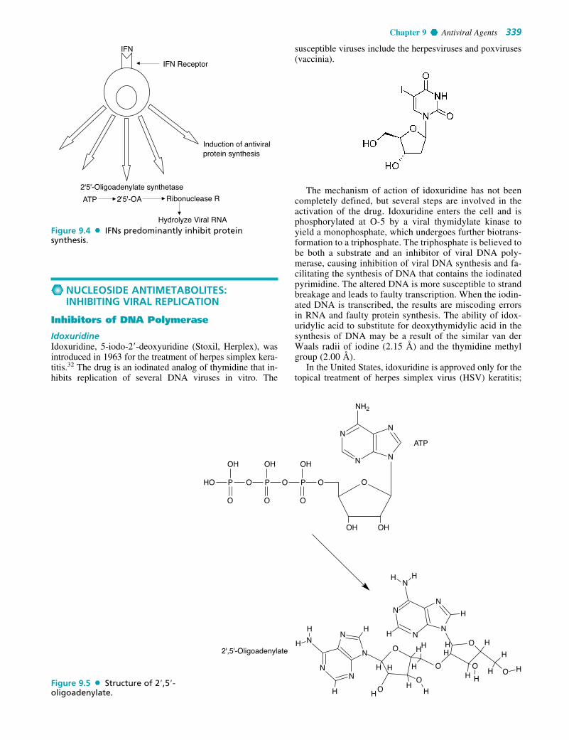
Chapter 9 Antiviral Agents 339
NUCLEOSIDE ANTIMETABOLITES:INHIBITING VIRAL REPLICATION
Inhibitors of DNA Polymerase
IdoxuridineIdoxuridine, 5-iodo-2�-deoxyuridine (Stoxil, Herplex), wasintroduced in 1963 for the treatment of herpes simplex kera-titis.32 The drug is an iodinated analog of thymidine that in-hibits replication of several DNA viruses in vitro. The
susceptible viruses include the herpesviruses and poxviruses(vaccinia).
The mechanism of action of idoxuridine has not beencompletely defined, but several steps are involved in theactivation of the drug. Idoxuridine enters the cell and isphosphorylated at O-5 by a viral thymidylate kinase toyield a monophosphate, which undergoes further biotrans-formation to a triphosphate. The triphosphate is believed tobe both a substrate and an inhibitor of viral DNA poly-merase, causing inhibition of viral DNA synthesis and fa-cilitating the synthesis of DNA that contains the iodinatedpyrimidine. The altered DNA is more susceptible to strandbreakage and leads to faulty transcription. When the iodin-ated DNA is transcribed, the results are miscoding errorsin RNA and faulty protein synthesis. The ability of idox-uridylic acid to substitute for deoxythymidylic acid in thesynthesis of DNA may be a result of the similar van derWaals radii of iodine (2.15 Å) and the thymidine methylgroup (2.00 Å).
In the United States, idoxuridine is approved only for thetopical treatment of herpes simplex virus (HSV) keratitis;
IFN
IFN Receptor
Induction of antiviralprotein synthesis
2'5'-Oligoadenylate synthetase
ATP 2'5'-OA Ribonuclease R
Hydrolyze Viral RNA
HO P O O O O
N
N
NH2
N
N
ATP
O
2',5'-Oligoadenylate
OH
P
O
OH
P
O
OH
OH
H
H
O
OO
O
O
OO
HHH
H H H
HH H
H HH H
H
HH
HH
H
H
H
H
N
N
N
N
NN
N
N
NN
OH
Figure 9.5 Structure of 2�,5�-oligoadenylate.
Figure 9.4 IFNs predominantly inhibit protein synthesis.
0330-0354_17865_Ch09.qxd 12/4/09 5:04 AM Page 339

although outside the United States, a solution of idoxuridinein dimethyl sulfoxide is available for the treatment of herpeslabialis, genitalis, and zoster. The use of idoxuridine is lim-ited because the drug lacks selectivity; low, subtherapeuticconcentrations inhibit the growth of uninfected host cells.The effective concentration of idoxuridine is at least 10times greater than that of acyclovir.
Idoxuridine occurs as a pale yellow, crystalline solid that issoluble in water and alcohol but poorly soluble in most organicsolvents. The compound is a weak acid, with a pKa of 8.25.Aqueous solutions are slightly acidic, yielding a pH of about6.0. Idoxuridine is light and heat sensitive. It is supplied as a0.1% ophthalmic solution and a 0.5% ophthalmic ointment.
CytarabineCytarabine is a pyrimidine nucleoside drug that is related toidoxuridine. This agent is primarily used as an anticanceragent for Burkitt lymphoma and myeloid and lymphaticleukemias. Cytarabine blocks the cellular utilization of de-oxycytidine, hence inhibiting the replication of viral DNA.Before it becomes active, the drug is converted to monophos-phates, diphosphates, and triphosphates, which block DNApolymerase and the C-2 reductase that converts cytidinediphosphate into the deoxy derivative.
The antiviral use of cytarabine is in the treatment of her-pes zoster (shingles), herpetic keratitis, and viral infectionsthat resist idoxuridine. Cytarabine is usually administeredtopically. Toxicity occurs on bone marrow, the gastrointesti-nal (GI) tract, and the kidneys.
TrifluridineTrifluridine, 5-trifluoromethyl-29-deoxyuridine (Viroptic),is a fluorinated pyrimidine nucleoside that demonstrates invitro inhibitory activity against HSV-1 and HSV-2, CMV,vaccinia, and some adenoviruses.33 Trifluridine possesses atrifluoromethyl group instead of an iodine atom at the 5-position of the pyrimidine ring. The van der Waals radius ofthe trifluoromethyl group is 2.44 Å, somewhat larger thanthat of the iodine atom.
Like idoxuridine, the antiviral mechanism of trifluridineinvolves inhibition of viral DNA synthesis. Trifluridinemonophosphate is an irreversible inhibitor of thymidylate
synthetase, and the biologically generated triphosphatecompetitively inhibits thymidine triphosphate incorporationinto DNA by DNA polymerase. In addition, trifluridine inits triphosphate form is incorporated into viral and cellularDNA, creating fragile, poorly functioning DNA.
Trifluridine is approved in the United States for the treat-ment of primary keratoconjunctivitis and recurrent epithelialkeratitis caused by HSV types 1 and 2. Topical trifluridineshows some efficacy in patients with acyclovir-resistantHSV cutaneous infections. Trifluridine solutions are heatsensitive and require refrigeration.
VidarabineChemically, vidarabine (Vira-A), is 9-�-D-arabinofuranosy-ladenine. The drug is the 2� epimer of natural adenosine.Introduced in 1960 as a candidate anticancer agent, vidara-bine was found to have broad-spectrum activity against DNAviruses.34 The drug is active against herpesviruses,poxviruses, rhabdoviruses, hepadnavirus, and some RNAtumor viruses. Vidarabine was marketed in the United Statesin 1977 as an alternative to idoxuridine for the treatment ofHSV keratitis and HSV encephalitis. Although the agent wasinitially prepared chemically, it is now obtained by fermenta-tion with strains of Streptomyces antibioticus.
The antiviral action of vidarabine is completely confinedto DNA viruses. Vidarabine inhibits viral DNA synthesis.Enzymes within the cell phosphorylate vidarabine to thetriphosphate, which competes with deoxyadenosine triphos-phate for viral DNA polymerase. Vidarabine triphosphate isalso incorporated into cellular and viral DNA, where it actsas a chain terminator. The triphosphate form of vidarabinealso inhibits a set of enzymes that are involved in methyla-tion of uridine to thymidine: ribonucleoside reductase, RNApolyadenylase, and S-adenosylhomocysteine hydrolase.
At one time in the United States, intravenous vidarabinewas approved for use against HSV encephalitis, neonatalherpes, and herpes or varicella zoster in immunocompro-mised patients. Acyclovir has supplanted vidarabine as thedrug of choice in these cases.
In the treatment of viral encephalitis, vidarabine had tobe administered by constant flow intravenous infusionbecause of its poor water solubility and rapid metabolic con-version to a hypoxanthine derivative in vivo. These prob-lems, coupled with the availability of less toxic and moreeffective agents, have caused intravenous vidarabine to bewithdrawn from the U. S. market.
Vidarabine occurs as a white, crystalline monohydratethat is soluble in water to the extent of 0.45 mg/mL at 25°C.The drug is still available in the United States as a 3% oint-ment for the treatment of HSV keratitis.
Adefovir DipivoxilAdefovir is an orally active prodrug that is indicated for thetreatment of the chronic form of hepatitis B. The dipivoxil
340 Wilson and Gisvold’s Textbook of Organic Medicinal and Pharmaceutical Chemistry
0330-0354_17865_Ch09.qxd 12/4/09 5:04 AM Page 340

Chapter 9 Antiviral Agents 341
moieties are hydrolyzed by ubiquitous esterases to yieldadefovir, which is phosphorylated by adenylate kinase toyield adefovir diphosphate. This compound is inhibitory atHBV DNA polymerase. In addition, adefovir undergoes in-corporation into viral DNA and causes chain termination.Adefovir is poorly absorbed by the oral route, but the dip-ivoxil ester groups cause the bioavailability to increase toapproximately 60%.
AcyclovirAcyclovir, 9-[2-(hydroxyethoxy)methyl]-9H-guanine (Zo-virax), is the most effective of a series of acyclic nucleosidesthat possess antiviral activity. In contrast with true nucleo-sides that have a ribose or a deoxyribose sugar attached to apurine or a pyrimidine base, the group attached to the basein acyclovir is similar to an open chain sugar, albeit lackingin hydroxyl groups. The clinically useful antiviral spectrumof acyclovir is limited to herpesviruses. It is most active (invitro) against HSV type 1, about two times less against HSVtype 2, and 10 times less potent against varicella–zostervirus (VZV). An advantage is that uninfected human cellsare unaffected by the drug.
The ultimate effect of acyclovir is the inhibition of viralDNA synthesis. Transport into the cell and monophosphory-lation are accomplished by a thymidine kinase that is encodedby the virus itself.35 The affinity of acyclovir for the viralthymidine kinase is about 200 times that of the correspondingmammalian enzyme. Hence, some selectivity is attained.Enzymes in the infected cell catalyze the conversion of themonophosphate to acyclovir triphosphate, which is present in40 to 100 times greater concentrations in HSV-infected thanuninfected cells. Acyclovir triphosphate competes forendogenous deoxyguanosine triphosphate (dGTP); hence,acyclovir triphosphate competitively inhibits viral DNA poly-merases. The triphosphorylated drug is also incorporated intoviral DNA, where it acts as a chain terminator. Because it hasno 3�-hydroxyl group, no 3�,5�-phosphodiester bond canform. This mechanism is essentially a suicide inhibition be-cause the terminated DNA template containing acyclovir as aligand binds to, and irreversibly inactivates, DNA poly-merase. Resistance to acyclovir can occur, most often by de-ficient thymidine kinase activity in HSV isolates. Acyclovir
resistance in vesicular stomatitis virus (VSV) isolates iscaused by mutations in VSV thymidine kinase or, less often,by mutations in viral DNA polymerase.
Two dosage forms of acyclovir are available for systemicuse: oral and parenteral. Oral acyclovir is used in the initialtreatment of genital herpes and to control mild recurrentepisodes. It has been approved for short-term treatment ofshingles and chickenpox caused by VZV. Intravenous ad-ministration is indicated for initial and recurrent infectionsin immunocompromised patients and for the prevention andtreatment of severe episodes. The drug is absorbed slowlyand incompletely from the GI tract, and its oral bioavailabil-ity is only 15% to 30%. Nevertheless, acyclovir is distrib-uted to virtually all body compartments. Less than 30% isbound to protein. Most of the drug is excreted unchanged inthe urine, about 10% excreted as the carboxy metabolite.
Acyclovir occurs as a chemically stable, white, crys-talline solid that is slightly soluble in water. Because of itsamphoteric properties (pKa values of 2.27 and 9.25), solu-bility is increased by both strong acids and bases. The in-jectable form is the sodium salt, which is supplied as alyophilized powder, equivalent to 50 mg/mL of active acy-clovir dissolved in sterile water for injection. Because thesolution is strongly alkaline (pH � 11), it must be adminis-tered by slow, constant intravenous infusion to avoid irrita-tion and thrombophlebitis at the injection site.
Adverse reactions are few. Some patients experience oc-casional GI upset, dizziness, headache, lethargy, and jointpain. An ointment composed of 5% acyclovir in a polyeth-ylene glycol base is available for the treatment of initial,mild episodes of herpes genitalis. The ointment is not an ef-fective preventer of recurrent episodes.
Valacyclovir HydrochlorideValacyclovir (Valtrex) is the hydrochloride salt of the L-valyl ester of acyclovir. The compound is a water-solublecrystalline solid, and it is a prodrug intended to increase thebioavailability of acyclovir by increasing lipophilicity.Valacyclovir is hydrolyzed rapidly and almost completely toacyclovir following oral administration. Enzymatic hydro-lysis of the prodrug is believed to occur during enterohep-atic cycling. The oral bioavailability of valacyclovir is threeto five times that of acyclovir, or about 50%.36
Valacyclovir has been approved for the treatment of her-pes zoster (shingles) in immunocompromised patients. Theside effect profile observed in valacyclovir is comparablewith bioequivalent doses of acyclovir. Less than 1% of anadministered dose of valacyclovir is recovered in the urine.Most of the dose is eliminated as acyclovir.
GanciclovirGanciclovir, 9-[(1,3-dihydroxy-2-propoxy) methyl]gua-nine) or DHPG (Cytovene), is an analog of acyclovir, with
0330-0354_17865_Ch09.qxd 12/4/09 5:04 AM Page 341

an additional hydroxymethyl group on the acyclic sidechain.
This structural modification, while maintaining the activ-ity against HSV and VSV possessed by acyclovir, greatlyenhances the activity against CMV infection.
After administration, similar to acyclovir, ganciclovir isphosphorylated inside the cell by a virally encoded proteinkinase to the monophosphate.37 Host cell enzymes catalyzethe formation of the triphosphate, which reaches more than10-fold higher concentrations in infected cells than in unin-fected cells. This selectivity is caused by the entry andmonophosphorylation step. Further phosphorylation withcellular enzymes occurs, and the triphosphate that is formedselectively inhibits viral DNA polymerase. Ganciclovirtriphosphate is also incorporated into viral DNA causingstrand breakage and cessation of elongation.38
The clinical usefulness of ganciclovir is limited by thetoxicity of the drug. Ganciclovir causes myelosuppression,producing neutropenia, thrombocytopenia, and anemia.These effects are probably associated with inhibition of hostcell DNA polymerase.39 Potential central nervous systemside effects include headaches, behavioral changes, and con-vulsions. Ganciclovir is mutagenic, carcinogenic, and tera-togenic in animals.
Toxicity limits its therapeutic usefulness to the treatmentand suppression of sight-threatening CMV retinitis in im-munocompromised patients and to the prevention of life-threatening CMV infections in at-risk transplant patients.21
Oral and parenteral dosage forms of ganciclovir are avail-able, but oral bioavailability is poor. Only 5% to 10% of anoral dose is absorbed. Intravenous administration is prefer-able. More than 90% of the unchanged drug is excreted in theurine. Ganciclovir for injection is available as a lyophilizedsodium salt for reconstitution in normal saline, 5% dextrose inwater, or lactated Ringer solution. These solutions arestrongly alkaline (pH �11) and must be administered by slow,constant, intravenous infusion to avoid thrombophlebitis.
Famciclovir and PenciclovirFamciclovir is a diacetyl prodrug of penciclovir.40 As a pro-drug, it lacks antiviral activity. Penciclovir, 9-[4-hydroxy-3-hydroxymethylbut-1-yl] guanine, is an acyclic guanine
342 Wilson and Gisvold’s Textbook of Organic Medicinal and Pharmaceutical Chemistry
0330-0354_17865_Ch09.qxd 12/4/09 5:04 AM Page 342

Chapter 9 Antiviral Agents 343
nucleoside analog. The structure is similar to that of acy-clovir, except in penciclovir, a side chain oxygen has beenreplaced by a carbon atom and an extra hydroxymethylgroup is present. Inhibitory concentrations for HSV andVSV are typically within twice that of acyclovir. Pencicloviralso inhibits the growth of hepatitis B virus.
Penciclovir inhibits viral DNA synthesis. In HSV- orVSV-infected cells, penciclovir is first phosphorylated byviral thymidine kinase41 and then further elaborated to thetriphosphate by host cell kinases. Penciclovir triphosphate isa competitive inhibitor of viral DNA polymerase. The phar-macokinetic parameters of penciclovir are quite different fromthose of acyclovir. Although penciclovir triphosphate is about100-fold less potent in inhibiting viral DNA polymerase thanacyclovir triphosphate, it is present in the tissues for longerperiods and in much higher concentrations than acyclovir.Because it is possible to rotate the side chain of penciclovirinto a pseudopentose, the triphosphorylated metabolite pos-sesses a 3�-hydroxyl group. This relationship is shown onpage 342 with guanosine. Penciclovir is not an obligate chainterminator,41 but it does competitively inhibit DNA elonga-tion. Penciclovir is excreted mostly unchanged in the urine.
Penciclovir (Denvir) has been approved for the topicaltreatment of recurrent herpes labialis (cold sores) in adults.It is effective against HSV-1 and HSV-2.42 It is available asa cream containing 10% penciclovir.
CidofovirCidofovir, (S)-3-hydroxy-2-phosphonomethoxypropyl cyto-sine (HPMPC, Vistide), is an acyclonucleotide analog thatpossesses broad-spectrum activity against several DNAviruses. Unlike other nucleotide analogs that are activated tonucleoside phosphates, Cidofovir is a phosphonic acid deriv-ative. The phosphonic acid is not hydrolyzed by phosphatasesin vivo but is phosphorylated by cellular kinases to yield adiphosphate. The diphosphate acts as an antimetabolite to de-oxycytosine triphosphate (dCTP). Cidofovir diphosphate is acompetitive inhibitor of viral DNA43 polymerase and can beincorporated into the growing viral DNA strand, causingDNA chain termination.
Cidofovir possesses a high therapeutic index against CMVand has been approved for treating CMV retinitis in patientswith AIDS. Cidofovir is administered by slow, constant intra-venous infusion in a dose of 5 mg/kg over a 1-hour periodonce a week for 2 weeks. This treatment is followed by amaintenance dose every 2 weeks. About 80% of a dose ofCidofovir is excreted unchanged in the urine, with a t1/2elim of2 to 3 hours. The diphosphate antimetabolite, in contrast, hasan extremely long half-life (17–30 hours).
The main dose-limiting toxicity of cidofovir involvesrenal impairment. Renal function must be monitoredclosely. Pretreatment with probenecid and prehydrationwith intravenous normal saline can be used to reduce thenephrotoxicity of the drug. Patients must be advised that
cidofovir is not a cure for CMV retinitis. The disease mayprogress during or following treatment.
Foscarnet SodiumTrisodium phosphonoformate is an inorganic pyrophos-phate analog that inhibits replication in herpesviruses(CMV, HSV, and VSV) and retroviruses (HIV).44 Foscarnet(Foscavir) is taken up slowly by the cells and does not un-dergo significant intracellular metabolism. Foscarnet is a re-versible, noncompetitive inhibitor at the pyrophosphate-binding site of the viral DNA polymerase and RT. Theultimate effect is inhibition of the cleavage of pyrophos-phate from deoxynucleotide triphosphates and a cessation ofthe incorporation of nucleoside triphosphates into DNA(with the concomitant release of pyrophosphate).45 Becausethe inhibition is noncompetitive with respect to nucleosidetriphosphate binding, foscarnet can act synergistically withnucleoside triphosphate antimetabolites (e.g., zidovudineand didanosine triphosphates) in the inhibition of viral DNAsynthesis. Foscarnet does not require bioactivation by viralor cellular enzymes and, hence, can be effective against re-sistant viral strains that are deficient in virally encoded nu-cleoside kinases.45
Foscarnet is a second-line drug for the treatment of retini-tis caused by CMV in patients with AIDS. The drug causesmetabolic abnormalities including increases or decreases inblood Ca2� levels. Nephrotoxicity is common, and this sideeffect precludes the use of foscarnet in other infections causedby herpesvirus or as single-agent therapy for HIV infection.Foscarnet is an excellent ligand for metal ion binding,which undoubtedly contributes to the electrolyte imbalancesobserved with the use of the drug.46 Hypocalcemia, hypomag-nesemia, hypokalemia, and hypophosphatemia and hyper-phosphatemia are observed in patients treated with foscarnet.Side effects such as paresthesias, tetani, seizures, and cardiacarrhythmias may result. Because foscarnet is nephrotoxic, itmay augment the toxic effects of other nephrotoxic drugs,such as amphotericin B and pentamidine, which are frequentlyused to control opportunistic infections in patients with AIDS.
Foscarnet sodium is available as a sterile solution intendedfor slow intravenous infusion. The solution is compatiblewith normal saline and 5% dextrose in water but is incompat-ible with calcium-containing buffers such as lactated Ringersolution and total parenteral nutrition (TPN) preparations.Foscarnet reacts chemically with acid salts such as midazo-lam, vancomycin, and pentamidine. Over 80% of an injecteddose of foscarnet is excreted unchanged in the urine.44 Thelong elimination half-life of foscarnet is thought to resultfrom its reversible sequestration into bone.46
Reverse Transcriptase Inhibitors
An early event in the replication of HIV-1 is reverse tran-scription, whereby genomic RNA from the virus is convertedinto a cDNA–RNA complex, then into double-strandedDNA ready for integration into the host chromosome. Theenzyme that catalyzes this set of reactions is reverse tran-
0330-0354_17865_Ch09.qxd 12/4/09 5:04 AM Page 343

scriptase. RT actually operates twice prior to the integrationstep. Its first function is the creation of the cDNA–RNAcomplex; RT acts alone in this step. In the second step, theRNA chain is digested away by RNase H, whereas RT cre-ates the double-stranded unintegrated DNA.
All of the classical antiretroviral agents are 2�,3�-dideoxynucleoside analogs. These compounds share a com-mon mechanism of action in inhibiting the RT of HIV.Because RT acts early in the viral infection sequence, in-hibitors of the enzyme block acute infection of cells but areonly weakly active in chronically infected ones. Eventhough the RT inhibitors share a common mechanism of ac-tion, their pharmacological and toxicological profiles differdramatically.
ZidovudineZidovudine, 3�-azido-3�-deoxythymidine or AZT, is ananalog of thymidine that possesses antiviral activityagainst HIV-1, HIV-2, HTLV-1, and several other retro-viruses. This nucleoside was synthesized in 1978 by Linand Prusoff47 as an intermediate in the preparation ofamino acid analogs of thymidine. A screening program di-rected toward the identification of agents potentially effec-tive for the treatment of patients with AIDS led to the dis-covery of its unique antiviral properties 7 years later.48 Thenext year, the clinical effectiveness of AZT in patientswith AIDS and AIDS-related complex (ARC) was demon-strated.49 AZT is active against retroviruses, a group ofRNA viruses responsible for AIDS and some kinds ofleukemia. Retroviruses possess a RT or an RNA-directedDNA polymerase that directs the synthesis of a DNA copy(proviral DNA) of the viral RNA genome that is dupli-cated, circularized, and incorporated into the DNA of aninfected cell. The drug enters the host cells by diffusionand is phosphorylated by cellular thymidine kinase.Thymidylate kinase then converts the monophosphate intodiphosphates and triphosphates. The rate-determining stepis conversion to the diphosphate, so high levels ofmonophosphorylated AZT accumulate in the cell. Lowlevels of diphosphate and triphosphate are present.Zidovudine triphosphate competitively inhibits RT withrespect to thymidine triphosphate. The 3�-azido group pre-vents formation of a 5�,3�-phosphodiester bond, so AZTcauses DNA chain termination, yielding an incompleteproviral DNA.50 Zidovudine monophosphate also compet-itively inhibits cellular thymidylate kinase, thus decreasingintracellular levels of thymidine triphosphate. The antivi-ral selectivity of AZT is caused by its greater (100)51
affinity for HIV RT than for human DNA polymerases.The human �-DNA polymerase of mitochondria is moresensitive to zidovudine; this may contribute to the toxicityassociated with the drug’s use. Resistance is common andis a result of point mutations at multiple sites in RT, lead-ing to a lower affinity for the drug.52
Zidovudine is recommended for the management of adultpatients with symptomatic HIV infection (AIDS or ARC)who have a history of confirmed Pneumocystis carinii pneu-monia or an absolute CD4� (T4 or TH cell) lymphocytecount below 200/mm3 before therapy. The hematologicaltoxicity of the drug precludes its use in asymptomatic pa-tients. Anemia and granulocytopenia are the most commontoxic effects associated with AZT.
For oral administration, AZT is supplied as 100-mg cap-sules and as a syrup containing 10 mg AZT per mL. The in-jectable form of AZT contains 10 mg/mL and is injectedintravenously. AZT is absorbed rapidly from the GI tractand distributes well into body compartments, including thecerebrospinal fluid (CSF). It is metabolized rapidly to an in-active glucuronide in the liver. Only about 15% is excretedunchanged. Because AZT is an aliphatic azide, it is heat andlight sensitive. It should be protected from light and storedat 15°C to 25°C.
DidanosineDidanosine (Videx, ddI) is 2�,3�-dideoxyinosine (ddI), asynthetic purine nucleoside analog that is bioactivated to 2�,3�-dideoxy-ATP (ddATP) by host cellular enzymes.53 The metabolite, ddATP, accumulates intracel-lularly, where it inhibits RT and is incorporated into viral DNA to cause chain termination in HIV-infected cells. The potency of didanosine is 10- to 100-foldless than that of AZT with respect to antiviral activity andcytotoxicity, but the drug causes less myelosuppressionthan AZT causes.54
Didanosine is recommended for the treatment of patientswith advanced HIV infection who have received prolongedtreatment with AZT but have become intolerant to, or expe-rienced immunosuppression from, the drug. AZT and ddIact synergistically to inhibit HIV replication in vitro, andddI is effective against some AZT-resistant strains of HIV.55
Painful peripheral neuropathy (tingling, numbness, and painin the hands and feet) and pancreatitis (nausea, abdominalpain, elevated amylase) are the major dose-limiting toxici-ties of didanosine. Didanosine is given orally in the form ofbuffered chewable tablets or as a solution prepared from thepowder. Both oral dosage forms are buffered to preventacidic decomposition of ddI to hypoxanthine in the stomach.Despite the buffering of the dosage forms, oral bioavailabil-ity is quite low and highly variable. Less than 20% of a doseis excreted in the urine, which suggests extensive metabo-lism.56 Food interferes with absorption, so the oral drugmust be given at least 1 hour before or 2 hours after meals.High-dose therapy can cause hyperuricemia in some pa-tients because of the increased purine load.
ZalcitabineZalcitabine, 2�,3�-dideoxycytidine or ddCyd, is an analog ofcytosine that demonstrates activity against HIV-1 and HIV-2,
344 Wilson and Gisvold’s Textbook of Organic Medicinal and Pharmaceutical Chemistry
0330-0354_17865_Ch09.qxd 12/4/09 5:04 AM Page 344
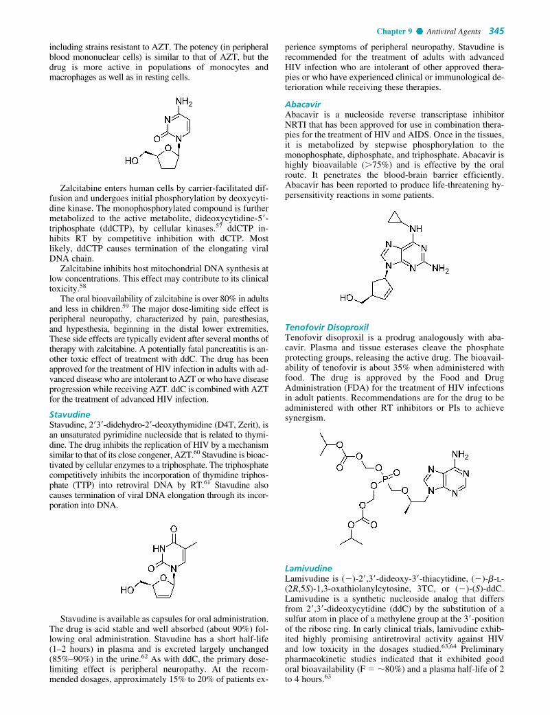
Chapter 9 Antiviral Agents 345
including strains resistant to AZT. The potency (in peripheralblood mononuclear cells) is similar to that of AZT, but thedrug is more active in populations of monocytes andmacrophages as well as in resting cells.
Zalcitabine enters human cells by carrier-facilitated dif-fusion and undergoes initial phosphorylation by deoxycyti-dine kinase. The monophosphorylated compound is furthermetabolized to the active metabolite, dideoxycytidine-5�-triphosphate (ddCTP), by cellular kinases.57 ddCTP in-hibits RT by competitive inhibition with dCTP. Mostlikely, ddCTP causes termination of the elongating viralDNA chain.
Zalcitabine inhibits host mitochondrial DNA synthesis atlow concentrations. This effect may contribute to its clinicaltoxicity.58
The oral bioavailability of zalcitabine is over 80% in adultsand less in children.59 The major dose-limiting side effect isperipheral neuropathy, characterized by pain, paresthesias,and hypesthesia, beginning in the distal lower extremities.These side effects are typically evident after several months oftherapy with zalcitabine. A potentially fatal pancreatitis is an-other toxic effect of treatment with ddC. The drug has beenapproved for the treatment of HIV infection in adults with ad-vanced disease who are intolerant to AZT or who have diseaseprogression while receiving AZT. ddC is combined with AZTfor the treatment of advanced HIV infection.
StavudineStavudine, 2�3�-didehydro-2�-deoxythymidine (D4T, Zerit), isan unsaturated pyrimidine nucleoside that is related to thymi-dine. The drug inhibits the replication of HIV by a mechanismsimilar to that of its close congener, AZT.60 Stavudine is bioac-tivated by cellular enzymes to a triphosphate. The triphosphatecompetitively inhibits the incorporation of thymidine triphos-phate (TTP) into retroviral DNA by RT.61 Stavudine alsocauses termination of viral DNA elongation through its incor-poration into DNA.
Stavudine is available as capsules for oral administration.The drug is acid stable and well absorbed (about 90%) fol-lowing oral administration. Stavudine has a short half-life(1–2 hours) in plasma and is excreted largely unchanged(85%–90%) in the urine.62 As with ddC, the primary dose-limiting effect is peripheral neuropathy. At the recom-mended dosages, approximately 15% to 20% of patients ex-
perience symptoms of peripheral neuropathy. Stavudine isrecommended for the treatment of adults with advancedHIV infection who are intolerant of other approved thera-pies or who have experienced clinical or immunological de-terioration while receiving these therapies.
AbacavirAbacavir is a nucleoside reverse transcriptase inhibitorNRTI that has been approved for use in combination thera-pies for the treatment of HIV and AIDS. Once in the tissues,it is metabolized by stepwise phosphorylation to themonophosphate, diphosphate, and triphosphate. Abacavir ishighly bioavailable (75%) and is effective by the oralroute. It penetrates the blood-brain barrier efficiently.Abacavir has been reported to produce life-threatening hy-persensitivity reactions in some patients.
Tenofovir DisoproxilTenofovir disoproxil is a prodrug analogously with aba-cavir. Plasma and tissue esterases cleave the phosphateprotecting groups, releasing the active drug. The bioavail-ability of tenofovir is about 35% when administered withfood. The drug is approved by the Food and DrugAdministration (FDA) for the treatment of HIV infectionsin adult patients. Recommendations are for the drug to beadministered with other RT inhibitors or PIs to achievesynergism.
LamivudineLamivudine is (�)-2�,3�-dideoxy-3�-thiacytidine, (�)-�-L-(2R,5S)-1,3-oxathiolanylcytosine, 3TC, or (�)-(S)-ddC.Lamivudine is a synthetic nucleoside analog that differsfrom 2�,3�-dideoxycytidine (ddC) by the substitution of asulfur atom in place of a methylene group at the 3�-positionof the ribose ring. In early clinical trials, lamivudine exhib-ited highly promising antiretroviral activity against HIVand low toxicity in the dosages studied.63,64 Preliminarypharmacokinetic studies indicated that it exhibited goodoral bioavailability (F � �80%) and a plasma half-life of 2to 4 hours.63
0330-0354_17865_Ch09.qxd 12/4/09 5:04 AM Page 345

It is interesting that the unnatural stereoisomer (�)-(S)-ddC exhibits greater antiviral activity against HIV than thenatural enantiomer (�)-(S)-ddC.65 Both enantiomers arebioactivated by cellular kinases to the correspondingtriphosphates.66 Both SddCTP isomers inhibit HIV RT andare incorporated into viral DNA to cause chaintermination. (�)-S-ddCTP inhibits cellular DNA poly-merases much more strongly than (�)-SddCTP, explainingthe greater toxicity associated with (�)-(S)-ddC. Initialmetabolic comparison of SddCTP isomers has failed to ex-plain the greater potency of the (�)-isomer against HIV.Therefore, although the intracellular accumulation of (�)-S-ddCTP was twice that of (�)-S-ddCTP, the latter wasone and a half times more potent as an inhibitor of HIVRT, and the two isomers were incorporated into viral DNAat comparable rates. The puzzle was solved with the dis-covery of a cellular 3�,5�-exonuclease, which was found tocleave terminal (�)-S-ddCMP incorporated into viralDNA 6 times faster than (�)-S-ddCMP from the viralDNA terminus.
Resistance to lamivudine develops rapidly as a result ofa mutation in codon 184 of the gene that encodes HIV-RTwhen the drug is used as monotherapy for HIV infection.64
When combined with AZT, however, lamivudine causedsubstantial increases in CD4� counts. The elevated countswere sustained over the course of therapy.67 The codon mu-tation that causes resistance to lamivudine suppresses AZTresistance,67 thus increasing the susceptibility of the virus tothe drug combination.
EmtricitabineEmtricitabine is an orally active NRTI whose pharmacoki-netics are favorable for once-daily administration.
Miscellaneous NucleosideAntimetabolites
RibavirinRibavirin is 1-�-D-ribofuranosyl-1,2,4-thiazole-3-carbox-amide. The compound is a purine nucleoside analog with amodified base and a D-ribose sugar moiety.
Ribavirin inhibits the replication of a very wide variety ofRNA and DNA viruses,68 including orthomyxoviruses,paramyxoviruses, arenaviruses, bunyaviruses, herpes-viruses, adenoviruses, poxvirus, vaccinia, influenza virus(types A and B), parainfluenza virus, and rhinovirus. In spiteof the broad spectrum of activity of ribavirin, the drug hasbeen approved for only one therapeutic indication—thetreatment of severe lower respiratory infections caused byRSV in carefully selected hospitalized infants and youngchildren.
The mechanism of action of ribavirin is not known. Thebroad antiviral spectrum of ribavirin, however, suggestsmultiple modes of action.69 The nucleoside is bioactivatedby viral and host cellular kinases to give the monophosphate(RMP) and the triphosphate (RTP). RMP inhibits inosinemonophosphate (IMP) dehydrogenase, thereby preventingthe conversion of IMP to xanthine monophosphate (XMP).XMP is required for guanosine triphosphate (GTP) synthe-sis. RTP inhibits viral RNA polymerases. It also preventsthe end capping of viral mRNA by inhibiting guanyl-N�-methyltransferase. Emergence of viral resistance to ribavirinhas not been documented.
Ribavirin occurs as a white, crystalline, polymorphicsolid that is soluble in water and chemically stable. It is sup-plied as a powder to be reconstituted in an aqueous aerosolcontaining 20 mg/mL of sterile water. The aerosol is admin-istered with a small-particle aerosol generator (SPAG).Deterioration in respiratory function, bacterial pneumonia,pneumothorax, and apnea have been reported in severely illinfants and children with RSV infection. The role of rib-avirin in these events has not been determined. Anemia,headache, abdominal pain, and lethargy have been reportedin patients receiving oral ribavirin.
Unlabeled uses of ribavirin include aerosol treatment ofinfluenza types A and B and oral treatment of hepatitis, gen-ital herpes, and Lassa fever. Ribavirin does not protect cellsagainst the cytotoxic effects of the AIDS virus.
NEWER AGENTS FOR THE TREATMENTOF HIV INFECTION
When HIV-1 was characterized and identified as thecausative agent of AIDS in 1983,70,71 scientists from all overthe world joined in the search for a prevention or cure for thedisease. Mapping the HIV-1 genome and elucidating thereplication cycle of the virus have supplied key information.72
Biochemical targets, many of which are proteins involved inthe replication cycle of the virus, have been cloned and se-quenced. These have been used to develop rapid, mechanism-based assays for the virus to complement tissue culturescreens for whole virus. Several of the biochemical steps that
346 Wilson and Gisvold’s Textbook of Organic Medicinal and Pharmaceutical Chemistry
0330-0354_17865_Ch09.qxd 12/4/09 5:04 AM Page 346
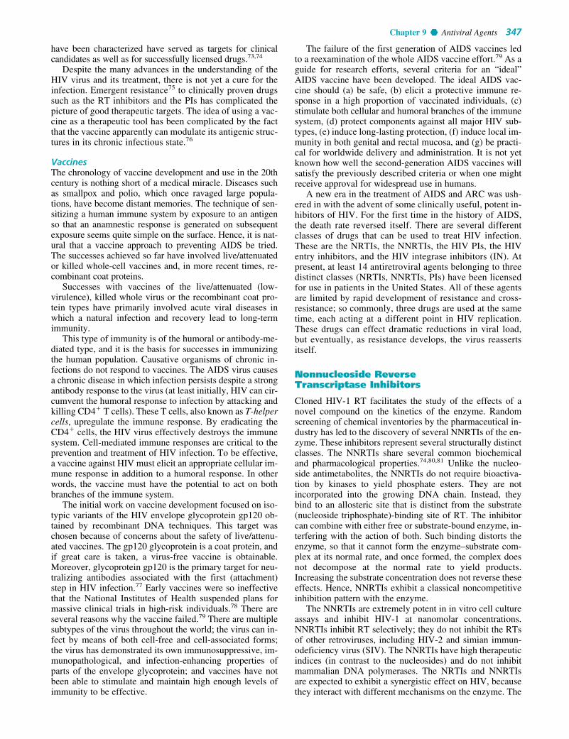
Chapter 9 Antiviral Agents 347
have been characterized have served as targets for clinicalcandidates as well as for successfully licensed drugs.73,74
Despite the many advances in the understanding of theHIV virus and its treatment, there is not yet a cure for theinfection. Emergent resistance75 to clinically proven drugssuch as the RT inhibitors and the PIs has complicated thepicture of good therapeutic targets. The idea of using a vac-cine as a therapeutic tool has been complicated by the factthat the vaccine apparently can modulate its antigenic struc-tures in its chronic infectious state.76
VaccinesThe chronology of vaccine development and use in the 20thcentury is nothing short of a medical miracle. Diseases suchas smallpox and polio, which once ravaged large popula-tions, have become distant memories. The technique of sen-sitizing a human immune system by exposure to an antigenso that an anamnestic response is generated on subsequentexposure seems quite simple on the surface. Hence, it is nat-ural that a vaccine approach to preventing AIDS be tried.The successes achieved so far have involved live/attenuatedor killed whole-cell vaccines and, in more recent times, re-combinant coat proteins.
Successes with vaccines of the live/attenuated (low-virulence), killed whole virus or the recombinant coat pro-tein types have primarily involved acute viral diseases inwhich a natural infection and recovery lead to long-termimmunity.
This type of immunity is of the humoral or antibody-me-diated type, and it is the basis for successes in immunizingthe human population. Causative organisms of chronic in-fections do not respond to vaccines. The AIDS virus causesa chronic disease in which infection persists despite a strongantibody response to the virus (at least initially, HIV can cir-cumvent the humoral response to infection by attacking andkilling CD4� T cells). These T cells, also known as T-helpercells, upregulate the immune response. By eradicating theCD4� cells, the HIV virus effectively destroys the immunesystem. Cell-mediated immune responses are critical to theprevention and treatment of HIV infection. To be effective,a vaccine against HIV must elicit an appropriate cellular im-mune response in addition to a humoral response. In otherwords, the vaccine must have the potential to act on bothbranches of the immune system.
The initial work on vaccine development focused on iso-typic variants of the HIV envelope glycoprotein gp120 ob-tained by recombinant DNA techniques. This target waschosen because of concerns about the safety of live/attenu-ated vaccines. The gp120 glycoprotein is a coat protein, andif great care is taken, a virus-free vaccine is obtainable.Moreover, glycoprotein gp120 is the primary target for neu-tralizing antibodies associated with the first (attachment)step in HIV infection.77 Early vaccines were so ineffectivethat the National Institutes of Health suspended plans formassive clinical trials in high-risk individuals.78 There areseveral reasons why the vaccine failed.79 There are multiplesubtypes of the virus throughout the world; the virus can in-fect by means of both cell-free and cell-associated forms;the virus has demonstrated its own immunosuppressive, im-munopathological, and infection-enhancing properties ofparts of the envelope glycoprotein; and vaccines have notbeen able to stimulate and maintain high enough levels ofimmunity to be effective.
The failure of the first generation of AIDS vaccines ledto a reexamination of the whole AIDS vaccine effort.79 As aguide for research efforts, several criteria for an “ideal”AIDS vaccine have been developed. The ideal AIDS vac-cine should (a) be safe, (b) elicit a protective immune re-sponse in a high proportion of vaccinated individuals, (c)stimulate both cellular and humoral branches of the immunesystem, (d) protect components against all major HIV sub-types, (e) induce long-lasting protection, (f) induce local im-munity in both genital and rectal mucosa, and (g) be practi-cal for worldwide delivery and administration. It is not yetknown how well the second-generation AIDS vaccines willsatisfy the previously described criteria or when one mightreceive approval for widespread use in humans.
A new era in the treatment of AIDS and ARC was ush-ered in with the advent of some clinically useful, potent in-hibitors of HIV. For the first time in the history of AIDS,the death rate reversed itself. There are several differentclasses of drugs that can be used to treat HIV infection.These are the NRTIs, the NNRTIs, the HIV PIs, the HIVentry inhibitors, and the HIV integrase inhibitors (IN). Atpresent, at least 14 antiretroviral agents belonging to threedistinct classes (NRTIs, NNRTIs, PIs) have been licensedfor use in patients in the United States. All of these agentsare limited by rapid development of resistance and cross-resistance; so commonly, three drugs are used at the sametime, each acting at a different point in HIV replication.These drugs can effect dramatic reductions in viral load,but eventually, as resistance develops, the virus reassertsitself.
Nonnucleoside Reverse Transcriptase Inhibitors
Cloned HIV-1 RT facilitates the study of the effects of anovel compound on the kinetics of the enzyme. Randomscreening of chemical inventories by the pharmaceutical in-dustry has led to the discovery of several NNRTIs of the en-zyme. These inhibitors represent several structurally distinctclasses. The NNRTIs share several common biochemicaland pharmacological properties.74,80,81 Unlike the nucleo-side antimetabolites, the NNRTIs do not require bioactiva-tion by kinases to yield phosphate esters. They are notincorporated into the growing DNA chain. Instead, theybind to an allosteric site that is distinct from the substrate(nucleoside triphosphate)-binding site of RT. The inhibitorcan combine with either free or substrate-bound enzyme, in-terfering with the action of both. Such binding distorts theenzyme, so that it cannot form the enzyme–substrate com-plex at its normal rate, and once formed, the complex doesnot decompose at the normal rate to yield products.Increasing the substrate concentration does not reverse theseeffects. Hence, NNRTIs exhibit a classical noncompetitiveinhibition pattern with the enzyme.
The NNRTIs are extremely potent in in vitro cell cultureassays and inhibit HIV-1 at nanomolar concentrations.NNRTIs inhibit RT selectively; they do not inhibit the RTsof other retroviruses, including HIV-2 and simian immun-odeficiency virus (SIV). The NNRTIs have high therapeuticindices (in contrast to the nucleosides) and do not inhibitmammalian DNA polymerases. The NRTIs and NNRTIsare expected to exhibit a synergistic effect on HIV, becausethey interact with different mechanisms on the enzyme. The
0330-0354_17865_Ch09.qxd 12/4/09 5:04 AM Page 347

chief problem with the NNRTIs is the rapid emergence ofresistance among HIV isolates.75 Resistance is a result ofpoint mutations in the gene coding for the enzyme. Cross-resistance between structurally different NNRTIs is morecommon than between NNRTIs and NRTIs. In the future,clinical use of the NNRTIs is expected to use combinationswith the nucleosides to reduce toxicity to the latter, to takeadvantage of additive or synergistic effects, and to reducethe emergence of viral resistance.75,80 The tricyclic com-pound nevirapine (Viramune),82 the bis(heteroacyl)piper-azine (BHAP) derivative delavirdine (Rescriptor),83 andefavirenz84 have been approved for use in combination withNRTIs such as AZT for the treatment of HIV infection.Numerous others, including the quinoxaline derivative GW-420867X,84 the tetrahydroimidazobenzodiazpinone (TIBO)analog R-82913,85 and calanolide-A84 are in clinical trials.
NevirapineNevirapine (Viramune)82 is more than 90% absorbed by theoral route and is widely distributed throughout the body. Itdistributes well into breast milk and crosses the placenta.Transplacental concentrations are about 50% those ofserum. The drug is extensively transformed by cytochromeP450 (CYP) to inactive hydroxylated metabolites; it mayundergo enterohepatic recycling.
The half-life decreases from 45 to 23 hours over a 2- to4-week period because of autoinduction. Elimination occursthrough the kidney, with less than 3% of the parent com-pound excreted in the urine.82 Dosage forms are supplied asa 50 mg/5 mL oral suspension and a 200-mg tablet.
DelavirdineDelavirdine (Rescriptor)83 must be used with at least twoadditional antiretroviral agents to treat HIV-1 infections.The oral absorption of delavirdine is rapid, and peakplasma concentrations develop in 1 hour. Extensive metab-olism occurs in the liver by CYP isozyme 3A (CYP3A) orpossibly CYP2D6. Bioavailability is 85%. Unlike nevirap-ine, which is 48% protein bound, delavirdine is more than98% protein bound. The half-life is 2 to 11 hours, andelimination is 44% in feces, 51% in urine, and less than 5%unchanged in urine. Delavirdine induces its own metabo-lism.83 Oral dosage forms are supplied as a 200-mg cap-sule and a 100-mg tablet.
EfavirenzEfavirenz (Sustiva)84 is also mandated for use with at leasttwo other antiretroviral agents. The compound is morethan 99% protein bound, and CSF concentrations exceedthe free fraction in the serum. Metabolism occurs in theliver. The half-life of a single dose of efavirenz is 52 to 76hours, and 40 to 55 after multiple doses (the drug inducesits own metabolism). Peak concentration is achieved in 3to 8 hours. Elimination is 14% to 34% in urine (as metabo-lites) and 16% to 41% in feces (primarily as efavirenz).84
The oral dosage form is supplied as a capsule.
HIV Protease Inhibitors
A unique biochemical target in the HIV-1 replication cyclewas revealed when HIV protease was cloned and ex-pressed86,87 in Escherichia coli. HIV protease is an en-zyme that cleaves gag-pro propeptides to yield active en-zymes that function in the maturation and propagation ofnew virus. The catalytically active protease is a symmetricdimer of two identical 99 amino acid subunits, each con-tributing the triad Asp-Thr-Gly to the active site.86,87 Thehomodimer is unlike monomeric aspartyl proteases (renin,pepsin, cathepsin D), which also have different substratespecificities. The designs of some inhibitors86,87 for HIV-1 protease exploit the C2 symmetry of the enzyme. HIV-1protease has active site specificity for the triad Tyr-Phe-Pro in the unit Ser-(Thr)-Xaa-Xaa-Tyr-Phe-Pro, whereXaa is an arbitrary amino acid.
HIV PIs are designed to mimic the transition state ofhydrolysis at the active site; these compounds are calledanalog inhibitors. Hydrolysis of a peptide bond proceedsthrough a transition state that is sp3 hybridized and, hence,tetrahedral. The analog inhibitors possess a preexisting sp3
hybridized center that will be drawn into the active site(one hopes with high affinity) but will not be cleavable bythe enzyme. This principle has been used to prepare hun-dreds of potentially useful transition state inhibitors.86,87
Unfortunately, very few of these are likely to be clinicallysuccessful candidates for the treatment of HIV infection.Because HIV PIs are aimed at arresting replication of thevirus at the maturation step to prevent the spread of cellu-lar infection, they should possess good oral bioavailabilityand a relatively long duration of action. A long half-life isalso desirable because of the known development of resist-ance by HIV under selective antiviral pressure.74,75
Resistance develops by point mutations.Most of the early PIs are high–molecular-weight, dipep-
tide- or tripeptide-like structures, generally with low watersolubility. The bioavailability of these compounds is low,and the half-life of elimination is very short because of hy-drolysis or hepatic metabolism.88 Strategies aimed at in-
348 Wilson and Gisvold’s Textbook of Organic Medicinal and Pharmaceutical Chemistry
0330-0354_17865_Ch09.qxd 12/4/09 5:04 AM Page 348

Chapter 9 Antiviral Agents 349
creasing water solubility and metabolic stability have ledto the development of several highly promising clinicalcandidates. Saquinavir (Invirase),81 indinavir (Crixivan),89
ritonavir (Norvir),90 nelfinavir (Viracept),91 and ampre-navir (Agenerase)92 have been approved for the treatmentof HIV-infected patients. Several others are in clinicaltrials.
There is an important caution for the use of PIs. As aclass, they cause dyslipidemia, which includes elevated cho-lesterol and triglycerides and a redistribution of body fatcentrally to cause the “protease paunch” buffalo hump, fa-cial atrophy, and breast enlargement. These agents alsocause hyperglycemia.
SaquinavirSaquinavir (Invirase)89 is well tolerated following oral ad-ministration. Absorption of saquinavir is poor but is in-creased with a fatty meal. The drug does not distribute intothe CSF, and it is approximately 98% bound to plasma pro-teins. Saquinavir is extensively metabolized by the first-pass effect. Bioavailability is 4% from a hard capsule and12% to 15% from a soft capsule. Saquinavir lowers p24antigen levels in HIV-infected patients, elevates CD4�
counts, and exerts a synergistic antiviral effect when com-bined with RT inhibitors such as AZT and ddC.93–95
Although HIV-1 resistance to saquinavir and other HIVPIs occurs in vivo, it is believed to be less stringent andless frequent than resistance to the RT inhibitors.96
Nevertheless, cross-resistance between different HIV PIsappears to be common and additive,97 suggesting thatusing combinations of inhibitors from this class would notconstitute rational prescribing. The drug should be used incombination with at least two other antiretroviral drugs tominimize resistance. Dosage forms are Invirase (hard cap-sule) and Fortovase (soft capsule).
IndinavirWhen administered with a high-fat diet, indinavir(Crixivan)90 achieves a maximum serum concentration of77% of the administered dose. The drug is 60% bound inthe plasma. It is extensively metabolized by CYP3A4, andseven metabolites have been identified. Oral bioavailabil-ity is good, with a tmax of 0.8 � 0.3 hour. The half-life ofelimination is 1.8 hour, and the elimination products aredetectable in feces and urine. Indinavir also causes dyslipi-demia. The available dosage forms are capsules of 200 mg,333 mg, and 400 mg.
Ritonavir, Amprenavir, and NelfinavirRitonavir (Norvir),98 amprenavir (Agenerase),99 and nelfi-navir (Viracept)100 have similar properties and cautionarystatements. All cause dyslipidemia, and they have a host ofdrug interactions, mainly because they inhibit CYP3A4.These agents must always be used with at least two other an-tiretroviral agents. Used properly, the PIs are an importantpart of HIV therapy.
LopinavirLopinavir is a protease inhibitor that has been approved foruse in combination with ritonavir for patients with HIV whohave not responded to other treatment modalities. Lopinaviris used in excess over ritonavir. Ritonavir at amounts givenhas no antiretroviral activity, Ritonavir inhibits lopinavir’smetabolism by CYP3A4, causing a higher level of lopinavirin the system. The combination is the first protease inhibitorapproved for patients as young as 6 months of age.
0330-0354_17865_Ch09.qxd 12/4/09 5:04 AM Page 349
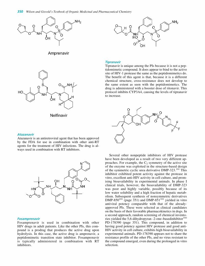
AtazanavirAtazanavir is an antiretroviral agent that has been approvedby the FDA for use in combination with other anti-RTagents for the treatment of HIV infections. The drug is al-ways used in combination with RT inhibitors.
FosamprenavirFosamprenavir is used in combination with other HIV drugs in adult patients. Like the other PIs, this com-pound is a prodrug that produces the active drug uponhydrolysis. In this case, the active drug is amprenavir, apeptidomimetic transition state inhibitor. Fosamprenavir is typically administered in combination with RTinhibitors.
TipranavirTipranavir is unique among the PIs because it is not a pep-tidomimetic compound. It does appear to bind to the activesite of HIV-1 protease the same as the peptidomimetics do.The benefit of this agent is that, because it is a differentchemical structure, cross-resistance does not develop tothe same extent as seen with the peptidomimetics. Thedrug is administered with a booster dose of ritonavir. Thisprotocol inhibits CYP3A4, causing the levels of tipranavirto increase.
Several other nonpeptide inhibitors of HIV proteasehave been developed as a result of two very different ap-proaches. For example, the C2 symmetry of the active siteof the enzyme was exploited in the structure-based designof the symmetric cyclic urea derivative DMP-323.101 Thisinhibitor exhibited potent activity against the protease invitro, excellent anti-HIV activity in cell culture, and prom-ising bioavailability in experimental animals. In phase Iclinical trials, however, the bioavailability of DMP-323was poor and highly variable, possibly because of itslow water solubility and a high fraction of hepatic metab-olism. Subsequent synthesis of nonsymmetric derivativesDMP-850101 (page 351) and DMP-851101 yielded in vitroantiviral potency comparable with that of the already-approved PIs. These were selected as clinical candidateson the basis of their favorable pharmacokinetics in dogs. Ina second approach, random screening of chemical invento-ries yielded the 5,6-dihydropyran -2-one–basedinhibitor102
PD-178390 (page 351). This compound, in addition tohaving good potency against HIV protease and good anti-HIV activity in cell culture, exhibits high bioavailability inexperimental animals. PD-178390 appears not to share theresistance profile of the other PIs, and no virus resistant tothe compound emerged, even during the prolonged in vitroselection.
350 Wilson and Gisvold’s Textbook of Organic Medicinal and Pharmaceutical Chemistry
0330-0354_17865_Ch09.qxd 12/4/09 5:04 AM Page 350

Chapter 9 Antiviral Agents 351
Dipeptide PIs containing 2-hydroxy-3-amino-4-arylbu-tanoic acid in their scaffold showed promising preclinicalresults. JE-2147103, containing the allophenylnorstatin moi-ety, exhibited potent in vitro anti-HIV activity. JE-2147 ap-pears to fully retain its susceptibility against various HIVstrains resistant to multiple approved PIs and exhibits goodoral bioavailability and a good pharmacokinetic profile intwo animal species. Also, emergence of resistance was con-siderably delayed with JE-2147.
HIV Entry Inhibitors
Entry of HIV into a cell is a complex process that involvesseveral specific membrane protein interactions. Initially, viralglycoprotein gp120 mediates the virus attachment via itsbinding to at least two host membrane receptors, CD4� andthe chemokine coreceptor. This bivalent interaction induces aconformational change in the viral fusion protein gp41.Protein gp41 acts as the anchor for gp120 in the virus. Withthe conformational change, the viral envelope fuses with the
host cell membrane. In addition to gp120–chemokine recep-tor interaction, the fusion activity of gp41 is currently beingexplored as a novel target for antiretroviral therapy. At leastone agent from each class is in clinical testing.
Chemokine Receptor Binders
Most HIV-1 isolates rely on the CCR5 coreceptor for entry(R5 strains). In later stages of the disease, however, morepathogenic selection variants of the virus emerge in about40% of individuals, which use the CXCR4 coreceptor in ad-dition to CCR5 (R5X4 strains) or the CXCR4 receptor only(X4 strains). Bicyclam compound AMD-3100104 was thefirst compound identified as a CXCR4-specific inhibitorthat interferes with the replication of X4 but not R5 viruses.The compound is currently in phase II clinical evaluations.It is used as an injectable agent because of its limitedbioavailability.
Several positively charged 9- to14-mer peptides havebeen described as capable of blocking the CXCR4 corecep-tor. A small molecule, TAK-779,105 exhibits high-affinitybinding to the CCR5 coreceptor, specifically blocking R5isolates.
0330-0354_17865_Ch09.qxd 12/4/09 5:04 AM Page 351

Inhibitors of GP41 Fusion Activity
The fusion of the HIV-1 viral envelope with host plasmamembrane is mediated by gp41, a transmembrane subunit ofthe HIV-1 glycoprotein subunit complex. Pentafuside106 (T-20) is a 36-mer peptide that is derived from the C-terminalrepeat of gp41. Pentafuside appears to inhibit the formationof the fusion-competent conformation of gp41 by interferingwith the interaction between its C- and N-terminal repeat.Pentafuside is a potent inhibitor of HIV-1 clinical isolates,and it is currently in clinical trials.
Integrase Inhibitors
Two closely related types of small molecules that blockstrand transfer catalyzed by recombinant integrase havebeen identified. Both types show in vitro antiviral activity.The diketo acids107 inhibit strand transfer catalyzed by re-combinant integrase with an IC50 less than 0.1 �m.Mutations that conferred resistance to the diketo acidsmapped near conserved residues in the IN enzyme. Thisfinding demonstrates that the compounds have a highlyspecific mechanism of action. X-ray crystallography of thebound tetrazole108 derivative revealed that the inhibitorwas centered in the active site of IN near acidic catalyticresidues.
Short-Interfering RNA
RNA interference is a phenomenon that has been recentlyused as a way to silence genes that are part of viral replica-tion cycles. The siRNAs very specifically interrupt genefunction and switch off some aspect of a disease. ThesiRNAs are found in higher organisms (eukaryotes) and aretypically double-stranded duplexes of RNA of about 21 base
pairs. In nature, these siRNAs are produced by excisionfrom a parent duplex RNA molecule. The siRNAs instructthe cell to split specific mRNAs that have identical se-quences as the siRNAs. As antiviral therapy, the idea wouldbe for the siRNAs to stop synthesis of mRNAs of a patho-genic organism. Antiviral siRNA therapies can be tailoredfor any given pathogen. The use of siRNAs as potential ther-apeutic modalities is a highly active area of research at thepresent time.
Combination Antiviral Therapy
Combination antiviral therapy, in which a mixture of drugspossessing different mechanisms of action, has beenshown to be advantageous for several reasons. The antivi-ral effect of the combination is excellent, toxicities are de-creased, and resistance to any drug in the combination isslow to develop. Resistance to single drugs such as aman-tadine, ganciclovir, and acyclovir is problematic.Administered of a given agent in combination with othertypes of drugs retards the development of such resistance.Combination treatment is especially important in antiretro-viral therapy.
Typical antiretroviral therapy, as exemplified by HIVtreatment, includes combinations of NRTI or NNRTIalong with PIs. The key that makes the combination workis that the drugs act to inhibit HIV virus replication at dif-ferent stages of the viral infective cycle. The RT inhibitors(NRTIs or NNRTIs) prevent RNA formation or viral pro-tein synthesis or inactivate the catalytic site of RT(NNRTIs). The PIs act once the provirus integrates into thehost’s genes. Protease is necessary to split viral precursorpolypeptides into new virus. The combination of RTs andPIs is synergistic. There are many two- and three-drugcombinations that have been reported to be useful in vari-ous viral infections.
352 Wilson and Gisvold’s Textbook of Organic Medicinal and Pharmaceutical Chemistry
0330-0354_17865_Ch09.qxd 12/4/09 5:04 AM Page 352

6. Describe the mechanism of action of the IFNs as antivi-ral agents.
7. Describe the class of agents that we call NRRTIs.
8. What kinds of drugs are typically used in combinationtherapy of HIV?
9. Describe the HIV entry inhibitors.
10. How do the HIV protease inhibitors exert their mecha-nism of action?
1. What is the difference between the lytic cycle and thelysogenic cycle in viruses?
2. What is an RNA virus? What is a DNA virus?
3. What is the mechanism of action of an antimetabolite an-tiviral drug?
4. What is chemoprophylaxis? How might you exercisechemoprophylaxis against the influenza virus?
5. What is the mechanism of action of amantadine?
R E V I E W Q U E S T I O N S
Chapter 9 Antiviral Agents 353
REFERENCES1. Condit, R. C.: Principles of virology. In Knipe, D. M., and Howley,
P. M. (eds.). Fundamental Virology, 4th ed. New York, LippincottWilliams & Wilkins, 2001, p. 19.
2. Harrison, S. C.: Principles of viral structure. In Knipe, D. M., andHowley, P. M. (eds.). Fundamental Virology, 4th ed. New York,Lippincott Williams & Wilkins, 2001, p. 53.
3. Wagner, E. K., and Hewlett, M. J. (eds.): Basic Virology. Malden,MA, Blackwell Science, 1999, p. 12.
4. Wagner, E. K., and Hewlett, M. J. (eds.): Basic Virology. Malden,MA, Blackwell Science, 1999, p. 61.
5. Beale, J. M., Jr.: Immunobiologicals. In Block, J. H., and Beale, J.M., Jr. (eds.). Wilson and Gisvold’s Textbook of Organic Medicinaland Pharmaceutical Chemistry, 11th ed. Baltimore, LippincottWilliams & Wilkins, 2004, p. 10.
6. Freshney, R. I.: Culture of Animal Cells, 3rd ed. New York, Wiley-Liss, 1994.
7. Young, J. A. T.: Virus entry and uncoating. In Knipe, D. M., andHowley, P. M. (eds.). Fundamental Virology, 4th ed. New York,Lippincott Williams & Wilkins, 2001, p. 87.
8. Hunter, E.: Virus assembly. In Knipe, D. M., and Howley, P. M.(eds.). Fundamental Virology, 4th ed. New York, LippincottWilliams & Wilkins, 2001, p. 171.
9. Lamb, R. A., and Choppin, R. W.: Annu. Rev. Biochem. 52:467,1983.
10. Cann, A. J.: Principles of Molecular Virology, 3rd ed. New York,Academic Press, 2001, p. 114.
11. Tyler, K. L., and Nathanson, N.: Pathogenesis of viral infections. InKnipe, D. M., and Howley, P. M. (eds.). Fundamental Virology, 4thed. New York, Lippincott Williams & Wilkins, 2001, pp. 214–220.
12. Dimitrov, R. S.: Cell 91:721, 1997.13. Dingwell, K. S., Brunetti, C. R., Hendricks, R. L., et al.: J. Virol.
68:834, 1994.14. Haywood, A. M.: J. Virol. 68:1, 1994.15. Norkin, L. C.: Clin. Microbiol. Rev. 8:298–315, 1995.16. Chesebro, B., Buller, R., Portis, J., et al.: J. Virol. 64:215, 1990.17. Clapham, P. R., Blanc, D., and Weiss, R. A.: Virology 181:703, 1991.18. Harrington, R. D., and Geballe, A. P.: J. Virol. 67:5939, 1993.19. Haywood, A. M.: J. Virol. 68:1, 1994.20. Young, J. A. T.: Virus entry and uncoating. In Knipe, D. M., and
Howley, P. M. (eds.). Fundamental Virology, 4th ed. New York,Lippincott Williams & Wilkins, 2001, p. 96
21. Wagner, E. K., and Hewlett, M. J. (eds.): Basic Virology. Malden,MA, Blackwell Science, 1999, p. 257
22. Mitsuya, H., and Broder, S.: Proc. Natl. Acad. Sci. U. S. A. 83:1911,1986.
23. Johnson, M. A., et al.: J. Biol. Chem. 263:1534, 1988.24. Kohl, N. E., et al.: Proc. Natl. Acad. Sci. U. S. A. 85:4686, 1988.25. Hay, A. J.: Semin. Virol. 3:21, 1992.26. Hayden, F. G., Belsche, R. B., Clover, R. D., et al.: N. Engl. J. Med.
321:1696, 1989.27. Douglas, R. G.: N. Engl. J. Med. 322:443, 1990.28. Capparelli, E. V., Stevens, R. C., and Chow, M. S.: Clin. Pharmacol.
Ther. 43:536, 1988.
29. Baron, S., et al.: Introduction to the interferon system. In Baron, S.,Dianzani, F., Stanton, G., et al. (eds.). Interferon: Principles andMedical Applications. Galveston, University of Texas, Texas MedicalBranch, 1992, pp. 1–15.
30. Sen, G. C., and Ransohoff, R. M.: Adv. Virus Res. 42:57, 1993.31. Bocci, V.: HIV proteases. In Baron, S., Dianzani, F., Stanton, G., et
al. (eds.). Interferon: Principles and Medical Applications. Galveston,University of Texas, Texas Medical Branch, 1992, pp. 417–425.
32. Prusoff, W. H.: Idoxuridine or how it all began. In DeClerq, E. (ed.).Clinical Use of Antiviral Drugs. Norwell, MA, Martinus Nijhoff,1988, pp. 15–24.
33. Birch, et al.: J. Infect. Dis. 166:108, 1992.34. Pavin-Langston, D., et al.: Adenosine Arabinoside: An Antiviral
Agent. New York, Raven Press, 1975.35. Schaeffer, H. J., et al.: Nature 272:583, 1978.36. Weller, S., et al.: Clin. Pharmacol. Ther. 54:595, 1993.37. Sullivan, V., et al.: Nature 358:162, 1992.38. Clair, M. H., et al.: Antimicrob. Agents Chemother. 25:191, 1984.39. Faulds, D., and Heel, R. C.: Drugs 39:597, 1990.40. Vere Hodge, R. A.: Antiviral Chem. Chemother. 4:67, 1993.41. Earnshaw, D. L., et al.: Antimicrob. Agents Chemother. 36:2747, 1992.42. Alrabiah, F. A., and Sachs, S. L.: Drugs 52:17, 1996.43. Xiong, X., et al.: Biochem. Pharmacol. 51:1563, 1996.44. Chrisp, P., and Chessold, S. P.: Drugs 41:104, 1991.45. Crumpacker, C. S.: Am. J. Med. 92(Suppl. 2A):25, 1992.46. Jacobson, M. A., et al.: J. Clin. Endocrinol. Metab. 72:1130, 1991.47. Lin, T. S., and Prusoff, W. H.: J. Med. Chem. 21:109, 1978.48. Mitsuya, H., et al.: Proc. Natl. Acad. Sci. U. S. A. 82:7096, 1985.49. Yarchoan, R., et al.: Lancet 1:575, 1986.50. Furman, P. A., et al.: Proc. Natl. Acad. Sci. U. S. A. 873:8333, 1986.51. St. Clair, M. H., et al.: Antimicrob. Agents Chemother. 31:1972, 1987.52. Richman, D. D., et al.: J. Infect. Dis. 164:1075, 1991.53. Johnson, M. A., and Fridland, A.: Mol. Pharmacol. 36:291, 1989.54. McLaren, C., et al.: Antiviral Chem. Chemother. 2:321, 1991.55. Johnson, V. A., et al.: J. Infect. Dis. 164:646, 1991.56. Knupp, C. A., et al.: Clin. Pharmacol. Ther. 49:523, 1991.57. Yarchoan, R., et al.: N. Engl. J. Med. 321:726, 1989.58. Chen, C., Vazquez-Padua, M., and Cheng, Y.: Mol. Pharmacol.
39:625, 1991.59. Broder, S.: Am. J. Med. 88(Suppl. 5B):25, 1990.60. Ho, H. T., and Hitchcock, M. J. M.: Antimicrob. Agents Chemother.
33:844, 1989.61. Huang, P., Farquhar, D., and Plunkett, W.: J. Biol. Chem. 267:2817,
1992.62. Browne, M. J., et al.: J. Infect. Dis. 167:21, 1993.63. Van Leeuwen, R., et al.: J. Infect. Dis. 171:1166, 1995.64. Pluda, J. M., et al.: J. Infect. Dis. 171:1438, 1995.65. Coates, J. A. V., et al.: Antimicrob. Agents Chemother. 36:202,
1992.66. Skalski, V., et al.: J. Biol. Chem. 268:23234, 1993.67. Larder, B. A., et al.: Science 269:696, 1995.68. Sidwell, R. W., et al.: Science 177:705, 1972.69. Robins, R. K.: Chem. Eng. News Jan. 27:28, 1986.70. Gallo, R. C., et al.: Science 220:865, 1983.
0330-0354_17865_Ch09.qxd 12/4/09 5:04 AM Page 353
71. Barré-Sinoussi, F., et al.: Science 220:868, 1983.72. Haseltine, W. A.: FASEB J. 5:2349, 1991.73. Yarchoan, R., Mitsuya, H., and Broder, S.: Trends Pharmacol. Sci.
14:196, 1993.74. DeClerq, E.: J. Med. Chem. 38:2491, 1995.75. Richman, D. D.: Annu. Rev. Pharmacol. Toxicol. 32:149, 1993.76. Cease, K. B., and Berzofsky, J. A.: Annu. Rev. Immunol. 12:923,
1994.77. Lasky, L. A., et al.: Science 23:209, 1986.78. Cohen, J.: Science 264:1839, 1994.79. Koff, W. C.: Science 266:1335, 1994.80. Spence, R. A., et al.: Science 267:988, 1995.81. Vacca, J. P., et al.: Proc. Natl. Acad. Sci. U. S. A. 91:4096, 1994.82. Merluzzi, U. T., et al.: Science 250:1411, 1990.83. Romero, D. L.: Drugs Future 19:7, 1994.84. Pedersen, O., and Pedersen, E.: Antiviral Chem. Chemother. 10:285,
1999.85. Pialoux, G., et al.: Lancet 338:140, 1991.86. Wlodawer, R., and Erickson, J. W.: Annu. Rev. Biochem. 62:543,
1993.87. Chow, Y. K., et al.: Nature 361:650, 1993.88. Roberts, N. A., et al.: Science 248:358, 1990.89. Kun, E. E., et al.: J. Am. Chem. Soc. 117:1181, 1995.
90. Kempf, D. J., et al.: Proc. Natl. Acad. Sci. U. S. A. 92:2484, 1995.91. Nelfinavir. St. Louis, Facts and Comparisons, 2000, p. 1431.92. Kageyama, S., et al.: Antimicrob. Agents Chemother. 37:810,
1993.93. Reich, S. H., et al.: Proc. Natl. Acad. Sci. U. S. A. 92:3298, 1995.94. Johnson, V. A., Merrill, D. P., Chou, T.-C., et al.: J. Infect. Dis.
166:1143, 1992.95. Craig, J. C., et al.: Antiviral Chem. Chemother. 4:161, 1993.96. Craig, J. C., et al.: Antiviral Chem. Chemother. 4:335, 1993.97. Condra, J. H., et al.: Nature 374:569, 1995.98. Wei, X., et al.: Nature 373:117, 1995.99. Ho, D. D., et al.: Nature 373:123, 1995.
100. Kageyama, S., et al.: Antimicrob. Agents Chemother. 37:810,1993.
101. DeLucca, G., et al.: Pharm. Biotechnol. 11:257, 1998.102. Prasad, J., et al.: Bioorg. Med. Chem. 7:2775, 1999.103. Yoshimura, K., et al.: Proc. Natl. Acad. Sci. U. S. A. 96:8675, 1999.104. Hendrix, C., et al.: 6th Conference on Retroviruses and
Opportunistic Infections, Abstr. 610, 1999.105. Baba, M., et al.: Proc. Natl. Acad. Sci. U. S. A. 96:5698, 1999.106. Wild, C., et al.: Proc. Natl. Acad. Sci. U. S. A. 91:9770, 1994.107. Hazuda, D., et al.: Science 287:646, 2000.108. Goldgur, Y., et al.: Proc. Natl. Acad. Sci. U. S. A. 96:13040, 1999.
354 Wilson and Gisvold’s Textbook of Organic Medicinal and Pharmaceutical Chemistry
0330-0354_17865_Ch09.qxd 12/4/09 5:04 AM Page 354

C H A P T E R 10
Antineoplastic AgentsFORREST T. SMITH AND C. RANDALL CLARK
Increased knowledge of intercellular and intracellularcommunication has led to the development of several neweragents that have shown some effectiveness in treating sev-eral cancers, especially when used in combination withmore traditional agents. These have included several mono-clonal antibodies that target the overproduction of growthfactor receptors and TK inhibitors that target the transduc-tion process involved in growth factor stimulation.
Progress against neoplastic disease has also been greatlyaided by early detection resulting from increased publicawareness and improved diagnostics. As a result, neoplasmsare often detected before they have a chance to metastasizeand become more difficult to treat. Surgery followed bychemotherapy is often effective in accomplishing a 5-yearsurvival in cancers that are discovered early and have notmetastasized. The approach to treatment depends on the ex-tent of the disease and the so-called stage. There are severalmethods of staging including the commonly used TNM sys-tem, where T—tumor, N—lymph node involvement, andM—metastasis. In this system, T and N are followed bynumbers (1, 2, 3, etc.) to indicate the size of the tumor andthe extent of lymph node involvement, respectively, wherehigher numbers are associated with more advanced disease.M is followed by either “0” to indicate metastasis has not oc-curred or “1” to indicate that it has. An alternative system uti-lizes stage 0 through stage IV designations, with higher num-bers indicating progression of the disease. In this system,stage IV indicates that the primary tumor has metastasized.
Even with all the advancements, progress in many caseshas been very slow. In comparing 5-year survival rates forpatients with pancreatic cancer diagnosed between 1975 and1977, with those diagnosed between 1996 and 2003, the rateincreased from 2% to only 5%. Five-year survival rates forlung cancer patients increased from 13% to 16% during thesame interval. It is obvious that these results do not repre-sent tremendous progress in these types of cancer, and someof the increase may be ascribed to increased public aware-ness and better diagnostic methods that have allowed forearlier detection. This is important because the starting datein calculating a 5-year survival rate is the day of initial de-tection. This lack of progress in lung cancer is especiallyominous since the American Cancer Society estimates thatlung cancer will account for the greatest percentage of can-cer deaths in 2008.1
As part of the definition, cancer is a group of diseasesand these have been traditionally grouped together basedon the organ in which the cancer originated. Even thisgrouping is somewhat misleading, because all cancers of aparticular organ may not be caused by the same geneticalterations. This has important consequences for therapy,
C H A P T E R O V E R V I E W
Antineoplastic agents describe the chemistry, use, metabo-lism, and adverse effect profiles for the alkylating agents,antibiotics, natural products, antimetabolites, and tyrosinekinase (TK) inhibitors used in the treatment of cancer.Emphasis has been placed on understanding how cell divisionand cell death are regulated and how drugs may influencethese processes as well as how cancer cells may become re-sistant to the actions of the antineoplastics. Topics such asapoptosis, DNA alkylation, reactive oxygen species (ROS),and the role of growth factors in controlling transcription andtranslation are especially important in the discussion of anti-neoplastic agents; however, in a wider sense, it is importantfor pharmacists to appreciate the role of these subjects in sev-eral other areas such as neurodegenerative diseases, muta-genicity, cardiovascular toxicity, and cell signaling.
INTRODUCTION
The American Cancer Society defines cancer as a group ofdiseases characterized by uncontrolled growth, and thespread of abnormal cells that left untreated may lead to death.Related to this definition is the term neoplasia, which is theuncontrolled growth of new tissue, the product of which isknown as a tumor, and these tumors may be either malignantor benign. Malignant tumors have the capability of invadingsurrounding tissues and moving to distant locations in thebody in a process known as metastasis; characteristics thatbenign tumors do not possess. Treatment of malignant tu-mors or cancer has generally involved initially surgicalremoval followed by radiation and/or chemotherapy, if nec-essary. In those cases where complete surgical removal is notfeasible, radiation and chemotherapy become the only avail-able options. The term chemotherapy, in the strictest sense,refers to drugs that are used to kill cells and includes both an-tibiotics and agents used in the treatment of cancer, but it isoften used to refer exclusively to anticancer agents alsoknown as antineoplastics. Traditional chemotherapy hasbeen based on the principle of selective toxicity; however,this has been difficult to achieve in the case of cancer cellsbecause these cells utilize the biochemical pathways used bynormal cells. In many cases, the agents have attempted to ex-ploit the increased proliferative rates of cancer cells com-pared with normal cells. This has been difficult to achieveeven in a relative sense because, in part, of the fact that notall normal tissue is slowly proliferative and, conversely, notall cancer cells are highly proliferative.
355
0355-0412_17865_Ch10.qxd 12/4/09 5:13 AM Page 355

because the ability to respond to a chemotherapeutic agentis partly dependent on the genetic abnormality occurring inthe cell.
The primary risk factor for most cancers is age. There arecertainly other risk factors such as exposure to environmentaltoxins, but generally, as aging occurs, the chance of develop-ing cancer increases. This is related to the genetic componentof cancer so that every time a cell divides, there is a smallchance that an aberrant cancer cell may arise as a result ofmutation or translocation of DNA. This is cumulative so thatthe chances increase with an increasing number of cell divi-sions and, hence, age.
Normal cellular growth and proliferation are generallydriven by factors external to the cell. Mitogenic signalssuch as hormones and growth factors direct the cell to un-dergo mitosis and are generally released from other tissuessetting up a system of checks and balances where any onecell type is not allowed to proliferate in an uncontrolledmanner. In cancer cells, this system may become disruptedin several ways. For example, some cells may acquire theability to synthesize their own growth factors in a processknown as autocrine signaling. It is also possible for cells tolose the requirements for growth factors altogether and stillbe capable of proliferating. There are numerous ways inwhich this may come about. For example, the signal trans-duction processes in which the interaction of a growth fac-tor and its receptor is translated into cellular division maybecome permanently activated at any one point along thechain or at multiple points. Central to this idea is the con-cept of oncogenes or cancer-producing genes, the productsof which may be responsible for these types of alterations.Oncogenes themselves are not normally present in thegenome; however, precursors known as proto-oncogenesare commonly seen. Alterations in these proto-oncogenesby mutation or translocation may result in their transfor-mation to an oncogene and the development of cancer.Several of these are listed in Table 10.1, but it should bekept in mind that although conversion of any particularproto-oncogene to an oncogene may be associated with aparticular cancer, it is not seen in all patients that developthis type of cancer.
The process of cell division occurs through a series ofphases that collectively are known as the cell cycle. Startingin the G1 for gap 1 or growth 1 phase, the enzymes neces-sary for the replication of DNA are synthesized (Fig. 10.1).Alternatively, cells may enter the G0 phase in which theydo not prepare for cell division but carry on normal meta-bolic processes. Entry into G0 (sometimes referred to assenescence) is not an irreversible process; however, some
cells such as nerve cells never divide. If the cell is to un-dergo division, it will progress to the S phase where DNAis replicated. This is followed by the G2 phase, duringwhich additional protein synthesis occurs including theformation of the microtubules. The M phase, which is fur-ther subdivided into prophase, metaphase, anaphase, andtelophase, follows in which the DNA is segregated and celldivision occurs. During the entire cycle, movement fromone phase to the next is driven by proteins known ascyclins and their associated cyclin-dependent kinases(CDKs). There are several subtypes of cyclins with theirCDKs, the concentrations of which change or cycle as thecell moves from G1 through M phase, and this concentra-tion change is associated with moving the cell into the nextphase. The D-type cyclins paired with CDK4/6 are in highconcentration during the G1 phase, and their formation isunder the control of external growth factors. Subsequent tothis, the D-type cyclins help drive the formation of cyclinE with its CDK2, which drives the cell from the G1 to theS phase. This is followed by formation of A- and B-typecyclins with their CDKs, which push the cell into G2 andsubsequently into M, respectively.
The progression through the cell cycle is a highly regu-lated process, and the cell is constantly monitoring itselfto make sure that the necessary enzymes are present andthe genome is intact. If problems are encountered, the cellmay be slowed from progressing or undergo programmedcell death also known as apoptosis. Regulation of the cellthrough the cell cycle is the function of the tumor suppres-sor proteins such as retinoblastoma protein (Rb), p21Cip1,and p53.2 Especially important in this regard is p53, some-times referred to as the guardian of the cell, it functions as atranscription factor and may activate genes, the products ofwhich are involved in the repair of DNA. When damage be-comes too severe to be repaired, p53 may direct the cell todie. One of the best examples of this is seen during over-exposure to the sun. The UV damage that the skin has suf-fered is detected by p53 and the cells undergo apoptosiswith the resulting peeling away of the dead skin.3 Activationof p53 may occur in response to various stresses that causedamage to the genome, but it has been found that in manycancer cells, p53 and other tumor suppressor genes are not
356 Wilson and Gisvold’s Textbook of Organic Medicinal and Pharmaceutical Chemistry
TABLE 10.1 Selected Oncogenes and the AssociatedHuman Cancer
Associated HumanOncogene Oncogene Product Cancer
ETS Transcription factor LeukemiaRAS G-protein Ovarian, bladder,
lungRAF Serine-threonine kinase BladderKIT Receptor tyrosine kinase ColonSIS Growth factor VariousTP53 Transcription factor (p53) Various
Figure 10.1 Cyclins and the cell cycle.
0355-0412_17865_Ch10.qxd 12/4/09 5:13 AM Page 356

functioning. Although a normal cell would undergo apopto-sis in this situation, the cancerous cells continue to surviveand may mutate more readily because the system of geneticchecks and balances is no longer in place.
The process of apoptosis can occur by both an intrinsicand extrinsic pathway (Fig. 10.2). The intrinsic pathway be-gins when cytochrome c is released from the mitochondriavia channels present in the mitochondrial membrane. Thestate of this channel is controlled by several different pro-teins such that opening is stimulated by the proapoptotic p53products Bad, Bax, and Bid and closure is stimulated by theantiapoptotic Bcl-2 and Bcl-XL proteins. Opening of thechannel releases cytochrome c into the cytoplasm where itcombines with apoptotic protease activating factor-1 (apaf-1)molecules to form a wheel-like structure of seven spokesknown as the apoptosome. This then binds and activatescaspase-9, which activates additional caspase enzymes in acascade fashion. The caspase enzymes cleave numerous pro-teins within the cell, leading to profound morphologicalchanges and cell death.
The extrinsic pathway is activated when ligands of thetumor necrosis factor family of proteins (TNF-�, FasL,TRAIL) interact with the so-called death receptors (FAS,DR3-5, TNFR1) present on the cell surface. This interactionserves to covert procaspase to active caspase-8/10, which iscapable of activating the executioner caspases directly, and inthis process, the proapoptotic Bid is activated by caspase-8,which opens the mitochondrial channel allowing for the re-lease of cytochrome c. This then functions as it had in the in-trinsic pathway to form the apoptosome.
The ability of drugs to kill cancer cells is generally be-lieved to be because of their ability to induce the process ofapoptosis. In high-dose therapy, cell death may occur bynecrosis but this is also toxic to the patient. In a general sense,the antineoplastics target DNA or the process of DNA repli-cation and stimulate apoptosis but the exact mechanisms bywhich this stimulation occurs are not known with certainty.The effectiveness of the agents is reduced in cells where apo-ptosis fails to occur properly, and this is a property of manycancer cells. Normal cells with fully functioning apoptotic
Chapter 10 Antineoplastic Agents 357
Figure 10.2 Intrinsic and extrinsic pathways of cellular apoptosis.
0355-0412_17865_Ch10.qxd 12/4/09 5:13 AM Page 357

mechanisms may then become susceptible to the action of theantineoplastics increasing the toxicity of the agents.
DRUG CLASSES
Alkylating Agents
The alkylating agents are a class of drugs that are capable offorming covalent bonds with important biomolecules. Themajor targets of drug action are nucleophilic groups presenton DNA (especially the 7-position of guanine); however,proteins and RNA among others may also be alkylated.Alkylation of DNA is thought to lead to cell death, althoughthe exact mechanism is uncertain. Potential mechanisms ofcell death include activation of apoptosis caused by p53activation and disruption of the template function of DNA.In many cases, however, the cancer cells have dysfunctionalp53 so that even though the cell has been unable to replicateDNA error free, cell death via apoptosis does not occur. Inthis way, cancer cells may become resistant to the effectsof alkylating agents. Another possibility is that the cancercells, like normal cells, have various mechanisms by whichalkylated DNA bases can be excised.
Disruption of the template function of DNA may have sev-eral effects. There are several potential nucleophilic sites onDNA, which are susceptible to electrophilic attack by an al-kylating agent (N-2, N-3, and N-7 of guanine, N-1, N-3, andN-7 of adenine, 0–6 of thymine, N-3 of cytosine). The most
important of these for many alkylating agents is the N-7 posi-tion of guanine whose nucleophilicity may be enhanced byadjacent guanine residues. Alkylation converts the base to aneffective leaving group so that attack by water leads to depuri-nation and the loss of genetic information if the resultingdepurination is not repaired by the cell (Scheme 10.1).Additionally, alkylation has been proposed to result in alteredbase pairing away from the normal G-C: A-T hydrogen bondsbecause of alterations in tautomerization.4 The alkylation alsoleads to increased acidity of the N-1 nitrogen reducing thepKa from 9 to 7 to 8 giving rise to a zwitterionic form thatmay also mispair. For those agents that possess two reactivefunctionalities, both interstrand and intrastrand cross-linkingbecomes possible. When interstrand links occur, separation ofthe two strands during replication is prevented and thereforereplication is blocked.
Most of the currently used alkylating agents are nonse-lective regarding the sequence of DNA with which theyreact. Therefore, it is uncertain whether alkylation will leadto a cytotoxic event. Some alkylation reactions may lead toinconsequential results, and others may be easily repaired bythe cell. Recent efforts have been directed at producingmore sequence-selective alkylators that could be used atlower concentrations in an effort to reduce the significantside effects associated with this group of agents.
The general mechanism for alkylation involves nucleo-philic attack by MNB, MNH2, MOH, MOMPO3H of DNAand RNA, while additional nucleophiles (MSH, COOH, etc.)present on proteins may also react (Scheme 10.2). Anion
358 Wilson and Gisvold’s Textbook of Organic Medicinal and Pharmaceutical Chemistry
Scheme 10.1 Alkylation of guanine N-7 and subsequent depurination of DNA.
0355-0412_17865_Ch10.qxd 12/4/09 5:13 AM Page 358

formation increases the reactivity of the nucleophile comparedwith the un-ionized form (MO� is more nucleophilic thanOH). Reaction with water is also possible, because it repre-sents the nucleophile in greatest abundance in the body andthis becomes more likely as the electrophile becomes morereactive. Reaction involves displacement of a leaving groupon the electrophile by the nucleophile. The reactivity of theelectrophile is dependent in part on the ability of the leavinggroup to stabilize a negative charge.
Along with a common mechanism, there are other charac-teristics that the alkylating agents share. Mechanisms bywhich cells may become resistant to these agents are thoughtto be similar and include decreased cellular uptake, increasedinactivation by detoxifying nucleophilic thiols such as glu-tathione, increased DNA repair processes, and decreased drugactivation when this is necessary for generation of an alkylat-ing species. Functioning apoptotic mechanisms are thought tobe important for the effectiveness of these agents at normaldoses, and those cells that do not possess these mechanismsmay not respond. In high-dose therapy involving bone mar-row transplants, the cell may be overwhelmed by the damagecaused by these agent and die because of necrosis. The alkyl-ating agents are thought to be effective from G0-M and are,therefore, not considered cell cycle–specific agents. Many ofthe toxicities seen with the various agents are similar.Myelosuppression and gastrointestinal (GI) disruption, whichoften present as nausea and vomiting are commonly seen andare caused by the highly proliferative nature of these tissues,and therefore susceptibility to the effects of the alkylatingagents. It is interesting to note that although cancer cells maydevelop resistance to the alkylating agents, this is not gener-ally seen for cells of the bone marrow and GI tract because of
the genetic stability and functioning DNA repair mechanismspresent in these cells. However, an additional long-termconsequence of the administration of these agents is theemergence of secondary cancers that are associated with themutagenic effects of the alkylating agents themselves.
NITROGEN MUSTARDS
The nitrogen mustards are compounds that are chemicallysimilar to sulfur mustard or mustard gas developed and usedin World War I. The term “mustard” comes from the similar-ity in the blisters produced by the compound and those seenupon exposure to the oil of black mustard seeds.Investigation of sulfur mustard revealed that it possessed an-tineoplastic properties but because the compound existed asa gas at room temperature, handling and administration ofthe material were difficult. Conversion of the sulfide to a ter-tiary amine allowed for the formation of salts, which exist assolids at room temperature allowing for easier handling anddosing. The term mustard was then extended to the nitrogenanalogs (nitrogen mustards) given their chemical similarity.
Mustards such as mechlorethamine are classified as di-alkylating agents in that one mustard molecule can alkylatetwo nucleophiles.5 The initial acid–base reaction is necessaryto release the lone pair of electrons on nitrogen, which sub-sequently displaces chloride to give the highly reactive aziri-dinium cation (Scheme 10.3). Nucleophilic attack can thenoccur at the aziridinium carbon to relieve the small ringstrain and neutralize the charge on nitrogen. This process canthen be repeated provided a second leaving group is present.6
Mechlorethamine is highly reactive, in fact, too reactiveand therefore nonselective, making it unsuitable for oral
Chapter 10 Antineoplastic Agents 359
Scheme 10.2 General reaction for alkylation andinactivation of alkylatingagents.
Scheme 10.3 Alkylation of nucleophilic species by nitrogen mustards.
0355-0412_17865_Ch10.qxd 12/4/09 5:13 AM Page 359
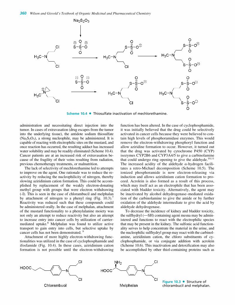
administration and necessitating direct injection into thetumor. In cases of extravasation (drug escapes from the tumorinto the underlying tissue), the antidote sodium thiosulfate(Na2S2O3), a strong nucleophile, may be administered. It iscapable of reacting with electrophilic sites on the mustard, andonce reaction has occurred, the resulting adduct has increasedwater solubility and may be readily eliminated (Scheme 10.4).Cancer patients are at an increased risk of extravasation be-cause of the fragility of their veins resulting from radiation,previous chemotherapy treatments, or malnutrition.
The lack of selectivity of mechlorethamine led to attemptsto improve on the agent. One rationale was to reduce the re-activity by reducing the nucleophilicity of nitrogen, therebyslowing aziridinium cation formation. This could be accom-plished by replacement of the weakly electron-donatingmethyl group with groups that were electron withdrawing (-I). This is seen in the case of chlorambucil and melphalanby attachment of nitrogen to a phenyl ring (Fig. 10.3).7
Reactivity was reduced such that these compounds couldbe administered orally. In the case of melphalan, attachmentof the mustard functionality to a phenylalanine moiety wasnot only an attempt to reduce reactivity but also an attemptto increase entry into cancer cells by utilization of carrier-mediated uptake.8 Melphalan was found to utilize activetransport to gain entry into cells, but selective uptake bycancer cells has not been demonstrated. 9
Attachment of more highly electron-withdrawing func-tionalities was utilized in the case of cyclophosphamide andifosfamide (Fig. 10.4). In these cases, aziridinium cationformation is not possible until the electron-withdrawing
function has been altered. In the case of cyclophosphamide,it was initially believed that the drug could be selectivelyactivated in cancer cells because they were believed to con-tain high levels of phosphoramidase enzymes. This wouldremove the electron-withdrawing phosphoryl function andallow aziridine formation to occur. However, it turned outthat the drug was activated by cytochrome P450 (CYP)isozymes CYP2B6 and CYP3A4/5 to give a carbinolaminethat could undergo ring opening to give the aldehyde.10,11
The increased acidity of the aldehyde �-hydrogen facili-tates a retro-Michael decomposition (Scheme 10.5). Theionized phosphoramide is now electron-releasing viainduction and allows aziridinium cation formation to pro-ceed. Acrolein is also formed as a result of this process,which may itself act as an electrophile that has been asso-ciated with bladder toxicity. Alternatively, the agent maybe inactivated by alcohol dehydrogenase–mediated oxida-tion of the carbinolamine to give the amide or by furtheroxidation of the aldehyde intermediate to give the acid byaldehyde dehydrogenase.
To decrease the incidence of kidney and bladder toxicity,the sulfhydryl (MSH) containing agent mesna may be admin-istered and functions to react with the electrophilic speciesthat may be present in the kidney. The sulfonic acid function-ality serves to help concentrate the material in the urine, andthe nucleophilic sulfhydryl group may react with the carbinol-amine, aziridinium cation, the chloro substituents of cy-clophosphamide, or via conjugate addition with acrolein(Scheme 10.6). This inactivation and detoxification may alsobe accomplished by other thiol-containing proteins such as
360 Wilson and Gisvold’s Textbook of Organic Medicinal and Pharmaceutical Chemistry
Scheme 10.4 Thiosulfate inactivation of mechlorethamine.
Figure 10.3 Structure ofchlorambucil and melphalan.
0355-0412_17865_Ch10.qxd 12/4/09 5:13 AM Page 360

glutathione. Increased levels of these proteins may occur ascancer cells become resistant to these alkylating agents.
Ifosfamide contains similar functionality and also requiresactivation by CYP2B6 and CYP3A4/5 (Scheme 10.7).Although the agents are similar, there are differences in themetabolism and activity of the agents.12,13 Both are adminis-tered as racemic mixtures as a result of the presence of a chi-ral phosphorus atom. There appears to be little difference inthe metabolic fate of the R- and S-isomers of cyclophos-phamide, but in the case of ifosfamide, the R-isomer is con-verted to the required 4-hydroxy-ifosfamide 2 to 3 timesfaster than the S-isomer.14 The S-isomer undergoes preferen-tial oxidation of the side chain to give N-dechloroethylation,which removes the ability of the agent to cross-link DNA andalso produces the neurotoxic and urotoxic chloroacetalde-hyde. An additional difference between cyclophosphamideand ifosfamide is the larger alkylating species that ultimately
results after metabolic activation of ifosfamide. This results inthe reactive form of ifosfamide having a higher affinity forDNA than the analogous form of cyclophosphamide and dif-ferences in the interstrand and intrastrand links that ultimatelyresult. The differential metabolism also results in increasedformation of the urotoxic chloroacetaldehyde in the case ofifosfamide such that bladder toxicity that normally presents ashemorrhagic cystitis becomes dose limiting with this agent.
THIOTEPA
The early success of the nitrogen mustards led researchers toinvestigate other compounds that contained a preformed aziri-dine ring, and thiotepa resulted from this work. Thiotepa con-taining the thiophosphoramide functionality was found to bemore stable than the oxa-analog (TEPA) but is metabolicallyconverted to TEPA by desulfuration in vivo.15 Thiotepa incor-porates a less reactive aziridine ring compared with thatformed in mechlorethamine. The adjacent thiophosphoryl iselectron withdrawing and, therefore, reduces the reactivity ofthe aziridine ring system. Although thiotepa is less reactivethan many other alkylating agents, it has been shown to formcross-links. This is believed to occur by sequential reactions ofthiotepa itself with DNA (Scheme 10.8).16 Monoalkylation isalso possible as a result of aziridine formation via hydrolysisof thiotepa. Thiotepa is also metabolized by oxidativedesulfurization mediated by CYP2B1 and CYP2C11.17 Thedecreased stability of the resulting TEPA undergoes hydroly-sis to give aziridine, which may function to monoalkylate
Chapter 10 Antineoplastic Agents 361
Figure 10.4 Structures of cyclophosphamide and ifosfamide.
Scheme 10.5 Metabolic and chemical activation of cyclophosphamide.
0355-0412_17865_Ch10.qxd 12/4/09 5:13 AM Page 361
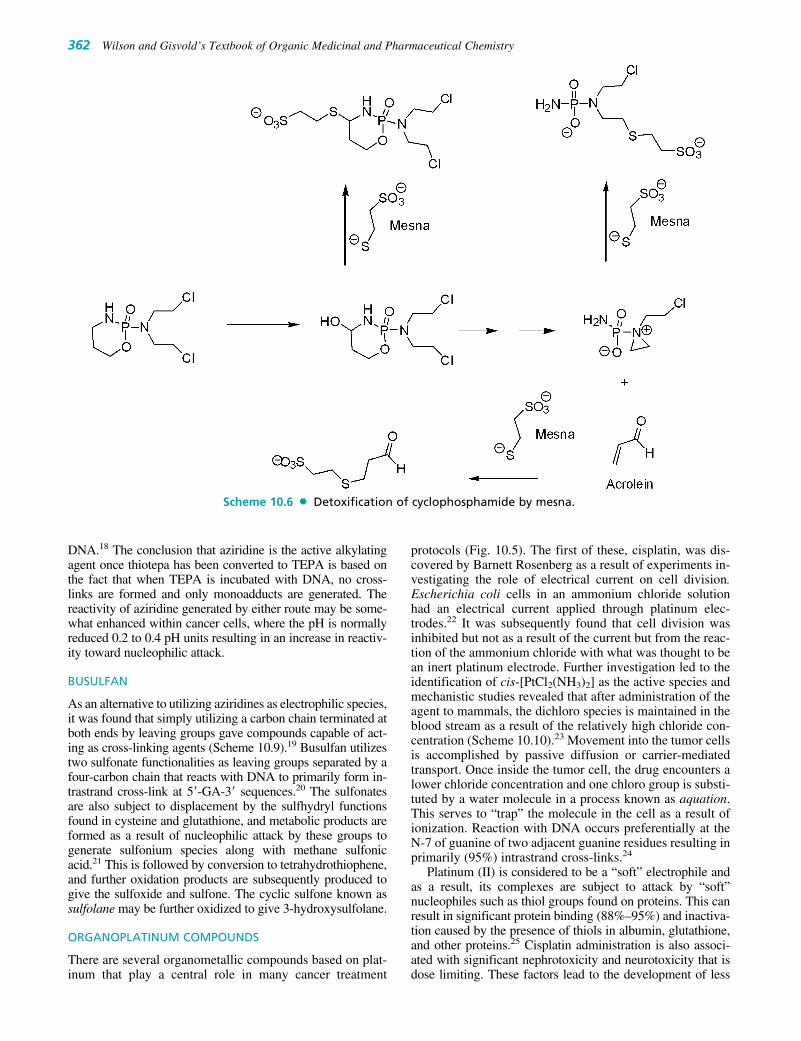
DNA.18 The conclusion that aziridine is the active alkylatingagent once thiotepa has been converted to TEPA is based onthe fact that when TEPA is incubated with DNA, no cross-links are formed and only monoadducts are generated. Thereactivity of aziridine generated by either route may be some-what enhanced within cancer cells, where the pH is normallyreduced 0.2 to 0.4 pH units resulting in an increase in reactiv-ity toward nucleophilic attack.
BUSULFAN
As an alternative to utilizing aziridines as electrophilic species,it was found that simply utilizing a carbon chain terminated atboth ends by leaving groups gave compounds capable of act-ing as cross-linking agents (Scheme 10.9).19 Busulfan utilizestwo sulfonate functionalities as leaving groups separated by afour-carbon chain that reacts with DNA to primarily form in-trastrand cross-link at 5�-GA-3� sequences.20 The sulfonatesare also subject to displacement by the sulfhydryl functionsfound in cysteine and glutathione, and metabolic products areformed as a result of nucleophilic attack by these groups togenerate sulfonium species along with methane sulfonicacid.21 This is followed by conversion to tetrahydrothiophene,and further oxidation products are subsequently produced togive the sulfoxide and sulfone. The cyclic sulfone known assulfolane may be further oxidized to give 3-hydroxysulfolane.
ORGANOPLATINUM COMPOUNDS
There are several organometallic compounds based on plat-inum that play a central role in many cancer treatment
protocols (Fig. 10.5). The first of these, cisplatin, was dis-covered by Barnett Rosenberg as a result of experiments in-vestigating the role of electrical current on cell division.Escherichia coli cells in an ammonium chloride solutionhad an electrical current applied through platinum elec-trodes.22 It was subsequently found that cell division wasinhibited but not as a result of the current but from the reac-tion of the ammonium chloride with what was thought to bean inert platinum electrode. Further investigation led to theidentification of cis-[PtCl2(NH3)2] as the active species andmechanistic studies revealed that after administration of theagent to mammals, the dichloro species is maintained in theblood stream as a result of the relatively high chloride con-centration (Scheme 10.10).23 Movement into the tumor cellsis accomplished by passive diffusion or carrier-mediatedtransport. Once inside the tumor cell, the drug encounters alower chloride concentration and one chloro group is substi-tuted by a water molecule in a process known as aquation.This serves to “trap” the molecule in the cell as a result ofionization. Reaction with DNA occurs preferentially at theN-7 of guanine of two adjacent guanine residues resulting inprimarily (95%) intrastrand cross-links.24
Platinum (II) is considered to be a “soft” electrophile andas a result, its complexes are subject to attack by “soft”nucleophiles such as thiol groups found on proteins. This canresult in significant protein binding (88%–95%) and inactiva-tion caused by the presence of thiols in albumin, glutathione,and other proteins.25 Cisplatin administration is also associ-ated with significant nephrotoxicity and neurotoxicity that isdose limiting. These factors lead to the development of less
362 Wilson and Gisvold’s Textbook of Organic Medicinal and Pharmaceutical Chemistry
Scheme 10.6 Detoxification of cyclophosphamide by mesna.
0355-0412_17865_Ch10.qxd 12/4/09 5:13 AM Page 362

reactive platinum compounds such as carboplatin and oxali-platin in which the leaving group was incorporated into achelate. More recently, there has been interest in the develop-ment of Pt(IV) compounds, which are much less reactive andbelieved to function as prodrugs requiring reduction to thePt(II) species prior to reaction with nucleophiles. One suchagent, satraplatin is currently in clinical trials. One advantageof these agents is the possibility of oral administration.Satraplatin has shown similar activity when given orally tothat of cisplatin given by injection.
Tumor cells may become resistant to the platins bymechanisms that are seen with other chemotherapeuticagents such as decreased uptake, increased inactivation by
thiol-containing proteins and increased DNA repair.However, a deficiency in a type of DNA repair known asmismatch repair (MMR) has also been implicated in resis-tance to cisplatin and carboplatin.26 The process of MMR in-volves several enzymes that are responsible for maintainingthe integrity of the genome, and interest has focused on theinteraction of these enzymes with repeating units foundthroughout DNA known as microsatellites. When MMRprocesses are not operating, these microsatellites may be-come longer or shorter and this is known as microsatelliteinstability. This can result in frame shift errors such thattumor suppressor genes may become less effective, andthe tumor cells therefore fail to undergo apoptosis even if
Chapter 10 Antineoplastic Agents 363
Scheme 10.7 Metabolic and chemical activation of ifosfamide.
0355-0412_17865_Ch10.qxd 12/4/09 5:13 AM Page 363

alkylation has occurred. The binding of cisplatin- and carbo-platin-DNA adducts by the MMR enzymes results in in-creased cytotoxicity of these agents. Several rationales havebeen put forward as to why this occurs, including the in-volvement of MMR enzymes in downstream signaling thatactivates apoptosis. A second rationale involves the ability
of MMR enzymes to remove replication errors that occurpast the point of adduct formation, and in the process of re-moving these errors, gaps in the DNA are created, whichlead to cell death. When there is a deficiency in the MMRenzymes, cells may be resistant to cisplatin and carboplatinbecause both of these agents produce the same DNA adduct.
364 Wilson and Gisvold’s Textbook of Organic Medicinal and Pharmaceutical Chemistry
Scheme 10.8 Metabolic and chemical interactions of thiotepa with DNA.
Scheme 10.9 Metabolic and chemical reactions of busulfan.
0355-0412_17865_Ch10.qxd 12/4/09 5:13 AM Page 364
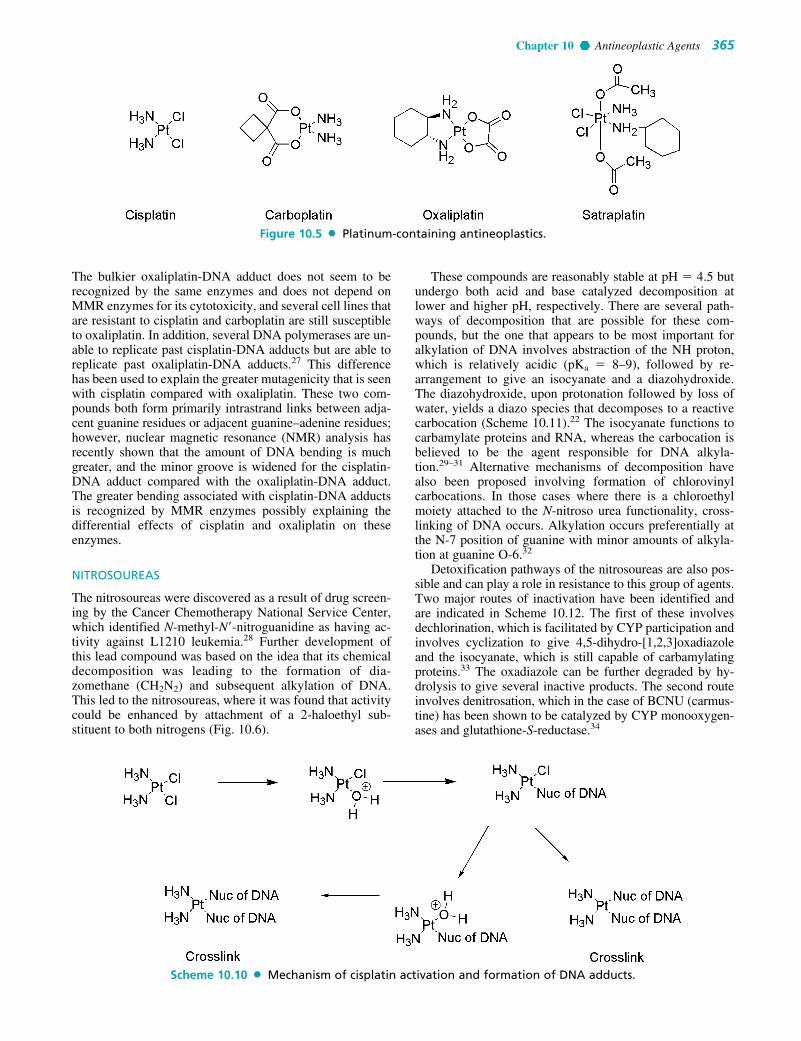
The bulkier oxaliplatin-DNA adduct does not seem to berecognized by the same enzymes and does not depend onMMR enzymes for its cytotoxicity, and several cell lines thatare resistant to cisplatin and carboplatin are still susceptibleto oxaliplatin. In addition, several DNA polymerases are un-able to replicate past cisplatin-DNA adducts but are able toreplicate past oxaliplatin-DNA adducts.27 This differencehas been used to explain the greater mutagenicity that is seenwith cisplatin compared with oxaliplatin. These two com-pounds both form primarily intrastrand links between adja-cent guanine residues or adjacent guanine–adenine residues;however, nuclear magnetic resonance (NMR) analysis hasrecently shown that the amount of DNA bending is muchgreater, and the minor groove is widened for the cisplatin-DNA adduct compared with the oxaliplatin-DNA adduct.The greater bending associated with cisplatin-DNA adductsis recognized by MMR enzymes possibly explaining thedifferential effects of cisplatin and oxaliplatin on theseenzymes.
NITROSOUREAS
The nitrosoureas were discovered as a result of drug screen-ing by the Cancer Chemotherapy National Service Center,which identified N-methyl-N�-nitroguanidine as having ac-tivity against L1210 leukemia.28 Further development ofthis lead compound was based on the idea that its chemicaldecomposition was leading to the formation of dia-zomethane (CH2N2) and subsequent alkylation of DNA.This led to the nitrosoureas, where it was found that activitycould be enhanced by attachment of a 2-haloethyl sub-stituent to both nitrogens (Fig. 10.6).
These compounds are reasonably stable at pH � 4.5 butundergo both acid and base catalyzed decomposition atlower and higher pH, respectively. There are several path-ways of decomposition that are possible for these com-pounds, but the one that appears to be most important foralkylation of DNA involves abstraction of the NH proton,which is relatively acidic (pKa � 8–9), followed by re-arrangement to give an isocyanate and a diazohydroxide.The diazohydroxide, upon protonation followed by loss ofwater, yields a diazo species that decomposes to a reactivecarbocation (Scheme 10.11).22 The isocyanate functions tocarbamylate proteins and RNA, whereas the carbocation isbelieved to be the agent responsible for DNA alkyla-tion.29–31 Alternative mechanisms of decomposition havealso been proposed involving formation of chlorovinylcarbocations. In those cases where there is a chloroethylmoiety attached to the N-nitroso urea functionality, cross-linking of DNA occurs. Alkylation occurs preferentially atthe N-7 position of guanine with minor amounts of alkyla-tion at guanine O-6.32
Detoxification pathways of the nitrosoureas are also pos-sible and can play a role in resistance to this group of agents.Two major routes of inactivation have been identified andare indicated in Scheme 10.12. The first of these involvesdechlorination, which is facilitated by CYP participation andinvolves cyclization to give 4,5-dihydro-[1,2,3]oxadiazoleand the isocyanate, which is still capable of carbamylatingproteins.33 The oxadiazole can be further degraded by hy-drolysis to give several inactive products. The second routeinvolves denitrosation, which in the case of BCNU (carmus-tine) has been shown to be catalyzed by CYP monooxygen-ases and glutathione-S-reductase.34
Chapter 10 Antineoplastic Agents 365
Figure 10.5 Platinum-containing antineoplastics.
Scheme 10.10 Mechanism of cisplatin activation and formation of DNA adducts.
0355-0412_17865_Ch10.qxd 12/4/09 5:13 AM Page 365

PROCARBAZINE, DACARBAZINE, AND TEMOZOLOMIDE
Procarbazine is an antineoplastic agent that was originallydeveloped as a result of efforts to find new inhibitors ofmonoamine oxidase. Subsequent screening revealed anti-neoplastic activity.35 It was initially believed that the cyto-toxicity was related to the ability of the compound toundergo auto-oxidation to give hydrogen peroxide, which inthe presence of Fe�2 would produce hydroxide radicals ca-pable of cleaving DNA.36,37 Subsequent work showed thatalthough this did occur, sufficient amounts of hydrogenperoxide were not produced to account for the observedeffects. Metabolism studies revealed that oxidation ofprocarbazine does occur in the liver and is mediated by CYPand monoamine oxidase to give azo-procarbazine. Thiscompound may also be generated nonenzymatically in anaerobic environment (Scheme 10.13).38–40 There are severalchemical and metabolic pathways that azo-procarbazinemay then undergo, and there is some disagreement regard-ing the exact structure of the active alkylating species. Onesuch route involves CYP-mediated oxidation of the benzylicmethylene carbon with subsequent decomposition to givemethyldiazine and the aldehyde. The methyldiazine maythen decompose by homolytic bond cleavage to give methyland hydrogen radicals along with nitrogen gas or be furtheroxidized to give the diazo compound, which can decomposeto give the methyl carbocation. Methyldiazine may alsobe produced by a minor route involving isomerization of
azo-procarbazine to give the hydrazone, which subsequentlyundergoes hydrolysis to give the aldehyde and methylhy-drazine. Methylhydrazine may react with oxygen to givemethyldiazine, which then decomposes as before. Studiesutilizing radiolabeled procarbazine indicated that the termi-nal methyl group was found covalently bound to the N-7position of guanine especially on tRNA disrupting its func-tion and preventing protein, RNA, and DNA synthesis.41
There was also a small amount of methylation at the O-6 po-sition of guanine.42 Hydrazines are also capable of inhibit-ing monoamine oxidase as seen with isocarboxazid andphenelzine; however, procarbazine is only a weak inhibitorof this enzyme. The agent is also capable of inhibitingaldehyde dehydrogenase and producing a disulfiram-likereaction. Given the involvement of the CYP system in itsactivation, the action of procarbazine is subject to drug in-teractions with both inducers and inhibitors of this system.
Somewhat related is dacarbazine, which was initiallythought to act as an inhibitor of purine biosynthesis, but lat-ter was shown to be an alkylating agent.43 Activation of theagent occurs through the action of CYP (isozymes 1A1, 1A2,and 2E1) to give the demethylated product monomethyl tri-azeno imidazole carboxamide (MTIC) (Scheme 10.14).44
Tautomerization allows for decomposition to give the amino-carboxamido-imidazole and diazomethane, which is capableof alkylating DNA.45 An alternative pathway involves acidcatalyzed or photoinduced loss of dimethylamine to give analternative diazo compound (diazo-IC), which may not only
366 Wilson and Gisvold’s Textbook of Organic Medicinal and Pharmaceutical Chemistry
Figure 10.6 Nitrosoureas antineoplastics.
Scheme 10.11 Mechanism of DNA alkylation by nitrosoureas.
0355-0412_17865_Ch10.qxd 12/4/09 5:13 AM Page 366

generate a carbocation but also undergoes internal cycliza-tion to give 2-azo-hypoxanthine.46 Formation of diazo-IC hasbeen associated with pain at the injection site, which is oftenseen during dacarbazine administration.47 Methylation ofDNA occurs at N-7, N-3 and O-6 of guanine among othersites.48 Dacarbazine proved to be more active against murinetumors than against human tumors. This was attributed to theenhanced ability of mice to metabolize the agent to MTIC
and the subsequent conversion to a methylating species.49
Building on this idea was temozolomide, which undergoesconversion to the same intermediate, MTIC, as dacarbazine,but it does not require metabolic activation to do so.50,51
Hydrolysis of temozolomide gives the carboxy-triazene,which spontaneously loses CO2 to give MTIC. Dacarbazinemust be administered intravenously; however, the relatedtemozolomide may be administered orally.
Chapter 10 Antineoplastic Agents 367
Scheme 10.12 Metabolic and chemical inactivation of nitrosoureas.
Scheme 10.13 Metabolic and chemical activation of procarbazine.
0355-0412_17865_Ch10.qxd 12/4/09 5:13 AM Page 367

Individual Agents
MECHLORETHAMINE HYDROCHLORIDE (NH2, MUSTARGEN)
Mechlorethamine is available in 10-mg vials for intra-venous (IV) administration in the treatment of Hodgkin’slymphoma. It is part of the MOPP regimen used in treatingthis condition, which is comprised of mechlorethamine,vincristine (Oncovin), procarbazine, and prednisone. Theagent is also used topically in the treatment of mycosis fun-goides, a rare type of cancer but the most common type ofcutaneous T-cell lymphoma. Additional uses have includedtreatment of cancers that have resulted in pleural effusion.Although the compound is a potent alkylating agent, resis-tance may develop as a result of increased inactivation bysulfhydryl containing proteins such as glutathione andincreased expression of DNA repair mechanisms. Adverseeffects include dose-limiting myelosuppression and nau-sea/vomiting. There is a significant risk of extravasationupon IV administration, and the agent may produce painat the injection site. Additional adverse effects include
alopecia, azoospermia, amenorrhea, hyperuricemia, and anincreased risk of secondary cancers.
CHLORAMBUCIL (AMBOCHLORIN, ABMOCLORIN,LEUKERAN)
Chlorambucil is available as 2-mg tablets for oral adminis-tration in the treatment of Hodgkin’s lymphoma, andchronic lymphocytic leukemia in combination with pred-nisone and as a single agent. The mechanisms of resistanceare the same as those seen for other agents of the class suchas mechlorethamine. The agent is well absorbed (75%) uponoral administration and highly protein bound. Metabolism ismediated by CYP and occurs extensively to give severalmetabolites, including the active phenylacetic acid–nitrogenmustard. The drug is eliminated via the kidneys with a ter-minal elimination half-life of 1.5 hours. Adverse effects in-clude dose-limiting myelosuppression, which are seen asboth leucopenia and thrombocytopenia. Nausea and vomit-ing occur less often than for mechlorethamine. Additionaladverse effects include hyperuricemia, azoospermia, amen-orrhea, seizures, pulmonary fibrosis, and skin rash.
368 Wilson and Gisvold’s Textbook of Organic Medicinal and Pharmaceutical Chemistry
Scheme 10.14 Metabolic and chemical activation of dacarbazine and temozolomide.
0355-0412_17865_Ch10.qxd 12/4/09 5:13 AM Page 368

MELPHALAN (L-PAM, ALKERAN, L-PHENYLALANINE MUSTARD)
Melphalan is available in 2-mg tablets and 50-mg vials fororal and IV administration, respectively in the treatment ofmultiple myeloma, breast and ovarian cancer, and in high-dose therapy when bone marrow transplant is being utilized.The mechanisms of resistance are the same as those seen formechlorethamine. The agent is poorly absorbed when givenby the oral route. Melphalan is highly plasma protein bound(80%–90%) and inactivated in the blood by water to givethe hydroxy metabolites. Elimination occurs primarily in thefeces with an elimination half-life of 38 to 108 minutes. Thecommonly seen adverse effects are myelosuppression, nausea,and vomiting. Nausea is normally mild with normal doses butbecomes severe when high doses are used during bone mar-row transplant. Less commonly seen adverse effects are hy-persensitivity reactions, skin rash, and alopecia. Secondarycancers are also of concern with the use of the agent.
CYCLOPHOSPHAMIDE (CTX, CPM, CPA, CLAFEN,CYTOXAN, NEOSAR)
Cyclophosphamide is available in 25- and 50-mg tabletsfor oral administration and 100-, 200-, 500-, 1,000-, and2,000-mg vials for IV use in the treatment of a wide varietyof cancers, including breast cancer, non-Hodgkin’s lym-phoma, chronic lymphocytic leukemia, ovarian cancer, boneand soft tissue sarcoma, rhabdomyosarcoma, neuroblas-toma, and Wilms tumor. Coadministration of mesna is rec-ommended. The alkylating agent is cell cycle–nonspecific,and mechanisms of resistance are similar to those seen forother alkylating agents including decreased uptake and acti-vation and increased inactivation by glutathione and oxidaseenzymes (aldehyde dehydrogenase and alcohol dehydroge-nase) as well as increased DNA repair mechanisms. Theagent is well absorbed when given orally. Metabolism byCYP2B6 and 3A4/5 is required for activation of the agent,and metabolites are formed as a result of this activation bysubsequent reactions that are shown in Scheme 10.5. Themajor metabolite seen in plasma is the phosphoramide mus-tard, although there is a small amount of material (10%)arising from the dechloroethylation, which is primarily me-diated by 3A4/5. This metabolic pathway gives rise to asmall amount of chloroacetaldehyde, which is both neuro-toxic and nephrotoxic. The parent compound and metabo-lites are eliminated in the urine with an elimination half-lifeof 4 to 6 hours. Adverse effects include dose-limitingmyelosuppression, which normally presents as leucopenia.Bladder toxicity, which presents as hemorrhagic cystitis, isrelated to the formation of electrophilic species in the kid-ney including acrolein and may be treated by administrationof mesna and increased water intake. Additionally, amenor-rhea may be seen and sterility may be permanent. Othereffects include alopecia, cardiotoxicity, inappropriate secre-tion of antidiuretic hormone, and an increased risk ofsecondary cancers.
IFOSFAMIDE (IPHOSPHAMIDE, IFO, CYFOS, IFEX, IFOSFAMIDUM)
Ifosfamide is available in 1- and 3-g vials for IV administra-tion as Food and Drug Administration (FDA)-approvedthird-line therapy in the treatment of testicular cancer. It has
also been utilized (although not FDA approved) in the treat-ment of a wide variety of cancers including Hodgkin’s andnon-Hodgkin’s lymphoma, soft tissue sarcoma, germ celltumors, small cell lung cancer, non–small cell lung cancer(NSCLC), cancers of the head and neck, bladder cancer, cer-vical cancer, and Ewing sarcoma. Coadministration ofmesna is recommended. The mechanisms of resistance areidentical to those seen with cyclophosphamide. The drug iswidely distributed with a low extent of protein binding(20%). Metabolism primarily by CYP3A4/5 and CYP2B6 isrequired for activation of the compound (Scheme 10.7). Theagent is administered as the racemic mixture as a result ofthe presence of the chiral phosphorus atom, and differentialmetabolism of the R- and S-isomers has been observed. Incontrast to cyclophosphamide, there is a greater amount ofdeactivation of the agent by N-dechloroethylation and sub-sequently more chloroacetaldehyde is produced, which mayresult in a greater amount of neurotoxicity and nephrotoxic-ity than seen with cyclophosphamide. The N-dechloroethyl-ated metabolites are the predominate species seen in theplasma. The parent and metabolites are eliminated inthe urine with an elimination half-life of 3 to 10 hours. Themajor components found in the urine are the dechlorethy-lated metabolites. Dose-limiting toxicities include myelo-suppression and bladder toxicity. Other adverse effectsinclude nausea, alopecia, amenorrhea, inappropriate secre-tion of antidiuretic hormone, as well as the production ofsecondary cancers. Neurotoxicity, which is associated withthe production of chloroacetaldehyde presents as confusion,seizure, weakness, and hallucination, and coma may occur.
THIOTEPA (TRIETHYLENETHIOPHOSPHORAMIDE,GIROSTAN, STEPA, TESPA, THIOPLEX, TIFOSYL)
Thiotepa is available in 15-mg vials for IV administration inthe treatment of bladder cancer, ovarian cancer, and breastcancer. It has been used in the past in the treatment of lym-phomas such as Hodgkin’s and non-Hodgkin’s lymphomabut has now mostly been replaced by other agents. The agentis widely distributed after infusion, and 40% is bound toplasma proteins. Metabolism mediated by CYP2B1 andCYP2C11 effects desulfurization to give TEPA from whichaziridine may arise. Metabolites have also been identified asarising from glutathione conjugation of thiotepa followed byformation of the mercapturic acid derivative. However, theexcretion profile has only accounted for a small percentage(10%–22%) of the administered dose. The elimination half-life of the compound is 2 to 3 hours. Adverse effects includedose-limiting myelosuppression and mucositis. Additionally,nausea, vomiting, skin rash, hemorrhagic cystitis, and sec-ondary cancers may occur.
BUSULFAN (BUSULFEX, MYLERAN)
Busulfan is available as 2-mg tablets for oral administrationand 10-mL vials for IV administration in the treatment ofchronic myelogenous leukemia (CML) and in high-dosetherapy for refractory leukemia with bone marrow transplant.The agent is well absorbed when given orally, well distrib-uted into tissues, and crosses the blood-brain barrier.Metabolism occurs in the liver as shown in Scheme 10.9 togive mainly methane sulfonic acid by the action of glu-tathione-S-transferase. Other identified metabolites in hu-mans have included tetrahydrothiophene-1-oxide, sulfalene,
Chapter 10 Antineoplastic Agents 369
0355-0412_17865_Ch10.qxd 12/4/09 5:13 AM Page 369
and 3-hydroxysulfolane. The metabolites are excretedprimarily in the urine, and the terminal elimination half-lifeis 2.5 hours. Adverse effects include dose-limiting myelo-suppression; nausea and vomiting that occur commonly butare generally mild; and pulmonary symptoms including in-terstitial pulmonary fibrosis, which is referred to as “busul-fan lung,” occurs belatedly (1–10 years posttreatment) andalthough rare, it is severe. Other adverse effects includemucositis, skin rash, impotence, amenorrhea, infertility, he-patoxicity, insomnia, anxiety, and an increased risk of sec-ondary malignancies. At normal doses, the agent is welltolerated except for the myelosuppression that occurs. Thishas allowed for high-dose therapy with the agent when ac-companied by bone marrow transplant to counter the myelo-suppressive effects.
CISPLATIN (CIS-DIAMINEDICHLOROPLATINUM,DDP, PLATINOL, PLATINOL-AQ)
Cisplatin is available in 10- and 50-mg vials for IV adminis-tration in the treatment of a wide variety of cancers includingnon-Hodgkin’s lymphoma, bladder cancer, ovarian cancer,testicular cancer, and cancers of the head and neck. A liposo-mal form is also available as well as an injectable collagenmatrix gel containing cisplatin. Compared with other platins,cisplatin is the most reactive and therefore the most effectivein platinating DNA. After IV administration, the agent iswidely distributed, highly protein bound (90%), and concen-trates in the liver and kidney. After infusion, covalent attach-ment to plasma proteins occurs such that after 4 hours, 90%of drug is protein bound. The elimination of platinum fromthe blood is a slow process with a terminal elimination half-life of 5 to 10 days. Metabolism involves aquation, which oc-curs to a greater extent once distribution out of the plasmahas occurred. Additional metabolites have been seen result-ing from reaction with glutathione and cysteine. The greaterreactivity of cisplatin gives rise to significant toxicitiescompared with other platins. These include dose-limitingnephrotoxicity, which normally presents as elevated bloodurea nitrogen (BUN) and creatinine. This effect is cumula-tive upon repeated dosing and may progress further to necro-sis, altered epithelial cells, cast formation, and thickening ofthe tubular basement membranes but is generally reversibleupon discontinuation of drug treatment. Sodium thiosulfatemay be given to reduce the nephrotoxicity. Neurotoxicitymay also be dose limiting, normally presenting initially asnumbness but may progress to seizure. Other adverse effectsinclude myelosuppression, nausea, vomiting, alopecia,ototoxicity, ocular toxicity, azoospermia, impotence, myo-cardial infarction, thrombotic events, and inappropriatesecretion of antidiuretic hormone.
CARBOPLATIN (CBDCA, PARAPLAT, PARAPLATIN)
Carboplatin is available in 50-, 150-, and 450-mg vials for IVadministration in the treatment of ovarian cancer, bladdercancer, germ cell tumors, head and neck cancers, small celllung cancer, and NSCLC. Activation of the agent occurs byaquation in a manner similar to that seen for cisplatin. Thepresence of the chelating 1,1-cyclobutane-dicarboxylateslows this reaction 100-fold and reduces the toxicity of theagent. The sites of alkylation and mechanisms of resistanceare like those seen for cisplatin, and the two agents showcross-resistance. The agent is widely distributed upon IV
administration but, because of its greater stability, it bindsslowly to plasma proteins, requiring 24 hours to reach 90%bound drug compared with 4 hours for cisplatin. The agent iseliminated in the urine with a terminal elimination half-lifeof 2 to 6 hours. Adverse effects include myelosuppression,which is dose limiting. Other adverse effects include renaltoxicity, nausea, vomiting, and peripheral neuropathy, butthese occur much less frequently than with cisplatin.
OXALIPLATIN (DIAMINOCYCLOHEXANEPLATINUM, DACH-PLATINUM, ELOXATIN)
Oxaliplatin is available in 50- and 100-mg vials for IV ad-ministration in the treatment of ovarian cancer, metastaticcolorectal cancer, and early stage colon cancer in combina-tion with 5-fluorouracil/leucovorin. The activation of theagent occurs in low-chloride environments to give theaquated species, which subsequently reacts with DNA in amanner similar to cisplatin. The mechanisms of resistance aresimilar for the two agents; however, oxaliplatin is not recog-nized by MMR enzymes and does not show cross-resistancewith cisplatin. The agent is widely distributed, highly proteinbound (85%–88%), and irreversibly binds to erythrocytes.Numerous metabolites have been identified many of whichare produced as a result of nonenzymatic processes and in-clude chloro-, dichloro-, monoaquo-, and diaquo-species.The parent and metabolites are eliminated primarily in theurine with a long terminal elimination half-life of 240 hours.Neurotoxicity is dose limiting and normally presents as pe-ripheral neuropathy, which may be exacerbated by exposureto low temperatures. The neurotoxicity is normally reversiblein contrast to that seen with cisplatin, which may be irre-versible. Other adverse effects include nausea, vomiting, di-arrhea, myelosuppression, and hypersensitivity reactions.Ototoxicity and renal toxicity occur only rarely in contrast tocisplatin.
CARMUSTINE (BIS-CHLOROETHYL-NITROSOUREA,BCNU, BECENUM, CARMUBRIS, BICNU, GLIADEL)
Carmustine is available in a 100-mg vial for IV administra-tion in the treatment of several types of brain tumors,Hodgkin’s and non-Hodgkin’s disease, and multiplemyeloma. The agent is also available as an implantablewafer containing 7.7 mg of drug for intracavity implantationin the treatment of glioblastoma multiforme. Cytotoxicity isassociated with cross-linking of DNA and RNA and car-bamoylation of glutathione reductase. Cross-resistance isnot seen with other alkylating agents. The agent is very lipidsoluble and easily crosses the blood-brain barrier, achievingconcentrations greater than 50% of those seen in plasma.Metabolism involves both the nonenzymatic formation ofreactive intermediates that may react with glutathione andother thiol-containing proteins, as well as enzymatic reduc-tive denitrosation and dechlorination. The half-life of theagent in plasma is short (15–20 minutes) because of rapiddecomposition. Myelosuppression is dose limiting and maypresent as thrombocytopenia, leucopenia, or more rarely asanemia. This is generally seen 24 to 48 days after treatment.Pulmonary toxicity occurs rarely at low doses but at highdoses such as those seen during bone marrow transplantmay present as dyspnea, cough, pulmonary infiltrates, andprogress to respiratory failure. This may be seen years afterthe completion of therapy. Other toxicities include nausea,
370 Wilson and Gisvold’s Textbook of Organic Medicinal and Pharmaceutical Chemistry
0355-0412_17865_Ch10.qxd 12/4/09 5:13 AM Page 370

vomiting, pain at the injection site, impotence, sterility,amenorrhea, and infertility. Hepatotoxicity may initiallypresent as elevations in serum transaminase levels. There isalso an increased risk of secondary cancers as is often seenwith the alkylating agents.
LOMUSTINE (CHLOROETHYL-CYCLOHEXYL-NITROSOUREA, CCNU, CEENU)
Lomustine is available in 10-, 40-, and 100-mg capsules fororal administration in the treatment of primary and metasta-tic brain cancers and Hodgkin’s lymphoma. This lipophilicagent is well absorbed, widely distributed, and crosses theblood-brain barrier. Lomustine undergoes extensive hepaticmetabolism, which is mediated by CYP3A4 to give severalhydroxylated metabolites, which arise as a result of oxida-tion of the cyclohexyl ring. Several of these are more activethan the parent compound. Denitrosation and dechlorinationhave also been demonstrated to occur for lomustine as well.The intact drug was not found in plasma when the agent wasadministered orally. Elimination occurs primarily in theurine with an elimination half-life of 16 to 72 hours.Myelosuppression is dose limiting and presents in a mannersimilar to that seen with carmustine. Other toxicities includenausea, vomiting, anorexia, impotence, sterility, amenor-rhea, and infertility. Pulmonary and renal toxicity are rarelyseen during standard-dose therapy but increase during high-dose therapy.
STREPTOZOCIN (STREPTOZOTOCIN, STZ, ZANOSAR)
Streptozocin is available in 1-g vials for IV administrationin the treatment of metastatic islet cell carcinoma of the pan-creas, colon cancer, and Hodgkin’s disease. Activation re-sults in the formation of methyl carbonium ions, which alky-late DNA, but the agent is less effective in alkylating DNAand carbamylating proteins than other nitrosoureas, which isbecause in part of an intramolecular carbamoylation. The in-ternal carbamate group migration occurs via the hydroxylgroups of the glucose portion of the molecule. There is a re-duced effect on RNA function compared with other ni-trosoureas as well. The hydrophilic agent is rapidly clearedfrom the plasma with an elimination half-life of 35 minutes.The presence of the glucose moiety allows for utilization ofglucose transporters, which concentrate the compound inthe �-cells of the pancreas. The metabolism of the agent hasnot been well characterized. Renal toxicity is dose limitingand may present initially as proteinuria and azotemia butmay progress to renal failure. Nausea and vomiting may besevere with the agent, whereas myelosuppression is gener-ally mild. Normal glucose metabolism may be altered sothat hypoglycemia or hyperglycemia may be seen. In someanimal species, streptozocin produces diabetes but muchmilder effects are seen in man.
PROCARBAZINE HYDROCHLORIDE (PCZ, PCB, MATULANE)
Procarbazine is available in 50-mg tablets for oral admin-istration in the treatment of Hodgkin’s (part of MOPP) andnon-Hodgkin’s disease, brain cancer, and mycosis fun-goides. The major mechanisms of resistance appear to beenhanced activity of DNA-repair enzymes including en-hanced O-6-alkylguanine DNA transferase (AGAT),
which removes the methyl group from the O-6 of guanine.The agent is rapidly and completely absorbed after oral ad-ministration and extensively metabolized in the liver togive azo-procarbazine followed by further oxidation tomethyldiazine and the aldehyde. The parent drug andmetabolites cross the blood-brain barrier. Elimination oc-curs in the urine mostly as metabolites with an eliminationhalf-life of 1 hour. Myelosuppression is dose limiting, gen-erally presenting as thrombocytopenia that may be fol-lowed by leucopenia. Glucose-6-phosphate dehydrogenasedeficient patients may develop hemolytic anemia duringprocarbazine therapy. Other adverse effects include nau-sea, vomiting, hypersensitivity, flulike symptoms, amenor-rhea, and azoospermia. Central nervous symptom effectsmay be seen, including lethargy, confusion, neuropathies,and seizure.
DACARBAZINE (DTIC, DTIC-DOME)
Dacarbazine is available in 100- and 200-mg vials for IVadministration in the treatment of Hodgkin’s disease, ma-lignant melanoma, carcinoid cancer, neuroblastoma, andsoft tissue sarcoma. Resistance to dacarbazine has been pri-marily attributed to enhanced activity of AGAT. The vol-ume of distribution exceeds the amount of water in thebody suggesting the compound distributes into body tissuespossibly the liver. The agent is not highly protein bound(20%) and is metabolized in the liver by CYP to give MTICand 4-amino-5-imidazole-carboxamide (AIC). Thedemethylation reaction is mediated by isozymes CYP1A1/2and CYP2E1. Elimination occurs via the urine with 40% to50% occurring as unchanged drug. Dose-limiting myelo-suppression presents as both leucopenia and thrombocy-topenia. Other adverse effects include nausea, vomiting,flulike symptoms, photosensitivity, and pain at the injec-tion site.
ALTRETAMINE (HMM, HEXALEN, HEXAMETHYLMELAMINE)
Altretamine (Fig. 10.7) is available in 50-mg capsules for oraladministration as a second-line treatment for ovarian cancer.The mechanism of action has not been firmly established, al-though the spectrum of activity is similar to that for otheralkylating agents; however, cross-resistance is not seen.Cytotoxicity has been correlated with metabolism to give thecarbinolamines, which may form imines capable of cross-linking, or decompose to give formaldehyde, which may reactwith nucleophiles on DNA or proteins. The agent is well ab-sorbed upon oral administration, well distributed, and highly(90%) plasma protein bound. The agent is extensively metab-olized in the liver by CYP to give demethylated metabolites
Chapter 10 Antineoplastic Agents 371
Figure 10.7 Chemical structure of altretamine.
0355-0412_17865_Ch10.qxd 12/4/09 5:13 AM Page 371

via the previously mentioned carbinolamines. Elimination oc-curs primarily in the urine as mostly demethylated metabo-lites with a terminal elimination half-life of 4 to 10 hours.Myleosuppression and nausea/vomiting are dose-limitingtoxicities. Other adverse effects include lethargy, agitation,hallucinations, skin rash, and elevations in transaminase lev-els, flulike symptoms, abdominal cramps, and diarrhea.
ANTIMETABOLITES
Antimetabolites are compounds closely related in structureto a cellular precursor molecule, yet these imposter sub-stances are capable of preventing the proper use or forma-tion of the normal cellular product. These antimetabolitesare similar enough in structure in many cases to interactwith the normal cellular process but differ in a manner suf-ficient to alter the outcome of that pathway. Most an-timetabolites are effective cancer chemotherapeutic agentsvia interaction with the biosynthesis of nucleic acids.Therefore, several of the useful drugs used in antimetabolitetherapy are purines, pyrimidines, folates, and related com-pounds. The antimetabolite drugs may exert their effects byseveral individual mechanisms involving enzyme inhibitionat active, allosteric, or related sites. Most of these targetedenzymes and processes are involved in the regulatory stepsof cell division and cell/tissue growth. Often the adminis-tered drug is actually a prodrug form of an antimetaboliteand requires activation in vivo to yield the active inhibitor.The administration of many purine and pyrimidine an-timetabolites requires the formation of the nucleoside and fi-nally the corresponding nucleotide for antimetabolite activ-ity. An antimetabolite and its transformation products mayinhibit several different enzymes involved in tissue growth.These substances are generally cell cycle specific with ac-tivity in the S phase.
The purine and pyrimidine antimetabolites are oftencompounds incorporated into nucleic acids and the nucleicacid polymers (DNA, RNA, etc.). The antifolates are com-pounds designed to interact at cofactor sites for enzymes in-volved in the biosynthesis of nucleic acid bases. The biosyn-thesis of these nucleic acid bases depend heavily on theavailability of folate cofactors, hence antimetabolites of thefolates find utility as antineoplastic agents. The antitumoreffects of all these compounds attempting to masquerade asthe natural precursor compound may occur because of amalfunction in the biosynthesis of the corresponding macro-molecules. Classic examples of pyrimidine and purine an-timetabolites are 5-fluorouracil and 6-mercaptopurine, re-spectively, and the classic antifolate is methotrexate.
The purine analog 6-mercaptopurine and its bioactivationproducts interact with many enzymes in the various stagesof cell division.52 The activity of 6-mercaptopurine requiresbioactivation to its ribonucleotide, 6-thioinosinate, by theenzyme hypoxanthine-guanine phosphoribosyl transferase(HGPRT). 6-Thioinosinate is a potent inhibitor of the con-version of 5-phosphoribosylpyrophosphate into 5-phospho-ribosylamine. The ribose diphosphate and triphosphates of6-mercaptopurine are active enzyme inhibitors, and thetriphosphate can be incorporated into DNA and RNA to in-hibit chain elongation.
The pyrimidine analog 5-fluorouracil was designedbased on the observation that in certain tumors, uracil was
used for nucleic acid pyrimidine biosynthesis.53 Fluorine,at the 5-position of uracil, blocks the conversion of uridy-late to thymidylate, thus diminishing DNA biosynthesis.The fluorine induced increase in acidity (inductive effect)was expected to cause the molecule to bind more stronglyto enzymes of the various stages of pyrimidine biosynthe-sis. Thymidine is a logical target, because it is only foundin DNA and not in RNA. 5-Fluorouracil is activated byanabolism to 5-fluoro-2-deoxyuridylic acid, and this acti-vated species is a strong inhibitor of thymidylate syn-thetase, the enzyme that converts 2�-deoxyuridylic acid tothymidylic acid.
Methotrexate inhibits the binding of the substrate folicacid to the enzyme dihydrofolate reductase (DHFR), result-ing in reductions in the synthesis of nucleic acid bases, per-haps most importantly, the conversion of uridylate tothymidylate as catalyzed by thymidylate synthetase, whichrequires folate cofactors.
The pathways for the normal biosynthesis of the pyrimi-dine and purine bases are summarized in Schemes 10.15 and10.16, respectively. The biosynthesis of pyrimidine andpurine monomers is a central component for DNA and RNAformation in mammalian cells. The process of cell divisionand tissue growth is a very complex set of biochemicalevents with many feedback controls. Thus, antimetabolitedrugs based on pyrimidine, purine, and related structuresmay exert their effects by altering many metabolic processesand pathways. The final step in Figure 10.15 illustrates theconversion of ribonucleotides to deoxyribonucleotides cat-alyzed by ribonucleotide reductase. The substrate for thisenzyme is the ribonucleotide diphosphate, and the process isthe same for both purine and pyrimidine bases.
Pyrimidine Drugs
The anticancer drugs based on pyrimidine structure areshown in Figure 10.8. The pyrimidine derivative 5-fluo-rouracil (5-FU) was designed to block the conversion of uri-dine to thymidine. The normal biosynthesis of thymidine in-volves methylation of the 5-position of the pyrimidine ringof uridine. The replacement of the hydrogen at the 5-posi-tion of uracil with a fluorine results in an antimetabolitedrug, leading to the formation of a stable covalent ternarycomplex composed of 5-FU, thymidylate synthase (TS), andcofactor (a tetrahydrofolate species). The normal pathwayfor the formation of thymidine from uridine catalyzed by theenzyme TS is shown in Scheme 10.17. Anticancer drugs tar-geting this enzyme should selectively inhibit the formationof DNA because thymidine is not a normal componentof RNA.
TS is responsible for the reductive methylation of de-oxyuridine monophosphate (dUMP) by 5,10-methylenete-trahydrofolate to yield dTMP and dihydrofolate.54 Becausethymine is unique to DNA, the TS enzyme system plays animportant role in replication and cell division. The tetrahy-drofolate cofactor species serves as both the one-carbondonor and the hydride source in this system. The initial stepof the process involves the nucleophilic attack by asulfhydryl group of a cystine residue at the 6-position ofdUMP. The resulting enolate adds to the methylene of 5,10-CH2-THF perhaps activated via the very reactive N-5-iminium ion (see Scheme 10.17). The iminium ion likelyforms at N-5 and only after 5,10-CH2-THF binds to TS.54
372 Wilson and Gisvold’s Textbook of Organic Medicinal and Pharmaceutical Chemistry
0355-0412_17865_Ch10.qxd 12/4/09 5:13 AM Page 372

The iminium ion is likely formed at N-5 because it is themore basic of the two nitrogens, whereas N-10 is the betterleaving group. The loss of the proton at the 5-position ofdUMP and elimination of folate yields the exocyclic meth-ylene uracil species. The final step involves hydride transferfrom THF and elimination to yield the enzyme, DHF, anddTMP.
5-Fluorouracil is activated by conversion to the corre-sponding nucleotide species,53 5-fluoro-2-deoxyuridylicacid (see Scheme 10.18). The resulting 5-fluoro-2�-de-oxyuridylic acid is a powerful inhibitor of thymidylate syn-thetase, the enzyme that converts 2�-deoxyuridylic acid tothymidylic acid. In the inhibiting reaction, the sulfhydrylgroup of TS adds via conjugate addition to the 6-position ofthe fluorouracil moiety (Scheme 10.19). The carbon at the5-position then binds to the methylene group of 5,10-methylenetetrahydrofolate following initial formation of themore electrophilic form of folate the N-5-iminium ion. Inthe normal process, this step is followed by the eliminationof dihydrofolate from the ternary complex, regeneration of
the active enzyme species, and the product thymidine.Central to this process is the loss of the proton at the 5-position of uracil to form the exocyclic methylene uracilspecies. The 5-fluorine is stable to elimination, and a termi-nal product results, involving the enzyme, cofactor, and sub-strate, all covalently bonded (Scheme 10.19).
The chemical mechanism of inhibition of thymidylatesynthetase by 5-fluorouracil is shown in Scheme 10.19. Thisprocess clearly shows that in order to inactivate the TS en-zyme, both 5-FU and the tetrahydrofolate species are re-quired to form the ternary complex. Some clinical studieshave shown that administration of a tetrahydrofolate sourceprior to treatment with 5-FU results in greater inhibition oftotal TS activity. The administered source of active 5,10-methylenetetrahydrofolate is leucovorin, N-5-formyl-tetrahydrofolate.
TS is the most obvious and well-documented mechanismof action for 5-FU cytotoxic activity. However, other mech-anisms may play a role in the overall value of this drug inthe treatment of human cancer. The triphosphate of 5-FU
Chapter 10 Antineoplastic Agents 373
Scheme 10.15 Biosynthetic pathway for pyrimidine nucleotides.
0355-0412_17865_Ch10.qxd 12/4/09 5:13 AM Page 373
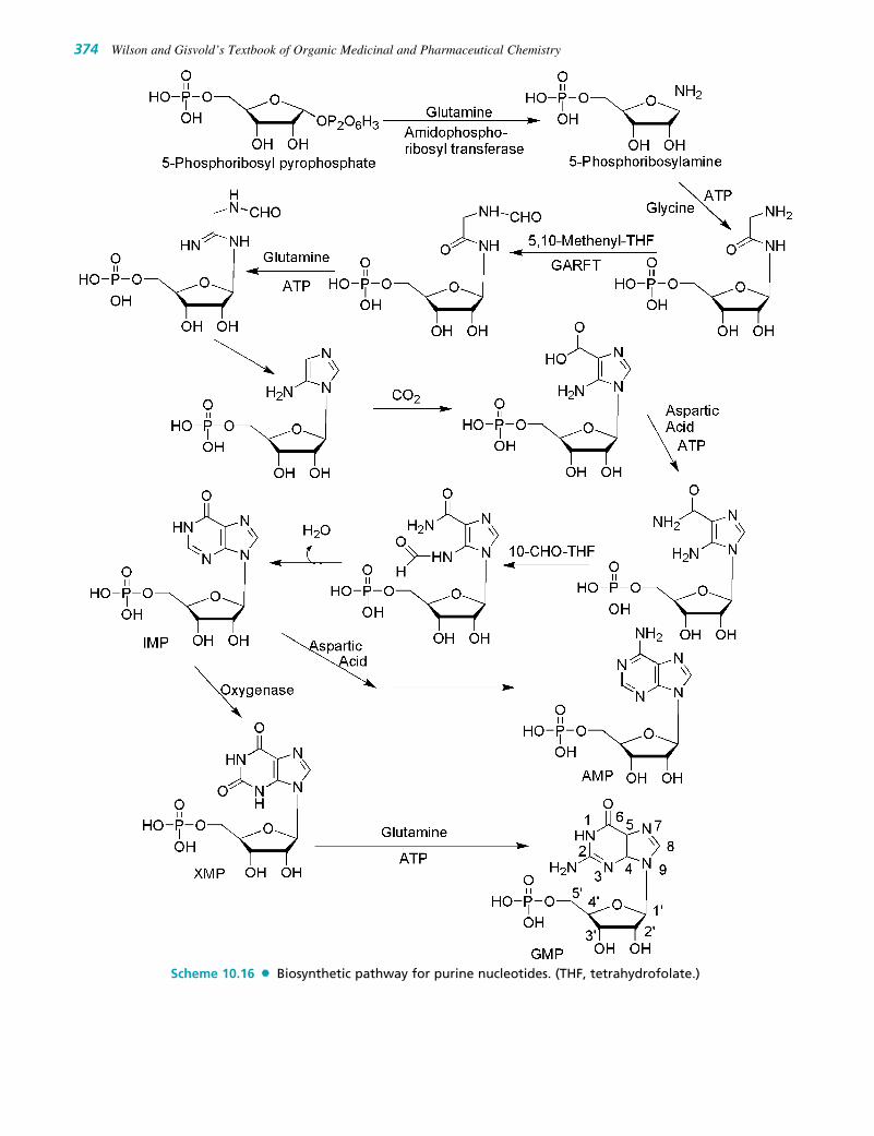
374 Wilson and Gisvold’s Textbook of Organic Medicinal and Pharmaceutical Chemistry
Scheme 10.16 Biosynthetic pathway for purine nucleotides. (THF, tetrahydrofolate.)
0355-0412_17865_Ch10.qxd 12/4/09 5:13 AM Page 374

nucleotide is a substrate for RNA polymerases, and 5-FU isincorporated into the RNA of some cell lines. The incorpo-ration of 5-FU into DNA via DNA polymerase occurs insome tissue lines even though uracil is not a common com-ponent of human DNA. The 5-FU in DNA likely serves assubstrate for the editing and repair enzymes involved inDNA processing for cell division and tissue growth. The ac-tual addition of 5-FU into RNA and/or DNA may not be thedirect cytotoxic event, but the incorporation may lead to lessefficient utilization of cellular resources. The significance ofthese various mechanisms on the overall cytotoxic effects of5-FU may vary with cell line and tissue.
The metabolic activation (anabolism) of 5-FU required toproduce the anticancer effects accounts for no more than 20%of the administered amount of drug in most patients.Catabolic inactivation via the normal pathways for uracil con-sumes the remaining approximate 80% of the dose. The majorenzyme of pyrimidine catabolism is dihydropyrimidine dehy-drogenase (DPD), and 5-FU is a substrate for this enzyme.55
The DPD catabolism of 5-FU is shown in Scheme 10.20. Theformation of dihydro-5-FU (5-FU-H2) occurs very rapidlyand accounts for the majority of the total 5-FU dose in mostpatients. Thus, �-fluoro-�-alanine is the major humanmetabolite of 5-FU.56 Uracil is a substrate for this enzymesystem also and has been dosed with 5-FU and 5-FU prodrugsin an attempt to saturate DPD and conserve active drugspecies. Variability in the levels of DPD activity among thepatient population is a major factor in the bioavailability of 5-FU. Inhibitors of DPD such as uracil or 5-chloro-2,4-dihydroxypyridine (CDHP)53 increase the plasma concentra-tion–time curve of 5-FU by preventing 5-FU catabolism. Onemechanism of drug resistance in 5-FU–treated patients maybe caused by increased levels of DPD in the target tissue. The
observed low bioavailability of 5-FU as a result of the cata-bolic efficiency of DPD and other enzymes has lead to the de-velopment of unique dosing routes and schedules as well asthe development of prodrug forms of 5-FU.53
Attempts at chemical modification of 5-FU to protectfrom catabolic events have produced several prodrug forms,which are converted via in vivo metabolic and/or chemicaltransformation to the parent drug 5-FU.57 Figure 10.8 showsthe structure of the more common prodrug forms of 5-FU.The carbamate derivative of 5�-deoxy-5-fluorocytidine isknown as capecitabine, and it is converted to 5-FU througha series of activation steps. The activation sequence isshown in Scheme 10.21. The initial step is carbamate hy-drolysis followed by deamination, then hydrolysis of thesugar moiety to yield 5-FU. Some of these activation stepstake place at a higher rate in tumor tissue leading to selec-tive accumulation in those cells.53 The last step in the se-quence shown in Scheme 10.21 is catalyzed by phosphoro-lases, and these enzymes occur in higher levels in colorectaltumors. Despite this complex activation process,capecitabine still exhibits some of the significant toxicitiesof 5-fluorouracil. The tetrahydrofuran derivative tegafur isslowly converted to 5-FU but requires quite high doses toreach therapeutic plasma concentrations. Esters of the N-hy-droxymethyl derivative of tegafur show greater anticanceractivity than tegafur.58
Pyrimidine analogs as antimetabolites for cancer therapyhave been developed based on the cytosine structure as well.Modification of the normal ribose or deoxyribose moietyhas produced useful drug species such as cytarabine (ara-C)and gemcitabine, see Figure 10.8. Cytosine arabinoside(ara-C or cytarabine) is simply the arabinose sugar instead
Chapter 10 Antineoplastic Agents 375
Figure 10.8 Anticancer drugs based on pyrimidine and related compounds.
(text continues on page 378)
0355-0412_17865_Ch10.qxd 12/4/09 5:13 AM Page 375

376 Wilson and Gisvold’s Textbook of Organic Medicinal and Pharmaceutical Chemistry
Scheme 10.17 Biochemical conversion of uridine to thymidine.
Scheme 10.18 Metabolic activation of 5-fluorouracil to 5-FdUMP.
0355-0412_17865_Ch10.qxd 12/4/09 5:13 AM Page 376

Chapter 10 Antineoplastic Agents 377
Scheme 10.19 Mechanism of inhibition of TS by 5-fluorouracil.
Scheme 10.20 Catabolic inactivation of 5-FU by dihydropyrimidine dehydrogenase.
Scheme 10.21 Metabolic activation of capecitabine to 5-FU.
0355-0412_17865_Ch10.qxd 12/4/09 5:13 AM Page 377

of ribose, and the only difference in structure is the epimerichydroxyl group at the 2�-position of the pentose sugar. Thisepimeric sugar is similar enough to the natural ribose toallow ara-C to be incorporated into DNA, and its mecha-nism of action may include a slowing of the DNA chainelongation reaction via DNA polymerase or cellular ineffi-ciencies in DNA processing or repair after incorporation.
Gemcitabine59 is the result of fluorination of the 2�-posi-tion of the sugar moiety. Gemcitabine is the 2�,2�-difluorodeoxycytidine species and after its anabolism to diphosphateand triphosphate metabolites, it inhibits ribonucleotide re-ductase and competes with 2�-deoxycytidine triphosphatefor incorporation into DNA. The mechanism of action forgemcitabine is likely similar to that of ara-C including alter-ation of the rate of incorporation into DNA as well as therate of DNA processing and repair.
Modification of the pyrimidine ring has also been ex-plored for the development of potential anticancer drugsbased on antimetabolite theory. Several pyrimidine nucleo-side analogs have one more or one less nitrogen in the hete-rocyclic ring. They are known as azapyrimidine or deaza-pyrimidine nucleosides. 5-Azacytidine is an example of adrug in this category (see Fig. 10.8). This compound was de-veloped via organic synthesis and later found as a naturalproduct of fungal metabolism. The mode of action of thiscompound is complex involving reversible inhibition ofDNA methyltransferase, and this lack of methylated DNAactivates tumor suppressor genes. In certain tumor systems,it is incorporated into nucleic acids, which may result inmisreading or processing errors.
Purine Drugs
The anticancer drugs based on purine structure are shownin Figure 10.9. The design of antimetabolites based onpurine structure began with isosteric thiol/sulfhydryl groupto replace the 6-hydroxyl group of hypoxanthine andguanine. One of the early successes was 6-mercaptopurine(6-MP), the thiol analog of hypoxanthine.60 This purine re-quires bioactivation to its ribonucleotide, 6-thioinosinate(6-MPMP), by the enzyme HGPRT. The resulting nu-cleotide (Scheme 10.22) is a potent inhibitor of an earlystep in basic purine biosynthesis, the conversion of 5-phos-phoribosylpyrophosphate into 5-phosphoribosylamine (seeScheme 10.16). The ribose diphosphate and triphosphatesof 6-mercaptopurine are active enzyme inhibitors, and thetriphosphate can be incorporated into DNA and RNA to in-hibit chain elongation.61 However, the major antineoplasticaction of 6-MP appears to be related to the inhibition ofpurine biosynthesis.
Thioguanine (6-TG) is the 6-mercapto analog of guanine,analogous to 6-MP. Thioguanine is converted into its ribonu-cleotide by the same enzyme that acts on 6-mercaptopurine.It is converted further into the diphosphates and triphos-phates. These species inhibit most of the same enzymes thatare inhibited by 6-mercaptopurine.62 Thioguanine is also in-corporated into RNA, and its 2�-deoxy metabolite is incorpo-rated into DNA. The incorporation into RNA and DNA andthe subsequent disruption of these polymers may account fora greater portion of the antineoplastic activity of thioguaninecompared with 6-MP.63
378 Wilson and Gisvold’s Textbook of Organic Medicinal and Pharmaceutical Chemistry
Figure 10.9 Anticancer drugs based on purines and related compounds.
0355-0412_17865_Ch10.qxd 12/4/09 5:13 AM Page 378

The antineoplastic activity of these purines as well asmost antimetabolites depends on the relative rates of enzy-matic activation and inactivation of these compounds invarious tissues and cells. Drug resistance in certain cell linesmay be caused by lower activity of activating enzymes orhigher activity of catabolic enzymes.64 For the classicpurine antimetabolites 6-MP major pathways of inactivation(see Scheme 10.22) include S-methylation via thiopurine-S-methyl-transferase (TPMT) and oxidation by the enzymexanthine oxidase (XO).62 Xanthine oxidase converts thedrugs to the inactive thiouric acid, and inhibition of the en-zymes responsible for the catabolic breakdown of the purinedrugs can potentiate the drug’s antineoplastic activity.Allopurinol is a potent inhibitor of xanthine oxidase and isoften used as an adjuvant in purine anticancer drug therapy.Allopurinol increases both the potency and the toxicity of 6-mercaptopurine. Its main importance is that it prevents theuric acid kidney toxicity caused by the release of purinesfrom destroyed cancer cells.
Heterocyclic derivatives of 6-mercaptopurine, such asazathioprine, were designed to protect it from catabolic re-actions. Although azathioprine has antitumor activity, it isnot significantly better than 6-mercaptopurine. It has an im-portant role, however, as an immunosuppressive agent inorgan transplants. Adenine arabinoside (Vidarabine) con-tains the sugar, D-arabinose, which is epimeric with D-riboseat the 2�-position. This structural change makes it a compet-itive inhibitor of DNA polymerase, and this activity ac-counts for its antineoplastic activity as well as its antiviralaction. Adenine arabinoside and some of its derivatives arelimited in their antitumor effect by susceptibility to adeno-sine deaminase. This enzyme converts them into the inactivehypoxanthine arabinoside derivatives. High levels of adeno-sine deaminase accounts for resistance of certain tumors tothe action of adenine arabinoside.
The addition of fluorine to the sugar moiety has producedsome purine-based drugs with resistance to the catabolicactivity of adenosine deaminase. In contrast to the suscepti-bility of adenosine arabinoside to adenosine deaminase, its2-fluoro derivative, fludarabine, is stable to this enzyme.The antineoplastic activity of fludarabine depends on itsanabolic conversion to the corresponding triphosphate.2-Chloro-2�-deoxyadenosine (cladribine) also is resistant toadenosine deaminase. It is phosphorylated in cells to the
triphosphate by cytidine kinase, and the triphosphate in-hibits enzymes required for DNA repair.
Folates
Folic acid and the structures of the major antifolate anti-cancer drugs are shown in Figure 10.10. Methotrexate is theclassic antimetabolite of folic acid structurally derived by N-methylation of the para-aminobenzoic acid residue (PABA)and replacement of a pteridine hydroxyl by the bioisostericamino group. The conversion of MOH to MNH2 increasesthe basicity of N-3 and yields greater enzyme affinity. Thisdrug competitively inhibits the binding of the substrate folicacid to the enzyme DHFR, resulting in reductions in the syn-thesis of nucleic acid bases, perhaps most importantly, theconversion of uridylate to thymidylate as catalyzed bythymidylate synthetase.65 In addition, purine synthesis is in-hibited because the N-10-formyl tetrahydrofolic acid is aformyl donor involved in purine synthesis. The interconver-sion of the various folate species is shown in Scheme 10.23.Recall that in Scheme 10.16, THFs are cofactors in at leasttwo key steps in the normal biosynthesis of purines.
Methotrexate66 is a broad-spectrum antineoplastic agentcommonly used in the treatment of acute lymphoblastic andmyeloblastic leukemia and other lymphomas and sarcomas.The major side effects seen are bone marrow suppression,pulmonary fibrosis, and GI ulceration. Leucovorin is oftengiven 6 to 24 hours after methotrexate to prevent the long-term effects on normal cells by preventing the inhibition ofDNA synthesis. Related to this is Pemetrexed, but its scopeis greater in that it not only inhibits DHFR but also TS andglycinamide ribonucleotide formyltransferase (GARFT),which is involved in purine biosynthesis (see Fig. 10.16).The material is taken into tumor cells by the reduced folate-carrier systems and subsequently converted to the polyglu-tamate, which is retained in the cell.
Products
5-FLUOROURACIL (5-FU, EFUDEX, ADRUCIL,FLUOROPLEX)
The drug is available in a 500-mg or 10-mL vial for IV useand as a 1% and 5% topical cream. 5-FU is used in the treat-ment of several carcinoma types including breast cancer,
Chapter 10 Antineoplastic Agents 379
Scheme 10.22 Conversion of6-MP to active 6-thioinosine-5-monophosphate (6-MPMP) byHPGRT and inactivation by xan-thine oxidase and thiopurinemethyl transferase.
0355-0412_17865_Ch10.qxd 12/4/09 5:13 AM Page 379

colorectal cancer, stomach cancer, pancreatic cancer, andtopical use in basal cell cancer of the skin. The mechanism ofaction includes inhibition of the enzyme TS by the deoxyri-bose monophosphate metabolite, 5-FdUMP. The triphos-phate metabolite is incorporated into DNA and the ribosetriphosphate into RNA. These incorporations into growingchains result in inhibition of synthesis and function of DNAand RNA. Resistance can occur as a result of increased ex-pression of TS, decreased levels of reduced folate substrate5,10-methylenetetrahydrofolate, or increased levels of dihy-dropyrimidine dehydrogenase. Dihydropyrimidine dehydro-genase is the main enzyme responsible for 5-FU catabolism.
Bioavailability following oral absorption is erratic.Administration of 5-FU by IV yields high drug concentra-tions in bone marrow and liver. The drug does distribute intothe central nervous system (CNS). Significant drug interac-tions include enhanced toxicity and antitumor activity of5-FU following pretreatment with leucovorin. Toxicities in-clude dose-limiting myelosuppression, mucositis, diarrhea,and hand–foot syndrome (numbness, pain, erythema, dry-ness, rash, swelling, increased pigmentation, nail changes,pruritus of the hands and feet).
CAPECITABINE (XELODA)
The drug is available in a 150- and 500-mg tablets for oraluse. This drug is a fluoropyrimidine carbamate prodrugform of 5-fluorouracil (5-FU). It is used to treat breast can-cer and colorectal cancer. The drug is converted to 5-FU bythe enzyme thymidine phosphorylase following esterase ac-
tivity to hydrolyze the carbamate moiety and deamination.Capecitabine is readily absorbed by the GI tract, and peakplasma levels of 5-FU occur about 2 hours after oral admin-istration. Indications, drug interactions, and toxicities areequivalent to those of 5-FU.
CYTARABINE (CYTOSINE ARABINOSIDE, ARA-C, CYTOSAR-U, TARABINE)
The drug is available in 100-, 500-, 1,000-, and 2,000-mgmultidose vials for IV use. Cytarabine is used in the treat-ment of acute myelogenous leukemia and CML. This drugis a deoxycytidine analog originally isolated from thesponge Cryptothethya crypta. It is active following intracel-lular activation to the nucleotide metabolite ara-CTP. Theresulting ara-CTP is incorporated into DNA resulting inchain termination and inhibition of DNA synthesis andfunction. Resistance can occur because of decreased activa-tion or transport and increased catabolic breakdown.Metabolic breakdown within the GI tract leads to poorbioavailability. The drug distributes rapidly into tissues andtotal body water with cerebrospinal fluid (CSF) levelsreaching 20% to 40% of those in plasma. Cytidine deami-nase is the primary catabolic enzyme involved in the inacti-vation of cytarabine. Drug interactions include antagonismof the effects of gentamicin, decreasing the oral bioavail-ability of digoxin, as well as enhancing the cytotoxicity ofvarious alkylating agents, cisplatin, and ionizing radiation.Pretreatment with methotrexate enhances the formation ofara-CTP metabolites resulting in enhanced cytotoxicity.
380 Wilson and Gisvold’s Textbook of Organic Medicinal and Pharmaceutical Chemistry
Figure 10.10 Structures of folic acid andantifolate anticancer drugs.
0355-0412_17865_Ch10.qxd 12/4/09 5:13 AM Page 380

Toxicities include myelosuppression, leukopenia andthrombocytopenia, nausea and vomiting anorexia, diarrhea,and mucositis. Neurotoxicity is usually expressed as ataxia,lethargy, and confusion. An allergic reaction often describedin pediatric patients includes fever, myalgia, malaise, bonepain, skin rash, conjunctivitis, and chest pain.
FLOXURIDINE (FLUORODEOXYURIDINE, FUDR)
The drug is available as a 500-mg vial of lyophilized pow-der. The drug is used to treat metastatic GI adenocarcinoma.The mechanism of action of this fluoropyrimidine deoxynu-cleoside analog involves metabolic conversion to 5-fluo-rouracil (5-FU) metabolites resulting in inhibition of TSthus disrupting DNA synthesis, function, and repair.Resistance can occur because of increased expression of TS,
decreased levels of reduced folate 5,10-methylenetetrahy-drofolate, increased activity of DNA repair enzymes, and in-creased expression of dihydropyrimidine dehydrogenase(the major catabolic enzyme). The drug is poorly absorbedfrom the GI tract and is extensive metabolized to 5-FU and5-FU metabolites. Dihydropyrimidine dehydrogenase is themain enzyme responsible for 5-FU catabolism, and it ispresent in liver, GI mucosa, white blood cells, and kidney.The drug interaction and toxicity profiles are equivalent tothose of 5-FU.
GEMCITABINE (DFDC, GEMZAR)
The drug is available as the hydrochloride salt in 200- and1,000-mg lyophilized single-dose vials for IV use.Gemcitabine is used to treat bladder cancer, breast cancer,
Chapter 10 Antineoplastic Agents 381
Scheme 10.23 Interconversion of various folate species. (DHFR, dihydrofolate reductase.)
0355-0412_17865_Ch10.qxd 12/4/09 5:13 AM Page 381
pancreatic cancer, and NSCLC. Gemcitabine is a potent ra-diosensitizer, and it increases the cytotoxicity of cisplatin.The mechanism of action of this fluorine-substituted deoxy-cytidine analog involves inhibition of DNA synthesis andfunction via DNA chain termination. The triphosphatemetabolite is incorporated into DNA inhibiting severalDNA polymerases and incorporated into RNA inhibitingproper function of mRNA. Resistance can occur because ofdecreased expression of the activation enzyme deoxycyti-dine kinase or decreased drug transport as well as increasedexpression of catabolic enzymes. Drug oral bioavailabilityis low because of deamination within the GI tract, and thedrug does not cross the blood-brain barrier. Metabolism bydeamination to 2�, 2�-difluorouridine (dFdU) is extensive.Drug toxicity includes myelosuppression, fever, malaise,chills, headache, myalgias, nausea, and vomiting.
CLADRIBINE (2-CHLORODEOXYADENOSINE, 2-CDA, LEUSTATIN)
The drug is available in a 10-mg or 10-mL single-use vialfor IV use. Cladribine is used for chronic lymphocyticleukemia, hairy cell leukemia, and non-Hodgkin’s lym-phoma. The mechanism of action of this purine de-oxyadenosine analog involves incorporation into DNA re-sulting in inhibition of DNA chain extension and inhibitionof DNA synthesis and function. This incorporation intoDNA occurs via the triphosphate metabolite active species.The 2-chloro group on the adenine ring produces resistanceto breakdown by adenosine deaminase. Resistance to the an-ticancer effects can occur because of decreased expressionof the activating enzyme or overexpression of the catabolicenzymes. Oral bioavailability is variable and averages about50%. The drug crosses the blood-brain barrier; however,CSF concentrations reach only 25% of those in plasma. Thedrug is selectively activated inside the cell, and intracellularconcentrations of phosphorylated metabolites exceed thosein plasma. Toxicities include myelosuppression, neutrope-nia, immunosuppression, fever, nausea, and vomiting.
FLUDARABINE (2-F-ARA-AMP, FLUDARA)
The drug is available as the phosphate salt in a 50-mg vialfor IV use. Fludarabine is used to treat chronic lymphocyticleukemia and non-Hodgkin’s lymphoma. The mechanism ofaction involves the triphosphate metabolite and its inhibitionof DNA chain elongation. The 2-fluoro group on the ade-nine ring renders fludarabine resistant to breakdown byadenosine deaminase. The drug is rapidly dephosphorylatedto 2-fluoro-ara-adenosine (F-ara-A) after administration. F-ara-A is taken into the cell and subsequently re-phosphory-lated to yield the triphosphate (F-ara-ATP), the active drugspecies. Resistance can occur via decreased expression ofthe activating enzymes and decreased drug transport.Fludarabine is orally bioavailable and is distributed through-out the body reaching high levels in liver, kidney, andspleen. The drug is metabolized to F-ara-A, which enterscells via the nucleoside transport system and is rephospho-rylated by deoxycytidine kinase to fludarabine monophos-phate and finally fludarabine triphosphate, the activespecies. About 25% of F-ara-A is excreted unchanged inurine. Drug interactions include an increased incidence offatal pulmonary toxicity when fludarabine is used in com-bination with pentostatin. Additionally, fludarabine may
potentiate the effects of several other anticancer drugs in-cluding cytarabine, cyclophosphamide, and cisplatin.Toxicities include myelosuppression, immunosuppression,fever, nausea, and vomiting.
MERCAPTOPURINE (6-MP, MERCAPTOPURINUM,PURINETHOL)
The drug is available as a 50-mg tablet for oral use. The pri-mary uses of mercaptopurine are in the treatment of lym-phoblastic leukemia, acute lymphocytic leukemia, and Crohndisease. The mechanism of action includes incorporation ofmercaptopurine into DNA and RNA via the triphosphatemetabolite. This incorporation inhibits synthesis and functionof the resulting modified DNA or RNA. The parent drug isinactive and requires phosphorylation for activity. Resistancecan occur via decreased expression of the activating enzymesor increased expression of the major catabolic enzymes.Oral absorption is generally incomplete (about 50%) andthe drug does not enter the CNS in therapeutic quantities.Mercaptopurine is metabolized by methylation, and themethylated product has anticancer activity. Oxidation byxanthine oxidase yields inactive metabolites. The concurrentuse of xanthine oxidase inhibitors such as allopurinol can en-hance the potency of mercaptopurine by inhibiting its cata-bolic breakdown. The toxicities for mercaptopurine includemyelosuppression, immunosuppression, nausea, vomiting,diarrhea, dry skin, urticaria, and photosensitivity.
THIOGUANINE (6-THIOGUANINE, 6-TG, TABLOID)
The drug is available in 40-mg tablets for oral use.Thioguanine is used to treat acute nonlymphocytic leukemia.The mechanism of action involves incorporation of thetriphosphate into DNA and RNA, resulting in inhibition ofprocessing and function. Intracellular phosphorylation is re-quired for activity and inhibition of purine biosynthesis.Resistance can include decreased expression of the activat-ing enzyme, decreased drug transport, and/or increased ex-pression of catabolic enzymes. The oral absorption ofthioguanine is poor, and the drug does not appear to cross theblood-brain barrier. Major metabolic pathways involvedeamination or methylation. Thioguanine is not a substratefor the enzyme xanthine oxidase in contrast to mercaptopu-rine. Toxicities include myelosuppression, immunosuppres-sion, nausea, vomiting, mucositis, and diarrhea.
PENTOSTATIN (2�-DEOXYCOFORMYCIN, DCF, NIPENT)
The drug is available in 10-mg vials for IV use. The drug isused to treat leukemias such as hairy cell leukemia, chroniclymphocytic leukemia, and lymphoblastic leukemia. Themechanism of action involves inhibition of the enzymeadenosine deaminase yielding increased cellular levels ofdeoxyadenosine and deoxyadenosine triphosphate (dATP).The increased levels of dATP are cytotoxic to lymphocytes.Pentostatin is a fermentation product of Streptomyces an-tibioticus. Resistance appears to involve decreased cellulartransport or increased expression of catabolic enzymes.Acid instability prevents oral administration, and the drug isonly administered by IV. The drug is distributed in totalbody water but does not enter the CNS. The majority of thedosage is excreted unchanged in the urine. Fatal pulmonary
382 Wilson and Gisvold’s Textbook of Organic Medicinal and Pharmaceutical Chemistry
0355-0412_17865_Ch10.qxd 12/4/09 5:13 AM Page 382

toxicity has occurred when pentostatin and fludarabine areused in combination. Toxicities include myelosuppression,immunosuppression, nausea, vomiting, headache, lethargy,and fatigue.
METHOTREXATE (MTX, ABITREXATE, MEXATE, FOLEX)
The drug is available in 50-, 100-, 200-, and 1,000-mg vialsfor IV use. Methotrexate is used to treat several cancer typesincluding breast cancer, bladder cancer, colorectal cancer,and head and neck cancer. The mechanism of action ofmethotrexate involves inhibition of DHFR leading to a de-pletion of critical reduced folates. The reduced folates arenecessary for biosynthesis of several purines and pyrim-idines. Resistance to methotrexate can occur because ofdecreased carrier-mediated transport of drug into cells orincreased expression of the target enzyme DHFR. Oralbioavailability varies with dose because of saturable uptakeprocesses, and high doses are required to reach therapeuticlevels in the CNS. The majority of drug dosage is excretedunchanged in the urine. The renal excretion of methotrexateis inhibited by several carboxylic acid drugs such as peni-cillins, probenecid, nonsteroidal anti-inflammatory agents,and aspirin. Methotrexate enhances 5-FU antitumor effectswhen given 24 hours prior to the fluoropyrimidine.Methotrexate toxicity includes myelosuppression, mucosi-tis, nausea, vomiting, severe headaches, renal toxicity, acutecerebral dysfunction, skin rash, and hyperpigmentation.
PEMETREXED (MTA, ALIMTA)
The drug is available in a 100-mg sterile vial for IV use. Thedrug appears to be effective against a range of tumors in-cluding mesothelioma, NSCLC, colorectal cancer, bladdercancer, and lung cancer. The mechanism of action involvesinhibition of TS resulting in inhibition of thymidylate andDNA synthesis. This drug is a pyrrolopyrimidine analog offolate with antifolate activity. Resistance can occur by in-creased expression of TS, decreased binding affinity for TS,or decreased drug transport into cells. The drug is adminis-tered only via the IV route and distributes to all tissues.Cellular activation to the more potent polyglutamated formsoccurs, and the majority of the dose is excreted unchangedin the urine. The drug interaction and toxicity profiles aresimilar to that of methotrexate.
TRIMETREXATE (TMTX, NEUTREXIN)
The drug is available as a lyophilized powder in 5- or 30-mgvials for IV use. The drug is used to treat colorectal cancer,head and neck cancer as well as NSCLC. The mechanism ofaction of trimetrexate involves folate antagonism and inhi-bition of thymidylate synthesis. Trimetrexate does not formintracellular polyglutamate adducts as does methotrexateand other related compounds. Resistance can occur by in-creased expression of the target enzyme, decreased bindingaffinity for the target enzyme, or decreased intracellulardrug transport. Trimetrexate is administered only by the IVroute and distributed throughout the body with extensivebinding to plasma proteins. The major catabolic pathwaysinvolve O-demethylation followed by glucuronide conjuga-tion. The drug interaction and toxicity profiles are similar tothose for methotrexate.
HYDROXYUREA (DROXIA, HYDREA)
HONH-CO-NH2. The drug is available in a 500-mg capsulefor oral use. Hydroxyurea is often considered an an-timetabolite drug, and it is used to treat myelogenousleukemia, ovarian cancer, and essential thrombocytosis. Themechanism of action of hydroxyurea involves inhibition ofDNA biosynthesis by inhibition of the enzyme ribonu-cleotide reductase (see Fig. 10.15). Resistance can occur viaincreased expression of ribonucleotide reductase. The oralbioavailability is quite high approaching 100% and the drugis distributed to all tissues. Hydroxyurea readily enters theCNS and distributes to human breast milk. A major portionof the total dose is excreted unchanged in the urine. Thedrug has been shown to increase the toxicity of 5-FU, andhydroxyurea may increase the effectiveness of some an-timetabolite HIV drugs. The toxicity profile includes myelo-suppression, leucopenia, nausea, vomiting, pruritus hyper-pigmentation, headache, drowsiness, and confusion.
ANTIBIOTICS AND NATURAL PRODUCTS
A variety of the anticancer agents available today are derivedfrom natural sources with several of these being obtainedfrom microbial sources (antibiotics). Many of the antineo-plastic antibiotics are produced by the soil fungusStreptomyces. Both the antibiotic and natural product classeshave multiple inhibitory effects on cell growth; however,they primarily act to disrupt DNA function and cell division.
There are several mechanisms by which these agents tar-get DNA, including intercalation, alkylation, and strandbreakage either directly or as a result of enzyme inhibition.Intercalation is a process by which a planar molecule of theappropriate size inserts itself between adjacent base pairs ofDNA and in so doing, it causes a local unwinding that maydisrupt the normal template function of DNA. Intercalationrequires that the drug induce a cavity between base pairsso that insertion may occur. The interaction of the intercala-tor and the adjacent base pairs occurs by the overlap of p-orbitals of the intercalator and the base pairs. The p-orbitals of the intercalation species are provided by a com-bination of aromatic and conjugated systems that impart theplanarity required for intercalation. The drug–DNA interac-tion is further stabilized by side chains attached to the inter-calation species. The side chains often include a cationicmoiety, which may form ionic bonds with the anionic phos-phate backbone. Alternative modes of stabilization mayoccur through a combination of van der Waals interaction orhydrogen bonds. The overall result of these interactions is tocause a local bend or kink in DNA resulting in a local shapedistortion. This may produce several effects but is often as-sociated with inhibition of normal DNA function.
By virtue of their ability to induce bends or kinks inDNA, intercalators may also result in inhibition oftopoisomerase enzymes. In order for the cell to store thenearly 2 m of DNA that it contains, it is necessary to fold thegenetic material into a more compact form and in theprocess, knots and tangles are produced. Topoisomerase en-zymes are responsible for the unwinding and relaxation ofDNA so that transcription may occur.67 There are two majortypes of topoisomerase enzymes, which are important sites
Chapter 10 Antineoplastic Agents 383
0355-0412_17865_Ch10.qxd 12/4/09 5:13 AM Page 383

of action for antineoplastics. Topoisomerase I makes a sin-gle-strand break in DNA and subsequently allows the otherstrand to spin, relieving any tension associated with thepacking process and subsequently reseals the broken strand.Topoisomerase II makes double-strand breaks in DNA al-lowing an intact chain to pass through and then subse-quently reseals the double-strand break. In this way, it canrelieve the knotting that occurs upon folding of the DNA.Both enzymes are believed to recognize sequences of DNA,bind and function by formation of a transient phosphodiesterbond between the DNA strand and a tyrosine residue pres-ent on the protein. Inhibition may occur at different steps inthe process such that if there is stabilization of the cleavablecomplex, the resealing step may be delayed or prevented re-sulting in strand breakage. This may then activate the apo-ptotic cascade.
There are several natural products that are capable of dis-rupting the formation and function of the mitotic spindle.These include the epipodophyllotoxins, the taxanes, and thevinca alkaloids. The mitotic spindle forms during the Mphase of the cell cycle and is responsible for moving thereplicated DNA to opposite ends of the cell in preparationfor cell division. During prophase, the cytoskeleton made ofproteins, which gives the cell its shape, begins to breakdownproviding the building blocks for the mitotic spindle. Thecell then progresses to prometaphase, where the nuclearmembrane begins to disappear and the spindle begins toform attaching the centrosomes to the kinetochores, whichare proteins joining the two sister chromatids. Tension isthen applied to the spindle so that during metaphase, thechromatids align near the center of the cell. In anaphase, afurther increase in tension separates the sister chromatids atthe kinetochore, and they are pulled to opposite ends of thecell. Ultimately, the microtubules will disintegrate and re-form the cytoskeleton. The process is highly controlled andthere are several proteins including “motor proteins,” whichare involved in coordinating the process.
Actinomycins
The actinomycins are a group of compounds that are iso-lated from various species of Streptomyces, all of whichcontain the same phenoxazone chromophore but differ inthe attached peptide portion. Originally, these materialswere investigated for use as antibiotics, but they proved tobe too toxic. From this group emerged actinomycin D,which is known as dactinomycin and contains identicalpentapeptides bound through an amide linkage utilizingthe amino group of L-threonine with carbonyls at positions1 and 9.68 The pentapeptides namely L-threonine, D-valine,L-proline, sarcosine, and L-methylvaline form a lactone viathe side chain hydroxyl of L-threonine and the carboxylgroup of L-methylvaline (Fig. 10.11).
Dactinomycin binds noncovalently to double-strandedDNA by partial intercalation between adjacent guanine-cytosine bases resulting in inhibition of DNA function. Thestructural feature of dactinomycin important for its mecha-nism of cytotoxicity is the planar phenoxazone ring, whichfacilitates intercalation between DNA base pairs. The pep-tide loops are located within the minor groove and providefor additional interactions (Fig. 10.12). The preference forGpC base pairs is thought to be partly related to the forma-tion of a hydrogen bond between the 2-amino groups of
guanine and the carbonyls of the L-threonine residues.69
Additional hydrophobic interactions and hydrogen bondsare proposed to form between the peptide loops and thesugars and base pairs within the minor groove. In all, acti-nomycin D spans four to five DNA base pairs (Fig. 10.12).This results in high affinity with the molecule only slowlydissociating from DNA. Additional modes of binding arealso possible for the molecule. The primary effect of thisinteraction is the inhibition of DNA-directed RNA synthe-sis and specifically RNA polymerase. DNA synthesis mayalso be inhibited, and the agent is considered cell cycle spe-cific for the G1 and S phases. The drug has been found tobind to single-stranded DNA and double-stranded DNAwithout adjacent GpC sequences.70 It has been suggestedthat binding to single-stranded DNA may occur as thestrands separate during transcription, and this may be re-sponsible for the inhibition of RNA polymerase.71 Thosegenes that were being actively transcribed, which would re-quire strand separation, would be more susceptible to thistype of binding. Inhibition of topoismerase II also occurssuch that the enzyme-DNA complex is stabilized and strandbreakage may be seen. Resistance is caused by a decreasedability of tumor cells to take up the drug and P-glycoprotein(Pgp)-mediated efflux.
384 Wilson and Gisvold’s Textbook of Organic Medicinal and Pharmaceutical Chemistry
Figure 10.11 Structure of dactinomycin.
Figure 10.12 Representation of the dactinomycin–DNA complex. (Reprinted with permission from Hou, M.H., et al.: Nucleic Acids Res. 30:4910, 2002.)
0355-0412_17865_Ch10.qxd 12/4/09 5:13 AM Page 384

Anthracyclines
The anthracycline antibiotics (Fig. 10.13) are characterizedby a planar oxidized anthracene nucleus fused to a cyclo-hexane ring that is subsequently connected via a glycosidiclinkage to an amino sugar. Initially discovered in the early1960s when they were isolated from Streptomycespeucetius, hundreds of compounds belonging to this classhave subsequently been discovered of which five are usedclinically in the United States (doxorubicin, daunorubicin,idarubicin, epirubicin, and valrubicin). The conjugated sys-tems found in these molecules impart a red color, which isreflected in the name.
Studies of the mechanism by which the anthracyclines ex-hibit their cytotoxic effects initially focused on the ability ofthe compounds to associate with DNA resulting from inter-calation of their planar ring system reinforced by auxiliarybinding of the amino sugar. Subsequent work focused on theability of these compounds to generate free radicals.
Although free radical generation is thought to be important inthe toxicity, it was not shown to occur in the nucleus andtherefore not likely associated with their antiproliferative ef-fects. Currently, the accepted mechanism involves intercala-tion followed by inhibition of topoisomerase II resulting instrand breakage leading to apoptosis.72 The anthracyclinesare considered specific for the S phase of the cell cycle.There is data to support increased binding of p53 with DNAwhen anthracyclines are administered, which would stimu-late the apoptotic process, although some studies have failedto confirm this.73,74 Several of these agents have been cocrys-tallized with B-DNA oligomers and the three-dimensionalstructure determined by x-ray crystallography. The generalfeatures show rings B and C intercalating between CpG basepairs and ring D protruding into the major groove. The sugarmoiety lies within the minor groove and along with ring A isimportant for interaction and inhibition of topoisomerase IIby stabilization of the cleavable complex. The amino sugar
Chapter 10 Antineoplastic Agents 385
Figure 10.13 Structure of the anthracycline antibiotics.
0355-0412_17865_Ch10.qxd 12/4/09 5:13 AM Page 385

seems to play an especially important role and compoundslacking this functionality failed to inhibit the enzyme. In thecase of doxorubicin and daunorubicin, specificity is providedby the hydrogen bonding between O-9 of the anthracyclineand N-2 and N-3 of guanine. The x-ray data also suggestedthat the interaction with DNA is stabilized by the formationof several hydrogen bonds through bridging water mole-cules.75 The formation of covalent bonds between anthracy-clines and DNA also is supported by several studies in whichformaldehyde is produced by oxidation of cellular compo-nents or other anthracycline molecules. This oxidation resultsfrom the production of ROS such as H2O2, which are gener-ated during redox cycling of anthracyclines (Scheme 10.24).The generated formaldehyde may then form a methylenebridge between the 4�-amino group of the anthracycline andthe 2-amino group of guanine in DNA.76
Although the anthracyclines produce several adverseeffects that are typical for antineoplastics, cardiotoxicity is aspecial concern with this class of agents. The associated car-diomyopathies and congestive heart failure (CHF) have beenrelated to the ability of these compounds to undergo redoxcycling.77 This is most notable in the case of daunorubicinand doxorubicin and less of a problem in the newer deriva-tives, idarubicin and epirubicin. The C ring of the anthracy-
clines is subject to a one or two electron reduction by severaldifferent enzymes including NAD(P)H-oxidoreductases andCYP reductases to give the semiquinone or hydroquinone.The case of the one electron reduction is shown in Scheme10.25. This may undergo reversion to the starting quinone infutile redox cycling accompanied by the production of super-oxide radical. The radical is normally converted to H2O2 bysuperoxide dismutase and H2O2 is then converted to H2O andO2 by catalase. However, the production of superoxide(O2
��) is also associated with the release of iron from intra-cellular stores, which may be chelated by the anthracycline.The iron then diverts the normal detoxification pathway bycatalase so that more potent radicals such as hydroxyl radical(�OH) are produced. Myocardial cells possess lower levels ofcatalase and therefore are less able to detoxify the H2O2 thatresults from redox cycling. This results in increasing levelsof �OH for which a detoxification pathway does not exist andtherefore cellular damage occurs. Materials such as O2
��,�OH, and H2O2 are known as reactive oxygen species or ROSand in sufficiently high concentration produce cellular dam-age. Hydroxyl radicals cause single-strand breaks in DNA,which would activate p53 and enhance apoptosis in cardiaccells. Studies have also shown that doxorubicin administra-tion stimulates both the intrinsic and extrinsic pathways of
386 Wilson and Gisvold’s Textbook of Organic Medicinal and Pharmaceutical Chemistry
Scheme 10.24 Formation of anthracycline-DNA adduct.
0355-0412_17865_Ch10.qxd 12/4/09 5:13 AM Page 386

apoptosis in numerous ways, and this has been linked to theincreased production of H2O2 and hydroxyl radical. One pro-posed mechanism involves damage to the mitochondrialmembrane by H2O2 and �OH resulting in the release of cy-tochrome c, which would activate the intrinsic pathway(Fig. 10.2).78 This sequence of events would be desirable ina cancer cell but not in a normal cardiac cell.
Additional mechanisms of cardiotoxicity have been pro-posed, which involve the metabolic reduction of the anthra-cycline C-13 ketone to the alcohol.79 Several pharmacolog-ical effects have been associated with the resulting alcoholsincluding loss of Ca�2 homeostasis and inhibition ofNa�/K�-ATPase in cardiac cells. The formation of doxoru-bicin C-13 alcohol, doxorubicinol has been associated withconversion of iron regulatory protein-1 (IRP-1) into a nullprotein that is no longer able to maintain iron homeostasis.80
It has been proposed that the loss of iron regulation maycontribute to the cardiotoxicity seen with doxorubicin.However, compared with doxorubicin, daunorubicin andidarubicin form greater amounts of the alcohol metabolitesand exhibit similar and reduced cardiotoxicity, respectively.This suggests that metabolism to the alcohols is not solelyresponsible for the cardiotoxicity.
Several strategies have been developed to reduce the car-diotoxicity associated with the anthracyclines. Slow infu-sion over 48 to 96 hours versus administration of a bolusgiven over 15 minutes was implemented in the 1980s andhas proven to be effective in reducing the toxicity withoutadversely affecting the antineoplastic effects. Given the pro-posed mechanism of toxicity, it is not surprising that severalantioxidants have been investigated including vitamins A,C, and E and also thiol-containing reducing agents such asacetylcysteine. These, however, have not proved effective inlarge measure. An alternative strategy is to chelate the ironrequired for the activation of H2O2, and for this, dexrazox-ane (Totect) is used.81 Radiolabeling experiment haveshown that dexrazoxane is rapidly taken up into myocardialcells, where it is subsequently hydrolyzed by a two-step
process to yield the diamide acid (Scheme 10.26), whichchelates free iron and iron bound to the anthracycline and issimilar to the chelating agent ethylenediaminetetraaceticacid (EDTA). This has been proven effective and has al-lowed patients to tolerate higher doses and increased num-bers of cycles of the anthracyclines. Dexrazoxane is also aninhibitor of topoisomerase II that, unlike the anthracyclines,does not result in strand breaks.82 This additional property isimportant in its utility as antidote in cases of anthracyclineextravasation.83 The anthracyclines are often administeredwith other antineoplastics such as the taxanes andtrastuzumab, which may have cardiotoxicities themselvesleading to an increased risk despite the precautionsmentioned.
Related to the anthracyclines is mitoxantrone, which isshown in Figure 10.14. Although mitoxantrone is a syn-thetic agent, it is included with the natural products becauseit is mechanistically similar to the anthracyclines. Producedin the late 1970s, it is a derivative of a synthetic dye and isclassified as an anthracenedione.84 The mechanism involvesintercalation by the chromophore, which is stabilized by the2-[(2-aminoethyl)amino]ethanol side chain presumably as a
Chapter 10 Antineoplastic Agents 387
Scheme 10.25 Process of anthracycline redox cycling.
Scheme 10.26 Aqueous hydrolysis of dexrazoxane toyield the iron-chelating metabolite.
0355-0412_17865_Ch10.qxd 12/4/09 5:13 AM Page 387

result of an ionic interaction of the protonated amines withthe phosphate backbone of DNA. The formation of covalentadducts with DNA have also been demonstrated to occur ina manner similar to the anthracyclines. In this case, how-ever, other enzymes such as myeloperoxidase are responsi-ble for the generation of formaldehyde.85 Topoisomerase IIis inhibited, and strand breakage occurs similar to that seenwith the anthracyclines. In contrast to the anthracyclines,mitoxantrone is not a substrate for the reductase enzymes re-sponsible for the conversion to the semiquinone so that ROSare not generated by this process.86 This lack of activationhas been attributed to the presence of the side chains. Thishas the effect of reducing the cardiotoxicity but not com-pletely eliminating it, and caution should be used especiallyin those patients with existing cardiovascular problems.
Individual Agents
DACTINOMYCIN-D (ACT-D, ACTINOMYCIN-D,COSMEGEN, LYOVAC COSMEGEN)
Dactinomycin is available in vials containing 0.5 mg of drugfor reconstitution in sterile water for IV administration Thisantibiotic is most effective in the treatment of rhabdomyosar-coma and Wilms tumor in children as well as in the treatmentof choriocarcinoma, Ewing sarcoma, Kaposi sarcoma, andtesticular carcinoma. The pharmacokinetics of dactinomycinhas not been well characterized, but it appears to concentratein nucleated blood cells. The agent is 5% to 15% plasmaprotein bound and is excreted mostly unchanged in urineand bile. Other metabolites have not been characterized.The terminal elimination half-life is 30 to 40 hours.Myelosuppression is dose limiting with both leucopenia andthrombocytopenia being the most likely presentation. Nauseaand vomiting occur shortly (2 hours) after treatment and maybe severe. Mucositis and diarrhea also result from irritation ofthe GI tract. Hair loss is commonly associated with the agentas is hyperpigmentation of the skin and erythema.
DOXORUBICIN HYDROCHLORIDE (ADM, ADR,ADRIA, ADRIAMYCIN)
Doxorubicin is available as both the conventional dosageform and a liposomal preparation, both of which are admin-istered by infusion. Doxorubicin HCl powder is available in10-, 20-, 50-, and 150-mg vials and is widely used in treat-ing various cancers, including leukemias, soft and bone tis-sue sarcomas, Wilms tumor, neuroblastoma, small cell lungcancer, and ovarian and testicular cancer. Resistance to theagent involves decreased expression of topoisomerase II and
mutations in this enzyme that decrease binding of the drug.The agent may also be actively secreted from cancer cells byPgp. The agent is rapidly taken up into tissues following in-jection with a distributive half-life of 5 minutes followed bya slow elimination half-life of 20 to 48 hrs. The primaryroute of elimination is in the bile and feces. Metabolism in-volves reduction of the C-13 ketone to yield doxorubicinol(active) along with cleavage of the amino sugar to give theaglycone. The aglycones are also capable of undergoingredox cycling and producing ROS.
The toxicities associated with doxorubicin include dose-limiting cardiotoxicity and myelosuppression. Most patientswill experience alopecia and nausea. Cardiotoxicity is a lim-iting factor in the administration of doxorubicin, which, inits most severe form, may progress to CHF. This may occurduring therapy or during months or years following the ter-mination of therapy. The percentage of patients thatprogress to CHF increases with increasing cumulative dose.
The liposomal form of doxorubicin (Doxil) offers severaladvantages including an improved pharmacokinetic profilewith slower plasma clearance giving a distributive half-lifeof 45 hours versus 5 minutes for doxorubicin and a smallervolume of distribution of 4 L versus 254 L for doxoru-bicin.87 There is also reportedly an increased uptake ofdoxorubicin in tumor cells, which has been attributed to theability of the liposomes to break down in the more acidic en-vironment associated with tumor cells or by the release ofenzymes from the tumor cells that degrade the liposomes.The liposomes decrease uptake by myocardial tissue, so car-diotoxicity is less common. The liposomes also reduce con-version to doxorubicinol, which has been implicated in thecardiotoxicity of doxorubicin. Additionally, there is a de-crease in the incidence of extravasation. Other adverse ef-fects are also decreased including less nausea, alopecia, andstomatitis although skin toxicity, which is normally seen asa rash on the feet and hands, may occur frequently.
DAUNORUBICIN HYDROCHLORIDE (DAUNOMYCIN,DNM, DNR, CERUBIDINE, RUBIDOMYCIN)
Daunorubicin is available in 20- and 100-mg vials for recon-stitution. The agent is given intravenously for the treatmentof acute nonlymphocytic and lymphocytic leukemia. Otherunlabeled uses include CML and Kaposi sarcoma. In gen-eral, its use is more limited than that of doxorubicin.Daunorubicin lacks the hydroxyl groups found at C-14 ofdoxorubicin. This leads to an increase in the amount of thealcohol metabolite daunorubicinol (active) arising as a resultof reduction of the side chain ketone. This, however, doesnot appear to lead to a significant increase in the occurrenceof cardiotoxicity compared with doxorubicin. The mecha-nisms of resistance and toxicities of daunorubicin are simi-lar to that of doxorubicin; the major difference betweenthe two agents being the spectrum of cancers that they areused treat.
A liposomal form of daunorubicin is also availableknown as DaunoXome. The drug offers the same advantagesas those seen for the liposomal form of doxorubicin.
EPIRUBRICIN HYDROCHLORIDE (4�-EPIADRIAMYCIN, EPI-ADR, ELLENCE)
Epirubicin is available in 50- and 200-mg vials for IV ad-ministration for the treatment of breast cancer. It also has
388 Wilson and Gisvold’s Textbook of Organic Medicinal and Pharmaceutical Chemistry
Figure 10.14 Structure of mitoxantrone.
0355-0412_17865_Ch10.qxd 12/4/09 5:13 AM Page 388

several unlabeled uses in various cancers in combinationwith other agents including therapy for small cell lung can-cer, NSCLC, Hodgkin’s lymphoma, and non-Hodgkin’slymphoma. Epirubicin is the 4�-epimer of doxorubicin,which exhibits a lower level of cardiotoxicity comparedwith doxorubicin. The reduced cardiotoxicity has been at-tributed to the epimerization of 4�-OH, which places this–OH function in an equatorial position resulting in increasedglucuronidation, faster clearance, and reduced metabolic re-duction to epirubicinol, the C-13 alcohol (compared withdoxorubicin). The glucuronide, which forms via glu-curonidation of the 4�-alcohol, is the major metabolite foundin plasma and urine.88 Epirubicinol that does form has littlecytotoxic activity. Other minor metabolites that are seen arethe aglycones of epirubicin and epirubicinol. These factorshave allowed epirubicin to be used in larger doses than dox-orubicin with less cardiotoxicity. Other toxicities typical ofthe class are also seen with epirubicin. Like doxorubicin, ithas a short distributive half-life of 5 minutes; however,plasma levels are 20% to 30% lower with an increased vol-ume of distribution. The agent has an elimination half-life of16 hours, which is shorter than that of doxorubicin.Epirubicin is eliminated primarily by biliary excretion.
IDARUBICIN HYDROCHLORIDE (4-DEMETHOXYDAUNOMYCIN, DMDR, IDAMYCIN)
Idarubicin is available in 5-, 10-, and 20-mL vials for IV ad-ministration in the treatment of acute myeloid leukemia andacute nonlymphocytic leukemia. The compound lacks the 4-methoxy group and terminal side-chain alcohol of doxoru-bicin making it the most lipophilic of the four major anthra-cyclines (doxorubicin, daunorubicin, epirubicin, idarubicin),and it is considered less cardiotoxic than doxorubicin. Theremoval of the 4-methoxy group also increases inhibition oftopoisomerase II. The drug has a fast distributive phase anda high volume of distribution reflecting binding to tissue.Concentrations in blood and bone marrow cells are 100 timeshigher than those found in plasma, reflecting its use in treat-ing leukemias. Metabolism of the agent primarily occurs byconversion to idarubicinol via reduction of the side chain ke-tone to the alcohol, which retains activity as an antineoplas-tic. Elimination occurs primarily in the bile. Adverse effectsare similar to those found for doxorubicin; however, there isa lower incidence of cardiotoxicity.
VALRUBICIN (AD 32, VALSTAR)
Valrubicin is available in 200-mg vials for intravesicular ad-ministration in the treatment of bladder cancer (orphan drugstatus). The increased lipophilicity associated with the va-leric acid ester and trifluoro acetate functionalities increasestissue penetration and remains intact because, in large mea-sure, of the lack of exposure to hydrolyzing enzymes causedby direct delivery into the bladder followed by voiding ofthe instilled solution. This local action also minimizes car-diotoxicity and other adverse effects seen with other anthra-cyclines. The major adverse effects that are seen are bladderirritation and reddening of the urine.
MITOXANTRONE HCL (DHAD, NOVANTRONE)
Mitoxantrone is supplied as a blue aqueous solution in 10-and 20-mg vials for IV administration in the treatment of
acute lymphoid leukemia, acute myeloid leukemia, breastcancer, prostate cancer, non-Hodgkin’s lymphoma, andmultiple sclerosis. The mechanisms of resistance are thesame as those seen for the anthracyclines. The distributionhalf-life is 1.1 to 3.1 hours, and the drug has a large volumeof distribution (11 L/kg). The elimination half-life rangedfrom 23 to 215 hours, and elimination was primarily via thebile. Metabolism of the agent involves oxidation of the side-chain alcohols to give the monocarboxylic and dicarboxylicacids.89 Other toxicities are those seen for the anthracyclinesand include myelosuppression, nausea, vomiting, mucositis,diarrhea, and alopecia. The intense color of the parent drugand metabolites may turn the urine blue.
Epipodophyllotoxins
The epipodophyllotoxins (Fig. 10.15) are semisynthetic de-rivatives of podophyllotoxin, which is isolated from themayapple (mandrake) root and functions as an inhibitor ofmicrotubule function.90,91 Chemical modification has led tocompounds with a different mechanism of action, which in-volves inhibition of topoisomerase enzymes.92,93 Thechange in mechanism was associated with removal of the4�-methyl group of podophyllotoxin. Further alteration inpodophyllotoxin involved the addition of the glycosidic por-tion of the molecules.
Etoposide acts on topoisomerase II stabilizing the cleav-able complex leading to single- and double-strand breaks.If enough breaks are initiated, apoptosis is activated.Etoposide is believed to bind to topoisomerase II in theabsence of DNA, because it shows little tendency to interactwith DNA alone.94,95 The etoposide-topoisomerase IIcomplex then binds DNA, and strand cleavage occurs; how-ever, the ligation step is inhibited. Binding of the drug oc-curs near the site at which the cleaved phosphodiester bondis held by the enzyme. One etoposide molecule stabilizes thecleavable complex of one chain, and therefore two etoposidemolecules are necessary to mediate double-strand breaks.96
The concentration of the agent will then determine whethersingle- or double-strand breaks occur. The resulting breaksin normal cells may lead to mutations and translocations thathave been associated with the development of leukemiasposttreatment. The agents are considered cell cycle specificand act in the late S and G2 phases of the cell cycle.
The glycosidic moiety of the epipodophyllotoxins, whichis lacking in podophyllotoxin, is associated with convertingthese compounds from tublin binders to topoisomerase in-hibitors. Replacement of the glycosidic 8-methyl group withthiophene gives tenoposide, which is 10-fold more potentthan etoposide. The glycosidic moiety is not an absolute re-quirement for activity, and other more active compounds areknown in which it has been replaced. The 4�-OH group is im-portant for the activity of the compounds, and loss of thisfunctionality results in greatly reduced levels of strand breaks.Removal of one of the adjacent methoxy groups by CYP3A4mediated oxidative-O-dealkylation gives the catecholanalogs, which are more potent than the parent mole-cules.97,98 The catechol analogs may be further oxidized togive the quinones, which are also more active than the parent(Scheme 10.27). There are several mechanisms by whichcells become resistant to the epipodophyllotoxins includingincreased efflux by Pgp as seen for many of the other naturalproducts. Additionally, topoisomerase II levels may decrease
Chapter 10 Antineoplastic Agents 389
0355-0412_17865_Ch10.qxd 12/4/09 5:13 AM Page 389

or develop altered binding sites with lower affinity for theseagents. Increased DNA repair mechanisms may also decreasethe effectiveness of these agents. There are mechanisms bywhich double-strand breaks in DNA can be repaired. If for ex-ample a strand break occurs in the late portion of the S phaseor in the G2 phase after DNA has been replicated, the sisterchromatid can be utilized as a template to make the repair ina process known as homologous recombination without theloss of genetic material. However, if a sister chromatid is notavailable, then a process known as nonhomologus end joiningmay be utilized. In this process, after a double-strand breakhas occurred, exonucleases remove additional base pairs fromeach strand creating overhangs. If complementary sequencesare present in the overhangs, the strands can associate and themissing bases can be filled in. This process, however, resultsin the loss of some genetic material.
ETOPOSIDE (VP-16, VEPESID, TOPOSAR)
Etoposide is available in 50- and 100-mg capsules for oral useand in 100-mg vials for IV use. The agent is approved for usein testicular cancer and small cell lung cancer. It has alsobeen used in a wide variety of cancers including NSCLC,
Hodgkin’s and non-Hodgkin’s disease, Kaposi sarcoma,acute lymphocytic leukemia, neuroblastoma, choriocarci-noma, and epithelial, ovarian, testicular, gastric, endometrial,and breast cancers. Etoposide is one of the few natural prod-uct derivatives that can be administered orally. When givenby this route, bioavailability is 50%. Administration by the IVroute is also utilized, and the drug is widely distributed whengiven by either route. The agent is highly protein bound(90%) primarily to albumin. Low albumin levels may lead toan increase in free drug and require a lowering of the dose.The drug does not penetrate the blood-brain barrier at normaldoses but does during high-dose therapy. Elimination occursprimarily in the urine with 30% to 40% of an IV dose appear-ing as unchanged drug. The elimination half-life is 5 to 10hours. Metabolism involves opening of the lactone ring togive the hydroxy acid as the major metabolite. Epimerizationoccurs at C-3 to give the cis-lactone, which may also undergohydrolysis to give the hydroxy acid. Glucuronidation and sul-fation of the 4�-OH give products that are inactive. Activemetabolites are formed as a result of CYP3A4 mediated ox-idative-O-demethylation of the 3�-methoxy group to give thecatechol followed by oxidation to give the quinone. The tox-icities of etoposide include dose-limiting myelosuppression,
390 Wilson and Gisvold’s Textbook of Organic Medicinal and Pharmaceutical Chemistry
Figure 10.15 Structures of theepipodophyllotoxins.
0355-0412_17865_Ch10.qxd 12/4/09 5:13 AM Page 390

which normally presents as leucopenia. In addition, the drugproduces nausea and vomiting in 30% to 40% of patients,which is more commonly seen when the drug is administeredorally. The agent also produces anorexia, alopecia, mucositis,and hypersensitivity reactions that may be caused by etopo-side or Cremophor EL (polyoxyethylated castor oil), which isused as a vehicle for IV administration of the drug. Leukemia,especially acute myelogenous leukemia, has been associatedwith the drugs’ ability to produce strand breaks with resultanttranslocation of genetic material. The leukemias are generallyseen 5 to 8 years posttreatment and have been associated withtranslocation of several different genes resulting in break-points around the mixed lineage leukemia (MLL) gene.Transcription and translation of this altered DNA giveschimeric proteins, which form partly from the translocatedgene and partly from the MLL gene. Exactly how thesechimeric proteins lead to leukemia is not known, but similaralterations are seen with other topoisomerase inhibitors.
Etoposide phosphate is a prodrug of etoposide and is con-verted to the parent by the action of phosphatases. The in-creased water solubility does not require Cremophor EL or
other vehicles to be used. The agent is administered IV andused in the treatment of germ cell tumors, small cell lungcancers, and NSCLCs.
TENIPOSIDE (VM-26, VUMON)
Teniposide is available in 50-mg ampules with Cremophor ELfor IV administration in the treatment of acute lymphoblasticleukemia (ALL). The agent is more potent as an inhibitor oftopoisomerase II. The pharmacokinetics of teniposide issimilar to that of etoposide; however, the more lipophilic teni-poside is more highly protein bound (99%) and less isexcreted unchanged in the urine (10%–20%). There is alsogreater overall metabolism of teniposide; however, CYP3A4-mediated conversion to the active catechol is slower comparedwith etoposide. Elimination occurs primarily in the urine witha terminal elimination half-life of 5 hours.
Camptothecins
The camptothecins (Fig. 10.16) are inhibitors of topoisom-erase I and are used clinically for the treatment of various
Chapter 10 Antineoplastic Agents 391
Scheme 10.27 Metabolism of epipodophyllotoxins.
0355-0412_17865_Ch10.qxd 12/4/09 5:13 AM Page 391

cancers.99 The lead drug for this class of agents was camp-tothecin, which was discovered in the 1960s by Wani andWall100 who isolated the material from Camptotheca acumi-nata, an ornamental tree found in China. Initial testing of theisolated material revealed promising antitumor activity, buttesting in phase II trials gave disappointing results. The reasonfor this outcome was that camptothecin had low water solubil-ity, and to overcome this, the sodium salt had been preparedand used during the trials. This was accomplished by hydrol-ysis of the E-ring lactone to give the carboxylate salt (Scheme10.28). The resulting ring-opened material was 10 times lessactive and more toxic producing inflammation of the small in-testines, blood in the urine, and myelosuppression.101,102
Interest in the material subsided until it was revealed that themechanism of antitumor action was inhibition of topoiso-merase I.103 Subsequently, the incorporation of side chainscontaining basic amines led to the more water-soluble deriva-tives, topotecan and irinotecan (Fig. 10.16).104,105 Theseagents could be administered as the lactone giving better clin-ical results. Topoisomerase I produces single-strand breaks inDNA, utilizing a similar mechanism to topoisomeraseII.106,107 The enzyme binds to supercoiled DNA and cleaves asingle strand resulting in the formation of a cleavable complexwith formation of a transient phosphodiester bond betweenDNA and a tyrosine residue of topoisomerase I. The comple-mentary strand of the double helix then passes through thebreak, and the enzyme then reseals the initial strand break. Inthis way, local supercoils are removed from DNA. The camp-tothecin analogs bind to the enzyme DNA complex afterstrand cleavage has occurred, such that the planar structure ofthe drug can intercalate between DNA base pairs and and then
stabilize the cleavable complex. The binding site for thisintercalation is only formed after the enzyme is boundto DNA, and once this site is occupied by the drug, it pre-vents the realignment necessary for resealing of the initialstrand break.
X-ray crystal structures have been solved for severalcamptothecin analogs bound to DNA and topoisomerase I.108
In the case of topotecan, the results indicate that the planarportion of the drug intercalates between base pairs at theDNA cleavage site. In this process, the 5�-OH of the cleavedstrand is shifted away from the phosphotyrosine bondformed between the enzyme and DNA. This shift preventsthe 5�-OH from attacking the phosphotyrosine bond and re-sealing the strands after supercoiling has been removed. Theresult is strand breakage. Other key interactions involve a di-rect hydrogen bond between Asp-533 of topoisomerase I andthe 20-OH of topotecan. There is a water-bridged hydrogenbond between the lactone carbonyl at C-21of the drug andthe phosphotyrosine of the covalent topoisomerase I-DNAadduct. An additional water-bridged hydrogen bond formsbetween Asn-722 and the pyridine carbonyl at C-16a oftopotecan. Arg-364 is located in proximity to the quinolinenitrogen at N-1, although the interatomic distance was notclose enough to allow for a specific interaction in the case oftopotecan. Subsequent work with camptothecin, which bindswith a slight twist of the planar ring system relative to topete-can, allowed for the formation of a hydrogen bond betweenArg-364 and the N-1 nitrogen.109 The drugs orient such thatC-7, C-9, and C-10 are directed into the major groove so thatbulky substituents can be tolerated at these positions. Theseinteractions are summarized in Figure 10.17. The result of
392 Wilson and Gisvold’s Textbook of Organic Medicinal and Pharmaceutical Chemistry
Figure 10.16 Structures of topoisomerase I inhibitors.
Scheme 10.28 Chemical hydrolysisof camptothecin lactone.
0355-0412_17865_Ch10.qxd 12/4/09 5:13 AM Page 392

this binding is the formation of single-strand breaks most no-tably at replication forks that occur during the S phase.Strand breakage of the leading strand of DNA during repli-cation results in double-strand breakage and ultimately leadsto cell death.
Several mechanisms of resistance to the camptothecinanalogs are known. Different neoplasms express differentlevels of topoisomerase I, and this is thought to explain thesusceptibility of any specific cancer to these agents. It is alsopossible for cells to decrease their expression of this enzymeor develop altered forms to which these compounds can nolonger bind. Increased DNA-repair enzymes may limit thedamage to DNA, and these agents are susceptible to Pgp-mediated efflux. The agents have also been shown to acti-vate nuclear factor-�B (NF-�B), which has an antiapoptoticeffect so that strand breaks may not initiate apoptosis.
Irinotecan undergoes hydrolysis of its carbamate moietyby irinotecan-converting enzyme to give SN-38, which is1,000 times more potent than the parent compound (Scheme10.29).110 There is wide interpatient variability in the extentof this transformation, which may explain differential re-sponses to the agent. Further metabolism involves the glu-curonidation (isozyme UGT1A1) of the resulting phenolicfunction of SN-38 to give SN-38G, which is inactive. Anadditional metabolite forms as a result of CYP3A4-mediated conversion to [4-N-(5-aminopentanoic acid)-1-piperidino]carbonylcamptothecin (APC), which is 100times less active than SN-38. There is also the additionalcomplication that the parent and metabolites may exist asthe lactones or as the inactive hydroxy acid forms.
IRINOTECAN HYDROCHLORIDE (CPT-11, CAMPTOSAR)
Irinotecan is available in 100-mg or 5-mL vials for IV ad-ministration and is used in combination with 5-FU and leu-covorin as first-line treatment of metastatic colon cancer.The agent may also be used as a single agent in colorectalcancer as a second-line therapy when 5-FU therapy hasfailed. Additional uses include small cell lung cancer,NSCLC, cervical cancer, esophageal cancer, and gastric can-
cer. Irinotecan is 30% to 60% plasma protein bound, whereasthe active metabolite SN-38 is 95% protein bound. Bindingof SN-38 as the lactone stabilizes the material to ring open-ing. The elimination of the agent occurs primarily in the bilewith a minor amount of renal elimination. The excretion ofactive metabolites or inactive metabolites such as the glu-curonide SN-38G, which may be converted back to SN-38 inthe bile, has been associated with severe diarrhea. Irinotecanand SN-38 have half-lives of 8 and 14 hours, respectively.Irinotecan has two dose-limiting toxicities, myelosuppres-sion and diarrhea. The diarrhea occurs in two forms, earlyand late. The early form occurs within the first 24 hours afteradministration. It has been associated with inhibition ofacetylcholinesterase, which results in increased gut motility.This early phase is also associated with flushing, abdominalpain, and excessive sweating. Atropine can be used to relievethese symptoms but it is not recommended for prophylacticuse unless there has been a prior episode. The late-phasediarrhea occurs after 24 hours and has been associated withthe presence of active material, particularly SN-38 in the gut,and may last 3 to 10 days. The prolonged nature may lead todehydration and electrolyte imbalances. Loperamide therapyis recommended at the first appearance of a loose stool. If thediarrhea persists, additional agents may be used includingantibiotics that decrease �-glucosidase–producing bacteria inthe gut and prevent the overgrowth of pathogenic bacteria.111
Other toxicities include emesis and alopecia.
TOPOTECAN HYDROCHLORIDE (HYCAMPTAMINE,TOPO, HYCAMTIN)
Topotecan is supplied in 4-mg vials and administered IV forthe treatment of ovarian cancer, cervical cancer, and smallcell lung cancer in those patients who did not respond tofirst-line therapy. Following IV administration, the drug iswidely distributed with 10% to 35% of the agent bound toplasma proteins. There is evidence that the agent may crossthe blood-brain barrier to some extent. In plasma, an equilib-rium is established between the lactone and the less activehydroxy acid with 20% of the drug present as the lactone 1hour after the infusion is complete. In contrast to irinotecan,
Chapter 10 Antineoplastic Agents 393Chapter 10 Antineoplastic Agents 393
Figure 10.17 Interactionsbetween camptothecins andtopoisomerase I.
0355-0412_17865_Ch10.qxd 12/4/09 5:13 AM Page 393

both the lactone and the hydroxy acid bind equally well tohuman serum albumin. N-Demethylation of the tertiaryamine to give the secondary amine is mediated by CYP3A4and represents a minor route of metabolism. Glucuronidationof the parent and the phase I metabolites also occurs to a lim-ited (10%) extent.112 Elimination occurs primarily in theurine, with 30% of the dose being recovered as unchangeddrug. The terminal elimination half-life is 2 to 3 hours. Themajor toxicity seen for topotecan is dose-limiting myelosup-pression. Nausea and vomiting are seen in most (70%–80%)patients, along with diarrhea and abdominal pain. Other tox-icities include headache myalgias, alopecia and elevation ofserum transaminases, alkaline phosphatases, and bilirubin.Microscopic hematuria (blood in the urine) may also be seen.
Bleomycin
Bleomycin is a glycopeptide antibiotic complex isolatedfrom Streptomyces verticillus initially by Umezawa.113 Atleast 13 different fractions of bleomycin have been isolatedwith the clinically used product (Blenoxane) being a mix-ture of predominantly A2 (55%–70%) and B2 (25%–32%)fractions (Fig. 10.18). Of these fractions, A2 appears to pos-sess the greatest antineoplastic activity. Copper is found inthe naturally occurring material, and its removal is impor-tant for the material used clinically because it significantlyreduces activity.
Bleomycin binds Fe�2 through multiple interactions withthe amino terminal end of the peptide chain (Fig. 10.19).114
Bleomycin may itself initiate the release of iron necessaryfor this complexation. Interaction with DNA subsequentlyoccurs through the bithiazole portion of the molecule, whichintercalates between G-C base pairs with a preference forgenes undergoing transcription. Held in proximity to DNAby this interaction, in an aerobic environment, Fe�2 is oxi-dized to Fe�3 in a one-electron process with the electronbeing transferred to molecular oxygen.115 This gives the ac-tivated form of bleomycin, which has been formulated asHOO�Fe(III)-bleomycin, which is believed to possesssquare bipyramidal geometry. This then results in the pro-duction of ROS in the form of superoxide and hydroxideradical, which initiate single-strand breakage of the phos-phodiester backbone and release of DNA bases by oxidativecleavage of the 3�-4� bond of the deoxyribose moiety.116
This activity may be enhanced in the presence of mercap-tans such as glutathione, which can facilitate the action ofreductase enzymes in reducing the Fe�3 that is generatedback to Fe�2 so that the process may continue. The agent ismost active in the G2 and M phases of the cell cycle.
NMR studies of bleomycin complexed with cobalt haveconfirmed the intercalation of the bithiazole with adjacentG-C base pairs with the dimethylsulfonium chain(bleomycin A2) projecting into the major groove where thesulfonium cation may interact with the phosphate backbone
394 Wilson and Gisvold’s Textbook of Organic Medicinal and Pharmaceutical Chemistry
Scheme 10.29 Metabolism of irinotecan.
0355-0412_17865_Ch10.qxd 12/4/09 5:13 AM Page 394

or N-7 of a purine base adjacent to the site of intercalation(Fig. 10.20).117,118 Additional stabilization is provided byhydrogen bonding of the amino-pyrimidine ring with a gua-nine residue in the minor groove. This also serves to orientthe coordinated metal and the hydroperoxide ligand towardthe minor groove.
Bleomycin is notable for its lack of myelotoxicity, andthis allows it to be combined with other myelosuppressantswithout a resulting additive effect. The acute toxicities seenwith bleomycin are erythema (reddening of the skin), hyper-pigmentation (skin darkening) found predominately on theextremities, and pulmonary toxicity. The pulmonary toxic-ity may first occur as pneumonitis (inflammation of lung tis-sue), which normally responds to glucocorticosteroid ther-apy. Chronic pulmonary toxicity is expressed as pulmonaryfibrosis, which is irreversible and limits utility of the agent.
The toxicity profile of bleomycin is explained by its route ofinactivation. Hydrolysis of the N-terminal amide to the car-boxylic acid increase the pKa of the amine at C-2 from 7.3to 9.4, resulting in a greater degree of ionization and de-creased binding to DNA.119 The enzyme responsible for thisconversion is known as bleomycin hydrolase, and it is pres-ent in most tissue but found in low concentration in skin andlung tissue. Tumor cells that are resistant to bleomycin maycontain high levels of this enzyme.
BLEOMYCIN SULFATE (BLEO, BLM, BLENOXANE)
Bleomycin occurs as a white powder and is available in 15-and 30-U vials for reconstitution in water. It may be givenintravenously, intramuscularly, or subcutaneously. It is used
Chapter 10 Antineoplastic Agents 395
Figure 10.18 Structure of bleomycins.
Figure 10.19 Structure of the bleomycin-Fe-O2complex.
Figure 10.20 Representation of the bleomycin-DNAcomplex. (Reprinted with permission from Zhao, C., etal.: J. Inorgan. Biochem. 91:259, 2002.)
0355-0412_17865_Ch10.qxd 12/4/09 5:13 AM Page 395

in the treatment of squamous cell carcinoma of the headneck, cervix, penis, and vulva. It is also used in Hodgkin’sand non-Hodgkin’s lymphoma as well as testicular carci-noma. Unlabeled uses include the treatment of mycosis fun-goides, osteosarcoma, and AIDS-related Kaposi sarcoma.
Bleomycin given intravenously has a distribution half-life of 10 to 20 minutes with a small volume of distributionof 20 L. The major metabolite results from hydrolysis of theamide at C-1 to give the inactive carboxylic acid. The drugis primarily eliminated in the kidneys with 60% to 70% ofthe drug recovered in the urine as unchanged drug. Theserum elimination is related to glomerular filtration rate,such that those patients with a creatinine clearance greaterthan 35 mL/min have a serum total elimination half-life of115 minutes, and in those with a creatinine clearance lessthan 35 mL/min, the serum total elimination half-life expo-nentially increases with decreasing creatinine clearance.
Dermatological reactions are the most commonly seenadverse effect with erythema, hyperpigmentation, rash, andtenderness occurring in 50% of the patients taking the drug.The most serious adverse effect is the pulmonary fibrosispreviously mentioned. Treatment with bleomycin can in-crease the risk of pulmonary toxicity that may occur if thepatient is subsequently administered oxygen during surgery.
Vinca Alkaloids
The vinca alkaloids (Fig. 10.21) are extracted from the leavesof Catharanthus roseus (periwinkle), and were originally in-vestigated for their hypoglycemic properties but latter foundto possess antineoplastic actions.120 The alkaloids are com-posed of a catharanthine moiety containing the indole sub-unit and the vindoline moiety containing the dihydroindolesubunit joined by a carbon–carbon bond. Vincristine and vin-
blastine differ only in the group attached to the dihydroindolenitrogen, which is a methyl group in vinblastine and a formylgroup in vincristine. Vinorelbine is a semisynthetic materialresulting from loss of water across the 3�,4� bond and firstprepared by the use of a modified Polonovski reaction ofvinblastine followed by hydrolysis.121
The vinca alkaloids were initially believed to gain entryinto the cell by an energy-dependent process, but more recentwork suggests that entry occurs by an energy- and tempera-ture-independent mechanism similar to passive diffusion.The agents then begin to accumulate in cells with intracellu-lar concentrations 5- to 500-fold higher than extracellularconcentrations. Once in the cell, the vincas bind to tubulindisrupting formation and function of the mitotic spindle.
The mitotic spindle is composed of the microtubules,which function as part of the cell’s cytoskeleton and are im-portant in maintaining cellular shape. They are also involvedin transport within the cell and cell signaling as well as play-ing a pivotal role in the movement of chromosomes duringmitosis. The microtubules are composed of heterodimers of�-tubulin and �-tubulin, which may arrange as alternatingheterodimers around a hollow axis to form the protofila-ments of the microtubule. The behavior of the microtubulesare regulated in part by microtubule-associated proteins(MAPs), which may bind to soluble tubulin or the micro-tubules themselves.122 As part of the mitotic process, thelength of the microtubule will change because of polymer-ization and depolymerization of the associated tubulin.Tubulin is capable of binding guanosine-5�-triphosphate(GTP) and hydrolyzing it to guanosine diphosphate (GDP)and inorganic phosphate (Pi). When this hydrolysis occursand Pi dissociates another tubulin-GTP complex adds to themicrotubule. This forms the GTP cap and as long as tubulin-GTP or tubulin-GDP with associated Pi is present at the end
396 Wilson and Gisvold’s Textbook of Organic Medicinal and Pharmaceutical Chemistry
Figure 10.21 Structures of vinca alkaloids.
0355-0412_17865_Ch10.qxd 12/4/09 5:13 AM Page 396

of the microtubule, shortening or depolymerization is pre-vented. The ends of the microtubules are differentiated bythe activity that is occurring and are designated as plus andminus. When the microtubule elongates and shortens, itdoes so primarily at the plus end. The minus end undergoesless pronounced changes in length and is normally attachedto a microtubule-organizing center. In a process known as“dynamic instability,” the ends go through phases of slowprolonged growth and rapid shortening, such that the over-all length changes. In the process of treadmilling, there isgrowth at one end counterbalanced by shortening at theother, such that the overall length does not change apprecia-bly. Both of these processes are important for the cell to suc-cessfully complete mitosis.123
As the cell prepares for mitosis, the microtubules thatmake up the cytoskeleton are degraded and reassembled intothe mitotic spindle. During prometaphase, the microtubulesgrow out from the spindle poles to connect to the kineto-chores of the individual chromosomes. This involves bothpolymerization of tubulin and depolymerization as the mi-crotubule seeks out the chromosome representing a periodof dynamic instability. These processes continue as thechromosomes are aligned at the metaphase plate, and duringthis alignment, treadmilling occurs.124 Chromosomes aresubsequently moved to the spindle poles during anaphase asmicrotubules attached to the spindle poles shorten and othermicrotubules known as intepolar microtubules lengthen.
The vincas bind to tubulin in a reversible manner at sitesdifferent from those at which other inhibitors of spindlefunction bind including the podophyllotoxins and thetaxanes. Combinations of these agents may give synergisticeffects because of their unique binding sites. X-ray studiesindicate that vinblastine binds between the �- and �-tubulinheterodimers and other studies have shown that there areboth high-affinity binding sites located at the end of thespindle and low-affinity sites located along the intact spin-dle.125,126 Binding at the high-affinity sites prevents bothlengthening and shortening of the spindle and thereby dis-rupts its function. Both dynamic instability and treadmillingare inhibited by the vinca alkaloids. Binding at low-affinitysites, which occurs at higher drug concentration, leads tobreakdown of the spindle as tubulin depolymerizes. Initialbinding at these low-affinity sites leads to an alteration instructure that exposes additional vinca-binding sites. The in-tracellular concentration of the drug then determines whichof these effects is seen. As a result of these actions, the mi-totic spindle fails to form properly, chromosomes do notmove to the metaphase plate, anaphase fails to occur, andthe cell undergoes apoptosis.127 The agents are consideredspecific for the M phase of the cell cycle. Other activitieshave been observed including antimetabolite activity, inhi-bition of protein synthesis, and altered lipid metabolism, butthese are only seen at very high concentrations of the drugs.Inhibition of angiogenesis has also been associated with thevinca alkaloids.128
Microtubules also play important roles in axons or nervefibers, and disruption of this function is thought to be re-sponsible for the neuropathies seen with this group of com-pounds. This seems to be most pronounced for vincristineand presumably represents a greater affinity for this type ofmicrotubule. The specific effects the vincas have on axonalmicrotubules is not as well defined as it is for the mitoticspindle.
Resistance to the vinca alkaloids occurs by several differ-ent mechanisms that are also associated with resistance toseveral high–molecular-weight molecules with diversemechanisms of action also used in treating cancer.129 Thismultidrug resistance (MDR) is also seen with the taxanes,epipodophyllotoxins, and the anthracyclines although theresistance is usually greatest to the principal agent to whichthe patient was exposed. The MDR has been associated withseveral proteins including permeability glycoprotein (Pgp)and multidrug resistance protein (MRP1), which function toactively secrete the molecules from the cell. There are sev-eral inhibitors of Pgp such as calcium channel blockers andcyclosporine, which have been investigated to decrease drugefflux; however, these first-generation inhibitors were notsuccessful in reducing drug efflux. There are several neweragents that have proven to be more potent inhibitors of Pgp,but none is currently approved. An additional mechanism ofresistance involves altered forms of tubulin to which thevincas fail to bind.
VINBLASTINE SULFATE (VLB, VELBAN, VELSAR)
Vinblastine sulfate is available as a powder in 10-mg vialsand as a solution in 10- and 25-mL vials for IV administra-tion in the treatment of various cancers including Hodgkin’sdisease, lymphocytic lymphoma, histiocytic lymphoma, ad-vanced mycosis fungoides, advanced testicular carcinoma,and Kaposi sarcoma. It has also been used in treating chorio-carcinoma and breast cancer when other therapies havefailed. Vinblastine is part of the ABVD (adriamycin,bleomycin, vinblastine, and dacarbazine) regimen used inthe treatment of Hodgkin’s lymphoma. This may be alter-nated with the MOPP regimen. Extravasation is a concernwith the vincas and is treated by administration ofhyaluronidase and application of heat. Hyaluronidase hy-drolyzes hyaluronic acid, a polysaccharide component of theconnective tissue. In so doing, the drug may increase diffu-sion out of the area of extravasation more quickly, prevent-ing the buildup of toxic levels. Vinblastine is highly proteinbound and metabolized by CYP to the 4-O-desacetylmetabolite, which has been shown to be active although onlysmall amounts have been recovered in the bile and feces. Themetabolism of the vinca alkaloids has not been well charac-terized. Vinblastine is eliminated primarily in the feces;however, little of the unchanged drug has been recovered.Metabolism appears to involve CYP3A but other than thedesacetyl derivative, other metabolites have not been charac-terized. The elimination half-life for vinblastine is 25 hours.Like vinorelbine, myelosuppression is commonly seen withvinblastine and is dose limiting. Inflammation of the GI tractis more commonly seen with vinblastine than vincristine.Nausea and vomiting may also occur. Other adverse effectsinclude alopecia, secretion of antidiuretic hormone,headache, and depression. Neurotoxicity is mild comparedwith vincristine, but peripheral neuropathy may be seen. Anadditional manifestation of this neurotoxicity is hypertensionrelated to disruption of autonomic function.
VINCRISTINE SULFATE (LCR, VCR,LEUROCRISTINE, ONCOVIN)
Vincristine sulfate is available as a 1-mg/mL solution in 1-,2-, and 5-mL vials for IV administration in acute leukemiaand as part of a multidrug regime for Hodgkin’s and
Chapter 10 Antineoplastic Agents 397
0355-0412_17865_Ch10.qxd 12/4/09 5:13 AM Page 397
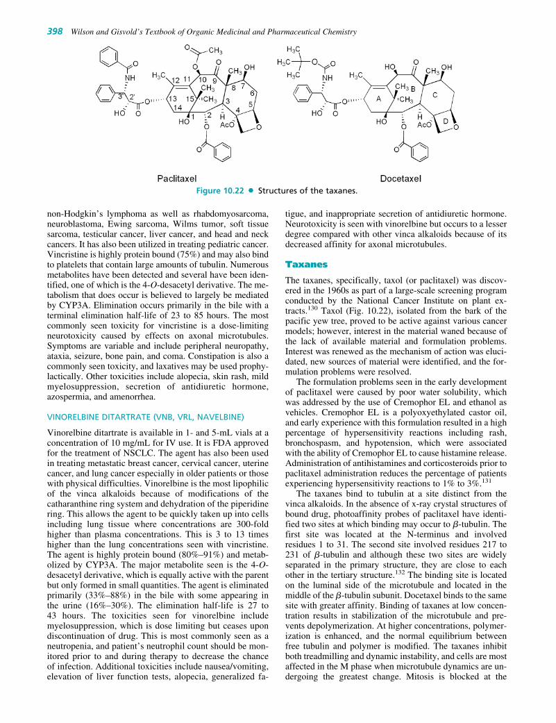
non-Hodgkin’s lymphoma as well as rhabdomyosarcoma,neuroblastoma, Ewing sarcoma, Wilms tumor, soft tissuesarcoma, testicular cancer, liver cancer, and head and neckcancers. It has also been utilized in treating pediatric cancer.Vincristine is highly protein bound (75%) and may also bindto platelets that contain large amounts of tubulin. Numerousmetabolites have been detected and several have been iden-tified, one of which is the 4-O-desacetyl derivative. The me-tabolism that does occur is believed to largely be mediatedby CYP3A. Elimination occurs primarily in the bile with aterminal elimination half-life of 23 to 85 hours. The mostcommonly seen toxicity for vincristine is a dose-limitingneurotoxicity caused by effects on axonal microtubules.Symptoms are variable and include peripheral neuropathy,ataxia, seizure, bone pain, and coma. Constipation is also acommonly seen toxicity, and laxatives may be used prophy-lactically. Other toxicities include alopecia, skin rash, mildmyelosuppression, secretion of antidiuretic hormone,azospermia, and amenorrhea.
VINORELBINE DITARTRATE (VNB, VRL, NAVELBINE)
Vinorelbine ditartrate is available in 1- and 5-mL vials at aconcentration of 10 mg/mL for IV use. It is FDA approvedfor the treatment of NSCLC. The agent has also been usedin treating metastatic breast cancer, cervical cancer, uterinecancer, and lung cancer especially in older patients or thosewith physical difficulties. Vinorelbine is the most lipophilicof the vinca alkaloids because of modifications of thecatharanthine ring system and dehydration of the piperidinering. This allows the agent to be quickly taken up into cellsincluding lung tissue where concentrations are 300-foldhigher than plasma concentrations. This is 3 to 13 timeshigher than the lung concentrations seen with vincristine.The agent is highly protein bound (80%–91%) and metab-olized by CYP3A. The major metabolite seen is the 4-O-desacetyl derivative, which is equally active with the parentbut only formed in small quantities. The agent is eliminatedprimarily (33%–88%) in the bile with some appearing inthe urine (16%–30%). The elimination half-life is 27 to43 hours. The toxicities seen for vinorelbine includemyelosuppression, which is dose limiting but ceases upondiscontinuation of drug. This is most commonly seen as aneutropenia, and patient’s neutrophil count should be mon-itored prior to and during therapy to decrease the chanceof infection. Additional toxicities include nausea/vomiting,elevation of liver function tests, alopecia, generalized fa-
tigue, and inappropriate secretion of antidiuretic hormone.Neurotoxicity is seen with vinorelbine but occurs to a lesserdegree compared with other vinca alkaloids because of itsdecreased affinity for axonal microtubules.
Taxanes
The taxanes, specifically, taxol (or paclitaxel) was discov-ered in the 1960s as part of a large-scale screening programconducted by the National Cancer Institute on plant ex-tracts.130 Taxol (Fig. 10.22), isolated from the bark of thepacific yew tree, proved to be active against various cancermodels; however, interest in the material waned because ofthe lack of available material and formulation problems.Interest was renewed as the mechanism of action was eluci-dated, new sources of material were identified, and the for-mulation problems were resolved.
The formulation problems seen in the early developmentof paclitaxel were caused by poor water solubility, whichwas addressed by the use of Cremophor EL and ethanol asvehicles. Cremophor EL is a polyoxyethylated castor oil,and early experience with this formulation resulted in a highpercentage of hypersensitivity reactions including rash,bronchospasm, and hypotension, which were associatedwith the ability of Cremophor EL to cause histamine release.Administration of antihistamines and corticosteroids prior topaclitaxel administration reduces the percentage of patientsexperiencing hypersensitivity reactions to 1% to 3%.131
The taxanes bind to tubulin at a site distinct from thevinca alkaloids. In the absence of x-ray crystal structures ofbound drug, photoaffinity probes of paclitaxel have identi-fied two sites at which binding may occur to �-tubulin. Thefirst site was located at the N-terminus and involvedresidues 1 to 31. The second site involved residues 217 to231 of �-tubulin and although these two sites are widelyseparated in the primary structure, they are close to eachother in the tertiary structure.132 The binding site is locatedon the luminal side of the microtubule and located in themiddle of the �-tubulin subunit. Docetaxel binds to the samesite with greater affinity. Binding of taxanes at low concen-tration results in stabilization of the microtubule and pre-vents depolymerization. At higher concentrations, polymer-ization is enhanced, and the normal equilibrium betweenfree tubulin and polymer is modified. The taxanes inhibitboth treadmilling and dynamic instability, and cells are mostaffected in the M phase when microtubule dynamics are un-dergoing the greatest change. Mitosis is blocked at the
398 Wilson and Gisvold’s Textbook of Organic Medicinal and Pharmaceutical Chemistry
Figure 10.22 Structures of the taxanes.
0355-0412_17865_Ch10.qxd 12/4/09 5:13 AM Page 398

metaphase anaphase boundary, and cells undergo apopto-sis.133 Paclitaxel has also been shown to enhance phospho-rylation of a serine residue of Bcl-2, an antiapoptotic proteinresulting in inhibition of Bcl-2�s ability to block apoptosis.The proapoptotic proteins Bad and Bax are stimulated andin a similar manner, docetaxel has been shown to induceapoptosis by activation of caspase enzymes.134
Resistance to the taxanes is like that seen for the vinca al-kaloids and other agents and involves Pgp-mediated efflux.Alterations in the structure of �-tubulin may also occur andresult in decreased binding of taxanes to the microtubulesand therefore reduced cytotoxicity.
PACLITAXEL (TAX, TAXOL)
Paclitaxel is available in single-dose vials of 30 mg/5 mLand 100 mg/16.7 mL for IV administration in the treatmentof breast, ovarian, NSCLC, and AIDS-related Kaposi sar-coma. Other uses have included treatment of head, neck,esophageal, cervical, prostate, and bladder cancers.
Paclitaxel is highly plasma protein bound (�90%) anddoes not penetrate the CNS. Metabolism involves CYP-mediated oxidation to give 6�-hydroxypaclitaxel (CYP2C8)and para hydroxylation of the phenyl group attached to the3�-position (CYP3A4). The 6�-hydroxy metabolite nor-mally predominates, but the para hydroxy metabolite mayoccur to a greater degree in those patients with liver diseaseor when CYP3A4 has been induced. Both metabolites areless active than the parent and do not undergo phase II con-jugation reactions. Elimination occurs primarily in the feces,and the elimination half-life is 9 to 50 hours depending onthe infusion period.
The major toxicity seen with paclitaxel is a dose-limitingmyelosuppression that normally presents as neutropenia. Thepreviously mentioned hypersensitivity reactions occur but aregreatly reduced by antihistamine pretreatment. Interactionwith the axonal microtubules such as that seen for the vincasalso occurs and leads to numbness and paresthesias (abnor-mal touch sensations including burning and prickling). Theagent is also available as an albumin-bound formulation(Abraxane) to eliminate the need for the solubilizing agentsassociated with the hypersensitivity reactions. Other adverseeffects include bradycardia, which may progress to heartblock, alopecia, mucositis, and/or diarrhea. Paclitaxel pro-duces moderate nausea and vomiting that is short-lived.
DOCETAXEL (TXT, TAXOTERE)
Docetaxel is available in single-dose vials of 20 mg/0.5 mLand 80 mg/2 mL for IV administration in the treatment of
breast, NSCLC, and prostate cancers. It has also been uti-lized in non–FDA-approved treatment of head, neck, gas-tric, bladder, and refractory ovarian cancers.
Docetaxel is highly plasma protein bound (80%) andwidely distributed with the highest concentration in the he-patobiliary system, but it does not appear to cross the blood-brain barrier. The metabolism of docetaxel has been lesswell studied than that of paclitaxel. The use of human livermicrosomes has indicated that metabolism involves oxida-tion of one of the tert-butyl methyl groups of the C-13 sidechain to initially give the alcohol. Further oxidation to thealdehyde and carboxylic acid both of which may cyclizewith the carbamate nitrogen to give stereoisomeric hydrox-yoxazolidinones and an oxazolidinedione, respectively. Noactive metabolites have been identified. The major enzymeinvolved is CYP3A4. The drug is primarily eliminated in thefeces with a terminal half-life of 11 hours.
The adverse effects profile for docetaxel is similar to thatof paclitaxel but also includes reversible fluid retention that isdose related. This has been associated with an initial increasein capillary permeability followed by a decrease in lymphaticdrainage later in the therapy. Restriction of sodium intake andpretreatment with corticosteroids is usually successful inminimizing this adverse effect. Peripheral neuropathy is seenwith docetaxel but occurs less often than with paclitaxel.Fatigue and muscle pain are commonly seen, and fever mayoccur in up to 30% of patients who are infection free.
IXABEPILONE (AZAEPOTHILONE B, IXEMPRA)
The epothilones are macrocyclic lactones that have a mech-anism of action similar to that of the taxanes but offer sev-eral advantages (Fig. 10.23).135 Ixabepilone is the semisyn-thetic amide analog of epothilone B that is isolated from themyxobacterium Sorangium cellulosum. The epothilonesshowed potent in vitro activity but greatly decreased activ-ity in vivo caused by metabolic instability via hydrolysis ofthe macrocyclic lactone. Conversion to the lactam increasedstability and maintained in vivo activity. Ixabepilone hasbeen recently (2007) approved for the treatment of metasta-tic breast cancer that is resistant to the taxanes. The agent isbelieved to bind to the same site occupied by the taxanes.Molecular modeling studies have been utilized to identifya common pharmacophore between the taxanes andepothilones.136 Key structural components that assumecomparable relative position are indicated in Table 10.2.Like the taxanes, ixabepilone binds to �-tubulin and stabi-lizes microtubules resulting in cell death. The agent is use-ful in cancers that have become resistant to the taxanes, be-cause it is not removed by Pgp and is still capable of binding
Chapter 10 Antineoplastic Agents 399
Figure 10.23 Structures of epithilones.
0355-0412_17865_Ch10.qxd 12/4/09 5:13 AM Page 399
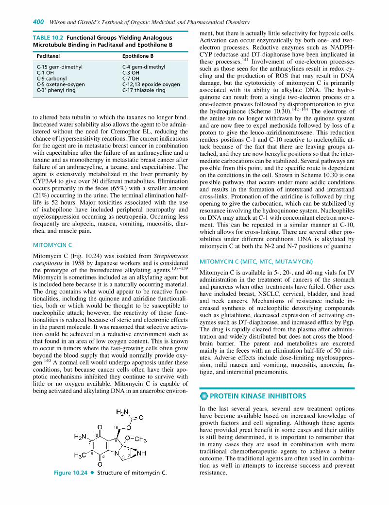
to altered beta tubulin to which the taxanes no longer bind.Increased water solubility also allows the agent to be admin-istered without the need for Cremophor EL, reducing thechance of hypersensitivity reactions. The current indicationsfor the agent are in metastatic breast cancer in combinationwith capecitabine after the failure of an anthracycline and ataxane and as monotherapy in metastatic breast cancer afterfailure of an anthracycline, a taxane, and capecitabine. Theagent is extensively metabolized in the liver primarily byCYP3A4 to give over 30 different metabolites. Eliminationoccurs primarily in the feces (65%) with a smaller amount(21%) occurring in the urine. The terminal elimination half-life is 52 hours. Major toxicities associated with the useof ixabepilone have included peripheral neuropathy andmyelosuppression occurring as neutropenia. Occurring lessfrequently are alopecia, nausea, vomiting, mucositis, diar-rhea, and muscle pain.
MITOMYCIN C
Mitomycin C (Fig. 10.24) was isolated from Streptomycescaespitosus in 1958 by Japanese workers and is consideredthe prototype of the bioreductive alkylating agents.137–139
Mitomycin is sometimes included as an alkylating agent butis included here because it is a naturally occurring material.The drug contains what would appear to be reactive func-tionalities, including the quinone and aziridine functionali-ties, both or which would be thought to be susceptible tonucleophilic attack; however, the reactivity of these func-tionalities is reduced because of steric and electronic effectsin the parent molecule. It was reasoned that selective activa-tion could be achieved in a reductive environment such asthat found in an area of low oxygen content. This is knownto occur in tumors where the fast-growing cells often growbeyond the blood supply that would normally provide oxy-gen.140 A normal cell would undergo apoptosis under theseconditions, but because cancer cells often have their apo-ptotic mechanisms inhibited they continue to survive withlittle or no oxygen available. Mitomycin C is capable ofbeing activated and alkylating DNA in an anaerobic environ-
ment, but there is actually little selectivity for hypoxic cells.Activation can occur enzymatically by both one- and two-electron processes. Reductive enzymes such as NADPH-CYP reductase and DT-diaphorase have been implicated inthese processes.141 Involvement of one-electron processessuch as those seen for the anthracylines result in redox cy-cling and the production of ROS that may result in DNAdamage, but the cytotoxicity of mitomycin C is primarilyassociated with its ability to alkylate DNA. The hydro-quinone can result from a single two-electron process or aone-electron process followed by disproportionation to givethe hydroquinone (Scheme 10.30).142–144 The electrons ofthe amine are no longer withdrawn by the quinone systemand are now free to expel methoxide followed by loss of aproton to give the leuco-aziridinomitosene. This reductionrenders positions C-1 and C-10 reactive to nucleophilic at-tack because of the fact that there are leaving groups at-tached, and they are now benzylic positions so that the inter-mediate carbocations can be stabilized. Several pathways arepossible from this point, and the specific route is dependenton the conditions in the cell. Shown in Scheme 10.30 is onepossible pathway that occurs under more acidic conditionsand results in the formation of interstrand and intrastrandcross-links. Protonation of the aziridine is followed by ringopening to give the carbocation, which can be stabilized byresonance involving the hydroquinone system. Nucleophileson DNA may attack at C-1 with concomitant electron move-ment. This can be repeated in a similar manner at C-10,which allows for cross-linking. There are several other pos-sibilities under different conditions. DNA is alkylated bymitomycin C at both the N-2 and N-7 positions of guanine
MITOMYCIN C (MITC, MTC, MUTAMYCIN)
Mitomycin C is available in 5-, 20-, and 40-mg vials for IVadministration in the treatment of cancers of the stomachand pancreas when other treatments have failed. Other useshave included breast, NSCLC, cervical, bladder, and headand neck cancers. Mechanisms of resistance include in-creased synthesis of nucleophilic detoxifying compoundssuch as glutathione, decreased expression of activating en-zymes such as DT-diaphorase, and increased efflux by Pgp.The drug is rapidly cleared from the plasma after adminis-tration and widely distributed but does not cross the blood-brain barrier. The parent and metabolites are excretedmainly in the feces with an elimination half-life of 50 min-utes. Adverse effects include dose-limiting myelosuppres-sion, mild nausea and vomiting, mucositis, anorexia, fa-tigue, and interstitial pneumonitis.
PROTEIN KINASE INHIBITORS
In the last several years, several new treatment optionshave become available based on increased knowledge ofgrowth factors and cell signaling. Although these agentshave provided great benefit in some cases and their utilityis still being determined, it is important to remember thatin many cases they are used in combination with moretraditional chemotherapeutic agents to achieve a betteroutcome. The traditional agents are often used in combina-tion as well in attempts to increase success and preventresistance.
400 Wilson and Gisvold’s Textbook of Organic Medicinal and Pharmaceutical Chemistry
TABLE 10.2 Functional Groups Yielding AnalogousMicrotubule Binding in Paclitaxel and Epothilone B
Paclitaxel Epothilone B
C-15 gem-dimethyl C-4 gem-dimethylC-1 OH C-3 OHC-9 carbonyl C-7 OHC-5 oxetane-oxygen C-12,13 epoxide oxygenC-3� phenyl ring C-17 thiazole ring
Figure 10.24 Structure of mitomycin C.
0355-0412_17865_Ch10.qxd 12/4/09 5:13 AM Page 400

Chapter 10 Antineoplastic Agents 401
Scheme 10.30 Alkylation of DNA by mitomycin C.
TABLE 10.3 Growth Factors and Receptors ofParacrine Origin
Growth Factor Designation Receptor
Epidermal growth EGF EGF-Rfactor
Platelet-derived PDGF PDGF-Rgrowth factor
Nerve growth factor NGF TrkVascular endothelial VEGF VEGF-R
growth factorStem cell factor SCF KitGlial cell line–derived GFL RET
neurotropic factor
It was realized early on that the growth and proliferationof many tissues was under the control of the endocrinesystem, in which endocrine glands secreted hormones andthat hormones or their antagonists were useful in controllingthe overgrowth of these tissues. It was found later that othergrowth factors, many of which were secreted by nearbycells, were also involved in controlling the growth and pro-liferation of the target tissue and this became known asparacrine control. Many of these growth factors have beenidentified and several along with their receptors are indi-cated in Table 10.3.144
These receptors have become targets for several differentmonoclonal antibodies that have been recently introduced.
Further work identified the pathways by which interac-tion with these cell surface receptors resulted in growth andproliferation of cells. There are various mechanisms bywhich these processes occur, but a generalized pathway in-volves interaction of a growth factor with a growth factorreceptor occurring as a monomer present in the phospho-lipid bilayer of the cell membrane, which results in dimer-ization of the receptors (Scheme 10.31). The receptorsthemselves possess an extracellular-binding domain for in-teraction with the growth factors, an intracellular domain,and a connecting transmembrane region. The intracellulardomain of these receptor proteins often function as kinases,many of which are TKs that phosphorylate residues on theother monomer receptor. The resulting phosphorylatedresidues are recognized by the Src homology 2 domain(SH2) of cytoplasmic proteins such as Shc, Grb-2, and Sos,which activate Ras. These cytoplasmic proteins then serveto transmit the growth signals from the growth factor recep-tors to Ras (Fig. 10.25). Ras is a G protein tethered to thecytoplasmic membrane, which may bind GTP resulting inactivation of Ras or bind GDP resulting in Ras inactivation.Ras also normally has the ability to hydrolyze GTP to inac-tivate itself. Many cancers are found to have altered formsof Ras, which is present in a hyperactive state. Ras, in itsactivated form activates a series of kinases, many of whichare TKs with various effects on cells. Notable is the Ras-Raf-MEK-ERK pathway also known as the mitogen-activated protein kinase (MAPK) pathway. This is a
0355-0412_17865_Ch10.qxd 12/4/09 5:13 AM Page 401

402 Wilson and Gisvold’s Textbook of Organic Medicinal and Pharmaceutical Chemistry
Scheme 10.31 Schematic of the activation of growth factor receptors.
Figure 10.25 Schematic of the Ras pathways leading to uncontrolled cell proliferation.
0355-0412_17865_Ch10.qxd 12/4/09 5:13 AM Page 402

cascade effect, whereby one kinase phosphorylates and ac-tivates more of the next kinase in the pathway amplifyingthe signal in the process. Stimulation of this pathway re-sults in the activation of several transcription factors in-volved in the expression of growth factors, cyclin D1 (in-volved in controlling cell division, see Fig. 10.1), andtranscription factors Fos and Jun, which were first discov-ered as viral oncogenes and are known to be elevated inseveral human cancers. In addition, overactivation of thispathway may result in loss of contact inhibition, loss of an-chorage dependence, and changes in cell shape, all ofwhich are characteristic of cancer cells.
The second major pathway activated by Ras is PI3K-PIP3-Akt/PKB whose activation increases cell proliferationbut perhaps most importantly, inhibits apoptosis. WhenRas is activated, phosphatidylinositol(4,5)-diphosphate(PIP2) is further phosphorylated to give phosphatidylinosi-tol(3,4,5)-triphosphate PIP3, which remains embedded inthe plasma membrane but attracts and activates other ki-nases namely protein kinase B (PKB), a serine-threoninekinase. Overactivity of this pathway results in decreasedapoptosis, increased proliferation and increased cell size.Specific effects include inactivation of the proapoptotic pro-tein Bad (Fig. 10.2) and activation of antiapoptotic NF-�B.
The third pathway involves Ras-mediated activation ofRal-GEF (Guanine nucleotide exchange factor), which actson Ral-A and Ral-B to stimulate their exchange of GDP forGTP and hence their activation. The activation of this path-way, although less well understood, seems to allow cancercells to metastasize and invade.
Overactivity of these pathways is present in several dif-ferent cancers, and consequently, several agents have beendeveloped to correct this. One such approach is the inhibi-tion of kinases whose normal activity has been amplified.
Working in Philadelphia in the 1960s, scientists discov-ered a genetic abnormality present in 95% of patients withCML.145 The abnormality was a translocation between chro-mosomes 22 and 9 in which the ABL gene (analogous to theAbelson proto-oncogene found in mice) originally on chro-mosome 9 was translocated to chromosome 22, such that itwas fused with BCR gene (breakpoint cluster region). Thenew chromosome was designated as the Philadelphiatranslocation or the Philadelphia chromosome (Ph1).146 Thefusion of these two genes resulted in a protein that encodedthe entire ABL gene and as many as three additional pro-teins from BCR. The product of this fusion was found to bea TK that was always activated. This altered TK utilizesATP to phosphorylate and activate numerous pathways in-cluding MAPK and Akt/PKB along with several others.This leads to proliferation of white cells that normally lastsabout a year, when the disease becomes more difficultto treat and eventually to the terminal blast phase lastingseveral months.
Imatinib (Fig. 10.26) was developed to specifically in-hibit this unique TK and does so rather selectively by bind-ing to the ATP-binding pocket and stabilizing an inactiveform of the enzyme. Protein kinases have similar struc-tures and consist of C- and N-terminal lobes with theactive site formed by a cleft between the two lobes. Theactivity of the enzyme is regulated by an activation loopthat is extended in the active form of the enzyme andprovides for substrate binding. X-ray crystal studies ofimatinib binding to Abl show that the drug binds in the
cleft formed by the two lobes of the enzyme through theformation of several hydrogen bonds and hydrophobic in-teractions with the pyridine ring assuming the postion oc-cupied by ATP in the active enzyme.147 Imatinib bindinglocks the enzyme in a conformation in which the activationloop is oriented so as to block substrate binding.Specificity for the BCR-ABL kinase is not complete be-cause of structural similarity of TKs, and those associatedwith the PDGF receptor and the receptor for SCF known asKIT are also inhibited (Table 10.3). The agent has beenfound to be effective in 90% of patients in the chronicstage (the chronic stage last 4–6 years and precedes theproliferation stage mentioned previously). Blast-phase pa-tients respond at a rate of 60% but usually relapse afterseveral months.148
The tremendous success of imatinib led to other agentsthat could be used in the case of resistance. Specifically,dasatinib can bind to either an active or inactive form of theenzyme and is 100 times more potent compared with ima-tinib. It has the ability to overcome resistance associatedwith amino acid alterations that leads to decreased affinityfor imatinib. As the structure is altered, binding to other TKs(BCR-ABL, PDGF, SRC, KIT) is manifested, which mayeventually lead to other indications but at this time, it is usedprimarily in those cases when patients fail to respond toimatinib.149
This general idea of targeting TKs was extended to theEGF receptor. It was hoped that by targeting EGF TKs theinhibitors would be applicable to a wide range of cancers.Recall that after the receptors dimerize, they transphospho-rylate each other and act as TKs. Currently, there are twostructurally similar agents in use, gefitinib and erlotinib,which bind to the ATP-binding site and thereby inhibit theresulting cascade that would normally occur from activationof the EGF receptor.150,151 The benefit of the agents washoped to be caused by inhibition to the Ras pathway so thatultimately, all three arms could be affected (MAPK,Akt/PKB, and Ral-GEF). The agents are modestly selectivefor this kinase. Currently both compounds have indicationsfor NSCLC generally as alternatives after the failure ofmore traditional therapy, and gefitinib has an additional in-dication against pancreatic cancer. Initial results have notbeen as dramatic as that seen with imatinib. There are alsoseveral known mutations and deletions in the EGF receptorof cancer patients and this may affect the activity of thesecompounds. Specifically, point mutations in the KRAS gene(a member of the Ras family) have been associated with re-sistance to these agents and tests have become available todetect this prior to initiating therapy.
The activation of numerous tyrosine and serine-threoninekinases offers many theoretical targets for drug interventionwhen these pathways become overactive as is often the casein neoplastic cells. These kinase enzymes are all thought tohave evolved from a common ancestor and as such possessa great deal of structural similarity that complicates the de-velopment of agents, which are selective for a single kinase.This has led to the strategy of simultaneously inhibiting nu-merous kinase enzymes, and consequently there are severalmultikinase inhibitors now available. Sorafenib was origi-nally developed using high-throughput testing and parallelsynthesis as an inhibitor of the serine-threonine kinaseRaf.152 It proved to be a potent inhibitor of this enzyme withan IC50 � 6 nM and acted by stabilizing an inactive form of
Chapter 10 Antineoplastic Agents 403
0355-0412_17865_Ch10.qxd 12/4/09 5:13 AM Page 403

the enzyme similar to imatinib. Subsequent x-ray analysis ofthe drug receptor complex showed that the pyridine ringoccupied the ATP-binding site of Raf, whereas the trifluo-romethyl-chloro–substituted phenyl ring occupied a hy-drophobic pocket in the cleft formed by the two lobes of theprotein.153 The urea moiety forms two hydrogen bonds, onethrough interaction of the amide proton with the carboxylateof Glu500 and the second through interaction of the car-bonyl oxygen with the peptide amide N-H of Asp593. Thecompound also has the capacity to inhibit several other TKsassociated with growth factor receptors including PDGF-R,VEGF-R, RET-kinase, and c-Kit with IC50s in the range of20 to 90 nM. In some cancer models, the compound hasbeen shown to be effective even though the MAPK pathwaywas not inhibited and this has been explained by the com-
pounds inhibition of angiogenesis This has been attributedto the effects on inhibition of PDGF-R and VEGF-R phos-phorylation and the role these growth factor receptors playin the creation of new blood vessels. The agent was investi-gated and subsequently approved for use in renal cell carci-noma (RCC) because of the fact that this particular cancer isoften associated with increased activity of the targets inhib-ited by sorafenib and the high level of neoangiogenesis thatis seen in this cancer.
As a group of agents, the protein kinase inhibitors havethe advantage of oral administration and better patient tol-erability. The major adverse effects include a skin rash thatnormally appears early in therapy on the upper torso and isgenerally mild but may become more serious in some cases.Mild diarrhea and nausea are also commonly seen but are
404 Wilson and Gisvold’s Textbook of Organic Medicinal and Pharmaceutical Chemistry
Figure 10.26 Protein kinase inhibitors.
0355-0412_17865_Ch10.qxd 12/4/09 5:13 AM Page 404

generally controlled with the administration of antiemeticsand antidiarrheals. Mild myelosuppression is also seen withseveral of these agents. There has been concern about car-diotoxicity associated with protein kinase inhibitors be-cause of the fact that multiple kinases may be inhibited.This could potentially have several effects on signalingprocesses in cardiac tissue. For example if the Akt/PKBpathway was blocked, apoptosis may become activated andcardiac cells would die (Table 10.3). There has been someevidence for cardiotoxicity in this class and the agents havebeen ranked with imatinib, dasatinib, sorafenib, and suni-tinib having known or likely cardiotoxicity and gefitinib,erlotinib, and lapatinib having low cardiotoxicity. Concernof cardiotoxicity is also a result of the fact that other drugssuch as the anthracyclines and several of the monoclonalantibodies directed against the EGF-R are known to havecardiotoxicity associated with their use.154 Therefore, pa-tients who have already been exposed to these agents mayexperience additional adverse effects to their cardiovascu-lar system if a protein kinase inhibitor were utilized and italso produced significant cardiotoxicity. Studies for themost part have not evaluated the cardiotoxicity of theseagents and the given ranking is based on preliminary data,but there is concern about the cardiovascular effects of thisclass of agents.
IMATINIB MESYLATE (STI-571, GLEEVEC)
Imatinib is available in 100- and 400-mg capsules for oraladministration and is indicated for the treatment of CML,gastrointestinal stromal tumors (GIST) that express Kit andacute lymphoplastic leukemia that is positive for thePhiladelphia chromosome.
Bioavailability of the agent is nearly 100% by the oralroute. The agent is highly protein bound and metabolized tothe N-desmethyl derivative by CYP3A4-mediated removalof the piperazinyl methyl group. The resulting metabolite issimilar to the parent in activity. Elimination occurs prima-rily in the feces, and the terminal half-life is 18 hours forthe parent and 40 hours of the N-desmethyl metabolite.Resistant forms of the TK are known, which have alteredamino acids that prevent binding. In addition, there may beincreased levels of the kinase itself. The drug is also a sub-strate for Pgp and an additional efflux transporter known asbreast cancer resistance protein (BCRP), both of which re-move the drug from the cell. These transporters are also in-hibited by the agent as well. Severe side effects include as-cites, neutropenia, thrombocytopenia, skin rash, andpulmonary edema. Less serious side effects include nau-sea/vomiting, heartburn, and headache but overall, the agentis better tolerated than most other medications used in treat-ing the disease.
DASATINIB (BMS-354825, SPRYCEL)
Dasatinib is available in 20-, 50-, and 70-mg tablets fororal administration in the treatment of CML and ALL thatare Ph1 positive. Although dasatinib is more potent thanimatinib, bioavailability is much lower with values rangingbetween 14% to 34%. The agent is extensively metabo-lized with as many as 29 metabolites seen as result of oxi-dation by primarily CYP3A4 and phase II conjugation.The agent may act as an inhibitor of CYP3A4 andCYP2C8. Metabolism does give an active metabolite, but
this accounts for only 5% of the total and is not believed tobe important for the overall activity of the agent. Dasatinibis 95% protein bound with a terminal half-life of 3 to5 hours. The majority of the drug and metabolites are elim-inated in the feces. The most common side effects are skinrash, nausea, diarrhea, and fatigue. Serious side effects in-clude myelosuppression appearing as neutropenia andthrombocytopenia, bleeding of the brain and GI tract, andfluid retention.
GEFITINIB (ZD1839, IRESSA)
Geftinib is available as 250-mg tablets for oral administrationin the treatment of NSCLC for those patients who have failedto respond to platinum-based therapies and docetaxel and hasalso been used against squamous cell cancers of the head andneck. The agent is an inhibitor of the TK of EGF-R and pos-sibly other TKs as well. Gefitinib is both a substrate and in-hibitor of Pgp and BCRP. The agent is absorbed slowly afterbeing administered orally with 60% bioavailability.Metabolism occurs in the liver and is mediated primarily byCYP3A4 to give eight identified metabolites resulting fromdefluorination of the phenyl ring, oxidative-O-demethylation,and multiple products arising as a result of oxidation of themorpholine ring. The O-demethylated product represents thepredominate metabolite and is 14-fold less active comparedwith the parent. The parent and metabolites are eliminated inthe feces with a terminal elimination half-life of 48 hours.The drug appears to be well tolerated with the most com-monly reported side effects being rash and diarrhea. It mayalso cause elevations in blood pressure especially in those pa-tients with preexisting hypertension, elevation of transami-nase levels, and mild nausea and mucositits.
ERLOTINIB HYDROCHLORIDE (CP-358,774, TARCEVA)
Erlotinib is available as 25-, 100-, and 150-mg tablets fororal administration and is used after failure of first-linetherapy in metastatic NSCLC and as first-line therapy incombination with gemcitabine in the treatment of metasta-tic pancreatic cancer, and in treating malignant gliomas.The structural similarity to gefitnib imparts similar phar-macokinetic behavior with bioavailability of 60% and pro-tein binding of 93%. The agent is extensively metabolizedprimarily by CYP3A4. Three major metabolic pathwayshave been identified, involving oxidative-O-demethylationof the side chains followed by further oxidation to give thecarboxlic acids, oxidation of the acetylene functionalityto give a carboxylic acid, and aromatic hydroxylation ofthe phenyl ring para to the electron-donating nitrogen. Themetabolites are primarily eliminated in the feces, and theterminal half-life is 36 hours.155 The major toxicities seenwith the agent are dose-limiting skin rash and diarrhea.Other common adverse effects include shortness of breath,fatigue, and nausea.
SORAFENIB TOSYLATE (BAY 43-9006, NEXAVAR)
Sorafenib is available in 200-mg tablets for oral administra-tion and is used in the treatment of RCC and colon cancer.The agent is classified as a multikinase inhibitor because ofits action on numerous kinase enzymes including thePDGF-R, VEGF-R, Kit, and Raf. Sorafenib is 39% to 48%
Chapter 10 Antineoplastic Agents 405
0355-0412_17865_Ch10.qxd 12/4/09 5:13 AM Page 405

bioavailable and CYP3A4-mediated metabolism giveseight identified metabolites including the N-oxide, which isequally active with the parent. However, the majority of thedrug in plasma is present as the parent compound.Sorafenib is highly protein bound (99.5%). The drug iseliminated primarily in the feces (77%) with 19% appearingin the urine as glucuronides (UGT1A1) and has a elimina-tion half-life of 24 to 48 hours. The most commonly seentoxicity is skin rash that normally occurs within the first6 weeks of therapy. Other adverse effects include hyperten-sion, fatigue, increased wound healing time, and increasedrisk of bleeding.
LAPATINIB DITOSYLATE (GSK572016, TYKERB)
Lapatinib is available in 250-mg tablets for oral adminis-tration and is used in combination with cabecitabine in thetreatment of breast cancer for those patients that over ex-press the type 2 EGF-R and who have previously receivedtaxane, anthracycline, and trastuzumab therapy. The type 2EGF-R is one subtype of this receptor and is also known asHER2 or ErbB-2. The agent is a receptor TK inhibitor tar-geting the ErbB-1 and ErbB-2 subtypes. Binding occurs atthe ATP-binding site and thereby prevents phosphoryla-tion and the subsequent activation of other kinase en-zymes. ErbB-1 overexpression occurs in approximately27% to 30% of breast cancers, while ErbB-2 is over ex-pressed in 20% to 25% of cases.156 The agent has demon-strated IC50 values of �0.2 �M against ErbB-1 and 2 fromseveral different cancer cell lines and dissociates slowly(t1/2 � 300 min) from these receptor TKs.157 The drug isboth a substrate and an inhibitor of the efflux transportersPgp and BCRP. It is also an inhibitor of the hepatic uptaketransporter OATP1B1, which is an organic anion trans-porter.158 The absorption of lapatinib is incomplete andvariable after oral administration. The agent is extensivelymetabolized by CYP3A4 and CYP3A5, with minor contri-butions from CYP2C19 and CYP2C8. Lapatinib inhibitsCYP3A and CYP2C8 at clinically relevant concentrations.The agent is highly (99%) protein bound and eliminatedprimarily in the feces. The half-life of the agent increaseupon repeated dosing, taking 6 days to reach steady statethat gives an effective half-life of 24 hours. The most com-monly seen adverse effects of lapatinib therapy are skinrash and diarrhea. Skin rash is commonly seen with manyof the other TK inhibitors and agents that target ErbB-1.Lapatinib-induced diarrhea is usually mild to moderate.There have been reports of decreases in left ventricularejection fraction associated with the agent, although thisappears to occur only rarely and is reversible upon discon-tinuation of therapy.
SUNITINIB MALATE (SU11248, SUTENT)
Sunitinib is available in 12.5-, 25-, and 50-mg capsules fororal administration for the treatment of advanced RCC andGIST upon the failure of imatinib. The agent is a multiki-nase inhibitor and has been shown to inhibit PDGF-R,VEGF-R, Kit, RET, and the colony-stimulating factor re-ceptor (CSR-1R). The result of this spectrum of activity isa slowing of tumor progression and inhibition of angiogen-esis both of which are useful in the highly vascularizedcancers, RCC and GIST. The median TTP (time to tumorprogression) was 27.3 weeks for sunitinib and 6.4 weeks
for placebo. Progression-free survival was also significantwith a median time of 24.1 weeks for sunitinib and 6 weeksfor placebo. The agent is well absorbed upon oral adminis-tration, and both the parent and major metabolite are highly(90%–95%) protein bound. Metabolism is mediated prima-rily by CYP3A4 to give the N-desethyl derivative, which isthe major metabolite (23%–37%), equally active with theparent and undergoes further metabolism by CYP3A4. Theterminal elimination half-life for the parent and N-desethylderivative are 40 to 60 hours and 80 to 110 hours, respec-tively. Elimination occurs primarily via the feces. Commonadverse effects of sunitinib include fatigue, diarrhea, yel-low skin discoloration, anorexia, nausea, and mucositis.Mild myelosuppression has been reported in patients withGIST including neutropenia, lymphopenia, thrombocy-topenia, and anemia. There have been reports of cardiotox-icity including decreases in left ventricular ejectionfraction, which occurred in 11% of patients during a GISTstudy.
MISCELLANEOUS COMPOUNDS
TEMSIROLIMUS (CCI-779, TORISEL)
Temsirolimus (Fig. 10.27) is an esterified derivative of rap-amycin and in a similar manner binds initially to the pro-tein FKBP-12(FK506-binding protein).159 This complexthen acts to inhibit the mammalian target of rapamycin(mTOR), a serine-threonine kinase that plays a crucial rolein cell division. It is somewhat unique in its method of ki-nase inhibition, because it actually binds to an allostericmodulator of the kinase rather than just binding to theATP-binding site like most other kinase inhibitors. Bindingof temsirolimus inhibits the phosphorylating activity ofmTOR. As shown in Table 10.3, mTOR regulates theAkt/PKB pathway, and therefore, one way in which theagent acts is to inhibit this pathway. An additional mecha-nism involves the ability of mTOR to initiate protein syn-thesis independent of the Akt/PKB pathway. Blockadeof mTOR results in inhibition of protein synthesis and pre-vents the cell from moving past the G1 phase into theS phase. More specifically, mTOR is prevented from phos-phorylating 4E-binding protein-1(4E-BP1) an initiatingfactor and 40S ribosomal protein S6 kinase (p70S6 kinase),both of which are involved in initiating protein synthesisnecessary for the cell cycle. In addition, mTOR is involvedin the control of several growth factors such VEGF, PDGF,and TGF, which are involved in cell growth and angiogen-esis. Temsirolimus is available as a 25-mg/mL injection forIV administration in the treatment of advanced RCC. Theagent is extensively metabolized and undergoes rapid hy-drolysis of the ester function to give rapamycin that retainsactivity.160 Additional metabolism is mediated primarilyby CYP3A4 to give several hydroxylated and demethylatedmetabolites that are inactive. The agent and metabolites areeliminated primarily in the feces with half-lives of 17 and55 hours for temsirolimus and rapamycin, respectively.This agent, like rapamycin, possesses immunosuppressantproperties and there is an increased risk of infection.The most serious side effects are interstitial lung disease,perforation of the bowel, and acute renal failure althoughthese occur only rarely. The most commonly seen side
406 Wilson and Gisvold’s Textbook of Organic Medicinal and Pharmaceutical Chemistry
0355-0412_17865_Ch10.qxd 12/4/09 5:13 AM Page 406
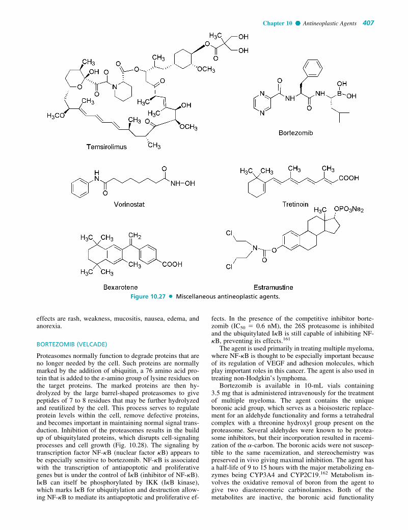
effects are rash, weakness, mucositis, nausea, edema, andanorexia.
BORTEZOMIB (VELCADE)
Proteasomes normally function to degrade proteins that areno longer needed by the cell. Such proteins are normallymarked by the addition of ubiquitin, a 76 amino acid pro-tein that is added to the �-amino group of lysine residues onthe target proteins. The marked proteins are then hy-drolyzed by the large barrel-shaped proteasomes to givepeptides of 7 to 8 residues that may be further hydrolyzedand reutilized by the cell. This process serves to regulateprotein levels within the cell, remove defective proteins,and becomes important in maintaining normal signal trans-duction. Inhibition of the proteasomes results in the buildup of ubiquitylated proteins, which disrupts cell-signalingprocesses and cell growth (Fig. 10.28). The signaling bytranscription factor NF-�B (nuclear factor �B) appears tobe especially sensitive to bortezomib. NF-�B is associatedwith the transcription of antiapoptotic and proliferativegenes but is under the control of I�B (inhibitor of NF-�B).I�B can itself be phosphorylated by IKK (I�B kinase),which marks I�B for ubiquitylation and destruction allow-ing NF-�B to mediate its antiapoptotic and proliferative ef-
fects. In the presence of the competitive inhibitor borte-zomib (IC50 � 0.6 nM), the 26S proteasome is inhibitedand the ubiquitylated I�B is still capable of inhibiting NF-�B, preventing its effects.161
The agent is used primarily in treating multiple myeloma,where NF-�B is thought to be especially important becauseof its regulation of VEGF and adhesion molecules, whichplay important roles in this cancer. The agent is also used intreating non-Hodgkin’s lymphoma.
Bortezomib is available in 10-mL vials containing3.5 mg that is administered intravenously for the treatmentof multiple myeloma. The agent contains the uniqueboronic acid group, which serves as a bioisosteric replace-ment for an aldehyde functionality and forms a tetrahedralcomplex with a threonine hydroxyl group present on theproteasome. Several aldehydes were known to be protea-some inhibitors, but their incorporation resulted in racemi-zation of the �-carbon. The boronic acids were not suscep-tible to the same racemization, and stereochemistry waspreserved in vivo giving maximal inhibition. The agent hasa half-life of 9 to 15 hours with the major metabolizing en-zymes being CYP3A4 and CYP2C19.162 Metabolism in-volves the oxidative removal of boron from the agent togive two diastereomeric carbinolamines. Both of themetabolites are inactive, the boronic acid functionality
Chapter 10 Antineoplastic Agents 407
Figure 10.27 Miscellaneous antineoplastic agents.
0355-0412_17865_Ch10.qxd 12/4/09 5:13 AM Page 407

being necessary for binding of the proteasome. The elimi-nation of the agent has not been well characterized.
The major toxicities seen with the agent are generalizedweakness, nausea, vomiting, diarrhea, peripheral neuropa-thy, fever, and orthostatic hypotension. Myelosuppressionalso occurs normally as thrombocytopenia and neutropenia.
VORINOSTAT (SUBEROYLANILIDE HYDROXAMICACID, SAHA, ZOLINZA)
Histones are proteins around which DNA is wound in theprocess of packing DNA into the nucleus. They also havea role in regulating the transcription of genes, and this iscontrolled by the covalent modifications acetylation, phos-phorylation, and methylation to which they are subject.The specific modifications present on the histones or his-tone code has been proposed to determine which transcrip-tion factors associate with specific genes and result in theirreplication.163 Acetylation occurs at the �-amino group oflysine and is accomplished by histone acetyltransferase en-zymes, whereas deacetylation is accomplished by histonedeacetylase enzymes. The result of inhibition of histone
deacetylase is hyperacetylation of lysine residues of the hi-stone proteins. The positively charged �-amino groups ofthe lysine residues are believed to interact with the nega-tively charged phosphate backbone of DNA. Once acetyla-tion has occurred, this interaction is prevented, and thebinding of transcription factors is favored. Therefore, theinhibition of deacetylation by vorinostat, a histonedeacetylase inhibitor (HDACis), results in the increasedtranscription of certain genes. Specifically, this has beenassociated with upregulation of a regulatory protein knownas p21, which serves to inhibit progression past the G1
phase of the cell cycle. Other genes and their proteins arealso effected by vorionostat such as Hsp90 (heat shockprotein 90) and BCL6.
Vorinostat fits the basic pharmacophore for the HDACis(Fig. 10.29), which consists of a hydrophobic cap regionconnected to a zinc coordinating functionality by a hy-drophobic linker.164 The hydroxamic acid functionality iscapable of bidendate binding to zinc present in the enzymeand is a major factor in the overall binding of the compound.The compound inhibits HDAC1, 2, 3, and 6 classes of thisenzyme with nanomolar (�86 nM) IC50 values.165
408 Wilson and Gisvold’s Textbook of Organic Medicinal and Pharmaceutical Chemistry
Figure 10.28 Actions of proteasomes on NF-�B.
0355-0412_17865_Ch10.qxd 12/4/09 5:13 AM Page 408

The agent is given orally and is available in 100-mg cap-sules for the treatment of cutaneous T-cell lymphoma. Thebioavailability is 43%, and the agent is 71% bound toplasma proteins. Extensive metabolism of the agent occursto give the O-glucuronide of the hydroxamic acid functionand 4-anilino-4-oxobutanoic acid with minimal involve-ment of isozymes of CYP. The metabolites, both of whichare inactive, are eliminated in the urine and the drug has aterminal elimination half-life of 2 hours. The most com-monly reported adverse effects are fatigue, diarrhea, andnausea. Elevations in glucose and triglyceride levels arecommonly seen with the agent. The agent has been associ-ated with thrombocytopenia, an increased risk of clotting re-sulting in pulmonary embolism and possibly prolongation ofthe QTc interval.
ARSENIC TRIOXIDE (AS2O3, TRISENOX)
Arsenic trioxide is available in 10-mL vials for IV adminis-tration as second-line therapy in the treatment of acutepromyelocytic leukemia (APL). The mechanism of theagent has not been well characterized; however, work hasindicated that the agent may cause the degradation of a pro-tein that blocks myeloid differentiation. Acute lymphocyticleukemia is associated with a translocation in which thepromyelocytic leukemia (PML) gene is fused with theretinoic acid receptor gene (RAR�), and the protein that re-sults from this genetic rearrangement prevents myeloid dif-ferentiation.166 Arsenic trioxide is capable of degrading thisprotein and allowing the cells to differentiate. Additional ef-fects have included stimulation of apoptosis by decreasingBcl-2 activity and stimulation of caspase enzymes and p53.Angiogenesis is inhibited by the inhibition of VEGF at theprotein level.167 The agent is widely distributed after IV ad-ministration; however, the pharmacokinetics of the agenthave not been well characterized. Metabolism studies haveshown that the agent undergoes reduction to trivalent ar-senic followed by methylation to give monomethylarsonicand dimethylarsinic acids, which are eliminated in theurine. Unlike most other antineoplastic agents, myelosup-pression does not occur in fact many patients (50%–60%)experience leukocytosis in which white blood cell count in-creases. APL differentiation syndrome is seen in many pa-tients (30%) and presents as fever, shortness of breath,weight gain, pulmonary infiltrates, and pleural or pericar-dial effusions. This may be fatal and is commonly treatedwith high-dose dexamethasone upon initial suspicion. Thepresentation of APL differentiation syndrome are identicalfor arsenic trioxide and retinoic acid. Additional adverse ef-fects include fatigue, a prolonged QT interval, dizziness,mild hyperglycemia, and mild nausea and vomiting.
TRETINOIN (ALL-TRANS-RETINOC ACID, ATRA, RETIN-A, VESANOID)
Tretinoin is available in 10-mg capsules for oral administra-tion in the treatment of APL. The mechanism of action in-volves passive diffusion through the cell membrane andthen movement to the nucleus where it interacts with theretinoic acid receptor (RAR) portion of the PML-RAR� fu-sion protein. Binding of tretinoin allows the cell to differen-tiate and has also been shown to result in the destruction ofthe PML-RAR� fusion protein. Resistance to tretinoin isproblematic and associated with an increase in cellularretinoic acid–binding proteins (CRAPBs) located in the cy-tosol. The complexation with tretinoin prevents movementinto the nucleus and may present the drug to metabolizingenzymes that inactivate it. Amino acid mutation of thePML-RAR� protein has also been established as a mecha-nism of resistance. The agent is well absorbed upon oral ad-ministration and highly (95%) protein bound. Metabolismoccurs in the liver and several inactive metabolites havebeen identified including 13-cis-retinoic acid, 4-oxo cis-retinoic, 4-oxo trans-retinoic acid and 4-oxo trans-retinoicacid glucuronide. Elimination occurs in the urine (63%) andfeces (31%) with an elimination half-life of 40 to 120 min-utes. Vitamin A toxicity is seen in nearly all patients andpresents as headache, fever, dryness of the skin, skin rash,mucositis, and peripheral edema. APL differentiation syn-drome such as that seen for arsenic trioxide also occurs.Cardiovascular effects include flushing, hypotension, CHF,stroke, and myocardial infarction have been reported butoccur only rarely. There are also several CNS and GI effectsthat have been associated with the agent as well.
BEXAROTENE (TARGRETIN)
Bexarotene is available in 75-mg capsules for oral adminis-tration in the treatment of refractory cutaneous T-cell lym-phoma. The agent is also available as a gel that may be usedtopically. The mechanism of action has not been fully estab-lished but is thought to involve binding to retinoid receptorsresulting ultimately in the formation of transcription factorsthat promote cell differentiation and regulate cellular prolif-eration.168 Bexarotene has been demonstrated to activateapoptosis as a result of stimulation of caspase 3 and inhibi-tion of survivin, an antiapoptotic protein that would nor-mally inhibit caspase activity.169 Apoptosis is also stimu-lated because of cleavage of poly(ADP-Ribose) polymerase,which is antiapoptotic. Reduced expression of the retinoidreceptor subtypes RXR� and RAR� has also been demon-strated for the agent. Absorption is nearly complete afteroral administration and plasma protein binding is high(�99%).170 There is extensive metabolism in the liver togive 6- and 7-hydroxy-bexarotene and 6- and 7-oxo-bexarotene as well as glucuronides of these metabolites andthe parent. Elimination occurs via the feces with an elimina-tion half-life of 7 hours. Adverse effects include hypercho-lesterolemia, hypertriglyceridemia, hypothyroidism, myelo-suppression, nausea, and skin rash.
ASPARAGINASE (L-ASPARAGINASE, ELSPAR,L-ASNASE, CRISTANASPASE)
Asparaginase is available in 10-mL vials for intramuscularand IV use in the treatment of acute lymphocytic leukemia.
Chapter 10 Antineoplastic Agents 409
Figure 10.29 Pharmacophore for histone deacetylaseinhibitors.
0355-0412_17865_Ch10.qxd 12/4/09 5:13 AM Page 409

Tumor cells are unable to synthesize asparagine, and there-fore must utilize what is available in the extracellular environ-ment. The agent acts by hydrolyzing extracellular asparagineto aspartate and ammonia. The tumor cells are then deprivedof a necessary nutrient, and protein synthesis is inhibited lead-ing to cell death. The agent is specific for the G1 phase of thecell cycle. Resistance occurs because of the development ofthe tumor cells ability to produce asparagine synthetase thatallows them to synthesize the required amino acid. Antibodyproduction directed at asparaginase may be stimulated by theagent as well. The agent remains in the extracellular spaceafter parental administration and is 30% protein bound. Themetabolism of the agent has not been well characterized andthe plasma half-life depends on the formulation of the drug.The E. coli-derived agent has a plasma half-life of 40 to 50hours, whereas polyethylene glycol-asparaginase’s half-life is3 to 5 days. Adverse effects include hypersensitivity reac-tions, fever, chills, nausea, lethargy, confusion, hallucina-tions, and possibly coma. Myelosuppression is not generallyseen. An increased risk of bleeding and clotting is seen in halfof the patients taking the agent.
ESTRAMUSTINE PHOSPHATE SODIUM (EM, EMCYT)
Estramustine as the phosphate is available in 140-mg cap-sules for the treatment of prostate cancer. Although origi-nally designed as an alkylating agent, it has been shown tobe devoid of alkylating activity and functions as an inhibitorof microtubule function by binding to microtubule associate
proteins (MAPs) and also binds to tubulin at a site that isdistinct from that of the vinca alkaloids but thought to par-tially overlap with that of pacilataxel.171 The major mecha-nism by which cells become resistant to the agent involvesincreased efflux, although this is not mediated by Pgp as isthe case with other microtubule inhibitors such as the tax-anes and vinca alkaloids. Therefore, the agent does not showcross-resistance with these agents. As an inhibitor of micro-tubules, it is cell cycle specific acting in the M phase. Theagent is well absorbed upon oral administration with hydro-lysis of the water solubilizing phosphate beginning to occurin the GI tract. Metabolism involves the formation of theactive estromustine, which arises from oxidation of the C17alcohol to give the ketone. Additional inactive metabolitesresult from carbamate hydrolysis to give estradiol and es-trone. The parent and metabolites are primarily eliminatedin the feces with a terminal elimination half-life of 20 hours.The adverse effects of the agent are nausea and vomiting,which is generally mild but the severity may increase uponprolonged administration. Gynecomastia also commonlyoccurs, and diarrhea may also be seen. Less commonly seeneffects include myelosuppression, skin rash, and cardiovas-cular abnormalities including CHF.
There are several agents that manipulate the endocrinesystem to inhibit cell growth that are used in treating cancersthat are endocrine dependent. Additionally, there are alsoseveral monoclonal antibodies that have been developed andare currently used in treating cancer. Both of these groups ofagents are discussed elsewhere in this text.
410 Wilson and Gisvold’s Textbook of Organic Medicinal and Pharmaceutical Chemistry
3. The following agent is an inhibitor of a topoisomeraseenzyme and binds to both DNA and the enzyme itself. Ithas been associated with the production of cardiotoxicitythat may progress to CHF related to the production ofROS.a. mitoxantroneb. mechlorethaminec. daunorubicind. epirubicine. topotecan
4. For the agent shown below, what would be the effect ofcoadministration of a CYP inducer and an inhibitor of al-cohol dehydrogenase?
a. increased cytotoxicity and bladder toxicityb. increased cytotoxicity and decreased bladder toxicityc. decreased cytotoxicity and increased bladder toxicityd. decreased cytotoxicity and decreased bladder toxicity
1. The agent shown is most likely to result from which ofthese agents?
a. methotrexate and temozolomideb. temozolomide and streptozocinc. streptozocin and cyclophosphamided. cyclophosphamide and methotrexatee. 5-fluorouracil and streptozocin
2. The following agent is capable of chelating iron and gen-erating ROS. It has been associated with pulmonary tox-icity, which is related to reduced levels of an enzyme inlung tissue that normally inactivates the compound.a. bleomycinb. doxorubicinc. mitoxantroned. procarbazine
R E V I E W Q U E S T I O N S
0355-0412_17865_Ch10.qxd 12/4/09 5:13 AM Page 410

REFERENCES1. Ries, L. A. G., et al.: SEER Cancer Statistics Review, 1975–2005,
National Cancer Institute. Bethesda, MD, 20072. Weinberg, R. A.: The Biology of Cancer, Garland Science, 2007,
Chapters 8 and 9, pp. 209–356.3. Ziegler, A. M., et al.: Nature 372:773, 2002.4. Topal, M. D., and Fresco, J. R.: Nature 263:285, 1976.5. Gilman, A., and Phillips, F. S.: Science 103:409, 1946.6. Kohn, S., et al.: J. Mol. Biol. 19:266, 1966.7. Everett, R. L., et al.: J. Chem. Soc. 2386, 1953.8. Bergel, F., and Stock, J. A.: J. Chem. Soc. 76:2409, 1954.9. Dufor, M., et al.: Cancer Chemother. Pharmacol. 15:125, 1985.
10. Engle, T. W., et al.: J. Med. Chem. 22:897, 1979.11. Chen, C. S., et al.: Mol. Pharmacol. 65:1278, 2004.12. Huang, Z., et al.: Biochem. Pharmacol. 59:961, 2000.13. Zhang, J., et al.: Curr. Drug Ther. 1:55, 2006.14. Chen, C. S., et al.: Drug Metab. Dispos. 33:1261, 2005.15. Chang, T. K. H., et al.: J. Pharm. Exp. Ther. 274:270, 1995.16. McCann, J. J., et al.: Cancer Res. 31:1573, 197117. Teicher, B. A., et al.: Cancer Res. 49: 4996, 1989.18. van Maanen, M. J., et al.: Cancer Treat. Rev. 26:257, 2000.19. Haddow, A., and Timmis, G. M.: Lancet 1:207, 1953.20. Iwamoto, T., et al.: Cancer Sci. 95:454, 2004.21. Hassan, M., et al.: Eur. J. Clin. Pharm. 36:525, 1989.22. Rosenberg, B.: In Lippert, B. (ed.). Cisplatin: Chemistry and
Biochemistry of a Leading Anticancer Drug. Verlag HelveticaChimica Acta: Zurich. Weinheim, Germany, Wiley-VCH, 1999,pp. 3–27.
23. Eastman, A.: In Lippert, B. (ed.). Cisplatin: Chemistry andBiochemistry of a Leading Anticancer Drug. Verlag HelveticaChimica Acta: Zurich. Weinheim, Germany, Wiley-VCH, 1999,pp. 111–134.
24. Eastman, A.: Biochemistry 25:3912, 1986.25. Esposito, B. P., and Najjar, R.: Coord. Chem. Rev. 232:137, 2002.26. Nehmé, A., et al.: Br. J. Cancer 79:1104, 1999.27. Chaney, S. G., et al.: Crit. Rev. Oncol. Hematol. 53:3, 2005.28. Wheeler, G. P.: A Review on the Mechanism of Action of
Nitrosoureas in Cancer Chemotherapy, 30, Sartorelli, A symposiumsponsored by the Division of Medicinal Chemistry at 169th Meetingof the American Chemical Society, Philadelphia, PA April, 1975,pp. 87–114.
29. Gnewuch, C. T., and Sosnovsky, G.: Chem. Rev. 97:829, 1997.30. Wheeler, G. P., and Chumley, S.: J. Med. Chem. 10:259, 1967.31. Wheeler, G. P.: Cancer Res. 35:2974. 1975.32. Eisenbrand, G., et al.: Cancer Res. Clin. Oncol. 112:196, 1986.33. Lemoine, A., et al.: Xenobiotica 21:775, 1991.34. Potter, D. W., and Reed D. J.: Arch. Biochem. Biophys. 216:158,
198235. Zeller, P., et al.: Experientia 19:129, 1963.36. Berneis, K. M., et al.: Experientia 19:132, 1963.37. Renschler, M. F.: Eur. J. Cancer 40:1934, 2004.38. Wienkam, R. J., and Shiba, D. A.: Life Sci. 22:937, 197839. Dunn, D. L., et al.: Cancer Res. 39:4555, 1979.40. Tweedie, D. J., et al.: Drug Metab. Dispos. 19:793, 1991.41. Souliotis, Y. L., et al.: Cancer Res. 50:2759, 1990.42. Wiestler, O. D., et al.: J. Cancer Res. Clin. Oncol. 108:56, 1984.43. Montgomery, J. A.: Cancer Treat. Rep. 60:125, 1976.44. Reid, J. M., et al.: Clin. Cancer Res. 5:2192, 1999.45. Clark, A. S., et al.: J. Med. Chem. 38:1493, 1996.46. Saunder, P. P., and Shulz, G. A.: Biochem. Pharmacol. 19:911, 1970.47. Kawahara, M., et al.: Jpn. J. Clin. Pharmacol. Ther. 32:15, 2001.48. Gibson, N. W., et al.: Carcinogenisis 7:259, 1986.49. Rutty, C. J., et al.: Br. J. Cancer 48:140, 1983.50. Stevens, M. F. G., et al.: Cancer Res. 47:5846, 1987.51. Lowe, P. R., et al.: J. Med. Chem. 35:3377, 1992.52. Ordentlich, P., et al.: J. Biol. Chem. 278:24791, 2003.53. Malet-Martino, M., and Martino, R.: Oncologist 7:288, 2002.54. Carreras, C. W., and Santi, D. V.: Annu. Rev. Biochem. 64:721, 1995.55. Diasio, R. B., and Johnson, M. R.: Clin. Cancer Res. 5:2672, 1999.56. Porter, D. J. Y., et al.: Biochem. Pharmacol. 50:1475, 1995.57. Cunningham, D., and James, R. D.: Eur. J. Cancer 37:826, 2001.58. Engel, D., et al.: J. Med. Chem. 51:314, 2008.59. Burris, H. A., III, et al.: J. Clin. Oncol. 15: 2403, 1997.60. Hitchings, G. H., and Elion, G. B.: Acc. Chem. Res. 2:202, 1969.61. Innocenti, F., et al.: Ther. Drug Monit. 22:375, 2000.62. Dervieux, T., et al.: Leukemia 15:1706, 2001.
63. Aarbakke, J., et al.: Trends Pharmacol. Sci. 18:3, 1997.64. Rowland, K., et al.: Xenobiotica 29:615, 1999.65. Schiffer, C. A., et al.: Biochem. 34:16279, 1995.66. De Angelis, P. M., et al.: Mol. Cancer, 5:20, 2006.67. Champoux, J. J.: Annu. Rev. Biochem. 70: 369, 2001.68. Selman, A., et al.: Proc. Natl. Acad. Sci. U. S. A. 44:602, 1958.69. Hou, M. H., et al.: Nucleic Acids Res. 30:4910, 2002.70. Wadkins, R. M., et al.: Biochemistry. 37:11915, 1998.71. Wadkins, R. M., et al.: J. Mol. Biol. 262:53, 1996.72. Minotti, G., et al.: Pharmacol. Rev. 56:185, 2004.73. Inoue, A., et al.: Cancer Lett. 157:105, 2000.74. Perego, P., et al.: Curr. Med. Chem. 8:31, 2001.75. Christine, A., and Frederick, C. A., et al.: Biochemistry 29:
2538, 1990.76. Taatjes, D. J., et al.: J. Med. Chem. 40:1276, 1997.77. Gutierrez, P. L.: Front. Biosci. 5:629, 2000.78. Clementi, M. E., et al.: Anticancer Res. 23:2445, 2003.79. Sacco, G., et al.: Br. J. Pharmacol. 139:641, 2003.80. Brazzolotto, X., et al.: Biochim. Biophys. Acta 1593:209, 2003.81. Hasinoff, B. B., et al.: Curr. Med. Chem. 5:1, 1998.82. Hasinoff, B. B., et al.: Biochem. Pharmacol. 50:953, 1995.83. Seppo, W., et al.: Clin. Cancer Res. 6:3680, 2000.84. Zee-Cheng, R. K., et al.: J. Med. Chem. 21:291, 1978.85. Panousis, C., et al.: Anticancer Drug Des. 10:593, 1995.86. Fisher, G. R., and Patterson, L. H.: Cancer Chemother. Pharmacol.
30:451, 1992.87. Gabizon, A., et al.: J. Control. Release 53:275, 1998.88. Weenen, H., et al.: Eur. J. Cancer Clin. Oncol. 20:919, 1984.89. Joachim Mian, J., et al.: Cancer Res. 51: 3427, 1991.90. Stahelin, H. F., and von Wartburg, A.: Cancer Res. 51: 5, 1991.91. Hande, K. R.: Eur. J. Cancer 34:1514, 1998.92. Chen, G. L., et al.: J. Biol. Chem. 259:13560, 1984.93. Yang, L., et al.: Cancer Res. 45:5872, 1985.94. Chow, K. C., et al.: Mol. Pharmacol. 34:467, 1988.95. Baldwin, E. L., and Osheroff, N.: Curr. Med. Chem. Anticancer
Agents 5:363, 2005.96. Bromberg, K. D., et al.: J. Biol. Chem. 278:7406, 2003.97. Mans, D. R., et al.: Br. J. Cancer 62:54, 1990.98. Relling, M. V., et al.: Mol. Pharmacol. 45:352, 1994.99. Li, Q. Y., et al.: Curr. Med. Chem. 13:2021, 2006.
100. Wall, M. E., et al.: J. Am. Chem. Soc. 88:3888, 1966.101. Burke, T. G., and Mi, Z.: Anal. Biochem. 212:285, 1993.102. Gottlieb, J. A., and Luce, J. K.: Cancer Chemother. Rpt. 56:103,
1972.103. Hsiang, Y. H., et al.: J. Biol.Chem. 260:14873, 1986.104. Hecht, S. M., et al.: J. Med. Chem. 34:98, 1991.105. Negoro, S., et al.: J. Natl. Cancer Inst. 83:1164, 1991.106. James, J., and Champoux, J. J.: Annu. Rev. Biochem. 70:369, 2001.107. Watt, P. M., and Hickson, I. D.: Biochem. J. 303:681, 1994.108. Staker, B. L.: Proc. Natl. Acad. Sci. U. S. A. 99:15387, 2002.109. Staker, B. L., et al.: J. Med. Chem. 48:2336, 2005.110. Abigerges, D., et al.: J. Clin. Oncol. 13:210, 1995.111. Yang, X., et al.: Curr. Med. Chem. 12:1343, 2005.112. Rosing, H., et al.: Anticancer Drugs 9:587, 1998.113. Umezawa, H., et al.: J. Antibiot. Ser. A 19:200, 1966.114. Stubbe, J., and Kozarich, J. W.: Chem. Rev. 87:107, 1987.115. Sausville, E. A., et al.: Biochemistry 17: 2746, 1978.116. Giloni, L., et al.: J. Biol. Chem. 256:8608, 1981.117. Zhao, C., et al.: J. Inorgan. Biochem. 91:259, 2002.118. Wu, W., et al.: J. Am. Chem. Soc. 118:1281, 1996.119. Argoudelis, Al A., et al.: J. Antibiot. 24:543, 1971.120. Noble, R. L., et al.: Ann. N. Y. Acad. Sci. 76:882, 1958.121. Mangeney, P., et al.: Tetrahedron 35:2175, 1979.122. Pellegrini, F., and Budman, D. R.: Cancer Invest. 23:264, 2005.123. Jordan, M. A.: Curr. Med. Chem. Anticancer Agents 2:17, 2002.124. Jordan, M. A.: Nat. Rev. Cancer 4:253, 2004.125. Gigant, B., et al.: Nature 435:519, 2005.126. Jordan, M. A., et al.: Cancer Res. 51:2212, 1991.127. Tucker, R. W., et al.: Cancer Res. 37:4346, 1977.128. Vacca, A., et al.: Blood 94:4143, 1999.129. Lockhart, A., et al.: Mol. Ther. 2:685, 2003.130. Wani, M. C., et al.: J. Am. Chem. Soc. 93:2325, 1971.131. Dye, D., and Watkins, J.: Br. Med. J. 280:1353, 1980.132. Rao, S., et al.: J. Biol. Chem. 270:20235, 1995.133. Jordan, M. A., et al.: Proc. Natl. Acad. Sci. U. S. A. 90:9552, 1993.134. Blagosklonny, M. V.: Leukemia 15:869, 2001.135. Bollag, D. M., et al.: Cancer Res. 55:2325, 1995.
Chapter 10 Antineoplastic Agents 411
0355-0412_17865_Ch10.qxd 12/4/09 5:13 AM Page 411

136. Giannakakou, P., et al.: Proc. Natl. Acad. Sci. U. S. A. 97:2904, 1999.137. Wakaki, S., et al.: Antibiot. Chemother. 8:228, 1958.138. Webb, J. S., et al.: J. Am. Chem. Soc. 84:3185, 1962.139. Lin, A. J., et al.: Cancer Chemother. Rep. 4:23, 1974.140. Borowy-Borowski, H., et al.: Biochemistry 29:2999, 1990.141. Saurajyoti, B., et al.: Inter. J. Cancer 109:703, 2004.142. Han, I., and Kohn, H.: J. Org. Chem. 56:4648, 1991.143. Cummings, J., et al.: Biochem. Pharm. 56:405, 1998.144. Weinberg, R. A.: The Biology of Cancer, Garland Science, 2007,
Chapters 5 and 6, pp. 119–207.145. Rowley, J. D.: Nature 243:290, 1973.146. deKlein, A., et al.: Nature 300:765, 1982.147. Nagar, B., et al.: Cancer Res. 6:4236, 2002.148. Druker, B. J., et al.: N. Engl. J. Med. 344:1038, 2001.149. Talpaz, M., et al.: N. Engl. J. Med. 354:2531, 2006.150. Dowell J., et al.: Nat. Rev. Drug Discov. 4:13, 2005.151. Tedesco, K. L., et al.: Curr. Treat. Options Oncol. 5:393, 2004152. Scott Wilhelm, S., et al.: Nat. Rev. Drug Disc. 5:835, 2006.153. Wan, P. T., et al.: Cell 116:855, 2004.154. Force, T., et al.: Nature 7:335, 2007.155. Ling, J., et al.: Drug Met. Dispos 34:420, 2006156. Moy, B., and Goss, P. E.: Oncologist 11:1047, 2006.157. Konecny, G. E., et al.: Cancer Res. 66:1630, 2006.
158. Polli, J. W., et al.: Drug Metab. Dispos. 36:695, 2008.159. Hidalgo, M., et al.: Clin. Cancer Res. 12:5755, 2006160. Cai, P., et al.: Drug Met. Dispos. 35:1554, 2007.161. McCormack, T., et al.: J. Biol. Chem. 272:26103, 1997.162. Pekol, T., et al.: Drug Metab. Dispos. 33:771, 2005.163. Brittain, H., et al.: Annu. Rev. Med. Chem. 42:337, 2007.164. Jung, M., et al.: Bioorg. Med. Chem. Lett. 7:1655, 1997.165. Miller, W. H., et al.: Cancer Res. 62:3893, 2002.166. Xiao, Y. F., et al.: World J. Gastroenterol. 12:5780, 2006.167. Zhu, J., et al.: Proc. Natl. Acad. Sci. U. S. A. 94:3978, 1997.168. Zhang, C., et al.: Clin. Cancer Res. 8:1234, 2002.169. Esteva, F. J., et al.: J. Clin. Oncol. 21:999, 2003.170. Howell, S. R., et al.: Drug Metab. Dispos. 29:990, 2001.171. Dahllof, B., et al.: Cancer Res. 53:4573, 1993.
SELECTED READINGChu, E., and DeVita, V. T.: Physician’s Cancer Chemotherapy Drug
Manual 2004, Sudburry, MA, Jones and Bartlett, 2003.Grochow, L. B., and Ames, M. M.: A Clinician’s Guide to Chemotherapy
Pharmacokinetics and Pharmacodynamics, Baltimore, MD, Williamsand Wilkins, 1998.
Holland, J. F., et al.: Cancer Medicine 7. London, BC Decker Inc., 2006.Weinberg, R. A.: The Biology of Cancer. Garland Science, 2007.
412 Wilson and Gisvold’s Textbook of Organic Medicinal and Pharmaceutical Chemistry
0355-0412_17865_Ch10.qxd 12/4/09 5:13 AM Page 412

C H A P T E R 11
Agents for Diagnostic ImagingJEFFREY J. CHRISTOFF
space or matter. Selected radioactive nuclei are present innature, while many other radioactive nuclei are produced bybombarding stable nuclei with high-energy particles.
Ionizing radiation is radiation, that when interacting withmatter, can cause changes in the atomic or nuclear structure.The first type of ionizing radiation is particulate radiation,which includes alpha (�), beta (�� or ��), proton (p), andneutron (n) particles. Particulate radiation is energy in theform of mass with kinetic energy. On the atomic scale, en-ergy is usually measured in electron volts (eV). By defini-tion, an electron volt is the energy needed to accelerate anelectron across a potential difference of 1 V. The secondtype of ionizing radiation is called electromagnetic radia-tion. Electromagnetic radiation is an electric and magneticdisturbance that is propagated through space at the speed oflight.
Electromagnetic radiation is unaffected by either anelectrical or magnetic field because it has no charge. Theseproperties are shared by radio waves (10�10–10�6 eV),microwaves (10�6–10�2 eV), infrared (10�2–1 eV), visiblelight (1–2 eV), ultraviolet (2–100 eV), x-rays (100–104 eV)and gamma (�) rays (100–106 eV). The various forms ofelectromagnetic radiation differ in their frequency and,therefore, their energy. The energy of electromagnetic radi-ation can be calculated in electron volts from the followingequation:
E � hv � hc / �
where h is Planck constant (4.13 � 10�15 eV*sec), v is thefrequency (hertz), c is the speed of light (cm/sec), and � isthe wavelength (cm). The difference between x-rays and �-rays is based on where they originate; x-rays come from out-side the nucleus via the electron orbitals, whereas �-raysoriginate in the nucleus via nuclear transformations. Thesehighly energetic x-rays and �-rays have properties resem-bling both waves and particles. Therefore, x-rays and �-raysare often referred to as photons.
Radionuclides are unstable nuclei that undergo processescalled decay. In many cases, the composition of the nucleustransforms from one element into another element. A nu-cleus may undergo a single transformation or several decaysbefore finally reaching a stable configuration. Nuclear parti-cles, either a proton or a neutron, are called nucleons. Aspecies of atom with a specified number of neutrons andprotons in its nucleus is called a nuclide. Nuclides with thesame number of protons and a different number of neutronsare called isotopes. Nuclides with the same number of neu-trons and a different number of protons are called isotones.Nuclides with same atomic mass are called isobars.Nuclides with the same number of protons and atomic massbut at two energy levels are called isomers.
C H A P T E R O V E R V I E W
Diagnostic medical imaging encompasses a group of tech-niques often used in the diagnosis and subsequent treatmentof disease. Medical imaging procedures are typically con-sidered low-risk events in comparison to direct surgical vi-sualization and often provide information or treatmentmethods that are simply not available by any other means.Medical imaging techniques use electromagnetic radiationto pass through tissue and convey the internal informationnecessary to create an image of the tissue or organ. The dif-ferent types of medical imaging techniques diverge in theirphysical means, methods, and the information that they canprovide. All of the techniques can provide anatomical and/orfunctional information that is often displayed as an imagefor interpretation by a physician trained to evaluate themeaning of the image in the context of the disease state.
Medical imaging began with Roentgen’s discovery of x-rays in 1895, and it has been the domain of diagnostic radiol-ogy ever since. In its earliest days, the specialty of radiologyused x-rays to produce images of the chest and skeleton. Atthe present time, diagnostic imaging uses decaying radioac-tive tracers (nuclear medicine), ionizing radiation (x-rays andcomputed tomography [CT]), radio waves (magnetic reso-nance imaging [MRI]), and high-frequency sound waves (ul-trasound) to produce diagnostic images of the body. Today,radiologists and other physicians can use these imaging tech-niques to facilitate interventional procedures, such as organbiopsy or abscess drainage.
Nuclear medicine procedures utilize radioactive tracers,(i.e., radiopharmaceuticals) and specialized radiation detec-tors to follow these compounds as they distribute to variousorgans or tissues. The other imaging modalities, x-rays, CT,MRI, and ultrasound, often utilize contrast agents to en-hance information obtained about the organs or organ sys-tems during imaging. This chapter is a discussion of thechemistry and physics of selected agents used in variousmedical imaging procedures.
RADIOPHARMACEUTICALS
Radioactivity and Nuclear Medicine
Atomic nuclei are either stable or radioactive. Stable nucleigenerally have a proton-to-neutron ratio near 1:1. As thisratio deviates significantly away from unity, atomic nucleibecome unstable and have a greater amount of internalnuclear energy. Such unstable nuclei are radioactive and ob-tain stability by emitting energy in the form of particulateand/or electromagnetic radiation that propagates through
413
0413-0442_17865_Ch11.qxd 12/4/09 5:34 AM Page 413

The atomic nucleus has discrete energy levels. In a stableatom, the energy level is called the ground (g) state. An un-stable, high-energy nucleus or a stable nucleus that is ex-cited to a higher level by interaction with high-speed parti-cles can emit this excess energy by decaying to the groundstate. If this decay process is delayed, the excited state maybe referred to as a metastable (m) state. During decay, en-ergy is released from the nucleus. Depending on the proper-ties of the nuclide, this may occur by simply releasing theenergy in the form of a �-ray or ejecting an energetic parti-cle from the nucleus with or without �-ray emission.
Nuclides are characterized by the following notation:AZXN
(valence) Example 13153I–1
78
where X is the symbol of the chemical element to which thenuclide belongs, A represents the atomic mass (number ofneutrons plus the number of protons), and Z represents theatomic number (number of protons). The right side of the el-ement is reserved for the oxidation state (valence), and Nrepresents the number of neutrons. In most medical refer-ences, various notations indicating the element and massnumber are used (e.g., 131I, I-131, or iodine-131).
The nuclide at the beginning of the decay sequence isreferred to as the parent. The nuclide produced by decay isreferred to as the daughter. Daughter nuclei may be stableor unstable. There are five types of radioactive decayprocesses described below that are distinguished accordingto the nature of the primary radiation event. A particularradioactive nucleus may decay by more than one process.The type of decay that occurs for a particular isotope gen-erally depends on the balance of protons and neutrons inthe nucleus.
1. Isomeric transition (IT). IT is a decay process wherethe nucleus simply changes from a higher energy levelto a lower energy level by emitting �-rays. Therefore,both mass number and atomic number remain un-changed. The daughter nucleus is the same chemicalelement as the original nucleus. The original nucleus,before the transition, is said to be in a metastable (m)state if the transition from the higher energy level to thelower level is delayed by a measurable period of time.Depending on the decaying nuclide, this delay could lastfrom fractions of a second to months. The predominantisotope used in nuclear medicine procedures, Tc-99m,has a half-life of 6 hours. Since no particles are emittedduring this nuclear transformation, Tc-99m is consid-ered a pure gamma emitter.
Example: 99m Tc → 99 Tc � � � ray (140 keV)
2. Electron capture (EC). The nucleus captures an electron(e�) from the electron cloud of the atom (mainly the Kshell). This electron combines with a proton (p) to pro-duce a neutron (n). The excess energy from this transfor-mation can be liberated in the form of a �-ray and/or aneutrino (�, a highly energetic, chargeless particle of ex-tremely small mass). Therefore, no charged nuclear par-ticles are emitted during this transformation. Other eventsoccurring within the vacant electron orbital can lead tothe production of secondary x-rays.
p� � e� → n � � � � � ray
Example: 123I ⎯EC→ 123Te � � � ray (159 keV)
3. Positron emission (��). An alternative decay process ina proton rich nucleus is the conversion of a proton to aneutron with the subsequent ejection of an energeticpositron and a neutrino. Positrons are considered antimat-ter and exist only for very short periods of time. Thepositron loses its kinetic energy, by interacting with sur-rounding atoms. The positron ultimately combines with afree electron from one of the surrounding atoms in an in-teraction in which the rest masses of both particles areconverted to two �-rays of 511 keV emitted approxi-mately 180° to each other. This combination is called an-nihilation radiation. This characteristic decay process isutilized for the specialized imaging technique calledpositron emission tomography (PET).
p� → n � �� � �
�� � e� → 2 � � rays (511 keV)
Example: 18F → 18O � �� � 2 � � rays (511 keV)
4. Negatron emission (��). A neutron-rich nucleus decaysby the conversion of a neutron to a proton with subse-quent emission of an energetic negatron and an antineu-trino (��, a highly energetic, chargeless particle ofextremely small mass) with or without �-ray emission.
n → p� � �� � �� � � � ray
Example: 99Mo → 99mTc � �� � � � ray (740 keV)
5. Alpha-particle emission (42He�2). A heavy, unstable
nucleus may decay to a daughter nuclide and an alphaparticle consisting of two protons and two neutrons, es-sentially the equivalent to a charged helium nucleus. Ifthe particle has sufficient energy, it may escape the nu-cleus in an excited energy state with/without the emissionof excess energy in the form of a �-ray.
Example: 226Ra → 222Rn � 42He�2 (4.59 MeV) � � � ray (187 keV)
These decay processes are random events. It is impossi-ble to predict when an individual atom of a particular ra-dionuclide will spontaneously decay. In quantitative terms,however, this process follows first-order kinetics. The ra-dioactive decay law is the number of atoms that decay perunit time. Radioactive decay is proportional to the numberof atoms (N) present and a decay constant (�) characteristicfor the particular radionuclide.
� �N
To obtain the number of decaying atoms for a measurableperiod of time, the differential equation must be integrated.Therefore, the basic equation for radioactive decay is ex-pressed as follows in terms of atoms:
Nt � N0 e��t
where Nt (number of atoms at time t) and N0 (number ofatoms at time 0). Activity (A) of a radionuclide is also ex-pressed as the number of atoms disintegrating per unit oftime (A � �N). With the substitution of A�� into the equa-tion above for N and simplifying the result,
At � A0 e��t
�dNdt
414 Wilson and Gisvold’s Textbook of Organic Medicinal and Pharmaceutical Chemistry
0413-0442_17865_Ch11.qxd 12/4/09 5:34 AM Page 414

where At is the activity at time t, A0 is the original activity,and � is the decay constant.
A radionuclide’s decay is routinely expressed as its phys-ical half-life, the time in which one half of the atoms decay.Mathematically, this is expressed as
t1/2 �
where t1/2 is the physical half-life and � is the decay con-stant. Substitution into the previous equation for the decayconstant, �, provides
At � A0 e�(ln 2)t/t1/2
which is simplified to
At � A0(0.5)t/t1/2
where At is the activity at time t, A0 is the original activity, tis the elapsed time interval, and t1/2 is the physical half-lifeof the radionuclide of interest. This equation is frequentlyutilized directly or indirectly in daily operations of a nuclearpharmacy or nuclear medicine department.
The activity (A) of radionuclides can be expressed in thefollowing three ways: (a) in disintegrations per unit time(usually disintegrations per second [dps] or disintegrationsper minute [dpm]), (b) in curies (Ci), and (c) in becquerels(Bq; 1 Bq � 1 dps). A curie is defined as the quantity ofany radionuclide that decays at a rate of 3.7 � 1010 dps, anumber closely approximating the historically calculatednumber of dps in 1 g of pure radium. More recently, theInternational System of Units (SI) has adopted thebecquerel as the official unit of radioactivity. The curie isstill traditionally utilized in clinical practice, and the rele-vant conversion factor to remember is the following:
1 millicurie (mCi) � 37 megabequerels (MBq)
An example of a routine radioactive decay calculationfollows:
A sample of sodium iodide (123I) is calibrated by the man-ufacturer at 200 �Ci on May 14 at 12 PM C.S.T. What isthe activity remaining on May 15 at 3 PM E.S.T.? The t1/2
of 123I is 13.2 hours. (Note: Calculations of elapsed timemust also indicate variations in time zones incurred duringtransport).
(t1/2 � 13.2 hours)
A � (200 �Ci) (0.5)(26/13.2)
A � (200 �Ci) (0.255)
A � 51.1 �Ci
Radionuclide Production
Radionuclides utilized in nuclear medicine are artificiallyproduced. This is accomplished when small charged or un-charged particles bombard atomic nuclei and initiate aprocess of nuclear change. The artificial production of aradionuclide requires preparation of target nuclei (parent),irradiation of the target, and chemical separation of thedaughter radionuclide produced from the nuclear reaction.The daughter radionuclide is converted to the desired ra-diopharmaceutical form. Quality assurance tests of thephysical, chemical, and pharmaceutical qualities (i.e.,sterility and apyrogenicity) of the final product are per-
ln 2�
formed. The systems used for practical production of ra-dionuclides are a nuclear reactor, cyclotron, or radioiso-tope generator.
The shorthand nuclear physics notation of artificial nu-clear transformation reaction is as follows:
112Cd(p,2n)111In
where Cd-112 is the stable target nuclei, a proton (p) is thebombarding particle, two neutrons (2n) are emitted from thenucleus during the transformation, and In-111 is the daugh-ter radionuclide produced.
The introduction of radionuclide generators into nuclearmedicine arose from the need to administer larger doses of ashort half-life radionuclide to obtain higher quality images.The general principle of the radionuclide generator is that along-lived parent is bound to some adsorbent material in achromatographic ion exchange column and the daughter iseluted from the column with some solvent or gas. When con-sidering potential radioactive parent and daughter pairs, twoscenarios can be envisioned depending on the magnitude ofdifference in half-lives between the long-lived parent and theshort-lived daughter. If the parent nuclide has a sufficientlylonger half-life than the daughter nuclide (10-fold to 100-fold), a state of transient equilibrium is ultimately reachedwhen the daughter nuclide is being produced from the parentat the same rate as the daughter decays. In the situation wherethe parent half-life is much longer than that of the daughter(�1,000-fold), a state of secular equilibrium is reached whenthe daughter nuclide is being produced from the parent at thesame rate as the parent decays
There are many possible parent–daughter generator sys-tems for clinical use, but there is only one in routine use innuclear medicine, the molybdenum-99/technetium-99m sys-tem. All of the molybdenum-99 at the present time isobtained as a fission product of uranium-235 from nuclearreactors.
235U(n, fission)236U → 99Mo � other radionuclides
Inorganic radiochemistry techniques separate molybde-num-99 from the other radionuclides. Molybdenum-99(t1/2 � 66 hours) decays by negatron emission to tech-netium-99m (t1/2 � 6 hours), which decays by IT to tech-netium-99 by emitting a �-ray (140 keV). Anionic molyb-date (99MoO4
�2) is loaded on a column of alumina (Fig.11.1). The molybdate ions adsorb firmly to the alumina,and the generator column is autoclaved to sterilize the sys-tem. Then the rest of the generator is assembled underaseptic conditions into its final form in a lead-shieldedcontainer. Each generator is eluted with sterile normalsaline (0.9% sodium chloride). The alumina column is aninorganic ion exchange column where chloride ions (Cl�)exchange for pertechnetate ions (99mTcO4
�) but notmolybdate ions (MoO4
�2). The column eluate containssodium pertechnetate as well as sodium chloride. Elutionefficiency of contemporary molybdenum-99/technetium-99m generators is approximately 80% to 90% per elution.
The method for calculating how much daughter ionpresent on the column at any given time is complex be-cause it must consider the decay rates of parent and daugh-ter nuclides, the abundance daughter nuclide producedfrom the parent, as well as any daughter nuclide initiallypresent or remaining on the column after previous elution
Chapter 11 Agents for Diagnostic Imaging 415
0413-0442_17865_Ch11.qxd 12/4/09 5:34 AM Page 415

(Fig. 11.2). In the case of Mo-99 (t1/2 � 66 hours), only86% of the atoms decay to Tc-99m (t1/2 � 6 hours).Typical elution efficiencies of commercially availablegenerators are approximately 80% to 90%. The equationfor the theoretical activity of the daughter nuclide presentat any time (t) after a previous elution is as follows:
Atd � (A0
p)�d[(e��pt � e��dt)/(�d � �p)] � A0de��dt
where A0p is the activity of the parent at the time of the pre-
vious elution, A0d is the activity of the daughter at the time of
the previous elution, �p and �d are their respective decayconstants, and t is the time since the last elution of the gen-erator. The generator system can be eluted several timeseach day to obtain collective radioactivity because Tc-99mis constantly produced by Mo-99 decay. The time intervalsince the last elution will determine the maximal amount ofTc-99m available for elution. Approximately 40% of maxi-mal Tc-99m is available after 6 hours, whereas maximal Tc-99m activity is achieved in 23 hours.1
Biological Effects of Radiation
The absorption of ionizing radiation by living cells alwaysproduces effects potentially harmful to the irradiated organ-ism. An undesirable aspect to the medical use of radiation isthat a small number of the atoms in the body tissues will haveelectrons removed as a result of the photons. Radiation thatdoes this is called ionizing radiation and is potentially dam-aging to body tissues. Therefore, in using ionizing radiation,as in using other pharmaceutical agents, the risks must bebalanced with the medical benefits provided for the patient.
The amount of radiation energy absorbed by tissue iscalled radiation absorbed dose and is specified in rads or mil-lirads. A dose of 1 rad implies 100 ergs of energy absorbed
per gram of any tissue. The unit of exposure for x-rays and �radiation in air, the roentgen, is used to specify radiation lev-els in the environment. One roentgen is the amount of radia-tion that produces 1 electrostatic unit (ESU) of charge ofeither sign per 0.001293 g of air at standard temperature andpressure (STP). The SI has adopted the gray (Gy) to replacethe rad (1 Gy � 100 rads). Again, the more traditional unit isstill used in clinical medicine. In the case of electromagneticradiation such as x-rays or �-rays, the roentgen and rad areequivalent.
416 Wilson and Gisvold’s Textbook of Organic Medicinal and Pharmaceutical Chemistry
Cross Sectionof a Generator
Eluant0.9%NaCl
LeadShielding
LeadShielding
Column
PlasticCase
OutletNeedle toCollection
Vial(eluate)
Eluant Inlet Needle
Rubber Stopper
Glass WoolSilica Gel
Alumina(Aluminum Oxide)Glass Filter (retainsAl Oxide particulates)Rubber Stopper
Air Filter
Air
Air Filter
Detail ofColumn
Air
NaCl
Eluate99mTcO4
Band of 99Mo
Dispersessaline toobtainmaximum99mTc yield
Alumina absorbsfree 99Mo to
keep breakthroughto a minimum
Figure 11.1 Cross-sectional diagram of amolybdenum-99/technetium-99m generator. (Reprintedwith permission fromBushberg, J. T., et al.: TheEssential Physics of MedicalImaging, 2nd ed.Philadelphia: LippincottWilliams and Wilkins, 2002.)
10
Act
ivit
y (G
Bq
)
Time (hours)0 24 56328 16 40 48
20
40
60
80
30
50
70
90100
In-Growth 99Tc
Elution
99Mo
Figure 11.2 Time–activity curve of a molybdenum-99/technetium-99m generator system demonstrating thein-growth of technetium-99m and subsequent elution.(Reprinted with permission from Bushberg, J. T., et al.:The Essential Physics of Medical Imaging, 2nd ed.Philadelphia: Lippincott Williams and Wilkins, 2002.)
0413-0442_17865_Ch11.qxd 12/4/09 5:34 AM Page 416

The major difference between electromagnetic radiation(x-rays or �-rays) and particulate radiation (��, ��, and� particles) lies in the ability of electromagnetic rays to pen-etrate matter. Whereas particles travel only a few millime-ters before expending all their energy, x-rays and �-rays dis-tribute their energy more diffusely and can travel throughseveral centimeters of tissue.
Particle emitters deliver highly localized radiation dosesto biological molecules. Damage results from the direct ab-sorption of this radiation energy. This is known as the directeffect of radiation damage. The radiation dose of particleemitters is generally clinically useful for therapeutic use,with limited benefits for diagnostic use.
X-rays and �-rays deliver more uniform doses in a lessconcentrated way throughout the irradiated volume of tis-sue. Damage is indirectly produced by formation of free rad-icals (atoms or molecules with an unpaired electron). Theindirect effect of radiation damage generally involves aque-ous free radicals as intermediaries in the transfer of radiationenergy to the biological molecules.
All biological systems contain water as the most abun-dant molecule (�80%). Radiolysis of water is the mostlikely event in the initiation of biological damage from diag-nostic x-rays and �-rays. The absorption of energy by awater molecule results in the ejection of an electron with theformation of a free radical ion (H2O��). The free radical iondissociates to yield a hydrogen ion (H�) and a hydroxyl freeradical (HO�). The hydroxyl free radicals combine to formhydrogen peroxide (H2O2), which is an oxidizing agent. Inaddition, hydrogen free radicals (H�) can form, which cancombine with oxygen (O2) and form a hydroperoxy-freeradical (HO2
�). These free radical intermediates are very re-active chemically and can attack and alter chemical bonds.
The only significant “target” molecule for biologicaldamage is DNA. Types of DNA damage include single- anddouble-chain breakage as well as intermolecular or in-tramolecular cross-linking of double-stranded DNA. Withthe direct effect of radiation, the damage makes cell replica-tion impossible. Cell death occurs. For the indirect effect ofradiation, if the damage is not lethal but changes the geneticsequence or structure, mutations occur that may lead to can-cer or birth of genetically damaged offspring. Some effectsof radiation may develop within a few hours; others maytake years to become apparent. Consequently, the effects ofionizing radiation on human beings may be classified as so-matic (affecting the irradiated person) or genetic (affectingprogeny).
Radiation dose can only be estimated, and its “measure-ment” is called radiation dosimetry. In the case of x-ray ex-posure, most radiation “doses” in the literature are describedas the entrance exposure (in roentgens per minute) to the pa-tient. In diagnostic nuclear medicine procedures, patientsare irradiated by radiopharmaceuticals localized in certainorgans or distributed throughout their bodies. Because theradionuclides are taken internally, there are many variables,and the radiation absorbed dose (rad) to individual patientscannot be measured. It can only be estimated by calculation.Under normal circumstances, no radiation worker or patientundergoing diagnostic investigation by radiopharmaceuticalor radiographic procedures should ever suffer from anyacute or long-term injury. Typical radiation doses to patientsfrom radiopharmaceuticals are similar to, or less than, thosefrom radiographic procedures.
Radiopharmaceuticals
Medical science provides a framework or paradigm in whichto understand disease and to maintain health. Nuclear medi-cine is the branch of medical science that contributes to med-icine by the use of the radiotracer method for diagnosis, andthe use of in vivo radioactivity for therapy.
Nuclear medicine generally involves the administration ofradioactively labeled compounds to trace a biologicalprocess. This process may be mechanical (gastric emptying,blood flow, cardiac wall motion) or a variety of other physio-logical functions. Within the concept of a “radiotracer” is theimplication that the agent administered will not disturb thefunctional aspects of the process you wish to examine. In nu-clear medicine, this concept is used to trace physiologicalprocesses in vivo and then compare them with known normalimages or levels. These images are interpreted within the con-text of relevant pathophysiology to allow diagnosis of dis-ease. The information obtained from diagnostic images canalso be used to follow the patient for improvement followingtreatment. In clinical practice, nuclear medicine also makesuse of in vitro diagnostic methods (radioimmunoassay) aswell as in vivo radiopharmaceutical therapy.
The most common application of nuclear medicine is toimage the distribution of radiopharmaceuticals in specifictissues or organ systems with a scintillation (Anger) camerafor diagnostic purposes. Fundamentally, these instrumentsor cameras allow in vivo detection and localization of radio-tracers. The purpose of the gamma camera is to record thelocation and intensity of the radiation within the imagingfield (Fig. 11.3).
Radiation in the form of gamma photons (occasionally x-rays) initially enters the camera through the collimator, whichusually is a sheet of lead with multiple small, precisely madeholes. The collimator covers the detector’s crystal. The pur-pose of the collimator is to decrease scattered radiation and toincrease the overall resolution of the system. Photons that arenot blocked by the collimator then enter a large sodium iodide(with a small amount of thallium) crystal that absorbs �-rays.The absorbed energy in the crystal is emitted as a flash oflight (called a scintillation), which is proportional to the
Chapter 11 Agents for Diagnostic Imaging 417
Figure 11.3 Patient being imaged with a scintillation(Anger) camera. (Reprinted with permission fromBushberg, J. T., et al.: The Essential Physics of MedicalImaging, 2nd ed. Philadelphia: Lippincott Williams andWilkins, 2002.)
0413-0442_17865_Ch11.qxd 12/4/09 5:34 AM Page 417
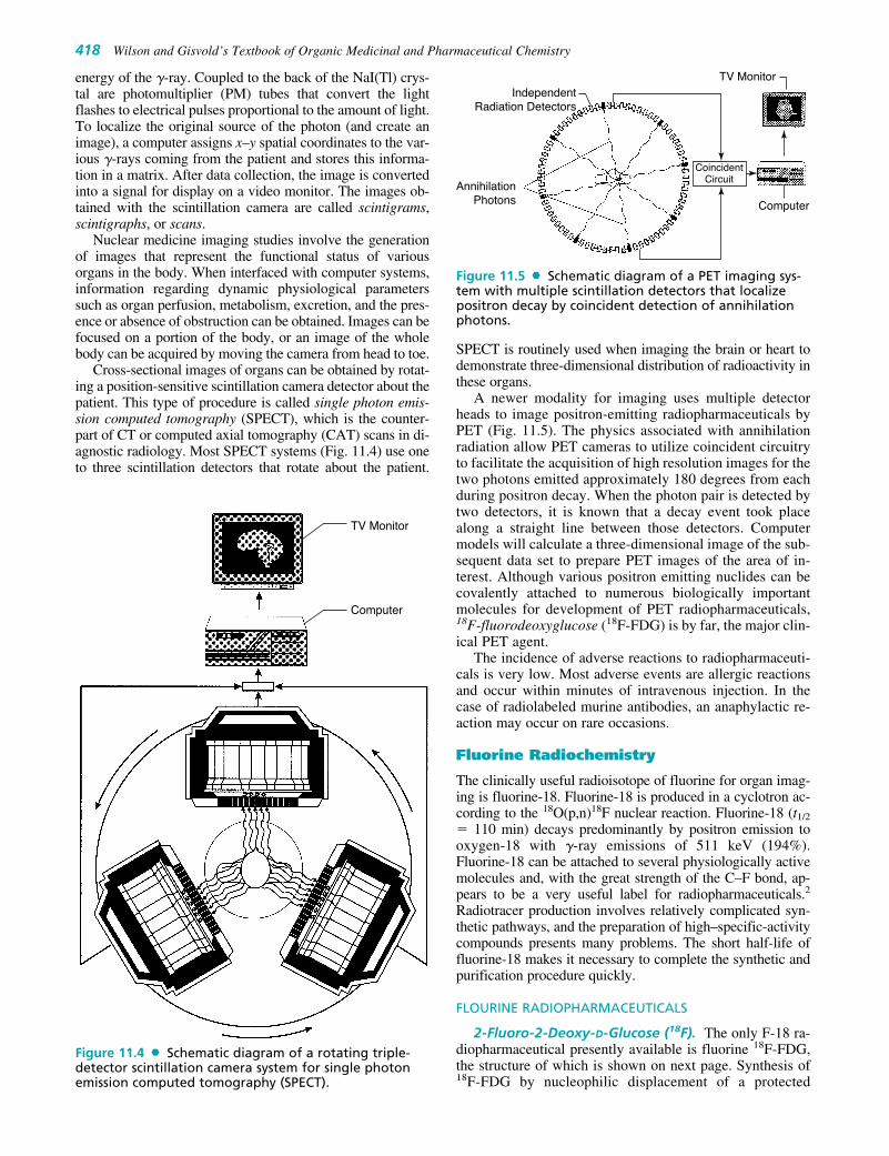
energy of the �-ray. Coupled to the back of the NaI(Tl) crys-tal are photomultiplier (PM) tubes that convert the lightflashes to electrical pulses proportional to the amount of light.To localize the original source of the photon (and create animage), a computer assigns x–y spatial coordinates to the var-ious �-rays coming from the patient and stores this informa-tion in a matrix. After data collection, the image is convertedinto a signal for display on a video monitor. The images ob-tained with the scintillation camera are called scintigrams,scintigraphs, or scans.
Nuclear medicine imaging studies involve the generationof images that represent the functional status of variousorgans in the body. When interfaced with computer systems,information regarding dynamic physiological parameterssuch as organ perfusion, metabolism, excretion, and the pres-ence or absence of obstruction can be obtained. Images can befocused on a portion of the body, or an image of the wholebody can be acquired by moving the camera from head to toe.
Cross-sectional images of organs can be obtained by rotat-ing a position-sensitive scintillation camera detector about thepatient. This type of procedure is called single photon emis-sion computed tomography (SPECT), which is the counter-part of CT or computed axial tomography (CAT) scans in di-agnostic radiology. Most SPECT systems (Fig. 11.4) use oneto three scintillation detectors that rotate about the patient.
SPECT is routinely used when imaging the brain or heart todemonstrate three-dimensional distribution of radioactivity inthese organs.
A newer modality for imaging uses multiple detectorheads to image positron-emitting radiopharmaceuticals byPET (Fig. 11.5). The physics associated with annihilationradiation allow PET cameras to utilize coincident circuitryto facilitate the acquisition of high resolution images for thetwo photons emitted approximately 180 degrees from eachduring positron decay. When the photon pair is detected bytwo detectors, it is known that a decay event took placealong a straight line between those detectors. Computermodels will calculate a three-dimensional image of the sub-sequent data set to prepare PET images of the area of in-terest. Although various positron emitting nuclides can becovalently attached to numerous biologically importantmolecules for development of PET radiopharmaceuticals,18F-fluorodeoxyglucose (18F-FDG) is by far, the major clin-ical PET agent.
The incidence of adverse reactions to radiopharmaceuti-cals is very low. Most adverse events are allergic reactionsand occur within minutes of intravenous injection. In thecase of radiolabeled murine antibodies, an anaphylactic re-action may occur on rare occasions.
Fluorine Radiochemistry
The clinically useful radioisotope of fluorine for organ imag-ing is fluorine-18. Fluorine-18 is produced in a cyclotron ac-cording to the 18O(p,n)18F nuclear reaction. Fluorine-18 (t1/2
� 110 min) decays predominantly by positron emission tooxygen-18 with �-ray emissions of 511 keV (194%).Fluorine-18 can be attached to several physiologically activemolecules and, with the great strength of the C–F bond, ap-pears to be a very useful label for radiopharmaceuticals.2
Radiotracer production involves relatively complicated syn-thetic pathways, and the preparation of high–specific-activitycompounds presents many problems. The short half-life offluorine-18 makes it necessary to complete the synthetic andpurification procedure quickly.
FLOURINE RADIOPHARMACEUTICALS
2-Fluoro-2-Deoxy-D-Glucose (18F). The only F-18 ra-diopharmaceutical presently available is fluorine 18F-FDG,the structure of which is shown on next page. Synthesis of18F-FDG by nucleophilic displacement of a protected
418 Wilson and Gisvold’s Textbook of Organic Medicinal and Pharmaceutical Chemistry
TV Monitor
Computer
Figure 11.4 Schematic diagram of a rotating triple-detector scintillation camera system for single photonemission computed tomography (SPECT).
TV Monitor
CoincidentCircuit
IndependentRadiation Detectors
AnnihilationPhotons Computer
Figure 11.5 Schematic diagram of a PET imaging sys-tem with multiple scintillation detectors that localizepositron decay by coincident detection of annihilationphotons.
0413-0442_17865_Ch11.qxd 12/4/09 5:34 AM Page 418

mannose triflate derivative, hydrolysis of the protectinggroups, and purification by solid phase extraction is the typ-ical method of production.3 The synthetic cascade and sub-sequent purification is routinely accomplished in about 50minutes by an automated synthesizer.4,5
The high glycolytic rate of many neoplasms comparedwith that of the surrounding tissues facilitates tumor imag-ing with this glucose analog. Both glucose and 18F-FDG aretransported into the cell and phosphorylated; however, 18F-FDG does not get catabolized as an energy source, nor is iteffectively degraded by glucose-6-phosphatase. Thus, ener-getically active tissues accumulate 18F-FDG. Because of thewidespread anatomical distribution of metastases, a whole-body imaging technique using a tumor-specific radiophar-maceutical, such as 18F-FDG, is very useful for tumor detec-tion and mapping to evaluate the extent and relativemetabolic activity of the disease.
18F-FDG can also be used for diagnostic imaging of myo-cardial viability and in the evaluation of seizure disorders.6
Gallium Radiochemistry
The only radioisotope of gallium that is presently used isgallium-67, which is produced in a cyclotron by protonbombardment of a zinc metal target according to the68Zn(p,2n)67Ga nuclear reaction. Gallium-67 (t1/2 � 78.2hours) decays by EC to stable zinc-67 with principal �-rayemissions of 93 keV (38%), 185 keV (24%), and 300 keV(16%). The radiotracer is isolated by dissolution of the tar-get in hydrochloric acid followed by isopropyl ether extrac-tion of the gallium-67 from the zinc and other impurities.Gallium-67 is back-extracted from the isopropyl ether into0.2 M hydrochloric acid, evaporated to dryness, and dis-solved in sterile, pyrogen-free 0.05 M hydrochloric acid.7
Gallium is an amphoteric element that acts as a metal at lowpH but forms insoluble hydroxides when the pH is raisedabove 2 in the absence of chelating agents. At high pH,gallium hydroxide acts as a nonmetal and dissolves in am-monia to form gallates. Gallium forms compounds of oxida-tion states �1, �2, and �3; however, only the Ga�3 state isstable in aqueous solutions.
GALLIUM RADIOPHARMACEUTICALS
Gallium Citrate (67Ga). The gallium (III)–citrate com-plex is formed by adding the required amount of sodium cit-rate (0.15 M) to gallium (III) chloride and adjusting the pHto 4.5 to 8.0 with sodium hydroxide. Recent studies haveproposed a 1:2 gallium:citrate complex.8,9
The patient receives an intravenous injection of 5 to 10 mCi(185–370 MBq) of gallium (67Ga) citrate, and whole-body im-ages are then obtained 24, 48, and 72 hours after injection.Gallium localizes at sites of inflammation or infection as wellas various tumors. It is used in clinical practice in the staging
O
HO
HO18F
CH2OH
OH
(18F)-2-Fluoro-2-deoxy-D-glucose
and evaluation of recurrence of lymphomas. Gallium localizesnormally in the liver and spleen, bone, nasopharynx, lacrimalglands, and breast tissue. There is also some secretion in thebowel; consequently, the patient may require a laxative and/orenemas to evacuate enteric radioactivity prior to the 48-hourimage. As more specific radiotracers have been developed, thenonspecific normal localization of gallium radioactivity haslimited its clinical use.
Chapter 11 Agents for Diagnostic Imaging 419
Gallium (67Ga) Citrate
67Ga
O
O
O
O
O
4
O
O
O
O
O
O
O
OH
O
_
_
Indium Radiochemistry
The most commonly used radioisotope of indium is in-dium-111, which is produced in a cyclotron by protonbombardment of a cadmium target according to the112Cd(p,2n)111In nuclear reaction. Indium-111 (t1/2 � 67.3hours) decays by EC to stable cadmium-111 with principal�-ray emissions of 171 keV (91%) and 245 keV (94%).The radiotracer is isolated by dissolution in hydrochloricacid to form indium chloride (111In). In aqueous solution,lower valence states of indium have been described, butthey are unstable and are rapidly oxidized to the triva-lent state. In acid solution, indium hexaaqua complexes[In(H2O)6]�3 are stable at low pH but are hydrolyzed(above pH 3.5) to form a precipitate of indium hydroxideIn(OH)3.5 Indium is stabilized in solution above pH 3.5 ifit is complexed with a weak chelating agent such assodium citrate and stronger chelating agents such as 8-hydroxyquinoline (oxine) or diethylenetriamine pen-taacetic acid (DTPA). Monoclonal antibodies and peptidesare radiolabeled with indium by using compounds calledbifunctional chelating agents. Bifunctional chelatingagents are molecules that can be attached to the antibodyor peptide as well as to the radionuclide.
INDIUM RADIOPHARMACEUTICALS
Indium (111In) Capromab (ProstaScint). Capromabpendetide is a murine monoclonal immunoglobulin G (IgG)antibody conjugated to the linker-chelator, glycyl-tyrosyl-(N,-diethylenetriamine pentaacetic acid)-lysine hydrochlo-ride (Gly-Tyr-Lys-DTPA�HCl). This conjugated antibody isdirected against the glycoprotein expressed on prostate ep-ithelium, prostate-specific membrane antigen. It is indicatedin the evaluation of newly diagnosed patients with biopsy-proven prostate cancer who are at high risk for pelvic lymph
0413-0442_17865_Ch11.qxd 12/4/09 5:34 AM Page 419
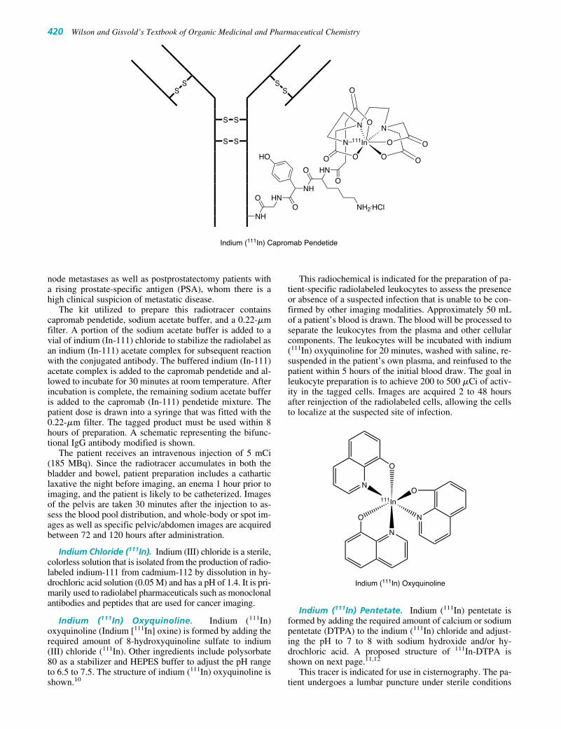
node metastases as well as postprostatectomy patients witha rising prostate-specific antigen (PSA), whom there is ahigh clinical suspicion of metastatic disease.
The kit utilized to prepare this radiotracer containscapromab pendetide, sodium acetate buffer, and a 0.22-�mfilter. A portion of the sodium acetate buffer is added to avial of indium (In-111) chloride to stabilize the radiolabel asan indium (In-111) acetate complex for subsequent reactionwith the conjugated antibody. The buffered indium (In-111)acetate complex is added to the capromab pendetide and al-lowed to incubate for 30 minutes at room temperature. Afterincubation is complete, the remaining sodium acetate bufferis added to the capromab (In-111) pendetide mixture. Thepatient dose is drawn into a syringe that was fitted with the0.22-�m filter. The tagged product must be used within 8hours of preparation. A schematic representing the bifunc-tional IgG antibody modified is shown.
The patient receives an intravenous injection of 5 mCi(185 MBq). Since the radiotracer accumulates in both thebladder and bowel, patient preparation includes a catharticlaxative the night before imaging, an enema 1 hour prior toimaging, and the patient is likely to be catheterized. Imagesof the pelvis are taken 30 minutes after the injection to as-sess the blood pool distribution, and whole-body or spot im-ages as well as specific pelvic/abdomen images are acquiredbetween 72 and 120 hours after administration.
Indium Chloride (111In). Indium (III) chloride is a sterile,colorless solution that is isolated from the production of radio-labeled indium-111 from cadmium-112 by dissolution in hy-drochloric acid solution (0.05 M) and has a pH of 1.4. It is pri-marily used to radiolabel pharmaceuticals such as monoclonalantibodies and peptides that are used for cancer imaging.
Indium (111In) Oxyquinoline. Indium (111In)oxyquinoline (Indium [111In] oxine) is formed by adding therequired amount of 8-hydroxyquinoline sulfate to indium(III) chloride (111In). Other ingredients include polysorbate80 as a stabilizer and HEPES buffer to adjust the pH rangeto 6.5 to 7.5. The structure of indium (111In) oxyquinoline isshown.10
This radiochemical is indicated for the preparation of pa-tient-specific radiolabeled leukocytes to assess the presenceor absence of a suspected infection that is unable to be con-firmed by other imaging modalities. Approximately 50 mLof a patient’s blood is drawn. The blood will be processed toseparate the leukocytes from the plasma and other cellularcomponents. The leukocytes will be incubated with indium(111In) oxyquinoline for 20 minutes, washed with saline, re-suspended in the patient’s own plasma, and reinfused to thepatient within 5 hours of the initial blood draw. The goal inleukocyte preparation is to achieve 200 to 500 �Ci of activ-ity in the tagged cells. Images are acquired 2 to 48 hoursafter reinjection of the radiolabeled cells, allowing the cellsto localize at the suspected site of infection.
Indium (111In) Pentetate. Indium (111In) pentetate isformed by adding the required amount of calcium or sodiumpentetate (DTPA) to the indium (111In) chloride and adjust-ing the pH to 7 to 8 with sodium hydroxide and/or hy-drochloric acid. A proposed structure of 111In-DTPA isshown on next page.11,12
This tracer is indicated for use in cisternography. The pa-tient undergoes a lumbar puncture under sterile conditions
N
N
111In
Indium (111In) Oxyquinoline
O
N
O
O
420 Wilson and Gisvold’s Textbook of Organic Medicinal and Pharmaceutical Chemistry
SS
S
S
SS
S
S
Indium (111In) Capromab Pendetide
NH
111In
O
ON
O
NN
O O
O
O
O
O HNO
NH
HO
O HN
NH2.HCl
O
0413-0442_17865_Ch11.qxd 12/4/09 5:34 AM Page 420

and receives an intrathecal injection of 0.5 mCi (18.5 MBq)of indium (111In) pentetate, which distributes into the cere-brospinal fluid (CSF). Initial images are obtained to ensurea good intrathecal injection. Images of the spinal canal andCSF spaces of the brain are acquired intermittently to assessCSF flow or leakage from the normal CSF space.
Another use for 111In-DTPA is in dual isotope gastricemptying studies. After a 6-hour fast, the patient ingests asolid meal of oatmeal or scrambled egg mixed with 1 mCi(37 MBq) of 99mTc-sulfur colloid along with a liquid mealof water mixed with 0.5 mCi (18.5 MBq) of 111In-DTPA.The patient’s stomach is imaged 5 minutes after the meal asthe initial gastric image utilizing appropriate camera settingsto detect each isotope separately. Imaging continues every 5to 15 minutes until the recorded activity is reduced to onehalf the initial gastric activity. Normal values are 50 to 80minutes for solid meals and 10 to 15 minutes for liquidmeals4; however, these values can vary based on the sizeand composition of the meal.5
Indium (111In) Pentetreotide (OctreoScan). Pente-treotide is a modified peptide hormone that consists of a bi-functional chelating agent, DTPA, linked to octreotide, along-acting analog of the endogenous hormone somato-statin. Somatostatin is a 14–amino acid peptide hormonethat binds to the somatostatin receptor. These receptorshave been found on many cells of neuroendocrine originand are expressed in large numbers on nearly all tumors ofsuch origin. The drug is indicated in the imaging of primaryand metastatic neuroendocrine tumors bearing somatostatinreceptors.
The kit utilized to prepare this radiotracer contains pen-tetreotide, a solution of indium (111In) chloride/ferric chlo-ride, and a transfer needle. The addition of ferric chlorideand the use of the transfer needle are necessary to increasethe labeling yield. The radiolabeled solution is added to thepentetreotide vial and incubated at room temperature for 30minutes. The tagged product must be used within 6 hours ofpreparation. The structure of labeled radiopharmaceutical,indium (111In) pentetreotide, is shown.13
The patient receives an intravenous injection of 3 to6 mCi (111–222 MBq) of indium (111In) pentetreotide de-pending on the type of imaging camera that will be usedto obtain images. Whole-body images are obtained 4 to48 hours after injection to localize the primary tumor andsites of metastases.
111In
O
O
ON
O
NN
O
Indium (111In) Pentetate
O
O
O
O
O
2_
Iodine Radiochemistry
Iodine is in group VIIB with the other halogens (fluorine,chlorine, bromine, and astatine). In aqueous solution, com-pounds of iodine are known with at least five differentoxidation states; however, in nuclear medicine, the �1 and�1 oxidation states are the most significant. The �1 oxi-dation state represented as sodium iodide (NaI) is impor-tant for thyroid studies and, when obtained in a reductant-free solution (no sodium thiosulfate), is a starting materialfor the radiolabeling of iodinated radiopharmaceuticals.The common methods for introducing radioiodine into or-ganic compounds are isotope exchange reactions, elec-trophilic substitution of hydrogen in activated aromaticsystems, nucleophilic substitution, and addition to doublebonds.14 Electrophilic substitution on tyrosine side chainsis commonly used for protein labeling, The electrophilicspecies (I�) can be generated from iodide (I�) by variousoxidizing agents, including (a) chloramine-T (N-chloro-p-toluene sulfonamide) sodium, (b) enzyme oxidation of(lactoperoxidase), and (c) Iodo-Gen (1,3,4,6-tetrachlora-3�-6�-diphenylglycoluril).
The useful radioisotopes of iodine for organ imaging areiodine-123 and iodine-131. Iodine-123 is produced in a cy-clotron by bombarding a xenon-124 target with a proton ac-cording to the 124Xe (p,2n)123Cs→123Xe→123I sequence.Iodine-123 is isolated in the iodide form. Cesium-123 (t1/2
� 5.9 min) decays by EC to xenon-123 (t1/2 � 2.1 hours),which decays to iodine-123. Iodine-123 (t1/2 � 13.3 hours)decays by EC to tellurium-123, with a principal �-ray emis-sion of 159 keV (83%), which makes it the ideal radioiso-tope of iodine for organ imaging because of increased detec-tion efficiency and reduced radiation burden to the patient.The main disadvantage of I-123 is the need for daily deliv-ery from commercial suppliers because of its short half-life.
Iodine-131 is obtained from a reactor by production of tel-lurium-131. It is prepared by nuclear bombardment of235U(n,fission)131Te→131I or 130Te(n,�)131Te→131I. I-131 isisolated in the iodide form. Tellurium-131 (t1/2 � 25 min-utes) decays by �� emission to iodine-131. Iodine-131 (t1/2 � 8.04 days) decays by �� emission to stable xenon-131, with five significant �-ray emissions ranging from 80 to723 keV. The major �-ray of 364 keV (82%) provides goodtissue penetration for organ imaging. I-131 properties are ei-ther desirable or undesirable based on one’s perspective. Thehigh radiation dose from the � particles may be undesirablewhen used for diagnostic purposes, but highly desirable fortherapeutic use. The long half-life provides sufficient timefor radiolabeling syntheses, but can be problematic in termsof waste disposal. Finally, poor quality images are obtained
Indium (In-111) Pentetreotide
111In
O
N
O
N N
OO
O
ONH
O
O
O
D-Phe CysSS
Cys
Phe
Thr
D-Trp
LysThr(ol)
Chapter 11 Agents for Diagnostic Imaging 421
0413-0442_17865_Ch11.qxd 12/4/09 5:34 AM Page 421

by current gamma cameras because the high-energy �-raysare not efficiently recorded by the detector.
IODINE PHARMACEUTICALS
Sodium Iodide (123I). The major indications for thyroidimaging with sodium iodide (123I) are for evaluation of thy-roid function (uptake) and/or morphology (imaging). Thepatient fasts before receiving the oral dose of 0.1 to 0.4 mCi(3.7–14.8 MBq) of sodium iodide (123I) typically in an en-capsulated dosage form. Images are obtained of the thyroidand surrounding area 4 to 6 hours after ingestion.
Sodium Iodide (131I). Diagnostic use of sodium iodide(131I) with microcurie doses for evaluating thyroid func-tion and/or morphology is no longer preferred because ofthe higher radiation dose associated with particle decayfrom this isotope. Doses from 1 to 10 mCi (37–370 MBq)are used for whole-body imaging to detect metastatic thy-roid cancer and/or residual thyroid tissue following thy-roidectomy. Whole-body images are obtained 48 to 72hours later.
Therapeutic use of sodium iodide (131I) occurs in hyper-thyroidism or differentiated types of thyroid cancers. A pa-tient with hyperthyroidism will receive an oral dose of 5 to10 mCi (185–370 MBq) of sodium iodide (131I). The dosecould be higher for patients with toxic nodular goiter orother special situations. Treatment doses for differentiatedthyroid carcinoma with or without metastases range from 30to 200 mCi (1,110–7,400 MBq). Seven days after the thera-peutic dose, a whole-body scan is performed in an effort todetect metastases not seen on the pretherapy diagnostic im-ages. Thyroid hormone medication will be discontinued forseveral weeks prior to radiotherapy and thyroid-stimulatinghormone (TSH) levels must be evaluated to ensure maximaluptake of the radiotracer for therapy.15
Iodine (131I) Tositumomab (Bexxar). Tositumomab isa murine IgG monoclonal antibody directed against theCD20 antigen found on the surface of normal and malignantB lymphocytes. Iodine tositumomab (131I) is a radioiodi-nated derivative of the antibody indicated for the treatmentof patients with CD20 antigen-expressing relapsed or re-fractory, non-Hodgkin lymphoma. A schematic represent-ing an IgG antibody labeled with covalently linked I-131 isshown. The exact number and position of the covalentlylinked I-131 is not described.
The treatment regimen occurs in two discrete steps: thedosimetric (diagnostic distribution) and therapeutic steps.Each step consists of a sequential infusion of unlabeledTositumomab followed by iodine (131I) tositumomab. Thepatient is premedicated with a thyroid protective agent (eitherpotassium iodide or Lugol solution) for at least 24 hours, andwill also receive acetaminophen and diphenhydramineshortly before the infusions begin. The dosimetric step ad-ministers 450-mg unlabeled tositumomab followed by 5 mCi(185 MBq) of iodine (131I) tositumomab.
The patient is imaged three times over the next 7 days toassess the biodistribution of the radiolabeled antibody.Imaging occurs within 1 hour following the radiolabeled an-tibody infusion, 2 to 4 days after the infusion, and also 6 to7 days after the infusion. If the dosimetric dose’s biodistrib-ution is altered, no further drug will be administered. If thedosimetric dose’s biodistribution is as expected, the thera-
peutic dose can be administered 7 to 14 days after the dosi-metric infusion.
The therapeutic dose is calculated based on the assess-ment of the patient’s biodistribution and elimination of thedosimetric infusion as well as the patient’s platelet count.Clinical studies have reported calculated therapeutic dosesof 450-mg unlabeled tositumomab followed by 50 to 200mCi (1,300–5,900 MBq) of iodine (131I) tositumomab.Thyroid protective agents are administered daily until 2weeks after the last radioiodine dose.16
Technetium Radiochemistry
The most commonly used radioisotope of technetium is tech-netium-99m, which is produced by molybdenum-99 ��
decay in a molybdenum-99/technetium-99m generator.Technetium-99m (t1/2 � 6 hours) decays by IT to technetium-99 (t1/2 � 2.1 � 105 years) with principal �-ray emissions of140 keV (100%). The radiotracer is collected as pertechnetateions (99mTcO4
�) from the generator by elution from the alu-mina column with sterile normal saline (0.9% sodium chlo-ride). Long-lived radioactive impurities such as molybdateions (99MoO4) that coelute from the generator column withpertechnetate ions (99mTcO4
�) theoretically may shorten themanufacturer’s recommended expiration time of each elutionsince the Nuclear Regulatory Commission (NRC) and UnitedStates Pharmacopeia (USP) limits molybendum-99 break-through to no more than 0.15 �Ci of 99Mo per 1 mCi of 99mTcin the preparation.5 In clinical practice, molybendum-99breakthrough is a rare occurrence.
Technetium, is a transition state metal and is the only “ar-tificial” element with a lower atomic number than uranium.No stable isotopes exist in nature. All known isotopes oftechnetium are radioactive, and as such, has hindered thestudy of technetium’s chemistry. However, milligram quan-tities of Tc-99 (a weak �� emitter; t1/2 � 2.1 � 105 years)are now available for characterization of technetium com-plexes. Technetium can exist in eight oxidation states, from�1 to �7. The �7 (99mTcO4
�) and �4 state (99mTcO2, orhydrolyzed-reduced technetium-99m) are the most stableforms.4
The chemistry of technetium is similar to that of rhe-nium and is dominated by forming compounds by bondingbetween the electron-deficient metal and electronegative
SS
S
S
SS
S
S
131I
Iodine (I-131) Tositumomab
422 Wilson and Gisvold’s Textbook of Organic Medicinal and Pharmaceutical Chemistry
0413-0442_17865_Ch11.qxd 12/4/09 5:34 AM Page 422

groups, which are capable of donating electron pairs. Someexamples of these electronegative groups include amines,carboxylic acids, hydroxyls, isonitriles, oximes, phos-phates, phosphines, and sulfhydryls. Various oxidationstates (e.g., �1, �3, �5, etc.) are prepared in the presenceof the reducing agent and stabilized by complexation withavailable ligands.
Tc-99m radiopharmaceuticals are by far the most com-monly used radiotracers in day-to-day diagnostic nuclearmedicine practice. Almost all technetium radiopharmaceuti-cals are metal–electron donor complexes. Compounds thatcontain two or more electron donor groups and bind to ametal are called chelating agents. As the pertechnetate(99mTcO4
�) ion, technetium will not form metal�donorcomplexes. However, the pertechnetate ion can be reducedby a stannous salt or other methods to an oxidation state thatwill complex with various monodentate, bidentate, or poly-dentate ligands. The most common reducing agent used inclinical practice is the stannous ion.
Technetium-99m radiopharmaceuticals are prepared bycombining sodium pertechnetate (Na99mTcO4) with nonra-dioactive components in a sterile reaction vial. The primarychemical substances in the vial are the complexing agent(ligand) and a stannous salt-reducing agent (stannous chlo-ride, stannous fluoride, or stannous tartrate). The oxidationstate of technetium in various complexes as well as the ac-tual chemical structure of many radiopharmaceuticals hasyet to be characterized. Several reviews of technetiumchemistry are available for further study.17–20
After preparation of the radiopharmaceutical, tests for ra-diochemical purity should be carried out to ensure that theradiotracer is in the correct radiochemical form. Analyticalquality control methods include paper and thin-layer chro-matography, column chromatography, and solvent extrac-tion. Likely, radiochemical impurities include unreactedsodium pertechnetate (Na99mTcO4), hydrolyzed-reducedtechnetium (99mTcO2) colloid, technetium–tin colloid, andpossibly radiochemical complexes different from the oneexpected (e.g., 99mTc-monodentate rather than 99mTc-biden-tate ligand, etc.). The sterile vials containing the stannoussalt and the ligand are lyophilized under a sterile inert gas at-mosphere (i.e., nitrogen or argon). The ligand in the reactionvial determines the final chemical structure of the 99mTc-complex and the biological fate after intravenous injectionof the radiopharmaceutical.
TECHNETIUM RADIOPHARMACEUTICALS
Sodium Pertechnetate (99mTc). Sodium pertechnetate(99mTc) is a sterile, colorless solution in normal saline (0.9%NaCl), obtained by elution of the sterile Mo–99/Tc-99mgenerator. The major use of sodium pertechnetate (99mTc) isthe preparation of various technetium-99m radiopharmaceu-ticals either in a nuclear pharmacy or nuclear medicine de-partment. On-site preparation of radiolabeled red blood cells(RBCs) requires the availability of sodium pertechnetatesince the radiolabeled cells must be reinfused to the patientwithin 30 minutes of preparation.
The pertechnetate ion (99mTcO4�), which has an ionic ra-
dius and charge similar to the iodide ion (I�) and is concen-trated in the thyroid, salivary glands, kidneys, stomach, andchoroid plexus in the brain. It can be used in a diluted solu-tion from the Mo–99/Tc-99m generator elution to image the
thyroid, Meckel’s diverticulum (stomach tissue in the intes-tine), salivary glands, and to detect processes that disrupt theblood-brain barrier (i.e., tumors, abscesses, strokes).
Like iodide, pertechnetate is trapped by the thyroid.Pertechnetate, however, cannot be utilized to prepare thy-roid hormone. The patient receives an intravenous injectionof 5 to 10 mCi (185–370 MBq) of Tc-99m pertechnetate,and thyroid images are obtained 20 to 30 minutes after in-jection. The usual dose for other imaging procedures such asMeckel’s diverticulum and salivary gland imaging is also 5to 10 mCi (185–370 MBq), whereas 20 mCi (740 MBq) isused for blood-brain barrier imaging.
Technetium (99mTc) Bicisate (Neurolite). Technetium(99mTc) bicisate is a neutral, lipophilic complex that crossesthe blood-brain barrier and is selectively retained in the brain.
The kit used to prepare the product contains two vials.Vial “A” contains the ligand, tin, and other components thatmust be protected from light, whereas vial “B” contains abuffer solution. The radiopharmaceutical is prepared byadding sodium pertechnetate (99mTc) to the shielded “B” vialcontaining the buffer. The vial A containing lyophilized bi-cisate, EDTA, mannitol, and stannous chloride is reconsti-tuted with 3 mL of normal saline (0.9% NaCl). Within 30seconds, 1 mL of the saline solution from vial A is trans-ferred to vial B and allowed to incubate at room temperaturefor 30 minutes. The tagged product must be used within 6hours of preparation. The structure of the technetium com-plex is [N,N�-ethylene-di-L-cysteinato(3�)]oxo [99mTc]tech-netium(V) diethyl ester.21
After intravenous injection of 20 mCi (740 MBq) of Tc-99m bicisate, about 5% of the injected dose is localizedwithin the brain cells 5 minutes after injection. Ester hydro-lysis traps the radiotracer within the brain. This radiotraceris indicated for the evaluation of stroke, but it is used off-label to clinically evaluate various brain disorders, espe-cially brain perfusion (“brain death”).
99mTc
O
O
O
O
Sodium Pertechnetate (99mTc)
_
Na+
Chapter 11 Agents for Diagnostic Imaging 423
99mTc
O
S
N
S
N
Technetium (99mTc) Bicisate
O
O
O
O
Technetium (99mTc) Disofenin (Hepatolite).Technetium (99mTc) disofenin is a 2:1 complex ofligand:metal that is rapidly eliminated via the hepatobiliarysystem. Clearance from the blood is delayed by high biliru-bin levels. The primary clinical indication for this product ispossible acute cholecystitis. In acute cholecystitis, there is
0413-0442_17865_Ch11.qxd 12/4/09 5:34 AM Page 423

obstruction of the cystic duct leading to the gallbladder. Thegallbladder is not visualized because the radiotracer cannotenter it. Some other clinical conditions that can be diag-nosed by biliary images are common bile duct obstruction,biliary leak following surgery, biliary atresia, and a chole-dochal cyst.
The product is prepared by adding sodium pertechnetate(99mTc) to the shielded vial containing disofenin and stan-nous chloride. The preparation is allowed to incubate for 4minutes. The product should be used within 6 hours ofpreparation. The structure of the technetium complex isshown.22,23 Hepatobiliary agents are commonly referred toas HIDA agents, or hepatobiliary aminodiacetic acids.
The fasting patient receives an intravenous injection of 5mCi (185 MBq), which is taken up by the hepatocytes in theliver by active anionic transport. Patients with bilirubin lev-els greater than 5 mg/dL require an 8 mCi (296 MBq) dose.Then the radiopharmaceutical is excreted in bile, via the bil-iary canaliculus, into the bile ducts, with accumulation inthe gallbladder and finally excretion via the common bileduct into the small bowel. The normal patient exhibits earlyaccumulation of the radiopharmaceutical in the liver and thegallbladder.
Technetium (99mTc) Exametazime (Ceretec).Technetium (99mTc) exametazime5 is a mixture of unstablelipophilic enantiomers that rapidly cross the blood-brain bar-rier and is trapped in the tissues. The proposed trapping mech-anism for localization includes reduction by glutathione. Asimilar diffusion and trapping process occurs with autologouslymphocytes in vitro.
Exametazime is also known as hexamethylpropyle-neamine oxime or HMPAO. The radiolabeled complex is in-
dicated for cerebral perfusion in stroke, but is most com-monly used for the radiolabeling of autologous leukocytesas an adjunct in the localization of intra-abdominal infectionand inflammatory bowel disease.
Each kit includes several components: (a) reaction vialscontaining a mixture of exametazime, stannous chloride,and sodium chloride; (b) vials of 1% methylene blue; (c)vials of phosphate buffer in 0.9% NaCl; and (d) 0.45-�m sy-ringe filters. Product preparation depends on the intendeduse.
For use as a cerebral perfusion agent, the kit can be pre-pared with or without stabilizer. Shelf life is 4 hours with sta-bilizer, but only 30 minutes without stabilizer. Stabilizer isprepared by injecting 0.5 mL of 1% methylene blue in thephosphate buffer vial. Fresh sodium pertechnetate (99mTc)elution (�2 hours old) is added to the shielded vial contain-ing exametazime. The vial is gently agitated for 10 secondsand used immediately because this unstabilized complex hasa shelf life of approximately 30 minutes. For stabilization,2 mL of the methylene blue/phosphate buffer mixture isadded to the reconstituted exametazime vial within 2 minutesof the technetium (99mTc) exametazime vial reconstitution.Since the stabilized product is dark blue, a syringe filter is uti-lized to prevent inadvertent administration of particulate mat-ter. Unstabilized product should be visually inspected for aclear, colorless appearance prior to administration. The pa-tient receives an intravenous dose of 10 to 20 mCi (370–740MBq). Dynamic imaging begins immediately following in-jection for up to 10 minutes. Static imaging may be per-formed from 15 minutes to 6 hours after injection.
For the radiolabeling of autologous leukocytes, approx-imately 50 mL of a patient’s blood is drawn. The bloodwill be processed to separate the leukocytes from the
424 Wilson and Gisvold’s Textbook of Organic Medicinal and Pharmaceutical Chemistry
Technetium (99mTc) Disofenin
99mTc
O
O
O
N
N
O
NH
O
HN
O
_
99mTc
O
N
N
N
N
Technetium (99mTc) Exametazime
O
H
O
99mTc
O
N
N
N
N
O
H
O
0413-0442_17865_Ch11.qxd 12/4/09 5:34 AM Page 424

plasma and other cellular components. Fresh sodiumpertechnetate (99mTc) elution (�2 hours old) is added tothe shielded vial containing exametazime. The vial is gently agitated for 10 seconds and used immediately. Theleukocytes will be incubated with technetium (99mTc) ex-ametazime for 15 minutes, washed with saline, resus-pended in the patient’s own plasma, and reinfused to thepatient as soon as possible. The goal in leukocyte prepara-tion is to achieve 7 to 25 mCi of activity in the taggedcells. Images are acquired 2 to 4 hours after reinjection ofthe radiolabeled cells, allowing the cells to localize at thesuspected site of infection.
Technetium (99mTc) Macroaggregated Albumin.Technetium (99mTc) macroaggregated albumin (99mTc-MAA) is a sterile white suspension of human albumin aggre-gates formed by denaturing human albumin at 80°C for 30minutes at pH 5 (isoelectric point of albumin).4 The particlesize and number of particles can be estimated with a hemo-cytometer grid. The particle composition within the suspen-sion should be between 10 and 100 �m, with no particlesgreater than 150 �m. Commercial manufacturing practicesprovide a consistent range for the number of particles per vialas well as a consistent range of particle size. This primary useof this agent is for the imaging of the pulmonary microcircu-lation to assess potential pulmonary emboli. Another muchless common indication is to evaluate peritoneovenous(LeVeen) shunt patency.
In general, the kit is prepared by adding sodiumpertechnetate (99mTc) to the shielded vial containing albu-min, aggregated albumin, and stannous chloride, gentlyinverting the reconstituted suspension (to prevent foam-ing), and allowing the mixture to incubate at room tem-perature for 15 minutes. Gentle inversion of the vial isrecommended to provide a uniform suspension prior towithdrawing a patient dose into a syringe. If the syringe isnot used immediately, it should be gently inverted to re-suspend the particles prior to injection. The tagged prod-uct is to be used within 12 hours of preparation. The pre-cise structure of the radiolabeled complex is unknown atthis time.
The patient receives an intravenous injection of 2 to 5mCi (74–185 MBq) of the Tc-99m albumin aggregates,which lodge in some of the small pulmonary arterioles andcapillaries. The number of aggregates recommended forgood image quality and patient safety is 100,000 to 500,000particles; thus, only a small fraction of the 280 billion capil-laries are occluded. Multiple images of the lung are obtainedto assess lung perfusion. The distribution of the particles inthe lung is a function of regional blood flow; consequently,
in the normal lung, the particles are distributed uniformlythroughout the lung. When blood flow is occluded becauseof emboli, multiple segmental “cold” (decreased radioactiv-ity) defects are seen. This procedure is almost always com-bined with a xenon-133 gas or technetium-99m DTPAaerosol lung ventilation scan (should be normal in pul-monary embolism) and same-day chest radiograph x-ray(should be normal).
Technetium (99mTc) Mebrofenin (Choletec).Technetium (99mTc) mebrofenin22,23 is a 2:1 complex ofligand:metal that is rapidly eliminated via the hepatobil-iary system. The presence of bromine within the structureof mebrofenin makes the drug more lipophilic, and thus ithas higher hepatic extraction than other hepatobiliaryiminodiacetic acids (HIDA). Clearance from the blood isalso affected by bilirubin levels, but generally to a muchlesser extent because of the higher hepatic extraction.The clinical indication for this product is hepatobiliaryimaging.
The product is prepared by adding sodium pertechne-tate (99mTc) to the shielded vial containing mebrofenin,stannous fluoride and preservatives, and allowing the mix-ture to incubate for 15 minutes. The presence of preserva-tives allows this kit to be used for up to 18 hours afterpreparation.
The fasting patient receives an intravenous injection of5 mCi (185 MBq), which is taken up by the hepatocytes inthe liver by active anionic transport. Patients with biliru-bin levels greater than 1.5 mg/dL may require a 10 mCi(370 MBq) dose.
Technetium (99mTc) Medronate. Technetium (99mTc)Medronate (99mTc-MDP)24 is a diphosphonate complexwith technetium-99m indicated for bone imaging to identifyareas of altered osteogenesis. The clinical use of this agentis for investigation of skeletal problems such as metastaticdisease to the bones, osteomyelitis, Paget disease, fractures,primary bone tumors, avascular necrosis, metabolic bonedisease, and loose or infected hip prostheses. Stress frac-tures can be diagnosed by bone imaging when x-rays arecompletely normal. Bone radioscintigraphy is one of themost commonly performed nuclear medicine diagnosticprocedures because the whole-body survey allows evalua-tion of the entire skeleton, which cannot be done as cost-effectively by any other imaging modality.
The relatively weak complex is prepared by addingsodium pertechnetate (99mTc) to the shielded vial containingmedronic acid, a stannous salt (varies among manufacturers)with or without an antioxidant stabilizer. Radiolabeling
Chapter 11 Agents for Diagnostic Imaging 425
Technetium (99mTc) Mebrofenin
99mTc
O
O
O
N
N
O
NH
O
HN
OBr
Br
_
0413-0442_17865_Ch11.qxd 12/4/09 5:34 AM Page 425

requires an incubation of 1 to 2 minutes, and the recom-mended shelf life is 6 hours.
The patient receives an intravenous injection of 10 to20 mCi (370–740 MBq) of Tc-99m medronate, which lo-calizes in bone according to the degree of metabolic activ-ity. Tc-99m medronate is absorbed onto hydroxyapatitecrystals at sites of new bone formation with about 50% to60% of the injected dose distributed throughout the skele-ton within 3 hours; the rest is excreted by the kidneys. Thepatient should void immediately prior to imaging to min-imize interference from the bladder. Images are obtained1 to 4 hours postinjection.
Technetium (99mTc) Mertiatide (TechneScan MAG3).Technetium (99mTc) mertiatide complex is the radiopharma-ceutical agent of choice to provide information about rela-tive function of the kidneys and urine outflow because of itshigh renal extraction efficiency. Indications include renalartery stenosis in nonperfused kidneys, renal transplant as-sessment, and outflow obstruction.
The kit includes a reaction vial containing betiatide (amertiatide prodrug), stannous chloride dihydrate, sodiumtartrate, and lactose monohydrate sealed under argon. A fil-ter-containing vented needle is also included. Betiatide mustbe protected from light. The generator elution utilized forthe preparation of this complex should be no more than6 hours old. The complex is prepared by inserting the vent-ing needle through the shielded reaction vial’s rubber stop-per and then adding sodium pertechnetate (99mTc) to theshielded reaction vial. Before the pertechnetate syringe is re-moved, the plunger should be pulled back so that 2 mL ofroom air replaces the argon in the vial. Room air is requiredto for oxidation of excess stannous ion to ensure high radio-chemical purity. The reaction vial is heated at 100°C for10 minutes and then allowed to cool for 15 minutes.Initially, a weak 99mTc-tartrate complex is formed followedby ligand exchange with mertiatide (formed by the hydroly-sis of betiatide) to form the desired technetium (99mTc) mer-tiatide complex.25
The patient receives a bolus intravenous injection of 10mCi (370 MBq) and dynamic images are obtained every 3to 5 seconds to study blood flow to the kidneys. Sequentialstatic images are then obtained for 20 to 30 minutes toevaluate renal cortical uptake, excretion, and tubular clear-ance. Delayed images may be required to evaluate patientswith obstruction or renal failure. Normally, there is promptsymmetric bilateral perfusion, good bilateral cortical accu-mulation with visualization of the collecting systems by 3to 5 minutes post injection, and rapid excretion into thebladder, with no delay to indicate partial or completeobstruction.
99mTc
OH
OH
O
O
O
OP
O
HO
P
O
HO
P
O
OH
P
O
OH
Technetium (99mTc) Medronate
Technetium (99mTc) Oxidronate Injection.Technetium (99mTc) oxidronate (99mTc-HDP)24 is a hydrox-ylated diphosphonate complex with technetium-99m alsoindicated for bone imaging to identify areas of altered osteo-genesis. The additional hydroxyl group on the methylenespacer connecting the phosphonate groups of medronateprovides higher binding affinity for hydroxyapatite crystalsin bone; however, subjective criteria indicate no advantageto use of this agent.
The relatively weak complex is prepared by addingsodium pertechnetate (99mTc) to the shielded vial contain-ing oxidronate sodium, stannous chloride, sodium chlo-ride, and gentisic acid. Radiolabeling requires an incuba-tion of 30 to 60 seconds, and the recommended shelf life is8 hours.
The recommended dose is an intravenous injection of 10to 20 mCi (370–740 MBq). The patient should void imme-diately prior to imaging to minimize interference from thebladder. Images are obtained 1 to 4 hours postinjection.
Technetium (99mTc) Pentetate. Technetium (99mTc)pentetate (99mTc DTPA) is a complex between DTPA andtechnetium-99m. The precise structure of Tc-99m pente-tate is unknown; evidence suggests the central metal atommay be at a �4 oxidation state.5,12 The clinical indicationsfor this agent are renal imaging, renal functional studies(perfusion and GFR), and brain imaging. An additionalclinical use of 99mTc DTPA is as an aerosol for pulmonarystudies.
The complex is formed when sodium pertechnetate(99mTc) is added to a shielded vial containing DTPA (or apentasodium or a calcium trisodium salt thereof) with orwithout an antioxidant and a stannous salt. Recommendedincubations times vary from a few minutes up to 15 minutesin length, and the shelf life is between 6 and 12 hoursdepending on the absence or presence of an antioxidant,respectively.
99mTc
OH
OH
O
O
O
OP
O
HO
P
O
HO
HO
P
O
OH
P
O
OH
OH
Technetium (99mTc) Oxidronate
99mTc
O
N
N
S
N
Technetium (99mTc) Mertiatide
O
O
O
OH
O
426 Wilson and Gisvold’s Textbook of Organic Medicinal and Pharmaceutical Chemistry
0413-0442_17865_Ch11.qxd 12/4/09 5:34 AM Page 426

For renal imaging and GFR studies, the patient receivesan intravenous injection of 3 to 5 mCi (111–185 MBq) and the kidneys are imaged for 20 to 30 minutes. The GFRis calculated by a quantitative method using a combinationof imaging and counting the radioactivity in serum andurine samples. Renal and brain perfusion studies use dosesfrom 10 to 20 mCi (370–740 MBq). Both dynamic andstatic images are usually acquired for perfusion studies.For lung studies, 30 to 50 mCi (1.11–1.85 GBq) is placedin a closed, disposable nebulizer system. After 5 to 10minutes of inhalation by the patient, the nebulizer is de-tached and patient images from various projections are ob-tained. Images are comparable to those obtained withxenon-133.
Technetium (99mTc) Pyrophosphate. Technetium(99mTc) pyrophosphate (99mTc-PYP)20 is a pyrophosphatecomplex with technetium-99m indicated for bone, cardiac,and blood pool imaging. The primary current clinical usesare for cardiac and blood pool imaging.
For cardiac imaging, the complex is prepared by addingsodium pertechnetate (99mTc) to the shielded vial containingsodium pyrophosphate and stannous fluoride. Radiolabelingrequires an incubation of 30 to 60 seconds, and the recom-mended shelf life is 6 hours. For blood pool imaging, thevial is reconstituted with normal saline (0.9% NaCl) andused within the next 30 minutes without further processing.The unlabeled pyrophosphate/stannous fluoride solution isadministered to the patient intravenously 15 to 60 minutesprior to sodium pertechnetate (99mTc) for in vivo labeling ofRBCs.
The recommended dose for cardiac imaging is anintravenous injection of 10 to 15 mCi (370–555 MBq) oftechnetium (99mTc) pyrophosphate within 24 to 144 hoursafter the onset of symptoms. The mechanism of radiotraceraccumulation in damaged myocardium is unclear, but it isassumed that the radiotracer may bind to an ongoing calci-fication process that occurs in the damaged cells. Forblood pool imaging (gated cardiac blood pool or gastroin-testinal [GI] bleeding), the stannous salt is administeredand allowed to accumulate by binding to hemoglobin inthe RBCs. Then, 20 to 30 mCi (740–1,110 MBq) ofsodium pertechnetate (99mTc) is administered. Thepertechnetate ion also penetrates the RBC, but encountersthe stannous salt for in vivo reduction and entrapmentwithin the RBCs.
Technetium (99mTc) Pentetate
99mTc
O
N
N
O
NO
O
O
O
O
OH
HO
O+
Technetium (99mTc) Red Blood Cells (UltraTag RBCKit). An in vitro method is available to radiolabel a pa-tient’s donated RBCs for use in blood pool imaging (gatedcardiac blood pool or GI bleeding) studies. A sterile reac-tion vial containing stannous chloride is used to radiolabela patient’s own RBCs with sodium pertechnetate (99mTc).Briefly, the patient’s blood (1–3 mL) is drawn with acidcitrate dextrose (ACD) or heparin (100 units) used as ananticoagulant. The blood is labeled with the patient’s nameand hospital number and added to the sterile reaction vial.After mixing and incubation for 5 minutes, sodiumhypochlorite (6 mg) is added to the vial to oxidize excessstannous ions (Sn�2) to stannic ions (Sn�4). Then, a citratebuffer is added to adjust the pH to about 7.4. Finally, 30mCi (1,110 MBq) of sodium pertechnetate (99mTc) isadded to the vial, mixed and incubated for 20 minutes.Without further preparation, the patient receives an intra-venous injection of 25 mCi (925 MBq) of his or her ownradiolabeled RBCs.
Technetium (99mTc) Sestamibi (Cardiolite andMiraluma). Technetium (99mTc) sestamibi is a lipophiliccation complex represented as 99mTc-(MIBI)6
�, whereMIBI is 2-methoxyisobutyl isonitrile.26 It is clinically indi-cated as a myocardial perfusion agent for detecting coro-nary artery disease and evaluation of myocardial function(accomplished by imaging during rest and during cardio-vascular stress with either exercise or pharmacologic stress)as well as a second line diagnostic drug after mammogra-phy to assist in the evaluation of breast lesions in patientswith an abnormal mammogram or a palpable breast mass.An additional clinical use is the evaluation of hyperparathy-roidism.
Preparation of 99mTc sestamibi begins with the additionof sodium pertechnetate (99mTc) to shielded reaction vialcontaining tetrakis (2methoxyisobutyl isonitrile) copper (I)tetrafluoroborate, sodium citrate, L-cysteine hydrochloride,mannitol, and stannous chloride. Initially, the reducedtechnetium-99m forms a weak complex with citrate, whichundergoes a ligand exchange with the isonitrile during a10-minute heating period in a boiling water bath. The reac-tion vial requires cooling for 15 minutes before use. Therecommended shelf life is 6 hours.
Technetium (99mTc) sestamibi accumulates in the myo-cardium by facilitated diffusion from the Na�/H� an-tiporter.27 There is no “redistribution,” or movement of thetracer out of the myocardium and back into the blood-stream. This tracer has several advantages as a myocardialperfusion agent when compared with thallous (201Tl) chlo-ride. The shorter half-life of technetium-99m (6 hours)when compared with thallium-201 (73 hours) allows ad-ministration of a larger dose for shorter imaging times. The
99mTc
OH
OH
O
O
O
OP
O
HO
O
P
O
HO
P
O
O
OH
P
O
OH
Technetium (99mTc) Pyrophosphate
Chapter 11 Agents for Diagnostic Imaging 427
0413-0442_17865_Ch11.qxd 12/4/09 5:34 AM Page 427
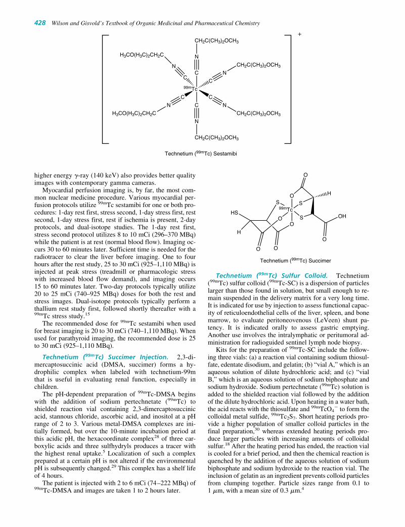
higher energy �-ray (140 keV) also provides better qualityimages with contemporary gamma cameras.
Myocardial perfusion imaging is, by far, the most com-mon nuclear medicine procedure. Various myocardial per-fusion protocols utilize 99mTc sestamibi for one or both pro-cedures: 1-day rest first, stress second, 1-day stress first, restsecond, 1-day stress first, rest if ischemia is present, 2-dayprotocols, and dual-isotope studies. The 1-day rest first,stress second protocol utilizes 8 to 10 mCi (296–370 MBq)while the patient is at rest (normal blood flow). Imaging oc-curs 30 to 60 minutes later. Sufficient time is needed for theradiotracer to clear the liver before imaging. One to fourhours after the rest study, 25 to 30 mCi (925–1,110 MBq) isinjected at peak stress (treadmill or pharmacologic stresswith increased blood flow demand), and imaging occurs15 to 60 minutes later. Two-day protocols typically utilize20 to 25 mCi (740–925 MBq) doses for both the rest andstress images. Dual-isotope protocols typically perform athallium rest study first, followed shortly thereafter with a99mTc stress study.15
The recommended dose for 99mTc sestamibi when usedfor breast imaging is 20 to 30 mCi (740–1,110 MBq). Whenused for parathyroid imaging, the recommended dose is 25to 30 mCi (925–1,110 MBq).
Technetium (99mTc) Succimer Injection. 2,3-di-mercaptosuccinic acid (DMSA, succimer) forms a hy-drophilic complex when labeled with technetium-99m that is useful in evaluating renal function, especially inchildren.
The pH-dependent preparation of 99mTc-DMSA beginswith the addition of sodium pertechnetate (99mTc) toshielded reaction vial containing 2,3-dimercaptosuccinicacid, stannous chloride, ascorbic acid, and inositol at a pHrange of 2 to 3. Various metal-DMSA complexes are ini-tially formed, but over the 10-minute incubation period atthis acidic pH, the hexacoordinate complex28 of three car-boxylic acids and three sulfhydryls produces a tracer withthe highest renal uptake.5 Localization of such a complexprepared at a certain pH is not altered if the environmentalpH is subsequently changed.29 This complex has a shelf lifeof 4 hours.
The patient is injected with 2 to 6 mCi (74–222 MBq) of99mTc-DMSA and images are taken 1 to 2 hours later.
Technetium (99mTc) Sulfur Colloid. Technetium(99mTc) sulfur colloid (99mTc-SC) is a dispersion of particleslarger than those found in solution, but small enough to re-main suspended in the delivery matrix for a very long time.It is indicated for use by injection to assess functional capac-ity of reticuloendothelial cells of the liver, spleen, and bonemarrow, to evaluate peritoneovenous (LeVeen) shunt pa-tency. It is indicated orally to assess gastric emptying.Another use involves the intralymphatic or peritumoral ad-ministration for radioguided sentinel lymph node biopsy.
Kits for the preparation of 99mTc-SC include the follow-ing three vials: (a) a reaction vial containing sodium thiosul-fate, edentate disodium, and gelatin; (b) “vial A,” which is anaqueous solution of dilute hydrochloric acid; and (c) “vialB,” which is an aqueous solution of sodium biphosphate andsodium hydroxide. Sodium pertechnetate (99mTc) solution isadded to the shielded reaction vial followed by the additionof the dilute hydrochloric acid. Upon heating in a water bath,the acid reacts with the thiosulfate and 99mTcO4
� to form thecolloidal metal sulfide, 99mTc2S7. Short heating periods pro-vide a higher population of smaller colloid particles in thefinal preparation,30 whereas extended heating periods pro-duce larger particles with increasing amounts of colloidalsulfur.18 After the heating period has ended, the reaction vialis cooled for a brief period, and then the chemical reaction isquenched by the addition of the aqueous solution of sodiumbiphosphate and sodium hydroxide to the reaction vial. Theinclusion of gelatin as an ingredient prevents colloid particlesfrom clumping together. Particle sizes range from 0.1 to 1 �m, with a mean size of 0.3 �m.4
Technetium (99mTc) Succimer
99mTc
O
OS
O
S
S
O
OH
O
H
HS
O
H
O
428 Wilson and Gisvold’s Textbook of Organic Medicinal and Pharmaceutical Chemistry
Technetium (99mTc) Sestamibi
99mTc
C
C
C
C
C
C
N
N
N
N
N
N
CH2C(CH3)2OCH3
CH2C(CH3)2OCH3
CH2C(CH3)2OCH3
CH2C(CH3)2OCH3
H3CO(H3C)2CH2C
H3CO(H3C)2CH2C
+
0413-0442_17865_Ch11.qxd 12/4/09 5:34 AM Page 428

After intravenous injection of 1 to 12 mCi (37–444 MBq)of 99mTc-SC, the tracer is rapidly cleared from the blood bythe reticuloendothelial cells of the liver, spleen, and bonemarrow. Uptake depends on the relative blood perfusion rateand the functional capacity of the cells. In the normal pa-tient, 85% of the radiocolloid is phagocytized by Kupffercells in the liver, 7.5% by the spleen, and the remainder bythe bone marrow, lungs, and kidneys. Liver-spleen imagescan be acquired within 20 minutes of administration. Bonemarrow imaging studies are performed 1 hour after injectionof 10 mCi (370 MBq) of 99mTc-SC. Normal bone marrowwill take up the radiocolloid, but diseased bone marrow ap-pears as “cold” defects in patients with tumor deposits inthe marrow.
Technetium (99mTc) sulfur colloid use for GI studies in-cludes gastroesophageal reflux (GER) and gastric emptyingof solid food. Gastroesophageal reflux imaging is performedafter having the patient swallow acidified orange juicemixed with 1 mCi (37 MBq) of 99mTc-SC. Normal patientsdo not demonstrate reflux. Gastric emptying imaging is per-formed after the patient swallows solid food (i.e., scrambledeggs or pancakes) radiolabeled with 1 mCi (37 MBq) of99mTc-SC. In general, the normal gastric emptying half-timeis less than 90 minutes for solid food.15 Imaging beginsshortly after administration and monitors the migration ofthe colloid particles.
Identification of sentinel lymph nodes for subsequentbiopsy in breast cancer31 and melanoma32 patients is accom-plished by the administration of several injections totaling0.5 to 1.5 mCi (19–56 MBq) of filtered (0.22-�m filter) orunfiltered 99mTc-SC. Smaller colloid particles may translo-cate more readily into the lymphatic system, whereas largerparticles remain at the injection site.15,30 Imaging beginsshortly after administration and monitors the migration ofthe colloid particles.
Technetium (99mTc) Tetrofosmin (Myoview). Techne-tium (99mTc) tetrofosmin is a lipophilic cation complex33 clin-ically indicated as a myocardial perfusion agent for detectingregions of reversible myocardial ischemia in the presence orabsence of infracted myocardium, identifying changes in per-fusion induced by pharmacologic stress in patients with coro-nary artery disease, and assessing left ventricular dysfunction.
Preparation of technetium (99mTc) tetrofosmin beginswith the addition of sodium pertechnetate (99mTc) to shieldedreaction vial containing tetrofosmin [6,9-bis(2-ethoxyethyl)-3,12-dioxa-6,9-diphosphatetradecane], stannous chloride,disodium sulphosalicylate, sodium D-gluconate, and sodiumhydrogen carbonate. Initially, the reduced technetium-99mforms a weak complex with gluconate which undergoes aligand exchange with tetrofosmin during a 15-minute incu-bation period at room temperature. The recommended shelflife is 8 hours.
Technetium (99mTc) tetrofosmin accumulates in the myo-cardium by facilitated diffusion from the Na�/H� an-tiporter.27 There is essentially no “redistribution,” or move-ment of the tracer out of the myocardium back into thebloodstream. Heart:liver ratios are somewhat higher for tech-netium (99mTc) tetrofosmin as compared with technetium(99mTc) sestamibi and allows for a shorter interval betweeninjection and imaging for technetium (99mTc) tetrofosmin.34
Various myocardial perfusion protocols utilize tech-netium (99mTc) tetrofosmin for 1-day, 2-day, and dual iso-
tope studies. One day protocols should utilize 5 to 12 mCi(185–444 MBq) for the first dose (rest or stress). Imagingcan commence as early as 15 minutes later. One to fourhours after the first study, 15 to 33 mCi (555–1,221 MBq) isinjected for the second study. Two-day protocols typicallyutilize 30 mCi (1,110 MBq) doses for both the rest andstress images. Dual-isotope protocols typically perform athallium rest study first, followed shortly thereafter with atechnetium-99m stress study.15
Thallium Radiochemistry
The clinically useful radioisotope of thallium is thallium-201.This isotope is produced in a cyclotron by proton bom-bardment of a thallium metal target according to a203Tl(p,3n)201Pb→201Tl sequence. Lead-201 (t1/2 � 9.4 hours)decays by EC to thallium-201. Thallium-201 (t1/2 � 73 hours)decays by EC to stable mercury-201, with principal �-rayemissions of 135 keV (3%) and 167 keV (10%), and mercury-201 daughter x-rays from 69 to 80 keV (94.5%), all of whichcan be used for organ imaging.
The irradiated thallium target is dissolved in nitricacid, and the Pb-201 is separated from the thallium-203by sequential ion exchange column chromatography withan EDTA containing mobile phase. Unbound lead-201 isallowed to decay for approximately 24 hours and passedthrough another ion exchanged column. Resin-boundthallium-201 (III) is eluted with hydrazine sulfate to pro-vide thallium-201 (I). The elute containing thallium-201ions are converted to the chloride salt form. The pH is ad-justed to 4.5 to 7.0 with sodium hydroxide, made isotonicwith sodium chloride containing benzyl alcohol as a pre-servative, and sterilized by filtration.4,35
THALLIUM RADIOPHARMACEUTICALS
Thallous (201Tl) Chloride. The most common clinicaluses of this radiotracer are for the evaluation of myocardialperfusion and myocardial viability. It is also indicated forthe localization of parathyroid hyperactivity. It is commer-cially available as a ready-to-use intravenous solution.
Thallous (201Tl) chloride accumulates in viable myocar-dial tissue by Na�/K� ATPase as well as Na�/K�/2Cl�
symporter27 much like potassium ions and is a dynamic ex-change between the myocardial cell and the blood. Uptakeis dependent on blood flow rates; at very high flow rates, ex-traction efficiency decreases. Thallous (201Tl) chloride doesnot concentrate in scarred or infarcted myocardium.
99mTc
O
O
P
P
P
P
Technetium (99mTc) Tetrofosmin
OCH2CH3
OCH2CH3H3CH2CO
H3CH2CO
H3CH2CO
H3CH2COOCH2CH3
OCH2CH3
+
Chapter 11 Agents for Diagnostic Imaging 429
0413-0442_17865_Ch11.qxd 12/4/09 5:34 AM Page 429

These concepts are the basis for the stress/redistributionimaging strategy employed for the detection of coronaryartery disease. To do this, the patient submits to eitherphysiological “stress” (treadmill exercise) or pharmaco-logical “stress” with an intravenous infusion of a vasodila-tor depending upon the patient’s physical condition. Atmaximum stress, the patient is injected with 3 to 4 mCi(111–148 MBq) of thallous (201Tl) chloride. The “stresstest” accentuates the myocardial perfusion abnormality be-cause areas of significant arterial narrowing cannot re-spond to the increased blood flow demands of the stressas well as normal arteries. Images of the heart are ob-tained immediately after stress. Damaged or ischemicmyocardium show less thallous (201Tl) chloride uptakethan surrounding normal heart muscle. Three hours afterstress, the resting patient may be imaged again or will beinjected with another 1.0 to 1.5 mCi (37–56 MBq) of thal-lous (201Tl) chloride prior to imaging to provide informa-tion under normal (rest) conditions. Ischemic tissues will“fill in” during rest images demonstrating normal uptake,whereas infarcted tissue continues to demonstrate reduceduptake.
Dual isotope perfusion studies utilize thallous (201Tl)chloride to first acquire rest images. Later, a technetium-99m perfusion agent is used to acquire the stress im-ages.15,36
Xenon Radiochemistry
The clinical radioisotope of xenon for organ imaging isxenon-133, a noble gas used in its elemental form. Xenon-133 is produced in a nuclear reactor as a byproduct of ura-nium fission by the nuclear reaction 235U(n, fission)133Xe.Xenon-133 (t1/2 � 5.3 days) decays by �� emission to ce-sium-133, with �-ray emission of 81 keV (37%). Gases usedin lung ventilation studies must be chemically and physio-logically inert at the concentrations used for imaging stud-ies. Xenon-133 is chemically inert and insoluble in water,which makes it insoluble in body fluids. Unfavorable phys-ical characteristics of xenon-133 include poor image qualitybecause of the low tissue penetration of the low-energy �-ray, increased patient dose due to �� emission, and the low�-ray emission yield (36 �-rays/100 disintegrations).
XENON RADIOPHARMACEUTICALS
Xenon (133Xe). Radioactive xenon (133Xe) gas is sup-plied at standard pressure and room temperature in aseptum-sealed glass vial in doses of 10 to 20 mCi(370–740 MBq) in either stable xenon carrier mixed with at-mospheric air or 5% xenon carrier mixed with 95% carbondioxide suitable for inhalation by the patient for diagnosticevaluation of pulmonary function and imaging of the lungs.The shelf life of the vials is 14 days because of the morerapid decay of xenon (133Xe) gas when compared with thesubsequent build-up of slower decaying impurities.
The general procedure involves mixing the vial contentsof xenon-133 gas in air or oxygen in a closed-circuitspirometer system that delivers the radioactive gas, allow-ing the patient to breath the gas mixture. The inhalationstudy consists of wash-in, equilibrium and washout phases,with the patient sitting or supine. The wash-in phase is thefirst breath of the xenon-133 bolus, the equilibrium phaseconsists of the patient breathing the mixture for 5 minutes,
and for the washout phase, the patient exhales xenon-133gas into an activated charcoal trap. Images are obtainedcontinuously for approximately 10 minutes with thegamma (�-ray) camera. In the wash-in study, the patientinspires the bolus and holds their breath for several sec-onds. In the equilibrium study, there is an initial distribu-tion of the radioactive gas throughout the lungs. In thewashout phase, the xenon-133 gas clears readily from thelungs. In the abnormal study, the initial flow into the lungsof xenon-133 gas is delayed, and the outflow is also de-layed. These abnormal lung ventilation findings add sig-nificant information in the evaluation of pulmonary em-bolism (blood clots in the lungs) when combined with theresults of a lung blood perfusion study with Tc-99mmacroaggregated albumin.
CONTRAST AGENTS
Radiography and ComputedTomography
A photographic film containing a radiographic image isproperly called a radiograph, although it is commonly re-ferred to as an x-ray or a film. The relative difference be-tween the light and dark areas on a radiographic image re-flects what is called radiographic contrast. Traditionalradiographic images of the body, such as skeletal, abdomi-nal, and chest x-rays, have four radiographic densities: air,fat, water (organs, muscle, soft tissue), and bone/metal.Organs and tissues are made visible according to how wellthey attenuate x-rays. The attenuation of x-rays by tissues isa complex process that depends on many factors, including,but not limited to, the energy of the x-ray beam and the den-sity of the tissue. Bone has an average density of about 1.16g/cm3, which accounts for its ability to absorb most of theradiation it encounters.
Whereas traditional radiological “film” studies have beenused since 1895 and continue to be a mainstay of diagnosticmedical imaging, they have their limitations. Many organsand tissues of the body are not easily discriminated fromeach other on traditional radiographic images. For example,the liver, spleen, kidneys, intestines, bladder, and abdominalmusculature all have very similar radiographic densities andare difficult, if not impossible, to distinguish from eachother.
To produce diagnostic x-rays, a very high voltage(20,000–150,000 volts) is applied to a glass vacuum tubethat contains a cathode and a rotating anode (Fig. 11.6). Thecathode is a filament that is heated to a very high tempera-ture, which provides a copious source of electrons. Theseelectrons are accelerated toward the positively chargedanode. When the accelerated electrons strike the anode(called the target), x-rays are produced. The distribution ofx-rays is as a continuous spectrum. Lower-energy x-rays areabsorbed by an aluminum filter, while higher-energy x-raystravel through the patient, exposing the x-ray film, and pro-ducing a traditional radiograph. Digital radiography is theprocess of producing a digital radiographic image where aspecialized phosphor plate is used in place of traditionalfilm. Such digital images can be readily viewed on a moni-tor, stored, and transferred much like any other type of dig-ital information.
430 Wilson and Gisvold’s Textbook of Organic Medicinal and Pharmaceutical Chemistry
0413-0442_17865_Ch11.qxd 12/4/09 5:34 AM Page 430

An invaluable modification of the x-ray system is fluo-roscopy (Fig. 11.7). This modality allows one to visualizeorgans in motion, position the patient for spot film expo-sures, instill contrast media into hollow cavities, and insertcatheters into arteries.
CT (sometimes called CAT, Fig. 11.8) uses ordinary x-ray energies for imaging and applies complex mathematicalreconstructions to produce multiple images of the body inthe axial and other planes. This technique typically providesmore detailed information about the anatomical region of in-terest because the images obtained are numerous thin slicesof the imaged area. With this process, the reconstructed im-ages increase the visibility of small differences in the radio-graphic densities between tissues to a far greater extent thantraditional radiographic film.
RADIOLOGICAL CONTRAST AGENTS
From the earliest days of radiology, considerable effort hasbeen devoted to developing compounds that if swallowed orinjected would increase the radiographic contrast betweenvarious tissues and organs. Injection of air or other gasesinto a GI tube in the esophagus, stomach, or duodenum orinto a rectal tube in the colon provides increased radio-graphic contrast for evaluating the gut; however, the infor-mation obtained by this technique is limited, and moreopaque substances have been developed.
Any agent or compound administered to a patient to im-prove the visualization of an organ or tissue is called a con-trast agent. Contrast agents can be classified as negative orpositive. Air and other gases are negative contrast agents be-cause they render a structure, such as the gut, more translu-cent. An agent that increases the radiographic opacity of anorgan or tissue is a positive contrast agent. Most contrast
Chapter 11 Agents for Diagnostic Imaging 431
Tube
Filament
Rotating Anode
Focus
IntensifyingScreens
Electrons
Filter (3mm Al)
Lead Collimator
X-rays
Grid
X-ray Film
Figure 11.6 Schematic diagram of an x-ray tube forproduction of x-ray films.
video camera
aperaturelenses
image intensifier
anti-scatter grid
filters
patient
table
x-ray tube
collimation
Figure 11.7 Schematic diagram of a fluoroscopic x-raysystem for real-time imaging during medical procedures.(Reprinted with permission from Bushberg, J. T., et al.:The Essential Physics of Medical Imaging, 2nd ed.Philadelphia: Lippincott Williams and Wilkins, 2002.)
Original ImageReconstruction
Computer
Radiation Detectors
X-ray Tube
Figure 11.8 Schematic diagram of a computerizedaxial tomography (CAT) system for producing cross-sectional images of the body.
0413-0442_17865_Ch11.qxd 12/4/09 5:34 AM Page 431

agents used in diagnostic radiology are positive contrastagents.
An ideal radiographic contrast agent should have the fol-lowing properties: (a) readily available, (b) inexpensive, (c)excellent x-ray absorption characteristics at the x-ray ener-gies used in diagnostic radiology, (d) minimal toxicity, (e)patient acceptance, (f) chemical stability, (g) high-water sol-ubility with low viscosity and no significant osmotic effects,and (h) the ability to be administered for selective tissue up-take and excretion.
No compound has all of these characteristics. Bariumsulfate and various iodine compounds, however, produceexcellent radiological contrast with low patient toxicity andrelatively low cost. The use of barium and iodine com-pounds as radiological contrast agents is based on theirradiographic appearance, distribution, and elimination fromthe body. Contrast media are used in very large quantitiesand are usually administered over a brief period of time.
Barium sulfate is a nearly ideal contrast agent for oraland rectal studies of the GI tract. It produces a metal-likedensity on radiological studies, is inexpensive, and whenproperly utilized, has minimal patient morbidity and mortal-ity. While various water-soluble barium compounds arequite toxic, barium sulfate is an insoluble white power thatis a colloidal suspension in water.
Most intravenous contrast agents used to opacify bloodvessels and increase contrast in solid organs, such as theliver, are water-soluble organic iodides. Iodine absorbs x-rays effectively at many energy levels and produces opaci-ties similar to bone density during radiographic studies.While iodine’s radiographic density is somewhat less thanthat of barium sulfate, it is still very useful for radiographiccontrast.
Early water-soluble contrast agents consisted of triiodi-nated benzoic acid salts. In solution, these salts dissociateinto two particles: (a) a triiodinated anion and (b) a cation,usually sodium or methylglucamine (meglumine). Thesecompounds, known as high-osmolar contrast medium(HOCM), have in effect three iodine atoms for every twoions in solution, a 3:2 ratio. They are often called conven-tional, ionic ratio 1.5 contrast agents, or triiodinatedmonomers and are represented by diatrizoate and iothala-mate.37 Various fixed ratios of sodium diatrizoate and meg-lumine diatrizoate are also available in proprietary products.
These water-soluble iodinated salts are administered infairly high volumes and concentrations to achieve satisfac-tory radiological contrast. It is not unusual to administer asmuch as 100 mL or more of a 30% to 70% solution intra-venously.38 The typical concentrations used for variousstudies have an osmolality of 5 times or more to that of nor-mal plasma. Administration of these HOCM agents can beassociated with osmotoxic effects. Initially, water shifts rap-idly from the interstitial and cellular spaces into the plasmaafter injection of an HOCM. This is typically accompaniedby vasodilatation, local pain and warmth, a metallic taste inthe mouth, and flushing. Later, there is an osmotic diuresisas these agents are excreted by the kidneys.
Various nonionic water-soluble compounds with a higherratio of iodine content per osmotic particle have been devel-oped. These types of agents, known as low-osmolar contrastmedium or isotonic contrast media (LOCM or ICM), pro-duce far fewer osmotic effects. Various monomers such asiohexol, iopamidol, iopramide, ioversol, and ioxilan as well
as dimers such as iodipamide, iodixanol, and ioxaglate havebeen described.38
All of the water-soluble iodinated contrast media are clear,colorless, viscous liquids. Even though they are clear liquids,they are often called “dyes” when their administration is beingexplained to patients. Sodium salts are slightly less viscousthan the meglumine salts, which are typically less viscous thannonionic monomers, which are typically less viscous than non-ionic dimers. Viscosity is a function of temperature and can bereduced by warming the contrast agent to body temperatureprior to its administration.
Water-soluble iodinated contrast media have relativelysmall molecular sizes and low chemical reactivity with bodyfluids and tissues. Their pharmacodynamic characteristicsare similar to those of extracellular tracers. They have lowlipid solubility and distribute throughout extracellularspaces. They do not significantly penetrate into intracellularspaces. Special attention must be given to ensure that iodin-ated contrast agents are not administered intrathecally un-less they are indicated for such use. Inadvertent intrathecaladministration can cause serious outcomes such as convul-sions, cerebral hemorrhage, brain edema, and death.
Water-soluble iodinated contrast agents are cleared fromthe body by glomerular filtration. They are neither reab-sorbed nor secreted by the renal tubules. When renal func-tion is compromised, these contrast agents are eliminated inpart or totally through the liver and gut. This alternate elim-ination pathway occurs at a much slower rate than elimina-tion when compared with glomerular filtration in a healthyperson.
Water-soluble organic iodides are the largest group of ra-diological contrast agents. In addition to water-insolublebarium sulfate, there is an iodinated fatty acid derivative ofpoppy seed oil that is occasionally utilized in the clinic.Unsaturated fatty acids are monoiodicated or diiodinated be-fore conversion to an ester. These substances are susceptibleto light-induced decomposition, and the oily nature of theformulation excludes intravascular use.
Radiological contrast agents that are administered orallyor intravascularly in large amounts infrequently report ad-verse effects. Nevertheless, any radiological contrast mate-rial may produce an untoward patient reaction, even suddendeath.39–42
Events can occur when contrast material is aspirated orwhen it leaks from the GI tract. Hypertonic ionic water-soluble contrast agents are potentially dangerous if aspi-rated into the tracheobronchial tree. They are irritating andcan cause pulmonary edema. Contrast material that leaksout of the bowel into the abdomen, pelvis, or chest is po-tentially quite dangerous, especially barium sulfate.Barium sulfate is insoluble, and its particulate naturemeans it is poorly cleared from the mediastinum, peri-toneum, and retroperitoneum. Water-soluble agents, on theother hand, are rapidly absorbed from these areas almost asquickly as if they had been injected intravenously. In gen-eral, water-soluble agents that leak from the GI tract rarelycause significant problems beyond transient inflammation.
The intravenous or intra-arterial injection of iodinatedcontrast material can lead to a diverse assortment of contrastreactions, most of which are minor and easily treated. Minorreactions include injection pain, a feeling of general bodywarmth and discomfort, mild nausea and vomiting, a strongmetallic taste in the mouth, and mild urticaria (hives). Minor
432 Wilson and Gisvold’s Textbook of Organic Medicinal and Pharmaceutical Chemistry
0413-0442_17865_Ch11.qxd 12/4/09 5:34 AM Page 432

reactions dissipate within a few minutes with patient reas-surance and observation.
Intermediate or moderate reactions are those that requiresome form of therapy but are not life-threatening. These re-actions include difficulty breathing, severe hives, severenausea and vomiting, mild hypotension, wheezing, and otherrelated reactions. Treatment ranges from administration ofintravenous fluids to the use of intravenous diphenhy-dramine and/or corticosteroids. Epinephrine may be admin-istered, and atropine is used if there is a vasovagal reactionwith hypotension and bradycardia.
Severe reactions are those that are life-threatening. Theyinclude sudden cardiovascular collapse and death, as well assevere hypotension; severe shortness of breath, wheezing, orlaryngoedema; loss of consciousness; massive hives and an-gioneurotic edema; ventricular cardiac arrhythmias; angina;and myocardial infarction. Treatment depends on the pa-tient’s signs and symptoms and includes intravenous fluids,oxygen, various drugs (including epinephrine, diphenhy-dramine, and atropine), and possible cardiopulmonary re-suscitation (CPR).
Barium Sulfate. Commercial preparations of barium sul-fate differ in their density and ability to coat the bowel wall.These characteristics are determined by the particle size of thebarium suspension, its viscosity, and concentration of thecontrast agent. Flavoring agents, suspending agents, and otheradditives may be utilized in preparing various products.
Barium sulfate preparations are given orally or rectallyand are used to study the esophagus, stomach, duodenum,entire small bowel, colon, or the total bowel. Most patientsfind the taste of these flavored mixtures tolerable, but theydislike the heavy texture of the barium.
Concentrated barium suspensions ranging from 50% to210% weight/volume (wt/vol) are frequently used for tradi-tional radiographic imaging. Dilute suspensions rangingfrom 1.5% to 5% weight/volume (wt/vol) are typically usedfor CT studies.
The quantity and concentration of barium sulfate suspen-sion administered to the patient will depend on the selectedprocedure. In general, upper GI studies will administer thecontrast agent just prior to imaging, whereas lower GI stud-ies recommend intake several hours prior to the procedure.For total bowel opacification, the patient should consumecontrast agent the night preceding the examination, addi-tional agent a few hours prior to examination, and a thirddose approximately 15 minutes prior to examination.
Diatrizoate. Diatrizoate is a water-soluble, ionicmonomer. It is commercially available as the megluminesalt, sodium salt, or as fixed combinations of the two saltforms. These triiodobenzoic acid salts contain 47% organi-cally bound iodine.
Numerous products with various concentrations of one ortwo of the salt forms are available for various imaging pro-cedures. Indicated uses include angiography, venography,excretory urography, retrograde cystourethrography, retro-grade or ascending pyelography, operative, T-tube, or per-cutaneous transhepatic cholangiography, splenoportogra-phy, arthrography; discography, radiographic examinationof the GI tract when barium sulfate is contraindicated (i.e.,suspected perforation of the GI tract), adjunct contrast en-hancement in CT of the torso (dual contrast study), and hys-terosalpingography.
Chapter 11 Agents for Diagnostic Imaging 433
I
I
I
NH
(CH2)4
OHO
Iodipamide Meglumine
O
CH2NHCH3
OHH
HHO
OHH
OHH
CH2OH
I
I
I
NH
OHO
O
2
I
I
I
NH
O
NH
OHO
Diaztriazoate Meglumine
O
CH2NHCH3
OHH
HHO
OHH
OHH
CH2OH
Ethiodized Oil (Ethiodol). This product is a mixture ofiodinated fatty acid ethyl esters of poppy-seed oil, primarilyas ethyl monoiodostearate and ethyl diiodostearate. It con-tains 37% organically bound iodine, has considerably re-duced viscosity when compared with the triiodobenzoic acidderivatives, and is not miscible with plasma. Ethiodol is in-dicated for use as a radiopaque medium for hysterosalpin-gography and lymphography.
Iodipamide Meglumine. Iodipamide is a water-soluble,ionic dimer that is administered as the meglumine salt incombination with diatrizoate meglumine as the commercialproduct, Sinografin. Iodipamide has 50% bound iodine con-tent. Intravenous use was associated with significant adverseeffects, and the product is now indicated solely for use inhysterosalpingography.
0413-0442_17865_Ch11.qxd 12/4/09 5:34 AM Page 433

Iodixanol (Visipaque). Iodixanol is a viscous, isosmo-lar, nonionic, water-soluble dimer with 49% iodine content.It is formulated as an isotonic solution for intravascular in-jection and indicated for excretory urography, angiography,and CT procedures.
Iohexol (Omnipaque). Iohexol is a low-osmolar, non-ionic monomer with 46% iodine content. Iohexol is one ofthe few iodinated contrast agents indicated for intrathecaluse (myelography). Only selected concentrations of theproduct have this indication. Besides myelography, iohexolfinds widespread use in excretory urography, angiography,
and CT procedures. Specific concentrations may have addi-tional indications in adults and/or children for arthrography,hysterosalpingography, endoscopic retrograde pancreatog-raphy, cholangiopancreatography, herniography, voidingcystourethrography, oral pass-thru examination of the GItract or CT of the abdomen.
Iopamidol (Isovue). Iopamidol is a low-osmolar, non-ionic monomer with 49% organically bound iodine. It is in-dicated for use in angiography, excretory urography, andnumerous CT procedures.
434 Wilson and Gisvold’s Textbook of Organic Medicinal and Pharmaceutical Chemistry
I
I I
HN
O
HN
Iodixanol
OH
HO
OH
OHO
N
O
OH
I I
I
HN
OHN
OH
OH
OH
O
OH
N
O
I
I
I
HN
O
HN
Iopamidol
OH
O
HN
(S)(S)
OHHO
HO
O
OH
I
I
I
HN
O
HN
Iohexol
OH
OH
OH
O
HO
N
O
OH
HO
0413-0442_17865_Ch11.qxd 12/4/09 5:34 AM Page 434

Iopromide (Ultravist). Iopromide is a low-osmolar,nonionic monomer with 48% bound iodine content. It is in-dicated for use in angiography, excretory urography, andnumerous CT procedures.
Iothalamate Meglumine (Conray). Iothalamate meg-lumine is a high-osmolar, ionic monomer with 47% boundiodine content. Conray is indicated for use in angiography,
Chapter 11 Agents for Diagnostic Imaging 435
I
I
I
N
O
HN
Ioversol
HN
HO
OH
OH
O OH
OH
O
HO
I
I
I
NH
O
HN
Iopromide
N
O
OH
OH
O OH
OH
O
I
I
I
NH
O
O
HN
OHO
Iothalamate Meglumine
CH2NHCH3
OHH
HHO
OHH
OHH
CH2OH
excretory urography, arthrography, cholangiography, endo-scopic retrograde cholangiopancreatography, and variousCT procedures.
Ioversol (Optiray). Ioversol is a low-osmolar, nonionicmonomer with 47% bound iodine content. It is indicated forangiography, excretory urography, ventriculography, andvarious CT procedures.
0413-0442_17865_Ch11.qxd 12/4/09 5:34 AM Page 435

Ioxaglate (Hexabrix). Ioxaglate is a high-osmolal,ionic dimer with 32% bound iodine content. It is commer-cially formulated as a mixture of its meglumine and sodiumsalts. Ioxaglate is indicated for angiography, excretory urog-raphy, and various CT procedures.
Ioxilan (Oxilan). Ioxilan is a low-osmolar, nonionicmonomer with 48% bound iodine content. It is indicatedfor angiography, excretory urography, and various CTprocedures.
Magnetic Resonance Imaging
MRI is a unique method of medical imaging. The strengthof MRI as an imaging modality is its ability to detect smallchanges within soft tissues by using nonionizing radiation.The exquisite soft tissue images produced by MRI are anal-ogous to the cross-sectional images of the body produced byCT with the benefit of MRI images being performed in anydesired plane (coronal, axial, sagittal, oblique, etc.).
When a patient is placed in a large, strong, uniform mag-netic field (Fig. 11.9), a population of hydrogen nuclei(protons) in all hydrogen-containing molecules align them-selves with the magnetic field. Once aligned in the mag-netic field, these nuclei will precess (spin like a gyroscope)with a characteristic spin frequency related to the magneticfield strength. The tesla (T) is the official unit of magneticfield strength, and MRI is normally performed between 0.5to 1.5 T.
By directing low-energy, electromagnetic radiation to adefined region of the patient’s body, the magnetized, precess-
ing hydrogen nuclei can selectively absorb this energy to be-come excited. The excitation is accomplished by a complexseries of radiofrequency pulses at the same frequency as theprecessing hydrogen nuclei. Each burst of energy displacesthe magnetic orientation of precessing nuclei. When the exci-tation energy field is removed, the displaced nuclei will losethe acquired energy, returning to magnetic equilibrium. Thesedecaying (i.e., relaxing) nuclei emit a weak, but detectable,radio wave (i.e., signal), whose strength and manner of relax-ation can be used to generate diagnostic medical images.
The timing of data acquisition from the relaxing nuclei isjust as important as the number of nuclei that are excited inthe observed tissues. Early receiver detection of emitted pro-ton signals following discontinuance of the radiofrequencypulse is called a T1-weighted sequence (longitudinal orspin-lattice relaxation). T1 images have good resolution andare useful for gaining anatomic information. Late receiverdetection of emitted signals is called a T2-weighted se-quence (transverse or spin–spin relaxation). T2 images areuseful in identifying pathologies because T2 images havebetter contrast between water (high proton density) and softtissue (relatively less proton density).43–45
MAGNETIC RESONANCE CONTRAST AGENTS
When a molecule is placed in a magnetic field, it displays acertain susceptibility to becoming magnetized and an abilityto alter the magnetic field of the immediate environment intowhich it has been placed. The ability of a substance to pro-duce additional magnetism within a magnetic field is knownas its susceptibility. A vacuum has a susceptibility defined as
436 Wilson and Gisvold’s Textbook of Organic Medicinal and Pharmaceutical Chemistry
I
I I
O
O
HN
Ioxaglate
HO
OH
NH
I I
I
N
OHN
NH
O O O
I
I
I
N
O
HN
Ioxilan
OH
HNO OH
OH
O
OH
HO
0413-0442_17865_Ch11.qxd 12/4/09 5:34 AM Page 436

zero in comparison with water. Many nonmetallic materialsare diamagnetic; they have negative susceptibility and induceweaker magnetic fields in the local environment. Such sub-stances generally have fully paired electrons. Materials withunpaired orbiting electrons are said to be paramagnetic, be-cause when placed in an applied magnetic field, theystrengthen the magnetic fields within their vicinity. The mag-nitude of paramagnetic influence is proportional to thestrength of the applied field. A substance with many unpairedelectrons will be superparamagnetic, because it produces astronger supplementary magnetic field. Ferromagnetic mate-rials have very large numbers of unpaired electrons and pro-duce a large, supplementary magnetization to the applied fieldthat remains once the magnetic field has been removed.
Gadolinium-containing contrast agents rely on unpairedelectrons to produce magnetic changes in nearby water mol-ecules. These paramagnetic agents predominantly decreasetissue T1 relaxation time, which increases tissue intensity onT1-weighted images. Such agents are known as positivecontrast agents because they brighten tissues associated withthe nearby paramagnetic substance.
Many contrast agents used for MRI are extracellularparamagnetic agents. The primary paramagnetic metal ionused is gadolinium, a rare earth element. Gadolinium ions(Gd�3) form when prepared in 0.05 M hydrochloric acidand are extremely effective for enhancing water proton re-laxation rates. The ion itself, however, is toxic for humanuse. The ion quickly binds to ferric (Fe�3) protein-bindingsites, most notably plasma transferrin, and has a long bio-logical half-life of several weeks. In addition, the ion read-ily forms insoluble compounds by interacting with endoge-nous ions including phosphate, carbonate, and hydroxide.46
Consequently, the development of gadolinium MRI con-trast agents has mainly focused on chelated compounds thatclear rapidly from the body through the kidneys and exhibitminimal toxicity. These include gadobenate dimeglumine(MultiHance), gadodiamide (Omniscan), gadopentetatedimeglumine (Magnevist), gadoteridol (ProHance), andgadoversetamide (Optimark).47
All intravenous gadolinium complexes are hypertonicwhen compared with plasma. The most common side effects
are headache, nausea, dizziness, taste perversions, and va-sodilation. A rare adverse event, nephrogenic systemic fibro-sis, is more likely to occur in patients with severe acute orchronic renal insufficiency (�30 mL/min). Postmarketing re-ports identified such an event following single and multipleadministrations of gadolinium-based contrast agents. Thesereports have not always identified a specific agent, and repet-itive administration of the same or different agents carriessimilar risk if the time interval between the administrationsprecludes clearance of the contrast agent from the body.48,49
Manganese (Mn2�) is strongly paramagnetic and can beused to form complexes used as MRI contrast agents. Atthis time, the only parenteral manganese compound used inhumans is mangafodipir trisodium. This compound is de-livered to tissues with active metabolism, such as the pan-creas, renal cortex, GI tract, heart, and adrenal glands. Theeffect of mangafodipir trisodium is to decrease T1 relax-ation time, which enhances signal intensity on T1-weightedimages.
Superparamagnetic iron oxide colloids (SPIO) contain acrystalline core composed of iron oxide complexes. Thecore may be coated with dextran, siloxanes, or another largepolysaccharide. The particles vary in size from 1 and 10�m, and their biological characteristics can be altered bychanging their coating. Large particles accumulate in theliver and spleen, whereas smaller particles remain in the cir-culation longer and tend to aggregate in lymph nodes.Ferumoxides and ferumoxsil are examples of such agents.
Because the main MRI effect of the iron oxide particlesis on T2-weighted images, they decrease the signal inten-sity of the tissue in which they accumulate. Such ironoxide agents are known as negative contrast agents, be-cause they darken tissues associated with the accumulationof ferromagnetic particles. Enhancement of T1-weightedimages is less dramatic, but can be obtained. SPIO contrastagents provide enhancement of the liver and/or spleenwhen injected intravenously or the bowel when adminis-tered orally.50
Ferumoxides (Feridex). Ferumoxides is a black to red-dish brown aqueous colloid of SPIO particles associated
Chapter 11 Agents for Diagnostic Imaging 437
Super Conducting Magnet Coil
Computer
RF Pulses RF SignalsResonatingTissue
RR CoilGradient Coils
Aquisition Control Image Reconstruction Storage RetrievalAnalysis and
Display
RadiofrequencyTransmitter
GradientPower Supply
DataProtocols ImageFourierTransform
RadiofrequencyReceiver
Super Conducting Magnet Coil
Disk
Figure 11.9 Schematic diagram of anMRI system demonstrating radiofrequencypulses and the strong magnetic field.
0413-0442_17865_Ch11.qxd 12/4/09 5:34 AM Page 437

with dextran. It is administered intravenously and taken upby cells of the reticuloendothelial system (RES). It is indi-cated to enhance T2-weighted images used to assess lesionsof the liver. Hepatic tissues with decreased RES function re-tain their usual signal intensity, so the contrast between ab-normal and normal tissue is enhanced.
Ferumoxsil (GastroMARK). Ferumoxsil is a turbid,slightly viscous, dark brown to orange-brown aqueous sus-pension of silicone-coated, SPIO for oral administration.The suspension distributes to the stomach and small intes-tine within 30 to 45 minutes and passes distally to the largeintestine within 4 to 7 hours after ingestion. It is indicated toenhance the delineation of the bowel and to distinguish itfrom organs and tissues that are adjacent to the upper re-gions of the GI tract. Its usefulness in the distal GI regionsis limited by transit time and dilution. Both T1- and T2-weighted images may be enhanced with ferumoxsil.
Gadobenate Dimeglumine (MultiHance). Gadobenatedimeglumine is an ionic, clear, colorless, gadolinium-containing solution. This salt solution has 6.9 times the osmo-lality of plasma. It is indicated to enhance the visualization ofcentral nervous system (CNS) lesions associated with an ab-normal blood-brain barrier or abnormal vascularity of thebrain, spine, or associated tissues when using T1-weightedsequences.
Gadodiamide (Omniscan). Gadodiamide is a clear,colorless to slightly yellow, gadolinium-based contrastagent indicated to visualize lesions with abnormal vascu-larity in the CNS and body. It is a nonionic gadoliniumcomplex administered by intravenous injection with an os-molality of 2.8 times that of plasma. Gadodiamide de-creases both the T1 and T2 relaxation times in tissueswhere it is distributed. In clinical MRI, the effect is prima-
rily on the T1 relaxation time resulting in an increase inT1-weighted sequence signal intensity.
Gadopentetate Dimeglumine (Magnevist).Gadopentetate dimeglumine is an ionic, clear, colorless toslightly yellow, gadolinium-based contrast agent indicatedto visualize lesions with abnormal vascularity in the CNSand body. This salt solution has 6.9 times the osmolality ofplasma. Like other gadolinium-based contrast agents, theprimary clinical effect is on T1 relaxation time.
Gadoteridol (ProHance). Gadoteridol is a nonionicclear, colorless to slightly yellow, gadolinium-based contrastagent indicated to visualize lesions with abnormal vascular-ity in the CNS, head, and neck. It is a hypertonic solutionwith an osmolality of 2.2 times that of plasma. Clinical en-hancement of images occurs primarily when using T1-weighted sequences.
Gadodiamide
N
N N
Gd
O O
O
O
O O
NH
O
HN
O
OH
H
438 Wilson and Gisvold’s Textbook of Organic Medicinal and Pharmaceutical Chemistry
Gadobenate Dimeglumine
N
N N
Gd
O O
O
O
O O
OH
O
HO
O
OH
H
O
CH2NHCH3
OHH
HHO
OHH
OHH
CH2OH
2
0413-0442_17865_Ch11.qxd 12/4/09 5:34 AM Page 438

Gadoteridol
NN
N N
Gd
O O
O
O
O O
O
Gadoversetamide (OptiMARK). Gadoversetamide anonionic clear, colorless to slightly yellow, gadolinium-based contrast agent indicated to visualize lesions withabnormal vascularity of the CNS or liver. It is a hypertonicsolution with an osmolality of 3.9 times that of plasma. Theprimary clinical effect is on T1 relaxation time.
Mangafodipir Trisodium (Teslascan). Mangafodipirtrisodium is a clear bright to dark yellow chelate of para-magnetic manganese for intravenous administration.Manganese is distributed to the liver and pancreas and short-ens the T1-weighted relaxation time. It is indicated to detectmetastatic lesions of the liver, focal pancreatic lesions, andhepatocellular carcinoma.
Chapter 11 Agents for Diagnostic Imaging 439
Gadopentetate dimeglumine
N
N N
Gd
O O
O
O
O O
OH
O
HO
O
CH2NHCH3
OHH
HHO
OHH
OHH
CH2OH
2
Gadoversetamide
N
N N
Gd
O O
O
O
O O
NH
O
HN
O
O
O
OH
H
Mn
O
OO
N
O
Mangafodipir
O
O NN
O
POH
HO
O
OP
O
HOOH
N
0413-0442_17865_Ch11.qxd 12/4/09 5:34 AM Page 439
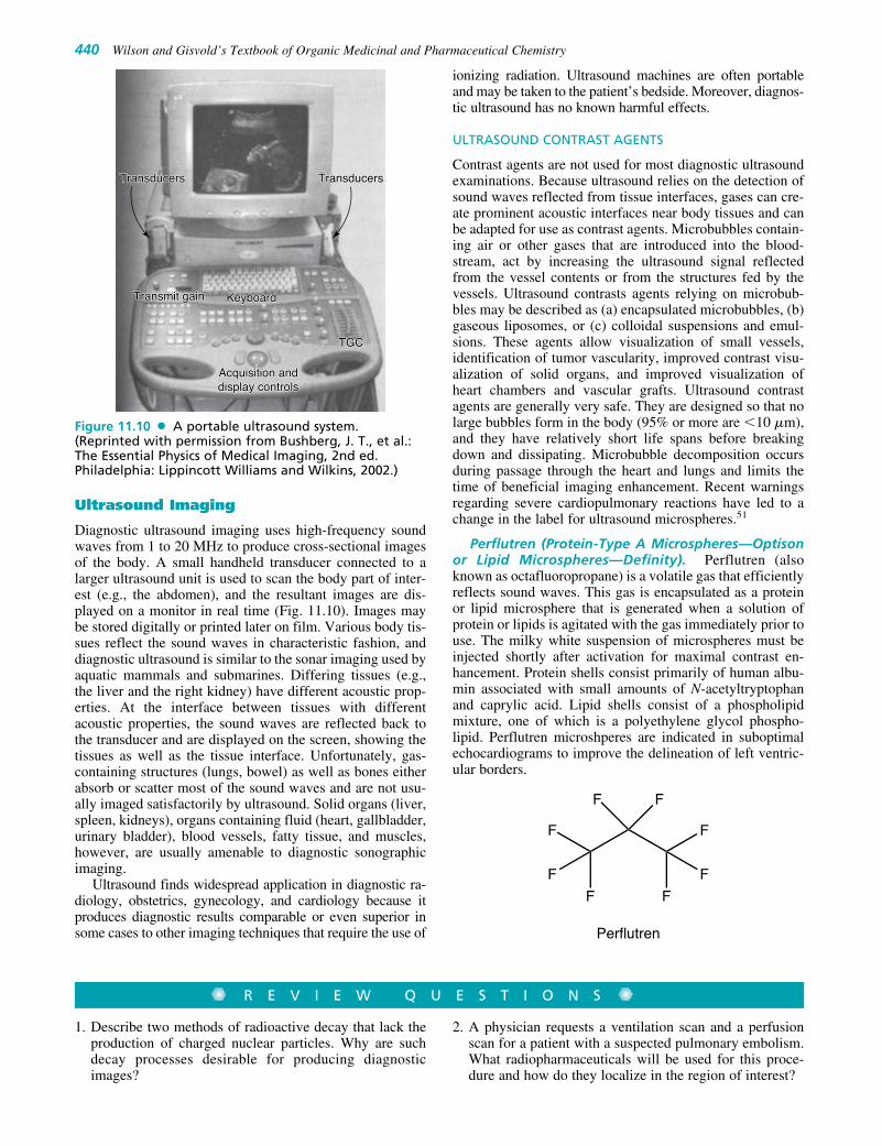
Ultrasound Imaging
Diagnostic ultrasound imaging uses high-frequency soundwaves from 1 to 20 MHz to produce cross-sectional imagesof the body. A small handheld transducer connected to alarger ultrasound unit is used to scan the body part of inter-est (e.g., the abdomen), and the resultant images are dis-played on a monitor in real time (Fig. 11.10). Images maybe stored digitally or printed later on film. Various body tis-sues reflect the sound waves in characteristic fashion, anddiagnostic ultrasound is similar to the sonar imaging used byaquatic mammals and submarines. Differing tissues (e.g.,the liver and the right kidney) have different acoustic prop-erties. At the interface between tissues with differentacoustic properties, the sound waves are reflected back tothe transducer and are displayed on the screen, showing thetissues as well as the tissue interface. Unfortunately, gas-containing structures (lungs, bowel) as well as bones eitherabsorb or scatter most of the sound waves and are not usu-ally imaged satisfactorily by ultrasound. Solid organs (liver,spleen, kidneys), organs containing fluid (heart, gallbladder,urinary bladder), blood vessels, fatty tissue, and muscles,however, are usually amenable to diagnostic sonographicimaging.
Ultrasound finds widespread application in diagnostic ra-diology, obstetrics, gynecology, and cardiology because itproduces diagnostic results comparable or even superior insome cases to other imaging techniques that require the use of
ionizing radiation. Ultrasound machines are often portableand may be taken to the patient’s bedside. Moreover, diagnos-tic ultrasound has no known harmful effects.
ULTRASOUND CONTRAST AGENTS
Contrast agents are not used for most diagnostic ultrasoundexaminations. Because ultrasound relies on the detection ofsound waves reflected from tissue interfaces, gases can cre-ate prominent acoustic interfaces near body tissues and canbe adapted for use as contrast agents. Microbubbles contain-ing air or other gases that are introduced into the blood-stream, act by increasing the ultrasound signal reflectedfrom the vessel contents or from the structures fed by thevessels. Ultrasound contrasts agents relying on microbub-bles may be described as (a) encapsulated microbubbles, (b)gaseous liposomes, or (c) colloidal suspensions and emul-sions. These agents allow visualization of small vessels,identification of tumor vascularity, improved contrast visu-alization of solid organs, and improved visualization ofheart chambers and vascular grafts. Ultrasound contrastagents are generally very safe. They are designed so that nolarge bubbles form in the body (95% or more are �10 �m),and they have relatively short life spans before breakingdown and dissipating. Microbubble decomposition occursduring passage through the heart and lungs and limits thetime of beneficial imaging enhancement. Recent warningsregarding severe cardiopulmonary reactions have led to achange in the label for ultrasound microspheres.51
Perflutren (Protein-Type A Microspheres—Optisonor Lipid Microspheres—Definity). Perflutren (alsoknown as octafluoropropane) is a volatile gas that efficientlyreflects sound waves. This gas is encapsulated as a proteinor lipid microsphere that is generated when a solution ofprotein or lipids is agitated with the gas immediately prior touse. The milky white suspension of microspheres must beinjected shortly after activation for maximal contrast en-hancement. Protein shells consist primarily of human albu-min associated with small amounts of N-acetyltryptophanand caprylic acid. Lipid shells consist of a phospholipidmixture, one of which is a polyethylene glycol phospho-lipid. Perflutren microshperes are indicated in suboptimalechocardiograms to improve the delineation of left ventric-ular borders.
Perflutren
FF
F
F
F
F
F
F
440 Wilson and Gisvold’s Textbook of Organic Medicinal and Pharmaceutical Chemistry
TransducersTransducersTransducersTransducers
Transmit gainTransmit gain KeyboardKeyboard
TGCTGC
Acquisition andAcquisition anddisplay controlsdisplay controls
TransducersTransducers
Transmit gain Keyboard
TGC
Acquisition anddisplay controls
Figure 11.10 A portable ultrasound system.(Reprinted with permission from Bushberg, J. T., et al.:The Essential Physics of Medical Imaging, 2nd ed.Philadelphia: Lippincott Williams and Wilkins, 2002.)
R E V I E W Q U E S T I O N S
1. Describe two methods of radioactive decay that lack theproduction of charged nuclear particles. Why are suchdecay processes desirable for producing diagnosticimages?
2. A physician requests a ventilation scan and a perfusionscan for a patient with a suspected pulmonary embolism.What radiopharmaceuticals will be used for this proce-dure and how do they localize in the region of interest?
0413-0442_17865_Ch11.qxd 12/4/09 5:34 AM Page 440

REFERENCES1. Lamson III, M., Hotte, C. E., and Ice, R. D.: J. Nucl. Med. Technol.
4:21–26, 1976.2. Palmer, A. J., Clark, J. C., and Goulding, R. W.: Int. J. Appl. Radiat.
Isotopes 28:53–65, 1977.3. Hamacher, K., Coenen, H. H., and Stocklin, G.: J. Nucl. Med.
27:235, 1986.4. Saha, G. B.: Fundamentals of Nuclear Pharmacy, 5th ed. New York,
Springer-Verlag, 2004.5. Kowalsky, R. J., and Falen, S. W.: Radiopharmaceuticals in Nuclear
Pharmacy and Nuclear Medicine, 2nd ed. Washington, DC, AmericanPharmacists Association, 2004.
6. Gambhir, S. S., Czernin, J., Schwimmer, J., et al.: J. Nucl. Med.42:1S–93S, 2001.
7. Brown, L. C., Callahan, A. P., Skidmore, M. R., et al.: Int. J. Appl.Radiat. Isotopes 24(11):651–655, 1973.
8. O’Brien, P., Salacinski, H., and Motevalli, M.: J. Am. Chem. Soc.119:12695–12696, 1997.
9. Matzapetakis, M., Kourgiantakis, M., Dakanali, M., et al.: Inorg.Chem. 40(8):1734–1744, 2001.
10. Green, M. A., and Huffman, J. C.: J. Nucl. Med. 29:417–420, 1988.11. Maceke, H. R., Riesen, A., and Ritter, W.: J. Nucl. Med.
30:1235–1239, 1989.12. Jurisson, S., Berning, D., Jia, W., et al.: Chem. Rev. 93:1137–1156,
1993.13. Liu, S.: Chem. Soc. Rev. 33:445–461, 2004.14. Baldwin, R. M.: Int. J. Radiat. Appl. Instru. Part A. Appl. Radiat.
Isotopes 37:817–821, 1986.15. Ziessman, H. A., O’Malley, J. P., and Thrall, J. H.: Nuclear
Medicine: The Requisites in Radiology, 3rd ed. Philadelphia, PA,Elsevier Mosby, 2006.
16. Macklis, R. M.: Semin. Radiat. Oncol. 17(3):176–183, 2007.17. Schwochau, K.: Angew. Chem. Int. Ed. Engl. 33(22):2231–2350,
1994.18. Eckelman, W. C., Stiegman, J., and Paik, C. H.: Radiopharmaceutical
chemistry. In Harbert, J. C., Eckelman, W. C., and Neumann, R. D.(eds.). Nuclear Medicine: Diagnosis and Therapy. New York, ThiemeMedical Publishers, 1996, pp. 213–266.
19. Mazzi, U., Schibli, R., Pietzsch, H. J., et al.: Technetium in medicine.In Zolle, I. (ed.). Technetium-99m Pharmaceuticals: Preparation andQuality Control in Nuclear Medicine. New York, Springer BerlinHeidelberg, 2007, pp. 7–58.
20. Zolle, I., Bremer, P. O., and Jánoki, G.: Monographs of Tc-99mPharmaceuticals. In Zolle, I. (ed.). Technetium-99m Pharmaceuticals:Preparation and Quality Control in Nuclear Medicine. Berlin,Springer, 2007, pp. 173–337.
21. Taylor, A., Hansen, L., Eshima, D., et al.: J. Nucl. Med.38(5):821–826, 1997.
22. Loberg, M. D., and Fields, A. T.: Int. J. Appl. Radiat. Isotopes29(3):167–173, 1978.
23. Costello, C. E., Brodack, J. W., Jones, A. G., et al.: J. Nucl. Med.24(4):353–355, 1983.
24. Dewanjee, M. K.: Semin. Nucl. Med. 20(1):5–27, 1990.25. Banerjee, S. R., Maresca, K. P., Francesconi, L., et al.: Nucl. Med.
Biol. 32:1–20, 2005.26. Hjelstuen, O. K.: Analyst 120:863–866, 1995.27. Arbab, S. A., Koizumi, K., Toyama, K., et al.: J. Nucl. Med.
39(2):266–271, 1998.28. Moretti, J. L., Rapin, J. R., Saccavini J. C., et al.: Int. J. Nucl. Med.
Biol. 11(3–4):270–274, 1984.29. Kubiatowicz, D. O., Bolles, T. F., Nora, J. C., et al.: J. Pharm. Sci.
68(5):621–623, 1979.30. Williams, B. S., Hinkle, G. H., Douthit, R. A., et al.: J. Nucl. Med.
Technol. 27(4):309–317, 1999.31. Mariani, G., Moresco, L., Viale, G., et al.: J. Nucl. Med.
42:1198–1215, 2001.
32. Mariani, G., Gipponi, M., Moresco, L., et al.: J. Nucl. Med.43:811–827, 2002.
33. Kelly, J. D., Forster, A. M., Higley, B., et al.: J. Nucl. Med.34(2):222–227, 1993.
34. Jain, D.: Semin. Nucl. Med. 35(3):221–236, 1999.35. Lebowitz, E., and Greene, M. W.: Production of high purity radio-
thallium. US Patent 3993538, 1976; http://www.freepatentsonline.com/3993538.html, accessed 10/15/2007.
36. Pandit-Taskar, N., Grewal, R. K., and Strauss, H. W.:Cardiovascular System. In Christian, P. E., and Waterstram-Rich,K. M. (eds.) Nuclear Medicine and PET/CT: Technology andTechniques, 6th ed. St. Louis, Mosby Elsevier, 2007, pp. 479–512.
37. Jacobsson, B. F., Jorulf, H., Kalantar, M. S., et al.: Radiology167:601–605, 1988.
38. Krause, W., and Schneider, P. W.: Chemistry of x-ray contrastagents. In Krause, W. (ed.). Contrast Agents II: Optical, Ultrasound,X-Ray and Radiopharmaceutical Imaging. Berlin, Springer-Verlag,2002, pp. 107–150.
39. Tramèr, M. R., von Elm, E., Loubeyre, P., et al.: BMJ 333:675, 2006doi:10.1136/bmj.38905.634132.AE (published 31 July 2006).
40. Idée, J. M., Pinès, E., Prigent, P., et al.: Fundam. Clin. Pharmacol.19(3):263–281, 2005.
41. Morcos, S. K., Dawson, P., Pearson, J. D., et al.: Eur. J. Radiol.29(1):31–46, 1998.
42. Maddox, T. G.: Am. Fam. Physician 66(7):1229–1234, 2002.43. Erkonen, W. E.: Radiography, computed tomography, magnetic reso-
nance imaging, and ultrasonography: principles and indications. InErkonen, W. E. (ed.). Radiology 101: The Basics and Fundamentalsof Imaging. Philadelphia, Lippincott Williams & Wilkins, 2005, pp. 1–16.
44. Weissleder, R., Wittenberg, J., Harisinghani, M. G., et al.: Primer ofDiagnostic Imaging, 4th ed. Philadelphia, Mosby, 2007.
45. Bushberg, J. T., Seibert, J. A., and Leidholdt, E. M., Jr.: TheEssential Physics of Medical Imaging, 2nd ed. Philadelphia,Lippincott Williams & Wilkins, 2002.
46. Tweedle, M. F., Wedeking, P., and Kumar, K.: Invest. Radiol.30:372–380, 1995.
47. Lin, S., and Brown, J. J.: J. Magn. Reson. Imaging 25:884–899,2007.
48. Marckmann, P., Skov, L., Rossen, K., et al.: J. Am. Soc. Nephrol.17(9):2359–2362, 2006.
49. US Food and Drug Administration, Important Drug Warning forGadolinium-Based Contrast Agents; http://www.fda.gov/medwatch/safety/2007/gadolinium_DHCP.pdf, accessed 10/09/2007.
50. Bonnemain, B.: The superparamagnetic iron oxides. In Dawson, P.,Cosgrove, D. O., and Grainger, R. G. (eds.). Textbook of ContrastMedia. Oxford, UK, Isis Medical Media, 1999, pp. 373–378.
51. US Food and Drug Administration. Micro-bubble Contrast Agents(marketed as Definity (Perflutren Lipid Microsphere) InjectableSuspension and Optison (Perflutren Protein-Type A Microspheres for Injection), http://www.fda.gov/cder/drug/InfoSheets/HCP/microbubbleHCP.htm, accessed 10/15/2007.
SELECTED READINGBushberg, J. T., Seibert, J. A., and Leidholdt, E. M., Jr.: The Essential
Physics of Medical Imaging, 2nd ed. Philadelphia, Lippincott Williams& Wilkins, 2002.
Christian, P. E., and Waterstram-Rich, K. M.: Nuclear Medicine andPET/CT: Technology and Techniques, 6th ed. St. Louis, MosbyElsevier, 2007.
Dawson, P., Cosgrove, D. O., and Grainger, R. G.: Textbook of ContrastMedia. Oxford, UK, Isis Medical Media, 1999.
Erkonen, W. E.: Radiology 101: The Basics and Fundamentals ofImaging. Philadelphia, Lippincott Williams & Wilkins, 2005.
Hall, E. J., and Giaccia, A. J.: Radiobiology for the Radiologist, 6th ed.Philadelphia, Lippincott Williams & Wilkins, 2006.
Chapter 11 Agents for Diagnostic Imaging 441
3. What are the properties of the radioisotopes used in a dualisotope stress test?
4. A production lot of F-18 FDG is calibrated at 50 mCi/mLat 6 AM. What is the activity per milliliter by 10:30 AM
later the same day?
5. What are the properties associated with x-ray, MRI, orultrasound contrast agents that provide each class withthe desirable characteristics for producing diagnosticimages?
0413-0442_17865_Ch11.qxd 12/4/09 5:34 AM Page 441

Harbert, J. C., Eckelman, W. C., and Neumann, R. D.: NucelarMedicine: Diagnosis and Therapy. New York, Thieme MedicalPublishers, 1996.
Huda, W., and Slone, R. M.: Review of Radiologic Physics, 2nd ed.Philadelphia, Lippincott Williams & Wilkins, 2003.
Kowalsky, R. J., and Falen, S. W.: Radiopharmaceuticals in NuclearPharmacy and Nuclear Medicine, 2nd ed. Washington, DC, AmericanPharmacists Association, 2004.
Krause, W.: Contrast Agents I: Magnetic Resonance Imaging. Berlin,Springer-Verlag, 2002.
Krause, W.: Contrast Agents II: Optical, Ultrasound, X-Ray andRadiopharmaceutical Imaging. Berlin, Springer-Verlag, 2002.
Krause, W.: Contrast Agents III: Radiopharmaceuticals: From Diagnosisto Therapeutics. Berlin, Springer-Verlag, 2005.
Lombardi, M. H.: Radiation Safety in Nuclear Medicine, 2nd ed. Boca,Raton, CRC Press, 2007.
Saha, G. B.: Fundamentals of Nuclear Pharmacy, 5th ed. New York,Springer-Verlag, 2004.
Sandler, M. P., Coleman, R. E., Patton, J. A., et al.: Diagnostic NuclearMedicine, 4th ed. Philadelphia, Lippincott Williams & Wilkins,2003.
Weissleder, R., Wittenberg, J., Harisinghani, M. G., et al.: Primer ofDiagnostic Imaging, 4th ed. Philadelphia, Mosby, 2007.
Ziessman, H. A., O’Malley, J. P., and Thrall, J. H.: Nuclear Medicine:The Requisites in Radiology, 3rd ed. Philadelphia, Elsevier Mosby,2006.
Zolle, I.: Technetium-99m Pharmaceuticals: Preparation and QualityControl in Nuclear Medicine. Berlin: Springer, 2007.
WEB SITES OF INTERESTBracco Diagnostics,http://www.bracco.com
Covidien Imaging,http://www.imaging.mallinckrodt.com
DRAXIMAGE,http://www.draximage.com
GE Healthcare,http://md.gehealthcare.com
Lantheus Medical Imaging,http://www.radiopharm.com
Pharmalucence,http://www.pharmalucence.com
The Nuclear Pharmacy,http://nuclearpharmacy.uams.edu
442 Wilson and Gisvold’s Textbook of Organic Medicinal and Pharmaceutical Chemistry
0413-0442_17865_Ch11.qxd 12/4/09 5:34 AM Page 442

C H A P T E R 12
Central Nervous System DepressantsSHENGQUAN LIU
azepam, lorazepam, triazolam, temazepam, estazolam,and quazepam). They bind to benzodiazepine-bindingsites on GABAA receptor (also known as benzodi-azepine receptor [BzR]). They are sometimes calledbenzodiazepine receptor agonists (BzRAs).
• Nonbenzodiazepine hypnotics (Z-drugs): Imidazopyri-dine (zolpidem), pyrazolopyrimidine (zaleplon), andcyclopyrrolone (zopiclone and its [S]-[�]-enantiomereszopiclone).
• Barbiturates including amobarbital, aprobarbital,butabarbital, pentobarbital, phenobarbital, and secobar-bital are largely obsolete and superseded by benzodi-azepines. Their use is now confined to anesthesia andtreatment of epilepsy.
• General anesthetics and ethanol.2. Melatonin-1 receptor (MT1) agonists. A new drug in this
area is ramelteon (Rozerem).1 Currently, 10 Food andDrug Administration (FDA)-approved drugs for insom-nia include nine BzRAs (five benzodiazepines, four non-benzodiazepines) and ramelteon.
3. Atypical azaspirodecanediones: Buspirone is a partial 5-HT1A receptor agonist and an anxiolytic. It is less seda-tive and has less abuse potential.
4. Miscellaneous drugs such as chloral hydrate, meprobam-ate, and glutethimide are no longer recommended, but oc-casionally used.
5. Antipsychotics and anticonvulsants. It has been proposedthat DA has a facilitative and active role in thesleep–wakefulness cycle. Waking appears to be a statemaintained by D2 receptor activation, whereas blockingD2 receptor appears to cause sedation.
6. Antidepressants: Many antidepressants cause sedation, ofwhich trazodone, doxepin, and mirtazapine have beenshown to be effective in the treatment of insomnia in pa-tients with depression. Several selective serotonin reuptakeinhibitors (SSRIs), including escitalopram, fluoxetine, flu-voxamine, paroxetine, and sertraline, became the first-linetherapy for some anxiety disorders in 1990s because theyare not as addictive as benzodiazepines.
7. Sedative H1-antihistamines: diphenhydramine and doxyl-amine:Diphenhydramine is sometimes used as sleeping pills,particularly for wakeful children. It is proposed that his-tamine may have an involvement in wakefulness andrapid eye movement (REM) sleep. Histamine-relatedfunctions in the CNS are regulated at postsynaptic sitesby both H1 and H2 receptors, whereas the H3 receptorsappear to be a presynaptic autoreceptor regulating the
C H A P T E R O V E R V I E W
Although the brain is undoubtedly the most wondrously com-plex organ, it is possible to distil the way it works into two op-posing forces; excitation and inhibition (depressing). Centralnervous system (CNS) depressants are drugs that can be usedto slow down or “depress” the functions of the CNS. Althoughmany agents have the capacity to depress the function of theCNS, CNS depressants discussed in this chapter include onlyanxiolytics, sedative–hypnotics, and antipsychotics.
There is some overlap between the first two groups. Theyoften have several structural features in common and likewiseoften share at least one mode of action, positive modulationof the action of �-aminobutyric acid (GABA) at GABAA re-ceptor complex. The list of anxiolytic, sedative, and hypnoticdrugs is a short one—benzodiazepines, Z-drugs, barbiturates,and a miscellaneous group.
Antipsychotic drugs—previously known as neurolepticdrugs, antischizophrenic drugs, or major tranquilizers—areused in the symptomatic treatment of thought disorders (psy-choses), most notably the schizophrenias. Antipsychotics aregrouped into typical and atypical categories. Both categoriesshare a common feature, a dopamine (DA)-like structure,often hydrophobically substituted. This feature can be relatedto the most commonly cited action of these agents, competi-tive antagonism of DA at D2 or occasionally D3 or D4 re-ceptors in the limbic system. The fundamental differencesbetween typical and atypical antipsychotics are that the atyp-ical agents are (a) less prone to produce extrapyramidal symp-toms (EPS), because they are less able to block striata D2
receptors vis-à-vis limbic D2 and D3 receptors, and (b) moreactive against negative symptoms (social withdrawal, apathy,anhedonia).
ANXIOLYTIC, SEDATIVE, ANDHYPNOTIC AGENTS
In addition to benzodiazepines, barbiturates, and a miscella-neous group, many drugs belonging to other pharmacologicalclasses may possess one or more of the anxiolytic, sedative,and hypnotic activities. An arbitrary classification of theseagents is as follows:
1. GABAA receptor modulators• Benzodiazepines are highly effective anxiolytic and
hypnotic agents (e.g., diazepam, chlordiazepoxide,prazepam, clorazepate, oxazepam, alprazolam, flur-
443
0443-0470_17865_Ch12.qxd 12/2/09 4:01 AM Page 443

synthesis and release of histamine. The H1 receptor ago-nists and the H3 receptor antagonists increase wakeful-ness, whereas the H1 receptor antagonists and H3 recep-tor agonists have the opposite effect. Another example ofH1-antihistamines is doxylamine.
8. �-Adrenoceptor antagonists (e.g., propranolol) are some-times used by actors and musicians to reduce the symp-toms of stage fright, but their use by snooker players tominimize tremor is banned as unsportsmanlike.
9. New areas explored for sleep-promoting agents:• Adenosine-2A receptor (A2A) agonists (adenosine is a
possible endogenous sleep-producing agent).• Linoleamide and 9,10-octadecenoamide are possible
endogenous sleep-producing agents and are positivemodulators of GABAA receptors.2
• Anandamide is an endogenous cannabinoid that mightbe used as a lead to search for new hypnotics.
The properties and side effects of FDA-approved hyp-notics and commonly used but not FDA-approved hypnoticsare reviewed.3,4 Older sedative–hypnotic drugs depress theCNS in a dose-dependent manner, progressively producingcalming or drowsiness (sedation), sleep, unconsciousness,surgical anesthesia, coma, and eventually death from respira-tory and cardiovascular depression. Although many factorsinfluence the pharmacokinetic profile of sedatives and hyp-notics, because most of them are in the nonionized form atphysiological pH, their high lipophilicity is an important fac-tor for following properties. (a) Most of them are absorbedwell from the gastrointestinal (GI) tract, with good distribu-tion to the brain. This property is responsible for the rapidonset of CNS effects of triazolam, thiopental, and newer hyp-notics. (b) Many sedative–hypnotics cross the placental bar-rier during pregnancy. (c) They are also detectable in breastmilk. (d) Some drugs with highest lipophilicity have shortduration of action because of their redistribution. (e) Mostdrugs in this class are highly protein bond. (f) Metabolism tomore water-soluble metabolites is necessary for their clear-ance from the body. Thus, the primary means of eliminationof the benzodiazepines is metabolism, and most of them areextensively metabolized. Consequently, their duration of ac-tion depends mainly on the rate of metabolism and if theirmetabolites are active. Benzodiazepines are the most impor-tant drugs in both groups; therefore, the two groups are dis-cussed together in the first section.
GABAA Receptors, Benzodiazepines,and Related Compounds
GABA system (deficiency of GABA activity in CNS) is im-portant in the pathophysiology of anxiety and insomnia.GABA is the most common and major inhibitory neurotrans-mitter (NT) in the brain and it exerts its rapid inhibitory actionmostly through GABA receptors. It is known to activate twotypes of receptors, the ionotropic GABAA and GABAC recep-tors and the metabotropic GABAB receptor. GABAA receptoris the target for many anxiolytics and sedative–hypnoticagents including benzodiazepines, barbiturates, zolpidem,zaleplon, eszopiclone, steroids, anticonvulsive agents, andmany other drugs that bind to different binding sites of theGABAA receptors in neuronal membranes in the CNS.5,6 It isa ligand-gated chloride ion channel. Upon activation, Cl� in-flux is increased and the membrane becomes hyperpolarized,resulting in neuronal inhibition.
GABAA receptor exists as heteropentomeric transmem-brane subunits arranged around a central chloride ion (Cl�)channel. The five polypeptide subunits (each subunit has anextracellular N-terminal domain, four membrane-spanningdomains, and an intracellular loop) that together make upthe structure of GABAA receptors come from the subunitfamilies �, �, �, �, �, �, �, and . There are six isoforms ofthe �-polypeptide (�1–�6), four of the � with two splicevariants, and three of the � with two variants. Most recep-tors consist of �, �, and � combinations. Of these, �1, �2,and �2 are most common. The most common pentomericGABA receptor combination includes two �1, two �2, andone �2 subunit. Other highly expressed combinations are �2,�2, �2 and �2, �3, �2.
The subunit composition of the receptors has great bear-ing on the response to benzodiazepines and other ligands.The multiplicity of subunits results in heterogeneity inGABAA receptors and is responsible, at least in part, for thepharmacological diversity in benzodiazepine effects. Forexample, �, �, and � subunits confer benzodiazepine sensi-tivity to the receptors, whereas � and � subunits confer bar-biturates sensitivity to the receptors. The benzodiazepinerecognition site is in the extracellular N-terminus of the �1,�2, �3, and �5 subunits. Studies suggest that �1 subunits arerequired for hypnotic, amnesic, and possibly anticonvulsanteffects of benzodiazepines, whereas �2 subunits are requiredfor the anxiolytic and myorelaxant effects of benzodi-azepines. The mutation to arginine of a histidine residue ofthe GABAA receptor �1 subunit render receptors containingthat subunit insensitive to the enhancing hypnotic effects ofdiazepam. Whereas, if arginine replaces histidine in an �2
subunit, the anxiolytic effect of benzodiazepines is lost.5 Inaddition, �3 and �5 subunits may be involved in other ac-tions of benzodiazepines; �4 or �6 subunits do not respondto benzodiazepines.
Although the binding domain of the benzodiazepines isconsidered to be in the N-terminal domain of the � subunit,the benzodiazepines also require a �2 subunit for most pos-itive allosteric effects. Amino acid residues in the �1 subunitthat have been identified as key binding sites within the ben-zodiazepine-binding site are His 101, Tyr 161, Thr 162, Gly200, Ser 204, Thr 206, and Val 211. In the � subunit, Phe 77has been identified.2,5,7–12
When benzodiazepines bind to a benzodiazepine recog-nition site, one of several allosteric sites that modulate theeffect of GABA binding to GABAA receptors located onGABA receptor complex, the benzodiazepines induce con-formational (allosteric) changes in the GABA-binding site,thereby increasing the affinity of the receptor for GABA. Asa result, the frequency of Cl� channel openings is increasedover that resulting from the binding of GABA alone, and thecell is further hyperpolarized, yielding a more pronounceddecrease in cellular excitability. The benzodiazepines ap-pear to have no direct effect on the GABAA complex orionophore.
Several newer agents that have structural characteristicsbroadly related to the benzodiazepines, including imida-zopyridines (zolpidem), pyrazolopyrimidines (zaleplon),and cyclopyrrolone (eszopiclone), can act as positive modu-lators at the benzodiazepine �1 recognition site selectivelywith fewer side effects.
Benzodiazepines and related compounds can act as ago-nists, antagonists, or inverse agonists at the benzodiazepine-
444 Wilson and Gisvold’s Textbook of Organic Medicinal and Pharmaceutical Chemistry
0443-0470_17865_Ch12.qxd 12/2/09 4:01 AM Page 444

binding site on GABAA receptor. Most classical benzodi-azepines are positive modulators (agonists), many probablynonselectively for all the receptor subtypes that respond tobenzodiazepines. Some have been claimed to be relativelyselective as T-drugs and anticonvulsants. Some �-carbolinesare negative modulators (inverse agonists) at benzodi-azepine modulatory sites. Negative modulators diminish thepositive effect of GABA on chloride flux. In whole animals,they appear to increase anxiety, produce panic attacks, andimprove memory. There are also compounds that can oc-cupy benzodiazepine modulatory sites, have no effect onchloride flux themselves, and block positive and negativemodulators. They have been called variously antagonists,zero modulators, and neutralizing allosteric modulators.One such compound, flumazenil, is used clinically to coun-teract the sedative effect of benzodiazepines and benzodi-azepine overdose.
In addition to benzodiazepine allosteric modulatory sites,there are other allosteric sites that recognize respectively,barbiturates, inhalation anesthetics, alcohols, propofol (sep-arate sites), and neurosteroids. The convulsants picrotoxinand pentylenetetrazole have definite binding sites on GABAreceptors.
The field of benzodiazepines was opened with the syn-thesis of chlordiazepoxide by Sternbach and the discoveryof its unique pharmacological properties by Randall.13
Chlordiazepoxide (see the discussion on individual com-pounds) is a 2-amino benzodiazepine, and other amino com-pounds have been synthesized. When it was discovered that
chlordiazepoxide is rapidly metabolized to a series of activebenzodiazepine-2-ones (see the general scheme of meta-bolic relationships), however, emphasis shifted to the syn-thesis and testing of the latter group. Most benzodiazepinesare 5-aryl-1,4-benzodiazepines and contain a carboxam-ide group in the seven-membered diazepine ring structure.Empirical structure–activity relationships (SARs) for an-tianxiety activity have been tabulated for this group (analo-gous statements apply for the older 2-amino group).13,14 Thecomparative quantitative SAR on nonbenzodiazepine com-pounds is also reviewed.15 The following general structurehelps to visualize it (Fig. 12.1).
Aromatic or heteroaromatic ring A is required for the ac-tivity that may participate in �-� stacking with aromaticamino acid residues of the receptor. An electronegative sub-stituent at position 7 is required for activity, and the moreelectronegative it is, the higher the activity. Positions 6, 8,and 9 should not be substituted. A phenyl ring C at posi-tion 5 promotes activity. If this phenyl group is ortho (2�)or diortho (2�,6�) substituted with electron-withdrawinggroups, activity is increased. On the other hand, para substi-tution decreases activity greatly. In diazepine ring B, satura-tion of the 4,5-double bond or a shift of it to the 3,4-positiondecreases activity. Alkyl substitution at the 3-position de-creases activity; substitution with a 3-hydroxyl does not. Thepresence or absence of the 3-hydroxyl group is importantpharmacokinetically. Compounds without the 3-hydroxylgroup are nonpolar, 3-hydroxylated in liver slowly to active3-hydroxyl metabolites, and have long overall half-lives. In
Chapter 12 Central Nervous System Depressants 445
Figure 12.1 General structure and SAR of benzodiazepines.
0443-0470_17865_Ch12.qxd 12/2/09 4:01 AM Page 445

contrast, 3-hydroxyl compounds are much more polar, rap-idly converted to inactive 3-glucuronides, which areexcreted in urine and thus are short-lived (Fig. 12.1). The 2-carbonyl function is important for activity, as is the nitrogenatom at position 1. The N1-alkyl side chains are tolerated. Aproton-accepting group at C2 is required and may interactwith histidine residue (as a proton donor) in benzodiazepine-binding site of GABAA receptor. Other triazole or imidazolerings capable of H-bonding can be fused on positions 1 and2 and increase the activity.
Additional research yielded compounds with a fused tri-azolo ring, represented by triazolam and alprazolam.Midazolam, with a fused imidazolo ring, also followed.These compounds are short acting because they are metabo-lized rapidly by �-hydroxylation of the methyl substituenton the triazolo or imidazolo ring (analogs to benzylic oxida-tion). The resulting active �-hydroxylated metabolite isquickly inactivated by glucuronidation. The compounds arealso metabolized by 3-hydroxylation of the benzodiazepinering. Interestingly, an electron-attracting group at position 7is not required for activity in some of these compounds.
The physicochemical and pharmacokinetic properties ofthe benzodiazepines greatly affect their clinical utility.Most benzodiazepines are lipophilic, in the nonionizedform and thus well absorbed from the GI tract, whereas themore polar compounds (e.g., those with a 3-hydroxylgroup) tend to be absorbed more slowly than the morelipophilic compounds.
These drugs tend to be highly bound to plasma proteins;in general, the more lipophilic the drug, the greater the bind-ing. However, they do not compete with other protein-bound drugs. They are also very effectively distributed tothe brain. Generally, the more lipophilic the compound, thegreater is the distribution to the brain, at least initially.When diazepam is used as an anesthetic, it initially distrib-utes to the brain and then redistributes to sites outside thebrain. The benzodiazepines are extensively metabolized.The metabolism of benzodiazepines has received muchstudy.16,17 Some of the major metabolic relationships areshown in Figure 12.2. Metabolites of some benzodiazepinesare not only active but also have long half-lives, thus thesedrugs are long acting. Many benzodiazepines are metabo-
446 Wilson and Gisvold’s Textbook of Organic Medicinal and Pharmaceutical Chemistry
Figure 12.2 Metabolism of benzodiazepines and their duration of action.
0443-0470_17865_Ch12.qxd 12/2/09 4:01 AM Page 446

lized by cytochrome P450 (CYP) 3A4 and CYP2C19.CYP3A4 inhibitors (erythromycin, clarithromycin, ritona-vir, itraconazole, ketoconazole, nefazodone, and grapefruitjuice) can affect their metabolism. However, they do not in-duce the metabolism of other drugs. Therefore, the drugshave fewer drug interactions than barbiturates. In addition,they have lower abuse potential and a much greater marginof safety than the barbiturates.
BENZODIAZEPINES
Chlordiazepoxide Hydrochloride, United StatesPharmacopeia (USP). Chlordiazepoxide hydrochloride, 7-chloro-2-(methylamino)-5-phenyl-3H-1,4-benzodiazepine4-oxide monohydrochloride (Librium), is well absorbedafter oral administration. Peak plasma levels are reachedin 2 to 4 hours. The half-life of chlordiazepoxide is 6 to30 hours. N-demethylation and hydrolysis of the condensedamidino group are rapid and extensive, producing demox-epam as a major metabolite. Demoxepam can undergo fourdifferent metabolic fates. It is converted principally to itsactive metabolite nordazepam, which is also a major ac-tive metabolite of diazepam, clorazepate, and prazepam.Nordazepam, in turn, is converted principally to active ox-azepam (marketed separately), which conjugated to the ex-creted glucuronide. Because of the long half-life of parentdrug and its active metabolites, this drug is long acting andself-tapering. As with diazepam (vide infra), repeated ad-ministration of chlordiazepoxide can result in accumulationof parent drug and its active metabolites, and thus cause ex-cessive sedation.
Diazepam, USP. Diazepam, 7-chloro-1,3-dihydro-1-methyl-5-phenyl-2H-1,4-benzodiazepine-2-one (Valium),is prototypical and was the first member of the benzodi-azepine-2-one group to be introduced. It is very lipophilicand is thus rapidly and completely absorbed after oral ad-ministration. Maximum peak blood concentration occursin 2 hours and elimination is slow, with a half-life of about�46 hours. As with chlordiazepoxide, diazepam is metab-olized by N-demethylation to active nordazepam, which is3-hydroxylated to active oxazepam (vide infra) and thenmetabolized according to the general scheme (Fig. 12.2).Like chlordiazepoxide, repeated administration of di-azepam leads to accumulation of an active nordazepam,
which can be detected in the blood for more than 1 weekafter discontinuation of the drug. This drug is a long actingfor the same reason. Diazepam is metabolized to nor-dazepam by CYP2C19 and CYP3A4. Cimetidine, by in-hibiting CYP3A4, decreases the metabolism and clear-ance of diazepam. Thus, drugs that affect the activityof CYP2C19 or CYP3A4 may alter diazepam kinetics.Because diazepam clearance is decreased in the elderlyand in patients with hepatic insufficiency, a dosage reduc-tion may be warranted. It is widely used for several anxi-ety states and has an additional wide range of uses (e.g., asan anticonvulsant, a premedication in anesthesiology, andin various spastic disorders).
Prazepam, USP. Prazepam, 7-chloro-1-(cyclopropyl-methyl)-1,3-dihydro-5-phenyl-2H-1,4-benzodiazepine-2-one(Verstran), has a long overall half-life. Extensive N-dealkyla-tion occurs to yield active nordazepam. 3-Hydroxylation ofboth prazepam and nordazepam occurs.
Halazepam, USP. Halazepam, 7-chloro-1,3-dihydro-5-phenyl-1(2,2,2-trifluoroethyl)-2H-1,4-benzodiazepine-2-one(Paxipam), is marketed as an anxiolytic and well absorbed. Itis active and present in plasma, but much of its activity iscaused by the major active metabolites nordazepam andoxazepam.
Chapter 12 Central Nervous System Depressants 447
0443-0470_17865_Ch12.qxd 12/2/09 4:01 AM Page 447

Flurazepam Hydrochloride, USP. Flurazepamhydrochloride, 7-chloro-1-[2-(diethylamino)ethyl]-5-(2-fluorophenyl)-1, 3-dihydro-2H-1, 4-benzodiazepine-2-onedihydrochloride (Dalmane), is notable as a benzodi-azepine marketed almost exclusively for use in insom-nia. Metabolism of the dialkylaminoalkyl side chain isextensive. A major metabolite is N1-dealkyl flurazepam,which has a very long half-life and persists for severaldays after administration. Consequently, it produces cu-mulative clinical effects and side effects (e.g., excessivesedation) and residual pharmacologic activity, even afterdiscontinuation.
Quazepam. Quazepam (Doral) and its active metabo-lites reportedly are relatively selective for the benzodi-azepine modulatory site on GABAA receptors with �1 sub-unit and are hypnotic agents. Quazepam is metabolized by
Clorazepate Dipotassium. Clorazepate dipotassium,7-chloro-2,3-dihydro-2-oxo-5-phenyl-1H-1,4-benzodi-azepine-3-carboxylic acid dipotassium salt monohydrate(Tranxene), can be considered a prodrug. Inactive itself, itundergoes rapid decarboxylation by the acidity of thestomach to nordazepam (a major active metabolite of di-azepam), which has a long half-life and undergoes hepaticconversion to active oxazepam. Despite the polar characterof the drug as administered, because it is quickly convertedin the GI tract to an active nonpolar compound, it has aquick onset, overall long half-life, and shares similar clini-cal and pharmacokinetic properties to chlordiazepoxideand diazepam.
Oxazepam, USP. Oxazepam, 7-chloro-1,3-dihydro-3-hydroxy-5-phenyl-2H-1,4-benzodiazpin-2-one (Serax), isan active metabolite of both chlordiazepoxide and diazepamand can be considered a prototype for the 3-hydroxy benzo-
448 Wilson and Gisvold’s Textbook of Organic Medicinal and Pharmaceutical Chemistry
oxidation to the 2-oxo compound and then N-dealkylation.Both metabolites are active; the first reportedly is the morepotent and selective. Thereafter, 3-hydroxylation and glu-curonidation occur.
0443-0470_17865_Ch12.qxd 12/2/09 4:01 AM Page 448

Chapter 12 Central Nervous System Depressants 449
diazepines. For the stereochemistry of this and other 3-hy-droxy compounds, see the chapter dealing with metabolism.It is much more polar than diazepam. Oxazepam is rapidlyinactivated to glucuronidated metabolites that are excretedin the urine. Thus, the half-life of oxazepam is about 4 to 8hours, and it is marketed as a short-acting anxiolytic. As aresult, its cumulative effects with chronic therapy are muchless than with long-acting benzodiazepine such as chlor-diazepoxide and diazepam.
Lorazepam, USP. Lorazepam, 7-chloro-5-(2-chloro-phenyl)-3-dihydro-3-hydroxy-2H-1,4-benzodiazepine-2-one(Ativan), is the 2�-chloro derivative of oxazepam. In keepingwith overall SARs, the 2�-chloro substituent increases activ-ity. As with oxazepam, metabolism is relatively rapid anduncomplicated because of the 3-hydroxyl group in the com-pound. Thus, it also has short half-life (2–6 hours) and simi-lar pharmacological activity.
Temazepam. Temazepam, 7-chloro-1,3-dihydro-3-hy-droxy-1-methyl-5-phenyl -2H-1,4-benzodiazepine-2-one(Restoril), also occurs as a minor metabolite of diazepam.It can be visualized as N-methyl oxazepam, and indeed, asmall amount of N-demethylation occurs slowly. However,metabolism proceeds mainly through glucuronidationof the 3-hydroxyl group, thus, it is intermediate acting andmarketed as a hypnotic said to have little or no residualeffect.
TRIAZOLOBENZODIAZEPINES
Alprazolam, USP. Alprazolam, 8-chloro-1-methyl-6-phenyl-4H-s-triazolo[4,3-a][1,4]benzodiazepine (Xanax), israpidly absorbed from the GI tract. Protein binding is lower(�70%) than with most benzodiazepines because of itslower lipophilicity. �-Hydroxylation of the methyl group tothe methyl alcohol (a reaction analogous to benzylic hy-droxylation) followed by conjugation is rapid; conse-quently, the duration of action is short. The drug is a highlypotent anxiolytic on a milligram basis.
Triazolam, USP. Triazolam, 8-chloro-6-(o-chloro-phenyl)-1-methyl-4H-s-triazolo[4,3-a][1,4] benzodiazepine(Halcion), has all of the characteristic benzodiazepine phar-macological actions. It is an ultra–short-acting hypnoticbecause it is rapidly �-hydroxylated to the 1-methyl alco-hol, which is then rapidly conjugated and excreted.Consequently, it has gained popularity as sleep inducers, es-pecially in elderly patients, because it causes less daytimesedation. It is metabolically inactivated primarily by hepaticand intestinal CYP3A4; therefore, coadministration withgrapefruit juice increases its peak plasma concentration by30%, leading to increased drowsiness.
Midazolam. This drug is used intravenously as a short-acting sedative–hypnotic and as an induction anestheticbecause of its short half-life for the same reason. Further in-formation can be found in the section on anesthetics.
0443-0470_17865_Ch12.qxd 12/2/09 4:01 AM Page 449

450 Wilson and Gisvold’s Textbook of Organic Medicinal and Pharmaceutical Chemistry
0443-0470_17865_Ch12.qxd 12/2/09 4:01 AM Page 450
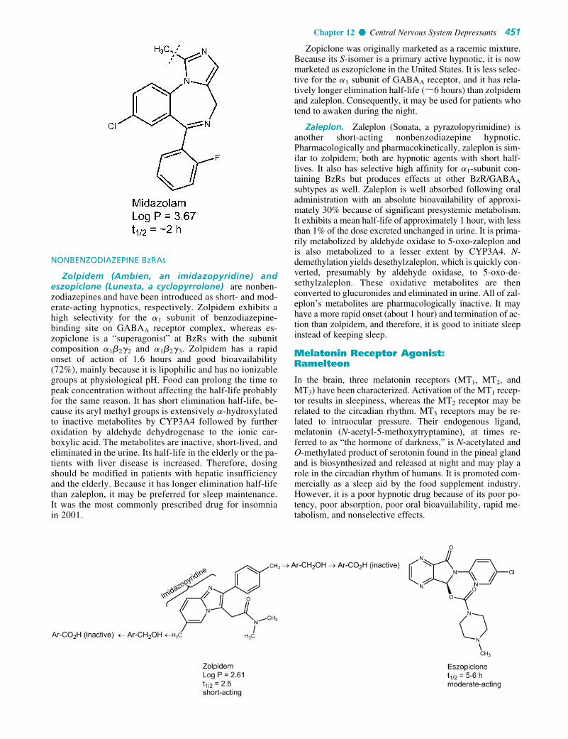
NONBENZODIAZEPINE BzRAs
Zolpidem (Ambien, an imidazopyridine) andeszopiclone (Lunesta, a cyclopyrrolone) are nonben-zodiazepines and have been introduced as short- and mod-erate-acting hypnotics, respectively. Zolpidem exhibits ahigh selectivity for the �1 subunit of benzodiazepine-binding site on GABAA receptor complex, whereas es-zopiclone is a “superagonist” at BzRs with the subunitcomposition �1�2�2 and �1�2�3. Zolpidem has a rapidonset of action of 1.6 hours and good bioavailability(72%), mainly because it is lipophilic and has no ionizablegroups at physiological pH. Food can prolong the time topeak concentration without affecting the half-life probablyfor the same reason. It has short elimination half-life, be-cause its aryl methyl groups is extensively �-hydroxylatedto inactive metabolites by CYP3A4 followed by furtheroxidation by aldehyde dehydrogenase to the ionic car-boxylic acid. The metabolites are inactive, short-lived, andeliminated in the urine. Its half-life in the elderly or the pa-tients with liver disease is increased. Therefore, dosingshould be modified in patients with hepatic insufficiencyand the elderly. Because it has longer elimination half-lifethan zaleplon, it may be preferred for sleep maintenance.It was the most commonly prescribed drug for insomniain 2001.
Zopiclone was originally marketed as a racemic mixture.Because its S-isomer is a primary active hypnotic, it is nowmarketed as eszopiclone in the United States. It is less selec-tive for the �1 subunit of GABAA receptor, and it has rela-tively longer elimination half-life (�6 hours) than zolpidemand zaleplon. Consequently, it may be used for patients whotend to awaken during the night.
Zaleplon. Zaleplon (Sonata, a pyrazolopyrimidine) isanother short-acting nonbenzodiazepine hypnotic.Pharmacologically and pharmacokinetically, zaleplon is sim-ilar to zolpidem; both are hypnotic agents with short half-lives. It also has selective high affinity for �1-subunit con-taining BzRs but produces effects at other BzR/GABAA
subtypes as well. Zaleplon is well absorbed following oraladministration with an absolute bioavailability of approxi-mately 30% because of significant presystemic metabolism.It exhibits a mean half-life of approximately 1 hour, with lessthan 1% of the dose excreted unchanged in urine. It is prima-rily metabolized by aldehyde oxidase to 5-oxo-zaleplon andis also metabolized to a lesser extent by CYP3A4. N-demethylation yields desethylzaleplon, which is quickly con-verted, presumably by aldehyde oxidase, to 5-oxo-de-sethylzaleplon. These oxidative metabolites are thenconverted to glucuronides and eliminated in urine. All of zal-eplon’s metabolites are pharmacologically inactive. It mayhave a more rapid onset (about 1 hour) and termination of ac-tion than zolpidem, and therefore, it is good to initiate sleepinstead of keeping sleep.
Melatonin Receptor Agonist:Ramelteon
In the brain, three melatonin receptors (MT1, MT2, andMT3) have been characterized. Activation of the MT1 recep-tor results in sleepiness, whereas the MT2 receptor may berelated to the circadian rhythm. MT3 receptors may be re-lated to intraocular pressure. Their endogenous ligand,melatonin (N-acetyl-5-methoxytryptamine), at times re-ferred to as “the hormone of darkness,” is N-acetylated andO-methylated product of serotonin found in the pineal glandand is biosynthesized and released at night and may play arole in the circadian rhythm of humans. It is promoted com-mercially as a sleep aid by the food supplement industry.However, it is a poor hypnotic drug because of its poor po-tency, poor absorption, poor oral bioavailability, rapid me-tabolism, and nonselective effects.
Chapter 12 Central Nervous System Depressants 451
0443-0470_17865_Ch12.qxd 12/2/09 4:01 AM Page 451

Ramelteon (Rozerem). The melatonin molecule wasmodified mainly by replacing the nitrogen of the indole ringwith a carbon to give an indane ring and by incorporating 5-methoxyl group in the indole ring into a more rigid furan ring.The selectivity of the resulting ramelteon for MT1 receptor iseight times more than that of MT2 receptor. Unlike mela-tonin, it is more effective in initiating sleep (MT1 activity)rather than to readjust the circadian rhythm (MT2 activity). Itappears to be distinctly more efficacious than melatonin butless efficacious than benzodiazepines as a hypnotic.Importantly, this drug has no addiction liability (it is not acontrolled substance). As a result, it has recently been ap-proved for the treatment of insomnia.
Barbiturates
The barbiturates were used extensively as sedative–hypnoticdrugs. Except for a few specialized uses, they have beenreplaced largely by the much safer benzodiazepine.Barbiturates act throughout the CNS. However, they exertmost of their characteristic CNS effects mainly by bindingto an allosteric recognition site on GABAA receptors that
positively modulates the effect of the GABAA receptor—GABA binding. Unlike benzodiazepines, they bind at differ-ent binding sites and appear to increase the duration of theGABA-gated chloride channel openings. In addition, bybinding to the barbiturate modulatory site, barbiturates canalso increase chloride ion flux without GABA attaching toits receptor site on GABAA. This has been termed a GABAmimetic effect. It is thought to be related to the profoundCNS depression that barbiturates can produce.
The barbiturates are 5,5-disubstituted barbituric acids.Consideration of the structure of 5,5-disubstituted barbituricacids reveals their acidic character. Those without methylsubstituents on the nitrogen have pKa’s of about 7.6; thosewith a methyl substituent have pKa’s of about 8.4. The freeacids have poor water solubility and good lipid solubility(the latter largely a function of the two hydrocarbon sub-stituents on the 5-position, although in the 2-thiobarbiturates,the sulfur atom increases lipid solubility).
Sodium salts of the barbiturates are readily preparedand are water soluble. Their aqueous solutions generate analkaline pH. A classic incompatibility is the addition ofan agent with an acidic pH in solution, which results in
452 Wilson and Gisvold’s Textbook of Organic Medicinal and Pharmaceutical Chemistry
0443-0470_17865_Ch12.qxd 12/2/09 4:01 AM Page 452
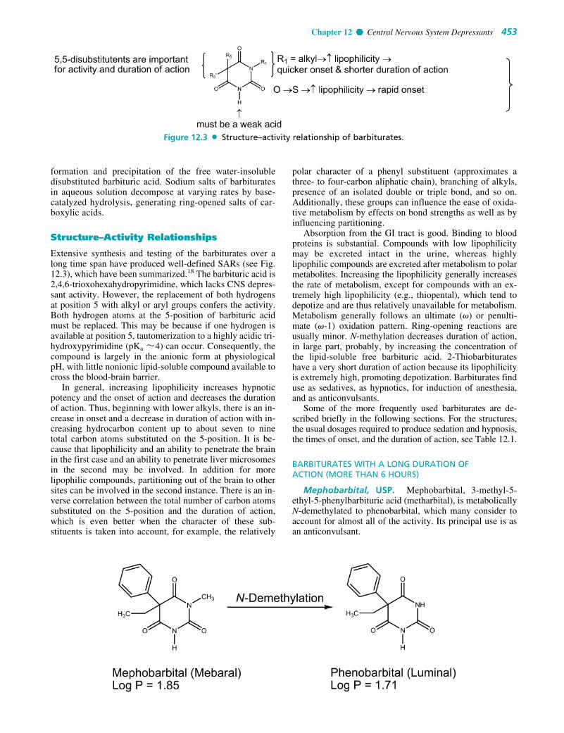
formation and precipitation of the free water-insolubledisubstituted barbituric acid. Sodium salts of barbituratesin aqueous solution decompose at varying rates by base-catalyzed hydrolysis, generating ring-opened salts of car-boxylic acids.
Structure–Activity Relationships
Extensive synthesis and testing of the barbiturates over along time span have produced well-defined SARs (see Fig.12.3), which have been summarized.18 The barbituric acid is2,4,6-trioxohexahydropyrimidine, which lacks CNS depres-sant activity. However, the replacement of both hydrogensat position 5 with alkyl or aryl groups confers the activity.Both hydrogen atoms at the 5-position of barbituric acidmust be replaced. This may be because if one hydrogen isavailable at position 5, tautomerization to a highly acidic tri-hydroxypyrimidine (pKa �4) can occur. Consequently, thecompound is largely in the anionic form at physiologicalpH, with little nonionic lipid-soluble compound available tocross the blood-brain barrier.
In general, increasing lipophilicity increases hypnoticpotency and the onset of action and decreases the durationof action. Thus, beginning with lower alkyls, there is an in-crease in onset and a decrease in duration of action with in-creasing hydrocarbon content up to about seven to ninetotal carbon atoms substituted on the 5-position. It is be-cause that lipophilicity and an ability to penetrate the brainin the first case and an ability to penetrate liver microsomesin the second may be involved. In addition for morelipophilic compounds, partitioning out of the brain to othersites can be involved in the second instance. There is an in-verse correlation between the total number of carbon atomssubstituted on the 5-position and the duration of action,which is even better when the character of these sub-stituents is taken into account, for example, the relatively
polar character of a phenyl substituent (approximates athree- to four-carbon aliphatic chain), branching of alkyls,presence of an isolated double or triple bond, and so on.Additionally, these groups can influence the ease of oxida-tive metabolism by effects on bond strengths as well as byinfluencing partitioning.
Absorption from the GI tract is good. Binding to bloodproteins is substantial. Compounds with low lipophilicitymay be excreted intact in the urine, whereas highlylipophilic compounds are excreted after metabolism to polarmetabolites. Increasing the lipophilicity generally increasesthe rate of metabolism, except for compounds with an ex-tremely high lipophilicity (e.g., thiopental), which tend todepotize and are thus relatively unavailable for metabolism.Metabolism generally follows an ultimate () or penulti-mate (-1) oxidation pattern. Ring-opening reactions areusually minor. N-methylation decreases duration of action,in large part, probably, by increasing the concentration ofthe lipid-soluble free barbituric acid. 2-Thiobarbiturateshave a very short duration of action because its lipophilicityis extremely high, promoting depotization. Barbiturates finduse as sedatives, as hypnotics, for induction of anesthesia,and as anticonvulsants.
Some of the more frequently used barbiturates are de-scribed briefly in the following sections. For the structures,the usual dosages required to produce sedation and hypnosis,the times of onset, and the duration of action, see Table 12.1.
BARBITURATES WITH A LONG DURATION OFACTION (MORE THAN 6 HOURS)
Mephobarbital, USP. Mephobarbital, 3-methyl-5-ethyl-5-phenylbarbituric acid (metharbital), is metabolicallyN-demethylated to phenobarbital, which many consider toaccount for almost all of the activity. Its principal use is asan anticonvulsant.
Chapter 12 Central Nervous System Depressants 453
Figure 12.3 Structure–activity relationship of barbiturates.
0443-0470_17865_Ch12.qxd 12/2/09 4:01 AM Page 453
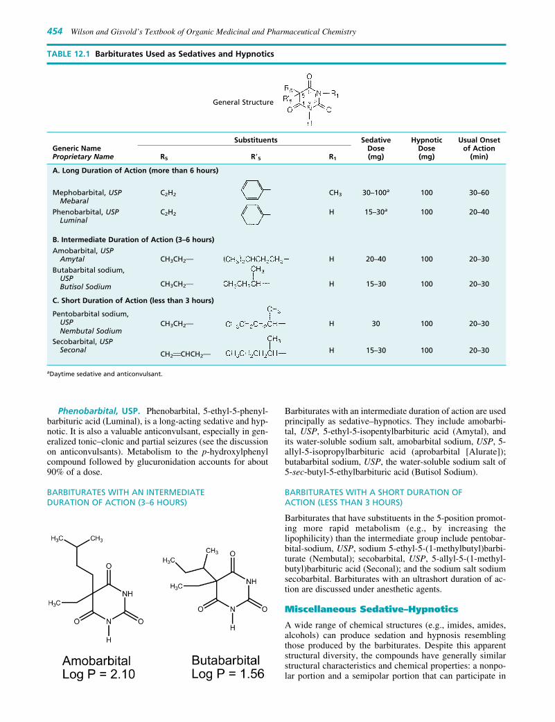
Phenobarbital, USP. Phenobarbital, 5-ethyl-5-phenyl-barbituric acid (Luminal), is a long-acting sedative and hyp-notic. It is also a valuable anticonvulsant, especially in gen-eralized tonic–clonic and partial seizures (see the discussionon anticonvulsants). Metabolism to the p-hydroxylphenylcompound followed by glucuronidation accounts for about90% of a dose.
BARBITURATES WITH AN INTERMEDIATE DURATION OF ACTION (3–6 HOURS)
Barbiturates with an intermediate duration of action are usedprincipally as sedative–hypnotics. They include amobarbi-tal, USP, 5-ethyl-5-isopentylbarbituric acid (Amytal), andits water-soluble sodium salt, amobarbital sodium, USP, 5-allyl-5-isopropylbarbituric acid (aprobarbital [Alurate]);butabarbital sodium, USP, the water-soluble sodium salt of5-sec-butyl-5-ethylbarbituric acid (Butisol Sodium).
BARBITURATES WITH A SHORT DURATION OF ACTION (LESS THAN 3 HOURS)
Barbiturates that have substituents in the 5-position promot-ing more rapid metabolism (e.g., by increasing thelipophilicity) than the intermediate group include pentobar-bital-sodium, USP, sodium 5-ethyl-5-(1-methylbutyl)barbi-turate (Nembutal); secobarbital, USP, 5-allyl-5-(1-methyl-butyl)barbituric acid (Seconal); and the sodium salt sodiumsecobarbital. Barbiturates with an ultrashort duration of ac-tion are discussed under anesthetic agents.
Miscellaneous Sedative–Hypnotics
A wide range of chemical structures (e.g., imides, amides,alcohols) can produce sedation and hypnosis resemblingthose produced by the barbiturates. Despite this apparentstructural diversity, the compounds have generally similarstructural characteristics and chemical properties: a nonpo-lar portion and a semipolar portion that can participate in
454 Wilson and Gisvold’s Textbook of Organic Medicinal and Pharmaceutical Chemistry
TABLE 12.1 Barbiturates Used as Sedatives and Hypnotics
General Structure
Substituents Sedative Hypnotic Usual OnsetGeneric Name Dose Dose of ActionProprietary Name R5 R�5 R1 (mg) (mg) (min)
A. Long Duration of Action (more than 6 hours)
Mephobarbital, USP C2H2 CH3 30–100a 100 30–60Mebaral
Phenobarbital, USP C2H2 H 15–30a 100 20–40Luminal
B. Intermediate Duration of Action (3–6 hours)
Amobarbital, USPAmytal CH3CH2M H 20–40 100 20–30
Butabarbital sodium, USPButisol Sodium CH3CH2M H 15–30 100 20–30
C. Short Duration of Action (less than 3 hours)
Pentobarbital sodium,CH3CH2M H 30 100 20–30USP
Nembutal SodiumSecobarbital, USP
H 15–30 100 20–30Seconal CH2FCHCH2M
aDaytime sedative and anticonvulsant.
0443-0470_17865_Ch12.qxd 12/2/09 4:01 AM Page 454

H-bonding. In some cases, modes of action are undeter-mined. As a working hypothesis, most of the agents can beenvisioned to act by mechanisms similar to those proposedfor barbiturates and alcohols.
AMIDES AND IMIDES
Glutethimide, USP. Glutethimide, 2-ethyl-2-phenyl-glutarimide (Doriden), is one of the most active nonbarbitu-rate hypnotics that is structurally similar to the barbiturates,especially phenobarbital. Because of glutethimide’s lowaqueous solubility, its dissolution and absorption from theGI track is somewhat erratic. Consistent with its highlipophilicity, it undergoes extensive oxidative metabolismin the liver with a half-life of approximately 10 hours.Glutethimide is used as a racemic mixture with the (�)enantiomer being primarily metabolized on the glutarimidering and the (�) enantiomer on the phenyl ring. The productof metabolic detoxification is excreted after conjugationwith glucuronic acid at the hydroxyl group. The drug is anenzyme inducer. In the therapeutic dosage range, adverse ef-fects tend to be infrequent. Toxic effects in overdose are assevere as, and possibly more troublesome than, those of thebarbiturates.
Alcohols and Their CarbamateDerivatives
The very simple alcohol ethanol has a long history of use asa sedative and hypnotic. Its modes of action were describedunder the anesthetic heading and are said to apply to otheralcohols. It is widely used in self-medication as asedative–hypnotic. Because this use has so many hazards, itis seldom a preferred agent medically.
As the homologous series of normal alcohols is ascendedfrom ethanol, CNS depressant potency increases up to eightcarbon atoms, with activity decreasing thereafter. Branchingof the alkyl chain increases depressant activity and, in anisometric series, the order of potency is tertiary � second-ary � primary. In part, this may be because tertiary andsecondary alcohols are not metabolized by oxidation to thecorresponding carboxylic acids. Replacement of a hydro-gen atom in the alkyl group by a halogen increases the alkylportion and, accordingly, for the lower–molecular weightcompounds, increases potency. Carbamylation of alcoholsgenerally increases depressant potency. Carbamate groupsare generally much more resistant to metabolic inactivationthan hydroxyl functions. Most of the alcohols and carba-mates have been superseded as sedative–hypnotics. Severaldifunctional compounds (e.g., diol carbamates) havedepressant action on the cord in addition to the brain andare retained principally for their skeletal muscle relaxantproperties.
Ethchlorvynol, USP. Ethchlorvynol, 1-chloro-3-ethyl-1-penten-4-yn-3-ol (Placidyl), is a mild sedative–hypnotic witha quick onset and short duration of action (t1/2 � 5.6 hours).Because of its highly lipophilic character, it is extensivelymetabolized to its secondary alcohol (�90%) prior to itsexcretion. It reportedly induces microsomal hepatic en-zymes. Acute overdose shares several features with barbitu-rate overdose.
Meprobamate, USP. Meprobamate, 2-methyl-2-propyltrimethylene dicarbamate, 2-methyl-2-propyl-1,3-propanediol dicarbamate (Equanil, Miltown), is officiallyindicated as an antianxiety agent. It is also a sedative–hypnotic agent. It has several overall pharmacological
Chapter 12 Central Nervous System Depressants 455
0443-0470_17865_Ch12.qxd 12/2/09 4:01 AM Page 455

properties resembling those of benzodiazepines and barbi-turates. The mechanism of action underlying anxiolytic ef-fects is unknown but may involve effects on conductivity inspecific brain areas.19 It does not appear to act through ef-fects on GABAergic systems. The drug is effective againstabsence seizures and may worsen generalized tonic–clonicseizures.
Meprobamate is also a centrally acting skeletal musclerelaxant. The agents in this group find use in several con-ditions, such as strains and sprains that may produce acutemuscle spasm. They have interneuronal blocking proper-ties at the level of the spinal cord, which are said to bepartly responsible for skeletal muscle relaxation.19 Also,the general CNS depressant properties they possess maycontribute to, or be mainly responsible for, the skeletalmuscle relaxant activity. Dihydric compounds and theircarbamate (urethane) derivatives, as described previouslyin the discussion of meprobamate, are prominent membersof the group.
Chlorphenesin Carbamate. Chlorphenesin carbamate,3-(p-chlorophenoxy)-1,2-propanediol 1-carbamate (Mao-late), is the p-chloro substituted and 1-carbamate derivativeof the lead compound in the development of this group ofagents, mephenesin. Mephenesin is weakly active and short-lived because of facile metabolism of the primary hydroxylgroup. Carbamylation of this group increases activity. p-Chlorination increases the lipophilicity and seals off thepara position from hydroxylation. Metabolism, still fairlyrapid, involves glucuronidation of the secondary hydroxylgroup. The biological half-life in humans is 3.5 hours.
Methocarbamol, USP. Methocarbamol, 3-(o-me-thoxyphenoxy)-1,2-propanediol 1-carbamate (Robaxin), issaid to be more sustained in effect than mephenesin. Likelysites for metabolic attack include the secondary hydroxylgroup and the two ring positions opposite the ether func-tions. The dihydric parent compound, guaifenesin, is used asan expectorant.
ALDEHYDES AND THEIR DERIVATIVES
For chemical reasons that are easily rationalized, few alde-hydes are valuable hypnotic drugs. The aldehyde in use,chloral (as the hydrate), is thought to act principally througha metabolite, trichloroethanol. Acetaldehyde is used as thecyclic trimer derivative, paraldehyde, which could also begrouped as a cyclic polyether.
Chloral Hydrate, USP. Chloral hydrate, trichloroac-etaldehyde monohydrate, CCl3CH(OH)2 (Noctec), is analdehyde hydrate stable enough to be isolated. The relativestability of this gem-diol is largely a result of an unfavorabledipole–dipole repulsion between the trichloromethyl carbonand the carbonyl carbon present in the parent carbonylcompound.20
Chloral hydrate is unstable in alkaline solutions, under-going the last step of the haloform reaction to yield chloro-form and formate ion. In hydroalcoholic solutions, it formsthe hemiacetal with ethanol. Whether or not this compoundis the basis for the notorious and potentially lethal effect of
456 Wilson and Gisvold’s Textbook of Organic Medicinal and Pharmaceutical Chemistry
Carisoprodol, USP. Carisoprodol, N-isopropyl-2-methyl-2-propyl-1,3-propanediol dicarbamate, 2-methyl-2-propyl-1,3-propanediol carbamate isopropylcarbamate(Soma), is the mono-N-isopropyl–substituted relative ofmeprobamate. The structure is given in the discussion ofmeprobamate. It is indicated in acute skeletomuscular con-ditions characterized by pain, stiffness, and spasm. As canbe expected, a major side effect of the drug is drowsiness.
0443-0470_17865_Ch12.qxd 12/2/09 4:01 AM Page 456

the combination of ethanol and chloral hydrate (the “MickeyFinn”) is controversial. Synergism between two differentCNS depressants also could be involved. Additionally,ethanol, by increasing the concentration of nicotinamideadenine dinucleotide (NADH), enhances the reduction ofchloral to the more active metabolite trichloroethanol, andchloral can inhibit the metabolism of alcohol because it in-hibits alcohol dehydrogenase. Chloral hydrate is a weak acidbecause its CCl3 group is very strong electron withdrawing.A 10% aqueous solution of chloral hydrate has pH 3.5 to4.4, which makes it irritating to mucous membranes in thestomach. As a result, GI upset commonly occurs for thedrug if undiluted or taken on an empty stomach. Chloral hy-drate as a capsule, syrup, or suppository is currently avail-able. Although it is suggested that chloral hydrate per semay act as a hypnotic,21 chloral hydrate is very quickly con-verted to trichloroethanol, which is generally assumed toaccount for almost all of the hypnotic effect. Thetrichloroethanol is metabolized by oxidation to chloral andthen to the inactive metabolite, trichloracetic acid (see Fig.12.4), which is also extensively metabolized to acylglu-curonides via conjugation with glucuronic acid. It appears tohave potent barbiturate-like binding to GABAA receptors.Although an old drug, it still finds use as a sedative in non-operating room procedures for the pediatric patient.
Paraldehyde, USP. Paraldehyde, 2,4,6-trimethyl-s-trioxane, paracetaldehyde, is recognizable as the cyclictrimer of acetaldehyde. It is a liquid with a strong charac-teristic odor detectable in the expired air and an unpleasanttaste. These properties limit its use almost exclusively toan institutional setting (e.g., in the treatment of deliriumtremens). In the past, when containers were opened and airadmitted and then reclosed and allowed to stand, fatalitiesoccurred because of oxidation of paraldehyde to glacialacetic acid.
ANTIPSYCHOTICS
The psychoses affect approximately 1% of the population inall cultures. They are psychogenic mental disorders involvinga loss of contact with reality. The psychotic disorders includeschizophrenia, the manic phase of bipolar (manic–depressive)illness, acute idiopathic psychotic illness, and other condi-tions marked by severe agitation. The most common isschizophrenia, in which perception, thinking, communica-tion, social functioning, and attention are altered.
Schizophrenia is a particular kind of psychosis character-ized mainly by a clear sensorium but a marked thinking dis-turbance. Symptoms are called positive (e.g., delusions, hal-lucinations) or negative (e.g., flat affect, apathy); cognitivedysfunction may occur. In the schizophrenias, which havean extremely complex and multifactored etiology,22,23 thefundamental lesion appears to be a defect in the brain’s in-formational gating mechanism. Basically, the gating systemhas difficulty discriminating between relevant and irrelevantstimuli. The etiology of psychosis remains unknown, al-though genetic, neurodevelopmental and environmentalcausative factors have all been proposed. Psychoses can be
Chapter 12 Central Nervous System Depressants 457
Figure 12.4 Reaction and metabolismof chloral.
0443-0470_17865_Ch12.qxd 12/2/09 4:01 AM Page 457

organic and related to a specific toxic chemical (e.g., delir-ium produced by central anticholinergic agents), an N-methyl D-aspartate (NMDA) receptor antagonist (e.g.,phencyclidine [PCP]), a definite disease process (e.g., de-mentia), or they can be idiopathic.
Although the actual structural or anatomical lesions are notknown, the basic defect appears to involve overactivity ofdopaminergic neurons in the mesolimbic system. DA hypoth-esis for schizophrenia is the most fully developed of severalhypotheses and is the basis for much of the rationale for drugtherapy because (a) drugs that increase dopaminergic neuro-transmission, such as levodopa (a DA precursor),amphetamines (a DA releaser), and apomorphine (a DA ago-nist), induce or exacerbate schizophrenia. Amphetamine-induced psychosis was determined to be caused by overacti-vation of mesolimbic D2 receptors and judged to be theclosest of the various chemically induced model psychoses tothe schizophrenias; (b) DA receptor density is increased incertain brain regions of untreated schizophrenics; (c) manyantipsychotic drugs strongly block postsynaptic D2 receptorsin CNS; and (d) successful treatment of schizophrenic pa-tients has been reported to change the amount of homovanil-lic acid (HVA), a DA metabolite, in the cerebrospinal fluid,plasma, and urine. Consequently, the antipsychotic action isnow thought to be produced (at least in part) by their abilityto block DA receptors in the mesolimbic and mesofrontal sys-tems. Moreover, extrapyramidal side effects of antipsychoticdrugs correlate with their D2 antagonism effect. The hyper-prolactinemia that follows treatment with antipsychotics iscaused by blockade of DA’s tonic inhibitory effect on pro-lactin release from the pituitary. Nevertheless, the defects ofDA hypothesis are significant, and it is now appreciatedthat schizophrenia is far more complex than originally sup-posed. Several classes of drugs are effective for symptomatictreatment.
Interest in DA, 5-HT, and Glu NTs led to most earlydrugs targeting the DA system, primarily as DA D2 receptor.Typical antipsychotics (e.g., chlorpromazine, haloperidol)are better for treating positive signs than negative signs. Fortreating negative signs, the newer (atypical) antipsychoticdrugs (e.g., clozapine, risperidone) target D2 receptor andother receptors. The bases of the atypical group’s activityagainst negative symptoms may be serotonin-2A receptor (5-HT2A) block, block at receptors yet to be determined, andpossibly decreased striatal D2 block.24 A classic competitiveantagonism has been demonstrated at D2 and D3 receptors.Also, in recombinantly expressed receptors, inverse agonismhas been demonstrated. Recent studies show that essentiallyall clinically used antipsychotic drugs are D2 inverse ago-nists, suggesting that biochemical as well as clinical ef-fects may not be explained by simple D2 receptor blockadehypothesis.25
Typical antipsychotics began with the serendipitous dis-covery of the antipsychotic activity of chlorpromazine. Aclear association between the ability to block DA atmesolimbic D2 receptors was established. The conventionaltypical antipsychotics (neuroleptics) are characterized bythe production of EPS, roughly approximating the symp-toms of Parkinson disease. These are reversible on discon-tinuing or decreasing the dose of the drug and are associ-ated with blockade of DA at D2 striatal receptors. Aftersustained high-dose therapy with antipsychotics, a late-appearing EPS, tardive dyskinesia, may occur. The overall
symptomatology resembles the symptoms of Huntingtonchorea. Atypical antipsychotics date from the discovery ofclozapine, its antipsychotic properties, and its much lowerproduction of EPS. Also contributing to the development ofatypical antipsychotics was the introduction of risperidone.It has reduced EPS, has increased activity against negativesymptoms, and, in addition to its DA-blocking ability, is a5-HT2A antagonist. The view has been proposed that 5-HT2A receptors are involved in part (the negative symp-toms) or wholly in schizophrenia. So far, the evidenceappears to be that 5-HT2A blocking agents do not relievepositive effects of schizophrenia.24 The view that 5-HT2A
overactivity is the source of negative symptoms (part of thebasis of psychosis) is not disproved at present, althoughsome say it has been weakened.24
One result of the development of atypical antipsychoticshas been a renewed interest in models of psychosis otherthan the amphetamine model. In line with possible dual in-volvement of 5-HT and DA, the lysergic acid diethylamide(LSD, a 5-HT agonist) model has been cited as better fittingschizophrenias than the amphetamine model. However, thishas been disputed. Interest in serotoninergic involvement isstill high and involves elucidating the roles of 5-HT6 and 5-HT7 receptors.
Interest remains in understanding the psychosis producedby several central anticholinergics. Muscarinic (M1 and M4)agonists appear to offer the best approach at this time.26 Therole of the M5 receptor awaits synthesis of M5-specificdrugs.27
PCP (an NMDA antagonist)-induced psychosis has beenproposed as a superior model for schizophrenia, because itpresents both positive and negative symptoms.24 It suggeststhat deficits in glutaminergic function occur in schizophre-nia. Results of agonists of NMDA receptors, overall, havenot been productive because of the excitatory and neuro-toxic effects of the agents tested. Identification of suscepti-ble receptor subtypes as targets, using glycine modulation orgroup II metabotropic receptor agonists to modulate NMDAreceptors, has been proposed to circumvent the problems as-sociated with the NMDA agonists.
The ionotropic glutamic acid �-amino-3-hydroxy-5-methyl-4-isoxazole propionic acid (AMPA) receptors areactivated by brain-penetrating ampakines. There are sugges-tions that these agents exert some antipsychotic actions byincreasing glutaminergic activity.
The individual antipsychotic agents are now considered.The substituted DA motif is useful as an organizational de-vice. Antipsychotics can be classified into four groups(Table 12.2).
Phenothiazines
Several dozen phenothiazine antipsychotic drugs are chem-ically related agents used worldwide. Other phenothiazinesare marketed primarily for their antiemetic, antihistaminic,or anticholinergic effect. The large body of information per-mits accurate statements about the structural features associ-ated with activity (see Fig. 12.5). Many of the features weresummarized and interpreted by Gordon et al.28
Phenothiazines have a tricyclic structure (6-6-6 system) inwhich two benzene rings are linked by a sulfur and anitrogen atom. The best position for substitution is the 2-position. Activity increases (with some exceptions) as
458 Wilson and Gisvold’s Textbook of Organic Medicinal and Pharmaceutical Chemistry
0443-0470_17865_Ch12.qxd 12/2/09 4:01 AM Page 458

electron-withdrawing ability of the 2-substituent increases(e.g., chlorpromazine vs. promazine). Another possibly im-portant structural feature in the more potent compounds isthe presence of an unshared electron pair on an atom oratoms of the 2-substituent. Substitution at the 3-position canimprove activity over nonsubstituted compounds but not assignificantly as substitution at the 2-position. Substitution atposition 1 has a deleterious effect on antipsychotic activity,as does (to a lesser extent) substitution at the 4-position.
The significance of these substituent effects could be thatthe hydrogen atom of the protonated amino group of the sidechain H-bonds with an electron pair of an atom of the 2-sub-stituent to develop a DA-like arrangement.
Horn and Snyder,29 from x-ray crystallography, proposedthat the chlorine-substituted ring of chlorpromazine basecould be superimposed on the aromatic ring of DA base, withthe sulfur atom aligned with the p-hydroxyl group of DA andthe aliphatic amino groups of the two compounds alsoaligned. The model used here is based on the interpretationof the SARs by Gordon et al.28 and on the Horn and Snyder29
proposal but involves the protonated species rather than thefree base. The effect of the substituent at the 1-position mightbe to interfere with the side chain’s ability to bring the pro-tonated amino group in proximity with the 2-substituent. Inthe Horn and Snyder29 scheme, the sulfur atom at position 5is in a position analogous to the p-hydroxyl group of DA, andit was also assigned a receptor-binding function by Gordonet al.28 A substituent at position 4 might interfere with recep-tor binding by the sulfur atom.
The three-carbon chain between position 10 and thealiphatic amino nitrogen is critical for neuroleptic activity.Shortening or lengthening the chain at this position drasti-cally decreases the activity. The three-atom chain length
may be necessary to bring the protonated amino nitrogen inproximity with the 2-substituent. Shortening the chain totwo carbons has the effect of amplifying the antihistaminicand anticholinergic activities. For example, promethazine iseffective antihistamine, whereas the amino ethyl derivativesdiethazine (anticholinergic) and ethopropazine (antimus-carinic) have proved useful in the treatment of Parkinsondisease. The amine is always tertiary. N-dealkylation ofthe side chain or increasing the size of amino N-alkylsubstituents reduces antidopaminergic and antipsychoticactivity.
As expected, branching with large groups (e.g., phenyl)decreases activity, as does branching with polar groups.Methyl branching on the �-position has a variable effect onactivity. More importantly, the antipsychotic potency oflevo (the more active) and dextro isomers differs greatly.This has long been taken to suggest that a precise fit (i.e., re-ceptor site occupancy) is involved in the action of thesecompounds.
Decreases in size from a dimethylamino group (e.g.,going to a monomethylamino) greatly decrease activity, asdo effective size increases, such as the one that occurs withN,N-diethylamino group. Once the fundamental requirementof an effective size of about that equivalent to a dimethyl-amino is maintained, as in fusing N,N-diethyl substituents togenerate a conformational restricted pyrrolidino group, ac-tivity can be enhanced with increasing chain length, as inN2-substituted piperizino compounds.
The critical size of groups on the amino atom suggeststhe importance of the amino group (here protonated) for re-ceptor attachment. The effect of the added chain length,once the critical size requirement is met, could be increasedaffinity. It appears to have been reasonably proved that the
Chapter 12 Central Nervous System Depressants 459
TABLE 12.2 Classification of Antipsychotics
Drug Groups Structure Features Examples Comments
Phenothiazines Aliphatic side chain Chlorpromazine Least potentPiperidine side chain Thioridazine Least potent and ↓ EPSPiperazine side chain Fluphenazine More potent and more EPS
Thioxanthenes Double bond on C10 Thiothixene Less potent than other phenothiazines
Butyrophenones Aromatic butylpiperidines and Haloperidol More potentdiphenylbutylpiperidines Fewer autonomic SEs
Greater EPS
Newer drugs Miscellaneous Risperidone Less EPSClozapine Also good for negative symptomsOlanzapine
Figure 12.5 SAR of phenothiazine antipsychotic agents.
0443-0470_17865_Ch12.qxd 12/2/09 4:01 AM Page 459

protonated species of the phenothiazines can bind to DAreceptors.30
Several piperazine phenothiazines are esterified at a freehydroxyl with long-chain fatty acids to produce highlylipophilic and long-acting prodrugs. They tend to have largevolumes of distribution, probably because they are se-questered in lipid compartments of the body and have veryhigh affinity for selected NT receptors in the CNS. They gen-erally have a much longer clinical duration of action thanwould be estimated from their plasma half-lives. This is par-alleled by prolonged occupancy of DA D2 receptors in brain.The decanoates of fluphenazine and haloperidol are usedcommonly in the United States; several others (including es-ters of pipotiazine and perphenazine) are available elsewhere.
Because of the high lipophilicity of most antipsychoticdrugs, they are highly membrane and protein bound(92%–99%) mostly to albumin. They accumulate in thebrain, lung, and other tissues with a rich blood supply andalso enter the fetal circulation and breast milk.
Most phenothiazines undergo significant first-pass me-tabolism. Chlorpromazine and other phenothiazines aremetabolized extensively by CYP2D6. Thus, oral doses ofchlorpromazine and thioridazine have systemic availabilityof 25% to 35%, whereas parenteral (intramuscular) admin-istration increases the bioavailability of active drug fourfoldto tenfold. In contrast, haloperidol, which is less likely to bemetabolized, has an average systemic availability of about65%. Metabolism of the phenothiazines is complex in detail.
460 Wilson and Gisvold’s Textbook of Organic Medicinal and Pharmaceutical Chemistry
0443-0470_17865_Ch12.qxd 12/2/09 4:01 AM Page 460

A major route is 7-hydroxylation of the tricyclic system.Because electron-withdrawing 2-Cl substituent blocks thehydroxylation on chlorophenyl ring, the hydroxylation oc-curs at 7-position rather than 2-position. Thus, the majorinitial metabolite is frequently the 7-hydroxy compound (ac-tive metabolite). This compound is further metabolized byconjugation with glucuronic acid, and the conjugate is ex-creted. Metabolites of chlorpromazine may be excreted inthe urine weeks after the last dose of chronically adminis-tered drug. Full relapse may not occur until 6 weeks or moreafter discontinuation of many antipsychotics. Detailed re-views of the metabolites of phenothiazines (as well as SARsand pharmacokinetic factors) are available.31
PRODUCTS
The structures of the phenothiazine derivatives describedlater are given in Table 12.3.
Chlorpromazine Hydrochloride, USP. Chlorpromazinehydrochloride, 2-chloro-10-[3-(dimethylamino)propyl]phe-
nothiazine monohydrochloride (Thorazine), was the first phe-nothiazine compound introduced into therapy. It is still usefulas an antipsychotic. Other uses are in nausea, vomiting, andhiccough. Oral doses of chlorpromazine and thioridazinehave systemic availability of 25% to 35% because of signifi-cant first-pass metabolism. Chlorpromazine and other pheno-thiazines are metabolized extensively by CYP2D6 (Fig.12.6). In contrast, bioavailability of chlorpromazine may beincreased up to 10-fold with injections, but the clinical doseusually is decreased by only threefold to fourfold.Chlorpromazine may weakly induce its own hepatic metabo-lism, because its concentration in blood is lower after severalweeks of treatment at the same dosage. Alterations of GImotility also may contribute. The drug has significant seda-tive and hypotensive properties, possibly reflecting centralhistaminergic and peripheral �1-noradrenergic blockingactivity, respectively. Effects of peripheral anticholinergic ac-tivity are common. As with the other phenothiazines, the ef-fects of other CNS-depressant drugs, such as sedatives andanesthetics, can be potentiated.
Chapter 12 Central Nervous System Depressants 461
TABLE 12.3 Phenothiazine Derivatives
Generic NameProprietary Name R10 R2
Propyl Dialkylamino Side Chain
Promazine hydrochloride, USPSparine M(CH2)3N(CH3)2 � HCl H
Chlorpromazine hydrochloride, USPThorazine M(CH2)3N(CH3)2 � HCl Cl
Triflupromazine hydrochloride, USPVesprin M(CH2)3N(CH3)2 � HCl CF3
Akyl Piperidyl Side Chain
Thioridazine hydrochloride, USP SCH3
Mellaril
Mesoridazine besylate, USP O Serentil ↑
SCH3
Propyl Piperazine Side Chain
Prochlorperazine maleate, USPClCompazine
Trifluoperazine hydrochloride, USPCF3Stelazine
Perphenazine, USPClTrilafon
Fluphenazine hydrochloride, USPCF3Permitil, Prolixin
0443-0470_17865_Ch12.qxd 12/2/09 4:01 AM Page 461
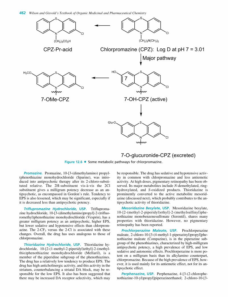
Promazine. Promazine, 10-[3-(dimethylamino) propyl-(phenothiazine monohydrochloride (Sparine), was intro-duced into antipsychotic therapy after its 2-chloro-substi-tuted relative. The 2H-substituent vis-à-vis the 2Clsubstituent gives a milligram potency decrease as an an-tipsychotic, as encompassed in Gordon’s rule. Tendency toEPS is also lessened, which may be significant, especially ifit is decreased less than antipsychotic potency.
Triflupromazine Hydrochloride, USP. Trifluproma-zine hydrochloride, 10-[3-(dimethylamino)propyl]-2-(trifluo-romethyl)phenothiazine monohydrochloride (Vesprin), has agreater milligram potency as an antipsychotic, higher EPS,but lower sedative and hypotensive effects than chlorprom-azine. The 2-CF3 versus the 2-Cl is associated with thesechanges. Overall, the drug has uses analogous to those ofchlorpromazine.
Thioridazine Hydrochloride, USP. Thioridazine hy-drochloride, 10-[2-(1-methyl-2-piperidyl)ethyl]-2-(methyl-thio)phenothiazine monohydrochloride (Mellaril), is amember of the piperidine subgroup of the phenothiazines.The drug has a relatively low tendency to produce EPS. Thedrug has high anticholinergic activity, and this activity in thestriatum, counterbalancing a striatal DA block, may be re-sponsible for the low EPS. It also has been suggested thatthere may be increased DA receptor selectivity, which may
be responsible. The drug has sedative and hypotensive activ-ity in common with chlorpromazine and less antiemeticactivity. At high doses, pigmentary retinopathy has been ob-served. Its major metabolites include N-demethylated, ring-hydroxylated, and S-oxidized products. Thioridazine isprominently converted to the active metabolite mesorid-azine (discussed next), which probably contributes to the an-tipsychotic activity of thioridazine.
Mesoridazine Besylate, USP. Mesoridazine besylate,10-[2-(methyl-2-piperidyl)ethyl]-2-(methylsulfinyl)phe-nothiazine monobenzenesulfonate (Serentil), shares manyproperties with thioridazine. However, no pigmentaryretinopathy has been reported.
Prochlorperazine Maleate, USP. Prochlorperazinemaleate, 2-chloro-10-[3-(4-methyl-1-piperazinyl)propyl]phe-nothiazine maleate (Compazine), is in the piperazine sub-group of the phenothiazines, characterized by high-milligramantipsychotic potency, a high prevalence of EPS, and lowsedative and autonomic effects. Prochlorperazine is more po-tent on a milligram basis than its alkylamino counterpart,chlorpromazine. Because of the high prevalence of EPS, how-ever, it is used mainly for its antiemetic effect, not for its an-tipsychotic effect.
Perphenazine, USP. Perphenazine, 4-[3-(2-chlorophe-nothiazine-10-yl)propyl]piperazineethanol; 2-chloro-10-[3-
462 Wilson and Gisvold’s Textbook of Organic Medicinal and Pharmaceutical Chemistry
Figure 12.6 Some metabolic pathways for chlorpromazine.
0443-0470_17865_Ch12.qxd 12/2/09 4:01 AM Page 462

[4-(2-hydroxyethyl)piperazinyl]propyl]phenothiazine(Trilafon), is an effective antipsychotic and antiemetic.
Fluphenazine Hydrochloride, USP. The member ofthe piperazine subgroup with a trifluoromethyl group at the2-position of the phenothiazine system and the most potentantipsychotic phenothiazine on a milligram basis is fluphen-azine hydrochloride, 4-[3-[2-(trifluoromethyl)phenazin-10-yl] propyl]-1-piperazineethanol dihydrochloride, 10[3-[4-(2-hydroxyethyl)piperazinyl]propyl]-2-trifluoromethylphenothiazine dihydrochloride (Permitil, Prolixin). It is alsoavailable as two lipid-soluble esters for depot intramuscularinjection, the enanthate (heptanoic acid ester) and the dec-anoate ester. These long-acting preparations have use intreating psychotic patients who do not take their medicationor are subject to frequent relapse.
Thiothixene, USP. The thioxanthene system differsfrom the phenothiazine system by replacement of the N-Hmoiety with a carbon atom doubly bonded to the propyli-dene side chain. With the substituent in the 2-position, Z-and E-isomers are produced. In accordance with the conceptthat the presently useful antipsychotics can be superimposedon DA, the Z-isomers are the more active antipsychotic iso-mers. The compounds of the group are very similar in phar-macological properties to the corresponding phenothiazines.Thus, thiothixene (Z-N-dimethyl-9-[3-(4-methyl-1-piper-azinyl)propylidene]thioxanthene-2-sulfonamide (Navane),displays properties similar to those of the piperazine sub-group of the phenothiazines.
Ring Analogs of Phenothiazines:Benzazepines, Dibenzoxazepines, andDibenzodiazepines
Additional tricyclic antipsychotic agents are the ben-zazepines, containing a seven-membered central ring (6-7-6system). These newer atypical antipsychotics include diben-
zodiazepines (clozapine with 2-Cl), dibenzoxazepines(loxapine with 2-Cl), thienobenzodiazepines (olanzapinewithout 2-substituent), and dibenzothiazepines (quetiapinewithout 2-substituent). These ring analogs of phenothiazinesare structural relatives of the phenothiazine antipsychotics;therefore, most of them share many clinical properties withthe phenothiazines. However, they have some important dif-ferences, notably low production of EPS and reduction ofnegative symptoms. These benzazepines and other atypicalantipsychotics including risperidone, ziprasidone, and arip-iprazole block both D2 and 5-HT2A receptor, other DA andserotonin receptor subtypes, adrenergic, histamine, and mus-carinic receptors. The low D2 receptor affinity and the high5-HT2A receptor affinity of atypical antipsychotics includingclozapine and olanzapine led to the proposal that 5-HT2A an-tagonism accounts for their lower EPS.
PRODUCTS
Loxapine. A dibenzoxazepine derivative in use is loxap-ine succinate, 2-chloro-11-(4-methyl-1-piperazinyl)dibenz[b,f ][1,4]oxazepine succinate (Daxolin). The structural relation-ship to the phenothiazine antipsychotics is apparent. Examplesin this group are clothiapine, metiapine, zotepine, and others.They have electron-withdrawing groups at position 2, rela-tively close to the side-chain nitrogen atoms. Loxapine, an ef-fective antipsychotic, blocks D2-type receptors and has side ef-fects similar to those reported for the phenothiazines. Itsmetabolism involves aromatic hydroxylation to give severalphenolic metabolites that have higher affinity for D2 receptorsthan the parent. It is also N-demethylated to yield amoxapine(an antidepressant drug), which inhibits norepinephrine (NE)neurotransporter to block neuronal NE reuptake.
Clozapine. The dibenzodiazepine derivative is clozapine(Clozaril). It is not a potent antipsychotic on a milligrambasis (note the orientation of the N-methyl piperazino grouprelative to the chlorine atom). In addition to their moderatepotencies at DA receptors (mainly D4), clozapine interactwith varying affinities at several other classes of receptors(�1 and �2 adrenergic, 5-HT1A, 5-HT2A, 5-HT2C, muscariniccholinergic, histamine H1, and others). It is effective againstboth positive and negative symptoms of schizophrenia andhas a low tendency to produce EPS. Clozapine has proved ef-fective even in chronically ill patients who respond poorly tostandard neuroleptics. However, there are legal restrictionson its use because of a relatively high frequency of agranulo-cytosis. As a rule, two other antipsychotics are tried beforerecourse to therapy with clozapine. Clozapine is metabolized
Chapter 12 Central Nervous System Depressants 463
0443-0470_17865_Ch12.qxd 12/2/09 4:01 AM Page 463

preferentially by CYP3A4 into demethylated, hydroxylated,and N-oxide derivatives that are excreted in urine and feces.Elimination half-life averages about 12 hours. Other cloza-pine-like atypical antipsychotics may lack a 2-Cl substituenton the aromatic ring (e.g., olanzapine and quetiapine).
Olanzapine and Quetiapine. Clozapine analogs, olan-zapine (Zyprexa) and quetiapine (Seroquel) possess tricyclicsystems with greater electron density than chlorpromazine.The drugs are atypical antipsychotics. Olanzapine is a morepotent antagonist at D2 and 5-HT2A receptors than clozapineand is well absorbed, but about 40% of an oral dose is me-tabolized before reaching the systemic circulation. Plasmaconcentrations of olanzapine peak at about 6 hours after oraladministration, and its elimination half-life ranges from 20 to54 hours. Major readily excreted metabolites of olanzapineare the inactive 10-N-glucuronide and 4�-nor derivatives,formed mainly by the action of CYP1A2, with CYP2D6 as aminor alternative pathway. It may have even lower risk thanrisperidone and has achieved widespread use.
Overall, these two compounds should bind less stronglyto D2 receptors and permit more receptor selectivity amongreceptor subtypes than typical antipsychotics. This could
464 Wilson and Gisvold’s Textbook of Organic Medicinal and Pharmaceutical Chemistry
0443-0470_17865_Ch12.qxd 12/2/09 4:01 AM Page 464

account for decreased striatal D2-blocking activity (EPS).Quetiapine is highly metabolized by hepatic CYP3A4 to in-active and readily excreted sulfoxide and acidic derivatives.
Fluorobutyrophenones
The fluorobutyrophenones belong to a much-studied groupof compounds, many of which possess high antipsychoticactivity. The structural requirements for antipsychotic activ-ity in the group are well worked out.32 General structure andSAR are expressed in the following structure (Fig. 12.7).
Attachment of a tertiary amino group to the fourth carbonof the butyrophenone skeleton is essential for neurolepticactivity; lengthening, shortening, or branching of the three-carbon propyl chain decreases neuroleptic potency. Thisaliphatic amino nitrogen is required, and highest activity isseen when it is incorporated into a cyclic form. A p-fluorosubstituent aids activity. The CBO group gives optimal ac-tivity, although other groups, C(H)OH and C(H)aryl, alsogive good activity. The Y group can vary and assist activity,and an example is the hydroxyl group of haloperidol.
The empirical SARs could be construed to suggest thatthe 4-aryl piperidino moiety is superimposable on the
2-phenylethylamino moiety of DA and, accordingly, couldpromote affinity for D2 and D3 receptors. The long N-alkylsubstituent could help promote receptor affinity and producereceptor antagonism activity and/or inverse agonism.
Some members of the class are D2 and D3 receptor an-tagonists and are extremely potent antipsychotic agents.EPS are extremely marked in some members of this class,which may, in part, be because of a potent DA block inthe striatum and almost no compensatory striatal anti-cholinergic block. Most of the compounds do not have thestructural features associated with effective anticholinergicactivity.
Haloperidol, USP. Haloperidol, 4[4-(p-chlorophenyl)-4-hydroxypiperidone]-4�-n-fluorobutyrophenone (Haldol),the representative of several related classes of aromaticbutylpiperidine derivatives, is a potent antipsychotic usefulin schizophrenia and in psychoses associated with braindamage. It is frequently chosen as the agent to terminatemania and often used in therapy for Gilles de la Tourettesyndrome. Haloperidol-induced dyskinesias may involveneurotoxicological metabolite similar to dopaminergic toxi-cant MPP� (Fig. 12.8).
Chapter 12 Central Nervous System Depressants 465
Figure 12.7 General structure and SAR of fluorobutyrophenones.
Figure 12.8 Metabolism of haloperidol and its possible neurotoxic metabolites.
0443-0470_17865_Ch12.qxd 12/2/09 4:01 AM Page 465

Droperidol, USP. Droperidol, 1-{1-[3-(p-fluoroben-zoyl)propyl]-1,2, 3,6-tetrahydro-4-pyridyl}-2-benzimida-zolinone (Inapsine), may be used alone as a preanestheticneuroleptic or as an antiemetic. Because of its very short-acting and highly sedating properties, its most frequent useis in combination (Innovar) with the narcotic agent fentanyl(Sublimaze) preanesthetically.
Risperidone. Risperidone (Risperdal, a benzisoxazole)has the structural features of a hybrid molecule between abutyrophenone antipsychotic and a trazodone-like antide-pressant. Its superior side effects profile (compared withhaloperidol) at dosage of 6 mg/d or less and the lower riskof tardive dyskinesia have contributed to its very wide-spread use. It benefited refractory psychotic patients, withparkinsonism controlled at one tenth the dose of antiparkin-sonian drugs used with haloperidol.33 Coexisting anxietyand depressive syndromes were also lessened. It is reportedto decrease the negative (e.g., withdrawal, apathy) as well asthe positive (e.g., delusions, hallucinations) symptoms ofschizophrenia. This is reportedly a consequence of the com-pound’s combination 5-HT2–D2 receptor antagonistic prop-erties.34 Overall, the reasons for the decreased EPS and
effectiveness against negative symptom are still under in-vestigation. It is an important atypical antipsychotic.Risperidone is metabolized in the liver by CYP2D6 to anactive metabolite, 9-hydroxyrisperidone. Because thismetabolite and risperidone are nearly equipotent, the clini-cal efficacy of the drug reflects both compounds.
466 Wilson and Gisvold’s Textbook of Organic Medicinal and Pharmaceutical Chemistry
Figure 12.9 Major metabolic pathway of ziprasidone.
0443-0470_17865_Ch12.qxd 12/2/09 4:01 AM Page 466

Ziprasidone. Ziprasidone (Geodon, a benzisothia-zolpiprazinylindolone derivative) also has the structuralfeatures of a hybrid molecule between a butyrophenone an-tipsychotic and a trazodone-like antidepressant. It is highlymetabolized to four major metabolites, only one of which, S-methyldihydroziprasidone, likely contributes to its clinical ac-tivity (see Fig. 12.9). In humans, less than 5% of the dose isexcreted unchanged. Reduction by aldehyde oxidase accountsfor about 66% of ziprasidone metabolism; two oxidative path-ways involving hepatic CYP3A4 account for the remainder.
Aripiprazole. Aripiprazole (Abilify). The newest, long-acting aripiprazole (an arylpiperazine quinolinone deriva-tive), appears to be partial agonist of D2 receptors (i.e., itstimulates certain D2 receptors while blocking others de-pending on their locations in the brain and the concentrationof drug). Bioavailability of aripiprazole is around 87%, with
peak plasma concentration attained at 3 to 5 hours afterdosing. It is metabolized by dehydrogenation (see Fig.12.10), oxidative hydroxylation, and N-dealkylation, largelymediated by hepatic CYPs 3A4 and 2D6.
The diphenylbutylpiperidine class can be considered amodification of the fluorobutyrophenone class. Because oftheir high lipophilicity, the compounds are inherently longacting. Pimozide has been approved for antipsychotic use,and penfluridol has undergone clinical trials in the UnitedStates. Overall, side effects for the two compounds resem-ble those produced by the fluorobutyrophenones.
�-AMINOKETONES
Several �-aminoketones have been examined as antipsy-chotics.31 They evolved out of research on the alkaloid lo-beline. The overall structural features associated with activity
Chapter 12 Central Nervous System Depressants 467
Figure 12.10 Major metabolic pathway for aripiprazole.
0443-0470_17865_Ch12.qxd 12/2/09 4:01 AM Page 467

can be seen in the structure of molindone. In addition to the�-aminoketone group, there must be an aryl group positionedas in molindone. It might be conjectured that the proton on theprotonated amino group in these compounds H-bonds withthe electrons of the carbonyl oxygen atom. This would pro-duce a cationic center, two-atom distance, and an aryl groupthat could be superimposed on the analogous features of pro-tonated DA.
Molindone Hydrochloride. Molindone hydrochloride,3-ethyl-6,7-dihydro-2-methyl-5-morpholinomethyl)indole-4(5H)-one monohydrochloride (Moban), is about as potentan antipsychotic as trifluoperazine. Overall, side effects re-semble those of the phenothiazines.
BENZAMIDES
The benzamides evolved from observations that the gastro-prokinetic and antiemetic agent, metoclopramide, has an-tipsychotic activity related to D2 receptor block. It washoped that the group might yield compounds with dimin-ished EPS liability. This expectation appears to have beenmet. An H-bond between the amido H and the unsharedelectrons of the methoxyl group to generate a pseudo ring isconsidered important for antipsychotic activity in thesecompounds. Presumably, when the protonated amine is su-perimposed on that of protonated DA, this pseudo ringwould superimpose on DA’s aromatic ring.34 These featurescan be seen in sulpiride and remoxipride.
Remoxipride (Roxiam). Remoxipride is a D2 receptorblocker.35 It is as effective as haloperidol with fewer EPS andautonomic side effects. Negative symptoms of schizophreniaare diminished. The drug is classed as an atypical antipsy-chotic. Life-threatening aplastic anemia was reported with itsuse, which prompted its withdrawal from the market.
With respect to the atypical antipsychotics, two eventslong in the past may shed some light on the events of today.
The field of reuptake-inhibiting antidepressants arose whenonly a very small structural change was made in an antipsy-chotic drug, and the new activity noted. (The antipsychoticactivity remained.) Therefore, small changes in structurecan produce antipsychotics that are active against depressivesymptoms. Likewise, small changes in structure could pro-vide selectivity among D2 receptors.
Almost 40 years ago, it was noted that thioridazine wasfar less unpleasant for patients than its relatives.36 Its tri-cyclic system is far more nucleophilic than that of mostother drugs. The emphasis at the time, however, was to in-crease milligram potency by increasing D2 receptor affinityby lowering tricyclic electron density. The experience ofclozapine, with increased electron density of the receptor-binding rings and thus lower affinity, appears to validate theobservation about thioridazine and appears to allow moreselectivity among D2 receptors. Lessening blocks on, for ex-ample, striatal D2 receptors, and possibly mesocortical D2
receptors could produce drugs that are much less unpleasantfor the patient. Additionally, a less intense D2 block couldallow the effects of other blocks to make up more of thedrug’s total action (e.g., 5-HT transporter block). Severalatypical antipsychotics have rings with enhanced nucle-ophility. Of course, other structural features could be influ-encing receptor selectivity, for example, increasing sterichindrance to receptor binding by the protonated aminogroup or to the ring binding.
Antimanic Agents
LITHIUM SALTS
The lithium salts used in the United States are the carbonate(tetrahydrate) and the citrate. Lithium chloride is not usedbecause of its hygroscopic nature and because it is more ir-ritating than the carbonate or citrate to the GI tract.
The active species in these salts is the lithium ion. Theclassic explanation for its antimanic activity is that it resem-bles the sodium ion (as well as potassium, magnesium, andcalcium ions) and can occupy the sodium pump. Unlike thesodium ion, it cannot maintain membrane potentials.Accordingly, it might prevent excessive release of NTs(e.g., DA) that characterize the manic state. Many of theactions of lithium ion have been reviewed.37 Despite consid-erable investigation, the mode of action of lithium remainsunclear. The major possibility is that lithium reduces signaltransduction through the phosphatidylinositol signalingpathway by uncompetitive inhibition of inositol phos-phatase. As a result, the pool of inositol available for the
468 Wilson and Gisvold’s Textbook of Organic Medicinal and Pharmaceutical Chemistry
0443-0470_17865_Ch12.qxd 12/2/09 4:01 AM Page 468

resynthesis of phosphatidylinositol-4,5-bisphosphate (PIP2)is depleted, the cellular levels of PIP2 is decreased, thereby,enzymatic formation of the second messengers is reduced.
The indications for lithium salts are acute mania (oftenwith a potent neuroleptic agent for immediate control, be-cause lithium is slow to take effect) and as a prophylactic toprevent occurrence of the mania of bipolar manic–depres-sive illness. Lithium salts are also used in severe recurrentunipolar depression. One effect of the drug that might bepertinent is an increase in the synthesis of presynaptic sero-tonin. Some have speculated that simply evening out trans-mission, preventing downward mood swing, for example,could be a basis for antidepressant action.
Because of its water solubility, the lithium ion is exten-sively distributed in body water. It tends to become involvedin the many physiological processes involving sodium,potassium, calcium, and magnesium ions, hence, many sideeffects and potential drug interactions exist. The margin ofsafety is low; therefore, lithium should be used only when
plasma levels can be monitored routinely. In the desireddose range, side effects can be adequately controlled.
Because of the toxicity of lithium, there is substantial in-terest in design of safer compounds. As more is learnedabout lithium’s specific actions, the likelihood of successfuldesign of compounds designed to act on specific targets isincreased. Actually, carbamazepine and valproic acid,which target sodium channels, are proving to be effective.38
These two drugs are discussed in the anticonvulsant section.
Lithium Carbonate, USP, and Lithium Citrate. Lith-ium carbonate (Eskalith, Lithane) and lithium citrate(Cibalith-S) are the salts commercially available in theUnited States.
ACKNOWLEDGMENT
Portions of this text were taken from Dr. Eugene I.Isaacson’s chapter in the 11th edition of this book.
Chapter 12 Central Nervous System Depressants 469
5. Which of the following drugs is/are metabolized to acompound that will continue to have significant sedativeand hypnotic effect?
1. What is the mechanism of action of the benzodiazepines?
2. What is the mechanism by which antipsychotics work?
3. What pharmacokinetic properties are shared by most ofthe antipsychotic drugs?
4. Which benzodiazepines shown below is/are short acting?
R E V I E W Q U E S T I O N S
0443-0470_17865_Ch12.qxd 12/2/09 4:01 AM Page 469

REFERENCES1. Sateia, M. J., et al.: Sleep Med. Rev. 12:319, 2008.2. Huang, J.-K., and Jan, C.-R.: Life Sci. 68:611, 2000.3. Tariq, S. H., et al.: Clin. Geriatr. Med. 24:93, 2008.4. Azabadi, E.: Br. J. Clin. Pharmacol. 61(6):761, 2006.5. Weinberger, D. R.: N. Engl. J. Med. 344:1247, 2001.6. Nemeroff, C. B.: Psychopharmacol. Bull. 37(4):113, 20037. Xue, H., et al.: J. Med. Chem. 44:1883, 2001.8. Xue, H., et al.: J. Mol. Biol. 296:739, 2000.9. Renard, S., et al.: J. Biol. Chem. 274:13370, 1999.
10. Buhr, A., et al.: Mol. Pharmacol. 49:1080, 1996.11. Buhr, A., et al.: Mol. Pharmacol. 52:672, 1997.12. Buhr, A., et al.: J. Neurochem. 74:1310, 2000.13. Sternbach, L. H.: In Garattini, S., Mussini, E., and Randall, L. O.
(eds.). The Benzodiazepines. New York, Raven Press, 1972, p. 1.14. Currie, K. S.: Antianxiety agents. In Abraham, D. J. (ed.). Burger’s
Medicinal Chemistry, vol. 6, 6th ed. Hoboken, NJ, John Wiley andSons, 2003, p. 525.
15. Hadjipavlou-Litina, D., et al.: Chem. Rev. 104:3751, 2004.16. Greenblatt, D. J., and Shader, R. I.: Benzodiazepines in Clinical
Practice. New York, Raven Press, 1974, p. 17 (and referencestherein).
17. Greenblatt, D. J., Shader, R. I., and Abernethy, D. R.: N. Engl. J.Med. 309:345, 410, 1983.
18. Daniels, T. C., and Jorgensen, E. C.: Central nervous system depres-sants. In Doerge, R. F. (ed.). Wilson and Gisvold’s Textbook ofOrganic Medicinal and Pharmaceutical Chemistry, 8th ed.Philadelphia, J. B. Lippincott, 1982, p. 335.
19. Berger, F. M.: Meprobamate and other glycol derivatives. In Usdin,E., and Forrest, I. S. (eds.). Psychotherapeutic Drugs, part II. NewYork, Marcel Dekker, 1977, p. 1089.
20. Cram, D. J., and Hammond, G. S.: Organic Chemistry, 2nd ed.New York, McGraw-Hill, 1964, p. 295.
21. Mackay, F. J., and Cooper, J. R.: J. Pharmacol. Exp. Ther. 135:271,1962.
22. Karlsson, H., et al.: Proc. Natl. Acad. Sci. U. S. A. 98:4634, 2001.23. Lewis, D. A.: Proc. Natl. Acad. Sci. U. S. A. 98:4293, 2000.24. Rowley, M., Bristow, L. J., and Hutson, P. H.: J. Med. Chem. 44:477,
2001.25. Akam, E., and Strange P. G.: Biochem. Pharmacol. 6:2039, 2004.26. Felder, C. C.: Life Sci. 68:2605, 2001.
27. Yeomans, J., et al.: Life Sci. 68:2449, 2001.28. Gordon, M., Cook, L., Tedeschi, D. H., et al.: Arzneim. Forsch.
13:318, 1963.29. Horn, A. S., and Snyder, S. H.: Proc. Natl. Acad. Sci. U. S. A.
68:2325, 1971.30. Miller, D. D., et al.: J. Med. Chem. 30:163, 1987.31. Altar, C. A., Martin, A. R., and Thurkauf, A.: Antipsychotic agents.
In Abraham, D. J. (ed.). Burger’s Medicinal Chemistry, vol. 6, 6thed. Hoboken, NJ, John Wiley and Sons, 2003, p. 599.
32. Janssen, P. A. J., and Van Bever, W. F. M.: Butyrophenones anddiphenylbutylamines. In Usdin, E., and Forrest, I. S. (eds.).Psychotherapeutic Drugs, part II. New York, Marcel Dekker, 1977,p. 869.
33. Chen, X.-M.: Annu. Rep. Med. Chem. 29:331, 1994.34. van de Waterbeemd, H., and Testa, B.: J. Med. Chem. 26:203, 1983.35. Howard, H. R., and Seeger, T. F.: Annu. Rep. Med. Chem. 28:39,
1993 (and references therein).36. Potter, W. Z., and Hollister, L. E.: Antipsychotic agents and lithium.
In Katzung, B. G. (ed.). Basic and Clinical Pharmacology, 10th ed.New York, Lange Medical Books/McGraw-Hill, Medical PublishingDivision, 2007, p. 457.
37. Emrich, H. M., Aldenhoff, J. B., and Lux, H. D. (eds.): BasicMechanisms in the Action of Lithium. Symposium Proceedings.Amsterdam, Excerpta Medica, 1981.
38. Leysen, D., and Pinder, R. M.: Annu. Rep. Med. Chem. 29:1, 1994.
SELECTED READINGCooper, J. R., Bloom, F. E., and Roth, R. H.: The Biochemical Basis of
Neuropharmacology, 8th ed. New York, Oxford University Press, 2003.Timmermans, P. B. M. W. M., Chiu, A. T., and Thoolen, M. J. M. C.:
�-Adrenergic receptors. In Hansch, C., Sammes, P. G., and Taylor, J.B. (eds.). Comprehensive Medicinal Chemistry, vol. 3, Membranes andReceptors. Oxford, Pergamon Press, 1990, p. 133.
Roweley, M., Bristow, L. J., and Hutson, P. H.: Current and novelapproaches to the drug treatment of schizophrenia. J. Med. Chem.44:477, 2001.
Strange, P. G.: Antipsychotic drugs: importance of dopamine receptorsfor mechanisms of therapeutic actions and side effects. Pharmacol.Rev. 53:119, 2001.
Weinberger, D. R.: Anxiety at the frontier of molecular medicine.N. Engl. J. Med. 344:1247, 2001.
470 Wilson and Gisvold’s Textbook of Organic Medicinal and Pharmaceutical Chemistry
0443-0470_17865_Ch12.qxd 12/2/09 4:01 AM Page 470

C H A P T E R 13
Central DopaminergicSignaling AgentsA. MICHAEL CRIDER, MARCELO J. NIETO, AND KENNETH A. WITT
present on presynaptic neurons (Fig. 13.2). The nerve im-pulse (i.e., action potential) results in rapid depolarizationcausing calcium ion channels to open. Calcium then stimu-lates the transport of vesicles to the synaptic membrane; thevesicle and cell membrane fuse, which leads to the release ofthe DA. In general, the degree of DA release is dependent onthe rate and pattern of the nerve impulse. The greater thespeed and number of impulses, the more DA is released intothe synaptic cleft—at least until the DA vesicle concentra-tions are depleted. The release process can also be modulatedvia DA-autoreceptors, which are present on presynaptic neu-rons. DA-autoreceptors act as a negative-feedback controlmechanism that modifies both the release and synthesis ofDA. In this regard, agonists with selectivity for presynapticDA-autoreceptors (D2-subtype) act to reduce DA levels andantagonists act to enhance DA levels.
Dopamine Receptors
Dopaminergic-mediated physiological effects are dependenton affinity and selectivity of a ligand (agonist or antagonist)for DA receptors. DA receptors fall into the larger class ofmetabotropic G-protein–coupled receptors that are promi-nent in the vertebrate CNS. Endogenous DA is the primaryligand for DA receptors. Once DA is released from presy-naptic neurons, it acts at presynaptic DA-autoreceptors andpostsynaptic receptors (Fig. 13.2). DA receptors are dividedinto five subtypes, D1 to D5. These five subtypes are groupedinto two principal categories based on structure and signalingtransduction mechanisms. D1 and D5 receptors are membersof the “D1-family” of DA receptors; whereas the D2, D3, andD4 receptors are members of the “D2-family.”2 Activation ofD1-family receptors stimulates the formation of cyclicadenosine monophosphate (cAMP) and phosphatidyl inosi-tol hydrolysis. Increased cAMP in neurons is typically excit-atory and can induce an action potential by modulating theactivity of ion channels. Whereas, D2-family receptor activa-tion inhibits cAMP synthesis, as well as suppresses Ca2�
currents and activates receptor-operated K� currents.Decreased cAMP in neurons is typically inhibitory and re-duces DA release. DA-autoreceptors are of the D2-family.
Although D1- and D2-family receptors have oppositeeffects with regard to cAMP, the physiological signifi-cance of their interactions is much more complex. DAreceptors are widespread throughout the brain, but eachsubtype has a unique distribution. Additionally, postsynap-tic DA receptors exist on multiple neuronal subtypes (e.g.,gamma-aminobutyric acid [GABA]ergic, glutamatergic,
C H A P T E R O V E R V I E W
The understanding and development of dopamine (DA)-focused pharmacotherapy, with regard to central nervoussystem (CNS) disorders, requires the insight and integrationof multiple disciplines. The structure–activity relationship(SAR) between drug and target, forming the foundation ofmedicinal chemistry, must be grounded by the ability toassess and discern the diseases under investigation. It isnecessary to evaluate the drug with regard not only to thetarget, but also to the effects of the body on the drug, thenature of disease, subsequent actions of the target, and anypotential “nontarget” actions. In this regard, DA-focusedpharmacotherapy possesses numerous caveats owing to ourlack of complete understanding of specific diseases and theoverlapping action of the dopaminergic systems within thebrain. Nevertheless, the respective drugs addressed in thischapter have proven to be effective in the symptomologicalmanagement of Parkinson disease (PD) and schizophrenia,significantly enhancing the quality of life of individuals suf-fering from these disease states.
DOPAMINE
DA acts as a CNS neurotransmitter, controlling emotion,movement, and reward mechanisms, as well as serving as themetabolic precursor of norepinephrine and epinephrine. DAis classified as a catecholamine neurotransmitter based onits catechol nucleus and is derived from the amino acid tyro-sine. Tyrosine is transported across the blood-brain barrier(BBB) into the brain, where it is then taken up by dopaminer-gic neurons. Conditions affecting tyrosine transport into thebrain significantly impact DA formation. Once L-tyrosineenters the dopaminergic neuron, it is converted to L-dihydroxyphenylalanine (L-DOPA) by the enzyme tyrosinehydroxylase (TH), which is the rate-limiting step in DA syn-thesis. Subsequently, the enzyme L-aromatic amino aciddecarboxylase (AADC) converts L-DOPA to DA (Fig. 13.1).The normally high levels of AADC in the brain will allow forthe substantial increase in DA levels if levels of L-DOPA areincreased. DA itself does not cross the BBB; however, L-DOPA crosses via the large neutral amino acid carrier.1
Dopamine Storage and Release
DA is stored in neuronal presynaptic vesicles, with its releasecontrolled by both nerve impulse and DA-autoreceptors
471
0471-0490_17865_Ch13.qxd 12/4/09 5:42 AM Page 471

and cholinergic neurons). Drugs that act as DA receptoragonists or antagonists often have differing affinity and se-lectivity for respective DA receptor subtypes. Thus, the neteffect of any DA receptor agonist or antagonist ondopaminergic activity is dependent on both its presynapticand postsynaptic effects. This complexity is further com-pounded by the receptor adaptations during disease states,such as PD and schizophrenia3,4; as some DA receptor sub-types will upregulate (increased number of receptors),whereas other subtypes will downregulate (decreasednumber of receptors) dependent on stage of disease andtime profile of drug treatment.
Dopamine Transporter
The dopamine transporter (DAT) is the primary mechanismby which DA is removed from the synaptic cleft. The DATplays a critical role in the inactivation and recycling of DAby actively pumping the extracellular DA back into presyn-aptic nerve terminals. Regional brain distribution of DAT isfound in areas with established dopaminergic circuitry (e.g.,mesostriatal, mesolimbic, and mesocortical pathways). Itscellular localization at presynaptic terminals provides an
excellent marker of dopaminergic neurons that are damagedin PD.5 Additionally, the DAT has a well-established phar-macologic profile and is the principal target of psychostim-ulants (e.g., cocaine, amphetamine, and methylphenidate),which inhibit the reuptake of DA in the synaptic cleft result-ing in locomotor stimulation.
Dopamine Metabolism
DA metabolism occurs through enzymatic action, viamonoamine oxidase (MAO) or catechol-O-methyltrans-ferase (COMT) (Fig. 13.3). There are two forms of MAO,MAO-A and MAO-B, both of which oxidize DA. DA is me-tabolized by intraneuronal MAO-A and by glial and astro-cyte cells by MAO-A and MAO-B.6 Depending on the path-way, DA can be converted to either dihydroxyphenylaceticacid (DOPAC) or homovanillic acid (HVA). In humans, themajor brain metabolite is HVA, followed by DOPAC.Accumulation of HVA in the cerebrospinal fluid (CSF) andbrain can be used as a measure of the functional activity ofdopaminergic neurons in the brain. Drugs that increase theturnover of DA (e.g., antipsychotics) also increase theamount of HVA in the brain and CSF.7
Dopaminergic Pathways
To understand the actions of DA-focused pharmacother-apy and the associated adverse effects, it is necessary toidentify the principal dopaminergic pathways in thebrain.3,4,7 The neurotransmission of DA can be dividedinto several major pathways: the nigrostriatal, mesocorti-cal, mesolimbic, and tuberohypophyseal DA neuronalpathways (Fig. 13.4). The nigrostriatal pathway accountsfor �75% of the DA in the brain, consisting of cell bodiesin the substantia nigra whose axons terminate in the stria-tum (principal connective nuclei of the basal ganglia). It isinvolved in the production of movement, as part of a sys-tem called the basal ganglia motor loop and is directlyaffected in PD. This system may also be involved in short-term side effects of antipsychotic medication (i.e., tremorand muscle rigidity), as well as long-term side effects oftardive dyskinesia. The mesocortical pathway originates inthe ventral tegmental area and projects to the prefrontalcortex. It is essential to the normal cognitive function ofthe prefrontal cortex and is thought to be involved in mo-tivation and emotional response. The mesolimbic pathwayoriginates in cell bodies in the ventral tegmental area of themidbrain and project to the mesial components of the lim-bic system. Mesolimbic DA neurons are involved withpleasure and reward behavior and are heavily implicated inaddiction. The tuberohypophyseal pathway emanates fromthe periventricular and arcuate nuclei of the hypothalamus,with projections to the pituitary gland and the medianeminence of the hypothalamus (i.e., tuberoinfundibularDA tract). The tuberohypophyseal pathway is involved inthe regulation of prolactin.
472 Wilson and Gisvold’s Textbook of Organic Medicinal and Pharmaceutical Chemistry
Figure 13.1 Synthesis of DA.VT
NerveImpulse
Post-Synaptic
TyrosinecAMP
cAMPATP
L-Dopa
DA
AC
TH
TH
DA
DADA DA
DADA
DAT
Ca��
D2
GsGi
D2, D3, D4, D1, D5
(�)
(�)
(�)(�)
(�)(�)
(�)(�)
(�)
AC AC
Figure 13.2 Schematic model of mechanisms involvedin DA synthesis, release, storage, reuptake, and receptoractivity in presynaptic and postsynaptic neurons.(AC, adenylate cyclase; cAMP, cyclic adenosinemonophosphate; VT, vesicular monoamine transporter.)
0471-0490_17865_Ch13.qxd 12/4/09 5:42 AM Page 472

Chapter 13 Central Dopaminergic Signaling Agents 473
PARKINSON DISEASE
PD is a progressive neurodegenerative illness characterizedby tremor, muscular rigidity, bradykinesia (slowness ofmovement), and postural imbalance.8 The incidence of PDis estimated to be about 1% in the general population olderthan 60 years of age.9 Although characterized as a neuro-muscular disorder, dementia also occurs at a much greaterrate in PD patients over the normal age-matched popula-tion.10 Although the etiology of PD remains unknown, sev-eral factors appear to play a role, including the agingprocess, environmental chemicals, oxidative stress, and ge-netic aspects.9 The discovery that drug addicts exposed tothe pyridine derivative 1-methyl-4-phenyl-1,2,3,6-tetrahy-dropyridine (MPTP), a byproduct of “synthetic heroin,” de-veloped a profound parkinsonian state led to intense studyof the pathogenesis of PD.11,12 On the basis of investiga-tions in the treatment of MPTP-treated primates, a working
understanding of neurochemical basis of PD has developed.The primary motor control–related symptoms have shownto be the result of dysregulation of the motor cortex via thenigrostriatal pathway. The dysregulation is caused by thedepletion of DA-producing neurons within the pars com-pacta region of the substantia nigra that project to the stria-tum. This is often accompanied by Lewy bodies, which areabnormal aggregates of protein that develop inside nervecells. In healthy individuals, stimulation of D1 receptorswithin the striatum results in an increased excitatory out-flow from the thalamus to the motor cortex and is known asthe “direct pathway”; whereas stimulation of the D2 recep-tors within the striatum results in a decreased excitatoryoutflow from the thalamus to the motor cortex and isknown as the “indirect pathway.” However, under condi-tions of PD, the loss of dopaminergic input to the striatumleads to a decreased activity in the direct pathway and anincreased activity in the indirect pathway (via D1 and D2
receptors, respectively). Both of these changes lead to
Figure 13.3 Metabolic pathway of DA.
Hp
StriatumNucleus accumbens
NigrostriataPathway
Substantianigra
VTA
Tuberohypophysealpathway
Mesolimbicpathway
corticalpathway
Frontalcortex
Figure 13.4 Dopamine pathways inthe brain (pathway projections notshown in their entirety). (Hp, hypothal-amus; VTA, ventral tegmental area.)
0471-0490_17865_Ch13.qxd 12/4/09 5:42 AM Page 473

decreased excitatory input to the motor cortex and the hy-pokinetic symptomology associated with PD. Nevertheless,the symptoms of PD are not seen until about 80% of thedopaminergic neurons in the striatum have been destroyed.Thus, significant disease progression must occur beforethere is an observable reduction of motor movement andcontrol.
Although DA loss within the striatum is the primaryneurological factor associated with PD, other DA path-ways and receptors have also been implicated. It has beenpostulated that dysregulation of mesolimbic and mesocor-tical dopaminergic pathways directly contributes to thedepression states common with PD.13 Dementia with PDhas been shown to correlate with a loss of response todopaminergic drugs, which has been correlated with re-duced D3 receptors.14 DA D3 receptors have also beenpostulated to play a role in motor control deficits associ-ated with PD because they are often found colocalizedwith D1 and D2 and have shown to have a reduced expres-sion in the basal ganglia of postmortem PD brains.14–16
However, the recognition of dopaminergic pathway andreceptor changes has not led to any more effective treat-ment than levodopa, which remains the gold standard.Clinical trials on the D1/D2/D3 agonist rotigotine haveshown promise in alleviating symptoms of early PD.17
Yet, simply obtaining an optimal ratio of drug activityat these receptors will not slow the degeneration ofdopamineric neurons. Nevertheless, current PD pharma-cotherapy is centered on replacement of dopaminergic ac-tivity within the striatum. Whether this is to enhance DArelease from remaining neurons, increase DA synthesis,provide exogenous DA agonists, or reduce DA metabo-lism, none of these approaches or combinations thereinhave shown to provide successful long-term treatment ofsymptoms.
Levodopa/Carbidopa
The first significant breakthrough in the treatment of PDcame about with the introduction of high-dose levodopa.Fahn18 referred to this as a revolutionary development intreating parkinsonian patients. The rationale for the use oflevodopa for the treatment of PD was established in theearly 1960s. Parkinsonian patients were shown to have de-creased striatal levels of DA and reduced urinary excretionof DA. Since then, levodopa has shown to be remarkablyeffective for treating the symptoms of PD.19 Because ofenzymatic action of MAO-A in the gastrointestinal (GI)tract and AADC in the periphery, only a small percent-age (1%–2%) of levodopa is delivered into the CNS.Coadministration of levodopa with the AADC inhibitor,carbidopa, prevents decarboxylation of levodopa outside ofthe CNS. The combination of levodopa and carbidopa re-sults in a substantial increase in DA delivery to the CNSwith a decrease in peripheral side effects. Long-term ther-apy with levodopa leads to predictable motor complica-tions. These include loss of efficacy before the next dose(“wearing off”), motor response fluctuations (“on/off”), andunwanted movements (dyskinesias).20,21 These effects arethought to be caused by the inability of levodopa therapyto restore normal DA levels in the CNS.22 As a result, theuse of longer-acting DA agonists may benefit parkinsonianpatients.
Levodopa, United States Pharmacopeia (USP).Levodopa, (S)-2-amino-3-(3,4-dihydroxyphenyl) propanoicacid, is a white or almost white crystalline powder, slightlysoluble in water, soluble in acidic and basic solutions, andpractically insoluble in alcohol, chloroform, and ether.Aqueous solutions are neutral to slightly acidic (pKa’s � 9.9and 11.8).23 Levodopa is rapidly absorbed from the small in-testine by an active transport system for aromatic aminoacids. It is widely distributed to most body tissues, but lessto the CNS, and is bound to plasma proteins only to a minorextent (10%–30%). Levodopa is extensively decarboxylatedby first-pass metabolism in the liver. A small amount ismethylated to 3-O-methyldopa (3OMD), which accumu-lates in the CNS because of its long half-life. Most of levo-dopa is converted to DA, small amounts of which in turn aremetabolized to norepinephrine and epinephrine. At least 30metabolites of levodopa have been identified. Metabolitesof DA are rapidly excreted in the urine. The principalmetabolites DOPAC and HVA (Fig. 13.3) account for up to50% of the administered dose. After prolonged therapy withlevodopa, the ratio of DOPAC and HVA excreted may in-crease, probably reflecting a depletion of methyl donorsnecessary for metabolism by COMT. Antipsychotic drugs,such as phenothiazines, butyrophenones, and reserpine in-terfere with the therapeutic effects of levodopa, and nonspe-cific MAO inhibitors interfere with inactivation of DA.Anticholinergic drugs (e.g., trihexyphenidyl, benztropine,and procyclidine) act synergistically with levodopa to im-prove certain symptoms of PD, especially tremor. However,large doses of anticholinergic drugs can slow gastric empty-ing sufficiently to cause a delay in reabsorption of levodopaby the small intestine. Sympathomimetic agents such as ep-inephrine or isoproterenol may also enhance the cardiac sideeffects of levodopa. In some patients, the coadministrationof antacids may enhance the GI absorption of levodopa.Levodopa is essentially a prodrug; that is itself inactive, butafter penetrating, the BBB is metabolized to DA. Levodopais indicated for the treatment of idiopathic, postencephalitic,and symptomatic parkinsonism.
474 Wilson and Gisvold’s Textbook of Organic Medicinal and Pharmaceutical Chemistry
Carbidopa, USP. Carbidopa, (S)-3-(3,4-dihydroxy-phenyl)-2-hydrazinyl-2-methylpropanoic acid, is a whitecrystalline powder, slightly soluble in water (pKa � 7.8).Carbidopa is absorbed slower than levodopa and is 36%plasma protein bound.23 Carbidopa is metabolized to twomain metabolites (�-methyl-3-methoxy-4-hydroxyphenyl-propionic acid and �-methyl-3,4-dihydroxyphenylpropionicacid). These two metabolites are primarily eliminated in theurine unchanged or as glucuronide conjugates. Unchangedcarbidopa accounts for 30% of the total urinary excretion.No drug interactions have been described.
MAO-B Inhibitors
MAO inhibitors are utilized to prolong the plasma half-life oflevodopa or block the striatal metabolism of DA. Initially, the
0471-0490_17865_Ch13.qxd 12/4/09 5:42 AM Page 474
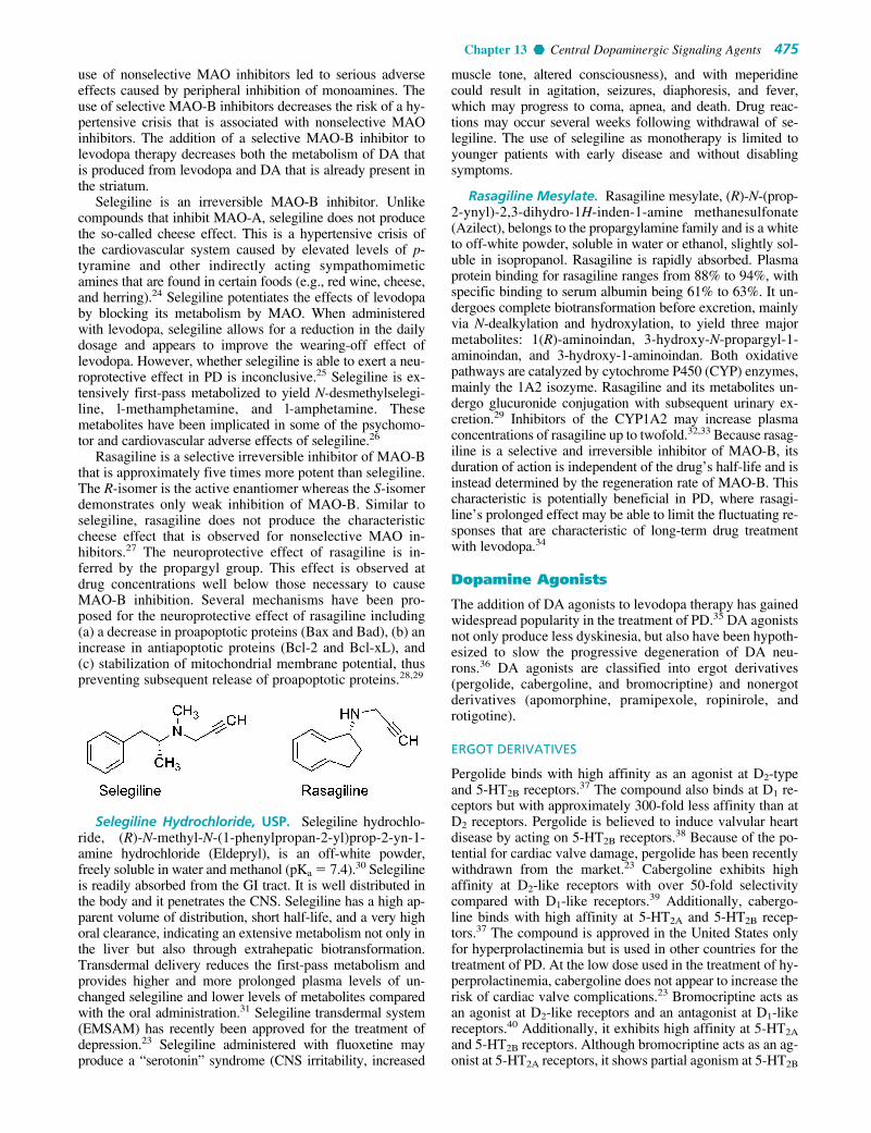
Chapter 13 Central Dopaminergic Signaling Agents 475
use of nonselective MAO inhibitors led to serious adverseeffects caused by peripheral inhibition of monoamines. Theuse of selective MAO-B inhibitors decreases the risk of a hy-pertensive crisis that is associated with nonselective MAOinhibitors. The addition of a selective MAO-B inhibitor tolevodopa therapy decreases both the metabolism of DA thatis produced from levodopa and DA that is already present inthe striatum.
Selegiline is an irreversible MAO-B inhibitor. Unlikecompounds that inhibit MAO-A, selegiline does not producethe so-called cheese effect. This is a hypertensive crisis ofthe cardiovascular system caused by elevated levels of p-tyramine and other indirectly acting sympathomimeticamines that are found in certain foods (e.g., red wine, cheese,and herring).24 Selegiline potentiates the effects of levodopaby blocking its metabolism by MAO. When administeredwith levodopa, selegiline allows for a reduction in the dailydosage and appears to improve the wearing-off effect oflevodopa. However, whether selegiline is able to exert a neu-roprotective effect in PD is inconclusive.25 Selegiline is ex-tensively first-pass metabolized to yield N-desmethylselegi-line, l-methamphetamine, and l-amphetamine. Thesemetabolites have been implicated in some of the psychomo-tor and cardiovascular adverse effects of selegiline.26
Rasagiline is a selective irreversible inhibitor of MAO-Bthat is approximately five times more potent than selegiline.The R-isomer is the active enantiomer whereas the S-isomerdemonstrates only weak inhibition of MAO-B. Similar toselegiline, rasagiline does not produce the characteristiccheese effect that is observed for nonselective MAO in-hibitors.27 The neuroprotective effect of rasagiline is in-ferred by the propargyl group. This effect is observed atdrug concentrations well below those necessary to causeMAO-B inhibition. Several mechanisms have been pro-posed for the neuroprotective effect of rasagiline including(a) a decrease in proapoptotic proteins (Bax and Bad), (b) anincrease in antiapoptotic proteins (Bcl-2 and Bcl-xL), and(c) stabilization of mitochondrial membrane potential, thuspreventing subsequent release of proapoptotic proteins.28,29
muscle tone, altered consciousness), and with meperidinecould result in agitation, seizures, diaphoresis, and fever,which may progress to coma, apnea, and death. Drug reac-tions may occur several weeks following withdrawal of se-legiline. The use of selegiline as monotherapy is limited toyounger patients with early disease and without disablingsymptoms.
Rasagiline Mesylate. Rasagiline mesylate, (R)-N-(prop-2-ynyl)-2,3-dihydro-1H-inden-1-amine methanesulfonate(Azilect), belongs to the propargylamine family and is a whiteto off-white powder, soluble in water or ethanol, slightly sol-uble in isopropanol. Rasagiline is rapidly absorbed. Plasmaprotein binding for rasagiline ranges from 88% to 94%, withspecific binding to serum albumin being 61% to 63%. It un-dergoes complete biotransformation before excretion, mainlyvia N-dealkylation and hydroxylation, to yield three majormetabolites: 1(R)-aminoindan, 3-hydroxy-N-propargyl-1-aminoindan, and 3-hydroxy-1-aminoindan. Both oxidativepathways are catalyzed by cytochrome P450 (CYP) enzymes,mainly the 1A2 isozyme. Rasagiline and its metabolites un-dergo glucuronide conjugation with subsequent urinary ex-cretion.29 Inhibitors of the CYP1A2 may increase plasmaconcentrations of rasagiline up to twofold.32,33 Because rasag-iline is a selective and irreversible inhibitor of MAO-B, itsduration of action is independent of the drug’s half-life and isinstead determined by the regeneration rate of MAO-B. Thischaracteristic is potentially beneficial in PD, where rasagi-line’s prolonged effect may be able to limit the fluctuating re-sponses that are characteristic of long-term drug treatmentwith levodopa.34
Dopamine Agonists
The addition of DA agonists to levodopa therapy has gainedwidespread popularity in the treatment of PD.35 DA agonistsnot only produce less dyskinesia, but also have been hypoth-esized to slow the progressive degeneration of DA neu-rons.36 DA agonists are classified into ergot derivatives(pergolide, cabergoline, and bromocriptine) and nonergotderivatives (apomorphine, pramipexole, ropinirole, androtigotine).
ERGOT DERIVATIVES
Pergolide binds with high affinity as an agonist at D2-typeand 5-HT2B receptors.37 The compound also binds at D1 re-ceptors but with approximately 300-fold less affinity than atD2 receptors. Pergolide is believed to induce valvular heartdisease by acting on 5-HT2B receptors.38 Because of the po-tential for cardiac valve damage, pergolide has been recentlywithdrawn from the market.23 Cabergoline exhibits highaffinity at D2-like receptors with over 50-fold selectivitycompared with D1-like receptors.39 Additionally, cabergo-line binds with high affinity at 5-HT2A and 5-HT2B recep-tors.37 The compound is approved in the United States onlyfor hyperprolactinemia but is used in other countries for thetreatment of PD. At the low dose used in the treatment of hy-perprolactinemia, cabergoline does not appear to increase therisk of cardiac valve complications.23 Bromocriptine acts asan agonist at D2-like receptors and an antagonist at D1-likereceptors.40 Additionally, it exhibits high affinity at 5-HT2A
and 5-HT2B receptors. Although bromocriptine acts as an ag-onist at 5-HT2A receptors, it shows partial agonism at 5-HT2B
Selegiline Hydrochloride, USP. Selegiline hydrochlo-ride, (R)-N-methyl-N-(1-phenylpropan-2-yl)prop-2-yn-1-amine hydrochloride (Eldepryl), is an off-white powder,freely soluble in water and methanol (pKa � 7.4).30 Selegilineis readily absorbed from the GI tract. It is well distributed inthe body and it penetrates the CNS. Selegiline has a high ap-parent volume of distribution, short half-life, and a very highoral clearance, indicating an extensive metabolism not only inthe liver but also through extrahepatic biotransformation.Transdermal delivery reduces the first-pass metabolism andprovides higher and more prolonged plasma levels of un-changed selegiline and lower levels of metabolites comparedwith the oral administration.31 Selegiline transdermal system(EMSAM) has recently been approved for the treatment ofdepression.23 Selegiline administered with fluoxetine mayproduce a “serotonin” syndrome (CNS irritability, increased
0471-0490_17865_Ch13.qxd 12/4/09 5:42 AM Page 475

receptors.41 Compared with cabergoline, bromocriptineexhibits less dyskinesia, which may be related to its D1-antagonist activity.37
Bromocriptine Mesylate, USP. Bromocriptine mesy-late, (6aR,9R)-5-bromo-N-((2R,5S,10aS,10bS)-10b-hydroxy-5-isobutyl-2-isopropyl-3,6-dioxooctahydro-2H-oxazolo[3,2-a]pyrrolo[2,1c]pyrazin-2-yl)-7-methyl-4,6,6a,7,8,9-hexahydroindolo[4,3-fg]quinoline-9-carboxamide methanesulfonate(Parlodel), is a white solid soluble in ethanol and slightly sol-uble in water (pKa’s � 6.6 and 15). Bromocriptine is rapidlyabsorbed after oral administration and it has low systemicbioavailability because of its extensive first-pass metabo-lism. Bromocriptine enters the brain quickly with a half-lifefor uptake into the brain of approximately 0.3 hours; 8% ofthe drug crosses the BBB.23 The metabolites are excreted pri-marily in the bile and feces. The high first-pass hepatic me-tabolism implies an increased risk of drug interactions.Concomitant administration with the DA antagonists, meto-clopramide, or domperidone may aggravate parkinsoniansymptoms and induce extrapyramidal side effects (EPS).Other drugs that may interact with bromocriptine are highlyplasma protein–bound drugs (e.g., warfarin, increased dysk-inesia caused by bromocriptine); macrolides antibacterials(enhanced dopaminergic effects); and caffeine (elevation inplasma bromocriptine concentrations). The combination oflevodopa/AADC inhibitors with bromocriptine permits a re-duction of the dose of levodopa. Thus, the side effects oflevodopa are decreased, resulting in a more continuous stim-ulation of DA receptors.
Cabergoline. Cabergoline, (6aR,9R,10aR)-7-allyl-N-(3-(dimethylamino)propyl) -N - (e thylcarbamoyl)-4,6,6a,7,8,9,10,10a-octahydroindolo[4,3-fg]quinoline-9-carboxamide (Dostinex), is a white powder soluble inalcohol, chloroform, and N,N-dimethylformamide; slightlysoluble in acidic solutions and in n-hexane; and insoluble inwater. Following oral administration, peak plasma concen-trations are reached within 2 to 3 hours. Cabergoline ismoderately bound to plasma proteins in a concentration-independent manner. The absolute bioavailability of caber-goline is unknown. Cabergoline is extensively metabolizedby the liver, predominantly via hydrolysis of the acylureabond of the urea moiety. CYP450 metabolism appears to beminimal. The major metabolites identified thus far do not
contribute to the therapeutic effect of cabergoline. Lessthan 4% is excreted unchanged in the urine. Fecal excretionrepresents the main route of cabergoline elimination. Thereare no reports of interactions of cabergoline with other an-tiparkinsonian agents. Clarithromycin may elevate theplasma concentration of cabergoline by the inhibition ofboth CYP3A4 and P-glycoprotein.42 Cabergoline is a po-tent D2 receptor agonist and is indicated for the treatment ofhyperprolactinemic disorders, either idiopathic or causedby pituitary adenomas.
NONERGOT DERIVATIVES
Pramipexole acts as an agonist at D2 and D3 receptors withgreater affinity for D3 than D2 receptors. The compound hasno appreciable affinity for D1, D5, 5-HT2A, and 5-HT2B re-ceptors.37 The reduced dyskinesia associated with pramipex-ole compared with cabergoline may be related to its lowaffinity for the D1 receptor. Ropinirole has about 20-fold se-lectivity for cloned human D3 receptors compared with D2 re-ceptors. Additionally, ropinirole has low affinity for 5-HT2A,5-HT2B, and D1-like receptors.37 A recent study suggests thatD3-agonists may be able to restore DA neurons and exert aneuroprotective effect.43 These developments could offer ex-citing new therapeutic options for parkinsonian patients.
Apomorphine Hydrochloride, USP. Apomorphine hy-drochloride, (6aR)-6-methyl-5,6,6a,7-tetrahydro-4H-dibenzo[de,g]quinolone-10,11-diol hydrochloride (Apokyn), is awhite or off-white powder or crystal soluble in hot water(pKa � 8.92). Apormorphine is an aporphine alkaloid of thebenzoquinoline class. Oral apomorphine is poorly absorbedand has a bioavailability of less than 4%. Upon subcutaneousadministration, apomorphine is completely absorbed. Within10 to 20 minutes, the maximum concentration of the drug isdistributed from the blood plasma to the CSF. Other potentialroutes of administration include continuous subcutaneous in-fusion, intravenous infusion, intranasal spray application,sublingual, and rectal administration.23 The agent is highlylipophilic in nature, allowing for rapid diffusion across theBBB after injection. Apomorphine has a short plasma half-life; however, clinical effects may last from 60 to 90 minutes.Apomorphine displays a significant degree of interpatientvariability in its pharmacokinetic profile. Studies of bothintravenous and subcutaneous injection routes found this
476 Wilson and Gisvold’s Textbook of Organic Medicinal and Pharmaceutical Chemistry
0471-0490_17865_Ch13.qxd 12/4/09 5:42 AM Page 476

Chapter 13 Central Dopaminergic Signaling Agents 477
variation was not attributable to body weight, age, gender,and duration of PD or L-DOPA dose/duration alone.Apomorphine is extensively metabolized. Hypothesizedroutes include sulfation, N-demethylation, glucuronidation,and oxidation. Subcutaneous injections of apomorphine arerenally and hepatically cleared, with the majority appearingto be renally cleared. Dosage adjustments are needed in bothliver and renal impairment. The activity of apomorphine isbelieved to be caused by stimulation of postsynaptic D1- andD2-type receptors within the caudate/putamen in the brain.Apomorphine is indicated for the acute, intermittent treat-ment of hypomobility, “off” episodes (“end-of-dose wearingoff” and unpredictable on/off episodes) associated with ad-vanced PD.23
Pramipexole Dihydrochloride. Pramipexole dihy-drochloride, (S)-2-amino-6-propylamino-dihydrochloride(Mirapex), is a white to off-white powder soluble in water,slightly soluble in methanol and ethanol, and practically insol-uble in dichloromethane. Following oral administration,pramipexole is readily absorbed. Pharmacokinetic propertiesdiffer between men and women, with area under the curve(AUC) for each dose level being 35% to 43% greater inwomen, mainly because of a 24% to 27% lower oral clear-ance. The drug undergoes minimal hepatic biotransformationand is excreted virtually unchanged in the urine by the renaltubular secretion. Pramipexole interacts with drugs excretedby renal tubular secretion (H2-antagonists, diuretics, vera-pamil, quinidine, quinine), which leads to a decreased clear-ance of pramipexole.44,45 Pramipexole is indicated fortreatment of the signs and symptoms of idiopathic PD, aloneor in combination with levodopa. It is also indicated for symp-tomatic treatment of moderate to severe idiopathic restlesslegs syndrome (RLS).
Ropinirole Hydrochloride. Ropinirole hydrochloride,4-(2-(dipropylamino)ethyl)indolin-2-one hydrochloride(Requip), is a white to pale greenish yellow powder that isvery soluble in water. Ropinirole is rapidly absorbed afteroral administration with maximal plasma concentrationsgenerally reached after about 1.5 hours, and the eliminationhalf-life appears to be approximately 3 hours. Ropinirole isalso rapidly and extensively distributed from the vascularcompartment and shows low plasma protein binding that isindependent of its plasma concentration. This drug iscleared by metabolism in the liver, with only 10% being ex-creted unchanged. The main metabolite of ropinirole is theN-despropyl metabolite. The glucuronide of this metaboliteand the carboxylic acid metabolite, 4-carboxymethylin-dolin-2-one, account only for 10% of the administered dose.None of the metabolites is pharmacologically active, and theexcretion of ropinirole-derived products is mainly via theurine. The main CYP450 isozyme involved in the metab-olism of ropinirole is CYP1A2. Inhibitors or inducers ofthis enzyme have been shown to alter the clearance ofropinirole.46,47 Ropinirole is believed to act as an agonist atpostsynaptic D2 receptors. Ropinirole is indicated for thetreatment of the signs and symptoms of idiopathic PD andmoderate to severe primary RLS.
Rotigotine. Rotigotine, (6S)-6-{propyl[2-(2-thienyl)ethyl]amino}-5,6,7,8-tetrahydro-1-naphthalenol (Neupro),is a nonergoline that is available as a silicone-based, self-adhesive matrix, transdermal system for continuous delivery
over a 24-hour period. Approximately 45% of the drug is re-leased within 24 hours. The terminal half-life of rotigotine is5 to 7 hours after removal of the patch. Rotigotine is 90%bound to plasma proteins. The compound undergoes exten-sive metabolism and has low bioavailability by the oralroute. The major metabolites of rotigotine are the glu-curonide and sulfate conjugates of rotigotine and sulfateconjugates of N-despropylrotigotine and N-desthienylethylrotigotine. Rotigotine is excreted in the urine (71%) andfeces (11%).23 Studies using human liver microsomes didnot find any interactions with CYP1A2, CYP2C9,CYP2C19, CYP2D6, and CYP3A4 substrates.48 Rotigotinetransdermal system contains sodium metabisulfite, and indi-viduals sensitive to sulfite could be at risk for allergic reac-tions. Additionally, somnolence is a common adverse reac-tion with individuals on rotigotine, and patients should beclosely monitored during therapy.23 In transfected Chinesehamster ovary (CHO) cells, rotigotine binds with high affin-ity at D3 and D2L receptors (variants in the D2 receptor sub-type are caused by insertion of the 29 amino acids into thethird loop to give D2s and D2L).49 Using rat CHO cells,rotigotine shows over 30-fold selectivity at D3 versus D2 re-ceptors.48 Rotigotine was approved in May 2007 for thetreatment of early-stage PD.
COMT Inhibitors
When administered with an AADC inhibitor, circulatinglevodopa is mainly metabolized by COMT. In the presenceof a COMT inhibitor, the peripheral metabolism of levodopais therefore decreased.
Tolcapone is a selective and reversible inhibitor ofCOMT in the CNS and periphery. Inhibition of COMT inthe periphery reduces the formation of 3OMD in tissues thathave significant COMT activity. It has been speculated that3OMD may compete with the uptake of levodopa into theCNS and thereby contributes to the wearing-off effect oflevodopa. Administration of levodopa in combination witha dopa decarboxylase inhibitor (carbidopa) and a COMT in-hibitor (tolcapone) increases CNS delivery of levodopa.Therefore, more continuous levels of levodopa and DA aremaintained.50 Unlike tolcapone, entacapone does not pene-trate the BBB to any extent. Thus, the compound only in-hibits peripheral COMT. When combined with levodopaand carbidopa, entacapone provides less motor fluctuationsin parkinsonian patients. Another advantage of entacaponeover tolcapone is its lack of hepatotoxicity.51
Tolcapone. Tolcapone, 3,4-dihydroxy-4�-methyl-5-nitrobenzophenone (Tasmar), is a yellow, odorless, nonhy-groscopic, crystalline compound (pKa � 4.78). Tolcapone israpidly absorbed after oral administration. Tolcapone ishighly bound to plasma albumin (�98%), and its distributionis therefore restricted. Tolcapone has low first-pass metabo-lism. It is almost completely metabolized in the liver before
0471-0490_17865_Ch13.qxd 12/4/09 5:42 AM Page 477

excretion, and 60% is excreted by the kidney. The majormetabolite of tolcapone is an inactive glucuronide conjugate.COMT inhibitors increase chronotropic and arrhythmogeniceffects of epinephrine.52,53 Tolcapone is indicated as an ad-junct to levodopa/carbidopa for the management of signs andsymptoms of PD.
Entacapone. Entacapone, (E)-2-cyano-3-(3,4-dihy-droxy-5-nitrophenyl)-N,N-diethyl-2-propenamide (Comtan),is a nitrocatechol that is practically insoluble in water (pKa �4.50). Entacapone is rapidly absorbed after oral administra-tion and does not cross the BBB. Entacapone does not dis-tribute widely into tissues because of its high plasma proteinbinding and it is completely metabolized before excretion.The main metabolic pathway is by isomerization to the cis-isomer followed by direct glucuronidation of the parent andthe cis-isomer. The glucuronide conjugates are inactive.Entacapone is eliminated in the feces (90%) and urine (10%).Entacapone is indicated as an adjunct to levodopa/carbidopato treat patients with idiopathic PD who experience the signsand symptoms of end-of-dose wearing off.
Carbidopa/Levodopa/Entacapone. Levodopa, car-bidopa, and entacapone (Stalevo) are formulated together totake advantage of each compound’s mechanism of action.When entacapone is coadministered with levodopa andcarbidopa, plasma levels of levodopa are greater and moresustained.23 For absorption, distribution, metabolism, andexcretion (ADME) properties and drug interactions, refer tothe preceding monograph.
Other Antiparkinsonian Drugs
Anticholinergic agents have been used in the early stages ofPD, frequently with amantadine and selegiline. The mostcommonly employed anticholinergics are benztropine, tri-hexyphenidyl, orphenadrine, and procyclidine. These com-pounds are most useful for treating the tremor aspect ofPD. Because most individuals with PD are elderly, anti-cholinergics may impair cognitive function. Additionally,anticholinergics may produce various side effects throughblockade of peripheral muscarinic receptors.21,26
Amantadine has demonstrated beneficial effects inthe treatment of PD, especially in attenuating dyskine-sias.26 Although amantadine has DA-releasing properties,other pharmacological mechanisms appear to be involvedin its antiparkinsonian effect. The compound is a weaknoncompetitive N-methyl D-aspartate (NMDA) antagonist,yet it differs in its action from the NMDA antagonist MK-801 in a rodent model of PD.54 Additionally, amantadinepotently inhibits nicotinic acetylcholine function in thehippocampus.55
ANTIPSYCHOTIC DRUGS
Psychotic illness is a compilation of multiple disorders, in-cluding schizophrenia, the manic phase of bipolar syndrome,acute idiopathic psychosis, and other conditions marked bysevere agitation,56 yet the term “psychosis” is most often as-sociated with schizophrenia. Schizophrenia affects approxi-mately 1% of the U.S. population,57 with both genetic58 andneurodevelopmental59 implications. Schizophrenia is charac-terized by delusions, abnormal behaviors, hallucinations, andthought disorders (i.e., positive symptoms), as well as loss ofnormal emotions, abilities, and motivation (i.e., negativesymptoms). Although the etiology of schizophrenia is un-known, the principal neurochemical theories focus on DA60
and, more recently, on glutamate and serotonin.61,62 Much ofour current knowledge of the mechanisms involved in schizo-phrenia has come from analyzing effects of antipsychoticdrugs.
The DA hypothesis of schizophrenia (DHS) has been theprincipal neurochemical theory of schizophrenia for morethan 30 years. The basis of the DHS lies in the capacity of an-tipsychotic drugs to block DA receptors (D2 receptor subtype)positively correlated to their clinical potency in alleviating thesymptoms of schizophrenia.60,63 Nevertheless, the observedinactivity of D2-antagonists in some individuals with schizo-phrenia and the pharmacological independence of positiveand negative symptoms indicate further level of complexity.Other variants of the DHS suggest an imbalance betweendopamingeric pathways, which may be phasic in nature.64,65
With regard to symptomology, it is thought that a dysregula-tion in the mesocortical DA pathway contributes to negativesymptoms.66,67 Positron emission tomography (PET) studiessuggest that individuals with schizophrenia may have de-creased densities of D1 receptors in the prefrontal cortex.68
Presynaptic D1 receptors within the prefrontal cortex are be-lieved to modulate glutamatergic activity, directly effectingworking memory in individuals with schizophrenia.69 Thepositive symptoms are believed to be derived from D2 recep-tor hyperactivity or upregulation within the in mesolimbicpathway.66,70 Although it remains uncertain as to exactly howthe different dopaminergic pathways and DA receptor sub-type activities mediate the positive and negative symptoms ofschizophrenia, it is clear that DA is a critical mediator. Yet,other transmitters have also been implicated. GlutamateNMDA receptor antagonists (e.g., phencyclidine, ketamine)produce psychotic symptoms in humans. A combined gluta-mate and DA dysregulation has also been implicated withschizophrenia.71 Likewise, serotonin has modulatory effectson DA pathways in the brain and has been implicated in thereduction of side effects induced by D2 receptor antagonists.
Typical Antipsychotic Agents
Typical antipsychotics (also known as first-generation orconventional antipsychotics, classical neuroleptics, or majortranquilizers) are a class of antipsychotic drugs firstdeveloped in the 1950s and used to treat psychosis (inparticular, schizophrenia). Typical antipsychotics includephenothiazines, thioxanthenes, butyrophenones, diphenyl-butylpiperidines, and the dihydroindolones (e.g., molin-done, not commercially available in the United States).The conventional antipsychotic drugs can be classifiedas high (haloperidol and fluphenazine) or low potency
478 Wilson and Gisvold’s Textbook of Organic Medicinal and Pharmaceutical Chemistry
0471-0490_17865_Ch13.qxd 12/4/09 5:42 AM Page 478

Chapter 13 Central Dopaminergic Signaling Agents 479
(chlorpromazine) based on their affinity for DA D2 recep-tors and the average therapeutic dose.72 This group of an-tipsychotics is also associated with a significant degree ofEPS. Some EPS are defined as acute dystonias (i.e., invol-untary movements: muscle spasms, protruding tongue),which tend to occur within the first few weeks of treatmentand decline over time. However, the EPS that is most widelyassociated with these drugs is tardive dyskinesia (i.e., im-pairment of voluntary movements, often in the face andtongue), which develops over months to years and is oftenirreversible. High-potency typical antipsychotics tend to beassociated with more EPS and less histaminic (sedation),alpha adrenergic (orthostasis), and anticholinergic (drymouth) side effects. Low-potency typical antipsychotics areassociated with less EPS but more H1, �1, and muscarinicside effects. Currently, the only patients in which the typicalantipsychotic agents are preferred are those for whom thereis a clear indication for short- or long-acting injectablepreparations, and/or a tolerance to the side effects.
Phenothiazines and Thioxanthenes
The phenothiazine nucleus was first synthesized in 1876(methylene blue) by Badische Anilin und Soda Fabrik(BASF) chemist H. Caro and elucidated by A. Bernstein in1883 (thiodiphenylamine). It was not until 1950 that chlor-promazine (Table 13.1) was synthesized to explore its antihis-taminic properties. The observations on the side effects ofchlorpromazine prompted French psychiatrists Delay,Denicker, and Harl to use it in schizophrenic patients, pub-lishing their seminal paper in 1952.73 In 1958, Petersen etal.74,75 synthesized the first thioxanthene. The structural dif-ference with the phenothiazines is the bioisosteric replace-ment of the N at the 10-position for a carbon atom in thethioxanthenes. The side chain (R10) in the thioxanthenesis attached by a double bond to the tricyclic system. This por-tion of the molecule is not involved in the interaction with thereceptor, but it serves to position correctly the other elementsof the molecule. Thioxanthenes that have a double bond in theside chain can be either cis- or trans-isomers (Fig. 13.5).76,77
The active form is the cis-isomer, which can be perfectly su-perimposed with the phenothiazine and the DA molecules,meaning that the Z-configuration is the preferred one for op-timal receptor affinity. In general, thioxanthenes are more po-tent than the structurally related phenothiazines.
STRUCTURE–ACTIVITY RELATIONSHIPS
The SAR of phenothiazines is relatively simple becauseonly position 2 (ring A) and the side chain afford activeantipsychotic molecules. Conformational studies on chlor-promazine reveal that tilting of the side chain toward ringA allows favorable van der Waals interactions of the sidechain with the chlorine substituent.78 The resulting confor-mation permits the superimposition of DA. Substitution atthe 1-position sterically hinders the ability of the side chainto approach ring A. On the other hand, a substituent at the 3-position will be too far from the side chain to provide vander Waals attractions. A trifluoromethyl group will givemore van der Waals contacts with the side chain than achloro substituent and, in fact, triflupromazine is more po-tent than chlorpromazine (Table 13.1). As a general rule,electron-withdrawing groups at the 2-position increasethe antipsychotic efficacy (chlorpromazine vs. promazine).
In the thioxanthenes, the substituent at the 2-position doesnot govern the side chain in the same way as with the phe-nothiazines to assume a DA-like conformation.79 The cis ortrans conformation of unsubstituted thioxanthenes does notshow any differential potency. For thioxanthenes, the 2-substituent of the cis form provides a closer approximationof the side chain toward ring A, enabling the substituentto enter into the van der Waals attractive forces with theside chain.78,80,81
A major requirement for the antipsychotic activity ofphenothiazines is that the basic amino group be separated bythree carbon atoms from the parent ring system. Molecularmodels indicate that a shorter or a branched side chain pro-hibits the assumption of the DA-like conformation causedby van der Waals repulsive forces between the side chainand the phenothiazine ring. The nature of the substituent onthe side chain amine may also influence the conformation ofthe phenothiazine side chain. A piperazine ring affords morevan der Waals contacts with the 2-substituent than doesan alkylamino side chain. Piperazinyl phenothiazines aremore potent in their antischizophrenic effects, their ability toelicit EPS, and their affinity for the DA-sensitive adenylatecyclase than alkylamino phenothiazines.78,82
PHARMACOKINETIC PROPERTIES
In general, these classes of drugs are rapidly absorbed fromthe GI tract (Table 13.1), but there is considerable individualpatient variation in peak plasma concentrations.83
Chlorpromazine has been the most widely studied phenothi-azine, and it is believed that thioxanthenes and phenothi-azines are metabolized following a very similar pathway.Compared with the phenothiazines, the thioxanthenes are lesslikely to form phenolic metabolites.84 Chlorpromazine under-goes extensive first-pass metabolism yielding numerousmetabolites. Numerous sites of attack by microsomalCYP450 system (especially CYP2D6) are possible, and mostof these reactions happen to various degrees.85,86 Severalmetabolites have been isolated and characterized. Phase I re-actions include oxidative N-demethylation to give primaryand secondary amines, aromatic hydroxylation that yieldsphenols, side chain tertiary amine oxidation that affords N-oxide metabolites, S-oxidation to give sulfoxide and sulfones,oxidative deamination that yields side chain carboxylic acids,and N-10 dealkylation to give chlorophenothiazine. Most ofthese metabolites are subject of further reactions similar tothose already mentioned. From the 168 proposed metabolites,only 45 have been isolated, and the 7-hydroxy-chlorpro-mazine metabolite has been evaluated and found to be effec-tive in schizophrenia. More recently, the sulfoxide metabolitewas found to have a diminished therapeutic response.87,88
Excretion is primarily via the kidneys with less than 1% of adose excreted unchanged in the urine and 20% to 70% asconjugated or unconjugated metabolites. Approximately 5%of a dose is excreted in feces via biliary elimination. Somemetabolites can still be detected up to 18 months after discon-tinuation of long-term therapy.
SPECIFIC AGENTS
Chlorpromazine Hydrochloride, USP. Chlorpromazinehydrochloride, 2-chloro-10-[3-(dimethylamino)propyl]phe-nothiazine (Thorazine), is a white to slightly creamy white,odorless, bitter tasting, crystalline powder (pKa � 9.43). The
0471-0490_17865_Ch13.qxd 12/4/09 5:42 AM Page 479

drug has significant sedative and hypotensive properties, pos-sibly reflecting central and peripheral �1-adrenergic blockingactivity, respectively. As with the other phenothiazines, theeffects of the other CNS-depressant drugs, such as sedativesand anesthetics, can be potentiated.90 Chlorpromazine is aminor substrate of CYP1A2 and 3A4 and a major substrate
of CYP2D6.91 Chlorpromazine also is a strong inhibitor ofCYP2D6 and a weak inhibitor of CYP2E1. Inhibitors of theCYP2D6 enzyme may increase the levels/effects of chlorpro-mazine and chlorpromazine may increase the levels/effectsof CYP2D6 substrates. Chlorpromazine may also de-crease the bioactivation of CYP2D6 prodrug substrates.91
480 Wilson and Gisvold’s Textbook of Organic Medicinal and Pharmaceutical Chemistry
TABLE 13.1 Structure and Pharmacokinetic Properties of Phenothiazines and Thioxanthenes
Name R2 R10 Absorption %89 PPB %89 Half-life (h)89
Promazine H Erratic* 94
Chlorpromazine Cl 40* 98 8–35
Thioridazine SCH3 60 95 21–25
Perphenazine Cl 40 8–12
Prochlorperazine Cl 91–99 6–8
Fluphenazine CF3 40–50 90 4–12
Trifluoperazine CF3 91–99 4–12
Chlorprothixene 17–24
Thiothixene Erratic 91–99 3.5–34
*Erratic because of variable metabolism in the intestinal wall and liver (marked first-pass effect).PPB %, percentage of plasma protein binding.
0471-0490_17865_Ch13.qxd 12/4/09 5:42 AM Page 480

Chapter 13 Central Dopaminergic Signaling Agents 481
Chlorpromazine has strong anticholinergic and sedative ef-fects and is a potent antiemetic. It is considered a low-potencyneuroleptic agent and therefore the associated incidence ofEPS is low. The incidence of sedative, anticholinergic, andcardiovascular side effects is high, however. It is indicated forthe symptomatic relief of nausea, vomiting, hiccups, and por-phyria; for preoperative sedation; and for the treatment ofpsychotic disorders.
Thioridazine Hydrochloride, USP. Thioridazinehydrochloride, 10-[2-(1-methyl-2-piperidyl)ethyl]-2-(methylthio)phenothiazine (Mellaril), is a member of thepiperidine subgroup of the phenothiazines. Thioridazine oc-curs as a white or slightly yellow, crystalline or micronizedpowder, which is odorless or has a faint odor and is practi-cally insoluble in water and freely soluble in dehydratedalcohol (pKa � 9.66). Thioridazine has relatively low ten-dency to produce EPS. The drug has sedative and hypoten-sive activity in common with chlorpromazine and lessantiemetic activity. At high doses, pigmentary retinopathyhas been observed. Thioridazine has similar activity towardthe CYP450 family of enzymes as chlorpromazine.Thioridazine is indicated for the treatment of schizophrenicpatients who fail to respond adequately to treatment withother antipsychotic drugs.
Perphenazine, USP. Perphenazine, 2-[4-[3-(2-chloro-10H-phenothiazin-10-yl)propyl]piperazin-1-yl]ethanol.Perphenazine is a minor substrate of CYP1A2, 2C9, 2C19,and 3A4 and a major substrate for 2D6. Perphenazine is in-dicated for use in the management of the manifestations ofpsychotic disorders and for the control of severe nausea andvomiting in adults.
Trifluoperazine Hydrochloride, USP. Trifluoperazinehydrochloride, 10-[3-(4-Methyl-1-piperazinyl)propyl-[2-(trifluoromethyl)phenothiazine dihydrochloride (Stelazine).The absorption of trifluoperazine is erratic and variable andit is widely distributed into tissues. The excretion of trifluo-perazine occurs 50% via kidneys and the other 50% isthrough enterohepatic circulation. The metabolism and druginteractions of trifluoperazine are the same as with the otherphenothiazines. Trifluoperazine does not show any specificdrug interactions. Trifluoperazine is indicated for the man-agement of schizophrenia and short-term treatment ofnonpsychotic anxiety.
Thiothixene, USP. Thiothixene, N,N-dimethyl-9-[3-(4-methyl-1-piperazinyl)propylidene]thioxanthene-2-sulfonamide (Navane). The absorption of thiothixene iserratic because of its high lipophilicity. Thiothixene is
widely distributed into tissues and it is more than 90%bound to plasma proteins. Thiothixene has a similar meta-bolic pathway as the phenothiazines. Thiothixene is amajor substrate of CYP1A2, and CYP1A2 inducersmay decrease the levels/effects of thiothixene, whereasCYP1A2 inhibitors may increase the levels/effects of thio-thixene. Thiothixene is also a weak inhibitor of CYP2D6.As with other antipsychotic agents, some patients who areresistant to previous medications have responded favor-ably to thiothixene. Thioxanthenes may also be of valuein the management of withdrawn, apathetic schizophrenicpatients.
BUTYROPHENONES AND RELATED STRUCTURES
Haloperidol was discovered by Janssen Laboratories (1958)as a byproduct while they were investigating analgesicsstructurally related to meperidine.73 The behavioral profileof haloperidol was found to be very similar to that of chlor-promazine, but the required dose was about 50-fold less toexert the same behavioral effect. It was rapidly studied andpursued as a new drug. Soon after, Janssen Laboratoriesdiscovered and developed droperidol, another butyrophe-none analog. Conformationally, the butyrophenones canassume a conformation resembling portions of thephenothiazine molecule. The three-carbon side chain of thephenothiazines may be analogous to the three-carbonbridge that separates the amino function from the carbonylmoiety of haloperidol. Moreover, the piperidine ring wouldcorrespond to the side chain amine or piperazine of the phe-nothiazines. After the introduction of haloperidol anddroperidol, a great number of SAR studies were carriedout.92 As for the pharmacophore of the butyrophenones(Fig. 13.6), it is known that a tertiary amino group at thefourth carbon as well as the para-substituted (F is pre-ferred) phenyl ring at the 1-position are required for D2
affinity.93,94 Possible variations on the butyrophenonegroup are at the piperidine moiety, in particular at the 4-position of the ring. Modifications by lengthening, shorten-ing, or branching of the three-carbon propyl chain decreaseneuroleptic activity. Replacement of the keto group also de-creases D2 affinity. The tertiary amino group is usually partof an N-containing heterocycles, preferably a piperidinering. Replacement of the six-member basic ring by smalleror larger heterocyclic rings or by noncyclic amines de-creases DA affinity.
Conformationally restricted butyrophenones also possesshigh D2 affinity. Haloperidol and droperidol are the only bu-tyrophenones currently commercially available in the UnitedStates. Replacement of the keto group for a di-4-fluo-rophenylmethane moiety produced diphenylbutylpiperidines,another class of neuroleptics. Pimozide, the prototype of thisgroup of drugs, is structurally related to droperidol and is theonly member commercially available within the diphenyl-butylpiperidines class.
Figure 13.5 Trans-(A) versus cis-(B) isomers in thethiothixenes.
Figure 13.6 Pharmacophore of the butyrophenones.
0471-0490_17865_Ch13.qxd 12/4/09 5:42 AM Page 481

PHARMACOKINETIC PROPERTIES
Haloperidol is the prototype of the butyrophenones and theone that has been studied the most. Haloperidol and droperi-dol, the two butyrophenones available in the United States,are well absorbed from the GI tract.83 First-pass metabolismin the liver reduces the bioavailability of haloperidol toapproximately 60%. Haloperidol undergoes extensive me-tabolism including N-dealkylation, reduction, oxidation, andN-oxidation. Most of the metabolites of haloperidol areinactive. Hydroxyhaloperidol, the reduced (ketone) metabo-lite of haloperidol, is the only active metabolite and it is alsosubject to extensive hepatic metabolism (Fig. 13.7).83,95
SPECIFIC AGENTS
Haloperidol, USP. Haloperidol, 4-[4-(p-chlorophenyl)-4-hydroxypiperidino]-4-fluorobutyrophenone (Haldol), is anodorless white to yellow crystalline powder. Haloperidol iswell and rapidly absorbed and has a high bioavailability. It ismore than 90% bound to plasma proteins. Haloperidol is ex-creted slowly in the urine and feces. About 30% of a dose isexcreted in urine and about 20% of a dose in feces via biliaryelimination,96 and only 1% of a dose is excreted as unchangeddrug in the urine.97 Haloperidol is a minor substrate of
CYP1A2 and a major substrate of CYP2D6 and CYP3A4.CYP2D6 inhibitors may increase the levels/effects ofhaloperidol.91 Haloperidol may increase the levels/effects ofCYP2D6 substrates98 and it may decrease the bioactivationof CYP2D6 prodrugs substrates. Haloperidol also is a moder-ate inhibitor of CYP2D6 and CYP3A4. CYP3A4 inducersmay decrease the levels/effects of haloperidol, whereasCYP3A4 inhibitors may increase the levels/effects ofhaloperidol. Centrally acting acetylcholinesterase inhibitorsmay increase the risk of antipsychotic-related EPS. The pre-cise mechanism of antipsychotic action is unclear but isconsidered to be associated with the potent DA D2
receptor–blocking activity in the mesolimbic system and theresulting adaptive changes in the brain. Haloperidol is usedprimarily for the long-term treatment of psychosis and is es-pecially useful in patients who are noncompliant with theirdrug treatment.
Haloperidol Decanoate. Haloperidol decanoate, 4-[4-(4-chlorophenyl)-4-hydroxypiperidino]-4-fluorobutyrophe-none decanoate (Haldol Decanoate), is the decanoate ester(prodrug) of haloperidol. Peak plasma concentrations occurwithin 3 to 9 days and then decrease slowly. Haloperidol de-canoate has no intrinsic activity. Haloperidol decanoate
482 Wilson and Gisvold’s Textbook of Organic Medicinal and Pharmaceutical Chemistry
Figure 13.7 Metabolic pathway for haloperidol.
0471-0490_17865_Ch13.qxd 12/4/09 5:42 AM Page 482
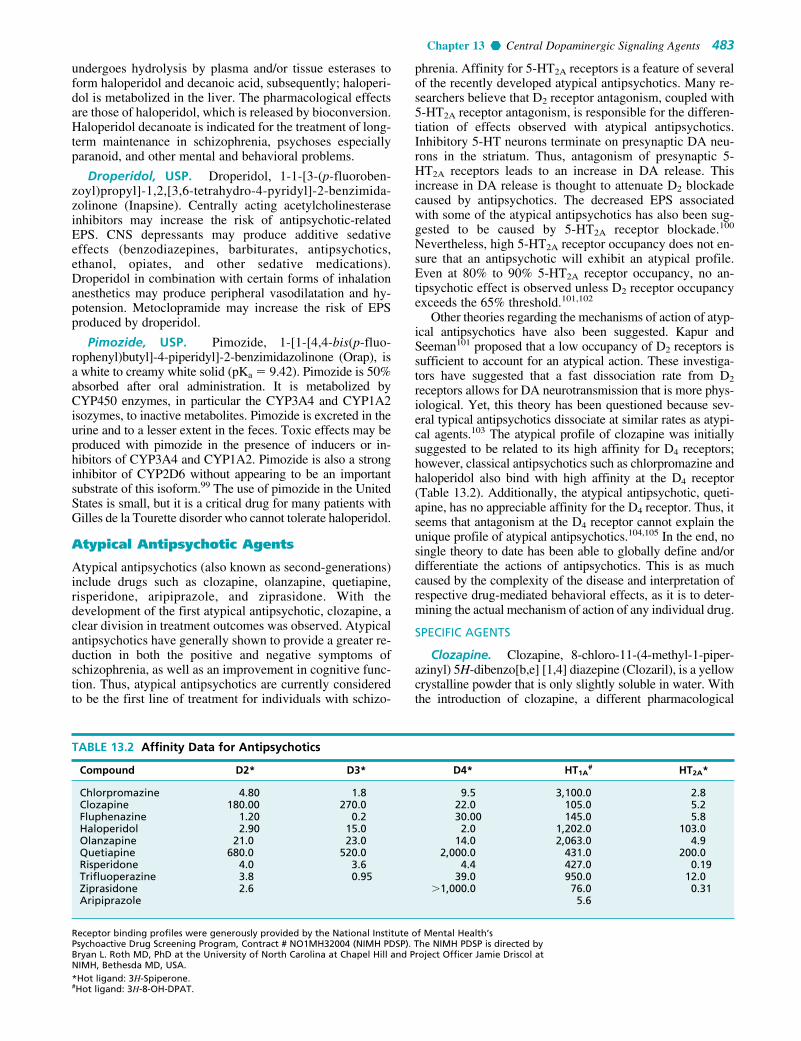
Chapter 13 Central Dopaminergic Signaling Agents 483
undergoes hydrolysis by plasma and/or tissue esterases toform haloperidol and decanoic acid, subsequently; haloperi-dol is metabolized in the liver. The pharmacological effectsare those of haloperidol, which is released by bioconversion.Haloperidol decanoate is indicated for the treatment of long-term maintenance in schizophrenia, psychoses especiallyparanoid, and other mental and behavioral problems.
Droperidol, USP. Droperidol, 1-1-[3-(p-fluoroben-zoyl)propyl]-1,2,[3,6-tetrahydro-4-pyridyl]-2-benzimida-zolinone (Inapsine). Centrally acting acetylcholinesteraseinhibitors may increase the risk of antipsychotic-relatedEPS. CNS depressants may produce additive sedativeeffects (benzodiazepines, barbiturates, antipsychotics,ethanol, opiates, and other sedative medications).Droperidol in combination with certain forms of inhalationanesthetics may produce peripheral vasodilatation and hy-potension. Metoclopramide may increase the risk of EPSproduced by droperidol.
Pimozide, USP. Pimozide, 1-[1-[4,4-bis(p-fluo-rophenyl)butyl]-4-piperidyl]-2-benzimidazolinone (Orap), isa white to creamy white solid (pKa � 9.42). Pimozide is 50%absorbed after oral administration. It is metabolized byCYP450 enzymes, in particular the CYP3A4 and CYP1A2isozymes, to inactive metabolites. Pimozide is excreted in theurine and to a lesser extent in the feces. Toxic effects may beproduced with pimozide in the presence of inducers or in-hibitors of CYP3A4 and CYP1A2. Pimozide is also a stronginhibitor of CYP2D6 without appearing to be an importantsubstrate of this isoform.99 The use of pimozide in the UnitedStates is small, but it is a critical drug for many patients withGilles de la Tourette disorder who cannot tolerate haloperidol.
Atypical Antipsychotic Agents
Atypical antipsychotics (also known as second-generations)include drugs such as clozapine, olanzapine, quetiapine,risperidone, aripiprazole, and ziprasidone. With thedevelopment of the first atypical antipsychotic, clozapine, aclear division in treatment outcomes was observed. Atypicalantipsychotics have generally shown to provide a greater re-duction in both the positive and negative symptoms ofschizophrenia, as well as an improvement in cognitive func-tion. Thus, atypical antipsychotics are currently consideredto be the first line of treatment for individuals with schizo-
phrenia. Affinity for 5-HT2A receptors is a feature of severalof the recently developed atypical antipsychotics. Many re-searchers believe that D2 receptor antagonism, coupled with5-HT2A receptor antagonism, is responsible for the differen-tiation of effects observed with atypical antipsychotics.Inhibitory 5-HT neurons terminate on presynaptic DA neu-rons in the striatum. Thus, antagonism of presynaptic 5-HT2A receptors leads to an increase in DA release. Thisincrease in DA release is thought to attenuate D2 blockadecaused by antipsychotics. The decreased EPS associatedwith some of the atypical antipsychotics has also been sug-gested to be caused by 5-HT2A receptor blockade.100
Nevertheless, high 5-HT2A receptor occupancy does not en-sure that an antipsychotic will exhibit an atypical profile.Even at 80% to 90% 5-HT2A receptor occupancy, no an-tipsychotic effect is observed unless D2 receptor occupancyexceeds the 65% threshold.101,102
Other theories regarding the mechanisms of action of atyp-ical antipsychotics have also been suggested. Kapur andSeeman101 proposed that a low occupancy of D2 receptors issufficient to account for an atypical action. These investiga-tors have suggested that a fast dissociation rate from D2
receptors allows for DA neurotransmission that is more phys-iological. Yet, this theory has been questioned because sev-eral typical antipsychotics dissociate at similar rates as atypi-cal agents.103 The atypical profile of clozapine was initiallysuggested to be related to its high affinity for D4 receptors;however, classical antipsychotics such as chlorpromazine andhaloperidol also bind with high affinity at the D4 receptor(Table 13.2). Additionally, the atypical antipsychotic, queti-apine, has no appreciable affinity for the D4 receptor. Thus, itseems that antagonism at the D4 receptor cannot explain theunique profile of atypical antipsychotics.104,105 In the end, nosingle theory to date has been able to globally define and/ordifferentiate the actions of antipsychotics. This is as muchcaused by the complexity of the disease and interpretation ofrespective drug-mediated behavioral effects, as it is to deter-mining the actual mechanism of action of any individual drug.
SPECIFIC AGENTS
Clozapine. Clozapine, 8-chloro-11-(4-methyl-1-piper-azinyl) 5H-dibenzo[b,e] [1,4] diazepine (Clozaril), is a yellowcrystalline powder that is only slightly soluble in water. Withthe introduction of clozapine, a different pharmacological
TABLE 13.2 Affinity Data for Antipsychotics
Compound D2* D3* D4* HT1A# HT2A*
Chlorpromazine 4.80 1.8 9.5 3,100.0 2.8Clozapine 180.00 270.0 22.0 105.0 5.2Fluphenazine 1.20 0.2 30.00 145.0 5.8Haloperidol 2.90 15.0 2.0 1,202.0 103.0Olanzapine 21.0 23.0 14.0 2,063.0 4.9Quetiapine 680.0 520.0 2,000.0 431.0 200.0Risperidone 4.0 3.6 4.4 427.0 0.19Trifluoperazine 3.8 0.95 39.0 950.0 12.0Ziprasidone 2.6 �1,000.0 76.0 0.31Aripiprazole 5.6
Receptor binding profiles were generously provided by the National Institute of Mental Health’sPsychoactive Drug Screening Program, Contract # NO1MH32004 (NIMH PDSP). The NIMH PDSP is directed byBryan L. Roth MD, PhD at the University of North Carolina at Chapel Hill and Project Officer Jamie Driscol atNIMH, Bethesda MD, USA.*Hot ligand: 3H-Spiperone. #Hot ligand: 3H-8-OH-DPAT.
0471-0490_17865_Ch13.qxd 12/4/09 5:42 AM Page 483

profile was observed compared with the classical antipsy-chotics.106 Unlike typical antipsychotics, clozapine is largelydevoid of EPS. The lack of EPS with this compound waspostulated to be caused by its preferential binding tomesolimbic rather than striatal DA receptors.107 Furthermore,clozapine was shown to exhibit potent affinity for 5-HT2A
receptors.108 When administered at a dose of 100 mg twicea day (bid [L. bis in die]), an average steady state plasma con-centration of 319 ng/mL resulted at an average of 2.5 hoursafter administration. Because food does not apparently affectabsorption, clozapine may be given with or without food. Thecompound is 97% bound to plasma proteins; however, inter-actions with other drugs have not been fully investigated.23
Clozapine is extensively metabolized after absorption with50% of an administered dose excreted in the urine and 30%in the feces. Clozapine is metabolized to two major metabo-lites by N-oxidation and N-demethylation. Clozapine N-oxide(CZNO) is thought to be formed by the action of CYP3A4and flavin monooxygenase 3 (FMO3), whereas N-desmethyl-clozapine (DMCZ) appears to result by the action of CYP3A4and CYP1A2 (Fig. 13.8).109 Clozapine also binds with highaffinity at M1 and M4 muscarinic receptors in the limbic areaof the brain.104 Whether affinity for muscarinic receptorscontributes to the atypical profile for clozapine has not beenestablished.103
DMCZ shows partial agonism at D2 and D3 receptors andexhibits a distinctly different pharmacological profile com-pared with clozapine and other atypical antipsychotics.Unlike clozapine, which is a potent M1 muscarinic antago-nist, DMCZ is a potent M1 agonist. Agonism at muscarinicreceptors has been proposed to be useful for impaired cog-nition in schizophrenia. Burnstein et al.110 found thatDMCZ acted as a partial agonist at DA D2 and D3 receptors.These investigators suggested that the low incidence of EPSassociated with clozapine may be caused by the partial ago-nism of DMCZ at D2 and D3 receptors. Thus, DMCZ maybe of interest as an atypical antipsychotic with an improvedside effect profile compared with clozapine.
Although clozapine demonstrates a favorable pharmaco-logical profile compared with typical antipsychotics, its useis restricted by a relatively high incidence of agranulocyto-sis. The exact mechanism for the cause of agranulocytosishas not been confirmed, but a highly reactive nitrenium ionthat is formed by the action of hepatic enzymes appears tobe involved.111 The mean elimination half-life of clozapine
following a single 75-mg dose is 8 hours. Because of severaladverse effects, clozapine is only used in refractory casesof schizophrenia. Individuals with a history of seizures orpredisposed to seizures should be cautioned when takingclozapine. Similar to other atypical antipsychotic agents,clozapine causes an increased risk of mortality in elderly in-dividuals with dementia-related psychoses.23
Olanzapine. Olanzapine, 2-methyl-4-(4-methyl-1-piper-azinyl)-10H-thieno[2,3-b][1,5] benzodiazepine (Zyprexa), isa yellow crystalline solid that is essentially insoluble in water.Olanzapine orally disintegrating tablets (Zyprexa Zydis) is asolid dosage form that immediately disintegrates when ex-posed to saliva. This product is useful for elderly patients whohave difficulty in swallowing. An injectable form, olanzapinefor injection (Zyprexa IM), is indicated for agitation associ-ated with schizophrenia or bipolar I mania. Olanzapine iscombined with fluoxetine (Symbyax) for use in depressionthat is associated with bipolar I disorder. Peak concentrationsof oral olanzapine are reached at 6 hours after oral adminis-tration, and absorption of the compound is not affected byfood.23 Olanzapine is 93% bound to plasma proteins, and itsmean half-life is 27 hours.112,113 Olanzapine undergoes exten-sive first-pass metabolism, and about 40% of an oral dose ismetabolized prior to reaching the systemic circulation. Themajor metabolite of olanzapine in human subjects is the 10-N-glucuronide. Other human metabolites that have identifiedinclude the N-desmethyl, the N-oxide, and the 2-hydrox-ymethyl metabolites (Fig. 13.9). The N-desmethyl and the 2-hydroxymethyl metabolites are products of CYP1A2 andCYP2D6 action, respectively.112,113
Olanzapine binds with high affinity at DA D2, 5-HT2A,5-HT2C, 5-HT6, �1, and H1 histamine receptors. The effectsof olanzapine for the treatment of schizophrenia are pre-sumably mediated through antagonism at D2 and 5-HT2A
receptors.23,112,113 The use of olanzapine in acute maniathat is associated with bipolar I disorder is thought to bemediated by antagonism at D2 and other monamine recep-tors. Additionally, olanzapine is postulated to produce itsmood stabilizing and antidepressant effects through 5-HT2A
receptor blockade and increased cortical DA and NE con-centrations.114 Several studies have investigated the effectof olanzapine-induced weight gain and new-onset type 2diabetes.115,116 In a comparison study with risperidone,olanzapine was shown to have a greater risk of producingdyslipidemia and type 2 diabetes.105,116
Quetiapine. Quetiapine, 2-[2-(4-dibenzo[b,f][1,4]thi-azepin-11-yl-1-piperazinyl)ethoxy]-ethanol fumarate (2:1,salt) (Seroquel), is a white to off-white crystalline powderthat is moderately water soluble. Quetiapine is rapidly ab-sorbed, and peak plasma levels occur 1 to 2 hours after ad-ministration. Food does not appreciably affect the absorptionof quetiapine. The compound is 83% bound to plasma pro-teins and it has a mean elimination half-life of 7 hours.Administration of a single dose of 14C-quetiapine showedthat only 1% of the drug was excreted unchanged, with 73%excreted into the urine and approximately 30% excreted inthe feces.23 Numerous metabolites of quetiapine are known,and the sulfoxide metabolite represents the major metabolitepresent in plasma (Fig. 13.10). This metabolite is pharmaco-logically inactive. The remaining metabolites represent only5% of the total radioactivity found in plasma. The 7-hydroxyand the 7-hydroxy-N-desalkyl are active metabolites, but
484 Wilson and Gisvold’s Textbook of Organic Medicinal and Pharmaceutical Chemistry
Figure 13.8 Metabolic pathway of clozapine.
0471-0490_17865_Ch13.qxd 12/4/09 5:42 AM Page 484

Chapter 13 Central Dopaminergic Signaling Agents 485
because of their low concentrations in plasma are not thoughtto contribute to the overall effects of quetiapine.107,117
In human liver microsomes, four primary metabolites ofquetiapine are formed. These include quetiapine sulfoxide,7-hydroxyquetiapine, and the N- and O-desalkylatedmetabolites (Fig. 13.10). This study showed that CYP3A4is the major isoform of P450 responsible for quetiapinemetabolism. CYP3A4 inducers (carbamazepine) orCYP3A4 inhibitors (ketoconazole) can either decrease orincrease plasma levels of quetiapine.118
Common side effects associated with quetiapine therapyare orthostatic hypotension and somnolence. These effects are
presumably caused by �-adrenergic and histamine H1 recep-tor blockade, respectively. As with other atypical antipsy-chotics, patients treated with quetiapine should be monitoredfor hyperglycemic symptoms. Also, children and adolescentswith major depressive disorder may experience an increase intheir depression or suicidal tendencies.23,107 Quetiapine bindswith high affinity at �1, �2, H1, and 5-HT2A receptors but hasmuch lower affinity for D2 and 5-HT1A receptors. The actionof quetiapine at D2 and 5-HT2A receptors is that of an antago-nist, whereas it shows agonist effects at 5-HT1A recep-tors.119,120 Quetiapine dissociates rapidly from D2 receptors,and this leads to lower D2 receptor occupancy compared with
Figure 13.9 Metabolic pathwayof olanzapine.
Figure 13.10 Metabolic pathway of quetiapine.
0471-0490_17865_Ch13.qxd 12/4/09 5:42 AM Page 485

typical antipsychotics such as chlorpromazine.121 This findingmay explain the lack of EPS associated with quetiapine.107
Risperidone. Risperidone, 3-[2-[4-(6-fluoro-1,2-benzisoxazol-3-yl)-1-piperidinyl]ethyl-6,7,8,9-tetrahydro-2-methyl-4H-pyrido[1,2]pyrimidin-4-one (Risperdal), is awhite to slightly beige powder that is essentially insoluble inwater. Risperidone is also available as a 1-mg/mL oral solu-tion and as orally disintegrating tablets (Risperdal M-Tab).Risperidone is well absorbed, and peak levels occur about 1hour after administration. The absorption of risperidone is notaffected by food. Risperidone is about 90% bound to albuminand �1-acid glycoprotein, whereas its metabolite 9-hydroxy-risperidone is bound about 77%.23 Risperidone is primarilymetabolized in humans to the active metabolite 9-hydroxy-risperidone (Fig. 13.11). Although CYP2D6 was thought tobe the major P450 isoform in the formation of the 9-hydroxymetabolite, a recent report suggests that CYP3A4 is in-volved.121 The potent CYP3A4 inducer, rifampin, was shownto significantly reduce plasma concentrations of risperi-done,122 whereas ketoconazole, a CYP3A4 inhibitor, de-creased the formation of the 9-hydroxy metabolite.123 Themetabolism of risperidone appears to be stereoselective. Inhuman microsomes, the CYP2D6 inhibitor, quinidine,strongly inhibited the formation of (�)-9-hydroxyrisperi-done, whereas the CYP3A4 inhibitor, ketoconazole, inhibitedthe formation of (�)-9-hydroxyrisperidone.124 Individualsconcurrently on risperidone and other medications should becarefully monitored because impaired CYP2D6 activity couldincrease the risk for drug interactions.125,126 Renal excretionis the main route of elimination for risperidone and itsmetabolites. The mean half-life of risperidone is 3 hours inextensive metabolizers and 21 hours in poor metabolizers, re-spectively. The elimination half-life for the 9-hydroxymetabolite is similar to risperidone in extensive and poor me-tabolizers, with an overall elimination half-life of about 20hours.23 The major side effects associated with risperidonetherapy are orthostatic hypotension, dose-related hyperpro-lactinemia, mild weight gain, EPS, and insomnia.127 Athigher doses (6 mg/day), risperidone is the atypical antipsy-chotic that most closely resembles conventional agents. APET study in a group of individuals with schizophreniashowed that D2 receptor occupancy was dose dependent. Ifthe dose was increased such that D2 receptor occupancy was79% to 85%, the majority of patients developed EPS.126
Risperidone is associated with increased mortality in elderlypatients with dementia-related psychosis and is not recom-mended for these individuals.23 Risperidone binds with highaffinity at 5-HT2A, 5-HT7, D2, �1, �2, and H1 receptors. Theantipsychotic action of risperidone has been proposed tobe the result of D2 and 5-HT2A antagonism.23,107 Risperidoneis effective in treating the positive and negative symptomsof schizophrenia and as adjunct therapy in bipolar mania.127
The U.S. Food and Drug Administration (FDA) granted ap-
proval of risperidone orally disintegrating tablets in October2006 for the treatment of irritability in autistic children andadolescents.23
Paliperidone. Paliperidone, (�)-3-[2-[4-(6-fluoro-1,2-benzisoxazol-3-yl)-1-piperidinyl]ethyl]-6,7,8,9-tetrahydro-9-hydroxy-2-methyl-4H-pyrido[1,2-a]pyrimidin-4-one(Invega), is essentially insoluble in water and is available asextended-release tablets. Paliperidone is delivered at a con-stant rate using an osmotic drug release device (OsmoticRelease Oral Systems [OROS]). The absolute bioavailabilityof paliperidone is 28%, and studies in healthy subjects on ahigh-fat, high-calorie meal showed an increase in AUC.Paliperidone is 74% bound to plasma proteins. After a single,1-mg dose of 14C-paliperidone, 59% of the dose was excretedin the urine as unchanged drug, and 32% of the dose was re-covered as metabolites. Most of the drug (80%) is excreted bythe kidneys. Paliperidone is metabolized by dealkylation, hy-droxylation, dehydrogenation, and scission of the benzoxa-zole ring. None of these metabolic pathways account for morethan 10% of the dose. The terminal elimination half-life ofpaliperidone is 23 hours.23
Aripiprazole. Aripiprazole, (7-[4-[4-(2,3-dichloro-phenyl)-1-piperazinyl]butoxy]-3,4-dihydro-2(1H)-quinolone,is an atypical antipsychotic that is available in tablets(Abilify), orally disintegrating tablets (Abilify Discmelt), anda 1-mg/mL oral solution. Unlike the other atypical antipsy-chotics, aripiprazole exhibits partial agonist activity at D2, D3,D4, and 5-HT1A receptors, and antagonist action at 5-HT2A re-ceptors. Although aripiprazole exhibits a low incidence ofEPS, the compound occupies about 95% of striatal D2 recep-tors at therapeutic doses. Additionally, aripiprazole does nothave a fast rate of dissociation from D2 receptors. Althoughthe mechanism of action of this compound remains to be elu-cidated, the atypical profile for aripiprazole may be related toits action at other monoamine receptors.105,128 The compoundis well absorbed with peak plasma levels occurring 3 to 5hours after oral administration. Food does not affect absorp-tion of aripiprazole. Aripiprazole is extensively metabolizedin the liver by the action of CYP2D6 and CYP3A4. The pri-mary metabolite in humans is dehydroaripiprazole (Fig.13.12). This metabolite represents about 40% of aripiprazoleat steady state.23 In the presence of CYP3A4 and CYP2D6 in-hibitors or inducers, dosage adjustments of aripiprazole maybe required. The mean elimination half-lives in extensive andpoor metabolizers are 75 hours and 146 hours, respectively.23
The major adverse effects of aripiprazole are headache, anxi-ety, and insomnia.129 Similar to other atypical antipsychotics,aripiprazole shows an increased risk in mortality in elderlypatients with dementia-related psychosis.23 Aripiprazoledemonstrates a different pharmacological profile from allother atypical antipsychotics. The compound exhibits partialagonist activity at 5-HT1A receptors and antagonist actionat 5-HT2A receptors. The partial agonism at 5-HT1A may
486 Wilson and Gisvold’s Textbook of Organic Medicinal and Pharmaceutical Chemistry
Figure 13.11 Metabolicpathway of risperidone.
0471-0490_17865_Ch13.qxd 12/4/09 5:42 AM Page 486

Chapter 13 Central Dopaminergic Signaling Agents 487
contribute to the effects of aripiprazole against the negativesymptoms of schizophrenia.130 Unlike other antipsychotics,aripiprazole acts as a partial agonist at D2 receptors.Additionally, the major metabolite, dehydroaripiprazole,shows similar pharmacological properties as aripiprazole, andit also acts as a partial agonist at D2 receptors.131 It has beensuggested that a partial D2 agonist will exhibit functional ag-onist activity in areas of the brain (mesocortical pathway) thathave low DA levels, whereas a D2 partial agonist can functionas an antagonist in areas of the brain (mesolimbic pathway)where DA levels are high. Thus, positive symptoms ofschizophrenia are reduced without significant EPS.132
Aripiprazole is indicated for schizophrenia and for acutemania that is associated with bipolar I disorder.23
Ziprasidone. Ziprasidone, 5-[2-[4-(1,2-benzisothiazol-3-yl)-1-piperazinyl]ethyl]-6-chloro-1,3-dihydro-2H-indol-2-one, is available as the hydrochloride monohydrate for oral
administration (Geodon) and as the mesylate trihydrate saltfor intramuscular (IM) injection. The compound is well ab-sorbed with peak plasma levels occurring at 6 to 8 hours afteroral administration. The oral absorption is enhanced approx-imately twofold in the presence of food. Ziprasidone isbound about 99% to plasma proteins, primarily to albuminand �1-acid glycoprotein. Ziprasidone is not displaced in thepresence of two highly protein bound drugs, warfarin andpropranolol. Ziprasidone is extensively metabolized withonly about 5 % of the drug excreted unchanged.23 In humans,two major pathways are responsible for the metabolism ofziprasidone: (a) oxidation by CYP3A4 (one third) and (b) re-duction by aldehyde oxidase (two thirds).133,134 The com-bined action of these metabolic pathways leads to four majorcirculating metabolites: benzisothiazole piperazine sulfoxide(BITP-sulfoxide), benzisothiazole piperazine sulfone (BITP-sulfone), ziprasidone sulfoxide, and S-methyldihydroziprasi-done (Fig. 13.13).
Figure 13.13 Metabolic pathway of ziprasidone.133,134
Figure 13.12 Metabolic pathway of aripiprazole.
0471-0490_17865_Ch13.qxd 12/4/09 5:42 AM Page 487

Multidrug therapy involving ziprasidone does not appear tobe a serious concern, because the reductive activity of alde-hyde oxidase is not affected by other drugs.134 The eliminationhalf-life of ziprasidone is 4 hours with about 20% of the drugexcreted in urine and 66% eliminated in the feces.23,107
Ziprasidone has a low incidence of sedation, EPS, and posturalhypotension. Unlike other atypical antipsychotics, ziprasidonedoes not cause weight gain or elevated glucose levels.Ziprasidone has been shown to cause a mild-to-moderateincrease in the QTc interval.134 Prolongation of the QTc inter-val in some other drugs is associated with potentially life-threatening ventricular arrhythmias and sudden death. As withother atypical antipsychotic agents, ziprasidone can increasethe risk of mortality in elderly patients with dementia-relatedpsychosis and is not indicated in these individuals.23
Ziprasidone binds with high affinity at D2, D3, 5-HT1A, 5-HT1D, 5-HT2A, 5-HT2C, and �1-receptors. The compound actsas an agonist at 5-HT1A receptors and as an antagonist at D2
and 5-HT2A receptors. Ziprasidone also inhibits synaptic up-take of serotonin and norepinephrine. Thus, it has been postu-lated that ziprasidone has potential use as an anxiolytic and anantidepressant.107,135 Ziprasidone is indicated for schizophre-nia and acute mania that is associated with bipolar disorder,whereas the injectable form of ziprasidone is indicated for thetreatment of acute agitation in schizophrenic patients.23
FUTURE DIRECTIONS
DA has long been linked with PD and schizophrenia in aunique and multifaceted relationship. Although this close
association ensures that DA and the dopaminergic systemswill remain a primary focus for the treatment of both PDand schizophrenia, other avenues of therapy are on the hori-zon. A focus on neuroprotective agents for the treatment ofPD may provide a more efficacious approach than DAsymptomology-based therapies. Such treatments would tar-get the mechanisms involved in the pathogenesis of the dis-ease. A promising antioxidant, coenzyme Q10 (ubiquinone),has been proposed to slow down functional decline.136
Based on findings that mitochondrial dysregulation andoxidative damage may play a role in the loss of nigral neu-rons, it is hypothesized that coenzyme Q10 could be aneffective neuroprotectant.136,137 Inhibitors of glutamate,anti-inflammatory, and antiapoptotic drugs have also beenproposed as PD neuroprotectants,138,139 although currentlythere is a lack of clinical verification. Yet, another approachis the use of gene therapy, using a glial cell line-derivedneurotrophic factor to slow neurodegeneration inPD.18,140,141 New approaches are also being explored interms of schizophrenia treatment. Over the past decade, adysfunction of glutaminergic neurons has been suggestedto contribute to the pathophysiology of schizophrenia.This theory is supported by the fact that a noncompetitiveinhibitor at NMDA receptors (a subtype of glutamate re-ceptors) produces positive and negative symptoms of schiz-ophrenia in normal subjects. Compounds that target theNMDA receptor may offer an alternative treatment forschizophrenia.142 Ultimately, future strategies for the phar-macological treatment of PD and schizophrenia will likelyevolve into a combination DA and non-DA approaches, ap-propriately tailored to each patients needs.
488 Wilson and Gisvold’s Textbook of Organic Medicinal and Pharmaceutical Chemistry
R E V I E W Q U E S T I O N S
1. Using chlorpromazine as the prototype, discuss the SARof the phenothiazine antipsychotics.
3. Shown below is the structure of the typical antipsychotictrifluoperazine. Give the structure of five likely metabo-lites for this compound.
2. Ropinirole and rotigotine act as agonists at D3 receptors.Compare the structures of these two compounds with thatof DA. Clearly specify the pharmacophoric groups inropinirole and rotigotine that impart D3 agonist activity. 4. Explain the rationale for the combination product levo-
dopa/carbidopa/entacapone in the treatment of PD.
0471-0490_17865_Ch13.qxd 12/4/09 5:42 AM Page 488

Chapter 13 Central Dopaminergic Signaling Agents 489
5. Shown on the next column are the structures of the atyp-ical antipsychotics olanzapine and clozapine. Describethe pharmacological action of these compounds at the D2
and 5-HT2A receptors. What is the relationship betweenthe circled phenyl ring in clozapine and the 2-methylthio-phene ring in olanzapine?
37. Kvernmo, T., Härtter, S., and Burger, E.: Clin. Ther. 28:1065–1078,2006.
38. Waller, E. A., Kaplan, J., and Heckman, M. G.: Mayo Clin. Proc.80:1016–1020, 2005.
39. Hagan, J. J., et al.: Trends Pharmacol. Sci. 18:156–163, 1997.40. Tan, E. K., and Jankovic, J.: Clin. Neuropharmacol. 24:247–253,
2001.41. Jähnichen, S., Horowski, R., and Pertz, H. H.: Eur. J. Pharmacol.
513:225–228, 2005.42. Nakatsuka, A., Nagai, M., Yabe, H., et al.: J. Pharmacol. Sci.
100:59–64, 2006.43. Joyce, J. N., and Millan, M. J.: Curr. Opin. Pharmacol. 7:100–105,
2007.44. Dooley, M., and Markham, A.: Drugs Aging 12:495–514, 1998.45. Deleu, D., Northway, M. G., and Hanssens, Y.: Clin. Pharmacokinet.
41:261–309, 2002.46. Kaye, C. M., and Nicholls, B.: Clin. Pharmacokinet. 39:243–254, 2000.47. Matheson, A. J., and Spencer, C. M.: Drugs 60:115–137, 2000.48. Reynolds, N. A., Wellington, K., and Easthope, S. E.: CNS Drugs
19:973–981, 2005.49. Kreiss, D. S., Bergstrom, D. A., Gonzalez, A. M., et al.: Eur. J.
Pharmacol. 277:209–214, 1995.50. Keating, G. M., and Lyseng-Williamson, K. A.: CNS Drugs
19:165–184, 2005.51. Gordin, A., Kaakkola, S., and Teräväinen, H.: J. Neural Transm.
111:1343–1363, 2004.52. Jorga, K., et al.: Br. J. Clin. Pharmacol. 49:39–48, 2000.53. LeWitt, P. A.: Pharmacotherapy 20:26S–32S, 2000.54. Allers, K. A., et al.: Exp. Neurol. 191:104–118, 2005.55. Matsubayashi, H., Swanson, K. L., and Albuquerque, E. X.:
J. Pharmacol. Exp. Ther. 281:834–844, 1997.56. Baldessarini, R. J., and Tarazi, F. I.: Pharmacotherapy of Psychosis
and Mania. In Brunton, L. L., Parker, K., and Lazo, J. S. (eds.).Goodman & Gilman’s The Pharmacological Basis of Therapeutics,11th ed. New York, McGraw-Hill, 2006, pp. 429–460.
57. Lewis, D. A., and Lieberman, J. A.: Neuron 28:325–334, 2000.58. McDonald, C., and Murphy, K. C.: Psychiatr. Clin. North. Am.
26:41–63, 2003.59. Lewis, D. A., and Levitt, P.: Annual Rev. Neurosci. 25:409–432, 2002.60. Seeman, P.: Synapse 1:133–152, 1987.61. Moghaddam, B.: Neuron 40:881–884, 2003.62. Abi-Dargham, A.: Int. Rev. Neurobiol. 78:133–164, 2007.63. Seeman, P., and Lee, T.: Science 188:1217–1219, 1975.64. Grace, A. A.: Neuroscience 41:1–24, 1991.65. Abi-Dargham, A., and Laruelle, M.: Eur. Psychiatry 20:15–27, 2005.66. Davis, K. L., et al.: Am. J. Psychiatry 148:1474–1486, 1991.67. Lieberman, J. A., Sheitman, B. B., and Kinon, B. J.:
Neuropsychopharmacology 17:205–229, 1997.68. Okubo, Y., Suhara, T., Suzuki, K., et al: Nature 385:634–636, 1997.69. Frankle, W. G., Lerma, J., and Laruelle, M.: Neuron 39:205–216,
2003.70. Weinberger, D. R., and Laruelle, M.: Neurochemical and
Neuropharmacological Imaging in Schizophrenia. In Davis, K. L., et al. (eds.). Neuropsychopharmacology: Fifth Generation ofProgress. Philadelphia, Lippincott, Williams and Wilkins, 2002, pp. 833–855.
71. Stone, J. M., Morrison, P., and Pilowsky, L. S.: J. Psychopharmacol.21:440–452, 2007.
72. Miyamoto, S., et al.: Therapeutics of Schizophrenia.Neuropsychopharmacology: The Fifth Generation of Progress; Davis,K. L., et al., (eds.). Philadelphia, Lippincott, Williams and Wilkins,2002, pp. 775–808.
73. Lehmann, H. E., and Ban, T. A.: Can. J. Psychiatry 42:152–162,1997.
REFERENCES1. Wade, L. A., and Katzman, R.: J. Neurochem. 25:837–842, 1975.2. Sokoloff, P., and Schwartz, J. C.: Trends Pharmacol. Sci.
16:270–275, 1995.3. Hurley, M. J., and Jenner, P.: Pharmacol. Ther. 111:715–728,
2006.4. Goto, Y., and Grace, A. A.: Int. Rev. Neurobiol. 78C:41–68, 2007.5. Uhl, G. R.: Mov. Disord. 18 Suppl 7:S71–S80, 2003.6. Youdim, M. B., Edmondson, D., and Tipton, K. F.: Nat. Rev.
Neurosci. 7:295–309, 2006.7. Cooper, J. R., Bloom, F. E., and Roth, R. H.: Dopamine. The
Biochemical Basis of Neuropharmacology, 8th ed. New York,Oxford University Press, Inc., 2003, pp. 225–270.
8. Standaert, D. G., and Young, A. B.: Treatment of Central NervousSystem Degenerative Disorders. In Brunton, L. L., Lazo, J. S., andParker, K. L. (eds.). Goodman & Gillman’s The PharmacologicalBasis of Therapeutics, 11th ed. New York, McGraw Hill, 2006, pp. 527–546.
9. Samii, A., Nutt, J. G., and Ransom, B. R.: Lancet 363:1783–1793,2004.
10. Lang, A. E., and Lozano, A. M.: N. Engl. J. Med. 339, 1044–1053,1998.
11. Davis, G. C., Williams, A. C., Markey, S. P., et al.: Psychiatry Res.1:249–254, 1979.
12. Langston, J. W., et al.: Science 219:979–980, 1983.13. Lieberman, A.: Acta Neurol. Scand. 113:1–8, 2006.14. Joyce, J. N., Ryoo, H. L., Beach, T. B., et al.: Brain Res.
955:138–152, 2002.15. Piggott, M. A., Marshall, E. F., Thomas, N., et al.: Brain 122
(Pt 8):1449–1468, 1999.16. Ryoo, H. L., Pierrotti, D., and Joyce, J. N.: Mov. Disord.
13:788–797, 1998.17. Jenner, P.: Neurology 65:S3–S5, 2005.18. Fahn, S.: Exp. Neurol. 144:21–23, 1997.19. Pletscher, A., and DaPrada, M.: Acta Neurol. Scand. Suppl.
146:26–31, 1993.20. Standaert, D. G., and Galanter J. M.: Pharmacology of Dopaminergic
Neurotransmission. In Golan D. E., et al. (eds.). Principles ofPharmacology: the Pathologic Basis of Drug Therapy, 2nd ed.Lippincott, Williams & Wilkins, 2008, pp. 185–206.
21. Sweeney, P. J.: Parkinson’s Disease: An Overview. In Carey, W. D.(ed.). The Cleveland Clinic Disease Management Project. TheCleveland Clinic, 2005.
22. Chase, T. N.: Drugs 55 Suppl 1:1–9, 1998.23. US Food and Drug Administration, 2007, http://www.fda.gov.24. Youdim, M. B. H., and Bakhle, Y. S.: Br. J. Pharmacol.
147:S287–S296, 2006.25. Ebadi, M., et al.: J. Neurosci. Res. 67:285–289, 2002.26. Lees, A.: Drugs Aging 22:731–740, 2005.27. Chen, J. J., and Ly, A. V.: Am. J. Health Syst. Pharm. 63:915–928,
2006.28. Mandel, S., et al.: Brain Res. Rev. 48:379–387, 2005.29. Guay, D. R. P.: Am. J. Geriatr. Pharmacother. 4:330–346, 2006.30. Chafetz, L., Desai, M. P., and Sukonik, L.: J. Pharm. Sci.
83:1250–1252, 1994.31. Magyar, K., Pálfi, M., Tábi, T., et al.: Curr. Med. Chem.
11:2017–2031, 2004.32. Siddiqui, M. A. A., and Plosker Greg, L.: Drugs Aging 22:83–91,
2005.33. Med. Lett. Drugs Ther. 48:97–99, 2006.34. Stocchi, F.: Int. J. Clin. Pract. 60:215–221, 2006.35. Olanow, C. W., Watts, R. L., and Koller, W. C.: Neurology
56:S1–S88, 2001.36. Clarke, C. E., and Guttman, M.: Lancet 360:1767–1769, 2002.
0471-0490_17865_Ch13.qxd 12/4/09 5:42 AM Page 489

74. Petersen, P. V., et al.: Arzneimittelforschung 8:395–397, 1958.75. Petersen, P. V., Lassen, N. O., and Holm, T. O.: 2-Chloro-9-
(3-dimethylaminopropylidene) thiaxanthene and its amine exchangereactions; (Kefalas A/S). United States, 1964; US 3149103, 6 pp.
76. Wilhelm, M.: Pharm. J. 214:414–416, 1975.77. Froimowitz, M., and Cody, V.: J. Med. Chem. 36:2219–2227, 1993.78. Feinberg, A. P., and Snyder, S. H.: Proc. Natl. Acad. Sci. U. S. A.
72:1899–1903, 1975.79. Kaiser, C., Pavloff, A. M. Garvey, E., et al.: J. Med. Chem.
15:665–673, 1972.80. Horn, A. S., and Snyder, S. H.: Proc. Natl. Acad. Sci. U. S. A.
68:2325–2328, 1971.81. Grol, C. J., et al.: J. Med. Chem. 25:5–9, 1982.82. Kaiser, C., Tedeschi, D. H., Fowler, P. J., et al.: J. Med. Chem.
14:179–186, 1971.83. Gareri, P., et al.: Clin. Drug Investig. 23:287–322, 2003.84. Belal, F., Hefnawy, M. M., and Aly, F. A.: J. Pharm. Biomed. Anal.
16:369–376, 1997.85. Shen, W. W.: Biol. Psychiatry 41:814–826, 1997.86. Fang, J., and Gorrod, J. W.: Cell. Mol. Neurobiol. 19:491–510,
1999.87. Chetty, M., et al.: Eur. Neuropsychopharmacol. 6:85–91, 1996.88. Terry, A. V., Gearhart, D. A., Mahadik, S. P., et al.: Neuroscience
136:519–529, 2005.89. Wishart, D. S., Knox, C., Guo, A. C., et al.: Nucleic Acids Res.
34:D668–D672, 2006.90. IPCS Intox Databank, 2007, http://www.intox.org/databank/
index.htm.91. Mula, M., and Monaco, F.: Clin. Neuropharmacol. 25:280–289,
2002.92. Janssen, P. A. J.: Haloperidol and related Butyrophenones. In
Gordon, M. (ed.). Psychopharmacological Agents. Philadelphia,Academic Press, 1967, pp. 199–248.
93. Sikazwe, D. M. N., Li, S., Mardenborough, L., et al.: Bioorg. Med.Chem. Lett. 14:5739–5742, 2004.
94. Raviña, E., and Masaguer, C. F.: Curr. Med. Chem. 1:43–62, 2001.95. Fang, J., McKay, G., Song, J., et al.: Drug Metab. Dispos.
29:1638–1643, 2001.96. Beresford, R., and Ward, A.: Drugs 33:31–49, 1987.97. Forsman, A., et al.: Curr. Ther. Res. 21:606–617, 1977.98. Glue, P., and Banfield, C.: Human Psychopharmacol. 11:97–114,
1996.99. Desta, Z., Kerbusch, T., Soukhova, N., et al.: J. Pharmacol. Exp.
Ther. 285:428–437, 1998.100. Matsui-Sakata, A., Ohtani, H., and Sawada, Y.: Drug Metab.
Pharmacokinet. 20:368–378, 2005.101. Kapur, S., and Seeman, P.: Am. J. Psychiatry 158:360–369, 2001.102. Weiner, D. M., Burstein, E. S., Nash, N., et al.: J. Pharmacol. Exp.
Ther. 299:268–276, 2001.103. Roth, B. L., Sheffler, D., and Potkin, S. G.: Clin. Neurosci. Res.
3:108–117, 2003.104. Dean, B., and Scarr, E.: Curr. Drug Targets CNS Neurol. Disord.
3: 217–225, 2004.105. Horacek, J., Bubenikova-Valesova, V., Kopecek, M., et al.: CNS
Drugs 20:389–409, 2006.106. Kiss, B., and Bitter, I.: Structural Analogues of Clozapine.
Analogue-Based Drug Discovery, 1st ed. In Fisher, J., and Ganellin,CR. (eds.). Research Triangle Park, Wiley-VCH, 2006, pp. 297–314.
107. Raggi, M. A., et al.: Curr. Med. Chem. 11:279–296, 2004.108. Meltzer, H. Y.: Psychopharmacology 99:S18–S27, 1989.
Psychopharmacology (Berl) 163:1–3, 2002.109. Fang, J., et al.: Naunyn Schmiedebergs Arch. Pharmacol.
358:592–599, 1998.110. Burstein, E. S., Ma, J., Wong, S., et al.: J. Pharmacol. Exp. Ther.
315:1278–1287, 2005.111. Williams, D. P., et al.: J. Pharmacol. Exp. Ther. 283:1375–1382,
1997.112. Kassahun, K., Mattiuz, E., Nyhart, E., Jr., et al.: Drug Metab.
Dispos. 25:81–93, 1997.113. Kassahun, K., et al.: Drug Metab. Dispos. 26:848–855, 1998.114. Bymaster, F. P., and Felder, C. C.: Mol. Psychiatry 7:S57–S63, 2002.115. Lambert, M. T., et al.: Prog. Neuropsychopharmacol. Biol.
Psychiatry 30:919–923, 2006.116. Moisan, J., et al.: Pharmacoepidemiol. Drug Saf. 14:427–436, 2005.117. DeVane, C. L., and Nemeroff, C. B.: Clin. Pharmacokinet.
40:509–522, 2001.118. Grimm, S. W., et al.: Br. J. Clin. Pharmacol. 61:58–69, 2006.119. Nemeroff, C. B., Kinkead, B., and Goldstein, J.: J. Clin. Psychiatry
63:5–11, 2002.120. Newman-Tancredi, et al.: Eur. J. Pharmacol. 428:177–184, 2001.121. Berecz, R., Dorado, P., De La Rubia, A., et al.: Curr. Drug Targets
5:573–579, 2004.122. Mahatthanatrakul, W., Dorado, P., De La Rubia, A., et al.: J. Clin.
Pharm. Ther. 32:161–167, 2007.123. Fang, J., Bourin, M., and Baker, G. B.: Naunyn Schmiedebergs
Arch. Pharmacol. 359:147–151, 1999.124. Yasui-Furukori, N., Hidestrand, M., Spiná, E., et al.: Drug Metab.
Dispos. 29:1263–1268, 2001.125. Bork, J. A., et al.: J. Clin. Psychiatry 60:469–476, 1999.126. Nyberg, S., et al.: Am. J. Psychiatry 156:869–875, 1999.127. Garver, D. L.: Curr. Drug Targets 7:1205–1215, 2006.128. Harrison, T. S., and Perry, C. M.: Drugs 64:1715–1736, 2004.129. Winans, E.: Am. J. Health Syst. Pharm. 60:2437–2445, 2003.130. Jordan, S., Koprivica, V., Chen, R., et al.: Eur. J. Pharmacol.
441:137–140, 2002.131. Wood, M. D., Scott, C., Clarke, K., et al.: Eur. J. Pharmacol.
546:88–94, 2006.132. Potkin, S. G., Saha, A. R., Kujawa, M. J., et al.: Arch. Gen.
Psychiatry 60:681–690, 2003.133. Obach, R. S., and Walsky, R. L.: J. Clin. Psychopharmacol.
25:605–608, 2005.134. Beedham, C., Miceli, J. J., and Obach, R. S.: J. Clin.
Psychopharmacol. 23:229–232, 2003.135. Stimmel, G. L., Gutierrez, M. A., and Lee, V.: Clin. Ther. 24:21–37,
2002.136. Beal, M. F.: J. Bioenerg. Biomembr. 36:381–386, 2004.137. Shults, C. W., Haas, R. H., and Beal, M. F.: Biofactors 9:267–272,
1999.138. Bonuccelli, U., and Del Dotto, P.: Neurology 67(7 Suppl
2):S30–S38, 2006.139. Djaldetti, R., and Melamed, E.: J. Neurol. 249 Suppl 2:II30–II35,
2002.140. Bohn, M. C.: Mol. Ther. 1:494–496, 2000.141. Gash, D. M., Zhang, Z., Ovadia, A., et al.: Nature 380:252–255, 1996.142. Kristiansen, L. V., Huerta, I., Beneyto, M., et al.: NMDA receptors
and schizophrenia. Curr. Opin. Pharmacol. 7:48–55, 2007.
490 Wilson and Gisvold’s Textbook of Organic Medicinal and Pharmaceutical Chemistry
0471-0490_17865_Ch13.qxd 12/4/09 5:42 AM Page 490

C H A P T E R 14
AnticonvulsantsMATTHIAS C. LU
events can negatively influence patient compliance and sig-nificantly diminish quality of life in patients with epilep-sies.6 Furthermore, even with recent market introduction ofat least 10 newer AEDs with much improved side effectsand toxicities profile, only less than 5% of patients with re-fractory epilepsy have their seizures effectively controlledwith these newer AEDs.11
Thus, the goal of this chapter is to provide the readerswith a basic understanding of how each of the drug mole-cules works, how they are metabolized, and the role thesemetabolite(s) might play in the development of idiosyn-cratic reactions or toxicities. Some of the basic conceptspresented in this chapter will allow clinicians to assess po-tential drug–drug/food interactions and to provide optimaltreatment plan for their patients. The inclusion of the struc-ture–activity relationships (SARs) within each drug orstructure type in this chapter should also provide readerswith a much needed tool to evaluate future drug develop-ment in this area.
DISEASE STATES REQUIRING ANTICONVULSANT THERAPY
The diagnosis of a first epileptic seizure or epilepsy, at pri-mary care facilities, is often subjective and prone to errorbecause it is usually based on eyewitness accounts of theepisodes.6,12 An incorrect diagnosis, especially of theseizure type, however, can have far-reaching negative con-sequences for the patient, including loss of work and drivingprivileges, potential toxic or ineffective medication given,and other socioeconomic consequences.13
Classification of Epileptic Seizures andRecommended Initial Drug Therapy
The 1981 classification of the epileptic seizure types14 andthe 1989 classification of epileptic syndromes and epilep-sies15 are still widely accepted and workable because theiraccuracy facilitates diagnosis, drug selection, and precisediscussion of seizure disorders.6,16 Seizures are classified,based on their initial signs and symptoms and the patternseen on the electroencephalogram (EEG), into two broadcategories as generalized seizures or as partial seizures.13
Each of the epilepsy types is characterized by an abnor-mal pattern in the EEG. The EEG indicates sudden, exces-sive electrical activity in the brain. Most of the currentlyavailable AEDs work by preventing, stopping, or lesseningthis electrical activity. The precise causes of these abnormalchanges is still unknown; however, it has been hypothesized
C H A P T E R O V E R V I E W
The terms anticonvulsant and antiepileptic drug (AED) areused interchangeably in the literature to describe a diversegroup of medications used clinically to provide seizurecontrol in patients with epilepsies. However, because someanticonvulsants described in this chapter are used to treatneuropathic pains and/or to control severe mood changes inpatients with mania and other mood disorders, the termAED will be used only when discussing the therapeuticusage of these drugs in the context of epilepsy treatment.
Epilepsy is the most prevalent neurological disorderaffecting more than 0.5% of the world’s population.1 It ischaracterized by recurrent seizures, unprovoked by anyidentifiable causes. The etiology of epilepsy is largely un-known even though recent evidence suggests that it mayhave a genetic component associated with its disease devel-opment.2,3 Seizures, on the other hand, are symptoms of dis-turbed electrical activity in the brain characterized byepisodes of abnormal, excessive, and synchronous dischargeof a group of neurons within the brain that cause involuntarymovement, sensation, or thought.4 It is generally agreed thatseizures may result from primary or acquired neurologicaldisturbances of brain function as a result of an imbalancebetween excitatory and inhibitory processes in the brain.There are many possible causes of seizures including braintumors or infections, head trauma, neurological diseases,systemic or metabolic disorders, alcohol abuse, drug over-dose, or toxicities.
In North America, the incidence of newly diagnosedepilepsy (�50–100 cases/100,000 per year) is highest amongchildren younger than 5 years of age and the elderly olderthan 65 years of age.1,5 Most of these newly diagnosedseizure disorders can be effectively controlled initially withthe use of AEDs such as valproic acid (VPA), carba-mazepine (CBZ), ethosuximide, and phenytoin to preventrecurrence of seizure activity.6 However, the success ofpharmacotherapy of epilepsy with these conventionalAEDs is often hampered by the loss of effectiveness afterprolonged drug exposure (�1/3 of the newly diagnosedpatients become resistant to current pharmacotherapy).7 Thereason for this refractory to an AED may be a result of thecoexistence of factors related to the epileptogenesis ofthe disease state or to the development of pharmacodynamicor pharmacokinetic tolerances.8 In addition, the usage ofthese AEDs as first-line treatment options is associatedwith a wide range of clinical problems including drug inter-actions, idiosyncratic reactions, hepatotoxicity, and AED-induced teratogenesis.6,9,10 These intolerable adverse
491
0491-0503_17865_Ch14.qxd 12/4/09 5:43 AM Page 491

that there is a site or focus of damaged or abnormal hyper-excitable neurons in the brain. These neurons can fire exces-sively and sometimes recruit adjacent neurons that in turninduce other neurons to fire. Thus, the location and the ex-tent of the abnormal firing is essential for the determinationof the epilepsy types.16
PRIMARY GENERALIZED SEIZURES
Two major types of generalized seizures are the primarilygeneralized tonic–clonic seizures (grand mal) and the ab-sence (petit mal) seizures. The typical primarily generalizedtonic–clonic seizure is often preceded by a series of bilateralmuscular jerks followed by loss of consciousness, which inturn is followed by a series of tonic and then clonic spasms.The typical absence seizure (classic petit mal) consists of asudden brief loss of consciousness (�10 seconds), some-times with no motor activity, although often some minorclonic motor activity exists. Based on a recent evidence-based metaanalysis of AED efficacy and effectiveness, therecommended initial monotherapy for patients with general-ized seizures are CBZ, oxcarbazepine (OXC), lamotrigine,VPA, phenytoin, and topiramate (TPM), whereas childrenwith absence seizures are best treated with lamotrigine, VPA,or ethosuximide.6,13
PARTIAL SEIZURES
Major types of partial seizure are simple partial seizures(focal) and complex partial seizures (temporal lobe or psy-chomotor). Partial seizures are the most common seizuretypes experienced by approximately 60% to 70% of theadult patients. A prototypic simple partial seizure isJacksonian motor epilepsy in which the Jacksonian marchmay be seen. As the abnormal discharge proceeds over thecortical site involved, the visible seizure progresses over thearea of the body controlled by the cortical site. Partialseizures, if not controlled, may progress to another seizuretype known as the secondarily generalized partial seizures(tonic–clonic or grand mal). The first-line treatment for pa-tients with newly diagnosed or untreated simple partialseizures are CBZ or phenytoin for adults, lamotrigine orgabapentin for elderly adults, and OXC for children.6,13
The complex partial seizure is represented by the psy-chomotor or temporal lobe seizure. There is an aura, then aconfused or bizarre but seemingly purposeful behavior lasting2 to 3 minutes, often with no memory of the event. Theseizure may be misdiagnosed as a psychotic episode.Although the initial treatment is the same, it is much harder tocontrol complex partial seizures.
Anticonvulsants Used in Neuropathic Pain
Neuropathic pain, despite its heterogeneity in etiology andanatomical locations, is characterized by a neuronal hyper-excitability in damaged areas of the nerves. This neuronalhyperexcitability and many of the corresponding cellularchanges is quite similar to that of epilepsy. Thus, it is notsurprising that anticonvulsants such as CBZ, gabapentin,and more recently its close analog, pregabalin were ap-proved for relieving pains associated with diabetic neu-ropathy, trigeminal neuralgia, and other neuropathicpains.17 Both gabapentin and pregabalin have become an
important treatment option in neuropathic pain because oftheir lack of potential drug interactions and minimal sideeffect profiles.
MECHANISMS OF ACTION OF ANTICONVULSANTS
At the cellular level, three basic mechanisms are believedto contribute to the antiepileptic action of the currentlymarketed anticonvulsants.18,19 These are (a) modulation ofvoltage-gated ion channels (Na�, Ca2�, and K�), (b) en-hancement of �-aminobutyric acid (GABA)-mediatedinhibitory neurotransmission, and (c) attenuation of excit-atory (particularly glutamate-mediated) neurotransmissionin the brain. Many of AEDs, especially the newer drugs,work by more than one of the above mechanisms of ac-tions, therefore possessing a broader spectrum of antiepilep-tic action.
Voltage-Gated Ion Channels as Targetsfor Anticonvulsants
VOLTAGE-GATED SODIUM CHANNELS
Voltage-gated sodium channels (VGSCs) in the presynapticnerve terminal of the excitatory glutamate receptors are themolecular target for phenytoin, CBZ, and lamotrigine aswell as some of the newer AEDs, such as OXC, felbamate(FBM), and zonisamide.18 These aromatic AEDs inhibit ex-cessive neuronal firing by binding to a site near the inactiva-tion gate, thereby prolonging inactivation of VGSCs.20
Figure 14.1 illustrates how phenytoin interacts with the hy-pothetical inactivation gate receptor according to a pharma-cophore model suggested by Unverferth et al.21 as a result ofmolecular simulations with these aromatic AEDs. Thismodel includes an aromatic binding site and two separatehydrogen bonding sites for interacting with potent AEDssuch as phenytoin and lamotrigine.
VOLTAGE-GATED CALCIUM CHANNELS
The voltage-gated calcium channels (VGCCs) are essentialin regulating Ca2� signaling, which is associated withmany important cellular events such as the release of exci-tatory glutamate neurotransmitters, the plasticity changes
492 Wilson and Gisvold’s Textbook of Organic Medicinal and Pharmaceutical Chemistry
O
HN N
HH
O
O HO
HO
Hydrogenbonding site
Aromaticbonding site
Hydrogen bonding site
Figure 14.1 Binding of phenytoin to the hypothetical“inactivation gate” receptor on the voltage-gatedsodium channels.
0491-0503_17865_Ch14.qxd 12/4/09 5:43 AM Page 492

Chapter 14 Anticonvulsants 493
of long-term potentiation in learning and memory, and themaintenance of homeostasis of nerve cells. It has been sug-gested that excessive influx of Ca2� plays a critical role inthe induction and progression of epileptic seizures.22,23
There are distinct classes of VGCCs. The high-thresholdL-type Ca2� channels in the presynaptic glutaminergic re-ceptors require strong depolarization for activation and arethe primary molecular targets of gabapentin and prega-balin, both of which are effective in refractory partialseizures.22,24 On the other hand, the low-threshold T-typeCa2� channels require only weak depolarization for activa-tion and are the molecular targets of AEDs such as etho-suximide and zonisamide.18,25
VOLTAGE-GATED POTASSIUM CHANNELS
Potentiation of the voltage-gated K� channels is another at-tractive target for designing of newer AEDs, because theyare intimately associated with the membrane repolarizationprocesses.20 Levetiracetam (LEV), a novel AED recentlymarketed for the adjunctive therapy of refractory partialseizures in adults, has been suggested to work by reducingthe voltage-operated A-type potassium currents as one of itsmechanism of actions.26,27
GABAA Receptors as Targets for Anticonvulsants
It is now well recognized that cellular excitability leading toconvulsive seizures can be attenuated by GABAergic stim-ulation in the brain.19 The GABAA receptor is one of twoligand-gated ion channels responsible for mediating the ef-fects of GABA, the major inhibitory neurotransmitter in thebrain.18,19 Activation of the GABAA/benzodiazepine (BZD)receptors/chloride channel complex allows increased chlo-ride conductance, thereby preventing the spread of neuronalexcitations.28,29
The potential targets for AED’s action on the GABAergicinhibitory synapses include (a) drugs that enhance thebiosynthesis of GABA (gabapentin, pregabalin, and VPA),(b) drugs that inhibit GABA degradation (vigabatrin), (c)drugs that inhibit the reuptake of GABA (tiagabine), and (d)drugs that bind to an allosteric site on the postsynapticGABAA receptor complex that increase chloride conduc-tance (barbiturates, BZDs, neurosteroids, FBM, TPM).19
DRUGS THAT ENHANCE THE BIOSYNTHESIS OF GABA
GABA, the major inhibitory neurotransmitter in the brain,is biosynthesized at the GABAergic neurons by the decar-boxylation of the amino acid, L-glutamic acid (itself anexcitatory amino acid neurotransmitter in the brain). Therate-limiting enzyme that catalyzes this conversion is L-glutamic acid decarboxylase (GAD). The essential cofactorfor this enzymatic reaction is pyridoxal phosphate (vitaminB6) (see Fig. 14.2). GABA, after its release from the synap-tic nerve terminal, is degraded by another pyridoxal-dependent enzyme, the GABA transaminase (GABA-T),which transfers an amino group from GABA to �-ketoglu-tarate producing L-glutamic acid and succinic acid semialde-hyde (SSA). As shown in Figure 14.2, SSA is furtheroxidized by the action of the enzyme succinic semialdehydedehydrogenase (SSADH, an aldehyde dehydrogenase) tosuccinic acid that can enter the TCA cycle for the produc-tion of additional �-ketoglutarate or be further reduced bySSA reductase (an alcohol dehydrogenase that catalyzes theinterconversion of SSA and 4-hydroxybutyric acid).30
Being a 3-substituted GABA, gabapentin and especiallypregabalin, may have the ability to activate GAD,31 in addi-tion to their major anticonvulsant action at the high-thresh-old L-type Ca2� channels in the presynaptic glutaminergicreceptors.24 Both of these drugs are weak activators ofGAD; however, their pharmacological actions on the GADcan not be discounted because gabapentin was able to ele-vate brain GABA levels (�75%–100%) in patients withepilepsy within hour after the first dosing.32
Similar to gabapentin and pregabalin, VPA, also elevatesbrain levels of GABA in patients with epilepsy.30 It is gen-erally agreed that VPA inhibits SSADH, the enzyme respon-sible for conversion of SSA to succinic acid. The exactmechanism of action of how this inhibition enhances GABAlevels in the brain is still the subject of much debate (i.e.,from an indirect stimulation of GAD to an inhibition ofGABA-T). However, if we take into account that GABAalso inhibits SSA reductase, an enzyme that reduces SSA to�-hydroxybutyric acid, an epitogenic metabolite of GABA.Thus, it is reasonable to propose that VPA elevates GABAlevels by reversing the transamination reaction mediated byGABA-T because of the accumulation of its product, SSA atthe enzyme active site of GABA-T (Fig 14.2).
H
GABA
CO2
GAD, Vitamin B6H2N COOH
COOHO
COOHO COOHO
COOH
COOH
H2N COOH
COOH
H OH
GABA-TVitamin B6
SSADH
4-Hydroxy-butyric acidSuccinic Acid
Semialdehyde (SSA)Succinic Acid
a-ketoglutaric acid(co-substrate)
inhibit by valproic Acid (?)
inhibit by valproic Acid
L-Glutamic Acid
inhibit by valproic Acid
TCA Cycle
HO
Figure 14.2 Biosynthesis and metabo-lism of GABA.
0491-0503_17865_Ch14.qxd 12/4/09 5:43 AM Page 493

DRUGS THAT INHIBIT GABA DEGRADATION
Vigabatrin (�-vinyl-GABA) is an irreversible inhibitor ofGABA-T, rationally designed based on the biochemicalmechanism of transamination reaction.19,33 Briefly, vigaba-trin, because of its structural similarity, competes withGABA for binding to GABA-T and forms a Schiff base in-termediate with the cofactor, pyridoxal phosphate similar toGABA. However, unlike its substrate GABA, during theprocess of transferring the amino group to the pyridoxalphosphate, a reactive intermediate is formed with vigabatrinthat immediately attaches itself to the active site of the en-zyme, thereby irreversibly inhibiting GABA-T and increas-ing GABA levels in the brain.
DRUGS THAT INHIBIT REUPTAKE OF GABA
Released GABA is actively taken back into the GABAergicneurons or glial cells in the brain by GABA transporters(GATs).28,34 Tiagabine, structurally related to nipecoticacid, is a selective inhibitor of the neuronal and glial GAT1at the GABAergic neurons and an effective drug for thetreatment of patients with refractory epilepsy.34 Addition oftwo lipophilic heterocyclic rings to the nicopetic acid moietydid not interfere with its ability to bind GAT1 but actually al-lows tiagabine to cross into the brain freely and also moreselectively than nicopetic acid toward GAT1.34
DRUGS THAT BIND TO THE GABAA RECEPTORCOMPLEX AND MODULATE CHLORIDE INFLUX
Many clinically useful anxiolytic hypnotic–sedatives andsome AED drugs such as BZDs, and barbiturates (e.g., phe-nobarbital) exert their pharmacological actions by interactingwith a discrete neuronal site on the GABAA- BZD receptor-chloride channel complex.35 The GABAA-chloride ionophoreis a glycoprotein pentamer that contains two �, two �, andone � subunit and is present throughout the mammalianbrain.28,35 It has been hypothesized that binding of BZDagonists such as clonazepam to its receptor enhances GABA-mediated inhibitory neurotransmission by increasing the fre-quency of chloride channel openings. TPM, a broad-spectrumAED, also binds and increases the frequency of chloridechannel opening but at a different site than the BZDs.18
Barbiturates, on the other hand, bind to a third binding site onthe GABAA receptor complex and increases the duration ofchloride channel openings.35 It has been hypothesized that thebinding of these drugs to their respective binding sites inducesconformational changes, thereby allowing GABA to workmore efficiently to modulate chloride channel openings.
Excitatory Glutamate-MediatedReceptors as Target forAnticonvulsants
The acidic amino acids, L-glutamate and L-aspartate, are themost important excitatory neurotransmitters in the brain act-ing through two distinct families of glutamate receptors, theligand-gated, ionotropic receptors and the G-protein–coupledmetabotropic receptors.36 The ligand-gated glutamate recep-tors such as N-methyl-D-aspartic acid (NMDA)/�-amino-3-hydroxyl-5-methyl-4-isoxazole propionic acid (AMPA) re-ceptors modulate sodium and calcium influx and are involvedin mediating excitatory synaptic transmission including theinitiation and spread of seizure activity. Activation of these
receptors is responsible for important functions in the brainincluding long-term potentiation in memory acquisition,learning, and some neurodegenerative disorders.22,37 Thesereceptors are potential therapeutic targets for epilepsy andother neurodegenerative disorders such as stroke and head in-jury, Alzheimer, and other chronic debilitating disorders.However, efforts in finding effective and safe NMDAantagonists have failed because of their toxicities and intoler-able side effects.38 Thus, with the exception of TPM, whichderiving some of its antiepileptic action by modulating theAMPA receptor and FBM, which appears to bind to theglycine binding site on the NMDA receptor, none of the cur-rent AEDs has any direct action at the postsynaptic glutamatereceptors.19 Metabotropic glutamate receptors, on the otherhand, modulate the release of glutamic acid, GABA, andother important neurotransmitters in the brain. Thus, these re-ceptors are exciting new therapeutic targets for designingmedications for pain, addiction, Parkinson disease, schizo-phrenia, and other neurodegenerative disorders.28
CLINICALLY IMPORTANT ANTICONVULSANTS
There is very little SAR among the clinically useful anticon-vulsants except within each structure type, primarily effect-ing their side effects and toxicities. Thus, these aspects ofmedicinal chemistry will be covered under each structuretype or under the individual drug monographs covered inthis section.
Valproic Acid (Depakote, Depakene, Depacon)
VPA is an established AED with a simple chemical structurebut an unusually broad spectrum of action. It is generallywell tolerated, but its use is limited by two rare but signifi-cant toxic side effects (hepatotoxicity and teratogenicity) thatcan be dose-dependent or idiosyncratic in nature.9,39,40 Thesedrawbacks are apparently shared by its equipotent activemetabolite, (E)-2-propyl-2-pentenoic acid (2-ene-VPA) (Fig.14.3).
VPA is also an important inhibitor of the cytochrome P450isozymes, mainly of CYP2C9 and also of uridine diphosphate(UDP)-glucuronyl transferase and epoxide hydrolase.10 Thisinhibition is competitive and dose-dependent, and its effectis observed when sufficient concentrations of VPA isachieved (usually within 24 hours). Thus, an increase in theplasma concentrations of other AEDs such as lamotrigine isto be expected after dosing with VPA.
Metabolism of VPA is very complex and is the result ofhepatic mitochondrial �-oxidation and microsomal oxida-tions, catalyzed by CYP2C9, CYP2C19, and CYP2E1 andpossibly CYP3A4, and glucuronidations.39,40 At least 10metabolites have been identified. The major urinary inactivemetabolite is 2-propyl-3-keto-pentanoic acid (3-keto-VPA)and an equipotent active metabolite, (E)-2-ene-VPA. Otherminor metabolites identified are their hydroxylated or dehy-drated products, and 2-propylglutaric acid (see Fig.14.3 fortheir structures). All of these metabolites are excreted asO-glucuronides.
The hepatotoxicity of VPA is most likely associatedwith 2, 4-diene-VPA and/or 4-epoxy-VPA rather than the4-ene-VPA originally suggested because its closely related
494 Wilson and Gisvold’s Textbook of Organic Medicinal and Pharmaceutical Chemistry
0491-0503_17865_Ch14.qxd 12/4/09 5:43 AM Page 494
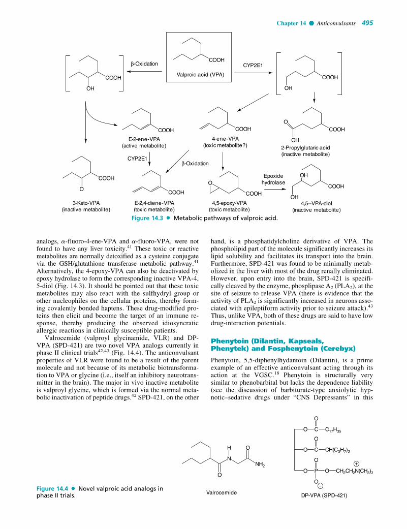
Chapter 14 Anticonvulsants 495
analogs, �-fluoro-4-ene-VPA and �-fluoro-VPA, were notfound to have any liver toxicity.41 These toxic or reactivemetabolites are normally detoxified as a cysteine conjugatevia the GSH/glutathione transferase metabolic pathway.41
Alternatively, the 4-epoxy-VPA can also be deactivated byepoxy hydrolase to form the corresponding inactive VPA-4,5-diol (Fig. 14.3). It should be pointed out that these toxicmetabolites may also react with the sulfhydryl group orother nucleophiles on the cellular proteins, thereby form-ing covalently bonded haptens. These drug-modified pro-teins then elicit and become the target of an immune re-sponse, thereby producing the observed idiosyncraticallergic reactions in clinically susceptible patients.
Valrocemide (valproyl glycinamide, VLR) and DP-VPA (SPD-421) are two novel VPA analogs currently inphase II clinical trials42,43 (Fig. 14.4). The anticonvulsantproperties of VLR were found to be a result of the parentmolecule and not because of its metabolic biotransforma-tion to VPA or glycine (i.e., itself an inhibitory neurotrans-mitter in the brain). The major in vivo inactive metaboliteis valproyl glycine, which is formed via the normal meta-bolic inactivation of peptide drugs.42 SPD-421, on the other
hand, is a phosphatidylcholine derivative of VPA. Thephospholipid part of the molecule significantly increases itslipid solubility and facilitates its transport into the brain.Furthermore, SPD-421 was found to be minimally metab-olized in the liver with most of the drug renally eliminated.However, upon entry into the brain, SPD-421 is specifi-cally cleaved by the enzyme, phosplipase A2 (PLA2), at thesite of seizure to release VPA (there is evidence that theactivity of PLA2 is significantly increased in neurons asso-ciated with epileptiform activity prior to seizure attack).43
Thus, unlike VPA, both of these drugs are said to have lowdrug-interaction potentials.
Phenytoin (Dilantin, Kapseals,Phenytek) and Fosphenytoin (Cerebyx)
Phenytoin, 5,5-diphenylhydantoin (Dilantin), is a primeexample of an effective anticonvulsant acting through itsaction at the VGSC.18 Phenytoin is structurally verysimilar to phenobarbital but lacks the dependence liability(see the discussion of barbiturate-type anxiolytic hyp-notic–sedative drugs under “CNS Depressants” in this
COOH
Valproic acid (VPA)
β-Oxidation
COOH
OH
COOH
OH
3-Keto-VPA(inactive metabolite)
β-Oxidation
COOH
OCOOH
O
COOH COOH
E-2-ene-VPA(active metabolite)
COOH
4-ene-VPA(toxic metabolite?)
COOHO
OH
CYP2E1
CYP2E1
E-2,4-diene-VPA(toxic metabolite)
4,5--VPA-diol(inactive metabolite)
4,5-epoxy-VPA(toxic metabolite)
COOH
OH
OHEpoxidehydrolase
2-Propylglutaric acid(inactive metabolite)
Figure 14.3 Metabolic pathways of valproic acid.
NNH2
O
OH
Valrocemide
O
O
C
C
O
O
O
PO
O
O
C17H35
CH(C3H7)2
CH2CH2N(CH3)3
DP-VPA (SPD-421)Figure 14.4 Novel valproic acid analogs inphase II trials.
0491-0503_17865_Ch14.qxd 12/4/09 5:43 AM Page 495

text). Phenytoin is very effective against all seizure typesexcept absence; however, the drug may be incompletely orerratically absorbed from sites of administration becauseof its very low water solubility. For this reason, fospheny-toin, a prodrug of phenytoin, was developed and marketedto avoid complications such as vein irritation, tissuedamage, and muscle necrosis associated with parenteralphenytoin administration. Fosphenytoin is rapidly ab-sorbed either by intravenous or intramuscular administra-tion.43 It is converted into phenytoin through phosphatase
catalyzed hydrolysis of the phosphate ester as shown inFigure 14.5.
METABOLISM OF PHENYTOIN
The principal metabolic pathway of phenytoin in humans isaromatic hydroxylation, catalyzed by the cytochrome P450isozymes (CYP2C9 and CYP2C19).10,44 The metabolic bio-transformations and its in vivo detoxification pathways aredepicted in Figure 14.6. The major metabolites of phenytoin
496 Wilson and Gisvold’s Textbook of Organic Medicinal and Pharmaceutical Chemistry
NN
O
O H
OP
O
O
O
Fosphenytoin(a pro-drug)
Phosphatase
Phenytoin
N
N
O
O H
HN
N
O
O H
HO
Figure 14.5 Biotransformation of fosphenytoin to phenytoin.
N
N
O
O H
H
O
N
N
O
O H
H
OH
N
N
O
O H
H OH
PHT-Arene-oxide(an epoxide)
p-HPPH
N
N
H
HO
O
OH
OH
N
N
O
O H
H
O
O
N
N
O
O H
H
OH
SG
m-HPPH
GSH, Glutathionetransferase
Epoxidehydrolase
N
N
O
O H
H
OH
OH
PHT-catechol
N
N
O
O H
H
Phenytoin (PHT)
PHT-dihydrodiol(inactive metabolite)
N
N
O
O H
H
OCH3
OH
PHT-o-quinone(reactive metabolite)
PHT-3-O-methylcatechol(inactive metabolite)
COMT
Figure 14.6 Metabolic pathways of phenytoin.
0491-0503_17865_Ch14.qxd 12/4/09 5:43 AM Page 496

Chapter 14 Anticonvulsants 497
are the 5-(4-hydroxyphenyl)-5-phenyl hydantoins (p-HPPH) and dihydrodiol. Both of these inactive metabolitesare excreted as the corresponding O-glucuronides. Furtheroxidation of p-HPPH leads to a catechol metabolite, whichappears in the urine as a methyl conjugate by the action ofcatechol-O-methyltransferase (COMT).
HYPERSENSITIVITY REACTION OF PHENYTOIN
Hypersensitivity reactions (idiosyncratic toxicity) to phenyt-oin and other aromatic AEDs in susceptible individuals arebelieved to stem from the reactions of these reactive interme-diates (i.e., arene oxide, catechol, or o-quinone) with hepaticenzymes or other cellular proteins forming covalentlybonded haptens.45 The reactive intermediate, arene oxide, isdeactivated by either epoxide hydrolase to dihydrodiol (amajor urinary metabolite) or by the action of GSH and glu-tathione transferase. It has also been suspected that these re-active arene oxides or epoxides mediate the teratogenicity ofphenytoin and other AEDs. Recent studies indicate thatepoxide hydrolase might be useful as a biomarker for prena-tal determination of risk of fetal hydantoin syndrome.46,47
Again, glutathione and epoxide hydrolase are important fordetoxification of these reactive metabolites. Furthermore, anormal level of COMT in the liver or kidneys also greatly re-duces the amount of catechol, which can be easily oxidizedto the reactive o-quinone. Thus, COMT and the NADPH-de-pendent quinine oxidoreductase may play a protective role inphenytoin-induced toxicities.48
Both phenytoin and phenobarbital are potent liver en-zyme inducers. Both of these drugs induce cytochrome P450isozymes CYP1A2, CYP2C9/19, and CYP3A4, as well asepoxide hydrolase and UDP-glucuronyl transferase (phaseII enzyme responsible for glucuronide formation).10 Thus,for patients who are using multiple drugs, their plasma con-centrations for drugs metabolized by these enzymes will begreatly affected.
Phenobarbital and Primidone(Mysoline)
Although sedative–hypnotic barbiturates commonly displayanticonvulsant properties, only phenobarbital display enoughanticonvulsant selectivity for use as antiepileptics. Primidone(Mysoline) is metabolized by CPY2C9/19 to phenobarbitaland phenylethylmalonamide (PEMA) as shown in Figure14.7. Both of these metabolites have anticonvulsant activities.However, it is generally believed that the pharmacologicalaction of primidone is mainly a result of the minor metabolite,phenobarbital. Thus, primidone is much less potent/toxic thanphenobarbital, because most of the drug is rapidly degradedto the less potent metabolite, PEMA.49
Carbamazepine (Tegretol) andOxcarbazepine (Trileptal)
CBZ, 5H dibenz[b,f]lazepine 5 carboxamide is an iminostil-bene derivative of tricyclic antidepressants.50 The twophenyls substituted on the urea nitrogen fit the pharma-cophore pattern suggested for binding to the VGSC21 (Fig.14.1). Like phenytoin, CBZ is useful in generalizedtonic–clonic and partial seizures.
The major metabolic pathway of CBZ is the formation ofa stable metabolite, 10,11-CBZ epoxide by cytochromeP450 isozyme CYP3A4. This reactive metabolite is furtherdeactivated by the action of epoxide hydrolase to give inac-tive 10,11-CBZ-diol that is excreted as the correspondingglucuronides50 (Fig. 14.8). The epoxide is a suspect in theidiosyncratic reactions CBZ may produce (e.g., aplastic ane-mia).48 OXC is a newer AED with a similar mechanism ofaction to CBZ except for its metabolic inactivation pathway.It is also not a liver enzyme inducer like CBZ and pheny-toin.43,50 With the presence of a carbonyl function at the C-10 carbon, OXC is reduced to the corresponding CBZ-10-olby the action of alcohol dehydrogenase that is excreted as its
HH
OO
CH2CH3
HH
OO
CH2CH3
N NN NN N
N
OHPrimidoneHH
OO
CH2CH3
O
CYP2C9/C19
Phenobarbital(minor metabolite)
H
OO
NH2NH2 NH2
OH
spontaneous
alcoholdehydrogenase
amide hydrolysisOO
CH2CH3CH2CH3
PEMA(major metabolite)
OH OH
OO
CH2CH3
Inactive metabolite
Figure 14.7 Metabolic biotransformation of primidone.
0491-0503_17865_Ch14.qxd 12/4/09 5:43 AM Page 497

O-glucuronide or can be further oxidized to the 10,11-CBZ-diol as an inactive metabolite (Fig. 14.8). Thus, OXC is saidto have much fewer hepatic and idiosyncratic side effectsassociated with CBZ.23 Although OXC does not induce itsown metabolism, it is a weak inducer of CYP3A4 and UDP-glucuronyl transferase and also inhibits CYP2C19. Thus, se-vere drug–drug interactions with medications metabolizedby these enzymes are to be expected.43
Gabapentin (Neurontin) andPregabalin (Lyrica)
Gabapentin and its closely related analog pregabalin, (S)-3-isobutyl-GABA (Fig. 14.9), are broad-spectrum anti-convulsants with multiple mechanisms of action.24,51 Inaddition to modulating calcium influx and stimulateGABA biosynthesis as discussed earlier, they also com-pete for the biosynthesis of L-glutamic acid because oftheir structural similarity to L-leucine.51 Gabapentin andpregabalin have very little liability for causing metabolic-based drug–drug interactions, particularly when used incombination with other AEDs because they are not metab-olized in humans. More than 95% of the drug is excretedunchanged through the kidneys. However, there are somedifferences in their bioavailability. Unlike gabapentin,which exhibits 60% bioavailability when given in lowdoses because of intestinal uptake by a saturable smallneutral L-amino acid transporter, the absorption of prega-balin is almost complete (98%) and exhibits an ideal linear
pharmacokinetic profile.24 This high bioavailability of pre-gabalin can be attributed to its closer structure similarity tothe essential amino acid, L-leucine.
FELBAMATE (FELBATOL) AND FLUROFELBAMATE
FBM, another potent and effective AED with a broad spec-trum of action, is a carbamate ester of 2-phenyl-1, 3-propanediol, structurally similar to the anxiolytic drugmeprobamate.43 The carbamate ester is stable to esterasesand therefore provides good oral bioavailability. However,the FBM therapy was found to be associated with rare butsevere side effects such as aplastic anemia, idiosyncratic re-actions, and hepatic failures within 6 months of its marketintroduction.52,53 Extensive clinical metabolic studies withFBM have been able to link the formation of reactivemetabolite(s) and the clinically observed toxicities.52 FBMundergoes hepatic cytochrome P450-mediated hydroxyla-tions (i.e., catalyzed by hepatic isozymes, CYP3A4, andCYP2E1) to give 2-hydroxyfelbamate (2-OH-FBM) andp-hydroxyfelbamate (pOH-FBM) that are excreted as theircorresponding glucuronides (Fig. 14.10).
However, FBM also undergoes esterase-catalyzedhydrolysis to give two minor metabolites, 2-phenyl-1, 3-propandiol monocarbamate (MCF) and 3-carbamoyl-2-phenylpropionic acid (CPPA). It has been suggested thatduring the oxidative degradation of MCF to CPPA, the in-termediate, 3-carbamoyl-2-phenylpropionaldehyde couldbe converted to a potentially toxic reactive metabolite, 2-
498 Wilson and Gisvold’s Textbook of Organic Medicinal and Pharmaceutical Chemistry
N
NH2
NH2
O
N
O
OHHO
N
NH2
NH2
NH2O
O
N
O
O
N
O
HO H
Carbamazepine (CBZ)
CYP3A4
10,11-CBZ Epoxide(reactive metabolite)
Epoxidehydrolase
10,11-CBZ diol(inactive metabolite)
alcoholdehydrogenase
Oxcarbazepine (OXC) (S)-CBZ-10-ol(active metabolite)
Figure 14.8 Metabolism of carbamazepine and oxcarbazepine.
H
H2N COOH H2N COOHH2N COOH
H
Gabapentin Pregabalin L-LeucineFigure 14.9 Gabapentin, pregabalin, and L-leucine.
0491-0503_17865_Ch14.qxd 12/4/09 5:43 AM Page 498

Chapter 14 Anticonvulsants 499
phenylpropanal (atropaldehyde) that is normally deacti-vated as a cysteine conjugate via the GSH/glutathionetransferase pathway discussed earlier under the metabolismof VPA.39 Isolation of 4-hydroxy-5-phenyl-1, 3-oxazaper-hydroin-2-one (i.e., a precursor for 2-phenylpropionalde-hyde in human urine) provided further evidence linking thistoxic metabolite to the observed clinical toxicities.53
Similar to VPA discussed earlier, placement of a fluorineatom at the C-2 position of FBM resulted in a very potentanticonvulsant, 2-fluorofelbamate that lacks the idiosyn-cratic properties of FBM. Fluorofelbamate is currentlyunder phase II clinical trials.54
It should be pointed out that although FBM has no effect onCYP2C9, it is an inhibitor of CYP2C19 and also inhibits themitochondrial enzymes responsible for �-oxidation. Thus,drug interactions between FBM and VPA are to be expected.55
Novel Broad-SpectrumAnticonvulsants
Chemical structures of these newer broad-spectrum anticon-vulsants are shown in Figure 14.11. All of these drugs haveunique mechanisms of action and will be discussed in indi-vidual drug monographs.
Felbamate (FBM)2-OH-FBM
HO
OCONH2
OCONH2
OCONH2
OCONH2
OH
OCONH2
OH
OCONH2
OCONH2
C-2, benzyilic carbon
OCONH2
OH
O
OCONH2
H
OH
O
O
N
HO H
O
CYP3A4/2E1 CYP3A4/2E1
MCF CPPA
esterase
alcoholdehydrogenase
aldehydedehydrogenase
atropaldehyde(reactive metabolite)
p-OH-FBM
O-glucuronides
O-glucuronides
4-hydroxy-5-phenyl-1,3-oxazaperhydroin-2-one
Figure 14.10 Metabolic biotransformation of felbamate.
N
N2N1
Cl
Cl
H2N NH2
NH2
H3C
CH3
CH3
CH3
SO2NH2
SO2NH2
SO2NH2
O
3
5
OO
O
O
O
sulphamate ester
TopiramateLamotrigine
Zonisamide
intestinal bacteria
Levetiracetam
NO
N
O
O
H
OHO
Figure 14.11 Newer broad-spectrumanticonvulsants.
0491-0503_17865_Ch14.qxd 12/4/09 5:43 AM Page 499

LAMOTRIGINE (LAMICTAL)
Lamotrigine, an AED of the phenyltriazine class, has beenfound effective against refractory partial seizures. Likephenytoin and CBZ, its main mechanism of action appears tobe blockade of sodium channels that is both voltage- and use-dependent. It also inhibits the high-threshold calcium chan-nel, possibly through inhibition of presynaptic N-type cal-cium channels and also blocks glutamate release.18,19
Lamotrigine is metabolized predominantly by glucuronida-tion. The major inactive urinary metabolites isolated are 2-N-glucuronide (76%) and 5-N-glucuronide (10%) because thearomatic ring is somewhat deactivated by the presence ofchlorine atoms toward arene oxide formation.56
Coadministration of lamotrigine with valproate, however,greatly increases the incidence of its idiosyncratic reactions.56
It is conceivable that in the presence of VPA, an inhibitor ofUDP-glucuronyl transferase, the concentration of the reactivearene oxide intermediate may be increased because of the re-duced capacity of UDP-glucuronyl transferase to metabolizelamotrigine via normal glucuronidation pathways.
TOPIRAMATE (TOPAMAX)
TPM is a sulphamate-substituted monosaccharide, a deriv-ative of the naturally occurring sugar D-fructose thatexhibits broad and potent AED actions at both glutamateand GABA receptors.19 It has good oral bioavailability of85% to 95%, most likely resulting from its structural simi-larity to D-glucose. Thus, it may be actively transportedinto the brain by the D-glucose transporter. (Recall thatD-fructose and D-glucose have identical stereochemistry atmany of their chiral centers.) Only about 20% of the drugis eliminated by hepatic metabolism (CYP2C19), the re-maining drug is excreted unchanged by the kidneys.57 Thesulphamate ester is hydrolyzed by sulfatases to the corre-sponding primary alcohol, which is further oxidized to thecorresponding carboxylic acid. Even though there are noreports of significant interactions between TPM and otherAEDs, TPM is said to have a weak carbonic anhydrase in-hibitory activity because of the presence of the sulphamatemoiety. Thus, concomitant use of TPM with other carbonicanhydrase inhibitors should be avoided.57 The exact mech-anism of actions are still unknown, but TPM appears toblock glutamate release, antagonize glutamate kainicacid/AMPA receptors, and increase GABAergic transmis-sion by binding to a site distinct from BZDs or barbiturateson the GABAA receptor complex.19
ZONISAMIDE (ZONEGRAN, EXCEGRAN)
Zonisamide, a sulfonamide-type anticonvulsant was re-cently approved for adjunctive therapy in the treatment ofpartial seizures in adults with epilepsy.43 Zonisamide isprimarily metabolized by reductive ring cleavage of the 1,2-benzisoxazole ring to 2-sulfamoyl-acetyl-phenol (Fig.14.11). This biotransformation is mainly carried out by theintestinal bacteria rather than the mammalian cytosolicaldehyde oxidase suggested earlier.58 Again, because ofthe presence of a sulfonamide moiety in zonisamide mole-cule, precaution should be given to patients who have ahistory of hypersensitivity reactions toward sulfonamidedrugs and concomitant use of zonisamide with other car-bonic anhydrase inhibitors should also be avoided.59
LEVETIRACETAM (KEPPRA)
LEV is an analog of the nootropic agent, piracetam. Onlythe S-isomer shown in Figure 14.11 has any anticonvulsantactivity. Unlike piracetam, LEV does not have any affinityfor the AMPA receptor thereby has no nootropic activity forthe treatment of Alzheimer disease. LEV also has no affin-ity for GABA receptors, BZD receptors, the various excit-atory amino acid related receptors, or the voltage-gated ionchannels.43,60 For this reason, its mechanism of anticonvul-sant action remains unclear, but it appears to exert itsantiepileptic action by modulating kainite/AMPA-inducedexcitatory synaptic currents, thus decreasing membraneconductance.60 Furthermore, the anticonvulsant activity ofthis drug appears to be mediated by the parent moleculerather than by its inactive metabolite, (S)-�-ethyl-2-oxo-1-pyrrolidineacetic acid (i.e., via the hydrolysis of amidegroup).61 Like gabapentin, LEV has few drug interactionswith other AEDs thereby can be used in combination to treatrefractory epilepsy.10,56
Anticonvulsants Acts on a SelectiveMolecular Target
TIAGABINE (GABITRIL)
A glance at tiagabine’s structure (Fig. 14.12) suggests anuptake inhibitor. Reportedly, it blocks GABA reuptake asa major mode of its anticonvulsant activity. Its use isagainst partial seizures. Inhibitors of GABA transporter-1(GAT-1 inhibitors) increase extracellular GABA concen-tration in the hippocampus, striatum, and cortex, therebyprolonging the inhibitory action of GABA released synap-tically. Nipecotic acid is a potent inhibitor of GABA reup-take into synaptosomal membranes, neurons, and glialcells. However, nipecotic acid fails to cross the blood-brainbarrier following systemic administration because of itshigh degree of ionization. Tiagabine, marketed as thesingle R(�)-enantiomer, a potent GAT-1 inhibitor struc-turally related to nipecotic acid, has an improved ability tocross the blood-brain barrier, and it has recently receivedFood and Drug Administration (FDA) approval as anAED.43 It is well absorbed and readily metabolized byCYP3A4 to an inactive metabolite, 5-oxo-tiagabine (oxida-tion of the thiophen ring) or eliminated as glucuronide ofthe parent molecule.
Over 90% of tiagabine is metabolized by CYP3A4isozymes.62 The primary site of metabolic attack is the oxi-dation of the thiophen rings leading to 5-oxo-tiagabine thatlacks anticonvulsant activity and the glucuronidation via thecarboxylic function. Thus, the plasma concentrations oftiagabine would be greatly effected by any compound thatinduces or inhibits CYP3A4.
ETHOSUXIMIDE (ZARONTIN) AND METHSUXIMIDE (CELONTIN)
Ethosuximide is considered the prototypical anticonvulsantneeded for treating patients with absence seizures.6,19,25
Ethosuximide and the N-dealkylated active metabolite ofmethsuximide (Fig. 14.12) work by blocking the low-threshold T-type calcium channels, thereby reducing thehyperexcitability of thalamic neurons that is specifically as-sociated with absence seizure.20
500 Wilson and Gisvold’s Textbook of Organic Medicinal and Pharmaceutical Chemistry
0491-0503_17865_Ch14.qxd 12/4/09 5:43 AM Page 500

Chapter 14 Anticonvulsants 501
VIGABATRIN (SABRIL)
Vigabatrin, a 4-vinyl analog of GABA, produces its phar-macological action by irreversibly blocking GABA catabo-lism catalyzed by GABA-T as discussed earlier. It is mar-keted in Europe and Canada as an adjunctive treatment ofpatients with partial seizures, but it has yet to gain FDA ap-proval in the United States even after extensive clinical tri-als.56 The main concern with this drug is its ability to causea reversible visual field defect associated with retinal func-tion in the eyes.63
BENZODIAZEPINES
For details of the chemistry and SARs of the BZDs, see thediscussion of anxiolytic sedative–hypnotic drugs. Amongthe current clinically useful drugs, the structural features as-sociated with anticonvulsant activity are identical with thoseassociated with anxiolytic sedative–hypnotic activity.
Clonazepam (Klonopin)Clonazepam is useful in absence seizures and in myoclonicseizures. Tolerance to the anticonvulsant effect of the clo-nazepam often developed rather quickly, and it is a commonproblem with the BZDs. Metabolism involves hydroxyla-tion of the C-3 position, followed by glucuronidation andnitro group reduction, followed by acetylation.
Diazepam (Valium, Diastat)Diazepam is given orally (Valium) or rectally (Diastat) as anadjunctive treatment in patients with generalized tonic–clonicstatus epilepticus (i.e., an acute and potentially fatal seizure)or in patients with refractory epilepsy in combination withother AEDs.4 For details on diazepam (Valium), see its dis-cussion under anxiolytics and sedative–hypnotic agents.
FUTURE DEVELOPMENT OFANTIEPILEPTIC DRUGS
As stated earlier, even with the recent introduction of newerAEDs, greater than a third of newly diagnosed patients withepilepsy are still in need of an effective AED to control theirseizures.3,11,20 Furthermore, all current AEDs work only asprophylaxis against the symptoms of epilepsy, they do notprevent disease progression into refractory epilepsy. Futuredirections in the development of AEDs must come from ei-ther a better understanding of the epileptogenesis, especiallythe genetic mechanisms that underlie disease progression orthe development of resistance after prolonged pharma-cotherapy,3 or from an innovative design strategy that pro-duce new AEDs with unique mechanisms of action.11
Several of these agents are currently undergoing differentphases of clinical trials. The readers are to consult recent re-views for further details of these agents.64–66
Tiagabine
Nipecotic acid moiety
Ethosuximide Methsuximide
Vigabatrin
Diazepam Clonazepam
N
HO
O
NO
O
H2N COOH
COOH
N
N
Cl
CH3O
N
N
O2N
HO
Cl
N
S
S
CH3
CH3
CH3CH3
CH3
H3C
Figure 14.12 Anticonvulsants actson a selective molecular target.
3. Explain chemically/biochemically why 2-fluoro-VPA, asynthetic analog of VPA currently in clinical trials, may notpossess similar hepatotoxic/teratogenic side effects as VPA.
4. Provide a biochemical rationale why valrocemide, a VPAanalog, lacks epoxide hydrolase inhibitory activity and ter-atogenicity of VPA at clinically relevant concentrations.
1. Provide a biochemical reason why pregabalin has betterbioavailability than gabapentin.
2. SSADH deficiency, a rare inborn error of human metab-olism, disrupts the normal metabolism of the inhibitoryneurotransmitter GABA. Explain why these individualsare more susceptible to seizure activity.
R E V I E W Q U E S T I O N S
0491-0503_17865_Ch14.qxd 12/4/09 5:43 AM Page 501

5. Explain why coadministering valproate with phenytoinwill increase its idiosyncratic toxicities. Identify at leasttwo possible reactive metabolites of phenytoin that maybe responsible for this observation.
6. Explain why there are cross-sensitivities among pheny-toin, phenobarbital, fosphenytoin and primidone.
7. What will be the expected metabolic profile of CBZ if aspecific inhibitor of CYP3A4 such as ketoconazole, anantifungal drug or the flavones present in grapefruit istaken concurrently?
8. Explain why lamotrigine is eliminated more rapidly inpatients who have been taking CBZ or phenytoin.
9. Provide a possible rationale for why CYP3A4 inducerscan increase the clearance of FBM, yet CYP3A4 in-hibitors have little or no effect on its pharmacokineticprofile.
10. Explain why some dose adjustment of VPA may be re-quired if FBM is added to a regimen of VPA.
502 Wilson and Gisvold’s Textbook of Organic Medicinal and Pharmaceutical Chemistry
REFERENCES1. McAfee, A. T., Chilcott, K. E., Johannes, C. B., et al.: Epilepsia
48:1075–1082, 2007.2. Tan, N. C. K., Mulley, J. C., and Berkovic, S. F.: Epilepsia
45:1429–1442, 2004.3. Steinlein, O. K.: Nat. Rev. Neurosci. 5:400–408, 2004.4. Pheops, S. J., Wheless, J. W., and Aldredge, B. K.: Seizure
Disorders. In Helms, R.A., Quan, D. J., Herfindal, E. T., et al. (eds.).Textbook of Therapeutics—Drug and Disease Management, 8th ed.Philadelphia, Lippincott Williams & Wilkins, 2006, pp. 1608–1645.
5. Theodore, W. H., Spencer, S. S., Wiebe, S., et al.: Epilepsia47:1700–1722, 2006.
6. Galuser, T., Ben-Menachem, E., Bourgeois, B., et al.: Epilepsia47:1094–1120, 2006.
7. Avanzini, G.: Epilepsia 47:1285–1287, 2006.8. Oby, E., and Janigro, D.: Epilepsia 47:1761–1774, 2006.9. Lindhout, D.: Neurology 42(Suppl. 5):43–46, 1992.
10. Patsalos, P. N., and Perucca, E.: Lancet Neurol. 2:347–356, 2003.11. Dichter, M. A.: Epilepsia 48(Suppl. 1):26–30, 2007.12. Van Donselaar, C. A., and Stroink, H.: Epilepsia 47(Suppl. 1):9–13,
2006.13. Browne, T. R., and Holmes, G. L.: N. Engl. J. Med. 344:1145–1151,
2001.14. Commission on Classification and Terminology of the International
League Against Epilepsy. Epilepsia 22:489–501, 1981.15. Commission on Classification and Terminology of the International
League Against Epilepsy. Epilepsia 30:389–399, 1989.16. Engel, Jr.: J. Epilepsia 47:1558–1568, 2006.17. Jensen, T. S.: Eur. J. Pain 6(Suppl. A):61–68, 2002.18. Czapinski, P., Blaszczyk, B., and Czuczwar, S.: J. Curr. Top. Med.
Chem. 5:3–14, 2005.19. Rho, J. M., and Sankar, R.: Epilepsia 40:1471–1483, 1999.20. Errington, A. C., Stöhr, T., and Lees G.: Curr. Top. Med. Chem.
5:15–30, 2005.21. Unverferth, K., Engel, J., Höfgen, N., et al.: J. Med. Chem. 41:63–73,
1998.22. DeLorenzo, R. J., Sun, D. A., and Deshpande, L. S.: Pharmacol.
Ther. 105:229–266, 2005.23. Cunningham, M. O., Woodhall, G. L., Thompson, S. E., et al.: Eur. J.
Neurosci. 20:1566–1576, 2004.24. Taylor, C. P., Angelotti, T., and Fauman, E.: Epilepsy Res.
73:137–150, 2007.25. Gomora, J. C., Daud, A. N., Weiergräber, M., et al.: Mol. Pharmacol.
60:1121–1132, 2001.26. Madeja, M., Margineanu, D. G., Gorji, A., et al.: Neuropharmacology
45:661–671, 2003.27. De Smedt, T., Raedt, R., Vonck, K., et al.: CNS Drugs Rev.
13:43–56, 2007.28. Foster, A. C., and Kemp, J. A.: Curr. Opin. Pharmacol. 6:7–17, 2006.29. Costa, E.: Annu. Rev. Pharmacol. Toxicol. 38:321–350, 1998.30. Johannessen, C. U.: Neurochem. Internat. 37:103–110, 2000.31. Silverman, R. B., Andruszkiewicz, R., Nanavati, S. M., et al.: J. Med.
Chem. 34:2295–2198, 1991.32. Petroff, Q. A. C., Hyder, F., Rothman, D. L., et al.: Epilepsia
41:675–680, 2000.33. Jung, M. J., Lippert, B., Metcalf, B. W., et al.: J. Neurochem.
29:797–802, 1977.34. Schousboe, A., Sarup, A., Larsson, O. M., et al.: Biochem.
Pharmacol. 68:1557–1563, 2004.
35. Chebib, M., and Johnston, G. A. R.: J. Med. Chem. 43:1427–1447,2000.
36. Ozawa, S., Kamiya, H., and Tsuzuki, K.: Progress Neurobiol.54:581–618, 1998.
37. Nakanishi, S.: Sciences 258:597–603, 1992.38. Albensi, B. C., Igoechi, C., Janigro, D., et al.: Am. J. Alzheimers Dis.
Other Demen. 19:269–274, 2004.39. Jurima-Romet, M., Abbott, F. S., Tang, W., et al.: Toxicology
112:69–85, 1996.40. Neuman, M. G., Shear, N. H., Jacobson-Brown, P. M., et al.: Clin.
Biochem. 34:211–218, 2001.41. Tang, W., Borel, A. G., Fujimiya, T., et al.: Chem. Res. Toxicol.
8:671–682, 1995.42. Spiegelstein, O., Yagen, B., and Bialer, M.: Epilepsia 40:545–552,
1999.43. Bialer, M., Johannessen, S. I., Kupferberg, H. J., et al.: Epilepsy
Research 51:31–71, 2002.44. Browne, T. R., and LeDuc, B.: Phenytoin—Chemistry and biotrans-
formation. In Levy, R. H., Mattson, R. H., and Meldrum, B. S. (eds.).Antiepleptic Drugs, 4th ed. New York, Raven Oress, Ltd., 1995,pp. 283–300.
45. Yasumori, T., Chen, L. S., Li, Q. H., et al.: Biochem. Pharmacol.57:1297–1303, 1999.
46. Buehler, B. A., Delimont, D., van Waes, M., et al.: New Engl. J.Med. 322:1567–1572, 1990.
47. Hartsfield, J. K., Benford, S.A., and Hilbelink, D. R.: Biochem. Mol.Med. 56:144–151, 1995.
48. Leeder, J. S.: Epilepsia 39(Suppl. 7):S8–S16, 1998.49. Ferranti, V., Chabenat, C., Ménager, S., et al.: J. Chromatography B.
718:199–204, 1998.50. Benes, J., Parada, A., Figueiredo, A. A., et al.: J. Med. Chem.
42:2582–2587, 1999.51. Taylor, C. P., Gee, N. S., Su, T. Z., et al.: Epilepsy Res. 29:233–249,
1998.52. Dieckhaus, C. M., Thompson, C. D., Roller, S. G., et al.: Chem. Biol.
Interact. 142:99–117, 2002.53. Dieckhaus, C. M., Santos, W. L., Sofia, R. D., et al.: Chem. Res.
Toxicol. 14:958–964, 2001.54. Hovinga, C. A.: Expert Opin. Investig. Drugs, 11:1387–1406, 2002.55. Wagner, M. L., Graves, N. M., Leppik, I. E., et al.: Clin. Pharmacol.
Ther. 56:494–502, 1994.56. Walker, M. C., and Patsalos, P. N.: Pharmac. Ther. 67:351–384,
1995.57. Mimrod, D., Specchio, L. M., Britzi, M., et al.: Epilepsia
46:1046–1054, 2005.58. Kitamura, S., Sugihara, K., Kuwasako, M., et al.: J. Pharm.
Pharmacol. 49:253–256, 1997.59. Fukushima, K., and Seino, M.: Epilepsia 47:1860–1864, 2006.60. Carunchio, I., Pieri, M., Ciotti, M. T., et al.: Epilesia 48:654–662,
2007.61. Patsalos, P. N., Ghattaura, S., Ratnaraj, N., et al.: Epilesia
47:1818–1821, 2006.62. Ingwersen, S. H., Pedersen, P. C., Groes, L., et al.: Eur. J. Pharm.
Sci. 11:247–254, 2000.63. Banin, E., Shalev, R. S., Obolensky, A., et al.: Arch. Ophthalmol.
121:811–816, 2003.64. Schmidt, B.: Amer. Soc. Exp. Neurotherapeutics 4:62–69, 2007.65. Löscher, W., and Schmidt, D.: Epilepsy Res. 69:183–272, 2006.66. Malawska, B.: Curr. Top. Med. Chem. 5:69–85, 2005.
0491-0503_17865_Ch14.qxd 12/4/09 5:43 AM Page 502

Kaneko, S., Okada, M., Iwasa, H., et al.: Genetics of epilepsy: currentstatus and perspectives. Neurosci. Res. 44:11–30, 2002.
Kwan, P., Sills, G. J., and Brodie, M. J.: The mechanisms of action ofcommonly used antiepileptic drugs. Pharmacol. Ther. 90:21–34, 2001.
Patsalos, P. N., and Perucca, E.: Clinically important drug interactions inepilepsy: interactions between antiepileptic drugs and other drugs.Lancet Neurol. 2:473–481, 2003.
Trevor, A. J., and Way, W. L.: Sedative-hypnotic drugs. In Katzung,B. G. (ed.). Basic & Clinical Pharmacology, 9th ed. New York, NY,McGraw-Hill, 2004, pp. 351–366.
Chapter 14 Anticonvulsants 503
SELECTED READINGCastilla-Guerra, L., Fernández-Moreno, M. del C., López-Chozas, J. M.,
et al.: Electrolytes Disturbances and Seizures. Epilepsia 47:1990–1998,2006.
Gidal, B. E., and Garnett, W. R.: Epilepsy. In DiPiro, J. T., Talbert, R. L.,Yee, G. C., et al. (eds.). Pharmacotherapy—A PathophysiologicApproach, 6th ed. New York, NY, McGraw-Hill, 2005, pp.1023–1048.
Johannessen, C. U., and Johannessen, S. I.: Valroate: past, present, andfuture. CNS Drug Rev. 9:199–216, 2003.
0491-0503_17865_Ch14.qxd 12/4/09 5:43 AM Page 503

504
C H A P T E R 15
Central Nervous System StimulantsJOHN M. BEALE, JR.
useful in determining mechanisms of action of sedative–hypnotics and anticonvulsants. Butyrolactones bind to thepicrotoxinin site.
PentylenetetrazolePentylenetetrazole, 6,7,8,9-tetrahydro-5H-tetrazolo[1,5-a]azepine, 1,5-pentamethylenetetrazole (Metrazol), hasbeen used in conjunction with the electroencephalograph tohelp locate epileptic foci. It is used as a laboratory tool indetermining potencies of potential anticonvulsant drugs inexperimental animals. The drug acts as a convulsant by in-terfering with chloride conductance.2 It binds to an allostericsite on the GABAA receptor and acts as a negative modula-tor. Overall, it appears to share similar effects on chlorideconductance with several other convulsive drugs, includingpicrotoxinin.
ModafinilModafinil (Provigil) has overall wakefulness-promotingproperties similar to those of central sympathomimetics. It isconsidered an atypical �1-norepinephrine (NE) receptorstimulant and is used to treat daytime sleepiness in nar-colepsy patients. Adverse reactions at therapeutic doses arereportedly not severe and may include nervousness, anxiety,and insomnia. Modafinil is used by oral administration.
Doxapram HydrochlorideDoxapram, 1-ethyl-4-(2-morpholinoethyl)-3,3-diphenyl-2-pyrrolidinone hydrochloride hydrate (Dopram), has an ob-scure molecular mechanism of action. Overall, it stimulatesrespiration by action on peripheral carotid chemoreceptors.It has use as a respiratory stimulant postanesthetically, afterCNS depressant drug overdose, in chronic obstructive pul-monary diseases, and in the apneas. Dopram is administeredexclusively by intravenous injection. Because of the benzylalcohol content of the injectable formulation, Dopram mustnever be given to neonates.
C H A P T E R O V E R V I E W
This chapter discusses a broad range of agents that stimu-late the central nervous system (CNS). The analepticsclassically are a group of agents with a limited range of usebecause of the general nature of their effects. The methyl-xanthines have potent stimulatory properties, mainly corti-cal at low doses but with more general effects as the doseis increased. The central sympathomimetic agents amphet-amine and close relatives have alerting and antidepressantproperties but are medically used more often as anorexi-ants. The antidepressant drugs are used most frequently indepressive disorders and can be broadly grouped into themonoamine oxidase inhibitors (MAOIs), the monoaminereuptake inhibitors, and agents acting on autoreceptors. Asmall group of miscellaneously acting drugs, which in-cludes several hallucinogens, cocaine, and cannabinoids,concludes the chapter.
ANALEPTICS
The traditional analeptics are a group of potent and rela-tively nonselective CNS stimulants. The convulsive doselies near their analeptic dose. They can be illustrated bypicrotoxinin and pentylenetetrazole. Both are obsolete asdrugs but remain valuable research tools in determininghow drugs act. Newer agents, modafinil and doxapram, aremore selective and have use in narcolepsy and as respira-tory stimulants.
PicrotoxinPicrotoxinin, the active ingredient of picrotoxin, has the fol-lowing structure:
According to Jarboe et al.,1 the hydroxylactonyl moietyis mandatory for activity, with the 2-propenyl group as-sisting. Picrotoxinin exerts its effects by interfering withthe inhibitory effects of �-aminobutyric acid (GABA) atthe level of the GABAA receptor’s chloride channel. Thedrug is obsolete medically. Pharmacologically, it has been
0504-0518_17865_Ch15.qxd 12/2/09 4:04 AM Page 504

Chapter 15 Central Nervous System Stimulants 505
METHYLXANTHINES
The naturally occurring methylxanthines are caffeine, the-ophylline, and theobromine. See Table 15.1 for their structuresand occurrence and Table 15.2 for their relative potencies.
Caffeine is a widely used CNS stimulant. Theophyllinehas some medical use as a CNS stimulant, but its CNS-stimulant properties are encountered more often as some-times severe, and potentially life-threatening, side effectsof its use in bronchial asthma therapy. Theobromine hasvery little CNS activity (probably because of poor physico-chemical properties for distribution to the CNS).
Caffeine is often used as it occurs in brewed coffee,brewed tea, and cola beverages. In most subjects, a dosageof 85 to 250 mg of caffeine acts as a cortical stimulant andfacilitates clear thinking and wakefulness, promotes an abil-ity to concentrate on the task at hand, and lessens fatigue. Asthe dose is increased, side effects indicating excessive stim-ulation (e.g., restlessness, anxiety, nervousness, tremulous-ness) become more marked. (They may be present in vary-ing degrees at lower-dose levels.) With further increases indosage, convulsions can occur. A review of the actions ofcaffeine in the brain with special reference to factors thatcontribute to its widespread use appears to be definitive.3
The CNS effects of theophylline at low-dose levelshave been little studied. At high doses, the tendency to pro-duce convulsions is greater for theophylline than for caf-feine. In addition to being cortical stimulants, theophyllineand caffeine are medullary stimulants, and both are used assuch. Caffeine may be used in treating poisoning fromCNS-depressant drugs, although it is not a preferred drug.
The important use of theophylline and its preparations inbronchial asthma is discussed elsewhere. Caffeine also is re-ported to have valuable bronchodilating properties in asthma.Finally, because of central vasoconstrictive effects, caffeinehas value in treating migraine and tension headaches andmay have actual analgesic properties in the latter use.
The CNS-stimulating effects of the methylxanthineswere once attributed to their phosphodiesterase-inhibitingability. This action is probably irrelevant at therapeuticdoses. Evidence indicates that the overall CNS-stimulant ac-
tion is related more to the ability of these compounds to an-tagonize adenosine at A1 and A2A receptors.3–6 All of theroles of these receptors are still under study. The adenosinereceptor subtypes and their pharmacology have been re-viewed.3,7–9 Problems with the present compounds, such ascaffeine and theophylline, are lack of receptor selectivityand the ubiquitous nature of the various receptor subtypes.
Caffeine and theophylline have pharmaceutically impor-tant chemical properties. Both are weak Brønsted-Lowrybases. The reported pKa values are 0.8 and 0.6 for caffeineand 0.7 for theophylline. These values represent the basicityof the imino nitrogen at position 9. As acids, caffeine has apKa above 14, and theophylline, a pKa of 8.8. In theophylline,a proton can be donated from position 7 (i.e., it can act as aBrønsted acid). Caffeine cannot donate a proton from posi-tion 7 and does not act as a Brønsted acid at pH values lessthan 14. Caffeine does have electrophilic sites at positions 1,3, and 7. In addition to its Brønsted acid site at 7, theophyllinehas electrophilic sites at 1 and 3. In condensed terms, bothcompounds are electron-pair donors, but only theophylline isa proton donor in most pharmaceutical systems.
Although both compounds are quite soluble in hot water(e.g., caffeine 1:6 at 80°C), neither is very soluble in waterat room temperature (caffeine about 1:40, theophyllineabout 1:120). Consequently, various mixtures or complexesdesigned to increase solubility are available (e.g., citratedcaffeine, caffeine and sodium benzoate, theophylline–ethylenediamine compound [aminophylline]).
Caffeine in blood is not highly protein bound; the-ophylline is about 50% bound. Differences in the substituentat the 7-position may be involved. Additionally, caffeine is
TABLE 15.1 Chemical Structures of the XanthineAlkaloids
Compound R R� R� Common Source
Caffeine CH3 CH3 CH3 Coffee, teaTheophylline CH3 CH3 H TeaTheobromine H CH3 CH3 Cocoa
TABLE 15.2 Relative Pharmacological Potencies of the Xanthine Alkaloids
CNS Respiratory Coronary Cardiac Skeletal MuscleXanthine Stimulation Stimulation Diuresis Dilation Stimulation Stimulation
Caffeine 1a 1 3 3 3 1Theophylline 2 2 1 1 1 2Theobromine 3 3 2 2 2 3
a1, most potent
0504-0518_17865_Ch15.qxd 12/2/09 4:04 AM Page 505

more lipophilic than theophylline and reputedly achieveshigher brain concentrations. The half-life of caffeine is 5 to8 hours, and that of theophylline, about 3.5 hours. About 1%of each compound is excreted unchanged. The compoundsare metabolized in the liver. The major metabolite of caf-feine is 1-methyluric acid, and that of theophylline, 1,3-di-methyluric acid.10 Neither compound is metabolized to uricacid, and they are not contraindicated in gout.
CENTRAL SYMPATHOMIMETIC AGENTS(PSYCHOMOTOR STIMULANTS)
Sympathomimetic agents, whose effects are manifestedmainly in the periphery, are discussed in Chapter 16. A fewsimple structural changes in these peripheral agents producecompounds that are more resistant to metabolism, more non-polar, and better able to cross the blood-brain barrier. Theseeffects increase the ratio of central to peripheral activity, andthe agents are designated, somewhat arbitrarily, as centralsympathomimetic agents.
In addition to CNS-stimulating effects, manifested as ex-citation and increased wakefulness, many central sympatho-mimetics exert an anorexiant effect. Central sympatho-mimetic (noradrenergic) action is often the basis for theseeffects. Other central effects, notably dopaminergic andserotoninergic effects, can be operative, however.11 In someagents, the ratio of excitation and increased wakefulness toanorexiant effects is decreased, and the agents are marketedas anorexiants. Representative structures of this group ofcompounds are given in Table 15.3. The structures of theanorexiants phendimetrazine and sibutramine and the alert-ing agents methylphenidate and pemoline, useful in atten-tion-deficient disorders, are given in the text.
Structural features for many of the agents can be visual-ized easily by considering that within their structure, theycontain a �-phenethylamine moiety, and this grouping cangive some selectivity for presynaptic or postsynaptic nor-adrenergic systems. �-Phenethylamine, given peripherally,lacks central activity. Facile metabolic inactivation bymonoamine oxidases (MAOs) is held responsible. Branchingwith lower alkyl groups on the carbon atom adjacent (�) tothe amino nitrogen increases CNS rather than peripheral ac-tivity (e.g., amphetamine, presumably by retarding metabo-lism). The � branching generates a chiral center. Thedextro(S)-isomer of amphetamine is up to 10 times as potentas the levo(R)-isomer for alerting activity and about twice asactive as a psychotomimetic agent. Hydroxylation of the ringor hydroxylation on the �-carbon (to the nitrogen) decreasesactivity, largely by decreasing the ability to cross the blood-brain barrier. For example, phenylpropanolamine, with a �-hydroxyl (OH), has about 1/100th the ability to cross theblood-brain barrier of its deoxy congener, amphetamine.
Halogenation (F, Cl, Br) of the aromatic ring decreasessympathomimetic activity. Other activities may increase. p-Chloroamphetamine has strong central serotoninergicactivity (and is a neurotoxin, destroying serotoninergic neu-rons in experimental animals).12,13
Methoxyl or methylenedioxy substitution on the ringtends to produce psychotomimetic agents, suggesting tro-pism for dopaminergic (D2) receptors.
N-methylation increases activity (e.g., compare metham-phetamine with dextroamphetamine). Di-N-methylation de-
creases activity. Mono-N substituents larger than methyl de-crease excitatory properties, but many compounds retainanorexiant properties. Consequently, some of these agentsare used as anorexiants, reportedly with less abuse potentialthan amphetamine.
There can be some departure from the basic �-phenethy-lamine structure when compounds act by indirect noradren-ergic mechanisms. A �-phenethylamine–like structure,however, can be visualized in such compounds.
The abuse potential of the more euphoriant and stimula-tory of the amphetamines and amphetamine-like drugs iswell documented. They produce an exceedingly destructiveaddiction. Apparently, both a euphoric “high” (possibly re-lated to effects on hedonistic D2 receptors) and a post-euphoric depression (especially among amine-depletingdrugs) contribute to compulsive use of these agents. Abuseof these drugs (especially methamphetamine) in recent yearshas reached disastrous proportions.
Recognized medical indications for dextroamphetamineand some very close congeners include narcolepsy, Parkinsondisease, attention-deficient disorders, and, although not thepreferred agents for obesity, appetite suppression. In someconditions, such as Parkinson disease, for which its main use
506 Wilson and Gisvold’s Textbook of Organic Medicinal and Pharmaceutical Chemistry
TABLE 15.3 Structures of Sympathomimetics withSignificant Central Stimulant Activity
Amphetamine
Methamphetamine
Phentermine
Benzphetamine
Diethylpropion
Fenfluramine
0504-0518_17865_Ch15.qxd 12/2/09 4:04 AM Page 506

Chapter 15 Central Nervous System Stimulants 507
is to decrease rigidity, the antidepressant effects of dextroam-phetamine can be beneficial. It is also reportedly an effectiveantidepressant in terminal malignancies. In almost all cases ofdepression, and especially in major depressive disorders ofthe unipolar type, however, dextroamphetamine has longbeen superseded by other agents, notably the MAOIs and themonoamine reuptake-inhibiting antidepressants.
The compounds and their metabolites can have complex,multiple actions. In a fundamental sense, the structural basisfor action is quite simple. The compounds and their metabo-lites resemble NE and can participate in the various neuronaland postsynaptic processes involving NE, such as synthesis,release, reuptake, and presynaptic and postsynaptic receptoractivation. Also, because dopamine (DA) and, to a lesserextent, serotonin (5-hydroxytryptamine [5-HT]) bear a struc-tural resemblance to NE, processes in DA- and 5-HT–activated systems can be affected. To illustrate the potentialcomplexity, the receptor activations that can be associatedwith just one parameter, reduction in food intake, reportedlyare �1, �1, �2, 5-HT1B, 5-HT2A, 5-HT2C, D1, and D2.
PRODUCTS
Amphetamine SulfateAmphetamine, (�)-1-phenyl-2-aminopropane (Benzedrine),as the racemic mixture has a higher proportion of cardiovas-cular effects than the dextro isomer. For most medical uses,the dextrorotatory isomer is preferred.
Dextroamphetamine Sulfate andDextroamphetamine PhosphateDextroamphetamine, (�)-(S)-methylphenethylamine, formssalts with sulfuric acid (Dexedrine) and with phosphoric acids.The phosphate is the more water-soluble salt and is preferredif parenteral administration is required. The dextrorotatoryisomer has the (S) configuration and fewer cardiovascular ef-fects than the levorotatory (R)-isomer. Additionally, it may beup to 10 times as potent as the (R)-isomer as an alerting agentand about twice as potent a psychotomimetic agent. Althoughit is a more potent psychotomimetic agent than the (R)-isomer,it has a better ratio of alerting to psychotomimetic effects.
The major mode of action of dextroamphetamine is releaseof NE from the mobile pool of the nerve terminal. Othermechanisms, such as inhibition of uptake, may make a smallcontribution to the overall effects. The alerting actions relateto increased NE available to interact with postsynaptic recep-tors (�1). Central �-receptor activation has classically beenconsidered the basis for most of the anorexiant effect.
The psychotomimetic effects are linked to release of DAand activation of postsynaptic receptors. D2 and mesolimbicD3 receptors would be involved. Effects on 5-HT systemsalso have been linked to some behavioral effects of dex-troamphetamine. Effects via 5-HT receptors would include5-HT1A receptors and, theoretically, all additional receptorsthrough 5-HT7.
Dextroamphetamine is a strongly basic amine, with val-ues from 9.77 to 9.94 reported. Absorption from the gas-trointestinal tract occurs as the lipid-soluble amine. Thedrug is not extensively protein bound. Varying amounts ofthe drug are excreted intact under ordinary conditions. Theamount is insignificant under conditions of alkaline urine.Under conditions producing systemic acidosis, 60% to 70%of the drug can be excreted unchanged. This fact can be usedto advantage in treating drug overdose.
The �-methyl group retards, but does not terminate, me-tabolism by MAO. Under most conditions, the bulk of a doseof dextroamphetamine is metabolized by N-dealkylation tophenylacetone and ammonia. Phenylacetone is degraded fur-ther to benzoic acid.
In experimental animals, about 5% of a dose accumulatesin the brain, especially the cerebral cortex, the thalamus, andthe corpus callosum. It is first p-hydroxylated and then �-hydroxylated to produce p-hydroxynorephedrine, whichhas been reported to be the major active metabolite involvedin NE and DA release.14
Methamphetamine HydrochlorideMethamphetamine, (�)-1-phenyl-2-methylaminopropanehydrochloride desoxyephedrine hydrochloride (Desoxyn), isthe N-methyl analog of dextroamphetamine. It has moremarked central and less peripheral action than dextroam-phetamine. It has a very high abuse potential, and by the in-travenous route, its salts are known as “speed.” The overallabuse problem presented by the drug is a national disaster.Medicinally acceptable uses of methamphetamine are anal-ogous to those of dextroamphetamine.
Phentermine Ion-Exchange Resin and Phentermine HydrochlorideThe free base is �,�-dimethylphenethylamine, 1-phenyl-2-methylaminopropane. In the resin preparation (Ionamin),the base is bound with an ion-exchange resin to yield a slow-release product; the hydrochloride (Wilpowr) is a water-soluble salt.
0504-0518_17865_Ch15.qxd 12/2/09 4:04 AM Page 507

Phentermine has a quaternary carbon atom with onemethyl oriented like the methyl of (S)-amphetamine and onemethyl oriented like the methyl of (R)-amphetamine, and itreportedly has pharmacological properties of both the (R)-and (S)-isomers of amphetamine. The compound is used asan appetite suppressant and is a Schedule IV agent, indicat-ing less abuse potential than dextroamphetamine.
Benzphetamine HydrochlorideBenzphetamine hydrochloride, (�)-N-benzyl-N,�-di-methylphenethylamine hydrochloride, (�)-1-phenyl-2-(N-methyl-N-benzylamine)propane hydrochloride (Didrex), isN-benzyl–substituted methamphetamine. The large (benzyl)N-substituent decreases excitatory properties, in keepingwith the general structure–activity relationship (SAR) forthe group. Anorexiant properties are retained. Classically,amphetamine-like drugs with larger than N-methyl sub-stituents are cited as anorexiant through central �-agonism.No claims for selectivity among �-receptor subtypes havebeen made in such citations. The compound shares mecha-nism-of-action characteristics with methylphenidate.Overall, it is said to reduce appetite with fewer CNS excita-tory effects than dextroamphetamine.
Diethylpropion HydrochlorideBecause it has two large (relative to H or methyl) N-alkylsubstituents, diethylpropion hydrochloride, 1-phenyl-2-di-ethylaminopropan-1-one hydrochloride (Tenuate, Tepanil),has fewer sympathomimetic, cardiovascular, and CNS-stimulatory effects than amphetamine. It is reportedly ananorexiant agent that can be used for the treatment of obe-sity in patients with hypertension and cardiovascular dis-ease. According to the generalization long used for thisgroup of drugs, increasing N-alkyl size reduces central �1
effects and increases � effects, even though the effects arelikely mediated principally by indirect NE release.
Fenfluramine HydrochlorideFenfluramine hydrochloride, (�)N-ethyl-�-methyl-m-(tri-fluoromethyl)phenethylamine hydrochloride (Pondimin), isunique in this group of drugs, in that it tends to produce se-dation rather than excitation. Effects are said to be mediatedprincipally by central serotoninergic, rather than central nor-adrenergic, mechanisms. In large doses in experimental an-imals, the drug is a serotonin neurotoxin.15 It was withdrawnfrom human use after reports of heart valve damage and pul-monary hypertension. From its structure, more apolar or hy-drophobic character than amphetamine, tropism for sero-toninergic neurons would be expected. Likewise, thestructure suggests an indirect mechanism. If an indirectmechanism were operative, then all postsynaptic 5-HT re-
ceptors could be activated. Evidence from several studies in-dicates that the 5-HT1B and the 5-HT2C receptors are mostresponsible for the satiety effects of 5-HT. 5-HT may alsoinfluence the type of food selected (e.g., lower-fat food in-take).11 The (�) isomer, dexfenfluramine (Redux), has agreater tropism for 5-HT systems than the racemic mixture.It, too, was withdrawn because of toxicity.
Phendimetrazine TartrateThe optically pure compound phendimetrazine tartrate,(2S,3S)-3,4-dimethyl-2-phenylmorpholine-L-(�)-tartrate(Plegine), is considered an effective anorexiant that is lessabuse prone than amphetamine. The stereochemistry of(�)phendimetrazine is as shown.16
SibutramineSibutramine (Meridia) is said to be an uptake inhibitor ofNE and 5-HT. These mechanisms fit its structure. It is re-portedly an antidepressant and an anorexiant drug. Thismechanism implies that activation of all presynaptic andpostsynaptic receptors in NE and 5-HT systems is possible.The data are not completely clear, but studies to date indi-cate that the receptors principally involved are �1, �1, and5-HT2C.
11
Methylphenidate HydrochlorideBecause methylphenidate (Ritalin) has two asymmetriccenters, there are four possible isomers. The threo race-mate is the marketed compound and is about 400 times aspotent as the erythro racemate.17 The absolute configura-tion of each of the threo-methylphenidate isomers has beendetermined.18 Considering that the structure is fairly com-plex (relative to amphetamine), it is likely that one of thetwo components of the threo racemate contains most of theactivity. Evidence indicates that the (�) -(2R,2�R)threoisomer is involved principally in the behavioral andpressor effects of the racemate.19 As is likely with many
508 Wilson and Gisvold’s Textbook of Organic Medicinal and Pharmaceutical Chemistry
0504-0518_17865_Ch15.qxd 12/2/09 4:04 AM Page 508
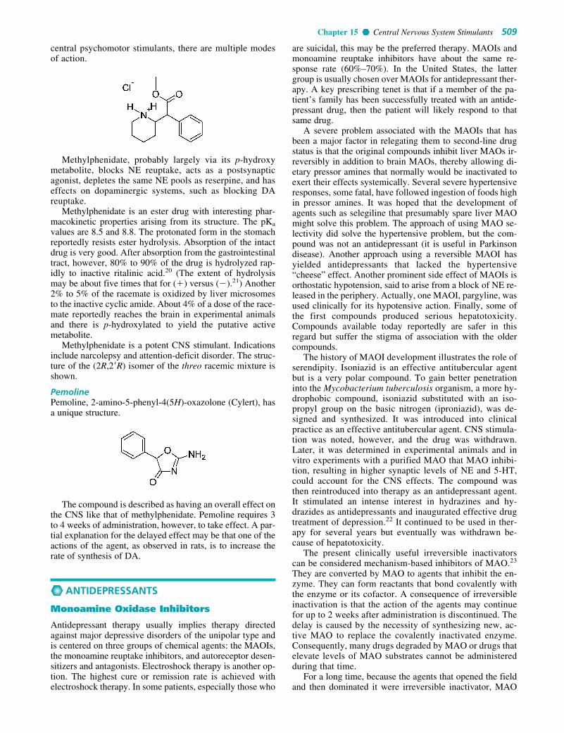
Chapter 15 Central Nervous System Stimulants 509
central psychomotor stimulants, there are multiple modesof action.
Methylphenidate, probably largely via its p-hydroxymetabolite, blocks NE reuptake, acts as a postsynapticagonist, depletes the same NE pools as reserpine, and haseffects on dopaminergic systems, such as blocking DAreuptake.
Methylphenidate is an ester drug with interesting phar-macokinetic properties arising from its structure. The pKa
values are 8.5 and 8.8. The protonated form in the stomachreportedly resists ester hydrolysis. Absorption of the intactdrug is very good. After absorption from the gastrointestinaltract, however, 80% to 90% of the drug is hydrolyzed rap-idly to inactive ritalinic acid.20 (The extent of hydrolysismay be about five times that for (�) versus (�).21) Another2% to 5% of the racemate is oxidized by liver microsomesto the inactive cyclic amide. About 4% of a dose of the race-mate reportedly reaches the brain in experimental animalsand there is p-hydroxylated to yield the putative activemetabolite.
Methylphenidate is a potent CNS stimulant. Indicationsinclude narcolepsy and attention-deficit disorder. The struc-ture of the (2R,2�R) isomer of the threo racemic mixture isshown.
PemolinePemoline, 2-amino-5-phenyl-4(5H)-oxazolone (Cylert), hasa unique structure.
The compound is described as having an overall effect onthe CNS like that of methylphenidate. Pemoline requires 3to 4 weeks of administration, however, to take effect. A par-tial explanation for the delayed effect may be that one of theactions of the agent, as observed in rats, is to increase therate of synthesis of DA.
ANTIDEPRESSANTS
Monoamine Oxidase Inhibitors
Antidepressant therapy usually implies therapy directedagainst major depressive disorders of the unipolar type andis centered on three groups of chemical agents: the MAOIs,the monoamine reuptake inhibitors, and autoreceptor desen-sitizers and antagonists. Electroshock therapy is another op-tion. The highest cure or remission rate is achieved withelectroshock therapy. In some patients, especially those who
are suicidal, this may be the preferred therapy. MAOIs andmonoamine reuptake inhibitors have about the same re-sponse rate (60%–70%). In the United States, the lattergroup is usually chosen over MAOIs for antidepressant ther-apy. A key prescribing tenet is that if a member of the pa-tient’s family has been successfully treated with an antide-pressant drug, then the patient will likely respond to thatsame drug.
A severe problem associated with the MAOIs that hasbeen a major factor in relegating them to second-line drugstatus is that the original compounds inhibit liver MAOs ir-reversibly in addition to brain MAOs, thereby allowing di-etary pressor amines that normally would be inactivated toexert their effects systemically. Several severe hypertensiveresponses, some fatal, have followed ingestion of foods highin pressor amines. It was hoped that the development ofagents such as selegiline that presumably spare liver MAOmight solve this problem. The approach of using MAO se-lectivity did solve the hypertensive problem, but the com-pound was not an antidepressant (it is useful in Parkinsondisease). Another approach using a reversible MAOI hasyielded antidepressants that lacked the hypertensive“cheese” effect. Another prominent side effect of MAOIs isorthostatic hypotension, said to arise from a block of NE re-leased in the periphery. Actually, one MAOI, pargyline, wasused clinically for its hypotensive action. Finally, some ofthe first compounds produced serious hepatotoxicity.Compounds available today reportedly are safer in thisregard but suffer the stigma of association with the oldercompounds.
The history of MAOI development illustrates the role ofserendipity. Isoniazid is an effective antitubercular agentbut is a very polar compound. To gain better penetrationinto the Mycobacterium tuberculosis organism, a more hy-drophobic compound, isoniazid substituted with an iso-propyl group on the basic nitrogen (iproniazid), was de-signed and synthesized. It was introduced into clinicalpractice as an effective antitubercular agent. CNS stimula-tion was noted, however, and the drug was withdrawn.Later, it was determined in experimental animals and invitro experiments with a purified MAO that MAO inhibi-tion, resulting in higher synaptic levels of NE and 5-HT,could account for the CNS effects. The compound wasthen reintroduced into therapy as an antidepressant agent.It stimulated an intense interest in hydrazines and hy-drazides as antidepressants and inaugurated effective drugtreatment of depression.22 It continued to be used in ther-apy for several years but eventually was withdrawn be-cause of hepatotoxicity.
The present clinically useful irreversible inactivatorscan be considered mechanism-based inhibitors of MAO.23
They are converted by MAO to agents that inhibit the en-zyme. They can form reactants that bond covalently withthe enzyme or its cofactor. A consequence of irreversibleinactivation is that the action of the agents may continuefor up to 2 weeks after administration is discontinued. Thedelay is caused by the necessity of synthesizing new, ac-tive MAO to replace the covalently inactivated enzyme.Consequently, many drugs degraded by MAO or drugs thatelevate levels of MAO substrates cannot be administeredduring that time.
For a long time, because the agents that opened the fieldand then dominated it were irreversible inactivator, MAO
0504-0518_17865_Ch15.qxd 12/2/09 4:04 AM Page 509

inhibition was almost always regarded as irreversible. Fromthe beginning, however, it was known that it was possible tohave agents that act exclusively by competitive enzyme in-hibition. For example, it has long been known that the har-mala alkaloids harmine and harmaline act as CNS stimu-lants by competitive inhibition of MAO. Reversible(competitive) inhibitors selective for each of the two majorMAO subtypes (A and B) are reportedly forthcoming.
Moclobemide has received considerable attentionabroad. A reversible inhibitor of MAO-A, it is consideredan effective antidepressant and permits metabolism of di-etary tyramine.24 Metabolites of the drug are implicated inthe activity. Reversible inhibitors of MAO-A (RIMAs) re-portedly are antidepressant without producing hyperten-sive crises. Reversible inhibitors of MAO-B have alsobeen studied. Presently, selective MAO-B inhibition hasfailed to correlate positively with antidepressant activity;selegiline, however, has value in treating Parkinson dis-ease. A transdermal patch (Emsam) is useful in the treat-ment of depression.
The clinically useful MAOI antidepressants are nonse-lective between inhibiting metabolism of NE and 5-HT.Agents selective for a MAO that degrades 5-HT have beenunder study for some time. The structures of phenelzine andtranylcypromine are given in Table 15.4.
Phenelzine SulfatePhenelzine sulfate, 2-(phenylethyl)hydrazine sulfate(Nardil), is an effective antidepressant agent. A mechanism-based inactivator, it irreversibly inactivates the enzyme orits cofactor, presumably after oxidation to the diazine,which can then break up into molecular nitrogen, a hydro-
gen atom, and a phenethyl free radical. The latter would bethe active species in irreversible inhibition.25
Tranylcypromine SulfateTranylcypromine sulfate, (�)-trans-2-phenylcyclopropy-lamine sulfate (Parnate), was synthesized to be an ampheta-mine analog (visualize the �-methyl of amphetaminecondensed onto the �-carbon atom).26 It does have someamphetamine-like properties, which may be why it has moreimmediate CNS-stimulant effects than agents that act byMAO inhibition alone. For MAO inhibition, there may betwo components to the action of this agent. One is thoughtto arise because tranylcypromine has structural features (thebasic nitrogen and the quasi-� character of the �- and �-cyclopropane carbon atoms) that approximate the transi-tion state in a route of metabolism of �-arylamines.27,28 As �-and �-hydrogen atoms are removed from the normal sub-strate of the enzyme, the quasi-� character develops over the�,�-carbon system. Duplication of the transition state permitsextremely strong, but reversible, attachment to the enzyme.Additionally, tranylcypromine is a mechanism-based inacti-vator. It is metabolized by MAO, with one electron of the ni-trogen pair lost to flavin. This, in turn, produces homolyticfission of a carbon–carbon bond of cyclopropane, with oneelectron from the fission pairing with the remaining lone ni-trogen electron to generate an imine (protonated) and with theother residing on a methylene carbon. Thus, a free radical isformed that reacts to form a covalent bond with the enzymeor with reduced flavin to inactivate the enzyme.29
Monoamine Reuptake Inhibitors
Originally, the monoamine reuptake inhibitors were a groupof closely related agents, the tricyclic antidepressants(TCAs), but now they are quite diverse chemically. Almostall of the agents block neuronal reuptake of NE or 5-HT orboth (i.e., are selective).
Reuptake inhibition by these agents is at the level of therespective monoamine transporter via competitive inhibitionof binding of the monoamine to the substrate-binding com-partment. Probably the same site on the protein is involvedfor inhibitor and monoamine, but this has not yet beenproved. The mechanism of reuptake by monoamine trans-porters has been reviewed.30
The net effect of the drug is to increase the level of themonoamine in the synapse. Sustained high synaptic levels of5-HT, NE, or both appear to be the basis for the antidepres-sant effect of these agents. There is a time lag of 2 or more
510 Wilson and Gisvold’s Textbook of Organic Medicinal and Pharmaceutical Chemistry
TABLE 15.4 Structures of Monoamine Oxidase Inhibitors
Generic NameProprietary Name Structure
Phenelzine SulfateNardil
Tranylcypromine SulfateParnate
0504-0518_17865_Ch15.qxd 12/2/09 4:04 AM Page 510

Chapter 15 Central Nervous System Stimulants 511
weeks before antidepressant action develops. It is consideredthat (in the case of 5-HT) 5-HT1A receptors and (in the caseof NE) �2 receptors undergo desensitization and transmitterrelease is maintained. Of course, activation of postsynapticreceptors and sustained transmission is the ultimate result ofsustained synaptic levels of neurotransmitter.31
Tricyclic Antidepressants
The SARs for the TCAs are compiled in detail in the eighthedition of this text.32 The interested reader is referred to thiscompilation. In summary, there is a large, bulky group en-compassing two aromatic rings, preferably held in a skewedarrangement by a third central ring, and a three- or, some-times, two-atom chain to an aliphatic amino group that ismonomethyl or dimethyl substituted. The features can bevisualized by consulting the structures of imipramine anddesipramine as examples. The overall arrangement has fea-tures that approximate a fully extended trans conformationof the �-arylamines. To relate these features to the mecha-nism of action, reuptake block, visualize that the basicarrangement is the same as that found in the �-arylamines,plus an extra aryl bulky group that enhances affinity for thesubstrate-binding compartment of the transporter. The over-all concept of a �-arylamine–like system with added struc-tural bulk, usually an aryl group, appears to be applicable tomany newer compounds—selective serotonin reuptake in-hibitors (SSRIs), selective norepinephrine reuptake in-hibitors (SNERIs)—that do not have a tricyclic grouping.
The TCAs are structurally related to each other and, con-sequently, possess related biological properties that can besummarized as characteristic of the group. The dimethyl-amino compounds tend to be sedative, whereas themonomethyl relatives tend to be stimulatory. The dimethylcompounds tend toward higher 5-HT to NE reuptake blockratios: in the monomethyl compounds, the proportion of NEuptake block tends to be higher and, in some cases, is con-sidered selective NE reuptake. The compounds have anti-cholinergic properties, usually higher in the dimethylaminocompounds. When treatment is begun with a dimethyl com-pound, a significant accumulation of the monomethyl com-pound develops as N-demethylation proceeds.
The TCAs are extremely lipophilic and, accordingly,very highly tissue bound outside the CNS. Because theyhave anticholinergic and noradrenergic effects, both centraland peripheral side effects are often unpleasant and some-times dangerous. In overdose, the combination of effects, aswell as a quinidine-like cardiac depressant effect, can belethal. Overdose is complicated because the agents are sohighly protein bound that dialysis is ineffective.
PRODUCTS
Imipramine HydrochlorideImipramine hydrochloride, 5-[3-(dimethylamino)propyl]-10,11-dihydro-5H-dibenz[b,f]azepine monohydrochloride(Tofranil), is the lead compound of the TCAs. It is also a closerelative of the antipsychotic phenothiazines (replace the10–11 bridge with sulfur, and the compound is the antipsy-chotic agent promazine). It has weaker D2 postsynaptic block-ing activity than promazine and mainly affects amines (5-HT,NE, and DA) via the transporters. As is typical of dimethyl-amino compounds, anticholinergic and sedative (central H1
block) effects tend to be marked. The compound per se has a
tendency toward a high 5-HT-to-NE uptake block ratio andprobably can be called a serotonin transport inhibitor(SERTI). Metabolic inactivation proceeds mainly by oxida-tive hydroxylation in the 2-position, followed by conjugationwith glucuronic acid of the conjugate. Urinary excretion pre-dominates (about 75%), but some biliary excretion (up to25%) can occur, probably because of the large nonpolargrouping. Oxidative hydroxylation is not as rapid or completeas that of the more nucleophilic ring phenothiazine antipsy-chotics; consequently, appreciable N-demethylation occurs,with a buildup of norimipramine (or desimipramine).
The demethylated metabolite is less anticholinergic, lesssedative, and more stimulatory and is a SNERI.31
Consequently, a patient treated with imipramine has twocompounds that contribute to activity. Overall, the effect isnonselective 5-HT versus NE reuptake. The activity of des-imipramine or norimipramine is terminated by 2-hydroxyla-tion, followed by conjugation and excretion. A second N-demethylation can occur, which in turn is followed by2-hydroxylation, conjugation, and excretion.
Desipramine HydrochlorideThe structure and salient properties of desipramine hy-drochloride, 10,11-dihydro-N-methyl-5H-dibenz[b,f]azepine-5-propanamine monohydrochloride, 5-(3-methylamino-propyl)-10,11-dihydro-5H-dibenz[b,f]azepine hydrochloride(Norpramin, Pertofrane), are discussed under the heading,Imipramine. Among tricyclics, desipramine would be consid-ered when few anticholinergic effects or a low level of seda-tion are important. It is a SNERI.31
Clomipramine HydrochlorideClomipramine (Anafranil) is up to 50 times as potentas imipramine in some bioassays. This does not implyclinical superiority, but it might be informative about tri-cyclic and, possibly, other reuptake inhibitors. The chloro re-placing the H-substituent could increase potency by increas-ing distribution to the CNS, but it is unlikely that this wouldgive the potency magnitude seen. It might be conjecturedthat an H-bond between the protonated amino group (as invivo) and the unshared electrons of the chloro substituent
0504-0518_17865_Ch15.qxd 12/2/09 4:04 AM Page 511

512 Wilson and Gisvold’s Textbook of Organic Medicinal and Pharmaceutical Chemistry
might stabilize a �-arylamine–like shape and give moreefficient competition for the transporter. The drug is anantidepressant. It is used in obsessive–compulsive disorder,an anxiety disorder that may have an element of depression.
Amitriptyline HydrochlorideAmitriptyline, 3-(10,11-dihydro-5H-dibenzo[a,d]cyclohep-ten-5-ylidene)-N,N-dimethyl-1-propanamine hydrochloride,5-(3-dimethylaminopropylidene)-10,11-dihydro-5H-dibenzo[a,d]cycloheptene hydrochloride (Elavil), is one ofthe most anticholinergic and sedative of the TCAs. Becauseit lacks the ring-electron–enriching nitrogen atom ofimipramine, metabolic inactivation mainly proceeds not at theanalogous 2-position but at the benzylic 10-position (i.e.,toluene-like metabolism predominates). Because of the 5-exocyclic double bond, E- and Z-hydroxy isomers are pro-duced by oxidation metabolism. Conjugation produces ex-cretable metabolites. As is typical of the dimethyl compounds,N-demethylation occurs, and nortriptyline is produced, whichhas a less anticholinergic, less sedative, and more stimulantaction than amitriptyline. Nortriptyline is a SNERI31; thecomposite action of drug and metabolite is nonselective.
Nortriptyline HydrochloridePertinent biological and chemical properties for nortriptyline,3-(10,11-dihydro-5H-dibenzo[a,d]cyclohepten-5-ylidene)N-methyl-1-propanamine hydrochloride, 5-(3-methyl-amino-propylidene)-10,11-hydro-5H-dibenzo[a,d] cycloheptene hy-drochloride (Aventyl, Pamelor), are given previously in thediscussion of amitriptyline. Metabolic inactivation and elim-ination are like those of amitriptyline. Nortriptyline is a selec-tive NE transporter (NET) inhibitor.31
Protriptyline HydrochlorideProtriptyline hydrochloride, N-methyl-5H-dibenzo[a,d]-cycloheptene-5-propylamine hydrochloride, 5-(3-methyl-aminopropyl)-5H-dibenzo[a,d]cycloheptene hydrochloride(Vivactil), like the other compounds under consideration, isan effective antidepressant. The basis for its chemical nam-ing can be seen by consulting the naming and the structureof imipramine. Protriptyline is a structural isomer of nor-triptyline. Inactivation can be expected to involve the rela-tively localized double bond. Because it is a monomethylcompound, its sedative potential is low.
Trimipramine MaleateFor details of chemical nomenclature, consult the descriptionof imipramine. Replacement of hydrogen with an �-methylsubstituent produces a chiral carbon, and trimipramine(Surmontil) is used as the racemic mixture. Biological prop-erties reportedly resemble those of imipramine.
Doxepin HydrochlorideDoxepin, 3-dibenz[b,e]-oxepin-11(6H)ylidine-N,N-dimethyl-1-propanamine hydrochloride, N,N-dimethyl-3-(dibenz[b,e]oxepin-11(6H)-ylidene)propylamine (Sinequan, Adapin), isan oxa congener of amitriptyline, as can be seen from itsstructure.
The oxygen is interestingly placed and should influenceoxidative metabolism as well as postsynaptic and presynap-tic binding affinities. The (Z)-isomer is the more active, al-though the drug is marketed as the mixture of isomers. Thedrug overall is a NE and 5-HT reuptake blocker with signif-icant anticholinergic and sedative properties. It can be antic-ipated that the nor- or des- metabolite will contribute to theoverall activity pattern.
0504-0518_17865_Ch15.qxd 12/2/09 4:04 AM Page 512

Chapter 15 Central Nervous System Stimulants 513
Maprotiline HydrochlorideMaprotiline hydrochloride, N-methyl-9,10-ethanoanthracene-9(10H)-propanamine hydrochloride (Ludiomil), is sometimesdescribed as a tetracyclic rather than a tricyclic antidepressant.The description is chemically accurate, but the compound,nonetheless, conforms to the overall TCA pharmacophore. Itis a dibenzobicyclooctadiene and can be viewed as a TCAwith an ethylene-bridged central ring. The compound is notstrongly anticholinergic and has stimulant properties. It canhave effects on the cardiovascular system. It is a SNERI.31
AmoxapineConsideration of the structure of amoxapine, 2-chloro-11-(1-piperazinyl)dibenz-[b,f] [1,4]oxazepine (Asendin), rein-forces the fact that many antidepressants are very closelyrelated to antipsychotics. Indeed, some, including amox-apine, have significant effects at D2 receptors. The N-methyl–substituted relative of amoxapine is the antipsy-chotic loxapine (Loxitane). The 8-hydroxy metabolite ofamoxapine is reportedly active as an antidepressant and asa D2 receptor blocker.
Selective Serotonin ReuptakeInhibitors
Structurally, the SSRIs differ from the tricyclics, in that thetricyclic system has been taken apart in the center. (This abol-ishes the center ring, and one ring is moved slightly forwardfrom the tricyclic “all-in-a-row” arrangement.) The net effectis that the �-arylamine–like grouping is present, as in the tri-cyclics, and the compounds can compete for the substrate-binding site of the serotonin transporter protein (SERT). As inthe tricyclics, the extra aryl group can add extra affinity andgive favorable competition with the substrate, serotonin.
Many of the dimethylamino tricyclics are, in fact, SSRIs.Because they are extensively N-demethylated in vivo to nor-compounds, which are usually SNERIs, however, the over-all effect is not selective. Breaking up the tricyclic systembreaks up an anticholinergic pharmacophoric group andgives compounds with diminished anticholinergic effects.Overall, this diminishes unpleasant CNS effects and in-creases cardiovascular safety. Instead, side effects related toserotonin predominate.
FluoxetineIn fluoxetine (Prozac), protonated in vivo, the protonatedamino group can H-bond to the ether oxygen electrons, whichcan generate the �-arylamino–like group, with the other arylserving as the characteristic “extra” aryl. The S-isomer ismuch more selective for SERT than for NET. The majormetabolite is the N-demethyl compound, which is as potent asthe parent and more selective (SERT versus NET).
Therapy for 2 or more weeks is required for the antide-pressant effect. Somatodendritic 5-HT1A autoreceptor de-sensitization with chronic exposure to high levels of 5-HT isthe accepted explanation for the delayed effect for this andother serotonin reuptake inhibitors.
To illustrate a difference between selectivity for a SERTand a NET, if the para substituent is moved to the ortho po-sition (and is less hydrophobic, typically), a NET is obtained.This and other SERTs have anxiolytic activity. One of sev-eral possible mechanisms would be agonism of 5-HT1A re-ceptors, diminishing synaptic 5-HT. Presumably, synapticlevels of 5-HT might be high in an anxious state.
ParoxetineIn the structure of paroxetine (Paxil), an amino group, proto-nated in vivo could H-bond with the –CH2–O– unshared elec-trons. A �-arylamine–like structure with an extra aryl groupresults. The compound is a very highly selective SERT. Asexpected, it is an effective antidepressant and anxiolytic.
SertralineInspection of sertraline (Zoloft) (1S,4S) reveals the pharma-cophore for SERT inhibition. The Cl substituents alsopredict tropism for a 5-HT system. The depicted stereo-chemistry is important for activity.
0504-0518_17865_Ch15.qxd 12/2/09 4:04 AM Page 513

FluvoxamineThe E-isomer of fluvoxamine (Luvox) (shown) can foldafter protonation to the �-arylamine–like grouping. Here,the “extra” hydrophobic group is aliphatic.
CitalopramCitalopram (Celexa) is a racemic mixture and is very SERTselective. The N-monodemethylated compound is slightlyless potent but is as selective. The aryl substituents are im-portant for activity. The ether function is important andprobably interacts with the protonated amino group to givea suitable shape for SERT binding.
Selective Norepinephrine ReuptakeInhibitors
The discussion of fluoxetine opened the subject of SNERIs.That is, movement of a para substituent of fluoxetine (andrelatives) to an ortho position produces a SNERI.
NisoxetineNisoxetine is a SNERI and is an antidepressant. Most activ-ity resides in the �-isomer.
ReboxetineMost of the activity of reboxetine resides in the S,S-isomer(The marketed compound is RR and SS.) It is claimed to besuperior to fluoxetine in severe depression. It is marketed inEurope. At least three tricyclic compounds, desipramine,nortriptyline, and the technically tetracyclic maprotiline areSNERIs. They, of course, have typical characteristic TCAside effects but lower anticholinergic and H1-antihistaminic(sedative) effects than dimethyl compounds. SNERIs areclinically effective antidepressants.
It would be expected that in the case of SNERIs, �2
presynaptic receptors would be desensitized, after whichsustained NE transmission would be via one or more post-synaptic receptors; �1, �1, and �2 receptors are possibilities.
Newer (Nontricyclic) Nonselective 5-HT And NE Reuptake Inhibitors
Presently, one such compound is clinically used in theUnited States.
VenlafaxineThe structure and activity of venlafaxine (Effexor) are in ac-cord with the general SARs for the group. As expected, it isan effective antidepressant. Venlafaxine is a serotonin–nor-epinephrine reuptake inhibitor (SNRI).
Selective Serotoninergic ReuptakeInhibitors and 5-HT2A Antagonists
The SSRIs and 5-HT2A antagonists are represented by tra-zodone (Desyrel) and nefazodone (Serzone).
The structures of these two compounds derive from thoseof the fluorobutyrophenone antipsychotics. They have �-arylamine–like structures that permit binding to the SERTand inhibit 5-HT reuptake. In these compounds, the addi-tional hydrophobic substituent can be viewed as being at-tached to the nitrogen of the �-arylamine–like group.Additionally, they are 5-HT2A antagonists. That antagonismmay or may not afford antipsychotic effectiveness is dis-cussed under antipsychotics. 5-HT2A antagonists appear tohave antidepressant and anxiolytic activities. They may act,at least in part, by enhancing 5-HT1A activities.33 Also, someof the effects may be mediated through 5-HT2C agonism
514 Wilson and Gisvold’s Textbook of Organic Medicinal and Pharmaceutical Chemistry
0504-0518_17865_Ch15.qxd 12/2/09 4:04 AM Page 514

Chapter 15 Central Nervous System Stimulants 515
(perhaps generally so for 5-HT–acting antidepressants).Some of the side effects of SSRIs are considered to be medi-ated through 5-HT2A receptors, so a 5-HT2A blocker wouldreduce them.33 The two compounds yield the same com-pound on N-dealkylation. It is a serotonin reuptake inhibitor.
The most common use of trazodone is not as an antide-pressant. A 100-mg dose can be used as a sedative–hypnotic.Despite this use, it has been shown that nefazodone producesbetter sleep hygiene than does trazodone, which is a rapid eyemovement (REM)-suppressing compound.
5-HT1A Agonists and Partial Agonists
BuspironeThe initial compound in this series, buspirone (BuSpar), hasanxiolytic and antidepressant activities and is a partial 5-HT1A agonist. Its anxiolytic activity is reportedly causedby its ability to diminish 5-HT release (via 5-HT1A agonism).High short-term synaptic levels of 5-HT are characteristic ofanxiety. Also, because it is a partial agonist, it can stimulatepostsynaptic receptors when 5-HT levels are low in thesynapse, as is the case in depression. Several other spironesare in development as anxiolytics and antidepressants.34
�2 Antagonists
MirtazapineMirtazapine (Remeron) was recently introduced for clinicaluse in the United States; its parent mianserin (pyridyl N re-placed with C-H) was long known to be an antidepressant.It is reported to be faster acting and more potent than certainSSRIs. The mode of action gives increased NE release via�2-NE receptor antagonism and increased 5-HT release viaantagonism of NE �2 heteroreceptors located on serotonin-ergic neurons.33,34
Miscellaneous Antidepressants
BupropionThe mechanism of action of bupropion (Wellbutrin) is con-sidered complex and reportedly involves a block of DA re-uptake via the dopamine transporter (DAT), but the overallantidepressant action is noradrenergic. A metabolite thatcontributes to the overall action and its formation can beeasily rationalized. Oxidation of one of the methyl groupson the t-butyl substituent yields hydroxybupropion, an ac-tive metabolite. Reduction of the keto group also occurs,yielding threohydrobupropion and erythrohydrobupropion.Both of these metabolites are also active.
Hydroxybupropion is half as potent as the parent bupro-pion, and the hydrobupropion isomers are five times less po-tent. The presence of these metabolites, especially hydroxy-bupropion which is formed by cytochrome P450 2D6(CYP2D6), suggests that there will be a myriad of drug in-teractions with bupropion.
Duloxetine (Cymbalta)Duloxetine (Cymbalta) is a newer antidepressant. It islargely like venlafaxine, which is an SNERI (selective nor-epinephrine reuptake inhibitor).
MISCELLANEOUS CNS-ACTING DRUGS
This section deals with a collection of drugs that do not fiteasily under other topic headings in this chapter or the chap-ter on CNS depressants (Chapter 12). All of the drugs aredrugs of abuse and could be organized under that heading.
The �-arylamino hallucinogens arose because of interestin the naturally occurring hallucinogens psilocin and mesca-line and in modifying the amphetamines, which were popu-lar drugs at the time. Lysergic acid diethylamide (LSD) wasaccidentally discovered during research on ergot alkaloids.It is of scientific interest because it serves as one model forclinical psychosis. Phencyclidine (PCP) is scientifically in-teresting because it gives information about the ionotropicN-methyl-D-aspartate glutamic acid receptor, and its CNSeffects serve as a model for schizophrenia.
Cocaine as a CNS stimulant is a pernicious drug ofabuse. Research on why it is so strongly addictive and ondrug measures that might mitigate its effects has been in-tense in the past 2 decades.
�1-Tetrahydrocannabinol and its relatives were studiedfor many years to determine the SARs. The field was givenstimulus with the discovery of the endogenous cannabinoidreceptors. Presently, the endogenous cannabinoid system isunder investigation.
0504-0518_17865_Ch15.qxd 12/2/09 4:04 AM Page 515

1�-Arylamino Hallucinogens
A property of the 1�-arylamino hallucinogens is alterationof the perception of stimuli. Reality is distorted, and the usermay undergo depersonalization. Literally, the effects arethose of a psychosis. Additionally, the drugs can produceanxiety, fear, panic, frank hallucinations, and additionalsymptoms that may be found in a psychosis. Accordingly,they are classed as hallucinogens and psychotomimetics.
This group can be subgrouped into those that possess anindolethylamine moiety, those that possess a phenylethyl-amine moiety, and those with both. In the first group, thereis a structural resemblance to the central neurotransmitter5-HT, and in the second, there is a structural resemblanceto NE and DA. This resemblance is suggestive, and theremay be some selectivity of effects on the respective trans-mitter systems. With structures of the complexity found inmany of these agents, however, a given structure may pos-sibly affect not just the closest structurally related neuro-transmitter systems but other systems as well. Thus, aphenethylamine system could affect not only NE andDA systems but also 5-HT systems, and an indolethy-lamine system could affect not only 5-HT but also NE andDA systems.
INDOLETHYLAMINES
DimethyltryptamineDimethyltryptamine is a very weak hallucinogen, activeonly by inhalation or injection, with a short duration of ac-tion. It possesses pronounced sympathomimetic (NE) sideeffects.
Psilocybin and PsilocinPsilocybin is the phosphoric acid ester of psilocin and ap-pears to be converted to psilocin as the active species invivo. It occurs in a mushroom, Psilocybe mexicana. Bothdrugs are active orally, with a short duration of action.
Synthetic �-methyl–substituted relatives have a muchlonger duration of action and enhanced oral potency.35 Thissuggests that psilocin is metabolized by MAOs.
2-PHENYLETHYLAMINES
MescalineMescaline, 3,4,5-trimethoxyphenethylamine, is a much-studied hallucinogen with many complex effects on the CNS.It occurs in the peyote cactus. The oral dose required for itshallucinogenic effects is very high, as much as 500 mg of thesulfate salt. The low oral potency probably results fromfacile metabolism by MAO. �-Methylation increases CNSactivity. Synthetic �-methyl–substituted relatives are morepotent.35,36 The drugs 2,5-dimethoxy-4-methylamphetamine(DOM), 3,4-methylenedioxyamphetamine (MDA), and 2,5-dimethyl-dicyanoquinonediimine (DMDA; ecstasy) are ex-tremely potent and are dangerous drugs of abuse.
The presence of methoxyl or dioxymethylene (methyl-enedioxy) substituents on a 2-phenethylamine system is acharacteristic of many psychotomimetic compounds andstrongly suggests DA involvement.
AN AGENT POSSESSING BOTH AN INDOLETHYLAMINE AND A PHENYLETHYLAMINE MOIETY
(�)-Lysergic Acid DiethylamideBoth an indolethylamine group and a phenylethylaminegroup can be seen in the structure of the extraordinarily
CH2
Mescaline
CH2 NH2
CH3O
CH3O
CH3O
CH3O
CH3
1-(2,5-Dimethoxy-4-methyphenyl)-2-aminopropane(DOM, STP)
3,4-Methylenedioxyamphetamine(MDA)
CH2 CHNH2
CH3OCH3
CH2
CH2
O
O CHNH2
CH3
CH2
CH2 CH3
O
O
DMDA (ecstasy)
CHNH
CH3
516 Wilson and Gisvold’s Textbook of Organic Medicinal and Pharmaceutical Chemistry
0504-0518_17865_Ch15.qxd 12/2/09 4:04 AM Page 516

Chapter 15 Central Nervous System Stimulants 517
potent hallucinogen LSD. The stereochemistry is exceed-ingly important. Chirality, as shown, must be maintainedor activity is lost; likewise, the location of the doublebond, as shown, is required.37 Experimentally, LSD hasmarked effects on serotoninergic and dopaminergic neu-rons. The bases for all of its complex CNS actions are notcompletely understood, however. Recently, its actionshave been suggested as being more typical of schizo-phrenic psychotic reactions than the model based on am-phetamine. For more on this, see the discussion of atypicalantipsychotics (Chapter 12).
Dissociative Agents
PhencyclidinePhencyclidine was introduced as a dissociative anestheticfor animals. Its close structural relative ketamine is still soused and may be used in humans (Chapter 22). In humans,PCP produces a sense of intoxication, hallucinogenic expe-riences not unlike those produced by the anticholinergic hal-lucinogens, and often, amnesia.
The drug affects many systems, including those of NE,DA, and 5-HT. It has been proposed that PCP (and certainother psychotomimetics) produces a unique pattern of acti-vation of ventral tegumental area dopaminergic neurons.38 Itblocks glutaminergic N-methyl-D-aspartate receptors.39 Thisaction is the basis for many of its CNS effects. PCP itself ap-pears to be the active agent. The psychotic state produced bythis drug is also cited as a better model than amphetaminepsychosis for the psychotic state of schizophrenia.40
Euphoriant–Stimulant
CocaineCocaine as a euphoriant–stimulant, psychotomimetic, anddrug of abuse could as well be discussed with amphetamineand methamphetamine, with which it shares many biologicalproperties. At low doses, it produces feelings of well-being,decreased fatigue, and increased alertness. Cocaine tends toproduce compulsive drug-seeking behavior, and a full-blowntoxic psychosis may emerge. Many of these effects appear tobe related to the effects of increased availability of DA for in-teraction with postsynaptic receptors (D2 and D3 receptors arepertinent). Cocaine is a potent DA reuptake blocker, acting bycompetitive inhibition of the DAT. A phenethylamine moiety
with added steric bulk may suffice for this action. An interac-tion between a hydrogen atom on the nitrogen of the proton-ated form of cocaine and an oxygen of the benzoyl estergroup, or alternatively, an interaction between the unsharedelectron pair of the freebase nitrogen and the carbonyl of thebenzoyl ester group, could approximate this moiety.
Considerable research on drugs affecting the DAT hasbeen published in recent years. A review of pharmacothera-peutic agents for cocaine abuse is available.41
Depressant–Intoxicant
�1-Tetrahydrocannabinol or �9-THCThere are two conventions for numbering THC: that arisingfrom terpenoid chemistry produces �1-THC and that basedon the dibenzopyran system results in a �9-THC designa-tion. The terpenoid convention is used here.
Tetrahydrocannabinoid is a depressant with apparent stim-ulant sensations arising from depression of higher centers.Many effects, reputedly subjectively construed as pleasant,are evident at low doses. The interested reader may consult apharmacology text for a detailed account. At higher doses,psychotomimetic actions, including dysphoria, hallucina-tions, and paranoia, can be marked. Structural features asso-ciated with activity among cannabis-derived compounds havebeen reviewed.42 Notably, the phenolic OH is required for ac-tivity. Certain SARs (especially separation of potency be-tween enantiomers) for cannabinoids suggested action at re-ceptors.43 Two receptors for THC have been discovered. Therelevant receptor for CNS actions is CB1.44 CB2 occurs in im-mune tissues. The first natural ligand found for the receptor isthe amide derivative of arachidonic acid, anandamide.45
Other natural cannabinoids are arachidonic acid 2-glycerolester and 2-arachidonyl glycerol ether.46 The endogenouscannabinoid system appears to function as a retrograde mes-senger system at both stimulatory synapses and depressantsynapses. The synaptic transmitter causes postsynaptic syn-theses of endocannabinoids that are then transported to CB1
receptors located presynaptically where they fine-tune bothexcitatory and inhibitory neurons.47–51 Because CB1 receptorsappear to be present in all brain areas and affect both excita-tory and inhibitory systems, the prospect of developing selec-tive cannabinoid drugs acting at receptors is considered notgood. Designing drugs to affect the transporter is consideredthe most promising research route.
Endocannabinoids, as regulated by leptin, are also involvedin maintaining food intake and in other behaviors.52,53
0504-0518_17865_Ch15.qxd 12/2/09 4:04 AM Page 517

518 Wilson and Gisvold’s Textbook of Organic Medicinal and Pharmaceutical Chemistry
7. Why do we say that phenelzine is irreversible?
8. How does cocaine produce its effects?
9. What are the uses of methylphenidate (Ritalin)?
10. What is the evidence that psilocin is metabolized bymonoamine oxidase?
11. MW is a 40-year-old WF who earns a living driving ataxicab. MW has just been diagnosed with major de-pressive disorder. Her mother has been depressed forabout 15 years and has been successfully treated withphenelzine (Nardil). Consider the classes of antidepres-sants, their pharmacological effects and side effects.What would your selection be for treating MW?
1. What is the definition of an analeptic?
2. How do the xanthine alkaloids produce their pharmaco-logical effects?
3. Describe the SARs of the �-phenethylamine sympatho-mimetics.
4. What is fenfluramine? How does its effect differ fromamphetamine?
5. Why is phentermine formulated with an ion-exchangeresin?
6. Why is it necessary to wait for 2 to 3 weeks after anMAOI is discontinued and before another antidepressantis administered?
R E V I E W Q U E S T I O N S
REFERENCES1. Jarboe, C. H., Porter, L. A., and Buckler, R. T.: J. Med. Chem.
11:729, 1968.2. Pellmar, T. C., and Wilson, W. A.: Science 197:912, 1977.3. Fredholm, B. B., et al.: Pharmacol. Rev. 51:83, 1999.4. Daly, J. W.: J. Med. Chem. 25:197, 1982.5. Williams, M., and Huff, J. R.: Annu. Rep. Med. Chem. 18:1, 1983.6. Snyder, S. H., et al.: Proc. Natl. Acad. Sci. U. S. A. 78:3260, 1981.7. Tucker, A. L., and Linden, J.: Cardiovasc. Res. 27:62, 1993.8. Erion, M. D.: Annu. Rep. Med. Chem. 28:295, 1993.9. DeNinno, M. P.: Annu. Rep. Med. Chem. 33:111, 1998.
10. Arnaud, M. J.: Products of metabolism of caffeine. In Dews, P. B.(ed.). Caffeine, Perspectives From Recent Research. New York,Springer-Verlag, 1984, p. 3.
11. Halford, J. C. G., and Blundell, J. E.: Prog. Drug Res. 54:25, 2000.12. Fuller, R. W.: Ann. N. Y. Acad. Sci. 305:147, 1978.13. Harvey, J. A.: Ann. N. Y. Acad. Sci. 305:289, 1978.14. Groppetti, A., and Costa, E.: Life Sci. 8:635, 1969.15. Clineschmidt, B. V., et al.: Ann. N. Y. Acad. Sci. 305:222, 1987.16. Dvornik, D., and Schilling, G.: J. Med. Chem. 8:466, 1965.17. Weisz, I., and Dudas, A.: Monatsh. Chem. 91:840, 1960.18. Shaffi’ee, A., and Hite, G.: J. Med. Chem. 12:266, 1969.19. Patrick, K. S., et al.: J. Pharmacol. Exp. Ther. 241:152, 1987.20. Perel, J. M., and Dayton, P. G.: Methylphenidate. In Usdin, E., and
Forrest, I. S. (eds.). Psychotherapeutic Drugs, part II. New York,Marcel Dekker, 1977, p. 1287.
21. Srinvas, N. R., et al.: J. Pharmacol. Exp. Ther. 241:300, 1987.22. Whitelock, O. V. (ed.): Ann. N. Y. Acad. Sci., 80:000–000, 1959.23. Richards, L. E., and Burger, A.: Prog. Drug Res. 30:205, 1986.24. Strupczewski, J. D., Ellis, D. B., and Allen, R. C.: Annu. Rep. Med.
Chem. 26:297, 1991.25. Green, A. L.: Biochem. Pharmacol. 13:249, 1964.26. Burger, A.: J. Med. Pharm. Chem. 4:571, 1961.27. Belleau, B., and Moran, J. F.: J. Am. Chem. Soc. 82:5752, 1960.28. Belleau, B., and Moran, J. F.: J. Med. Pharm. Chem. 5:215, 1962.29. Silverman, R. B.: J. Biol. Chem. 258:14766, 1983.30. Rudnick, G., and Clark, J.: Biochim. Biophys. Acta 1144:249, 1993.31. Olivier, B.: Prog. Drug Res. 54:59, 2000.32. Daniels, T. C., and Jorgensen, E. C.: Central nervous system stimu-
lants. In Doerge, R. F. (ed.). Wilson and Gisvold’s Textbook ofOrganic Medicinal and Pharmaceutical Chemistry, 8th ed.Philadelphia, J. B. Lippincott, 1982, p. 383.
33. Evrad, D. A., and Harrison, B. L.: Annu. Rep. Med. Chem. 34:1, 1999.34. Olivier, B., et al.: Prog. Drug Res. 52:103, 1999.35. Murphree, H. B., et al.: Clin. Pharmacol. Ther. 2:722, 1961.36. Shulgin, A. T.: Nature 201:120, 1964.37. Stoll, A., and Hofmann, A.: Helv. Chim. Acta 38:421, 1955.38. Bowers, M. B., Bannon, M. J., and Hoffman, F. J., Jr.:
Psychopharmacology 93:133, 1987.39. Foster, A. C., and Fogg, G. E.: Nature 329:395, 1987.40. Rowley, M., Bristow, L. J., and Hutson, P. H.: J. Med. Chem
44:477, 2001.41. Carroll, F. I., Howell, L. L., and Kuhov, M. J.: J. Med. Chem.
42:2721, 2000.42. Edery, H., et al.: Ann. N. Y. Acad. Sci. 191:40, 1971.43. Hollister, L. E., Gillespie, H. K., and Srebnik, M.:
Psychopharmacology 92:505, 1987.44. Matsuda, L. A., et al.: Nature 346:561, 1990.45. Davanne, W. A., et al.: Science 258:1946, 1992.46. Mechoulam, R., et al.: Proc. Natl. Acad. Sci. U. S. A. 98:3602,
2001.47. Egertova, M., et al.: Proc. R. Soc. London B 265:208, 1998.48. Wilson, R. I., and Nicoll, R. A.: Nature 410:588, 2001.49. Ohn-Shosaku, T., Maejqma, T., and Kano, N.: Neuron 29:729,
2001.50. Kreitzer, A. C., and Regehr, W. G.: Neuron 29:717, 2001.51. Christie, M. J., and Vaughn, C. W.: Nature 410:527, 2001.52. DiMarzo, V., et al.: Nature 410:822, 2001.53. Mechoulam, R., and Fride, E.: Nature 410:763, 2001.
SELECTED READINGCarroll, F. I., Howell, F. I., and Kuhar, M. J.: Pharmacotherapies for treat-
ment of cocaine abuse: preclinical aspects. J. Med. Chem. 42:2721,1999.
Fredholm, B. B., Bättig K., Holmén, J., et al.: Actions of caffeine in thebrain with special reference to factors that contribute to its widespreaduse. Pharmacol. Rev. 51:83, 1999.
Halford, J. C. G., and Blundell, J. E.: Pharmacology of appetite suppres-sion. Prog. Drug. Res. 54:25, 2000.
Olivier, B., Soudijn, W., and van Wijngaarden, I.: Serotonin, dopamineand norepinephrine transporters in the central nervous system and theirinhibitors. Prog. Drug. Res. 54:59, 2000.
Xiang, J.-N., and Lee, J. C.: Pharmacology of cannabinoid receptor ago-nists and antagonists. Annu. Rep. Med. Chem. 54:199, 2000.
0504-0518_17865_Ch15.qxd 12/2/09 4:04 AM Page 518

C H A P T E R 16
Adrenergic AgentsSHENGQUAN LIU
for absorption, we can predict that E has poor absorption andpoor central nervous system (CNS) penetration.
Like most phenols, the catechol functional groups in CAsare highly susceptible to facile oxidation. E and NE undergooxidation in the presence of oxygen (air) or other oxidizingagents to produce a quinone analog, which undergoes furtherreactions to give mixtures of colored products, one of whichis adrenochrome (Fig. 16.2). Hence, solutions of these drugsoften are stabilized by the addition of an antioxidant (reduc-ing agent) such as ascorbic acid or sodium bisulfite.
E and NE each possess a chiral carbon atom; thus, eachcan exist as an enantiomeric pair of isomers. The enantiomerwith the (R) configuration is biosynthesized by the body andpossesses the biological activity. This (R) configuration ofmany other adrenergic agents also contributes to their highaffinity to the corresponding adrenoceptors.
Biosynthesis
The CA biosynthesis and physiological regulation in neu-roendocrine cells has been reviewed.1 A summary view ofthe events and life cycle of NE are given in Figure 16.3.
CAs in neuroendocrine cells are in a state of constantflux. They are continuously being synthesized, released, andmetabolized, yet they maintain a remarkable constant levelin tissues. CAs are widely distributed in mammals, and theirlevels and physiological functions are regulated at manysites. The biosynthesis involves a sequence of enzymatic re-actions.2 The steps in the biosynthesis of CAs and relatedenzyme inhibitors are shown in Figure 16.4.
The first step in CA biosynthesis is the 3�-hydroxylationof the amino acid L-tyrosine to form L-dihydroxyphenylala-nine (L-DOPA). L-Tyrosine is normally present in the circu-lation and transported actively into the adrenergic neuron,where it is 3�-hydroxylated by tyrosine hydroxylase (TH,tyrosine-3-monooxygenase). TH is stereospecific and re-quires molecular O2, Fe2�, and a tetrahydropteridine cofac-tor. As usual for the first enzyme in a biosynthetic pathway,TH hydroxylation is the rate-limiting step in the biosynthesisof NE. Further proof that this step is rate limiting inCA biosynthesis is that inhibitors of TH markedly reduce en-dogenous NE and DA in the brain and NE in the heart,spleen, and other sympathetically innervated tissues. Thisenzyme plays a key role in the regulation of CA biosynthesisand is, therefore, the logical biological target of some drugs.
Some effective TH inhibitors include �-methyl-p-tyrosine,�-methyl-3�-iodotyrosine, and �-methyl-5-hydroxytrypto-phan. In general, �-methyl analogs are more potent than theunmethylated analogs. Most of the agents in this category act
C H A P T E R O V E R V I E W
Adrenergic drugs exert their principal pharmacological andtherapeutic effects by either enhancing or reducing theactivity of the various components of the sympatheticdivision of the autonomic nervous system. In general, sub-stances that produce effects similar to stimulation of sym-pathetic nervous activity are known as sympathomimeticsor adrenergic stimulants. Those that decrease sympatheticactivity are referred to as sympatholytics, antiadrenergics,or adrenergic-blocking agents.
Adrenergic agents act on adrenergic receptors (adrenocep-tors, ARs) or affect the life cycle of adrenergic neurotransmit-ters (NTs), including norepinephrine (NE, noradrenaline),epinephrine (E, adrenaline), and dopamine (DA). These NTsmodulate many vital functions, such as the rate and force ofcardiac contraction, constriction, and dilation of blood vesselsand bronchioles, the release of insulin, and the breakdown offat. Therefore, adrenergic drugs constitute a broad class ofagents, including albuterol (a bronchodilator), atenolol (anantihypertensive), and many common over-the-counter(OTC) cold remedies (Table 16.1). This chapter examines thebiochemical basis for adrenergic action, adrenoceptors andthen discusses the agents that affect the synthesis, storage,uptake, metabolism, or release of the adrenergic NTs andespecially those that act directly on the various types ofadrenoceptors.
ADRENERGIC NEUROTRANSMITTERS
Structure and PhysicochemicalProperties
NE, E, and DA are chemically catecholamines (CAs), whichrefer generally to all organic compounds that contain a cat-echol nucleus (ortho-dihydroxybenzene) and an ethylaminegroup (Fig. 16.1). In a physiological context, the term usu-ally means DA and its metabolites NE and E. E contains onesecondary amino group and three hydroxyl groups. Usingthe polyfunctional solubilizing potential and calculated logP (�0.63) of E, one would expect the molecule is polar andsoluble in water, and, indeed, this turns out to be the case.
E is a weak base (pKa � 9.9) because of its aliphaticamino group. It is also a weak acid (pKa � 8.7) because ofits phenolic hydroxyl group. It can be predicted that ionizedspecies (the cation form) of E at physiological pH is predom-inant (log D at pH 7 � �2.75). This largely accounts for thehigh water solubility of this compound as well as other CAs.Because log P with a value of 0 to 3 is an optimal window
519
(text continues on page 522)
0519-0557_17865_Ch16.qxd 12/2/09 4:16 AM Page 519

520 Wilson and Gisvold’s Textbook of Organic Medicinal and Pharmaceutical Chemistry
Figure 16.1 Adrenergic neurotransmitters and related compounds.
Figure 16.2 Oxidation of Epinephrine (E).
TABLE 16.1 Most Commonly Used Adrenergic Prescription Drugs
Mechanism of Action Drug Major Indications
�1-Agonists Naphazoline (Privine) Nasal & ophthalmic congestion�2-Agonists Clonidine (Catapres)* Hypertension
Methyldopa (Aldomet) Hypertension�1-Blockers Prazosin (Minipress) Hypertension & benign prostatic hyperplasia (BPH)
Terazosin (Hytrin)* Hypertension & BPHDoxazosin (Cardura)* Hypertension & BPHTamsulosin (Flomax) BPH & hypertension
�2-Agonists Albuterol (Ventolin)* Asthma�1-, �1-, & �2-Blockers Labetalol (Normodyne)* Hypertension
Carvedilol (Coreg) Hypertension & heart failure�1- & �2-Blockers Propranolol (Inderal)* Hypertension, arrhythmias, & angina
Nadolol (Corgard)* Hypertension, angina, & hyperthyroidismTimolol (Timoptic)* Glaucoma & hypertensionSotalol (Betapace)* ArrhythmiasLevobunolol (Betagan) Glaucoma
�1-Blockers Acebutolol (Sectral) Hypertension, angina, & hyperthyroidismAtenolol (Tenormin)* Hypertension, angina, & hyperthyroidismMetoprolol (Lopressor)* HypertensionBisoprolol (Zebeta)* Hypertension
*Indicates adrenergic prescription drugs in the top 200 for 2005.
0519-0557_17865_Ch16.qxd 12/2/09 4:16 AM Page 520

Chapter 16 Adrenergic Agents 521
Figure 16.3 Model of life cycle of NE.
Figure 16.4 Biosynthesis of the catecholamines dopamine, norepinephrine, andepinephrine.
0519-0557_17865_Ch16.qxd 12/2/09 4:16 AM Page 521

as competitive inhibitors of TH. �-Methyl-p-tyrosine (mety-rosine, Demser, Fig. 16.4) and its methyl ester have been theTH inhibitors most widely used to demonstrate the effects ofexercise, stress, and various drugs on the turnover of CAs andto lower NE formation in patients with pheochromocytomaand malignant hypertension.
TH is a specific phenotypic marker of CA-producingcells in the CNS and peripheral nervous system (PNS). Theactivity of TH is carefully controlled. For example, adrener-gic nerve stimulation leads to activation of a protein kinasethat phosphorylates TH, thereby increasing its activity.3 Inaddition, through end-product inhibition, NE markedly re-duces TH activity. The basis of this feedback inhibition isbelieved to be a competition between the CA product andthe pterin cofactor.
The second step in CA biosynthesis is the decarboxylationof L-DOPA to give DA, which is an important NT and a drugin its own right discussed in Chapter 13. The enzyme in-volved is DOPA decarboxylase. Although originally believedto remove carboxyl groups only from L-DOPA, a study ofpurified enzyme preparations and specific inhibitors demon-strated that DOPA decarboxylase acts on all naturally occur-ring aromatic L-amino acids, including L-histidine (precursorin the biosynthesis of histamine), L-tyrosine, L-tryptophan(precursor in the biosynthesis of 5-HT), and L-phenylalaninein addition to both L-DOPA and L-5-hydroxytryptophan.Therefore, this enzyme is more appropriately referred to as L-aromatic amino acid decarboxylase (AADC). In addition tobeing found in catecholaminergic neurons, AADC is found inhigh concentrations in many other tissues, including the liverand kidneys.
In simplest term, parkinsonism can be characterized as aDA deficiency in the brain. Thus, increasing brain levels ofDA should ameliorate the symptoms. Unfortunately, directparenteral DA administration is useless because the com-pound does not penetrate the blood-brain barrier (BBB).However, oral dosing with L-DOPA (levodopa, Dopar)could act as a prodrug because it entered the brain (on a spe-cific carrier) and then was decarboxylated to DA there. L-DOPA is effective and decrease tremor and rigidity.Unfortunately, many adverse systemic effects were the re-sult of the high doses needed to achieve the desired results.The main reason is the relatively higher concentration ofAADC in peripheral system than in the brain. Inhibition ofperipheral AADC activity by coadministration of at periph-eral decarboxylase inhibitor such as carbidopa (charged atphysiological pH), can markedly increase the proportion oflevodopa that crosses the BBB. (see Chapter 13).
The third step in CA biosynthesis is side-chain �-hydroxylation of DA to give NE. DA formed in the cyto-plasm of the neuron is actively transported into storagevesicles by a 12-helix membrane–spanning proton antiportercalled the vesicular monoamine transporter (VMAT) and isthen hydroxylated stereospecifically at the �-carbon to NEinside the vesicle by dopamine �-hydroxylase (DBH,dopamine �-monooxygenase). The NE formed is stored inthe vesicles until depolarization of the neuron initiates theprocess of vesicle fusion with the plasma membrane and ex-trusion of NE into the synaptic cleft. DBH has rather lowsubstrate specificity and acts in vitro on various substratesbesides DA, hydroxylating almost any phenylethylamine toits corresponding phenylethanolamine (e.g., tyramine to oc-topamine, �-methyldopamine to �-methylnorepinephrine).
A number of the resultant structurally analogous metabolitescan replace NE at the noradrenergic nerve endings and func-tion as “false” NTs.
The last step in CA biosynthesis is the N-methylation ofNE to give E in the adrenal medulla. The reaction is catalyzedby the enzyme phenylethanolamine-N-methyltransferase(PNMT). PNMT is a cytosolic enzyme and the methyl donorS-adenosyl methionine (SAM) is required for the N-methyla-tion of NE. The adrenal medullary enzyme has rather lowsubstrate specificity and transfers methyl groups to the nitro-gen atom on various �-phenylethanolamines. The reactionoccurs in the cell cytoplasm, and the E formed is transportedinto the storage granules of the chromaffin cells. AlthoughPNMT is mostly found in the adrenal medulla, low levels ofactivity exist in heart and mammalian brain. Regulation ofthis enzyme in the brain has not been extensively studied, butglucocorticoids are known to regulate the activity of PNMTin the adrenal gland.
Storage, Release, Uptake, and Metabolism
Storage and Release. A large percentage of the NEpresent is located within highly specialized subcellular par-ticles (later shown to be synaptic vesicles but colloquiallyreferred to as granules) in sympathetic nerve endings andchromaffin cells. Much of the NE in CNS is also locatedwithin similar vesicles. The concentration in the vesicles ismaintained also by the VMAT.
Following its biosynthesis and storage in granules, the en-trance of Ca2� into these cells results in the extrusion of NEby exocytosis of the granules. Ca2�-triggered secretion in-volves interaction of highly conserved molecular scaffoldingproteins leading to docking of granules at the plasma mem-brane and then NE is released from sympathetic nerve end-ings into the synaptic cleft, where it interacts with specificpresynaptic and postsynaptic adrenoceptors, on the effectorcell, triggering a biochemical cascade that results in a phys-iologic response by the effector cell. Indirectly acting andmixed sympathomimetics (e.g., tyramine, amphetamines,and ephedrine) are capable of releasing stored transmitterfrom noradrenergic nerve endings by a calcium-independentprocess. These drugs are poor agonists (some are inactive) atadrenoceptors, but they are excellent substrates for VMAT.They are avidly taken up into noradrenergic nerve endingsby NE reuptake transporter (NET) responsible for NE reup-take into the nerve terminal. In the nerve ending, they arethen transported by VMAT into the vesicles, displacing NE,which is subsequently expelled into the synaptic space by re-verse transport via NET. Their action does not require vesi-cle exocytosis.
Uptake. Once NE has exerted its effect at adrenergic re-ceptors, there must be mechanisms for removing the NEfrom the synapse and terminating its action at the receptors.These mechanisms include (a) reuptake of NE into thepresynaptic neuron (recycling, major mechanism) by NETand into extraneuronal tissues, (b) conversion of NE to aninactive metabolite, and (c) diffusion of the NE away fromthe synapse. The first two of these mechanisms requirespecific transport proteins or enzymes, and therefore are tar-gets for pharmacologic intervention. By far, the most impor-tant of these mechanisms is recycling the NE. This processis termed uptake-1 and involves a Na�/Cl�-dependent
522 Wilson and Gisvold’s Textbook of Organic Medicinal and Pharmaceutical Chemistry
0519-0557_17865_Ch16.qxd 12/2/09 4:16 AM Page 522

transmembrane (TM) NET that has a high affinity for NE.4
This reuptake system also transports certain amines otherthan NE into the nerve terminal, and can be blocked by suchdrugs as cocaine and some of the tricyclic antidepressants(see Chapter 15). Similar transporters, dopamine transporter(DAT) and serotonin transporter (SERT) are responsible forthe reuptake of DA and 5-HT (serotonin), respectively, intothe neurons that release these transmitters. Some of the NEthat reenters the sympathetic neuron is transported from thecytoplasm into the storage granules carried out by an H�-dependent TM VMAT.5 There, it is held in a stable complexwith adenotriphosphate (ATP) and proteins until sympa-thetic nerve activity or some other stimulus causes it to bereleased into the synaptic cleft. Certain drugs, such as reser-pine, block this transport, preventing the refilling of synap-tic vesicles with NE and eventually cause nerve terminals tobecome depleted of their NE stores. By this mechanism, re-serpine inhibits neurotransmission at adrenergic synapses.
In addition to the neuronal uptake of NE, there existsan extraneuronal uptake process, called uptake-2 with rela-tively low affinity for NE. Although its physiologicalsignificance is unknown, it may play a role in the dispositionof circulating CAs, because CAs that are taken up into ex-traneuronal tissues are metabolized quite rapidly.
Metabolism. The second mechanism of CA removal ismetabolism. The major mammalian enzymes of importancein the CA metabolism are monoamine oxidase (MAO) andcatechol-O-methyltransferase (COMT).6,7 The comparisonof the two enzymes is shown in Table 16.2.
Both E and NE are orally inactive and have short durationsof action because of their high hydrophilicity, ionization, andextensive first-pass metabolic deactivation by COMT andMAO. The lack of substrate specificity of COMT and MAOis manifested in the metabolism of NE and E, shown inFigure 16.5. Not only do both MAO and COMT use NE andE as substrates, but each also acts on the metabolites pro-duced by the other. Drugs that are catechols are subjectto metabolism by COMT, whereas drugs with unsubsti-tuted or secondary N-methyl-amino amino groups are oftensubstrates for MAO.
The first enzyme of importance in the metabolism ofCAs in the adrenergic neurons of human brain and periph-eral tissues is MAO. MAOs oxidatively deaminate CAs totheir corresponding aldehydes, which are rapidly oxidizedto the corresponding acid by the enzyme aldehyde dehy-drogenase (AD). In some circumstances, the aldehyde isreduced to the glycol by aldehyde reductase (AR). For
example, NE is deaminated oxidatively by MAO to give3�,4�-dihydroxyphenylglycolaldehyde (DOPGA), whichthen is reduced by AR to 3�,4�-dihydroxyphenylethyleneglycol. It is primarily this glycol metabolite that is releasedinto the circulation, where it undergoes methylation by theCOMT that it encounters in nonneuronal tissues. The prod-uct of methylation, 3�-methoxy-4�-hydroxyphenylethyleneglycol, is oxidized by alcohol dehydrogenase and AD togive 3�-methoxy-4�-hydroxymandelic acid. This metabolitecommonly is referred to as vanillylmandelic acid (VMA),and is the major end product of several pathways of NE me-tabolism. An estimate of CA turnover can be obtained fromlaboratory analysis of total metabolites (sometimes referredto as VMA and metanephrines) in 24-hour urine sample. Inthe oxidative deamination of NE and E at extraneuronalsites such as the liver, the aldehyde formed is oxidized usu-ally by AD to give 3,4-dihydroxymandelic acid (DOMA).MAO inhibitors (MAOIs) prevent MAO-catalyzed deami-nation of NE, DA, and 5-HT following their reuptake intothe nerve terminal from the synaptic cleft. As a result,higher concentration of the NTs will be stored in the vesi-cles and become available for release from the presynapticterminals on demand. Antidepressants such as phenelzine(Nardil), isocarboxazid (Marplan), and tranylcypromine(Parnate) are MAOIs.
There are two types of MAOs, and these exhibit differentsubstrate selectivity.8 MAO-B primarily metabolizes DAand thus MAO-B inhibitors would tend to preserve brain DAand be effective by themselves and/or potentiate levodopa.Selegiline (Eldepryl) is a specific type MAO-B inhibitor anddoes extend the duration and increase the efficacy of levo-dopa. The drug is promising, probably as an adjunct to levo-dopa or in levodopa refractory patients.
The understanding of CAs metabolism can be importantin the management of certain drug therapies and may evenaid in diagnosis. For example, changes resulting from drugsmay indicate the success of a treatment. The cerebrospinalfluid (CSF) levels of methylhydroxyphenylglycoaldehyde(MOPEG) are indicative of NE levels. Just as they can be re-lated to the intensity of depression, the degree of improve-ment expected with antidepressants can be monitored in theweeks it may take before the clinical symptoms improve. ACSF level of homovanillic acid (HVA, the major metaboliteof brain DA, which is a counterpart of end metabolite ofNE) is understandably low in parkinsonism.
The second enzyme of importance in the metabolism ofCAs is COMT that O-methylates 3�-OH group of CAs andrenders them inactive. Methylation by COMT occurs almost
Chapter 16 Adrenergic Agents 523
TABLE 16.2 Comparison of MAO and COMT
MAO COMT
Location in Neurons In neuronal mitochondria Not in sympathetic neuron metabolism of metabolism of intraneuronal CAs extraneuronal CAs
Reaction Deaminate CAs to aldehydes O-methylate CAs to 3�-OMe-CAsSubstrates Compounds with a terminal Catechol-containing molecules
aliphatic amino group (primary or N-methyl-amino groups) preference:MAO-A MOA-BNE and 5-HT DA DA, E, NE, ISO, etc.
�-phenylethylamine benzylamine
0519-0557_17865_Ch16.qxd 12/2/09 4:16 AM Page 523

exclusively on the meta-OH group of the catechol, regard-less of whether the catechol is NE, E, or one of the meta-bolic products. For example, the action of COMT on NEand E gives normetanephrine and metanephrine, respec-tively. A converging pattern of the metabolism of NE and Ein which 3�-methoxy-4�-hydroxymandelic acid (VMA) and3�-methoxy-4�-hydroxyphenylethylene glycol are commonend products thus occurs, regardless of whether the firstmetabolic step is oxidation by MAO or O-methylation byCOMT. In patients with tumors of chromaffin tissue that se-crete these amines (a rare cause of high blood pressure), theurinary excretion of VMA is markedly increased and is usedas a diagnostic test for this condition.
Under normal circumstances, VMA is the principal uri-nary metabolite of NE, although substantial amounts of 3�-methoxy-4�-hydroxyphenylethylene glycol are excretedalong with varying quantities of other metabolites, both inthe free form and as sulfate or glucuronide conjugates (notshown in the schemes). Endogenous E is excreted primarilyas metanephrine and VMA. These pathways of metabolismare also important to drugs that are structural analogs of NE.Tolcapone (Tasmar) and entacapone (Comtan) are COMTinhibitors presently available.
CAs released from either noradrenergic nerve terminalsor the adrenal medulla are recognized by and bind to spe-cific receptor molecules on the plasma membrane of theneuroeffector cells. These receptor ligand interactions pro-duce a physiological response. Many cells possess these
receptors, and the binding of an agonist will generallycause the organism to respond in a fight-or-flight manner.For instance, the heart rate will increase and the pupils willdilate, energy will be mobilized, and blood flow will be di-verted from other organs to skeletal muscle.
ADRENERGIC RECEPTORS
Adrenergic Receptor Subtypes
Membrane receptors transfer information from the environ-ment to the cell’s interior. A few nonpolar signal moleculessuch as estrogens and other steroid hormones are able to dif-fuse through the cell membranes and, hence, enter the cell.However, most signaling molecules such as CAs are toopolar to pass through the membrane, and no appropriatetransport systems are available. Thus, the information thatthey present must be transmitted across the cell membranewithout the molecules themselves entering the cell. A mem-brane-associated receptor protein such as adrenergic recep-tors often performs the function of information transferacross the membrane.
In 1948, Ahlquist9 proposed and designated �- and �-adrenoceptors based on their apparent drug sensitivity. Thediverse physiological responses of CAs are mediated via �1-,�2-, and �-adrenoceptors, which are further divided into�1A, �1B, �1D, �2A, �2B, �2C, �1, �2, and �3. They all belong
524 Wilson and Gisvold’s Textbook of Organic Medicinal and Pharmaceutical Chemistry
Figure 16.5 Metabolism of norepinephrine and epinephrine by MAO and COMT.
0519-0557_17865_Ch16.qxd 12/2/09 4:16 AM Page 524

to the superfamily of guanine nucleotide (G)-regulatory pro-teins (G-protein)–coupled receptors (GPCR), which haveseven-transmembrane (7TM) helical regions. Elucidation ofthe characteristics of these receptors and the biochemical andphysiological pathways they regulate has increased our un-derstanding of the seemingly contradictory and variable ef-fects of CAs on various organ systems. Although structurallyrelated, different receptors regulate distinct physiologicalprocess by controlling the synthesis or release of various sec-ond messengers. An important factor in the response of anycell or organ to adrenergic drugs is the density and propor-tion of �- and �-adrenoceptors. For example, NE has rela-tively little capacity to increase bronchial airflow, becausethe receptors in bronchial smooth muscle are largely of the�2-subtype. In contrast, isoproterenol (ISO) and E are potentbronchodilators.
The various adrenoceptor types and subtypes are not uni-formly distributed with certain tissues containing more ofone type than another. Table 16.3 describes various tissuesthey predominate, types of adrenoceptors, the result of acti-vating these receptors, and the principal therapeutic uses ofadrenergic agonists and antagonists. The clinical use ofreceptors-selective drugs becomes obvious when one con-siders the adrenoceptor subtypes and their locations.
�1-Agonists as Vasoconstrictors and NasalDecongestants. In blood vessels, the principal effect isvasoconstriction. Blood vessels with �1-receptors are presentin skin and during the fight-or-flight response, vasoconstric-tion results in the decreased blood flow to this organ. Thisaccounts for an individual’s skin appearing pale when fright-ened. Agonists acting selectively on �1-receptors cause vaso-constriction and thus can be used alongside local anestheticsin dentistry to localize and prolong the effect on the anestheticat the site of injection. They are also used as nasal deconges-tants (vasoconstriction of mucous membranes) and for raisingblood pressure (vasoconstriction of blood vessels) in shock.
�1-Antagonists for Treatment of Hypertension.Because �1-agonists are vasoconstrictor and hypertensive,�1-antagonists would be expected to be vasodilators and
hypotensive with clear implications of treating hyperten-sion. Similarly, they should block �1A-receptor in prostatesmooth muscle and relax the muscle with implication oftreating benign prostatic hyperplasia (BPH).
�2-Agonists for Treatment of Hypertension. �2-Agonists (e.g., clonidine) act at CNS sites to decrease sym-pathetic outflow to the periphery, resulting in decreased NErelease at sympathetic nerve terminal and, therefore, relaxedvascular smooth muscle.
�1-Blockers for Treatment of Hypertension, Angina,and Certain Cardiac Arrhythmias. Activation of the �1-receptors in heart causes an increase in rate and force of con-traction and �1-blockers should be expected to slow the heartrate and decrease the force of contraction. They are used intreating hypertension, angina, and certain cardiac arrhythmias.
�2-Agonists for Treatment of Asthma and PrematureLabor. A major clinical use for adrenergic agonists is in treat-ment of asthma. Activation of �2-receptors relaxes the smoothmuscles in the bronchi, thus dilating and opening airways.Similarly, activation of �2-receptors in the uterus relaxes themuscle, and some �2-agonists are thus used to inhibit uterinecontractions.
�-Adrenergic Receptors
The subtypes of adrenoceptor and their effector systems aresummarized in Table 16.4. In the early 1970s, the discoverythat certain adrenergic agonists and antagonists exhibited var-ious degrees of selectivity for presynaptic and postsynaptic �-receptors led to the proposal that postsynaptic �-receptorsbe designated �1 and that presynaptic �-receptors be referredto as �2.10 Later, a functional classification of the �-receptorswas proposed wherein �1-receptors were designated as thosethat were excitatory, while �2-receptors purportedly mediatedinhibitory responses.11 Further developments revealed, how-ever, that both �1- and �2-receptors could be either presyn-aptic or postsynaptic and either excitatory or inhibitoryin their responses. However, the physiologic significanceof postsynaptic �2-receptors is less well understood.12
Chapter 16 Adrenergic Agents 525
TABLE 16.3 Distribution and Effects of Adrenoceptors and Main Uses of the Adrenergic Drugs
Predominant Effect of Physiological Therapeutic Organ or Tissue Adrenoceptors Activation Effect Drugs Uses
Blood vessels and �1 Vasoconstriction ↑ Blood pressure �1-Agonists Shock, skin hypotension
Mucous �1 Vasoconstriction �1-Agonists Nasal congestionmembranes �1-Antagonists Hypertension
Prostatic gland �1A Contraction Prostatic �1A-Antagonists BPHmuscle hyperplasia
CNS �2 ↓ NE release ↓ Blood pressure �2-Agonists HypertensionHeart muscle �1 (minor �2, �3) Muscle ↑ Heart rate & �1-Antagonists Hypertension
contraction force ArrhythmiasBronchial smooth �1 Smooth muscle Closes airways
muscle contraction�2 (Bronchodilation) Smooth muscle Dilates & opens �2-Agonists Asthma and COPD
relaxation airwaysUterus (pregnant) �1 Muscle
contraction�2 Smooth muscle (�) Uterine �2-Antagonists Premature labor
relaxation contractionsKidney �1 Increases rennin ↑ Blood pressure
secretion
0519-0557_17865_Ch16.qxd 12/2/09 4:16 AM Page 525

Pharmacological and molecular biological methods haveshown that it is possible to subdivide the �1- and �2-receptorsinto additional subtypes. The �1- and �2-receptors each havebeen divided into at least three subtypes, which have beendesignated �1A, �1B, �1D and �2A, �2B, �2C, respectively.13–15
Both receptor subtypes belong to a superfamily ofmembrane receptors whose general structure consists of 7TM�-helical segments. The interaction of adrenergic drugs andthe receptors alters the tertiary or quaternary structure of thereceptor, including the intracellular domain. These structuralchanges are not sufficient to yield an appropriate response,because they are restricted to a small number of receptor mol-ecules in the cell membrane. The information embodied byCAs or drugs, which act as primary messengers, must betransduced into downstream activity that can alter the bio-chemistry of the cell. The signal–transduction mechanismsinvolve coupling to G proteins. Each G protein is a het-erotrimer consisting of �-, �-, and �-subunit and is classifiedbased on their distinctive � subunits. G proteins of particularimportance for adrenoceptor function include Gs, the stimu-latory G protein of adenylyl cyclase (AC); Gi, the inhibitoryG protein of AC; and Gq, the protein coupling receptors tophospholipase C (PLC).
The �- and �-receptors differ from each other in theirstructures, functions, and in the second-messenger systemthat is affected.16,17 Stimulation of �1-receptors results inthe regulation of multiple effector systems. A primary modeof signal transduction involves (a) activation of the Gq-PLC-IP3-Ca2� pathway and (b) the activation of other Ca2� andcalmodulin-sensitive pathway such as CaM kinases. The �1-receptor subtype is coupled to the enzyme PLC via a Gq.When stimulated by activation of the �1-receptor, PLC hy-drolyzes phosphatidylinositol-4,5-bisphosphate (PIP2) togive the second messengers inositol-1,4,5-triphosphate (IP3)and 1,2-diacylglycerol (DAG). In smooth muscle, IP3
stimulates the release of Ca2� from the sarcoplasmic retic-ulum, resulting in an increase in free intracellular calciumlevels. Increased free intracellular calcium is correlated withsmooth muscle contraction, whereas DAG activates proteinkinase C (PKC), which phosphorylates proteins, and mayinduce slowly developing contractions of vascular smoothmuscle (Fig. 16.6).
Vasoconstriction is commonly initiated by the opening ofvoltage-gated L-type Ca2� channels in the sarcolemmaduring plasma membrane depolarization, which mediatesCa2� flux into the cytoplasm. Ca2� entry into the cell
activates calmodulin (CaM). The Ca2�-CaM complex acti-vates myosin light chain kinase (MLCK) to phosphorylatemyosin light chain (myosin-LC). The phosphorylatedmyosin-LC interacts with actin to form actin-myosin cross-bridges, a process that initiates vascular smooth muscle cellcontraction. The vascular constriction thus causes an in-crease in blood pressure. In contrast, relaxation is a coordi-nated series of steps that act to dephosphorylate and henceinactivate myosin-LC.
Activation of �2-receptors leads to a reduction in the cat-alytic activity of AC, which in turn results in a lowering ofintracellular levels of cyclic adenosine monophosphate(cAMP) (Fig. 16.7). The �2-receptor–mediated inhibition ofAC is regulated by the Gi.
�-Receptors of the CNS and in peripheral tissues per-form a number of important physiological functions.16 Inparticular, �-receptors are involved in control of the cardio-vascular system. For example, constriction of vascularsmooth muscle is mediated by both postjunctional �1- and�2-receptors, though the predominant receptor mediatingthis effect is �1.18 In the heart, activation of �1-receptors re-sults in a selective inotropic response with little or nochange in heart rate.19 This is in contrast to the �1-receptor,which is the predominant postjunctional receptor in theheart, mediating both inotropic and chronotropic effects. Inthe brain, activation of postjunctional �2-receptors reducessympathetic outflow from the CNS, which in turn causes alowering of blood pressure.20 The prototypical �2-receptoris the presynaptic �-receptor found on the terminal sympa-thetic neuron.10,11,21 Interaction of this receptor with ago-nists such as NE and E results in inhibition of NE releasefrom the neuron. The �2-receptors not only play a role in theregulation of NE release but also regulate the release ofother NTs, such as acetylcholine and serotonin. Both �1-and �2-receptors also play an important role in the regula-tion of several metabolic processes, such as insulin secretionand glycogenolysis.22
�-Adrenergic Receptors
Three �-receptor subtypes have been cloned, including �1,�2, and �3. In 1967, Lands et al.23 suggested that �-receptorscould be subdivided into �1 and �2 types. Seventeen yearslater, Arch et al.24 identified a third subtype of �-receptor inbrown adipose tissue designated as the �3 subtype.13 All clin-ically relevant �1-, �1-, �2-, and �3-receptors are postsynaptic
526 Wilson and Gisvold’s Textbook of Organic Medicinal and Pharmaceutical Chemistry
TABLE 16.4 Subtypes of Adrenoceptors and Their Effector Systems
Receptor Agonists Antagonists G Protein Main Biochemical Effectors
�1 E � NE �� ISO Prazosin Gq (�) PLC → ↑ IP3 & DAGPhenylephrine Corynanthine ↑ Ca2�
Methoxamine�2 E � NE �� ISO Yohimbine Gi (�) AC → ↓ cAMP
Clonidine Gi(�� subunits) ↑ K� channels�-type Isoproterenol Propranolol Gs (�) AC → ↑ cAMP�1 ISO � E � NE Metoprolol Gs (�) AC → ↑ cAMP
Dobutamine Betaxolol ↑ L-type Ca2� channels�2 ISO � E � NE Butoxamine Gs (�) AC → ↑ cAMP
Terbutaline
Gs, the s in subscript indicates the subunit’s stimulatory role; Gi, the i in subscript indicates the subunit’sinhibitory role; Gq, the q in subscript indicates the protein coupling receptors to phospholipase C.
0519-0557_17865_Ch16.qxd 12/2/09 4:16 AM Page 526

receptors that are linked to stimulation of biochemicalprocesses in the postsynaptic cell. The function of presynap-tic �-receptors is, however, unclear. The �-adrenoceptor sub-types also differ in terms of the rank order of potency of theadrenergic receptor agonists NE, E, and ISO (Table 16.4).
The use of �2-agonists as bronchodilators and �1- or�1/�2-blockers as antihypertensives is well established. The�1-receptors are located mainly in the heart, where they me-diate the positive inotropic and chronotropic effects of theCAs. They are also found on the juxtaglomerular cells of thekidney, where they are involved in increasing renin secre-tion. The �2-receptors are located on smooth musclethroughout the body, where they are involved in relaxationof the smooth muscle, producing such effects as bronchodi-lation and vasodilation. They are also found in the liver,where they promote glycogenolysis. The �3-receptor is lo-cated on brown adipose tissue and is involved in the stimu-lation of lipolysis.
All three �-receptors are coupled to AC, which catalyzesthe conversion of ATP to cAMP (Fig. 16.7). This coupling
is via the G-protein Gs.25,26 In the absence of an agonist,guanosine diphosphate (GDP) is bound reversibly to the Gs
protein. Interaction of the agonist with the receptor causes aconformational change in the receptor, which decreases theaffinity of the Gs protein for GDP and a concomitantly in-creases the affinity for guanosine triphosphate (GTP). The�s-subunit of Gs-protein complex dissociates from the re-ceptor–G protein tertiary complex and then binds to and ac-tivates AC.
The intracellular function of the second-messengercAMP is activation of protein kinases, which phosphorylatespecific proteins, thereby altering their function.27 The ac-tion of cAMP is terminated by a class of enzymes known asphosphodiesterase (PDE) that catalyzes the hydrolysis ofcAMP to AMP (Fig. 16.7) and is related to the mechanismof action of caffeine and theophylline.
Cloning of the gene and complementary DNA (cDNA)for the mammalian �-receptor has made it possible toexplore the structure–function relationships of the recep-tor.28 Through such studies of single point mutations, it has
Chapter 16 Adrenergic Agents 527
Figure 16.6 Mechanism of �1-receptor–mediated signal transduction.
0519-0557_17865_Ch16.qxd 12/2/09 4:16 AM Page 527

been proposed that the adrenergic agonist-binding site iswithin the TM-spanning regions, whereas the cytoplasmicregions of the receptor interact with the Gs protein.Specifically, aspartic acid residue 113 in transmembrane re-gion III (TM-III) acts as the counterion to the cationic aminogroup of the adrenergic agonist. This aspartic acid residue isfound not only in a comparable position in all the otheradrenoceptors but also in other known GPCRs that bind sub-strates having positively charged nitrogens in their structure.Two serine residues, at positions 204 and 207 in TM-V,form H-bonds with the catechol OH groups of the adrener-gic agonists. The �-OH group of adrenergic agonists isthought to form an H-bond with the side chain of asparagine293 in TM-VI, whereas the phenylalanine residue at posi-tion 290 in the same TM-VI is believed to interact with thecatechol ring (Fig. 16.8). Because the serine is in the fifthmembrane-spanning region and the aspartic acid is in thethird, it is likely that CAs bind parallel to the plane of themembrane, forming a bridge between the two TM-spanningregions.
Molecular biological techniques have shown the exis-tence of adrenergic receptor polymorphism for both the �-and �-receptors. It is postulated that such polymorphismsmay be an important factor behind individual differences inresponses to drugs acting at these receptors. In addition, there
may be an association between the polymorphisms of adren-ergic receptor genes and disease states.29
DRUGS AFFECTING ADRENERGICNEUROTRANSMISSION
Drugs Affecting CatecholamineBiosynthesis
Metyrosine (�-Methyl-L-tyrosine, Demser). Althoughinhibition of any of the three enzymes involved in CA bio-synthesis should decrease CAs, inhibitors of the first andthe rate-limiting enzyme TH would be the most effective.As such, metyrosine is a much more effective competitiveinhibitor of E and NE production than agents that inhibitany of the other enzymes involved in CA biosynthesis. Itis often possible to “fool” the enzymes into accepting astructurally similar and unnatural substrate such as mety-rosine. Metyrosine differs structurally from tyrosine onlyin the presence of an �-methyl group (Fig. 16.9). It is oneexample of a CA-biosynthesis inhibitor in clinical use.30
Although metyrosine is used as a racemic mixture, it is the(�) isomer that possesses the inhibitory activity.Metyrosine, which is given orally in dosages ranging from
528 Wilson and Gisvold’s Textbook of Organic Medicinal and Pharmaceutical Chemistry
Figure 16.7 Mechanism of �2-receptor–mediated signal transduction.
0519-0557_17865_Ch16.qxd 12/2/09 4:16 AM Page 528

Chapter 16 Adrenergic Agents 529
Figure 16.8 Model of �2-AR binding sites: Illustration of the Easson-Stedmanhypothesis representing the interaction of three critical pharmacophoric groups ofnorepinephrine with the complementary binding areas on the adrenergic receptor assuggested by site-directed mutagenesis studies.
Figure 16.9 Mechanism of action of metyrosine.
0519-0557_17865_Ch16.qxd 12/2/09 4:16 AM Page 529
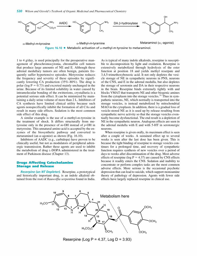
1 to 4 g/day, is used principally for the preoperative man-agement of pheochromocytoma, chromaffin cell tumorsthat produce large amounts of NE and E. Although theseadrenal medullary tumors are often benign, patients fre-quently suffer hypertensive episodes. Metyrosine reducesthe frequency and severity of these episodes by signifi-cantly lowering CA production (35%–80%). The drug ispolar (log P � 0.73) and excreted mainly unchanged in theurine. Because of its limited solubility in water caused byintramolecular bonding of the zwitterions, crystalluria is apotential serious side effect. It can be minimized by main-taining a daily urine volume of more than 2 L. Inhibitors ofCA synthesis have limited clinical utility because suchagents nonspecifically inhibit the formation of all CAs andresult in many side effects. Sedation is the most commonside effect of this drug.
A similar example is the use of �-methyl-m-tyrosine inthe treatment of shock. It differs structurally from me-tyrosine only in the presence of m-OH instead of p-OH inmetyrosine. This unnatural amino acid is accepted by the en-zymes of the biosynthetic pathway and converted tometaraminol (an �-agonist) as shown (Fig. 16.10).
Inhibitors of AADC (e.g., carbidopa) have proven to beclinically useful, but not as modulators of peripheral adren-ergic transmission. Rather these agents are used to inhibitthe metabolism of drug L-DOPA administered in the treat-ment of Parkinson disease (Chapter 13).
Drugs Affecting CatecholamineStorage and Release
Reserpine (an NT Depleter). Reserpine, a prototypicaland historically important drug, is an indole alkaloid ob-tained from the root of Rauwolfia serpentina found in India.
As is typical of many indole alkaloids, reserpine is suscepti-ble to decomposition by light and oxidation. Reserpine isextensively metabolized through hydrolysis of the esterfunction at position 18 and yields methyl reserpate and3,4,5-trimethoxybenzoic acid. It not only depletes the vesi-cle storage of NE in sympathetic neurons in PNS, neuronsof the CNS, and E in the adrenal medulla, but also depletesthe storage of serotonin and DA in their respective neuronsin the brain. Reserpine binds extremely tightly with andblocks VMAT that transports NE and other biogenic aminesfrom the cytoplasm into the storage vesicles.31 Thus in sym-pathetic neurons, NE, which normally is transported into thestorage vesicles, is instead metabolized by mitochondrialMAO in the cytoplasm. In addition, there is a gradual loss ofvesicle-stored NE as it is used up by release resulting fromsympathetic nerve activity so that the storage vesicles even-tually become dysfunctional. The end result is a depletion ofNE in the sympathetic neuron. Analogous effects are seen inthe adrenal medulla with E and with 5-HT in serotonergicneurons.
When reserpine is given orally, its maximum effect is seenafter a couple of weeks. A sustained effect up to severalweeks is seen after the last dose has been given. This isbecause the tight binding of reserpine to storage vesicles con-tinues for a prolonged time, and recovery of sympatheticfunction requires synthesis of new vesicles over a period ofdays to weeks after discontinuation of the drug. Most adverseeffects of reserpine (log P � 4.37) are caused by CNS effectsbecause it readily enters the CNS. Sedation and inability toconcentrate or perform complex tasks are the most commonadverse effects. More serious is the occasional psychoticdepression that can lead to suicide, which support monoaminetheory of pathology of depression. Agents with fewer sideeffects have largely replaced reserpine in clinical use.
530 Wilson and Gisvold’s Textbook of Organic Medicinal and Pharmaceutical Chemistry
Figure 16.10 Metabolic activation of �-methyl-m-tyrosine to metaraminol.
0519-0557_17865_Ch16.qxd 12/2/09 4:16 AM Page 530

Guanethidine (Ismelin) and guanadrel (Hylorel) areseldom used orally active antihypertensives. Drugs of thistype enter the adrenergic neuron by way of the uptake-1process and accumulate within the neuronal storage vesicles.There they bind to the storage vesicles and stabilize the neu-ronal storage vesicle membranes, making them less respon-sive to nerve impulses. The ability of the vesicles to fuse withthe neuronal membrane is also diminished, resulting in inhi-bition of NE release into the synaptic cleft in response to aneuronal impulse and generalized decrease in sympathetictone. Long-term administration of some of these agents alsocan produce a depletion of NE stores in sympathetic neurons.
Both neuronal blocking drugs possess a guanidino moiety[CNHC(BNH)NH2], which is attached to either a hexahy-droazocinyl ring linked by an ethyl group as in guanethidine,or a dioxaspirodecyl ring linked by a methyl group as inguanadrel. The presence of the more basic guanidino group(pKa �12) than the ordinary amino group in these drugsmeans that at physiological pH, they are essentially com-pletely protonated. Thus, these agents do not get into theCNS. As a result, this drug has none of the central effects seenwith many of the other antihypertensive agents described inthis chapter. Guanethidine contains two basic nitrogen atomswith pKa values of 9.0 and 13.43, and can therefore formguanethidine monosulfate (C10H22N4 � H2SO4) or guanethi-dine sulfate [(C10H22N4)2 � H2SO4].
Although guanethidine and guanadrel have virtually thesame mechanism of action on sympathetic neurons, theydiffer in their pharmacokinetic properties. For example,although guanethidine is absorbed incompletely after oraladministration (3%–50%), guanadrel is well absorbed,with a bioavailability of 85%.32 These two agents alsodiffer in terms of half-life: Guanethidine has a half-lifeof about 5 days, whereas guanadrel has a half-life of12 hours. Both agents are partially metabolized (�50%) bythe liver, and both are used to treat moderate-to-severehypertension, either alone or in combination with anotherantihypertensive agent.
SYMPATHOMIMETIC AGENTS
Sympathomimetic agents produce effects resembling thoseproduced by stimulation of the sympathetic nervous system.They may be classified as agents that produce effects by a di-rect, indirect, or mixed mechanism of action. Direct-actingagents elicit a sympathomimetic response by interactingdirectly with adrenergic receptors. Indirect-acting agentsproduce effects primarily by causing the release of NE from
adrenergic nerve terminals; the NE that is released by the in-direct-acting agent activates the receptors to produce the re-sponse. Compounds with a mixed mechanism of action inter-act directly with adrenergic receptors and indirectly cause therelease of NE. As described later, the mechanism by whichan agent produces its sympathomimetic effect is related inti-mately to its chemical structure.
Direct-Acting Sympathomimetics
STRUCTURE–ACTIVITY RELATIONSHIPS
The structure–activity relationships (SARs) summary isshown in Figure 16.11. Comprehensive reviews of the SARsof �- and �-agonists and antagonists33–35 covered their devel-opments in the late 1980s. The parent structure with thefeatures in common for many of the adrenergic drugs is �-phenylethylamine. The manner in which �-phenylethylamineis substituted on the meta- and para-positions of the aromaticring, on the amino (R1), and on �-, (R2)-, and �-positions ofthe ethylamine side chain influences not only their mecha-nism of action, the receptor selectivity, but also their absorp-tion, oral activity, metabolism, degradation, and thus durationof action (DOA). For the direct-acting sympathomimeticamines, maximal activity is seen in �-phenylethylamine de-rivatives containing (a) a catechol and (b) a (1R)-OH groupon the ethylamine portion of the molecule. Such structuralfeatures are seen in the prototypical direct-acting compoundsNE, E, and ISO. The SARs are supported by the model of �2-AR binding studies (Fig. 16.8).
Optical Isomerism. A critical factor in the interactionof adrenergic agonists with their receptors is stereoselectiv-ity. Substitution on either carbon-1 or carbon-2 yields opti-cal isomers. (1R,2S) isomers seem correct configuration fordirect-acting activity. For CAs, the more potent enan-tiomer has the (1R) configuration. This enantiomer is typi-cally several 100-fold more potent than the enantiomer withthe (1S) configuration. It appears that for all direct-acting,phenylethylamine-derived agonists that are structurally sim-ilar to NE, the more potent enantiomer is capable of assum-ing a conformation that results in the arrangement in spaceof the catechol group, the amino group, and the (1R)-OHgroup in a fashion resembling that of (1R)-NE. This expla-nation of stereoselectivity is based on the presumed interac-tion of these three critical pharmacophoric groups with threecomplementary binding areas on the receptor and is knownas the Easson-Stedman hypothesis.17,36 This three-point in-teraction is supported by site-directed mutagenesis studies28
on the adrenergic receptor and is illustrated in Figure 16.8.
Chapter 16 Adrenergic Agents 531
0519-0557_17865_Ch16.qxd 12/2/09 4:16 AM Page 531

Separation of Aromatic Ring and Amino Group.By far, the greatest adrenergic activity occurs when twocarbon atoms separate the aromatic ring from the aminogroup. This rule applies with few exceptions to all types ofactivities.
R1, Substitution on the Amino Nitrogen Determines�- or �-Receptor Selectivity. The amine is normally ion-ized at physiological pH. This is important for direct agonistactivity, because replacing nitrogen with carbon results ina large decline in activity. The activity is also affected
by the number of substituents on the nitrogen. Primary andsecondary amines have good adrenergic activity, whereastertiary amines and quaternary ammonium salts do not. Thenature of the amino substituent also dramatically affectsthe receptor selectivity of the compound. As the size of thenitrogen substituent increases, �-receptor agonist activitygenerally decreases and �-receptor agonist activity in-creases. Thus, NE has more �-activity than �-activity and Eis a potent agonist at �-, �1-, and �2-receptors. ISO, how-ever, is a potent �1- and �2-agonist but has little affinity for�-receptors.
532 Wilson and Gisvold’s Textbook of Organic Medicinal and Pharmaceutical Chemistry
Figure 16.11 Structure–activity relationship of adrenergic phenylethylamine agonists.
0519-0557_17865_Ch16.qxd 12/2/09 4:16 AM Page 532

The nature of the substituents can also affect �1- and �2-receptor selectivity. In several instances, it has been shownthat a �2-directing N-tert-butyl group enhances �2-selectivity.For example, N-tert-butylnorepinephrine (Colterol) is 9 to 10times more potent as an agonist at tracheal �2-receptors thanat cardiac �1-receptors. These results indicate that the �-re-ceptor has a larger lipophilic binding pocket adjacent to theamine-binding aspartic acid residue than do the �-receptors.Increasing the length of the alkyl chain offers no advantage,but if a polar functional group is placed at the end of the alkylgroup, the situation changes. In particular, adding a phenolgroup to the end of a C2 alkyl chain results in a dramaticrise in activity, indicating that an extra polar-binding regionhas been accessed, which can take part in H-bonding.Experiments have shown the activity of the extension analogis thereby increased by a factor of 800. As R1 becomes largerthan butyl group, it can provide compounds with �1-blockingactivity (e.g., tamsulosin and labetalol). Large substituents onthe amino group also protect the amino group from undergo-ing oxidative deamination by MAO.
R2, Substitution on the �-Carbon (Carbon-2).Substitution by small alkyl group (e.g., CH3- or C2H5-) slowsmetabolism by MAO but has little overall effect on DOA ofcatechols because they remain substrates for COMT.However, the resistance to MAO activity is more important innoncatechol indirect-acting phenylethylamines. The DOA ofdrugs such as ephedrine or amphetamine is thus measured inhours rather than in minutes. Because addition of small alkylgroup increases the resistance to metabolism and lipophilic-ity, such compounds often exhibit enhanced oral effective-ness and greater CNS activity than their counterparts that donot contain an �-alkyl group. In addition, compounds with an�-methyl substituent persist in the nerve terminals and aremore likely to release NE from storage sites. For example,metaraminol is an �-agonist and also exhibits a greater degreeof indirect sympathomimetic activity.
Methyl or ethyl substitution on the �-carbon of the eth-ylamine side chain reduces direct agonist activity at both�- and �-receptors. �-Substitution also significantly af-fects receptor selectivity. An ethyl group in this positiondiminishes �-activity far more than �-activity, affordingcompounds with �-selectivity (e.g., ethylnorepinephrineand isoetharine). In the case of �-receptors, for example,�-methyl or ethyl substitution results in compounds to-ward the �2-selectivity, whereas in the case of �-receptors,�-methyl substitution gives compounds toward the �2-selectivity. Another effect of �-substitution is the intro-duction of a chiral center, which has pronounced effects onthe stereochemical requirements for activity. For example,with �-methylnorepinephrine, it is the erythro (1R,2S) iso-mer that possesses significant activity at �2-receptors.
OH substitution on the �-carbon (carbon-1) general-ly decreases CNS activity largely because it lowers lipid sol-ubility. However, such substitution greatly enhances agonistactivity at both �- and �-receptors. For example, ephedrine isless potent than methamphetamine as a central stimulant, butit is more powerful in dilating bronchioles and increasingblood pressure and heart rate. Compounds lacking the �-OHgroup (e.g. DA) have a greatly reduced adrenergic receptoractivity. Some of the activity is retained, indicating that theOH group is important but not essential. The R-enantiomer ofNE is more active than the S-enantiomer, indicating that thesecondary alcohol is involved in an H-bonding interaction.
Substitution on the Aromatic Ring. Maximal �- and�-activity also depends on the presence of 3� and 4� OHgroups. Tyramine, which lacks two OH groups, has no affin-ity for adrenoceptors, indicating the importance of the OHgroups. Studies of �-adrenoceptor structure suggest that theOH groups on serine residues 204 and 207 probably form H-bonds with the catechol OH groups at positions 3� and 4�,respectively.
Chapter 16 Adrenergic Agents 533
0519-0557_17865_Ch16.qxd 12/2/09 4:16 AM Page 533

Although the catechol moiety is an important structuralfeature in terms of yielding compounds with maximalagonist activity at adrenoceptors, it can be replaced withother substituted phenyl moieties to provide selective adren-ergic agonists. This approach has been used in particular inthe design of selective �2-agonists. For example, re-placement of the catechol function of ISO with the resorcinolstructure gives a selective �2-agonist, metaproterenol.Furthermore, because the resorcinol ring is not a substrate forCOMT, �-agonists that contain this ring structure tend tohave better absorption characteristics and a longer DOA thantheir catechol-containing counterparts. In another approach,replacement of the meta-OH of the catechol structure with ahydroxymethyl group gives agents, such as albuterol, whichshow selectivity to the �2-receptor. Because they are not cat-echols, these agents are not metabolized by COMT and thusshow improved oral bioavailability and longer DOA.
Modification of the catechol ring can also bring aboutselectivity at �-receptors as it appears that the catechol moi-ety is more important for �2-activity than for �1-activity.For example, removal of the p-OH group from E givesphenylephrine, which, in contrast to E, is selective for the�1-receptor. Phenylephrine is less potent than E at both �-and �-receptors, with �2-activity almost completely absent.However, the OH group can be replaced by other groupscapable of interacting with the binding site by H-bonding.This is particularly true for the meta-OH group, which canbe replaced by CH2OH, NHMe, NHCOR, NMe2, orNHSO2R group.
CAs without OH Groups. Phenylethylamines that lackOH groups on the ring and the �-OH group on the side chainact almost exclusively by causing the release of NE fromsympathetic nerve terminals and thus results in a loss ofdirect sympathomimetic activity. Because substitution ofOH groups on the phenylethylamine structure makes theresultant compounds less lipophilic, unsubstituted or alkyl-substituted compounds cross the BBB more readily and havemore central activity. Thus, amphetamine and metham-phetamine exhibit considerable CNS activity.
CAs per oral have only a brief DOA and are almost inac-tive, because they are rapidly inactivated in the intestinalmucosa and in the liver before reaching the systemic circu-lation. In contrast, compounds without one or both phenolicOH substituents are, however, not metabolized by COMT,and they are orally active and have longer DOA.
Imidazolines and �-Adrenergic Agonists. Althoughnearly all �-agonists are �-phenylethanolamine derivatives, itis �-adrenoceptors that exhibit a far more diverse assortmentof structures. A second chemical class of �-agonists, the
imidazolines, which give rise to �-agonists and are thus vaso-constrictors. These imidazolines can be nonselective, or theycan be selective for either �1- or �2-receptors. Structurally,most imidazolines have their heterocyclic imidazoline nu-cleus linked to a substituted aromatic moiety via some type ofbridging unit (Fig. 16.12).34 The optimum bridging unit (X) isusually a single methylene group or amino group. Althoughmodification of the imidazoline ring generally results in com-pounds with significantly reduced agonist activity, there areexamples of so-called open-ring imidazolines that are highlyactive. The nature of the aromatic moiety, as well as how it issubstituted, is quite flexible. However, agonist activity is en-hanced when the aromatic ring is substituted with halogensubstituents like chlorine (Cl) or small alkyl groups likemethyl group, particularly when they are placed in the twoortho positions. Because the SARs of the imidazolines arequite different from those of the �-phenylethylamines, it hasbeen postulated that the imidazolines interact with �-recep-tors differently from the way the �-phenylethylamines do,particularly with regard to the aromatic moiety.37
ENDOGENOUS CATECHOLAMINES
The three naturally occurring catecholamines DA, NE, andE are used as therapeutic agents.
Dopamine. (DA, 3�,4�-dihydroxyphenylethylamine)differs from NE in lacking of 1-OH group. It is the imme-diate precursor of NE and is a central NT particularlyimportant in the regulation of movement (see Chapter 13).As a catechol and primary amine, DA is rapidly metabo-lized by COMT and MAO and has a short DOA with nooral activity. It is used intravenously in treatment of shock.In contrast with the NE and E, DA increases blood flow tothe kidney in doses that have no chronotropic effect on theheart or that cause no increase in blood pressure. Theincreased blood flow to the kidneys enhances glomerularfiltration rate, Na� excretion, and, in turn, urinary output.The dilation of renal blood vessels produced by DA is theresult of its agonist action on the D1-DA receptor.
534 Wilson and Gisvold’s Textbook of Organic Medicinal and Pharmaceutical Chemistry
Figure 16.12 General structural features of theimidazoline �-adrenergic receptor agonists.
0519-0557_17865_Ch16.qxd 12/2/09 4:16 AM Page 534

In doses slightly higher than those required to increaserenal blood flow, DA stimulates the �1-receptors of the heartto increase cardiac output. Some of the effects of DA on theheart are also caused by NE release. Infusion at a rate greaterthan 10 �g/kg per minute results in stimulation of �1-receptors, leading to vasoconstriction and an increase in arte-rial blood pressure. DA should be avoided or used at a muchreduced dosage (one tenth or less) if the patient has receivedan MAO inhibitor. Careful adjustment of dosage also is nec-essary in patients who are taking tricyclic antidepressants.
Norepinephrine (NE, Levophed) differs from DA onlyby addition of a 1-OH substituent (�-OH-DA) and from Eonly by lacking the N-methyl group. Like DA, it is polar andrapidly metabolized by both COMT and MAO, resulting inpoor oral bioavailability and short DOA (1 or 2 minutes evenwhen given intravenously). It is a stimulant of �1-, �2-, and�1-adrenoceptors (notice that lacking the N-methyl group re-sults in lacking �2- and �3-activity). It is used to counteractvarious hypotensive crises, because its �-activity raisesblood pressure and as an adjunct treatment in cardiac arrestbecause its �-activity stimulates the heart. It has limitedclinical application caused by the nonselective nature of itsactivities.
Epinephrine (E, Adrenalin) differs from NE only bythe addition of an N-methyl group. Like the other CAs, E islight sensitive and easily oxidized on exposure to air be-cause of the catechol ring system. The development of apink-to-brown color indicates oxidative breakdown. Tominimize oxidation, solutions of the drug are stabilized bythe addition of reducing agents such as sodium bisulfite. Eis also destroyed readily in alkaline solutions and by metals(e.g., Cu, Fe, Zn) and weak oxidizing agents. It is used inaqueous solution for inhalation as the free amine. Like otheramines, it forms salts with acids, hydrochloride, and thebitartrate being the most common.
Like NE, it lacks oral activity and has short DOA.However, it is much more widely used clinically than NE.E is a potent stimulant of all �1-, �2-, �1-, �2-, and �3-adrenoceptors, and thus it switches on all possible adrener-gic receptors, leading to a whole range of desired and sideeffects. Particularly prominent are the actions on the heartand on vascular and other smooth muscle. It is a very potentvasoconstrictor and cardiac stimulant. NE has, in general,greater �-activity caused by an additional N-methyl group.Therefore, E is used to stimulate the heart in cardiac arrest.Although intravenous infusion of E has pronounced effectson the cardiovascular system, its use in the treatment of
heart block or circulatory collapse is limited because of itstendency to induce cardiac arrhythmias.
The ability of epinephrine to stimulate �2-receptors hasled to its use by injection and by inhalation to relaxbronchial smooth muscle in asthma and in anaphylacticreactions. Several OTC preparations (e.g., Primatene, Bron-kaid) used for treating bronchial asthma use E. It is also usedin inhibiting uterine contraction. Because of its �-activity, Eis used to treat hypotensive crises and nasal congestion, toenhance the activity of local anesthetics, and as a constrictorin hemorrhage.
In addition, E is used in the treatment of open-angle glau-coma, where it apparently reduces intraocular pressure byincreasing the rate of outflow of aqueous humor from theanterior chamber of the eye. The irritation often experiencedon instillation of E into the eye has led to the developmentof other preparations of the drug that potentially are not asirritating. One such example is dipivefrin.
Dipivefrin (Propine, Dipivalyl Epinephrine). Toovercome several of the pharmacokinetic and pharmaceuti-cal shortcomings of E as an ophthalmic agent, the prodrugapproach has been successfully applied. Dipivefrin is a pro-drug of E that is formed by the esterification of the catecholOH groups of E with pivalic acid. Most of the advantages ofthis prodrug over E stem from improved bioavailability. Thegreatly increased lipophilicity allows much greater pene-trability into the eye through the corneal epithelial andendothelial layer. The stroma in between requires hy-drophilicity for penetration. Dipivefrin has that, too, due tothe 1-OH group and cationic nitrogen (the eyedrops containthe hydrochloride [HCl] salt). This dual solubility permitsmuch greater penetrability into the eye than the very hy-drophilic E hydrochloride. Increased DOA is also achievedbecause the drug is resistant to the metabolism by COMT.In addition to its increased in vivo stability, it is also lesseasily oxidized by air due to the protection of the catecholOH groups. This high bioavailability and in vivo and in vitrostability translate into increased potency such that the 0.1%ophthalmic solution is approximately equivalent to a 2% Esolution. After its absorption, it is converted to E by es-terases slowly in the cornea and anterior chamber.Dipivefrin also offers the advantage of being less irritatingto the eye than E.
�-ADRENERGIC RECEPTOR AGONISTS
All selective �1-agonists have therapeutic activity as vasocon-strictors. Structurally, they include (a) phenylethanolamines
Chapter 16 Adrenergic Agents 535
0519-0557_17865_Ch16.qxd 12/2/09 4:16 AM Page 535

such as phenylephrine, metaraminol, and methoxamine and(b) 2-arylimidazolines such as xylometazoline, oxymetazo-line, tetrahydrozoline, and naphazoline.
Phenylephrine. (Neo-Synephrine, a prototypical se-lective direct-acting �1-agonist) differs from E only inlacking a p-OH group. It is orally active, and its DOA isabout twice that of E because it lacks the catechol moietyand thus is not metabolized by COMT. However, its oralbioavailability is less than 10% because of its hydrophilicproperties (log P � �0.3), intestinal 3�-O-glucuronida-tion/sulfation and metabolism by MAO. Lacking the p-OHgroup, it is less potent than E and NE but it is a selective�1-agonist and thus a potent vasoconstrictor. It is usedsimilarly to metaraminol and methoxamine for hypoten-sion. Another use is in the treatment of severe hypotensionresulting from either shock or drug administration. It alsohas widespread use as a nonprescription nasal deconges-tant in both oral and topical preparations. When applied tomucous membranes, it reduces congestion and swelling byconstricting the blood vessels of the membranes. In theeye, it is used to dilate the pupil and to treat open-angleglaucoma. In addition, it is used in spinal anesthesia toprolong the anesthesia and to prevent a drop in blood pres-sure during the procedure. It is relatively nontoxic and pro-duces little CNS stimulation. Metaraminol is just anotherexample.
Methoxamine (Vasoxyl) is another �1-agonist and par-enteral vasopressor used therapeutically and so have fewcardiac stimulatory properties. It is bioactivated by O-demethylation to an active m-phenolic metabolite. In fact,it tends to slow the ventricular rate because of activation ofthe carotid sinus reflex. It is less potent than phenylephrineas a vasoconstrictor. Methoxamine is used primarily duringsurgery to maintain adequate arterial blood pressure, espe-cially in conjunction with spinal anesthesia. It does not stim-ulate the CNS. Because it is not a substrate for COMT, itsDOA is significantly longer than NE.
Midodrine (ProAmatine) is the N-glycyl prodrug ofthe selective �1-agonist desglymidodrine. Removal of the N-glycyl moiety from midodrine occurs readily in the liver aswell as throughout the body, presumably by amidases.Midodrine is orally active and represents another example ofa dimethoxy-�-phenylethylamine derivative that is used ther-apeutically for its vasoconstrictor properties. Specifically, it isused in the treatment of symptomatic orthostatic hypotension.
536 Wilson and Gisvold’s Textbook of Organic Medicinal and Pharmaceutical Chemistry
0519-0557_17865_Ch16.qxd 12/2/09 4:16 AM Page 536

Chapter 16 Adrenergic Agents 537
Naphazoline (Privine), tetrahydrozoline (Tyzine,Visine), xylometazoline (Otrivin), and oxymetazoline(Afrin) are 2-aralkylimidazolines �1-agonists. These agentsare used for their vasoconstrictive effects as nasal and oph-thalmic decongestants. Although nearly all �-agonists arephenylethanolamine derivatives, �-receptors accommodatemore diverse chemical structures. All 2-aralkylimidazoline�1-agonists contain a one-carbon bridge between C-2 of theimidazoline ring and a phenyl ring, and thus a phenylethyl-amine structure feature is there. Ortho-lipophilic groups onthe phenyl ring are important for �-activity. However, meta-or para-bulky lipophilic substituents on the phenyl ring maybe important for the �1-selectivity. They have limited accessto the CNS, because they essentially exist in an ionized format physiological pH caused by the very basic nature of theimidazoline ring (pKa � 10–11). Xylometazoline andoxymetazoline have been used as topical nasal deconges-tants because of their ability to promote constriction of thenasal mucosa. When taken in large doses, oxymetazoline
may cause hypotension, presumably because of a centralclonidine-like effect. Oxymetazoline also has significantaffinity for �2A-receptors.
Clonidine (Catapres) differs from 2-arylimidazoline�1-agonists mainly by the presence of o-chlorine groups anda NH bridge. The o-chlorine groups afford better activitythan o-methyl groups at �2 sites. Importantly, clonidinecontains a NH bridge (aminoimidazolines) instead of CH2
bridge in 2-arylimidazoline. The uncharged form of cloni-dine exists as a pair of tautomers as shown next. Clonidineis an example of a (phenylimino) imidazolidine derivativethat possesses central �2-selectivity. The �1:�2 ratio is300:1. Under certain conditions, such as intravenous in-fusion, clonidine can briefly exhibit vasoconstrictive ac-tivity as a result of stimulation of peripheral �-receptors.However, this hypertensive effect, if it occurs, is followedby a much longer-lasting hypotensive effect as a result ofthe ability of clonidine to enter into the CNS and stimulate
0519-0557_17865_Ch16.qxd 12/2/09 4:16 AM Page 537

�2-receptors located in regions of the brain such as thenucleus tractus solitarius. Stimulation of these �2-receptorsbrings about a decrease in sympathetic outflow from theCNS, which in turn leads to decreases in peripheral vascularresistance and blood pressure.20,38 Bradycardia is also pro-duced by clonidine as a result of a centrally induced fa-cilitation of the vagus nerve and stimulation of cardiacprejunctional �2-receptors.39 These pharmacological ac-tions have made clonidine quite useful in the treatment ofhypertension.
The ability of clonidine and its analogs to exert an anti-hypertensive effect depends on the ability of these com-pounds not only to interact with the �2-receptor in the brainbut also to gain entry into the CNS. For example, in the caseof clonidine, the basicity of the guanidine group (typicallypKa � 13.6) is decreased to 8.0 (the pKa of clonidine)because of the inductive and resonance effects of thedichlorophenyl ring. Thus, at physiological pH, clonidinewill exist to a significant extent in the nonionized formrequired for passage into the CNS. It has an oral bioavail-ability of more than 90%.
Although various halogen and alkyl substitutions can beplaced at the two ortho positions of the (phenylimino)imida-zolidine nucleus without affecting the affinity of the deriva-tives for �2-receptors, methyl analogs are much more readilymetabolized to the corresponding acids (inactive) and thushave short DOA. Halogen substituents such as chlorine seemto provide the optimal characteristics in this regard.40 One ofthe metabolites of clonidine, 4-hydroxyclonidine, has goodaffinity for �2-receptors, but because it is too polar to get intothe CNS, it is not an effective antihypertensive agent.
In addition to binding to the �2-adrenergic receptor, cloni-dine, as well as some other imidazolines, shows high affinityfor what has been termed the imidazoline receptor.41,42
Through the use of imidazoline and �2-antagonists, specificnonadrenergic imidazoline binding sites (I1-IBS) have re-cently been characterized in CNS control of blood pressure,which are GPCR, with agmatine (decarboxylated arginine)being the endogenous ligand for IBS.43,44 However, otherstudies involving both site-directed mutagenesis of the �2A-receptor subtype and genetically engineered knockout micedeficient in either the �2A- or �2B-receptor subtypes provideevidence that the hypotensive response of the �2-receptor ag-onists such as clonidine primarily involves the �2A-receptor
subtype.45,46 Thus, the central hypotensive activity for cloni-dine and other 2-aminoimidazolines need both �2-receptorsand I1-IBS to produce their central sympatholytic response.Clonidine appears to be more selective for �2-receptors thanfor I1-IBS.
Apraclonidine (Iopidine) and Brimonidine (Alpha-gan). In addition to its therapeutic use as an antihyperten-sive agent, clonidine has been found to provide beneficialeffects in several other situations,47 including glaucoma,spasticity, migraine prophylaxis, opiate withdrawal syn-drome, and anesthesia. This has prompted the developmentof analogs of clonidine for specific use in some of the men-tioned areas. Two of such examples are apraclonidine andbrimonidine. Apraclonidine does not cross the BBB.However, brimonidine can cross the BBB and hence canproduce hypotension and sedation, although these CNSeffects are slight compared with those of clonidine. CNS ef-fects of these drugs are correlated well to their log P, pKa,and thus log D value. Both apraclonidine and brimonidineare selective �2-agonists with �1:�2 ratios of 30:1 and1,000:1, respectively. Brimonidine is a much more selective�2-agonist than clonidine or apraclonidine and is a first-line agent for treating glaucoma. Apraclonidines’s primarymechanism of action may be related to a reduction of aque-ous formation, whereas brimonidine lowers intraocularpressure by reducing aqueous humor production and in-creasing uveoscleral outflow. Apraclonidine is used specifi-cally to control elevations in intraocular pressure that canoccur during laser surgery on the eye. Another example istizanidine (Zanaflex), which finds use in treating spasticityassociated with multiple sclerosis or spinal cord injury. Bystimulating �2-adrenergic receptors, it is believed to de-crease the release of excitatory amino acid NTs from spinalcord interneurons.48
Guanabenz (Wytensin) and Guanfacine (Tenex)(Open-Ring Imidazolidines). Studies on SAR of central�2-agonists showed that the imidazoline ring was not nec-essary for �2-activity. Two clonidine analogs, guanabenz(pKa � 8.1) and guanfacine (pKa � 7), which are closelyrelated chemically and pharmacologically, are also used asantihypertensive drugs. In these compounds, the 2,6-dichlorophenyl moiety found in clonidine is connected to aguanidino group by a two-atom bridge. In the case of
538 Wilson and Gisvold’s Textbook of Organic Medicinal and Pharmaceutical Chemistry
0519-0557_17865_Ch16.qxd 12/2/09 4:16 AM Page 538
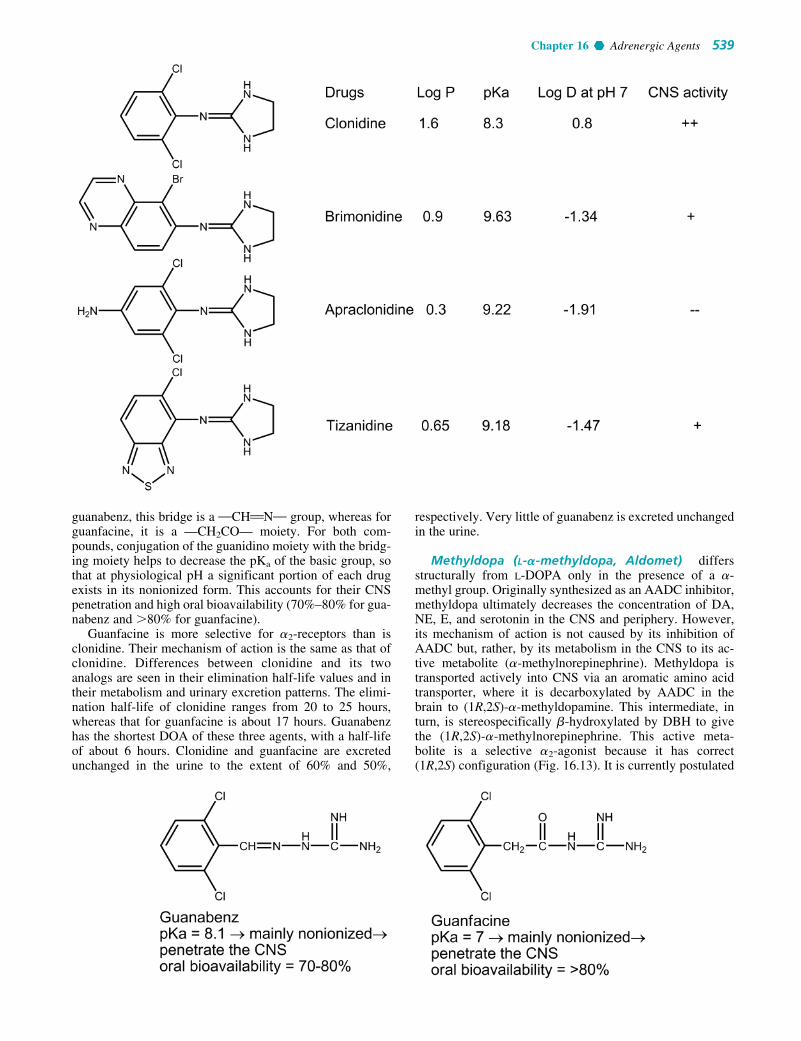
guanabenz, this bridge is a MCHBNM group, whereas forguanfacine, it is a —CH2CO— moiety. For both com-pounds, conjugation of the guanidino moiety with the bridg-ing moiety helps to decrease the pKa of the basic group, sothat at physiological pH a significant portion of each drugexists in its nonionized form. This accounts for their CNSpenetration and high oral bioavailability (70%–80% for gua-nabenz and �80% for guanfacine).
Guanfacine is more selective for �2-receptors than isclonidine. Their mechanism of action is the same as that ofclonidine. Differences between clonidine and its twoanalogs are seen in their elimination half-life values and intheir metabolism and urinary excretion patterns. The elimi-nation half-life of clonidine ranges from 20 to 25 hours,whereas that for guanfacine is about 17 hours. Guanabenzhas the shortest DOA of these three agents, with a half-lifeof about 6 hours. Clonidine and guanfacine are excretedunchanged in the urine to the extent of 60% and 50%,
respectively. Very little of guanabenz is excreted unchangedin the urine.
Methyldopa (L-�-methyldopa, Aldomet) differsstructurally from L-DOPA only in the presence of a �-methyl group. Originally synthesized as an AADC inhibitor,methyldopa ultimately decreases the concentration of DA,NE, E, and serotonin in the CNS and periphery. However,its mechanism of action is not caused by its inhibition ofAADC but, rather, by its metabolism in the CNS to its ac-tive metabolite (�-methylnorepinephrine). Methyldopa istransported actively into CNS via an aromatic amino acidtransporter, where it is decarboxylated by AADC in thebrain to (1R,2S)-�-methyldopamine. This intermediate, inturn, is stereospecifically �-hydroxylated by DBH to givethe (1R,2S)-�-methylnorepinephrine. This active meta-bolite is a selective �2-agonist because it has correct(1R,2S) configuration (Fig. 16.13). It is currently postulated
Chapter 16 Adrenergic Agents 539
0519-0557_17865_Ch16.qxd 12/2/09 4:16 AM Page 539

that �-methylnorepinephrine acts on �2-receptors in theCNS in the same manner as clonidine, to decrease sympa-thetic outflow and lower blood pressure.
Absorption can range from 8% to 62% and appears to in-volve an amino acid transporter. Absorption is thus affectedby food, and about 40% of that absorbed is converted tomethyldopa-O-sulfate by the intestinal mucosal cells.Methyldopa is used only by oral administration because itszwitterionic character limits its solubility. The ester hy-drochloride salt of methyldopa, methyldopate (Aldometester), was developed as a highly water-soluble derivativethat could be used to make parenteral preparations. It is con-verted to methyldopa in the body through the action of es-terases (Fig. 16.13).
DUAL �- AND �-AGONISTS/ANTAGONISTS
Dobutamine (Dobutrex) is a positive inotropic agentadministered intravenously for congestive heart failure. Itresembles DA structurally but possesses a bulky 1-(methyl)-3-(4-hydroxyphenyl)propyl group on the amino group. Itpossesses a center of asymmetry, and both enantiomericforms are present in the racemic mixture used clinically. The(�) isomer of dobutamine is a potent �1-agonist, which iscapable of causing marked pressor responses. In contrast,(�)-dobutamine is a potent �1-antagonist, which canblock the effects of (�)-dobutamine. Importantly, the ef-fects of these two isomers are mediated via �1-receptors.Both isomers appear to be full agonists, but the (�) isomeris a more potent �1-agonist than the (�) isomer (approxi-mately tenfold).49,50
Dobutamine contains a catechol group and is orally in-active and thus is given by intravenous infusion. Solutionsof the drug can exhibit a slight pink color because of oxida-tion of the catechol function. It has a plasma half-life ofabout 2 minutes because it is metabolized by COMT and byconjugation, although not by MAO.
�-ADRENERGIC RECEPTOR AGONISTS
Isoproterenol (Isuprel) is a nonselective and proto-typical �-agonist (�2/�1 � 1). After oral administration,the absorption of ISO is rather erratic and undependable.The principal reason for its poor absorption characteristicsand relatively short DOA is its facile metabolism by sulfateand glucuronide conjugation of the phenolic OH groupsand o-methylation by COMT. Because it is a catechol, it issensitive to light and air. Aqueous solutions become pinkon standing. Unlike E and NE, ISO does not appear to
540 Wilson and Gisvold’s Textbook of Organic Medicinal and Pharmaceutical Chemistry
Figure 16.13 Metabolic conversion of methyldopate and methyldopa to �-methyl-norepinephrine.
0519-0557_17865_Ch16.qxd 12/2/09 4:16 AM Page 540

undergo oxidative deamination by MAO. The drug hasDOA of 1 to 3 hours after inhalation.
Because of an isopropyl substitution on the nitrogenatom, it has virtually no �-activity. However, it does act onboth �1- and �2-receptors. It thus can produce an increase incardiac output by stimulating cardiac �1-receptors and canbring about bronchodilation through stimulation of �2-receptors in the respiratory tract. In fact, it is one of the mostpotent bronchodilators available and is available for use byinhalation and injection. Cardiac stimulation is an occasion-ally dangerous adverse effect. This effect of ISO on theheart is sometimes used in the treatment of heart block.
The cardiac stimulation caused by its �1-activity andits lack of oral activity have led to its diminished use infavor of more selective �-agonists. The problems have beenovercome at least partially by the design and developmentof several noncatechol selective �2-agonists. These agentsrelax smooth muscle of the bronchi, uterus, and skeletalmuscle vascular supply. They find their primary use as bron-chodilators in the treatment of acute and chronic bronchialasthma and other obstructive pulmonary diseases.
Metaproterenol (Alupent), terbutaline (Bricanyl,Brethine), and fenoterol (an investigational drugs)belong to the structural class of resorcinol bronchodilatorsthat have 3�,5�-diOH groups of the phenyl ring (rather than3�,4�-diOH groups as in catechols). 3�,5�-diOH groups con-fer �2-receptor selectivity on compounds with large aminosubstituents. For example, metaproterenol (a resorcinolanalog of ISO), terbutaline (an N-t-butyl analog ofmetaproterenol), and other similar compounds are resorcin-ol �2-selective agonists. They relax the bronchial muscula-ture in patients with asthma but cause less direct cardiacstimulation than do the nonselective �-agonists. Becausemetaproterenol has a �-directing N-isopropyl group and itis less �2 selective than either terbutaline or albuterol (both
have �2-directing t-butyl groups), and hence is more proneto cause cardiac stimulation. Although these agents aremore selective for �2-receptors, they have a lower affinityfor �2-receptors than ISO. However, they are much moreeffective when given orally, and they have a longer DOA.This is because they are resistant to the metabolism by ei-ther COMT or MAO. Instead, their metabolism primarilyinvolves glucuronide conjugation. Although both metapro-terenol and terbutaline exhibit significant �2-receptor selec-tivity, the common cardiovascular effects associated withother adrenergic agents can also be seen with these drugswhen high doses are used.
Albuterol (Proventil, Ventolin), pirbuterol (Maxair),and salmeterol (Serevent) are examples of selective �2-agonists whose selectivity results from replacement of themeta-OH group of the aromatic ring with a hydroxymethylmoiety. Pirbuterol is closely related structurally to albuterol(�2/�1 � 60); the only difference between the two is thatpirbuterol contains a pyridine ring instead of a benzene ring.As in the case of metaproterenol and terbutaline, these drugsare not metabolized by either COMT or MAO. Instead, theyare conjugated with sulfate. They are thus orally active, andexhibit a longer DOA than ISO. The DOA of terbutaline,albuterol, and pirbuterol is in the range of 3 to 6 hours. (S)-albuterol enhances bronchial muscle contraction, and thisundesirable effect is completely avoided by using the pure(R)-albuterol, levalbuterol (Xopenex). Therefore, the effi-cacy is achieved at one-fourth dose of racemic albuterolwith markedly reduced adverse effects.
Salmeterol has an N-phenylbutoxyhexyl substituent incombination with a �-OH group and a salicyl phenyl ring foroptimal direct-acting �2-receptor selectivity and potency. Ithas a potency similar to that of ISO. This drug associateswith the �2-receptor slowly resulting in slow onset of actionand dissociates from the receptor at an even slower rate.51 It
Chapter 16 Adrenergic Agents 541
0519-0557_17865_Ch16.qxd 12/2/09 4:16 AM Page 541

is resistant to both MAO and COMT and highly lipophilic(log P � 3.88). It is thus very long acting (12 hours), aneffect also attributed to the highly lipophilic phenylalkylsubstituent on the nitrogen atom, which is believed to inter-act with a site outside but adjacent to the active site.
Formoterol and Levalbuterol. Formoterol (Foradil) isalso a lipophilic (log P � 1.6) and long-acting �2-agonist. Ithas 3�-formylamino (�-directing) and 4�OH groups on onephenyl ring and a lipophilic �-directing N-isopropyl-p-methoxyphenyl group on the nitrogen atom. Its long DOA(12 hours), which is comparable to that of salmeterol, hasbeen suggested to result from its association with the mem-brane lipid bilayer,52 from which it gradually diffuses to pro-vide prolonged stimulation of �2 receptors and its resistanceto MAO and COMT. Formoterol has a much faster onset ofaction than does salmeterol as result of its lower lipophilic-ity. Both of these long-acting drugs are used by inhalationand are recommended for maintenance treatment of asthma,usually in conjunction with an inhaled corticosteroid.
All of the previously mentioned �2-agonists possess atleast one chiral center and are used as racemic mixtures.Formoterol possesses two chiral centers and is used asthe racemic mixture of the (R,R) and (S,S) enantiomers.As mentioned previously, it is the (R) isomer of the
phenylethanolamines that possesses the pharmacologicalactivity. There is no clinical advantage for using (R,R)-formoterol as bronchodilators compared with the racemicmixture because of its high potency and low dose. However,concerns have been raised about the use of racemic mixturesunder the belief that the inactive isomer may be responsiblefor some of the adverse effects. Levalbuterol (Xopenex), thepure (R) isomer of racemic albuterol, represents the first at-tempt to address this issue as mentioned earlier.
Isoetharine. (�-ethyl ISO) was the first �2-selectivedrug widely used for the treatment of airway obstruction.However, its degree of selectivity and activity for �2-receptors may not approach that of some of the other agentssuch as terbutaline, albuterol, or even ISO. Because of thepresence of the �2-directing �-ethyl group and �-directingisopropyl group, isoetharine is a �2-agonist and is resistantto MAO. However, because it contains the catechol ringsystem, it is metabolized by COMT and O-sulfated quiteeffectively. Consequently, it has a short DOA similar to thatof ISO and is used only by inhalation for the treatment ofacute episodes of bronchoconstriction.
Bitolterol (Tornalate, a Prodrug). Colterol (activemetabolite of bitolterol) differs from ISO by replacing the �-directing N-isopropyl to �2-directing N-tert-butyl group,which results in the increased �2-selectivity. Bitolterol(Tornalate) is a prodrug of colterol (a �2-selective agonist)in which the catechol OH groups have been converted to di-p-toluate esters, providing increased lipid solubility causedby the presence of the two lipophilic di-p-toluate esters inbitolterol. The presence of the bulky di-ester and bulky N-tert-butyl groups also prolong the DOA (8 hours) becauseit is resistant to COMT and MAO metabolism. Bitolterol isadministered by inhalation for bronchial asthma and re-versible bronchospasm. The highly bulky p-toluoyl groupsapparently inhibit the efficiency of the esterases. After ab-sorption, it is hydrolyzed by esterases slowly enough in the
542 Wilson and Gisvold’s Textbook of Organic Medicinal and Pharmaceutical Chemistry
0519-0557_17865_Ch16.qxd 12/2/09 4:16 AM Page 542

lung and other tissues to produce the active agent (colterol)affording sustained bronchodilation. The DOA of a singledose of the prodrug bitolterol is twice that of a single dose ofcolterol, permitting less frequent administration and greaterconvenience to the patient. Colterol is then metabolized afterpharmacological action by COMT and conjugation.
Ritodrine (Yutopar) is a selective �2-agonist that wasdeveloped specifically for use as a uterine relaxant. Its uter-ine inhibitory effects are more sustained than its effects onthe cardiovascular system, which are minimal comparedwith those caused by nonselective �-agonists. The cardio-vascular effects usually associated with its administrationare mild tachycardia and slight diastolic pressure decrease.Usually, it is administered initially by intravenous infusionto stop premature labor and subsequently it may be givenorally.
�3-Adrenergic Receptor Agonists. The �3-receptor hasbeen shown to mediate various pharmacological effects suchas lipolysis, thermogenesis, and relaxation of the urinary blad-der. Activation of the �3-receptor is thought to be a possibleapproach for the treatment of obesity, type 2 diabetes mellitus,and frequent urination. Therefore, it is recognized as an attrac-tive target for drug discovery. Selective �3-agonists have beendeveloped, but they have not been approved for therapeutic
use. Continuing clinical studies will clarify the physiologiceffects mediated by �3-receptors and elucidate the potentialtherapeutic uses of its agonists in humans.53
Indirect-Acting Sympathomimetics
Indirect-acting sympathomimetics act by releasing endoge-nous NE. They also enter the nerve ending by way of theactive-uptake process and displace NE from its storage gran-ules. As with the direct-acting agents, the presence of the cat-echol OH groups enhances the potency of indirect-actingphenylethylamines. However, the indirect-acting drugs thatare used therapeutically are not catechol derivatives and, inmost cases, do not even contain an OH moiety. In contrastwith the direct-acting agents, the presence of a �-hydroxylgroup decreases, and an �-methyl group increases, the effec-tiveness of indirect-acting agents. The presence of nitrogensubstituents decreases indirect activity, with substituentslarger than methyl groups rendering the compound virtuallyinactive. Phenylethylamines that contain a tertiary aminogroup are also ineffective as NE-releasing agents. Given theforegoing structure–activity considerations, it is easy to un-derstand why amphetamine and p-tyramine are often cited asprototypical indirect-acting sympathomimetics. Because am-phetamine-type drugs exert their primary effects on the CNS,they are discussed in more detail in Chapter 15. This chapterdiscusses those agents that exert their effects primarily on theperiphery.
Chapter 16 Adrenergic Agents 543
0519-0557_17865_Ch16.qxd 12/2/09 4:16 AM Page 543
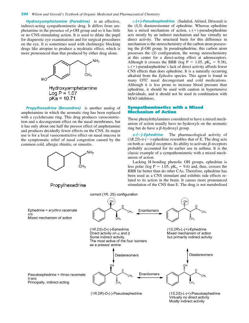
Hydroxyamphetamine (Paredrine) is an effective,indirect-acting sympathomimetic drug. It differs from am-phetamine in the presence of p-OH group and so it has littleor no CNS-stimulating action. It is used to dilate the pupilfor diagnostic eye examinations and for surgical procedureson the eye. It is sometimes used with cholinergic blockingdrugs like atropine to produce a mydriatic effect, which ismore pronounced than that produced by either drug alone.
Propylhexedrine (Benzedrex) is another analog ofamphetamine in which the aromatic ring has been replacedwith a cyclohexane ring. This drug produces vasoconstric-tion and a decongestant effect on the nasal membranes, butit has only about one half the pressor effect of amphetamineand produces decidedly fewer effects on the CNS. Its majoruse is for a local vasoconstrictive effect on nasal mucosa inthe symptomatic relief of nasal congestion caused by thecommon cold, allergic rhinitis, or sinusitis.
L-(�)-Pseudoephedrine. (Sudafed, Afrinol, Drixoral) isthe (S,S) diastereoisomer of ephedrine. Whereas ephedrinehas a mixed mechanism of action, L-(�)-pseudoephedrineacts mostly by an indirect mechanism and has virtually nodirect activity. The structural basis for this difference inmechanism is the stereochemistry of the carbon atom possess-ing the �-OH group. In pseudoephedrine, this carbon atompossesses the (S) configuration, the wrong stereochemistryat this center for a direct-acting effect at adrenoceptors.Although it crosses the BBB (log P � 1.05, pKa � 9.38), L-(�)-pseudoephedrine’s lack of direct activity affords fewerCNS effects than does ephedrine. It is a naturally occurringalkaloid from the Ephedra species. This agent is found inmany OTC nasal decongestant and cold medications.Although it is less prone to increase blood pressure thanephedrine, it should be used with caution in hypertensiveindividuals, and it should not be used in combination withMAO inhibitors.
Sympathomimetics with a MixedMechanism of Action
Those phenylethylamines considered to have a mixed mech-anism of action usually have no hydroxyls on the aromaticring but do have a �-hydroxyl group.
D-(�)-Ephedrine. The pharmacological activity of(1R,2S)-D-(�)-ephedrine resembles that of E. The drug actson both �- and �-receptors. Its ability to activate �-receptorsprobably accounted for its earlier use in asthma. It is theclassic example of a sympathomimetic with a mixed mech-anism of action.
Lacking H-bonding phenolic OH groups, ephedrine isless polar (log P � 1.05, pKa � 9.6) and, thus, crosses theBBB far better than do other CAs. Therefore, ephedrine hasbeen used as a CNS stimulant and exhibits side effects re-lated to its action in the brain. It causes more pronouncedstimulation of the CNS than E. The drug is not metabolized
544 Wilson and Gisvold’s Textbook of Organic Medicinal and Pharmaceutical Chemistry
0519-0557_17865_Ch16.qxd 12/2/09 4:16 AM Page 544

by either MAO or COMT and therefore has more oral activ-ity and longer DOA than E. Like many other phenyliso-propylamines, a significant fraction of the drug is excretedunchanged in the urine although it can be p-hydroxylatedand N-demethylated by cytochrome P450 mixed-functionoxidases. Because it is a weak base, its excretion can be ac-celerated by acidification of the urine.
Ephedrine has two asymmetric carbon atoms; therebycreating four optically active isomers. The erythro race-mate is called ephedrine, and the threo racemate is knownas pseudoephedrine (�-ephedrine). Natural ephedrine is theD (�) isomer, and it is the most active of the four isomersas a pressor amine. This is largely because of the fact thatthis isomer has the correct (1R,2S) configuration for opti-mal direct action at adrenergic receptors.
Ephedrine decomposes gradually and darkens when ex-posed to light. The free alkaloid is a base, and an aqueoussolution of the free alkaloid has a pH above 10. The saltform has a pKa of 9.6. Ephedrine and its salts are usedorally, intravenously, intramuscularly, and topically forvarious conditions, such as allergic disorders, colds, hy-potensive conditions, and narcolepsy. It is used locally toconstrict the nasal mucosa and cause decongestion and to di-late the pupil or the bronchi. Systemically, it is effective forasthma, hay fever, and urticaria.
This drug is an alkaloid that can be obtained from thestems of various species of Ephedra. Ma huang, the plantcontaining ephedrine, was known to the Chinese in 2000 BC,but the active principle, ephedrine, was not isolated until1885. In recent years, various companies have begun mar-keting extracts of Ephedra shrubs for such purposes asweight loss and enhancement of athletic performance.Herbalists also market them as “alternative medicines” forcold and cough relief. It has been estimated that nearly onethird of young, obese women have used a weight-losssupplement containing Ephedra.54 Pharmacists must beaware that some patients may be taking such ma huang
containing herbal remedies or dietary supplements inaddition to regulated drugs that could lead to serious adversereactions.
Phenylpropanolamine (Propadrine) is the N-desmethylanalog of ephedrine and thus has many similar properties.Lacking the N-methyl group, phenylpropanolamine is slightlymore polar, and therefore does not enter the CNS as well asephedrine. This modification gives an agent that has slightlyhigher vasopressive action and lower central stimulatoryaction than ephedrine. Its action as a nasal decongestant ismore prolonged than that of ephedrine. It is orally active.Phenylpropanolamine was a common active component inOTC appetite suppressants and cough and cold medicationsuntil 2001 when the Food and Drug Administration (FDA)recommended its removal from such medications, becausestudies showed an increased risk of hemorrhagic stroke inyoung women who took the drug.
Metaraminol (Aramine) is the N-desmethyl-�-methylanalog of phenylephrine. It possesses a mixed mechanismof action, with its direct-acting effects mainly on �1-receptors. It is used parenterally as a vasopressor in thetreatment and prevention of the acute hypotensive stateoccurring with spinal anesthesia. It also has been used totreat severe hypotension brought on by other traumas thatinduce shock.
ADRENERGIC RECEPTORANTAGONISTS (BLOCKERS)
�-Blockers
Because �-agonists cause vasoconstriction and raise bloodpressure, �-blockers should be therapeutically used as an-tihypertensive agents. Unlike the �-blockers, which bearclear structural similarities to the adrenergic agonists NE,
Chapter 16 Adrenergic Agents 545
0519-0557_17865_Ch16.qxd 12/2/09 4:17 AM Page 545

546 Wilson and Gisvold’s Textbook of Organic Medicinal and Pharmaceutical Chemistry
E, and ISO, the �-blockers consist of several compoundsof diverse chemical structure that bear little obvious re-semblance to the �-agonists.34
NONSELECTIVE �-BLOCKERS
Tolazoline (Priscoline) and phentolamine (Regitine)are imidazoline competitive �-blockers, and primarily ofhistorical interest. The structure of tolazoline are similar tothe imidazoline �1-agonists, but does not have the lipophilicsubstituents required for agonist activity. The type of groupattached to the imidazoline ring thus dictates whether animidazoline is an agonist or a blocker. Phentolamine isthe more effective �-blocker, but neither drug is useful intreating essential hypertension for following reasons. (a)Tolazoline and phentolamine have both �1- and �2-blockingactivity and produce tachycardia. Presumably, the blockingactions of these agents at presynaptic �2-receptors con-tribute to their cardiac stimulatory effects by enhancing therelease of NE. Both agents also have a direct vasodilatoryaction on vascular smooth muscle that may be more promi-nent than their �-blocking effects. (b) The blocking actionof tolazoline is relatively weak, but its histamine-like andacetylcholine-like agonistic actions probably contribute toits vasodilatory activity. Its histamine-like effects includestimulation of gastric acid secretion, rendering it inappropri-ate for administration to patients who have gastric or pepticulcers.
It has been used to treat Raynaud syndrome and otherconditions involving peripheral vasospasm. Tolazoline isavailable in an injectable form and is indicated for use inpersistent pulmonary hypertension of the newborn whensupportive measures are not successful. Phentolamine is
used to prevent or control hypertensive episodes that occurin patients with pheochromocytoma. It also has been used incombination with papaverine to treat impotence.
IRREVERSIBLE �-BLOCKERS
Agents in this class, when given in adequate doses, producea slowly developing, prolonged adrenergic blockade thatis not overcome by E. They are irreversible �-blockers,because �-haloalkylamines in the molecules alkylate �-receptors (recall that �-haloalkylamines are present in ni-trogen mustard anticancer agents and are highly reactivealkylating agents). Although dibenamine is the prototypicalagent in this class, it is phenoxybenzamine that is used ther-apeutically today.
The mechanism whereby �-haloalkylamines produce along-lasting, irreversible �-receptor blockade is depicted inFigure 16.14. The initial step involves the formation of anintermediate aziridinium ion (ethylene iminium ion). Thepositively charged aziridinium ion electrophile then reactswith a nucleophilic group on the �-receptor (if this occursin the vicinity of the �-receptor), resulting in the formationof a covalent bond between the drug and the receptor.Unfortunately, these nonselective drugs alkylate not only�-receptors but also other biomolecules, leading to theirtoxicity. It is thus used only to relieve the sympathetic ef-fects of pheochromocytoma.
Phenoxybenzamine (Dibenzyline) an old but power-ful �-blocker, is a haloalkylamine that blocks �1- and �2-receptors irreversibly. Phenoxybenzamine administrationhas been described as producing a “chemical sympathec-tomy” because of its selective blockade of the excitatory
0519-0557_17865_Ch16.qxd 12/2/09 4:17 AM Page 546

responses of smooth muscle and of the heart muscle.Although phenoxybenzamine is capable of blockingacetylcholine, histamine, and serotonin receptors, itsprimary pharmacological effects, especially that of vaso-dilation, may be attributed to its �-adrenergic blockingcapability. As would be expected of a drug that producessuch a profound �-blockade, administration is frequentlyassociated with reflex tachycardia, increased cardiacoutput, and postural hypotension. There is also evidenceindicating that blockade of presynaptic �2-receptorscontribute to the increased heart rate produced byphenoxybenzamine.
The onset of action of phenoxybenzamine is slow, butthe effects of a single dose of drug may last 3 to 4 days, be-cause essentially new receptors must be made to replacethose that have been inhibited irreversibly. The principalperipheral effects following its administration are an in-crease in systemic blood flow, an increase in skin temper-ature, and a lowering of blood pressure. It has no effect onthe parasympathetic system and little effect on the gas-trointestinal tract. The most common side effects are mio-sis, tachycardia, nasal stuffiness, and postural hypotension,
all of which are related to the production of adrenergicblockade.
Oral phenoxybenzamine is used for the preoperativemanagement of patients with pheochromocytoma and in thechronic management of patients whose tumors are notamenable to surgery. Only about 20% to 30% of an oraldose is absorbed.
SELECTIVE �1-BLOCKERS
Prazosin (Minipress), terazosin (Hytrin), and doxa-zosin (Cardura) are quinazoline �1-blockers. As a result,in part, of its greater �1-receptor selectivity, the quinazolineclass of �-blockers exhibits greater clinical utility and haslargely replaced the nonselective haloalkylamine and imida-zoline �-blockers. Structurally, these agents consist of threecomponents: the quinazoline ring, the piperazine ring, andthe acyl moiety. The 4-amino group on the quinazoline ringis very important for �1-receptor affinity. Although theypossess a piperazine moiety attached to the quinazoline ring,this group can be replaced with other heterocyclic moieties(e.g., piperidine moiety) without loss of affinity. The nature
Chapter 16 Adrenergic Agents 547
Figure 16.14 Mechanismof inactivation of �-adrenergic receptors by �-haloalkylamines.
0519-0557_17865_Ch16.qxd 12/2/09 4:17 AM Page 547

of the acyl group has a significant effect on the pharmacoki-netic properties.55
These drugs dilate both arterioles and veins and are thusused in the treatment of hypertension. They offer distinctadvantages over the other �-blockers, because they produceperipheral vasodilation without an increase in heart rate orcardiac output. This advantage is attributed, at least in part,to the fact that prazosin blocks postjunctional �1-receptorsselectively without blocking presynaptic �2-receptors.Contraction of the smooth muscle of prostate gland,prostatic urethra, and bladder neck is also mediated by �1-adrenoceptors, with �1A being predominant, and blockadeof these receptors relaxes the tissue. For this reason, theseagents are also used in the treatment of BPH, where theyhelp improve urination flow rates.
Although the adverse effects of these drugs are usuallyminimal, the most frequent one, known as the first-dosephenomenon, is sometimes severe. This is a dose-dependenteffect characterized by marked excessive postural hypoten-sion and syncope, and can be minimized by giving an initiallow dose at bedtime.
The main difference between prazosin, terazosin, anddoxazosin lies in their pharmacokinetic properties. Asmentioned previously, these differences are dictated by thenature of the acyl moiety attached to the piperazine ring.These drugs are metabolized extensively with the metabo-lites excreted in the bile. The fact that a single �1A-adreno-ceptor subtype is found in the prostatic and urethralsmooth muscle cells led to the design of drugs with urose-lectivity for this receptor subtype. Thus, alfuzosin andtamsulosin are uroselective �1-blockers and first-linedrugs for treatment of BPH without utility in treatinghypertension.
Alfuzosin (Uroxatral) is also a quinazoline �1-blockerbut differs from terazosin in replacing the piperazine ring interazosin with an open piperazine ring (a rotatable propy-lenediamine group). Alfuzosin is more selective for thesubtype of �1A-receptor in the prostate gland than those invascular tissue. Thus, it has been used extensively in treat-ing BPH as a first-line drug with fewer cardiovascular sideeffects than terazosin and doxazosin.
Tamsulosin (Flomax), a nonquinazoline benzensulfon-amide, is the first in the class of subtype selective �1A-blocker. It is many folds more selective for �1A-receptorsthan for the other �1-receptors. This selectivity may favorblockade of �1A-receptors found in the prostate gland overthose found in vascular tissue. Tamsulosin is efficacious inthe treatment of BPH with little effect on blood pressure.Orthostatic hypotension is not as great with this agent aswith the nonselective quinazolines.
SELECTIVE �2-BLOCKERS
Yohimbine and Corynanthine. Yohimbine (Yocon)is a competitive and selective �2-blocker. The compound isan indolealkylamine alkaloid and is found in the bark of thetree Pausinystalia yohimbe and in Rauwolfia root; its struc-ture resembles that of reserpine. These isomeric indole al-kaloids known as the yohimbanes exhibit different degreesof selectivity toward the �1- and �2-receptors, dependingon their stereochemistry. For example, yohimbine is a
548 Wilson and Gisvold’s Textbook of Organic Medicinal and Pharmaceutical Chemistry
0519-0557_17865_Ch16.qxd 12/2/09 4:17 AM Page 548

selective �2-blocker, whereas corynanthine is a selective�1-blocker. The only difference between these two com-pounds is the relative stereochemistry of the carbon con-taining the carbomethoxy substituent. In yohimbine, thisgroup lies in the plane of the alkaloid ring system, whereasin corynanthine, it lies in an axial position and thus is out ofthe plane of the rings.56
Yohimbine increases heart rate and blood pressure as aresult of its blockade of �2-receptors in the CNS. It has beenused experimentally to treat male erectile impotence.
Mirtazapine (Remeron) is another example of tetra-cyclic �2-blockers that shows selectivity for �2-receptorsversus �1-receptors.57 Blockade of central �2-receptors re-sults in an increased release of NE and serotonin. This hasprompted its use as an antidepressant. This agent also hasactivity at nonadrenergic receptors. It is a potent blockerof 5-HT2 and 5-HT3 serotonin receptors and at histamineH1-receptors.
�-Blockers
STRUCTURE–ACTIVITY RELATIONSHIPS
�-Blockers are among the most widely employed antihyper-tensives and are also considered the first-line treatment forglaucoma. Most of �-blockers are in the chemical class ofaryloxypropanolamines. The first �-blocker, dichloroisopro-terenol (DCI), was reported in 1958 by Powell and Slater.58
DCI differs structurally from ISO in that the agonist directing3�4�-di-OH groups have been replaced by two chloro groups.This simple structural modification, involving the replace-ment of the aromatic OH groups, has provided the basis fornearly all of the approaches used in subsequent efforts to de-sign and synthesize therapeutically useful �-blockers.35
Unfortunately, DCI is not a pure antagonist but a partial ago-nist. The substantial direct sympathomimetic action of DCIprecluded its development as a clinically useful drug.
Pronethalol was the next important �-blocker devel-oped. Although it had much less intrinsic sympathomimeticactivity (ISA) than DCI, it was withdrawn from clinicaltesting because of reports that it caused thymic tumors inmice. Within 2 years of this report, however, Black andStephenson59 described the �-blocking actions of propran-olol, a close structural relative of pronethalol. Propranololhas become one of the most thoroughly studied and widelyused drugs in the therapeutic armamentarium. It is the stan-dard against which all other �-blockers are compared.
Propranolol belongs to the group of �-blockers knownas aryloxypropanolamines. This term reflects the fact thatan MOCH2M group has been incorporated into themolecule between the aromatic ring and the ethylaminoside chain. Because this structural feature is frequentlyfound in �-blockers, the assumption is made that theMOCH2M group is responsible for the antagonistic prop-erties of the molecules. However, this is not true; in fact,
Chapter 16 Adrenergic Agents 549
0519-0557_17865_Ch16.qxd 12/2/09 4:17 AM Page 549

the MOCH2M group is present in several compounds thatare potent �-agonists.60 This latter fact again leads to theconclusion that it is the nature of the aromatic ring and itssubstituents that is the primary determinant of �-antago-nistic activity. The aryl group also affects the absorption,excretion, and metabolism of the �-blockers.61 Note thatthe side chain has been moved from C2� to the C1� posi-tion from the naphthyl ring.
The nature of the aromatic ring is also a determinant intheir �1-selectivity. One common structural feature ofmany cardioselective �-blockers is the presence of a para-substituent of sufficient size on the aromatic ring alongwith the absence of meta-substituents. Practolol is the pro-totypical example of a �1-blocker of this structural type. Itwas the first cardioselective �1-blocker to be used exten-sively in humans. Because it produced several toxic effects,however, it is no longer in general use in most countries.
Like �-agonists, �-directing tert-butyl and isopropylgroups, are normally found on the amino function of the aryl-oxypropanolamine �-blockers. It must be a secondary aminefor optimal activity.
For arylethanolamine adrenergic agonists, the �-OH-substituted carbon must be in the R absolute configurationfor maximal direct activity. However, for �-blockers, the�-OH-substituted carbon must be in the S absolute config-uration for maximal �-blocking activity. Because of theinsertion of an oxygen atom in the side chain of the aryl-oxypropanolamines, the Cahn-Ingold Prelog priority of thesubstituents around the asymmetric carbon differs from theagonists. This is effect of the nomenclature rules. The phar-macologically more active enantiomer of �-blockers inter-acts with the receptor recognition site in same manner asthat of the agonists. In spite of the fact that nearly all of the�-blocking activity resides in one enantiomer, propranololand most other �-blockers are used clinically as racemicmixtures. The only exceptions are levobunolol, timolol, andpenbutolol, with which the (S) enantiomer is used.
Propranolol (log P � 3.10) is the most lipophilic drugamong the available �-blockers, and thus it enters the CNSmuch better than the less lipophilic drug such as atenolol(log P � 0.10) or nadolol (log P � 1.29). The use oflipophilic �-blockers such as propranolol has been associ-ated with more CNS side effects, such as dizziness, confu-sion, or depression. These side effects can be avoided,however, with the use of hydrophilic drugs, such as atenololor nadolol. The more lipophilic drugs are primarily clearedby the liver, and so their doses need to be adjusted in pa-tients with liver disease. In contrast, the less lipophilic drugsare cleared by the kidney and so their doses need to beadjusted in patients with impaired renal function.
NONSELECTIVE �-BLOCKERS (FIRST GENERATION)
Propranolol (Inderal, others) is the prototypical andnonselective �-blocker. It blocks the �1- and �2-receptorswith equal affinity, lacks ISA, and does not block �-receptors. Propranolol, like the other �-blockers dis-cussed, is a competitive blocker whose receptor-blockingactions can be reversed with sufficient concentrations of �-agonists. Currently, propranolol is approved for use inthe United States for hypertension, cardiac arrhythmias,angina pectoris, postmyocardial infarction, hypertrophiccardiomyopathy, pheochromocytoma, migraine prophy-laxis, and essential tremor. In addition, because of its highlipophilicity (log P � 3.10) and thus its ability to penetratethe CNS, propranolol has found use in treating anxiety andis under investigation for the treatment of a variety of otherconditions, including schizophrenia, alcohol withdrawalsyndrome, and aggressive behavior.
Some of the most prominent effects of propranolol areon the cardiovascular system. By blocking the �-receptorsof the heart, propranolol slows the heart, reduces the forceof contraction, and reduces cardiac output. Because of re-flex sympathetic activity and blockade of vascular �2-receptors, administration may result in increased peripheralresistance. The antihypertensive action, at least in part, maybe attributed to its ability to reduce cardiac output, as wellas to its suppression of renin release from the kidney.Because it exhibits no selectivity for �1-receptors, it is con-traindicated in the presence of conditions such as asthmaand bronchitis.
A facet of the pharmacological action of propranolol is itsso-called membrane-stabilizing activity. This is a nonspe-cific effect (i.e., not mediated by a specific receptor), which
550 Wilson and Gisvold’s Textbook of Organic Medicinal and Pharmaceutical Chemistry
0519-0557_17865_Ch16.qxd 12/2/09 4:17 AM Page 550

is also referred to as a local anesthetic effect. Both enan-tiomers possess membrane-stabilizing activity. Because theconcentrations required to produce this effect far exceedthose obtained with normal therapeutic doses of propranololand related �-blockers, it is unlikely that the nonspecificmembrane-stabilizing activity plays significant role in theclinical efficacy of �-blockers.
Propranolol is well absorbed after oral administration,but it undergoes extensive first-pass metabolism before itreaches the systemic circulation. Lower doses are extractedmore efficiently than higher doses, indicating that the ex-traction process may become saturated at higher doses. Inaddition, the active enantiomer is cleared more slowly thanthe inactive enantiomer.62
One of the major metabolites after a single oral doseis naphthoxylactic acid. It is formed by a series of meta-bolic reactions involving N-dealkylation, deamination, andoxidation of the resultant aldehyde. Another metabolite ofparticular interest is 4-hydroxypropranolol, which is apotent �-blocker that has some ISA. It is not known whatcontribution, if any, 4-hydroxypropranolol makes to thepharmacological effects seen after administration of pro-pranolol. The half-life of propranolol after a single oraldose is 3 to 4 hours, which increases to 4 to 6 hours afterlong-term therapy.
Other Nonselective �-Blockers. Several other clin-ically used nonselective �-blockers include nadolol(Corgard), pindolol (Visken), penbutolol (Levatol), car-teolol (Cartrol, Ocupress), timolol (Blocadren, Timoptic),levobunolol (Betagan), sotalol (Betapace), and metipra-nolol (OptiPranolol). Structures of these compounds areshown in Figure 16.15. The first five of these blockers areused to treat hypertension. Nadolol is also used in the long-term management of angina pectoris, whereas timolol findsuse in the prophylaxis of migraine headaches and in thetherapy following myocardial infarction. Sotalol is used asan antiarrhythmic in treating ventricular arrhythmias andatrial fibrillation because in addition to its �-adrenergic
blocking activity, this agent blocks the inward K� currentthat delays cardiac repolarization.
Carteolol, timolol, levobunolol, and metipranolol areused topically to treat open-angle glaucoma. These agentslower intraocular pressure with virtually no effect on pupilsize or accommodation. They thus offer an advantage overmany of the other drugs used in the treatment of glaucoma.Although the precise mechanism whereby �-blockers lowerintraocular pressure is not known with certainty, it is be-lieved that they may reduce the production of aqueoushumor. Even though these agents are administered into theeye, systemic absorption can occur, producing such adverseeffects as bradycardia and acute bronchospasm in patientswith bronchospastic disease.
Pindolol possesses modest membrane-stabilizing activityand significant intrinsic �-agonistic activity. Penbutolol andcarteolol also have partial agonistic activity but not to the de-gree that pindolol does. The �-blockers with partial agonis-tic activity cause less slowing of the resting heart rate than doagents without this capability. The partial agonistic activitymay be beneficial in patients who are likely to exhibit severebradycardia or who have little cardiac reserve.
Timolol, pindolol, penbutolol, and carteolol have half-lifevalues in the same range as propranolol. Nadolol undergoesvery little hepatic metabolism and most of this drug is ex-creted unchanged in the urine. As a result, the half-life ofnadolol is about 20 hours, making it one of the longest-acting�-blockers. Timolol undergoes first-pass metabolism but notto the same extent that propranolol does. Timolol and penbu-tolol are metabolized extensively such that little or no un-changed drug excreted in the urine. Pindolol is metabolizedby the liver to the extent of 60%, with the remaining 40%being excreted in the urine unchanged.
�1-SELECTIVE BLOCKERS (SECOND GENERATION)
The discovery that �-blockers are useful in the treatmentof cardiovascular disease, such as hypertension, stimulateda search for cardioselective �-blockers. Cardioselective
Chapter 16 Adrenergic Agents 551
0519-0557_17865_Ch16.qxd 12/2/09 4:17 AM Page 551

�1-blockers are drugs that have a greater affinity for the �1-receptors of the heart than for �2-receptors in other tissues.Such cardioselective agents should provide two importanttherapeutic advantages. The first advantage should be the lackof a blocking effect on the �2-receptors in the bronchi.Theoretically, this would make �1-blockers safe for use in pa-tients who have bronchitis or bronchial asthma. The secondadvantage should be the absence of blockade of the vascular�2-receptors, which mediate vasodilation. This would be ex-pected to reduce or eliminate the increase in peripheral resist-ance that sometimes occurs after the administration of nonse-lective �-blockers. Unfortunately, cardioselectivity is usuallyobserved with �1-blockers at only relatively low doses. Atnormal therapeutic doses, much of the selectivity is lost.
At present, the following �1-selective blockers are usedtherapeutically: acebutolol (Sectral), atenolol (Tenormin),betaxolol (Kerlone, Betoptic), bisoprolol (Zebeta), esmolol(Brevibloc), and metoprolol (Lopressor). Structures of theseagents are depicted in Figure 16.16. All of these agents ex-cept esmolol are indicated for the treatment of hypertension.Atenolol and metoprolol are also approved for use in treat-ing angina pectoris and in therapy following myocardialinfarction. Betaxolol is the only �1-selective blocker indi-cated for the treatment of glaucoma.
Esmolol was designed specifically to possess a veryshort DOA; it has an elimination half-life of 9 minutes. Itseffects disappear within 20 to 30 minutes after the infusionis discontinued. Esmolol must be diluted with an injection
552 Wilson and Gisvold’s Textbook of Organic Medicinal and Pharmaceutical Chemistry
Figure 16.15 Nonselective �-blockers.
0519-0557_17865_Ch16.qxd 12/2/09 4:17 AM Page 552

solution before administration; it is incompatible withsodium bicarbonate. The short DOA of esmolol is the re-sult of rapid hydrolysis of its ester functionality by es-terases present in erythrocytes (Fig. 16.17). The resultantcarboxylic acid is an extremely weak �-blocker that doesnot appear to exhibit clinically significant effects. The acidmetabolite has an elimination half-life of 3 to 4 hours andis excreted primarily by the kidneys. This agent is admin-istered by continuous intravenous infusion for control ofventricular rate in patients with atrial flutter, atrial fibrilla-tion, or sinus tachycardia. Its rapid onset and short DOArender it useful during surgery, after an operation, or dur-ing emergencies for short-term control of heart rates.Esmolol and acebutolol are also indicated for treating cer-tain cardiac arrhythmias.
In the class of �1-selective blockers, only acebutolol pos-sesses ISA. However, this activity is very weak. Acebutololand betaxolol possess membrane-stabilizing activity, but theactivity is much weaker than that seen with propranolol.
The half-life values of acebutolol and metoprolol arecomparable to that seen with propranolol, and those ofatenolol and bisoprolol are about twice that of propranolol.Betaxolol, with a half-life ranging between 14 and 22 hours,has the longest DOA of the �1-selective blockers. Like pro-
pranolol, metoprolol has low bioavailability because of sig-nificant first-pass metabolism. Although the bioavailabilityof betaxolol is very high, it is metabolized extensively bythe liver, with very little unchanged drug excreted in theurine. Atenolol (log P � 0.10), like nadolol (log P � 1.29),has low lipid solubility and does not readily cross the BBB.It is absorbed incompletely from the gastrointestinal tract,the oral bioavailability being approximately 50%. Little ofthe absorbed portion of the dose is metabolized; most of it isexcreted unchanged in the urine. In the case of bisoprolol,about 50% of a dose undergoes hepatic metabolism,whereas the remaining 50% is excreted in the urine un-changed.
Acebutolol is one of the very few �-blockers whosemetabolite plays a significant role in its pharmacological ac-tions. This drug is absorbed well from the gastrointestinaltract, but it undergoes extensive first-pass metabolic conver-sion to diacetolol by hydrolytic conversion of the amidegroup to the amine, followed by acetylation of the amine(Fig. 16.18). After oral administration, plasma levels of diace-tolol are higher than those of acebutolol. Diacetolol is also aselective �1-blocker with partial agonistic activity; it has littlemembrane-stabilizing activity. It has a longer half-life (8–12hours) than the parent drug and is excreted by the kidneys.
Chapter 16 Adrenergic Agents 553
Figure 16.16 �1-Selective blockers.
0519-0557_17865_Ch16.qxd 12/2/09 4:17 AM Page 553

554 Wilson and Gisvold’s Textbook of Organic Medicinal and Pharmaceutical Chemistry
Figure 16.17 Metabolism of esmolol.
Figure 16.18 Metabolism of acebutolol.
�-BLOCKERS WITH �1-ANTAGONIST ACTIVITY(THIRD GENERATION)
Several drugs have been developed that possess both �- and�-receptor–blocking activities within the same molecule.Two examples of such molecules are labetalol (Normodyne)and carvedilol (Coreg). As in the case of dobutamine, thearylalkyl group with nearby methyl group in these mole-cules is responsible for its �1-blocking activity. The bulkyN-substituents and another substituted aromatic ring are re-sponsible for its �-blocking activity.
Labetalol (Normodyne, Trandate, others), aphenylethanolamine derivative, is representative of a classof drugs that act as competitive blockers at �1-, �1-, and �2-receptors. It is a more potent �-blocker than �-blocker.Because it has two asymmetric carbon atoms (1 and 1�), it ex-ists as a mixture of four isomers. It is this mixture that is usedclinically in treating hypertension. The different isomers,however, possess different �- and �-blocking activities. The�-blocking activity resides solely in the (1R,1�R) isomer,whereas the �1-blocking activity is seen in the (1S,1�R) and(1S,1�S) isomers, with the (1S,1�R) isomer possessing thegreater therapeutic activity.63 Labetalol is a clinically usefulantihypertensive agent. The rationale for its use in themanagement of hypertension is that its �-receptor–blockingeffects produce vasodilation and its �-receptor–blockingeffects prevent the reflex tachycardia usually associated withvasodilation. Although labetalol is very well absorbed, it un-dergoes extensive first-pass metabolism.
Carvedilol. Carvedilol (Coreg) is a �-blocker that hasa unique pharmacological profile. Like labetalol, it is a�-blocker that possesses �1-blocking activity. Only the(S) enantiomer possesses the �-blocking activity, althoughboth enantiomers are blockers of the �1-receptor.64 Overall,its �-blocking activity is 10- to 100-fold of its �-blocking ac-tivity. This drug is also unique in that it possesses antioxidantactivity and an antiproliferative effect on vascular smoothmuscle cells. It thus has a neuroprotective effect and the abil-ity to provide major cardiovascular organ protection.65 It isused in treating hypertension and congestive heart failure.
ACKNOWLEDGMENT
The author wishes to express his gratitude to Dr. PaulGoldsmith for proofreading. Portions of this text were takenfrom Dr. Rodney L. Johnson’s chapter in the 11th edition ofthis book.
0519-0557_17865_Ch16.qxd 12/2/09 4:17 AM Page 554

Chapter 16 Adrenergic Agents 555
R E V I E W Q U E S T I O N S
1. What functional group interactions between the cate-cholamines and adrenoceptors are most likely? Whichfunctional groups are considered pharmacophores ofcatecholamines?
2. Which of the following drugs would be resistant to me-tabolism by COMT and why?
3. Which of the drug(s) shown would be the least orally ac-tive and have the shortest duration of action and why?
0519-0557_17865_Ch16.qxd 12/2/09 4:17 AM Page 555

REFERENCES1. Flatmark, T.: Acta. Physiol. Scand. 168:1, 2000.2. Musacchio, J. M.: Enzymes involved in the biosynthesis and degrada-
tion of catecholamines. In Iversen, L. L., Iversen, S. D., and Snyder,S. H. (eds.). Handbook of Psychopharmacology, vol. 3, Biochemistryof Biogenic Amines. New York, Plenum Press, 1975.
3. Masserano, J. M., et al.: The role of tyrosine hydroxylase in the regu-lation of catecholamine synthesis. In Trendelenburg, U., and Weiner,N. (eds.). Handbook of Experimental Pharmacology, vol. 90/II,Catecholamines II. Berlin, Springer-Verlag, 1989.
4. Borowsky, B., and Hoffman, B. J.: Int. Rev. Neurobiol. 38:139, 1995.5. Schuldiner, S.: J. Neurochem. 62:2067, 1994.6. Kopin, I. J.: Pharmacol. Rev. 37:333, 1985.7. Dostert, P. L., Benedetti, M. S., and Tipton, K. F.: Med. Res. Rev.
9:45, 1989.8. Boulton, A. A., and Eisenhofer, G.: Adv. Pharmacol. 42:273, 1998.9. Ahlquist, R. P.: Am. J. Physiol. 153:586, 1948.
10. Langer, S. Z.: Biochem. Pharmacol. 23:1793, 1974.11. Berthelsen, S., and Pettinger, W. A.: Life Sci. 21:595, 1977.12. McGrath, J. C.: Biochem. Pharmacol. 31:467, 1982.13. Hein, P., and Michel, M. C.: Biochem. Pharmacol. 73:1097, 2007.14. Perez, D. M.: Biochem. Pharmacol. 73:1051, 200715. Carrieri, A., and Fano, A.: Curr. Top. Med. Chem. 7:195, 2007.16. Ruffolo, R. R., Jr., and Hieble, J. P.: Pharmacol. Ther. 61:1, 1994.17. Koshimizu, T., et al.: Pharmacol. Ther. 98:235, 2003.18. Ruffolo, R. R., Jr., Nichols, A. J., and Hieble, J. P.: Functions
mediated by alpha-2 adrenergic receptors. In Limbird, L. E. (ed.).The Alpha-2 Adrenergic Receptor. Clifton, NJ, Humana Press,1988, p. 187.
19. Broadley, K. J.: J. Auton. Pharmacol. 2:119, 1982.20. Timmermans, P. B. M. W. M.: Centrally acting hypotensive drugs. In
van Zwieten, P. A. (ed.). Handbook of Hypertension, vol 3.Pharmacology of Antihypertensive Drugs. Amsterdam, Elsevier,1984, p. 102.
21. Langer, S. Z., and Hicks, P. E.: J. Cardiovasc. Pharmacol. 6(Suppl.4):S547, 1984.
22. Ruffolo, R. R., Jr., Nichols, A. J., and Hieble, J. P.: Life Sci.49:171, 1991.
23. Lands, A. M., et al.: Nature 214:597, 1967.24. Arch, J. R., et al.: Nature 309:163, 1984.25. Gilman, A. G.: Annu. Rev. Biochem. 56:615, 1987.26. Lefkowitz, R. J., et al.: Trends Pharmacol. Sci. 7:444, 1986.27. Nestor, E. J., Walaas, S. I., and Greengard, P.: Science
225:1357, 1984.28. Ostrowski, J., et al.: Annu. Rev. Pharmacol. Toxicol. 32:167, 1992.
29. Buscher, R., Herrmann, V., and Insel, P. A.: Trends Pharmacol. Sci.20:94, 1999.
30. Brogden, R. N., et al.: Drugs 21:81, 1981.31. Rudnick, G., et al.: Biochemistry 29:603, 1990.32. Palmer, J. D., and Nugent, C. A.: Pharmacotherapy 3:220, 1983.33. Triggle, D. J.: Adrenergics: catecholamines and related agents. In
Wolff, M. E. (ed.). Burger’s Medicinal Chemistry, 4th ed., part III.New York, John Wiley & Sons, 1981.
34. Nichols, A. J., and Ruffolo, R. R., Jr.: Structure–activity relationshipsfor �-adrenoreceptor agonists and antagonists. In Ruffolo, R. R., Jr.(ed.). �-Adrenoceptors: Molecular Biology, Biochemistry, andPharmacology. Progress in Basic Clinical Pharmacology, vol. 8.Basel, Karger, 1991, p. 75.
35. Hieble, J. P.: Structure–activity relationships for activation and block-ade of �-adrenoceptors. In Ruffolo, R. R., Jr. (ed.). �-Adrenoceptors:Molecular Biology, Biochemistry, and Pharmacology. Progress inBasic Clinical Pharmacology, vol. 7. Basel, Karger, 1991, p. 105.
36. Easson, L. H., and Stedman, E.: Biochem. J. 27:1257, 1933.37. Ruffolo, R. R., Jr., and Waddel, J. E.: J. Pharmacol. Exp. Ther.
224:559, 1983.38. Langer, S. Z., Cavero, I., and Massingham, R.: Hypertension
2:372, 1980.39. de Jonge, A., Timmermans, P. B., and van Zwieten, P. A.: Naunyn
Schmiedebergs Arch. Pharmacol. 317:8, 1981.40. Comer, W. T., and Matier, W. L.: Antihypertensive agents. In Wolff,
M. E. (ed.). Burger’s Medicinal Chemistry, 4th ed., part III. NewYork, John Wiley & Sons, 1981, p. 285.
41. Bousquet, P., et al.: Ann. N. Y. Acad. Sci. 881:272, 1999.42. Head, G. A.: Ann. N. Y. Acad. Sci. 881:279, 1999.43. Piletz, J. E., et al.: Ann. N. Y. Acad. Sci. 1009–1043, 2003.44. Dardonville, C., and Rozas, I.: Med. Res. Rev. 24:639, 2004.45. MacMillan, L. B., et al.: Science 273:801, 1996.46. Link, R. E., et al.: Science 273:803, 1996.47. Ruffolo, R. R., Jr., et al.: Annu. Rev. Pharmacol. Toxicol.
33:243, 1993.48. Davies, J., and Quinlan, J. E.: Neuroscience 16:673, 1985.49. Ruffolo, R. R., Jr., et al.: J. Pharmacol. Exp. Ther. 219:447, 1981.50. Majerus, T. C., et al.: Pharmacotherapy 9:245, 1989.51. Clark, R. B., et al.: Mol. Pharmacol. 49:182, 1996.52. Anderson, G. P., Linden, A., and Rabe, K. F.: Eur. Respir. J.
7:569, 1994.53. Sawa, M., and Harada, H.: Curr. Med. Chem. 13:25, 2006.54. Andraws, R., et al.: Prog. Cardiovasc. Dis. 47:271, 2005.55. Honkanen, E., et al.: J. Med. Chem. 26:1433, 1983.56. Ferry, N., et al.: Br. J. Pharmacol. 78:359, 1983.
4. Which of the following drugs is the most likely to be se-lective �2-agonist?
5. Which of the following drugs is considered to be a pro-drug?
556 Wilson and Gisvold’s Textbook of Organic Medicinal and Pharmaceutical Chemistry
0519-0557_17865_Ch16.qxd 12/2/09 4:17 AM Page 556

57. Holm, K. J., and Markham, A.: Drugs 57:607, 1999.58. Powell, C. E., and Slater, I. H.: J. Pharmacol. Exp. Ther.
122:480, 1958.59. Black, J. W., and Stephenson, J. S.: Lancet 2:311, 1962.60. Kaiser, C., et al.: J. Med. Chem. 20:687, 1977.61. Riddell, J. G., Harron, D. W. G., and Shank, R. G.: Clin.
Pharmacokinet. 12:305, 1987.62. Walle, T., et al.: Biochem. Pharmacol. 37:115, 1988.63. Gold, E. H., et al.: J. Med. Chem. 25:1363, 1982.64. Nichols, A. J., et al.: Chirality 1:265, 1989.65. Lysko, P. G., Feuerstein, G. Z., and Ruffolo, R. R., Jr.: Pharm. News
2:12, 1995.
SELECTED READINGAbraham, D. J. (ed): Adrenergics and adrenergic-blocking agents. In
Burgers’ medicinal chemistry and drug discovery, 6th ed., vol. 6.Hoboken, NJ, John Wiley and Sons, 2003.
Burnham, T. H., and Schwalm, A. (eds.). Drug Facts and Comparisons.St. Louis, Wolters-Kluwer, 2007.
Cooper, J. R., Bloom, F. E., and Roth, R. H.: The Biochemical Basis ofNeuropharmacology, 8th ed. New York, Oxford University Press,2003.
Westfall, T. C., and Westfall, D. P.: Adrenergic agonists and antagonists.In Brunton, L. L., Lazo, J. S., and Parker, K. L. (eds.). Goodman andGilman’s The Pharmacological Basis of Therapeutics, 11th ed. NewYork, McGraw-Hill, 2006, p. 237.
Westfall, T. C., and Westfall, D. P.: Neurotransmission: The autonomicand somatic motor nervous systems. In Brunton, L. L., Lazo, J. S., andParker, K. L. (eds.). The Pharmacological Basis of Therapeutics, 11thed. New York, McGraw-Hill, 2006, p. 137.
Chapter 16 Adrenergic Agents 557
0519-0557_17865_Ch16.qxd 12/2/09 4:17 AM Page 557

558
C H A P T E R 17
Cholinergic Drugs andRelated AgentsSTEPHEN J. CUTLER
presence in the CNS is currently receiving the most attention,as researchers are beginning to unlock the mysteries sur-rounding cognitive impairment and, most particularly,Alzheimer disease. Synaptic terminals in the cerebral cortex,corpus striatum, hippocampus, and several other regions inthe CNS are rich in ACh and in the enzymes that synthesizeand hydrolyze this neurotransmitter. Many experimentsshow that agonists and antagonists of cholinergic receptorscan modify the output of neurotransmitters, including ACh,from brain preparations. Although the function of ACh in thebrain and brainstem is not clear, it has been implicated inmemory and behavioral activity in humans.3 The peripheralnervous system consists of those nerves outside the cere-brospinal axis and includes the somatic nerves and the auto-nomic nervous system. The somatic nerves are made up of asensory (afferent) nerve and a motor (efferent) nerve. Themotor nerves arise from the spinal cord and project uninter-rupted throughout the body to all skeletal muscle. ACh me-diates transmission of impulses from the motor nerve toskeletal muscle (i.e., neuromuscular junction).
The autonomic nervous system is composed of two divi-sions: sympathetic and parasympathetic. ACh serves as aneurotransmitter at both sympathetic and parasympatheticpreganglionic nerve endings, postganglionic nerve fibers inthe parasympathetic division, and some postganglionicfibers (e.g., salivary and sweat glands) in the sympatheticdivision of the autonomic nervous system. The autonomicnervous system regulates the activities of smooth muscleand glandular secretions. These, as a rule, function belowthe level of consciousness (e.g., respiration, circulation, di-gestion, body temperature, metabolism). The two divisionshave contrasting effects on the internal environment of thebody. The sympathetic division frequently discharges asa unit, especially during conditions of rage or fright, andexpends energy. The parasympathetic division is organizedfor discrete and localized discharge and stores and con-serves energy.
Drugs and chemicals that cause the parasympatheticdivision to react are termed parasympathomimetic, whereasthose blocking the actions are called parasympatholytic.Agents that mimic the sympathetic division are sympatho-mimetic, and those that block the actions are sympatholytic.Another classification used to describe drugs and chemicalsacting on the nervous system or the structures that the fibersinnervate are based on the neurotransmitter released at thenerve ending. Drugs acting on the autonomic nervous systemare divided into adrenergic, for those postganglionic sympa-thetic fibers that release norepinephrine and epinephrine,
C H A P T E R O V E R V I E W
This chapter describes the role that the autonomic nervoussystem plays in regulating the mammalian system. Thischapter supports the fact that acetylcholine (ACh) plays acritical role in the mechanisms of regulation of human phys-iology. This, in turn, not only serves in the development ofagents that mimic the effect of ACh, but also those thatblock its effects as therapeutic agents. Through these devel-opments, scientists are gaining a better understanding ofhow to treat diseases such as Alzheimer disease, Parkinsondisease, and, most recently, overactive bladder. For pharma-cists to be good practitioners, it is necessary that they under-stand how the autonomic nervous system influences normalphysiological function as well as to how they can be used inmanipulating diseases.
Few systems, if any, have been studied as extensively asthose innervated by neurons that release ACh at their end-ings. Since the classic studies of Dale,1 who described theactions of the esters and ethers of choline on isolated organsand their relationship to muscarine, pharmacologists, physi-ologists, chemists, and biochemists have applied theirknowledge to understand the actions of the cholinergic nerveand its neurotransmitter. Advances in the applications ofbiotechnology and chemistry have developed probes thathave uncovered the complexity of the action of ACh oncholinergic neurons and receptors, unknown when ACh wasfirst demonstrated in the frog heart in 1921 by Loewi as thesubstance released by vagus nerve stimulation.2
This chapter includes the drugs and chemicals that act oncholinergic nerves or the tissues they innervate to eithermimic or block the action of ACh. Drugs that mimic theaction of ACh do so either by acting directly on the cholin-ergic receptors in the tissue or by inhibiting acetyl-cholinesterase (AChE), the enzyme that inactivates AChat the nerve terminal. Chemicals that bind or compete withACh for binding to the receptor may block cholinergicneurotransmission.
Cholinergic nerves are found in the peripheral nervoussystem and central nervous system (CNS) of humans. Its
0558-0606_17865_Ch17.qxd 12/9/09 1:52 AM Page 558

Chapter 17 Cholinergic Drugs and Related Agents 559
and cholinergic, for the remaining fibers in the autonomicnervous system and the motor fibers of the somatic nervesthat release ACh.
CHOLINERGIC RECEPTORS
There are two distinct receptor types for ACh that differ incomposition, location, and pharmacological function andhave specific agonists and antagonists. Cholinergic recep-tors have been characterized as nicotinic and muscarinicon the basis of their ability to be bound by the naturallyoccurring alkaloids nicotine and muscarine, respectively.Receptor subtypes that differ in location and specificity toagonists and antagonists have been identified for both thenicotinic and muscarinic receptors.
Nicotinic Receptors
Nicotinic receptors are coupled directly to ion channels and,when activated by Ach, mediate very rapid responses. Ionchannels are responsible for the electrical excitability of nerveand muscle cells and for the sensitivity of sensory cells. Thechannels are pores that open or close in an all-or-nothing fash-ion on time scales ranging from 0.1 to 10 milliseconds to pro-vide aqueous pathways through the plasma membrane thations can transverse. Factors affecting selectivity of ion poresinclude both the charge and size of the ion. Ions in aqueoussolution are hydrated. The water around the ion is character-ized by the presence of two distinct water structures: a tightlybound, highly ordered layer immediately surrounding the ionand a second, less structured layer4 (Fig. 17.1). Ion transportthrough a channel requires some denuding of the surroundingwater shell. The degree of organization of the water structuredetermines the energy required to remove the hydration
shell and is a factor in the selectivity of that ion channel.5
Table 17.1 lists the effective radii of alkali and alkalineearth cations.
The nicotinic ACh receptor was the first neurotransmitterisolated and purified in an active form.6 It is a glycoproteinembedded into the polysynaptic membrane that can be ob-tained from the electric organs of the marine ray, Torpedocalifornica and the electric eel, Electrophorus electricus.The receptor is pictured as a cylindrical protein of about250,000 Da and consists of five-subunit polypeptide chains,of which two appear to be identical.7,8 The subunit stoi-chiometry of the polypeptide units from the Torpedo recep-tor is �2,�,�,�.9 The peptide chains of the receptor arearranged to form an opening in the center, which is the ionchannel. Each � chain contains a negatively charged bind-ing site for the quaternary ammonium group of ACh. The re-ceptor appears to exist as a dimer of the two five-subunitpolypeptide chain monomers linked through a disulfidebond between � chains. A structural protein of molecularweight 43,000 binds the nicotinic receptor to the membrane(Fig. 17.2). With so many variables in the subunits, manycombinations of nicotinic subtype receptors are available.
When the neurotransmitter ACh binds to the nicotinic re-ceptor, it causes a change in the permeability of the mem-brane to allow passage of small cations Ca2�, Na�, and K�.The physiological effect is to temporarily depolarize the endplate. This depolarization results in muscular contraction ata neuromuscular junction or, as occurs in autonomic gan-glia, continuation of the nerve impulse. Neuromuscularnicotinic ACh receptors are of interest as targets for autoim-mune antibodies in myasthenia gravis and for muscle relax-ants used during the course of surgical procedures. Nicotinicreceptors in autonomic ganglia, when blocked by drugs, canplay a role in the control of hypertension.
NICOTINIC RECEPTOR SUBTYPES
Nicotinic receptors located in the neuromuscular junctiondiffer from those on neurons, such as those in the CNS andautonomic ganglia, in that they have different ligand speci-ficities. Nicotinic receptors at the neuromuscular junction(N1) are blocked by succinylcholine, d-tubocurarine, anddecamethonium and stimulated by phenyltrimethylammo-nium. N2-nicotinic receptors are found in autonomic gan-glia. They are blocked by hexamethonium and trimethaphanbut stimulated by tetramethylammonium and dimethyl-4-phenylpiperazinium (DMPP). Nicotinic receptor subtypes
Figure 17.1 Hydrated cation showing a highly struc-tured shell of water around the cation (A), a less struc-tured layer surrounding the inner water shell (B), andwater in a “normal” state (C). (With permission from theauthor and the Royal Society of Chemistry.)
TABLE 17.1 Radii of Alkali and Alkali Earth Cations
Ionic Radius Effective HydratedIon (Å) Radius (Å)
Li� 0.60 4.5Na� 0.95 3.4K� 1.33 2.2Rb� 1.48 1.9Cs� 1.69 1.9Mg2� 0.65 5.9Ca2� 0.99 4.5Sr2� 1.13 3.7Ba2� 1.35 3.7
From Triggle, D. J.: Neurotransmitter-Receptor Interactions. San Diego,Academic Press, 1971.
0558-0606_17865_Ch17.qxd 12/9/09 1:52 AM Page 559

have also been identified in many regions of the CNS;however, their pharmacological function is not yet fully un-derstood.10 Even so, great attempts are being made to under-stand the role of so many receptor subtypes, particularlythose found in the CNS.11,12
Muscarinic Receptors
Muscarinic receptors play an essential role in regulating thefunctions of organs innervated by the autonomic nervoussystem to maintain homeostasis of the organism. The actionof ACh on muscarinic receptors can result in stimulationor inhibition of the organ system affected. ACh stimulatessecretions from salivary and sweat glands, secretions andcontraction of the gut, and constriction of the airways of therespiratory tract. It inhibits contraction of the heart and re-laxes smooth muscle of blood vessels.
As early as 1980, it became apparent that a single mus-carinic receptor type could not mediate the actions of ACh.Research on cholinergic receptors has increased since the1980s, as these receptors represent potential targets for use-ful drugs for disease states that are becoming more prevalentbecause of our increasing population of aged persons. Theoutcome of these studies has been the discovery of severalmuscarinic receptor subtypes.
CH3
CH3
(CH2)10H3C N�
CH3
CH3
Decamethonium
Phenyltrimethylammonium
CH3N�
CH3
CH3
CH3N�
Muscarinic receptors mediate their effects by activatingguanosine triphosphate (GTP)-binding proteins (G pro-teins). These receptors have seven protein helixes that tran-scend the plasma membrane, creating four extracellulardomains and four intracellular domains (Fig. 17.3). Theextracellular domain of the receptor contains the bindingsite for ACh. The intracellular domain couples with G pro-teins to initiate biochemical changes that result in pharma-cological action from receptor activation.
MUSCARINIC RECEPTOR SUBTYPES
Evidence from both pharmacological and biochemical stud-ies shows that subtypes of muscarinic receptors are locatedin the CNS and peripheral nervous system.13,14 Molecularcloning studies have revealed the existence of five differentmolecular mammalian muscarinic receptor proteins. Thecloned receptors have been identified as m1 to m5. In another
CH3
CH3
(CH2)6H3C N�
CH3
CH3
H3C
H3C
CH3H3C N�
N� N
CH3
CH3
Hexamethonium
Tetramethylammonium
DMPP
CH3N�
560 Wilson and Gisvold’s Textbook of Organic Medicinal and Pharmaceutical Chemistry
Figure 17.2 Model of the nico-tinic receptor consisting of five pro-tein subunits embedded in a cellmembrane, based on electron mi-croscopy and neutron scatteringdata. Jagged lines representoligosaccharide chains on the upperpart of the receptor. A 43K proteinis bound to the receptor on the cy-tosolic side of the cell membrane.The ACh-binding sites are shown onthe two-subunit proteins. (Reprintedwith permission from Lindstrom, J.M., et al.: Cold Spring Harbor Symp.Quant. Biol. 48:93, 1983.)
0558-0606_17865_Ch17.qxd 12/9/09 1:52 AM Page 560

Chapter 17 Cholinergic Drugs and Related Agents 561
method of identification, muscarinic receptor subtypes havebeen defined on the basis of their affinity for selective ago-nists and antagonists and the pharmacological effects theycause. These receptors are designated with capital letters andsubscript numbers as M1 to M5. The nomenclature conven-tion adopted for these receptors is that the pharmacologi-cally defined subtypes M1, M2, and M3 correspond to the ge-netically defined subtypes m1, m2, and m3. The m4
gene-derived protein is referred to as the M4 subtype and hasmany pharmacological properties similar to those of the M2
subtype. The m5 receptor gene product does not have anequivalent pharmacological profile.
M1 Receptors. Even though molecules do not have ex-clusive selectivity on muscarinic receptor subtypes, M1 re-ceptors have been defined as those with high affinity forpirenzepine and low affinity for compounds such as AF-DX116. They have been termed neural because of their distri-bution within particular brain structures. In addition to theCNS, M1 receptors are located in exocrine glands and auto-nomic ganglia. In humans, these receptors seem to affectarousal attention, rapid eye movement (REM) sleep, emo-tional responses, affective disorders including depression,and modulation of stress. They are believed to participate inhigher brain functions, such as memory and learning.Alzheimer disease research has implicated cholinergic neu-rons and receptors, but evidence does not show conclusivelythat these are the primary causes of the disease. M1 recep-tors have been identified in submucosal glands and somesmooth muscle. They are located in parietal cells in the gas-trointestinal (GI) tract and in peripheral autonomic ganglia,
such as the intramural ganglia of the stomach wall. Whenstimulated, M1 receptors cause gastric secretion.15 AlthoughMcN-A343 is a selective agonist, pirenzepine hydrochloride(HCl) acts as an antagonist and has been used outside theUnited States for the treatment of peptic ulcer disease.
M2 Receptors. M2 receptors are identified by their highaffinity for methoctramine, a polyamine, and by their lowaffinity for pirenzepine. M2 receptors are also called cardiacmuscarinic receptors because they are located in the atriaand conducting tissue of the heart. Their stimulation causesa decrease in the strength and rate of cardiac muscle contrac-tion. These effects may be produced by affecting intracellu-lar K� and Ca2� levels in heart tissue. M2 receptors activateK� channels to cause hyperpolarization of cardiac cells, re-sulting in bradycardia. These receptors may also act throughan inhibitory G protein (Gi) to reduce adenylate cyclase ac-tivity and lower cyclic 3�,5�-adenosine monophosphate(cAMP) levels in cardiac cells. Lower cAMP levels de-crease the amount of free Ca2� in cardiac cells and slowdown the heart rate.16 M2 receptors can also serve as autore-ceptors on presynaptic terminals of postganglionic choliner-gic nerves to inhibit ACh release. The balance of the effectsof multiple muscarinic receptor subtypes determines the sizeof the airway of the smooth muscle in the bronchioles.Contraction is primarily the result of the action of ACh onM3 receptors (see on next page) following stimulation of thevagus. At the same time, ACh stimulates inhibitor M2 au-toreceptors located on nerve endings to limit release of ACh.In asthmatics, neuronal M2 receptors in the lungs do notfunction normally.17
Figure 17.3 Hypothetical model ofa muscarinic receptor showing the loca-tion of the transmembrane helical pro-tein domains and the extracellular andintracellular domains connecting theseven �-helical proteins in the membrane.(Reprinted from Goyal, R. K.: N. Engl. J.Med. 321:1024, 1989, with permissionfrom the author and the MassachusettsMedical Society.)
0558-0606_17865_Ch17.qxd 12/9/09 1:52 AM Page 561

562 Wilson and Gisvold’s Textbook of Organic Medicinal and Pharmaceutical Chemistry
M3 Receptors. M3 receptors, referred to as glandularmuscarinic receptors, are located in exocrine glands andsmooth muscle. Their effect on these organ systems is mostlystimulatory. Glandular secretions from lacrimal, salivary,bronchial, pancreatic, and mucosal cells in the GI tract arecharacteristic of M3 receptor activation. Contraction of vis-ceral smooth muscle is also a result of M3 receptor stimula-tion. These stimulant effects are mediated through G proteinactivation of phospholipase C (PLC) to form the second-messenger inositol triphosphate (IP3) and diacylglycerol(DAG). Discoveries in the past decade have revealed that theendothelium can control the tone of vascular smooth muscleby the synthesis of a potent relaxant, endothelium-derivedrelaxing factor (EDRF), now identified as nitric oxide (NO),and a vasoconstrictor substance, endothelium-derived con-tracting factor (EDCF). The synthesis and release of thesesubstances contribute to the tone of the vascular epithelium.M3 receptors, when activated in endothelial cells, cause therelease of EDRF and contribute to vasodilation.18
M4 Receptors. M4 receptors, like M2 receptors, actthrough Gi protein to inhibit adenylate cyclase. They alsofunction by a direct regulatory action on K� and Ca2� ionchannels. M4 receptors in tracheal smooth muscle, whenstimulated, inhibit the release of ACh19 in the same mannerthat M2 receptors do.
M5 Receptors. A great deal of research remains to beperformed on the M5 subclass of receptors. Because the M5
receptor messenger RNA (mRNA) is found in the substan-tia nigra, it has been suggested that M5 receptors may regu-late dopamine release at terminals within the striatum.
BIOCHEMICAL EFFECTS OF MUSCARINIC RECEPTORSTIMULATION
Transmission at the synapse involving second messengers ismuch slower, about 100 ms, compared with the few mil-liseconds at synapses where ion channels are activated di-rectly. The delayed reaction to receptor stimulation iscaused by a cascade of biochemical events that must occurto cause the pharmacological response (Fig. 17.4). The se-quence of events in these second-messenger systems beginswith activation of the receptors by an agonist and involvesthe activation of G proteins that are bound to a portion of theintracellular domain of the muscarinic receptor.20 G proteinsare so called because of their interaction with the guaninenucleotides GTP and guanosine diphosphate (GDP). Theytranslate drug–receptor interactions at the surface of the cellto components inside the cell to create the biological re-sponse. G proteins consist of three subunits, �, �, and �.When the receptor is occupied, the � subunit, which has en-zymatic activity, catalyzes the conversion of GTP to GDP.The � subunit bound with GTP is the active form of the Gprotein that can associate with various enzymes (i.e., PLCand adenylate cyclase) and ion channels (K� and Ca2�). Gproteins are varied, and the � subunit may cause activation(Gs) or inactivation (Gi) of the enzymes or channels. Recentstudies suggest that � and � subunits also contribute to phar-macological effects.21
A single drug–receptor complex can activate several Gprotein molecules, and each in turn can remain associatedwith a target molecule (e.g., an enzyme) and cause the pro-duction of many molecules, amplifying the result of the
initial drug–receptor combination. M1, M3, and M5 recep-tors activate PLC, causing the release of IP3 and DAG,which in turn release intracellular Ca2� and activate proteinkinases, respectively. M2 and M4 receptors produce inhibi-tion of adenylate cyclase.
Phosphoinositol System. The phosphoinositol systemrequires the breakdown of membrane-bound phosphatidyli-nositol 4,5-diphosphate (PIP2) by PLC to IP3 and DAG,which serve as second messengers in the cell. IP3 mobilizesCa2� from intracellular stores in the endoplasmic reticulumto elevate cytosolic free Ca2�. The Ca2� activates Ca2�-dependent kinases (e.g., troponin C in muscle) directly orbinds to the Ca2�-binding protein calmodulin, which acti-vates calmodulin-dependent kinases. These kinases phos-phorylate cell-specific enzymes to cause muscle contraction.DAG is lipidlike and acts in the plane of the membranethrough activation of protein kinase C to cause thephosphorylation of cellular proteins, also leading to musclecontraction (Fig. 17.4).22,23
Adenylate Cyclase. Adenylate cyclase, a membraneenzyme, is another target of muscarinic receptor activation.The second-messenger cAMP is synthesized within the cellfrom adenosine triphosphate (ATP) by the action of adenyl-ate cyclase. The regulatory effects of cAMP are many, as itcan activate various protein kinases. Protein kinases catalyzethe phosphorylation of enzymes and ion channels, alteringthe amount of calcium entering the cell and thus affecting
Figure 17.4 Proposed biochemical mechanisms ofcholinergic receptor action. A. ACh activates a G protein(�, �, �) in the phospholipase system to activate themembrane enzyme phospholipase C (PLC), enhancingmuscle contraction. B. Inhibition of adenylate cyclasesystem through an inhibitory G protein (�1) to causemuscle relaxation.
Phosphoinositol System
R
ACh
DG
GTP
PKCPLC PIP2
IP3
Protein
Free Ca2�Muscle contraction
Protein P� �
�
ER
A
Muscle contraction
ACh
R
GTP
ATP cAMP
Protein Protein P
� �
AC
B
� i
Adenylate Cyclase System
0558-0606_17865_Ch17.qxd 12/9/09 1:52 AM Page 562

Chapter 17 Cholinergic Drugs and Related Agents 563
muscle contraction. Muscarinic receptor activation causeslower levels of cAMP, reducing cAMP protein-dependentkinase activity, and a relaxation of muscle contraction.Some have suggested that a GTP-inhibitory protein (Gi) re-duces the activity of adenylate cyclase, causing smooth mus-cle relaxation (Fig. 17.4).20,24
Ion Channels. In addition to the action of protein ki-nases that phosphorylate ion channels and modify ion con-ductance, G proteins are coupled directly to ion channelsto regulate their action.24 The Ca2� channel on the cellmembrane is activated by G proteins without the need of asecond messenger to allow Ca2� to enter the cell. The � sub-unit of the G protein in heart tissue acts directly to open theK� channel, producing hyperpolarization of the membraneand slowing the heart rate.
CHOLINERGIC NEUROCHEMISTRY
Cholinergic neurons synthesize, store, and release ACh (Fig.17.5). The neurons also form choline acetyltransferase(ChAT) and AChE. These enzymes are synthesized in the
soma of the neuron and distributed throughout the neuron byaxoplasmic flow. AChE is also located outside the neuron andis associated with the neuroglial cells in the synaptic cleft.ACh is prepared in the nerve ending by the transfer of anacetyl group from acetyl-coenzyme A (CoA) to choline. Thereaction is catalyzed by ChAT. Cell fractionation studiesshow that much of the ACh is contained in synaptic vesiclesin the nerve ending but that some is also free in the cytosol.Choline is the limiting substrate for the synthesis of ACh.Most choline for ACh synthesis comes from the hydrolysis ofACh in the synapse. Choline is recaptured by the presynapticterminal as part of a high-affinity uptake system under the in-fluence of sodium ions25 to synthesize ACh.
Several quaternary ammonium bases act as competitiveinhibitors of choline uptake. Hemicholinium (HC-3), abisquaternary cyclic hemiacetal, and the triethyl analog ofcholine, 2-hydroxyethyltriethylammonium, act at the presyn-aptic membrane to inhibit the high-affinity uptake of cholineinto the neuron. These compounds cause a delayed paralysisat repetitively activated cholinergic synapses and can pro-duce respiratory paralysis in test animals. The delayed blockis caused by the depletion of stored ACh, which may be
ACh
ACh
Ca2�
ACh
Choline
Choline
Acetate
Choline
Choline
Phosphorylcholine
SynapticCleftLA7
ChAT
ChAT
Glucose
PyruvateAcetyl“carrier”
ChAT
Acetyl-CoA
AChE
AChE
AChE
ACH
ACH
HA
5
3
4
216
6
Receptor
Receptor
CHOLINERGIC NERVE TERMINAL
�
�
Figure 17.5 Hypothetical model of synthesis, storage, and release of ACh. 1. ACh isreleased from storage granules under the influence of the nerve action potential andCa2�. 2. ACh acts on postsynaptic cholinergic receptors. 3. Hydrolysis of ACh by AChEoccurs in the synaptic cleft. 4. A high-affinity uptake system returns choline to thecytosol. 5. ChAT synthesizes ACh in the cytosol, and the ACh is stored in granules.6. Glucose is converted to pyruvate, which is converted to acetyl-CoA in the mitochon-dria. Acetyl-CoA is released from the mitochondria by an acetyl carrier. 7. Choline isalso taken up into the neuron by a low-affinity uptake system and converted partly tophosphorylcholine.
0558-0606_17865_Ch17.qxd 12/9/09 1:52 AM Page 563

reversed by choline. The acetyl group used for the synthesisof ACh is obtained by conversion of glucose to pyruvate inthe cytosol of the neuron and eventual formation of acetyl-CoA. Because of the impermeability of the mitochondrialmembrane to acetyl-CoA, this substrate is brought into thecytosol by the aid of an acetyl “carrier.”
The synthesis of ACh from choline and acetyl-CoA is cat-alyzed by ChAT. Transfer of the acetyl group from acetyl-CoA to choline may be by a random or an ordered reactionof the Theorell-Chance type. In the ordered sequence, acetyl-CoA first binds to the enzyme, forming a complex (EA)that then binds to choline. The acetyl group is transferred,and the ACh formed dissociates from the enzyme active site.The CoA is then released from the enzyme complex, EQ, toregenerate the free enzyme. The scheme is diagrammedin Figure 17.6. ChAT is inhibited in vitro by trans-N-methyl-4-(1-naphthylvinyl)pyridinium iodide26; however, its in-hibitory activity in whole animals is unreliable.27
Newly formed ACh is released from the presynapticmembrane when a nerve action potential invades a presy-naptic nerve terminal.28 The release of ACh results fromdepolarization of the nerve terminal by the action potential,which alters membrane permeability to Ca2�. Calcium en-ters the nerve terminal and causes release of the contents ofseveral synaptic vesicles containing ACh into the synapticcleft. This burst, or quantal release, of ACh causes depolar-ization of the postsynaptic membrane. The number ofquanta of ACh released may be as high as several hundredat a neuromuscular junction, with each quantum containingbetween 12,000 and 60,000 molecules. ACh is also releasedspontaneously in small amounts from presynaptic mem-branes. This small amount of neurotransmitter maintains
muscle tone by acting on the cholinergic receptors on thepostsynaptic membrane.
After ACh has been released into the synaptic cleft, itsconcentration decreases rapidly. It is generally accepted thatthere is enough AChE at nerve endings to hydrolyze intocholine and acetate any ACh that has been liberated. For ex-ample, there is sufficient AChE in the nerve junction of ratintercostal muscle to hydrolyze about 2.7 � 108 ACh mole-cules in 1 ms; this far exceeds the 3 � 106 molecules re-leased by one nerve impulse.29
CHOLINERGIC AGONISTS
Cholinergic Stereochemistry
Three techniques have been used to study the conformationalproperties of ACh and other cholinergic chemicals: x-raycrystallography, nuclear magnetic resonance (NMR), andmolecular modeling by computation. Each of these methodsmay report the spatial distribution of atoms in a moleculein terms of torsion angles. A torsion angle is defined as theangle formed between two planes, for example, by theO1MC5MC4MN atoms in ACh. The angle between the oxy-gen and nitrogen atoms is best depicted by means of Newmanprojections (Fig. 17.7). A torsion angle has a positive signwhen the bond of the front atom is rotated to the right toeclipse the bond of the rear atom. The spatial orientation ofACh is described by four torsion angles (Fig. 17.8).
The conformation of the choline moiety of ACh hasdrawn the most attention in studies relating structure and
564 Wilson and Gisvold’s Textbook of Organic Medicinal and Pharmaceutical Chemistry
Figure 17.6 Ordered synthesis of acetylcholine (ACh)by choline acetyltransferase (ChAT).
0558-0606_17865_Ch17.qxd 12/9/09 1:52 AM Page 564

Chapter 17 Cholinergic Drugs and Related Agents 565
pharmacological activity. The torsion angle (�2) determinesthe spatial orientation of the cationic head of ACh to theester group. X-ray diffraction studies have shown that thetorsion angle (�2) on ACh has a value of �77°. Many com-pounds that are muscarinic receptor agonists containing acholine component (e.g., OMCMCMN�[CH3]3) have a pre-ferred synclinal (gauche) conformation, with �2 values rang-ing from 68° to 89° (Table 17.2). Intermolecular packingforces in the crystal as well as electrostatic interactions be-tween the charged nitrogen group and the ether oxygen ofthe ester group are probably the two dominant factors thatlead to a preference for the synclinal conformation in thecrystal state. Some choline esters display an antiperiplanar(trans) conformation between the onium and ester groups.For example, carbamoyl choline chloride (�2, �178°) is sta-bilized in this trans conformation by several hydrogenbonds. Acetylthiocholine iodide (�2, �171°) is in this con-formation because of the presence of the bulkier and lesselectronegative sulfur atom, and (�) trans-(1S,2S)-acetoxy-cyclopropyltrimethylammonium iodide (�2, �137°) is fixedin this conformation by the rigidity of the cyclopropyl ring.
NMR spectroscopy of cholinergic molecules in solutionis more limited than crystallography in delineating the con-formation of compounds and is restricted to determining thetorsion angle O1MC5MC4MN. Most NMR data are inagreement with the results of x-ray diffraction studies. NMRstudies indicate that ACh and methacholine apparently arenot in their most stable trans conformation but exist in oneof two gauche conformers30 (Fig. 17.9). This may resultfrom strong intramolecular interactions that stabilize theconformation of these molecules in solution.31
Molecular orbital calculations based on the principles ofquantum mechanics may be used to determine energy min-ima of rotating bonds and to predict preferred conformationsfor the molecule. By means of molecular mechanics, theo-retical conformational analysis has found that ACh has anenergy minimum for the �2 torsion angle at about 84° andthat the preferred conformation of ACh corresponds closelyin aqueous solution to that found in the crystal state.
The study of interactions between bimolecules and smallmolecules is of great interest and importance toward theunderstanding of drug action. These studies are challenging
because of the large size of at least one molecule. For the firsttime, the conformation of a neurotransmitter has been deter-mined for a molecule in the bound state. ACh is transformedfrom the gauche conformation in the free state to a nearlytrans conformation when bound to the nicotinic receptor.32
The active conformation of muscarinic agonists on their re-ceptor has a dihedral angle of �2 between 110° and 117°.33
The parasympathomimetic effects of muscarine werefirst reported in 1869,34 but its structure was not elucidateduntil 1957.35 Muscarine has four geometric isomers: mus-carine, epimuscarine, allomuscarine, and epiallomuscarine(Fig. 17.10). None has a center or plane of symmetry. Eachgeometric isomer can exist as an enantiomeric pair. Theactivity of muscarine, a nonselective muscarinic receptoragonist, resides primarily in the naturally occurring (�)-muscarine enantiomer.
It is essentially free of nicotinic activity and apparently hasthe optimal stereochemistry to act on the muscarinic receptorsubtypes. Synthetic molecules with a substituent on the car-bon atom that corresponds to the � carbon of ACh also showgreat differences in muscarinic activity between their isomers.Acetyl-(�)-(S)-�-methylcholine, (�)-cis-(2S)-methyl-(4R)-trimethylammonium-1,3-dioxolane, (�)-trans-(1S,2S)-ace-toxycyclopropyltrimethylammonium, and naturally occurring(�)-(2S,3R,5S)-muscarine are more potent than their enan-tiomers and have very high ratios of activity between the (S)- and (R)-isomer (Table 17.3). A similar observation maybe made of (�)-acetyl-(S)-�-methylcholine, (�)-cis-(2S)-methyl-(4R)-trimethylammonium-1,3-dioxolane, and (�)-trans-(1S,2S)-acetoxycyclopropyltrimethylammonium, all of
Figure 17.7 Spatial orientation of O1MC5MC4MNatoms in ACh.
TABLE 17.2 Conformational Properties of SomeCholinergic Agents
O1-C5-C4-NCompound Torsion Angle
Acetylcholine bromide �77Acetylcholine chloride �85(�)-2S,3R,5S-Muscarine iodide �73Methylfurmethide iodide �83(�)-Acetyl-(S)-�-methylcholine iodide �85(�)-Acetyl-(R)-�-methylcholine iodide
Crystal form A �89Crystal form B �150
(�)-cis(2S)-Methyl-(4R)-trimethylammonium-1,3- �68dioxolane iodide
(�)-trans(1S, 2S)-Acetoxycyclopropyltrimethyl �137ammonium iodide
Carbamoylcholine bromide �178Acetylthiocholine bromide �171Acetyl-(R�, S�)-dimethylcholine iodide (erythro) �76
From Shefter, E.: Structural variations in cholinergic legends. In Triggle,D. J., Moran, J. F., and Barnard, E. A. (eds.). Cholinergic LigandInteractions. New York, Academic Press, 1971, pp. 87–89.
Figure 17.8 ACh torsion angles. Figure 17.9 Gauche conformers of methacholine.
0558-0606_17865_Ch17.qxd 12/9/09 1:52 AM Page 565

TABLE 17.3 Equipotent Molar Ratios of Isomers on Guinea Pig Ileum: Ratios Relative to Acetylcholine
Compound Guinea Pig Ileum (S)/(R) Ratio
(�)-Acetyl-(S)-�-methylcholine chloride 1.0a 24(�)-Acetyl-(R)-�-methylcholine chloride 24.0a
(�)-(2S,3R,5S)-Muscarine iodide 0.33b 394(�)-(2R,3S,5R)-Muscarine iodide 130b
(�)-cis(2S)-Methyl-(4R)-trimethylammonium-1,3-dioxolane iodide 6.00c 100(�)-cis(2R)-Methyl-(4S)-trimethylammonium-1,3-dioxolane iodide 0.06c
(�)-trans(1S,2S)-Acetoxycyclopropyltrimethylammonium iodide 00.88d 517(�)-trans(1R,2R)-Acetoxycyclopropyltrimethylammonium iodide 455d
aBeckett, A. H., et al.: Nature 189:671, 1961.bWaser, P. I.: Pharmacol. Rev. 13 : 465, 1961.cBelleau, B., and Puranen, J.: J. Med. Chem. 6 : 235–328, 1963.dArmstrong, P. D., Cannon, J. G., and Long, J. P.: Nature 220 : 65–66, 1968.
which have an (S) configuration at the carbon atom thatcorresponds to the � carbon of ACh. Each of these activemuscarinic molecules may be deployed on the receptor in thesame manner as ACh and (�)-muscarine. Their (S)/(R) ratios(Table 17.3) show the greatest stereoselectivity of the mus-carinic receptor in guinea pig ileum for the configuration atthe carbon adjacent to the ester group. In contrast, the nico-tinic receptors are not considered as highly stereoselective astheir muscarinic counterparts.
Structure–Activity Relationships
Although muscarinic receptors have been cloned and theamino acid sequences are known, their three-dimensionalstructures remain unresolved. Thus, it is not possible to usethis information alone to design specific drug molecules.Scientists still use pharmacological and biochemical tests todetermine optimal structural requirements for activity. ACh isa relatively simple molecule. The chemistry and ease of test-ing for ACh biological activity have allowed numerous chem-ical derivatives to be made and studied. Alterations on themolecule may be divided into three categories: the oniumgroup, the ester function, and the choline moiety.
The onium group is essential for intrinsic activity andcontributes to the affinity of the molecule for the receptors,partially through the binding energy and partially because ofits action as a detecting and directing group. Molecularmodeling data show the binding site to be a negativelycharged aspartic acid residue in the third of the seven trans-membrane helixes of the muscarinic receptor.36 Hydro-phobic pockets are located in helices 4, 5, 6, and 7 of themuscarinic receptor (Fig. 17.11).37 The trimethylammoniumgroup is the optimal functional moiety for activity, althoughsome significant exceptions are known (e.g., pilocarpine,arecoline, nicotine, and oxotremorine). Phosphonium, sulfo-nium, arsenonium isosteres, or substituents larger thanmethyl on the nitrogen increase the size of the onium moi-ety, produce diffusion of the positive charge, and interferesterically with proper drug–receptor interaction, resulting indecreased activity (Table 17.4).
The ester group in ACh contributes to the binding of thecompound to the muscarinic receptor because of hydrogenbond formation with threonine and asparagine residues at
566 Wilson and Gisvold’s Textbook of Organic Medicinal and Pharmaceutical Chemistry
Figure 17.10 Geometric isomers of muscarine.
�NO
O
OC
H—B
CH3
O�
CAsp
Hydrophobicpockets
Figure 17.11 Hypothetical structure of the muscarinicreceptor.
0558-0606_17865_Ch17.qxd 12/9/09 1:53 AM Page 566
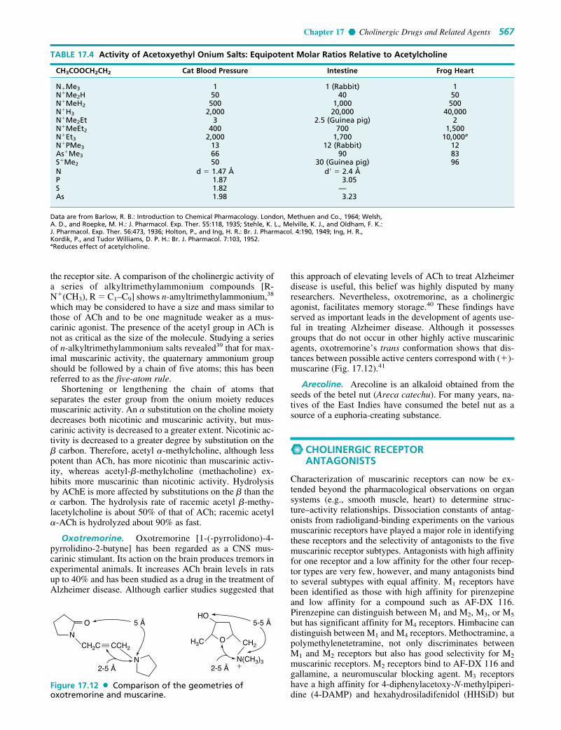
Chapter 17 Cholinergic Drugs and Related Agents 567
the receptor site. A comparison of the cholinergic activity ofa series of alkyltrimethylammonium compounds [R-N�(CH3), R � C1–C9] shows n-amyltrimethylammonium,38
which may be considered to have a size and mass similar tothose of ACh and to be one magnitude weaker as a mus-carinic agonist. The presence of the acetyl group in ACh isnot as critical as the size of the molecule. Studying a seriesof n-alkyltrimethylammonium salts revealed39 that for max-imal muscarinic activity, the quaternary ammonium groupshould be followed by a chain of five atoms; this has beenreferred to as the five-atom rule.
Shortening or lengthening the chain of atoms thatseparates the ester group from the onium moiety reducesmuscarinic activity. An � substitution on the choline moietydecreases both nicotinic and muscarinic activity, but mus-carinic activity is decreased to a greater extent. Nicotinic ac-tivity is decreased to a greater degree by substitution on the� carbon. Therefore, acetyl �-methylcholine, although lesspotent than ACh, has more nicotinic than muscarinic activ-ity, whereas acetyl-�-methylcholine (methacholine) ex-hibits more muscarinic than nicotinic activity. Hydrolysisby AChE is more affected by substitutions on the � than the� carbon. The hydrolysis rate of racemic acetyl �-methy-lacetylcholine is about 50% of that of ACh; racemic acetyl�-ACh is hydrolyzed about 90% as fast.
Oxotremorine. Oxotremorine [1-(-pyrrolidono)-4-pyrrolidino-2-butyne] has been regarded as a CNS mus-carinic stimulant. Its action on the brain produces tremors inexperimental animals. It increases ACh brain levels in ratsup to 40% and has been studied as a drug in the treatment ofAlzheimer disease. Although earlier studies suggested that
this approach of elevating levels of ACh to treat Alzheimerdisease is useful, this belief was highly disputed by manyresearchers. Nevertheless, oxotremorine, as a cholinergicagonist, facilitates memory storage.40 These findings haveserved as important leads in the development of agents use-ful in treating Alzheimer disease. Although it possessesgroups that do not occur in other highly active muscarinicagents, oxotremorine’s trans conformation shows that dis-tances between possible active centers correspond with (�)-muscarine (Fig. 17.12).41
Arecoline. Arecoline is an alkaloid obtained from theseeds of the betel nut (Areca catechu). For many years, na-tives of the East Indies have consumed the betel nut as asource of a euphoria-creating substance.
CHOLINERGIC RECEPTOR ANTAGONISTS
Characterization of muscarinic receptors can now be ex-tended beyond the pharmacological observations on organsystems (e.g., smooth muscle, heart) to determine struc-ture–activity relationships. Dissociation constants of antag-onists from radioligand-binding experiments on the variousmuscarinic receptors have played a major role in identifyingthese receptors and the selectivity of antagonists to the fivemuscarinic receptor subtypes. Antagonists with high affinityfor one receptor and a low affinity for the other four recep-tor types are very few, however, and many antagonists bindto several subtypes with equal affinity. M1 receptors havebeen identified as those with high affinity for pirenzepineand low affinity for a compound such as AF-DX 116.Pirenzepine can distinguish between M1 and M2, M3, or M5
but has significant affinity for M4 receptors. Himbacine candistinguish between M1 and M4 receptors. Methoctramine, apolymethylenetetramine, not only discriminates betweenM1 and M2 receptors but also has good selectivity for M2
muscarinic receptors. M2 receptors bind to AF-DX 116 andgallamine, a neuromuscular blocking agent. M3 receptorshave a high affinity for 4-diphenylacetoxy-N-methylpiperi-dine (4-DAMP) and hexahydrosiladifenidol (HHSiD) but
TABLE 17.4 Activity of Acetoxyethyl Onium Salts: Equipotent Molar Ratios Relative to Acetylcholine
CH3COOCH2CH2 Cat Blood Pressure Intestine Frog Heart
N�Me3 1 1 (Rabbit) 1N�Me2H 50 40 50N�MeH2 500 1,000 500N�H3 2,000 20,000 40,000N�Me2Et 3 2.5 (Guinea pig) 2N�MeEt2 400 700 1,500N�Et3 2,000 1,700 10,000a
N�PMe3 13 12 (Rabbit) 12As�Me3 66 90 83S�Me2 50 30 (Guinea pig) 96N d � 1.47 Å d� � 2.4 ÅP 1.87 3.05S 1.82 —As 1.98 3.23
Data are from Barlow, R. B.: Introduction to Chemical Pharmacology. London, Methuen and Co., 1964; Welsh,A. D., and Roepke, M. H.: J. Pharmacol. Exp. Ther. 55:118, 1935; Stehle, K. L., Melville, K. J., and Oldham, F. K.:J. Pharmacol. Exp. Ther. 56:473, 1936; Holton, P., and Ing, H. R.: Br. J. Pharmacol. 4:190, 1949; Ing, H. R.,Kordik, P., and Tudor Williams, D. P. H.: Br. J. Pharmacol. 7:103, 1952. aReduces effect of acetylcholine.
O 5 Å
2-5 Å 2-5 Å
5-5 Å
N
N
HO
OCH2C
H3C CH2CCH2
N(CH3)3�
Figure 17.12 Comparison of the geometries ofoxotremorine and muscarine.
0558-0606_17865_Ch17.qxd 12/9/09 1:53 AM Page 567

also exhibit affinity for M1 and M2 receptors.21 Tropicamidehas been reported to be a putative M4 receptor antagonist.Figure 17.13 includes structures of some receptor subtypeantagonists.
Products
Acetylcholine Chloride. ACh chloride exerts a power-ful stimulant effect on the parasympathetic nervous system.Attempts have been made to use it as a cholinergic agent,but its duration of action is too short for sustained effects,because of rapid hydrolysis by esterases and lack of speci-ficity when administered for systemic effects. It is a cardiacdepressant and an effective vasodilator. Stimulation of thevagus and the parasympathetic nervous system produces atonic action on smooth muscle and induces a flow from thesalivary and lacrimal glands. Its cardiac-depressant effectresults from (a) a negative chronotropic effect that causes adecrease in heart rate and (b) a negative inotropic action onheart muscle that produces a decrease in the force of myo-cardial contractions. The vasodilatory action of ACh isprimarily on the arteries and the arterioles, with distinct ef-fect on the peripheral vascular system. Bronchial constric-tion is a characteristic side effect when the drug is givensystemically.
One of the most effective antagonists to the action ofACh is atropine, a nonselective muscarinic antagonist.Atropine blocks the depressant effect of ACh on cardiacmuscle and its production of peripheral vasodilation (i.e.,muscarinic effects) but does not affect the skeletal musclecontraction (i.e., nicotinic effect) produced.
ACh chloride is a hygroscopic powder that is available inan admixture with mannitol to be dissolved in sterile waterfor injection shortly before use. It is a short-acting mioticwhen introduced into the anterior chamber of the eye and isespecially useful after cataract surgery during the placementof sutures. When applied topically to the eye, it has littletherapeutic value because of poor corneal penetration andrapid hydrolysis by AChE.
Methacholine Chloride, United States Pharmacopeia (USP). Methacholine chloride, acetyl-�-methylcholine
568 Wilson and Gisvold’s Textbook of Organic Medicinal and Pharmaceutical Chemistry
Figure 17.13 Chemical structures of partially selective muscarinic antagonists.
0558-0606_17865_Ch17.qxd 12/9/09 1:53 AM Page 568

Chapter 17 Cholinergic Drugs and Related Agents 569
chloride or (2-hydroxypropyl)trimethylammonium chlorideacetate, is the acetyl ester of �-methylcholine. Unlike ACh,methacholine has sufficient stability in the body to give sus-tained parasympathetic stimulation. This action is accompa-nied by little (1/1,000 that of ACh) or no nicotinic effect.
Methacholine can exist as (S) and (R) enantiomers.Although the chemical is used as the racemic mixture, itsmuscarinic activity resides principally in the (S)-isomer.The (S)/(R) ratio of muscarinic potency for these enan-tiomers is 240:1.
(�)-Acetyl-(S)-�-methylcholine is hydrolyzed by AChE,whereas the (R)(�)-isomer is not. (�)-Acetyl-(R)-�-methylcholine is a weak competitive inhibitor (Ki, 4 � 10�4
M) of AChE obtained from the electric organ of the eel (E.electricus). The hydrolysis rate of the (S)(�)-isomer isabout 54% that of ACh. This rate probably compensates forany decreased association (affinity) owing to the �-methylgroup with the muscarinic receptor site and may account forthe fact that ACh and (�)-acetyl-�-methylcholine haveequimolar muscarinic potencies in vivo. (�)-Acetyl-(R)-�-methylcholine weakly inhibits AChE and slightly reinforcesthe muscarinic activity of the (S)(�)-isomer in the racemicmixture of acetyl-�-methylcholine.
In the hydrolysis of the acetyl �- and �-methylcholines,the greatest stereochemical inhibitory effects occur whenthe choline is substituted in the �-position. This also appearsto be true of organophosphorous inhibitors. The (R)(�)- and(S)(�)-isomers of acetyl-�-methylcholine are hydrolyzed at78% and 97% of the rate of ACh, respectively.
Methacholine chloride occurs as colorless or white crys-tals or as a white crystalline powder. It is odorless or has aslight odor and is very deliquescent. It is freely soluble inwater, alcohol, or chloroform, and its aqueous solution isneutral to litmus and bitter. It is hydrolyzed rapidly in alka-line solutions. Solutions are relatively stable to heat and willkeep for at least 2 or 3 weeks when refrigerated to delaygrowth of molds.
Carbachol. Choline chloride carbamate is nonspecific inits action on muscarinic receptor subtypes. The pharmaco-logical activity of carbachol is similar to that of ACh. It isan ester of choline and thus possesses both muscarinic andnicotinic properties by cholinergic receptor stimulation. Itcan also act indirectly by promoting release of ACh and byits weak anticholinesterase activity. Carbachol forms a car-bamyl ester in the active site of AChE, which is hydrolyzedmore slowly than an acetyl ester. This slower hydrolysis ratereduces the amount of free enzyme and prolongs the dura-tion of ACh in the synapse. Carbachol also stimulates theautonomic ganglia and causes contraction of skeletal musclebut differs from a true muscarinic agent in that it does nothave cardiovascular activity despite the fact that it seems toaffect M2 receptors.42
Carbachol is a miotic and has been used to reduce the in-traocular tension of glaucoma when a response cannot beobtained with pilocarpine or neostigmine. Penetration of thecornea is poor but can be enhanced by the use of a wettingagent in the ophthalmic solution. In addition to its topicaluse for glaucoma, carbachol is used during ocular surgery,when a more prolonged miosis is required than can be ob-tained with ACh chloride.
Carbachol differs chemically from ACh in its stability tohydrolysis. The carbamyl group of carbachol decreases theelectrophilicity of the carbonyl and, thus, can form reso-nance structures more easily than ACh can. The result is thatcarbachol is less susceptible to hydrolysis and, therefore,more stable in aqueous solutions.
Bethanechol Chloride, USP. Bethanechol, �-methyl-choline chloride carbamate, (2-hydroxypropyl)trimethy-lammonium chloride carbamate, carbamylmethylcholinechloride (Urecholine), is nonspecific in its action on mus-carinic receptor subtypes but appears to be more effective ateliciting pharmacological action of M3 receptors.43 It haspharmacological properties similar to those of metha-choline. Both are esters of �-methylcholine and have feeblenicotinic activity. Bethanechol is inactivated more slowlyby AChE in vivo than is methacholine. It is a carbamyl esterand is expected to have stability in aqueous solutions simi-lar to that of carbachol.
The main use of bethanechol chloride is in the relief ofurinary retention and abdominal distention after surgery.The drug is used orally and by subcutaneous injection. Itmust never be administered by intramuscular or intravenousinjection because of the danger from cholinergic overstimu-lation and loss of selective action. Proper administration ofthe drug is associated with low toxicity and no serious sideeffects. Bethanechol chloride should be used with caution inasthmatic patients; when used for glaucoma, it producesfrontal headaches from the constriction of the sphinctermuscle in the eye and from ciliary muscle spasms. Its dura-tion of action is 1 hour.
Pilocarpine Hydrochloride, USP. Pilocarpine monohy-drochloride is the hydrochloride of an alkaloid obtainedfrom the dried leaflets of Pilocarpus jaborandi or P. micro-phyllus, in which it occurs to the extent of about 0.5% to-gether with other alkaloids.
0558-0606_17865_Ch17.qxd 12/9/09 1:53 AM Page 569

It occurs as colorless, translucent, odorless, faintly bittercrystals that are soluble in water (1:0.3), alcohol (1:3), andchloroform (1:360). (In this chapter, a solubility expressedas 1:360 indicates that 1 g is soluble in 360 mL of the sol-vent at 25°C. Solubilities at other temperatures are so indi-cated.) It is hygroscopic and affected by light; its solutionsare acid to litmus and may be sterilized by autoclaving.Alkalies saponify the lactone group to give the pharma-cologically inactive hydroxy acid (pilocarpic acid). Base-catalyzed epimerization at the ethyl group position occursto an appreciable extent and is another major pathway ofdegradation.44 Both routes result in loss of pharmacologicalactivity.
Pilocarpine is a nonselective agonist on the muscarinicreceptors. Despite this, it reportedly acts on M3 receptorsin smooth muscle to cause contractions in the gut, trachea,and eye.45,46 In the eye, it produces pupillary constriction(miosis) and a spasm of accommodation. These effectsare valuable in the treatment of glaucoma. The pupil con-striction and spasm of the ciliary muscle reduce intraocu-lar tension by establishing better drainage of ocular fluidthrough the canal of Schlemm, located near the corner ofthe iris and cornea. Pilocarpine is used as a 0.5% to 0.6%solution (i.e., of the salts) in treating glaucoma. Systemiceffects include copious sweating, salivation, and gastricsecretion.
Pilocarpine Nitrate, USP. Pilocarpine mononitrate oc-curs as shining white crystals that are not hygroscopic butare light sensitive. It is soluble in water (1:4) and alcohol(1:75) but insoluble in chloroform and ether. Aqueoussolutions are slightly acid to litmus and may be sterilizedin the autoclave. The alkaloid is incompatible with alka-lies, iodides, silver nitrate, and reagents that precipitatealkaloids.
Cevimeline Hydrochloride. Cevimeline (Evoxac) iscis-2�-methylspiro {1-azabicyclo [2.2.2] octane-3, 5� -[1,3]oxathiolane} hydrochloride, hydrate (2:1). Cevimeline has amolecular weight of 244.79 and is a white to off white crys-talline powder. It is freely soluble in alcohol, chloroform, andwater. Cevimeline is a cholinergic agonist which binds to theM3 muscarinic receptor subtype, which results in an increasesecretion of exocrine glands, such as salivary and sweatglands. Because of these effects, it was approved for use in thetreatment of dry mouth associated with Sjögren syndrome. Bystimulating the salivary muscarinic receptors cevimeline pro-motes secretion thereby alleviating dry-mouth in these pa-tients. Cevimeline is metabolized by the isozymes CYP2D6and CYP3A3 and CYP3A4. It has a half-life of 5 hours.47
Cholinesterase Inhibitors
There are two types of cholinesterases in humans, AChEand butyrylcholinesterase (BuChE). The cholinesterases dif-fer in their location in the body and their substrate speci-ficity. AChE is associated with the outside surface of glialcells in the synapse and catalyzes the hydrolysis of ACh tocholine and acetic acid. Inhibition of AChE prolongs the du-ration of the neurotransmitter in the junction and producespharmacological effects similar to those observed whenACh is administered. These inhibitors are indirect-actingcholinergic agonists. AChE inhibitors have been used in thetreatment of myasthenia gravis, atony in the GI tract, andglaucoma. They have also been used as agricultural insecti-cides and nerve gases. More recently, they have received at-tention as symptomatic drug treatments in patients sufferingfrom Alzheimer disease.48
BuChE (pseudocholinesterase) is located in humanplasma. Although its biological function is not clear, it hascatalytic properties similar to those of AChE. The substratespecificity is broader (Table 17.5), and it may hydrolyze di-etary esters and drug molecules in the blood.
Three different chemical groupings, acetyl, carbamyl,and phosphoryl, may react with the esteratic site of AChE.Although the chemical reactions are similar, the kinetic pa-rameters for each type of substrate differ and result in differ-ences between toxicity and usefulness.
The initial step in the hydrolysis of ACh by AChE is areversible enzyme–substrate complex formation. The asso-ciation rate (k�1) and dissociation rate (k�1) are relativelylarge. The enzyme–substrate complex, EA–ACh, may also
570 Wilson and Gisvold’s Textbook of Organic Medicinal and Pharmaceutical Chemistry
0558-0606_17865_Ch17.qxd 12/9/09 1:53 AM Page 570

Chapter 17 Cholinergic Drugs and Related Agents 571
form an acetyl-enzyme intermediate at a rate (k2) that isslower than either the association or dissociation rates.Choline is released from this complex with the formation ofthe acetyl-enzyme intermediate, EA. This intermediate isthen hydrolyzed to regenerate the free enzyme and aceticacid. The acetylation rate, k2, is the slowest step in this se-quence and is rate-limiting (see discussion that follow).
Kinetic studies with different substrates and inhibitors sug-gest that the active center of AChE consists of several majordomains: an anionic site, to which the trimethylammoniumgroup binds; an esteratic site, which causes hydrolysis of theester portion of ACh; and hydrophobic sites, which bind arylsubstrates, other uncharged ligands, and the alkyl portion ofthe acyl moiety of ACh. There is also a peripheral anionic site,removed by at least 20 Šfrom this active center, which al-losterically regulates activity at the esteratic site.49 The anionicsite was believed to have been formed by the �-carboxylategroup of a glutamic acid residue,50 but more recent studiessuggest that the aromatic moieties of tryptophan and pheny-lalanine residues bind the quaternary ammonium group ofACh in the anionic site through cation�� interactions.51 Thelocation and spatial organization in the esteratic site by serine,histidine, and glutamic acid residues constitute the esteraticsite. The triad of these amino acid residues contributes to thehigh catalytic efficiency of AChE (Fig. 17.14).52
AChE attacks the ester substrate through a serine hy-droxyl, forming a covalent acyl-enzyme complex. Theserine is activated as a nucleophile by the glutamic acid andhistidine residues that serve as the proton sink to attack thecarbonyl carbon of ACh. Choline is released, leaving theacetylated serine residue on the enzyme. The acetyl-enzyme
intermediate is cleaved by a general base catalysis mecha-nism to regenerate the free enzyme. The rate of the deacety-lation step is indicated by k3.
Carbamates such as carbachol are also able to serve as sub-strates for AChE, forming a carbamylated enzyme intermedi-ate (E–C). The rate of carbamylation (k2) is slower than therate of acetylation. Hydrolysis (k3, decarbamylation) of thecarbamyl-enzyme intermediate is 107 times slower than thatof its acetyl counterpart. The slower hydrolysis rate limits theoptimal functional capacity of AChE, allowing carbamatesubstrates to be semireversible inhibitors of AChE.
In the mechanism above, k3 is rate-limiting. The rate k2
depends not only on the nature of the
alcohol moiety of the ester but also on the type of carbamylester. Esters of carbamic acid are better
carbamylating agents of AChE than the methylcarbamyl
and dimethylcarbamyl analogs.53
TABLE 17.5 Hydrolysis of Various Substrates by AChE and BuChE
AChE BuChE
Enzyme Substrate Source Relative Ratea Source Relative Ratea
Acetylcholine Human or bovine RBC 100 Human or horse plasma 100Acetylthiocholine Bovine RBC 149 Horse plasma 407Acetyl-�-methylcholine Bovine RBC 18 Horse plasma 0Propionylcholine Human RBC 80 Horse plasma 170Butyrylcholine Human RBC 2.5 Horse plasma 250Butyrylthiocholine Bovine RBC 0 Horse plasma 590Benzoylcholine Bovine RBC 0 Horse plasma 67Ethyl acetate Human RBC 2 Human plasma 13,3-Dimethylbutyl acetate Human RBC 60 Human plasma 352-Chloroethyl acetate Human RBC 37 Human plasma 10Isoamyl acetate Human RBC 24 Horse plasma 7Isoamyl propionate Human RBC 10 Horse plasma 13Isoamyl butyrate Human RBC 1 Horse plasma 14
Adapted from Heath, D. F.: Organophosphorus Poisons—Anticholinesterases and Related Compounds. NewYork, Pergamon Press, 1961.aRelative rates at approximately optimal substrate concentration; rate with acetylcholine � 100.
0558-0606_17865_Ch17.qxd 12/9/09 1:53 AM Page 571
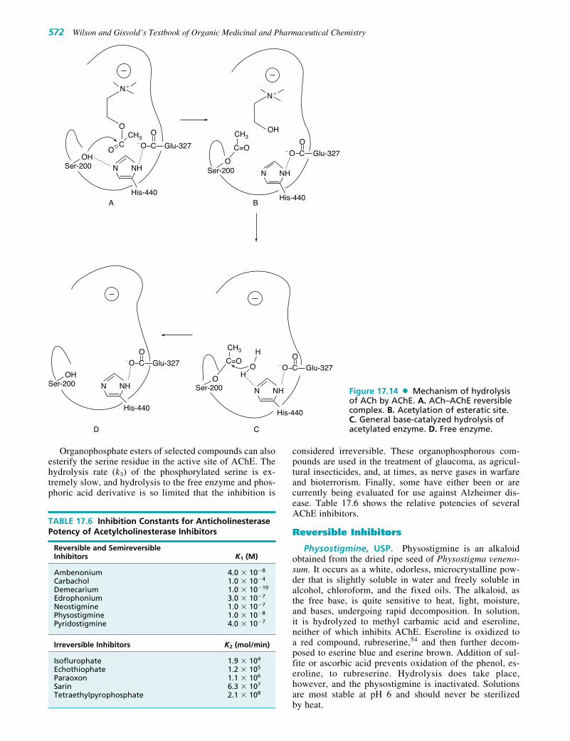
572 Wilson and Gisvold’s Textbook of Organic Medicinal and Pharmaceutical Chemistry
N�
C Glu-327
OO
�OOOH
CCH3 CH3
NH
His-440
Ser-200 N
A
D C
B
N�
OH
NH
His-440
N
C Glu-327
O�O
NH
His-440
N
C Glu-327
O�O
NH
His-440
N
C Glu-327
O�O
OHSer-200
O H
H
OC=O
CH3
Ser-200
OSer-200
C=O
Figure 17.14 Mechanism of hydrolysisof ACh by AChE. A. ACh–AChE reversiblecomplex. B. Acetylation of esteratic site.C. General base-catalyzed hydrolysis ofacetylated enzyme. D. Free enzyme.
TABLE 17.6 Inhibition Constants for AnticholinesterasePotency of Acetylcholinesterase Inhibitors
Reversible and SemireversibleInhibitors K1 (M)
Ambenonium 4.0 � 10�8
Carbachol 1.0 � 10�4
Demecarium 1.0 � 10�10
Edrophonium 3.0 � 10�7
Neostigmine 1.0 � 10�7
Physostigmine 1.0 � 10�8
Pyridostigmine 4.0 � 10�7
Irreversible Inhibitors K2 (mol/min)
Isoflurophate 1.9 � 104
Echothiophate 1.2 � 105
Paraoxon 1.1 � 106
Sarin 6.3 � 107
Tetraethylpyrophosphate 2.1 � 108
Organophosphate esters of selected compounds can alsoesterify the serine residue in the active site of AChE. Thehydrolysis rate (k3) of the phosphorylated serine is ex-tremely slow, and hydrolysis to the free enzyme and phos-phoric acid derivative is so limited that the inhibition is
considered irreversible. These organophosphorous com-pounds are used in the treatment of glaucoma, as agricul-tural insecticides, and, at times, as nerve gases in warfareand bioterrorism. Finally, some have either been or arecurrently being evaluated for use against Alzheimer dis-ease. Table 17.6 shows the relative potencies of severalAChE inhibitors.
Reversible Inhibitors
Physostigmine, USP. Physostigmine is an alkaloidobtained from the dried ripe seed of Physostigma veneno-sum. It occurs as a white, odorless, microcrystalline pow-der that is slightly soluble in water and freely soluble inalcohol, chloroform, and the fixed oils. The alkaloid, asthe free base, is quite sensitive to heat, light, moisture,and bases, undergoing rapid decomposition. In solution,it is hydrolyzed to methyl carbamic acid and eseroline,neither of which inhibits AChE. Eseroline is oxidized toa red compound, rubreserine,54 and then further decom-posed to eserine blue and eserine brown. Addition of sul-fite or ascorbic acid prevents oxidation of the phenol, es-eroline, to rubreserine. Hydrolysis does take place,however, and the physostigmine is inactivated. Solutionsare most stable at pH 6 and should never be sterilizedby heat.
0558-0606_17865_Ch17.qxd 12/9/09 1:53 AM Page 572

Chapter 17 Cholinergic Drugs and Related Agents 573
Physostigmine is a relatively poor carbamylating agentof AChE and is often considered a reversible inhibitor ofthe enzyme. Its cholinesterase-inhibiting properties varywith the pH of the medium (Fig. 17.15). The conjugateacid of physostigmine has a pKa of about 8, and as the pHof the solution is lowered, more is present in the proto-nated form. Inhibition of cholinesterase is greater in acidmedia, suggesting that the protonated form makes a contri-bution to the inhibitory activity well as its carbamylationof the enzyme.
Physostigmine was used first as a topical application inthe treatment of glaucoma. Its lipid solubility properties per-mit adequate absorption from ointment bases. It is used sys-temically as an antidote for atropine poisoning and otheranticholinergic drugs by increasing the duration of actionof ACh at cholinergic sites through inhibition of AChE.Physostigmine, along with other cholinomimetic drugs act-ing in the CNS, has been studied for use in the treatment ofAlzheimer disease.55 Cholinomimetics that are currentlyused or which have been recently evaluated in the treatmentof Alzheimer disease include donepezil, galantamine, metri-fonate, rivastigmine, and tacrine.48 It is anticipated that thislist will continue to grow as the etiology of this disease be-comes better understood.
Physostigmine Salicylate, USP. The salicylate ofphysostigmine (eserine salicylate) may be prepared byneutralizing an ethereal solution of the alkaloid with anethereal solution of salicylic acid. Excess salicylic acid isremoved from the precipitated product by washing itwith ether. The salicylate is less deliquescent than thesulfate.
Physostigmine salicylate occurs as a white, shining,odorless crystal or white powder that is soluble in water(1:75), alcohol (1:16), or chloroform (1:6) but much lesssoluble in ether (1:250). On prolonged exposure to air andlight, the crystals turn red. The red may be removed bywashing the crystals with alcohol, although this causes lossof the compound as well. Aqueous solutions are neutral orslightly acidic and take on a red coloration after a period.The coloration may be taken as an index of the loss of activ-ity of physostigmine solutions.
100
80
60
6 7 8 9 10
40
20
Neostigmine
Physostigmine
%��I
nhib
ition
of
Cho
lines
tera
se
pH
Figure 17.15 Effect of pH on inhibition ofcholinesterase by physostigmine and neostigmine.
0558-0606_17865_Ch17.qxd 12/9/09 1:53 AM Page 573

574 Wilson and Gisvold’s Textbook of Organic Medicinal and Pharmaceutical Chemistry
Solutions of physostigmine salicylate are incompatiblewith the usual reagents that precipitate alkaloids (alkalies)and with iron salts. Incompatibility also occurs with benzal-konium chloride and related wetting agents because of thesalicylate ion.
Physostigmine Sulfate, USP. Physostigmine sulfateoccurs as a white, odorless, microcrystalline powder that isdeliquescent in moist air. It is soluble in water (1:4), alcohol(1:0.4), and ether (1:1,200). It has the advantage over thesalicylate salt of being compatible in solution with benza-lkonium chloride and related compounds.
Neostigmine Bromide. Neostigmine bromide, (m-hy-droxyphenyl)trimethylammonium bromide dimethylcarba-mate or the dimethylcarbamic ester of 3-hydroxyphenyl-trimethylammonium bromide (Prostigmin bromide), is usedas an antidote to nondepolarizing neuromuscular blockingdrugs and in the treatment of myasthenia gravis. It occurs asa bitter, odorless, white, crystalline powder. It is soluble inwater and alcohol. The crystals are much less hygroscopicthan those of neostigmine methylsulfate and thus may beused in tablets. Solutions are stable and may be sterilized byboiling. Aqueous solutions are neutral to litmus.
Use of physostigmine, as a prototype of an indirect-actingparasympathomimetic drug, facilitated the development ofstigmine, in which a trimethylamine group was placed para toa dimethylcarbamate group in benzene. Better inhibition ofcholinesterase was observed when these groups were placedmeta to each other as in neostigmine, a more active and use-ful agent. Although physostigmine contains a methylcarba-mate functional group, greater chemical stability towardhydrolysis was obtained with the dimethylcarbamyl group inneostigmine.56
Neostigmine has a half-life of about 50 minutes after oralor intravenous administration. About 80% of a single intra-muscular dose is excreted in the urine within 24 hours; approx-imately 40% is excreted unchanged, and the remainder isexcreted as metabolites. Of the neostigmine that reachesthe liver, 98% is metabolized in 10 minutes to 3-hydroxy-phenyltrimethyl ammonium, which has activity similar to, butweaker than, neostigmine. Its transfer from plasma to livercells and then to bile is probably passive. Because cellularmembranes permit the passage of plasma proteins synthesizedin the liver into the bloodstream through capillary walls orlymphatic vessels, they may not present a barrier to the diffu-sion of quaternary amines such as neostigmine. The rapidhepatic metabolism of neostigmine may provide a downhillgradient for the continual diffusion of this compound.57 A cer-tain amount is hydrolyzed slowly by plasma cholinesterase.
Neostigmine has a mechanism of action quite similar tothat of physostigmine. It effectively inhibits cholinesteraseat about 10�6 M concentration. Its activity does not varywith pH, and at all ranges it exhibits similar cationic proper-ties (Fig. 17.15). Skeletal muscle is also stimulated byneostigmine, a property that physostigmine does not have.
The uses of neostigmine are similar to those of physostig-mine but differ in exhibiting greater miotic activity, fewer andless unpleasant local and systemic manifestations, and greaterchemical stability. The most frequent application of neostig-mine is to prevent atony of the intestinal, skeletal, and bladdermusculature. An important use is in the treatment of myasthe-nia gravis, a condition caused by an autoimmune mechanismthat requires an increase in ACh concentration in the neuro-muscular junction to sustain normal muscular activity.
Neostigmine Methylsulfate. Neostigmine methylsul-fate, (m-hydroxyphenyl)trimethylammonium methylsulfatedimethylcarbamate or the dimethylcarbamic ester of 3-hydro-xyphenyltrimethylammonium methylsulfate (Prostigminmethylsulfate), is a bitter, odorless, white, crystalline powder.It is very soluble in water and soluble in alcohol. Solutionsare stable and can be sterilized by boiling. The compound istoo hygroscopic for use in a solid form and thus is alwaysused as an injection. Aqueous solutions are neutral to litmus.
The methylsulfate salt is used postoperatively as a uri-nary stimulant and in the diagnosis and treatment of myas-thenia gravis.
Pyridostigmine Bromide, USP. Pyridostigmine bromide,3-hydroxy-1-methylpyridinium bromide dimethylcarbamateor pyridostigmine bromide (Mestinon), occurs as a white, hy-groscopic, crystalline powder with an agreeable, characteristicodor. It is freely soluble in water, alcohol, and chloroform.
0558-0606_17865_Ch17.qxd 12/9/09 1:53 AM Page 574

Chapter 17 Cholinergic Drugs and Related Agents 575
Pyridostigmine bromide is about one fifth as toxic asneostigmine. It appears to function in a manner similar tothat of neostigmine and is the most widely used anti-cholinesterase agent for treating myasthenia gravis. Theliver enzymes and plasma cholinesterase metabolize thedrug. The principal metabolite is 3-hydroxy-N-methylpyri-dinium. Orally administered pyridostigmine has a half-lifeof 90 minutes and a duration of action of between 3 and6 hours.
Ambenonium Chloride. Ambenonium chloride, [oxa-lylbis(iminoethylene)]bis[(o-chlorobenzyl)diethylammo-nium] dichloride (Mytelase chloride), is a white, odorlesspowder, soluble in water and alcohol, slightly soluble inchloroform, and practically insoluble in ether and acetone.Ambenonium chloride is used for the treatment of myasthe-nia gravis in patients who do not respond satisfactorily toneostigmine or pyridostigmine.
This drug acts by suppressing the activity of AChE. Itpossesses a relatively prolonged duration of action andcauses fewer side effects in the GI tract than the other anti-cholinesterase agents. The dosage requirements vary con-siderably, and the dosage must be individualized accordingto the response and tolerance of the patient. Because of itsquaternary ammonium structure, ambenonium chloride isabsorbed poorly from the GI tract. In moderate doses, thedrug does not cross the blood-brain barrier. Ambenoniumchloride is not hydrolyzed by cholinesterases.
Demecarium Bromide, USP. Demecarium bromide,(m-hydroxyphenyl)trimethylammonium bromide, decame-thylenebis[methylcarbamate] (Humorsol), is the diester of(m-hydroxyphenyl)trimethylammonium bromide with de-camethylene-bis-(methylcarbamic acid) and thus is compa-rable to a bis-prostigmine molecule.
It occurs as a slightly hygroscopic powder that is freelysoluble in water or alcohol. Ophthalmic solutions of the drughave a pH of 5 to 7.5. Aqueous solutions are stable and maybe sterilized by heat. Its efficacy and toxicity are comparableto those of other potent anticholinesterase inhibitor drugs. Itis a long-acting miotic used to treat wide-angle glaucomaand accommodative esotropia. Maximal effect occurs hoursafter administration, and the effect may persist for days.
0558-0606_17865_Ch17.qxd 12/9/09 1:53 AM Page 575

576 Wilson and Gisvold’s Textbook of Organic Medicinal and Pharmaceutical Chemistry
Donepezil. Donepezil, (�)-2,3-dihydro-5,6-dimethoxy-2-[[1-(phenylmethyl)-4-piperidinyl]methyl]-1H-inden-1-one (Aricept), commonly referred to in the literature asE2020, is a reversible inhibitor of AChE. It is indicated forthe treatment of symptoms of mild-to-moderate Alzheimerdisease. Donepezil is approximately 96% bound to plasmaproteins, with an elimination half-life of 70 hours. It is me-tabolized principally by the 2D6 and 3A4 isozymes of theP450 system.
Edrophonium Chloride, USP. Edrophonium chlo-ride, ethyl(m-hydroxyphenyl)dimethylammonium chloride(Tensilon), is a reversible anticholinesterase agent. It isbitter and very soluble in water and alcohol. Edrophoniumchloride injection has a pH of 5.2 to 5.5. On parenteral ad-ministration, edrophonium has a more rapid onset andshorter duration of action than neostigmine, pyridostig-mine, or ambenonium. It is a specific anticurare agent andacts within 1 minute to alleviate overdose of d-tubocu-rarine, dimethyl d-tubocurarine, or gallamine triethiodide.The drug is also used to terminate the action of any one ofthese drugs when the physician so desires. It is of no value,however, in terminating the action of the depolarizing (i.e.,noncompetitive) blocking agents, such as decamethoniumand succinylcholine. In addition to inhibiting AChE, edro-phonium chloride has a direct cholinomimetic effect onskeletal muscle, which is greater than that of most otheranticholinesterase drugs.
Edrophonium chloride is structurally related to neostig-mine methylsulfate and has been used as a potential diagnos-tic agent for myasthenia gravis. This is the only degenerativeneuromuscular disease that can be temporarily improved byadministration of an anticholinesterase agent. Edrophoniumchloride brings about a rapid increase in muscle strengthwithout significant side effects.
Galantamine. Galantamine, 4a,5,9,10,11,12-hexahydro-3-methoxy-11-methyl-6H-benzofuro-[3a,3,2,ef][2]-benzazepin-6-ol (Nivalin, Reminyl), is an alkaloid extractedfrom the tuberous plant Leucojum aestivum (L.) belongingto the Amaryllidaceae family and from the bulbs of thedaffodil, Narcissus pseudonarcissus. It is a reversiblecholinesterase inhibitor that appears to have no effect on bu-tyrylcholinesterase. In addition, it acts at allosteric nicotinicsites, further enhancing its cholinergic activity. Galantamineundergoes slow and minor biotransformation with approxi-mately 5% to 6% undergoing demethylation. It is primarilyexcreted in the urine.
Metrifonate. Metrifonate is an organophosphate thatwas originally developed to treat schistosomiasis under thetrade name Bilarcil. It is an irreversible cholinesterase inhibitor with some selectivity for BuChE over AChE. Itachieves sustained cholinesterase inhibition by its nonenzy-matic metabolite dichlorvos (DDVP), a long-actingorganophosphate. Its use in mild-to-moderate Alzheimerdisease was suspended recently because of adverse effectsexperienced by several patients during the clinical evalua-tion of this product. Toxicity at the neuromuscular junctionis probably attributable to the inhibition by the drug of neu-rotoxic esterase, a common feature of organophosphates.
Rivastigmine. Rivastigmine (Exelon, EA 713) is apseudoirreversible noncompetitive carbamate inhibitor ofAChE. Although the half-life is approximately 2 hours,the inhibitory properties of this agent last for 10 hours be-cause of the slow dissociation of the drug from the enzyme.The Food and Drug Administration (FDA) approved its usein mild-to-moderate Alzheimer disease in April 2000. InJuly 2007, rivastigmine was granted approval for use inmanaging mild-to-moderate dementia associated withParkinson disease.
Tacrine Hydrochloride. Tacrine hydrochloride,1,2,3,4-tetrahydro-9-aminoacridine hydrochloride (THA,Cognex), is a reversible cholinesterase inhibitor that hasbeen used in the treatment of Alzheimer disease for severalyears. The drug has been used to increase the levels of AChin these patients on the basis of observations from autopsiesthat concentrations of ChAT and AChE are markedly re-duced in the brain, whereas the number of muscarinic recep-tors is almost normal. The use of the drug is not withoutcontroversy, as conflicting results on efficacy have been re-ported.58,59 The drug has been used in mild-to-moderateAlzheimer dementia.
0558-0606_17865_Ch17.qxd 12/9/09 1:53 AM Page 576

Chapter 17 Cholinergic Drugs and Related Agents 577
analogs and are much weaker hydrogen bond-forming mol-ecules because of the sulfur atom.60 Their anticholinesteraseactivity is 105-fold weaker than their oxygen analogs. X isthe leaving group when the molecule reacts with the en-zyme. Typical leaving groups include fluoride, nitrile, andp-nitrophenoxy. The R groups may be alkyl, alkoxy, aryl,aryloxy, or amino. The R moiety imparts lipophilicity to themolecule and contributes to its absorption through the skin.Inhibition of AChE by organophosphorous compoundstakes place in two steps, association of enzyme and inhibitorand the phosphorylation step, completely analogous toacylation by the substrate (Fig. 17.16). Stereospecificity ismainly caused by interactions of enzyme and inhibitor at theesteratic site.
The serine residue at the esteratic site forms a stablephosphoryl ester with the organophosphorous inhibitors.This stability permits labeling studies61 to be carried out onthis and other enzymes (e.g., trypsin, chymotrypsin) thathave the serine hydroxyl as part of their active site.
Although insecticides and nerve gases are irreversibleinhibitors of cholinesterases by forming a phosphorylated ser-ine at the esteratic site of the enzyme, it is possible toreactivate the enzyme if action is taken soon after exposure to
Irreversible Inhibitors
Both AChE and BuChE are inhibited irreversibly by a groupof phosphate esters that are highly toxic (LD50 for humansis 0.1–0.001 mg/kg). These chemicals are nerve poisons andhave been used in warfare, in bioterrorism, and as agricul-tural insecticides. They permit ACh to accumulate at nerveendings and exacerbate ACh-like actions. The compoundsbelong to a class of organophosphorous esters. A generalformula for such compounds follows:
A is usually oxygen or sulfur but may also be selenium.When A is other than oxygen, biological activation is re-quired before the compound becomes effective as an in-hibitor of cholinesterases. Phosphorothionates [R1R2P(S)X]have much poorer electrophilic character than their oxygen
iC3H7O
iC3H7OP
F
Ser–200 Ser–200
His–440
NOH
NH
O–C—Glu–327
OO
O
�
Glu–327
Ser–200OH
Ser–200O
His–440
N NH
His–440
N NH
2–PAM
BA
D C
O–C—Glu–327�
Glu–327�O–C—
His–440
N
H
HC
NOO
NH
NCH3
�
B iC3H7O
iC3H7OP
iC3H7O
iC3H7OP
OBB
O
B
O
B�O–C—
O
B
BB
M
M
M
M
MM
M
M
M
Figure 17.16 Phosphorylation andreactivation of cholinesterase. A. Phospho-rylation of serine by isofluorphate. B. Phosphorylated serine at the esteraticsite. C. Nucleophilic attack on phosphory-lated residue by 2-PAM. D. Free enzyme.
0558-0606_17865_Ch17.qxd 12/9/09 1:53 AM Page 577

578 Wilson and Gisvold’s Textbook of Organic Medicinal and Pharmaceutical Chemistry
these poisons. Several compounds can provide a nucleophilicattack on the phosphorylated enzyme and cause regenerationof the free enzyme. Substances such as choline, hydroxyl-amine, and hydroxamic acid have led to the development ofmore effective cholinesterase reactivators, such as nicotinichydroxamic acid and pyridine-2-aldoxime methiodide (2-PAM). A proposed mode of action for the reactivation ofcholinesterase that has been inactivated by isoflurophate by 2-PAM is shown in Figure 17.16.
Cholinesterases that have been exposed to phosphorylat-ing agents (e.g., sarin) become refractory to reactivation bycholinesterase reactivators. The process is called aging andoccurs both in vivo and in vitro with AChE and BuChE.Aging occurs by partial hydrolysis of the phosphorylatedmoiety that is attached to the serine residue at the esteraticsite of the enzyme (Fig. 17.17).
Phosphate esters used as insecticidal agents are toxic andmust be handled with extreme caution. Symptoms of toxic-ity are nausea, vomiting, excessive sweating, salivation,miosis, bradycardia, low blood pressure, and respiratory dif-ficulty, which is the usual cause of death.
The organophosphate insecticides of low toxicity, suchas malathion, generally cause poisoning only by ingestion ofrelatively large doses. Parathion or methylparathion, how-ever, cause poisoning by inhalation or dermal absorption.Because these compounds are so long acting, cumulativeand serious toxic manifestations may result after severalsmall exposures.
Products
Isofluorphate, USP. Isofluorphate, diisopropylphos-phorofluoridate (Floropryl), is a colorless liquid soluble inwater to the extent of 1.54% at 25°C, which decomposes togive a pH of 2.5. It is soluble in alcohol and to some extentin peanut oil. It is stable in peanut oil for a period of 1 yearbut decomposes in water in a few days. Solutions in peanutoil can be sterilized by autoclaving. The compound shouldbe stored in hard glass containers. Continued contact withsoft glass is said to hasten decomposition, as evidenced bydiscoloration.
Isofluorphate must be handled with extreme caution.Contact with eyes, nose, mouth, and even skin should beavoided because it can be absorbed readily through intactepidermis and more so through mucous tissues.
Because isofluorphate irreversibly62 inhibits cholinesterase,its activity lasts for days or even weeks. During this period,new cholinesterase may be synthesized in plasma, erythro-cytes, and other cells.
A combination of atropine sulfate and magnesium sul-fate protects rabbits against the toxic effects of isofluor-phate. Atropine sulfate counteracts the muscarinic effect,and magnesium sulfate counteracts the nicotinic effect ofthe drug.63 Isofluorphate has been used in the treatment ofglaucoma.
Echothiophate Iodide, USP. Echothiophate iodide,(2-mercaptoethyl)trimethylammonium iodide, S-ester withO,O-diethylphosphorothioate (Phospholine Iodide), oc-curs as a white, crystalline, hygroscopic solid that has aslight mercaptan-like odor. It is soluble in water (1:1) anddehydrated alcohol (1:25); aqueous solutions have a pH ofabout 4 and are stable at room temperature for about1 month.
Echothiophate iodide is a long-lasting cholinesterase in-hibitor of the irreversible type, as is isofluorphate. Unlikethe latter, however, it is a quaternary salt, and when appliedlocally, its distribution in tissues is limited, which can bevery desirable. It is used as a long-acting anticholinesteraseagent in the treatment of glaucoma.
Hexaethyltetraphosphate (HETP) and Tetraethylpyrophosphate (TEPP). HETP and TEPP are compoundsthat also show anticholinesterase activity. HETP was devel-oped by the Germans during World War II and is used as aninsecticide against aphids. When used as insecticides, thesecompounds have the advantage of being hydrolyzed rapidlyto the relatively nontoxic, water-soluble compounds phos-phoric acid and ethyl alcohol. Fruit trees or vegetablessprayed with this type of compound retain no harmfulresidue after a period of a few days or weeks, depending onthe weather conditions. Workers spraying with these agentsshould use extreme caution so that the vapors are notbreathed and none of the vapor or liquid comes in contactwith the eyes or skin.
Figure 17.17 Aging of phosphorylated enzyme.
0558-0606_17865_Ch17.qxd 12/9/09 1:53 AM Page 578

Chapter 17 Cholinergic Drugs and Related Agents 579
Malathion. Malathion, 2-[(dimethoxyphosphinothioyl)thio]butanedioic acid diethyl ester, is a water-insolublephosphodithioate ester that has been used as an agriculturalinsecticide. Malathion is a poor inhibitor of cholinesterases.Its effectiveness as a safe insecticide is a result of the differ-ent rates at which humans and insects metabolize the chem-ical. Microsomal oxidation, which causes desulfuration,occurs slowly to form the phosphothioate (malaoxon),which is 10,000 times more active than the phosphodithioate(malathion) as a cholinesterase inhibitor. Insects detoxifythe phosphothioate by a phosphatase, forming dimethylphosphorothioate, which is inactive as an inhibitor.Humans, however, can rapidly hydrolyze malathion by a
carboxyesterase enzyme, yielding malathion acid, a stillpoorer inhibitor of AChE. Phosphatases and car-boxyesterases further metabolize malathion acid to di-methylphosphothioate. The metabolic reactions are shownin Figure 17.18.
Parathion. Parathion, O,O-diethyl O-p-nitrophenylphosphorothioate (Thiophos), is a yellow liquid that is freelysoluble in aromatic hydrocarbons, ethers, ketones, esters,and alcohols but practically insoluble in water, petroleumether, kerosene, and the usual spray oils. It is decomposedat a pH above 7.5. Parathion is used as an agricultural insec-ticide. It is a relatively weak inhibitor of cholinesterase;
0558-0606_17865_Ch17.qxd 12/9/09 1:53 AM Page 579

580 Wilson and Gisvold’s Textbook of Organic Medicinal and Pharmaceutical Chemistry
however, enzymes present in liver microsomes and insecttissues convert parathion (pI50 �4) to paraoxon, a more po-tent inhibitor of cholinesterase (pI50 8).64 Parathion is alsometabolized by liver microsomes to yield p-nitrophenol anddiethylphosphate; the latter is inactive as an irreversiblecholinesterase inhibitor.65
Schradan. Schradan, octamethyl pyrophosphoramide(OMPA), bis[bisdimethylaminophosphonous] anhydride(Pestox III), is a viscous liquid that is miscible with waterand soluble in most organic solvents. It is not hydrolyzed byalkalies or water but is hydrolyzed by acids. Schradan isused as a systemic insecticide for plants, being absorbed bythe plants without appreciable injury. Insects feeding on theplant are incapacitated.
Schradan is a weak inhibitor of cholinesterases in vitro.In vivo, it is metabolized to the very strong inhibitor hydrox-ymethyl OMPA. Hydroxymethyl OMPA is not stable andis metabolized further to the N-methoxide, which is a weakinhibitor of cholinesterase.66
Pralidoxime Chloride, USP. Pralidoxime chloride, 2-formyl-1-methylpyridinium chloride oxime, 2-PAM chlo-ride, or 2-pyridine aldoxime methyl chloride (Protopamchloride), is a white, nonhygroscopic, crystalline powderthat is soluble in water, 1 g in less than 1 mL.
Pralidoxime chloride is used as an antidote for poisoningby parathion and related pesticides. It may be effective
against some phosphates that have a quaternary nitrogen. Itis also an effective antagonist for some carbamates, such asneostigmine methylsulfate and pyridostigmine bromide.The mode of action of pralidoxime chloride is described inFigure 17.16.
The biological half-life of pralidoxime chloride in hu-mans is about 2 hours, and its effectiveness is a function of
Figure 17.18 Comparison of metabolism of malathion by mammals and insects.
0558-0606_17865_Ch17.qxd 12/9/09 1:53 AM Page 580

Chapter 17 Cholinergic Drugs and Related Agents 581
its concentration in plasma, which reaches a maximum 2 to3 hours after oral administration.
Pralidoxime chloride, a quaternary ammonium com-pound, is most effective by intramuscular, subcutaneous,or intravenous administration. Treatment of poisoningby an anticholinesterase will be most effective if givenwithin a few hours. Little will be accomplished if the drugis used more than 36 hours after parathion poisoning hasoccurred.
CHOLINERGIC BLOCKING AGENTS
A wide variety of tissues respond to ACh released by theneuron or exogenously administered chemicals to mimicthis neurotransmitter’s action. Peripheral cholinergicreceptors are located at parasympathetic postganglionicnerve endings in smooth muscle, sympathetic andparasympathetic ganglia, and neuromuscular junctions inskeletal muscle. Although ACh activates these receptors,there are antagonists that are selective for each. Atropine isan effective blocking agent at parasympathetic postgan-glionic terminals. Like most classic blocking agents, it actson all muscarinic receptor subtypes. d-Tubocurarineblocks the effect of ACh on skeletal muscle, which isactivated by N1 nicotinic receptors. Hexamethoniumblocks transition at N2 nicotinic receptors located in auto-nomic ganglia.
Anticholinergic action by drugs and chemicals appar-ently depends on their ability to reduce the number of freereceptors that can interact with ACh. The theories ofStephenson67 and Ariens68 have explained the relationshipbetween drug–receptor interactions and the observed bio-logical response (see Chapter 2). These theories indicate thatthe amount of drug–receptor complex formed at a giventime depends on the affinity of the drug for the receptor andthat a drug that acts as an agonist must also possess anotherproperty, called efficacy or intrinsic activity. Another expla-nation of drug–receptor interactions, the Paton rate theory,69
defines a biological stimulus as proportional to the rate ofdrug–receptor interactions (see Chapter 2). Both of thesetheories are compatible with the concept that a blockingagent that has high affinity for the receptor may decrease thenumber of available free receptors and the efficiency of theendogenous neurotransmitter.
Structure–Activity Relationships
A wide variety of compounds possess anticholinergic activ-ity. The development of such compounds has been largelyempiric and based principally on atropine as the prototype.Nevertheless, structural permutations have resulted in com-pounds that do not have obvious relationships to the parentmolecule. The following classification delineates the majorchemical types encountered:
• Solanaceous alkaloids and synthetic analogs• Synthetic aminoalcohol esters• Aminoalcohol ethers• Aminoalcohols• Aminoamides• Miscellaneous• Papaveraceous
The chemical classification of anticholinergics actingon parasympathetic postganglionic nerve endings is com-plicated somewhat because some agents, especially thequaternary ammonium derivatives, act on the ganglia thathave a muscarinic component to their stimulation patternand, at high doses, at the neuromuscular junction in skele-tal muscle.
There are several ways in which the structure–activityrelationship could be considered, but in this discussion wefollow, in general, the considerations of Long et al.,70 whobased their postulations on the 1-hyoscyamine moleculebeing one of the most active anticholinergics and, therefore,having an optimal arrangement of groups.
Anticholinergic compounds may be considered chemi-cals that have some similarity to ACh but contain additionalsubstituents that enhance their binding to the cholinergicreceptor.
As depicted above, an anticholinergic agent may containa quaternary ammonium function or a tertiary amine that isprotonated in the biophase to form a cationic species. Thenitrogen is separated from a pivotal carbon atom by a chainthat may include an ester, ether, or hydrocarbon moiety.The substituent groups A and B contain at least one aro-matic moiety capable of van der Waals interactions to thereceptor surface and one cycloaliphatic or other hydrocar-bon moiety for hydrophobic bonding interactions. C maybe hydroxyl or carboxamide to undergo hydrogen bondingwith the receptor.
THE CATIONIC HEAD
It is generally considered that the anticholinergic moleculeshave a primary point of attachment to cholinergic sitesthrough the cationic head (i.e., the positively charged nitro-gen). For quaternary ammonium compounds, there is noquestion of what is implied, but for tertiary amines, one as-sumes, with good reason, that the cationic head is achievedby protonation of the amine at physiological pH. The natureof the substituents on this cationic head is critical insofar asa parasympathomimetic response is concerned. Stericfactors that cause diffusion of the onium charge or producea less-than-optimal drug–receptor interaction result in adecrease of parasympathomimetic properties and allowthe drug to act as an antagonist because of other bonding in-teractions. Ariens68 has shown that carbocholines (e.g., ben-zilylcarbocholine) engage in a typical competitive actionwith ACh, although they are less effective than the corre-sponding compounds possessing a cationic head, suggesting
0558-0606_17865_Ch17.qxd 12/9/09 1:53 AM Page 581

that hydrophobic bonding may play an important role inthese drug–receptor interactions.
THE HYDROXYL GROUP
Although not requisite for activity, a suitably placed alco-holic hydroxyl group enhances antimuscarinic activity overthat of a similar compound without the hydroxyl group. Theposition of the hydroxyl group relative to the nitrogen ap-pears to be fairly critical, with the diameter of the receptivearea estimated to be about 2 to 3 Å. It is assumed that the hy-droxyl group contributes to the strength of binding, proba-bly by hydrogen bonding to an electron-rich portion of thereceptor surface.
THE ESTERATIC GROUP
Many of the highly potent antimuscarinic compounds pos-sess an ester grouping, and this may be a contributing featurefor effective binding. This is reasonable because the agonist(i.e., ACh) possesses a similar function for binding to thesame site. An esteratic function is not necessary for activity,because several types of compounds do not possess such agroup (e.g., ethers, aminoalcohols).
CYCLIC SUBSTITUTION
Examination of the active compounds discussed in the fol-lowing sections reveals that at least one cyclic substituent
582 Wilson and Gisvold’s Textbook of Organic Medicinal and Pharmaceutical Chemistry
0558-0606_17865_Ch17.qxd 12/9/09 1:53 AM Page 582

Chapter 17 Cholinergic Drugs and Related Agents 583
(phenyl, thienyl, or other) is a common feature in almostall anticholinergic molecules. Aromatic substitution isoften used in connection with the acidic moiety of the esterfunction. Virtually all acids used, however, are of the aryl-substituted acetic acid variety. Use of aromatic acids leadsto low activity of these compounds as anticholinergics butpotential activity as local anesthetics.
In connection with the apparent need for a cyclic group,Ariens68 points out that the “mimetic” molecules, richlyendowed with polar groups, undoubtedly require a comple-mentary polar receptor area for effective binding. As a conse-quence, it is implied that a relatively nonpolar area surroundssuch sites. Thus, increasing the binding of the molecule in thisperipheral area by introducing flat, nonpolar groups (e.g., aro-matic rings) should achieve compounds with excellent affin-ity but without intrinsic activity. This postulate is consistentwith most antimuscarinic drugs, whether they possess an estergroup or not.
PARASYMPATHETIC POSTGANGLIONICBLOCKING AGENTS
Parasympathetic postganglionic blocking agents are alsoknown as antimuscarinic, anticholinergic, parasympa-tholytic, or cholinolytic drugs. Antimuscarinic drugs act bycompetitive antagonism of ACh binding to muscarinic re-ceptors. Endogenous neurotransmitters, including ACh, arerelatively small molecules. Ariens68 noted that competitivereversible antagonists generally are larger molecules capa-ble of additional binding to the receptor surface. The mostpotent anticholinergic drugs are derived from muscarinicagonists that contain one or sometimes two large or bulkygroups. Ariens68 suggested that molecules that act as com-petitive reversible antagonists generally are capable of bind-ing to the active site of the receptor but have an additionalbinding interaction that increases receptor affinity but doesnot contribute to the intrinsic activity (efficacy) of the drug.Several three-dimensional models of G-protein–coupledreceptors, including the muscarinic receptor, have beenreported. Despite knowledge of their amino acid sequences,it is not yet possible to provide an unambiguous descrip-tion of the docking of molecules to these receptors. The con-cepts of Ariens68 and others, however, appear consistentwith the binding site models proposed. Bebbington andBrimblecombe41 proposed in 1965 that there is a relativelylarge area lying outside the agonist–receptor binding site,where van der Waals interactions can take place between theagonist and the receptor area. This, too, is not inconsistentwith contemporary theories on cholinergic receptorinteraction with small molecules.
Therapeutic Actions
Organs controlled by the autonomic nervous system usu-ally are innervated by both the sympathetic and theparasympathetic systems. There is a continual state of dy-namic balance between the two systems. Theoretically,one should achieve the same end result by either stimula-tion of one of the systems or blockade of the other.Unfortunately, there is usually a limitation to this type ofgeneralization. There are, however, three predictable and
clinically useful results from blocking the muscariniceffects of ACh.
1. Mydriatic effect: dilation of the pupil of the eye; andcycloplegia, a paralysis of the ciliary structure of theeye, resulting in a paralysis of accommodation for nearvision
2. Antispasmodic effect: lowered tone and motility of the GItract and the genitourinary tract
3. Antisecretory effect: reduced salivation (antisialagogue),reduced perspiration (anhidrotic), and reduced acid andgastric secretion
These three general effects of parasympatholytics can beexpected in some degree from any of the known drugs,although, occasionally, one must administer rather heroicdoses to demonstrate the effect. The mydriatic and cyclo-plegic effects, when produced by topical application, are notsubject to any greatly undesirable side effects because oflimited systemic absorption. This is not true for the systemicantispasmodic effects obtained by oral or parenteral admin-istration. Drugs with effective blocking action on theGI tract are seldom free of adverse effects on the otherorgans. The same is probably true of drugs used for theirantisecretory effects. Perhaps the most common side effectsexperienced from the oral use of these drugs, under ordinaryconditions, are dryness of the mouth, mydriasis, and urinaryretention. The latter effects have been exploited in thedevelopment of agents used to manage overactive bladderwith symptoms of the urge of urinary incontinence, urgency,and frequency.
Mydriatic and cycloplegic drugs are generally prescribedor used in the office by ophthalmologists. Their principaluse is for refraction studies in the process of fitting lenses.This permits the physician to examine the eye retina for pos-sible abnormalities and diseases and provides controlledconditions for the proper fitting of glasses. Because of theinability of the iris to contract under the influence of thesedrugs, there is a definite danger to the patient’s eyes duringthe period of drug activity unless they are protected fromstrong light by the use of dark glasses. These drugs also areused to treat inflammation of the cornea (keratitis), inflam-mation of the iris and the ciliary organs (iritis and iridocy-clitis), and inflammation of the choroid (choroiditis). Adark-colored iris appears to be more difficult to dilate than alight-colored one and may require more concentrated solu-tions. Caution in the use of mydriatics is advisable becauseof their demonstrated effect in raising the intraocular pres-sure. Pupil dilation tends to cause the iris to restrict drainageof fluid through the canal of Schlemm by crowding theangular space, thereby leading to increased intraocularpressure. This is particularly true for patients with glauco-matous conditions.
Atropine is used widely as an antispasmodic because of itsmarked depressant effect on parasympathetically innervatedsmooth muscle. It appears to block all muscarinic receptorsubtypes. Atropine is, however, the standard by which othersimilar drugs are measured. Also, atropine has a blocking ac-tion on the transmission of the nerve impulse, rather than adepressant effect directly on the musculature. This action istermed neurotropic, in contrast with the action of an antispas-modic such as papaverine, which appears to act by depressionof the muscle cells and is termed musculotropic.
0558-0606_17865_Ch17.qxd 12/9/09 1:53 AM Page 583

Papaverine is the standard for comparison of muscu-lotropic antispasmodics and, although not strictly aparasympatholytic, is treated together with its syntheticanalogs later in this chapter. The synthetic antispasmodicsappear to combine neurotropic and musculotropic effectsin greater or lesser measure, together with a certainamount of ganglion-blocking activity for the quaternaryderivatives.
Anticholinergic drugs have a minor role in the manage-ment of peptic ulcer disease.71 For the present, the mostrational therapy involving anticholinergic drugs seems tobe a combination of a nonirritating diet to reduce acid se-cretion, antacid therapy, and reduction of emotional stress.Most of the anticholinergic drugs are offered either as thechemical alone, or in combination with a CNS depressantsuch as phenobarbital, or with a neuroleptic drug to reducethe CNS contribution to parasympathetic hyperactivity.In addition to the antisecretory effects of anticholinergicson hydrochloric acid and gastric acid secretion, therehave been some efforts to use them as antisialagogues andanhidrotics.
Paralysis agitans or parkinsonism (Parkinson disease),first described by the English physician James Parkinson in1817, is another condition that is often treated with anti-cholinergic drugs. It is characterized by tremor, “pillrolling,” cogwheel rigidity, festinating gait, sialorrhea, andmasklike facies. Fundamentally, it represents a malfunctionof the extrapyramidal system. Parkinsonism is characterizedby progressive and selective degeneration of dopaminergicneurons, which originate in the substantia nigra of the mid-brain and terminate in the basal ganglia (i.e., caudate nu-cleus, putamen, and pallidum). Skeletal muscle movementis controlled to a great degree by patterns of excitation andinhibition resulting from the feedback of information to thecortex and mediated through the pyramidal and extrapyra-midal pathways. The basal ganglia structures, such as thepallidum, corpus striatum, and substantia nigra, serve asdata processors for the pyramidal pathways and the struc-tures through which the extrapyramidal pathways pass ontheir way from the spinal cord to the cortex. Lesions of thepyramidal pathways lead to a persistent increase in muscletone, resulting in an excess of spontaneous involuntarymovements, along with changes in the reflexes. Thus, thebasal ganglia are functional in maintaining normal motorcontrol. In parkinsonism, there is degeneration of the sub-stantia nigra and corpus striatum, which are involved withcontrolled integration of muscle movement. The neurons inthe substantia nigra and basal ganglia use the neurotransmit-ter dopamine and interact with short cholinergic interneu-rons. When dopamine neurons degenerate, the balancebetween them is altered. The inhibitory influence ofdopamine is reduced, and the activity of cholinergic neuronsis increased. The principal goal of anticholinergic drugs inthe treatment of parkinsonism is to decrease the activity ofcholinergic neurons in the basal ganglia.
The usefulness of the belladonna group of alkaloids forthe treatment of parkinsonism was an empiric discovery.Since then, chemists have prepared many synthetic analogsof atropine in an effort to retain the useful antitremor andantirigidity effects of the belladonna alkaloid while reducingthe adverse effects. In this process, it was discoveredthat antihistamine drugs (e.g., diphenhydramine) reducedtremor and rigidity. The antiparkinsonian-like activity of
antihistamines has been attributed to their anticholinergicproperties. The activity of these drugs is confined to thosethat can pass through the blood-brain barrier.
SOLANACEOUS ALKALOIDS ANDANALOGS
The solanaceous alkaloids, represented by (�)-hyoscyamine,atropine [(�)-hyoscyamine], and scopolamine (hyoscine), arethe forerunners of the class of antimuscarinic drugs. Thesealkaloids are found principally in henbane (Hyoscyamusniger), deadly nightshade (Atropa belladonna), and jimsonweed (Datura stramonium). There are other alkaloids that aremembers of the solanaceous group (e.g., apoatropine, norat-ropine, belladonnine, tigloidine, meteloidine) but lack suffi-cient therapeutic value to be considered in this text.
Crude drugs containing these alkaloids have been usedsince early times for their marked medicinal properties,which largely depend on inhibition of the parasympatheticnervous system and stimulation of the higher nervous cen-ters. Belladonna, probably as a consequence of the weaklocal anesthetic activity of atropine, has been used topi-cally for its analgesic effect on hemorrhoids, certain skininfections, and various itching dermatoses. Application ofsufficient amounts of belladonna or its alkaloids results inmydriasis. Internally, the drug causes diminution of secre-tions, increases the heart rate (by depression of the vagusnerve), depresses the motility of the GI tract, and acts as anantispasmodic on various smooth muscles (ureter, bladder,and biliary tract). In addition, it directly stimulates the res-piratory center. The multiplicity of actions exerted by thedrug is looked on with some disfavor, because the physi-cian seeking one type of response unavoidably also obtainsthe others. The action of scopolamine-containing drugsdiffers from those containing hyoscyamine and atropine inhaving no CNS stimulation; rather, a narcotic or sedativeeffect predominates. The use of this group of drugs is ac-companied by a fairly high incidence of reactions becauseof individual idiosyncrasies; death from overdosage usu-ally results from respiratory failure. A complete treatmentof the pharmacology and uses of these drugs is not withinthe scope of this text. The introductory pages of this chap-ter briefly review some of the more pertinent points in con-nection with the major activities of these drug types.
Structural Considerations
All of the solanaceous alkaloids are esters of the bicyclicaminoalcohol 3-hydroxytropane or of related amino-alcohols. The structural formulas that follow show thepiperidine ring system in the commonly accepted chairconformation because this form has the lowest energy re-quirement. The alternate boat form can exist under certainconditions, however, because the energy barrier is notgreat. Inspection of the 3-hydroxytropane formula also in-dicates that even though there is no optical activity be-cause of the plane of symmetry, two stereoisomeric forms(tropine and pseudotropine) can exist because of the rigid-ity imparted to the molecule through the ethylene chainacross the 1,5 positions.
In tropine, the axially oriented hydroxyl group, transto the nitrogen bridge, is designated �, and the alternatecis equatorially oriented hydroxyl group is designated �.
584 Wilson and Gisvold’s Textbook of Organic Medicinal and Pharmaceutical Chemistry
0558-0606_17865_Ch17.qxd 12/9/09 1:53 AM Page 584
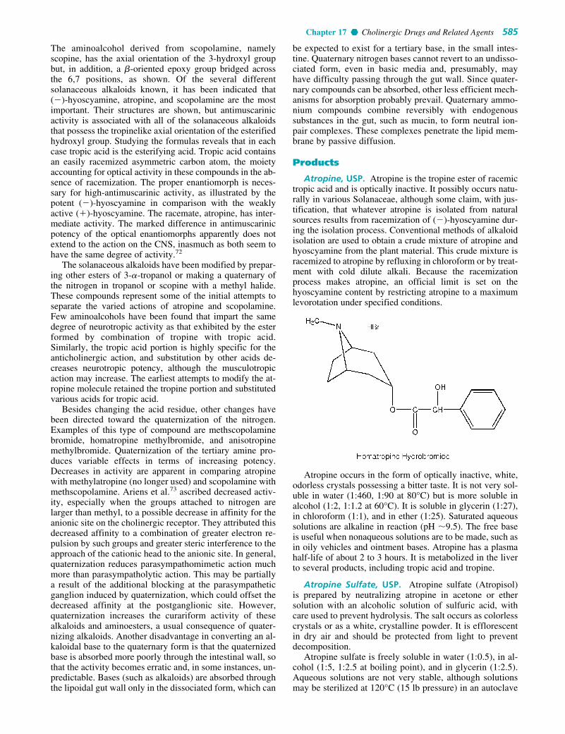
Chapter 17 Cholinergic Drugs and Related Agents 585
The aminoalcohol derived from scopolamine, namelyscopine, has the axial orientation of the 3-hydroxyl groupbut, in addition, a �-oriented epoxy group bridged acrossthe 6,7 positions, as shown. Of the several differentsolanaceous alkaloids known, it has been indicated that (�)-hyoscyamine, atropine, and scopolamine are the mostimportant. Their structures are shown, but antimuscarinicactivity is associated with all of the solanaceous alkaloidsthat possess the tropinelike axial orientation of the esterifiedhydroxyl group. Studying the formulas reveals that in eachcase tropic acid is the esterifying acid. Tropic acid containsan easily racemized asymmetric carbon atom, the moietyaccounting for optical activity in these compounds in the ab-sence of racemization. The proper enantiomorph is neces-sary for high-antimuscarinic activity, as illustrated by thepotent (�)-hyoscyamine in comparison with the weaklyactive (�)-hyoscyamine. The racemate, atropine, has inter-mediate activity. The marked difference in antimuscarinicpotency of the optical enantiomorphs apparently does notextend to the action on the CNS, inasmuch as both seem tohave the same degree of activity.72
The solanaceous alkaloids have been modified by prepar-ing other esters of 3-�-tropanol or making a quaternary ofthe nitrogen in tropanol or scopine with a methyl halide.These compounds represent some of the initial attempts toseparate the varied actions of atropine and scopolamine.Few aminoalcohols have been found that impart the samedegree of neurotropic activity as that exhibited by the esterformed by combination of tropine with tropic acid.Similarly, the tropic acid portion is highly specific for theanticholinergic action, and substitution by other acids de-creases neurotropic potency, although the musculotropicaction may increase. The earliest attempts to modify the at-ropine molecule retained the tropine portion and substitutedvarious acids for tropic acid.
Besides changing the acid residue, other changes havebeen directed toward the quaternization of the nitrogen.Examples of this type of compound are methscopolaminebromide, homatropine methylbromide, and anisotropinemethylbromide. Quaternization of the tertiary amine pro-duces variable effects in terms of increasing potency.Decreases in activity are apparent in comparing atropinewith methylatropine (no longer used) and scopolamine withmethscopolamine. Ariens et al.73 ascribed decreased activ-ity, especially when the groups attached to nitrogen arelarger than methyl, to a possible decrease in affinity for theanionic site on the cholinergic receptor. They attributed thisdecreased affinity to a combination of greater electron re-pulsion by such groups and greater steric interference to theapproach of the cationic head to the anionic site. In general,quaternization reduces parasympathomimetic action muchmore than parasympatholytic action. This may be partiallya result of the additional blocking at the parasympatheticganglion induced by quaternization, which could offset thedecreased affinity at the postganglionic site. However,quaternization increases the curariform activity of thesealkaloids and aminoesters, a usual consequence of quater-nizing alkaloids. Another disadvantage in converting an al-kaloidal base to the quaternary form is that the quaternizedbase is absorbed more poorly through the intestinal wall, sothat the activity becomes erratic and, in some instances, un-predictable. Bases (such as alkaloids) are absorbed throughthe lipoidal gut wall only in the dissociated form, which can
be expected to exist for a tertiary base, in the small intes-tine. Quaternary nitrogen bases cannot revert to an undisso-ciated form, even in basic media and, presumably, mayhave difficulty passing through the gut wall. Since quater-nary compounds can be absorbed, other less efficient mech-anisms for absorption probably prevail. Quaternary ammo-nium compounds combine reversibly with endogenoussubstances in the gut, such as mucin, to form neutral ion-pair complexes. These complexes penetrate the lipid mem-brane by passive diffusion.
Products
Atropine, USP. Atropine is the tropine ester of racemictropic acid and is optically inactive. It possibly occurs natu-rally in various Solanaceae, although some claim, with jus-tification, that whatever atropine is isolated from naturalsources results from racemization of (�)-hyoscyamine dur-ing the isolation process. Conventional methods of alkaloidisolation are used to obtain a crude mixture of atropine andhyoscyamine from the plant material. This crude mixture isracemized to atropine by refluxing in chloroform or by treat-ment with cold dilute alkali. Because the racemizationprocess makes atropine, an official limit is set on thehyoscyamine content by restricting atropine to a maximumlevorotation under specified conditions.
Atropine occurs in the form of optically inactive, white,odorless crystals possessing a bitter taste. It is not very sol-uble in water (1:460, 1:90 at 80°C) but is more soluble inalcohol (1:2, 1:1.2 at 60°C). It is soluble in glycerin (1:27),in chloroform (1:1), and in ether (1:25). Saturated aqueoussolutions are alkaline in reaction (pH �9.5). The free baseis useful when nonaqueous solutions are to be made, such asin oily vehicles and ointment bases. Atropine has a plasmahalf-life of about 2 to 3 hours. It is metabolized in the liverto several products, including tropic acid and tropine.
Atropine Sulfate, USP. Atropine sulfate (Atropisol)is prepared by neutralizing atropine in acetone or ethersolution with an alcoholic solution of sulfuric acid, withcare used to prevent hydrolysis. The salt occurs as colorlesscrystals or as a white, crystalline powder. It is efflorescentin dry air and should be protected from light to preventdecomposition.
Atropine sulfate is freely soluble in water (1:0.5), in al-cohol (1:5, 1:2.5 at boiling point), and in glycerin (1:2.5).Aqueous solutions are not very stable, although solutionsmay be sterilized at 120°C (15 lb pressure) in an autoclave
0558-0606_17865_Ch17.qxd 12/9/09 1:53 AM Page 585

if the pH is kept below 6. Sterilization probably is besteffected by the use of aseptic techniques and a bacteriolog-ical filter. It has been suggested that no more than a 30-daysupply of an aqueous solution should be made and that forsmall quantities the best procedure is to use hypodermictablets and sterile distilled water.73 Kondritzer andZvirblis74 have studied the kinetics of alkaline and proton-catalyzed hydrolyses of atropine in aqueous solution. Theregion of maximal stability lies between pH 3 and approxi-mately 5. They have also proposed an equation to predictthe half-life of atropine undergoing hydrolysis at constantpH and temperature.
The action of atropine or its salts is the same. It producesa mydriatic effect by paralyzing the iris and the ciliary mus-cles and, for this reason, is used by the oculist in iritis andcorneal inflammations and lesions. Its use is rational in theseconditions because one of the first rules in the treatmentof inflammation is rest, which, of course, is accomplishedby the paralysis of muscular motion. Its use in the eye(0.5%–1% solutions or gelatin disks) for fitting glasses iswidespread. Atropine is administered in small doses beforegeneral anesthesia to lessen oral and air passage secretionsand, when administered with morphine, to lessen the respi-ratory depression induced by morphine.
Atropine causes restlessness, prolonged pupillary dila-tion, and loss of visual accommodation and, furthermore,gives rise to arrhythmias such as atrioventricular dissocia-tion, ventricular extrasystoles, and even ventricular fibrilla-tion. Even though ether has been gradually replaced by otheranesthetics, thereby eliminating problems with respiratorysecretions caused by ether and thus requiring atropine, sur-geons and anesthesiologists today continue to use it as ananesthetic premedicant to reduce excessive salivary and air-way secretions and to prevent vagal reflexes.
Its ability to dry secretions has also been used in the so-called rhinitis tablets for symptomatic relief in colds. Incathartic preparations, atropine or belladonna has been usedas an antispasmodic to lessen the smooth muscle spasm(griping) often associated with catharsis.
Atropine may be used to treat some types of arrhythmias. Itincreases the heart rate by blocking the effects of ACh on thevagus. In this context, it is used to treat certain reversible brad-yarrhythmias that may accompany acute myocardial infarc-tion. It is also used as an adjunct to anesthesia to protect againstbradycardia, hypotension, and even cardiac arrest induced bythe skeletal muscle relaxant succinylcholine chloride.
Another use for atropine sulfate emerged following thedevelopment of the organophosphates, which are potent in-hibitors of AChE. Atropine is a specific antidote to preventthe muscarinic effects of ACh accumulation, such as vomit-ing, abdominal cramps, diarrhea, salivation, sweating, bron-choconstriction, and excessive bronchial secretions. It isused intravenously but does not protect against respiratoryfailure caused by depression of the respiratory center andthe muscles of respiration.
Hyoscyamine, USP. Hyoscyamine is a levorotatory al-kaloid obtained from various solanaceous species. One ofthe commercial sources is Egyptian henbane (Hyoscyamusmuticus), in which it occurs to the extent of about 0.5%.Usually, it is prepared from the crude drug in a manner sim-ilar to that used for atropine and is purified as the oxalate.The free base is obtained easily from this salt.
It occurs as white needles that are sparingly soluble inwater (1:281), more soluble in ether (1:69) or benzene(1:150), very soluble in chloroform (1:1), and freely solublein alcohol. It is used as the sulfate and hydrobromide. Theprincipal reason for the popularity of the hydrobromide hasbeen its nondeliquescent nature. The salts have the advan-tage over the free base in being quite water soluble.
Hyoscyamine is the levo form of the racemic mixtureknown as atropine. The dextro form does not exist naturallybut has been synthesized. Cushny75 compared the activitiesof (�)-hyoscyamine, (�)-hyoscyamine, and the racemate(atropine) in 1904 and found greater peripheral potency forthe (�) isomer and twice the potency of the racemate. Alllater studies have essentially confirmed that the (�) isomeris only weakly active and that the (�) isomer is, in effect,the active portion of atropine. Inspection of the relativedoses of atropine sulfate and hyoscyamine sulfate illus-trates the differences very nicely. The principal criticismoffered against the use of hyoscyamine sulfate exclusivelyis that it tends to racemize to atropine sulfate rather easilyin solution, so that atropine sulfate then becomes the morestable of the two. All of the isomers behave very much thesame in the CNS.
Hyoscyamine is used to treat disorders of the urinarytract more so than any other antispasmodic, although thereis no evidence that it has any advantages over the otherbelladonna preparations and the synthetic anticholinergics.It is used to treat spasms of the bladder and, in this man-ner, serves as a urinary stimulant. It is used together witha narcotic to counteract the spasm produced by the narcoticwhen the latter is used to relieve the pain of urethral colic.Hyoscyamine preparations are also used as antispasmodicsin the therapy of peptic ulcers.
Hyoscyamine Sulfate, USP. Hyoscyamine sulfate(Levsin sulfate) is a white, odorless, crystalline compoundof a deliquescent nature that also is affected by light. It issoluble in water (1:0.5) and alcohol (1:5) but almost in-soluble in ether. Solutions of hyoscyamine sulfate are acidicto litmus.
This drug is used as an anticholinergic in the same mannerand for the same indications as atropine and hyoscyamine, butit possesses the disadvantage of being deliquescent.
Scopolamine. Scopolamine (hyoscine) is found in vari-ous members of the Solanaceae (e.g., H. niger, Duboisia my-oporoides, Scopolia spp., and Datura metel). Scopolamineusually is isolated from the mother liquor remaining from theisolation of hyoscyamine.
Hyoscine is the older name for this alkaloid, althoughscopolamine is the accepted name in the United States.Scopolamine is the levo component of the racemic mixturethat is known as atroscine. The alkaloid is racemized read-ily in the presence of dilute alkali.
The alkaloid occurs in the form of a levorotatory, viscousliquid that is only slightly soluble in water but very solublein alcohol, chloroform, or ether. It forms crystalline saltswith most acids, with the hydrobromide being the most sta-ble and the most popularly accepted. An aqueous solution ofthe hydrobromide containing 10% mannitol is said to be lessprone to decomposition than unprotected solutions. Thecommercially available transdermal system of scopolaminecomprises an outer layer of polymer film and a drug reser-voir containing scopolamine, polyisobutylene, and mineral
586 Wilson and Gisvold’s Textbook of Organic Medicinal and Pharmaceutical Chemistry
0558-0606_17865_Ch17.qxd 12/9/09 1:53 AM Page 586

Chapter 17 Cholinergic Drugs and Related Agents 587
oil, which is interfaced with a microporous membrane tocontrol diffusion of the drug. In this dosage form, scopol-amine is effective in preventing motion sickness. The actionis believed to be on the cortex or the vestibular appara-tus. Whereas atropine stimulates the CNS, causing restless-ness and talkativeness, scopolamine usually acts as a CNSdepressant.
Scopolamine Hydrobromide, USP. Scopolamine hy-drobromide (hyoscine hydrobromide) occurs as white orcolorless crystals or as a white, granular powder. It is odor-less and tends to effloresce in dry air. It is freely soluble inwater (1:1.5), soluble in alcohol (1:20), only slightly solublein chloroform, and insoluble in ether.
Scopolamine is a competitive blocking agent of theparasympathetic nervous system as is atropine, but it differsmarkedly from atropine in its action on the higher nervecenters. Both drugs readily cross the blood-brain barrierand, even at therapeutic doses, cause confusion, particularlyin the elderly.
A sufficiently large dose of scopolamine will cause an in-dividual to sink into a restful, dreamless sleep for about8 hours, followed by a period of approximately the samelength in which the patient is in a semiconscious state.During this time, the patient does not remember events thattake place. When scopolamine is administered with mor-phine, this temporary amnesia is termed twilight sleep.
Homatropine Hydrobromide, USP. Homatropine hy-drobromide, 1�H,5�H-tropan-3�-ol mandelate (ester) hy-drobromide (Homatrocel), occurs as white crystals or as awhite, crystalline powder that is affected by light. It is solu-ble in water (1:6) and alcohol (1:40), less soluble in chloro-form (1:420), and insoluble in ether.
Solutions are incompatible with alkaline substances,which precipitate the free base, and with the commonreagents that precipitate alkaloids. As with atropine, solu-tions are sterilized best by filtration through a bacteriologi-cal filter.
Homatropine hydrobromide is used topically to paralyzethe ciliary structure of the eye (cycloplegia) and to effectmydriasis. It behaves very much like atropine but is weakerand less toxic. In the eye, it acts more rapidly but less per-sistently than atropine. Dilation of the pupil takes place inabout 15 to 20 minutes, and the action subsides in about24 hours. By using a miotic, such as physostigmine, it ispossible to restore the pupil to normality in a few hours.
Homatropine Methylbromide, USP. Homatropinemethylbromide, 3�-hydroxy-8-methyl-1�H,5�H-tropaniumbromide mandelate (Novatropine, Mesopin), occurs as a bit-ter, white, odorless powder and is affected by light. The com-pound is readily soluble in water and alcohol but insoluble inether. The pH of a 1% solution is 5.9 and that of a 10% solu-tion is 4.5. Although a solution of the compound yields a pre-cipitate with alkaloidal reagents, such as mercuric potassiumiodide test solution, addition of alkali hydroxides or carbon-ates does not cause the precipitate that occurs with nonquater-nary nitrogen salts (e.g., atropine, homatropine).
Homatropine methylbromide is transported poorly acrossthe blood-brain barrier because of its quaternary ammoniumgroup and, therefore, has far fewer stimulant properties thanatropine. It does have all the characteristic peripheralparasympathetic depressant properties of atropine and isused to reduce oversecretion and to relieve GI spasms.
Ipratropium Bromide. Ipratropium bromide, 3-(3-hy-droxy-1-oxo-2-phenylpropoxy)-8-methyl-8-(1-methylethyl)-8-azoniabicyclo[3.2.1]octane bromide (Atrovent), is a quater-nary ammonium derivative of atropine. It is freely soluble inwater and ethanol but insoluble in chloroform and ether. Thesalt is stable in neutral and acidic solutions but rapidly hy-drolyzed in alkaline solutions.
Ipratropium bromide is used in inhalation therapy toproduce dilation of bronchial smooth muscle for acute asth-matic attacks. The drug produces bronchodilation by com-petitive inhibition of cholinergic receptors bound to smooth
0558-0606_17865_Ch17.qxd 12/9/09 1:53 AM Page 587

muscle of the bronchioles. Ipratropium may also act on thesurface of mast cells to inhibit ACh-enhanced release ofchemical mediators. The drug has a slow onset of action,within 5 to 15 minutes after being administered by inhala-tion, and should not be used alone for acute asthmatic at-tacks. The peak therapeutic effect from one dose is observedbetween 1 and 2 hours. The effects of the drug last for about6 hours. It has a half-life of 3.5 hours.
Tiotropium Bromide. Tiotropium bromide, (1�,2�,4�,7�)-7-[(hydroxidi-2-thienylacetyl)oxy]-9,9-dimethyl-3-oxa-9-azoniatricyclo[3.3.1.02,4]nonane, (Spiriva) is anantimuscarinic agent that is used in an inhalation device to de-liver the drug into the lungs. It is indicated in the treatment ofchronic obstructive pulmonary disease (COPD), includingchronic bronchitis and emphysema. The standard once-dailydose is 18 �g of tiotropium.
SYNTHETIC CHOLINERGIC BLOCKINGAGENTS
Aminoalcohol Esters
The solanaceous alkaloids are generally agreed to be potentparasympatholytics, but they have the undesirable propertyof producing a wide range of effects through their nonspe-cific blockade of autonomic functions. Efforts to use theantispasmodic effect of the alkaloids most often result inside effects such as dryness of the mouth and fluctuations inpulse rate. Therefore, synthesis of compounds possessingspecific cholinolytic actions has been a very desirable fieldof study. Few prototypical drugs were as avidly dissectedin the minds of researchers as atropine in attempts to mod-ify its structure to separate the numerous useful activities(i.e., antispasmodic, antisecretory, mydriatic, and cyclo-plegic). Most early research was carried out in the pre– andpost–World War II era before muscarinic receptor subtypeswere known.
Efforts at synthesis started with rather minor deviationsfrom the atropine molecule, but a review of the commonlyused drugs today indicates a marked departure from therigid tropane aminoalcohols and tropic acid residues.Examination of the structures of antispasmodics shows thatthe acid portion has been designed to provide a large hy-drophobic moiety rather than the stereospecific requirementof (S)-tropic acid in (�)-hyoscyamine that was once consid-ered important. One of the major developments in the fieldof aminoalcohol esters was the successful introductionof the quaternary ammonium derivatives as contrasted with
the tertiary amine-type esters synthesized originally.Although some effective tertiary amine esters are in usetoday, the quaternaries, as a group, represent the more pop-ular type and appear to be slightly more potent than theirtertiary amine counterparts.
The accompanying formula shows the portion of theatropine molecule (enclosed in the curved dotted line) be-lieved to be responsible for its major activity. This is some-times called the spasmophoric group and compares withthe anesthesiophoric group obtained by similar dissectionof the cocaine molecule. The validity of this conclusionhas been amply borne out by the many active compoundshaving only a simple diethylaminoethyl residue replacingthe tropine portion.
The aminoalcohol portion of eucatropine may be consid-ered a simplification of the atropine molecule. In eucatropine,the bicyclic tropine has been replaced by a monocyclicaminoalcohol and mandelic acid replaces tropic acid (seeunder “Products”).
Although simplification of the aminoalcohol portion ofthe atropine prototype has been a guiding principle in mostresearch, many of the anticholinergics now used still includea cyclic aminoalcohol moiety. The aminoalcohol ester anti-cholinergics are used primarily as antispasmodics or mydri-atics, and cholinolytic compounds classed as aminoalcoholor aminoalcohol ether analogs of atropine are, with few ex-ceptions, used as antiparkinsonian drugs.
Another important feature in many of the synthetic anti-cholinergics used as antispasmodics is that they contain aquaternary nitrogen, presumably to enhance activity. Theinitial synthetic quaternary compound methantheline bro-mide has served as a forerunner for many others. Thesecompounds combine anticholinergic activity of the antimus-carinic type with some ganglionic blockade to reinforce theparasympathetic blockade. Formation of a quaternary am-monium moiety, however, introduces the possibility ofblockade of voluntary synapses (curariform activity); thiscan become evident with sufficiently high doses.
Products
The antimuscarinic compounds now in use are described inthe following monographs.
Clidinium Bromide, USP. Clidinium bromide, 3-hy-droxy-1-methylquinuclidinium bromide benzilate (Quarzan),is a white or nearly white, almost odorless, crystalline powderthat is optically inactive. It is soluble in water and alcohol butonly very slightly soluble in ether and benzene.
588 Wilson and Gisvold’s Textbook of Organic Medicinal and Pharmaceutical Chemistry
0558-0606_17865_Ch17.qxd 12/9/09 1:53 AM Page 588

Chapter 17 Cholinergic Drugs and Related Agents 589
This anticholinergic agent is marketed alone and in combi-nation with the minor tranquilizer chlordiazepoxide (Librium)in a product known as Librax. The rationale of the combina-tion for the treatment of GI complaints is the use of an anxi-ety-reducing agent together with an anticholinergic agent,based on the recognized contribution of anxiety to the devel-opment of the diseased condition. It is suggested for pepticulcer, hyperchlorhydria, ulcerative or spastic colon, anxietystates with GI manifestations, nervous stomach, irritable orspastic colon, and others. Clidinium bromide is contraindi-cated in glaucoma and other conditions that may be aggra-vated by the parasympatholytic action, such as prostatic hyper-trophy in elderly men, which could lead to urinary retention.
Cyclopentolate Hydrochloride, USP. Cyclopentolatehydrochloride, 2-dimethylaminoethyl 1-hydroxy-�-phenyl-cyclopentaneacetate hydrochloride (Cyclogyl), is a crys-talline, white, odorless solid that is very soluble in water,easily soluble in alcohol, and only slightly soluble in ether.A 1% solution has a pH of 5.0 to 5.4.
It is used only for its effects on the eye, where it acts asa parasympatholytic. When placed in the eye, it quickly pro-duces cycloplegia and mydriasis. Its primary field of useful-ness is in refraction studies. Cyclopentolate hydrochloridecan be used, however, as a mydriatic in the management ofiritis, iridocyclitis, keratitis, and choroiditis. Although itdoes not seem to affect intraocular tension significantly, it isbest to be very cautious with patients with high intraocularpressure and with elderly patients with possible unrecog-nized glaucomatous changes.
Cyclopentolate hydrochloride has one half of the antispas-modic activity of atropine and is nonirritating when instilledrepeatedly into the eye. If not neutralized after the refractionstudies, its effect dissipates within 24 hours. Neutralizationwith a few drops of pilocarpine nitrate solution, 1% to 2%,often results in complete recovery in 6 hours. It is supplied asa ready-made ophthalmic solution in concentrations of either0.5% or 2%.
Dicyclomine Hydrochloride, USP. Dicyclomine hy-drochloride, 2-(diethylamino)ethyl bicyclohexyl-1-carboxyl-
ate hydrochloride (Bentyl), has some muscarinic receptorsubtype selectivity. It binds more firmly to M1 and M3 thanto M2 and M4 receptors.76
Dicyclomine hydrochloride has one eighth of the neu-rotropic activity of atropine and approximately twice themusculotropic activity of papaverine. This preparation, firstintroduced in 1950, has minimized the adverse effects asso-ciated with the atropine-type compounds. It is used for itsspasmolytic effect on various smooth muscle spasms, par-ticularly those associated with the GI tract. It is also usefulin dysmenorrhea, pylorospasm, and biliary dysfunction.
Eucatropine Hydrochloride, USP. Eucatropine hy-drochloride, euphthalmine hydrochloride or 1,2,2,6-tetram-ethyl-4-piperidyl mandelate hydrochloride, possesses theaminoalcohol moiety characteristic of one of the early localanesthetics (e.g., �-eucaine) but differs in the acidic portion ofthe ester by being a mandelate instead of a benzoate. The saltis an odorless, white, granular powder, providing solutionsthat are neutral to litmus. It is very soluble in water, freely sol-uble in alcohol and chloroform, but almost insoluble in ether.
The action of eucatropine hydrochloride closely parallelsthat of atropine, although it is much less potent than the latter.It is used topically in a 0.1 mL dose as a mydriatic in 2%solution or in the form of small tablets. Use of concentrationsfrom 5% to 10% is, however, not uncommon. Dilation, withlittle impairment of accommodation, takes place in about30 minutes, and the eye returns to normal in 2 to 3 hours.
Glycopyrrolate, USP. Glycopyrrolate, 3-hydroxy-1,1-dimethylpyrrolidinium bromide �-cyclopentylmandelate(Robinul), occurs as a white, crystalline powder that is sol-uble in water or alcohol but practically insoluble in chloro-form or ether.
0558-0606_17865_Ch17.qxd 12/9/09 1:53 AM Page 589

Glycopyrrolate is a typical anticholinergic and pos-sesses, at adequate dosage levels, the atropine-like effectscharacteristic of this class of drugs. It has a spasmolyticeffect on the musculature of the GI tract as well as the gen-itourinary tract. It diminishes gastric and pancreatic secre-tions and the quantity of perspiration and saliva. Its sideeffects are also typically atropine-like (i.e., dryness of themouth, urinary retention, blurred vision, constipation).Glycopyrrolate is a more potent antagonist on M1 than onM2 and M3 receptors. The low affinity of M2 receptorsmay, in part, explain the low incidence of tachycardiaduring use of this drug as an antispasmodic.77 Because ofits quaternary ammonium character, glycopyrrolate rarelycauses CNS disturbances, although in sufficiently highdosage, it can bring about ganglionic and myoneural junc-tion block.
The drug is used as an adjunct in the management of pep-tic ulcer and other GI ailments associated with hyperacidity,hypermotility, and spasm. In common with other anticholin-ergics, its use does not preclude dietary restrictions or use ofantacids and sedatives if these are indicated.
Mepenzolate Bromide. Mepenzolate bromide, 3-hy-droxy-1,1-dimethylpiperidinium bromide benzilate (Cantil),has an activity about one half that of atropine in reducingACh-induced spasms of the guinea pig ileum. The selectiveaction on colonic hypermotility is said to relieve pain, cramps,and bloating and to help curb diarrhea.
Methantheline Bromide, USP. Methantheline bromide, diethyl(2-hydroxyethyl)methylammonium bro-mide xanthene-9-carboxylate (Banthine Bromide), is awhite, slightly hygroscopic, crystalline salt that is solublein water to produce solutions with a pH of about 5.Aqueous solutions are not stable and hydrolyze in a fewdays. The bromide form is preferable to the very hygro-scopic chloride.
This drug, introduced in 1950, is a potent anticholinergicagent and acts at the nicotinic cholinergic receptors of thesympathetic and parasympathetic systems, as well as at themyoneural junction of the postganglionic cholinergic fibers.Like other quaternary ammonium drugs, methantheline bro-mide is absorbed incompletely from the GI tract.
Among the conditions for which methantheline bromideis indicated are gastritis, intestinal hypermotility, bladderirritability, cholinergic spasm, pancreatitis, hyperhidrosis,and peptic ulcer, all of which are manifestations ofparasympathotonia.
Side reactions are atropine-like (mydriasis, cycloplegia,dryness of mouth). The drug is contraindicated in glaucoma.Toxic doses may bring about a curare-like action, a not toosurprising fact when it is considered that ACh is the mediat-ing factor for neural transmission at the somatic myoneuraljunction. This side effect can be counteracted with neostig-mine methylsulfate.
Oxyphencyclimine Hydrochloride. Oxyphencycliminehydrochloride, 1,4,5,6-tetrahydro-1-methyl-2-pyrimidinyl)methyl �-phenylcyclohexaneglycolate monohydrochloride(Daricon, Vistrax), was introduced in 1958 and promoted asa peripheral anticholinergic–antisecretory agent, with little orno curare-like activity and little or no ganglionic blocking ac-tivity. These activities are probably absent because of the ter-tiary character of the molecule. This activity is in contrastwith that of compounds that couple antimuscarinic actionwith ganglionic blocking action. The tertiary character of thenitrogen promotes intestinal absorption of the molecule.Perhaps the most significant activity of this compound is itsmarked ability to reduce both the volume and the acid contentof the gastric juices, a desirable action in view of the more re-cent hypotheses pertaining to peptic ulcer therapy. Anotherimportant feature of this compound is its low toxicity in com-parison with many of the other available anticholinergics.Oxyphencyclimine hydrochloride is hydrolyzed in the pres-ence of excessive moisture and heat. It is absorbed from theGI tract and has a duration of action of up to 12 hours.
590 Wilson and Gisvold’s Textbook of Organic Medicinal and Pharmaceutical Chemistry
0558-0606_17865_Ch17.qxd 12/9/09 1:53 AM Page 590

Chapter 17 Cholinergic Drugs and Related Agents 591
Oxyphencyclimine hydrochloride is suggested for usein peptic ulcer, pylorospasm, and functional bowel syn-drome. It is contraindicated, as are other anticholinergics,in patients with prostatic hypertrophy and glaucoma.
Propantheline Bromide, USP. Propantheline bromide,2-hydroxy-ethyl)diisopropylmethylammonium bromidexanthene-9-carboxylate (Pro-Banthine), is prepared in amanner exactly analogous to that used for methanthelinebromide. It is a white, water-soluble, crystalline substance,with properties quite similar to those of methantheline bro-mide. Its chief difference from methantheline bromide is inits potency, which has been estimated variously to be 2 to5 times as great.
Oxybutynin. Oxybutynin, 4-Diethylaminobut- 2-ynyl2-cyclohexyl-2- hydroxy-2-phenyl-ethanoate, (Oxytrol) wasone of the first agents specifically developed to exploit the ef-fects that cholinergic blocking agents have on the bladder. Bycompetitively blocking the muscarinic receptors, it has directspasmolytic effects on bladder smooth muscle. This reductionin smooth muscle tone allows for greater volumes of urine tobe stored in the bladder, which results in less urinary inconti-nence, urgency, and frequency. Oxybutynin acts as a compet-itive antagonist on M1, M2, and M3 receptor subtypes.78
Trospium Chloride. Trosoium chloride, 3-benziloy-loxynortropane-8-spiro-1�-pyrrolidinium chloride, (Sanctura)was available in Europe for almost 20 years before gainingapproval in the United States in May 2004. Much likeoxybutynin, it too is a competitive antagonist for muscarinicreceptors and is used to manage overactive bladder. Thequaternary amine reduces the likelihood that this agent willcross the blood-brain barrier. In addition, it has a limited
metabolic profile, largely a result of its highly water-solublecharacteristics.79
Solifenacin Succinate. Solifenacin succinate (Vesicare),(�)-(1S, 3�R)-quinuclidin-3�-yl 1-phenyl-1,2,3,4-tetrahy-droisoquinoline-2-carboxylate, is a competative antagonistfor M1, M2, and M3 receptor subtypes. One of the issues sur-rounding the use of such antagonists is the selectivity for thebladder over other tissue such as the salivary glands. It isreported that the selectivity of solifenacin for bladder mus-carinic receptors over salivary receptors is superior to theeffects observed with oxybutynin.80
Aminoalcohol Ethers
The aminoalcohol ethers thus far introduced have beenused as antiparkinsonian drugs rather than as conventionalanticholinergics (i.e., as spasmolytics or mydriatics). Ingeneral, they may be considered closely related to the anti-histaminics and, indeed, do possess substantial antihista-minic properties. In turn, the antihistamines possessanticholinergic activity and have been used as antiparkin-sonian agents. Comparison of chlorphenoxamine and or-phenadrine with the antihistaminic diphenhydramineillustrates the close similarity of structure. The use ofdiphenhydramine in parkinsonism has been cited previ-ously. Benztropine may also be considered a structuralrelative of diphenhydramine, although the aminoalcoholportion is tropine and, therefore, more distantly related thanchlorphenoxamine and orphenadrine. In the structure ofbenztropine, a three-carbon chain intervenes between thenitrogen and oxygen functions, whereas the others evince atwo-carbon chain. However, the rigid ring structure possi-bly orients the nitrogen and oxygen functions into morenearly the two-carbon chain interprosthetic distance than isapparent at first glance. This, combined with the flexibility
0558-0606_17865_Ch17.qxd 12/9/09 1:53 AM Page 591

of the alicyclic chain, would help to minimize the distancediscrepancy.
Benztropine Mesylate, USP. Benztropine mesylate,3�-(diphenylmethoxy)-1�H,5�H-tropane methanesulfonate(Cogentin), has anticholinergic, antihistaminic, and localanesthetic properties. Its anticholinergic effect makes it ap-plicable as an antiparkinsonian agent. It is about as potent ananticholinergic as atropine and shares some of the side ef-fects of this drug, such as mydriasis and dryness of mouth.Importantly, however, it does not produce central stimula-tion but instead exerts the characteristic sedative effect ofthe antihistamines.
The tremor and rigidity characteristic of parkinsonismare relieved by benztropine mesylate, and it is particularlyvaluable for those patients who cannot tolerate central exci-tation (e.g., aged patients). It may also have a useful effectin minimizing drooling, sialorrhea, masklike facies, oculo-gyric crises, and muscular cramps.
The usual caution exercised with any anticholinergic inglaucoma and prostatic hypertrophy is observed with thisdrug.
Orphenadrine Citrate. Orphenadrine citrate, N,N-dimethyl-2-(o-methyl-�-phenylbenzyloxy)ethylamine cit-rate (1:1) (Norflex), introduced in 1957, is closely related todiphenhydramine structurally but has much lower antihista-minic activity and much higher anticholinergic action.Likewise, it lacks the sedative effects characteristic ofdiphenhydramine. Pharmacological testing indicates that itis not primarily a peripherally acting anticholinergic be-cause it has only weak effects on smooth muscle, on the eye,and on secretory glands. It does reduce voluntary musclespasm, however, by a central inhibitory action on cerebralmotor areas, a central effect similar to that of atropine.
This drug is used for the symptomatic treatment ofParkinson disease. It relieves rigidity better than it doestremor, and in certain cases, it may accentuate the latter. The
drug combats mental sluggishness, akinesia, adynamia, andlack of mobility, but this effect seems to diminish rather rap-idly with prolonged use. It is best used as an adjunct to theother agents, such as benztropine, procyclidine, cycrimine,and trihexyphenidyl, in the treatment of paralysis agitans.Orphenadrine citrate is also used as an adjunct to rest, phys-iotherapy, and other measures to relieve pain of local mus-cle spasm (e.g., nocturnal leg cramps).
The drug has a low incidence of the usual side effectsfor this group, namely, dryness of mouth, nausea, and mildexcitation.
Aminoalcohols
The development of aminoalcohols as parasympatholyticstook place in the 1940s. It was soon established, however,that these antispasmodics were equally efficacious inparkinsonism.
Several of the drugs in this class of antimuscarinic agentspossess bulky groups in the vicinity of hydroxyl and cyclicamino functional groups. These compounds are similar to theclassic aminoester anticholinergic compounds derived fromatropine. The presence of the alcohol group seems to substi-tute adequately as a prosthetic group for the carboxyl functionin creating an effective parasympathetic blocking agent. Theaminoester group, per se, is not a necessary adjunct to choli-nolytic activity, provided that other polar groupings, suchas the hydroxyl, can substitute as a prosthetic group for thecarboxyl function. Another structural feature common toall aminoalcohol anticholinergics is the �-aminopropanolarrangement, with three carbons intervening between the hy-droxyl and amino functions. All of the aminoalcohols usedfor paralysis agitans are tertiary amines. Because the desiredlocus of action is central, formation of a quaternary ammo-nium moiety destroys the antiparkinsonian properties. Theseaminoalcohols have been quaternized, however, to enhancethe anticholinergic activity to produce an antispasmodic andantisecretory compound, such as tridihexethyl chloride.
Biperiden, USP. Biperiden, �-5-norbornen-2-yl-�-phenyl-1-piperidinepropanol (Akineton), introduced in 1959,has a relatively weak visceral anticholinergic, but a strongnicotinolytic, action in terms of its ability to block nicotine-induced convulsions. Therefore, its neurotropic action israther low on intestinal musculature and blood vessels. It hasa relatively strong musculotropic action, which is aboutequal to that of papaverine, in comparison with most syn-thetic anticholinergic drugs. Its action on the eye, althoughmydriatic, is much lower than that of atropine. These weakanticholinergic effects add to its usefulness in Parkinson syn-drome by minimizing side effects.
592 Wilson and Gisvold’s Textbook of Organic Medicinal and Pharmaceutical Chemistry
0558-0606_17865_Ch17.qxd 12/9/09 1:53 AM Page 592

Chapter 17 Cholinergic Drugs and Related Agents 593
The drug is used in all types of Parkinson disease (pos-tencephalitic, idiopathic, arteriosclerotic) and helps toeliminate akinesia, rigidity, and tremor. It is also usedin drug-induced extrapyramidal disorders to eliminatesymptoms and permit continued use of tranquilizers.Biperiden is also of value in spastic disorders not related toparkinsonism, such as multiple sclerosis, spinal cord in-jury, and cerebral palsy. It is contraindicated in all formsof epilepsy.
Biperiden Hydrochloride, USP. Biperiden hydrochlo-ride, �-5-norbornen-2-yl-�-phenyl-1-piperidinepropanolhydrochloride (Akineton hydrochloride), is a white, opti-cally inactive, crystalline, odorless powder that is slightlysoluble in water, ether, alcohol, and chloroform and spar-ingly soluble in methanol.
Biperiden hydrochloride has all of the actions describedfor biperiden. The hydrochloride is used for tablets becauseit is better suited to this dosage form than is the lactate salt.As with the free base and the lactate salt, xerostomia (dry-ness of the mouth) and blurred vision may occur.
Procyclidine Hydrochloride, USP. Procyclidine hy-drochloride, �-cyclohexyl-�-phenyl-1-pyrrolidinepropanolhydrochloride (Kemadrin), was introduced in 1956. Althoughit is an effective peripheral anticholinergic and, indeed, hasbeen used for peripheral effects similar to its methochloride(i.e., tricyclamol chloride), its clinical usefulness lies inits ability to relieve voluntary muscle spasticity by its cen-tral action. Therefore, it has been used with success in thetreatment of Parkinson syndrome. It is said to be as effectiveas trihexyphenidyl and is used to reduce muscle rigidityin postencephalitic, arteriosclerotic, and idiopathic types ofthe disease. Its effect on tremor is not predictable and prob-ably should be supplemented by combination with othersimilar drugs.
The toxicity of the drug is low, but when the dosageof the drug is high, side effects are noticeable. At thera-peutic dosage levels, dry mouth is the most common sideeffect. The same care should be exercised with this drugas with all other anticholinergics when it is admin-istered to patients with glaucoma, tachycardia, or prosta-tic hypertrophy.
Tridihexethyl Chloride, USP. Tridihexethyl chloride,3-cyclohexyl-3-hydroxy-3-phenylpropyl)triethylammoniumchloride (Pathilon), is a white, bitter, crystalline powderwith a characteristic odor. The compound is freely soluble inwater and alcohol, with aqueous solutions being nearly neu-tral in reaction.
Although this drug, introduced in 1958, has ganglion-blocking activity, its peripheral atropine-like activity pre-dominates; therefore, its therapeutic application has beenbased on the latter activity. It possesses the antispasmodicand the antisecretory activities characteristic of this group,but because of its quaternary character, it is valueless in re-lieving Parkinson syndrome.
The drug is useful for adjunctive therapy in a wide vari-ety of GI diseases, such as peptic ulcer, gastric hyperacidity,and hypermotility and spastic conditions, such as spasticcolon, functional diarrhea, pylorospasm, and other relatedconditions. Because its action is predominantly antisecre-tory, it is more effective in gastric hypersecretion than in hy-permotility and spasm. It is best administered intravenouslyfor the latter conditions.
The side effects usually found with effective anticholiner-gic therapy occur with the use of this drug. These are drynessof mouth, mydriasis, and such. As with other anticholiner-gics, care should be exercised when administering the drug topatients with glaucomatous conditions, cardiac decompen-sation, and coronary insufficiency. It is contraindicated inpatients with obstruction at the bladder neck, prostatic hyper-trophy, stenosing gastric and duodenal ulcers, or pyloric orduodenal obstruction.
Trihexyphenidyl Hydrochloride, USP. Trihexypheni-dyl hydrochloride, �-cyclohexyl-�-phenyl-1-piperidinepro-panol hydrochloride (Artane, Tremin, Pipanol), introduced in1949, is approximately half as active as atropine as an anti-spasmodic but is claimed to have milder side effects, such asmydriasis, drying of secretions, and cardioacceleration. It hasa good margin of safety, although it is about as toxic as at-ropine. It has found a place in the treatment of parkinsonismand is claimed to provide some measure of relief from themental depression often associated with this condition. It
0558-0606_17865_Ch17.qxd 12/9/09 1:53 AM Page 593

does, however, exhibit some of the side effects typical of theparasympatholytic-type preparation, although adjusting thedose carefully may often eliminate these.
Tolterodine. Tolterodine (Detrol), 2-[3-[bis(1-methyl-ethyl)amino]-1-phenyl-propyl]-4-methyl-phenol, is an an-timuscarinic agent that acts on M2 and M3 muscarinic sub-type receptors. By competitively blocking of the muscarinicreceptors results in a reduction of the smooth muscle tone,allowing for greater volume of urine to be stored in the blad-der. This results in less urinary incontinence, urgency, andfrequency.81
Aminoamides
From a structural standpoint, the aminoamide type of anti-cholinergic represents the same type of molecule as theaminoalcohol group, with the important exception that thepolar amide group replaces the corresponding polar hy-droxyl group. Aminoamides retain the same bulky structuralfeatures found at one end of the molecule or the other in allof the active anticholinergics. Isopropamide iodide is theonly drug of this class currently in use.
Another amide-type structure is that of tropicamide, for-merly known as bis-tropamide, a compound with some ofthe atropine features.
Isopropamide Iodide, USP. Isopropamide iodide, 3-car-bamoyl-3,3-diphenylpropyl)diisopropylmethylammoniumiodide (Darbid), occurs as a bitter, white to pale yellow, crys-talline powder that is only sparingly soluble in water butfreely soluble in chloroform and alcohol.
This drug, introduced in 1957, is a potent anticholinergic,producing atropine-like effects peripherally. Even with its
quaternary nature, it does not cause sympathetic blockade atthe ganglionic level except at high dosages. Its principal dis-tinguishing feature is its long duration of action. A singledose can provide antispasmodic and antisecretory effects foras long as 12 hours.
It is used as adjunctive therapy in the treatment of pepticulcer and other conditions of the GI tract associated withhypermotility and hyperacidity. It has the usual side effectsof anticholinergics (dryness of mouth, mydriasis, difficulturination) and is contraindicated in glaucoma, prostatic hy-pertrophy, etc.
Tropicamide, USP. Tropicamide, N-ethyl-2-phenyl-N-(4-pyridylmethyl)hydracrylamide (Mydriacyl), is aneffective anticholinergic for ophthalmic use when mydri-asis is produced by relaxation of the sphincter muscle ofthe iris, allowing adrenergic innervation of the radial mus-cle to dilate the pupil. Its maximum effect is achieved inabout 20 to 25 minutes and lasts for about 20 minutes,with complete recovery in about 6 hours. Its action ismore rapid in onset and wears off more rapidly thanthat of most other mydriatics. To achieve mydriasis, ei-ther 0.5% or 1.0% concentration may be used, althoughcycloplegia is achieved only with the stronger solution.Its uses are much the same as those described above formydriatics in general, but opinions differ on whether thedrug is as effective as homatropine, for example, inachieving cycloplegia. For mydriatic use, however, in ex-amination of the fundus and treatment of acute iritis,iridocyclitis, and keratitis, it is quite adequate; and be-cause of its shorter duration of action, it is less prone toinitiate a rise in intraocular pressure than the more potent,longer-lasting drugs. As with other mydriatics, however,pupil dilation can lead to increased intraocular pressure.In common with other mydriatics, it is contraindicated inpatients with glaucoma, either known or suspected, andshould not be used in the presence of a shallow anteriorchamber. Thus far, no allergic reactions or ocular damagehas been observed with this drug. The ability to clone thevarious muscarinic receptor subtypes has allowed the ob-servation that tropicamide has modest selectivity for theM4 receptor.82
594 Wilson and Gisvold’s Textbook of Organic Medicinal and Pharmaceutical Chemistry
Darifenacin Hydrobromide. Darfenacin (Enablex),(s)-2-{1-[2-(2,3-dihydrobenzofuran-5-yl)ethyl]-3-pyrrolidinyl}-2,2-diphen-ylacetamide, is an antimus-carinic agent that has selectivity for the M3 muscarinicsubtype receptor. By competitively blocking of the mus-carinic receptors results in a reduction of the smoothmuscle tone, allowing for greater volume of urine to bestored in the bladder. This results in less urinary inconti-nence, urgency, and frequency. It is a white to almostwhite, crystalline powder, with a molecular weight of507.5. Darifenacin is metabolized by the isozymesCYP2D6 and CYP3A4 with the primary metabolic routesbeing monohydroxylation of the dihydrobenzofuran ring,
0558-0606_17865_Ch17.qxd 12/9/09 1:53 AM Page 594
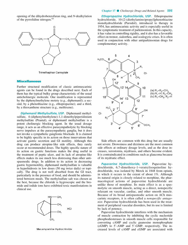
Chapter 17 Cholinergic Drugs and Related Agents 595
opening of the dihydrobenzofuran ring, and N-dealkylationof the pyrrolidine nitrogen.83
Miscellaneous
Further structural modification of classic antimuscarinicagents can be found in the drugs described next. Each ofthem has the typical bulky group characteristic of the usualanticholinergic molecule. One modification is representedby the diphenylmethylene moiety (e.g., diphemanil); a sec-ond, by a phenothiazine (e.g., ethopropazine); and a third,by a thioxanthene structure (e.g., methixene).
Diphemanil Methylsulfate, USP. Diphemanil methyl-sulfate, 4-(diphenylmethylene)-1,1-dimethylpiperidiniummethylsulfate (Prantal), or diphemanil methylsulfate is apotent cholinergic blocking agent. In the usual dosagerange, it acts as an effective parasympatholytic by blockingnerve impulses at the parasympathetic ganglia, but it doesnot invoke a sympathetic ganglionic blockade. It is claimedto be highly specific in its action on those innervations thatactivate gastric secretion and GI motility. Although thisdrug can produce atropine-like side effects, they rarelyoccur at recommended doses. The highly specific nature ofits action on gastric functions makes the drug useful inthe treatment of peptic ulcer, and its lack of atropine-likeeffects makes its use much less distressing than other anti-spasmodic drugs. In addition to its action in decreasinggastric hypermotility, diphemanil methylsulfate is valuablein hyperhidrosis in low doses (50 mg twice daily) or topi-cally. The drug is not well absorbed from the GI tract,particularly in the presence of food, and should be adminis-tered between meals. The methylsulfate salt was chosen asthe best, because the chloride is hygroscopic and the bro-mide and iodide ions have exhibited toxic manifestations inclinical use.
Ethopropazine Hydrochloride, USP. Ethopropazinehydrochloride, 10-[2-(diethylamino)propyl]phenothiazinemonohydrochloride (Parsidol), introduced to therapy in1954, has antimuscarinic activity and is especially useful inthe symptomatic treatment of parkinsonism. In this capacity,it has value in controlling rigidity, and it also has a favorableeffect on tremor, sialorrhea, and oculogyric crises. It is oftenused in conjunction with other antiparkinsonian drugs forcomplementary activity.
Side effects are common with this drug but are usuallynot severe. Drowsiness and dizziness are the most commonside effects at ordinary dosage levels, and as the dose in-creases, xerostomia, mydriasis, and others become evident.It is contraindicated in conditions such as glaucoma becauseof its mydriatic effect.
Papaverine Hydrochloride, USP. Papaverine hy-drochloride, 6,7-dimethoxy-1-veratrylisoquinoline hy-drochloride, was isolated by Merck in 1848 from opium,in which it occurs to the extent of about 1%. Althoughits natural origin is closely related to morphine, the phar-macological actions of papaverine hydrochloride areunlike those of morphine. Its main effect is as a spas-molytic on smooth muscle, acting as a direct, nonspecificrelaxant on vascular, cardiac, and other smooth muscle.Because of its broad antispasmodic action on ACh mus-carinic receptors, it is often called a nonspecific antago-nist. Papaverine hydrochloride has been used in the treat-ment of peripheral vascular disorders, but its use is limitedby lack of potency.
Papaverine hydrochloride interferes with the mechanismof muscle contraction by inhibiting the cyclic nucleotidephosphodiesterases in smooth muscle cells responsible forconverting cAMP and cyclic guanosine monophosphate(cGMP) to 5�-AMP and 5�-GMP, respectively. The in-creased levels of cAMP and cGMP are associated with
0558-0606_17865_Ch17.qxd 12/9/09 1:53 AM Page 595

596 Wilson and Gisvold’s Textbook of Organic Medicinal and Pharmaceutical Chemistry
muscle relaxation through their phosphorylation of myosinlight-chain kinase.
GANGLIONIC BLOCKING AGENTS
Autonomic ganglia have been the subject of interest formany years in the study of interactions between drugs andnervous tissues. The first important account84 was given byLangley and described the stimulating and blocking actionsof nicotine on sympathetic ganglia. It was found that smallamounts of nicotine stimulated ganglia and then produced ablockade of ganglionic transmission because of persistentdepolarization. From these experiments, Langley was ableto outline the general pattern of innervation of organs by theautonomic nervous system. Parasympathetic ganglia usu-ally are located near the organ they innervate and havepreganglionic fibers that stem from the cervical and thoracicregions of the spinal cord. Sympathetic ganglia consist of22 pairs that lie on either side of the vertebral columnto form lateral chains. These ganglia are connected both toeach other by nerve trunks and to the lumbar or sacral re-gions of the spinal cord.
Using the sympathetic cervical ganglion as a model re-vealed that transmission in the autonomic ganglion ismore complex than formerly believed. Traditionally, stim-ulation of autonomic ganglia by ACh was considered tobe the nicotinic action of the neurotransmitter. It is nowunderstood that stimulation by ACh produces a triphasicresponse in sympathetic ganglia. Impulse transmissionthrough the ganglion occurs when ACh is released frompreganglionic fibers and activates the N2 nicotinic recep-tors of the neuronal membrane. This triggers an increasein sodium and potassium conductances of a subsynapticmembrane, resulting in an initial excitatory postsynapticpotential (EPSP) with a latency of 1 ms, followed by aninhibitory postsynaptic potential (IPSP) with a latency of35 ms, and, finally, a slowly generating EPSP with a la-tency of several hundred milliseconds. The ACh releasedby preganglionic fibers also activates M1 muscarinicreceptors of the ganglion and probably of the small-intensity fluorescent (SIF) cell. This results in the appear-ance of a slow IPSP and a slow EPSP in the neurons of theganglion.85 The initial EPSP is blocked by conventionalcompetitive nondepolarizing ganglionic blocking agents,such as hexamethonium, and is considered the primary
0558-0606_17865_Ch17.qxd 12/9/09 1:53 AM Page 596

Chapter 17 Cholinergic Drugs and Related Agents 597
pathway for ganglionic transmission.86 The slowly gener-ating or late EPSP is blocked by atropine but not by thetraditional ganglionic blocking agents. This receptor hasmuscarinic properties because methacholine causes gener-ation of the late EPSP without causing the initial spikecharacteristic of ACh. Atropine also blocks the late EPSPproduced by methacholine. There may be more than onetype of muscarinic receptor in sympathetic ganglia.Atropine blocks both high-affinity (M1) and low-affinity(M2) muscarinic receptors in the ganglion.87 In addition tothe cholinergic pathways, the cervical sympathetic gan-glion has a neuron that contains a catecholamine.88 Theseneuronal cells, identified initially by fluorescence histo-chemical studies and shown to be smaller than the post-ganglionic neurons, are now referred to as SIF cells.Dopamine has been identified as the fluorescent cate-cholamine in the SIF cells that are common to many othersympathetic ganglia. Dopamine apparently mediates anincrease in cAMP, which causes hyperpolarization ofpostganglionic neurons (Fig. 17.19). The IPSP phase ofthe transmission of sympathetic ganglia following AChadministration can be blocked by both atropine and �-adrenergic blocking agents.85
If a similar nontraditional type of ganglionic transmis-sion occurs in the parasympathetic ganglia, it has not yet become evident. With the anatomical and physiologi-cal differences between sympathetic and parasympatheticganglia, it should be no surprise that ganglionic agentsmay show some selectivity between the two types ofganglia. Although we do not have drug classificationssuch as “parasympathetic ganglionic blockers” and “sym-pathetic ganglionic blockers,” we do find that certainganglia have a predominant effect over certain organsand tissues and that a nondiscriminant blockade ofautonomic ganglia results in a change in the effect of theautonomic nervous system on that organ (Table 17.7).
None of the commonly known ganglionic blockers hasyet been identified as a selective blocker of parasympa-thetic ganglia.
Van Rossum89,90 has reviewed the mechanisms of gan-glionic synaptic transmission, the mode of action of gan-glionic stimulants, and the mode of action of ganglionicblocking agents. They have been classified as blockingagents in the following manner.
Depolarizing Ganglionic Blocking Agents
Depolarizing blocking agents are actually ganglionic stim-ulants. Thus, for nicotine, small doses give an action simi-lar to that of the natural neuroeffector ACh, an action
Muscarinic Cholinergic
ACh
ACh
cAMP
cGMP
SIF cell
ACh
f-EPSPs-EPSP
s-IPSP
blocked by hexomethonium
blocked by atropine
POSTGANGLIONIC NEURON
DOPAMINE
PRE-GANGLIONIC
CHOLINERGICFIBERS
POSTGANGLIONICSYNAPTIC
POTENTIALS
blockedby
atropine
Dopominergi
Nicotinic
blocked by -adrenergicantagonists
�
Figure 17.19 Neurotransmission at the sympathetic cervical ganglion.
TABLE 17.7 Results of Ganglionic Blockers on Organs
Results of Predominant Ganglionic
Organ System Blockade
Cardiovascular systemHeart Parasympathetic TachycardiaArterioles Sympathetic VasodilationVeins Sympathetic Dilation
EyeIris Parasympathetic MydriasisCiliary muscle Parasympathetic Cycloplegia
GI tract Parasympathetic RelaxationUrinary bladder Parasympathetic Urinary
retentionSalivary glands Parasympathetic Dry mouthSweat glands Sympathetica Anhidrosis
Adapted from Goth, A.: Medical Pharmacology, 9th ed. St. Louis, C. V.Mosby, 1978.aNeurotransmitter is ACh.
0558-0606_17865_Ch17.qxd 12/9/09 1:53 AM Page 597
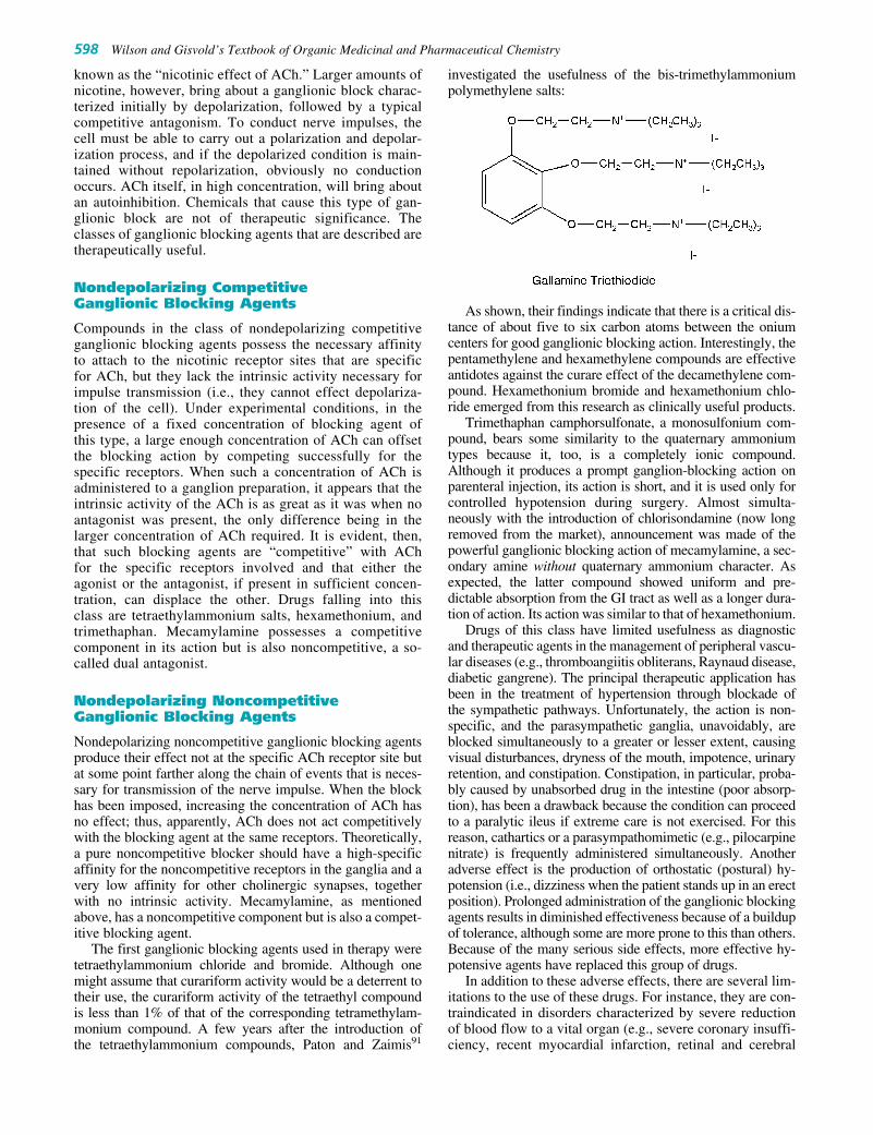
known as the “nicotinic effect of ACh.” Larger amounts ofnicotine, however, bring about a ganglionic block charac-terized initially by depolarization, followed by a typicalcompetitive antagonism. To conduct nerve impulses, thecell must be able to carry out a polarization and depolar-ization process, and if the depolarized condition is main-tained without repolarization, obviously no conductionoccurs. ACh itself, in high concentration, will bring aboutan autoinhibition. Chemicals that cause this type of gan-glionic block are not of therapeutic significance. Theclasses of ganglionic blocking agents that are described aretherapeutically useful.
Nondepolarizing CompetitiveGanglionic Blocking Agents
Compounds in the class of nondepolarizing competitiveganglionic blocking agents possess the necessary affinityto attach to the nicotinic receptor sites that are specificfor ACh, but they lack the intrinsic activity necessary forimpulse transmission (i.e., they cannot effect depolariza-tion of the cell). Under experimental conditions, in thepresence of a fixed concentration of blocking agent ofthis type, a large enough concentration of ACh can offsetthe blocking action by competing successfully for thespecific receptors. When such a concentration of ACh isadministered to a ganglion preparation, it appears that theintrinsic activity of the ACh is as great as it was when noantagonist was present, the only difference being in thelarger concentration of ACh required. It is evident, then,that such blocking agents are “competitive” with AChfor the specific receptors involved and that either theagonist or the antagonist, if present in sufficient concen-tration, can displace the other. Drugs falling into thisclass are tetraethylammonium salts, hexamethonium, andtrimethaphan. Mecamylamine possesses a competitivecomponent in its action but is also noncompetitive, a so-called dual antagonist.
Nondepolarizing NoncompetitiveGanglionic Blocking Agents
Nondepolarizing noncompetitive ganglionic blocking agentsproduce their effect not at the specific ACh receptor site butat some point farther along the chain of events that is neces-sary for transmission of the nerve impulse. When the blockhas been imposed, increasing the concentration of ACh hasno effect; thus, apparently, ACh does not act competitivelywith the blocking agent at the same receptors. Theoretically,a pure noncompetitive blocker should have a high-specificaffinity for the noncompetitive receptors in the ganglia and avery low affinity for other cholinergic synapses, togetherwith no intrinsic activity. Mecamylamine, as mentionedabove, has a noncompetitive component but is also a compet-itive blocking agent.
The first ganglionic blocking agents used in therapy weretetraethylammonium chloride and bromide. Although onemight assume that curariform activity would be a deterrent totheir use, the curariform activity of the tetraethyl compoundis less than 1% of that of the corresponding tetramethylam-monium compound. A few years after the introduction ofthe tetraethylammonium compounds, Paton and Zaimis91
investigated the usefulness of the bis-trimethylammoniumpolymethylene salts:
As shown, their findings indicate that there is a critical dis-tance of about five to six carbon atoms between the oniumcenters for good ganglionic blocking action. Interestingly, thepentamethylene and hexamethylene compounds are effectiveantidotes against the curare effect of the decamethylene com-pound. Hexamethonium bromide and hexamethonium chlo-ride emerged from this research as clinically useful products.
Trimethaphan camphorsulfonate, a monosulfonium com-pound, bears some similarity to the quaternary ammoniumtypes because it, too, is a completely ionic compound.Although it produces a prompt ganglion-blocking action onparenteral injection, its action is short, and it is used only forcontrolled hypotension during surgery. Almost simulta-neously with the introduction of chlorisondamine (now longremoved from the market), announcement was made of thepowerful ganglionic blocking action of mecamylamine, a sec-ondary amine without quaternary ammonium character. Asexpected, the latter compound showed uniform and pre-dictable absorption from the GI tract as well as a longer dura-tion of action. Its action was similar to that of hexamethonium.
Drugs of this class have limited usefulness as diagnosticand therapeutic agents in the management of peripheral vascu-lar diseases (e.g., thromboangiitis obliterans, Raynaud disease,diabetic gangrene). The principal therapeutic application hasbeen in the treatment of hypertension through blockade ofthe sympathetic pathways. Unfortunately, the action is non-specific, and the parasympathetic ganglia, unavoidably, areblocked simultaneously to a greater or lesser extent, causingvisual disturbances, dryness of the mouth, impotence, urinaryretention, and constipation. Constipation, in particular, proba-bly caused by unabsorbed drug in the intestine (poor absorp-tion), has been a drawback because the condition can proceedto a paralytic ileus if extreme care is not exercised. For thisreason, cathartics or a parasympathomimetic (e.g., pilocarpinenitrate) is frequently administered simultaneously. Anotheradverse effect is the production of orthostatic (postural) hy-potension (i.e., dizziness when the patient stands up in an erectposition). Prolonged administration of the ganglionic blockingagents results in diminished effectiveness because of a buildupof tolerance, although some are more prone to this than others.Because of the many serious side effects, more effective hy-potensive agents have replaced this group of drugs.
In addition to these adverse effects, there are several lim-itations to the use of these drugs. For instance, they are con-traindicated in disorders characterized by severe reductionof blood flow to a vital organ (e.g., severe coronary insuffi-ciency, recent myocardial infarction, retinal and cerebral
598 Wilson and Gisvold’s Textbook of Organic Medicinal and Pharmaceutical Chemistry
0558-0606_17865_Ch17.qxd 12/9/09 1:53 AM Page 598
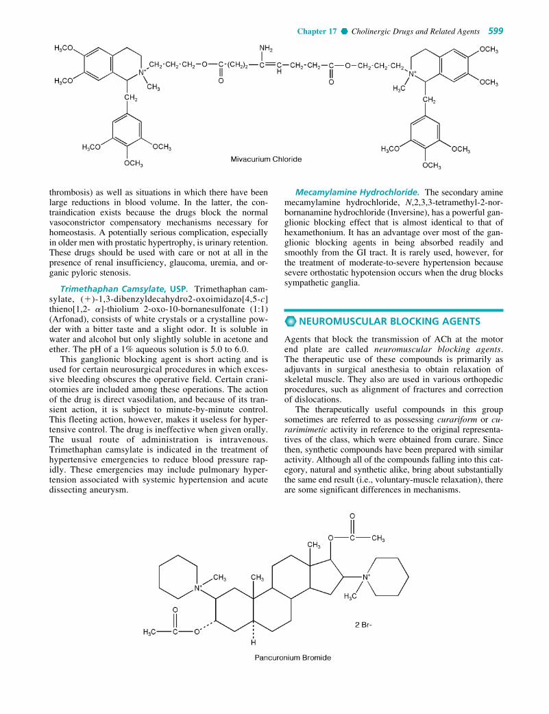
Chapter 17 Cholinergic Drugs and Related Agents 599
thrombosis) as well as situations in which there have beenlarge reductions in blood volume. In the latter, the con-traindication exists because the drugs block the normalvasoconstrictor compensatory mechanisms necessary forhomeostasis. A potentially serious complication, especiallyin older men with prostatic hypertrophy, is urinary retention.These drugs should be used with care or not at all in thepresence of renal insufficiency, glaucoma, uremia, and or-ganic pyloric stenosis.
Trimethaphan Camsylate, USP. Trimethaphan cam-sylate, (�)-1,3-dibenzyldecahydro2-oxoimidazo[4,5-c]thieno[1,2- �]-thiolium 2-oxo-10-bornanesulfonate (1:1)(Arfonad), consists of white crystals or a crystalline pow-der with a bitter taste and a slight odor. It is soluble inwater and alcohol but only slightly soluble in acetone andether. The pH of a 1% aqueous solution is 5.0 to 6.0.
This ganglionic blocking agent is short acting and isused for certain neurosurgical procedures in which exces-sive bleeding obscures the operative field. Certain crani-otomies are included among these operations. The actionof the drug is direct vasodilation, and because of its tran-sient action, it is subject to minute-by-minute control.This fleeting action, however, makes it useless for hyper-tensive control. The drug is ineffective when given orally.The usual route of administration is intravenous.Trimethaphan camsylate is indicated in the treatment ofhypertensive emergencies to reduce blood pressure rap-idly. These emergencies may include pulmonary hyper-tension associated with systemic hypertension and acutedissecting aneurysm.
Mecamylamine Hydrochloride. The secondary aminemecamylamine hydrochloride, N,2,3,3-tetramethyl-2-nor-bornanamine hydrochloride (Inversine), has a powerful gan-glionic blocking effect that is almost identical to that ofhexamethonium. It has an advantage over most of the gan-glionic blocking agents in being absorbed readily andsmoothly from the GI tract. It is rarely used, however, forthe treatment of moderate-to-severe hypertension becausesevere orthostatic hypotension occurs when the drug blockssympathetic ganglia.
NEUROMUSCULAR BLOCKING AGENTS
Agents that block the transmission of ACh at the motorend plate are called neuromuscular blocking agents.The therapeutic use of these compounds is primarily asadjuvants in surgical anesthesia to obtain relaxation ofskeletal muscle. They also are used in various orthopedicprocedures, such as alignment of fractures and correctionof dislocations.
The therapeutically useful compounds in this groupsometimes are referred to as possessing curariform or cu-rarimimetic activity in reference to the original representa-tives of the class, which were obtained from curare. Sincethen, synthetic compounds have been prepared with similaractivity. Although all of the compounds falling into this cat-egory, natural and synthetic alike, bring about substantiallythe same end result (i.e., voluntary-muscle relaxation), thereare some significant differences in mechanisms.
0558-0606_17865_Ch17.qxd 12/9/09 1:53 AM Page 599

The possible existence of a junction between muscle andnerve was suggested as early as 1856, when Claude Bernardobserved that the site of action of curare was neither thenerve nor the muscle. Since that time, it has been agreedthat ACh mediates transmission at the neuromuscular junc-tion by a sequence of events described previously in thischapter. The neuromuscular junction consists of the axonimpinging onto a specialized area of the muscle known asthe muscle end plate. The axon is covered with a myelinsheath, containing the nodes of Ranvier, but is bare at theending. The nerve terminal is separated from the end plateby a gap of 200 Å. The subsynaptic membrane of the endplate contains the cholinergic receptor, the ion-conductingchannels (which are opened under the influence of ACh),and AChE.
One of the anatomical differences between the neuro-muscular junction and other ACh-responsive sites is theabsence in the former of a membrane barrier or sheath thatenvelopes the ganglia or constitutes the blood-brain barrier.This is important in the accessibility of the site of action todrugs, particularly quaternary ammonium compounds, be-cause they pass through living membranes with consider-ably greater difficulty and selectivity than do compoundsthat can exist in a nonionized species. The essentially barenature (i.e., lack of lipophilic barriers) of the myoneuraljunction permits ready access by quaternary ammoniumcompounds. In addition, compounds with considerablemolecular dimensions are accessible to the receptors in themyoneural junction. As a result of this property, variationsin the chemical structure of quaternaries have little influenceon the potential ability of the molecule to reach the cholin-ergic receptor in the neuromuscular junction. Thus, thefollowing types of neuromuscular junction blockers havebeen noted.
Nondepolarizing Blocking Agents
Traditionally, nondepolarizing blocking agents is a term ap-plied to categorize drugs that compete with ACh for therecognition site on the nicotinic receptor by preventingdepolarization of the end plate by the neurotransmitter.Thus, by decreasing the effective ACh–receptor combina-tions, the end plate potential becomes too small to initiatethe propagated action potential. This results in paralysisof neuromuscular transmission. The action of these drugs isquite analogous to that of atropine at the muscarinic recep-tor sites of ACh. Many experiments suggest that the agonist (ACh) and the antagonist compete on a one-to-one basis for the end plate receptors. Drugs in this classare tubocurarine, dimethyltubocurarine, pancuronium, andgallamine.
Depolarizing Blocking Agents
Drugs in the category of depolarizing blocking agents depo-larize the membrane of the muscle end plate. This depolar-ization is quite similar to that produced by ACh itself atganglia and neuromuscular junctions (i.e., its so-callednicotinic effect), with the result that the drug, if in sufficientconcentration, eventually will produce a block. Eithersmooth or voluntary muscle, when challenged repeatedlywith a depolarizing agent, eventually becomes insensitive.
This phenomenon is known as tachyphylaxis, or desensiti-zation, and is demonstrated convincingly under suitableexperimental conditions with repeated applications of AChitself, the results indicating that within a few minutes theend plate becomes insensitive to ACh. These statementsmay imply that a blocking action of this type is clear-cut,but under experimental conditions, it is not quite so un-ambiguous, because a block that begins with depolarizationmay regain the polarized state even before the block.Furthermore, depolarization induced by increasing thepotassium ion concentration does not prevent impulse trans-mission. For these and other reasons, it is probably best toconsider the blocking action a desensitization until a clearerpicture emerges. Drugs falling in this class are decametho-nium and succinylcholine.
Curare and Curare Alkaloids
Originally curare was a term used to describe collectivelythe very potent arrow poisons used since early times by theSouth American Indians. The arrow poisons were preparedfrom numerous botanic sources and often were mixtures ofseveral different plant extracts. Some were poisonous byvirtue of a convulsant action and others by a paralyzant ac-tion. Only the latter type is of value in therapeutics and isspoken of ordinarily as curare.
Chemical investigations of the curares were not espe-cially successful because of difficulties in obtaining authen-tic samples with definite botanic origin. Not until 1935 wasa pure crystalline alkaloid, d-tubocurarine chloride, possess-ing in great measure the paralyzing action of the original cu-rare, isolated from a plant. Wintersteiner and Dutcher,92 in1943, isolated the same alkaloid. They showed, however,that the botanic source was Chondodendron tomentosum(Menispermaceae) and, thus, provided a known source ofthe drug.
Following the development of quantitative bioassaymethods for determining the potency of curare extracts, apurified and standardized curare was developed and mar-keted under the trade name Intocostrin (purified C. tomento-sum extract), the solid content of which consisted of almostone-half (�)-tubocurarine solids. Following these essen-tially pioneering developments, (�)-tubocurarine chlorideand dimethyltubocurarine iodide appeared on the market aspure entities.
Tubocurarine Chloride, USP. Tubocurarine chloride,(�)-tubocurarine chloride hydrochloride pentahydrate, isprepared from crude curare by a process of purification andcrystallization. Tubocurarine chloride occurs as a white oryellowish white to grayish white, odorless, crystalline pow-der that is soluble in water. Aqueous solutions of it are sta-ble to heat sterilization.
The structural formula for (�)-tubocurarine was longthought to be that of Ia (see structure diagram). Throughthe work of Everett et al.,93 the structure is now knownto be that of Ib. The monoquaternary nature of Ib thusrevealed has caused some reassessment of thinking con-cerning the theoretical basis for the blocking action, be-cause all had previously assumed a diquaternary structure(i.e., Ia). Nevertheless, this does not negate the earlierconclusions that a diquaternary nature of the molecule
600 Wilson and Gisvold’s Textbook of Organic Medicinal and Pharmaceutical Chemistry
0558-0606_17865_Ch17.qxd 12/9/09 1:53 AM Page 600

Chapter 17 Cholinergic Drugs and Related Agents 601
provides better blocking action than does a monoquater-nary nature (e.g., Ib is approximately fourfold lesspotent than dimethyl tubocurarine iodide). Further, (�)-isotubocurarine chloride (Ic) has twice the activity of Ib inthe particular test used.
Tubocurarine is a nondepolarizing blocking agent usedfor its paralyzing action on voluntary muscles, the site ofaction being the neuromuscular junction. Its action isinhibited or reversed by the administration of AChEinhibitors, such as neostigmine, or by edrophonium chlo-ride (Tensilon). Such inhibition of its action is necessitatedin respiratory embarrassment caused by overdosage.Additionally, in somewhat higher concentrations, d-tubocurarine may enter the open ion channel and add anoncompetitive blockade. Cholinesterase inhibitors do notrestore this latter action easily or fully. Often, adjunctiveartificial respiration is needed until the maximal curareaction has passed. The drug is inactive orally because ofinadequate absorption through lipoidal membranes in theGI tract, and when used therapeutically, it usually is in-jected intravenously.
d-Tubocurarine binds for only 1 ms to the receptor, yetits pharmacological effect of muscle paralysis, producedby administration of the drug intravenously during sur-gery, lasts for up to 2 hours. The basis of this action is thepharmacokinetics of the drug. d-Tubocurarine is given in-travenously, and although 30% to 77% is bound to plasmaproteins, the drug is distributed rapidly to central bodycompartments, including neuromuscular junctions. About45% of d-tubocurarine is eliminated unchanged by the kid-neys. Its half-life is 89 minutes.
Tubocurarine, in the form of a purified extract, was usedfirst in 1943 as a muscle relaxant in shock therapy for men-tal disorders. Its use markedly reduced the incidence ofbone and spine fractures and dislocations from convul-sions because of shock. Following this, it was used as anadjunct in general anesthesia to obtain complete musclerelaxation, a use that persists to this day. Before its usebegan, satisfactory muscle relaxation in various surgicalprocedures (e.g., abdominal operations) was obtainable onlywith “deep” anesthesia with the ordinary general anesthet-ics. Tubocurarine permits a lighter plane of anesthesia, withno sacrifice in the muscle relaxation so important to the sur-geon. A reduced dose of tubocurarine is administered withether because ether itself has curare-like action.
Metocurine Iodide, USP. Metocurine iodide, (�)-O,O�-dimethylchondrocurarine diiodide (Metubine iodide),is prepared from natural crude curare by extracting the cu-rare with methanolic potassium hydroxide. When theextract is treated with an excess of methyl iodide, the (�)-tubocurarine is converted to the diquaternary dimethylether and crystallizes out as the iodide (see “TubocurarineChloride”). Other ethers besides the dimethyl ether havebeen made and tested. For example, the dibenzyl ether wasone third as active as tubocurarine chloride, and the diiso-propyl compound had only one half the activity. For com-parison, the dimethyl ether has approximately 4 times theactivity of tubocurarine chloride.
The pharmacological action of this compound is the sameas that of tubocurarine chloride, namely, a nondepolarizingcompetitive blocking effect on the motor end plate of skeletalmuscles. It is considerably more potent than d-tubocurarine,however, and has the added advantage of exerting much lesseffect on respiration. The effect on respiration is not a signif-icant factor in therapeutic doses. Accidental overdosage iscounteracted best by forced respiration.
Synthetic Compounds with Curariform Activity
Curare, until relatively recent times, remained the only use-ful curarizing agent; and it, too, suffered from a lack of stan-dardization. The original pronouncement in 1935 of thestructure of (�)-tubocurarine chloride, unchallenged for35 years, led other workers to hope for activity in syntheticsubstances of less complexity. The quaternary ammoniumcharacter of the curare alkaloids coupled with the known ac-tivity of the various simple onium compounds hardlyseemed to be coincidental, and it was natural for research tofollow along these lines. One of the synthetic compoundsdiscovered was marketed in 1951 as Flaxedil (gallamine tri-ethiodide). Other various neuromuscular blocking agentshave followed.
Atracurium Besylate. Atracurium besylate, 2-(2-car-boxyethyl)-1,2,3,4-tetrahydro-6,7-dimethoxy-2-methyl-1-veratrylisoquinolinium benzenesulfonate pentamethyleneester (Tracrium), is a nondepolarizing neuromuscular block-ing agent that is approximately 2.5 times more potent thand-tubocurarine. Its duration of action (half-life, 0.33 hours)is much shorter than that of d-tubocurarine. The drug is
0558-0606_17865_Ch17.qxd 12/9/09 1:53 AM Page 601

metabolized rapidly and nonenzymatically to yield laudano-sine and a smaller quaternary compound (Fig. 17.20),which do not have neuromuscular blocking activity. In vitroexperiments show that atracurium besylate breaks down atpH 7.4 and 37°C by a Hoffman elimination reaction.94
Atracurium besylate undergoes enzymatic decompositionof its ester function to yield an inactive quaternary alcoholand quaternary acid. AChE inhibitors such as neostigmine,edrophonium, and pyridostigmine antagonize paralysis byatracurium besylate.
Doxacurium Chloride. The molecular structure of dox-acurium chloride, 1,2,3,4-tetrahydro-2-(3-hydroxypropyl)-6,7,8-trimethoxy2-methyl-1-(3,4,5-trimethoxybenzyl) iso-quinolinium chloride succinate (Nuromax), provides thepossibility for 10 stereoisomers: 4 d,l pairs and two meso
forms. Of the 10 stereoisomers, 3 are all-trans configuration,and these are the only active ones.95 Doxacurium chlorideis a long-acting nondepolarizing blocking agent. The drugdiffers from drugs such as gallium and pancuronium in thatit has no vagolytic activity. It is used as a skeletal musclerelaxant in surgical procedures expected to last longer than90 minutes.
Gallamine Triethiodide, USP. Gallamine triethiodide,[v-phenenyl-tris(oxyethylene)]tris[triethylammonium] triio-dide (Flaxedil), is a skeletal muscle relaxant that works byblocking neuromuscular transmission in a manner similar tothat of d-tubocurarine (i.e., a nondepolarizing blockingagent). It does have some differences, however. It has astrong vagolytic effect and a persistent decrease in neuro-muscular function after successive doses that cannot be
602 Wilson and Gisvold’s Textbook of Organic Medicinal and Pharmaceutical Chemistry
Figure 17.20 Hoffman elimination and hydrolysis reactions of atracurium.
0558-0606_17865_Ch17.qxd 12/9/09 1:53 AM Page 602

Chapter 17 Cholinergic Drugs and Related Agents 603
overcome by cholinesterase inhibitors. Gallamine triethio-dide also has muscarinic antagonistic properties and bindswith greater affinity to the M2 receptors than to the M1 re-ceptor. This latter characteristic may cause its strongvagolytic action.95, 96
The drug is contraindicated in patients with myastheniagravis, and one should remember that its action is cumula-tive, as with curare. The antidote for gallamine triethiodideis neostigmine.
Mivacurium Chloride. Mivacurium chloride, 1,2,3,4-tetrahydro-2-(3-hydroxypropyl)-6,7-dimethoxy-2-methyl-1-(3,4,5-trimethoxybenzyl)isoquinolinium chloride, (E)-4-octandioate (Mivacron), is a mixture of three stereoisomers,the trans-trans, cis-trans, and cis-cis diesters, each ofwhich has neuromuscular blocking properties. The cis-cisisomer is about one tenth as potent as the other isomers.Mivacurium chloride is a short-acting nondepolarizing drug
used as an adjunct to anesthesia to relax skeletal muscle.The drug is hydrolyzed by plasma esterases, and it is likelythat anticholinesterase agents used as antidotes could pro-long rather than reverse the effects of the drug.
Pancuronium Bromide. Although pancuronium bro-mide, 2�,16�-dipiperidino-5�-androstane-3�,17�-diol diac-etate dimethobromide (Pavulon), is a synthetic product, it isbased on the naturally occurring alkaloid malouetine, found inarrow poisons used by primitive Africans. Pancuronium bro-mide acts on the nicotinic receptor and in the ion channel,inhibiting normal ion fluxes.
This blocking agent is soluble in water and is marketed inconcentrations of 1 or 2 mg/mL for intravenous administra-tion. It is a typical nondepolarizing blocker, with a potencyapproximately 5 times that of (�)-tubocurarine chloride anda duration of action approximately equal to the latter. Studiesindicate that it has little or no histamine-releasing potential organglion-blocking activity and that it has little effect on thecirculatory system, except for causing a slight rise in thepulse rate. As one might expect, ACh, anticholinesterases,and potassium ion competitively antagonize it, whereas itsaction is increased by inhalation anesthetics such as ether,halothane, enflurane, and methoxyflurane. The latter en-hancement in activity is especially important to the anes-thetist because the drug is frequently administered as anadjunct to the anesthetic procedure to relax the skeletal mus-cle. Perhaps the most frequent adverse reaction to this agentis occasional prolongation of the neuromuscular block be-yond the usual time course, a situation that can usually becontrolled with neostigmine or by manual or mechanical
0558-0606_17865_Ch17.qxd 12/9/09 1:53 AM Page 603

ventilation, since respiratory difficulty is a prominent mani-festation of the prolonged blocking action.
As indicated, the principal use of pancuronium bromideis as an adjunct to anesthesia, to induce relaxation of skele-tal muscle, but it is also used to facilitate the managementof patients undergoing mechanical ventilation. Only expe-rienced clinicians equipped with facilities for applyingartificial respiration should administer it, and the dosageshould be adjusted and controlled carefully.
Pipecurium Bromide. Pipecurium bromide, 4,4�-(3�,17�-dihydroxy-5�-androstan-2�,16�-ylene)bis(1,1-dimeth-ylpiperazinium)dibromide diacetate (Arduan), is anondepolarizing muscle relaxant similar, both chemicallyand clinically, to pancuronium bromide. It is a long-acting
drug indicated as an adjunct to anesthesia and in patients un-dergoing mechanical ventilation.
Vecuronium Bromide. Vecuronium bromide, 1-(3�,17�-dihydroxy-2�-piperidino-5�-androstan-16�-yl)-1-methylpiperidinium bromide diacetate (Norcuron), is themonoquaternary analog of pancuronium bromide. It belongsto the class of nondepolarizing neuromuscular blockingagents and produces effects similar to those of drugs in thisclass. It is unstable in the presence of acids and undergoesgradual hydrolysis of its ester functions in aqueous solution.Aqueous solutions have a pH of about 4.0. This drug is usedmainly to produce skeletal muscle relaxation during surgeryand to assist in controlled respiration after general anesthe-sia has been induced.
604 Wilson and Gisvold’s Textbook of Organic Medicinal and Pharmaceutical Chemistry
0558-0606_17865_Ch17.qxd 12/9/09 1:53 AM Page 604

Chapter 17 Cholinergic Drugs and Related Agents 605
REFERENCES1. Dale, H. H.: J. Pharmacol. Exp. Ther. 6:147, 1914.2. Loewi, O.: Arch. Gesamte. Physiol. (Pfluegers) 189:239, 1921.3. Zola-Morgan, S., and Squire, L. R.: Annu. Rev. Neurosci. 16:547,
1994.4. Frank, H. S., and Wen, W.-Y.: Discuss. Faraday Soc. 24:133, 1957.5. Hille, B.: Annu. Rev. Physiol. 38:139, 1976.6. Karlin, A.: In Cotman, C. U., Poste, G., and Nicolson, G. L. (eds.).
Cell Surface and Neuronal Function. Amsterdam, ElsevierBiomedical, 1980, p. 191.
7. Changeaux, J. P., Devillers-Thiery, A., and Chermoulli, P.: Science225:1335, 1984.
8. Anholt, R., et al.: In Martonosi, A. N. (ed.). The Enzymes ofBiological Membranes, vol. 3. New York, Plenum Press, 1985.
9. Raftery, M. A., et al.: Science 208:1445, 1980.10. Sargent, P. B.: Neuroscience 16:403, 1994.11. Dani, J. A.: Biol. Psychiatry 49:166, 2001.12. Rattray, M.: Biol. Psychiatry 49:185, 2001.13. Goyal, R. K.: N. Engl. J. Med. 321:1022, 1989.14. Birdsall, N. J. M., et al.: Pharmacology 37(Suppl.):22, 1988.15. Mutschlur, E., et al.: Prog. Pharmacol. Clin. Pharmacol. 7:13, 1989.16. Doods, H. N., et al.: Prog. Pharmacol. Clin. Pharmacol. 7:47, 1989.17. Barnes, P. J.: Life Sci. 52:521, 1993.18. Rubanyi, G. M.: J. Cell Biol. 46:27, 1991.
1. When discussing the structure–activity relationship ofacetylcholine what will alkyl substitutions on the betacarbon cause to the molecule?a. decrease rate of hydrolysis by esterasesb. decrease nicotinic actionc. increase muscarinic actiond. All of the above are correct.e. Only a and c are correct.
2. The therapeutic usefulness of the agent representedbelow is:
a. incontinenceb. glaucomac. myasthenia gravisd. insecticidal agente. irritable bowel syndrome
O
C O CH2CH2 N
O
O
CH3
3. The therapeutic usefulness of the agent representedbelow is in treating Alzheimer disease. Please describewhy this agent has usefulness in treating this disease.
4. Describe the reason cholinergics can be used inmyasthenia gravis.
NH3C
CH3
O C
O
N
CH2CH3
CH3
CH3
Succinylcholine Chloride, USP. Succinylcholine chlo-ride, choline chloride succinate (2:1) (Anectine, Sucostrin),is a white, odorless, crystalline substance that is freely solu-ble in water to give solutions with a pH of about 4. It isstable in acidic solutions but unstable in alkali. Aqueous so-lutions should be refrigerated to ensure stability.
Succinylcholine chloride is characterized by a very shortduration of action and a quick recovery because of its rapidhydrolysis after injection. It brings about the typical muscu-lar paralysis caused by blocking nervous transmission at themyoneural junction. Large doses may cause temporary res-piratory depression, as with similar agents. Its action, in con-trast with that of (1)-tubocurarine, is not antagonized byneostigmine, physostigmine, or edrophonium chloride.These anticholinesterase drugs actually prolong the action of
succinylcholine chloride, which suggests that the drug isprobably hydrolyzed by cholinesterases. The brief durationof action of this curare-like agent is said to render an antidoteunnecessary if the proper supportive measures are available.Succinylcholine chloride has a disadvantage, however, inthat the usual antidotes cannot terminate its action promptly.
It is used as a muscle relaxant for the same indications asother curare agents. It may be used for either short or longperiods of relaxation, depending on whether one or severalinjections are given. In addition, it is suitable for continuousintravenous drip administration.
Succinylcholine chloride should not be used withthiopental sodium because of the high alkalinity of the lat-ter. If used together, they should be administered immedi-ately after mixing; however, separate injection is preferable.
R E V I E W Q U E S T I O N S
0558-0606_17865_Ch17.qxd 12/9/09 1:53 AM Page 605

606 Wilson and Gisvold’s Textbook of Organic Medicinal and Pharmaceutical Chemistry
19. Kilbinger, H., Dietricht, C., and von Bardeleben, R. S.: J. Physiol.Paris 87:77, 1993.
20. Linder, M. E., and Gilman, A. E.: Sci. Am. 27:56, 1992.21. Caufield, M. P.: Pharmacol. Ther. 58:319, 1993.22. Berridge, M. J., and Irvine, R. F.: Nature 312:315, 1984.23. Berridge, M. J.: Annu. Rev. Biochem. 56:159, 1987.24. Clapham, D. E.: Annu. Rev. Neurosci. 17:441, 1994.25. Haga, T., and Nada, H.: Biochim. Biophys. Acta. 291:564, 1973.26. Cavallito, C. J., et al.: J. Med. Chem. 12:134, 1969.27. Aquilonius, S. M., et al.: Acta Pharmacol. Toxicol. 30:129, 1979.28. Whittaker, V. P.: Trends Pharmacol. Sci. 7:312, 1986.29. Namba, T., and Grob, D.: J. Neurochem. 15:1445, 1968.30. Partington, P., Feeney, J., and Burgen, A. S. V.: Mol. Pharmacol.
8:269, 1972.31. Casey, A. F.: Prog. Med. Chem. 11:1, 1975.32. Behling, R. W., et al.: Proc. Natl. Acad. Sci. U. S. A. 85:6721,
1988.33. Nordvall, G., and Hacksell, U.: J. Med. Chem. 36:967, 1993.34. Schmeideberg, O., and Koppe, R.: Das Muscarine, das Giftege Alka
id des Fiielgenpiltzes. Leipzig, Vogel, 1869.35. Hardegger, E., and Lohse, F.: Helv. Chim. Acta 40:2383, 1957.36. Trumpp-Kallmeyer, S., et al.: J. Med. Chem. 35:3448, 1992.37. Triggle, D. J., et al.: J. Med. Chem. 34:3164, 1991.38. Ariens, E. J., and Simonis, A. M.: In deJong, H. (ed.). Quantitative
Methods in Pharmacology. Amsterdam, Elsevier, 1969.39. Ing, H. R.: Science 109:264, 1949.40. Hernandez, M., et al.: Br. J. Pharmacol. 110:1413, 1993.41. Bebbington, A., and Brimblecombe, R. W.: Adv. Drug Res. 2:143,
1965.42. Ren, L. N., Nakane, T., and Chiba, S.: J. Cardiovasc. Pharmacol.
22:841, 1993.43. Morrison, K. J., and Vanhoutte, P. M.: Br. J. Pharmacol. 106:672, 1992.44. Nunes, M. A., and Brochmann-Hanssen, E. J.: J. Pharm. Sci. 63:716,
1974.45. Williams, P. D., et al.: Pharmacology 23:177, 1992.46. Gabelt, B. T., and Kaufman, P. L.: J. Pharmacol. Exp. Ther. 263:1133,
1992.47. Ramos-Casals, M., and Brito-Zeron, P.: Expert Rev. Clin. Immun.
3:195, 2007.48. Mallarkey, G.: Cholinesterase Inhibitors in Alzheimer’s Disease.
Auckland, Adis International, 1999.49. Berman, A. H., Yguerabide, J., and Taylor, P.: Biochemistry 19:2226,
1980.50. Englehard, N., Prchal, K., and Nenner, M.: Angew. Chem. Int. Ed.
6:615, 1967.51. Ordentlich, A., et al.: J. Biol. Chem. 268:17083, 1993.52. Shafferman, A., et al.: J. Biol. Chem. 267:17640, 1992.53. Wilson, I. B., Harrison, M. A., and Ginsberg, S.: J. Biol. Chem.
236:1498, 1961.54. Ellis, S., Krayer, O., and Plachte, F. L.: J. Pharmacol. Exp. Ther.
79:309, 1943.55. Enz, A., et al.: Prog. Brain Res. 98:431, 1993.56. O’Brien, R. D.: Mol. Pharmacol. 4:121, 1968.57. Calvey, H. T.: Biochem. Pharmacol. 16:1989, 1967.58. Summers, W. K., et al.: N. Engl. J. Med. 315:1241, 1986.
59. Molloy, D. W., et al.: Can. Med. Assoc. J. 144:29, 1991.60. Heath, D. F.: Organophosphorus Poisons. Oxford, Pergamon Press,
1961.61. Oosterban, R. A., and Cohen, J. A.: The active site of esterases. In
Goodwin, T. W., Harris, I. J., and Hartley, B. S. (eds.). Structure andActivity of Enzymes. New York, Academic Press, 1964.
62. Linn, J. G., and Tomarelli, R. C.: Am. J. Ophthalmol. 35:46, 1952.63. Comroe, J. H., et al.: J. Pharmacol. Exp. Ther. 87:281, 1946.64. Gage, J. C.: Biochem. J. 54:426, 1953.65. Nakatsugawa, T., Tolman, N. M., and Dahm, P. A.: Biochem.
Pharmacol. 17:1517, 1968.66. Mountner, L. A., and Cheatam, R. M.: Enzymologia 25:215, 1963.67. Stephenson, R. P.: Br. J. Pharmacol. Chemother. 11:379, 1956.68. Ariens, E. J.: Adv. Drug Res. 3:235, 1966.69. Paton, W. D. M.: Proc. R. Soc. Lond. B 154:21, 1961.70. Long, J. P., et al.: J. Pharmacol. Exp. Ther. 117:29, 1956.71. AMA Drug Evaluations Annual 1994. Chicago, American Medical
Association, 1994, p. 894.72. Gyermek, L., and Nador, K.: J. Pharm. Pharmacol. 9:209, 1957.73. Ariens, E. J., Simonis, A. M., and Van Rossum, J. M.: In Ariens, E.
J. (ed.). Molecular Pharmacology. New York, Academic Press, 1964,p. 205.
74. Kondritzer, A. A., and Zvirblis, P.: J. Am. Pharm. Assoc. Sci. Ed.46:531, 1957.
75. Cushny, A. R.: J. Physiol. 30:176, 1904.76. Doods, H. N., et al.: Eur. J. Pharmacol. 250:223, 1993.77. Fuder, M., and Meincke, M.: Naunyn Schmiedebergs Arch.
Pharmakol. 347:591, 1993.78. Abramov, Y., and Sand, P. K.: Expert Opinion Pharmaco. 5:2351,
2004.79. Madersbacher, H., and Rovner, E.: Expert Opinion Pharmaco.
7:1373, 2006.80. Ohtake, A., Ukai, M., Hatanaka, T., et al.: Eur. J. Pharmacol. 492:243,
2004.81. Clemett, D., and Jarvis, B.: Drugs & Aging 18:277, 2001.82. Lazareno, S., and Birdsall, N. J.: Br. J. Pharmacol. 109:1120, 1993.83. Blok, B. F. M., and Corcos, J.: Aging Health 3:143, 2007.84. Karczmar, A. G. (ed.): International Encyclopedia of Pharmacology
and Therapeutics, Sect. 12, Ganglionic Blocking and StimulatingAgents, vol. 1. Berlin, Springer-Verlag, 1980.
85. Skok, V. I.: In Karkevich, D. A. (ed.). Pharmacology of GanglionicTransmission. Berlin, Springer-Verlag, 1980, p. 7.
86. Greengard, P., and Kebabian, J. W.: Fed. Proc. 33:1059, 1974.87. Hammer, R., and Giachetti, A.: Life Sci. 31:2291, 1982.88. Volle, R. L., and Hancock, J. C.: Fed. Proc. 29:1913, 1970.89. Van Rossum, J. M.: Int. J. Neuropharmacol. 1:97, 1962.90. Van Rossum, J. M.: Int. J. Neuropharmacol. 1:403, 1962.91. Paton, W. D. M., and Zaimis, E. J.: Br. J. Pharmacol. 4:381, 1949.92. Wintersteiner, O., and Dutcher, J. D.: Science 97:467, 1943.93. Everett, A. J., Lowe, L. A., and Wilkinson, S.: Chem. Commun.
1020, 1970.94. Stenlake, J. B., et al.: Br. J. Anaesth. 55:3S, 1983.95. USAN and the USP Dictionary of Drug Names. Rockville, MD,
United States Pharmacopeial Convention, 1994, p. 231.96. Burke, R. E.: Mol. Pharmacol. 30:58, 1986.
0558-0606_17865_Ch17.qxd 12/9/09 1:53 AM Page 606

C H A P T E R 18
Drugs Acting on the Renal SystemSTEPHEN J. CUTLER
from the carboxyl terminal of angiotensin I by angiotensin-converting enzyme (ACE) to form the octapeptide an-giotensin II, a potent vasoconstrictor. Angiotensin III isformed by removal of the N-terminal aspartate residue ofangiotensin II, a reaction catalyzed by glutamyl aminopepti-dase. In contrast to angiotensin II, angiotensin III has a lesspotent but significant regulatory effect on sodium excretionby the renal tubules. This is primarily resulting from the ef-fect angiotensin III has in stimulating aldosterone secretion,a potent mineralcorticoid.
The vasoconstrictive effects of angiotensin II are a resultof many factors. In the kidneys, it constricts glomerular ar-terioles. The effects are greater on efferent arterioles than af-ferent ones. Constriction of afferent arterioles results inincreased arteriolar resistance, which raises systemic arterialblood pressure. In addition, angiotensin II stimulates the re-lease of vasopressin from the hypothalamus. Vasopressin isalso known as the antidiuretic hormone (ADH), because thispeptidic hormone is typically released to conserve waterwhen the body is dehydrated. In the kidneys, it increases thepermeability to water of the distal convoluted tubules andcollecting tubules in the nephrons. This, in turn, results inconcentrating the urine and reducing urine volume. By in-ducing moderate vasoconstriction, this peptide results in anincrease in blood pressure. Additionally, angiotensin II hasbeen shown to stimulate the production of endothelin, a 21amino acid peptide that is produced in the vascular endothe-lium and which plays a role in the regulation of smooth mus-cle contraction and which contributes to blood pressureregulation (see Chapter 19).4 Lastly, angiotensin II plays aprimary role in regulating aldosterone secretion. Upon stim-ulation by the renin–angiotensin system, aldosterone is se-creted by the adrenal cortex and elicits its effects at varioussites, including the nephrons. It is responsible for the reab-sorption of sodium into the bloodstream. This results in in-creased levels of sodium in the plasma, which in turn resultin increased blood volume and vascular resistance.5 If aldos-terone levels in the body become too high, then symptomssuch as elevated blood pressure or heart failure (formallyclassified as CHF) can occur.
The regulatory action of the renin–angiotensin system incontrolling sodium and potassium balance and arterial bloodpressure is modified by vasodilators called kinins. Proteolyticenzymes that circulate in the plasma form kinins. Kallikrein isactivated in plasma by noxious influences to act on a kinin,callidin, which is converted to bradykinin by tissue enzymes.Bradykinin enhances release of the prostaglandins PGE2 andPGI2 within certain tissues to produce a vasodilatory effect(Fig. 18.3). Bradykinin is converted to inactive products by
C H A P T E R O V E R V I E W
Drugs acting on the renal system describes the rolerenin–angiotensin system plays in regulating the cardiovas-cular system. This chapter supports the fact that this systemplays a critical role in the mechanisms of hypertension andheart failure (formally recognized as congestive heart failure[CHF]). This, in turn, serves in the development of thera-peutic agents that act on the various enzymes and receptorsassociated with the renin–angiotensin system. For pharma-cists to be good practitioners, it is necessary that they under-stand how the renin–angiotensin system influences normalphysiological functions as well as how components of thesystem, such as renin, angiotensin I and II, and angiotensin-converting enzyme, can be used in manipulating specificcardiovascular diseases.
The Renin–Angiotensin System and Hypertension
The renin–angiotensin system is a hormonal system thatplays a central role in the control of sodium excretion andbody fluid volume. It interacts closely with the sympatheticnervous system and aldosterone secretion in the regulationof blood pressure. Figure 18.1 shows the relationship of thecomponent parts of the renin–angiotensin system and theirmain physiological effects.
The relationship between the renin–angiotensin systemand blood pressure in humans has been known since beforethe beginning of the 20th century. Tigerstedt and Bergman1
demonstrated in 1898 that, when injected in a host, kidneyextract produced a potent vasopressor response. The sub-stance was named renin. Many years later, this substancewas shown to require a cofactor to produce vasoconstric-tion.2 Eventually, in 1939, this hypertensive substance wasisolated, identified as a decapeptide, and later called an-giotensin. This cofactor existed as an inactive precursor, an-giotensinogen. Later studies revealed that angiotensin ex-isted in two forms, the biologically inactive decapeptideangiotensin I and the active octapeptide angiotensin II.3
The precursor of angiotensin, angiotensinogen, is a gly-coprotein of molecular weight (MW) 58,000 to 61,000,synthesized primarily in the liver and brought into thecirculatory system. Renin, an aspartyl protease (MW35,000–40,000), whose primary source is the kidney,cleaves the Leu-Val bond from the aspartic acid end of theangiotensinogen polypeptide molecule to release the deca-peptide angiotensin I (Fig. 18.2). The biochemical conver-sion continues with the cleavage of a dipeptide (His-Leu)
607
0607-0616_17865_Ch18.qxd 12/4/09 5:50 AM Page 607

ACE and other carboxypeptidases.6 Although ACE causesactivation of angiotensin and inactivation of bradykinin, ac-tions that appear to be opposite, the balance of the systemseems to favor vasoconstriction.
ACE is a membrane-bound enzyme anchored to the cellmembrane through a single transmembrane domain locatednear the carboxy-terminal extremity. The enzyme is a zinc-containing glycoprotein with an MW about 130,000. It is anonspecific peptidyldipeptide hydrolase, widely distributedin mammalian tissues, that cleaves dipeptides from the car-
boxy terminus of several endogenous peptides. The mini-mum structural requirement for binding and cleavage of asubstrate by ACE is that it is a tripeptide with a free car-boxylate group. A general exception is that this enzymedoes not cleave peptides with a penultimate prolyl residue.This accounts for the biological stability of angiotensin II.7
The important binding points at the active site of ACE are acationic site to attract a carboxylate ion and a zinc ion thatcan polarize a carbonyl group of an amide function to makeit more susceptible to hydrolysis. In the active site, there is
608 Wilson and Gisvold’s Textbook of Organic Medicinal and Pharmaceutical Chemistry
Figure 18.1 Renin–angiotensin systemof blood pressure control.
Reduced Blood Pressure Lowered Sodium Excretion
Renin Release
Angiotensinogen
Angiotensin I Angiotensin II
Vasoconstriction
IncreasedPeripheral Resistance
ElevatedBlood Pressure
Increased CardiacOutput
Sodium and FluidRetention
Aldosterone Secretion
Angiotensin III
ACE
(NH2 end) ASP–ARG–VAL–TYR–ILE–HIS–PRO–PHE–HIS–LEU–VAL–ILE–HIS–R(Angiotensinogen)
ASP–ARG–VAL–TYR–ILE–HIS–PRO–PHE–HIS–LEU(Angiotensin I)
ASP–ARG–VAL–TYR–ILE–HIS–PRO–PHE–HIS–LEU(Angiotensin II)
ARG–VAL–TYR–ILE–HIS–PRO–PHE(Angiotensin III)
INACTIVE PRODUCTS
Angiotensinases
Aminopeptidase
Renin
ACE
(COOH end)
Figure 18.2 Biochemistry of the renin–angiotensin system: formation ofangiotensins from angiotensinogen.
0607-0616_17865_Ch18.qxd 12/4/09 5:50 AM Page 608

a nucleophilic attack of the amide carbonyl by the �-car-bonyl group of a glutamic acid residue to cause hydrolysisof the peptide. Figure 18.4 shows a hypothetical model ofthe hydrolysis of angiotensin I by the active site of ACE.ACE exists in more than one form. Somatic ACE that regu-lates blood pressure, found in most tissues, differs from theisoenzyme ACE found in the testis. Somatic ACE, in con-trast to testicular ACE, contains two binding domains. Theprincipal active site for hydrolysis is the domain located inthe C-terminal half of somatic ACE.8
Because the renin–angiotensin system plays such an im-portant role in regulating kidney function,9 aldosterone re-lease,10 electrolyte balance, and blood volume, it is easy torecognize why targeting this pathway is beneficial in themanagement of high blood pressure and heart failure.Inhibitors of angiotensin-converting enzyme (ACE in-
hibitors) are often used to reduce the formation of the morepotent angiotensin II. In addition, angiotensin receptorblockers (ARBs) can be used to prevent angiotensin II fromacting on angiotensin receptors.11
Recently, there have been several agents investigated fortheir ability to inhibit renin, which in turn results in less an-giotensin II being produced.12
RENIN–ANGIOTENSIN SYSTEMINHIBITORS
Angiotensin-Converting EnzymeInhibitors
PRODUCTS
Captopril. Captopril, 1-[(2S)-3-mercapto-2-methyl-1-ox-opropionyl]proline (Capoten), blocks the conversion of an-giotensin I to angiotensin II by inhibiting the convertingenzyme. The rational development of captopril as an inhibitorof ACE was based on the hypothesis that ACE and car-boxypeptidase A functioned by similar mechanisms. It wasnoted that d-2-benzylsuccinic acid7 was a potent inhibitor ofcarboxypeptidase A, but not ACE. By use of this small
Chapter 18 Drugs Acting on the Renal System 609
RallikreinCalladin Bradykinin
ACE
Inactive Products Vasodilatation
Prostaglandin Release
Figure 18.3 Bradykinin formation and action.
Glu
C O
O
O
O
O
CC
C
C
Arg
N
N
NH
NHNH NH
(His)
(Phe)
(Leu)
Zn++
O
(CH2)
CH2
CH2
CH2
CH3 CH3
CH2
NH2 NH2
CH
CH
CH
O
C
Glu
C O
O
O
O
O
CCOH
C
C
Arg
N
N
NH
H2N
NH NH
Zn++
OH
(CH2)
CH2
CH2
CH2
CH3 CH3
CH2
NH2 NH2
+
CH
CH
CH
O
C
+
Figure 18.4 Model showing cleav-age of the histidine–phenylalanineresidue of angiotensin I by ACE toform the octapeptide antiotensin IIand the dipeptide residue of histi-dine and leucine.
0607-0616_17865_Ch18.qxd 12/4/09 5:50 AM Page 609
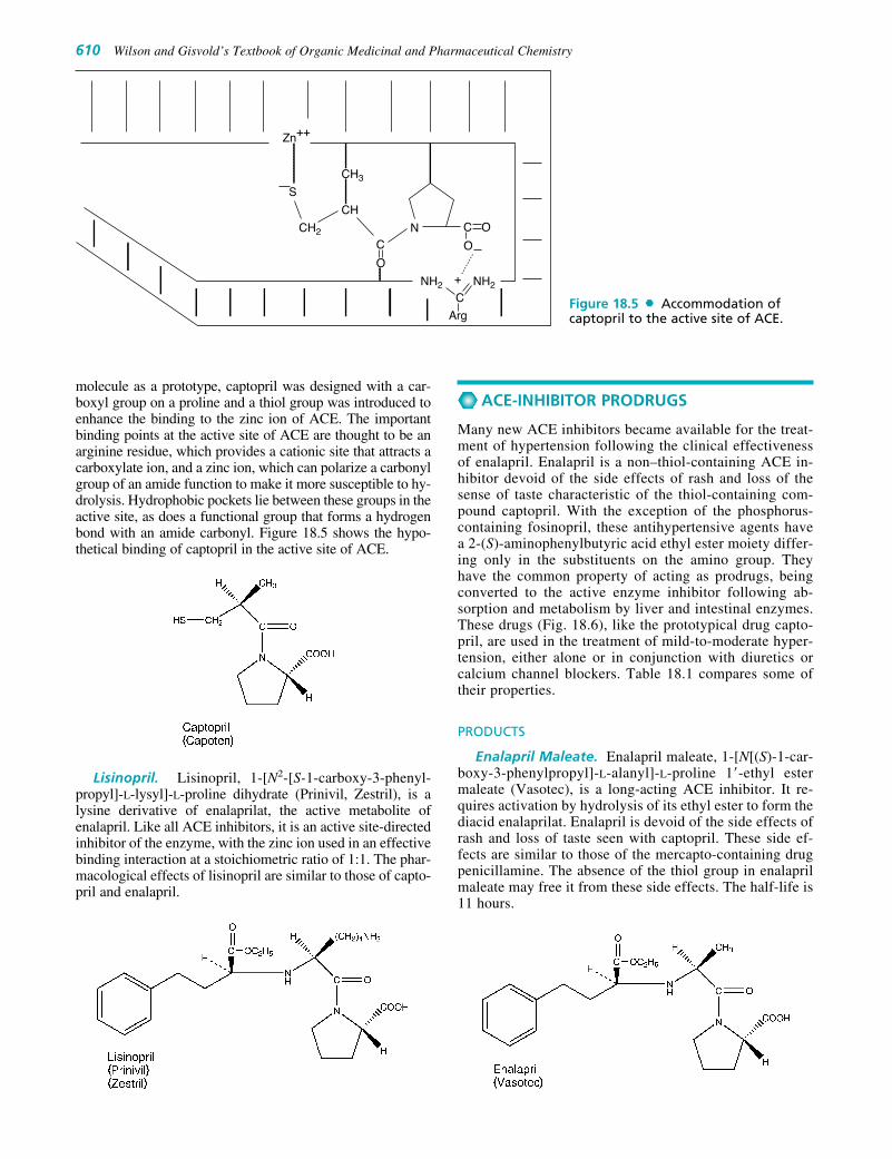
molecule as a prototype, captopril was designed with a car-boxyl group on a proline and a thiol group was introduced toenhance the binding to the zinc ion of ACE. The importantbinding points at the active site of ACE are thought to be anarginine residue, which provides a cationic site that attracts acarboxylate ion, and a zinc ion, which can polarize a carbonylgroup of an amide function to make it more susceptible to hy-drolysis. Hydrophobic pockets lie between these groups in theactive site, as does a functional group that forms a hydrogenbond with an amide carbonyl. Figure 18.5 shows the hypo-thetical binding of captopril in the active site of ACE.
Lisinopril. Lisinopril, 1-[N2-[S-1-carboxy-3-phenyl-propyl]-L-lysyl]-L-proline dihydrate (Prinivil, Zestril), is alysine derivative of enalaprilat, the active metabolite ofenalapril. Like all ACE inhibitors, it is an active site-directedinhibitor of the enzyme, with the zinc ion used in an effectivebinding interaction at a stoichiometric ratio of 1:1. The phar-macological effects of lisinopril are similar to those of capto-pril and enalapril.
ACE-INHIBITOR PRODRUGS
Many new ACE inhibitors became available for the treat-ment of hypertension following the clinical effectivenessof enalapril. Enalapril is a non–thiol-containing ACE in-hibitor devoid of the side effects of rash and loss of thesense of taste characteristic of the thiol-containing com-pound captopril. With the exception of the phosphorus-containing fosinopril, these antihypertensive agents havea 2-(S)-aminophenylbutyric acid ethyl ester moiety differ-ing only in the substituents on the amino group. Theyhave the common property of acting as prodrugs, beingconverted to the active enzyme inhibitor following ab-sorption and metabolism by liver and intestinal enzymes.These drugs (Fig. 18.6), like the prototypical drug capto-pril, are used in the treatment of mild-to-moderate hyper-tension, either alone or in conjunction with diuretics orcalcium channel blockers. Table 18.1 compares some oftheir properties.
PRODUCTS
Enalapril Maleate. Enalapril maleate, 1-[N[(S)-1-car-boxy-3-phenylpropyl]-L-alanyl]-L-proline 1�-ethyl estermaleate (Vasotec), is a long-acting ACE inhibitor. It re-quires activation by hydrolysis of its ethyl ester to form thediacid enalaprilat. Enalapril is devoid of the side effects ofrash and loss of taste seen with captopril. These side ef-fects are similar to those of the mercapto-containing drugpenicillamine. The absence of the thiol group in enalaprilmaleate may free it from these side effects. The half-life is11 hours.
610 Wilson and Gisvold’s Textbook of Organic Medicinal and Pharmaceutical Chemistry
Figure 18.5 Accommodation ofcaptopril to the active site of ACE.Arg
NH2
CNH2
N
+
S
Zn++
CH3
CH2
CH
O
C
OC
O
0607-0616_17865_Ch18.qxd 12/4/09 5:50 AM Page 610

Benazepril Hydrochloride. Benazepril hydrochloride,(3S)-3-[[(1S)-1-carbethoxy-3-phenylpropyl]amino]-2,3,4,5-tetrahydro-2-oxo-1H-1-benzazepine-1-acetic acid 3-ethylester hydrochloride (Lotensin), is metabolized rapidly to theactive diacid benazaprilat. As with the ACE prodrugs, nomutagenicity has been found, even though these drugs crossthe placenta.
Quinapril Hydrochloride. Quinapril hydrochloride,(S)-[(S)-N-[(S)21-carboxy3-phenylpropyl]alanyl]-1,2,3,4-tetrahydro-3-isoquinolinecarboxylic acid 1-ethyl ester hy-drochloride (Acuretic), forms the diacid quinaprilate in the
body. It is more potent than captopril and equipotent to theactive form of enalapril.
Ramipril. Ramipril, (2S, 3aS, 6aS)-1-[(S)-N-[(S)-1-car-boxy-3-phenylpropyl]alanyl]octahydrocyclopenta[b]-pyrrole-2-carboxylic acid 1-ethyl ester (Altace), ishydrolyzed to ramiprilat, its active diacid form, faster thanenalapril is hydrolyzed to its active diacid form. Peak serumconcentrations from a single oral dose are achieved be-tween 1.5 and 3 hours. The ramiprilate formed completely
Chapter 18 Drugs Acting on the Renal System 611
Enalapril Maleate
O
CH3
CH2CH2CH NH CH C N
COOC2H5COOH
HC COOH
HC COOHH
N
H
CCOCH2CH3
CH2CH2
Benzapril Hydrochloride
HN
CH2COOHO
N
HCOOH
O
C N CH2 CH2 C
H
C
H
CH3
O
COCH2CH3
Quinapril Hydrochloride
H
Ramipril
CH2CH2
C
H H3C
NH
H3C2O C
O
CH
C
NH
H
COOHH
O
O
HCI
HCI
(CH2)4 P CH2 C NO
OCONa
(CH3)2CH CHOCCH2CH3
H
Fosinopril Sodium
O O
O
H
Figure 18.6 ACE-inhibitor prodrugs.
0607-0616_17865_Ch18.qxd 12/4/09 5:50 AM Page 611

suppresses ACE activity for up to 12 hours, with 80% inhi-bition of the enzyme still observed after 24 hours.
Fosinopril Sodium. Fosinopril sodium, (4S)-4-cyclo-hexyl-1-[[[(RS)-1-hydroxy-2-methylpropoxy](4-phenyl-butyl)phosphinyl]acetyl]-L-proline sodium salt (Monopril),is a phosphorus-containing ACE inhibitor. It is inactive butserves as a prodrug, being completely hydrolyzed by intes-tinal and liver enzymes to the active diacid fosinoprilat.
Trandolapril. Trandolapril, 1-[2-(1-ethoxycarbonyl-3-phenylpropylamino)propionyl]octahydroindole-2-car-boxylic acid (Mavik), is an indole-containing ACE in-hibitor that is structurally related to most of the precedingagents discussed. Enalapril is very similar to trandolapril,with the primary difference occurring in the heterocyclicsystems. The pyrrolidine of enalapril has been replacedwith an octahydroindole system. Much like enalaprilate,trandolapril must be hydrolyzed to tranolaprilate, which isthe bioactive species.
ANGIOTENSIN ANTAGONISTS
There are four different subtypes of angiotensin II receptorsubclasses identified to date and include AT1, AT2, AT3,and AT4. These belong to the class of G-protein–coupled re-ceptors and are responsible for the signal transduction of thehormone. Although there is some understanding of the roleAT2 plays in development of a fetus or neonate, there is verylittle known about the role of AT2 and AT3 in the humanbody. However, it is the AT1 receptors that are of particularinterest in managing specific cardiovascular diseases.Studies have demonstrated that stimulation of AT1 receptorsby angiotensin II result in vasoconstriction, aldosterone syn-thesis and secretion, increased vasopressin secretion, de-creased renal blood flow, renal tubular sodium reuptake, andseveral other physiological events.
Administration of a competitive antagonist that inhibitsangiotensin II at the AT1 receptor will produce a vasodilatoryeffect. Because the substrate for this receptor is an octapep-tide, much of the earlier work was performed by using vari-ous peptide systems. One such agent, saralasin, is an octapep-tide that differs from angiotensin by two amino acids. Thisagent’s use was limited because it had some partial agonisticproperties. Nevertheless, it served as a lead in the develop-ment of other agents that are useful in antagonizing the an-giotensin II receptor. The most significant lead in the devel-opment of this class came from a series of imidazole-5-aceticacid derivatives that attenuated pressor response to an-giotensin II in test animals. Molecular modeling revealed thatthe imidazole-5-acetic acid could be exploited to mimic moreclosely the pharmacophore of angiotensin II. The first suc-cessful agent to be developed through this method is losartan.Later, four other agents were introduced into the U.S. market.These tend to be biphenylmethyl derivatives that possess cer-tain acidic moieties, which can interact with various positionson the receptor, much like the substrate, angiotensin II.
612 Wilson and Gisvold’s Textbook of Organic Medicinal and Pharmaceutical Chemistry
TABLE 18.1 Comparison of Properties of ACE-Inhibitor Prodrugs
Metabolite Protein Metabolite Plasma Mode ofProdrug Metabolite Binding (%) t1/2 (hours) Excretion
Benazepril Benazeprilat 95 10–11 RenalEnalapril Enalaprilat 50–60 11.0 RenalFosinopril Fosinoprilat 97 11.5 Renal/fecalQuinapril Quinaprilat 97 3.0 Renal/fecalRamipril Ramiprilat 56 13–17 Renal/fecal
0607-0616_17865_Ch18.qxd 12/4/09 5:50 AM Page 612

Chapter 18 Drugs Acting on the Renal System 613
ANGIOTENSIN II BLOCKERS
PRODUCTS
Losartan. Losartan, 2-butyl-4-chloro-1-[p-(o-1H-tetra-zol-5-yl-phenyl)benzyl]imidazole-5-methanol monopotas-sium salt (Cozarr), was the first nonpeptide imidazole to beintroduced as an orally active angiotensin II antagonist withhigh specificity for AT1. When administered to patients, itundergoes extensive first-pass metabolism, with the 5-methanol being oxidized to a carboxylic acid. This metabo-lism is mediated by CYP 2C9 and 3A4 isozymes. The 5-methanol metabolite is approximately 15 times more potentthan the parent hydroxyl compound. Because the parent hy-droxyl compound has affinity for the AT1 receptor, strictlyspeaking, it is not a prodrug.13
Candesartan. Candesartan, (�)-1-[[(cyclohexyloxy)car-bonyl]-oxy]ethyl 2- ethoxy-1-[[2�-(1H-tetrazol-5-yl)[1,1�-biphenyl]-4-yl]methyl]-1H-benzimidazole-7-carboxylate(Atacand), like losartan, possesses the acidic tetrazole system,which most likely plays a role in binding to the angiotensin IIreceptor similarly to the acidic groups of angiotensin II. Also,the imidazole system has been replaced with a benzimidazolepossessing an ester at position 7. This ester must be hy-drolyzed to the free acid. Fortunately, this conversion takesplace fairly easily because of the carbonate in the ester sidechain. This facilitates hydrolysis of the ester so much thatconversion to the free acid takes place during absorption fromthe gastrointestinal tract.
Irbesartan. Irbesartan, 2-butyl-3-[[29-(1H-tetrazol-5-yl)[1,19-biphenyl]-4-yl]methyl]1,3-diazaspiro[4,4]non-1en-4-one (Avapro), like losartan, possesses the acidic tetrazolesystem, which most likely plays a role, similar to the acidicgroups of angiotensin II, in binding to the angiotensin II re-ceptor. In addition, the biphenyl system that serves to sepa-rate the tetrazole from the aliphatic nitrogen is still present.A major difference in this agent is that it does not possessthe acidic side chain. Even so, irbesartan has good affinityfor the angiotensin II receptor because of hydrogen bondingwith the carbonyl moiety of the amide system. Also, thisparticular agent does not require metabolic activation ascandesartan does.
Telmisartan. Telmisartan, 4�-[(1,4�-dimethyl-2�-propyl[2,6�-bi-1H-benzimidazol]-1�-yl)methyl]-[1,1�-biphenyl]-2-carboxylic acid (Micardis), does not appear to bear any struc-tural relationship to this class, but there is actually a great dealof overlap in the chemical architecture with other agents. Thefirst, and most significant, difference is the replacement of theacidic tetrazole system with a simple carboxylic acid. Thisacid, like the tetrazole, plays a role in receptor binding. Thesecond difference is the lack of a carboxylic acid near the im-idazole nitrogen that also contributes to receptor binding.14
As with irbesartan, however, there is not a need for this groupto be acidic but, rather, to be one that participates in receptorbinding. The second imidazole ring, much like a purine basein deoxyribonucleic acid (DNA), can hydrogen bond with theangiotensin II receptor.
Olmesartan. Olmesartan medoxomil, (5-methyl-2-oxo-1,3-dioxol-4-yl)methyl-5-(2-hydroxypropan-2-yl)-2-
0607-0616_17865_Ch18.qxd 12/4/09 5:50 AM Page 613
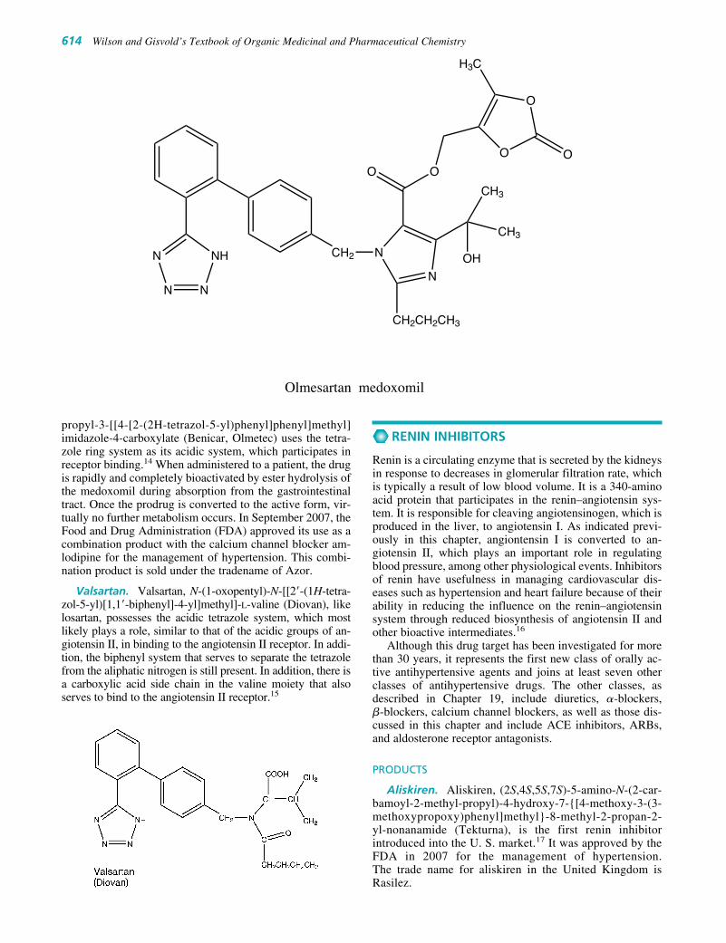
RENIN INHIBITORS
Renin is a circulating enzyme that is secreted by the kidneysin response to decreases in glomerular filtration rate, whichis typically a result of low blood volume. It is a 340-aminoacid protein that participates in the renin–angiotensin sys-tem. It is responsible for cleaving angiotensinogen, which isproduced in the liver, to angiotensin I. As indicated previ-ously in this chapter, angiontensin I is converted to an-giotensin II, which plays an important role in regulatingblood pressure, among other physiological events. Inhibitorsof renin have usefulness in managing cardiovascular dis-eases such as hypertension and heart failure because of theirability in reducing the influence on the renin–angiotensinsystem through reduced biosynthesis of angiotensin II andother bioactive intermediates.16
Although this drug target has been investigated for morethan 30 years, it represents the first new class of orally ac-tive antihypertensive agents and joins at least seven otherclasses of antihypertensive drugs. The other classes, asdescribed in Chapter 19, include diuretics, �-blockers, �-blockers, calcium channel blockers, as well as those dis-cussed in this chapter and include ACE inhibitors, ARBs,and aldosterone receptor antagonists.
PRODUCTS
Aliskiren. Aliskiren, (2S,4S,5S,7S)-5-amino-N-(2-car-bamoyl-2-methyl-propyl)-4-hydroxy-7-{[4-methoxy-3-(3-methoxypropoxy)phenyl]methyl}-8-methyl-2-propan-2-yl-nonanamide (Tekturna), is the first renin inhibitorintroduced into the U. S. market.17 It was approved by theFDA in 2007 for the management of hypertension.The trade name for aliskiren in the United Kingdom isRasilez.
614 Wilson and Gisvold’s Textbook of Organic Medicinal and Pharmaceutical Chemistry
propyl-3-[[4-[2-(2H-tetrazol-5-yl)phenyl]phenyl]methyl]imidazole-4-carboxylate (Benicar, Olmetec) uses the tetra-zole ring system as its acidic system, which participates inreceptor binding.14 When administered to a patient, the drugis rapidly and completely bioactivated by ester hydrolysis ofthe medoxomil during absorption from the gastrointestinaltract. Once the prodrug is converted to the active form, vir-tually no further metabolism occurs. In September 2007, theFood and Drug Administration (FDA) approved its use as acombination product with the calcium channel blocker am-lodipine for the management of hypertension. This combi-nation product is sold under the tradename of Azor.
Valsartan. Valsartan, N-(1-oxopentyl)-N-[[2�-(1H-tetra-zol-5-yl)[1,1�-biphenyl]-4-yl]methyl]-L-valine (Diovan), likelosartan, possesses the acidic tetrazole system, which mostlikely plays a role, similar to that of the acidic groups of an-giotensin II, in binding to the angiotensin II receptor. In addi-tion, the biphenyl system that serves to separate the tetrazolefrom the aliphatic nitrogen is still present. In addition, there isa carboxylic acid side chain in the valine moiety that alsoserves to bind to the angiotensin II receptor.15
N N
NHN CH2 N
N
CH2CH2CH3
OH
CH3
CH3
OO
O
O
H3C
O
Olmesartan medoxomil
0607-0616_17865_Ch18.qxd 12/4/09 5:50 AM Page 614

ALDOSTERONE ANTAGONISTS
Aldosterone antagonists are, as the name implies, receptor an-tagonists at the mineralcorticoid receptor. Antagonism ofthese receptors inhibits sodium resorption in the distal tubuleof the nephron, which in turn, interferes with sodium/potas-
sium exchange, reducing urinary potassium excretion.Overall, this results in a weak diuretic effect and decreasescardiac workload. Agents that block aldosterone may be usedin the management of hypertension or heart failure. The twoagents approved for use in the United States are spironolac-tone (Aldactone) and eplerenone (Inspra) and are discussed ingreater detail in the diuretic chapter of this textbook.
Chapter 18 Drugs Acting on the Renal System 615
O
O
HN
O
H2N
OH
O
NH2
O
Aliskiren
c. renin inhibitord. aldosterone antagoniste. More than one answer is correct.
3. Although the agent represented below (losartan [Cozaar])has some pharmacological effects, one of its metabolitesis reported to be more potent. Identify the metabolite anddescribe its production.
N N
NHN CH2 N
N
Cl
CH2CH2CH2CH3
CH2OH
1. The renin–angiotensin system plays an important rolein regulation of blood pressure. For that reason, this path-way encompasses the conversion of an approximately400 amino acid sequence to a decapeptide, then to the bi-ologically active octapeptide, and is an excellent targetfor drug therapy to manage hypertension. Describe thestarting protein and its conversion to the biologically ac-tive metabolite. Be sure to include the enzymes involvedin the conversion of the protein and peptides.
2. The therapeutic usefulness of the agent representedbelow (candesartan [Atacand]) is:
a. angiotensin-converting enzyme inhibitorb. angiotensin II receptor blocker
N N
NHN CH2 N
N
OCH2CH3
C
O
O
OO
O
R E V I E W Q U E S T I O N S
0607-0616_17865_Ch18.qxd 12/4/09 5:50 AM Page 615

REFERENCES1. Tigerstedt, R., and Bergman, P. G.: Scand. Arch. Physiol. 8:223,
1898.2. Page, F., and Helmer, O. A.: J. Exp. Med. 71:29, 1940.3. Skeggs, L., Marsh, W. H., Kahn, J. R., et al.: J. Exp. Med. 99:275, 1954.4. Moreau, P., Laplante, M.-A., Beaucage, P., et al.: HHandb. Exp.
Pharmacol. 163:149, 2004.5. Jackson, E. K.: In Hardman, J. G., and Limbird, L. E. (eds.).
Goodman and Gilman’s The Pharmacological Basis of Therapeutics,10th ed. New York, Macmillan, 2001, pp. 809–841.
6. Ceconi, C., Francolini, G., Olivares, A., et al.: Eur. J. Pharmacol.577:1, 2007.
7. Shapiro, R., and Riordan, J. F.: Biochemistry 23:5225, 1984.
8. Ehlers, M. R., and Riordan, J. F.: Biochemistry 30:7118, 1991.9. Levens, N. R., Peach, M. J., and Carey, R. M.: Circ. Res. 48:157,
1981.10. Jordan, J., Tank, J., Diedrich, A., et al.: Hypertension 36:e3, 2000.11. Aulakh, G. K., Sodhi, R. K., and Singh, M.: Life Sci. 81:615, 2007.12. Li, Y. C.: Current Opinion in Investigational Drugs 8:750, 2007.13. Schmidt, B., Drexler H., and Schieffer B.: Am. J. Cardiovasc. Drugs
4:361, 2004.14. Le, M. T., Pugsley, M. K., Vauquelin, G., et al.: Br. J. Pharmacol.
151:952, 2007.15. de Gasparo, M., and Whitebread, S.: Regul. Pept. 59:303, 1995.16. Venkata, C., and Ram, S.: J. Clin. Hypertens. 9:615, 2007.17. Sealey, J. E., and Laragh, J. H.: Am. J. Hypertens. 20(5):587, 2007.
616 Wilson and Gisvold’s Textbook of Organic Medicinal and Pharmaceutical Chemistry
0607-0616_17865_Ch18.qxd 12/4/09 5:50 AM Page 616

C H A P T E R 19
Cardiovascular AgentsSTEPHEN J. CUTLER
only secondary importance. We now know that the coronaryblood vessels in the atherosclerotic heart already are dilated,and that ordinary doses of dilator drugs do not significantlyincrease blood supply to the heart; instead, anginal pain isrelieved by a reduction of cardiac consumption of oxygen.2
Although vasodilators are used in the treatment of angina,a more sophisticated understanding of the hemodynamic re-sponse to these agents has broadened their clinical useful-ness to other cardiovascular conditions. Because of theirability to reduce peripheral vascular resistance, vasodilators,including organonitrates, angiotensin-converting enzyme(ACE) inhibitors, and angiotensin receptor–blocking agents,are used to improve cardiac output in some patients withcongestive heart failure (CHF).
The coronary circulation supplies blood to the myocar-dial tissues to maintain cardiac function. It can react to thechanging demands of the heart by dilating its blood vesselsto provide sufficient oxygen and other nutrients and toremove metabolites. Myocardial metabolism is almost ex-clusively aerobic, which makes blood flow critical to thesupport of metabolic processes of the heart. This demand ismet effectively by the normal heart, because it extracts a rel-atively large proportion of the oxygen delivered to it by thecoronary circulation. The coronary blood flow dependsstrongly on myocardial metabolism, which in turn is af-fected by work done by the heart and the efficiency of theheart. The coronary system normally has a reserve capacitythat allows it to respond by vasodilation to satisfy the needsof the heart during strenuous activity by the body.
Coronary atherosclerosis, one of the more prevalent car-diovascular diseases, develops with increasing age and maylead to a reduction of the reserve capacity of the coronarysystem. It most often results in multiple stenosis and makesit difficult for the coronary system to meet adequately theoxygen needs of the heart that occur during physical exer-cise or emotional duress. Insufficient coronary blood flow(myocardial ischemia) in the face of increased oxygen de-mand produces angina pectoris.
The principal goal in the prevention and relief of angina isto limit the oxygen requirement of the heart, so that theamount of blood supplied by the stenosed arteries is adequate.Nitrate esters, such as nitroglycerin, lower arterial blood pres-sure and, in turn, reduce the work of the left ventricle. Thisaction is produced by the powerful vasodilating effect of thenitrates on the arterial system and, to an even greater extent,on the venous system. The result is reduced cardiac fillingpressure and ventricular size. This reduces the work requiredof the ventricle and decreases the oxygen requirements, al-lowing the coronary system to satisfy the oxygen demands ofmyocardial tissue and relieve anginal pain.
C H A P T E R O V E R V I E W
Proteins, cardiovascular agents, describe the role variouschemical entities play in controlling cardiovascular diseases.This chapter describes the agents used in the treatment ofangina, cardiac arrhythmias, hypertension, erectile dysfunc-tion, hyperlipidemias, and disorders of blood coagulation.For pharmacists to be good practitioners, it is necessary thatthey understand the pathophysiology of cardiovascular dis-eases and the agents used to treat these syndromes.
The treatment and therapy of cardiovascular disease haveundergone dramatic changes since the 1950s. Data showthat since 1968 and continuing through the 1990s, there hasbeen a noticeable decline in mortality from cardiovasculardisease. The bases for advances in the control of heart dis-ease have been (a) a better understanding of the diseasestate, (b) the development of effective therapeutic agents,and (c) innovative medical intervention techniques to treatproblems of the cardiovascular system.
The drugs discussed in this chapter are used for their ac-tion on the heart or other parts of the vascular system, tomodify the total output of the heart or the distribution ofblood to the circulatory system. These drugs are used in thetreatment of angina, cardiac arrhythmias, hypertension, hy-perlipidemias, and disorders of blood coagulation. Thischapter also includes a discussion of hypoglycemic agents,thyroid hormones, and antithyroid drugs.
ANTIANGINAL AGENTS ANDVASODILATORS
Most coronary artery disease conditions are caused by de-posits of atheromas in the intima of large and medium-sizedarteries serving the heart. The process is characterized byan insidious onset of episodes of cardiac discomfort causedby ischemia from inadequate blood supply to the tissues.Angina pectoris (angina), the principal symptom of is-chemic heart disease, is characterized by a severe constrict-ing pain in the chest, often radiating from the precordium tothe left shoulder and down the arm. The syndrome has beendescribed since 1772, but not until 1867 was amyl nitrite in-troduced for the symptomatic relief of angina pectoris.1 Itwas believed at that time that anginal pain was precipitatedby an increase in blood pressure and that the use of amyl ni-trite reduced both blood pressure and, concomitantly, thework required of the heart. Later, it was generally acceptedthat nitrites relieved angina pectoris by dilating the coronaryarteries and that changes in the work of the heart were of
617
0617-0665_17865_Ch19.qxd 12/4/09 5:50 AM Page 617

Intermediary Myocardial Metabolism
Energy metabolism by heart tissue provides an adequatesupply of high-energy phosphate compounds to replace theadenosine triphosphate (ATP) that is continually being con-sumed in contraction, ion exchange across membranes, andother energy-demanding processes. Because of the highturnover rate of ATP in heart muscle, a correspondinglyhigh rate of ATP production in the mitochondria is required.
Normal myocardial metabolism is aerobic, and the rate ofoxygen use parallels the amount of ATP synthesized by thecells.3 Free fatty acids (FFAs) are the principal fuel for myo-cardial tissue, but lactate, acetate, acetoacetate, and glucoseare also oxidized to carbon dioxide and water. A large vol-ume of the myocardial cell consists of mitochondria inwhich two-carbon fragments from FFA breakdown are me-tabolized through the Krebs cycle. The reduced flavin andnicotinamide dinucleotides formed by this metabolism arereoxidized by the electron-transport chain because of thepresence of oxygen (Fig. 19.1). In the hypoxic or ischemicheart, the lack of oxygen inhibits the electron-transportchain function and causes an accumulation of reduced flavinand nicotinamide coenzymes. As a result, fatty acids areconverted to lipids rather than being oxidized. To compen-sate for this, glucose use and glycogenolysis increase, butthe resulting pyruvate cannot be oxidized; instead, it is con-verted to lactate. A great loss of efficiency occurs as a resultof the change of myocardial metabolism from aerobic toanaerobic pathways. Normally, 36 mol of ATP are formedfrom the oxidation of 1 mol of glucose, but only 2 mol areformed from its glycolysis. This great loss of high-energystores during hypoxia thus limits the functional capacity of
the heart during stressful conditions and is reflected by theproduction of anginal pain.
Nitrovasodilators
SMOOTH MUSCLE RELAXATION
The contractile activity of all types of muscle (smooth, skele-tal) is regulated primarily by the reversible phosphorylationof myosin. Myosin of smooth muscle consists of two heavychains (molecular weight [MW] 200,000 each) that arecoiled to produce a filamentous tail. Each heavy chain is as-sociated with two pairs of light chains (MW 20,000 and16,000) that serve as substrates for calcium- and calmodulin-dependent protein kinases in the contraction process.Together with actin (MW 43,000), they participate in a cas-cade of biochemical events that are part of the processes ofmuscle contraction and relaxation (Fig. 19.2).
Cyclic nucleotides, cyclic adenosine monophosphate(cAMP), and, especially, cyclic guanosine monophosphate(cGMP) play important roles in the regulation of smoothmuscle tension. cAMP is the mediator associated withthe smooth muscle relaxant properties of drugs such as �-adrenergic agonists. It activates the protein kinases thatphosphorylate myosin light-chain kinase (MLCK).Phosphorylation of MLCK inactivates this kinase and pre-vents its action with Ca2� and calmodulin to phosphorylatemyosin, which interacts with actin to cause contraction ofsmooth muscle (Fig. 19.2).
The activity of cGMP in smooth muscle relaxation is af-fected by exogenous and endogenous agents. It is suggested4
that nitrovasodilators undergo metabolic transformation in
618 Wilson and Gisvold’s Textbook of Organic Medicinal and Pharmaceutical Chemistry
Glucose
Pyruvate (2)
Lactate
TCAcycle
Acetyl-CoA (2)
NADH
2 NADH
6 NADH � 2 FADH2
NORMAL GLUCOSE METABOLISM ISCHEMIC GLUCOSE METABOLISM
2 ATP 4 ADP � 2 NAD�
2 NAD�
NAD�
NAD�� 2 FAD
4 ATP � 2 NADH 4 ATP � 2 NADH2 ADP
Pyruvate (2)
Lactate
NADH
2 ATP 4 ADP � 2 NAD�
NAD�
2 ADP
Glycogen Glucose Glycogen
Oxidative
Phosphorylation
No Oxidative
Phosphorylation
Mitochondrion Mitochondrion
34 ATP�
H2O
Figure 19.1 Normal and ischemic myocardial metabolism of glucose. A total pro-duction of 36 mol of ATP results from the aerobic catabolism of 1 mol of glucose anduse of NADH and FADH2 in the oxidative phosphorylation process in mitochondria.When oxygen is not available, NADH and FADH2 levels rise and shut off the tricar-boxylic acid (TCA) cycle. Pyruvate is converted to lactate. Only 2 mol of ATP areformed from anaerobic catabolism of 1 mol of glucose. (Adapted from Giuliani, E. R.,et al.: Cardiology: Fundamentals and Practice, 2nd ed. By permission of the MayoFoundation, Rochester, MN.)
0617-0665_17865_Ch19.qxd 12/4/09 5:50 AM Page 618

vascular smooth muscle cells to form nitric oxide (NO). NOmediates smooth muscle relaxation by activating guanylatecyclase to increase intracellular concentrations of cGMP.cGMP activates protein kinases that can regulate free Ca2�
levels in the muscle cell and cause relaxation of smooth mus-cle by phosphorylating MLCK.
A short-lived free radical gas, NO is widely distributed inthe body and plays an important role by its effect throughcGMP on the smooth muscle vasculature. It is synthesizedin the vascular endothelial cell from the semiessential aminoacid L-arginine by NO synthase. After production in the cell,it diffuses to the smooth muscle cell, where it activates theenzyme guanylate cyclase, which leads to an increase incGMP and then muscle relaxation (Fig. 19.3). Endothelium-derived relaxing factor (EDRF), released from the endothe-lial cell to mediate its smooth muscle–relaxing propertiesthrough cGMP, is identical with NO.
Inhibitors of phosphodiesterases of cAMP and cGMPalso cause smooth muscle relaxation. These inhibitors in-crease cellular levels of cAMP and cGMP by preventingtheir hydrolysis to AMP and GMP, respectively. Drugs suchas papaverine (see Chapter 17), theophylline (see chapter on
Diuretics), and sildenafil (this chapter), which relax smoothmuscle, do so in part by inhibiting phosphodiesterases.
METABOLISM OF NITROVASODILATORS
After oral administration, organic nitrates are metabolizedrapidly by the liver, kidney, lungs, intestinal mucosa, andvascular tissue. Buccal absorption reduces the immediatehepatic destruction of the organic nitrates because only 15%of the cardiac output is delivered to the liver; this allows atransient but effective circulating level of the intact organicnitrate before it is inactivated.5
Organic nitrates, nitrites, nitroso compounds, and a vari-ety of other nitrogen-containing substances, such as sodiumnitroprusside, for the most part cause their pharmacologicaleffects by generating or releasing NO in situ. In some ways,these drugs are viewed as “replacement agents” for the en-dogenous NO generated by the NO synthase pathway fromarginine. The mechanisms by which vasodilatory drugs re-lease NO have become better understood recently. Table19.1 shows the oxidation state of various nitrosyl com-pounds that are common in nitrovasodilatory drugs. A com-mon feature of these drugs is that they release nitrogen inthe form of NO and contain nitrogen in an oxidation statehigher than �3 (as would occur in ammonia, amines,amides, and most biological nitrogen compounds). The ni-trogen in NO has an oxidation state of �2. Compoundssuch as nitroprusside, nitrosoamines, and nitrothiols withoxidation states of �3 release NO nonenzymatically.Although their spontaneous liberation of NO is by an un-known mechanism, it involves only a one-electron reduc-tion, which may occur on exposure of these chemicals tovarious reducing agents in the tissue of vascular smoothmuscle membranes. Organic nitrites such as amyl nitritereact with available thiol groups to form unstable S-ni-trosothiols, which rapidly decompose to NO by homolyticcleavage of their SMN bond. In mammalian smooth mus-cle, this will occur almost exclusively with glutathione asthe most abundant thiol compound.6
The pharmacodynamic action of nitroglycerin is pre-ceded by metabolic changes that follow various paths.Biotransformation of nitroglycerin to the dinitrates and the
Chapter 19 Cardiovascular Agents 619
Ca2�
Ca2�
Ca2��CM
Calcium channel
complex
MLCK*
Myosin�LC
Actin
Contraction Relaxation
Myosin�LC�PO4 Myosin�LC
MLCK MLCK�(PO4)2
CMATP
AC
Receptor
Membrane
cAMP
Agonist�
Figure 19.2 Regulation ofsmooth muscle contraction.Contraction is triggered by an in-flux of Ca2�. The increase of freeCa2� causes binding to calmodulin(CM). The Ca2�MCM complex bindsto myosin light-chain kinase (MLCK)and causes its activation (MLCK*).MLCK* phosphorylates myosin,which combines with actin to pro-duce contraction of smooth muscle.Myosin is dephosphorylated in thepresence of myosin phosphataseto cause muscle relaxation. The�-agonists activate adenylatecyclase (AC) to raise levels of cAMP,which in turn activates kinases thatphosphorylate MLCK, inactivatingit to prevent muscle contraction.
NitrovasodilatorsEndothelial Cells
NO
GC*
GTP
MLCK* MLCK MLCK�PO4
cGMP
Guanylate cyclase (GC)
Figure 19.3 Mechanism of nitrovasodilators. Nitricoxide (NO) formed in smooth muscle from nitrovasodila-tors or from endothelial cells (EDRF) activates guanylatecyclase (GC*). GC* activates cGMP-dependent protein ki-nases that phosphorylate myosin light-chain kinase(MLCK), causing its inactivation and subsequent musclerelaxation (see also Fig. 19.2).
0617-0665_17865_Ch19.qxd 12/4/09 5:50 AM Page 619

increase of intracellular cGMP precede vascular relaxation.Sulfhydryl-containing compounds, such as cysteine, reactchemically with organic nitrates to form inorganic nitriteions. The release of NO from an organic nitrate, such as ni-troglycerin, appears to occur in a stepwise fashion involvingnonenzymatic and enzymatic steps. Because nitroglycerinrequires a three-electron reduction to release NO, thiols maybe involved in the process. Nitroglycerin may decomposenonenzymatically by interaction with various thiols, such ascysteine or N-acetylcysteine, which may be present in tis-sue, to form a nitrosothiol intermediate before undergoingenzymatic transformation to release NO. Nitroglycerin alsoreadily releases NO by acting on an enzyme system attachedto the cellular surface membrane of smooth muscle. Theprocess may include glutathione-S-transferases, which con-vert nitroglycerin to a vasoinactive nitrite, which then mayrelease NO nonenzymatically.7
ESTERS OF NITROUS AND NITRIC ACIDS
Inorganic acids, like organic acids, will form esters with analcohol. Pharmaceutically, the important ones are sulfate,nitrite, and nitrate. Sulfuric acid forms organic sulfates, ofwhich methyl sulfate and ethyl sulfate are examples.
Nitrous acid (HNO2) esters may be formed readily froman alcohol and HNO2. The usual procedure is to mix sodiumnitrite, sulfuric acid, and the alcohol. Organic nitrites aregenerally very volatile liquids that are only slightly solublein water but soluble in alcohol. Preparations containingwater are very unstable because of hydrolysis.
The organic nitrates and nitrites and the inorganic nitriteshave their primary utility in the prophylaxis and treatment of
angina pectoris. They have a more limited application in treat-ing asthma, gastrointestinal spasm, and certain cases of mi-graine headache. Their application may be regarded as causaltherapy, because they act by substituting an endogenous fac-tor, the production or release of NO, which may be impairedunder pathophysiological circumstances associated with dys-function of the endothelial tissue. Nitroglycerin (glyceryltrinitrate) was one of the first members of this group to be in-troduced in medicine and remains an important member ofthe group. Varying the chemical structure of the organic ni-trates yields differences in speed of onset, duration of action,and potency (Table 19.2). Although the number of nitrateester groups may vary from two to six or more, depending onthe compound, there is no direct relationship between thenumber of nitrate groups and the level of activity.
It appears that the higher the oil/water partition coefficientof the drug, the greater the potency. The orientation of thegroups within the molecule also may affect potency.Lipophilicity of the nitrogen oxide–containing compoundproduces a much longer response of vasodilatory action. Thehighly lipophilic ester nitroglycerin permeates the cell mem-brane, allowing continual formation of NO within the cell.The same effect appears to occur for sodium nitroprusside, ni-troso compounds, and other organic nitrate and nitrite esters.4
ANTIANGINAL ACTION OF NITROVASODILATORS
The action of short-acting sublingual nitrates in the relief ofangina pectoris is complex. Although the sublingual nitratesrelax vascular smooth muscle and dilate the coronary arter-ies of normal humans, there is little improvement of coro-nary blood flow when these chemicals are administered toindividuals with coronary artery disease. Nitroglycerin is aneffective antianginal agent because it causes redistributionof coronary blood flow to the ischemic regions of the heartand reduces myocardial oxygen demand. This latter effect isproduced by a reduction of venous tone resulting from thenitrate vasodilating effect and a pooling of blood in the pe-ripheral veins, which results in a reduction in ventricularvolume, stroke volume, and cardiac output. It also causes re-duction of peripheral resistance during myocardial contrac-tions. The combined vasodilatory effects cause a decrease incardiac work and reduce oxygen demand.
PRODUCTS
Amyl Nitrite, United States Pharmacopeia (USP).Amyl nitrite, isopentyl nitrite [(CH3)2CHCH2CH2ONO], isa mixture of isomeric amyl nitrites but is principally isoamylnitrite. It may be prepared from amyl alcohol and HNO2 byseveral procedures. Usually, amyl nitrite is dispensed in
620 Wilson and Gisvold’s Textbook of Organic Medicinal and Pharmaceutical Chemistry
TABLE 19.1 Nitrosyl Vasodilatory Substances and TheirOxidation State
Nitrosyl NitrogenCompound Structure Oxidation State
Nitric oxide NBO �2
Nitrite MONO �3
Nitrate MONO2 �5
Organic nitrite RMOMNBO �3
Nitrosothiol RMSMNBO �3
Organic nitrate RMOMNO2 �5
Thionitrate RMSMNO2 �5
Nitroprusside [(CN)5FeMNBO]2 �3
Reprinted from Harrison, D. G., and Bates, J. M.: Circulation 87:1462, 1993,with permission from the American Heart Association.
TABLE 19.2 Relationship between Speed and Duration of Action of Sodium Nitrite and Certain Inorganic Esters
Action Begins Maximum Effect Duration of Action Compound (minutes) (minutes) (minutes)
Amyl nitrite 0.25 0.5 1
Nitroglycerin 2 8 30
Isosorbide dinitrate 3 15 60
Sodium nitrite 10 25 60
Erythrityl tetranitrate 15 32 180
Pentaerythritol tetranitrate 20 70 330
0617-0665_17865_Ch19.qxd 12/4/09 5:50 AM Page 620

ampul form and used by inhalation or orally in alcohol solu-tion. Currently, it is recommended for treating cyanide poi-soning; although not the best antidote, it does not require in-travenous injections.
Amyl nitrite is a yellowish liquid with an ethereal odor anda pungent taste. It is volatile and inflammable at room temper-ature. Amyl nitrite vapor forms an explosive mixture in air oroxygen. Inhalation of the vapor may involve definite explo-sion hazards if a source of ignition is present, as both roomand body temperatures are within the flammability range ofamyl nitrite mixtures with either air or oxygen. It is nearly in-soluble in water but is miscible with organic solvents. The ni-trite also will decompose in valeric acid and nitric acid.
Nitroglycerin. Glyceryl trinitrate is the trinitrate ester ofglycerol and is listed as available in tablet form in the USP.It is prepared by carefully adding glycerin to a mixture of ni-tric and fuming sulfuric acids. This reaction is exothermic,and the reaction mixture must be cooled to between 10°Cand 20°C.
The ester is a colorless oil, with a sweet, burning taste. Itis only slightly soluble in water, but it is soluble in organicsolvents.
Nitroglycerin is used extensively as an explosive in dy-namite. A solution of the ester, if spilled or allowed to evap-orate, will leave a residue of nitroglycerin. To prevent anexplosion of the residue, the ester must be decomposed byadding alkali. Even so, the material dispensed is so dilutethat the risk of explosions does not exist. It has a strong va-sodilating action and, because it is absorbed through theskin, is prone to cause headaches among workers associatedwith its manufacture. This transdermal penetration is whynitroglycerin is useful in a patch formulation. In medicine, ithas the action typical of nitrites, but its action develops moreslowly and is of longer duration. Of all the known coronaryvasodilatory drugs, nitroglycerin is the only one capable ofstimulating the production of coronary collateral circulationand the only one able to prevent experimental myocardialinfarction by coronary occlusion.
Previously, the nitrates were thought to be hydrolyzedand reduced in the body to nitrites, which then lowered theblood pressure. This is not true, however. The mechanism ofvasodilation of nitroglycerin through its formation of NO isdescribed previously.
Nitroglycerin tablet instability was reported in moldedsublingual tablets.8 The tablets, although uniform when
manufactured, lost potency both because of volatilization ofnitroglycerin into the surrounding materials in the containerand intertablet migration of the active ingredient.Nitroglycerin may be stabilized in molded tablets by incor-porating a “fixing” agent such as polyethylene glycol 400 orpolyethylene glycol 4000.9 In addition to sublingual tablets,the drug has been formulated into an equally effective lin-gual aerosol for patients who have problems with dissolu-tion of sublingual preparations because of dry mucousmembranes. Transdermal nitroglycerin preparations appearto be less effective than other long-acting nitrates, as absorp-tion from the skin is variable.
Diluted Erythrityl Tetranitrate, USP. Erythritol tetra-nitrate, 1,2,3,4-butanetetrol, tetranitrate (R*, S*)-(Cardilate),is the tetranitrate ester of erythritol and nitric acid. It is pre-pared in a manner analogous to that used for nitroglycerin.The result is a solid, crystalline material. This ester is alsovery explosive and is diluted with lactose or other suitableinert diluents to permit safe handling; it is slightly soluble inwater and soluble in organic solvents.
Erythrityl tetranitrate requires slightly more time than ni-troglycerin to produce its effect, which is of longer duration.It is useful when mild, gradual, and prolonged vascular dila-tion is warranted. The drug is used in the treatment of, andas prophylaxis against, attacks of angina pectoris and to re-duce blood pressure in arterial hypertonia.
Erythrityl tetranitrate produces a reduction of cardiacpreload as a result of pooling blood on the venous side of thecirculatory system by its vasodilating action. This actionresults in a reduction of blood pressure on the arterial sideduring stressful situations and is an important factor in pre-venting the precipitation of anginal attacks.
Diluted Pentaerythritol Tetranitrate, USP. Pen-taerythritol tetranitrate, 2,2-bis (hydroxymethyl)-1,3-propanediol tetranitrate (Peritrate, Pentritol), is a white,crystalline material with a melting point of 140°C. It isinsoluble in water, slightly soluble in alcohol, and readilysoluble in acetone. The drug is a nitric acid ester of thetetrahydric alcohol pentaerythritol and is a powerful explo-sive. Accordingly, it is diluted with lactose, mannitol, orother suitable inert diluents to permit safe handling.
Chapter 19 Cardiovascular Agents 621
0617-0665_17865_Ch19.qxd 12/4/09 5:50 AM Page 621

It relaxes smooth muscle of smaller vessels in the coro-nary vascular tree. Pentaerythritol tetranitrate is used pro-phylactically to reduce the severity and frequency of anginalattacks and is usually administered in sustained-releasepreparations to increase its duration of action.
Diluted Isosorbide Dinitrate, USP. Isosorbide dini-trate, 1,4:3,6-dianhydro-D-glucitol dinitrate (Isordil, Sor-bitrate), occurs as a white, crystalline powder. Its watersolubility is about 1 mg/mL.
Isosorbide dinitrate, as a sublingual or chewable tablet,is effective in the treatment or prophylaxis of acute anginalattacks. When it is given sublingually, the effect begins inabout 2 minutes, with a shorter duration of action thanwhen it is given orally. Oral tablets are not effective inacute anginal episodes; the onset of action ranges from 15to 30 minutes. The major route of metabolism involves den-itration to isosorbide 5-mononitrate. This metabolite has amuch longer half-life than the parent isosorbide dinitrate.As such, this particular metabolite is marketed in a tabletform that has excellent bioavailability with much less first-pass metabolism than isosorbide dinitrate. Isosorbide ismarketed as a combination therapy with hydralazine underthe tradename BiDil specifically for African Americanswith CHF.
Calcium Antagonists
EXCITATION–CONTRACTION COUPLING MUSCLE
Stimulation of the cardiac cell initiates the process of exci-tation, which has been related to ion fluxes through the cellmembrane. Depolarization of the tissue in the atria of theheart is mediated by two inwardly directed ionic currents.When the cardiac cell potential reaches its threshold, ionchannels in the membrane are opened, and Na� enters thecell through ion channels. These channels give rise to thefast sodium current that is responsible for the rapidly risingphase, phase 0, of the ventricular action potential (Fig.19.4). The second current is caused by the slow activation ofan L-type Ca2� ion channel that allows the movement ofCa2� into the cell. This “slow channel” contributes to themaintenance of the plateau phase (phase 2) of the cardiac ac-tion potential. We now understand that the Ca2� that enterswith the action potential initiates a second and larger releaseof Ca2� from the sarcoplasmic reticulum in the cell. Thissecondary release of Ca2� is sufficient to initiate the con-tractile process of cardiac muscle.
Contraction of cardiac and other muscle occurs from areaction between actin and myosin. In contrast to smoothvascular muscle, the contractile process in cardiac muscleinvolves a complex of proteins (troponins I, C, and T andtropomyosin) attached to myosin, which modulates the in-teraction between actin and myosin. Free Ca2� ions bind totroponin C, uncovering binding sites on the actin moleculeand allowing interaction with myosin, causing contractionof the muscle. The schematic diagram in Figure 19.5 showsthe sequence of events.10 Contraction of vascular smoothmuscle, like that of cardiac muscle, is regulated by theconcentration of cytoplasmic Ca2� ions. The mechanismby which the contraction is effected, however, includes acalcium- and calmodulin-dependent kinase as opposedto a Ca2�-sensitive troponin–tropomyosin complex (Fig.19.2). The activating effect depends on a different type ofreaction. The elevated free cytosolic Ca2� in vascularsmooth muscle cells binds to a high-affinity binding pro-tein, calmodulin.
622 Wilson and Gisvold’s Textbook of Organic Medicinal and Pharmaceutical Chemistry
Figure 19.4 Diagrammatic representation of themembrane action potential, as recorded from aPurkinje fiber, and an electrogram recorded from anisolated ventricular fiber. The membrane restingpotential is 90 mV relative to the exterior of the fiber.At the point of depolarization, there is a rapidchange (phase 0) to a more positive value. Phases 0 to4 indicate the phases of depolarization and repolariza-tion. Note that phases 0 and 3 of the membraneaction potential correspond in time to the inscriptionof the QRS and T waves, respectively, of the localelectrogram.
Figure 19.5 Sequence of events showingexcitation–contraction coupling in cardiac muscle.
0617-0665_17865_Ch19.qxd 12/4/09 5:50 AM Page 622

ION CHANNELS AND CALCIUM
Calcium ions play an important role in the regulation ofmany cellular processes, such as synaptic transmission andmuscle contraction. The role of calcium in these cellularfunctions is as a second messenger, for example, regulatingenzymes and ion channels. The entry of extracellular Ca2�
in the cytosol of myocardial cells and the release of Ca2�
from intracellular storage sites is important for initiatingcontractions of the myocardium. Normally, the concentra-tion of Ca2� in the extracellular fluid is in the millimolarrange, whereas the intracellular concentration of free Ca2�
is less than 10�7 M, even though the total cellular concen-tration may be 10�3 M or higher. Most of the Ca2� is storedwithin intracellular organelles or tightly bound to intracellu-lar proteins. The free Ca2� needed to satisfy the require-ments of a contraction resulting from a stimulus may resultfrom activation of calcium channels on the cell membraneand/or the release of calcium from bound internal stores.Each of these methods of increasing free cytosolic Ca2� in-volves channels that are selective for the calcium ion.Calcium channel blockers reduce or prevent the increase offree cytosolic calcium ions by interfering with the transportof calcium ions through these pores.
Calcium is one of the most common elements on earth.Most calcium involved in biological systems occurs as hy-droxyapatite, a static, stabilizing structure like that found inbone. The remaining calcium is ionic (Ca2�). Ionic calciumfunctions as a biochemical regulator, more often within thecell. The importance of calcium ions to physiological func-tions was realized first by Ringer, who observed in 1883 therole of Ca2� in cardiac contractility.
The ionic composition of the cytosol in excitable cells,including cardiac and smooth muscle cells, is controlled toa large extent by the plasma membrane, which prevents thefree movement of ions across this barrier. Present in themembranes are ion-carrying channels that open in responseto either a change in membrane potential or binding of a li-gand. Calcium-sensitive channels include (a) Na� to Ca2�
exchanger, which transports three Na� ions in return for oneCa2�; (b) a voltage-dependent Ca2� channel, which pro-vides the route for entry of Ca2� for excitation and contrac-tion in cardiac and smooth muscle cells and is the focus ofthe channel-blocking agents used in medicine; and (c) re-ceptor-operated Ca2� channels mediated by ligand bindingto membrane receptors such as in the action of epinephrineon the �-adrenergic receptor. The membrane of the sar-colemma within the cell also has ion-conducting channelsthat facilitate movement of Ca2� ions from storage loci inthe sarcoplasmic reticulum.
Four types of calcium channels, differing in location andfunction, have been identified: (a) L type, located in skele-tal, cardiac, and smooth muscles, causing contraction ofmuscle cells; (b) T type, found in pacemaker cells, causingCa2� entry, inactivated at more negative potentials andmore rapidly than the L type; (c) N type, found in neuronsand acting in transmitter release; and (d) P type, located inPurkinje cells but whose function is unknown at this time.
Calcium antagonists act only on the L-type channel toproduce their pharmacological effects. The L channels areso called because once the membrane has been depolarized,their action is long lasting. Once the membrane has been de-polarized, L channels must be phosphorylated to open.
Although there are similarities between L-type calciumchannels that exist in cardiac and smooth muscle, there aredistinct differences between the two. Cardiac L channelsare activated through �-adrenergic stimulation via a cAMP-dependent phosphorylation process,11 whereas L channelsin smooth muscle may be regulated by the inositol phos-phate system linked to G-protein–coupled, receptor-linkedphospholipase C activation.12
CALCIUM CHANNEL BLOCKERS
The L-type calcium channel, acted on by calcium channelblockers, consists of five different subunits, designated �1,�2, �, �, and �. The �1 subunit provides the central pore ofthe channel (Fig. 19.6). Calcium channel blockers can be di-vided conveniently into the three different chemical classesof the prototype drugs that have been used: phenylalkyl-amines (verapamil), 1,4-dihydropyridines (nifedipine), andbenzothiazepines (diltiazem). These prototype compoundssometimes are termed the “first generation” of calciumchannel blockers because two of the groups of drug classeshave been expanded by the introduction of a “second gener-ation” of more potent analogs (Table 19.3).
The specific Ca2� channel antagonists verapamil,nifedipine, and diltiazem interact at specific sites on thecalcium channel protein. These blockers do not occlude thechannel physically but bind to sites in the channel, becausethey can promote both channel activation and antagonism.Affinity for binding sites on the channel varies, depend-ing on the status of the channel. The channel can exist ineither an open (O), resting (R), or inactivated (I) state, andthe equilibrium between them is determined by stimulusfrequency and membrane potential (Fig. 19.7). Verapamil
Chapter 19 Cardiovascular Agents 623
Figure 19.6 Schematic representation of an L-typeCa2� channel.
TABLE 19.3 First- and Second-Generation CalciumChannel Blockers
Chemical First SecondClassification Generation Generation
Phenylalkylamines Verapamil AnipamilBepridil
1,4-Dihydropyridine Nifedipine AmlodipineFelodipineIsradipineNicardipineNimodipine
Benzothiazepine Diltiazem —
0617-0665_17865_Ch19.qxd 12/4/09 5:50 AM Page 623

and diltiazem do not bind to a channel in the resting state,only after the channel has been opened. They are ionized,water-soluble Ca2�-entry blockers that reach their bindingsites by the hydrophilic pathway when the channel is open.Verapamil and diltiazem are use dependent (i.e., theirCa2�-blocking activity is a function of the frequency ofcontractions). An increase in contraction frequency causesa reduction, rather than an augmentation, of contractions.Nifedipine is a neutral molecule at physiological pHand can cause interference with the Ca2� in the open orclosed state. In the closed state, nifedipine can traverse thephospholipid bilayer to reach its binding site because of itslipid solubility.
CARDIOVASCULAR EFFECTS OF CALCIUM IONCHANNEL BLOCKERS
All Ca2� antagonists yet developed are vasodilators.Vasodilation is a result of the uncoupling of the contractilemechanism of vascular smooth muscle, which requiresCa2�. Coronary artery muscle tone is reduced in healthyhumans but is particularly pronounced in a condition ofcoronary spasm. Peripheral arteriole resistance is reducedmore than venous beds. The vasodilatory effect of thesedrugs is the basis for their use in the control of angina andhypertension.13
Although verapamil, nifedipine, and diltiazem can causevasodilation, they are not equally effective at blocking theCa2� channels found in various tissues. The phenyl-alkylamine verapamil and the benzothiazepine diltiazemhave both cardiac and vascular actions. These drugs haveantiarrhythmic, antianginal, and antihypertensive activity.They depress the cardiac neural network, and so slow sinusnode automaticity, prolong atrioventricular (AV) nodalconductance, and depress myocardial contractility, as wellas reduce peripheral vascular resistance to prevent acoronary vascular spasm. Nifedipine and other 1,4-dihy-dropyridines are more effective at causing vasodilation thanaffecting pacemaker and tension responses in the heart.This is especially important because selectivity occurs asa consequence of disease states. Hypertensive smoothmuscle is more sensitive to Ca2� channel blockers thanis normotensive tissue.14 This makes verapamil and dilti-azem more useful in ischemic conditions, because theyhave a more profound effect on cardiac muscle calciumchannels.15
The inhibition of Ca2� influx into cardiac tissue byCa2� antagonists is also the basis for the use of these drugsas antiarrhythmic agents. The Ca2� channel blockersdampen Ca2�-dependent automaticity in the regular pace-maker cells in the sinoatrial (SA) node and depress the
origination of ectopic foci. Calcium antagonists can blockreentry pathways in myocardial tissue, an integral compo-nent of arrhythmias. Numerous side effects in the heart,such as bradycardia, decreased cardiac contractility, andreduced AV conductance, are traced to Ca2� channel–blocking activity.
PRODUCTS
Verapamil. Verapamil, 5-[(3,4-dimethoxyphenethyl)methylamino]-2-(3,4-dimethoxyphenyl)-2-isopropylvalero-nitrile (Calan, Isoptin), was introduced in 1962 as a coro-nary vasodilator and is the prototype of the Ca2� antagonistsused in cardiovascular diseases. It is used in the treatment ofangina pectoris, arrhythmias from ischemic myocardial syn-dromes, and supraventricular arrhythmias.
Verapamil’s major effect is on the slow Ca2� channel.The result is a slowing of AV conduction and the sinusrate. This inhibition of the action potential inhibits onelimb of the reentry circuit believed to underlie most parox-ysmal supraventricular tachycardias that use the AV nodeas a reentry point. It is categorized as a class IV antiar-rhythmic drug (see “Classes of Antiarrhythmic Drugs”later in this chapter). Hemodynamically, verapamil causesa change in the preload, afterload, contractility, heart rate,and coronary blood flow. The drug reduces systemic vas-cular resistance and mean blood pressure, with minor ef-fects on cardiac output.
Verapamil is a synthetic compound possessing slightstructural similarity to papaverine. It can be separated intoits optically active isomers, of which the levorotatory enan-tiomer is the most potent. It is absorbed rapidly after oraladministration. The drug is metabolized quickly and, as aresult, has low bioavailability. The liver is the main siteof first-pass metabolism, forming several products. Thepreferential metabolic step involves N-dealkylation, fol-lowed by O-demethylation, and subsequent conjugation ofthe product before elimination. The metabolites have no sig-nificant biological activity. Verapamil has an eliminationhalf-life of approximately 5 hours.
624 Wilson and Gisvold’s Textbook of Organic Medicinal and Pharmaceutical Chemistry
R O
I
Figure 19.7 Schematic representation of an ion chan-nel existing in an equilibrium of resting (R), open (O),and inactivated (I) states.
The route traveled by a Ca2� channel blocker, such as ve-rapamil, to its receptor site parallels that observed withmany local anesthetic-like antiarrhythmic agents. It is be-lieved that verapamil, like most of the Ca2� channel block-ers, crosses the cell membrane in an uncharged form to gainaccess to its site of action on the intracellular side of themembrane. Data show a greater affinity of verapamil andother Ca2� channel blockers to the inactivated state of thechannel.16
0617-0665_17865_Ch19.qxd 12/4/09 5:50 AM Page 624

Diltiazem Hydrochloride. Diltiazem hydrochloride,(�)-cis-3-(acetoxy)-5-[2(dimethylamino)ethyl]-2,3-dihy-dro-2-(4-methoxyphenyl)1,5-benzothiazepin-4(5H)one hy-drochloride (Cardizem), was developed and introduced inJapan as a cardiovascular agent to treat angina pectoris. Itwas observed to dilate peripheral arteries and arterioles. Thedrug increases myocardial oxygen supply by relieving coro-nary artery spasm and reduces myocardial oxygen demandby decreasing heart rate and reducing overload. Diltiazemhydrochloride is used in patients with variant angina. Thedrug has electrophysiological properties similar to those ofverapamil and is used in clinically similar treatment condi-tions as an antiarrhythmic agent, but it is less potent.
The drug is absorbed rapidly and almost completely fromthe digestive tract. It reaches peak plasma levels within 1hour after administration in gelatin capsules. Oral formula-tions on the market are sustained-release preparations pro-viding peak plasma levels 3 to 4 hours after administration.
Diltiazem hydrochloride is metabolized extensively afteroral dosing, by first-pass metabolism. As a result, thebioavailability is about 40% of the administered dose. Thedrug undergoes several biotransformations, includingdeacetylation, oxidative O- and N-demethylations, and conju-gation of the phenolic metabolites. Of the various metabolites(Fig. 19.8), only the primary metabolite, deacetyldiltiazem, ispharmacologically active. Deacetyldiltiazem has about 40%to 50% of the potency of the parent compound.
Nifedipine. Nifedipine, 1,4-dihydro-2, 6-dimethyl-4-(2-nitrophenyl)-3,5-pyridinedicarboxylate dimethyl ester
(Adalat, Procardia), is a dihydropyridine derivative thatbears no structural resemblance to the other calcium antag-onists. It is not a nitrate, but its nitro group is essential for itsantianginal effect.17 As a class, the dihydropyridines possessa central pyridine ring that is partially saturated. To this, po-sitions 2 and 6 are substituted with an alkyl group that mayplay a role in the agent’s duration of action. In addition, po-sitions 3 and 5 are carboxylic groups that must be protectedwith an ester functional group. Depending on the type ofester used at these sites, the agent can be distributed to var-ious parts of the body. Finally, position 4 requires an aro-matic substitution possessing an electron-withdrawinggroup (i.e., Cl or NO2) in the ortho and/or meta position.
The prototype of this class, nifedipine, has potent periph-eral vasodilatory properties. It inhibits the voltage-dependentcalcium channel in the vascular smooth muscle but has littleor no direct depressant effect on the SA or AV nodes, eventhough it inhibits calcium current in normal and isolated car-diac tissues. Nifedipine is more effective in patients whoseanginal episodes are caused by coronary vasospasm and isused in the treatment of vasospastic angina as well as classicangina pectoris. Because of its strong vasodilatory proper-ties, it is used in selected patients to treat hypertension.
Nifedipine is absorbed efficiently on oral or buccal ad-ministration. A substantial amount (90%) is protein bound.Systemic availability of an oral dose of the drug may be ap-proximately 65%. Two inactive metabolites are the majorproducts of nifedipine metabolism and are found in equilib-rium with each other (Fig. 19.9). Only a trace of unchangednifedipine is found in the urine.18
Chapter 19 Cardiovascular Agents 625
Figure 19.8 Biotransformations of diltiazem.
0617-0665_17865_Ch19.qxd 12/4/09 5:50 AM Page 625

Amlodipine. Amlodipine, 2-[(2-aminoethoxy)methyl]4-(2-chlorophenyl)-1,4-dihydro-6-methyl-3,5-pyridinedicar-boxylic acid 3-ethyl 5-methyl ester (Norvasc), is a second-generation 1,4-dihyropyridine derivative of the prototypicalmolecule nifedipine. Like most of the second-generationdihydropyridine derivatives, it has greater selectivity forthe vascular smooth muscle than myocardial tissue, alonger half-life (34 hours), and less negative inotropy thanthe prototypical nifedipine. Amlodipine is used in the treat-ment of chronic stable angina and in the management ofmild-to-moderate essential hypertension. It is marketed asthe benzene sulfonic acid salt (besylate). Amlodipine was ap-proved in September 2007 as a combination product witholmesartan (Azor), an angiotensin II receptor antagonist(Chapter 18), for the treatment of hypertension. Amlodipineis also marketed as a combination therapy with atorvastatinunder the tradename Norvasc for the management of highcholesterol and high blood pressure.
Felodipine. Felodipine, 4-(2,3-dichlorophenyl)-1,4-di-hydro-2,6-dimethyl-3,5-pyridinedicarboxylic acid ethylmethyl ester (Plendil), is a second-generation dihydropyri-dine channel blocker of the nifedipine type. It is more selec-tive for vascular smooth muscle than for myocardial tissueand serves as an effective vasodilator. The drug is used inthe treatment of angina and mild-to-moderate essential hy-pertension. Felodipine, like most of the dihydropyridines,exhibits a high degree of protein binding and has a half-liferanging from 10 to 18 hours.
Isradipine. Isradipine, 4-(4-benzofuranazyl)-1,4-dihy-dro-2,6-dimethyl-3,5-pyridinecarboxylic acid methyl 1-methylethyl ester (DynaCirc), is another second-generationdihydropyridine-type channel blocker. This drug, like theother second-generation analogs, is more selective for vas-cular smooth muscle than for myocardial tissue. It is effec-tive in the treatment of stable angina, reducing the frequencyof anginal attacks and the need to use nitroglycerin.
Nicardipine Hydrochloride. Nicardipine hydrochloride,1,4-dihydro-2,6-dimethyl-4-(3-nitrophenyl)-3,5-pyridinedi-carboxylic acid methyl 2-[methyl(phenylmethyl)amino]ethylester hydrochloride (Cardene), is a more potent vasodilator ofthe systemic, coronary, cerebral, and renal vasculature andhas been used in the treatment of mild, moderate, and severehypertension. The drug is also used in the management of sta-ble angina.
Nimodipine. Nimodipine, 1,4-dihydro-2,6-dimethyl-4-(3-nitrophenyl)- 3,5-pyridinedicarboxylic acid 2-methoxy-ethyl 1-methylethyl ester (Nimotop), is another dihydropy-ridine calcium channel blocker but differs in that it dilatesthe cerebral blood vessels more effectively than do the otherdihydropyridine derivatives. This drug is indicated for treat-ment of subarachnoid hemorrhage-associated neurologicaldeficits.
626 Wilson and Gisvold’s Textbook of Organic Medicinal and Pharmaceutical Chemistry
Figure 19.9 Nifedipine metabolism.
0617-0665_17865_Ch19.qxd 12/4/09 5:51 AM Page 626

Nisoldipine. In vitro studies show that the effects ofnisoldipine, 1,4-dihydro-2, 6-dimethyl-4-(2-nitrophenyl)-3,5-pyridinecarboxylic acid methyl 2-methylpropyl ester(Sular), on contractile processes are selective, with greaterpotency on vascular smooth muscle than on cardiac muscle.
Nisoldipine is highly metabolized, with five majormetabolites identified. As with most of the dihydropy-ridines, the cytochrome P450 (CYP) 3A4 isozyme is mainlyresponsible for the metabolism of nisoldipine. The majorbiotransformation pathway appears to involve the hydroxyl-ation of the isobutyl ester side chain. This particular metabo-lite has approximately 10% of the activity of the parentcompound.
Nitrendipine. Nitrendipine, 1,4-dihydro-2,6-dimethyl-4-(3-nitrophenyl)-3,5-pyridinecarboxylic acid methyl ethylester (Baypress), is a second-generation dihydropyridinechannel blocker of the nifedipine type. It is more selectivefor vascular smooth muscle than for myocardial tissue andserves as an effective vasodilator. The drug is used in thetreatment of mild-to-moderate essential hypertension.
Bepridil Hydrochloride. Bepridil hydrochloride, �-[(2-methylpropoxy)methyl]-N-phenyl-N-(phenylmethyl)-1-pyrrolidineethylamine hydrochloride (Vascor), is a second-
generation alkylamine-type channel blocker, structurallyunrelated to the dihydropyridines. Its actions are less spe-cific than those of the three prototypical channel blockers,verapamil, diltiazem, and nifedipine. In addition to being aCa2� channel blocker, it inhibits sodium flow into the hearttissue and lengthens cardiac repolarization, causing brady-cardia. Caution should be used if it is given to a patient withhypokalemia. Bepridil hydrochloride is used for stableangina. The drug has a half-life of 33 hours and is highlybound to protein (99%).
Sodium Channel Blocker
Ranolazine. Ranolazine, N-(2,6-dimethylphenyl)-2-[4-[2-hydroxy-3-(2-methoxyphenoxy)propyl]piperazin-1-yl]acetamide (Ranexa), is an antianginal medication thatwas approved by the Food and Drug Administration (FDA)in January 2006 for the treatment of chronic angina.Ranolazine is believed to elicit its effects by altering thetranscellular late sodium current. This, in turn, alters thesodium-dependent calcium channels during myocardial is-chemia. Thus, ranolazine indirectly prevents the calciumoverload that is associated with cardiac ischemia.Ranolazine is metabolized by the cytochrome CYP3A en-zymes in the liver.19
Chapter 19 Cardiovascular Agents 627
CH3
CH3
HN
N
N O
OH O
CH3
O
Ranolazine
Antithrombotic Agents
Platelet activation and platelet aggregation play an impor-tant role in the pathogenesis of thromboses. These, in turn,play an important role in unstable angina, myocardial infarc-tion, stroke, and peripheral vascular thromboses. Becausemany cardiovascular diseases are associated with plateletactivation, many agents possessing antiplatelet or an-tithrombotic effects have been investigated. This has revolu-tionized cardiovascular medicine, in which vascular stentingor angioplasty can be used without compromising normalhemostasis or wound healing. Although most of these agentsact by different mechanisms, many of the newer agents arebeing developed to antagonize the GPIIb/IIIa receptors ofplatelets.
Aspirin. Aspirin, acetylsalicylic acid, has an inhibitoryeffect on platelet aggregation not only because of its abilityto inhibit cyclooxygenase (COX) but also because of its
0617-0665_17865_Ch19.qxd 12/4/09 5:51 AM Page 627

ability to acetylate the enzyme. Aspirin irreversibly inhibitsCOX (prostaglandin H synthase), which is the enzyme in-volved in converting arachidonate to prostaglandin G2 andultimately thromboxane 2, an inducer of platelet aggrega-tion. Aspirin’s mechanism of action includes not only the in-hibition in the biosynthesis of thromboxane 2, but also itsability to acetylate the serine residue (529) in the polypep-tide chain of platelet prostaglandin H synthetase-1. Thisexplains why other nonsteroidal anti-inflammatory agentsthat are capable of inhibiting the COX enzyme do not act asantithrombotics—they are not capable of acetylating thisenzyme. Because platelets cannot synthesize new enzymes,aspirin’s ability to acetylate COX lasts for the life of theplatelet (7–10 days) and is, thus, irreversible.
Dipyridamole. Dipyridamole, 2,2�,2�,2�-[(4,8-di-1piperidinylpyrimido[5,4-d]pyrimidine-2,6-diyl)dinitrilo]-tetrakisethanol (Persantine), may be used for coronary andmyocardial insufficiency. Its biggest use today, however, isas an antithrombotic in patients with prosthetic heart valves.It is a bitter, yellow, crystalline powder, soluble in diluteacids, methanol, or chloroform. A formulation containingdipyridamole and aspirin (Aggrenox) is currently beingmarketed as an antithromobotic.
Dipyridamole is a long-acting vasodilator. Its vasodilat-ing action is selective for the coronary system; it is indi-cated for long-term therapy of chronic angina pectoris. Thedrug also inhibits adenosine deaminase in erythrocytesand interferes with the uptake of the vasodilator adenosineby erythrocytes. These actions potentiate the effect ofprostacyclin (PGI2), which acts as an inhibitor to plateletaggregation.
Clopidogrel. Clopidogrel, methyl (�)-(S)-�-(2-chlorophenyl)-6,7-dihydrothieno[3,2-c]pyridine-5(4H)-acetate sulfate (Plavix), is useful for the preventative
management of secondary ischemic events, including myo-cardial infarction, stroke, and vascular deaths. It may beclassified as a thienopyridine because of its heterocyclicsystem. Several agents possessing this system have beenevaluated as potential antithrombotic agents. These agentshave a unique mechanism, in that they inhibit the purinergicreceptor located on platelets. Normally, nucleotides act asagonists on these receptors, which include the P2Y type.Two P2Y receptor subtypes (P2Y1 and P2Y2) found onplatelets, when stimulated by adenosine diphosphate (ADP),cause platelet aggregation. Clopidogrel acts as an antagonistto the P2Y2 receptor. It is probably a prodrug that requiresmetabolic activation, because in vitro studies do not inter-fere with platelet aggregation. Although platelet aggregationis not normally seen in the first 8 to 11 days after adminis-tration to a patient, the effect lasts for several days after thedrug therapy is discontinued. Unlike other thienopyridinescurrently used, clopidogrel does not seriously reduce thenumber of white cells in the blood, and therefore, routinemonitoring of the white blood cell count is not necessaryduring treatment.
Ticlopidine. Ticlopidine, 5-[(2-chlorophenyl)methyl]-4,5,6,7-tetrahydrothieno [3,2-c]pyridine hydrochloride(Ticlid), is useful in reducing cardiac events in patients withunstable angina and cerebrovascular events in secondaryprevention of stroke. It belongs to the thienopyridine classand facilitated the development of clopidogrel. One of thedrawbacks to this agent is its side effect profile, whichincludes neutropenia, and patients receiving this antithrom-botic should have their blood levels monitored. Its mecha-nism of action is similar to that of clopidogrel, in that itinhibits the purinergic receptors on platelets.
GPIIB/IIIA RECEPTORS
Located on platelets is a site that serves to recognize andbind fibrinogen. This site is a dimeric glycoprotein that al-lows fibrinogen to bind, leading to the final step of plateletaggregation. The receptor must be activated before it willassociate with fibrinogen, and this may be accomplished bythrombin, collagen, or thromboxane A2 (TXA2). Once thereceptor is activated, fibrinogen most likely binds to the
628 Wilson and Gisvold’s Textbook of Organic Medicinal and Pharmaceutical Chemistry
0617-0665_17865_Ch19.qxd 12/4/09 5:51 AM Page 628

platelet through the arginine-glycine-aspartic acid (RGD)sequences at residues 95-96-97 and 572-573-574 of the �-chain of fibrinogen. This particular feature has been usedin the design of nonpeptide antagonists that mimic the RGDsystem in which a distance of 15 to 17Å (16–18 atoms) sep-arates the amine group of arginine and the carbonyl oxygenof aspartic acid.
Eptifibatide. Eptifibatide (Integrilin) is a syntheticcyclic heptapeptide that acts as a GPIIb/IIIa receptor antag-onist, thus causing inhibition of platelet aggregation. Itsstructure is based on the natural product barbourin, a peptideisolated from the venom of a pygmy rattlesnake (Sistrurusmilarud barbouri). As part of the structure, there is a se-quence RGD that can bind to the RGD receptor found onplatelets and block its ability to bind with fibrinogen. Thisagent is used in the treatment of unstable angina and for an-gioplastic coronary interventions.
Abciximab. Abciximab (ReoPro) is a chimeric Fabfragment monoclonal antibody that can bind to theGPIIa/IIIb receptor of platelets and block the ability offibrinogen to associate with the platelet. This results in lessplatelet aggregation. Abciximab is useful in treating unsta-ble angina and as an adjunct to percutaneous coronaryintervention (PCI). The half-life of abciximab is about30 minutes, whereas its effects when bound to theGPIIa/IIIb may last up to 24 hours. A significant drawbackto using abciximab lies in its cost, which is approximately$1,500 for a single dose.
ANTIARRHYTHMIC DRUGS
Cardiac arrhythmias are caused by a disturbance in the con-duction of the impulse through the myocardial tissue, by dis-orders of impulse formation, or by a combination of thesefactors. The antiarrhythmic agents used most commonly af-fect impulse conduction by altering conduction velocity andthe duration of the refractory period of heart muscle tissue.They also depress spontaneous diastolic depolarization,causing a reduction of automaticity by ectopic foci.
Many pharmacological agents are available for the treat-ment of cardiac arrhythmias. Agents such as oxygen, potas-sium, and sodium bicarbonate relieve the underlying cause ofsome arrhythmias. Other agents, such as digitalis, propran-olol, phenylephrine, edrophonium, and neostigmine, act onthe cardiovascular system by affecting heart muscle or on theautonomic nerves to the heart. Finally, there are drugs thatalter the electrophysiological mechanisms causing arrhyth-mias. The latter group of drugs is discussed in this chapter.
Within the past 5 decades, research on normal cardiactissues and, in the clinical setting, on patients with distur-bances of rhythm and conduction has brought to light infor-mation on the genesis of cardiac arrhythmias and the modeof action of antiarrhythmic agents. In addition, laboratorytests have been developed to measure blood levels of antiar-rhythmic drugs such as phenytoin, disopyramide, lidocaine,procainamide, and quinidine, to help evaluate the pharmaco-kinetics of these agents. As a result, it is possible to main-tain steady-state plasma levels of these drugs, which allowsthe clinician to use these and other agents more effectivelyand with greater safety. No other clinical intervention hasbeen more effective at reducing mortality and morbidity incoronary care units.
Cardiac Electrophysiology
The heart depends on the synchronous integration of electri-cal impulse transmission and myocardial tissue response tocarry out its function as a pump. When the impulse is re-leased from the SA node, excitation of the heart tissue takes
Chapter 19 Cardiovascular Agents 629
Tirofiban. Tirofiban is a nonpeptide that appears unre-lated chemically to eptifibatide, but actually has many sim-ilarities. The chemical architecture incorporates a systemthat is mimicking the RGD moiety that is present in eptifi-batide. This can be seen in the distance between the nitrogenof the piperidine ring, which mimics the basic nitrogen ofarginine in the RGD sequence, and the carboxylic acid,which mimics the acid of aspartate in the RGD sequence.The basic nitrogen and the carboxylic acid of tirofiban areseparated by approximately 15 to 17Å (16–18 atoms). Thisis the optimum distance seen in the RGD sequence of theplatelet receptor. Tirofiban is useful in treating non–Q wavemyocardial infarction and unstable angina.
0617-0665_17865_Ch19.qxd 12/4/09 5:51 AM Page 629
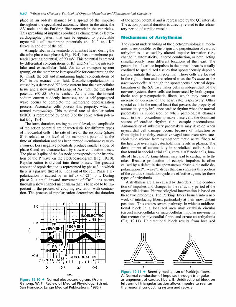
place in an orderly manner by a spread of the impulsethroughout the specialized automatic fibers in the atria, theAV node, and the Purkinje fiber network in the ventricles.This spreading of impulses produces a characteristic electro-cardiographic pattern that can be equated to predictablemyocardial cell membrane potentials and Na� and K�
fluxes in and out of the cell.A single fiber in the ventricle of an intact heart, during the
diastolic phase (see phase 4, Fig. 19.4), has a membrane po-tential (resting potential) of 90 mV. This potential is createdby differential concentrations of K� and Na� in the intracel-lular and extracellular fluid. An active transport system(pump) on the membrane is responsible for concentrating theK� inside the cell and maintaining higher concentrations ofNa� in the extracellular fluid. Diastolic depolarization iscaused by a decreased K� ionic current into the extracellulartissue and a slow inward leakage of Na� until the thresholdpotential (60–55 mV) is reached. At this time, the inwardsodium current suddenly increases, and a self-propagatedwave occurs to complete the membrane depolarizationprocess. Pacemaker cells possess this property, which istermed automaticity. This maximal rate of depolarization(MRD) is represented by phase 0 or the spike action poten-tial (Fig. 19.4).
The form, duration, resting potential level, and amplitudeof the action potential are characteristic for different typesof myocardial cells. The rate of rise of the response (phase0) is related to the level of the membrane potential at thetime of stimulation and has been termed membrane respon-siveness. Less negative potentials produce smaller slopes ofphase 0 and are characterized by slower conduction times.The phase 0 spike of the SA node corresponds to the inscrip-tion of the P wave on the electrocardiogram (Fig. 19.10).Repolarization is divided into three phases. The greatestamount of repolarization is represented by phase 3, in whichthere is a passive flux of K� ions out of the cell. Phase 1 re-polarization is caused by an influx of Cl� ions. Duringphase 2, a small inward movement of Ca2� ions occursthrough a slow channel mechanism that is believed to be im-portant in the process of coupling excitation with contrac-tion. The process of repolarization determines the duration
of the action potential and is represented by the QT interval.The action potential duration is directly related to the refrac-tory period of cardiac muscle.
Mechanisms of Arrhythmias
The current understanding of the electrophysiological mech-anisms responsible for the origin and perpetuation of cardiacarrhythmias is caused by altered impulse formation (i.e.,change in automaticity), altered conduction, or both, actingsimultaneously from different locations of the heart. Thegeneration of cardiac impulses in the normal heart is usuallyconfined to specialized tissues that spontaneously depolar-ize and initiate the action potential. These cells are locatedin the right atrium and are referred to as the SA node or thepacemaker cells. Although the spontaneous electrical depo-larization of the SA pacemaker cells is independent of thenervous system, these cells are innervated by both sympa-thetic and parasympathetic fibers, which may cause anincrease or decrease of the heart rate, respectively. Otherspecial cells in the normal heart that possess the property ofautomaticity may influence cardiac rhythm when the normalpacemaker is suppressed or when pathological changesoccur in the myocardium to make these cells the dominantsource of cardiac rhythm (i.e., ectopic pacemakers).Automaticity of subsidiary pacemakers may develop whenmyocardial cell damage occurs because of infarction orfrom digitalis toxicity, excessive vagal tone, excessive cate-cholamine release from sympathomimetic nerve fibers tothe heart, or even high catecholamine levels in plasma. Thedevelopment of automaticity in specialized cells, such asthat found in special atrial cells, certain AV node cells, bun-dle of His, and Purkinje fibers, may lead to cardiac arrhyth-mias. Because production of ectopic impulses is oftencaused by a defect in the spontaneous phase 4 diastolic de-polarization (“T wave”), drugs that can suppress this portionof the cardiac stimulation cycle are effective agents for thesetypes of arrhythmia.
Arrhythmias are also caused by disorders in the conduc-tion of impulses and changes in the refractory period of themyocardial tissue. Pharmacological intervention is based onthese two properties. The Purkinje fibers branch into a net-work of interlacing fibers, particularly at their most distantpositions. This creates several pathways in which a unidirec-tional block in a localized area may establish circular(circus) microcellular or macrocellular impulse movementsthat reenter the myocardial fibers and create an arrhythmia(Fig. 19.11). Unidirectional block results from localized
630 Wilson and Gisvold’s Textbook of Organic Medicinal and Pharmaceutical Chemistry
Figure 19.10 Normal electrocardiogram. (FromGanong, W. F.: Review of Medical Physiology, 9th ed.San Francisco, Lange Medical Publications, 1985.)
Figure 19.11 Reentry mechanism of Purkinje fibers.A. Normal conduction of impulses through triangulararrangement of cardiac fibers. B. Unidirectional block onleft arm of triangular section allows impulse to reenterthe regional conducting system and recycle.
0617-0665_17865_Ch19.qxd 12/4/09 5:51 AM Page 630

myocardial disease (infarcts) or from a change in depen-dence of the tissue to Na� fluxes that causes a longer con-duction time and allows the tissue to repolarize to propagatethe retrograde impulse.
Classes of Antiarrhythmic Drugs
Antiarrhythmic drugs can be placed into four separateclasses, based on their mechanism of action or pattern ofelectrophysiological effects produced on heart tissue. Table19.4 summarizes the four-part classification of antiarrhyth-mic drugs as first proposed by Vaughan Williams20 in 1970and expanded in 1984.21 Note that drugs within the samecategory are placed there because they demonstrate similarclinical actions. That patients do not respond to a drug inthis class, however, should not rule out use of other drugs inthe same class.22 Despite the well-intentioned use of theseagents, most antiarrhythmic drugs have the potential to ag-gravate the arrhythmia they treat (proarrhythmia).Proarrhythmia develops from an increase in the density ofsingle ectopic beats and is more likely to occur in patientswho have a dysfunction in the left ventricle or sustainedventricular tachycardia. Class I antiarrhythmic agents (dis-cussed under “Class I. Membrane-Depressant Drugs” ) areespecially proarrhythmic in myocardial infarction patients.
CLASS I. MEMBRANE-DEPRESSANT DRUGS
Class I antiarrhythmic agents are drugs that have mem-brane-stabilizing properties (i.e., they shift membranes tomore negative potentials). Drugs in this class act on the fastNa� channels and interfere with the process by which thedepolarizing charge is transferred across the membrane. It isassumed that these drugs bind to the Na� channel and blockits function, preventing Na� conductance as long as the drugis bound. The prototypical drugs in this class are quinidineand procainamide. During the 1970s, several drugs werestudied for their antiarrhythmic effects. Most of them werelocal anesthetics that affected Na� membrane channels, andthey were grouped in a single class (class I). Studies on theantiarrhythmic properties of these chemicals have shownthat there are sufficient differences to place them into sepa-rate subgroups.22
Class I antiarrhythmic drugs can be subdivided basedon the relative ease with which they dissociate from theNa� ion channel. Drugs in class IC, such as encainide,
lorcainide, and moricizine, are the most potent sodiumchannel–blocking agents of the class I antiarrhythmicdrugs. They slowly dissociate from the Na� channel, caus-ing a slowing of the conduction time of the impulsethrough the heart. Class IB drugs, which include lidocaine,tocainide, and mexiletine, dissociate rapidly from the Na�
channels and thus have the lowest potency as sodium chan-nel blockers. They produce little, if any, change in actionpotential duration. Quinidine, procainamide, and disopyra-mide are drugs that have an intermediate rate of dissocia-tion from Na� channels. These are categorized as class IAantiarrhythmic agents, and they lengthen the refractory pe-riod of cardiac tissue to cause cessation of arrhythmias.23
Studies have shown that Na� channels on the membranesof Purkinje fiber cells normally exist in at least three states:R, rested, closed near the resting potential but able to beopened by stimulation and depolarization; A, activated, al-lowing Na� ions to pass selectively through the membrane;and I, inactivated and unable to be opened (i.e., inactive).24
The affinity of the antiarrhythmic drug for the receptor onthe ion channel varies with the state of the channel orwith the membrane potential. Because of this, R, A, and I ionchannels can have different kinetics of interaction withantiarrhythmic drugs. A review of the recent literatureshows that the antiarrhythmic drugs have low affinity forR channels but relatively high affinity for the A or I chan-nels or both. Regardless of which channel state is blockedby class I antiarrhythmic drugs, the unblocking rate directlydetermines the amount of depression present at normalheart rates.
CLASS II. �-ADRENERGIC BLOCKING AGENTS
�-Adrenergic blocking drugs cause membrane-stabilizing ordepressant effects on myocardial tissue. Their antiarrhyth-mic properties, however, are considered to be principallythe result of inhibition of adrenergic stimulation to theheart. The principal electrophysiological property of these�-blocking agents is reduction of the phase 4 slope of poten-tial sinus or ectopic pacemaker cells, such that the heart ratedecreases and ectopic tachycardias are either slowed or con-verted to sinus rhythm.
CLASS III. REPOLARIZATION PROLONGATORS
Drugs in this class (e.g., amiodarone, bretylium, sotalol, ibu-tilide, dofetilide) cause several different electrophysiologi-cal changes on myocardial tissue but share one commoneffect, prolonging the action potential, which increases theeffective refractory period of the membrane action potentialwithout altering the phase of depolarization or the restingmembrane potential. Drugs in this class produce their effectsby more than one mechanism. Sotalol is a K� channelblocker and has some �-adrenergic blocking properties.2
Amiodarone and bretylium, drugs that also prolong the ac-tion potential by means that are unclear, also have Na�
channel–blocking properties.
CLASS IV. CALCIUM CHANNEL BLOCKERS
Although not all Ca2� channel blockers possess antiarrhyth-mic activity, some members of this class of antiarrhythmicdrugs (verapamil, diltiazem) block the slow inwardcurrent of Ca2� ions during phase 2 of the membrane action
Chapter 19 Cardiovascular Agents 631
TABLE 19.4 Classes of Antiarrhythmic Drugs
Class Drugs Mechanism of Action
IA
IB
IC
II
III
IV
Quinidine, procainamide, disopyramide
Lidocaine, phenytoin, tocainide, mexiletine
Encainide, flecainide, lorcainide, moricizine, propafenone
�-Adrenergic blockers (e.g., propranolol)
Amiodarone, bretylium, sotalol
Calcium channel blockers (e.g., verapamil, diltiazem)
Lengthens refractory period
Shortens duration of action potential
Slows conduction
Slows AV conduction time,suppresses automaticity
Prolongs refractoriness
Blocks slow inward Ca2�
channel
0617-0665_17865_Ch19.qxd 12/4/09 5:51 AM Page 631
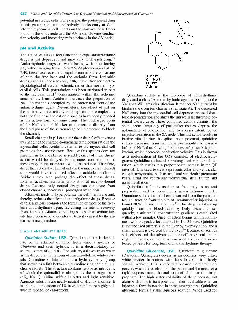
potential in cardiac cells. For example, the prototypical drugin this group, verapamil, selectively blocks entry of Ca2�
into the myocardial cell. It acts on the slow-response fibersfound in the sinus node and the AV node, slowing conduc-tion velocity and increasing refractoriness in the AV node.
pH and Activity
The action of class I local anesthetic-type antiarrhythmicdrugs is pH dependent and may vary with each drug.25
Antiarrhythmic drugs are weak bases, with most havingpKa values ranging from 7.5 to 9.5. At physiological pH of7.40, these bases exist in an equilibrium mixture consistingof both the free base and the cationic form. Ionizabledrugs, such as lidocaine (pKa 7.86), have stronger electro-physiological effects in ischemic rather than normal myo-cardial cells. This potentiation has been attributed in partto the increase in H� concentration within the ischemicareas of the heart. Acidosis increases the proportion ofNa� ion channels occupied by the protonated form of theantiarrhythmic agent. Nevertheless, the effect of pH onthe antiarrhythmic activity of drugs can be complex, asboth the free base and cationic species have been proposedas the active form of some drugs. The uncharged formof the Na� channel blocker can penetrate directly fromthe lipid phase of the surrounding cell membrane to blockthe channel.
Small changes in pH can alter these drugs’ effectivenessby changing the charged-to-uncharged molecular ratio in themyocardial cells. Acidosis external to the myocardial cellpromotes the cationic form. Because this species does notpartition in the membrane as readily, onset of these drugs’action would be delayed. Furthermore, concentration ofthese drugs in the membrane would be reduced. Therefore,drugs that act on the channel only in the inactivated (closed)state would have a reduced effect in acidotic conditions.Acidosis may also prolong the effect of these drugs.External acidosis facilitates protonation of receptor-bounddrugs. Because only neutral drugs can dissociate fromclosed channels, recovery is prolonged by acidosis.
Alkalosis tends to hyperpolarize the cell membrane and,thereby, reduces the effect of antiarrhythmic drugs. Becauseof this, alkalosis promotes the formation of more of the free-base antiarrhythmic agent, increasing the rate of recoveryfrom the block. Alkalosis-inducing salts such as sodium lac-tate have been used to counteract toxicity caused by the an-tiarrhythmic quinidine.
CLASS I ANTIARRHYTHMICS
Quinidine Sulfate, USP. Quinidine sulfate is the sul-fate of an alkaloid obtained from various species ofCinchona and their hybrids. It is a dextrorotatory di-astereoisomer of quinine. The salt crystallizes from wateras the dihydrate, in the form of fine, needlelike, white crys-tals. Quinidine sulfate contains a hydroxymethyl groupthat serves as a link between a quinoline ring and a quinu-clidine moiety. The structure contains two basic nitrogens,of which the quinuclidine nitrogen is the stronger base(pKa 10). Quinidine sulfate is bitter and light sensitive.Aqueous solutions are nearly neutral or slightly alkaline. Itis soluble to the extent of 1% in water and more highly sol-uble in alcohol or chloroform.
Quinidine sulfate is the prototype of antiarrhythmicdrugs and a class IA antiarrhythmic agent according to theVaughan Williams classification. It reduces Na� current bybinding the open ion channels (i.e., state A). The decreasedNa� entry into the myocardial cell depresses phase 4 dias-tolic depolarization and shifts the intracellular threshold po-tential toward zero. These combined actions diminish thespontaneous frequency of pacemaker tissues, depress theautomaticity of ectopic foci, and, to a lesser extent, reduceimpulse formation in the SA node. This last action results inbradycardia. During the spike action potential, quinidinesulfate decreases transmembrane permeability to passiveinflux of Na�, thus slowing the process of phase 0 depolar-ization, which decreases conduction velocity. This is shownas a prolongation of the QRS complex of electrocardio-grams. Quinidine sulfate also prolongs action potential du-ration, which results in a proportionate increase in the QTinterval. It is used to treat supraventricular and ventricularectopic arrhythmias, such as atrial and ventricular prematurebeats, atrial and ventricular tachycardia, atrial flutter, andatrial fibrillation.
Quinidine sulfate is used most frequently as an oralpreparation and is occasionally given intramuscularly.Quinidine sulfate that has been absorbed from the gastroin-testinal tract or from the site of intramuscular injection isbound 80% to serum albumin.18 The drug is taken upquickly from the bloodstream by body tissues; conse-quently, a substantial concentration gradient is establishedwithin a few minutes. Onset of action begins within 30 min-utes, with the peak effect attained in 1 to 3 hours. Quinidineis metabolized primarily in the liver by hydroxylation, and asmall amount is excreted by the liver.26 Because of seriousside effects and the advent of more effective oral antiar-rhythmic agents, quinidine is now used less, except in se-lected patients for long-term oral antiarrhythmic therapy.
Quinidine Gluconate, USP. Quinidinium gluconate(Duraquin, Quinaglute) occurs as an odorless, very bitter,white powder. In contrast with the sulfate salt, it is freelysoluble in water. This is important because there are emer-gencies when the condition of the patient and the need for arapid response make the oral route of administration inap-propriate. The high water solubility of the gluconate saltalong with a low irritant potential makes it valuable when aninjectable form is needed in these emergencies. Quinidinegluconate forms a stable aqueous solution. When used for
632 Wilson and Gisvold’s Textbook of Organic Medicinal and Pharmaceutical Chemistry
0617-0665_17865_Ch19.qxd 12/4/09 5:51 AM Page 632

injection, it usually contains 80 mg/mL, equivalent to 50 mgof quinidine or 60 mg of quinidine sulfate.
Quinidine Polygalacturonate. Quinidine polygalac-turonate (Cardioquin) is formed by reacting quinidine andpolygalacturonic acid in a hydroalcoholic medium. It con-tains the equivalent of approximately 60% quinidine. Thissalt is only slightly ionized and slightly soluble in water, butstudies have shown that although equivalent doses of quini-dine sulfate give higher peak blood levels earlier, a moreuniform and sustained blood level is achieved with the poly-galacturonate salt.
In many patients, the local irritant action of quinidinesulfate in the gastrointestinal tract causes pain, nausea, vom-iting, and especially diarrhea, often precluding oral use inadequate doses. Studies with the polygalacturonate saltyielded no evidence of gastrointestinal distress. It is avail-able as 275-mg tablets. Each tablet is the equivalent of200 mg of quinidine sulfate or 166 mg of free alkaloid.
Procainamide Hydrochloride, USP. Procainamide hy-drochloride, p-amino-N-[2-(diethylamino)ethyl]benzamidemonohydrochloride, procainamidium chloride (Pronestyl,Procan SR), has emerged as a major antiarrhythmic drug. Itwas developed in the course of research for compoundsstructurally similar to procaine, which had limited effectas an antiarrhythmic agent because of its central nervoussystem (CNS) side effects and short-lived action causedby rapid hydrolysis of its ester linkage by plasma esterases.Because of its amide structure, procainamide hydrochlo-ride is also more stable in water than is procaine. Aqueoussolutions of procainamide hydrochloride have a pH ofabout 5.5. A kinetic study of the acid-catalyzed hydrolysisof procainamide hydrochloride showed it to be unusuallystable to hydrolysis in the pH range 2 to 7, even at elevatedtemperatures.27
Procainamide hydrochloride is metabolized through theaction of N-acetyltransferase. The product of enzymaticmetabolism of procainamide hydrochloride is N-acetylpro-cainamide (NAPA), which possesses only 25% of the activ-ity of the parent compound.26 A study of the disposition ofprocainamide hydrochloride showed that 50% of the drugwas excreted unchanged in the urine, with 7% to 24% recov-ered as NAPA.28,29 Unlike quinidine, procainamide hy-drochloride is bound only minimally to plasma proteins.Between 75% and 95% of the drug is absorbed from the gas-trointestinal tract. Plasma levels appear 20 to 30 minutesafter administration and peak in about 1 hour.30
Procainamide hydrochloride appears to have all of theelectrophysiological effects of quinidine. It diminishesautomaticity, decreases conduction velocity, and increasesaction potential duration and, thereby, the refractory periodof myocardial tissue. Clinicians have favored the use of pro-cainamide hydrochloride for ventricular tachycardias and
quinidine for atrial arrhythmias, even though the two drugsare effective in either type of disorder.
Disopyramide Phosphate, USP. Disopyramide phos-phate, �-[2(diisopropylamino)ethyl]-�-phenyl-2-pyridine-acetamide phosphate (Norpace), is an oral and intravenousclass IA antiarrhythmic agent. It is quite similar to quinidineand procainamide in its electrophysiological properties, inthat it decreases phase 4 diastolic depolarization, decreasesconduction velocity, and has vagolytic properties.31 It is usedclinically in the treatment of refractory, life-threatening ven-tricular tachyarrhythmias. Oral administration of the drugproduces peak plasma levels within 2 hours. The drug isbound approximately 50% to plasma protein and has a half-life of 6.7 hours in humans. More than 50% is excretedunchanged in the urine. Therefore, patients with renal insuffi-ciency should be monitored carefully for evidence ofoverdose. Disopyramide phosphate commonly exhibits sideeffects of dry mouth, constipation, urinary retention, andother cholinergic blocking actions because of its structuralsimilarity to anticholinergic drugs.
Lidocaine Hydrochloride, USP. Lidocaine hydrochlo-ride, 2-(diethylamino)-2�,6�-acetoxylidide monohydrochlo-ride (Xylocaine), was conceived as a derivative of gramine(3-dimethylaminomethylindole) and introduced as a localanesthetic. It is now being used intravenously as a standardparenteral agent for suppression of arrhythmias associatedwith acute myocardial infarction and cardiac surgery. It isthe drug of choice for the parenteral treatment of prematureventricular contractions.
Lidocaine hydrochloride is a class IB antiarrhythmicagent with a different effect on the electrophysiologicalproperties of myocardial cells from that of procainamideand quinidine. It binds with equal affinity to the active (A)and inactive (I) Na� ion channels. It depresses diastolic de-polarization and automaticity in the Purkinje fiber networkand increases the functional refractory period relative toaction potential duration, as do procainamide and quinidine.It differs from the latter two drugs, however, in that it doesnot decrease, and may even enhance, conduction velocity
Chapter 19 Cardiovascular Agents 633
0617-0665_17865_Ch19.qxd 12/4/09 5:51 AM Page 633

and increase membrane responsiveness to stimulation.There are fewer data available on the subcellular mecha-nisms responsible for the antiarrhythmic actions of lidocainethan on the more established drug quinidine. It has been pro-posed that lidocaine has little effect on membrane cation ex-change of the atria. Sodium ion entrance into ventricularcells during excitation is not influenced by lidocaine be-cause it does not alter conduction velocity in this area.Lidocaine hydrochloride does depress Na� influx duringdiastole, as do all other antiarrhythmic drugs, to diminishautomaticity in myocardial tissue. It also alters membraneresponsiveness in Purkinje fibers, allowing increased con-duction velocity and ample membrane potential at the timeof excitation.32
Lidocaine hydrochloride administration is limited to theparenteral route and is usually given intravenously, althoughadequate plasma levels are achieved after intramuscular injec-tions. Lidocaine hydrochloride is not bound to any extent toplasma proteins and is concentrated in the tissues. It is metab-olized rapidly by the liver (Fig. 19.12). The first step isdeethylation with the formation of monoethylglycinexylidide,followed by hydrolysis of the amide.33 Metabolism is rapid,the half-life of a single injection ranging from 15 to 30 min-utes. Lidocaine hydrochloride is a popular drug because of itsrapid action and its relative freedom from toxic effects on theheart, especially in the absence of hepatic disease.Monoethylglycinexylidide, the initial metabolite of lidocaine,is an effective antiarrhythmic agent; its rapid hydrolysis bymicrosomal amidases, however, prevents its use in humans.
Precautions must be taken so that lidocaine hydrochlo-ride solutions containing epinephrine salts are not used ascardiac depressants. Such solutions are intended only forlocal anesthesia and are not used intravenously. The aque-ous solutions without epinephrine may be autoclaved sev-eral times, if necessary.
Phenytoin Sodium, USP. Phenytoin sodium, 5,5-diphenyl-2,4-imidazolidinedione, 5,5-diphenylhydantoin,diphenyl-hydantoin sodium (Dilantin), has been used fordecades in the control of grand mal types of epilepticseizure. It is structurally analogous to the barbiturates butdoes not possess their extensive sedative properties. Thecompound is available as the sodium salt. Solutions for par-enteral administration contain 40% propylene glycol and10% alcohol to dissolve the sodium salt.
Phenytoin sodium’s cardiovascular effects were uncov-ered during observation of toxic manifestations of the drugin patients being treated for seizure disorders. Phenytoinsodium was found to cause bradycardia, prolong the PRinterval, and produce T-wave abnormalities on electrocar-diograms. It is a class IB antiarrhythmic agent. Today,phenytoin sodium’s greatest clinical use as an antiarrhyth-mic drug is in the treatment of digitalis-induced arrhyth-mias.34 Its action is similar to that of lidocaine. It depressesventricular automaticity produced by digitalis, without ad-verse intraventricular conduction. Because it also reversesthe prolongation of AV conduction by digitalis, phenytoinsodium is useful in supraventricular tachycardias caused bydigitalis intoxication.
Phenytoin sodium is located in high amounts in the bodytissues, especially fat and liver, leading to large gradientsbetween the drug in tissues and the plasma concentrations.It is metabolized in the liver.
Mexiletine Hydrochloride. Mexiletine hydrochloride,1-methyl-2-(2,6-xylyloxy)ethylamine hydrochloride (Mexitil)(pKa 8.4), is a class IB antiarrhythmic agent that is effectivewhen given either intravenously or orally. It resembles lido-caine in possessing a xylyl moiety but otherwise is differentchemically. Mexiletine hydrochloride is an ether and is notsubject to the hydrolysis common to the amides lidocaineand tocainide. Its mean half-life on oral administration isapproximately 10 hours.
Although not subject to hydrolysis, mexiletine hy-drochloride is metabolized by oxidative and reductiveprocesses in the liver. Its metabolites, p-hydroxymexiletineand hydroxymethylmexiletine, are not pharmacologicallyactive as antiarrhythmic agents.35
634 Wilson and Gisvold’s Textbook of Organic Medicinal and Pharmaceutical Chemistry
Figure 19.12 Metabolism oflidocaine.
0617-0665_17865_Ch19.qxd 12/4/09 5:51 AM Page 634

Mexiletine hydrochloride, like class I antiarrhythmicagents, blocks the fast Na� channel in cardiac cells. It isespecially effective on the Purkinje fibers in the heart. Thedrug increases the threshold of excitability of myocardialcells by reducing the rate of rise and amplitude of the actionpotential and decreases automaticity.
Mexiletine hydrochloride is used for long-term oral pro-phylaxis of ventricular tachycardia. The drug is given in200- to 400-mg doses every 8 hours.
Tocainide Hydrochloride. Tocainide hydrochloride, 2-amino-2�,6�-propionoxyxylidide hydrochloride (Tonocard)(pKa 7.7), is an analog of lidocaine. It is orally active and haselectrophysiological properties like those of lidocaine.36 Totalbody clearance of tocainide hydrochloride is only166 mL/min, suggesting that hepatic clearance is not large.Because of low hepatic clearance, the hepatic extraction ratiomust be small; therefore, tocainide hydrochloride is unlikelyto be subject to a substantial first-pass effect. The drug differsfrom lidocaine, in that it lacks two ethyl groups, which pro-vides tocainide hydrochloride some protection from first-passhepatic elimination after oral ingestion. Tocainide hydrochlo-ride is hydrolyzed in a manner similar to that of lidocaine.None of its metabolites is active.
Tocainide hydrochloride is classed as a IB antiarrhyth-mic agent and used orally to prevent or treat ventricular ec-topy and tachycardia. The drug is given in 400- to 600-mgdoses every 8 hours.
Flecainide Acetate. Flecainide acetate, N-(2-piperidinyl-methyl)-2,5-bis (2,2,2-trifluoroethoxy)benzamide monoac-etate (Tambocor), is a class IC antiarrhythmic drug withlocal anesthetic activity; it is a chemical derivative of benza-mide. The drug undergoes biotransformation, forming ameta-O-dealkylated compound, whose antiarrhythmic prop-erties are half as potent as those of the parent drug, and ameta-O-dealkylated lactam of flecainide with little pharmaco-logical activity.37 Flecainide acetate is given orally to sup-press chronic ventricular ectopy and ventricular tachycardia.It has some limitations because of CNS side effects.
Moricizine. Moricizine, ethyl 10-(3-morpholino-propionyl)phenothiazine-2-carbamate (Ethmozine), is a
phenothiazine derivative used for the treatment of malig-nant ventricular arrhythmias. It is categorized as a class ICantiarrhythmic agent, blocking the Na� channel with 1:1stochiometry. The drug has higher affinity for the inacti-vated state than the activated or resting states. It appearsto bind to a site on the external side of the Na� channelmembrane.38 It has been used to suppress life-threateningventricular arrhythmias.
Propafenone. Propafenone, 2-[2�-hydroxy-3-(propyl-amino)propoxy]-3-phenylpropiophenone (Rythmol), a classIC antiarrhythmic drug, contains a chiral center and is mar-keted as the racemic mixture. Therapy with the racemicmixture of propafenone produces effects that can be attrib-uted to both (S) and (R) enantiomers. Although (R) and (S)enantiomers exert similar Na� channel–blocking effects, the(S) enantiomer also produces a �-adrenergic blockade. As aresult, the (S) enantiomer is reported to be 40-fold morepotent than the (R) enantiomer as an antiarrhythmic agent.39
The enantiomers also display stereoselective dispositioncharacteristics. The (R) enantiomer is cleared more quickly.Hepatic metabolism is polymorphic and determined geneti-cally. Ten percent of Caucasians have a reduced capacity tohydroxylate the drug to form 5-hydroxypropafenone. Thispolymorphic metabolism accounts for the interindividualvariability in the relationships between dose and concentra-tion and, thus, variability in the pharmacodynamic effects ofthe drug. The 5-hydroxy metabolites of both enantiomersare as potent as the parent compound in blocking Na� chan-nels. Propafenone also depresses the slow inward current ofCa2� ions. This drug has been used for acute termination orlong-term suppression of ventricular arrhythmias. It isbound in excess of 95% to �1-acid glycoprotein in theplasma. It is absorbed effectively, but bioavailability is esti-mated to be less than 20% because of first-pass metabolism.Less than 1% is eliminated as unchanged drug. Therapywith propafenone may produce effects that can be attributedto both (S) and (R) enantiomers. Thus, the effects may bemodulated because of an enantiomer–enantiomer interactionwhen patients are treated with the racemate.40
Chapter 19 Cardiovascular Agents 635
0617-0665_17865_Ch19.qxd 12/4/09 5:51 AM Page 635

CLASS II ANTIARRHYTHMICS
Class II antiarrhythmics are discussed under the heading,“Adrenergic System Inhibitors.”
CLASS III ANTIARRHYTHMICS
Amiodarone. Amiodarone, 2-butyl-3-benzofuranyl-4-[2-(diethylamino)ethoxy]-3,5-diiodophenyl ketone (Cordarone),was introduced as an antianginal agent. It has very pro-nounced class III action and is especially effective in main-taining sinus rhythm in patients who have been treated bydirect current shock for atrial fibrillation.41 Like class IIIantiarrhythmic drugs, amiodarone lengthens the effective re-fractory period by prolonging the action potential duration inall myocardial tissues. Amiodarone is eliminated very slowlyfrom the body, with a half-life of about 25 to 30 days after oraldoses.42 Although the drug has a broad spectrum of antiar-rhythmic activity, its main limitation is a slow onset of action.Drug action may not be initiated for several days, and thepeak effect may not be obtained for several weeks.
Amiodarone has adverse effects involving many differ-ent organ systems. It also inhibits metabolism of drugscleared by oxidative microsomal enzymes. It contains io-dine in its molecular structure and, as a result, has an effecton thyroid hormones. Hypothyroidism occurs in up to 11%of patients receiving amiodarone.43 The principal effect isthe inhibition of peripheral conversion of T4 to T3. Serumreverse T3 (rT3) is increased as a function of the dose as wellas the length of amiodarone therapy. As a result, rT3 levelshave been used as a guide for judging adequacy of amio-darone therapy and predicting toxicity.44
Bretylium Tosylate. Bretylium tosylate, (o-bro-mobenzyl)ethyl dimethylammonium p-toluenesulfonate(Bretylol), is an extremely bitter, white, crystalline pow-der. The chemical is freely soluble in water and alcohol.Bretylium tosylate is an adrenergic neuronal-blockingagent that accumulates selectively in the neurons anddisplaces norepinephrine. Because of this property,bretylium was used initially, under the trade name ofDarenthin, as an antihypertensive agent. It caused postural
decrease in arterial pressure.44 This use was discontinuedbecause of the rapid development of tolerance, erratic oralabsorption of the quaternary ammonium compound, andpersistent pain in the parotid gland on prolonged therapy.Currently, bretylium is reserved for use in ventriculararrhythmias that are resistant to other therapy. Bretyliumdoes not suppress phase 4 depolarization, a common actionof other antiarrhythmic agents. It prolongs the effectiverefractory period relative to the action potential durationbut does not affect conduction time and is categorized asa class III antiarrhythmic agent. Because bretylium doesnot have properties similar to those of the other antiar-rhythmic agents, it has been suggested that its action is aresult of its adrenergic neuronal-blocking properties; theantiarrhythmic properties of the drug, however, are notaffected by administration of reserpine. Bretylium is also alocal anesthetic, but it has not been possible to demonstratesuch an effect on atria of experimental animals, except atvery high concentrations.45 Therefore, the precise mecha-nism of the antiarrhythmic action of bretylium remains tobe resolved.
Dofetilide. Dofetilide, N-[4-(3-{[2-(4-methanesul-fonylaminophenyl)ethyl]methylamino}propoxy)phenyl]methane-sulfonamide (Tikosyn), acts by blocking thecardiac ion channel carrying the rapid component of thedelayed rectifier potassium currents (Ikr) and is used toterminate supraventricular arrhythmias, prevent the recur-rence of atrial fibrillation, and treat life-threatening ven-tricular arrhythmias. Unlike sotalol and ibutilide, whichare also methanesulfonanilides, it has no effect on adrener-gic receptors or sodium channels, respectively. Dofetilidehas high specificity for the delayed rectifier potassiumcurrents.46
Ibutilide. Ibutilide, N-{4-[4-(ethylheptylamino)-1-hydroxybutyl]phenyl}methanesulfonamide (Corvert), aclass III antiarrhythmic belonging to the methanesulfo-nanilide class of agents, is indicated for rapid conversion ofatrial fibrillation or atrial flutter to normal sinus rhythm.
636 Wilson and Gisvold’s Textbook of Organic Medicinal and Pharmaceutical Chemistry
0617-0665_17865_Ch19.qxd 12/4/09 5:51 AM Page 636

Unlike dofetilide, it is not highly specific for the delayedrectifier potassium currents (Ikr) and does have some affin-ity for sodium channels.
Sotalol. Sotalol, 4�[1-hydroxy-2-(isopropylamino)ethyl]methylsulfonanilide (Betapace), is a relatively new an-tiarrhythmic drug, characterized most often as a class IIIagent, and although it has effects that are related to the classII agents, it is not therapeutically considered a class II antiar-rhythmic. It contains a chiral center and is marketed as theracemic mixture. Because of its enantiomers, its mechanismof action spans two of the antiarrhythmic drug classes. Thel(�) enantiomer has both �-blocking (class II) and potassiumchannel-blocking (class III) activities. The d(�) enantiomerhas class III properties similar to those of the (�) isomer,but its affinity for the �-adrenergic receptor is 30 to 60 timeslower. The sotalol enantiomers produce different effects onthe heart. Class III action of d-sotalol in the sinus node isassociated with slowing of sinus heart rate, whereas �-adren-ergic blockade contributes to the decrease in heart rate ob-served with 1- or d,1-sotalol. Sotalol is not metabolized, noris it bound significantly to proteins. Elimination occurs byrenal excretion, with more than 80% of the drug eliminatedunchanged. Sotalol is characteristic of class III antiarrhythmicdrugs, in that it prolongs the duration of the action potentialand, thus, increases the effective refractory period of myocar-dial tissue. It is distinguished from the other class III drugs(amiodarone and bretylium) because of its �-adrenergicreceptor–blocking action.
Azimilide. Azimilide, E-1-[[[5-(4-chlorophenyl)-2-furanyl]methylene]amino]-3-[4-(4-methyl-1-piperazinyl)butyl]-2,4-imidazolidinedione, is a class III agent that sig-
nificantly blocks the delayed rectifier potassium current,Iks, including the Ikr component. Its ability to block multi-channels may be caused by a lack of the methane sulfon-amide group that is common to other class III agents, whichselectively block the Ikr potassium current. It is believedthat blocking both Ikr and Iks potassium currents yields con-sistent class III antiarrhythmic effects at any heart rate.47
CLASS IV ANTIARRHYTHMICS
Verapamil and Diltiazem. Both verapamil and dilti-azem block the slow inward Ca2� currents (voltage-sensitivechannel) in cardiac fibers. This slows down AV conductionand the sinus rate. These drugs are used in controlling atrialand paroxysmal tachycardias and are categorized as class IVantiarrhythmic agents according to the Vaughan Williamsclassification.21 (A more detailed description of calciumchannel blockers is given previously.)
ANTIHYPERTENSIVE AGENTS
Hypertension is a consequence of many diseases.Hemodynamically, blood pressure is a function of theamount of blood pumped by the heart and the ease withwhich the blood flows through the peripheral vasculature(i.e., resistance to blood flow by peripheral blood vessels).Diseases of components of the central and peripheral ner-vous systems, which regulate blood pressure and abnor-malities of the hormonal system, and diseases of the kid-ney and peripheral vascular network, which affect bloodvolume, can create a hypertensive state in humans.Hypertension is generally defined as mild when the dias-tolic pressure is between 90 and 104 mm Hg, moderatewhen it is 105 to 114 mm Hg, and severe when it is above115 mm Hg. It is estimated that about 15% of the adultpopulation in the United States (about 40 million) arehypertensive.
Primary (essential) hypertension is the most commonform of hypertension. Although advances have been madein the identification and control of primary hypertension, theetiology of this form of hypertension has not yet been re-solved. Renal hypertension can be created by experimen-tally causing renal artery stenosis in animals. Renal arterystenosis also may occur in pathological conditions of thekidney, such as nephritis, renal artery thrombosis, renal ar-tery infarctions, or other conditions that restrict blood flowthrough the renal artery. Hypertension also may originatefrom pathological states in the CNS, such as malignancies.Tumors in the adrenal medulla that cause release of largeamounts of catecholamines create a hypertensive conditionknown as pheochromocytoma. Excessive secretion of aldo-sterone by the adrenal cortex, often because of adenomas,also produces hypertensive disorders.
Chapter 19 Cardiovascular Agents 637
0617-0665_17865_Ch19.qxd 12/4/09 5:51 AM Page 637
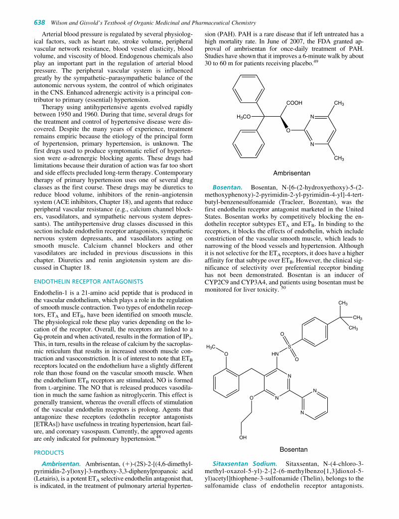
Arterial blood pressure is regulated by several physiolog-ical factors, such as heart rate, stroke volume, peripheralvascular network resistance, blood vessel elasticity, bloodvolume, and viscosity of blood. Endogenous chemicals alsoplay an important part in the regulation of arterial bloodpressure. The peripheral vascular system is influencedgreatly by the sympathetic–parasympathetic balance of theautonomic nervous system, the control of which originatesin the CNS. Enhanced adrenergic activity is a principal con-tributor to primary (essential) hypertension.
Therapy using antihypertensive agents evolved rapidlybetween 1950 and 1960. During that time, several drugs forthe treatment and control of hypertensive disease were dis-covered. Despite the many years of experience, treatmentremains empiric because the etiology of the principal formof hypertension, primary hypertension, is unknown. Thefirst drugs used to produce symptomatic relief of hyperten-sion were �-adrenergic blocking agents. These drugs hadlimitations because their duration of action was far too shortand side effects precluded long-term therapy. Contemporarytherapy of primary hypertension uses one of several drugclasses as the first course. These drugs may be diuretics toreduce blood volume, inhibitors of the renin–angiotensinsystem (ACE inhibitors, Chapter 18), and agents that reduceperipheral vascular resistance (e.g., calcium channel block-ers, vasodilators, and sympathetic nervous system depres-sants). The antihypertensive drug classes discussed in thissection include endothelin receptor antagonists, sympatheticnervous system depressants, and vasodilators acting onsmooth muscle. Calcium channel blockers and othervasodilators are included in previous discussions in thischapter. Diuretics and renin angiotensin system are dis-cussed in Chapter 18.
ENDOTHELIN RECEPTOR ANTAGONISTS
Endothelin-1 is a 21-amino acid peptide that is produced inthe vascular endothelium, which plays a role in the regulationof smooth muscle contraction. Two types of endothelin recep-tors, ETA and ETB, have been identified on smooth muscle.The physiological role these play varies depending on the lo-cation of the receptor. Overall, the receptors are linked to aGq-protein and when activated, results in the formation of IP3.This, in turn, results in the release of calcium by the sacroplas-mic reticulum that results in increased smooth muscle con-traction and vasoconstriction. It is of interest to note that ETB
receptors located on the endothelium have a slightly differentrole than those found on the vascular smooth muscle. Whenthe endothelium ETB receptors are stimulated, NO is formedfrom L-arginine. The NO that is released produces vasodila-tion in much the same fashion as nitroglycerin. This effect isgenerally transient, whereas the overall effects of stimulationof the vascular endothelin receptors is prolong. Agents thatantagonize these receptors (edothelin receptor antagonists[ETRAs]) have usefulness in treating hypertension, heart fail-ure, and coronary vasospasm. Currently, the approved agentsare only indicated for pulmonary hypertension.48
PRODUCTS
Ambrisentan. Ambrisentan, (�)-(2S)-2-[(4,6-dimethyl-pyrimidin-2-yl)oxy]-3-methoxy-3,3-diphenylpropanoic acid(Letairis), is a potent ETA selective endothelin antagonist that,is indicated, in the treatment of pulmonary arterial hyperten-
sion (PAH). PAH is a rare disease that if left untreated has ahigh mortality rate. In June of 2007, the FDA granted ap-proval of ambrisentan for once-daily treatment of PAH.Studies have shown that it improves a 6-minute walk by about30 to 60 m for patients receiving placebo.49
Bosentan. Bosentan, N-[6-(2-hydroxyethoxy)-5-(2-methoxyphenoxy)-2-pyrimidin-2-yl-pyrimidin-4-yl]-4-tert-butyl-benzenesulfonamide (Tracleer, Bozentan), was thefirst endothelin receptor antagonist marketed in the UnitedStates. Bosentan works by competitively blocking the en-dothelin receptor subtypes ETA and ETB. In binding to thereceptors, it blocks the effects of endothelin, which includeconstriction of the vascular smooth muscle, which leads tonarrowing of the blood vessels and hypertension. Althoughit is not selective for the ETA receptors, it does have a higheraffinity for that subtype over ETB. However, the clinical sig-nificance of selectivity over preferential receptor bindinghas not been demonstrated. Bosentan is an inducer ofCYP2C9 and CYP3A4, and patients using bosentan must bemonitored for liver toxicity. 50
Sitaxsentan Sodium. Sitaxsentan, N-(4-chloro-3-methyl-oxazol-5-yl)-2-[2-(6-methylbenzo[1,3]dioxol-5-yl)acetyl]thiophene-3-sulfonamide (Thelin), belongs to thesulfonamide class of endothelin receptor antagonists.
N
N
N
N
O
OH
O
H3C
HN
S
O
OCH3
CH3
CH3
Bosentan
H3CO
COOH
O
N
N
CH3
CH3
Ambrisentan
638 Wilson and Gisvold’s Textbook of Organic Medicinal and Pharmaceutical Chemistry
0617-0665_17865_Ch19.qxd 12/4/09 5:51 AM Page 638
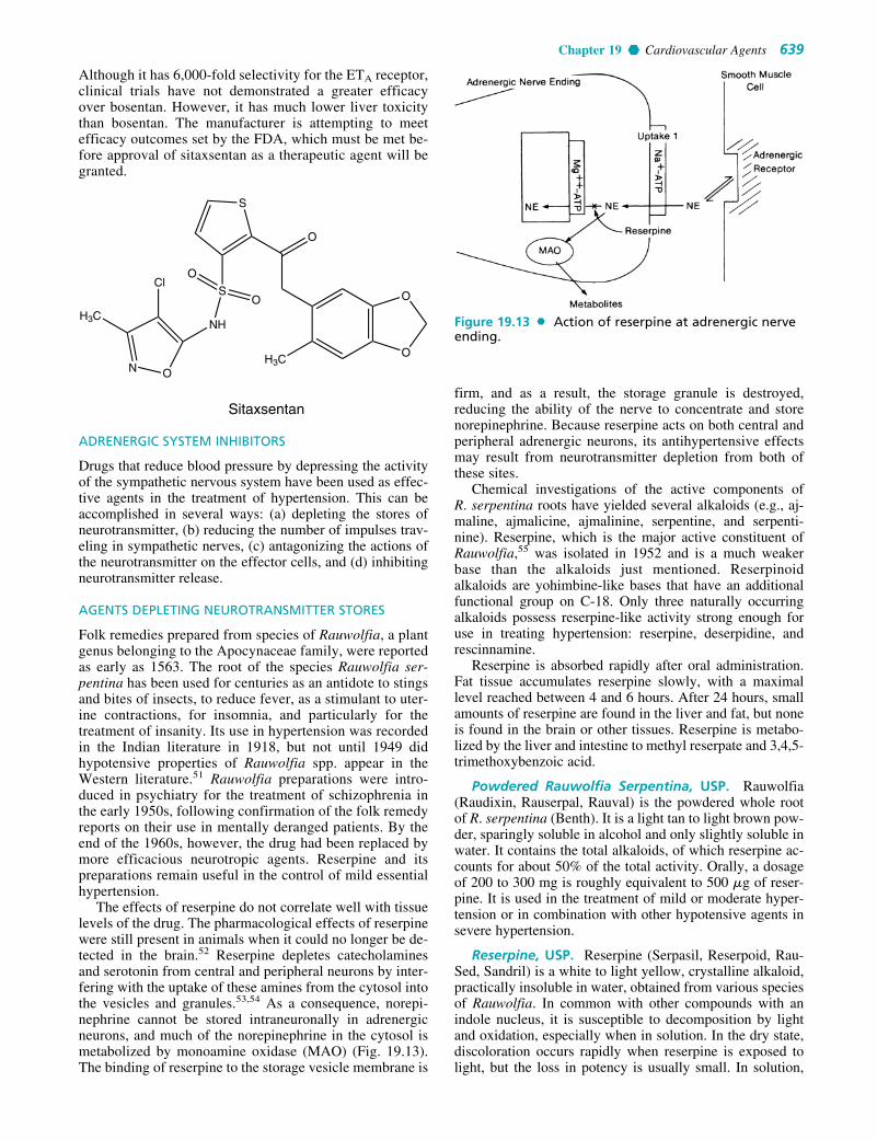
Although it has 6,000-fold selectivity for the ETA receptor,clinical trials have not demonstrated a greater efficacyover bosentan. However, it has much lower liver toxicitythan bosentan. The manufacturer is attempting to meetefficacy outcomes set by the FDA, which must be met be-fore approval of sitaxsentan as a therapeutic agent will begranted.
ADRENERGIC SYSTEM INHIBITORS
Drugs that reduce blood pressure by depressing the activityof the sympathetic nervous system have been used as effec-tive agents in the treatment of hypertension. This can beaccomplished in several ways: (a) depleting the stores ofneurotransmitter, (b) reducing the number of impulses trav-eling in sympathetic nerves, (c) antagonizing the actions ofthe neurotransmitter on the effector cells, and (d) inhibitingneurotransmitter release.
AGENTS DEPLETING NEUROTRANSMITTER STORES
Folk remedies prepared from species of Rauwolfia, a plantgenus belonging to the Apocynaceae family, were reportedas early as 1563. The root of the species Rauwolfia ser-pentina has been used for centuries as an antidote to stingsand bites of insects, to reduce fever, as a stimulant to uter-ine contractions, for insomnia, and particularly for thetreatment of insanity. Its use in hypertension was recordedin the Indian literature in 1918, but not until 1949 didhypotensive properties of Rauwolfia spp. appear in theWestern literature.51 Rauwolfia preparations were intro-duced in psychiatry for the treatment of schizophrenia inthe early 1950s, following confirmation of the folk remedyreports on their use in mentally deranged patients. By theend of the 1960s, however, the drug had been replaced bymore efficacious neurotropic agents. Reserpine and itspreparations remain useful in the control of mild essentialhypertension.
The effects of reserpine do not correlate well with tissuelevels of the drug. The pharmacological effects of reserpinewere still present in animals when it could no longer be de-tected in the brain.52 Reserpine depletes catecholaminesand serotonin from central and peripheral neurons by inter-fering with the uptake of these amines from the cytosol intothe vesicles and granules.53,54 As a consequence, norepi-nephrine cannot be stored intraneuronally in adrenergicneurons, and much of the norepinephrine in the cytosol ismetabolized by monoamine oxidase (MAO) (Fig. 19.13).The binding of reserpine to the storage vesicle membrane is
O
O
H3C
O
S
S
NH
N O
H3C
Cl
O
O
Sitaxsentanfirm, and as a result, the storage granule is destroyed,reducing the ability of the nerve to concentrate and storenorepinephrine. Because reserpine acts on both central andperipheral adrenergic neurons, its antihypertensive effectsmay result from neurotransmitter depletion from both ofthese sites.
Chemical investigations of the active components ofR. serpentina roots have yielded several alkaloids (e.g., aj-maline, ajmalicine, ajmalinine, serpentine, and serpenti-nine). Reserpine, which is the major active constituent ofRauwolfia,55 was isolated in 1952 and is a much weakerbase than the alkaloids just mentioned. Reserpinoidalkaloids are yohimbine-like bases that have an additionalfunctional group on C-18. Only three naturally occurringalkaloids possess reserpine-like activity strong enough foruse in treating hypertension: reserpine, deserpidine, andrescinnamine.
Reserpine is absorbed rapidly after oral administration.Fat tissue accumulates reserpine slowly, with a maximallevel reached between 4 and 6 hours. After 24 hours, smallamounts of reserpine are found in the liver and fat, but noneis found in the brain or other tissues. Reserpine is metabo-lized by the liver and intestine to methyl reserpate and 3,4,5-trimethoxybenzoic acid.
Powdered Rauwolfia Serpentina, USP. Rauwolfia(Raudixin, Rauserpal, Rauval) is the powdered whole rootof R. serpentina (Benth). It is a light tan to light brown pow-der, sparingly soluble in alcohol and only slightly soluble inwater. It contains the total alkaloids, of which reserpine ac-counts for about 50% of the total activity. Orally, a dosageof 200 to 300 mg is roughly equivalent to 500 �g of reser-pine. It is used in the treatment of mild or moderate hyper-tension or in combination with other hypotensive agents insevere hypertension.
Reserpine, USP. Reserpine (Serpasil, Reserpoid, Rau-Sed, Sandril) is a white to light yellow, crystalline alkaloid,practically insoluble in water, obtained from various speciesof Rauwolfia. In common with other compounds with anindole nucleus, it is susceptible to decomposition by lightand oxidation, especially when in solution. In the dry state,discoloration occurs rapidly when reserpine is exposed tolight, but the loss in potency is usually small. In solution,
Chapter 19 Cardiovascular Agents 639
Figure 19.13 Action of reserpine at adrenergic nerveending.
0617-0665_17865_Ch19.qxd 12/4/09 5:51 AM Page 639

reserpine may break down with no appreciable color changewhen exposed to light, especially in clear glass containers;thus, color change cannot be used as an index of the amountof decomposition.
Reserpine is effective orally and parenterally for thetreatment of hypertension. After a single intravenous dose,the onset of antihypertensive action usually begins in about1 hour. After intramuscular injection, the maximumeffect occurs within approximately 4 hours and lasts about10 hours. When it is given orally, the maximum effectoccurs within about 2 weeks and may persist up to 4 weeksafter the final dose. When used in conjunction with otherhypotensive drugs in the treatment of severe hypertension,the daily dose varies from 100 to 250 �g.
Guanethidine and Related Compounds. Guanethidinehas been classified traditionally as an adrenergic blockingagent because it can prevent the release of norepinephrinefrom postganglionic neurons in response to adrenergic stimu-lation. Guanethidine and other compounds discussed in thissection have other actions on catecholamine metabolism andcan cause significant depletion of these amines in adrenergicneurons. They do not interfere with release of epinephrinefrom the adrenal medulla.
Guanethidine Monosulfate, USP. Guanethidinemonosulfate, [2-(hexahydro-1 (2H)-azocinyl)ethyl]guani-dine sulfate (Ismelin sulfate), is a white, crystalline mate-rial that is very soluble in water. It was one of a series ofguanidine compounds prepared in the search for potent an-titrypanosomal agents. There is an absence of CNS effects,such as depression, because the drug is highly polar anddoes not easily cross the blood-brain barrier. Guanethidinemonosulfate produces a gradual, prolonged fall in bloodpressure. Usually, 2 to 7 days of therapy are required be-fore the peak effect is reached, and usually, this peak effectis maintained for 3 or 4 days. Then, if the drug is discon-tinued, the blood pressure returns to pretreatment levelsover a period of 1 to 3 weeks. Because of this slow onsetand prolonged duration of action, only a single daily doseis needed.
Guanethidine monosulfate is metabolized by microsomalenzymes to 2-(6-carboxyhexylamino)ethylguanidine andguanethidine N-oxide (Fig. 19.14). Both metabolites havevery weak antihypertensive properties. Guanethidine mono-sulfate is taken up by the amine pump located on theneuronal membrane and retained in the nerve, displacingnorepinephrine from its storage sites in the neuronal gran-ules. The displaced norepinephrine is metabolized to ho-movanillic acid by mitochondrial MAO, depleting the nerveending of the neurotransmitter. The usefulness of guanethi-dine monosulfate also resides in the fact that once it is takenup by the nerve, it produces a sympathetic blockade by in-hibiting release of nonepinephrine that would occur on neu-ronal membrane response to stimulation29 by the nerveaction potential. Guanethidine monosulfate stored in thegranules is released by the nerve action potential but hasvery low intrinsic activity for the adrenergic receptors on thepostjunctional membrane. Moderate doses for a prolongedperiod or large doses may produce undesirable side effectsby causing neuromuscular blockade and adrenergic nerveconduction blockade.
Guanadrel Sulfate. Guanadrel sulfate, (1,4-dioxas-piro[4.5]dec-2-ylmethyl)guanadine sulfate (Hylorel), issimilar to guanethidine monosulfate in the manner in whichit reduces elevated blood pressure. It acts as a post-ganglionic adrenergic blocking agent by displacing norepi-nephrine in adrenergic neuron storage granules, therebypreventing release of the endogenous neurotransmitter onnerve stimulation. Guanadrel sulfate has a much shorterhalf-life (10 hours) than guanethidine monosulfate, whose
640 Wilson and Gisvold’s Textbook of Organic Medicinal and Pharmaceutical Chemistry
Figure 19.14 Metabolism of guanethidinemonosulfate.
0617-0665_17865_Ch19.qxd 12/4/09 5:51 AM Page 640

half-life is measured in days. In the stepped-care approachto hypertension, guanadrel sulfate is usually a step 2 agent.
SELECTIVE �-ADRENERGIC ANTAGONISTS
The principal clinical use of �-adrenergic antagonists is in thetreatment of catecholamine-dependent hypertension. Classicdrugs such as phentolamine and phenoxybenzamine are non-specific-blocking agents of both �1- and �2-receptors on thepresynaptic membrane of the adrenergic neuron. Specific an-tagonists of �1-receptors are effective antihypertensive agentsby blocking the vasocontricting effect on smooth muscle andnot interfering with the activation of �2-receptors on theadrenergic neuron, which when activated inhibit further re-lease of norepinephrine.
Prazosin Hydrochloride. The antihypertensive effectsof prazosin hydrochloride, 1-(4-amino-6,7-dimethoxy-2-quinazolinyl)-4-(2-furoyl)piperazine monohydrochloride(Minipress), are caused by peripheral vasodilation as a resultof its blockade of �1-adrenergic receptors. In ligand-bindingstudies, prazosin hydrochloride has 5,000-fold greater affin-ity for �1-receptors than for some �2-adrenergic receptors.56
Prazosin hydrochloride is readily absorbed, and plasmaconcentrations reach a peak about 3 hours after administra-tion. Plasma half-life is between 2 and 3 hours. Prazosin hy-drochloride is highly bound to plasma protein; it does notcause adverse reactions, however, with drugs that might bedisplaced from their protein-binding sites (e.g., cardiac gly-cosides). It may cause severe orthostatic hypertension be-cause of its �-adrenergic blocking action, which preventsthe reflex venous constriction that is activated when an indi-vidual sits up from a prone position.
Terazosin Hydrochloride. Terazosin hydrochloride, 1-(4-amino-6,7-dimethoxy-2-quinazolinyl)-4-(tetrahydro-2-furoyl)piperazine monohydrochloride (Hytrin), is a struc-tural congener of prazosin hydrochloride. It possessessimilar selective properties of specifically inhibiting �1-adrenergic receptors. The drug is slightly less potent thanprazosin hydrochloride. Terazosin hydrochloride has a half-life of approximately 12 hours, which is much longer thanthat of prazosin. This lends itself to a once-daily dose tocontrol hypertension in many patients.
Doxazosin. Doxazosin, 1-(4-amino-6,7-dimethoxy-2-quinazolinyl)-4-(1,4-benzodioxan-2-ylcarbonyl)piperazine(Cardura), is a quinazoline compound that selectively in-hibits the �1-subtype of �-adrenergic receptors. This agentis very useful in the management of hypertension associatedwith pheochromocytoma.
Chapter 19 Cardiovascular Agents 641
CENTRALLY ACTING ADRENERGIC DRUGS
The use of agents that directly affect the peripheral compo-nent of the sympathetic nervous system represents animportant approach to the treatment of hypertension. A sec-ond approach to modifying sympathetic influence on thecardiovascular system is through inhibition or reduction ofCNS control of blood pressure. Several widely used med-ications act by stimulating �2-receptors, which in the CNSreduces sympathetic outflow to the cardiovascular systemand produces a hypotensive effect.
Methyldopate Hydrochloride, USP. Methyldopatehydrochloride, L-3-(3,4-dihydroxyphenyl)-2-methylala-nine ethyl ester hydrochloride (Aldomet ester hydrochlo-ride), �-methyldopa, lowers blood pressure by inhibitingthe outflow of sympathetic vasoconstrictor impulses fromthe brain. Early studies had suggested that the hypotensiveaction of �-methyldopa was a result of the peripheral prop-erties of the drug as a decarboxylase inhibitor or a falsetransmitter.
The current hypothesis concerning the hypotensive ac-tivity of methyldopa involves the CNS as the site of ac-tion.57 Methyldopa, on conversion to �-methylnorepineph-
0617-0665_17865_Ch19.qxd 12/4/09 5:51 AM Page 641

rine, acts on �2-adrenergic receptors to inhibit the re-lease of norepinephrine, resulting in decreased sympa-thetic outflow from the CNS and activation of parasympa-thetic outflow.
Methyldopa is used as a step 2 agent and is recommendedfor patients with high blood pressure who are not responsiveto diuretic therapy alone. Methyldopa, suitable for oral use,is a zwitterion and is not soluble enough for parenteral use.The problem was solved by making the ester, leaving theamine free to form the water-soluble hydrochloride salt. It issupplied as a stable, buffered solution, protected with an-tioxidants and chelating agents.
Clonidine Hydrochloride. Clonidine hydrochloride, 2-[(2,6-dichlorophenyl)imino]imidazolidine monohydrochlo-ride (Catapres), was the first antihypertensive known to acton the CNS. It was synthesized in 1962 as a derivativeof the known �-sympathomimetic drugs naphazoline andtolazoline, potential nasal vasoconstrictors, but instead itproved to be effective in the treatment of mild-to-severe hy-pertension.
Clonidine hydrochloride acts by both peripheral andcentral mechanisms in the body to affect blood pressure. Itstimulates the peripheral �-adrenergic receptors to pro-duce vasoconstriction, resulting in a brief period of hyper-tension. Clonidine hydrochloride acts centrally to inhibitthe sympathetic tone and cause hypotension that is ofmuch longer duration than the initial hypertensive effect.Administration of clonidine hydrochloride thus produces abiphasic change in blood pressure, beginning with a briefhypertensive effect and followed by a hypotensive effectthat persists for about 4 hours. This biphasic response isaltered by dose only. Larger doses produce a greater hy-pertensive effect and delay the onset of the hypotensiveproperties of the drug. Clonidine hydrochloride acts on �2-adrenoreceptors located in the hindbrain to produce itshypotensive action. Clonidine hydrochloride also acts cen-trally to cause bradycardia and to reduce plasma levels ofrenin. Sensitization of baroreceptor pathways in the CNSappears to be responsible for the bradycardia transmittedby way of the vagus nerve. The central mechanism that re-sults in decreased plasma renin is not known, however.The hypotensive properties of clonidine in animals can beblocked by applying �-adrenergic blocking agents directlyto the brain.58
Clonidine hydrochloride has advantages over antihyper-tensive drugs such as guanethidine monosulfate and pra-zosin hydrochloride, in that it seldom produces orthostatichypotensive side effects. It does, however, have some seda-tive properties that are undesirable; it also may cause consti-pation and dryness of the mouth.
Clonidine hydrochloride is distributed throughout thebody, with the highest concentrations found in the organsof elimination: kidney, gut, and liver. Brain concentrationsare low but higher than plasma concentrations. The highconcentration in the gut is caused by an enterohepaticcycle in which clonidine hydrochloride is secreted into thebile in rather high concentrations. The half-life in humansis about 20 hours. Clonidine hydrochloride is metabolizedby the body to form two major metabolites, p-hydroxy-clonidine and its glucuronide. p-Hydroxyclonidine doesnot cross the blood-brain barrier and has no hypotensiveeffect in humans.
Guanabenz Acetate. Guanabenz acetate, [(2,6-dichloro-benzylidene)amino]guanidine monoacetate (Wytensin), is acentral �2-adrenergic agonist that reduces the release of nor-epinephrine from the neuron when stimulated. The effect ofthe drug results in decreased sympathetic tone in the heart,kidneys, and peripheral blood vessels. The drug does not pro-duce orthostatic hypotension.
Guanfacine Hydrochloride. Guanfacine hydrochloride,N-(aminoiminomethyl)-2,6-dichlorobenzeneacetamide(Tenex), is structurally related to clonidine hydrochloride andguanabenz acetate and shares many of their pharmacologicalproperties. The drug has a longer duration of action than ei-ther clonidine hydrochloride or guanabenz acetate. It lasts upto 24 hours. It also requires much longer (8–12 hours) for apeak effect to occur after the drug is administered.
VASODILATORY DRUGS ACTING ON SMOOTH MUSCLE
Reduction of arterial smooth muscle tone may occur bymany mechanisms, such as reduction in sympathetic tone,stimulation of �-adrenergic receptors, or even direct actionon the vasculature without interference from the autonomicinnervation. Drugs acting on the arteriolar smooth musclealso increase sympathetic reflex activity, causing an in-crease in heart rate and cardiac output and stimulating reninrelease, which increases sodium retention and plasma vol-ume. As a result, it is common to coadminister salureticsand �-adrenergic blocking drugs with these agents.
Antihypertensive agents that produce vasodilation ofsmooth muscle can be divided into two categories: direct-acting and indirect-acting vasodilators. Indirect-acting va-sodilators may be distinguished from direct-acting vasodila-tors, in that they produce their effect by interfering with the
642 Wilson and Gisvold’s Textbook of Organic Medicinal and Pharmaceutical Chemistry
0617-0665_17865_Ch19.qxd 12/4/09 5:51 AM Page 642

vasoconstrictor stimuli, and their primary site of action is notnecessarily the vascular smooth muscle itself. Indirect-actingvasodilators include sympatholytic drugs, such as reserpine;�-adrenergic antagonists, such as prazosin hydrochloride;ACE inhibitors; and angiotensin II receptor antagonists, suchas saralysin. Direct-acting vasodilators include hydralazinehydrochloride, sodium nitroprusside, potassium channelopeners, and calcium channel-blocking agents.58
Hydralazine Hydrochloride, USP. Hydralazine hy-drochloride, 1-hydrazinophthalazine monohydrochloride(Apresoline hydrochloride), originated from the work of achemist59 attempting to produce some unusual chemicalcompounds and from the observation60 that this compoundhad antihypertensive properties. It occurs as yellow crystalsand is soluble in water to the extent of about 3%. A 2%aqueous solution has a pH of 3.5 to 4.5.
Hydralazine hydrochloride is useful in the treatment ofmoderate-to-severe hypertension. It is often used in con-junction with less potent antihypertensive agents, becauseside effects occur frequently when it is used alone in ade-quate doses. In combinations, it can be used in lower andsafer doses. Its action appears to be centered on the smoothmuscle of the vascular walls, with a decrease in peripheralresistance to blood flow. This results in increased bloodflow through the peripheral blood vessels. It also has theunique property of increasing renal blood flow, an importantconsideration in patients with renal insufficiency.
Hydralazine hydrochloride acts on vascular smooth mus-cle to cause relaxation. Its mechanism of action is unclear. Itinterferes with Ca2� entry and Ca2� release from intracellu-lar stores and reportedly causes activation of guanylate cy-clase, resulting in increased levels of cGMP. All of thesebiochemical events can cause vasodilation.
Absorption of hydralazine hydrochloride taken orally israpid and nearly complete. The maximal hypotensive effect isdemonstrable within 1 hour. The drug is excreted rapidly bythe kidneys, and within 24 hours, 75% of the total amount ad-ministered appears in the urine as metabolites or unchangeddrug. Hydralazine hydrochloride undergoes benzylic oxida-tion, glucuronide formation, and N-acetylation by the micro-somal enzymes in the tissues (Fig. 19.15). Acetylation ap-pears to be a major determinant of the rate of hepatic removalof the drug from the blood and, therefore, of systemic avail-ability.61 Rapid acetylation results in a highly hepatic extrac-tion ratio from blood and greater first-pass elimination.
Hydralazine hydrochloride is more effective clinicallywhen coadministered with drugs that antagonize adrenergictransmission (e.g., �-adrenergic antagonists, reserpine,guanethidine monosulfate, methyldopa, and clonidine hy-drochloride). When given with diuretics, it is useful in thetreatment of CHF.
Hydralazine hydrochloride is marketed as a combinationtherapy with isosorbide under the trade name BiDil specifi-cally for African Americans with CHF.
Sodium Nitroprusside, USP. Sodium nitroprusside,sodium nitroferricyanide, disodium pentacyanonitrosylfer-rate(2) Na2[Fe(CN)5NO] (Nipride, Nitropress), is one of themost potent blood pressure–lowering drugs. Its use is lim-ited to hypertensive emergencies because of its short dura-tion of action. The effectiveness of sodium nitroprusside asan antihypertensive has been known since 1928, but notuntil 1955 was its efficacy as a drug established.62 The drugdiffers from other vasodilators, in that vasodilation occurs inboth venous and arterial vascular beds. Sodium nitroprus-side is a reddish brown water-soluble powder that is decom-posed by light when in solution. The hypotensive effect ofthe chemical is a result of the formation of NO in situ (dis-cussed under the heading, “Nitrovasodilators”), elevatingcellular levels of cGMP. Sodium nitroprusside is metabo-lized by the liver, yielding thiocyanate. Because thiocyanateis excreted by the kidneys, patients with impaired renalfunction may suffer thiocyanate toxicity.
Chapter 19 Cardiovascular Agents 643
Figure 19.15 Metabolism of hydralazine hydrochloride.
0617-0665_17865_Ch19.qxd 12/4/09 5:51 AM Page 643

PHOSPHODIESTERASE TYPE 5 INHIBITORS
As discussed throughout this chapter, the cyclic nucleotidephosphodiesterases constitute a group of enzymes that cat-alyze the hydrolysis of various cyclic nucleotides includingcAMP and cGMP. The contractility of all muscle types is reg-ulated primarily by reversible phosphorylation of the proteinmyosin. The phosphorylation of myosin allows it to interactwith actin, which results in muscle contraction. Through dif-ferent mechanisms, cAMP and cGMP interfere with the abil-ity of myosin to be phosporylated and/or interact with actin.This, in turn, results in muscle relaxation. Agents that inter-fere with the degradation of the cyclic nucleotide have use-fulness in treating hypertension, erectile dysfunction, etc.Phosphodiesterase type 5 (PDE5) is responsible for the cat-alytic hydrolysis of cGMP in the smooth muscle of the arter-ies in the penis and lungs. Inhibitors of this particular phos-phodiesterase have been shown to relax arteries in the penis,thus allowing the corpus cavernosum to fill with blood andaiding in an erection.63
PRODUCTS
Silenafil Citrate. Sildenafil, 1-[4-ethoxy-3-(6,7-dihydro-1-methyl-7-oxo-3-propyl-1H-pyrazolo[4,3-d]pyrimidin-5-yl)-phenylsulfonyl]-4-methylpiperazine citrate (Viagra), isa potent inhibitor of PDE5. It was initially studied for use inhypertension and angina pectoris because of its ability to in-crease intracellular levels of cyclic nucleotides. However,during phase I clinical trials, it was noted that the drug had lit-tle effect on angina and was unpredictable in managing hy-pertension. However, patients noted that there were improvedeffects in their sexual dysfunction. Therefore, the manufac-turer decided to develop the investigational compound fortreating erectile dysfunction, rather than for angina or hyper-tension. In March 1998, the FDA approved the drug for usein erectile dysfunction, becoming the first oral agent approvedto treat erectile dysfunction in the United States. Within ayear, it soon had annual sales exceeding $1 billion per year.The chemical structure of sildenafil is similar to that of cGMPand acts as a competitive binding agent of PDE5.
Sildenafil is also effective in the rare disease, PAH (pre-sented earlier in this chapter under “Endothelin ReceptorAntagonist”). By increasing cycli nucleotide levels, it relaxesthe arterial wall, leading to decreased pulmonary arterial re-sistance and pressure. As a result of the selective distributionof PDE-5 within the arterial smooth muscle of the lungs andpenis, sildenafil acts selectively in these areas. In June 2005,the FDA approved sildenafil (Revatio) for the management ofPAH. To avoid confusion with its use in either PAH or erec-tile dysfunction, the product used for erectile dysfunction is ablue oval tablet, whereas the product used for management ofPAH is a round white tablet.63
NNH3C S
O
O
O
N
HN
N
N
OCH3
Sildenafil
Vardenafil Hydrochloride. Vardenafil, 4-[2-ethoxy-5-(4-ethylpiperazin-1-yl)sulfonyl-phenyl]-9-methyl-7-propyl-3,5,6,8-tetrazabicyclo[4.3.0]nona-3,7,9-trien-2-one(Levitra), was the second PDE5 introduced in the U.S. mar-ket. The metabolism of vardenafil is primarily by CYP3A4.As such, concomitant use of CYP3A4 inhibitors such as ri-tonavir, indinavir, ketoconazole, as well as moderate CYP3Ainhibitors such as erythromycin typically results in signifi-cant increases of plasma levels of vardenafil.
644 Wilson and Gisvold’s Textbook of Organic Medicinal and Pharmaceutical Chemistry
NNCH2 S
O
O
O
N
N
HN
N
OCH3
Vardenafil
H3C
Tadalafil. Tadalafil, (6R-trans)-6-(1,3-benzodioxol-5-yl)-2,3,6,7,12,12a-hexahydro-2-methyl-pyrazino[1�, 2�:1,6]pyrido[3,4-b]indole-1,4-dione (Cialis), is a potent PDE5 in-hibitor. It received FDA approval for the treatment of erec-tile dysfunction in December 2003. Because of its half-life of17.5 hours, it is marketed as a 36-hour treatment. Tadalafil ispredominantly metabolized by hepatic enzymes, includingCYP3A4. The concomitant use of CYP3A4 inhibitors suchas ritonavir, indinavir, ketoconazole, as well as moderateCYP3A inhibitors such as erythromycin have been shown toresult in significant increases in tadalafil plasma levels.Much like sildenafil, tadalafil is under clinical investigationfor managing PAH.64
POTASSIUM CHANNEL AGONISTS
The two agents that can be classified in this category are di-azoxide and minoxidil. These drugs are also called potassiumchannel openers. These agents activate ATP-sensitive potas-sium channels, which leads to a decrease of intracellularCa2� and reduces the excitability of smooth muscle. The pri-mary action of these drugs is to open potassium channels in
NNH
N
O
O
O
O
Tadalafil
0617-0665_17865_Ch19.qxd 12/4/09 5:51 AM Page 644

the plasma membrane of vascular smooth muscle. An effluxof potassium from the cell follows, resulting in hyperpolar-ization of the membrane, which produces an inhibitory influ-ence on membrane excitation and subsequent vasodilation.
Diazoxide, USP. Diazoxide is used as the sodium salt of7-chloro-3-methyl-2H-1,2,4-benzothiadiazine 1,1-dioxide(Hyperstat IV). Diazoxide lowers peripheral vascular resis-tance, increases cardiac output, and does not compromiserenal blood flow.
This is a des-sulfamoyl analog of the benzothiazine diuret-ics and has a close structural similarity to chlorothiazide. Itwas developed intentionally to increase the antihypertensiveaction of the thiazides and to minimize the diuretic effect.
It is used by intravenous injection as a rapidly acting an-tihypertensive agent for emergency reduction of blood pres-sure in hospitalized patients with accelerated or malignanthypertension. More than 90% is bound to serum protein, andcaution is needed when it is used in conjunction with otherprotein-bound drugs that may be displaced by diazoxide.The injection is given rapidly by the intravenous route toensure maximal effect. The initial dose is usually 1 mg/kg ofbody weight, with a second dose given if the first injectiondoes not lower blood pressure satisfactorily within 30 min-utes. Further doses may be given at 4- to 24-hour intervalsif needed. Oral antihypertensive therapy is begun as soon aspossible.
The injection has a pH of about 11.5, which is necessaryto convert the drug to its soluble sodium salt. There is nosignificant chemical decomposition after storage at roomtemperature for 2 years. When the solution is exposed tolight, it darkens.
Minoxidil, USP. Minoxidil, 2,4-diamino-6-piperidinopy-rimidine-3-oxide (Loniten), was developed as a result ofisosteric replacement of a triaminotriazine moiety by tri-aminopyrimidine. The triaminotriazines were initially ob-served to be potent vasodilators in cats and dogs, followingtheir formation of N-oxides in these animals. The triazineswere inactive in humans because of their inability to form N-oxide metabolites; this led to the discovery of minoxidil.Minoxidil is the only direct-acting vasodilator that requiresmetabolic activation to produce its antihypertensive effect(Fig. 19.16). It is converted to minoxidil sulfate in the liver bya sulfotransferase enzyme.65
The antihypertensive properties of minoxidil are similarto those of hydralazine hydrochloride, in that minoxidil candecrease arteriolar vascular resistance. Minoxidil exerts itsvasodilatory action by a direct effect on arteriolar smoothmuscle and appears to have no effect on the CNS or on theadrenergic nervous system in animals.66 The serum half-lifeis 4.5 hours, and the antihypertensive effect may last up to24 hours.
Minoxidil is used for severe hypertension that is difficultto control with other antihypertensive agents. The drug has
some of the characteristic side effects of direct vasodilatorydrugs. It causes sodium and water retention and may requirecoadministration of a diuretic. Minoxidil also causes reflextachycardia, which can be controlled by use of a �-adrenergicblocking agent.
Minoxidil topical solution is used to treat alopecia andro-genitica (male-pattern baldness). Although the mechanismis not clearly understood, topical minoxidil is believed to in-crease cutaneous blood flow, which may stimulate hairgrowth. The stimulation of hair growth is attributed to va-sodilation in the vicinity of application of the drug, resultingin better nourishment of the local hair follicles.
POSITIVE INOTROPIC AGENTS
Agents that successfully increase the force of contraction ofthe heart may be particularly useful in the treatment of CHF.In CHF, the heart cannot maintain sufficient blood flow tovarious organs to provide oxygen-rich blood. Agents that in-crease the force of contraction allow greater amounts of bloodto be distributed throughout the body and, in turn, reduce thesymptoms associated with CHF. Most of the positive ino-tropic agents exhibit their effects on the force of contractionby modifying the coupling mechanism involved in the myo-cardial contractile process.
Digitalis glycosides, a mixture of products isolated fromfoxglove, Digitalis spp., were first used as a heart medicationas early as 1500 BC when in the Ebers Papyrus, the ancientEgyptians reported their success in using these products.Throughout history, these plant extracts have also been usedas arrow poisons, emetics, and diuretics. The dichotomy ofthe poisonous effects and the beneficial heart properties is stillevident today. Cardiac glycosides are still used today in thetreatment of CHF and atrial fibrillation, with careful attentionpaid in monitoring the toxicity that these agents possess.
The cardiac glycosides include two distinct classes ofcompounds—the cardenolides and the bufadienolides. Thesediffer in the substitutions at the C-17 position, where the car-denolides possess an unsaturated butyrolactone ring, whereas
Chapter 19 Cardiovascular Agents 645
Figure 19.16 Activation of minoxidil.
0617-0665_17865_Ch19.qxd 12/4/09 5:51 AM Page 645

the bufadienolides have an �-pyrone ring. Pharmacologically,both have similar properties and are found in many of thesame natural sources, including plant and toad species. By far,the most important sources include Digitalis purpurea andDigitalis lanata. In 1785, William Withering published AnAccount of the Foxglove and Its Medical Uses: With PracticalRemarks on Dropsy and Other Diseases, in which he de-scribes the beneficial use of foxglove in dropsy (edema),which often exists in CHF.
Even with recent advances in synthetic organic chemistrycoupled with the use of combinatorial chemistry, no new ther-apeutics have displaced the cardiac glycosides. Furthermore,the perennial use of these agents over many centuries is evenmore remarkable when one considers the useful life of a“blockbuster” drug in today’s marketplace. This remarkablefact is based, quite simply, on the unique ability of nature toproduce extraordinarily bioactive substances, which charac-teristically possess both a lipophilic portion in the steroidalring and a hydrophilic moiety in the glycosidic rings. Thetherapeutic use of these agents depends largely on a balancebetween the different solubility characteristics of the steroidstructure, and the type and number of sugar units attached toit. Although the fundamental pharmacological properties re-side with the steroidal nucleus, the sugars play a critical rolein the biological effects elicited, because they increase thewater solubility of the lipid system, making them more avail-able for translocation in an aqueous environment and, at thesame time, allowing transportation across fatty sites. Theseproperties uniquely balance each other and allow successfultranslocation to the receptive sites in the body. Ultimately, thelipophilic steroid also plays a specific role in the agent’s onsetand duration of action. As the steroidal rings are modifiedwith polar groups (e.g., hydroxyls), the onset increases, andthe duration of action decreases. The sugar residues are sub-stituted on C-3 of the steroid and generally are digitoxose,glucose, rhamnose, or cymarose.
The cardiac glycosides elicit their effects through inhibi-tion of the Na�/K�-ATPase pump. Inhibition of this pumpincreases the intracellular Na� concentration, which affectsNa�/Ca2� exchange. This increases intracellular concentra-tions of Ca2�, which is available to activate the contractileproteins actin and myosin, thereby enhancing the force ofcontraction. Also, it is suggested that these agents haveother compensatory mechanisms including baroreceptorsensitivity, which result in improved conditions for patientssuffering from CHF.
Digoxin. Digoxin (Lanoxin) is a purified digitalis prepa-ration from D. lanata and represents the most widely useddigitalis glycoside. This wide use is primarily a result of itsfast onset and short half-life. Position 3 of the steroid is sub-stituted with three digitoxose residues that, when removed,provide a genin or aglycone steroid that is still capable of re-ceptor binding but with altered pharmacokinetics.
Digitalis. Digitalis (Crystodigin) is isolated fromD. lanata and D. purpurea among other Digitalis spp., and isthe chief active glycoside in digitalis leaf, with 1 mg digitoxinequal to 1 g of digitalis leaf therapy. In patients who missdoses, digitalis is very useful for maintenance therapy be-cause of the longer half-life it provides. The longer durationand increased half-life are a result of the lack of the C-12hydroxy that is present in digoxin. In digoxin, this hydroxyplays two roles: (a) it serves as a site for metabolism, which
reduces the compound’s half-life; and (b) it gives more hy-drophilic character, which results in greater water solubilityand ease in renal elimination.
Amrinone. During normal heart function, cAMP per-forms important roles in regulating intracellular calcium lev-els. That is, certain calcium channels and storage sites forcalcium must be activated by cAMP-dependant protein ki-nases. Because cAMP plays an indirect role in the contractil-ity process, agents that inhibit its degradation will providemore calcium for cardiac contraction. One phosphodiesteraseenzyme that is involved in the hydrolysis of myocardiumcAMP is F-III. Amrinone, 5-amino (3,4�-dipyridin)-6 1
646 Wilson and Gisvold’s Textbook of Organic Medicinal and Pharmaceutical Chemistry
0617-0665_17865_Ch19.qxd 12/4/09 5:51 AM Page 646

(H)-one (Inocor), possesses positive isotropic effects as aresult of its ability to inhibit this phosphodiesterase. In 1999,the USP Nomenclature Committee and the United StatesAdopted Names (USAN) Council approved changing thenonproprietary name and the current official monograph titleof amrinone to inamrinone. This change in nomenclature wasa result of amrinone being confused with amiodarone becauseof the similarity of the names. This was reported to cause con-fusion between the products that led to medication errors,some of which resulted in serious injury or death.
Milrinone. Milrinone, 1,6-dihydro-2-methyl-6-oxo-3,4�-bipyridine-5-carbonitrile (Primacor), is another dipyridinephosphodiesterase F-III inhibitor that possesses pharmaco-logical properties similar to those of amrinone. The inhibitionof the degradation of cAMP results in an increase in the car-diac muscle’s force of contraction.
Dopamine. Dopamine (Intropin) acts primarily on �1-and �1-adrenergic receptors, increasing systemic vascularresistance and exerting a positive inotropic effect on theheart. It must be administered by an intravenous route, be-cause oral administration results in rapid metabolism byMAO and/or catechol-O-methyltransferase (COMT).
Dobutamine. Dobutamine (Dobutex) is a sympatho-mimetic drug that is a �1-adrenergic agonist with �1-activity.It is primarily used in cases of cardiogenic shock, which re-sult from its �1-inotropic effects, which increase heart con-tractility and cardiac output. The drug is dispensed and ad-ministered as a racemic mixture consisting of both (�) and(�) isomers. The (�) isomer is a potent �1-agonist, whereasthe (�) isomer is an �1-agonist.
ANTIHYPERLIPIDEMIC AGENTS
The major cause of death in the Western world today is vas-cular disease, of which the most prevalent form is athero-sclerotic heart disease. Although many causative factorsof this disease are recognized (e.g., smoking, stress, diet),
atherosclerotic disease can be treated through medicationor surgery.
Hyperlipidemia is the most prevalent indicator for suscep-tibility to atherosclerotic heart disease; it is a term used to de-scribe elevated plasma levels of lipids that are usually in theform of lipoproteins. Hyperlipidemia may be caused by anunderlying disease involving the liver, kidney, pancreas, orthyroid, or it may not be attributed to any recognizable dis-ease. In recent years, lipids have been implicated in the devel-opment of atherosclerosis in humans. Atherosclerosis may bedefined as degenerative changes in the intima of medium andlarge arteries. This degeneration includes the accumulation oflipids, complex carbohydrates, blood, and blood products andis accompanied by the formation of fibrous tissue and calciumdeposition on the intima of the blood vessels. These depositsor plaques decrease the lumen of the artery, reduce its elastic-ity, and may create foci for thrombi and subsequent occlusionof the blood vessel.
Lipoprotein Classes
Lipoproteins are macromolecules consisting of lipid sub-stances (cholesterol, triglycerides) noncovalently boundwith protein and carbohydrate. These combinations solubi-lize the lipids and prevent them from forming insoluble ag-gregates in the plasma. They have a spherical shape andconsist of a nonpolar core surrounded by a monolayer ofphospholipids whose polar groups are oriented toward thelipid phase of the plasma. Included in the phospholipidmonolayer are a small number of cholesterol molecules andproteins termed apolipoproteins. The apolipoproteins ap-pear to be able to solubilize lipids for transport in an aque-ous surrounding such as plasma (Fig. 19.17).
The various lipoproteins found in plasma can be sepa-rated by ultracentrifugal techniques into chylomicrons,very–low-density lipoprotein (VLDL), intermediate-density lipoprotein (IDL), low-density lipoprotein (LDL),and high-density lipoprotein (HDL). These correlate withthe electrophoretic separations of the lipoproteins asfollows: chylomicrons, pre–�-lipoprotein (VLDL), broad
Chapter 19 Cardiovascular Agents 647
Figure 19.17 Hypothetical model of lipoproteinparticle.
0617-0665_17865_Ch19.qxd 12/4/09 5:51 AM Page 647

�-lipoprotein (IDL), �-lipoprotein (LDL), and �-lipopro-tein (HDL).
Chylomicrons contain 90% triglycerides by weight andoriginate from exogenous fat from the diet. They are the leastdense of the lipoproteins and migrate the least under the in-fluence of an electric current. Chylomicrons are normally ab-sent in plasma after 12 to 24 hours of fasting. The VLDL iscomposed of about 60% triglycerides, 12% cholesterol, and18% phospholipids. It originates in the liver from FFAs.Although VLDL can be isolated from plasma, it is catabo-lized rapidly into IDL, which is degraded further into LDL.Normally, IDL also is catabolized rapidly to LDL, but it isusually not isolated from plasma. The LDL consists of 50%cholesterol and 10% triglycerides. This is the major choles-terol-carrying protein. In normal persons, this lipoprotein ac-counts for about 65% of the plasma cholesterol and is ofmajor concern in hyperlipidemic disease states. The LDL isformed from the intravascular catabolism of VLDL. TheHDL is composed of 25% cholesterol and 50% protein andaccounts for about 17% of the total cholesterol in plasma.
Lipoprotein Metabolism
The rate at which cholesterol and triglycerides enter the cir-culation from the liver and small intestine depends on thesupply of the lipid and proteins necessary to form the lipopro-tein complexes. Although the protein component must besynthesized, the lipids can be obtained either from de novobiosynthesis in the tissues or from the diet. Reduction ofplasma lipids by diet can delay the development of athero-sclerosis. Furthermore, the use of drugs that decrease assimi-lation of lipids into the body plus diet decreases mortalityfrom cardiovascular disease.67
The lipid transport mechanisms that exists shuttle choles-terol and triglycerides among the liver, intestine, and othertissues. Normally, plasma lipids, including lipoprotein cho-lesterol, are cycled in and out of plasma and do not causeextensive accumulation of deposits in the walls of arteries.Genetic factors and changes in hormone levels affect lipidtransport by altering enzyme concentrations and apoproteincontent, as well as the number and activity of lipoprotein re-ceptors. This complex relationship makes the treatment ofall hyperlipoproteinemias by a singular approach difficult, ifnot impractical.
Lipids are transported by both exogenous and endogenouspathways. In the exogenous pathway, dietary fat (triglyc-erides and cholesterol) is incorporated into large lipoproteinparticles (chylomicrons), which enter the lymphatic systemand are then passed into the plasma. The chylomicrons areacted on by lipoprotein lipase in the adipose tissue capillar-ies, forming triglycerides and monoglycerides. The FFAscross the endothelial membrane of the capillary and are in-corporated into triglycerides in the tissue for storage as fat orare used for energy by oxidative metabolism. The chylomi-cron remnant in the capillary reaches the liver and is clearedfrom the circulation by binding to a receptor that recognizesthe apoprotein E and B-48 protein components of the chy-lomicron remnant.
In the endogenous pathway of lipid transport, lipids are se-creted from the liver. These are triglycerides and cholesterolcombined with apoprotein B-100 and apoprotein E to formVLDL. The VLDL is acted on by lipoprotein lipase in thecapillaries of adipose tissue to generate FFAs and an IDL.
Some IDL binds to LDL receptors in the liver and is clearedfrom plasma by endocytosis. Approximately half of the circu-lating IDL is converted to LDL in the plasma by additionalloss of triglycerides. This LDL has a half-life in plasma ofabout 1.5 days and represents 60% to 70% of the cholesterolin plasma. These LDL particles bind to LDL receptors in ex-trahepatic tissues and are removed from the plasma. Levels ofLDL receptors vary depending on the need of extrahepatic tis-sues to bind LDL to use cholesterol. The extrahepatic tissuesubsequently releases HDL. Free plasma cholesterol can beadsorbed onto HDL and the cholesterol esters formed by theenzyme lecithin–cholesterol acyltransferase (LCAT). Theseesters are transferred from HDL to VLDL or LDL in plasmato complete the cycle. The pathways for plasma lipoproteinmetabolism by the exogenous and endogenous routes areshown in Figure 19.18.
Hyperlipoproteinemias
Lipid disorders are related to problems of lipoprotein metab-olism68 that create conditions of hyperlipoproteinemia. Thehyperlipoproteinemias have been classified into six types,each of which is treated differently (Table 19.5).
The abnormal lipoprotein pattern characteristic of type Iis caused by a decrease in the activity of lipoprotein lipase,an enzyme that normally hydrolyzes the triglycerides pres-ent in chylomicrons and clears the plasma of this lipoproteinfraction. Because the triglycerides found in chylomicronscome primarily from exogenous sources, this type of hyper-lipoproteinemia may be treated by decreasing the intake ofdietary fat. There are no drugs at present that can be used tocounteract type I hyperlipidemia effectively.
Type II hyperlipoproteinemia has been divided into typesIIa and IIb. Type IIa is characterized by elevated levels ofLDL (�-lipoproteins) and normal levels of triglycerides.This subtype disorder is very common and may be causedby disturbed catabolism of LDL. Type IIb differs from typeIIa, in that this hyperlipidemia has elevated VLDL levels inaddition to LDL levels. Type II hyperlipoproteinemia isoften clearly familial and frequently inherited as an autoso-mal dominant abnormality with complete penetrance andexpression in infancy. Patients have been treated by use ofdietary restrictions on cholesterol and saturated fats. This
648 Wilson and Gisvold’s Textbook of Organic Medicinal and Pharmaceutical Chemistry
Figure 19.18 Exogenous and endogenous pathwaysof lipoprotein metabolism.
0617-0665_17865_Ch19.qxd 12/4/09 5:51 AM Page 648

type of hyperlipoproteinemia responds to some form ofchemotherapy. The combined therapy may bring LDLlevels back to normal.
Type III is a rare disorder characterized by a broad bandof �-lipoprotein. Like type II, it is also familial. Patients re-spond favorably to diet and drug therapy.
In type IV hyperlipoproteinemia, levels of VLDL are ele-vated. Because this type of lipoprotein is rich in triglycerides,plasma triglyceride levels are elevated. The metabolic defectthat causes type IV is still unknown; this form of hyperlipid-emia, however, responds to diet and drug therapy.
Type V hyperlipoproteinemia has high levels of chy-lomicrons and VLDL, resulting in high levels of plasmatriglycerides. The biochemical defect of type V hyper-lipoproteinemia is not understood. Clearance of dietary fatis impaired, and reduction of dietary fat is indicated alongwith drug therapy.
Clofibrate, USP. Clofibrate, ethyl 2-(p-chlorophenoxy)-2-methylpropionate (Atromid-S), is a stable, colorless topale yellow liquid with a faint odor and a characteristictaste. It is soluble in organic solvents but insoluble in water.
Clofibrate is prepared by a Williamson synthesis, con-densing p-chlorophenol with ethyl �-bromoisobutyrate, or bythe interaction of a mixture of acetone, p-chlorophenol, andchloroform in the presence of excess potassium hydroxide.The acid obtained by either of these methods is esterified togive clofibrate. Both acid and ester are active; the latter, how-ever, is preferred for medicinal use. Clofibrate is hydrolyzedrapidly to 2-p-chlorophenoxy-2-methylpropionic acid by es-terases in vivo and, bound to serum albumin, circulates inblood. The acid has been investigated as a hypolipidemicagent. It is absorbed more slowly and to a smaller extent thanis the ester. The aluminum salt of the acid gives even lowerblood levels than p-chlorophenoxy-2-methylpropionic acid.69
Clofibrate is the drug of choice in the treatment of typeIII hyperlipoproteinemias and may also be useful, to a lesserextent, in types IIb and IV hyperlipoproteinemias. The drugis not effective in types I and IIa.
Clofibrate can lower plasma concentrations of both tri-glycerides and cholesterol, but it has a more consistent clin-ical effect on triglycerides. It also affects lipoprotein plasmalevels by enhancing removal of triglycerides from the cir-culation and causes reduction of VLDL by stimulating
lipoprotein lipase to increase the catabolism of this lipopro-tein to LDL.70 Clofibrate lowers triglyceride levels in theserum much more than cholesterol levels and decreases lev-els of FFAs and phospholipids. The lowering of cholesterollevels may result from more than one mechanism. Clofibrateinhibits the incorporation of acetate into the synthesis ofcholesterol, between the acetate and mevalonate step, by in-hibiting sn-glyceryl-3-phosphate acyltransferase. Clofibratealso regulates cholesterol synthesis in the liver by inhibitingmicrosomal reduction of 3-hydroxy-3-methylglutaryl-CoA(HMG-CoA), catalyzed by HMG-CoA reductase. Clofibratemay lower plasma lipids by means other than impairment ofcholesterol biosynthesis, such as increasing excretionthrough the biliary tract.
Clofibrate is tolerated well by most patients; the mostcommon side effects are nausea and, to a smaller extent,other gastrointestinal distress. The dosage of anticoagu-lants, if used in conjunction with this drug, should be re-duced by one third to one half, depending on the individualresponse, so that the prothrombin time may be kept withinthe desired limits.
Gemfibrozil. Gemfibrozil, 5-(2,5-dimethylphenoxy)-2,2-dimethylpentanoic acid (Lopid), is a congener of clofibratethat was used first in the treatment of hyperlipoproteinemia inthe mid-1970s. Its mechanism of action and use are similar tothose of clofibrate. Gemfibrozil reduces plasma levels ofVLDL triglycerides and stimulates clearance of VLDL fromplasma. The drug has little effect on cholesterol plasma levelsbut does cause an increase of HDL.
Gemfibrozil is absorbed quickly from the gut and excretedunchanged in the urine. The drug has a plasma half-life of1.5 hours, but reduction of plasma VLDL concentration takesbetween 2 and 5 days to become evident. The peak effect of
Chapter 19 Cardiovascular Agents 649
I
IIaIIbIII
IV
V
Massive chylomicronemia
�-Lipoproteins elevatedPre–�-lipoproteins elevatedBroad �-lipoprotein band
Pre–�-lipoproteins elevated
Pre–�-lipoproteins elevated;chylomicronemia
Clear; creamy layer ofchylomicronemiaon top
LDL increasedLDL � VLDL increasedVLDL/LDL of abnormal
compositionVLDL increased
VLDL increased;chylomicronemia
Massively elevated
ClearSlightly turbidSlightly turbid to
turbidTurbid
Turbid; on top,chylomicronemia
Slightly tomoderatelyelevated
NormalSlightly elevatedElevated
Moderately to heavily elevated
Massively elevated
Heavily elevatedHeavily elevatedElevated
Normal toelevated
Slightly elevated
TABLE 19.5 Characterization of Hyperlipoproteinemia Types
AbnormalityHyperlipo- Appearance Totalproteinemia Electrophoresis Ultracentrifuge of Plasmaa Triglycerides Cholesterol
Adapted from Witte, E. C.: Prog. Med. Chem. 11:199, 1975.aAfter having been kept standing at 4°C for 25 hours.
0617-0665_17865_Ch19.qxd 12/4/09 5:51 AM Page 649

Dextrothyroxine Sodium, USP. Dextrothyroxine so-dium, O-(4-hydroxy-3,5-diiodophenyl)-3,5-diiodo-D-tyro-sine monosodium salt hydrate, sodium D-3,3�,5,5�-tetraiodo-thyronine (Choloxin), occurs as a light yellow to buffpowder. It is stable in dry air but discolors on exposure tolight; hence, it should be stored in light-resistant containers.It is very slightly soluble in water, slightly soluble in alco-hol, and insoluble in acetone, chloroform, and ether.
The hormones secreted by the thyroid gland have markedhypocholesterolemic activity along with their other well-known actions. The finding that not all active thyroid prin-ciples possessed the same degree of physiological actionsled to a search for congeners that would cause a decrease inserum cholesterol levels without other effects such as anginapectoris, palpitation, and congestive failure. D-Thyroxineresulted from this search. At the dosage required, however,L-thyroxine contamination must be minimal; otherwise, itwill exert its characteristic actions. One route to opticallypure (at least 99% pure) D-thyroxine is the use of an L-aminoacid oxidase from snake venom, which acts only on the L-isomer and makes separation possible.
The mechanism of action of D-thyroxine appears to bestimulation of oxidative catabolism of cholesterol in theliver through stimulation of 7-�-cholesterol hydroxylase,the rate-limiting enzyme in the conversion of cholesterol tobile acids. The bile acids are conjugated with glycine or tau-rine and excreted by the biliary route into the feces.Although thyroxine does not inhibit cholesterol biosynthe-sis, it increases the number of LDL receptors, enhancing re-moval of LDL from plasma.
Use of thyroxine in the treatment of hyperlipidemias isnot without adverse effects. The drug increases the fre-quency and severity of anginal attacks and may cause car-diac arrhythmias.
D-Thyroxine potentiates the action of anticoagulants suchas warfarin or dicumarol; thus, the dosage of the anticoagu-lants used concurrently should be reduced by one third andthen, if necessary, further modified to maintain the prothrom-bin time within the desired limits. Also, it may increase thedosage requirements for insulin or oral hypoglycemic agentsif used concurrently with them.
Cholestyramine Resin, USP. Cholestyramine (Cuemid,Questran) is the chloride form of a strongly basic anion-exchange resin. It is a styrene copolymer with divinylbenzenewith quaternary ammonium functional groups. After oral in-gestion, cholestyramine resin remains in the gastrointestinaltract, where it readily exchanges chloride ions for bile acids inthe small intestine, to be excreted as bile salts in the feces.Cholestyramine resin is also useful in lowering plasma lipids.The reduction in the amounts of reabsorbed bile acids resultsin increased catabolism of cholesterol in bile acids in the liver.The decreased concentration of bile acids returning to theliver lowers the feedback inhibition by bile acids of 7-�-hydroxylase, the rate-limiting enzyme in the conversion ofcholesterol to bile acids, increasing the breakdown of hepaticcholesterol. Although biosynthesis of cholesterol is increased,it appears that the rate of catabolism is greater, resulting in anet decrease in plasma cholesterol levels by affecting LDLclearance. The increase of LDL receptors in the liver that oc-curs when its content of cholesterol is lowered augments thisbiochemical event.
Cholestyramine resin does not bind with drugs that areneutral or with amine salts; acidic drugs (in the anion form)could be bound, however. For example, in animal tests, ab-sorption of aspirin given concurrently with the resin was de-pressed only moderately during the first 30 minutes.
650 Wilson and Gisvold’s Textbook of Organic Medicinal and Pharmaceutical Chemistry
its hypolipidemic action may take up to 4 weeks to becomemanifest.
Fenofibrate. Fenofibrate, 2-[4-(4-chlorobenzoyl)phe-noxy]-2-methylpropanoic acid 1-methylethyl ester (Tricor),has structural features represented in clofibrate. The primarydifference involves the second aromatic ring. This imparts agreater lipophilic character than exists in clofibrate, resultingin a much more potent hypocholesterolemic and triglyceride-lowering agent. Also, this structural modification results in alower dose requirement than with clofibrate or gemfibrozil.
0617-0665_17865_Ch19.qxd 12/4/09 5:51 AM Page 650

Cholestyramine resin is the drug of choice for type IIa hy-perlipoproteinemia. When used in conjunction with a con-trolled diet, it reduces �-lipoproteins. The drug is an insolu-ble polymer and, thus, probably one of the safest because itis not absorbed from the gastrointestinal tract to cause sys-temic toxic effects.
Colestipol Hydrochloride. Colestipol (Colestid) is ahigh–molecular-weight, insoluble, granular copolymer oftetraethylenepentamine and epichlorohydrin. It functions asan anion-exchange, resin-sequestering agent in a manner sim-ilar to that of cholestyramine resin. Colestipol hydrochloridereduces cholesterol levels without affecting triglycerides andseems to be especially effective in the treatment of type IIhyperlipoproteinemias.
Colesevelam. Colesevelam (Welchol) is one of themore recent additions to the class of bile acid-sequesteringagents. Its structure is rather novel, and at first glance, it ap-pears to look like the previous examples of cholestyramineand colestipol. It does not possess the chloride ions, how-ever, and, strictly speaking, is not an anion-exchange resin.This compound has good selectivity for both the trihydroxyand dihydroxy bile acids. The selectivity for these hydroxyl-ated derivatives lends some insight into the reduced side ef-fects colesevelam possesses, compared with cholestyramineand colestipol. Unlike the older agents, colesevelam doesnot have a high incidence of causing constipation. This re-sults from the compound’s ability to “pick up” water as a re-sult of its affinity for hydroxyl system (i.e., hydrogen bond-ing with either the bile acid or water). In turn, this yieldssofter, gel-like materials that are easier to excrete.
Nicotinic Acid. Nicotinic acid, 3-pyridinecarboxylicacid (Niacin), is effective in the treatment of all types ofhyperlipoproteinemia except type I, at doses above thosegiven as a vitamin supplement. The drug reduces VLDLsynthesis and, subsequently, its plasma products, IDL andLDL. Plasma triglyceride levels are reduced because of thedecreased VLDL production. Cholesterol levels are low-ered, in turn, because of the decreased rate of LDL forma-tion from VLDL. Although niacin is the drug of choicefor type II hyperlipoproteinemias, its use is limited be-cause of the vasodilating side effects. Flushing occurs inpractically all patients but generally subsides when thedrug is discontinued.
The hypolipidemic effects of niacin may be caused byits ability to inhibit lipolysis (i.e., prevent the release ofFFAs and glycerol from fatty tissues). Therefore, there is areduced reserve of FFA in the liver and diminution oflipoprotein biosynthesis, which reduces the production ofVLDL. The decreased formation of lipoproteins leads to apool of unused cholesterol normally incorporated inVLDL. This excess cholesterol is then excreted throughthe biliary tract.
Niacin (nicotinic acid) may be administered as alu-minum nicotinate (Nicalex). This is a complex of alu-minum hydroxy nicotinate and niacin. The aluminum saltis hydrolyzed to aluminum hydroxide and niacin in thestomach. The aluminum salt seems to have no advantageover the free acid. Hepatic reaction appears more prevalentthan with niacin.
Nicotinic acid has been esterified to prolong itshypolipidemic effect. Pentaerythritol tetranicotinate hasbeen more effective experimentally than niacin in reducingcholesterol levels in rabbits. Sorbitol and myo-inositolhexanicotinate polyesters have been used in the treatment ofpatients with atherosclerosis obliterans.
The usual maintenance dose of niacin is 3 to 6 g/daygiven in three divided doses. The drug is usually given atmealtimes to reduce the gastric irritation that often accom-panies large doses.
�-Sitosterol. Sitosterol is a plant sterol, whose struc-ture is identical with that of cholesterol, except for the sub-stituted ethyl group on C-24 of its side chain. Although themechanism of its hypolipidemic effect is not clearly under-stood, it is suspected that the drug inhibits the absorption ofdietary cholesterol from the gastrointestinal tract.Sitosterols are absorbed poorly from the mucosal lining andappear to compete with cholesterol for absorption sites inthe intestine.
Chapter 19 Cardiovascular Agents 651
0617-0665_17865_Ch19.qxd 12/4/09 5:51 AM Page 651

Probucol, USP. Probucol, 4,4�-[(1-methylethylidene)bis(thio)]bis[2,6-bis(1,1-dimethylethyl)phenol], DH-581(Lorelco), is a chemical agent that was developed for the plas-tics and rubber industry in the 1960s. The probucol moleculehas two tertiary butylphenol groups linked by a dithiopropy-lidene bridge, giving it a high lipophilic character with strongantioxidant properties. In humans, it causes reduction of bothliver and serum cholesterol levels, but it does not alter plasmatriglycerides. It reduces LDL and (to a lesser extent) HDLlevels by a unique mechanism that is still not clearly delin-eated. The reduction of HDL may be caused by the ability ofprobucol to inhibit the synthesis of apoprotein A-1, a majorprotein component of HDL.71 It is effective at reducing levelsof LDL and is used in hyperlipoproteinemias characterized byelevated LDL levels.
Ezetimibe. Ezetimibe, (3R,4S)-1-(4-fluorophenyl)-3-((3S)-3-(4-fluorophenyl)-3-hydroxypropyl)-4-(4-hydrox-yphenyl)-2-azetidinone (Zetia), is an antihyperlipidemicagent that has usefulness in lowering cholesterol levels. Itacts by decreasing cholesterol absorption in the intestine byblocking the absorption of the sterol at the Brush boarder.Specifically, the �-lactam binds to the Niemann-Pick C1-Like 1 (NPC1L1) protein on the gastrointestinal tract that is
responsible for cholesterol absorption. Although it may beused alone, it is marketed as a combination product withsimvastatin under the trade name Vytorin.72
HMG-CoA Reductase Inhibitors
Drugs in this class of hypolipidemic agents inhibit the en-zyme HMG-CoA reductase, responsible for the conversionof HMG-CoA to mevalonate in the synthetic pathway forthe synthesis of cholesterol (Fig. 19.19). HMG-CoA reduc-tase is the rate-limiting catalyst for the irreversible conver-sion of HMG-CoA to mevalonic acid in the synthesis ofcholesterol. The activity of HMG-CoA reductase is alsounder feedback regulation. When cholesterol is available insufficient amounts for body needs, the enzyme activity ofHMG-CoA reductase is suppressed.
Elevated plasma cholesterol levels have been correlatedwith an increase in cardiovascular disease. Of the plasmalipoproteins, the LDL fraction contains the most cholesterol.The source of cholesterol in humans is either the diet or thede novo synthesis with the reduction of HMG-CoA byHMG-CoA reductase as the rate-limiting step. Ingested cho-lesterol as the free alcohol or ester is taken up after intestinalabsorption and transported to the liver and other body or-gans through the exogenous pathway (Fig. 19.19). The LDLdelivers cholesterol to peripheral cells. This process occursafter binding of LDL to specific LDL receptors located onthe surface of cell membranes. After binding and endocyto-sis of the receptor and LDL, lysosomal degradation of thiscomplex in the cell makes cholesterol available for use incellular membrane synthesis. It is generally accepted thattotal plasma cholesterol is lowered most effectively by re-ducing LDL levels. Therefore, the population of LDL recep-tors is an important component of clearing the plasma ofcholesterol. HMG-CoA reductase inhibitors contribute tothis by directly blocking the active site of the enzyme. Thisaction has a twofold effect on cholesterol plasma levels; itcauses a decrease in de novo cholesterol synthesis and an in-crease in hepatic LDL receptors. These HMG-CoA reduc-
N
O
F
OH
F
OH
Ezetimibe
652 Wilson and Gisvold’s Textbook of Organic Medicinal and Pharmaceutical Chemistry
C
CHO
SCoA
O
CH2
CH2
COO�
HGG–CoA
CH3 � � 2H�2NADPH CHO
CH2
CH2OH
CH2
COO�
Mevalonate
CH3 � � 2NADP�CoA
Figure 19.19 HMG-CoA reductase reaction.
0617-0665_17865_Ch19.qxd 12/4/09 5:51 AM Page 652

tase inhibitors are effective hypocholesteremic agents in pa-tients with familial hypercholesteremia.
Three drugs, lovastatin, simvastatin, and pravastatin, com-pose the list of approved HMG-CoA reductase inhibitors forthe treatment of hyperlipidemia in patients. The three drugshave structures similar to the substrate, HMG-CoA, of the en-zyme HMG-CoA reductase. Lovastatin and simvastatin arelactones and prodrugs, activated by hydrolysis in the liver totheir respective �-hydroxy acids. Pravastatin, in contrast, isadministered as the sodium salt of the �-hydroxy acid.
Lovastatin. Lovastatin, 2-methylbutanoic acid 1,2,3,7,8,8a-hexahydro-3,7-dimethyl-8-[2-(tetrahydro-4-hydroxy6-oxo-2H-pyran-2-yl)ethyl]-1-naphthalenyl ester, mevinolin,MK-803 (Mevacor) (formerly called mevinolin), is a potentinhibitor of HMG-CoA. The drug was obtained originallyfrom the fermentation products of the fungi Aspergillus ter-reus and Monascus ruber. Lovastatin was one of two originalHMG-CoA reductase inhibitors. The other drug, mevastatin(formerly called compactin), was isolated from cultures ofPenicillium cillium citrum. Mevastatin was withdrawn fromclinical trials because it altered intestinal morphology in dogs.This effect was not observed with lovastatin. For inhibitoryeffects on HMG-CoA reductase, the lactone ring must be hy-drolyzed to the open-ring heptanoic acid.
Simvastatin. Simvastatin, 2,2-dimethyl butanoic acid,1,2,3,7,8,8a-hexahydro-3,7-dimethyl-8-[2-(tetrahydro-4-hydroxy-6-oxo-2-pyran-2-yl)ethyl]-1-naphthalenyl ester(Zocor), is an analog of lovastatin. These two drugs havemany similar properties. Both drugs, in the prodrug form,reach the liver unchanged after oral administration, wherethey undergo extensive metabolism to several open-ring hy-droxy acids, including the active �-hydroxy acids. They arealso highly bound to plasma proteins. These actions makethe bioavailability of simvastatin rather poor but better thanthat of lovastatin, which has been estimated to be 5%.
Pravastatin. Pravastatin, sodium 1,2,6,7,8,8a-hexahydro-�,�,6-trihydroxy-2-methyl-8-(2-methyl-1-oxobutoxy)-1-naphthaleneheptanoate (Pravachol), is the most rapid actingof the three HMG-CoA reductase inhibitor drugs, reachinga peak concentration in about 1 hour. The sodium salt of the�-hydroxy acid is more hydrophilic than the lactone forms ofthe other two agents, which may explain this property. In ad-dition, the open form of the lactone ring contributes to a morehydrophilic agent, which, in turn, results in less CNS penetra-tion. This explains, in part, why pravastatin has fewer CNSside effects than the more lipophilic lactone ester of this classof agents. Absorption of pravastatin following oral adminis-tration can be inhibited by resins such as cholestyramine be-cause of the presence of the carboxylic acid function on thedrug. The lactone forms of lovastatin and simvastatin are lessaffected by cholestyramine.
Fluvastatin. Fluvastatin, [R*,S*-(E)]-(�)-7-[3-(4-fluo-rophenyl)-1-(1-methylethyl)-1H-indol-2-yl]-3,5-dihydroxy-6-heptenoic acid monosodium salt (Lescol), is very similarto pravastatin. It possesses a heptanoic acid side chain thatis superimposable over the lactone ring found in lovastatinand simvastatin. This side chain is recognized by HMG-CoA reductase. Also, much like pravastatin, the CNS sideeffects of this lipid-lowering agent are much lower thanthose of the agents that possess a lactone ring as part of theirarchitectural design.
Atorvastatin. Atorvastatin, [R-(R*,R*)]-2-(4-fluo-rophenyl)-b,d-dihydroxy-5-(1-methylethyl)-3-phenyl-4-[(phenylamino)carbonyl]-lH-pyrrole-1-heptanoic acid(Lipitor), also possesses the heptanoic acid side chain,which is critical for inhibition of HMG-CoA reductase.Although the side chain is less lipophilic than the lactoneform, the high amount of lipophilic substitution causes this
Chapter 19 Cardiovascular Agents 653
0617-0665_17865_Ch19.qxd 12/4/09 5:51 AM Page 653
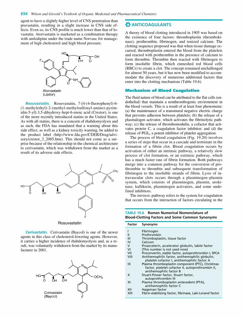
agent to have a slightly higher level of CNS penetration thanpravastatin, resulting in a slight increase in CNS side ef-fects. Even so, its CNS profile is much lower than that of lo-vastatin. Atorvastatin is marketed as a combination therapywith amlodipine under the trade name Norvasc for manage-ment of high cholesterol and high blood pressure.
Rosuvastatin. Rosuvastatin, 7-[4-(4-fluorophenyl)-6-(1-methylethyl)-2-(methyl-methylsulfonyl-amino)-pyrim-idin-5-yl]-3,5-dihydroxy-hept-6-enoic acid (Crestor), is oneof the more recently introduced statins in the United States.As with all statins, there is a concern of rhabdomyolysis andas such, the FDA has mandated that a warning about thisside effect, as well as a kidney toxicity warning, be added tothe product label (http://www.fda.gov/CDER/Drug/advi-sory/crestor_3_2005.htm). This should not come as a sur-prise because of the relationship in the chemical architectureto cerivastatin, which was withdrawn from the market as aresult of its adverse side effects.
Cerivastatin. Cerivastatin (Baycol) is one of the neweragents in this class of cholesterol-lowering agents. However,it carries a higher incidence of rhabdomyolysis and, as a re-sult, was voluntarily withdrawn from the market by its manu-facturer in 2001.
ANTICOAGULANTS
A theory of blood clotting introduced in 1905 was based onthe existence of four factors: thromboplastin (thromboki-nase), prothrombin, fibrinogen, and ionized calcium. Theclotting sequence proposed was that when tissue damage oc-curred, thromboplastin entered the blood from the plateletsand reacted with prothrombin in the presence of calcium toform thrombin. Thrombin then reacted with fibrinogen toform insoluble fibrin, which enmeshed red blood cells(RBCs) to create a clot. The concept remained unchallengedfor almost 50 years, but it has now been modified to accom-modate the discovery of numerous additional factors thatenter into the clotting mechanism (Table 19.6).
Mechanism of Blood Coagulation
The fluid nature of blood can be attributed to the flat cells (en-dothelial) that maintain a nonthrombogenic environment inthe blood vessels. This is a result of at least four phenomena:(a) the maintenance of a transmural negative electric chargethat prevents adhesion between platelets; (b) the release of aplasmalogen activator, which activates the fibrinolytic path-way; (c) the release of thrombomodulin, a cofactor that acti-vates protein C, a coagulation factor inhibitor; and (d) therelease of PGI2, a potent inhibitor of platelet aggregation.
The process of blood coagulation (Fig. 19.20) involvesa series of steps that occur in a cascade and terminate in theformation of a fibrin clot. Blood coagulation occurs byactivation of either an intrinsic pathway, a relatively slowprocess of clot formation, or an extrinsic pathway, whichhas a much faster rate of fibrin formation. Both pathwaysmerge into a common pathway for the conversion of pro-thrombin to thrombin and subsequent transformation offibrinogen to the insoluble strands of fibrin. Lysis of in-travascular clots occurs through a plasminogen–plasminsystem, which consists of plasminogen, plasmin, uroki-nase, kallikrein, plasminogen activators, and some unde-fined inhibitors.
The intrinsic pathway refers to the system for coagulationthat occurs from the interaction of factors circulating in the
654 Wilson and Gisvold’s Textbook of Organic Medicinal and Pharmaceutical Chemistry
N
N NS
F
O
HO
OH OH
O O
CH3
Rosuvastatin
TABLE 19.6 Roman Numerical Nomenclature of Blood-Clotting Factors and Some Common Synonyms
Factor Synonyms
I FibrinogenII ProthrombinIII Thromboplastin, tissue factorIV CalciumV Proaccelerin, accelerator globulin, labile factorVI (This number is not used now)VII Proconvertin, stable factor, autoprothrombin I, SPCAVIII Antihemophilic factor, antihemophilic globulin,
platelet cofactor I, antihemophilic factor AIX Plasma thromboplastin component (PTC), Christmas
factor, platelet cofactor II, autoprothrombin II,antihemophilic factor B
X Stuart-Power factor, Stuart factor,autoprothrombin III
XI Plasma thromboplastin antecedent (PTA),antihemophilic factor C
XII Hageman factorXIII Fibrin-stabilizing factor, fibrinase, Laki-Lorand factor
0617-0665_17865_Ch19.qxd 12/4/09 5:51 AM Page 654

blood. It is activated when blood comes into contact with adamaged vessel wall or a foreign substance. Each of theplasma coagulation factors (Table 19.6), with the exceptionof factor III (tissue thromboplastin), circulates as an inactiveproenzyme. Except for fibrinogen, which precipitates as fib-rin, these factors are usually activated by enzymatic removalof a small peptide in the cascade of reactions that make upthe clotting sequence (Fig. 19.20). The extrinsic clotting sys-tem refers to the mechanism by which thrombin is generatedin plasma after the addition of tissue extracts. When varioustissues, such as brain or lung (containing thromboplastin),are added to blood, a complex between thromboplastin andfactor VII in the presence of calcium ions activates factor X,bypassing the time-consuming steps of the intrinsic pathwaythat form factor X.
The intrinsic and extrinsic pathways interact in vivo.Small amounts of thrombin formed early after stimulation ofthe extrinsic pathway accelerate clotting by the intrinsicpathway by activating factor VIII. Thrombin also speeds upthe clotting rate by activating factor V, located in the com-mon pathway. Thrombin then converts the soluble proteinfibrinogen into a soluble fibrin gel by acting on Gly–Argbonds to remove small fibrinopeptides from the N-terminus,enabling the remaining fibrinogen molecule to polymerize.It also activates factor XIII, which stabilizes the fibrin gel inthe presence of calcium by cross-linking between the chainsof the fibrin monomer through intermolecular �-glutamyl–lysine bridges to form an insoluble mass.
Anticoagulant Mechanisms
In the milieu of biochemicals being formed to facilitate theclotting of blood, the coagulation cascade in vivo is controlledby a balance of inhibitors in the plasma to prevent all of the
blood in the body from solidifying. Thrombin plays a pivotalrole in blood coagulation. It cleaves fibrinogen, a reaction thatinitiates formation of the fibrin gel, which constitutes theframework of the blood clot. As mentioned previously, it ac-tivates the cofactors factor V and factor VIII to accelerate thecoagulation process. Intact endothelial cells express a recep-tor, thrombomodulin, for thrombin. When thrombin is boundto thrombomodulin, it does not have coagulant activity, whichthus prevents clot formation beyond damaged areas and ontointact endothelium. In this bound state, however, thrombindoes activate protein C, which then inactivates two cofactorsand impedes blood clotting. Thrombin also activates factorXIII, leading to cross-linking of the fibrin gel. The activity ofthrombin is regulated by its inactivation by plasma proteininhibitors: �1-proteinase inhibitor, �2-macroglobulin, an-tithrombin (antithrombin III), and heparin cofactor II. Thesebelong to a family of proteins called serpins, an acronym forserine protease inhibitors.
Antithrombin III, an �2-globin, neutralizes thrombin andthe serine proteases in the coagulation cascade—Xa, IXa,XIa, and XIIa. Although antithrombin III is a slow-actinginhibitor, it becomes a rapid-acting inhibitor of thrombin inthe presence of heparin. Heparin is a naturally occurring an-ticoagulant that requires antithrombin III (discussed previ-ously) for its biological property of preventing blood clotformation. It binds at the lysine site of the antithrombin IIImolecule, causing a change in the conformation of an-tithrombin III and increasing its anticoagulant properties.Heparin can then dissociate from antithrombin III to bind toanother antithrombin III molecule. An additional system,which controls unwanted coagulation, involves protein C, avitamin K–dependent zymogen in the plasma. Protein C isconverted to a serine protease when thrombin and factor Xa,formed in the blood in the coagulation cascade, interact withthrombomodulin. The now-activated protein C inhibits fac-tors V and VIII and, in so doing, blocks further productionof thrombin. Protein C also enhances fibrinolysis by causingrelease of the tissue plasminogen activator.
The biosynthesis of prothrombin (factor II) depends onan adequate supply of vitamin K. A deficiency of vitamin Kresults in the formation of a defective prothrombin mole-cule. The defective prothrombin is antigenically similar tonormal prothrombin but has reduced calcium-binding abil-ity and no biological activity. In the presence of calciumions, normal prothrombin adheres to the surface of phospho-lipid vesicles and greatly increases the activity of the clot-ting mechanism. The defect in the abnormal prothrombin isin the NH2-terminal portion, in which the second carboxylresidue has not been added to the �-carbon atom of someglutamic acid residues on the prothrombin molecule to form�-carboxyglutamic acid.73 Administration of vitamin K an-tagonists decreases synthesis of a biologically active pro-thrombin molecule and increases the clotting time of bloodin humans.74
Vitamin K is critical to the formation of clotting factorsVII, IX, and X. These factors are glycoproteins that have �-carboxyglutamic acid residues at the N-terminal end ofthe peptide chain. The enzyme involved in forming an activeprothrombin is a vitamin K–dependent carboxylase locatedin the microsomal fraction of liver cells. It has been sug-gested that vitamin K drives the carboxylase reaction by ab-stracting a proton from the relatively unreactive methylenecarbon of the glutamyl residue, forming a 2,3-epoxide. Oral
Chapter 19 Cardiovascular Agents 655
Figure 19.20 Scheme of blood coagulation and fibri-nolysis. (*, a vitamin K–dependent factor; †, inhibitionby heparin and antithrombin III.)
0617-0665_17865_Ch19.qxd 12/4/09 5:51 AM Page 655

anticoagulants interfere with the �-carboxylation of glu-tamic acid residues by preventing the reduction of vitamin Kto its hydroquinone form (Fig. 19.21).
Hemophilia A, a blood disease characterized by a defi-ciency of coagulation factor VIII, is the most commoninherited blood coagulation disorder. Treatment of thisdisease over the past 25 years has depended on the concentra-tion of the antihemophilic factor (factor VIII) by cryoprecipi-tation and immunoaffinity chromatography separation tech-nology. The impact of this therapy has been diminished by thepresence of viruses that cause the acquired immunodeficiencysyndrome (AIDS) and other less tragic viral diseases in hu-mans. Recombinant antihemophilic factor preparations havebeen produced since 1989 with use of mammalian cells genet-ically altered to secrete human factor VIII. Kogenate andHelixate are recombinant preparations, obtained from geneti-cally altering baby hamster kidney cells that contain high con-centrations of factor VIII. Recombinant factor VIIa, an activefactor in the extrinsic pathway, now in phase III clinical trials(Novo Seven), has been used to treat patients with hemophiliaA factor VII deficiency. Hemophilia B, another genetic blooddisorder, which constitutes about 20% of hemophilia cases, iscaused by a deficiency of factor IX and has been treated fromcryoprecipitated fractions obtained from plasma. Monoclonalantibody technology has produced an essentially pure, car-rier-free preparation of native factor IX (Mononine).Recombinant technology has solved the problem of limitedsupply and viral contamination of these critical blood factors.
Blood Degredation
Some patients suffer from a rare, potentially life-threateningdisease of the blood characterized by hemolytic anemia and
thrombosis, which is known as paroxysmal nocturnal hemo-globinuria (PNH). One symptom of the disease includes redurine, which results from the breakdown of RBC products(hemoglobin and hemosiderin) in the urine. An inconsistent,but potentially life-threatening, complication of PNH is thedevelopment of venous thrombosis.
Platelet Aggregation and Inhibitors
Blood platelets play a pivotal role in hemostasis and throm-bus formation. Actually, they have two roles in the cessationof bleeding: a hemostatic function, in which platelets, throughtheir mass, cause physical occlusion of openings in blood ves-sels, and a thromboplastic function, in which the chemicalconstituents of the platelets take part in the blood coagulationmechanism. The circulatory system is self-sealing because ofthe clotting properties of blood. The pathological formation ofclots within the circulatory system, however, creates a poten-tially serious clinical situation that must be dealt with throughthe use of anticoagulants.
Platelets do not adhere to intact endothelial cells. They dobecome affixed to subendothelial tissues, which have beenexposed by injury, to cause hemostasis. Platelets bind to col-lagen in the vessel wall and trigger other platelets to adhereto them. This adhesiveness is accompanied by a change inshape of the platelets and may be caused by mobilization ofcalcium bound to the platelet membrane. The growth of theplatelet mass depends on the ADP released by the first fewadhering cells and enhances the aggregation process. A sec-ondary phase (phase II) immediately follows, with additionalplatelet aggregation. In this secondary phase, the plateletsundergo a secretory process during which enzymes such ascathepsin and acid hydrolases, along with fibrinogen, are
656 Wilson and Gisvold’s Textbook of Organic Medicinal and Pharmaceutical Chemistry
Figure 19.21 Mechanism of action of vitamin K and sites of action of warfarin.
0617-0665_17865_Ch19.qxd 12/4/09 5:51 AM Page 656

released from �-granules in the platelets and ADP, ATP,serotonin, and calcium are released from dense bodies in theplatelets. The dense bodies are likened to the storage gran-ules associated with adrenergic neurons. Increased levels ofcAMP inhibit platelet aggregation. cAMP activates specificdependent kinases, which form protein–phosphate com-plexes that chelate calcium ions. The reduced levels of cal-cium inhibit aggregation (Fig. 19.22). Inhibitors of plateletaggregation can increase cAMP levels by either stimu-lating adenylate cyclase or inhibiting phosphodiesterase.74
Substances such as glucagon, adenosine, and isoproterenolincrease cAMP levels and inhibit platelet aggregation. Drugssuch as theophylline, aminophylline, dipyramidole, papaver-ine, and adenosine inhibit phosphodiesterase and aggregationof platelets. Epinephrine, collagen, and serotonin inhibitadenylate cyclase and stimulate platelet aggregation.75 Therole of platelets in arterial thrombosis is similar to that in he-mostasis. The factors contributing to venous thrombosis arecirculatory stasis, excessive generation of thrombin forma-tion of fibrin, and, to a lesser extent than in the artery, plateletaggregation.
Aspirin, sulfinpyrazone, and indomethacin have an in-hibitory effect on platelet aggregation. They inhibit COX,the enzyme that controls the formation of prostaglandin en-doperoxides and increases the tendency for platelets to ag-gregate.76 Aspirin also inhibits the platelet-release reaction.Dipyridamole inhibits adenosine deaminase and adenosineuptake by platelets. As a result, the increased plasma con-centrations of adenosine inhibit ADP-induced aggregationof platelets.
Among the many pharmacological actions of prostaglan-dins is the ability of some to stimulate or inhibit the aggre-gation of platelets and alter the clotting time of blood.Prostaglandins are synthesized from 20-carbon polyunsatu-rated fatty acids containing from three to five double bonds.These fatty acids are present in the phospholipids of cellmembranes of all mammalian tissues. The main precursorof prostaglandins is arachidonic acid. Arachidonic acid isreleased from membrane phospholipids by the enzymephospholipase A2. Once released, arachidonic acid is metab-olized by COX synthetase to form unstable cyclic endoper-oxides, PGG2 and PGH2, which subsequently are trans-formed into PGI2 and TXA2. The conversion to TXA2 isaided by the enzyme thromboxane synthetase. The forma-tion of PGI2 can occur nonenzymatically. Blood plateletsconvert arachidonic acid to TXA2, whereas PGI2 is formedmainly by the vascular endothelium. Both PGI2 and TXA2
are unstable at physiological pH and temperatures. Theirhalf-lives are 2 to 3 minutes.
PGI2 inhibits platelet aggregation by stimulating adeny-late cyclase to increase cAMP levels in the platelets. PGI2 isalso a vasodilator and, as a result, has potent hypotensiveproperties when given intravenously or by intra-arterial ad-ministration. TXA2 induces platelet aggregation. Togetherwith PGI2, TXA2 plays a role in the maintenance of vascu-lar homeostasis. In addition to being a platelet aggregator,TXA2 is a potent vasoconstrictor.
Retardation of clotting is important in blood transfusions,to avoid thrombosis after surgery or from other causes, to pre-vent recurrent thrombosis in phlebitis and pulmonary em-bolism, and to lessen the propagation of clots in the coronaryarteries. This retardation may be accomplished by agents thatinactivate thrombin (heparin) or substances that prevent theformation of prothrombin in the liver (the coumarin deriva-tives and the phenylindanedione derivatives).
Although heparin is a useful anticoagulant, it has limitedapplications. Many of the anticoagulants in use today weredeveloped following the discovery of dicumarol, an anticoag-ulant present in spoiled sweet clover. These compounds areorally effective, but there is a lag period of 18 to 36 hours be-fore they increase the clotting time significantly. Heparin, incontrast, produces an immediate anticoagulant effect after in-travenous injection. A major disadvantage of heparin is thatthe only effective therapeutic route is parenteral.
Dicumarol and related compounds are not vitamin K an-tagonists in the classic sense. They appear to act by interfer-ing with the function of vitamin K in the liver cells, whichare the sites of synthesis of the clotting factors, includingprothrombin. This lengthens the clotting time by decreasingthe amount of biologically active prothrombin in the blood.
The discovery that dicumarol and related compoundswere potent reversible competitors of vitamin K coagulant-promoting properties (although at high levels, dicumarol isnot reversed by vitamin K) led to the development of an-tivitamin K compounds such as phenindione, which wasdesigned in part according to metabolite–antimetaboliteconcepts. The active compounds of the phenylindanedioneseries are characterized by a phenyl, a substituted phenyl, ora diphenylacetyl group in the 2-position. Another require-ment for activity is a keto group in the 1- and 3-position,one of which may form the enol tautomer. A second sub-stituent, other than hydrogen, at the 2-position prevents thisketo–enol tautomerism, and the resulting compounds are in-effective as anticoagulants.
PRODUCTS
Protamine Sulfate, USP. Protamine sulfate has an an-ticoagulant effect, but if used in the proper amount, it coun-teracts the action of heparin and is used as an antidote for thelatter in cases of overdosage. It is administered intra-venously in a dose that depends on the circumstances.
Dicumarol, USP. Dicumarol, 3,3�-methylenebis[4-hy-droxycoumarin], is a white or creamy white crystalline pow-der with a faint, pleasant odor and a slightly bitter taste. It ispractically insoluble in water or alcohol, slightly soluble inchloroform, and dissolved readily by solutions of fixed alka-lies. The effects after administration require 12 to 72 hours todevelop and persist for 24 to 96 hours after discontinuance.
Chapter 19 Cardiovascular Agents 657
Figure 19.22 Role of adenosine 3�,5�-cyclic monophos-phate (cAMP) in inhibition of platelet aggregation.
0617-0665_17865_Ch19.qxd 12/4/09 5:51 AM Page 657

Dicumarol is used alone or as an adjunct to heparin in theprophylaxis and treatment of intravascular clotting. It is usedin postoperative thrombophlebitis, pulmonary embolus, acuteembolic and thrombotic occlusion of peripheral arteries, andrecurrent idiopathic thrombophlebitis. It has no effect on analready-formed embolus but may prevent further intravascu-lar clotting. Because the outcome of acute coronary thrombo-sis depends largely on extension of the clot and formation ofmural thrombi in the heart chambers, with subsequent em-bolization, dicumarol has been used in this condition. It hasalso been administered to arrest impending gangrene afterfrostbite. The dose, after determination of the prothrombinclotting time, is 25 to 200 mg, depending on the size and thecondition of the patient. The drug is given orally in the formof capsules or tablets. On the second day and thereafter, itmay be given in amounts sufficient to maintain the prothrom-bin clotting time at about 30 seconds. If hemorrhages shouldoccur, a dosage of 50 to 100 mg of menadione sodium bisul-fite is injected, supplemented by a blood transfusion.
Warfarin Sodium, USP. Warfarin sodium, 3-(�-acetonylbenzyl)-4-hydroxycoumarin sodium salt (Coumadin,Panwarfin), is a white, odorless, crystalline powder, with aslightly bitter taste; it is slightly soluble in chloroform and sol-uble in alcohol or water. A 1% solution has a pH of 7.2 to 8.5.
By virtue of its great potency, warfarin sodium at firstwas considered unsafe for use in humans and was used veryeffectively as a rodenticide, especially against rats. At theproper dosage level, however, it can be used in humans, es-pecially through the intravenous route.
Warfarin Potassium, USP. Warfarin potassium, 3-(�-acetonylbenzyl)-4-hydroxycoumarin potassium salt(Athrombin-K), is readily absorbed after oral administration,and a therapeutic hypoprothrombinemia is produced within12 to 24 hours after administration of 40 to 60 mg. This saltis therapeutically interchangeable with warfarin sodium.
658 Wilson and Gisvold’s Textbook of Organic Medicinal and Pharmaceutical Chemistry
Anisindione, USP. Anisindione, 2-(p-methoxyphenyl)-1,3-indandione, 2-(p-anisyl)-1,3-indandione (Miradon), is ap-methoxy congener of phenindione. It is a white, crys-talline powder, slightly soluble in water, tasteless, and ab-sorbed well after oral administration.
In instances when the urine may be alkaline, an orangecolor may be detected. This is caused by metabolic productsof anisindione and is not hematuria.
Eculizumab. Eculizumab (Soliris) is a monoclonal anti-body that binds to the terminal complement protein C5 inRBCs. This blocks the cleavage of C5 and halts the processof complement-mediated cell destruction of the RBCs.Eculizumab has been shown to be effective in treating PNHand in March 2007 was approved by the FDA for treatingPNH.77
SYNTHETIC HYPOGLYCEMIC AGENTS
The discovery that certain organic compounds will lower theblood sugar level is not recent. In 1918, guanidine wasshown to lower the blood sugar level. The discovery that cer-tain trypanosomes need much glucose and will die in its ab-sence was followed by the discovery that galegine loweredthe blood sugar level and was weakly trypanocidal. This ledto the development of several very active trypanocidalagents, such as the bisamidines, diisothioureas, bisguani-dines, and others. Synthalin (trypanocidal at 1:250 million)and pentamidine are outstanding examples of very active try-panocidal agents. Synthalin lowers the blood sugar level innormal, depancreatized, and completely alloxanized animals.This may be caused by reduced oxidative activity of mito-chondria, resulting from inhibition of the mechanisms that si-multaneously promote phosphorylation of ADP and stimu-late oxidation by nicotinamide adenine dinucleotide (NAD)in the citric acid cycle. Hydroxystilbamidine isethionate,USP, is used as an antiprotozoan agent.
0617-0665_17865_Ch19.qxd 12/4/09 5:51 AM Page 658

In 1942, p-aminobenzenesulfonamidoisopropylthiadia-zole (an antibacterial sulfonamide) was found to producehypoglycemia. This result stimulated research for the devel-opment of synthetic hypoglycemic agents, several of whichare in use today.
Sulfonylureas became widely available in 1955 for thetreatment of non–ketosis-prone mild diabetes and are stillthe drugs of choice. A second class of compounds, thebiguanides, in the form of a single drug, phenformin, hasbeen used since 1957. Phenformin was withdrawn from theU.S. market, however, because it causes lactic acidosis, fromwhich fatalities have been reported.
Sulfonylureas
The sulfonylureas may be represented by the following gen-eral structure:
These are urea derivatives with an arylsulfonyl group inthe 1-position and an aliphatic group at the 3-position. Thealiphatic group, R�, confers lipophilic properties to the mol-ecule. Maximal activity results when R� consists of three tosix carbon atoms, as in chlorpropamide, tolbutamide, andacetohexamide. Aryl groups at R� generally give toxiccompounds. The R group on the aromatic ring primarily in-fluences the duration of action of the compound.Tolbutamide disappears quite rapidly from the bloodstreamby being metabolized to the inactive carboxy compound,which is excreted rapidly. Chlorpropamide, however, ismetabolized more slowly and persists in the blood muchlonger.
The mechanism of action of the sulfonylureas is to stim-ulate the release of insulin from the functioning �-cells ofthe intact pancreas. In the absence of the pancreas, they haveno significant effect on blood glucose. The sulfonylureasmay have other actions, such as inhibition of secretion ofglucagon and action at postreceptor intracellular sites to in-crease insulin activity.
For a time, tolbutamide, chlorpropamide, and acetohex-amide were the only oral hypoglycemic agents. Subsequently,a second generation of these drugs became available.Although they did not present a new method of loweringblood glucose levels, they were more potent than the existingdrugs. Glipizide and glyburide are the second-generation oralhypoglycemic agents.
Whether they are first- or second-generation oral hypo-glycemic drugs, this group of agents remains a valuable ad-
junct to therapy in adult-onset diabetes patients. Accordingly,the sulfonylureas are not indicated in juvenile-onset diabetes.
Tolbutamide, USP. Tolbutamide, 1-butyl-3-(p-tolylsul-fonyl)urea (Orinase), occurs as a white, crystalline powderthat is insoluble in water and soluble in alcohol or aqueousalkali. It is stable in air.
Tolbutamide is absorbed rapidly in responsive diabetic pa-tients. The blood sugar level reaches a minimum after 5 to8 hours. It is oxidized rapidly in vivo to 1-butyl-3-(p-car-boxyphenyl)sulfonylurea, which is inactive. The metabolite isfreely soluble at urinary pH; if the urine is strongly acidified,however, as in the use of sulfosalicylic acid as a protein pre-cipitant, a white precipitate of the free acid may be formed.
Tolbutamide should be used only when the diabetic patientis an adult or shows adult-onset diabetes, and the patientshould adhere to dietary restrictions.
Tolbutamide Sodium, USP. Tolbutamide sodium, 1-butyl-3-(p-tolylsulfonyl)urea monosodium salt (OrinaseDiagnostic), is a white, crystalline powder, freely soluble inwater, soluble in alcohol and chloroform, and very slightlysoluble in ether.
This water-soluble salt of tolbutamide is used intra-venously for the diagnosis of mild diabetes mellitus and offunctioning pancreatic islet cell adenomas. The sterile drypowder is dissolved in sterile water for injection to make aclear solution, which then should be administered within1 hour. The main route of breakdown is to butylamine andsodium p-toluene sulfonamide.
Chlorpropamide, USP. Chlorpropamide, 1-[(p-chloro-phenyl)-sulfonyl]-3-propylurea (Diabinese), is a white, crys-
Chapter 19 Cardiovascular Agents 659
0617-0665_17865_Ch19.qxd 12/4/09 5:51 AM Page 659

talline powder, practically insoluble in water, soluble in alco-hol, and sparingly soluble in chloroform. It will form water-soluble salts in basic solutions. This drug is more resistant toconversion to inactive metabolites than is tolbutamide and, asa result, has a much longer duration of action. One studyshowed that about half of the drug is excreted as metabolites,with the principal one being hydroxylated in the 2-position ofthe propyl side chain.78 After control of the blood sugar level,the maintenance dose is usually on a once-a-day schedule.
Tolazamide, USP. Tolazamide, 1-(hexahydro-1H-azepin-1-yl)-3-(p-tolylsulfonyl)urea (Tolinase), is an analogof tolbutamide and is reported to be effective, in general,under the same circumstances in which tolbutamide is useful.Tolazamide, however, appears to be more potent than tolbu-tamide and is nearly equal in potency to chlorpropamide. Instudies with radioactive tolazamide, investigators found that85% of an oral dose appeared in the urine as metabolites thatwere more soluble than tolazamide itself.
660 Wilson and Gisvold’s Textbook of Organic Medicinal and Pharmaceutical Chemistry
Acetohexamide, USP. Acetohexamide, 1-[(p-acetyl-phenyl)sulfonyl]-3-cyclohexylurea (Dymelor), is relatedchemically and pharmacologically to tolbutamide and chlor-propamide. Like the other sulfonylureas, acetohexamidelowers the blood sugar level, primarily by stimulating the re-lease of endogenous insulin.
Acetohexamide is metabolized in the liver to a reducedform, the �-hydroxyethyl derivative. This metabolite, themain one in humans, possesses hypoglycemic activity.Acetohexamide is intermediate between tolbutamide andchlorpropamide in potency and duration of effect on bloodsugar levels.
Glipizide. Structurally, glipizide, 1-cyclohexyl-3-[[p-[2(methylpyrazinecarboxamido)ethyl]phenyl]sulfonyl]urea(Glucotrol), is a cyclohexylsulfonylurea analog similar toacetohexamide and glyburide. The drug is absorbed rapidlyon oral administration. Its serum half-life is 2 to 4 hours, andit has a hypoglycemic effect that ranges from 12 to 24 hours.
Glyburide. Similar to glipizide, glyburide, 1-[[p-[2-(5-chloro-o-anisamido)ethyl]-phenyl]sulfonyl]-3-cyclohexyl-urea (DiaBeta, Micronase, Glynase), is a second-generationoral hypoglycemic agent. The drug has a half-life elimina-tion of 10 hours, but its hypoglycemic effect remains for upto 24 hours.
0617-0665_17865_Ch19.qxd 12/4/09 5:51 AM Page 660

Glipizide. Glipizide, 1-cyclohexyl-3-[[p-(2-(5-methyl-pyrazinecarboxamido)ethyl]phenyl]sulfonyl]urea(Glucotrol), is an off-white, odorless powder with a pKa of5.9. It is insoluble in water and alcohols, but soluble in 0.1 NNaOH. Even though on a weight basis, it is approximately100 times more potent than tolbutamide, the maximal hypo-glycemic effects of these two agents are similar. It is rapidlyabsorbed on oral administration, with a serum half-life of 2 to4 hours, whereas the hypoglycemic effects range from 12 to24 hours. Metabolism of glipizide is generally through oxida-tion of the cyclohexane ring to the p-hydroxy and m-hydroxymetabolites. A minor metabolite that occurs involves theN-acetyl derivative, which results from the acetylation of theprimary amine following hydrolysis of the amide system byamidase enzymes.
Glimepiride. Glimepiride, 1-[[p-[2-(3-ethyl-4-methyl-2-oxo-3-pyrroline-1-carboxamido)ethyl]phenyl]sulfonyl]-3-(trans-4-methylcyclohexyl)urea (Amaryl), is very similarto glipizide with the exception of their heterocyclic rings.Instead of the pyrazine ring found in glipizide, glimepiridecontains a pyrrolidine system. It is metabolized primarilythrough oxidation of the alkyl side chain of the pyrrolidine,with a minor metabolic route involving acetylation of theamine.
Gliclazide. Chemically, gliclazide, 1-(3-azabicy-clo[3.3.0]oct-3-yl)-3-p-tolylsulphonylurea (Diamicron), isvery similar to tolbutamide, with the exception of the bi-cyclic heterocyclic ring found in gliclazide. The pyrrol-idine increases its lipophilicity over that of tolbutamide,which increases its half-life. Even so, the p-methyl is sus-ceptible to the same oxidative metabolic fate as observedfor tolbutamide, namely, it will be metabolized to a car-boxylic acid.
Nonsulfonylureas—Metaglinides
The metaglinides are nonsulfonylurea oral hypoglycemicagents used in the management of type 2 diabetes (non–insulin-dependent diabetes mellitus, NIDDM). These agentstend to have a rapid onset and a short duration of action.Much like the sulfonylureas, these induce insulin releasefrom functioning pancreatic � cells. The mechanism of ac-tion for the metaglinides, however, differs from that of thesulfonylureas. The mechanism of action is through bindingto specific receptors in the �-cell membrane, leading to theclosure of ATP-dependent K� channels. The K� channelblockade depolarizes the �-cell membrane, which in turn
leads to Ca2� influx, increased intracellular Ca2�, andstimulation of insulin secretion. Because of this differentmechanism of action from the sulfonylureas, there are twomajor differences between these seemingly similar classesof agents. The first is that the metaglinides cause muchfaster insulin production than the sulfonylureas. As a result,the metaglinides should be taken during meals, as the pan-creas will produce insulin in a much shorter period. The sec-ond difference is that the effects of the metaglinides do notlast as long as the effects of the sulfonylureas. The effects ofthis class appear to last less than 1 hour, whereas sulfonyl-ureas continue to stimulate insulin production for severalhours. One advantage of a short duration of action is thatthere is less risk of hypoglycemia.
Repaglinide. Repaglinide, (�)-2-ethoxy-4-[N-[3-methyl-1(S)-[2-(1-piperidinyl) phenyl]butyl]carbamoyl-methyl]ben-zoic acid (Prandin), represents a new class of nonsulfony-lurea oral hypoglycemic agents. With a fast onset and ashort duration of action, the medication should be taken withmeals. It is oxidized by CYP 3A4, and the carboxylic acidmay be conjugated to inactive compounds. Less than 0.2%is excreted unchanged by the kidney, which may be an ad-vantage for elderly patients who are renally impaired. Themost common side effect involves hypoglycemia, resultingin shakiness, headache, cold sweats, anxiety, and changes inmental state.
Nateglinide. Although nateglinide, N-(4-isopropylcy-clohexanecarbonyl)-D-phenylalanine (Starlix), belongs tothe metaglinides, it is a phenylalanine derivative and repre-sents a novel drug in the management of type 2 diabetes.
Chapter 19 Cardiovascular Agents 661
0617-0665_17865_Ch19.qxd 12/4/09 5:51 AM Page 661

Thiazolindiones
The thiazolindiones represent a novel nonsulfonylurea classof hypoglycemic agents for the treatment of NIDDM. Muchlike the sulfonylureas, the use of these agents requires afunctioning pancreas that can successfully secrete insulinfrom � cells. Although insulin may be released in normallevels from the cells, peripheral sensitivity to this hormonemay be reduced or lacking. The thiazolidinediones arehighly selective agonists for the peroxisome proliferator-activated receptor-� (PPAR�), which is responsible forimproving glycemic control, primarily through the improve-ment of insulin sensitivity in muscles and adipose tissue. Inaddition, they inhibit hepatic gluconeogenesis. These agentsnormalize glucose metabolism and reduce the amount ofinsulin needed to achieve glycemic control. They are onlyeffective in the presence of insulin.
Rosiglitazone. Rosiglitazone, (�)-5-[[4-[2-(methyl-2-pyridinylamino)ethoxy]phenyl]methyl]-2,4-thiazolidine-dione (Avandia), is a white to off-white solid with pKa
values of 6.8 and 6.1. Rosiglitazone is readily soluble inethanol and a buffered aqueous solution with pH of 2.3; sol-ubility decreases with increasing pH in the physiologicalrange. The molecule has a single chiral center and is presentas a racemate. Even so, the enantiomers are functionally in-distinguishable because of rapid interconversion.
Pioglitazone. Pioglitazone, (�)-5-[[4-[2-(5-ethyl-2-pyri-dinyl)ethoxy]phenyl]methyl]-2,4-thiazolidinedione (Actos),is an odorless, white, crystalline powder that must be con-verted to a salt such as its hydrochloride before it will haveany water solubility. Although the molecule contains one chi-ral center, the compound is used as the racemic mixture. Thisis primarily a result of the in vivo interconversion of the twoenantiomers. Thus, there are no differences in the pharmaco-logical activity of the two enantiomers.
Bisguanidines
Metformin. Metformin, N,N-dimethylimidodicarboni-midic diamide hydrochloride (Glucophage), is a bisguanidine.This class of agents is capable of reducing sugar absorption
from the gastrointestinal tract. Also, they can decrease gluco-neogenesis while increasing glucose uptake by muscles andfat cells. These effects, in turn, lead to lower blood glucoselevels. Unlike the sulfonylureas, these are not hypoglycemicagents but rather can act as antihyperglycemics. This differ-ence in nomenclature is caused by the inability of these agentsto stimulate the release of insulin from the pancreas. Often,metformin is coadministered with the nonsulfonylureas to im-prove the efficacy of those agents.
�-Glucosidase Inhibitors
The enzyme �-glucosidase is present in the brush border ofthe small intestine and is responsible for cleaving dietarycarbohydrates and facilitating their absorption into the body.Inhibition of this enzyme allows less dietary carbohydrate tobe available for absorption and, in turn, less available in theblood following a meal. The inhibitory properties of theseagents are greatest for glycoamylase, followed by sucrose,maltase, and dextranase, respectively. Because these do notenhance insulin secretion when used as monotherapy, hypo-glycemia is generally not a concern when using theseagents.
Acarbose. Acarbose, O-4,6-dideoxy-4-[[(1S,4R,5S,6S)4,5,6-trihydroxy-3-(hydroxymethyl)-2-cyclohexen-1-yl]amino]�-D-glucopyranosyl-(1,4)-O-�-D-glucopyranosyl-(1,4)-D-glucose (Precose), is a naturally occurring oligosac-charide, which is obtained from the microorganismActinoplanes utahensis. It is a white to off-white powder
662 Wilson and Gisvold’s Textbook of Organic Medicinal and Pharmaceutical Chemistry
0617-0665_17865_Ch19.qxd 12/4/09 5:51 AM Page 662

that is soluble in water and has a pKa of 5.1. As one mightexpect, its affinity for �-glucosidase is based on it being apolysaccharide that the enzyme attempts to hydrolyze. Thisallows acarbose to act as a competitive inhibitor, which inturn reduces the intestinal absorption of starch, dextrin, anddissacharides.
Miglitol. Miglitol, 1-(2-hydroxyethyl)-2-(hydroxy-methyl)-[2R-(2�,3�,4�,5�)]-piperidine (Glyset), a des-oxynojirimycin derivative, is chemically known as 3,4,5-piperidinetriol. It is a white to pale yellow powder that issoluble in water, with a pKa of 5.9. In chemical structure,this agent is very similar to a sugar, with the heterocyclic ni-trogen serving as an isosteric replacement of the sugar oxy-gen. This feature allows recognition by the �-glucosidase asa substrate. This results in competitive inhibition of the en-zyme and delays complex carbohydrate absorption from thegastrointestinal tract.
THYROID HORMONES
Desiccated, defatted thyroid substance has been used formany years as replacement therapy in thyroid gland defi-ciencies. The efficacy of the whole gland is now knownto depend on its thyroglobulin content. This is an iodine-containing globulin. Thyroxine was obtained as a crystallinederivative by Kendall79 of the Mayo Clinic in 1915. Itshowed much the same action as the whole thyroid sub-stance. Later, thyroxine was synthesized by Harington andBarger80 in England. Later studies showed that an evenmore potent iodine-containing hormone existed, which isnow known as triiodothyronine. Evidence now indicatesthat thyroxine may be the storage form of the hormone,whereas triiodothyronine is the circulating form. Anotherpoint of view is that in the blood, thyroxine is bound morefirmly to the globulin fraction than is triiodothyronine,which can then enter the tissue cells.
Levothyroxine Sodium, USP. Levothyroxine sodium,O-(4-hydroxy-3,5-diiodophenyl)-3,5-diiodo-2-tyrosinemonosodium salt, hydrate (Synthroid, Letter, Levoxine,Levoid), is the sodium salt of the levo isomer of thyroxine,which is an active physiological principle obtained from thethyroid gland of domesticated animals used for food by hu-mans. It is also prepared synthetically. The salt is a light yel-low, tasteless, odorless powder. It is hygroscopic but stablein dry air at room temperature. It is soluble in alkali hydrox-ides, 1:275 in alcohol, and 1:500 in water, to give a pH ofabout 8.9.
Levothyroxine sodium is used in replacement therapy ofdecreased thyroid function (hypothyroidism). In general, adosage of 100 �g of levothyroxine sodium is clinicallyequivalent to 30 to 60 mg of thyroid USP.
Liothyronine Sodium, USP. Liothyronine sodium, O-(4-hydroxy-3-iodophenyl)-3,5-diiodo-L-thyroxinemonosodium salt (Cytomel), is the sodium salt of L-3,3�,5-triiodothyronine. It occurs as a light tan, odorless, crystallinepowder, which is slightly soluble in water or alcohol and hasa specific rotation of �18 to 22 degrees in a mixture ofdiluted HCl and alcohol.
Chapter 19 Cardiovascular Agents 663
Liothyronine sodium occurs in vivo together withlevothyroxine sodium; it has the same qualitative activitiesas thyroxine but is more active. It is absorbed readily fromthe gastrointestinal tract, is cleared rapidly from the blood-stream, and is bound more loosely to plasma proteins than isthyroxine, probably because of the less acidic phenolic hy-droxyl group.
Its uses are the same as those of levothyroxine sodium,including treatment of metabolic insufficiency, male infer-tility, and certain gynecological disorders.
ANTITHYROID DRUGS
Hyperthyroidism (excessive production of thyroid hormones)usually requires surgery, but before surgery the patient mustbe prepared by preliminary abolition of the hyperthyroidismthrough the use of antithyroid drugs. Thiourea and relatedcompounds show an antithyroid activity, but they are too
0617-0665_17865_Ch19.qxd 12/4/09 5:51 AM Page 663
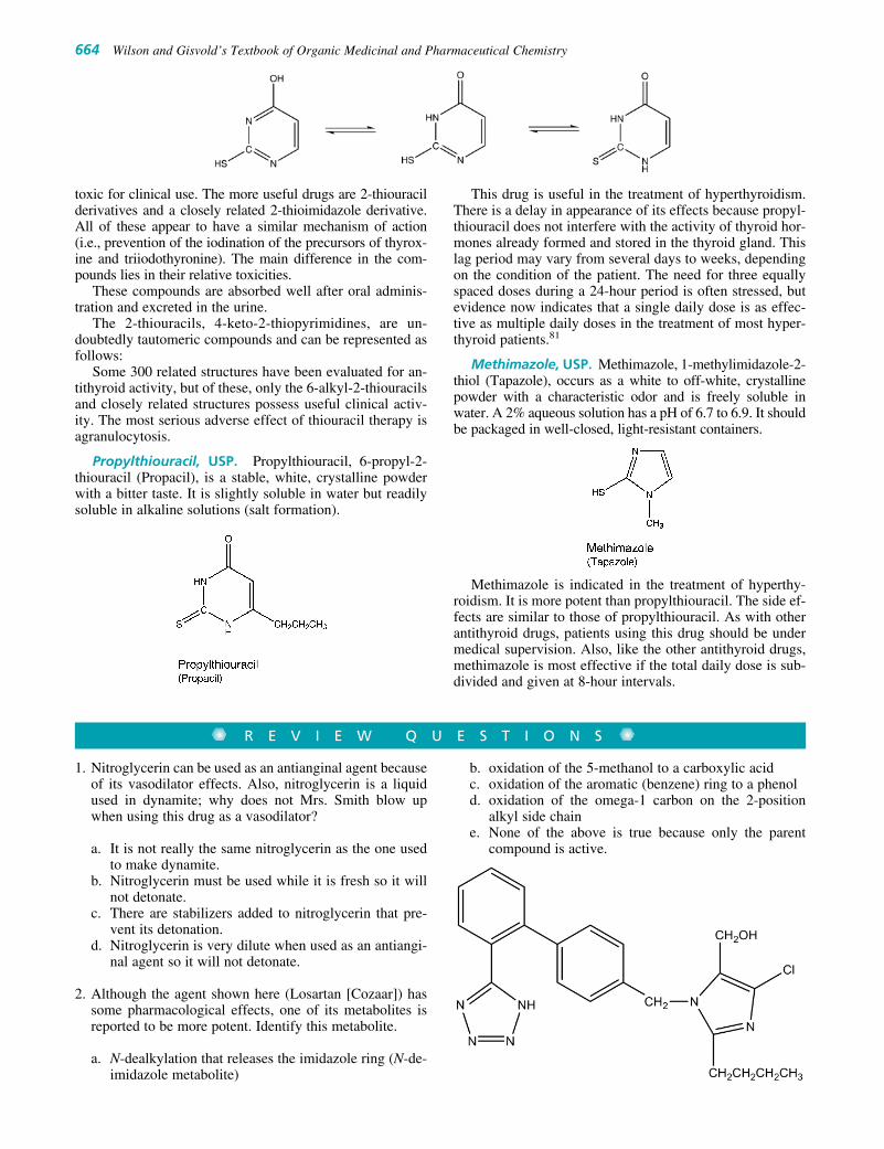
b. oxidation of the 5-methanol to a carboxylic acidc. oxidation of the aromatic (benzene) ring to a phenold. oxidation of the omega-1 carbon on the 2-position
alkyl side chaine. None of the above is true because only the parent
compound is active.
toxic for clinical use. The more useful drugs are 2-thiouracilderivatives and a closely related 2-thioimidazole derivative.All of these appear to have a similar mechanism of action(i.e., prevention of the iodination of the precursors of thyrox-ine and triiodothyronine). The main difference in the com-pounds lies in their relative toxicities.
These compounds are absorbed well after oral adminis-tration and excreted in the urine.
The 2-thiouracils, 4-keto-2-thiopyrimidines, are un-doubtedly tautomeric compounds and can be represented asfollows:
Some 300 related structures have been evaluated for an-tithyroid activity, but of these, only the 6-alkyl-2-thiouracilsand closely related structures possess useful clinical activ-ity. The most serious adverse effect of thiouracil therapy isagranulocytosis.
Propylthiouracil, USP. Propylthiouracil, 6-propyl-2-thiouracil (Propacil), is a stable, white, crystalline powderwith a bitter taste. It is slightly soluble in water but readilysoluble in alkaline solutions (salt formation).
This drug is useful in the treatment of hyperthyroidism.There is a delay in appearance of its effects because propyl-thiouracil does not interfere with the activity of thyroid hor-mones already formed and stored in the thyroid gland. Thislag period may vary from several days to weeks, dependingon the condition of the patient. The need for three equallyspaced doses during a 24-hour period is often stressed, butevidence now indicates that a single daily dose is as effec-tive as multiple daily doses in the treatment of most hyper-thyroid patients.81
Methimazole, USP. Methimazole, 1-methylimidazole-2-thiol (Tapazole), occurs as a white to off-white, crystallinepowder with a characteristic odor and is freely soluble inwater. A 2% aqueous solution has a pH of 6.7 to 6.9. It shouldbe packaged in well-closed, light-resistant containers.
Methimazole is indicated in the treatment of hyperthy-roidism. It is more potent than propylthiouracil. The side ef-fects are similar to those of propylthiouracil. As with otherantithyroid drugs, patients using this drug should be undermedical supervision. Also, like the other antithyroid drugs,methimazole is most effective if the total daily dose is sub-divided and given at 8-hour intervals.
664 Wilson and Gisvold’s Textbook of Organic Medicinal and Pharmaceutical Chemistry
R E V I E W Q U E S T I O N S
1. Nitroglycerin can be used as an antianginal agent becauseof its vasodilator effects. Also, nitroglycerin is a liquidused in dynamite; why does not Mrs. Smith blow upwhen using this drug as a vasodilator?
a. It is not really the same nitroglycerin as the one usedto make dynamite.
b. Nitroglycerin must be used while it is fresh so it willnot detonate.
c. There are stabilizers added to nitroglycerin that pre-vent its detonation.
d. Nitroglycerin is very dilute when used as an antiangi-nal agent so it will not detonate.
2. Although the agent shown here (Losartan [Cozaar]) hassome pharmacological effects, one of its metabolites isreported to be more potent. Identify this metabolite.
a. N-dealkylation that releases the imidazole ring (N-de-imidazole metabolite)
0617-0665_17865_Ch19.qxd 12/4/09 5:51 AM Page 664
4. Highlight the difference between digoxin and digitoxinand point out the two primary reasons why one is moretoxic than the other.
Chapter 19 Cardiovascular Agents 665
REFERENCES1. Robinson, B. F.: Adv. Drug. Res. 10:93, 1975.2. Aronow, W. S.: Am. Heart J. 84:273, 1972.3. Sonnenblick, E., Ross, J., Jr., and Braunwald, E.: Am. J. Cardiol.
22:328, 1968.4. Ignarro, L. J., et al.: J. Pharmacol. Exp. Ther. 218:739, 1981.5. Feelisch, M.: Eur. Heart J. 14(Suppl. I):123, 1993.6. Needleman, P.: Annu. Rev. Pharmacol. Toxicol. 16:81, 1976.7. Chung, S. J., and Fung, H. L.: J. Pharmacol. Exp. Ther. 253:614, 1990.8. Fusari, S. A.: J. Pharm. Sci. 62:123, 1973.9. Fusari, S. A.: J. Pharm. Sci. 62:2012, 1973.
10. McCall, D., et al.: Curr. Probl. Cardiol. 10:1, 1985.11. Heschler, J., et al.: Eur. J. Biochem. 165:261, 1987.12. Beridge, M. J.: Annu. Rev. Biochem. 56:159, 1987.13. van Zweiten, P. A., and van Meel, J. C.: Prog. Pharmacol. 5:1, 1982.14. Atkinson, J., et al.: Naunyn Schmeidbergs Arch. Pharmacol.
337:471, 1988.15. Smith, H. J., and Briscoe, M. G.: J. Mol. Cell Cardiol. 17:709, 1985.16. Sanguinetti, M. C., and Kass, R. S.: Circ. Res. 55:336, 1984.17. Rosenkirchen, R., et al.: Naunyn Schmiedebergs Arch. Pharmacol.
310:69, 1979.18. Triggle, D. J.: Calcium antagonists. In: Antonoccio, P. N. (ed.).
Cardiovascular Pharmacology, 3rd ed. New York, Raven Press, 1990.19. Dobesh, P. P., and Trujillo, T. C.: Pharmacotherapy 27:1659, 2007.20. Vaughan Williams, E. M.: In Sandoe, E., Flensted-Jansen, E., and
Olesen, K. H. (eds.). Symposium on Cardiac Arrhythmias. Sodertalje,Sweden, B. Astra, 1970, pp. 449–472.
21. Vaughan Williams, E. M.: J. Clin. Pharmacol. 24:129, 1984.22. CAPS Investigators: Am. J. Cardiol. 61:501, 1988.23. Woosley, R. L.: Annu. Rev. Pharmacol. Toxicol. 31:427, 1991.24. Campbell, T. J.: Cardiovasc. Res. 17:344, 1983.25. Yool, A. J.: Mol. Pharmacol. 46:970, 1994.26. Hondeghem, L. M., and Katzung, B. G.: Annu. Rev. Pharmacol.
Toxicol. 24:387, 1984.27. Nies, A. S., and Shang, D. G.: Clin. Pharmacol. Exp. Ther.
14:823, 1973.28. Koch-Wester, J.: Ann. N. Y. Acad. Sci. 179:370, 1971.29. Giardinia, E. V., et al.: Clin. Pharmacol. Ther. 19:339, 1976.30. Elson, J., et al.: Clin. Pharmacol. Ther. 17:134, 1975.31. Belfer, B., et al.: Am. J. Cardiol. 35:282, 1975.32. Bigger, T. J., and Jaffe, C. C.: Am. J. Cardiol. 27:82, 1971.33. Hollunger, G.: Acta Pharmacol. Toxicol. 17:356, 1960.34. Helfant, R. H., et al.: Am. Heart J. 77:315, 1969.35. Beckett, A. H., and Chiodomere, E. C.: Postgrad. Med. J. 53(Suppl. 1):
60, 1977.36. Anderson, J. L., Mason, J. W., and Roger, M. D.: Circulation
57:685, 1978.37. Guehler, J., et al.: Am. J. Cardiol. 55:807, 1985.38. Saeki, T., et al.: Eur. J. Pharmacol. 261:249, 1994.39. Groshner, K., et al.: Br. J. Pharmacol. 102:669, 1991.40. Hii, J. T., Duff, H. J., and Burgess, E. D.: Clin. Pharmacokinet.
21:1, 1991.41. Olsson, S. B., Brorson, L., and Varnauskas, E.: Br. Heart J.
35:1255, 1973.
42. Kannan, R., et al.: Clin. Pharmacol. 31:438, 1982.43. Witt, D. M., Ellsworth, A. J., and Leversee, J. H.: Ann.
Pharmacother. 27:1463, 1993.44. Nadermanee, K., et al.: Circulation 66:202, 1982.45. Papp, J. G., and Vaughan Williams, E. M.: Br. J. Pharmacol.
37:380, 1969.46. Boyer, E. W., Stork, C., and Wang, R. Y.: Int. J. Med. Toxicol.
2001;4:16.47. Salata, J. J., and Brooks, R. R.: Cardiol. Drug Rev. 15:137–156, 1997.48. D’Orleans-Juste, P., Labonte, J., Bkaily, G., et al.: Pharmacol. Ther.
95:221, 2002.49. Newman, J. H., Kar, S., and Kirkpatrick, P.: Nat. Rev. Drug Discov.
6:697, 200750. Oldfield, V., and Lyseng-Williamson, K. A.: Am. J. Cardiovasc.
Drugs, 6:189, 2006.51. Vakil, R.: Br. Heart J. 11:350, 1949.52. Hess, S. M., Shore, P. A., and Brodie, P. P.: Br. J. Pharmacol. Exp.
Ther. 118:84, 1956.53. Kirshner, N.: J. Biol. Chem. 237:2311, 1962.54. von Euler, U. S., and Lishajko, F.: Int. J. Neuropharmacol. 2:127, 1963.55. Muller, J. M., Schlittler, E., and Brin, H. J.: Experientia 8:338, 1952.56. U’Prichard, D., et al.: Eur. J. Pharmacol. 50:87, 1978.57. Langer, S. Z., and Cavero, I.: Hypertension 2:372, 1980.58. Starke, K., and Montel, H.: Neuropharmacology 12:1073, 1973.59. Meisheri, K. D.: Direct-acting vasodilators. In Singh, B. J., et al.
(eds.). Cardiovascular Pharmacology. New York, ChurchillLivingstone, 1994, p. 173.
60. Gross, F., Druey, J., and Meier, R.: Experientia 6:19, 1950.61. Zacest, R., and Koch-Wesesr, J.: Clin. Pharmacol. 13:4420, 1972.62. Page, I. H., et al.: Circulation 11:188, 1955.63. Corbin, J. D., Francis, S. H., and Webb, D. J.: Urology 60(2):4, 2002.64. Ring, B. J., Patterson, B. E., Mitchell, M. I., et al.: Clin. Pharmacol.
Ther. 77(1):63, 2005.65. Cook, N. S.: Potassium Channels: Structure, Classification,
Function and Therapeutic Potential. New York, John Wiley & Sons,1990, p. 181.
66. DuCharme, D. W., et al.: Pharmacol. Exp. Ther. 184:662, 1973.67. Levy, R. I., et al.: Circulation 69:325, 1984.68. Levy, R. I.: Annu. Rev. Pharmacol. Toxicol. 47:499, 1977.69. Mannisto, P. T., et al.: Acta Pharmacol. Toxicol. 36:353, 1975.70. Kesaniemi, Y. A., and Grundy, S. M.: JAMA 251:2241, 1984.71. Atmeh, R. F., et al.: J. Lipid Res. 24:588, 1983.72. Sweeney, M. E., and Johnson, R. R.: Exp. Opin. Drug Metab.
Toxicol. 3(3):441, 2007.73. Stenflo, J., et al.: Proc. Natl. Acad. Sci. U. S. A. 71:2730, 1974.74. Jackson, C. M., and Suttie, J. W.: Prog. Hematol. 10:333, 1978.75. Triplett, D. A. (ed.): Platelet Function. Chicago, American Society of
Clinical Pathology, 1978.76. Hamberg, M., et al.: Proc. Natl. Acad. Sci. U. S. A. 71:345, 1974.77. Kaplan, M.: Curr. Opin. Investig. Drugs 3(7):1017, 2002.78. Thomas, R. C., and Ruby, R. W.: J. Med. Chem. 15:964, 1972.79. Kendall, E. C.: JAMA 64:2042, 1915.80. Harrington, C. R., and Barger, C.: Biochem. J. 21:169, 1927.81. Greer, M. A., and Meihoff, W. C.: N. Engl. J. Med. 272:888, 1965.
3. After a patient is administered an organic nitrate, theseare then metabolized to nitric oxide (NO) by first reactingwith _______ to form ______.
a. sulfhydryls: S-nitrosothiolsb. amidase; carboxylic acids and aminesc. esterases; carboxylic acids and alcoholsd. hydroxyl residues; O-nitrosoalcohols
0617-0665_17865_Ch19.qxd 12/4/09 5:51 AM Page 665

666
C H A P T E R 20
Hormone-Related Disorders:Nonsteroidal TherapiesRONALD A. HILL
characterization of their physiological actions and biochem-istry, led to the creation of amylin and glucagon-like peptidetype 1 (GLP-1) receptor agonists, which to date are repre-sented in the market by analogs of the endogenous proteins.Enzymes catalyzing the degradation of these hormones werealso identified as potential molecular targets for therapeuticintervention; thus, the first inhibitor of dipeptidyl peptidasetype 4 (DPP-4 or DPP-IV), sitagliptin, recently reached theU.S. market, with others expected in the near future.
Given the fact that endocrine signals of a suitable natureare requisite to the survival and proliferation of many can-cerous cells, various therapeutics used in oncology(Chapter 10) fall within the potential scope of this chapter.Among these, the gonadotropin-releasing hormone recep-tor (GnRH-R) agonists and antagonists receive considera-tion herein. These drugs represent treatment options forvarious gonadal hormone–dependent cancers, but also seesuch clinical uses as fertility modulation, mitigation of var-ious uterine disorders, and in treating precocious puberty.The GnRH-R agonists and antagonists currently on themarket are modified peptides, and examining how the mo-lecular features of each of these drugs endow them withtheir particular pharmacological and biopharmaceuticalproperties offers the medicinal chemistry student some ex-cellent lessons within a realm that is often lightly empha-sized. Entry points to the literature in this area have beenselected and provided to facilitate further explorations bystudents who may be motivated to expand the breadth anddepth of these lessons.
DISORDERS OF GLUCOSEMETABOLISM: DIABETES AND THE METABOLIC SYNDROME
Various forms of diabetes afflict a large and increasing pro-portion of the population of much of the developed world,and (somewhat surprisingly) rapidly increasing numbers ofindividuals in the developing world. Diabetes causes im-pairments, and eventually often kills, in large measure,because of the secondary effects of excessively elevated cir-culating and tissue glucose levels. Perhaps more insidiously,tolerance to hypoglycemic excursions as a result of progres-sively impaired counterregulatory responses to the adminis-tration of insulin and drugs that elicit insulin release cancause individuals to suffer severe hypoglycemic episodes,involving shock and, in more extreme cases, coma or death.
C H A P T E R O V E R V I E W
A large and growing number of medicinal substances act onendocrine systems. Many are steroid analogs (i.e., ligandsfor estrogen, progestin, and androgen receptors), and, assuggested by the title of this chapter, will not be consideredherein, but instead in Chapter 25. Neither will all nons-teroidal endocrine modulators be considered in the presentchapter, however. Medicinals acting to modulate calciumdeposition and resorption, particularly within bone, are dis-cussed in Chapter 21 (including ligands for vitamin D re-ceptors, parathyroid hormone receptors, and calcitoninreceptors). Coverage of thyroid hormones, and of agents fortreating hyperthyroid states, has been retained in Chapter 19for this edition of the text. Numerous other endocrine sys-tem interventions are, at present, solely accomplished byadministering a naturally occurring human protein pro-duced by artificial means. For now, agents of this nature re-ceive primary consideration within a chapter dedicated to“biologics” (Chapter 27); examples include human growthhormone, somatostatin, and the follitropins.
For more than 6 decades, a collection of disorders ofprimary metabolism, chiefly type 1 and type 2 diabetes,have, for obvious reasons, received great attention fromthe drug discovery community. These efforts have intensi-fied over the past several decades, evolving to include asignificant fraction of the biotechnology enterprise. Insulinremains central to the treatment of type 1 diabetes, andvarious details of insulin chemistry, biochemistry, andpharmacology are provided in Chapter 27. Within the pastdecade or so, however, various modified insulins havereached the market, and the molecular bases of their di-verse characteristics are considered in the present chapter,along with a brief introduction to insulin itself. Two gen-eral classes of drugs that act primarily—but not necessar-ily exclusively—as insulin secretagogues for treating type2 diabetes, namely the sulfonylureas and the glinides, arethen considered in detail. It has long been realized that de-fective responsiveness to insulin plays a major role in theetiology and progression of type 2 diabetes. On this basis,biguanides that act, at least in part, as “insulin sensitizers”have been in clinical use for many years, and more recentlythe thiazolidinediones appeared on the market.
Hormones other than insulin and glucagon also play im-portant roles in regulating the body’s glucose handling andassociated aspects of food consumption and primary metab-olism. The identification of two of these, and subsequent
0666-0704_17865_Ch20.qxd 12/4/09 5:52 AM Page 666

Chapter 20 Hormone-Related Disorders: Nonsteroidal Therapies 667
Diabetes can be broadly classified as “type 1” or “type 2.”Type 1 diabetes is also often referred to as insulin-dependentdiabetes mellitus (IDDM), or sometimes as juvenile-onset di-abetes mellitus, although onset can in fact occur in adults.The hallmark of type 1 diabetes is absent or insufficient in-sulin secretion by the insulin-secreting cells of the pancreas,namely the beta cells (� cells) of the islets of Langerhans. Insome individuals, this arises because of a genetic defect,whereas in other individuals, the disease is “idiopathic” (i.e.,the cause is indeterminate), but in most cases, the �-islet cellsare partially or completely destroyed as a result of an erro-neous autoimmune response. Regardless of cause, type 1 di-abetes must, at present, be treated with one or more types ofinsulin. Type 2 diabetes is often referred to as non–insulin-dependent diabetes mellitus (NIDDM). In type 2 diabetes, theinsulin secretory response is impaired—increasingly so as thedisease progresses with time, but in most cases, a comorbiddefect is the failure of many cells of the body to properly re-spond to circulating insulin, particularly certain cells in theliver and skeletal muscles having a primary glucose storagerole, and certain cells in adipose tissue. Another way of stat-ing this is that the “insulin sensitivity” of these cells becomesimpaired. This impairment often occurs secondary to obesity,although this contributor is apparently not obligatory, and the“obesity” may be so mild as to hardly be recognizable assuch. In recent years, states of health have been characterizedthat, without intervention, often lead to type 2 diabetes. Suchprediabetic states are now often broadly referred to as “themetabolic syndrome,”1,2 and various medical organizationsare establishing criteria and treatment guidelines intended toavert further development into full-blown type 2 diabetes—that is, preventive medicine tactics. Although historicallytype 2 diabetes has most often been treated with insulin sec-retagogues (such as one of the sulfonylurea drugs, vide infra)or, in some cases, insulin, the recognition that this disorder—which is really an array of related disorders—is most oftenone of defective cellular insulin responsiveness has, in recentyears, guided basic and drug discovery research onto pathsculminating with the testing and marketing of therapeuticagents, having completely new mechanisms of action.Clinical results in some cases provide the hope not only thatthe damaging out-of-safe-range excursions of blood and tis-sue glucose levels might be prevented, but also that individ-uals with type 2 diabetes might be cured, and individuals withprediabetic syndromes be saved from developing the full-blown disease. For type 1 diabetics or those type 2 patientsrequiring insulin supplementation, devices allowing for con-tinuous monitoring of blood glucose and microprocessor-controlled on-demand insulin delivery seem likely to soonrevolutionize treatment. Until very recently, though, alterna-tives to lifelong insulin therapy for such patients seemed no
more than hopeful dreams. Yet, scientific advances now sug-gest that, before many more years pass, it should be possibleto implant alternative glucose-responsive, insulin-secretingcells in the pancreas or liver. Even more excitingly, it appearsthat it will be possible to stimulate regeneration of �-cell pop-ulations within the pancreatic islets while simultaneously ar-resting the autoimmune attack that would otherwise destroythem; therefore, type 1 diabetes could, for all practical pur-poses, be cured.
Insulins and Modified Insulins
Type 1 diabetes must, for now, be treated with insulin re-placement therapy. After the demonstration c. 1900 that thisdisorder, which was invariably fatal within a short time,was one of pancreatic �-cell failure, more than 20 yearspassed before insulin was isolated and purified from fetalcalf pancreases, and its clinical relevance demonstrated.Commercially viable production was achieved by Eli Lillyand Co., with initial marketing in 1923 (Iletin). The primarystructure of human insulin was not reported until 1958(Nobel Prize in Chemistry to Frederick Sanger, 1964), andthe tertiary structure was not available until reported byDorothy Crowfoot Hodgkin in 1969, a triumph of x-raycrystallography. Prior to the 1980s, treatment of type 1 dia-betes was achieved via injections of porcine or bovine in-sulin, or combinations thereof. Although preparationscontaining porcine or bovine insulin may remain in usearound the globe, the availability of human insulin pro-duced artificially via methods of “biotechnology” haswidely supplanted insulin from animal sources, and, un-doubtedly, will soon completely replace them as cost barri-ers are conquered. No animal-sourced preparations are nowavailable in the United States. Moreover, several modifiedinsulin preparations have come on the market in recentyears, and the development of technology for alternativeroutes of administration besides injection is also rapidlychanging the therapeutic landscape.
Human insulin is a heterodimer (Fig. 20.1), consisting oftwo different peptide subunits linked covalently by two disul-fide bridges. This heterodimer is produced from a single, con-tinuous, and substantially larger (110-amino-acid) protein,namely preproinsulin. Among other reasons, production inthis manner ensures proper three-dimensional (3D) foldingand cross-linking of insulin. Preproinsulin is converted toproinsulin (86-amino-acid residues), from which insulin isproduced by removal of a 35-amino-acid segment joining thetwo subunits. Chapter 27 includes a more detailed treatmentof the biosynthesis and structural attributes of human insulin,as well as further information regarding its storage within,and release from, pancreatic � cells.
Figure 20.1 The primary structure of human insulin.
0666-0704_17865_Ch20.qxd 12/4/09 5:52 AM Page 667

Porcine insulin differs from human insulin by only a sin-gle amino acid: The carboxyl terminus residue of the B chainin human insulin is threonine, whereas in porcine insulin, it isalanine (Fig. 20.1 and Table 20.1). Bovine insulin differs inthis same manner, but also at two additional positions in theA chain (ThrA8Ala and IleA10Val; again see Table 20.1). Eventhough these differences are quite modest, and allow eitherbovine or porcine insulin to bind to and activate human in-sulin receptors comparably to human insulin, immunogenic-ity to the animal insulins can and does occur. (Although it isnow realized that, historically, more incidences of immuneintolerance were likely traceable to minor impurities in the in-sulin preparations than to the animal insulin itself.) Human re-combinant insulin is produced commercially in Escherichiacoli or yeast. Because proper folding of the final product mustbe achieved, and purification is nontrivial, the productionprocesses for human recombinant insulin are highly complexand demanding (see Chapter 27, and references therein, forfurther details). Human insulin had also been produced com-mercially by conversion of porcine insulin, which requiresonly the B30 Ala→Thr replacement, and can be done in sucha way as to preserve 3D structure.
Insulin is monomeric in solution only at very low concen-trations, less than about 0.1 �m. In many pharmaceuticalpreparations, it is dimeric; that is, it exists in a form in whichone molecule of A-B covalently linked heterodimer associ-ates noncovalently with another A-B heterodimer. In solu-tions of pH near neutral, the addition of suitable amounts ofZn2� to such dimeric preparations results in the formation ofhexamers (i.e., 6 � A-B heterodimer). Hexamer formationalso occurs without Zn2� in solutions having of concentra-tions higher than �0.2 �m. Pharmaceutical advantage hasbeen taken of the formation of such noncovalent multimers,because solubilities of the various insulin species differ and,thus, release rates from the injection site can be considerablyaltered, and reliably so. Consequently, products such asprotamine zinc insulin suspension, semilente insulin, andultralente insulin were created (see Chapter 27). TheB21→B30 sequence was found to play a crucial role in this
noncovalent insulin dimerization, and a more detailed under-standing of this fact has allowed, yet additional pharmaceuti-cal manipulations, resulting in such products as neutral pro-tein Hagedorn (NPH) insulin, also known as isophane insulin.Amino acid substitutions in the B21→B30 region, in somecases in conjunction with a further single amino acid substi-tution elsewhere in the A-B heterodimer, have, over the pastdecade, provided several therapeutically valuable “modifiedinsulins,” including lispro insulin, insulin aspart, insulinglargine, insulin glulisine, and insulin detemir (see Table20.2). In these products, enhanced solution stabilities, modi-fied solubilities, increased rapidity of onset, and varied (short-ened or extended) durations of action are achieved.
Sulfonylureas and Glinides
Sulfonylureas act at the molecular level primarily as “in-sulin secretagogues,” that is, these compounds elicit insulinsecretion from pancreatic �-islet cells. Glinides (also knownas meglitinides) exert their action mainly by this means aswell. A sufficient complement of functioning � cells must,therefore, be present in a patient in order for any one ofthese compounds to exert at least this component of its in-tended action, thus only patients with type 2 (not type 1)diabetes are candidates for pharmacotherapy with such com-pounds. In recent years, it has become apparent that for in-sulin secretagogues, homeostatic (biochemical) adjustmentscause a progressive loss of at least some effectiveness overthe course of several months after initiation of therapy.3 Theentirety of the consequent implications in clinical practicehas not yet been established.
The insulin-secreting response that a sulfonylurea or glinideelicits from � cells is brought about at the macromolecular tar-get level by interaction of these drug molecules with adenosinetriphosphate (ATP)-sensitive K� channels. (Secondary phar-macological targets leading to significant differences betweenand among sulfonylureas and glinides will be briefly discussedlater in the chapter.) More specifically, a sulfonylurea or glin-ide molecule binds to an sulfonylurea receptor (SUR) subunit
668 Wilson and Gisvold’s Textbook of Organic Medicinal and Pharmaceutical Chemistry
TABLE 20.1 Modified Insulinsa
Structural DifferencesTrade Name vs. Human Insulin Characteristics Achieved
Bovine insulin ThrA8→Ala, IleA10→ValThrB30→Ala
Porcine insulin A chain same as hInsThrB30→Ala
Insulin aspart NovoLog A chain same as hIns Rapidity of onset increased, duration substantiallyProB28→Asp decreased vs. regular human insulinb
Insulin lispro Humalog A chain same as hIns Rapidity of onset increased, duration substantiallyProB28→Lys, LysB29→Pro decreased vs. regular human insulinb
Insulin glulisine Apidra A chain same as hIns Rapidity of onset increased, duration substantiallyAsnB3→Lys, LysB29→Glu decreased vs. regular human insulinb
Insulin glargine Lantus AsnA21→Gly Extended actionArgB31 and ArgB32
added to B chain C terminusInsulin detemir Levemir A chain same as hIns Soluble and long-acting
LysB29 derivatized: N�-CO(CH2)12 CH3
ThrB30 removed
aSee also Table 20.2 for other details and information pertaining to modified insulins.bSolution dimerization is reduced or abolished, the onset rate is thus substantially increased because themonomer remains in solution; completion of release into the systemic circulation occurs substantially soonerbecause the molecules mostly stay in solution at the injection site.
0666-0704_17865_Ch20.qxd 12/4/09 5:52 AM Page 668

Chapter 20 Hormone-Related Disorders: Nonsteroidal Therapies 669
of an octameric protein complex (Fig. 20.2), which includesfour channel-forming Kir6 protein subunits, and four SUR1protein subunits. SUR proteins are so named for their ability tobind various sulfonylureas with high affinity. Sulfonylurea (orglinide) binding favors channel closure, intracellular K� con-centrations ([K�]in) consequently increase, which in turn in-creases the propensity of � cells to depolarize (Fig. 20.3).Sufficient �-cell depolarizations bring about insulin releasefrom membrane-associated vesicular stores, which, under nor-mal conditions, are continuously replenished from intracellularinsulin storage vesicles. Normally functioning �-islet cells
similarly release insulin in response to sufficiently elevatedblood glucose concentrations: glucose catabolism (glycolysis)within the cells increases the intracellular ATP:adenosinediphosphate (ADP) concentration ratio (i.e., [ATP]/[ADP] in-creases), thereby increasing the chances that ATP rather thanADP will be bound to specific adenosine nucleotide–bindingsites on the SUR subunits of ATP-sensitive K� channels,which in turn favors channel closure. Thus, sufficient tissue-level concentrations of sulfonylureas or glinides in effectmimic the high (ATP)/(ADP) situation that would normally re-sult from elevated blood glucose concentrations, and can,
TABLE 20.2 Marketed Insulin Productsa
Formulation and OnsetTrade Name Administration Characteristics Peak (hr) Duration Other Information
Regular human Humulin R 100 U/mL (OTC) Rapid Varies by 8–12 hrinsulin, Humulin R Pen 0.5–1 hr formu-recombinantb Novolin R lation
Regular human Exubera Powder, 10–20 min 2 hr �6 hr Restricted availabilityinsulin, (Pfizer) 1 mg or 3 mg recombinant per inhalation
Regular human Humulin R 500 U/mL (Rx) Rapid Extended up to Zinc-insulin preparation;insulin, (Lilly) 24 hr clear solution but pre-recombinant, cipitates upon injectionconcentrated
Recombinant Humulin N 100 U/mL (OTC) 1–1.5 hr 4–12 hr 24 hr NPH (neutral proteinhuman insulin Novolin N Hagedorn): a crystallineisophane protamine zinc insulinsuspension suspension
Insulin suspen- Humulin 70/30, 70 U/mL Rapid Mixed 24 hrsion isophane Humulin 70/30human Pen (Lilly)recombinant;insulin human Novolin 70/30 30 U/mLrecombinant (Novo Nordisk) sc
Insulin glulisine Apidra, 100 U/mL sc 15 min Rapid 0.5–1.5 hr 1–2.5 hr Produced in a nonpath-recombinant Apidra Solostar before meal, or ogenic laboratory strain
(Sanofi-Aventis) infusion sc or iv of E. coli (K12)Insulin as part NovoLog 100 U/mL Rapid 1–3 hr 3–5 hr Produced in
recombinant (Novo Nordisk) sc 0.25 hr Saccharomyces cere-visiae (baker’s yeast)
Insulin lispro Humalog, 100 U/mL Rapid 0.5–1.5 hr 2–5 hr Consists of zinc-insulinrecombinant Humalog Pen, sc 0.25 hr lispro crystals dissolved
Humalog in clear aqueous fluidKwikpen (Lilly)
Insulin glargine Lantus 100 U/mL 1.1 hr Extended 24� hr Formulated upon as a solu-recombinant (Sanofi-Aventis) sc release— tion at pH 4; injection,
no distinct hexameric insulin peak microcrystals precipitate,
from which insulin in biologically active form is slowly released
Insulin detemir Levemir 100 U/mL 0.8–2 hr Extended 24 hrrecombinant (Novo Nordisk) sc or iv infusion release—
no distinctpeak
Insulin lispro Humalog Mix, 50 U/mL/50 U/mL Rapid Mixed up to protamine Humalog Mix or 24 hrrecombinant; Kwikpen (Lilly) 75 U/mL/75 U/mLinsulin lispro screcombinant
Insulin as part NovoLog 70 U/mL Rapid Mixed 12–16 hrprotamine Mix 70/30recombinant; (Novo Nordisk)insulin aspart 30 U/mLrecombinant sc
aSee Table 20.1 for structural details of modified insulins.bUltralente (U) and Lente (L) insulins have been discontinued in the United States, supplanted by newermodified insulins and insulin mixtures.iv, intravenous; OTC, over-the-counter; rDNA, recombinant DNA; sc, subcutaneous; U, international activity units.
0666-0704_17865_Ch20.qxd 12/4/09 5:52 AM Page 669

670 Wilson and Gisvold’s Textbook of Organic Medicinal and Pharmaceutical Chemistry
in this way, compensate for a loss of proper intracellular re-sponsiveness to food-associated hyperglycemic excursions.
Insulin secretagogue sulfonylureas are more specifically re-ferred to as N-(phenylsulfonyl)-N�-alkylureas. Sulfonylureascommonly referred to as “first-generation” (Fig. 20.4) and“second-generation” (Fig. 20.5) molecules differ mainly withrespect to elaboration on the benzene ring at the para positionof the phenylsulfonyl moiety. Glimepiride is sometimesclassified as a third-generation molecule, but structurally,there is no real basis for this distinction.
Sulfonylureas were fortuitously discovered because of thehypoglycemic side effects of various sulfonamide antibiotics(“sulfa drugs”), including the isopropylthiadiazole derivativeof sulfanilamide and, during its clinical testing, carbutamide(see Fig. 20.6). In the subsequent process of creating suchfirst-generation insulin secretagogues as tolbutamide andchlorpropamide, and thereafter second-generation com-pounds such as glyburide and gliquidone, many thousands ofanalogs were synthesized and evaluated pharmacologically.The structure–activity results are somewhat self-evident fromthe information in Table 20.3, to the extent that molecular-level potency (insulin-releasing action) correlates with dose.In fact, upon accounting for the consequences of relativelymodest oral bioavailability and pharmacokinetic differences,observed hypoglycemic potencies correlate very well withmolecular-level affinities of the SUR1 binding of thesecompounds, and with functional potencies obtained from invitro physiological assays. Invariably, optimized alkyl, cy-cloalkyl, or cycloazalkyl N3 (N�) substituents provide thehighest affinities and potencies. Substitution of N1 with abenzenesulfonyl moiety (or a comparable arylsulfonyl moi-ety) is necessary, with para substituents generally increasingthe potency. Further elaboration at the para position with a�-(acylamino)ethyl moiety (as in, for example, the structureof glyburide) was found to greatly enhance the activitiesamong compounds of this general structural class, based onwhich finding all of the marketed second-generation hypo-glycemic sulfonylureas were created.
Figure 20.2 Structure of SUR1/Kir6.2 assemblies aspertains to sulfonylurea and glinide binding.A. Schematic of the 17-transmembrane SUR subunits,4
indicating the two nucleotide binding folds NBF1and NBF2; the two-transmembrane Kir6.X subunits;and the general locations of the “A” ligand-bindingdomains and the “B” ligand-binding domains (Fig. 20.7);B. Schematic of the octameric (4�4) channel assem-blages illustrating the central pore created by four Kir6.2subunits, and the four surrounding SUR1 subunits.5,6
A sulfonylurea or glinide ligand simultaneously engagesthe portions of the SUR1 and Kir6.2 subunits comprisingthe “B” ligand-binding domain.
Figure 20.3 Simplified diagram of sulfonylurea actionon pancreatic � cells leading to insulin release (adaptedfrom Bounds et al.,7 with permission). Glucose enteringthe cell via a membrane transporter (1) results in ATP syn-thesis (2), and sufficiently elevated ATP/ADP ratios favorATP binding (3) over ADP binding at the nucleotide bind-ing sites on the channel assemblage, in turn favoring K�
channel closure. Sulfonylureas or glinides also bind to thechannel assemblage (4), also favoring channel closure.Increased K� channel closure traps a greater portion ofK� ions within the cell, rendering the intracellular milieuless negative versus extracellular fluids (i.e., depolariza-tion [5]). Sufficient depolarization favors calcium channelopening (6), allowing Ca2� to flow into the cell. Sufficientincreases in cytoplasmic [Ca2�] cause insulin exocytosis (7),and also increase trafficking of insulin granules to the cellmembrane.
0666-0704_17865_Ch20.qxd 12/4/09 5:52 AM Page 670

Chapter 20 Hormone-Related Disorders: Nonsteroidal Therapies 671
Figure 20.4 First-generation sulfonylurea structures.
Figure 20.5 Second-generation sulfonylurea structures.
0666-0704_17865_Ch20.qxd 12/4/09 5:53 AM Page 671

672 Wilson and Gisvold’s Textbook of Organic Medicinal and Pharmaceutical Chemistry
The sulfonylureas behave as weak acids within thephysiological pH range, with pKa values typically 5 to 6(Table 20.3); thus, at the pH of blood, the negatively chargedconjugate base predominates to the extent of �95%. In addi-tion to the presence of the anionic moiety, portions of theseionized molecules are also relatively lipophilic; such struc-tural combinations often lend themselves to extensive bind-ing to serum and tissue proteins, and this is found to be thecase with all of these drugs. In their free acid states, all of themarketed sulfonylureas are quite lipophilic (see log P values,Table 20.3). This characteristic facilitates absorption fromthe gut lumen, especially in initial portions of the small intes-tine where the pH remains below or near the pKa of the drug.Despite the lipophilicity of these molecules in their free-acidform, diffusion through the blood-brain barrier (BBB) is re-ported to be minimal for drugs of this class, presumably at-tributable to the combination of high serum protein bindingand low percentage of free acid species in blood. Effluxtransporters are known to be involved in some cases.8 Forsome of these drugs, however, the high potency coupled with
the presence of ATP-sensitive K� channels within the brainmight possibly account for certain undesirable side effects(notably, weight gain).
Glinides (Figs. 20.7 and 20.8) contain a carboxylic acidmoiety that, in its carboxylate form, interacts with the sameanion recognition site on ATP-sensitive K� channels asdoes the ionized (conjugate base) form of the sulfonylureamoiety.10 The pKa’s for the carboxylic acid moieties ofrepaglinide and nateglinide fall within the expected range.Repaglinide includes a tertiary amine moiety, incorporatedwithin a piperidine ring. The effective pKa of this structuralmoiety (6.0–6.2),12 which gives account of two dissociationmicroconstants (see Fig. 20.9), is well above the pKa of ani-line (4.6), owing to the sterically demanded twist of thepiperidine nitrogen’s lone-pair out of optimal conjugationwith the benzene ring. Because of the presence of two ion-izable moieties, at no solution pH does an uncharged formpredominate. Even so, the carboxylate anion (with thepiperidine moiety as uncharged free base), which is the pre-dominant molecular species at plasma pH, is relatively
Figure 20.6 Structural genesis of hypoglycemic sulfonylureas.
0666-0704_17865_Ch20.qxd 12/4/09 5:53 AM Page 672

TAB
LE 2
0.3
Sulf
on
ylu
reas
Pro
tein
Vo
lum
e o
fA
cid
–Bas
e an
d L
ipid
D
rug
Bin
din
gD
istr
ibu
tio
nPa
rtit
ion
ing
Ch
arac
ter
Bio
tran
sfo
rmat
ion
Elim
inat
ion
aFo
rms
and
Do
se R
ang
e
Tolb
uta
mid
e91
%–9
6%0.
12 L
/kg
#p
Ka
5.2,
c5.
3dEx
ten
sive
, CY
P2C
9fN
o u
nch
ang
ed d
rug
in u
rin
efTa
ble
ts: 5
00 m
gh
sab
10 L
hLo
g P
2.3
d(f
ree
acid
)�
60%
as
carb
oxy
met
abo
lite,
30%
as
250–
3,00
0 m
g/d
ayLo
g D
7.4
� �
0.98
ch
ydro
xy m
etab
olit
e in
uri
ne
(48
hr)
h
Log
D7.4
�0.
81e
Cl ss
�0.
9 L/
hrf
t1 ⁄ 2�
7 h
r (4
–25
hr)
g
Ace
toh
exam
ide
65%
–88%
h15
Lh
pK
a4.
3*Ex
ten
sive
�85
% o
f d
ose
in u
rin
e, �
15%
in f
eces
iTa
ble
ts: 2
50 m
g, 5
00 m
gh
saLo
g P
2.4
4 (f
ree
acid
)*ac
eto
hex
amid
e: t
1 ⁄ 2�
1.5
hr
(0.8
–2.4
hr)
g25
0–1,
500
mg
/day
Log
D7.0
0.52
*�
-OH
met
abo
lite:
t1 ⁄ 2
�5
hr
(2–1
2)g
Ch
lorp
rop
amid
e96
%h
10 L
hp
Ka
4.7,
j5.
1k�
80%
–82%
18
% o
f d
ose
as
un
chan
ged
dru
g, 7
5% a
sTa
ble
ts: 1
00 m
g, 2
50 m
gh
saLo
g P
2.3
(fr
ee a
cid
)lb
iotr
ansf
orm
edm
etab
olit
es (
[�–1
]-O
H, 5
5%)
in u
rin
em10
0–75
0 m
g/d
ayLo
g D
7.0
0.45
*C
YP2
C9(
?)f
t1 ⁄ 2�
33–4
3 h
rh
Tola
zam
ide
94%
, hsa
nN
Ap
Ka
3 (H
B�
)dEx
ten
sive
o85
% e
xcre
ted
in u
rin
e, �
7% u
nch
ang
edo
Tab
lets
: 100
mg
, 200
mg
,65
%e
pK
a5.
7 (H
A)d
Enzy
mes
no
t re
po
rted
t1 ⁄ 2�
7 h
r (4
–25
hr)
g50
0 m
gLo
g P
1.5
,d1.
7* (
un
char
ged
)10
0–1,
000
mg
/day
Log
D7.0
0.02
*G
lipiz
ide
98.4
%p
hsa
h0.
17 L
/kg
pp
Ka
5.1,
j5.
9q(H
A)
�90
% b
iotr
ansf
orm
edr
61%
–75%
of
tota
l do
se in
uri
ne,
3%
–20%
Tab
lets
: 5 m
g, 1
0 m
g15
Lh; 1
2–14
Lp
Log
P 1
.9 (
free
aci
d)j
�95
% b
iotr
ansf
orm
edh
in f
eces
,s�
5% u
nch
ang
ed u
rin
epER
tab
lets
: 2.5
mg
, 5 m
g,
Log
D7.0
0.29
*C
YP2
C9f
Cl ss
�2.
1 L/
hrf
10 m
gt1 ⁄ 2
�3.
4
0.7
hp
2.5–
40 m
g/d
ayG
lyb
uri
de
�99
%, h
sab
0.20
L/k
g#
pK
a6.
2–6.
4tEx
ten
sive
60%
uri
ne
(as
met
abo
lites
), 40
% f
eces
Ta
ble
ts: 1
.25
mg
, 1.5
mg
,(g
liben
clam
ide)
15 L
hLo
g P
3.4
(fr
ee a
cid
)tC
YP2
C8,
CY
P2C
9,
(min
imal
exc
reti
on
of
un
chan
ged
dru
g)v
2.5
mg
, 3 m
g, 4
.5 m
g,
Log
P 0
.7 (
con
j bas
e)t
CY
P3A
4uC
l ss�
7.8
L/h
rf5
mg
, 6 m
gt1 ⁄ 2
�5–
7 h
rw0.
75–2
0 m
g/d
ayG
limep
irid
e�
99.5
%x
0.18
L/k
g#
pK
a5.
0*C
YP2
C9y,
z�
0.5%
un
chan
ged
dru
g in
uri
ne
Tab
lets
: 1 m
g, 2
mg
, Lo
g P
2.9
(fr
ee a
cid
)*37
%–5
2% in
uri
ne
as m
etab
olit
esx
3 m
g, 4
mg
, 6 m
g,
Log
D7.0
1.3*
Cl ss
�4.
3 L/
hrf
8 m
gt1 ⁄ 2
�3.
4
2 h
rx1–
8 m
g/d
ay
aH
alf-
lives
(t 1
1 ⁄ 2) r
efer
to
ter
min
al-p
has
e el
imin
atio
n, u
nle
ss o
ther
wis
e n
ote
d.
bB
row
n, K
. F.,
and
Cro
oks
, M. J
.: B
ioch
em. P
har
mac
ol.
25(1
0):1
175–
1178
, 197
6.c Ja
nss
on
, R.,
Bre
db
erg
, U.,
and
Ash
ton
, M.:
J. P
har
m. S
ci. 9
7(6)
:232
4–23
39, 2
008.
dH
anai
, T, e
t al
.: In
tern
et E
lect
ro. J
. Mo
l. D
es. 2
(10)
:702
–711
, 200
3.ePl
um
b, R
. S.,
Pott
s, W
. B. I
II, a
nd
Rai
nvi
lle. P
. D.:
Rap
id C
om
mu
n. M
ass
Spec
tro
m. 2
2(14
):21
39–2
152,
200
8.f K
irch
hei
ner
, J.,
Ro
ots
, I.,
and
Go
ldam
mer
, M.:
Clin
. Ph
arm
aco
kin
et. 4
4(12
):12
09–1
225,
an
d r
efer
ence
s th
erei
n, 2
005.
gJa
ckso
n J
. E.,
and
Bre
ssle
r, R
.: D
rug
s 22
(3):
211–
245,
198
1.hB
alan
t, L
.: C
lin. P
har
mac
oki
net
. 6(3
):21
5–24
1, 1
981.
i Gal
low
ay, J
. A.,
McM
aho
n, R
. E.,
Cu
lp, H
. W.,
et a
l: D
iab
etes
16(
2):1
18–1
27, 1
967.
j Llin
as, A
., G
len
, R. C
., an
d G
oo
dm
an, J
. M.:
J. C
hem
. In
f. M
od
el. 4
8(7)
:128
9–13
03, 2
008.
kLi
pin
ski,
C. A
., Fi
ese,
E. F
., an
d K
ors
t, R
. J.:
QSA
R 1
0(2)
:109
–117
, 199
1.l H
ug
hes
, L. D
., Pa
lmer
, D. S
., an
d N
igsc
h, F
., et
al:
J. C
hem
. In
f. M
od
el. 4
8(1)
:220
–232
, 200
8.m
Tayl
or,
J. A
.: C
lin. P
har
mac
ol.
Ther
. 13(
5)(P
t. 1
):71
0–71
8, 1
972.
nC
roo
ks, M
. J.,
and
Bro
wn
, K. F
.: J.
Ph
arm
. Ph
arm
aco
l. 26
(5):
304–
311,
197
4.oTh
om
as, R
. C.,
et a
l.: J
. Med
. Ch
em. 2
1(8)
:725
–732
, 197
8.pK
ob
ayas
hi,
K. A
., B
auer
, L.A
., H
orn
, JR
, et
al.:
Clin
. Ph
arm
. 7(3
):22
4–22
8, 1
988.
qPa
nte
n, U
., B
urg
feld
, J.,
Go
erke
, F.,
et a
l.: B
ioch
em. P
har
mac
ol.
38(8
):12
17–1
229,
198
9.r Fu
ccel
la, L
. M.,
Tam
assi
a, V
., an
d V
alze
lli, G
.: J.
Clin
. Ph
arm
aco
l. N
ew D
rug
s 13
(2):
68–7
5, 1
973.
s Bal
ant,
L.,
Fab
re, J
., an
d Z
ahn
d, G
.R.:
Eur.
J. C
lin. P
har
mac
ol.
8:63
–69,
197
5.t U
ihle
in, M
., an
d S
isto
vari
s, N
.: J.
Ch
rom
ato
gr.
227
:93–
101,
198
2.uZh
arik
ova
, O. L
., R
avin
dra
n, S
., N
ano
vska
ya, T
. N.,
et a
l.: B
ioch
em. P
har
mac
ol.
73:2
012–
2019
, 200
7. S
ee a
lso
ref
eren
ces
ther
ein
.v R
ydb
erg
, T.,
Wåh
lin-B
oll,
E.,
Mel
and
er, A
.: J.
Ch
rom
ato
gr.
564
(1):
223–
233,
199
1.w
Bal
ant,
L.,
Fab
re, J
., Lo
uta
n, L
., et
al.:
Arz
nei
m. F
ors
ch. 2
9:16
2–16
3, 1
979.
x Lan
gtr
y, H
. D.,
and
Bal
fou
r, J
. A.:
Dru
gs
55(4
):56
3–58
4, 1
998.
y Nie
mi,
M.,
Neu
von
en, P
. J.,
and
Kiv
istö
, K. T
.: C
lin. P
har
mac
ol.
Ther
. 70(
5):4
39–4
45, 2
001.
z Wan
g, R
., C
hen
, K.,
Wen
, S. Y
., et
al.:
Clin
. Ph
arm
aco
l. Th
er. 7
8(1)
:90–
92, 2
005.
*Cal
cula
ted
(A
ccel
rys
Soft
war
e, A
CD
Lab
s), a
s re
po
rted
via
Sci
Fin
der
(C
hem
ical
Ab
stra
cts
Serv
ice)
acc
esse
d S
pri
ng
200
9.#Fr
om
dat
a co
mp
iled
in T
able
A-I
I-1:
Go
od
man
an
d G
ilman
’s T
he
Phar
mac
olo
gic
al B
asis
of
Ther
apeu
tics
, 11t
h E
dit
ion
(M
cGra
w-H
ill, 2
006)
.�
1G
P, a
lph
a 1
acid
gly
cop
rote
in; E
R, e
xten
ded
rel
ease
; hsa
, hu
man
ser
um
alb
um
in; N
A, n
ot
avai
lab
le.
673
0666-0704_17865_Ch20.qxd 12/4/09 5:53 AM Page 673

Figure 20.7 Relationship offirst- and second-generationsulfonylurea structures to thoseof repaglinide and nateglinide,and structural segments engag-ing the “A” and “B” binding re-gions at one of the four bind-ing sites (at each SUR1:Kir6.2interface) of the channel assem-blage. Ability to appropriatelyengage the “A” site confersSUR1 selectivity; after Winkleret al.,4 Vila-Carriles et al.,9 Grellet al.,10 and Sleevi.11
lipophilic (log P �1.8). Moreover, log D peaks near 4.0 be-tween pH 5 and pH 6, even though the amphoteric (doublycharged) form predominates 76:1 over the unchargedspecies at the pH of log D maximum. Thus, absorption fromthe gut occurs readily, although there is significant losscaused by first-pass biotransformation (CYP3A4 andCYP2C8; see Table 20.4).
The “structural heritage” of repaglinide is made more ob-vious by comparison of the structure of meglitinide, a long-known molecule that has not been commercially marketed
as a hypoglycemic agent, to that of glyburide (Fig. 20.10).Moreover, structure–activity relationships reported by Grellet al.10 in conjunction with the molecular biology studies ofthe receptor by several groups4,9,13 clarify the interactions ofthese molecules at the interface of the SUR1 protein subunitand the Kir6.2 protein subunits, and provide a basis for se-lectivity of hypoglycemic sulfonylureas and glinides forSUR1 over the SUR2A-containing channels found in heartand skeletal muscle or the SUR2B-containing channelsfound in smooth muscle (Figs. 20.2 and 20.7).
674 Wilson and Gisvold’s Textbook of Organic Medicinal and Pharmaceutical Chemistry
0666-0704_17865_Ch20.qxd 12/4/09 5:53 AM Page 674

Chapter 20 Hormone-Related Disorders: Nonsteroidal Therapies 675
Figure 20.8 Glinide structures, including the prototypical compound meglitinide(see Fig. 20.10).
Figure 20.9 Repaglinide solution ionization. The proton dissociationmicroconstants combine to account for the observed macro pKa values,as well as the lipid/aqueous phase partitioning characteristics.12
0666-0704_17865_Ch20.qxd 12/4/09 5:53 AM Page 675

676 Wilson and Gisvold’s Textbook of Organic Medicinal and Pharmaceutical Chemistry
Recently, evidence indicates that ATP-sensitive K� chan-nels involved in mediating insulin secretion are found notonly on the cell surface, but also at insulin-storage gran-ules,14 and various drugs may differ at least with respect toaccess to these two sites of action. Also, additional pharma-cological targets probably have considerable clinical rele-vance. For example, gliquidone, glipizide, and nateglinideexhibit peroxisome proliferator–activated receptor–typegamma (PPAR�) activity at concentrations reached in tis-sues under standard pharmacotherapy regimens of thesedrugs.15 The noteworthiness of this finding will be moreapparent to the reader upon completing the section on thia-zolidinediones further on in this chapter. Nateglinide andmitiglinide, but not sulfonylureas, evidently elicit insulin se-
cretion in part via a ryanodine–receptor-mediated pathway.16
Still, other molecular targets have been identified for variousof these drugs, and these may partly account for the signifi-cant pharmacodynamic variations among the sulfonylureasand glinides that have yet to be fully explained. Activemetabolites (see succeeding discussion), tissue and serumprotein binding, and transporters also undoubtedly play im-portant roles in these variations, some of which are alreadycharacterized, and in each such case, the possibility of phar-macogenomic variations among individuals accordinglyarises. As an example, repaglinide and nateglinide are sub-strates of the SLCO1B1-encoded hepatic organic anion up-take transporter (i.e., OATP1B1), and at least one single-nu-cleotide polymorphism significantly alters repaglinidepharmacokinetics (PK)17 and nateglinide PK.18
Regarding biotransformation (“drug metabolism”) of thevarious hypoglycemic sulfonylureas in clinical use, al-though each drug exhibits a unique fate, common themesare, as is to be expected, associated with structural common-alities among the molecules (Fig. 20.11). Those that includea cyclohexyl ring are hydroxylated on that ring. Just withinthe past decade, it became clear that for glyburide, not onlydoes hydroxylation occur in humans at each of the possiblepositions (2, 3, and 4), but with each of the possible stereo-chemical orientations (cis and trans in relative disposition tothe attached urea nitrogen); see Figure 20.12.19 Moreover,an ethylene-bridge–hydroxylated metabolite of glyburide(Fig. 20.13), presumably the benzylic hydroxylation prod-uct, was identified only within the past decade, and is nowknown to be a major metabolite in humans.20 Some of thesemetabolites are known to confer a significant portion of thehypoglycemic effects of this drug, and may account for cer-tain significant pharmacodynamic characteristics of thisdrug as well as for side effects, especially hypoglycemicepisodes. Pharmacologically active metabolites are alsoknown to be important for other sulfonylureas. Notably, theketone moiety of the p-acetyl substituent on the phenyl ringof acetohexamide is reduced to the corresponding benzylicalcohol (Fig. 20.11), which exhibits a longer pharmacoki-netic half-life than the parent drug, and is approximately
TABLE 20.4 Glinides Marketed in the United Statesa
Protein Volume of Lipid Partitioning Biotrans- Oral Bio- Dosage FormsDrug Binding Distribution Character formation availability Elimination and Dose Range
Repaglinide 98%, Vss � 31 L Log D7.4 �2.6c Extensive 56%d 8% urine as Tabletshsab CYP3A4, metabolites, Low end: 0.5 mg
CYP2C8 90% feces b.i.d. (1 mg/day)(�2% High end: 4 mg unchanged)e q.i.d. (16 mg/day)
Nateglinide 98%, 10 L Log D 0.76 �70% biotrans- 73%b 83%–87% Tabletshsa, (pH 7)c formedf urine (16% Low end: 60 mg (�1GP)b Log P 4.20c CYP2C9 (70%), unchanged), once a day;
(free acid) CYP3A4 8%–10% High end: 120 mg (30%)f,g fecesa,f t.i.d. (360 mg/day)
aInformation comes from manufacturer’s product information unless otherwise indicated.bScheen, A. J.: Clin. Pharmacokinet. 46(2):93–108, 2007.cMandič, Z., and Gabelica, V.: J. Pharm. Biomed. Anal. 41(3):866–871, 2006.dHatorp, V.: Clin. Pharmacokinet. 41(7):471–483, 2002.evan Heiningen, P. N., Hatorp, V., Kramer Nielsen, K., et al.: Eur. J. Clin. Pharmacol. 55(7):521–525, 1999.fWeaver, M. L., Orwig, B. A., Rodriguez, L. C., et al.: Drug. Metab. Dispos. 29:415–421, 2001.gSabia, H., Sunkara, G., Ligueros-Saylan, M., et al.: Eur. J. Clin. Pharmacol. 60(6):407–412, 2004.�1GP, alpha glycoprotein; b.i.d., bis in die (twice a day); hsa, human serum albumin; q.i.d.,quater in die (4 times a day); t.i.d., ter in die (3 times a day).
Figure 20.10 Comparison of the structure of theprototypical glinide, meglitinide, with that of its“ancestor,” glyburide. Comparison to Figure 20.7 willreveal that meglitinide lacks the SUR1 selectivity–bestowing pendant lipophilic group that engages the“A” binding site of the receptor (see Fig. 20.2).
0666-0704_17865_Ch20.qxd 12/4/09 5:53 AM Page 676

Chapter 20 Hormone-Related Disorders: Nonsteroidal Therapies 677
Figure 20.11 Biotransformations of first-generation sulfonylureas (see also Fig. 20.12).
equiactive with respect to hypoglycemic effect. �-Hydroxylated or (�–1)-hydroxylated metabolites of alkylchain–containing sulfonylureas are formed and are eitherknown or suspected to contribute to hypoglycemic activity.Those molecules having a p-methylphenyl moiety undergobenzylic hydroxylation. Because this transformation is
effected, at least in part, by CYP2C9, pharmacogenomicvariations in PK are to be expected.21 For gliclazide,CYP2C19 genetic polymorphism rather than CYP2C9 ge-netic polymorphism accounts for pharmacogenomic varia-tions in PK.22,23 In each case, though the resulting hydrox-ymethyl metabolite, which is either known to be able to
0666-0704_17865_Ch20.qxd 12/4/09 5:53 AM Page 677

678 Wilson and Gisvold’s Textbook of Organic Medicinal and Pharmaceutical Chemistry
Figure 20.12 P450-catalyzed cyclohexane ring hydroxylations of glyburide. Evidencesuggests that the 2-cis-hydroxy metabolite (not shown) is also formed, but definitiveproof has not been obtained. Also note that some of these metabolites can exist asa pair of enantiomers, concerning which nothing is known.
Figure 20.13 Major biotransformations of second-generation sulfonylureas.
0666-0704_17865_Ch20.qxd 12/4/09 5:53 AM Page 678

Chapter 20 Hormone-Related Disorders: Nonsteroidal Therapies 679
exert significant target-level hypoglycemic activity (e.g.,tolbutamide) or undoubtedly can, probably plays a relativelyinsignificant role with respect to in vivo pharmacologicalactivity: because of relatively rapid oxidation to aldehydeand thence to inactive carboxylic acid, as well as conjuga-tion to inactive �-D-glucuronide, blood and tissue concen-trations do not build to sufficient levels. Similarly, becauseof rapid conversion to glucuronide or sulfate conjugates,which are eliminated mainly in bile, the O-demethylatedmetabolite of gliquidone (Fig. 20.13) does not contributesubstantively to the hypoglycemic activity of this drug.
Repaglinide undergoes extensive biotransformation inhumans,24 and this fact primarily accounts for the modest56% average oral bioavailability of this drug.25 Oxidativebiotransformation catalyzed predominantly by CYP3A4and CYP2C826,27 produces a dicarboxylic acid (M2, seeFig. 20.14) as the major metabolite, reported to account for�65% of an administered dose of [14C]-repaglinide, and tohave no hypoglycemic effect.24,28 Subsequent N-dealkyla-tion to the aniline M1 also occurs, and M1 was found at aslightly higher concentration than M2 in urine,24 but most(�90%) of an administered dose is excreted in feces, ofwhich some 72% is M2. On the other hand, the major cir-culating fraction of an administered dose is repaglinide,
which is rapidly cleared from plasma. Direct phase II con-jugation to the acyl glucuronide has been reported.
Biotransformation plays the predominant role in the clear-ance of nateglinide, with isopropyl side chain–centered phaseI biotransformation and direct phase II conjugation as keycomponents (Fig. 20.15). Although the p-hydroxyphenylmetabolite has been identified, with respect to phase I bio-transformations, only products resulting from the actions ofP450s on the isopropyl moiety are reported to be formed insignificant quantities in humans. Monohydroxylation of theterminal isopropyl methyls produces two diastereomeric pri-mary alcohols (M2 and M3), both of which are reportedlyabout a third as pharmacologically active as nateglinide.29
Urinary excretion accounts for about 85% of the eliminationof an administered dose, about half of which is M1 andunchanged nateglinide (2:1).30 Following nateglinide admin-istration, M1 is prominent in plasma but reportedly exertsabout fivefold or sixfold less potent hypoglycemic activity(intravenous [iv]) than nateglinide. M7 is either formed viadirect dehydrogenation of repaglinide, or by elimination ofwater from M1, M2, or M3. M7 is also interesting because itexhibits hypoglycemic activity of comparable potency andnature to nateglinide’s, although circulating concentrationsare almost 10-fold lower than that of nateglinide. Two
Figure 20.14 Important biotransformations of repaglinide. (UDP, uridine-3�,5�-diphosphate; UDPGA, 3�-phosphouridine-5�-phospho-�-D-glucuronic acid.)
0666-0704_17865_Ch20.qxd 12/4/09 5:53 AM Page 679

680 Wilson and Gisvold’s Textbook of Organic Medicinal and Pharmaceutical Chemistry
diastereomeric dihydroxylated metabolites (M11, M12, notpictured), together representing �8% of administered dose,may be formed from M7 via epoxidation and spontaneous orepoxide hydrolase–mediated hydrolysis, but detection of theepoxide has yet to be reported, so direct �–1 hydroxylation ofM2 and M3, cannot be ruled out as pathways to M11 andM12. Although, apparently, no glucuronides of any of theisopropyl-hydroxylated species have been detected in hu-mans, an acyl �-D-glucuronopyranoside is produced fromthe carboxylic acid moiety. Two apparent rearrangementproducts have also been characterized, in which the acylgroup is shifted to the 2�-hydroxyl or 3�-hydroxyl of the sugar(Fig. 20.15), and the three glucuronides in aggregate appear inurine and account for 5% (oral) or 8% (iv) of an administereddose. Because CYP2C9 plays a major role in nateglinide bio-transformation, pharmacogenomic variations in PK havebeen characterized.21,31
PRODUCTS (SEE ALSO TABLES 20.3 AND 20.4)
Tolbutamide is N-[(butylamino)carbonyl]-4-methylben-zenesulfonamide; or 1-butyl-3-(p-tolylsulfonyl)urea (Orinase,generic). Orinase Diagnostic was the sodium salt, which isfreely soluble in water for injection, but this product was dis-continued c. 2000.
Acetohexamide is 4-acetyl-N-[(cyclohexylamino)car-bonyl]benzenesulfonamide; or 1-[(p-acetylphenyl)sulfonyl]-3-cyclohexylurea; or 1-(p-acetylbenzenesulfonyl)-3-cyclohe-xylurea (generic). Acetohexamide incorporates the nearly op-
timal (for potency) cyclohexyl moiety in the “right-hand” sideof its molecular structure, but a p-acetyl substituent on the“left-side” benzene ring that decreases lipophilicity and israpidly biotransformed by reduction to an active metabolitethat is cleared relatively rapidly (see preceding discussion) in-dependently of any P450s.
Tolazamide is N-[[(hexahydro-1H-azepin-1-yl)amino]carbonyl]-4-methylbenzenesulfonamide; or 1-(hexahydro-1H-azepin-1-yl)-3-(p-tolylsulfonyl)urea; or 1-(4-methylphenylsulfonyl)-3-(hexahydro-1H-azepin-1-yl)urea (generic).Tolazamide incorporates a fully saturated azepine moietythat is but weakly basic, with a pKa of �3.32 The pKa of thesulfonylurea group lies within the typical range; thus, inareas of the duodenum wherein the pH falls within the rangeof 4 to 5, the uncharged form of the drug is the predominantspecies, and its lipophilicity lends to rapid absorption bypassive diffusion.
Chlorpropamide is 4-chloro-N-[(propylamino)carbo-nyl]benzenesulfonamide; or 1-[(p-chlorophenyl)sulfonyl]-3-propylurea; or 1-(p-chlorobenzenesulfonyl)-3-propylurea(Diabinese, generic). The p-chlorophenyl moiety is quite re-sistant to P450-mediated hydroxylations; hence, blood levelsof the drug are sustained for a markedly long length of time,as aliphatic hydroxylation constitutes most of the clearance,and this happens relatively slowly. Although the �-hydroxyland (�–1)-hydroxyl metabolites (the latter formed in muchgreater portion) exert hypoglycemic potencies not much lessthan does the parent drug, elimination of these by conversion
Figure 20.15 Important biotransformations of nateglinide. The abbreviationsUDPGA and UDP are defined in the caption to Figure 20.14.
0666-0704_17865_Ch20.qxd 12/4/09 5:53 AM Page 680

Chapter 20 Hormone-Related Disorders: Nonsteroidal Therapies 681
to the corresponding glucuronides occurs more rapidly thanhydroxylation of chlorpropamide,33 so blood levels of thesemetabolites remain low, and thus they probably do not makean appreciable contribution to the hypoglycemic action ofthis drug in clinical application. Removal of the entire propylside chain (oxidative N-dealkylation) also occurs to a signif-icant extent (up to 20% of an orally administered dose), cre-ating the inactive metabolite p-chlorobenzenesulfonylurea,about 10% of which degrades to the corresponding benzene-sulfonamide.34
Glipizide is N-[2-[4-[[[(cyclohexylamino)carbonyl]amino]sulfonyl]phenyl]ethyl]-5-methyl-2-pyrazinecarbox-amide; this compound can also be named as the urea—seepreceding discussion (Glucotrol, generic). In the UnitedStates, combinations are available with metformin (Metaglip,generic; tablets, mg glipizide/mg metformin as hydrochloride:2.5/250, 2.5/500, 5/500). Extended-release tablets are avail-able (Glucotrol XL, generic). The pyrazine moiety within thisstructure renders the molecule significantly more hydrophilicthan the similar molecule glyburide, albeit also moderatelyless potent on a dosage as well as target-level basis.
Glyburide (glibenclamide) is 5-chloro-N-[2-[4-[[[(cy-clohexylamino)carbonyl]amino]sulfonyl]phenyl]ethyl]-2-methoxybenzamide; this compound can also be named asthe urea—see preceding discussion (Diabeta, Glynase,generic). Some tablet formulations contain micronized drug(formerly Micronase, now only generic). Combinations areavailable with metformin in the United States (Glucovance,generic; tablets, mg glipizide/mg metformin as hydrochlo-ride: 1.25/250, 2.5/500, 5/500).
Glimepiride is 3-ethyl-2,5-dihydro-4-methyl-N-[2-[4-[[[[(trans-4-methylcyclohexyl)amino]-carbonyl]amino]sul-fonyl]phenyl]ethyl]-2-oxo-1H-pyrrole-1-carboxamide; thiscompound can also be named as the urea—see precedingdiscussion (Amaryl, generic). Combinations are availablewith rosiglitazone in the United States (Avandaryl tablets;mg glimepiride/mg rosiglitazone as maleate salt: 1/4,2/4, 4/4, 2/8, 4/8); and with pioglitazone (Duetact tablets;mg glimepiride/ mg pioglitazone as hydrochloride salt:2/30, 4/30).
Repaglinide is 2-ethoxy-4-[2-[[(1S)-3-methyl-1-[2-(1-piperidinyl)phenyl]butyl]amino]-2-oxoethyl]benzoic acid(Prandin); approvals for generics are pending. Combinationsare available with metformin in the United States(Prandimet), and the drug may also be coprescribed with oneof the thiazolidinediones (typically pioglitazone or rosiglita-zone; see previous discussion). To establish the most clini-cally valuable dose, the patient is titrated while monitoringblood glucose levels and hemoglobin glycosylation (HbA1c)as an index of longer-term overall control.
Nateglinide (Starlix) is D-Phenylalanine, N-[[trans-4-(1-methylethyl)cyclohexyl]carbonyl]-; or (�)-N-[(trans-4-isopropylcyclohexyl)carbonyl]-D-phenylalanine. In additionto the information given previously and in Table 20.4, itis noteworthy that, although nateglinide is much less potenton a dosage basis than is repaglinide and most of the sul-fonylureas, this drug seems to exhibit unique molecularpharmacodynamics. Nateglinide closes ATP-sensitive K�
channels some threefold more rapidly than repaglinide, andexhibits an off-rate twice as fast as that of glyburide orglimepiride and five times faster than repaglinide.35 These
characteristics are reflected by the systemic pharmacody-namics of this drug, translating clinically to improvedsafety, among other apparent benefits.
Thiazolidinediones (Glitazones)
The thiazolidinedione class of hypoglycemic agents, whichare also commonly called glitazones, are represented on theU.S. market by rosiglitazone and pioglitazone (Fig. 20.16).The 2,4-thiazolidinedione (or thiazolidine-2,4-dione) moi-ety is acidic (Fig. 20.17), with the glitazones exhibitingpKa values near 6.5 to 6.8, and this characteristic is crucialto the target-level pharmacological activity: the negativelycharged conjugate base mimics the carboxylate anion of thenatural fatty acid ligands (Fig. 20.18).
On the molecular level, the glitazones are thought tomainly exert their hypoglycemic action via binding to thePPAR�. The endogenous ligands for PPARs are free fattyacids and eicosanoids (prostaglandins and leukotrienes). ThePPARs are nuclear, wherein the glitazone-PPAR� complexbinds with a retinoid X receptor (RXR). This interaction inturn causes release of corepressor protein by the RXR:HREcomplex (HRE, hormone response elements), and upon re-cruitment of coactivator protein, interaction of the resultingmultiprotein complex with DNA promotes transcription.Several biological effects result from the activation ofPPAR�, but the action of main interest here is sensitizationto insulin within key tissues, including the liver, skeletalmuscle, and adipose tissue.36,37 Induction of adiponectinsynthesis may be a crucial component of this beneficial ef-fect.38,39 Expression levels of PPAR� (both subtypes—PPAR�1 and PPAR�2) are particularly high in adiposetissue, where thiazolidinediones can upregulate adiposetriglyceride lipase,40 and exert effects on the expression ofseveral genes suggestive of significant adipose tissue remod-eling.37 Allowing for tissue access differences, the potencyof the beneficial effects of these compounds on carbohydratemetabolism for type 2 diabetics parallels potency of agonisteffect on PPAR�. A relatively recent body of evidenceindicates that pancreatic �-cell function is improved as a re-sult of chronic treatment with PPAR� agonists.41
Ciglitazone (see Fig. 20.16), a Takeda compound, was thefirst molecule of this series brought to clinical testing, fromwhich it was withdrawn because of an elevated incidence ofcataracts among patients enrolled in longer-term chronicstudies. Takeda replaced ciglitazone with pioglitazone, whichreceived Food and Drug Administration (FDA) approval(Actos) in 1999. During the same period, troglitazone was de-veloped by Dai-ichi Sankyo and received FDA approval inJanuary 1997. The trimethylchromanol moiety in troglitazoneis the same as in the �-tocopherol form of vitamin E, andtroglitazone thereby exhibits significant antioxidant activity.Unfortunately, incidence of hepatotoxicity was substantiallyhigher for troglitazone than either rosiglitazone or pioglita-zone, both of which came on the U.S. market in the summerof 1999. By this time, demarketing of troglitazone worldwidewas already occurring, and the FDA issued a formal recall inthe following year. A probable basis for troglitazone’s hepa-totoxicity, involving one or more reactive metabolites, has re-cently been elucidated (see succeeding discussion).
Selectivity for PPAR� over PPAR� or PPAR� consti-tutes a necessary characteristic of molecules intended totherapeutically exploit this mechanism of obtaining insulin-sensitization (although there is rationale for PPAR� activity,
0666-0704_17865_Ch20.qxd 12/4/09 5:53 AM Page 681

682 Wilson and Gisvold’s Textbook of Organic Medicinal and Pharmaceutical Chemistry
and aleglitazar—see structure Fig. 20.18—is a dualPPAR�/PPAR� agonist currently in clinical trials). Extensivex-ray crystallographic, nuclear magnetic resonance (NMR),and computational chemistry studies provided an understand-ing that the thiazolidinedione ring not only constitutes a car-boxylate anion isostere in receptor binding, but also that anetwork of hydrogen bonds to complementary side chainmoieties of Ser-289, His-323, Tyr-473, and His-449 accountsfor the PPAR� selectivity. Moreover, NMR studies suggestthat agonist binding selects among a set of interchanging lig-and-binding domain (LBD) conformations (i.e., the LBD re-gion is mobile in the aporeceptor), bestowing a conformationthat enables the coactivator recruitment necessary for promot-ing transcription, over conformations that are not en-abling.42–44 In PPAR�, the residue corresponding to His-323is instead a prohibitively positioned tyrosine, and in PPAR�the binding pocket is too narrow to accommodate the thiazol-idinedione ring or substituents � to the acidic moiety, such asoccurs in aleglitazar (structure Fig. 20.18). Structure–activityrelationships for PPAR� ligands have been extensively inves-tigated,11 and many originally empirical observations cannow be readily explained based on the knowledge of the bind-ing-pocket structure as discussed previously.
Rosiglitazone and pioglitazone are formulated as theirsalts (maleate and hydrochloride, respectively). The salts aremuch more water soluble than the parent acids, conferringrapid dissolution of the drug upon disintegration of a tablet in
the stomach. Although the monosubstitution at C5 of the thi-azolidinedione ring allows for stereoisomers, interconversionis rapid under physiological conditions, caused by the acidityof the hydrogen at this position, and drugs of this class aretherefore sold as racemates. Besides the two products in thelist that follows, agents of the thiazolidine class in later stagesof clinical development include netoglitazone (RWJ 241947,also known as isaglitazone, MCC 555) and rivoglitazone.45
PRODUCTS (SEE ALSO TABLE 20.5)
Rosiglitazone is 5-[4-[2-(N-methyl-N-(2-pyridyl)amino)ethoxy]benzyl]thiazolidine-2,4-dione, and is avail-able as the maleate salt in tablets containing the drug alone(Avandia) or in combination products with metformin(Avandamet) or with glimepiride (Avandaryl). The 2-aminopyridine moiety allows for salt formation; the mar-keted formulations contain the 1:1 salt with maleic acid, inwhich the pyridine nitrogen accepts a proton, forming the 2-aminopyridinium species.
Although rosiglitazone is extensively biotransformed—essentially no unchanged drug appears in urine or feces—inhumans, only �35% of the administered radioactivity wasrecovered within 48 hours, and more than a week was re-quired to reach 90% recovery.46 The major routes of biotrans-formation (Fig. 20.19) are N-demethylation and hydroxylationof the pyridine ring para to the amino nitrogen, with CYP2C8
Figure 20.16 Structures of thiazolidinediones used currently or historically, or inlate-stage development for treating type 2 diabetes.
0666-0704_17865_Ch20.qxd 12/4/09 5:53 AM Page 682

Chapter 20 Hormone-Related Disorders: Nonsteroidal Therapies 683
Figure 20.17 Thiazolidinedione nomenclature and numbering, and canonical(resonance) structures representing delocalization of the negative charge in theanionic conjugate base.
Figure 20.18 PPAR� ligands of diverse structure, including a natural prostanoidligand, 15-deoxy-12,14-PGJ2.
0666-0704_17865_Ch20.qxd 12/4/09 5:53 AM Page 683

684 Wilson and Gisvold’s Textbook of Organic Medicinal and Pharmaceutical Chemistry
playing the major role in both transformations, and some in-volvement of CYP2C9.21,47 The sulfate conjugate M10 is thepredominant circulating metabolite by 4-hour postdose. Theextraordinarily high plasma protein binding of this metabolite(and the N-demethylated sulfate conjugate M4) in humans ac-counts for the lengthy residence time of the radioactivity in the
body, despite the relatively short pharmacokinetic half-life(4–4.5 hours) of rosiglitazone itself.
Pioglitazone is 5-(4-[2-(5-ethylpyridin-2-yl)ethoxy]benzyl)thiazolidine-2,4-dione, and is available in tabletscontaining the hydrochloride salt alone (Actos in the United
TABLE 20.5 Glitazones Marketed in the United States
Acid–Baseand Lipid Dosage
Protein Volume of Partitioning Oral Forms andDrug Binding Distribution Character Biotransformation Bioavailability Elimination Dose Range
Rosiglitazone 99.8% Vss � 17.6 La pKa 6.5 (HB�)b extensived 99%a,d t1/2 �4 hr Tablets: 2 mg,(hsa)a pKa 6.4 (HA)b CYP2C8 (major) 64% urine 4 mg, 8 mg
Log P � 2.8 CYP2C9 (minor)a,e (�0.1% (base eq.)(uncharged unchanged); 4–8 mg/dayform)c 23% feces Combinations
Log D7.4 � 2.6c (�no with met-unchanged formin ordrug)a,d glimepiride
Pioglitazone 99%, hsaf 0.63 L/kgf pKa 5.5 (HB�)b CYP3A4 �80%g t1/2 3–7 hr Tablets: 15 pKa 6.4 (HA)b CYP2C8f,g 15%–30% mg, 30 mg, Log P � 3.3 urine 45 mg
(uncharged (metabolites (base eq.)form)c and their 15–45 mg/day
Log D7.4 � 3.1c conjugates); combinations fate of with met-remaining drug formin or uncharacterizedf glimepiride
aAvandia product information (20-Oct-2008 labeling).bCalculated (Accelrys Software, ACD Labs), as reported via SciFinder (Chemical Abstracts Service) accessedSpring 2009. The proximity of the two pKa values renders experimental determination difficult, as noted byGiaginis et al.c NMR spectroscopic determination, with deconvolution, would be necessary, but such determi-nations were not discovered in the literature.cGiaginis, C., Theocharis, S., and Tsantili-Kakoulidou, A.: J. Chromatogr. B. Analyt. Technol. Biomed. Life Sci.857(2):181–187, 2007.dCox, P. J., Ryan, D. A., Hollis, F. J., et al.: Drug. Metab. Dispos. 28(7):772–780, 2000.eBaldwin, S. J., Clarke, S. E., and Chenery, R. J.: Br. J. Clin. Pharmacol. 48(3):424–432, 1999.fActos product information (11-Dec-2008 labeling).gJaakkola, T., Backman, J. T., Neuvonen, M., et al.: Br. J. Clin. Pharmacol. 61(1):70–78, 2006, and referencestherein.hsa, human serum albumin.
Figure 20.19 Important biotransformations of rosiglitazone. (PAP, 3�-phospho-adenosine-5�-phosphate; PAPS, 3�-phosphoadenosine-5�-phosphosulfate.)
0666-0704_17865_Ch20.qxd 12/4/09 5:53 AM Page 684
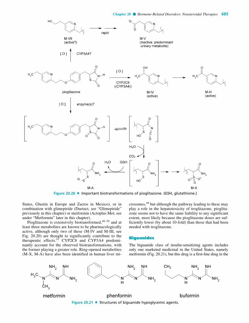
Chapter 20 Hormone-Related Disorders: Nonsteroidal Therapies 685
States, Glustin in Europe and Zactos in Mexico), or incombination with glimepiride (Duetact, see “Glimepiride”previously in this chapter) or metformin (Actoplus Met; seeunder “Metformin” later in this chapter).
Pioglitazone is extensively biotransformed,48–50 and atleast three metabolites are known to be pharmacologicallyactive, although only two of these (M-IV and M-III, seeFig. 20.20) are thought to significantly contribute to thetherapeutic effects.51 CYP2C8 and CYP3A4 predomi-nantly account for the observed biotransformations, withthe former playing a greater role. Ring-opened metabolites(M-X, M-A) have also been identified in human liver mi-
crosomes,48 but although the pathway leading to these mayplay a role in the hepatotoxicity of troglitazone, pioglita-zone seems not to have the same liability to any significantextent, most likely because the pioglitazone doses are suf-ficiently lower (by about 10-fold) than those that had beenneeded with troglitazone.
Biguanides
The biguanide class of insulin-sensitizing agents includesonly one marketed medicinal in the United States, namelymetformin (Fig. 20.21), but this drug is a first-line drug in the
Figure 20.20 Important biotransformations of pioglitazone. (GSH, glutathione.)
Figure 20.21 Structures of biguanide hypoglycemic agents.
0666-0704_17865_Ch20.qxd 12/4/09 5:53 AM Page 685

treatment of type 2 diabetes, for which it is prescribed heav-ily, alone and in combinations. Having been brought to mar-ket in France in 1979 (though not until 1995 in the UnitedStates), metformin has a long history of use, despite whichfact the mechanisms underlying its effects remain uncer-tain.52–56 Activation by metformin of adenosine monophos-phate-activated protein kinase (AMPK) has commonly beenstated as the molecular mechanism in recent years; however,the as-yet-to-be-identified primary target(s) must either beupstream of AMPK, or trigger downstream mechanisms thatdeliver stimulatory feedback upstream of AMPK, or causechanges in crosstalk mechanisms (such as those involvinginsulin receptor substrate 1) to indirectly enhance AMPK ac-tion, or some combination thereof. A month of chronic met-formin therapy increases basal levels of protein kinase C typezeta (PKC�) in the muscle tissue of type 2 diabetics, in turnrestoring glucose handling to a more normal state,57,58
and PKC� acts through serine-threonine kinase 11(STK11/LKB1) to modulate AMPK activity;55 however,PKC� is clearly not the direct macromolecular target ofmetformin, either. Some evidence suggests that subtle
metformin-caused changes in mitochondrial membrane po-tential may circuitously bring about the beneficial collateralchanges in other pathways, including the aforementionedPKC�→STK11→AMPK signaling chain; metformin inhibitscomplex I (NADH:ubiquinone dehydrogenase) of the elec-tron transport chain.59,60 In any case, over a period of weeks,metformin enhances the sensitivity of various cells of thebody to insulin. Recently, much activity has centered on fullycharacterizing the roles of the organic cation transporter 1(OCT1), the functioning of which is clearly essential to met-formin’s action.61 Presumably, this transporter allows thedrug to reach its intracellular target(s), and pharmacogenomicvariations with respect to the OCT1 complex that decrease orabolish the effectiveness of metformin have been identi-fied.61,62 OCT1 is richly expressed in those hepatocytes par-ticipating most directly in the regulation of glucose levels.
The bioavailability of metformin at normal clinicaldoses ranges from 40% to 60%, which is quite high forsuch an extensively ionized and hydrophilic drug (calcu-lated log D � �6 at pH 7)63; the biguanide moiety is ex-tremely basic (Fig. 20.22), thus, metformin exists almost
686 Wilson and Gisvold’s Textbook of Organic Medicinal and Pharmaceutical Chemistry
Figure 20.22 Biguanides: basis of the “biguanide” designation, tautomeric forms ofthe protonated species, and canonical (resonance) structures representing delocaliza-tion of the positive charge in the conjugate acid.
0666-0704_17865_Ch20.qxd 12/4/09 5:53 AM Page 686

Chapter 20 Hormone-Related Disorders: Nonsteroidal Therapies 687
exclusively as protonated, positively charged molecularspecies throughout the entire physiological pH range.Absorption from the gut was recently shown to occur by aparacellular route, which, surprisingly, was found to be sat-urable;64 the exact molecular basis for this observation, ifyet known, has not been reported. Renal elimination is alsotransporter mediated (active tubular secretion). Eliminationof absorbed metformin is essentially 100% by the kidneys,at a rate about 3.5-fold creatinine clearance, thus renal in-sufficiency precludes metformin pharmacotherapy becauseof the resultant risk of lactic acidosis that otherwise essen-tially never occurs with this drug. Other drugs of this class(phenformin, buformin, structures Fig. 20.21) were aban-doned because of lactic acidosis-linked fatalities, eventhough the incidence with phenformin was actually quiterare. Little metformin binding to plasma proteins occurs,but partitioning into erythrocytes is considerable, signifi-cantly impacting the PK of this drug.
Metformin is N,N-dimethylimidodicarbonimidic di-amide, but can accurately and much more simply be namedas 1,1-dimethylbiguanide. Metformin is available, as its hy-drochloride salt, in tablets ranging in strengths from 500-mgto 1-g (Glucophage, numerous generics), extended-releasetablets (Fortamet, Glumetza), and an oral solution (Riomet);and in combinations with rosiglitazone (Avandamet),pioglitazone (Actoplus Met), glipizide (Metaglip, generics),glyburide (Glucovance), repaglinide (Prandimet), and mostrecently, sitagliptin (Janumet).
�-Glucosidase Inhibitors
Medicinals acting as �-glucosidase inhibitors slow thebreakdown of disaccharides (notably sucrose) and starch-
derived polysaccharides into monosaccharides in the gas-trointestinal (GI) tract, delaying production and therebyabsorption of glucose following consumption of meals.This approach to reducing peak postprandial serum glu-cose levels is highly effective, with the caveat that thesedrugs cause very uncomfortable GI side effects in a highpercentage of patients; fortunately, artful inception of ther-apy, with monitoring, can greatly reduce the impact of thisliability. The �-glucosidase inhibitors marketed in theUnited States include acarbose and miglitol; an additional�-glucosidase inhibitor, voglibose, is marketed in Japanand several other countries including Brazil (structuresFig. 20.23).
Digestion of amylose and amylopectin forms of starchbegins with reactions catalyzed by a pair of related �-amy-lases, which are endoglucosidase enzymes contained inpancreatic secretions. Complete digestion also requires de-branching for amylopectin, and further cleavage of variousdisaccharides and small oligosaccharides. These reactionsare catalyzed by �-glucosidases, which are a highly similarset of enzymes that cleave the glycoside linkage betweenparticular sugar moieties; more specifically, hydrolysis oc-curs at the acetal oxygen bridging the C1 of a glucoseresidue and either C4 or C6 of another glucose in �-(1→4)or �-(1→6) linkage (maltose or isomaltose, respectively;see Fig. 20.24), but not �-(1→4) glucose–glucose linkages(as in cellobiose) or the �-(1→4) galactose–glucose linkagein lactose. �-Glucosidases are exoglycosidases, cleavingterminal glucose residues from the nonreducing end of anoligosaccharide. Four separate maltase enzymes found inthe luminal membrane of enterocytes (more specifically,the brush-border epithelial cells) actually constitute theseactivities: two associated with “sucrase-isomaltase” (SI)
Figure 20.23 Structures of antihyperglycemic �-glucosidase inhibitors on the marketworldwide.
0666-0704_17865_Ch20.qxd 12/4/09 5:53 AM Page 687

activity and two with “maltase–glucoamylase” (MGAM)activity.65,66 The sucrase activity represents catalytic hy-drolysis of the �-(1→2) linkage between glucose C1 andfructose C2 in sucrose.
Acarbose potently and competitively inhibits the MGAMactivity,66 and more specifically, the glucoamylase—but notthe maltase—activity.67 The former activity was recentlyshown to reside within the C-terminal subunit of the en-zyme, which extends into the intestinal lumen, and to whichacarbose binds with considerably higher affinity than it doesto the N-terminal subunit.68 The latter subunit is mostlymembrane-imbedded, although both subunits are catalyti-cally active and are closely related members of the glycosylhydrolase family 31. Miglitol potently inhibits glucoamy-lase and sucrase activities, but not isomaltase activity.69
Voglibose inhibits sucrase and maltase activities70 with suchpotency that clinically effective doses of voglibose are morethan 100-fold lesser than the doses required for acarbose ormiglitol.
Structure–activity relationships for molecules inhibiting �-glucosidases were extensively reviewed in 1996 by Jungeet al.71; however, interpretations are complicated by sub-strate-dependent inhibitor binding and inhibition kinetics forthese enzymes. These complexities are now understood toarise, in part, as manifestations of allosteric crosstalk betweenbinding sites in these multisubunit enzymes.68,72
PRODUCTS
Acarbose is O-4,6-dideoxy-4-[[(1S,4R,5S,6S)-4,5,6-trihydroxy-3-(hydroxymethyl)-2-cyclohexen-1-yl]amino]-�-D-glucopyranosyl-(1→4)-O-�-D-glucopyranosyl-(1→4)-D-glucose. It is sold as 25-, 50-, and 100-mg tablets (Precose,generics) dosed with the first bite of each meal, up to t.i.d.Acarbose potently inhibits the glucoamylase activity ofMGAM �-glucosidases and the sucrase activity of SI �-glucosidases, whereas isomaltase activity is at most mod-erately inhibited at concentrations in the range of those in theintestinal lumen upon oral dosing, and trehalase and lactaseare not significantly inhibited.73 Some inhibition of pancre-atic �-amylases may also contribute to the clinical effects.
Based on the structure of acarbose, it should come as nosurprise that little intact acarbose reaches the systemic circula-tion; instead, acarbose is extensively biotransformed by the ac-tion of microbes and digestive enzymes in the gut. Only about35% of the radioactivity in a dose of 14C-labeled acarbose ad-ministered orally to men was excreted in the urine, appearingas several metabolites, some of which are phase II conversionproducts of 4-methylpyrogallol (O-methyl, O-sulfate, or O-glucuronide conjugates)74; the methylpyrogallol fragmentarises from the terminal valienamine pseudosugar. That thesebiotransformation products are mostly formed in the gut isshown by the fact that nearly 90% of an intravenously admin-istered dose of acarbose is excreted intact in urine.
688 Wilson and Gisvold’s Textbook of Organic Medicinal and Pharmaceutical Chemistry
Figure 20.24 Polysaccharide linkages of relevance to the actions of �-glucosidaseinhibitors, and the relationship of inhibitor structures to postulated high-energy inter-mediates in the hydrolysis of the disaccharide bonds.
0666-0704_17865_Ch20.qxd 12/4/09 5:53 AM Page 688

Chapter 20 Hormone-Related Disorders: Nonsteroidal Therapies 689
Miglitol is (2R,3R,4R,5S)-1-(2-hydroxyethyl)-2-(hy-droxymethyl)-3,4,5-piperidinetriol, or N-hydroxyethyl-1-deoxynorjirimycin. The drug is available as 25-, 50-, and100-mg tablets (Glyset), for administration 25 to 100 mgt.i.d. with meals. In contrast to acarbose, miglitol is highlyabsorbed from a 25-mg oral dose, although absorption is re-ported to be saturable, and accordingly less complete athigher doses.69 Binding to plasma proteins is minimal, andthe volume of distribution (0.18 L/kg, corresponding to12–13 L in a 70-kg adult) is characteristic of compoundsdistributed only to blood and extracellular fluids. Practicallyno systemic biotransformation of miglitol occurs in humans;essentially 100% of an orally administered dose is excretedintact in urine.
Amylin Analogs
Amylin is a circulating hormone released along withinsulin from normally functioning pancreatic � cells. This37-amino-acid polypeptide (Fig. 20.25) incorporates oneintramolecular disulfide bridge between cysteine residuesat positions 2 and 7. The physiological and biochemicalroles of amylin are at present only rather vaguely under-stood, but some of these are discussed here. Amylin, in ex-cess, causes amyloidogenesis. The ramifications of thisfact, in terms of � cell loss in pancreatic islets or progres-sive amyloid tangle formation associated with Alzheimerdisease, is an active area of investigation. The observation,though, that this property is not shared by rat amylin75–77
was exploited in the design of pramlintide, which differsfrom human amylin at only three positions toward the C-terminal end, namely substitution in each case with pro-line (Fig. 20.25).
Pramlintide is the 25-L-proline-28-L-proline-29-L-pro-line trisubstitution product of human amylin. The mar-keted formulation (Symlin) is an acetate salt, the exactcomposition of which may not be public-domain knowl-edge, although salts containing up to four acetic acidmolecules per pramlintide molecule would be possible byvirtue of the free amino terminus and Lys1, Arg11, andHis18 residues. The carboxyl terminus of pramlintide, as inamylin, is amidated, and there are no appreciably acidicmoities in the structure; thus, in solution at the pH of bloodand tissues pramlintide would be present almost entirely asone or the other of two significant molecular species, onehaving a �3 charge (histidine imidazole deprotonated anduncharged) predominating modestly over the quadruplyprotonated (�4) species.
Pramlintide’s labeled pharmacotherapeutic uses are (a)as an adjunct in type 1 diabetics who require mealtimeinsulin injections and yet fail to achieve desired glucosecontrol (15 �g initially, titrated to 30 or 60 �g as tolerated);and (b) as an adjunct in type 2 diabetics who require meal-time insulin injections and yet fail to achieve desiredglucose control (60 �g initially, increased after 3–7 days to120 �g if tolerated), with or without ongoing managementwith metformin or a sulfonylurea. Pramlintide functions asan amylinomimetic; its administration slows gastric empty-ing, suppresses glucagon secretion (in turn inhibiting liverglucose output), and reduces the amount of food consumedvia centrally mediated appetite suppression or satietyenhancement. Abnormal postprandial glucagon secretionoccurs in diabetics, exacerbating hyperglycemic excursions,and pramlintide’s glucagon-suppressing action serves tonormalize this response. The multiple effects of pramlintide(and by analogy, endogenous amylin) are mediated directlyor indirectly, or by some combination thereof, mechanisticunderstanding of which remains modest or even poor inmany aspects.
Amylin receptors consist of short-form calcitonin recep-tors (CT[a]) in hetero-oligomeric complex with receptoractivity–modifying proteins (RAMP1, RAMP2, RAMP3),thereby generating three subtypes of receptor complexes(AMY1(a), AMY2(a), and AMY3(a), or simply AMY1, AMY2,and AMY3).78,79 CT receptors are G-protein–coupled recep-tors (GPCRs), and the short-form CTr is a splice variant lack-ing a 16-amino-acid insert in the first intracellular loop (thus,it is also known as “insert negative”). Compared with manyother GPCRs, particularly those that are targets of currentlymarketed or late-stage investigational pharmacotherapeuticagents, characterization of anatomical distributions and tis-sue- and system-level functions of amylin receptors remainbut lightly addressed in open literature reports.
Pramlintide’s effects upon administration to patients al-most certainly arise in significant measure from directAMY-mediated actions within the brainstem.80 Amylin re-ceptors are abundant in the circumventricular organs, in-cluding the subfornical organ (where AMY1(a) receptors areknown to be expressed in relative abundance), the organumvasculosum lateralis terminalis, and the area postrema(AMY3(a)), where the action of pramlintide (or of �-cell–se-creted amylin) is not precluded by a diffusional BBB.Amylin receptors are also expressed in various other brainareas, in particular the nucleus accumbens, but neitheramylin or pramlintide circulating in the bloodstream arelikely to exert any action at these BBB-shielded locations.Direct amylin receptor–mediated actions of pramlintide in
Figure 20.25 Primary amino acid sequences of human amylin, rat amylin, andpramlintide. Charge symbols above the set of sequences indicate charges of the pre-dominant molecular form of the side chain (the free N-terminal amino group is alsoindicated) at physiological pH (7.4): positive (�), neutral (0), or near-equal (�).Underlines indicate sites of amylin hydrolysis by insulin-degrading enzyme (IDE).
0666-0704_17865_Ch20.qxd 12/4/09 5:53 AM Page 689

the periphery, if any, remain poorly characterized.81
Unsurprisingly, though, the appetite-suppressing actions,and benefits of pramlintide and amylin agonists in general,are receiving significant clinical and scientific attention.
Pramlintide has a relatively short pharmacokinetic half-life of about 50 minutes. One conversion product, des-Lys1-pramlintide, retains full agonist activity and exhibits anelimination time-course similar to that of pramlintide itself.Amylin is degraded by IDE at three sites (as indicatedin Fig. 20.25); in vivo administration of IDE inhibitorsincreases toxicities of the same nature as those associatedwith excessive endogenous or exogenously administeredamylin.82 Pramlintide possesses amino acid residue pairsidentical with those of amylin at each of the IDE cleavagesites, thus this enzyme probably governs the eliminationrates of pramlintide and des-Lys1-pramlintide.
Despite the fact that pramlintide is a relatively large andclearly nonnative polypeptide, systemic immunosensitivityhas not proven to be an issue of clinical concern, even uponlong-term pramlintide pharmacotherapy.
Incretin System Modulating Agents:Incretin Mimetics and DipeptidylPeptidase Type 4 Inhibitors
Several polypeptide hormones have been identified that aresynthesized in the cells of the upper intestines and act to
modulate carbohydrate metabolism. Among these are the in-cretins, a term first coined by La Barre in 1932.83 The “in-cretin effect” refers to insulin secretion stimulated by orallyadministered glucose independent of any increase in bloodglucose. Eventually, two incretins were identified, in 1971and 1985, respectively: glucose-dependent insulinotropicpeptide (GIP) and GLP-1.84 GIP was originally named gas-tric inhibitory peptide by its discoverers,85 but it was laterrenamed in such a way as to retain the acronym while betterreflecting its physiological roles.
The gene encoding proglucagon (PG), besides being ex-pressed in pancreatic islet � cells (and in some neurons inthe hypothalamus and brainstem, and certain taste cells ofthe tongue), is also expressed in endocrine cells of thelower intestinal mucosa. From PG, gut L cells produce thepeptides glicentin, oxyntomodulin, intervening peptide-2,and GLP-1. PG(72–108) is equivalent to GLP-1(1–37), andis derived from PG via the action of the convertase PC1/3.GLP-1(1–37) is further shortened, apparently also byPC1/3, to GLP-1(7–37) or GLP-1(7–36)amide (Fig. 20.26);at least 80% of “GLP-1” released into the bloodstream isthe latter. Secretion occurs upon ingestion of lipid- or car-bohydrate-containing foodstuffs.86–88 Following their re-lease, both bioactive forms of GLP-1 are very short-lived(plasma t1
⁄2 �1–2 min): DPP-4 or DPP-IV rapidly cleavestwo residues from the amino terminus, generating GLP-1(9–37) and GLP-1(9–36)amide, respectively.88 Release of
690 Wilson and Gisvold’s Textbook of Organic Medicinal and Pharmaceutical Chemistry
Figure 20.26 Primary sequences of endogenous human incretins and incretin mimeticpharmacotherapeutic agents. Arrows indicate a peptide bond cleaved by DPP-4 catalysis,carets (^) indicate sites of cleavage by neutral endopeptidase 24.11 (neprilysin) catalysis(double-caret–marked bonds are likely more prominently cleaved). Underscored residuesin the structure of exenatide indicate nonconservative substitutions versus GLP-1 or GIP.The numbering for GLP-1 is “slipped” by six residues; the corresponding numberings aregiven below the GIP structure for reference. (HSA, human serum albumin.)
0666-0704_17865_Ch20.qxd 12/4/09 5:53 AM Page 690

Chapter 20 Hormone-Related Disorders: Nonsteroidal Therapies 691
the bioactive forms of GLP occurs within minutes of foodintake, even though L cells are found predominantly in theileum and caecum, and considerable evidence indicates thatsome combination of neural or endocrine signaling medi-ates this response. GLP-1 acts via specific GPCRs in theportal vein to trigger vagal afferents that, mediated by neu-ronal pathways within the brain, generate efferent signalsstimulating pancreatic insulin release and inhibitingglucagon release.89 GPCRs for GLP-1 (GLP-1Rs) are alsoexpressed in pancreatic islet � cells, as well as in the GItract, kidney, vagus nerve, and neurons in areas of thebrain, including the hypothalamus and hindbrain; in �-isletcells, agonist binding to GLP-1Rs increases the biosynthe-sis of insulin, as well as of glucokinase and the GLUT2glucose transporter.90
Structure–activity studies indicate that the C-terminalportions of the GLP-1 peptides provide an important com-ponent of receptor binding, whereas the N-terminal seg-ment is necessary for receptor activation.91 Accordingly,once DPP-4 removes the N-terminal dipeptide from eitherGLP-1(7–37) or GLP-1(7–36)NH2, agonist activity is lost.GLP-1(9–36)NH2 finds some use as a pharmacological toolfor its antagonist activity at GLP-1Rs.
GIP is synthesized via the action of a prohormone con-vertase on a 153-amino-acid proprotein coded by a specificgene in gut K cells, which are found mainly in the mucosaof the duodenum and jejunum.84 In contrast to the mostly in-direct stimulation of GLP-1 release, GIP secretion is mainlytriggered directly by the enteral presence of glucose andlipids, providing what amounts to an early warning systemfor the pancreas. The bioactive GIP(1–42) is rapidly con-verted (plasma t1
⁄2 �5–7 min) to inactive GIP(3–42). Like thetwo GLP-1 hormones, this inactivation is also effected viaDPP-4 catalyzed cleavage of a dipeptide from the amino ter-minus.92 GIP activates insulin secretion via binding toseven-transmembrane–segment GPCRs on islet � cells, dis-tinct receptors from those activated by GLP-1(7–37) andGLP-1(7–36)NH2.88 GIP-stimulated insulin secretion onlyoccurs when blood glucose is substantively elevated, and re-ceptor knockout studies in mice indicate that GIP receptors(GIPRs, which are also present in brain, pituitary,adipocytes, upper GI tract, adrenal cortex, and bone) arerequired for a proper gluconormative response to hyper-glycemia. Notably, in type 2 diabetics, exogenously admin-istered (iv) GIP cannot facilitate glucose-stimulated insulinsecretion as it does in healthy patients.
Relatively recently, compelling direct evidence fromrodent and in vitro experimentation and some indirect butinconclusive evidence from human clinical studies of in-cretin mimetics (see discussion that follows) and DPP-4inhibitors suggests that incretins may exert preservative,proliferative, and even neogenerative effects on �-cellmass and �-cell populations, thereby arresting—and po-tentially even reversing—the progressive declines thathave, heretofore, been inevitable in type 2 diabetes.90,93 Ifso, a truly new age in the treatment of this common and in-sidious disorder has begun, particularly when technologyallows reliable detection of prediabetic states amenable toprophylactic treatment.
Exenatide, at the time of this writing, is currently theonly incretin mimetic approved (2005) for the U.S. market(Byetta, Amylin Pharmaceuticals). Exenatide must be
administered parenterally (subcutaneous [sc]); injection istypically made in the abdomen about an hour before thefirst and last meals of the day. Exenatide is syntheticallyproduced exendin-4, a 39-residue hormone (see Fig. 20.26)present in the venom of the Gila monster. Exenatide’sactions mimic those of GLP-1; exenatide differs fromGLP-1 in 14 of its first 30 amino acids (the length of GLP-1(7–36)NH2), although at least four of these substitu-tions may be regarded as conservative. Exenatide is not,however, a DPP-4 substrate, by virtue of the presence of aglycine rather than an alanine in the second position, andpartly for this reason has a greatly increased serum half-lifeof 2 to 3 hours94; some of the other amino acid changesalmost certainly also render exenatide less susceptibleto neutral endopeptidase 24.11 (NEP-24.11),95 which is amembrane-bound zinc metallopeptidase also known asneprilysin, and which may account for as much as half ofthe elimination of GLP-1(7–37) and GLP-1(7–36)NH2.Although approved as adjunctive therapy (with metformin,or with a sulfonylurea, or with metformin � a sulfony-lurea, or with metformin � a thiazolidinedione), in factnone of these other medicinals are necessary for exenatideto produce its clinically beneficial effects. Exenatide ad-ministration alone in pharmacological doses restores theearly stage insulin release, loss of which is a hallmark oftype 2 diabetes, and increases the robustness of the second-stage insulin release. Premeal exenatide injections (5 or 10�g) also suppress glucagon release following a meal (butdo not affect normal hypoglycemia-stimulated glucagonrelease), slow gastric emptying, induce satiety, and pro-mote normalcy of postprandial glucose uptake while sup-pressing hepatic glucose release. Moreover, the glucose de-pendency of exenatide-stimulated insulin secretion meansthat this drug cannot cause abnormal hypoglycemicepisodes, although this can, of course, happen when thedrug is adjunctive to a sulfonylurea. Although there areGLP-1 receptors expressed in hindbrain and hypothalamusthat are probably involved in mediating exenatide-inducedsatiety effects, evidence suggests that direct agonist actionon these receptors by circulating exenatide is likely notinvolved.96
Liraglutide is GLP-1(1–37) modified at Lys26: the �-amino group is acylated with an (N-hexadecanoyl)glutam-�-yl moiety (Fig. 20.26) that noncovalently but avidly binds tohuman serum albumin. (Also, Lys34 is conservatively re-placed with Arg.) These changes provide for a greatly ex-tended serum t1
⁄2 of �10 to 15 hours after sc administration(0.6–1.8 mg), allowing for once-daily dosing.97–99
Liraglutide (Victoza, Novo Nordisk) is awaiting approvalby the FDA, EMEA, and the Kōrō-shō (Japan).
Albiglutide (also known as naliglutide, GSK 716155) isa sequential dimer of GLP-1(7–36)(Ala8→Gly) covalentlylinked to modified human serum albumin (i.e., it is a “fu-sion protein” with modified HSA). This molecule is cur-rently well into phase III clinical trials. Although albiglu-tide, if marketed (Syncria, GlaxoSmithKline), will—likeexenatide—be administered by injection, the half-life andduration of 6 to 7 days100 would allow for weekly adminis-tration.
Taspoglutide (R1583, Roche) is GLP-1 modified atresidues 8 and 35 (see Fig. 20.26) with �-methylalanine in
0666-0704_17865_Ch20.qxd 12/4/09 5:53 AM Page 691

place of the Ala and Gly, respectively. These modificationsgreatly retard degradation by DPP-IV and neutral endopep-tidase 24.11, respectively (see preceding discussion for exe-natide). The phase III trials, which commenced in 2008, arebeing conducted with a zinc-based preparation, prolongingthe duration in a manner similar to zinc-containing insulinpreparations, so that once-weekly injections of 20 to 30 mgwill be possible.
Besides the aforementioned list of products, a number ofother incretin mimetics are being created and developed. Along-acting version of exenatide (“exenatide LAR”) consist-ing of microspheres of a poly(lactide-coglycolide) poly-meric matrix has been devised, from which exenatide isslowly released from the injection site through diffusion anderosion.101 An adenoviral vector has also been created forexenatide “gene therapy,” and was reported to successfullygenerate exenatide and modulate glucose control and otherGLP-1-mediated responses in mice for at least 15 weeks fol-lowing treatment102; other gene therapy approaches for de-livering GLP-1 itself are also being explored. Boc5,103 acyclobutanedicarboxylic acid (CAS Reg. No. 917569-14-3,structure still undisclosed), is reported to be a nonpeptideGLP-1 agonist exerting incretin effects in C57BL/6J anddb/db mice. The variety of the developments describedabove provide some sense of the current level of interest inincretin mimetics among researchers and clinicians, interestundoubtedly based on perceived strength of promise. Therelative merits of incretin receptor agonists as compared tothe indirect incretin-enhancing approach achieved with in-hibitors of DPP-IV (discussed next) or neutral endopepti-dase 24.11 largely remain to be discerned, which will likelyhappen during the coming decade or so.
DPP-4 or DPP-IV is a serine protease that selectivelybinds and hydrolyzes substrates that incorporate alanine (asin GLP(7–36)NH2, see earlier discussion and Fig. 20.26) orproline (as in neuropeptide Y and peptide YY) at the sec-ond position. Because there are many serine proteases, ex-cellent selectivity is an essential design feature requisite tobestowing pharmacotherapeutic usefulness.104 In particu-lar, minimizing inhibition of the closely related enzymesDPP-2, fibroblast activation protein (FAP), DPP-7 (alsoknown as quiescent cell proline peptidase, QPP), DPP-8,and DPP-9 has proved to be crucial.105,106 These serine pro-teases are among a relatively few peptidases that can cleaveat proline, and they do so on the carboxyl side. HumanDPP-4 is a 766-amino-acid transmembrane glycoproteinand a member of peptidase clan SC, and more specificallya member of family S9, subfamily S9B. Clan SC serineproteases include a catalytic triad in the specific order Ser,Asp, His—different from the classical serine proteases suchas trypsin (class SA: His, Asp, Ser) or subtilisin (class SB:Asp, His, Ser).
DPP-4 was first identified in 1967, and early inhibitorswere created by relatively simple modifications of proline-based structures. After the later realization that this enzymemight likely be an important pharmacotherapeutic target,compound library screening provided inhibitors with a vari-ety of new base structures, including the lead compound fromwhich sitagliptin was created (Fig. 20.27). In due course, asignificant number of x-ray crystal structures were generated,for complexes in which the catalytic pocket is occupied withvarious inhibitors, the first two reports occurring in
2003.107,108 This body of structural information very quicklygrew to be sufficient to enable true de novo structure-baseddrug design; an excellent overview is provided by Kuhn etal.109 The catalytic site of DPP-4 is extracellular, and in-cludes an �/�-hydrolase fold containing the Ser630-Asp708-His740 catalytic triad, and an eight-bladed �-propellerdomain108 that participates in the binding of various knowninhibitors, including sitagliptin.109 The S1 pocket within theprotein, which accommodates proline (or, less favorably, ala-nine, as is found in the GLP-1 peptides and in GIP), is en-closed by the largely hydrophobic residues Tyr631, Val656,Trp662, Tyr666, and Val711, and among the reported x-raystructures of enzyme:inhibitor complexes the shape of thispocket changes very little, even for structurally very disparateinhibitors.
The discovery of sitagliptin at Merck preceded theavailability of x-ray structures, and was accomplished bymore classical (albeit highly refined) screening and lead-optimization approaches. Thornberry and Weber106 pro-vide an informative account of this process. Althoughinitially commencing with preclinical development of in-licensed compounds, extensive screening of a very largecompound collection was also conducted c. 2001, fromwhich effort these authors report that relatively few hits—and only three suitable leads—were gleaned. Sitagliptinwas ultimately created (Fig. 20.27) from one of these leads,which was a �-amino acid derivative and thus unlike the �-amino-acid–based compounds divulged prior to this dis-covery. The in-licensed compounds were abandoned atabout the same time because of toxicities that were foundto be traceable to DPP-8 or DPP-9 inhibition; the existenceof these enzymes, as well as of QPP (see previous discus-sion), were first reported circa this same time frame. Somecharacteristics of a few of the “way-station” compoundscreated during the lead optimization process that led tositagliptin provide a sense of the process, particularly thelarge changes in target-level activity, biopharmaceuticscharacteristics, and deleterious effects profile, which canresult from seemingly very subtle changes to the molecularstructure. The necessity to monitor and optimize multiplecharacteristics simultaneously (such as target affinity andoral bioavailability) during the process of creating a mar-ketable drug should also be readily apparent from this out-line. Sufficient preclinical studies were completed forsitagliptin by January 2002 that it was moved to first prior-ity for development. (Marketing approval was obtainedfrom the FDA in late 2006, representing an impressivelyand exceptionally compressed timeline from screening hitidentification to marketing.)
An x-ray structure of DPP-4 with bound sitagliptin laterprovided an understanding of the structure–affinity observa-tions.110 The trifluorophenyl moiety occupies the S1 pocket.The triazolopyrazine-ring-attached trifluoromethyl sub-stituent—where a buildup of electron density occurs—situ-ates near the positively charged side chain of Arg358; theprotonated (cationic) form of the primary amino groupforms a hydrogen bond with Tyr662, and salt bridges to the�-carboxylates of Glu205 and Glu206; the triazolopiperazinegroup engages the phenyl group of residue Phe357.
Other DPP-4 inhibitors are either on the market else-where (vildagliptin in the EU, Novartis [Galvus]), or areunder regulatory review (alogliptin, Takeda; saxagliptin,
692 Wilson and Gisvold’s Textbook of Organic Medicinal and Pharmaceutical Chemistry
0666-0704_17865_Ch20.qxd 12/4/09 5:53 AM Page 692

Chapter 20 Hormone-Related Disorders: Nonsteroidal Therapies 693
Bristol Myers Squibb [Onglyza]),111 or are well along inphase III studies (linagliptin, Boehringer Ingelheim).Three of these (Fig. 20.28) incorporate a nitrile (cyano)moiety: vildagliptin and saxagliptin a 2-cyanopyrrolidine,alogliptin a 2-cyanophenyl. In each case, the nitrile en-gages the enzyme at the same position as does the scissilebond in peptide substrates such as GLP-1 or GIP. The ni-trile group is attacked by the hydroxyl of the catalytic ser-ine (Ser630), forming a covalent imidate species(Fig. 20.29). Hydrolysis of this imidate occurs slowly;thus, these inhibitors may be described as slowly re-versible or pseudoirreversible.
Sitagliptin phosphate is the 1:1 phosphoric acid salt ofsitagliptin free base (i.e., (2R)-4-oxo-4-[3-(trifluoromethyl)-5,6-dihydro[1,2,4]triazolo[4,3-a]pyrazin-7(8H)-yl]-1-(2,4,5-trifluorophenyl)-2-butanamine), see Figure 20.30, and is mar-keted (Junuvia, 2006) in 25-, 50-, and 100-mg tablets. Acombination product with metformin (Janumet, 2007) intwo strengths (50 mg/500 mg and 50 mg/1,000 mgsitagliptin/metformin) is also available, and sitagliptin mayalso be prescribed with a thiazolidinedione, or possibly a sul-fonylurea. The phosphate salt provides very high water solu-bility.112 The bioavailability of orally administered sitagliptinis �87%.113 The drug exhibits relatively low plasma protein
Figure 20.27 Sitagliptin creation outline. The �-amino acid–lead compound (1) wasdiscovered by screening of a large library of compounds. Compound 2 bound with veryhigh affinity to the enzyme but was not endowed with useful oral bioavailability.Changing the metabolically labile ethyl substituent in compound 3 to trifluoromethylgave a compound (4) with acceptable bioavailability, albeit binding with considerablylower affinity than molecule 2. Relocating the fluorines on the phenyl ring improved thebinding affinity but a cardiovascular side effect appeared in preclinical studies of 5 indogs. Adding one additional fluorine increased the affinity and eliminated the worri-some preclinical side effect; molecule 6 was ultimately marketed as sitagliptin phosphate.
0666-0704_17865_Ch20.qxd 12/4/09 5:53 AM Page 693

binding (�38%), a relatively large volume of distribution(198 L), and a terminal elimination half-life of �12 hours.About 79% of a 100-mg oral dose is excreted unchanged inurine, the balance as trace-level metabolites (CYP3A4, lessercontribution by CYP2C8) in urine or feces: 87% of adminis-tered radioactivity is excreted in urine, and 13% in feces.Active tubular excretion is reported to play a key role in renalclearance of unchanged drug, and may be mediated at least inpart via the organic anion transporter hOAT-3.
Glucose Elevating Agents
Apart from administration of glucose, either glucagon(rDNA origin) or diazoxide represent therapeutic options for
pharmacotherapy of hypoglycemia. Applications are limitedand specialized, as will be discussed here.
Glucagon produced by recombinant DNA technology ismarketed by Novo Nordisk (glucagon [recombinant] hy-drochloride, GlucaGen HypoKit, GlucaGen Diagnostic Kit)and by Eli Lilly (glucagon [recombinant] Emergency Kit).Details regarding the structure and chemistry of this 29-amino-acid peptide are available in Chapter 27. As dis-cussed earlier in this chapter, endogenous glucagon isproduced from the gene-derived protein PG in the � cells ofthe islets of Langerhans in the pancreas. The core functionof this hormone is to renormalize blood glucose levels whenthey fall too low, by stimulating production from glycogen
694 Wilson and Gisvold’s Textbook of Organic Medicinal and Pharmaceutical Chemistry
Figure 20.28 Structures of DPP-4 inhibitors on the global market or in advancedstages of clinical development.
Figure 20.29 Covalently bound imidate intermediate formed with nitrilo-containingDPP-4 inhibitors.
0666-0704_17865_Ch20.qxd 12/4/09 5:53 AM Page 694

Chapter 20 Hormone-Related Disorders: Nonsteroidal Therapies 695
stores in liver and muscle, and stimulating hepatic gluconeo-genesis. Glucagon also elicits biochemical processes (suchas fatty acid oxidation) that supply the needed precursors forgluconeogenesis. Glucagon supplied exogenously (i.e., in-jected) in response to emergency hypoglycemia acts rapidlyto elicit these same responses. Clinically, glucagon also pro-vides an alternative to cholinergic antagonists for reducingGI motility and secretory activity during radiologic imagingprocedures.
Diazoxide is 7-chloro-3-methyl-4H-benzo[e][1,2,4]thiadiazine-1,1-dioxide (structure Fig. 20.31), and is cur-rently available in the United States only as a 50-mg/mLoral suspension (Proglycem); discontinued formulations in-cluded capsules for oral administration, and injectable formsthat typically found use for indications other than hypo-glycemic conditions. Diazoxide is a cyclic benzenesulfon-amide, although the free acid in solution can exist in threetautomeric forms, and the 4H tautomer most likely predom-inates to a very high proportion.114 Partly because of theadditional nitrogen in the quinazoline ring structure, themolecule is somewhat more acidic (pKa �8.4,115 8.6116)than benzenesulfonamide (pKa �10).
Diazoxide acts as an ATP-sensitive potassium channelopener, thus inhibiting basal insulin secretion. This actionmay be viewed as the reverse of the insulin secretagogue ac-tions of the sulfonylureas discussed earlier in this chapter;however, diazoxide is much less selective for SUR1 overSUR2A, SUR2B, or SUR2C receptor types,117 acts on mito-chondrial ATP-sensitive potassium channels,118 and exertsvarious extrapancreatic effects that will not be discussedhere.119 The action of diazoxide on �-islet cells may triggerglucagon release from �-islet cells via paracrine pathways.120
Structurally, diazoxide closely resembles (Fig. 20.31) hy-drochlorthiazide, a diuretic that inhibits the sodium chloridesymporter in the distal convoluted tubule. Diazoxide does notact as a diuretic, however, instead exerting an antidiuretic ef-fect. The discovery/creation/development of pancreas-selec-tive ATP-sensitive potassium channel openers continues tobe an area of ongoing investigation.121–123
Diazoxide binds extensively (�90%) to serum proteins,which greatly retards its renal excretion, by which route thisdrug is almost exclusively eliminated, with a terminal elim-ination half-life about 28 8 hours. Lacking this serumbinding (or active tubular reuptake), the hydrophilicity ofthis molecule would otherwise confer a much more rapidrate of renal excretion.
In contrast to the acute clinical uses of glucagon, diazox-ide finds use in chronic hypoglycemic conditions: inoperableislet cell adenoma or carcinomas, extrapancreatic malignan-cies of insulin-secreting cells, or islet cell hyperplasias. Inchildren, additional indications include congenital hyperinsu-linemia124 and leucine sensitivity. Experimentally, diazoxideis among an array of ATP-sensitive potassium channel open-ers being studied for intermittently bringing about �-cell rest(see discussion of �-cell “fatigue” earlier in this chapter).125
GONADOTROPINS, GONADOTROPIN-RELEASING HORMONE, AND GNRHRECEPTOR AGONISTS ANDANTAGONISTS
As discussed elsewhere in this book (Chapters 25 and 27),the term gonadotropin encompasses three large-peptide
Figure 20.30 Basis of sitagliptin fused ring system nomenclature, and explicit structureof 1:1 phosphate salt.
0666-0704_17865_Ch20.qxd 12/4/09 5:53 AM Page 695
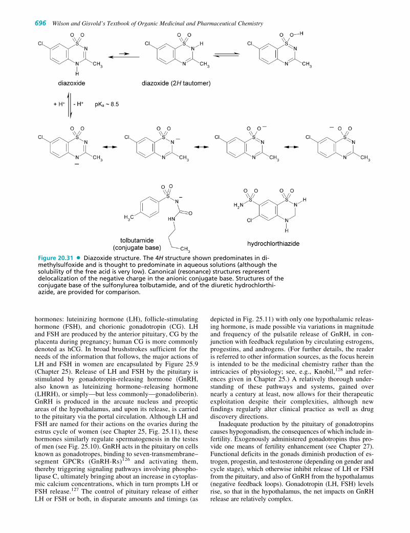
hormones: luteinizing hormone (LH), follicle-stimulatinghormone (FSH), and chorionic gonadotropin (CG). LHand FSH are produced by the anterior pituitary, CG by theplacenta during pregnancy; human CG is more commonlydenoted as hCG. In broad brushstrokes sufficient for theneeds of the information that follows, the major actions ofLH and FSH in women are encapsulated by Figure 25.9(Chapter 25). Release of LH and FSH by the pituitary isstimulated by gonadotropin-releasing hormone (GnRH,also known as luteinizing hormone–releasing hormone(LHRH), or simply—but less commonly—gonadoliberin).GnRH is produced in the arcuate nucleus and preopticareas of the hypothalamus, and upon its release, is carriedto the pituitary via the portal circulation. Although LH andFSH are named for their actions on the ovaries during theestrus cycle of women (see Chapter 25, Fig. 25.11), thesehormones similarly regulate spermatogenesis in the testesof men (see Fig. 25.10). GnRH acts in the pituitary on cellsknown as gonadotropes, binding to seven-transmembrane–segment GPCRs (GnRH-Rs)126 and activating them,thereby triggering signaling pathways involving phospho-lipase C, ultimately bringing about an increase in cytoplas-mic calcium concentrations, which in turn prompts LH orFSH release.127 The control of pituitary release of eitherLH or FSH or both, in disparate amounts and timings (as
depicted in Fig. 25.11) with only one hypothalamic releas-ing hormone, is made possible via variations in magnitudeand frequency of the pulsatile release of GnRH, in con-junction with feedback regulation by circulating estrogens,progestins, and androgens. (For further details, the readeris referred to other information sources, as the focus hereinis intended to be the medicinal chemistry rather than theintricacies of physiology; see, e.g., Knobil,128 and refer-ences given in Chapter 25.) A relatively thorough under-standing of these pathways and systems, gained overnearly a century at least, now allows for their therapeuticexploitation despite their complexities, although newfindings regularly alter clinical practice as well as drugdiscovery directions.
Inadequate production by the pituitary of gonadotropinscauses hypogonadism, the consequences of which include in-fertility. Exogenously administered gonadotropins thus pro-vide one means of fertility enhancement (see Chapter 27).Functional deficits in the gonads diminish production of es-trogen, progestin, and testosterone (depending on gender andcycle stage), which otherwise inhibit release of LH or FSHfrom the pituitary, and also of GnRH from the hypothalamus(negative feedback loops). Gonadotropin (LH, FSH) levelsrise, so that in the hypothalamus, the net impacts on GnRHrelease are relatively complex.
696 Wilson and Gisvold’s Textbook of Organic Medicinal and Pharmaceutical Chemistry
Figure 20.31 Diazoxide structure. The 4H structure shown predominates in di-methylsulfoxide and is thought to predominate in aqueous solutions (although thesolubility of the free acid is very low). Canonical (resonance) structures representdelocalization of the negative charge in the anionic conjugate base. Structures of theconjugate base of the sulfonylurea tolbutamide, and of the diuretic hydrochlorthi-azide, are provided for comparison.
0666-0704_17865_Ch20.qxd 12/4/09 5:53 AM Page 696

Chapter 20 Hormone-Related Disorders: Nonsteroidal Therapies 697
Gonadotropin-Releasing Hormone Agonists
Upon acute administration, GnRH agonists produce an ini-tial stimulation of either LH release (called the “flare” re-sponse) or FSH release (in this case to stimulate ovulation),but upon chronic administration, the effect is to downregu-late pituitary release of gonadotropins. In turn, during theinitial flare response there is an increase in gonadal steroido-genesis (testosterone and dihydrotestosterone in men, es-trone and estradiol in premenopausal women). Followingthe flare response, steroidogenesis decreases over the next
2 to 4 weeks. Examples of therapeutic applications (seeTable 20.6) include controlling timing of estrus cycle eventsin women and—in veterinary medicine—female animals toallow for artificial insemination or in vitro fertilization; sup-pression of precocious puberty; inducing ovarian suppres-sion and a hypoestrogenic state in women when beneficial(e.g., menorrhagia, endometriosis, adenomyosis, or uterinefibroids); induction of hypogonadal states in oncology (no-tably, prostate cancer); and fertility suppression in maledogs. For chronic administration, long-lasting depot injec-tions enable monthly or quarterly administration, and im-plantable devices allow release for as long as a year.
TABLE 20.6 GnRH Agonists
Serum Volume of Protein Serum
Drug Indications Dosing Distribution Binding Time-course Elimination
Leuprolide acetate A, B, C, D Injection, implant, 27 La 43%–49%b (Bolus dose) Not fully(Lupron, Eligard, off-label: or depot; amount terminal characterizedLupron Depot, E, F, G varies from 1–45 mg t1/2 � 3 hr Viadur, generic) for adult indications, (two-
depending on compartment indication and type model)a
of administration Clss � 140 (i.e., immediate- mL/mina
release vs. depot).Histrelin acetate A 50-mg sc implant 58 8 Lc 30 9%c (Bolus dose) Not studied
(Vantas) once every terminal (Supprelin LA) B 12 months t1/2 �3.9
→ 50–60 �g/day 1.0 hrc
release rate (50-mg for Vantas implant)
→ ca. 65 �g/day Clss � 174 release rate 56.5 mL/minc
for Supprelin LATriptorelin A, C, D Depot: im, 3.75 mg 30–33 Ld Minimald (Bolus dose) �42% as intact
pamoate off-label: monthly t1/2 � 2.8 drug in urine(Trelstar LA, F, H, I, J, K LA: im, 11.25 mg 1.2 hrd following ivTrelstar Depot) every 84 days bolusd;
ClT �210 mL/min,ClH �80 mL/min,ClR �91 mL/mind
Toserelin A, C, E, N 3.6 mg (base equiv) (0.25 mg 27%e (0.25-mg �90% in urineacetate implant sc bolus): sc bolus): (20% as (Zoladex) 10.8 mg (base equiv) 44 14 L t1/2 � 4.2 unchanged
implant men, 1.1 hr in men, drug)e
20 4 L t1/2 � 2.3 womene 0.6 hr in
womene
Nafarelin B, C 2 mg/mL intranasal, NA �80%f Bioavailability 44%–55% urineacetate 200 �g/spray 2%–6% (3% of dose(Synarel) B: 1,600 mg/day (200 �g as unchanged
C: starting dose: intranasal)f drug)f
200 �g b.i.d., Peak plasmaincrease to 800 concentration�g/day if needed 10–45 min
t1/2 � 2.5 hr(400 �g
intranasal)f
Indications: A, palliative treatment of advanced prostate cancer; B, treatment of children with central preco-cious puberty; C, endometriosis; D, uterine leiomyomata (fibroids); E, palliative treatment of advanced breastcancer; F, ovarian carcinoma; G, paraphilia; H, pancreatic carcinoma; I, hypergonadism; J, growth hormonedeficiency; K, in vitro fertilization assistance; M, assisted reproduction (LH surge suppression for timing con-trol); N, to bring about presurgical endometrial thinning.aSennello, L. T., Finley, R. A., Chu, S. Y., et al.: J. Pharm. Sci. 75(2):158–160, 1986.bEligard product information (8-Nov-2007 labeling).cVantas product information (8-Nov-2004 labeling).dTrelstar product information (29-Jun-2001 labeling).eZolodex product information (27-Jul-1998 labeling), or Facts and Comparisons’ Web site, accessed April 2009.fSynarel product information (12-Apr-2006 labeling).b.i.d., bis in die (twice a day); im, intramuscular; iv, intravenous; NA, not available; sc, subcutaneous.
0666-0704_17865_Ch20.qxd 12/4/09 5:53 AM Page 697

Nafarelin acetate is formulated for intranasal administration,the only nonparenteral formulation among the currentlyavailable GnRH agonists. Deslorelin acetate currently findsuse only in veterinary medicine, such as for stimulatingovulation in mares (acute administration → flare effect) orhelping to stabilize and maintain high-risk pregnancies inlivestock, although clinical trials in humans for certainbreast cancers, precocious puberty, and congenital adrenalhyperplasia are in progress or completed.
The marketed GnRH agonists are all close structuralanalogs of GnRH itself (Fig. 20.32). They are often called“superagonists” because they have increased potency andincreased duration of action versus the endogenous ago-nist (GnRH), which is rapidly cleaved in the pituitary byendopeptidases (24.11 and 24.15)129 and has a very shorthalf-life (2–4 minutes) in the systemic circulation. The re-ported agonist potencies relative to GnRH for some ofthese molecules are leuprolide (15-fold), buserelin (20-fold), deslorelin (144-fold), and histrelin (210-fold).130 Allof the marketed synthetic agonists incorporate, likeGnRH, pyro-glutamic acid at the amino terminus, in whichthe N-terminal �-amino group has undergone cyclocon-densation with the �-carboxyl moiety of the same gluta-mate residue, forming a five-membered amide-containingring—that is, a lactam moiety (more specifically, this con-stitutes a 5-oxopyrrolidine-2-carboxylic acid residue). The
following four amino acids (positions 2–5) are also thesame in all. Position 6 is the main point of structural vari-ation among these GnRH-analog agonists; substitution isconsistently with a D amino acid to confer peptidase resis-tance (see discussion that follows). Leuprolide (leuprore-lin), buserelin, histrelin, and deslorelin are nonapeptidesrather than decapeptides: They lack the amidated C-termi-nal glycine, instead terminating at Pro9, which is amidatedwith ethylamine, thus forming an N-ethylprolinamide ter-minus. This terminal N-ethyl moiety can be viewed as anabbreviated stand-in for the terminal glycinamide moietyof GnRH. Goserelin terminates in an unusual (among me-dicinal substances) aminocarbonylhydrazide moiety,whereas nafarelin and triptorelin maintain the same struc-ture at the C-terminus as GnRH. The goserelin N-terminuscan be seen as a bioisosteric substitution: It is identical tothat of the natural hormone except that an �NH� is sub-stituted for the methylene (�CH2�) of the terminalglycine. All of the marketed agonists include the stronglybasic Arg8 residue, so that, with no appreciably acidicgroups elsewhere within the structure to provide an inter-nal counterion, a salt form is obligatory; acetate is thecounterion for most. Triptorelin is marketed in the UnitedStates as its pamoate salt (see Fig. 20.32), which providesfor long duration of action by virtue of depot creationupon intramuscular injection.
698 Wilson and Gisvold’s Textbook of Organic Medicinal and Pharmaceutical Chemistry
Figure 20.32 Primary sequences and selected substructures for endogenous humanGnRH, and marketed GnRH agonists.
0666-0704_17865_Ch20.qxd 12/4/09 5:53 AM Page 698

Chapter 20 Hormone-Related Disorders: Nonsteroidal Therapies 699
The elimination processes and fates of these peptidesare not fully characterized. For leuprolide, available infor-mation suggests that elimination is mostly via peptidase-catalyzed cleavage, and subsequent urinary excretion ofpeptide fragments.131 In healthy men, a pentapeptide is theprominent metabolite, and formation is relatively rapid,with levels of this metabolite peaking between 2- and 6-hour postdose. Following administration of the 3.75-mgdepot suspension formulation, less than 5% of an adminis-tered dose was recovered as leuprolide and the pentapep-tide metabolite. Such extensive fragmentation does notoccur with all of these analogous compounds; however,more than 40% of an iv bolus dose of triptorelin, for exam-ple, is recovered intact in urine.132 Although the overallelimination of histrelin has reportedly not been studied fol-lowing insertion of 50-mg implants, biotransformation inhuman hepatic microsomes showed the presence of a sin-gle prominent metabolite resulting from N-deethylation ofthe C-terminal amide moiety.133 Peptidase-generatedhistrelin fragments are also likely, but the molecule is de-signed to be resistant to hydrolysis by endopeptidases24.11 and 24.15. Nafarelin is extensively biotransformed.Following administration of (14C)nafarelin subcutaneouslyto men, at least six metabolites appeared in urine, collec-tively accounting for about half of the dose, with �3%of the dose appearing in urine as unchanged nafarelin.
Hexapeptide Tyr5–Gly10(NH2) was the most prominentmetabolite.134 The metabolites collectively persist inserum for much longer than the parent drug.
Gonadotropin-Releasing HormoneAntagonists
To some extent, the clinical uses of GnRH antagonists(Table 20.7) parallel those of the agonists discussed previ-ously; however, antagonists do not, of course, generate theflare response.135 Instead, GnRH receptors in the go-nadotrope cells of the pituitary are blocked, so that pituitaryoutput of FSH and LH is reduced or abolished. This re-sponse occurs rapidly, unlike the desensitization that devel-ops over several weeks when GnRH agonists are used forgonadotropin suppression. Return to a pretreatment statealso occurs relatively quickly, within 2 days after stoppinglower doses of GnRH antagonists (except, of course, withdepot injections).
The marketed GnRH antagonists (Fig. 20.33) were de-signed based on the GnRH structure,136 but contain manymore alterations as compared with agonists, including moreD amino acids, and numerous unnatural or derivatized sidechains (the full structure of ganirelix, Fig. 20.34, providesan exemplary illustration). His2 was identified early on asimportant to receptor activation,137 and this residue is re-
TABLE 20.7 GnRH Antagonists
Serum Volume of Protein Serum
Drug Indications Dosing Distribution Binding Time-course Elimination
Cetrorelix A (C, D, E) 0.25 mg daily, sc �1.2 L/kga 86%a tmax �1 hr Urine (unchanged,(Cetrotide) or 3 mg once, sc (0.25 mg sc) slow) bile
tmax �1.5 hr (unchanged �(3 mg sc) metabolites, slow)
highly nonlinearpharmacokinetics(dose-dependent)a
Ganirelix A 0.25 mg daily, sc 43.7 L (single 82%b t1/2 � 13 hr Feces (75%), diacetate 0.25-mg iv (single 0.25-mg urine (22%)
dose), 76.5 L dose), t1/2 � 16 hr(daily (daily dosing)b
dosing)b
Degarelix acetate C sc �1 L/kg upon �90%c Depot formed upon �9 L/hr Clss in240 mg initial iv adminc injection; serum patients; peptide80 mg once/ concentration hydrolysis in
28 days profile highly hepatobiliary dependant on system, mainlyinjection site concc biliary excretion
(20%–30% renalexcretionfollowing depotinjection)c
Abarelix acetate B Depot 0.42 L/kgd NA tmax �1 hr, 13% of a 15 �g/kg(Plenaxis) t1/2 � 5 2 hr im dose excreted[Germany] (single 15 �g/kg unchanged in
im dose)d urined
Indications: A, assisted reproduction (LH surge suppression for timing control); B, in oncology, to reduce theamount of testosterone made in patients with advanced symptomatic prostate cancer for which no othertreatment options are available; said to overcome the need for anti-androgen coprescribing; C, hormone-sen-sitive cancers of the prostate; D, hormone-sensitive cancers of the breast (in premenopausal/perimenopausalwomen); E, certain benign gynecological disorders (endometriosis, uterine fibroids and endometrial thinning).aCetrotide product information (FDA labeling approved 4-Apr-2008).bGanirelix Acetate (Organon) product information (30-June-2008 FDA labeling).cDegarelix Acetate (Ferring) product information (24-Dec-2008 FDA labeling).dWong, S. L., Lau, D. T., Baughman, S. A., et al.: J. Clin. Pharmacol. 44(5):495–502, 2004.iv, intravenous; im, intramuscular; NA, not available; sc, subcutaneous.
0666-0704_17865_Ch20.qxd 12/4/09 5:53 AM Page 699

700 Wilson and Gisvold’s Textbook of Organic Medicinal and Pharmaceutical Chemistry
Figure 20.33 Primary sequences and selected substructures for marketed GnRH antagonists.
placed in the antagonists with structural moieties that en-gage the receptor but do not allow receptor activation.Additional structure–activity information came from studiesof cyclic peptides,138,139 which, together with informationfrom protein structure modeling (see discussion that fol-lows), indicated a relatively large binding pocket formedwithin the bundle of the transmembrane helices I to VII ofthe GnRH-R. (Although there are genes corresponding toGnRH-R1 and GnRH-R2 subtypes, the latter is apparently
not expressed in humans.) Abarelix incorporates backboneN-methylation at Tyr5. All are decapeptides like GnRHitself, but unlike GnRH, incorporate alaninamide rather thanglycinamide at the C-terminus. Rather than having a pyro-Glu at the N-terminus as in the native hormone, all are in-stead N-acetylated. Each has at least one strongly basicamino acid in its sequence, at residue 8, thus these peptidesare formulated as salts. The counterion is acetate in allcases. Ganirelix is a diacetate, having an additional basic
Figure 20.34 Full structure of ganirelix.
0666-0704_17865_Ch20.qxd 12/4/09 5:53 AM Page 700
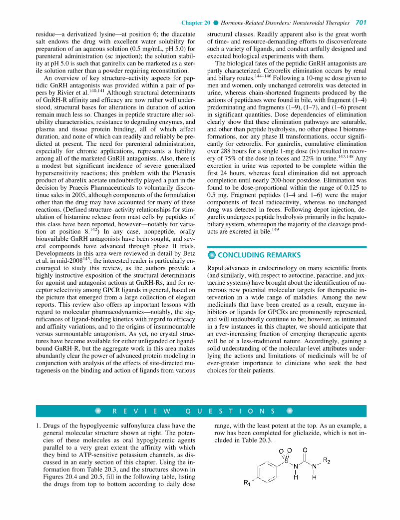
Chapter 20 Hormone-Related Disorders: Nonsteroidal Therapies 701
residue—a derivatized lysine—at position 6; the diacetatesalt endows the drug with excellent water solubility forpreparation of an aqueous solution (0.5 mg/mL, pH 5.0) forparenteral administration (sc injection); the solution stabil-ity at pH 5.0 is such that ganirelix can be marketed as a ster-ile solution rather than a powder requiring reconstitution.
An overview of key structure–activity aspects for pep-tidic GnRH antagonists was provided within a pair of pa-pers by Rivier et al.140,141 Although structural determinantsof GnRH-R affinity and efficacy are now rather well under-stood, structural bases for alterations in duration of actionremain much less so. Changes in peptide structure alter sol-ubility characteristics, resistance to degrading enzymes, andplasma and tissue protein binding, all of which affectduration, and none of which can readily and reliably be pre-dicted at present. The need for parenteral administration,especially for chronic applications, represents a liabilityamong all of the marketed GnRH antagonists. Also, there isa modest but significant incidence of severe generalizedhypersensitivity reactions; this problem with the Plenaxisproduct of abarelix acetate undoubtedly played a part in thedecision by Praecis Pharmaceuticals to voluntarily discon-tinue sales in 2005, although components of the formulationother than the drug may have accounted for many of thesereactions. (Defined structure–activity relationships for stim-ulation of histamine release from mast cells by peptides ofthis class have been reported, however—notably for varia-tion at position 8.142) In any case, nonpeptide, orallybioavailable GnRH antagonists have been sought, and sev-eral compounds have advanced through phase II trials.Developments in this area were reviewed in detail by Betzet al. in mid-2008143; the interested reader is particularly en-couraged to study this review, as the authors provide ahighly instructive exposition of the structural determinantsfor agonist and antagonist actions at GnRH-Rs, and for re-ceptor selectivity among GPCR ligands in general, based onthe picture that emerged from a large collection of elegantreports. This review also offers up important lessons withregard to molecular pharmacodynamics—notably, the sig-nificances of ligand-binding kinetics with regard to efficacyand affinity variations, and to the origins of insurmountableversus surmountable antagonism. As yet, no crystal struc-tures have become available for either unliganded or ligand-bound GnRH-R, but the aggregate work in this area makesabundantly clear the power of advanced protein modeling inconjunction with analysis of the effects of site-directed mu-tagenesis on the binding and action of ligands from various
structural classes. Readily apparent also is the great worthof time- and resource-demanding efforts to discover/createsuch a variety of ligands, and conduct artfully designed andexecuted biological experiments with them.
The biological fates of the peptidic GnRH antagonists arepartly characterized. Cetrorelix elimination occurs by renaland biliary routes.144–146 Following a 10-mg sc dose given tomen and women, only unchanged cetrorelix was detected inurine, whereas chain-shortened fragments produced by theactions of peptidases were found in bile, with fragment (1–4)predominating and fragments (1–9), (1–7), and (1–6) presentin significant quantities. Dose dependencies of eliminationclearly show that these elimination pathways are saturable,and other than peptide hydrolysis, no other phase I biotrans-formations, nor any phase II transformations, occur signifi-cantly for cetrorelix. For ganirelix, cumulative eliminationover 288 hours for a single 1-mg dose (iv) resulted in recov-ery of 75% of the dose in feces and 22% in urine.147,148 Anyexcretion in urine was reported to be complete within thefirst 24 hours, whereas fecal elimination did not approachcompletion until nearly 200-hour postdose. Elimination wasfound to be dose-proportional within the range of 0.125 to0.5 mg. Fragment peptides (1–4 and 1–6) were the majorcomponents of fecal radioactivity, whereas no unchangeddrug was detected in feces. Following depot injection, de-garelix undergoes peptide hydrolysis primarily in the hepato-biliary system, whereupon the majority of the cleavage prod-ucts are excreted in bile.149
CONCLUDING REMARKS
Rapid advances in endocrinology on many scientific fronts(and similarly, with respect to autocrine, paracrine, and jux-tacrine systems) have brought about the identification of nu-merous new potential molecular targets for therapeutic in-tervention in a wide range of maladies. Among the newmedicinals that have been created as a result, enzyme in-hibitors or ligands for GPCRs are prominently represented,and will undoubtedly continue to be; however, as intimatedin a few instances in this chapter, we should anticipate thatan ever-increasing fraction of emerging therapeutic agentswill be of a less-traditional nature. Accordingly, gaining asolid understanding of the molecular-level attributes under-lying the actions and limitations of medicinals will be ofever-greater importance to clinicians who seek the bestchoices for their patients.
R E V I E W Q U E S T I O N S
1. Drugs of the hypoglycemic sulfonylurea class have thegeneral molecular structure shown at right. The poten-cies of these molecules as oral hypoglycemic agentsparallel to a very great extent the affinity with whichthey bind to ATP-sensitive potassium channels, as dis-cussed in an early section of this chapter. Using the in-formation from Table 20.3, and the structures shown inFigures 20.4 and 20.5, fill in the following table, listingthe drugs from top to bottom according to daily dose
range, with the least potent at the top. As an example, arow has been completed for gliclazide, which is not in-cluded in Table 20.3.
0666-0704_17865_Ch20.qxd 12/4/09 5:53 AM Page 701

702 Wilson and Gisvold’s Textbook of Organic Medicinal and Pharmaceutical Chemistry
2. The sulfonylureas are weak acids, and all drugs of thisclass are poorly water soluble, though some more so thanothers. One possible strategy for increasing the dissolu-tion rate for a sulfonylurea to be formulated in an oralsolid dosage form would be to make a salt, which wouldbe expected to have a considerably greater water solubil-ity (although none of the marketed sulfonylureas are cur-rently formulated as their salts). Draw, explicitly, thestructure of the potassium salt of glipizide.
3. (a) Draw the structure of N1-(trans-4-hydroxycyclo-hexyl)-N3-[p-(1-hydroxyethyl)phenyl-sulfonyl]urea.
(b) Your structure, if correctly drawn, represents that ofa metabolite of which hypoglycemic drug discussedin this chapter?
(c) How many biotransformative steps are required toproduce this metabolite from the parent drug you(hopefully) named in part b?
(d) What enzymes are required to catalyze the biotrans-formations that can produce this metabolite from itsparent drug?
(e) Would you expect this metabolite to be a prominentone, or a minor one for this drug, and why?
4. Glipizide undergoes a pair of biotransformations thatwere not discussed in this chapter (a less-prominent routeof biotransformation than those that were mentioned). A
glipizide molecule is acted on by a liver amidase, and oneof the two products thereby produced is then N-acetyl-ated, a reaction catalyzed by an N-acetyltransferase.
(a) Draw the structure of the product of these two steps ofbiotransformation, and also the structure of the otherproduct (a carboxylic acid) produced in the first step.
(b) Would you expect the N-acetylated metabolite to(1) have significant pharmacological activity at themolecular level of the same nature as that of the glip-izide, or (2) to exert hypoglycemic activity of clinicalimportance following oral administration? In eachcase, provide a justification for your answer.
5. In each of the following blanks, list a drug discussed inthis chapter that acts at the molecular level in a way meet-ing the given description.
__________________ an enzyme inhibitor
__________________ a ligand for a protein complex thatactivates gene transcription
__________________ an inhibitor of ion channel opening
__________________ an agonist for a G-protein–coupledreceptor
Drug Daily Dose Range R1 R2
gliclazide 80–240 mg
0666-0704_17865_Ch20.qxd 12/4/09 5:53 AM Page 702

Chapter 20 Hormone-Related Disorders: Nonsteroidal Therapies 703
6. Diazoxide increases the propensity of ATP-sensitivepotassium channels in �-islet cells of the pancreas toopen, whereas glyburide exerts essentially the oppositeeffect. Yet, diazoxide is being studied clinically as aprospective aid in the treatment of type 2 diabetes, andsignificant drug discovery activity currently focuses onfinding molecules that exert the same action as diazoxide,but with greater target selectivity, for the same therapeu-tic purpose. What is the basis for these efforts?
7. Why, based on its molecular nature and physicochemicalproperties, must exenatide be administered only via a par-enteral route?
8. Within the structure of ganirelix (Fig. 20.34), how manyamino acid residues come from the set of 20 amino acidsabundant in proteins (including stereochemistry)? Listthem.
REFERENCES1. Arenillas, J. F., Moro, M. A., and Dávalos, A.: Stroke 38:2196–2203,
2007.2. Blaha, M. J., Bansal, S., Rouf, R., et al.: Mayo Clin. Proc. 83(8):
932–943, 2008.3. Remedi, M. S., and Nichols, C. G.: PLoS Med. 5(10):e206, 2008.4. Winkler, M., Stephan, D., Bieger, S., et al.: J. Pharmacol. Exp. Ther.
322(2):701–708, 2007.5. Babenko, A. P., Aguilar-Bryan, L., and Bryan, J: Annu. Rev. Physiol.
60:667–687, 1998.6. Tusnády, G. E., Bakos E., Váradi A., et al.: FEBS Lett. 402(1):1–3,
1997.7. Roane, D. S., and Bounds, J. K.: Nutr. Neurosci. 2(4):209–225, 1999.8. Takanaga, H., Murakami, H., Koyabu, N., et al.: J. Pharmacy
Pharmacol. 50:1027–1033, 1998.9. Vila-Carriles, W. H., Zhao, G., and Bryan, J.: FASEB. J. 21(1):
18–25, 2007.10. Grell, W., Hurnaus, R., Griss, G., et al.: J. Med. Chem. 41(26):
5219–5246, 1998.11. Sleevi, M.: Insulin and hypoglycemic agents: In Abraham, D. J. (ed.).
Burger’s Medicinal Chemistry and Drug Discovery, 6th ed., vol. 4:Autocoids, Diagnostics, and Drugs from New Biology. New York,John Wiley, 2003, pp. 24–31.
12. Mandič, Z., and Gabelica, V: J. Pharm. Biomed. Anal. 41(3):866–871, 2006.
13. Ashfield R., Gribble, F. M., Ashcroft S. J., et al.: Diabetes8(6):1341–1347, 1999.
14. Geng, X., Li, L., Bottino, R., et al.: Am. J. Physiol. 293(1 Pt. 1):E293–E301, 2007.
15. Scarsi, M., Podvinec, M., and Roth, A.: Mol. Pharmacol. 71(2):398–406, 2007.
16. Shigeto, M., Katsura, M., Matsuda, M., et al.: J. Pharmacol. Exp.Ther. 322(1):1–7, 2007.
17. Kalliokoski, A., Neuvonen, M., Neuvonen, P., et al.: J. Clin.Pharmacol. 48(3):311–321, 2008.
18. Zhang, W., He, Y. J., Han, C. T., et al.: Br. J. Clin. Pharmacol.62(5):567–572, 2006.
19. Ravindran, S., Zharikova, O. L., Hill, R. A., et al.: Biochem.Pharmacol. 72(12):1730–1737, 2006. See also references therein.
20. Zharikova, O. L., Ravindran, S., and Nanovskaya, T. N.: Biochem.Pharmacol. 73:2012–2019, 2007. See also references therein.
21. Kirchheiner, J., Roots, I., Goldammer, M., et al.: Clin.Pharmacokinet. 44(12):1209–1225, 2005. See also referencestherein.
22. Zhang, Y., Si, D., Chen, X., et al.: Br. J. Clin. Pharmacol.64(1):67–74, 2007.
23. Xu, H., Williams, K. M., Liauw, W. S., et al.: Br. J. Pharmacol.153(7):1579–1586, 2008.
24. van Heiningen, P. N., Hatorp, V., Kramer Nielsen, K., et al.: Eur. J.Clin. Pharmacol. 55(7):521–525, 1999.
25. Tornio, A., Niemi, M., Neuvonen, M., et al.: Clin. Pharmacol. Ther.84(3):403–411, 1998.
26. Bidstrup, T. B., Bjornsdottir, I., Sidelmann, U. G., et al.: Br. J. Clin.Pharmacol. 56(3):305–314, 2003.
27. Niemi, M., Backman, J. T., Kajosaari, L. I., et al.: Clin. Pharmacol.Ther. 77(6):468–478, 2005.
28. Niemi, M., Kajosaari, L., Neuvonen, M., et al.: Br. J. Clin.Pharmacol. 57(4):441–447, 2004.
29. Takesada, H., Matsuda, K., Ohtake, R., et al.: Bioorg. Med. Chem.4(10):1771–1781, 1996.
30. Weaver, M. L., Orwig, B. A., Rodriguez, L. C., et al.: Drug Metab.Dispos. 29:415–421, 2001.
31. Sabia, H., Sunkara, G., Ligueros-Saylan, M., et al.: Eur. J. Clin.Pharmacol. 60(6):407–412, 2004.
32. Hanai, T., Miyazaki, R., Kamijima, E., et al.: Internet Electro. J. Mol.Des. 2(10):702–711, 2003.
33. Balant, L.: Clin. Pharmacokinet. 6(3):215–241, 1981.34. Taylor, J. A.: Clin. Pharmacol. Ther. 13(5)(Pt. 1):710–718, 1972.35. Hu, S., Boettcher, B. R., and Dunning, B. E.: Diabetologia 46 Suppl
1:M37–M43, 2003.36. Yki-Järvinen, H.: N. Engl. J. Med. 351(11):1106–1118, 2004.37. Kolak, M., Hannele, Y.-J., Katja K., et al.: J. Clin. Endocrinol. Metab.
92:720–724, 2007.38. Ikeda, Y., Takata, H., Inoue, K., et al.: Diabetes Care 30:e48; doi:10.
2337/dc07-0308, 2007.39. Kanatani, Y., Usui, Y., Ishizuka, K., et al.: Diabetes 56:795–803, 2007.40. Kershaw, E. E., Schupp, M., Guan, H. P., et al.: Am. J. Physiol.
Endocrinol. Metab. 293:E1736–E1745, 2007.41. Evans-Molina, C., Robbins, R. D., Kono, T., et al.: Mol. Cell. Biol.
29:2053–2067, 2009.42. Xu, H. E., Lambert, M. H., Montana, V. G., et al.: Proc. Natl. Acad.
Sci. U. S. A., 98(24):13919–13924, 2001.43. Cronet, P., Petersen, J. F. W., Folmer, R., et al.: Structure 9(8):
699–706, 2001.44. Johnson, B. A., Wilson E. M., Li, Y., et al.: J. Mol. Biol. 298:
187–194, 2000.45. Schimke, K., and Davis, T. M.: Curr. Opin. Investig. Drugs 8(4):
338–344, 2007.46. Cox, P. J., Ryan, D. A., Hollis, F. J., et al.: Drug Metab. Dispos.
28(7):772–780, 2000.47. Baldwin, S. J., Clarke, S. E., and Chenery, R. J.: Br. J. Clin.
Pharmacol. 48(3):424–432, 1999.48. Baughman, T. M., Graham, R. A., Wells-Knecht, K., et al.: Drug
Metab. Dispos. 33(6):733–738, 2005.49. Eckland, D. A., Danhof, M.: Exp. Clin. Endocrinol. Diabetes
108(Suppl 2):S234–S242, 2000.50. Shen, Z., Reed, J. R., Creighton, M., et al.: Xenobiotica 33(5):
499–509, 2003.51. Jaakkola, T., Backman, J. T., Neuvonen, M., et al.: Br. J. Clin.
Pharmacol. 61(1):70–78, 2006.52. Basu, R., Shah, P., Basu, A., et al.: Diabetes 57(1):24–31, 2008.53. Mukhtar, M. H., Payne, V. A., Arden, C., et al.: Am. J. Physiol.
Regul. Integr. Comp. Physiol. 294:R766-R774, 2008.54. Saeedi, R., Parsons, H. L., Wambolt, R. B., et al.: Am. J. Physiol.
Heart Circ. Physiol. 294(6):H2497–H2506, 2008.55. Xie, Z., Dong, Y., Scholz, R., et al.: Circulation 117:952–962, 2008.56. Correia, S., Carvalho, C., Santos, M. S., et al.: Mini Rev. Med.
Chem. 8(13):1343–1354, 2008.57. Luna, V., Casauban, L., Sajan, M. P., et al.: Diabetologia 49(2):
375–382, 2006.58. Temofonte, N., Sajan, M. P., Nimal, S., et al.: Diabetologia 52(1):
60–64, 2009.59. El-Mir, M. Y., Nogueira, V., Fontaine, E., et al.: J. Biol. Chem. 275
(1):223–228, 2000.60. Mooney, M. H., Fogarty, S., Stevenson, C., et al.: Br. J. Pharmacol.
153(8):1669–1677, 2008.61. Shu, Y., Sheardown, S. A., Brown, C., et al.: J. Clin. Invest. 117(5):
1422–1431, 2007.62. Takane, H., Shikata, E., Otsubo, K., et al.: Pharmacogenomics 9(4):
415–422, 2008.63. Saitoh, R., Sugano, K., Takata, N., et al.: Pharm. Res. 21(5):749–755,
2004.64. Proctor, W. R., Bourdet, D. L., and Thakker, D. R.: Drug Metab.
Dispos. 36(8):1650–1658, 2008.65. Nichols, B. L., Eldering, J., Avery, S., et al.: J. Biol. Chem. 273(5):
3076–3081, 1998.66. Quezada-Calvillo, R., Robayo-Torres, C. C., Opekun, A. R. et al.: J.
Nutr. 137(7):1725–1733, 2007.
0666-0704_17865_Ch20.qxd 12/4/09 5:53 AM Page 703

704 Wilson and Gisvold’s Textbook of Organic Medicinal and Pharmaceutical Chemistry
67. Quezada-Calvillo, R., Sim, L., Ao, Z., et al.: J. Nutr.138(4):685–692, 2008.
68. Sim, L., Quezada-Calvillo, R., Sterchi, E. E., et al.: J. Mol. Biol.375(3):782–792, 2008.
69. Scott, L. J., and Spencer, C. M.: Drugs 59(3):521–549, 2000.70. Horii, S., Fukase, H., Matsuo, T., et al.: J. Med. Chem. 29(6):
1038–1046, 1986.71. Junge, B., Matzke, M., and Stoltefuss, J.: Chemistry and structure-
activity relationships of glucosidase inhibitors. Handbook ofExperimental Pharmacology, vol. 119 (Oral Antidiabetics),411–482, 2006.
72. Breitmeier, D., Gunther, S., and Heymann, H.: Arch. Biochem.Biophys. 346(1):7–14, 1997.
73. Caspary, W. F., and Graf, S.: Res. Exp. Med. (Berl) 175(1):1–6,1979.
74. Boberg, M., Kurz, J., Ploschke, H. J., et al.: Arzneimittelforschung40(5):555–563, 1990.
75. Konarkowska, B., Aitken, J. F., Kistler, J., et al.: FEBS J. 273(15):3614–3624, 2006. See also references therein.
76. Götz, J., Ittner, L. M., and Lim, Y. A.: Cell Mol. Life Sci. 66(8):1321–1325, 2009. Review.
77. Lim, Y. A., Ittner, L. M., and Lim, Y. L.: FEBS Lett. 582(15):2188–2194, 2008.
78. Hay, D. L., Christopoulos, G., Christopoulos, A., et al.: Biochem.Soc. Trans. 32(5):865–867, 2004.
79. Qi, T., Christopoulos, G., Bailey, R. J., et al.: Mol. Pharmacol.74(4):1059–1071, 2008.
80. Young, A.: Adv. Pharmacol. 52:47–65, 2005.81. Roth, J. D., Maier, H., Chen, S., et al.: Arch. Neurol.
66(3):306–310, 2009.82. Bennett, R. G., Hamel, F. G., and Duckworth, W. C.: Diabetes
52(9):2315–2320, 2003.83. La Barre, J.: Bull. Acad. R. Med. Belg. 12:620–634, 1932.84. Kim, W., and Egan, J. M.: Pharmacol. Rev. 60: 470–512, 2008.85. Brown, J. C., and Dryburgh, J. R.: Can. J. Biochem. 49:867–872,
1971.86. Holst, J. J.: Annu. Rev. Physiol. 59:257–271, 1997.87. Drucker, D. J:. Cell Metab. 3(3):153–165, 2006.88. Drucker, D. J., Nauck, M. A.: Lancet 368:1696–1705, 2006.89. Vahl, T. P., Tauchi, M., Durler, T. S.: Endocrinology 148:4965–4973,
2007.90. Salehi, M., Aulinger, B. A., and D’Alessio, D. A.: Endocr. Rev. 29:
367–379, 2008.91. Adelhorst, K., Hedegaard, B. B., Knudsen, L. B., et al.: J. Biol.
Chem. 4;269(9):6275–6278, 1994.92. Kieffer, T. J., McIntosh, C. H., and Pederson R. A.: Endocrinology
136:3585–3596, 1995.93. Nicolucci, A., and Rossi, M. C.: Acta. Biomed. 79(3):184–191, 2008.94. Kolterman, O. G., Kim, D. D., Shen, L., et al.: Am. J. Health Syst.
Pharm. 62(2):173, 2005.95. Plamboeck, A., Holst, J. J., Carr, R. D., et al.: Diabetologia 48:
1882–1890, 2005.96. Williams, D. L., Baskin, D. G., and Schwartz, M. W.:
Endocrinology 150:1680–1687, 2009.97. Elbrond, B., Jakobsen, G., Larsen, S., et al.: Diabetes Care 25(8):
1398–1404, 2002.98. Madsen, K., Knudsen, L. B., Agersoe, H., et al.: J. Med. Chem.
50(24), 6126–6132, 2007.99. Russell-Jones, D.: Mol. Cell Endocrinol. 297:137–140, 2009.
100. Matthews, J. E., Stewart, M. W., De Boever, E. H., et al.: J. Clin.Endocrinol. Metab. 93(12):4810–4817, 2008.
101. Kim, D., MacConell, L., Zhuang, D., et al.: Diabetes Care 30(6):1487–1493, 2007.
102. Samson, S. L., Gonzalez, E. V., Yechoor, V., et al.: Mol. Ther.16(11):1805–1812, 2008.
103. Su, H., He, M., Li, H., et al.: PLoS ONE 3(8):e2892, 2008.doi:10.1371/journal.pone.0002892.
104. Pei, Z.: Curr. Opin. Drug Discov. Devel. 11(4):515–532, 2008.105. Van der Veken, P., Van der Haemers, A., and Augustyns, K.: Curr.
Top. Med. Chem. 7:621–635, 2007.106. Thornberry, N. A., and Weber, A. E.: Curr. Top. Med Chem 7(6):
557–568, 2007.107. Rasmussen, H. B., Branner, S., Wiberg, F. C., et al.: Nat. Struct.
Biol. 10:19–25, 2003.108. Thoma, R., Löffler, B., Stihle, M., et al.: Structure 11(8):947–959,
2003.109. Kuhn, B., Hennig, M., and Mattei, P.: Curr. Top. Med. Chem 7:
609–619, 2007.
110. Kim, D., Wang, L., Beconi, M., et al.: J. Med. Chem. 48(1):141–151,2005.
111. Augeri, D. J., Robl, J. A., Betebenner, D. A., et al.: J. Med. Chem.48(15):5025–5037, 2005.
112. Fitzpatrick, S., Taylor, S., Booth, S. W., et al.: Pharm. Dev.Technol. 11(4):521–528, 2006.
113. Januvia product information (22-July-2008): obtained via http://www.accessdata.fda.gov/drugsatfda_docs/label/2008/021995s007lbl.pdf
114. Jakobsen, P., and Treppendahl, S.: Tetrahedron 35(18):2151–2153,1979.
115. Hennig, U. G. G., Chatten, L. G., Moskalyk, R. E., et al.: Analyst(Cambridge, United Kingdom) 106(1262):557–564, 1981.
116. Llinas, A., Glen, R. C., and Goodman, J. M.: J. Chem. Inf. Model.48(7):1289–1303, 2008.
117. Wheeler, A., Wang, C., Yang, K., et al.: Mol. Pharmacol. 74(5),1333–1344, 2008.
118. Riess, M. L., Camara, A. K., Heinen, A., et al.: J. Cardiovasc.Pharmacol. 51(5):483–491, 2008.
119. Adebiyi, A., McNally, E. M., and Jaggar, J. H.: Mol. Pharmacol.74(3):736–743, 2008.
120. Jacobson, D. A., Wicksteed, B. L., and Philipson, L. H.: Diabetes58:304–306, 2009.
121. Sebille, S., de Tullio, P., Florence, X., et al.: Bioorg. Med. Chem.16(10):5704–5719, 2008.
122. de Tullio, P., Boverie, S., Becker, B., et al.: J. Med. Chem.48:4990–5000, 2005.
123. Sharma, B. K., Sharma, S. K., Singh, P., et al.: J. Enzyme Inhib.Med. Chem. 23(1):1–6, 2008.
124. Mohnike, K., Blankenstein, O., Pfuetzner, A., et al.: Horm. Res.70(1):59–64, 2008.
125. Grill, V., and Bjoerklund, A.: Mol. Cell Endocrinol.297(1–2):86–92, 2009.
126. Millar, R. P., Lu, Z. L., Pawson, A. J., et al.: Endocr. Rev.25(2):235–275, 2004.
127. Hsieh, K. P., and Martin, T. F.: Mol. Endocrinol. 6(10):1673–1681,1992.
128. Knobil, E.: Recent Prog. Horm. Res. 36:53–88, 1980.129. Molineaux, C. J., Lasdun, A., Michaud, C., et al.: J. Neurochem.
51(2):624–633, 1988.130. Dineen, M. K., Tierney, D. S., Kuzma, P., et al.: J. Clin. Pharmacol.
45(11):1245–1249, 2005.131. Sennello, L. T., Finley, R. A., Chu, S. Y., et al.: J. Pharm. Sci.
75(2):158–160, 1986.132. Trelstar product information, as per 29-June-2001 FDA labeling.133. Vantas product information, as per 8-Nov-2004 FDA labeling.134. Synarel product information (12-Apr-2006 labeling).135. Felberbaum, R. E., Ludwig, M., and Diedrich, K.: Mol. Cell
Endocrinol. 166(1):9–14, 2000.136. Karten, M. J., and Rivier, J. E.: Endocr. Rev. 7(1):44–66, 1986.137. Vale, W., Grant, G., Rivier, J., et al.: Science 176(37):933–934,
1972.138. Beckers, T., Bernd, M., Kutscher, B., et al.: Biochem. Biophys. Res.
Commun. 289(3):653–663, 2001.139. Koerber, S. C., Rizo, J., Struthers, R. S., et al.: J. Med. Chem. 43(5):
819–828, 2000.140. Samant, M. P., Hong, D. J., Croston, G., et al.: J. Med. Chem. 49
(12):3536–3543, 2006.141. Samant, M. P., Gulyas, J., Hong, D. J., et al.: J. Med. Chem. 48(15):
4851–4860, 2005.142. Ljungqvist, A., Feng, D. M., Tang, P. F., et al.: Biochem. Biophys.
Res. Commun. 148(2):849–856, 1987.143. Betz, S. F., Zhu, Y. F., Chen, C., et al.: J. Med. Chem. 51(12):
3331–3348, 2008.144. Norman, P.: Curr. Opin. Oncol. Endoc. Metab. Invest. Drugs
2(2):227–248, 2000.145. Schwahn, M., Schupke, H., Gasparic, A., et al.: Drug Metab.
Dispos. 28(1):10–20, 2000.146. Cetrotide product information (4-Apr-2008 labeling); Cetrorelix:
Facts and Comparisions 4.0, Wolters-Kluwer Health, accessedMay 2009.
147. Rabasseda, X., Leeson, P., and Castaner, J.: Drugs Future24(4):393–403, 1999.
148. Ganirelix Acetate (Organon) FDA Labeling approved 30-June-2008:http://www.accessdata.fda.gov/drugsatfda_docs/label/2008/021057s007lbl.pdf
149. Degarelix Acetate product information (labeling approved 24-Dec-2008).
0666-0704_17865_Ch20.qxd 12/4/09 5:53 AM Page 704

C H A P T E R 21
Agents Treating Bone DisordersJOHN H. BLOCK
Heterotopic Ossification
This diagnosis consists of various diseases resulting in ab-normal formation of true bone within extraskeletal softtissues. The latter include fascia, tendons, and other mes-enchymal soft tissues. Many times the cause is related totraumatic injury. Treatment includes surgery, radiation, non-steroidal anti-inflammatory drugs (NSAIDS, Chapter 24)and bisphosphonates.
Hypercalcemia of Malignancy
A malignancy is the most common cause of hypercalcemiaand can be an indication of a malignancy, the most commonbeing multiple myeloma, breast, or lung cancer. It is causedby increased osteoclastic activity within the bone. Primaryhyperparathyroidism is the second most common cause ofhypercalcemia. In addition to treating the cause, approveddrug treatments for hypercalcemia of malignancy includebisphosphonates, calcitonin, gallium nitrate, and cinacalcet.
Osteoporosis
Osteoporosis is the most common of the bone diseases. It is apathological condition characterized by decreased bone massand structural deterioration of bone tissue.2 The result is bonefragility leading to increased risk of fractures of the hip, spine,and wrist. Many times the broken hip is considered the resultof the patient falling when, in actuality, the broken hip was theinitial event causing the fall. The economic impact of this dis-ease is tremendous. Osteoporosis is responsible for more than1.5 million fractures annually distributed among 300,000 hips,700,000 vertebrae, 250,000 wrists, and 300,000 other sites. Itis more common in women than men because women, on av-erage, have a smaller bone mass than men. Diagnosis, prior toan actual fracture, is done by bone density measurements.
Osteoporosis has been considered a disease of aging, butinflammation may be an important variable. Calcium flux fa-vors bone mineralization through adolescence. Bone resorp-tion and deposition are in balance in adults and become nega-tive beginning in the late 50s. Loss of calcium from boneincreases relative to calcium deposition onto bone increaseswith menopause. The importance of estrogen is shown by itacting directly on osteoblasts and possibly inhibiting osteo-clasts. The loss of estrogen production results in a significantalteration of the osteoblast–osteoclast ratio because of in-creased osteoclastogenesis and decreased osteoclast apopto-sis. The role of estrogen on osteoblast function has led to thedevelopment of selective estrogen receptor modifiers (SERM)
C H A P T E R O V E R V I E W
Bone is a dynamic tissue. Ninety-nine percent of the body’scalcium is found in bone as a complex mixed salt calledhydroxyapetite [(Ca5(PO4)3(OH)]. Because the crystal unitcell has two molecules of hydroxyapatite, it is also written asCa10(PO4)6(OH)2. Bone is the body’s calcium reservoir withcalcium constantly being removed by osteoclast cells (boneresorption) and laid down by osteoblast cells (calcium depo-sition). The former are derived from monocyte–macrophagecell lines. Together, bone resorption and calcium depositionare known as bone remodeling. The large bones such as thefemur and hip harbor the body’s marrow containing stemcells that produce a wide variety of cells including erythro-cytes, platelets, B cells, T cells, neutrophils, basophils,eosinophils, monocytes, and mast cells.
It should not be surprising that bone, being a dynamic tis-sue, is subject to several diseases, and there are pharmaco-logical treatments for these diseases. These diseases rangefrom deterioration of bone structure, defects in bone remod-eling, and malignancies. Some of these pathological condi-tions have a genetic basis, others can be ascribed to lifestyle,and some attributed to an aging population. This chapterwill be restricted to those bone diseases for which there areapproved drug treatments. Antineoplastic agents used totreat malignancies of the bone are discussed in Chapter 10.
DISEASES OF BONE TISSUE UTILIZINGAPPROVED DRUG THERAPIES
Paget Disease of Bone(Osteo Deformans)
Paget disease can be thought of as improper resorption ofbone. Essentially, bones grow larger leading to deformationof the affected bones and become weaker than normal withincreasing risk of fracture. It can occur on several bones orbe isolated to one bone. It most likely occurs in the pelvis,skull, spine, or leg bones. The bones may become mis-shapen and break more easily. There may or may not bepain. Manifestations of Paget disease tend to appear in thepatient’s late 40s. Although there is some evidence of hered-itary factors, slow viruses also have been considered acause. Diagnosis begins with a determination of the pa-tient’s alkaline phosphatase levels followed by bone scansand x-rays. Drug therapy includes bisphosphonates andcalcitonin.1
705
0705-0710_17865_Ch21.qxd 12/4/09 7:39 PM Page 705

for the prevention and treatment of osteoporosis. These arediscussed in more detail in Chapter 25.
Associated with menopause and the loss of estrogen isan increase in the production of proinflammatory cytokinesincluding interleukin-1 (IL-1), tumor necrosis factor-�(TNF-�), and interleukin-6 (IL-6). Paralleling this in-creased production of proinflammatory cytokines isincreased osteoclastic bone resorption. Of particular impor-tance for osteoclast differentiation and activation and,therefore, progression of osteoporosis, is the receptor acti-vator of nuclear factor-�B (RANK), a membrane-boundreceptor on osteoblast precursor cells and the receptor’sfunctional ligand, RANK ligand (RANKL), also a cy-tokine. When RANKL binds to RANK, there is increasedosteoclastic activity and increased osteoclast numbers be-cause of decreased osteoclast apoptosis. Opposing RANKLis osteoprotegerin, an endogenous inhibitor of RANKL,specifically binding it and blocking its interaction withRANK. Osteoprotegerin, by inhibiting RANKL, normal-izes the osteoblast–osteoclast ratio decreasing bone resorp-tion and loss of calcium from bone.3
There are medical conditions that can increase the risk ofosteoporosis (secondary osteoporosis) or exacerbate anexisting osteoporotic condition. Chronic use of glucocorti-coids can accelerate calcium loss. In addition, some anticon-vulsants (i.e., phenobarbital and phenytoin) can negativelyaffect calcium flux involving bone. Medical conditions thatimmobilize individuals for extensive time periods can alsolead to calcium loss.
There are controllable risk factors that reduce the risk andseverity of the disease. These include anorexia in the agespan when humans are accumulating dietary calcium, a dietlow in calcium and vitamin D (cholecalciferol), inactivelifestyle, cigarette smoking, and excessive use of alcohol.Staying as physically active as possible throughout one’s lifespan is important, particularly focusing on activities that in-clude working against gravity (walking, hiking, jogging, stairclimbing, and dancing). It is important for all age groups toconsume calcium, but because calcium deposition onto boneis at its greatest through adolescence, it is important to con-sume quantities of calcium equal to the daily adequate intake(AI) of, depending on age, 500 to 1,300 mg/day. Youngerpeople can obtain their calcium from diet, primarily dairy.Older individuals may need to take calcium supplements.Paralleling adequate calcium intake is vitamin D consump-tion. The current adult AI is 5 to 10 �g (200–400 IU). Thishas been considered too conservative with recommendationsthat it be increased to 20 to 25 �g (800–1,000 IU). Aperson’s vitamin D status can be checked by measuring25-hydroxycholecalciferol blood levels.
Osteoporosis is a greater problem in women mainlybecause women live longer and have less bone mass than men.Nevertheless, osteoporosis is a disease of older men. A 60-year-old white man has a 25% lifetime risk for an osteoporoticfracture, with consequences more severe than in women.4 The1-year mortality rate in men after hip fracture is twice than inwomen.5 The factors that increase the risk of osteoporotic fac-ture in men are the same as those found in women.6
Osteomalacia
Osteomalacia is caused by a vitamin deficiency and can bethought of as the adult version of rickets. Whereas osteo-
porosis is caused by loss of both calcium and cartilage frombone, the cartilage matrix of bone remains with osteomala-cia. Because of the vitamin D deficiency, dietary calcium isnot transported across the intestinal mucosa to be depositedonto adult bone. The latter process is referred to as mineral-ization of the bone. The result is “soft” bone that becomesmisshapen, because the bones cannot support the body.Inward curvature of the vertebral column and collapsedpelvis are two common results from osteomalacia.Treatment is increased vitamin D intake, but it may not bepossible to correct the misshapen bone.
DRUGS USED TO TREAT DISEASES OF THE BONE
Bisphosphonates
The general bisphosphonate structure is illustrated in Figure21.1. Bisphosphonates should not be confused with bisphos-phates. Bisphosphonates have covalent carbon–phosphorousbonds, and bisphosphates carbon-oxygen-phosphorousbonds. In other words, the latter are phosphate esters verysimilar in chemistry to biochemical phosphate esters. Most invivo bisphosphates are substrates for phosphatase enzymes,whereas bisphosphonates are stable in the presence of hy-drolytic phosphatases. The phosphonate groups are requiredboth for binding to bone hydroxyapatite and cell-mediatedantiresorptive activity.
Bisphosphonates were first reported in 1897. The discov-ery for treating and preventing osteoporosis began in theearly 1960s with Procter & Gamble’s detergent divisionsearching for better additives for use in hard water areas.Bisphosphonates have excellent affinity for Ca2� andMg2�. The research moved to the company’s dental divisionwhere it was discovered that bisphosphonates prevent tartarbuildup by forming a thin film on the surface of teeth be-cause it is strongly chemisorbed onto hydroxyapetite.7
Bisphosphonates approved for treating bone diseases can bedivided into two groups: non–N-containing and N-containing(Table 21.1). The rank order of bisphosphonates binding tohydroxyapatite, from weaker to stronger binding is clo-dronate (lowest) � etidronate � risedronate � ibandronate
706 Wilson and Gisvold’s Textbook of Organic Medicinal and Pharmaceutical Chemistry
Figure 21.1 Bisphosphonate and bisphosphatechemistry. Bisphosphonates have carbon–phosphorousbonds, whereas bisphosphates are phosphate esters withcarbon-oxygen-phosphorous bonds. The therapeuticbisphosphonates (Table 21.1) differ by the substituentsat positions R1 and R2.
0705-0710_17865_Ch21.qxd 12/4/09 7:39 PM Page 706

Chapter 21 Agents Treating Bone Disorders 707
TAB
LE 2
1.1
Bis
ph
osp
ho
nat
es
Gen
eric
Nam
eB
ran
d N
ame
R1
R2
Ap
pro
ved
Ind
icat
ion
sN
on
app
rove
d In
dic
atio
ns
Co
ntr
ain
dic
atio
ns
Do
sin
g
No
n–N
-co
nta
inin
gB
isp
ho
sph
on
ates
Etid
ron
ate
Did
ron
elM
OHM
CH
3H
eter
oto
pic
Clin
ical
ly o
vert
D
aily
dis
od
ium
oss
ific
atio
n;
ost
eom
alac
iaH
yper
calc
emia
of
mal
ign
ancy
;Pa
get
dis
ease
Tilu
dro
nat
eSk
elid
MO
HPa
get
dis
ease
Dai
lyd
iso
diu
m
N-C
on
tain
ing
B
isp
ho
sph
on
ates
Ale
nd
ron
ate
Fosa
max
MO
HM
(CH
2) 3
NH
2M
ale
ost
eop
oro
sis;
Ost
eop
oro
sis
fro
mEs
op
hag
eal
Wee
kly
or
dai
lyso
diu
mC
ort
ico
ster
oid
-in
du
ced
Cro
hn
dis
ease
; hyp
er-
abn
orm
alit
ies
ost
eop
oro
sis;
calc
emia
of
pre
gn
ancy
that
del
ayp
ost
men
op
ausa
l ost
eo-
eso
ph
agea
lp
oro
sis;
pro
ph
ylax
isem
pty
ing
;o
f p
ost
men
op
ausa
lIn
abili
ty t
oo
steo
po
rosi
sst
and
or
sit
up
rig
ht
for
30 m
inu
tes;
pat
ien
ts a
tin
crea
sed
ris
k o
fas
pir
atio
n s
ho
uld
no
t re
ceiv
e o
ral
solu
tio
nPa
mid
ron
ate
Are
dia
MO
HM
(CH
2) 2
NH
2O
steo
lyti
c b
on
eD
aily
to
mo
nth
ly,
dis
od
ium
met
asta
sis;
dep
end
ing
on
hyp
erca
lcem
iath
e co
nd
itio
no
f p
reg
nan
cy;
Pag
et d
isea
seR
ised
ron
ate
Act
on
elM
OH
Mal
e o
steo
po
rosi
s;H
ypo
calc
emia
;D
aily
to
mo
nth
lyso
diu
mC
ort
ico
ster
oid
-in
du
ced
Inab
ility
to
sit
ost
eop
oro
sis;
pro
ph
y-o
r st
and
up
rig
ht
laxi
s o
f co
rtic
ost
ero
ids
for
at le
ast
ind
uce
d o
steo
po
rosi
s;30
min
ute
sp
ost
men
op
ausa
lo
steo
po
rosi
s; p
rop
hyl
axis
of
po
stm
eno
pau
sal
ost
eop
oro
sis;
Pag
et d
isea
seZo
led
ron
ic a
cid
Zom
eta
�O
HB
on
e m
etas
tasi
s;H
ypo
calc
emia
IV; s
pac
ing
dep
end
sH
yper
calc
emia
of
on
th
e in
dic
atio
nm
alig
nan
cy; m
ult
iple
mye
lom
a; P
aget
dis
ease
;p
ost
men
op
ausa
l ost
eop
oro
sis
0705-0710_17865_Ch21.qxd 12/4/09 7:39 PM Page 707

708 Wilson and Gisvold’s Textbook of Organic Medicinal and Pharmaceutical Chemistry
� alendronate � pamidronate � zoledronate (highest). Asnoted in later discussion, this order does not completely par-allel antiresorptive potency. A key difference between thenon–N-containing and N-containing bisphosphonates is theability of the latter to form positive charges on their nitrogensat physiological pH. In general, the more positively chargedbisphosphonates (zoledronate, ibandronate, and alendronate)produce a positive surface on the bone, attracting additionalbisphosphonates to bone.
The non–N-containing and N-containing bisphospho-nates differ in their mechanism of action. The former’s(etidronate, tiludronate) site of action is the osteoclast’saminoacyl-transverse ribonucleic acid (tRNA) synthetasereversing the reactions normally involved in activatingamino acids during protein synthesis. These two bisphos-phonates replace the terminal pyrophosphate moiety ofadenosine triphosphate (ATP). This initiates apoptosis in theosteoclast leading to a decrease in bone resorption. There issome evidence that the bisphosphonate-modified ATP mayaffect mitochondrial function that also would lead to apop-tosis. The two non–N-containing bisphosphonates are indi-cated for Paget disease and, in the case of etidronatedisodium, also heterotopic ossification and hypercalcemiaof malignancy.
The N-containing bisphosphonates have a different siteof action. They interfere with the mevalonate pathway re-quired for biosynthesis of cholesterol and subsequentsterols. The specific sites appear to be that portion of thepathway utilizing isoprenoid diphosphates as substrates forfarnesyl diphosphate synthase (FPPS) as the primary target.The bisphosphonates may be false substrates for the iso-prenoid diphosphates. The impact of FPPS inhibition maybe more than inhibiting sterol synthesis. FPPS is requiredfor the synthesis of farnesyl pyrophosphate, which is theprecursor for geranylgeranyl diphosphate. Both farnesyl py-rophosphate and geranylgeranyl pyrophosphate are requiredfor lipid modification (prenylation) of subcellular mem-branes where signaling proteins are located. This causes lossof osteoclast function and may be more important thanapoptosis of the cells necessary for bone resorption to occur.
X-ray crystallography indicates the bisphosphonate nitro-gen binds to the FPPS threonine (T201) or the backbone car-bonyl of lysine (K200). The optimal distance between thedrug’s nitrogen and the amino acid oxygen is approximately3 Å. From lowest to highest, binding to FPPS is pamidronate� alendronate � ibandronate � risedronate � zoledronate,which closely matches antiresorptive potency. The latter twowith their heterocyclic nitrogens are significantly better in-hibitors of FPPS than the other three.8
No drug is without adverse reactions, and this is true ofbisphosphonates. They can be very irritating to the gastroin-testinal mucous linings. The recommended procedure fororal administration directs the patient to sit upright, con-sume a full glass of water with the tablet, and remain in theupright position for at least 30 minutes. Oral bisphospho-nates should be taken an hour before food. They are poorlyabsorbed.
A rare but serious adverse reaction is osteonecrosis ofjaws. A suggested mechanism is oversuppression of boneturnover.9 In the majority of reported cases, there had beena previous dental surgical procedure. In this model, bis-phosphonates, by disrupting the osteoblast–osteoclastbalance, impair proper healing of the jaw following
surgery. Unfortunately, there are no definitive guidelinesthat help the clinician decide if the patient has medicalcondition that increase his or her risk of osteonecrosiswhen using bisphosphonates.10
The bisphosphonates also may cause severe and some-times incapacitating pain in bone, joint, and/or muscle(musculoskeletal). This adverse reaction is difficult to de-fine because it can appear within days, months, or yearsafter starting treatment. Some patients report that the paindisappears after discontinuing the bisphosphonate, whereasothers have reported slow or incomplete resolution afterstopping the drug.11
It is difficult to describe any type of rank ordering ofefficacy for bisphosphonates. There have been no head-to-head comparisons of several of the drugs in a single trialusing the same diagnostic criteria. Measurements of effi-cacy include incidence of vertebral fracture, nonvertebralfracture, and bone mineral density. Comparisons are morelikely focusing on convenience of dosing. Because bisphos-phonates adhere strongly to the bone surface, dosing hasmoved from daily to weekly or monthly for oral administra-tion. Depot injections permit annual administration.
HORMONE THERAPY
It is important to keep in mind that calcium’s biochemicalfunctions include much more than being the main mineralcomponent of bones and teeth. All cells have calcium chan-nels that strictly control calcium flux into and out of cells.Therefore, it is important that blood calcium levels betightly regulated. This is done with three hormones:parathyroid, calcitonin, and 1,25-dihydroxycholecalciferol(1,25-diOH-D3). Parathyroid hormone (PTH) is producedby the parathyroid gland located behind the thyroid gland.The parathyroid gland has calcium receptors that senseblood calcium levels. If there is hypocalcemia, PTH is re-leased increasing osteoclastic activity on bone, resulting inincreased bone resorption.12 PTH also acts on the kidney,maximizing tubular reabsorption of calcium and activatingthe cytochrome P450 mixed function oxidase hydroxyla-tion of 25-hydroxycholecalciferol to 1,25-diOH-D3. Thelatter travels to the intestinal mucosa where it is a ligand forthe vitamin D receptor (VDR). The result is active transportof dietary calcium into systemic circulation. 1,25-diOH-D3
can be thought of as a kidney hormone. A patient with kid-ney failure is prescribed 1,25-diOH-D3 because of the in-ability to carry out the final hydroxylation. In addition toregulating the uptake of dietary calcium from the intestinaltract, 1,25-diOH-D3, along with PTH, enhances calciumflux from bone.
Calcitonin is produced in the thyroid gland and some-times is called thyrocalcitonin. It is released when there ishypercalcemia. This hormone reduces blood calcium levelsby suppressing tubular reabsorption of calcium, thereby en-hancing calcium urinary excretion and inhibiting bone re-sorption minimizing calcium flux from bone.
Calcitonin-Salmon (sCT; Miacalcin, Calcimar)
Human and salmon calcitonins differ from each other by 16of the total 32 amino acids. Although each calcitonin has a
0705-0710_17865_Ch21.qxd 12/4/09 7:39 PM Page 708

Chapter 21 Agents Treating Bone Disorders 709
cys-S-S-cys disulfide bridge between cysteines 1 and 7 at theamino terminal end, and each ends at the carboxyl end with aprolinamide, the differences in the other amino acids betweenthe two differ in their tertiary structures. These differencescause calcitonin-salmon to be one order of magnitude greaterthan human calcitonin in reducing calcium concentration inthe blood stream of mammals.13,14 Nevertheless, these signif-icant differences in amino acid sequence increase the risk ofadverse reactions caused by the patient’s immune system.
There are two dosage forms: intramuscular and nasal.Because of the adverse effects, there can be inflammation atthe injection site plus flushing, nausea, and vomiting follow-ing an injection. Use of the nasal dosage form can causebackache and rhinitis. The approved indications for calci-tonin-salmon are hypercalcemia, Paget disease, and post-menopausal osteoporosis.
Teriparatide Injection [hPTH(1–34);Forteo]
Teriparatide contains the first 34 amino acids of the 84 aminoacid human parathyroid hormone and is produced by recom-binant DNA technology. The N-terminal region of endoge-nous PTH contains most of the biological activity. Timing theadministration of PTH determines the response. Teriparatideis administered once daily because once-daily administrationstimulates new bone formation by stimulation of osteoblasticactivity over osteoclastic activity. In contrast, continuous in-fusion leads to greater bone resorption than formation.
Teriparatide is indicated for primary postmenopausalosteoporosis in women who are at high risk of fracture andfor increasing bone mass in men with primary or hypogo-nadal osteoporosis who are at high risk for fracture. It is notindicated for pseudohypoparathyroidism or secondary hy-poparathyroidism. “True” hypoparathyroidism is caused by aPTH deficiency, and teriparatide acts as a PTH replacement.Pseudohypoparathyroidism usually is caused by a PTHreceptor defect. Treatment for the latter includes calciumsupplements and vitamin D with the goal to maintain bloodcalcium levels.
There is a boxed warning regarding a potential risk of de-veloping osteosarcoma. It is based on studies in male and
female rats. The effect was observed at systemic exposures toteriparatide ranging from 3 to 60 times the exposure in humansgiven a 20-�g dose. The boxed warning also warns against ad-ministering teriparatide to patients with Paget disease becauseof the active osteoblastic activity associated with this disease.
Hyperparathyroidism
The focus on this chapter has been on diseases that interferewith bone remodeling. A key gland is the parathyroid, partic-ularly when there is hypoparathyroidism. The opposite canoccur with an overactive gland or hyperparathyroidism. Thereare two vitamin D analogs that are indicated for renal failurewhen patients are on dialysis. These patients cannot carry outthe final cytochrome P450–catalyzed hydroxylation of 25-hydroxycholecalciferol (25-OH-D3) to 1,25-diOH-D3 (cal-citriol). It is common to prescribe calcitriol to prevent vitaminD–resistant rickets or osteomalacia. Because of the defectivekidney, or lack of kidney, there is inactive feedback to theparathyroid, and the patient’s parathyroid can over secreteparathyroid hormone to maintain blood calcium levels. Thereare two drugs (Fig. 21.2) based on the ergocalciferol structurethat better regulate the parathyroid gland with less transport ofdietary calcium and phosphorous. These are paricalcitol (19-nor-1,3,25-trihydroxyergocalciferol) and doxercalciferol (25-norhydroxy-1,3-dihydroxyergocalciferol). A third drug,cinacalcet (Fig. 21.3), either mimics calcium because it is aligand for the calcium-sensing receptor on the parathyroid
Figure 21.2 Drugs based on the ergocalciferol indicated for hyperparathyroidism.
Figure 21.3 Cinacalcet (Sensipar).
0705-0710_17865_Ch21.qxd 12/4/09 7:39 PM Page 709

gland or sensitize the receptor’s sensitivity to extracellularcalcium. The end result is that the parathyroid gland secretesless hormone. Cinacalcet is indicated for secondary hyper-parathyroidism in patients undergoing renal dialysis and hy-percalcemia caused by parathyroid carcinoma.
FUTURE DIRECTIONS
Serotonin Regulation of Bone Formation
The low-density lipoprotein (LDL)-receptor–related pro-tein 5 (Lrp5) is a regulator of bone remodeling (balancedosteoblast and osteoclast activity). Among its functions islimiting biosynthesis of serotonin in the gut. Using knock-out mice models that are deficient in Lrp5, increased sero-tonin levels results in decreased bone formation. The site ofapproximately 95% of the body’s serotonin production isthe gut, and this serotonin cannot cross the blood-brain bar-rier. Most of the gut-produced serotonin is taken up byplatelets with much of the remainder possibly affectingbone formation by controlling osteoblast proliferation.15
Consistent with this model is the observation that patients
taking selective serotonin reuptake inhibitors (SSRIs),where there is increased peripheral serotonin, can havereduced bone mass.16
Monoclonal Antibodies
A monoclonal antibody target for the treatment of osteo-porosis is the RANKL, which is the essential mediator of os-teoclasts. As pointed out in the osteoporosis discussion, theloss of calcium by increased osteoclastic activity and de-creased osteoclast apoptosis is opposed by a RANKLinhibitor, osteoprotegerin a 401-amino acid glycoprotein,and a member of the TNF superfamily. Rather than developa pharmaceutically acceptable osteoprotegerin, a fullyhuman monoclonal antibody, denosumab, that binds to theRANKL preventing its binding to RANK is being evaluatedfor the treatment of osteoporosis. The reported result is inhi-bition of all stages of osteoclast activity.17 It is important tokeep a perspective when administering drugs that interferewith the immune system over long periods of time. RANKLis part of the normal bone remodeling process as well aswith pathological bone loss. The key is balance, and thiswill require long-term studies in patients with post-menopausal osteoporosis.
710 Wilson and Gisvold’s Textbook of Organic Medicinal and Pharmaceutical Chemistry
R E V I E W Q U E S T I O N S
1. Name the two cell lines responsible for bone remodelingand function of each.
2. What is found in the large bones and what is its function?
3. How does osteoporosis differ from osteomalacia?
4. How does the site of action of the non–N-bisphosphonatesdiffer from the N-containing bisphosphonates?
5. What is the basis for the adverse reactions seen withadministration of calcitonin-salmon?
6. What is teriparatide?
REFERENCES1. The Paget Foundation www.page.org, accessed February 2009.2. National Institutes of Health Osteoporosis and Related Bone
Diseases—National Resources Center.3. Mundy, G. R.: Nut. Rev. 65:S147–S151, 2007.4. Nguyen, T. V., Eisman, J. A., and Kelly, P. J.: Am. J. Epidemiol.
144:255–263, 1966.5. Kiebzak, G. M., Beinart, G. A., Perser, K., et al.: Arch. Intern. Med.
162:2217–2222, 2002.6. Liu, H., Paige, N. M., Goldzweig, C. L., et al.: Ann. Intern. Med.
148:685–701, 2008.7. Francis, M. D., and Centner, R. L.: J. Chem. Edu. 55:760–766,
1978.8. Russell, R. G. G., Watts, N. B., Ebetino, F. H., et al.: Osteoporos.
International 19:733–759, 2008.
9. Woo, S.-B., Hellstein, J. W., and Kalmar, J. R.: Ann. Intern. Med.144:753–761, 2006.
10. Edwards, B. J., Hellstein, J. W., Jacobsen, P. L., et al.: J. Am. Dental.Assoc. 139:1674–1677, 2008.
11. Information on bisphosphonates. www.fda.gov/cder/drug/infopage/bisphosphonates/default.htm, accessed February 2009.
12. Shoback, D.: N. Eng. J. Med. 359:301–403, 2008.13. Findlay, D. M., Michelangeli, V. P., Orlowski, R. C., et al.:
Endocrinology 112:1288–1291, 1983.14. Arvinte, T., and Drake, A. F.: J. Biol. Chem. 268:6408–6414, 1993.15. Yadav, V. K., Ryu, J. H., and Suda, N.: Cell 135:825–837, 2008.16. Richards, J. B., Papaioannou, A., Adachi, J. D., et al.: Arch. Intern.
Med. 167:188–194, 2007.17. McClung, M. R., Lewiecki, E. M., Cohen, S. B., et al.: N. Eng. J.
Med. 354:821–831, 2006.
0705-0710_17865_Ch21.qxd 12/4/09 7:39 PM Page 710

C H A P T E R 22
AnestheticsCAROLYN J. FRIEL
may be irregular. Induction agents are designed tomove the patient through this undesirable stagequickly.
Surgical Anesthesia (Stage III): This stage is divided intofour planes but for the purpose of this chapter, it is suffi-cient to understand that this stage of anesthesia is the tar-get depth for the procedure. During this stage, a painfulstimuli will not elicit a somatic reflex or deleteriousautonomic response.
Impending Death (Stage IV): This stage lasts from onset ofapnea to failure of circulation and respiration and ends indeath.
The ideal anesthetic combination will allow the patient toproceed quickly from stage I to stage III and avoid stage IV.Inhaled anesthetics are used in combination with other drugsto induce anesthesia. The drug combinations used to achievethis are complex and will not be addressed here; the readeris referred to a general anesthesia textbook for a moredetailed description.
The Ideal Inhaled Anesthetic
The ideal inhaled anesthetic will be inexpensive, potent,pleasant to inhale, minimally soluble in the blood and tis-sues, stable on the shelf and during administration, and lackundesirable side effects such as cardiotoxicity, hepatotoxi-city, renal toxicity, and neurotoxicity. The ideal inhaledanesthetic has not been developed yet and all of these fac-tors must be kept in mind when choosing an anesthetic fora particular patient.
POTENCY
The most common way to measure inhaled anesthetic po-tency is by recording the minimum alveolar concentration(MAC) needed to prevent movement to a painful stimulus.The MAC concentrations are recorded at 1 atmosphere andreported as the mean concentration needed to abolish move-ment in 50% of subjects. The MACs for the inhaled anesthet-ics are listed in Table 22.1. Potency of the inhaled anestheticsis extremely important as the anesthetic gas is displacing oxy-gen in the inspired gas. Inhaled agents with low potencies(high MACs) such as nitrous oxide require administration atincreased pressure or are used only in combination with morepotent inhaled agents.
SOLUBILITY
The ideal general anesthetic will have low solubility inblood conveyed by the blood:gas partition coefficient listed
C H A P T E R O V E R V I E W
This chapter focuses on the general and local anestheticdrugs used to block the transmission of pain. The generalanesthetic section covers both the inhaled and the intra-venous drugs. The local anesthetic section includes thetopical and parenteral drugs. This chapter includes themechanism of action, structure–activity relationships(SAR), and individual drug monographs for the general andthe local anesthetic agents.
THE INHALED GENERAL ANESTHETICS
In 1846, a Boston dentist, William Morton, demonstrated theuse of ether as a general anesthetic in a public demonstration.The publication of the successful operation led to the use ofgeneral anesthetics by surgeons and ushered in the “age ofanesthesia.” The first paper that described the use of the gen-eral anesthetic ended with the warning that “Its action is notyet thoroughly understood, and its use should be restricted toresponsible persons.”1 Although it has been 160 years sincethat statement was written, the sentiment remains today.
The pharmacological mechanism of action of the anes-thetic drugs is not clear. The idea that we can understandthe mechanism of action for a general anesthetic supposesthat we understand what the term anesthesia means. A clin-ical definition of general anesthesia is a state where nomovement occurs in response to what should be painful.Although this gives us a working definition of the drug’saction, it does not explain the physiology behind the ac-tion. Much research remains to be done on the physiologi-cal mechanism of action of the drugs discussed in thischapter. The general anesthetic section will begin by re-viewing the stages of general anesthesia, followed by asummary of the SAR of the inhaled anesthetics and theirproposed mechanism of action and will conclude with in-dividual drug monographs.
Stages of General Anesthesia2
Analgesia (Stage I): The stage of analgesia lasts from onsetof drowsiness to loss of eyelash reflex (blinking whenthe eyelash is stroked). Variable levels of amnesia andanalgesia are seen in this stage. The patient is consideredunconscious at the end of stage I.
Excitement (Stage II): The stage of excitement is charac-terized by agitation and delirium. During this stage,salivation may be copious. Heart rate and respiration
711
0711-0732_17865_Ch22.qxd 12/6/09 2:28 AM Page 711

in Table 22.1. The blood:gas partition coefficient is definedas the ratio of the concentration of the drug in the blood tothe concentration of the drug in the gas phase (in the lung),at equilibrium. The volatile anesthetic is inhaled into thelungs, diffuses into the blood, and when equilibrium isreached, it diffuses into tissues. For the drug to have a quickonset, the solubility in the blood should be low, thus satura-tion will occur quickly, and the drug can then move into thetissue compartment. Recovery is also expected to be fasterfor those drugs with a low blood:gas partition coefficient asthe drug will be eliminated quicker if it has a low solubilityin the blood and quickly passes into the lungs for exhalation.
When a patient is exposed to the volatile gas for pro-longed procedures, greater than 5 hours, the solubility of thedrug in the tissues will also effect the recovery period. Inthese cases, it is necessary to consider the solubility of thedrug in fat and lean organs as well as the body mass of theindividual patient to determine how recovery could be af-fected by drug solubility. Most inhaled anesthetics havesimilar solubilities in lean organs, but their solubilities in fatvary as predicted by their oil:gas partition coefficients listedin Table 22.1.3 Obese patients may have increased recoverytimes if an inhaled anesthetics with high-fat solubility isused for a prolonged period.
STABILITY
The early inhaled anesthetics suffered from stability prob-lems, leading to explosions and operating room fires. Byhalogenating the ether and hydrocarbon anesthetics, theexplosiveness and flammability of the drugs were dimin-ished, and the number of operating room fires decreased.Halogenation clearly stabilizes the inhaled agent and all in-haled anesthetics used today contain halogens.
Sporadic reports of fires involving sevoflurane can befound in the literature.4–6 The operating room fires involv-ing sevoflurane all involved the recapture process and re-circulating equipment. In an attempt to decrease operatingroom personnel exposure to the volatile anesthetics beingexhaled by the patient, as well as to reduce the acquisitioncosts of the drug, recirculating breathing apparatus weredeveloped. These breathing apparatus are designed to cap-ture the expired gas, remove the carbon dioxide, and thenallow the patient to inhale the anesthetic gas again.Different carbon dioxide absorbents are used such as cal-cium hydroxide, barium hydroxide, and sodium hydroxide.When the absorbent inadvertently dries out, as a result ofthe continuous flow of fresh gas, sevoflurane can breakdown and produce hydrogen in an exothermic reaction.The generation of hydrogen and heat may have been
responsible for the reported fires that began in the anes-thetic equipment.6 Other anesthetics have not had thissame problem but all are monitored for their degradationproducts when exposed to carbon dioxide absorbents.Toxic metabolites of the inhaled anesthetics, such as thefluoride anion, are also a concern and discussed in the in-dividual drug monographs.
Structure–Activity Relationships of theVolatile General Anesthetics
The inhalation anesthetics in use today are nitrous oxide,halothane, isoflurane, desflurane, and sevoflurane. The chem-ical structures for these compounds and some of historicalinterest can be seen in Figure 22.1. While it is true thatthere is no single pharmacophore for the inhaled anesthet-ics, the chemical structure is related to the activity of the drugmolecule.
ALKANE/CYCLOALKANE
The first SAR studies conducted independently by Meyerand Overton in the 1880s showed a distinct positive correla-tion between anesthetic potency and solubility in olive oil.Many series of compounds confirm this simple relationshipbut exceptions to the model exist. The potency of alkanes,cycloalkanes, and aromatic hydrocarbons increases in directproportion to the number of carbon atoms in the structure upto a cutoff point.7 Within the n-alkane series, the cutoffnumber is 10, with n-decane showing minimal anestheticpotency. In the cycloalkane series, the cutoff number inmost studies is eight with cyclooctane showing no anes-thetic activity in the rat. The reduced activity of the com-pounds beyond their cutoff number could be a result ofproblems getting to the site of action (reduced vapor pres-sure or high blood solubility) or inability to bind to the siteof action and induce the conformational change required foranesthetic action.8
n
alkane series cycloalkane series
ring(CH2)n
712 Wilson and Gisvold’s Textbook of Organic Medicinal and Pharmaceutical Chemistry
TABLE 22.1 Properties of the Inhaled Anesthetics
Anesthetic MAC (%) Blood:Gas Oil:Gas
Nitrous oxide 104 0.46 1.1Halothane 0.75 2.4 137Enflurane 1.68 1.8 98Methoxyflurane 0.16 16.0 970Isoflurane 1.15 1.43 90.8Sevoflurane 2.10 0.65 50Desflurane 7.3 0.42 18.7Xenon 71 0.12 1.9
C O CH
CCl
HF
FF
F
FIsoflurane
C O CH
CF
HF
FF
F
FDesflurane
C CBr
HCl
FF
FHalothane
C O CF
CF
HF
ClH
F
FEnflurane
C O CH
CCF3
FF
FF
H
HSevoflurane
N N ONitrous Oxide Cyclopropane Methoxyflurane
C O CF
CF
HCl
ClH
H
H
Figure 22.1 The chemical structures of the inhaledanesthetic agents.
0711-0732_17865_Ch22.qxd 12/6/09 2:28 AM Page 712

Chapter 22 Anesthetics 713
The cycloalkanes are more potent anesthetics than thestraight chain analog with the same number of carbons. Forexample, the MAC of cyclopropane in rats is about one fifthof the MAC of n-propane.8
ALKANOL SERIES
A similar increase in potency with increase in carbon lengthwas seen in the n-alkanol series. In addition, the n-alkanolwith a given number of carbons is more potent than the n-alkane with the same chain length.9
EFFECT OF HALOGENATION/ETHER HALOGENATION
The first inhaled anesthetics used in the late 1800s, diethylether and cyclopropane caused laryngospasms. These com-pounds were also explosive and flammable requiring care-ful handling. Early studies found that halogenating theethers decreased the flammability of the compounds, en-hanced their stability and increased their potency. Higheratomic mass halogens increased potency compared to loweratomic mass halogens. For example, replacing the fluorinein desflurane (CF2HOCFHCF3) with a chlorine to formisoflurane (CF2HOCClHCF3) increased potency more thanfourfold.10 Replacing the chlorine with bromine in theinvestigational agent I-537 (CF2HOCBrHCF3) increasedpotency threefold further. In general, halogenated ethercompounds also caused less laryngospasms than unhalo-genated compounds. Unfortunately, halogenation also in-creased the propensity of the drugs to cause cardiac ar-rhythmias and/or convulsions.11 Halogenated methyl ethylethers were found to be more stable and potent than halo-genated diethyl ethers. The commonly used inhaled anes-thetics are ethers or aliphatic hydrocarbons with 2 to 5 car-bon atoms.
The effect of halogenating the alkane, cycloalkane, andalkanol compounds has been extensively studied in animalmodels. For the n-alkane series, fully saturating the alkanewith fluorine abolished activity except when n equaled one.7
When n was 2 to 4 carbons the highest potency was seenwhen the terminal carbon contained one hydrogen(CHF2(CF2)nCHF2). When n was greater than 5 carbons thepotency decreased in this series. The potency of the dihydro-gen compounds (CH2F(CF)nCH2F) (n � 2–4) was greaterthan the equivalent nonfluorinated alkane compound.Similar findings for the n-alkanol series were found.The most potent fluorinated n-alkanols were theCHF2(CF2)nCH2OH series, when n was between 2 and 5.7
The decrease in activity for the completely fluorinated com-pounds would not be predicted by the Meyer-Overton hy-pothesis. Another interesting phenomenon that does notfollow the Meyer-Overton hypothesis can be found usingthe structural isomers isoflurane and enflurane. (Fig. 22.1)The compounds have similar solubility in oil, yet the MACfor enflurane is greater than the MAC for isoflurane (Table22.1). The stereoisomers of isoflurane (�) and (�) havealso been isolated and tested for anesthetic potency in rats.The (�) isomer was found to be 53% more potent than the(�) isomer. This observation would not be predicted basedon the Meyer-Overton hypothesis.
OHalkane series alkanol series
n n
EFFECT OF SATURATION
Molecular flexibility of the inhaled anesthetics is not re-quired. The addition of double and/or triple bonds to smallanesthetic molecules having 6 carbon atoms or less increasespotency.12
Mechanism of Action of the InhaledAnesthetics
The mechanism of action of the inhaled anesthetics is notknown. Most theories of the inhaled anesthetics leave roomfor the possibility of multiple mechanisms, receptors, andregions of the nervous system (spinal cord and brain) toexplain their action. It seems likely that the individualanesthetic molecules act with different potencies on multi-ple receptors that lead to similar clinical states of anesthesia.Some of theories of anesthetic action are briefly coveredbelow.
MEYER-OVERTON THEORY
As discussed, work of Meyer and Overton in the 1880sshowed that there was strong correlation between the po-tency of the anesthetic and its solubility in olive oil. Thiscorrelation holds true for a surprising number of inhaledanesthetics but it does not explain the drugs mechanism ofaction. When biological membranes were found to be com-posed mainly of lipids, the work of Meyer and Overton wasextended to offer a proposed mechanism of action of the in-haled anesthetics. Namely, that the lipid-soluble drug some-how disrupted the biological membrane and produced anes-thesia. This “unitary hypothesis” has led to research focusedon the effect of the anesthetics on the lipid membrane. Theinhaled anesthetics do disrupt the lipid bilayer but it is un-clear if they make enough changes to effect cell signaling.13
If lipid solubility were the only factor for anesthetic po-tency, then chiral enantiomers would have the same poten-cies as their log P’s are identical. This is not the case. Thestereoisomers of isoflurane do not have the same in vivopotency (�20%) despite having the same effect on the lipidmembrane.13 It is also clear that all lipophilic chemicals arenot anesthetics and thus the unitary theory of anesthesiacannot adequately explain the mechanism of action of theinhaled anesthetic drugs.
INTERACTION WITH ION CHANNELS14
Inhaled anesthetics interact with various ion channels to in-fluence the electrical activity of cells and their physiologicalresponse. The most studied receptor that inhaled anestheticsact on is the GABAA receptor. Inhaled anesthetics enhancechloride ion conductance into the cell and thus hyperpolar-ize the cell membrane and prevent impulse transmission.They act at the GABAA receptor in a manner similar to thebenzodiazepines but not identical. Molecular modelingstudies based on mutagenesis data and structurally homolo-gous proteins predict the existence of an inhaled anestheticbinding site on the GABAA receptor. The binding site ispostulated to be between the second and third transmem-brane segments of the GABAA �1-receptor subunits.15
Chloride channel blockers and benzodiazepine antagonistsshow only a small effect on the potency of the anesthetics,thus GABAA potentiation cannot universally explain theirmechanism of action.13 Inhaled anesthetics are also known
0711-0732_17865_Ch22.qxd 12/6/09 2:28 AM Page 713

to enhance the major inhibitory receptors in the spinal cord,the glycine receptors.
Inhaled anesthetics are also proposed to inhibit the excita-tory synaptic channel activity of neuronal nicotinic acetyl-choline receptors, glutamate receptors (N-methyl-D-aspartate[NMDA] and �-amino-3-hydroxy-5-methyl-4-isoxazole pro-pionic acid [AMPA]), sodium channels, potassium channels,calcium channels (voltage-gated cardiac and neuronal), andryanodine receptors. The multitude of receptor channels thatare affected to various degrees by specific inhaled anestheticsleads to the current theory that multiple receptors are targetsof the volatile anesthetics and no single receptor–drug inter-action can fully explain their mechanism of action.
General Anesthetic Monographs,Individual Products Including Adverse Reactions
The chemical structures of the inhaled anesthetic agents areshown in Figure 22.1.
NITROUS OXIDE
Nitrous oxide is a gas at room temperature and is supplied asa liquid under pressure in metal cylinders. Nitrous oxide is a“dissociative anesthetic” and causes slight euphoria and hal-lucinations. The low potency of nitrous oxide (MAC �104%) precludes it from being used alone for surgical anes-thesia. To use it as the sole anesthetic agent the patient wouldhave to breathe in pure N2O to the exclusion of oxygen. Thissituation would obviously cause hypoxia and potentially leadto death. Nitrous oxide can inactivate methionine synthase, aB12-dependent enzyme necessary for the synthesis of DNAand therefore should be used with caution in pregnant andB12-deficient patients. Nitrous oxide is also soluble in closedgas containing body spaces and can cause these spaces toenlarge when administered possibly leading to adverse oc-currences (occluded middle ear, bowel distension, pneumo-thorax). Nitrous oxide is a popular anesthetic in dentistrywere it is commonly referred to as “laughing gas.” It is usedin combination with more potent anesthetics for surgicalanesthesia and remains a drug of recreational abuse.16
Nitrous oxide undergoes little or no metabolism.3
HALOTHANE
Halothane is a nonflammable, nonpungent, volatile, liquid,halogenated (F, Cl, and Br) ethane (bp � 50°C), introducedin 1956. Halothane may increase heart rate, cause cardiacarrhythmias, increase cerebral blood flow, and increase in-tracranial pressure.17 It can undergo spontaneous oxidationwhen exposed to ultraviolet light to yield HCl, HBr, Cl�,Br�, and phosgene (COCl2). To prevent oxidation it ispackaged in amber bottles with a low concentration of thy-mol (0.01%) as a stabilizer. The drug has a high potency(MAC � 0.75%), a blood:gas partition coefficient of 2.4,and high adipose solubility. Halothane undergoes both re-ductive and oxidative processes with up to 20% of the doseundergoing metabolism (Fig. 22.2).18 The trifluoroacetylchloride metabolite is electrophilic and can form covalentbonds with proteins leading to immune responses andhalothane hepatitis upon subsequent halothane exposure.Halothane hepatitis is rare with 1 case reported for every6,000 to 35,000 patients exposed.17,18
The use of inhaled anesthetics and halothane in particu-lar can produce malignant hyperthermia (MH) in geneti-cally susceptible individuals. This results in an increase inbody temperature, tachycardia, tachypnea, acidosis, andrhabdomylolysis. MH is a result of the excessive release ofcalcium from the sarcoplasmic reticulum (SR). Patientswith an inherited mutation in the ryanodine receptor(RYR1), which is located in the SR, are at an increased riskof developing MH when exposed to an inhaled anesthetictriggering agent. Treatment entails discontinuing the anes-thetic agent, rapid cooling, intravenous dantrolene sodium,and supportive measures. MH has been reported to occurwith all anesthetics agents and with succinylcholine, a de-polarizing neuromuscular blocker. The combination ofhalothane and succinylcholine appears to trigger a great ex-tent of the MH episodes. Patients with a family history ofMH can be genetically screened for MH susceptibility viathe caffeine–halothane contracture test. This test is invasiverequiring a piece of skeletal muscle to conduct and it isexpensive costing approximately $5,000 (cost in 2008).Noninvasive molecular genetic screening is currently beingdeveloped.19
714 Wilson and Gisvold’s Textbook of Organic Medicinal and Pharmaceutical Chemistry
C CH
ClBr
FF
F
C COH
ClBr
FF
F
CYP2A6CYP3A4
-HBrC C ClF
FF
O
C C OHF
FF
O
C CF
FF
O
Nu
Halothane
CYP2E1
protein
C C ClF
FF
H
C CH
Cl
F
F
C C ClF
FF
H
H
H
e
F
radical rx with CYP enzymesradical rx with lipids
oxidation
reduction
Figure 22.2 Halothane metabolism.
0711-0732_17865_Ch22.qxd 12/6/09 2:28 AM Page 714

METHOXYFLURANE
Methoxyflurane is a volatile liquid (bp � 105°C) with a highblood:gas partition coefficient and thus a slow induction andprolonged recovery.20 Approximately 75% of the drug under-goes metabolism yielding dichloroacetate, difluoromethoxy-acetate, oxalate, and fluoride ions. The intrarenal inorganicfluoride concentration, as a result of renal defluorination, maybe responsible for the nephrotoxicity seen with methoxyflu-rane. Both the concentration of F� generated and the durationfor which it remained elevated were factors in the develop-ment of methoxyflurane nephrotoxicity.21 Methoxyfluranewas removed from the U.S. market in 2000 because of saferalternatives. Both isoflurane and enflurane produce less fluo-ride ion upon metabolism than methoxyflurane.
ENFLURANE
Enflurane is a volatile liquid (bp � 56.5°C) with a blood:gas partition coefficient of 1.8 and an MAC of 1.68%.Approximately 2% to 8% of the drug is metabolized prima-rily at the chlorofluoromethyl carbon. Little chlorofluo-roacetic acid is produced suggesting minor metabolism at thedifluoromethyl carbon. Difluoromethoxydifluoroacetate andfluoride ion have been reported as metabolites. Enfluranemay increase heart rate, cause cardiac arrhythmias, increasecerebral blood flow, and increase intracranial pressure but allto a smaller degree than halothane.17 Enflurane also causeselectroencephalographic (EEG) patterns consistent withelectrical seizure activity. It has caused tonic–clonic convul-sive activity in patients when used at high concentrations orduring profound hypocarbic periods. Enflurane is thereforenot recommended in patients with seizure disorders.22
ISOFLURANE
Isoflurane is a volatile liquid (bp � 48.5°C) with an MAC of1.15, a blood:gas partition coefficient of 1.43 and high solu-bility in fat. Isoflurane is a structural isomer of enflurane. Itis a known respiratory irritant, but less so than desflurane.Approximately 0.2% of the administered drug undergoesmetabolism, the rest is exhaled unchanged. The metabolismof isoflurane yields low levels of the nephrotoxic fluoride ionas well as a potentially hepatotoxic trifluoroacetylating com-pound (Fig. 22.3).23 The relatively low concentrations ofthese compounds have resulted in very low risks of hepato-toxicity and nephrotoxicity. There have been no reports of
seizures caused by isoflurane and only transient increases inheart rate have been reported.
DESFLURANE
Desflurane is a nonflammable, colorless, very volatile liquidpackaged in amber-colored vials. The boiling point is22.8°C, and it requires a vaporizer specifically designed fordesflurane. The manufacturer states that the vials can bestored at room temperature. Desflurane has a blood:gas par-tition coefficient of 0.42, an MAC of 7.3% and an oil:gaspartition coefficient of 18.7. The low blood:gas partition co-efficient leads to fast induction times and short recoverytimes. Desflurane is not recommended for induction anes-thesia in children because of the high incidence of laryn-gospasms (50%), coughing (72%), breath holding (68%),and increase in secretions (21%).24 Desflurane can producea dose-dependent decrease in blood pressure and concentra-tions exceeding 1 MAC may cause transient increases inheart rate. Desflurane can react with desiccated carbon diox-ide absorbents to produce carbon monoxide that may resultin elevated levels of carboxyhemoglobin.24
Desflurane is metabolized minimally with less than0.02% of the administered dose recovered as urinary metabo-lites. Desflurane produces minimal free fluoride ion and verylittle trifluoroacetic acid and has not been reported to causeeither kidney or liver damage.
SEVOFLURANE
Sevoflurane is a volatile, nonpungent, nonflammable, andnonexplosive liquid with a boiling point of 58.6°C. Theblood:gas partition coefficient is 0.65, the oil:gas partitioncoefficient is 50, and the MAC is 2.1%. Sevoflurane reactswith desiccated carbon dioxide adsorbents, to produce com-pounds (A and B) with known toxicity (Fig. 22.4). The typeof CO2 absorbent used, the temperature of the absorbent,and the duration of exposure can influence the degree towhich sevoflurane breaks down.25 The major breakdownproduct, compound A, pentafluoroisopropenyl fluoromethylether, (PIFE, C4H2F6O) has been studied extensively.25
Compound A is nephrotoxic in rats and nonhuman primatesand remains a theoretical risk to humans.26–28 As discussedpreviously under the stability of inhaled anesthetics,sevoflurane breakdown by CO2 absorbents generates heatand has resulted in sporadic operating room fires.5,29
Chapter 22 Anesthetics 715
C O CF
HF
Isoflurane
H
ClCF
FF
[O]
CYP2E1C O CF
HF
OH
ClCF
FF C O C
FH
F
OCF
FF + Cl
[O]minor
C O HCF
F
OH
ClCF
FF+ HC
OCF
FF + Cl
CO
CF
FFHO
H2O
+CO2 2FFigure 22.3 Isoflurane metabolism.
0711-0732_17865_Ch22.qxd 12/6/09 2:28 AM Page 715

Approximately 5% to 8% of the administered dose ofsevoflurane is metabolized in man by CYP2E1 to hexafluo-roisopropanol, CO2 and the potentially nephrotoxic fluorideion. (Fig. 22.4).27,30,31 Patients should be monitored for in-creases in blood urea nitrogen (BUN) and creatine levels ashigh fluoride ion levels, and concerns about Compound Aexposure, may induce renal toxicity. Sevoflurane has beenstudied in a small number of patients with preexisting renalinsufficiency (creatine �1.5 mg/dL). Creatinine levels in-creased in 7% of renally impaired patients and the manufac-turer recommends that it be used with caution in this patientpopulation.32 To reduce the potential toxicity in humans theuse of sevoflurane should be limited to less than 2 MAChours.17
Sevoflurane has been shown to cause epileptic changeson EEGs and case reports of seizures during surgery, espe-cially in children, have been reported.33
XENON
Xenon is an inert gas that is nonflammable and nonexplo-sive. The outer shell of xenon is complete thus it is not ahighly reactive compound neither seeking, nor donatingelectrons to biological molecules. Despite its “inert” status,xenon has been shown to interact with biological moleculesby forming an induced dipole in the presence of a cationicsite.34 An induced dipole could also result from an interac-tion with another fleeting dipole formed at the proposedbinding site to form an induced dipole-induced dipole orLondon dispersion force.35 The mechanism of xenon anes-thesia and the site of action are still unknown.
The low blood:gas partition coefficient (0.12) leads toquick onset and recovery, but the potency is low with anMAC of 71%. Xenon gas is produced in low quantities andpresently is expensive to obtain. The low incidence of re-ported side effects, lack of environmental concerns (it willnot contribute to the destruction of the ozone layer), and ab-sence of metabolites makes xenon an interesting anestheticfor development.36,37 Xenon is being tested in Japan andEurope presently and if a closed system anesthetic circula-tor is developed that can recapture the exhaled xenon the fis-cal concerns may lesson. Patents have been awarded in theUnited States for xenon anesthetic equipment.
THE INJECTABLE GENERALANESTHETICS
General anesthesia includes the use of the inhalation anes-thetics covered previously in this chapter and many intra-venous drugs used during surgical procedures. The reader isreferred to other chapters in this textbook for information onthe benzodiazepines (Chapter 12), the barbiturates (Chapter12), and the neuromuscular blockers (Chapter 17). Threeadditional drugs will be covered here that are solely used fortheir anesthetic effects: propofol, etomidate, and ketamine.
Propofol
Propofol is an injectable sedative–hypnotic used for the in-duction and maintenance of anesthesia or sedation. Propofolis only slightly soluble in water with an octanol/water parti-tion coefficient of 6,761:1; thus, it is formulated as an oil-in-water emulsion. The fat component of the emulsion consistsof soybean oil, glycerol, and egg lecithin. The pKa of thepropanol hydroxyl is 11 and the injectable emulsion has apH of 7 to 8.5. Formulations contain either disodium eth-ylenediaminetetraacetic acid (EDTA) (0.005%) or sodiummetabisulfite to retard the growth of microorganisms. EDTAis a metal chelator and patients on propofol containingEDTA for extended periods of time excrete more zinc andiron in their urine.38 The clinical consequence of this is notknown but the manufacturer recommends that a drug holidayor zinc supplementation be considered after 5 days of ther-apy.39 Generic formulations of propofol may contain sodiummetabisulfite as the antimicrobial agent, and patients allergicto sulfites, especially asthmatic patients, should avoid thisformulation.40 Aseptic technique must be followed and un-used portions of the drug must be discarded according to the
Propofol
OHCH3
H3C
CH3
CH3
716 Wilson and Gisvold’s Textbook of Organic Medicinal and Pharmaceutical Chemistry
C C OF
FF
CH
HF
H
CF3
[O]
C C OF
FF
CH
HF
OH
CF3
FF
F
OH
CF3
C C H O CH
HF
C COF
F
CH
HF
CF3
C C OF
FF
CH
HF
H
CF2
OCH3Sevoflurane
CYP2E1
degradationproducts dueto CO2 absorbents
++
O-glucuronidation
"Compound A"
+
"Compound B"
CO2
Figure 22.4 Sevoflurane metabolism and chemical instability.
0711-0732_17865_Ch22.qxd 12/6/09 2:28 AM Page 716

manufacturer’s instructions to prevent microbial contamina-tion and possible sepsis.
Although unrelated chemically to the inhaled anesthet-ics, propofol has been shown to be a positive modulator ofthe GABAA receptor. The binding site on GABAA is dis-tinct from the benzodiazepine binding site, and propofolbinding is not inhibited by the benzodiazepine antagonistflumazenil.41 The propofol binding site is believed to be onthe � subunit. Propofol also directly activates Cl currents atglycine receptors, the predominant spinal inhibitory recep-tor. Unlike many volatile general anesthetics, propofol doesnot enhance the function of serotonin 5-HT3 receptors andthis may explain its low incidence of postoperative nauseaand vomiting.42 Propofol shows no analgesic properties,but it does not increase sensitivity to pain like some barbi-turates.43 Propofol causes a dose-dependent decrease inblood pressure and heart rate and a threefold to fourfold in-crease in serum triglyceride concentrations after 7 days ofadministration.40
Propofol is quickly and extensively metabolized with 88%of a 14C-labelled intravenous administered dose appearing inthe urine as conjugates. Less than 2% of the dose is found un-changed in the feces and less than 0.3% found unchanged inthe urine (Fig 22.5). Thirty minutes after the 14C-labelleddose was administered, 81% of the radioactivity was in theform of metabolites.43 Propofol has a quick onset of action(arm-to-brain circulation time) and a quick recovery time.
Etomidate
N
CH3
N
OO
Etomidate
H
Etomidate is a carboxylated imidazole intended for the in-duction of general anesthesia. It is marketed as the morepotent R (�) isomer. It is believed to exert its anestheticeffect via positive modulation of the GABAA receptor.44 Itis not water soluble and is available in the United States as a2-mg/mL solution containing 35% v/v propylene glycol andin Europe as a soybean oil and medium-chain triglyceridesformulation.45,46 The propylene glycol has been associatedwith moderate-to-severe pain on injection and irritation ofthe vascular tissue.46 A high incidence of skeletal musclemovements were noted in about 32% of patients followingetomidate injection. Case reports of seizures are also foundin the literature.47 Administration of etomidate has little ef-fect on cardiac output, peripheral, or pulmonary circulation.Studies have found that etomidate, even after a single dose,reduces plasma cortisol and aldosterone levels. Etomidateshould not be used in patients with severe sepsis as the adre-nal insufficiency has been shown to increase the risk of deathin these patients.48 This is probably a result of the inhibitionof the 11 �-hydroxylase enzyme. Etomidate should only beused for induction of anesthesia when the cardiac benefitsoutweigh the risks associated with adrenal insufficiency.
Etomidate is quickly distributed throughout most organsin the body after intravenous administration and the tissueconcentrations equal and sometimes exceed the plasmaconcentrations. The lipid solubility of the drug allows it torapidly penetrate into the brain with peak concentrationsoccurring within 1 minute of administration. Etomidate israpidly metabolized in the plasma and liver via esterases.About 75% of the drug is eliminated in the urine as the in-active ester hydrolyzed carboxylic acid.49
Ketamine
Ketamine is formulated as an acidic solution, pH 3.5 to 5.5,available with or without 0.1 mg/mL benzethonium chloridepreservative. Ketamine is marketed as the racemic mixtureand some properties of the individual isomers have beenelucidated.50 Ketamine is a rapid-acting agent that can beused for induction, used as the sole agent for general anes-thesia or combined with other agents. Unlike the proposedmechanism of action for most anesthetics, ketamine doesnot act at the GABAA receptor. Ketamine acts as a noncom-petitive antagonist at the glutamate, NMDA receptor, anonspecific ion channel receptor. The NMDA receptor is lo-cated throughout the brain and contains four well-studiedbinding sites. The primary binding site binds L-glutamate,NMDA, and aspartate. The allosteric site binds glycine,which facilitates primary ligand binding. There is also amagnesium binding site that blocks ion flow through thechannel and a phencyclidine (PCP) binding site that blocksthe ion channel when occupied. Ketamine is believed tobind to the PCP site in a stereoselective manner and blockthe ion flow in the channel.51 By blocking the flow ofcalcium ions into the cell, ketamine prevents the calcium
NHCH3
O
Cl
Ketamine
*
Chapter 22 Anesthetics 717
OHCH3
H3C
CH3
CH3
OH
OHCH3
H3C
CH3
CH3
OHCH3
H3C
CH3
CH3
OSO3H
OHCH3
H3C
CH3
CH3
O
OCH3
H3C
CH3
CH3
OH
OCH3
H3C
CH3
CH3
Glu
Propofol
35%
53% Glu
Glu
Figure 22.5 Metabolism of propofol.
0711-0732_17865_Ch22.qxd 12/6/09 2:28 AM Page 717

ONHCH3
Cl
ONH2
Cl
(R,S)-Ketamine1/3 activity
major metabolite (80%)(R,S)-Norketamine
CYP3A4 (major)CYP2C9 and CYP2B6 (minor)
[O]
ONHCH3
ClOH
OH
OH
O-Glucuronidation
[O]
ONH2
ClOH
[O]
ONH2
Cl
OH
OH
or
ONH2
Cl
* *
**
*
* Figure 22.6 Metabolism of ketamine.
concentration from building and triggering excitatorysynaptic transmissions in the brain and spinal cord.
Ketamine causes a transient increase in blood pressureafter administration and is contraindicated in patients whoma significant elevation of blood pressure would constitute aserious hazard. Ketamine has also been found to bind to mu,delta, and kappa opioid receptors as well as the sigma recep-tors. The S(�) ketamine is two to three times more potentthan the R(�) ketamine as an analgesic.51 Ketamine has dif-ferent effects at different doses on the opioid receptors andthe use of ketamine as a postoperative analgesic or forchronic pain requires more study.52,53
Ketamine is classified as a “dissociative anesthetic,” andpsychological manifestations during emergence of anesthe-sia occur in 12% of patients. These vary from pleasantdreamlike states to vivid hallucinations and delirium. Theduration is usually for only a few hours but patients have re-ported recurrences taking place up to 24 hours postopera-tively. The incidence of this appears to be less in childrenyounger than 16 years of age and in patients older than 65years of age. Like other dissociative anesthetics, ketamine isabused for its hallucinatory effects. Most of the illegallyused ketamine comes from stolen legitimate sources, partic-ularly from veterinary clinics or smuggled in fromMexico.54
Ketamine is metabolized via N-demethylation to formthe main metabolite norketamine. Norketamine has aboutone third the potency of the parent compound. Minor meta-
bolic pathways include hydroxylation of the cyclohexanonering; hydroxylation followed by glucuronide conjugation,and hydroxylation followed by dehydration to the cyclo-hexenone derivative (Fig. 22.6).55,56
THE LOCAL ANESTHETICS
Drugs classified as local anesthetics inhibit the conduction ofaction potentials in all afferent and efferent nerve fibers.Thus, pain and other sensations are not transmitted effec-tively to the brain, and motor impulses are not transmittedeffectively to muscles. Local anesthetics have various clini-cal uses to treat acute or chronic pain or to prevent the sen-sation of pain during procedures. To understand the mecha-nism of action of the local anesthetics, an introduction to thephysiology of nerve fibers and the transmission of pain sen-sation is briefly discussed below.
Physiology of Nerve Fibers andNeurotransmission
The nervous system functions to receive stimulation andtransmit stimulus via the nerve cells or neurons. A neuronis a single cell typically composed of a cell body connectedvia an axon to the axon terminal (Fig. 22.7). The axon ter-minal is the presynaptic component of the nerve synapseand may contain neurotransmitters ready to be releasedupon receiving an action potential “message”. The axon
718 Wilson and Gisvold’s Textbook of Organic Medicinal and Pharmaceutical Chemistry
Neuroncell
body
DendritesNucleus
Axon
Axon hillock
Node ofRanvier Neuron, cell
membrane
Myelin sheath
Synaptic knob
Figure 22.7 Schematicdiagram of a neuron.Representation of the variousbranching found in dendrites.
0711-0732_17865_Ch22.qxd 12/6/09 2:28 AM Page 718

below �70 mV, followed by the return of the membrane tothe resting potential (Fig. 22.10). The generation of the ac-tion potential from the axon hillock to the terminal end ofthe nerve may result in the release of neurotransmitters thatcross the synaptic cleft to deliver the “message” to the adja-cent neuron or target organ.
Neuronal Membrane Ion PermeabilityDuring an Action Potential
By definition, the membrane potential is the difference inthe polarity of the inside of the cell compared to the out-side of the cell, with the outside of the cell conventionallyset at 0 mV. During an action potential, the membrane po-tential changes from a �70 to a �35 mV. Exactly howdoes a membrane change its electrical potential? It changesits potential by the movement of ions. For an ion to movefrom one side of the lipophilic membrane to the other, itmust go through a channel. There are many specializedprotein channels that can change their three-dimensionalconfiguration to allow ions to flow through. If the ion ismoving with its concentration gradient, it can simply dif-fuse through an open channel, no energy would be re-quired. For an ion to move against the electrical gradient,energy is required and the channels are therefore coupledto ATP pumps.
The axolemma is more permeable to K� ions than to Na�
ions. These ions diffuse out of the neuron through the so-called potassium leak channels, whose opening does notappear to require a specific membrane change. The move-ment of K� ions is concentration driven; K� ions movefrom inside the neuron, where the concentration is high, tothe extracellular fluid, where the concentration is lower.This tendency of K� ions to leak out of the neuron (drivenby the concentration gradient) is balanced to some extent bya limited movement of K� ions back into the neuron, bothby diffusion through K� channels and by active transportmechanisms such as the sodium/potassium ATPase pump.These movements of K� result in a potential differenceacross the membrane, which is a major contributor to theequilibrium potential that exists between the opposite faces
Chapter 22 Anesthetics 719
varies in length from a few millimeters to a meter orlonger. Most axons are too long to transmit the signal tothe terminal ending by simple chemical diffusion. Thus,the message received by the neuron cell body is transmit-ted as an electrical impulse to the axon terminal. The elec-trical impulse is most often generated at the axon hillock,the region of the cell body where the axon emerges. Theelectrical impulse is conducted by changes in the electricalpotential across the neural membrane. The rate at whichthe message is transmitted down the axon depends on thethickness of the axon and the presence or absence ofmyelin. The axon may be bare or it may be surrounded bythe membrane of a glial cell that forms a myelin sheath(Fig. 22.8). Between the myelin sheaths are areas of theaxonal membrane that are unmyelinated, these bare areasare called the nodes of Ranvier. The nodes of Ranvierallow the nerve impulse to skip from node to node downthe length of the axon to increase the speed of the actionpotential conduction (Fig. 22.9). In unmyelinated neurons,the change in the electrical potential of one part of themembrane causes a change in electrical potential of the ad-jacent membrane, thus the impulse moves along the axonslower. Nerve impulses travel at speeds of up to 120 m/s inmyelinated axons and 10 m/s in unmyelinated axons.
The electrical potential difference between the inner andouter surfaces of the cell membrane is a result of the move-ment of ions across the membrane. At rest, most neuronshave a resting potential of about �70 mV. This means thatthe inside of the neuron contains more anionic charges thanthe external side. The ions that move across the nervemembrane and contribute to most of the electrochemicalpotential are Ca2�, Na�, K�, and Cl�. For a nerve cell totransmit an impulse, the internal charge must increase about20 mV to �50 mV, the firing threshold for a nerve cell. Ifthis initial depolarization reaches the firing threshold, an ac-tion potential will be generated. During an action potentialthe internal charge will quickly increase to about �35 mVand the membrane is now depolarized. This spike is quicklyfollowed by the hyperpolarization of the membrane, to
Axon Direction of movement of nerve impulse
A B C
Figure 22.9 Representation of the transmission of anerve impulse along a neuron fiber by saltatoryconduction.
Schwann cell
Schwann cellAxon
AxonMyelin sheathNucleus
Nucleus
(a) (b)
Figure 22.8 Representations of myelinated (A) andunmyelinated (B) axons.
�35
�70
0Millivolts (mV)
Firing threshold
Resting potential
Time
Depolarization Repolarization
Hyperpolarization
Initial depolarization
Figure 22.10 Changes in electrical potentialobserved during a nerve impulse.
0711-0732_17865_Ch22.qxd 12/6/09 2:28 AM Page 719

720 Wilson and Gisvold’s Textbook of Organic Medicinal and Pharmaceutical Chemistry
22.11–22.12). The � subunit is composed of four domains(DI–DIV) that fold to make the pore that the sodium ionspass through. Each of the four domains is composed of sixtransmembrane �-helical segments (S1–S6). The � sub-units are involved in the kinetics and voltage dependenceof sodium channel opening and closing. The � subunitshave large extracellular domains with many sites of glyco-sylation and only one transmembrane segment.57
The sodium channel contains specific amino acids thatact as a selectivity filter, only allowing sodium ions to passthrough the channel. The amino acids that make up the se-lectivity filter of an ion channel are referred to as the P re-gion. Sodium channels have a P region that gives speci-ficity to sodium ions, whereas potassium and calciumchannels have their own P regions that confer selectivityfor their respective ions. The selectivity filter of thesodium channel is composed of two rings made up ofamino acids from the four homologous domains (DI–DIV).The first ring is composed entirely of negatively chargedamino acids. Approximately two to three amino acidsdeeper into the pore, the second ring is found. The secondring of the P region of the sodium channel is composed ofthe amino acid sequence DEKA (Asp Glu Lys Ala),whereas the P region of a calcium channel is EEEE (GluGlu Glu Glu).58 By selectively mutating the four aminoacids from DEKA to EEEE, selectivity for calcium couldbe conferred to the sodium channels.59 Other studies alsoshowed that when external solutions where made highlyacidic the negative charges of the selectivity filter aminoacids could be neutralized (COO� → COOH) and ion con-ductivity decreased.58
The sodium channel must also be able to change confor-mations in response to small changes in the membrane po-tential. How do sodium channels detect voltage changes andthen change shape in response to them? The voltage sensingunits of the sodium channels are the S4 segments of the �subunit. These segments contain positively charged aminoacids at every third residue. It is postulated that the S4 seg-ments move in response to the change in the local mem-brane potential and cause a further conformational changethat opens the gate of the sodium channel, thus allowingsodium to flow in.60 The S4 voltage sensors are also respon-sible for causing conformational changes in the receptor thatclose the channel to sodium conductance. Exactly how theconformational changes occur is being studied (Fig. 22.12).
Further down the pore of the sodium channel, beyond theselectivity filter lays the putative local anesthetic bindingsite (Fig. 22.13). Site directed mutagenesis studies and mo-lecular modeling studies suggest that local anesthetic bind-ing involves multiple interactions. The positively chargednitrogen of the local anesthetic molecule may form a cation� � electron interaction with a phenylalanine residue fromthe DIVS6 domain.61,62 The aromatic ring of the anestheticmay also interact with a tyrosine amino acid in the DIVS6domain. The putative local anesthetic binding site isbelieved to involve the S6 subunits of the � DI, DIII, andDIV domains. The exact amino acids involved depend onthe source and the state of the sodium channel being studied.These studies also suggest that the positively charged nitro-gen of the local anesthetic may lie in the center of the poreto create an electrostatic repulsive force that, in addition tothe steric block, would prevent sodium ion passage throughthe pore (Fig. 22.13).57
of a biological membrane in a normal cell at rest with aswitched-on sodium pump.
The initial depolarization of the neuron (Fig. 22.10) wasshown by Hodgkin and Huxley in 1953 to be a result ofincreased movement of Na� into the neuron, which is fol-lowed almost immediately by increased movement of K�
ions out of the neuron. It is thought that the action poten-tial is triggered by a stimulation that causes a momentaryshift of the membrane potential of a small section of themembrane to a less negative value (depolarization of themembrane). This causes the gated Na� channels in thissection of the membrane to open, which allows Na� toenter the cell. This process depolarizes the membrane stillfurther, until the action potential reaches a critical value(the firing threshold), when it triggers the opening of largenumbers of adjacent Na� channels so that Na� ions floodinto the axon. This process continues until the membranepotential of this section of membrane reaches the equilib-rium potential for Na� ions when the cell is at rest. At thispoint, all the Na� channels of the membrane should be per-manently open. This situation is not reached, however, be-cause each channel has an automatic closing mechanismthat operates even though the cell membrane is still depo-larized. Once closed, the ion channel cannot open againuntil the membrane potential in its vicinity returns to itsoriginal negative value, which is brought about by theleakage of K� ions out of the neuron through K� channels.Hodgkin and Huxley showed that a membrane becomesmore permeable to K� ions, a fraction of a millisecondafter the Na� channels have started to open. As a result,K� ions flow out of the neuron, which reduces the electri-cal potential of the membrane, and so at the peak of the ac-tion potential, the membrane potential has a value of about�40 mV. The movement of the K� ions out of the axon,coupled with the automatic closing of the sodium channelgates and the slower action of the sodium/potassiumATPase pump that transports 3 Na� ions out of the neuronfor every 2 K� ions into the neuron, results in a net flow ofpositive ions out of the neuron. This briefly hyperpolarizesthe membrane and causes the membrane potential to dropbelow its resting potential. As the sodium channels closeand K� ions flow back into the axon, the membrane poten-tial returns to its resting value. The entire process of depo-larization and repolarization is normally accomplishedwithin 1 millisecond.
Ligand-Gated Sodium ChannelStructure and Function
Based on mutation studies and electrophysiology studies, athree-dimensional picture relating the structure of thesodium channel to its function is emerging. The sodiumchannel is a complicated protein with multiple polypeptidesections that are responsible for specific functions of thechannel. The channel must (a) be selective for sodium ions,(b) be able to detect and then open when the membrane isslightly depolarized, (c) be able to detect and then closewhen the membrane becomes hyperpolarized, and (d) con-vert to a resting state ready to depolarize again. There ismuch work currently being conducted on all of these func-tions of the sodium channel.
The mammalian brain sodium channel is comprised ofan � subunit and one or more auxiliary � subunits (Figs.
0711-0732_17865_Ch22.qxd 12/6/09 2:28 AM Page 720

Chapter 22 Anesthetics 721
�2
NH3� �H3N
�H3N
CO2�
CO2�
�O2C
I
1234�
��
��
� ��
�65 1234
�
�65 1234
�
�6
h P
P
PP
PP
5 1234�
�65
II
Out
Voltagesensing
Pore
Inactivation
Modulation
In
III IV
�1�
Figure 22.11 Structure of voltage-gated sodium channels. Schematic representationof the sodium-channel subunits. The � subunit of the Nav1.2 channel is illustrated to-gether with the �1 and �2 subunits; the extracellular domains of the � subunits areshown as immunoglobulin-like folds, which interact with the loops in the � subunits asshown. Roman numerals indicate the domains of the � subunit; segments 5 and 6 (darkgray) are the pore-lining segments and the S4 helices (light gray) make up the voltagesensors. Light gray circles in the intracellular loops of domains III and IV indicate the in-activation gate IFM motif and its receptor (h, inactivation gate); P, phosphorylationsites, in dark gray circles, sites for protein kinase A; in dark gray diamonds, sites forprotein kinase C; �, probable N-linked glycosylation site. The circles in the re-entrantloops in each domain represent the amino acids that form the ion selectivity filter (theouter rings have the sequence EEDD and inner rings DEKA). (Reprinted from Catterall,W. A.: Ionic currents to molecular mechanisms: the structure and function of voltage-gated sodium channels. Neuron 26:13–25, 2000, with permission from Elsevier.)
�H3N
CO2�
I
IFM
IF
M
IIOut
In
III
IV
�H3N
CO2�
I
II III
IV
Figure 22.12 Mechanism of inactivation of sodium channels. The hinged-lid mechanism. The intracellular loop connecting domains III and IV ofthe sodium channel is depicted as forming a hinged lid with the criticalphenylalanine (F1489) within the IFM motif shown occluding the mouth ofthe pore during the inactivation process. The circles represent the transmem-brane helices. (Reprinted from Catterall, W. A.: Ionic currents to molecularmechanisms: the structure and function of voltage-gated sodium channels.Neuron, 26:13–25, 2000, with permission from Elsevier.)
0711-0732_17865_Ch22.qxd 12/6/09 2:28 AM Page 721

Mechanism of Action of Local Anesthetics
The mechanism of action of the local anesthetics is be-lieved to be via their sodium channel blocking effects. Thelocal anesthetic drug binds to the channel in an area justbeyond the selectivity filter or P region (Fig. 22.13). Whenthe local anesthetic binds, it blocks sodium ion passageinto the cell and thus blocks the formation and propagationof the action potential. This blocks the transmittance of themessage of “pain” or even “touch” from getting to thebrain. The ability of a local anesthetic to block actionpotentials depends on the ability of the drug to penetratethe tissue surrounding the targeted nerve as well as theability of the drug to access the binding site on the sodiumchannel.
Local anesthetics do not access the binding site by enter-ing into the sodium channel from the exterior of the neuron.The molecules are too big to pass by the selectivity filter.When local anesthetics are synthesized with a permanentcharge, such as compound QX-314 below, they are unableto access the binding site unless they are applied directly tothe interior of the neuron.63 (For more information on QX-314 see “Future Directions,” at the end of this chapter.)
NH
CH3
CH3
OCH2N(CH2CH3)3
QX-314
722 Wilson and Gisvold’s Textbook of Organic Medicinal and Pharmaceutical Chemistry
HNO
NH
selectivity filter
Extracellular
Intracellular
LA b
indi
ng s
ite
selectivity filter
HN
ON
+H
-H
HN
O
HN
HN
ON
+H
-H
HN
O
HN
B
C
A
++++++++
volta
ge s
enso
r
gategate
voltage sensor
++++++++
repulsiveforce
Na+
Na+
Na+
Na+
Figure 22.13 Proposed mechanism of action of the local anesthetics:(A) Hydrophobic pathway into the nerve cell, (B) Hydrophobic pathway to the bindingsite, (C) Hydrophilic pathway from the inside of the nerve cell to the binding site.(Adapted with permission from Hille, B.: Local anesthetics: hydrophilic and hydropho-bic pathways for the drug-receptor reaction. J. Gen. Physiol. 69:497–515, 1977 andStrichartz, G., et al.: Fundamental properties of local anesthetics. II. Measured octanol:buffer partition coefficients and pKa values of clinically used drugs. Anesth. Analg.71:158–170, 1990.)
0711-0732_17865_Ch22.qxd 12/6/09 2:28 AM Page 722

If local anesthetics do not access the binding site via theexternal side, how do they get to their site of action? Thereis much experimental evidence to show that local anesthet-ics must access the binding site via a hydrophobic or via ahydrophilic pathway (Fig. 22.13).64,65 The anesthetics passthrough the membrane in their uncharged form (Fig. 22.13hydrophobic pathway A). In the axoplasm, they reequili-brate with their cationic species. It is postulated that theanesthetic molecule may access the binding site via a hy-drophilic pathway by entering into the sodium channel fromthe interior of the pore, when the channel is open (Fig. 22.13hydrophilic pathway C). The local anesthetic molecule isbelieved to bind to the binding site in its ionized form.Another possibility is that before passing all the waythrough the lipid membrane, the anesthetic may be able todirectly access the local anesthetic binding site (Fig. 22.13hydrophobic pathway B).
Studies using local anesthetics containing a tertiary ni-trogen also confirm that the local anesthetic is accessingthe binding site from either the hydrophobic or hydrophilicpathways described previously. When this type of anes-thetic is applied externally, and the pH of the externalmedia is increased, and thus the local anesthetic is predom-inantly in the unionized form, the onset of block is morerapid. The neutral form of the drug molecule penetrates themembrane and then accesses the binding site through path-way B or C described in Figure 22.13. The pH of the solu-tion had no effect on sodium conduction block when thelocal anesthetic used was the neutral molecule benzocaine,suggesting that the change in pH affects the drug chargeand not the receptor.63,66
The sodium channel has been shown to have a great dealof flexibility and can change shape when the electrical envi-ronment around the channel changes. There are at least threeconformations that the sodium channel can form. (a) Anopen state, such as that depicted in Figure 22.12 (drawing onthe left) where the sodium ion has a clear pathway from theexternal side of the membrane to the internal side of themembrane. (b) A “closed/inactive” state shown in Figure22.12 (drawing on the right), where the sodium channelundergoes a conformational change to prevent sodium ionpassage into the cell. The sodium channel undergoes thisconformational change in response to the huge influx ofsodium causing depolarization of the cell membrane. Thesodium channel is now closed and inactive, it cannot openagain until the membrane has reached its resting potential.(c) The third conformation of the sodium channel is formedwhen the membrane potential returns to the resting poten-tial. The sodium channel is now closed but able to openwhen a stimulus reaches the threshold potential. At thispoint, the sodium channel is in a “closed/resting” state thatis different from the “closed/inactive” state.
The affinity of the local anesthetics for the binding sitehas complex voltage and frequency dependant relationships.Affinity depends on what state the sodium channel is in aswell as the specific drug being tested. In general, at restingstates, when the membrane is hyperpolarized, the local anes-thetics bind with low affinity. When the membrane has beendepolarized and the channel is open, local anesthetics bindwith high affinity. Local anesthetics also bind with highaffinities when the sodium channel is in the “closed/inac-tive” conformation, perhaps stabilizing the inactive form ofthe receptor.
The ability of the local anesthetic to block conductionalso depends on the targeted neuron. In general, autonomicfibers, small unmyelinated C fibers (mediating pain sensa-tions), and small myelinated A� fibers (mediating pain andtemperature sensations) are blocked before the larger myeli-nated A�, A�, and A� fibers (mediating postural, touch,pressure, and motor information).67
SARs of Local Anesthetics
The structure of most local anesthetic agents consists ofthree parts as shown above. They contain (a) a lipophilicring that may be substituted, (b) a linker of various lengthsthat usually contains either an ester or an amide, and (c) anamine group that is usually a tertiary amine with a pKa
between 7.5 and 9.0.
1. The Aromatic RingThe aromatic ring adds lipophilicity to the anesthetic andhelps the molecule penetrate through biological mem-branes. It is also thought to have direct contact withthe local anesthetic binding site on the sodium channel.The exact amino acids involved in binding depend on thesodium channel being studied as well as the state (open,closed/inactive, closed/active) of the sodium channel.The aromatic ring is believed to interact with the localanesthetic binding site in a �–� interaction or a �-cationinteraction with the S6 domain of the � component of thesodium channel. Substituents on the aromatic ring mayincrease the lipophilic nature of the aromatic ring. AnSAR study of para substituted ester type local anestheticsshowed that lipophilic substituents and electron-donatingsubstituents in the para position increased anesthetic ac-tivity.68 The lipophilic substituents are thought to both in-crease the ability of the molecule to penetrate the nervemembrane and increase their affinity at the receptor site.Buchi and Perlia suggested that the electron-donatinggroups on the aromatic ring created a resonance effect be-tween the carbonyl group and the ring, resulting in theshift of electrons from the ring to the carbonyl oxygen.As the electronic cloud around the oxygen increased, sodid the affinity of the molecule with the receptor (Fig.22.14).69 When the aromatic ring was substituted with anelectron-withdrawing group, the electron cloud aroundthe carbonyl oxygen decreased and the anesthetic activitydecreased as well.
2. The LinkerThe linker is usually an ester or an amide group alongwith a hydrophobic chain of various lengths. In general,when the number of carbon atoms in the linker is in-creased, the lipid solubility, protein binding, duration ofaction, and toxicity increases.70 Esters and amides arebioisosteres having similar sizes, shapes, and electronicstructures. The similarity in their structures means thatesters and amides have similar binding properties andusually differ only in their stability in vivo and in vitro.For molecules that only differ at the linker functionalgroups, amides are more stable than esters and thus havelonger half-lives than esters. Plasma protein binding may
NR2
R3R1Linker
Chapter 22 Anesthetics 723
0711-0732_17865_Ch22.qxd 12/6/09 2:28 AM Page 723

be more prevalent for the amide anesthetics as well, con-tributing to the increased half-life.71 Individual drugs arediscussed in the drug monograph section.
As described previously, the nature of the substituentson the aromatic ring can affect the electronic nature ofthe linker and can contribute to the drug’s potency andstability. Substituents on the aromatic ring may also con-fer a steric block to protect the linker from metabolism.Thus, the binding affinity and stability of the anestheticmolecule is affected by the linker as well as the func-tional groups on the aromatic ring. In general, estergroups are more susceptible to hydrolysis than amidefunctional groups because of the prevalence of esterasesin the blood and the liver. The first ester type local anes-thetic synthesized was procaine (Novocain) in 1905.Procaine metabolism can be seen in Figure 22.15. Thepara-aminobenzoic acid (PABA) metabolite, common tothe ester class of drugs, is believed to be responsible forthe allergic reactions some patients have experiencedwith local anesthetics.72
3. The NitrogenMost local anesthetics contain a tertiary nitrogen with apKa between 7.5 and 9.5. Therefore, at physiological pH,both the cationic and neutral form of the molecule exists.At physiological pH, the ionized to unionized form of theanesthetic can be calculated using the Henderson-Hasselbalch equation:
pH � pKa � log ([B]/[BH�])
Extensive work using both internally and externally ap-plied compounds, changing the pH of the solution, andusing permanently charged anesthetic analogs has led to thepresent theory of anesthetic SARs. Namely, that the anes-thetic compounds bind to the anesthetic receptor site on thesodium channel in the ionized form. From Figure 22.13, itcan be seen that the molecule can penetrate the nerve mem-brane in its neutral form and then reequilibrate with itscationic form on the internal side of the membrane.Permanently charged, quaternary anesthetics applied to the
external side of the nerve membrane do not penetrate andcannot access the local anesthetic binding site.
To keep the anesthetic soluble in commercial solutions,most preparations are acidified. In an attempt to decreasepain on injection and to increase the onset of action, somepractitioners advocate adding sodium bicarbonate to thecommercial preparation. By adding sodium bicarbonate, thesolution will become less acidic and more of the drug will befound in the neutral form. The neutral form will thus crossthe nerve membrane quicker and have a quicker onset ofaction. Although this theoretically makes sense, many stud-ies have found no difference in the onset of action betweenalkalinized and nonadjusted anesthetics. Manufacturers for-mulate solutions at a pH that gives them adequate shelflife. When the pH is increased, the stability of the prepara-tion decreases and outright precipitation can occur if toomuch of the water-soluble cationic form is converted to theanesthetic base. If this practice is followed, very careful titra-tion of the added base is required. The solution should beobserved for precipitation and the solution must be usedimmediately.
Vasoconstrictors Used in Combinationwith Local Anesthetics
Many anesthetic preparations are commercially availablecombined with the vasoconstrictor epinephrine. Some anes-thetics are also combined with other agents such as norepi-nephrine, phenylephrine, oxymetolazone, or clonidine toachieve a desired formulation. The epinephrine in the anes-thetic solution has multiple purposes. As a vasoconstrictor,the injected epinephrine will constrict capillaries at the in-jection site and thus limit blood flow to the area. The localanesthetic will thus stay in the immediate area of injectionlonger and not be carried away to the general circulation.This will help keep the drug where it is needed and allowminimal drug to be absorbed systemically. Thus, anestheticswith epinephrine used for infiltration anesthesia consistentlyresult in lower plasma levels of the anesthetic. This will re-duce the systemic toxicity from the anesthetic and increase
724 Wilson and Gisvold’s Textbook of Organic Medicinal and Pharmaceutical Chemistry
R
C O
O
NHCH2CH2
CH2CH3
RO C O
O
C O
O
Electron donorsincrease polarizationof carbonyl group
Electron acceptorsdecrease polarizationof carbonyl group
CH2CH3
O2N
Electrostaticattraction
Van der Waals'forces
Van der Waals'forces
Permanent dipole-dipole attraction
Van der Waals'forces
• •
Figure 22.14 Schematic representation of the binding of an ester-type local anes-thetic agent to a receptor site. (Reprinted with permission from Buchi, J., Perlia, X.:Design of local anesthetics. In Ariens, E. J. [ed.] Drug Design. New York, AcademicPress, 1972, Vol. 3, p. 243.)
H2N CO
O CH2 CH2 NCH2
CH2
CH3
CH3
Procaine
cholinesteraseH2N C
OOH CH2 CH2 N
CH2
CH2
CH3
CH3
HO+
para-aminobenzoic acid (PABA)
Figure 22.15 Metabolism of procaine.
0711-0732_17865_Ch22.qxd 12/6/09 2:28 AM Page 724

the duration of anesthetic activity at the site of injection. Thelack of blood flow in the immediate area will also decreasethe presence of metabolizing enzymes and this also increasethe duration of action of the anesthetic locally. The charac-teristic blanching that follows epinephrine infiltration anes-thesia also makes suturing or manipulating the area easierbecause of the lack of blood flow in the area. It is not rec-ommended that anesthetics with a vasoconstrictor be used intissue served by end-arterial blood supply (fingers, toes, ear-lobes, etc.). This is to prevent ischemic injury or necrosis ofthe tissue. Epinephrine has also been shown to counteractthe myocardial depressant effects of bupivacaine whenadded to a bupivacaine epidural solution.73
Allergic Reactions to Local Anesthetics
True allergic reactions to local anesthetics are very rare.Patients may be allergic to the anesthetic, a metabolite of theanesthetic or a preservative in the anesthetic. Allergies to theester anesthetics are more common than allergies to the amideanesthetics. As discussed, the ester anesthetics may be metab-olized to PABA, which is believed to be responsible for theallergic reactions (Fig. 22.15).
Although the amide type local anesthetics are not me-tabolized to PABA they may contain a paraben preserva-tive that can be metabolized to PABA like compounds.Parabens are methyl, ethyl, propyl, and butyl aliphatic es-ters of PABA. In addition to parabens, anesthetics may bepreserved with metabisulfites that are also known to causeallergic reactions in sensitive patients, especially patientswith asthma.74 Thus, patients that are allergic to ester typelocal anesthetics should receive a preservative free amidetype anesthetic.
PABA also blocks the mechanism of action of the sulfon-amide antibiotics. Sulfonamide antibiotics bind to andinhibit the action of the dihydropteroate synthetase enzyme,the enzyme bacteria used to convert PABA to folate. Theinhibition of this enzyme is competitive, therefore if enoughPABA is present it will compete with the antibiotic for thebinding site on the enzyme and bacterial folate would besynthesized. Thus, there is at least a theoretical reason not touse a PABA forming anesthetic in a patient being treatedwith a sulfonamide antibiotic.
Methemoglobinemia
Cyanosis as a result of the formation of methemoglobinemiamay occur after the administration of the local anestheticslidocaine,75 prilocaine,76,77 and benzocaine.78 When normalhemoglobin is oxidized by a drug or drug metabolite, it formsmethemoglobin. Methemoglobin contains the oxidized formof iron, ferric iron (Fe3�) rather than the reduced ferrous iron(Fe2�) that hemoglobin contains. The oxidized iron cannotbind to oxygen, and methemoglobinemia results when themethemoglobin concentration in the blood reaches 10 to20 g/L (6%–12% of the normal hemoglobin concentration).Cyanosis results and does not respond to treatment with 100%oxygen. Patients with increased risk factors for developingdrug-induced methemoglobinemia include children youngerthan 2 years, anemic patients, those with a genetic deficiencyof glucose-6-phosphate dehydrogenase or nicotinamide ade-nine dinucleotide methemoglobin reductase or those exposedto excessive doses of the causative local anesthetic. If clinical
symptoms of methemoglobinemia occur, treatment is anintravenous infusion of a 1% methylene blue solution,1 mg/kg body weight, over 5 minutes.
LOCAL ANESTHETIC MONOGRAPHS,INDIVIDUAL PRODUCTS INCLUDINGADVERSE REACTIONS
The Ester Local Anesthetics
COCAINE
Cocaine was the first agent used for topical anesthesia. Itwas isolated from the coca leaves that native peoples of theAndes Mountains chew for multiple effects including localanesthesia and stimulant properties to ward off fatigue.Chemists working with the coca alkaloids noticed that thecrystals could numb their tongues. In 1884, a German sur-geon demonstrated the successful use of cocaine to anes-thetize the cornea during eye surgery. One of the mostprominent surgeons at Johns Hopkins University, Dr.William Halsted, read about this account and began inves-tigations with cocaine for general surgery. They success-fully used cocaine during surgery, but unfortunately Dr.Halsted and several colleagues became addicted.79 Today,cocaine is used for topical anesthesia of mucous mem-branes using a 4% to 10% solution. If the solution remainson the membrane for 5 minutes, anesthesia and vasocon-striction of the area will occur. Cocaine has inherent vaso-constrictor properties thus requires no additional epineph-rine. The toxicity of cocaine is a result of its vasoconstrictorproperties and ability to inhibit catecholamine, includingnorepinephrine reuptake. Toxic manifestations include ex-citation, dysphoria, tremor, seizure activity, hypertension,tachycardia, myocardial ischemia, and infarction. Cocaineis used primarily for nasal surgeries, although its abuse po-tential has resulted in a decrease in use and a search foralternate anesthetics. When cocaine was compared with li-docaine/phenylephrine for nasal intubations, the resultswere the same with less toxicity in the lidocaine/phenyle-phrine group.80
PROCAINE
Procaine was synthesized in 1904 to address the chemical in-stability of cocaine and the local irritation it produced. The
H2N CO
O CH2 CH2 NCH2
CH2
CH3
CH3
Procaine
Cocaine
O
O
NCH3
H
OH3CO
H
Chapter 22 Anesthetics 725
0711-0732_17865_Ch22.qxd 12/6/09 2:28 AM Page 725

pKa of procaine is 8.9; it has low lipid solubility and the estergroup is unstable in basic solutions. Procaine is available inconcentrations ranging from 0.25% to 10% with pHs adjustedto 5.5 to 6.0 for chemical stability. Procaine is also includedin some formulations of penicillin G to decrease the pain ofintramuscular injection. Procaine is very quickly metabolizedin the plasma by cholinesterases and in the liver via ester hy-drolysis by a pseudocholinesterase (Fig. 22.15). The in vitroelimination half-life is approximately 60 seconds. Any condi-tion that decreases the cholinesterase concentration may in-crease exposure to procaine and potential toxicity. Decreasedenzyme activity can be found with genetic deficiency, liverdisease, malignancy, malnutrition, renal failure, burns, thirdtrimester of pregnancy, and following cardiopulmonary by-pass surgery. Ester hydrolysis produces PABA, the com-pound responsible for the allergic reactions common to theester anesthetics. Procaine is not used topically because of itsinability to pass through lipid membranes and finds use as aninfiltration agent for cutaneous or mucous membranes, forshort procedures. Procaine is also used for peripheral nerveblock and as an epidural agent to diagnose pain syndromes.73
CHLOROPROCAINE
The 2 chloride substitution on the aromatic ring of chloropro-caine is an electron-withdrawing functional group. Thus, itpulls the electron density from the carbonyl carbon into thering. The carbonyl carbon is now a stronger electrophile andmore susceptible to ester hydrolysis. Therefore, chloropro-caine has a more rapid metabolism than procaine. The in vitroplasma half-life is approximately 25 seconds. The 2-chloro-4-aminobenzoic acid metabolite precludes this from being usedin patients allergic to PABA. The very short duration of ac-tion means that this drug can be used in large doses for con-duction block (with rapid onset and short duration of action.)As with procaine, a decrease in the plasma cholinesterase ac-tivity will prolong the half-life. The pKa of the chloroprocainealkyl amine is 9.0 and thus chloroprocaine is almost exclu-sively ionized at physiological pH. Chloroprocaine is formu-lated with a pH between 2.5 and 4.0 using hydrochloric acid.The acidic pH of the formulation is responsible for consider-able irritation and pain on injection.
Chloroprocaine is used for cutaneous or mucous mem-brane infiltration for surgical procedures, epidural anesthesia(without preservatives) and for peripheral conduction block.
TETRACAINE
Tetracaine was developed to address the low potency andshort duration of action of procaine and chloroprocaine. The
HN C
OO CH2 CH2 N
CH3
CH3
Tetracaine
CH3 CH2 CH2 CH2
H2N CO
O CH2 CH2 NCH2
CH2
CH3
CH3
2-Chloroprocaine
Cl
addition of the butyl side chain on the para nitrogen increasesthe lipid solubility of the drug and enhances the topical po-tency of tetracaine. The plasma half-life is 120 to 150 seconds.Topically applied tetracaine to unbroken skin requires 30 to 45minutes to confer topical anesthesia. Tetracaine 4% gel is su-perior than eutectic mixture with lidocaine (EMLA) (an emul-sion of lidocaine and prilocaine) in preventing pain associatedwith needle procedures in children.81 Tetracaine metabolism issimilar to procaine ester metabolism yielding parabuty-laminobenzoic acid and dimethylaminoethanol and conjugatesexcreted in the urine. The pKa of the dimethylated nitrogen is8.4 and tetracaine is formulated as a hydrochloride salt with apH of 3.5 to 6.0. The increased absorption from topical siteshas resulted in reported toxicity. Overdoses of tetracaine mayproduce central nervous system (CNS) toxicity and seizure ac-tivity with fatalities from circulatory depression reported.82
No selective cardiac toxicity is seen with tetracaine althoughhypotension has been reported. Tetracaine is employed for in-filtration anesthesia, spinal anesthesia, or topical use.
BENZOCAINE
Benzocaine is a unique local anesthetic because it does notcontain a tertiary amine. The pKa of the aromatic amine is 3.5ensuring that benzocaine is uncharged at physiological pH.Because it is uncharged, it is not water soluble but is ideal fortopical applications. The onset of action is within 30 secondsand the duration of drug action is 10 to 15 minutes.Benzocaine is used for endoscopy, bronchoscopy, and topicalanesthesia. Benzocaine is available as a 20% solution topicalspray, in a 1% gel for mucous membrane application, and a14% glycerin suspension for topical use in the outer ear.
Toxicity to benzocaine can occur when the topical doseexceeds 200 to 300 mg resulting in methemoglobinemia.Infants and children are more susceptible to this and methe-moglobinemia has been reported after benzocaine lubricationof endotracheal tubes and after topical administration to treata painful diaper rash.83,84
The Amino Amide Local Anesthetics
LIDOCAINE
Lidocaine was the first amino amide synthesized in 1948and has become the most widely used local anesthetic. Thetertiary amine has a pKa of 7.8 and it is formulated as thehydrochloride salt with a pH between 5.0 and 5.5. When li-docaine is formulated premixed with epinephrine the pH ofthe solution is adjusted to between 2.0 and 2.5 to prevent
NH
Lidocaine
CH3
CH3
OCH2 N
CH2
CH2
CH3
CH3
H2N CO
O CH2 CH3
Benzocaine
726 Wilson and Gisvold’s Textbook of Organic Medicinal and Pharmaceutical Chemistry
0711-0732_17865_Ch22.qxd 12/6/09 2:28 AM Page 726

the hydrolysis of the epinephrine. Lidocaine is also avail-able with or without preservatives. Some formulations of li-docaine contain a methylparaben preservative that maycause allergic reactions in PABA-sensitive individuals. Thelow pKa and medium water solubility provide intermediateduration of topical anesthesia of mucous membranes.Lidocaine can also be used for infiltration, peripheral nerveand plexus blockade, and epidural anesthesia.
The metabolism of lidocaine is typical of the amino amideanesthetics and is shown in Figure 22.16. The liver is respon-sible for most of the metabolism of lidocaine and any de-crease in liver function will decrease metabolism. Lidocaineis primarily metabolized by de-ethylation of the tertiary ni-trogen to form monoethylglycinexylidide (MEGX). At lowlidocaine concentrations, CYP1A2 is the enzyme responsiblefor most MEGX formation. At high lidocaine concentrations,both CYP1A2 and CYP3A4 are responsible for the forma-tion of MEGX. The amide functional group is fairly stablebecause of the steric block provided by the ortho methylgroups although amide hydrolysis products are reported.85–87
The toxicity associated with lidocaine local anesthesia islow when used at appropriate doses. Absorption of lidocainewill be decreased with the addition of epinephrine to the localanesthetic. Toxicity increases in patients with liver disease andthose with acidosis, which decreases plasma protein binding oflidocaine. CNS toxicity is low with seizure activity reportedwith high doses. The cardiac toxicity of lidocaine is mani-fested by bradycardia, hypotension, and cardiovascular col-lapse, which may lead to cardiac arrest and death.
MEPIVACAINE
NH
Mepivacaine
CH3
CH3
O
NCH3
Mepivacaine hydrochloride is available in 1% to 3% solutionsand is indicated for infiltration anesthesia, dental procedures,peripheral nerve block, or epidural block. The onset of anes-thesia is rapid, ranging from about 3 to 20 minutes for sensoryblock. Mepivacaine is rapidly metabolized in the liver with50% of the administered dose excreted into the bile asmetabolites. The metabolites are reabsorbed in the intestineand excreted in the kidney with only a small percentage foundin the feces. Less than 5% to 10% of the administered dose isfound unchanged in the urine. The primary metabolic prod-ucts are the N-demethylated metabolite and the 3 and 4 phe-nolic metabolites excreted as their glucuronide conjugates.
PRILOCAINE
Prilocaine hydrochloride is a water-soluble salt available asa solution for nerve block or infiltration in dental proce-dures. Prilocaine is used for intravenous regional anesthesiaas the risk of CNS toxicity is low because of the quick me-tabolism. Prilocaine prepared in the crystal form is used inEMLA for topical administration to decrease painful needlesticks in children. Prilocaine 4% solution should be pro-tected from light and the manufacturer recommends discard-ing if the solution turns pinkish or slightly darker than lightyellow. Solutions are available in various concentrations upto 4%, with or without epinephrine and with or withoutpreservatives.
The pKa of the secondary amine is 7.9 and commercialpreparations have a pH of 5.0 to 5.6. Prilocaine has only oneortho substitution on the aromatic ring, making it more sus-ceptible to amide hydrolysis and giving it a shorter duration
NH
CH3O
CH NH
Prilocaine
CH3
CH2 CH2 CH3
Chapter 22 Anesthetics 727
NH
Lidocaine
CH3
CH3
OCH2 N
CH2
CH2
CH3
CH3
NH
CH3
CH3
OCH2 NH
CHCH2 CH3CH3
Major pathwayN-dealkylationLiver CYP1A2 and CYP3A4
O
monoethylglycinexylidide (MEGX) acetaldehyde
+
NH
CH3
CH3
OCH2 N
CH2
CH2
CH3
CH3
Minor pathway
Liver CYP1A2
amidehydrolysis
NH2
CH3
CH3
amide
hydrolysis
HO
HO
or
amidehydrolysis
Figure 22.16 Metabolism oflidocaine.
0711-0732_17865_Ch22.qxd 12/6/09 2:28 AM Page 727

of action than lidocaine. Prilocaine metabolism has beenstudied extensively in animal models, less is known aboutthe human metabolites or the human CYP enzymes involvedin their formation (Fig. 22.17).88,89 The metabolism of prilo-caine in the liver yields o-toluidine, which is a possiblecarcinogen. Many aromatic amines, including o-toluidinehave been shown to be mutagenic, and metabolites of o-toluidine have been shown to form DNA adducts.Metabolites of o-toluidine are also believed to be responsi-ble for the methemoglobinemia observed with prilocaineuse. To decrease the potential for methemoglobinemia, strictadherence to the maximum recommended dose should befollowed. Metabolism of prilocaine is extensive with lessthan 5% of a dose excreted unchanged in the urine.
ETIDOCAINE
Etidocaine differs from lidocaine by the addition of an alkylchain and the extension of one ethyl group on the tertiaryamine to a butyl group. The additional lipophilicity givesetidocaine a quicker onset, longer half-life, and an increasedpotency compared with lidocaine. This may make etido-caine desirable for use when A and C nerve fibers are beinganesthetized for long surgical procedures (�2 hours).90 Thetertiary nitrogen pKa is 7.74, which is similar to lidocaine’spKa (7.8).
Etidocaine is the most potent amino amide local anes-thetic and is used for epidural anesthesia, topical anesthesia,and for peripheral nerve or plexus block. Etidocaine blockslarge fast-conducting neurons quicker than the sensory neu-rons and may leave epidural patients unable to move yetsensitive to painful procedures.73 Etidocaine has the samepotential for cardiac toxicity as bupivacaine and the de-creased reports probably are results of the decreased use ofetidocaine.
NH
CH3
CH3
OHC
Etidocaine
CH2
N
CH3
CH2
CH2
CH2 CH3
CH3
BUPIVACAINE AND LEVOBUVACAINE
Bupivacaine was synthesized simultaneously with mepiva-caine in 1957 but was at first overlooked because of the in-creased toxicity compared with mepivacaine. When themethyl on the cyclic amine of mepivacaine is exchanged fora butyl group the lipophilicity, potency and the duration ofaction all increase. Literature reports of cardiovascular toxi-city, including severe hypotension and bradycardia, areabundant in the literature.91 Bupivacaine is highly bound toplasma proteins (95%), and thus the free concentration mayremain low until all of the protein binding sites are occupied.After that point, the plasma levels of bupivacaine rise rapidlyand patients may progress to overt cardiac toxicity withoutever showing signs of CNS toxicity.92 The cardiotoxicity ofbupivacaine is a result of its affinity to cardiac tissues and itsability to depress electrical conduction and predispose theheart to reentry types of arrhythmias. The cardiotoxicity ofbupivacaine was found to be significantly more prominentwith the “R” isomer, or the racemic mixture, thus the “S”stereoisomer is now on the market as levobupivacaine.93
Levobupivacaine is the pure “S” enantiomer of bupiva-caine and in vivo and in vitro studies confirm that it does notundergo metabolic inversion to R(�) bupivacaine. The pKa
of the tertiary nitrogen is 8.09, the same as bupivacaine’s
NH
Levobupivacaine S (-)
CH3
CH3
O
NH
NH
Bupivacaine
CH3
CH3
O
* NH
728 Wilson and Gisvold’s Textbook of Organic Medicinal and Pharmaceutical Chemistry
NH
CH3O
CH NHCH3
CH2 CH2 CH3 NH2
CH3 OCH NHCH3
CH2 CH2 CH3HO+
NH
CH3O
CH NHCH3
CH2 CH2 CH3HO NH2
CH3
HO
Prilocaineο-toluidine
NH2
CH3NH
CH3
OH
OH
NH
CH3 O
CH3
34%
0.75%
2.7%
0%
0.2%rat
[O]
N-acetylationN-oxidation
amidehydrolysis
[O]
[O]
Figure 22.17 Metabolism of prilocaine.
0711-0732_17865_Ch22.qxd 12/6/09 2:28 AM Page 728
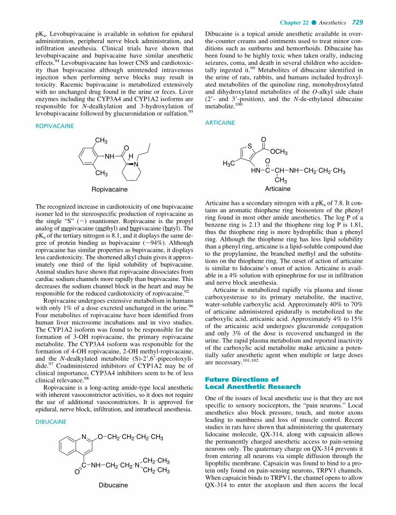
pKa. Levobupivacaine is available in solution for epiduraladministration, peripheral nerve block administration, andinfiltration anesthesia. Clinical trials have shown thatlevobupivacaine and bupivacaine have similar anestheticeffects.94 Levobupivacaine has lower CNS and cardiotoxic-ity than bupivacaine although unintended intravenousinjection when performing nerve blocks may result intoxicity. Racemic bupivacaine is metabolized extensivelywith no unchanged drug found in the urine or feces. Liverenzymes including the CYP3A4 and CYP1A2 isoforms areresponsible for N-dealkylation and 3-hydroxylation oflevobupivacaine followed by glucuronidation or sulfation.95
ROPIVACAINE
The recognized increase in cardiotoxicity of one bupivacaineisomer led to the stereospecific production of ropivacaine asthe single “S” (�) enantiomer. Ropivacaine is the propylanalog of mepivacaine (methyl) and bupivacaine (butyl). ThepKa of the tertiary nitrogen is 8.1, and it displays the same de-gree of protein binding as bupivacaine (�94%). Althoughropivacaine has similar properties as bupivacaine, it displaysless cardiotoxicity. The shortened alkyl chain gives it approx-imately one third of the lipid solubility of bupivacaine.Animal studies have shown that ropivacaine dissociates fromcardiac sodium channels more rapidly than bupivacaine. Thisdecreases the sodium channel block in the heart and may beresponsible for the reduced cardiotoxicity of ropivacaine.92
Ropivacaine undergoes extensive metabolism in humanswith only 1% of a dose excreted unchanged in the urine.96
Four metabolites of ropivacaine have been identified fromhuman liver microsome incubations and in vivo studies.The CYP1A2 isoform was found to be responsible for theformation of 3-OH ropivacaine, the primary ropivacainemetabolite. The CYP3A4 isoform was responsible for theformation of 4-OH ropivacaine, 2-OH methyl-ropivacaine,and the N-dealkylated metabolite (S)-2�,6�-pipecoloxyli-dide.97 Coadministered inhibitors of CYP1A2 may be ofclinical importance, CYP3A4 inhibitors seem to be of lessclinical relevance.98
Ropivacaine is a long-acting amide-type local anestheticwith inherent vasoconstrictor activities, so it does not requirethe use of additional vasoconstrictors. It is approved forepidural, nerve block, infiltration, and intrathecal anesthesia.
DIBUCAINE
Dibucaine
N O CH2 CH2 CH2 CH3
CO
NH CH2 CH2 NCH2 CH3
CH2 CH3
NH
Ropivacaine
CH3
CH3
O
NH
Dibucaine is a topical amide anesthetic available in over-the-counter creams and ointments used to treat minor con-ditions such as sunburns and hemorrhoids. Dibucaine hasbeen found to be highly toxic when taken orally, inducingseizures, coma, and death in several children who acciden-tally ingested it.99 Metabolites of dibucaine identified inthe urine of rats, rabbits, and humans included hydroxyl-ated metabolites of the quinoline ring, monohydroxylatedand dihydroxylated metabolites of the O-alkyl side chain(2�- and 3�-position), and the N-de-ethylated dibucainemetabolite.100
ARTICAINE
Articaine has a secondary nitrogen with a pKa of 7.8. It con-tains an aromatic thiophene ring bioisostere of the phenylring found in most other amide anesthetics. The log P of abenzene ring is 2.13 and the thiophene ring log P is 1.81,thus the thiophene ring is more hydrophilic than a phenylring. Although the thiophene ring has less lipid solubilitythan a phenyl ring, articaine is a lipid-soluble compound dueto the propylamine, the branched methyl and the substitu-tions on the thiophene ring. The onset of action of articaineis similar to lidocaine’s onset of action. Articaine is avail-able in a 4% solution with epinephrine for use in infiltrationand nerve block anesthesia.
Articaine is metabolized rapidly via plasma and tissuecarboxyesterase to its primary metabolite, the inactive,water-soluble carboxylic acid. Approximately 40% to 70%of articaine administered epidurally is metabolized to thecarboxylic acid, articainic acid. Approximately 4% to 15%of the articainic acid undergoes glucuronide conjugationand only 3% of the dose is recovered unchanged in theurine. The rapid plasma metabolism and reported inactivityof the carboxylic acid metabolite make articaine a poten-tially safer anesthetic agent when multiple or large dosesare necessary.101,102
Future Directions of Local Anesthetic Research
One of the issues of local anesthetic use is that they are notspecific to sensory nociceptors, the “pain neurons.” Localanesthetics also block pressure, touch, and motor axonsleading to numbness and loss of muscle control. Recentstudies in rats have shown that administering the quaternarylidocaine molecule, QX-314, along with capsaicin allowsthe permanently charged anesthetic access to pain-sensingneurons only. The quaternary charge on QX-314 prevents itfrom entering all neurons via simple diffusion through thelipophilic membrane. Capsaicin was found to bind to a pro-tein only found on pain-sensing neurons, TRPV1 channels.When capsaicin binds to TRPV1, the channel opens to allowQX-314 to enter the axoplasm and then access the local
Articaine
HN C
SO
OCH3
H3C OCH NH CH2 CH2 CH3
CH3
Chapter 22 Anesthetics 729
0711-0732_17865_Ch22.qxd 12/6/09 2:28 AM Page 729

anesthetic binding site in the sodium channel from the hy-drophilic pathway (Fig. 22.13 hydrophilic pathway C). Byspecifically blocking the sodium channels of only pain neu-rons, other nerve function is preserved. This type of painmanagement would make it possible to have a dental proce-dure with no pain, yet not leave the entire face numb, orhave an epidural during labor and still be able to walk. Thisis an exciting new opportunity in pain management that isundergoing research presently.103
Another area of research is the formulation of local anes-thetic liposomes and microspheres. The principle behind this
research is that the local anesthetic molecules are amphi-pathic, with both a hydrophilic and hydrophobic component.The anesthetic molecule can be suspended inside carrier sub-stances made of egg phospholipids and cholesterol lipo-somes or biodegradable polymers of lactic acid or glycolicacid. The carrier molecule will allow a controlled delivery ofthe local anesthetic and provide a long duration of action,potentially with less systemic toxicity.91,104 Carrier mole-cules are also being designed that combine a local anestheticwith an opioid analgesic that may offer alternative analgesicdelivery methods.105
R E V I E W Q U E S -T I O N S
730 Wilson and Gisvold’s Textbook of Organic Medicinal and Pharmaceutical Chemistry
3. An anesthesiologist from the obstetric unit calls to requeststat delivery of 20% intralipid to treat an inadvertentintravascular injection of bupivacaine. The patient is incardiac arrest. Why is the anesthesiologist requestingintralipid?
4. TG, a 45-year-old male woodworker, presents to the ERwith an injured index finger. While cleaning a circularsaw blade, he accidentally severed the tip (fleshy part, nobone) of his finger. The ER resident calls to request lido-caine 2% with epinephrine 1:200,000 to decrease the painassociated with suturing. You suggest an alternate. Whydid you NOT fill the original requested anesthetic?
5. JF, a patient with a severe allergic reaction to 2-chloropro-caine, is having plastic surgery to remove three moles fromher face. The physician calls to request your input inchoosing a local anesthetic. The plastic surgeon has thefive drugs shown at the bottom of this page available in hisclinic. Which drug would you recommend that he use?
6. While working as a pharmacy intern at a retail pharmacy, awoman carrying an infant asks you to ring up three tubes ofMaximum Strength Vagisil Creme. In conversation withher, you find out that she has been treating her 2-month-olddaughter’s painful, excoriated diaper rash with the creamfor the last week. She claims to have received the advice ona mothers’ chat room on the Internet. You observe that her2-month-old daughter has bluish lips and very pale translu-cent skin that also has a bluish tint. You immediately adviseher to take her daughter to the emergency room. Why?
1. A nurse anesthetist calls the OR pharmacy to discuss apatient history and get your input on choosing a generalanesthetic for an upcoming operation.ML is a 32-year-old, 5�4�, 460-pound woman who isscheduled for an elective gastroplasty (stomach stapling).She has a history of hypertension (150/98 mm Hg), hyper-lipidemia (LDL-cholesterol � 140mg/dL), and a largestage III pressure ulcer on her lower back. The last timethat the patient underwent surgery, she could not be rousedfrom the sedation and spent 24 hours in the postanestheticcare unit (PACU). Her prior surgery required 3 hours ofgeneral anesthesia maintained with isoflurane. The nurseanesthetist would like to know if one of the other formu-lary agents offers a shorter recovery time. General anes-thetics on the formulary include nitrous oxide, desflurane,sevoflurane, isoflurane, enflurane, and halothane.
2. A medical student was assigned the task of debridingML’s decubitus ulcer and calls the pharmacy to obtainprocaine to inject locally before beginning the painfulprocedure. You review the patient’s chart for allergies(no known drug allergies [NKDA]) and notice that thepatient is also receiving 1% silver sulfadiazine cream toprevent bacterial colonization of the ulcer. You recom-mend against using the procaine and suggest a 1% lido-caine with epinephrine instead. Why?
SO
ON
N
NH2N
Ag
R E V I E W Q U E S T I O N S
CH3
CH3
NH
ON(C2H5)2
(Preservative free)
(Preservative: Benzalkonium chloride) (Preservative: Methylparaben)(Preservative: Benzalkonium chloride)
(Preservative: Methylparaben)A B
C DE
O
ON(CH3)2
NH
O
ON(CH3)2
H2N
O
O CH3
H2N
CH3
CH3
NH
ON(C2H5)2
0711-0732_17865_Ch22.qxd 12/6/09 2:28 AM Page 730

Chapter 22 Anesthetics 731
REFERENCES1. Morton, W. T. G.: Remarks on the Proper Mode of Administering
Sulphuric Ether by Inhalation. Boston, MA, Dutton and Wentworth,1847, pp. 1–44.
2. Antognini, J. F., and Carstens, E.: Br. J. Anaesth. 89:156–166, 2002.3. Eger, E. I., 2nd.: Am. J. Health. Syst. Pharm. 61 Suppl 4:S3–10, 2004.4. Castro, B. A., Freedman, L. A., Craig, W. L., et al.: Anesthesiology
101:537–539, 2004.5. Wu, J., Previte, J. P., Adler, E., et al.: Anesthesiology 101:534–537,
2004.6. Dunning, M. B., 3rd, Bretscher, L. E., Arain, S. R., et al.:
Anesthesiology 106:144–148, 2007.7. Eger, E. I., 2nd, Halsey, M. J., Harris, R. A., et al.: Anesth. Analg.
88:1395–1400, 1999.8. Kobin, D. D.: Structure-activity relationships of inhaled anesthetics.
In Moody, E., and Skolnick, P. (eds.). Molecular Bases ofAnesthesia. New York, CRC Press, 2000, pp. 123–145.
9. Fang, Z., Ionescu, P., Chortkoff, B., et al.: Anesth. Analg.84:1042–1048, 1997.
10. Targ, A. G., Yasuda, N., Eger, E. I., 2nd, et al.: Anesth. Analg.68:599–602, 1989.
11. Halsey, M. J.: Br. J. Anaesth. 56 Suppl 1:9S–25S, 1984.12. Eger, E., II, and Laster, M.: Anesth. Analg. 92:1477–1482, 2001.13. Eckenhoff, R. G.: Mol. Interv. 1:258–268, 2001.14. Campagna, J. A., Miller, K. W., and Forman, S. A.: N. Engl. J. Med.
348:2110–2124, 2003.15. Hemmings, H. C., Akabas, M. H., Goldstein, P. A., et al.: Trends.
Pharmacol. Sci. 26:503–510, 2005.16. Anonymous Inhalants. http://www.drugabuse.gov/PDF/Infofacts/
Inhalants06.pdf (accessed October 12, 2007).17. Stachnik, J.: Am. J. Health. Syst. Pharm. 63:623–634, 2006.18. Spracklin, D. K., Thummel, K. E., and Kharasch, E. D.: Drug Metab.
Dispos. 24:976–983, 1996.19. Litman, R. S., and Rosenberg, H.: JAMA 293:2918–2924, 2005.20. Lerman, J., Gregory, G. A., Willis, M. M., et al.: Anesthesiology
61:139–143, 1984.21. Mazze, R. I.: Anesthesiology 105:843–846, 2006.22. Evers, A. S., Crowder, C. M., and Balser J. R.: General anesthetics.
In Brunton L. L., Lazo, J. L., and Parker, K. L. (eds.). Goodman andGilman’s The Pharmacological Basis of Therapeutics. New York,McGraw-Hill, 2006, pp. 341–368.
23. Goldberg, M. E., Cantillo, J., Larijani, G. E., et al.: Anesth. Analg.82:1268–1272, 1996.
24. Package Insert Suprane (desflurane, USP). Baxter HealthcareCorporation 2005.
25. Smith, I., Nathanson, M., and White, P. F.: Br. J. Anaesth.76:435–445, 1996.
26. Iyer, R. A., Baggs, R. B., and Anders, M. W.: J. Pharmacol. Exp. Ther.283:1544–1551, 1997.
27. Kharasch, E. D., and Jubert, C.: Anesthesiology 91:1267–1278, 1999.28. Kharasch, E., Schroeder, J., Bammler, T., et al.: Toxicol. Sci.
90:419–431, 2006.29. Fatheree, R. S., and Leighton, B. L.: Anesthesiology
101:531–533, 2004.30. Kharasch, E. D., Armstrong, A. S., Gunn, K., et al.: Anesthesiology
82:1379–1388, 1995.31. Kharasch, E. D., and Thummel, K. E.: Anesthesiology
79:795–807, 1993.32. Package Insert Ultane (sevoflurane). Abbott Laboratories 2006.33. Jaaskelainen, S. K., Kaisti, K., Suni, L., et al.: Neurology
61:1073–1078, 2003.34. Schoenborn, B. P., and Nobbs, C. L.: Mol. Pharmacol. 2:495–498, 1966.35. Trudell, J., Koblin, D., and Eger, E., 2nd: Anesth. Analg.
87:411–418, 1998.36. Goto, T., Saito H., Shinkai M., et al.: Anesthesiology 86:1273–1278,
1997.37. Goto, T.: Can. J. Anaesth. 49:335–338, 2002.38. Higgins, T. L., Murray, M., Kett, D. H., et al.: Intensive Care Med.
26:S413–421, 2000.39. Package Insert Diprivan (propofol) Injectable Emulsion. AstraZeneca
Rev. 08/2005.40. McKeage, K., and Perry, C. M.: CNS Drugs 17:235–272, 2003.41. Fassoulaki, A., Sarantopoulos, C., and Papilas, K.: Can. J. Anaesth.
40:10, 1993.42. Trapani, G., Altomare, C., Sanna, E., et al.: Curr. Med. Chem. 7:249,
2000.
43. Langley, M. S., and Heel, R. C.: Drugs 35:334–372, 1988.44. Belelli, D., Lambert, J. J., Peters, J. A., et al.: PNAS
94:11031–11036, 1997.45. Package Insert Etomidate Injection. Bedford Laboratories 03/2004.46. Doenicke, A. W., Roizen, M. F., Hoernecke, R., et al.: Br. J. Anaesth.
83:464–466, 1999.47. Sinha, A. C., and Soliz, J. M.: The Internet J. Anesthesiol. 5, 2002.48. Lipiner-Friedman, D., Sprung, C. L., Laterre, P. F., et al.: Crit. Care
Med. 35:1012–1018, 2007.49. Giese, J. L., and Stanley, T. H.: Pharmacotherapy 3:251–258, 1983.50. Hustveit, O., Maurset, A., and Oye, I.: Pharmacol. Toxicol.
77:355–359, 1995.51. Hirota, K., and Lambert, D. G.: Br. J. Anaesth. 77:441–444, 1996.52. Hocking, G., and Cousins, M. J.: Anesth. Analg. 97:1730–1739, 2003.53. Visser, E., and Schug, S. A.: Biomed. Pharmacother.
60:341–348, 2006.54. Anonymous, Intelligence Bulletin:Ketamine. USDOJ.gov/ndic
(accessed June 6, 2007).55. Hijazi, Y., and Boulieu, R.: Drug Metab. Dispos. 30:853–858, 2002.56. Williams, M. L., Mager, D. E., Parenteau, H., et al.: Drug Metab.
Dispos. 32:786–793, 2004.57. Catterall, W. A.: Neuron 26:13–25, 2000.58. Armstrong, C. M., and Hille, B.: Neuron 20:371–380, 1998.59. Heinemann, S. H., Terlau, H., Stuhmer, W., et al.: Nature
356:441–443, 1992.60. Elinder, F., Nilsson, J., and Arhem, P.: Physiol. Behav. 92:1–7, 2007.61. Lipkind, G., and Fozzard, H.: Mol. Pharmacol. 68:1611–1622, 2005.62. Li, H., Galue, A., Meadows, L., et al.: Mol. Pharmacol. 55:134–141,
1999.63. Strichartz, G.: J. Gen. Physiol. 62:37–57, 1973.64. Hille, B.: J. Gen. Physiol. 69:497–515, 1977.65. Strichartz, G., Sanchez, V., Arthur, G., et al.: Anesth. Analg.
1:158–170, 1990.66. Chernoff, D. M., and Strichartz, G. R.: Biophys. J. 58:69–81, 1990.67. Catterall, W. A., and Mackie, K.: Local anesthetics. In Brunton,
L. L., Lazo, J. L., and Parker, K. L. (eds.). Goodman and Gilman’sThe Pharmacological Basis of Therapeutics. New York, The McGrawHill Companies, 2006, p. 369.
68. Gupta, S. P.: Chem. Rev. 91:1109–1119, 1991.69. Buchi, J., and Perlia, X.: Design of local anesthetics. In Ariens, E. J.
(ed.). Drug Design. New York, Academic Press, 1972, Vol. 3, p. 243.70. Heavner, J. E.: Curr. Opin. Anaesthesiol. 20:336–342, 2007.71. Tucker, G. T., and Mather, L. E.: Br. J. Anaesth. 47:1029–1030, 1975.72. Eggleston, S., and Lush, L.: Ann. Pharmacother. 30:851–857, 1996.73. Tetzlaff, J. E.: Boston, Clinical Pharmacology of Local Anesthetics;
Butterworth, 1999, p. 258.74. Simon, R. A. J.: Allergy Clin. Immunol. 74:623–630, 1984.75. Karim, A., Ahmed, S., Siddiqui, R., et al.: Am. J. Med. 111:150–153,
2001.76. Kaendler, L., Dorszewski, A., and Daehnert, I.: Heart 90:e51, 2004.77. Wilburn-Goo, D., and Lloyd, L.: J. Am. Dent. Assoc.
130:826–831, 1999.78. Moore, T., Walsh, C., and Cohen, M.: Arch. Intern. Med.
164:1192–1196, 2004.79. Nunn, D. B.: Adv. Stud. Med. 6:106–108, 2006.80. Goodell, J., Gilroy, G., and Huntress, J.: Am. J. Health Syst. Pharm.
45:2510–2513, 1988.81. Lander, J., Weltman, B., and So, S.: Cochrane Database Syst. Rev. 3,
2007.82. Noorily, A., Noorily, S., and Otto, R.: Anesth. Analg.
81:724–727, 1995.83. Tush, G., and Kuhn, R.: Ann. Pharmacother. 30:1251–1254, 1996.84. Dahshan, A., and Donovan, G. K.: Pediatrics 117:e806–809, 2006.85. Alexson, S. H., Diczfalusy, M., Halldin, M., et al.: Drug Metab.
Dispos. 30:643–647, 2002.86. Wang, J., Backman, J., Taavitsainen, P., et al.: Drug Metab. Dispos.
28:959–965, 2000.87. Wang, J. S., Backman, J. T., Wen, X., et al.: Pharmacol. Toxicol.
85:201–205, 1999.88. Hjelm, M., Ragnarsson, B., and Wistrand, P.: Biochem. Pharmacol.
21:2825–2834, 1972.89. Gaber, K., Harreus, U. A., Matthias, C., et al.: Toxicology
229:157–164, 2007.90. Paradis, B., and Fournier, L.: Can. J. Anesth. 22:70–75, 1975.91. Ruetsch, Y. A., Boni, T., and Borgeat, A.: Curr. Top. Med. Chem.
1:175–182, 2001.
0711-0732_17865_Ch22.qxd 12/6/09 2:28 AM Page 731

92. Whiteside, J. B., and Wildsmith, J. A. W.: Br. J. Anaesth.87:27–35, 2001.
93. Huang, Y., Pryor, M., Mather, L., et al.: Anesth. Analg.86:797–804, 1998.
94. Glaser, C., Marhofer, P., Zimpfer, G., et al.: Anesth. Analg.94:194–198, 2002.
95. Gantenbein, M., Attolini, L., Bruguerolle, B., et al.: Drug Metab.Dispos. 28:383–385, 2000.
96. Halldin, M., Bredberg, E., Angelin, B., et al.: Drug Metab. Dispos.24:962–968, 1996.
97. Ekstrom, G., and Gunnarsson, U.: Drug Metab. Dispos.24:955–961, 1996.
98. Arlander, E., Ekström, G., Alm, C., et al.: Clin. Pharmacol. Ther.64:484–491, 1998.
99. Dayan P. S., Litovitz T. L., Crouch B. I., et al.: Ann. Emerg. Med.28:442–445, 1996.
100. Igarashi, K., Kasuya, F., and Fukui, M.: J. Pharmacobiodyn. 6:538–550, 1983.
101. Vree, T. B., Simon, M. A. M., Gielen, M. J. M., et al.: Br. J. Clin.Pharmacol. 44:29–34, 1997.
102. van Oss, G. E., Vree, T. B., Baars, A. M., et al.: Eur. J.Anaesthesiol. 6:49–56, 1989.
103. Binshtok, A. M., Bean B. P., and Wu, J.: Nature 449:607–610,2007.
104. deAraujo, D., Cereda, C. S., Brunetto, G., et al.: Can. J. Anesth.51:566–572, 2004.
105. Sendil, D., Bonney, I. M., Carr, D. B., et al.: Biomaterials24:1969–1976, 2003.
732 Wilson and Gisvold’s Textbook of Organic Medicinal and Pharmaceutical Chemistry
0711-0732_17865_Ch22.qxd 12/6/09 2:28 AM Page 732

C H A P T E R 23
Histamine and Antihistaminic AgentsJACK DERUITER
tautomers is dependent on the presence of imidazole ringsubstituents as discussed later in this chapter.4
Structure–activity relationship studies suggest that the�NH3
� monocation is important for agonist activity at hista-mine receptors and that transient existence of the morelipophilic uncharged histamine species may contribute todiffusion across cell membranes. Other studies support pro-posal that the N�MH tautomer of the histamine monocationis the pharmacophoric species at the H1-receptor, while a1,3-tautomeric system is important for selective H2-agonism.
Stereochemistry
While histamine is an achiral molecule, histamine receptorsexert high stereoselectivity toward chiral ligands.5
Molecular modeling and steric–activity relationship studiesof the influence of conformational isomerism suggest the im-portance of trans-gauche rotameric structures in the receptorbinding of histamine (Fig. 23.3). Although both conformersexist in solution, studies with conformationally restrictedhistamine analogs suggest that the trans-rotamer of hista-mine possesses affinity for both H1- and H2-receptors, andthe gauche conformer is preferred for H3-receptors, but notH1- or H2-receptors.4
HISTAMINE AS A CHEMICAL MESSENGER
Knowledge of the biodisposition of histamine is importantin understanding the involvement of this substance in vari-ous pathophysiologies as well as the actions of various li-gands that either enhance or block its actions. Each of thesteps in the “life cycle” of histamine represents a potentialpoint for pharmacological intervention.
Biosynthesis and Distribution
Histamine is synthesized in Golgi apparatus of its principalstorage cells, mast cells, and basophils.6 Histamine isformed from the naturally occurring amino acid L-hisitidne(S-histidine) via the catalysis of either the pyridoxal phos-phate dependent enzyme histidine decarboxylase (HDC,EC 4.1.1.22) or L-aromatic amino acid decarboxylase (L-AAAD) (Fig. 23.4). Substrate specificity is higher forHDC versus L-AAAD. HDC inhibitors (HDCIs) include �-fluoromethylhistidine (FMH), a mechanism-based in-hibitor, and certain flavonoids.7 Although useful as pharmo-logic probes, HDCIs have not proved to be useful clinically.
C H A P T E R O V E R V I E W
Histamine or �-imidazolylethylamine is synthesized from L-histidine by histidine decarboxylase, an enzyme that isexpressed in many mammalian tissues including gastric-mucosa parietal cells, mast cells, and basophils and thecentral nervous system (CNS).1 As a result, histamine playsan important role in human physiology including regulationof the cardiovascular system, smooth muscle, exocrineglands, the immune system, and central nerve function. It isalso involved in embryonic development, the proliferationand differentiation of cells, hematopoiesis, inflammation,and wound healing. Histamine exerts its diverse biologic ef-fects through four types of receptors. The involvement ofhistamine in the mediation of immune and hypersensitivityreactions and the regulation of gastric acid secretion has ledto the development of important drug classes useful in thetreatment of symptoms associated with allergic and gastrichypersecretory disorders.
HISTAMINE CHEMISTRY
Nomenclature
Histamine, known trivially as 4(5-)(2-aminoethyl)imidazole,structurally is composed of an imidazole heterocycle andethylamine side chain. The methylene groups of the amino-ethyl side chain are designated � and �. The side chain isattached, via the �-CH2 group, to the 4-position of an imida-zole ring. The imidazole N at position 3 is designated thepros (�) N, whereas the N at position 1 is termed the tele(�) N. The side chain N is distinguished as N� (Fig. 23.1).
Ionization and Tautomerization
Histamine is a basic organic compound (N�, pKa1 � 5.80;N pKa2 � 9.40; N�, pKa3 � 14.0) capable of existing as amixture of different ionic and uncharged tautomeric species(Fig. 23.2).2,3 Histamine exists almost exclusively (96.6%)as the monocationic conjugate species (�NH3
�) at physio-logical pH (7.4). At lower pHs, a higher percentage of thedicationic species exists. In water, the ratio of the concentra-tions of the tautomers N�MH/N�MH has been calculated tobe 4.2, indicating that 80% of the histamine monocation ex-ists as N�MH and 20% exists as N�MH in aqueous solu-tion.2,3 However, crystalline forms of histamine salts appearto exist primarily as N�MH tautomer to facilitate crystalpacking. Also, for derivatives of histamine, the ratio of
733
0733-0775_17865_Ch23.qxd 12/4/09 8:04 PM Page 733

Histamine is found in almost all mammalian tissues inconcentrations ranging from 1 to more than 100 �g/g. Mastcells and histamine are in particularly high concentration inskin and the mucosal cells of the bronchi, intestine, urinarytract, and tissues adjacent to the circulation. It is found inhigher concentrations in mammalian cerebrospinal fluidthan in plasma and other body fluids.
Storage and Release
Most histamine is biosynthesized and stored as protein com-plexes in mast cells (complexed with heparin) and basophilicgranulocytes (complexed with chondroitin).8,9 Protein-complexed histamine is stored in secretory granules and re-leased by exocytosis in response to a wide variety of immune(antigen and antibody) and nonimmune (bacterial products,xenobiotics, physical effects, and cholinergic effects) stimuli.The release of histamine as one of the mediators of
734 Wilson and Gisvold’s Textbook of Organic Medicinal and Pharmaceutical Chemistry
Figure 23.1 Histamine.
Figure 23.2 Histamine tautomersand cations.
0733-0775_17865_Ch23.qxd 12/4/09 8:04 PM Page 734
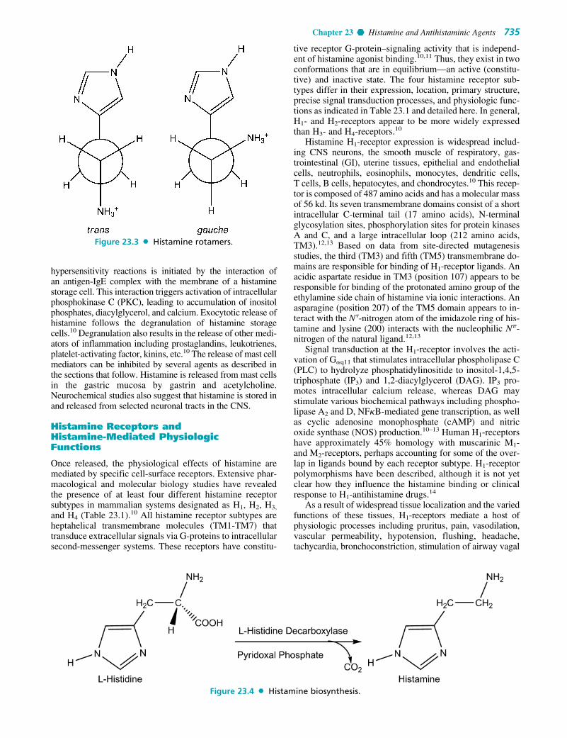
hypersensitivity reactions is initiated by the interaction ofan antigen-IgE complex with the membrane of a histaminestorage cell. This interaction triggers activation of intracellularphosphokinase C (PKC), leading to accumulation of inositolphosphates, diacylglycerol, and calcium. Exocytotic release ofhistamine follows the degranulation of histamine storagecells.10 Degranulation also results in the release of other medi-ators of inflammation including prostaglandins, leukotrienes,platelet-activating factor, kinins, etc.10 The release of mast cellmediators can be inhibited by several agents as described inthe sections that follow. Histamine is released from mast cellsin the gastric mucosa by gastrin and acetylcholine.Neurochemical studies also suggest that histamine is stored inand released from selected neuronal tracts in the CNS.
Histamine Receptors and Histamine-Mediated PhysiologicFunctions
Once released, the physiological effects of histamine aremediated by specific cell-surface receptors. Extensive phar-macological and molecular biology studies have revealedthe presence of at least four different histamine receptorsubtypes in mammalian systems designated as H1, H2, H3,
and H4 (Table 23.1).10 All histamine receptor subtypes areheptahelical transmembrane molecules (TM1-TM7) thattransduce extracellular signals via G-proteins to intracellularsecond-messenger systems. These receptors have constitu-
tive receptor G-protein–signaling activity that is independ-ent of histamine agonist binding.10,11 Thus, they exist in twoconformations that are in equilibrium—an active (constitu-tive) and inactive state. The four histamine receptor sub-types differ in their expression, location, primary structure,precise signal transduction processes, and physiologic func-tions as indicated in Table 23.1 and detailed here. In general,H1- and H2-receptors appear to be more widely expressedthan H3- and H4-receptors.10
Histamine H1-receptor expression is widespread includ-ing CNS neurons, the smooth muscle of respiratory, gas-trointestinal (GI), uterine tissues, epithelial and endothelialcells, neutrophils, eosinophils, monocytes, dendritic cells,T cells, B cells, hepatocytes, and chondrocytes.10 This recep-tor is composed of 487 amino acids and has a molecular massof 56 kd. Its seven transmembrane domains consist of a shortintracellular C-terminal tail (17 amino acids), N-terminalglycosylation sites, phosphorylation sites for protein kinasesA and C, and a large intracellular loop (212 amino acids,TM3).12,13 Based on data from site-directed mutagenesisstudies, the third (TM3) and fifth (TM5) transmembrane do-mains are responsible for binding of H1-receptor ligands. Anacidic aspartate residue in TM3 (position 107) appears to beresponsible for binding of the protonated amino group of theethylamine side chain of histamine via ionic interactions. Anasparagine (position 207) of the TM5 domain appears to in-teract with the N�-nitrogen atom of the imidazole ring of his-tamine and lysine (200) interacts with the nucleophilic N�-nitrogen of the natural ligand.12,13
Signal transduction at the H1-receptor involves the acti-vation of G�q11 that stimulates intracellular phospholipase C(PLC) to hydrolyze phosphatidylinositide to inositol-1,4,5-triphosphate (IP3) and 1,2-diacylglycerol (DAG). IP3 pro-motes intracellular calcium release, whereas DAG maystimulate various biochemical pathways including phospho-lipase A2 and D, NF�B-mediated gene transcription, as wellas cyclic adenosine monophosphate (cAMP) and nitricoxide synthase (NOS) production.10–13 Human H1-receptorshave approximately 45% homology with muscarinic M1-and M2-receptors, perhaps accounting for some of the over-lap in ligands bound by each receptor subtype. H1-receptorpolymorphisms have been described, although it is not yetclear how they influence the histamine binding or clinicalresponse to H1-antihistamine drugs.14
As a result of widespread tissue localization and the variedfunctions of these tissues, H1-receptors mediate a host ofphysiologic processes including pruritus, pain, vasodilation,vascular permeability, hypotension, flushing, headache,tachycardia, bronchoconstriction, stimulation of airway vagal
Chapter 23 Histamine and Antihistaminic Agents 735
Figure 23.3 Histamine rotamers.
Figure 23.4 Histamine biosynthesis.
0733-0775_17865_Ch23.qxd 12/4/09 8:04 PM Page 735
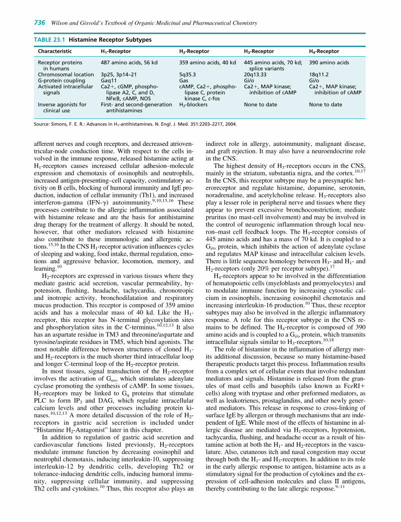
afferent nerves and cough receptors, and decreased atrioven-tricular-node conduction time. With respect to the cells in-volved in the immune response, released histamine acting atH1-receptors causes increased cellular adhesion–moleculeexpression and chemotaxis of eosinophils and neutrophils,increased antigen-presenting–cell capacity, costimulatory ac-tivity on B cells, blocking of humoral immunity and IgE pro-duction, induction of cellular immunity (Th1), and increasedinterferon-gamma (IFN-�) autoimmunity.9,10,15,16 Theseprocesses contribute to the allergic inflammation associatedwith histamine release and are the basis for antihistaminedrug therapy for the treatment of allergy. It should be noted,however, that other mediators released with histaminealso contribute to these immunologic and allergenic ac-tions.15,16 In the CNS H1-receptor activation influences cyclesof sleeping and waking, food intake, thermal regulation, emo-tions and aggressive behavior, locomotion, memory, andlearning.10
H2-receptors are expressed in various tissues where theymediate gastric acid secretion, vascular permeability, hy-potension, flushing, headache, tachycardia, chronotropicand inotropic activity, bronchodilatation and respiratorymucus production. This receptor is composed of 359 aminoacids and has a molecular mass of 40 kd. Like the H1-receptor, this receptor has N-terminal glycosylation sitesand phosphorylation sites in the C-terminus.10,12,13 It alsohas an aspartate residue in TM3 and threonine/aspartate andtyrosine/aspirate residues in TM5, which bind agonists. Themost notable difference between structures of cloned H1-and H2-receptors is the much shorter third intracellular loopand longer C-terminal loop of the H2-receptor protein.
In most tissues, signal transduction of the H2-receptorinvolves the activation of G�s, which stimulates adenylatecyclase promoting the synthesis of cAMP. In some tissues,H2-receptors may be linked to Gq proteins that stimulatePLC to form IP3 and DAG, which regulate intracellularcalcium levels and other processes including protein ki-nases.10,12,13 A more detailed discussion of the role of H2-receptors in gastric acid secretion is included under“Histamine H2-Antagonist” later in this chapter.
In addition to regulation of gastric acid secretion andcardiovascular functions listed previously, H2-receptorsmodulate immune function by decreasing eosinophil andneutrophil chemotaxis, inducing interleukin-10, suppressinginterleukin-12 by dendritic cells, developing Th2 ortolerance-inducing dendritic cells, inducing humoral immu-nity, suppressing cellular immunity, and suppressingTh2 cells and cytokines.10 Thus, this receptor also plays an
indirect role in allergy, autoimmunity, malignant disease,and graft rejection. It may also have a neuroendocrine rolein the CNS.
The highest density of H3-receptors occurs in the CNS,mainly in the striatum, substantia nigra, and the cortex.10,17
In the CNS, this receptor subtype may be a presynaptic het-eroreceptor and regulate histamine, dopamine, serotonin,noradrenaline, and acetylcholine release. H3-receptors alsoplay a lesser role in peripheral nerve and tissues where theyappear to prevent excessive bronchoconstriction; mediatepruritus (no mast-cell involvement) and may be involved inthe control of neurogenic inflammation through local neu-ron–mast cell feedback loops. The H3-receptor consists of445 amino acids and has a mass of 70 kd. It is coupled to aGi/o protein, which inhibits the action of adenylate cyclaseand regulates MAP kinase and intracellular calcium levels.There is little sequence homology between H3- and H1- andH2-receptors (only 20% per receptor subtype).17
H4-receptors appear to be involved in the differentiationof hematopoietic cells (myeloblasts and promyelocytes) andto modulate immune function by increasing cytosolic cal-cium in eosinophils, increasing eosinophil chemotaxis andincreasing interleukin-16 production.10 Thus, these receptorsubtypes may also be involved in the allergic inflammatoryresponse. A role for this receptor subtype in the CNS re-mains to be defined. The H4-receptor is composed of 390amino acids and is coupled to a Gi/o protein, which transmitsintracellular signals similar to H3-receptors.10,18
The role of histamine in the inflammation of allergy mer-its additional discussion, because so many histamine-basedtherapeutic products target this process. Inflammation resultsfrom a complex set of cellular events that involve redundantmediators and signals. Histamine is released from the gran-ules of mast cells and basophils (also known as FceRI�cells) along with tryptase and other preformed mediators, aswell as leukotrienes, prostaglandins, and other newly gener-ated mediators. This release in response to cross-linking ofsurface IgE by allergen or through mechanisms that are inde-pendent of IgE. While most of the effects of histamine in al-lergic disease are mediated via H1-receptors, hypotension,tachycardia, flushing, and headache occur as a result of his-tamine action at both the H1- and H2-receptors in the vascu-lature. Also, cutaneous itch and nasal congestion may occurthrough both the H1- and H3-receptors. In addition to its rolein the early allergic response to antigen, histamine acts as astimulatory signal for the production of cytokines and the ex-pression of cell-adhesion molecules and class II antigens,thereby contributing to the late allergic response.9–11
736 Wilson and Gisvold’s Textbook of Organic Medicinal and Pharmaceutical Chemistry
TABLE 23.1 Histamine Receptor Subtypes
Characteristic H1-Receptor H2-Receptor H3-Receptor H4-Receptor
Receptor proteins 487 amino acids, 56 kd 359 amino acids, 40 kd 445 amino acids, 70 kd; 390 amino acidsin humans splice variants
Chromosomal location 3p25, 3p14–21 5q35.3 20q13.33 18q11.2G-protein coupling Gaq11 Gas Gi/o Gi/oActivated intracellular Ca2�, cGMP, phospho- cAMP, Ca2�, phospho- Ca2�, MAP kinase; Ca2�, MAP kinase;
signals lipase A2, C, and D, lipase C, protein inhibition of cAMP inhibition of cAMPNF�B, cAMP, NOS kinase C, c-fos
Inverse agonists for First- and second-generation H2-blockers None to date None to dateclinical use antihistamines
Source: Simons, F. E. R.: Advances in H1-antihistamines. N. Engl. J. Med. 351:2203–2217, 2004.
0733-0775_17865_Ch23.qxd 12/4/09 8:04 PM Page 736

In addition to those actions, histamine exerts other im-portant and variable immunomodulatory effects through itsreceptor subtypes.10,19 First, the expression of the receptorschanges according to the stage of cell differentiation andlocal tissue influences. Second, depending on the predomi-nance of the type of receptor, histamine may have proin-flammatory or anti-inflammatory effects. Through the H1-receptor, histamine has proinflammatory activity and isinvolved in the development of several aspects of antigen-specific immune response, including the maturation of den-dritic cells and the modulation of the balance of type 1helper (Th1) T cells and type 2 helper (Th2) T cells.Histamine may induce an increase in the proliferation ofTh1 cells and in the production of interferon and may blockhumoral immune responses by means of this mechanism. Italso induces the release of proinflammatory cytokines andlysosomal enzymes from human macrophages and has thecapacity to influence the activity of basophils, eosinophils,and fibroblasts. Finally, histamine may play a role in au-toimmunity and malignant disease through the H1-receptor.
Termination of Histamine Action
Three principal ways exist to terminate the physiologicaleffects of histamine3,20,21:
• Cellular uptake: Animal studies have documented the uptakeof histamine by many cells. In particular, uptake is a temper-ature- and partially Na�-dependent process in rabbit gastricglands and the histamine is metabolized once in the cell.
• Desensitization of cells: Some H1-receptor–containing tis-sues exhibit a homogeneous loss of sensitivity to the actionsof histamine, perhaps as a result of receptor modification.
• Metabolism: The most common pathway for terminatinghistamine action involves enzymatic inactivation, as dis-cussed in more detail here.
Released histamine is rapidly inactivated by metabolismvia two pathways as shown in Figure 23.5. One pathwayinvolves N�-methylation via the enzyme histamine N-methyltransferase (HMT; EC 2.1.1.8). This enzyme iswidely distributed in mammalian tissues and catalyzes thetransfer of a methyl group from S-adenosyl-L-methionine(SAM) to the ring tele-nitrogen of histamine, producing N�-methylhistamine and S-adenosyl-L-homocysteine (Fig. 23.5).The other pathway of catabolism involves oxidative deamina-tion by diamine oxidase (DAO; EC 1.4.3.6), yielding imida-zole acetaldehyde, which is further oxidized to imidazoleacetic acid by aldehyde dehydrogenases (ALD-DH).Similarly, N�-methylhistamine is converted by both DAO andmonoamine oxidase (MAO), followed by ALD-DH to N-methyl imidazole acetic acid (Fig. 23.5). All of these metabo-lites are devoid of histamine receptor agonist activity.
Functions of Histamine as Related to Pharmacological Intervention
Histamine exhibits a wide variety of both physiological andpathological functions in different tissues and cells. Theactions of histamine that are of interest from both a pharma-cological and therapeutic point of view include (a) itsimportant, but limited, role as a chemical mediator of hyper-sensitivity and allergic inflammatory reactions, (b) a majorrole in the regulation of gastric acid secretion, and (c) anemerging role as a neurotransmitter in the CNS.
ANTIHISTAMINES
The term antihistamine historically has referred to drugs thatblock the actions of histamine at H1-receptors rather thanother histamine receptor subtypes. The development of anti-histamine drugs began decades ago with the discovery byFourneau and Bovet22 that piperoxan could protect animalsfrom the bronchial spasm induced by histamine. This findingwas followed by the synthesis of several N-phenylethylene-diamines with antihistamininic activities superior to those ofpiperoxan.23 Further traditional structure–activity studies inthis series, based largely on the principles of isosterism andfunctional group modification, led to the introduction in the1940s to 1970s of various H1-antihistamine containing thediarylalkylamine framework.23,24 These antihistamines, re-ferred to now as the first-generation or classical antihista-mines, are related structurally and include several aminoalkylethers, ethylenediamines, piperazines, propylamines, phe-nothiazines, and dibenzocycloheptenes. In addition to H1-antihistaminic action, these compounds display an array ofother pharmacological activities that contribute either towardadditional therapeutic applications, or limit use as adversereactions. More recently, several second-generation or“nonsedating” antihistamines have been developed and intro-duced.10,23 The second-generation agents are derivatives ofseveral first-generation agents, but have been modified to bemore specific in pharmacologic action and limited in theirtissue distribution or accumulation profiles.
Mechanism of Action
It is now known that H1-antihistamines act as inverse ago-nists that combine with and stabilize the inactive form of theH1-receptor, shifting the equilibrium toward the inactivestate.10 Thus, they effectively antagonize the actions of his-tamine at H1-receptors. Historically, H1-antihistamines havebeen evaluated in vitro in terms of their ability to inhibit his-tamine-induced spasms in an isolated strip of guinea pigileum. Antihistamines may be evaluated in vivo in terms oftheir ability to protect animals against the lethal effects ofhistamine administered intravenously or by aerosol.
To distinguish antagonism of histamine from other modesof action, the index pA is applied in in vitro assays. Theindex pA2 is defined as the inverse of the logarithm of themolar concentration of the antagonist that reduces the re-sponse of a double dose of the agonist to that of a single one.The more potent H1-antihistamines exhibit a pA2 value sig-nificantly higher than 6. Although there are many pitfalls tobe avoided in the interpretation of structure–activity relation-ship (SAR) studies using pA2 values, the following exampleillustrates distinguishing competitive antagonism. pA2 val-ues for pyrilamine (mepyramine) antagonism range from 9.1to 9.4 with human bronchi and guinea pig ileum.24,25 By con-trast, the pA2 value in guinea pig (H2-receptor) is 5.3. Thus,one may conclude that pyrilamine is a weak, noncompetitiveinhibitor of histamine at the H2-receptors and a competitiveinhibitor at H1-receptors. The structural features required foreffective interaction of these receptors are discussed next.Some H1-antihistamines can also block histamine release.The concentrations, however, are considerably higher thanthose required to produce significant histamine receptorblockade. The H1-antihistamines do not, however, block an-tibody production or antigen–antibody interactions.10,26
Chapter 23 Histamine and Antihistaminic Agents 737
0733-0775_17865_Ch23.qxd 12/4/09 8:04 PM Page 737

738 Wilson and Gisvold’s Textbook of Organic Medicinal and Pharmaceutical Chemistry
Figure 23.5 Metabolism of histamine. (ALD-DH, aldehyde dehydrogenase; PRT,phosphoribosyltransferase.)
0733-0775_17865_Ch23.qxd 12/4/09 8:04 PM Page 738

Structure–Activity Relationships at H1-Receptors
The H1-antihistamines are now commonly subdivided intotwo broad groups—the first-generation or classical antihis-tamines and the second-generation or “nonsedating” antihis-tamines—based primarily on their general pharmacologicalprofiles.10,23,27 The differences between these two series arediscussed in more detail in the sections that follow. Themost detailed published SAR analyses for antihistamines,however, focus on the structural requirements for the first-generation agents.22–24 From these studies, the structural re-quirements for H1-antihistaminic action were identified asshown in Figure 23.6. In this structure, Ar is aryl (includingphenyl, substituted phenyl, and heteroaryl groups such as 2-pyridyl); Ar' is a second aryl or arylmethyl group; X is aconnecting atom of O, C, or N; (CH2)n represents a carbonchain, usually ethyl; and NRR' represents a basic, terminalamine function. The nature of the connecting atom as wellas the diaryl substitution pattern and amine moiety has beenused to subclassify the first-generation antihistamines as in-dicated in the succeeding sections.
This diaryl substitution pattern is present in both thefirst- and second-generation antihistamines and is essen-tially a significant H1-receptor affinity.22–24 Furthermore,several SAR studies suggest that the two aryl moieties mustbe able to adopt a noncoplanar conformation relative toeach other for optimal interaction with the H1-receptor.28
The two aromatic systems may be linked, as in the tricyclicantihistamines (phenothiazines, dihenzocycloheptanes, andheptenes), but again they must be noncoplanar for effectivereceptor interaction. Most H1-antihistamines contain sub-stituents in one of the aryl rings (usually benzene), andthese influence histamine potency as well as biodisposition,as discussed for the individual classes of compounds in thesucceeding sections.
In many of the first-generation, or classical, antihista-mines, the terminal nitrogen atom is a simple dimethylmoiety. The amine may also be part of a heteroyclic struc-ture, however, as illustrated by the piperazines, some propyl-amines (pyrrolidines and piperidines), some phenthiazines,the dibenzocycloheptenes, and the second-generation anti-histamines. In all cases, the amino moiety is basic, with pKasranging from 8.5 to 10, and thus is presumed to be protonatedwhen bound on the receptor. The amine moiety is also im-portant in the development of stable, solid dosage formsthrough salt formation.
The carbon chain of typical H1-antihistamines consists oftwo or three atoms.22–24 As a result, the distance between thecentral point of the diaryl ring system and the terminal nitro-gen atom in the extended conformation of these compoundsranges from 5 to 6 angstroms (Å). A similar distance be-tween these key moieties is observed for those antihista-mines with less conformational freedom. In some structuralseries, branching of the carbon chain results in reduced
antihistaminic activity. There are exceptions, however, as ev-idenced by promethazine, which has greater activity than itsnonbranched counterpart. When the carbon adjacent to theterminal nitrogen atom is branched, the possibility of asym-metry exists. Stereoselective H1-receptor binding is typicallynot observed, however, when chirality exists at this site.29
Also, in compounds with an asymmetrically substituted un-saturated carbon chain (pyrrobutamine and triprolidine), onegeometric isomer typically displays higher receptor affinitythan the other.
The X connecting moiety of typical H1-antihistaminesmay be a saturated carbon–oxygen moiety or simply a car-bon atom. This group, along with the carbon chain, appearsto serve primarily as a spacer group for the key pharma-cophoric moieties. Many antihistamines containing a carbonatom in the connecting moiety are chiral and exhibit stereo-selective receptor binding. For example, in the pheniramineseries and carbinoxamine, this atom is chiral, and in vitroanalyses indicate that enantiomers with the S configurationhave higher H1-receptor affinity.30
Generally, the first- and second-generation antihista-mines are substantially more lipophilic than the endogenousagonist, histamine (or the H2-antagonists).31 This lipophilic-ity difference results primarily from the presence of the twoaryl rings and the substituted amino moieties and thus maysimply reflect the different structural requirements for an-tagonist versus agonist action at H1-receptors.
The nature of the connecting moiety and the structuralnature of the aryl moieties have been used to classify the an-tihistarnines as indicated in the sections that follow.Furthermore, variations in the diaryl groups, X-connectingmoieties, and the nature of substitution in the alkyl sidechain or terminal nitrogen among the various drugs accountfor differences observed in antihistaminic potency as well aspharmacological, biodisposition, and adverse reaction pro-files. The ability of these drugs to display an array of phar-macological activities is largely because they can interactwith H1-receptors throughout the body, and that they con-tain the basic pharmacophore required for binding to mus-carinic as well as adrenergic and serotonergic receptors(Table 23.2). The relationships of antihistamine structure tothese overlapping actions (H1-anthistaminic, anticholiner-gic, and local anesthetic) have been analyzed.
General Pharmacological andTherapeutic Considerations
The classical antihistamines have been used extensively forthe symptomatic treatment (sneezing, rhinorrhea, and itchingof eyes, nose, and throat) of allergic rhinitis (hay fever, polli-nosis), chronic idiopathic urticaria, and several other hista-mine-related diseases.10 These uses are clearly attributed totheir ability to counter the action of histamine at peripheralH1-receptors, which mediate the immune and inflammatoryprocesses characteristic of these pathologies. These drugsbest relieve the symptoms of allergic diseases at the begin-ning of the season when pollen counts are low. The antihista-mines also reduce the number, size, and duration of whealsand itching in chronic urticaria when used promptly. Mostclinical evidence suggests that there is no significant differ-ence in therapeutic efficacy for first- and second-generationagents in the treatment of these conditions. The antihista-mines have been widely used to relieve the symptoms of
Chapter 23 Histamine and Antihistaminic Agents 739
Figure 23.6 General antihistamine structure.
0733-0775_17865_Ch23.qxd 12/4/09 8:04 PM Page 739

asthma and upper respiratory infections including the com-mon cold, otitis media, and sinusitis. Most clinical evi-dence10 does not support such use unless asthma or infectionis accompanied by allergic inflammation. Also, the antihista-mines should not be used alone in analphylaxis, but may beused with epinephrine to relieve any H1-receptor–mediatedsymptoms.10
As the aforementioned general pharmacological profilessuggests, many antihistamines can interact with various neu-rotransmitter receptors and other biomacromolecular tar-gets.10 This is most evident among the first-generation agents,many of which function as antagonists at muscarinic recep-tors and, to a lesser extent, adrenergic, serotonergic, anddopamine receptors. The first-generation agents also tend toachieve and maintain higher levels in the brain than the sec-ond-generation agents, resulting in CNS-based pharmaco-logic actions.10 Although some of these non–target-receptorinteractions may have therapeutic value (as discussed next),more frequently they are manifested as adverse reactions thatlimit drug use. This is particularly true of the peripheral anti-cholinergic effects produced by these drugs and of interac-tions with several neurotransmitter systems in the CNS thatresult in sedation, fatigue, and dizziness (see Table 23.2).
Several first-generation antihistamines, particularly thephenothiazines and aminoalkyl ethers, have antiemetic actions
and thus may be useful in the treatment of nausea and vomit-ing and for vertigo and motion sickness.10,26,27 Several of thephenothiazines have limited use in parkinsonism as a result oftheir ability to block central muscarinic receptors.10,26,27
Antihistamines, including promethazine, pyrilamine, tripelen-namine, and diphenhydramine, also display local anestheticactivity that may be therapeutically useful.32
Many of the first-generation antihistamines readily pene-trate the blood-brain barrier (BBB) because of theirlipophilicity, and maintain significant CNS concentrationsbecause they are not substrates for P-glycoprotein effluxspump that are expressed on endothelial cells of the CNSvasculature. The lack of affinity by P-glycoprotein effluxpumps appears to be directly related to the relatively lowmolecular weight and small size of the first-generation anti-histamines.10,33 Blockade of central H1-receptors results insedation, an effect that has contributed to the historical useof antihistamines as nonprescription sleep aids, as well asdrugs for perioperative sedation.10 However, the sedativeactions of antihistamines are undesired effects in the treat-ment of allergy-based diseases. Furthermore, the blockadeof central H1-receptors also results in decreased cognitiveand psychomotor performance, as well as increased appetiteand weight gain, all considered to be therapy-limitingadverse effects.10,33
740 Wilson and Gisvold’s Textbook of Organic Medicinal and Pharmaceutical Chemistry
TABLE 23.2 General Pharmacologic Properties of Selected “Antihistamines”
Dosing Int. Sedative Anti-H1 Anti-M Antihistamine Dose (mg) (hrs) Effects Activity Activity Antiemetic
First Generation: Propylamines
Brompheniramine 4 4–6 � ��� �� —Chlorpheniramine 4 4–6 � �� �� —Dexchlorpheniramine 2 4–6 � ��� �� —Triprolidine 2.5 4–6 � ��/��� �� —Phenindamine 25 4–6 � �� �� —
First Generation: Ethanolamines (Aminoalkyl Ethers)
Clemastine 1 12 �� �� ��� ��/���Carbinoxamine 4–8 6–8 �� �/�� ��� ��/���Diphenhydramine 25–50 6–8 ��� � ��� ��/���
First Generation: Ethylenediamines
Pyrilamine 25–50 6–8 � �/�� � —Tripelennamine 25–50 4–6 �� �/�� � —
First Generation: Phenothiazines
Promethazine 12.5–25 6–24 ��� ��� ��� ����Trimeprazine 2.5 6 �� ��/��� ��� ����Methdilazine 8 6–12 � ��/��� ��� ����
First Generation: Piperazines (Cyclizines)
Hydroxyzine 25–100 4–8 ��� ��/��� �� ���
First Generation: Dibenzocycloheptenes/Heptanes
Azatadine 1–2 12 �� �� �� —Cyproheptadine 4 8 � �� �� —
First Generation: Phthalazinone
Azelastine 0.5 12 � ��/��� � —
Second Generation (Peripherally Selective): Piperazine
Cetirizine/Levocetirizine* 5* to10 24 � ��/��� � —
Second Generation (Peripherally Selective): Piperidines
Astemizole 10 24 � ��/��� � —Fexofenadine 60 12 � — � —Loratadine /Desloratadine 10 24 � ��/��� � —
Source: Facts and Comparisons 4.0, Wolters Kluwer Health, Amsterdam, Netherlands, 2009.����, very high; ���, high; ��, moderate; �, low; �, low to none; — no data.
0733-0775_17865_Ch23.qxd 12/4/09 8:04 PM Page 740

Cardiac toxic effects induced by H1-antihistamines occurrarely and are independent of H1-receptor occupation.10,33,34
As mentioned previously, first-generation H1-antihistamineshave antimuscarinic and �-adrenergic blockade activity andthus may cause dose-related prolongation of the QT interval.The first two second-generation H1-antihistamines, astemi-zole and terfenadine, were found to be even more cardiotoxicthan the first-generation agents, especially when used in com-bination with other drugs. This led to their withdrawal fromthe market. The cardiac effects of the second-generation anti-histamines are discussed in more detail in the next section ofthis chapter.
The primary objective of antihistamine drug developmentover the past several decades has centered on developingnew drugs with higher selectivity for H1-receptors and lack-ing undesirable CNS and cardiovascular actions.10,33,34
These efforts led to the introduction of the second-generationantihistamines, which are classified as “nonsedating” andhave little antagonist activity at other neurotransmitter recep-tors, including muscarinic receptors, and cardiac ion chan-nels at therapeutic concentrations. The pharmacologicalproperties of these agents will be discussed in more detail.
There are relatively few published studies concerning thepharmacokinetic and biodisposition profiles of the first-generation antihistamines.10,31 Generally, the compounds areorally active and well absorbed, but oral bioavailability maybe limited by first-pass metabolism. The metabolites formeddepend on drug structure to a large extent but commonly in-volve the tertiary amino moiety. This functionality may besubject to successive oxidative N-dealkylation, deamination,and amino acid conjugation of the resultant acid. The aminegroup may also undergo N-oxidation (which may bereversible) or direct glucuronide conjugation. First-generationagents with unsubstituted and activated aromatic rings (phe-nothiazines) may undergo aromatic hydroxylation to yield
phenols, which may be eliminated as conjugates. More de-tailed pharmacokinetic data are available for the second-generation agents and are included in the monographs thatfollow and Table 23.3.
The H1-antihistamines display various significant druginteractions when coadministered with other therapeuticagents. For example, MAO inhibitors prolong and intensifythe anticholinergic actions of the antihistamines. Also, thesedative effects of these agents may potentiate the depres-sant activity of barbiturates, alcohol, narcotic analgesics,and other depressants.10,23,31
First-Generation Antihistamine Classes
AMINOALKYL ETHERS (ETHANOLAMINES)
The aminoalkyl ether antihistamines are characterized bythe presence of a CHO connecting moiety (X) and a two- orthree-carbon atom chain as the linking moiety between thekey diaryl and tertiary amino groups (Fig. 23.7). Most com-pounds in this series are simple N, N-dimethylethanolaminederivatives and are so classified in several texts. Clemastineand diphenylpyraline differ from this basic structuralpattern, in that the basic nitrogen moiety and at least part ofthe carbon chain are part of a heterocyclic ring system and
Chapter 23 Histamine and Antihistaminic Agents 741
TABLE 23.3 Antihistamine Pharmacokinetic Properties
Tmax after Terminal % Elim. Onset, Dosea Single Elim. Unchanged Clinically Dur. of Adjustment
H1-Antihistamine Dose Half-life in Urine/ Relevant Action Required for(Metabolite) (hrs) (hrs) Feces DIs? (hrs) Usual Adult Dose Impairment?
First-Generation Antihistamines
Chlorpheniramine 2.8�0.8 27.9�8.7 — Possible 3, 24 4 mg 3–4 times per day —12 mg SRF)/2 times per day
Diphenhydramine 1.7�1.0 9.2�2.5 — Possible 2, 12 25–50 mg 3–4 times per day Hepaticor at bedtime
Doxepin 2 13 — Possible — 25–50 mg 3 times daily Hepaticor at bedtime
Hydroxyzine 2.1�0.4 20.0�4.1 — Possible 2, 24 25–50 mg 3 times daily Hepaticor at bedtime
Second-Generation Antihistamines
Acrivastine 1.4�0.4 1.4–3.1 59/0 Unlikely 1, 8 8 mg 3 times per day —Cetirizine 1.0�0.5 6.5–10 60/10 Unlikely 1, 24 5–10 mg daily Renal and
hepaticDesloratadine 1–3 27 0 Unlikely 2, 24 5 mg daily Renal and
hepaticFexofenadine 2.6 14.4 12/80 Unlikely 2, 24 60 mg twice per day or 120 Renal
or 180 mg per dayLevocetirizine 0.8�0.5 7�1.5 86/13 Unlikely 1, 24 5 mg daily Renal and
hepaticLoratadine 1.2�0.3 7.8�4.2 Trace Unlikely 2, 24 10 mg daily Hepatic
(Desloratadine) (1.5�0.7) (24�9.8) — — — — —
Source: Simons, F. E. R.: Advances in H1-antihistamines. N. Engl. J. Med. 351:2203–2217, 2004.
Figure 23.7 General structure of the aminoalkylethers.
0733-0775_17865_Ch23.qxd 12/4/09 8:04 PM Page 741

there are three carbon atoms between the oxygen and nitro-gen atoms. The basic nitrogen in all of these compounds isnot only essential for binding affinity, but also serves as amoiety for the formation of stable, solid salts.
The simple diphenyl derivative diphenhydramine was thefirst clinically useful member of the ethanolamine series andserves as the prototype. Other therapeutically useful deriva-tives of dipherthydraniine have been obtained by para substi-tution of methyl (methyldiphenhydramine), methoxy (me-drylamine), chloro (chlorodiphenhydramine), or bromo(bromodiphenhydramine) on one of the phenyl rings. Thesederivatives reportedly have better therapeutic profiles thandiphenhydramine because of reduced adverse effects.10,31
Replacement of one of the phenyl rings of the diphenhy-dramine with a 2-pyridyl group, as in doxylamine andcarbinoxamine, enhances antihistaminic activity as demon-strated by pA2 values in vitro and therapeutic dosing data(Table 23.2). These compounds display oral antihistaminicactivities 40 and 2 greater, respectively, than diphenhy-dramine in animal test models.31
As a result of an asymmetrically substituted benzylic car-bon, most of the aminoalkyl ethers are optically active. Moststudies indicate indicate that the individual enantiomersdiffer significantly in antihistaminic activity, with activityresiding predominantly in the S-enantiomers.30,35
The diaryl tertiary aminoalkyl ether structure that charac-terizes these compounds also serves as a pharmacophore formuscarinic receptors (Table 23.2). As a result, the drugs inthis group possess significant anticholinergic activity, whichcontributes not only to their therapeutic use as antiemetic,motion sickness, and anti-Parkinson drugs but also to theiradverse effect profile (dry mouth, blurred vision, urinary re-tention, constipation, tachycardia). Drowsiness, as well asother CNS effects, is a common side effect to the tertiaryaminoalkyl ethers, presumably as a result of the ability ofthese compounds to penetrate the BBB and occupy centralH1-receptors. Although this central action is exploited inover-the-counter (OTC) sleeping aids, it may interfere withthe performance of tasks requiring mental alertness.10,26,27
The frequency of GI side effects in this series of antihista-mines is relatively low, compared with the ethylenediamineantihistamines.10,26,27
In spite of their long and extensive use, pharmacokineticdata on this series of compounds are relatively limited. Mostmembers of this series appear to be extensively metabolizedby pathways including N-oxidation and successive oxidativeN-dealkylation followed by aldehyde oxidation and aminoacid conjugation.31 The known kinetic properties of repre-sentative members of this class of antihistamines are sum-marized in Table 23.3.10
The structures of the aminoalkyl ether derivatives withphysicochemical properties, basic therapeutic activty data,and dosage form information are provided in the mono-graphs that follow.
Diphenhydramine Hydrochloride, Tannate, andCitrate. Diphenhydramine hydrochloride, 2-(diphenylme-thoxy)-N,N-dimethylethanamine hydrochloride (Benadryl), isan oily, lipid-soluble free base available as the bitter-tastinghydrochloride salt, which is a stable, white crystalline powdersoluble in water (1:1), alcohol (1:2) and chloroform (1:2).The salt has a pKa value of 9, and a 1% aqueous solution hasa pH of about 5. The other salts display comparable properties.
In addition to antihistaminic action, diphenhydramine ex-hibits antidyskinetic, antiemetic, antitussive and antimus-carinic, and sedative properties. In the usual dose range of25 to 400 mg, diphenhydramine is not a highly active H1-antihistamine. Conversion to a quaternary ammonium saltdoes not alter the antihistaminic action greatly but does in-crease the anticholinergic action.
As an antihistaminic agent, diphenhydramine is recom-mended in various allergic conditions and, to a lesser as anantitussive and Parkinsonism drug. It is also used in OTCsleep-aid products. It is administered either orally or par-enterally in the treatment of urticaria, seasonal rhinitis (hayfever), and some dermatoses. The most common side effectis drowsiness, and the concurrent use of alcohol and otherCNS depressants should be avoided.
Dosage forms: Tablets (12.5, 25, and 50 mg); capsules (25 and 50 mg); oral disintegrating strips (12.5 and 25 mg); solutions, elixirs, and syrups (12.5 mg/5 mL);suspension (25 mg/5 mL); and injection (50 mg/mL).
Usual adult doses:• Antihistamine: 25 to 50 mg every 4 to 6 hours (maxi-
mum of 300 mg/day)• Antitussive liquid: 25 to 50 mg every 4 hours (liquid)
or 25 mg every 4 hours (syrup); do not exceed300 mg/day
• Motion sickness: 25 to 50 mg every 4 to 6 hours (maxi-mum of 300 mg/day)
Dimenhydrinate. The 8-chlorotheophyllinate (theo-clate) salt of diphenhydramine, 8-chlorotheophylline 2-(diphenylmethoxy)-N,N-dimethylethylamine (Dramamine),is a white crystalline, odorless powder that is highly soluble inwater and freely soluble in alcohol and chloroform.
Dimenhydrinate is recommended for nausea of motionsickness and for hyperemesis gravidarum (nausea of preg-nancy). For the prevention of motion sickness, the doseshould be taken at least 0.5 hour before beginning a trip.The cautions listed for diphenhydramine should beobserved.
Dosage forms: Tablets (50 mg), liquid (12.5 mg/5 mL,15.62 mg/5 mL), injection (50 mg/mL)
Usual dosage (motion sickness):• Adult/children �12 years of age: 1 to 2 chewable tablets
or tablets (50–100 mg) every 4 to 6 hours, not to exceed8 chewable tablets or tablets (400 mg) in 24 hours
742 Wilson and Gisvold’s Textbook of Organic Medicinal and Pharmaceutical Chemistry
0733-0775_17865_Ch23.qxd 12/4/09 8:04 PM Page 742

• Children 6 to younger than 12 years of age: 1⁄2 to 1 chew-able tablet or tablet (25–50 mg) every 6 to 8 hours, notto exceed 3 tablets in 24 hours
• Children 2 to younger than 6 years of age: 1⁄4 to 1⁄2 chew-able tablet or tablet (12.5–25 mg) every 6 to 8 hours, notto exceed 1 1⁄2 tablets in 24 hours
Bromodiphenhydramine Hydrochloride. Bromo-diphenhydramine hydrochloride, 2-[(4-bromophenyl)-phenylmethoxy]-N, N-dimethylethanamine hydrochloride(Ambodryl Hydrochloride), is a white to pale buff crys-talline powder that is freely soluble in water and in alcohol.Relative to diphenhydramine, bromodiphenhydramine ismore lipid soluble and was twice as effective in protectingagainst the lethal effects of histamine aerosols.
Dosage forms: Capsules and elixirUsual adult dose: Oral, 25 mg/4 to 6 hours
Doxylamine Succinate. The acid succinate salt (bisuc-cinate) of doxylamine, 2-[�-[2-(dimethylamino)ethoxy]-�-methylbenzyl]pyridine bisuccinate (Decapryn Succinate), isa white to creamy-white powder with a characteristic odor.It is soluble in water (1:1), alcohol (1:2), and chloroform(1:2). A 1% solution has a pH of about 5.
Doxylamine succinate is comparable in potency todiphenhydramine. It is indicated for the relief of seasonal
rhinitis symptoms, but is also used as a nighttime sedative.Concurrent use of alcohol and other CNS depressantsshould be avoided.
Dosage forms: Tablets (5 mg), oral suspension (1 mg/mL),and oral liquid (2.5 mg/2.5 mL)
Usual adult dose: Oral, 12.5 to 25 mg/4 to 6 hours
Carbinoxamine Maleate. Carbinoxamine is availableas a bitter bimaleate salt, (d, l)-2-[p-chloro-�-[2-(dimethyl-amino)ethoxy]benzyl]pyridine bimaleate (Clistin), which isa white crystalline powder that is very soluble in water andfreely soluble in alcohol and in chloroform. The pH of a 1%solution is between 4.6 and 5.1.
Chapter 23 Histamine and Antihistaminic Agents 743
0733-0775_17865_Ch23.qxd 12/4/09 8:04 PM Page 743

Carbinoxamine is a potent antihistaminic and is availableas the racemic mixture. It differs structurally from chlor-pheniramine only in having an oxygen atom separate theasymmetric carbon atom from the aminoethyl side chain. Themore active levo isomer of carbinoxamine has the (S) ab-solute configuration30 and can be superimposed on the moreactive dextro isomer (S configuration) of chlorpheniramine.
Dosage forms: Tablets (4 and 8 mg), capsules (10 mg), liquid (1.67 mg/5 mL), solution (4 mg/5 mL), and suspension (3.2 mg/5 mL)
Usual dose (tablets):• Adults and children 12 years of age and older: 1 tablet
(8 mg) twice daily• Children 6 to 12 years of age: 1⁄2 tablet twice daily (every
12 hours). The tablets are not recommended for childrenyounger than 6 years of age.
Clemastine Fumarate. Dextrorotatory clemastine,R,R-2[2[1-(4-chlorophenyl)-1-phenylethoxy]ethyl]-1-methylpyrrolidine hydrogen furnarate (1:1) (Tavist), hastwo chiral centers, each of which has the (R) absolute con-figuration. A comparison of the activities of the enantiomersindicates that the asymmetric center close to the terminalside chain nitrogen is of lesser importance to antihistaminicactivity.36
This member of the ethanolamine series is characterizedby a long duration of action, with an activity that reaches amaximum in 5 to 7 hours and persists for 10 to 12 hours. It iswell absorbed when administered orally, and it is excretedprimarily in the urine. The side effects are those usually en-countered with this series of antihistamines. Clemastine isclosely related to chlorphenoxamine, which is used for itscentral cholinergic-blocking activity. Therefore, it is not sur-prising that clemastine has significant antimuscarinic activity.
Dosage forms: Tablets (1.34 and 2.68 mg) and syrup (0.67 mg/5 mL)
Usual adult dose:• Allergic rhinitis: 1.34 mg every 12 hours or twice daily
(maximum 8.04 mg/day for the syrup or 2.68 mg in24 hours for the tablets) for adults; 0.67 mg twice daily(maximum 4.02 mg) for children 6 to 12 years of age(syrup only)
• Urticaria/angioedema: 2.68 mg twice daily, not to ex-ceed 8.04 mg/day for adults; 1.34 mg twice daily, not to
exceed 4.02 mg/day for children 6 to 12 years of age(syrup only)
Diphenylpyraline Hydrochloride. Diphenylpyr alinehydrochloride, 4-(diphenylmethoxy)-1-methylpiperidinehydrochloride (Hispril, Diafen) is a white or slightly off-white crystalline powder that is soluble in water or alcohol.Diphenylpyraline is structurally related to diphenhydraminewith the aminoalkyl side chain incorporated in a piperidinering. It is a potent antihistaminic, and the usual dose is 2 mg3 or 4 times daily.
744 Wilson and Gisvold’s Textbook of Organic Medicinal and Pharmaceutical Chemistry
Dosage forms: Extended-release capsules (5 mg)Usual adult dose: Oral, 5 mg/l2 hours
ETHYLENEDIAMINES
The ethylenediamine antihistamines are characterized by thepresence of a nitrogen-connecting atom (X) and a two-carbonatom chain as the linking moiety between the key diaryl andtertiary amino moieties (Fig. 23.8). All compounds in this se-ries are simple diarylethylenediamines except antazoline, inwhich the terminal amine and a portion of the carbon chainare included as part of an imidazoline ring system. Because itdiffers significantly in its pharmacological profile, antazolineis not always classified as an ethylenediamine derivative.
Phenbenzamine was the first clinically useful member ofthis class and served as the prototype for the developmentof more effective derivatives. Replacement of the phenylmoiety of phenbenzamine with a 2-pyridyl system yielded
0733-0775_17865_Ch23.qxd 12/4/09 8:04 PM Page 744

tripelennamine, a significantly more effective histamine re-ceptor blocker as was also observed in the aminoalkyl etherseries described previously.31 Substitution of a paramethoxy (pyrilamine or mepyramine), chloro (chloropyra-mine), or bromo (bromtripelennamine) further enhances ac-tivity, as observed earlier. Replacement of the benzyl groupof tripelennamine with a 2-thienylmethyl group providedmethapyrilene, and replacement of tripelennamine’s 2-pyridyl group with a pyrimidinyl moiety (along with p-methoxy substitution) yielded thonzylamine, both ofwhich function as potent H1-receptor antagonists.31
In all of these compounds, the aliphatic or terminal aminogroup is required for H1-blocking activity and is significantlymore basic than the nitrogen atom bonded to the diaryl moi-ety; the nonbonded electrons on the diaryl nitrogen are delo-calized by the aromatic ring, and the resultant reduction inelectron density on nitrogen decreases basicity. Thus, thealiphatic amino group in the ethylenediamines is sufficientlybasic for the formation of pharmaceutically useful salts.
Historically, the ethylenediamines were among the firstuseful antihistamines. They are moderately effective H1-an-tihistamines based on pA2 values and doses, but they alsodisplay a relatively high frequency of CNS depressant andGI side effects.27,31 The anticholinergic and antiemetic ac-tions of these compounds are relatively low compared withthose of most other classical, first-generation antihistamines(Table 23.2). The piperazine- and phenothiazine-type anti-histamines also contain the ethylenediamine moiety, butthese agents are discussed separately because they exhibitsignificantly different pharmacological properties.
Relatively little information is available concerning thepharmacokinetics of this series of compounds. Tripelenna-mine is metabolized in humans by N-glucuronidation, N-oxidation, and pyridyl oxidation followed by phenol glu-curonidation. It is anticipated that other members of thisseries are similarly metabolized.31 The known pharmacoki-netic properties of representative members of this class ofantihistamines are summarized in Table 23.3.
The structures of the salt forms of the marketed ethyl-enediamine antihistamines, along with physicochemicalproperties, therapeutic activity profiles, and dosage forminformation, are provided in the monographs that follow.
Tripelennamine Citrate. Tripelennamine citrate, 2-[ben-zyl[2-dimethylamino)-ethyl]amino]pyridine citrate (1:1),PBZ (Pyribenzamine Citrate), is available as a monocitratesalt, which is a white crystalline powder freely soluble inwater and in alcohol. A 1% solution has a pH of 4.25. For oraladministration in liquid dose forms, the citrate salt is less bit-ter and thus more palatable than the hydrochloride. Becauseof the difference in molecular weights, the doses of the twosalts must be equated �30 mg of the citrate salt is equivalentto 20 mg of the hydrochloride salt.
Chapter 23 Histamine and Antihistaminic Agents 745
Figure 23.8 General structure of the ethylenediamines.
Dosage forms: ElixirUsual adult dose: Oral, 25 to 50 mg/4 to 6 hours
Tripelennamine Hydrochloride. Tripelennamine hy-drochloride is a white crystalline powder that darkensslowly on exposure to light. The salt is soluble in water(1:0.77) and in alcohol (1:6). It has a pKa of about 9, and a0.1% solution has a pH of about 5.5.
Tripelennamine, the first ethylenediamine developed inthe United States and is well absorbed when given orally. Itappears to be as effective as diphenhydramine and may havethe advantage of fewer and less severe side reactions.Drowsiness may occur, however, and may impair the abilityto perform tasks requiring alertness. The concurrent use ofalcoholic beverages should be avoided.
Dosage forms: Tablets, extended-release tabletsUsual adult dose: Oral tablets, 25 to 50 mg/4 to 6 hours;
extended-release tablets, 100 mg/8 to l2 hours
Pyrilamine Maleate. Pyrilamine, 2-[4-methoxybenzyl[2-dimethylamino)ethyl]-amino]pyridine, is available as theacid maleate salt (1:1), which is a white crystalline powderwith a faint odor and a bitter, saline taste. The salt is solublein water (1:0.4) and freely soluble in alcohol. A 10% solu-tion has a pH of approximately 5. At a pH of 7.5 or above,the free base begins to precipitate.
0733-0775_17865_Ch23.qxd 12/4/09 8:04 PM Page 745

Pyrilamine differs structurally from tripelennamine byhaving a methoxy group in the para position of the benzylradical. It differs from its more toxic and less potent precur-sor phenbenzamine (Antergan) by having a 2-pyridyl groupon the nitrogen atom in place of a phenyl group.
Clinically, pyrilamine and tripelennamine are consideredamong the less potent antihistaminics. They are highly po-tent, however, in antagonizing histamine-induced contrac-tions of guinea pig ileum.22 Because of the pronounced localanesthetic action, the drug should not be chewed, but takenwith food.
Dosage forms: TabletsUsual adult dose: Oral, 25 to 50 mg/6 to 8 hours
Methapyrilene Hydrochloride. Methapyrilene hy-drochloride, 2-[[2-(dimethylamino)-ethyl]-2-thienylarnino]pyridine monohydrochloride (Histadyl) is available as the bit-ter-tasting, white crystalline powder that is soluble in water(1:0.5), in alcohol (1:5), and in chloroform (1:3). Its solutionshave a pH of about 5.5. It differs structurally from tripelen-namine in having a 2-thiophene—methylene group in placeof the benzyl group. The thiophene ring is considered isos-teric with the benzene ring, and the isosteres exhibit similaractivity. A study of the solid-state conformation of methapyri-lene hydrochloride showed that the trans-conformation is pre-ferred for the two ethylenediamine nitrogen atoms. The Foodand Drug Administration declared methapyrilene a potentialcarcinogen in 1979, and all products containing it have beenrecalled.
similar in activity to tripelennamine but is claimed to be lesstoxic. The usual dose is 50 mg taken up to 4 times daily. Itis available in certain combination products.
Antazoline Phosphate. Antazoline phosphate, 2-[(N-benzylanilino)methyl]-2-imidazoline dihydrogen phos-phate, is a bitter, white to off-white crystalline powder thatis soluble in water. It has a pKa of 10.0, and a 2% solutionhas a pH of about 4.5. Antazoline, like the ethylenedi-amines, contains an N-benzylanilino group linked to a basicnitrogen through a two-carbon chain.
746 Wilson and Gisvold’s Textbook of Organic Medicinal and Pharmaceutical Chemistry
Thonzylamine Hydrochloride. Thonzylamine hy-drochloride, 2-[[2-(dimethylamino)-ethyl](p-methoxyben-zyl)amino]pyrimidine hydrochloride, is a white crystallinepowder soluble in water (1:1), in alcohol (1:6), and in chlo-roform (1:4). A 2% aqueous solution has a pH of 5.5. It is
Antazoline is less active than most of the other antihista-minic drugs, but it is characterized by the lack of local irri-tation. The more soluble phosphate salt is applied topicallyto the eye in a 0.5% solution. The less soluble hydrochlorideis given orally. In addition to its use as an antihistamine, an-tazoline has more than twice the local anesthetic potency ofprocaine and also exhibits anticholinergic actions.
PIPERAZINES (CYCLIZINES)
The piperazines or cyclizines can also be considered ethyl-enediamine derivatives or cyclic ethylenediamines (cy-clizines); in this series, however, the connecting moiety (X)is a CHN group, and the carbon chain, terminal amine func-tionality, and the nitrogen atom of the connecting group areall part of a piperazine moiety (Fig. 23.9). Both nitrogenatoms in these compounds are aliphatic and thus displaycomparable basicities. The primary structural differenceswithin this series involve the nature of the para aromaticring substituent (H or Cl) and, more importantly, the natureof the terminal piperazine nitrogen substituent.
The piperazines are moderately potent antihistaminicswith a relatively high potential to cause drowsiness and psy-chomotor and cognitive dysfunction (Table 23.2).26,27,31
The activity of the piperazine-type antihistaminics is charac-terized by a slow onset, but a long duration of action. These
0733-0775_17865_Ch23.qxd 12/4/09 8:04 PM Page 746

agents exhibit peripheral and central antimuscarinic activity,and thereby diminish vestibular stimulation and act on themedullary chemoreceptor trigger zone.10,26,27 Thus, as agroup, these agents have found significant use as antiemet-ics and antivertigo agents and in the treatment of motionsickness (Table 23.2).
Some members of this series have exhibited a strongteratogenic potential, inducing several malformations in an-imal models. The N-dealkylayed metabolites, the norchlor-cyclizines, have been proposed as responsible for theteratogenic effects of the parent drugs.37 Although terato-genicity has not been observed in humans, its use duringpregnancy is discouraged. Metabolic studies in this seriesof compounds have focused primarily on cyclizine andchlorcyclizine, and these compounds undergo similar bio-transformation. The primary pathways involve N-oxidationand N-demethylation, and both of these metabolites are de-void of antihistaminic activity.
The structures of the marketed salt forms of the piper-azine antihistamines, along with physicochemical proper-ties, basic therapeutic activity profiles, and dosage forminformation, are provided in the monographs that follow.The known kinetic properties of representative members ofthis class of antihistamines are summarized in Table 23.3.
Cyclizine Hydrochloride. Cyclizine hydrochloride, 1-(diphenylmethyl)-4-methylpiperazine monohydrochloride
(Marezine), occurs as a light-sensitive, white crystallinepowder with a bitter taste. It is slightly soluble in water(1:115), in alcohol (1:115), and in chloroform (1:75). It isused primarily in the prophylaxis and treatment of motionsickness. The lactate salt (Cyclizine Lactate Injection,United States Pharmacopoeia [USP]) is used for intramus-cular (IM) injection because of the limited water solubilityof the hydrochloride. The injection should be stored in acold place, because if it is stored at room temperature forseveral months, a slight yellow tint may develop. This doesnot indicate a loss in biologic potency.
Dosage forms: Tablets HCl (25 and 50 mgHC1) and injec-tion (lactate)
Usual adult dose: 1 tablet every 4 to 6 hours for adults andchildren 12 years of age and older; 1⁄2 tablet every 6 to8 hours, not to exceed 11⁄2 tablets in 24 hours for childrenfrom 6 to 12 years of age
Chlorcyclizine Hydrochloride. Chlorcyclizinehydrochloride, 1-(p-chloro-�-phenylbenzyl)-4-methylpiper-azine monohydrochioride, is a light-sensitive, white crys-talline powder that is soluble in water (1:2), in alcohol(1:11), and in chloroform (1:4). A 1% solution has a pH be-tween 4.8 and 5.5. Disubstitution or substitution of halogenin the 2- or 3-position of the benzhydryl rings results in amuch less potent compound. Chlorcydizine is indicated forthe symptomatic relief of urticaria, hay fever, and certainother conditions.
Meclizine Hydrochloride. Meclizine hydrochloride, 1-(p-chloro-�-phenylbenzyl)-4-(m-methylbenzyl)piper-azine dihydrochloride monohydrate (Bonine, Antivert), is atasteless, white or slightly yellowish crystalline powder that
Chapter 23 Histamine and Antihistaminic Agents 747
Figure 23.9 General structure of the piperazines.
0733-0775_17865_Ch23.qxd 12/4/09 8:04 PM Page 747

is practically insoluble in water (1:1,000). It differs fromchlorcyclizine in having an N-m-methylbenzyl group inplace of the N-methyl group. Although it is a moderately po-tent antihistaminic, meclizine is used primarily as an anti-nauseant in the prevention and treatment of motion sicknessand in the treatment of nausea and vomiting associated withvertigo.
Dosage forms: Tablets (12, 5, 25, 50 mg) and capsules (25 mg)
Usual adult dose:• Motion sickness: 25- to 50-mg meclizine HCl should be
taken 1 hour prior• Vertigo: 25 to 100 mg daily in divided doses
Buclizine Hydrochloride. Buclizine hydrochloride, l-(p-tertbutylbenzyl)-4-(p-chloro-�-phenylbenzyl)piperazinedihydrochloride (Bucladin-S), is a white to slightly yellowcrystalline powder that is insoluble in water. The highlylipid-soluble buclizine has CNS depressant, antiemetic, andantihistaminic properties. This drug has been discontinued inthe United States.
PROPYLAMINES (MONOAMINOPROPYL OR ALKYLAMINE DERIVATIVES)
The propylamine antihistamines are characterized struc-turally by an sp3 or sp2 carbon-connecting atom with a car-bon chain of two additional carbons linking the key tertiaryamino and diaryl pharmacophore moieties (Fig. 23.10).Those propylamines with a saturated carbon-connectingmoiety are commonly referred to as the pheniramines. All of
the pheniramines consist of a phenyl and a 2-pyridyl arylgroup and a terminal dimethylamino moiety. These com-pounds differ only in the phenyl substituent at the para po-sition: H (pheniramine), Cl (chlorpheniramine), and Br(brompheniramine). The halogenated pheniramines are sig-nificantly more potent (20–50 times) and have a longer du-ration of action than pheniramine.31
All pheniramines are chiral molecules, and the halogen-substituted derivatives have been resolved by crystallizationof salts formed with d-tartaric acid. Antihistaminic activityresides almost exclusively in the S-stereoisomers (200–1,000times higher H1-receptor binding affinities).30,38 The phenir-amines are widely used in OTC products for seasonalallergies.
The propylamines with an unsaturated connecting moietyinclude the simple open-chain alkene derivatives pyrrobu-tarnine and triprolidine and the cyclic alkene analogsdirnethindene and phenindamine. In the open-chain propyl-amines, a coplanar aromatic double-bond system appears tobe an important factor for antihistaminic activity. Also,these compounds are asymmetric and the E-isomers aresignificantly more potent than the Z-isomers.4,39 The pyrro-lidino group of these compounds is the side chain tertiaryamine that imparts greatest antihistaminic activity. The con-formational rigidity of the cyclic alkene propylamines hasprovided a useful model to determine distances betweenthe key diaryl and tertiary pharmacophoric moieties in H1-antagonists, a distance of 5 to 6 Å.39
The antihistamines in this group are among the most activeH1-antagonists in terms of pA2 values and doses (Table 23.2and 23.3). They also have greater receptor selectivity and
748 Wilson and Gisvold’s Textbook of Organic Medicinal and Pharmaceutical Chemistry
0733-0775_17865_Ch23.qxd 12/4/09 8:04 PM Page 748

fewer anticholingeric and CNS side effects than other first-generation antihistamines. The propylamines have relativelylong half-lives and are in many OTC preparations for once-a-day treatment of mild allergy symptoms. Although theyproduce less sedation, a significant proportion of patients doexperience this effect.
In the propylamine series, the phamacokinetics of chlor-pheniramine have been studied most extensively in hu-mans.10,31 Oral bioavailability is relatively low (30%–50%)and may be limited by first-pass metabolism. The primarymetabolites for this compound and other members of thisseries are the mono- and di-N-dealkylation products.Complete oxidation of the terminal amino moiety followedby glycine conjugation has also been reported forbrompheniramine. In general, the members of this grouphave long half-lives, allowing for once-daily dosing.Chlorpheniramine’s plasma half-life ranges from about 12to 28 hours, depending on the route of administration(Table 23.3). The known kinetic properties of representa-tive members of this class of antihistamines are summa-rized in Table 23.3.
The structures of the marketed salt forms of the propyl-amine antihistamines, along with physicochemical proper-ties, basic therapeutic activity profiles, and dosageform information, are provided in the monographs thatfollow.
Pheniramine Maleate. Pheniramine maleate, 2-[�-[2-dimethylaminoethyl]benzyl]-pyridine bimaleate (Trimeton,Inhiston), is a white crystalline powder, with a faint amine-like odor, which is soluble in water (1:5) and very soluble inalcohol. This drug is the least potent member of the seriesand is marketed as the racemate. The usual adult dose is 20to 40 mg 3 times daily. It is available in certain combinationproducts.
Chlorpheniramine Maleate. Chlorpheniraminemaleate, (�)2-[p-chloro-�-[2-dimethylamino)ethyl]ben-zyl]pyridine bimaleate (Chlor-Trimeton), is a white crys-talline powder that is soluble in water (1:3.4), in alcohol(1:10), and in chloroform (1:10). It has a pKa of 9.2, and anaqueous solution has a pH between 4 and 5. Chlorination ofpheniramine in the para position of the phenyl ring in-creases potency 10-fold with no appreciable change in toxi-city. Most of the antihistaminic activity resides with thedextro isomer (see under “Dexchlorpheniramine Maleate” ).The usual dose is 2 to 4 mg 3 or 4 times a day. It has a half-life of 12 to 15 hours.
Dosage forms: Tablets (2, 4, 8, and 12 mg), extended-release capsules (8 and 12 mg), syrup (2 mg/5 mL), and oral suspension (4 and 8 mg/5 mL)
Usual adult dose: Oral, 4 mg/4 to 6 hours; extendedrelease, 8 to 12 mg/8 to 12 hours; IM, intravenous (IV), or subcutaneous, 5 to 40 mg
Dexchlorpheniramine Maleate. Dexchlorpheniramine(Polaramine) is the dextrorotatory enantiomer of chlorphen-iramine. In vitro and in vivo studies of the enantiomers ofchlorpheniramine showed that the antihistaminic activity ex-ists predominantly in the dextro isomer. As mentioned previ-ously, the dextro isomer has the (S) configuration, which issuperimposable on the (S) configuration of the more activelevorotatory enantiomer of carbinoxamine.30
Dosage forms: Extended-release tablets (4 and 6 mg),syrup (2 mg/5mL)
Usual adult dose: 4 or 6 mg at bedtime or every 8 to 10 hours
Brompheniramine Maleate and Tannate. Bromphen-iramine maleate, (�)2-[p-bromo-�-[2-(dimethylamino)ethyl]benzyl]pyridine bimaleate (Dimetane), differs fromchlorpheniramine by the substitution of a bromine atom forthe chlorine atom. Its actions and uses are like those of chlor-pheniramine. It has a half-life of 25 hours, which is almosttwice that of chlorpheniramine.
Chapter 23 Histamine and Antihistaminic Agents 749
Figure 23.10 General structure of the propylamines.
0733-0775_17865_Ch23.qxd 12/4/09 8:04 PM Page 749

Dosage forms: Tablets (6 and 12 mg), oral drops (1 mg),capsules (12 mg), liquid (2 mg/5mL), oral suspension (4, 8, 10, and 12 mg/5 mL)
Usual adult dose: 1or 2 tablets (6–12 mg) every 12 hours,or 5 to 10 mL of solution or suspension
Dexbrompheniramine Maleate. Like the chlorinederivative, the antihistaminic activity of brompheniramineexists predominantly in the dextro isomer, dexbromphenir-amine maleate (D-isomer), and is of comparable potency.
Pyrrobutamine Phosphate. Pyrrobutamine phosphate,(E)-1-[4-(4-chlorophenyl)-3-phenyl-2-butenyl]pyrrolidinediphosphate (Pyronil), is a white crystalline powder that issoluble to the extent of 10% in warm water. Pyrrobutaminewas investigated originally as the hydrochloride salt, but thediphosphate was absorbed more readily and completely.Clinical studies indicate that it is long acting, with a compar-atively slow onset of action. The weak antihistaminic prop-erties of several analogs point to the importance of having aplanar ArC�CH-CH2N unit and a pyrrolidino group as theside chain tertiary amine.39
powder with a slight, but unpleasant, odor. It is soluble inwater and in alcohol, and its solutions are alkaline to litmus.
The antihistaminic activity is confined mainly to the geo-metric isomer in which the pyrrolidinomethyl group is transto the 2-pyridyl group. Pharmacological studies confirm thehigh activity of triprolidine and the superiority of (E) overcorresponding (Z) isomers as H1-antihistamines.39 At guineapig ileum sites, the affinity of triprolidine for H-receptorswas more than 1,000 times the affinity of its (Z) diasteromer.
The relative potency of triprolidine is of the same orderas that of dexchlorpheniramine. The peak effect occursabout 3.5 hours after oral administration, and the duration ofeffect is about 12 hours.
Dosage forms: Oral liquid (1.25 mg/5 mL) and oral sus-pension (2.5 mg/5 mL)
Usual adult dose: 10 mL every 4 to 6 hours; do not exceed40 mL in 24 hours
Phenindamine Tartrate. Phenindamine tartrate,2,3,4,9-tetrahydro-2-methyl-9-phenyl-1H-indeno[2, 1-c]pyridine bitartrate, occurs as a creamy-white powder, usuallywith a faint odor and sparingly soluble in water (1:40). A 2%aqueous solution has a pH of about 3.5. It is most stable inthe pH range of 3.5 to 5.0 and is unstable in solutions of pH 7or higher. Oxidizing substances or heat may cause isomeriza-tion to an inactive form.
750 Wilson and Gisvold’s Textbook of Organic Medicinal and Pharmaceutical Chemistry
Triprolidine Hydrochloride. Triprolidine hydrochloride,(E)-2-[3-(1-pyrrolidinyl)-1-p-tolylpropenyl]pyridine mono-hydrochloride monohydrate (Actidil), is a white crystalline
0733-0775_17865_Ch23.qxd 12/4/09 8:04 PM Page 750

Structurally, phenindamine can be regarded as an unsat-urated propylamine derivative, in that the rigid ring systemcontains a distorted, trans-alkene. Like the other commonlyused antihistamines, it may produce drowsiness and sleepi-ness; it may also cause a mildly stimulating action in somepatients and insomnia when taken just before bedtime.
Dosage forms: Tablets (25 mg)Usual adult dose: 1 tablet every 4 to 6 hours, not to exceed
6 tablets in 24 hours
Dimethindene Maleate. Dimethindene maleate, (�)2-[1-[2-[2-dimethylamino)ethyl]-inden-3-yl]ethyl]pyridinebimaleate (1:1) (Forhistal Maleate), is a white to off-whitecrystalline powder that has a characteristic odor and is spar-ingly soluble in water. This potent antihistaminic agent may beconsidered a derivative of the unsaturated propylamines. Theprincipal side effect is some sedation or drowsiness. The anti-histaminic activity resides mainly in the levorotatory isomer.
totype of this class, promethazine, have been resolved andfound to possess similar antihistaminic and other pharmaco-logical properties as described later.40,41 This is in contrastwith results of studies of the pheniramines and carbinoxaminecompounds in which the chiral center is closer to the aromaticmoieties of the molecule. Thus, asymmetry appears to haveless influence on antihistaminic activity when the chiral cen-ter lies near the positively charged side-chain nitrogen. Ingeneral, the combination of lengthening of the side chain andsubstitution of lipophilic groups in the 2-position of the aro-matic ring results in compounds with decreased antihista-minic activity and increased psychotherapeutic properties.Also, unlike the phenothiazine antipsychotics, the hetero-cyclic ring of the antihistamines is unsubstituted.
Promethazine, the parent member of this series, is a mod-erately potent antihistamine based on pA2 values and doseswith pronounced sedative effects (Table 23.2). It is a relativelylong-acting antihistamine as a result of slow metabolic inacti-vation, perhaps because of sterically hindered cytochrome N-dealkylation. In addition to its antihistaminic and sedating ac-tion, it is a potent antiemetic, anticholinergic, and significantlypotentiates the action of analgesic and other sedative drugs.30
The other members of this series display a similar pharmaco-logical profile and thus may cause drowsiness and impair theability to perform tasks requiring alertness. Also, concurrentadministration of alcoholic beverages and other CNS depres-sants with the phenothiazines should be avoided.10
Although few pharmacokinetic data are available for thephenothiazine antihistamines, the metabolism of a closestructural analog promethazine has been studied in detail.31
This compound undergoes mono- and di-N-dealkylation,sulfur oxidation, aromatic oxidation at the 3-position toyield the phenol, and N-oxidation. Several of these metabo-lites, particularly the phenol, may yield glucuronide conju-gates. It is expected that the phenothiazine antihistamineswould display similar metabolic profiles.
Promethazine Hydrochloride. Promethazine hy-drochloride, (�)10-[2-(dimethylamino)-propyl]phenothi-azine monohydrochloride (Phenergan), occurs as a white tofaint yellow crystalline powder that is very soluble in water,in hot absolute alcohol, and in chloroform. Its aqueous solu-tions are slightly acid to litmus.
Dosage forms: Tablets (12.5, 25, and 50 mg), oral syrup(6.25 mg/5 mL), rectal suppositories (12.5, 25, and 50mg), injection (25 and 50 mg/mL)
Usual adult dose:• Allergy: 25 mg taken before retiring; however, 12.5 mg
may be taken before meals or on retiring, if necessary
Chapter 23 Histamine and Antihistaminic Agents 751
PHENOTHIAZINES
Beginning in the mid-l940s, several antihistaminic drugs werebeen discovered as a result of bridging the aryl units of agentsrelated to the ethylenediamines. The search for effective anti-malarials led to the investigation of phenothiazine derivativesin which the bridging entity is sulfur. Subsequent testingfound that the phenothiazine class of drugs had not only anti-histaminic activity but also a pharmacological profile of itsown, considerably different from that of the ethylenediamines(Fig. 23.11). Thus began the era of development of useful psy-chotherapeutic agents, the phenothiazine antipsychotics.
The phenothiazine derivatives that display therapeuticallyuseful antihistaminic actions contain a two- or three-carbon,branched alkyl chain between the ring system and terminal ni-trogen atom. This differs significantly from the phenothiazineantipsychotic series in which an unbranched propyl chain isrequired. The branched alkyl chain contains a chiral carbon,giving rise to optical isomerism. The enantiomers of the pro-
Figure 23.11 General structure of the phenothiazines.
0733-0775_17865_Ch23.qxd 12/4/09 8:04 PM Page 751

• Transfusion reactions: 25-mg doses to control minortransfusion reactions
• Motion sickness: 25 mg taken 30 to 60 minutes beforetravel and repeated 8 to 12 hours later, if necessary
• Nausea and vomiting: 12.5 to 25 mg every 4 to 6 hoursas necessary
• Prophylactic dosage: 25 mg every 4 to 6 hours as neces-sary for prophylactic dosing (given during surgery andthe postoperative period) and 25 to 50 mg with anal-gesics postoperatively or preoperatively
• Sedation: 25 to 50 mg for nighttime, presurgical, or ob-stetrical sedation
Trimeprazine Tartrate. Trimeprazine tartrate, (�)10-[3-(dimethylamino)-2-methylpropyl] phenothiazine tartrate(Temaril), occurs as a white to off-white crystalline powderthat is freely soluble in water and soluble in alcohol. Its an-tihistaminic action is reported to be from 1.5 to 5 times thatof promethazine. Clinical studies have shown it has a pro-nounced antipruritic action that may be unrelated to itshistamine-antagonizing properties.
Dosage forms: Syrup and tabletsUsual adult dose: Oral, 2.5 mg 4 times a day
Methdilazine. Methdilazine, 10-[(1-methyl-3-pyrrol-idinyl)methyl]phenothiazine (Tacaryl), is a light-tan crys-talline powder that has a characteristic odor and is practi-cally insoluble in water. Methdilazine, as the free base, isused in chewable tablets because its low solubility in watercontributes to its tastelessness. Some local anesthesia of thebuccal mucosa may be experienced if the tablet is chewedand not swallowed promptly.
Methdilazine Hydrochloride. Methdilazine hy-drochloride (Tacaryl Hydrochloride), also occurs as a light-tan crystalline powder with a slight characteristic odor. Thesalt is freely soluble in water and in alcohol, however. Theactivity is like that of methdilazine, and it is administeredorally for its antipruritic effect.
DIBENZOCYCLOHEPTENES AND DIBENZOCYCLOHEPTANES
The dibenzocycloheptene and dibenzocycloheptane antihis-tamines may be regarded as phenothiazine analogs in whichthe sulfur atom has been replaced by an isosteric vinyl group(cyproheptadine) or a saturated ethyl bridge (azatadine), andthe ring nitrogen has been replaced by an sp2 carbon atom(Fig. 23.12). The two members of this series are closely re-lated in structure; azatadine is an aza (pyridyl) isostere ofcyproheptadine in which the 10,11-double bond is reduced.These agents are potent antihistamines with moderate seda-tion and anticholinergic potential, and little antiemetic activ-ity (Table 23.2).
Cyproheptadine Hydrochloride. Cyproheptadinehydrochloride, 4-(5H-dibenzo-[a,d]-cyclohepten-5-yli-dine)-1-methylpiperidine hydrochloride sesquihydrate(Periactin), is slightly soluble in water and sparingly solu-ble in alcohol.
Cyproheptadine possesses both antihistamine and anti-serotonin activity and is used as an antipruritic agent. It isindicated for the treatment of hypersensitivity reactions,perennial, and seasonal allergic rhinitis; vasomotor rhinitis;allergic conjunctivitis, uncomplicated allergic skin manifes-tations of urticaria and angioedema; amelioration of allergicreactions to blood or plasma; and cold urticaria. It is alsoused off-label for nightmares associated with posttraumaticstress disorder (PTSD), prevention of migraine, suppressionof vascular headaches, and appetite stimulation. Sedation isthe most prominent side effect, and this is usually brief, dis-appearing after 3 or 4 days of treatment.
Dosage forms: Tablets (4 mg) and syrup (2 mg/5 mL)Usual adult dose: Oral, 4 mg 3 or 4 times a day
Azatadine Maleate. Azatadine maleate, 6,11-dihydro-11-(l-methyl-4-piperidylidene)-5H-benzo-[5,6]cyclo-
752 Wilson and Gisvold’s Textbook of Organic Medicinal and Pharmaceutical Chemistry
0733-0775_17865_Ch23.qxd 12/4/09 8:04 PM Page 752

hepta(1,2-b]pyridine maleate (1:2) (Optimine), is a potent,long-acting antihistaminic with antiserotonin activity. Inearly testing, azatadine exhibited more than three times thepotency of chiorpheniramine in the isolated guinea pigileum screen and more than seven times the oral potency ofchlorpheniramine in protection of guinea pigs against a dou-ble lethal dose of intravenously administered histamine.41
Dosage forms: Tablets (1 mg)Usual dosage: 1 to 2 mg twice daily
Second-Generation Antihistamines
Initial design efforts toward new or second-generation anti-histamines focused on developing agents with a lower seda-tion potential and reduced binding affinities for nontarget pro-teins including muscarinic, adrenergic, and serotonergicreceptors. These efforts led to the introduction of astemizoleand terfenadine, derivatives of the first-generation agents withsubstantially larger N-tertiary amine substituents (Fig. 23.13).Astemizole and terfenadine proved to be “nonsedating” andtarget-receptor selective, but extensive clinical use uncov-ered serious cardiovascular toxicities (QT prolongation and
arrhythmias).10,33,34 These cardiac actions appear to resultfrom the blockade of the rapid component of delayed rectifierpotassium current (IKr) by astemizole and terfenadine, evenat therapeutic concentrations. Blocking the rectifier currentcauses prolongation of the monophasic cardiac action poten-tial and QT interval, as well as induction of early after-depolarizations and dispersion (slowing) of repolarization,resulting in torsades de pointes.10 Furthermore, such cardiaceffects were observed more commonly when astemizole andterfenadine were administered with other drugs that inhibittheir cytochrome metabolism, such as the imidazole antifun-gals (ketoconazole, itraconazole) and the macrolides (erythro-mycin, clarithromycin). Through this drug interaction, astem-izole and terfenadine levels would rise and the incidence ofuntoward cardiac events increases. As a result of theseconcerns, both astemizole and terfenadine have been with-drawn from the U.S. market. The newer second-generationH1-antihistamines described later have significantly reducedIKr current-blocking capacity, and have demonstrated signif-icantly lower cardiotoxicity in clinical use.
Further drug design efforts resulted in the discovery ofnew second-generation agents, which retained the nonsedat-ing and receptor-selectivity properties, but lacked the car-diotoxicity of astemizole and terfenadine. These newer agentsare structurally diverse, but are derivatives of first-generationdrugs, containing larger, polar functionality connected eitherto the terminal tertiary amine, or as a substituent of one of thearomatic rings. The new second-generation drugs currentlyon the market include acrivastine, an alkene-acid derivative oftriprolidine; cetirizine and levocetirizine, oxidation metabo-lites of hydroxyzine; desloratadine, an oxidation–hydrolysismetabolite of loratadine; and fexofenadine, an oxidativemetabolite of terfenadine.10 In addition to their H1-receptorselectivity, these second-generation agents do not accumulatein the CNS because their hydrophilicity (in some cases) andhigh affinity for P-glycoprotein efflux pumps of cells associ-ated with the BBB.10,42,43 The propensity of these drugs to oc-cupy CNS H1-receptors varies from 0% for fexofenadine to30% for cetirizine. The properties of these drugs are describedin more detail in the monographs that follow.44
Fexofenadine Hydrochloride. Fexofenadine hy-drochloride, (�)-4-[1-hydroxy-4-[4-(hydroxyldiphenyl-methyl)-1-piperinyl]butyl-�,�-dimethylbenzeneacetic acid(Allegra), occurs as a white to off-white crystalline powderthat is freely soluble in methanol and ethanol, slightly solu-ble in chloroform and water, and insoluble in hexane. Thiscompound is marketed as a racemate and exists as a zwitter-ion in aqueous media at physiological pH.
Fexofenadine is a primary oxidative metabolite of terfena-dine.45 Terfenadine was developed during a search for newbutyrophenone antipsychotic drugs as evidenced by the pres-ence of the N-phenylbutanol substituent. It also contains adiphenylmethyl-piperidine moiety analogous to that found inthe piperazine antihistamines. Terfenadine is a selective, long-acting (�12 hours) H1-antihistamine with little affinity formuscarinic, serotonergic, or adrenergic receptors. The hista-mine receptor affinity of this compound is believed to be re-lated primarily to the presence of the diphenylmethylpiperi-dine moiety. The prolonged action results from very slowdissociation from these receptors. The lack of anticholinergic,adrenergic, or serotonergic actions appears to be linked to thepresence of the N-phenylbutanol substituent (Table 23.2). Thissubstituent contributes to P-glycoprotein efflux pump affinity,
Chapter 23 Histamine and Antihistaminic Agents 753
Figure 23.12 General structure of the dibenzocyclo-heptenes and dibenzocycloheptanes.
0733-0775_17865_Ch23.qxd 12/4/09 8:04 PM Page 753

which serves to limit accumulation of terfenadine in the CNS.Terfenadine undergoes significant first-pass cytochrome P450(CYP)-based metabolism, with the predominant metabolitebeing fexofenadine, an active metabolite resulting frommethyl group oxidation. When drugs that inhibit this transfor-mation, such as the imidazole antifungals and macrolides, areused concurrently, terfenadine levels may rise to toxic levels,resulting in potentially fatal heart rhythm problems that weredescribed. The observation that fexofenadine displays antihis-taminic activity comparable with that of terfenadine but is lesscardiotoxic led to its development as an alternative to terfena-dine for the relief of the symptoms of seasonal allergies.
Fexofenadine, like terfenadine, is a selective peripheralH1-receptor ligand that produces no clinically significantanticholinergic effects or �1-adrenergic receptor blockade at
therapeutic doses (Table 23.2). No sedative or other CNS ef-fects have been reported for this drug, and animal studies in-dicate that fexofenadine does not cross the BBB. In vitrostudies also suggest that unlike terfenadine, fexofenadinedoes not block potassium channels in cardiocytes.Furthermore, in drug interaction studies, no prolongation ofthe QTc interval or related heart rhythm abnormalities weredetected when fexofenadine was administered concurrentlywith erythromycin or ketoconazole.10,46
Fexofenadine is indicated for the treatment of seasonalallergic rhinitis and chronic idiopathic urticaria. It is rapidlyabsorbed after oral administration, producing peak serumconcentrations in about 2.5 hours. Fexofenadine is 60% to70% plasma protein bound. Unlike its parent drug, only 5%of the total dose of fexofenadine is metabolized. The re-
754 Wilson and Gisvold’s Textbook of Organic Medicinal and Pharmaceutical Chemistry
Figure 23.13 Structures of terfenadine and astemizole.
0733-0775_17865_Ch23.qxd 12/4/09 8:04 PM Page 754

mainder is excreted primarily in the urine; the mean elimi-nation half-life is about 14 hours (Table 23.3).10
Dosage form: Tablets (30, 60, and 180 mg), oral disintegrat-ing tablets (30 mg), and an oral suspension (6 mg/mL)
Usual doses:• Chronic idiopathic urticaria: 60 mg twice daily or 180 mg
once daily in adults and children �12; lower doses arerecommended in children
• Seasonal allergic rhinitis: 60 mg twice daily or 180 mgonce daily in adults and children �12; lower doses arerecommended in children
Loratadine. Loratadine, 4-(8-chloro-5, 6-dihydro- 11H-benzo[5,6]-cyclohepta[1,2-b]pyridin-l 1-ylidene-1-carboxylicacid ethyl ester, is a white to off-white powder insoluble inwater but very soluble in acetone, alcohols, and chloroform.Loratadine is structurally related to the antihistamines azata-dine and cyproheptadine, and to some tricyclic antidepres-sants. It differs from azatadine, in that a neutral carbamategroup has replaced the basic tertiary amino moiety, and aphenyl ring has been substituted with a chlorine atom.
Loratadine is a selective peripheral H1-antihistamine witha receptor-binding profile like that of the other members ofthis series, except that it has more antiserotonergic activity(Table 23.2). Thus, it produces no substantial CNS or auto-nomic side effects or cardiac toxicity. Loratadine displayspotency comparable with that of astemizole and greater thanthat of terfenadine.
Loratadine is indicated for the relief of nasal and non-nasal symptoms of seasonal allergic rhinitis. It is rapidly ab-sorbed after oral administration, producing peak plasma lev-els in about 1.5 hours. This drug is extensively metabolizedby oxidation of the carbamate methylene group, a reactioncatalyzed by CYP3A4 and, to a lesser extent, CYP2D6(Table 23.3). This oxidation metabolite readily undergoesO-demethylation and decarboxylation to form the descar-boethoxy metabolite (desloratadine), which appears to beresponsible for antihistaminic activity.47–49 In the presenceof a CYP3A4 inhibitor ketoconazole, loratadine is metabo-lized to descarboethoxyloratadine predominantly byCYP2D6. Concurrent administration of loratadine with ei-ther ketoconazole, erythromycin (both CYP3A4 inhibitors),
or cimetidine (CYP2D6 and CYP3A4 inhibitor) is associ-ated with substantially increased plasma concentrations ofloratadine.10,48 Both parent drug and active metabolite haveelimination half-lives ranging from 8 to 15 hours. Themetabolite is excreted renally as a conjugate (Table 23.3).10
Dosage forms: Tablets (5 and 10 mg), oral disintegratingtablets (5 and 10 mg), syrup (5 mg/5 mL)
Usual adult dose (allergic rhinitis): 10 mg once daily (adultsand children 6 years of age and older), 5 mg (chewabletablets, syrup) once daily for children 2 to 5 years of age
Desloratadine. Desloratadine, 8-chloro-6,11-dihydro-11-(4-piperdinylidene)-5H-benzo[5,6]cyclohepta[1,2-b]pyri-dine (Clarinex) is a white to off-white powder that is slightlysoluble in water, but very soluble in ethanol and propyleneglycol. It is the proposed active metabolite loratadine and hasa very similar receptor binding and safety profile (Table23.2). It is indicated for the symptomatic relief of pruritus andreduction in the number and size of hives in chronic idio-pathic urticaria patients 6 months of age and older and for therelief of the nasal and nonnasal symptoms of perennial aller-gic rhinitis (in patients 6 months of age and older) and sea-sonal allergic rhinitis (in patients 2 years of age and older).
Desloratadine is extensively metabolized to 3-hydroxy-desloratadine, also an active metabolite, which is subse-quently glucuronidated (Table 23.3). The cytochrome en-zymes responsible for the formation of 3-hydroxy-desloratadine have not been reported. Coadministration ofdesloratadine with CYP3A4 inhibitors results in marginalincreases in plasma concentrations of desloratadine and 3-hydroxydesloratadine, but no significant changes in safetyor efficacy.48 The mean elimination half-life of deslorata-dine is about 6 hours, and the drug and its metabolites areeliminated in the urine and feces (Table 23.3).
Dosage forms: Tablets (5 mg), rapidly disintegrating tablets(2.5 and 5.0 mg), syrup (2.5 mg/5 mL)
Usual doses:Adults and children 12 years of age and older: 5 mg once dailyChildren 6 to 11 years of age: 2.5 mg once daily
Cetirizine. Cetirizine, (�)-[2-[4-[(4-chlorophenyl)phenyl-methyl]-1-piperazinyl]ethoxy]acetic acid (Zyrtec), is aracemic compound available as a white crystalline powder that
Chapter 23 Histamine and Antihistaminic Agents 755
0733-0775_17865_Ch23.qxd 12/4/09 8:04 PM Page 755

is water soluble. Cetirizine is the primary acid metabolite ofhydroxyzine, resulting from complete oxidation of the primaryalcohol moiety. This compound is zwitterionic and relativelypolar and thus does not penetrate or accumulate in theCNS.10,50,51 Before its introduction in the United States, cetir-izine was one of the most widely prescribed H1-antihistaminesin Europe. It is highly selective in its interaction with variousneuronal binding sites and highly potent (�� terfenadine) aswell (Table 23.2). The advantages of this compound appear tobe its long action (once-daily dosing), rapid onset of activity,minimal CNS effects and a lack of clinically significant effectson cardiac rhythm.10,50
Cetirizine produces qualitatively different effects on psychomotor and psychophysical functions from the first-generation antihistamines.10,50 The drug has been associatedwith dose-related somnolence, however, so patients shouldbe advised that cetirizine may interfere with the performanceof certain jobs or activities. Other effects of this drug includefatigue, dry mouth, pharyngitis, and dizziness. Because thedrug is extensively eliminated by a renal route, its adverse re-actions may be more pronounced in individuals sufferingfrom renal insufficiency. No cardiotoxic effects, such as QTprolongation, are observed with the new drug when used atits recommended or higher doses or when coadministeredwith imidazole antifungals and macrolide antibiotics.10
Other typical drug interactions of H1-antihistamines, how-ever, apply to cetirizine and concurrent use of this drug withalcohol and other CNS depressants should be avoided.
Cetirizine is indicated for the temporary relief of runnynose, sneezing, itching of the nose or throat, and/or itchy,watery eyes caused by hay fever or other upper respiratoryallergies. It also relieves itching caused by hives (urticaria),but it will not prevent hives or an allergic skin reactionfrom occurring.
Dose-proportional Cmax values are achieved within 1 hourof oral administration of cetirizine. Food slows the rate ofcetirizine absorption but does not affect the overall extent.Consistent with the polar nature of this carboxylic acid drug,less than 10% of peak plasma levels have been measured inthe brain. Cetirizine is not extensively metabolized, and morethan 60% of a 10-mg oral dose is excreted in the urine(�60% as unchanged drug) and 10% is recovered in thefeces (Table 23.3). The drug is highly protein bound (93%)and has a terminal half-life of 8.3 hours. The clearance ofcetirizine is reduced in elderly subjects and in renally andhepatically impaired patients.
Dosage forms: Tablets (5 and 10 mg) and oral syrup(1 mg/mL)
Usual adult dose (allergies/hay fever and urticaria): 5 to10 mg once daily depending on the severity of symp-toms, up to maximum of 10 mg/day in adults and chil-
dren 6 or older; 2.5 mg once daily of the oral solutionfor children younger than the age of 6
Levocetirizine Dihydrochloride. Levocetirizine dihy-drochloride, (R)-[2-[4-[(4-chlorophenyl) phenylmethyl]-1-piperazinyl] ethoxy] acetic acid dihydrochloride, (Xyzal) is awhite, crystalline powder and is water soluble. This R-enantiomer is has a 30-fold higher affinity than the S-enan-tiomer, and dissociates more slowly from H1-receptors.51
Pharmacologically, it displays the same receptor and CNSselecitivity profile as the racemate, cetirizine (Table 23.2),and thus the same therapeutic advantages. Levocetirizine isindicated for the relief of symptoms associated with allergicrhinitis (seasonal and perennial) in adults and children 6 yearsof age and older and for the treatment of the uncomplicatedskin manifestations of chronic idiopathic urticaria in adultsand children 6 years of age and older. Its pharmacokineticsand clearance profile are essentially the same as the racemate,cetirizine (Table 23.3).
Dosage forms: Tablets (5 mg) and solution (2.5 mg/5 mL)Usual dose: 5 mg (one tablet or 10-mL oral solution) once
daily in the evening for adults and children 12 years of ageand older; 2.5 mg (half of a tablet or 5-mL oral solution)once daily in the evening for children 6 to 11 years old
Acrivastine. Acrivastine, (E, E)-3-[6-[1-(4-methylphenyl)-3-(1-pyrrolidinyl)-1-propenyl-2-pyridinyl]-2-propenoic acid(Semprex), is a fixed-combination product of the antihistamineacrivastine (8 mg) with the decongestant pseudoephedrine(60 mg). Acrivastine is an odorless, white to pale cream crys-talline powder that is soluble in chloroform and alcohol andslightly soluble in water.
756 Wilson and Gisvold’s Textbook of Organic Medicinal and Pharmaceutical Chemistry
0733-0775_17865_Ch23.qxd 12/4/09 8:04 PM Page 756

Acrivastine is an analog of triprolidine containing acarboxyethenyl moiety at the 6-position of the pyridylring. Acrivastine shows antihistaminic potency and dura-tion of action comparable to those of triprolidine (Table23.2). Unlike triprolidine, acrivastine does not display sig-nificant anticholinergic activity at therapeutic concentra-tions. Also, the enhanced polarity of this compound result-ing from carboxyethenyl substitution limits BBBaccumulation, and thus, this compound produces less seda-tion than triprolidine.
Limited pharmacokinetic data are available for this com-pound. Orally administered drug has a half-life of about 1.7hours and a total body clearance of 4.4 mL/min per kilo-gram. The mean peak plasma concentrations are reported tovary widely, and the drug appears to penetrate the CNSpoorly. The metabolic fate of acrivastine has not beenreported.
Dosage forms: Tablets: various combination productsUsual adult dose: Oral, 8 or 60 mg 3 to 4 times a day
INHIBITION OF HISTAMINE RELEASE:MAST CELL STABILIZERS
The discovery of the bronchodilating activity of the naturalproduct khellin, obtained from the plant Ammi visnaga, ledto the development of the bis(chromones) as compoundsthat stabilize mast cells and inhibit the release of histamineand other mediators of inflammation.52 The first therapeuti-cally significant member of this class was cromolyn sodium.Further research targeting more effective agents resulted inthe introduction of nedocromil, followed more recently bypemirolast and lodoxantide. Generally, the mast cell stabi-lizers inhibit activation of, and mediator release from, vari-ous inflammatory cell types associated with allergy andasthma, including eosinophils, neutrophils, macrophages;mast cells, monocytes, and platelets. In addition to hista-mine, these drugs inhibit the release of leukotrienes (C4, D4,E4) and prostaglandins.53,54 In vitro studies suggest thatthese drugs stimulate the protein kinase C-mediated phos-phorylation of moesin. This phosphorylated protein thenbinds with key proteins on the secretory granule (such asactin) and stabilizes them to prevent exocytosis. Other stud-ies suggest that the bischromones indirectly inhibit calciumion entry into the mast cell and that this action prevents me-diator release.
In addition to their inhibition of mediator release, someof these drugs also inhibit the chemotaxis of eosinophils atthe site of application (i.e., ocular tissue).54 In lung tissue,pretreatment with the mast cell stabilizers cromolyn and ne-docromil blocks the immediate and delayed bronchocon-strictive reactions induced by the inhalation of antigens.These drugs also attenuate the bronchospasm associatedwith exercise, cold air, environmental pollutants, and certaindrugs (aspirin). However, the mast cell stabilizers do nothave intrinsic bronchodilator, antihistamine, anticholiner-gic, vasoconstrictor, or glucocorticoid activity, and, whendelivered by inhalation at the recommended dose, have noknown therapeutic systemic activity. The structures, chemi-cal properties, pharmacological profiles, and dosage data forthese agents are provided in the monographs that follow.
Cromolyn Sodium. Cromolyn sodium, disodium 1,3-bis(2-carboxychromon-5-yloxy)-2-hydroxypropane (Intal), isa hygroscopic, white, hydrated crystalline powder that is sol-uble in water (1:10). It is tasteless at first but leaves a veryslightly bitter aftertaste. The pK of cromolyn is 2. It is avail-able as a solution for a nebulizer, an aerosol spray, a nasal so-lution, an ophthalmic solution, and an oral concentrate.
Nebulized and aerosol cromolyn has been used for prophy-lactic management of bronchial asthma and prevention ofexercise-induced bronchospasm.52 Cromolyn nasal solution isused for the prevention and treatment of allergic rhinitis, andoral concentrate is used to treat the histaminic symptomsof mastocytosis (diarrhea, flushing, headaches, vomiting, ur-ticaria, abdominal pain, nausea, and itching). Topical cro-molyn (eye drops) is used to treat allergic conjunctivitis andkeratitis. In the treatment of asthma, cromolyn efficacy is man-ifested by decreased severity of clinical symptoms, or need for
Chapter 23 Histamine and Antihistaminic Agents 757
0733-0775_17865_Ch23.qxd 12/4/09 8:04 PM Page 757

concomitant therapy, or both. Long-term use is justified if thedrug significantly reduces the severity of asthma symptoms;permits a significant reduction in, or elimination of, steroiddosage; or improves management of those who have intolera-ble side effects to sympathomimetic agents or methylxan-thines. For cromolyn to be effective, it must be administered atleast 30 minutes prior to antigen challenge and administered atregular intervals (see dosing information that follows). Wheninhaled, the powder does produce irritation in some patients.Also, overuse of cromolyn can result in tolerance.
Dosage forms: Oral concentrate (100 mg/5 mL), nasal so-lution (40 mg/mL), solution for inhalation (20 mg/2 mL),aerosol (800 mg), ophthalmic solution (4%)
Usual adult dose:• Inhalation solution (adults and children at least 2 years
of age): 20 mg (one ampule per vial) bynebulization4 times/day at regular intervals
• Aerosol (asthma in adults and children at least 5 years ofage): 2 metered sprays inhaled 4 times per day at regu-lar intervals
• Nasal solution (adults and children): one spray in eachnostril 3 to 6 times daily at regular intervals every 4 to6 hours
• Oral: two ampules 4 times per day 30 minutes beforemeals and at bedtime
• Ophthalmic: 1 drop of a 2% to 4% solution 4 to 6 timesdaily; Oral: two ampules 4 times a day 30 minutes be-fore meals and at bedtime
Nedocromil Sodium. Nedocromil sodium, disodium 9-ethyl-6, 9-dihydro-4,6-dioxo-10-propyl-4H-pyrano[3, 2-g]quinoline-2,8-dicarboxylate (Tilade), is structurally re-
lated to cromolyn and displays similar, but broader, pharma-cological actions.52 It was available as an aerosol in ametered-dose inhaler for asthma treatment, and currently re-mains available as ophthalmic solution for the treatmentof itching associated with allergic conjunctivitis. The in-halation formulation was marketed for maintenance therapyin the management of patients with mild-to-moderatebronchial asthma, but since has been withdrawn.
Dosage forms: Ophthalmic solution (2% or 20 mg/mL)Usual adult dose: 1 or 2 drops in each eye twice a day at
regular intervals
Lodoxamide Tromethamine. Lodoxamidetromethamine, N, N�-(2-chloro-5-cyano-m-phenylene)diox-amic acid (Alomide), is a white crystalline, water-solublepowder. The only significant structural similarity betweenlodoxamide and cromolyn and nedocromil is the presence oftwo acidic groups. Lodoxamide is indicated in the treatmentof the ocular disorders including vernal keratoconjunctivitis,vernal conjunctivitis, and vernal keratitis.55 Lodoxamide isavailable as a 0.1% solution, with each milliliter containing1.78 mg of lodoxamide tromethamine equivalent to 1 mg oflodoxamide. The solution contains the preservative benzal-konium chloride (0.007%) as well as mannitol, hydroxy-propyl methylcellulose, sodium citrate, citric acid, edetatedisodium, tyloxapol, hydrochloric acid and/or sodium hy-droxide (to adjust pH), and purified water.
The dose for adults and children older than 2 years of ageis 1 to 2 drops in each affected eye 4 times daily for up to 3months. The most frequently reported ocular adverse expe-riences were transient burning, stinging, or discomfort oninstillation.
758 Wilson and Gisvold’s Textbook of Organic Medicinal and Pharmaceutical Chemistry
0733-0775_17865_Ch23.qxd 12/4/09 8:04 PM Page 758

Pemirolast Potassium Ophthalmic Solution.Pemirolast potassium, 9-methyl-3-(1H-tetrazol-5-yl)-4H-pyrido[1, 2-�]pyrimidin-4-one potassium (Alamast), is ayellow, water-soluble powder. It can be considered an ana-log of one portion of the cromolyn structure in which thecarboxyl group has been replaced with an isosteric tetra-zole moiety. Pemirolast is indicated for the prevention ofitching of the eye caused by allergic conjunctivitis. Thecommercial preparation is available as a 0.1% sterile oph-thalmic solution for topical administration to the eyes.Each milliliter of this solution contains 1 mg of pemirolastpotassium, as well as the preservative lauralkonium chlo-ride (0.005%), and glycerin, phosphate buffers,and sodium hydroxide to maintain a solution pH of8.0. The solution has an osmolality of 240 mOsm/L. Therecommended dose is 1 to 2 drops instilled into each af-fected eye 4 times a day. This drug product is for ocularadministration only and not for injection or oral use andshould be used with caution during pregnancy or whilenursing.56
RECENT ANTIHISTAMINE DEVELOPMENTS: THE “DUAL-ACTING”ANTIHISTAMINES
Over the several past decades, there has been considerable in-terest in the development of novel antihistaminic compoundswith dual mechanisms of action including H1-receptorantihistaminic action and mast cell stabilization. Currentlyavailable drugs that exhibit such dual antihistaminic actionsinclude azelastine and ketotifen. These compounds containthe basic pharmacophore to produce relatively selective anti-histaminic action (diarylalkylamines) as well as inhibitionof the release of histamine and other mediators (e.g.,leukotrienes and PAP) from mast cells involved in the aller-gic response. In vitro studies suggest that these compoundsalso decrease chemotaxis and activation of eosinophils.Azelastine and ketotifen currently are indicated for the treat-ment of itching of the eye associated with allergic conjunc-tivitis. Their antiallergy actions occur within minutes afteradministration and may persist for up to 8 hours. The struc-tures, chemical properties, pharmacological profiles, anddosage data for these agents are provided in the monographsthat follow.
Azelastine Hydrochloride Ophthalmic Solution.Azelastine hydrochloride, (�)-4-[(4-chlorophenyl)methyl]-2-(hexahydro-l-methyl-1H-azepin-4-yl)-l-(2H)-phthalazinone monohydrochloride (Optivar), is a whitecrystalline powder that is sparingly soluble in water,methanol, and propylene glycol and slightly soluble inethanol, octanol, and glycerine. The commercial prepara-tion is available as a 0.05% sterile ophthalmic solution fortopical administration to the eyes. Each milliliter of azelas-tine solution contains 0.5-mg azelastine hydrochlorideequivalent to 0.457 mg of azelastine base, the preservativebenzalkonium chloride (0.125 mg), and inactive ingredi-ents including disodium edetate dihydrate, hydroxypropyl-methylcellulose, sorbitol solution, sodium hydroxide, andwater for injection. The solution has a pH of approxi-mately 5.0 to 6.5 and an osmolality of approximately 271to 312 mOsm/L.
The recommended dose of azelastine solution is 1 dropinstilled into each affected eye twice a day. This drugproduct is for ocular administration only and not for injec-tion or oral use. Absorption of azelastine following ocularadministration is relatively low (less than 1 ng/rnL).Absorbed drug undergoes extensive oxidative N-demethyl-ation by CYP, and the parent drug and metabolite are elim-inated primarily in the feces. The most frequently reportedadverse reactions are transient eye burning or stinging,headaches, and bitter taste. Azelastine solution should beused with caution during pregnancy or while nursing, be-cause its safety has not been studied under these circum-stances.57
Ketotifen Fumarate Ophthalmic Solution.Ketotifen fumarate, 4-(1-methyl-4-piperidylidene)-4H-benzo[4,5]cyclohepta[1,2-b]thiophen-10(9H)-one hydro-gen fumarate (Zaditor), is a fine crystalline powder.Ketotifen is a ketothiophene isostere analog of the diben-zocycloheptane antihistamines. The solution contains0.345 mg of ketotifen fumarate, which is equivalent to0.25 mg of ketotifen. The solution also contains the preser-vative benzalkonium chloride (0.01%) as well as glycerol,sodium hydroxide and/or hydrochloric acid (to adjust pH),and purified water. It has a pH of 4.4 to 5.8 and an osmo-lality of 210 to 300 mOsm/kg.
Chapter 23 Histamine and Antihistaminic Agents 759
0733-0775_17865_Ch23.qxd 12/4/09 8:04 PM Page 759

The recommended dose of ketotifen solution is 1 drop in-stilled into each affected eye every 8 to 12 hours. The mostfrequently reported adverse reactions are conjunctival infec-tion, headaches, and rhinitis. This drug product is for ocularadministration only and not for injection or oral use.Ketotifen solution should be used with caution during preg-nancy or while nursing, because its safety has not been stud-ied under these circumstances.58
HISTAMINE H2-ANTAGONISTS
Drugs whose pharmacological action primarily involves an-tagonism of the action of histamine at its H2-receptors findtherapeutic application in the treatment of acid-peptic disor-ders including heartburn, gastroesophageal reflux disease(GERD), erosive esophagitis, gastric and duodenal ulcers,and gastric acid pathologic hypersecretory diseases such asZollinger-Ellison syndrome. They are also useful in combi-nation with H1-antihistamines for the treatment of chronicurticaria and for the itching of anaphylaxis and pruritis.59–62
Gastric Acid Secretion
A characteristic feature of the mammalian stomach is itsability to secrete acid as part of its processes to digest foodfor absorption later in the intestine. The presence of acid andproteolytic pepsin enzymes, whose formation is facilitatedby the low gastric pH, is generally assumed to be requiredfor the hydrolysis of proteins and other food constiuents.The acid secretory unit of the gastric mucosa is the parietal(oxyntic) cell. Parietal cells contain an H�, K�-ATPase orhydrogen ion pump that secretes proton (H�) in exchangefor the uptake of K� ion (Fig, 23.14).61,62 Secretion of acidby gastric parietal cells is stimulated by various mediators atreceptors located on the basolateral membrane, includinghistamine agonism of H2-receptors (cellular), gastrin activ-ity at G receptors (blood), and acetylcholine (ACh) at M2
muscarinic receptors (neuronal). Gastric and muscarinic re-ceptors modulate acid secretion through calcium-dependentprocesses, while the H2-receptor is coupled to adenylate cy-
clase (Fig. 23.14). The adenylate cyclase pathway, andtherefore acid secretion, is inhibited by intracellularprostaglandins of the E series. These prostaglandins alsostimulate other GI epithelial cells to secrete mucous and bi-carbonate, and enhance blood flow through gastric tissues.All of these prostaglandin-mediated actions have acid-neutralizing and gastric tissue protective properties referredto generally as “cytoprotection.” When there is hypersecre-tion of gastric acid or breakdown of muscosal cell defenses,including bacterial infection, gastric tissues and contiguousstructures (esophagus, intestines) may become compro-mised and ulcerated.61,62
Peptic Ulcer Disease
Peptide ulcer disease (PUD) is a group of upper GI tract dis-orders characterized by mucosal erosions equal to or greaterthan 0.5 cm that result from the erosive action of acid andpepsin.63 Duodenal ulcer and gastric ulcer are the most com-mon forms, although PUD may occur in the esophagus orsmall intestine. Factors involved in the pathogenesis and re-currence of PUD include hypersecretion of acid and pepsinand GI infection by Helicobacter pylori, a Gram-negativespiral bacterium. H. pylori have been found in virtually allpatients with duodenal ulcers and approximately 75% of pa-tients with gastric ulcers. Some risk factors associated withrecurrence of PUD include cigarette smoking, chronic use ofulcerogenic drugs (e.g., nonsteroidal anti-inflammatorydrugs [NSAIDs]), male gender, age, alcohol consumption,emotional stress, and family history. About 4% of gastric ul-cers are caused by a malignancy, whereas duodenal ulcersare generally benign.
The goals of PUD therapy are to promote healing, relievepain, and prevent ulcer complications and recurrences.Medications used to heal or reduce ulcer recurrence includeantacids, histamine H2-antagonists, protective mucosal bar-riers, proton pump inhibitors (PPIs), prostaglandins, andbismuth salt and combinations of these drugs with antibi-otics to eradicate H. pylori infection.
Structural Derivation of the “H2-Antagonists”
The H2-antagonists are used to treat acid indigestion (anOTC application), GERD, peptic ulcers, and pathologic hy-persecretory disorders, as well as some of the symptoms ofurticaria and anaphylaxis. Cimetidine, the original memberof this drug class, was developed by a classical structure–activity study beginning with the endogenous agonist, hista-mine as shown in Table 23.4.64 Methylation of the 5-positionof the imidazole heterocycle of histamine produced a selec-tive agonist at atrial histamine receptors (H2). The guanidinoanalog of histamine was then found to possess weakantagonist activity to the acid-secretory actions of histamine.Increasing the length of the side chain from two to four car-bons, coupled with replacement of the strongly basic guani-dino group by a neutral methyl thiourea function, led todevelopment of burimamide, the first full H2-receptor antag-onist, albeit one of low potency. The low potency of buri-mamide was postulated to be related to its nonbasic,electron-releasing side chain, which favors the nonpharma-cophoric N�MH imidazole tautomer over the basic, electron-withdrawing side chain in histamine, which predominantlypresents the higher-affinity N�MH imidazole tautomer to the
760 Wilson and Gisvold’s Textbook of Organic Medicinal and Pharmaceutical Chemistry
0733-0775_17865_Ch23.qxd 12/4/09 8:04 PM Page 760

receptor. Insertion of an electronegative thioether function inthe side chain in place of a methylene group favors theN�MH tautomer, and introduction of the 5-methyl group fa-vors H2 selectivity and led to metiamide, an H2-blocker ofhigher potency and oral bioavailability than burimamide.Unfortunately, drug-related toxicities including agranulocy-tosis prevented further development of this compound.However, replacement of the thiourea moiety of metiamidewith a cyano-imino function produced cimetidine, the firstclinically effective and relatively safe member of this thera-peutic class (Table 23.4).64
Introduction of cimetidine into medicine revealed an effec-tive gastric antisecretory agent that promotes the healing ofduodenal ulcers. Cimetidine is not without several limitations,however. Because it is short acting, it requires a frequent-dosing schedule. Also, cimetidine was found to possess an-tiandrogenic activity, which can lead to gynecomastia, and to
cause confusional states in some elderly patients and de-creased renal function. Also, cimetidine inhibits CYPisozymes and renal tubular secretion, resulting in clinicallysignificant drug interactions.65 The drug interaction potentialand several adverse effects (gynecomastia) of cimetidine ap-pear to be directly related to the presence of the imidazolegroup. These limitations prompted additional drug design anddevelopment efforts, which revealed that the imidazole ringwas not required for H2-antagonist activity (discussed next).In fact, replacement of the imidazole ring with a furan (rani-ditine) or a thiazole (famotidine, nizatidine) heterocycle witha basic ring substitutent (guanidine or dimethylaminomethyl)not only enhances both potency and selectivity of H2-antago-nism, but also reduces cytochrome and renal secretory druginteractions.64
The pharmacokinetic properties of the H2-antagonists aresummarized in Table 23.5.66 Cimetidine, ranitidine, and
Chapter 23 Histamine and Antihistaminic Agents 761
Figure 23.14 Hormonal regulation of acid secretion by parietal cells.
0733-0775_17865_Ch23.qxd 12/4/09 8:04 PM Page 761

762 Wilson and Gisvold’s Textbook of Organic Medicinal and Pharmaceutical Chemistry
TABLE 23.5 Potency and Pharmacokinetics of the H2-Antagonists
H2-Blocker Relative Potency Oral Bioaval Metabolites % Dose Metab Elimination
Cimetidine 1 63–78 S-oxide, Hydroxy-methyl 25 RenalFamotidine 40 37–45 S-oxide 30 RenalNizatidine 10 98 N2-desmethyl, N-oxide 37 RenalRanitidine 6 52 N-desmethyl, N-oxide, S-oxide 30 Renal, biliary
Source: Facts and Comparisons 4.0, Wolters Kluwer Health, Amsterdam, Netherlands, 2009.
TABLE 23.4 Development of H2-Antagonists
Structure–Activity Relationship Structure
Histamine: Nonselective histamine receptor agonist (H1 � H2)
5-Methylhistamine: Selective H2-agonist (H2 � H1)
N�-Guanylhistamine: Partial H2-receptor agonist (weak antagonist)
Burimamide: Full H2-receptor antagonist; but low potency and poor oral bioavailability
Metiamide: Full H2-receptor antagonist with higher potency andimproved oral bioavailability; but toxicity resulting from the thiourea
Cimetidine: Full H2-receptor antagonist with higher potency andimproved oral bioavailability and low systemic toxicity
0733-0775_17865_Ch23.qxd 12/4/09 8:04 PM Page 762

famotidine have lower oral bioavailabilities than nizatidineas a result of greater first-pass metabolism. Based on theircommon structural features, most of these agents are metab-olized by S- and N-oxidation. The imidazole ring of cimeti-dine serves as an addition site for oxidative metabolism, asdo the N,N-dimethyl functionalities of nizatidine and raniti-dine. All of these metabolites are presumed to be inactive, orat least less active than the parent drugs. Also, a significantproportion of the dose of each of the H2-antagonists is ex-creted renally in unmetabolized form. The half-lives of theseagents range from 1 to 4 hours, with nizatidine having theshortest half-life.66 The properties of the individual H2-antagonists that are in clinical use in the United States aredescribed and compared in the monographs that follow.
Cimetidine, USP. Cimetidine, N�-cyano-N-methyl-N�-[2-[[5-methylimidazol-4-yl)methyl]thio]ethyl]guanidine(Tagamet), is a colorless crystalline solid that is slightly sol-uble in water (1.14% at 37°C). The solubility is greatlyincreased by the addition of dilute acid to protonate the im-idazole ring (apparent pKa of 6.8). At pH 7, aqueous solu-tions are stable for at least 7 days. Cimetidine is a relativelyhydrophilic molecule with an octanol/water partition coeffi-cient of 2.5.
Cimetidine inhibits the hepatic metabolism of drugs bio-transformed by various CYP isozymes, delaying eliminationand increasing serum levels of these drugs. Thus, concomi-tant therapy with cimetidine and drugs metabolized by he-patic cytochrome isozymes or in patients with renal or he-patic impairment may require dosage adjustment. This isparticulary true for drugs with relatively low therapeuticindices including warfarin-type anticoagulants, phenytoin,propranolol, nifedipine, benzodiazepines, certain tricyclicantidepressants, lidocaine, theophylline, and metronida-zole.66 Additionally, cimetidine administration may increasegastric pH and decrease the absorption of drugs such as theazole antifungals (e.g., ketoconazole), which require anacidic environment for dissolution.66 If concurrent azoletherapy is required, it is best to administer it at least 2 hoursbefore cimetidine administration. Cimetidine has a weak an-tiandrogenic effect, and it may cause gynecomastia in pa-tients treated for 1 month or more.
Cimetidine exhibits relatively good bioavailability (60%–70%) and a plasma half-life of about 2 hours, which is in-creased in renal and hepatic impairment and in the elderly.Approximately 30% to 40% of a cimetidine dose is metabo-lized (S-oxidation, 5-CH3 hydroxylation), and the parent drugand metabolites are eliminated primarily by renal excretion.66
Antacids interfer with cimetidine absorption and should beadministered at least 1 hour before or after a cimetidine dose.
Dosage forms: Tablet (200, 300, 400, 800 mg), liquid(300 mg/5 mL), injection (150/mL and 6 mg/mL)
Usual adult oral dose:• Benign gastric ulcer: 800 or 300 mg 4 times a day• Duodenal ulcer: 800 or 300 mg 4 times a day (with meals
and at bedtime); maintenance dose, 400 mg 4 times a day• Gastroesophageal reflux disease, erosive: 1,600 mg
daily in divided doses (800 mg twice daily or 400 mg4 times per day) for 12 weeks
• Heartburn relief or prevention (OTC): 200 mg withwater as symptoms occur or right before or any time upto 30 minutes before eating food or drinking; maximumdosage, 400 mg/day
• Pathological hypersecretory conditions: 300 mg 4 timesper day with meals and at bedtime; maximum dosage,2,400 mg/day
• Usual pediatric dose: Oral, 20 to 40 mg (base) per kilo-gram of body weight 4 times a day with meals and atbedtime
Famotidine, USP. Famotidine, N�-(aminosulfonyl)-3-[[[2[(diamnomethylene)amino]-4-thiazolyl]methyl]thio]propanimidamide (Pepcid), is a white to pale-yellowcrystalline compound that is very slightly soluble in waterand practically insoluble in ethanol. It is a thiazole bioisotereof cimetidine that contains a guanidine substituent that maymimic the imizadole of cimetidine.
Famotidine is a competitive inhibitor of histamine H2-receptors with a potency significantly greater than cimeti-dine. It inhibits basal and nocturnal gastric secretion as wellas secretion stimulated by food and pentagastrin. Its currentlabeling indications are for the short-term treatment of duo-denal and benign gastric ulcers, GERD, pathological hyper-secretory conditions (e.g., Zollinger-Ellison syndrome), andheartburn (OTC only).66
No cases of gynecomastia, increased prolactin levels, orimpotence have been reported, even at the higher dosage lev-els used in patients with pathological hypersecretory condi-tions. Studies with famotidine in humans, in animal models,and in vitro have shown no significant interference with thedisposition of compounds metabolized by cytochromeisozymes. No significant interactions have been detectedwith warfarin, theophylline, phenytoin, diazepam, aminopy-rine, and antipyrine.
Famotidine is incompletely absorbed (37%–45%bioavailability).66 The drug is eliminated by renal (65%–70%) and metabolic (30%–35%) routes. Famotidine sulfox-ide is the only metabolite identified in humans. The effectsof food or antacid on the bioavailability of famotidine arenot clinically significant.
Chapter 23 Histamine and Antihistaminic Agents 763
0733-0775_17865_Ch23.qxd 12/4/09 8:04 PM Page 763

Dosage forms: Tablets (20 and 40 mg), oral disintegrat-ing tablets (20 and 40 mg), gelcaps (10 mg), oralsuspension (40 mg/5 mL), injection (10 mg/mL and20 mg/50 mL)
Usual adult oral dose:• Benign gastric ulcer: 40 mg once daily (bedtime)• Duodenal ulcer: Treatment dose, 40 mg once to twice a
day at bedtime; maintenance dose, 20 mg once daily atbedtime
• Gastroesophageal reflux disease: 20 mg twice a day• Heartburn: 10 to 20 mg for relief or 1 hour before a meal
for prevention• Esophagitis, including erosions and ulcerations: 20 or
40 mg twice a day• Pathological hypersecretory conditions: individualize
dosage; typically 20 mg every 6 hours• Severe Zollinger-Ellison syndrome: up to 160 mg every
6 hours
Ranitidine, USP. Ranitidine, N-[2-[[[5-(dimethyl-amino)methyl]-2-furanyl]methyl]thiol] ethyl]-N�-methyl-2-nitro-l,1-ethenediamine (Zantac), is a white solid, which inits hydrochloride salt form is highly soluble in water. It is anaminoalkyl furan derivative with pKa values of 2.7 (sidechain) and 8.2 (dimethylamino). Ranitidine is more potentthan cimetidine, but less potent than famotidine. Likeother H2-antagonists, it does not appear to bind to otherreceptors.66
Bioavailability of an oral dose of ranitidine is about 50%and is not significantly affected by the presence of food.66
Some antacids may reduce ranitidine absorption and shouldnot be taken within 1 hour of administration of this drug. Theplasma half-life of the drug is 2 to 3 hours, and it is excretedalong with its metabolites in the urine. Three metabolites, ra-nitidine N-oxide, ranitidine S-oxide, and desmethyl ranitidine,
have been identified. Ranitidine is only a weak inhibitor ofthe hepatic cytochrome isozymes, and recommended doses ofthe drug do not appear to inhibit the metabolism of otherdrugs. However, there have been isolated reports of drug in-teractions (warfarin, triazolam) that suggest that ranitidinemay affect the bioavailability of certain drugs by someunidentified mechanism, perhaps by pH-dependent effect onabsorption or a change in volume of distribution.66
In addition to being available in various dosage forms asthe hydrochloride salt, ranitidine is also available as a bis-muth citrate salt for use with the macrolide antibiotic cla-rithromycin in treating patients with an active duodenalulcer associated with H. pylori infection. Eradication of H.pylori reduces the risk of duodenal ulcer recurrence.
Dosage forms: Tablets (75, 150, and 300 mg of HCI salt);effervescent tablets (25 and 150 mg); capsules (150 and300 mg); syrup (15 mg/mL as HCI salt); injection (1 and25 mg/mL as HCI salt)
Usual adult oral dose:• Erosive esophagitis: 150 mg 4 times daily initially and
then 150 mg twice daily for maintenance• Benign gastric ulcer: 150 mg twice a day• Duodenal ulcer: 150 mg twice a day or 300 mg daily ini-
tially and then 150 mg once daily for maintenance• Heartburn (OTC only): Initial dosage for relief 1 tablet,
can be used up to twice daily for maintenance• Gastroesophageal reflux disease: 150 mg twice a day• Pathological hypersecretory conditions: 150 mg twice a
day
Nizatidine. Nizatidine, N-[2-[[[2-(dimethylamino)methyl]-4-thiazolyl]methyl]thio]-ethyl]-N�-methyl-2-nitro-1,1-ethenediamine (Axid), is an off-white to buff crystallinesolid that is soluble in water, alcohol, and chloroform. It isa thaizole derivative of raniditine and has pKas of 2.1 (side
764 Wilson and Gisvold’s Textbook of Organic Medicinal and Pharmaceutical Chemistry
0733-0775_17865_Ch23.qxd 12/4/09 8:04 PM Page 764

chain) and 6.8 (dimethylamino). Nizatidine’s mechanism ofaction is similar to other H2-antagonists, as is its receptor se-lectivity. It is more potent than cimetidine.
Nizatidine has excellent oral bioavailability (�90%).The effects of antacids or food on its bioavailability arenot clinically significant.66 The elimination half-life is 1 to2 hours. It is excreted primarily in the urine (90%) andmostly as unchanged drug (60%). Metabolites include niza-tidine sulfoxide (6%), N-desmethylnizatidine (7%), andnizatidine N-oxide (dimethylaminomethyl function).66
Nizatidine has no demonstrable antiandrogenic action, ef-fects on other hormones, or inhibitory effects on cytochromeisozymes involved in the metabolism of other drugs.
Dosage forms: Tablets (75 mg), capsules (150 and300 mg), oral solution (15 mg/mL)
Usual adult oral dose:• Benign gastric ulcer: 150 mg twice a day or 300 mg
once daily at bedtime.• Duodenal ulcer: Treatment, 300 mg once to twice a day;
maintenance, 150 mg once daily• GERD: 150 mg twice a day• Heartburn (OTC products only): 1 tablet with a full glass
of water, up to 2 tablets in 24 hours.• Hypersecretory condition: 150 mg twice a day
Other Antiulcer and Gastric AcidHypersecretory Disease Therapies:Proton Pump Inhibitors
The final step in acid secretion in the parietal cell is the ex-trusion or “pumping” of protons into the lumen of stomachby the membrane H�, K�-ATPase pump as described pre-viously (Fig. 23.14). Thus, inhibition of this proton pumpacts beyond the site of action of second messengers (e.g.,calcium and cAMP) and is independent of the action of se-cretogogues histamine, gastrin, and acetyicholine. Thus,acid pump inhibitors block both basal and stimulated acidsecretion.
In 1972, a group of Swedish medicinal chemists discov-ered that certain pyridylmethyl benzimidazole sulfides werecapable of functioning as H�, K�-ATPase or PPIs.67,68
These compounds were subsequently converted to sulfoxide
derivatives, which exhibited highly potent, irreversibleinhibition of the proton pump. The benzimidazole PPIs areprodrugs that are rapidly converted to a sulfenamide inter-medate in the highly acidic environment of gastric parietalcells. The weakly basic benzimidazole PPIs accumulate inthese acidic compartments on the luminal side of the tubu-vesicular and canalicular structures of the parietal cells.The benzimidazole PPIs are then chemically converted byacid to a sulfenamide intermediate that inhibits the protonpump via covalent interaction with sulfhydryl groups withcysteine residues of the H�, K�-ATPase pump (Fig.23.15).67,68 All of the PPIs have been shown to react withcysteine 813 of pump, but some bind to additional pumpsulfhydryl residues as well.69 For example, omeprazole andesomeprazole also react with cysteine 892, and pantoprazolewith cysteine 822 and lansoprazole to cysteine 321.69,70
Cysteines 321, 813, and 822 are all in the proton-transportdomain of the H�, K�-ATPase system, whereas cysteine892 is on the external luminal surface and does not affectpump transport ability.69,70 The bioaccumulation and activa-tion of the PPIs within the acidic environment of the parietalcell also ensures specificity of pharmacologic action. Theacid lability of the benzimidazole PPIs dictates that thesedrugs must be formulated as delayed-release, enteric-coatedgranular dosage forms.
To date, five structually related benzimidazole sulfoxidePPIs have been approved for use in the United States to treatvarious gastric acid hypersecretory disorders. These includeomeprazole, esomeprazole (the active enantiomers ofomeprazole), lansoprazole, patoprazole, and rabeprazole.The PPIs differ only in the nature and degree and pyridineand imidazole ring substituents, which has an impact on pKa
values and pharmacokinetic properties. The extent of initialPPI protonation and, thereby, parietal cell accumulation isgoverned by the pKa of the pyridine ring nitrogen thatranges from 3.8 for lansoprazole and pantoprazole to 4.5 forrabeprazole (Table 23.6).71 Thus, all drugs are believed tobe concentrated at their physiologic site of action to a simi-lar extent. The rate of conversion of PPIs to their activesulfenamides, however, is determined largely by the pKa ofthe benzimidazole group (pKa2). Omeprazole, lansoprazole,and rabeprazole with higher pKa2s (0.62–0.79) undergo
Chapter 23 Histamine and Antihistaminic Agents 765
0733-0775_17865_Ch23.qxd 12/4/09 8:04 PM Page 765

766 Wilson and Gisvold’s Textbook of Organic Medicinal and Pharmaceutical Chemistry
Figure 23.15 Mechanism of action of PPIs.
TABLE 23.6 pKas of the Proton Pump Inhibitors
PPI X Y X R pKa1 pKa2
Omeprazole CH3 CH3O CH3 CH3O 4.06 0.79Lansoprazole CH3 CF3CH2O H H 3.83 0.62Pantoprazole CH3O CH3O H CHF2O 3.83 0.11Rabeprazole CH3 CH3OCH2CH2CH2O H H 4.53 0.62
Source: Sachs, G., Shin, J. M., and Howden, C. W.: Review article: the clinical pharmacology of proton pumpsinhibitors. Aliment. Pharmacol. Ther. 23(Suppl. 2):2–8, 2006.
0733-0775_17865_Ch23.qxd 12/4/09 8:04 PM Page 766

benzimidazole protonation and sulfenamide formation fasterthan pantoprazole with a pKa2 of 0.11.71
The duration of PPI antisecretory action is determined byseveral factors including the extent and nature of cysteineresidues inactivated, pump protein turnover, activation of in-active pumps, and reactivation of inhibited pumps by endoge-nous reducing agents capable of breaking the PPI-pumpdisulfide bonds.72–74 PPIs that inactivate cysteine 822 (panto-prazole) tend to have the longest duration of effect, becausethis cyteine residue is on the pump transport domain, is resis-tant to reductive reactivation, and therefore, requires replace-ment by new pump protein. Thus, pantoprazole has an effec-tive half-life of 46 hours, whereas omeprazole is 28 hours, andlansoprazole is less than 15 hours.72–75 The significance of thedifferences in rates of PPI activation or duration of actiondoes not appear to have overwhelming clinical significance.
Most clinical data suggests that these PPIs are more ef-fective in the short term than the H2-blockers in healing duo-denal ulcers and erosive esophagitis and can even healesophagitis resistant to treatment with the H2-antago-nists.73,74 In addition, the benzimidazole PPIs are reportedto have antimicrobial activity against H. pylori, and thus,possess efficacy in treating gastric ulcers or with one ormore antimicrobials, in eradicating infection by this organ-
ism. A host of clinical trials have been performed in an at-tempt to compare the relative clinical efficacies of the PPIsin various hypersecretory disease states (see Table 23.7 forindications). Often the results of these studies have been dif-ficult to interpret because of many variables in PPI formula-tion, dosage strength, pharmacokinetic properties, diagnos-tic criteria, etc. Most studies have concluded, however, thatcomparable dosage strengths and formulations of all ofthese agents are therapeutically interchangeable in terms ofclinical efficacy.73,74 The PPIs also share a very similaradverse reaction profile, with the most common adverseeffects involving abdominal pain, diarrhea, and headache.Several other, more severe, adverse reactions (serious al-lergy, blood dyscrasias) have been reported rarely with PPIuse, but have not been linked causally with the drugs.73,74
The PPIs vary in their oral bioavialability from 30%(omeprazole) to 80% (lansoprazole), and bioavailabilitygenerally increases upon repeated dosing. All, exceptrabeprazole, achieve peak plasma levels by about 2 hours(Table 23.8).66 All of PPIs are eliminated almost entirely ascytochrome-based metabolites and, with the exception oflansoprazole, predominantly by the renal route. Virtually nounchanged drug is excreted in the urine and feces. The cy-tochrome isozymes primarily involved in PPI metabolism in-
Chapter 23 Histamine and Antihistaminic Agents 767
TABLE 23.7 FDA Indications for Antihypersecretory Agents
PathologicalErosive Benign Gastric Duodenal NSAID-Induced Hypersecretory
Drug Heartburn GERD Esophagitis Ulcer Ulcer Ulcers Conditions*
Cimetidine X X X X XRanitidine X X X X XNizatidine X X X XFamotidine X X X X XOmeprazole X X X X X XEsomeprazole X X X X XLansoprazole X X X X X XPantoprazole X X XRabeprazole X X X X
Source: Facts and Comparisons 4.0, Wolters Kluwer Health, Amsterdam, Netherlands, 2009.*Including Zollinger-Ellison syndrome.
TABLE 23.8 Pharmacokinetic Properties of the Proton Pump Inhibitors
Oral Plasma LinearPPI Bioavail* Tmax PPB t1/2 Kinetics?
Omeprazole 30%–40% 0.5–3.5 hr 95% 0.7 hr Hydroxy acid sulfide 77% urine as Noand sulfone metabolites
Esomeprazole 64% 1.6 hr 97% 0.9 hr Hydroxy, desmethyl, 80% urine as Nosulfone metabolites
Lansoprazole 80% 1.7 hr 97% 1.2 hr Hydroxylated sulfinyl 33% urine as Yesand sulfone metabolites
66% feces asmetabolites
Pantoprazole 77% 2.5 hr 98% 1.2 hr Demethylation, with 71% urine as Yessubsequent sulfation metabolites
18% feces asmetabolites
Rabeprazole 52% 2–5 hr 96% 1.0 hr Thioether carboxylic 90% urine as Yesacid, its glucuronide, metabolitessulfone, mercapturicacid
Source: Facts and Comparisons 4.0, Wolters Kluwer Health, Amsterdam, Netherlands, 2009.*Increases with repeated once-daily dosing.
0733-0775_17865_Ch23.qxd 12/4/09 8:04 PM Page 767

768 Wilson and Gisvold’s Textbook of Organic Medicinal and Pharmaceutical Chemistry
Figure 23.16 Metabolic transformations of benzimidazole PPIs.
clude CYP2C19 and CYP3A4, and these catalyze pyridinering methyl oxidation (CYP2C19: omeprazole, esomepra-zole), benzimidazole ring O-demethylation (CYP2C19:omeprazole and esomeprazole) and sulfoxide oxidation (allPPIs as illustrated in Fig. 23.16) for omeprazole.75,76 The pri-mary metabolite of omeprazole, a racemate, is 5-hydroxy-methylomeprazole, whereas the main metabolite formedfrom esomeprazole (the S-isomer) is the sulfone.75,76
Lansoprazole, pantoprazole, and rabeprazole lack the pyridyl5-methyl group and benzimidazole 5-methoxy group, so theydo not undergo these oxidative processes (see structures thatfollow).75,76
As a result of their metabolic clearance profiles, the PPIs,and especially omeprazole, can interact with other drugs thatare also processed by CYP2C19 and CYP3A4 isozymes(Table 23.9). Inhibition of oxidative metabolism by omepra-zole (but not esomeprazole) is responsible for prolongingthe clearance of benzodiazepines, phenytoin, and war-farin.66 Lansoprazole decreases theophylline concentrationslightly and may decrease the efficacy of oral contracep-tives. Pantoprazole and rabeprazole appear to be free ofthese interactions. Further, the profound and long-lastinginhibition of gastric acid secretion by the PPIs may interferewith the bioavailability of drugs when gastric pH is animportant determinant, such as the azole antifungals (e.g.,
ketoconazole), ampicillin, iron salts, digoxin, andcyanocobalamin (Table 23.9).66
Omeprazole. Omeprazole, 5-methoxy-2-(((4-methoxy-3, 5-dimethyl-2-pyridinyl)methyl) sulfinyl)-1H-benzimidazole (Losec), is a white to off-white crystallinepowder with very slight solubility in water. Omeprazole isan amphoteric compound (pyridine N, pKa 4.06; benzimid-azole N-H, pKa 0.79), and consistent with the proposedmechanism of action of the substituted benzimidazoles, isacid labile. Hence, the omeprazole product is formulatedas delayed-release capsules containing enteric-coatedgranules.
The absolute bioavailability of orally administeredorneprazole is 30% to 40% related to substantial first-passbiotransformation (Table 23.8).66 The drug has a plasmahalf-life of about 1 hour. Most (77%) of an oral dose ofomeprazole is excreted in the urine as metabolites with in-significant antisecretory activity. The primary metabolitesof omeprazole are 5-hydroxyomeprazole (CYP2C19) andomeprazole sulfone (CYP3A4). The antisecretory actions ofomeprazole persist for 24 to 72 hours, long after the drughas disappeared from plasma, which is consistent with itssuggested mechanism of action involving irreversible inhi-bition of the proton pump.
0733-0775_17865_Ch23.qxd 12/4/09 8:04 PM Page 768

Omeprazole is approved for the treatment of heartburn,GERD, duodenal ulcer, erosive esophagitis, gastric ulcer,and pathological hypersecretory conditions.
Dosage form: Delayed-release tablets (20 mg) and capsules(20 and 40 mg)
Usual adult doses:• Heartburn (OTC): 20 mg daily for 14 days• GERD without esophageal lesions: 20 mg/daily for up to
4 weeks• GERD with erosive esophagitis: 20 mg/daily for 4 to
8 weeks; an additional 4 weeks of treatment may bebeneficial
• Duodenal ulcer: 20 mg/day for 4 or more weeks• Duodenal ulcer associated with H. pylori: Triple therapy
with omeprazole 20 mg plus clarithromycin 500 mg plusamoxicillin 1,000 mg, each twice daily for 10 days. If anulcer is present at the initiation of therapy, continueomeprazole 20 mg for an additional 14 days.
• Erosive esophagitis: 20 mg daily for 4 to 8 weeks; notbeyond 12 months
• Gastric ulcer: 40 mg once a day for 4 to 8 weeks• Pathological hypersecretory conditions: 60 mg daily for
as long as indicated; dosages up to 120 mg 3 times perday have been administered.
Esomeprazole Magnesium. Esomeprazole magne-sium, S-bis(5-rnethoxy-2-[(S)-[(4-methoxy-3,5-dimethyl-2-pyridinyl)methyl]sulfinyl]-1H-benzimidazole-l-yl) mag-nesium trihydrate (Nexium), is the S-enantiomer ofomeprazole. The benzimidazole PPIs contain a chiral sul-fur atom that form an enantiomeric pair that is stable andinsoluble under standard conditions. The S-isomer ofomeprazole has slightly greater PPI activity, and its intrin-sic clearance is approximately three times lower thanthat of R-omeprazole (15 vs. 43 �L/min). The lowerclearance of S-omeprazole is related to slower metabolicclearance by the CYP2C19 isozyme.66,76 Although R-omeprazole is primarily transformed to the 5-hydroxymetabolite, the S-isomer is metabolized by O-demethyla-tion and sulfoxidation, which contribute little to intrinsicclearance (Table 23.7).
Chapter 23 Histamine and Antihistaminic Agents 769
TABLE 23.9 PPI Drug Interactions
PPI Drug Interaction Mechanism of Drug Interaction
Azole antifungals Inhibition of PPI (especially omeprazole, esomeprazole) metabolism, resulting insignificantly higher drug levels.
PPIs may reduce the GI absorption of azoles by elevating gastric pH and reducing azolesolubility in the GI tract
Macrolide antibiotics Inhibition of PPI (especially omeprazole, esomeprazole) metabolism, resulting insignificantly higher drug levels.
Herbals (e.g., ginkgo Certain herbals may induce the metabolism of PPIs, especially omeprazole, reducingbiloba, St. John’s wort their plasma concentrations and efficacy.
Hydantoins (e.g., phenytoin) PPIs, especially omeprazole, may increase serum hydantoin levels. Dose adjustment may be required.
Benzodiazepines (e.g., diazepam, PPIs, especially omeprazole and esomeprazole, may decrease the oxidative metabolismtriazolam) of certain benzodiazepines, thereby reducing their clearance, increasing their half-life
and serum levels, and increasing the risk of benzodiazepine-associated adverseeffects. Benzodiazepine dosage should be decreased (or dosing interval increased).
Cilostazol, digoxin PPIs, especially omeprazole, may increase the plasma concentrations of these drugs,thereby increasing their therapeutic effect and adverse effect potential. Considerdosage adjustment of cilostazol.
Protease inhibitors (e.g., atazanavir, PPIs may reduce the absorption and plasma levels certain protease inhibitors. indinavir) Concurrent use of atazanavir and omeprazole is not recommended.
Warfarin PPIs may increase the hypoprothrombinemic effects of warfarin. The anticoagulant efficacy of warfarin should be monitored when starting or stopping omeprazole.
Source: Facts and Comparisons 4.0, Wolters Kluwer Health, Amsterdam, Netherlands, 2009.
0733-0775_17865_Ch23.qxd 12/4/09 8:04 PM Page 769

Dosage forms: Delayed-release capsules (20 or 40 mg),powder for suspension (10, 20, and 40 mg), and powderfor injection (20 and 40 mg)
Usual adult doses:• Symptomatic gastroesophageal reflux disease: 20 mg
daily usually for 4 weeks• Erosive esophagitis: 20 or 40 mg daily usually for 4 to
8 weeks• Duodenal ulcer associated H. pylori eradication: Triple
therapy with esomeprazole (40 mg once daily), amoxi-cillin (1,000 mg twice daily), and clarithromycin(500 mg twice daily), each for 10 days
• Risk reduction of NSAIDs-associated gastric ulcer: 20or 40 mg daily for up to 6 months
• Pathological hypersecretory conditions, includingZollinger-Ellison syndrome: 40 mg twice daily butdoses of up to 240 mg daily have been used
Lansoprazole. Lansoprazole, 2-[[[3-methyl-4-(2,2,2-trifluoroethoxy)-2-pyridyl]methyl]sulfinyl]-1H-benzimida-zole (Prevacid), is a white to brownish white, odorless crys-talline powder that is practically insoluble in water.Lansoprazole is a weak base (pyridine N, pKa 3.83.) and aweak acid (benzimidazole N-H, pK 0.62). Like other PPIs,lansoprazole is essentially a prodrug that, in the acidic bio-phase of the parietal cell, forms an active metabolite thatirreversibly interacts with the target ATPase of the pump.Lansoprazole must be formulated as encapsulated enteric-coated granules for oral administration to protect the drugfrom the acidic environment of the stomach.
In the fasting state, about 80% of a dose of lansoprazole(vs. 50% of omeprazole) reaches the systemic circulation,where it is 97% bound to plasma proteins (Table 23.8).66 Thedrug is metabolized in the liver (sulfone and hydroxymetabolites) and excreted in bile and urine, with a plasmahalf-life of about 1.5 hours.
Dosage form: Delayed-release and orally disintegratingtablets (15 and 30 mg), delayed-release capsules (5 and30 mg), delayed-release granules for oral suspension (15and 30 mg), and a powder for injection (30 mg/vial)
Usual adult dose:• GERD: 15 mg once daily for up to 8 weeks• Erosive esophagitis: Initially 30 mg once daily for up to
8 weeks. For adults who do not heal within 8 weeks(5%–10%), an additional 8 weeks of treatment may begiven. Then, 15 mg once daily to maintain healing oferosive esophagitis.
• Gastric ulcer: 30 mg once daily for up to 8 weeks• Gastric associated with NSAIDs: 30 mg once daily for
up to 8 weeks to promote healing 15 mg once daily forup to 12 weeks to reduce the risk of NSAID-inducedulcer. Controlled studies did not extend beyond indi-cated duration.
• Duodenal ulcer: Short-term treatment: 15 mg once dailyfor 4 weeks for short-term treatment and 15 mg oncedaily to maintain healing of duodenal ulcers
• Duodenal ulcer associated with H. pylori: Dual therapywith lansoprazole (30 mg) and amoxicillin (1 g) bothtaken 3 times/day (every 8 hours) for 14 days for pa-tients intolerant or resistant to clarithromycin
• Duodenal ulcer associated with H. pylori: Triple therapywith lansoprazole (30 mg) plus clarithromycin (500 mg)and amoxicillin (1 g), all taken twice daily (every12 hours) for 10 or 14 days
• Hypersecretory conditions, including Zollinger-Ellisonsyndrome: Usually an initial dose of 60 mg once daily;dosages up to 90 mg twice daily have been administered
Pantoprazole Sodium. Pantoprazole sodium, racemic-sodium 5-(difluoromethoxy)-2-[[3,4-dimethoxy-2-pyridinyl)methyl]sulfinyl]-1H-benzimidazole sesquihydrate is a white tooff-white crystalline powder that is freely soluble in water,very slightly soluble in phosphate buffer at pH 7.4, and practi-cally insoluble in n-hexane. The benzimidazole of this drughas a weakly basic nitrogen (pyridine N, pKa 3.83) and an ben-zimidazole proton (pKa 0.11), facilitating formulation as asodium salt. The stability of the compound in aqueous solutionis pH dependent; the rate of degradation increases with de-creasing pH. At ambient temperature, the degradation half-lifeis approximately 2.8 hours at pH 5.0 and approximately 220hours at pH 7.8.
The absorption of pantoprazole is rapid (Cmax of2.5 �g/mL, Tmax 2.5 hours) after single or multiple oral 40-mg doses. Pantoprazole is well absorbed (77% bioavail-ability). Administration with food may delay its absorptionbut does not alter pantoprazole bioavailability. Pantoprazoleis distributed mainly in extracellular fluid. The serum proteinbinding of pantoprazole is about 98%, primarily to albumin.Pantoprazole is extensively metabolized in the liver throughthe CYP system, including O-demethylation (CYP2C19),with subsequent sulfation. Other metabolic pathways includesulfur oxidation by CYP3A4 (Table 23.8).66 There is no ev-idence that any of the pantoprazole metabolites have signifi-cant pharmacological activity. Approximately 71% of a dose
770 Wilson and Gisvold’s Textbook of Organic Medicinal and Pharmaceutical Chemistry
0733-0775_17865_Ch23.qxd 12/4/09 8:04 PM Page 770

of pantoprazole is excreted in the urine, with 18% excretedin the feces through biliary excretion.
Dosage forms: Delayed-release tablets (20 and 40 mg),delayed-release oral suspension (40 mg), and a powderfor injection (40 mg)
Usual adult dose:• Erosive esophagitis (maintenance): Usually 40 mg once
daily for up to 8 weeks. For those patients who have nothealed after 8 weeks of treatment, an additional 8-weekcourse of pantoprazole may be considered. The mainte-nance dose is 40 mg daily.
• Pathological hypersecretory conditions, includingZollinger-Ellison syndrome: The usual starting dosageis 40 mg twice daily. Adjust dosage regimens to individ-ual patient needs and continue for as long as clinicallyindicated. Doses of up to 240 mg daily have beenadministered.
Rabeprazole Sodium. Rabeprazole sodium, 2[[[4-(3-methoxypropoxy)-3-methyl-2-pyridinyl]methyl]sulfinyl]-1H-benzimidazole sodium salt (Aciphex), is a white toslightly yellowish white solid. It is very soluble in water andmethanol, freely soluble in ethanol, chloroform, and ethyl ac-etate, and insoluble in ether and hexane. Rabeprazole is aweak base (pyridine N, pKa 4.53) and a weak acid (benzimid-azole N-H, pKa 0.62), faciliting sodium salt formation.
Rabeprazole sodium is formulated as enteric-coated,delayed-release tablets to allow the drug to pass throughthe stomach relatively intact. After oral administration of20-mg peak plasma concentrations (Cmax) occur over arange of 2 to 5 hours (Tmax). Absolute bioavailability for a20-mg oral tablet of rabeprazole (vs. IV administration) isapproximately 52% (Table 23.8).66 The plasma half-life ofrabeprazole ranges from 1 to 2 hours. The effects of foodon the absorption of rabeprazole have not been evaluated.Rabeprazole is 96% bound to human plasma proteins.Rabeprazole is extensively metabolized in the liver. Thethioether and sulfone are the primary metabolites meas-ured in human plasma resulting from CYP3A oxidation.Additionally, desmethyl rabeprazole is formed via the ac-tion of CYP2C19. Approximately 90% of the drug is elim-inated in the urine, primarily as thioether carboxylic acidand its glucuronide and mercapturic acid metabolites. Theremainder of the dose is recovered in the feces. No un-changed rabeprazole is excreted in the urine or feces(Table 23.8).66
Dosage form: Delayed-release tablets (20 mg)Usual adult dose:
• Duodenal ulcers (healing): Usually 20 mg once dailyafter the morning meal for a period of up to 4 weeks
• Erosive or ulcerative GERD: 20 mg once daily for4 weeks. If symptoms do not resolve completely after4 weeks, an additional course of treatment may beconsidered.
• H. pylori eradication: Triple therapy with rabeprazole(20 mg), amoxicillin (1 g), and clarithromycin (500 mg)
• Treatment of pathological hypersecretory conditions:Usually 60 mg once daily, but doses should be adjustedto individual patient needs and should continue for aslong as clinically indicated. Some patients may requiredivided doses. Dosages of up to 100 mg daily and 60 mgtwice daily have been administered.
CHEMICAL COMPLEXATION
The sulfate esters and sulfonate derivatives of polysaccha-rides and lignin form chemical complexes with the enzymepepsin. These complexes have no proteolytic activity.Because polysulfates and polysulfonates are poorly absorbedfrom the GI tract, specific chemical complexation appears
Chapter 23 Histamine and Antihistaminic Agents 771
0733-0775_17865_Ch23.qxd 12/4/09 8:04 PM Page 771

to be a desirable mechanism of pepsin inhibition.77 Un-fortunately, these polymers are also potent anticoagulants.
The properties of chemical complexation and anticoagu-lant action are separable by structural variation. In a com-parison of selected sulfated saccharides of increasing num-ber of monosaccharide units, from disaccharides throughstarch-derived polysaccharides of differing molecular size,three conclusions are supported by the data: (a) the antico-agulant activity of sulfated saccharide is positively related tomolecular size; (b) anticoagulant activity is absent in thedisaccharides; and (c) the inhibition of pepsin activity andthe protection against experimentally induced ulceration de-pend on the degree of sulfation and not on molecular size.77
The readily available disaccharide sucrose has been used todevelop a useful antiulcer agent, sucralfate.
Sucralfate. Sucralfate, 3,4,5,6-tetra-(polyhydroxyalu-minum)-�-D-glucopyranosyl sulfate-2,3,4,5-tetra-(polyhy-droxyaluminum)-�-D-fructofuranoside sulfate (Carafate), isthe aluminum hydroxide complex of the octasulfate ester ofsucrose. It is practically insoluble in water and soluble instrong acids and bases. It has a pKa value between 0.43 and1.19.
Sucralfate is minimally absorbed from the GI tract by de-sign, and thus exerts its antiulcer effect through local ratherthan systemic action. It has negligible acid-neutralizing orbuffering capacity in therapeutic doses. Although its mech-anism of action has not been established, studies suggestthat sucralfate binds preferentially to the ulcer site to form aprotective barrier that prevents exposure of the lesion to acidand pepsin. In addition, it adsorbs pepsin and bile salts.Either would be very desirable modes of action.
The simultaneous administration of sucralfate may reducethe bioavailability of certain agents (e.g., tetracycline, phenyt-oin, digoxin, or cimetidine).78 It further recommends restora-tion of bioavailability by separating administration of theseagents from that of sucralfate by 2 hours. Presumably, sucral-fate binds these agents in the GI tract.78 The most frequentlyreported adverse reaction to sucralfate is constipation (2.2%).Antacids may be prescribed as needed but should not be takenwithin 0.5 hour before or after sucralfate.
Dosage forms: Tablets ( l g) and suspension (1 g/10 mL)Usual adult dose: 1 g (10 mL or 2 teaspoons of the oral
suspension) 4 times a day on an empty stomach. Oral,1 g 4 times a day on an empty stomach. Treatmentshould be continued for 4 to 8 weeks.
PROSTAGLANDINS
The prostaglandins are endogenous 20-carbon unsaturatedfatty acids biosynthetically derived from arachidonic acid.These bioactive substances and their synthetic derivativeshave been of considerable research and development inter-est as potential therapeutic agents because of their wide-spread physiological and pharmacological actions on thecardiovascular system, GI smooth muscle, the reproductivesystem, the nervous system, platelets, kidney, the eye, etc.Prostaglandins of the E, F, and I series are found in signifi-cant concentrations throughout the GI tract. The GI actionsof the prostaglandins include inhibition of basal and stimu-lated gastric acid and pepsin secretion in addition to preven-tion of ulcerogen or irritant-induced gross mucosal lesionsof the stomach and intestine (termed cytoprotection). Theprostaglandins can both stimulate (PGFs) and inhibit (PGEsand PGIs) intestinal smooth muscle contractility and accu-mulation of fluid and electrolytes in the gut lumen (PGEs).Therapeutic application of the natural prostaglandins in thetreatment of GI disorders is hindered by their lack of phar-macological selectivity coupled with a less-than-optimalbiodisposition profile.
Misoprostol. Misoprostol, (�)-methyl 11�, 16-dihy-droxy-16-methyl-9-oxoprost-13E-en-1-oate, is a semisyn-thetic derivative of PGE1 that derives some pharmacologi-cal selectivity as well as enhanced biostability from its16-methyl, 16-hydroxy substitutions. A mixture of miso-prostol diastereomers are present in the commercial prod-uct, but most of the activity is associated with the 11r, 16S-isomer.79 Misoprostol exhibits both antisecretory andcytoprotectant effects characteristic of the naturalprostaglandins and has a therapeutically acceptable biodis-
772 Wilson and Gisvold’s Textbook of Organic Medicinal and Pharmaceutical Chemistry
0733-0775_17865_Ch23.qxd 12/4/09 8:04 PM Page 772

position profile. Although the antisecretory effects of miso-prostol are thought to be related to its agonistic actions atparietal cell prostaglandin receptors, its cytoprotective ac-tions are proposed to be related to increases in GI mucusand bicarbonate secretion, increases in mucosal blood flow,and/or prevention of back diffusion of acid into the gastricmucosa.80
Misoprostol is rapidly absorbed following oral adminis-tration and undergoes rapid deesterification to the pharma-cologically active free acid with a terminal half-life of 20 to40 minutes. Misoprostol is commonly used to preventNSAID-induced gastric ulcers in patients at high risk ofcomplications from a gastric ulcer, such as elderly patientsand patients with a history of ulcer. Misoprostol has alsobeen used in treating duodenal ulcers unresponsive to hista-mine H2-antagonists; the drug does not prevent duodenal ul-cers, however, in patients taking NSAIDS. Misoprostol cancause miscarriage, often associated with potentially danger-ous bleeding.
Dosage form: 100- and 200-�g tabletsUsual adult dose: Oral, 200 mg 4 times a day with food
HISTAMINE H3- AND H4-RECEPTORLIGANDS
As discussed earlier, H3-receptors are presynaptic recep-tors that regulate the release of histamine (autoreceptors)and other neurotransmitters (heteroreceptor).10,17 The H3-heteroreceptors have been identified in CNS, stomach,lung and cardiac tissues where they appear to regulate neu-rotransmitter release from histaminergic, noradrenergic,dopaminergic, cholinergic, serotoninergic, and peptidergicneurons. Histaminergic nerves are located in the hippocam-pus and project into many brain regions where they regu-late sleep, alertness, feeding behavior, and memory. Thus,H3-receptor antagonists have a potential therapeutic role inlearning and memory impairment, attention-deficit hyper-activity disorder, obesity, and even epilepsy. Furthermore,studies of the regulation of inflammatory processes,gastroprotection, and cardiovascular function suggest sev-eral therapeutic possibilities for peripherally acting hista-mine H3-agonists.10 To date, no histamine H3-receptorligands have been approved for marketing in the UnitedStates.
Potent H3-agonists (Fig. 23.17) are obtained by simplemodifications of the histamine molecule.81,82 The imidazole
ring is a common structural feature in almost all H3-ago-nists. Methylation of the aminoethyl side chain of histaminefavors H3 activity. Introduction of one or two methyl groupsto give �-methylhistamine and �,�-dimethylhistamineyields potent H3-agonists that show little selectivity amongthe histamine receptors. The increased potency of �-methyl-histamine is ascribed almost completely to its R-enantiomer(H3/H1 ratio � 17). The clinical use of R-�-methylhista-mine is compromised by rapid catabolism by histamine-N-methyltransferase. Azomethine derivatives of R-�-methyl-histamine have been developed and shown to possessanti-inflammatory and antinociceptive properties. Other H3-agonists include the isothiourea derivative, imetit, a highlyselective, full agonist that is more potent than R-�-methyl-histamine. A third type of H3-agonist is immepip, whichmay be considered as a histamine analog with an elongatedand cyclized side chain. Immepip is both a highly selectiveand potent H3-agonist.81,82
A large number of H3-antagonists have been de-scribed.83 In general, antagonist structures conform to thegeneral representation shown in Figure 23.18. The hetero-cycle component of this general structure is most com-monly a 4-monosubstituted imidazole or bioisostericequivalent. Chains A and B can be of various structuresand lengths, and there is also wide latitude in the structuralrequirements for the polar group. Halogenated phenyl, cy-cloalkyl, and heteroaryl structures are usually found for thelipophilic moiety.
Thioperamide (Fig. 23.18) was the first potent H3-antag-onist to be described. This agent enhances arousal and/orvigilant patterns in a dose-dependent fashion in animals,suggesting possible use of CNS-acting H3-antagonists intreating sleep disorders characterized by excessive daytimesleep, such as narcolepsy.83 Other H3 structures are shownin Figure 23.18, including the natural product verongamine,isolated from a sea sponge.
As discussed earlier, the H4-receptor appears to be in-volved in the differentiation of hematopoietic cells(myeloblasts and promyelocytes) and to modulate immunefunction by modulating eosinophil calcium levels andchemotaxis. Several H3-agonists (R-�-methylhistamine,imetit) and antagonists (thioperamide) are also ligands forH4-receptors.84 Based on its physiological role in immuneand inflammatory processes, H4-antagonists may have util-ity in various autoimmune (rheumatoid arthritis, asthma)and allergic disorders (allergic rhinitis, etc.).10 To date, nohistamine H4-receptor ligands have been approved for mar-keting in the United States.
Chapter 23 Histamine and Antihistaminic Agents 773
Figure 23.17 Histamine H3-receptor agonists.
0733-0775_17865_Ch23.qxd 12/4/09 8:04 PM Page 773
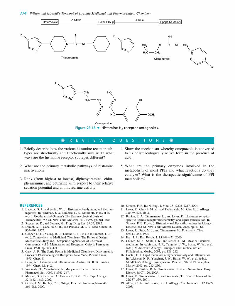
774 Wilson and Gisvold’s Textbook of Organic Medicinal and Pharmaceutical Chemistry
Figure 23.18 Histamine H3-receptor antagonists.
R E V I E W Q U E S T I O N S
1. Briefly describe how the various histamine receptor sub-types are structurally and functionally similar. In whatways are the histamine receptor subtypes different?
2. What are the primary metabolic pathways of histamineinactivation?
3. Rank (from highest to lowest) diphehydramine, chlor-pheniramine, and cetirizine with respect to their relativesedation potential and antimuscarinic activity.
4. Show the mechanism whereby omeprazole is convertedto its pharmacologically active form in the presence ofacid.
5. What are the primary enzymes involved in themetabolism of most PPIs and what reactions do theycatalyze? What is the therapeutic significance of PPImetabolism?
REFERENCES1. Babe, K. S. J., and Serfin, W. E.: Histamine, bradykinin, and their an-
tagonists. In Hardman, J. G., Limbird, L. E., Mollinoff, P. B., et al.(eds.). Goodman and Gilman’s The Pharmacological Basis ofTherapeutics, 9th ed. New York, McGraw-Hill, 1995, pp. 581–600.
2. Saxena, A. K., and Saxena, M.: Prog. Drug Res. 39:35, 1992.3. Durant, G. I., Ganellin, C. R., and Parsons, M. E.: J. Med. Chem. 18:
905–909, 1975.4. Cooper, D. G., Young, R. C., Durant, G. H., et al.: In Emmett, J. C.,
(ed.). Comprehensive Medicinal Chemistry. The Rational Design,Mechanistic Study and Therapeutic Application of ChemicalCompounds, vol 3. Membranes and Receptors. Oxford: PermagonPress, 1990, pp. 343–421.
5. Casy, A. F.: The Steric Factor in Medicinal Chemistry. DissymmetricProbes of Pharmacological Receptors. New York, Plenum Press,1993, Chap. 11.
6. Falus, A.: Histamine and Inflammation. Austin, TX: R. G. Landes,1994, Chap. 1.1, p. 2.
7. Watanabe, T., Yamatodani, A., Maeyama K., et al.: TrendsPharmacol. Sci. 1009. 11:363–367.
8. Marone, G., Genovese, A., Granata F., et al.: Clin. Exp. Allergy.32:1682–1689, 2002.
9. Oliver, J. M., Kepley, C. I., Ortega, E., et al.: Immunopharm. 48:269–281, 2000.
10. Simons, F. E. R.: N. Engl. J. Med. 351:2203–2217, 2004.11. Leurs, R., Church, M. K., and Taglialatela, M.: Clin. Exp. Allergy.
32:489–498, 2002.12. Bakker, R. A., Timmerman, H., and Leurs, R.: Histamine receptors:
specific ligands, receptor biochemistry, and signal transduction. InSimons, F. E. R., (ed.). Histamine and H1-antihistamines in AllergicDisease, 2nd ed. New York, Marcel Dekker, 2002, pp. 27–64.
13. Leurs, R., Smit, M. J., and Timmerman, H.: Pharmacol. Ther.66:413–463, 1995.
14. Hall, I. P.: Eur. Respir. J. 15:449–451, 2000.15. Church, M. K., Shute, J. K., and Jensen, H. M.: Mast cell derived
mediators. In Adkinson, N. F., Yunginer, J. W., Busse, W. W., et al.(eds.). Middleton’s Allergy: Principles and Practice, 6th ed.Philadelphia, Mosby, 2003, pp. 189–212.
16. Goetzl, E. J.: Lipid mediators of hypersensitivity and inflammation.In Adkinson, N. F., Yunginer, J. W., Busse, W. W., et al. (eds.).Middleton’s Allergy: Principles and Practice, 6th ed. Philadelphia,Mosby, 2003, pp. 213–230.
17. Leurs, R., Bakker, R. A., Timmerman, H., et al.: Nature Rev. DrugDiscov. 4:107–120, 2005.
18. Leurs, R., Timmerman, H., and Watanabe, T.: Trends Phamacol. Sci.22:337–339, 2001.
19. Akdis, C. A., and Blaser, K.: J. Allergy Clin. Immunol. 112:15–22,2003.
0733-0775_17865_Ch23.qxd 12/4/09 8:04 PM Page 774
20. Maslinski, C., and Fogel, W. A.: Catabolism of histamine. InUvnas, B. (ed.). Histamine and Histamine Antagonists, Handbook ofExperimental Pharmacology, vol. 97. New York, Springer-Verlag,1991, Chap. 5.
21. Schayer, R. C., and Cooper, J. A. D.: J. Appl. Physiol. 9:481–483,1956.
22. Fourneau, B., and Bovet, D.: Arch. Int. Pharmacodyn. 46:178–191,1993.
23. Witiak, D. T.: Antiallergenic agents. In Burger, A. (ed.). MedicinalChemistry, 3rd ed. New York, Wiley lnterscience, 1970, p. 1643.
24. Casy, A. F.: Chemistry of anti-H1 histamine antagonists. In Rocha eSilva, M. (ed.). Handbook of Experimental Pharmacology, vol. 18/2.New York, Springer-Verlag, 1978, p. 175.
25. Paton, D. M.: Receptors for histamine. In Schachter, M. (ed.).Histamine and Antihistamines. New York, Pergamon Press, 1973, p. 3.
26. Rocha e Silva, M., and Antonio, A.: Bioassay of antihistaminic ac-tion. In Rocha e Silva, M. (ed.). Handbook of ExperimentalPharmacology, vol. 18/2. New York, Springer-Verlag, 1978, p. 381.
27. Biel, J. H., and Martin, Y. C.: Organic synthesis as a source of new drugs.In Gould, R. F. (ed.). Drug Discovery, Advances in Chemistry Series no.108. Washington, DC, American Chemical Society, 1971, p. 81.
28. Ahlquist, R. P.: Am. J. Physiol. 153:586, 1948.29. Lin, T. M., et al.: Ann. N. Y. Acad. Sci. 99:30, 1962.30. Ash, A. S. F., and Schild, H. O.: Br. J. Pharmacol. Chemother.
27:427, 1966.31. Black, J. W., et al.: Nature 236:385, 1972.32. Metzler, D. E., and Ikawa Mand Sneil, E. E. J.: Am. Chem. Soc. 76:
648, 1954.33. Taglialatela, M., Timmerman, H., and Annunziato, L.: Trends
Pharmacol. Sci. 21:52–56, 2000.34. Woosley, R. L.: Ann. Rev. Pharmacol. Toxicol. 36:233–252, 1996.35. Casy, A. F., et al.: Chirality. 4:356–366, 1992.36. Ednoether, A., and Weber, H. P.: Helv. Chim. Acta. 59:2462–2468,
1976.37. Ganellin, C. R., and Parsons, M. E.: Bristol, UK, Wright, 1982.38. Shafi’ee, A., and Hite, G.: J. Med Chem. 12:266–270, 1969.39. Hanna, P. E., and Ahmed, A. E.: J. Med. Chem. 16:963–968, 1973.40. van den Brink, F. G., and Lien, B. 3.: Competitive and noncompetitive
antagonism. In Rocha e Silva, M. (ed.). Handbook of ExperimentalPharmacology, vol. 18/2. New York, Springer-Verlag, 1978, p. 333.
41. Nauta, W. T., and Rekker, R. F.: Structure-activity relationships ofHi-receptor antagonists. In Rocha e Silva, M. (ed.). Handbook ofExperimental Pharmacology, vol. 18/2. New York, Springer-Verlag,1978.
42. Chishty, M., et al.: J. Drug Target. 9:223–228, 2001.43. Chen, C., Hanson, E., Watson, J. W., et al.: Drug Metab. Dispos.
31:312–318, 2003.44. Tashiro, M., Mochizuki, H., Iwabuchi, K., et al.: Life Sci. 72:
409–414, 2002.45. Garteiz, D. A., Hook, R. H., Walker, B. J., et al.: Arzneim-Forsch.
32:1185–1190, 1982.46. Nobles, W. L., and Blanton, C. D.: 3. Pharm. Sci. 53:115, 1964.47. Toldy, L., et al.: Acta Chim. Acad. Sci. Hung. 19: 273, 1959.48. Gupta, S., Banfield, C., Kantesaria, B., et al.: Clin. Ther. 23:451–466,
2001.49. Yumibe, N., Huie, K., Chen, K. J., et al.: Biochem. Pharmacol.
51:165–172, 1996.50. Curran, M. P., Scott, L. J., and Perry, C. M.: Drugs. 64: 523–561,
2004.51. Gillard, M., Van Der Perren, C., Moguilevsky, N., et al.: Mol.
Pharmacol. 61:391–399, 2002.
52. Holgate, S. T.: The chromones: cromolyn sodium and nedocromilsodium. In Adkinson, N. F., Yunginer, J. W., Busse, W. W., et al.(eds.). Middleton’s Allergy: Principles and Practice, 6th ed.Philadelphia, Mosby, 2003, pp. 915–927.
53. Douglas, W. W.: Histamine and 5-hydroxytryptamine (serotonin)and their antagonists. In Gilman, A. G., Goodman, L. S., Rail, T. W.,et al. (eds.). Goodman and Gilman’s The Pharmacological Basis ofTherapeutics, 7th ed. New York, Macmillan, 1985, p. 605.
54. Cook, E. B., Stahl, J. L., Barney N. P., et al.: Med. Chem. Rev.1:333–347, 2004.
55. Caldwell, D. R., Verin, P., Hartwich-Young, R., et al.: Am. J.Ophthalmol. 113:632–637, 1992.
56. Tanaka, M.: Drugs Today 28:29–31, 1992.57. McTavish, D., and Sorkin, E. M.: Drugs 38:778–800, 1989.58. Martin, U., and Romer, D.:Arzneimittelforschung 28:770–782, 1978.59. Feldman, M., and Burton, M. E.: N. Engl. J. Med. 323:1672–1680,
1990.60. Feldman, M., and Burton, M. E.: N. Engl. J. Med. 323:1749–1755,
1990.61. Roberts, S., and McDonald, I. M.: Inhibitors of gasatric acid secre-
tion. In Abraham, D. J., (ed.). Burger’s Medicinal Chemistry andDrug Discovery, vol 4. Autocoids, Diagnostics and Drugs from NewBiology, 6th ed. Hoboken, NJ, Wiley-Interscience, 2003, pp. 85–127.
62. Sachs, G.: Pharmacotherapy. 23:S68–S73, 2003.63. Soil, A. H.: N. Engl. J. Med. 322:909–916, 1990.64. Ganellin, C. R.: Discovery of cimetidine. In Roberts, S. M., Price,
B. J., (eds.). Medicinal Chemistry, The role of organic chemistry indrug research. London, Academic Press, 1985, pp. 93–118.
65. Somogyi, A., and Muirhead, M.: Clin. Phrmacokinet. 12:321–366,1987.
66. Facts and Comparisons 4.0, Wolters Kluwer Health, Amsterdam,Netherlands, 2009.
67. Brandstrom, A., Lindberg, P., and Junggren, U.: Scand. J.Gastroenterol. 20(Suppl. 108):15–22, 1985.
68. Lindberg, P., Brandstrom, A., Wallmark, B., et al.: Med. Res. Rev.10:1–54, 1990.
69. Munson, K., Garcia, R., and Sachs, G.: Biochemistry 44:5267–5284,2005.
70. Shin, J. M., and Sachs, G.: Biochem. Pharmacol. 68:2117–2127,2004.
71. Shin, J. M., Choo, Y. M., and Sachs, G. J.: Am. Chem. Soc.126:7800–7811, 2004.
72. Shin, J. M., and Sachs, G.: Gastroenterology 123:1588–1597, 2002.73. Sachs, G., Shin, J. M., and Howden, C. W.: Aliment. Pharmacol.
Ther. 23(Suppl. 2):2–8, 2006.74. Yacyshyn, B. R., and Thomson, A. B. R.: Digestion. 66:67–78, 2002.75. Andersson, T.: Clin. Pharmacokinet. 31:9–28, 1996.76. Andersson, T.: Clin. Pharmacokinet. 43:279–285, 2004.77. Nagashima, R., and Yoshida, N.: Arzneim.-Forsch. 29:1668–1676,
1979.78. Marks, I. N.: J. Clin. Gastroenterol. 9(Suppl 1):18–22, 1987.79. Won-Kin, S., Kachur, J. F., and Gaginella, T. S.: Prostaglandins
46:221–231, 1993.80. Monk, J. P., and Clissold, S. P.: Drugs 33:1–30, 1987.81. Leurs, R., and Timmerman, H.: Prog. Drug Res. 39:127, 1992.82. Phillips, J. G., Au, S. M., Yates, S. L., et al.: Annu. Rep. Med. Chem.
33:31–40, 1998.83. Stark, H., Ligneau, X., Arrang, J. M., et al.: Bioorg. Med. Chem.
Lett. 8:2011–2016, 1998.84. Lim, H. D., van Rijn, R. M., Ling, P., et al.: J. Pharmacol. Exp. Ther.
314:1310–1321, 2005.
Chapter 23 Histamine and Antihistaminic Agents 775
0733-0775_17865_Ch23.qxd 12/4/09 8:04 PM Page 775

C H A P T E R 24
AnalgesicsCAROLYN J. FRIEL AND MATTHIAS C. LU
healthcare providers because of fear of psychological de-pendence (commonly referred to as addiction), disciplinaryaction, or adverse effects such as tolerance and physicaldependence of the pain medications.2,3 As a result, manypatients with moderate to severe pain are often undertreatedor untreated.3–5 Another possible reason for treatment failureis the fact that pain perception may also vary among individ-uals especially across racial and ethnic origins.6
Origin of Pain
Pain differs in its underlying causes, symptoms, and neuro-biological mechanisms and has been classified into threemajor types: physiological (nociceptive), inflammatory, andneuropathic.7,8 Physiological pain is the most common andis often caused by an injury to body organs or tissues. It isfurther categorized, according to the source of the pain, intocutaneous pains (skin and surface tissues), somatic pains(ligaments, tendons, bones, blood vessels), and visceralpains (body organs and internal cavities). Inflammatory painoriginates from an infection or inflammation as a result ofthe initial tissue or organ damage. Neuropathic pain is a verycomplex, chronic pain, resulting from injury of the nervoussystems.9 Neuropathic pain may originate from limb ampu-tation, spinal surgery, viral infections such as shingles, orworsening of disease states associated with diabetes, ac-quired immunodeficiency syndrome (AIDS), or multiplesclerosis. The pharmacological interventions are very com-plex and often require the use of other analgesic adjuvantsnot covered in this chapter. Readers should consult a recentreview for a practical guide to the current clinical manage-ment of neuropathic pain.10
Acute and Chronic Pain
Pain may be acute or chronic.11 Acute pain is often severebut usually lasts only until the removal of the source thattriggered the pain. Acute pain includes nociceptive, somatic,or visceral pain, postoperative and posttraumatic pain, burnpain, acute pain during childbirth, acute headache, etc.Acute and postoperative pains are most often treated withthe opioid analgesics.
Chronic pain, on the other hand, is defined as a pain last-ing longer than 6 months that persists even when the initialcause has been resolved through appropriate medicalintervention. Chronic pain can be further divided intochronic malignant pain (e.g., cancer, human immunodefi-ciency virus [HIV]/AIDS, amyotrophic lateral sclerosis,multiple sclerosis, end-stage organ failure) and chronic non-malignant pain (e.g., lower-back pain, chronic degenerative
C H A P T E R O V E R V I E W
The terms analgesics and analgetic drugs are often usedinterchangeably to describe a diverse group of pain med-ications such as opioids, nonsteroidal anti-inflammatorydrugs (NSAIDs), and triptans, each with very differentmechanisms of action for relieving pains of a wide arrayof causes.
Analgesics can be broadly categorized, according totheir therapeutic use, into several drug classes: (a) the opi-oids (or narcotic analgesics), which play a major role in therelief of acute pain and in the management of moderate tosevere chronic pain; (b) the NSAIDs and acetaminophen,which are the most widely used analgesic drugs for reliev-ing mild to moderate pain and reducing fever; (c) the trip-tans (the antimigraine medications), which are specificallydesigned and targeted for acute and abortive treatment ofmigraine and cluster headaches; and (d) a new emergingclass of analgesics known as analgesic adjuvants that in-clude tricyclic antidepressants such as amitriptyline, anti-convulsants such as gabapentin and pregabalin, and topicalanalgesics such as lidocaine patches that can be used totreat neuropathic pains.
In this chapter, only the first three classes of pain medica-tions are covered. The main objectives of this chapter willbe focusing on their mechanism of action, structure–activityrelationships (SARs), pharmacokinetic properties, and theirclinical applications for pain management. Readers shouldconsult other chapters under anticonvulsants, antidepres-sants, and local anesthetics for a detailed discussion of drugsused for treating neuropathic and other incidental pains as-sociated with minor cuts, burns, and insect bites.
PAIN AND PAIN MANAGEMENT
Pain, one of the most common complaints for which patientsseek medical attention, is also the hardest to manage despitethe availability of many analgesic medications as well asother nonpharmacologic treatment options. Pain manage-ment, especially the use of opioids in patients with chronicpains, is one of the most troublesome public health issues forpatients, doctors, and other healthcare providers.1,2 As earlyas the 1970s, the National Institutes of Health formed a com-mittee, at the request of the president, to investigate therapiesfor rare diseases, but the initial focus was on pain manage-ment. The committee found that the problem associated withthe use of the opioid analgesics was a lack of training ofphysicians in pain management and misinformation among
776
0776-0818_17865_Ch24.qxd 12/4/09 8:08 PM Page 776
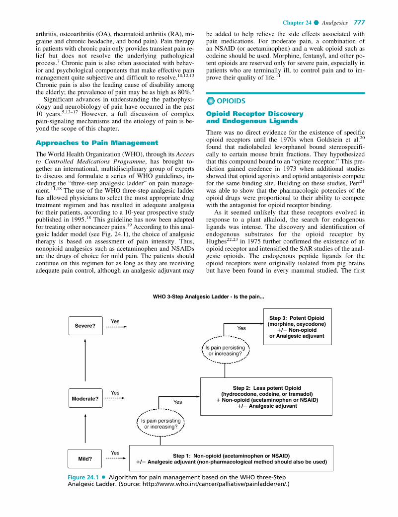
Chapter 24 Analgesics 777
arthritis, osteoarthritis (OA), rheumatoid arthritis (RA), mi-graine and chronic headache, and bond pain). Pain therapyin patients with chronic pain only provides transient pain re-lief but does not resolve the underlying pathologicalprocess.7 Chronic pain is also often associated with behav-ior and psychological components that make effective painmanagement quite subjective and difficult to resolve.10,12,13
Chronic pain is also the leading cause of disability amongthe elderly; the prevalence of pain may be as high as 80%.3
Significant advances in understanding the pathophysi-ology and neurobiology of pain have occurred in the past10 years.5,13–17 However, a full discussion of complexpain-signaling mechanisms and the etiology of pain is be-yond the scope of this chapter.
Approaches to Pain Management
The World Health Organization (WHO), through its Accessto Controlled Medications Programme, has brought to-gether an international, multidisciplinary group of expertsto discuss and formulate a series of WHO guidelines, in-cluding the “three-step analgesic ladder” on pain manage-ment.11,18 The use of the WHO three-step analgesic ladderhas allowed physicians to select the most appropriate drugtreatment regimen and has resulted in adequate analgesiafor their patients, according to a 10-year prospective studypublished in 1995.18 This guideline has now been adaptedfor treating other noncancer pains.19 According to this anal-gesic ladder model (see Fig. 24.1), the choice of analgesictherapy is based on assessment of pain intensity. Thus,nonopioid analgesics such as acetaminophen and NSAIDsare the drugs of choice for mild pain. The patients shouldcontinue on this regimen for as long as they are receivingadequate pain control, although an analgesic adjuvant may
be added to help relieve the side effects associated withpain medications. For moderate pain, a combination ofan NSAID (or acetaminophen) and a weak opioid such ascodeine should be used. Morphine, fentanyl, and other po-tent opioids are reserved only for severe pain, especially inpatients who are terminally ill, to control pain and to im-prove their quality of life.11
OPIOIDS
Opioid Receptor Discoveryand Endogenous Ligands
There was no direct evidence for the existence of specificopioid receptors until the 1970s when Goldstein et al.20
found that radiolabeled levorphanol bound stereospecifi-cally to certain mouse brain fractions. They hypothesizedthat this compound bound to an “opiate receptor.” This pre-diction gained credence in 1973 when additional studiesshowed that opioid agonists and opioid antagonists competefor the same binding site. Building on these studies, Pert21
was able to show that the pharmacologic potencies of theopioid drugs were proportional to their ability to competewith the antagonist for opioid receptor binding.
As it seemed unlikely that these receptors evolved inresponse to a plant alkaloid, the search for endogenousligands was intense. The discovery and identification ofendogenous substrates for the opioid receptor byHughes22,23 in 1975 further confirmed the existence of anopioid receptor and intensified the SAR studies of the anal-gesic opioids. The endogenous peptide ligands for theopioid receptors were originally isolated from pig brainsbut have been found in every mammal studied. The first
WHO 3-Step Analgesic Ladder - Is the pain...
Severe?
Moderate?
Mild?
YesYes
Yes
Is pain persistingor increasing?
Is pain persistingor increasing?
Step 3: Potent Opioid(morphine, oxycodone)
�/� Non-opioidor Analgesic adjuvant
Step 2: Less potent Opioid(hydrocodone, codeine, or tramadol)
� Non-opioid (acetaminophen or NSAID)�/� Analgesic adjuvant
Step 1: Non-opioid (acetaminophen or NSAID)�/� Analgesic adjuvant (non-pharmacological method should also be used)
Yes
Yes
Figure 24.1 Algorithm for pain management based on the WHO three-StepAnalgesic Ladder. (Source: http://www.who.int/cancer/palliative/painladder/en/.)
0776-0818_17865_Ch24.qxd 12/4/09 8:08 PM Page 777

endogenous peptide was termed enkephalin, which wasfound to be a mixture of the two pentapeptides that onlydiffer in their terminal amino acid.
Both methionine-enkephalin (Met-enkephalin) andleucine-enkephalin (Leu-enkephalin) were shown to inhibitthe contraction of electrically stimulated guinea pig ileum(GPI) and mouse vas deferens (MVD). These two tests arestill used as screening methods for opioid activity. Naloxonecompletely reversed the inhibitory effects of enkephalin,which led Hughes to infer that the peptides were acting on anopioid receptor.22 The central administration of enkephalinsin rats produced short analgesic activity. The transient natureof the enkephalins’ actions correlated with the rapid degrada-tion of the enkephalin Tyr-Gly bond by aminopeptidases.Much synthetic work has been done in an attempt to increasethe duration of action of the opioid peptides and maintaintheir analgesic effect.
SARs of Enkephalins
TYR1
The first amino acid of the pentapeptide shows a distinctpreference for tyrosine. Most changes to this amino acid, ei-ther by substituting with other amino acids or masking thephenolic hydroxyl (OH) or amino function, produce an inac-tive or weakly active peptide.
GLY2
Replacing the naturally occurring L-Gly with variousD-amino acids produces a peptide that is resistant to peptidecleavage by aminopeptidases. Replacement with D-Ser is themost effective replacement, and all L-amino acid analogs hadlow activities. Substituting D-amino acids for L-amino acidsproduces stable peptides, and the stereochemical change maygive the peptide access to additional binding sites on the re-ceptors; both of these actions may explain their increasedpotencies. Replacing the Gly2 with D-Ala2 while simulta-neously replacing the L-Leu5 with D-Leu5 produces the pep-tide known as D-Ala2, D-Leu5 enkephalin (DADLE), whichis commonly used as a selective �-agonist.
GLY3
Almost all changes to this amino acid result in a drop in po-tency, unless they are also accompanied by another changesuch as replacing the Gly2 with D-Ala2 as described above.
PHE4
The aromatic nature of the fourth residue is required for highactivity. When combined with the D-Ala2 replacement, the ad-dition of an electron withdrawing, lipophilic substituent (e.g.,
778 Wilson and Gisvold’s Textbook of Organic Medicinal and Pharmaceutical Chemistry
Met-enkephalin = Tyr1-Gly2-Gly3-Phe4-Met5(OH) Leu-enkephalin = Tyr1-Gly2-Gly3-Phe4-Leu5(OH)
H2NHC CCH2
O
OH
HN
HC CH
OHN
HC CH
OHN
HC CCH2
OHN
HC CCH2
OHO
CH2
SCH3
HN CHCCH2
OHO
CHCH3
CH3
-Met5 -Leu5
or
NO2, Cl, and Br) in the para position of Phe4 greatly increasesactivity. Para substitutions with electron donating, hydrophilicfunctional groups (e.g., NH2 and OH) abolish activity.
MET5/LEU5
Position 5 appears to tolerate more residue changes than theother positions. Many amino acid substitutions at this positionmaintain activity (e.g., Ala, Gly, Phe, Pro). Even the loss ofthe fifth residue to yield the tetrapeptide Tyr1-Gly2-Gly3-Phe4
maintains weak activity in both the GPI and MVD assays.The protected peptide, Tyr1-D-Ala2-Gly3-MePhe4-Gly-ol5,known as DAMGO is highly selective for the �-receptor.
The pituitary gland, hypothalamus, and the adrenalmedulla all produce various opioid peptides. Many of thesematerials were found to be fragments of �-lipotropin (�-LPT) a 91 amino acid peptide. �-LPT has no opioid activityitself. The fraction containing amino acids 61–91 isdesignated �-endorphin (a word derived from combiningendogenous and morphine). It is much more potent than theenkephalins in both in vivo and in vitro tests. The search foropioid peptides continued, and additional precursor proteinswere discovered. Many additional precursor proteins syn-thesized in the pituitary, adrenal glands, and brain nerve ter-minals were found to contain sequences of amino acids thatwere enkephalins or other active opioid peptides. It appearsthat naturally occurring peptides may serve roles as bothshort-acting analgesics and long-term neuronal or endocrinemodulators. Acupuncture, running, or other physical activ-ity may induce the release of neuropeptides although studiesexist that both confirm and refute these claims.24,25 A com-plete discussion on the neuropeptides is beyond the scope ofthis chapter. Some examples of opioid peptides are givenin Figure 24.2.26–29
Opioid Receptors
The discovery of the endogenous opioid peptides paralleledthe development of radiolabeling techniques. During the1970s, researchers were able to label opioid receptors withreversible radioactive ligands that allowed pharmacologicalactions of specific receptors and their locations to be identi-fied. These techniques allowed for the identification of opi-oid receptor locations in the brain. Additional pharma-cological advances in gene expression studies led to theidentification of peripheral opioid receptor locations.30
Opioid receptors are distributed throughout the brain, spinalcord, and peripheral tissues. The distribution of specific opi-oid receptor subtypes (�, �, and �) usually overlaps. In rats,high concentrations of all three genes for the �-, �-, and�-receptors were found in the hypothalamus and cerebral
0776-0818_17865_Ch24.qxd 12/4/09 8:08 PM Page 778

Chapter 24 Analgesics 779
cortex. Intermediate concentrations were found in the smallintestine, adrenal gland, testes, ovary, and uterus. Low con-centrations of all the three gene transcripts were found in thelung and kidney. The gene for the �-opioid receptor was notfound in the stomach, heart, endothelium, or synovium ofthe rat.30 Opioid receptors are also found on the peripheralterminals of sensory neurons in inflamed rat paws.31 Thus,the central and peripheral distribution of opioid receptors iscomplex. This complicates the interpretation of pharmaco-logical data of individual drugs that may have overlappingbinding at multiple opioid receptor subtypes. In addition,opioid receptors form homodimers and heterodimers withopioid receptors and nonopioid receptors such as the �2a-adrenergic receptors resulting in different pharmacologicactions and altered coupling to second messengers.32
Genes for the four major opioid receptor subtypes havebeen cloned; the MOP � �-receptor (mu for morphine),the KOP � �-receptor (kappa for ketocyclazocine), theDOP � �-receptor (delta for deferens because it was orig-inally discovered in the MVD), and the NOP � nocicep-tion/orphanin FQ receptor (named as an orphan receptorbecause the endogenous/exogenous ligand was unknownat its time of discovery).33 The opioid receptor subtypesshare extensive residue homology in their transmembrane(TM) domains with most of the variation found in the ex-tracellular loops (Fig. 24.3).33 All of the opioid receptorsbelong to the G-protein–coupled receptor class and assuch, they are composed of seven TM domains. When thereceptor is activated, a portion of the G protein diffuseswithin the membrane and causes an inhibition of adenylatecyclase activity. The decreased enzyme activity results ina decrease in cyclic adenosine monophosphate (cAMP)formation, which regulates numerous cellular processes.One process that is inhibited is the opening of voltage-gated calcium influx channels on nociceptive C-fibers.This results in the hyperpolarazation of the nerve cell anddecreased firing and release of pain neurotransmitters suchas glutamate and substance P.34
OPIOID LIGAND BINDING SITE
There are no x-ray crystal structures reported for any opioidreceptor, and most modeling work has been based on thecrystal structure of rhodopsin. Many groups have attemptedto find the specific binding site through homology modeling,molecular dynamics, site-directed mutagenesis, chimericreceptors, truncated receptors, and SAR studies. As of yet,no definitive model of the ligand-binding site is available.The opioid-binding site for all four opioid receptors is be-lieved to be an inner cavity formed by conserved residueson TM helices TM3, TM4, TM5, TM6, and TM7.33,35 Not allgroups agree that the binding pocket is formed via TM do-mains; some evidence suggest that the amino terminus is animportant determinant of ligand binding affinity as well.36
The ligand specificity that each receptor shows may be a re-sult of differences in the extracellular loops that form lids onthe binding cavities or differences in amino acids within thebinding cavity. Within the cavity, agonists are thought tobind toward the bottom of the cavity via interactions with aconserved Asp from TM3 and a His from TM6. Molecularmodeling calculations show that the phenolic OH of the Tyr1
opioid peptide, or the ring A OH of nonpeptide opioids,forms a hydrogen bond with the conserved His on TM6. TheTyr1-charged nitrogen, or the N� of the nonpeptide agonist,forms an ionic bond with the conserved Asp of TM3.Antagonist ligands are thought to bind deeper in the bindingpocket but retain the ionic bond with the Asp of TM3. Thebulky substituent on the charged nitrogen of antagonists isbelieved to insert itself between TM3 and TM6, preventingthe shifts required for activation. Thus, antagonists preventthe necessary movement of TM3 and TM6 resulting in func-tional antagonism.33,35
THE �-RECEPTOR
Mu receptors are found primarily in the brainstem and me-dial thalamus.34 Endogenous peptides for the �-receptorinclude endomorphin-1, endomorphin-2, and �-endorphin
Endogenous Precursor ProteinsPro-opimelanocortin ACTH � � LPH �-LPH � � endorphinPro-enkephalin A263 4Met-enkephalin � Leu-enkephalinPro-enkephalin B256 � neo-endorphin175-183 � dynorphin209-225 � Leu-enkephalin228-232
Endogenous Opioid Peptide sequences� Endorphin � Tyr-Gly-Gly-Phe-Met5-Thr-Ser-Glu-Lys-Ser10-Gln-Thr-Pro-Leu-Val15-Thr-Leu-Phe-Lys-Asn20-Ala-Ile-Ile-Lys-Asn25-Ala-Tyr-Lys-Lys-Gly-Glu31 � � and � opioid receptor ligandEndomorphin-1 � Tyr-Pro-Trp-Phe-NH2 � � receptor agonistEndomorphin-2 � Tyr-Pro-Phe-Phe-NH2 � � receptor agonist
Dynorphin � Tyr-Gly-Gly-Phe-Leu5-Arg-Arg-Ile-Arg-Pro10-Lys-Leu-Lys13 � � opioid selectivity-Neoendorphin � Tyr-Gly-Gly-Phe-Leu5-Arg-Lys-Tyr-Pro-Lys10
�-Neoendorphin � Tyr-Gly-Gly-Phe-Leu5-Arg-Lys-Tyr-Pro9
Nociceptin � Phe-Gly-Gly-Thr-Gly5-Ala-Arg-Lys-Ser-Ala10-Lys-Ala-Asn-Gln14 � orphanin receptorOrphanin FQ � Phe-Gly-Gly-Phe-Thr5-Gly-Ala-Arg-Lys-Ser10-Ala-Arg-Lys-Leu-Ala-Asn-Gln17
Exogenous Opioid Peptide sequences “Exorphins”DADLE � Tyr-D-Ala-Gly-Phe-D-Leu � � selective agonistDPDPE � Tyr-D-Pen-Gly-Phe-D-Pen � � selective agonistDSLET � Tyr-D-Ser-Gly-Phe-Leu-Thr � � selective agonistCasomorphin (cow’s milk � opioid receptor agonist) � Tyr-Pro-Phe-Pro-Gly-Pro-Ile7
Dermorphin (South American frog skin � opioid receptor agonist) � Tyr-D-Ala-Phe-Gly-Tyr-Pro-Ser7-NH2Gluten exorphins (multiple peptides from wheat having opioid agonist and antagonist activity)
Figure 24.2 Endogenous and exogenous opioid peptides.
0776-0818_17865_Ch24.qxd 12/4/09 8:08 PM Page 779

(Fig. 24.2). Exogenous agonists for the �-receptor includedrugs from the five structural classes discussed later in thischapter (4,5-epoxymorphinan, morphinan, benzomorphan,4-phenyl/4-anilido piperidines, and the diphenylheptanes)and exogenous peptides such as dermorphin isolated fromthe skin of South American frogs. Recently, human neuro-blastoma cells have been shown to be capable of synthesizingmorphine via biosynthesis from radiolabeled tyramine. Thesynthetic route involves at least 19 steps and is similar, butnot exact, to the synthetic route used by the poppy plant.37
The exact role of endogenous morphine is unknown at thistime. In general, agonists at the �-receptor produce analge-sia, respiratory depression, decreased gastrointestinal (GI)motility, euphoria, feeding, and the release of hormones.Agonists are also responsible for the addictive effects of theopioid analgesics. Most clinically used opioid drugs bind tothe �-opioid receptor.
THE �-RECEPTOR
Opioid peptides for the �-receptor include the endogenouspeptides described previously, Met and Leu enkephalin, aswell as some synthetic peptides such as DADLE, DSLET, andDPDPE (see Fig. 24.2 for amino acid sequences). These pep-tides have high affinity for the receptor but low bioavailabilityand thus limited clinical usefulness. They are used in animalin vitro studies as probes for �-receptor location and function.
In an attempt to distinguish the amino acids responsiblefor �-receptor ligand specificity, point mutations of the �-receptor, and �/�-receptor chimeras were constructed.38 Thealtered receptors had a decreased ability to bind to �-receptorselective peptide and nonpeptide ligands, when amino acidsin the extracellular top of TM6 and TM7 and the extracel-lular loop (EC loop 3) that connected them were mutated.
Specifically, amino acids Try284, Val296, and Val297 werecrucial to selective �-ligand binding. These amino acids mayprovide recognition sites on the receptor that the ligandwould have to pass through to reach the putative binding sitedeeper in the TM cavity. Befort et al.39 also used site-directed point mutant receptors along with molecular model-ing to identify Tyr129 in TM3 as the most crucial amino acidfor ligand binding. In addition, they found a role for aminoacid Tyr308 (TM7) in ligand binding.
Nonpeptide agonists and antagonists have been designedto further study the function of the �-receptor. The first non-peptide lead compound selective for the �-receptor came fromscreening. Modifications to the lead compound led to theidentification of SNC-80 as a potent agonist specific for the�-receptor (Fig. 24.4). This compound has weak antinoci-ceptive effects in monkeys, an effect that can be reversed withthe �-antagonist naltrindole (NTI) (Fig. 24.4).40 A series ofSNC-80 analog, prepared by multiple groups, found that theamide nitrogen appears to be the most sensitive to modifica-tions and may play an important role in �-receptor selectiv-ity.41 Portoghese designed 7-spiroindanyloxymorphone(SIOM) (Fig. 24.4) based on the idea that the indole of NTI isacting as an “address” mimic of the Phe4 phenyl group ofenkephalin. The “address–message” concept proposes thatone part of the molecule may act as an “address,” essentiallydirecting the chemical to the correct receptor by bindingspecifically to that receptor, and another part of the moleculeacts as the “message,” which gives the compound thebiological action. The indole of NTI was replaced with aspiroindane functional group equivalent address in SIOM.The N-methyl group of SIOM confers an agonist message asopposed to the antagonist message of the NTI cyclopropylmethyl.42 Additional agonists for the �-receptor have been
780 Wilson and Gisvold’s Textbook of Organic Medicinal and Pharmaceutical Chemistry
TM IV
N�
C�
EL3EL2EL1
IL1
IY
Y
IA
S
VV
LN
M V
R
TK M K
K
T
T
TT
N
NF
F
FF
FF
F
F
P
M
M
M
M
ND
DR
TT
T
D
D
K
F
F
FF
WH
E
E
W
W
Q
GG
G
G
G
G
LL
L
LD
L L
L
L
L
L
L
L
L
L
L
LL
L
L
L
L
L
V
V
VCH
PK
DFR
R RR R
R
RR
R
R
R
TP
PP
P
V
V
V
V
VVV
V
V
SS
S
S
S
S
SS
SA
AA
AA
A
A K
K
K
NN
N
N
WC
C
C
CC
CC
C
C
R
TF
A A
AI II
I
I
I
I
I
I I
II I
I
Y
Y
YY
YY
Y
YII III IV V VI VII
IL2IL3
TM III
TM II
TM ITM VIITM VI
TM V
Figure 24.3 Structure of opioid receptors. (Left) Serpentine model of the opioidreceptor. Each transmembrane helix is labeled with a roman number. The white emptycircles represent nonconserved amino acids among the MOP, DOP, KOP, and NOP re-ceptors. White circles with a letter represent identical amino acids among all four opi-oid receptors. Violet circles represent further identity between the MOP-R, DOP-R, andKOP-R. Green circles highlight the highly conserved fingerprint residues of family A re-ceptors, Asn I:18 in TM1, AspII:10 in TM2, CysIII:01 in TM3, TrpIV:10 in TM4, ProV:16 inTM5, ProVI:15 in TM6, and ProVII:17 TM7. Yellow circles depict the two conservedcystines in EL loops 1 and 2, likely forming a disulfide bridge. (IL, intracellular loop,and EL, extracellular loop.) (Right) Proposed arrangement of the seven transmembranehelices of opioid receptors as viewed from the top (extracellular side). The seven trans-membrane helices are arranged sequentially in a counterclockwise manner. Each trans-membrane helix is labeled with a roman number. (Reprinted with permission from theAnnual Review of Biochemistry, Volume 73 © 2004 by Annual Reviews.)
0776-0818_17865_Ch24.qxd 12/4/09 8:08 PM Page 780

Chapter 24 Analgesics 781
developed based on the octahydroisoquinoline structure withfive-membered ring spacers being �-receptor antagonists andsix-membered ring spacers, agonists. TAN-67 is the mostextensively studied in this class (Fig. 24.4).41 The �-receptortolerates multiple chemical structures, and no pharmacophorehas been identified that can accommodate all of the diversestructures.38,43
Transgenic mice lacking the �-receptor have been gener-ated and found to display increased levels of anxiety. In ad-dition, �-receptor agonist show antidepressant activity in ratmodels.32,44 These results suggest that ligands targeting thisreceptor may represent new leads for the treatment ofschizophrenia, bipolar disorders, and depression. There arefew clinical reports of selective �-receptor agonist use in hu-mans, although they are reported to have proconvulsiveactivity that may limit their use.44
THE �-RECEPTOR
Kappa receptors are primarily found in the limbic, brain stem,and spinal cord.34 The �-receptor shows less structural ho-mology to the �-receptor than the �-receptor does. (Fig. 24.3)Unlike the �- and �-receptors that bind the (enkephalin) pep-tide sequence Tyr-Gly-Gly-Phe-(Leu/Met), the �-receptordoes not. The �-receptor shows a clear preference for bindingpeptides with an arginine in position 6 as seen in the dynor-phin peptides (Fig. 24.2).45
The structure of some �-agonists and an antagonist canbe seen in Figure 24.5.46,47 TRK-820 is a �-agonist display-ing approximately 15 times selectivity toward �-receptorsversus �-receptors.46 The 4,5-epoxymorphinan structure ofTRK-820 is proposed to mimic the tyrosine–glycine moietyof endogenous opioid peptides, in effect the agonistmessage. The additional 6�-substituent constitutes the ad-dress directing the compound to the �-receptor. Spiradolinewas one of the earliest compounds to show improved �-receptor selectivity (125 times over �-receptors). SARs onthis class of compounds revealed that methyl-substitutednitrogen amides, a methylene spacer between the amide and
the aromatic ring and the (�) isomer were all required for �-activity. The dichlorophenyl ring could be replaced with a 4-benzothiophene or a 4-benzofuran for increased potency and�-receptor selectivity.41 Salvinorin A (Fig. 24.5) a nonnitro-gen-containing natural compound found in the herb sage isa potent agonist at the �-receptor.48 The hallucinogenicproperties of salvia divinorum (sage) have been known forhundreds of years but recently, these properties have beenexploited by online groups attempting to sell legal hal-lucinogenic compounds. The �-antagonist nor-binaltor-phimine (nor-BNI) was made by incorporating a rigid pyr-role spacer linking two �-pharmacophores. Nor-BNI is usedin the laboratory to determine if �-receptors are involved inan observed activity.41 The second pharmacophore of nor-BNI is not required for �-antagonism, and compounds thatare missing the second aromatic ring have been found to beeven more selective and potent antagonists at the �-receptor.
In clinical trials, most �-agonists produce dysphoria andthus may be less psychologically addicting but also lessacceptable to patients. Some compounds with �-agonist ac-tivity (e.g., pentazocine, nalbuphine) are available, but theclinical development of pure �-agonists has been limited bythe centrally mediated side effects. Spiradoline was devel-oped under the premise that a selective �-agonist would bean analgesic without the �-opioid–mediated problems asso-ciated with addiction and respiratory depression. Clinicaltrials in humans found that spiradoline did not produce anal-gesic effects within a dose range that did not also cause dys-phoria, diuresis, and sedation.47 The selective �-agonistTRK-820 shows some promise for the treatment of uremicpruritus, the itch associated with dialysis.49
THE ORPHANIN RECEPTOR/FQ/NOP1
The traditional opioid peptides do not elicit any biologicaleffect at the orphanin receptor. An endogenous peptide forthe orphanin receptor has been found and termed orphaninFQ or nociceptin (Fig. 24.2). This 17 amino acid peptide canreverse the analgesic effects of morphine thus is antiopioid
O NH
OHN
HO
Naltrindole (NTI) δ receptor antagonist
O
OHN
HO
7-spiroindanyloxymorphone (SIOM)δ receptor agonist
CH3
O
"address" for δ receptorPhe4 mimic of enkephalin
"address" for δ receptorPhe4 mimic of enkephalin
O
N
OCH3
N
N
CH3
H3C
(+) SNC-80 δ receptor agonist
OH
NH3C
H
N
TAN-67δ receptor agonist
Figure 24.4 Selective �-receptor ligands.
0776-0818_17865_Ch24.qxd 12/4/09 8:08 PM Page 781

in some situations.50 The carboxy-terminal half of the recep-tor may serve as the portion that excludes binding to the �-,�-, and �-opioid ligands. The functional role of the receptoris still being investigated, but selective agonists have beenshown to delay gastric emptying and decrease gastric secre-tory functions in rats.51 In addition, this receptor is found onairway nerves, and agonists decrease bronchospasms inguinea pigs and cats. This receptor is a target for the designof novel peripherally acting antitussive agents, although nocompounds are on the market at present.
STRUCTURE–ACTIVITY RELATIONSHIPS
Relating the structure of the chemical ligand to the phys-iologic activity it induces can only be accomplished bylooking at each receptor type individually. The chemicallydiverse nature of agonists and antagonists found for the �-,�-, and �-receptors is testimony to the adaptability of the
receptors to multiple chemical backbones and tolerance fordivergent functional groups. The reader is referred to re-views of the �- and �-receptor for detailed SARs for ligandsat those receptors.41 A summary review of the SARs of the�-receptor compounds is included in the drug monographsection below. For the purpose of this chapter, the drugclasses covered will be 4,5-epoxymorphinans, morphinans,benzomorphans, 4-phenylpiperidines/4-anilidopiperidines,diphenylheptanes, and the miscellaneous category.
DRUG MONOGRAPHS
4,5-Epoxymorphinans
A summary of the SARs of the 4,5-epoxymorphinan struc-ture can be seen in Figure 24.6, and these are discussed inthe succeeding individual drug monographs.
782 Wilson and Gisvold’s Textbook of Organic Medicinal and Pharmaceutical Chemistry
O N
OHN
HO
"address" for κ receptor
CH3
OO
opioid receptor Tyr1-Gly2 "message"
ON
NCH3
O Cl
Cl
O
OHN
HOO
HON
OH
NH
(-) spiradolineκ receptor agonist (125 x over �)
TRK-820 κ receptor agonistmodest selectivity (15x over �)
Nor-binaltorphimine (nor-BNI)κ receptor antagonist
salvinorin Aκ receptor agonist
O
O
OO
O
O
OOFigure 24.5 Selective �-receptorligands.
Morphine
Changes on Morphine that increase analgesic activityC6 - OH to OAcC3 and C6 - OH to OAc C6 - OH to O-Sulfate or O-glucuronide C6 - OH to =O and C7-C8 single bondC6 - OH to H and C7-C8 single bondC14 - H to βOH N - CH3 to CH2CH2PhN - CH3 to CH2CH2furanN - CH3 to CH2C=OPhRemoval of C4-C5 ether link
Changes on Morphine that produce antagonistsN - CH3 to CH2CH=CH2N - CH3 to CH2
23 4
12111
1314910
5 67
8
OOHHO
NH3C
AE
B
C
D ring
4
3OH
O
9NH3C
6OH
=
Figure 24.6 Summary of functional group changes on morphine structure.
0776-0818_17865_Ch24.qxd 12/4/09 8:08 PM Page 782

Chapter 24 Analgesics 783
MORPHINE
The prototype ligand for the �-receptor is morphine. Thenumbering and ring lettering (A, B, C. . .) can be seen inFigure 24.6. Morphine contains 5 chiral centers and has16 optical isomers (not 32 because of the restriction of C-9to C-13 ethanamino bridge). The naturally occurring, activeform of morphine is the levorotatory enantiomorph withthe stereochemistry 5(R), 6(S), 9(R), 13(S), and 14 (R). Thex-ray determined conformation of morphine is a “T” shapewith the A, B, and E rings forming the vertical portion, andthe C and D ring forming the top (Fig. 24.6).41
Morphine was isolated from opium in 1806 by a Germanpharmacist, Seturner. He named the compound “morphine”after the Greek god of dreams “Morpheus.” The first men-tion of the opium poppy was found in Iraq on clay tabletsinscribed in cuneiform script in about 3000 BC.52 Opium hasbeen used throughout history and is found in ancientEgyptian, Greek, Roman, Arabic, Indian, and Chinese writ-ing. Opium is isolated from the opium poppy, Papaversomniferum, by lancing the unripe pod and collecting anddrying the latex that seeps from the incision. Opium con-tains over 40 different alkaloids with most alkaloids repre-sented in the following five structures: morphine(8%–17%), codeine (0.7%–5%), thebaine (0.1%–2.5%), pa-paverine (0.5%–1.5%), and noscapine (1%–10%).52
Although the total synthesis of morphine has beenaccomplished by various chemical processes, it is still pro-duced from the poppy latex.53 Thus far, no synthetic path-way is efficient enough to make it competitive with thepreparation of morphine from either isolation or semisyn-
thesis from a precursor compound from the poppy plant.The endogenous synthesis of morphine in human neuroblas-toma cells has been elegantly described.37 The function ofendogenous morphine is unknown at this time, but the genesand enzymes involved in morphine biosynthesis may be-come targets for novel drugs used to treat pain.
Morphine is the prototype �-receptor agonist; it is thedrug to which all other �-agonists are compared. The phar-macological properties of morphine are well documentedand include analgesia, its primary use. For equivalent anal-gesic effect, the oral dose must be 3 times the intravenous(IV) dose to account for the morphine lost to first-pass he-patic metabolism. The equianalgesic dose of morphine con-geners can be seen in Figure 24.7.
Morphine is extensively metabolized via phase II conju-gation to morphine-3-glucuronide (�60%), morphine-6-glucuronide (�9%), and, to a lesser extent, the N-demethyl-ated metabolite (�3%)54 (Fig. 24.8). Much controversyexists on the contribution of the metabolites of both codeine-6-glucuronide and morphine-6-glucuronide to their analgesiceffect. In some studies, the 6-glucuronide metabolite of bothdrugs contributes significantly to their potency.55–57 In otherstudies, the 6-glucuronide metabolites of morphine andcodeine produces very little analgesic effect.58,59
Morphine, or another potent opioid drug, is introduced inthe WHO stepladder when pain is severe and no relief is ob-tained from NSAIDs or a combination of NSAIDs and a lesspotent opioid. Morphine is a monoacidic base and readilyforms water-soluble salts with most acids. Because morphineitself is poorly soluble in water (1 g/5,000 mL at 25°C ), the
OOHHO
NH3C
Morphineoral =30 mgparenteral=10 mg
O OHH3CO
NH3C
Codeineoral =200 mgparenteral=130 mg
OOO
NH3C
O
H3C
O
CH3
O OHO
NH3C
Heroinoral =15 mgparenteral=5 mg
Hydromorphoneoral =7.5mgparenteral=1.5 mg
O OH3CO
NH3C
Hydrocodoneoral =30 mgparenteral=NA
O OH3CO
NH3C
OH
Oxycodoneoral =20 mgparenteral=NA
OOHO
NH3C
OH
Oxymorphoneoral =10 mgparenteral=1 mg
NH3C
R
MorphinanR= OH (-) Levorphanol oral = 2 mgR= OCH3 (+) Dextromethorphan
Figure 24.7 4,5-Epoxymorphinans andmorphinans.
0776-0818_17865_Ch24.qxd 12/4/09 8:08 PM Page 783

sulfate salt is formed for both oral, IV, and suppository use.The usual starting parenteral dose of morphine in adults is2.5 to 5 mg q 4 hours. Morphine is available for oral use bothas an immediate release and sustained release formulation.The immediate release preparations are dosed q 4 hours,and the sustained release formulations are dosed q 12 to q24 hours depending on the formulation.
CODEINE
Codeine is an alkaloid that occurs naturally in opium, butthe amount present is usually too small to be of commercialimportance. Consequently, most commercial codeine is pre-pared from morphine by methylating the phenolic OHgroup. It occurs as levorotatory, colorless, efflorescent crys-tals, or as a white crystalline powder. It is light sensitive.Codeine is a monoacidic base and readily forms salts withacids, with the most important being the sulfate and thephosphate. The acetate and methylbromide derivatives havebeen used to a limited extent in cough preparations.
The general pharmacological action of codeine is simi-lar to that of morphine, but it does not possess the sameanalgesic potency. The equianalgesic dose of parenteralmorphine 3 to 5 mg q 4 hours is codeine 30 to 65 mg q4 hours. The equianalgesic dose of oral morphine 10 mg q4 hours is codeine 60 mg q 4 hours. The decreased potencyalso leads to a lower addiction potential compared withmorphine. Side effects include respiratory depression,mioisis, constipation, nausea, itching, dry mouth, anddrowsiness. Approximately 5% of codeine is metabolizedto morphine via O-demethylation. (Fig. 24.8) The enzymeresponsible for the O-demethylation of codeine is cy-tochrome P450 (CYP) 2D6. This enzyme exhibits geneticpolymorphism with an estimated 7% to 10% of Caucasiansdesignated as poor metabolizers, and thus only able to formtraces of morphine after codeine is administered.60 Theanalgesic component of codeine has long been assumed to
be the O-demethylated metabolite, morphine. If codeinehas no analgesic potency itself, then patients who lackthis enzyme should have no analgesic effect to adminis-tered codeine. This has been shown not to be the case, socodeine itself may posses analgesic activity, or codeine-6-glucuronide may be the active analgesic.56,57
Codeine’s role as an effective antitussive agent has beenquestioned. A Cochrane evidence-based review of the liter-ature shows that codeine is no more effective than placebofor acute cough in children or adults.61 Codeine is availablein several combination products with either aspirin, ibupro-fen, or acetaminophen for the treatment of moderate pain.
HEROIN
Heroin, was first commercially synthesized in 1898 byBayer company in Germany as an alternate analgesic tomorphine. Heroin is the 3,6 diacetylated form of morphine(Fig. 24.7). The laboratory researchers, which also used theacetylation process to convert salicylic acid into acetylsalicylic acid (aspirin), believed that heroin would be an ef-fective analgesic with no addictive properties. This was un-fortunately not the case. They named the product “heroin”because it made the test subjects, including some of thechemists, feel “heroic.” With both OH groups protected asan ester, heroin can pass through the blood-brain barrierquicker than morphine and lead to the euphoric “rush” thatbecomes so addictive to addicts, especially after IV injec-tion. Once heroin is in the brain, it is quickly metabolizedto 3-acetylmorphine, which has low to zero activity at the�-receptor and 6-acetylmorphine, which is 2 to 3 timesmore potent at the �-receptor than morphine.62
Heroin is not available as a prescription product in theUnited States, although it is available in some countries totreat pain associated with cancer and myocardial infarctions.It remains one of the most widely used narcotics for illicitpurposes and places major economic burdens on society.
784 Wilson and Gisvold’s Textbook of Organic Medicinal and Pharmaceutical Chemistry
OOHHO
NH3C
Morphine
O OHH3CO
NH3C
Codeine
3 glucuronidation or6 glucuronidation
OOO
NH3C
conj conj
or
O-demethylation
6 glucuronidation
O O-conjH3CO
NH3C
CYP-2D6
N-Demethylation
CYP3A4 (major)
CYP2C8 (minor)
OOHRO
HN
ActiveInactive
conj = glucuronide or sulfate
Active
Figure 24.8 Metabolism of morphine and codeine.
0776-0818_17865_Ch24.qxd 12/4/09 8:08 PM Page 784

Chapter 24 Analgesics 785
HYDROMORPHONE
Hydromorphone, (Dilaudid) is a synthetic derivative ofmorphine prepared by the catalytic hydrogenation and dehy-drogenation of morphine under acidic conditions, using alarge excess of platinum or palladium.63 Oxidation of the 6-OH of morphine resulted in a compound with decreased po-tency. Reducing the 7,8 double bond of morphine increasedthe flexibility of the molecule and resulted in a compoundwith slightly enhanced binding. Making both of these struc-tural changes to morphine-produced hydromorphone, acompound approximately 5 times as potent as morphine.Hydromorphone was introduced in 1926 and is available as animmediate release tablet, a liquid, and a suppository. A sus-tained release form is available in some countries but not inthe United States. The sustained release form was removedfrom the U.S. market in 2005 when studies showed that drink-ing 8 oz of alcohol (40%) could cause the drug to be releasedfrom the capsule immediately and lead to concentrations thatwere 5.5 times higher than in patients that did not drink alco-hol. This potentially lethal combination prompted the Foodand Drug Administration (FDA) to remove it from the market.
HYDROCODONE
Hydrocodone is the 3 methoxy version of hydromorphone.The loss of the 3-OH group yields a compound that is approx-imately 4 to 5 times less potent than hydromorphone, thusabout equal to morphine. Unlike codeine, the agonist activityof hydrocodone does not require 3-O-demethylation, al-though it does occur via CYP2D6 representing 4.6% of totalclearance.64 The protected 3-position has better brain penetra-tion, and the 7,8-dihydro-6-keto C ring contributes to the in-creased binding of the compound to the �-receptor.
There are no pure hydrocodone products available on theU.S. market. All FDA-approved products containing hy-drocodone are combination products. Like codeine, hy-drocodone is marketed as an antitussive agent. It is avail-able combined with the anticholinergic agent homatropineas a syrup and a tablet. The addition of the anticholinergicagent is to discourage abuse. It is also available in a delayedrelease suspension form (Tussionex). This formulation usesa sulfonated styrene divinylbenzene copolymer complexedwith hydromorphone and chlorpheniramine that acts as acation-exchange resin slowly releasing the drugs over a 12-hour period. Hydrocodone is also marketed in combinationwith acetaminophen (Vicodin, Lortab) or aspirin (LortabASA) for the treatment of pain. The dose of acetaminophenconsumed by the patient must be closely monitored, andprescriptions that allow for greater than 4 grams of aceta-minophen per 24-hour period should not be dispensed.
OXYCODONE
Oxycodone is synthesized from the natural opium alkaloidthebaine. Oxycodone is the 14 beta-hydroxyl version of hy-drocodone. This additional functional group gives oxycodonegreater potency (1.5 times orally) than hydrocodone presum-ably by increasing receptor affinity. The oral bioavailabilityof oxycodone is 65% to 87%.65 The metabolism of oxy-codone follows the similar pattern of opioid metabolism withN-demethylation, O-demethylation, and their glucuronides allidentified. Per the manufacturer, the analgesic effect of oxy-codone correlates well with oxycodone plasma concentra-tions, not the minimal amount of oxymorphone formed, thus
oxycodone is not assumed to be a prodrug. There are no large-scale studies of oxycodone used for analgesia in CYP2D6poor metabolizers that can confirm this.
Oxycodone is marketed in combination with acetamino-phen (Percocet), aspirin (Percodan), and ibuprofen(Combunox). It has been available for over 50 years as animmediate-release tablet, and in 1995 an extended-releasetablet was approved by the FDA (OxyContin). OxyContin ismanufactured in eight strengths from 10 to 160 mg, and thehigh-dose preparations quickly became attractive to drugabusers. The extended-release tablets are crushed and theninjected or snorted to give an immediate high. The DrugAbuse Warning Network (DAWN) is a public health surveil-lance system that monitors drug-related emergency roomvisits and drug-related deaths. In 1995, they estimated that598,542 emergency room visits involved the nonmedical useof a pharmaceutical (e.g., antidepressant, anxiolytic, stimu-lant). Of these ER visits, 160,363 visits were attributed toopiates with an estimated 42,810 involving oxycodone or anoxycodone combination. Methadone and hydrocodone/com-binations were estimated to be similar to oxycodone.66
OXYMORPHONE
Oxymorphone is the 14 beta-hydroxyl version of hydromor-phone, analogous to the hydrocodone, oxycodone pair dis-cussed above. Although the addition of the 14 beta-hydroxylgroup to hydrocodone (30 mg) yielded oxycodone (20 mg),a more potent drug, the opposite is true for the conversion ofhydromorphone (7.5 mg) to oxymorphone (10 mg). The rea-son for this is that the oral bioavailability of oxymorphone(10%) is lower than that of hydromorphone (35%) becauseof decreased absorption and increased first-pass metabo-lism. Presumably, the addition of the OH group does in-crease its binding affinity at the receptor as the injectableform of oxymorphone (1 mg) is more potent than injectablehydromorphone (1.5 mg).
Oxymorphone is available as a suppository (5 mg), an in-jection (1 mg/mL), an immediate-release tablet (5 mg, 10mg), and in 2003 the FDA approved a sustained release for-mulation (Opana ER 5 mg, 10 mg, 20 mg, and 40 mg). The12-hour coverage of the extended release tablet provides an-other option for those patients suffering from chronic pain.The side effect profile of the extended release formulationsof morphine, oxycodone, and oxymorphone are similar, andthere appears to be no clear advantage of one over the other.
Morphinans
The morphinans were made by removing the E ring of mor-phine, the 4,5-ether bridge, in an attempt to simplify thestructure (Fig. 24.7).
LEVORPHANOL
Levorphanol tartrate is the levorotatory form of methorphanand is approximately 7.5 times more potent than morphineorally. The loss of the 4,5-epoxide and the 7,8-double bondallows levorphanol greater flexibility and presumable leadsto the increased binding affinity at all opioid receptor sub-types compared with morphine. The plasma half-life of lev-orphanol is about 6 to 8 hours but displays great interpersonvariability and may increase upon repeated dosing. Theexcretion of levorphanol is dependent on the kidneys, socaution must be used in renally compromised patients.67
0776-0818_17865_Ch24.qxd 12/4/09 8:08 PM Page 785

The analgesic effect of levorphanol may not match the longplasma half-life, and patients must be closely monitored fordrug accumulation and respiratory depression. Levorphanolhas strong agonist activity at the �-, �-, and �-opioid recep-tors and has also been shown to be a noncompetitive N-methyl-D-aspartate (NMDA)-receptor antagonist.68 Thepharmacodynamic properties of levorphanol are sufficientlydifferent to make it an attractive alternate for patients thatreceive inadequate pain relief from morphine. Levorphanolis available as a 2-mg oral tablet (Levo-Dromoran), and a 2-mg/mL solution for injection.
DEXTROMETHORPHAN
Dextromethorphan is the dextrorotatory form of levorphanolwith a methoxy group on the 3-position (Fig. 24.7). It is avail-able in more than 140 over-the-counter (OTC) cough and coldformulations.69 Evidence-based reviews have been unable toconclude that it is more effective than placebo in reducingcough.61,70 Like (�) and (�) levorphanol, (�) dextromethor-phan is a potent NMDA antagonist and, in higher than rec-ommended doses, has the potential for causing dissociativeanesthetic effects similar to ketamine or phencyclidine (PCP).The OTC status and availability of pure dextromethorphanpowder online has contributed greatly to its abuse in recentyears. DAWN reports that in 2004, there were approximately12,500 emergency room visits involving dextromethorphanwith 44% of those involving abuse of the drug.71 The 2006National Survey on Drug Abuse report shows that nearly 1million persons aged 12 to 25 years (1.7%) misused OTCcough and cold medications in the past year.69
Dextromethorphan’s ability to antagonize the NMDA re-ceptor has led to its use to treat phantom pain, diabetic neu-ropathy, and postoperative acute pain.72,73
Benzomorphans
Structural simplification of the morphine ring system further,by removing the C ring of the morphinan structure, yields thebenzomorphans also referred to as the benzazocines.
PENTAZOCINE
The benzomorphans are prepared synthetically and thus re-sult in several stereoisomers. The active benzomorphans arethose that have the equivalent bridgehead carbons in thesame absolute configuration of morphine (carbons 9, 13,and 14 of morphine). The only benzomorphan in clinical useis pentazocine, which is prepared as the 2(R), 6(R), 11(R)enantiomer (Chemical Abstracts numbering).74 Pentazocineis a mixed agonist/antagonist displaying differing intrinsicactivity at the opioid receptor subtypes. At the �-receptor,pentazocine is a partial agonist and a weak antagonist.
HO
N
HCH3CH3
H
Pentazocine
According to the manufacturer, a 50-mg dose of pentazocinehas about the same analgesic potency as 60 mg of codeineand about 1/50th the antagonistic activity of nalorphine.74
Pentazocine is also an agonist at the �-receptor, and thismay be responsible for the higher percentage of patients thatexperience dysphoria with pentazocine versus morphine.75
Some evidence also exists that women respond better to �-agonists than men.76,77 Pentazocine is available in a 50-mgtablet along with a low dose of the antagonist naloxone 0.5mg (Talwin NX). Naloxone 0.5 mg orally is expected tohave no pharmacological effect but is included to dissuadeIV drug abusers from dissolving and injecting Talwin NX.
4-Phenylpiperidines and 4-Anilidopiperidines
Further structural simplification of the benzomorphan ringsystem, via removal of the B ring of the benzomorphansyields the 4-substituted piperidines. The resultant structuresare flexible and, without the B ring locking the A ring in anaxial position relative to the piperidine (D) ring, the A ringcan exist in either an axial or an equatorial position. MuchSAR work has been conducted on theses compounds, andthe reader is referred to Casy’s book for a detailed review.62
MEPERIDINE
Meperidine (Demerol) (Fig. 24.9) was discovered in 1939during a serendipitous screening of compounds being stud-ied for antispasmodic activity. Mice given meperidine werenoted to carry their tails in an erect position (the Straub tailreaction), which was indicative of narcotic analgesia. Thisled to the study of meperidine and derivatives as analgesicagents. Meperidine was found to have low potency at the re-ceptor compared with morphine (0.2%) but much higherpenetration into the brain resulting in a compound withabout 10% of the potency of morphine.41 Meperidine is anagonist at the �-receptor and a 300-mg oral or 75-mg IVdose is reported to be equianalgesic with morphine 30-mgoral or 10-mg IV dose.
The 4-ethyl ester was found to be the optimal length foranalgesic potency. Increasing or decreasing the chain lengthdecreased activity.41 Structural changes that increase thepotency of meperidine include the introduction of an m-hydroxyl on the phenyl ring, substituting the methyl on theN for a phenylethyl or a p-aminophenylethyl.41 Replacingthe N-methyl with an N-allyl or N-cyclopropylmethyl groupdoes not generate an antagonist, unlike the similar substitu-tion of the morphine congeners. Meperidine quickly pene-trates the blood-brain barrier and thus has a quick onset ofactivity and a high abuse potential. Meperidine is metabo-lized to normeperidine (Fig. 24.10) by the liver enzymesCYP3A4 and CYP2C18 and in the brain by CYP2B6.Meperidine and normeperidine are also metabolized by livercarboxylesterases.78,79 The metabolism to normeperidinehas clinical consequences. The duration of analgesia ofmeperidine may be shorter than the 3- to 4-hour half-life ofthe drug. This may necessitate frequent dosing to relievepain, and thus the excessive formation of normeperidine.Normeperidine has been shown to cause central nervoussystem (CNS) excitation that presents clinically as tremors,twitches, “shaky feelings,” and multifocal myoclonus poten-tially followed by grand mal seizures.80 Patients at the great-est risk of developing normeperidine toxicity are those thatare on high doses, long durations (greater than 3 days), have
786 Wilson and Gisvold’s Textbook of Organic Medicinal and Pharmaceutical Chemistry
0776-0818_17865_Ch24.qxd 12/4/09 8:08 PM Page 786

Chapter 24 Analgesics 787
renal dysfunction, thus cannot eliminate the normeperidine,and those on CYP inducers.81
In addition to the CNS toxicity of normeperidine, meperi-dine has also been found to be a weak serotonin reuptake in-hibitor and has been involved in serotonin toxicity reactionswhen used with monoamine oxidase inhibitors or serotoninreuptake inhibitors.82 Meperidine is available in tablet, liquid,and injectable forms. The use of meperidine should be limitedto those patients that have true allergies to the morphine-typeopioids, and patients should be monitored for toxicity.
DIPHENOXYLATE
Diphenoxylate (Fig. 24.9) is a weak opioid agonist and isavailable combined with atropine (Lomotil) for use as an an-tidiarrheal agent. At low doses, the opioid effect is minimal,
and the atropine is added to dissuade abuse. One studyfound both codeine and loperamide to be superior to diphe-noxylate for treating chronic diarrhea.83 The manufacturerhas strict dosing guidelines for pediatric use because opioidintoxication and deaths from diphenoxylate have beenreported.84
LOPERAMIDE
Loperamide (Imodium) is a 4-phenylypiperidine with amethadone-like structure attached to the piperidine nitrogen(Fig. 24.9). It acts as an antidiarrheal by directly binding tothe opiate receptors in the gut wall. Loperamide inhibitsacetylcholine and prostaglandin release, decreasing peristal-sis and fluid secretion thus increasing the GI transit time andreducing the volume of fecal matter.85,86 Loperamide is
N
O
N
Fentanyl
N
O
NH3COH2C CH2CH2
S
Sufentanil
N
O
NH3COH2C CH2CH2 NN N
N
O
Alfentanil
N
O
N CH2CH2
OH3CO
O
OCH3
Remifentanil
esterase metabolism
N R
CH2CH2
O
OH3CMeperidine R= CH3Diphenoxylate R = CH2CH2C
CN
4-Phenylpiperidines
4-Anilidopiperidines
NHO
O NH3C
CH3
ClLoperamide
Figure 24.9 4-Phenylpiperidines and 4-anilidopiperidines.
N CH3O
OH3C CYP3A4CYP2C18CYP2B6 (brain)
NHO
OH3C
MeperidineNormeperidine(neurotoxic)
renalelimination
N RO
OHH3CHO
+
liver
carboxylesterase
hCE-1
Inactive renal elimination
liver
carb
oxyle
stera
se
hCE-1
R= H, CH3
Figure 24.10 Metabolism of meperidine.
0776-0818_17865_Ch24.qxd 12/4/09 8:08 PM Page 787

sufficiently lipophilic to cross the blood-brain barrier, yet itdisplays no CNS-opioid effects. The reason for this is that itis actively pumped out of the brain via the P-glycoproteinpump (MDR1).87 Knockout mice with the P-glycoproteinpump genetically removed were given radiolabeled lo-peramide and sacrificed 4 hours later. The [3H]loperamideconcentrations were measured and compared with wild-typemice. A 13.5-fold increase in loperamide concentration wasfound in the brain of the knockouts. In addition, the micelacking the P-glycoprotein pump displayed pronouncedsigns of central opiate agonism.87 Loperamide is availableas 2-mg capsules for treatment of acute and chronic diar-rhea. Recommended dosage is 4 mg initially, with 2 mgafter each loose stool for a maximum of 16 mg/d.
FENTANYL
When the 4-phenyl substituent of meperidine was replacedwith a 4-aniline with a nitrogen connection, the potency in-creased. This led to the development of the 4-anilidopiperi-dine series of compounds seen in Figure 24.9. Fentanyl(Sublimaze) was the first compound marketed and wasfound to be almost 500 times more potent than meperidine.The high lipophilicity of fentanyl gave it a quick onset, andthe quick metabolism led to a short duration of action. Thecombination of potency, quick onset, and quick recovery ledto the use of fentanyl as an adjunct anesthetic. In addition tothe injectable formulation, fentanyl is available in a uniquetransdermal system (Duragesic). This formulation is benefi-cial to many chronic pain sufferers unable to take oral med-ication. The transdermal system releases fentanyl from thedrug reservoir patch into the skin, forming a depot layer.The fentanyl is then absorbed into the systemic circulation.Patches are replaced every 72 hours, and serum concentra-tions of fentanyl can be maintained relatively constant. Atransmucosal lollipop form (Actiq) for breakthrough painonly in opioid tolerant cancer patients is also available.
The SAR studies of the 4-phenylpiperidine analgesicsfound that the propionamide is the optimal chain length.Adding polar groups to the 4-piperidine carbon (CH2OCH3
in sufentanil and alfentanil, COOCH3 in remifentanil) in-creases potency. The piperidine nitrogen of fentanyl con-tains a phenethyl substituent that appears to be the correctchain length for optimal potency. Molecular docking studiesspeculate that the phenethyl group of fentanyl binds to acrevice between TM2 and TM3 of the �-opioid receptor.This model also aligns the cationic nitrogen of fentanyl withthe conserved Asp but leaves the aromatic pocket of the ringunoccupied.88 Substituting the N-phenethyl group withbioisosteres led to the development of fentanyl congenersalfentanil, sufentanil, and remifentanil.
ALFENTANIL
The addition of a methoxy methyl on the 4-piperidine and thesubstitution of the phenethyl ring for an ethyl-substitutedtetrazole-one yielded a compound with about one fourth toone third the potency of fentanyl (Fig. 24.9). Although lesspotent, it has a quicker onset of action, a shorter duration ofaction, and thus a better, safety profile for use as an anestheticadjunct. The piperidine amine has a pKa of 6.5 compared withfentanyl’s pKa of 8.4. This results in a higher proportion ofunionized drug for alfentanil leading to quicker penetrationthrough the blood-brain barrier and thus onset of action.41
Alfentanil (Alfenta) is available as an IV injection for use asan analgesic adjunct for induction of general anesthesia and tomaintain analgesia during general surgical procedures.
SUFENTANIL
Sufentanil (Sufenta) contains the same 4 methoxy methylsubstituent as alfentanil along with an isosteric replacementof the phenyl of the phenethyl group with a thiophene ring(Fig. 24.9). The resulting compound is about 7 times morepotent than fentanyl with an immediate onset of action anda similar recovery time compared with fentanyl. Sufentanilis only available in an injectable formulation and is alsoused as an anesthetic adjunct.
REMIFENTANIL
Remifentanil (Ultiva) was designed as a “soft drug.” Softdrugs are designed to undergo metabolism quickly and thushave ultrashort durations of action. In place of the ethylaromatic ring seen on the other piperidine opioids, remifen-tanil has an ester group (Fig. 24.9). This ester group is me-tabolized by esterases in the blood and tissue to a weaklyactive metabolite (1:300–1:1,000 the potency of remifen-tanil).89 The n-octanol/water partition coefficient ofremifentanil is 17.9. The pKa of remifentanil is 7.07, thus itis predominately unionized at physiological pH. Both ofthese properties account for its rapid distribution across theblood-brain barrier (�1 minute). The ester hydrolysis leadsto a quick recovery (5–10 minutes) independent of durationof drug administration, renal, or liver function. The favor-able pharmacodynamics of remifentanil have led to its usefor induction and maintenance of surgical anesthesia.
Diphenylheptanes
METHADONE
Methadone (Dolophine) (Fig. 24.11) is a synthetic opioidapproved for analgesic therapy and for the maintenance andtreatment of opioid addiction. Methadone is marketed as the
788 Wilson and Gisvold’s Textbook of Organic Medicinal and Pharmaceutical Chemistry
C CO
Methadone Acetylmethadol (LAAM) Propoxyphene
NH3C
H3CCH3
CH3 C CH
NH3C
H3CCH3
CH3C(R)(R)
(S)(S)N
H3C
H3CO
O
CH3H3C
O
OCH3
Figure 24.11 Diphenylheptanes.
0776-0818_17865_Ch24.qxd 12/4/09 8:08 PM Page 788
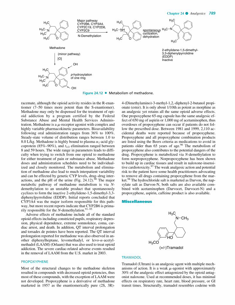
Chapter 24 Analgesics 789
racemate, although the opioid activity resides in the R-enan-tiomer (7–50 times more potent than the S-enantiomer).Methadone may only be dispensed for the treatment of opi-oid addiction by a program certified by the FederalSubstance Abuse and Mental Health Services Adminis-tration. Methadone is a �-receptor agonist with complex andhighly variable pharmacokinetic parameters. Bioavailabilityfollowing oral administration ranges from 36% to 100%.Steady-state volume of distribution ranges between 1.0 to8.0 L/kg. Methadone is highly bound to plasma �1-acid gly-coprotein (85%–90%), and t1/2 elimination ranged between8 and 59 hours. The wide range in parameters leads to diffi-culty when trying to switch from one opioid to methadonefor either treatment of pain or substance abuse. Methadonedoses and administration schedules need to be individual-ized and closely monitored. The metabolism and elimina-tion of methadone also lead to much interpatient variabilityand can be effected by genetic CYP levels, drug–drug inter-actions, and the pH of the urine (Fig. 24.12).90 The majormetabolic pathway of methadone metabolism is via N-demethylation to an unstable product that spontaneouslycyclizes to form the inactive 2-ethylidene-1,5-dimethyl-3,3-diphenylpyrrolidine (EDDP). Initial reports concluded thatCYP3A4 was the major isoform responsible for this path-way, but more recent reports indicate that CYP2B6 is prima-rily responsible for the N-demethylation.91–95
Adverse effects of methadone include all of the standardopioid effects including constricted pupils, respiratory depres-sion, physical dependence, extreme somnolence, coma, car-diac arrest, and death. In addition, QT interval prolongationand torsades de pointes have been reported. The QT intervalprolongation reported for methadone was also observed in an-other diphenylheptane, levomethadyl, or levo-�-acetyl-methadol (LAAM) (Orlaam) that was also used to treat opioidaddiction. The severe cardiac-related adverse events resultedin the removal of LAAM from the U.S. market in 2003.
PROPOXYPHENE
Most of the structural changes to the methadone skeletonresulted in compounds with decreased opioid potencies, thusmost of these compounds, with the exception of LAAM werenot developed. Propoxyphene is a derivative of methadonemarketed in 1957 as the enantiomerically pure (2S, 3R)-
4-(Dimethylamino)-3-methyl-1,2,-diphenyl-2-butanol propi-onate (ester). It is only about 1/10th as potent as morphine asan analgesic yet retains all the same opioid adverse effects.One propoxyphene 65-mg capsule has the same analgesic ef-fect of 650 mg of aspirin or 1,000 mg of acetaminophen, thusoverdoses of propoxyphene can occur if patients do not fol-low the prescribed dose. Between 1981 and 1999, 2,110 ac-cidental deaths were reported because of propoxyphene.Propoxyphene and all propoxyphene combination productsare listed using the Beers criteria as medications to avoid inpatients older than 65 years of age.96 The metabolism ofpropoxyphene also contributes to the potential dangers of thedrug. Propoxyphene is metabolized via N-demethylation toform norpropoxyphene. Norpropoxyphene has been shownto build up in cardiac tissues and result in naloxone-insensi-tive cardiotoxicity.97 The weak analgesic action and potentialrisk to the patient have some health practitioners advocatingto remove all drugs containing propoxyphene from the mar-ket.98 The hydrochloride salt is marketed as Darvon, the nap-sylate salt as Darvon-N, both salts are also available com-bined with acetaminophen (Darvocet, Darvocet-N) and apropoxyphene, aspirin, caffeine product is also available.
Miscellaneous
TRAMADOL
Tramadol (Ultram) is an analgesic agent with multiple mech-anisms of action. It is a weak �-agonist with approximately30% of the analgesic effect antagonized by the opioid antag-onist naloxone. Used at recommended doses, it has minimaleffects on respiratory rate, heart rate, blood pressure, or GItransit times. Structurally, tramadol resembles codeine with
H3COHO
NH3C
CH3
(+/-)Tramadol
Methadone
Major pathwayCYP2B6, CYP3A4, CYP2C19, CYP2D6, CYP2C9
OH3C
H3C
N
N-DemethylationOH3C
NH
H3C
H3C
H3C
N
H3C
H3CH3CH3C
spontaneouscyclization,dehydration
2-ethylidene-1,5-dimethyl-3,3-diphenylpyrrolidine (EDDP)Inactive
OH3C
H3C
NH3CH3C
OH
[O]
p-hydroxylationof one ring
OH
(minor pathway)
Figure 24.12 Metabolism of methadone.
0776-0818_17865_Ch24.qxd 12/4/09 8:08 PM Page 789

the B, D, and E ring removed. The manufacturer states thatpatients allergic to codeine should not receive tramadol, be-cause they may be at increased risk for anaphylactic reac-tions.99 Tramadol is synthesized and marketed as the racemicmixture of two (the [2S, 3S] [-] and the [2R, 3R] [�]) of thefour possible enantiomers.100 The (�) enantiomer is about30 times more potent than the (�) enantiomer; however,racemic tramadol shows improved tolerability.101,102
Neurotransmitter reuptake inhibition is also responsible forsome of the analgesic activity with the (�) enantiomer pri-marily responsible for norepinephrine reuptake and the (�)enantiomer responsible for inhibiting serotonin reup-take.101,103 Like codeine, tramadol is O-demethylated viaCYP2D6 to a more potent opioid agonist having 200-foldhigher affinity for the opioid receptor than the parent com-pound. Tramadol was initially marketed as nonaddictive, anda 3-year follow up study showed that the abuse potential isvery low, but not zero. Most abusers of tramadol have abusedopioid drugs in the past.104 Both enantiomers of tramadoland the major O-demethylated metabolite are proconvulsive,and tramadol should not be used in patients with a low-seizure threshold including patients with epilepsy.101
Mixed Agonist/Antagonist
NALBUPHINE
Nalbuphine (Nubain) is structurally a member of thephenanthrene class of compounds and resembles oxymor-phone with a cyclobutyl methyl group on the nitrogen,equivalent to naloxone’s substitution. It was introduced in1979 as an agonist/antagonist with the hope of becoming aneffective pain reliever with little abuse potential. Althoughthe abuse potential of nalbuphine is low, it is not zero, andincreasing reports of diversion and abuse can be found in theliterature and the Internet.105 At low parenteral doses(�0.5 mg), it has an analgesic potency approximately twothirds that of morphine, and it has a similar degree of respi-ratory depression. However, escalating doses above 30 mgdoes not produce further respiratory depression.106 The oralbioavailability of nalbuphine is only 12%, and the drug isonly marketed as an injectable.107 Patents have been filed foran oral extended-release formulation, and it is presently inphase II testing.108 The pharmacologic profile of nalbuphinein animal studies includes agonist activity at the �-receptorand antagonist activity at the �-receptor.109 Clinical studieshave shown that nalbuphine, and �-agonists in general, mayhave better analgesic activity in female patients comparedwith male patients.77 Used as the sole opioid agent, nal-buphine has been used successfully to treat the pain of labor,cesarean section, dental extraction, hip replacement, andhysterectomy surgery.110 Nalbuphine also may have a role in
N
HO
OH
O OH
Nalbuphine
treating opioid-induced pruritus, because it can reverse thepruritus without reversing the analgesic effect when used inlow doses. Nalbuphine is marketed as an injectable (10 and20 mg/mL). The usual dose is 10 mg administered subcuta-neously, intramuscularly, or intravenously at 3- to 6-hour in-tervals, with a maximal daily dose of 160 mg.
BUTORPHANOL
Structurally, butorphanol is a morphinan and shares the samecyclobutyl methyl group on the nitrogen as nalbuphine. Likenalbuphine, butorphanol is an agonist at the �-receptor but atthe �-receptor butorphanol is both a partial agonist and anantagonist.111 The affinity for opioid receptors in vitro is1:4:25 for the �-, �-, and �-receptors respectively.111 Thehigh affinity for the �-receptors is proposed to give butor-phanol its analgesic properties and is also responsible for theCNS adverse effects such as hallucinations, psychosis, andparanoid reactions. Butorphanol binds with �-receptors as apartial agonist, and administration to humans maintained onhigh-potency �-agonists such as morphine may precipitatewithdrawal. Butorphanol was found to produce convulsionsin morphine-deprived, morphine-dependent monkeys.112
The parenteral injection is used for moderate to severe painassociated with orthopedic procedures, obstetric surgery, andburns. The recommended dose is 1 mg IV or 2 mg intramus-cular (IM) every 3 to 4 hours as needed. Early studies pro-posed a “ceiling effect” for butorphanol’s antinociception andrespiratory depressant effect. More recently, the WHO ExpertCommittee on Drug Dependence performed a critical reviewof butorphanol and found no ceiling effect to the respiratorydepressant effect of parenteral butorphanol in monkeys andhumans.112 The nasal preparation (Stadol NS) is an effectiveanalgesic for the relief of moderate to severe pain such as mi-graine attacks, dental, or other surgical pain where an opioid isappropriate. The nasal spray is administered 1 mg (1 spray inone nostril) with an additional spray 60 to 90 minutes later ifadequate pain relief is not achieved. Respiratory depression isnot an issue with normal doses of the nasal preparation, butCNS side effects are the same as parenteral butorphanol.Increased reports of abuse and addiction of the nasal spray ledthe FDA to change the product to a Schedule IV drug in 1997.
N
HO O OCH3
OH
C(CH3)3
CH3
Buprenorphine
N
HO
OH
Butorphanol
790 Wilson and Gisvold’s Textbook of Organic Medicinal and Pharmaceutical Chemistry
0776-0818_17865_Ch24.qxd 12/4/09 8:08 PM Page 790

Chapter 24 Analgesics 791
BUPRENORPHINE
Buprenorphine is a semisynthetic, highly lipophilic opiate de-rived from thebaine. Pharmacologically, it is classified as amixed �-agonist/antagonist (a partial agonist) and a weak �-antagonist. It has a high affinity for the �-receptors(1,000 times greater than morphine) and a slow dissociationrate leading to its long duration of action (6–8 hours).113 Atrecommended doses, it acts as an agonist at the �-receptorwith approximately 0.3 mg IV equianalgesic to 10 mg of IVmorphine. One study in humans found that buprenorphine dis-plays a ceiling effect to the respiratory depression, but not theanalgesic effect over a dose range of 0.05 to 0.6 mg.114 In prac-tice, this makes buprenorphine a safer opiate (when usedalone) than pure �-agonists. Relatively few deaths frombuprenorphine overdose (when used alone) have been re-ported.113 The tight binding of the drug to the receptor also hasled to mixed reports on the effectiveness of using naloxone toreverse the respiratory depression. In animal studies, normaldoses of the pure antagonist naloxone were unable to removebuprenorphine from the receptor site and precipitate with-drawal. In a human study designed to precipitate withdrawalfrom buprenorphine, a naloxone dose (mean � 35 mg) 100times the dose usually needed to precipitate withdrawal inmethadone-dependent subjects was used. For comparison, ap-proximately 0.3 mg, 4 mg, 4 mg, and 10 mg of naloxonewould be required to precipitate withdrawal from heroin, bu-torphanol, nalbuphine, or pentazocine respectively.115
The unique pharmacologic and pharmacokinetic profileof buprenorphine made it a drug of interest for the treatmentof opioid dependence. The first study of buprenorphine forthis purpose was published in 1978 and confirmed thatbuprenorphine was an acceptable alternative to methadonefor addicts and that it blocked the effects of large singledoses of morphine for at least 24 hours.116 Early clinicalstudies showed that the oral bioavailability of buprenor-phine was low because of intestinal and liver metabolism.Therefore, a sublingual (SL) formulation (Subutex) was de-veloped that would bypass this metabolism. The SL tablethas a bioavailability of 55% but varies widely among indi-viduals.116 SL buprenorphine is also available combinedwith naloxone (Suboxone) in sufficient quantity (25% of thebuprenorphine dose) to discourage crushing and injectingthe SL tablet. SL naloxone is 0% to 10% bioavailable, andit does not change the absorption or action of the SLbuprenorphine. Abrupt withdrawal of chronically adminis-tered buprenorphine produces mild to moderate opioid with-drawal symptoms peaking between 3 and 5 days followingthe last buprenorphine dose. These symptoms require notherapeutic intervention. Suboxone and Subutex are the firstmedications approved for office-based treatment of opioiddependence under the Drug Addiction Treatment Act of2000. U.S. pharmacists may only dispense these medica-tions when prescribed by physicians that meet special train-
ing criteria and are registered with the Center for SubstanceAbuse Treatment, a component of the Substance Abuse andMental Health Services Administration. A transdermalbuprenorphine patch is available outside the United Statesfor use in chronic pain.117 This product is currently under-going phase III clinical trials in the United States.
Buprenorphine is oxidatively metabolized by N-dealky-lation by hepatic CYP3A4 and to a lesser extent CYP2C8 tothe active metabolite norbuprenorphine. Both the parent andmajor metabolite undergo glucuronidation at the phenolic-3position. Minor metabolites include hydroxylation of thearomatic ring of buprenorphine and norbuprenorphine at anunspecified site.118
Opioid Antagonists
NALTREXONE
Naltrexone (Fig. 24.13) is a pure opioid antagonist at allopioid receptor subtypes with the highest affinity for the�-receptor. Naltrexone is orally bioavailable and blocksthe effects of opiate agonists for approximately 24 hoursafter a single dose of 50 mg. It produces no opioid agonisteffects and is devoid of any intrinsic actions other thanopioid receptor blockade. Theoretically, it should workwell to treat opioid dependence but in clinical practice,patients have shown poor compliance and high relapserates. Naltrexone has also been studied to treat alcohol de-pendence with mixed results. To address the complianceissues and effectively remove the “choice” of taking theantagonist, naltrexone was developed into an extended-release injectable microsphere formulation for IM injec-tion once a month (Vivitrol). This formulation providessteady-state plasma concentrations of naltrexone threefoldto fourfold higher than the 50-mg oral dose 4 times aday.119 Currently, Vivitrol is only indicated for the treat-ment of alcohol dependence. A Cochrane review found in-sufficient evidence from randomized controlled trials toevaluate its effectiveness for treating opioid depend-ence.120 Currently, phase II and phase III clinical trials ofan implantable pellet form of naltrexone are being con-ducted for treating opioid dependence.
The CYP450 system is not involved in naltrexonemetabolism. Naltrexone is reduced to the active antagonist6-�-naltrexol by dihydrodiol dehydrogenase, a cytosolicenzyme. Naltrexone has a black box warning, because ithas the potential to cause hepatocellular injury when givenin excessive doses.119
NALOXONE
Naloxone (Narcan) (Fig. 24.13) is a pure antagonist at allopioid receptor subtypes. Structurally, it resembles oxymor-phone except that the methyl group on the nitrogen isreplaced by an allyl group. This minor structural change
N
HO
OH
Naltrexone
O O
N
HO
OH
Naloxone
O O
N
HO
OH
Nalmefene
O CH2
N
HO
OH
Methylnaltrexone
O O
CH3
Figure 24.13 Opioidantagonists.
0776-0818_17865_Ch24.qxd 12/4/09 8:08 PM Page 791

792 Wilson and Gisvold’s Textbook of Organic Medicinal and Pharmaceutical Chemistry
retains high binding affinity to the receptor, but no intrinsicactivity. It is used to reverse the respiratory depressant ef-fects of opioid overdoses.
Naloxone is administered intravenously with an onset ofaction within 2 minutes. Because it is competing with theopioid for the receptor sites, the dose and frequency of ad-ministration will depend on the amount and type of narcoticbeing antagonized. Overdoses of long-acting opioids(methadone) may require multiple IV doses of naloxone orcontinuous infusions. Neonates born to opioid-exposedmothers may be given IV naloxone at birth to reverse the ef-fects of opiates.
Very few metabolism studies on naloxone have beenconducted, although the major metabolite found in the urineis naloxone-3-glucuronide.121,122
NALMEFENE
Nalmefene (Revex) is a pure opioid antagonist that is the6-methylene analog of naltrexone. It is available as a so-lution for IV, IM, or subcutaneous (SC) administration toreverse the effects of opioids after general anesthesia andin the treatment of overdose. It is longer acting than nalox-one but otherwise has a similar pharmacodynamic andmetabolic (3-glucuronidation) profile. Nalmefene hashigher oral bioavailability (approximately 40%)123 thannaloxone or naltrexone and is currently being investigatedas an oral treatment for pathological gambling124 and alco-hol abuse.125
METHYLNALTREXONE
Methylnaltrexone (Relistor) is the methylated, quaternaryform of naltrexone (Fig. 24.13). The permanently chargednitrogen prevents the drug from crossing the blood-brainbarrier. Thus, it only acts as an antagonist at peripheralopioid receptors. Relistor was approved in April 2008 totreat opioid-induced constipation in patients receiving pal-liative care. It is administered as a SC injection once everyother day.
NONSTEROIDAL ANTI-INFLAMMATORYDRUGS
NSAIDs including aspirin and acetaminophen, two of theoldest pain medications, are among the most widely pre-scribed drugs worldwide for the treatment of rheumaticarthritis and other degenerative inflammatory joint dis-eases.126,127 Although NSAIDs are very effective in reliev-ing mild to moderate pains and inflammation, their use isalso often associated with many undesirable side effects, in-cluding GI irritation and bleeding, platelet dysfunction, kid-ney damage, and bronchospasm.126–129
With the exception of acetaminophen (Tylenol) and thenewer “coxibs” drugs, the conventional NSAIDs (also com-monly referred to as the aspirin-like drugs), share very sim-ilar therapeutic and side effect profiles. The conventionalNSAIDs exert their therapeutic action by inhibiting two iso-forms of cyclooxygenase (COX-1, the constitutive isozymeand COX-2, the inducible isozyme), which is the rate-limit-ing enzyme responsible for the biosynthesis of the proin-flammatory prostaglandins (PGs) such as the PGD2, PGE2,
PGF2�, and PGI2 and thereby modulating pain transmission,attenuating inflammation, and reducing fever.127,128 Theyalso produce their undesirable side effects such as GI bleed-ing, ulcerations, or renal impairments by blocking the samecyclooxygenases responsible for synthesizing PGs thatmodulate platelet activity (TXA2 and PGI2), gastric acid se-cretion and cytoprotection (PGE2 and PGI2), and renalblood flow (PGE2).128–132
In early 1990, Vane et al.133,134 hypothesized that theundesirable side effects of the conventional NSAIDs are aresult of inhibition of the COX-1 isozyme, whereas thetherapeutic effects are related mainly to their inhibitoryaction on the inducible COX-2 isozyme. This hypothesishas stimulated extensive drug development and hastymarket introductions of many selective COX-2 inhibitors,or coxibs drugs.130,135 However, all of the marketed cox-ibs drugs except celecoxib (Celebrex), the first FDA-ap-proved COX-2 drug in 1998, have been withdrawn fromthe market because of the potential risk of a cardiovascu-lar event, including heart attack or stroke, especially incardiac patients.136 Recent clinical trials have placed allNSAIDs under surveillance for their potential cardiovas-cular risk, thus the indiscriminate use of any NSAIDsincluding naproxen in cardiac patients should beavoided.135–137
Mechanism of Action and NSAID-Induced Side Effects
For aspirin and many of the conventional NSAIDs, despitetheir worldwide use as pain medications for over a century,their mechanism of action was not completely known until1971 when Vane first identified the cyclooxygenases astheir molecular targets.128 Cyclooxygenase (also known asprostaglandin endoperoxide synthase or PGH synthase) isthe rate-limiting enzyme responsible for the biosynthesisof PGs.
PGs are short-lived, lipidlike molecules that play a vitalrole in modulating many important physiological and patho-physiological functions including pain, inflammation, gas-tric acid secretion, wound healing, and renal function. Theyare biosynthesized via a tissue-specific cyclooxygenasepathway (COX-1 or COX-2) either on an as-needed basis(mostly via the COX-1 isozyme) or via the induced andoverexpressed COX-2 isozyme because of an injury, in-flammation, or infection.126,129 Some of the salient featuresof the cyclooxygenase pathway involved in the biosynthesisof these PGs from arachidonic acid (AA) (5,8,11,14-eicosatetraenoic acid), a polyunsaturated fatty acid releasedfrom membrane phospholipids by the action of phospholi-pase A2, are depicted in Figure 24.14.
As stated earlier, all classes of NSAIDs strongly inhibitprostaglandin synthesis in various tissues, especially at thesite of the tissue damage or inflammation. This inhibitionoccurs at the stage of oxidative cyclization of AA, catalyzedby the rate-limiting enzyme, cyclooxygenase (or PGH syn-thase), to the hydroperoxy-endoperoxide (prostaglandin G2,PGG2) and its subsequent reduction to key intermediate,prostaglandin H2 (PGH2) needed for all prostaglandinbiosynthesis.138
Blockade of PGH2 production, thus prevents its furtherconversion, by tissue-specific terminal prostaglandin syn-thases or isomerases, into different biologically active PGs
0776-0818_17865_Ch24.qxd 12/4/09 8:08 PM Page 792

Chapter 24 Analgesics 793
including PGE2, PGD2, PGF2�, PGI2 (prostacyclin), andthromboxane A2 (TXA2) (Fig. 24.14).138,139
Among the PGs synthesized by the action of either COX-1 or COX-2 isozymes, PGI2 and PGE2 made at the site of in-jury (via COX-2 isozyme in the inflammatory cells such asmonocytes and macrophages) and also in the brain, areknown to play a dominant role in mediating inflammationand inducing hyperanalgesia.132 However, their synthesis inthe GI tract (via COX-1 isozyme) and in the renal tubules(via COX-1 and COX-2 isozymes), is essential to providecytoprotective action for restoring the integrity of the stom-ach lining and maintaining renal functions in an otherwisecompromised kidney as a result of constant insult.132,138
Thus, inhibition of PGE2 synthesis by the conventionalNSAIDs in the parietal cells removes its ability to modulatehistamine-mediated release of gastric acid from the parietalcells, whereas blockade of PGI2 and PGE2 synthesis in theepithelial cells in the stomach linings also prevents their ac-tion on the biosynthesis and release of bicarbonate and mu-cous gel desperately needed to repair damage resulting fromerosion caused by gastric acid and other aggressive fac-tors.127,129,132 Thus, it should not be surprising to note thatNSAID-induced gastric ulcers can only be prevented clini-cally with coadministration of misoprostol, a stable PGEanalog, but not with either the histamine H2-antagonists,sucralfate, or any proton pump inhibitors such as omepra-zole.127 Furthermore, maintenance of kidney function,
especially in patients with congestive heart failure, liver cir-rhosis, or renal insufficiency, is reliant on the action of PGI2
and PGE2 to restore normal renal blood flow. Thus, NSAIDuse (both COX-1 and COX-2 inhibitors) will increase therisk of renal ischemia and therefore is contraindicated inthese patients.129
The readers should consult Chapter 26 of this text on“Prostanglandins, Leukotrienes, and Other Eicosanoids”for a detailed discussion of PGs, their physiological andpathophysiological functions, and their corresponding PGreceptors.
Structure–Activity Relationshipsof NSAIDs
It is well established that the therapeutic potency of the con-ventional NSAIDs are highly correlated with their ability toinduce upper GI toxicity.127 But, are all NSAIDs other thancoxibs really equally effective in the treatment of pain andinflammation? Some insight might be found by exploringhow these chemically diverse classes of drugs bind to theirmolecular targets (i.e., their selectivity for COX-2 relative toCOX-1).140 Thus, the benefit/risk profile of individualNSAIDs, as reflected by their COX selectivity, may be moreclinically relevant for predicting the risk of GI complica-tions. Table 24.1 summarizes a few representative drugsfrom different NSAID classes with their recommended daily
PGE2
Two Isozymes(COX-1, constituted form
& COX-2, the inducible form)
PGE SynthasePGI Synthase
TXA Synthase
PGG2, R = OOHPGH2, R = OH
Membrane phospholipids
11
COOH158
14O2 O2
Inhibited by Corticosteroids
Inhibited by NSAIDs Cyclooxygenase
O
O
R
COOH
Lipoxygenase
Arachidonic acid
TXA2
PGI2
Leukotrienes(SRS-A)
PGF2α
PGD2
PGF Synthase
PGD Synthase
Phospholipase A2
Figure 24.14 Conversion of arachidonic acid to prostaglandins.
0776-0818_17865_Ch24.qxd 12/4/09 8:08 PM Page 793

794 Wilson and Gisvold’s Textbook of Organic Medicinal and Pharmaceutical Chemistry
dosages and their relative risks of bleeding peptic ulcercomplications.130,141,142
ENZYMATIC STRUCTURE OF CYCLOOXYGENASES
Cyclooxygenases (COX-1 and COX-2) are heme-containing,membrane-bound proteins that share a high degree of se-quence identity and also have very similar active site topog-raphy.138,143–145 Note that the homologous residues of theCOX-2 isozyme are numbered in parallel with the similaramino acid residues found at the active site of COX-1isozyme for easier comparison of their structural differencesin the following discussion (i.e., the actual Arg-106 ofhuman COX-2 [hCOX-2] is numbered as Arg-120).143 Theactual hCOX-2 assignments for these residues are providedin parentheses.
Thus, despite their similarity, the active site for hCOX-2 is approximately 20% larger than the COX-1 binding sitebecause of the replacement of Ile-523 in COX-1 with asmaller Val-523 in hCOX-2 (Val-509).138,144,145 There area total of 24 amino acid residues lining the largely hy-drophobic AA binding site with only one difference be-tween the isozymes (i.e., Ile-523 in COX-1 and Val-509 inhCOX-2). The hCOX-2 isozyme has an additional hy-drophilic side pocket accessible for drug binding, extendedfrom the main binding pocket.143 The size and nature of thishydrophilic side pocket for binding in hCOX-2 is a result offurther substitutions Ile-434 and His-513 in COX-1 with asmaller Val-434 (Val 420 in hCOX-2) and a more basicArg-513 (Arg-499 in hCOX-2).146 Thus, it is not surprisingthat this basic amino acid residue in hCOX-2 (Arg-499)may provide an additional binding interaction for selectiveCOX-2 inhibitors such as celecoxib.144 Although there isonly a very limited amount of published data showing howthe substrate, AA, binds to cyclooxygenases, the relativepositioning of the double bonds in AA at the active site,proposed by Gund and Shen based on the conformationalanalysis of indomethacin and other conventional NSAIDs,is still currently valid.147
Interestingly, Arg-120 (Arg-104 in hCOX-2) is theonly positively charged amino acid residue in the COXactive site, on one end of the active site as depicted byLuong144,148 and is responsible for binding, via an ionicinteraction, with the carboxylate anion of the substrate
(AA) or the conventional NSAIDs. The amino acidresidue Tyr-385 (Tyr-371 in hCOX-2) is located on theopposite end of the COX active site and is believed toserve as the catalytic residue for activating molecular oxy-gen and its initial addition to the �11-double bond of thesubstrate to form PGG2.143–148 Furthermore, the presenceof an amino acid residue, Ser-530 (Ser-516 in hCOX-2) isbelieved to be involved only in the irreversible inactiva-tion by aspirin and NSAID action but not contributing toany substrate binding.143
BINDING OF NSAID TO CYCLOOXYGENASE
Most NSAIDs possess a free carboxylic acid (COOH) foran ionic interaction with the positively charged arginineresidue at the active site of the cyclooxygenases (i.e., Arg-120 in COX-1 or Arg-106 in COX-2 isozymes). Thisacidic moiety is further linked to an aromatic (or het-eroaromatic) ring for binding to either the �5-double-bondor �8-double-bond binding regions, and an additional cen-ter of lipophilicity in the form of an alkyl chain (e.g.,ibuprofen) or an additional aromatic ring (e.g., in-domethacin) for binding to the �11-double-bond bindingregion.
A hypothetical binding model for indomethacin and re-lated analogs to the COX-1 isozyme is shown in Figure 24.15-A, based on the crystallographic and SAR data foundin the literature.146,149 The suggested orientation and place-ment of the N-p-chlorobenzoyl group to the �11-double-bond binding region and the indole ring to the �5- and �8-double-bond binding regions is made from assuming that apreferred, lower-energy conformation of indomethacin isessential for its binding to COX isozymes147, and the factthat this orientation also allows correct positioning of theamide carbonyl oxygen for possible binding interactions toSer-530 and the 5-methoxy group to Ser-355 at the activesite of the enzymes.146
Additional support for this model can be obtained fromthe observation that the (Z)-isomer of sulindac, an indeneisosteric analog of indomethacin, is much more potentthan the corresponding (E)-isomer and the fact that replace-ment of the 5-methoxy group with a fluorine atom on the in-dole ring further enhances its analgesic action.145,149
Furthermore, because there is limited space available for
TABLE 24.1 Comparison of Relative Risk of NSAID-Induced Gastrointestinal Complication
Dosing Range Relative Risk for COX-2/COX-1Drug Trade Name (Total Daily Dose) GI complication Selectivity Ratio
Not on NSAIDs 1.0 —or Aspirin
Celecoxib Celebrex 100–200 mg BIDa (200–400 mg) — 0.7Meloxicam Mobic 7.5–15 mg daily — 0.37Ibuprofen Advil, Motrin 200–600 mg TIDb (0.6–1.8 g) 2.9 (1.7–5.0) 0.9Sulindac Clinoril 150–200 mg BID (300–600 mg) 2.9 (1.5–5.6) 29 (on active sulfide)Aspirin — 325–650 mg QIDc (1.3–2.6 g) 3,1 (2.0–4.8) �100Naproxen Aleve, Naprosyn 125–500 mg BID (0.5–1.0 g) 3.1 (1.7–5.9) 3.0Diclofenac Voltaren 25–50 mg TID (75–150 mg) 3.9 (2.3–6.5) 0.5Ketoprofen Orudis 50–100 mg TID (150–300 mg) 5.4 (2.6–11.3) 61Indomethacin Indocin 25–50 mg TID (75–150 mg) 6.3 (3.3–12.2) 80Piroxicam Feldene 10–20 mg daily 18.0 (8.2–39.6) 3.3
aBID, bis in die (twice a day).bTID, ter in die (3 times a day).cQID, quater in die (4 times a day).
0776-0818_17865_Ch24.qxd 12/4/09 8:08 PM Page 794

Chapter 24 Analgesics 795
binding around either Leu-384 or Ile-523 (i.e., both aroundthe �11-double-bond binding region), it is not surprising tosee a facile conversion of the indomethacin from a nonselec-tive COX inhibitor into a potent and highly selective COX-2inhibitor with just a simple substitution of the p-chlorine witha larger bromine atom or by addition of two chlorine atomsto the o,o’ positions of the N-p-chlorobenzoyl group reportedby Kalgutkar et al.150 This observation can explain why alower risk for GI complications was reported with sulindacthan indomethacin, because it requires metabolic activation,via reduction of its bulky sulfoxide moiety into a muchsmaller methyl sulfide for binding into the Leu-384 pocket.The fact that kidneys possess a high level of the flavin-con-taining monooxygenases for deactivating the active metabo-lite of sulindac back into its inactive sulfoxide or sulfone,further provided evidence for this binding model.149,151
With this binding model, it is also possible to see why therelative therapeutic potency of the conventional NSAIDs andtheir ability to induce GI complication is in the order: in-domethacin �ketoprofen �diclofenac �naproxen �ibupro-fen. Ibuprofen is the least potent because it can only bind tothe �5-double-bond region with additional weak van derWaals interactions to the �11-double-bond region. Naproxenis slightly more active than ibuprofen, because its naphtha-lene ring, isosteric to the indole ring, can interact with boththe �5- and �8-double-bond regions. Ketoprofen and di-clofenac are slightly less potent than indomethacin becauseof the absence of an additional ring for binding to either the�5- or the �8-double-bond regions.
BINDING OF CELECOXIB TO COX-2 ISOZYME
The hypothetical binding model for binding of celecoxib, theonly selective “coxibs” type COX-2 inhibitor shown onFigure 24.15B, is purely speculative, based only on thecrystallographic binding data reported for SC-558 (the p-bromophenyl analog of celecoxib) with the murine COX-2isozyme.146 However, the celecoxib selectivity toward theCOX-2 isozyme is most likely a result of the extension of thesulfonamide moiety into the extra hydrophilic-bindingpocket, surrounded by His-90 (His-76 in hCOX-2) and Arg-513 (Arg-499 in hCOX-2). The opening of this additionalpocket for binding is the result of the replacement of Ile-523
in the COX-1 with a smaller valine residue (Val 509 inhCOX-2). It should be pointed out that a similar hydrophilicpocket does exist in the COX-1 isozyme, but it is inaccessiblebecause of the bulkier Ile-523 residue that guards the entranceto this side pocket of the COX-1 isozyme (see Fig. 24.15-A).
The COX-2/COX-1 selectivity ratios, estimated by dif-ferent research groups, can be quite different (e.g., the re-ported selectivity ratio for celecoxib ranges any where from0.003–0.7; for piroxicam, the range is 3.3–600). Thus, theselectivity ratios included in Table 24.1, obtained from onesuch study, is only valid for comparing their differencesamong these NSAIDs. Furthermore, recent reviews compar-ing the SAR among different structural classes of COX-2 in-hibitors have indicated that there is little or no commonpharmacophore required for their COX-2 selectivity, butminor changes within the structure type, in terms of molec-ular shape, lipophilicity, electronic density, flexibility, po-larity, and hydrogen-bonding properties, can all have drasticeffects in its COX selectivity.152,153
PIROXICAM AND MELOXICAM: THE DIFFERENCE INTHEIR COX SELECTIVITY
Piroxicam and meloxicam have nearly identical structuralfeatures but also have at least a ninefold difference in selec-tivity for meloxicam to COX-2 isozyme and an even largerdifference in their relative risks for GI complications (i.e.,piroxicam has the highest risk among NSAIDs, whereasmeloxicam has very little or no such side effects) (Table24.1). A closer comparison of their structure (Fig. 24.16),however, reveals no apparent reason for these differences, ei-ther in size, lipophilicity, or electronic properties, between the2-pyridyl group (in piroxicam) and the 5-methyl-2-thiazoylgroup (meloxicam) that may alter their ability to bind COXisozymes. It is unlikely that these drastic differences in theirCOX selectivity, especially the drug-induced GI toxicity,could be due solely to the binding of the parent moleculeswith such minor changes in their structures. Thus, could theobserved differences, especially the differences in drug-in-duced GI side effects, be attributed to the involvement of anactive metabolite of piroxicam and/or meloxicam?
The metabolism of piroxicam to its major activemetabolite, 5�-hydroxypiroxicam and meloxicam to its
5-double bondbinding region
8-double bondbinding region
11-double bondbinding region
14-double bondbinding region
Tyr-385
Ile-523
Ser-355Tyr-355
Ser-530
N
OCH3
OO
H3C
O
Cl
Leu-384
Arg-120
Tyr-371
Arg-499
Extra bindingpocket in COX-2
14-double bondbinding region
Val-509
11-double bondbinding region
8-double bondbinding region
5-double bondbinding region
F3C
SO2NH2
N
N
H3C
Arg-106
Figure 24.15 Hypothetical binding models of indomethacin to COX-1 and Celecoxibto COX-2. A. Binding of the conventional NSAID, indomethacin, to the active site ofCOX-1; B. Binding of celecoxib, a selective COX-2 inhibitor to the active site of COX-2with crystallographic maps suggested by Luong et al. (Flexibility of the NSAID bindingsite in the structure of human cyclooxygenase-2. Nat. Struct. Biol. 1996;3:927–933)
AABB
0776-0818_17865_Ch24.qxd 12/4/09 8:08 PM Page 795

5�-hydroxy-methylmeloxicam and 5�-carboxymeloxicammetabolites are shown in Figure 24.16.154–156 Thus, withthe proposed binding interaction of indomethacin de-scribed earlier (Fig. 24.15), it is reasonable to assume thatthe pyridyl group of the piroxicam or the 5�-hydroxy-pyridyl group of its active metabolite will be directed tobind to the �11-double-bond binding region (especiallythe active metabolite caused by an additional H-bondingto the Tyr-385 residue), in a similar manner as the N-p-chlorobenzoyl group of the indomethacin. This wouldalso allow the OH group of the acidic enol carboxamidemoiety of the piroxicam or its active metabolite to posi-tion itself for binding to the Ser-530 residue of the COX-1 isozyme. However, a similar binding interaction of theactive metabolites of meloxicam, especially the 5�carboxymetabolite, to the active site of the COX-1 isozyme maynot be possible, because it will reorient itself for astronger ionic interaction with the Arg-120 of the COX-1isozyme. Thus, they can fit only into the active site of theCOX-2 isozyme because the binding of an acidic moietyto Arg-106 in the active site of the COX-2 isozyme is be-lieved to play a lesser role in the inhibitory action of theCOX-2 selective inhibitors like celecoxib152 (i.e., themost acidic moiety, sulfonamide is extended into the sidepocket instead of binding to Arg-106).
ASPIRIN AND ITS COX-1 SELECTIVITY
Aspirin covalently modifies COX-1 and hCOX-2 isozymesby acetylating the OH group of Ser-530 in COX-1 and Ser-516 in hCOX-2 isozymes. This is made possible by an ionicattraction between the carboxylate anion of aspirin and thearginine cation of Arg-120 in COX-1 (or Arg-106 inhCOX-2), thereby positioning the acetyl group of aspirinfor acetylating the COX isozymes. Even though both COXisozymes are irreversibly acetylated by aspirin, acetylationof Ser-530 totally blocks the accessibility of substrate AAfrom entering into the active site, whereas an acetylatedhCOX-2 is still able to form a significant amount ofPGG2.143 Thus, aspirin, among all conventional NSAIDs,
exhibits the highest selectivity toward the COX-1 isozyme,especially the COX-1 isozyme present in the platelets.
Aspirin and Salicylic Acid Derivatives
Aspirin and the salicylates were among the first group ofNSAIDs introduced into medicine for their use as anal-gesics to relieve pain and as antipyretics to reduce fever.157
As early as 1763, the Reverend Edward Stone of ChippingNorton in Oxfordshire, England reported the use of driedwillow bark to parishioners suffering from rheumaticfever.129,158 The active ingredient of willow bark was iso-lated by Leroux, in 1827 and named salicin, which is a sal-icylic acid containing glycoside. After these discoveries,Cahours (1844) obtained salicylic acid from oil of winter-green (methyl salicylate), and Kolbe and Lautermann(1860) prepared it synthetically from phenol. Sodium sali-cylate was introduced in 1875 by Buss, followed by the in-troduction of phenyl salicylate by Nencki in 1886. Aspirin,or acetylsalicylic acid, was first prepared in 1853 byGerhardt but remained obscure until Felix Hoffmann fromBayer discovered its pharmacological activities in 1899. Itwas tested and introduced into medicine by Dreser (1899),who named it aspirin by taking the a from acetyl andspirin, an old name for salicylic or spiric acid, derivedfrom its natural source of spirea plants.
Most of the salicylic acid drugs (commonly referred to asthe salicylates) are either marketed as salts of salicylic acid(sodium, magnesium, bismuth, choline, or triethanolamine)or as ester or amide derivatives (aspirin, salsalate, salicy-lamide). (Fig. 24.17) Children, between the ages of 3 and12, who are recovering from flu or chicken pox, should notbe taking aspirin or any salicylates because of the perceivedrisks of a rare disease known as Reye syndrome.159
MECHANISM OF ACTION OF SALICYLATES
Salicylates, in general, exert their antipyretic action infebrile patients by increasing heat elimination of the bodyvia the mobilization of water and consequent dilution of
796 Wilson and Gisvold’s Textbook of Organic Medicinal and Pharmaceutical Chemistry
NS
N
O O
CH3
OH O
H
N
NS
N S
O O
CH3
OH O
H
N
CH3
NS
N
O O
CH3
OH O
H
N
OH
CYP2C9
5’-HydroxypiroxicamPiroxicam
NS
N S
O O
CH3
OH O
H
N
R
Meloxicam
CYP2C9
5’-Hydroxymethylmeloxicam, R = CH2OH,5’-Carboxymeloxicam, R = COOH
Figure 24.16 Metabolism ofpiroxicam and meloxicam.
0776-0818_17865_Ch24.qxd 12/4/09 8:08 PM Page 796

Chapter 24 Analgesics 797
the blood. This brings about perspiration, causing cuta-neous dilatation. This does not occur with normal temper-atures. The antipyretic and analgesic actions are believedto work by inhibiting cyclooxygenase and reducing thelevels of PGE2, a proximal mediator of the febrile re-sponse, in the hypothalamic area of the brain that regulatesbody temperature.157
It is well established that a low daily dose of aspirin(75–100 mg or one tablet of baby aspirin) is sufficient tocompletely block platelet TXA2 production and its ability toinduce platelet aggregation after only 1 week of dosing,thereby preventing the risk of a cardiovascular event, in-cluding myocardial infarction and ischemic stroke.160,161
This antiplatelet action of aspirin is because COX-2 is notexpressed in platelets; therefore, aspirin can selectively andirreversibly inhibit platelet TXA2 production for 8 to 10days (i.e., until new platelets are formed) at such a low dose(i.e., via the irreversible acetylation of Ser-530 of the COX-1 isozymes discussed earlier). However, the analgesic, an-tipyretic, and anti-inflammatory action of aspirin is morecomplex and may involve other mechanisms of action thansimply based on its ability to irreversibly inhibit the COXisozymes.157,162,163 Furthermore, it is worth noting that up to50% of the oral analgesic dose of aspirin is rapidly deacety-lated before it reaches general circulation, and its major ac-tive metabolite, salicylic acid, is found to have comparablein vivo antipyretic and anti-inflammatory properties to as-pirin but is a very weak inhibitor of cyclooxygenases (i.e., invitro binding studies).164 Several possible mechanisms ofaction have recently been suggested for aspirin and espe-cially the salicylates including blocking the induction of theCOX-2 isozyme at the genetic levels,165 turning off the
nuclear factor-�B-mediated polymorphonuclear leukocyteapoptosis signaling166,167 or blocking the activation of themitogen-activated kinase, Erk signaling associated with in-flammatory responses.168
PHARMACOKINETICS OF SALICYLATES
The salicylates, being acidic in nature, are readily absorbedfrom the stomach and the small intestine. However, their ab-sorption depends strongly on the pH of the environment,thus coadministration of an antacid or other buffering agentsshould be avoided because it greatly hinders their absorptionand reduces their bioavailability and onset of action. Theyare also highly bound to plasma proteins, a major source ofpotential drug interactions with other medications.
Salicylic acid undergoes extensive phase-II metabolism(see Fig. 24.17) and is excreted via the kidneys as the water-soluble glycine conjugate, salicyluric acid, the majormetabolite (�75%) or as the corresponding acyl glu-curonides (i.e., the ester type, via the COOH) or O-glu-curonides (i.e., the ether type, via the phenolic OH) (�15%).Alkalinization of the urine increases the rate of excretion ofthe free salicylates.
AspirinAspirin, acetylsalicylic acid (Aspro, Empirin), was intro-duced into medicine by Dreser in 1899.
Aspirin occurs as white crystals or as a white crystallinepowder and must be kept under dry conditions. It is not ad-visable to keep aspirin products in the kitchen or bathroomcabinets, because aspirin is slowly decomposed into aceticand salicylic acids in the presence of heat and moisture.Several proprietaries (e.g., Bufferin) use compounds such as
COOH
O CH3
O
COOH
OHF
F
COOH
OH
Aspirin
COO
OH
COOH
O
O OH
Salsalate
Salts of Salicylic acid
Diflunisal
CONH2
OH
Salicylic acid Salicylamide
Ester or EtherGlucuronides
CONHCH2COOH
OH
Salicyluric acid(major metabolite)
Ether Glucuronideor sulfate
Figure 24.17 Salicylates andtheir metabolism.
0776-0818_17865_Ch24.qxd 12/4/09 8:08 PM Page 797

798 Wilson and Gisvold’s Textbook of Organic Medicinal and Pharmaceutical Chemistry
sodium bicarbonate, aluminum glycinate, sodium citrate,aluminum hydroxide, or magnesium trisilicate to counteractaspirin’s acidic property. One of the better antacids is dihy-droxyaluminum aminoacetate. Aspirin is unusually effec-tive when prescribed with calcium glutamate. The more sta-ble, nonirritant calcium acetylsalicylate is formed, and theglutamate portion (glutamic acid) maintains a pH of 3.5 to5. Practically all salts of aspirin, except those of aluminumand calcium, are unstable for pharmaceutical use. Thesesalts appear to have fewer undesirable side effects and in-duce analgesia faster than aspirin. A timed release prepara-tion of aspirin is available. It does not appear to offer anyadvantages over aspirin, except for bedtime dosage.
Aspirin is used as an analgesic for minor aches and painsand as an antipyretic to reduce fever. Although higher dosesof aspirin can also be used to treat inflammation, its use isoften associated with many unwanted side effects includingulcers, stomach bleeding, and tinnitus. A low dosage formof aspirin, 81 mg, equivalent to the dose recommended forinfants (the “baby aspirin”), is recommended as a daily dosefor individuals who are at even a low cardiovascular risk.Several large studies have found that this low dose of as-pirin reduces the number of heart attacks and thromboticstrokes.160,161 Other salicylates and NSAIDs have notshown similar effects. In fact, a recent report indicatedusing ibuprofen can interfere with aspirin’s cardiovascularbenefits, and they should not be taken within 12 hours ofeach other.169
Aspirin and other potent NSAIDs (except salicylates) arealso known to precipitate asthma attacks and other hyper-sensitivity reactions in up to 10% of the patients who haveany type of respiratory problems.170 This hypersensitivityreaction is believed to occur as a result of shifting the sub-strate, AA from the inhibited cyclooxygenase pathway tothe lipoxygenase pathway (see Fig. 24.14), therefore result-ing in overproduction of anaphylactic leukotrienes becauseof the blockade of the cyclooxygenase pathway by aspirinand other potent NSAIDs.171
SalsalateSalsalate, salicylsalicylic acid (Amigesic, Disalcid,Salflex), is the ester formed between two salicylic acidmolecules. It is rapidly hydrolyzed to salicylic acid fol-lowing its absorption. It reportedly causes less gastric ir-ritation than aspirin, because it is relatively insoluble inthe stomach and is not absorbed until it reaches the smallintestine.
DiflunisalDiflunisal (Dolobid), is a longer acting and more potent drugthan aspirin because of its hydrophobic, 2,4-difluorophenylgroup attached to the 5-position of the salicyclic acid. In alarge-scale comparative study with aspirin, it was also bettertolerated with less GI complications than aspirin.172 It ismarketed in tablet form for treating mild to moderate post-operative pain as well as RA and OA.
Diflunisal is highly protein bound. Its metabolism is sub-ject to a dose-dependent, saturable, and capacity-limitedglucuronide formation.173 This unusual pharmacokineticprofile is a result of an enterohepatic circulation and the re-absorption of 65% of the drug and its glucuronides, fol-lowed by cleavage of its unstable, acyl glucuronide back tothe active drug. Thus, diflunisal usage in patients with renalimpairment should be closely monitored.
Sodium SalicylateSodium salicylate may be prepared by the reaction, in aque-ous solution, by adding equal molar ratio of salicylic acidand sodium bicarbonate; evaporating to dryness yields thewhite salt. In solution, particularly in the presence of sodiumbicarbonate, the salt will darken on standing. This is the saltof choice for salicylate medication and usually is adminis-tered with sodium bicarbonate to lessen gastric distress, or itis administered in enteric coated tablets. However, the useof sodium bicarbonate is ill advised, because it decreases theplasma levels of salicylate and increases the excretion offree salicylate in the urine.
Sodium salicylate, even though not as potent as aspirinfor pain relief, also has less GI irritation and is useful for pa-tients who are hypersensitive to aspirin.
OTHER SALTS OF SALICYLIC ACID
Sodium thiosalicylate (Rexolate) is the sulfur or thio analogof sodium salicylate. It is more soluble and better absorbed,thus allowing lower dosages. It is recommended for gout,rheumatic fever, and muscular pains, but it is available onlyfor injection.
Magnesium salicylate (Mobidin, Magan) is a sodium-free salicylate preparation for use when sodium intake is re-stricted. It is claimed to produce less GI irritation.
Choline salicylate (Arthropan) is extremely soluble inwater and is available as a flavored liquid. It is claimed to beabsorbed more rapidly than aspirin, giving faster peak bloodlevels. It is used when salicylates are indicated. It is alsoavailable in combination with magnesium salicylate(Trilisate, Tricosal, Trisalcid, CMT) for the relief of minorto moderate pains and fever associated with arthritis, bursi-tis, tendinitis, menstrual cramps, and others.
SalicylamideSalicylamide, o-hydroxybenzamide, is a derivative of salicylicacid that is fairly stable to heat, light, and moisture. Itreportedly exerts a moderately quicker and deeper analgesiceffect than aspirin because of quicker CNS penetration. Its me-tabolism differs from aspirin, because it is not metabolized tosalicylic acid but rather excreted exclusively as the ether glu-curonide or sulfate.174 Thus, as a result of lack of contributionfrom salicylic acid, it has a lower analgesic and antipyretic ef-ficacy than that of aspirin. However, it can be used in place ofsalicylates for patients with a demonstrated sensitivity to sali-cylates. It is also excreted much more rapidly than other sali-cylates, which accounts for its lower toxicity. It is available inseveral nonprescription products, in combination with acet-aminophen and phenyltoloxamine (e.g., Rid-A Pain com-pound, Cetazone T, Dolorex, Ed-Flex, Lobac) or with aspirin,acetaminophen, and caffeine (e.g., Saleto, BC Powder).
The Conventional NonselectiveCyclooxygenase Inhibitors
With the removal of rofecoxib (Vioxx) from the market andthe concern of cardiovascular risk associated with cele-coxib (Celebrex), the conventional NSAIDs, being morepotent than aspirin and related salicylates, once again be-come the drug of choice for the treatment of RA and otherinflammatory diseases.130 The conventional NSAIDs varyconsiderably in terms of their selective inhibitory action for COX-2 relative to COX-1 isozymes and also their relative
0776-0818_17865_Ch24.qxd 12/4/09 8:08 PM Page 798

Chapter 24 Analgesics 799
drug-induced toxicities discussed earlier in this text.Although several drugs in this class are available OTC,they are no safer than prescription medications with regardto their drug-induced GI liability.127 The conventionalNSAIDs, as a group, are highly protein bound and exhibitboth pharmacokinetic as well as pharmacodynamic interac-tions with many drugs, especially anticoagulants, diuretics,lithium, and other arthritis medications.
For the purpose of comparing their SAR, toxicity, andmetabolic biotransformations, the conventional NSAIDs arefurther divided into several chemical classes.
ARYL- AND HETEROARYLACETIC ACIDS
This group of NSAIDs has received the most intensive at-tention for new clinical candidates. As a group, they showhigh analgesic potency in addition to their potent anti-inflammatory activity, needed for treating inflammatory dis-eases. Among the members of this class shown in Figure24.18, ketorolac, indomethacin, and tolmetin have the high-est risk of GI complications because of their higher affinityfor the COX-1 isozymes, whereas etodolac has the lowestrisk because of its COX-2 selective inhibitory action. Bothsulindac and nabumetone are prodrugs that require activa-tion, and therefore have lower risk of causing GI irritationthan indomethacin.130
IndomethacinFrom the time of its introduction in 1965, indomethacin(Indocin) has been widely used as an analgesic to relieve in-flammatory pain associated with RA, OA and ankylosingspondylitis, and, to a lesser extent, in gout. Although both itsanalgesic and anti-inflammatory activities are well estab-lished, its use is often limited because of frequent GI distressand potential drug interactions, especially with warfarinfurosemide, and lithium (i.e., it elevates blood levels of
lithium as a result of reducing renal blood flow and there-fore increases lithium toxicities).
Following oral administration, indomethacin is rapidlyabsorbed and is 90% protein bound at therapeutic plasmaconcentrations.175 The drug has a biological half-life ofabout 5 to 10 hours and a plasma clearance of 1 to 2.5 ml/kgper minute. It is metabolized to its inactive, O-desmethyl,N-deschlorobenzoyl-, and O-desmethyl, N-deschloroben-zoylindomethacin metabolites.175
SulindacSulindac, (Z)-5-fluoro-2-methyl-1-([p-(methylsulfinyl)phenyl]methylene)-1H-indene-3-acetic acid (Clinoril), isan NSAID prodrug that contains a chiral sulfoxide moietybut is marketed as the racemate because it undergoes invivo reduction by the hepatic enzymes into its achiral, ac-tive metabolite, methyl sulfide that exhibits potent andnonselective COX inhibition similar to indomethacin.151
The parent sulfoxide has a plasma half-life of 8 hours, andthe active methyl sulfide metabolite is 16.4 hours. The morepolar and inactive sulfoxide is virtually the only form ex-creted into the renal tubules, thus sulindac is believed to haveminimal nephrotoxicity associated with indomethacin.176 Thelong half-life of sulindac is caused by the extensive entero-hepatic circulation and reactivation of the inactive sulfoxideexcreted. Coadministration of aspirin is contraindicated be-cause it considerably reduces the sulfide blood levels. Carefulmonitoring of patients with a history of ulcers is recom-mended. Gastric bleeding, nausea, diarrhea, dizziness, andother adverse effects have been noted with sulindac, but witha lower frequency than with aspirin. Sulindac is recom-mended for RA, OA, and ankylosing spondylitis.
TolmetinTolmetin sodium (Tolectin), is an arylacetic acid derivativewith a pyrrole as the aryl group. This drug is well absorbed
N
OH3C
CH3
COOH
O
Cl
Indomethacin
SH3C
CH3
COOH
F
H
OSulindac
N
COOH
CH3
O
R1
R2
Tolmetin, R1=CH3, R2=HZomipirac, R1=Cl, R2=CH3
N
O
HCH3
COOH
CH3
Etodolac
N
O
COOH
Ketorolac
CH3
O
OH3C
Nabumetone
O
NH2
R1
O
R2 Bromfenac, R1=OH, R2=BrAmfenac, R1=OH, R2=HNepafenac, R1=NH2, R2=H
Figure 24.18 Aryl- and heteroarylacetic acid derivatives.
0776-0818_17865_Ch24.qxd 12/4/09 8:08 PM Page 799

800 Wilson and Gisvold’s Textbook of Organic Medicinal and Pharmaceutical Chemistry
and has a relatively short plasma half-life (1 hour). It is rec-ommended for use in the management of acute and chronicRA. Its efficacy is similar to aspirin and indomethacin, butwith less frequency of the adverse effects and tinnitus asso-ciated with aspirin. It does not potentiate coumarin-likedrugs nor alter the blood levels of sulfonylureas or insulin.However, tolmetin, and especially its closely related drug,zomepirac (i.e., with a p-chlorobenzoyl group and an addi-tional methyl group on the pyrrole ring), can produce a rarebut fatal anaphylactic reaction because of irreversible bind-ing of their unstable acyl glucuronides.177 Zomepirac waswithdrawn from market because it is eliminated only via theester-type, acyl glucuronide.178 It is possible that tolmetin isless toxic in this regard because it undergoes additional he-patic benzylic hydroxylation via its p-methyl group and isexcreted as its stable ether glucuronide.
KetorolacKetorolac tromethamine (Toradol), marketed as a mixture of(R)- and (S)-ketorolac enantiomers, is a potent NSAID anal-gesic indicated for the treatment of moderately severe, acutepain. It should be noted that the pharmacokinetic dispositionof ketorolac in humans is subject to marked enantioselectiv-ity. Thus, it is important to monitor the individual blood lev-els so an accurate assessment of its therapeutic action can bemade correctly.179 However, it should be noted that, beingone of the conventional NSAIDs with highest risk of GIcomplications, its administration should not exceed 5 days.
NabumetoneNabumetone (Relafen), a nonacidic NSAID prodrug, isclassified as an arylacetic acid, because it undergoes rapidhepatic metabolism to its active metabolite, 6-methoxy-2-naphthylacetic acid.180 Similar to the other arylacetic aciddrugs, it is used in short- or long-term management of RAand OA. Being nonacidic, it does not produce significantprimary insult to the GI mucosa lining and also has no ef-fect on prostaglandin synthesis in gastric mucosa, thus pro-ducing minimum secondary GI damage when comparedwith other conventional NSAIDs.
EtodolacEtodolac (Lodine, Ultradol), a chiral, COX-2 selectiveNSAID drug that is marketed as a racemate, possesses an in-dole ring as the aryl portion of this group of NSAID drugs.It shares many similar properties of this group and is indi-cated for short- and long-term management of pain and OA.
Similar to ketorolac, etodolac exhibits several uniqueenantioselective pharmacokinetic properties.181 For example,the “inactive” (R)-enantiomer has approximately a 10-foldhigher plasma concentration than the active (S)-enantiomer.Furthermore, the active (S)-enantiomer is less protein boundthan its (R)-enantiomer and therefore has a very large volumeof distribution. It is well absorbed with an elimination half-life of 6 to 8 hours. Etodolac is extensively metabolized intothree major inactive metabolites, 6-hydroxy etodolac (via aro-matic hydroxylation), 7-hydroxy-etodolac (via aromatic hy-droxylation), and 8-(1�-hydroxylethyl) etodolac (via benzylichydroxylation), which are eliminated as the correspondingether glucuronides.182 Its unstable, acyl glucuronide, how-ever, is subject to enterohepatic circulation and reactivation tothe parent drug, similar to other NSAIDS in this class.
In a recent study comparing its gastric safety profile withthose of meloxicam, diclofenac, and indomethacin, etodolac
was found to have the highest safety index among theseNSAIDs in arthritic rats.183 Thus, etodolac is an example ofa COX-2 selective drug with a much better safety profileamong the first generation of NSAIDs developed throughthe traditional route of using only animal model studies. Itsselective COX-2 inhibitory action, which was not realizeduntil much later, explains its much lower risk of the GI sideeffects among first-generation NSAIDs.
Amfenac, Bromfenac, and NepafenacAmfenac (Fenazox), its amide prodrug, nepafenac (Nevanac),and the related analog, bromofenac, are amphoteric be-cause of the presence of an additional aromatic amine group.They are less likely to be absorbed into the general circula-tion. They are approved for use as topical ocular anti-inflam-matory agents for the treatment of postoperative ocular pain,inflammation, and posterior segment edema.184 The only ob-served side effects of these drugs are all related to tissuesaround the eye including abnormal ocular sensation, eye red-ness and irritation, burning and stinging, and conjunctival orcornea edema.
ARYL- AND HETEROARYLPROPANOIC ACIDS
These are perhaps the most widely used drugs worldwidebecause three members of this class, ibuprofen, naproxen,and ketoprofen, are now available without a prescription(Fig. 24.19). Their indiscriminate use, however, by the gen-eral public without a doctor’s prescription, has resulted in anincreased incidence of complications in adolescents, includ-ing acute and chronic renal failure.185
All of the members of this class (except oxaprozin) con-tain a chiral carbon in the �-position of the acetic acid sidechain. Even though most are marketed as racemates, onlythe (S)-enantiomer was found to have any inhibitory activ-ity against the COX isozymes. Thus, the (S)-enantiomer isbelieved to be solely responsible for the observed therapeu-tic action as well as the drug-induced GI side effects andnephrotoxicity. Furthermore, in most cases, the inactive (R)-enantiomer is epimerized in vivo, via the 2-arylpropionylcoenzyme-A epimerase to its active (S)-enantiomer.186
IbuprofenIbuprofen, 2-(4-isobutylphenyl)propionic acid (Motrin,Advil, Nuprin), was introduced into clinical practice follow-ing extensive clinical trials. It appears to have comparableefficacy to aspirin in the treatment of RA, but with a lowerincidence of side effects. It has also been approved for usein the treatment of primary dysmenorrhea, which is thoughtto be caused by an excessive concentration of PGs and en-doperoxides.187 However, a recent study indicates that con-current use of ibuprofen and aspirin may actually interferewith the cardioprotective effects of aspirin, at least in pa-tients with established cardiovascular disease.169 This is be-cause ibuprofen can reversibly bind to the platelet COX-1isozymes, thereby blocking aspirin’s ability to inhibit TXA2
synthesis in platelets.
NaproxenNaproxen (Naprosyn, Anaprox), marketed as the (S)-enan-tiomer, is well absorbed after oral administration, givingpeak plasma levels in 2 to 4 hours and a half-life of 13 hours.Naproxen is highly protein bound and displaces most pro-tein-bound drugs. It is recommended for use in RA, OA,
0776-0818_17865_Ch24.qxd 12/4/09 8:08 PM Page 800

Chapter 24 Analgesics 801
acute gouty inflammation, and in primary dysmenorrhea. Itshows good analgesic activity (i.e., 400 mg is comparable to75–150 mg of oral meperidine and superior to 65 mg ofpropoxyphene and 325 mg of aspirin plus 30 mg of codeine).It is also available OTC as 200-mg tablets (Aleve).
FenoprofenFenoprofen (Nalfon), is rapidly absorbed orally, reachespeak plasma levels within 2 hours, and has a short plasmahalf-life (3 hours). It is highly protein bound, just like theother NSAIDs, thus caution is needed when it is used con-currently with other medications including hydantoins, sul-fonamides, and sulfonylureas. It is recommended for RAand OA, at an oral dose of 300 to 600 mg for 3 or 4 timesper day, but not exceeding 3 g/d to avoid any serious side ef-fects. It should be noted that in a comparison study of allNSAIDs, fenoprofen is the one that has been most closelyassociated with a rare acute interstitial nephritis.188 For mildto moderate pain relief, the recommended dosage is 200 mggiven every 4 to 6 hours, as needed.
Ketoprofen and SuprofenKetoprofen (Orudis, Rhodis) and suprofen (Profenal) areclosely related to fenoprofen in their structures, properties,and indications. Even though ketoprofen has been approvedfor OTC use (Orudis KT, Actron), its GI side effects aresimilar to indomethacin (Table 24.1), and therefore its useshould be closely monitored, especially in patients with GIor renal problems.
FlurbiprofenFlurbiprofen (Ansaid, Ocufen, Froben), is another drug inthis class indicated for both acute and long-term manage-ment of RA and OA but with a more complex mechanism ofaction. Unlike the other drugs in this class, it does not un-dergo chiral inversion (i.e., the conversion of the “inactive”[R]-enantiomer to the active, [S]-enantiomer). Similar to as-pirin and other salicylates, both flurbiprofen enantiomersblock COX-2 induction as well as inhibiting the nuclearfactor-�B-mediated polymorphonuclear leukocyte apoptosis
signaling189; therefore, both enantiomers are believed to con-tribute equally to its overall anti-inflammatory action.
(R)-flurbiprofen is actually a strong clinical candidate forthe treatment of Alzheimer disease, because it has beenshown to reduce A�42 production by human cells.190
OxaprozinOxaprozin, 4,5-diphenyl-2-oxazolepropionic acid (Daypro),differs from the other members of this group in being anarylpropionic acid derivative. It shares the same propertiesand side effects of other members in this group. It is indi-cated for the short- and long-term management of OA andRA, administered as a once-daily dose of 600- to 1,200-mgdose because of its long duration of action.191
N-ARYLANTHRANILIC ACIDS (FENAMATES) ANDSTRUCTURALLY RELATED ANALOGS
This class of NSAIDs shares one common structural featurethat is not present in the other classes discussed earlier.Unlike other classes discussed earlier, the second aromaticring in this class is connected to the main aromatic car-boxylic acid containing ring through a secondary aminelinkage (rather than carbonyl group or other nonbasiclinker) and at the ortho position rather than at the meta orpara position (see their structures in Fig. 24.20). As a resultof this structural feature, this class of NSAIDs appears tohave a lower risk of causing GI irritation. Recent crystallo-graphic evidence suggests that diclofenac binds to COXisozymes in an inverted conformation with its carboxylategroup hydrogen-bonded to Tyr-385 and Ser-530.192 Thisfinding provides a reason why diclofenac and especially itsrelated analog, lumicoxib, have much greater selectivitytoward COX-2 isozymes.
Mefenamic AcidMefenamic acid (Ponstel, Ponstan) is one of the oldestNSAIDs, introduced into the market in 1967 for mild tomoderate pain and for primary dysmenorrhea. It is rapidly
COOH
CH3
Ibuprofen
COOH
H3CO
CH3H
Naproxen
COOH
CH3
F
Flurbiprofen
COOH
CH3
O
Fenoprofen
COOH
CH3
O
Ketoprofen Suprofen
COOH
CH3
O
S
N
O
COOH
Oxaprozin
Figure 24.19 Aryl- and heteroarylpropionic acids.
0776-0818_17865_Ch24.qxd 12/4/09 8:08 PM Page 801

802 Wilson and Gisvold’s Textbook of Organic Medicinal and Pharmaceutical Chemistry
absorbed with peak plasma levels occurring 2 to 4 hoursafter oral administration. It undergoes hepatic benzylic hy-droxylation of its 3�methyl group regioselectively into twoinactive metabolites, 3�-hydroxymethylmefenamic acid andthe 3�carboxylate metabolite (via further oxidation of thebenzylic alcohol group). The parent drugs and these metabo-lites are conjugated with glucuronic acid and excreted pri-marily in the urine. Thus, although patients with knownliver deficiency may be given lower doses, it is contraindi-cated in patients with preexisting renal dysfunction.
Common side effects associated with its use include diar-rhea, drowsiness, and headache. The possibility of blood dis-orders has also prompted limitation of its administration to 7days. It is not recommended for children or during pregnancy.
MeclofenamateMeclofenamate sodium (Meclomen) is available for use inthe treatment of acute and chronic RA, OA, and primarydysmenorrhea. It is metabolized in a similar manner tomefenamic acid discussed above, thus a similar restriction isalso applied to meclofenamate. The most significant side ef-fects are GI, including diarrhea.
Diclofenac and LumiracoxibDiclofenac sodium (Voltaren), is indicated for short- and long-term treatment of RA, OA, and ankylosing spondylitis. Thepotassium salt (Cataflam), which is faster acting, is indicatedfor the management of acute pain and primary dysmenorrhea.Diclofenac was first marketed in Japan in 1974 but was not ap-proved for its use in the United States until 1989, perhaps be-cause of concerns about its hepatotoxicity. Diclofenac is alsoavailable in combination with misoprostol (Arthrotec).
Unlike the other NSAIDs, diclofenac appears to be morehepatotoxic and, in rare cases, can cause severe liver dam-age. This idiosyncratic hepatotoxicity has been attributed tothe formation of reactive benzoquinone imines, similar toacetaminophen, which will be discussed later.193 Diclofenacundergoes hepatic CYP2C9/3A4 catalyzed aromatic hy-droxylations to give 4�-Hydroxy-diclofenac as its major in-active metabolite (�30%) and 5-hydroxy- and 4�5-dihy-droxy-diclofenac as its minor metabolites (Fig. 24.20).These hydroxylated metabolites are excreted, normally,such as their glucuronides. Similar to that of acetaminophen,both the 4� and 5-hydroxylated metabolites can be furtheractivated to their reactive quinone imines (not shown in the
N
COOH
CH3
H
CH3
Mefenamic acid
2’
3’
N
ClCl
H
COOHH3C
Lumiracoxib
N
ClCl
H
COOHHO
N
ClCl
H
COOH
OH
N
ClCl
H
COOH
Diclofenac4’-Hydroxy diclofenac
(major metabolite)
5-Hydroxy diclofenac(minor metabolite)
CYP3A4CYP2C9
N
ClCl
H
COOHHO
OH
4’,5-Dihydroxy diclofenac(minor metabolite)
N
ClCl
H
COOHHO
N
ClCl
H
COOH
OH
SG
Glutathione conjugates
Glutathione conjugate
GS
N
COOH
ClCl
H
CH3
Meclofenamic acid
Figure 24.20 Fenamates and metabolism of diclofenac.
0776-0818_17865_Ch24.qxd 12/4/09 8:08 PM Page 802

Chapter 24 Analgesics 803
Figure), which are normally deactivated by glutathione, thehost defensive mechanism, to its inactive glutathione conju-gates shown in Figure 24.20. Thus, it is reasonable to as-sume that patients with low levels of glutathione are moresusceptible to diclofenac toxicity, and their use in these pa-tients should be avoided.
Lumiracoxib (Prexige), one of the most COX-2 selectiveinhibitors marketed in Australia (2004), the United Kingdom(2005), and the United States (2007), was the mainstay oftherapy for OA, RA, and acute pain. It differs from diclofenacwith an additional methyl substituted onto the 5-position ofphenylacetic acid ring. It is extensively metabolized byCYP2C9, just like diclofenac, into three major inactivemetabolites, 5-carboxy, 4�-hydroxy, and 4�-hydroxy-5-car-boxy derivative, through the oxidation of the 5-methyl groupand hydroxylation of the dichloroaromatic ring.194 Althoughno evidence of formation of potentially reactive metaboliteswas reported, the 4�-hydroxy derivatives are the major inac-tive metabolites eliminated (as the glucuronides), so it is notsurprising to learn that lumiracoxib was withdrawn from mar-ket in October, 2007 because of several cases of serious ad-verse liver reactions to the drug, including two deaths and twoliver transplants. Thus, patients with low glutathione levels orglucuronyl transferase activity as a result of drug interactionsor aging are susceptible for forming reactive metabolites thatare not found with healthy individuals, the subjects used inthe original metabolic study.194
OXICAMS
Oxicams, are first-generation NSAIDs that lack a free car-boxylic acid side chain but with an acidic enolic 1,2-benzoth-iazine carboxamide ring (see Fig. 24.16). Only two membersof this class, piroxicam and meloxicam, are available in theUnited States for the management of inflammatory arthritis.Tenoxicam (Mobiflex), a close isosteric analog of piroxicam(i.e., with a 1,2-thiazole ring replacing the benzene ring fusedto the thiazine ring), is available in Canada but with a phar-macodynamic and pharmacokinetic profile similar to piroxi-cam. As discussed earlier, piroxicam and meloxicam havevery different affinities for the COX isozymes, and thereforeexhibit very different risks for GI complications.
PiroxicamPiroxicam (Feldene) is the most widely used oxicam be-cause of its once-daily dosing schedule. It is well absorbedafter oral administration and has a plasma half-life of 50hours, thus requiring a dose of only 20 to 30 mg oncedaily.195 It undergoes extensive hepatic metabolism, cat-alyzed by CYP2C9 to give 5-hydroxypiroxicam as its majormetabolite (See Fig. 24.16).154 Several piroxicam prodrugshave been synthesized via derivatization of the enol alcoholgroup (amipiroxicam, droxicam, and pivoxicam) to reducepiroxicam-induced GI irritation.195
MeloxicamMeloxicam (Mobic) is a selective COX-2 inhibitor amongoxicams indicated for use in RA and OA. It also has a rela-tively long half-life of 15 to 20 hours and has a much lowerrate of serious GI side effects and a lower than average riskof nephropathy when compared with other conventionalNSAIDs.196 The recommended dose is 7.5 mg once dailywith a maximum of 15 mg/d. Meloxicam is metabolized inhumans mainly by CYP2C9 (with a minor contribution via
CYP3A4) to 5�-hydroxymethylmeloxicam and 5�car-boxymeloxicam (see Fig. 24.16).156
In large-scale comparative trials, meloxicam was foundto be at least as effective as most conventional NSAIDs inthe treatment of rheumatic disease or postoperative pain, buthas demonstrated a more favorable GI tolerability profile.158
The Selective COX-2 Inhibitors
As stated earlier, the development and hasty market intro-duction of the first selective coxibs drugs, celecoxib(Celebrex), and rofecoxib (Vioxx) in 1999, was based onVane’s133,134 hypothesis that blocking an inducible COX-2isozyme retains all of the therapeutic effects but none of theside effects of the conventional NSAIDs. Shortly after theirmarket introduction, the results of a preliminary Vioxx gas-trointestinal outcomes research (VIGOR) trial was reportedin 2000 that raised concern and much debate on the cardio-vascular safety of all selective COX-2 inhibitors.135–137,197
Several additional coxibs drugs including valdecoxib(Bextra), etoricoxib (Arcoxia), and parecoxib sodium(Dynastat), were introduced into the worldwide market dur-ing 2002. The potential cardiovascular risk was not taken se-riously until late 2004 when rofecoxib (Vioxx) was volun-tary withdrawn from the worldwide market, based on anadditional risk assessment from a 3-year randomized,placebo-controlled, double-blind clinical trial.197 To date,all but the least potent of COX-2 drugs, celecoxib(Celebrex), have been removed from the worldwide market,therefore depriving an otherwise, rational choice of painmedications, especially for arthritic patients who are athigher risk of serious GI complications.137
An overexpression of COX-2 was found in multiple can-cer types, especially in colorectal cancer,198,199 thus futureroles of the selective COX-2 inhibitors may be realized inthe chemoprevention of cancers and other inflammatory de-generative diseases. The reader should consult several ex-cellent reviews on these latest developments.200–202
CELECOXIB
Celecoxib (Celebrex) was the first selective COX-2 in-hibitor drug introduced into the market in 1998 for use in thetreatment of RA, OA, acute pain, and menstrual pain. Thereal benefit is that it has caused fewer GI complicationswhen compared with other conventional NSAIDs. It hasalso been approved for reducing the number of adenomatouscolorectal polyps in familial adenomatous polyposis (FAP).
Celecoxib is well absorbed and undergoes rapid oxida-tive metabolism via CYP2C9 to give its inactive metabolites(Fig. 24.21).203 Thus, a potential drug interaction exists be-tween celecoxib and warfarin because the active isomer ofwarfarin is primarily degraded by CYP2C9.
The Analgesic Antipyretics:Acetaminophen (Paracetamol) andRelated Analogs
From a historical perspective, acetaminophen (paracetamol)and the related analgesic antipyretic drugs such as acetanilide,antipyrine, and dipyrone were introduced into the marketabout the same time as aspirin and the other salicylates (i.e.,acetanilide, 1886; phenacetin, 1887; and acetaminophen,1893).204 They were once the most widely used analgesic
0776-0818_17865_Ch24.qxd 12/4/09 8:08 PM Page 803
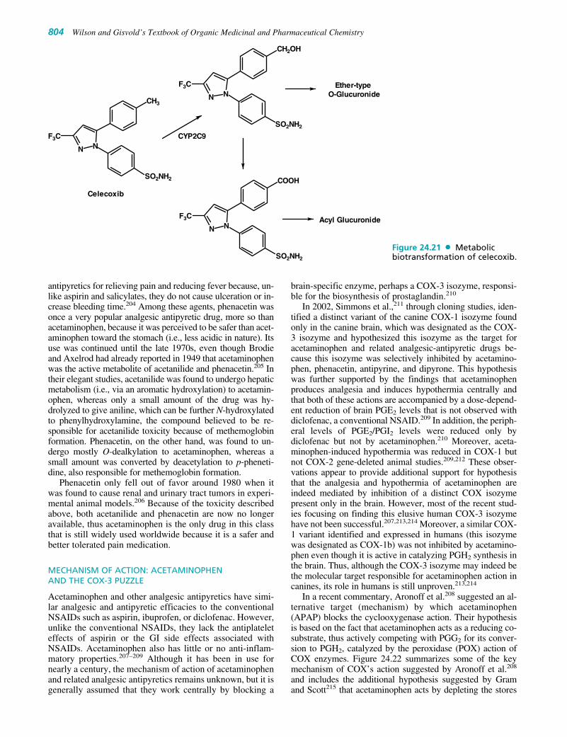
antipyretics for relieving pain and reducing fever because, un-like aspirin and salicylates, they do not cause ulceration or in-crease bleeding time.204 Among these agents, phenacetin wasonce a very popular analgesic antipyretic drug, more so thanacetaminophen, because it was perceived to be safer than acet-aminophen toward the stomach (i.e., less acidic in nature). Itsuse was continued until the late 1970s, even though Brodieand Axelrod had already reported in 1949 that acetaminophenwas the active metabolite of acetanilide and phenacetin.205 Intheir elegant studies, acetanilide was found to undergo hepaticmetabolism (i.e., via an aromatic hydroxylation) to acetamin-ophen, whereas only a small amount of the drug was hy-drolyzed to give aniline, which can be further N-hydroxylatedto phenylhydroxylamine, the compound believed to be re-sponsible for acetanilide toxicity because of methemoglobinformation. Phenacetin, on the other hand, was found to un-dergo mostly O-dealkylation to acetaminophen, whereas asmall amount was converted by deacetylation to p-pheneti-dine, also responsible for methemoglobin formation.
Phenacetin only fell out of favor around 1980 when itwas found to cause renal and urinary tract tumors in experi-mental animal models.206 Because of the toxicity describedabove, both acetanilide and phenacetin are now no longeravailable, thus acetaminophen is the only drug in this classthat is still widely used worldwide because it is a safer andbetter tolerated pain medication.
MECHANISM OF ACTION: ACETAMINOPHEN AND THE COX-3 PUZZLE
Acetaminophen and other analgesic antipyretics have simi-lar analgesic and antipyretic efficacies to the conventionalNSAIDs such as aspirin, ibuprofen, or diclofenac. However,unlike the conventional NSAIDs, they lack the antiplateleteffects of aspirin or the GI side effects associated withNSAIDs. Acetaminophen also has little or no anti-inflam-matory properties.207–209 Although it has been in use fornearly a century, the mechanism of action of acetaminophenand related analgesic antipyretics remains unknown, but it isgenerally assumed that they work centrally by blocking a
brain-specific enzyme, perhaps a COX-3 isozyme, responsi-ble for the biosynthesis of prostaglandin.210
In 2002, Simmons et al.,211 through cloning studies, iden-tified a distinct variant of the canine COX-1 isozyme foundonly in the canine brain, which was designated as the COX-3 isozyme and hypothesized this isozyme as the target foracetaminophen and related analgesic-antipyretic drugs be-cause this isozyme was selectively inhibited by acetamino-phen, phenacetin, antipyrine, and dipyrone. This hypothesiswas further supported by the findings that acetaminophenproduces analgesia and induces hypothermia centrally andthat both of these actions are accompanied by a dose-depend-ent reduction of brain PGE2 levels that is not observed withdiclofenac, a conventional NSAID.209 In addition, the periph-eral levels of PGE2/PGI2 levels were reduced only bydiclofenac but not by acetaminophen.210 Moreover, aceta-minophen-induced hypothermia was reduced in COX-1 butnot COX-2 gene-deleted animal studies.209,212 These obser-vations appear to provide additional support for hypothesisthat the analgesia and hypothermia of acetaminophen areindeed mediated by inhibition of a distinct COX isozymepresent only in the brain. However, most of the recent stud-ies focusing on finding this elusive human COX-3 isozymehave not been successful.207,213,214 Moreover, a similar COX-1 variant identified and expressed in humans (this isozymewas designated as COX-1b) was not inhibited by acetamino-phen even though it is active in catalyzing PGH2 synthesis inthe brain. Thus, although the COX-3 isozyme may indeed bethe molecular target responsible for acetaminophen action incanines, its role in humans is still unproven.213,214
In a recent commentary, Aronoff et al.208 suggested an al-ternative target (mechanism) by which acetaminophen(APAP) blocks the cyclooxygenase action. Their hypothesisis based on the fact that acetaminophen acts as a reducing co-substrate, thus actively competing with PGG2 for its conver-sion to PGH2, catalyzed by the peroxidase (POX) action ofCOX enzymes. Figure 24.22 summarizes some of the keymechanism of COX’s action suggested by Aronoff et al.208
and includes the additional hypothesis suggested by Gramand Scott215 that acetaminophen acts by depleting the stores
804 Wilson and Gisvold’s Textbook of Organic Medicinal and Pharmaceutical Chemistry
N NF3C
CH2OH
SO2NH2
N NF3C
CH3
SO2NH2
Celecoxib
CYP2C9
N NF3C
COOH
SO2NH2
Ether-typeO-Glucuronide
Acyl Glucuronide
Figure 24.21 Metabolicbiotransformation of celecoxib.
0776-0818_17865_Ch24.qxd 12/4/09 8:08 PM Page 804

Chapter 24 Analgesics 805
of glutathione (GSH), which is a known cofactor for PGEsynthase. Thus, with this illustration, it is now possible to seewhy acetaminophen’s action depends on the level of peroxidegenerated in solution (i.e., PGG2 and other lipid peroxides,generated by the lipoxygenase pathway), and its effectivenessvaries with COX activity.207 At low peroxide concentrations,acetaminophen can compete effectively with the electrontransfer mechanism between the Tyr-385 residue and theheme radical, which generates the tyrosine radical in the ac-tive site of COX enzymes for the production of PGG2, it alsoprevents the regeneration of Fe (IV) (APAP causes the forma-tion of Fe (III), thus ↓ activity of POX), a process that is es-sential for starting a new POX cycle as shown in Fig. 24.22.However, acetaminophen is ineffective during inflammationbecause the higher concentration of PGG2 or other peroxides,produced in the inflamed cells as a consequence of inductionof the COX-2 isozyme and lipoxygenase, can overcome theacetaminophen inhibition by degrading acetaminophen in thesynovial fluids as depicted in Figure 24.22 (i.e., conversion ofAPAP to its inactive glutathione conjugate shown).
This mechanism would also explain the recent findingsthat acetaminophen, unlike NSAIDs, cannot inhibit COXactivity in broken cells, and the observation that only the
inhibitory effects of acetaminophen, not indomethacin or di-clofenac, were abolished by increasing intracellular oxidationconditions with the addition of cell-permeable hydroperox-ide, t-butyl-OOH.216 This hypothesis would also provide anadditional reason why depletion of glutathione is the maincause of acetaminophen toxicity discussed under the nextsection. In summary, APAP does not compete with AA forthe binding site on the COX enzyme. Its mechanism of actionis via inhibiting the peroxidase activity of the COX enzyme.
TOXICITY IN ACETAMINOPHEN AND PHENACETIN
Acetaminophen and phenacetin can be metabolized to reac-tive hepatotoxic and renal toxic metabolites by various mech-anisms.217 Readers should consult a more detailed discussionin the drug metabolism chapter of this text. Figure 24.23 sum-marizes only the salient features for the purpose of discussingphenacetin/acetaminophen-induced liver and renal toxicities.
Acetanilide and phenacetin are hydroxylated or O-dealkylated, respectively via CYP1A2 to their activemetabolite, acetaminophen.206 In healthy individuals,acetaminophen is primarily eliminated as its O-sulfatesand O-glucuronides, with only a small amount of the
N-AcetyliminoquinoneGlutathioneConjugate
PGH2
Peroxides
Fe(IV)=O(Oxidized form)
Fe(III)(resting State)
PGG2
O
N CH3
O
GSH
NH
O
CH3
O
NH
O
CH3
OH
SG
APAP
GSH
GSSG
2O2
PGG2
AA
AA
Fe(IV)=OPP(Radical cation)
OH
Tyr
Tyr-385(in COX active site)
O
Tyr
Tyr-385 Radical
Acetaminophen(APAP)
NH
O
CH3
OH
Cyclooxygenase
Peroxidase
Figure 24.22 Mechanism of action of acetaminophen on PGH2 synthesis.
0776-0818_17865_Ch24.qxd 12/4/09 8:08 PM Page 805

N-hydroxylated metabolite (via CYP2E1/CYP3A4 isozymes),which can be sulfated or glucuronidated.217,218 Smallamounts of these O-sulfates, if accumulated in liver or renaltubules, can slowly rearrange to form the reactive N-acetyliminoquinone metabolites, shown in Figure 24.23.However, these reactive metabolites, once formed, are im-mediately deactivated by glutathione, the body’s defensemechanism for detoxifying reactive metabolites.
In contrast, a similar N-O-sulfate of phenacetin will im-mediately rearrange to this reactive metabolite, N-acetylim-inoquinone, whereas the corresponding N-O-glucuronidecan also be slowly converted to this reactive metabolite.217
Thus, it is not surprising that acetaminophen is a much saferdrug than phenacetin with regard to their relative toxicities(i.e., with occasional use, most of acetaminophen is elimi-nated as its O-sulfates and O-glucuronides). However, itshould be pointed out that acetaminophen-induced toxicitycan be greatly increased by concurrent use of alcoholic bev-erages, especially in alcoholic individuals. This is becauseboth CYP2E1 and CYP3A4 isozymes are induced byalcohol consumption.218 Moreover, heavy caffeine use,together with alcohol, would further increase the risk ofalcohol-mediated acetaminophen hepatotoxicity.219 N-acetylcysteine is typically given as an antidote to treat pos-sible acetaminophen poisoning even before plasma levels ofacetaminophen are determined. Similar to glutathione, it de-activates the N-acetyliminoquinone metabolite before itchanges to covalently bind cellular proteins (Fig. 24.23).
ACETAMINOPHEN
Acetaminophen (also known as paracetamol, APAP), a wellestablished analgesic/antipyretic drug, is frequently used byitself OTC (Panado, Tempra, Tylenol) or in combinationwith codeine (Tylenol 3), hydrocodone (Vicodin), or oxy-codone (Percocet) for the treatment of mild to moderate painand to reduce fever. It is available in several nonprescriptionforms and is also marketed in combination with aspirin andcaffeine (Excedrin, Vanquish).
Unlike aspirin or ibuprofen, acetaminophen is well toler-ated with a low incidence of GI side effects. It also has goodoral bioavailability, a fast onset and a plasma half-life of ap-proximately 2 hours after dosing. Although it is a relativelysafe pain medication, several precautions should be recog-nized, including not exceeding the recommended maximumdosage of 4 g/d. A lower daily dose of less than 2 g/d is re-quired in patients who are chronic alcoholics or have renalcomplications.220
DISEASE-MODIFYING ANTIRHEUMATIC DRUGS
RA is one of the most common chronic, inflammatory au-toimmune diseases, affecting approximately 1% of theworld’s adult population.221 It is characterized by persistentinflammation of the synovial lining of the joints that causes
806 Wilson and Gisvold’s Textbook of Organic Medicinal and Pharmaceutical Chemistry
NRO
CH3
O
OH
1. CYP2E1/CYP3A42. sulfation/glucuronidation
NRO
CH3
O
OCH2CH3
NH
CH3
O
Acetanilide
NH
CH3
O
OH
Acetaminophen
NH
CH3
O
OCH2CH3
Phenacetin
CYP1A2 CYP1A2
O-Sulfates/ O-Glucuronides(Major metabolites)
N-O-Sulfates orN-O-Glucuronides
N CH3
O
O
N-O-Sulfates orN-O-Glucuronides
Slow Fast rearrangement
Inactive cysteine or glutathione conjugates
N-acetylcysteineor glutathione
Hepatic or renal proteins
Hepatic necrosisor renal failure
Figure 24.23 Metabolic activation of acetaminophen and phenacetin.
0776-0818_17865_Ch24.qxd 12/4/09 8:08 PM Page 806

Chapter 24 Analgesics 807
pain, swelling, and stiffness of the upper and lower extrem-ities. RA, if left untreated, can lead to permanent joint car-tilage and bone damage that eventually results in disabilityand a significant impairment of quality of life.221,222 A typ-ical treatment of RA may involve a combination of physi-cal and drug therapy including aspirin, other NSAIDs, orglucocorticoids initially to provide symptomatic relief ofpain and swelling, and one or more of the disease-modify-ing antirheumatic drugs (DMARDs) to slow down theunderlying disease progression and to limit further jointdamage.223,224
DMARDs, sometime also referred to as the “slow-actingantirheumatic drugs,” include a diverse class of syntheticdrugs such as methotrexate, hydroxychloroquine, sul-fasalazine, leflunomide, azathioprine, organic gold com-pounds, or immunobiologicals useful for treating thepersistent inflammatory conditions associated with RA.DMARDs have very different mechanisms of action thatallow physicians to individualize treatment strategies toslow down the clinical and radiographic progression of RA,
even though it takes between 6 weeks to 6 months to fullyrealize any of their therapeutic effects.222,224–226
In this chapter, only a few of the commonly used syn-thetic DMARDs are covered to exemplify different treatmentstrategies. Readers interested in more detailed informationregarding the structure–activity and mechanism of actions ofthese DMARDs and related analogs should consult otherchapters in antimalarials (chloroquine and hydroxychloro-quine), anti-infectives (sulfasalazine), and antineoplasticagents (methotrexate, azathioprine, leflunomide). Recent ad-vances in new disease-modifying anti-tumor necrosis factoragents such as etanercept, adalimumab, and infliximab arecovered in a separate chapter entitled “Immunobiologicals”in this text as well as in several recent reviews.221,222,225–227
Synthetic Disease-ModifyingAntirheumatic Drugs
Figure 24.24 contains the chemical structures of theDMARDs and their metabolites mentioned in this section.
H2N
OH
COOH
5-Aminosalicylic acid
N N
H
S
O O
NH2
Sulfapyridine
CH2COO
CHCOOSAu Na
Na
Gold Sodium Thiomalate
N
N
N
N
NH2
H2N
N
CH3
N
O
H
COOHH
COOH
Methotrexate
7 NCl
NH N CH2R
CH3
CH3
Chloroquine, R=HHydroxychloroquine, R=OH
N N
H
S
O O
NN
OH
COOH
Sulfasalazine
N
OCH3
N
O
H
CF3
Leflunomide
NC
HOCH3
N
O
H
CF3
Teriflunomide
O
SAu
OAc
OAc
AcO
H
P(C2H5)3
AcO
AuranofinAurothioglucose
O
S
OH
OHHO
HO
H
Au
Figure 24.24 Disease-modifying antirheumatic drugs.
0776-0818_17865_Ch24.qxd 12/4/09 8:08 PM Page 807

Readers should consult other chapters in this text for addi-tional information regarding these drugs.
METHOTREXATE
Methotrexate (MTX, Rheumatrex), an antifolate drug usedin cancer treatment, has also been used in the diseasemanagement of RA since the 1950s. Because of its quickertherapeutic onset among all DMARDs and its demonstratedefficacy, tolerability, and low cost, MTX has been the first-line therapy for RA patients who are not responsive toNSAIDs alone.224,226,227 Recent findings have indicated thatother DMARDs should only be used for patients who are re-fractory to MTX.224,226
At least four anti-inflammatory mechanisms of actionhave been suggested for MTX’s ability to slow down RA dis-ease progression.222,228 First, MTX, being a folate antagonist,prevents antigen-dependent T-cell proliferation by blockingde novo pyrimidine biosynthesis, via a reversible inhibitionof dihydrofolate reductase.228 It also inhibits folate-mediatedproduction of spermine and spermidine in synovial tissue.These polyamines are believed to be the toxic compoundsresponsible for causing tissue injury in RA. MTX can alsoreduce intracellular glutathione concentration, thereby alter-ing the cellular redox state that suppresses the formation ofreactive oxygen radicals in synovial tissue. Lastly, MTX,similar to sulfasalazine, infliximab, and IL-4, can also inhibitosteoclastogenesis (i.e., bone erosion) in patients with RA, bymodulating the interaction of the receptor activator of nuclearfactor �B, its ligand, and osteoprotegrin.229
Methotrexate is usually administered in a once-weeklydose in the range of 15 to 17.5 mg. Although it has good oralbioavailability (�70%), it is more efficacious via IM or SCroutes, because its uptake from the GI tract is mediated by asaturable transporter, known as the reduced folate carrier-1.225,228 Most of the administered dose of methotrexate iseliminated in the urine via its 7-hydroxymethotrexatemetabolite.
Common side effects of MTX include nausea, anemia,and GI mucosa ulcerations. The use of folinic acid supple-ment to reduce MTX-mediated toxicities, however, is notrecommended for RA patients, because high dosages offolinic acid will compete with MTX for the same transporterfor absorption from the GI tract and for cellular uptake.Thus, it may also reverse MTX’s anti-inflammatory effectsin RA patients.228 Moreover, MTX can cause birth defectsin unborn children, thus it is contraindicated for womenduring pregnancy.
HYDROXYCHLOROQUINE AND CHLOROQUINE
Hydroxychloroquine (Plaquenil, Quineprox) is an olderdrug used to treat malaria that is used more often in thetreatment of RA and lupus erythematosus. Although itsmechanism of action is not known, its ability to interferewith lipopolysaccharide-induced TNF-� gene expression,thereby preventing TNF-� release from mononuclearphagocytes has been suggested.230 It is usually taken orallywith food or milk to prevent stomach irritation and ispreferred over chloroquine because of a lower incidence ofocular toxicity. However, long-term use of this medicationrequires periodic retinal examinations because of rare butpotentially preventable retinopathy associated with its use,especially at a daily dose over 6.5 mg/kg.231
SULFASALAZINE
Sulfasalazine (Azufidine) is an azo prodrug that is reducedby the bacterium present in the lower intestine to its activemetabolites, sulfapyridine and 5-aminosalicylic acid (5-ASA or mesalamine). It has been used for the treatment ofRA or ankylosing spondylitis, and inflammatory bowel dis-eases (IBDs) such as ulcerative colitis and Crohn disease.
It is generally agreed that the therapeutic effects of sul-fasalazine in treating IBDs are due mainly through its activemetabolite, 5-ASA. 5-ASA is believed to work, via similarmechanisms of action to the salicylates discussed earlier, byblocking prostaglandin synthesis in the lower intes-tine.165–168 On the other hand, because 5-ASA is completelyionized and very little of this metabolite can enter into sys-temic circulation, the antirheumatic effects of sulfasalazinehave been attributed to its other active metabolite, sulfapyri-dine.232 In a more recent study, sulfasalazine and sulfapyri-dine were found to inhibit 5-aminoimidazole-4-carboxamideribonucleotide transformylase, an enzyme involved in denovo purine biosynthesis.233 Sulfasalazine also inhibits neu-trophil function, reduces immunoglobulin levels, and inter-feres with T-cell function via suppression of NF-�B activa-tion.233 Furthermore, like methotrexate, sulfasalazine hasalso been shown to inhibit osteoclastogenesis, thereby pre-venting bone erosion in arthritic patients.229,234
It should be pointed out that approximately one third of thepatients treated long term with sulfasalazine discontinued thedrug because of dose-related adverse effects including nau-sea, dyspepsia, vomiting, headache, rash, gastric distress, es-pecially in patients on a daily dosage of greater than 4 g (or aserum sulfapyridine levels above 50 mg/mL).
LEFLUNOMIDE
Leflunomide (Arava), an isoxazole prodrug, is an orally ac-tive DMARD marketed in 1998 for the treatment of RA. Itis well absorbed and extensively metabolized in vivo to itsactive metabolite, 2-cyano-3-hydroxy-2-buteneamide (teri-flunomide), resulting from a reductive ring opening of theisoxazole ring (see Fig. 24.24). Unlike MTX, teriflunomideblocks T-cell proliferation by inhibiting dihydroorotate de-hydrogenase, the rate-limiting enzyme in the de novobiosynthesis of pyrimidine that is believed to be responsiblefor the immunosuppressive properties of leflunomide.235,236
For this reason, it is not surprising that leflunomide has avery comparable therapeutic efficacy to the first-lineDMARD, MTX as shown in several extended open clinicaltrials.237 However, even though leflunomide is well toler-ated like MTX, several cases of toxic neuropathy have beenobserved during its use, thus careful monitoring of the pa-tient’s neurological status during treatment is mandatory.237
Like MTX, leflunomide is contraindicated in pregnancy orin women considering pregnancy.
THE GOLD COMPOUNDS
The gold salts, gold sodium thiomalate (Aurolate), auro-thioglucose (Solganal), and auranofin (Ridaura) have beenknown to be effective in the treatment of RA for more than60 years.225 Recent findings have suggested that, auranofin,an orally active gold salt, inhibits Nuclear factor �B (NF-�B) activation TLR4-mediated activation of the signalingpathway that leads to the blocking of the formation of
808 Wilson and Gisvold’s Textbook of Organic Medicinal and Pharmaceutical Chemistry
0776-0818_17865_Ch24.qxd 12/4/09 8:08 PM Page 808
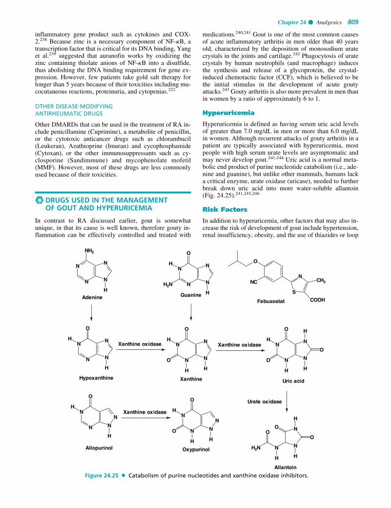
Chapter 24 Analgesics 809
inflammatory gene product such as cytokines and COX-2.238 Because zinc is a necessary component of NF-�B, atranscription factor that is critical for its DNA binding, Yanget al.239 suggested that auranofin works by oxidizing thezinc containing thiolate anions of NF-�B into a disulfide,thus abolishing the DNA binding requirement for gene ex-pression. However, few patients take gold salt therapy forlonger than 5 years because of their toxicities including mu-cocutaneous reactions, proteinuria, and cytopenias.222
OTHER DISEASE-MODIFYING ANTIRHEUMATIC DRUGS
Other DMARDs that can be used in the treatment of RA in-clude penicillamine (Cuprimine), a metabolite of penicillin,or the cytotoxic anticancer drugs such as chlorambucil(Leukeran), Azathioprine (Imuran) and cycophosphamide(Cytoxan), or the other immunosuppressants such as cy-closporine (Sandimmune) and mycophenolate mofetil(MMF). However, most of these drugs are less commonlyused because of their toxicities.
DRUGS USED IN THE MANAGEMENTOF GOUT AND HYPERURICEMIA
In contrast to RA discussed earlier, gout is somewhatunique, in that its cause is well known, therefore gouty in-flammation can be effectively controlled and treated with
medications.240,241 Gout is one of the most common causesof acute inflammatory arthritis in men older than 40 yearsold, characterized by the deposition of monosodium uratecrystals in the joints and cartilage.242 Phagocytosis of uratecrystals by human neutrophils (and macrophage) inducesthe synthesis and release of a glycoprotein, the crystal-induced chemotactic factor (CCF), which is believed to bethe initial stimulus in the development of acute goutyattacks.243 Gouty arthritis is also more prevalent in men thanin women by a ratio of approximately 6 to 1.
Hyperuricemia
Hyperuricemia is defined as having serum uric acid levelsof greater than 7.0 mg/dL in men or more than 6.0 mg/dLin women. Although recurrent attacks of gouty arthritis in apatient are typically associated with hyperuricemia, mostpeople with high serum urate levels are asymptomatic andmay never develop gout.241,244 Uric acid is a normal meta-bolic end product of purine nucleotide catabolism (i.e., ade-nine and guanine), but unlike other mammals, humans lacka critical enzyme, urate oxidase (uricase), needed to furtherbreak down uric acid into more water-soluble allantoin(Fig. 24.25).241,245,246
Risk Factors
In addition to hyperuricemia, other factors that may also in-crease the risk of development of gout include hypertension,renal insufficiency, obesity, and the use of thiazides or loop
N
N
N
N
NH2
H
N
N
N
N
O
H
H
H2N
Guanine
N
N
N
N
O
H
HN
N
N
N
O
H
H
O
HHypoxanthine Xanthine
N
N
N
N
O
H
O
O
H
H
H
Uric acid
Xanthine oxidaseXanthine oxidase
N
N
N
N
O
H
H
N
N
N
N
O
H
H
O
H
Xanthine oxidase
Allopurinol Oxypurinol
S
NCH3
COOH
NC
O
Febuxostat
N
N
N
H
O
H
H
OO
H2N
Allantoin
Urate oxidase
Adenine
Figure 24.25 Catabolism of purine nucleotides and xanthine oxidase inhibitors.
0776-0818_17865_Ch24.qxd 12/4/09 8:08 PM Page 809
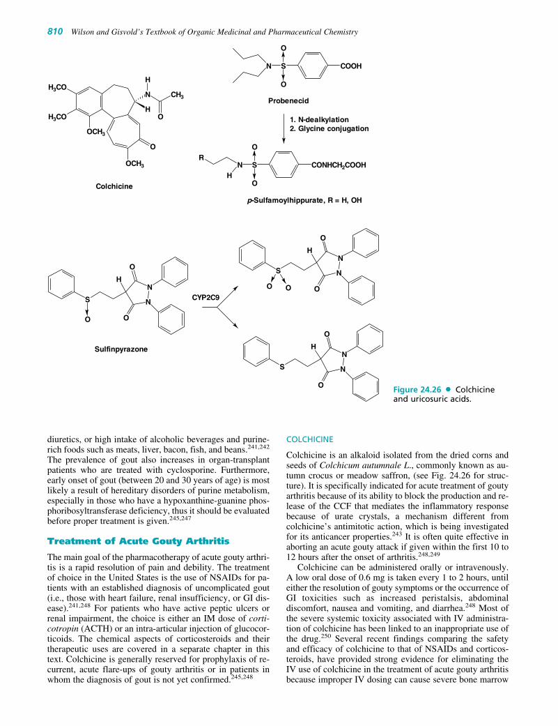
810 Wilson and Gisvold’s Textbook of Organic Medicinal and Pharmaceutical Chemistry
diuretics, or high intake of alcoholic beverages and purine-rich foods such as meats, liver, bacon, fish, and beans.241,242
The prevalence of gout also increases in organ-transplantpatients who are treated with cyclosporine. Furthermore,early onset of gout (between 20 and 30 years of age) is mostlikely a result of hereditary disorders of purine metabolism,especially in those who have a hypoxanthine-guanine phos-phoribosyltransferase deficiency, thus it should be evaluatedbefore proper treatment is given.245,247
Treatment of Acute Gouty Arthritis
The main goal of the pharmacotherapy of acute gouty arthri-tis is a rapid resolution of pain and debility. The treatmentof choice in the United States is the use of NSAIDs for pa-tients with an established diagnosis of uncomplicated gout(i.e., those with heart failure, renal insufficiency, or GI dis-ease).241,248 For patients who have active peptic ulcers orrenal impairment, the choice is either an IM dose of corti-cotropin (ACTH) or an intra-articular injection of glucocor-ticoids. The chemical aspects of corticosteroids and theirtherapeutic uses are covered in a separate chapter in thistext. Colchicine is generally reserved for prophylaxis of re-current, acute flare-ups of gouty arthritis or in patients inwhom the diagnosis of gout is not yet confirmed.245,248
COLCHICINE
Colchicine is an alkaloid isolated from the dried corns andseeds of Colchicum autumnale L., commonly known as au-tumn crocus or meadow saffron, (see Fig. 24.26 for struc-ture). It is specifically indicated for acute treatment of goutyarthritis because of its ability to block the production and re-lease of the CCF that mediates the inflammatory responsebecause of urate crystals, a mechanism different fromcolchicine’s antimitotic action, which is being investigatedfor its anticancer properties.243 It is often quite effective inaborting an acute gouty attack if given within the first 10 to12 hours after the onset of arthritis.248,249
Colchicine can be administered orally or intravenously.A low oral dose of 0.6 mg is taken every 1 to 2 hours, untileither the resolution of gouty symptoms or the occurrence ofGI toxicities such as increased peristalsis, abdominaldiscomfort, nausea and vomiting, and diarrhea.248 Most ofthe severe systemic toxicity associated with IV administra-tion of colchicine has been linked to an inappropriate use ofthe drug.250 Several recent findings comparing the safetyand efficacy of colchicine to that of NSAIDs and corticos-teroids, have provided strong evidence for eliminating theIV use of colchicine in the treatment of acute gouty arthritisbecause improper IV dosing can cause severe bone marrow
COOHS
O
O
N
CONHCH2COOHS
O
O
NH
R
Probenecid
1. N-dealkylation2. Glycine conjugation
p-Sulfamoylhippurate, R = H, OH
N
N
O
O
H
S
O
H3CO
H3CO
OCH3
OCH3
O
N CH3
O
H
H
Colchicine
N
N
O
O
H
S
O O
N
N
O
O
H
S
Sulfinpyrazone
CYP2C9
Figure 24.26 Colchicineand uricosuric acids.
0776-0818_17865_Ch24.qxd 12/4/09 8:08 PM Page 810

Chapter 24 Analgesics 811
suppression, renal failure, and death.251,252 Moreover, oralcolchicine is also recommended only as a second-line ther-apy for acute gout treatment when NSAIDs or corticos-teroids are contraindicated or ineffective.252 Concomitantuse of oral colchicine with any of the cholesterol-loweringstatins including pravastatin (Pravachol) should also beavoided because of increased risk of axonal neuromyopathythat can progress to rhabdomyolysis with renal failure.253
This is because most of the statins and colchicine are metab-olized in liver primarily by the hepatic CYP3A4 isozymes.Colchicine can also induce acute myopathy in patients whenused with pravastatin even though it is not metabolized byCYP3A4, because they share the same P-glycoprotein ef-flux transporter system.253
Control of Hyperuricemia
In general, if a patient who has hyperuricemia and the recur-rence of gouty arthritis attacks (i.e., less than twoattacks/year), their gout can usually be controlled by main-taining serum urate levels below the limit of solubility (i.e.,at 6 mg/dL or lower) with drugs that block uric acid synthe-sis by inhibiting xanthine oxidase or with uricosuric drugsthat promote uric acid elimination from the renaltubules.244,245,248
XANTHINE OXIDASE INHIBITORS: ALLOPURINOL,OXYPURINOL, FEBUXOSTAT
Allopurinol (Zyloprim, Progout) is the only xanthineoxidase inhibitor available in this class. It is rapidly metab-olized to its active isomer oxypurinol (Oxyprim), an exper-imental drug currently in phase III trials, that has a half-lifeof approximately 15 hours but was found to exhibit a simi-lar side effect profile to that of allopurinol (i.e., 40% ofthose allergic to allopurinol have similar cross-reactivity tothis drug).240 Allopurinol and its active metabolite, oxy-purinol, works by effectively competing with the substratehypoxanthine and xanthine, respectively, because of theirstructural similarity to xanthine, for xanthine oxidase, thusblocking uric acid formation from purine nucleosides, ade-nine, and guanine as shown in Figure 24.25. Similarly, al-lopurinol (or oxypurinol) may also exhibit potential druginteractions with other medications such as didanosine (an-tiviral agent), azathioprine and mercaptopurine (anticancerdrugs), and theophylline (asthma drug). For example, if apatient is given a prescription for allopurinol and azathio-prine (Imuran), then the dose of azathioprine should be re-duced by at least 50% to avoid the risk of myelosuppressionby azathioprine.254
Febuxostat is a novel and selective, nonpurine inhibitorof xanthine oxidase currently awaiting FDA approval fortreatment of chronic gout. It is more effective, at a dailydose of 80 or 120 mg, than allopurinol at the commonlyrecommended daily dose of 300 mg in lowering serumurate levels.255,256 Unlike allopurinol, it has minimal ef-fects on other enzymes involved in purine and pyrimidinemetabolisms.
URICOSURIC AGENTS: PROBENECID AND SULFINPYRAZONE
Probenecid (Benemid) and sulfinpyrazone (Anturane) areuricosuric agents that work by preventing uric acid reab-
sorption from the renal proximal tubules (see Fig. 24.26 forstructures).257 They are only effective in patients with nor-mal renal function who are not taking any medications suchas the thiazide diuretics that compete for uric acid excretionvia the organic anion transporter system (OATS) at the renalproximal tubules. In addition, patients who are usingprobenecid should also be counseled to avoid using anysalicylates at doses greater than 81 mg/d or sulfonamide-containing diuretics such as furosemide, because concomi-tant use of any salicylates or diuretics can negate itsuricosuric activity.242
ProbenecidProbenecid (Benemid) is the most widely used uricosuricagent in the United States. It is selectively excreted into therenal tubules by OATS. It is extensively metabolized via N-dealkylation or �-oxidation, followed by phase II conjuga-tion into the active metabolite, p-sulfamyl hippurate, whichexhibits a high affinity, similar to p-aminohippurate, forbinding to OATS, thereby preventing uric acid reabsorptionfrom the renal proximal tubules (Fig. 24.26) 257
SulfinpyrazoneSulfinpyrazone (Anturane) produces its uricosuric action ina similar manner to that of probenecid and is indicated forthe treatment of chronic and recurrent gouty arthritis. It iswell absorbed with approximately 50% of the administereddose excreted as unchanged drug into the renal tubules. Therest of the drug is primarily metabolized via CYP2C9 intothe corresponding sulfide and sulfone metabolites, thus itcan potentiate the anticoagulant effect of warfarin.258
TRIPTANS
Triptans are safe and effective drugs for abortive, but not forprophylactic, treatment of moderate to severe migraine andcluster headaches.259 Because of their higher affinity and se-lectivity for the serotonin 5-HT1B/1D receptors at the trigemi-nal nerve fibers and dural vasculature, they are able to inducevasoconstrictions at these sites, thereby relieving pain fromcranial vasodilatation and reducing neurogenic inflammationassociated with these disorders.259,260 However, triptans areoften contraindicated in patients with uncontrolled hyperten-sion, especially those with coronary vascular diseases, eventhough there are far fewer 5-HT1B receptors found in coro-nary blood vessels and in the peripheral vasculatures than inthe cranial blood vessels.261 Thus, special attention shouldalso be given to assess the safety and tolerability profile ofthe triptans when choosing among available drugs because oftheir pharmacokinetic differences.261,262
Pathophysiology of Migraine
Migraine, a recurrent and debilitating headache disorder,affects about 12% of the worldwide population with ahigher prevalence in women (�18%) than in men(�6%).259 In the most recent classification and diagnosticcriteria for headache disorders published by the HeadacheClassification Committee of the International HeadacheSociety, the terms, common and classical migraines havebeen replaced with migraine without aura and migrainewith aura, respectively.263 The patients often describetheir migraine attack as having an intense pulsating and
0776-0818_17865_Ch24.qxd 12/4/09 8:08 PM Page 811

throbbing headache lasting from 4 hours to 3 days, if notproperly treated.259,260 In addition to this excruciating pain,migraine patients often have other symptoms like nausea,vomiting, sensitivity to light (photophobia), sound (phono-phobia), or movements that can also severely impact theirquality of life.
Although the etiology of migraines remains poorly un-derstood, activation of the meningeal nociceptors at the in-tracranial trigeminovascular system (TGVS, also referred toas the trigeminal pain pathway) is believed to play a key rolein promoting headache and other symptoms associated withmigraines.264–266 Several theories have been suggested toexplain the underlying causes for the symptoms associatedwith migraines.259 Only two of the most prominent theoriesare briefly discussed under this section.
THE VASCULAR THEORY OF MIGRAINES
According to the vascular theory, the vasodilatation of cra-nial carotid arteriovenous anastomoses (sites of many 5-HT1B/1D receptors) and meningeal, dural, cerebral, or pialvessels (primary sites of 5-HT1B receptors) plays an impor-tant role in the pathogenesis of migraines and is responsiblefor the pain associated with migraine headaches.259,261 Thefact that sumatriptan-induced cranial vasoconstriction is se-lectively blocked by a selective 5-HT1B antagonist, and notby a 5-HT1D antagonist, lends further support to this vascu-lar theory of migraines.267
THE NEUROGENIC INFLAMMATION THEORY OF MIGRAINES
This theory suggests that migraine headaches occur as a re-sult of an abnormal firing of meningeal nociceptors at theTGVS. Activation of trigeminal neurons releases vasoac-tive peptides including calcitonin gene-related peptide(CGRP, a vasodilator peptide), substance P, and neurokininA (both play an important role in pain transmission as wellas activation of immune responses and neurogenic inflam-mation) onto dural tissue where these peptides produce a
local response known as neurogenic inflammation.259
These peptides induce cranial vasodilatation, especially atthe dural membranes surrounding the brain (mainly a resultof CGRP), thus producing the pain associated with mi-graine attacks. Further evidence supporting this theory asthe underlying cause of migraines can be found from a re-cent study linking the dural mast cell degranulation to theprolonged activation of the trigeminal pain pathway andneurogenic inflammation.268
The discovery of these vasoactive peptides providesnew targets for the future design of nonvasoconstrictors,nontriptan drugs such as CGRP antagonists for the acuteand preventive treatment of migraine and clusterheadaches.259,269
Structure–Activity Relationship
All clinically available triptans possess comparable phar-macodynamic properties. They all bind and stimulate sero-tonin 5-HT1B/1D with affinity in the low nanomolar ranges,thus they are equally effective for the acute treatment ofmigraine.262,270 However, they all have different pharma-cokinetic properties and side effect profiles that vary intype and severity.261 Thus, they are not equally efficaciousin preventing migraine recurrence because of the differ-ences in their elimination half-lives.262 They also differ intheir potential to induce drug-related CNS side effects, es-pecially somnolence and paresthesia that may lead to a pa-tient’s noncompliance of an otherwise effective migrainetreatment.270
Table 24.2 summarizes the pharmacokinetic properties ofclinically available triptans that contributes to their efficacy,safety, and tolerability.259,262,270–274 A quick glance at theirchemical structures (see Fig. 24.27) reveals two generalstructural types, which might provide insights for assessingsome of these differences.
The 3-alkylaminoethyl containing triptans (sumatriptan,zolmitriptan, rizatriptan, and almotriptan) are substrates forthe hepatic monoamine oxidase type A (MAO-A) because
812 Wilson and Gisvold’s Textbook of Organic Medicinal and Pharmaceutical Chemistry
TABLE 24.2 Pharmacokinetic Properties of the Triptansa
Bioavailability Onset Time Plasma Metabolizing Drug % CNS Drug Trade Name Oral (%) (min) Half-life (h) Enzymea,b Interactions Side Effects
Sumitriptan Imitrex 14 10–15 (iv) 2 MAO-A MAOIs especially 1.7–6.3Imigrain 15–30 (nasal) MAO-A inhibitors
30–90 (PO)Zolmitriptan Zomig, 40–48 10–15 (nasal) 3 CYP1A2 MAOIs, SSRIs, 9.9–11.5
Zomig-ZMT 45–60 (PO) MAO-A CimethidineNaratriptan Amerge 63 (men) 60–180 (PO) 5–6.3 Renal/CYP 1.9
Naramig 74 (women) isozymesRizatriptan Maxalt 45 30–120 (PO) 2–3 MAO-A MAOIs especially 6.1–9.4
Maxalt-MLT MAO-A inhibitors, Propranolol
Almotriptan Axert 70–80 60–180 (PO) 3.3 MAO-A 1.5CYP3A4
Frovatriptan Prova 60 120–180 (PO) 26 CYP1A2 Fluvoxamine, 6.0ciprofloxacin,mexiletine
Eletriptan Relpax 50 30–60 (PO) 3.6–5.5 CYP3A4 Ketoconazole and 2.6–14.6other CYP3A4 inhibitors
aCompiled from data reported in the following references (1, 4, 12, and 13).bPrimary mode of metabolism listed first.
0776-0818_17865_Ch24.qxd 12/4/09 8:08 PM Page 812

Chapter 24 Analgesics 813
of their structural resemblance to 5-HT, the preferred sub-strate for MAO-A. They all have a relatively short durationof action (i.e., 2–3 hours) because of first-pass metabolic in-activation by the MAO-A into an indole acetic acid metabo-lite that is rapidly eliminated as their corresponding glu-curonides. Thus, the use of these triptans requires repeatdosing to prevent the recurrence of migraine attack. Theyare also contraindicated with concomitant use of any MAOinhibitors, especially inhibitors of MAO-A such as the anti-depressant drug, moclobemide (Aurorix, Mauerix).261,273
Eletriptan, frovatriptan, and naratriptan, on the otherhand, with their 3-alkylamino side chain fused into a carbo-cyclic ring structure, all have much longer elimination half-lives and lower incidence of headache recurrence re-ported.273 This is because they are not a substrate forMAO-A (amphetamine is not a substrate for MAO becauseit has an additional methyl group � to the terminal aminefunction). These triptans are mainly degraded by the he-patic CYP isozymes (CYP1A2, 2D6, 3A4), but theirbioavailability may be altered with a drug that inhibits orinduces these CYP isozymes.275 For example, eletriptan isprimarily metabolized by CYP3A4 isozyme, thus it is notadvisable to use eletriptan with a CYP3A4 inhibitor such asketoconazole, or nefazodone without appropriately adjust-ing their dosages.275
The highest reported incidences of drug-induced CNStoxicities were found with eletriptan (14% at 80-mg dose),zolmitriptan (11.5% at 5-mg dose) and rizatriptan (9.4% at10-mg dose). They all have N-demethylated, active metabo-lites that can easily gain entry into the brain because of their
high lipophilicity.270 Thus, the presence of this activemetabolite in the brain and the high lipophilicity of theparent triptan have been suggested as a factor for contribut-ing to the observed CNS side effects of the triptans.270 Thishypothesis is further supported by the fact that frovatriptanhas a much lower CNS toxicity because of the greater watersolubility of its N-demethylated active metabolite thatprevents it from entering the brain, whereas sumatriptan, al-motriptan, and naratriptan all have a very low reported inci-dence of CNS side effects because they have no clinicallysignificant active metabolites.270,276
Donitriptan, a unique high-efficacy and high-selectivity5-HT1B/1D agonist (i.e., with high-intrinsic activity ap-proaching that of the endogenous agonist 5-HT) currently inlate-stage clinical trials, is a novel arylpiperazole derivativewith much better consistency of pain relief and a lower inci-dence of migraine recurrence.277 Furthermore, unlike suma-triptan, it can also block capsaicin-sensitive trigeminalsensory nerves from releasing CGRP, resulting in selectivecranial vasodilatation and central nociception.278
Mechanism of Action
Triptans are specifically designed to bind to the 5-HT1B/1D
receptors based on the findings that 5-HT1B receptors arepresent in the cranial blood vessels279,280 and 5-HT1B/1D
receptors are found in the trigeminal pain pathway.280,281
Three distinct mechanisms have been suggested to explainthe actions of triptans: (a) Triptans abort migraineheadache by its agonist action at the 5-HT1B receptors,
N
H
NCH3S
NH3C
CH3
O O
H
N
H
NCH3S
N
CH3
O O
Sumatriptan
N
H
NCH3N
N
CH3
N
Almotriptan
Zolmitriptan
N
H
NCH3
N
CH3
O
H
H
O
Rizatriptan
N
H
NCH3
H
H2N
O
H
Frovatriptan
N
H
N
SN
H3C
OO
H
CH3
Naratriptan
N
H
S
OO
N
CH3
H
Eletriptan
N
H
NH2ON
O
N
NC
Donitriptan
Figure 24.27 Triptans.
0776-0818_17865_Ch24.qxd 12/4/09 8:08 PM Page 813

thereby inducing vasoconstriction of the meningeal, dural,or pial blood vessels.279 (b) Triptans also inhibitneurogenic inflammation via its presynaptic stimulation of5-HT1D receptors and/or through its additional action atthe 5-HT1B/1D receptors.281 (c) Triptans relieve migrainepain transmission most likely because of its inhibitoryaction at the trigeminal pain pathway mediated via 5-HT1B/1D/1F receptors.259,279,280
Antimigraine Drugs Acting on 5-HT1B/1D Receptors
SUMATRIPTAN (IMITREX)
Sumatriptan was the first triptan approved (1991) for theacute treatment of migraine headaches. It has the lowestoral bioavailability among all triptans because of its lowlipophilicity. The availability of many different dosageforms (i.e., an oral tablet, a SC injection, a nasal spray for-mulation, and a suppository) allows the flexibility of tailor-ing therapy to the needs of the individual patients, thusmaking sumatriptans a very useful drug for an acute treat-ment of migraine headaches.262,282 It also has a very fastonset of action via SC injection or nasal spray adminis-tration. However, sumatriptan is contraindicated withmonoamine oxidase inhibitors because it is primarily de-graded by hepatic MAO-A. Thus, it may require frequentdosing as a result of its short duration of action to preventmigraine recurrence.262,282
ZOLMITRIPTAN (ZOMIG, ZOMIG-ZMT)
Zolmitriptan, the second triptan marketed (approved in1997), has a much better bioavailability (40%–48%) thansumatriptan. It is rapidly absorbed after oral or nasal sprayadministration. It also has an orally disintegrating tablet for-mulation (Zomig ZMT), which can be taken without water.Zolmitriptan undergoes rapid N-demethylation via CYP1A2to a more potent, active metabolite, N-desmethylzolmitrip-tan, which is 2 to 6 times more potent than the parentdrug.262,283 This active metabolite was detected 5 minutesafter dosing and accounts for about two thirds of the plasmaconcentration of the administered dose of the parent drug.284
Thus, it is reasonable to assume that the therapeutic effectsand especially the CNS side effects of zolmitriptan must bein part attributed to the plasma levels of this active metabo-lite, at least until it is further degraded by hepatic MAO-Ato its inactive indole acetic acid derivatives.262,283
NARATRIPTAN (AMERGE, NARAMIG)
Naratriptan, the third triptan approved in 1998, is one of themost lipophilic triptans marketed to date. It has a much im-proved bioavailability (63% in men and 74% in women), agreater affinity for 5-HT1B/1D receptors (3–6 times), and alower recurrence rate than sumatriptan because of its muchlonger elimination half-life.262,285 Naratritan also has a fa-vorable CNS side effect profile when compared with suma-triptan or zolmitriptan because of its metabolic stability,thereby lacking a N-demethylated active metabolite and asignificant renal excretion (�70% of naratriptan is excretedunchanged and the rest of the administered dose is degradedvia several CYP isozymes).262,270,286
RIZATRIPTAN (MAXALT)
Rizatriptan, approved in 1998, is a fast-acting triptan be-cause of its moderate lipophilicity yet has a very shortelimination half-life similar to sumatriptan (i.e., like suma-triptan, it is mainly metabolized by MAO-A). The only ad-vantages of this drug when compared with sumatriptan arethat it has a slightly faster onset and that it has an orallydisintegrating tablet formulation which can be taken with-out water.262
ALMOTRIPTAN (AXERT)
Almotriptan, marketed in 2000, has the highest oralbioavailability among all triptans (see Table 24.2). It is me-tabolized by both MAO-A and CYP3A4, thus has a more fa-vorable side effects profile when compared with sumatrip-tan. However, it is only available in a 12.5 mg tablet form.
FROVATRIPTAN (FROVA)
Frovatriptan is a newer triptan introduced into the market in2001. With the incorporation of a 3-alkylamino side chaininto a carbazole ring structure, it is not a substrate forMAO-A or CYP3A4. Thus, unlike the other clinicallyavailable triptans, it possesses a much longer duration ofaction with fewer drug–drug interactions with MAO in-hibitors or with drugs metabolized by the CYP3A4isozymes.274,276 It is primarily metabolized by CYP1A2 togive its active metabolite, N-desmethyl-frovatriptan, whichhas about one third of the binding affinity for 5-HT1D/1B re-ceptors but with three times longer plasma half-life of theparent drug.276,287
Frovatriptan also has the highest affinity for brain 5-HT1B receptors and the longest elimination half-life amongall triptans.273,288 The only disadvantage of this drug is itsslower onset of action because of its greater water solubil-ity. However, it is a drug of choice for patients with long-lasting migraines or if recurrence is a problem.273
ELETRIPTAN (RELPAX)
Eletriptan, introduced into the market in 2002, is the newesttriptan with highest affinity for 5-HT1B, 5-HT1D, and 5-HT1F
receptors. It is one of the most lipophilic triptans marketedto date and is well tolerated and safe across its dosing rangeof 20 to 80 mg.275 However, it is metabolized primarily(�90%) by CYP3A4 isozyme to its active metabolite, theN-desmethyleletriptan, which accounts for approximately10% to 20% of the plasma concentration of that observedfor parent drug.289 Thus, coadministration of eletriptan withpotent CYP3A4 inhibitors such as ketoconazole, itracona-zole, nefazodone, troleandomycin, clarithromycin, ritonavir,and nelfinavir may require dose reduction and closer moni-toring for CNS side effects.262,275 Furthermore, becauseeletriptan and its active metabolite, N-desmethyleletriptan,are also substrates for the P-glycoprotein efflux pumps thatare responsible for their removal from the brain, coadminis-tration of eletriptan with a known P-glycoprotein inhibitorand/or inducer such as digoxin, diltiazem, verapamil, or St.John’s Worth would result in higher brain levels of its activemetabolite, and thus a higher rate of the CNS side effectsreported for this drug.290
814 Wilson and Gisvold’s Textbook of Organic Medicinal and Pharmaceutical Chemistry
0776-0818_17865_Ch24.qxd 12/4/09 8:08 PM Page 814

Chapter 24 Analgesics 815
1. A patient is brought to the emergency room via ambu-lance. He is unresponsive to questions, has a respiratoryrate of 4 breaths per minute, and an arterial oxygen satu-ration (SpO2) of 82% measured by finger pulse oximetry.On physical exam, multiple injection (track) marks areidentified on his forearms, and the patient also displays acutaneous rash over his trunk. The suspected diagnosis isa heroin overdose. The nurse calls the pharmacy for 50mg of diphenhydramine to treat the cutaneous rash. Youdo not dispense it, why?
2. A new mom comes to your pharmacy to refill her codeineprescription, prescribed for episiotomy pain. She is con-cerned because her 2-week-old breastfed baby is ex-tremely lethargic, sleeps all day, and seems unresponsiveto stimulus. You recommend that she bring the baby tothe emergency room immediately. What genetic poly-morphism do you suspect the mom has? What dangerdoes this pose to her breastfed infant?
3. A patient presents to your pharmacy with a prescriptionfor Vicodin ES; 2 tablets q 6 hours around the clock;#240; refills � zero. You do not dispense it, why?
4. A 72-year-old man who has been taking the antiarrythmicdrug quinidine for 4 years self treats his diarrhea withImodium (loperamide). His son calls your pharmacy to
report that his father is very sleepy, his pupils are pin-points, and he is acting “very strange.” You suspect thatloperamide is having CNS effects. What is the pharmaco-logical reason behind the quinidine-loperamide drug–druginteraction?
5. Diflunisal is a potent, long acting nonselective COX in-hibitor. Explain how this drug binds to the COX-1 en-zyme, and how it is eliminated from the body.
6. Provide a biochemical reason why acetaminophen, unlikeaspirin and other NSAIDS, is a centrally acting anal-gesic/antipyretic drug that has no anti-inflammatoryactivity.
7. Provide a possible rationale for why CYP3A4 inducerscan increase the clearance of diclofenac, yet CYP3A4 in-hibitors have little or no effect on its pharmacokineticprofile.
8. Sulindac, a potent nonselective COX inhibitor, is said tohave a lower risk of stomach bleeding and nephrotoxicitythan other aspirin-like NSAIDs such as indomethacin.Explain.
9. Provide a possible chemical/biochemical rationale whyfrovatriptan has a much lower CNS toxicity than eletriptan.
R E V I E W Q U E S T I O N S
REFERENCES1. Anonymous. U.S. Department of Health, Education and Welfare. The
Interagency Committee on New Therapies for Pain and Discomfort:Report to the White House, 1979.
2. Katz, N. P., Adams, E. H., Benneyan, J. C., et al.: Clin. J. Pain23:103–118, 2007.
3. Staats, P. S.: J. Pain Symptom Manage. 24:S4–S9, 2002.4. Fields, H. L.: Pain 129:233–234, 2007.5. Hobson, A. R., and Aziz, Q.: News Physiol. Sci. 16:109–114, 2003.6. Riley, J. L., III, Wade, J. B., Myers, C. D., et al.: Pain 100:291–298,
2002.7. Loeser, J. D., and Melzack, R.: Lancet 353:1607–1609, 1999.8. Besson, J. M.: Lancet 353:1610–1615, 1999.9. Zimmermann, M.: Eur. J. Pharmacol. 429:23–37, 2001.
10. Baliki, M. N., Chialvo, D. R., Geha, P. Y., et al.: J. Neurosci.26:12165–12173, 2006.
11. Kumar, N.: World Health Organization Normative Guidelines onPain Management-Report of a Delphi Study. 2007.
12. Koyama, T., McHaffie, J. G., Laurienti, P. J., et al.: Proc. Natl. Acad.Sci. U. S. A. 102:12950–12955, 2005.
13. Price, D. D.: Science 288:1769–1772, 2000.14. Price, D. D., Greenspan, J. D., and Dubner, R.: Pain 106:215–219,
2003.15. Seminowicz, D. A., and Davis, K. D.: Pain 130:8–13, 2007.16. Goffaux, P., Redmond, W. J., Rainville, P., et al.: Pain 130:137–143,
2007.17. Jensen, T. S., and Baron, R.: Pain 102:1–8, 2003.18. Zech, D. F. J., Grond, S., Lynch, J., et al.: Pain 63:65–76, 1995.19. Barakzoy, A. S., and Moss, A. H.: J. Am. Soc. Nephrol.
17:3198–3203, 2006.20. Goldstein, A., Lowney, L. I., and Pal, B. K.: Proc. Natl. Acad. Sci.
U. S. A. 68:1742–1747, 1971.21. Pert, C. B., and Snyder, S. H.: Proc. Natl. Acad. Sci. U. S. A.
70:2243–2247, 1973.22. Hughes, J., Smith, T. W., and Kosterlitz, H. W.: Nature 258:577–579,
1975.23. Morley, J. S.: Annu. Rev. Pharmacol. Toxicol. 20:81–110, 1980.
24. Clement-Jones, V., McLoughlin, L., Tomlin, S., et al.: Lancet2:946–949, 1980.
25. Kenyon, J. N., Knight, C. J., and Wells, C.: Acupunct. Electrother.Res. 8:17–24, 1983.
26. Tseng, L. F., Narita, M., Suganuma, C., et al.: J. Pharmacol. Exp.Ther. 292:576–583, 2000.
27. New, D. C., and Wong, Y. H.: Neurosignals 11:197–212, 2002.28. Klavdieva, M. M.: Neuroendocrinol. 17:247–280, 1996.29. Zioudrou, C., Streaty, R. A., and Klee, W. A.: J. Biol. Chem.
254:2446–2449, 1979.30. Wittert, G., Hope, P., and Pyle, D.: Biochem. Biophys. Res.
Commun. 218:877–881, 1996.31. Stein, C., Hassan, A., Przewlocki, R., et al.: Proc. Natl. Acad. Sci. U.
S. A. 87:5935–5939, 1990.32. Corbett, A. D., Henderson, G., McKnight, A. T., et al.: Br. J.
Pharmacol. 147 Suppl 1:S153–S162, 2006.33. Waldhoer, M., Bartlett, S. E., and Whistler, J. L.: Annu. Rev.
Biochem. 73:953–990, 2004.34. Trescot, A. M., Datta, S., Lee, M., et al.: Pain Physician
11:S133–S153, 2008.35. Pogozheva, I. D., Lomize, A. L., and Mosberg, H. I.: Biophys. J.
75:612–634, 1998.36. Chaturvedi, K., Shahrestanifar, M., and Howells, R. D.: Brain Res.
Mol. Brain Res. 76:64–72, 2000.37. Boettcher, C., Fellermeier, M., Boettcher, C., et al.: Proc. Natl. Acad.
Sci. U. S. A. 102:8495–8500, 2005.38. Valiquette, M., Vu, H. K., Yue, S. Y., et al.: J. Biol. Chem.
271:18789–18796, 1996.39. Befort, K., Tabbara, L., Kling, D., et al.: J. Biol. Chem.
271:10161–10168, 1996.40. Negus, S. S., Gatch, M. B., Mello, N. K., et al.: J. Pharmacol. Exp.
Ther. 286:362–375, 1998.41. Aldrich, J. V., and Vigil-Cruz, S. C.: Narcotic analgesics. In
Abraham D. J., (ed.). Burger’s Medicinal Chemistry and DrugDiscovery. New York, John Wiley and Sons, 2003.
42. Portoghese, P. S., Moe, S. T., and Takemori, A. E.: J. Med. Chem.36:2572–2574, 1993.
0776-0818_17865_Ch24.qxd 12/4/09 8:08 PM Page 815
816 Wilson and Gisvold’s Textbook of Organic Medicinal and Pharmaceutical Chemistry
43. Portoghese, P. S., Sultana, M., Nagase, H., et al.: J. Med. Chem.31:281–282, 1988.
44. Broom, D. C., Jutkiewicz, E. M., Rice, K. C., et al.: Jpn. J.Pharmacol. 90:1–6, 2002.
45. Mansour, A., Hoversten, M. T., Taylor, L. P., et al.: Brain Res.700:89–98, 1995.
46. Nagase, H., Hayakawa, J., Kawamura, K., et al.: Chem. Pharm. Bull.(Tokyo) 46:366–369, 1998.
47. Wadenberg, M. G.: CNS Drug Rev. 9:187–198, 2003.48. Chavkin, C., Sud, S., Jin, W., et al.: J. Pharmacol. Exp. Ther.
308:1197–1203, 2004.49. Wang, Y., Tang, K., Inan, S., et al.: J. Pharmacol. Exp. Ther.
312:220–230, 2005.50. Reinscheid, R. K., Higelin, J., Henningsen, R. A., et al.: J. Biol.
Chem. 273:1490–1495, 1998.51. Broccardo, M., Guerrini, R., Morini, G., et al.: Peptides
28:1974–1981, 2007.52. Schiff, P. L. J.: Amer. J. Pharm. Educ. 66:186–194, 2002.53. Novak, B. H., Hudlicky, T., Reed, J. W., et al.: Curr. Org. Chem.
4:343–362, 2000.54. McQuay, H. J., and Moore, R. A.: Opioid problems, and morphine
metabolism and excretion. In Handbook of experimental pharmacol-ogy. Springer Verlag KG, 1997, vol. 130, pp. 335–360.
55. Buetler, T., Wilder-Smith, O., Wilder-Smith, C., et al.: Br. J.Anaesth. 84:97–99, 2000.
56. Lotsch, J., Skarke, C., Schmidt, H., et al.: Clin. Pharmacol. Ther.79:35–48, 2006.
57. Vree, T. B., van Dongen, R. T., and Koopman-Kimenai, P. M.: Int. J.Clin. Pract. 54:395–398, 2000.
58. Skarke, C., Darimont, J., Schmidt, H., et al.: Clin. Pharmacol. Ther.73:107–121, 2003.
59. Lotsch, J., Kobal, G., and Geisslinger, G.: Clin. Neuropharmacol.21:351–354, 1998.
60. Eckhardt, K., Li, S., Ammon, S., et al.: Pain 76:27–33, 1998.61. Schroeder, K., and Fahey, T.: Cochrane Database Syst. Rev.
(4):CD001831, 2004.62. Casy, A. F., and Parfitt, R. T.: Opioid Analgesics Chemistry and
Receptors. New York, Plenum Press, 1986, pp. 518.63. Willette, R. E.: Ch. 22 Analgesic agents. In Block, J. H., and Beale,
J. M., Jr., (eds.). Wilson and Gisvold’s Textbook of OrganicMedicinal and Pharmaceutical Chemistry, 11th ed. New York,Lippincott Williams and Wilkins, 2004, pp. 731–766.
64. Kaplan, H. L., Busto, U. E., Baylon, G. J., et al.: J. Pharmacol. Exp.Ther. 281:103–108, 1997.
65. Poyhia, R., Seppala, T., Olkkola, K. T., et al.: Br. J. Clin. Pharmacol.33:617–621, 1992.
66. Anonymous. Drug Abuse Warning Network, 2005: National esti-mates of drug-related emergency department visits Series D-29,DHHS Publication No. (SMA) 07-4256, Rockville, MD, accessedJanuary 28, 2008.
67. Prommer, E. E.: J. Palliat. Med. 10:1228–1230, 2007.68. Stringer, M., Makin, M. K., Miles, J., et al.: d-morphine, but not l-mor-
phine, has low micromolar affinity for the non-competitive N-methyl-D-aspartate site in rat forebrain. Possible clinical implications for themanagement of neuropathic pain. Neurosci. Lett. 295:21–24, 2000.
69. Anonymous. Substance Abuse and Mental Health ServicesAdministration, Office of Applied Studies. The NSDUH Report:Misuse of Over-the-Counter Cough and Cold Medications amongPersons Aged 12 to 25. Rockville, MD, January 10, 2008.
70. Smith, S. M., Schroeder, K., and Fahey, T.: Cochrane Database Syst.Rev. (1):CD001831, 2008.
71. Ball, J., and Alexson, S. H.: The Dawn Report: EmergencyDepartment Visits Involving Dextromethorphan. Rockville MD,Substance Abuse and Mental Health Services Administration, 2006,32, 1–4.
72. Abraham, R. B., Marouani, N., and Weinbroum, A. A.: Ann. Surg.Oncol. 10:268–274, 2003.
73. Sindrup, S. H., and Jensen, T. S.: Pain 83:389–400, 1999.74. Anonymous. Package Insert: Talwin Nx. September 2006, 7.75. Zacny, J. P., Hill, J. L., Black, M. L., et al.: J. Pharmacol. Exp. Ther.
286:1197–1207, 1998.76. Gear, R. W., Gordon, N. C., Heller, P. H., et al.: Neurosci. Lett.
205:207–209, 1996.77. Gear, R. W., Miaskowski, C., Gordon, N. C., et al.: Pain 83:339–345,
1999.78. Zhang, J., Burnell, J. C., Dumaual, N., et al.: J. Pharmacol. Exp.
Ther. 290:314–318, 1999.
79. Latta, K. S., Ginsberg, B., and Barkin, R. L.: Am. J. Ther. 9:53–68,2002.
80. Simopoulos, T. T., Smith, H. S., Peeters-Asdourian, C., et al.: Arch.Surg. 137:84–88, 2002.
81. Seifert, C. F., and Kennedy, S.: Pharmacotherapy 24:776–783, 2004.
82. Tissot, T. A.: Anesthesiology 98:1511–1512, 2003.83. Palmer, K. R., Corbett, C. L., and Holdsworth, C. D.:
Gastroenterology 79:1272–1275, 1980.84. McCarron, M. M., Challoner, K. R., and Thompson, G. A.:
Pediatrics 87:694–700, 1991.85. Baker, D. E.: Rev. Gastroenterol. Disord. 7 Suppl 3:S11–S18, 2007.86. Sandhu, B. K., Tripp, J. H., Candy, D. C., et al.: Gut 22:658–662,
1981.87. Schinkel, A. H., Wagenaar, E., Mol, C. A., et al.: J. Clin. Invest.
97:2517–2524, 1996.88. Subramanian, G., Paterlini, M. G., Portoghese, P. S., et al.: J. Med.
Chem. 43:381–391, 2000.89. Westmoreland, C. L., Hoke, J. F., Sebel, P. S., et al.:
Anesthesiology 79:893–903, 1993.90. Anonymous. Dolophine Hydrochloride CII (Methadone
Hydrochloride Tablets, USP) Package Insert, Roxane Laboratories,Inc., Revised 10/2006.
91. Gerber, J. G., Rhodes, R. J., and Gal, J.: Chirality 16:36–44, 2004.92. Foster, D. J., Somogyi, A. A., and Bochner, F.: Br. J. Clin.
Pharmacol. 47:403–412, 1999.93. Iribarne, C., Berthou, F., Baird, S., et al.: Chem. Res. Toxicol.
9:365–373, 1996.94. Totah, R. A., Sheffels, P., Roberts, T., et al.: Anesthesiology
108:363–374, 2008.95. Wang, J., and DeVane, C. L.: Drug Metab. Dispos. 31:742–747,
2003.96. Fick, D. M., Cooper, J. W., Wade, W. E., et al.: Arch. Intern. Med.
163:2716–2724, 2003.97. Ulens, C., Daenens, P., and Tytgat, J.: Cardiovasc. Res. 44:568–578,
1999.98. Willens, J. S.: Pain Manag. Nurs. 7:43–43, 2006.99. Anonymous. Tramadol HCl Tablets, Ultram, Package Insert, Ortho-
McNeil, Revised December 1999.100. Evans, G. R., Henshilwood, J. A., and O’Rourke, J.: Tetrahedron
Asymmetry 12:1663–1670, 2001.101. Potschka, H., Friderichs, E., and Loscher, W.: Br. J. Pharmacol.
131:203–212, 2000.102. Scott, L. J., and Perry, C. M.: Drugs 60:139–176, 2000.103. Raffa, R. B., Friderichs, E., Reimann, W., et al.: J. Pharmacol. Exp.
Ther. 267:331–340, 1993.104. Raffa, R. B.: J. Clin. Pharm. Ther. 33:101–108, 2008.105. Anonymous. DEA Office of Diversion Control, Nalbuphine.
(http://www.deadiversion.usdoj.gov/drugs_concern/nalbuphine.htm),2008.
106. Anonymous. Nubaine (Nalbuphine Hydrochloride), Package Insert,Endo Pharmaceuticals Inc., Updated January, 2005.
107. Aitkenhead, A. R., Lin, E. S., and Achola, K. J.: Br. J. Clin.Pharmacol. 25:264–268, 1988.
108. Anonymous. Press Release: Penwest Begins Dosing Phase IIaClinical Study of Nalbuphine ER. (http://www.penwest.com/2008).
109. De Souza, E., Schmidt, W., and Kuhar, M.: J. Pharmacol. Exp.Ther. 244:391–402, 1998.
110. Gunion M. W., Marchionne A. M., and Anderson, C. T. M.: AcutePain 6:29–39, 2004.
111. Commiskey, S., Fan, L. W., Ho, I. K., et al.: J. Pharmacol. Sci.98:109–116, 2005.
112. Anonymous. Critical Review of Butorphanol; WHO 34th ExpertCommittee on Drug Dependence (ECDD) 2006.http://www.who.int/medicines/areas/quality_safety/4.1ButhorphanolCritReview.pdf.
113. Megarbane, B., Hreiche, R., Pirnay, S., et al.: Toxicol. Rev.25:79–85, 2006.
114. Dahan, A., Yassen, A., Romberg, R., et al.: Br. J. Anaesth.96:627–632, 2006.
115. Kosten, T. R., Morgan, C., and Kleber, H. D.: NIDA Res. Monogr.121:101–119, 1992.
116. Lewis, J. W., and Walter, D.: NIDA Res. Monogr. 121:5–11, 1992.117. Griessinger, N., Sittl, R., and Likar, R.: Curr. Med. Res. Opin.
21:1147–1156, 2005.118. Picard, N., Cresteil, T., Djebli, N., et al.: Drug Metab. Dispos.
33:689–695, 2005.
0776-0818_17865_Ch24.qxd 12/4/09 8:08 PM Page 816

Chapter 24 Analgesics 817
119. Anonymous. Vivitrol (naltrexone for extended-release injectablesuspension) Package Insert Manufactured by Alkermes, Inc.Marketed by Cephalon, Inc. Revised 11/10/05.
120. Lobmaier, P., Kornor, H., Kunoe, N., et al.: Cochrane DatabaseSyst. Rev. (2):CD006140, 2008.
121. Fujimoto, J. M.: Proc. Soc. Exp. Biol. Med. 133:317–319, 1970.122. Weinstein, S. H., Pfeffer, M., Schor, J. M., et al.: J. Pharm. Sci.
60:1567–1568, 1971.123. Costantini, L. C., Kleppner, S. R., McDonough, J., et al.: Int. J.
Pharm. 283:35–44, 2004.124. Grant, J. E., Potenza, M. N., Hollander, E., et al.: Am. J. Psychiatry
163:303–312, 2006.125. Mason, B. J., Salvato, F. R., Williams, L. D., et al.: Arch. Gen.
Psychiatry 56:719–724, 1999.126. Fiorucci, S., Meli, R., Bucci, M., et al.: Biochem. Pharmacol. 2001,
62:1433–1438.127. Champion, G. D., Feng, P. H., Azuma, T., et al.: Drugs 53:6–19,
1997.128. Vane, J. R.: Nat. New Biol. 231:232–235, 1971.129. Vane, J. R., and Botting, R. M.: Inflamm. Res. 47 Suppl 2:S78–S87,
1998.130. Warner, T. D., Giuliano, F., Vojnovic, I., et al.: Proc. Natl. Acad.
Sci. U. S. A. 96:7563–7568, 1999.131. Meyer-Kirchrath, J., and Schror, K.: Curr. Med. Chem.
7:1121–1129, 2000.132. Zeilhofer, H. U.: Biochem. Pharmacol. 73:165–174, 2007.133. Vane, J.: Nature 367:215–216, 1994.134. Vane, J. R., and Botting, R. M.: Inflamm. Res. 44:1–10, 1995.135. McGettigan, P., and Henry, D.: JAMA 296:1633–1644, 2006.136. Howard, P. A., and Delafontaine, P.: J. Am. Coll. Cardiol.
43:519–525, 2004.137. Graham, D. J.: JAMA 296:1653–1656, 2006.138. Vane, J. R., Bakhle, Y. S., and Botting, R. M.: Annu. Rev.
Pharmacol. Toxicol. 38:97–120, 1998.139. Charlier, C., and Michaux, C.: Eur. J. Med. Chem. 38:645–659,
2003.140. Fenner, H.: Semin. Arthritis Rheum. 26:28–33, 1997.141. Garcia Rodriguez, L. A., and Jick, H.: Lancet 343:769–772, 1994.142. Langman, M. J., Weil, J., Wainwright, P., et al.: Lancet
343:1075–1078, 1994.143. Smith, W. L., DeWitt, D. L., and Garavito, R. M.: Annu. Rev.
Biochem. 69:145–182, 2000.144. Marnett, L. J., and Kalgutkar, A. S.: Curr. Opin. Chem. Biol.
2:482–490, 1998.145. Kiefer, J. R., Pawlitz, J. L., Moreland, K. T., et al.: Nature
405:97–101, 2000.146. Kurumbail, R. G., Stevens, A. M., Gierse, J. K., et al.: Nature
384:644–648, 1996.147. Gund, P., and Shen, T. Y.: J. Med. Chem. 20:1146–1152, 1977.148. Luong, C., Miller, A., Barnett, J., et al.: Nat. Struct. Biol.
3:927–933, 1996.149. Duggan, D., Hooke, K., Risley, E., et al.: J. Pharmacol. Exp. Ther.
201:8–13, 1977.150. Kalgutkar, A. S., Crews, B. C., Rowlinson, S. W., et al.: Proc. Natl.
Acad. Sci. U. S. A. 97:925–930, 2000.151. Hamman, M. A., Haehner-Daniels, B. D., Wrighton, S. A., et al.:
Biochem. Pharmacol. 60:7–17, 2000.152. van Ryn, J., Trummlitz, G., and Pairet, M.: Curr. Med. Chem.
7:1145–1161, 2000.153. Dannhardt, G., and Laufer, S.: Curr. Med. Chem. 7:1101–1112,
2000.154. McKinney, A. R., Suann, C. J., and Stenhouse, A. M.: Rapid
Commun. Mass Spectrom. 18:2338–2342, 2004.155. Busch, U.: Drug Metab. Dispos. 26:576–584, 1998.156. Ludwig, E., Schmid, J., Beschke, K., et al.: J. Pharmacol. Exp. Ther.
290:1–8, 1999.157. Aronoff, D. M., and Neilson, E. G.: Am. J. Med. 111:304–315,
2001.158. Del Tacca, M., Colucci, R., Fornai, M., et al.: Clin. Drug Investig.
22:799–818, 2002.159. Orlowski, J. P., Hanhan, U. A., and Fiallos, M. R.: Drug Saf.
25:225–231, 2002.160. Patrono, C., Garcia Rodriguez, L. A., Landolfi, R., et al.: N. Engl. J.
Med. 353:2373–2383, 2005.161. Patrono, C. N.: Engl. J. Med. 330:1287–1294, 1994.162. Amann, R., and Peskar, B. A.: Eur. J. Pharmacol. 447:1–9, 2002.163. Wu, K. K.: Circulation 102:2022–2023, 2002.
164. Riendeau, D., Charleson, S., Cromlish, W., et al.: Can. J. Physiol.Pharmacol. 75:1088–1095, 1997.
165. Xu, X. M., Sansores-Garcia, L., Chen, X. M., et al.: Proc. Natl.Acad. Sci. U. S. A. 96:5292–5297, 1999.
166. Negrotto, S., Malaver, E., Alvarez, M. E., et al.: J. Pharmacol. Exp.Ther. 319:972–979, 2006.
167. Zheng, L., Howell, S. J., Hatala, D. A., et al.: Diabetes 56:337–345,2007.
168. Pillinger, M. H., Capodici, C., Rosenthal, P., et al.: Proc. Natl.Acad. Sci. U. S. A. 95:14540–14545, 1998.
169. MacDonald, T. M., and Wei, L.: Lancet 361:573–574, 2003.170. Szczeklik, A., Sanak, M., Nizankowska-Mogilnicka, E., et al.: Curr.
Opin. Pulm. Med. 10:51–56, 2004.171. Gilroy, D. W., Lawrence, T., Perretti, M., et al.: Nat. Rev. Drug
Discov. 3:401–416, 2004.172. Huskisson, E. C., Williams, T. N., Shaw, L. D., et al.: Curr. Med.
Res. Opin. 5:589–592, 1978.173. Loewen, G. R., Herman, R. J., Ross, S. G., et al.: Br. J. Clin.
Pharmacol. 26:31–39, 1988.174. Xu, X., Hirayama, H., and Pang, K. S.: Drug Metab. Dispos.
17:556–563, 1989.175. Helleberg, L.: Clin. Pharmacokinet. 6:245–258, 1981.176. Basivireddy, J., Jacob, M., Pulimood, A. B., et al.: Biochem.
Pharmacol. 67:587–599, 2004.177. Ding, A., Ojingwa, J. C., McDonagh, A. F., et al.: Proc. Natl. Acad.
Sci. U. S. A. 90:3797–3801, 1993.178. Smith, P. C., McDonagh, A. F., and Benet, L. Z.: J. Clin. Invest.
77:934–939, 1986.179. Hayball, P. J., Wrobel, J., Tamblyn, J. G., et al.: Br. J. Clin.
Pharmacol. 37:75–78, 1994.180. Haddock, R. E., Jeffery, D. J., Lloyd, J. A., et al.: Xenobiotica
14:327–337, 1984.181. Brocks, D. R., and Jamali, F.: Clin. Pharmacokinet. 26:259–274,
1994.182. Strickmann, D. B., Chankvetadz, B., Blaschke, G., et al.: J.
Chromatogr. A 887:393–407, 2000.183. Tachibana, M., Inoue, N., Yoshida, E., et al.: Pharmacology
68:96–104, 2003.184. Gamache, D. A., Graff, G., Brady, M. T., et al.: Inflammation
24:357–370, 2000.185. Nakahura, T., Griswold, W., Lemire, J., et al.: J. Adolesc. Health
23:307–310, 1998.186. Reichel, C., Brugger, R., Bang, H., et al.: Mol. Pharmacol.
51:576–582, 1997.187. Mehlisch, D.: J. Clin. Pharmacol. 28:S29–S33, 1988.188. Porile, J., Bakris, G., and Garella, S.: J. Clin. Pharmacol.
30:468–475, 1990.189. Tegeder, I., Niederberger, E., Israr, E., et al.: FASEB J. 15:2–4, 2001.190. Morihara, T., Chu, T., Ubeda, O., et al.: J. Neurochem.
83:1009–1012, 2002.191. Miller, L. G.: Clin. Pharm. 11:591–603, 1992.192. Rowlinson, S. W., Kiefer, J. R., Prusakiewicz, J. J., et al.: J. Biol.
Chem. 278:45763–45769, 2003.193. Tang, W., Stearns, R. A., Bandiera, S. M., et al.: Drug Metab.
Dispos. 27:365–372, 1999.194. Mangold, J. B., Gu, H., Rodriguez, L. C., et al.: Drug Metab.
Dispos. 32:566–571, 2004.195. Olkkola, K. T., Brunetto, A. V., and Mattila, M. J.: Clin.
Pharmacokinet. 26:107–120, 1994.196. Engelhardt, G., Homma, D., Schlegel, K., et al.: Inflamm. Res.
44:423–433, 1995.197. Dogne, J. M., Supuran, C. T., and Pratico, D.: J. Med. Chem.
48:2251–2257, 2005.198. Sano, H., Kawahito, Y., Wilder, R. L., et al.: Cancer Res.
55:3785–3789, 1995.199. Kulkarni, S., Rader, J. S., Zhang, F., et al.: Clin. Cancer Res.
7:429–434, 2001.200. Zha, S., Yegnasubramanian, V., Nelson, W. G., et al.: Cancer Lett.
215:1–20, 2004.201. FitzGerald, G. A.: Nat. Rev. Drug Discov. 2:879–890, 2003.202. Pruthi, R. S., Kouba, E., Carson, C. C., 3rd, et al.: Urology
68:917–923, 2006.203. Tang, C., Shou, M., Mei, Q., et al.: J. Pharmacol. Exp. Ther.
293:453–459, 2000.204. Brune, K.: Acute Pain 1:33–40, 1997.205. Brodie, B. B., and Axelrod, J.: J. Pharmacol. Exp. Ther. 97:58–67,
1949.
0776-0818_17865_Ch24.qxd 12/4/09 8:08 PM Page 817

206. Peters, J. M., Morishima, H., Ward, J. M., et al.: Toxicol. Sci.50:82–89, 1999.
207. Kis, B., Snipes, J. A., and Busija, D. W.: J. Pharmacol. Exp. Ther.315:1–7, 2005.
208. Aronoff, D. M., Oates, J. A., and Boutaud, O.: Clin. Pharmacol.Ther. 79:9–19, 2006.
209. Botting, R., and Ayoub, S. S.: Fatty Acids 72:85–87, 2005.210. Botting, R. M.: Clin. Infect. Dis. 31 Suppl 5:S202–S210, 2000.211. Chandrasekharan, N. V., Dai, H., Roos, K. L., et al.: Proc. Natl.
Acad. Sci. U. S. A. 99:13926–13931, 2002.212. Ayoub, S. S., Colville-Nash, P. R., Willoughby, D. A., et al.: Eur. J.
Pharmacol. 538:57–65, 2006.213. Censarek, P., Freidel, K., Hohlfeld, T., et al.: Eur. J. Pharmacol.
551:50–53, 2006.214. Nossaman, B. D., Baber, S. R., Nazim, M. M., et al.: Pharmacology
80:249–260, 2007.215. Graham, G. G., and Scott, K. F.: Am. J. Ther. 12:46–55, 2005.216. Lucas, R., Warner, T. D., Vojnovic, I., et al.: FASEB J. 19:635–637,
2005.217. Hinson, J. A.: Environ. Health Perspect. 49:71–79, 1983.218. Sinclair, J., Jeffery, E., Wrighton, S., et al.: Biochem. Pharmacol.
55:1557–1565, 1998.219. DiPetrillo, K., Wood, S., Kostrubsky, V., et al.: Toxicol. Appl.
Pharmacol. 185:91–97, 2002.220. Vale, J. A., and Proudfoot, A. T.: Lancet 346:547–552, 1995.221. Smith, R. J.: Drug Discov. Today 10:1598–1606, 2005.222. Gaffo, A., Saag, K. G., and Curtis, J. R.: Am. J. Health. Syst.
Pharm. 63:2451–2465, 2006.223. O’Dell, J. R., Leff, R., Paulsen, G., et al.: Arthritis Rheum.
46:1164–1170, 2002.224. van der Kooij, S. M., de Vries-Bouwstra, J. K., Goekoop-
Ruiterman, Y. P., et al.: Ann. Rheum. Dis. 66:1356–1362, 2007.225. Gabriel, S. E., Coyle, D., and Moreland, L. W.: Pharmacoeconomics
19:715–728, 2001.226. Bertele’, V., Assisi, A., Di Muzio, V., et al.: Eur. J. Clin.
Pharmacol. 63:879–889, 2007.227. Simmons, D. L.: Drug Discov. Today 11:210–219, 2006.228. Cronstein, B. N.: Pharmacol. Rev. 57:163–172, 2005.229. Lee, C. K., Lee, E. Y., Chung, S. M., et al.: Arthritis Rheum.
50:3831–3843, 2004.230. Weber, S. M., and Levitz, S. M.: J. Immunol. 165:1534–1540, 2000.231. Mavrikakis, I., Sfikakis, P. P., Mavrikakis, E., et al.: Ophthalmology
110:1321–1326, 2003.232. Pullar, T., Hunter, J. A., and Capell, H. A.: Br. Med. J. (Clin.
Res. Ed) 290:1535–1538, 1985.233. Cronstein, B. N., Montesinos, M. C., and Weissmann, G.: Proc.
Natl. Acad. Sci. U. S. A. 96:6377–6381, 1999.234. Gadangi, P., Longaker, M., Naime, D., et al.: J. Immunol.
156:1937–1941, 1996.235. Strand, V., Cohen, S., Schiff, M., et al.: Arch. Intern. Med.
159:2542–2550, 1999.236. Magne, D., Mezin, F., Palmer, G., et al.: Inflamm. Res. 55:469–475,
2006.237. Martin, K., Bentaberry, F., Dumoulin, C., et al.:
Pharmacoepidemiol. Drug Saf. 16:74–78, 2007.238. Youn, H. S., Lee, J. Y., Saitoh, S. I., et al.: Biophys. Res. Commun.
350:866–871, 2006.239. Yang, J. P., Merin, J. P., Nakano, T., et al.: FEBS Lett. 361:89–96,
1995.240. Wortmann, R. L.: Curr. Opin. Rheumatol. 17:319–324, 2005.241. Kim, K. Y., Ralph Schumacher, H., Hunsche, E., et al.: Clin. Ther.
25:1593–1617, 2003.242. Rott, K. T., and Agudelo, C. A.: JAMA 289:2857–2860, 2003.243. Spilberg, I., Mandell, B., Mehta, J., et al.: J. Clin. Invest.
64:775–780, 1979.244. Shoji, A., Yamanaka, H., and Kamatani, N.: Arthritis Rheum.
51:321–325, 2004.245. Terkeltaub, R. A.: N. Engl. J. Med. 349:1647–1655, 2003.246. Wu, X., et al.: Proc. Natl. Acad. Sci. U. S. A. 91:742–746, 1994.247. Simmonds, H. A., Duley, J. A., Fairbanks, L. D., et al.: J. Inherit.
Metab. Dis. 20:214–226, 1997.
248. Hoskison, T. K., and Wortmann, R. L.: Scand. J. Rheumatol.35:251–260, 2006.
249. Ahern, M. J., Reid, C., Gordon, T. P., et al.: Aust. N. Z. J. Med.17:301–304, 1987.
250. Wallace, S. L., and Singer, J. Z.: J. Rheumatol. 15:495–499, 1988.251. Schlesinger, N.: Expert Opin. Drug Saf. 6:625–629, 2007.252. Schlesinger, N., Schumacher, R., Catton, M., et al.: Cochrane
Database Syst. Rev. (4):CD006190, 2006.253. Alayli, G., Cengiz, K., Canturk, F., et al.: Ann. Pharmacother.
39:1358–1361, 2005.254. Mikuls, T. R., MacLean, C. H., Olivieri, J., et al.: Arthritis Rheum.
50:937–943, 2004.255. Bruce, S. P.: Ann. Pharmacother. 40:2187–2194, 2006.256. Becker, M. A., Schumacher, H. R., Jr, Wortmann, R. L., et al.: N.
Engl. J. Med. 353:2450–2461, 2005.257. Roch-Ramel, F., and Guisan, B.: News Physiol. Sci. 14:80–84, 1999.258. He, M., Kunze, K. L., and Trager, W. F.: Drug Metab. Dispos.
23:659–663, 1995.259. Arulmozhi, D. K., Veeranjaneyulu, A., and Bodhankar, S. L.:
Vascul. Pharmacol. 43:176–187, 2005.260. Striessnig, J.: Drug Discov. Today Dis. Mech. 2:453–462, 2005.261. Martin, V. T., and Goldstein, J. A.: Am. J. Med. 118(Suppl
1):36S–44S, 2005.262. Rapoport, A. M., Tepper, S. J., Sheftell, F. D., et al.: Neurol. Sci. 27
Suppl 2:S123–S129, 2006.263. Anonymous. Cephalalgia 24:1–160, 2004.264. Strassman, A. M., Raymond, S. A., and Burstein, R.: Nature
384:560–564, 1996.265. Pietrobon, D., and Striessnig, J.: Nat. Rev. Neurosci. 4:386–398, 2003.266. Waeber, C., and Moskowitz, M. A.: Neurology 64:S9–S15, 2005.267. De Vries, P., Sanchez-Lopez, A., Centurion, D., et al.: Eur. J.
Pharmacol. 362:69–72, 1998.268. Levy, D., Burstein, R., Kainz, V., et al.: Pain 130:166–176, 2007.269. Goadsby, P. J.: Curr. Opin. Neurol. 18:283–288, 2005.270. Dodick, D. W., and Martin, V.: Cephalalgia 24:417–424, 2004.271. Armstrong, S. C., and Cozza, K. L.: Psychosomatics 43:502–504,
2002.272. Rapoport, A. M., and Tepper, S. J.: Arch. Neurol. 58:1479–1480,
2001.273. Tepper, S. J., and Millson, D.: Expert Opin. Drug Saf. 2:123–132,
2003.274. Geraud, G., Keywood, C., and Senard, J. M.: Headache 43:376–388,
2003.275. Mathew, N. T., Hettiarachchi, J., and Alderman, J.: Headache
43:962–974, 2003.276. Buchan, P., Wade, A., Ward, C., et al.: Headache 42 Suppl
2:S63–S73, 2002.277. John, G. W., Perez, M., Pauwels, P. J., et al.: CNS Drug Rev.
6:278–289, 2000.278. Munoz-Islas, E., Gupta, S., Jimenez-Mena, L. R., et al.: Br. J.
Pharmacol. 149:82–91, 2006.279. De Vries, P., Villalon, C. M., and Saxena, P. R.: Eur. J. Pharmacol.
375:61–74, 1999.280. Ahn, A. H., and Basbaum, A. I.: Pain 115:1–4, 2005.281. Burstein, R., Jakubowski, M., and Levy, D.: Pain 115:21–28, 2005.282. Adelman, J. U., and Adelman, R. D.: Clin. Ther. 23:772–788;
discussion 771, 2001.283. Uemura, N., Onishi, T., Mitaniyama, A., et al.: Clin. Drug Investig.
25:199–208, 2005.284. Kilic, B., Ozden, T., Toptan, S., et al.: Chromatographia Supplement
6:S129–S133, 2007.285. Lambert, G. A.: CNS Drug Rev. 11:289–316, 2005.286. Rapoport, A. M., Tepper, S. J., Bigal, M. E., et al.: CNS Drugs
17:431–447, 2003.287. Buchan, P., Keywood, C., Wade, A., et al.: Headache 42 Suppl
2:S54–S62, 2002.288. Comer, M. B.: Headache 42 Suppl 2:S47–S53, 2002.289. Evans, D. C., O'Connor, D., Lake, B. G., et al.: Drug Metab.
Dispos. 31:861–869, 2003.290. Armstrong, S. C., Cozza, K. L., and Sandson, N.: Psychosomatics
44:255–258, 2003.
818 Wilson and Gisvold’s Textbook of Organic Medicinal and Pharmaceutical Chemistry
0776-0818_17865_Ch24.qxd 12/4/09 8:08 PM Page 818

C H A P T E R 25
Steroid Hormones andTherapeutically Related CompoundsPHILIP J. PROTEAU
backbone should be drawn. When the stereochemistry is notknown, a wavy line is used in the drawing. Methyls are ex-plicitly indicated as CH3.
The terms cis and trans are occasionally used in steroidnomenclature to indicate the backbone stereochemistryamong rings. For example, 5� steroids are A/B trans, and 5�-steroids are A/B cis. The terms syn and anti are used analo-gously to trans and cis for indicating stereochemistry in bondsconnecting rings (e.g., the C9:C10 bond that connects rings Aand C). The use of these terms is indicated in Figure 25.2.
The position of double bonds can be designated in any ofthe various ways shown below in Figure 25.3. Double bondsfrom C8 may go toward C9 or C14, and those from C20 maygo toward C21 or C22. In such cases, both carbons areindicated in the name if the double bond is not between se-quentially numbered carbons (e.g., 5�-androst-8(14)-ene or5�-�8(14)-androstene; see Fig. 25.3). These principles ofmodern steroid nomenclature are applied to naming severalcommon steroid drugs shown in Figure 25.1.
Such common names as testosterone and cortisone are ob-viously much easier to use than the long systematic names.Substituents must always have their position and stereochem-istry clearly indicated, however, when common names areused (e.g.,17�-methyltestosterone, 9�-fluorocortisone).
Steroid drawings sometimes appear with lines drawn in-stead of methyls (CH3), and backbone stereochemistry is notindicated unless it differs from that of 5�-androstane (Fig.25.4). This manner of representation should be used onlywhen there is no ambiguity in the implied stereochemistry.
STEROID BIOSYNTHESIS
Steroid hormones in mammals are biosynthesized from cho-lesterol, which in turn is made in vivo from acetyl-coenzymeA (acetyl-CoA) via the mevalonate pathway. Although hu-mans do obtain approximately 300 mg of cholesterol per dayin their diets, a greater amount (about 1 g) is biosynthesizedper day. A schematic outline of these biosynthetic pathwaysis shown in Figure 25.5.
Conversion of cholesterol to pregnenolone is the rate-limiting step in steroid hormone biosynthesis. It is not theenzymatic transformation itself that is rate limiting; how-ever, the translocation of cholesterol to the inner mito-chondrial membrane of steroid-synthesizing cells is ratelimiting.5 A key protein involved in the translocation is the
C H A P T E R O V E R V I E W
Steroid hormones and related products represent one of themost widely used classes of therapeutic agents. These drugsare used primarily in birth control, hormone-replacementtherapy (HRT), inflammatory conditions, and cancer treat-ment. Most of these agents are chemically based on a com-mon structural backbone, the steroid backbone. Althoughthey share a common structural foundation, the variations inthe structures provide specificity for the unique moleculartargets. Five general groups of steroid hormones are dis-cussed: estrogens, progestins, androgens, glucocorticoids(GCs), and mineralocorticoids (MCs). The structural basesfor the differences in actions and the various therapeuticuses for these compounds are explored. Several review arti-cles and texts provide excellent coverage of the pharmacol-ogy and chemistry of steroid hormones.1–3
STEROID NOMENCLATURE,STEREOCHEMISTRY, AND NUMBERING
As shown in Figure 25.1, nearly all steroids are named asderivatives of cholestane, androstane, pregnane, or estrane.The standard system of numbering is illustrated with 5�-cholestane (the H8 and H9 protons have been omitted herefor clarity).
The absolute stereochemistry of the molecule and anysubstituents is shown with solid (�) and dashed (�) bonds.Most carbons have one � bond and one � bond, with the �bond lying closer to the “top” or C18 and C19 methyl sideof the molecule. Both �- and �-substituents may be axial orequatorial. This system of designating stereochemistry canbest be illustrated by the use of 5�-androstane (Fig. 25.2).
The stereochemistry of the H at C5 is always indicated inthe name. The stereochemistry of the other H atoms is not in-dicated, unless it differs from 5�-cholestane. Changing thestereochemistry of any of the ring juncture or backbone car-bons (shown in Fig. 25.1 with a heavy line on 5�-cholestane)greatly changes the shape of the steroid, as seen in the exam-ples of 5�, 8�-androstane and 5�-androstane (Fig. 25.2).
Because of the immense effect that “backbone” stereo-chemistry has on the shape of the molecule, the InternationalUnion of Pure and Applied Chemistry (IUPAC) rules4
strongly recommend that the stereochemistry at all backbonecarbons be clearly shown. That is, all hydrogens along the
819
(text continues on page 822)
0819-0867_17865_Ch25.qxd 12/2/09 4:43 AM Page 819
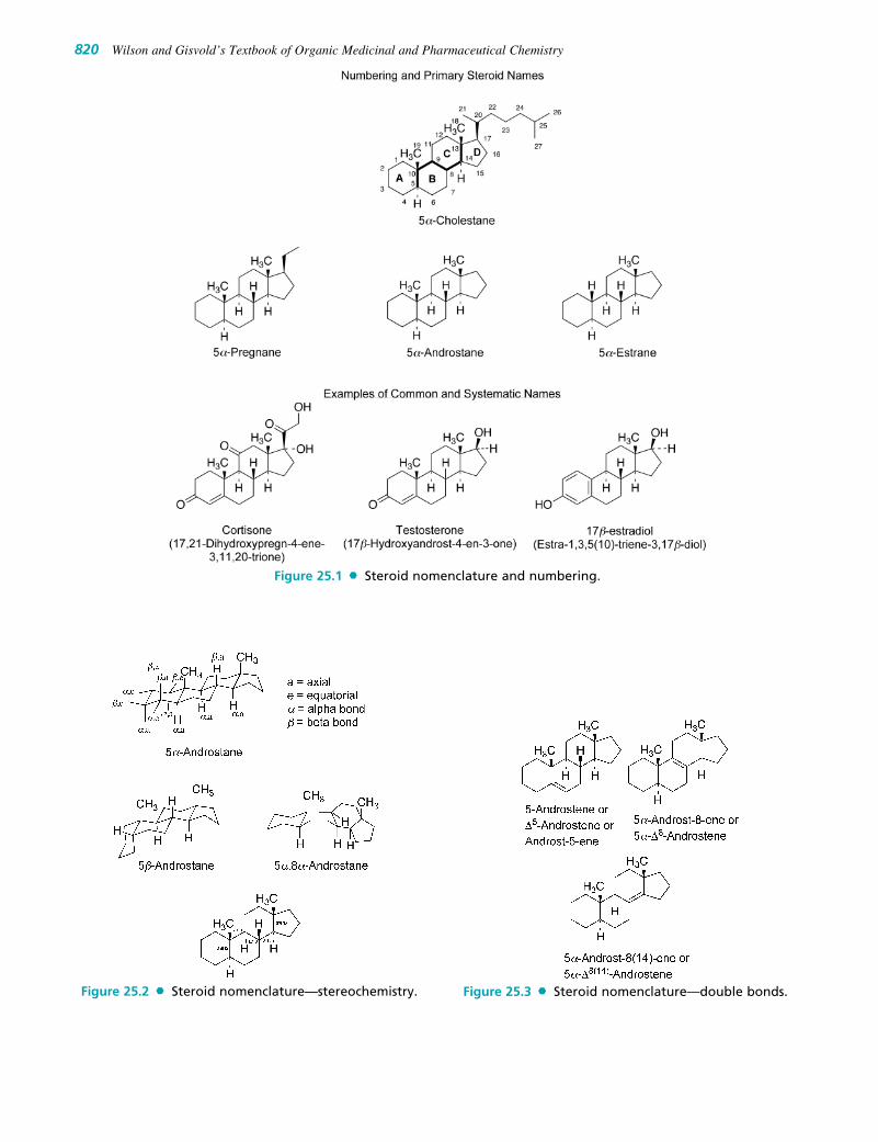
820 Wilson and Gisvold’s Textbook of Organic Medicinal and Pharmaceutical Chemistry
Figure 25.1 Steroid nomenclature and numbering.
Figure 25.3 Steroid nomenclature—double bonds.Figure 25.2 Steroid nomenclature—stereochemistry.
0819-0867_17865_Ch25.qxd 12/2/09 4:43 AM Page 820

Chapter 25 Steroid Hormones and Therapeutically Related Compounds 821
Figure 25.4 Alternative representations of steroids.
Figure 25.5 Outline ofthe biosynthesis of steroidhormones. (3�-HSD, 3�-hydroxysteroiddehydrogenase/�5-4-isomerase;17�-HSD, 17�-hydroxysteroiddehydrogenase.)
0819-0867_17865_Ch25.qxd 12/2/09 4:43 AM Page 821
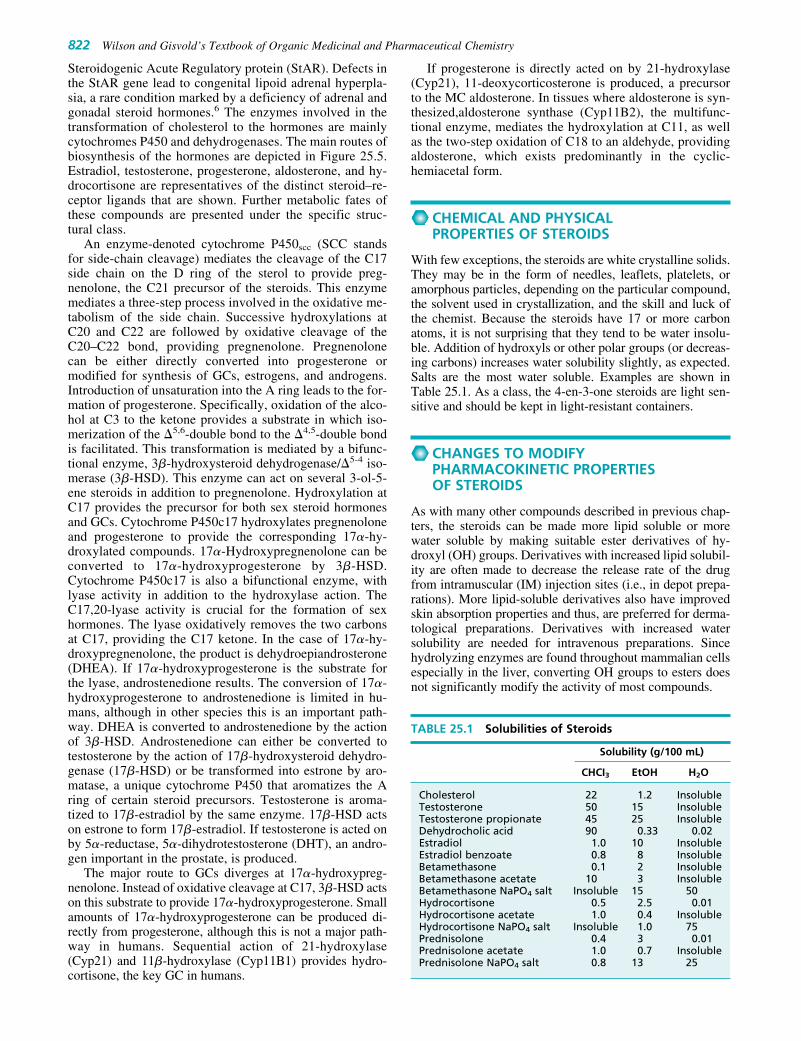
Steroidogenic Acute Regulatory protein (StAR). Defects inthe StAR gene lead to congenital lipoid adrenal hyperpla-sia, a rare condition marked by a deficiency of adrenal andgonadal steroid hormones.6 The enzymes involved in thetransformation of cholesterol to the hormones are mainlycytochromes P450 and dehydrogenases. The main routes ofbiosynthesis of the hormones are depicted in Figure 25.5.Estradiol, testosterone, progesterone, aldosterone, and hy-drocortisone are representatives of the distinct steroid–re-ceptor ligands that are shown. Further metabolic fates ofthese compounds are presented under the specific struc-tural class.
An enzyme-denoted cytochrome P450scc (SCC standsfor side-chain cleavage) mediates the cleavage of the C17side chain on the D ring of the sterol to provide preg-nenolone, the C21 precursor of the steroids. This enzymemediates a three-step process involved in the oxidative me-tabolism of the side chain. Successive hydroxylations atC20 and C22 are followed by oxidative cleavage of theC20–C22 bond, providing pregnenolone. Pregnenolonecan be either directly converted into progesterone ormodified for synthesis of GCs, estrogens, and androgens.Introduction of unsaturation into the A ring leads to the for-mation of progesterone. Specifically, oxidation of the alco-hol at C3 to the ketone provides a substrate in which iso-merization of the �5,6-double bond to the �4,5-double bondis facilitated. This transformation is mediated by a bifunc-tional enzyme, 3�-hydroxysteroid dehydrogenase/�5-4 iso-merase (3�-HSD). This enzyme can act on several 3-ol-5-ene steroids in addition to pregnenolone. Hydroxylation atC17 provides the precursor for both sex steroid hormonesand GCs. Cytochrome P450c17 hydroxylates pregnenoloneand progesterone to provide the corresponding 17�-hy-droxylated compounds. 17�-Hydroxypregnenolone can beconverted to 17�-hydroxyprogesterone by 3�-HSD.Cytochrome P450c17 is also a bifunctional enzyme, withlyase activity in addition to the hydroxylase action. TheC17,20-lyase activity is crucial for the formation of sexhormones. The lyase oxidatively removes the two carbonsat C17, providing the C17 ketone. In the case of 17�-hy-droxypregnenolone, the product is dehydroepiandrosterone(DHEA). If 17�-hydroxyprogesterone is the substrate forthe lyase, androstenedione results. The conversion of 17�-hydroxyprogesterone to androstenedione is limited in hu-mans, although in other species this is an important path-way. DHEA is converted to androstenedione by the actionof 3�-HSD. Androstenedione can either be converted totestosterone by the action of 17�-hydroxysteroid dehydro-genase (17�-HSD) or be transformed into estrone by aro-matase, a unique cytochrome P450 that aromatizes the Aring of certain steroid precursors. Testosterone is aroma-tized to 17�-estradiol by the same enzyme. 17�-HSD actson estrone to form 17�-estradiol. If testosterone is acted onby 5�-reductase, 5�-dihydrotestosterone (DHT), an andro-gen important in the prostate, is produced.
The major route to GCs diverges at 17�-hydroxypreg-nenolone. Instead of oxidative cleavage at C17, 3�-HSD actson this substrate to provide 17�-hydroxyprogesterone. Smallamounts of 17�-hydroxyprogesterone can be produced di-rectly from progesterone, although this is not a major path-way in humans. Sequential action of 21-hydroxylase(Cyp21) and 11�-hydroxylase (Cyp11B1) provides hydro-cortisone, the key GC in humans.
If progesterone is directly acted on by 21-hydroxylase(Cyp21), 11-deoxycorticosterone is produced, a precursorto the MC aldosterone. In tissues where aldosterone is syn-thesized,aldosterone synthase (Cyp11B2), the multifunc-tional enzyme, mediates the hydroxylation at C11, as wellas the two-step oxidation of C18 to an aldehyde, providingaldosterone, which exists predominantly in the cyclic-hemiacetal form.
CHEMICAL AND PHYSICALPROPERTIES OF STEROIDS
With few exceptions, the steroids are white crystalline solids.They may be in the form of needles, leaflets, platelets, oramorphous particles, depending on the particular compound,the solvent used in crystallization, and the skill and luck ofthe chemist. Because the steroids have 17 or more carbonatoms, it is not surprising that they tend to be water insolu-ble. Addition of hydroxyls or other polar groups (or decreas-ing carbons) increases water solubility slightly, as expected.Salts are the most water soluble. Examples are shown inTable 25.1. As a class, the 4-en-3-one steroids are light sen-sitive and should be kept in light-resistant containers.
CHANGES TO MODIFYPHARMACOKINETIC PROPERTIES OF STEROIDS
As with many other compounds described in previous chap-ters, the steroids can be made more lipid soluble or morewater soluble by making suitable ester derivatives of hy-droxyl (OH) groups. Derivatives with increased lipid solubil-ity are often made to decrease the release rate of the drugfrom intramuscular (IM) injection sites (i.e., in depot prepa-rations). More lipid-soluble derivatives also have improvedskin absorption properties and thus, are preferred for derma-tological preparations. Derivatives with increased watersolubility are needed for intravenous preparations. Sincehydrolyzing enzymes are found throughout mammalian cellsespecially in the liver, converting OH groups to esters doesnot significantly modify the activity of most compounds.
822 Wilson and Gisvold’s Textbook of Organic Medicinal and Pharmaceutical Chemistry
TABLE 25.1 Solubilities of Steroids
Solubility (g/100 mL)
CHCI3 EtOH H2O
Cholesterol 22 1.2 InsolubleTestosterone 50 15 InsolubleTestosterone propionate 45 25 InsolubleDehydrocholic acid 90 0.33 0.02Estradiol 1.0 10 InsolubleEstradiol benzoate 0.8 8 InsolubleBetamethasone 0.1 2 InsolubleBetamethasone acetate 10 3 InsolubleBetamethasone NaPO4 salt Insoluble 15 50Hydrocortisone 0.5 2.5 0.01Hydrocortisone acetate 1.0 0.4 InsolubleHydrocortisone NaPO4 salt Insoluble 1.0 75Prednisolone 0.4 3 0.01Prednisolone acetate 1.0 0.7 InsolublePrednisolone NaPO4 salt 0.8 13 25
0819-0867_17865_Ch25.qxd 12/2/09 4:43 AM Page 822

Some steroids (e.g., estradiol, progesterone, and testos-terone) are particularly susceptible to rapid metabolism afterabsorption or rapid inactivation in the gastrointestinal tractbefore absorption. These inactivation processes limit the ef-fectiveness of these hormones as orally available drugs, al-though micronized forms of estradiol and progesterone areavailable for oral administration. Sometimes, a simple chem-ical modification can decrease the rate of inactivation and,thereby, increase the drug’s half-life or make it possible to betaken orally.
Examples of common chemical modifications are il-lustrated in Figure 25.6. Drugs such as testosterone cy-clopentylpropionate (cypionate) and methylprednisolone
sodium succinate are prodrugs that require hydrolysis to re-lease the active hormone in the body.
STEROID HORMONE RECEPTORS
Steroid hormones regulate tissue-specific gene expression.The individual hormones exhibit remarkable tissue selec-tivity, even though their structural differences are relativelyminor. Estrogens such as estradiol increase uterine cell pro-liferation, for example, but not prostate cell proliferation.Androgens such as testosterone do the reverse, but neitherandrogens nor estrogens affect stomach epithelium. The
Chapter 25 Steroid Hormones and Therapeutically Related Compounds 823
Figure 25.6 Common steroid modifications to alter therapeutic utility. (*, prodrug.)
0819-0867_17865_Ch25.qxd 12/2/09 4:43 AM Page 823

basis for this selectivity is the presence of selective steroidhormone receptors in individual tissues. This section pro-vides an overview of steroid hormone receptors and theirmode of action.1,2
The steroid receptors themselves are key players in geneexpression, but many other proteins are involved in thisprocess. Chaperone proteins, for example, help fold the recep-tor proteins into the proper three-dimensional shape for bind-ing the steroid ligand. Together, the steroid hormone receptorand associated proteins (Fig. 25.7; details on next page) makeup the mature-receptor complex. Transcription (or repressionof transcription) occurs when all the necessary associated pro-teins have been recruited to the DNA–receptor complex. Inaddition to the major effects on transcription, some effects ofsteroid hormones are mediated via rapid, nongenomic mech-
anisms. Some of these actions appear to involve classicalsteroid hormone receptors, whereas others rely on novelmembrane receptors.7–9 This is an active area of study andwill help explain all the various actions of steroid hormones.
Structure of SteroidHormone Receptors
The complementary DNAs (cDNAs) of all the major steroidhormone receptors have been cloned, giving the completeamino acid sequence of each. Although the whole three-dimensional structure of a steroid hormone receptor has notbeen solved (structures of the ligand-binding domains of thesteroid hormone receptors have been elucidated; see nextpage in this section), the functional role of each part is wellknown (see Fig. 25.8).10 The organization of the domainsfor all types of steroid hormone receptors is the same, butthe number of amino acids for each receptor varies:
1. N-terminal (A/B) domain. Once the steroid–receptor com-plex has bound to the target gene(s), this domain (alsocalled the A/B modulator domain) activates the hormoneresponse elements adjacent to the genes. The hormoneresponse elements are on the DNA adjacent to the targetgene. They contain about 12 to 18 base-pair DNA se-quences and consist of two “half sites” that are separated bya variable spacer. In the nucleus, steroid hormone–receptor
824 Wilson and Gisvold’s Textbook of Organic Medicinal and Pharmaceutical Chemistry
HSP
CA
AT
Gene Transcription
HSP
CC
SteroidHormone
Figure 25.7 Generic structural model of a steroidhormone–receptor complex and its activation for genetranscription. (AT, histone acetyltransferase; CA, coacti-vator; CC, cochaperone; HSP, heat shock protein; SHR,steroid hormone receptor.)
PRA
165 567 632 681 933
� A/B domainTranscriptionalactivation
� D domainHinge region andnuclear localization
� C domainDNA binding
� E domainLigand-bindingdomain andtranscriptional activation
PRB
1 567 632 681 933
AR
1 559 624 690 919
MR
1 603 668 732 984
GR
1 421 486 527 777
ER�
1 185 250 310 595
ER�
1 149 214 248 530
Figure 25.8 Functional domains of the steroidhormone receptors. (AR, androgen receptor; ER, estro-gen receptor; GR, glucocorticoid receptor; MR, mineralo-corticoid receptor; and PR, progesterone receptor.)
0819-0867_17865_Ch25.qxd 12/2/09 4:43 AM Page 824

complexes exist as dimers; the dimeric structure allows ac-cess to both half sites. The nucleotide sequence and spac-ing between the half sites are essential for the specificity ofthe various steroid–hormone complexes. After the dimerbinds and all accessory proteins have been recruited to thereceptor–DNA complex, transcription is initiated.
2. DNA-binding (C) domain. This short section is made upof about 65 amino acids, organized into two zinc fingermotifs that are important for recognition and binding tothe DNA response elements. The zinc fingers are also re-sponsible for dimerization of the receptor.
3. Hinge (D) domain. This variable linker region appears to beinvolved with nuclear localization and transport (transloca-tion) of the steroid–receptor complex into the nucleus.
4. C-terminal ligand-binding (E) domain (LBD). The C-terminal domain includes about 250 amino acids. Thissection has the steroid hormone–binding site and is alsoinvolved with ligand-dependent transcriptional activa-tion, receptor dimerization, binding to chaperone proteins(discussed later), and in some cases, repressing (“silenc-ing”) particular genes.
ESTROGEN RECEPTORS
There are two distinct estrogen receptors (ERs), estrogen re-ceptor � (ER�) and estrogen receptor � (ER�), which areencoded by different genes.11 The ERs have distinct tissuedistributions and can have distinct actions on the targetgenes. ER� can be found in high abundance in the uterus,vagina, and ovaries, as well as in the breast, the hypothala-mus, endothelial cells, and vascular smooth muscle. ER� isfound in greatest abundance in the ovaries and the prostate,with reduced occurrence in the lungs, brain, and vascula-ture.12 Although many ligands bind with similar affinities toboth receptor subtypes, some ligands are selective for one orthe other receptor.13–15
PROGESTERONE RECEPTORS
The progesterone receptor (PR) can also be found in twoforms, but these are derived from a single gene. PRA has had164 amino acids truncated from the N-terminus of PRB, pro-viding a receptor that has different interactions with targetgenes and associated proteins. PRB mainly mediates the stim-ulatory actions of progesterone. PRA acts as a transcriptionalinhibitor of ER, androgen receptor (AR), glucocorticoid recep-tor (GR), mineralocorticoid receptor (MR), and PRB.12 Thesedifferential actions are believed to be a result of interactionswith different coactivators and corepressors. The DNA- andligand-binding domains for the two receptors are identical.
ANDROGEN, GLUCOCORTICOID, ANDMINERALOCORTICOID RECEPTORS
The AR, GR, and MR are present in only a single form. Onlyone gene and one protein are known for each receptor. Mutantforms of AR16 and GR17 are known, and evidence is mount-ing that some of these mutant receptors are associated withdisease states.
X-RAY CRYSTALLOGRAPHY AND STEROID FIT ATTHE RECEPTOR
The x-ray structures have now been solved for the ligand-binding domains of all the steroid hormone receptors (ER�
and ER�,18–20 PR,21 AR,22 GR,23,24 and MR25). The x-raycrystal structures of the ER, PR, and AR have revealed a keydifference that leads to the unique ligand specificity of theERs.19,26 In the region of the ligand-binding domain, wherethe A ring of steroids binds, are key residues that bind to ei-ther the phenolic A ring of estrogens or the enone A ring ofprogesterone or testosterone. In the case of the ER, gluta-mate and arginine residues are important in a hydrogen-bonding network that involves the phenolic hydroxyl. Incontrast to this structural arrangement, the PR and AR haveglutamine and arginine residues that hydrogen bond to theA-ring enones of progesterone and testosterone. The changefrom glutamate, a hydrogen-bond acceptor, to glutamine, ahydrogen-bond donor, is critical for the discrimination be-tween estrogens and other steroid hormones. The structuresof the GR and MR LBDs also reveal the features that pro-vide the specificities for these two receptors. In the LBD ofthe GR, key proline and glutamine residues are replaced byserine and leucine, respectively, in the MR.25 The prolineresidue results in a slightly more open-binding pocket,which can accommodate larger residues at the 17�-position,as seen in many GR ligands, and the glutamine side chaincan hydrogen bond with the 17�-hydroxyl, which is seen inGRs, but is absent in aldosterone.
The x-ray crystal structures of the steroid hormonesthemselves have also provided important information.Although the conformations of rigid molecules in crystalsand their preferred conformations in solution with receptorscan differ, it is now clear from x-ray crystallography studiesof steroids, prostaglandins, thyroid compounds, and manyother drug classes that this technique can be a powerful toolin understanding drug action and in designing newdrugs.27–29 The relationship is straightforward: steroid drugsusually do not have a charge and, as a result, are held to theirreceptors by relatively weak forces of attraction. The sameis true for steroid molecules as they “pack” into crystals. Inboth events, the binding energy is too small to hold any butlow-energy conformations. In short, the steroid conforma-tion observed in steroid crystals is often the same or verysimilar to that at the receptor.
Structure of SteroidHormone–Receptor Complexes
Steroid hormone–receptor complexes include the steroidhormone receptor as well as other proteins, predominantlychaperone (heat shock) proteins, cochaperones, and im-munophilins (Fig. 25.7).30,31 Their role is to “chaperone” thecorrect conformation and folding of complex proteins,which is otherwise much more difficult as temperatures in-crease. At normal physiological temperatures, the chaperoneproteins assist the proper folding of large proteins such assteroid hormone receptors. The individual components varydepending on the type of steroid hormone receptor. Withoutthe chaperones, the steroid hormone-binding site on the re-ceptor does not have the proper folding and conformationfor optimal steroid binding.
Once the steroid hormone binds to the receptor, a con-formational change of the receptor occurs, and the maturereceptor complex dissociates (Fig. 25.7). The receptor isdimerized, phosphorylated, and transported into the nu-cleus, if necessary. There, the zinc fingers on the steroidhormone receptor bind to the target gene(s) in the DNA.
Chapter 25 Steroid Hormones and Therapeutically Related Compounds 825
0819-0867_17865_Ch25.qxd 12/2/09 4:43 AM Page 825

Additional proteins are recruited to the receptor–DNAcomplex prior to initiation or repression of transcription.32
These additional proteins include coactivators or corepres-sors and histone acetyltransferases. Typically, the recep-tor–DNA–coactivator complex displays histone acetyl-transferase action, which relaxes the chromatin structure,allowing binding of RNA polymerase II and the subsequentinitiation of transcription. If corepressors are recruited tothe complex, deacetylation of the histone complex is facil-itated, preventing transcription.
GONADOTROPIN-RELEASINGHORMONE AND GONADOTROPINS
The gonadotropins are peptides that have a close functionalrelationship to estrogen, progesterone, and testosterone.They are called gonadotropins because of their actions onthe gonads. As shown in Figures 25.9 to 25.11, they controlovulation, spermatogenesis, and development of sex organs,and they maintain pregnancy. An additional peptide, go-nadotropin-releasing hormone (GnRH), regulates release ofthe gonadotropins. Included in this group are the following:
• GnRH• Luteinizing hormone (LH)• Follicle-stimulating hormone (FSH)• Chorionic gonadotropin (CG; hCG is human chorionic go-
nadotropin), a glycopeptide produced by the placenta; itspharmacological actions are essentially the same as thoseof LH
Gonadotropin-Releasing Hormone
The hypothalamus releases GnRH, a peptide that stimulatesthe anterior pituitary to secrete LH and FSH in males andfemales. This peptide controls and regulates both male andfemale reproduction (Figs. 25.9 and 25.10). GnRH is a mod-ified decapeptide (10 amino acids): PyroGlu-His-Trp-Ser-Tyr-Gly-Leu-Arg-Pro-Gly-NH2. The pyroglutamate at theN-terminus and the C-terminal amide distinguish this pep-
tide from unmodified decapeptides. Analogs of GnRH thatare used therapeutically are covered in Chapter 20.
Pituitary Gonadotropins:Luteinizing Hormone and Follicle-Stimulating Hormone
The pituitary gonadotropins LH and FSH, their structures,genes, receptors, biological roles, and their regulation (in-cluding by negative feedback actions of steroid hormones)have been studied intensively.33,34FSH, LH, and CG are allglycopeptide dimers with the same � subunit but different�-subunits.
In females, LH and FSH regulate the menstrual cycle (seeFigs. 25.9 and 25.11). At the start of the cycle, plasma con-centrations of estradiol and other estrogens (Fig. 25.11) andprogesterone are low. FSH and LH stimulate several ovarianfollicles to enlarge and begin developing more rapidly thanothers. After a few days, only one follicle continues develop-ing to the release of a mature ovum. The granulosa cells of thematuring follicles begin secreting estrogens, which then causethe uterine endometrium to thicken. Vaginal and cervicalsecretions increase. Gonadotropins and estrogen reach their
826 Wilson and Gisvold’s Textbook of Organic Medicinal and Pharmaceutical Chemistry
Figure 25.9 Regulation of ovulation by GnRH.
Figure 25.10 Regulation of spermatogenesis.
0
mlU
/mL
Days14 211 7 28
20
40
60
80
LH
Gonadotropins
FSH
0
pg/m
L
200
400 Estradiol
0
ng/m
L
10
20 Progesterone
Figure 25.11 Hormone changes in the normalmenstrual cycle.
0819-0867_17865_Ch25.qxd 12/2/09 4:43 AM Page 826

maximum plasma concentrations at about day 14 of the cycle.The release in LH causes the follicle to break open, releasinga mature ovum. Under the stimulation of LH, the folliclechanges into the corpus luteum, which begins secreting pro-gesterone as well as estrogen.
The increased concentrations of estrogens and proges-terone regulate the hypothalamus and the anterior pituitaryby a feedback inhibition process that decreases GnRH, LH,and FSH production. The result is that further ovulation isinhibited. As described later in this chapter, this is the pri-mary mechanism by which steroid birth control products in-hibit ovulation.
If fertilization does not occur by about day 25, the corpusluteum begins to degenerate, slowing down its production ofhormones. The concentrations of estrogens and progesteronebecome too low to maintain the vascularization of the en-dometrium, and menstruation results.
The pharmacological actions of hCG are essentially thesame as those of LH. In females during pregnancy, the hCGsecreted by the placenta maintains the corpus luteum to con-tinue secretion of estrogen and progesterone, thus inhibitingovulation and menstruation.
In males (Fig. 25.10), LH stimulates testosterone synthe-sis by the testes, and together, testosterone and LH promotespermatogenesis (sperm production) and development ofthe testes. Testosterone is also essential for the develop-ment of secondary sex characteristics in males. FSH stimu-lates production of proteins and nutrients required forsperm maturation.
SEX HORMONES
Although estrogens and progesterone are usually called fe-male sex hormones and testosterone is called a male sexhormone, all of these steroids are biosynthesized in bothmales and females. For example, examination of thebiosynthetic pathway in Figure 25.5 reveals that proges-terone serves as a biosynthetic precursor to hydrocortisoneand aldosterone and, to a lesser extent, to testosterone andestrogens. Testosterone is one of the precursors of estro-gens. Estrogens and progesterone are produced in muchlarger amounts in females, however, as is testosterone inmales. These hormones play profound roles in reproduc-tion, in the menstrual cycle, and in giving women and mentheir characteristic physical differences.
Several modified steroidal compounds, as well as somenonsteroidal compounds, have estrogenic activity. A largenumber of synthetic or semisynthetic steroids with biologi-cal activities similar to those of progesterone have beenmade, and these are commonly called progestins. Althoughthe estrogens and progestins have had their most extensiveuse as chemical contraceptive agents for women and inHRT, their wide spectrum of activity has given them a di-versity of therapeutic uses in women, as well as a few usesin men.
Testosterone has two primary kinds of activities: andro-genic (promoting male physical characteristics) and anabolic(muscle building). Many synthetic and semisynthetic an-drogenic and anabolic steroids have been prepared. Despiteefforts to prepare selective anabolic agents (e.g., for usein aiding recovery from debilitating illness or surgery), all“anabolic” steroids have androgenic effects. The androgenic
agents are mainly used in males, but they do have sometherapeutic usefulness in women (e.g., in the palliation ofcertain sex organ cancers).
In summary, although many sex hormone products havetheir greatest therapeutic uses in either women or men, nearlyall have some uses in both sexes. Nevertheless, the higherconcentrations of estrogens and progesterone in women andof testosterone in men cause the development of the comple-mentary reproductive systems and characteristic physical dif-ferences of women and men.
Estrogens35
ENDOGENOUS ESTROGENS
The active endogenous estrogens are estradiol, estrone, andestriol. Estradiol provides the greatest estrogenic activity,with less activity for estrone, and the least activity with es-triol. This range of activity parallels the affinity of theseestrogens for the ER,36 but the in vivo activities of thesecompounds are also affected by interconversions betweenactive and inactive metabolites.
BIOSYNTHESIS
The estrogens are synthesized by the action of the enzymearomatase on androstenedione or testosterone (Fig. 25.5).They are normally produced in relatively large quantitiesin the ovaries and the placenta, in lower amounts in theadrenal glands, and in trace quantities in the testes. In post-menopausal women, most estrogens are synthesized in adi-pose tissue and other nonovarian sites. About 50 to 350 �g/dof estradiol are produced by the ovaries (especially the corpusluteum) during the menstrual cycle. During the first months ofpregnancy, the corpus luteum produces larger amounts ofestradiol and other estrogens; the placenta produces most ofthe circulating hormone in late pregnancy. During pregnancy,the estrogen blood levels are up to 1,000 times higher thanduring the menstrual cycle.
METABOLISM OF ESTROGENS
The metabolism of natural estrogens has been reviewed indetail.37 The three primary estrogens in women are 17�-estradiol, estrone, and estriol (16�,17�-estriol). Although17�-estradiol is produced in the greatest amounts, it isquickly oxidized (see Fig. 25.12) to estrone, the estrogenfound in highest concentration in the plasma. Estrone, inturn, is converted to estriol, the major estrogen found inhuman urine, by hydroxylation at C16 (to provide the 16�-hydroxyl) and reduction of the C17 ketone (17�-hydroxyl).Estradiol can also be directly converted to estriol. In thehuman placenta, the most abundant estrogen synthesized isestriol. In both pregnant and nonpregnant women, however,the three primary estrogens are also metabolized to smallamounts of other derivatives (e.g., 2-hydroxyestrone, 2-methoxyestrone, 4-hydroxyestrone, and 16�-hydroxy-17�-estradiol). Only about 50% of therapeutically administeredestrogens (and their various metabolites) are excreted in theurine during the first 24 hours. The remainder are excretedinto the bile and reabsorbed; consequently, several days arerequired for complete excretion of a given dose.
Conjugation appears to be very important in estro-gen transport and metabolism. Although the estrogens areunconjugated in the ovaries, significant amounts of conju-
Chapter 25 Steroid Hormones and Therapeutically Related Compounds 827
0819-0867_17865_Ch25.qxd 12/2/09 4:43 AM Page 827

gated estrogens may predominate in the plasma and othertissues. Most of the conjugation takes place in the liver. Theprimary estrogen conjugates found in plasma and urine areglucuronides and sulfates. As sodium salts, they are quitewater soluble. The sodium glucuronide of estriol and thesodium sulfate ester of estrone are shown in Figure 25.12.
BIOLOGICAL ACTIVITIES OF ESTROGENS12
In addition to having important roles in the menstrual cycle(described previously), the estrogens and, to a lesser extent,progesterone are largely responsible for the development ofsecondary sex characteristics in women at puberty. The es-trogens cause a proliferation of the breast ductile system,and progesterone stimulates development of the alveolarsystem. The estrogens also stimulate the development oflipid and other tissues that contribute to breast shape andfunction. Pituitary hormones and other hormones are also in-volved. Fluid retention in the breasts during the later stagesof the menstrual cycle is a common effect of the estrogens.
The estrogens directly stimulate the growth and develop-ment of the vagina, uterus, and fallopian tubes and, in com-bination with other hormones, play a primary role in sexualarousal and in producing the body contours of the maturewoman. Pigmentation of the nipples and genital tissues andgrowth stimulation of pubic and underarm hair (possiblywith the help of small amounts of testosterone) are other re-sults of estrogen action.
The physiological changes at menopause emphasize theimportant roles of estrogens in a young woman. Breastand reproductive tissues atrophy, the skin loses some of its
suppleness, coronary atherosclerosis and gout become po-tential health problems for the first time, and the bonesbegin to lose density because of decreased mineral content.
STRUCTURAL CLASSES: ESTROGENS
As shown in Figure 25.13, there are three structural classesof estrogens: steroidal estrogens, diethylstilbestrol and othersynthetic compounds, and phytoestrogens (Each class issummarized in the sections that follow). The steroidal es-trogens include the naturally occurring estrogens found inhumans and other mammals, as well as semisynthetic deriv-atives of these compounds. Because of rapid metabolism,estradiol itself has poor oral bioavailability. The addition ofa 17�-alkyl group to the estradiol structure blocks oxidationto estrone. Ethinyl estradiol is therefore very effective orally,whereas estradiol itself is not. Most of the therapeuticallyuseful steroidal estrogens are produced semisyntheticallyfrom natural precursors such as diosgenin, a plant sterol.
Steroidal Estrogens—Conjugated Estrogens, EsterifiedEstrogens. Conjugated estrogens (sometimes called equineestrogens) are estradiol-related metabolites originally ob-tained from the urine of horses, especially pregnant mares.Premarin, the major conjugated estrogen product on the mar-ket, is a mixture of numerous components that is obtainedfrom mare urine. Equine estrogens are largely mixtures of es-trone sodium sulfate and equilin sodium sulfate. Little or noequilin and equilenin are produced in humans. The sulfategroups must be removed metabolically to release the activeestrogens. Conjugated estrogens that derive exclusively from
828 Wilson and Gisvold’s Textbook of Organic Medicinal and Pharmaceutical Chemistry
Figure 25.12 Metabolites of 17�-estradiol and estrone.
0819-0867_17865_Ch25.qxd 12/2/09 4:43 AM Page 828

Chapter 25 Steroid Hormones and Therapeutically Related Compounds 829
plant precursors are also on the market. Esterified estrogensare also mainly a combination of estrone sodium sulfate andequilin sodium sulfate, but in a different ratio to that in con-jugated estrogens. The esterified estrogens are now preparedexclusively from plant sterols.
Diethylstilbestrol. At first glance, it might be surprisingthat synthetic nonsteroidal molecules such as diethylstilbes-trol (DES) could have the same activity as estradiol or otherestrogens. DES can be viewed, however, as a form of estra-diol with rings B and C open and a six-carbon ring D. The ac-tivity of DES analogs was explained in 1946.38 It was pro-posed that the distance between the two DES phenol OHgroups was the same as the 3-OH to 17-OH distance of estra-diol; therefore, they could both fit the same receptor.Medicinal chemists have shown the OH-to-OH distance to beactually 12.1 Å in DES and 10.9 Å in estradiol. In aqueous so-lution, however, estradiol has two water molecules that arehydrogen bonded to the 17-OH. If one of the two water mol-
ecules is included in the distance measurement, there is a per-fect fit with the two OH groups of DES (Fig. 25.14). This sug-gests that water may have an important role for estradiol in itsreceptor site. It is now generally accepted that the estrogensmust have a phenolic moiety for binding, but some investiga-tors propose that the receptor may be flexible enough to ac-commodate varying distances between the two key hydrox-yls. This point about estrogens needing a phenolic ring forhigh-affinity binding to the ER is critical. Steroids with a phe-nolic A ring and related phenolic compounds lack high-affin-ity binding to the other steroid hormone receptors. AlthoughDES was used for many years, it was discovered that thedaughters of women who had taken DES during pregnancy(DES babies) had a high risk of vaginal, cervical, and uterineabnormalities, along with a low risk of vaginal clear cell ade-nocarcinoma.39 Because of the safety concerns associatedwith DES, it was completely removed from the U.S. marketin the late 1990s. DES is still available as an estrogen for usein veterinary medicine, however.
Figure 25.13 Natural and synthetic estrogens. (continues on next page)
0819-0867_17865_Ch25.qxd 12/2/09 4:43 AM Page 829

Another synthetic compound, bisphenol A, shares somestructural features of DES, in that it has two phenolicgroups, but in this case only a single dimethylated carbonseparates the two rings, rather than the substituted doublebond in DES. As might be expected, the reduced distancebetween the phenolic hydroxyls and the greater flexibilityin the structure lead to an affinity for the ERs several ordersof magnitude less than estradiol or DES.40 Although bisphe-nol A is only weakly estrogenic, it is still considered tobe an “environmental estrogen” and is present in smallquantities in a wide range of plastic products. The low-levelexposure of humans to bisphenol A, although currently con-sidered to have minimal health risks, continues to be thefocus of numerous studies.41
Phytoestrogens. Several natural plant substances thathave general structural features similar to those of DESand estradiol also have estrogenic effects and have beentermed phytoestrogens.42 These include genistein, fromsoybeans and a species of clover; daidzein, from soybeans;and coumestrol, found in certain legumes. Genistein anddaidzein are examples of isoflavones. These and othershave antifertility activity in animals.43 Many claims havebeen made about the beneficial effects of consuming prod-ucts containing these phytoestrogens, including preventingand treating cardiovascular disease, reducing post-menopausal symptoms, and preventing osteoporosis.Because of the numerous other components present inmany of the commercial products as well as a lack of well-
830 Wilson and Gisvold’s Textbook of Organic Medicinal and Pharmaceutical Chemistry
Figure 25.13 (Continued)
0819-0867_17865_Ch25.qxd 12/2/09 4:43 AM Page 830

designed studies to test the effects of the phytoestrogensthemselves, however, positive health effects specificallybecause of direct hormonal action of the phytoestrogensare uncertain. Questions have also been raised about a pos-sible contribution of phytoestrogens to an increased inci-dence of breast cancer. These concerns, however, are con-tradicted by studies that suggest a chemoprotective role forsoy products containing phytoestrogens (this could well bedue to other components of the mixture).44 The long his-tory of use of soy products in the world and no global cor-relations with increased breast cancer risks suggest thatany connection between phytoestrogens in general andbreast cancer is quite small.45
Recent studies have demonstrated that genistein and otherphytoestrogens binds preferentially to ER� over ER�.46,47
The clinical relevance of this difference in binding is unclear,but further research is being vigorously pursued to under-stand the distinctions between the phytoestrogens and classi-cal estrogens.44
THERAPEUTIC USES OF ESTROGENS
Birth Control. A major use of estrogens is for inhibi-tion of ovulation, in combination with progestins.Steroidal birth control agents containing estrogens are dis-cussed in the section on chemical contraception later inthis chapter.
Hormone Replacement Therapy. Another major useof estrogens is in HRT for postmenopausal women. For thisuse, a progestin is often included to oppose the effects of es-trogens on endometrial tissue. HRT is covered in more depthlater in the chapter.
Treatment of Estrogen Deficiency from OvarianFailure or after Oophorectomy. Estrogen therapy, usu-ally with a progestin, is common in cases of ovarian failureand after an oophorectomy.
Treatment of Advanced, Inoperable Breast Cancer inMen and Postmenopausal Women and of Advanced,Inoperable Prostate Cancer in Men. Estrogens are used totreat inoperable breast cancer in men and in postmenopausalwomen, but estrogen therapy can actually stimulate exist-ing breast cancers in premenopausal women. The selectiveER modulator tamoxifen is reported to have fewer sideeffects; hence, it is usually preferred. Estrogens have alsobeen used to treat inoperable prostate cancer, but GnRHanalogs are now generally preferred because of fewer un-wanted side effects.
Estrogens and Cancer. Many years of study have firmlyestablished an association between estrogen use and increasedrisk of breast cancer. The risk is associated, however, with thetiming of estrogen exposure, the estrogen dose, the length ofuse, and the type of estrogen used.48 A patient should discussthe potential risks of breast cancer with her doctor carefullybefore starting estrogen therapy. Unopposed estrogens inHRT for postmenopausal women are also linked to an in-creased risk of endometrial carcinoma, which is the basis forinclusion of a progestin in many forms of HRT.49
ESTROGEN PRODUCTS
Estrogens are commercially available in a wide variety ofdosage forms: oral tablets, vaginal creams and foams, trans-dermal patches, and IM dosage preparations.
Estradiol, United States Pharmacopeia (USP).Estradiol, estra-1,3,5(10)-triene-3,17�-diol, is the most ac-tive of the natural steroid estrogens. Although its 17�-OHgroup is vulnerable to bacterial and enzymatic oxidation toestrone (Fig. 25.12), it can be temporarily protected as anester at C3 or C17, or permanently protected by adding a17�-alkyl group (e.g., 17�-ethinyl estradiol, the most com-monly used estrogen in oral contraceptives). The increasedoil solubility of the 17�-esters (relative to estradiol) permitsthe esters to remain in oil at the IM injection site for extendedperiods. These derivatives illustrate the principles of steroidmodification shown in Figure 25.6. Transdermal estradiolproducts avoid first-pass metabolism, allowing estradiol tobe as effective as oral estrogens for treating menopausalsymptoms. A new transdermal spray, Evamist, was approvedin 2007. Estradiol itself is typically not very effective orallybecause of rapid metabolism, but an oral formulation of mi-cronized estradiol that allows more rapid absorption of thedrug is available (Estrace). In addition to the oral and trans-dermal products, estradiol is also available in gel, cream, andvaginal ring formulations. The commercially available estra-diol esters are the following:
Estradiol 3-acetate, USP (oral; vaginal ring)Estradiol 17-valerate, USP (IM injection)Estradiol 17-cypionate, USP (IM injection)
Estrone, USP. Estrone, 3-hydroxyestra-1,3,5(10)-trien-17-one, is less active than estradiol but more active than itsmetabolite, estriol. As the salt of its 3-sulfate ester, estroneis the primary ingredient in conjugated estrogens, USP, andesterified estrogens, USP. Although originally obtainedfrom the urine of pregnant mares (about 10 mg/L), estroneis now prepared synthetically. Estrone itself is not availablein commercial oral formulations, but can be obtained at
Chapter 25 Steroid Hormones and Therapeutically Related Compounds 831
H2O
H2O
H2O
H2O
O3�
O3�
O3
O3
Top View
Side View
Estradiol (H2O)2 � dark linesDES � light lines
Figure 25.14 Computer graphics. Superposition ofestradiol (H2O)2 (dark lines) with DES (light lines).(Courtesy of Medical Foundation of Buffalo, Inc.)
0819-0867_17865_Ch25.qxd 12/2/09 4:43 AM Page 831

compounding pharmacies as a topical formulation. Oleoyl-estrone, the C3 ester of estrone with oleic acid, is in phase IIclinical trials for the treatment of obesity. This acyl estronederivative reduces fat stores by a mechanism not involvingthe ER,50 although some of the oleoyl-estrone is hydrolyzedto estrone in vivo.
Piperazine Estrone Sulfate (3-Sulfoxy-Estra-1,3,5(10)-Trien-17-One Piperazine Salt), USPAll the estrone 3-sulfate salts have the obvious pharmaceuti-cal advantage of increased water solubility and better oralavailability. Acids convert the salts to the free 3-sulfate estersand cause some hydrolysis of the ester. This does not seem toaffect absorption adversely, but precipitation of the free sul-fate esters in acidic pharmaceutical preparations should beavoided. The dibasic piperazine molecule acts as a buffer,giving it somewhat greater stability.
Conjugated Estrogens, USPThe term conjugated estrogens refers to the mix of sulfateconjugates of estrogenic components isolated from pregnantmare urine (Premarin). These compounds are also referred toas CEE (conjugated equine estrogens). Conjugated estrogenscontain 50% to 65% sodium estrone sulfate and 20% to 35%sodium equilin sulfate (based on the total estrogen content ofthe product). Premarin also contains the sulfate esters of17�-estradiol, 17�-dihydroequilin, and 17�-dihydroequilin,in addition to other minor components. Although most com-monly used in HRT to treat postmenopausal symptoms, theconjugated estrogens are used for the entire range of indica-tions described previously, except birth control.
Synthetic Conjugated EstrogensCenestin, referred to as “synthetic conjugated estrogens, A”is a mixture of nine estrogenic substances (Fig. 25.13):sodium estrone sulfate, sodium equilin sulfate, sodium equi-lenin sulfate, sodium 17�-estradiol sulfate, sodium 17�-estradiol sulfate, sodium 17�-dihydroequilin sulfate,sodium 17�-dihydroequilin sulfate, sodium 17�-dihydroe-quilenin sulfate, and sodium 17�-dihydroequilenin sulfate.These estrogenic substances are synthesized from soysterols. The term synthetic has been added before conju-gated estrogens to indicate that this product is distinct from(not equivalent to) the conjugated estrogens derived frommare urine. The “A” following the name indicates that thisis the first approved mixture of synthetic conjugated estro-gens. Enjuvia is “synthetic conjugated estrogens, B.”Subsequent synthetic conjugated estrogen products will benamed in order, with the final descriptors “C, D, E,” etc.Enjuvia contains �8,9-dehydroestrone sulfate, in addition tothe nine estrogenic substances listed previously. Cenestinand Enjuvia are both approved for the treatment of moder-ate-to-severe vasomotor symptoms and vulvar and vaginalatrophy associated with menopause.
Esterified Estrogens, USPEsterified estrogens (Menest) contain some of the same sul-fate conjugates of estrogens present in conjugated estrogens,but the ratios of these components and the composition ofminor components of the products differ. These productscontain 75% to 85% sodium estrone sulfate and 6% to 15%sodium equilin sulfate, in such proportion that the total ofthese two components is 90% of the total esterified estro-gens content. The esterified estrogens find the same uses asthe conjugated estrogens.
Estriol, USP. Estriol, estra-1,3,5(10)-triene-3,16�,17�-triol, is available for compounding into several differentformulations for use in HRT. It can be used alone or in com-binations with estradiol (Bi-Est) or with estradiol and es-trone (Tri-Est).
Ethinyl Estradiol, USP. 17�-Ethinyl estradiol has thegreatest advantage over other estradiol products of beingorally active. It is equal to estradiol in potency by injectionbut is 15 to 20 times more orally active. The primary meta-bolic path for ethinyl estradiol is 2-hydroxylation bycytochrome P450 isozyme 3A4 (CYP3A4), followed byconversion to the 2- and 3-methyl ethers by catechol-O-methyltransferase. The 3-methyl ether of ethinyl estradiolis mestranol, USP, used in oral contraceptives. Mestranolis a prodrug that is 3-O-demethylated to the active ethinylestradiol. An oral dose of about 50 �g of mestranol has anestrogenic action approximately equivalent to 35 �g oforal ethinyl estradiol. The demethylation is mainly medi-ated by CYP2C9.51
SELECTIVE ESTROGEN RECEPTOR MODULATORSAND ANTIESTROGENS
Whereas estrogens have been very important in chemicalcontraception and HRT, compounds that can antagonize theER have been of great interest for the treatment of estrogen-dependent breast cancers. Tumor biopsies have shown ER tobe present in about 60% of primary breast cancers, and mostare responsive to estrogen blockade. Unfortunately, most ofthese ER-related breast cancers also develop resistance toantiestrogen therapy within 5 years. In contrast, only about6% of nonmalignant breast tissues have significant ER pres-ent. Three compounds that are used clinically for estrogenantagonist action in the treatment of breast cancer are tamox-ifen, toremifene, and fulvestrant (Fig. 25.15). Two additionalagents that can antagonize ERs are clomiphene, which isused as an ovulation stimulant, and raloxifene, which is usedfor the prevention and treatment of osteoporosis.
Tamoxifen and clomiphene were traditionally called ERantagonists or antiestrogens. Referring to these compoundsas ER antagonists, however, does not accurately portray howthese compounds work in vivo. Although tamoxifen is an ERantagonist in breast tissue, it has agonist actions on en-dometrium, liver, bone, and cardiovascular system. Becauseof the differential agonist and antagonist effects of thesetypes of compounds on the ER, depending on the specific tis-sue, a new term was coined: selective estrogen receptor mod-ulators (SERMs). A SERM is a drug that has tissue-specificestrogenic activity. Although many compounds exhibitSERM activity, a few agents are antagonists in all tissues.These compounds are termed antiestrogens, and fulvestrantis one example (Fig. 25.15). Tamoxifen and clomiphene areoften referred to in older literature as antiestrogens.
Tamoxifen has seen extensive use in treating primarybreast cancers that are ER dependent.52,53 For premenopausalwomen with metastatic disease, tamoxifen is an alternativeand adjuvant with oophorectomy, ovarian irradiation, andmastectomy. Tamoxifen use, however, is not problem free.Tamoxifen increases the incidence of endometrial polyps, hy-perplasia and carcinoma, and uterine sarcomas. The risk ofendometrial cancer resulting from tamoxifen is, however,much lower than the “modest but highly significant reduc-tions in morbidity and mortality of breast cancer.”54 Because
832 Wilson and Gisvold’s Textbook of Organic Medicinal and Pharmaceutical Chemistry
0819-0867_17865_Ch25.qxd 12/2/09 4:43 AM Page 832

of the increased risk of endometrial cancer with tamoxifentherapy, tamoxifen should be used to prevent breast canceronly in women at high risk. Women without a family historyof breast cancer or other risks should not use tamoxifen inthis manner.
Raloxifene is another SERM, but its profile of activitydiffers from that of tamoxifen. Raloxifene is an ER antago-nist in both breast and endometrial tissue, but has agonist ac-tion on bone and acts as an estrogen agonist in lowering totalcholesterol and low-density lipoprotein (LDL). The subtlestructural differences between the two drugs underlie thedistinct activity profiles.55 The agonist action on bone tissueis the basis for the use of this drug for treating osteoporosis.
A key question is why do compounds like tamoxifen andraloxifene exhibit antagonist action in some tissues, but ago-nist action in other tissues? Major developments in the pastfew years are beginning to provide the answer to this ques-tion.15,56–58 Tamoxifen,59 raloxifene, and estradiol18 all bindto the ER at the same site, but their binding modes are differ-ent. In addition, each induces a distinct conformation in thetransactivation region of the ligand-binding domain.18 Theseunique conformations dictate how the receptor–ligand com-plex will interact with coregulator proteins (coactivatorsor corepressors).56 In all tissues, the estradiol–ER complexrecruits coactivators, so gene transcription is stimulated.
In breast tissue, both raloxifene- and tamoxifen-bound re-ceptors prevent the association with coactivators, but ratherrecruit corepressors, so antagonist action is observed. In uter-ine tissue, however, the raloxifene–ER complex recruitscorepressors, whereas the tamoxifen–ER complex recruits acoactivator, SRC-1, which leads to agonist action.57 An ad-ditional factor involved in this agonist action of tamoxifen isthe context in which the liganded receptor interacts with thetarget gene. The liganded ER can interact directly with DNAor can interact indirectly by being tethered to other transcrip-tion factors. When the tamoxifen-bound receptor interacts ina tethered manner with the target genes in uterine tissue, theSRC-1 coactivator is recruited, and gene transcription is pro-moted.56,57 A deeper understanding of how the liganded re-ceptors interact with target genes and the mechanisms forrecruitment of coregulators may help explain the phenome-non of tamoxifen resistance, which results from increased ta-moxifen agonism in breast tissue.56
Bazedoxifene is a new SERM that should soon be mar-keted for the treatment of osteoporosis. Bazedoxifene incor-porates an indole ring system in place of the benzothiopheneof raloxifene. Bazedoxifene was the result of a screening pro-gram that selected for compounds that did not stimulate breastor uterine tissue.60 Lasofoxifene, arzoxifene, and ospemifeneare the other newer SERMs that are in the late stage of
Chapter 25 Steroid Hormones and Therapeutically Related Compounds 833
Figure 25.15 Selective estrogen receptor modulators (SERMs) and antiestrogens.
0819-0867_17865_Ch25.qxd 12/2/09 4:43 AM Page 833
development (Fig. 25.15).61 Lasofoxifene can be viewed as aconstrained, saturated analog of 4-hydroxytamoxifen. A newdrug application (NDA) was filed with the Food and DrugAdministration (FDA) in late 2007 and the review is still inprogress. Structurally, arzoxifene is most similar to ralox-ifene, with an ether bridge, rather than a carbonyl bridge,attached to the benzothiophene core. Arzoxifene is being in-vestigated (phase III) for treating osteoporosis and for pre-venting breast cancer. Ospemifene is an analog of toremifene,which has an OH in place of the N,N-dimethyl amino sub-stituent. It is in phase III clinical trials for the treatment ofvaginal atrophy in postmenopausal women.
Clomiphene is another drug that exhibits antiestrogenactions, but it is not used for treating breast cancer or osteo-porosis; rather, it is used for increasing the odds of a success-ful pregnancy. Clomiphene’s therapeutic application as anovulation stimulant results from its ability to increase GnRHproduction by the hypothalamus. The mechanism is presum-ably a blocking of feedback inhibition of ovary-produced es-trogens (via ER antagonism). The hypothalamus and pitu-itary interpret the false signal that estrogen levels are low andrespond by increasing the production of GnRH. The in-creased GnRH, in turn, leads to increased secretion of LHand FSH, maturation of the ovarian follicle, and ovulation (asdescribed previously in this chapter; see Fig. 25.9). Supportfor the feedback inhibition mechanism is provided by testswith experimental animals in which clomiphene has no ef-fect in the absence of a functioning pituitary gland.
Multiple births occur about 10% of the time with patientstaking clomiphene, and birth defects in 2% to 3% of livenewborns. Vasomotor “hot flashes” occur about 10% of thetime, and abnormal enlargement of the ovaries about 14%.Abdominal discomfort should be discussed immediatelywith the physician.
SERM AND ANTIESTROGEN PRODUCTS
Tamoxifen Citrate, USP. Tamoxifen, 2-[4-(1,2-diphenyl-1-butenyl)phenoxy]-N,N-dimethylethanamine(Nolvadex), is a triphenylethylene SERM used to treatearly and advanced breast carcinoma in postmenopausalwomen. Tamoxifen is used as adjuvant treatment for breastcancer in women following mastectomy and breast irradia-tion. It reduces the occurrence of contralateral breast cancerin patients receiving adjuvant tamoxifen therapy. It is alsoeffective in the treatment of metastatic breast cancer in bothwomen and men. In premenopausal women with metastaticbreast cancer, tamoxifen is an alternative to oophorectomyor ovarian irradiation. Tamoxifen can be used preventa-tively to reduce the incidence of breast cancer in women athigh risk. Antiestrogenic and estrogenic side effects caninclude hot flashes, nausea, vomiting, platelet reduction,and (in patients with bone metastases) hypercalcemia. Likeall triphenylethylene derivatives, it should be protectedfrom light.
The major metabolite of tamoxifen is N-desmethyltamox-ifen, which reaches steady-state levels higher than tamoxifenitself. It is believed that N-desmethyltamoxifen contributessignificantly to the overall antiestrogenic effect. Anothermetabolite, 4-hydroxytamoxifen, is a more potent antiestro-gen than tamoxifen, but because it is only a minor metaboliteof tamoxifen, it probably does not contribute significantly tothe therapeutic effects. 4-Hydroxytamoxifen, with its greater
affinity for the ERs, however, has been used extensively inpharmacological studies of these receptors. Tamoxifen con-centrations are reduced if coadministered with rifampin, a cy-tochrome P450 inducer.
Toremifene Citrate, USP. Toremifene, 2-[4-[(1Z)-4-chloro-1,2-diphenyl-1-butenyl]phenoxy]-N ,N-di-methylethanamine (Fareston), differs structurally fromtamoxifen only by having a chloroethyl group (rather than anethyl group) attached to the triphenylethylene structure. Asmight be expected, the pharmacological actions of toremifeneand tamoxifen are quite similar. Toremifene is also a SERM,with estrogen antagonist action in breast tissue but agonist ac-tion in the endometrium, on bone tissue, and on serum lipidprofiles. Recent clinical data indicate that the incidence ofendometrial cancer is lower with toremifene use than with ta-moxifen.62 Toremifene is used in the treatment of metastaticbreast cancer in postmenopausal women.
Raloxifene, USP. Raloxifene, [6-hydroxy-2-(4-hy-droxyphenyl)benzo[b]thien-3-yl][4-[2-(1-piperidinyl)ethoxy]phenyl]methanone (Evista), is a benzothiophene de-rivative that differs slightly from the triphenylethyleneSERMs. A key structural difference is the carbonyl “hinge”that connects the modified phenolic side chain to the ben-zothiophene ring system. This hinge is the key structural el-ement that leads to the differing actions at the ERs.55
Raloxifene, unlike tamoxifen and toremifene, has antagonistproperties on the endometrium and breast tissue and agonistproperties on bone and the cardiovascular system. The lackof agonist action on endometrial tissue has been suggestedas a reason for the lack of endometrial cancer associatedwith raloxifene use. Raloxifene is approved for the preven-tion and treatment of osteoporosis in postmenopausalwomen. It has also been investigated for preventing breastcancer in comparison with tamoxifen. Recent studies indi-cates that it has similar effectiveness to tamoxifen, but has apreferable side effect profile.63
Bazedoxifene, USP. Bazedoxifene, 1-[[4-[2-(azepan-1-yl)ethoxy]phenyl]methyl]-2-(4-hydroxyphenyl)-3-methyl-indol-5-ol (Viviant), was declared to be “approvable” bythe FDA in 2007 for the prevention of postmenopausalosteoporosis. As of mid-2009, final approval in the UnitedStates is still pending. Bazedoxifene was approved inEurope in 2009 for the treatment of post–menopausalosteoporosis.
Fulvestrant, USP. Fulvestrant, 7�-[9-[(4,4,5,5,5-pentafluoropentyl)sulfinyl]nonyl]estra-1,3,5(10)-triene-3,17�-diol (Faslodex), is an antagonist structurally based onthe estradiol structure, with a long, substituted alkyl chainattached at the 7�-position of the steroid skeleton. Whenbound to the ERs, this alkyl chain induces a conformationof the receptor distinctive from that formed upon estradiolor tamoxifen binding, preventing agonist action.Fulvestrant is a pure antagonist at both ER� and ER� andan ER downregulator (stimulates degradation of the ER),completely lacking the agonist activity that is seen with ta-moxifen or raloxifene. The different pharmacological pro-file of fulvestrant allows the use of this agent in womenwho have had disease progression after prior antiestrogentherapy (typically tamoxifen), providing an alternative toaromatase inhibitors.64
834 Wilson and Gisvold’s Textbook of Organic Medicinal and Pharmaceutical Chemistry
0819-0867_17865_Ch25.qxd 12/2/09 4:43 AM Page 834

Clomiphene Citrate, USP. Clomiphene citrate, 2-[4(2-chloro-1,2-diphenylethenyl)phenoxy]-N ,N-diethyl-ethanamine (Clomid), is used as an ovulation stimulant inwomen desiring pregnancy. Although early literature refersto clomiphene as an estrogen antagonist, it is more accuratelya SERM.65 Clomiphene is chemically a mixture of two geo-metric isomers, zuclomiphene, the cis-isomer, and en-clomiphene, the trans-isomer. In animal studies, these iso-mers have different estrogenic actions in different tissues.Zuclomiphene appears to have weak agonist actions on alltissues studied, whereas enclomiphene has antagonist actionson uterine tissue, but agonist action on bone tissue.66 The ac-tions of clomiphene in humans are likely a composite of theactions of the two isomers.
AROMATASE INHIBITORS
Aromatase is a cytochrome P450 enzyme complex that cat-alyzes the conversion of androstenedione to estrone andtestosterone to estradiol (Figs. 25.5 and 25.16).67–69 Thecomplex is made up of reduced nicotinamide adenine dinu-cleotide phosphate (NADPH)-cytochrome P450 reductase,and cytochrome P450 hemoprotein. In the first two steps, theC19 methyl is hydroxylated to CH2OH, and then to an alde-
hyde hydrate form that dehydrates to provide the C19 alde-hyde. In the final aromatization step, the C19 carbon is ox-idatively cleaved to formate. A hydride shift, proton transfer,and free radical pathways have been proposed, with cis-elimination of the 1� and 2� hydrogens.70 In premenopausalwomen, aromatase is primarily found in ovaries, but in post-menopausal women, aromatase is largely in muscle and adi-pose tissue.
As discussed previously with SERMs, some types ofbreast cancer are estrogen dependent. Because the aro-matase reaction is unique in steroid biosynthetic pathways,it would be anticipated that aromatase inhibitors would bevery specific in their estrogen biosynthesis blockade. Thishas proved to be true, and aromatase inhibitors offer a use-ful approach to decreasing estrogen levels in the treatmentof estrogen-dependent breast cancer. Initially, aromatase in-hibitors were used as second-line therapy in postmenopausalwomen who failed on tamoxifen therapy. Recent studieshave indicated, however, that the newer aromatase in-hibitors can be used as first-line therapy and possibly forcancer prevention in patients at high risk.71
Aromatase inhibitors include both steroidal and non-steroidal compounds. Examples of aromatase inhibitors areshown in Fig. 25.17. The first-generation aromatase inhibitors
Chapter 25 Steroid Hormones and Therapeutically Related Compounds 835
Figure 25.17 Aromatase inhibitors.
Figure 25.16 Conversion of androstenedione toestrone by aromatase.
0819-0867_17865_Ch25.qxd 12/2/09 4:43 AM Page 835

were aminoglutethimide, a nonsteroidal compound, and thesteroid-based testolactone, two compounds that were de-veloped before it was recognized that their effectiveness inbreast cancer treatment was caused by aromatase inhibition.72
Aminoglutethimide also inhibits other P450s involved insteroid hormone biosynthesis, which limits its use in breastcancer treatment. The newer drugs are more potent and morespecific inhibitors of aromatase than the earlier compounds.
Exemestane is a newer steroidal aromatase inhibitor. Itis an enzyme activated irreversible (“suicide”) inhibitor ofaromatase. It is orally available and highly selective foraromatase. Its structure reflects only minor structural mod-ifications to the natural substrate, androstenedione.
The nonsteroidal aromatase inhibitors are competitiveinhibitors that bind to the enzyme active site by coordinat-ing the iron atom present in the heme group of the P450protein. Aside from aminoglutethimide, the first selectivearomatase inhibitor to be marketed in the United States wasanastrozole (Arimidex). Anastrozole incorporates a triazolering into its structure that can coordinate to the heme iron.Letrozole is another triazole-containing inhibitor that is alsoeffective in the treatment of breast cancer.
Currently available inhibitors suppress plasma estrogenlevels (estradiol, estrone, and estrone sulfate) by 80% to 95%.Earlier aromatase inhibitors, such as aminoglutethimide andtestolactone, suppressed plasma estrogens to a lesser extent.The side effects often seen with aminoglutethimide becauseof additional inhibition of other biosynthetic enzymes areavoided with the newest agents. The selectivity of these drugsfor aromatase is quite high.
As with the SERMs, these drugs are only effectivewhen the breast cancer cells are “ER positive,” meaningthat they respond and proliferate in the presence of estro-gen. Aromatase inhibitors can cause fetal harm in pregnantwomen and are therefore contraindicated.
Additional nonsteroidal inhibitors are based on theflavone structure. Chrysin is a flavonoid natural product thathas aromatase inhibitory action in vitro similar to that ofaminoglutethimide.73 It is isolated from Passiflora coeruleaand other plants. While this compound is not used therapeu-tically as an aromatase inhibitor, it has found questionableuse as a nutritional supplement either alone or in combina-tion with anabolic steroids to enhance muscle building andathletic performance. The theory for use is that an aromataseinhibitor allows the estrogenic side effects of androgeniccompounds to be reduced. Although various nutritional sup-plement products containing chrysin are available, it is un-clear whether or not significant aromatase inhibition isbeing achieved in vivo. The oral bioavailability of chrysin isvery low, mainly as a result of efficient conversion to thecorresponding glucuronide and sulfate conjugates, so theplasma concentration of free chrysin is minimal.74
AROMATASE INHIBITOR PRODUCTS
Anastrozole, USP. Anastrozole, �,�,��,��-tetramethyl5-(1H-1,2,4-triazol-1-ylmethyl)-1,3-benzenediacetonitrile,was the first specific aromatase inhibitor approved in theUnited States. It is indicated for first-line treatment of post-menopausal women with advanced or metastatic breast can-cer, for second-line treatment of postmenopausal patientswith advanced breast cancer who have had disease progres-sion following tamoxifen therapy, and for adjuvant treatment
of women with early breast cancer. Patients who did not re-spond to tamoxifen therapy rarely respond to anastrozole.
Anastrozole reduces serum estradiol approximately 80%after 14 days of daily dosing. Because of an eliminationhalf-life of 50 hours, anastrozole is effective with once-dailydosing (1 mg). Metabolism of anastrozole includes hy-droxylation and glucuronidation, as well as N-dealkylationto produce triazole. The metabolites of anastrozole are inac-tive. Although anastrozole can inhibit CYPs 1A2, 2C9, and3A4 with Ki values in the low micromolar range, the con-centrations of anastrozole reached under standard therapeu-tic dosing are much lower, so anastrozole should lack signif-icant P450 interactions.75
Letrozole, USP. Letrozole, 4,4�-(1H-1,2,4-triazol-1-yl-methylene)dibenzonitrile (Femara), is used for most of thesame indications as anastrozole. It reduces concentrations ofestrogens by 75% to 95%, with maximal suppressionachieved within 2 to 3 days. Letrozole is specific for aro-matase inhibition, with no additional effects on adrenal cor-ticoid biosynthesis. CYPs 3A4 and 2A6 are involved in themetabolism of letrozole to the major carbinol metabolite,which is inactive. The loss of the triazole ring, which is in-volved in coordination of the heme iron, would explain theloss of activity. Letrozole strongly inhibits CYP2A6 invitro, with moderate inhibition of CYP2C19. The effect ofthis in vitro inhibition on the pharmacokinetics of coadmin-istered drugs is unknown. Tamoxifen reduces the levels ofletrozole significantly if they are used together, so combina-tion treatment with these agents is not recommended.76
Exemestane, USP. Exemestane, 6-methylenandrosta-1,4-diene-3,17-dione (Aromasin), is the first steroid-basedaromatase inhibitor approved for the treatment of breastcancer in the United States. It is a mechanism-based inacti-vator that irreversibly inhibits the enzyme. Plasma estrogenlevels are reduced by 85% to 95% within 2 to 3 days, and ef-fects last 4 to 5 days. Exemestane does not inhibit any of themajor cytochromes P450 and has essentially no interactionwith steroid receptors, with only a very weak affinity for theAR. The 17�-hydroxyexemestane reduction product, how-ever, has much higher affinity for the AR than the parent(still several fold less than DHT, 0.28% for parent vs. 30%for metabolite). The clinical significance of the affinity islikely minimal because of the low levels of the metaboliteproduced.
Aminoglutethimide, USP. Aminoglutethimide, 3-(4-aminophenyl)-3-ethyl-2,6-piperidinedione, is mainly usedto treat Cushing syndrome, a condition of adrenal steroidexcess, a use in which the P450scc inhibition of thiscompound is exploited rather than its aromatase inhibi-tion. Aminoglutethimide is a weak inhibitor of aromataseand has been used successfully in the treatment of es-trogen-dependent breast cancer. Because of the develop-ment of more selective aromatase inhibitors, the use ofaminoglutethimide for its ability to inhibit aromatase is notsupported.
Testolactone, USP. Testolactone, 13-hydroxy-3-oxo-13,17-secoandrosta-1,4-dien-17-oic acid �-lactone (Teslac),was originally synthesized as a possible anabolic steroid,considering its structural similarity to testosterone. The keystructural difference from anabolic steroids is the D-ring
836 Wilson and Gisvold’s Textbook of Organic Medicinal and Pharmaceutical Chemistry
0819-0867_17865_Ch25.qxd 12/2/09 4:43 AM Page 836

lactone instead of the typical cyclopentyl ring. Althoughconsidered in many texts an androgen or anabolic steroid (itis a Schedule III drug because of its classification as an an-abolic steroid), testolactone lacks androgenic effects in vivo.Its action is believed to be caused by irreversible inhibitionof aromatase. It is a relatively weak inhibitor of aromatase,but the irreversible nature of the inhibition can lead to pro-longed effects. Its relatively weak inhibition of aromataseand its undesirable dosage schedule (5 � 50-mg tabletsq.i.d.) give this older agent only limited use in breast cancertreatment because of better available options.
Progestins
ENDOGENOUS PROGESTINS
The key endogenous steroid hormone that acts at the PRs isprogesterone. All other endogenous steroids lack significantprogestational action.
BIOSYNTHESIS
Progesterone is produced in the ovaries, testes, and adrenalglands. Much of the progesterone that is synthesized frompregnenolone is immediately converted to other hormonalintermediates and is not secreted. See the biosynthetic path-way (Fig. 25.5). The corpus luteum secretes the most pro-gesterone, 20 to 30 mg/d during the last or “luteal” stage ofthe menstrual cycle. Normal men secrete about 1 to 5 mg ofprogesterone daily.
METABOLISM OF PROGESTERONE
Progesterone has a half-life of only about 5 minutes whentaken orally, because of rapid metabolism. Progesterone canbe transformed to many other steroid hormones (Fig. 25.5)and, in that sense, has numerous metabolic products. Theprincipal excretory product of progesterone metabolism,however, is 5�-pregnane-3�,20�-diol and its conjugates(Fig. 25.18). The steps that are involved in the formation of
this metabolite are reduction of the C4-5 double bond, re-duction of the C3 ketone providing the 3�-ol, and reductionof the C20 ketone. The reduction at C5 must precede thereduction of the C3 ketone, but the timing of the C20 canvary, depending on the tissue.77,78 Structural features thatcan block reduction at C5 or C20 have greatly increased thehalf-lives of progesterone derivatives.
BIOLOGICAL ACTIVITIES OF THE PROGESTINS12
Like the estrogens, progesterone has various pharmacologi-cal actions, with the main target tissues being the uterus, thebreast, and the brain. Progesterone decreases the frequencyof the hypothalamic pulse generator and increases the am-plitude of LH pulses released from the pituitary. The actionsof progesterone in the uterus include development of the se-cretory endometrium. When release of progesterone fromthe corpus luteum at the end of the menstrual cycle declines,menstruation begins. Progesterone also acts to thicken cer-vical secretions, decreasing cervical penetration by sperm.Progesterone is critical for the maintenance of pregnancy bysuppressing menstruation and decreasing uterine contractil-ity. Progesterone has important actions in the breasts duringpregnancy, acting in conjunction with estrogens to preparefor lactation.
A thermogenic action is also associated with progesterone.During the menstrual cycle, progesterone mediates a slighttemperature increase near midcycle and maintains the in-creased temperature until the onset of menstruation. The exactmechanism for this temperature increase is not known.Progesterone and its metabolites have additional central ef-fects, which are being actively explored (see “Neurosteroids”section at the end of the chapter).79
STRUCTURAL CLASSES—PROGESTINS
Progestins are compounds with biological activities similarto those of progesterone. They include three structuralclasses: (a) progesterone and derivatives, (b) testosterone and19-nortestosterone derivatives, and (c) miscellaneous syn-thetic progestins (Fig. 25.19). Progesterone itself has loworal bioavailability because of poor absorption and almostcomplete metabolism in one passage through the liver. A re-cently available oral formulation is micronized progesteronein gelatin capsules. The micronized drug is much more read-ily absorbed, allowing oral delivery of progesterone, eventhough the dose must be much higher than a parenteral doseto compensate for extensive liver metabolism. Adding 17�-acyl groups slows metabolism of the 20-one, whereas a 6-methyl group enhances activity and reduces metabolism.Medroxyprogesterone acetate (MPA) is a particularly potentexample (Table 25.2).
The structural requirements of the PR have been studiedin detail.27,28 The progesterone 4-en-3-one ring A is a key tobinding but only when it is in a conformation quite differentfrom that of testosterone or the GCs. Reviews of this workcontain stereo drawings that show the required conforma-tions in three dimensions.27,28 Structural features modifyingthe steroid D ring are also important for optimal interactionswith the PR.
Two important discoveries led to the development ofthe nortestosterone derivatives. One was the discovery that19-norprogesterone still maintained significant progesta-tional activity. The second was that 17�-alkynyl testosterone
Chapter 25 Steroid Hormones and Therapeutically Related Compounds 837
Figure 25.18 Progesterone metabolism (timing ofthe reduction steps can vary). (HSD, hydroxysteroiddehydrogenase.)
0819-0867_17865_Ch25.qxd 12/2/09 4:43 AM Page 837

(ethisterone) had greater progestational than androgenic ac-tivity. Although the 19-nortestosterones do have androgenicside effects, their primary activity, nevertheless, is progesta-tional. In addition to causing a marked increase in progesta-tional activity, the 17�-alkynyl group also blocks metabolicor bacterial oxidation to the corresponding 17-ones. Thus, byadding a 17�-ethinyl group to testosterone, one can simulta-neously decrease androgenic activity, promote good proges-tational activity, and have an orally active compound as well.Table 25.2 illustrates the relative progestational activity ofseveral progestins. A further modification to the nortestos-terones yields progestins with minimal androgenic activities.Changing the alkyl group at C13 from a methyl to an ethylgroup, as in levonorgestrel, reduces the androgenic effects,while maintaining the progestational effects.
The most exciting new research area relating to progestinsis the development of nonsteroidal PR agonists. Tanoprogetis the most successful example to date (phase II trials).80 Ascan be seen in Figure 25.19, tanaproget is completely unre-lated to progesterone, but it still has high affinity and selec-tivity for the PR. The nonsteroidal nature of tanaproget offers
838 Wilson and Gisvold’s Textbook of Organic Medicinal and Pharmaceutical Chemistry
Figure 25.19 Natural and synthetic progestins. (continues on next page)
TABLE 25.2 Comparative Progestational Activity ofSelected Progestins
Relative Oral ActivityActivity SC
Progesterone (nil) 117 �-Ethinyltestosterone 1 0.1
(ethisterone)17 �-Ethinyl-19-nortestosterone 5–10 0.5–1
(norethindrone)Norethynodrel 0.5–1 0.05–117 �-Hydroxyprogesterone 2–10 4–10
caproateMedroxyprogesterone acetate 12–25 5019-Norprogesterone 5–10Norgestrel 3Dimethisterone 12
Data from Salhanick, H. A., et al.: Metabolic Effects of GonadalHormones and Contraceptive Steroids. New York, Plenum Press, 1969.
0819-0867_17865_Ch25.qxd 12/2/09 4:43 AM Page 838

Chapter 25 Steroid Hormones and Therapeutically Related Compounds 839
the potential for a different side effect profile compared with the other progestins. While there is still a long road be-fore approval of nonsteroidal progestins like tanaproget,there is great enthusiasm that a new class of progestins canbe developed.
THERAPEUTIC USES OF PROGESTINS
Progestin therapy may cause menstrual irregularities, suchas spotting or amenorrhea. Weight gain and acne have beenassociated with testosterone and 19-nortestosterone analog,in part because of their slight androgenic effects.
Birth Control. A significant use of the progestins, as ofthe estrogens, is inhibition of ovulation. Steroidal birth con-trol agents are discussed in the following section on chemi-cal contraception.
Reduction of the Risk of Endometrial Cancer fromPostmenopausal Estrogens. As discussed in the section ontherapeutic uses of estrogens, several studies have suggestedthat the combination of a progestin with an estrogen may sig-nificantly reduce the risk of endometrial cancer in womentaking postmenopausal estrogens. Because of this, a progestinis often included in HRT.
Primary and Secondary Amenorrhea and FunctionalUterine Bleeding Caused by Insufficient ProgesteroneProduction or Estrogen–Progesterone Imbalance.Progestins have been used very effectively to treat primaryand secondary amenorrhea, functional uterine bleeding, andrelated menstrual disorders caused by hormonal deficiency orimbalance.
Breast or Endometrial Carcinoma. Progestins can beused for palliative treatment of advanced carcinoma of thebreast or endometrium. These agents should not be used inplace of surgery, radiation, or chemotherapy.
PROGESTIN PRODUCTS
The progestins are primarily used in oral contraceptive prod-ucts and in hormone replacement regimens for women. Theyare also used to treat several gynecological disorders: dys-menorrhea, endometriosis, amenorrhea, and dysfunctionaluterine bleeding. Estrogens are given simultaneously in mostof these situations.
Progesterone, USP. Progesterone, pregn-4-en-3,20-dione, is so rapidly metabolized that it is not particularlyeffective orally, being only one twelfth as active as intramus-cularly. An oral formulation of micronized progesterone(Prometrium) is available. Progesterone given intramus-cularly can be very irritating. A vaginal gel containing 4% or8% progesterone offers an alternative dosage form.Progesterone was originally obtained from animal ovariesbut is now prepared synthetically from plant sterol precur-sors. The discovery of 19-nortestosterones with progesteroneactivity made synthetically modified progestins of tremen-dous therapeutic importance.
Progesterone (and all other steroid 4-ene-3-ones) is lightsensitive and should be protected from light.
Hydroxyprogesterone Caproate, USP. Hydroxy-progesterone caproate, 17-hydroxypregn-4-ene-3,20-dionehexanoate, is much more active and longer acting than
Figure 25.19 (Continued)
0819-0867_17865_Ch25.qxd 12/2/09 4:43 AM Page 839

progesterone (see Table 25.2), probably because the 17�ester hinders reduction to the 20-ol. In contrast, hydroxy-progesterone itself lacks progestational activity. Thecaproate ester is given only intramuscularly. The estergreatly increases oil solubility, allowing it to be slowly re-leased from depot preparations, as one would predict fromFigure 25.6. Although only currently available throughcompounding pharmacies, a new formulation (Gestiva) wasdeemed approvable by the FDA in late 2006 for the preven-tion of preterm labor, pending further studies.
Medroxyprogesterone Acetate, USP. MPA, 17-acety-loxy-6�-methylpregn-4-ene-3,20-dione (Provera), adds a6�-methyl group to the basic 17�-hydroxyprogesteronestructure to greatly decrease the rate of reduction of the 4-ene-3-one system. The 17�-acetate group also decreases re-duction of the 20-one, similar to the 17�-caproate. MPA isvery active orally (see Table 25.2) and has such a long dura-tion of action intramuscularly that it cannot be routinely usedintramuscularly for treating many menstrual disorders. TheIM formulation is useful in the palliative treatment of ad-vanced endometrial, breast, and renal carcinomas. MPA alsohas an important role in several birth control products (Depo-Provera, Depo-SubQ Provera 104).
Megestrol Acetate, USP. Megestrol acetate, 17-hydroxy-6-methylpregna-4,6-diene-3,20-dione acetate(Megace), is a progestin used primarily for the palliativemanagement of recurrent, inoperable, or metastatic endome-trial or breast carcinoma. Megestrol acetate has also beenindicated for appetite enhancement in patients with AIDS.The biochemical basis for this use of megestrol is unclear.
Nomegestrol Acetate, USP. Nomegestrol acetate, 17-hydroxy-6-methyl-19-norpregna-4,6-diene-3,20-dione ac-etate, is being investigated as the progestin component of anew oral contraceptive, in combination with the estrogenestradiol. This new combination, NOMAC/E2, is in phaseIII trials and would be the first oral contraceptive to utilizeestradiol itself as the estrogenic component. Structurally,NOMAC is considered a nor-progesterone derivative thatlacks the C19 methyl group in megestrol acetate.
Norethindrone, USP, and Norethynodrel, USP.Norethindrone, 17�-ethinyl-19-nortestosterone, and its�5(10)-isomer, norethynodrel, might appear at first glance tobe subtle copies of each other. One would predict that the�5(10)-double bond would isomerize in the stomach’s acid tothe �4-position. However, the two drugs were actually devel-oped simultaneously and independently; hence, neither can beconsidered a copy of the other. Furthermore, norethindrone isabout 10 times more active than norethynodrel (see Table25.2), indicating that isomerization is not as facile in vivo asone might predict. Although they are less active than proges-terone when given subcutaneously, they have the importantadvantage of being orally active. The discovery of the potentprogestin activity of 17�-ethinyltestosterone (ethisterone) and19-norprogesterone preceded the development of these potentprogestins. Both are orally active, with the 17�-ethinyl groupblocking oxidation to the less active 17-one. The rich electrondensity of the ethinyl group and the absence of the 19-methylgroup greatly enhance progestin activity. Both compoundswere of great importance as progestin components of oralcontraceptives, although currently, use of norethynodrel isminimal. Norethindrone, USP, and norethindrone acetate,
USP, are widely used for all the usual indications of the pro-gestins, as well as being components of oral contraceptives.Because these compounds retain key features of the testos-terone structure, including the 17�-OH, it is not surprisingthat they possess some androgenic side effects.
Ethynodiol Diacetate, USP. Ethynodiol diacetate, 19-norpregn-4-en-20-yne-3�,17�-diol diacetate, is a prodrug ofnorethindrone. A combination of hydrolysis of both estersand oxidation of the C3 alcohol to the ketone is necessary toprovide the fully active progestin.81
Norgestrel, USP, and Levonorgestrel, USP.Norgestrel, (17�)-(�)-13-ethyl-17-hydroxy-18,19-dinor-pregn-4-en-20-yn-3-one, and levonorgestrel, (17�)-()-13-ethyl-17-hydroxy-18,19-dinorpregn-4-en-20-yn-3-one,have a C13 ethyl group instead of the C13 methyl but haveprogestational properties similar to those of norethindrone,with decreased androgenic effects. The ethyl group appar-ently provides unfavorable steric interactions with the ARthat reduce the affinity compared with that with the PRs.Norgestrel is a racemic mixture, while levonorgestrel is thesingle active levorotatory enantiomer. Norgestrel is usedonly in oral contraceptives. Levonorgestrel is used in bothoral combination birth control products and polymeric im-plants that provide contraception for up to 5 years.
Desogestrel, USP. Desogestrel, (17�)-13-ethyl-11-methylene-18,19-dinorpregn-4-en-20-yn-17-ol, is a 19-nortestosterone analog with good progestin activity. Likethe other progestins, it is orally active and used in combina-tion with an estrogen in oral contraceptives. Desogestrel is aprodrug that must be oxidized to the 3-one in vivo to haveprogestational action. CYPs 2C9 and 2C19 have been impli-cated in the initial hydroxylation of desogestrel at C3.82
Norgestimate, USP. Norgestimate, (17�)-17-acetyloxy-13-ethyl-18,19-dinor-pregn-4-en-20yn-3-one oxime, is a 19-nortestosterone, 3-oxime prodrug that is orally active andused with an estrogen in oral contraceptive products. It hasminimal androgenic action. Norgestimate is metabolized to17-deacetylnorgestimate (norelgestromin) and norgestrel,which provide the progestational action.83
Norelgestromin, USP. Norelgestromin, (17�)-13ethyl-17-hydroxy-18,19-dinor-pregn-4-en-20-yn-3-one, oxime, isthe progestin component in the contraceptive patch (Ortho-Evra). First-pass metabolism in the liver is avoided by thetransdermal application. Hepatic metabolism does occur,however, and norgestrel, an active metabolite, and other hy-droxylated and conjugated metabolites are formed.
Etonogestrel, USP. Etonogestrel, 17�-13-ethyl-17hy-droxy-11-methylene-18,19-dinorpregn-4-en-20-yn-3-one,3-ketodesogestrel, is the active metabolite of desogestrel. It isthe progestin component in a newer implantable contraceptive(Implanon) and in the vaginal contraceptive ring (NuvaRing).
Drospirenone, USP. Drospirenone, 3-oxo-6�,7�:15�,16�-dimethylene-17�-pregn-4-en-21,17-carbolactone,differs structurally from all the other commercially avail-able progestins. Its structure is similar to that of spirono-lactone, an MR antagonist, and it does have antimineralo-corticoid activity as well as progestational activity. It isalso reported to have some antiandrogenic effects. Thespirolactone at C17 and the two cyclopropyl groups at C6-
840 Wilson and Gisvold’s Textbook of Organic Medicinal and Pharmaceutical Chemistry
0819-0867_17865_Ch25.qxd 12/2/09 4:43 AM Page 840

C7 and C15-C16 contribute to these unique actions.Drospirenone is the progestin component in the newer oralcontraceptives, Yasmin and Yaz, and in the HRT product,Angeliq.
Trimegestone. Trimegestone, 17�-(S)-lactoyl-17-methyl-estra-4,9-dien-3-one, is a highly modified norpro-gesterone derivative. The key structural differences are a17�-lactoyl group in place of the typical acetyl group, a17�-methyl, and a C9-C10 double bond. Trimegestonelacks androgenic action and has little to no affinity for theER and GR.84 Although already approved for treating HRTin Sweden in combination with estradiol, it is still in devel-opment in the United States for its use in HRT and as a com-ponent of oral contraceptives.
Progesterone Receptor Antagonistsand Selective Progesterone ReceptorModulators
Mifepristone (Mifeprex), a PR antagonist, is approved bythe FDA to induce abortion in the first 49 days of pregnancy(Fig. 25.20). Mifepristone acts directly by antagonizing theeffects of progesterone at PRs, as well as indirectly by caus-ing a decrease in progesterone secretion from the corpus lu-teum. These combined effects lead to an increase in the levelof prostaglandins, which stimulates uterine contractions.
Mifepristone also causes a softening of the cervix, whichaids in expulsion of the fertilized ovum. Mifepristone treat-ment is followed by the use of misoprostol, a prostaglandinE2 analog, to ensure a complete abortion. Mifepristone alsohas antagonist action at GR.
Another PR antagonist that is in development is CDB-2914. It is a norprogesterone derivative that has the same11�-aryl substituent as seen in mifepristone, although it hasless GR antagonism than mifepristone.85 CDB-2914 is inphase III trials as an emergency contraceptive.
Compounds that have both agonist and antagonist proper-ties at PRs, depending on the target tissue, have been calledselective progesterone receptor modulators (SPRMs), whichare analogous to the SERMs. Several are currently in devel-opment for various conditions, with asoprisnil being one thathas been examined in phase III clinical trials for the treat-ment of uterine fibroids.86 Asoprisnil has antagonist proper-ties in breasts and partial agonist actions in the uterus andvagina.87 Structurally, asoprisnil is a nortestosterone deriva-tive with an 11�-aryl group bearing an oxime in the 4’ posi-tion. O-Demethylation at C-17� produces a metabolite thatalso acts an SPRM.
CHEMICAL CONTRACEPTIVE AGENTS
The most notable achievement in chemical contraceptioncame in the late 1950s and early 1960s with the develop-ment of oral contraceptive agents—“the pill.” Since then,various contraceptive products have been introduced, in-cluding hormone-releasing intrauterine devices, polymerimplants, injectable formulations, and a transdermal patch.In addition, postcoital contraceptives and abortifacientshave been developed. Despite the advances in chemical con-traceptive agents for women, no hormonal male contracep-tives are currently available, although limited research inthis area has been conducted. In the following pages, each ofthese approaches to chemical contraception is discussed.Individual compounds are discussed previously with the es-trogens and progestins.
Ovulation Inhibitors and RelatedHormonal Contraceptives
HISTORY88–91
In the 1930s, several research groups found that injectionsof progesterone inhibited ovulation in rats, rabbits, andguinea pigs.92–94 In the early 1940s, it was discovered thatestrogens, progesterone, or both could be used to preventovulation in women.95,96 In 1965, it was reported that pro-gesterone given from day 5 to day 25 of the menstrual cyclewould inhibit ovulation in women.97 During this time,Djerassi et al.98 of Syntex and Colton99 of G. D. Searle andCo. reported the synthesis of norethindrone and norethyn-odrel. These progestins possessed very high progestationaland ovulation-inhibiting activity.
Extensive animal and clinical trials confirmed in 1956that Searle’s norethynodrel and Syntex’s norethindrone wereeffective ovulation inhibitors in women. In 1960, Searlemarketed Enovid (a mixture of norethynodrel and mestra-nol), and in 1962, Ortho marketed Ortho-Novum (a mixtureof norethindrone and mestranol) under contract with Syntex.Norethindrone has remained the most extensively used
Chapter 25 Steroid Hormones and Therapeutically Related Compounds 841
Figure 25.20 Progesterone receptor antagonists andSPRM.
0819-0867_17865_Ch25.qxd 12/2/09 4:43 AM Page 841

842 Wilson and Gisvold’s Textbook of Organic Medicinal and Pharmaceutical Chemistry
TABLE 25.3 Comparison of Steroid Contraceptive Regimens
1. Combination—Monophasic
Products are available in 21- or 28-day dispensers and refills. The 28-day dispensers containseveral inert (or Fe21-containing) tablets of a different color, taken daily after the 21 daysof active tablets. Doses of active tablets are shown.
Brand Progestin Estrogen
2. Combination—Biphasic
Products are available in 21- or 28-day dispensers and refills. They are taken on the sameschedule of 21 days plus 7 days of no (or inert) tablets as the monophasics above, exceptMircette. Doses of active tablets are shown.
Brand Progestin and Estrogen
Jenest-28
Necon 10/11
Ortho-Novum 10/11 21
Mircette
7 days: Norethindrone, 0.5 mg, and ethinyl estradiol, 35 �g14 days: Norethindrone, 1 mg, and ethinyl estradiol, 35 �g10 days: Norethindrone, 0.5 mg, and ethinyl estradiol, 35 �g11 days: Norethindrone, 1 mg, and ethinyl estradiol, 35 �g10 days: Norethindrone, 0.5 mg, and ethinyl estradiol, 35 �g11 days: Norethindrone, 1 mg, and ethinyl estradiol, 35 �g21 days: Desogestrel, 0.15 mg, and ethinyl estradiol, 20 �g5 days: Ethinyl estradiol, 10 �g2 days: Inert
Necon 1/50Norinyl 1 50Ortho-Novum 1/50Ovcon 50Demulen 1/50Zovia 1/50EOvral-28OgestrelNecon 1/35Norinyl 1 35Nortrel 1/35Ortho-Novum 1/35ModiconNecon 0.5/35Nortrel 0.5/35Ovcon-35Ortho-CyclenDemulen 1/35Zovia 1/35EYasminLoestrin 21 1.5/30Loestrin Fe 1.5/30Microgestin Fe 1.5/30Lo/OvralLow-OgestrelDesogenOrtho-CeptApriLevlenLevoraNordetteAlesseAvianeLevliteLoestrin 21 1/20Loestrin Fe 1/20Microgestin Fe 1/20
Norethindrone, 1 mgNorethindrone, 1 mgNorethindrone, 1 mgNorethindrone, 1 mgEthynodiol diacetate, 1 mgEthynodiol diacetate, 1 mgNorgestrel, 0.5 mgNorgestrel, 0.5 mgNorethindrone, 1 mgNorethindrone, 1 mgNorethindrone, 1 mgNorethindrone, 1 mgNorethindrone, 0.5 mgNorethindrone, 0.5 mgNorethindrone, 0.5 mgNorethindrone, 0.4 mgNorgestimate, 0.25 mgEthynodiol diacetate, 1 mgEthynodiol diacetate, 1 mgDrospirenone, 3 mgNorethindrone acetate, 1.5 mgNorethindrone acetate, 1.5 mgNorethindrone acetate, 1.5 mgNorgestrel, 0.3 mgNorgestrel, 0.3 mgDesogestrel, 0.15 mgDesogestrel, 0.15 mgDesogestrel, 0.15 mgLevonorgestrel, 0.15 mgLevonorgestrel, 0.15 mgLevonorgestrel, 0.15 mgLevonorgestrel, 0.1 mgLevonorgestrel, 0.1 mgLevonorgestrel, 0.1 mgNorethindrone acetate, 1 mgNorethindrone acetate, 1 mgNorethindrone acetate, 1 mg
Mestranol, 50 �gMestranol, 50 �gMestranol, 50 �gEthinyl estradiol, 50 �gEthinyl estradiol, 50 �gEthinyl estradiol, 50 �gEthinyl estradiol, 50 �gEthinyl estradiol, 50 �gEthinyl estradiol, 35 �gEthinyl estradiol, 35 �gEthinyl estradiol, 35 �gEthinyl estradiol, 35 �gEthinyl estradiol, 35 �gEthinyl estradiol, 35 �gEthinyl estradiol, 35 �gEthinyl estradiol, 35 �gEthinyl estradiol, 35 �gEthinyl estradiol, 35 �gEthinyl estradiol, 35 �gEthinyl estradiol, 30 �gEthinyl estradiol, 30 �gEthinyl estradiol, 30 �gEthinyl estradiol, 30 �gEthinyl estradiol, 30 �gEthinyl estradiol, 30 �gEthinyl estradiol, 30 �gEthinyl estradiol, 30 �gEthinyl estradiol, 30 �gEthinyl estradiol, 30 �gEthinyl estradiol, 30 �gEthinyl estradiol, 30 �gEthinyl estradiol, 20 �gEthinyl estradiol, 20 �gEthinyl estradiol, 20 �gEthinyl estradiol, 20 �gEthinyl estradiol, 20 �gEthinyl estradiol, 20 �g
progestin in oral contraceptives, but several other usefulagents have been developed.
THERAPEUTIC CLASSES AND MECHANISMOF ACTION
The modern hormonal contraceptives fall into several majorcategories (Table 25.3), each with its own mechanism of
contraceptive action. Individual compounds are discussedwith the estrogens and progestins in the previous section.
Combination Tablets: Mechanism of Action. Althoughrecognized in the early 1940s that either estrogens or pro-gestins could inhibit ovulation, it was subsequently found thatcombinations were highly effective. Some problems, such as
0819-0867_17865_Ch25.qxd 12/2/09 4:43 AM Page 842

Chapter 25 Steroid Hormones and Therapeutically Related Compounds 843
3. Combination—Triphasic
Products are available in 21- or 28-day dispensers and refills. They are taken on the sameschedule of 21 days plus 7 days of no (or inert) tablets as the monophasics. Doses of activetablets are shown.
Brand Progestin and Estrogen
4. Progestin Only
An active tablet is taken each day of the year.
Brand Progestin Dose
5. Injectable Depot Hormonal Contraceptives
Brand Drug Dosage Cycle
6. Transdermal Contraceptive Patch
Brand Release Rate Total Hormone Content Dosage Cycle
7. Hormone-Releasing Implants, IUDs, and Vaginal Rings
Brand Drug Dosage Cycle
Ortho-Novum 7/7/7
Ortho-Tri-Cyclen
Trinorinyl
Tri-Levlen
Tri-Phasil
Trivora
Estrostep
7 days: Norethindrone, 0.5 mg, and ethinyl estradiol, 35 �g7 days: Norethindrone, 0.75 mg, and ethinyl estradiol, 35 �g7 days: Norethindrone, 1 mg, and ethinyl estradiol, 35 �g7 days: Norgestimate, 0.18 mg, and ethinyl estradiol, 35 �g7 days: Norgestimate, 0.215 mg, and ethinyl estradiol, 35 �g7 days: Norgestimate, 0.25 mg, and ethinyl estradiol, 35 �g7 days: Norethindrone, 0.5 mg, and ethinyl estradiol, 35 �g9 days: Norethindrone, 1 mg, and ethinyl estradiol, 35 �g5 days: Norethindrone, 0.5 mg, and ethinyl estradiol, 35 �g6 days: Levonorgestrel, 0.05 mg, and ethinyl estradiol, 30 �g5 days: Levonorgestrel, 0.075 mg, and ethinyl estradiol, 40 �g10 days: Levonorgestrel, 0.125 mg, and ethinyl estradiol, 30 �g6 days: Levonorgestrel, 0.05 mg, and ethinyl estradiol, 30 �g5 days: Levonorgestrel, 0.075 mg, and ethinyl estradiol, 40 �g10 days: Levonorgestrel, 0.125 mg, and ethinyl estradiol, 30 �g6 days: Levonorgestrel, 0.05 mg, and ethinyl estradiol, 30 �g5 days: Levonorgestrel, 0.075 mg, and ethinyl estradiol, 40 �g10 days: Levonorgestrel, 0.125 mg, and ethinyl estradiol, 30 �g5 days: Norethindrone acetate, 1 mg, and ethinyl estradiol, 20 �g7 days: Norethindrone acetate, 1 mg, and ethinyl estradiol, 30 �g9 days: Norethindrone acetate, 1 mg, and ethinyl estradiol, 35 �g
MicronorNor-Q.D.Ovrette
NorethindroneNorethindroneNorgestrel
0.35 mg0.35 mg0.075 mg
Depo-Provera
Lunelle
Medroxyprogesterone acetate alone
Medroxyprogesterone acetate(MPA), 25 mg, and estradiolcypionate (E2C), 5 mg/0.5 mL
150 mg/month150 mg every 3 months0.5-mL IM injection once in del-
toid, gluteus maximus, or ante-rior thigh every 28–30 days
Ortho-Evra 0.15-mg norelgestromin,0.02 mg, ethinylestradiol/24 hours
6-mg norelgestromin,0.075 mg, ethinylestradiol
One patch each week for 3 weeks,1 week no patch.
ProgestasertMirena
Norplant
Progesterone-releasing IUDLevonorgestrel-releasing intrauterine
system (LRIS)6 Silastic capsules with 36-mg
levonorgestrel; all 6 capsules areinserted subdermally in the middleupper arm
38-mg dose in IUD lasts 1 year52-mg dose in LRIS provides
contraception for up to 5 yearsContraceptive efficacy lasts for
5 years if the implants are notremoved
(table continues on page 844)
0819-0867_17865_Ch25.qxd 12/2/09 4:43 AM Page 843

844 Wilson and Gisvold’s Textbook of Organic Medicinal and Pharmaceutical Chemistry
breakthrough (midcycle) bleeding, were also reduced by theuse of a combination of progestin and estrogen.
It is now believed that the combination tablets suppress theproduction of LH, FSH, or both by a feedback-inhibitionprocess (see Fig. 25.9). Without FSH or LH, ovulation is pre-vented. The process is similar to the natural inhibition of ovu-lation during pregnancy, caused by the release of estrogensand progesterone from the placenta and ovaries. An addi-tional effect comes from the progestin causing the cervicalmucus to become very thick, providing a barrier for the pas-sage of sperm through the cervix. Because pregnancy isimpossible without ovulation, however, the contraceptive ef-fects of thick cervical mucus or alterations in the lining of theuterus (to decrease the probability of implantation of a fertil-ized ovum) would appear to be quite secondary. Nevertheless,occasional ovulation may occur, and thus the alterations ofthe cervical mucus and the endometrium may actually servean important contraceptive function (especially, perhaps,when the patient forgets to take one of the tablets). Duringcombination drug treatment, the endometrial lining developsenough for withdrawal bleeding to occur about 4 or 5 daysafter taking the last active tablet of the series (see Table 25.3).
Monophasic (Fixed) CombinationsThe monophasic combinations of a progestin and estrogencontain the same amount of drug in each active tablet (seeTable 25.3). As discussed later in this chapter, the trend inprescribing has been toward lower doses of estrogen. As es-trogen levels are reduced, however, breakthrough bleeding(or spotting) becomes an annoying side effect for some pa-tients at early to midcycle. Spotting after midcycle or amen-orrhea appears to be related to too little progestin relative tothe estrogen. The biphasic and triphasic combinations weredeveloped to solve these breakthrough-bleeding problems insome patients.
Biphasic and Triphasic (Variable) CombinationsIn the natural menstrual cycle, progesterone plasma concen-trations peak late in the cycle. The higher estrogen/proges-terone ratio early in the cycle is believed to assist in develop-ment of the endometrium. The higher progesterone
concentration later contributes to proliferation of the en-dometrium and a resultant “normal” volume of menstrualflow. The biphasic and triphasic combinations attempt tomimic this variation in estrogen/progestin levels, and therebyreduce the incidence of spotting associated with low-dosemonophasic combinations. With proper selection of patients,the goal has been achieved; but in other patients, the inci-dence of spotting has not decreased appreciably.
Extended Oral Contraceptive TherapySeveral oral contraceptive products that offer women theoption to reduce the number of menstrual cycles are nowavailable. Three of these use a 91-day cycle as opposed tothe current 28-day cycles typically used for oral contracep-tives. The key difference with this approach is that the num-ber of menstrual cycles during the year would be reducedfrom 12 to 4. The monophasic products, Seasonale andQuasense, have levonorgestrel (0.15 mg) and ethinyl estra-diol (30 �g) as the progestin and estrogen, respectively.The dosing regimen has 84 days of hormones, followed bya week of inert tablets. Seasonique is a biphasic contracep-tive using the same progestin and estrogen, but with 7 daysof 10-�g ethinyl estradiol tablets, instead of inert tablets.The latest addition to extended oral contraceptive therapy isLybrel, a low-dose product that is dosed continuously,completely eliminating monthly periods. Lybrel uses 90-�glevonorgestrol and 20-�g ethinyl estradiol in each tablet.Although regular menstrual periods will be eliminated,many women in the clinical trials experienced unscheduledbreakthrough bleeding.
HOW SAFE?
The safety of the pill has been investigated extensively be-cause of the widespread use of these drugs in healthy youngwomen. Overall, oral contraceptives have an excellent safetyprofile in healthy, nonsmoking women of child-bearingage.100 Early studies, based largely on the earlier products thatcontained high doses of estrogen, showed an alarming inci-dence of thromboembolic disease (blood clots). More recentstudies have shown a greatly reduced risk of cardiovascular
TABLE 25.3 Comparison of Steroid Contraceptive Regimens (continued)
Brand Drug Dosage Cycle
8. Emergency Contraceptives
Brand Drug Dosage
Implanon
NuvaRing
One polymeric rod with 68-mgetonorgestrel, released at a rate of �40 �g/day.
11.7 mg etonogestrel, 2.7-mg ethinylestradiol in a flexible, polymericvaginal ring
Contraceptive efficacy lasts up to3 years if the implant is notremoved
Vaginal ring is inserted for 3 weeksduration, then 1 week off beforeinsertion of a new ring
Plan B
Preven
0.75-mg levonorgestrel
0.25-mg levonorgestrel,0.05-mg ethinyl estradiol
The first dose (1 tablet) should be taken assoon as possible within 72 hours ofintercourse; the second dose (1 tablet)must be taken 12 hours later
The first dose (2 tablets) should be taken assoon as possible within 72 hours ofintercourse; the second dose (2 tablets)must be taken 12 hours later
0819-0867_17865_Ch25.qxd 12/2/09 4:43 AM Page 844

effects with lower estrogen doses. Another concern has beenan association between estrogens and increased cancer risk.Recent reanalysis of clinical data supports a slightly increasedrisk of breast cancer in women taking oral contraceptives, butthe risk subsides within 10 years of discontinuation of use.101
This small increase in incidence of breast cancer is not greatlyaffected by duration of use, dose, age at first use, or progestincomponent. In addition, use of oral contraceptives has showna decreased risk for endometrial and ovarian cancers.101
The overall results of these studies have been that (a) thesequential contraceptive products with their high doses of es-trogen have been removed from American markets; (b) mostcombination contraceptives now marketed contain less than50 �g of estrogen per dose (see Table 25.3); (c) progestin-only or minipill products are available (see Table 25.3); and(d) a few groups of women who should definitely not takeoral contraceptives (e.g., women with a history of throm-boembolic disease or other cardiovascular disease, womenwho are heavy smokers over the age of 35, and women witha history of breast cancer in their immediate family) havebeen identified. The actual incidence of “pill-induced” car-diovascular death for nonsmoking young women is quitesmall, and there is not a widespread link between oral contra-ceptive use and cancer. Cigarette smoking increases the riskof thromboembolic disorders associated with oral contracep-tive use, so women should be counseled to abstain fromsmoking while using oral contraceptives.
Progestin Only (Minipill). The estrogen component ofsequential and combination oral contraceptive agents hasbeen related to some side effects, with thromboembolismbeing a concern. One solution to this problem has been todevelop new products with decreased estrogen content. Theminipill contains no estrogen at all.
Although higher doses of progestin are known to sup-press ovulation, minipill doses of progestin do not suffice tosuppress ovulation in all women. Some studies have indi-cated that increased viscosity of the cervical mucus (orsperm barrier) could account for much of the contraceptiveeffect. Low doses of progestin have also been found to in-crease the rate of ovum transport and to disrupt implanta-tion. There is a good probability that most, or all, of thesefactors contribute to the overall contraceptive effect of theminipill. The incidence of pregnancy with the minipill isslightly higher than with combination products, althoughstill very low when the minipill is used as directed.
Depo-Provera. MPA IM injection (Depo-Provera) pro-vides contraception for 3 months after a single 150-mg IMdose. Most women experience some irregular bleeding orspotting and often experience small weight gain. Fertilityreturns for most women within the first 12 months after dis-continuance of Depo-Provera. Contraception typically con-tinues for a few weeks beyond the 3-month term, givingpatients a short grace period if the subsequent IM doseis delayed. A related product is Depo-SubQ Provera 104.This is a formulation designed for subcutaneous injectionand uses 104 mg of MPA for the same 3-month period ofeffectiveness.
Transdermal Contraceptives. One transdermal contra-ceptive patch, Ortho-Evra, is available. The product containsnorelgestromin and ethinyl estradiol. A patch is applied oncea week for 3 weeks, followed by a week with no patch. The
pregnancy rate for this product is 1 in 100, a rate similar tothat often observed with oral contraceptives. Reports of in-creased risk of blood clots relative to the use of the pill ap-peared several years after the introduction of Ortho-Evra,which led the FDA to strengthen the warning associated withthe use of this product.
Progesterone IUD. The low progestin doses of theminipill seem to have a direct effect on the uterus and asso-ciated reproductive tract. Therefore, it would seem possibleto lower the progestin dose even more if the drug was re-leased in the reproductive tract itself.
The Progestasert IUD (Progesterone IntrauterineContraceptive System, USP) has 38 mg of microcrystallineprogesterone dispersed in silicone oil. The dispersion is con-tained in a flexible polymer in the approximate shape of a T.The polymer acts as a membrane to permit 65 �g of proges-terone to be released slowly into the uterus each day for1 year. The progesterone-containing IUD has had some ofthe therapeutic problems of other IUDs, including a rela-tively low patient continuation rate, some septic abortions,and some perforations of uterus and cervix.
Levonorgestrel-Releasing Intrauterine System (LRIS).Because the Progestasert system provided evidence that aprogestin-releasing intrauterine device was an effective con-traceptive, another intrauterine system has been developedwith use of a different progestin. Mirena is a plastic T-shaped frame, with the stem of the “T” containing 52 mg oflevonorgestrel. The levonorgestrel is released slowly, at adose lower than in a pill (approximately one-seventhstrength), directly to the lining of the uterus. This localrelease and absorption of the hormone helps to reduce sys-temic progesterone-type side effects. The contraceptive ef-fectiveness of this device lasts up to 5 years.
Mirena acts as a contraceptive in two ways: it thickensthe mucus at the cervix, preventing sperm from gettingthrough, and it also thins the lining of the uterus, preventingimplantation. In some women it also prevents ovulation. Anadditional feature of the LRIS is that menstrual periods aretypically lighter than usual. The LRIS may be useful to alle-viate the difficulties associated with heavy periods, even inpatients who do not need contraception.
There is a small chance that the device may dislodge inthe early months of use. Although the LRIS releases a re-duced amount of progestin, it does slightly increase proges-terone levels in the bloodstream. This increased progesteronecan cause side effects including headache, water retention,breast tenderness, or acne, although these are typically mild.Bleeding problems are the most common side effect, but thiseffect usually ceases after 3 to 6 months of use.
Intrauterine Ring. Another contraceptive option is aflexible polymeric ring, approximately 2.1 inches in diameter,that contains etonogestrel and ethinyl estradiol (NuvaRing).The ring is inserted into the vagina by the woman herself andremains inserted for 3 weeks. The spent ring is removed for1 week to allow menstrual period. A new ring is inserted1 week after the removal of the prior ring. The ring contains11.7 mg of etonogestrel and 2.7 mg of ethinyl estradiol, witha release rate of 0.12-mg etonogestrel per day and 0.015 mgof ethinyl estradiol per day. Unlike a diaphragm, the place-ment of the vaginal ring contraceptive device is not critical.Clinical trials suggest a 1% to 2% pregnancy rate for women
Chapter 25 Steroid Hormones and Therapeutically Related Compounds 845
0819-0867_17865_Ch25.qxd 12/2/09 4:43 AM Page 845
using the ring as indicated. Like other hormone-based contra-ceptives, the ring should not be used by women who have car-diovascular disease, blood clots, or hormone-dependentbreast cancer. Women should also abstain from smokingwhile using the ring.
CONTRACEPTIVE IMPLANTS
Norplant. The first implantable contraceptive wasNorplant, a set of six flexible Silastic (dimethylsiloxane/methylvinylsiloxane copolymer) capsules that contain levo-norgestrel. The capsules implanted in the midportion of theupper arm provided contraception for up to 5 years.Contraceptive efficacy was very high, but the insertion andremoval procedures required extra training of physicians, afeature that reduced the desirability of this product.Although Norplant was extremely effective as a contracep-tive, various legal issues, public concerns, and productionissues led the manufacturers to discontinue production ofNorplant. Norplant II, a two-rod implantable system thathad reduced problems with insertion and removal, was ap-proved by the FDA but was never marketed in the UnitedStates. This system, however, is available as the productJadelle in various other countries.
The implantable system that has replaced Norplant in theUnited States is Implanon, a single-rod system (40 � 2 mm)that releases etonogestrel (3-ketodesogestrel) rather than thelevonorgestrel. The contraceptive efficacy is up to 3 years.With a single polymer rod and a specially designed applica-tor system, the insertion/removal difficulties with Norplantshould be avoided.
POSTCOITAL CONTRACEPTIVES
Two products specifically designated for postcoital or emer-gency contraception have been approved. Plan B uses ahigh-dose, progestin-only approach, whereas Preven com-bines a progestin and an estrogen (Table 25.3). Both must betaken within 72 hours of unprotected intercourse, followedby another dose 12 hours later. Plan B had its status changedin 2006 to over-the-counter for women 18 years of age orolder. Women younger than 18 years old must still obtain aprescription. These treatments are intended only for use inshort-term emergency situations.
Combined Estrogen/ProgestinHormone Replacement Therapy
Similar to the combined estrogen and progestin oral con-traceptives, combination estrogen/progestin products are
available for use in HRT in women. In contrast to the oralcontraceptives, in which the estrogen component is almostalways ethinyl estradiol, the estrogen component of HRTproducts is typically conjugated estrogens or estradiol. Theprogestin component for HRT is often MPA or norethin-drone acetate. Table 25.4 lists the currently available com-bination products. Both oral tablets and a transdermal patchare used.
TREATMENT OF VASOMOTOR SYMPTOMS OFMENOPAUSE AND ATROPHIC VAGINITIS
Estrogens have been very useful in treating the hot flashesassociated with early menopause, as well as atrophicvaginitis and other vaginal symptoms of inadequate es-trogen production. The evidence that they result inenhanced mood and improved cognitive function in post-menopausal women is less clear, however, and more stud-ies are needed to sort out the competing claims in theseareas.102,103 Based on the results of the Women’s HealthInitiative (WHI) studies of HRT, however, the lowest ef-fective doses should be used for as short of a duration asnecessary for the management of postmenopausal symp-toms (see below).
OSTEOPOROSIS PREVENTION ANDTREATMENT104–106
Osteoporosis is an enormous public health problem, respon-sible for approximately 1.5 million fractures in the UnitedStates each year. Because of the prevalence of osteoporosis,especially in older women, the prevention and treatment ofthis condition have received much attention. Prior tomenopause, a good diet and exercise are essential for youngwomen, to decrease the risk of osteoporosis later in life.After menopause, supplemental estrogens can have a posi-tive effect relative to osteoporosis. Estrogens mainly act bydecreasing bone resorption, so estrogens are better at pre-venting bone loss than restoring bone mass. Estrogens takenafter menopause (often with a supplemental progestin) havebeen unequivocally shown to greatly decrease the incidenceand severity of osteoporosis, especially when combined withgood nutrition and exercise. The long-term use of estrogensplus a progestin for preventing osteoporosis, however,should be carefully evaluated in light of the WHI studies ex-amining HRT for lowering the risk of heart disease (see nextpage). Alternatives to estrogens for the prevention of osteo-porosis, such as raloxifene and bisphosphonates, should alsobe considered.
846 Wilson and Gisvold’s Textbook of Organic Medicinal and Pharmaceutical Chemistry
TABLE 25.4 Combined Progestin/Estrogen Hormone Replacement Therapy Products (Available in Tablets or aTransdermal Patch)
Brand Progestin Estrogen
Prempro Medroxyprogesterone acetate, 2.5 or 5 mg Conjugated estrogens, 0.625 mgPremphasea Medroxyprogesterone acetate, 5 mg Conjugated estrogens, 0.625 mgFemhrt Norethindrone acetate, 1 mg Ethinyl estradiol, 5 �gActivella Norethindrone acetate, 0.5 mg Estradiol, 1 mgOrtho-Prefestb Norgestimate, 0.09 mg Estradiol, 1 mgCombiPatch Norethindrone acetate, 0.14 or 0.25 mg Estradiol, 50 �g
aPremphase is dosed 14 days of estrogen-only tablets (0.625-mg conjugated estrogens), followed by 14 days of combined progestin/estrogen.bOrtho-Prefest is dosed 15 days of estradiol (1 mg) alone, then 15 days of the combined progestin/estrogen.
0819-0867_17865_Ch25.qxd 12/2/09 4:43 AM Page 846

HORMONE REPLACEMENT THERAPY, HEARTDISEASE, AND CANCER—WOMEN’S HEALTHINITIATIVE TRIALS107,108
After years of general recommendations for the beneficialuse of estrogens after menopause for lowering the risk ofheart disease, the results of a long-term study with conju-gated estrogens supplemented with a progestin have indi-cated that the risks of this approach outweigh the benefits.The WHI trial, which enrolled over 16,000 postmenopausalwomen between 1993 and 1998, was terminated early in2002 because of an unacceptably high level of adverseeffects relative to the benefits gained. With long-term use(average follow-up of 5.2 years), there was a slight increasein the incidence of coronary heart disease, as well as an in-crease in breast cancer risk. Although there was a slightdecrease in the risk of colorectal cancer and fewer hip frac-tures, the effects on the heart and breast argue against the useof estrogens plus a progestin for the prevention of coronaryheart disease in postmenopausal women. A second WHI trialwith estrogen alone in postmenopausal women without auterus was halted in 2004. In this study, with an average of 7years of follow-up, no effect was seen on heart disease, butthere was an increase in stroke risk as well as a decrease inhip fractures. In both studies, a single drug regimen was used(0.625 mg of conjugated equine estrogens alone or with2.5 mg of MPA), so care should be taken in extending theseresults to other regimens and products. Several years afterthe studies were stopped, the medical profession is still ana-lyzing and debating the results of the WHI trials.108,109 It isfair to say that there are many important health issues asso-ciated with HRT and each patient should discuss the costsand benefits of HRT with their physicians and pharmacists.In addition to the WHI studies indicating an increase inbreast cancer risk, the Million Women Study conducted inthe United Kingdom indicated an additional risk of ovariancancer for women using HRT, with an increase in risk corre-lating with the duration of use.110
ANDROGENS111
Endogenous Androgens
Testosterone and its more potent reduction product 5�-DHTare produced in significantly greater amounts in males than infemales, but females also produce low amounts of these“male” sex hormones. These endogenous compounds havetwo important activities: androgenic activity (promoting malesex characteristics) and anabolic activity (muscle building).
DHEA and androstenedione are referred to as adrenalandrogens, although this nomenclature is somewhat mis-leading. DHEA and androstenedione are biosynthetic pre-cursors to the androgens but have only low affinity for theAR themselves.112,113 Therefore, DHEA or androstenedionecan have androgenic actions, but only after in vivo conver-sion to testosterone and DHT. DHEA and androstenedioneare, however, also precursors to the estrogens, so estrogenicactions may also occur.
Biosynthesis
As shown in Figure 25.5, testosterone can be synthesizedthrough pregnenolone, DHEA, and androstenedione. About
7 mg/d is synthesized by young human adult males.Labeling experiments have also shown that it can be biosyn-thesized from androst-5-ene-3�,17�-diol, a reduction prod-uct of DHEA.
Testosterone is primarily produced by the interstitial cellsof the testes, synthesized largely from cholesterol made inSertoli cells. DHT is also secreted by the testes, as well asbeing produced in other tissues. The ovaries and adrenal cor-tex synthesize androstenedione and DHEA, which can be rap-idly converted to testosterone in many tissues. Testosteronelevels in the plasma of men are 5 to 100 times higher thanthose in the plasma of women.
Testosterone is produced in the testes in response to LHrelease by the anterior pituitary, as shown in Figure 25.10.Testosterone and DHT inhibit the production of LH andFSH by a feedback-inhibition process. This is quite similarto the feedback inhibition by estrogens and progestins inFSH and LH production.
Metabolism of Androgens
Testosterone is rapidly converted to 5�-DHT in many tissuesby the action of 5�-reductase. Depending on the tissue, this iseither to activate testosterone to the more potent androgen,DHT (e.g., in the prostate), or a step in the metabolic inacti-vation of this androgen. The primary route for metabolic in-activation of testosterone and DHT is oxidation to the 17-one.The 3-one group is also reduced to the 3�- (major) and 3�-ols(minor). The metabolites are shown in Figure 25.21.Androsterone is the major urinary metabolite and was the first“androgenic” steroid isolated. These metabolites are excretedmainly as the corresponding glucuronides. Other minormetabolites have also been detected.111
Biological Activities of Androgens114
Testosterone and DHT cause pronounced masculinizingeffects, even in the male fetus. They induce the develop-ment of the prostate, penis, and related sexual tissues. Atpuberty, the secretion of testosterone by the testes in-creases greatly, leading to an increase in facial and bodyhair, deepening of the voice, increased protein anabolic ac-tivity and muscle mass, rapid growth of long bones, andloss of some subcutaneous fat. Spermatogenesis begins,and the prostate and seminal vesicles increase in activity.Sexual organs increase in size. The skin becomes thicker,and sebaceous glands increase in number, leading to acnein many young people. The androgens also play importantroles in male psychology and behavior. In women, testos-terone plays a role in libido, mood, muscle mass andstrength, as well as bone density.115,116
Structural Classes: AnabolicAndrogenic Steroids
The androgens, also known as anabolic androgenic steroids(AAS), include all of the therapeutic agents whose mainactions are mediated by the AR. The inclusion of bothanabolic and androgenic in referring to these compoundsreflects the fact that no products are currently available inwhich the anabolic properties of androgens can be separatedfrom the androgenic properties. The commonly used AASare shown in Figure 25.22. Several recent reviews on AAShave been published.111,117
Chapter 25 Steroid Hormones and Therapeutically Related Compounds 847
0819-0867_17865_Ch25.qxd 12/2/09 4:43 AM Page 847

SEMISYNTHETIC ANALOGS
Because bacterial and hepatic oxidation of the 17�-hydroxylto the 17-one is a key component of metabolic inactivation,17�-alkyl groups have been added to prevent oxidation of thealcohol. Even though 17�-methyltestosterone is only abouthalf as active as testosterone, it can be taken orally because itshalf-life is longer than that of testosterone. 17�-ethyl-testosterone has greatly reduced activity, as shown in Table25.5.98 As mentioned previously, addition of an �-alkynylgroup provides more progestogenic action than androgenic
action, although some activities at AR are retained. A disad-vantage of the 17�-methyl testosterones is hepatotoxicity.Hepatic disturbances, jaundice (occasionally), and death(in rare cases) may occur, particularly in the high doses oftenused by athletes (see next section).
Table 25.5 illustrates some structure–activity effects of theandrogens, such as the greatly decreased activity of the 17�-olisomer of testosterone (epitestosterone). A carbonyl groupat C3 and a 17�-OH on a steroid backbone are key structuralfeatures required for high affinity at the AR. Hundreds ofdifferent AAS have been synthesized and studied. The goal ofmany synthetic programs was to make a compound that pos-sessed the anabolic properties of testosterone but lacked itsandrogenic actions. Although numerous compounds wereprepared that did display improved anabolic/androgenic ratiosin vitro, no compounds completely lacked androgenic ac-tion.117 Also, the high anabolic/androgenic ratios did notappear to be maintained when these drugs were used in hu-mans. Despite the inability to prepare a strictly anabolicsteroid, several trends were noticed in the structure–activityrelationships for the hundreds of AAS that have been pre-pared.111 Removal of the C19 methyl, 5�-reduction, andreplacement of C2 with an oxygen are all structural changesthat tend to increase anabolic activity. Although most of theandrogens have a carbonyl at C3, stanozolol (Fig. 25.22) rep-resents an “anabolic” steroid that lacks the C3 carbonyl, butstill is active. As with other compounds that have been dis-cussed, OH groups in the testosterones are often converted tothe corresponding esters to prolong activity or to providesome protection from oxidation.
Therapeutic Uses of AnabolicAndrogenic Steroids
The primary use of AAS is in androgen replacement therapyin men, either at maturity or in adolescence. The cause of
848 Wilson and Gisvold’s Textbook of Organic Medicinal and Pharmaceutical Chemistry
Figure 25.21 Metabolism of testosteroneand 5�-DHT (conjugates of the metabolites arealso formed). (HSD, hydroxysteroiddehydrogenase.)
TABLE 25.5 Androgenic Activities of Some Androgens
�gEquivalent to an
Compound International Unit
Testosterone (17 �-ol) 15Epitestosterone (17 �-ol) 40017 �-Methyltestosterone 25–3017 �-Ethyltestosterone 70–10017 �-Methylandrostane-3�, 17 �-diol 3517 �-Methylandrostane-3-one-17 �-ol 15Androsterone 100Epiandrosterone 700Androstane-3�, 17 �-diol 20–25Androstane-3�, 17 �-diol 350Androstane-3�, 17 �-diol 500Androstane-17�-ol-3-one 20Androstane-17�-ol-3-one 300�5-Androstene-3�, 17�-diol 35�5-Androstene-3�, 17�-diol 500Androstanedione-3, 17 120–130�4-Androstenedione 120
Data are from Djerassi, C., Miramontes, L., Rosenkranz, G., et al.:Steroids. LIV. Synthesis of 19-Nor-17�-ethynyltestosterone and 19-Nor-17�-methyltestosterone. J. Am. Chem. Soc. 76:4092–4094, 1954.
0819-0867_17865_Ch25.qxd 12/2/09 4:44 AM Page 848

Androgens are also used to relieve bone pain associatedwith osteoporosis and to treat certain anemias, althoughthis use has greatly decreased because of the availability oferythropoietin. In all cases, use of these agents requirescaution.
Androgens and Sports
The use of androgens for their anabolic effects (hence theterm anabolic steroids) by athletes began in the late 1940sand has, at times, been widespread.119 Prior to urine testingrequirements, it was estimated that up to 80% of competitiveweight lifters and about 75% of professional football play-ers used these drugs, along with various other athletes.Despite the growing awareness of the dangers of anabolicsteroid use over the past 20 years, abuse of steroids is still aproblem in many competitive sports. The recent Bay AreaLaboratory Co-Operative (BALCO) scandal involving pro-fessional athletes in baseball, football, and track, and thecontinuing steroid problems seen in cycling illustrate thisphenomenon all too well.
Some of the specific risks associated with the use/abuseof AAS are120:
In both sexesIncreased risk of coronary heart disease, stroke, or
obstructed blood vessels
Chapter 25 Steroid Hormones and Therapeutically Related Compounds 849
Figure 25.22 Testosterone and synthetic anabolic androgenic steroids.
testosterone deficiency may be either hypogonadism orhypopituitarism.
The use of the AAS for their anabolic activity or for usesother than androgen replacement has been limited becauseof their masculinizing actions. This has greatly limited theiruse in women and children. Although anabolic activity isoften needed clinically, especially in patients with AIDS,none of the products presently available is free of significantandrogenic side effects.
The masculinizing (androgenic) side effects in females in-clude hirsutism, acne, deepening of the voice, clitoral enlarge-ment, and depression of the menstrual cycle. Furthermore,AAS generally alter serum lipid levels and increase the prob-ability of atherosclerosis, characteristically a disease of menand postmenopausal women.
The masculinizing effects of the AAS preclude their usein most circumstances in women. Secondary treatment ofadvanced or metastatic breast carcinoma in selected pa-tients is generally considered to be the only indication forlarge-dose, long-term androgen therapy in women. Inlower doses, androgen replacement therapy is more oftenbeing considered for use in menopausal and post-menopausal women for the positive effects on libido,mood, vasomotor symptoms, and muscle mass, all areasnegatively affected by decreased testosterone levels inaging women.118
0819-0867_17865_Ch25.qxd 12/2/09 4:44 AM Page 849
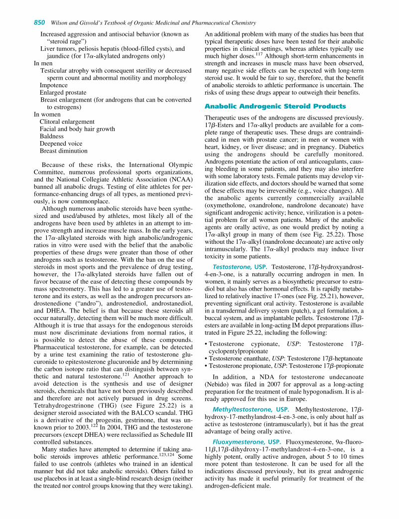
Increased aggression and antisocial behavior (known as“steroid rage”)
Liver tumors, peliosis hepatis (blood-filled cysts), andjaundice (for 17�-alkylated androgens only)
In menTesticular atrophy with consequent sterility or decreased
sperm count and abnormal motility and morphologyImpotenceEnlarged prostateBreast enlargement (for androgens that can be converted
to estrogens)In women
Clitoral enlargementFacial and body hair growthBaldnessDeepened voiceBreast diminution
Because of these risks, the International OlympicCommittee, numerous professional sports organizations,and the National Collegiate Athletic Association (NCAA)banned all anabolic drugs. Testing of elite athletes for per-formance-enhancing drugs of all types, as mentioned previ-ously, is now commonplace.
Although numerous anabolic steroids have been synthe-sized and used/abused by athletes, most likely all of theandrogens have been used by athletes in an attempt to im-prove strength and increase muscle mass. In the early years,the 17�-alkylated steroids with high anabolic/androgenicratios in vitro were used with the belief that the anabolicproperties of these drugs were greater than those of otherandrogens such as testosterone. With the ban on the use ofsteroids in most sports and the prevalence of drug testing,however, the 17�-alkylated steroids have fallen out offavor because of the ease of detecting these compounds bymass spectrometry. This has led to a greater use of testos-terone and its esters, as well as the androgen precursors an-drostenedione (“andro”), androstenediol, androstanediol,and DHEA. The belief is that because these steroids alloccur naturally, detecting them will be much more difficult.Although it is true that assays for the endogenous steroidsmust now discriminate deviations from normal ratios, itis possible to detect the abuse of these compounds.Pharmaceutical testosterone, for example, can be detectedby a urine test examining the ratio of testosterone glu-curonide to epitestosterone glucuronide and by determiningthe carbon isotope ratio that can distinguish between syn-thetic and natural testosterone.121 Another approach toavoid detection is the synthesis and use of designersteroids, chemicals that have not been previously describedand therefore are not actively pursued in drug screens.Tetrahydrogestrinone (THG) (see Figure 25.22) is adesigner steroid associated with the BALCO scandal. THGis a derivative of the progestin, gestrinone, that was un-known prior to 2003.122 In 2004, THG and the testosteroneprecursors (except DHEA) were reclassified as Schedule IIIcontrolled substances.
Many studies have attempted to determine if taking ana-bolic steroids improves athletic performance.123,124 Somefailed to use controls (athletes who trained in an identicalmanner but did not take anabolic steroids). Others failed touse placebos in at least a single-blind research design (neitherthe treated nor control groups knowing that they were taking).
An additional problem with many of the studies has been thattypical therapeutic doses have been tested for their anabolicproperties in clinical settings, whereas athletes typically usemuch higher doses.117 Although short-term enhancements instrength and increases in muscle mass have been observed,many negative side effects can be expected with long-termsteroid use. It would be fair to say, therefore, that the benefitof anabolic steroids to athletic performance is uncertain. Therisks of using these drugs appear to outweigh their benefits.
Anabolic Androgenic Steroid Products
Therapeutic uses of the androgens are discussed previously.17�-Esters and 17�-alkyl products are available for a com-plete range of therapeutic uses. These drugs are contraindi-cated in men with prostate cancer; in men or women withheart, kidney, or liver disease; and in pregnancy. Diabeticsusing the androgens should be carefully monitored.Androgens potentiate the action of oral anticoagulants, caus-ing bleeding in some patients, and they may also interferewith some laboratory tests. Female patients may develop vir-ilization side effects, and doctors should be warned that someof these effects may be irreversible (e.g., voice changes). Allthe anabolic agents currently commercially available(oxymetholone, oxandrolone, nandrolone decanoate) havesignificant androgenic activity; hence, virilization is a poten-tial problem for all women patients. Many of the anabolicagents are orally active, as one would predict by noting a17�-alkyl group in many of them (see Fig. 25.22). Thosewithout the 17�-alkyl (nandrolone decanoate) are active onlyintramuscularly. The 17�-alkyl products may induce livertoxicity in some patients.
Testosterone, USP. Testosterone, 17�-hydroxyandrost-4-en-3-one, is a naturally occurring androgen in men. Inwomen, it mainly serves as a biosynthetic precursor to estra-diol but also has other hormonal effects. It is rapidly metabo-lized to relatively inactive 17-ones (see Fig. 25.21), however,preventing significant oral activity. Testosterone is availablein a transdermal delivery system (patch), a gel formulation, abuccal system, and as implantable pellets. Testosterone 17�-esters are available in long-acting IM depot preparations illus-trated in Figure 25.22, including the following:
• Testosterone cypionate, USP: Testosterone 17�-cyclopentylpropionate
• Testosterone enanthate, USP: Testosterone 17�-heptanoate• Testosterone propionate, USP: Testosterone 17�-propionate
In addition, a NDA for testosterone undecanoate(Nebido) was filed in 2007 for approval as a long-actingpreparation for the treatment of male hypogonadism. It is al-ready approved for this use in Europe.
Methyltestosterone, USP. Methyltestosterone, 17�-hydroxy-17-methylandrost-4-en-3-one, is only about half asactive as testosterone (intramuscularly), but it has the greatadvantage of being orally active.
Fluoxymesterone, USP. Fluoxymesterone, 9�-fluoro-11�,17�-dihydroxy-17-methylandrost-4-en-3-one, is ahighly potent, orally active androgen, about 5 to 10 timesmore potent than testosterone. It can be used for all theindications discussed previously, but its great androgenicactivity has made it useful primarily for treatment of theandrogen-deficient male.
850 Wilson and Gisvold’s Textbook of Organic Medicinal and Pharmaceutical Chemistry
0819-0867_17865_Ch25.qxd 12/2/09 4:44 AM Page 850

Oxymetholone, USP. Oxymetholone, 17�-hydroxy-2-(hydroxymethylene)-17-methylandrostan-3-one, is ap-proved for the treatment of various anemias.
Oxandrolone, USP. Oxandrolone, 17�-hydroxy-17-methyl-2-oxaandrostan-3-one, is approved to aid in the pro-motion of weight gain after weight loss following surgery,chronic infections, or severe trauma and to offset protein ca-tabolism associated with long-term corticosteroid use.Oxandrolone is also used to relieve bone pain accompany-ing osteoporosis. It has been used to treat alcoholic hepatitisand HIV wasting syndrome.
Nandrolone Decanoate, USP. Nandrolone decanoate,17�-hydroxyestr-4-en-3-one 17-decanoate, has been used inthe management of certain anemias, but the availability oferythropoietin has greatly reduced this use.
Danazol and Endometriosis
Danazol, USP. Danazol, 17�-pregna-2,4-dien-20-yno-[2,3-d]isoxazol-17-ol, is a weak androgen that, in spite ofthe 17�-ethinyl group, has little estrogenic or progesto-genic activity. Danazol has been called a synthetic steroidwith diverse biological effects.125 Danazol binds to sexhormone–binding globulin (SHBG) and decreases the he-patic synthesis of this estradiol and testosterone carrier.Free testosterone thus increases. Danazol inhibits FSH andLH production by the hypothalamus and pituitary. It bindsto PRs, GRs, ARs, and ERs. Although the exact mecha-nism of action is unclear, danazol alters endometrial tissueso that it becomes inactive and atrophic, which allowsdanazol to be an effective treatment for endometriosis.Danazol is also used to treat hereditary angioedema and fi-brocystic breast disease.
Antiandrogens
Various compounds (Fig. 25.23) have been intensively stud-ied as AR antagonists, or antiandrogens.126,127 Antiandrogensare of therapeutic use in treating conditions of hyperan-drogenism (e.g., hirsutism, acute acne, and prematurebaldness) or androgen-stimulated cancers (e.g., prostatic car-cinoma). The ideal antiandrogen would be nontoxic, highlyactive, and devoid of any hormonal activity. Both steroidaland nonsteroidal antiandrogens have been investigated, butonly nonsteroidal antiandrogens have been approved for use
in the United States. Cyproterone acetate, a steroidal antian-drogen, is used in Europe. The steroidal antiandrogens typi-cally have actions at other steroid receptors that limit theiruse. The nonsteroidal antiandrogens, while lacking hormonalactivity, bind with lower affinity to the AR than the endoge-nous hormones.
FLUTAMIDE, BICALUTAMIDE, AND NILUTAMIDE
Three nonsteroidal antiandrogens are in clinical use in theUnited States—flutamide, bicalutamide, and nilutamide(Fig. 25.23). They are mainly used in the management ofprostate cancer. Flutamide was the first of these compoundsapproved for use by the FDA, but liver toxicity and thrice-daily dosing offered room for improvement. It was also de-termined that a metabolite of flutamide, hydroxyflutamide,had greater antiandrogen action than the parent.Bicalutamide, which has greater potency than flutamide, in-corporates an OH into its structure at the same relative posi-tion as in hydroxyflutamide. Bicalutamide is dosed once aday and has less toxicity than flutamide and nilutamide,making it a preferred choice when initiating therapy.
Prostate cancer is strongly androgen sensitive, so byblocking AR, the cancer can be inhibited or slowed. Studieshave shown that these drugs completely inhibit the actionof testosterone and other androgens by binding to AR. Inclinical trials when given as a single agent for prostate can-cer, serum testosterone and estradiol increase. But whengiven in combination with a GnRH agonist, such as gosere-lin or leuprolide, bicalutamide and flutamide do not affecttestosterone suppression, which is the result of GnRH.GnRH agonists greatly decrease gonadal function—themedical equivalent of castration in men. Thus, the combi-nation of GnRH with bicalutamide or flutamide blocksthe production of testosterone in the testes and AR inthe prostate.
Antiandrogen Products
Flutamide, USP. Flutamide, 2-methyl-N-[4-nitro-3-(tri-fluoromethyl)phenyl]propanamide, is dosed 3 times daily(250-mg dose; 750-mg total daily dose). A major metaboliteof flutamide, hydroxyflutamide, is a more potent AR antag-onist than the parent compound. This metabolite, which ispresent at a much higher steady-state concentration than isflutamide, contributes a significant amount of the
Chapter 25 Steroid Hormones and Therapeutically Related Compounds 851
Figure 25.23 Antiandrogens.
0819-0867_17865_Ch25.qxd 12/2/09 4:44 AM Page 851
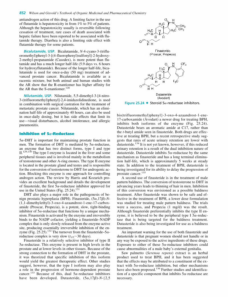
antiandrogen action of this drug. A limiting factor in the useof flutamide is hepatotoxicity in from 1% to 5% of patients.Although the hepatotoxicity usually is reversible followingcessation of treatment, rare cases of death associated withhepatic failure have been reported to be associated with flu-tamide therapy. Diarrhea is also a limiting side effect withflutamide therapy for some patients.
Bicalutamide, USP. Bicalutamide, N-4-cyano-3-(triflu-oromethyl)phenyl-3-[(4-fluorophenyl)sulfonyl]-2-hydroxy-2-methyl-propanamide (Casodex), is more potent than flu-tamide and has a much longer half-life (5.9 days vs. 6 hoursfor hydroxyflutamide). Because of the longer half-life, bica-lutamide is used for once-a-day (50 mg) treatment of ad-vanced prostate cancer. Bicalutamide is available as aracemic mixture, but both animal and human studies withthe AR show that the R-enantiomer has higher affinity forthe AR than the S-enantiomer.128
Nilutamide, USP. Nilutamide, 5,5-dimethyl-3-[4-nitro-3-(trifluoromethyl)phenyl]-2,4-imidazolidinedione, is usedin combination with surgical castration for the treatment ofmetastatic prostate cancer. Nilutamide, which has an elimi-nation half-life of approximately 40 hours, can also be usedin once-daily dosing, but it has side effects that limit itsuse—visual disturbances, alcohol intolerance, and allergicpneumonitis.
Inhibition of 5�-Reductase
5�-DHT is important for maintaining prostate function inmen. The formation of DHT is mediated by 5�-reductase,an enzyme that has two distinct forms, type I and typeII.129,130 The type I enzyme is located in the liver and someperipheral tissues and is involved mainly in the metabolismof testosterone and other A-ring enones. The type II enzymeis located in the prostate gland and testes and is responsiblefor the conversion of testosterone to DHT for androgenic ac-tion. Blocking this enzyme is one approach for controllingandrogen action. The review by Harris and Kozarich pro-vides an excellent background and details the developmentof finasteride, the first 5�-reductase inhibitor approved foruse in the United States (Fig. 25.24).131
DHT also plays a major role in the pathogenesis of be-nign prostatic hyperplasia (BPH). Finasteride, (5�,17�)-N-(1,1-dimethylethyl)-3-oxo-4-azaandrost-1-ene-17-carbox-amide (Proscar, Propecia), is a potent, slow, tight-bindinginhibitor of 5�-reductase that functions by a unique mecha-nism. Finasteride is activated by the enzyme and irreversiblybinds to the NADP cofactor, yielding a finasteride–NADPcomplex that is only slowly released from the enzyme activesite, producing essentially irreversible inhibition of the en-zyme (Fig. 25.25).132 The turnover from the finasteride–5�-reductase complex is very slow (t1/2�30 days).
Finasteride is a relatively selective inhibitor of type II5�-reductase. This enzyme is present in high levels in theprostate and at lower levels in other tissues. Because of thestrong connection to the formation of DHT in the prostate,it was theorized that specific inhibition of this isoformwould yield the greatest therapeutic effect. Other studiessuggest, however, that the type I isoform may also playa role in the progression of hormone-dependent prostatecancer.133 Because of this, dual 5�-reductase inhibitorshave been developed. Dutasteride, (5�,17�)-N-{2,5
bis(trifluoromethyl)phenyl}-3-oxo-4-azaandrost-1-ene-17-carboxamide (Avodart) a newer drug for treating BPH,inhibits both isoforms of the enzyme (Fig. 25.24).Dutasteride bears an aromatic amide at C17, rather thanthe t-butyl amide seen in finasteride. Both drugs are effec-tive at treating BPH, but a recent retrospective study sug-gests that rates of acute urinary retention are lower withdutasteride.134 It is not yet known, however, if this reducedurinary retention is a result of the dual inhibition nature ofdutasteride. Dutasteride inhibits 5�-reductase by the samemechanism as finasteride and has a long terminal elimina-tion half-life, which is approximately 5 weeks at steadystate. In addition to the treatment of BPH, dutasteride isbeing investigated for its ability to delay the progression ofprostate cancer.135
A second use of finasteride is in the treatment of malepattern baldness. The conversion of testosterone to DHT inadvancing years leads to thinning of hair in men. Inhibitionof this conversion was envisioned as a possible baldnesstreatment. After finasteride was shown to be safe and ef-fective in the treatment of BPH, a lower dose formulationwas studied for treating male pattern baldness. The trialswere a success, and Propecia (1 mg/d) was the result.Although finasteride preferentially inhibits the type II en-zyme, it is believed to be the peripheral type I 5�-reduc-tase that is being targeted for the baldness treatment.Dutasteride is also being investigated for use as a baldnesstreatment.
An important warning for the use of both finasteride anddutasteride is that pregnant women should not handle or inany way be exposed to the active ingredients of these drugs.Exposure to either of these 5�-reductase inhibitors couldcause abnormalities of a male baby’s external genitalia.
Saw palmetto (Serenoa repens) extract is an herbalproduct used to treat BPH, and it has been suggestedthat the effects may be attributed to a constituent of the ex-tract with 5�-reductase inhibition, but other mechanismshave also been proposed.136 Further studies and identifica-tion of a specific component that inhibits 5�-reductase arenecessary.
852 Wilson and Gisvold’s Textbook of Organic Medicinal and Pharmaceutical Chemistry
Figure 25.24 Steroid 5�-reductase inhibitors.
0819-0867_17865_Ch25.qxd 12/2/09 4:44 AM Page 852

Future Directions: SARMs137,138
Selective androgen receptor modulators or SARMs are thenewest developments among the potential androgen drugs.A parallel to the SERMs and SPRMs, SARMs are moleculesthat have differential actions at ARs in distinct tissues. Thelead compounds furthest along in development are non-steroidal AR ligands that have anabolic properties in muscleand bone, with only partial agonist action in the prostate.Two of these lead compounds, LGD2226 and S4 (Fig.25.26) are in phase I/II trials, with potential to treat age-re-lated muscle wasting and osteoporosis, in both men andwomen. As can be seen from Figure 25.26, S4 shares struc-tural similarities with the antiandrogen bicalutamide.
ADRENAL CORTEX HORMONES
Endogenous Corticosteroids
The adrenal glands (which lie just above the kidneys)secrete over 50 different steroids, including precursorsfor other steroid hormones. The most important hormonal
steroids produced by the adrenal cortex, however, are aldos-terone and hydrocortisone. Aldosterone is the primaryMC in humans (i.e., it causes significant salt retention).Hydrocortisone is the primary GC in humans (i.e., it has itsprimary effects on intermediary metabolism). The GCs have
Chapter 25 Steroid Hormones and Therapeutically Related Compounds 853
Figure 25.25 Comparison of 5�-reduc-tase action on testosterone and finas-teride. This scheme is an oversimplifica-tion of the exact mechanisms, but itindicates that when finasteride is boundat the active site of 5�-reductase, NADPHis positioned closer to C1 of finasteridethan to the normal C5 of testosterone,leading to essentially irreversibleinhibition.
Figure 25.26 SARMs.
0819-0867_17865_Ch25.qxd 12/2/09 4:44 AM Page 853

become very important in modern medicine, especially fortheir anti-inflammatory effects.
Aldosterone and, to a lesser extent, other MCs maintaina constant electrolyte balance and blood volume. The GCshave key roles in controlling carbohydrate, protein, and lipidmetabolism.
Biosynthesis
As shown in the scheme in Figure 25.27, aldosterone andhydrocortisone are biosynthesized from pregnenolonethrough a series of steps involving hydroxylations at C17,C11, and C21 that convert pregnenolone to hydrocortisone.Deficiencies in any of the enzymes cause congenital adrenal
hyperplasia. Defects in the gene regulation, as well as theenzymes that catalyze the hydroxylation, have been studiedintensively.139–141 Investigators have linked defects in par-ticular genes or steroid-binding sites to the pathophysiologyof patients with the corresponding metabolic diseases.141
These disorders are usually caused by an inability of theadrenal glands to carry out 11�-, 17�-, or 21-hydroxylations.The most common is a lack of 21-hydroxylase activity, whichwill result in decreased production of hydrocortisone and acompensatory increase in adrenocorticotropic hormone(ACTH) production. Furthermore, the resultant buildup of17�-hydroxyprogesterone will lead to an increase of testos-terone. The 21-hydroxylase is important for the synthesis ofboth MCs and GCs. When 11�-hydroxylase activity is low,
854 Wilson and Gisvold’s Textbook of Organic Medicinal and Pharmaceutical Chemistry
Figure 25.27 Biosynthesis ofhydrocortisone and aldosterone.
0819-0867_17865_Ch25.qxd 12/2/09 4:44 AM Page 854

large amounts of 11-deoxycorticosterone will be produced.Because 11-deoxycorticosterone is a potent MC, there will besymptoms of MC excess, including hypertension. When 17�-hydroxylase activity is low, there will be decreased produc-tion of testosterone and estrogens as well as hydrocortisone.
Although the details are not completely known, the 39-amino acid peptide ACTH (corticotropin) produced by theanterior pituitary is necessary for the conversion of choles-terol to pregnenolone. ACTH acts at the ACTH receptor, aG-protein–coupled receptor that activates adenylyl cyclase,leading to increased cyclic adenosine monophosphate(cAMP) levels. Activation of the ACTH receptors has short-and long-term effects on steroidogenesis. The short-termphase involves an increase in the supply of cholesterol foruse by cytochrome P450scc in the formation of preg-nenolone. The long-term effects are caused by an increasedtranscription of steroidogenic enzymes.142 An overall resultof ACTH action is increased synthesis and release of hydro-cortisone. Hydrocortisone then acts by feedback inhibitionto suppress the formation of additional ACTH (ACTH isdiscussed in more detail in Chapter 27).
The release of the primary MC aldosterone depends onlyslightly on ACTH. Aldosterone is an active part of the an-giotensin–renin–blood pressure cycle that controls bloodvolume. A decrease in blood volume stimulates the kidneysto secrete the enzyme renin. Renin, in turn, converts an-giotensinogen to angiotensin, which stimulates the adrenalcortex to release aldosterone. Aldosterone then causes thekidneys to retain sodium, and blood volume increases.When the blood volume has increased sufficiently, reninproduction decreases, until blood volume drops again.
Metabolism of Hydrocortisone
Hydrocortisone and cortisone are enzymatically intercon-vertible, and thus one finds metabolites with both the 11-keto and the 11�-hydroxy functionality. Most of the meta-bolic processes occur in the liver, with the metabolitesexcreted primarily in the urine. Although many metaboliteshave been isolated, the primary routes of catabolism are(a) reduction of the C4,5 double bond to yield 5�-pregnanes,(b) reduction of the 3-one to give 3�-ols, and (c) reductionof the 20-one to the corresponding 20�- and 20�-ols. Theseare the same steps that are involved in progesterone metab-olism. The two primary metabolites are tetrahydrocortisoland tetrahydrocortisone and their conjugates. The cortols(20� and 20�), cortolones (20� and 20�), and 11�-hydroxy-etiocholanolone are some of the minor metabolites of hy-drocortisone (Fig. 25.28).
Biological Activitiesof Mineralocorticoids andGlucocorticoids142
The adrenocortical steroids permit the body to adjust to en-vironmental changes, to stress, and to changes in the diet.Aldosterone and, to a lesser extent, other MCs maintain aconstant electrolyte balance and blood volume, and the GCshave key roles in controlling carbohydrate, protein, and lipidmetabolism.
Aldosterone increases sodium reabsorption in the kidneys.An increase in plasma sodium concentration, in turn, will leadto increased blood volume, because blood volume and urinaryexcretion of water are directly related to the plasma sodium
concentration. Simultaneously, aldosterone increases potas-sium ion excretion. 11-Deoxycorticosterone is also quiteactive as a MC. Similar actions are exhibited with hydrocor-tisone and corticosterone, but to a much smaller degree.
Aldosterone controls the movement of sodium ions inmost epithelial structures involved in active sodium trans-port. Although aldosterone acts primarily on the distal con-voluted tubules of the kidneys, it also acts on the proximalconvoluted tubules and collecting ducts. Aldosterone con-trols the transport of sodium in sweat glands, small intestine,salivary glands, and the colon. In all of these tissues, aldos-terone enhances the inward flow of sodium ions and pro-motes the outward flow of potassium ions.
The GCs have many physiological and pharmacologicalactions. They control or influence carbohydrate, protein,lipid, and purine metabolism. They also affect the cardio-vascular and nervous systems and skeletal muscle. Theyregulate growth hormone gene expression. In addition, GCshave anti-inflammatory and immunosuppressive actions thatarise through complex mechanisms.
GCs stimulate glycogen storage synthesis by inducingthe synthesis of glycogen synthase and stimulate gluconeo-genesis in the liver. They have a catabolic effect on muscletissue, stimulating the formation and transamination ofamino acids into glucose precursors in the liver. The cata-bolic actions in Cushing syndrome are demonstrated bywasting of the tissues, osteoporosis, and reduced musclemass. Lipid metabolism and synthesis increase significantlyin the presence of GCs, but the actions usually seem to de-pend on the presence of other hormones or cofactors. A lackof adrenal cortex steroids also causes depression, irritability,and even psychoses, reflecting significant effects on the cen-tral nervous system.
Chapter 25 Steroid Hormones and Therapeutically Related Compounds 855
Figure 25.28 Metabolites of cortisone andhydrocortisone.
0819-0867_17865_Ch25.qxd 12/2/09 4:44 AM Page 855

ANTI-INFLAMMATORY/IMMUNOSUPPRESSIVE ACTIONS OF GLUCOCORTICOIDS143–147
GR complexes may activate or repress the genes to whichthey associate. Repression in particular may have an impor-tant role in GC anti-inflammatory actions. GCs inhibit thetranscription of genes encoding cytokines such as inter-feron-�, tumor necrosis factor-� (TNF-�), the interleukins,and granulocyte/monocyte colony-stimulating factor, allfactors involved in the immune system and inflammatory re-sponses.142 GCs inhibit the production and release of othermediators of inflammation, including prostaglandins,leukotrienes, and histamine. In addition, GCs inhibit the ex-pression of the gene encoding collagenase, an important en-zyme involved with inflammation.
RESISTANCE TO GLUCOCORTICOIDS148–150
A few patients with chronic inflammatory illnesses such asasthma, rheumatoid arthritis, and lupus develop resistance tothe anti-inflammatory effects of the GCs. The mechanism isnot fully understood but appears to be a decrease in the bind-ing or activation ability of GR complexes and their target or“activator” genes. Disruption of the translocation of the GRto the nucleus has also been implicated in GC resistance.151
Structural Classes: Mineralocorticoidsand Glucocorticoids152
Medically important adrenal cortex hormones and syntheticMCs and GCs are shown in Figure 25.29. Because salt re-
856 Wilson and Gisvold’s Textbook of Organic Medicinal and Pharmaceutical Chemistry
Figure 25.29 Natural andsynthetic corticosteroids.(continues on next page)
0819-0867_17865_Ch25.qxd 12/2/09 4:44 AM Page 856

tention activity is usually undesirable, the drugs are classi-fied by their salt retention activities. As illustrated in Figure25.29, the adrenal cortex hormones are classified by their bi-ological activities into three major groups.
MINERALOCORTICOIDS
The MCs are adrenal cortex steroids and analogs with highsalt-retaining activity. They are used mainly for treatment ofAddison disease, or primary adrenal insufficiency. The nat-urally occurring hormone aldosterone has an 11�-OH andan 18-CHO that naturally bridge to form a hemiacetal (asdrawn in Fig. 25.27). Aldosterone is too expensive to pro-duce commercially; therefore, other semisynthetic analogs
have taken its place for treatment of Addison disease.Adding a 9�-fluoro group to hydrocortisone greatly in-creases both salt retention and anti-inflammatory activity.Deoxycorticosterone (11-deoxy), an intermediate in thebiosynthesis of aldosterone, has lower MC activity than al-dosterone (20-fold) but may play a role if the 11�-hydroxy-lase is deficient. Deoxycorticosterone is not available fortherapeutic uses.
Extensive modifications have been made to the basic hy-drocortisone structure to alter the properties of GCs.Modifications at all sites of the steroid backbone have beentried. Aside from addition of a double bond at C1–C2, themost beneficial changes are made to rings B and D of thesteroid skeleton and modification of the C17 side chain.
Chapter 25 Steroid Hormones and Therapeutically Related Compounds 857
Figure 25.29 (Continued)
0819-0867_17865_Ch25.qxd 12/2/09 4:44 AM Page 857

Table 25.6 summarizes the relative effects of various sub-stituents seen in commercially available products on saltretention and GC activity. The salt-retaining actions are ap-proximately additive. For example, the 3 increase in saltretention of a 9�-fluoro group can be eliminated by the 3–decrease of a 6�-methyl.
GLUCOCORTICOIDS WITH MODERATE-TO-LOWSALT RETENTION
The GCs with moderate-to-low salt retention include corti-sone, hydrocortisone, and their 1-enes prednisolone andprednisone. As shown in Table 25.7, an 11�-OH maintainsgood topical anti-inflammatory activity, but 11-ones havelittle or none. The 11�-hydroxysteroid dehydrogenase in theskin oxidizes an 11�-hydroxyl to an 11-ketone.153 For acti-vation of an 11-one GC for topical action, reduction at C11would be necessary. The 1-ene of prednisolone and pred-nisone increases anti-inflammatory activity about fourfoldand somewhat decreases salt retention. This increase in ac-tivity may be a result of a change in shape of ring A.28
Specifically, analogs more active than hydrocortisone ap-pear to have their ring A bent underneath the molecule to amuch greater extent than hydrocortisone.
The 11�-OH of hydrocortisone is of major importance inbinding to the receptors. Cortisone is reduced in vivo toyield hydrocortisone as the active agent. The increased ac-tivity of 9�-halo derivatives may be a result the electron-withdrawing inductive effect on the 11�-OH, making itmore acidic and, therefore, better able to form hydrogenbonds with the receptor. A 9�-halo substituent also reducesoxidation of the 11�-OH to the inactive 11-one.
GLUCOCORTICOIDS WITH VERY LITTLE OR NOSALT RETENTION
Cortisone and hydrocortisone, and even prednisone andprednisolone, have too much salt-retaining activity in thedoses needed for some therapeutic purposes. Over the pastseveral decades, several substituents have been discoveredthat greatly decrease salt retention. They include 16�-hy-droxy, 16�,17�-ketal, 6�-methyl, and 16�- and 16�-methyl.Other substituents have been found to increase both GC andMC activities: 9�-fluoro, 9�-chloro, and 21-hydroxy.
As a result of the great economic benefit of having a po-tent anti-inflammatory product on the market, pharmaceuti-cal manufacturers have made numerous combinations ofthese various substituents. In almost every case, a 16-
858 Wilson and Gisvold’s Textbook of Organic Medicinal and Pharmaceutical Chemistry
TABLE 25.6 Effects of Substituents on Glucocorticoid/Mineralocorticoid Activity
Glycogen Anti-inflammatory Effects onFunctional Group Deposition Activity Urinary Sodiuma
9�-Fluoro 10 7–10 9�-Chloro 3–5 3–4 1-Dehydro 3–4 3–4 6�-Methyl 2–3 1–2 16�-Hydroxy 0.4–0.5 0.1–0.2 17�-Hydroxy 1–2 4 21-Hydroxy 4–7 25
TABLE 25.7 Approximate Relative Activities of Corticosteroidsa
Anti-inflammatory Salt-Retaining EquivalentActivity Topical Activity Activity Dose (mg)
MineralocorticoidsAldosterone 0.2 0.2 800Deoxycorticosterone 0 0 40Fludrocortisone 10 5–40 800 2
GlucocorticoidsHydrocortisone 1 1 1 20Cortisone 0.8 0 0.8 25Prednisolone 4 4 0.6 5Prednisone 3.5 0 0.6 5Methylprednisolone 5 5 0 4Triamcinolone acetonide 5 5–100 0 4Triamcinolone 1–5Fluocinolone acetonide Over 40Flurandrenolide Over 20Fluocinolone Over 40Fluocinonide 40–100Betamethasone 35 5–100 0 0.6Dexamethasone 30 10–35 0 0.75
Adapted from Rodig, O.R.: In Burger, A. (ed.). Medicinal Chemistry, Part 2, 3rd ed. New York, Wiley-Interscience, 1970. Used with permission.a, retention; , excretion.
aThe data in this table are only approximate. Blanks indicate that comparative data are not available to the au-thor or that the product has only one use (e.g., topical). Data were taken from several sources, and there is aninherent risk in comparing such data. The table should, however, serve as a guide to relative activities.
0819-0867_17865_Ch25.qxd 12/2/09 4:44 AM Page 858

methyl or a modified 16-hydroxy (to eliminate salt reten-tion) has been combined with another substituent to in-crease GC or anti-inflammatory activity. The number ofpermutations and combinations has resulted in a redundantarray of analogs with very low salt retention and high anti-inflammatory activity.
A primary goal of these highly anti-inflammatory drugshas been to increase topical potency. As shown in Table25.7, some are as much as 100 times more active topicallythan hydrocortisone. Relative potency is as follows:
Very high potencyAugmented betamethasone dipropionate ointment, 0.05%Clobetasol propionate, 0.05%Diflorasone diacetate ointment, 0.05%
High potencyAmcinonide, 0.1%Betamethasone dipropionate ointment, 0.05%Desoximetasone, 0.25%Diflorasone diacetate cream, 0.05%Fluocinonide, 0.05%Halcinonide, 0.1%Halobetasol propionate, 0.05%Triamcinolone acetonide, 0.5%
Medium potencyBetamethasone valerate, 0.1%Clocortolone pivalate, 0.1%Desoximetasone, 0.05%Fluocinolone acetonide, 0.025%Fluticasone propionate, 0.005%Hydrocortisone butyrate, 0.1%Hydrocortisone valerate, 0.2%Mometasone furoate, 0.1%Prednicarbate, 0.1%Triamcinolone acetonide, 0.1%
Low potencyAlclometasone dipropionate, 0.05%Desonide, 0.05%Fluocinolone acetonide, 0.01%Triamcinolone acetonide cream, 0.1%
Lowest potencyHydrocortisone, 1.0%Hydrocortisone, 2.5%
Although, as shown in Table 25.7, cortisone and pred-nisone are not active topically, most other GCs are active.Some compounds, such as clobetasol and betamethasonedipropionate, have striking activity topically. Skin absorp-tion is favored by increased lipid solubility of the drug.
Absorption of topical GCs can also be greatly affected bythe extent of skin damage, concentration of the GC, creamor ointment base used, and similar factors. One must not as-sume, therefore, from a study of Table 25.7 that, for exam-ple, a 0.25% cream of prednisolone is necessarily exactlyequivalent in anti-inflammatory potency to 1% hydrocorti-sone. Nevertheless, the table can serve as a preliminaryguide. Furthermore, particular patients may seem to respondbetter to one topical anti-inflammatory GC than to another,irrespective of the relative potencies shown in Table 25.7.
RISK OF SYSTEMIC ABSORPTION
The topical corticosteroids do not typically cause significantabsorption effects when used on small areas of intact skin.
When these compounds are used on large areas of the body,however, systemic absorption may occur, especially if theskin is damaged or if occlusive dressings are used. Up to20% to 40% of hydrocortisone given rectally may also beabsorbed.
Therapeutic Uses of AdrenalCortex Hormones
The adrenocortical steroids are used primarily for their GCeffects, including immunosuppression, anti-inflammatoryactivity, and antiallergic activity. The MCs are used only fortreatment of Addison disease. Addison disease is caused bychronic adrenocortical insufficiency and may be due to ei-ther adrenal or anterior pituitary failure. The GCs are alsoused in the treatment of congenital adrenal hyperplasias.
The symptoms of Addison disease illustrate the great im-portance of the adrenocortical steroids in the body and, espe-cially, the importance of aldosterone. These symptomsinclude increased loss of body sodium, decreased loss ofpotassium, hypoglycemia, weight loss, hypotension, weak-ness, increased sensitivity to insulin, and decreased lipolysis.
Hydrocortisone is also used during postoperative recov-ery after surgery for Cushing syndrome—excessive adrenalsecretion of GCs. Cushing syndrome can be caused by bilat-eral adrenal hyperplasia or adrenal tumors and is treated bysurgical removal of the tumors or resection of hyperplasticadrenal gland(s).
The use of GCs during recovery from surgery forCushing syndrome illustrates a most important principle ofGC therapy: abrupt withdrawal of GCs may result in adre-nal insufficiency, showing clinical symptoms similar tothose of Addison disease. For that reason, patients who havebeen on long-term GC therapy must have the dose reducedgradually. Furthermore, prolonged treatment with GCs cancause adrenal suppression, especially during times of stress.The symptoms are similar to those of Cushing syndrome,such as rounding of the face, hypertension, edema, hy-pokalemia, thinning of the skin, osteoporosis, diabetes, andeven subcapsular cataracts.
The GCs are used in the treatment of collagen vasculardiseases, including rheumatoid arthritis and disseminatedlupus erythematosus. Although there is usually prompt re-mission of redness, swelling, and tenderness by the GCs inrheumatoid arthritis, continued long-term use may lead toserious systemic forms of collagen disease. As a result, theGCs should be used infrequently in rheumatoid arthritis.
The GCs are used extensively topically, orally, and par-enterally to treat inflammatory conditions. They also usuallyrelieve the discomforting symptoms of many allergicconditions—intractable hay fever, exfoliative dermatitis, gen-eralized eczema, and others. The GCs are also used to treatasthmatic symptoms unresponsive to bronchodilators. Theyare especially useful in inhaled formulations (see section onpage 863). The GCs’ lymphocytopenic actions make themparticularly useful for treatment of chronic lymphocyticleukemia in combination with other antineoplastic drugs.
The adrenocortical steroids are contraindicated or shouldbe used with great caution in patients who have (a) pepticulcer (in which the steroids may cause hemorrhage), (b)heart disease, (c) infections (the GCs suppress the body’snormal infection-fighting processes), (d) psychoses (sincebehavioral disturbances may occur during steroid therapy),
Chapter 25 Steroid Hormones and Therapeutically Related Compounds 859
0819-0867_17865_Ch25.qxd 12/2/09 4:44 AM Page 859

(e) diabetes (the GCs increase glucose production, so moreinsulin may be needed), (f) glaucoma, (g) osteoporosis, or(h) herpes simplex involving the cornea.
When administered topically, the GCs present relativelyinfrequent therapeutic problems, but their anti-inflammatoryaction can mask symptoms of infection. Many physiciansprefer not giving a topical anti-inflammatory steroid untilafter an infection is controlled with topical antibiotics. Theimmunosuppressive activity of the topical GCs can also pre-vent natural processes from curing the infection. Topicalsteroids actually may also cause dermatoses in some patients.
Finally, as discussed previously with the oral contracep-tives, steroid hormones should not be used during preg-nancy. If it is absolutely necessary to use GCs topically dur-ing pregnancy, they should be limited to small areas ofintact skin and used for a limited time.
Mineralocorticoid andGlucocorticoid Products
The corticosteroids used in commercial products are shownin Figures 25.29, 25.30, and 25.31. The structures illustratethe usual changes (see Fig. 25.6) made to modify solubilityof the products and, therefore, their therapeutic uses. In par-ticular, the 21-hydroxyl can be converted to an ester to makeit less water soluble to modify absorption or to a phosphateester salt or hemisuccinate ester salt to make it more watersoluble and appropriate for intravenous use. The productsalso reflect the structure–activity relationship changes dis-cussed previously to increase anti-inflammatory activity orpotency or decrease salt retention.
Again, patients who have been on long-term GC therapymust have the dose reduced gradually. This “critical rule”and indications are discussed previously under the heading,“Therapeutic Uses of Adrenal Cortex Hormones.” Dosageschedules and gradual dosage reduction can be quite com-plex and specific for each indication.
Many of the GCs are available in topical dosage forms,including creams, ointments, aerosols, lotions, and solutions.They are usually applied 3 to 4 times a day to well-cleanedareas of affected skin. Ointments are usually prescribed fordry, scaly dermatoses. Lotions are well suited for weepingdermatoses. Creams are of general use for many other
860 Wilson and Gisvold’s Textbook of Organic Medicinal and Pharmaceutical Chemistry
Figure 25.30 Ophthalmic glucocorticoids.
Figure 25.31 Glucocorticoids used to treat asthma and allergic rhinitis (some arealso used topically).
0819-0867_17865_Ch25.qxd 12/2/09 4:44 AM Page 860

dermatoses. When applied to very large areas of skin or todamaged areas of skin, significant systemic absorption canoccur. The use of an occlusive dressing can also greatly in-crease systemic absorption.
The GCs that are mainly used for inflammation of the eyeare shown in Figure 25.30. These compounds differ struc-turally from other GRs, in that the 21-hydroxyl is missingfrom medrysone, fluorometholone, and rimexolone, whileloteprednol etabonate has a modified ester at C17 that leadsto rapid degradation upon systemic absorption.
MINERALOCORTICOIDS
Fludrocortisone Acetate, USP. Fludrocortisone ac-etate, 21-acetyloxy-9-fluoro-11�,17-dihydroxypregn-4-ene-3,20-dione, 9�-fluorohydrocortisone (Florinef Acetate), isused only for the treatment of Addison disease and for inhi-bition of endogenous adrenocortical secretions. As shown inTable 25.7, it has up to about 800 times the MC activity ofhydrocortisone and about 11 times the GC activity. Its po-tent activity stimulated the synthesis and study of the manyfluorinated steroids shown in Figure 25.29. Although itsgreat salt-retaining activity limits its use to Addison disease,it has sufficient GC activity that in some cases of the dis-ease, additional GCs need not be prescribed.
GLUCOCORTICOIDS WITH MODERATE-TO-LOWSALT RETENTION
Hydrocortisone, USP. Hydrocortisone, 11�,17,21-tri-hydroxypregn-4-ene-3,20-dione, is the primary natural GCin humans. Despite the large number of synthetic GCs, hy-drocortisone, its esters, and its salts remain a mainstay ofmodern adrenocortical steroid therapy and the standard forcomparison of all other GCs and MCs (see Table 25.7). It isused for all the indications mentioned previously. Its estersand salts illustrate the principles of chemical modification tomodify pharmacokinetic use shown in Figure 25.6. The com-mercially available salts and esters (see Fig. 25.29) include:
Hydrocortisone acetate, USP (21-acetate)Hydrocortisone buteprate � hydrocortisone probutate,
USP (17-butyrate, 21-propionate)Hydrocortisone butyrate, USP (17-butyrate)Hydrocortisone sodium phosphate, USP (21-sodium
phosphate)Hydrocortisone sodium succinate, USP (21-sodium
succinate)Hydrocortisone valerate, USP (17-valerate)
Cortisone Acetate, USP. Cortisone acetate, 21-(acety-loxy)-17-hydroxypregn-4-ene-3,11,20-trione, is the 21-acetate of naturally occurring cortisone with good systemicanti-inflammatory activity and low-to-moderate salt-reten-tion activity after its in vivo conversion to hydrocortisoneacetate. This conversion is mediated by 11�-hydroxysteroiddehydrogenase. It is used for the entire spectrum of uses dis-cussed previously under the heading, “Therapeutic Uses ofAdrenal Cortex Hormones”—collagen diseases, Addisondisease, severe shock, allergic conditions, chronic lympho-cytic leukemia, and many other indications. Cortisone ac-etate is relatively ineffective topically, mainly because itmust be reduced in vivo to hydrocortisone. Its plasma half-life is only about 30 minutes, compared with 90 minutes to3 hours for hydrocortisone.
Prednisolone, USP. Prednisolone, �1-hydrocortisone,11�,17,21-trihydroxypregna-1,4-diene-3,20-dione, hasless salt-retention activity than hydrocortisone (see Table25.7), but some patients have more frequently experiencedcomplications such as gastric irritation and peptic ulcers.Because of low MC activity, it cannot be used alone for ad-renal insufficiency. Prednisolone is available in varioussalts and esters to maximize its therapeutic utility (see Fig.25.29):
Prednisolone acetate, USP (21-acetate)Prednisolone sodium phosphate, USP (21-sodium
phosphate)Prednisolone sodium succinate, USP (21-sodium
succinate)Prednisolone tebutate, USP (21-tebutate)
Prednisone, USP. Prednisone, �1-cortisone, 17,21-di-hydroxypregna-1,4-diene-3,11,20-trione, has systemic ac-tivity very similar to that of prednisolone, and because of itslower salt-retention activity, it is often preferred over corti-sone or hydrocortisone. Prednisone must be reduced in vivoto prednisolone to provide the active GC.
GLUCOCORTICOIDS WITH VERY LITTLE OR NOSALT RETENTION
Most of the key differences between the many GCs with min-imal salt retention (see Fig. 25.29) have been summarized inTables 25.6 and 25.7. The tremendous therapeutic and, there-fore, commercial importance of these drugs has stimulatedthe proliferation of new compounds and their products. Manycompounds also are available as salts or esters to give thecomplete range of therapeutic flexibility illustrated inFigure 25.29. When additional pertinent information is avail-able, it is given below the drug name. The systemic name foreach drug is provided after the common name.
Alclometasone Dipropionate, USP. Alclometasonedipropionate, 7�-chloro-11�-hydroxy-16�-methyl-17,21-bis(1-oxopropoxy)-pregna-1,4-diene-3,20-dione (Aclovate),is one of the few commercially used GRs that bears a halo-gen substituent in the 7�-position.
Amcinonide, USP. Amcinonide, 21-(acetyloxy)-16�,17-[cyclopentylidenebis(oxy)]-9-fluoro-11�-hydroxy-pregna-1,4-diene-3,20-dione (Cyclocort).
Beclomethasone Dipropionate, USP. Beclomethasonedipropionate, 9-chloro-11�-hydroxy-16�-methyl-17,21-bis-(1-oxopropoxy)-pregna-1,4-diene-3,20-dione (Beconase,QVAR), is used in nasal sprays and aerosol formulations totreat allergic rhinitis and asthma (see section on page 863).
Betamethasone, USP. Betamethasone, 9-fluoro-11�,17,21-trihydroxy-16�-methylpregna-1,4-diene-3,20-dione,is available as a variety of ester derivatives.
Betamethasone valerate, USP (17-valerate)Betamethasone acetate, USP (21-acetate)Betamethasone sodium phosphate, USP (21-sodium
phosphate)Betamethasone dipropionate, USP (17-propionate,
21-propionate)
Budesonide, USP. Budesonide, 16�,17-[butylidenebis-(oxy)]-11�,21-dihydroxypregna-1,4-diene-3, 20-dione
Chapter 25 Steroid Hormones and Therapeutically Related Compounds 861
0819-0867_17865_Ch25.qxd 12/2/09 4:44 AM Page 861

(Entocort), in oral capsules is used to treat Crohn disease. Theaffinity for the GC is approximately 200-fold greater than thatof hydrocortisone and 15-fold greater than that of pred-nisolone. Budesonide is a mixture of epimers, with the 22Rform having twice the affinity for the GR of the S epimer.This GC is metabolized by CYP3A4, and its levels can be in-creased in the presence of potent CYP3A4 inhibitors.Budesonide is also used in an inhaled formulation for thetreatment of asthma (see next page).
Clobetasol Propionate, USP. Clobetasol propionate,21-chloro-9-fluoro-11�-hydroxy-16�-methyl-17-(1-oxo-propoxy)-pregna-1,4-diene-3,20-dione (Temovate).
Clocortolone Pivalate, USP. Clocortolone pivalate, 9-chloro-21-(2,2-dimethyl-1-oxopropoxy)-6�-fluoro-11�-hy-droxy-16�-methylpregna-1,4-diene-3,20-dione (Cloderm),along with desoximetasone, lacks the C17� oxygen func-tionality that is present in other GCs but still retains goodGC activity.
Desonide, USP. Desonide, 11�,21-dihydroxy-16�,17-[(1-methylethylidene)bis(oxy)]pregna-1,4-diene-3,20-dione(DesOwen, Tridesiol).
Desoximetasone, USP. Desoximetasone, 9-fluoro-11�, 21-dihydroxy-16�-methylpregna-1,4-diene-3,20-dione, like clocortolone pivalate, lacks a C17� OH group inits structure.
Dexamethasone, USP. Dexamethasone, 9-fluoro-11�,17,21-trihydroxy-16�-methylpregna-1,4-diene-3,20-dione,is the 16�-isomer of betamethasone.
Dexamethasone acetate, USP (21-acetate)Dexamethasone sodium phosphate, USP (21-sodium
phosphate)
Diflorasone Diacetate, USP. Diflorasone diacetate, 17,21-bis(acetyloxy)-6�,9-difluoro-11�-hydroxy-16�-methyl-pregna-1,4-diene-3,20-dione.
Difluprednate. Difluprednate, 21-(acetyloxy)-6�,9-difluoro-11�-hydroxy-17-(1-oxobutoxy)pregna-1,4-diene-3,20-dione is being developed (phase III) as a topical oph-thalmic emulsion to treat inflammatory eye diseases.
Flunisolide, USP. Flunisolide, 6�-fluoro-11�,21-dihydroxy-16�,17-[(1-methylethylidene)bis(oxy)]pregna-1,4-diene-3,20-dione. (See following section for use of flu-nisolide in the treatment of asthma.)
Fluocinolone Acetonide, USP. Fluocinolone acetonide,6�,9-difluoro-11�,21-dihydroxy-16�,17-[(1-methylethyli-dene)bis(oxy)]pregna-1,4-diene-3,20-dione, also known as6�-fluorotriamcinolone acetonide, is the 21-acetate deriva-tive of fluocinolone acetonide and is about 5 times morepotent than fluocinolone acetonide in at least one topicalactivity assay.
Fluorometholone, USP. Fluorometholone, 9-fluoro-11�,17-dihydroxy-6�-methylpregn-4-ene-3,20-dione(Fluor-Op, FML), lacks the typical C21 OH group of GCsand is used exclusively in ophthalmic products. The 17-ac-etate of fluorometholone is also used as an ophthalmic sus-pension (Flarex).
Flurandrenolide, USP. Flurandrenolide, 6�-fluoro-11�,21-dihydroxy-16 �,17-[(1-methylethylidene)bis(oxy)]
pregn-4-ene-3,20-dione, although available as a tape prod-uct, can stick to and remove damaged skin, so it should beavoided with vesicular or weeping dermatoses.
Fluticasone Propionate, USP. Fluticasone propionate,S-(fluoromethyl) 6�,9-difluoro-11�-hydroxy-16�-methyl-3-oxo-17�-(1-oxopropoxy)androsta-1,4-diene-17-car-bothio-ate (Cutivate), is threefold to fivefold more potentthan dexamethasone in receptor binding assays (see also thefollowing section on inhaled corticosteroids).
Halcinonide. Halcinonide, 21-chloro-9-fluoro-11�-hydroxy-16�,17-[(1-methylethylidene)bis(oxy)]pregn-4-ene-3,20-dione, was the first chloroGC marketed. Likemany other potent GCs, it is used only topically.
Halobetasol Propionate, USP. Halobetasol propi-onate, 21-chloro-6�,9-difluoro-11�-hydroxy-16�-methyl-17-(1-oxopropoxy)pregna-1,4-diene-3,20-dione.
Loteprednol Etabonate, USP. Loteprednol etabon-ate, chloromethyl 17�-[(ethoxycarbonyl)oxy]-11�-hy-droxy-3-oxoandrosta-1,4-diene-17-carboxylate (Alrex,Lotemax), has a modified carboxylate at the C17 positionrather than the typical ketone functionality. This modifica-tion maintains affinity for the GR but allows facile metab-olism to inactive metabolites. This limits the systemic ac-tion of the drug. Loteprednol etabonate is used as anophthalmic suspension that has greatly reduced systemicaction because of rapid metabolism to the inactive car-boxylate (Fig. 25.30).
Methylprednisolone, USP. Methylprednisolone, 11�,17,21-trihydroxy-6�-methyl-1,4-pregnadiene-3,20-dione, isavailable unmodified or as ester derivatives.
Methylprednisolone acetate, USPMethylprednisolone sodium succinate, USP
Mometasone Furoate, USP. Mometasone furoate,9,21-dichloro-17�-[(2-furanylcarbonyl)oxy]-11�-hydroxy-16�-methylpregna-1,4-diene-3,20-dione (Elocon), is a high-potency GC available in cream, lotion, or ointment formula-tions for topical use. In addition, mometasone furoatemonohydrate is formulated for treating allergic rhinitis andasthma (see following section).
Prednicarbate, USP. Prednicarbate, 17-[(ethoxycar-bonyl)oxy]-11�-hydroxy-21-(1-oxopropoxy)pregna-1,4-diene-3,20-dione, is a prednisolone derivative with a C21 pro-pionate ester and a C17 ethyl carbonate group. It is availablefor use only in a 0.1% topical cream. Prednicarbate is amedium-potency GC.
Rimexolone, USP. Rimexolone, 11�-hydroxy-16�,17�-dimethyl-17-(1-oxopropyl)androsta-1,4-diene-3-one,like medrysone and fluorometholone, lacks the C21 OHgroup. In addition, rimexolone has an additional methylgroup in the 17�-position, a site where an OH group is typ-ically found. Rimexolone is available as a suspension forophthalmic use (Fig. 25.30).
Triamcinolone, USP. Triamcinolone, 9-fluoro-11�,16�,17,21-tetrahydroxypregna-1,4-diene-3,20-dione.
Triamcinolone acetonide, USP: Triamcinolone-16�,17-acetonide
Triamcinolone hexacetonide, USP: Triamcinolone ace-tonide 21-[3-(3,3-dimethyl)butyrate]
Triamcinolone diacetate, USP: 16,21-Diacetate
862 Wilson and Gisvold’s Textbook of Organic Medicinal and Pharmaceutical Chemistry
0819-0867_17865_Ch25.qxd 12/2/09 4:44 AM Page 862

Triamcinolone acetonide is approximately 8 times morepotent than prednisone in animal inflammation models.Topically applied triamcinolone acetonide is a potent anti-inflammatory agent (see Table 25.7), about 10 times more sothan triamcinolone. The plasma half-life is approximately 90minutes, although the plasma half-life and biological half-lives for GCs do not correlate well. The hexacetonide isslowly converted to the acetonide in vivo and is given onlyby intra-articular injection. Only triamcinolone and the diac-etate are given orally. The acetonide and diacetate may begiven by intra-articular or intrasynovial injection. In addi-tion, the acetonide may be given by intrabursal or, some-times, IM or subcutaneous injection. A single IM dose of thediacetate or acetonide may last up to 3 or 4 weeks. Plasmalevels with IM doses of the acetonide are significantly higherthan with triamcinolone itself. The acetonide is also used totreat asthma and allergic rhinitis (see following section).
INHALED CORTICOSTEROIDS FOR ASTHMA ANDALLERGIC RHINITIS
The National Asthma Education and Prevention Program hasprovided recent recommendations on the treatment of asthma,including a strong recommendation for the first-line use of in-haled corticosteroids for severe and moderate persistentasthma in all age groups. The corticosteroids currently used ininhaled formulations are all relatively potent topical corticos-teroids that have the advantage of rapid deactivation/inactiva-tion for the portion of the dose that is swallowed. The devel-opment of GCs that are efficiently inactivated metabolicallywhen swallowed has greatly reduced the systemic side effectsassociated with the use of steroids in asthma treatment. Theolder corticosteroids that are used orally (e.g., methylpred-nisolone, prednisolone, and prednisone) have much greatersystemic side effects, and their use should be limited, if pos-sible. Although systemic side effects are reduced, they are notcompletely eliminated. The side effects can vary with thesteroid used and the frequency of administration.
The six GCs that are currently approved for use in theUnited States for asthma as inhaled formulations are be-clomethasone dipropionate, budesonide, flunisolide, fluticas-one propionate, mometasone furoate, and triamcinolone ace-tonide (Fig. 25.31). Ciclesonide (Alvesco) is the newest GCbeing pursued for use in the treatment of asthma. Ciclesonideis in phase III clinical trials and may be available in theUnited States within a few years. It is already approved totreat asthma in Europe and Canada. Clinical trials suggest thatit may have better tolerability than some of the currentlyavailable inhaled steroids.
The following agents are also available in nasal inhalersfor the treatment of allergic rhinitis. Details are providedbelow for the mode of metabolic inactivation involved foreach of these products. Although all of these agents havemuch lower systemic effects than the oral steroids, somesystemic effects, as measured by suppression of the hypo-thalamic–pituitary–adrenal (HPA) axis, have been observedfor these products. Ciclesonide (2006) and fluticasonefuroate (Veramyst, 2007), with a 17-furoate in place of the17-propionate, were both also recently approved for treatingallergic rhinitis.
GLUCOCORTICOIDS FOR ASTHMA ANDALLERGIC RHINITIS
Beclomethasone Dipropionate. Beclomethasonedipropionate (Beclovent, Beconase, Vanceril, Vancenase)
(BDP) is rapidly converted in the lungs to beclomethasone17-monopropionate (17-BMP), the metabolite that providesthe bulk of the anti-inflammatory activity. The monopropi-onate also has higher affinity for the GR than either thedipropionate or beclomethasone. The portion of BDP that isswallowed is rapidly hydrolyzed to 17-BMP, 21-BMP(which arises by a transesterification reaction from 17-BMP), and beclomethasone itself.154 Beclomethasone hasmuch less GC activity than the monopropionate.155
Budesonide. Budesonide (Pulmicort Turbohaler,Rhinocort) is extensively metabolized in the liver, with 85%to 95% of the orally absorbed drug metabolized by the first-pass effect. The major metabolites are 6�-hydroxybudes-onide and 16�-hydroxyprednisolone, both with less than1% of the activity of the parent compound. Metabolism in-volves the CYP3A4 enzyme, so coadministration of budes-onide with a known CYP3A4 inhibitor should be moni-tored carefully.
Ciclesonide. Ciclesonide (Omnaris) is a prodrug that re-quires hydrolysis of the isobutyrate ester at C21 to form theactive corticosteroid (des-ciclesonide). It has minimal oralbioavailability due to extensive metabolism, mainly byCYP3A4. The metabolites of ciclesonide have not beenfully characterized.
Flunisolide. The portion of a flunisolide (AeroBid,Nasarel) dose that is swallowed is rapidly converted to the6�-hydroxy metabolite after first-pass metabolism in theliver. The 6�-hydroxy metabolite is approximately as activeas hydrocortisone itself, but the small amount produced usu-ally has limited systemic effects. Water-soluble conjugatesare inactive.
Fluticasone Propionate. The main metabolite of flu-ticasone propionate (Flovent, Flonase) found in circulationin humans is the 17�-carboxylate derivative. As expected,a charged carboxylate in place of the normal acetol func-tionality at C17 greatly reduces affinity for the GR (2,000-fold less than the parent), and this metabolite is essentiallyinactive. The metabolite is formed via the CYP3A4 sys-tem, so care should be taken if fluticasone propionate iscoadministered with a CYP3A4 inhibitor such as keto-conazole or ritonavir. Clinically induced Cushing syn-drome has been observed when inhaled fluticasone propi-onate was administered concurrently with ritonavir.156
Fluticasone is also available in an inhaled formulation incombination with the long-acting �2-agonist salmeterol(Advair Diskus).
Mometasone Furoate. Mometasone furoate (Asmanex,Nasonex) undergoes extensive metabolism to multiplemetabolites. No major metabolites are detectable in humanplasma after oral administration, but the 6�-hydroxy metabo-lite is detectable by use of human liver microsomes. Thismetabolite is formed via the CYP3A4 pathway.
Triamcinolone Acetonide. The three main metabolitesof triamcinolone acetonide (Azmacort, Nasacort) are 6�-hydroxytriamcinolone acetonide, 21-carboxytriamcinoloneacetonide, and 6�-hydroxy-21-carboxytriamcinolone ace-tonide. All are much less active than the parent compound.The 6�-hydroxyl group and the 21-carboxy group are bothstructural features that greatly reduce GC action. The in-creased water solubility of these metabolites also facilitatesmore rapid excretion.
Chapter 25 Steroid Hormones and Therapeutically Related Compounds 863
0819-0867_17865_Ch25.qxd 12/2/09 4:44 AM Page 863

Mineralocorticoid ReceptorAntagonists (Aldosterone Antagonists)
Antagonism of the MR can have profound effects on therenin–angiotensin system, thus having significant cardiaceffects. Structurally, these compounds have an A-ringenone, essential for recognition by the receptor, but the 7�-substituent and the D-ring spirolactone provide structural el-ements that lead to antagonism (Fig. 25.32).
Spironolactone, USP. Spironolactone, 7�-(acetylthio)-17�-hydroxy-3-oxopregn-4-ene-3-one-21-carboxylic acid�-lactone (Aldactone) is an aldosterone antagonist of greatmedical importance because of its diuretic activity.Spironolactone is discussed in Chapter 18.
Eplerenone, USP. Eplerenone, 9,11�-epoxy-17�-hy-droxy-3-oxopregn-4-ene-7�,21-dicarboxylic acid, �-lactone,methyl ester (Inspra), is a newer aldosterone antagonist that isused for the treatment of hypertension.
NEUROSTEROIDS79,157,158
Although the main effects of the steroid hormones describedin this chapter have been focused on sexual development,maintenance of sexual organ function, and inflammatoryprocesses, steroids also play important roles in the brain.Two terms have been developed to describe the specificsteroids that act on the nervous system, neurosteroids andneuroactive steroids. Neurosteroids are synthesized in thebrain, either from cholesterol or from other steroid hor-mones, and have their specific actions in the nervous sys-tem. Neuroactive steroids are synthesized in other steroido-genic tissues, but have their actions in the brain aftertransport from the site of synthesis. Many of the neuros-teroids are the same as steroid hormones acting peripherally,
but some have unique roles in the brain. Neurosteroids in-clude pregnenolone, progesterone, and DHEA, as well astheir sulfate esters. Metabolites of progesterone and 11-de-oxycorticosterone, allopregnanolone and allotetrahy-drodeoxycorticosterone (allotetrahydroDOC; Fig. 25.33),respectively, represent neurosteroids with specific centralactions.
In addition to the action of neurosteroids/neuroactivesteroids via traditional steroid receptors (progesterone,estradiol), many neurosteroids act at different sites, espe-cially the neurosteroids with a 3�-hydroxyl (Fig. 25.33).Allopregnanolone and allotetrahydroDOC are positive mod-ulators of GABAA receptors, while pregnenolone sulfateand DHEA sulfate are negative modulators of the GABAA
receptors. DHEA sulfate and pregnenolone sulfate also haveactions at NMDA receptors and sigma type 1 receptors,while progesterone interacts with kainite, glycine, 5HT3,and nicotinic acetylcholine receptors. Although much of theneurosteroid research has been conducted in animal models,the clinical significance of neurosteroids is an area of grow-ing interest.79 Neurosteroids have been hypothesized tohave neuroprotective effects, anxiolytic effects, and mayplay roles in modulating seizures. The actions of steroids inthe brain will undoubtedly be the focus of much more re-search in the years to come.
ACKNOWLEDGMENT
I would like to thank Debra Peters for assistance with the il-lustration of several figures. I would also like to express myappreciation to the authors of various review articles on thesteroids and the authors of previous versions of this text.Without the dedication and hard work of these individuals,the assembly of this chapter would have been a much morechallenging task.
864 Wilson and Gisvold’s Textbook of Organic Medicinal and Pharmaceutical Chemistry
Figure 25.33 Neurosteroid examples.Figure 25.32 Aldosterone receptor antagonists.
0819-0867_17865_Ch25.qxd 12/2/09 4:44 AM Page 864

5. Two enzymes involved in steroid hormone biosynthesisare useful therapeutic targets, one for treating breast cancerand the other for treating benign prostatic hypertrophy(BPH). What are these two enzymes and what biosyntheticconversions do they mediate? Provide the name of an in-hibitor of each enzyme that is used as a drug.
6. Based on structural features, which of the following rep-resents a corticosteroid that would have strong anti-in-flammatory properties, but would have minimal MC(water-retention) action? Explain your choice, specifi-cally identifying key structural features.
1. What is the natural precursor in the human body to all thesteroid hormones (estrogens, progestins, androgens, andcorticosteroids)? Explain the general steps necessary toconvert this precursor to the steroid backbones necessaryfor the specific steroid hormones. Also indicate the en-zyme involved in the key steps.
2. Tamoxifen and raloxifene both mediate their main phar-macological actions via the the ERs. Tamoxifen is an an-tagonist in breast tissue and an agonist in the en-dometrium and bone. Raloxifene, in contrast, is anantagonist in the breast and the endometrium and an ago-nist in bone tissue. Explain how these two drugs can havethese varied effects in separate tissues, despite both bind-ing at the same site on the ER.
3. Levonorgestrel has several structural differences relativeto progesterone, yet still is a drug with useful progesta-tional activity. Describe these structural differences andindicate how they affect the therapeutic properties of thisdrug.
4. The three structures depicted all illustrate a commonstructural modification seen in steroid hormone drugs,even though each structure represents a different class ofsteroid hormone. What is the common structural modifi-cation and how does it affect the action of each of thesedrugs? For each of the structures, provide the specificclass of steroid hormone that is represented.
R E V I E W Q U E S T I O N S
Chapter 25 Steroid Hormones and Therapeutically Related Compounds 865
HO
H3CO
O
H
H
H
C
O
OH
O O
O
CH3
H3C
HOH3C
F
H
H
A
O
OH
O O
H3C
OH3C
CH3
O
H
H
H
B
O
O OH
H3C
H OH3C
F
OH
CH3
H
H
C
OO
H3C
H3C O
O
H3C O
H
H
H
A
O
H3C
H3CO
O
H
H
H
B
0819-0867_17865_Ch25.qxd 12/2/09 4:44 AM Page 865

REFERENCES
1. Brunton, L. L., Lazo, J. S., and Parker, K. L.: Goodman & Gilman’sThe Pharmacological Basis of Therapeutics. New York, McGraw-Hill, 2006.
2. Norman, A. W., and Litwack, G.: Hormones, 2nd ed. San Diego,Academic Press, 1997.
3. Brueggemeier, R., and Li, P.-K.: Fundamentals of steroid chemistryand biochemistry. In Abraham, D. J. (ed.). Burger’s MedicinalChemistry and Drug Discovery, 6th ed. Hoboken, Interscience, 2003,pp. 593–627.
4. Moss, G. P.: Eur. J. Biochem. 186:429–458, 1989.5. Stocco, D. M.: Annu. Rev. Physiol. 63:193–213, 2001.6. Lin, D., Sugawara, T., and Strauss, J. F., et al.: Science
267:1828–1831, 1995.7. Wehling, M., and Losel, R.: J. Steroid Biochem. Mol. Biol.
102:180–183, 2006.8. Vasudevan, N., and Pfaff, D. W.: Endocr. Rev. 28:1–19, 2007.9. Tasker, J. G., Di, S., and Malcher-Lopes, R.: Endocrinology
147:5549–5556, 2006.10. Aranda, A., and Pascual, A.: Physiol. Rev. 81:1269–1304, 2001.11. Mosselman, S., Polman, J., and Dijkema, R.: FEBS Lett. 392:49–53,
1996.12. Loose-Mitchell, D. S., and Stancel, G. M.: Estrogens and progestins.
In Brunton, L. L., Lazo, J. S., and Parker, K. L. (eds.). Goodman andGilman’s The Pharmacological Basis of Therapeutics, 11th ed. NewYork, McGraw-Hill, 2006, pp. 1541–1571.
13. Harris, H. A., Katzenellenbogen, J. A., and Katzenellenbogen, B. S.:Endocrinology 143:4172–4177, 2002.
14. Harris, H. A., Bapat, A. R., Gonder, D. S., et al.: Steroids67:379–384, 2002.
15. Katzenellenbogen, B. S., Sun, J., Harrington, W. R., et al.: Ann. N.Y. Acad. Sci. 949:6–15, 2001.
16. Gelmann, E. P.: J. Clin. Oncol. 20:3001–3015, 2002.17. DeRijk, R. H., Schaaf, M., and de Kloet, E. R.: J. Steroid Biochem.
Mol. Biol. 81:103–122, 2002.18. Brzozowski, A. M., Pike, A. C., Dauter, Z., et al.: Nature
389:753–758, 1997.19. Tanenbaum, D. M., Wang, Y., Williams, S. P., et al.: Proc. Natl.
Acad. Sci. U. S. A. 95:5998–6003, 1998.20. Pike, A. C., Brzozowski, A. M., Hubbard, R. E., et al.: EMBO J.
18:4608–4618, 1999.21. Williams, S. P., and Sigler, P. B.: Nature 393:392–396, 1998.22. Sack, J. S., Kish, K. F., Wang, C., et al.: Proc. Natl. Acad. Sci.
U. S. A. 98:4904–4909, 2001.23. Bledsoe, R. K., Montana, V. G., Stanley, T. B., et al.: Cell
110:93–105, 2002.24. Kauppi, B., Jakob, C., Farnegardh, M., et al.: J. Biol. Chem.
278:22748–22754, 2003.25. Li, Y., Suino, K., Daugherty, J., et al.: Mol. Cell. 19:367–380, 2005.26. Ekena, K., Katzenellenbogen, J. A., and Katzenellenbogen, B. S.: J.
Biol. Chem. 273:693–699, 1998.27. Duax, W. L.: Biochemical Actions of Hormones, vol 11. New York,
Academic Press, 1984.28. Duax, W. L., Griffin, J. F., Weeks, C. M., et al.: J. Steroid Biochem.
31:481–492, 1988.29. Hanson, J. R.: Nat. Prod. Rep. 19:381–389, 2002.30. Cheung, J., and Smith, D. F.: Mol. Endocrinol. 14:939–946, 2000.31. Pratt, W. B., and Toft, D. O.: Endocr. Rev. 18:306–360, 1997.32. DeFranco, D. B.: Mol. Endocrinol. 16:1449–1455, 2002.33. Burns, K. H., and Matzuk, M. M.: Endocrinology 143:2823–2835,
2002.34. Themmen, A. P. N., and Huhtaniemi, I. T.: Endocr. Rev. 21:551–583,
2000.35. Ruenitz, P. C.: Chapter 13. Female sex hormones, contraceptives, and
fertility drugs. In Abraham, D. J. (ed.). Burger’s Medicinal Chemistryand Drug Discovery, 6th ed., vol 3. Hoboken, Wiley-Interscience,2003.
36. Rich, R. L., Hoth, L. R., Geoghegan, K. F., et al.: Proc. Natl. Acad.Sci. U. S. A. 99:8562–8567, 2002.
37. Martin, C. R.: Endocrine Physiology. New York, Oxford UniversityPress, 1985.
38. Schuler, F. S.: Science 103:221, 1946.39. Mittendorf, R.: Teratology 51:435–445, 1995.40. Kuiper, G. G., Lemmen, J. G., Carlsson, B., et al.: Endocrinology
139:4252–4263, 1998.41. Kaiser, J.: Science 317: 884–885, 2007.
42. Jordan, V. C., Mittal, S., Gosden, B., et al.: Environ. Health Perspect.61:97–110, 1985.
43. Adams, N. R.: J. Anim. Sci. 73:1509–1515, 1995.44. Rice, S., and Whitehead, S. A.: Endocr. Relat. Cancer 13:995–1015,
2006.45. Setchell, K. D.: J. Am. Coll. Nutr. 20:354S–362S, discussion:
381S-383S, 2001.46. An, J., Tzagarakis-Foster, C., Scharschmidt, T. C., et al.: J. Biol.
Chem. 276:17808–17814, 2001.47. Zhao, L., and Brinton, R. D.: J. Med. Chem. 48:3463–3466, 2005.48. Hilakivi-Clarke, Cabanes, A., Olivo, S., et al.: J. Steroid Biochem.
Mol. Biol. 80:163–174, 2002.49. Shapiro, S., Kelly, J. P., Rosenberg, L., et al.: N. Engl. J. Med.
313:969–972, 1985.50. Ferrer-Lorente, R., Garcia-Pelaez, B., Fernandez-Lopez, J. A., et al.:
Steroids 69:661–665, 2004.51. Schmider, J., Greenblatt, D. J., von Moltke, L. L., et al.: J. Clin.
Pharmacol. 37:193–200, 1997.52. Cummings, F. J.: Clin. Ther. 24 (Suppl C):C3–25, 2002.53. Carlson, R. W.: Breast Cancer Res. Treat. 75 (Suppl 1):S27–32,
discussion: S33–S35, 2002.54. Ross, D., and Whitehead, M.: Curr. Opin. Obstet. Gynecol. 7:63–68,
1995.55. Grese, T. A., Sluka, J. P., Bryant, H. U., et al.: Proc. Natl. Acad. Sci.
U. S. A. 94:14105–14110, 1997.56. Katzenellenbogen, B. S., and Katzenellenbogen, J. A.: Science
295:2380–2381, 2002.57. Shang, Y., and Brown, M.: Science 295:2465–2468, 2002.58. Musa, M. A., Khan, M. O., and Cooperwood, J. S.: Curr. Med.
Chem. 14:1249–1261, 2007.59. Shiau, A. K., Barstad, D., Loria, P. M., et al.: Cell 95:927–937, 1998.60. Komm, B. S., and Lyttle, C. R.: Ann. N. Y. Acad. Sci. 949:317–326,
2001.61. Gennari, L., Merlotti, D., Valleggi, F., et al.: Drugs Aging
24:361–379, 2007.62. Harvey, H. A., Kimura, M., and Hajba, A.: Breast 15:142–157, 2006.63. Vogel, V. G., Costantino, J. P., Wickerham, D. L., et al.: JAMA
295:2727–2741, 2006.64. Wardley, A. M.: Int. J. Clin. Pract. 56:305–309, 2002.65. Goldstein, S. R., Siddhanti, S., Ciaccia, A. V., et al.: Hum. Reprod.
Update 6:212–224, 2000.66. Turner, R. T., Evans, G. L., Sluka, J. P., et al.: Endocrinology
139:3712–3720, 1998.67. Brueggemeier, R. W.: Am. J. Ther. 8:333–344, 2001.68. Recanatini, M., Cavalli, A., and Valenti, P.: Med. Res. Rev.
22:282–304, 2002.69. Simpson, E. R., Clyne, C., Rubin, G., et al.: Annu. Rev. Physiol.
64:93–127, 2002.70. Osawa, Y., Higashiyama, T., Fronckowiak, M., et al.: J. Steroid
Biochem. 27:781–789, 1987.71. Simpson, E. R., and Dowsett, M.: Recent Prog. Horm. Res.
57:317–338, 2002.72. Cocconi, G.: Breast Cancer Res. Treat. 30:57–80, 1994.73. Campbell, D. R., and Kurzer, M. S.: J. Steroid Biochem. Mol. Biol.
46:381–388, 1993.74. Walle, T., Otake, Y., Brubaker, J. A., et al.: Br. J. Clin. Pharmacol.
51:143–146, 2001.75. Grimm, S. W., and Dyroff, M. C.: Drug Metab. Dispos. 25:598–602,
1997.76. Dowsett, M., Pfister, C., Johnston, S. R., et al.: Clin. Cancer Res.
5:2338–2343, 1999.77. Charbonneau, A.: Biochim. Biophys. Acta. 1517:228–235, 2001.78. Blom, T., Ojanotko-Harri, A., Laine, M., et al.: J. Steroid Biochem.
Mol. Biol. 44:69–76, 1993.79. Mellon, S. H., and Griffin, L. D.: Trends Endocrinol. Metab.
13:35–43, 2002.80. Fensome, A., Bender, R., Chopra, R., et al.: J. Med. Chem.
48:5092–5095, 2005.81. Briggs, M. H.: Curr. Med. Res. Opin. 3:95–98, 1975.82. Gentile, D. M., Verhoeven, C. H., Shimada, T., et al.: J. Pharmacol.
Exp. Ther. 287:975–982, 1998.83. Kuhnz, W., Fritzemeier, K. H., Hegele-Hartung, C., et al.:
Contraception 51:131–139, 1995.84. Zhang, Z., Lundeen, S. G., Zhu, Y., et al.: Steroids 65:637–643,
2002.85. Attardi, B. J., Burgenson, J., Hild, S. A., et al.: J. Steroid Biochem.
Mol. Biol. 88:277–288, 2004.
866 Wilson and Gisvold’s Textbook of Organic Medicinal and Pharmaceutical Chemistry
0819-0867_17865_Ch25.qxd 12/2/09 4:44 AM Page 866

86. Chabbert-Buffet, N., Meduri, G., Bouchard, P., et al.: Hum. Reprod.Update 11:293–307, 2005.
87. DeManno, D., Elger, W., Garg, R., et al.: Steroids 68:1019–1032,2003.
88. Djerassi, C.: The Politics of Contraception. New York, W.W.Norton, 1979.
89. Lednicer, D. (ed.): Contraception, The Chemical Control ofFertility. New York, Marcel Dekker, 1969.
90. Chester, E.: Woman of Valor, Margaret Sanger. New York, Simonand Schuster, 1992.
91. Asbell, B.: The Pill. New York, Random House, 1995.92. Makepeace, A. W., et al.: Am. J. Physiol. 119:512, 1937.93. Selye, H., Tache, Y., and Szabo, S.: Fertil. Steril. 22:735–740, 1971.94. Dempsey, E. W.: Am. J. Physiol. 120:926, 1937.95. Kurzrok, R.: J. Contraception 2:27, 1937.96. Sturgis, S. H., and Albright, F.: Endocrinology 26:68, 1940.97. Pincus, G.: The Control of Fertility. New York, Academic Press,
1965.98. Djerassi, C., Miramontes, L., Rosenkranz, G., et al.: J. Am. Chem.
Soc. 76:4092–4094, 1954.99. Colton, F. B.: U.S. Patent 2,691,028. 1954.
100. Borgelt-Hansen, L.: J. Am. Pharm. Assoc. (Wash.) 41:875–886,2001.
101. La Vecchia, C., Altieri, A., Franceschi, S., et al.: Drug Saf.24:741–754, 2001.
102. Sherwin, B. B.: J. Neuroendocrinol. 19:77–81, 2007.103. Genazzani, A. R., Pluchino, N., Luisi, S., et al.: Hum. Reprod.
Update 13:175–187, 2007.104. Gennari, L., Becherini, L., Falchetti, A., et al.: J. Steroid Biochem.
Mol. Biol. 81:1–24, 2002.105. Riggs, B. L., Khosla, S., and Melton, L. J.: Endocr. Rev.
23:279–302.106. Notelovitz, M.: J. Reprod. Med. 47:71–81, 2002.107. Rossouw, J. E., et al.: JAMA 288:321–33, 2002.108. Harman, S. M., Naftolin, F., Brinton, E. A., et al.: Ann. N. Y.
Acad. Sci. 1052:43–56, 2005.109. Mastorakos, G., Sakkas, E. G., Xydakis, A. M., et al.: Ann. N. Y.
Acad. Sci. 1092:331–340, 2006.110. Beral, V., Bull, D., Green, J., et al.: Lancet 369:1703–1710, 2007.111. Brueggemeier, R.: Chapter 14. Male sex hormones, analogs, and
antagonists. In Abraham, D. J. (ed.). Burger’s Medicinal Chemistryand Drug Discovery, 6th ed., vol 3. Hoboken, Wiley, 2003.
112. George, F. W.: Endocrinology 138:871–877, 1997.113. Williams, M. R., Ling, S., Dawood, T., et al.: J. Clin. Endocrinol.
Metab. 87:176–181.114. Snyder, P. J.: Androgens. In Brunton, L. L., Lazo, J. S., and Parker,
K. L. (eds.). Goodman and Gilman’s The Pharmacological Basis ofTherapeutics, 11th ed. New York, McGraw-Hill, 2006.
115. Davis, S. R., and Tran, J.: Trends Endocrinol. Metab. 12:33–37,2001.
116. Padero, M. C., Bhasin, S., and Friedman, T. C.: J. Am. Geriatr. Soc.50:1131–1140, 2002.
117. Kuhn, C. M.: Recent Prog. Horm. Res. 57:411–434, 2002.118. Burd, I. D., and Bachmann, G. A.: Curr. Womens Health Rep.
1:202–205, 2001.119. Hoberman, J. M., and Yesalis, C. E.: Sci. Am. 272:76–81, 1995.120. Bahrke, M. S., and Yesalis, C. E.: Curr. Opin. Pharmacol.
4:614–620, 2004.121. Saudan, C., Baume, N., Robinson, N., et al.: Br. J. Sports Med.
40(Suppl 1):i21–4, 2006.122. Death, A. K., McGrath, K. C., Kazlauskas, R., et al.: J. Clin.
Endocrinol. Metab. 89:2498–2500, 2004.123. Hartgens, F., and Kuipers, H.: Sports Med. 34:513–554, 2004.124. Foster, Z. J., and Housner, J. A.: Curr. Sports Med. Rep. 3:234–241,
2004.
125. Dmowski, W. P.: J. Reprod. Med. 35: 69–74, 1990.126. Reid, P., Kantoff, P., and Oh, W.: Invest. New Drugs 17:271–284,
1999.127. Singh, S. M., Gauthier, S., and Labrie, F.: Curr. Med. Chem.
7:211–247.128. Mukherjee, A., Kirkovsky, L., Yao, X. T., et al.: Xenobiotica
26:117–122, 1996.129. Li, X., Chen, C., Singh, S. M., et al.: Steroids 60:430–441, 1995.130. Jin, Y., and Penning, T. M.: Best Pract. Res. Clin. Endocrinol.
Metab. 15:79–94, 2001.131. Harris, G. S., and Kozarich, J. W.: Curr. Opin. Chem. Biol.
1:254–259, 1997.132. Bull, H. G., Garcia-Calvo, M., Andersson, S., et al.: J. Am. Chem.
Soc. 118:2359–2365, 1996.133. Thomas, L. N., Douglas, R. C., Vessey, J. P., et al.: J. Urol.
170:2019–2025, 2003.134. Issa, M. M., Runken, M. C., Grogg, A. L., et al.: Am. J. Manag.
Care 13(Suppl 1):S10–S16, 2007.135. Fleshner, N., Gomella, L. G., Cookson, M. S., et al.: Contemp. Clin.
Trials, 2007.136. Buck, A. C.: J. Urol. 172:1792–1799, 2004.137. Gao, W., and Dalton, J. T.: Drug Discov. Today 12:241–248,
2007.138. Omwancha, J., and Brown, T. R.: Curr. Opin. Investig. Drugs
7:873–881, 2006.139. Stowasser, M., and Gordon, R. D.: J. Steroid Biochem. Mol. Biol.
78:215–229, 2001.140. Peter, M., Dubuis, J. M., and Sippell, W. G.: Horm. Res.
51:211–222, 1999.141. Dacou-Voutetakis, C., Maniati-Christidi, M., and Dracopoulou-
Vabouli, M.: J. Pediatr. Endocrinol. Metab. 14(Suppl 5):1303–1308,2001.
142. Schimmer, B. P., and Parker, K. L.: Adrenocorticotropic hormone;adrenocortical steroids and their synthetic analogs; inhibitors of thesynthesis and actions of adrenocortical hormones. In Brunton, L. L.,Lazo, J. S., and Parker, K. L. (eds.). Goodman and Gilman’s ThePharmacological Basis of Therapeutics, 11th ed. New York,McGraw-Hill, 2006.
143. Amsterdam, A., Tajima, K., and Sasson, R.: Biochem. Pharmacol.64:843–850, 2002.
144. Van Laethem, F., Baus, E., Andris, F., et al.: Cell Mol. Life Sci.58:599–1606, 2001.
145. Karin, M., and Chang, L.: J. Endocrinol. 169:447–451, 2001.146. Sternberg, E. M.: J. Endocrinol. 169:429–435, 2001.147. De Bosscher, K., Vanden Berghe, W., and Haegeman, G.: J.
Neuroimmunol. 109:16–22, 2000.148. Chikanza, I. C.: Ann. N. Y. Acad. Sci. 966: 39–48, 2002.149. Loke, T. K., Sousa, A. R., Corrigan, C. J., et al.: Curr. Allergy
Asthma Rep. 2:144–150, 2002.150. Kino, T., and Chrousos, G. P.: J. Endocrinol. 169:437–445, 2001.151. Goleva, E., Kisich, K. O., and Leung, D. Y.: J. Immunol.
169:5934–5940.152. Avery, M. A., and Woolfrey, J. R.: Anti-inflammatory steroids. In
Abraham, D. J. (ed.). Burger’s Medicinal Chemistry and DrugDiscovery, 6th ed., vol 3. Hoboken, Wiley-Interscience, 2003.
153. Hennebold, J. D., and Daynes, R. A.: Arch. Dermatol. Res.290:413–419, 1998.
154. Foe, K., Cheung, H. T., Tattam, B. N., et al.: Drug Metab. Dispos.26:132–137, 1998.
155. Daley-Yates, P. T., Price, A. C., Sisson, J. R., et al.: Br. J. Clin.Pharmacol. 51:400–409, 2001.
156. Clevenbergh, P., Corcostegui, M., Gerard, D., et al.: J. Infect.44:94–195, 2002.
157. Baulieu, E. E.: Recent Prog. Horm. Res. 52:1–32, 1997.158. Tsutsui, K.: J. Steroid Biochem. Mol. Biol. 102:187–194, 2006.
Chapter 25 Steroid Hormones and Therapeutically Related Compounds 867
0819-0867_17865_Ch25.qxd 12/2/09 4:44 AM Page 867

C H A P T E R 26
Prostaglandins, Leukotrienes, andEssential Fatty AcidsTHOMAS J. HOLMES, JR.
fatty acids that occur in the oils of cold-water ocean fish andhave been found to be beneficial to the cardiovascular healthof humans. The nomenclature for these PUFA is presentedin Table 26.1.
In general, the designation (18:2) indicates that the fattyacid contains 18 carbon atoms and two double bonds, and theomega designation indicates the position of the double bondrelative to the last carbon in the fatty acid. Thus, an 18:2omega-3 fatty acid would have its last double bond terminat-ing at the third carbon from the 18th carbon of the fatty acid(likewise omega-6 indicating the last double bond terminatingat the sixth carbon from the end). Omega-9 fatty acids, suchas oleic acid (18:1), are not considered essential to the diet butoften occur in natural mixtures of unsaturated fatty acids. Theomega designation has been transformed to an “n” designa-tion (i.e., n-3, n-6, n-9) in some publications. A delta super-script designation indicates the position of each double bondrelative to the carboxylic acid terminus. All double bondsoccurring in natural fatty acids are of the cis-configuration.The production of harmful trans-unsaturated fatty acids (andtheir triglyceride ester derivatives) occurs as a result ofcommercial “partial” hydrogenation methods designed toraise the boiling point (cooking temperature) for vegetableoils used in deep-frying popular foods. Partial hydrogena-tion of vegetable oils was initially considered beneficial in thatcompletely saturated fats (no double bonds) derived from an-imal fats (i.e., lard) could be replaced in the cooking process.
HISTORY OF EICOSANOID DISCOVERY
Early in the past century (1931), it was noted by Kurzrok andLieb1 that human seminal fluid could increase or decreasespontaneous muscle contractions of uterine tissue undercontrolled conditions. This observed effect on uterine mus-culature was believed to be induced by an acidic vasoactivesubstance formed in the prostate gland, which was later(1936) termed prostaglandin by von Euler.2 Much later(1950s), it was found that the acidic extract contained not onebut several structurally related prostaglandin substances.3
These materials subsequently were separated, purified, andcharacterized as the prostaglandins (PGA through PGJ),varying somewhat in degree of oxygenation and dehydro-genation and markedly in biologic activity (see Table 26.2).Specific stereochemical syntheses of the prostaglandins pro-vided access to sufficient quantities of purified materials forwide-scale biologic evaluation and confirmed the structuralcharacterization of these complex substances.4
C H A P T E R O V E R V I E W
Lipidomics, which is the quantitative measurement ofspecifically identified, highly fat-soluble materials foundnaturally in the human body, is a subset of metabolomics,whose goal is to establish patterns of occurrence or changeof intermediary metabolites that might help characterizeconditions of health or disease. Prostaglandins, leukotrienes,and essential fatty acids are just three of the many fat-soluble, naturally occurring “targets” or “markers” for suchinvestigations. Other lipids of high interest include choles-terol, steroid hormones, triglycerides, sphingolipids, phos-pholipids, glycolipids, the fat-soluble vitamins (A, D, E, K,Q), and the myriad derivatives of these compounds.Essential fatty acids are those polyunsaturated fatty acids(PUFA) that must be included in the human diet to maintainhealth. Various omega-3 and omega-6 fatty acids are in-cluded in this group. The prostaglandins (PGA through PGJ)are one group of naturally occurring 20-carbon fatty acidderivatives produced by the oxidative metabolism of5,8,11,14-eicosatetraenoic acid, an omega-6 fatty acid,which is also called arachidonic acid. Other so-calledeicosanoids produced in the complex biologic oxidationscheme called the arachidonic acid cascade are thrombox-ane A2 (TXA2), the leukotrienes (LKT A–F), and the highlypotent antithrombotic agent prostacyclin (PGI2). Althougheicosanoid-derived agents in current human clinical therapyare few, the promise of future contributions from this areais presumed to be very great. This promise stems from thefact that intermediates of arachidonic acid metabolism playan essential modulatory role in many normal and disease-related cellular processes. In fact, much of the pain, fever,swelling, nausea, and vomiting associated with “illness,” ingeneral is probably a result of excessive prostaglandin pro-duction in damaged tissues.
ESSENTIAL FATTY ACIDS
The fatty acids that are absolutely necessary to maintainthe health of humans are generally polyunsaturated, even-numbered organic acids derived from plants or marine ani-mals. For example, the essential fatty acids linoleic acid(18:2) and alpha-linolenic acid (ALA,18:3) are respectivelyomega-6 and omega-3 18-carbon fatty acids, which occurnaturally in olive oil as well as other oil-rich plants andseeds. Other PUFA, such as eicosapentaenoic acid (EPA;20:5) and docasahexaenoic acid (DHA; 22:6) are omega-3
868
0868-0879_17865_Ch26.qxd 12/4/09 8:10 PM Page 868

Chapter 26 Prostaglandins, Leukotrienes, and Essential Fatty Acids 869
PGD2
PGE1
PGE2
PGF2
PGI2
PGJ2
TXA2
LTB4
LTC/D4
5- or 12-HPETE
5- or 12-HETE
Although many scientists have contributed to a refinedcharacterization of the eicosanoid biosynthetic pathwaysand the biologic consequences of this cascade (currentlydubbed “eicosanomics”), the discerning and persistent pi-oneering efforts of Sune Bergström, Bengt Samuellson,and John R. Vane were recognized by the award of ashared Nobel Prize in Medicine in 1982. These scientistsnot only dedicated themselves to the chemical and bio-logic characterization of the eicosanoid substances, butalso were the first to realize the profound significanceof the arachidonic acid cascade in disease processes,particularly inflammation. These individuals first provedthat the mechanism of the anti-inflammatory action ofaspirin and related nonsteroidal anti-inflammatory drugs(NSAIDs) was a direct result of their inhibitory effect onprostaglandin formation. It was shown subsequently thatthe analgesic and antipyretic effects of these NSAIDs, aswell as their proulcerative and anticoagulant side effects,also result from their effect on eicosanoid metabolism(e.g., inhibition of cyclooxygenases 1 and 2 [COX-1 andCOX-2]) (see the “Selected Reading” listed at the end ofthis chapter for comprehensive reviews of the diverse bi-ological roles of the eicosanoids).
EICOSANOID BIOSYNTHESIS
Prostaglandins and other eicosanoids are produced by theoxidative metabolism of free arachidonic acid. Under nor-mal circumstances, arachidonic acid is not available formetabolism as it is present as a conjugated component ofthe phospholipid matrix of most cellular membranes.Release of free arachidonic acid, which subsequently maybe oxidatively metabolized, occurs by stimulation ofphospholipase (PLA2) enzyme activity in response tosome traumatic event (e.g., tissue damage, toxin exposure,or hormonal stimulation). It is believed that the clinicalanti-inflammatory effect of glucocortical steroids (i.e.,hydrocortisone) is a result of their ability to suppressPLA2 activity via lipocortins and thus prevent the releaseof free arachidonic acid.5 Modulation of PLA2 activity byalkali metal ions, toxins, and various therapeutic agentshas become a major focus of biologic research becauseof the changes in eicosanoid production and the dramatic
TABLE 26.1 Nomenclature of Unsaturated Fatty Acids
Oleic acid HOOCMCH2MCH2MCH2MCH2MCH2MCH2MCH2M 18:1 omega-9 n-9 delta-9CHFCHMCH2MCH2MCH2MCH2MCH2MCH2M
CH2MCH3
Linoleic acid HOOCMCH2MCH2MCH2MCH2MCH2MCH2MCH2M 18:2 omega-6 n-6 delta-9, -12CHFCHMCH2MCHFCHMCH2MCH2MCH2M
CH2MCH3
Alpha-linolenic acid (ALA) HOOCMCH2MCH2MCH2MCH2MCH2MCH2MCH2M 18:3 omega-3 n-3 delta-9, -12, -15CHFCHMCH2MCHFCHMCH2MCHFCHMCH2MCH3
Arachidonic acid HOOCMCH2MCH2MCH2MCHFCHMCH2MCHF 20:4 omega-6 n-6 delta-5, -8, -11, -14CHMCH2MCHFCHMCH2MCHFCHMCH2MCH2M
CH2MCH2MCH3
Eicosapentaenoic acid (EPA) HOOCMCH2MCH2MCH2MCHFCHMCH2MCHF 20:5 omega-3 n-3 delta-5, -8, -11, CHMCH2MCHFCHMCH2MCHFCHMCH2MCHF -14, -17CHMCH2MCH3
Docosahexaenoic acid (DHA) HOOCMCH2MCH2MCHFCHMCH2MCHFCHMCH2M 22:6 omega-3 n-3 delta-4, -7, -10, CHFCHMCH2MCHFCHMCH2MCHFCHMCH2MCHF -13, -16, -19CHMCH2MCH3
Weak inhibitor of platelet aggregationMast cell allergic responseVasodilationInhibitor of lipolysisInhibitor of platelet aggregationBronchodilatationStimulates contraction of gastrointestinal
smooth muscleStimulates hyperalgesic responseRenal vasodilatationStimulates uterine smooth muscle
contractionProtects gastrointestinal epithelia from
acid degradationReduces secretion of stomach acidElevates thermoregulatory set point in
anterior hypothalamusStimulates breakdown of corpus luteum
(luteolysis) in animalsStimulates uterine smooth muscle
contractionPotent inhibitor of platelet aggregationPotent vasodilatorIncreases cAMP levels in plateletsStimulates osteogenesisInhibits cell proliferationPotent inducer of platelet aggregationPotent vasoconstrictorDecreases cAMP levels in plateletsStimulates release of ADP and serotonin
from plateletsIncreases leukocyte chemotaxis and
aggregationSlow-reacting substances of anaphylaxisPotent and prolonged contraction of
guinea pig ileum smooth muscleContracts guinea pig lung parenchymal
stripsBronchoconstrictive in humansIncreased vascular permeability in guinea
pig skin (augmented by PGEs)Vasodilatation of rat and rabbit gastric
circulationInhibits induced platelet aggregationAggregates human leukocytesPromotes leukocyte chemotaxis
TABLE 26.2 Biological Activities Observed with theEicosanoids
Substance Observed Biological Activity
0868-0879_17865_Ch26.qxd 12/4/09 8:10 PM Page 869

870 Wilson and Gisvold’s Textbook of Organic Medicinal and Pharmaceutical Chemistry
biological effects accompanying PLA2 stimulation orsuppression. Although it was initially believed that the in-flammatory response (swelling, redness, pain) was princi-pally a result of PGE2, recent interest has focused on theinterrelationships of PGE-type eicosanoids with the cy-tokines, such as interleukins-1 and -2, in the modulationof inflammatory reactions.6
Two different routes for oxygenation of arachidonic acidhave been identified: the cyclooxygenase pathway (Fig.26.1) and the lipoxygenase pathway (Fig. 26.2). The relativesignificance of each of these pathways may vary in a partic-ular tissue or disease state. The cyclooxygenase pathway, sonamed because of the unusual bicyclic endoperoxide(PGG2) produced in the first step of the sequence, involvesthe highly stereospecific addition of two molecules of oxy-gen to the arachidonic acid substrate, followed by subse-quent enzyme-controlled rearrangements to produce an
array of oxygenated eicosanoids with diverse biologic activ-ities (see Table 26.2). The first enzyme in this pathway,PGH-synthase, is a hemoprotein that catalyzes both the ad-dition of oxygen (to form PGG2) and the subsequent reduc-tion (peroxidase activity) of the 15-position hydroperoxideto the 15-(S)-configuration alcohol (PGH2).7 PGH-synthase(also called cyclooxygenase-1[COX-1] or -2 [COX-2], andformerly PG-synthetase) has been the focus of intense in-vestigation because of its key role as the first enzyme inthe arachidonic acid cascade.8 It is this enzyme in constitu-tive (COX-1) or inducible form (COX-2) that is suscepti-ble to inhibition by NSAIDs, leading to relief of pain, fever,and inflammation.6,9 This enzyme is also inhibited by the �-3 (omega-3) fatty acids (EPA and docosahexaenoic acid[DHA]) found in certain cold-water fish, which are pro-vided commercially as nutritional supplements, leading tobeneficial cardiovascular effects.10 Cyclooxygenase will
Figure 26.1 Cyclooxygenase pathway.
0868-0879_17865_Ch26.qxd 12/4/09 8:10 PM Page 870

Chapter 26 Prostaglandins, Leukotrienes, and Essential Fatty Acids 871
metabolize 20-carbon fatty acids with one more or oneless double bond than arachidonic acid, leading toprostaglandins of varied degrees of unsaturation (e.g., PGE1
or PGE3, for which the subscript number indicates the num-ber of double bonds in the molecule).
Prostaglandin H2 serves as a branch-point substrate forspecific enzymes, leading to the production of the variousprostaglandins, TXA2, and PGI2. Even though most tissuescan produce PGH2, the relative production of each of thesederived eicosanoids is highly tissue specific and may besubject to secondary modulation by various cofactors. Thecomplete characterization of enzymes involved in branchesof the cyclooxygenase pathway and their genetic origins iscurrently under way.11
Specific cellular or tissue responses to the eicosanoidsare apparently a function of available surface receptorrecognition sites.12 The various tissue responses observedon eicosanoid exposure is outlined in Table 26.2. Nontissue-selective inhibitors of the cyclooxygenase pathway, such asaspirin, thus may exert a diversity of therapeutic effects orside effects (e.g., decreased uterine muscle contraction andplatelet aggregation, gastric ulceration, lowering of elevatedbody temperature, central and peripheral pain relief, and de-creased vascular perfusion) based on the inhibitor’s tissuedistribution profile.
The lipoxygenase pathway of arachidonic acid metabo-lism (Fig. 26.2) produces various acyclic lipid peroxides
(hydroperoxyeicosatetraenoic acids [HPETEs]) and derivedalcohols (hydroxyeicosatetraenoic acids [HETEs]).13
Although the specific biologic function of each of theselipoxygenase-derived products is not completely known,they are believed to play a major role as chemotactic factorsthat promote cellular mobilization toward sites of tissue in-jury. In addition, the glutathione (GSH) conjugates LKT-C4
and LKT-D4 are potent, long-acting bronchoconstrictorsthat are released in the lungs during severe hypersensitivityepisodes (leading to their initial designation as the “slow-reacting substances of anaphylaxis” [SRSAs]). Because ofthe presumed benefit of preventing formation of LKTs inasthmatic patients, much research effort is being dedicatedto the design and discovery of drugs that might selectivelyinhibit the lipoxygenase pathway of arachidonic acid me-tabolism, without affecting the cyclooxygenase pathway.14
Zileuton (Zyflo by Abbott Laboratories) specifically in-hibits the 5-lipoxygenase (5-LO) pathway. It has beenproposed that aspirin hypersensitivity in susceptible indi-viduals may result from effectively “shutting down” thecyclooxygenase metabolic route, allowing only the biosyn-thesis of lipoxygenase pathway intermediates, including thebronchoconstrictive LKTs.14 Other drugs (i.e., Singulair)have been developed, which block the receptors for certainleukotrienes. Considerable effort has also been directed to-ward discovering inhibitors of 5-LO activating protein(FLAP), which affects this pathway.15
Figure 26.2 Lipoxygenase pathway.
0868-0879_17865_Ch26.qxd 12/4/09 8:11 PM Page 871

DRUG ACTION MEDIATED BYEICOSANOIDS
The ubiquitous nature of the eicosanoid-producing en-zymes implies their significance in various essential cellu-lar processes. In addition, the sensitivity of these enzymesto structurally varied hydrophobic materials, particularlycarboxylic acids and phenolic antioxidants, implies theirsusceptibility to influence by various exogenously admin-istered agents.
The only group of drugs that has been thoroughly char-acterized for its effect on arachidonic acid metabolism isthe NSAIDs. This large group of acidic, aromatic mole-cules exerts a diverse spectrum of activities (mentionedpreviously in this chapter) by inhibition of the first enzymein the arachidonic acid cascade, PGH-synthase. Suchagents as salicylic acid, phenylbutazone, naproxen, sulin-dac, and ibuprofen presumably act by a competitive, re-versible inhibition of arachidonic acid oxygenation.16
Aspirin and certain halogenated aromatics (including in-domethacin, flurbiprofen, and meclomen) appear to inhibitPGH-synthase in a time-dependent, irreversible manner.17
Since this irreversible inhibition appears critical for as-pirin’s significant effect on platelet aggregation and, there-fore, prolongation of bleeding time,18 this discovery has ledclinicians to recommend the daily consumption of lowdoses of aspirin (81 mg) in patients at risk for myocardialinfarction (MI; heart attack), particularly a second MI.
Interestingly, aspirin’s primary competitor in the com-mercial analgesic marketplace, acetaminophen, is a ratherweak inhibitor of arachidonic acid oxygenation in vitro.19
This, in fact, is a characteristic of reversible, noncompetitive,phenolic antioxidant inhibitors in general.20 This determina-tion, in concert with its lack of in vitro anti-inflammatory ac-tivity (while maintaining analgesic and antipyretic activityequivalent to that of the salicylates), has led to the proposalthat acetaminophen is more active as an inhibitor of cy-clooxygenases in the brain, where peroxide levels (whichstimulate cyclooxygenase activity) are lower, than in in-flamed peripheral joints, where lipid peroxide levels arehigh.16 In fact, when in vitro experimental conditions aremodified to reduce the so-called peroxide tone, acetamino-phen becomes as effective an inhibitor as aspirin in reducingarachidonic acid metabolism.19
COX-2 INHIBITORS
The newer anti-inflammatory COX-2 inhibitors wereclaimed to show greater inhibitory selectivity for the in-ducible form of cyclooxygenase.21 Although not absolute,this selectivity provides a potential therapeutic advantageby reducing side effects, particularly gastric irritation andulceration.22 Unfortunately, this altered profile of activityis not totally risk free. The manufacturer of rofecoxib(Vioxx) issued a warning (April 2002) regarding the useof this product in patients with a medical history of is-chemic heart disease, and this product was removed fromthe market in 2004. COX-2 inhibitors do not share thesame beneficial effects as aspirin in preventing cardiovas-cular thrombotic events.
DESIGN OF EICOSANOID DRUGS
The ability to capitalize successfully on the highly potentbiologic effects of the various eicosanoids to develop newtherapeutic agents currently seems an unfulfilled promiseto medicinal chemists. Although these natural substancesare highly potent effectors of various biologic functions,their use as drugs has been hampered by the followingfactors: (a) their chemical complexity and relative insta-bility, which have limited, to some extent, their large-scale production and formulation for clinical testing;(b) their susceptibility to rapid degradation in vivo (Fig.26.3), which limits their effective bioactive half-life;and (c) their propensity to affect diverse tissues (particu-larly the gastrointestinal tract, which may lead to severenausea and vomiting) if they enter the systemic circula-tion, even in small amounts. Caution is always recom-mended with the use of prostaglandin analogs in womenof childbearing age because of their potential for inducingdramatic contraction of uterine muscles, possibly leadingto miscarriage.
Several approaches have been used to overcome these dif-ficulties. First, structural analogs of particular eicosanoidshave been synthesized that are more resistant to chemical andmetabolic degradation but maintain, to a large extent, a desir-able biologic activity. Although commercial production andformulation may be facilitated by this approach, biologicalpotency of these analogs is usually reduced by several ordersof magnitude. Also, systemic side effects may become trou-blesome because of broader tissue distribution as a result ofthe increased biological half-life.
Structural alterations of the eicosanoids have beenaimed primarily at reducing or eliminating the very rapidmetabolism of these potent substances to relatively inac-tive metabolites (see Fig. 26.3). Several analogs are pre-sented in Table 26.3 to illustrate approaches that have ledto potentially useful eicosanoid drugs. Methylation at the15- or 16-position will eliminate or reduce oxidationof the essential 15-(S)-alcohol moiety. Esterification ofthe carboxylic acid function may affect formulation orabsorption characteristics of the eicosanoid, whereas es-terase enzymes in the bloodstream or tissues would be ex-pected to quickly regenerate the active therapeutic agent.Somewhat surprisingly, considering the restrictive con-figurational requirements at the naturally asymmetriccenters, various hydrophobic substituents (includingphenyl rings) may replace the saturated alkyl chains, withretention of bioactivity.
A second major approach has been aimed at deliveringthe desired agent, either a natural eicosanoid or a modifiedanalog, to a localized site of action by a controlled deliverymethod. The exact method of delivery may vary accordingto the desired site of action (e.g., uterus, stomach, lung)but has included aerosols and locally applied suppository,gel formulations, or cyclodextrin complexes. The recentcommercial development of prostaglandin PGF-type deriv-atives for use in the eye to lower intraocular pressure (IOP)in glaucoma (discussed under “Prostaglandins forOphthalmic Use”) relies on their potent therapeutic effectscoupled with their limited distribution from this site ofadministration.23
872 Wilson and Gisvold’s Textbook of Organic Medicinal and Pharmaceutical Chemistry
0868-0879_17865_Ch26.qxd 12/4/09 8:11 PM Page 872

Chapter 26 Prostaglandins, Leukotrienes, and Essential Fatty Acids 873
Figure 26.3 Eicosanoid degradation.
0868-0879_17865_Ch26.qxd 12/4/09 8:11 PM Page 873

874 Wilson and Gisvold’s Textbook of Organic Medicinal and Pharmaceutical Chemistry
TABLE 26.3 Prostaglandin Analogs under Investigation as Receptor Ligands and Future Drug Candidates
EP2-receptor ligand
DP-receptor ligands
IP-receptor ligand
EP3-receptor ligand antiulcer therapy
Orphan status: cyclosporine toxicity
Abortifacient
TP-receptor ligand
Butaprost
BW245C: R � H
Cicaprost
Enprostil (Roche)
Enisoprost (Searle)
Gemeprost (Cervagem by OnoPharmaceuticals)
S-145
HO
OH
OCOOCH3
O
HO HO
COOH
CH3
HO HO
COOCH3O
O
C
COOCH3
CH3
HO
HO
O 5 4
COOCH3
HO HO
O
CH2
SO2NH
COOH
R' =
R' = HN
HO
HO
BWA 868C: R = CH2
O
O
NN
R
COOH
R
0868-0879_17865_Ch26.qxd 12/4/09 8:11 PM Page 874

Chapter 26 Prostaglandins, Leukotrienes, and Essential Fatty Acids 875
classes of prostanoid receptors are proposed12: (a) relaxant,including DP, EP2, EP4, and IP, which promote smoothmuscle relaxation by raising intracellular cyclic adenosinemonophosphate (cAMP) levels; (b) contractile, includingEP1, FP, and TP, which promote smooth muscle contractionvia calcium ion mobilization; and (c) inhibitory, such asEP3, which prevents smooth muscle contraction by loweringintracellular cAMP levels. Although structural and func-tional characterization of prostanoid receptors has permittedthe identification and differentiation of selective receptorligands (Table 26.3) (both agonists and antagonists), over-lapping tissue distributions and common signal transductionmechanisms present formidable obstacles to the develop-ment of specific pharmacological therapies.
COMMERCIALLY AVAILABLE ESSENTIALFATTY ACID SUPPLEMENTS
Lovaza (omega-3-acid ethyl esters) is available fromReliant Pharmaceuticals by prescription only. Formerlyknown as Omacar, this mixture of omega-3 fatty acid ethylesters is recommended to reduce very high (500 mg/dL orabove) triglyceride blood levels in adults. This product is ad-ministered orally and has similar precautions to usage as theavailable over-the-counter (OTC) products discussed below.
OTC fatty acid supplements (“fish oil” capsules) arewidely available as mixtures of omega-3 PUFA containingpredominantly DHA and EPA. The primary therapeuticclaim for these products is improved cardiovascular health,however, other health claims for these products are broad andmay be unsubstantiated. A comprehensive listing of claimsand references to supportive evidence has been published.26
The recommended minimum oral daily dose for these prod-ucts is 500 mg of combined DHA and EPA. Suggesteddosages for specific indications may be 2 to 3 times this level.The natural antioxidant vitamin E may be included in these
EICOSANOID RECEPTORS
Another approach to developing new therapies based on theknown biological activities of the prostaglandins andleukotrienes requires characterization of the naturally occur-ring tissue receptors for these agents. A thorough knowledgeof the tissue distribution (localization) of such receptors andtheir binding characteristics would allow the design of re-ceptor-specific agonists or antagonists, which might notpossess the same limitations as the natural eicosanoids, butcould affect tissue function nonetheless.24
An excellent historical description of prostanoid receptorisolation and characterization has been published25; and, areview of developments in this field is available.12
Basically, prostanoid receptors are identified by their pri-mary eicosanoid agonist (e.g., DP, EP, IP, and TP), althoughsubclassification of PGE receptors has been necessary (e.g.,EP1, EP2, EP3, and EP4), and separate DP1 and DP2 have alsorecently been proposed. In fact, subtypes of the EP3 recep-tor (EP3A, EP3B, EP3C, EP3D) and TP receptor (TP�, TP�)have also been proposed. Complete characterization of re-ceptors (and subtypes) includes tissue localization, biologi-cal effect produced, cellular signal transduction mechanism,inhibitor sensitivity, protein structure, and genetic origin.Not all receptors, or subtypes, have been completely charac-terized in this way, but significant progress toward this goalhas occurred recently. Table 26.4 indicates characteristics ofthe prostanoid receptors identified thus far.
Although receptor studies have required the use of non-human species (principally, the mouse, but also rat, cow,sheep, and rabbit), a high correlation of structural homologyof receptor subtypes between species (approximately80%–90%) has been observed, whereas structural homologyamong receptor subtypes is relatively low (30%–50%). Allprostanoid receptors, however, are believed to belong to a“rhodopsin-type” superfamily of receptors that function viaG-protein–coupled transduction mechanisms. Three general
NH
NH
HN
O O
COOHTP-receptor ligand
EP-receptor ligand Oxytoxic
TP-receptor ligand
SQ-29548
Sulprostone (Glofil Banca Dati Sanitaria Farmaceutical)
U-46619
HO
O
O
O
HN
HO
CH3SO2
CH2O
HO
COOH
0868-0879_17865_Ch26.qxd 12/4/09 8:11 PM Page 875

products as a “preservative” or cobeneficial nutritional sup-plement. At lower recommended dosages, side effects aregenerally minimal (i.e., fishy aftertaste or burping; potentialdiarrhea), however, caution is recommended for patients onanticoagulant therapy who might experience an exaggerated“blood-thinning” effect caused by displacement of protein-bound anticoagulant in the bloodstream.
EICOSANOIDS APPROVED FORHUMAN CLINICAL USE
Prostaglandin E2. PGE2 Dinoprostone (Prostin E2;Cervidil) is a naturally occurring prostaglandin that is ad-ministered in a single dose of 10 mg by controlled-release(0.3 mg/hr) vaginal insert to induce cervical ripening. Use ofthis agent will potentiate the effects of oxytocin.
HO
O
HO
COOH
Carboprost Tromethamine. Carboprost tromethamine,15-(S)-methyl-PGF2� (Hemabate), is a prostaglandin deriva-tive that has been modified to prevent metabolic oxidation ofthe 15-position alcohol function. This derivative may be ad-ministered in a hospital setting only in a dose of 250 �g bydeep intramuscular injection to induce abortion or to amelio-rate severe postpartum hemorrhage.
Prostaglandin E1, United States Pharmacopeia(USP). PGE1, Alprostadil (Prostin VR Pediatric), is a natu-rally occurring prostaglandin that has found particular use inmaintaining a patent (opened) ductus arteriosus in infantswith congenital defects that restrict pulmonary or systemicblood flow.
Alprostadil must be administered intravenously continu-ally at a rate of approximately 0.1 �g/kg/min to temporarily
HO
HO
HO CH3
COOH
876 Wilson and Gisvold’s Textbook of Organic Medicinal and Pharmaceutical Chemistry
TABLE 26.4 Prostanoid Receptor Characteristics
Receptor Principle Ligands Tissue/Action Transduction Gene Knockout Effect
DP1 PGD2 Ileum/muscle relaxation ↑cAMP/Gs Not availableBW245C Brain (leptomeninges)/BWA868C sleep induction
DP2 BAY �3405 Eosinophil chemoattractant (CRTH2) ↑Ca2� ↓Asthmatic responseEP1 PGE2 Kidney/papillary ducts ↑Ca2� Not available
17-phenyl-PGE2 Lung/bronchoconstrictionSulprostone Stomach/smoothIloprost muscle contractionBimatoprost
EP2 (inducible) PGE2; PGE1 Lung/bronchodilation ↑cAMP/Gs ↓OvulationButaprost (misoprostol) Uterus/implantation ↓Fertilization
↑Na� hypertensionEP3 PGE2; PGE1 Gastric/antisecretory EP3A ↓ cAMP/Gi Pyrogen response
Sulprostone Gastric/cytoprotective EP3B ↑ cAMP/Gs
Misoprostol Uterus/inhibits contraction EP3C ↑ cAMP/Gs
Enprostil Brain/fever response EP3D ↑PIGemeprost turnover/Gq
EP4 PGE2; PGE1 Ductus arteriosus/relaxant ↑cAMP/Gs Patent ductus arteriosusMisoprostol Kidney/glomerulus ↓Bone resorption
Gastric antrum/mucous secretionUterus/endometrium
FP PGF2� Eye/decreases intraocular pressure ↑PI turnover/Gq Lost parturitionFluprostenol Corpus luteum/luteolysisCarboprost Lung/bronchoconstrictionLatanoprostUnoprostoneTravoprostBimatoprost
IP PGI2 Platelets/aggregation ↑cAMP/Gs ↑ThrombosisIloprost Arteries/dilationCicaprost DRG neurons/pain ↓Inflammatory edemaBeraprost Kidney/afferent arterioles (↑GFR)
TP TXA2 Lung/bronchoconstriction TP� ↓ cAMP ↑BleedingS-145 Kidney/↓ GFRSQ-29548 Arteries/constriction TP� ↑ cAMPU-46619 Thymus/↓ immature thymocytes
Source: Narumiya, S., Sugimoto, Y., and Ushikubi, F.: Physiol. Rev. 79:1193–1226, 1999.
0868-0879_17865_Ch26.qxd 12/4/09 8:11 PM Page 876

Chapter 26 Prostaglandins, Leukotrienes, and Essential Fatty Acids 877
maintain the patency of the ductus arteriosus until correctivesurgery can be performed. Up to 80% of circulating al-prostadil may be metabolized in a single pass through thelungs. Because apnea has been observed in 10% to 12% ofneonates with congenital heart defects, this product shouldbe administered only when ventilatory assistance is immedi-ately available. Other commonly observed side effects in-clude decreased arterial blood pressure, which should bemonitored during infusion; inhibited platelet aggregation,which might aggravate bleeding tendencies; and diarrhea.Prostin VR Pediatric is provided as a sterile solution in ab-solute alcohol (0.5 mg/mL) that must be diluted in saline ordextrose solution before intravenous administration. A lipo-somal preparation is available (Liposome Company) to ex-tend the biological half-life of the active prostaglandin.
Alprostadil (Caverject) is also available in glass vials for re-constitution to provide 1 mL of solution containing either 10or 20 �g/mL for intercavernosal penile injection to diagnoseor correct erectile dysfunction in certain cases of impotence.The availability of orally administered drugs to treat erectiledysfunction has reduced the therapeutic use of this agent.
Prostaglandin E1 Cyclodextrin. This cyclic polysac-charide complex of PGE1 (Vasoprost) is available as an or-phan drug for the treatment of severe peripheral arterial oc-clusive disease when grafts or angioplasty are not indicated.Cyclodextrin complexation is used to enhance water solubil-ity and reduce rapid metabolic inactivation.
Misoprostol. Misoprostol, (16-(R,S)-methyl-16-hydroxy)-PGE1, methyl ester (Cytotec), is a modified prostaglandinanalog that shows potent gastric antisecretory and gastro-protective effects when administered orally. Misoprostol isadministered orally in tablet form in a dose of 100 to 200 �g4 times a day to prevent gastric ulceration in susceptibleindividuals who are taking NSAIDs. Misoprostol is combinedwith the NSAID diclofenac (Voltaren) in an analgesic product(Arthrotec by Pharmacia), which is potentially safe for long-term antiarthritic therapy. This prostaglandin derivative ab-solutely should be avoided by pregnant women because of itspotential to induce abortion. In fact, the combined use of intra-muscular methotrexate and intravaginal misoprostol has beenclaimed to be a safe and effective, noninvasive method for thetermination of early pregnancy.27
HO
HO
OCOOCH3
CH3
HOHO
COOHO
Lubiprostone. This product is marketed as Amitiza bySucampo Pharmaceuticals, Inc. and Takeda PharmaceuticalsAmerica, Inc. to relieve chronic idiopathic constipation inadults. The recommended oral dosage is 24 �g 2 times a daywith food. Precautions and side effects are similar to thosefor other prostaglandin-derived products.
Prostacyclin (PGI2). Marketed as the sodium salt(epoprostenol; Flolan by GlaxoSmithKline), this productis administered by continuous infusion of a recently pre-pared solution (within 48 hours) for the treatment of primarypulmonary hypertension (PPH). The chemical stability ofthis compound in aqueous solution is limited, and the bi-ological half-life is less than 6 minutes. The potent va-sodilatory, platelet antiaggregatory effect, and vascularsmooth muscle antiproliferatory effect of this naturallyoccurring eicosanoid produce a dramatic but short-livedtherapeutic effect in PPH patients. Because continuousuninterrupted administration of the drug by portable infu-sion pump is necessary to prevent symptoms of reboundpulmonary hypertension, distribution of this product isrestricted.28
Iloprost. This more chemically and biologically stable de-rivative of prostacyclin is available as a solution (10 �g/mL)for nasal inhalation (Ventavis by Actelion) via a preciselycalibrated inhalation device for the treatment of pulmonaryarterial hypertension (PAH). Patients inhale 6 to 8 puffs ofaerosolized iloprost every 2 to 3 hours to produce a directvasodilatory effect on pulmonary blood vessels, therebydecreasing vascular resistance. Side effects of coughing,flushing, headaches, and jaw pain have been most commonlyreported.
HO HO
CH3CH3
COOH
COO– Na+
O
HOHO
0868-0879_17865_Ch26.qxd 12/4/09 8:11 PM Page 877

PROSTAGLANDINS FOR OPHTHALMIC USE
Several prostaglandin analogs have recently come to marketfor the treatment of open-angle glaucoma or ocular hyperten-sion in patients who have not benefited from other availabletherapies. These products are marketed as sterile solutionsfor use in the eye (as indicated). Each of these agents is pre-sumed to lower IOP by stimulation of FP receptors to openthe uveoscleral pathway, thus increasing aqueous humor out-flow. Commonly occurring side effects reported for thisproduct group include conjunctival hyperemia, increasedpigmentation and growth of eyelashes, ocular pruritus, andincreased pigmentation of the iris and eyelid. Contact lensesshould be removed during and after (15 minutes) administra-tion of these products.
Bimatoprost (Lumigan) is supplied as a sterile 0.03%ophthalmic solution in 2.5- and 5.0-mL sizes. The recom-mended dosage of bimatoprost is limited to one drop intothe affected eye once daily in the evening. Increased usagemay decrease its beneficial effect. If used concurrently withother IOP-lowering drugs, a waiting period of 5 minutesshould separate administrations.
Latanoprost (Xalatan) is available as a 0.005% sterileophthalmic solution in a 2.5-mL dispenser bottle.Latanoprost is also marketed as a combination ophthalmicproduct with the �-adrenergic blocking agent, timolol,which apparently enhances IOP-lowering by decreasing theproduction of aqueous humor. Cautions and side effects aresimilar to those for other ophthalmic prostanoids.
Travoprost (Travatan and Travatan-Z) is supplied asa 2.5- or 5.0-mL sterile 0.004% ophthalmic solution in a4.0- or 7.5-mL size container. Travoprost is claimed to bethe most potent and FP-specific analog in this product cate-gory.29 Cautions and side effects are similar to those givenpreviously.
O
O
HO
HO
HO
CH3
CH3
HO
HOHO
O
HN–CH2CH3
Unoprostone (Rescula) is supplied as a 0.15% sterileophthalmic solution. Unoprostone is somewhat unusual inthat it is a docosanoid (22-carbon atom) PGF2� analog mar-keted as the isopropyl ester. The natural 15-position alcoholis oxidized to the ketone as would be expected to occur invivo. Cautions and side effects are similar to those givenpreviously.
VETERINARY USES OF PROSTANOIDS
Since McCracken and coworkers30 demonstrated thatPGF2� acts as a hormone in sheep to induce disintegrationof the corpus luteum (luteolysis), salts of this prostaglandinand various analogs have been marketed to induce or syn-chronize estrus in breed animals. This procedure allows ar-tificial insemination of many animals during one insemina-tion period. The following two products are currentlyavailable for this purpose.
Cloprostenol Sodium. Cloprostenol sodium (Estrumate)is available as the sodium salt from Bayer AgriculturalDivision or Bayvet Division of Miles Laboratory as an aque-ous solution containing 250 mg/mL.
Dinoprost Trimethamine. Dinoprost trimetamine(Lutalyse) marketed by Upjohn Veterinary is a pH-balancedaqueous solution of the trimethylammonium salt of PGF2�
(5 mg/mL).
HO
HO
O
CI
HO
COO– Na+
HO
HO
O
CH3
CH3
O
O
HO
HO
O
O
O
CH3
CH3
CF3
HO
878 Wilson and Gisvold’s Textbook of Organic Medicinal and Pharmaceutical Chemistry
0868-0879_17865_Ch26.qxd 12/4/09 8:11 PM Page 878

Chapter 26 Prostaglandins, Leukotrienes, and Essential Fatty Acids 879
EICOSANOIDS IN CLINICAL DEVELOPMENTFOR HUMAN TREATMENT
Numerous prostaglandin analogs are under investigation forthe treatment of human diseases (see Table 26.3). Efforts arebeing focused in the areas of gastroprotection as antiulcer ther-apy, fertility control, the development of thrombolytics (e.g.,
prostacyclin or thromboxane synthetase inhibitors) to treatcerebrovascular or coronary artery diseases, and the develop-ment of antiasthmatics through modulation of the lipoxygen-ase pathway. However, future application of eicosanoids tothe treatment of cancer, hypertension, or immune system dis-orders cannot be ruled out. Thus, although progress in this areahas been slow, the expanded use of eicosanoids or eiconsanoidanalogs as therapeutic agents in the future is almost ensured.
REFERENCES1. Kurzrok, R., and Lieb, C.: Proc. Soc. Exp. Biol. 28:268, 1931.2. von Euler, U. S.: J. Physiol. (Lond.) 88:213, 1937.3. Bergstrom, S., et al.: Acta Chem. Scand. 16:501, 1962.4. Nicolaou, K. C., and Petasis, N. A.: In Willis, A. L. (ed.). Handbook
of Eicosanoids, Prostaglandins and Related Lipids, vol. I, part B.Boca Raton, FL, CRC Press, 1987, pp. 1–18.
5. Flower, R. J., Blackwell, G. J., and Smith, D. L.: In Willis, A. L.(ed.). Handbook of Eicosanoids, Prostaglandins and Related Lipids,vol. II. Boca Raton, FL, CRC Press, 1989, pp. 35–46.
6. Parnham, M. J., Day, R. O., and Van den Berg, W. B.: AgentsActions 41:C145–C149, 1994.
7. Van der Donk, W. A., Tsai, A. L., and Kulmacz, R. J.: Biochemistry41:15451–15458, 2002.
8. Smith, W. L., Dewitt, D. L., and Garavito, R. M.: Annu. Rev.Biochem. 69:145–182, 2000.
9. Vane, J. R., Bakhle, Y. S., and Botting, R. M.: Annu. Rev.Pharmacol. Toxicol. 38:97–120, 1998
10. Smith, W. L.: Curr. Opin. Cell Biol. 17:174–182, 2005.11. Kang, Y. J., Mbonye, U. R., DeLong, C. J., et al.: Prog. Lipid Res.
46:108–125, 2007.12. Narumiya, S., Sugimoto, Y., and Ushikubi, F.: Physiol. Rev.
79:1193–1226, 1999.13. Kuhn, H.: Prostaglandins Other Lipid Mediat. 62:255–270, 2000.14. Szczeklik, A.: Adv. Prostaglandin Thromboxane Leukot. Res.
22:185–198, 1994.15. Friesen, R. W., and Riendeau, D.: In Doherty, A. M. (ed.). Annu.
Rep. Med. Chem. 40:199–214, 2005.16. Lands, W. E. M., Jr.: Trends Pharmacol. Sci. 1:78, 1981.17. Rome, L. H., and Lands, W. E. M.: Proc. Natl. Acad. Sci. U. S. A.
72:4863, 1975.18. Higgs, G. A., et al.: Proc. Natl. Acad. Sci. U. S. A. 84:1417, 1987.19. Hanel, A. M., and Lands, W. E.: Biochem. Pharmacol. 31:3307, 1982.20. Kuehl, F. A., et al.: In Ramwell, P. (ed.). Prostaglandin Synthetase
Inhibitors: New Clinical Applications. New York, Alan R. Liss,1980, pp. 73–86.
21. Cryer, B., and Feldman, M.: Am. J. Med. 104:413–421, 1998.22. Warner, T. D., Giuliano, F., Vojnovic, I., et al.: Proc. Natl. Acad. Sci.
U. S. A. 96:7563–7568, 1999.
23. Susanna, R., Jr., Giampani, J., Borges, A. S., et al.: Opthalmology108:259–263, 2001.
24. Medina, J. C., and Liu, J.: In Wood, A. (ed.). Annu. Rep. Med.Chem. 41:221–235, 2006.
25. Coleman, R. A., Smith, W. L., and Narumiya, S.: Pharmacol. Rev.46:205–229, 1994.
26. Ulbricht, C. E., and Basch, E. M. (eds.): Natural Standards Herb andSupplement Reference: Evidence-Based Clinical Reviews. St. Louis,MO, Elsevier Mosby, 2005, pp. 267–301.
27. Hausknecht, R. V.: N. Engl. J. Med. 333:537–540, 1995.28. Am. J. Health Syst. Pharm. 53:976, 982, 1996.29. Sharif, N. A., Davis, T. L., and Williams, G. W.: J. Pharm.
Pharmacol. 51:685–694, 1999.30. McCracken, J. A., et al.: Nature 238:129, 1972.
SELECTED READINGBatt, D. G.: 5-Lipoxygenase inhibitors and their anti-inflammatory activi-
ties. Prog. Med. Chem. 29:1–63, 1992.Chandra, R. K. (ed.): Health Effects of Fish and Fish Oils. St. John’s,
Newfoundland, ARTS Biomedical Publishers and Distributors, 1989.Edqvist, L. E., and Kindahl, H. (eds.): Prostaglandins in Animal
Reproduction. New York, Elsevier, 1984.Fukushima, M.: Biological activities and mechanisms of action of PGJ2
and related compounds: an update. Prostaglandins Leukot. Essent.Fatty Acids 47:1–12, 1992.
Gryglewski, R. J., and Stock, G. (eds.): Prostacyclin and Its StableAnalogue Iloprost. New York, Springer-Verlag, 1987.
Pace-Asciak, C. R.: Mass spectra of prostaglandins and related products. Adv. Prostaglandin Thromboxane Leukot. Res. 18:1–565,1989.
Rainsford, K. D.: Anti-Inflammatory and Anti-Rheumatic Drugs, vols.1–3. Boca Raton, FL, CRC Press, 1985.
Robinson, H. J., and Vane, J. R. (eds.): Prostaglandin SynthetaseInhibitors. New York, Raven Press, 1974.
Thaler-Dao, H., dePaulet, A. C., and Paoletti, R.: Icosanoids and Cancer.New York, Raven Press, 1984.
Vane, J. R., and O’Grady, J. (eds.): Therapeutic Applications ofProstaglandins. Boston, Edward Arnold, 1993.
R E V I E W Q U E S T I O N S
1. Why is the initial enzyme in the arachidonic acid cascadecalled cyclooxygenase?
2. Name three major categories of highly potent chemicalmediators of biological processes produced by the arachi-donic acid cascade.
3. What chemical and biological properties of natural prosta-glandins cause difficulties in the development of success-ful commercial drug products derived from this group?
4. Name several classes of therapeutic drug products, whichprovide their beneficial effects through interference withthe arachidonic acid cascade.
5. Why are all prostanoid drug products strictly contraindi-cated in the treatment of women of childbearing age?
6. What particular birth defect may be successfully treatedusing PGE1?
7. Name the two omega-3 fatty acids, which are usuallypresent in the highest concentrations in over-the-counternutritional supplement formulations?
8. Which eye disease may be successfully treated by localadministration of a dilute solution of PGF2 analogs?
9. Lubiprostone (Amitiza) is recommended for treatmentof what condition based on the natural effect ofprostaglandins to strongly stimulate contraction ofsmooth muscles in the GI tract?
10. Why does increased unsaturation (more double bonds)increase the ease of oxidation for long chain fatty acids?
0868-0879_17865_Ch26.qxd 12/4/09 8:11 PM Page 879

C H A P T E R 27
Proteins, Enzymes, andPeptide HormonesSTEPHEN J. CUTLER AND HORACE G. CUTLER
practically applied to the study of drug–receptor interac-tions. Hence, a basic introduction to enzymes is included.
PROTEIN HYDROLYSATES
In therapeutics, agents affecting volume and composition ofbody fluids include various classes of parenteral products.Ideally, it would be desirable to have available parenteralfluids that provide adequate calories and important proteinsand lipids to mimic, as closely as possible, an appropriatediet. Unfortunately, this is not the case. Usually, sufficientcarbohydrate is administered intravenously to prevent ketosis,and in some cases, it is necessary to give further sources ofcarbohydrate by vein to reduce protein waste. Sources of pro-tein are made available in the form of protein hydrolysates,and these can be administered to induce a favorable balance.
Protein deficiencies in human nutrition are sometimestreated with protein hydrolysates. The lack of adequate pro-tein may result from several conditions, but the problem isnot always easy to diagnose. The deficiency may be causedby insufficient dietary intake, temporarily increased de-mands (as in pregnancy), impaired digestion or absorption,liver malfunction, increased catabolism, or loss of proteinsand amino acids (e.g., in fevers, leukemia, hemorrhage,surgery, burns, fractures, or shock).
Protein Hydrolysate. Protein hydrolysate is a solutionof amino acids and short-chain oligopeptides that representthe approximate nutritive equivalent of the casein, lactalbu-min, plasma, fibrin, or other suitable protein from which itis derived by acid, enzymatic, or other hydrolytic methods.It may be modified by partial removal, and restoration or ad-dition of one, or more, amino acids. It may contain dextrose,or another carbohydrate suitable for intravenous infusion.Not less than 50% of the total nitrogen present is in the formof �-amino nitrogen. It is a yellowish to red-amber transpar-ent liquid with a pH of 4 to 7.
Parenteral preparations are used to maintain a positive ni-trogen balance in patients who exhibit interference with in-gestion, digestion, or absorption of food. For such patients,the material to be injected must be nonantigenic and mustnot contain pyrogens or peptides of high–molecular weight.Injection may result in untoward effects such as nausea,vomiting, fever, vasodilatation, abdominal pain, twitchingand convulsions, edema at the site of injection, phlebitis,and thrombosis. Sometimes, these reactions are caused byinadequate cleanliness or too-rapid administration.
C H A P T E R O V E R V I E W
This chapter describes the role human proteins, enzymes,and peptides play in regulating the mammalian system. Itsupports the fact that proteins play a critical role in themechanisms of molecular biology and the role cellularcomponents play in human physiology. This, in turn, notonly serves in the development of natural proteins as ther-apeutic agents, but also in the development of novel syn-thetic congeners and genetic derivatives. For pharmaciststo be good practitioners, it is necessary that they under-stand how proteins influence normal physiological func-tion as well as to how they can be used in manipulatingdiseases.
Proteins are essential to all living matter and performnumerous functions as cellular components. Fundamentalcellular events are catalyzed by proteins called enzymes,while other proteins serve as architectural constituents ofprotoplasm and cell membranes. Most important are theclasses of hormones that are characterized as proteins orproteinlike compounds because of their polypeptidicstructure.
Protein chemistry, essential in understanding the mecha-nisms of molecular biology and how cellular componentsparticipate in physiology, is also key to certain aspects ofmedicinal chemistry. An examination of the chemical natureof proteins explains the action of those medicinal agents thatare proteins, or proteinlike compounds, and elucidates theirphysicochemical and biochemical properties. This, in turn,relates to their mechanisms of action. Furthermore, in me-dicinal chemistry, drug–receptor interactions are directly re-lated to structure–activity relationships (SARs) and aid inthe process of rational drug design. Drug receptors are con-sidered to be macromolecules, some of which appear to beproteins or proteinlike.
Recombinant DNA (rDNA) technology1 has had a dra-matic impact on our ability to produce complex proteins andpolypeptides structurally identical with those found in vivo.Many of the endogenous proteins or polypeptides haveexhibited neurotransmitter and hormonal properties thatregulate various important physiological processes. rDNA-derived technology products currently being used are dis-cussed in this chapter.
Although this chapter reviews the medicinal chemistryof proteins, it includes some enzymology, not only be-cause many drugs affect enzyme systems and vice versa, butalso because basic discoveries in enzymology have been
880
0880-0914_17865_Ch27.qxd 12/2/09 5:06 AM Page 880

Chapter 27 Proteins, Enzymes, and Peptide Hormones 881
AMINO ACID SOLUTIONS
Amino acid solutions contain a mixture of essential andnonessential crystalline amino acids, with or without elec-trolytes (e.g., Aminosyn, ProcalAmine, Travasol, Novamine).Although oral studies have shown a comparison betweenprotein hydrolysates and free amino acid diets,2 protein hy-drolysates are being replaced by crystalline amino acid solu-tions for parenteral administration because the free aminoacids are used more efficiently than the peptides produced bythe enzymatic cleavage of protein hydrolysates.3
PROTEINS AND PROTEINLIKE COMPOUNDS
The chemistry of proteins is complex, with many facets notcompletely understood. Protein structure is usually studiedin basic organic chemistry and, to a greater extent, in bio-chemistry, but for the purposes of this chapter, some of themore important topics are summarized, with emphasis on re-lationships to medicinal chemistry. Much progress has beenmade in understanding the more sophisticated features ofprotein structure4 and its correlation with physicochemicaland biological properties. With the total synthesis of ribonu-clease in 1969, new approaches to the study of SARs amongproteins have involved the synthesis of modified proteins.
Many types of compounds important in medicinalchemistry are classified structurally as proteins, includingenzymes, antigens, and antibodies. Numerous hormones arelow–relative-molecular-mass proteins and so are calledsimple proteins. Fundamentally, all proteins are composedof one or more polypeptide chains; that is, the primary orga-nizational level of protein structure is the polypeptide(polyamide) chain composed of naturally occurring aminoacids bonded to one another by amide linkages (Fig. 27.1).The specific physicochemical and biological properties ofproteins depend not only on the nature of the specific aminoacids and their sequence within the polypeptide chain butalso on conformational characteristics.
Conformational Features of Protein Structure
As stated, the polypeptide chain is considered to be the pri-mary level of protein structure, and the folding of thepolypeptide chains into a specific coiled structure is main-tained through hydrogen-bonding interactions (intramolecu-lar) (Fig. 27.2). The folding pattern is the secondary level ofprotein structure. The intramolecular hydrogen bonds in-volve the partially negative oxygens of amide carbonylgroups and the partially positive hydrogens of the amideMNH. Additional factors, such as ionic bonding betweenpositively and negatively charged groups and disulfidebonds, help stabilize such folded structures.
The arrangement and interfolding of the coiled chainsinto layers determine the tertiary and higher levels ofprotein structure. Such final conformational character isdetermined by various types of interaction, primarily hy-drophobic forces and, to some extent, hydrogen bondingand ion pairing.4,5 Hydrophobic forces are implicatedin many biological phenomena associated with proteinstructure and interactions.6 The side chains (R groups) of
various amino acids have hydrocarbon moieties that arehydrophobic, and they have minimal tendency to associatewith water molecules, whereas water molecules arestrongly associated through hydrogen bonding. Such hy-drophobic R groups tend to get close to one another, withexclusion of water molecules, to form “bonds” betweendifferent segments of the chain or between differentchains. These are often termed hydrophobic bonds, hy-drophobic forces, or hydrophobic interactions.
The study of protein structure has required several physico-chemical methods of analysis.4 Ultraviolet spectrophotome-try has been applied to the assessment of conformationalchanges that proteins undergo. Conformational changescan be investigated by the direct plotting of the differencein absorption of the protein under various sets of conditions.
Figure 27.1 Diagrammatic representation of a fullyextended polypeptide chain with the bond lengths andthe bond angles derived from crystal structures andother experimental evidence. (From Corey, R. B., andPauling, L.: Proc. R. Soc. Lond. Ser. B 141:10, 1953.)
0880-0914_17865_Ch27.qxd 12/2/09 5:06 AM Page 881

X-ray analysis has been most useful in the elucidation ofthe structures of several proteins (e.g., myoglobulin andlysozyme). Absolute determinations of conformation andhelical content can be made by x-ray diffraction analysis.Optical rotation of proteins has also been studied fruitfully.The specific rotations of proteins are always negative, andextreme changes in pH (when the protein is in solution)and conditions that promote denaturation (urea solutions,increased temperatures) tend to augment the negative opti-cal rotation. Accordingly, it is thought that the changes inrotation are caused by conformational changes (i.e., changesin protein structure at the secondary and higher levels oforganization). Optical rotatory dispersion has also beenused to study conformational alterations and differencesamong globular proteins. Additionally, circular dichroismmethodology has been involved in structural studies. Theshape and the magnitude of rotatory dispersion curves andcircular dichroism spectra are very sensitive to conforma-tional alterations; thus, the effects of enzyme inhibitors onconformation can be analyzed. Structural studies have in-cluded the investigation of the tertiary structures of proteinsin high-frequency nuclear magnetic resonance (NMR).7,8
NMR spectroscopy has been of some use in the study ofinteractions between drug molecules and proteins such asenzymes, proteolipids, and others. NMR has been appliedto the study of binding of atropine analogs to acetyl-cholinesterase9 and interactions involving cholinergic ligandsand housefly brain and torpedo electroplax.10 NMR was alsoused in the determination of the tertiary structure of the cap-sid protein of the human immunodeficiency virus (HIV).11
Factors Affecting Protein Structure
Conditions that promote the hydrolysis of amide linkages af-fect protein structure (see under “Protein Hydrolysates” pre-viously in this chapter). The highly ordered conformation ofa protein can be disorganized (without hydrolysis of theamide linkages), and in the process, the protein’s biologi-cal activity is lost. This process, customarily calleddenaturation, involves unfolding of the polypeptide chains,loss of the native conformation of the protein, and disor-ganization of the uniquely ordered structure, without thecleavage of covalent bonds (e.g., cooked egg albumin). Therupture of native disulfide bonds is usually considered a more
882 Wilson and Gisvold’s Textbook of Organic Medicinal and Pharmaceutical Chemistry
Figure 27.2 Left-handedand right-handed �-helices.The R and H groups on the �-carbon atom are in the cor-rect position correspondingto the known configurationof the L-amino acids in pro-teins. (From Pauling, L., andCorey, R. B.: unpublisheddrawings.)
0880-0914_17865_Ch27.qxd 12/2/09 5:06 AM Page 882

Chapter 27 Proteins, Enzymes, and Peptide Hormones 883
extensive and drastic change than denaturation. Criteria forthe detection of denaturation involve detection of previouslymasked MSH, imidazole, and MNH2 groups; decreased sol-ubility; increased susceptibility to the action of proteolyticenzymes; decreased diffusion constant and increased viscos-ity of protein solution; loss of enzymatic activity if the pro-tein is an enzyme; and modification of antigenic properties.
Purification and Classification
It might be said that it is old-fashioned to classify proteinsaccording to the following system, since so much progresshas been made in understanding protein structure.Nevertheless, an outline of this system of classification isgiven because the terms used are still found in the pharma-ceutical and medical literature. Table 27.1 includes the clas-sification and characterization of simple proteins. Beforeclassification, the protein material must be purified as muchas possible, which is a very challenging task. Several criteriaare used to determine homomolecularity, including crys-tallinity, constant solubility at a given temperature, osmoticpressure in different solvents, diffusion rate, electrophoreticmobility, dielectric constant, chemical assay, spectropho-tometry, and quantification of antigenicity. The methodologyof purification is complex; procedures can involve varioustechniques of chromatography (column), electrophoresis, ul-tracentrifugation, and others. High-performance liquid chro-matography (HPLC) has been applied to the separation ofpeptides (e.g., the purification of some hypothalamic pep-tides by a combination of chromatographic methods includ-ing HPLC).12,13
Conjugated proteins contain a nonprotein structural com-ponent in addition to the protein moiety, whereas simpleproteins contain only the polypeptide chain of amino acidunits. Nucleoproteins are conjugated proteins containing nu-cleic acids as structural components. Glycoproteins are car-bohydrate-containing conjugated proteins (e.g., thyroglobu-lin). Phosphoproteins contain phosphate moieties (e.g.,casein). Lipoproteins are lipid bearing. Metalloproteins havesome bound metal. Chromoproteins, such as hemoglobin orcytochrome, have some chromophoric moiety.
Properties of Proteins
The classification in Table 27.1 is based on solubilityproperties. Fibrous proteins are water insoluble and highly
resistant to hydrolysis by proteolytic enzymes; the colla-gens, elastins, and keratins are in this class. Globular pro-teins (albumins, globulins, histones, and protamines) arerelatively water soluble; they are also soluble in aqueoussolutions containing salts, acids, bases, or ethanol.Enzymes, oxygen-carrying proteins, and protein hormonesare globular proteins.
Another important characteristic of proteins is their am-photeric behavior. In solution, proteins migrate in an electricfield, and the direction and rate of migration are a functionof the net electrical charge of the protein molecule, which inturn depends on the pH of the solution. The isoelectric pointis the pH value at which a given protein does not migrate inan electric field; it is a constant for any given protein andcan be used as an index of characterization. Proteins differin rate of migration and in their isoelectric points.Electrophoretic analysis is used to determine purity and forquantitative estimation because proteins differ in elec-trophoretic mobility at any given pH.4
Because they are ionic in solution, proteins bind withcations and anions depending on the pH of the environment.Sometimes, complex salts are formed, and precipitationtakes place (e.g., trichloroacetic acid is a precipitating agentfor proteins and is used for deproteinizing solutions).
Proteins possess chemical properties characteristic oftheir component functional groups, but in the native state,some of these groups are “buried” within the tertiary proteinstructure and may not react readily. Certain denaturationprocedures can expose these functions and allow them to re-spond to the usual chemical reagents (e.g., an exposedMNH2 group can be acetylated by ketene; MCO2H can beesterified with diazomethane).
Color Tests and MiscellaneousSeparation and Identification Methods
Proteins respond to the following color tests: (a) biuret, pinkto purple with an excess of alkali and a small amount of cop-per sulfate; (b) ninhydrin, a blue color when boiled with nin-hydrin (triketohydrindene hydrate), which is intensified bythe presence of pyridine; (c) Millon test for tyrosine, a brick-red color or precipitate when boiled with mercuric nitrate inan excess of nitric acid; (d) Hopkins-Cole test for trypto-phan, a violet zone with a salt of glyoxylic acid and strati-fied over sulfuric acid; and (e) xanthoproteic test, a brilliant
TABLE 27.1 Simple (True) Proteins
Class Characteristics Occurrence
Albumins Soluble in water, coagulable by heat and reagents Egg albumin, lactalbumin, serum albumin, leucosin of wheat, legumelin of legumes
Globulins Insoluble in water, soluble in dilute salt solution, Edestin of plants, vitelline of egg, serum globulin,coagulable lactoglobulin, amandin of almonds, myosin of muscles
Prolamines Insoluble in water or alcohol, soluble in 60%–80% Found only in plants (e.g., gliadin of wheat, hordein ofalcohol, not coagulable barley, zein of corn, and secalin of rye)
Glutelins Soluble only in dilute acids or bases, coagulable Found only in plants (e.g., glutenin of wheat and oryzenin of rice)
Protamines Soluble in water or ammonia, strongly alkaline, Found only in the sperm of fish (e.g., salmine fromnot coagulable salmon)
Histones Soluble in water, but not in ammonia, Globin of hemoglobin, nucleohistone frompredominantly basic, not coagulable nucleoprotein
Albuminoids Insoluble in all solvents In keratin of hair, nails, and feathers; collagen of connective tissue; chondrin of cartilage; fibroin of silk;and spongin of sponges
0880-0914_17865_Ch27.qxd 12/2/09 5:06 AM Page 883

orange zone when a solution in concentrated nitric acid isstratified under ammonia. Almost all so-called alkaloidalreagents will precipitate proteins in slightly acid solution.
The qualitative identification of the amino acids found inproteins and other substances has been simplified greatly bythe application of paper chromatographic techniques to theproper hydrolysate of proteins and related substances. End-member degradation techniques for the detection of the se-quential arrangements of the amino acid residues inpolypeptides (proteins, hormones, enzymes, etc.) have beendeveloped to such a high degree with the aid of paper chro-matography that very small samples of the polypeptides canbe used. These techniques, together with statistical methods,have led to the elucidation of the amino acid sequences inoxytocin, vasopressin, insulin, hypertensin, glucagon, corti-cotropins, and others.
Ion exchange chromatography has been applied to proteinanalysis and to the separation of amino acids. The principlesof ion exchange chromatography can be applied to the de-sign of automatic amino acid analyzers with appropriaterecording instrumentation.4 One- or two-dimensional thin-layer chromatography has been used to accomplish separa-tions not possible with paper chromatography. Anothermethod for separating amino acids and proteins involves atwo-dimensional analytical procedure that uses electrophore-sis in one dimension and partition chromatography in theother. The applicability of HPLC was noted previously.12,13
Products
Gelatin, NF. Gelatin, NF, is a protein obtained by thepartial hydrolysis of collagen, an albuminoid found inbones, skin, tendons, cartilage, hoofs, and other animal tis-sues. The products seem to be of great variety, and from atechnical standpoint, the raw material must be selected ac-cording to the purpose intended (Table 27.2). This is be-cause collagen is usually accompanied in nature by elastinand, especially, mucoids such as chondromucoid, whichenter into the product in a small amount. The raw materialsfor official gelatin, and that used generally for food, areskins of calf or swine and bones. The bones are first treatedwith hydrochloric acid to remove the calcium compoundsand then are digested with lime for a prolonged period,which solubilizes most other impurities. The fairly pure col-lagen is extracted with hot water at a pH of about 5.5, andthe aqueous solution of gelatin is concentrated, filtered, andcooled to a stiff gel. Calf skins are treated in about the sameway, but those from hogs are not given any lime treatment.The product derived from an acid-treated precursor isknown as type A and exhibits an isoelectric point between
pH 7 and 9; that for which alkali is used is known as type Band exhibits an isoelectric point between pH 4.7 and 5. Theminimum gel strength officially is that a 1% solution kept at0°C for 6 hours must show no perceptible flow when thecontainer is inverted.
Gelatin occurs in sheets, shreds, flakes, or coarse powder.It is white or yellowish, has a slight but characteristic odorand taste, and is stable in dry air but subject to microbial de-composition when moist or in solution. It is insoluble in coldwater but swells and softens when immersed and graduallyabsorbs 5 to 10 times its own weight of water. It dissolves inhot water to form a colloidal solution; it also dissolves inacetic acid and in hot dilute glycerin. Gelatin commonly isbleached with sulfur dioxide, but the medicinal productmust not have more than 40 parts per million of sulfur diox-ide. A proviso is made, however, for the manufacture ofcapsules or pills, which may have certified colors added,may contain as much as 0.15% sulfur dioxide, and may havea lower gel strength.
Gelatin is used in the preparation of capsules, in the coat-ing of tablets, and, with glycerin, as a vehicle for supposito-ries. It has also been used as a vehicle when slow absorptionis desired for drugs. When dissolved in water, the solutionbecomes somewhat viscous, and in cases of shock, these so-lutions may be used to replace the loss in blood volume.Presently, this replacement is accomplished more efficientlywith blood plasma, which is safer to use. In hemorrhagicconditions, it is sometimes administered intravenously to in-crease the clotting of blood or is applied locally for the treat-ment of wounds.
The most important value in therapy is as an easily digested and adjuvant food. Notably, it fails to provide anytryptophan and is lacking in adequate amounts of other es-sential amino acids; approximately 60% of the total aminoacids consist of glycine and the prolines. Nevertheless,when supplemented, it is very useful in various forms ofmalnutrition, gastric hyperacidity or ulcer, convalescence,and general diets of the sick. It is especially useful in thepreparation of modified milk formulas for feeding infants.
Gelatin Film, Absorbable, United StatesPharmacopoeia (USP). Gelatin film, absorbable(Gelfilm), is a sterile, nonantigenic, absorbable, water-insoluble gelatin film. The gelatin films are prepared from asolution of specially prepared gelatin–formaldehyde combi-nation, by spreading on plates and drying under controlledhumidity and temperature. The film is available as light yel-low, transparent, brittle sheets 0.076 to 0.228 mm thick.Although insoluble in water, they become rubbery afterbeing in water for a few minutes.
Gelatin Sponge, Absorbable, USP. Gelatin spongeabsorbable (Gelfoam, Surgifoam) is a sterile, absorbable,water-insoluble, gelatin-based sponge that is a light, nearlywhite, nonelastic, tough, porous matrix. It is stable to dryheat at 150°C for 4 hours. It absorbs 50 times its own weightof water or 45 times oxalated whole blood.
It is absorbed in 4 to 6 weeks when used as a surgicalsponge. When applied topically to control capillary bleed-ing, it should be moistened with sterile isotonic sodiumchloride solution or thrombin solution.
Venoms. Cobra (Naja) venom solution, from which thehemotoxic and proteolytic principles have been removed,
884 Wilson and Gisvold’s Textbook of Organic Medicinal and Pharmaceutical Chemistry
TABLE 27.2 Pharmaceutically Important Protein Products
NameProprietary Name Category
Gelatin, NF Pharmaceutical acid (encapsulating agent; suspending agent; tabletbinder and coating agent)
Gelatin film, absorbable, Local hemostaticUSP Gelfilm
Gelatin sponge, absorbable, Local hemostaticUSP Gelfoam, Surgiform
0880-0914_17865_Ch27.qxd 12/2/09 5:06 AM Page 884

Chapter 27 Proteins, Enzymes, and Peptide Hormones 885
has been credited with virtues because of its toxins and hasbeen injected intramuscularly as a nonnarcotic analgesic indoses of 1 mL/day. Snake venom solution of the water moc-casin is used subcutaneously in doses of 0.4 to 1.0 mL as ahemostatic in recurrent epistaxis and thrombocytopenic pur-pura and as a prophylactic before tooth extraction and minorsurgical procedures. Stypven, from the Russell viper, is usedtopically as a hemostatic and as a thromboplastic agent inQuick’s modified clotting-time test. Ven-Apis, the purifiedand standardized venom from bees, is furnished in gradu-ated strengths of 32, 50, and 100 bee-sting units. It is admin-istered topically in acute and chronic arthritis, myositis, andneuritis.
The frog venom, caerulein, isolated from the red-eyedtree frog Agalychnis callidryas mimics the effects of chole-cystokinin and has been used in radiography procedures tocontract the gallbladder. In addition, sauvagine, an anxio-lytic, has been isolated from A. callidryas. Finally,bombesin, a 14-amino acid peptide that also possesses anx-iolytic properties, has been isolated from the European fire-bellied frog. Although not a complete list of the peptidesisolated from frogs, these provide an insight into the ancientdefense mechanisms these reptiles possess and the possibil-ity of exploitation for such uses as analgetics, antimicrobials(especially against resistant organisms), and cardiovascularagents.
Nucleoproteins. The aforementioned nucleoproteinsare found in the nuclei and cytoplasm of all cells. They canbe deproteinized by several methods. The compounds thatoccur in yeast are usually treated by grinding with a very di-lute solution of potassium hydroxide, adding picric acid inexcess, and precipitating the nucleic acids with hydrochloricacid, leaving the protein in solution. The nucleic acids arepurified by dissolving in dilute potassium hydroxide, filter-ing, acidifying with acetic acid, and finally precipitatingwith a large excess of ethanol.
The nucleoproteins found in the nucleus of eukaryoticcells include various enzymes, such as DNA and RNApolymerases (involved in nucleic acid synthesis), nucle-ases (involved in the hydrolytic cleavage of nucleotidebonds), isomerases, and others. The nucleus of eukaryoticcells also contains specialized proteins, such as tubulin (in-volved in the formation of mitotic spindle before mitosis)and histones. Histones are proteins rich in the basic aminoacids arginine and lysine, which together make up onefourth of the amino acid residues. Histones combine withnegatively charged double-helical DNA to form com-plexes that are held together by electrostatic interactions.Histones package and order the DNA into structural unitscalled nucleosomes. Because of the enormous amount ofresearch on histones, the reader is encouraged to evaluatethe “Selected Reading” list provided at the end of thischapter.
ENZYMES
Proteins that have catalytic properties are called enzymes(i.e., enzymes are biological catalysts of protein nature).Some enzymes have full catalytic reactivity per se; these areconsidered simple proteins because they do not have a non-protein moiety. Other enzymes are conjugated proteins, and
the nonprotein structural components are necessary for reac-tivity. Occasionally, enzymes require metallic ions. Becauseenzymes are proteins or conjugated proteins, the general re-view of protein structural studies presented previously inthis chapter (e.g., protein conformation and denaturation) isfundamental to the following topics. Conditions that affectdenaturation of proteins usually have an adverse effect onthe activity of the enzyme.
General enzymology is discussed effectively in numer-ous standard treatises, and one of the most concise discus-sions appears in the classic work by Ferdinand,14 who in-cludes reviews of enzyme structure and function,bioenergetics, and kinetics and appropriate illustrations witha total of 37 enzymes selected from the six major classes.For additional basic studies of enzymology, the readershould refer to this classic monograph and to a comprehen-sive review of this topic.15
Relation of Structure and Function
Koshland16 has reviewed concepts concerning correlationsof protein conformation and conformational flexibility ofenzymes with enzyme catalysis. Enzymes do not exist ini-tially in a conformation complementary to that of the sub-strate. The substrate induces the enzyme to assume a com-plementary conformation. This is the so-called induced-fittheory. There is proof that proteins do possess conforma-tional flexibility and undergo conformational changes underthe influence of small molecules. This does not mean that allproteins must be flexible; nor does it mean that conforma-tionally flexible enzymes must undergo conformationalchanges when interacting with all compounds. Furthermore,a regulatory compound that is not directly involved in thereaction can exert control on the reactivity of the enzyme byinducing conformational changes (i.e., by inducing the en-zyme to assume the specific conformation complementaryto the substrate). (Conceivably, hormones as regulatorsfunction according to the foregoing mechanism of affectingprotein structure.) So-called flexible enzymes can be dis-torted conformationally by molecules classically calledinhibitors. Such inhibitors can induce the protein to undergoconformational changes, disrupting the catalytic functionsor the binding function of the enzyme. In this connection, itis noteworthy how the work of Belleau17 and the molecularperturbation theory of drug action relate to Koshland’s stud-ies presented previously in this textbook.
Evidence continues to support the explanation of enzymecatalysis based on the active site (reactive center) of aminoacid residues, which is considered to be that relatively smallregion of the enzyme’s macromolecular surface involved incatalysis. Within this site, the enzyme has strategically posi-tioned functional groups (from the side chains of amino acidunits) that participate cooperatively in the catalytic action.18
Some enzymes have absolute specificity for a singlesubstrate; others catalyze a particular type of reaction thatvarious compounds undergo. In the latter, the enzyme issaid to have relative specificity. Nevertheless, comparedwith other catalysts, enzymes are outstanding in theirspecificity for certain substrates.19 The physical, chemical,conformational, and configurational properties of the sub-strate determine its complementarity to the enzyme’s reac-tive center. These factors, therefore, determine whether agiven compound satisfies the specificity of a particular
0880-0914_17865_Ch27.qxd 12/2/09 5:06 AM Page 885

enzyme. Enzyme specificity must be a function of the na-ture, including conformational and chemical reactivity, ofthe reactive center, but when the enzyme is a conjugatedprotein with a coenzyme moiety, the nature of the coen-zyme also contributes to specificity characteristics.
In some instances, the active center of the enzyme is ap-parently complementary to the substrate molecule in astrained configuration, corresponding to the “activated”complex for the reaction catalyzed by the enzyme. The sub-strate molecule is attracted to the enzyme, and the forces ofattraction cause it to assume the strained state, with confor-mational changes that favor the chemical reaction; that is,the enzyme decreases the activation energy requirement ofthe reaction to such an extent that the reaction proceeds ap-preciably faster than it would in the absence of the enzyme.If enzymes were always completely complementary instructure to the substrates, then no other molecule would beexpected to compete successfully with the substrate in com-bination with the enzyme, which, in this respect, would besimilar in behavior to antibodies. Occasionally, however, anenzyme complementary to a strained substrate molecule at-tracts a molecule resembling the strained substrate moleculemore strongly; for example, the hydrolysis of benzoyl-L-tyrosylglycineamide is practically inhibited by an equalamount of benzoyl-D-tyrosylglycineamide. This exampleillustrates a type of antimetabolite activity.
Several types of interaction contribute to the formationof enzyme–substrate complexes: attractions betweencharged (ionic) groups on the protein and the substrate,hydrogen bonding, hydrophobic forces (the tendency ofhydrocarbon moieties of side chains of amino acidresidues to associate with the nonpolar groups of the sub-strate in a water environment), and London forces (in-duced dipole interactions).
Many studies of enzyme specificity have involved pro-teolytic enzymes (proteases). Configurational specificity
can be exemplified by the aminopeptidase that cleaves L-leucylglycylglycine but does not affect D-leucylglycyl-glycine. D-Alanylglycylglycine is cleaved slowly by this en-zyme. These phenomena illustrate the significance of stericfactors; at the active center of aminopeptidase, the closenessof approach affects the kinetics of the reaction.
One can easily imagine how difficult it is to study the re-activity of enzymes on a functional group basis because themechanism of enzyme action is so complex.16 Nevertheless,the MSH group probably is found in more enzymes as afunctional group than are the other polar groups. In some en-zymes (e.g., urease), the less readily available SH groups arenecessary for biological activity and cannot be detected bythe nitroprusside test, which is used to detect freely reactiveSH groups.
A free MOH group of the tyrosyl residue is necessary forthe activity of pepsin. Both the MOH of serine and the imi-dazole portion of histidine appear to be necessary parts ofthe active center of certain hydrolytic enzymes, such astrypsin and chymotrypsin, and furnish the electrostaticforces involved in a proposed mechanism (Fig. 27.3), inwhich E denotes enzyme and the other symbols are self-evident. (Alternative mechanisms have been proposed15; es-terification and hydrolysis were studied extensively by M.L. Bender.19a–d D. M. Blow reviewed studies concerning thestructure and mechanism of chymotrypsin.19e )
These two groups (i.e., MOH and FNH) could be lo-cated on separate peptide chains in the enzyme as long as thespecific three-dimensional structure formed during activa-tion of the zymogen brought them near enough to form a hy-drogen bond. The polarization of the resulting structurewould cause the serine oxygen to be the nucleophilic agentthat attacks the carbonyl function of the substrate. The com-plex is stabilized by the simultaneous “exchange” of the hy-drogen bond from the serine oxygen to the carbonyl oxygenof the substrate.
886 Wilson and Gisvold’s Textbook of Organic Medicinal and Pharmaceutical Chemistry
Figure 27.3 Proposed generalized mechanism for enzyme-catalyzed hydrolysis of
O
RMCMX.
0880-0914_17865_Ch27.qxd 12/2/09 5:06 AM Page 886

Chapter 27 Proteins, Enzymes, and Peptide Hormones 887
The intermediate acylated enzyme is written with theproton on the imidazole nitrogen. The deacylation reactioninvolves the loss of this positive charge simultaneously withthe attack of the nucleophilic reagent (abbreviated Nu:H).
Roberts20 used nitrogen-15 (15N) NMR to study themechanism of protease catalysis. A schematic summary ofthe generalized mechanism is represented in Figure 27.4. Itis concluded that the tertiary N-1 nitrogen of the histidineunit within the reactive center of the enzyme deprotonatesthe hydroxyl of the neighboring serine unit and simulta-neously the hydroxyl oxygen exerts a nucleophilic attack onthe carbonyl carbon of the amide substrate, as depicted inthe scheme. A tetrahedral intermediate is implicated, and thecarboxylate group of the aspartate unit (the third functionalgroup within the reactive center) stabilizes the developingimidazolium ion by hydrogen bonding to the N-3 hydrogen.Finally, decomposition of the anionic tetrahedral intermedi-ate toward product formation (amine and acylated serine) ispromoted by prior protonation of the amide nitrogen by theimidazolium group.
A possible alternative route to deacylation would involvethe nucleophilic attack of the imidazole nitrogen on thenewly formed ester linkage of the postulated acyl interme-diate, leading to the formation of the acyl imidazole. Thelatter is unstable in water, hydrolyzing rapidly to give theproduct and regenerated active enzyme.
The reaction of an alkyl phosphate in such a scheme maybe written in an entirely analogous fashion, except that theresulting phosphorylated enzyme would be less susceptibleto deacylation through nucleophilic attack. The diagram-matic scheme in Figure 27.5 has been proposed to explainthe function of the active thiol ester site of papain. This estersite is formed and maintained by the folding energy of theenzyme (protein) molecule.
Zymogens (Proenzymes)
Zymogens, also called proenzymes, are enzyme precursors.These proenzymes are said to be activated when they aretransformed to the enzyme. Activation usually involves cat-alytic action by some proteolytic enzyme. Occasionally, theactivators merely effect a reorganization of the tertiarystructure (conformation) of the protein so that the groupsinvolved within the reactive center become functional (i.e.,unmasked).
Synthesis and Secretion of Enzymes
Exportable proteins (enzymes), such as amylase, ribonucle-ase, chymotrypsin(ogen), trypsin(ogen), and insulin, aresynthesized on the ribosomes. They pass across the mem-brane of the endoplasmic reticulum into the cisternae anddirectly into a smooth vesicular structure, which effectsfurther transportation. They are finally stored in highly con-centrated form within membrane-bound granules calledzymogen granules. The exportable protein content of zymo-gen granules may reach a value of 40% of the total proteinof the gland cell. In these enzyme sequences, the newlysynthesized exportable protein (enzyme) is not free in thecell sap. The stored exportable digestive enzymes are re-leased into the extracellular milieu and the hormones intoadjacent capillaries. Release of these proteins is initiated byspecific inducers. For example, cholinergic agents (but notepinephrine) and Ca2� effect a discharge of amylase, li-pase, or others into the medium, increased glucose levelsstimulate the secretion of insulin, and so on. This release ofthe reserve enzymes and hormones is completely indepen-dent of the synthetic process, as long as the stores in thegranules are not depleted. Energy oxidative phosphoryla-tion does not play an important role in these releases.Electron microscope studies indicate a fusion of the zymo-gen granule membrane with the cell membrane so that thegranule opens directly into the extracellular lumen of thegland.
Classification
There are various systems for the classification of en-zymes. The International Union of Biochemistry systemincludes some of the terminology used in the literature of
Figure 27.4 Generalized mechanism of proteasecatalysis. (Roberts, J. D.: Chem. Eng. News 57:23, 1979.)
Figure 27.5 Proposed scheme for theaction of papain.
0880-0914_17865_Ch27.qxd 12/2/09 5:06 AM Page 887

medicinal chemistry, and in many instances the terms areself-explanatory. For example, transferases catalyze trans-fer of a group (e.g., methyltransferase); hydrolases catalyzehydrolysis reactions (e.g., esterases and amidases); andlyases catalyze nonhydrolytic removal of groups, leavingdouble bonds. There are also oxidoreductases, isomerases,and ligases. Other systems are sometimes used to classifyand characterize enzymes, and the following terms are fre-quently encountered: lipase, peptidase, protease, phos-phatase, kinase, synthetase, dehydrogenase, oxidase, andreductase.
Products
Pharmaceutically important enzyme products are listed inTable 27.3.
Pancreatin, USP. Pancreatin (Panteric) is a substanceobtained from the fresh pancreas of the hog or the ox andcontains a mixture of enzymes, principally pancreatic amy-lase (amylopsin), protease, and pancreatic lipase (steapsin).It converts not less than 25 times its weight of USP PotatoStarch Reference Standard into soluble carbohydrates andnot less than 25 times its weight of casein into proteoses.Pancreatin of higher digestive power may be brought to this
standard by admixture with lactose, sucrose containing notmore than 3.25% of starch, or pancreatin of lower digestivepower. Pancreatin is a cream-colored amorphous powderwith a faint, characteristic, but not offensive, odor. It dis-solves slowly but incompletely in water and is insoluble inalcohol. It acts best in neutral or faintly alkaline media, andexcessive acid or alkali renders it inert. Pancreatin can beprepared by extracting the fresh gland with 25% alcohol orwith water and subsequently precipitating with alcohol.Besides the enzymes mentioned, it contains some trypsino-gen, which can be activated by intestinal enterokinase;chymotrypsinogen, which is converted by trypsin to chy-motrypsin; and carboxypeptidase.
Pancreatin is used largely for predigestion of food and forthe preparation of hydrolysates. The value of its enzymesorally must be very small because they are digested bypepsin and acid in the stomach, although some of them mayescape into the intestines without change. Even if they areprotected by enteric coatings, it is doubtful they could be ofgreat assistance in digestion.
Trypsin Crystallized, USP. Trypsin crystallized is a pro-teolytic enzyme crystallized from an extract of the pancreasgland of the ox, Bos taurus. It occurs as a white to yellowish
888 Wilson and Gisvold’s Textbook of Organic Medicinal and Pharmaceutical Chemistry
TABLE 27.3 Pharmaceutically Important Enzyme Products
NameProprietary Usual Adult Usual DoseName Preparations Category Application Dosea Rangea
Pancreatin, USP Pancreatin Digestive aid 325 mg–1 gPanteric capsules, USP
Pancreatintablets, USP
Trypsin Trypsin Proteolytic Aerosol, crystallized, USP crystallized enzyme 125,000 USP
for aerosol, units in 3 mL USP of saline daily
Pancrelipase, Pancrelipase Digestive aid An amount ofUSP capsules, USP pancrelipaseCotazym Pancrelipase equivalent to
tablets, USP 8,000–24,000 USPunits of lipolytic activity before eachmeal or snack, or tobe determined bythe practitioner according to theneeds of the patient
Chymotrypsin, Chymotrypsin Proteolytic 1–2 mL by irrigation toUSP for enzyme the posterior chamberChymar ophthalmic (for zonule of the eye, under
solution, USP lysis) the iris, as a solutioncontaining 75–150 U/mL
Dornase Alfa Aerosol Proteolytic NebulizerPulmozyme enzyme
Hyaluronidase for Hyaluronidase Spreading agent Hypodermoclysis, injection, USP injection, USP 150 USPAlidase, Wydase hyaluronidase
unitsImiglucerase Injection Proteolytic Dose based on body
Cerezyme enzyme weight; range is15–60 U/kg IV over1–2 hours
Sutilains, USP Sutilains Proteolytic Topical, ointment, Travase ointment, enzyme b.i.d. to q.i.d.
USP
aSee USP DI for complete dosage information.
0880-0914_17865_Ch27.qxd 12/2/09 5:06 AM Page 888

Chapter 27 Proteins, Enzymes, and Peptide Hormones 889
white, odorless, crystalline or amorphous powder, and500,000 USP trypsin units are soluble in 10 mL of water orsaline TS.
Trypsin has been used for several conditions in which itsproteolytic activities relieve certain inflammatory states, liq-uefy tenacious sputum, and so forth. Many side reactions areencountered, however, particularly when it is used parenter-ally, which mitigate against its use.
Pancrelipase, USP. Pancrelipase (Cotazym) has agreater lipolytic action than other pancreatic enzyme prepa-rations. Hence, it is used to help control steatorrhea and inother conditions in which pancreatic insufficiency impairsthe digestion of fats in the diet.
Chymotrypsin, USP. Chymotrypsin (Chymar) is ex-tracted from mammalian pancreas and is used in cataractsurgery. A dilute solution is used to irrigate the posteriorchamber of the eye to dissolve the fine filaments that holdthe lens.
Dornase Alpha, USP. Dornase alpha (Pulmozyme) is ahighly purified solution of recombinant human deoxyri-bonuclease I (rhDNAse). It is indicated for use in cystic fi-brosis because of its ability to liquefy secretions from thelung effectively. It accomplishes this by cleaving the extra-cellular DNA in purulent sputum and reducing the viscosityand elasticity of the secretion.
Hyaluronidase for Injection, USP. Hyaluronidasefor injection (Alidase, Wydase) is a sterile, dry, solubleenzyme product prepared from mammalian testes and ca-pable of hydrolyzing the mucopolysaccharide hyaluronicacid. It contains not more than 0.25 �g of tyrosine for eachUSP hyaluronidase unit. Hyaluronidase in solution mustbe stored in a refrigerator. Hyaluronic acid, an essentialcomponent of tissues, limits the spread of fluids and otherextracellular material, and because the enzyme destroysthis acid, injected fluids and other substances tend tospread farther and faster than normal when administeredwith this enzyme. Hyaluronidase may be used to increasethe spread and consequent absorption of hypodermoclyticsolutions; to diffuse local anesthetics, especially in nerveblocking; and to increase diffusion and absorption of otherinjected materials, such as penicillin. It also enhanceslocal anesthesia in surgery of the eye and is useful in glau-coma because it causes a temporary drop in intraocularpressure.
Hyaluronidase is practically nontoxic, but caution mustbe exercised in the presence of infection because the en-zyme may cause a local infection to spread, through thesame mechanism. It should never be injected in an infectedarea. Sensitivity to the drug is rare.
The activity of hyaluronidase is determined by measuringthe reduction in turbidity of a substrate of nativehyaluronidate and certain proteins, or by measuring the reduc-tion in viscosity of a buffered solution of sodium, or potas-sium hyaluronidate. Each manufacturer defines its product inturbidity or viscosity units, but values are not the same be-cause they measure different properties of the enzyme.
Imiglucerase Injection. Imiglucerase injection(Cerezyme) is a form of human placental glucocerebrosidasefrom which the terminal mannose residues have been re-moved. This product is produced through recombinant tech-
nology and is used to treat type 1 Gaucher disease becauseits ability to hydrolyze glucocerebroside prevents the accu-mulation of this lipid in organs and tissues.
Sutilains, USP. Sutilains (Travase) is a proteolyticenzyme obtained from cultures of Bacillus subtilis and usedto dissolve necrotic tissue occurring in second- and third-degree burns as well as bed sores and ulcerated wounds.
Many substances are contraindicated during the topicaluse of sutilains. These include detergents and anti-infectivesthat denature the enzyme preparation. The antibioticspenicillin, streptomycin, and neomycin do not inactivatesutilains. Mafenide acetate is also compatible with theenzyme.
Streptokinase. Streptokinase (Kabikinase, Streptase)is a catabolic 47,000-d protein secreted by group C �-he-molytic streptococci. It is a protein with no intrinsic enzy-matic activity. Streptokinase activates plasminogen toplasmin, a proteolytic enzyme that hydrolyzes fibrin andpromotes the dissolution of thrombi. Plasminogen is acti-vated when streptokinase forms a 1:1 stoichiometric com-plex with it. Allergic reactions to streptokinase occur com-monly because of antibody formation in individuals treatedwith it. Furthermore, the antibodies inactivate streptoki-nase and reduce its ability to prolong thrombin time.Streptokinase is indicated for acute myocardial infarction,for local perfusion of an occluded vessel, and before an-giography, by intravenous, intra-arterial, and intracoronaryadministration, respectively.
Urokinase. Urokinase (Abbokinase) is a glycosylatedserine protease consisting of 411 amino acid residues,which exists as two polypeptide chains connected by a sin-gle disulfide bond. It is isolated from human urine or tis-sue culture of human kidneys. The only known substrate ofurokinase is plasminogen, which is activated to plasmin, afibrinolytic enzyme. Unlike streptokinase, urokinase is adirect activator of plasminogen. Urokinase is nonantigenicbecause it is an endogenous enzyme and, therefore, may beused when streptokinase use is impossible because of anti-body formation. It is administered intravenously or by theintracoronary route. Its indications are similar to those ofstreptokinase.
Alteplase. Alteplase (Activase) is a tissue plasminogenactivator (t-PA) produced by rDNA technology. It is a sin-gle-chain glycoprotein protease consisting of 527 aminoacid residues. Native t-PA is isolated from a melanoma cellline. The single-chain molecule is susceptible to enzymaticdigestion to a two-chain molecule, in which the two chainsremain linked with a disulfide bond. Both forms of the na-tive t-PA are equipotent in fibrinolytic (and plasminogen-activating) properties. It is an extrinsic plasminogen activa-tor associated with vascular endothelial tissue, whichpreferentially activates plasminogen bound to fibrin. Thefibrinolytic action of alteplase (t-PA) is confined to thrombi,with minimal systemic activation of plasminogen. It is pro-duced commercially by rDNA methods by inserting the al-teplase gene (acquired from human melanoma cells) intoovarian cells of the Chinese hamster, serving as host cells.The melanoma-derived alteplase is immunologically andchemically identical with the uterine form. Alteplase is indi-cated for the intravenous management of acute myocardialinfarction.
0880-0914_17865_Ch27.qxd 12/2/09 5:06 AM Page 889

Papain, USP. Papain (Papase), the dried and purifiedlatex of the fruit of Carica papaya L. (Caricaceae), can digestprotein in either acidic or alkaline media; it is best at a pH be-tween 4 and 7 and at 65°C to 90°C. It occurs as light brown-ish gray to weakly reddish brown granules, or as a yellowishgray to weakly yellow powder. It has a characteristic odor andtaste and is incompletely soluble in water to form an opales-cent solution. The commercial material is prepared by evapo-rating the juice, but the pure enzyme has also been preparedand crystallized. In medicine, it has been used locally in var-ious conditions similar to those for which pepsin is used. Ithas the advantage of activity over a wider range of conditions,but it is often much less reliable. Intraperitoneal instillation ofa weak solution has been recommended to counteract a ten-dency to develop adhesions after abdominal surgery, and sev-eral enthusiastic reports have been made about its value underthese conditions. Papain has been reported to cause allergiesin persons who handle it, especially those who are exposed toinhalation of the powder.
Bromelains. Bromelains (Ananase) is a mixture of pro-teolytic enzymes obtained from the pineapple plant. It isproposed for use in the treatment of soft-tissue inflammationand edema associated with traumatic injury, localized in-flammation, and postoperative tissue reactions. Theswelling that accompanies inflammation may be caused byocclusion of the tissue spaces with fibrin. If this is true,enough Ananase would have to be absorbed and reach thetarget area after oral administration to act selectively on thefibrin. This is yet to be established, and its efficacy as ananti-inflammatory agent is inconclusive. An apparent inhi-bition of inflammation, however, has been demonstratedwith irritants such as turpentine and croton oil (granulomapouch technique). Ananase is available in 50,000-U tabletsfor oral use.
Diastase. Diastase (Taka-Diastase) is derived from theaction of a fungus, Aspergillus oryzae Cohn (Ahlburg), on ricehulls or wheat bran. It is a yellow, hygroscopic, almost taste-less powder that is freely soluble in water and can solubilize300 times its weight of starch in 10 minutes. It is used in dosesof 0.3 to 1.0 g in the same conditions as malt diastase. Taka-Diastase is combined with alkalies as an antacid in Takazyme,with vitamins in Taka-Combex, and in other preparations.
HORMONES
The hormones discussed in this chapter may be classifiedstructurally as polypeptides, proteins, or glycoproteins. Thesehormones include metabolites elaborated by the hypothala-mus, pituitary gland, pancreas, gastrointestinal tract, parathy-roid gland, liver, and kidneys. A comprehensive review of thebiochemistry of these polypeptides and other related hor-mones is beyond the scope of this chapter. For a detailed dis-cussion, the reader should refer to the review by Wallis et al.21
and other literature cited throughout this chapter.
Hormones from the Hypothalamus
Spatola provides an excellent, although somewhat dated,review on the physiological and clinical aspects of hypo-thalamic-releasing hormones.22 Through use of these hor-mones, the central nervous system regulates other essential
endocrine systems, including the pituitary, which in turncontrols still other systems (e.g., the thyroid).
Thyroliberin (thyrotropin-releasing hormone [TRH]) is thehypothalamic hormone responsible for the release of the pitu-itary’s thyrotropin. Thyrotropin stimulates the production ofthyroxine and liothyronine by the thyroid. The latter thyroidhormones, by feedback regulation, inhibit the action of TRHon the pituitary. TRH is a relatively simple tripeptide that hasbeen characterized as pyroglutamyl-histidyl-prolinamide.TRH possesses interesting biological properties. In additionto stimulating the release of thyrotropin, it promotes the re-lease of prolactin. It has also some central nervous system ef-fects that have been evaluated for antidepressant therapeuticpotential, but the results of clinical studies are not yet consid-ered conclusive.
Gonadoliberin, as the name implies, is the gonadotropin-releasing hormone (Gn-RH), also known as luteinizing hor-mone–releasing hormone (LH-RH). This hypothalamic de-capeptide stimulates the release of luteinizing hormone(LH) and follicle-stimulating hormone (FSH) by the pitu-itary. LH-RH is considered to be of potential therapeutic im-portance in the treatment of hypogonadotropic infertility inboth males and females.23
A hypothalamic growth-releasing factor (GRF), alsocalled somatoliberin, continues to be under intensive inves-tigation. Its identification and biological characterization re-main to be completed, but physiological and clinical datasupport the existence of hypothalamic control of pituitaryrelease of somatotropin.
Somatostatin is another very interesting hypothalamichormone.22 It is a tetradecapeptide possessing a disulfidebond linking two cysteine residues, 3 and 14, in the form ofa 38-member ring. Somatostatin suppresses several en-docrine systems. It inhibits the release of somatotropin andthyrotropin by the pituitary. It also inhibits the secretion ofinsulin and glucagon by the pancreas. Gastrin, pepsin, andsecretin are intestinal hormones that are likewise affected bysomatostatin. The therapeutic potential of somatostatin isdiscussed later in relation to the role of glucagon in thepathology of human diabetes.
Other hypothalamic hormones include the luteinizinghormone release-inhibiting factor (LHRIF), prolactin-releasing factor (PRF), corticotropin-releasing factor (CRF),melanocyte-stimulating hormone-releasing factor (MRF),and melanocyte-stimulating hormone release-inhibitingfactor (MIF).
As the foregoing discussion illustrates, the hypothalamicendocrine system performs many essential functions affect-ing other endocrine systems. In turn, the thalamus and cor-tex exert control on the secretion of these (hypothalamic)
890 Wilson and Gisvold’s Textbook of Organic Medicinal and Pharmaceutical Chemistry
0880-0914_17865_Ch27.qxd 12/2/09 5:06 AM Page 890

Chapter 27 Proteins, Enzymes, and Peptide Hormones 891
factors. A complete review of this field is beyond the scopeof this chapter; the interested reader should refer to the liter-ature cited.21–23
Pituitary Hormones
The pituitary gland, or the hypophysis, is located at the baseof the skull and is attached to the hypothalamus by a stalk.The pituitary gland plays a major role21 in regulating activ-ity of the endocrine organs, including the adrenal cortex, thegonads, and the thyroid. The neurohypophysis (posteriorpituitary), which originates from the brain, and the adenohy-pophysis (anterior pituitary), which is derived from epithe-lial tissue, are the two embryologically and functionallydifferent parts of the pituitary gland. The adenohypophysisis under the control of hypothalamic regulatory hormones,and it secretes adrenocorticotropic hormone (ACTH),growth hormone (GH), LH, FSH, prolactin, and others. Theneurohypophysis is responsible for the storage and secretionof the hormones vasopressin and oxytocin, controlled bynerve impulses traveling from the hypothalamus.
ADRENOCORTICOTROPIC HORMONE
ACTH (adrenocorticotropin, corticotropin) is a medicinalagent that has been the center of much research. In the late1950s, its structure was elucidated, and the total synthesiswas accomplished in the 1960s. Related peptides also havebeen synthesized, and some of these possess similar physio-logical action. Human ACTH has 39 amino acid units withinthe polypeptide chain.
SAR studies of ACTH24 showed that the COOH-terminalsequence is not particularly important for biological activ-ity. Removal of the NH2-terminal amino acid results in com-plete loss of steroidogenic activity. Full activity has been re-ported for synthetic peptides containing the first 20 aminoacids. A peptide containing 24 amino acids has full steroido-genic activity, without allergenic reactions. This is of prac-tical importance because natural ACTH preparations some-times produce clinically dangerous allergic reactions.
Corticotropin exerts its major action on the adrenal cor-tex, promoting steroid synthesis by stimulating the formationof pregnenolone from cholesterol.25 An interaction betweenACTH and specific receptors is implicated in the mechanismleading to stimulation of adenylate cyclase and accelerationof steroid production. The rate-limiting step in the biosynthe-sis of steroids from cholesterol is the oxidative cleavage ofthe side chain of cholesterol, which results in the formationof pregnenolone. This rate-limiting step is regulated bycyclic adenosine monophosphate (cAMP). Corticotropin,through cAMP, stimulates the biosynthesis of steroids fromcholesterol by increasing the availability of free cholesterol.This involves activation of cholesterol esterase by phosphor-ylation. Corticotropin also stimulates the uptake of choles-terol from plasma lipoproteins. Other biochemical effects ex-erted by ACTH include stimulation of phosphorylase andhydroxylase activities. Glycolysis also is increased by thishormone. Enzyme systems that catalyze processes involvingthe production of reduced nicotinamide adenine dinucleotidephosphate (NADPH) are also stimulated. (NADPH is re-quired by the steroid hydroxylations that take place in theoverall transformation of cholesterol to hydrocortisone, themajor glucocorticoid hormone.) Pharmaceutically importantACTH products are listed in Table 27.4.
Corticotropin Injection, USP. Adrenocorticotropin in-jection (ACTH injection, Acthar) is a sterile preparation ofthe principle, or principles derived from the anterior lobe ofthe pituitary of mammals used for food by humans. It occursas a colorless or light straw-colored liquid, or a soluble,amorphous solid by drying such liquid from the frozen state.It exerts a tropic influence on the adrenal cortex. The solu-tion has a pH range of 3 to 7 and is used for its adrenocorti-cotropic activity.
Repository Corticotropin Injection, USP. ACTH pu-rified (ACTH-80, corticotropin gel, purified corticotropin)is corticotropin in a solution of partially hydrolyzed gelatinto be used intramuscularly for a more uniform and pro-longed maintenance of activity.
Sterile Corticotropin Zinc Hydroxide Suspension,USP. Sterile corticotropin zinc hydroxide suspension is asterile suspension of corticotropin, adsorbed on zinc hy-droxide, which contains no less than 45 and no more than55 �g of zinc for each 20 USP corticotropin units. Becauseof its prolonged activity caused by slow release of corti-cotropin, an initial dose of 40 USP units can be administeredintramuscularly, followed by a maintenance dose of 20 U2 or 3 times a week.
Cosyntropin. Cosyntropin (Cortrosyn) is a syntheticpeptide containing the first 24 amino acids of natural corti-cotropin. Cosyntropin is used as a diagnostic agent to testfor adrenal cortical deficiency. Plasma hydrocortisone con-centration is determined before and 30 minutes after theadministration of 250 �g of cosyntropin. Most normal re-
0880-0914_17865_Ch27.qxd 12/2/09 5:06 AM Page 891

sponses result in an approximate doubling of the basalhydrocortisone concentration in 30 to 60 minutes. If the re-sponse is not normal, adrenal insufficiency is indicated.Such adrenal insufficiency could be a result of either adre-nal or pituitary malfunction, and further testing is requiredto distinguish between the two. Cosyntropin (250 �g in-fused within 4–8 hours) or corticotropin (80–120 U/day for3–4 days) is administered. Patients with functional adrenaltissue should respond to this dosage. Patients who respondaccordingly are suspected of hypopituitarism, and the diag-nosis can be confirmed by other tests for pituitary function.Patients who have Addison disease, however, show little orno response.
Corticorelin. Corticorelin (Acthrel) is a synthetic pep-tide that may be used as an injectable in the determination of
pituitary responsiveness. It possesses the amino acid se-quence found in corticotropin-releasing hormone that isresponsible for stimulating the release of ACTH.
MELANOTROPINS (MELANOCYTE-STIMULATINGHORMONE)
Melanocyte-stimulating hormone (MSH) is elaborated bythe intermediate lobe of the pituitary gland and regulatespigmentation of skin in fish, amphibians, and, to a lesser ex-tent, humans. Altered secretion of MSH has been implicatedin causing changes in skin pigmentation during the men-strual cycle and pregnancy. The two major types of melano-tropin, �-MSH and �-MSH, are derived from ACTH and�-lipotropin, respectively. �-MSH contains the same aminoacid sequence as the first 13 amino acids of ACTH; �-MSH
892 Wilson and Gisvold’s Textbook of Organic Medicinal and Pharmaceutical Chemistry
TABLE 27.4 Pharmaceutically Important Adrenocorticotropic Hormone Products
PreparationProprietary Usual Adult Usual Dose Usual PediatricName Category Dosea Rangea Dosea
Corticotropin Adrenocorticotropic Adrenocorticotropic hormone: Adrenocorticotropic Parenteral, injection, hormone; parenteral, 20 USP units, q.i.d. hormone: 0.4 U/kg of USP adrenocortical Adrenocortical steroid (anti- 40–80 U/day; body weight or
Corticotropin steroid (anti- inflammatory): parenteral, adrenocortical 12.5 U/m2 offor injection, inflammatory): 20 USP units q.i.d. steroid body surface,USP diagnostic aid Diagnostic aid (adrenocortical (anti-inflammatory): q.i.d.Acthar (adrenocortical insufficiency): rapid test—IM or 40–80 U/day
insufficiency) IV, 25 USP units, with blood sampling in 1 hour; adrenocortical steroid output—IV infusion, 25 U in 500–1,000 mL of 5% dextrose injection over a period of 8 hours on each of 2 successive days, with 24-hour urine collection each day
Repository Adrenocorticotropic Adrenocorticotropic hormone: IM or Adrenocorti- corticotropin hormone; SC, 40–80 U every 24–72 hours; IV cotropicinjection, adrenocortical infusion, 40–80 U in 500 mL of 5% hormone: USP steroid (anti- dextrose injection given over an parenteral,Acthar Gel, inflammatory); 8-hour period, q.d. 0.8 U/kg ofCortrophin Gel diagnostic aid Adrenocortical steroid (anti- body weight or
(adrenocortical inflammatory): IM or SC, 40–80 U 25 U/m2 ofinsufficiency) every 24–72 hours; IV infusion, 40–80 U body surface
in 500 mL of 5% dextrose injection per dosegiven over an 8-hour period, q.d.Diagnostic aid (adrenocortical insufficiency): IM, 40 U b.i.d. on each of 2 successive days, with24-hour urine collection each day
Sterile Adrenocorticotropic Adrenocorticotropic hormone: IM,corticotropin hormone; initial, 40–60 U/day, increasing zinc adrenocortical interval to 48, then 72 hours: reducehydroxide steroid (anti- dose per injection thereafter; suspension, inflammatory); maintenance, 20 U/day to twiceUSP diagnostic aid weekly Cortrophin- (adrenocortical Adrenocortical steroid (anti- Zinc insufficiency) inflammatory): IM, initial, 40–60 U/day,
increasing interval to 48, then 72 hours; reduce dose per injection thereafter; maintenance, 20 U/day to twice weeklyDiagnostic aid (adrenocortical insufficiency): IM, 40 U on each of 2 successive 24-hour periods
Cosyntropin Diagnostic aid IM or IV, 250 �g Children 2 yearsCortrosyn (adrenocortical of age or less,
insufficiency) 0.125 mg
aSee USP DI for complete dosage information.
0880-0914_17865_Ch27.qxd 12/2/09 5:06 AM Page 892

Chapter 27 Proteins, Enzymes, and Peptide Hormones 893
has 18 amino acid residues. A third melanotropin, �-melano-tropin, is derived from a larger peptide precursor, proopi-omelanocortin (POMC). Some important endocrinologicalcorrelations include inhibitory actions of hydrocortisone onthe secretion of MSH and the inhibitory effects of epineph-rine and norepinephrine on MSH action.
LIPOTROPINS (ENKEPHALINS AND ENDORPHINS)
Opiates, such as opium and morphine, have been known forcenturies as substances that relieve pain and suffering.Neuropharmacologists have theorized that opiates interactwith receptors in the brain that are affected by endogenoussubstances that function as regulators of pain perception.The important breakthrough came in 1975, with the isolationof two peptides with opiatelike activity26 from pig brains.These related pentapeptides, called methionine-enkephalin(metenkephalin) and leucine-enkephalin (leuenkephalin),are abundant in certain nerve terminals and have been foundin the pituitary gland.
An examination of the structures of enkephalinsrevealed that the amino acid sequence of metenkephalinwas identical with the sequence of residues 61 to 65 of �-lipotropin (�-LPH), a larger peptide found in the pitu-itary gland. This discovery suggested that �-LPH might bea precursor for other larger peptides containing themetenkephalin sequence. Soon after the structural relation-
ship between �-LPH and metenkephalin was established,longer peptides, called endorphins, were isolated from theintermediate lobe of the pituitary gland. The endorphins(�, �, and �) contained the metenkephalin amino acid se-quence and possessed morphinelike activity.27 The longestof these peptides, �-endorphin, a 31-residue peptide(residues 61–91 of �-LPH), is about 20 to 50 times morepotent than morphine as an analgesic and has a consider-ably longer duration of action than enkephalins. Numerousenkephalin analogs and derivatives have been prepared,and their biological activity has been evaluated. Like mor-phine, �-endorphin and the enkephalins can induce toler-ance and dependence.
In addition to the enkephalins and endorphins, severalother opioid peptides have been extracted from pituitary, ad-renal, and nervous tissue, including dynorphins and neoen-dorphins. The peptides �-LPH, ACTH, and �-MSH are de-rived from the same precursor, POMC.
The endorphins and enkephalins have a wide range ofbiological effects, and most of their actions are in the cen-tral nervous system. Their actions include inhibition of re-lease of dopamine in brain tissue and inhibition of releaseof acetylcholine from neuromuscular junctions. The role ofendorphins and enkephalins as inhibitory neurotransmit-ters agrees well with the observed biological effects ofthese peptides in lowering response to pain and other stim-uli. The role of endorphins and enkephalins as neurotrans-mitters and neuromodulators, with emphasis on receptorinteractions, has been reviewed.28 Also, see Chapter 24 inthis textbook.
GROWTH HORMONE (SOMATOTROPIN)
GH is a 191-residue polypeptide elaborated by the anteriorpituitary. The amino acid sequence of GH has been deter-mined, and comparison with growth hormones of differentspecies has revealed considerable structural variation.29 Inaddition, the structure and properties of human GH havebeen reviewed.30
The major biological action of GH is to promote over-all somatic growth. Deficiency in the secretion of this hor-mone can cause dwarfism, and an overproduction of thishormone can cause acromegaly and giantism. Secretion ofthis hormone is stimulated by growth hormone–releasinghormone (GH-RH), a 44-residue polypeptide secretedby the hypothalamus. Secretion of GH is inhibited bysomatostatin.
GH stimulates protein synthesis, both in the skeletalmuscles and in the liver. In the liver, GH stimulates uptakeof amino acids and promotes the synthesis of all forms ofRNA. It stimulates glucagon secretion by the pancreas,increases synthesis of glycogen in muscles, augments therelease of fatty acids from adipose tissue, and increasesosteogenesis. It also causes acute hypoglycemia followedby elevated blood glucose concentration and, perhaps,glycosuria.
GH has been recognized as an effective replacement ther-apy for GH-deficient children. The supply of GH, however,was very limited because its source was the pituitary glandsof human cadavers, and several reports of deaths in childrenwith Creutzfeldt-Jakob disease (caused by viral contamina-tion of GH) halted the distribution of GH in 1977. Both ofthese problems were solved with the application of rDNA
0880-0914_17865_Ch27.qxd 12/2/09 5:06 AM Page 893

technology in the commercial production of somatrem andsomatropin.
Somatrem (Systemic). Somatrem (Protropin) is abiosynthetic form of human GH that differs from the pitu-itary-derived GH and recombinant somatotropin by additionof an extra amino acid, methionine. Because of its structuraldifference from the natural GH, patients receiving somatremmay develop antibodies, which may result in a decreasedresponse to it. Somatrem is administered intramuscularly orsubcutaneously, and the therapy is continued as long as thepatient responds, until the patient reaches mature adultheight, or until the epiphyses close. The dosage range is 0.05to 0.1 IU.
Somatropin (rDNA Origin). Somatropin for injection(Humatrope) is a natural-sequence human GH of rDNAorigin. Its composition and sequence of amino acids areidentical with those of human GH of pituitary origin. It isadministered intramuscularly or subcutaneously. Thedosage range is 0.05 to 0.1 IU.
PROLACTIN
Prolactin (PRL), a hormone secreted by the anterior pitu-itary, was discovered in 1928. It is a 198-residue polypep-tide with general structural features similar to those of GH.PRL stimulates lactation of parturition.
Gonadotropic Hormones
The two principal gonadotropins elaborated by the adenohy-pophysis are FSH and LH. LH is also known as interstitialcell–stimulating hormone. The gonadotropins along withthyrotropin form the incomplete glycoprotein group of hor-mones. FSH and LH may be produced by a single cell, thegonadotroph. The secretion of FSH and LH is controlled bythe hypothalamus, which produces LH-RH. LH-RH stimu-lates the secretion of both FSH and LH, although its effectson the secretion of LH are more pronounced.
FOLLICLE-STIMULATING HORMONE
FSH promotes the development of ovarian follicles to matu-rity as well as spermatogenesis in testicular tissue. It is aglycoprotein, and the carbohydrate component is consideredto be associated with its activity. Urofollitropin (Bravelle) isa highly purified, human-derived FSH that is indicated forovulation induction.
LUTEINIZING HORMONE
LH is another glycoprotein. It acts after the maturing actionof FSH on ovarian follicles, stimulates production of estro-gens, and transforms the follicles into corpora lutea. LHalso acts in the male to stimulate the Leydig cells that pro-duce testosterone.
MENOTROPINS
Pituitary hormones prepared from the urine of post-menopausal women whose ovarian tissue does not respondto gonadotropin are available for medicinal use as theproduct menotropins (Pergonal). The latter has FSH andLH gonadotropin activity in a 1:1 ratio. Menotropins areuseful in the treatment of anovular women whose ovaries
respond to pituitary gonadotropins but who have a go-nadotropin deficiency caused by either pituitary or hypo-thalamus malfunction. Usually, menotropins are adminis-tered intramuscularly in an initial dose of 75 IU of FSHand 75 IU of LH daily for 9 to 12 days, followed by 10,000IU of chorionic gonadotropin 1 day after the last dose ofmenotropins.
Thyrotropin
The thyrotropic hormone, also called thyrotropin and thyroid-stimulating hormone (TSH), is a glycoprotein consisting oftwo polypeptide chains. This hormone promotes productionof thyroid hormones by affecting the kinetics of the mecha-nism by which the thyroid concentrates iodide ions from thebloodstream, thereby promoting incorporation of the halogeninto the thyroid hormones and release of hormones by thethyroid.
TSH (Thyropar) appears to be a glycoprotein (relativemolecular mass [Mr] 26,000–30,000) containing glu-cosamine, galactosamine, mannose, and fucose, whose ho-mogeneity is yet to be established. It is produced by thebasophil cells of the anterior lobe of the pituitary gland.TSH enters the circulation from the pituitary, presumablytraversing cell membranes in the process. After exogenousadministration, it is widely distributed and disappears veryrapidly from circulation. Some evidence suggests that thethyroid may directly inactivate some of the TSH by an ox-idation mechanism that may involve iodine. TSH thus in-activated can be reactivated by certain reducing agents.TSH regulates the production by the thyroid gland of thy-roxine, which stimulates the metabolic rate. Thyroxinefeedback mechanisms regulate the production of TSH bythe pituitary gland.
The decreased secretion of TSH from the pituitary is apart of a generalized hypopituitarism that leads to hypothy-roidism. This type of hypothyroidism can be distinguishedfrom primary hypothyroidism by the administration of TSHin doses sufficient to increase the uptake of radioiodine, orto elevate the blood or plasma protein-bound iodine (PBI) asa consequence of enhanced secretion of hormonal iodine(thyroxine). Interestingly, massive doses of vitamin A in-hibit the secretion of TSH. Thyrotropin is used as a diagnos-tic agent to differentiate between primary and secondaryhypothyroidism. Its use in hypothyroidism caused by pitu-itary deficiency has limited application; other forms of treat-ment are preferable.
Somatostatin
Somatostatin was discovered in the hypothalamus. It is elab-orated by the �-cells of the pancreas and elsewhere in thebody. Somatostatin is an oligopeptide (14 amino acidresidues) and is referred to as somatotropin release–inhibitingfactor (SRIF).
Its primary action is inhibiting the release of GH from thepituitary gland. Somatostatin also suppresses the release ofboth insulin and glucagon. It causes a decrease in bothcAMP levels and adenylate cyclase activity. It also inhibitscalcium ion influx into the pituitary cells and suppressesglucose-induced pancreatic insulin secretion by activatingand deactivating potassium ion and calcium ion permeabil-ity, respectively. The chemistry, SARs, and potential clini-cal applications have been reviewed.22,31
894 Wilson and Gisvold’s Textbook of Organic Medicinal and Pharmaceutical Chemistry
0880-0914_17865_Ch27.qxd 12/2/09 5:06 AM Page 894

Chapter 27 Proteins, Enzymes, and Peptide Hormones 895
A powerful new synthetic peptide that mimics the actionof somatostatin, octreotide acetate (Sandostatin), is ap-proved by the Food and Drug Administration (FDA) for thetreatment of certain rare forms of intestinal endocrine can-cers, such as malignant carcinoid tumors and vasoactiveintestinal peptide-secreting tumors (VIPomas). Octreotideacetate is indicated for long-term treatment of severe diar-rhea associated with these carcinomas.
An octaoeotide somatostatin analog, lanreotide acetate(Somatuline Depot), was introduced in 2007 for the long-term treatment of acromegaly. Lanreotide is an inhibitor ofvarious endocrine functions, but its main effects are associ-ated with inhibiting insulin-like growth factor-1 (IGF-1).
Although not an analog of somatostatin but having in-hibitory effects on IGF-1 is mecasermin (Increlex). It is de-signed to replace natural IGF-1 in patients who are deficientin the hormone.
Placental Hormones
HUMAN CHORIONIC GONADOTROPIN
Human chorionic gonadotropin (hCG) is a glycoprotein syn-thesized by the placenta. Estrogens stimulate the anterior pi-tuitary to produce placentotropin, which in turn stimulateshCG synthesis and secretion. hCG is produced primarilyduring the first trimester of pregnancy. It exerts effects thatare similar to those of pituitary LH.
hCG is used therapeutically in the management of cryp-torchidism in prepubertal boys. It also is used in women inconjunction with menotropins to induce ovulation when theendogenous availability of gonadotropin is not normal.
HUMAN PLACENTAL LACTOGEN
Human placental lactogen (hPL) also is called human chori-omammotropin and chorionic growth-hormone prolactin.This hormone exerts numerous actions. In addition to mam-motropic and lactotropic effects, it exerts somatotropic andluteotropic actions. It is a protein composed of 191 aminoacid units in a single-peptide chain with two disulfidebridges.23 hPL resembles human somatotropin.
Neurohypophyseal Hormones(Oxytocin, Vasopressin)
The posterior pituitary (neurohypophysis) is the source ofvasopressin, oxytocin, �- and �-MSH, and coherin. The
synthesis, transport, and release of these hormones havebeen reviewed by Brownstein.32 Vasopressin and oxytocinare synthesized and released by neurons of the hypotha-lamic–neurohypophyseal system. These peptide hormones,and their respective neurophysin carrier proteins, are syn-thesized as structural components of separate precursor pro-teins, and these proteins appear to be partially degraded intosmaller bioactive peptides in the course of transport alongthe axon.
The structures of vasopressin and oxytocin have been elu-cidated, and these peptides have been synthesized. Actually,three closely related nonapeptides have been isolated frommammalian posterior pituitary: oxytocin and arginine vaso-pressin from most mammals and lysine vasopressin frompigs. The vasopressins differ from one another in the natureof the eighth amino acid residue: arginine and lysine, respec-tively. Oxytocin has leucine at position 8, and its third aminoacid is isoleucine instead of phenylalanine. Several analog ofvasopressin have been synthesized and their antidiureticactivity evaluated. Desmopressin, 1-desamino-8-arginine-vasopressin, is a synthetic derivative of vasopressin. It is alonger-acting and more potent antidiuretic than vasopressin,with much less pressor activity. Desmopressin is much moreresistant to the actions of peptidases because of the deamina-tion at position 1, which accounts for its longer duration ofaction. The substitution of D- for L-arginine in position 8 ac-counts for its sharply lower vasoconstrictive effects.
Vasopressin also is known as the pituitary antidiuretichormone (ADH). This hormone can effect graded changesin the permeability of the distal portion of the mammaliannephron to water, resulting in either conservation or ex-cretion of water; thus, it modulates the renal tubular reab-sorption of water. ADH has been shown to increase cAMPproduction in several tissues. Theophylline, which pro-motes cAMP by inhibiting the enzyme (phosphodi-esterase) that catalyzes its hydrolysis, causes permeabilitychanges similar to those caused by ADH. Cyclic AMPalso effects similar permeability changes; hence, it is sug-gested that cAMP is involved in the mechanism of actionof ADH.
The nonrenal actions of vasopressin include its vasocon-strictor effects and neurotransmitter actions in the centralnervous system, such as regulation of ACTH secretion, cir-culation, and body temperature.
ADH is therapeutically useful in the treatment of dia-betes insipidus of pituitary origin. It also has been used torelieve intestinal paresis and distention.
Oxytocin is appropriately named because of its oxytocicaction. Oxytocin exerts stimulant effects on the smooth mus-cle of the uterus and mammary gland and has a relaxing ef-
0880-0914_17865_Ch27.qxd 12/2/09 5:06 AM Page 895

fect on vascular smooth muscle when administered in highdoses. It is considered the drug of choice to induce labor, par-ticularly in cases of intrapartum hypotonic inertia. Oxytocinalso is used in inevitable or incomplete abortion after the20th week of gestation. It also may be used to prevent or con-trol hemorrhage and to correct uterine hypotonicity. In somecases, oxytocin is used to promote milk ejection; it acts bycontracting the myoepithelium of the mammary glands.Oxytocin is usually administered parenterally by intravenousinfusion, intravenous injection, or intramuscular injection.Oxytocin citrate buccal tablets are also available, but the rateof absorption is unpredictable, and buccal administration isless precise. Topical administration (nasal spray) 2 or 3 min-utes before nursing to promote milk ejection is sometimesrecommended.33 See Table 27.5 for product listing.
Oxytocin Injection, USP. Oxytocin injection is a ster-ile solution in water for injection of oxytocic principle pre-pared by synthesis, or obtained from the posterior lobe ofthe pituitary of healthy, domestic animals used for food byhumans. The pH is 2.5 to 4.5; expiration date, 3 years.
Oxytocin preparations are widely used with or withoutamniotomy to induce and stimulate labor. Although injec-tion is the usual route of administration, the sublingual routeis extremely effective. Sublingual and intranasal spray(Oxytocin Nasal Solution, USP) routes of administrationalso will stimulate milk letdown.
Vasopressin Injection, USP. Vasopressin injection(Pitressin) is a sterile solution of the water-soluble pressorprinciple of the posterior lobe of the pituitary of healthy,domestic animals used for food by humans; it also may beprepared by synthesis. Each milliliter possesses a pressoractivity equal to 20 USP posterior pituitary units; expirationdate, 3 years.
Vasopressin Tannate. Vasopressin tannate (PitressinTannate) is a water-insoluble tannate of vasopressin ad-
ministered intramuscularly (1.5–5.0 pressor units daily)for its prolonged duration of action by the slow release ofvasopressin. It is particularly useful for patients who havediabetes insipidus, but it should never be used intra-venously.
Felypressin. Felypressin, 2-L-phenylalanine-8-L-lysinevasopressin, has relatively low antidiuretic activity and littleoxytocic activity. It has considerable pressor (i.e., vasocon-strictor) activity, which differs from that of epinephrine (i.e.,following capillary constriction in the intestine it lowers thepressure in the vena portae, whereas epinephrine raises theportal pressure). Felypressin also causes increased renalblood flow in the cat, whereas epinephrine brings about afall in renal blood flow. Felypressin is five times more effec-tive as a vasopressor than is lysine vasopressin and is rec-ommended in surgery to minimize blood flow, especially inobstetrics and gynecology.
Lypressin. Lypressin is synthetic 8-L-lysine vaso-pressin, a polypeptide similar to ADH. The lysine analog isconsidered more stable, and it is absorbed rapidly from thenasal mucosa. Lypressin (Diapid) is pharmaceutically avail-able as a topical solution, spray, 50 pressor units(185 �g)/mL in 5-mL containers. Usual dosage, topical (in-tranasal), one or more sprays applied to one or both nostrilsone or more times daily.
Desmopressin Acetate. Desmopressin acetate(DDAVP, Stimate) is synthetic 1-desamino-8-D-argininevasopressin. Its efficacy, ease of administration (intranasal),long duration of action, and lack of side effects make itthe drug of choice for the treatment of central diabetesinsipidus. It may also be administered intramuscularly orintravenously. It is preferred to vasopressin injectionand oral antidiuretics for use in children. It is indicatedin the management of temporary polydipsia and polyuriaassociated with trauma to, or surgery in, the pituitary region.
896 Wilson and Gisvold’s Textbook of Organic Medicinal and Pharmaceutical Chemistry
TABLE 27.5 Neurohypophyseal Hormones: Pharmaceutical Products
PreparationProprietary Name Category Usual Adult Dosea Usual Pediatric Dosea
Oxytocin injection, USP Oxytocic IM, 3–10 U after delivery of placenta; Pitocin, Syntocinon IV, initially no more than 1–2 mU/
minute, increased every 15–30 minutes in increments of 1–2 mU
Oxytocin nasal solution, Oxytocic 1 spray or 3 drops in one or both nostrilsUSP 2–3 minutes before nursing orSyntocinon pumping of breasts
Vasopressin injection, USP Antidiuretic posterior IM or SC, 2.5–10 U t.i.d. or q.i.d. as IM or SC, 2.5–10 U t.i.d. or q.i.d.Pitressin pituitary hormone necessary as necessary
Sterile vasopressin tannate Antidiuretic posterior IM, 1.5–5 U every 1–3 days IM, 1.25–2.5 U every 1–3 daysoil suspension pituitary hormonePitressin
Desmopressin acetate Antidiuretic posterior Maintenance: intranasal, 2–4 �g/day, Maintenance: intranasal, 2–4nasal solution pituitary hormone as a single dose or in 2–3 divided �g/kg of body weight per DDAVP doses day or 5–30 mg/day or in 2–3
divided doses
Desmopressin acetate Antidiuretic posterior IV or SC, 2–4 �g/day usually in 2 divided IV, 3 �g/kg of body weightinjection pituitary hormone doses in the morning or evening diluted in 0.9% sodiumDDAVP, Stimate chloride injection USP
aSee USP DI for complete dosage information.
0880-0914_17865_Ch27.qxd 12/2/09 5:06 AM Page 896
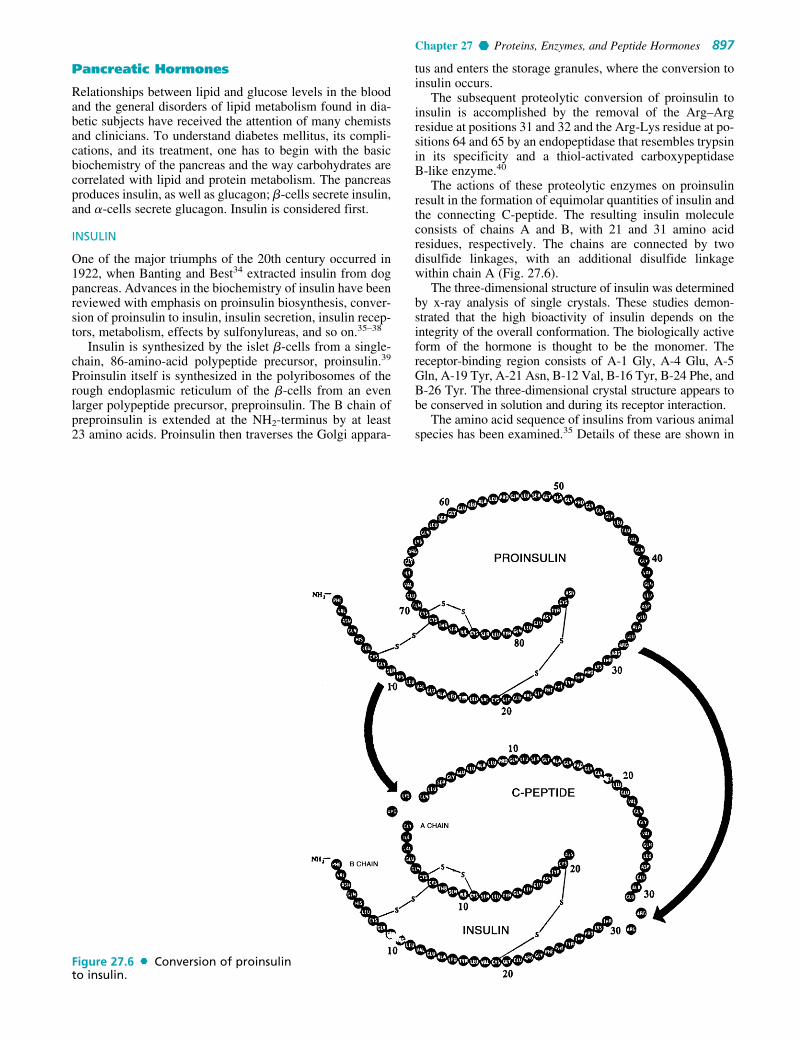
Chapter 27 Proteins, Enzymes, and Peptide Hormones 897
Pancreatic Hormones
Relationships between lipid and glucose levels in the bloodand the general disorders of lipid metabolism found in dia-betic subjects have received the attention of many chemistsand clinicians. To understand diabetes mellitus, its compli-cations, and its treatment, one has to begin with the basicbiochemistry of the pancreas and the way carbohydrates arecorrelated with lipid and protein metabolism. The pancreasproduces insulin, as well as glucagon; �-cells secrete insulin,and �-cells secrete glucagon. Insulin is considered first.
INSULIN
One of the major triumphs of the 20th century occurred in1922, when Banting and Best34 extracted insulin from dogpancreas. Advances in the biochemistry of insulin have beenreviewed with emphasis on proinsulin biosynthesis, conver-sion of proinsulin to insulin, insulin secretion, insulin recep-tors, metabolism, effects by sulfonylureas, and so on.35–38
Insulin is synthesized by the islet �-cells from a single-chain, 86-amino-acid polypeptide precursor, proinsulin.39
Proinsulin itself is synthesized in the polyribosomes of therough endoplasmic reticulum of the �-cells from an evenlarger polypeptide precursor, preproinsulin. The B chain ofpreproinsulin is extended at the NH2-terminus by at least23 amino acids. Proinsulin then traverses the Golgi appara-
tus and enters the storage granules, where the conversion toinsulin occurs.
The subsequent proteolytic conversion of proinsulin toinsulin is accomplished by the removal of the Arg–Argresidue at positions 31 and 32 and the Arg-Lys residue at po-sitions 64 and 65 by an endopeptidase that resembles trypsinin its specificity and a thiol-activated carboxypeptidase B-like enzyme.40
The actions of these proteolytic enzymes on proinsulinresult in the formation of equimolar quantities of insulin andthe connecting C-peptide. The resulting insulin moleculeconsists of chains A and B, with 21 and 31 amino acidresidues, respectively. The chains are connected by twodisulfide linkages, with an additional disulfide linkagewithin chain A (Fig. 27.6).
The three-dimensional structure of insulin was determinedby x-ray analysis of single crystals. These studies demon-strated that the high bioactivity of insulin depends on theintegrity of the overall conformation. The biologically activeform of the hormone is thought to be the monomer. Thereceptor-binding region consists of A-1 Gly, A-4 Glu, A-5Gln, A-19 Tyr, A-21 Asn, B-12 Val, B-16 Tyr, B-24 Phe, andB-26 Tyr. The three-dimensional crystal structure appears tobe conserved in solution and during its receptor interaction.
The amino acid sequence of insulins from various animalspecies has been examined.35 Details of these are shown in
Figure 27.6 Conversion of proinsulinto insulin.
0880-0914_17865_Ch27.qxd 12/2/09 5:06 AM Page 897

Table 27.6. It is apparent from the analysis that frequentchanges in sequence occur within the interchain disulfidering (positions 8, 9, and 10). The hormonal sequence forporcine insulin is the closest to that of humans, differingonly by the substitution of an alanine residue at the COOH-terminus of the B chain. Porcine insulin, therefore, is a goodstarting material for the synthesis of human insulin.
Insulin composes 1% of pancreatic tissue, and secretoryprotein granules contain about 10% insulin. These granulesfuse with the cell membrane with simultaneous liberation ofequimolar amounts of insulin and the C-peptide. Insulin en-ters the portal vein, and about 50% is removed in its firstpassage through the liver. The plasma half-life of insulin isapproximately 4 minutes, compared with 30 minutes for theC-peptide.
Usually, exogenous insulin is weakly antigenic. Insulinantibodies have been observed to neutralize the hypo-glycemic effect of injected insulin. The antibody-bindingsites on insulin are quite different from the sites involved inbinding of insulin with its receptors.41
Regulation of insulin secretion is affected by numerousfactors, such as food, hormonal and neuronal stimuli, andionic mechanisms.42 In humans, the principal substrate thatstimulates the release of insulin from the islet �-cells is glu-cose. In addition to glucose, other substrates (e.g., aminoacids, free fatty acids, and ketone bodies) also can stimulateinsulin secretion directly. Secretin and ACTH can directlystimulate insulin secretion. Glucagon and other related pep-tides can increase the secretion of insulin, whereas somato-statin inhibits its secretion.
Autonomic neuronal mechanisms also play an importantrole in regulating insulin release. In the sympathetic nervoussystem, �-adrenergic agonists inhibit insulin release, whereas�-adrenergic agonists stimulate the release of insulin. In theparasympathetic nervous system, cholinomimetic drugs stim-ulate insulin release.
“Clinical” insulin that has been crystallized five times andthen subjected to countercurrent distribution (2-butanol:1%dichloroacetic acid in water) yields about 90% insulin A,with varying amounts of insulin B together with other minorcomponents. A and B differ by an amide group and have thesame activity. End-member analysis, sedimentation, and dif-fusion studies indicate an Mr of about 6,000. The value of12,000 Mr for insulin containing trace amounts of zinc(obtained by physical methods) is probably a bimolecular
association product through the aid of zinc. Insulin was thefirst protein for which a complete amino acid sequence wasdetermined. The extensive studies of Sanger43 and otherselucidated the amino acid sequence and structure of insulin.Katsoyannis44 and others followed with the synthesis of Aand B chains of human, bovine, and sheep insulin. The A andB chains were combined to form insulin in 60% to 80%yields, with a specific activity comparable to that of the nat-ural hormone.
The total synthesis of human insulin was reported byRittel et al.45 These workers selectively synthesized the finalmolecule appropriately cross-linked by disulfide (-S-S-)groups in yields ranging between 40% and 50%, whereasearlier synthetic methods involved random combination ofseparately prepared A and B chains of the molecule.
rDNA technology has been applied successfully in theproduction of human insulin on a commercial scale. Humaninsulin is produced in genetically engineered Escherichiacoli.46 Eli Lilly and Co., in cooperation with Genentech,began marketing rDNA-derived human insulin (Humulin) in1982. There are two available methods of applying rDNAtechnology in the production of human insulin. The earliermethod involved insertion of genes, for production of eitherthe A or the B chain of the insulin molecule, into a specialstrain of E. coli (KI2) and subsequently combining the twochains chemically to produce an insulin that is structurallyand chemically identical with pancreatic human insulin. Thesecond, and more recent, method involves the insertion ofgenes for the entire proinsulin molecule into special E. colicells that are then grown via fermentation. The connectingC-peptide is then enzymatically cleaved from proinsulin toproduce human insulin.47 Human insulin produced by rDNAtechnology is less antigenic than that from animal sources.
Although insulin is readily available from natural sources(e.g., porcine and bovine pancreatic tissue), partial synthesesand molecular modifications have been developed as the basisfor SAR studies. Such studies have shown that amino acidunits cannot be removed from the insulin peptide chain Awithout significant loss of hormonal activity. Several aminoacids of chain B, however, are not considered essential foractivity. Up to the first six and the last three amino acid unitscan be removed without significant decrease in activity.23
Two insulin analogs, which differ from the parent hor-mone in that the NH2-terminus of chain A (A1) glycine hasbeen replaced by L- and D-alanine, respectively, have been
898 Wilson and Gisvold’s Textbook of Organic Medicinal and Pharmaceutical Chemistry
TABLE 27.6 Some Sequence Differences in Insulins of Various Species
A Chain B Chain
Species 1 8 9 10 –1 1 29 30
Human Gly. Thr. Ser. Ile. Phe. Lys. Thr.Pork Gly. Thr. Ser. Ile. Phe. Lys. Ala.Beef Gly. Ala. Ser. Val. Phe. Lys. Ala.Sheep Gly. Ala. Gly. Val. Phe. Lys. Ala.Horse Gly. Thr. Gly. Ile. Phe. Lys. Ala.Rabbit Gly. Thr. Ser. Ile. Phe. Lys. Ser.Chicken Gly. His. Asn. Thr. Ala. Lys. Ala.Cod Gly. His. Arg. Pro. Met. Ala. Lys. —Rat Ia Gly. Thr. Ser. Ile. Phe. Lys. Ser.Rat IIa Gly. Thr. Ser. Ile. Phe. Met. Ser.
See Reference 55 for details.aAsp substitution for Glu at position 4 on A chain.
0880-0914_17865_Ch27.qxd 12/2/09 5:06 AM Page 898

Chapter 27 Proteins, Enzymes, and Peptide Hormones 899
synthesized by Cosmatos et al.48 for SAR studies. Therelative potencies of the L and D analogs reveal interestingSARs. The L- and D-alanine analogs are 9.4% and 95%, re-spectively, as potent as insulin in glucose oxidation. Therelative binding affinity to isolated fat cells is reported to beapproximately 10% for the L and 100% for the D analog.Apparently, substitution on the � carbon of A1 glycine ofinsulin, with a methyl in a particular configuration, inter-feres with the binding; hence, the resulting analog (that ofL-alanine) is much less active. Methyl substitution in theopposite configuration affects neither the binding nor thebioactivity.
Molecular modifications of insulin on the amino groupsappear to reduce bioactivity, but modifications of the �-aminogroup of lysine number 29 on chain B (B-29) may yield activeanalogs. Accordingly, May et al.49 synthesized N-�-(�)-biotinyl insulin, which was equipotent with natural insulin.Complexes of this biotinyl-insulin derivative with avidin alsowere prepared and evaluated biologically; these complexesshowed a potency decrease to 5% of that of insulin. Suchcomplexes conjugated with ferritin are expected to be usefulin the development of electron microscope stains of insulinreceptors.
Alteration in the tertiary structure of insulin appears todrastically reduce biological activity as well as receptorbinding. The three-dimensional structure provided by x-raycrystallography of the insulin monomer has revealed an ex-posed hydrophobic face that is thought to be involveddirectly in interacting with the receptor.50 Thus, loss of bio-logical activity in insulin derivatives, produced by chemicalmodification, can be interpreted in terms of adversely affect-ing this hydrophobic region. Also, species variation in thishydrophobic region is very unusual.
Insulin is inactivated in vivo by (a) an immunochemicalsystem in the blood of insulin-treated patients, (b) reductionof disulfide bonds (probably by glutathione), and (c) insuli-nase (a proteolytic enzyme) that occurs in liver. Pepsin andchymotrypsin hydrolyze some peptide bonds that lead to in-activation. Insulin is inactivated by reducing agents such assodium bisulfite, sulfurous acid, and hydrogen.
Advances in the area of insulin’s molecular mechanismshave been reviewed36–38,51 with emphasis on receptor inter-actions, effect on membrane structure and functions, effectson enzymes, and the role of second messengers. The insulinreceptor is believed to be a glycoprotein complex with a highMr. The receptor is thought to consist of four subunits: twoidentical � units with an Mr of about 130,000 d and twoidentical � units with an Mr of 95,000 d, joined together bydisulfide bonds. The � subunits are primarily responsible forbinding insulin to its receptor, and the � subunits are thoughtto possess intrinsic protein kinase activity that is stimulatedby insulin. The primary effect of insulin may be a kinasestimulation leading to phosphorylation of the receptor aswell as other intracellular proteins.52,53 Additionally, insulinbinding to its receptors may result in the generation of a sol-uble intracellular second messenger (possibly a peptide) thatmay mediate some insulin activity relating to activation ofenzymes such as pyruvate dehydrogenase and glycogen syn-thetase. The insulin–receptor complex becomes internalizedand may serve as a vehicle for translocating insulin to thelysosomes, in which it may be broken down and recycledback to the plasma membrane. The half-life of insulin isabout 10 hours.
The binding of insulin to its target tissue is determined byseveral factors. The number of receptors in the target tissueand their affinity for insulin are two important determinants.These factors vary substantially from tissue to tissue.Another important consideration is the concentration of in-sulin itself. Elevated levels of circulating insulin decreasethe number of insulin receptors on target cell surfaces andvice versa. Other factors that affect insulin binding to its re-ceptors include pH, temperature, membrane lipid composi-tion, and ionic strength.53 It is conceivable, therefore, thatconditions associated with insulin resistance, such as obe-sity and type 1 and type 2 diabetes mellitus, could be causedby altered receptor kinase activity, or impaired generation ofsecond messengers (low-Mr peptides), increased degrada-tion of the messenger, or fewer substrates (enzymes in-volved in metabolic activity) for the messenger or receptorkinase.54
Metabolic Effects of Insulin. Insulin has pronouncedeffects on the metabolism of carbohydrates, lipids, and pro-teins.55 The major tissues affected by insulin are muscle(cardiac and skeletal), adipose tissue, and liver. The kidneyis much less responsive, and others (e.g., brain tissue and redblood cells) do not respond at all. The actions of insulin arehighly complex and diverse. Because many of the actions ofinsulin are mediated by second messengers, it is difficult todistinguish between its primary and secondary actions.
In muscle and adipose tissue, insulin promotes transportof glucose and other monosaccharides across cell mem-branes; it also facilitates transport of amino acids, potassiumions, nucleosides, and ionic phosphate. Insulin also activatescertain enzymes—kinases and glycogen synthetase in mus-cle and adipose tissue. In adipose tissue, insulin decreases therelease of fatty acids induced by epinephrine or glucagon.cAMP promotes fatty acid release from adipose tissue; there-fore, it is possible that insulin decreases fatty acid release byreducing tissue levels of cAMP. Insulin also facilitates theincorporation of intracellular amino acids into protein.
Insulin is believed to influence protein synthesis at the ri-bosomal level in various tissues.56 In skeletal muscles, in-sulin predominantly stimulates translation by increasing therate of initiation of protein synthesis and the number of ribo-somes. In the liver, the predominant effect is on transcription.In cardiac muscles, insulin is believed to decrease the rate ofprotein degradation.
In the liver, there is no barrier to the transport of glucoseinto cells; nevertheless, insulin influences liver metabolism,decreasing glucose output, decreasing urea production,lowering cAMP levels, and increasing potassium and phos-phate uptake. The lower cAMP levels result in decreased ac-tivity of glycogen phosphorylase, leading to diminishedglycogen breakdown and increased activity of glycogen syn-thetase. It appears that insulin induces specific hepatic en-zymes involved in glycolysis while inhibiting gluconeogenicenzymes. Thus, insulin promotes glucose use through gly-colysis by increasing the synthesis of glucokinase, phospho-fructokinase, and pyruvate kinase. Insulin decreases theavailability of glucose from gluconeogenesis by suppressingpyruvate carboxylase, phosphoenolpyruvate carboxykinase,fructose-1,6-diphosphatase, and glucose-6-phosphatase.
Insulin’s effects on lipid metabolism also are important.In adipose tissue, it has an antilipolytic action (i.e., an effectopposing the breakdown of fatty acid triglycerides). It also
0880-0914_17865_Ch27.qxd 12/2/09 5:06 AM Page 899

decreases the supply of glycerol to the liver. Thus, at thesetwo sites, insulin decreases the availability of precursors forthe formation of triglycerides. Insulin is necessary for theactivation and synthesis of lipoprotein lipases, enzymes re-sponsible for lowering very low-density lipoprotein (VLDL)and chylomicrons in peripheral tissue. Other effects includestimulation of the synthesis of fatty acids (lipogenesis) inthe liver.
Diabetes mellitus is a systemic disease caused by a de-crease in the secretion of insulin or reduced sensitivity orresponsiveness to insulin by target tissue (insulin receptoractivity). The disease is characterized by hyperglycemia,hyperlipidemia, and hyperaminoacidemia. Diabetes mellitusfrequently is associated with the development of microvas-cular and macrovascular diseases, neuropathy, and athero-sclerosis. Various types of diabetes have been recognizedand classified and their pathophysiology discussed.57
The two major types of diabetes are type 1, insulin-dependent diabetes mellitus (IDDM), and type 2, non–insulin-dependent diabetes mellitus (NIDDM). Type 1 dia-betes (also known as juvenile-onset diabetes) is character-ized by a destruction of pancreatic �-cells, resulting in a de-ficiency of insulin secretion. Autoimmune complexes andviruses have been mentioned as two possible causes of �-cell destruction. Generally, in type 1 diabetes, receptor sen-sitivity to insulin is not decreased. Type 2 diabetes, alsoknown as adult-onset diabetes, is characterized primarily byinsulin receptor defects or postinsulin receptor defects.There is no destruction of �-cells, and insulin secretion isrelatively normal. In reality, however, the two types of dia-betes show a considerable overlap of clinical features.57
Diabetes mellitus is associated with both microangiopa-thy (damage to smaller vessels, e.g., the eyes and kidney) andmacroangiopathy (damage to larger vessels, e.g., atheroscle-rosis). Hyperlipidemia (characterized by an increase in theconcentration of lipoproteins such as VLDL, intermediatedensity lipoprotein [IDL], and LDL) has been implicated inthe development of atherosclerosis and is known to occur indiabetes. Severe hyperlipidemia may lead to life-threateningattacks of acute pancreatitis. It also seems that severe hyper-lipidemia causes xanthoma. Considering the effects of in-sulin on lipid metabolism, as summarized previously, onecan rationalize that in type 2 diabetes, in which the patientmay actually have an absolute excess of insulin, in spite ofthe evidence of glucose tolerance tests, the effect of the ex-cessive insulin on lipogenesis in the liver may suffice to in-crease the levels of circulating triglycerides and VLDL. Intype 1 diabetes, with a deficiency of insulin, the circulatinglevel of lipids may rise because too much precursor is avail-able, with fatty acids and carbohydrates going to the liver.
The relationship between the carbohydrate metabolicmanifestations of diabetes and the development of mi-crovascular and macrovascular diseases has been studiedextensively.58,59 It is becoming increasingly clear that hy-perglycemia plays a major role in the development of vascu-lar complications of diabetes, including intercapillaryglomerulosclerosis, premature atherosclerosis, retinopathywith its specific microaneurysms and retinitis proliferans,leg ulcers, and limb gangrene. First, hyperglycemia causesan increase in the activity of lysine hydroxylase and galac-tosyl transferase, two important enzymes involved in glyco-protein synthesis. Increased glycoprotein synthesis in thecollagen of kidney basement membrane may lead to the de-
velopment of diabetic glomerulosclerosis. Second, in-creased uptake of glucose by noninsulin-sensitive tissues(e.g., nerve Schwann cells and ocular lens cells) occurs dur-ing hyperglycemia. Intracellular glucose is converted enzy-matically first to sorbitol and then to fructose. The buildupof these sugars inside the cells increases the osmotic pres-sure in ocular lens cells and Schwann cells, resulting in in-creased water uptake and impairing cell functions. Someforms of diabetic cataracts and diabetic neuropathy are be-lieved to be caused by this pathway. Third, hyperglycemiamay precipitate nonenzymatic glycosylation of various pro-teins in the body, including hemoglobin, serum albumin,lipoprotein, fibrinogen, and basement membrane protein.Glycosylation is believed to alter the tertiary structures ofproteins and possibly their rate of metabolism. The rate ofglycosylation is a function of plasma glucose concentrationand the duration of hyperglycemia. Needless to say, thismechanism might play an important role in both macrovas-cular and microvascular lesions. Finally, hyperglycemiaincreases the rate of aggregation and agglutinization ofcirculating platelets. Platelets play an important role in pro-moting atherogenesis. The increase in the rate of platelet ag-gregation and agglutinization leads to the development ofmicroemboli, which can cause transient cerebral ischemicattacks, strokes, and heart attacks.57
Concepts of the therapeutics of diabetes mellitus havebeen reviewed by Maurer.60 This review emphasizes thatinsulin therapy does not always prevent serious complica-tions. Even diabetic patients considered under insulin thera-peutic control experience wide fluctuations in blood glucoseconcentration, and it is hypothesized that these fluctuationseventually cause the serious complications of diabetes (e.g.,kidney damage, retinal degeneration, premature atheroscle-rosis, cataracts, neurological dysfunction, and a predisposi-tion to gangrene).
Insulin Preparations. The various commercially avail-able insulin preparations are listed in Table 27.7. Amorphousinsulin was the first form made available for clinical use.Further purification afforded crystalline insulin, which isnow commonly called “regular insulin.” Insulin injection,USP, is made from zinc insulin crystals. For some time, reg-ular insulin solutions have been prepared at a pH of 2.8 to3.5; if the pH were increased above the acidic range, particleswould be formed. More highly purified insulin, however, canbe maintained in solution over a wider pH range, even whenunbuffered. Neutral insulin solutions have greater stabilitythan acidic solutions; neutral insulin solutions maintainnearly full potency when stored up to 18 months at 5°C and25°C. As noted in Table 27.7, the various preparations differin onset and duration of action. A major disadvantage of reg-ular insulin is its short duration of action (5–7 hours), whichnecessitates its administration several times daily.
Many attempts have been made to prolong the durationof action of insulin, for example, development of insulinforms less water soluble than the highly soluble (in bodyfluids) regular insulin. Protamine insulin preparationsproved to be less soluble and less readily absorbed frombody tissue. Protamine zinc insulin (PZI) suspensions wereeven longer acting (36 hours) than protamine insulin; theseare prepared by mixing insulin, protamine, and zinc chloridewith a buffered solution. The regular insulin/PZI ratios inclinically useful preparations range from 2:1 to 4:1.
900 Wilson and Gisvold’s Textbook of Organic Medicinal and Pharmaceutical Chemistry
0880-0914_17865_Ch27.qxd 12/2/09 5:06 AM Page 900

Chapter 27 Proteins, Enzymes, and Peptide Hormones 901
Isophane insulin suspension incorporates some of the qual-ities of regular insulin injection and is usually long actingenough (although not as much as PZI) to protect the patientfrom first day to the next (the term isophane is derived fromthe Greek iso and phane, meaning equal and appearance,respectively). Isophane insulin is prepared by careful controlof the protamine/insulin ratio and the formation of a crys-talline entity containing stoichiometric amounts of insulin andprotamine. (Isophane insulin also is known as NPH; the N in-dicates neutral pH, the P stands for protamine, and the H forHegedorn, the developer of the product.) NPH insulin has aquicker onset and a shorter duration of action (28 hours) thanPZI. NPH is given in single morning doses and normally ex-hibits greater activity during the day than at night. NPH andregular insulin can be combined conveniently and effectivelyfor many patients with diabetes.
The posology of various insulin preparations is summa-rized in Table 27.7.
A major concern with PZI and NPH insulins is the po-tential antigenicity of protamine (obtained from fish). Thisconcern led to the development of lente insulins. By vary-ing the amounts of excess zinc, by using an acetate buffer(instead of phosphate), and by adjusting the pH, two typesof lente insulin were prepared. At high concentrations ofzinc, a microcrystalline form precipitates and is called ul-tralente. Ultralente insulin is relatively insoluble and has aslower onset and a longer duration of action than PZI. At arelatively low zinc concentration, an amorphous form pre-cipitates and is called semilente insulin. The latter is moresoluble and has a quicker onset and a shorter duration of ac-tion than regular insulins. A third type of insulin suspen-sion, lente insulin, is a 70:30 mixture of ultralente andsemilente insulins. Lente insulin has a rapid onset and anintermediate duration of action (comparable to that of NPHinsulin). Lente insulins are chemically incompatible withthe PZI and NPH insulins because of the different buffersystem used in the preparation of these insulins (an acetatebuffer is used in lente insulins and a phosphate buffer isused in PZI and NPH insulins). Dosage and sources aresummarized in Table 27.8.
Additionally, regular insulin will remain fast acting whencombined with NPH but not when added to lente. The rapidaction of regular insulin is neutralized by the excess zincpresent in lente insulin.54 Similar products61 containingrDNA-derived human insulin (instead of the bovine- andporcine-derived insulin) are available.
Progress in alternative routes of delivery of insulin hasbeen prompted by problems associated with conventional in-sulin therapy, mentioned previously. First, various types ofelectromechanical devices (infusion pumps) have been de-veloped with the aim of reducing fluctuations in blood glu-cose levels associated with conventional insulin therapy(subcutaneous injections). These continuous-infusion pumpsare either close-loop or open-loop systems. The ultimate goalof research in this area is to develop a reliable implantable(miniature) device for long-term use that would eliminate theneed for daily administration and monitoring of blood glu-cose levels. The second area of research studies alternativeroutes of administration such as oral, nasal, and rectal.Preliminary results indicate that absorption of insulin at thesesites is not uniform and is unpredictable. The third approachto correcting the problems of conventional insulin therapy isto supplement the defective pancreas by transplantation witha normally functioning pancreas from an appropriate donor.The major problem with this approach is rejection of thedonor pancreas by the recipient, as well as problems associ-ated with the draining of exocrine enzymes. A modified pro-cedure transplants only viable pancreatic islet cells or fetal orneonatal pancreas. The possibility remains, however, that intype 1 diabetes, the newly transplanted pancreatic �-cellscould be destroyed by the same autoimmune process thatcaused the disease in the first place.
GLUCAGON
Glucagon, USP. The hyperglycemic–glycogenolytic hor-mone elaborated by the �-cells of the pancreas is known asglucagon. It contains 29 amino acid residues in the sequenceshown. Glucagon has been isolated from the amorphous frac-tion of a commercial insulin sample (4% glucagon).
TABLE 27.7 Insulin Preparations
Particle Size DurationName (�m) Action Composition pH (hours)
Insulin injection,a USP Prompt Insulin � ZnCl2 2.5–3.5 5–7Prompt insulin zinc 2b Rapid Insulin � ZnCl2 � buffer 7.2–7.5 12
suspension,a USPInsulin zinc 10–40 (70%) 2 Intermediate Insulin � ZnCl2 � buffer 7.2–7.5 18–24
suspension,a USP (30%)b
Extended insulin zinc 10–40 Long acting Insulin � ZnCl2 � buffer 7.2–7.5 24–36suspension,a USP
Globin zinc insulin Intermediate Globinc � ZnCl2 � insulin 3.4–3.8 12–18injectiona USP
Protamine zinc insulin Long acting Protaminee � insulin � Zn 7.1–7.4 24–36suspension,d USP
Isophane insulin 30 Intermediate Protaminef ZnCl2 insulin buffer 7.1–7.4 18–24suspension,a USP
aClear or almost clear.bAmorphous.cGlobin (3.6–4.0 mg/100 USP units of insulin) prepared from beef blood.d Turbid.eProtamine (1.0–1.5 mg/100 USP units of insulin) from the sperm or the mature testes of fish belonging tothe genus Oncorhynchus or Salmo.f Protamine (0.3–0.6 mg/100 USP units of insulin).
0880-0914_17865_Ch27.qxd 12/2/09 5:06 AM Page 901

Attention has been focused on glucagon as a factor in thepathology of human diabetes. According to Unger et al.,62 thefollowing observations support this implication of glucagon:elevated glucagon blood levels (hyperglucagonemia) havebeen observed in association with every type of hyper-glycemia; when secretion of both glucagon and insulin is sup-pressed, hyperglycemia is not observed unless the glucagonlevels are restored to normal by the administration of glucagon;the somatostatin-induced suppression of glucagon release indiabetic animals and humans restores blood sugar levels tonormal and alleviates certain other symptoms of diabetes.
Unger et al.62 propose that although the major role ofinsulin is regulation of the transfer of glucose from theblood to storage in insulin-responsive tissues (e.g., liver, fat,and muscle), the role of glucagon is regulation of the liver-mediated mobilization of stored glucose. The principalconsequence of high concentrations of glucagon is liver-mediated release into the blood of abnormally high concen-
trations of glucose, thereby causing persistent hyper-glycemia. This indicates that a relative excess of glucagon isan essential factor in the development of diabetes.
Glucagon’s solubility is 50 �g/mL in most buffers betweenpH 3.5 and 8.5. It is soluble, 1 to 10 mg/mL, in the pH ranges2.5 to 3.0 and 9.0 to 9.5. Solutions of 200 �g/mL at pH 2.5 to3.0 are stable for at least several months at 4°C if sterile. Lossof activity by fibril formation occurs readily at high concentra-tions of glucagon at room temperature, or above, at pH 2.5.The isoelectric point appears to be at pH 7.5 to 8.5. Because ithas been isolated from commercial insulin, its stability proper-ties should be comparable to those of insulin.
As with insulin and some of the other polypeptide hor-mones, glucagon-sensitive receptor sites in target cells bindglucagon. This hormone–receptor interaction leads to activa-tion of membrane adenylate cyclase, which catalyzes cAMPformation. Thus, intracellular cAMP levels are elevated. Themode of action of glucagon in glycogenolysis is basically thesame as the mechanism of epinephrine (i.e., stimulation ofadenylate cyclase). Subsequently, the increase in cAMP acti-vates the protein kinase that catalyzes phosphorylation ofphosphorylase kinase to phosphophosphorylase kinase. Thelatter is necessary for the activation of phosphorylase to formphosphorylase a. Finally, phosphorylase a catalyzesglycogenolysis, which is the basis for the hyperglycemic ac-tion of glucagon. Although both glucagon and epinephrineexert hyperglycemic action through cAMP, glucagon affectsliver cells and epinephrine affects both muscle and liver cells.
Fain63 reviewed the many phenomena associated withhormones, membranes, and cyclic nucleotides, includingseveral factors that activate glycogen phosphorylase in ratliver. These factors involve not only glucagon but also vaso-pressin and the catecholamines. Glucagon and �-cate-cholamines mediate their effects on glycogen phosphorylasethrough cAMP, but may involve other factors as well.
Glucagon exerts other biochemical effects. Gluconeogen-esis in the liver is stimulated by glucagon, and this is accom-panied by enhanced urea formation. Glucagon inhibits the
902 Wilson and Gisvold’s Textbook of Organic Medicinal and Pharmaceutical Chemistry
TABLE 27.8 Dosage and Source of Insulin Preparations
USP Insulin Type Strengths and Sources Usual Adult Dosea
Insulin injection (regular insulin, U-40 mixed, Diabetic hyperglycemia: SC, as directed by crystalline zinc insulin, Cispro) U-100 mixed: purified beef, pork; purified physician 15–30 minutes before meals up to
pork; biosynthetic human; semisynthetic t.i.d. or q.i.d.human
U-500: purified porkIsophane insulin suspension (NPH U-40 mixed, SC, as directed by physician, q.d. 30–60 minutes
insulin) U-400 mixed: beef; purified beef, pork; before breakfast; an additional dose before purified pork; biosynthetic human; breakfast may be necessary for some patients semisynthetic human about 30 minutes before a meal or at bedtime
Isophane insulin suspension (70%) U-100: purified pork; semisynthetic human SC, as directed by physician, q.d. 15–30 minutesand insulin injection (30%) before breakfast, or as directed
Insulin zinc suspension (Lente U-40 mixed, SC, as directed by physician, q.d. 30–60 minutesinsulin) U-100 mixed: beef; purified beef; purified before breakfast; an additional dose may be
pork; biosynthetic human; semisynthetic necessary for some patients about 30 minutes human before a meal or at bedtime
Extended insulin zinc suspension U-40 mixed, SC, as directed by physician, q.d. 30–60 minutes(Ultralente insulin) U-100 mixed: beef; purified beef before breakfast
Prompt insulin zinc suspension U-40 mixed, SC, as directed by physician, q.d. 30–60 minutes(Semilente insulin) U-100 mixed: beef; purified pork before breakfast; an additional dose may be
necessary for some patients about 30 minutesbefore a meal or at bedtime
Protamine zinc insulin suspension U-40 mixed, SC, as directed by physician, q.d. 30–60 minutes(PZI Insulin) U-100 mixed: purified pork before breakfast
aSee USP DI for complete dosage information.SC, subcutaneously.
0880-0914_17865_Ch27.qxd 12/2/09 5:06 AM Page 902

Chapter 27 Proteins, Enzymes, and Peptide Hormones 903
incorporation of amino acids into liver proteins. Fatty acidsynthesis is decreased by glucagon. Cholesterol formation isalso reduced. Glucagon activates liver lipases, however, andstimulates ketogenesis. Ultimately, the availability of fattyacids from liver triglycerides is elevated, fatty acid oxidationincreases acetyl-CoA and other acyl-CoAs, and ketogenesisis promoted. As glucagon effects elevation of cAMP levels,release of glycerol and free fatty acids from adipose tissuealso is increased.
Glucagon, whose regulatory effect on carbohydrate andfatty acid metabolism is well understood, is therapeuticallyimportant. It is recommended for the treatment of severe hy-poglycemic reactions caused by the administration of in-sulin to diabetic or psychiatric patients. Of course, this treat-ment is effective only when hepatic glycogen is available.Nausea and vomiting are the most frequently encounteredreactions to glucagon.
Usual dose: parenteral, adults, 500 �g to 1 mg (0.5–1.0 U),repeated in 20 minutes if necessary; pediatric, 25 �g/kg ofbody weight, repeated in 20 minutes if necessary.
Gastrointestinal Hormones
There is a formidable array of polypeptide hormones of thegastrointestinal tract that includes secretin, pancreozymin–cholecystokinin, gastrin, motilin, neurotensin, vasoactiveintestinal peptide, somatostatin, and others. The biosynthe-sis, chemistry, secretion, and actions of these hormoneshave been reviewed.64
GASTRIN
Gastrin is a 17-residue polypeptide isolated from the antralmucosa. It was isolated originally in two different forms. Inone of the forms, the tyrosine residue in position 12 is sul-fated. Both forms are biologically active. Cholinergic re-sponse to the presence of food in the gastrointestinal tract pro-vides the stimulus for gastrin secretion. The lowering of pHin the stomach inhibits the secretion of gastrin. The effects ofstructural modification of gastrin on gastric acid secretionhave been reviewed.65 These studies revealed that the fourresidues at the COOH terminus retain significant biologicalactivity and that the aspartate residue is the most critical foractivity. The most important action of gastrin is to stimulatethe secretion of gastric acid and pepsin. Other actions of gas-trin include increased secretion of pancreatic enzymes; con-traction of smooth muscles; water and electrolyte secretion bythe stomach and pancreas; water and electrolyte absorption bythe small intestine; and secretion of insulin, glucagon, and so-matostatin. A synthetic pentapeptide derivative, pentagastrin,is currently used as a gastric acid secretagogue.
Pentagastrin. Pentagastrin (Peptavlon), a physiologicalgastric acid secretagogue, is the synthetic pentapeptide de-rivative: N-t-butyloxycarbonyl-�-alanyl-L-tryptophyl-L-me-thionyl-L-aspartyl-L-phenylalanyl amide. It contains theCOOH-terminal tetrapeptide amide (H � Try � Met � Asp �Phe � NH2), which is considered to be the active center ofthe natural gastrins. Accordingly, pentagastrin appears tohave the physiological and pharmacological properties ofthe gastrins, including stimulation of gastric secretion,pepsin secretion, gastric motility, pancreatic secretion ofwater and bicarbonate, pancreatic enzyme secretion, biliaryflow and bicarbonate output, intrinsic factor secretion, andcontraction of the gallbladder.
Pentagastrin is indicated as a diagnostic agent to evaluategastric acid secretory function, and it is useful in testing foranacidity in patients with suspected pernicious anemia, at-rophic gastritis or gastric carcinoma, hypersecretion in sus-pected duodenal ulcer or postoperative stomal ulcers, andZollinger-Ellison tumor.
Pentagastrin is usually administered subcutaneously; theoptimal dose is 6 �g/kg. Gastric acid secretion begins ap-proximately 10 minutes after administration, and peak re-sponses usually occur within 20 to 30 minutes. The usualduration of action is from 60 to 80 minutes. Pentagastrin hasa relatively short plasma half-life, perhaps less than 10 min-utes. The available data from metabolic studies indicate thatpentagastrin is inactivated by the liver, kidney, and tissuesof the upper intestine.
Contraindications include hypersensitivity or idiosyn-crasy to pentagastrin. It should be used with caution in pa-tients with pancreatic, hepatic, or biliary disease.
SECRETIN
Secretin is a 27-amino-acid polypeptide that is structurallysimilar to glucagon. The presence of acid in the small intestineis the most important physiological stimulus for the secretionof secretin. The primary action of secretin is on pancreatic aci-nar cells that regulate the secretion of water and bicarbonate.Secretin also promotes the secretion of pancreatic enzymes, toa lesser extent. Secretin inhibits the release of gastrin and,therefore, gastric acid. It also increases stomach-emptyingtime by reducing the contraction of the pyloric sphincter.64
0880-0914_17865_Ch27.qxd 12/2/09 5:06 AM Page 903

CHOLECYSTOKININ–PANCREOZYMIN
It was thought originally that cholecystokinin and pancre-ozymin were two different hormones. Cholecystokinin wasthought to be responsible for contraction of the gallbladder,whereas pancreozymin was believed to induce secretion ofpancreatic enzymes. It is now clear that both actions arecaused by a single 33-residue polypeptide, referred to ascholecystokinin–pancreozymin (CCK-PZ). CCK-PZ is se-creted in the blood in response to the presence of food in theduodenum, especially long-chain fatty acids. The fiveCOOH-terminal amino acid residues are identical with thosein gastrin. The COOH-terminal octapeptide retains full ac-tivity of the parent hormone.
The octapeptide is found in the gut as well as the centralnervous system. SARs of cholecystokinin have been re-viewed.64 The COOH-terminal octapeptide is present in sig-nificant concentrations in the central nervous system. Itspossible actions here, the therapeutic implications in thetreatment of Parkinson disease and schizophrenia, and itsSAR have been reviewed.65
VASOACTIVE INTESTINAL PEPTIDE
Vasoactive intestinal peptide (VIP) is widely distributed inthe body and is believed to occur throughout the gastroin-testinal tract. It is a 28-residue polypeptide with structuralsimilarities to secretin and glucagon. It causes vasodilata-tion and increases cardiac contractibility. VIP stimulates bi-carbonate secretion, relaxes gastrointestinal and othersmooth muscles, stimulates glycogenesis, inhibits gastricacid secretion, and stimulates insulin secretion. Its hormonaland neurotransmitter role has been investigated.66
GASTRIC INHIBITORY PEPTIDE
Gastric inhibitory peptide (GIP) is a 43-amino-acidpolypeptide isolated from the duodenum. Secretion of GIPinto the blood is stimulated by food. The primary action ofGIP is inhibition of gastric acid secretion. Other actions in-clude stimulation of insulin and glucagon secretion andstimulation of intestinal secretion.64
MOTILIN
Motilin is a 22-residue polypeptide isolated from the duode-num. Its secretion is stimulated by the presence of acid inthe duodenum. Motilin inhibits gastric motor activity anddelays gastric emptying.
NEUROTENSIN
Neurotensin is a 13-amino-acid peptide, first isolated frombovine hypothalamus. It has now been identified in the intes-tinal tract. The ileal mucosa contains 90% of the total neu-rotensin of the body. It is implicated as a releasing factor forseveral adenohypophyseal hormones. It causes vasodilata-tion, increases vascular permeability, and increases gastrinsecretion. It decreases secretion of gastric acid and secretin.
Parathyroid Hormone
This hormone is a linear polypeptide containing 84 aminoacid residues. SAR studies67 of bovine parathyroid hormonerevealed that the biological activity is retained by an NH2-terminal fragment consisting of eight amino acid residues. Itregulates the concentration of calcium ion in the plasmawithin the normal range, in spite of variations in calcium in-take, excretion, and anabolism into bone. In addition, forthis hormone, cAMP is implicated as a second messenger.Parathyroid hormone activates adenylate cyclase in renaland skeletal cells, and this effect promotes formation ofcAMP from ATP. The cAMP increases the synthesis and re-lease of the lysosomal enzymes necessary for the mobiliza-tion of calcium from bone.
Parathyroid Injection, USP. Parathyroid injection hasbeen used therapeutically as an antihypocalcemic agent for thetemporary control of tetany in acute hypoparathyroidism.
Teriparatide (Forteo). Teriparatide is a recombinantform of parathyroid hormone, which is used for the treat-ment of osteoporosis in men and postmenopausal women.The N-terminal region possesses 34 amino acids, which areidentical to the biologically active region of the 84-aminoacid sequence of human parathyroid hormone. It has beenshown to act on osteoblasts to stimulate new bone growthand improve bone density.
904 Wilson and Gisvold’s Textbook of Organic Medicinal and Pharmaceutical Chemistry
0880-0914_17865_Ch27.qxd 12/2/09 5:06 AM Page 904

Chapter 27 Proteins, Enzymes, and Peptide Hormones 905
CALCITONIN
Calcitonin (thyrocalcitonin) is a 32-amino-acid polypeptidehormone secreted by parafollicular cells of the thyroidglands in response to hypocalcemia. The entire 32-residuepeptide appears to be required for activity, because smallerfragments are totally inactive. Common structural featuresof calcitonin isolated from different species are a COOH-terminal prolinamide, a disulfide bond between residues 1and 7 at the NH2 terminus, and a chain length of 32 residues.Calcitonin inhibits calcium resorption from bone, causinghypocalcemia, with parallel changes in plasma phosphateconcentration. In general, calcitonin negates the osteolyticeffects of parathyroid hormone.
The potential therapeutic uses of calcitonin are in thetreatment of hyperparathyroidism, osteoporosis and otherbone disorders, hypercalcemia of malignancy, and idio-pathic hypercalcemia.
Angiotensins
The synthesis of angiotensins in the plasma is initiated bythe catalytic action of renin (a peptidase elaborated by thekidneys) on angiotensinogen, an �-globulin produced by theliver and found in the plasma. The hydrolytic action of reninon angiotensinogen yields angiotensin I, a decapeptide con-sisting of the first 10 residues of the NH2-terminal segmentof angiotensinogen. Angiotensin I has weak pharmacologi-cal activity. It is converted to angiotensin II, an octapeptide,by the catalytic actions of angiotensin-converting enzyme(ACE). Angiotensin II is a highly active peptide and is hy-drolyzed to angiotensin III, a heptapeptide, by an aminopep-tidase. Angiotensin III retains most of the pharmacologicalactivity of its precursor. Further degradation of angiotensinIII leads to pharmacologically inactive peptide fragments.
Angiotensin II is the most active form; hence, it is themost investigated angiotensin for pharmacological actionand SARs. The two primary actions of angiotensin II arevasoconstriction and stimulation of synthesis and secretionof aldosterone by the adrenal cortex. Both of these actionslead to hypertension.
Mechanisms and sites of action of angiotensin agonistsand antagonists in terms of biological activity and receptorinteractions have been reviewed.68 Additionally, com-
pounds that inhibit ACE have found therapeutic use as anti-hypertensive agents (e.g., captopril). The synthesis and thebiological activity of several ACE inhibitors have been re-viewed.69 Further, the agents used to target this enzyme arediscussed in the cardiovascular section of this textbook.
Angiotensin Amide. Angiotensin amide (Hypertensin)is a synthetic polypeptide (1-L-aspariginyl-5-L-valine an-giotensin octapeptide) and has twice the pressor activity ofangiotensin II. It is pharmaceutically available as alyophilized powder for injection (0.5–2.5 mg diluted in500 mL of sodium chloride injection or 5% dextrose for in-jection) to be administered by continuous infusion. The pres-sor effect of angiotensin is due to an increase in peripheralresistance; it constricts resistance vessels but has little or nostimulating action on the heart and little effect on the capac-itance vessels. Angiotensin has been used as an adjunct invarious hypotensive states. It is mainly useful in controllingacute hypotension during administration of general anesthet-ics that sensitize the heart to the effects of catecholamines.
Plasmakinins
Bradykinin and kallidin are potent vasodilators and hy-potensive agents that have different peptide structures:bradykinin is a nonapeptide, whereas kallidin is a decapep-tide. Kallidin is lysyl-bradykinin; that is, it has an addi-tional lysine at the NH2 terminus of the chain. These twocompounds are made available from kininogen, a bloodglobulin, on hydrolysis. Trypsin, plasmin, or the proteasesof certain snake venoms can catalyze the hydrolysis ofkininogen.
0880-0914_17865_Ch27.qxd 12/2/09 5:06 AM Page 905

Bradykinin is one of the most powerful vasodilatorsknown; 0.05 to 0.5 �g/kg intravenously can decrease bloodpressure in all mammals investigated so far.
Although the kinins per se are not used as medicinals,kallikrein enzyme preparations that release bradykinin fromthe inactive precursor have been used in the treatment ofRaynaud disease, claudication, and circulatory diseases ofthe eyegrounds. (Kallikreins is the term used to designatethe group of proteolytic enzymes that catalyze the hydroly-sis of kininogen, forming bradykinin.)
SUBSTANCE P
Substance P is a polypeptide consisting of 11 amino acidresidues. It has been implicated in the transmission of“painful” sensory information through the spinal cord tohigher centers in the central nervous system.70–72 Substance Pis localized in the primary afferent sensory fibers. Other phar-macological effects are vasodilatation, stimulation of smoothmuscles, stimulation of salivary secretion, and diuresis. In ad-dition, this neuropeptide contributes to some inflammatory re-sponses. Approximately 50% of the known neuropeptides aresynthesized as biologically inactive glycine extended precur-sors that require a carboxy-terminal posttranslational amida-tion for biological activity. Amidation enzymes are responsiblefor the conversion of the carboxyl group of the neuropeptide tothe corresponding amide group and include the two amidatingenzymes peptidylglycine �-monooxygenase (PAM) and pep-tidylamidoglycolate lyase (PGL), which work sequentially toproduce the inflammatory neuropeptide Substance P from aninactive precursor peptide. Much research is being performedto exploit this mechanism of inflammation.
ThyroglobulinThyroglobulin, a glycoprotein, is composed of several pep-tide chains; it also contains 0.5% to 1% iodine and 8% to10% carbohydrate in the form of two types of polysaccha-ride. The formation of thyroglobulin is regulated by TSH.Thyroglobulin has no hormonal properties. It must be hy-drolyzed to release the hormonal iodothyronines thyroxineand liothyronine (see “Thyroid Hormones” in Chapter 19).
BLOOD PROTEINS
The blood is the transport system of the organism and thusperforms important distributive functions. Considering themultitude of materials transported by the blood (e.g., nutri-ents, oxygen, carbon dioxide, waste products of metabolism,buffer systems, antibodies, enzymes, and hormones), itschemistry is very complex. Grossly, approximately 45% con-sists of the formed elements that can be separated by centrifu-gation, and of these, only 0.2% is other than erythrocytes. The55% of removed plasma contains approximately 8% solids, ofwhich a small portion (less than 1%) can be removed by clot-ting to produce defibrinated plasma, called serum. Serumcontains inorganic and organic compounds, but the totalsolids are chiefly protein, mostly albumin, and the rest nearlyall globulin. The plasma contains the protein fibrinogen,which is converted by coagulation to insoluble fibrin. Theseparated serum has an excess of the clotting agent thrombin.
Serum globulins can be separated by electrophoresis into�-, �-, and �-globulins, which contain most of the antibodies.The immunological importance of globulins is well-known.Many classes and groups of immunoglobulins are produced inresponse to antigens or even to a single antigen. The specificityof antibodies has been studied from various points of view, andRichards et al.73 have suggested that even though immune seraappear to be highly specific for antigen binding, individual im-munoglobulins may not only interact with several structurallydiverse determinants, but may bind such diverse determinantsto different sites within the combining region.
The importance of the blood coagulation process hasbeen obvious for a long time. Coagulation mechanisms arecovered in several biochemistry texts (see under “SelectedReading”), so a brief summary suffices here. The requiredtime for blood clotting is normally 5 minutes, and any pro-longation beyond 10 minutes is considered abnormal.Thrombin, the enzyme responsible for the catalysis of fibrinformation, originates from the inactive zymogen prothrom-bin; the prothrombin–thrombin transformation depends oncalcium ions and thromboplastin. The fibrinogen–fibrin re-action catalyzed by thrombin involves proteolytic cleavage(partial hydrolysis), polymerization of the fibrin monomersfrom the preceding step, and actual clotting (hard clot for-mation). The final process forming the hard clot occurs inthe presence of calcium ions and the enzyme fibrinase.
Thrombin, USP. Thrombin is a sterile protein substanceprepared from prothrombin of bovine origin. It is used as atopical hemostatic because it can clot blood, plasma, or a so-lution of fibrinogen without addition of other substances.Thrombin also may initiate clotting when combined withgelatin sponge or fibrin foam.
For external use it is applied topically to the wound, as asolution containing 100 to 2,000 National Institutes ofHealth (NIH) units/mL in sodium chloride irrigation or ster-ile water for injection or as a dry powder.
Hemoglobin
Erythrocytes contain 32% to 55% hemoglobin, about 60%water, and the rest as stroma. Stroma can be obtained, afterhemolysis of the corpuscles by dilution, through the processof centrifuging and consists of lecithin, cholesterol, inorganicsalts, and a protein, stromatin. Hemolysis of the corpuscles,
906 Wilson and Gisvold’s Textbook of Organic Medicinal and Pharmaceutical Chemistry
0880-0914_17865_Ch27.qxd 12/2/09 5:06 AM Page 906

Chapter 27 Proteins, Enzymes, and Peptide Hormones 907
or “laking” as it is sometimes called, may be brought aboutby hypotonic solution, by fat solvents, by bile salts that dis-solve the lecithin, by soaps or alkalies, by saponins, by im-mune hemolysins, and by hemolytic sera, such as those fromsnake venom and numerous bacterial products.
Hemoglobin (Hb) is a conjugated protein; the prostheticgroup heme (hematin) and the protein (globin), which iscomposed of four polypeptide chains, are usually in identi-cal pairs. The total Mr is about 66,000, including four hememolecules. The molecule has an axis of symmetry and,therefore, is composed of identical halves with an overall el-lipsoid shape of the dimensions 55 � 55 � 70 Å.
Iron in the heme of hemoglobin (ferrohemoglobin), is inthe ferrous state and can combine reversibly with oxygen tofunction as a transporter of oxygen.
Hb � O2 ↔ Oxyhemoglobin (HbO2)
In this process, the formation of a stable oxygen complex,the iron remains in the ferrous form because the heme moi-ety lies within a cover of hydrophobic groups of the globin.Both Hb and O2 are magnetic, whereas HbO2 is diamagneticbecause the unpaired electrons in both molecules have be-come paired. When oxidized to the ferric state (methemoglo-bin or ferrihemoglobin), this function is lost. Carbon monox-ide combines with hemoglobin to form carboxyhemoglobin(carbonmonoxyhemoglobin) to inactivate it.
The stereochemistry of the oxygenation of hemoglobin isvery complex, and it has been investigated to some extent.Some evidence from x-ray crystallographic studies revealsthat the conformations of the � and � chains are altered whentheir heme moieties complex with oxygen, thus promotingcomplexation with oxygen. It is assumed that hemoglobin canexist in two forms, the relative position of the subunits in eachform being different. In the deoxy form, � and � subunits arebound to each other by ionic bonds in a compact structure thatis less reactive toward oxygen than is the oxy form. Someionic bonds are cleaved in the oxy form, relaxing the confor-mation. The latter conformation is more reactive to oxygen.
IMPACT OF BIOTECHNOLOGY ON THEDEVELOPMENT AND COMMERCIALPRODUCTION OF PROTEINS AND PEPTIDES AS PHARMACEUTICALPRODUCTS
Over the past decade and a half, far-reaching and revolution-ary breakthroughs in molecular biology, especially researchinvolving gene manipulations (i.e., genetic engineering), haveled the way in the development of new biotechnology-derivedproducts for the treatment of diseases. The term biotherapyhas been coined to describe the clinical and diagnostic use ofbiotechnology-derived products. Generally, these productsare proteins, peptides, or nucleic acids that are structurallyand/or functionally similar to naturally occurring biomole-cules. The large-scale production of these complex biomole-cules was beyond the capabilities of traditional pharmaceuti-cal technologies. According to the 1995 survey74 conductedby the Pharmaceutical Research and Manufacturers ofAmerica, there are currently more than 230 biotechnology-derived products in various stages of development and 24 ap-proved biotechnology-derived products available in the mar-ket. The currently approved biotechnology products are listed
in Table 27.9. There are 14 approval applications pending atthe FDA and 49 in the third and final stage of clinical testing.A detailed discussion of the various processes and method-ologies involved in biotechnology and the wide array ofbiotechnology-derived pharmaceutical products is beyond thescope of this chapter and is covered in Chapter 4. There areseveral reference sources72,75–77 available.
Since the emphasis in this chapter is on proteins, pep-tides, and enzymes, the discussion of biotechnologyprocesses and products is limited to these topics. The vari-ous biotechnology-derived products74 include enzymes, re-ceptors, hormones and growth factors, cytokines, vaccines,monoclonal antibodies, and nucleic acids (genes and anti-sense RNA).
Biotechnology techniques are constantly changing andexpanding; however, the two primary techniques responsi-ble for the development of most of the products are rDNAtechnology and monoclonal antibody technology. The em-phasis in this chapter is on rDNA technology and productsderived from this technology. The monoclonal antibodytechnology and resulting products are discussed elsewherein this book. Excellent references78,79 are available for re-view. The following discussion of rDNA technology as-sumes that the reader has thorough comprehension of thenormal process of genetic expression in human cells (i.e.,replication, transcription, and translation). Several biochem-istry textbooks are available for review.
rDNA Technology
rDNA technology frequently has been referred to as geneticengineering or gene cloning. A comprehensive discussionof the process and application of rDNA technology is avail-able in several good reviews.72,75–77 The concept of geneticengineering is based on the fact that the genetic material(DNA) in all living organisms is made of the same fourbuilding blocks, that is, four different deoxymononu-cleotides. Therefore, genetic material from one organism orcell may be combined with the genetic material of anotherorganism or cell. Because every single protein, regardless ofits source, is produced as a result of expression of a specificgene coding for it, the application of this technology in themass production of desired human proteins is obvious. Anumber of human diseases are caused by deficiencies of de-sired proteins or peptides. For example, insulin deficiency isa major cause of diabetes, and human growth hormone defi-ciency causes dwarfism. If a human gene coding for a defi-cient protein is identified and isolated, then it may be com-bined with fast-replicating, nonchromosomal bacterial DNA(i.e., plasmids). The recombined DNA is placed back intothe bacteria, which then are grown in ideal media. The plas-mids replicate and the genes within the plasmid are ex-pressed, including the human gene, resulting in large quan-tities of the desired human protein.
The major steps in a typical rDNA process used in com-mercial-scale synthesis of human proteins are summarizedin Figure 27.7 and discussed in the following:
1. Identification and isolation of the desired gene: The pos-sible nucleotide sequence of a desired gene can be ascer-tained by (a) isolating and determining the amino acid se-quence of the protein expressed by the gene and thendetermining the possible nucleotide sequences for thecorresponding mRNA and the DNA (gene), (b) isolating
0880-0914_17865_Ch27.qxd 12/2/09 5:06 AM Page 907

908 Wilson and Gisvold’s Textbook of Organic Medicinal and Pharmaceutical Chemistry
TABLE 27.9 Approved Biotechnology of Drugs and Vaccines
Product Name Company Indication (Date of U.S. Approval)
ActimmuneInterferon gamma-1b
ActivaseAlteplase, recombinant
AldurazymeLaronidase
Alferon NInterferon alfa-n3 (injection)
BetaseronInterferon beta-1b, recombinant
CerezymeImiglucerase for injection (recombinant
glucocerebrosidase)
Engerix-BHepatitis B vaccine (recombinant)
EpogenEpoetin alfa (rEPO)
FabrazymeAgalsidase beta
Procritc
Epoetin alfa (rEPO)
HumatropeSomatropin (rDNA origin) for injection
HumulinHuman insulin (rDNA origin)
Intron AInterferon alfa-2b (recombinant)
KoGENateAntihemophiliac factor (recombinant)
LeukineSargramostim (yeast-derived GM-CSF)
MyozymeAlglucosidase alfa
NaglazymeGalsulfase
NeupogenFilgrastim (rG-CSF)
NutropinSomatropin for injection
OncoScint CR/OVSatumomab pendetide
OrenciaAbatacept
ORTHOCLONE OKT 3Muromonab-CD3
Genentecha (San Francisco, CA)
Genentecha (San Francisco, CA)
Genzyme
Interferon Sciences (NewBrunswick, NJ)
Berlex Laboratoriesa (Wayne, NJ)Chirona (Emeryville, CA)
Genzyme (Cambridge, MA)
SmithKline Beechama
(Philadelphia, PA)
Amgena (Thousand Oaks, CA)
Genzyme
Ortho Biotecha (Raritan, NJ)
Eli Lillya (Indianapolis, IN)
Eli Lillya (Indianapolis, IN)
Schering-Plougha (Madison, NJ)
Milesa (West Haven, CT)
Immunexa (Seattle, WA)
Genzyme
BioMarin
Amgena (Thousand Oaks, CA)
Genentecha (San Francisco, CA)
CYTOGENa (Princeton, NJ)
Bristol-Myers
Ortho Biotecha (Raritan, NJ)
Management of chronic granulomatous disease (December 1990)
Acute myocardial infarction (November1987); acute massive pulmonaryembolism (June 1990)
Mucopolysaccharidosis I (May 2003)
Genital warts (October 1989)
Relapsing, remitting multiple sclerosis (July 1993)
Treatment of Gaucher disease (May 1994)
Hepatitis B (September 1989)
Treatment of anemia associated with chronic renal failure, including patients ondialysis and not on dialysis, and anemia inRetrovir-treated, HIV-infected patients(June 1989); treatment of anemia causedby chemotherapy in patients withnonmyeloid malignancies (April 1993)
Fabry disease (April 2003)
Treatment of anemia associated with chronic renal failure, including patients on dialysisand not on dialysis, and anemia inRetrovir-treated, HIV-infected patients(December 1990); treatment of anemiacaused by chemotherapy in patients withnonmyeloid malignancies (April 1993)
Human growth hormone deficiency in children (March 1987)
Diabetes (October 1982)
Hairy cell leukemia (June 1986); genital warts (June 1988); AIDS-related Kaposisarcoma (November 1988); hepatitis C(February 1991); hepatitis B (July 1992)
Treatment of hemophilia A (February 1993)
Autologous bone marrow transplantation(March 1991)
Treatment of Pompe disease (April 2006)
Mucopolysaccharidosis VI (May 2005)
Chemotherapy-induced neutropenia(February 1991); autologous or allogeneicbone marrow transplantation (June1994); chronic severe neutropenia(December 1994)
Growth failure in children due to chronic renal insufficiency, growth hormoneinadequacy in children (March 1994)
Detection, staging, and follow-up of colorectal and ovarian cancers(December 1992)
Treatment of rheumatoid arthritis (December 2005)
Reversal of acute kidney transplant rejection (June 1986); reversal of heartand liver transplant rejection (June 1993)
0880-0914_17865_Ch27.qxd 12/2/09 5:06 AM Page 908
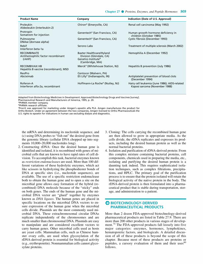
Chapter 27 Proteins, Enzymes, and Peptide Hormones 909
the mRNA and determining its nucleotide sequence, and(c) using DNA probes to “fish out” the desired gene fromthe genomic library (cellular DNA chopped up into seg-ments 10,000–20,000 nucleotides long).
2. Constructing rDNA: Once the desired human gene isidentified and isolated, it is recombined with genes of mi-crobial cells that are known to have rapid rates of cell di-vision. To accomplish this task, bacterial enzymes knownas restriction endonucleases are used. More than 100 dif-ferent variations of these hydrolytic enzymes, which actlike scissors in hydrolyzing the phosphodiester bonds ofDNA at specific sites (i.e., nucleotide sequences), areavailable. The use of a specific restriction endonucleaseboth to obtain the human gene and to open a site on themicrobial gene allows easy formation of the hybrid (re-combined) DNA molecule because of the “sticky” endson both genes. The ends of the human gene and the mi-crobial DNA vector are “glued” together by enzymesknown as DNA ligases. The human genes are placed inspecific locations on the microbial DNA vectors to en-sure expression of the human gene when the microbialcells divide. Plasmids are the most commonly used mi-crobial DNA. These extrachromosomal circular DNAsreplicate independently of the chromosomes and aremuch smaller than chromosomal DNA. Plasmids are easyto manipulate and are considered excellent vectors tocarry human genes. Other microbial cells used as hostsare yeast cells. Mammalian cells, such as Chinese ham-ster ovary cells, are used when glycosylation of therDNA-derived protein is essential for biological activity(e.g., erythropoietin). Nonmammalian cells cannot glyco-sylate proteins.
3. Cloning: The cells carrying the recombined human geneare then allowed to grow in appropriate media. As thecells divide, the rDNA replicates and expresses its prod-ucts, including the desired human protein as well as thenormal bacterial proteins.
4. Isolation and purification of rDNA-derived protein: Fromthis complex mixture containing bacterial proteins, cellcomponents, chemicals used in preparing the media, etc.,isolating and purifying the desired human protein is adaunting task indeed. This requires sophisticated isola-tion techniques, such as complex filtrations, precipita-tions, and HPLC. The primary goal of the purificationprocess is to ensure that the protein isolated will retain thebiological activity of the native protein in the body. TherDNA-derived protein is then formulated into a pharma-ceutical product that is stable during transportation, stor-age, and administration to a patient.
BIOTECHNOLOGY-DERIVEDPHARMACEUTICAL PRODUCTS
More than 2 dozen FDA-approved biotechnology-derivedpharmaceutical products are listed in Table 27.9. There aremore than 200 other products in various stages of develop-ment.74 The FDA-approved products fall loosely into fivemajor categories: enzymes, hormones, lymphokines,hematopoietic factors, and biologicals. A detailed discus-sion of all of these products is beyond the scope of thischapter. Because most of these products are proteins orpeptides, a cursory evaluation of them and their uses80
follows.
Product Name Company Indication (Date of U.S. Approval)
ProleukinAldesleukin (interleukin-2)
ProtropinSomatrem for injection
PulmozymeDNAse (dornase alpha)
RebifInterferon beta-1a
RECOMBINATEAntihemophilic factor recombinant
(rAHF)
RECOMBIVAX HBHepatitis B vaccine (recombinant), MSD
ReoProAbciximab
Roferon-AInterferon alfa-2a, recombinant
Chirona (Emeryville, CA)
Genentecha (San Francisco, CA)
Genentecha (San Francisco, CA)
Serono Labs
Baxter Healthcare/Hyland Division (Glendale, CA)
Genetics Instituteb
(Cambridge, MA)
Mercka (Whitehouse Station, NJ)
Centocor (Malvern, PA)Eli Lillya (Indianapolis, IN)
Hoffmann-La Rochea (Nutley, NJ)
Renal cell carcinoma (May 1992)
Human growth hormone deficiency in children (October 1985)
Cystic fibrosis (December 1993)
Treatment of multiple sclerosis (March 2002)
Hemophilia A (December 1992)
Hepatitis B prevention (July 1986)
Antiplatelet prevention of blood clots(December 1994)
Hairy cell leukemia (June 1986); AIDS-relatedKaposi sarcoma (November 1988)
Adapted from Biotechnology Medicines in Development: Approved Biotechnology Drugs and Vaccines [survey].Pharmaceutical Research and Manufacturers of America, 1995, p. 20.aPhRMA member company.bPhRMA research affiliate.cProcrit was approved for marketing under Amgen’s epoetin alfa PLA. Amgen manufactures the product forOrtho Biotech. Under an agreement between the two companies, Amgen licensed to Ortho Pharmaceuticals theU.S. rights to epoetin for indications in human use excluding dialysis and diagnostics.
0880-0914_17865_Ch27.qxd 12/2/09 5:06 AM Page 909

910 Wilson and Gisvold’s Textbook of Organic Medicinal and Pharmaceutical Chemistry
rDNA-DERIVED ENZYMES
Alteplase, Recombinant. Alteplase (Activase) wasdiscussed previously.
Dornase Alpha. Dornase alpha, rhDNAse (Pulmozyme),is a mucolytic enzyme identical with the natural humanDNAse and is used in the treatment of cystic fibrosis. Patientswith cystic fibrosis suffer from decreased pulmonary functionand infections caused by the secretion of thick mucus.Proteins contained in the mucus are bound to extracellularDNA, produced as a result of disintegration of bacteria in thelungs. This enzyme is involved in cleaving extracellular DNAand separates DNA from proteins, allowing proteolytic en-zymes to break down proteins and thus decrease the viscosityof mucus in the lungs.81 Proteins bound to extracellular DNAare not susceptible to proteolytic enzymes.82 Dornase alpha isa glycoprotein containing 260 amino acids that is commer-cially produced in genetically engineered Chinese hamsterovary cells.
Dornase alpha is indicated for the treatment of cystic fi-brosis in conjunction with other available therapies, such asantibiotics, bronchodilators, and corticosteroids. Adultdosage is 2.5 mg inhaled once daily, administered via a rec-ommended nebulizer. Dornase alpha should not be mixed ordiluted with other agents in the nebulizer because of the pos-sibility of adverse physicochemical changes that may affect
activity. Common adverse effects include sore throat,hoarseness, and facial edema.
Imiglucerase. Imiglucerase (Cerezyme)80 is a glycopro-tein containing 497 amino acid residues and is N-glycosyl-ated at four different positions. It is an analog of the naturalhuman enzyme �-glucocerebrosidase and contains arginineat position 495 instead of the histidine in the natural en-zyme. It is commercially produced in genetically engineeredChinese hamster ovary cells.
Like the natural enzyme, imiglucerase catalyzes the hy-drolysis of glucocerebroside, a glycolipid, to glucose andceramide within the lysosomes of phagocytic cells. Gaucherdisease is caused by a deficiency of this enzyme, which re-sults in the accumulation of glucocerebroside within tissuemacrophages. The glycolipid-engorged macrophages areknown as Gaucher cells and are responsible for the numer-ous clinical manifestations of Gaucher disease. The com-mon clinical manifestations of Gaucher disease are severeanemia, thrombocytopenia, and skeletal complications thatinclude osteonecrosis and osteopenia.
Imiglucerase is indicated for the long-term replacementtherapy of Gaucher disease. It is administered intravenouslyat an initial dose of 2.5 to 60 U/kg, infused over 1 to 2 hours.This dose is usually repeated every 2 weeks. Both the doseand the frequency of administration may be varied, how-ever, depending on the response.83 Common adverse effectsinclude dizziness, headache, abdominal discomfort, nausea,and rash.
rDNA-DERIVED HORMONES
The rDNA-derived hormones include insulin human injectionUSP (Humulin R, Novolin R, Velosulin Human), growth hor-mone (somatotropin; Humatrope), and somatrem (Protropin).All of these products, as well as other products containinghuman insulin, have been discussed in this chapter.
rDNA-DERIVED CYTOKINES
Interferons.80 Interferons are natural glycoproteins pro-duced by virtually all eukaryotic cells; they possess im-munomodulating, antiviral, and cytotoxic activities. This fam-ily of glycoproteins is produced by cells in response to a widerange of stimuli.84 In humans, interferons bind to cellular re-ceptors, which leads to the synthesis of more than a dozen pro-teins that contribute to viral resistance. The antiviral effects ofinterferons may be caused by inhibition of the synthesis ofviral mRNA or proteins or prevention of viral penetration oruncoating.85,86 Based on their antigenic subtypes, the interfer-ons are classified into three major groups: �, �, and �. �-Interferon and �-interferon are produced by virtually allcells in response to a viral infection and various other stimuli.�-Interferon is produced specifically by the T lymphocytesand the natural killer (NK) cells. �-Interferons have greaterimmunoregulatory, but lower antiviral, effects than �- or �-interferons.86 More than 12 subspecies of �-interferons, 1�-interferon, and 2 �-interferons are known to exist. In gen-eral, the interferons are glycoproteins consisting of 165 to166 amino acid residues. There are four rDNA-derived �-interferons available for clinical use around the world andthree available in the United States (described next). All �-interferons exhibit antiviral and antiproliferative activ-ity, enhance phagocytic activity, and augment specific
Figure 27.7 Summary of a typical rDNA process usedin the commercial-scale production of human proteins.
0880-0914_17865_Ch27.qxd 12/2/09 5:06 AM Page 910

Chapter 27 Proteins, Enzymes, and Peptide Hormones 911
cytotoxicity of lymphocytes for certain target cells.87 Themost common adverse effects of �- and �-interferons includeflulike symptoms, bone marrow suppression, neurotoxic ef-fects, hypocalcemia, anorexia and other gastrointestinalsymptoms, and weight loss.
Interferon Alfa-2a, Recombinant. Interferon alfa-2a,recombinant (Roferon), is produced from genetically engi-neered E. coli and contains 165 amino acid residues. At posi-tion 23, interferon alfa-2a has a lysine residue. The pharma-ceutical product contains a single �-interferon subtype. Amurine monoclonal antibody is used during purification byaffinity chromatography. Interferon alfa-2a is used in thetreatment of hairy cell leukemia and acquired immunodefi-ciency syndrome (AIDS)-related Kaposi sarcoma. It is ab-sorbed well after intramuscular or intravenous administrationand has a half-life of 5 to 7 hours when administered by theintramuscular route. The solution should be stored in the re-frigerator at 36°F to 46°F and should not be frozen or shaken.
Peginterferon Alfa-2a, Recombinant. Interferon alfa-2a, recombinant (Roferon), is produced from genetically en-gineered E. coli. It is interferon, alfa-2a utilizing a 40 kdpolyethylene glycol (PEG) strand to allow for stable thera-peutic serum levels of alpa-2a for up to 168 hours on a singledose. This process is known as PEGylation and involves theprocess of attaching strands of the polymer PEG to moleculesin an attempt to make them safer and more effective as ther-apeutic agents. The physiochemical changes in the drug in-crease systemic retention as well as influence the bindingaffinity to the cell receptors. The therapeutic usefulness ofpeginterferon alpfa-2a is in the treatment of hepatitis C.
Interferon Alfa-2b, Recombinant. Interferon alfa-2b,recombinant (Intron A), also contains a single subtype of �-interferon. It is a glycoprotein containing 165 amino acidresidues and is commercially produced from genetically engi-neered E. coli. It differs from interferon alfa-2b in possessingan arginine residue at position 23. It is used in the treatmentof hairy cell leukemia, condyloma acuminata (genital warts),AIDS-related Kaposi sarcoma, hepatitis C, and hepatitis B. Itis administered intramuscularly or subcutaneously with ahalf-life of 2 to 3 hours and via intravenous infusion with ahalf-life of 8 hours. The reconstituted solution is stable for 1month when stored at a temperature of 36°F to 46°F.
Interferon Alfa-n3 (Injection). Interferon alfa-n3(Alferon N) is a polyclonal mixture of up to 14 natural �-in-terferon subtypes and contains 166 amino acid residues. Itscommercial production involves induction of pooled unitsof human leukocytes with an avian virus (Sendai virus). Thepurification process involves immunoaffinity and filtrationchromatography. It is indicated primarily by intralesionalinjection for the treatment of genital warts. The solutionshould be stored at a temperature of 36°F to 46°F and shouldnot be shaken.
Interferon Beta-1a. Interferon beta-1a (Rebif, Avonex),has biological effects similar to those of natural �-interferonand �-interferons. Unlike beta-1b, which is produced inmodified E. coli, beta-1a is produced in mammalian cells. Ithas been shown to slow the progression of multiple sclerosisas well as reduce the rate of relapse of this disease.
Interferon Beta-1b, Recombinant. Interferon beta-1b,recombinant (Betaseron), has biological effects similar to
those of natural �-interferon and �-interferons. The natural �-interferon is a glycoprotein containing 166 amino acidresidues. The rDNA product differs from the natural form, inthat it is not glycosylated, it lacks the amino-terminal methio-nine, and it has serine in the place of methionine at position17.88 It is used for a wide variety of indications via intra-venous, intramuscular, subcutaneous, intrathecal, and intrale-sional routes. Its primary indication is for the prevention ofexacerbations in patients suffering from relapsing/remittingmultiple sclerosis. Recommended dosage is 8 million units,administered subcutaneously, every other day. It also is indi-cated in the treatment of malignant glioma and malignantmelanoma. Recommended temperature for storage is 36°F to46 °F, and unused reconstituted solution should be discarded.
Aldesleukin. Aldesleukin, interleukin-2 (Proleukin),80
is an rDNA-derived lymphokine that differs structurallyfrom native interleukin-2 (IL-2) but has biological activitysimilar to that of the natural lymphokine.89 Natural IL-2 isproduced primarily by the peripheral blood lymphocytesand contains 133 amino acid residues. The immunoregula-tory effects of aldesleukin include enhancing mitogenesis oflymphocytes, stimulating the growth of IL-2–dependentcell lines, enhancing cytotoxicity of lymphocytes, inducinglymphokine-activated killer (LAK) cells and NK cells, andinducing interferon-� production. The exact mechanism ofthe antitumor activity of aldesleukin in humans is unknown.
The rDNA process involves genetically engineeredE. coli (pBR 322 plasmids). The gene for IL-2 was synthe-sized after first isolating and identifying the mRNA from thehuman Jurkat cell line and then preparing the complemen-tary DNA (cDNA). The IL-2 gene was genetically engi-neered before it was hybridized into pBR 322 plasmid.Further manipulation of the hybridized plasmid resulted inthe production of a modified IL-2, aldesleukin.90
Aldesleukin differs structurally from the native IL-2 in thatthe former is not glycosylated, it lacks the N-terminal ala-nine residue, and it has serine in the place of cysteine at po-sition 125. Noncovalent, molecular aggregation ofaldesleukin is different from IL-2, and the former exists as amicroaggregate of 27 molecules.
The primary indication for aldesleukin is in the treatmentof adult metastatic renal carcinoma. It is administered via in-travenous infusion in doses of 10,000 to 50,000 U/kg every8 hours for 12 days. It is primarily metabolized by the kid-neys, with no active form found in the urine. Aldesleukincauses serious adverse effects in patients, including fever,hypotension, pulmonary congestion and dyspnea, coma,gastrointestinal bleeding, respiratory failure, renal failure,arrhythmias, seizures, and death.
rDNA-DERIVED HEMATOPOIETIC FACTORS
Hematopoietic growth factors are glycoproteins producedby a number of peripheral and marrow cells. More than 200billion blood cells are produced each day; and thehematopoietic factors, along with other lymphopoietic fac-tors such as the stem cell factor and the interleukins, are in-volved in the proliferation, differentiation, and maturationof various types of blood cells derived from the pluripotentstem cells.
Erythropoietin.80 Erythropoietin is a heavily glycosyl-ated protein containing 166 amino acid residues. It is pro-
0880-0914_17865_Ch27.qxd 12/2/09 5:06 AM Page 911

duced primarily by the peritubular cells in the cortex of thekidney, and up to 15% is produced in the liver. It is the prin-cipal hormone responsible for stimulating the production ofred blood cells from erythroid progenitor cells, erythrocyteburst-forming units, and erythrocyte colony-formingunits.91 Small amounts of erythropoietin are detectable inthe plasma; however, most of the hormone is secreted by thekidneys in response to hypoxia or anemia, when levels ofthe hormone can rise more than 100-fold.
Decreased erythropoietin production is one of several po-tential causes of anemia of chronic renal disease. Othercauses of anemia of chronic renal disease include infection orinflammatory condition in the kidneys, iron deficiency, mar-row damage, and vitamin or mineral deficiency. Regardlessof the underlying disease causing renal failure, erythropoi-etin levels decrease in patients with renal failure. Until rDNAtechnology was used to produce commercial quantities oferythropoietin, it was obtained from the urine of patients suf-fering from severe aplastic anemia. This process of obtainingnatural hormone was costly and time-consuming and pro-duced only small quantities of the hormone.
Epoetin Alfa. Epoetin alfa, rEPO (Epogen, Procrit), isthe recombinant human erythropoietin produced in Chinesehamster ovary cells into which the human erythropoietin genehas been inserted. These mammalian cells glycosylate theprotein in a manner similar to that observed in human cells.92
Epoetin alfa is indicated in anemic patients with chronicrenal failure, including both those who require regular dial-ysis and those who do not. Epoetin alfa is also indicated inanemia associated with AIDS, treatment of AIDS with zi-dovudine, frequent blood donations, and neoplastic dis-eases. It is indicated to prevent anemia in patients who do-nate blood prior to surgery for future autologoustransfusions and to reduce the need for repeated mainte-nance transfusions.93 The hormone is available as an iso-tonic buffered solution, which is administered by the intra-venous route. The solution should not be frozen or shakenand is stored at 36°F to 46°F.
Colony-Stimulating Factors.80 Colony-stimulatingfactors are natural glycoproteins produced in lymphocytesand monocytes. These factors bind to cell-surface receptorsof hematopoietic progenitor cells and stimulate prolifera-tion, differentiation, and maturation of these cells into rec-ognizable mature blood cells.93 Colony-stimulating factorsproduced by rDNA technology have the same biological ac-tivity as the natural hormones. Currently, there are twocolony-stimulating factors commercially produced byrDNA technology. These products are discussed next.
Filgrastim. Filgrastim, rG-CSF (Neupogen), is a 175-amino acid polypeptide produced in genetically engi-neered E. coli cells containing the human granulocyte colony-stimulating factor (G-CSF) gene. Filgrastim differs from thenatural hormone in that the former is not glycosylated and con-tains an additional methionine group at the N-terminus, whichis deemed necessary for expression of the gene in E. coli.
Filgrastim specifically stimulates the proliferation andmaturation of neutrophil granulocytes and, hence, is consid-ered lineage specific. Accepted indications for filgrastim in-clude the following: (a) to decrease the incidence of febrileneutropenia in patients with nonmyeloid malignancies whoreceive myelosuppressive chemotherapeutic agents, thus
lowering the incidence of infections in these patients; (b) toaccelerate myeloid recovery in patients undergoing autolo-gous bone marrow transplantation; and (c) in AIDS patients,to decrease the incidence of neutropenia caused by the dis-ease itself, or by drugs used to treat the disease. The usualstarting dose for filgrastim is 5 �g/kg per day in patientswith nonmyeloid cancer who receive myelosuppressivechemotherapy.
Filgrastim solution should be stored at 36°F to 46°F andused within 24 hours of preparation. The solution should notbe shaken or allowed to freeze. Any solution left at roomtemperature for more than 6 hours should be discarded. Themost frequent adverse effects of filgrastim are medullarybone pain, arthralgia, and myalgia.
Sargramostim. Sargramostim, rGM-CSF (Leukine), isa glycoprotein commercially produced in genetically engi-neered yeast cells. Its polypeptide chain contains 127 aminoacids. It differs from the natural hormone by substitution ofleucine at position 23 and variations in the glycosylation.94
Sargramostim is a lineage-nonspecific hematopoietic factor,because it promotes the proliferation and maturation ofgranulocytes (neutrophils and eosinophils) and monocytes(macrophages and megakaryocytes).
The primary indication for sargramostim is in myeloidengraftment following autologous bone marrow transplanta-tion and hematopoietic stem cell transplantation. Handling,storage precautions, and adverse effects are similar to thosefor filgrastim.
RDNA-DERIVED MISCELLANEOUS PRODUCTS
Antihemophilic Factor. Antihemophilic factor (factorVIII) (Humate-P, Hemophil M, Koate HP, Monoclate-P)is a glycoprotein found in human plasma and a necessarycofactor in the blood-clotting mechanism. This high–molecular-weight glycoprotein has a complex structurewith several components (subcofactors).95 The commer-cially available concentrates derived from blood collectedfrom volunteer donors by the American Red Cross BloodServices are used primarily for the treatment of patientswith hemophilia A. Because the commercially availableproducts are purified concentrates derived from bloodpooled from millions of donors, the major precautions inusing the products relate to transmission of viruses, such ashepatitis virus, herpesvirus, and HIV. This major problemhas been alleviated, mostly because of the developmentand marketing of rDNA-derived antihemophilic factors.
Antihemophilic Factor (Recombinant). Antihemophilicfactor (recombinant), rAHF (KoGENate, Helixate), is anrDNA-derived factor VIII expressed in genetically engineeredbaby hamster kidney cells.96 KoGENate has the same biolog-ical activity as the human plasma-derived antihemophilicfactor (pdAHF). The purification process for rAHF includesmonoclonal antibody immunoaffinity chromatography to re-move any protein contaminants.
rAHF is indicated for the treatment of hemophilia A andis administered by the intravenous route. Patients sufferingfrom hemophilia A exhibit a decrease in the activity ofplasma clotting factor VIII. This product temporarily pre-vents bleeding episodes in hemophiliacs and may be used toprevent excessive bleeding during surgical procedures inthese patients. A major advantage of the rAHF over the
912 Wilson and Gisvold’s Textbook of Organic Medicinal and Pharmaceutical Chemistry
0880-0914_17865_Ch27.qxd 12/2/09 5:06 AM Page 912

Chapter 27 Proteins, Enzymes, and Peptide Hormones 913
natural factor VIII is the lack of virus in the product. rAHFdoes not contain von Willebrand factor; therefore, it is notindicated in the treatment of von Willebrand disease.Patients receiving rAHF should be monitored carefully forthe development of antibodies.
Bioclate is an rDNA-derived factor VIII expressed in ge-netically engineered Chinese hamster ovary cells. It has thesame biological activity as pdAHF and is structurally simi-lar. Its indications and adverse effects are similar to thosefor KoGENate.
3. Clinically, which of the following is used to dissolvenecrotic tissue of second- and third-degree burn patients?a. streptokinaseb. sutilainsc. trypsind. dornasee. lypressin
4. While working in your basement you discover a methodof producing proteins and peptides that has never beendescribed in the literature. What is the significance ofbeing able to produce cytokines such as interferons?
5. You have discovered a protein that is capable of blockingthe progression of Alzheimer disease. The sources of thisprotein are very limited, and you need additional material tocomplete a phase II clinical trial. Describe how you woulduse rDNA technology to produce more of this wonder drug.
1. The rennin–angiotensin system plays an important role inregulation of blood pressure. As such, this pathway en-compasses the conversion of an approximately 400-amino acid sequence to a decapeptide, then to the biolog-ically active octapeptide and is an excellent target fordrug therapy to manage hypertension. Describe the start-ing protein and its conversion to the biologically activemetabolite. Be sure to include the enzymes involved inthe conversion of the protein and peptides.
2. In the digestion and absorption of dietary protein:a. pepsinogen is converted to pepsin by gastrin via a pro-
teolysis reaction.b. chymotrypsinogen is activated by trypsin.c. carboxypeptidase activates pepsinogen in the stomach.d. hydrochloric acid activates aminopeptidase.
R E V I E W Q U E S T I O N S
REFERENCES1. Ahmad, F., et al. (eds.): From Gene to Protein: Translation into
Biotechnology, vol. 19. New York, Academic Press, 1982.2. Boza, J. J., et al.: Eur. J. Nutr. 39(6):237, 2000.3. American Medical Association Department of Drugs: Drug
Evaluations, 6th ed. New York, John Wiley & Sons, 1986,pp. 867–870.
4. Stryer, L.: Biochemistry, 5th ed. New York, W. H. Freeman & Co.,2002, Chap. 3.
5. Corey, R. B., and Pauling, L.: Proc. R. Soc. Lond. Ser. B 141:10,1953. (See also Adv. Protein Chem. 8:147, 1957)
6. Tanford, C.: The Hydrophobic Effect: Formation of Micelles andBiological Membranes, 2nd ed. New York, John Wiley & Sons,1979.
7. McDonald, C. C., and Phillips, W. D.: J. Am. Chem. Soc. 89:6332,1967.
8. Rienstra, C. M., et al.: Proc. Natl. Acad. Sci. U. S. A. 99(16):10260,2002.
9. Kato, G., and Yung, J.: Mol. Pharmacol. 7:33, 1971.10. Elefrawi, M. E., et al.: Mol. Pharmacol. 7:104, 1971.11. Tang, C., Ndassa, Y., and Summers, M. F.: Nat. Strict. Biol.
9(7):537, 2002.12. Kodak, A., et al.: Endocrinology 143(2):411, 2002.13. Catrina, S. B., Cocilescu, M., and Andersson, M.: J. Cell Mol. Med.
5(2):195, 2001.14. Ferdinand, W.: The Enzyme Molecule. New York, John Wiley &
Sons, 1976.15. Stryer, L.: Biochemistry, 5th ed. New York, W. H. Freeman & Co.,
2002, Chap. 4.16. Koshland, D. E.: Sci. Am. 229:52, 1973 (see also Annu. Rev.
Biochem. 37:359, 1968).17. Belleau, B.: J. Med. Chem. 7:776, 1964.18. Lowe, J. N., and Ingraham, L. L.: An Introduction to Biochemical
Reaction Mechanisms. Englewood Cliffs, NJ, Prentice-Hall, 1974.19. Hanson, K. R., and Rose, I. A.: Acc. Chem. Res. 8:1, 1975 (see also
Science 193:121, 1976).19a.Bender, M. L.: J. Am. Chem. Soc. 79:1258, 1957.19b.Bender, M. L.: J. Am. Chem. Soc. 80:5338, 1958.19c.Bender, M. L.: J. Am. Chem. Soc. 82:1900, 1960.19d.Bender, M. L.: J. Am. Chem. Soc. 86:3704, 5330, 1964.
19e.Blow, D. M.: Acc. Chem. Res. 9:145, 1976.20. Roberts, J. D.: Chem. Eng. News, 57:23, 1979.21. Wallis, M., Howell, S. L., and Taylor, K. W.: The Biochemistry of
the Peptide Hormone. Chichester, U. K., John Wiley & Sons, 1986.22. Spatola, A. F.: Annu. Rep. Med. Chem. 16:199, 1981.23. Brueggemeier, R. W.: Peptide and protein hormones. In Wolff, M. E.
(ed.). Burger’s Medicinal Chemistry, vol. 3, 5th ed. New York, JohnWiley & Sons, 1996, pp. 464–465.
24. Otsuka, H., and Inouye, L. K.: Pharmacol. Ther. [B] 1:501, 1975.25. Schimmer, B. P., and Parker, K. L.: In Hardman, J. G., and Limbird,
L. E. (eds.). Goodman and Gilman’s The Pharmacological Basis ofTherapeutics, 10th ed. New York, Macmillan, 2001, pp. 1650–1655.
26. Hughes, J., et al.: Nature 258:577, 1975.27. Gutstein, H. B., and Akil, H.: In Hardman, J. G., and Limbird, L. E.
(eds.). Goodman and Gilman’s The Pharmacological Basis ofTherapeutics, 10th ed. New York, Macmillan, 2001, pp. 569–579.
28. Snyder, S. H.: Science 224:22, 1984.29. Wallis, M.: J. Mol. Evol. 17:10, 1981.30. Lewis, U. J., Sinha, Y. N., and Lewis, G. P.: Endocr. J.
47(Suppl.):S1–8, 2000.31. Janecka, A., Zubrzycka, M., and Janecki, T.: J. Pept. Res. 58(2):91,
2001.32. Brownstein, M. J.: Science 207:373, 1980.33. Drug Information for the Health Care Professional, vol. 1, 22nd ed.
Rockville, MD, U. S. Pharmacopeial Convention, 2002, p. 2275.34. Banting, F. G., and Best, C. H.: J. Lab. Clin. Med. 7:251, 1922.35. Wallis, M., Howell, S. L., and Taylor, K. W.: The Biochemistry of
Peptide Hormones. Chichester, U. K., John Wiley & Sons, 1985,pp. 257–297.
36. Alrefai, H., Allababidi, H., Levy, S., et al.: Endocrine18(2):105, 2002.
37. Newgard, C. B.: Diabetes 51(11):3141, 2002.38. De Meyts, P., and Whittaker, J.: Nat. Rev. Drug Discov. 1(10):769,
2002.39. Steiner, D. F., et al.: Recent Prog. Horm. Res. 25:207–282, 1969.40. Docherty, K., et al.: Proc. Natl. Acad. Sci. U. S. A. 79:4613, 1982.41. Arquilla, E. R., et al.: In Steiner, D. F., and Freinkel, N. (eds.).
Handbook of Physiology, Sect. 7: Endocrinology, vol. 1. Washington,DC, American Physiological Society, 1972, pp. 159–173.
42. Gerich, J., et al.: Annu. Rev. Physiol. 38:353, 1976.
0880-0914_17865_Ch27.qxd 12/2/09 5:06 AM Page 913

43. Sanger, F.: Annu. Rev. Biochem. 27:58, 1958.44. Katsoyannis, P. G.: Science 154:1509, 1966.45. Rittel, W., et al.: Helv. Chim. Acta 67:2617, 1974.46. Chance, R. E., et al.: Diabetes Care 4:147, 1981.47. Johnson, I. S.: Diabetes Care 5(Suppl. 2):4, 1982.48. Cosmatos, A., et al.: J. Biol. Chem. 253:6586, 1978.49. May, J. M., et al.: J. Biol. Chem. 253:686, 1978.50. Blundell, T. L., et al.: Crit. Rev. Biochem. 13:141, 1982.51. Larner, J.: Diabetes 37:262, 1988.52. Kasuga, M., et al.: Science 215:185, 1982.53. Gerich, J. E.: In Diabetes Mellitus, 9th ed. Indianapolis, Eli Lilly &
Co., 1988, p. 53.54. Galloway, J. A.: In Diabetes Mellitus, 9th ed. Indianapolis, Eli Lilly
& Co., 1988, pp. 109–122.55. Wallis, M., et al.: The Biochemistry of the Polypeptide Hormones.
Chichester, U. K., John Wiley & Sons, 1985, p. 280.56. Jefferson, L. S.: Diabetes 29:487, 1980.57. Feldman, J. M.: In Diabetes Mellitus, 9th ed. Indianapolis, Eli Lilly
& Co., 1988, pp. 28–42.58. Pirat, J.: Diabetes Care 1(part 1):168–188, (part 2):252–263, 1978.59. Raskin, P., and Rosenstock, J.: Ann. Intern. Med. 105:254, 1986.60. Maurer, A. C.: Am. Sci. 67:422, 1979.61. USP DI: Drug Information for the Health Care Professional, vol. I,
22nd ed. Rockville, MD, U. S. Pharmacopeial Convention, 2002, pp.1701–1730.
62. Unger, R. J., et al.: Science 188:923, 1975.63. Fain, J. N.: Recept. Recogn. Ser. A 6:3, 1978.64. Wallis, M., Howell, S. L., and Taylor, K. W.: The Biochemistry of
the Polypeptide Hormones. Chichester, U. K., John Wiley & Sons,1985, pp. 318–335.
65. Emson, P. C., and Sandberg, B. E.: Annu. Rep. Med. Chem. 18:31,1983.
66. Miller, R. J.: J. Med. Chem. 27:1239, 1984.67. Adams, A. E., et al.: Mol. Endocrinol. 12(11):1673, 1998.68. Bumpus, F. M.: Fed. Proc. 36:2128, 1977.69. Ondetti, M. A., and Cushman, J.: J. Med. Chem. 24:355, 1981.70. Ogonowski, A. A., et al.: J. Pharm. Exp. Ther. 280:846–853, 1997.71. May, S. W., and Pollock, S. H.: U. S. Patent #6,495,514. Issued
December 2002.72. Pezzuto, J. M., Johnson, M. E., and Manasse, H. R. (eds.):
Biotechnology and Pharmacy. New York, Chapman & Hall, 1993.73. Richards, F. F., et al.: Science 187:130, 1975.74. Biotechnology Medicines in Development [survey], Pharmaceutical
Research and Manufacturers of America, Washington, DC, 1995.75. Zito, S. W. (ed.): Pharmaceutical Biotechnology: A Programmed
Text. Lancaster, PA, Technomic Publishing, 1992.76. Hudson, R. A., and Black, C. D.: Biotechnology—The New
Dimension in Pharmacy Practice: A Working Pharmacists’ Guide.Columbus, OH, Council of Ohio Colleges of Pharmacy, 1992, p. 1.
77. Primrose, S. B.: Modern Biotechnology. Boston, Blackwell ScientificPublications, 1987.
78. Hudson, R. A., and Black, C. D.: Biotechnology—The NewDimension in Pharmacy Practice: A Working Pharmacists’ Guide,Council of Ohio Colleges of Pharmacy, 1992, p. 2.
79. Birch, J. R., and Lennox, E. S. (eds.): Monoclonal Antibodies:Principles and Applications. New York, John Wiley & Sons, 1995.
80. Gelman, C. R., Rumack, B. H., and Hess, A. J. (eds.): DRUGDEX (R)System. Englewood, CO, MICROMEDEX, Inc. (expired 11/30/95).
81. Shak, S., et al.: Recombinant human DNAse I reduces viscosity ofcystic fibrosis sputum. Proc. Natl. Acad. Sci. U. S. A. 87:9188, 1990.
82. Lieberman, J., and Kurnick, N. B.: Nature 196:988, 1962.83. Brady, R. O., et al.: Prog. Clin. Biol. Res. 95:669, 1982.84. Kirkwood, J. M., and Ernstoff, M. S.: J. Clin. Oncol. 2:336, 1984.85. Houglum, J. E.: Clin. Pharm. 2:202, 1983.86. Hayden, F. G.: Antiviral agents. In Hardman, J. G., and Limbird, L.
E. (eds.). Goodman and Gilman’s The Pharmacological Basis ofTherapeutics, 10th ed. New York, Macmillan, 2001, pp. 1332–1335.
87. USP DI: Drug Information for the Health Care Professional, vol. I,22nd ed. Rockville, MD, U. S. Pharmacopeial Convention, 2002,pp. 1730–1733.
88. Von Hoff, D. D., and Huong, A. M.: J. Interferon Res. 8:813, 1988.89. Doyle, M. V., Lee, M. T., and Fong, S.: J. Biol. Response Mod. 4:96,
1985.90. Kato, K., et al.: Biochem. Biophys. Res. Commun. 130:692, 1985.91. Erslev, A. J.: Erythropoietin coming of age. N. Engl. J. Med.
316:101, 1981.92. Lin, F. K., et al.: Proc. Natl. Acad. Sci. U. S. A. 82:7580, 1985.93. USP DI: Drug Information for the Health Care Professional, vol. I,
22nd ed. Rockville, MD, U. S. Pharmacopeial Convention, 2002,pp. 941–947.
94. Lee, F., et al.: Proc. Natl. Acad. Sci. U. S. A. 82:4360, 1985.95. Fulcher, C., and Zimmerman, T.: Proc. Natl. Acad. Sci. U. S. A.
79:1648, 1982.96. Lawn, R. M., and Vehar, G. A.: Sci. Am. 254:48, 1986.
SELECTED READINGBirch, J. R., and Lennox, E. S. (eds.): Monoclonal Antibodies: Principles
and Applications. New York, John Wiley & Sons, 1995.Boyer, P. D. (ed.): The Enzymes, 3rd ed. New York, Academic Press,
1970.Brockerhoff, H., and Jensen, R. G.: Lipolytic Enzymes. New York,
Academic Press, 1974.Ferdinand, W.: The Enzyme Molecule. New York, John Wiley & Sons,
1976.Galloway, J. A., Potvin, J. H., and Shuman, C. R. (eds.): Diabetes
Mellitus, 9th ed. Indianapolis, Eli Lilly & Co., 1988.Grollman, A. P.: Inhibition of protein biosynthesis. In Brockerhoff, H.,
and Jensen, R. G. (eds.). Lipolytic Enzymes. New York, AcademicPress, 1974, pp. 231–247.
Hardman, J. G., and Limbird, L. E. (eds.). Goodman and Gilman’s ThePharmacological Basis of Therapeutics, 10th ed. New York,Macmillan, 2001.
Haschemeyer, R. H., and de Harven, E.: Electron microscopy of enzymes.Annu. Rev. Biochem. 43:279, 1974.
Hruby, V. J., de Chavez, C. G., and Kavarana, M.: Peptide and proteinhormones, peptide neurotransmitters, and therapeutic agents. InAbraham, D. J. (ed). Burger’s Medicinal Chemistry and DrugDiscovery, vol. 4, 6th ed. New York, John Wiley & Sons, 2003.
Jenks, W. P.: Catalysis in Chemistry and Enzymology. New York,McGraw-Hill, 1969.
Lowe, J. N., and Ingraham, L. L.: An Introduction to BiochemicalReaction Mechanisms. Englewood Cliffs, NJ, Prentice-Hall, 1974.(This book includes elementary enzymology including mechanisms ofcoenzyme function.)
Mildvan, A. S.: Mechanism of enzyme action. Annu. Rev. Biochem.43:357, 1974.
Pezzuto, J. M., Johnson, M. E., and Manasse, H. R. (eds.): Biotechnologyand Pharmacy. New York, Chapman & Hall, 1993.
Pikes, S. J., and Parks, C. R.: The mode of action of insulin. Annu. Rev.Pharmacol. 14:365, 1974.
Schaeffer, H. J.: Factors in the design of reversible and irreversible en-zyme inhibitors. In Ariens, E. J. (ed.). Drug Design, vol. 2. New York,Academic Press, 1971, pp. 129–159.
Stryer, L. S.: Biochemistry, 5th ed. New York, W. H. Freeman & Co.,2002.
Tager, H. S., and Steiner, D. F.: Peptide hormones. Annu. Rev. Biochem.43:509, 1974.
Waife, S. O. (ed.): Diabetes Mellitus, 8th ed. Indianapolis, Eli Lilly &Co., 1980.
Wallis, M., Howell, S. L., and Taylor, K. W.: The Biochemistry of thePeptide Hormones. Chichester, U. K., John Wiley & Sons, 1985.
Wilson, C. A.: Hypothalamic amines and the release of gonadotrophinsand other anterior pituitary hormones. Adv. Drug Res. 8:119–204,1974.
914 Wilson and Gisvold’s Textbook of Organic Medicinal and Pharmaceutical Chemistry
0880-0914_17865_Ch27.qxd 12/2/09 5:06 AM Page 914

C H A P T E R 28
VitaminsMICHAEL J. DEIMLING, M. O. FARUK KHAN, AND GUSTAVO R. ORTEGA
or in combination, provides any health benefit or can pre-vent or treat disease.
One difficulty in assessing the role of vitamins, and othernutrients, in health and disease is the finding that often morethan one is involved in a physiological or disease process.This is seen in studies on bone mineralization where admin-istering calcium in the presence of vitamin D deficiency, orvitamin D in the presence of a calcium-deficient diet, maymiss the importance of calcium and/or vitamin D supplemen-tation. We now realize that vitamin K is also involved inbone formation and this third factor, among others, must beconsidered. However, studies examining multiple factors arecomplex and not easily performed. Another complicatingfactor comes from the continually expanding use of fortifica-tion. Total vitamin intake is often unknown, even in the ab-sence of supplementation, as a result of intake from multiplesources unnaturally high in vitamin content. For a summary,refer to the National Institutes of Health (NIH) State-of-the-Science Conference on Multivitamin/Mineral Supplementsand Chronic Disease Prevention.1
INTRODUCTION
A complete history of the vitamins would have to begin withthe earliest written historical records. For millennia, differ-ent cultures have recorded the use of dietary substances tosuccessfully treat disease; some of which we now know area result of vitamin deficiency. Patients afflicted with one ofthese deficiencies were successfully treated as a result of theempirical or fortuitous selection of dietary substances rich inthe deficient vitamin(s). However, the basis for these defi-ciency diseases and why these treatments were successfulwas not systematically examined until the late 1800s toearly 1900s.
The search for vitamins began in earnest in the early 1900swhen researchers first began to realize that synthetic dietsconsisting of sufficient amounts of purified carbohydrates,proteins, fats, minerals, water, and calories failed to sustaingrowth in animals. Although there were many important con-tributors to the eventual discovery of the concept of vitaminsand the individual chemicals that comprise them, only a fewkey papers are presented here. These help explain the termi-nology of vitamins in general; more detail is included with theindividual vitamins. In 1911, Funk2 described a substancethat could cure polyneuritis in birds that was found in theaqueous phase of an extract of rice polishings. Later in 1912,Funk3 published an excellent summary of the state of vitamindiscovery, and for the first time proposed the use of the wordvitamine, which has been often stated to be derived from a
C H A P T E R O V E R V I E W
The medicinal chemistry of vitamins is fundamental notonly to the therapeutics of nutritional problems but also tothe understanding of the biochemical actions of other me-dicinal agents that directly or indirectly affect the metabolicfunctions of vitamins and coenzymes. Notable in this regardis the interrelationship between folic acid and vitamin B12 inthe development of megaloblastic anemia, as well as the ef-ficacy of warfarin in relation to vitamin K status. In addi-tion, vitamins may rarely be used in treatments unrelated totheir role as a vitamin. The use of retinoic acid in acne,niacin in hyperlipidemias, and hydroxocobalamin in thetreatment of cyanide poisoning are a few that are discussedwithin this chapter. Accordingly, this chapter includes abrief summary of the history, chemistry, kinetics, biochem-istry, therapeutic use, adverse effects (hypervitaminosis),and available vitamin products that can assist the clinician ina better understanding of the role vitamins play in bothhealth and disease.
The use of vitamins, especially as multivitamins, has in-creased significantly as a result of a move toward the prac-tice of healthier lifestyles. There are two approaches to theuse of vitamins that are not always clearly distinct. The firstis in the treatment of known or suspected deficiencies, or toprevent deficiencies as bodily needs change as seen duringpregnancy, disease, or aging. In this approach, health conse-quences are caused by vitamin deficiency, and the vitaminsrestore health by resolving the deficiency. There are numer-ous studies that prove the efficacy of using vitamins to treattheir known deficiency syndromes or to prevent deficien-cies. However, there is evidence that vitamin deficienciesmay cause more health problems than are currently known.The role of vitamin deficiencies in cancer, cardiovasculardisease, bone health, and immune disorders, among others,continue to be examined although the results are often con-flicting. Certainly, the maintenance of adequate vitamin in-take, either through diet or a multivitamin supplement, is tobe encouraged.
The second approach is the use of a vitamin or group ofvitamins in doses above that required for prevention of defi-ciency, to provide an overall health benefit, or to prevent ortreat disease. Study results are even more controversial inthis area because of conflicting results and, more impor-tantly, the gathered evidence that excessive use of certain vi-tamins, under some circumstances, may actually be harmful.It remains controversial whether there is clear and convinc-ing evidence to suggest that the use of vitamins in dosesabove their recommended dietary allowance (RDA), singly
915
0915-0960_17865_Ch28.qxd 12/4/09 8:12 PM Page 915

combination of the latin, vita (life) or alternatively from vital,and amine, which comes from the chemical term am (fromammonia) for nitrogen and the suffix -ine, which means basicin the chemical sense. Early work of Funk indicated that thecurative substance for bird polyneuritis found in rice polish-ings was necessary for life, contained nitrogen, and was basic,hence the name.
Also in 1912, Sir Frederick Gowland Hopkins4 clearlydemonstrated that growth retardation found in rats fed puri-fied diets could be reversed by the addition of small amountsof milk. The amount added was not sufficient to be quantita-tively important with respect to its caloric value or nutritivevalue as a source of protein, carbohydrate, fat, or minerals.Therefore, Hopkins suggested that there must be some, yetunidentified, factors in the milk that are required for growth,but only in very small amounts. He referred to these sub-stances as accessory factors. McCollum and Davis5–8 in a se-ries of papers showed that rats fed a diet of purified milksugar, protein, minerals, and lard or olive oil, which providedsufficient calories, also failed to grow. Furthermore, theyfound that addition of small amounts of butterfat, egg yolk,or lipid extracts of either of these two substances wouldquickly restore growth. In 1916, McCollum and Kennedy9
suggested that the term vitamine be dropped for two reasons.First, the use of the prefix vita implied that these were theonly substances vital for life; however, other dietary sub-stances were known to be required for life, such as essentialamino acids. Second, the ending amine implied they con-tained basic nitrogen, proof of which was lacking. Becausethe substance they were examining was fat soluble and theaccessory factor described by Funk was water soluble, thesuggestion was made to refer to these as fat-soluble A andwater-soluble B. Deficiency of an antiscorbutic substance inthe diet was thought to result in scurvy, and Drummond10
proposed the term water-soluble C in 1919.In 1920, Drummond11 proposed an alternative. He noted
that the ending -ine was used in chemical nomenclature todenote a basic substance, and there was no evidence that anyof the dietary factors was basic, much less amines. Nor wasthere an expectation that future discoveries would be basic.He therefore suggested dropping the final “-e” and using theterm vitamin, which would be acceptable under the rules ofchemical nomenclature. He further suggested using theterms vitamin A and vitamin B for the fat-soluble A andwater-soluble B, which had been suggested by McCollumand Kennedy, and continuing with subsequent letters, suchas C, as new vitamins were discovered. Other names werealso proposed, but none as historically important as these.
This terminology continued for the most part, through thediscovery of many of the remaining vitamins. The only alter-ation is that, except for vitamin C, the water-soluble vitaminswere grouped into the vitamin B complex, instead of, or in ad-dition to, using alphabetic letters as originally suggested, andthey were then numbered as vitamin B1, vitamin B2, etc. Thisterminology was a result of the early crude extracts of water-soluble B vitamin actually containing several vitamins, notjust one as originally thought, and as the extracts were re-solved into individual vitamins, the series B1, B2, etc. devel-oped. Jansen12 noted in 1935 that there were no similarities inthe structure or action of the B vitamins and that assigning let-ters was prone to error. He suggested using their propernames, for example ascorbic acid instead of vitamin C, andthis practice has, for the most part, been adopted by the major
chemistry standards organization, the International Union ofPure and Applied Chemistry (IUPAC).
It continues to be traditional to classify these compoundsas either lipid-soluble or water-soluble vitamins. This clas-sification is convenient because members of each categorypossess important properties in common although they aremuch more diverse than this simple division suggests. Somedesignations were later proved to be duplicates, or shownnot to be vitamins; hence, we are left with A, B, C, D, E, andK as the only letters to commonly designate the vitamins.Currently, there are 13 substances recognized as vitamins.
The vitamins are arranged in this chapter in the tradi-tional groupings of the fat-soluble vitamins, vitamin A, D,E, and K, followed by the water-soluble vitamins, usingtheir historical B-complex vitamin number such as vitaminB1, vitamin B2, etc., with vitamin C at the end. Because thepreferred names of many of the water-soluble vitamins nolonger follow this convention, Table 28.1 lists the designa-tion and order used in this chapter, followed by the preferredname(s) and other known names.
Two organizations provide guidelines for daily vitaminintake in the United States. The first, the Food and DrugAdministration (FDA), regulates the labeling of foods anddietary supplements with respect to their nutritional content,including vitamins. The current labeling requirements forvitamins, established following the Dietary SupplementHealth and Education Act (DSHEA) of 1994 and publishedin the Code of Federal Regulations13 (CFR) under Title 21,Section 101.9, requires that the vitamin content of food anddietary supplements be listed as a percentage of the dailyvalue (DV). The DV for nutrients is determined by using thedaily reference value (DRV) if the nutrient is a source of en-ergy, such as protein, carbohydrates, and fats, or by usingthe reference daily intake (RDI) for vitamins and miner-als. The RDI replaces the previously used term, USRDA(United States Recommended Daily Allowance), to preventconfusion with the RDAs (see later) published by theNational Academy of Sciences. The RDI for each of the vi-tamins as published in 21CFR101.9 is listed in Table 28.2,and the percent DV that is placed on the food or dietary sup-plement label is determined by dividing the content of oneserving of the food by the RDI and multiplying by 100.
The second organization that provides nutritional guide-lines is the Nutrition Board of the National Academy of
916 Wilson and Gisvold’s Textbook of Organic Medicinal and Pharmaceutical Chemistry
TABLE 28.1 Nomenclature of Water-Soluble Vitamins
Historical Name Preferred Name(s) Other Names
Vitamin B1 Thiamine Thiamin, aneurine, aneurin
Vitamin B2 Riboflavin Vitamin G, riboflavine, lactoflavin
Vitamin B3 Niacin, niacinamide Nicotinic acid, nicotinamide, vitamin PP
Vitamin B5 Pantothenic acidVitamin B6 Vitamin B6 Pyridoxine, pyridoxinVitamin B7 Biotin Vitamin HVitamin B9 Folate, folic acid Vitamin M, vitamin Bc,
folacinVitamin B12 Cobalamins Cyanocobalamin,
extrinsic factor (EF)Vitamin C Ascorbic acid Antiscorbutic substance
0915-0960_17865_Ch28.qxd 12/4/09 8:12 PM Page 916

Sciences, Institute of Medicine.14 Its guidelines are pub-lished in the form of dietary reference intakes (DRIs),which include several ways of evaluating the proper intakeof vitamins and minerals. The estimated average require-ment (EAR) is defined as the “average daily nutrient intakelevel that is estimated to meet the nutrient needs of half ofthe healthy individuals in a life stage or gender group.” If anEAR can be determined for a vitamin, then the RDA is cal-culated using statistical methods and is defined as an “esti-mate of the daily average dietary intake that meets the needsof nearly all (97%–98%) healthy members of a particularlife stage and gender group.” Generally, the RDA is themost specific guide for daily vitamin intake available.However, when there is not enough data to support determi-nation of an EAR, and thus an RDA, the nutrition board setsa level known as the adequate intake (AI), which is definedas the “recommended average daily nutrient intake levelbased on observed or experimentally determined approxi-mations of estimates of nutrient intake by a group (orgroups) of apparently healthy people who are assumed to bemaintaining an adequate nutritional state.” Finally, the toler-able upper intake level (UL) is defined as the “highest aver-age nutrient intake level likely to pose no risk of adversehealth effects for nearly all people in a particular group.”Table 28.3 contains the current DRIs, in terms of the RDA(when available) or the AI (when the RDA is not available)for the vitamins covered in this chapter.
Finally, the World Health Organization15 (WHO) is inthe process of harmonizing (WHO term for standardizing)nutrient values so that they can be useful internationally.This is necessary because of the wide variety of terms andcorresponding approaches to defining dietary standards seenacross the globe. WHO recommends using the term nutrientintake value (NIV) to encompass all nutrient-based dietarystandards, not just vitamins. In order to simplify terminol-ogy, WHO recommends only two NIVs. First, the averagenutrient requirement (ANR) represents the mean require-ment in a healthy population required to achieve a specificoutcome. Second, the upper nutrient level (UNL) is definedas “the highest level of habitual nutrient intake that is likelyto pose no risk of adverse health effects in almost all indi-viduals in the general population.”
Another important distinction is the difference betweenthe natural vitamin content of foods and the vitamin content
following enrichment or fortification. A good examplewould be vitamins in whole grain products. These naturallyserve as an excellent source of certain vitamins; however,when they are processed as is the case with the productionof white flour, much of the vitamin content is lost. To effec-tively prevent vitamin deficiencies, many developed coun-tries, including the United States, require that the lost vita-mins be added to approximate the natural levels. Thisprocess is called enrichment. Fortification is the addition ofvitamins above the levels found naturally in the food. Forexample, many ready-to-eat cereals are fortified with vita-mins, which can then become an excellent source of a vita-min, although the grain used in the cereal is not a naturalsource of the vitamin. We try to distinguish between foodsthat are good natural sources and those that have becomesignificant sources through fortification.
FAT-SOLUBLE VITAMINS
The fat-soluble vitamins include vitamins A, D, E, and K.These compounds possess other characteristics in commonbesides solubility. They are usually associated with thelipids in foods and are absorbed from the intestine withthese dietary lipids. Other than vitamin K, the fat-soluble vi-tamins are stored in the liver and, thus, conserved by the or-ganism; whereas storage of the water-soluble vitamins isusually not significant. This leads to a greater potential fortoxicity associated with long-term accumulation of the fat-soluble vitamins following ingestion of large doses that isnot generally seen with the water-soluble vitamins.
Vitamin A
The discovery of vitamin A is often attributed to the origi-nal paper by McCollum and Davis5 in 1912 demonstratingthat dietary fats contained substances, other than the fatsthemselves, necessary for proper growth. In 1931, vitamin Awas isolated from fish oil16 and the structure was confirmedthe next year.17 Vitamin A was first synthesized in 1947,18
but studies of the molecular mechanism of action in the vi-sual process were not significantly productive until the1960s. One of the major contributors in elucidating the vi-sual function of vitamin A was George Wald,19 a corecipi-ent of the Nobel Prize for Physiology or Medicine in 1967for his work that is summarized in a 1968 paper. Since theoriginal isolation of the substance referred to as vitamin A,several compounds have been found naturally or synthe-sized to have biological activity similar to vitamin A. Ofparticular dietary importance in this regard are the provita-min A cartenoids which are converted into active forms ofvitamin A in vivo.
Dietary vitamin A is obtained naturally from foods of an-imal origin, either as free vitamin A or combined as the bio-logically active esters, chiefly of palmitic and some myristicand dodecanoic acids. The highest sources of natural vitaminA are livers of animals, especially herbivorous, poultry, andfish. Vitamin A is also found in egg yolk; however, thewhites are not a good source. Vitamin A can also be obtainedfrom foods fortified with this vitamin such as ready-to-eatbreakfast cereals, milk, butter, and margarine.
The provitamin A carotenoids are obtained from plantsources and comprise some of the best sources of this vita-min. The provitamin As are found in deep green, yellow,
Chapter 28 Vitamins 917
TABLE 28.2 Reference Daily Intakes for Vitamins Usedin Food Labeling
Vitamin Reference Daily Intake
Vitamin A 5,000 IUa
Vitamin C 60 mgVitamin D 400 IUVitamin E 30 IUVitamin K 80 �gThiamin 1.5 mgRiboflavin 1.7 mgNiacin 20 mgVitamin B6 2 mgFolate 400 �gVitamin B12 6 �gBiotin 300 �gPantothenic acid 10 mg
aInternational Units.
0915-0960_17865_Ch28.qxd 12/4/09 8:12 PM Page 917
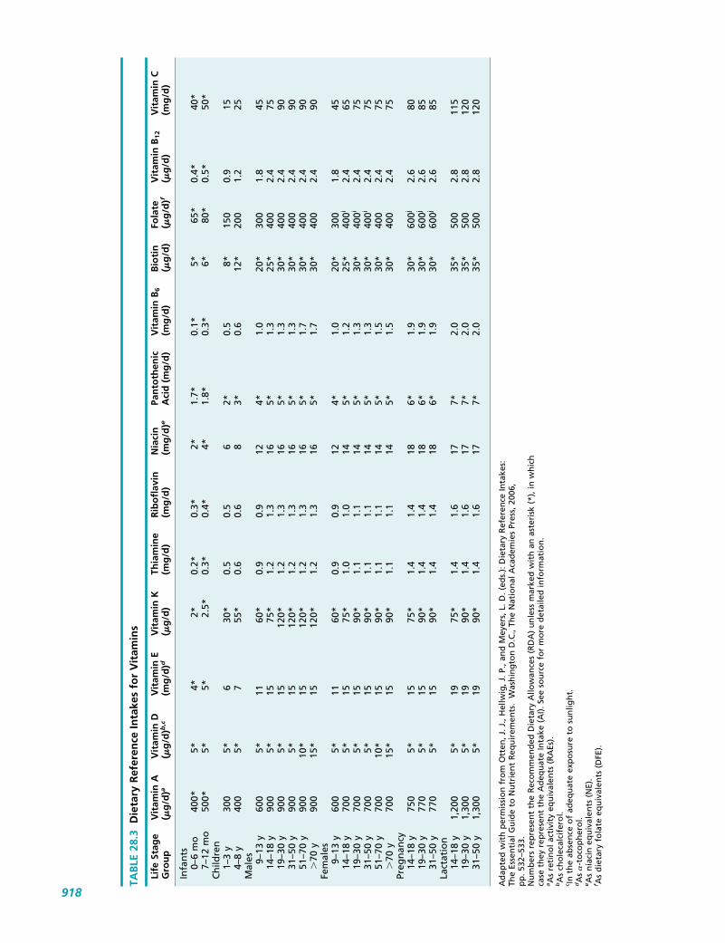
TAB
LE 2
8.3
Die
tary
Ref
eren
ce In
take
s fo
r V
itam
ins
Life
Sta
ge
Vit
amin
AV
itam
in D
Vit
amin
EV
itam
in K
Thia
min
eR
ibo
flav
inN
iaci
nPa
nto
then
icV
itam
in B
6B
ioti
nFo
late
Vit
amin
B12
Vit
amin
CG
rou
p(�
g/d
)a(�
g/d
)b,c
(mg
/d)d
(�g
/d)
(mg
/d)
(mg
/d)
(mg
/d)e
Aci
d (
mg
/d)
(mg
/d)
(�g
/d)
(�g
/d)f
(�g
/d)
(mg
/d)
Infa
nts
0–6
mo
400*
5*4*
2*0.
2*0.
3*2*
1.7*
0.1*
5*65
*0.
4*40
*7–
12 m
o50
0*5*
5*2.
5*0.
3*0.
4*4*
1.8*
0.3*
6*80
*0.
5*50
*C
hild
ren
1–3
y30
05*
630
*0.
50.
56
2*0.
58*
150
0.9
154–
8 y
400
5*7
55*
0.6
0.6
83*
0.6
12*
200
1.2
25M
ales 9–
13 y
600
5*11
60*
0.9
0.9
124*
1.0
20*
300
1.8
4514
–18
y90
05*
1575
*1.
21.
316
5*1.
325
*40
02.
475
19–3
0 y
900
5*15
120*
1.2
1.3
165*
1.3
30*
400
2.4
9031
–50
y90
05*
1512
0*1.
21.
316
5*1.
330
*40
02.
490
51–7
0 y
900
10*
1512
0*1.
21.
316
5*1.
730
*40
02.
490
�70
y90
015
*15
120*
1.2
1.3
165*
1.7
30*
400
2.4
90Fe
mal
es9–
13 y
600
5*11
60*
0.9
0.9
124*
1.0
20*
300
1.8
4514
–18
y70
05*
1575
*1.
01.
014
5*1.
225
*40
0i2.
465
19–3
0 y
700
5*15
90*
1.1
1.1
145*
1.3
30*
400i
2.4
7531
–50
y70
05*
1590
*1.
11.
114
5*1.
330
*40
0i2.
475
51–7
0 y
700
10*
1590
*1.
11.
114
5*1.
530
*40
02.
475
�70
y70
015
*15
90*
1.1
1.1
145*
1.5
30*
400
2.4
75Pr
egn
ancy
14–1
8 y
750
5*15
75*
1.4
1.4
186*
1.9
30*
600j
2.6
8019
–30
y77
05*
1590
*1.
41.
418
6*1.
930
*60
0j2.
685
31–5
0 y
770
5*15
90*
1.4
1.4
186*
1.9
30*
600j
2.6
85La
ctat
ion
14–1
8 y
1,20
05*
1975
*1.
41.
617
7*2.
035
*50
02.
811
519
–30
y1,
300
5*19
90*
1.4
1.6
177*
2.0
35*
500
2.8
120
31–5
0 y
1,30
05*
1990
*1.
41.
617
7*2.
035
*50
02.
812
0
Ad
apte
d w
ith
per
mis
sio
n f
rom
Ott
en, J
. J.,
Hel
lwig
, J. P
., an
d M
eyer
s, L
. D. (
eds.
): D
ieta
ry R
efer
ence
Inta
kes:
Th
e Es
sen
tial
Gu
ide
to N
utr
ien
t R
equ
irem
ents
. W
ash
ing
ton
D.C
., Th
e N
atio
nal
Aca
dem
ies
Pres
s, 2
006,
p
p.5
32–5
33.
Nu
mb
ers
rep
rese
nt
the
Rec
om
men
ded
Die
tary
Allo
wan
ces
(RD
A)
un
less
mar
ked
wit
h a
n a
ster
isk
(*),
in w
hic
h
case
th
ey r
epre
sen
t th
e A
deq
uat
e In
take
(A
I). S
ee s
ou
rce
for
mo
re d
etai
led
info
rmat
ion
.aA
s re
tin
ol a
ctiv
ity
equ
ival
ents
(R
AEs
).bA
s ch
ole
calc
ifer
ol.
c In t
he
abse
nce
of
adeq
uat
e ex
po
sure
to
su
nlig
ht.
dA
s �
-to
cop
her
ol.
eA
s n
iaci
n e
qu
ival
ents
(N
E).
f As
die
tary
fo
late
eq
uiv
alen
ts (
DFE
).
918
0915-0960_17865_Ch28.qxd 12/4/09 8:12 PM Page 918

and orange fruits and vegetables, such as carrots, spinach,broccoli, kale, collard and turnip greens, mangoes, apricots,nectarines, pumpkins, and sweet potatoes.
CHEMISTRY
Vitamin A belongs to a class of compounds referred to as theretinoids, which consist of four isoprenoid units joined in ahead-to-tail manner.20 All double bonds in the isoprenoidunits are implied to be in the E (trans) configuration unlessstated otherwise. Currently, the term retinoid is applied toretinol and its naturally occurring derivatives plus syntheticanalogs, which need not have vitamin A activity. Vitamin A iscurrently used as a generic descriptor for all retinoids that ex-hibit the biological activity of retinol, the original substanceidentified as vitamin A. The term vitamin A1 and all-transretinol have been used to refer to retinol whereas vitamin A2
was used to refer to 3,4-dehydroretinol. Retinoate analogs areused to refer to “compounds that control epithelial differenti-ation and prevent metaplasia, without possessing the fullrange of activities of vitamin A.”21 Retinal (formerly all-transretinal), the form of vitamin A that takes part in the visualcycle, and retinoic acid (formerly all-trans retinoic acid), theform that takes part in cell differentiation, are the two activeforms of vitamin A (Fig. 28.1). About 60 �-carotenes, out of700 carotenoids that have been identified, exhibit vitamin Aactivity in the body and are termed the provitamin As orprovitamin A carotenoids. The important carotenoids re-garded as the provitamin As are �-, �-, and �-carotenes andcryptoxanthin (Fig. 28.2). �-Carotene is the most importantprovitamin A carotenoid. The transformations of preformedvitamin As and �-carotene are shown in Figure 28.1.
Vitamin A activity has been historically expressed asUnited States Pharmacopeia (USP) units, international units(IU), retinol equivalents (RE), or �-carotene equivalents. TheUSP units and IU are equivalent. Each unit expresses the ac-tivity of 0.3 �g of retinol. Thus, 1 mg of retinol has the activ-ity of 3,333 units. Other equivalents are listed in Table 28.4.This system does not take into account the incomplete absorp-tion and rate-limited bioconversion of the provitamin Acarotenoids into the active form, retinal. One RE representsthe biological activity of 1 �g of retinol, 6 �g of �-carotene,or 12 �g of �-carotene or �-cryptoxanthin. The RE was used
Chapter 28 Vitamins 919
Figure 28.1 Interconversions of �-carotene and preformed dietary retinoids.
OH
8’10’12’14’151311975
α–Carotene
β–Carotene
γ–Carotene
Cryptoxanthin
Figure 28.2 Provitamin A carotenoids.
TABLE 28.4 Weight of Various Vitamin A SourcesEquivalent to 1 International Unit
Retinoid �g
Retinol 0.3Retinol acetate 0.334Retinol propionate 0.359Retinol palmitate 0.55�-Carotene 0.6
0915-0960_17865_Ch28.qxd 12/4/09 8:12 PM Page 919

to convert all dietary sources of vitamin A into a single unitfor easy comparison and accounts for inefficiencies in ab-sorption and rate-limited bioconversion of the provitamin Acarotenoids.22 The current unit used to express vitamin Aactivity is the retinol activity equivalents (RAE). This cameabout from the finding that the absorption of the provitaminA carotenoids is 50% less than originally thought. The onlydifference in this system is that the provitamin Acarotenoids are one half the activity assumed when usingREs (see Table 28.5).
The stereochemistry of vitamin A and related compoundsis complex, and the study of the structural relationshipsamong vitamin A and its stereoisomers has been compli-cated by the common use of several numbering systems(Fig. 28.3). The first numbering system (A) is the one cur-rently recommended by IUPAC and is used throughout thistext. The second system (B) places emphasis on the conju-gated � system, whereas the third (C) is used by the USPDictionary of USAN and International Drug Names.
The conjugated double-bond systems found in vitamin Aand �-carotene, as well as the attached ring in the form of�-ionone or dehydro-�-ionone found in 3,4-dehydroretinol,are essential for activity. When any of these is saturated, ac-tivity is lost. The ester and methyl ethers of vitamin A havea biological activity on a molar basis equal to that of vitaminA. Retinoic acid (vitamin A acid) is biologically active butis not stored in the liver.
For steric reasons, the number of isomers of vitamin Amost likely to occur is limited. These are all-trans, 9-cis,13-cis, and the 9,13-di-cis. A cis linkage at double bond 7or 11 encounters steric hindrance. The 11-cis isomer istwisted as well as bent at this linkage; nevertheless, this is
the only isomer that is active in vision and is synthesized inretinyl pigmented epithelial cells from all-trans-retinyl es-ters by the action of isomerase followed by hydrolysis.
KINETICS
Vitamin A is absorbed in the intestine via the same pathwayused for dietary lipids, requiring bile salts, dietary fat, andpancreatic juice. Retinyl esters are hydrolyzed in the intestinallumen to retinol by pancreatic triacylglycerol lipase (EC3.1.1.3) and the brush border enzyme lysophospholipase (EC3.1.1.5). The retinol is absorbed into the enterocytes by facil-itated diffusion at normal concentrations. At pharmacologicaldoses, however, retinol can be absorbed by passive diffu-sion.23,24 Bioavailability for most forms of preformed vitaminA is 70% to 90%. Upon cellular entry, retinol associates witha retinol binding protein (RBP), most likely RBP2. RBPsrepresent a group of proteins involved in intracellular andintercellular binding and transport of vitamin A. These intra-cellular proteins function in the transport and metabolism ofretinol and retinoic acid by solubilizing them in aqueousmedia and presenting them to the appropriate enzymes whileprotecting them from catabolizing enzymes.25 These proteinsalso limit the concentration of free retinoids within the cell.
Provitamin A carotenoids are absorbed by passive diffu-sion and also depend on absorbable fats and bile. It is cur-rently assumed that one twelfth of normal dietary �-carotenebut only one twenty-fourth of the other provitamin Acarotenoids is absorbed and bioconverted. This explains thelow activity of the provitamin A carotenoids in relation toretinol as shown in Table 28.5. The enterocytes are the mainsite of �-carotene transformation to retinal,26 but the enzymesthat catalyze the transformation also occur in hepatic andother tissue. Up to 20% to 30% of provitamin A carotenoidsare absorbed unchanged. Following absorption by the ente-rocytes, �-carotene is symmetrically cleaved into retinal by�-carotene 15,15�-monooxygenase (EC 1.14.99.36), whichrequires molecular oxygen.27 �-Carotene can give rise to twomolecules of retinal; whereas with the other three carotenoids,only one molecule is possible by this transformation. This isa result of the composition of �-carotene, which has a struc-ture identical to retinal in both halves of the molecule,whereas the other provitamin As have a structure identical toretinal in one half of the molecule (see Fig. 28.2). Asymmetriccleavage of the carotenoids by �-carotene-9,10-monooxygen-ase has also been reported,26,28,29 but its role in humans is notfully resolved. The retinal formed from the provitamin As isthen reduced to retinol by retinol dehydrogenase (EC1.1.1.105) or alcohol dehydrogenase (EC 1.1.1.1).
Within the enterocytes, the retinol is esterified by threeenzymes, diacylglycerol O-acyltransferase (DGAT, EC2.3.1.20), retinol O-fatty-acyltransferase (EC 2.3.1.76), alsoknown as acyl-coenzyme A (CoA):retinol acyltransferase(ARAT), and phosphatidylcholine-retinol O-acyltransferase(EC 2.3.1.135), also known as lecithin:retinal acyltrans-ferase (LRAT). LRAT esterifies retinol bound to RBP2,whereas ARAT can esterify unbound retinol. It has beenproposed that LRAT esterifies retinol at normal doses, whileARAT esterifies excess retinol.30 It has also been suggestedthat high doses of retinol may increase retinoic acid forma-tion in the enterocytes through a retinoic acid responsive el-ement (RARE) that promotes RBP2 expression.31
The retinyl esters are incorporated into chylomicrons,which in turn enter the lymph. Once in the general circula-
920 Wilson and Gisvold’s Textbook of Organic Medicinal and Pharmaceutical Chemistry
TABLE 28.5 Weight of Various Vitamin A SourcesEquivalent to 1 Retinol Equivalent or 1 RetinolActivity Equivalent
Vitamin A Source REs1 (�g) RAEs2 (�g)
Retinol 1 1�-Carotene 6 12�-Carotene 12 24�-Cryptoxanthin 12 24
1Retinol Equivalents.2Retinol Activity Equivalents.
OH
OH
OH
System B
54321
System A
1514
1312
1110
98
765
4
32
1
System C
98
76
54
32
1
Figure 28.3 Vitamin A numbering systems.
0915-0960_17865_Ch28.qxd 12/4/09 8:12 PM Page 920

tion, chylomicrons are converted into chylomicron rem-nants, which are cleared primarily by the liver but possiblyother tissues.30 As the esters enter the hepatocytes, they arehydrolyzed to retinol which is then bound to RBP1 orRBP4. These are released into the blood or transferred toliver stellate cells for storage. Within the stellate cells, theretinol bound to RBP is esterified for storage by LRAT andARAT. Stellate cells contain up to 80% of the bodies vita-min A stores.32 The RBP–retinol complex released into thegeneral circulation from hepatocytes or stellate cells is ex-tensively bound to transthyretin (TTR), which protectsRBP-retinol from glomerular filtration and subsequentrenal excretion.33 Retinol alone is taken up by target cells,probably by passive diffusion, from circulating RBP-retinoland once inside, is bound to RBP1 and possibly RBP2.Other retinoids may also circulate in plasma in lowamounts, such as free retinol and 13-cis retinoic acid, butthese are most likely bound to albumin.
Elimination of vitamin A generally involves the glu-curonidation of retinol or retinoic acid followed by renal orbiliary excretion that may result in enterohepatic cycling.Other pathways for oxidizing retinoids to more polar sub-stances also exist. Normally, no unchanged retinol is ex-creted. Retinal, retinoic acid, and other metabolites are,however, found in the urine and feces.
BIOCHEMISTRY
Vitamin A is involved in numerous biochemical functions.The best-known action of vitamin A is in its function in thechemistry of vision. The requirement for vitamin A forproper visual response to light in the retina has been wellcharacterized. However, its role in cellular growth and dif-ferentiation continues to expand.
The molecular mechanism of action of vitamin A, in theform of 11-cis retinal, in the visual process has been underinvestigation for many years.34,35 Figure 28.4 represents the
Chapter 28 Vitamins 921
H+
O
N
Bleachedrhodopsin
Thermalreactions
Light–catalyzedreaction
Seconds
Milliseconds
Microseconds
Nanoseconds
hνPicoseconds
+
retinal
Opsin
Metarhodopsin–II (λmax, 380 nm)
Metarhodopsin–I (λmax, 478 nm)
Lumirhodopsin (λmax, 497 nm)
Bathorhodopsin (λmax, 548 nm)
Rhodopsin (λmax, 506 nm)
Lysine Protein
OH
retinol
reduction
OH
retinol
O
retinyl esters
R
O
OH11-cis retinol
O11-cis retinal
hydrolysis
Transport
Transport
oxidation
isomerizationhydrolysis
esterification
Rod Photoreceptor Retinal Pigmented Epithelial Cell
Figure 28.4 Retinol and the visual cycle.
0915-0960_17865_Ch28.qxd 12/4/09 8:12 PM Page 921

chemical changes that take place in the visual cycle involv-ing the rhodopsin system in the rod cells of the retina. Uptakeof retinol from blood by the epithelial cells of the retina isfollowed by re-esterification and conversion to the 11-cisretinol form. This is converted to the aldehyde, 11-cis retinal,which is the form used in the visual pathway. In contrast tothe cone cells of the retina, which are responsible for colorvision and only function well in the presence of ample light,the vitamin A–dependent rods are essential for vision over awide range of light intensity, especially low levels. This sys-tem is responsible for vision in the dark, which explains thenight blindness seen in vitamin A–deficient individuals.
Vitamin A is also intimately involved in cellular differen-tiation during embryonic development and the maintenance ofcellular differentiation in various tissues in the adult. The mo-lecular mechanism of vitamin A, in the form of retinoic acid,in this area is more complex.36,37 This form of vitamin A actsthrough retinoic acid receptors (RARs) which are a memberof the steroid/thyroid hormone superfamily of receptors.These act as ligand-dependent transcription factors that regu-late gene expression. Several RARs have been identified:RAR� (RARA), RAR� (RARB), and RAR� (RARG). Thesediffer in their tissue distribution and the level of expressionduring cell development and differentiation. Following cyto-plasmic RAR-retinoic acid binding, the complex forms a het-erodimer with the retinoid X receptor (RXR). The het-erodimer translocates to the nucleus and binds to a RARE orretinoid X response element (RXRE) on DNA associated withthe promoter region of target genes and, together with coacti-vators, regulates target gene transcription. Several RXR re-ceptors have also been identified including RXR� (RXRA),RXR� (RXRB), and RXR� (RXRG), and they have beenshown to be also involved in the intracellular action of vita-min D. They have a different tissue distribution from theRARs. A specific ligand for RXR has been identified as 9-cis-retinoic acid, which can also bind to RARs; however, thephysiological role of this retinoid is not known. Retinoic acidcan bind only to RARs. 9-cis retinoic acid may, therefore, rep-resent a third active form of vitamin A.38,39 Several syntheticretinoids have been synthesized that bind selectively to RXRreceptors, and these are referred to as rexinoids.
The biological effects of retinoic acid are mediatedthrough various proteins, numbering in the hundreds, whichare controlled at the nuclear level by this form of the vitamin.Ultimately, effects are seen in cell differentiation, both duringembryonic development and in the adult. Effects on reproduc-tion, growth, immune function, hematopoiesis, epithelium,central nervous system (CNS) function, and cancer have alsobeen observed, among others. This widespread involvementin biological function explains the myriad of effects seen indeficiency and overdoses of this vitamin.
Deficiency of this vitamin leads to several disorders col-lectively referred to as vitamin A deficiency disorders(VADD). Preschool-age children are especially susceptibleto and develop more serious forms of VADD leading to in-creased mortality which may approach 50% in poor soci-eties.40 A deficiency of vitamin A is manifested chiefly bykeratinization of the mucous membranes throughout thebody. This degeneration is more pronounced in the eye thanin any other part of the body and gives rise to a conditionknown as xerophthalmia which results in drying and thick-ening of the conjunctiva and cornea. This may ultimatelylead to corneal destruction and permanent blindness.
Vitamin A deficiency also results in poor dark adaptation ofthe eyes referred to as night blindness (nyctalopia). Immuneinsufficiency develops resulting in a tendency to infection.Anemia results from hematopoietic failure in the marrow. Inchildren, growth failure and wasting also occurs. Deficiencyof vitamin A can also give rise to dryness and scaliness ofthe skin in adults.
THERAPEUTIC USES
Vitamin A is used in the treatment of known or suspected vi-tamin A deficiency. Vitamin A deficiency in developedcountries such as the United States is not likely of dietary ori-gin but is more likely caused by malabsorption as is seen insprue, hepatic cirrhosis, biliary, or pancreatic diseases.Interestingly, vitamin A levels decrease during infection withthe measles virus (rubeola) resulting in increased morbidityand mortality, especially in children with preexisting vita-min A deficiency. Vitamin A supplementation has beenshown to decrease mortality by 50% and prevent blindness inchildren developing measles if given at the time of diagnosis.Of course, vaccination is of prime importance; however, inunderdeveloped countries where the rate of vaccination islow, vitamin A supplementation is a beneficial alternative.41
Retinoate analogs have been developed for use in the treat-ment of dermatological conditions such as psoriasis andacne. Some therapies for cancer by retinoate analogs havealso been established, and newer therapies against variouscancers are under investigation.37
Both natural and synthetic carotenoids decrease acute pho-tosensitivity in porphyria in a manner unrelated to its vitaminactivity.42 Most �-carotene is converted to retinol duringabsorption, but the fraction that is absorbed unchanged isdistributed widely and accumulates in the skin. This can pro-vide protection against sun exposure in patients with erythro-poietic protoporphyria. It does not provide total protectionagainst the sun, but patients who respond to its treatment canremain in the sun the same as normal individuals.Discontinuance of the drug results in a return of hypersensi-tivity. �-Carotene does not function as a sunscreen in normalpatients and should not be used as such. Carotenodermia, a re-sult of accumulation in the skin, is the major side effect.Tanning capsules containing �-carotene and/or the carotenoidand canthaxanthin, uses this effect.
�-Carotene has also been investigated for possible protec-tive effects in cancer. Early studies suggested a protective ef-fect in lung cancer, but recent studies of lung, prostate, colon,breast, or nonmelanoma skin cancer did not show a protectiveeffect.43 Surprisingly, the results of several later studies sug-gest that increased intake of �-carotene might increase therisk of lung cancer in smokers.1,44 Studies also do not showany protective effects in cardiovascular disease.1,43
HYPERVITAMINOSIS
The toxicities of vitamin A have been known since beforethe vitamin was discovered,45 and can lead to both short- andlong-term effects.46–48 Short-term doses of 0.5 to 4 millionIU can lead, within hours to several days, to CNS effects in-cluding increased intracranial pressure, headache, irritabil-ity, and seizures; gastrointestinal effects including nausea,vomiting, and pain; dermatological effects such as desqua-mation; ophthalmic effects such as papilledema, scotoma,and photophobia; and liver damage. Most of these reactions
922 Wilson and Gisvold’s Textbook of Organic Medicinal and Pharmaceutical Chemistry
0915-0960_17865_Ch28.qxd 12/4/09 8:12 PM Page 922

have been reported in infants following treatment with largedoses of vitamin A, but some have resulted from ingestionof food rich in vitamin A, such as liver, especially frompolar bears.
Long-term intake of doses of vitamin A lower than theintake required for short-term toxicity but still above that re-quired by the body can lead to long-term effects, includingeffects on the skin, liver, CNS, and bone. Although theamount required varies, doses as low as 15,000 IU/day haveled to some adverse effects. Higher doses, generally above100,000 IU/day, are required to see all the reported adverseeffects. In patients with low body weight, malnutrition, orliver or renal disease, the doses required for long-term ad-verse effects may be still lower.
Dermatological adverse effects include drying of the skinand mucosa, dermatitis, pruritus, swelling and fissuring ofthe lips, and rarely loss of body hair. Hepatic effects includehypertrophy and hyperplasia of the hepatic stellate cells,hepatomegaly, fibrosis, and cirrhosis, which can lead to por-tal hypertension, ascites, and jaundice. Splenomegaly isalso seen. CNS effects include increased intracranial pres-sure (pseudotumor cerebri) leading to headache, visual dis-turbances (e.g., diplopia), drowsiness, vomiting, seizures,and a bulging fontanel in infants. Finally, pain in the boneand joints, with accompanying tenderness and reduced bonemineralization49 have also been reported.
The teratogenic effects of vitamin A are also well-known.50 Intake of as little as 10,000 IU/day during preg-nancy may increase the risk of birth defects, and the riskincreases with increasing intake of vitamin A.51,52 Birth de-fects include cardiovascular, craniofacial, neural tube, andurogenital and musculoskeletal abnormalities.
�-Carotene, in contrast, is relatively nontoxic as are theother provitamin A carotenoids.47,53 After long-term expo-sure to high levels of �-carotene (30–180 mg/day for15 years), patients have not developed any problems otherthan skin discoloration and asymptomatic hypercarotone-mia. Furthermore, �-carotene is not teratogenic. The mostlikely mechanism is the incomplete absorption and slowrate of conversion of �-carotene to retinol in the intestinalcells.54 However, a recent systematic review and meta-analysis found an increase in overall mortality in studiesusing �-carotene.43,55 This, coupled with the finding that�-carotene may increase the risk of lung cancer in smokers,as discussed previously, suggests that it may not be as non-toxic as originally thought when used chronically.
PRODUCTS
Vitamin A contains retinol (vitamin A alcohol) or its estersfrom edible fatty acids (chiefly acetic and palmitic acids). Itis available from various manufacturers under many names.Dosage forms vary from oral tablets, capsules, and solutionsto topical ointments and creams. It is also available as an in-jectable in the form of vitamin A palmitate (Aquasol A) thatis intended for intramuscular (IM) injection. Vitamin A isalso available as a component of multivitamins from manydifferent sources.
The provitamin A carotenoids are also widely available,primarily �-carotene, from various sources under manynames. Oral liquids, tablets, and capsules are available, con-taining �-carotene, either singly or in combination as multi-vitamin preparations.
Retinoate analogs are also available for the treatment ofdermatological conditions and cancer. Table 28.6 lists thecurrent products on the market and their approved uses,whereas Figure 28.5 shows their corresponding structure.
Vitamin D
The recognition in 1919 that rickets was the result of anutritional deficiency led to the isolation of antirachiticcompounds from food products.56 The role of sunlight inthe prevention of rickets was noted at the same time.57
Early studies showed that vitamin A preparations availableat the time, in addition to their growth-promoting proper-ties, could also cure xerophthalmia and rickets. Thus, theearly assumption was that all of these were because of thedeficiency of vitamin A. A preliminary report by Funk andDubin58 in 1921 was the first to suggest the presence of avitamin in yeast extracts that was distinctly different fromvitamin A and vitamin B, which the authors named vitaminD. It is interesting that the authors believed the new vita-min D was another of the B vitamins, which at that pointin history was known to consist of at least two different vi-tamins. McCollum et al.59 clearly showed in 1922 that vi-tamin A and the antirachitic factor were, in fact, two sepa-rate vitamins. The synthesis of both major forms ofvitamin D, ergocalciferol and cholecalciferol, was accom-plished in 1977.60
The highest natural source of vitamin D is fatty fishsuch as herring, catfish, salmon, mackerel, and tuna or oilsderived from fish such as cod liver oil. Eggs, from hens fedvitamin D, and butter also contain small amounts of vita-min D. The most common source of vitamin D in devel-oped countries, such as the United States, is fortified foodssuch as milk, ready-to-eat cereals, and some fruit juices.Cholecalciferol or ergocalciferol is usually used in fortifi-cation or supplementation.
CHEMISTRY
Chemically, the various forms of vitamin D are broken-opensteroids referred to as secosteroids. The term vitamin D iscurrently applied to all steroids possessing biological activ-ity like that of cholecalciferol.61 Irradiation of yeast ergos-terol yields a 1:1 mixture of ergocalciferol and lumisterolknown as vitamin D1. Because all the activities reside inthe ergocalciferol, the term vitamin D1 is no longer used.The photoconversion of ergocalciferol (ercalciol, previ-ously calciferol, vitamin D2) from ergosterol is shown inFigure 28.6. Cholecalciferol (calciol, previously vitaminD3) is the form produced in vivo from the action of sun-light on 7-dehydrocholesterol (Fig. 28.7) in the skin.22,23-Dihydroergocalciferol was known as Vitamin D4.(24S)-Ethylcalciol (formerly sitocalciferol), made from 7-dehydrositosterol, was known as vitamin D5. Note thestructural differences between ergocalciferol and cholecal-ciferol shown in Figure 28.6 and Figure 28.7. The ergocal-ciferol side chain contains a double bond between C22 andC23 and a methyl group on C24.
KINETICS
The gastrointestinal absorption of the vitamin Ds requiresbile, pancreatic juice, and dietary fat. Following formation
Chapter 28 Vitamins 923
0915-0960_17865_Ch28.qxd 12/4/09 8:12 PM Page 923

of micelles in the intestinal lumen, vitamin D esters arehydrolyzed, and free vitamin D, usually as cholecalciferol orergocalciferol, is absorbed by passive diffusion into the en-terocytes. There, it is incorporated into chylomicrons andsecreted into the lymph where it enters the circulation.62
Cholecalciferol and ergocalciferol were once thought to bethe active forms of vitamin D; however, they have now beenidentified as provitamins because they require hydroxyla-tion by the liver and the kidney to be fully active.
As shown in Figure 28.8, the first step occurs in theliver, following uptake from chylomicron remnants, bythe enzyme vitamin D3 25-hydroxylase (EC 1.14.15.-). Thisenzyme converts the provitamin to calcidiol (previously 25-hydroxyvitamin D3, 25(OH)D3) and requires both molecular
oxygen and reduced NADPH. The enzyme is found on theinner mitochondrial membrane,63 and the rate correlateswith substrate concentration. Following secretion into theblood, calcidiol and other forms of vitamin D are associatedwith group-specific component (vitamin D–binding protein[GC]) formerly referred to as vitamin D–binding protein(VDBP). The calcidiol bound to GC is the major circulatingform of the vitamin and may be stored in fats and muscle forprolonged periods. GC binding helps in the transport of vi-tamin Ds in blood and also prolongs the circulatory half-lives by making them less susceptible to hepatic metabolismand biliary excretion.64 Albumin and lipoproteins also bindvitamin Ds but with lower affinity than GC. The circulatinglevels of calcidiol are proportional to vitamin D intake andsynthesis; thus, plasma levels of calcidiol have been used toindicate vitamin D status.65
The epithelial cells of the proximal convoluted tubulesin the kidneys convert calcidiol to calcitriol [previously1�,25-dihydroxyvitamin D3, 1�,25(OH)2D3] by the enzymecalcidiol 1-monooxygenase (EC 1.14.13.13) also knownpreviously as 25(OH)D3 1�-hydroxylase. The activity ofthis mitochondrial, cytochrome P450 enzyme is increasedby parathyroid hormone and hypophosphatemia and de-creased by calcitriol and Ca2�.66 Understanding this finalstep is crucial because renal disease can lead to deficiencyof the active calcitriol and subsequent derangements in cal-cium and bone metabolism. Treatment is only effective if avitamin D preparation that already has the 1�-hydroxy isused. Similar to cholecalciferol, ergocalciferol also requireshydroxylation in the liver and kidney to become fully activeas shown in Figure 28.9.
In a classical sense, cholecalciferol, the form produced inanimals, is not a true vitamin because it is produced in the
924 Wilson and Gisvold’s Textbook of Organic Medicinal and Pharmaceutical Chemistry
HO OOH3C
Etretin
O
OH
OH3C
Acetretin
O
OH
H3CO
Adapalene
HO O
Alitretinoin(9-cis-Retinoic acid)
O
OH
CH2
Bexarotene
HO O
Isotretinoin(13-cis-Retinoic acid)
O
CH3
O
CC
S
Tazarotene
O
OH
Tretinoin(Retinoic acid)
Figure 28.5 Structures of clinically used retinoate analogs.
TABLE 28.6 Clinically Used Retinoate Analogs
Generic Trade Dosage Name Name Form Use
Acitretin Soriatane Capsule PsoriasisAdapalene Differin Cream or gel Acne vulgarisAlitretinoin Panretin Gel Kaposi sarcomaBexarotene Targretin Capsule T-cell lymphomasIsotretinoin Accutane, Capsule Acne vulgaris
Isotrex Topicalpreparation
Tazarotene Tazorac, Cream or gel PsoriasisAvage
Tretinoin Retin-A, Cream or gel Acne vulgarisRenova, Avita,Altinac, Atralin,Vesanoid Capsule Leukemia
0915-0960_17865_Ch28.qxd 12/4/09 8:12 PM Page 924

skin from 7-dehydrocholesterol by UV radiation (Fig. 28.7)in the range of 290 to 300 nm.66 7-Dehydrocholesterol isproduced from cholesterol by the enzyme 7-dehydrocholes-terol reductase (EC 1.3.1.21). Only when exposure to sun-light is inadequate does cholecalciferol become a vitamin inthe historical sense. As shown in Figure 28.7, upon expo-sure to UV irradiation, 7-dehydrocholesterol is convertedrapidly to previtamin D3. Previtamin D3 undergoes slowthermal conversion to cholecalciferol and the biologicallyinactive lumisterol and tacalciol (formerly tachysterol3).Excess exposure increases production of the inactive com-pounds. The slow conversion of previtamin D3 to cholecal-ciferol ensures adequate supplies when the exposure isbrief. Further, lumisterol and tacalciol can be convertedback to previtamin D3 and thus serve as a reservoir.66 It hasbeen estimated that a 10-minute exposure of just the uncov-ered hands and face will produce sufficient cholecalcif-erol.67 Inadequate photo conversion to cholecalciferol ismost likely to occur in northern latitudes during the wintermonths, in people with dark-colored skin or in people whoroutinely use clothing or sunscreens to reduce their sun ex-posure, such as the elderly.
The mechanism responsible for the movement of chole-calciferol from the skin to the blood is not known. In theblood, cholecalciferol is bound primarily to GC. This pro-tein selectively removes cholecalciferol from the skin
because it has low affinity for 7-dehydrocholesterol, previ-tamin D3, lumisterol, and tacalciol.
Catabolism of vitamin D is initiated by the enzyme1,25-dihydroxyvitamin D3 24-hydroxylase (EC 1.14.13.-),whose expression is stimulated by calcitriol, itself. 24-Hydroxylation is followed by oxidation to the ketone.Subsequent hydroxylation at C23 leads to cleavage of theside chain, resulting in the biologically inactive productcalcitronic acid.68,69 The 24-hydroxy metabolites are ex-creted primarily in the bile.
BIOCHEMISTRY
Similar to retinoic acid, the vitamin D receptor (VDR) is amember of the steroid/thyroid hormone superfamily of re-ceptors. These act as ligand-dependent transcription factorsthat regulate gene expression. In intestine, bone, kidney, andother tissues, calcitriol binds to VDR localized in the cyto-plasm, and the complex forms a heterodimer with a retinoidRXR. RXRs are also involved in vitamin A action and thereader is referred to that section for more information. Thiscomplex translocates to the nucleus and binds to vitamin Dresponse elements (VDREs) associated with the promoterregion of target genes and, together with coactivators, regu-lates target gene transcription. Over 50 genes have beenidentified that are controlled by calcitriol.
Chapter 28 Vitamins 925
CH3
CH3H3C
HO
CH3
CH3H3C
HO
27
2625
24
23
2221
20
199
8
76
54
3
21
1817
16
1514
13
1211
10
CH3
CH3H3C
H
CH3
CH3H3C
HO
Δ
hνhν
hν
TacalciolLumisterol
Cholecalciferol(Vitamin D3)
Previtamin D37–DehydrocholesterolProvitamin D3
(6-electron conrotatory electrophilic reaction)
H
(antarafacial hydride sigmatropic shift)
HO
CH2
CH3
CH3H3C
OH
Figure 28.7 Photoactivation of 7-dehydrocholesterol to cholecalciferol.
HO
CH2
CH3
CH3
CH3
H3C
CH3
CH3
CH3
H3C
HO
CH2
CH3
CH3
CH3
H3C
HO
hν =
27
26
28
18
11
252423
2221
20
9 8
17
16
1514
13
12
10
19
76
54
32
1
28
27
26
252423
2221
20
199
8
76
54
3
21
1817
16
1514
13
1211
10
Vitamin D2Ergocalciferol
Ergosterol(Provitamin D2)
Figure 28.6 Photoactivation of ergosterol to ergocalciferol.
0915-0960_17865_Ch28.qxd 12/4/09 8:12 PM Page 925

One of the most well-known effects of calcitriol is tomaintain calcium homeostasis. Phosphate metabolism isalso affected. The mechanism of action promoting Ca2�
transport in the intestine involves calcitriol-mediated for-mation of a calcium-binding protein, calbindin, which isknown to exist in at least two forms in humans, calbindin 1,28kDa (CALB1) and S100 calcium binding protein G(S100G), also known as calbindin-D9k. A calcium-dependent ATPase, Na�, and calbindin are necessary forintestinal Ca2� transport and act to enhance the transcellu-lar movement resulting in increased absorption of dietaryCa2�. Transient receptor potential cation channel, subfam-ily V, member 6 (TRPV6), which controls the rate of cel-lular entry of Ca2�, is also found to be increased in intes-tinal cells under the control of calcitriol. Calcitriol alsopromotes intestinal phosphate absorption, mobilization ofCa2� and phosphate from bone, and renal reabsorption ofCa2� and phosphate. Also involved are parathyroid hor-mone and calcitonin.
VDRs have also been identified in tissues not normallyassociated with bone mineral homeostasis. Besides the in-testines, kidneys, and osteoblasts, vitamin D receptors havebeen located in the parathyroid gland, the pancreatic isletcells, the mammary epithelium, the skin keratinocytes, mus-cle, and the immune cells. Activation in the latter has potentantiproliferative and immunomodulatory functions. Thus,calcitriol and its analogous VDR ligands have therapeuticpotential in the treatment of inflammatory diseases (rheuma-toid and psoriatic arthritis), dermatological conditions (pso-riasis, keratosis), osteoporosis, suppression of parathyroidhormone, cancers (including colon, prostate, and breast),and autoimmune diseases.70 It has been suggested that vita-min D exerts its anticancer effects through its role as anuclear transcription factor that regulates cell growth, dif-
ferentiation, apoptosis, and other cancer-developing cellularmechanisms.71
The classic form of vitamin D deficiency universally rec-ognized is rickets. In young infants, the entire skull is soft(craniotabes), and problems in bone development lead to de-lays in sitting, crawling, and walking. Osteomalacia (evi-dent on x-rays) and deformities of the bones eventually leadto deformities in the rib cage, bowlegs or knock-knees, bonepain, and a predisposition to fractures. Bone deformitiesmay become permanent if not treated with vitamin D atan early stage. Vitamin D deficiency in children and adultsalso leads to myopathies, which can lead to weakness, andhypocalcemia, which can lead to paresthesia, musclespasms, and tetany. Recent studies conclude that an in-creased rate of overall mortality in the general population isassociated with low levels of vitamin D.72 The same studyalso demonstrated that vitamin D deficiency increases therisk of cancer, diabetes, and hypertension.
THERAPEUTIC USES
Cholecalciferol and ergocalciferol are used in the treatmentof known or suspected vitamin D deficiency or the preven-tion of vitamin D deficiency when bodily needs are in-creased as seen in pregnancy. Calcitriol can also be used,especially when an immediate effect is desired as in severedeficiencies because it is the active form of vitamin D foundin the body. This would involve treatment of rickets, osteo-malacia, muscle pain, and hypocalcemia that can result fromvitamin D deficiency. Vitamin D is also useful, particularlywhen used with calcium supplementation, to treat osteo-porosis.44,73 Vitamin D can also be used to treat hypophos-phatemia or hypocalcemia because of other causes, such ashypoparathyroidism.
926 Wilson and Gisvold’s Textbook of Organic Medicinal and Pharmaceutical Chemistry
HO
CH2
CH3
CH3
CH3
H3CCH3
OH
OH
HO
CH2
CH3
CH3H3CCH3
OH
HO
CH2
CH3
CH3H3C
ErgocalciferolVitamin D2
KidneyLiver
25–Hydroxyergocalciferol25–Hydroxyvitamin D2
1,25–Dihydroxyergocalciferol1,25–Dihydroxyvitamin D2
25-hydroxylase 1α-hydroxylase
Figure 28.9 Ergocalciferolbioactivation.
HO
CH2
CH3
CH3H3C
CholecalciferolVitamin D3
20
31
OH
HO
CH2
CH3
CH3H3C
Calcifediol25–Hydroxyvitamin D3
(25–Hydroxycholecalciferol,25(OH)D3 or calcidiol)
OH
OH
HO
CH2
CH3
CH3H3C
Calcitriol1,25–Dihydroxyvitamin D3
(1,25–Dihydroxycholecalciferol,1,25(OH)2D3)
Liver Kidney
25
25-hydroxylase 1α-hydroxylase
Figure 28.8 Cholecalciferolbioactivation.
0915-0960_17865_Ch28.qxd 12/4/09 8:12 PM Page 926

Chapter 28 Vitamins 927
Uremia associated with renal failure can result in hypocal-cemia and secondary hyperparathyroidism as a result of insuf-ficient conversion of calcidiol to calcitriol by the diseasedkidneys. This results in renal osteodystrophy, a metabolicbone disease involving abnormal mineralization. Patients areoften on dialysis by the time these problems occur. Treatmentwith calcitriol, or other active analogs of vitamin D with a 1�-hydroxy group (see Table 28.7) helps to increase Ca2� levels,as well as decrease parathyroid hormone levels essentiallycontrolling these effects. Doses used do not result in ex-traskeletal calcification or increase the rate of decline in renalfunction as can be seen with excessive doses of vitamin D.
Beneficial effects have been shown for high levels ofcalcitriol in patients with advanced prostate cancer74 andcolorectal cancer75 although this is not a proven therapy.These investigational treatments require high doses of vita-min D and the resultant hypercalcemia limits the useful-ness. Vitamin D analogs with a decreased tendency to causehypercalcemia and hypercalciuria are being developed andinvestigated. Although the issue of vitamin D in hearthealth is not resolved yet, an inverse correlation was foundbetween cardiovascular events and serum vitamin D (ascalcidiol or calcitriol) levels—low vitamin D levels are as-sociated with hypertension, elevated very low-densitylipoprotein (VLDL) triglycerides, and impaired insulin me-tabolism.76,77 Calcipotriene represents a vitamin D analogused for its antiproliferative effects and the ability to pro-mote cellular differentiation in the epidermis and is effec-tive in the treatment of psoriasis.78
HYPERVITAMINOSIS
Hypervitaminosis D apparently cannot arise from exces-sive exposure to sunlight79 but only occurs followingingestion of large quantities of synthetic vitamin D. Theprecise amount to produce toxicity would depend on thedose and duration of exposure, however, doses of 100,000IU daily for 4 days have not resulted in toxicity.80 Toxicityinvolves derangements of calcium metabolism, resulting inhypercalcemia and metastatic calcification of soft tissue.Most problems result from the hypercalcemia, which typi-cally causes muscular weakness, anorexia, nausea, vomit-ing, and depression of the CNS (which can result in comaand death). In addition, deposition of calcium salts in thekidneys (nephrocalcinosis) and the tubules (nephrolithia-sis) can lead to potentially irreversible renal damage. Earlysigns are polyuria and nocturia because of damage to therenal concentrating mechanism.
PRODUCTS
Table 28.7 is a list of the currently available products, andFigure 28.10 shows their structures. Most vitamin supple-ments containing vitamin D singly, or in combination withother vitamins as multivitamin preparations, contain chole-calciferol. However, ergocalciferol can also be used Theseare available in various preparations under many differentnames as oral capsules, tablets, solutions, and powders.Table 28.7 also shows the vitamin D analogs used in thetreatment of renal osteodystrophy that occurs as a result ofsecondary hyperparathyroidism in patients unable to con-vert calcidiol to the active calcitriol. This is most oftenseen in severe renal failure, especially in patients on dial-ysis. Each of these products already has the 1�-hydroxy,so the need for bioactivation by the kidney is circum-vented. Finally, calcipotriene is available for the treatmentof psoriasis.
Vitamin E
Since the early 1920s, it was known that rats fed only cow’smilk cannot produce offspring. In 1922, Evans and Bishop81
discovered an unrecognized dietary factor essential for re-production. In 1924, Sure82 named the principle that couldrectify this deficiency vitamin E. When the compoundknown as vitamin E was isolated from wheat germ oil in1936 by Evans et al.,83 it was named �-tocopherol, from theGreek words tokos meaning “childbirth,” phero meaning “tobear,” and “-ol” indicating an alcohol. The structure84 andsynthesis85 of �-tocopherol were reported in 1938. Sincethen, several other closely related compounds have been dis-covered from natural sources, and this family of naturalproducts took the generic name tocopherols.
The tocopherols are especially abundant in wheat germ,rice germ, corn germ, other seed germs, lettuce, soya, andcottonseed oil. All green plants contain some tocopherols,and there is evidence that some green leafy vegetables androse hips contain more than wheat germ. It is probably syn-thesized by leaves and translocated to the seeds. All fourtocopherols have been found in wheat germ oil; �-, �-, and�-tocopherols have been found in cottonseed oil. Corn oilcontains predominantly �-tocopherol and thus furnishes aconvenient source for the isolation of this difficult memberof the tocopherols. �-Tocopherol is 30% of the mixed toco-pherols of soya bean oil. Palm oil contains about 30% of to-copherols and 70% of tocotrienols. Cereals like oat, rye, andbarley contain small amounts of tocotrienols.
TABLE 28.7 Vitamin D Products
Generic Name Trade Name Dosage Form Use
Calcifediol Calderol Capsule Vitamin D therapyCalcipotriene Dovonex Ointment, cream, or scalp solution Plaque psoriasisCalcitriol Rocaltrol, Calcijex Capsule, oral solution, injection Vitamin D therapy, renal osteodystrophy,
hypocalcemia, hypoparathyroidismCholecalciferol Delta-D, various Capsules, tablet, injection, cream, Vitamin D therapy, hypoparathyroidism,
ointment hypophosphatamiaDihydrotachysterol DHT Capsule, oral solution, tablet HypoparathyroidismDoxercalciferol Hectorol Capsule Renal osteodystrophyErgocalciferol Drisdol, various Oral softgel, capsule, oral solution, Vitamin D therapy, hypoparathyroidism,
tablet hypophosphatamiaParicalcitol Zemplar Injection, capsule Renal osteodystrophy
0915-0960_17865_Ch28.qxd 12/4/09 8:12 PM Page 927

CHEMISTRY
Several tocopherols have been isolated. Some have the4�,8�,12�-trimethyltridecyl-saturated side chain; others haveunsaturation in the side chain. It has been suggested thatthese polyunsaturated tocols be named tocotrienols. Thebest-known tocopherol is �-tocopherol, which has the great-est biological activity. The base structure, represented inFigure 28.11, shows that the tocopherols are methyl-substi-tuted tocol derivatives: �-tocopherol is 5,7,8-trimethyltocol;�-tocopherol is 5,8-dimethyltocol; the �-compound is 7,8-dimethyltocol; and �-tocopherol is 8-methyltocol. The to-cotrienols have similar substituents. Vitamin E is a genericdescriptor for all tocol and tocotrienol derivatives that havebiological activity similar to �-tocopherol.86 Currently,eight of these tocols, four tocopherols and four tocotrienols,are considered the vitamin E family. These tocols possesstwo important structural components, the chromanol ringand the side chain, and differ by the methylation patterns ofthe chromanol ring (�-, �-, �-, and �-) and presence or ab-sence of double bonds in the side chain (no double bond intocopherols whereas three double bonds in tocotrienols).
928 Wilson and Gisvold’s Textbook of Organic Medicinal and Pharmaceutical Chemistry
OH
HO
CH2
CH3
CH3H3C
Calcifediol
CH2
H3COH
OHHOCalcipotriene
OH
OH
HO
CH2
CH3
CH3H3C
Calcitriol
HO
CH2
CH3
CH3H3C
Cholecalciferol
H3C
CH3
OH
CH3
CH3H3C
DihydrotachysterolOH
CH3
HO
CH3
CH3H3C
Doxercalciferol
OH
CH3
OH
HO
CH3
CH3H3C
ParicalcitolHO
CH2
CH3
CH3
CH3
H3C
Ergocalciferol
Figure 28.10 Structures of vitamin D products.
Natural (�)-�-tocopherol has the configuration 2R,4�R,8�R.The natural tocotrienol has a 2R,3�E,7�E configuration. Thetocopherols are diterpenoid natural products biosynthesizedfrom a combination of four isoprenoid units; geranylgeranylpyrophosphate is the key intermediate that leads to thesecompounds.
(�)-�-Tocopherol is about 1.36 times as effective as(�)-�-tocopherol in rat antisterility bioassays. As men-tioned previously, (�)-�-tocopherol is the naturally occur-ring form of �-tocopherol with 2R,4�R,8�R configuration.(�)-�-Tocopherol is synthetic, and is called all-rac-�-toco-pherol. The configuration at C2 is critical and the NutritionBoard of the National Academy of Sciences, Institute ofMedicine recognized in 2000 that 2R-forms of �-tocopherolmeet the human requirements. �-Tocopherol is about half asactive as �-tocopherol, and the �- and �-tocopherols areonly 0.01 times as active as �-tocopherol.
The esters of tocopherol (e.g., acetate, propionate, andbutyrate) are more active than the parent compound becauseof better bioavailability.87 This is also true of the phosphoricacid ester of (�)-�-tocopherol when it is administered
0915-0960_17865_Ch28.qxd 12/4/09 8:12 PM Page 928

parenterally.88 The ethers of the tocopherols are inactive,because unlike esters that are readily hydrolyzed to the ac-tive antioxidant chromanol form by esterases, ethers are rel-atively metabolically more stable and as such do not possessantioxidant property.
Oxidation of the tocopherols to their correspondingquinones also leads to inactive compounds. Replacement ofthe methyl groups on the chromanol ring by ethyl groups de-creases activity. The introduction of a double bond in the3,4-position of �-tocopherol reduces its activity by abouttwo thirds. Reduction of the size of the long alkyl side chainor the introduction of double bonds in this side chainmarkedly reduces activity.
KINETICS
Bile salts and dietary lipids play critical roles in the absorp-tion of vitamin E and their esters into intestinal epithelialcells through micellization. Pancreatic lipases facilitate themicellization of vitamin E as a consequence of hydrolysis ofnon–vitamin E lipids by forming mixed micells. The vita-min E esters (e.g., �-tocopherol acetate), on the other hand,require hydrolysis by the pancreatic lipases prior to micel-lization and uptake into the enterocytes. The uptake of vita-min E from the mixed micells is thought to be a passivediffusion process; however, the role of a scavenger receptorclass B type I (SR-BI) has also been suggested.
The intracellular trafficking of vitamin E may involve itsincorporation into chylomicrons. Both �- and �-tocopherolsare equally incorporated into chylomicrons, which are thensecreted into the lymphatic system. An ATP-binding cas-sette, subfamily A, member 1 (ABCA1) transport protein-directed efflux of vitamin E from epithelial cells into HDLfollowed by direct transport into the portal vein has alsobeen suggested as an important alternative pathway forvitamin E absorption. Dietary fats increase chylomicron-mediated absorption while a low-fat diet increases HDL-mediated absorption of vitamin E.89 The absence of dietary
fat results in little absorption of vitamin E.90 Despite a low-fat diet, high bioavailability was reported when vitamin Ewas added with emulsifier onto fortified breakfast cereal,suggestive of micellization as a critical factor for its absorp-tion.91 No apparent differences in intestinal absorption ofvarious forms of vitamin E (e.g., �- and �-tocopherols orRRR- and SRR-�-tocopherols) have been observed.92
Overall, 20% to 80% of vitamin E is absorbed from the in-testinal lumen into the bloodstream. The liver is an impor-tant storage site. �-Tocopherol is secreted primarily into thebile while �-tocopherol enters the circulation, where it isfound in much higher levels than �-tocopherol, even thoughthe latter predominates in the diet. This difference is attrib-uted to a liver cytosolic binding protein, �-tocopherol trans-fer protein (�-TTP) that is selective for �-tocopherol.
The tocopherols in lymph are associated with chylomi-crons and VLDLs. Circulating tocopherols are also carriedby �-TTP, which preferentially incorporates them intoblood low-density lipoproteins (LDLs). The tocopherolsare readily and reversibly bound to most tissues, includingadipose tissue, and the vitamin is thus stored. The vitaminis concentrated in membrane structures, such as mitochon-dria, endoplasmic reticulum, and nuclear and plasmamembranes.
Although “Simon metabolites” (tocopheronic acid and its�-lactone) were first identified as the primary vitamin Emetabolites by in vitro sample oxidation, the biologicallyrelevant are the 2-carboxyethyl-6-hydroxychroman (CEHC)products (Fig. 28.12). Thus �-, �-, �-, and �-CEHCs areidentified as metabolites of the respective tocopherols. Theterminal methyl group is oxidized to a carboxylic acid andshortened by several steps of �-oxidation to produce CEHC.The CEHC metabolites undergo glucuronide and sulfateconjugations, and most of the CEHC metabolites are foundin urine as these conjugates. These metabolites are also ex-creted in the bile and vitamin E may undergo some entero-hepatic circulation.92
BIOCHEMISTRY
For decades, there has been significant interest in investi-gating the biochemical functions of vitamin E, but it is stilldifficult to explain many of the biochemical derangementscaused by vitamin E deficiency in animals. There seems tobe general agreement that one of the primary metabolic
Chapter 28 Vitamins 929
CH3
CH3
CH3CH3R2
HO
R1
R3CH3O
CH3
CH3
CH3CH3R2
HO
R1
R3CH3O
1’
8
7
65 4
3
2
1
11’7’3’
8
61’ 12’8’4’7
5 43
2
1
δ
γ
β
α R1 = R2 = R3 = CH3
R1 = R3 = CH3; R2 = H
R2 = R3 = CH3; R1 = H
R1 = R2 = H; R3 = CH3
Chromanol ring
Tocopherols
Tocotrienols
Figure 28.11 Tocopherols and tocotrienols.
Figure 28.12 Vitamin E metabolites.
0915-0960_17865_Ch28.qxd 12/4/09 8:12 PM Page 929

functions of the vitamin is preventing the oxidation oflipids, particularly unsaturated fatty acids. Through thisantioxidant effect, �-tocopherol plays an important role inprotecting cell membranes made of lipids and LDLs. The�-tocopherol donates its electrons to the free radicals toneutralize them, thus protecting the cellular lipids from oxida-tive damage. In this process, �-tocopherol loses its antioxi-dant capacity by forming the �-tocoquinone (Fig. 28.13). Itis generally accepted that other antioxidants like vitamin Cor glutathione can restore vitamin E from its single electronoxidation state, tocopherol radical. Although several re-ports suggested that once it is fully oxidized to the toco-quinone form, it will not be regenerated back to vitamin E,reports are also available showing that it can be regeneratedback in presence of glutathione according to the mechanismshown in Figure 28.14.93,94
Because of the methylation level, which enhances thenucleophilicity and reactivity of the 6-OH group, the �-isoforms of both tocopherol and tocotrienol are the most po-tent antioxidants among all the vitamin E family. Although�-tocotrienol is believed to be a more potent antioxidant thanthe �-tocopherol for many factors (e.g., higher recyclingefficiency, more uniform distribution within the microsomalmembrane bilayer and more efficient interaction with lipidfree radicals) misleading results have been demonstrated be-cause of the high affinity of �-TTP for �-tocopherol. �-Tocopherol is biologically the most important isoform be-cause of its high affinity for �-TTP, which results in highbioavailability and bioactivity.95
Biochemical functions of the vitamin E family are diverseand may include actions other than their well-documented an-tioxidant activities. �-Tocopherol not only strongly inhibitsplatelet and other cell adhesion but also inhibits cell prolifer-ation, protein kinase C, 5-lipooxygenase, and phospholipaseA2 and activates protein phosphatase 2A and diacylglycerolkinase, which are unrelated to its antioxidant activity. Theseactions reflect the specific interactions of �-tocopherol withenzymes, structural proteins, lipids, and transcription factors.�-Tocopherol and CEHC metabolic products of vitamin E ex-hibit many other functions including anti-inflammatory, anti-neoplastic, natriuretic, and also cardioprotective functions.96
During the last 5 years, interest in the action of to-cotrienols has increased with the inclusion of a substantialamount of vitamin E research directed toward that of �-to-cotrienol. Numerous unique biochemical functions of �-to-cotrienol have been discovered that include prevention ofinducible neurodegeneration by regulating specific media-tors of cell death, hypocholesterolemic effects by suppressing3-hydroxy-3-methyl-glutaryl-CoA (HCG-CoA) reductaseactivity, and protection against stroke. It has also been re-ported that tocotrienols, but not tocopherols, suppress thegrowth of breast cancer cells.96
930 Wilson and Gisvold’s Textbook of Organic Medicinal and Pharmaceutical Chemistry
Figure 28.13 Antioxidant mechanisms of �- and �-tocopherols. (R1, H, or CH3; R2, Phytyl.)
Figure 28.14 Interconversion of �-tocopherol and �-tocoquinone.
0915-0960_17865_Ch28.qxd 12/4/09 8:12 PM Page 930

The diseases caused by vitamin E deficiency in animalsare not well correlated with that in human. The sterility inrats and nutritional muscular dystropies in other lower ani-mal including monkeys, rabbits, lambs, and chicks are welldocumented. Reversible neurological disorders in humansand hemolytic anemia in premature infants because of vita-min E deficiency have been seen.
The antioxidant function does not explain all the biochem-ical abnormalities caused by vitamin E deficiency. Moreover,vitamin E is not the only in vivo antioxidant. Two enzymesystems, glutathione reductase and o-phenylenediamine per-oxidase, also function in this capacity.97 It has been postu-lated that vitamin E has a role in the regulation of proteinsynthesis. Other actions of this vitamin have also been inves-tigated, for example, effects on muscle creatine kinase andliver xanthine oxidase. Vitamin E deficiency leads to an in-crease in the turnover of creatine kinase. Vitamin E–deficientanimals also exhibit increased liver xanthine oxidase activity,which is because of increased de novo synthesis.97
Although it has been difficult to establish clinical corre-lates of vitamin E deficiency in humans, Bieri and Farrell97
have summarized some useful generalizations and conclu-sions. These workers noted that the infant, especially thepremature infant, is susceptible to tocopherol deficiency be-cause of ineffective transfer of the vitamin from placenta tofetus and that growth in infants requires greater availabilityof the vitamin. In adults, the tocopherol storage depots pro-vide adequate availability that is not readily depleted, but in-testinal malabsorption syndromes, when persistent, can leadto depletion of the storage depots. Children with cystic fi-brosis suffer from severe vitamin E deficiency caused bymalabsorption. Tropical sprue, celiac disease, gastrointesti-nal resections, hepatic cirrhosis, biliary obstruction, and ex-cessive ingestion of mineral oil may also cause long-termmalabsorption.
THERAPEUTIC USES
Vitamin E is used in the treatment of known or suspected vi-tamin E deficiency. The use of vitamin E to prevent or treata wide variety of diseases has been examined quite exten-sively and in some cases has been found to be of some ben-efit, although the effect is frequently minimal. Vitamin Emay help in Alzheimer disease, dysmenorrhea, nonalcoholicfatty liver, and tardive dyskinesias, to name a few. However,in most cases, and especially for common diseases, there isoften no proven benefit or the studies are contradictory.
Vitamin E has been extensively studied for the preven-tion of cardiovascular disease, but evidence against any ben-efit continues to accumulate.44,98 Indeed, some studies showthat vitamin E supplementation may actually increase strokerisk98 or all cause mortality.55 It has also been suggested thatmegadoses of tocopherol be used in the treatment of periph-eral vascular disease. Although some studies support thisproposal, experts in the field state that further clinical stud-ies are necessary to make a definitive recommendation.Nevertheless, it continues to be popular and controversial toconsider the beneficial effects, and investigations ofmegavitamin E therapy for cardiovascular disease continueto appear in the literature.97
The failure to demonstrate the protective effects againstheart diseases by vitamin E supplements may be becausethey contain only �-tocopherol. To effectively remove the
peroxide damage in these cells by electrophilic peroxyni-trites or other mutagens, �-tocopherol is also needed that, inaddition to forming the orthoquinone, acts in vivo as a trapfor these membrane-soluble electrophilic mutagens formingstable adducts through the nucleophilic C5, which is blockedin �-tocopherol (Fig. 28.13). It has been suggested that be-cause �-tocopherol supplementation suppresses �-toco-pherol levels, a combination of the two tocopherols in theratios found in the diet may be more useful than �-toco-pherol, alone.99
The well-documented antioxidant property of the toco-pherols led to their examination for anticancer activitywithout any consistent success;43,100 although some stud-ies have shown a reduction in prostate cancer in smokers.44
It is interesting that the ethers (RO-), esters (RCOO-), andamides (RCONH-) at C6 of the chromanol ring (�-toco-pheryl succinate ester is the prototype, Fig. 28.15) haveshown to be potent antineoplastic agents. The antineoplas-tic potency of the tocopherol analogs is not related to theirantioxidant property, which was proven by synthesizing aseries of “redox-silent” analogs and testing them againstvarious neoplastic cell lines. The detailed molecular biol-ogy studies revealed that these are mainly mitocans (mito-chondrially targeted anticancer drugs) that selectivelydestabilize the mitochondria in malignant cells and sup-pressed cancer in preclinical models. Although they arefully recognized as potent anticancer agents, detailed clin-ical studies for their therapeutic application are yet to becompleted.101
HYPERVITAMINOSIS
Vitamin E used in high doses, as high as 3,200 mg/day, hasa proven record of safety.47 Various reports, many singlecase reports, or uncontrolled studies have suggested adverseeffects such as interference with clotting, weakness, de-creased thyroid hormone levels, and gastrointestinal upset.The importance of these is unknown. The only notable ad-verse effects have occurred in premature infants given largedoses of this vitamin. Hepatotoxicities have been seen inpremature infants of less than 1,500 g birth weight givenvitamin E intravenously, and the incidence of necrotizingenterocolitis and sepsis increased under similar conditionsfollowing oral or intravenous (IV) dosing.
PRODUCTS
Vitamin E is available from numerous sources as oral cap-sules, liquids, and tablets under various names. Vitamin E isalso available as a component of numerous multivitaminpreparations. The forms of vitamin E used in these productsare shown in Table 28.8. Vitamin E activity is currently ex-pressed in terms of the (�)-�-tocopherol equivalents basedon the former USP units and mass. One former USP unit isequal to one former IU.
Chapter 28 Vitamins 931
CH3
CH3
CH3CH3H3C
O
CH3
CH3CH3O
O
O
OH
Figure 28.15 �-Tocopheryl succinate.
0915-0960_17865_Ch28.qxd 12/4/09 8:12 PM Page 931

Vitamin K
Lipid-free diet research by Henrik Dam102 starting in 1929and culminating in 1935103 resulted in the discovery of anantihemorrhagic factor that Dam named vitamin K (fromthe German word Koagulation). Along with the work ofEdward A. Doisy, the structure of vitamin K was deter-mined in 1939104 and synthesis accomplished in 1965.105
For their work, Dam and Doisy shared the 1943 NobelPrize in Medicine. Originally, the term vitamin K was ap-plied to the vitamin isolated from alfalfa, and a similarprinciple from putrified fish meal was named vitamin K2.The original vitamin K was thereafter referred to as vita-min K1. In 1960, IUPAC recommended the terms phyllo-quinone for vitamin K1 with the official abbreviation of“K” and farnoquinone for the originally isolated vitaminK2.106 Several compounds chemically related to vitaminK2, differing only in the number of 5-carbon prenyl (iso-prenoid) units attached, were subsequently discovered, andin 1973, IUPAC adopted the term menaquninones (MK) torefer to this group of substances.107 The menaquinones dif-fer in the number of prenyl units attached and specificcompounds are designated menaquinone-n (MK-n) wheren represents the number of prenyl side chains. The originalvitamin K2 is now known as MK-6. For this discussion, theterm vitamin K will be used to denote both phylloquinoneand menaquinones when appropriate. Also of note, theUSP generic name for phylloquinone in pharmaceuticalpreparations is phytonadione, and this is the preferred termamong medical practitioners. Throughout this text, thesetwo terms will be used in their best context.
Naturally occurring vitamin K is found in two formsdepending on whether the source is plants or bacteria.Plants produce phylloquinone whereas bacteria produce
menaquinones. Dietary sources from plants include leafygreen vegetables; such as collard greens, spinach, andsalad greens; soy and canola oils; and margarine. Broccoli,brussels sprouts, cabbage, and olive oil also contain appre-ciable quantities. The major bacterial source is the intes-tinal flora, especially Gram-positive organisms; however,their contribution to overall vitamin K intake is unknown.
CHEMISTRY
Chemically, all vitamin Ks are 2-methyl 1,4-naphtho-quinone derivatives containing variable aliphatic sidechains at C3. Phylloquinone (Fig. 28.16) invariably containsa phytyl side chain. Menaquinones (Fig. 28.17) are a seriesof compounds that have a longer side chain with more un-saturation. This side chain may be composed of 1 to 13prenyl (isoprenyl) units.
Many other closely related compounds possess vitamin Kactivity. Of particular historical note are the water-solublemenadione (2-methyl-1,4-naphthoquinone; vitamin K3),which is no longer marketed because of toxicities, and mena-diol (2-methylnaphthalene-1,4-diol; vitamin K4). Menadionecontains the naphthoquinone ring without any attachedprenyls and can be thought of as MK-0 (see Fig. 28.17).Menadiol is menadione with the two keto groups reduced tohydroxyls. Both compounds are bioactivated to the activeform through alkylation. Phylloquinone naturally occurs as atrans-isomer and has an R,R,E configuration. The synthetic,commercially available form is a mixture of cis- and trans-isomers, with no more than 20% cis.
KINETICS
Phylloquinone is absorbed in the proximal intestine108 viathe same pathway used for dietary lipids, requiring bilesalts, dietary fat, and pancreatic juice. The extent of absorp-tion depends on the dietary source. Pharmaceutical prepara-tions of purified phylloquinone may have bioavailabilitiesas high as 80%, whereas the bioavailability from plantsources, although rich in phylloquinone, may be lower than10%.109 This is thought to be because of the inefficiency inextracting the vitamin K from the plant cells during the di-gestive process. Still, these plant sources are likely the mostimportant natural source of vitamin K in the diet. An addi-tional source of vitamin K is the menaquinones, which aresynthesized by the gut flora. These are likely absorbed fromthe ileum similar to phylloquinone. The contribution to
932 Wilson and Gisvold’s Textbook of Organic Medicinal and Pharmaceutical Chemistry
TABLE 28.8 Relative Potencies of Various CommercialForms of Vitamin E
Form of Vitamin E Potency
Potency (in former USP units) of 1 mg(�)-�-Tocopherol 1.1(�)-�-Tocopherol acetate 1(�)-�-Tocopherol acid succinate 0.89(�)-�-Tocopherol 1.49(�)-�-Tocopherol acetate 1.36(�)-�-Tocopherol acid succinate 1.21
O
O
CH3
CH3CH3CH3CH3
CH3
4 3
21
Naphthoquinone
Phytyl
Prenyl
Figure 28.16 Phylloquinone (2–Methyl–3–phytyl–1,4–naphthoquinone).
Figure 28.17 Menaquinone general structure.Menaquinone-4 (n � 4) and menaquinone-6 (n � 6) arethe most common forms.
0915-0960_17865_Ch28.qxd 12/4/09 8:12 PM Page 932
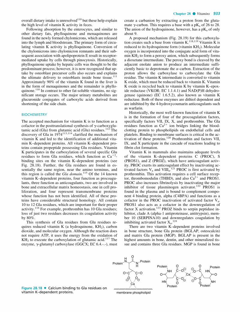
overall dietary intake is unresolved110 but these help explainthe high level of vitamin K activity in feces.
Following absorption by the enterocytes, and similar toother dietary fats, phylloquinone and menaquinones arefound in the newly formed chylomicrons, which are releasedinto the lymph and bloodstream. The primary form of circu-lating vitamin K activity is phylloquinone. Conversion ofthe chylomicrons into chylomicron remnants and their sub-sequent association with apolipoprotein E result in receptor-mediated uptake by cells through pinocytosis. Historically,phylloquinone uptake by hepatic cells was thought to be thepredominant process; however, it is now recognized that up-take by osteoblast precursor cells also occurs and explainsthe ultimate delivery to osteoblasts inside bone tissue.111
Approximately 90% of the vitamin K found in the liver isin the form of menaquinones and the remainder is phyllo-quinone.110 In contrast to other fat-soluble vitamins, no sig-nificant storage occurs. The major urinary metabolites areglucuronide conjugates of carboxylic acids derived fromshortening of the side chain.
BIOCHEMISTRY
The accepted mechanism for vitamin K is to function as acofactor in the posttranslational synthesis of �-carboxyglu-tamic acid (Gla) from glutamic acid (Glu) residues.112 Thediscovery of Gla in 1974113,114 clarified the mechanism ofvitamin K and led to the identification of additional vita-min K–dependent proteins. All vitamin K–dependent pro-teins contain propeptide possessing Glu residues. VitaminK participates in the carboxylation of several specific Gluresidues to form Gla residues, which function as Ca�2-binding sites on the vitamin K–dependent proteins (seeFig. 28.18). Further, the Gla residues are found in es-sentially the same region, near the amino terminus, andthis region is called the Gla domain.115 Of the 14 knownvitamin K–dependent proteins, four function as procoagu-lants, three function as anticoagulants, two are involved inbone and extracellular matrix homeostasis, one in cell pro-liferation, and four represent transmembrane proteinswhose function has not been identified. All of these pro-teins have considerable structural homology. All contain10 to 12 Gla residues, which are important for their properactivity.116 For example, prothrombin has 10 Gla residues;loss of just two residues decreases its coagulation activityby 80%.
This synthesis of Gla residues from Glu residues re-quires reduced vitamin K (a hydroquinone, KH2), carbondioxide, and molecular oxygen. Although the reaction doesnot require ATP, it uses the energy from the oxidation ofKH2 to execute the carboxylation of glutamic acid.117 Theenzyme, �-glutamyl carboxylase (GGCX; EC 6.4.-.-), must
create a carbanion by extracting a proton from the gluta-mate �-carbon. This requires a base with a pKa of 26 to 28.The anion of the hydroquinone, however, has a pKa of onlyabout 9.
A proposed mechanism (Fig. 28.19) for this carboxyla-tion creates such a base from vitamin K.118,119 Vitamin K isreduced to its hydroquinone form (vitamin KH2). Molecularoxygen is incorporated into the conjugate acid form of vita-min KH2 to form a peroxy anion, which subsequently formsa dioxetane intermediate. The peroxy bond is cleaved by theadjacent enolate anion to produce an intermediate suffi-ciently basic to deprotonate the �-carbon. Extraction of theproton allows the carboxylase to carboxylate the Gluresidue. The vitamin K intermediate is converted to vitaminK oxide, which must be reduced back to vitamin K. VitaminK oxide is recycled back to vitamin K by vitamin K–epox-ide reductase (VKOR; EC 1.1.4.1) and NAD(P)H dehydro-genase (quinone) (EC 1.6.5.2), also known as vitamin Kreductase. Both of these enzymes are dithiol dependent andare inhibited by the 4-hydroxycoumarin anticoagulants suchas warfarin.
Historically, the most well-known function of vitamin Kis in the formation of four of the procoagulation factors,specifically factors VII, IX, X, and prothrombin. The Glaresidues function as Ca2� ion bridges linking the blood-clotting protein to phospholipids on endothelial cells andplatelets. Binding to membrane surfaces is critical in the ac-tivation of these proteins.120 Prothrombin and factors VII,IX, and X participate in the cascade of reactions leading tofibrin clot formation.
Vitamin K in mammals also maintains adequate levelsof the vitamin K–dependent proteins C (PROC), S(PROS1), and Z (PROZ), which have anticoagulant activ-ity. PROC exerts its anticoagulant effect by inactivating ac-tivated factors Va and VIIIa.121 PROC is first activated byprothrombin. This activation requires a cell surface recep-tor, thrombomodulin (THBD), and also Ca2� and PROS1.PROC also increases fibrinolysis by inactivating the majorinhibitor of tissue plasminogen activator.122 PROS1 isfound in the plasma and is bound to complement compo-nent 4 binding protein, alpha (C4BPA) and functions as acofactor in the PROC inactivation of activated factor Va.PROS1 also acts as a cofactor in the downregulation offactor X activation.123 PROZ binds to serpin peptidase in-hibitor, clade A (alpha-1 antiproteinase, antitrypsin), mem-ber 10 (SERPINA10) and downregulates coagulation byinhibiting activated factor Xa.124
There are two vitamin K–dependent proteins involvedin bone structure, bone Gla protein (BGLAP, osteocalcin)and matrix Gla protein (MGP). BGLAP is present in thehighest amounts in bone, dentin, and other mineralized tis-sue and contains three Gla residues. MGP is found in bone
Chapter 28 Vitamins 933
membrane phospholipid
Ca
O O
OO CC
O
ProteinNH
HN
Protein
O
O
O
O
C
CO
Protein
NH
HNProtein
CaO
O
O
O
C
CO
ProteinHN
NHProtein
Figure 28.18 Calcium binding to Gla residues onvitamin K–dependent proteins.
0915-0960_17865_Ch28.qxd 12/4/09 8:12 PM Page 933
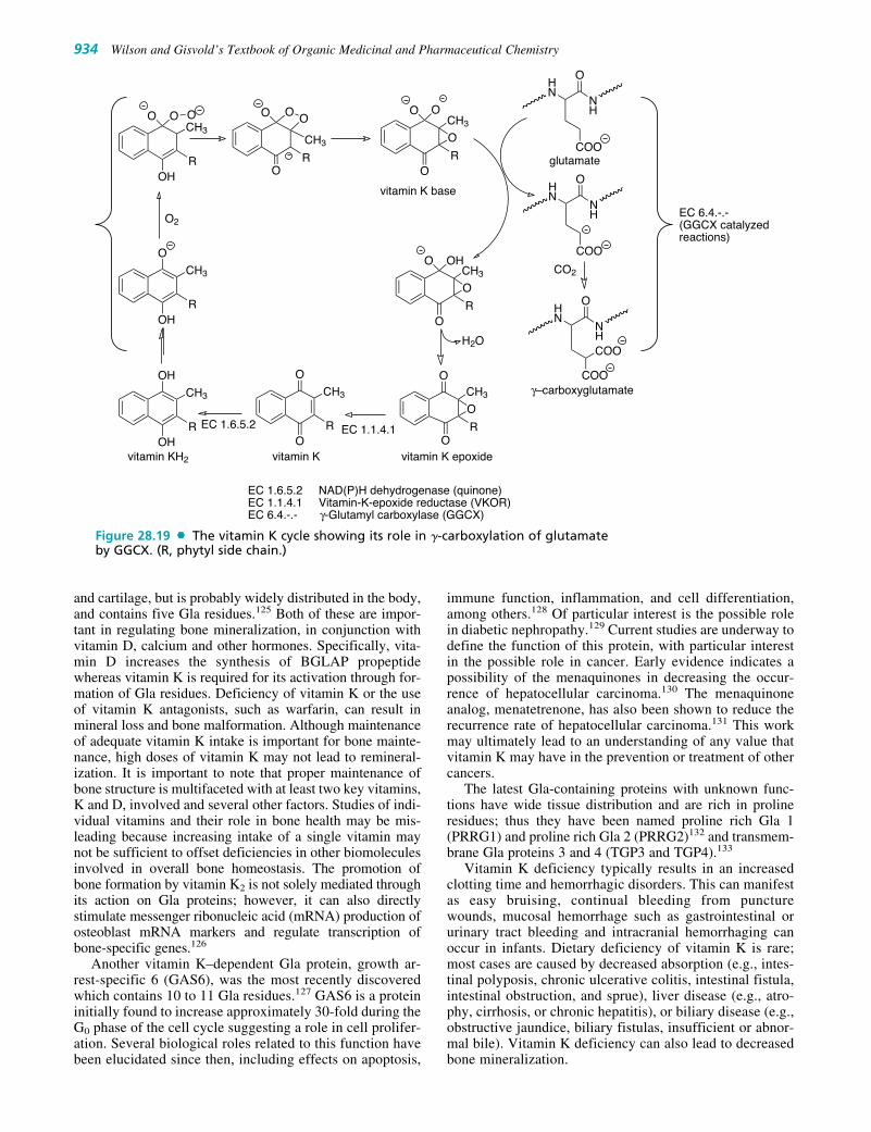
and cartilage, but is probably widely distributed in the body,and contains five Gla residues.125 Both of these are impor-tant in regulating bone mineralization, in conjunction withvitamin D, calcium and other hormones. Specifically, vita-min D increases the synthesis of BGLAP propeptidewhereas vitamin K is required for its activation through for-mation of Gla residues. Deficiency of vitamin K or the useof vitamin K antagonists, such as warfarin, can result inmineral loss and bone malformation. Although maintenanceof adequate vitamin K intake is important for bone mainte-nance, high doses of vitamin K may not lead to remineral-ization. It is important to note that proper maintenance ofbone structure is multifaceted with at least two key vitamins,K and D, involved and several other factors. Studies of indi-vidual vitamins and their role in bone health may be mis-leading because increasing intake of a single vitamin maynot be sufficient to offset deficiencies in other biomoleculesinvolved in overall bone homeostasis. The promotion ofbone formation by vitamin K2 is not solely mediated throughits action on Gla proteins; however, it can also directlystimulate messenger ribonucleic acid (mRNA) production ofosteoblast mRNA markers and regulate transcription ofbone-specific genes.126
Another vitamin K–dependent Gla protein, growth ar-rest-specific 6 (GAS6), was the most recently discoveredwhich contains 10 to 11 Gla residues.127 GAS6 is a proteininitially found to increase approximately 30-fold during theG0 phase of the cell cycle suggesting a role in cell prolifer-ation. Several biological roles related to this function havebeen elucidated since then, including effects on apoptosis,
immune function, inflammation, and cell differentiation,among others.128 Of particular interest is the possible rolein diabetic nephropathy.129 Current studies are underway todefine the function of this protein, with particular interestin the possible role in cancer. Early evidence indicates apossibility of the menaquinones in decreasing the occur-rence of hepatocellular carcinoma.130 The menaquinoneanalog, menatetrenone, has also been shown to reduce therecurrence rate of hepatocellular carcinoma.131 This workmay ultimately lead to an understanding of any value thatvitamin K may have in the prevention or treatment of othercancers.
The latest Gla-containing proteins with unknown func-tions have wide tissue distribution and are rich in prolineresidues; thus they have been named proline rich Gla 1(PRRG1) and proline rich Gla 2 (PRRG2)132 and transmem-brane Gla proteins 3 and 4 (TGP3 and TGP4).133
Vitamin K deficiency typically results in an increasedclotting time and hemorrhagic disorders. This can manifestas easy bruising, continual bleeding from puncturewounds, mucosal hemorrhage such as gastrointestinal orurinary tract bleeding and intracranial hemorrhaging canoccur in infants. Dietary deficiency of vitamin K is rare;most cases are caused by decreased absorption (e.g., intes-tinal polyposis, chronic ulcerative colitis, intestinal fistula,intestinal obstruction, and sprue), liver disease (e.g., atro-phy, cirrhosis, or chronic hepatitis), or biliary disease (e.g.,obstructive jaundice, biliary fistulas, insufficient or abnor-mal bile). Vitamin K deficiency can also lead to decreasedbone mineralization.
934 Wilson and Gisvold’s Textbook of Organic Medicinal and Pharmaceutical Chemistry
EC 1.1.4.1
COO
O
COO
COO
O
COO
OHN
CO2
H2O
O
OHO
OR
CH3
O
O
OR
CH3
O
OO
OR
CH3
RO
O
CH3
OOO
OR
CH3
O2
CH3
ROH
O O O
ROH
O
CH3
ROH
OH
CH3
vitamin KH2 vitamin K epoxidevitamin K
vitamin K base
γ–carboxyglutamate
glutamate
EC 1.6.5.2
EC 6.4.-.-(GGCX catalyzed reactions)
EC 1.6.5.2 NAD(P)H dehydrogenase (quinone)EC 1.1.4.1 Vitamin-K-epoxide reductase (VKOR)EC 6.4.-.- γ-Glutamyl carboxylase (GGCX)
NH
NH
HN
HN
NH
Figure 28.19 The vitamin K cycle showing its role in �-carboxylation of glutamateby GGCX. (R, phytyl side chain.)
0915-0960_17865_Ch28.qxd 12/4/09 8:12 PM Page 934

THERAPEUTIC USES
Phytonadione is indicated in the treatment of known or sus-pected vitamin K deficiency that may result from dietary de-ficiency, malabsorption, hepatic, or biliary disease.Phytonadione is also used in the prophylactic treatment of vi-tamin K deficiency bleeding (VKDB) of the newborn, previ-ously known as hemorrhagic disease of the newborn. In theaverage infant, the birth values of prothrombin content areadequate, but during the first few days of life, they appear tofall rapidly, even dangerously low, and then slowly recoverspontaneously. This transition period was and is critical be-cause of the numerous sites of hemorrhagic manifestations,traumatic or spontaneous, that may prove serious, if not fatal.Several controversial studies linked IM phytonadione admin-istration to newborns with an increased risk of the develop-ment of childhood cancer. However, the current position ofthe American Academy of Pediatrics (AAP) is that thesestudies were flawed and later large-scale studies showed noassociation. Therefore, the 2003 policy statement of the AAPrecommended 0.5 mg to 1 mg of vitamin K1 (phytonadione)intramuscularly at birth as a prophylactic measure.134 Thiswas reaffirmed in 2006.135
Phytonadione, or other vitamin K products, should not beadministered to patients receiving coumarin anticoagulants,such as warfarin, during routine therapy as these can antag-onize their effects. However, phytonadione is indicated inthe treatment of overdoses of vitamin K–epoxide reductaseinhibitors, such as warfarin, or other drugs that can interferewith vitamin K utilization in the clotting process. Of partic-ular note is the requirement in this one use for an active formof vitamin K. Specifically, the only marketed product isphytonadione. Inactive forms, such as menadiol, are notbioactivated with sufficient rapidity to be used as antidotesin this situation. Finally, vitamin K may also have some util-ity in preventing bone loss as is seen in osteoporosis.125
HYPERVITAMINOSIS
Phytonadione is considered relatively nontoxic even at highdoses. Rare cases of hemolytic anemia have been reported,and the manufacturer has reported anaphylactic reactionsfollowing use of the injectable product. Menadiol is alsoconsidered relatively nontoxic. Menadione, in contrast, canproduce hemolytic anemia, hyperbilirubinemia, and ker-nicterus in newborns, especially premature infants. In addi-tion, menadione can cause hemolytic anemia in patientswith glucose-6-phosphate dehydrogenase deficiency.45 Forthese reasons, it is no longer marketed in the United States.
PRODUCTS
Phytonadione is available as oral tablets and capsules fromnumerous sources under various names. A popular prescrip-tion product for oral tablets is Mephyton. Phytonadione isalso available as an injectable for IM use. It is also availablein numerous multivitamin preparations under variousnames. Menadiol is also available in some markets.
WATER-SOLUBLE VITAMINS
Although the water-soluble vitamins are structurally diverse,they are put in a general class to distinguish them from the
lipid-soluble vitamins. This class includes the B-complex vi-tamins and ascorbic acid (vitamin C). The term B-complexrefers to combinations including all eight B vitamins: thi-amine (B1), riboflavin (B2), niacin (B3), pantothenic acid(B5), pyridoxine (B6), biotin (B7), folic acid (B9), andcyanocobalamin (B12). Dietary deficiencies of any of the Bvitamins commonly are complicated by deficiencies of othermembers of the group, so treatment with B-complex prepa-rations is usually indicated.
Vitamin B1
Thiamine, the preferred name for vitamin B1, holds a promi-nent place in the history of vitamin discovery becauseberiberi, the disease resulting from insufficient thiamine in-take, was one of the earliest recognized deficiency diseases.Its relationship to polyneuritis in birds was noted in the earlywork of Funk,3 and his work with pigeons led to the discov-ery that a water-soluble substance in rice polishings was cur-ative. The early designation of water-soluble B was replacedwith vitamin B by Drummond11 when suggesting the use ofvitamin instead of vitamine. Complicating early animal stud-ies was the inconsistency in the deficiency syndromes ob-served by the various investigators. Early work on vitamin Busing crude extracts showed that this vitamin also had growth-promoting properties in rats fed vitamin B–deficient diets. Inaddition, vitamin B–deficient diets could also result in der-matitis, which at that time appeared similar to human pellagra.Oftentimes, the source used for vitamin B and the animalmodel studied determined the experimental results observed.
The antineuritic water-soluble B vitamin that curedpolyneuritis in birds and the growth-promoting water-solubleB vitamin were proposed to be two different vitamins byEmmett and Luros136 in 1920. This was later confirmed byother investigators.137 In 1927, the British Accessory FoodFactors Committee recommended vitamin B1 for the antineu-ritic water-soluble B and vitamin B2 for the growth-promoting, also called the antipellegra, water-soluble B. TheAmerican Society of Biological Chemists recommended thename vitamin B for the antineuritic factor and vitamin G forthe growth-promoting factor. Eventually, the British systembecame the standard. In 1960, the IUPAC Commission on theNomenclature of Biological Chemistry106 adopted the namethiamine. In 1966, IUPAC138 proposed the name thiamin,and in 2004, reverted once again to thiamine although thi-amin is still in common use. Thiamine has also been referredto as aneurine.12 The isolation of thiamine was elusive; how-ever, it was finally isolated and crystallized from rice polish-ings by Jansen and Donath139 in 1926. The structure and syn-thesis was published 10 years later in 1936.140
One of the richest natural sources of thiamine is pork.Whole grain and whole-grain products, such as yeast, beans,and peas, are also excellent sources. Beef and chicken as wellas some fish are also good sources. In developed countries, in-cluding the United States, fortified products, especially ready-to-eat cereals, are a significant dietary source of thiamine.
CHEMISTRY
The chemical structures of thiamine and its coenzyme form,thiamine diphosphate (ThDP), are shown in Figure 28.20.ThDP is composed of a diphosphate terminated side chain,a five-membered thiazolium ring, and a six-memberedaminopyrimidine ring. Thiamine hydrochloride (Fig. 28.21),
Chapter 28 Vitamins 935
0915-0960_17865_Ch28.qxd 12/4/09 8:12 PM Page 935
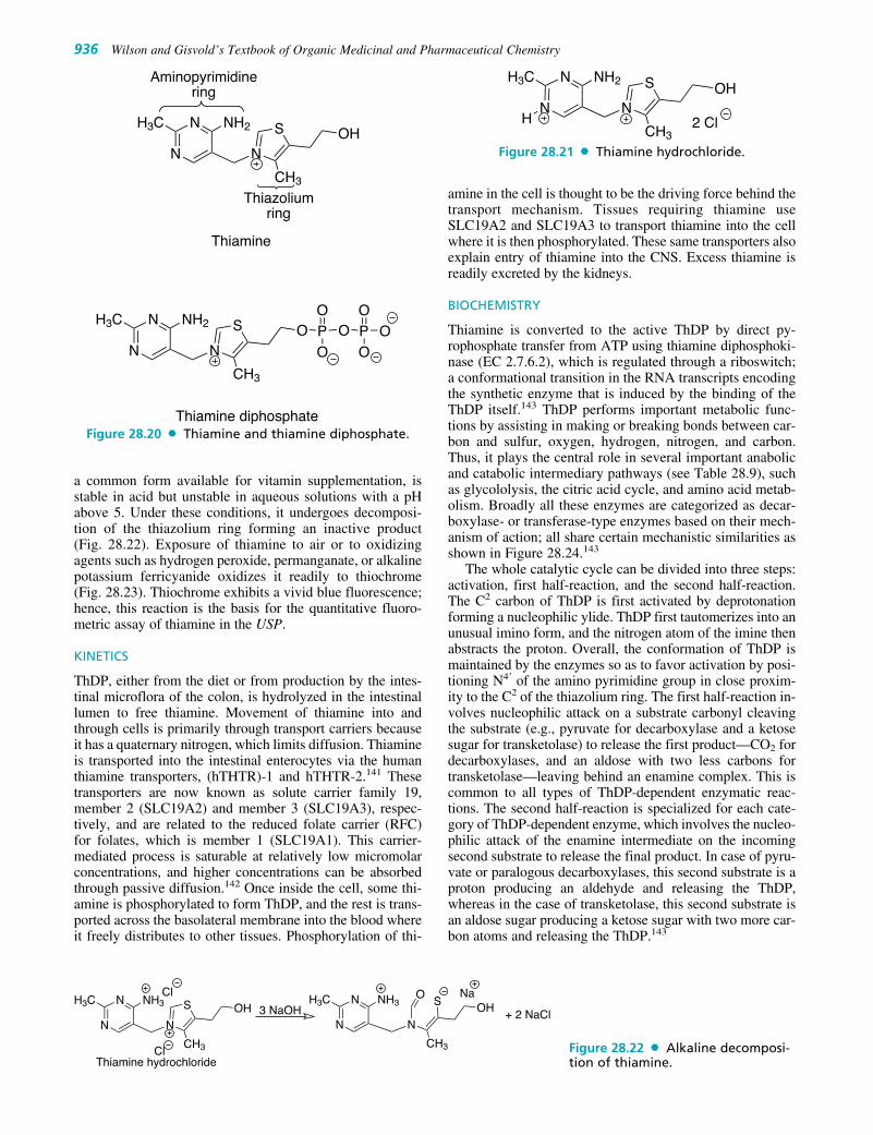
a common form available for vitamin supplementation, isstable in acid but unstable in aqueous solutions with a pHabove 5. Under these conditions, it undergoes decomposi-tion of the thiazolium ring forming an inactive product(Fig. 28.22). Exposure of thiamine to air or to oxidizingagents such as hydrogen peroxide, permanganate, or alkalinepotassium ferricyanide oxidizes it readily to thiochrome(Fig. 28.23). Thiochrome exhibits a vivid blue fluorescence;hence, this reaction is the basis for the quantitative fluoro-metric assay of thiamine in the USP.
KINETICS
ThDP, either from the diet or from production by the intes-tinal microflora of the colon, is hydrolyzed in the intestinallumen to free thiamine. Movement of thiamine into andthrough cells is primarily through transport carriers becauseit has a quaternary nitrogen, which limits diffusion. Thiamineis transported into the intestinal enterocytes via the humanthiamine transporters, (hTHTR)-1 and hTHTR-2.141 Thesetransporters are now known as solute carrier family 19,member 2 (SLC19A2) and member 3 (SLC19A3), respec-tively, and are related to the reduced folate carrier (RFC)for folates, which is member 1 (SLC19A1). This carrier-mediated process is saturable at relatively low micromolarconcentrations, and higher concentrations can be absorbedthrough passive diffusion.142 Once inside the cell, some thi-amine is phosphorylated to form ThDP, and the rest is trans-ported across the basolateral membrane into the blood whereit freely distributes to other tissues. Phosphorylation of thi-
amine in the cell is thought to be the driving force behind thetransport mechanism. Tissues requiring thiamine useSLC19A2 and SLC19A3 to transport thiamine into the cellwhere it is then phosphorylated. These same transporters alsoexplain entry of thiamine into the CNS. Excess thiamine isreadily excreted by the kidneys.
BIOCHEMISTRY
Thiamine is converted to the active ThDP by direct py-rophosphate transfer from ATP using thiamine diphosphoki-nase (EC 2.7.6.2), which is regulated through a riboswitch;a conformational transition in the RNA transcripts encodingthe synthetic enzyme that is induced by the binding of theThDP itself.143 ThDP performs important metabolic func-tions by assisting in making or breaking bonds between car-bon and sulfur, oxygen, hydrogen, nitrogen, and carbon.Thus, it plays the central role in several important anabolicand catabolic intermediary pathways (see Table 28.9), suchas glycololysis, the citric acid cycle, and amino acid metab-olism. Broadly all these enzymes are categorized as decar-boxylase- or transferase-type enzymes based on their mech-anism of action; all share certain mechanistic similarities asshown in Figure 28.24.143
The whole catalytic cycle can be divided into three steps:activation, first half-reaction, and the second half-reaction.The C2 carbon of ThDP is first activated by deprotonationforming a nucleophilic ylide. ThDP first tautomerizes into anunusual imino form, and the nitrogen atom of the imine thenabstracts the proton. Overall, the conformation of ThDP ismaintained by the enzymes so as to favor activation by posi-tioning N4� of the amino pyrimidine group in close proxim-ity to the C2 of the thiazolium ring. The first half-reaction in-volves nucleophilic attack on a substrate carbonyl cleavingthe substrate (e.g., pyruvate for decarboxylase and a ketosesugar for transketolase) to release the first product—CO2 fordecarboxylases, and an aldose with two less carbons fortransketolase—leaving behind an enamine complex. This iscommon to all types of ThDP-dependent enzymatic reac-tions. The second half-reaction is specialized for each cate-gory of ThDP-dependent enzyme, which involves the nucleo-philic attack of the enamine intermediate on the incomingsecond substrate to release the final product. In case of pyru-vate or paralogous decarboxylases, this second substrate is aproton producing an aldehyde and releasing the ThDP,whereas in the case of transketolase, this second substrate isan aldose sugar producing a ketose sugar with two more car-bon atoms and releasing the ThDP.143
936 Wilson and Gisvold’s Textbook of Organic Medicinal and Pharmaceutical Chemistry
OH
CH3
NH2H3C S
NN
N
Thiazolium ring
Aminopyrimidine ring
O O
OOOPOPO
CH3
NH2H3C S
NN
N
Thiamine
Thiamine diphosphate
2 ClH
OH
CH3
NH2H3C S
NN
N
Cl
NaOOH
CH3
NH3H3C S
NN
NCl
OH
CH3
NH3H3C S
NN
N3 NaOH + 2 NaCl
Thiamine hydrochloride
Figure 28.20 Thiamine and thiamine diphosphate.
Figure 28.21 Thiamine hydrochloride.
Figure 28.22 Alkaline decomposi-tion of thiamine.
0915-0960_17865_Ch28.qxd 12/4/09 8:12 PM Page 936

Chapter 28 Vitamins 937
OH
CH3
NH3H3C S
NN
NCl
OH
CH3
S
N
NH2C
N
N
Cl
Alkaline
ThiochromeThiamine hydrochloride
K3Fe(CN)6
Figure 28.24 Catalytic cycle of thiamine diphosphate.
TABLE 28.9 Thiamine Diphosphate–Dependent Enzymes
Enzyme EC Number Function
Pyruvate dehydrogenase (acetyl-transferring) 1.2.4.1 Glycolysis, gluconeogenesis, amino acid synthesisOxoglutarate dehydrogenase (succinyl-transferring) 1.2.4.2 Citric acid cycle (TCA cycle)3-Methyl-2-oxobutanoate dehydrogenase 1.2.4.4 Valine, leucine, and isoleucine degradation
(2-methylpropanoyl-transferring)Transketolase 2.2.1.1 Pentose phosphate pathwayBranched-chain-2-oxoacid decarboxylase 4.1.1.72 Decarboxylation of �-keto acids
Figure 28.23 Oxidation of thiamine tothiochrome.
0915-0960_17865_Ch28.qxd 12/4/09 8:12 PM Page 937
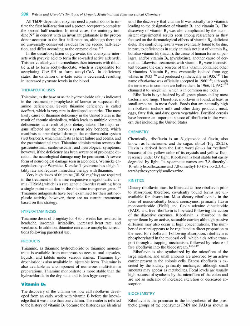
All ThDP-dependent enzymes need a proton donor to ini-tiate the first half-reaction and a proton acceptor to completethe second half-reaction. In most cases, the aminopyrimi-dine N4� in concert with an invariant glutamate is the protondonor-acceptor in the first half-reaction, although there areno universally conserved residues for the second half-reac-tion, and differ according to the enzyme class.143
In the decarboxylation of pyruvate, the coenzyme inter-acts with pyruvic acid to form the so-called active aldehyde.This active aldehyde intermediates then interacts with thioc-tic acid to form acetyl-thioctate, which is responsible foracetylating CoA-SH to form acetyl-CoA. In deficiencystates, the oxidation of �-keto acids is decreased, resultingin increased pyruvate levels in the blood.
THERAPEUTIC USES
Thiamine, as the base or as the hydrochloride salt, is indicatedin the treatment or prophylaxis of known or suspected thi-amine deficiencies. Severe thiamine deficiency is calledberiberi, which is very rare in developed countries. The mostlikely cause of thiamine deficiency in the United States is theresult of chronic alcoholism, which leads to multiple vitamindeficiencies as a result of poor dietary intake. The major or-gans affected are the nervous system (dry beriberi), whichmanifests as neurological damage, the cardiovascular system(wet beriberi), which manifests as heart failure and edema, andthe gastrointestinal tract. Thiamine administration reverses thegastrointestinal, cardiovascular, and neurological symptoms;however, if the deficiency has been severe or of prolonged du-ration, the neurological damage may be permanent. A severeform of neurological damage seen in alcoholics, Wernicke en-cephalopathy or Wernicke-Korsakoff syndrome, has a high fa-tality rate and requires immediate therapy with thiamine.
Very high doses of thiamine (30–90 mg/day) are requiredin the treatment of thiamine-responsive megaloblastic ane-mia (TRMA),which is a rare genetic disorder resulting froma single point mutation in the thiamine transporter gene.144
Thiamine antagonists have been examined for their antineo-plastic activity; however, there are no current treatmentsbased on this strategy.
HYPERVITAMINOSIS
Thiamine doses of 5 mg/day for 4 to 5 weeks has resulted inheadache, insomnia, irritability, increased heart rate, andweakness. In addition, thiamine can cause anaphylactic reac-tions following parenteral use.
PRODUCTS
Thiamine, as thiamine hydrochloride or thiamine mononi-trate, is available from numerous sources as oral capsules,liquids, and tablets under various names. Thiamine hy-drochloride is also available in injectable form. Thiamine isalso available as a component of numerous multivitaminpreparations. Thiamine mononitrate is more stable than thehydrochloride in the dry state and is less hygroscopic.
Vitamin B2
The discovery of the vitamin we now call riboflavin devel-oped from an early work with vitamin B before the knowl-edge that it was more than one vitamin. The reader is referredto the history of vitamin B1 because the histories are identical
until the discovery that vitamin B was actually two vitaminsleading to the designation of vitamin B1 and vitamin B2. Thediscovery of vitamin B2 was also complicated by the incon-sistent experimental results seen among researchers as theyfocused on the dermatological effects of vitamin B2–deficientdiets. The conflicting results were eventually found to be due,in part, to deficiencies in study animals not just of vitamin B2,but also vitamin B3 (niacin), the cause of human forms of pel-lagra, and/or vitamin B6 (pyridoxine), another cause of der-matitis. Likewise, treatments with vitamin B2 were inconsis-tent because the early sources of this vitamin contained otherB vitamins. Vitamin B2 was eventually isolated from eggwhites in 1933146 and produced synthetically in 1935.147 Thename riboflavine was officially accepted in 1960106; althoughthe term was in common use before then. In 1966, IUPAC138
changed it to riboflavin, which is in common use today.Riboflavin is synthesized by all green plants and by most
bacteria and fungi. Therefore, riboflavin is found, at least insmall amounts, in most foods. Foods that are naturally highin riboflavin include milk and other dairy products, meat,eggs, fatty fish, and dark green vegetables. Fortified cerealshave become an important source of riboflavin in the west-ern diet including the United States.
CHEMISTRY
Chemically, riboflavin is an N-glycoside of flavin, alsoknown as lumichrome, and the sugar, ribitol (Fig. 28.25).Flavin is derived from the Latin word flavus for “yellow”because of the yellow color of its crystals and yellow fluo-rescence under UV light. Riboflavin is heat stable but easilydegraded by light. Its systematic names are 7,8-dimethyl-10-ribitylisoalloxazine and 7,8-dimethyl-10-(D-ribo-2,3,4,5-tetrahydroxypentyl)isoalloxazine.
KINETICS
Dietary riboflavin must be liberated as free riboflavin priorto absorption; therefore, covalently bound forms are un-available for absorption. Most dietary riboflavin is in theform of noncovalently bound coenzymes, primarily flavinmononucleotide (FMN) and flavin adenine dinucleotide(FAD), and free riboflavin is liberated following the actionof the digestive enzymes. Riboflavin is absorbed in theupper ileum by an active, saturable carrier; although passivediffusion may also occur at high concentrations. The num-ber of carriers appears to be regulated in direct proportion tothe need for riboflavin. Following absorption, riboflavin isphosphorylated in the mucosal cell, which aids active trans-port through a trapping mechanism, followed by release offree riboflavin into the bloodstream.148,149
Riboflavin is also synthesized by the microflora of thelarge intestine, and small amounts are absorbed by an activecarrier present in the colonic cells. Excess riboflavin is ex-creted by the kidney, primarily unchanged, although smallamounts may appear as metabolites. Fecal levels are usuallyhigh because of synthesis by the microflora of the colon andare not an indicator of increased excretion or decreased ab-sorption.
BIOCHEMISTRY
Riboflavin is the precursor in the biosynthesis of the pros-thetic groups of the coenzymes FMN and FAD as shown in
938 Wilson and Gisvold’s Textbook of Organic Medicinal and Pharmaceutical Chemistry
0915-0960_17865_Ch28.qxd 12/4/09 8:12 PM Page 938

Figure 28.25. FMN is actually named incorrectly because it isnot a nucleotide. The metabolic functions of this vitamin in-volve these two enzyme cofactors, which participate in nu-merous, vital oxidation–reduction processes. FMN (ri-boflavin 5�-phosphate) is produced from the vitamin and ATP
by riboflavin kinase (EC 2.7.1.26). Amitriptyline,imipramine, and chlorpromazine can inhibit riboflavin kinaseresulting in a functional riboflavin deficiency. FAD originatesfrom an FMN and ATP reaction that involves reversible din-ucleotide formation catalyzed by FAD synthetase (EC2.7.7.2). FMN and FAD function in combination with manyenzymes, often characterized as flavoproteins, as the pros-thetic groups of their coenzymes and rarely as the actual coen-zyme. In some instances, the FAD or FMN is found cova-lently bound to the enzyme.
Flavoproteins function under aerobic or anaerobic condi-tions as oxidases and dehydrogenases. Examples includeglucose oxidase, xanthine oxidase, cytochrome reductase,and acyl-CoA dehydrogenase. The riboflavin moiety of thecomplex is considered a hydrogen-transporting agent func-tioning as a hydrogen acceptor. The hydrogen donors maybe NADH, NADPH, or other suitable substrate. The isoal-loxazine rings accept two hydrides stepwise to form the di-hydroriboflavin derivative (Fig. 28.26).
THERAPEUTIC USES
Severe riboflavin deficiency is known as ariboflavinosis, andtreatment or prevention of this condition is the only provenuse of riboflavin. Ariboflavinosis is most commonly associ-ated with multiple vitamin deficiency as a result of alco-holism in developed countries. Because of the large numberof enzymes requiring riboflavin as a coenzyme, deficienciescan lead to a wide range of abnormalities. In adults seborrheicdermatitis, photophobia, peripheral neuropathy, anemia, andoropharyngeal changes including angular stomatitis, glossitis,and cheilosis, are often the first signs of riboflavin deficiency.In children, cessation of growth can also occur. As the defi-ciency progresses, more severe pathologies develop untildeath ensues. Riboflavin deficiency may also produce terato-genic effects and alter iron handling leading to anemia.150
HYPERVITAMINOSIS
There are no known toxicities associated with large doses ofriboflavin. This is most likely because of the capacity-limiteduptake that, in the presence of high doses, down regulates,thereby limiting absorption from the intestines. Renal filtra-tion and excretion of riboflavin can also increase significantlyin the presence of high plasma levels.149
PRODUCTS
Riboflavin or riboflavin 5�-monophosphate is availablefrom numerous sources as oral capsules, liquids, and tabletsunder various names. Riboflavin is also available as a com-ponent of numerous multivitamin preparations.
Vitamin B3
Vitamin B3 was formerly called nicotinic acid; however, theterm niacin is now preferred to avoid any confusion with the
Chapter 28 Vitamins 939
HO OH
O
NH2
N
N
N
N
OOH
OP
HO
OH
HO O
O
O
CH3H3C
NN
NN
H
P OO
OH
Flavin (7,8-Dimethylisoalloxazine)
D-Ribitol
OHOH
OP
HO
OH
HO O
O
H O
CH3H3C
NN
NN
ATP
ADP
Riboflavin
ATP
PPi
HO
OH
HO OH
O
H O
CH3H3C
NN
NN
Flavin Mononucleotide (FMN)
Flavin Adenine Dinucleotide (FAD)
Riboflavin kinase(EC 2.7.1.26)
FAD synthetase(EC 2.7.7.2)
O
O
H
RH3C
H3CN
NN
N
H
O
O
H
RH3C
H3CN
NN
N
H
oxidized species reduced species
+ 1e- H + 1e-
H
O
O
H
RH3C
H3CN
NN
N
H
Figure 28.25 Bioactivation of riboflavin to FMN andFAD.
Figure 28.26 Oxidationand reduction of the isoallox-azine rings.
0915-0960_17865_Ch28.qxd 12/4/09 8:12 PM Page 939

alkaloid, nicotine. Niacinamide, also known as nicotin-amide, refers to the amide derivative of niacin that is equiv-alent in vitamin activity. Some texts use niacin to refer tonicotinic acid, niacinamide, and any derivatives with vita-min activity comparable to niacin. Furthermore, researchand chemistry-based resources use the terms nicotinic acidand nicotinamide; whereas pharmacy resources use niacinand niacinamide. For example, enzyme nomenclature usesthe former, whereas facts and comparisons uses the latter.This section uses each term in its best context.
The synthesis and structural identification of niacin actu-ally predated its discovery as a vitamin. Niacin had been pre-pared from nicotine since at least 1898. Niacin had also beenpurified and identified in 1913 as a component of yeast andrice polishings by Funk151 in his quest to discover the vitaminlater called thiamine. The discovery of niacin as a vitaminwas complicated, as discussed under vitamin B2, by the earlymisconception that animal forms of pellagra and the humanform were caused by the same vitamin deficiency.Ultimately, it was the study of human pellagra and blacktongue disease in dogs, also caused by niacin deficiency,152
that led to the discovery of niacin as a vitamin. In Septemberof 1937, Elvehjem et al.153 were the first to prove the link be-tween niacin and black tongue disease in dogs. Clinical stud-ies using niacin in human pellagra patients followed, and thefirst published report of success was in November of 1937.154
Niacin can be synthesized by humans into its active forms,nicotinamide adenine dinucleotide (NAD�) and nicotin-amide adenine dinucleotide phosphate (NADP�), from theamino acid, tryptophan. Consequently, and analogous to vita-min D, niacin is not a true vitamin by the classic definition ofthe term. However, because of inherent inefficiencies in theconversion, the use of dietary tryptophan as a replacement forniacin can result in tryptophan deficiency for protein synthe-sis. This results in a requirement for niacin in the diet as a vi-tamin or a diet abnormally high in tryptophan. To compensatefor this conversion, the DRIs for niacin are expressed inniacin equivalents (NE) that allows for 60 mg of dietary tryp-tophan to equal 1-mg niacin. Niacin or niacin derived nu-cleotides can also be synthesized using aspartic acid in plantsand microorganisms
Natural sources of niacin include meat (beef, pork,chicken, lamb), fish, and whole grains. As with other B vi-tamins, fortified ready-to-eat cereals are an important sourcein developed countries such as the United States.
CHEMISTRY
Niacin is chemically pyridine-3-carboxylic acid (Fig. 28.27).Its coenzyme forms are NADH/NAD� or NADPH/NADP�as shown in Figure 28.28.
KINETICS
Niacin appears in the diet in various forms.155 Someniacin is relatively unavailable for absorption unless the
food is treated in a manner that releases the niacin. Forexample, some grains (i.e., corn) may contain predomi-nantly niacin esterified to polysaccharides or peptides, re-ferred to as niacytin. This form has a relatively lowbioavailability unless treated with alkali in cooking orprocessing. Coffee beans have a high niacin content in theform of trigonellin (1-methyl nicotinic acid) that is con-verted to niacin upon roasting. Most dietary niacin fromnatural sources is in the form of the nucleotide deriva-tives, NAD� and NADP�, which are digested by NAD�diphosphatase (EC 3.6.1.22 ), followed by hydrolases inthe intestine to release nicotinamide. Free nicotinamideand niacin are absorbed through a facilitated diffusionprocess in the intestine.156 At physiological doses, littleniacin is excreted unchanged. Most is excreted as N-methylniacin or the glycine conjugate (nicotinuric acid).After administration of large doses, niacin can be foundin the urine unchanged.
BIOCHEMISTRY
Niacin synthesis into the active cofactors, NAD� andNADP�, is shown in Figure 28.29. Both niacin and niaci-namide react with 5-phosphoribosyl-1-pyrophosphate(PRPP) to form the respective mononucleotide deriv-ative, which then reacts with ATP to produce the corre-sponding dinucleotide product. For niacinamide, this isNAD�; however, for niacin this product is convertedto NAD� by transformation of the carboxyl of the nico-tinic acid moiety to the amide using free ammonia. Analternative pathway is to use glutamine through NAD�synthase (glutamine-hydrolysing) (EC 6.3.5.1); however,this pathway is slower and is most likely not as important.NADP� is produced from NAD� by ATP through kinasecatalysis.
940 Wilson and Gisvold’s Textbook of Organic Medicinal and Pharmaceutical Chemistry
OH
O
NNiacin
C
N
NH2
O
OHHO
O
N
N N
NNH2
O
HO OR
HO O OHO
OP
OP
O
R = PO32- Nicotinamide adenine dinucleotide phosphate
R = H Nicotinamide adenine dinucleotide
C
N
NH2
O
OHHO
O
N
N N
NNH2
O
HO OR
HO O OHO
OP
OP
O
Oxidized form (NAD+, NADP+)
Reduced form (NADH, NADPH)
Figure 28.27 Niacin.
Figure 28.28 Structures of niacin-derived cofactors.
0915-0960_17865_Ch28.qxd 12/4/09 8:12 PM Page 940

NAD� and NADP� are oxidizing coenzymes for many(�200) dehydrogenases. Some dehydrogenases requireNAD�, others require NADP�, and some function witheither. The generalized representation in Figure 28.30 illus-trates the function of these coenzymes in metabolic oxida-tions and reductions. The abbreviation NAD� emphasizesthe electrophilicity of the pyridine C4 moiety (which is thecenter of reactivity), and the substrate designated could be a
Chapter 28 Vitamins 941
Figure 28.29 Biosynthesis of NAD� and NADP� from niacin, nicotinamide, ortryptophan.
H
RH
O
NH2
N
HR1
OR2 C
H
HOC
O
NH2
NR
H
Product(Aldehyde/ketone)
ReducedCoenzyme
CoenzymeSubstrate(Alcohol)
c
+
a
+bR1
R2
Figure 28.30 Generalized representation of thehydride transfer reaction.
0915-0960_17865_Ch28.qxd 12/4/09 8:12 PM Page 941

primary or secondary alcohol. Arrow a in Figure 28.30 sym-bolizes the function of NAD� as oxidant in the hydridetransfer from the substrate to the coenzyme, formingNADH, reduced coenzyme. The hydroxyl of the substrate isvisualized as undergoing deprotonation concertedly by ei-ther water or the pyridine nitrogen of NADH. Arrow bshows concerted formation of the carbonyl �-bond of theoxidation product. Arrow c symbolizes the reverse hydridetransfer from reduced coenzyme, NADH, to the carbonylcarbon, and concertedly, as the carbonyl oxygen undergoesprotonation, the reduction of the carbonyl group forms thecorresponding alcohol. Thus, NAD� and NADP� are hy-dride acceptors, and NADH and NADPH are hydridedonors. Although this is a simplistic representation, it showsthe dynamism of the oxidation–reduction reactions effectedby these coenzymes under appropriate dehydrogenase catal-ysis. Alternatively, the reduced coenzymes may be used inATP production through the electron-transport system.
THERAPEUTIC USES
Niacin is used in the treatment of niacin deficiency, which isreferred to as pellagra (from the Italian, pelle for “skin” andagra for “dry”). The major systems affected are the gas-trointestinal tract (diarrhea, enteritis and stomatitis), the skin(dermatitis), and the CNS (generalized neurological deficitsincluding dementia). Pellagra has become a rare condition inthe United States and other countries that require or encour-age enrichment of wheat flour or fortification of cereals withniacin. Because the nucleotide form can be synthesized invivo from tryptophan, pellagra is most often seen in areaswhere the diet is deficient in both niacin and tryptophan.Typically, maize (corn)-based diets meet this criteria.Niacin deficiency can also result from diarrhea, cirrhosis, al-coholism, or Hartnup disease. It is interesting that niacin de-ficiency can also, rarely, result from vitamin B6 deficiency(see Vitamin B6 section).
Niacin, but not niacinamide, is also one of the few vita-mins that are useful in the treatment of diseases unrelated todeficiencies. In 1955, Altschul et al.157 reported that niacindoses of 1 to 4 g/day could lower serum cholesterol levels inhumans. Currently, niacin doses of 2 to 4.5 g/day are used inthe treatment of hypercholesterolemia and hypertriglyc-eridemia. Triglycerides, VLDL, and LDL are reduced; HDLis increased. The mechanisms underlying these effects haverecently been reviewed.158,159
HYPERVITAMINOSIS
Niacin, in doses that range above the DRI but below thatrequired for dyslipidemias, is unlikely to produce adverseeffects. However, adverse effects of niacin are seen whenthis vitamin is used at pharmacological doses above 1 g/dayin the treatment of dyslipidemia. Notable adverse effects in-clude flushing because of vasodilation; dermatological ef-fects including dry skin, pruritus and hyperkeratosis; gas-trointestinal effects including peptic ulcer, stomach pain,nausea, and diarrhea; elevations in serum uric acid andglucose (in Type 2 diabetics); and rare hepatotoxic-ity.159–161 Traditionally, hepatotoxicity has been more asso-ciated with the sustained release as compared to the imme-diate release formulations; however, recent analysis ofniacin-ER adverse events suggests the opposite may be truefor this formulation.162
Of particular note is that niacinamide, even in high doses,is not effective in hyperlipdemias and only rarely producesthe vascular, dermatological and hepatotoxicity associatedwith high doses of niacin. Even gastrointestinal upset is lesscommon with niacinamide. For this reason, niacinamidewould be the preferred form for vitamin supplementation,especially if taken in high doses.
PRODUCTS
Niacin is available, in immediate-release form, from numer-ous sources as oral capsules, liquids, and tablets undervarious names. Timed-release products of niacin are alsoavailable primarily for the treatment of hyperlipidemias.Niacin is also available as a fixed-dose combination withone of the HMG-CoA reductase inhibitors or statin antihy-perlipidemics (Advicor, Simcor). Niacin is also available asa component of numerous multivitamin preparations.Niacinamide is available from numerous sources as oralcapsules, liquids, and tablets under various names.Niacinamide is also available as a component of numerousmultivitamin preparations
Vitamin B5
The first suggestion for the existance of vitamin B5 camefrom Carter et al.163 in 1930; although it was never character-ized or isolated. In 1933, R. J. Williams164 and his collabora-tors found a “growth determinant of universal occurrence”and named it, according to the authors, “pantothenic acid, thename being derived from the Greek, meaning from every-where” (probably from the Greek pantothen). Pantothenicacid, the preferred term, later became associated with thename vitamin B5. The synthesis and structure were reported in1940.165 This vitamin is synthesized by most green plants andmicroorganisms. Excellent sources of the vitamin are liver,egg yolk, whole grains, and fortified ready-to-eat cereals.However, as the original name implies, many foods containsufficient pantothenic acid to supply dietary needs.
CHEMISTRY
Chemically, pantothenic acid is considered to be a �-alaninederivative of the asymmetric pantoic acid and thus showsasymmetry (Fig. 28.31). Only the naturally occurring D(�)-stereoisomer (with R configuration) is biologically activeand the L(�)-stereoisomer (with S configuration) is inactive.When its carboxylate functional group is attached throughan amide linkage with �-mercaptoethylamine, it is known aspantetheine (also spelled pantotheine). The biologically ac-tive form of pantothenic acid, CoA, is formed when the ter-minal alcoholic function of pantetheine is attached to ADP3�-phosphate as shown in Figure 28.32.
942 Wilson and Gisvold’s Textbook of Organic Medicinal and Pharmaceutical Chemistry
HO
HN OH
OO
OH
CH3H3C
pantoic acid
β-alanine
Figure 28.31 Pantothenic acid.
0915-0960_17865_Ch28.qxd 12/4/09 8:12 PM Page 942
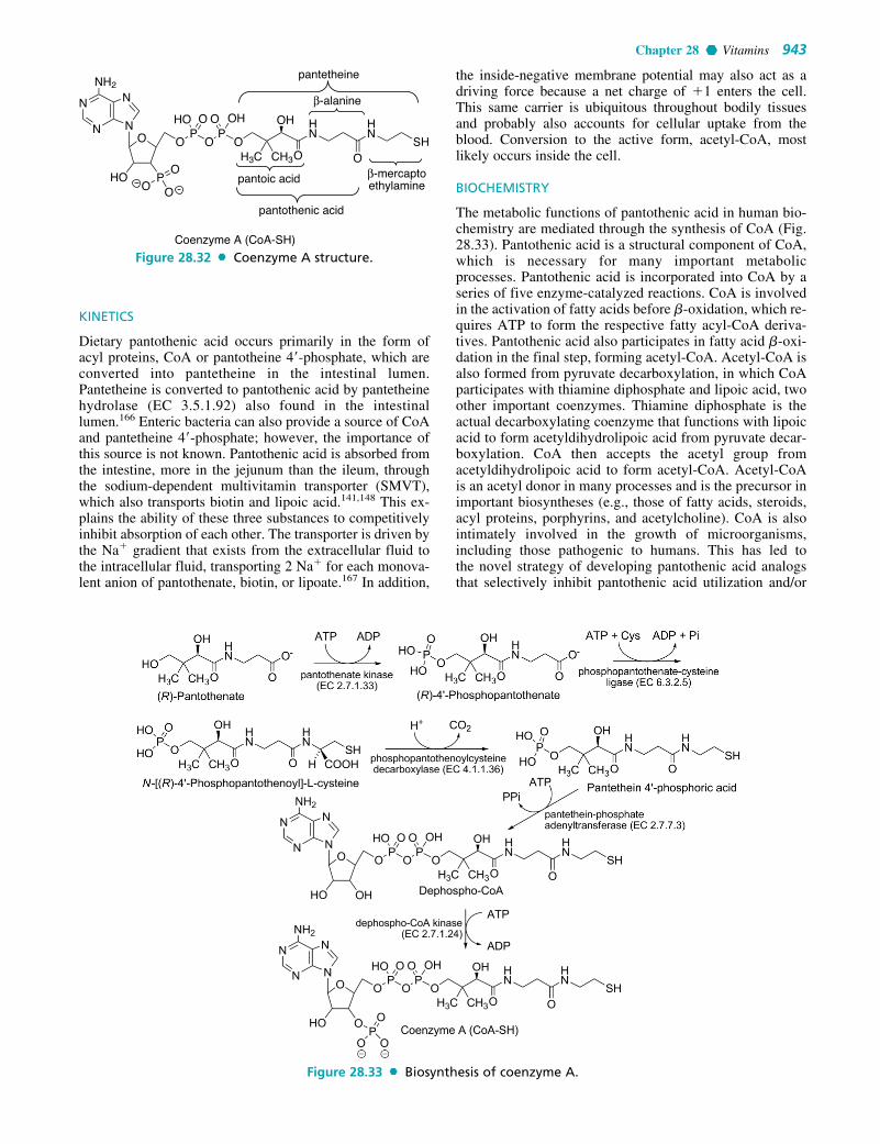
KINETICS
Dietary pantothenic acid occurs primarily in the form ofacyl proteins, CoA or pantotheine 4�-phosphate, which areconverted into pantetheine in the intestinal lumen.Pantetheine is converted to pantothenic acid by pantetheinehydrolase (EC 3.5.1.92) also found in the intestinallumen.166 Enteric bacteria can also provide a source of CoAand pantetheine 4�-phosphate; however, the importance ofthis source is not known. Pantothenic acid is absorbed fromthe intestine, more in the jejunum than the ileum, throughthe sodium-dependent multivitamin transporter (SMVT),which also transports biotin and lipoic acid.141,148 This ex-plains the ability of these three substances to competitivelyinhibit absorption of each other. The transporter is driven bythe Na� gradient that exists from the extracellular fluid tothe intracellular fluid, transporting 2 Na� for each monova-lent anion of pantothenate, biotin, or lipoate.167 In addition,
the inside-negative membrane potential may also act as adriving force because a net charge of �1 enters the cell.This same carrier is ubiquitous throughout bodily tissuesand probably also accounts for cellular uptake from theblood. Conversion to the active form, acetyl-CoA, mostlikely occurs inside the cell.
BIOCHEMISTRY
The metabolic functions of pantothenic acid in human bio-chemistry are mediated through the synthesis of CoA (Fig.28.33). Pantothenic acid is a structural component of CoA,which is necessary for many important metabolicprocesses. Pantothenic acid is incorporated into CoA by aseries of five enzyme-catalyzed reactions. CoA is involvedin the activation of fatty acids before �-oxidation, which re-quires ATP to form the respective fatty acyl-CoA deriva-tives. Pantothenic acid also participates in fatty acid �-oxi-dation in the final step, forming acetyl-CoA. Acetyl-CoA isalso formed from pyruvate decarboxylation, in which CoAparticipates with thiamine diphosphate and lipoic acid, twoother important coenzymes. Thiamine diphosphate is theactual decarboxylating coenzyme that functions with lipoicacid to form acetyldihydrolipoic acid from pyruvate decar-boxylation. CoA then accepts the acetyl group fromacetyldihydrolipoic acid to form acetyl-CoA. Acetyl-CoAis an acetyl donor in many processes and is the precursor inimportant biosyntheses (e.g., those of fatty acids, steroids,acyl proteins, porphyrins, and acetylcholine). CoA is alsointimately involved in the growth of microorganisms,including those pathogenic to humans. This has led tothe novel strategy of developing pantothenic acid analogsthat selectively inhibit pantothenic acid utilization and/or
Chapter 28 Vitamins 943
OP
OP
O
HN
HN
SHOO
OH
CH3H3C
OHOOHO
PHO
NH2
N
NN
N
O
Coenzyme A (CoA-SH)
pantoic acid
pantothenic acid
pantetheine
β-alanine
β-mercapto ethylamineO
OO
Figure 28.32 Coenzyme A structure.
Figure 28.33 Biosynthesis of coenzyme A.
0915-0960_17865_Ch28.qxd 12/4/09 8:12 PM Page 943

biosynthesis in these organisms leading to antibacterial, an-tifungal, or antimalarial effects.168
THERAPEUTIC USES
The only therapeutic indication for pantothenic acid is intreatment of a known or suspected deficiency of this vitamin.Because of the ubiquitous nature of pantothenic acid, defi-ciency states of this vitamin are only seen experimentally byuse of synthetic diets devoid of the vitamin,169 by use of thevitamin antagonist, �-methylpantothenic, or both. In a 1991review, Tahiliani and Beinlich170 described that the mostcommon symptoms associated with pantothenic acid defi-ciency were headache, fatigue, and a sensation of weakness.Sleep disturbances and gastrointestinal disturbances, amongothers, were also noted. The most likely setting for pan-tothenic acid deficiency is in the setting of alcoholism wherea multiple vitamin deficiency exists confounding the exactrole of the pantothenic acid deficiency as compared to theother vitamins. Because a deficiency of a single B vitamin israre, pantothenic acid is commonly formulated in multivita-min or B-complex preparations.
HYPERVITAMINOSIS
Pantothenic acid is considered to be relatively safe. Doses of10 to 20 g/day have caused diarrhea and water retention.161
PRODUCTS
Pantothenic acid is available from numerous sources as oralcapsules, liquids, and tablets under various names. The cal-cium salt of pantothenic acid or the alcohol derivative pan-tothenol is commonly used in pharmaceutical preparationsbecause it is more stable than the parent compound.Pantothenic acid, or one of its derivative or salt forms, isalso available as a component of numerous multivitaminpreparations.
Vitamin B6
The discovery of vitamin B6 is generally ascribed to PaulGyörgy171 who first realized there was a vitamin that wasdistinctly different from vitamin B2 in 1934. Four years later(1938), Lepkovsky172 was the first to report the isolation ofvitamin B6 in crystalline form although others, includingGyörgy, followed by just a few months.173,174 The struc-ture175,176 and synthesis177 were published simultaneously in1939. György178 proposed the term pyridoxin, derived fromtwo of the chemical constituents, pyrid of pyridine and oxo ofmethoxy, as a name for the new vitamin in 1939, but pyridox-ine came into general use thereafter. Other active forms of vi-tamin B6 were found,179 and in 1960, IUPAC106 used the de-scriptor pyridoxine to refer to all “naturally occurringpyridine derivatives with vitamin B6 activity.” IUPAC180 ter-minology was again revised in 1973, and the term vitamin B6,instead of pyridoxine, is the approved generic descriptor forall active forms of the vitamin. Pyridoxine is used specificallyto refer to 4,5-bis(hydroxymethyl)-2-methylpyridin-3-ol. Theterm pyridoxol is no longer recommended. All active formsof vitamin B6 and their recommended names are shown inFigure 28.34, along with their interconversions.
Pyridoxine is naturally found in whole grains, meat, poul-try, cereals, peanuts, corn, and fish. However, up to 40% ofthe vitamin may be destroyed during cooking. Food sources
contain all three forms, either in their free form or phospho-rylated. Plants contain primarily pyridoxine and pyridox-amine, whereas animal sources provide chiefly pyridoxal.
CHEMISTRY
Pyridoxine (PN) is the C4 hydroxymethyl derivative, pyri-doxal (PL) is the C4 formyl derivative and pyridoxamine(PM) is the C4 aminomethyl derivative of 5-(hydroxymethyl)-2-methylpyridin-3-ol). Each of these are also converted totheir corresponding 5�-phosphate derivatives referred to aspyridoxine 5�-phosphate (PNP), pyridoxal 5�-phosphate(PLP), and pyridoxamine 5�-phosphate (PMP), respectively(see Fig. 28.34). Because of their ability to interconvert, allare considered active forms of vitamin B6 in vivo. AlthoughPLP is the major coenzyme form, PMP can also function as acoenzyme primarily in aminotransferases. The major metabo-lite is 4-pyridoxic acid, which is excreted in the urine.
KINETICS
PNP, PLP, and PMP are enzymatically hydrolyzed in thesmall intestine to PN, PL, and PM prior to absorption. Theintestinal absorption of dietary vitamin B6, in the form ofPN, PL, and PM, has historically been ascribed to passivediffusion.181 Recent evidence suggests that a carrier-medi-ated process may also be involved.182 One additional formof vitamin B6, pyridoxine-5�-�-D-glucoside (Fig. 28.35), isthe predominate form in some plants (grains, fruits, and veg-etables), and this form has a significantly lower bioavailabil-ity than other forms because of incomplete hydrolysis of theglycosidic bond in the intestines.
In the liver, all three forms are interconverted; however,the major form of vitamin B6 in the liver is PLP.181 PLP(60%), PN (15%), and PL (14%) circulate in the bloodbound to albumin. Vitamin B6 traverses cell membranes,upon entry or exit, only in the dephosphorylated form.Plasma alkaline phosphatase (EC 3.1.3.1) is responsible forthe removal of the phosphate group prior to cellular uptakefrom the blood.183 Upon cellular entry via facilitated trans-port, phosphorylation acts to trap the vitamin and drive theoverall process of cellular uptake.184 Total body stores ofvitamin B6 approximate 170 mg with 75% to 80% found inmuscle, primarily as phosphorylase (EC 2.4.1.1), and 5%to 10% in the liver185 with the remaining portion distrib-uted among the remaining tissues. The muscle pool isthought to be very stable, relatively unavailable to othertissues and highly conserved during deficient intake.Therefore, deficiency of this vitamin is more likely to af-fect tissues other than muscle, especially newly formed tis-sues.185 Excess vitamin B6 is eventually converted to PL,which is oxidized to 4-pyridoxic acid. This acid is the pri-mary form found in the urine, accounting for essentiallyall excess dietary vitamin B6.
BIOCHEMISTRY
PLP functions as a coenzyme186 that performs many vitalfunctions in human metabolism. More than 140 distinctPLP-dependent enzymes have been cataloged by theEnzyme Commission (EC).187 It functions in the transam-inations and decarboxylations that amino acids generallyundergo, and about half of all the PLP-dependent enzymesfunction as aminotransferases. For example, it functions as
944 Wilson and Gisvold’s Textbook of Organic Medicinal and Pharmaceutical Chemistry
0915-0960_17865_Ch28.qxd 12/4/09 8:12 PM Page 944

a cotransaminase in the transamination of alanine to formpyruvic acid and as a codecarboxylase in the decarboxyla-tion of dihydroxyphenylalanine (DOPA) to formdopamine. Other biological transformations of aminoacids in which pyridoxal can function are racemization,elimination of the �-hydrogen together with a �-sub-stituent (i.e., OH or SH) or a �-substituent, and probablythe reversible cleavage of �-hydroxyamino acids toglycine and carbonyl compounds.
An electromeric displacement of electrons from bondswith the �-carbon (see Fig. 28.36) results in the releaseof a cation (H, R�, or COOH) and, subsequently, leads tovarious reactions observed with pyridoxal. The extent towhich one of these displacements predominates overothers depends on the structure of the amino acid andthe environment (pH, solvent, catalysts, enzymes, etc.).When this mechanism applies in vivo, the pyridoxal com-ponent is linked to the enzyme through the phosphate ofthe hydroxymethyl group by hydrogen bond networksand/or ionic interactions.
Figure 28.37 shows the role of PLP in the transamina-tion reaction. The first half-reaction involves the initial
proton abstraction from the external aldimine at C� bythe active site lysine, yielding a quinonoid intermediate.Lysin ammonium, thus formed, protonates the C4� of thequinonoid intermediate to generate a ketimine, which issubsequently hydrolyzed to release �-ketogluterate andenzyme-PMP complex (E-PMP).
Diacylglycine decarboxylase (DGD) is an unusual PLP-dependent enzyme that performs both decarboxylation, inthe first half-reaction, and transamination, in the second half-reaction (Fig. 28.38). In the first half-reaction, a dialkylaminoacid is decarboxylated producing a ketone and releasing the E-PMP form, which in the second half-reaction catalyzestransamination to convert an �-keto acid (e.g., pyruvic acid)into an amino acid (e.g., alanine), regenerating the DGD-PLP form.
Chapter 28 Vitamins 945
H3C
HO
OH
OH
NPyridoxine
EC 1.4.3.5+FMN
H
H3C
HO
O
OH
NPyridoxal
EC 1.4.3.5+FMN
H3C
HO
NH2
OH
N
Pyridoxamine
HO
H3C
HO
O
OH
N
EC 1.2.3.1+ FAD
Pyridoxic acid
OO
OP
H3C
HO
NH2
O
N
Pyridoxamine 5'-phosphate (PMP)
EC 2.7.1.35+ ATP
OO
OP
H
H3C
HO
O
O
NPyridoxal 5'-phosphate (PLP)
EC 1.4.3.5+FMN
EC 3.1.3.74
EC 2.7.1.35+ ATP
OO
OP
H3C
HO
OH
O
NPyridoxine 5'-phosphate
EC 2.7.1.35+ATP
EC 1.4.3.5+ FMN
EC 3.1.3.74
EC 3.1.3.74
EC 1.4.3.5 pyridoxal 5'-phosphate synthaseEC 2.7.1.35 pyridoxal kinaseEC 3.1.3.74 pyridoxal phosphataseEC 1.2.3.1 aldehyde oxidase
Figure 28.34 Active forms of vitamin B6 and the major metabolite pyridoxicacid.
O
HO
OH
OHOH
H3C
HO
OH
O
NFigure 28.35 Pyridoxine-5�-�-D-glucoside. Figure 28.36 Generalized vitamin B6 reactions.
0915-0960_17865_Ch28.qxd 12/4/09 8:12 PM Page 945

THERAPEUTIC USES
Pyridoxine is indicated in the treatment and prevention ofknown or suspected vitamin B6 deficiency, which is mostlikely to occur in the setting of alcoholism in developedcountries. Vitamin B6 deficiency-related conditions havebeen reviewed by Spinneker et al.184 A hypochromic, mi-crocytic, iron-refractory anemia, a form of sideroblastic ane-mia, develops because of decreased hemoglobin synthesis.Cognitive defects also develop with impairment in memoryfunction, especially in the elderly. Convulsive seizuresoccur, and these are particularly problematic in vitamin B6–deficient infants. Peripheral neuropathy with paresthesia,and burning and thermal sensations is also common. Theconversion of tryptophan to niacin is also PLP-dependentand vitamin B6 deficiency may, rarely, lead to pellagra.183
All of these conditions respond to increased dietary intakeor administration of vitamin B6.
At least seven genetic disorders that result in a vitaminB6 deficiency syndrome in the presence of an adequate di-etary intake have been identified. These result from defectsin enzymes that are responsible for the bioactivation or uti-
lization of vitamin B6. Chronic use of estrogen-containingoral contraceptives may lead to derangements in tryptophanmetabolism, a phenomenon also seen during pregnancy thatis thought to result from inhibition of kinureninase (EC3.7.1.3) by metabolites of estrogenic agents.188 Vitamin B6
levels may also be lower in these same groups. All of theseconditions respond, at least partially, to treatment withlarge doses of pyridoxine; although the use of high doses ofpyridoxine in pregnancy may not be warranted and its usewith contraceptives may lead to failure.183
Certain hydrazine derivatives, when administered thera-peutically (e.g., isoniazid and hydralazine), can induce a defi-ciency of the coenzyme PLP by direct inactivation throughthe mechanism of hydrazone formation with the aldehydefunctional group. Cycloserine and penicillamine may actthrough a similar mechanism.184 Concurrent administrationof pyridoxine can avoid or resolve the relative vitamin B6 de-ficiency seen in each of these conditions. Administration ofhigh doses of vitamin B6 with levodopa, alone, is not recom-mended because of enhanced conversion of levodopa todopamine in the periphery resulting in decreased levodopa
946 Wilson and Gisvold’s Textbook of Organic Medicinal and Pharmaceutical Chemistry
Figure 28.37 Transamination reactions of pyridoxine.
Figure 28.38 Vitamin B6–catalyzed decarboxylation and transamination by diacyl-glycine decarboxylase.
0915-0960_17865_Ch28.qxd 12/4/09 8:12 PM Page 946

availability to the CNS. The concurrent use of a DOPA decar-boxylase inhibitor mitigates this interaction and high doses ofvitamin B6 are without effect on peripheral levodopa conver-sion. RDA amounts of vitamin B6 have not been shown to beproblematic in either case.
HYPERVITAMINOSIS
Interestingly, peripheral neuropathy, a symptom of vitaminB6 deficiency, can also occur following ingestion of highdoses of pyridoxine. Doses of 2 g/day can produce pares-thesia and alter proprioception.189 Some studies suggestthe dose required may be as low as 50 to 500 mg/day.Symptoms may actually increase for several weeks after dis-continuing pyridoxine, a phenomenon called coasting.190
PRODUCTS
Pyridoxine is available from numerous sources under vari-ous names as oral formulations in tablet, capsule, powder,lozenge, and solution form. Pyridoxine HCl is avalable forIM or IV injection. Pyrodoxine is also available as a compo-nent of numerous multivitamin preparations.
Vitamin B7
Vitamin B7 is now referred to as biotin. Biotin was firstisolated in 1936191 and identified structurally in 1942.192
Although this vitamin is known to perform essential meta-bolic functions in the human, the biotin content of foods,and thus the necessary dietary intake, has not been well es-tablished. Good dietary sources of biotin include liver(beef and chicken), meat (beef, pork, chicken), fish, eggs,nuts, and some fruits and vegetables.193 Enteric microor-ganisms synthesize biotin from pimeloyl-CoA194 in thediet and thereby become an additional source of biotin.
CHEMISTRY
Biotin is composed of a tetrahydroimidazolidone (ureido)ring fused with a tetrahydrothiophene ring. A pentanoic acidsubstituent (at C5 of pentanoic acid) is attached to thetetrahydrothiophene ring at C4. The D-isomer (all cis) pos-sesses all the activity and the N1 position is the attachmentpoint for the carboxyl group involved in carboxylation reac-tions (see Fig. 28.39).
KINETICS
Dietary biotin occurs primarily in the form of protein-boundbiotin, which is converted into biotin in the intestinal lumen.Enteric bacteria can also provide a source of protein-boundbiotin; however, the importance of this source is not known.Biotin is absorbed from the intestine, more in the jejunumthan the ileum, through the SMVT, which also transports
pantothenic acid and lipoic acid.141,148 This explains theability of these three substances to competitively inhibit ab-sorption of each other and the ability of some drugs, such ascarbamazepine and primidone, to produce biotin deficiencythrough competitive inhibition of absorption. The trans-porter is driven by the Na� gradient that exists from theextracellular fluid to the intracellular fluid, transporting 2Na� for each monovalent anion of pantothenate, biotin, orlipoate.167 In addition, the inside-negative membrane poten-tial may also act as a driving force because a net charge of�1 enters the cell. This same carrier is ubiquitous through-out bodily tissues and probably also accounts for cellularuptake from the blood. Converson to the active form mostlikely occurs inside the cell. Greater than 50% of a dose isexcreted in the urine as intact biotin. The body appearsunable to break the fused imidazolidone and tetrahydrothio-phene ring system. The metabolites bisnorbiotin, biotin-d, l-sulfoxide, bisnorbiotin methyl ketone, and biotin sulfonehave been identified in the urine.141,195
BIOCHEMISTRY
Biotin is required for enzymatic activity of several impor-tant carboxylases (Table 28.10). Biotin is first attached to alysine residue of biotin carboxyl carrier protein (BCCP) bybiotin-[propionyl-CoA-carboxylase (ATP-hydrolysing)]ligase (holocarboxylase [HCS], EC 6.3.4.10), which formsthe biotinyl domain of the enzyme or separate subunit. TheN1 position is subsequently carboxylated to form carboxy-biotin (Fig. 28.40), which is then able to transfer the car-boxyl group to other substrates. The source of the carboxylis either bicarbonate or decarboxylation of another sub-strate as is seen in transcarboxylation reactions.196 A typi-cal carboxylation reaction mediated by biotin-BCCP isshown in Figure 28.41. The source of the carboxyl is bicar-bonate with ATP supplying energy for the reaction. This isfollowed by the subsequent transfer of the carboxyl to thesubstrate with regeneration of the biotin-BCCP complex.
An interesting recent development is the finding that bi-otin is also attached to histones in the nucleus by the sameenzyme involved in attachment to BCCP.197 The suggestionis that biotin may be involved in genetic regulation; al-though the exact role remains unknown at this time.
Deficiency states may rarely develop as a result of abiotin-deficient diet, as was observed in early, biotin-deficient TPN solutions,198 or as a result of prolonged feed-ing of large quantities of raw egg white.199 Raw egg whitecontains avidin, a protein that complexes biotin and inter-feres with its absorption from the gastrointestinal tract. Arelative biotin deficiency has also been shown to occur in
Chapter 28 Vitamins 947
Figure 28.39 Biotin.
TABLE 28.10 Biotin-Dependent Enzymes
Enzyme EC Number Function
Pyruvate 6.4.1.1 Citric acid cycle (TCA carboxylase cycle)
Acetyl-CoA 6.4.1.2 Fatty acid synthesiscarboxylase
Propionyl-CoA 6.4.1.3 Conversion of propionyl-carboxylase CoA to methylmalonyl-
CoAMethylcrotonyl- 6.4.1.4 Valine, leucine, and
CoA carboxylase isoleucine degredation
0915-0960_17865_Ch28.qxd 12/4/09 8:12 PM Page 947

the case of two identified genetic disorders. Holocarbo-xylase deficiency is a genetic disorder of biotin-(propionyl-CoA-carboxylase [ATP-hydrolysing]) ligase, the enzymeresponsible for the terminal step incorporating biotin intothe apoenzyme. Biotinidase (EC 3.5.1.12) deficiency is agenetic disorder of the amidase that releases biotin from theapoenzyme and allows it to be recycled. Both of these leadto multiple carboxylase deficiency (MCD), a conditionmarked by decreased activity of all biotin-dependent car-boxylases and both respond, at least partially, to largedoses of biotin.197 The symptoms of biotin deficiencyinclude dermatitis, conjunctivitis, alopecia, and CNS ab-normalities including behavioral, developmental, and psy-chological effects.198
HYPERVITAMINOSIS
There are no known documented cases of toxicity resultingfrom excessive intake of biotin.
PRODUCTS
Biotin is available as oral tablets, capsules, and lozengesfrom numerous sources under various names. Biotin con-tent ranges from approximately 300 to 1,000 �g, some-
times higher. Biotin is also found in a host of mutlivitaminpreparations.
Vitamin B9
Vitamin B9 is now known officially as folic acid or folate,which is preferred because it is shorter.200 The term folicacid is derived from the Latin word folium meaning “leaf”and was coined by Mitchell et al.201 in 1941 to refer to a sub-stance isolated from spinach leaves that was an essentialgrowth factor in Streptococcus lactis R (now classified asEnterococcus hirae). Folate crystals were isolated in1943202 and the structure reported in 1946.203
Natural sources of dietary folate are fruits, vegetables,beans, and peas as well as beef and chicken liver. However,since January 1998, the Federal Government204 has requiredfolic acid fortification of all enriched cereal-grain products(flour, rice, bread, rolls, buns, pasta, corn, grits, corn meal,farina, macaroni, and noodle products), and these have nowbecome important dietary sources of folate in the UnitedStates.205,206
CHEMISTRY
The chemical structure of folic acid (Fig. 28.42) was found tobe composed of a pteridine heterocyclic system (rings A andB), p-aminobenzoic acid (PABA) and Glu; hence the namepteroylglutamate (PteGlu). In plants and bacteria, folic acid issynthesized by converting guanosine triphosphate (GTP)through a multistep sequence into the pteridine heterocycle.This is followed by dihydropteroate synthase (EC 2.5.1.15),which adds the PABA moiety and finally the glutamateresidue is added by dihydrofolate synthase (EC 6.3.2.12). It isof note that the antibacterial sulfonamides compete withPABA for the bacterial form of dihydropteroate synthase and,thereby, interfere with bacterial folic acid synthesis. This
948 Wilson and Gisvold’s Textbook of Organic Medicinal and Pharmaceutical Chemistry
Figure 28.41 Biotin-mediated carboxylation reaction.
Figure 28.40 Coenzyme form of biotin.
S
HN NH
O
HN
O
LysBCCP
0915-0960_17865_Ch28.qxd 12/4/09 8:12 PM Page 948
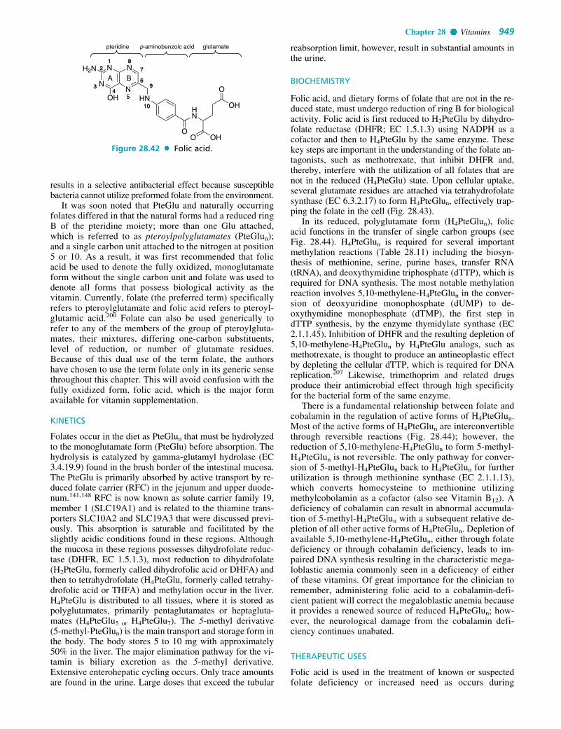
results in a selective antibacterial effect because susceptiblebacteria cannot utilize preformed folate from the environment.
It was soon noted that PteGlu and naturally occurringfolates differed in that the natural forms had a reduced ringB of the pteridine moiety; more than one Glu attached,which is referred to as pteroylpolyglutamates (PteGlun);and a single carbon unit attached to the nitrogen at position5 or 10. As a result, it was first recommended that folicacid be used to denote the fully oxidized, monoglutamateform without the single carbon unit and folate was used todenote all forms that possess biological activity as thevitamin. Currently, folate (the preferred term) specificallyrefers to pteroylglutamate and folic acid refers to pteroyl-glutamic acid.200 Folate can also be used generically torefer to any of the members of the group of pteroylgluta-mates, their mixtures, differing one-carbon substituents,level of reduction, or number of glutamate residues.Because of this dual use of the term folate, the authorshave chosen to use the term folate only in its generic sensethroughout this chapter. This will avoid confusion with thefully oxidized form, folic acid, which is the major formavailable for vitamin supplementation.
KINETICS
Folates occur in the diet as PteGlun that must be hydrolyzedto the monoglutamate form (PteGlu) before absorption. Thehydrolysis is catalyzed by gamma-glutamyl hydrolase (EC3.4.19.9) found in the brush border of the intestinal mucosa.The PteGlu is primarily absorbed by active transport by re-duced folate carrier (RFC) in the jejunum and upper duode-num.141,148 RFC is now known as solute carrier family 19,member 1 (SLC19A1) and is related to the thiamine trans-porters SLC10A2 and SLC19A3 that were discussed previ-ously. This absorption is saturable and facilitated by theslightly acidic conditions found in these regions. Althoughthe mucosa in these regions possesses dihydrofolate reduc-tase (DHFR, EC 1.5.1.3), most reduction to dihydrofolate(H2PteGlu, formerly called dihydrofolic acid or DHFA) andthen to tetrahydrofolate (H4PteGlu, formerly called tetrahy-drofolic acid or THFA) and methylation occur in the liver.H4PteGlu is distributed to all tissues, where it is stored aspolyglutamates, primarily pentaglutamates or heptagluta-mates (H4PteGlu5 or H4PteGlu7). The 5-methyl derivative(5-methyl-PteGlun) is the main transport and storage form inthe body. The body stores 5 to 10 mg with approximately50% in the liver. The major elimination pathway for the vi-tamin is biliary excretion as the 5-methyl derivative.Extensive enterohepatic cycling occurs. Only trace amountsare found in the urine. Large doses that exceed the tubular
reabsorption limit, however, result in substantial amounts inthe urine.
BIOCHEMISTRY
Folic acid, and dietary forms of folate that are not in the re-duced state, must undergo reduction of ring B for biologicalactivity. Folic acid is first reduced to H2PteGlu by dihydro-folate reductase (DHFR; EC 1.5.1.3) using NADPH as acofactor and then to H4PteGlu by the same enzyme. Thesekey steps are important in the understanding of the folate an-tagonists, such as methotrexate, that inhibit DHFR and,thereby, interfere with the utilization of all folates that arenot in the reduced (H4PteGlu) state. Upon cellular uptake,several glutamate residues are attached via tetrahydrofolatesynthase (EC 6.3.2.17) to form H4PteGlun, effectively trap-ping the folate in the cell (Fig. 28.43).
In its reduced, polyglutamate form (H4PteGlun), folicacid functions in the transfer of single carbon groups (seeFig. 28.44). H4PteGlun is required for several importantmethylation reactions (Table 28.11) including the biosyn-thesis of methionine, serine, purine bases, transfer RNA(tRNA), and deoxythymidine triphosphate (dTTP), which isrequired for DNA synthesis. The most notable methylationreaction involves 5,10-methylene-H4PteGlun in the conver-sion of deoxyuridine monophosphate (dUMP) to de-oxythymidine monophosphate (dTMP), the first step indTTP synthesis, by the enzyme thymidylate synthase (EC2.1.1.45). Inhibition of DHFR and the resulting depletion of5,10-methylene-H4PteGlun by H4PteGlu analogs, such asmethotrexate, is thought to produce an antineoplastic effectby depleting the cellular dTTP, which is required for DNAreplication.207 Likewise, trimethoprim and related drugsproduce their antimicrobial effect through high specificityfor the bacterial form of the same enzyme.
There is a fundamental relationship between folate andcobalamin in the regulation of active forms of H4PteGlun.Most of the active forms of H4PteGlun are interconvertiblethrough reversible reactions (Fig. 28.44); however, thereduction of 5,10-methylene-H4PteGlun to form 5-methyl-H4PteGlun is not reversible. The only pathway for conver-sion of 5-methyl-H4PteGlun back to H4PteGlun for furtherutilization is through methionine synthase (EC 2.1.1.13),which converts homocysteine to methionine utilizingmethylcobolamin as a cofactor (also see Vitamin B12). Adeficiency of cobalamin can result in abnormal accumula-tion of 5-methyl-H4PteGlun with a subsequent relative de-pletion of all other active forms of H4PteGlun. Depletion ofavailable 5,10-methylene-H4PteGlun, either through folatedeficiency or through cobalamin deficiency, leads to im-paired DNA synthesis resulting in the characteristic mega-loblastic anemia commonly seen in a deficiency of eitherof these vitamins. Of great importance for the clinician toremember, administering folic acid to a cobalamin-defi-cient patient will correct the megaloblastic anemia becauseit provides a renewed source of reduced H4PteGlun; how-ever, the neurological damage from the cobalamin defi-ciency continues unabated.
THERAPEUTIC USES
Folic acid is used in the treatment of known or suspectedfolate deficiency or increased need as occurs during
Chapter 28 Vitamins 949
22
N3344
N11
N88
N55
H2N
OHOH
O
O OHO
HN
HN1010
9966
77
A B
pteridine p-aminobenzoic acid glutamate
Figure 28.42 Folic acid.
0915-0960_17865_Ch28.qxd 12/4/09 8:12 PM Page 949

950 Wilson and Gisvold’s Textbook of Organic Medicinal and Pharmaceutical Chemistry
Figure 28.44 Interconversion of active folates.
Figure 28.43 Bioactivation of folic acid to tetrahydrofolate polyglutamates.
0915-0960_17865_Ch28.qxd 12/4/09 8:12 PM Page 950

pregnancy. The most likely cause of folate deficiency in de-veloped countries is alcoholism. The resulting megaloblasticanemia, thought to result from a deficiency in DNA synthe-sis, as noted previously, is indistinguishable from that causedby vitamin B12 because both vitamins are involved in thesame critical biochemical step. Unlike vitamin B12, no neu-rological abnormalities are associated with folate deficiency.Because folate requirements increase during pregnancy, andmaternal folate deficiency has been associated with a risk ofneural tube defects in the fetus, folic acid supplementation isrecommended, at a minimum, periconceptually.208,209
Because of the potentially beneficial effects in reducing thesebirth defects, the FDA has mandated fortification of all en-riched cereal-grain products in the United States with folicacid since 1998.204
Folic acid has also been shown to reduce homocysteinelevels, which is a risk factor for cardiovascular disease inde-pendent of other risk factors. However, although there issome evidence that folic acid supplementation may reducethe risk of stroke,210 the risk of other cardiovascular diseasehas not been proven to be affected.211 Some studies suggestthat multivitamin preparations with folic acid taken prior to,and during pregnancy, may protect against congenital heartdefects. Folate may also decrease the risk of colorectal can-cer; however, once colorectal cancer develops it may act asa promoting agent.212
One of the biologically active forms of folic acid, leucov-orin (5-formyl-H4PteGlu), is used in the treatment of acci-dental overdoses of DHFR inhibitors, such as methotrexate,because it does not require bioactivation by DHFR as doesfolic acid. Leucovorin is also used concomitantly with, orshortly following, methotrexate, trimetrexate, pyrime-thamine, trimethoprim, and related drug therapy to decreasethe toxicity in sensitive cells. Finally, although leucovorincan be used in place of folic acid in all other uses, there isno therapeutic advantage.
HYPERVITAMINOSIS
High doses of folic acid are rather nontoxic; however, atleast one study has shown that 15 mg/day for 1 month canresult in gastrointestinal disturbances (including anorexia,nausea, and pain) and CNS effects (including sleep distur-bances, vivid dreams, malaise, irritability, and increased ex-citability).213 The primary danger of excessive folate intakeis the masking of vitamin B12 deficiency. Folic acid can cor-rect the anemia caused by vitamin B12 deficiency, but it hasno effect on the neurological damage. The potential for highdoses of folic acid to mask B12 deficiency explains whylarge daily doses of folic acid are best avoided in the generalpopulation.
PRODUCTS
Folic acid is available alone in tablet and injectable form, orin combination with other vitamins, in various multivitaminpreparations. Leucovorin is a racemic mixture of 6S and 6R5-formyl-H4PteGlu and is available as the calcium salt foruse in treatment of accidental overdoses of DHFR inhibitors,such as methotrexate, or to decrease their toxicity in sensitivecells. The active 6S-isomer of leucovorin is also available aslevoleucovorin calcium (Fusilev) and is dosed at one-half therate of leucovorin with the same uses. L-Methylfolate(Deplin) is the 6S-isomer of 5-methyl-H4PteGlu. This is thebiologically active isomer of 5-methyl-H4PteGlu, which isthe main transport and storage form (as 5-methyl-H4PteGlun)of folates in the body. It is used in tablet form as a medicalfood for the treatment of methylenetetrahydrofolate reduc-tase [NAD(P)H] (EC 1.5.1.20) deficiency (MTHFR defi-ciency). Medical foods are substances that are regulated bythe FDA and are administered enterally under the supervi-sion of a physician intended for the dietary management of adisease that has distinctive nutritional needs.
Vitamin B12
Vitamin B12 specifically refers to cyanocobalamin, the formtypically found in dietary supplements because of its highstability.214 However, the immediate precursor to the vita-min B12-derived coenzymes is hydroxocobalamin (vitaminB12b), which is formed in vivo from cyanocobalamin.Cobalamin is used to refer to any of the related cobalt-containing compounds that serve as coenzymes or theirbiologically active precursors as shown in Figure 28.45.
The discovery of vitamin B12 began with the unwittingdescription by Dr. Thomas Addison in 1855 of patients suf-fering from an anemia that was later identified as perniciousanemia.215 Following the rapid developments in vitamindiscovery in the early 1900s, it was soon determined that adiet high in liver could reverse the effects of pernicious ane-mia as was clearly shown in 1926 by Minot and Murphy.216
However, concentration and identification of the vitaminwas slow because the only known assay for vitamin B12 ac-tivity was the reversal of pernicious anemia in human pa-tients. Almost 30 years had passed before Shorb217 reportedin 1947 that liver extract was required for the growth ofLactobacillus lactis in similar proportions to that found totreat pernicious anemia. With an inexpensive, rapid assayfor vitamin B12 activity, the crystalline form was isolatedand reported simultaneously just 1 year later by Smith218
and Ricks et al.219 who suggested the name vitamin B12.Interestingly, the conversion to cyanocobalamin occursduring the purification process and is not the natural form of
Chapter 28 Vitamins 951
TABLE 28.11 Folic Acid–Dependent Enzymes
Enzyme EC Number Function
Methionine synthase 2.1.1.13 Conversion of L-homocysteine to L-methionineGlycine hydroxymethyltransferase 2.1.2.1 Interconversion of serine and glycinePhosphoribosylglycinamide formyltransferase 2.1.2.2 Purine synthesisPhosphoribosylaminoimidazolecarboxamide formyltransferase 2.1.2.3 Purine synthesisGlutamate formimidoyltransferase 2.1.2.5 Histidine metabolismMethionyl-tRNA formyltransferase 2.1.2.9 tRNA synthesisThymidylate synthase 2.1.1.45 Conversion of dUMP to dTMP in DNA synthesis
0915-0960_17865_Ch28.qxd 12/4/09 8:12 PM Page 951

the vitamin. The structure of cyanocobalamin was announcedsimultaneously by Hodgkin et al.220 and Bonnett et al.221 in1955 and the complete synthesis completed in 1972.222
De novo synthesis of cobalamin is only found in microor-ganisms. Therefore, cobalmin is found naturally in commer-cial fermentation processes of antibiotics, such as thoseof Streptomyces griseus, Streptomyces olivaceus, andStreptomyces aureofaciens as well as sewage, Milorganiteand others. Some of these fermentations furnish a commer-cial source of vitamin B12. Animals depend on synthesis bythe intestinal flora or, as in humans, foods from animals thathave already consumed cobalamin. Excellent dietarysources are meats, eggs, dairy, fish, and shellfish. The onlydietary plant products containing the vitamin are legumes,because of their symbiosis with microorganisms, or thosethat have been fortified with the vitamin.
CHEMISTRY
These are the most complex group of vitamins consisting of acorrin core with a central cobalt atom with �1, �2, or �3 ox-idation state. The corrin core has four pyrrol units, two ofwhich are attached directly whereas the others are joined bymethylene bridges. Out of six coordination bonds of cobalt,four are bonded to pyrrole nitrogens and the fifth is bound toone of the nitrogen atoms of the dimethylbenzimidazole de-rivative containing ribose 3-phosphate and aminoisopropanollinked through an amide bond to the side chain of the corrincore. The sixth substituent of the cobalt atom can be �CN,�OH, �CH3, or a 5�-deoxyadenosyl unit (Fig. 28.45). In hy-droxocobalamin, the cobalt atom is in the �3 oxidation stateand called vitamin B12b (Co3�), which is reduced by flavo-protein reductase to the vitamin B12r (Co2�) form and then tothe vitamin B12s (Co�) form, which is converted to the methylcoenzyme form. Vitamin B12r (Co2�) is believed to be con-verted to the adenosylcoenzyme form by cob(I)yrinic acid a,c-diamide adenosyltransferase (EC 2.5.1.17) using ATP.
KINETICS
Dietary cobalamin is protein bound but is released in thestomach through the action of acid and gastric proteases.The primary natural forms are methylcobalamin, de-oxyadenosylcobalamin, and hydroxocobalamin with only
trace amounts of cyanocobalamin, which is processed simi-larly except for the initial liberation from protein. Freecobalamin then binds to transcobalamin (TC) I, also calledhaptocorrin, which is a high-affinity cobalamin-bindingprotein found in all bodily fluids including saliva. As thebound cobalamin enters the small intestine, pancreatic pro-teases digest TC I releasing free cobalamin, which thenbinds to intrinsic factor (IF), secreted primarily by parietalcells of the gastric mucosa, forming an intrinsic factor-cobalamin (IF-CBL) complex. In the distal ileum, IF-CBLbinds to cubilin, also known as the intrinsic factor cobal-amin receptor (IFCR), on the luminal surface and undergoesreceptor-mediated endocytosis. Intracellularly, IF is cleavedby the lysozymal enzyme, cathepsin L (EC 3.4.22.15), andthe free cobalamin binds to TC II to form TC II-CBL, whichis then released into the bloodstream completing the processof transcytosis.223–225 Dietary supplements that containcyanocobalamin undergo an additional conversion in theplasma by cyanocobalamin reductase (cyanide-eliminating)(EC 1.16.1.6) to hydroxocobalamin, which serves as the im-mediate precursor to the cobalamin derived coenzymes.
The average dietary intake of cobalamin is 5 to 30 �g;however, only 1 to 2 �g/meal is absorbed most likely becauseof saturation of the IF-dependent transport. Small amountsof cobalamin can also be absorbed through passive diffu-sion; however, this is normally insufficient to prevent defi-ciency unless the diet consists of abnormally large quantitiesof foods rich in cobalamin, such as liver. Only 1% of an oraldose of cobalamin is absorbed in patients that lack gastricacid and IF, which explains the large doses, 1,000 to 2,000 �g(1–2 mg) per day, of vitamin B12 required in the oral treat-ment of pernicious anemia.
In the bloodstream, cobalamin is bound not only to TC IIbut also to plasma haptocorrin, which may function prima-rily as a storage form of cobalamin. Uptake into cells thatneed cobalamin is receptor mediated through TC II-CBL re-ceptors (TC II-R). Half of the body stores of cobalamin arein the liver primarily as adenosylcobalamin. Cobalamin isexcreted into the bile and undergoes enterohepatic cyclingutilizing IF for reabsorption. This explains why deficiencywill occur more rapidly in patients with malabsorption ofcobalamin than inadequate dietary intake. Excess cobalaminis eliminated unchanged in the feces and urine.
952 Wilson and Gisvold’s Textbook of Organic Medicinal and Pharmaceutical Chemistry
OHHO
O
N
NH2
N
N
N
OH
CH3
R = Adenosylcobalamin(Vitamin B12 coenzyme)
Methylcobalamin (Me-B12 coenzyme)
R = Hydroxocobalamin (B12b, B12r or B12s)
R =
CNR = Cyanocobalamin
NH2
O
H3C
O
NH
O
H2N
O
H2N
NH2ONH2 O
NH2
O
HO
R
N
NH
N
N
Me
Me
Co
Me
Me
Me
Me
MeMe
OH
N Me
MeOO
OP
O
O
N
Figure 28.45 Biologically activeforms of cobalamin.
0915-0960_17865_Ch28.qxd 12/4/09 8:12 PM Page 952

BIOCHEMISTRY
In the biosynthesis of the coenzymes derived from cobal-amin, cobalt is reduced from a trivalent to a monovalentstate before the organic anionic ligands are attached to thestructure. The two types of cobalamins that participate ascoenzymes in human metabolism are adenosylcobalaminand methylcobalamin. These coenzymes perform vital func-tions in methylmalonate–succinate isomerization and inmethylation of homocysteine to methionine (Fig. 28.46).
Two enzymes have been shown to be cobalamin de-pendent in humans. Methylmalonyl-CoA mutase (EC5.4.99.2) requires 5�-deoxyadenosylcobalamin; this en-zyme system catalyzes the methylmalonyl-CoA transfor-mation to succinyl-CoA, which is the major pathway ofpropionyl-CoA metabolism (Fig. 28.46). This is a free rad-ical mediated rearrangement reaction where cobalamin’srole is to serve as the source of free radicals, through ho-molytic cleavage of the weak Co-C bond, for the abstrac-tion of hydrogen atoms. Propionyl-CoA from odd-chainfatty acid metabolism must be processed through this path-way to succinyl-CoA to enter the Krebs (citric acid) cycleto be either converted to �-oxaloacetate, leading to gluco-neogenesis, or oxidized aerobically to CO2, with produc-tion of ATP. The methylation of homocysteine to form me-thionine requires methylcobalamin and is catalyzed bymethionine synthase (EC 2.1.1.13), a transmethylase thatalso requires 5-methyl-PteGlun and reduced FAD. This lat-
ter reaction forms the basis for the megaloblastric anemiathat results from either folic acid or cobalamin deficiencyand the masking of cobalamin deficiency by folic acid.The reader is referred to the section on vitamin B9 for a re-lated discussion.
THERAPEUTIC USES
The most common form of cobalamin deficiency, known aspernicious anemia, is now known to be an autoimmune disor-der that results from parietal cell destruction. This results ininsufficient production of gastric acid and IF and the ensuingmalabsorption of cobalamin. Other less common causes of in-sufficient absorption are hypochlorhydria, gastrectomy, ileo-cecal resection, celiac disease, and chemical incompatibilitieswith drugs in the gastric milieu. A deficiency of cobalamincan also result from nutritional deficiencies, increased re-quirements as is seen in pregnancy, as well as a strict vegetar-ian diet without adequate supplementation. The symptoms ofcobalamin deficiency primarily result from potentially irre-versible nerve damage, involving sensory, motor, and cogni-tive functioning, and the megaloblastic anemia that can beconfused with that seen in folate deficiency. The ability offolic acid to reverse megaloblastic anemia seen in cobalamindeficiency, without affecting nerve damage, is discussed inthe Vitamin B9 section.
Cyanocobalamin is the form most commonly used inthe treatment of cobalamin deficiency; however, hydroxo-
Chapter 28 Vitamins 953
Figure 28.46 Coenzymes of vitamin B12.
0915-0960_17865_Ch28.qxd 12/4/09 8:12 PM Page 953

cobalamin is equally efficacious. A rare genetic disorderthat results in an inability to convert cyanocobalamin tohydroxocobalamin is the only known instance where hy-droxocobalamin would be the preferred treatment.226
Hydroxocobalamin, but not cycanocobalamin, is also usedin the treatment of cyanide toxicity. Administration oflarge doses results in the conversion of hydroxocobalaminto the nontoxic cyanocobalamin effectively reducingplasma levels of cyanide. In this case, 5 to 10 g of IV hy-droxocobalamin may be needed as compared to 1 mg/dayin severe pernicious anemia.
HYPERVITAMINOSIS
Milligram doses of cyanocobalamin are generally not associ-ated with toxicity although rare allergic227 and anaphylacticreactions228,229 and mild diarrhea have occurred. As furtherevidence of the safety of vitamin B12, the adverse effects fol-lowing the use of hydroxocobalmin in the treatment ofcyanide poisoning, where the typical dose is 5 to 10 g, havebeen relatively mild.230
PRODUCTS
Cyanocobalamin, the most common form used, is availableas oral and sublingual tablets as well as oral lozenges fromnumerous sources under various names. These productscontain anywhere from approximately 50 to 1,000 �g ofcyanocobalamin. Cyanocobalamin is also available as anasal spray for daily use (CaloMist 25 �g/actuation) orweekly use (Nascobal 500 �g/actuation) and as an in-jectable for intramascular (IM) or deep subcutaneous (SQ)administration. Hydroxocobalamin is available as two, 2.5-gvials (Cyanokit) for the treatment of cyanide poisoning.Hydroxocobalamin is also available in oral and injectabledosage forms from numerous sources under various names.The only advantage for hydroxocobalamin in the treatmentof pernicious anemia would be in patients unable to convertcyanocobalamin to hydroxocobalamin as the result of a raregenetic disorder as discussed previously. Cyanocobalaminand hydroxocobalamin are also available in a host of multi-vitamin preparations.
Vitamin C
Scurvy (from the French word scorbutus) has been recog-nized as a disease afflicting mankind for thousands ofyears.231 However, it was not until the early 1500s that na-tives of North America used teas brewed from pine needlesto treat or prevent the symptoms of scurvy without under-standing the basis for their curative properties. Citrus fruitssuch as oranges, lemons, and limes were later identified asequally effective treatments. Only within the last 100 yearshas a deficiency in vitamin C been definitively identified asthe cause of scurvy. In 1932, Waugh and King232,233 isolatedcrystalline vitamin C from lemon juice and showed it to bethe antiscorbutic factor present in each of these treatments.Interestingly, in a series of papers published at approxi-mately the same time,234,235 Svirbely and Szent-Györgyi236
showed that the substance the latter had crystallized frompeppers and reported 4 years earlier was the same as vitaminC and not a hexuronic acid as originally proposed. Thestructure and chemical formula of vitamin C was identifiedin 1933 by Hirst et al.237 as one of a series of possible
tautomeric isomers. The first synthesis of vitamin C wasannounced almost simultaneously by Ault et al.238 andReichstein239 in 1933. Since that time, it has been synthe-sized in several different ways.
Vitamin C can be synthesized by nearly all living organ-isms, plants, and animals; but primates, guinea pigs, bats,and a few other species cannot produce this vitamin. Theconsensus is that organisms that cannot synthesize vitaminC lack the liver microsomal enzyme L-gulonolactone oxi-dase (1.1.3.8), which catalyzes the terminal step of thebiosynthetic process. Sato and Udenfriend240 summarizedstudies of the biosynthesis of vitamin C in mammals and thebiochemical and genetic basis for the incapability of somespecies to synthesize the vitamin. Because humans are oneof the few animal species that cannot synthesize vitamin C,it has to be available as a dietary component.
Dietary sources of ascorbic acid include fruits (espe-cially citrus fruits), vegetables (especially peppers), andpotatoes. Although the sources of some commercial prod-ucts are rose hips and citrus fruits, most ascorbic acid isprepared synthetically.
CHEMISTRY
Vitamin C is now commonly referred to as ascorbic acidbecause of its acidic character and its effectiveness in thetreatment and prevention of scorbutus (scurvy). The acidiccharacter is because of the two enolic hydroxyls; the C3
hydroxyl has a pKa value of 4.1, and the C2 hydroxyl hasa pKa of 11.6. All biological activities reside in L-ascorbicacid (Fig. 28.47); therefore, all references to vitamin C,ascorbic acid, ascorbate, and their derivatives refer to thisform. The monobasic sodium salt (Fig. 28.48) is the usualsalt form.
KINETICS
Dietary ascorbic acid is readily absorbed as ascorbate and de-hydroascorbate by facilitated diffusion while ascorbate canalso be absorbed by active transport. Large doses can saturatethese systems, limiting the amounts absorbed. Once absorbed,it is distributed to all tissues and transported into cells via thesame mechanisms.241,242 Excess ascorbate is metabolized tooxalic acid and excreted by the kidneys (Fig. 28.49), which
954 Wilson and Gisvold’s Textbook of Organic Medicinal and Pharmaceutical Chemistry
HO
HO
HO OH
OO
HO
HO
NaO OH
OO
Figure 28.48 Ascorbate sodium.
Figure 28.47 L-Ascorbic acid.
0915-0960_17865_Ch28.qxd 12/4/09 8:12 PM Page 954

may form the basis for the formation of kidney stones follow-ing ingestion of large doses. Although oxalate is the majorurinary metabolite (44%), unchanged ascorbate (20%) andthe intermediate metabolite 2,3 diketo-L-gulonic acid (20%)are also found in appreciable quantities.242
BIOCHEMISTRY
Ascorbic acid is a required cofactor for several importantdioxygenases, functioning as an electron donor in each ofthese reactions (Table 28.12). These dioxygenases are in-volved in the hydroxylation of many important biomoleculesduring their synthetic sequence including collagen, peptidehormones, steroids, and norepinephrine. The resulting oxi-dized product, semidehydroascorbate (Fig. 28.50), can bereduced back to ascorbic acid through cytochrome-b5 reduc-tase (EC 1.6.2.2) or thioredoxin-disulfide reductase (EC1.8.1.9). Alternatively, two molecules of semidehydroascor-bate can undergo a disproportionation reaction yielding onemolecule of ascorbic acid and one molecule of dehy-droascorbate. The latter can also be converted to ascorbicacid by thioredoxin-disulfide reductase (EC 1.8.1.9) or re-duction by glutathione that may be enzymatically or nonen-zymatically mediated.242
THERAPEUTIC USES
Vitamin C is indicated for the treatment and prevention ofknown or suspect deficiency. Although scurvy occurs in-frequently, it is seen in the elderly, infants, alcoholics, anddrug users. The array of symptoms associated with scurvyis thought to result from inadequate formation of the prod-ucts of the key enzymatic reactions for which ascorbate is a
cofactor. Especially notable are the derangements in con-nective tissue formation as a result of the effects on colla-gen synthesis leading to hemorrhaging, poor wound heal-ing, and defects in bone and dentin. Ascorbate can also beused to enhance absorption of dietary nonheme iron or ironsupplements. Ascorbic acid (but not the sodium salt) washistorically used to acidify the urine as a result of excretionof unchanged ascorbic acid, although this use has falleninto disfavor. Ascorbate also increases iron chelation bydeferoxamine, explaining its use in the treatment of irontoxicity. However, use of ascorbate can also enhance ironabsorption, which could aggravate the original iron toxicityand so caution is advised. It is also a useful adjunct in thetreatment of methemoglobinemia.
Ascorbic acid is an effective reducing agent and antioxi-dant; although the daily doses required to produce signifi-cant in vivo activity (�1 g) are much higher than the RDA(�100 mg). Because oxidative stress is thought to underlymany disease processes, there has been intense interest inutilizing high doses of the antioxidant vitamins to prevent ortreat diseases unrelated to their role as vitamins. There arenumerous studies that have examined the effects of vitaminC supplementation alone, or in combination with other vita-mins and minerals, yet the only conclusion to be drawn isthat there is no clear and convincing evidence that vitaminC is useful in the treatment or prevention of most major dis-ease states. Systematic reviews have not found evidence forgeneral use in the comon cold, cardiovascular disease ingeneral, hypertension, atherosclerosis, cancer, preeclamp-sia, or overall mortality.43,55,243,244
HYPERVITAMINOSIS
High doses of vitamin C can lead to renal, bone, hematolog-ical, and gastrointestinal effects.46 Renal calculi because ofoxalate or urate result from enhanced renal excretion ofthese compounds in the presence of high doses of vitamin C.Increased release of calcium and phosphorus from bonehave been observed. Hematological effects include in-creased absorption of nonheme iron without significant in-creases in total body iron stores. Diarrhea, likely resultingfrom an osmotic effect, has been reported following largedoses, and ascorbic acid tablets that lodge in the esophaguscan cause local erosion. Finally, ascorbic acid has beenshown to interfere with several colorimetric redox assaysbecause of its ability to act as a reducing agent.
PRODUCTS
Ascorbic acid is available as oral tablets, capsules,lozenges, crystals, and solutions from various manufactur-ers. It is also available as an injectable solution and as part
Chapter 28 Vitamins 955
HO
HO
O O
OO
HO
HO
HO OH
OOOH
HO
HO
O O
OOH
L-Dehydroascorbic acidL-Ascorbic acid 2,3 Diketo–L–gulonic acid
CH2OHC OCHOCH2OH
HCC
O O
OO
+
L-Erythrulose Oxalate
Figure 28.49 Oxidation of ascorbic acid.
TABLE 28.12 Ascorbate-Dependent Enzymes
Enzyme EC Number Function
Gamma-butyrobetaine 1.14.11.1 Lysine dioxygenase degradation
Procollagen-proline 1.14.11.2 Collagen synthesisdioxygenase synthesis
Procollagen-lysine 1.14.11.4 Collagen 5-dioxygenase synthesis
Procollagen-proline 1.14.11.7 Collagen 3-dioxygenase synthesis
Trimethyllysine 1.14.11.8 Lysine dioxygenase degredation
Phytanoyl-CoA 1.14.11.18 Fatty acid dioxygenase metabolism
Dopamine beta- 1.14.17.1 Norepinephrine monooxygenase synthesis
Peptidylglycine 1.14.17.3 Peptide hormonemonooxygenase synthesis
0915-0960_17865_Ch28.qxd 12/4/09 8:12 PM Page 955

of innummerable multivitamin combinations under variousnames. Ascorbic acid is also available as the sodium salt inan injectable solution and as calcium ascorbate in oraltablets, granules, and powder. These two salts are also for-mulated into an assortment of multivitamin combinationssold under various names. Several other micronutrient saltsof ascorbic acid, such as the potassium salt, are available.Ascorbyl palmitate (ascorbic acid 6 palmitate) is the C6
palmitic acid ester of ascorbic acid (see Fig. 28.51). Thisester of ascorbic acid is a fat-soluble form that releasesascorbic acid in the digestive tract following hydrolysis ofthe ester bond. Ascorbyl palmitate has also been used intopical preparations designed to protect the skin from UVradiation under the premise that the antioxidant properties,
in a manner similar to the oral use of ascorbic acid, wouldadd to the protectant effects. However, there is evidencethat its use in these preparations may actually lead to en-hanced skin damage.245
956 Wilson and Gisvold’s Textbook of Organic Medicinal and Pharmaceutical Chemistry
Figure 28.50 Ascorbic acid cycle.
O
(CH2)14CH3OHO
HO OH
OO
Figure 28.51 Ascorbyl palmitate.
5. Vitamin A is very toxic in high doses but the provitaminA carotenoids are not. Explain this difference.
6. Which vitamins can be made in humans and how are theysynthesized?
7. Explain the sunlight-mediated production of vitamin Din the skin using structures when appropriate. Doesexcessive sunlight produce toxic levels of vitamin D?
8. What are tocols? Researchers have shown that all formsof tocols are absorbed from the intestine equally, yet
1. List the fat-soluble vitamins, the water-soluble vitamins,and the B-complex vitamins.
2. Explain the difference between enrichment and fortifi-cation.
3. How do most of the vitamins function? How else mightsome vitamins function?
4. Which vitamin is involved in proper function of the eyeand how is it involved? Would you recommend retinoicacid (Tretinoin) for treatment of a defiency of this vita-min? Explain your reasoning.
R E V I E W Q U E S T I O N S
0915-0960_17865_Ch28.qxd 12/4/09 8:12 PM Page 956

REFERENCES1. NIH State-of-the-Science Panel: Ann. Intern. Med. 145:64–371,
2006.2. Funk, C. J.: Physiol. 43:395–400, 1911.3. Funk, C. J.: State Med. 20:341–368, 1912.4. Hopkins, F. G.: J. Physiol. 44:425–460, 1912.5. McCollum, E. V., and Davis, M. J.: Biol. Chem. 15:167–175, 1913.6. McCollum, E. V., and Davis, M. J.: Biol. Chem. 19:245–250, 1914.7. McCollum, E. V., and Davis, M. J.: Biol. Chem. 20:641–658, 1915.8. McCollum, E. V., and Davis, M. J.: Biol. Chem. 21:179–182, 1915.9. McCollum, E. V., and Kennedy, C. J.: Biol. Chem. 24:491–502,
1916.10. Drummond, J. C.: Biochem. J. 13:77–80, 1919.11. Drummond, J. C.: Biochem. J. 14:660, 1920.12. Jansen, B. C.: Nature 135:267, 1935.13. Title 21 Code of Federal Regulations, pt.101.9 2007.14. Otten, J. J., Hellwig, J. P., and Meyers, L. D. (eds.): Dietary
Reference Intakes: The Essential Guide to Nutrient Requirements.Washington D.C.: The National Academies Press, 2006, pp. 10–12.
15. King, J. C., and Garza, C.: Food Nutr. Bull. 28:S3–S12, 2007.16. Karrer, P., Morf, R., and Schöpp, K.: Helv. Chim. Acta
14:1036–1040, 1931.17. Heilbron, I. M., and Morton, R. A.: Biochem. J. 26:1194–1196, 1932.18. Isler, O., Huber, W., Ronco, A., et al.: Helv. Chim. Acta
30:1911–1927, 1947.19. Wald, G.: Science 162:230–239, 1968.20. IUPAC-IUB Commission on Biochemical Nomenclature: Pure Appl.
Chem. 55:721–726, 1983.21. Liébecg, C.: Eur. J. Biochem. 247:733–739, 1997.22. Blomhoff, R.: Nutr. Rev. 52(2 Pt 2):S13–S23, 1994.23. Blomhoff, R., Green, M. H., Green, J. B., et al.: Physiol. Rev.
71:951–990, 1991.24. Harrison, E. H.: Annu. Rev. Nutr. 25:87–103, 2005.25. Wolf, G.: Nutr. Rev. 49:1–12, 1991.26. Kloer, D. P., and Schulz, G. E.: Cell. Mol. Life Sci. 63:2291–2303,
2007.27. Lindqvist, A., and Andersson, S. J.: Biol. Chem. 277:23942–23948,
2002.28. Biesalski, H. K., Chichili, G. R., Frank, J., et al.: Vitam. Horm.
75:117–130, 2007.
29. Kiefer, C., Hessel, S., Lampert, J. M., et al.: J. Biol. Chem.276:14110–14116, 2001.
30. Blomhoff, R., Green M. H., Berg T., et al.: Science 250:399–404,1990.
31. Norum, K. R., and Blomhoff, R.: Am. J. Clin. Nutr. 56:734–744,1992.
32. Senoo, H., Kojima, N., and Sato, M.: Vitam. Horm. 75:131–159, 2007.33. Wolf, G.: Physiol. Rev. 64:873–937, 1984.34. Lamb, T. D., and Pugh, E. N.: Prog. Retinal Eye Res. 23:307–380,
2004.35. Imanishi, Y., Lodowski, K. H., and Koutalos, Y.: Biochemistry
46:9674–9684, 2007.36. Blomhoff, R., and Blomhoff, H. K.: Neurobiol. 66:606–630, 2006.37. Brtko, J., and Thalhamer, J.: Curr. Pharm. Des. 9:2067–2077, 2003.38. Desvergne, B.: Vitam. Horm. 75:1–32, 2007.39. Murphy, K. A., Quadro, L., and White, L. A.: Vitam. Horm.
75:33–67, 2007.40. West, K. P., Jr.: Food Nutr. Bull. 24:S78–S90, 2003.41. World Health Organization: Measles. Fact Sheet No. 286. January
2007.42. Kanofsky, J. R., and Sima, P. D.: J. Photocem. Photobiol. B
87:124–129, 2007.43. U.S. Preventive Services Task Force: Ann. Intern. Med. 139:51–55,
2003.44. Fairfield, K. M., and Fletcher, R. H.: JAMA 287:3116–3126, 2002.45. DiPalma, J. R., and Ritchie, D. M.: Annu. Rev. Pharmacol. Toxicol.
17:133–148, 1977.46. Penniston, K. L., and Tanumihardjo, S. A.: Am. J. Clin. Nutr.
83:191–201, 2006.47. Meyers, D. G., Maloley, P. A., and Weeks, D.: Arch. Intern. Med.
156:925–935, 1996.48. Bendich, A., and Langseth, L.: Am. J. Clin. Nutr. 49:358–371, 1989.49. Melhus, H., Michaëlsson, K., Kindmark, A., et al.: Ann. Intern. Med.
129:770–778, 1998.50. Lammer, E. J., Chen, D. T., Hoar, R. M., et al.: N. Engl. J. Med.
313:837–841, 1985.51. Rothman, K. J., Moore, L. L., Singer, M. R., et al.: N. Engl. J. Med.
21:1369–1373, 1995.52. Jenkins, K. J., Correa, A., Feinstein, J. A., et al.: Circulation
115:2995–3014, 2007.
Chapter 28 Vitamins 957
�-tocopherol is found in the circulation in the highestamounts. Why?
9. Which two vitamins are absorbed via the SMVT andcan competitively inhibit each other?
10. What is a severe form of central neurological damage,with a high fatality rate, seen in alcoholics because of asevere vitamin deficiency? Name the vitamin.
11. Controversial results have been found with vitamin Euse in cardiovascular diseases. Explain at least one pos-sible chemical reason for this controversy.
12. Vitamin C is an antioxidant vitamin. Explain how theindiscriminate use of large doses may lead to renal,bone, hematological, and gastrointestinal effects.
13. Explain the role of vitamin K in the formation of Glaproteins. List the known biochemical roles of the Glaproteins.
14. Which vitamins have a role in bone mineralization?
15. Explain how leucovorin can treat overdoses ofmethotrexate, and other DHFR inhibitors, while folicacid is ineffective.
16. What is the danger of treating megaloblastic anemia em-pirically with folic acid?
17. What is the purpose of fortification of food with folicacid and what foods require fortification?
18. Niacin is used unrelated to its vitamin activity for whatcardiovascular disorder? Would you recommend niaci-namide instead of niacin for this use because it causesless side effects?
19. What is the major form of vitamin B6 that takes part inenzymatic reactions? Which form is excreted in theurine? Which other forms of vitamin B6 are known asactive forms because they can be interconverted to themajor form used in enzymatic reactions? List the typesof reactions cocatalyzed by vitamin B6.
20. What is pernicious anemia and what is the cause? Whatchemical form of the deficiency vitamin is most com-monly given to treat pernicious anemia? When is thisthe form to use?
0915-0960_17865_Ch28.qxd 12/4/09 8:12 PM Page 957

53. Diplock, A. T.: Am. J. Clin. Nutr. 62:1510S–1516S, 1995.54. Micozzi, M. S., Brown, E. D., Taylor, P. R., et al.: Am. J. Clin. Nutr.
48:1061–1064, 1988.55. Bjelakovic, G., Nikolova, D., Gluud, L. L., et al.: JAMA
297:842–857, 2007.56. Mellanby, E., and Cantag, M. D.: Lancet 196:407–412, 1919.57. Huldschinsky, K.: Dtsch. Med. Wochenschr. 45:712–713, 1919.58. Funk, C., and Dubin, H. E.: Proc. Soc. Exp. Biol. Med. 19:15–16,
1921.59. McCollum, E. V., Simmonds, N., and Becker, J. E.: J. Biol. Chem.
53:293–312, 1922.60. Lythgoe, B., Nambudiry, M. E., and Tideswell, J.: Tetrahedron Lett.
18:3685–3688, 1977.61. IUPAC-IUB Commission on Biochemical Nomenclature: Pure Appl.
Chem. 54:1511–1516, 1982.62. van den Berg, H.: Eur. J. Clin. Nutr. 51:S76–S79, 1997.63. Saarem, K., Bergseth, S., Oftebro, H., et al.: J. Biol. Chem.
259:10936–10940, 1984.64. Yagci, A., Werner, A., Murer, H., et al.: Pflugers Arch. 422:211–216,
1992.65. Holick, M. F.: J. Invest. Dermatol. 77:51–58, 1981.66. Bikle, D. D.: Sci. Med. 2:58–67, 1995.67. DeLuca, H. F.: Nutr. Today 28:6–11, 1993.68. Makin, G., Lohnes, D., Byford, V., et al.: Biochem. J. 262:173–180,
1989.69. Reddy, G. S., and Tserng, K. Y.: Biochemistry 28:1763–1769, 1989.70. Nagpal, S., Na, S., and Rathnachalam, R.: Endocrin. Rev.
26:662–687, 2005.71. Ingraham, B. A., Bragdon, B., and Nohe, A.: Curr. Med. Res. Opin.
24:139–149, 2008.72. Melamed, M. L., Michos, E. D., Post, W., et al.: Arch. Intern. Med.
168:1629–1637, 2008.73. Dawson-Hughes, B., Heaney, R. P., Holick, M. F., et al.:
Osteoporosis Int. 16:713–716, 2005.74. Beer, T., and Myrthue, A.: Anticancer Res. 26:2647–2651, 2006.75. Freedman, D. M., Looker, A. C., Chang, S. C., et al.: J. Natl. Cancer
Inst. 99:1594–1602, 2007.76. Wang, T. J., Pencina, M. J., Booth, S. L., et al.: Circulation
117:503–511, 2008.77. Lind, L., Hänni, A., Lithell, H., et al.: Am. J. Hypertens. 8:894–901,
1995.78. van de Kerkhof, P. C.: Skin Pharmacol. Appl. Skin Physiol. 11:2–10,
1998.79. Fraser, D. R.: Lancet 345:104–107, 1995.80. Hathcock, J. N., Shao, A., Vieth, R., et al.: Am. J. Clin. Nutr.
85:6–18, 2007.81. Evans, H. M., and Bishop, K. S.: Science 56:650–651, 1922.82. Sure, B.: J. Biol. Chem. 58:693–709, 1924.83. Evans, H. M., Emerson, O. H., and Emerson, G. A.: J. Biol. Chem.
113:319–332, 1936.84. Fernholz, E.: J. Am. Chem. Soc. 60:700–705, 1938.85. Karrer, P., Fritzsche, H., and Ringier, B. H.: Helv. Chim. Acta
21:820–825, 1938.86. IUPAC-IUB Joint Commission on Biochemical Nomenclature: Pure
Appl. Chem. 54:1507–1510, 1982.87. Demole, V., Isler, O., Ringier, B. H., et al.: Helv. Chim. Acta
22:65–68, 1939.88. Karrer, P., and Bussmann, G.: Helv. Chim. Acta 23:1137–1138,
1940.89. Rigotti, A.: Mol. Aspects Med. 28:423–436, 2007.90. Borel, P., Pasquier, B., Armand, M., et al.: Am. J. Physiol.
Gastrointest. Liver Physiol. 280:G95–G103, 2001.91. Leonard, S. W., Good, C. K., Gugger, E. T., et al.: Am. J. Clin. Nutr.
79:86–92, 2004.92. Mustacich, D. J., Bruno, R. S., and Traber, M. G.: Vitam. Horm.
76:1–21, 2007.93. van Haaften, R. I., Evelo, C. T., Haenen, G. R., et al.: Biochem.
Pharmacol. 61:715–719, 2001.94. Moore, A. N., and Ingold, K. U.: Free Rad. Biol. Med. 22:931–934,
1997.95. Sylvester, P. W.: Vitam. Horm. 76:329–355, 2007.96. Sen, C. K., Khanna, S., Rink, C., et al.: Vitam. Horm. 76:203–261,
2007.97. Bieri, J. G., and Farrell, P. M.: Vitam. Horm. 34:31–75, 1976.98. Sesso, H. D., Buring, J. E., Christen, W. G., et al.: JAMA
300:2123–2133, 2008.
99. Christen, S., Woodall, A. A., Shigenaga, M. K., et al.: Proc. Natl.Acad. Sci. U. S. A. 94:3217–3222, 1997.
100. Slatore, C. G., Littman, A. J., Au, D. H., et al.: Am. J. Respir. Crit.Care Med. 177:524–530, 2008.
101. Neuzil, J., Tomasetti, M., Zhao, Y., et al.: Mol. Pharmacol.71:1185–1199, 2007.
102. Dam, H.: Biochem. Z. 215:475–492, 1929.103. Dam, H.: Biochem. J. 29:1273–1285, 1935.104. MacCorquodale, D. W., Cheney, L. C., Binkley, S. B., et al.: J. Biol.
Chem. 31:357–370, 1939.105. Jackman, L. M., Rüegg, R., Ryser, G., et al.: Helv. Chim. Acta
48:1332–1349, 1965.106. IUPAC Commission on the Nomenclature of Biological Chemistry:
J. Am. Chem. Soc. 82:5575–5584, 1960.107. IUPAC-IUB Commission on Biochemical Nomenclature: Pure
Appl. Chem. 38:439–447, 1974.108. Shearer, M. J., Barkhan, P., and Webster, G. R.: Br. J. Haematol.
18:297–308, 1970.109. Gijsbers, B. L., Jie, K. G., and Vermeer, C.: Br. J. Nutr.
76:223–229, 1996.110. Shearer, M. J., Bach, A., and Kohlmeier, M.: J. Nutr.
126:1181S–1186S, 1996.111. Kohlmeier, M., Salomon, A., Saupe, J., et al.: J. Nutr.
126:1192S–1196S, 1996.112. Esmon, C. T., Sadowski, J. A., and Suttie, J. W.: J. Biol. Chem.
250:4744–4748, 1975.113. Stenflo, J., Fernlund, P., Egan, W., et al.: Proc. Natl. Acad. Sci.
U. S. A. 71:2730–2733, 1974.114. Nelsestuen, G. L., Zytkovicz, T. H., and Howard, J. B.: J. Biol.
Chem. 249:6347–6350, 1974.115. Sokoll, L. J., and Sadowski, J. A.: Am. J. Clin. Nutr. 63:566–573,
1996.116. Suttie, J. W.: FASEB J. 7:445–452, 1993.117. Olson, R. E., and Suttie, J. W.: Vitam. Horm. 35:59–108, 1977.118. Dowd, P., Ham, S. W., and Geib, S. J.: J. Am. Chem. Soc.
113:7734–7743, 1991.119. Dowd, P., Ham, S. W., and Hershline, R.: J. Am. Chem. Soc.
114:7613–7617, 1992.120. Dowd, P., Hershline, R., Ham, S. W., et al.: Science
269:1684–1691, 1995.121. Stenflo, J.: Semin. Thromb. Hemost. 10:109–121, 1984.122. van Hinsbergh, V. W., Bertina, R. M., van Wijngaarden, A., et al.:
Blood 65:444–451, 1985.123. Castoldi, E., and Hackeng, T. M.: Curr. Opin. Hematol. 15:529–36,
2008.124. Vasse, M.: J. Thromb. Haemost. 100:548–556, 2008.125. Bügel, S.: Vitam. Horm. 78:393–416, 2008.126. Stafford, D. W.: J. Thromb. Haemost. 3:1873–1878, 2005.127. Nakano, T., Higashino, K., Kikuchi, N., et al.: J. Biol. Chem.
270:5702–5705, 1995.128. Bellido-Martin, L., and de Frutos, P. G.: Vitam. Horm. 78:185–209,
2008.129. Arai, H., Nagai, K., and Doi, T.: Vitam. Horm. 78:375–392, 2008.130. Mizuta, T., and Ozaki, I.: Vitam. Horm. 78:435–442, 2008.131. Mizuta, T., Ozaki, I., Eguchi, Y., et al.: Cancer 106:867–872,
2006.132. Kulman, J. D., Harris, J. E., Haldeman, B. A., et al.: Proc. Natl.
Acad. Sci. U. S. A. 94:9058–9062, 1997.133. Kulman, J. D., Harris, J. E., Xie, L., et al.: Proc. Natl. Acad. Sci.
U. S. A. 98:1370–1375, 2001.134. Committee on Fetus and Newborn: Pediatrics 112:191–192, 2003.135. Policy statement AAP publications reaffirmed, May 2006: Pediatrics
118:1266, 2006. 136. Emmett, A. D., and Luros, G. O.: J. Biol. Chem. 43:265–286,
1920.137. Goldberger, J., and Lillie, R. D.: Public Health Rep. 41:1025–1029,
1926.138. IUPAC-IUB Commission on Biochemical Nomenclature: J. Biol.
Chem. 241:2987–2994, 1966.139. Jansen, B. C., and Donath, W. F.: Proc. K. Ned. Akad. Wet.
29:1389–1400, 1926.140. Williams, R. R., and Cline, J. K.: J. Am. Chem. Soc. 58:1504–1505,
1936.141. Said, H. M., and Mohammed, Z. M.: Curr. Opin. Gastroenterol.
22:140–146, 2006.142. Gregory, J. F.: Eur. J. Clin. Nutr. 53(Suppl 1):S34–S37, 1997.
958 Wilson and Gisvold’s Textbook of Organic Medicinal and Pharmaceutical Chemistry
0915-0960_17865_Ch28.qxd 12/4/09 8:12 PM Page 958

143. Frank, R. A., Leeper, F. J., and Luisi, B. F.: Cell. Mol. Life Sci.64:892–905, 2007.
144. Neufeld, E. J., Fleming, J. C., and Tartaglini, E.: Blood Cells Mol.Dis. 27:135–138, 2001.
145. Singleton, C. K., and Martin, P. R.: Curr. Mol. Med. 1:197–207,2001.
146. Kuhn, R., György, P., and Wagner-Jauregg, T.: Naturwiss.21:560–561, 1933.
147. Karrer, P., Schöpp, K., and Benz, F.: Helv. Chim. Acta 18:426–429,1935.
148. Said, H. M.: Annu. Rev. Physiol. 66:419–446, 2004.149. Bates, C. J.: Eur. J. Clin. Nutr. 51(Suppl 1):S38–S42, 1997.150. Powers, H. J.: Am. J. Clin. Nutr. 77:1352–1360, 2003.151. Funk, C.: J. Physiol. 46:173–179, 1913.152. Goldberger, J., and Wheeler, G. A.: Public Health Rep. 43:172–217,
1928.153. Elvehjem, C. A., Madden, R. J., Strong, F. M., et al.: J. Am. Chem.
Soc. 59:1767–1768, 1937.154. Fouts, P. J., Helmer, O. M., Lepkovsky, S., et al.: Proc. Soc. Exp.
Biol. Med. 37:405–406, 1937.155. van den Berg, H.: Eur. J. Clin. Nutr. 51(Suppl 1):S64–S65, 1997.156. Nabokina, S. M., Kashyap, M. L., and Said, H. M.: Am. J. Physiol.
Cell Physiol. 289:C97–C103, 2005.157. Altschul, R., Hoffer, A., and Stephen, J. D.: Arch. Biochem.
Biophys. 54:558–559, 1955.158. Kamanna, V. S., and Kashyap, M. L.: Am. J. Cardiol.
101:20B–26B, 2008.159. Gille, A., Bodor, E. T., Ahmed, K., et al.: Annu. Rev. Pharmacol.
Toxicol. 48:79–106, 2008.160. McKenney, J. M., Proctor, J. D., Harris, S., et al.: JAMA
271:672–677, 1994.161. Alhadeff, L., Gualtieri, C. T., and Lipton, M.: Nutr. Rev. 42:33–40,
1984.162. Alsheikh-Ali, A. A., and Karas, R. H.: Am. J. Cardiol. 101
(Suppl):9B–13B, 2008.163. Carter, C. W., Kinnersley, H. W., and Peters, R. A.: Biochem. J.
24:1844–1851, 1930.164. Williams, R. J., Lyman, C. M., Goodyear, G. H., et al.: J. Am.
Chem. Soc. 55:2912–2927, 1933.165. Williams, R. J., and Major, R. T.: Science 91:246, 1940.166. van den Berg, H.: Eur. J. Clin. Nutr. 51:S62–S63, 1997.167. Prasad, P. D., and Ganapathy, V.: Curr. Opin. Clin. Nutr. Metab.
Care 3:263–266, 2000.168. Spry, C., Kirk, K., and Saliba, K. J.: FEMS Microbiol. Rev.
32:56–106, 2008.169. Fry, P. C., Fox, H. M., and Tao, H. G.: J. Nutr. Sci. Vitaminol.
22:339–346, 1976.170. Tahiliani, A. G., and Beinlich, C. J.: Vitam. Horm. 46:165–228,
1991.171. György, P.: Nature 133:498–499, 1934.172. Lepkovsky, S.: Science 87:169–170, 1938.173. György, P.: J. Am. Chem. Soc. 60:983–984, 1938.174. Keresztesy, J. C., and Stevens, J. R.: J. Am. Chem. Soc.
60:1267–1268, 1938.175. Stiller, E. T., Keresztesy, J. C., and Stevens, J. R.: J. Am. Chem.
Soc. 61:1237–1242, 1939.176. Stiller, E. T., Keresztesy, J. C., and Stevens, J. R.: J. Am. Chem.
Soc. 61:1242–1244, 1939.177. Harris, S. A., and Folkers, K.: J. Am. Chem. Soc. 61:1245–1247,
1939.178. György, P., and Eckhardt, R. E.: Nature 144:512, 1939.179. Snell, E. E., Guirard, B. M., and Williams, R. J.: J. Biol. Chem.
143:519–530, 1942.180. IUPAC-IUB Commission on Biochemical Nomenclature: Pure
Appl. Chem. 33:445–452, 1973.181. Gregory, J. F.: Eur. J. Clin. Nutr. 51(Suppl 1):S43–S48, 1997.182. Said, H. M., Ortiz, A., and Ma, T. Y.: Am. J. Physiol. Cell Physiol.
285:C1219–C1225, 2003.183. Bender, D. A.: Eur. J. Clin. Nutr. 43:289–309, 1989.184. Spinneker, A., Sola, R., Lemmen, V., et al.: Nutr. Hosp. 22:7–24,
2007.185. Coburn, S. P.: Ann. N. Y. Acad. Sci. 585:76–85, 1990.186. Snell, E.: Vitam. Horm. 16:77–125, 1958.187. Percudani, R., and Peracchi, A.: EMBO Rep. 4:850–854, 2003.188. Ink, S. L., and Henderson, L. M.: Ann. Rev. Nutr. 4:455–470,
1984.
189. Schaumburg, H., Kaplan, J., Windebank, A., et al.: N. Engl. J. Med.309:445–448, 1983.
190. Parry, G. J., and Bredesen, D. E.: Neurology 35:1466–1468, 1985.191. Kögl, F., and Tönnis, B.: Z. Physiol. Chem. 242:43–73, 1936.192. du Vigneaud, V., Melville, D. B., Folkers, K., et al.: J. Biol. Chem.
146:475–485, 1942.193. Staggs, C. G., Sealey, W. M., McCabe, B. J., et al.: J. Food
Compost. Anal. 17(6):767–776, 2004.194. Marquet, A., and Bui, B. T.: Vitam. Horm. 61:51– 101, 2001.195. Zempleni, J., and Mock, D.: J. Nutr. 129:494S–497S, 1999.196. Attwood, P. V., and Wallace, J. C.: Acc. Chem. Res. 35:113–120,
2002.197. Gravel, R. A., and Narang, M. A.: J. Nutr. Biochem. 16:428–431,
2005.198. Mock, D. M., Baswell, D. L., Baker, H., et al.: Ann. N. Y. Acad.
Sci. 447:314–334, 1985.199. Scott, D.: Acta Med. Scand. 162:69–70, 1958.200. IUPAC-IUB Joint Commission on Biochemical Nomenclature: Pure
Appl. Chem. 59:833–836, 1987.201. Mitchell, H. K., Snell, E. E., and Williams, R. J.: J. Am. Chem. Soc.
63:2284, 1941.202. Pfiffner, J. J., Binkley, S. B., Bloom, E. S., et al.: Science
97:404–405, 1943.203. Angier, R. B., Booth, J. H., Hutchings, B. L., et al.: Science
103:667–669, 1946.204. Title 21 Code of Federal Regulations, pt. 136, 137, 139, 2007.205. Choumenkovitch, S. F., Selhub, J., Wilson, P. W., et al.: J. Nutr.
132:2792–2798, 2002. 206. Dietrich, M., Brown, C., and Block, G.: J. Am. Coll. Nutr.
24:266–274, 2005.207. Gangjee, A., Jain, H. D., and Kurup, S.: Anticancer Agents Med.
Chem. 7:524–542, 2007.208. ACOG Committee on Practice Bulletins: Obstet. Gynecol.
102:203–213, 2003.209. Pitkin, R. M.: Am. J. Clin. Nutr. 85:285S–288S, 2007.210. Wang, X., Qin, X., Demirtas, H., et al.: Lancet 1876–1882, 2007.211. Bazzano, L. A., Reynolds, K., Holder, K. N., et al.: JAMA
296:2720–2726, 2006.212. Kim, Y.: Mol. Nutr. Food Res. 51:267–292, 2007.213. Hunter, R., Barnes, J., Oakeley, H. F., et al.: Lancet 1(7637):61–63,
1970.214. IUPAC-IUB Commission on Biochemical Nomenclature: Pure
Appl. Chem. 48:495–502, 1976.215. Addison, T.: On the Constitutional and Local Effects of Disease of
the Suprarenal Capsules. London, Samuel Highley, 1955.216. Minot, G. R., and Murphy, W. P.: JAMA 87:470–476, 1926.217. Shorb, M. S.: J. Biol. Chem. 169:455–456, 1947.218. Smith, E. L.: Nature 161:638–639, 1948.219. Rickes, E. L., Brink, N. G., Koniuszy, F. R., et al.: Science
107:396–397, 1948.220. Hodgkin, D. C., Pickworth, J., Robertson, J. H., et al.: Nature
176:325–328, 1955.221. Bonnett, R., Cannon, J. R., Johnson, A. W., et al.: Nature
176:328–330, 1955.222. Woodward, R. B.: Pure Appl. Chem. 33:145–178, 1973.223. Seetharam, B.: Annu. Rev. Nutr. 19:173–195, 1999.224. Okuda, K.: J. Gastroenterol. Hepatol. 14:301–308, 1999.225. Scott, J. M.: Eur. J. Clin. Nutr. 51(Suppl 1):S49–S53, 1997.226. Cooper, B. A., and Rosenblatt, D. S.: Ann. Rev. Nutr. 7:291–320,
1987.227. Bigby, M., Jick, S., Jick, H., et al.: JAMA 256:3358–3363, 1986.228. Ugwu, C.: Age Ageing 10:196–197, 1981.229. Hovding, G.: Br. Med. J. 3:102, 1968.230. Shepherd, G.: Ann. Pharmacother. 42:661–669.231. Carpenter, K. J.: The history of scurvy and vitamin C, New
Rochelle, NY, Cambridge University Press, 1986.232. King, C. G., and Waugh, W. A.: Science 75:357–358, 1932.233. Waugh, W. A., and King, C. G.: J. Biol. Chem. 97:325–331, 1932.234. Svirbely, J. L., and Szent-Györgyi, A.: Biochem. J. 26:865–870, 1932.235. Svirbely, J. L., and Szent-Györgyi, A.: Biochem. J. 27:279–285, 1933.236. Szent-Györgyi, A.: Biochem. J. 22:1387–1409, 1928.237. Hirst, E. L.: J. Soc. Chem. Ind. 2:221–222, 1933.238. Ault, R. G., Baird, D. K., Carrington, H. C., et al.: J. Chem. Soc.
1419–1423, 1933.239. Reichstein, T., Grüssner, A., and Oppenauer, R.: Helv. Chim. Acta
16:1019–1033, 1933.
Chapter 28 Vitamins 959
0915-0960_17865_Ch28.qxd 12/4/09 8:12 PM Page 959

240. Sato, P., and Udenfriend, S.: Vitam. Horm. 36:33–52, 1978.241. Wilson, J. X.: Annu. Rev. Nutr. 25:105–25, 2005.242. Linster, C. L., and Schaftingen, E. V.: FEBS J. 274:1–22, 2007.243. Douglas, R. M., Hemilä, H., Chalker, E., et al.: Vitamin C for pre-
venting and treating the common cold (Cochrane Review). In TheCochrane Library, Issue 3, 2007.
244. Rodrigo, R., Guichard, C., and Charles, R.: Fundam. Clin.Pharmacol. 21:111–127, 2007.
245. Meves, A., Stock, S. N., Beyerle, A., et al.: J. Invest. Dermatol.119:1103–1108, 2002.
SELECTED READINGOtten, J. J., Hellwig, J. P., and Meyers, L. D. (eds.): Dietary Reference
Intakes: The Essential Guide to Nutrient Requirements. WashingtonD.C., The National Academies Press, 2006.
Block, J. H.: Vitamins. In Abraham, D. J. (ed.). Burger’s MedicinalChemistry & Drug Discovery, 6th ed. Hoboken, John Wiley and Sons,Inc. 2003, pp. 359–420.
Leid, M.: Retinoids. In Abraham, D. J. (ed.). Burger’s MedicinalChemistry & Drug Discovery, 6th ed. Hoboken, John Wiley and Sons,Inc. 2003, pp. 317–358.
960 Wilson and Gisvold’s Textbook of Organic Medicinal and Pharmaceutical Chemistry
0915-0960_17865_Ch28.qxd 12/4/09 8:12 PM Page 960

C H A P T E R 29
An Introduction to the MedicinalChemistry of HerbsJOHN M. BEALE, JR.
States, training of healthcare professionals in the use ofherbal drugs has been nearly nonexistent for years. Hence,herbal users are left to their own devices regarding choice,safety, and quality of the herb chosen.
HISTORICAL ASPECTS
Until 1882,5 several drugs were monographed and describedin the United States Pharmacopoeia (USP). Ginseng, for ex-ample, was clearly characterized as a drug and could be la-beled and described to patients as such. Today, the Food andDrug Administration (FDA) calls ginseng, legally, a “foodfor beverage use.” Outside the United States, it is commonto find ginseng recommended for many different medicalconditions.6 At the time of the 1882 USP and into the early1900s, pharmacists were well trained in the use, preparation,and dispensing of herbals and could advise patients on theirherbal selection. This is not always the case today.
The convoluted regulatory efforts that began in 1906 andcontinue today are well described by Tyler.7 A group ofherbs had been grandfathered against the Federal Food,Drug, and Cosmetic Act of 1938 and the Kefauver-HarrisAmendments of 1962. Most purveyors of herbs assumedthat these herbs would continue to be salable with the indi-cations on the label—in other words, marketed as drugs.The FDA declared that all of these grandfathered drugswould be considered misbranded and subject to confiscationif any claims of efficacy were not in accordance with theevaluations of 1 of 17 over-the-counter (OTC) drug evalua-tion panels that had operated between 1972 and 1990. Thenet result was that it became possible to sell herbal drugsonly if no statements regarding prevention or treatment of adisease state were on the label. The FDA assigned three cat-egories into which herbs were placed. Category I8 (effec-tive) contains only a select few herbal drugs. These aremostly laxatives such as cascara bark and senna leaf (al-though as Tyler points out, prune juice is excluded).Category II (unsafe or ineffective) contained 142 herbs, andcategory III (insufficient evidence to judge) contains 116.As applied to herbs and, in fact, the entire OTC classifica-tion, the judgments of the FDA have been harmful and non-scientific.
For the most part, herbal manufacturers decided not tofight the FDA and merely removed the disease- or condi-tion-specific information from the labels and continued tosell the herbs as foods or nutritional supplements. With re-gard to using herbs as food additives, the FDA has
C H A P T E R O V E R V I E W
According to the World Health Organization, approximately80% of the world’s population uses herbal drugs as part oftheir normal healthcare routine.1 The United States is no ex-ception. In the United States, herbal medicines represent thefastest-growing segment of pharmacy trade.2 Self-medicationwith herbs cuts across all educational and affluence levels.Senior citizens as well as young, health-conscious persons areusing herbs at an incredible rate. The reasons for herbal useare many and certainly cannot be fully enumerated here.Possibly, herbal users desire to assume control over their ownhealthcare needs. Perhaps the large, “impersonal” healthcaresystem is unpalatable to many, and they turn to herbal medi-cines as an alternative. Patients may feel alienated by increas-ingly busy physicians who have less time to spend with them,and they may turn to herbal drugs because they feel that theycan gain some control.
Obviously, if people are going to use herbals as part oftheir healthcare routine, they must find out about the herbsand what they do. There is no doubt that a definite majorfactor affecting herbal use is advertising targeted very suc-cessfully to specific populations, such as the young profes-sional or the senior citizen. Such advertising is mere pseu-doscience, but its flamboyance is a big drawing factor.Herbal ads often convey an attitude that self-medication issafe and effective, and this makes people feel good and as ifthey do not need the physician who just saw them for 5 min-utes. People also tend to believe that natural products are in-herently better than synthetic drugs. The natural drugssomehow contain the “vital force” that is going to improvetheir health. This is actually a belief in the vitalism princi-ple, which Wöhler disproved in 1828.3 Tyler puts this issueinto clear terms: “If it comes in a shrinkwrap, safety-sealedbox, or a bottle with a childproof lid, it is not harvestedfreshly from the earth. It is a manufactured industrial prod-uct, no matter how many flowers adorn the wrapper, no mat-ter how many times the label invokes the word ‘nature.’”4
Certainly, the cost of medical care cannot be overlookedwhen considering the reasons for interest in alternativeforms of medicine. Most herbal medicines are far less ex-pensive than prescription drugs. There is danger, however,in self-medicating with herbal drugs. The old adage that “hewho tries to diagnose himself has a fool for a physician” be-comes very apropos with herbal medicine. When prescrip-tion drugs are dispensed, a patient has access to the informa-tion that is available from the physician or pharmacist. Thisis often not the case with herbal medicinals. In the United
961
0961-0975_17865_Ch29.qxd 12/4/09 8:13 PM Page 961

maintained a list of substances “generally recognized assafe” (the GRAS list).9 The list contains about 250 herbs,primarily relating to their use in beverages and cooking andas food additives. The list, of course, contains no referencesto herbs as drugs.
In 1990, the Nutrition Labeling and Education Act re-quired consistent, scientifically based labeling on allprocessed foods. Herbal medicines were still left in limbo.Finally, in 1994, the Dietary Safety Health and EducationAct included herbal medicines in the definition of dietarysupplements. The act ensures consumers’ access to supple-ments on the market as long as they are safe, and it allowsstructure and function claims on the label. Despite all ofthese legislative efforts, herbal drugs have been relegated tothe grocery shelf as dietary supplements. They are sold withlittle or no instruction, and the public has no way of ascer-taining purity, standardization, or legitimacy of the use forwhich the product is sold. Indeed, there is more informationon the label of a tube of toothpaste than on a bottle of anherbal drug. Herbal manufacturing companies are makingefforts to improve products along these lines, but there is along way to go. Until testing can be done to prove that herbsare safe and effective, they will probably remain in theirpresent circumstances.
For many years, medicinal chemistry was paired with ascience called pharmacognosy,10 a course of study of plantswith medicinal uses and analytical techniques for detectingactive ingredients. Pharmacognosy was an important sci-ence, because many of the pharmaceuticals used in the treat-ment of disease are discovered through the study of ethno-botanical leads and laboratory research. This course of studygave pharmacists the training required to understand, recom-mend, and counsel on herbs. Today, pharmacognosy is nolonger required in most schools, and most practicing phar-macists and physicians have little understanding of herbs.
Medicinal herbs are unlike anything else that appears inthe chapters of this book. Although they are unquestionablydrugs (a fact that many sellers of herbs will dispute), theyare not pure substances. Indeed, most herbal preparationscontain many different constituents. Ephedra complex, orma huang, is a sympathomimetic that contains five or sixdifferent �-phenethylamine derivatives. At least one ofthese, (�)-norpseudoephedrine, is a Schedule IV com-pound. The alkaloid content11–13 varies widely in commer-cial ephedra-containing preparations. In one study, for onesingle product, lot-to-lot variations in the content of (�)-ephedrine, (�)-pseudoephedrine, and (�)-methylephedrineexceeded 180%, 250%, and 1,000%, respectively.11 Totalalkaloid content among 20 products ranged from 0.0 to 18.5mg/dosage unit, and discrepancies were exhibited betweenthe labeled contents and actual measurements.12 Anotherstudy assayed nine products and found that total alkaloidlevels ranged from 0.3 to 56 mg/g of ephedra.13
Unlike the situation with ephedra, in many cases, we sim-ply have no idea what the components do. The constituentsof some herbal drugs seem to work synergistically and can-not be separated without loss of activity of the preparation.Herbal preparations are most often used as crude mixturesand are not standardized or analyzed for the content of theactive principle(s). Hence, the chemistry of medicinal herbscannot be treated in the same way as that of, say, a pure an-tibiotic or a calcium channel blocker. The medicinal chem-istry of the actions, interactions, and side effects of herbal
products is complex and difficult to assess clinically andchemically. Frequently, some of the compounds present in agiven herb can be identified, but there may be no obviousway to correlate chemical structure with function. Often, as-sessment of structure–function relationships with herbalproducts involves a lot of guessing.
WHAT IS AN HERB?
An herb is a substance of plant origin that, according toone’s desires, can be used for culinary or medicinal pur-poses.14 Obviously, some are more suited to culinary thanmedicinal uses, but most herbal substances have some iden-tifiable medicinal use. As mentioned in the previous section,a typical herb may contain dozens of different compounds,so it has rarely been advantageous to separate an herb intoits component parts. In fact, doing so may completely inac-tivate the drug. In an herbal mixture, some compounds canreinforce others and vice versa. It is impossible to predictwhat will happen. In relatively few cases has the active in-gredient of an herb been isolated, characterized thoroughly,and tested for activity. We are left with a situation in whichherbs are used in crude form as powders, fluidextracts, andteas. This should not be misconstrued to mean that analysisof herb components is useless. On the contrary, it is essen-tial but just is not done often enough.
HERBAL PURITY ANDSTANDARDIZATION
In the United States, the issues of herbal purity and adulter-ation have been neglected for years. Because herbs are notregulated in the United States the same way they are in othercountries, the pressure to ensure lot-to-lot standardizationand to screen out plant adulterants has been less than opti-mal. Instances have been reported in the literature11 in whichseveral bottles of an herb purchased in the same locationpossessed different concentrations of active ingredient.Ephedra and ginseng are notable examples. Another exam-ple is echinacea. This herb is occasionally adulterated with aplant that looks very similar to Echinacea angustifolia buthas none of the activity of echinacea. Plant parts, insectparts, soil, etc. can all be adulterants. Fortunately, pressureon the herb-producing industry has caused a markedly in-creased effort to screen and standardize herbs for trade.Possibly, simple high-performance liquid chromatography(HPLC) methods could be used to verify the herb’s qualityif the time were taken to obtain them. A chromatogram couldbe supplied to the purchaser to show the purity of the herb.Nevertheless, remember that an herb is a crude material con-taining multiple pharmacologically active compounds.
AN HERB IS A DRUG
Despite the regulations, an herbal preparation possesses theproperties of a drug, albeit a mild one in many cases, andshould be treated as such. It is pharmacologically active. Itinteracts with prescription drugs. It affects the health of theperson taking it. Advertisers often tout herbs as “nondrugs”in an attempt to lure consumers. Pharmacists should be cog-
962 Wilson and Gisvold’s Textbook of Organic Medicinal and Pharmaceutical Chemistry
0961-0975_17865_Ch29.qxd 12/4/09 8:13 PM Page 962

nizant of the truth that herbs are real drugs and that their pa-tients may be self-medicating with them. This practice mayaffect the outcome of therapy with prescription drugs. Forexample, suppose a patient is stabilized on Coumadin andstarts to take ginkgo. Ginkgo affects platelet-activating fac-tor (PAF)15 and can effectively cause an overdose effectwith Coumadin, and the patient can bleed. Or, suppose a pa-tient taking a monoamine oxidase inhibitor (MAOI) decidesto take St. John’s wort; the patient may have a toxic reac-tion.16 Because pharmacists often do not see what their pa-tients are buying, this is a complex problem.
The herbal drugs available to consumers are far too nu-merous to discuss in this chapter. This section presents a fewof the more commonly used herbs along with their chem-istry. Information on other herbs can easily be found in theliterature.
TYPES OF HERBS
Echinacea
The medicinal herb that we call echinacea, or the purpleconeflower, is indigenous to the Great Plains of the UnitedStates and, indeed, can be found in much of the WesternHemisphere. The plant gets its name (coneflower) from thenarrow florets, which project downward in a conical arrayfrom a prominent center toward a substantial stem that bearsone flower. Today, the herb is used as an immunostimulant,17
as a means of lessening the symptoms and duration of a coldor the flu, and sometimes as a wound healer. Recent reportsshow that echinacea is the best-selling herb in the UnitedStates.3 Folklore18 tells us that Native Americans used E.angustifolia to treat superficial wounds, snakebite, and thecommon cold. In the latter part of the 19th century, settlersexchanged information about medicines with the Indians andadded echinacea to their own “pharmacopoeia.” Echinaceaeven became one of the first patent medicines, sold by ahuckster in Missouri at midcentury. In the early 1900s, echi-nacea was introduced to the European continent, where it oc-cupies a special place in medical therapy. One estimate statedthat, in Germany alone, there are 800 echinacea-containingdrugs, including several homeopathic preparations.19
Three species are identified as echinacea20: E. angustifo-lia, Echinacea pallida, and Echinacea purpurea. All areused for medicinal purposes, and they have similar proper-ties. There are slight differences among the species with re-gard to the anatomical distribution of active constituents.
CHEMISTRY
There is no doubt that echinacea has immunomodulatingproperties. The chemistry of the constituents of the plant hasbeen studied extensively, but it is difficult to correlate amajor activity with any plant fraction. Indeed, no singlecomponent appears to be responsible for the activity of echi-nacea. Standardization of echinacea preparations has alsoproved to be a problem. The consumer is presented withthree potential preparations sold under the name “echi-nacea”: E. purpurea, E. angustifolia, and E. pallida. Somemedicinal preparations are from the roots (all three species),some from the aerial parts (E. purpurea), and some from thewhole plant (homeopathic mother tinctures of E. angustifo-lia and E. pallida).20 Another factor that affects the compo-
sition of echinacea preparations is the way in which they areextracted. Expressed juices, teas, hydroalcoholic extracts,and tinctures in alcohol are used, as well as solids. It is rarelypossible to obtain reproducibility in these preparations.
In 1916, the National Formulary of the United Stateslisted the roots of both E. angustifolia and E. pallida as of-ficial, and the distinction between the two began to be for-gotten. In about 1950, E. purpurea was introduced as theprimary medicinal plant in Europe. Of the three species, E.pallida is the most widely cultivated, the tallest, and has thelargest flowers. This is considered the official preparation inthe United States. E. purpurea root preparations are some-times adulterated with a similar-looking plant calledParthenium integrifolium. HPLC analysis can easily detectthe adulteration.21 This shows how difficult it is to standard-ize echinacea preparations. Additionally, opinions differabout which plant component is the best for analytical stan-dardization of the drug. Echinacea contains a series ofphenylpropanoid glycosides, echinacoside, verbascoside,and 6-O-caffeoyl echinacoside (Fig. 29.1).22,23 These com-pounds possess no immunostimulating activity. Some arguethat the caffeoyl glycoside echinacoside should be the stan-dard, because it is easy to detect and quantitate; others feelthat it makes little sense to standardize the echinacea prepa-ration to a compound that has no medicinal activity.
Several fractions of echinacea have been isolated accord-ing to polarity and studied for pharmacological activity. Themost polar components, the polysaccharides, yielded twoimmunostimulatory polysaccharides, PS1 and PS2.24 Thesestimulate phagocytosis in vivo and in vitro and cause a burstof production of oxygen radicals by macrophages in a dose-dependent way. PS1 is a 4-O-methylglucuronoarabinxylanwith a molecular weight (MW) of 35,000. PS2 is an acidicarabinorhamnogalactan with an average MW of 45,000.Luettig et al.25 have shown that different concentrations ofpolysaccharides from E. purpurea could stimulatemacrophages to release tumor necrosis factor-� (TNF-�).These constituents also activate B cells and stimulate theproduction of interleukin-1. Three glycoproteins have alsobeen isolated that exhibit B-cell–stimulating activity and in-duce the release of interleukin-1, TNF-�, and interferon(IFN) from macrophages, both in vitro and in vivo.22
Alcoholic tinctures of the aerial parts and roots of echi-nacea contain caffeic acid derivatives (Fig. 29.2) and
Chapter 29 An Introduction to the Medicinal Chemistry of Herbs 963
OHOH
OH
OH
H
CH3
Rhamnose
OO
O R
OHR'
H
H
H
O
OH
H
Echinacoside R=Glucose (1,6-) R'=Rhamnose (1,3)-Verbascoside R=H R'=Rhamnose (1,3)-6-O-Caffeoylglycoside R=6-O-caffeoylglucose R'=Rhamnose (1,3)-
Figure 29.1 Chemical constituents of Echinacea.
0961-0975_17865_Ch29.qxd 12/4/09 8:13 PM Page 963

lipophilic, polyacetylenic compounds (Fig. 29.3). The rootsof E. angustifolia and E. pallida contain 0.3% to 1.7% echi-nacoside26 as the principal conjugate in the plant. E. angus-tifolia contains 1,5-O-dicaffeoyl quinic acids (quinic,chlorogenic, and cynarin)27 in the root tissue, allowing dis-tinction by HPLC. Echinacoside has low bacterial and viralactivity but does not stimulate the immune system. The mostimportant set of compounds that seem to be found through-out the tissues of all of the echinacea species are the 2,3-O-dicaffeoyl tartaric acids, caftaric acid, and chicoric acid.28
Chicoric acid possesses phagocytic stimulatory activity invitro and in vivo, whereas echinacoside lacks this activity.Chicoric acid also inhibits hyaluronidase and protects colla-gen type III from free-radical degradation.
The alkamides29 (Fig. 29.3) from the roots and flowers ofE. angustifolia and E. purpurea stimulate phagocytosis inmodel animal systems. There are a host of these compoundsdistributed throughout the aerial parts of some Echinaceaspecies and the roots of most. One such compound, echina-cein,28 displays sialogogue and insect repellent properties andis believed to be the main immunostimulant in echinacea. Thealkamides and ketoalkynes29,30 may very well possess activ-ity. One notable effect is the anti-inflammatory effect of ahigh–molecular-weight arabinogalactan that is about as po-tent as indomethacin.25 The lipophilic fractions, the alka-mides, stimulate phagocytosis and inhibit 5-lipoxygenase andcyclooxygenase (COX), blocking the inflammatory process.
It is clear that echinacea can stimulate components of theinnate immune system, but no single component seems to beresponsible for the effect. Echinacea, if taken at the onset ofsymptoms of a cold or flu, will lessen the severity of the dis-ease. It is not recommended that one use echinacea longerthan 10 to 14 days, however, and persons younger than theage of 12 and those who are immunocompromised shouldnever use this herb.
Feverfew
Feverfew, Tanacetum parthenium (L.) Schultz Bip,31 is anherb that was used in antiquity to reduce fever and pain. The
literature is replete with anecdotal evidence of the useful-ness of the herb, and recent clinical studies have added moresupport. Feverfew is a member of the aster/daisy family.The plant tissues have a pungent smell and very bitter taste.The medicinal principle of feverfew is concentrated in hairytrichomes on the chrysanthemum-like leaves.32 The plantdisplays clusters of daisylike flowers with yellow centersand radiating white florets. Recent uses of feverfew are formigraine and arthritis, although the indication for arthritis isdisputable. The anecdotal evidence that an herb could suc-cessfully treat a condition such as migraine headache, natu-rally begged for some scientific proof.
964 Wilson and Gisvold’s Textbook of Organic Medicinal and Pharmaceutical Chemistry
Figure 29.2 Caffeic acid derivatives inEchinacea.
CH3 NH
O CH3CH3
CH3
all-trans-Echinacein
CH3
O
NHCH3
CH3CH3
H
HH
H
Alkamide
CH3
O
CHKetoalkyne
Figure 29.3 Lipophilic, polyacetylenic constituents ofEchinacea.
0961-0975_17865_Ch29.qxd 12/4/09 8:13 PM Page 964

Two prospective clinical studies using dried whole fever-few leaf have been performed33,34 to assess the value of theherb in migraine. The two leaf studies on migraine providedgood supportive evidence for activity of the herb against mi-graine. Both studies were double-blinded, placebo-controlled, and standardized on 0.54-mg parthenolide percapsule. In both studies, the feverfew group demonstratedsignificant decreases in frequency, severity of attacks, andnausea and vomiting. No adverse effects were observed ofthe nonsteroidal anti-inflammatory drug (NSAID) type.
MECHANISM OF ACTION
Feverfew inhibits prostaglandin synthesis, but not throughan effect on COX.35 The herb also inhibits the synthesis ofthromboxane, again by a COX-independent mechanism.Additionally, feverfew inhibits the synthesis of phospholi-pase A2 in platelets,36,37 preventing the liberation of arachi-donic acid from membrane phospholipids for subsequentconversion to prostaglandins and thromboxane. Feverfewalso inhibits the adenosine diphosphate (ADP)-, collagen-,and thrombin-induced aggregation of platelets, suggestingthat the herb has a greater thrombotic effect. In another sim-ilar study, dried leaves of feverfew inhibited prostaglandinand thromboxane synthesis in platelets and inhibited plateletaggregation initiated by ADP and collagen.38 Surprisingly,this study showed that feverfew inhibited the platelet-release reaction by which intracellular storage granules arereleased.39 These storage granules contain serotonin, a pos-itive effector in migraine headache. Some of the more sur-prising findings for feverfew are that it appears to be a se-lective inhibitor of inducible COX-2,40 and it has cleareffects on vascular tone in animal models.
The pharmacologically active constituent in feverfew hastypically been considered to be parthenolide,41 an am-phiphilic sesquiterpene lactone that is biosynthesized fromthe germacranolide cation. Parthenolide is present in much
greater quantities than any of the other constituents, and itspresence does seem to correlate with activity, at least in driedleaves. Parthenolide is an �-methylene lactone,42 an �,�-unsaturated molecule that can serve as an acceptor in theMichael reaction. In the Michael reaction, a donor nucleo-phile such as a thiol can attack the acceptor to form a cova-lent adduct. If parthenolide functions this way, a biologicalnucleophile such as a thiol on an enzyme could be bound, in-activating that enzyme. The likelihood of this mechanism hasbeen shown by successfully forming adducts with partheno-lide itself in vivo.43,44 The other components—canin, arte-canin, secotanaparthenolide, and 3-�-hydroxyparthenolide(Figs. 29.4 and 29.5) are present in lower concentrations.45
Their effects may be important to the activity of feverfew,but this is impossible to judge at present. One study that usedan extract of the leaf with known parthenolide content failedto show activity, so questions still remain.
Saint John’s Wort
Saint John’s wort (Hypericum perforatum) is a medicinalherb that has been used since the time of Paracelsus(1493–1541) to treat various psychiatric disorders. Today, theherb remains one of the most important psychotropic drugs inGermany and Western Europe for the treatment of depres-sion, anxiety, and nervousness.46 The drug has recently be-come popular among consumers in the United States. The de-mand for the drug in the United States has been fueled byGerman studies that reported that St. John’s wort was equief-fective with fluoxetine in the treatment of depression.
Some interesting circumstances give the herb its name.The plant is a low-growing shrub that grows wild in Europeand Western Asia with yellow flowers that bloom aroundJune 24, the traditional birthdate of St. John. If the flowersare rubbed, a red pigment is released. This red substance hastraditionally been associated with the blood of St. John re-leased at his beheading.
The medicinal components of St. John’s wort are derivedfrom the flowering tops. A 2001 study in Journal of theAmerican Medical Association, however, negates about 30previously published trials and showed that St. John’s wortfailed to improve major depressive disorder in the firstlarge-scale, multicenter, randomized, placebo-controlledtrial in patients diagnosed with major depressive disorder.The herb has a definite mild sedative effect, and in 10 yearsof controlled clinical trials, it has proved effective in thetreatment of mild depression.
CHEMISTRY47
The red components of Saint John’s wort are the an-thracene derivatives hypericin, present in about 0.15%,
Chapter 29 An Introduction to the Medicinal Chemistry of Herbs 965
CH3
CH3O
O
CH2
O
Parthenolide
O
O
O
CH3
CH3OH
H
O
CH2
Canin
O
O
O
CH3
CH3OH
H
O
CH2
Artecanin
OCH3
OH
HO
CH2
O
O
OH
Secotanaparthenolide
CH3
CH3O
O
CH2
O
OH
3-�-Hydroxyparthenolide
Figure 29.4 Chemical constituents of feverfew.
Figure 29.5 Chemical constituents of feverfew.
0961-0975_17865_Ch29.qxd 12/4/09 8:13 PM Page 965

and pseudohypericin. (Fig. 29.6) Flavonoids present arequercetin, hyperoside, quercitrin, isoquercitrin, and rutin.(Fig. 29.6) Two C–C-linked biflavins, amentoflavone andbiapigenin, are present, as is the acylphloroglucinol deriv-ative hyperforin. Procyanidin, a chiral flavone dimer, addsto the list of flavones. Some terpenes and n-alkanes arepresent as minor components. The primary active ingredi-ents have traditionally been held to be the hypericins andthe flavone/flavonols, especially the hypericins. In fact,the German Commission E Monograph specifies that theherb should be standardized to hypericin content.
Despite the evidence of efficacy, the mechanism of ac-tion of St. John’s wort remains unclear. Several possibili-ties have been put forth. Probably, the most popular one is
the MAOI/COMT hypothesis. According to this hypothe-sis, St. John’s wort increases the levels of catecholaminesat the brain synapses by inhibiting their inactivation by ox-idative deamination (MAOI) and by catechol functional-ization (catechol-O-methyltransferase [COMT]).47 Recentstudies have shown that hypericins possess such activitiesonly at pharmacologically excessive concentrations. Iftrue, these effects at normal doses are small and do noth-ing to alleviate depression. Other hypotheses suggest hor-monal effects or effects on the dopaminergic system.Hyperforin has become a candidate for the major antide-pressant constituent of St. John’s wort, supposedly inhibit-ing serotonin reuptake by elevating free intracellularsodium.
966 Wilson and Gisvold’s Textbook of Organic Medicinal and Pharmaceutical Chemistry
O
O OH
OHOH
OH
OH
OHR
CH3
R=CH3, Hypericin R=CH2OH, Pseudohypericin
O
OOH
OH
OH
OH
OR
R=H, Quercetin R=Gal, Hyperoside R=Rha, Quercitrin R=Glu, Isoquercitrin R=Rha-Glu, Rutin
O OH
OHO
OH
O
OOH
OH
OH
Amentoflavone
O OH
OHO
OH
O
O
OH
OH
Biapigenin
O
O OH
CH3
CH3
CH3
CH3
CH3
CH3
CH3
O
CH3
CH3
CH3CH3
Hyperforin
O
OH
OH
OH
OH
OHO
OH
OH
OH
OH
Procyanidine Dimer
Figure 29.6 Chemical components of St. John’s wort.
0961-0975_17865_Ch29.qxd 12/4/09 8:13 PM Page 966

St. John’s wort also exhibits anti-inflammatory and anti-bacterial activity. Reports of antiviral activity are unsub-stantiated. The main adverse effect with St. John’s wort issevere phototoxicity. A sunburnlike condition may occur atnormal dosages. St. John’s wort should never be taken withMAOIs because of the risk of potentiation of the effects.Selective serotonin reuptake inhibitors (SSRIs) likewiseshould not be taken with St. John’s wort because of the riskof serotonergic syndrome.
Dose (capsules standardized to 0.3% hypericin): 300 mgof standardized extract 3 times daily.
The FDA considers St. John’s wort to be unsafe.
Capsicum (Capsaicin, Chili Pepper,Hot Pepper)
Pepper plants have been used for years as herbal remediesfor pain.48 The therapeutically useful pepper plants aremembers of the Solanaceae family. There are two primaryspecies whose dried fruit is commonly used: Capsicumfrutescens and Capsicum annum. The actual active ingredi-ent, capsaicin, is extracted from an oleoresin that representsup to 1.5% of the plant. Two major components in the oleo-resin (among several) are capsaicin and 6,7-dihydrocap-saicin. (Fig. 29.7) Volatile oils and vitamins A and C occurin large quantities. The amount of ascorbic acid in the cap-saicin oleoresin is reportedly 4 to 6 times that in an orange.49
Capsaicin is supplied pharmaceutically as a cream, gel,or lotion. The first application of the preparation producesintense pain and irritation at the site of application, butusually no skin reaction occurs. Repeated applicationscause desensitization, and eventually analgesic and anti-inflammatory effects occur. Stimulation of afferent nervetracts causes a heat sensation.
There are several potential explanations for the allevia-tion of pain by capsaicin. There is believed to be a com-pound called substance P that mediates pain stimuli fromthe periphery to the spinal cord. One theory is that cap-saicin depletes the neuronal supply of substance P so thatpain stimuli cannot reach the brain.50 Additionally, themethoxyphenol portion of the capsaicin molecule may fit
the COX receptor site and inhibit the lipoxygenase andCOX pathways.49
Capsaicin is extremely potent, so topical preparations arecompounded in percentage strengths of 0.025% to 0.25%.The preparations deplete substance P most effectively ifused 3 to 4 times a day. Capsaicin has been suggested as aremedy for postsurgical pain (postamputation and postmas-tectomy), postherpetic neuralgia, and a variety of other com-plex pain situations. Relief of pain may occur as quickly as3 days or may require up to 28 days.
Patients should be instructed to wash their hands wellafter applying capsaicin. Contact with mucous membranesand the eyes should be avoided. If contact occurs, oneshould wash with cool running water.
Capsaicin as the oleoresin is used as a “pepper spray”for self-defense. Spraying into the eyes causes immediateblepharospasm, blindness, and incapacitation for up to30 minutes.
Garlic
Garlic (Allium sativum)51 is an herbal drug with referencesto medicinal properties that date back thousands of years. Ofall the herbal remedies available to consumers, garlic isprobably the most extensively researched.52 Publicationsabout the effects and benefits of garlic occur with great fre-quency. Garlic is a bulb. It may be used as such, but it is typ-ically dried, powdered, and compressed into a tablet.Usually, the tablets are enteric coated.
CHEMISTRY
Garlic53 contains a key component, alliin or S-methyl-l-cysteine sulfoxide. Additionally, methiin (methylcysteinesulfoxide), cycloalliin, several �-L-glutamyl-S-alkyl-L-cysteines, and alkyl alkanethiosulfinates are present (Fig.29.8). The tissues also contain enzymes (alliinase, peroxi-dase, myrosinase), as well as additional sulfur-containingcompounds, ajoene, and other minor components. Garlic it-self has the highest sulfur content of all of the Alliumspecies. When the garlic clove is crushed, the enzyme
Chapter 29 An Introduction to the Medicinal Chemistry of Herbs 967
NH
CH3
CH3
OH
OCH3
O
Capsaicin
NH
O
CH3
CH3
OH
OCH3
6,7-Dihydrocapsaicin
Figure 29.7 Capsaicin derivatives of Capsicum.
S
O
CH2
O
OH
NH2
CH2
S
O
SCH2
Alliin Allicin
S
OO
NH2
OH
CH3
Methiin
O
CH3
O
OH
S
NH
Cycloalliin
S SS
CH2O
CH2
Ajoene
Figure 29.8 Chemical components of garlic.
0961-0975_17865_Ch29.qxd 12/4/09 8:13 PM Page 967

alliinase is released, and alliin is converted to allicin. Allicingives garlic its characteristic odor and is believed to be thepharmacologically active ingredient from the herb. Alliinasewill not survive the acidic environment of the stomach, soenteric-coated tablets are used.
Garlic is most often studied for its lipid-lowering and an-tithrombotic54 effects. A cholesterol-lowering effect is welldocumented in both animals and humans. Garlic lowersserum cholesterol, triglycerides, and low-density lipoprotein(LDL) while increasing high-density lipoprotein (HDL).Jain reported mean reductions of 6% in total serum choles-terol and 11% in LDL. Methylallyltrisulfide in garlic oil po-tentially inhibits ADP-induced platelet aggregation, andajoene inhibits platelet aggregation for short periods.
Compared with the statin drugs, there is a paucity ofhuman research data for garlic. There have been no morbid-ity and mortality studies for garlic, for instance. All that wecan really gather from the literature is that there may be an ef-fect on patient lipid profiles, but for every positive response,a negative one can be found. Garlic is certainly not harmfuland can be used safely as part of a lipid-reduction program,but this should be done under a physician’s supervision.
Chamomile
The herb known as chamomile55 is derived from the plantsMatricaria chamomilla (German, Hungarian, or genuinechamomile) and Anthemis nobilis (English, Roman, or com-mon chamomile). Plants from the two genera have similaractivities. The medicinal components are obtained from theflowering tops. The flowers are dried and used forchamomile teas and extracts. Chamomile has been usedmedicinally for at least 2,000 years. The Romans used theherb for its medicinal properties, which they knew were an-tispasmodic and sedative. The herb also has a long history inthe treatment of digestive and rheumatic disorders.
The activity of chamomile is found in a light blue essentialoil that composes only 0.5% of the flower. The blue color iscaused by chamazulene, 7-ethyl-1,4-dimethylazulene (Fig.29.9). This compound is actually a byproduct of processingthe herb. The major component of the oil is the sesquiterpene(�)-�-bisabolol. Also present are apigenin, angelic acid,tiglic acid, the terpene precursors (farnesol, nerolidol, andgermacranolide) coumarin, scopoletin-7-glucoside, umbellif-erone, and herniarin. Much of the effect of chamomile iscaused by bisabolol (Fig. 29.9). Bisabolol is a highly activeanti-inflammatory agent in various rodent inflammation andarthritis tests. In addition, bisabolol shortens the healing timeof burns and ulcers in animal models.
The gastrointestinal (GI) antispasmodic properties of bis-abolol and its oxides are well-known. In fact, bisabolol is saidto be as potent as papaverine in tests of muscle spasticity.Besides bisabolol, the flavone and coumarin components
have antispasmodic activities. The blue compound chamazu-lene possesses both anti-inflammatory and antiallergenic ac-tivities, as do the water-soluble components (the flavonoids).Apigenin and luteolin (Fig. 29.10) possess anti-inflammatorypotencies similar to that of indomethacin. These flavonoidspossess acidic phenolic groups, a spacer, and an aromaticmoiety that could fit into the COX receptor. None of theseeffects has been unequivocally documented in humans. Theessential oil possesses low-water solubility, but teas usedover a long period of time provide a cumulative medicinaleffect. Typically, 1 teaspoon (3 g) of flower head is boiled inhot water for 15 minutes, 4 times a day.
DRUG INTERACTIONS
Chamomile contains coumarins and may enhance the effectof prescription anticoagulants. The herb is an antispasmodicand slows the motility of the GI tract. This action might de-crease the absorption of drugs. Chamomile preparationsmay be adulterated with chamomile pollen. This may causeallergy, anaphylaxis, and atopic dermatitis.
Ephedra
The varieties of ephedra (Ephedra sinica, Ephedra nevaden-sis, Ephedra trifurca, ma huang, natural ecstasy, ephedrine,Herba Ephedrae) that possess medicinal activity grow inMongolia or along the Mongolian border region with China.The plant itself, an evergreen with a pine odor, consists ofgreen canelike structures with small, reddish brown basalleaves. In the fall, the canes, root, and rhizome are har-vested and dried in the sun. The dried material furnishes theactive ingredients.
ACTIVITY
Ma huang is a sympathomimetic agent. The active principlesare �-phenethylamines (Fig. 29.11).11–13 These agents canstimulate the release of epinephrine and norepinephrine fromnerve endings. Ma huang is a sympathomimetic stimulant inthe periphery as well as in the central nervous system (CNS).It has positive inotropic and positive chronotropic effects on
968 Wilson and Gisvold’s Textbook of Organic Medicinal and Pharmaceutical Chemistry
CH3
CH3
CH3
Chamazulene
CH3CH3
CH3
CH3 OH
(–)-�-Bisabolol
Figure 29.9 Bisabolol, a key component of chamomile.
O
OOH
OH
OH
Apigenin
O
OOH
OH
OH
OH
Luteolin
Figure 29.10 Anti-inflammatory components ofchamomile.
0961-0975_17865_Ch29.qxd 12/4/09 8:13 PM Page 968

the heart; hence, the herb may be dangerous to people withcardiac disease. The amounts of ephedra-type compoundsand the relative composition differ so widely that it isdifficult to be certain what one is getting in any given prepa-ration. Ma huang’s main active ingredient is the �-phenethyl-amine compound (�)-ephedrine. Plants grown in China maycontain 0.5% to 2.5% of this compound. Many ephedrinecongeners are represented in the plant,11 and many of thesepossess considerable pharmacological activity. Some are asfollows: (�)-ephedrine, (�)-pseudoephedrine, norephedrine,norpseudoephedrine, ephedroxane, and pseudoephedroxane(Fig. 29.11).
Ma huang’s principal active ingredient is (�)-ephedrine.This compound is the erythro-D(�)-isomer with the 2(S),3(R)configuration. The less potent (�)-pseudoephedrine has thethreo 2(S),3(S) structure. Ephedrine acts as a mixed agoniston both �- and �-receptors.
PHARMACOLOGICAL EFFECTS
Ephedrine’s actions occur through mixed stimulation of the�- and �-adrenergic receptors. The drug is a CNS stimulantthat increases the strength and rate of cardiac contraction.Additionally, ephedrine decreases gastric motility, causesbronchodilation, and stimulates peripheral vasoconstrictionwith the predicted increase in blood pressure. The threo iso-mer (�)-pseudoephedrine causes similar effects but is muchless potent than (�)-ephedrine. The claims that ephedracauses increased metabolism and “fat burning” are certainlyfalse, and ephedra lacks anorectic effects. Any reports ofsuccessful use of ephedra preparations in weight loss prob-ably reflect the stimulant or “energizing” effect and in-creased physical activity. In the United States, ephedra hasbeen used as a recreational CNS stimulant (natural ecstasy).
E. nevadensis and E. trifurca are typically used in teas.The FDA prohibits preparations with more than 8 mg/doseand advises that one should not take an ephedra productmore often than every 6 hours and no more than 24 mg/day.Ephedra should not be used for more than 7 days. Dosages
over the recommended amount may cause stroke, myocar-dial infarction, seizures, and death.
Ephedra has been closely linked to methamphetamineproduction. There are movements in many localities to out-law the herb. There are many drug interactions with mahuang. �-Blockers may enhance the sympathetic effect andcause hypertension. MAOIs may interact with ephedra tocause hypertensive crisis. Phenothiazines might block the �-effects of ephedra, causing hypotension and tachycardia.Simultaneous use of theophylline may cause GI and CNSeffects. In pregnancy, ephedra is absolutely contraindicated(uterine stimulation). Persons with heart disease, hyperten-sion, and diabetes should not take ephedra.
Cranberry
The cranberry plant (Vaccinium macrocarpon, Vacciniumoxycoccus, and Vaccinium erythrocarpum) is a trailing ever-green that grows primarily in acidic swamp areas. Thewhole berries are divested of seeds and skins, and the rest ofthe fruit is used as a drink or in capsule form. Cranberryjuice has been used, for many years, as a urinary tract disin-fectant. In 1923,56 a report said that the urine of persons whoconsumed cranberry juice became more acidic. Because anacidic medium hinders the growth of bacteria, it was thoughtthat acidification of the urine inhibited bacterial growth.
An analysis of cranberry juice shows that it containsmany different compounds. Citric, malic, benzoic, andquinic acids are present as carboxylic acid components.With pKa’s of 3.5 to 5, these compounds should exist in theionized form in the urine at pH 5.5, thus lowering the pH.We now know that acidification of the urine is not the entirestory. In fact, drinking the cocktail does not appreciablyacidify the urine. Two other constituents exist in the juice:mannose and a high–molecular-weight polysaccharide.57–59
With bacteria that use fimbrial adhesins in infecting the uri-nary tract, mannose binds and inhibits adhesion of the type1 mannose-sensitive fimbriae, whereas the high–molecular-weight polysaccharide inhibits binding of the P-type fim-briae. Hence, adhesion of many Escherichia coli strains,which cause over half of all urinary tract infections, is inhib-ited. This inhibition has the effect of blocking infection.
Dosage: Drink between 10 and 16 oz of juice daily.
Ginkgo Biloba
Ginkgo biloba (L.), also known as the maidenhair or Kewtree, has survived essentially unchanged in China for 200million years.60 There is a Chinese monograph, describingthe use of ginkgo leaves, dating from 2800 BC. Today,ginkgo is extracted by an extremely complex multistepprocess that concentrates the active constituents and re-moves the toxic ginkgolic acid.61
The ginkgo extract is a complex mixture of both polarand nonpolar components (Fig. 29.12). The more polar frac-tions contain flavonol and flavone glycosides. The morenonpolar fractions contain some diterpene lactones, knownas ginkgetin, ginkgolic acid, and isoginkgetin, and some in-teresting caged diterpenes known as ginkgolide A, B, C, J,and M.62 There is also a 15-carbon sesquiterpene (bilob-alide) and other minor components. G. biloba extract is pre-pared by picking the leaves, drying them, and constitutingthem into an acetone-water extract that is standardized tocontain 24% flavone glycosides and 6% terpenes.60
Chapter 29 An Introduction to the Medicinal Chemistry of Herbs 969
NHCH3
OH H
CH3 H
(–)-Ephedrine
NHCH3
OH H
CH3 H
(�)-Pseudoephedrine
NHCH3
OH H
CH3 H
Norephedrine
NHCH3
OH H
CH3 H
Norpseudoephedrine
O
N
O
CH3H
H
Ephedroxane
CH3
O
N
O
CH3H
H
Pseudoephedroxane
CH3
Figure 29.11 Beta-phenethylamines in ma huang.
0961-0975_17865_Ch29.qxd 12/4/09 8:13 PM Page 969

G. biloba produces vasodilating effects on both the arterialand venous circulation.60,61 The result is increased tissue per-fusion (i.e., in the peripheral circulation) and cerebral bloodflow. The extract produces arterial vasodilatation (rodentmodels), dampens arterial spasticity, and decreases capillarypermeability, capillary fragility,60 erythrocyte aggregation,and blood viscosity. There are several possible explanationsfor these effects. One possibility is that the compounds in G.biloba extract inhibit prostaglandin and thromboxane biosyn-thesis. It has also been speculated that G. biloba extract has anindirect regulatory effect on catecholamines. Ginkgolide B isreportedly a potent inhibitor of PAF.15 In any case, the effectsare caused by a mixture of the constituents, not a single one.
G. biloba has become popular because of its putativeabilities to increase peripheral and cerebral circulation. Theherb is called an adaptogen,63 a drug that helps persons han-dle stress. In the periphery, the herb has been compared topentoxifylline. If the properties are true, the herb could beused for intermittent claudication. If cerebral blood flow canbe increased with G. biloba, the herb might be useful for dis-orders of memory that occur with age and Alzheimer dis-ease. The popular use for the herb is to help people thinkbetter under stress and to increase the length of time thatsomeone (e.g., a student) can handle mental stress.
Ginseng
Ginseng is the root of the species Panax quinquefolius. Thisform is commonly known as American, or Western, gin-seng. The shape of the root is important to many and maymake it highly prized. Panax means “all” or “man.”Sometimes, the root is shaped like the figure of a human.The doctrine of signatures would say that this root wouldbenefit the whole person. Another species of ginseng, Panaxginseng, is commonly called Asian, or Korean, ginseng.Chemically, the two species are very similar. Major compo-nents are named the ginsenosides (Figs. 29.13, 29.14).64
The chemical constituents of ginseng are called ginseno-sides or panaxosides. A total of 12 of these have been iso-lated but are present in such small quantities that purificationis difficult. Sterols, flavonoids, proteins, and vitamins (B1,B2, B12, pantothenic acid, niacin, and biotin) are also compo-nents with pharmacological activity. The chemistry of gin-seng gives a good example of how different compounds in
970 Wilson and Gisvold’s Textbook of Organic Medicinal and Pharmaceutical Chemistry
OO
OH
OH
OO
O
CH3CH3
H
H
OO
Ginkgolide A
CH3
CH3
OO
OH
OH
OO
O
H
H
O
O
OHGinkgolide B
CH3
CH3
CH3
CH3
OO
OH
OH
OO
O
H
H
OH
OH
O
O
Ginkgolide C
CH3
CH3 CH3
CH3
N
OH
OOH
ClH
GinkgotoxinCH3
CH3
Figure 29.12 Chemical components of ginkgo extract.
O
OH
OHOH
OHO
O
OH
H
H
OHOH
O
O
OH
OHOH
OH
CH3 CH3
OH
H CH3
H
CH3
CH3
CH3
O
CH3CH3
Ginsenoside Rd Figure 29.13 Structures of ginsenosides.
0961-0975_17865_Ch29.qxd 12/4/09 8:13 PM Page 970

one herb can have opposing pharmacological effects.65
Ginsenoside Rb-1 acts as a CNS depressant, anticonvulsant,analgesic, and antipsychotic, prevents stress ulcers, and ac-celerates glycolysis and nuclear RNA synthesis. GinsenosideRg-1 stimulates the CNS, combats fatigue, is hypertensive,and aggravates stress ulcers. Additionally, ginsenosides Rgand Rg-1 enhance cardiac performance, whereas Rb de-presses that function. Some of the other ginsenosides displayantiarrhythmic activity similar to that of the calcium channelblocker verapamil and amiodarone.
Ginseng is popularly believed to enhance concentration,stamina, alertness, and the ability to do work. Longer-termuse in elderly patients is claimed to enhance “well-being.”There are few data from human studies. Clinical studies com-paring ginseng with placebo on cognitive function testsshowed statistically insignificant improvement. Nevertheless,ginseng is a popular herbal product recommended by theGerman Commission E.
Milk Thistle
Milk thistle (Silybum marianum) is a member of theAsteraceae, a family that includes daisies, asters, and this-tles. The plant has a wide range around the world and isfound in the Mediterranean, Europe, North America, SouthAmerica, and Australia. The seeds of the milk thistle planthave been used for 2,000 years as a hepatoprotectant.66,67
This usage can be traced to the writings of Pliny the Elder(AD 23–79) in Rome, who reported that the juice of the plantcould be used for “carrying off bile.” Culpepper in Englandreported that milk thistle was useful in “removing obstruc-tions of the liver and spleen and against jaundice.”
CHEMISTRY
Milk thistle contains as an active constituent silymarin(Fig. 29.15),66 which is actually a mixture of three iso-meric flavanolignans: silybin (silibinin), silychristin, andsilydianin. Silybin is the most active hepatoprotectant andantioxidant compound of the mixture. Also present in theplant are the flavanolignans dehydrosilybin, silyandrin,silybinome, and silyhermin. Other lipid-soluble compo-nents are apigenin, silybonol, and linoleic, oleic, myristic,stearic, and palmitic acids.
MECHANISM OF ACTION
The silymarin complex is aptly suited for its hepatoprotec-tive actions.68 Silymarin undergoes enterohepatic cycling,
moving from intestine to liver and concentrating in livercells. Protein synthesis is induced in the liver by silybin,whose steroid structure stimulates both DNA and RNA syn-thesis. Through these activities, the regenerative capacity ofthe liver is activated. Silymarin is reported to alter the outercell membrane structure of liver cells, blocking entrance oftoxic substances into the cell. This blockage is so pro-nounced that it can reduce the death rate from Amanita phal-loides poisoning. Silymarin’s effect can be explained by itsantioxidant properties; it scavenges free radicals. By this ef-fect, the level of intracellular glutathione rises, becomingavailable for other detoxification reactions. Silybin inhibitsenzymes such as lipoxygenase,66 blocking peroxidation offatty acids and membrane lipid damage. Studies also showthat silymarin protects the liver from amitriptyline, nor-triptyline, carbon tetrachloride, and cisplatin. When treated,patients with alcoholic cirrhosis showed increased liverfunction as measured by enzymes. In patients with acuteviral hepatitis, silymarin shortened treatment time and im-proved aspartate aminotransferase (AST) and alanineaminotransferase (ALT) levels.
In liver disease, silymarin appears to have an immunomod-ulatory effect. The activities of superoxide dismutase (SOD)and glutathione peroxidase are increased, which probably ac-counts for the effect on free radicals. Silymarin, however, hasan anti-inflammatory effect on human platelets. Silybin retardsrelease of histamine from human mast cells and inhibits acti-vation of T lymphocytes. The chemical appears capable of re-ducing the levels of all immunoglobulin classes and enhancesthe motility of lymphocytes. Milk thistle extract and its com-ponents have shown efficacy in treating hepatotoxin poison-ing, cirrhosis, and hepatitis. It also plays a role in blood and
Chapter 29 An Introduction to the Medicinal Chemistry of Herbs 971
OHO
OHH
OHOH
O
CH3 CH3
OH
H CH3
H
CH3
CH3
CH3
OH
CH3CH3
20 (R ) GinsenosideFigure 29.14 Structures of ginsenosides.
OO
O
O
OH
OH O
OH
OH
OH
CH3
Silymarin
Figure 29.15 Silymarin from milk thistle.
0961-0975_17865_Ch29.qxd 12/4/09 8:13 PM Page 971

immunomodulation and in lipids and biliary function. Theoverall effect is caused by the electron-scavenging propertiesof flavanolignans, the enhanced regenerative capacity of theliver, and the alteration of liver cell membranes that blockstoxin entry.
Valerian
Valerian (Valeriana officinalis) is found in temperate re-gions of North America, Europe, and Asia. The dried rhi-zome of valerian contains an unpleasant-smelling volatileoil that is attributed to isovaleric acid. Despite the odor, va-lerian is a safe and effective sleep aid.
CHEMISTRY
Three classes of compounds have been linked to the seda-tive properties of valerian. The rhizome contains monoter-penes and sesquiterpenes (valerenic acid and its acetoxy de-rivative), iridoids (valepotrioates), and pyridine alkaloids(Fig. 29.16).69,70 At present, it is not possible to state whichclass of compound is responsible for the sedative activity.Most researchers believe that the valepotrioate is the activecomponent, but some studies have shown that valerenic acidis more potent.
Aqueous and hydroalcoholic extracts of valerian inducethe release of [3H]�-aminobutyric acid (GABA) from synap-tosome preparations. The extracts appear to have much thesame effects as benzodiazepines, except that valerian doesnot act on the Na�/K�-ATPase. Valerenic acid inhibits theGABA transaminase. This effect would increase the in-hibitory effect of GABA in the CNS.
There is no doubt that valerian is safe and effective as asleep aid. Used properly, it is one of the more recommend-able herbs.
Dose: 400 to 900 mg standardized extract 0.5 to 1 hourbefore bedtime.
Pennyroyal
Pennyroyal (Hedeoma pulegeoides, Mentha pulegium) is anexample of an extremely toxic herb. The plant is a member
of the mint family, Labiatae. The dried leaves and floweringtops of the plant contain from 16% to 30% oil, consisting ofthe monoterpene pulegone (Fig. 29.17).71 The oil also con-tains tannins, �- and �-pinenes, other terpenes, long-chainalcohols, piperitenones, and paraffin.
The toxicity of pennyroyal is believed to be a result of thepulegone71 in the oil. Cytochrome P450 catalyzes the me-tabolism of pulegone to yield the toxic metabolite mentho-furan (Fig. 29.17). Possibly, some of the other terpenes un-dergo oxidation to active metabolites as well. Menthofuran,metabolites of other terpenes, and pulegone itself depletehepatic glutathione, resulting in liver failure. This mechanis-tic hypothesis is supported by the fact that administration ofacetylcysteine reverses the toxicity.
Pennyroyal has been used as an abortifacient since thetime of Pliny the Elder,72 an insect repellent (the terpenes inthe oil have citronellal-like properties), an aid to inducemenstruation, and a treatment for the symptoms of premen-strual syndrome. It has also been used as a flea repellent ondogs and cats.
When used as an abortifacient, the drug often causes liverfailure and hemorrhage, leading to death. Pennyroyal issometimes used with black cohosh to accelerate the aborti-facient effect. Coma and death have been reported.Pennyroyal is an example of an herb that has no safe uses. Itshould not be sold.
Herbal Drugs Used in the Treatmentof Cancer
Anticancer drugs derived from biological sources are fairlycommon and are among the most important in the therapeu-tic armamentarium. Drugs such as doxorubicin, mitomycinC, mithramycin, and bleomycin have been around for a longtime and have shed much light on the treatment of cancer.Three plant-derived drugs that have found their way throughclinical trials deserve mention here. Two of the most famousare vincristine and vinblastine (Fig. 29.18). These are com-pounds isolated from the periwinkle plant Catharanthusroseus. The Vinca alkaloids bind tightly to tubulin in cellsand interfere with its normal function in spindle formation.The Vinca alkaloids make the tubulin less stable. The net re-sult is metaphase arrest of cell division. Paclitaxel (Taxol,Fig. 29.18) was originally isolated from the needles or barkof the Pacific yew. Because it occurs in vanishingly smallconcentration in the plant, a semisynthetic method for itsproduction was developed. Taxol binds to tubulin like theVinca alkaloids, but it makes the tubulin structure hyper-stable so that it cannot function. Again, the net result ismetaphase arrest.
972 Wilson and Gisvold’s Textbook of Organic Medicinal and Pharmaceutical Chemistry
CH3
CH3
CH3
O OH
Valerenic Acid
OO
O
O O
OO
O
CH3
CH3
CH3
CH3
CH3
Valepotrioate
N+
H ROH
CH3
Pyridine Alkaloid
Figure 29.16 Chemical constituents of valerian.
CH3
O
CH3CH3
O
CH3
CH3
Pulegone Menthofuran
Figure 29.17 Pulegone in pennyroyal.
0961-0975_17865_Ch29.qxd 12/4/09 8:13 PM Page 972

Licorice
When we think of licorice, we typically think of the popularcandy. Licorice, however, has an important history in herbalmedicine. Licorice is a perennial shrub that is indigenous tothe Mediterranean and is cultivated in the Middle East,Spain, northern Asia, and the United States. The most com-mon variety used for medicinal purposes is Glycyrrhizaglabra var. typica. Licorice has been used since Romantimes and was described in early Chinese writings.
CHEMISTRY
The root and rhizomes of the licorice plant contain �5% to9% of a steroidal glycoside called glycyrrhizin (Fig. 29.19).In the glycoside form, glycyrrhizin is 150 times sweeter thansugar. Also present are triterpenoids, glucose, mannose, andsucrose. Concentrated aqueous extracts may contain 10% to20% glycyrrhizin. When the herb is ingested, the intestinalflora catalyze the conversion of glycyrrhizin into gly-cyrrhetic acid (Fig. 29.19), the pharmacologically active
Chapter 29 An Introduction to the Medicinal Chemistry of Herbs 973
N
NHO
O
CH3
OH
CH3
N
N
OHO
O CH3
CH3
O
CH3
OH
HO
O
Vincristine
N
NH
N
N
O
OH
O
O
OCH3
O
O
O
OH
CH3
CH3
CH3
CH3
H
H
(57)CH3
CH3
Vinblastine
NH
O
O
OO
O OO
O
O
OH O
O
CH3
OH
OH
CH3
CH3
CH3
CH3
H
PaclitaxelFigure 29.18 Vincristine and vinblastine.
Figure 29.19 Chemicalcomponents of licorice.
0961-0975_17865_Ch29.qxd 12/4/09 8:13 PM Page 973

compound. Glycyrrhizin and glycyrrhetic acid possess mildanti-inflammatory properties. Glycyrrhizin appears to stim-ulate gastric mucus secretion. This may be the origin of theantiulcer properties of licorice. Glycyrrhizin and gly-cyrrhetic acid do not act directly as steroids. Instead, theypotentiate, rather than mimic, endogenous compounds.
There is some interesting folklore relating to the use oflicorice. During World War II, a Dutch physician73 noticedthat patients with peptic ulcer disease improved dramati-cally when treated with a paste containing 40% licorice ex-tract. The physician treated many patients in this way, butduring the course of his work, he noticed that there was a se-rious side effect from the herbal drug. About 20% of hisulcer patients developed a reversible edema of the face andextremities. Since these original observations, many studieshave been conducted with licorice root. The findings haveremained the same; licorice is useful for peptic ulcer dis-ease, but potentially serious mineralocorticoid side effectsare possible (lethargy, edema, headache, sodium and waterretention, excess excretion of potassium, and increasedblood pressure).
Licorice exerts its protective effects on the gastric mu-cosa by inhibiting two enzymes, 15-hydroxyprostaglandin
dehydrogenase and �13-prostaglandin reductase. Inhibitionof these enzymes causes their substrates to increase in con-centration, increasing the levels of prostaglandins in the gas-tric mucosa and causing a cytoprotective effect. The acidalso inhibits 11-�-hydroxysteroid dehydrogenase,74 thus in-creasing the glucocorticoid concentration in mineralocorti-coid-responsive tissues, causing increased sodium retention,potassium excretion, and blood pressure.
In the 1960s, a semisynthetic compound based on gly-cyrrhetic acid, 4-O-succinylglycyrrhetic acid (carbenox-olone), was introduced in Europe. It proved effective againstpeptic ulcer disease, but it was later shown to be inferior tothe H2-receptor antagonists.
Licorice is also an effective demulcent, soothing a sorethroat, and is an expectorant and cough suppressant.
Licorice can cause serious adverse reactions. Theseare mineralocorticoid effects (pseudoprimary aldostero-nism), muscle weakness, rhabdomyolysis, and heart fail-ure. Poisoning by licorice is insidious. Long-term highdoses are extremely toxic. Licorice can potentiate thedigitalis glycosides and cause toxicity. With cardiovascu-lar agents that prolong the QT interval, the effects may beadditive.
974 Wilson and Gisvold’s Textbook of Organic Medicinal and Pharmaceutical Chemistry
5. What is the constituent of St. John’s wort that is now be-lieved to be responsible for the antidepressant effect ofthe herb?
6. What herbal drug has been shown to be an effectivehepatoprotectant?
7. What herbal drug is an effective treatment for pepticulcers, albeit with unfortunate side effects?
8. What is a rhizome?
1. What did the Durham-Humphrey Amendment do to drugcommerce?
2. What enzyme is induced by St. John’s wort and is respon-sible for prescription drug interactions with this herb?
3. What drug interaction is commonly observed with G.biloba use?
4. What is the most problematic aspect of herbal manufac-ture in the United States?
R E V I E W Q U E S T I O N S
REFERENCES1. Strohecker, J.: Alternative Medicine: The Definitive Guide. Puyallup,
WA, Future Medicine Publishing, 1994, p. 257.2. Brevort, P.: HerbalGram 44:33–46, 1998.3. Solomons, G., and Fryhle, C.: Organic Chemistry, 7th ed. New York,
John Wiley & Sons, 2002, pp. 3–4.4. Tyler, L.: Understanding Alternative Medicine. New York, Haworth
Herbal Press, 2000, p. 57.5. Tyler, V. E.: Herbs of Choice. New York, Pharmaceutical Products
Press, 1994, p. 17.6. Duke, J. A., and Avensu, E. S.: Medicinal Plants of China, vol. 1.
Algonac, MI, Reference Publications, 1985, p. 122.7. Tyler, V. E.: Herbs of Choice. New York, Pharmaceutical Products
Press, 1994, pp. 17–31.8. Blumenthal, M.: HerbalGram 23(49):32–33, 1990.9. Winter, R.: A Consumer’s Dictionary of Food Additives. New York,
Crown Publishers, 1984.10. Tyler, V. E., Brady, L. R., and Robbers, J. E.: Pharmacognosy, 7th
ed. Philadelphia, Lea & Febiger, 1976.11. Gurley, B. J., Gardner, S. F., and Hubbard, M. M.: Am. J. Health
Syst. Pharm. 57(10):963–969, 2000.12. Gurley, B. J., Wang, P., and Gardner, S. F.: J. Pharm. Sci.
87(12):1547–1553, 1988.13. Betz, J. M., et al.: J. Assoc. Anal. Chem. Int. 80(2):303–315, 1997.
14. Tyler, V. E.: Herbs of Choice. New York, Pharmaceutical ProductsPress, 1994, p. 1.
15. Koltai, M., et. al.: Drugs 42:9–29, 1991.16. Suzuki, O., et al.: Planta Med. 50:272–274, 1984.17. Haas, H.: Arzneipflanzenkunde. Mannheim, B. I. Wissenschafts,
Verlag, 1991, pp. 134–135.18. Moerman, D. E.: Medicinal Plants of Native America. Research
Report on Ethnobotany, Contrib. 2, Tech. Rep. no. 19. Ann Arbor,University of Michigan Museum of Anthropology, 1998.
19. Bauer, R.: Echinacea: Biological effects and active principles. InLawson, L. D., and Bauer, R. (eds.). Phytomedicines of Europe:Chemistry and Biological Activity. American Chemical SocietySymposium Series. New York, Oxford University Press, 1998,p. 140.
20. Bauer, R.: Echinacea: Biological effects and active principles. InLawson, L. D., and Bauer, R. (eds.). Phytomedicines of Europe:Chemistry and Biological Activity. American Chemical SocietySymposium Series. New York, Oxford University Press, 1998,p. 141.
21. Bauer, R., Khan, I. A., Lotter, H., et al.: Helv. Chim. Acta68:2355–2358, 1985.
22. Egert, D., and Beuscher, N.: Planta Med. 58:426–430, 1988.23. Bauer, R., Khan, I. A., and Wagner, H.: Planta Med. 54:426–430,
1988.
0961-0975_17865_Ch29.qxd 12/4/09 8:13 PM Page 974

24. Stimpel, M., Proksch, A., Wagner, H., et al.: Infect. Immun.46:845–849, 1984.
25. Luettig, B., Steinmüller, C., Gifford, G. E., et al.: J. Natl. Cancer Inst.81:669–675, 1989.
26. Bauer, R., Remiger, P., and Wagner, H.: Dtsch. Apoth. Ztg.128:174–180, 1988.
27. Egert, D., and Beuscher, N.: Planta Med. 58:163–165, 1992.28. Jacobson, M.: J. Org. Chem. 32:1646–1647, 1967.29. Bauer, R., Remiger, P., and Wagner, H.: Phytochemistry 28:505–508,
1989.30. Bauer, R.: Echinacea: Biological effects and active principles.
In Lawson, L. D., and Bauer, R. (eds.). Phytomedicines of Europe:Chemistry and Biological Activity. American Chemical SocietySymposium Series. New York, Oxford University Press, 1998, p. 150.
31. Bauer, R.: Echinacea: Biological effects and active principles.In Lawson, L. D., and Bauer, R. (eds.). Phytomedicines of Europe:Chemistry and Biological Activity. American Chemical SocietySymposium Series. New York, Oxford University Press, 1998,pp. 158–159.
32. Blakeman, J. P., and Atkinson, P.: Physiol. Plant Pathol. 15:183–192,1979.
33. Johnson, E. S., Kadam, N. P., Hylands, D. M., et al.: Br. Med. J.291:569–573, 1985.
34. Murphy, J. J., Heptinstall, S., and Mitchell, J. R. A.: Lancetii:189–192, 1988.
35. Collier, H. O. J., Butt, N. M., McDonald-Gibson, W. J., et al.: Lancetii:922–923, 1980.
36. Makheja, A. M., and Bailey, J. M.: Lancet ii:1054, 1981.37. Makheja, A. M., and Bailey, J. M.: Prostaglandins Leukotrienes Med.
8:653–660, 1982.38. Thakkar, J. K., Sperelaki, N., Pang, D., et al.: Biochim. Biophys.
Acta 750:134–140, 1983.39. Heptinstall, S., Groenewegen, W. A., Knight, D. W., et al.: In Rose,
C. (ed.). Current Problems in Neurology: 4. Advances in HeadacheResearch. Proceedings of the 6th International Migraine Symposium1987. London, John Libbey & Co. Ltd., 1987, pp. 129–134.
40. Bork, P. M., et al.: FEBS Lett. 402:85–90, 1997.41. Bohlmann, F., and Zdero, C.: Phytochemistry 21:2543–2549, 1982.42. Kupchan, S. M., Fessler, D. C., Eakin, M. A., et al.: Science
168:376–377, 1970.43. Heptinstall, S., Groenewegen, W. A., Spangenberg, P., et al.: Folia
Haematol. 115:447–449, 1988.44. Heptinstall, S., Groenewegen, W. A., Spangenberg, P., et al.: J.
Pharm. Pharmacol. 39:459–465, 1987.45. Heptinstall, S., Awang, D. V. C., Dawson, B. A., et al.: J. Pharm.
Pharmacol. 44:391–395, 1992.46. German Commission E Monograph, 1999.47. Reuter, H. D.: Chemistry and biology of Hypericum perforatum
(St. John’s Wort). In Lawson, L. D., and Bauer, R. (eds.).Phytomedicines of Europe: Chemistry and Biological Activity.American Chemical Society Symposium Series. New York, OxfordUniversity Press, 1998, pp. 287–298.
48. Tyler, V. E., Brady, L. R., and Robbers, J. E.: Pharmacognosy, 9thed. Philadelphia, Lea & Febiger, 1988, pp. 148–150.
49. Fetrow, C. W., and Avila, J. R.: Professional’s Handbook ofComplementary and Alternative Medicines. Springhouse, PA,Springhouse Corporation, 1999, p. 123.
50. Tyler, V. E.: Herbs of Choice. New York, Pharmaceutical ProductsPress, 1994, p. 125.
51. Gruenwald, J.: HerbalGram 34:60–65, 1995.52. Koch, H. P., and Lawson, L. D.: Garlic: The Science and Therapeutic
Application of Allium sativum and Related Species. Baltimore,Williams & Wilkins, 1996, pp. 25–36.
53. Lawson, L. D.: Garlic: A review of its medicinal effects and indi-cated active compounds. In Lawson, L. D., and Bauer, R. (eds.).Phytomedicines of Europe: Chemistry and Biological Activity.American Chemical Society Symposium Series. New York, OxfordUniversity Press, 1998, pp. 180–186.
54. Koch, H. P., and Lawson, L. D.: Garlic: The Science and TherapeuticApplication of Allium sativum and Related Species. Baltimore,Williams & Wilkins, 1996, pp. 135–212.
55. Tyler, V. E.: Herbs of Choice. New York, Pharmaceutical ProductsPress, 1994, pp. 57–58.
56. Blatherwick, N. R., and Long, M. L.: J. Biol. Chem. 57:815–818,1923.
57. Sabota, A. E.: J. Urol. 131:1013–1016, 1984.58. Soloway, M. S., and Smith, R. A.: JAMA 260:1465, 1988.59. Ofek, I., et al.: N. Engl. J. Med. 324:1599, 1991.60. Tyler, V. E.: Herbs of Choice. New York, Pharmaceutical Products
Press, 1994, p. 109.61. Fetrow, C. W., and Avila, J. R.: Professional’s Handbook of
Complementary and Alternative Medicines. Springhouse, PA,Springhouse Corporation, 1999, p. 278.
62. Hänsel, R.: Phytopharmaka, 2nd ed. Berlin, Springer-Verlag, 1991,pp. 59–72.
63. Fetrow, C. W., and Avila, J. R.: Professional’s Handbook ofComplementary and Alternative Medicines. Springhouse, PA,Springhouse Corporation, 1999, p. 279.
64. Fetrow, C. W., and Avila, J. R.: Professional’s Handbook ofComplementary and Alternative Medicines. Springhouse, PA,Springhouse Corporation, 1999, p. 282.
65. Tyler, V. E.: Herbs of Choice. New York, Pharmaceutical ProductsPress, 1994, p. 172.
66. Flora, K.: Am. J. Gastroenterol. 93:139–143, 1998.67. Salmi, A., and Sarna, S.: Scand. J. Gastroenterol. 174:517–521, 1982.68. Fetrow, C. W., and Avila, J. R.: Professional’s Handbook of
Complementary and Alternative Medicines. Springhouse, PA,Springhouse Corporation, 1999, p. 430.
69. Hänsel, R.: Phytopharmaka, 2nd ed. Berlin, Springer-Verlag, 1991,pp. 252–259.
70. Krieglstein, J., and Grusla, D.: Dtsch. Apoth. Ztg. 128:2041–2046,1988.
71. Anderson, I. B.: Ann. Intern. Med. 124:726–734, 1996.72. Fetrow, C. W., and Avila, J. R.: Professional’s Handbook of
Complementary and Alternative Medicines. Springhouse, PA,Springhouse Corporation, 1999, p. 499.
73. Nieman, C.: Chem. Drug. 177:741–745, 1962.74. Baker, M. E., and Fanestil, D. D.: Lancet 337:428–429, 1991.
Chapter 29 An Introduction to the Medicinal Chemistry of Herbs 975
0961-0975_17865_Ch29.qxd 12/4/09 8:13 PM Page 975

976
A P P E N D I X
Calculated Log P, Log D, and pKa
will be little, if any, anionic contribution in the pH rangesused in pharmaceutical formulations and in physiologicalpH ranges. Similarly, for BH� acids, there will be little, ifany, cationic contribution for pKa values below 2 to 3.Because Chemical Abstracts does not report calculatedphysicochemical values for ionized compounds, includingsalts and quaternary ammonium compounds, the log P val-ues in this appendix are for the un-ionized form.
The log P, log D at pH 7, and pKa values are from ChemicalAbstracts Service, American Chemical Society, Columbus,OH, 2009, and were calculated using Advanced ChemistryDevelopment (ACD/Labs) Software V8.14 for Solaris(©1994–2009 ACD/Labs). The pKa values are for the mostacidic HA acid and most weakly acidic BH� groups. Thelatter represent the most basic nitrogen. Keep in mind thatpKa values for HA acids that exceed 10 to 11 mean that there
Abacavir 0.72 0.72 5.08Abarelix 5.18 2.08 9.82 10.66Acarbose �3.03 12.39 5.90Acebutolol 2.59 0.52 13.78 9.11Acetaminophen 0.34 0.34 9.86Acetazolamide �0.26 �0.40 7.44Acetic acid �0.29 �2.49 4.79Acetohexamide 2.24 0.03Acetohydroxamic �1.59 �1.59 9.26
acidAcetylcysteine �0.15 �3.74 3.25Acitretin 5.73 3.52 4.79Acyclovir �1.76 �1.76 9.18 1.89Adapalene 8.04 5.29 4.23Adefovir 2.06 �5.78 1.63 4.07Adefovir dipivoxil 2.38 2.38 4.63Adenine �2.12 �2.12 2.95Adenosine �1.46 �1.46 13.11 3.25Alanine �0.68 �3.18 9.62 2.31Alatrofloxacin 0.31 �2.22 0.64 8.12Albendazole 3.01 2.99 10.46 5.62Albuterol 0.02 �2.15 9.83 9.22Alclometasone 4.26 4.26 13.73
dipropionateAlendronic acid �3.52 �7.80 0.47 10.56Alfentanil 2.03 7.59Alfuzosin �1.00 �2.69 8.60Aliskiren 2.74 0.13 12.54 9.78Alitretinoin 6.83 4.62 4.79Allopurinol �0.48 �0.50 9.20 2.40Almotriptan 1.89 �0.51 9.48Alosetron 0.96 0.65 6.71Alprazolam 2.50 2.50 2.39Alprostadil 2.25 0.02 4.77
(prostaglandin E1)Altretamine 2.42 1.90 7.37Alvimopan 3.38 0.87 3.64Amantadine 2.22 �0.79 10.75Ambrisentan 6.24 2.55 0.93 2.98Amcinonide 3.80 3.80 13.15Amifostine �1.69 �4.72 1.29 10.16Amikacin �3.84 12.94 9.52
Amiloride 1.90 1.88 8.58 1.58p-Aminobenzoic acid 0.01 �2.12 4.90 2.48Aminoglutethimide 1.41 1.41 11.60 4.41Aminolevulinic acid �0.93 �3.38 4.00 7.374-Aminosalicylic acid 0.32 �3.02 3.58 2.21Amiodarone 8.59 6.29 9.37Amitriptyline 6.14 3.96 9.24Amlexanox 4.67 1.65 3.95Amlodipine 3.72 2.00 8.73Amobarbital 2.10 2.05 7.94Amoxapine 2.59 1.52 8.03Amoxicillin 0.61 �2.21 2.61 6.93Amphetamine 1.81 �0.91 9.94Amphotericin B 0.18 3.96 8.13Ampicillin 1.35 �1.54 2.61 6.79Amprenavir 4.20 4.20 11.54 1.76Amyl nitrite 2.45 2.45Anagrelide 1.13 1.13 11.79 1.48Anastrozole 0.77 0.77 4.78Anidulafungin �3.86 �3.86 9.86Anthralin 4.16 3.91 7.16Apomorphine 2.47 2.34 9.41 6.50Apraclonidine 0.30 �1.91 9.11Aprepitant 4.23 4.17 8.06 4.02Arginine �1.78 �5.26 2.51 13.64Aripiprazole 5.68 5.55 6.50Aripiprazole 5.60 5.41 6.71Armodafinil 1.17 1.17Articaine 2.44 1.41 13.46 7.99Ascorbic acid �2.12 �4.96 4.13Asparagine �1.51 �4.02 2.30 8.34Aspartic acid �0.67 �4.17 2.28 9.95Aspirin 1.19 �2.23 3.48Astemizole 5.80 3.62 9.03Atazanavir 5.51 11.11 4.81Atenolol 0.10 �2.03 13.88 9.17Atomoxetine 3.84 1.03 10.12Atorvastatin 4.22 1.54 4.30Atovaquone 6.18 4.14 4.97Atropine 1.53 �1.21 9.98Azacitidine �1.99 �1.99 3.28Azathioprine �0.54 �0.54 0.25
pKa
Name of Log D atCompound Log P pH 7 HA BH�
pKa
Name of Log D atCompound Log P pH 7 HA BH�
0976-0983_17865_Appendix.qxd 12/2/09 5:09 AM Page 976

Appendix Calculated Log P, Log D, and pKa 977
pKa
Name of Log D atCompound Log P pH 7 HA BH�
pKa
Name of Log D atCompound Log P pH 7 HA BH�
Azelaic acid 1.33 �3.01 4.47Azelastine 3.71 1.60 9.16Azithromycin 3.33 0.58 13.30 8.59Aztreonam �2.07 7.11 2.36Baclofen 1.56 �0.94 4.00 10.32Balsalazide 2.70 �2.29 2.97Beclomethasone 4.59 4.59 13.08
dipropionateBenazepril 5.50 2.31 3.73 5.02Bendamustine 2.69 0.36 4.50 6.36Bendroflumethiazide 2.02 2.01 8.63Benzocaine 2.49 2.49 2.51Benzoic acid 1.90 �0.88 4.20Benzonatate 0.32 0.32 2.20Benzoyl peroxide 3.47 3.47Benzphetamine 4.43 2.57 8.88Benzthiazide 2.68 2.67 9.15Benztropine 4.96 2.00 10.54Bepridil 6.43 4.27 9.21Besifloxacin 2.57 0.05 6.02 10.22Betamethasone 2.06 2.06 12.14Betamethasone 2.61 2.61 12.05
acetateBetamethasone 4.23 4.23 12.93
dipropionateBetamethasone 3.98 3.97 12.67
valerateBetaxolol 2.69 0.56 13.89 9.17Bexarotene 8.75 6.07 4.30Bicalutamide 4.54 4.54 11.49Bimatoprost 1.98 1.98Biperiden 4.52 1.89 9.80Bisoprolol 2.22 0.11 13.86 9.16Bitolterol 5.25 3.30 13.68 8.97Bortezomib 2.45 2.44 9.66Bosentan 1.15 0.01 5.89Brimonidine 0.97 �1.34 9.63Brinzolamide 0.25 �1.06 9.62 8.29Bromocriptine 4.63 4.52 9.61 6.45Brompheniramine 3.57 1.30 9.33Buclizine 6.24 6.10 6.59Budesonide 3.24 3.24 12.85Bumetanide 2.78 �0.27 3.18 4.48Bupivacaine 3.64 2.45 8.17Buprenorphine 3.61 2.40 9.67 8.18Bupropion 3.47 3.08 7.16Buspirone 3.43 3.33 6.43Busulfan �0.52 �0.52Butabarbital 1.56 1.52 7.95Butenafine 6.77 5.84 7.87Butoconazole 6.88 6.69 6.72Butorphanol 3.94 2.93 10.26 7.97Cabergoline 2.39 �0.92 13.06 9.41Caffeine �0.08 �0.08 1.39Calcifediol 7.53 7.53
(25-OH-D3)Calcipotriene 5.43 5.43 13.98Calcitriol 6.12 6.12 13.98
(1,25-di(OH)-D3)Candesartan 7.43 4.81 4.22 4.24
CilexetilCapecitabine 0.97 �0.38 5.67Capsaicin 3.31 3.31 9.91Captopril 0.27 �2.86 3.82Carbamazepine 2.67 2.67 13.94Carbenicillin 1.01 �3.99 2.62Carbidopa �0.19 �2.71 3.40 7.91Carbinoxamine 2.76 1.12 8.65Carboprost 2.49 0.27 4.77Carisoprodol 2.15 2.15 12.49
Carmustine 1.30 1.30 10.19�-Carotene 15.51 15.51Carteolol 1.67 �0.42 13.84 9.13Carvedilol 4.23 3.16 13.90 8.03Cefaclor 0.19 �2.71 1.95 6.80Cefadroxil �0.09 �2.89 3.12 6.93Cefamandole 1.52 �2.39 2.62Cefdinir �0.73 �5.13 2.80 3.27Cefditoren pivoxil 1.23 1.13 7.57 2.89Cefixime �0.51 �5.53 2.10 2.86Cefonicid 0.54 �4.46Cefoperazone 1.43 2.62Cefotaxime �0.31 �4.24 2.66 2.90Cefoxitin 0.72 �3.19 2.63Cefpodoxime proxetil 0.66 0.57 7.61 2.90Cefprozil 0.15 �2.67 2.92 6.93Ceftibuten �1.06 �5.08 3.00 5.44Ceftizoxime �0.92 �4.70 2.99 2.90Ceftriaxone �1.76 �5.86 2.57 2.90Cefuroxime �0.54 �4.47 2.59Celecoxib 3.01 3.01 9.68Cephalexin 0.65 �2.22 3.12 6.80Cephapirin 0.79 �3.05 2.67 4.49Cephradine 0.98 �1.79 3.12 6.99Cetirizine 2.97 �0.02 3.27 6.43Cevimeline 1.12 �1.29 9.51Chloral hydrate 1.68 1.68 10.54Chlorambucil 3.70 1.52 4.86 3.66Chloramphenicol 1.02 1.02 11.03Chlordiazepoxide 2.49 2.49 4.45Chlorhexidine 4.54 �0.46 11.73Chloroprocaine 3.38 1.28 9.13Chloroquine 4.69 1.15 10.48Chlorothiazide �0.18 �0.18 9.17Chloroxine 3.75 1.51 2.07 7.20Chlorphenesin 1.41 1.41 12.99
carbamateChlorpheniramine 3.39 1.13 9.33Chlorpromazine 5.36 3.01 9.43Chlorpropamide 2.21 0.28Chlorthalidone �0.74 �0.74 9.57Chlorzoxazone 2.44 2.43 8.92Cholecalciferol 9.72 9.72Ciclesonide 6.13 6.13Ciclopirox 2.59 1.76 6.25Cidofovir �3.38 �7.64 1.61 4.54Cilastatin 2.42 �1.09 2.09 8.83Cilostazol 3.04 3.04Cimetidine 0.20 �0.11 6.73Cinacalcet 5.74 3.59 9.19Cinoxacin �0.53 �4.58 4.31Ciprofloxacin 1.31 �1.20 2.74 8.76Citalopram 2.89 0.41 9.59Citric acid �1.72 �7.67 2.93Cladribine 0.24 0.24 13.75 1.44Clarithromycin 3.16 2.00 13.07 8.14Clavulanic acid 1.98 �5.84 2.78Clemastine 5.69 2.83 10.23Clevidipine 5.46 5.46 2.45Clindamycin 2.14 0.41 12.87 8.74Clioquinol 4.32 2.35 2.10 7.24Clobetasol 4.18 4.18 12.94
propionateClocortolone 4.41 4.41 13.10
pivalateClodronic acid �2.38 �7.90 �0.75Clofarabine 0.24 0.24 12.56Clofazimine 7.50 7.43 6.24Clomiphene 8.01 5.58 9.53Clomipramine 5.19 2.80 9.49
(table continues on page 978)
0976-0983_17865_Appendix.qxd 12/2/09 5:09 AM Page 977

Clonazepam 3.02 3.02 11.19 1.55Clonidine 1.41 �0.67 9.16Clotrimazole 5.76 5.71 6.12Clozapine 3.48 3.40 6.33Cocaine 3.08 1.14 8.97Codeine 2.04 0.83 13.42 8.29Colchicine 1.03 1.03Conivaptan 4.40 4.26 7.32Cortisone 1.24 1.24 12.29Cromolyn 0.20 �4.80 1.85Crotamiton 3.10 3.10Cyclizine 2.42 1.83 7.46Cyclobenzaprine 6.22 4.06 9.21Cyclophosphamide 0.63 0.63 4.09Cyclopropane 1.69 1.69Cycloserine �1.84 �1.87 5.93Cyproheptadine 6.62 4.93 8.70Cystamine 0.62 �2.53 8.97Cysteamine 0.03 �1.45 7.93 10.47Cysteine 0.24 �2.31 2.07 11.05Cytarabine �2.30 �2.30 13.48 4.47Dacarbazine �0.26 �0.26 12.32 4.09Dalfopristin �0.94 �2.87 13.32 8.95Danazol 4.70 4.70 13.10Dantrolene 0.95 0.87 7.69Dapiprazole 2.44 2.28 6.39Dapsone 0.94 0.94 1.24Daptomycin �4.07 �9.56 4.00 10.06Darifenacin 4.50 2.30 9.32Darunavir 3.94 3.94 1.75Daunorubicin 2.39 0.47 7.15 8.64Decitabine �1.93 �1.93 3.49Deferasirox 6.43 3.42 3.54Degarelix 4.45 1.45 10.66Dehydrocholic acid 1.77 �0.48 4.74Delavirdine �1.23 �3.23 10.10 8.87Demeclocycline �0.58 �4.34 4.50 9.68Desflurane 1.87 1.87Desipramine 3.97 1.05 10.40Desloratadine 5.26 2.95 9.38Desonide 2.72 2.72 12.85Desoximetasone 2.40 2.40 12.80Desvenlafaxine 2.26 0.00 10.04 9.33Dexamethasone 2.06 2.06 12.14Dexamethasone acetate 2.61 2.61 12.05Dexlansoprazole 2.76 2.76 8.91 3.64Dexmedetomidine 3.18 2.85 6.75Dexrazoxane �0.37 �0.37 10.74 2.62Dextromethorphan 4.28 2.22 9.10Diazepam 3.86 3.86 3.40Diazoxide 1.07 1.07Dibucaine 4.40 1.95 12.90 9.56Dichloroacetic acid 0.54 �3.54 1.37Dichlorphenamide 0.93 0.92 8.95Diclofenac 3.28 0.48 4.18Dicloxacillin 3.02 �0.90 2.60Dicyclomine 6.05 3.87 9.23Didanosine �0.92 �0.93 8.67 1.98Diethylcarbamazine 1.14 1.00 6.57Diethylpropion 2.95 1.46 8.48Difenoxin 5.73 3.23 3.57 8.91Diflorasone diacetate 2.91 2.91 12.69Diflunisal 4.32 0.54 2.94Difluprednate 3.67 3.67Digoxin 1.14 1.14 13.50Dihydroergotamine 3.02 1.17 9.64 8.87Dihydrotachysterol 9.86 9.86Dihydroxyacetone �0.78 �0.78 12.44Diltiazem 4.53 2.64 8.91Dimercaprol 0.84 0.83 8.88
Dinoprostone 1.88 �0.36 4.76(prostaglandin E2)
Diphenhydramine 3.66 1.92 8.76Diphenoxylate 6.57 5.85 7.63Dipivefrin 1.49 �0.49 13.76 9.01Dipyridamole �1.22 13.54 6.37Disopyramide 2.86 0.07 10.10Disulfiram 3.88 3.88 0.86Dobutamine 2.49 �0.31 9.65 10.37Dofetilide 1.56 0.27 9.68 8.28Dolasetron 2.40 2.10 7.00Donepezil 4.70 2.89 8.82DOPA (dihydroxy- �0.23 �2.73 2.24 9.30
phenylalanine)Dopamine 0.12 �2.36 9.41 9.99Doripenem �3.65 �6.15 4.37 9.39Dorzolamide �0.21 �2.02 9.48 8.82Doxapram 3.23 2.67 7.41Doxazosin 0.65 0.54 6.47Doxepin 5.08 2.93 9.19Doxercalciferol 8.15 8.14 13.82Doxorubicin 2.29 0.36 7.12 8.64Doxycycline �0.26 �3.83 4.50 9.32Dronabinol 7.64 7.64 9.81Droperidol 4.10 2.85 11.79 8.23Duloxetine 3.73 0.97 10.02Dutasteride 6.85 6.85 13.34Dyclonine 4.67 2.83 8.86Dyphylline �1.12 �1.12 13.66 0.76Econazole 5.81 5.64 6.69Efavirenz 4.90 4.85 7.92Eflornithine 0.56 �2.14 1.22 10.45Eletriptan 3.27 0.36 10.35Eltrombopag 3.71 0.95 �1.65Emedastine 2.06 �0.98 8.91Emtricitabine �0.41 �0.41 1.93Enalapril 2.98 �0.12 3.75 5.50Enflurane 2.10 2.10Entacapone 2.63 1.66 6.07Entecavir �0.96 �0.96 9.48 2.67Ephedrine 1.05 �1.25 13.96 9.38Epinastine 3.44 1.44 12.02Epinephrine �0.63 �2.75 9.60 9.16Epirubicin 2.29 0.36 7.12 8.64Eplerenone 1.05 1.05Epoprostenol 2.21 �0.07 4.71Eprosartan 4.96 1.46 3.31 8.73Ergocalciferol 9.56 9.56Ergonovine 0.57 0.09 7.30Ergotamine 3.06 2.65 9.62 7.20Erlotinib 2.39 2.38 5.06Ertapenem �1.28 �4.81 4.03 7.94Erythromycin 2.83 1.66 13.08 8.14Escitalopram oxalate 2.89 0.41 9.59Esmolol 1.91 �0.22 13.88 9.17Esomeprazole 1.80 1.80 9.08 4.61Estazolam 3.25 3.25 1.67Estradiol 4.13 4.13 10.37Estradiol cypionate 7.59 7.59 10.35Estradiol valerate 6.62 6.62 10.35Estramustine 5.75 5.75Eszopiclone 0.74 0.53 6.79Ethacrynic acid 3.38 �0.47 2.80Ethambutol �0.05 �2.56 9.60Ethanolamine oleate 7.59 6.82 7.68Ethaverine 5.55 5.43 6.47Ethinyl estradiol 4.52 4.52 10.34Ethionamide 7.22 7.22 12.14 4.34Ethosuximide 1.14 1.14 9.70Ethotoin 0.86 0.82 8.00
978 Wilson and Gisvold’s Textbook of Organic Medicinal and Pharmaceutical Chemistry
pKa
Name of Log D atCompound Log P pH 7 HA BH�
pKa
Name of Log D atCompound Log P pH 7 HA BH�
0976-0983_17865_Appendix.qxd 12/2/09 5:09 AM Page 978

Appendix Calculated Log P, Log D, and pKa 979
pKa
Name of Log D atCompound Log P pH 7 HA BH�
pKa
Name of Log D atCompound Log P pH 7 HA BH�
Ethylene 1.32 1.32Etidronic acid �3.54 �9.21 0.68Etodolac 3.31 0.64 4.31Etomidate 3.36 3.36 4.23Etonogestrel 4.23 4.22 13.02Etoposide 1.97 1.96 9.95Etravirine 4.19 4.19 1.89Exemestane 3.30 3.30Ezetimibe 4.39 4.29 9.66Famciclovir �0.09 �0.09 4.24Famotidine 2.18 �3.02 7.61 7.75Febuxostat 4.87 1.73 3.37Felbamate 1.20 1.19 12.99Felodipine 4.92 4.92 3.96Fenofibrate 4.80 4.80Fenoldopam 1.72 �0.56 8.51 9.53Fenoprofen 3.84 1.06 4.20Fentanyl 3.93 1.90 9.06Fesoterodine 5.08 2.09 10.60Fexofenadine 5.18 2.68 4.43 9.56Finasteride 3.24 3.24Flavoxate 5.46 4.18 8.27Flecainide 3.47 0.55 13.63 10.39Floxuridine �1.20 �1.21 8.66Fluconazole 0.31 0.30 11.93 5.23Flucytosine �2.36 �2.38 8.36 3.68Fludarabine �2.32 �2.32 13.05 1.73Fludrocortisone 1.78 1.78 12.04
acetateFlumazenil 0.87 0.87 1.42Flunisolide 2.26 2.26 12.84Fluocinolone 0.77 0.77 11.43Fluocinolone 2.34 2.34 12.55
acetonideFluorescein 3.61 3.60 8.56Fluorexon 2.19 �3.86 1.79 9.24Fluorometholone 2.22 2.21 12.42Fluorouracil �0.78 �2.29 7.88Fluoxetine 4.35 1.57 10.05Fluoxymesterone 2.17 2.17 13.43Fluphenazine 4.84 4.29 7.21Flurandrenolide 1.95 1.95 12.84Flurazepam 4.71 2.12 9.76Flurbiprofen 4.11 1.28 4.14Flutamide 4.06 4.06 13.12Fluticasone 3.92 3.92 12.59
propionateFluvastatin 3.72 1.01 4.28Fluvoxamine 3.17 0.86 9.39Folic acid �2.63 �7.52 3.55 2.11Fomepizole 0.78 0.78 3.21Formaldehyde 0.35 0.35Formoterol 1.57 �0.17 8.65 9.09Foscarnet �2.53 �7.64 0.66Fosfomycin �2.98 �7.25 1.70Fosinopril 5.81 2.65 3.78Fosphenytoin 0.47 �4.61 1.72Fospropofol 2.70 �1.59 1.67Frovatriptan 0.84 �2.06 10.34Fulvestrant 7.92 7.92 10.27Fumaric acid �0.01 �4.95 3.15Furazolidone �0.04 �0.04 3.12Furosemide 2.92 �0.80 3.04Gabapentin 1.19 �1.31 4.72 10.27Galantamine 2.12 1.13 13.98 7.94Ganciclovir �2.07 �2.07 9.15 3.05Gatifloxacin 1.59 �0.92 8.82Gefitinib 4.11 3.61 7.34Gemcitabine �0.68 �0.68 11.65 4.47Gemfibrozil 4.39 2.14 4.75
Gemifloxacin 1.04 �1.40 6.02 9.15Glimepiride 2.94 1.27 5.34Glipizide 2.19 0.52 5.34Glutamic acid �1.44 �4.92 2.17 9.76Glutamine �1.60 �4.10 2.27 9.52Glutaraldehyde �0.34 �0.33Glutethimide 2.70 2.70 11.36Glyburide 3.93 2.28Glycerin �2.32 �2.32 13.52Glycine �1.03 �3.53 2.43 9.64Granisetron 1.95 �1.00 12.39 10.50Griseofulvin 2.36 2.36Guaifenesin 0.57 0.57 13.38Guanadrel �0.08 �3.18 12.76Guanethidine 1.07 �3.58 13.43Guanfacine 1.12 1.12 11.81 3.75Guanidine 2.57 0.15 9.66Halcinonide 3.32 3.32 13.25Halobetasol 3.92 3.92 12.61
propionateHalofantrine 8.86 6.50 13.56 9.43Haloperidol 4.06 2.80 13.90 8.25Halothane 2.30 2.30Heroin 1.52 0.52 7.95Hexachlorophene 7.20 7.20 6.58Histamine �0.84 �3.68 10.15Homatropine 1.57 �1.17 12.10 9.98Hydrochlorothiazide �0.07 �0.08 8.95Hydrocortisone 1.43 1.43 12.48Hydrocortisone 1.98 1.98 12.42
acetateHydrocortisone 4.12 4.12
buteprateHydrocortisone 2.81 2.80 12.95
butyrateHydrocortisone 4.53 4.53 12.33
cypionateHydrocortisone 3.34 3.34 12.95
valerateHydroflumethiazide 0.54 0.54 8.63Hydromorphone �1.23 �0.21 9.61 8.36Hydroquinone 0.64 0.64 10.33Hydroxyamphetamine 1.07 �1.84 9.82 10.71Hydroxychloroquine 3.54 1.08 8.87Hydroxyprogesterone 5.74 5.74
caproateHydroxyurea �1.80 �1.80 10.56Hydroxyzine 2.31 2.21 6.34Hyoscyamine sulfate 1.53 �1.21 9.98Ibandronate �0.65 �4.89 9.50Ibandronic acid �0.65 �4.89 0.43 9.50Ibuprofen 3.72 1.15 4.41Ibutilide fumarate 4.17 1.47 9.57 9.47Idarubicin 2.16 0.43 7.79 8.64Ifosfamide 0.63 0.63 4.03Iloperidone 3.81 2.41 8.39Iloprost 2.94 0.72 4.77Imatinib 1.86 1.18 13.28 7.53Imipenem �2.78 �5.28 4.47 10.37Imipramine 4.46 2.07 9.49Imiquimod 2.61 0.57 9.04Indapamide 2.10 2.09 9.35Indinavir 2.29 2.26 5.73Indomethacin 3.11 0.30 4.17Inositol �2.11 �2.11 12.63Iodoquinol 4.34 2.14 2.21 7.37Irinotecan 3.81 1.54 11.00 9.33Isocarboxazid 1.03 1.03 11.26 1.23Isoetharine 1.13 �1.03 9.26 9.55Isoflurane 2.79 2.79
(table continues on page 980)
0976-0983_17865_Appendix.qxd 12/2/09 5:10 AM Page 979

Isoniazid �0.89 �0.89 11.27 3.79Isoproterenol 0.25 �1.87 9.60 9.16Isosorbide �1.75 �1.75Isosorbide dinitrate 0.90 0.90Isosorbide �0.51 �0.51 13.09
mononitrateIsotretinoin 6.83 4.62 4.79Isoxsuprine 2.58 1.33 9.47 8.24Isradipine 3.68 3.67 3.81Itraconazole 3.29 3.15 6.39Ixabepilone 1.77 1.77 3.24Kanamycin �2.60 12.94 9.52Ketamine 2.15 2.01 6.59Ketoconazole 2.88 2.73 6.54Ketoprofen 2.81 0.07 4.23Ketorolac 2.08 �0.44 4.47Ketotifen 4.99 3.25 8.75Labetalol 2.87 0.99 7.91 9.20Lacosamide 0.90 0.90Lactulose �2.41 �2.41 11.67Lamivudine �1.02 �1.02 13.83 4.41Lamotrigine �0.19 �0.19 3.31Lanoxin 1.14 1.14 13.50Lansoprazole 2.39 2.38 8.48 3.53Lapatinib 5.14 4.99 6.61Latanoprost 3.65 3.65Leflunomide 1.95 1.95 11.74Lenalidomide �1.09 �1.08 10.75Letrozole 1.52 1.52 3.63Leucovorin �8.12 �7.91 3.55 5.01Levalbuterol 0.02 �2.15 9.83 9.22Levamisole 0.54 �1.26 8.81Levarterenol �0.88 �2.19 9.57 8.30Levetiracetam �0.67 �0.67Levobetaxolol 2.69 0.56 13.89 9.17Levobunolol 2.86 0.77 13.84 9.13Levobupivacaine 3.64 2.45 8.17Levocabastine 4.86 2.36 3.56 9.38Levocetirizine 2.17 �0.83 3.46 6.22Levodopa �0.23 �2.73 2.24 9.30Levofloxacin 1.49 �1.35 2.27 6.81Levomethadyl 5.45 3.13 9.40
acetateLevonorgestrel 3.92 3.92 13.10Levorphanol 3.63 1.61 10.41 9.01Levothyroxine 5.96 3.16 2.12 8.94
(T4; L-thyroxine)Lidocaine 2.36 0.83 8.53Lincomycin 0.86 �0.91 12.91 8.78Lindane 3.94 3.94Linezolid �0.92 �0.92 3.94Liothyronine (T3; 5.12 2.59 2.13 8.96
triiodothyronine)Lisdexamfetamine 0.72 �2.88 10.48Lisinopril 1.75 �1.32 2.18 10.51Lodoxamide 0.54 �4.46 2.07Lomefloxacin 2.33 �0.17 2.40 8.82Lomustine 2.76 2.76 10.88Loperamide 4.95 3.87 13.89 8.05Lopinavir 5.65 5.64 13.89Loracarbef �0.95 �3.79 3.24 6.84Loratadine 6.23 6.23 3.80Lorazepam 2.48 2.48 10.78 0.03Losartan 3.50 0.89 4.24 3.10Loteprednol 3.69 3.69Lovastatin 4.07 4.07 13.49Loxapine 2.99 2.91 6.28Lubiprostone 2.85 0.63 4.77Lysine �1.04 �4.52 2.48 10.64Mafenide �0.80 �2.38 10.16 8.58Malathion 2.93 2.93
Mannitol �4.67 �4.67 13.14Maprotiline 4.51 1.52 10.63Maraviroc 3.60 0.74 10.24Mebendazole 2.43 2.42 10.29 5.02Mecamylamine 3.06 �0.02 11.35Mechlorethamine 1.66 1.33 7.06Meclizine 5.02 4.83 6.73Meclofenamate 5.90 2.57 3.59Medroxyprogesterone 4.11 4.11
acetateMedrysone 2.87 2.87Mefenamic acid 5.33 2.09 3.69Mefloquine 2.87 0.05 13.13 10.13Megestrol acetate 3.82 3.82Meloxicam 2.71 0.22 4.50 3.05Melphalan 2.40 �0.11 2.12 9.54Memantine 3.18 0.16 10.79Meperidine 2.81 1.23 8.55Mephentermine 2.29 �0.62 10.38Mephobarbital 1.85 1.81 7.97Mepivacaine 2.04 0.93 8.09Meprobamate 0.70 0.70 13.09Mercaptopurine 0.39 0.37 8.46 2.40Meropenem �3.13 �5.63 4.47 8.00Mesalamine 0.46 �2.19 1.90 5.43Mesoridazine 3.98 1.45 9.66Metaproterenol 0.13 �2.02 9.12 9.33Metaraminol 0.07 �1.40 9.75 8.47Metaxalone 2.42 2.42 12.24Metformin �2.31 �5.41 13.10Methadone 4.20 2.18 9.05Methamphetamine 1.94 �0.97 10.38Methazolamide 0.13 0.13 0.33Methenamine 2.17 2.16 5.28Methimazole �0.02 �0.02 11.64 1.10Methionine 0.37 �2.13 2.23 9.26Methocarbamol 0.55 0.54 13.00Methohexital 2.41 2.36 7.92Methotrexate �0.28 3.54 5.09Methoxsalen 1.31 1.31Methoxyflurane 1.66 1.66Methsuximide 2.22 2.22Methyclothiazide 1.76 1.76 9.36Methyldopa 0.13 �2.38 2.28 9.30Methylergonovine 1.10 0.62 7.30Methylphenidate 2.55 �0.42 10.55Methylprednisolone 2.18 2.18 12.48Methylprednisolone 2.73 2.73 12.42
acetateMethyltestosterone 4.02 4.02Metipranolol 2.67 0.53 13.91 9.19Metoclopramide 2.35 �0.15 13.28 9.62Metolazone 3.16 3.16 10.00Metoprolol 1.79 �0.34 13.89 9.17Metronidazole �0.02 �0.02 2.58Metyrosine 0.73 �1.77 2.29 9.35Mexiletine 2.16 0.58 8.58Micafungin �7.49 11.00Miconazole 6.42 6.25 6.67Midazolam 3.67 3.68 5.65Midodrine �0.32 �1.14 13.53 7.75Mifepristone 4.91 4.90 12.94 5.19Miglitol �1.40 �1.42 13.71 5.78Miglustat 0.46 0.22 6.73Milnacipran 1.23 �1.68 10.36Milrinone 0.41 0.40 8.83 3.36Minocycline �0.27 �3.81 4.50 9.74Minoxidil 0.69 0.69 4.35Mirtazapine 2.52 1.38 8.10Misoprostol 2.91 2.91 13.92Mitomycin 0.44 0.44 13.27 4.89
980 Wilson and Gisvold’s Textbook of Organic Medicinal and Pharmaceutical Chemistry
pKa
Name of Log D atCompound Log P pH 7 HA BH�
pKa
Name of Log D atCompound Log P pH 7 HA BH�
0976-0983_17865_Appendix.qxd 12/2/09 5:10 AM Page 980

Mitotane 5.39 5.39Mitoxantrone 2.62 �0.90 7.08 9.22Modafinil 1.40 1.40Moexipril 4.47 1.22 3.57 5.38Molindone 1.96 1.73 6.83Mometasone furoate 4.73 4.72 13.08Monobenzone 2.96 2.96 10.28Monochloroacetic �0.05 �3.95 2.65
acidMonoctanoin 2.12 2.12 13.12Montelukast 7.85 4.76Moricizine 2.67 2.47 10.27 6.77Morphine 1.27 0.11 9.72 8.14Moxifloxacin 1.97 �0.53 2.17 10.77Mupirocin 3.44 1.22 4.78Mycophenolate 4.10 4.00 9.98 6.39
mofetilNabumetone 2.82 2.82Nadolol 1.29 �0.84 13.91 9.17Nafcillin 3.52 �0.40 2.61Naftifine 5.67 4.64 7.98Nalbuphine 1.96 1.53 9.62 7.22Nalidixic acid 0.18 �3.27 1.20 5.95Nalmefene 2.82 2.16 9.61 7.56Naloxone 1.92 1.77 9.38 6.61Naltrexone 1.97 1.42 9.39 7.40Nandrolone 8.14 8.14
decanoateNaphazoline 3.53 0.65 10.27Naproxen 3.00 0.41 4.40Naratriptan 1.81 �0.71 11.52 9.66Natamycin 0.93 �1.59 3.72 8.13Nateglinide 4.57 1.26 3.61Nebivolol 3.67 2.03 8.65Nedocromil 2.63 �2.37 2.00Nefazodone 3.50 3.19 6.75Nelarabine �0.58 �0.58 13.07 4.42Nelfinavir 6.55 5.91 9.58 7.53Nepafenac 1.17 1.17Neridronic acid �3.36 �7.48 0.63 10.65Nevirapine �0.31 �0.31 10.93 4.74Niacin 0.82 �2.58 2.17 4.82Niacinamide �0.11 �0.11 3.54Nicardipine 5.22 4.86 7.11Nicotine 0.72 �0.32 8.00Nifedipine 3.05 3.05 3.93Nilotinib 5.15 5.13 5.68Nilutamide 3.15 3.08 7.73Nimodipine 3.94 3.94 4.01Nisoldipine 4.46 4.46 3.91Nitazoxanide 0.83 0.83 10.16Nitisinone 1.37 �2.30Nitrofurantoin �0.55 �0.63 7.69 1.20Nitrofurazone 0.09 0.09 11.15 3.87Nitroglycerin 2.22 2.22Nitrous oxide �1.28 �1.28Nizatidine 1.23 0.75 7.31Norelgestromin 4.40 4.40 12.47Norethindrone 3.99 3.99 2.75 8.76
acetateNorfloxacin 1.47 �1.03Nortriptyline 5.65 2.86 10.08Ofloxacin 1.49 �1.35 2.27 6.81Olanzapine 3.30 3.20 6.37Olmesartan 4.87 2.26 4.23 4.24
medoxomilOlopatadine 4.37 1.86 4.29 9.19Olpadronic acid �2.77 �7.21 �0.09Olsalazine 3.94 �1.06 2.70Omeprazole 1.80 1.80 9.08 4.61
Ondansetron 2.49 1.84 7.54Orlistat 8.95 8.94Orphenadrine 4.12 2.41 8.72Oseltamivir 1.50 �0.30 8.81Oxacillin 2.05 �1.87 2.61Oxandrolone 3.33 3.33Oxaprozin 4.19 1.40 4.19 0.36Oxazepam 2.31 2.31 10.94 1.68Oxcarbazepine 1.25 1.25 13.73Oxiconazole 5.89 5.82 6.19Oxybutynin 5.19 3.93 11.94 8.24Oxycodone 1.84 1.19 13.45 7.53Oxymetazoline 4.17 1.20 11.96 10.53Oxymetholone 4.22 1.72 4.50Oxymorphone 1.07 0.46 9.40 7.48Oxytetracycline �1.22 �4.83 4.50 9.26Paliperidone 1.52 0.60 13.00 7.86Palonosetron 2.61 0.45 9.77Pamidronic acid �3.40 �7.80 0.18 8.93Pantoprazole 1.32 1.16 7.36 3.45
sodiumPapaverine 3.42 3.33 6.38Paraldehyde 0.31 0.31Paricalcitol 5.83 5.83Paromomycin �3.31 12.93 9.52Paroxetine 3.89 1.00 10.32Pemetrexed �0.70 �5.28 3.46Pemirolast �0.02 �3.12 2.91Pemoline 0.52 0.52 1.80Penbutolol 4.17 2.05 13.90 9.17Penciclovir �2.03 �2.03 9.50 3.55Penicillamine 0.93 �1.60 2.13 11.54Penicillin G 1.67 �2.25 2.62Penicillin V 1.88 �2.04 2.62Pentamidine 2.47 �0.65 11.67Pentazocine 5.00 3.08 10.36 8.90Pentobarbital 2.10 2.04 7.88Pentostatin �2.82 �3.15 12.88 7.62Pentoxifylline 0.37 0.37 1.36Pergolide 3.97 1.44 9.66Perindopril 3.36 0.27 3.72 5.74Permethrin 6.74 6.74Perphenazine 4.49 3.94 7.22Phenazopyridine 2.55 2.55 4.16Phendimetrazine 1.62 1.31 7.02Phenelzine 1.14 �0.22 8.34Phenindamine 4.41 3.21 8.07Phenobarbital 1.71 1.62 7.63Phenoxybenzamine 5.18 5.04 6.58Phentermine 2.16 �0.56 9.94Phentolamine 3.60 0.70 9.65 10.31Phenylacetic acid 1.51 �1.18 4.30Phenylephrine �0.30 �2.20 9.76 9.22Phenytoin 2.52 2.52 8.33Physostigmine 1.16 �0.29 12.23 8.44Phytonadione 12.25 12.25Pilocarpine �0.10 �0.54 7.25Pimecrolimus 5.21 5.21 9.96Pimozide 6.08 3.74 12.11 9.42Pindolol 1.97 �0.19 13.94 9.21Pioglitazone 3.16 2.40 6.35 5.56Piperacillin 1.88 �2.04 2.62Pirbuterol �1.63 �3.17 7.81 8.82Piroxicam 1.71 �0.78 4.50 3.60Plerixafor 0.20 �5.89 10.60Plicamycin 1.39 4.54Podofilox 1.29 1.29 13.42Polythiazide 1.55 1.54 9.37Posaconazole 2.25 2.04 6.50Pramipexole 1.62 �0.77 9.46
Appendix Calculated Log P, Log D, and pKa 981
pKa
Name of Log D atCompound Log P pH 7 HA BH�
pKa
Name of Log D atCompound Log P pH 7 HA BH�
(table continues on page 982)
0976-0983_17865_Appendix.qxd 12/2/09 5:10 AM Page 981

Pramoxine 3.51 2.95 7.42Pravastatin 1.44 �1.24 4.31Praziquantel 2.44 2.44Prazosin �1.14 �1.25 6.47Prednicarbate 3.82 3.82Prednisolone 1.69 1.69 12.47Prednisolone 2.24 2.24 12.41
acetatePrednisolone 4.00 4.00 12.32
tebutatePregabalin 1.12 �1.38 4.23 11.31Prilocaine 1.74 0.75 7.95Primaquine 2.67 �0.25 10.38Primidone �0.84 �0.84 12.26Probenecid 3.30 0.06 3.69Procainamide 1.23 �1.43 9.86Procaine 2.91 0.72 9.24Procarbazine 0.77 0.11 7.46Prochlorperazine 4.76 3.69 7.82Procyclidine 4.50 1.55 10.48Progesterone 4.04 4.04Proguanil 2.46 �0.59 11.30Promethazine 4.69 2.73 8.98Propafenone 4.63 2.39 13.82 9.31Proparacaine HCl 3.55 1.40 9.20Propofol 4.16 4.16 11.00Propoxyphene 5.44 3.29 9.19Propranolol 3.10 0.99 13.84 9.14Propylthiouracil 1.37 1.24 7.63 0.54Protriptyline 5.06 2.08 10.61Pseudoephedrine 1.05 �1.25 13.96 9.38Pyrantel 1.37 �1.67 10.97Pyrazinamide �0.37 �0.37 13.91Pyridoxine �1.90 �2.12 8.37 5.06Pyrimethamine 2.87 2.67 6.77Pyrithione �0.05 �2.35 4.70Quazepam 3.87 3.87 0.85Quetiapine 1.83 1.82 5.34Quinapril 4.73 1.30 3.29 5.38Quinidine �1.55 1.35 13.05 9.13Quinine 3.44 1.35 13.05 9.13Rabeprazole 1.46 1.44 8.50 4.42Raloxifene 6.80 5.12 8.98 8.67Raltegravir �0.68 �3.14 4.50Ramelteon 2.57 2.57Ramipril 3.97 0.85 3.72 5.51Ranitidine 1.28 �0.13 8.40Ranolazine 3.47 3.42 6.03Rasagiline 2.27 1.66 7.49Regadenoson �3.09 �3.09Remifentanil 2.00 1.87 6.55Repaglinide 4.86 2.10 4.19 5.78Reserpine 4.37 3.93 7.25Retapamulin 5.45 2.47 10.61Retinol 6.84 6.84Ribavirin �2.63 �2.63 12.95 1.00Riboflavin �2.02 �4.68 4.32Rifabutin �3.07Rifampin 0.49 �1.75 4.92 6.57Rifapentine 1.98Rifaximin 3.22 0.40 8.06 4.42Riluzole 2.84 2.75 6.38Rimantadine 3.10 0.03 11.17Rimexolone 4.21 4.21Risedronic acid �2.94 �8.56 0.32 5.09Risperidone 2.85 1.88 7.91Ritodrine 1.61 �0.48 9.13Ritonavir 5.08 5.08 11.47 3.48Rivastigmine 2.14 0.52 8.62Rizatriptan 0.76 �1.64 9.49
Rofecoxib 1.63 1.63Ropinirole 3.19 0.81 9.47Ropivacaine 3.11 1.92 8.17Rosiglitazone 2.56 1.71 6.34 6.48Rosuvastatin 0.42 �2.29 4.25Rotigotine 4.96 3.15 10.50 8.82Rufinamide 0.05 0.05Salicylic acid 2.06 �1.68 3.01Salmeterol 3.16 0.97 9.82 9.23Sapropterin �4.22 �6.01 9.20Scopolamine 1.34 0.29 8.01Secobarbital 2.33 2.27 7.81Selegiline 2.92 2.28 7.53Sertaconazole 7.49 7.31 6.69Sertraline 4.77 2.39 9.47Sevoflurane 2.48 2.48Sibutramine 5.43 2.88 9.68Sildenafil 2.28 1.47 9.35 7.74Silodosin 2.33 0.48 8.86Simvastatin 4.41 4.41 13.49Sirolimus 3.58 3.58 10.40Sitagliptin 1.30 0.88 7.21Solifenacin 3.70 1.70 9.03Sorafenib 5.16 5.16Sorbitol �4.67 �4.67 13.14Sotalol 0.32 �1.82 9.55 9.19Sparfloxacin 2.87 0.36 2.27 8.88Spectinomycin 1.17 �1.00 9.25 8.56Spironolactone 3.12 3.12Stanozolol 5.53 5.53 4.08Stavudine �0.91 �0.91 9.57Streptozocin �1.55 �1.55 9.36Succinic acid �0.59 �4.75 4.24Sufentanil 3.42 2.16 8.24Sulconazole 6.03 5.90 6.56Sulfacetamide �0.90 �2.14 5.78 0.93Sulfadiazine �0.12 �0.74 6.50 1.57Sulfadoxine 0.34 �0.56 6.16 3.15Sulfasalazine 3.18 �0.63 2.88 1.86Sulfinpyrazone 2.32 �1.01 3.60Sulfisoxazole 1.01 �1.12 4.83 1.52Sulindac 3.56 0.80 4.22Sumatriptan 0.67 �1.73 11.31 9.49Sunitinib 3.16 0.54 11.70 9.78Suprofen 2.42 �0.49 4.07Tacrine 3.32 0.69 9.64Tacrolimus 3.96 3.96 9.96Tadalafil 1.43 1.43Tamoxifen 7.88 6.20 8.69Tamsulosin 2.24 0.51 10.08 8.74Tapentadol 3.22 0.85 10.08 9.32Tazarotene 6.22 6.21 0.04Tazobactam �1.68 �5.68 2.33 0.88Tegaserod 2.19 �0.17 11.21Telbivudine �1.11 �1.11 9.23Telithromycin 4.52 3.36 10.85 8.13Telmisartan 7.80 4.79 3.83 5.83Temazepam 3.10 3.10 11.84 1.58Temozolomide �1.32Temsirolimus 3.00 3.00 10.40Teniposide 3.10 3.10Tenofovir disoproxil 1.97 1.97 4.67Terazosin �0.96 �1.08 6.47Terbinafine 6.49 6.15 7.07Terbutaline 0.48 �1.67 9.12 9.33Terconazole 3.98 3.36 7.46Testolactone 2.72 2.72Testosterone 3.47 3.47Testosterone 6.93 6.93
cypionate
982 Wilson and Gisvold’s Textbook of Organic Medicinal and Pharmaceutical Chemistry
pKa
Name of Log D atCompound Log P pH 7 HA BH�
pKa
Name of Log D atCompound Log P pH 7 HA BH�
0976-0983_17865_Appendix.qxd 12/2/09 5:10 AM Page 982

Testosterone 7.03 7.03enanthate
Tetrabenazine 3.48 3.37 6.45Tetracaine 3.49 2.23 8.24Tetrahydrozoline 3.31 0.38 10.42Theophylline 0.05 0.04 8.60 1.05Thiabendazole 2.87 2.86 9.38 3.58Thiethylperazine 5.05 3.98 7.82Thioguanine �0.26 �0.40 7.44 3.09Thiopental 3.00 2.93 7.76Thioridazine 6.13 3.60 9.66Thiotepa 0.52Thiothixene 3.89 2.96 7.74Threonine �1.23 �3.73 2.19 9.64Tiagabine 5.65 3.15 3.88 9.44Tiaprofenic acid 2.42 �0.47 4.05Ticarcillin 0.69 �4.31 2.62Ticlopidine 3.53 3.21 7.05Tigecycline �0.85 �3.59 4.50 11.05Tiludronic acid �0.51 �6.30 0.45Timolol �4.30 �1.99 13.38 8.86Tinidazole �0.27 �0.27 2.30Tioconazole 5.79 5.61 6.71Tiopronin �0.33 �3.64 3.62Tipranavir 7.21 3.73 4.50Tirofiban 4.14 1.64 3.37 11.23Tizanidine 0.65 �1.47 9.18Tobramycin �3.44 10.01 13.07 9.52Tocainide 0.76 �0.37 8.10�-Tocopherol 11.86 11.86 11.40Tolazamide 1.71 0.47Tolbutamide 2.34 0.80Tolcapone 4.15 1.98 4.78Tolmetin 1.55 �0.98 4.46Tolnaftate 5.48 5.48Tolterodine 5.77 2.80 10.00 10.78Tolvaptan 4.33 4.33Topotecan 0.79 0.55 7.20 2.28Toremifene 7.96 6.32 8.63Torsemide 3.17 0.53 3.08 4.80Tramadol 2.51 0.40 9.16Trandolapril 4.53 1.41 3.72 5.51Tranexamic acid 0.32 �2.19 4.77 10.39Tranylcypromine 1.21 �0.56 8.78Travoprost 4.06 4.06 13.50Trazodone 1.66 1.52 6.59Treprostinil 4.09 0.47 3.19Tretinoin 6.83 4.62 4.79Triacetin �0.24 �0.24Triamcinolone 1.03 1.03 11.58Triamcinolone 2.60 2.60 12.69
acetonideTriamcinolone 1.82 1.82 11.21
diacetate
Triamcinolone 5.08 5.08 13.15hexacetonide
Triamterene 1.30 1.22 6.30Triazolam 2.67 2.67 2.32Trichlormethiazide 0.57 0.32 7.12Trichloroacetic acid 1.67 �2.42 1.10Triclosan 5.82 5.75 7.80Trifluoperazine 5.11 4.04 7.82Trifluridine 0.01 �0.69 6.40Trihexyphenidyl 5.06 2.42 9.83Trimethadione 0.31 0.31Trimethobenzamide 2.91 1.25Trimethoprim 0.79 0.28 7.34Trimetrexate 1.23 �2.03 10.35Trimipramine 4.81 2.51 9.37Trioxsalen 3.80 3.80Triprolidine 4.44 2.36 9.12Troleandomycin 3.36 2.46 7.84Tropicamide 1.16 1.14 5.49Trovafloxacin 1.57 �0.95 0.64 8.43Undecylenic acid 3.99 1.77 4.78Unoprostone 4.63 4.63
isopropyl esterUrea �2.11 �2.11 0.10Ursodiol 4.66 2.42 4.76Valacyclovir 0.40 �0.78 9.17 7.75Valdecoxib 1.44 1.44 9.83Valganciclovir �0.36 �1.16 9.13 7.73Valproic acid 2.72 0.54 4.82Valrubicin 5.25 4.89 7.11Valsartan 4.38 �0.57 3.69Vardenafil 3.03 2.97 9.11 6.11Varenicline 0.74 �1.74 9.60Venlafaxine HCl 2.91 0.70 9.26Verapamil 4.91 2.91 9.03Vidarabine �1.46 �1.46 13.11 3.25Vinblastine 4.22 3.45 11.36 7.64Vincristine 2.84 2.10 11.07 7.64Vinorelbine 5.42 5.03 11.34 7.09Voriconazole 0.72 0.72 12.00 4.98Vorinostat 0.86 0.86 9.48Warfarin 3.47 0.98 4.50Xylometazoline 4.91 1.92 10.62Yohimbine 1.91 0.47 8.44Zafirlukast 6.15 2.65 3.37Zalcitabine �1.51 �1.51 4.47Zaleplon 1.00 1.00Zanamivir �3.75 �6.25 3.92 11.65Zileuton 3.74 3.74 9.99Ziprasidone 4.02 2.75 13.34 8.24Zoledronic acid �2.28 �7.04 0.32 6.78Zolmitriptan 1.64 �0.78 12.57 9.52Zolpidem 2.61 2.35 6.91Zonisamide �0.10 �0.10 9.56
Appendix Calculated Log P, Log D, and pKa 983
pKa
Name of Log D atCompound Log P pH 7 HA BH�
pKa
Name of Log D atCompound Log P pH 7 HA BH�
0976-0983_17865_Appendix.qxd 12/2/09 5:10 AM Page 983
AAAS. See Androgen(s)Abacavir, 345Abarelix acetate, 699t, 700–701, 700fAbbokinase. See UrokinaseAbciximab, 149, 629Abilify. See AripiprazoleAbitrexate. See MethotrexateAbmoclorin. See ChlorambucilAbsolute ethanol, 182Absolute specificity, 885Absorption, distribution, metabolism, and elimination
(ADME) models, for computer-aided drug design, 25
Acarbose, 662–663, 687–688, 687f–688fAccuretic. See Quinapril hydrochlorideACE. See Angiotensin-converting enzymeAcebutolol, 552–553, 553f–554fAcetaldehyde, in alcohol metabolism, 182Acetaminophen
as antipyretic, 803–806, 804f–806fconjugation of, 89–90, 92, 97, 99metabolism of, 8, 73–74toxicity in, 805–806, 806f
Acetanilide, 803–804Acetohexamide, 660, 671f, 673t, 676, 677f, 680
metabolism of, 59, 61, 80, 82Acetophenone, metabolism of, 80–82(�)-trans-(1S,2S)-acetoxycyclopropyltrimethylammo-
nium, 565, 566tAcetoxyethyl onium salts, 567t2-Acetylaminofluorene, metabolism of, 73–74Acetylation, 97–102Acetylation polymorphism, 101Acetylcholine (ACh), 24, 558
conformational flexibility of, 29–30, 29fconformation of, 564–565, 565fganglionic stimulation by, 596–597, 597fhydrolysis of, 570–571, 572fneuromuscular junction transmission and, 599–600as neurotransmitter, 558–559, 563–564, 563f–564f,
596–597, 597fnicotinic effect of, 597–598pharmaceutical, 568release of, 563–564, 563fstorage of, 563, 563fstructure–activity relationships of, 566–567,
566f–567f, 567tstructure of, 558synthesis of, 563–564, 563f–564f
Acetylcholine chloride, 568Acetylcholine receptors, 559–563
activation of, 562–563, 562fmuscarinic, 560–563, 561f–562f, 566–567, 566fnicotinic, 559–560, 559f–560f, 559t
Acetylcholinesterase (AChE), 558, 563–564, 563faction of, 570–572, 572finhibition of, 570–572, 571t–572t, 572fphosphorylation of, 577–578, 577f–578freactivation of, 577–578, 577f
Acetylcholinesterase inhibitors, 570–572, 571t–572t,572f
irreversible, 572t, 577–581, 577f–578f, 580freversible, 572–576, 572t, 573f
Acetyl-CoA, 563–564, 563f–564fAcetylmethadol (LAAM), 788f, 789Acetylsalicylic acid. See AspirinACh. See AcetylcholineAChE. See AcetylcholinesteraseAcidosis, antiarrhythmic activity and, 632Acids, 12–17
conjugate, 12, 13tdefinition of, 12
percent ionization, 14–16, 15f–16freactions, 12, 13tstrength of, 12, 14, 14t
AcipHex. See Rabeprazole sodiumAcitretin, half-life of, 6Aclovate. See Alclometasone dipropionateAcquired immunity, 159t, 161–165
innate vs., 159, 159tAcrivastine, 741t, 756–757ACT-D. See Dactinomycin-DACTH. See Adrenocorticotropic hormone;
CorticotropinActhar. See Corticotropin injectionActhar Gel. See Repository corticotropin injectionActhrel. See CorticorelinActidil. See Triprolidine hydrochlorideActinomycin, 260t, 384, 384f, 388Actinomycin-D. See Dactinomycin-DActinospectocin. See Spectinomycin hydrochlorideAction potentials, neuronal membrane permeability
during, 719–720Actiq. See FentanylActivase. See Alteplase; Alteplase, recombinantActivella, 846tActive-site-directed irreversible inhibition, 25Actonel. See Risedronate sodiumActoplus Met. See Metformin; PioglitazoneActos. See PioglitazoneActron. See KetoprofenAcute gouty arthritis, 810–811, 810fAcute pain, 776–777Acute phase factors, 160, 160tAcyclovir, 341
metabolism of, 7–8Acylases, penicillin resistance and, 266AD 32. See ValrubicinAdalat. See NifedipineAdamantanamines, 336Adapin. See Doxepin hydrochlorideAdaptogen, 970Addison disease, 857, 859–860Adefovir dipivoxil, 340–341Adenine arabinoside, 40, 40f, 340, 378Adenosine monophosphate (AMP), 561–563
in platelet aggregation, 657, 657fin smooth muscle relaxation, 618–619, 619f
Adenosine monophosphate-activated protein kinase(AMPK), metformin activation by, 686
Adenylate cyclase, 562–563, 562fgastric acid secretion and, 760, 761f
ADH. See VasopressinAdjuvants, vaccines and, 176–177ADM. See Doxorubicin hydrochlorideADME models. See Absorption, distribution,
metabolism, and eliminationADR. See Doxorubicin hydrochlorideAdrenal cortex hormones, 853–864, 854f–857f, 858t,
860f, 864fbiological activities of, 855–856biosynthesis of, 854–855, 854fendogenous, 853–854metabolism of, 855, 855fmineralocorticoid receptor antagonists,
615, 864structural classes of, 856–859, 856f–857f, 858ttherapeutic uses of, 859–860
Adrenalin. See EpinephrineAdrenergic agents, 519–555, 520t
adrenergic receptor antagonists, 545–555neurotransmitters, 519–524
drugs affecting, 528–531receptors, 524–528sympathomimetic agents, 531–545
�-Adrenergic antagonists, 641Adrenergic-blocking agents, 519Adrenergic drugs, 558
centrally active, 641–642Adrenergic neurotransmitters, 519–524, 520t
biosynthesis of, 519, 521f, 522drugs affecting, 528–531metabolism of, 523–524, 523t, 524fstorage and release of, 522structure and physicochemical properties of,
519, 520fuptake of, 522–523
Adrenergic receptors, 524–528�, 525–526, 525t–526t, 527fantagonists, 545–555�, 525t, 526–527, 526t, 528f–529fsubtypes of, 524–525, 525t–526t
Adrenergic stimulants, 519Adrenergic system inhibitors, 639Adrenocorticotropic hormone (ACTH), 810, 855,
891, 892tAdrenocorticotropic hormone injection,
891, 892tAdria. See Doxorubicin hydrochlorideAdriamycin. See Doxorubicin hydrochlorideAdrucil. See 5-FluorouracilAdsorbed diphtheria and tetanus toxoid, 173Adsorbed tetanus and diphtheria toxoid for
adults, 174Adsorbed tetanus toxoid, 173Adsorption, of virus, 333Advil. See IbuprofenAeroBid. See FlunisolideAerosporin. See Polymyxin B sulfateAF-DX 116, 567, 568fAflatoxin B1, metabolism of, 54, 56African sleeping sickness, 221Afrin. See OxymetazolineAfrinol. See L-(�)-PseudoephedrineAftate. See TolnaftateAge, drug metabolism and, 104Agenerase. See AmprenavirAgglutination, immunological, 164Aggregation, of recombinant proteins, 134Aggrenox. See DipyridamoleAging, of cholinesterases, 578, 578f�-Agonists, 534–535, 540�1-Agonists, 525, 525t–526t�2-Agonists, 525, 525t–526t�-Agonists, 540–543�2-Agonists, 525, 525t–526t�3-Agonists, 543Akineton. See BiperidenAkineton hydrochloride. See Biperiden
hydrochlorideAlamast. See Pemirolast potassium ophthalmic
solutionAlbamycin. See Novobiocin sodiumAlbendazole, 226Albiglutide, 690f, 691Albuminoids, 883tAlbumins, 883tAlbuterol. See SalbutamolAlclometasone, 861Alclometasone dipropionate, 857fAlcofenac, metabolism of, 53Alcohol(s)
as anti-infective agents, 181–182metabolism of, 181–182as sedative–hypnotics, 455–456
Alcohol intolerance, cephalosporins and, 285Aldactone. See SpironolactoneAldehyde oxidase, 78
INDEX
984
Note: Page numbers followed by “f ” indicate figures; those followed by “t” indicate tables. Drugs are listed under thegeneric name.
0984-1010_17865_Index.qxd 12/2/09 5:11 AM Page 984

Aldehydesas anti-infective agents, 182–183as sedative–hypnotics, 456–457
Aldesleukin, 141, 909t, 911Aldomet. See MethyldopaAldomet ester hydrochloride. See Methyldopate
hydrochlorideAldosterone, 821f, 853–857, 854f, 856f, 858tAldosterone antagonists, 615, 864Aleglitazar, 682, 683fAlemtuzumab, 148Alendronate sodium, 707t, 708Aleve. See NaproxenAlfenta. See AlfentanilAlfentanil, 787f, 788Alferon N. See Interferon alfa-n3Alfuzosin, 548Alidase. See Hyaluronidase for injectionAlimta. See PemetrexedAliskiren, 614–615Alkalosis, antiarrhythmic activity and, 632Alkane anesthetics, structure–activity relationships of,
712–713, 712fAlkanol anesthetics, 713Alkeran. See MelphalanAlkylamine antihistamines. See Propylamine
antihistaminesAlkylating agents, 358–372
busulfan, 362, 364f, 369–370dacarbazine, 366–367, 367f–368f, 371individual agents, 368–372mechanism of action of, 358–359, 358f–359fnitrogen mustards, 359–361nitrosoureas, 365, 366f–367forganoplatinum compounds, 362–365, 365fprocarbazine, 366–367, 367f–368f, 371resistance to, 359temozolomide, 366–367, 367f–368fthiotepa, 361–362, 364f, 369
Alkylationmonoalkylation, 361–362process of, 358–359, 358f–359f
Alkylbenzyldimethylammonium chloride, 187–188Allegra. See Fexofenadine hydrochlorideAllergic reactions
to cephalosporins, 285glucocorticoids for, 859, 860f, 863histamine role in, 736, 739–740to local anesthetics, 724–726to penicillins, 268–269
Allicin, 967f, 968Alliin, 967–968, 967fAllopurinol, 809f, 811All-trans-retinoic acid. See TretinoinAllylamines antifungals, 199–200Allylic carbon atoms, oxidative reactions at, 55–58Allylisopropylacetamide, metabolism of, 55–56Almotriptan, 812–814, 812t, 813fAlogliptin, 692, 694fAlomide. See Lodoxamide tromethamineAlphagan. See BrimonidineAlpha-particle emission, 414Alprazolam, 449–450Alprostadil, 876–877Alrex. See Loteprednol etabonateAltace. See RamiprilAlteplase, 889
recombinant, 908t, 910Alternative complement pathway, 160–161, 160fAltretamine, 371–372Alupent. See MetaproterenolAmantadine, 336, 478
metabolism of, 69–70methylation, 103–104
Amaranth, metabolism of, 84–85Amaryl. See GlimepirideAmbenonium, 572tAmbenonium chloride, 575Ambien. See ZolpidemAmbodryl Hydrochloride. See Bromodiphenhydramine
hydrochlorideAmbrisentan, 638Amcill. See AmpicillinAmcinonide, 857f, 861AMD-3100, 351Amebiasis, 220
Amenorrhea, progestins for, 839Amerge. See NaratriptanAmfenac, 799f, 800Amigesic. See SalsalateAmikacin, 294, 299–300
inactivation of, 296Amikin. See Amikacin7-Amino-5-nitroindazole, conjugation of, 90–91Amino acid solutions, 881Aminoalcohols, 592–594Aminoalcohol esters, 588–591Aminoalcohol ethers, 591–592Aminoalkyl ethers. See Ethanolamine antihistaminesAmino amide local anesthetics, 726–729, 727f–728fAminoamides, 594–595p-Aminobenzoic acid (PABA), 217
acetylation of, 97, 100in allergic reactions to local anesthetics, 724–726
Aminoglutethimide, 835f, 836Aminoglycosides, 260t, 294–301
chemistry of, 294–295discovery of, 294mechanism of action of, 295microbial resistance to, 295–296spectrum of activity of, 295structure–activity relationships of, 296–297types of, 297–301
�-Aminoketones, 467–4684-Aminoquinolines, 246t–247t, 248–250, 249f8-Aminoquinolines, 246t–247t, 250–251, 250fAminosalicylate sodium, 2175-Aminosalicylic acid, 807fp-Aminosalicylic acid (PAS), 215, 217
acetylation of, 97, 100Amiodarone, 636Amitiza. See LubiprostoneAmitriptyline hydrochloride, 512
metabolism of, 8, 55fmetabolites of, 111
Amlodipine, 623t, 626Amobarbital, 454, 454t
metabolism of, 59–60Amodiaquine, 248, 249f, 250Amoxapine, 513Amoxicillin, 15, 268, 269t, 272–273Amoxicillin-clavulanate potassium, 275Amoxil. See AmoxicillinAMP. See Adenosine monophosphateAmphetamine sulfate, 506t, 507, 533
metabolism of, 48, 68–69, 83, 104–105Amphicol. See ChloramphenicolAmphotericin B, 197–198, 260tAmphotericins, chemical classification of, 260Ampicillin, 268–269, 269t, 272Ampicillin-sulbactam, 276AMPK. See Adenosine monophosphate-activated
protein kinaseAmprenavir, 349–350Amrinone, 646–647Amylin analogs, 689–690, 689fAmyl nitrite, 617, 620–621, 620tAmytal. See AmobarbitalAnabolic androgenic steroids. See Androgen(s)Anabolic steroids, 849–850Anadrol-50. See OxymetholoneAnafranil. See Clomipramine hydrochlorideAnaleptics, 504–505Analgesics, 776–815
disease-modifying antirheumatic drugs, 806–809, 807ffor gout and hyperuricemia, 809–811, 809f–810fNSAIDs, 792–806, 793f, 794t, 795f–797f, 799f,
801f–802f, 804f–806fopioids, 777–792, 779f–784f, 787f–789f, 791ftriptans, 811–814, 812t, 813furinary, 214World Health Organization ladder of, 777, 777f
Analog inhibitors, 348Anamnestic response, 163, 164fAnanase. See BromelainsAnaprox. See NaproxenAnastrozole, 835f, 836Ancef. See Cefazolin sodiumAncobon. See FlucytosineAndrogen(s), 847–853, 848f–849f, 848t, 851f–853f
antiandrogens, 851–852, 851fbiological activities of, 847, 848f
danazol, 849f, 851endogenous, 847metabolism of, 847, 848f5�-reductase inhibitors, 852, 852f–853fSARMs, 853, 853fin sports, 849–850structural classes of, 847–848, 848t, 849ftherapeutic uses of, 848–849
Androgen receptors (ARs), 825Androlone decanoate, 849fAndrostenedione, 821f, 823f, 847, 848fAnectine. See Succinylcholine chlorideAnesthesia, general, stages of, 711Anesthesiophoric agents, 588Anesthetics, 711–730
inhaled general anesthetics, 711–716, 712t, 714f–716finjectable general anesthetics, 716–718, 717f–718flocal, 718–730, 718f–719f, 721f–722f, 724f,
727f–728fAnger camera, 417–418, 417fAngina pectoris, 617Angiotensin, 607–609, 608f–609fAngiotensin amide, 905Angiotensin antagonists, 612–614Angiotensin-converting enzyme (ACE), 607–609, 609fAngiotensin-converting enzyme inhibitors,
609–610, 610fprodrugs, 610–612, 611f, 612t
Angiotensin II, 607–609, 608f–609f, 614Angiotensin II blockers, 612–614Angiotensinogen, 607, 608f, 614Angiotensin receptor blockers (ARBs), 609Anhidrotics, 583Anhydrodesacetylcephalosporoic acid, 2794-Anilidopiperidines, 786–788, 787fAniline
acetylation of, 97, 100metabolism of, 70–71, 73–74
Aniline-substituted sulfonamides, 230Anipamil, 623tAnisindione, 658Annihilation radiation, 414Anopheles mosquito
control of, 244–245malaria and, 242
Ansaid. See FlurbiprofenAnsamycins, 218Anspor. See CephradineAntabuse. See DisulfiramAntazoline phosphate, 746Antergan. See PhenbenzamineAnthelmintics, 224–227Anthracyclines, 385–389, 385f–387fAntiadrenergics, 519Antiandrogens, 851–852, 851fAntianginal agents, 617–629, 618f–619f, 620t,
622f–625f, 623tantithrombotic agents, 627–629calcium antagonists, 622–627, 622f–625f, 623tmyocardial metabolism and, 618, 618fnitrovasodilators, 618–622, 619f, 620tsodium channel blockers, 627
Antiarrhythmic agents, 629–637, 630f–631t, 634farrhythmia mechanisms, 630–631, 630fcardiac electrophysiology and, 629–630, 630fclass I (membrane-depressant), 631–635, 631t, 634fclass II ( �-blockers), 631, 631t, 636class III (repolarization prolongators), 631, 631t,
636–637class IV (calcium channel blockers), 631–632,
631t, 637pH and activity of, 632
Antibacterial potencies, of alcohols, 181Antibiotics, 258–326
aminoglycosides, 260t, 294–301antifungal, 196–199as anti-infective agents, 180antineoplastic, 383–400
actinomycins, 260t, 384, 384f, 388anthracyclines, 385–389, 385f–387fbleomycin, 394–396, 395fcamptothecins, 391–394, 392f–393fepipodophyllotoxins, 389–391, 390f–391fmechanism of action of, 383–384taxanes, 398–400, 398f–399f, 400tvinca alkaloids, 396–398, 396f, 972
Index 985
0984-1010_17865_Index.qxd 12/2/09 5:11 AM Page 985

antitubercular agents, 218–220�-lactam, 260–261�-lactamase inhibitors, 274–278cephalosporins, 278–293chemical classification of, 260commercial production of, 259current status, 259discovery of, 326historical background on, 258–259lincomycins, 313–315macrolides, 308–313mechanism of action of, 259–260, 260tmicrobial resistance to, 260monobactams, 293–294newer, 324–326penicillins, 261–274polyene, 197–199polypeptides, 315–320spectrum of activity of, 259sulfonamides, 228–239synthetic, 206–220tetracyclines, 259–260, 260t, 301–308, 302t, 305tunclassified, 320–324
Antibodyantigen reactions with, 163–164, 164fformation, 157f, 163molecular structure of, 163, 163fproduction, 146–150, 147f, 157f, 163
hybridoma techniques, 146–147, 147ftypes and reactions, 164–165
Anticancer drugs, medicinal herbs, 972, 973fAnticholinergic agents, 478Anticoagulants, 654–658, 654t, 655f–657f
mechanisms of, 655–656, 656frecombinant, 144
Anti conformations, 28, 28fAnticonvulsants, 491–501
carbamazepine, 53, 86, 88, 497–498, 498fdisease states requiring, 491–492felbamate, 498–499, 499fflurofelbamate, 498–499, 499ffosphenytoin, 495–497, 496ffuture development of, 501gabapentin, 493, 498, 498fmechanism of action of, 492–494
excitatory glutamate-mediated receptors, 494GABAA receptors, 493–494voltage-gated ion channels, 492–493
for neuropathic pain, 492novel broad-spectrum, 499–500, 499foxcarbamazepine, 497–498, 498fphenobarbital, 48, 454–455, 454t, 497, 497fphenytoin, 16, 48–49, 51, 104–105, 109, 492, 492f,
495–497, 496fpregabalin, 493, 498, 498fprimidone, 497, 497fselective molecular target action, 500–501, 501fvalproic acid, 494–495, 495f
Antidepressants, 509–515�2 antagonists, 5155-HT1A antagonists, 515miscellaneous, 515monoamine oxidase inhibitors, 509–510, 510tmonoamine reuptake inhibitors, 510–511nonselective serotonin-norepinephrine reuptake
inhibitor, 514selective norepinephrine reuptake inhibitors, 514selective serotonin reuptake inhibitors, 513–514, 710
5-HT2A antagonists and, 514–515Antidiuretic hormone (ADH). See VasopressinAntiepileptics. See AnticonvulsantsAntiestrogens, 832–835, 833fAntifungal agents, 191–206
allylamines, 199–200antibiotic, 196–199azole, 200–206, 201fnucleoside, 196, 196ftopical, 194–196
Antigenantibody reactions with, 163–164, 164ffor vaccines, 176–177
Antihemophilic factor, 908t–909t, 912recombinant, 656, 908t–909t, 912–913
Antihistamines, 737–757cardiotoxicity of, 741, 753dual-acting, 759–760
first-generation H1 (classical) antagonists,740t–741t, 741–753, 741f, 745f, 747f, 749f,751f, 753f
general pharmacological and therapeuticconsiderations for, 739–741, 740t–741t
H2-antagonists, 760–773, 761f, 762t, 766f,766t–767t, 768f, 769t
H3-receptor ligands, 773, 773f–774fH4-receptor ligands, 773, 773f–774fmast cell stabilizers, 757–759mechanism of action of, 737second-generation H1 (nonsedating) antagonists,
740t–741t, 753–757, 754fstructure–activity relationships of, 739, 739f, 740t
Antihyperlipidemic agents, 647–654, 647f–648f, 649t,652f
HMG-CoA reductase inhibitors, 652–654, 652fAntihypersecretory agents
antihistamines, 760–765, 761f, 762tproton pump inhibitors, 765–773, 766f, 766t–767t,
768f, 769tAntihypertensive agents, 637–647, 639f–640f, 643f,
645fACE inhibitor prodrugs, 610–612, 611f, 612tadrenergic system inhibitors, 639agents depleting neurotransmitter stores, 639–641,
639f–640faldosterone antagonists, 615, 864angiotensin antagonists, 612–614centrally active adrenergic drugs, 641–642endothelin receptor antagonists, 638–639phosphodiesterase type 5 inhibitors, 644positive inotropic agents, 645–647potassium channel agonists, 644–645, 645frenin–angiotensin system inhibitors, 609–610,
610frenin inhibitors, 614–615selective �-adrenergic antagonists, 641vasodilators, 642–643, 643f
Anti-infective agents, 179–240alcohols and related compounds, 181–183anthelmintics, 224–227antibacterial sulfonamides, 228–239antifungal agents, 191–206antipedicular agents, 227–228antiprotozoal agents, 220–224antiscabious agents, 227–228cationic surfactants, 186–188, 187tchlorine-containing compounds, 186classification of, 180, 181tdihydrofolate reductase inhibitors, 239dyes, 188–189historical perspective on, 179–180iodophors, 185–186mercurials, 189–190oxidizing agents, 185phenols and derivatives, 183–185preservatives, 190–191sulfones, 239–240synthetic antibiotics, 206–220
Anti-inflammatorieschamomile, 968, 968fglucocorticoids, 853–864, 854f–857f, 858t, 860f,
864fNSAIDs, 792–806, 793f, 794t, 795f–797f, 799f,
801f–802f, 804f–806fAntileprotic agents, 239–240Antimalarials, 242–256
4-aminoquinolines, 248–250, 249f8-aminoquinolines, 250–251, 250fcinchona alkaloids, 245–248, 248ffixed combinations, 253–255formulations of, 245–256future trends in, 255–256indications for, 246t–247tmechanism of action of, 243f, 245polycyclic, 251–253, 251ffor treatment, 246t–247t
Antimanic agents, 468–469Antimetabolites, 372–383
description of, 372folates, 379, 380f–381fproducts, 379–383purines, 378–379, 378f–379fpyrimidines, 372–378, 373f, 375f
Antiminth. See Pyrantel pamoateAntimony, for anti-infective agents, 179
Antimuscarinics, 583–584Antineoplastic agents, 355–410
alkylating agents, 358–367antibiotics, 383–400
actinomycins, 260t, 384, 384f, 388anthracyclines, 385–389, 385f–387fbleomycin, 394–396, 395fcamptothecins, 391–394, 392f–393fepipodophyllotoxins, 389–391, 390f–391fmechanism of action of, 383–384taxanes, 398–400, 398f–399f, 400tvinca alkaloids, 396–398, 396f, 972
antimetabolites, 372–383arsenic trioxide, 409asparaginase, 409–410bexarotene, 407f, 409bortezomib, 407–408, 407f–408festramustine, 407f, 410mechanism of action of, 357–358, 357fprotein kinase inhibitors, 400–406temsirolimus, 406–407, 407ftretinoin, 407f, 409vitamin E, 931vorinostat, 407f, 408–409, 409f
Antiparkinsonian drugs, 584, 591–592Antipedicular agents, 227–228Antiplatelet agents, 627–629
aspirin, 797Antiprotozoal agents, 220–224Antipseudomonal cephalosporins, 284Antipsychotics, 443, 457–469, 478–488
�-aminoketones, 467–468antimanic agents, 468–469benzamides, 468benzazepines, 463–465butyrophenones, 481–483, 481fclassification of, 458, 459tdibenzodiazepines, 463–465dibenzoxazepines, 463–465discovery and development of, 458first-generation, 478–479fluorobutyrophenones, 465–468, 465ffuture directions for, 488phenothiazines, 458–463, 462f, 479–481second-generation, 483–488, 483tthioxanthenes, 479–481
Antipyreticsacetaminophen and related analogs, 803–806,
804f–806fsalicylates, 796–798, 797f
Antirheumatic drugs, 806–809, 807fAntiscabious agents, 227–228Antischizophrenic drugs. See AntipsychoticsAntisecretory agents, 583–584Antisense technology, 152Antiseptics, 180
classification of, 180, 181tevaluation of effectiveness of, 180–181improper use of, 181phenol coefficient of, 183wound healing and, 180–181
Antisialagogues, 583Antispasmodics, 583–584, 588Antithrombin III, 655Antithrombotic agents, 627–629Antithyroid drugs, 663–664Antitoxin, 165Antitubercular agents, 214–218
antibiotics, 218–220Antiulcer agents, 760–773, 761f, 762t, 766f,
766t–767t, 768f, 769tantihistamines, 760–765, 761f, 762tproton pump inhibitors, 765–773, 766f, 766t–767t,
768f, 769tAntivert. See Meclizine hydrochlorideAntiviral agents, 330–352
biochemical targets for, 331, 333chemokine receptor binders, 351combination antiviral therapy, 352DNA polymerase inhibitors, 339–343gp41 fusion activity inhibitors, 352HIV entry inhibitors, 351HIV protease inhibitors, 348–351integrase inhibitors, 352interferons, 337–338, 338f–339fmetabolites of, 111–112neuraminidase inhibitors, 336–337
986 Index
Antibiotics (continued)
0984-1010_17865_Index.qxd 12/2/09 5:11 AM Page 986

nonnucleoside reverse transcriptase inhibitors,347–348
nucleoside antimetabolites, 339–346reverse transcriptase inhibitors, 343–346short-interfering RNA, 352types of, 339–352
Anturane. See SulfinpyrazoneAnxiolytics, sedatives and hypnotics, 443–457
classification of, 443–444properties of, 444side effects of, 444
Apidra. See Insulin glulisineApidra Solostar. See Insulin glulisineApigenin, 968, 968fApokyn. See Apomorphine hydrochlorideApolipoproteins, 647, 647fApomorphine hydrochloride, 476–477Apoprotein, 45Apoptosis, 356–357, 357fApoptosome, 357Apraclonidine, 538Apresoline. See HydralazineApresoline hydrochloride. See Hydralazine
hydrochlorideAra-C. See CytarabineArachidonic acid, 657Arachidonic acid 2-glycerol ester, 5172-Arachidonyl glycerol ether, 517Aramine. See MetaraminolArava. See LeflunomideARBs. See Angiotensin receptor blockersArcitumomab, 149Arduan. See Pipecuronium bromideArecoline, 567Aredia. See Pamidronate disodiumArenes, oxidation of, 47–52, 48f, 50fArenols, oxidation of, 47–52, 48f, 50fArfonad. See Trimethaphan camsylateAricept. See DonepezilArimidex. See AnastrozoleAripiprazole, 467, 483t, 486–487, 487fAromasin. See ExemestaneAromatase inhibitors, 835–837, 835fAromatic hydroxylation, 47–52, 48f, 50fArrhythmias, 629–631ARs. See Androgen receptorsArsenic, for anti-infective agents, 179Arsenic trioxide, 409Arsobal. See MelarsoprolArtane. See Trihexyphenidyl hydrochlorideArtemether, 246t–247t, 252f, 255Artemisinin, 246t–247t, 252–253, 252fArtemotil, 252fArtesunate, 252f, 255Arthriticine. See PiperazineArthritis
acute gouty, 810–811, 810frheumatoid, 806–807
Arthropan. See Choline salicylateArthrotec. See Diclofenac; Misoprostol-diclofenacArticaine, 729Arylacetic acids, 799–800, 799f1�-Arylamino hallucinogens, 516–517N-Arylanthranilic acids, 801–803, 802fAryloxypropanolamines, 549–550Arylpropanoic acids, 800–801, 801fAryltrioxanes, 252fArzoxifene, 833–834, 833fAS203. See Arsenic trioxideAscorbic acid, 954–956, 954f–956fAscorbyl palmitate, 956, 956fAsendin. See AmoxapineAsmanex. See Mometasone furoateL-ASNase. See AsparaginaseAsparaginase, 409–410L-Asparaginase. See AsparaginaseAspirin, 627–628, 794t, 796–798, 797f
as antiplatelet agent, 797as cardiovascular agent, 798COX-1 selectivity of, 796metabolism of, 86
Aspro. See AspirinAssembly, of virus, 333, 335–336Astemizole, 740t, 753, 754fAsthma, glucocorticoids for, 859, 860f, 863Atacand. See CandesartanAtazanavir, 350
Atenolol, 552–553, 553fAtherosclerosis, 617, 647Athrombin-K. See Warfarin potassiumAtivan. See LorazepamAtorvastatin, 626, 653–654
metabolism of, 48Atovaquone, 222–223, 246t–247t, 253f, 254–255ATRA. See TretinoinAtracurium besylate, 596, 601–602, 602fAtromid-S. See ClofibrateAtrophic vaginitis, HRT for, 846Atropine, 568, 584–585, 597
structure–activity relationships of, 581–582, 588therapeutic actions of, 583–584
Atropine sulfate, 585–586Atropisol. See Atropine sulfateAtroscine, 586Atrovent. See Ipratropium bromideAugmentin. See Clavulanate-amoxicillinAuranofin, 807fAureobasidins, 206–207Aureomycin hydrochloride. See Chlortetracycline
hydrochlorideAurolate. See Gold sodium thiomalateAurothioglucose, 807fAutocrine signaling, 356Automaticity, 630Autonomic nervous system, 558, 596–597Avandamet. See Metformin; RosiglitazoneAvandaryl. See Glimepiride; RosiglitazoneAvandia. See RosiglitazoneAvapro. See IrbesartanAventyl. See Nortriptyline hydrochlorideAvlosulfon. See DapsoneAvodart. See DutasterideAvonex. See Interferon beta-1aAxepin. See CefepimeAxert. See AlmotriptanAxid. See NizatidineAzactam. See Aztreonam disodium5-Azacytidine, 375f, 378Azaepothilone B. See IxabepiloneAzalides, 312Azapyrimidine nucleosides, 378Azatadine, 740tAzatadine maleate, 752–753Azathioprine
conjugation of, 96metabolism of, 7–8
Azelastine, 740tAzelastine hydrochloride ophthalmic solution, 759Azeotropic isopropyl alcohol, 182Azilect. See Rasagiline mesylateAzimilide, 637Aziridine, 361–362Azithromycin, 312Azlocillin, 268Azmacort. See Triamcinolone acetonideAzochloramid. See ChloroazodinAzole antifungals, 200–206, 201fAzolid. See PhenylbutazoneAzor. See AmlodipineAztreonam disodium, 294, 324–325Azulfidine. See Sulfasalazine
BBacampicillin hydrochloride, 272Bacitracins, 260t, 315, 317–318
chemical classification of, 260Bacterial resistance, to penicillins, 265–267Bacterial vaccines, 172–173Bactericidal factor, 159–160, 159tBacteriolysis, immunological, 164Bacteriophage, for vector cloning, 125, 125fBactroban. See MupirocinBAL. See DimercaprolBaltimore Classification Scheme, for viruses, 330–331,
331tBanthine bromide. See Methantheline bromideBarbiturates, 452–454, 453f, 454t
intermediate duration, 454, 454tlong duration, 453–454, 454tmechanism of action of, 452preparation of, 452–453short duration, 454, 454tstructure–activity relationships of, 453, 453f
Barium sulfate, 432–433
Bases, 12–17conjugate, 12, 13tdefinition of, 12percent ionization, 14–16, 15f–16freactions, 12, 13tstrength of, 12, 14, 14t
Basic fuchsin, 189Basiliximab, 148Basophils, 157, 157fBAY 43-9006. See Sorafenib toxylateBaycol. See CerivastatinBaypress. See NitrendipineBazedoxifene, 833–834, 833fB cells, 157f, 159BCNU. See CarmustineBC powder. See SalicylamideBecaplermin, 138Becenum. See CarmustineBeclomethasone dipropionate, 860f, 861, 863Beclovent. See Beclomethasone dipropionateBeconase. See Beclomethasone dipropionate�-Elimination, of recombinant proteins, 132Belladonna, 584Benadryl. See Diphenhydramine; Diphenhydramine
hydrochlorideBenazepril hydrochloride, 611, 611f, 612tBenemid. See ProbenecidBenicar. See OlmesartanBenign prostatic hyperplasia (BPH), 852Bentyl. See Dicyclomine hydrochlorideBenzadrine. See Amphetamine sulfateBenzalkonium chloride, 187–188Benzamides, 468Benzazepines, dibenzoxazepines, and dibenzodiazepines,
463–465Benzazocines. See BenzomorphansBenzedrex. See PropylhexedrineBenzethonium chloride, 188Benzilylcarbocholine, 581Benzimidazole proton pump inhibitors, 765, 766f,
766t–767t, 767–768, 768fBenznidazole, 223Benzo[a]pyrene, metabolism of, 51–52, 52fBenzocaine, 726Benzodiazepines, 501
agents, 447–451discovery and development of, 445–446GABAA receptor binding of, 444–445general structure of, 445metabolism of, 446–447, 446fproperties of, 446
Benzoic acid, 191, 195conjugation of, 90–91, 94
Benzomorphans, 786Benzphetamine hydrochloride, 506t, 508
metabolism of, 63–64Benztropine, 478, 591Benztropine mesylate, 591–592Benzyl alcohol, 191Benzyl benzoate, 227–228Benzylic carbon atoms, oxidative reactions at, 55Benzylpenicillin, 260–261, 267–269, 269t2-Benzylthio-5-trifluoromethylbenzoic acid,
metabolism of, 75, 77Bepridil, 623tBepridil hydrochloride, 627Beriberi, 938Beta cells, in glucose metabolism disorders, 667–670,
670fBetadine. See Povidone-iodineBetagan. See LevobunololBetamethasone, 822t, 857f, 858t, 859, 861Betamethasone acetate, 822tBetamethasone NaPO4, 822tBetapace. See SotalolBetaseron. See Interferon beta-1b, recombinantBetaxolol, 552–553, 553fBethanechol chloride, 569Betoptic. See BetaxololBexarotene, 407f, 409Bexxar. See Iodine (131I) tositumomabBH� acids, 15
lipid barrier passage of, 16, 17fBiapenem, 278Biaxin. See ClarithromycinBicalutamide, 851–852, 851fBicillin. See Penicillin G benzathine
Index 987
0984-1010_17865_Index.qxd 12/2/09 5:11 AM Page 987

BICNU. See CarmustineBi-Est. See EstriolBifunctional chelating agents, 419Biguanides, 685–687, 685f–686fBilarcil. See MetrifonateBiltricide. See PraziquantelBimatoprost, 878Biological activity, optical isomerism and, 30–31,
31f–32fBiotechnology, 119–153
antisense technology, 152for development of pharmaceutical proteins and
peptides, 907–909, 908t–909t, 910fgene therapy, 153genomics, 150–152, 151fliterature of, 119new drug development and, 119–121overview of, 119, 120f–121fpharmaceutical care and, 119products, 134–150
antibody production, 146–147, 147fcytokines, 136–138, 136t, 137fenzymes, 142–145hormones, 134–136in-home test kits, 150interferons, 138–141, 138f, 139t–140tinterleukins, 141–142monoclonal antibodies, 147–149monoclonal antibody radionuclide test kits,
149–150therapeutic radionuclide monoclonal antibody,
149–150tumor necrosis factor, 142vaccines, 145–146, 146t
recombinant DNA, 121–126, 122f–125f, 123t–124tBiotechnology-derived pharmaceuticals, 908t–909t,
909–913Biotherapy, 907Biotin, 947–948, 947f–948f, 947tBiperiden, 591–593Biperiden hydrochloride, 593Biphasic combination contraceptive tablets, 842t–843t,
844Birth control. See Contraceptive agentsBisabolol, 968, 968fBisguanidines, 662Bisoprolol, 552–553, 553fBisphosphonates, 706–708, 706f, 707tBithin. See BithionolBithionol, 226Bitolterol, 542–543Blenoxane. See Bleomycin sulfateBleo. See Bleomycin sulfateBleomycin, 394–396, 395fBleomycin hydrolase, 395Bleomycin sulfate, 394–396, 395fBLM. See Bleomycin sulfateBlocadren. See Timolol�-Blockers, 540, 545–549
irreversible, 546–547, 547fnonselective, 546
�1-Blockers, 525, 525t–526t, 547–548�2-Blockers, 548–549�-Blockers, 540, 549–554
with �1-blocker activity, 554nonselective, 550–551, 552fstructure–activity relationship of, 549–550
�1-Blockers, 525, 525t–526t, 551–554, 553f�-Blockers, as antiarrhythmic agents, 631, 631t, 636Blood-clotting factors, recombinant, 142–144, 143tBlood coagulation, 654–656, 654t, 655f
platelets in, 656–657, 657fBlood degradation, 656Blood pressure, regulation of, 637–639, 641
renin–angiotensin system in, 607–609, 608f–609fBlood proteins, 906–907BMS-354825. See DasatinibBone disease, agents treating, 705–710
bisphosphonates, 706–708, 706f, 707thormone therapy, 708–710, 709fmonoclonal antibodies, 710
Bone formation, serotonin regulation of, 710Bone remodeling, 705Bonine. See Meclizine hydrochlorideBortezomib, 407–408, 407f–408fBosentan, 638
Bovine insulin, 668, 668tBPH. See Benign prostatic hyperplasiaBradykinin, 607–608, 609f, 905–906Breast cancer
aromatase inhibitors for, 835estrogen treatment of, 831progestins for, 839SERM treatment of, 832
Brethine. See TerbutalineBretylium tosylate, 636Bretylol. See Bretylium tosylateBrevibloc. See EsmololBricanyl. See TerbutalineBrimonidine, 538British anti-Lewisite. See DimercaprolBroad-spectrum antibiotics, 259Bromelains, 890Bromfenac, 799f, 800Bromobenzene, metabolism of, 52Bromocriptine mesylate, 475–476Bromodiphenhydramine hydrochloride, 743Brompheniramine, 740t
conjugation of, 94metabolism of, 63–64
Brompheniramine maleate, 749–750Brompheniramine tannate, 749–750Brookhaven Protein Databank, 37BuChE. See ButyrylcholinesteraseBucladin-S. See Buclizine hydrochlorideBuclizine hydrochloride, 748Budding, viruses, 336Budesonide, 860f, 861–863Bufadienolides, 645–646Bufferin. See AspirinBuformin, 685fBunolol, metabolism of, 82Bupivacaine, 31f, 728–729Buprenorphine, 791Bupropion, 515Burimamide, 760, 762tBurn therapy, topical sulfonamides for, 238Burst size, in viral infections, 330Buserelin, 698, 698fBuSpar. See BuspironeBuspirone, 515Busulfan, 362, 364f, 369–370Busulfex. See BusulfanButabarbital, 454, 454tButane, potential energy for, 34, 34fButazolidin. See PhenylbutazoneButoconazole nitrate, 202Butorphanol, 790N-t-Butylnorchlorocyclizine, metabolism of, 63, 66Butylparaben, 190Butyrophenones, 481–483, 481f
agents, 482–483discovery of, 481mechanism of action of, 481metabolic pathway of, 482, 482fpharmacokinetic properties of, 482, 482f
Butyrylcholinesterase (BuChE), 570, 571tphosphorylation of, 577–578, 577f–578freactivation of, 577–578, 577f
Byetta. See Exenatide
CCabergoline, 475–476Caffeine, 505–506, 505tCalan. See VerapamilCalbindin, 926Calcifediol, 927t, 928fCalcimar. See Calcitonin, salmonCalcipotriene, 927t, 928fCalcitonin, 905
in calcium regulation, 708salmon (sCT), 708–709
Calcitriol, 708–709, 709f, 925–926, 927t, 928fCalcium
in bone, 705–706regulation of, 708
Calcium antagonists, 622–627, 622f–625f, 623texcitation–contraction coupling in muscle, 622, 622fion channels and calcium, 623, 623f
Calcium channel blockersas antianginals/vasodilators, 623–624, 623f–624f,
623t
as antiarrhythmic agents, 631–632, 631t, 637cardiovascular effects of, 624
Calcium ionin muscle contraction/relaxation, 618–619, 619f,
622, 622fas second messenger, 623
Calcium ion channels, 623, 623f–624fCalculated conformations, 32–35, 34f–35fcAMP. See Cyclic AMPCampath. See AlemtuzumabCamptosar. See Irinotecan hydrochlorideCamptothecins, 391–394, 392f–393fCancer. See also Antineoplastic agents
apoptosis and, 356–357, 357fbreast
aromatase inhibitors for, 835estrogen treatment of, 831progestins for, 839SERM treatment of, 832
cell cycle and, 356–357, 356fdefinition of, 355drug activity against, 357–358endometrial, progestins and, 839estrogens and risk of, 831growth factors and receptors in, 401, 401tHRT and, 847medicinal herbs used for, 972, 973foncogenes and, 356, 356tprostate
antiandrogens for, 851estrogen treatment of, 831
Ras pathways and, 401, 402f, 403viral infections and, 335
Candesartan, 613Cantil. See Mepenzolate bromideCapastat sulfate, 219–220Capecitabine, 375, 375f, 377f, 380Capoten. See CaptoprilCapreomycin, 219–220
antitubercular activity of, 215Capromab pendetide, 150Capsaicin, 729, 967, 967fCapsicum, 967, 967fCaptopril, 609–610, 610fCarafate. See SucralfateCarbachol, 569, 571, 572tCarbamazepine, 497–498, 498f
metabolism of, 53, 86, 88Carbamide peroxide topical solution, 185Carbapenems, 276–278Carbarsone, 220Carbenicillin disodium, 267–268, 269t, 273
metabolism of, 86–87Carbenicillin indanyl sodium, 273Carbidopa, 474, 478Carbinolamide, metabolism of, 70, 72Carbinoxamine, 740tCarbinoxamine maleate, 743–744Carbocholines, 581Carbolic acid, 179, 183�-Carboline, 444–445Carbomycin A, 308Carbon–nitrogen systems, oxidative reactions in, 62–73Carboplatin, 363–365, 365f, 370Carboprost tromethamine, 876Carboxide, 182Carbutamide, 672fCardene. See Nicardipine hydrochlorideCardenolides, 645–646Cardiac arrhythmias, 629–631Cardiac electrophysiology, 629–630, 630fCardiac glycosides, 645–647Cardiac muscarinic receptors, 561Cardilate. See Erythrityl tetranitrate, dilutedCardiolite. See Technetium (99mTc) sestamibiCardioquin. See Quinidine polygalacturonateCardiotoxicity, of antihistamines, 741, 753Cardiovascular agents, 617–665
antianginal agents, 617–629, 618f–619f, 620t,622f–625f, 623t
antiarrhythmic agents, 629–637, 630f, 631t, 634fanticoagulants, 654–658, 654t, 655f–657fantihyperlipidemic agents, 647–654, 647f–648f,
649t, 652fantihypertensive agents, 637–647, 639f–640f, 643f,
645f
988 Index
0984-1010_17865_Index.qxd 12/2/09 5:11 AM Page 988
antithyroid drugs, 663–664aspirin, 798synthetic hypoglycemic agents, 658–669thyroid hormones, 663vasodilators, 617–629, 618f–619f, 620t, 622f–625f,
623tCardiovascular disease, 617–618, 618fCardizem. See Diltiazem hydrochlorideCardomec. See IvermectinCardura. See DoxazosinCarisoprodol, 456Carmubris. See CarmustineCarmustine, 365, 366f, 370–371�-Carotene, 919–921, 919f, 920t�-Carotene, 919–921, 919f, 920t
sources of, 923toxicity of, 923
�-Carotene, 919–921, 919fCarotenoids
chemistry of, 919–920sources of, 917, 919
Carteolol, 551, 552fCartrol. See CarteololCarvedilol, 554–555Casodex. See BicalutamideCAT. See Computed axial tomographyCataflam. See DiclofenacCatapres. See Clonidine; Clonidine hydrochlorideCatecholamines
biosynthesis, drugs affecting, 528, 529f–530f, 530endogenous, 534–535storage and release of, drugs affecting, 530–531
Catechol-O-methyltransferase (COMT) inhibitors,477–478
Caverject. See AlprostadilCBDCA. See CarboplatinCCI-779. See TemsirolimusCCK-PZ. See Cholecystokinin-pancreozyminCCNU. See Lomustine2-CDA. See CladribinecDNA, preparation of, 123, 123fCEA-Scan. See ArcitumomabCeclor. See CefaclorCedax. See CeftibutenCEENU. See LomustineCefaclor, 280t, 286Cefadroxil, 280t, 286Cefadyl. See Cephapirin sodiumCefamandole, 281t, 283Cefamandole nafate, 287–288Cefazolin sodium, 281t, 287Cefepime, 292Cefixime, 280t, 290–291Cefizox. See Ceftizoxime sodiumCefmetazole sodium, 282t, 289Cefobid. See Cefoperazone sodiumCefonicid sodium, 281t, 283, 288Cefoperazone sodium, 281t, 284, 288–289Ceforanide, 281t, 283, 288Cefotan. See Cefotetan disodiumCefotaxime sodium, 281t, 283–284, 291Cefotetan disodium, 282t, 289Cefoxitin sodium, 282t, 289Cefpirome, 292Cefpodoxime proxetil, 280t, 290Cefprozil, 27, 280t, 286–287Cefrom. See CefpiromeCeftazidime sodium, 281t, 291–292Ceftibuten, 292Ceftin. See Cefuroxime axetilCeftizoxime sodium, 281t, 283–284, 291Ceftriaxone disodium, 281t, 283–284, 291Cefuroxime axetil, 280t, 290Cefuroxime sodium, 281t, 283–284, 289–290Cefzil. See CefprozilCelebrex. See CelecoxibCelecoxib, 792, 794t, 803, 804f
COX-2 binding of, 795, 795fmetabolism of, 55, 57
Celexa. See CitalopramCell cycle, 356–357, 356fCell membrane
drug receptors and, 10partition coefficient and, 18–20, 19f–20fpenicillin and, 266–267
Cellular antigen from pathogen, vaccine, 166
Celontin. See MethsuximideCenestin. See Synthetic conjugated estrogensCentrally active adrenergic drugs, 641–642Central nervous system, 558Central nervous system depressants, 443–469
antipsychotics, 457–469anxiolytics, sedatives and hypnotics, 443–457
Central nervous system stimulants, 504–517analeptics, 504–505antidepressants, 509–515methylxanthines, 505–506, 505tmiscellaneous, 515–517psychomotor stimulants, 506–509, 506t
Central sympathomimetic agents, 506–509, 506tmedical indications for, 506–507products, 506t, 507–509structures of, 506, 506t
Cephalexin, 280t, 286Cephalosporanic acid, 278–279Cephalosporin(s), 260t, 278–293
adverse reactions of, 285antipseudomonal, 284�-lactamase resistance of, 282–284, 284fclassification of, 260, 285, 285tdegradation of, 279, 283fdrug interactions with, 285future developments in, 292–293historical background on, 278nomenclature of, 278oral, 279, 282parenteral, 282semisynthetic derivatives of, 278–279spectrum of activity of, 282structures of, 280f–282ftypes of, 286–293
Cephalosporin C, 278Cephalosporin P1, 278Cephalosporoic acid, 279Cephalothin sodium, 280t, 287Cepham, 278–279Cephamycin, 284
parenteral, 282tCephamycin C, 284Cephapirin sodium, 280t, 287Cephradine, 280t, 286Cerebyx. See FosphenytoinCeretec. See Technetium (99mTc) exametazimeCerezyme. See Imiglucerase; Imiglucerase injectionCerivastatin, 654Cerubidine. See Daunorubicin hydrochlorideCestocide. See NiclosamideCestode infections, 224–225Cetazone T. See SalicylamideCetirizine, 740t–741t, 755–756Cetrorelix, 699t, 700f, 701Cetrotide. See CetrorelixCetylpyridinium chloride, 188Cevimeline hydrochloride, 570CG. See Chorionic gonadotropincGMP. See Cyclic GMPChagas disease, 221, 223Chamazulene, 968, 968fChamomile, 968, 968fCharton’s steric parameter, 20ChAT. See Choline acetyltransferaseChelating agents, 423Chemical bonds, type of, 25, 25tChemoprophylaxis, for viral infections, 331, 333, 336Chemotherapy, 355CHF. See Congestive heart failureChickenpox vaccine, 170Chili pepper, 967, 967fChimeric protein, 128, 128fChinosol. See OxineChloral hydrate, 456–457, 457fChlorambucil, 360, 360f, 368Chloramphenicol, 260t, 320–321
conjugation of, 90–91metabolism of, 78–79, 84spectrum of activity, 259
Chloramphenicol palmitate, 321solubility of, 4
Chloramphenicol sodium succinate, 321Chlorcyclizine hydrochloride, 747Chlordiazepoxide, 589Chlordiazepoxide hydrochloride, 447
Chlorhexidine gluconate, 188Chlorine-containing compounds, 186Chloroacetaldehyde, 361, 363fChloroazodin, 186Chlorobutanol, 190–191Chlorocresol, 1842-Chlorodeoxyadenosine. See CladribineChloroethyl-cyclohexyl-nitrosourea. See Lomustinebis-Chloroethyl-nitrosourea. See CarmustineChloroform, metabolism of, 78–79p-Chloro-m-xylenol, 184Chloromycetin. See Chloramphenicolp-Chlorophenol, 183Chloroprocaine, 726Chloroquine, 246t–247t, 248–250, 249f, 807f, 808Chloroquine phosphate, 248–250Chlorphenesin carbamate, 456Chlorpheniramine, 740t–741t
metabolism of, 80–81Chlorpheniramine maleate, 749Chlorphentermine, metabolism of, 69–70Chlorpromazine hydrochloride, 460–461, 461t,
479–481, 480t, 483tmetabolism of, 49, 63–64
Chlorpropamide, 659–660, 671f, 673t, 677f, 680–681metabolism of, 59–60, 71–72
Chlorprothixene, 480tChlortetracycline hydrochloride, 302t, 305t, 306Chlor-Trimeton. See Chlorpheniramine maleateCholecalciferol, 923–924, 925f, 926, 927t, 928f
bioactivation of, 924, 926fCholecystokinin-pancreozymin (CCK-PZ), 904Cholera vaccine, 173Cholesterol, 647–648, 821f, 822t
ketoconazole and, 203in lipid membrane, 193, 193f
Cholestyramine resin, 650–651Choletec. See Technetium (99mTc) mebrofeninCholine, 563–564, 563f–564fCholine acetyltransferase (ChAT), 563–564, 563f–564fCholinergic agonists, 564–567, 565f–567f, 565t–567t
stereochemistry of, 564–565, 565f–566f, 565t–566tstructure–activity relationships of, 566–567,
566f–567f, 567tCholinergic blocking agents, 581–596
aminoalcohol esters, 588–591aminoalcohol ethers, 591–592aminoalcohols, 592–594aminoamides, 594–595miscellaneous, 595–596solanaceous alkaloids and analogues, 584–588structure–activity relationships of, 581–583, 588
cationic head, 581–582cyclic substitution, 582–583esteratic group, 582hydroxyl group, 582
synthetic, 588–596therapeutic actions of, 583–584
Cholinergic drugs, 558–605cholinergic agonists, 564–567, 565f–567f,
565t–567tcholinergic blocking agents, 558, 581–596cholinergic neurochemistry, 563–564, 563f–564fcholinergic receptor antagonists, 567–581, 568fcholinergic receptors, 559–563, 559f–562f, 559tganglionic blocking agents, 596–599, 597f, 597tneuromuscular blocking agents, 599–605, 602fparasympathetic postganglionic blocking agents,
583–584stereochemistry of, 564–565, 565f–566f, 565t–566t
Cholinergic neurochemistry, 563–564, 563f–564fCholinergic receptor, 559–563
activation of, 562–563, 562fmuscarinic, 560–563, 561f–562f, 566–567, 566fnicotinic, 559–560, 559f–560f, 559t
Cholinergic receptor antagonists, 567–581, 568fcholinesterase inhibitors, 570–572, 571t–572t,
572firreversible, 572t, 577–581, 577f–578f, 580freversible, 572–576, 572t, 573f
Cholinergic stereochemistry, 564–565, 565f–566f,565t–566t
Choline salicylate, 798Cholinesterase, 570–572, 571t–572t, 572f
phosphorylation of, 577–578, 577f–578freactivation of, 577–578, 577f
Index 989
0984-1010_17865_Index.qxd 12/2/09 5:11 AM Page 989

Cholinesterase inhibitors, 570–572, 571t–572t, 572firreversible, 572t, 577–581, 577f–578f, 580freversible, 572–576, 572t, 573f
Choloxin. See Dextrothyroxine sodiumChorionic gonadotropin (CG), 696Chorionic growth-hormone prolactin. See Human
placental lactogenChromosome walking, 126Chronic pain, 776–777Chrysin, 835f, 836Chylomicrons, 647–648, 649tChymar. See ChymotrypsinChymotrypsin, 888t, 889Cialis. See TadalafilCiclesonide, 860f, 863Ciclopirox olamine, 196Cidex. See Glutaraldehyde disinfectant solutionCidofovir, 343Ciglitazone, 681, 682fCilastatin-imipenem, 276–277Cimetidine, 760–761, 762t, 763, 767t
metabolism of, 76, 78Cinacalcet, 709–710, 709fCinchona, as anti-infective agent, 179Cinchona alkaloids, 245–248, 246t–247t, 248fCinobac. See CinoxacinCinoxacin, 208, 210Cipro. See CiprofloxacinCiprofloxacin, 208, 208t, 210f, 211Cipro IV. See CiprofloxacinCisplatin, 362–365, 365f, 370Cispro. See Insulin injectionCisternography, 420–421Citalopram, 514Cladinose, 309Cladribine, 378, 382Clafen. See CyclophosphamideClaforan. See Cefotaxime sodiumClarinex. See DesloratadineClarithromycin, 311–312Classical complement pathway, 160–161, 162fClavulanate-amoxicillin, 275Clavulanate potassium, 275Clavulanate-ticarcillin, 275Clemastine, 740tClemastine fumarate, 744Cleocin. See Clindamycin hydrochlorideCleocin Pediatric. See Clindamycin palmitate
hydrochlorideCleocin Phosphate. See Clindamycin phosphateClidinium bromide, 588–589Clindamycin hydrochloride, 313–315Clindamycin palmitate hydrochloride, 315Clindamycin phosphate, 315Clinoril. See SulindacClioquinol, 195Clistin. See Carbinoxamine maleateClobenprobil, 774fClobetasol, 859Clobetasol propionate, 857f, 862Clocortolone pivalate, 857f, 862Cloderm. See Clocortolone pivalateClodronate, 706Clofarabine, 378Clofazimine, 217–218Clofibrate, 649
metabolism of, 86–87metabolites of, 111
Clomid. See Clomiphene citrateClomiphene, 27, 832, 833f, 834Clomiphene citrate, 835Clomipramine hydrochloride, 511–512Clonazepam, 501, 501f
metabolism of, 84Clonidine, 537–538
metabolism of, 49Clonidine hydrochloride, 642Cloning. See Recombinant DNA technologyClopidogrel, 628Cloprostenol sodium, 878Clorazepate dipotassium, 446f, 448Clorpactin. See Oxychlorosene sodiumClotrimazole, 202Clotting factors, 654–656, 654t, 655fClotting factor IX, recombinant, 144Cloxacillin sodium, 269t, 271
Clozapine, 463–464, 468, 483–484, 483t, 484fClozaril. See ClozapineCNS toxicity, of triptans, 813Coagulation, 654–656, 654t, 655f
platelets in, 656–657, 657fCobalamin, 951–954, 952f–953fCocaine, 517
as local anesthetic, 725Codeine, 783f–784f, 784
metabolism of, 75Coenzyme A, 942–944, 943fCogentin. See Benztropine mesylateCognex. See Tacrine hydrochlorideColchicine, 810–811, 810fColesevelam, 651Colestid. See Colestipol hydrochlorideColestipol hydrochloride, 651Colimicin-A. See Polymyxin B sulfateColimycin-E. See Polymyxin B sulfateColistimethate sodium, 319Colistin sulfate, 318–319Colomycin. See Polymyxin B sulfateColony-stimulating factors, 908t, 912Colterol, 532–533, 542–543Coly-Mycin. See Polymyxin B sulfateColy-Mycin M. See Colistimethate sodiumCombination contraceptive tablets, 842, 842t–844t, 844Combined estrogen/progestin hormone replacement
therapy, 846–847, 846tCombiPatch, 846tCombunox. See OxycodoneCoMFA. See Comparative molecular field analysisComparative molecular field analysis (CoMFA),
36–37, 36fCompazine. See Prochlorperazine maleateComplementarity-determining regions, 147Complement pathway, 160
alternative, 160–161, 160fclassical, 160–161, 162f
Computed axial tomography (CAT), 418contrast agents for, 430–436description of, 431, 431f
Computed tomography (CT)contrast agents for, 413, 430–436description of, 431, 431f
Computer-aided drug design, 17–40calculated conformations, 32–35, 34f–35fclassification methods, 23–24conformational flexibility and multiple modes of
action, 29–30, 29fdatabase searching and mining, 37–38, 37f–38f, 38tdrug-receptor interactions, 25–29, 25tisosterism, 39–40, 39f–40f, 39tnewer methods for, 25optical isomerism and biological activity, 30–31,
31f–32foverview of, 17partition coefficient, 18–20, 19f–20fphysicochemical and descriptor parameters, 20–22,
20t–21tQSAR success, 24statistical prediction of pharmacological activity,
17–18success of, 40three-dimensional quantitative structure–activity
relationships, 36–37, 36ftopological descriptors, 21t, 22–23, 22f
Comtan. See EntacaponeCOMT inhibitors. See Catechol-O-methyltransferase
inhibitorsComvax, 145Concentration, of recombinant protein, 131Conformational flexibility, multiple modes of action
and, 29–30, 29fConformational isomers, 27Conformations, calculated, 32–35, 34f–35fCongestive heart failure (CHF), 645–646Conjugated estrogens, 828–829, 829f, 832Conjugation reactions, 8, 88–104
acetylation, 97–102in drug metabolism, 43–44, 44tglucuronic acid, 89–91, 89f–90f, 89twith glycine, glutamine, and other amino acids, 93–94GSH or mercapturic acid conjugates, 94–97, 95fmethylation, 102–104, 102fsulfate, 91–93, 92f
Connectivity indices, 20Connectivity table, 21t, 22Conray. See Iothalamate meglumineContraceptive agents, 841–847, 842t–844t, 846t
estrogens in, 831ovulation inhibitors, 841–846, 842t–844tprogestins in, 839safety of, 844–845
Contraceptive implants, 843t–844t, 846Contraction, of muscle, 618–619, 619f, 622, 622fContrast agents, 430–440
for computed tomography, 413, 430–436conventional, 432criteria for, 432description of, 431–432high-osmolar, 432ionic ratio 1.5, 432low osmolar, 432for magnetic resonance imaging, 413, 436–440for radiography, 430–436reactions to, 432–433triiodinated, 432for ultrasound, 413, 440water-soluble, 432
Conventional contrast agents, 432Cordarone. See AmiodaroneCoreceptors, in viral binding, 334Coreg. See CarvedilolCorgard. See NadololCoronary atherosclerosis, 617Corticorelin, 892Corticosteroids, 853–864, 854f–857f, 858t, 860f, 864f
biological activities of, 855–856biosynthesis of, 854–855, 854fendogenous, 853–854metabolism of, 855, 855fmineralocorticoid receptor antagonists, 615, 864structural classes of, 856–859, 856f–857f, 858ttherapeutic uses of, 859–860
Corticotropin, 810, 855, 891, 892tCorticotropin injection, 891, 892tCortisone, 820f, 858–859, 858tCortisone acetate, 856f, 861Cortrophin Gel. See Repository corticotropin injectionCortrophin-Zinc, 892tCortrosyn. See CosyntropinCorvert. See IbutilideCorynanthine, 548–549Cosmegen. See Dactinomycin-DCosyntropin, 891–892, 892tCotazym. See PancrelipaseCotinine, metabolism of, 70–71, 73Coumadin. See Warfarin sodiumCoumestrol, 830fCOX. See CyclooxygenaseCOX-1. See CyclooxygenaseCOX-2. See CyclooxygenaseCozaar. See LosartanCP-358. See Erlotinib hydrochlorideCP-358,774. See Erlotinib hydrochlorideCPA. See CyclophosphamideCPM. See CyclophosphamideCPT-11. See Irinotecan hydrochlorideCranberry, 969Cresol, 184Crestor. See RosuvastatinCristanaspase. See AsparaginaseCrixivan. See IndinavirCromolyn sodium, 757–758Crotamiton, 228Cruex. See Undecylenic acidCryptoxanthin, 919–921, 919f, 920tCrysticillin. See Penicillin G procaineCrystodigin. See DigitalisCT. See Computed tomographyCTX. See CyclophosphamideCuemid. See Cholestyramine resinCurare/curare alkaloids, 599–601Curariform activity, 599–605, 602f
synthetic compounds with, 601–605, 602fCushing syndrome, 859Cutaneous mycoses, 192–193, 192t–193t
topical agents for, 194–196Cutivate. See Fluticasone propionateCyanocobalamin, 951–954, 952f–953fCyanokit. See Hydroxocobalamin
990 Index
0984-1010_17865_Index.qxd 12/2/09 5:11 AM Page 990

Cyclacillin, 268Cyclic AMP (cAMP), 561–563
in platelet aggregation, 657, 657fin smooth muscle relaxation, 618–619, 619f
Cyclic GMP (cGMP), in smooth muscle relaxation,618–619, 619f
Cyclizine hydrochloride, 747Cyclizine lactate, 747Cyclizine lactate injection. See Cyclizine lactateCyclizines. See Piperazine antihistaminesCycloalkane anesthetics, structure–activity
relationships of, 712–713, 712fCyclocort. See AmcinonideCycloguanil, 254–255, 254fCyclogyl. See Cyclopentolate hydrochlorideCycloheximide, 297, 335Cyclooxygenase (COX, COX-1, COX-2)
acetaminophen interaction with, 804–805, 805faspirin binding to, 796celecoxib binding to, 795, 795fenzymatic structure of, 794in inflammation, 792–793, 793finhibition of, 627–628NSAID binding to, 794–795, 795fpiroxicam and meloxicam binding to, 795–796, 796fpotential COX-3 isoenzyme of, 804–805, 805f
Cyclooxygenase inhibitors, 872conventional nonselective, 798–803, 799f, 801f–802fselective COX-2 inhibitors, 795, 795f, 803, 804f
Cyclooxygenase pathway, 870–871, 870fCyclopentolate hydrochloride, 588–589Cyclopentylpropionate, 829fCyclophosphamide, 360, 361f–362f, 369
metabolism of, 71–73Cycloplegic drugs, 583–584Cyclopropane, 712f, 713Cycloserine, 215, 219, 260tCYFOS. See IfosfamideCylcohexane oxide, metabolism of, 51Cylert. See PemolineCymbalta. See DuloxetineCyproheptadine, 740t
conjugation of, 90–91metabolism of, 53–54, 64, 66
Cyproheptadine hydrochloride, 752Cyproterone acetate, 851fCytadren. See AminoglutethimideCytarabine, 340, 375f, 380–381Cytochrome P450
drug biotransformation role of, 45–47, 45t, 46f–47fdrug–drug interactions, 106–108, 107t
CytokinesrDNA-derived, 908t–909t, 910–911recombinant, 136–138, 136t, 137f
Cytomel. See Liothyronine sodiumCytosar-U. See CytarabineCytosine arabinoside. See CytarabineCytotec. See MisoprostolCytovene. See GanciclovirCytoxan. See Cyclophosphamide
DD4T. See StavudineDA. See DopamineDacarbazine, 366–367, 367f–368f, 371DACH-platinum. See OxaliplatinDaclizumab, 148Dactinomycin-D, 384, 384f, 388Daidzein, 830fDalfopristin/quinupristin, 322–323Dalmane. See Flurazepam hydrochloride4-DAMP. See 4-Diphenylactoxy-N-methylpiperidineDanazol, 849f, 851Dantrium. See DantroleneDantrolene, metabolism of, 84Dapsone
acetylation of, 97, 100antileprotic activity of, 239–240antitubercular activity of, 215metabolism of, 70–71
Darbid. See Isopropamide iodideDarenthin. See Bretylium tosylateDaricon. See Oxyphencyclimine hydrochlorideDarifenacin hydrobromide, 594–595Darvocet. See PropoxypheneDarvocet-N. See Propoxyphene
Darvon. See PropoxypheneDarvon-N. See PropoxypheneDasatinib, 404f, 405Database searching and mining, 37–38, 37f–38f, 38tDaughter nuclide, 414Daunomycin. See Daunorubicin hydrochlorideDaunorubicin hydrochloride, 385, 385f, 388
metabolism of, 82Daxolin. See LoxapineDaypro. See OxaprozinDCF. See PentostatinDDAVP. See Desmopressin acetateddCyd. See ZalcitabineddI. See DidanosineDDP. See CisplatinDDT. See DichlorodiphenyltrichloroethaneDeamidation, of recombinant proteins, 132Deazapyrimidine nucleosides, 378Debrisoquin, metabolism of, 55fDecamethonium, 559–560Decapryn Succinate. See Doxylamine succinateDecay, of radionuclides, 413Decitabine, 375fDeclomycin. See DemeclocyclineDefinity. See PerflutrenDegarelix acetate, 699t, 700f, 701Dehydrated ethanol, 182Dehydrocholic acid, 822tDehydroemetine, 222Dehydroepiandrosterone (DHEA), 821f, 847Delavirdine, 348Demecarium, 572tDemecarium bromide, 575Demeclocycline, 302t, 305t, 306–307Demerol. See Meperidine4-Demethoxydaunomycin. See Idarubicin
hydrochlorideDemser. See MetyrosineDenaturation, 882–883Denatured alcohol, 181Denileukin diftitox, 142Denosumab, 710Denvir. See Penciclovir2�-Deoxycoformycin. See PentostatinDeoxycorticosterone, 858t11-Deoxycorticosterone, 855, 856fDeoxystreptamine, 2986-Deoxytetracycline, 302, 304Depacon. See Valproic acidDepakene. See Valproic acidDepakote. See Valproic acidDepolarization, of neuronal membrane, 719–720, 719fDepolarizing ganglionic blocking agents, 597–598Depolarizing neuromuscular blocking agents, 600Depo-Provera, 843t, 845Depressant-intoxicant, 517Dermatophytoses, 192–193, 192t
topical agents for, 194–196DES. See DiethylstilbestrolDesenex. See Undecylenic acidDesensitization, 600Desflurane, 712f, 712t, 713, 715Desipramine hydrochloride, 511
conjugation of, 90–91metabolism of, 8
Desloratadine, 740t–741t, 755Deslorelin acetate, 698, 698fDesmopressin, 895Desmopressin acetate (DDAVP), 896, 896tDesogestrel, 839f, 840Desonide, 857f, 862DesOwen. See DesonideDesoximetasone, 857f, 862Desoxyn. See Methamphetamine hydrochlorideDesulfuration, 75Desyrel. See TrazodoneDetoxication, 43Detoxification, 43Detrol. See TolterodineDexamethasone, 857f, 858t, 862Dexbrompheniramine maleate, 750Dexchlorpheniramine maleate, 740t, 745Dexlansoprazole, 30Dexmethylphenidate, 30Dextroamphetamine phosphate, 30, 507Dextroamphetamine sulfate, 507
Dextromethorphan, 30, 31f, 786metabolism of, 63, 65
Dextropropoxyphene, 31fDextrothyroxine sodium, 650DFDC. See GemcitabineDGD. See Diacylglycine decarboxylaseDHAD. See Mitoxantrone hydrochlorideDHEA. See DehydroepiandrosteroneDHPG, 341–342DHS. See Dopamine hypothesis of schizophreniaDiaBeta. See GlyburideDiabetes, 900
glucagon in, 902hypoglycemics for, 659–663nonsteroidal therapies for, 666–695
amylin analogs, 689–690, 689fbiguanides, 685–687, 685f–686fglinides, 668–681, 670f–672f, 673t, 674f–680f,
676tglucose elevating agents, 694–695, 696f�-glucosidase inhibitors, 662–663, 687–689,
687f–688fincretin system modulating agents, 690–694,
690f, 693f–695finsulins and modified insulins, 667–668, 667f,
668t–669tsulfonylureas, 659–661, 668–681, 670f–672f,
673t, 674f–680f, 676tthiazolidinediones, 662, 681–685, 682f–685f, 684t
Diabinese. See ChlorpropamideDiacylglycine decarboxylase (DGD), 945, 946fDiafen. See Diphenylpyraline hydrochlorideDiagnostic imaging agents. See RadiopharmaceuticalsDiamicron. See Gliclazidecis-Diaminedichloroplatinum. See CisplatinDiaminocyclohexane platinum. See OxaliplatinDiapid. See LypressinDiastase, 890Diastat. See DiazepamDiastereomeric, 30Diatrizoate, 433Diazepam, 110, 446f, 447, 501, 501f
metabolism of, 49, 58–59, 70, 72metabolites of, 111
Diazohydroxide, 365Diazomethane, 365, 366f–367fDiazoxide, 645, 695, 696fDibenzocycloheptane antihistamines, 752–753, 753fDibenzocycloheptene antihistamines, 740t, 752–753,
753fDibenzodiazepines. See Benzazepines,
dibenzoxazepines, and dibenzodiazepinesDibenzoxazepines. See Benzazepines,
dibenzoxazepines, and dibenzodiazepinesDibenzyline. See PhenoxybenzamineDibucaine, 729Dichlorodiphenyltrichloroethane (DDT), for mosquito
control, 244–245Dichloroisoproterenol, 5502,4-Dichloronitrobenzene, conjugation of, 95–96Diclofenac, 794t, 795, 802–803, 802fDicloxacillin sodium, 269t, 271Dicumarol, 657–658Dicyclomine hydrochloride, 589Didanosine, 344Didrex. See Benzphetamine hydrochlorideDidronel. See Etidronate disodiumDiethazine, 460Diethylcarbamazepine citrate, 225Diethylenediamine, 225Diethyl ether, 713Diethylpropion hydrochloride, 506t, 508
metabolism of, 82Diethylstilbestrol (DES), 27, 829–830, 830f–831f
metabolism of, 54–55, 56Diethylthiocarbamic acid, conjugation of, 90–91Diflorasone diacetate, 857f, 862Diflucan. See FluconazoleDiflunisal, 797f, 798Difluprednate, 860f, 862Digitalis, 646Digitalis glycosides, 645–647Digital radiography, 430Digitoxigenin, conjugation of, 97–98Digoxin, 646Dihydroartemisinin, 252f
Index 991
0984-1010_17865_Index.qxd 12/2/09 5:11 AM Page 991
Dihydrofolate reductase inhibitors, 239Dihydropyrimidine dehydrogenase (DPD), 375, 377fDihydrotachysterol, 927t, 928f5�-Dihydrotestosterone, 821f, 847, 848f, 852, 853f1,25-Dihydroxycholecalciferol, in calcium regulation,
708–709, 709fDihydroxyphenylacetic acid (DOPAC), 472, 473fDiiodohydroxyquin, 222Diketo, 352Dilantin. See Phenytoin; Phenytoin sodiumDilaudid. See HydromorphoneDiloxanide, 220–221Diltiazem hydrochloride, 625, 625f
as antiarrhythmic, 637as vasodilator, 623–625, 623t, 625f
Dimenhydrinate, 742–743Dimercaprol, 224Dimetane. See Brompheniramine; Brompheniramine
maleateDimethindene maleate, 751Dimethisterone, 838t2,5-Dimethoxy-4-methylamphetamine (DOM), 5161-(2,5-Dimethoxy-4-methylphenyl)-2-aminopropane,
metabolism of, 69Dimethyl-4-phenylpiperazinium (DMPP), 559–5607,12-Dimethylbenz[a]anthracene, metabolism of, 52–53Dimethylbenzylammonium chloride, analogues of,
186, 187t2,5-Dimethyl-dicyanoquinonediimine (DMDA), 516Dimethyl sulfoxide (DMSO), metabolism of, 76, 78, 86Dimethyltryptamine, 5165,7-Dinitroindazole, 110–111Dinoprost trimethamine, 878Diodoquin. See IodoquinolDiovan. See ValsartanDipeptidyl peptidase type 4 (DPP-4) inhibitors,
690–694, 690f, 693f–695fDiphemanil methylsulfate, 593, 595Diphenhydramine, 590, 740t–741t
conjugation of, 94, 96metabolism of, 63–64
Diphenhydramine citrate, 742Diphenhydramine hydrochloride, 742Diphenhydramine tannate, 742Diphenidol, metabolism of, 64, 66Diphenoxylate, 787, 787f
metabolism of, 86–874-Diphenylactoxy-N-methylpiperidine (4-DAMP),
567, 568fDiphenylheptanes, 788–789, 788f–789fDiphenylpyraline hydrochloride, 744Dipivalyl epinephrine. See DipivefrinDipivefrin, 535Dipole–dipole interactions, 28, 28fDipyridamole, 628Diquinol. See IodoquinolDirect effect of radiation damage, 417Dirithromycin, 312–313Disalcid. See SalsalateDisease-modifying antirheumatic drugs (DMARDs),
806–809, 807fDisinfectant, 180
classification of, 180, 181tevaluation of effectiveness of, 180–181improper use of, 181phenol coefficient of, 183
Disopyramide phosphate, 633metabolism of, 63–64
Dispermin. See PiperazineDissociative agents, 517Disulfiram, metabolism of, 84–85DMARDs. See Disease-modifying antirheumatic drugsDMDA. See 2,5-Dimethyl-dicyanoquinonediimineDMDR. See Idarubicin hydrochlorideDMP-850, 350–351DMP-851, 350DMPP. See Dimethyl-4-phenylpiperaziniumDMSO. See Dimethyl sulfoxideDNA
cloning of, 123, 123fexpression of, 127strategies for, 126, 126t
manipulation of, 127–128, 128fprotein production, 121–122, 122f
DNA ligases, 124, 909DNA microarrays, 151–152
DNA polymerase inhibitors, 339–343DNA viruses, 331, 331t–332t, 335DNM. See Daunorubicin hydrochlorideDNR. See Daunorubicin hydrochlorideDobutamine, 110–111, 540, 647
methylation, 102–103Dobutrex. See DobutamineDocetaxel, 398–399, 398fDocking programs, 35Dofetilide, 636Dolobid. See DiflunisalDolophine. See MethadoneDolorex. See SalicylamideDOM. See 2,5-Dimethoxy-4-methylamphetamineDomagk, Gerhard, 187, 228–229Donepezil, 573, 575–576Donitriptan, 813, 813fDOPAC. See Dihydroxyphenylacetic acidDopamine (DA), 24, 471–472, 472f
agonists, 475–477ergot derivatives, 475–476nonergot derivatives, 476–477
as antihypertensive, 647ganglionic stimulation by, 597, 597flevodopa/carbidopa, 474MAO-B inhibitors, 474–475metabolism of, 472, 473fmethylation, 102Parkinson disease and, 473–474pathways of, 472, 473freceptors, 471–472, 472fstorage and release of, 471, 472fas sympathomimetic, 534–535synthesis of, 471, 472ftransporter, 472
Dopamine hypothesis of schizophrenia (DHS), 478Dopaminergic signaling agents, 471–488
antipsychotic drugs, 478–488dopamine, 471–472, 471f–472fParkinson disease, 473–478
Dopram. See Doxapram hydrochlorideDoral. See QuazepamDoriden. See GlutethimideDornase alpha, 888t, 889, 909t, 910Dorzolamide, computer-aided design for, 34–35, 34f–35fDostinex. See CabergolineDoxacurium chloride, 596, 602–603Doxapram hydrochloride, 504–505Doxazosin, 547–548, 641Doxepin hydrochloride, 27, 512, 741tDoxercalciferol, 709, 709f, 927t, 928fDoxorubicin hydrochloride, 385, 385f, 388Doxycycline, 246t–247t, 251–252, 302, 302t, 305t, 307Doxylamine succinate, 743DPD. See Dihydropyrimidine dehydrogenaseDPP-4 inhibitors. See Dipeptidyl peptidase type 4
inhibitorsDramamine. See DimenhydrinateDrixoral. See L-(�)-PseudoephedrineDroperidol, 466, 483Drospirenone, 839f, 840–841Drotrecogin alfa, 144Droxia. See HydroxyureaDrug biotransformation
asymmetric center and, 109–110cytochrome P450 role in, 45–47, 45t, 46f–47fsites of, 45
Drug designacid–base properties, 12–17advances in, 1–2biotechnology and, 119–121
new targets for, 128–129, 129tcomputer-aided, 17–24drug distribution in, 3–11, 4feicosanoids, 872, 873f, 874t–875tWeb pages, 40
Drug distribution, 3–11, 4fdrug metabolism, 4f, 7–8excretion, 4f, 8oral administration, 3–5parenteral administration, 4f, 5–6pKa values and, 16–17, 16f–17fprotein binding, 4f, 6–7receptors, 8–11summary for, 11tissue depots, 4f, 7
Drug metabolism, 4f, 7–8, 43–112conjugation reactions, 88–104
acetylation, 97–102glucuronic acid, 89–91, 89f–90f, 89twith glycine, glutamine, and other amino acids,
93–94GSH or mercapturic acid conjugates, 94–97, 95fmethylation, 102–104, 102fsulfate, 91–93, 92f
drug biotransformationcytochrome P450 role in, 45–47, 45t, 46f–47fsites of, 45
factors affecting, 104–112age differences, 104enzyme induction, 106–108, 107tenzyme inhibition, 108, 108fhereditary or genetic differences, 105miscellaneous, 108sex differences, 106species and strain differences, 104–105, 106f
general pathways of, 43–44, 44thydrolytic reactions, 86–88
bioactivation of prodrugs, 87–88esters and amides, 86miscellaneous, 86–87
overview of, 43oxidative reactions, 47–78
alcohols and aldehydes, 76, 78at aliphatic and alicyclic carbon atoms, 59–60at allylic carbon atoms, 55–58aromatic hydroxylation, 47–52, 48f, 50fat benzylic carbon atoms, 55at carbon atoms � to carbonyls and imines, 58–59carbon–heteroatom systems, 60–61carbon–nitrogen systems, 62–73carbon–oxygen systems, 73–75carbon–sulfur systems, 75–76of olefins, 52–55other pathways, 78stereoselectivity, 110
pharmacologically active metabolites, 111–112, 111treductive reactions, 78–86
aldehyde and ketone carbonyls, 80–83nitro and azo compounds, 83–84
stereochemical aspects of, 108–111Drug-receptor interactions
efficacy and, 581intrinsic activity and, 581Paton rate theory of, 581
Drug receptorsadverse reactions with, 10–11body locations of, 10–11cell membrane and, 10computer-generated representation of, 35, 35fdrug distribution and, 8–11forces involved with, 25–26, 25tisolation and characterization of, 9–10side effects and, 9structure and, 9–10
Drug resistancealkylating agents, 359aminoglycosides, 295–296antibiotics, 260camptothecin, 393glucocorticoids, 856macrolides, 309penicillins, 265–267porins, 266–267sulfonamides, 235tetracyclines, 303, 308vinca alkaloids, 397
Drug screeninghigh-throughput, 38, 38fnovel strategies for, 129–131, 129f–130fin silico, 24virtual high-throughput screening, 25
Dryvax. See Smallpox vaccineDTIC. See DacarbazineDTIC-Dome. See DacarbazineDual-acting antihistamines, 759–760Duetact. See Glimepiride; PioglitazoneDuloxetine, 515Duracillin. See Penicillin G procaineDuragesic. See FentanylDuraquin. See Quinidine gluconateDuricef. See Cefadroxil
992 Index
0984-1010_17865_Index.qxd 12/2/09 5:11 AM Page 992

Dutasteride, 852, 852fDyes, 179, 188–189Dymelor. See AcetohexamideDynabac. See DirithromycinDynaCirc. See IsradipineDynapen. See Dicloxacillin sodiumDysrhythmias, 629–631
EE2020. See DonepezilEA 713. See RivastigmineEast African sleeping sickness, 221, 223–224Echinacea, 962–964, 963f–964fEchinocanadins, 206Echothiophate iodide, 572t, 578Econazole nitrate, 202Ecstasy. See 2,5-Dimethyl-dicyanoquinonediimineEculizumab, 658EDCF. See Endothelium-derived contracting factorEdecrin. See Ethacrynic acidEd-Flex. See SalicylamideEDRF. See Endothelium-derived relaxing factorEdrophonium chloride, 572t, 575–576, 601EES. See Erythromycin ethylsuccinateEfavirenz, 348Effexor. See VenlafaxineEfficacy, of drugs, 581Eflornithine, 223Efudex. See 5-FluorouracilEhrlich, Paul, 179Eicosanoids
approved for human use, 876–877biological activities observed with, 868, 869tbiosynthesis of, 869–871, 870f–871fdegradation of, 872, 873fdrug action mediated by, 872drug design for, 872, 873f, 874t–875thistory of, 868–869, 869treceptors of, 875, 876t
Elavil. See Amitriptyline hydrochlorideEldepryl. See SelegilineElectromagnetic radiation, 413, 417Electron capture, 414Electron volt, 413Eletriptan, 812t, 813–814, 813fEligard. See Leuprolide acetateEllence. See Epirubicin hydrochlorideElocon. See Mometasone furoateElspar. See AsparaginaseEm. See Estramustine phosphate sodiumEMCYT. See Estramustine phosphate sodiumEmetine, 222Empirin. See AspirinEmtricitabine, 346E-Mycin. See ErythromycinEnablex. See Darifenacin hydrobromideEnalapril, as prodrug, 5Enalapril maleate, 610, 611f, 612tEnantiomers, drug metabolism of, 109Enclomiphene, 833fEndocannabinoids, 517Endocrine disorders, nonsteroidal therapies for. See
Nonsteroidal therapies for hormone-relateddisorders
Endocytosis, in viral infection, 334Endometrial cancer, progestins and, 839Endometriosis, 851Endomorphins, 779�-Endorphin, 778, 893Endothelin receptor antagonists, 638–639Endothelium-derived contracting factor (EDCF), 562Endothelium-derived relaxing factor (EDRF), 562Enflurane, 712f, 712t, 713, 715Engerix-B, 145Enjuvia. See Synthetic conjugated estrogensEnkephalins, 778, 779f, 893Enoxacin, 208, 208t, 211Entacapone, 477–478, 524Entamoeba histolytica, 220Entocort. See BudesonideEnzactin. See TriacetinEnzyme(s), 880, 885–890, 886f–887f, 888t
biotin-dependent, 947, 947tclassification of, 887–888folic acid-dependent, 949, 951tinduction, 106–108, 107t
inhibition, 108, 108frDNA-derived, 908t–909t, 910recombinant, 142–145structure–function relationships of, 885–887,
886f–887fsynthesis and secretion of, 887vitamin B12 and, 953, 953fzymogens, 887
Eosinophils, 157, 157fEphedra, 962, 968–969, 969fEphedrine, 30, 533, 544–545, 696f, 969
metabolism of, 83Epi-ADR. See Epirubicin hydrochloride4�-Epiadriamycin. See Epirubicin hydrochlorideEpilepsy
description of, 491initial drug therapy for, 491–492prevalence of, 491seizure classification, 491–492
Epinephrine, 30, 474, 523–524, 524fin local anesthetics, 724–725as sympathomimetic, 532–535
Epipodophyllotoxins, 389–391, 390f–391fEpirubicin hydrochloride, 385, 385f, 388–389Epitetracyclines, 302, 304Epitope tagging, 128Eplerenone, 615, 864, 864fEpoetin alfa, 908t, 912Epogen. See Epoetin alfaEpoprostenol. See ProstacyclinEpothilones, 399–400, 399f, 400tEptifibatide, 629Equanil. See MeprobamateEquilibrium potential, of neuronal membrane, 719Equine estrogens, 828–829, 829fEqvalan. See IvermectinErgocalciferol, 709, 709f, 923–924, 925f, 927t, 928f
bioactivation of, 924, 926fErgosterol
in lipid membrane, 193, 194fphotoactivation of, 923, 925f
Ergot derivatives, 475–476Erlotinib hydrochloride, 403, 404f, 405ERs. See Estrogen receptorsErtapenem, 325Erypar. See Erythromycin stearateEryPed. See Erythromycin ethylsuccinateErythrityl tetranitrate, diluted, 620t, 621Erythrocin. See ErythromycinErythrohydrobupropion, 515Erythromycin, 260t, 308–310Erythromycin estolate, 311Erythromycin ethylsuccinate, 311Erythromycin gluceptate, 311Erythromycin lactobionate, 311Erythromycin stearate, 310Erythromycyclamine, 312–313Erythropoietin, 908t, 911–912Erythropoietin alfa, 137Escitalopram, 31fEskazole. See AlbendazoleEsmolol, 552–553, 553f–554fEsomeprazole, 765, 767t, 768, 769tEsomeprazole magnesium, 769–770Essential hypertension, 637–638Esterified estrogens, 828–829, 829f, 832Ester local anesthetics, 725–726Estrace. See EstradiolEstradiol, 820f–821f, 822t, 827, 829f, 831, 831f17�-Estradiol, 820fEstradiol benzoate, 822tEstramustine phosphate sodium, 407f, 410Estriol, 827, 829f, 832Estrogen(s)
aromatase inhibitors, 835–837, 835fbiological activities of, 828biosynthesis of, 827endogenous, 827in HRT, 846–847, 846tmetabolism of, 827–828, 828fin osteoporosis, 705–706SERMs and antiestrogens, 705, 832–835, 833fas sex hormones, 827–837, 828f–831f, 833f, 835fstructural classes of, 828–831, 829f–831ftherapeutic uses of, 831
Estrogen receptors (ERs), 825
Estrone, 821f, 827, 829f, 831–832, 835, 835fEstrumate. See Cloprostenol sodiumEszopiclone, 451Ethacrynic acid, 25
conjugation of, 97–98Ethambutol, 215–217Ethanol, 181–182
as sedative–hypnotics, 455Ethanolamine antihistamines, 740t, 741–744, 741fEthchlorvynol, 455–456Ether, metabolism of, 75Ethinyl estradiol, 829f, 832Ethiodized oil, 433Ethiodol. See Ethiodized oilEthionamide, 215–216Ethisterone, 838f, 838tEthmozine. See MoricizineEthopropazine, 460Ethopropazine hydrochloride, 593, 595Ethosuximide, 500, 501fEthril. See Erythromycin stearateEthylenediamine antihistamines, 740t, 744–746, 745fEthylene oxide, 182N-Ethylnormorphine, metabolism of, 63, 65Ethylparaben, 190Ethynodiol diacetate, 838f, 840Etidocaine, 728Etidronate disodium, 706, 707t, 708Etodolac, 799f, 800Etomidate, 717Etonogestrel, 839f, 840Etoposide, 390–391, 390fEtretinate, half-life of, 6Eucatropine hydrochloride, 588–589Eugenol, 184Euphoriant-stimulant, 517Eurax. See CrotamitonEutamide, 221Evamist. See EstradiolEvista. See RaloxifeneEvoxac. See Cevimeline hydrochlorideExcedrin. See AcetaminophenExcegran. See ZonisamideExcitation, of muscle, 622, 622fExcitatory glutamate-mediated receptors,
anticonvulsants and, 494Excretion, drug distribution and, 4f, 8Exelderm. See Sulconazole nitrateExelon. See RivastigmineExemestane, 835f, 836Exenatide, 690f, 691Exenatide LAR, 692Exocytosis, viruses, 336Extended insulin zinc suspension, 901, 901t–902tExtended oral contraceptive therapy, 844Extended-spectrum penicillins, 267–268Exubera. See Human insulin, recombinantEzetimibe, 652
FFactor IX, recombinant, 656Factors. See Clotting factorsFactor VIIa, recombinant, 656Factor VIII, recombinant, 143–144, 656Famciclovir, 342–343Famotidine, 762t, 763–764, 767t2-F-ara-AMP. See FludarabineFareston. See ToremifeneFarnesyl diphosphate synthase (FPPS), 708Faslodex. See FulvestrantFat-soluble vitamins, 917–935
vitamin A, 917–923vitamin D, 917–923vitamin E, 927–931vitamin K, 932–935
Fatty acidsas antifungals, 194essential, 686, 689t
supplements, 875–876Febuxostat, 809f, 811Felbamate, 498–499, 499fFelbatol. See FelbamateFeldene. See PiroxicamFelodipine, 623t, 626Felypressin, 896Femara. See Letrozole
Index 993
0984-1010_17865_Index.qxd 12/2/09 5:11 AM Page 993

Femguard. See Triple sulfaFemhrt, 846tFemstat. See Butoconazole nitrateFenamates, 801–803, 802fFenazox. See AmfenacFenfluramine hydrochloride, 506t, 508Fenofibrate, 650Fenoprofen, 801, 801f
conjugation of, 90–91Fenoterol, 541Fentanyl, 787f, 788Feridex. See FerumoxidesFerrihemoglobin, 70Ferumoxides, 437–438Ferumoxsil, 438Feverfew, 964–965, 965fFexofenadine hydrochloride, 740t–741t, 753–755Fibrin, 654–655, 655fFibrinogen, 654–655, 654t, 655fFibrinogen receptor, 628–629Filgrastim, 908t, 912Finasteride, 852, 852f–853fFiring threshold, of neurons, 720First-dose phenomenon, 548“Fish oil” capsules, 875–876Five-atom rule, 567Fixed combination contraceptive tablets, 842t, 844Flagyl. See MetronidazoleFlarex. See FluorometholoneFlaxedil. See Gallamine triethiodideFlecainide acetate, 635Fleming, Alexander, 258Fligrastim, 137–138Flomax. See TamsulosinFlonase. See Fluticasone propionateFlorinef acetate. See Fludrocortisone acetateFlovent. See Fluticasone propionateFloxin. See OfloxacinFloxin IV. See OfloxacinFloxuridine, 375f, 381Fluconazole, 204–205Flucytosine, 196, 196fFludara. See FludarabineFludarabine, 378, 382Fludrocortisone acetate, 856f, 858t, 861Fluke infections, 224–225Flumadine. See RimantadineFlumazenil, 444–445Flunisolide, 860f, 862–863Fluocinolone acetonide, 857f, 858t, 862Fluocinonide, 858tFluorine radiopharmaceuticals, 418–4192-Fluoro-2-deoxy-D-glucose (18F), 418–419Fluorobutyrophenones, 465–468, 465f
structure and activity of, 465, 465f5-Fluorocytosine, 196Fluorodeoxyuridine. See FloxuridineFluorometholone, 860f, 862Fluor-Op. See FluorometholoneFluoroplex. See 5-FluorouracilFluoroscopy, 431, 431f5-Fluorouracil, 40, 40f, 372–373, 375, 375f–377f,
379–380Fluoxetine, 513Fluoxymesterone, 849f, 850Fluphenazine hydrochloride, 461t, 463, 480t, 483tFlurandrenolide, 857f–858t, 862Flurazepam hydrochloride, 446f, 448
metabolism of, 58–59, 71–72Flurbiprofen, 801, 801fFlurofelbamate, 498–499, 499fFluroxene, metabolism of, 55–56Flutamide, 851–852, 851fFluticasone 17�-furoate, 860fFluticasone propionate, 860f, 862–863Fluvastatin, 653Fluvoxamine, 514FML. See FluorometholoneFolate reductase inhibitors, sulfonamides and, 235Folates, 379, 380f–381fFolex. See MethotrexateFolic acid, 40, 40f, 379, 380f–381f, 948–951,
949f–950f, 951tFollicle-stimulating hormone (FSH), 696–697, 699,
826–827, 826f, 890, 894recombinant, 136
Foradil. See FormoterolForhistal Maleate. See Dimethindene maleateFormaldehyde solution, 182–183Formalin, 182–183Formic acid, 182–183Formoterol, 542Formulation, of recombinant protein, 131Fortamet. See MetforminFortaz. See Ceftazidime sodiumForteo. See Teriparatide injectionFosamax. See Alendronate sodiumFosamprenavir, 350Foscarnet sodium, 343Foscavir. See Foscarnet sodiumFosfomycin tromethamine, 323–324Fosinopril sodium, 611f, 612, 612tFosphenytoin, 495–497, 496fFPPS. See Farnesyl diphosphate synthaseFroben. See FlurbiprofenFrova. See FrovatriptanFrovatriptan, 812t, 813–814, 813fFSH. See Follicle-stimulating hormone5-FU. See 5-FluorouracilFUDR. See FloxuridineFulvestrant, 832, 833f, 834Functional expression cloning, 126, 126tFunctional genomics, 151, 151fFunctionalization, in drug metabolism, 43–44, 44tFungacetin. See TriacetinFungal infections
cutaneous, 192–193, 192t–193thistorical perspective on, 191–192opportunistic, 192subcutaneous, 193, 193tsuperficial, 193tsystemic, 192, 193ttissue reaction to, 193–194
Fungizone. See Amphotericin BFuracin. See NitrofurazoneFuradantin. See NitrofurantoinFuramide, 221Furazolidone, 213Furoxone. See FurazolidoneFusion protein, 128, 128f
GGABA
degradation inhibition, 494enhancement of, 493–494, 493freuptake inhibition, 494
GABAA receptors, 444–451alcohols and carbamate derivatives, 455–456aldehydes and derivatives, 456–457amides and imides, 454–455anticonvulsants and, 493–494barbiturates, 452–454, 453f, 454tbenzodiazepines, 447–451binding to, 444–445general anesthetic interaction with,
713, 717melatonin receptor agonist, 451–452modulation of, 494pentylenetetrazole, 504picrotoxin, 504subunits of, 444
Gabapentin, 493, 498, 498fGABA system, 444Gabitril. See TiagabineGadobenate dimeglumine, 438Gadodiamide, 438Gadolinium, for MRI, 437Gadopentetate dimeglumine, 438–439Gadoteridol, 438–439Gadoversetamide, 439Galantamine, 573, 576Galegine, 658Gallamine triethiodide, 598, 601–603Gallium citrate (67Ga), 419Gallium radiopharmaceuticals, 419Galvus. See VildagliptinGamma camera, 417–418, 417fGamma (�) rays, 413, 417Ganciclovir, 341–342Ganglionic blocking agents, 596–599, 597f, 597t
depolarizing, 597–598nondepolarizing competitive, 598
nondepolarizing noncompetitive, 598–599organ-specific effects of, 597, 597t
Ganglionic stimulation, 596–597, 597fGanirelix diacetate, 699t, 700–701, 700fGantanol. See SulfamethoxazoleGantrisin. See SulfisoxazoleGaramycin. See Gentamicin sulfateGarlic, 967–968, 967fGastric acid hypersecretory disease, 760–773, 761f,
762t, 766f, 766t–767t, 768f, 769tGastric acid secretagogues, 903Gastric acid secretion, histamine role in, 736, 760, 761fGastric inhibitory peptide (GIP), 904Gastrin, 903Gastrointestinal complications, of NSAIDs, 793,
794t, 795Gastrointestinal hormones, 903–904GastroMARK. See FerumoxsilGauche conformations, 28, 28fGDP-binding proteins. See Guanosine
diphosphate-binding proteinsGefitinib, 403, 404f, 405Gelatin, 884, 884tGelatin film, 884, 884tGelatin sponge, absorbable, 884, 884tGelfilm. See Gelatin filmGelfoam. See Gelatin sponge, absorbableGemcitabine, 375f, 378, 381–382Gemfibrozil, 649–650Gemtuzumab ozogamicin, 148Gemzar. See GemcitabineGenBank, growth of, 119, 120fGene cloning. See Recombinant DNA technologyGene expression, 151General anesthesia, stages of, 711General anesthetics
inhaled, 711–716, 712t, 714f–716finjectable, 716–718, 717f–718f
Gene therapy, 153Genetically engineered pathogens, vaccine, 166–167,
167fGenetic engineering. See Recombinant DNA;
Recombinant DNA technologyGenistein, 830fGenomics, 150–152, 151f
DNA microarrays, 151–152functional, 151, 151finformatics, 150–151pharmacogenomics, 152proteomics, 152
Gentamicin sulfate, 300inactivation of, 295–296
Gentian violet, 179, 189Geocillin. See Carbenicillin indanyl sodiumGeodon. See ZiprasidoneGeometric isomers, 27Geopen. See Carbenicillin disodiumGermicides, 180
classification of, 180, 181tevaluation of effectiveness of, 180–181improper use of, 181phenol coefficient of, 183wound healing and, 180–181
Ginkgo biloba, 963, 969–970, 970fGinseng, 962, 970–971, 970f–971fGinsenosides, 970–971, 970f–971fGIP. See Gastric inhibitory peptide; Glucose-dependent
insulinotropic peptideGirostan. See ThiotepaGlandular muscarinic receptors, 562Gla proteins, 933–934Gleevec. See Imatinib mesylateGliadel. See CarmustineGlibenclamide. See GlyburideGliclazide, 661, 671f, 677, 677f, 679Glimepiride, 661, 670, 671f, 673t, 678f, 681Glinides, 668–681, 670f–672f, 673t, 674f–680f, 676t
acid–base and lipid partitioning character of, 672,674, 676t
biotransformation of, 676t, 679–680, 679fstructure–activity relationships of, 674, 674f–676f
Glipizide, 660–661, 671f, 673t, 678f, 681Gliquidone, 671f, 683fGlisoxepide, 671fGlitazones. See ThiazolidinedionesGlobal minimum, 32
994 Index
0984-1010_17865_Index.qxd 12/2/09 5:11 AM Page 994

Globin zinc insulin injection, 901tGlobulins, 883t, 906–907GLP-1, 690–692, 690fGlucaGen. See Glucagon, recombinantGlucaGen Diagnostic Kit. See GlucagonGlucaGen HypoKit. See GlucagonGlucagon, 901–903
recombinant, 135, 694–695Glucocorticoid receptors (GRs), 825Glucocorticoids, 853–864, 854f–857f, 858t, 860f, 864f
biological activities of, 855–856resistance to, 856structural classes of, 856–859, 856f–857f, 858t
Glucophage. See MetforminGlucose-6-phosphate dehydrogenase deficiency,
malaria and, 244Glucose-dependent insulinotropic peptide (GIP),
690–691, 690fGlucose elevating agents, 694–695, 696fGlucose metabolism
nonsteroidal therapies for disorders of, 666–695amylin analogs, 689–690, 689fbiguanides, 685–687, 685f–686fglinides, 668–681, 670f–672f, 673t, 674f–680f,
676tglucose elevating agents, 694–695, 696f�-glucosidase inhibitors, 662–663, 687–689,
687f–688fincretin system modulating agents, 690–694,
690f, 693f–695finsulins and modified insulins, 667–668, 667f,
668t–669tsulfonylureas, 659–661, 668–681, 670f–672f,
673t, 674f–680f, 676tthiazolidinediones, 662, 681–685, 682f–685f,
684tby normal vs. ischemic myocardium, 618, 618f
�-Glucosidase inhibitors, 662–663, 687–689, 687f–688fGlucotrol. See GlipizideGlucotrol XL. See GlipizideGlucovance. See Glyburide; MetforminGlucuronic acid conjugation, 89–91, 89f, 89t, 90fGlumetza. See MetforminGlustin. See PioglitazoneGlutamate, vitamin K and, 933, 934fGlutaraldehyde disinfectant solution, 183Glutathione, conjugation of, 94–95Glutathione reductase, inhibition of, 256, 256fGlutelins, 883tGlutethimide, 455
enantiomers of, 109Glyburide, 660, 671f, 673t, 674, 674f, 676, 676f,
678f, 681Glycopyrrolate, 589–590Glycylcyclines, 308Glycyrrhetic acid, 973–974, 973fGlycyrrhizin, 973–974, 973fGlynase. See GlyburideGly-Oxide. See Carbamide peroxide topical solutionGlyset. See MiglitolGMP. See Guanosine monophosphateGnRH. See Gonadotropin-releasing hormoneGold compounds, 807f, 808–809Gold sodium thiomalate, 807f, 808–809Gonadoliberin. See Gonadotropin-releasing hormoneGonadotropin-releasing hormone (GnRH), 695–701,
696f, 697t, 698f, 699t, 700f, 826–827, 826f,890, 894
Gonadotropin-releasing hormone agonists, 697t,698–699, 698f
Gonadotropin-releasing hormone antagonists,699–701, 699t, 700f
Gonadotropins, 695–701, 696f, 697t, 698f, 699t, 700f,726–827, 826f, 890, 894
Gonal-F. See Follicle-stimulating hormoneGoserelin, 698, 698fGout, 809–811, 809f–810fGPIIB/IIIA receptors, 628–629G proteins, 560–563, 562fGR-69153, 293Gramicidin, 319–320Grand mal seizure, 492Granulocytes, 156, 157fGrapefruit, drug metabolism and, 108, 108fGRF. See Growth-releasing factorGrifulvin. See Griseofulvin
Grisactin. See GriseofulvinGriseofulvin, 199, 260tGris-PEG. See GriseofulvinGround state, 414Growth factors and receptors, in cancer, 401, 401tGrowth hormone, 893–894Growth-releasing factor (GRF), 890GRs. See Glucocorticoid receptorsGSK572016. See Lapatinib ditosylateGTP-binding proteins. See Guanosine triphosphate-
binding proteinsGuanabenz, 538–539Guanabenz acetate, 642Guanadrel, 531Guanadrel sulfate, 640–641Guanethidine, 531, 640Guanethidine monosulfate, 640, 640fGuanfacine, 538–539Guanfacine hydrochloride, 642Guanidine, 686fGuanosine, 342–343Guanosine diphosphate (GDP)-binding proteins, 562Guanosine monophosphate (GMP), in smooth muscle
relaxation, 618–619, 619fGuanosine triphosphate (GTP)-binding proteins,
560–562, 562fG-Well. See LindaneGyne-Lotrimin. See Clotrimazole
HH1-antagonists. See AntihistaminesH1 receptors, 735–736, 736t
structure–activity relationships at, 739, 739f, 740tH2-antagonists. See AntihistaminesH2 receptors, 736, 736tH3-receptor ligands. See AntihistaminesH3 receptors, 736, 736tH4-receptor ligands. See AntihistaminesH4 receptors, 736, 736tHA acids, 15
lipid barrier passage of, 16, 16fHaemophilus influenza type B conjugate vaccine, 172Halazepam, 446f, 447Halazone, 186Halcinonide, 857f, 862Halcion. See TriazolamHaldol. See HaloperidolHaldol decanoate. See Haloperidol deconoateHalfantrine, 246t–247t, 251f, 252Halobetasol propionate, 857f, 862Halogenation, of inhalational general anesthetics, 713Haloperidol, 465, 465f, 482, 482f, 483t
conjugation of, 94, 96Haloperidol deconoate, 482–483Haloprogin, 195Halotestin. See FluoxymesteroneHalotex. See HaloproginHalothane, 712f, 712t, 714, 714f
metabolism of, 78–79Hammett’s constant, 20HAV. See Hepatitis A vaccinesHBV. See Hepatitis B vaccinesHC-3. See HemicholiniumhCG. See Human chorionic gonadotropinHCV. See Hepatitis C vaccinesHeart disease
HRT and, 847ischemic, 617–618, 618f
Helicobacter pylori, 760, 764Helixate. See Antihemophilic factor, recombinantHelminthic infections, 224–225Hemabate. See Carboprost tromethamineHemagluttin, 336Hematopoietic factors, rDNA-derived, 908t, 911Hematopoietic growth factors, 136–137, 136t, 137fHemicholinium (HC-3), 563Hemoglobin, 906–907Hemoglobin C, malaria and, 244Hemophilia A, 656Hemophilia B, 656Hemophil M. See Antihemophilic factorHenderson-Hasselbalch equation, 12Heparin, 655, 657–658Hepatitis A vaccines (HAV), 170Hepatitis B vaccines (HBV), 171Hepatitis C vaccines (HCV), 171
Hepatitis E vaccines (HEV), 171Hepatitis vaccines, 170–171Hepatobiliary aminodiacetic acids, 424Hepatobiliary iminodiacetic acids (HIDA), 425Hepatolite. See Technetium (99mTc) disofeninHepatotoxicity, of acetaminophen and phenacetin,
805–806, 806fHeptane antihistamines, 740tHerbal medicines, as anti-infective agent, 179Herbs. See Medicinal herbsHerceptin. See TrastuzumabHeroin, 783f, 784Herplex. See IdoxuridineHeteroarylacetic acids, 799–800, 799fHeteroarylpropanoic acids, 800–801, 801fHeterotopic ossification, 705HETP. See HexaethyltetraphosphateHetrazan. See Diethylcarbamazepine citrateHEV. See Hepatitis E vaccinesHexabrix. See IoxaglateHexachlorophene, 184Hexaethyltetraphosphate (HETP), 578Hexahydropyrazine, 225Hexahydrosiladifenidol (HHSiD), 567, 568fHexalen. See AltretamineHexamethonium, 559–560, 581, 596, 598Hexamethonium bromide, 598Hexamethonium chloride, 598Hexamethylenetetramine mandelate, 214Hexamethylmelamine. See AltretamineHexamethylpropyleneamine oxime, 424Hexobarbital, metabolism of, 55, 58, 71–72, 86, 88Hexylresorcinol, 185HHSiD. See HexahydrosiladifenidolHibiclens. See Chlorhexidine gluconateHigh-density lipoprotein, 647–648High-osmolar contrast medium (HOCM), 432High-throughput screening, 38, 38fHimbacine, 567, 568fHiprex. See Methenamine hippurateHirudin, 144Hispril. See Diphenylpyraline hydrochlorideHistadyl. See Methapyrilene hydrochlorideHistamine, 733–737
acetylation of, 100–101biosynthesis and distribution of, 733–734, 735fas chemical messenger, 733–737, 735f, 736tin gastric acid secretion, 760, 761fionization of, 733, 734fmetabolism of, 737, 738fmethylation, 102pharmacological intervention and, 737physiologic functions mediated by, 735–737, 736tstereochemistry of, 733, 735fstorage and release of, 734–735tautomerization of, 733, 734ftermination of action of, 737, 738f
Histamine H1-antagonists. See AntihistaminesHistamine H2-antagonists. See AntihistaminesHistamine H3-antagonists. See AntihistaminesHistamine H4-antagonists. See AntihistaminesHistamine receptors, 735–737, 736t
structure–activity relationships at, 739, 739f, 740tHistone deacetylase inhibitors, 408–409, 409fHistones, 883t, 885Histrelin acetate, 697t, 698–699, 698fHMG-CoA reductase inhibitors, 652–654, 652fHMM. See AltretamineHOCM. See High-osmolar contrast mediumHomatrocel. See Homatropine hydrobromideHomatropine hydrobromide, 587Homatropine methylbromide, 587Homology-based cloning, 126, 126tHomovanillic acid (HVA), 472, 473fHormonal contraceptives. See Contraceptive agentsHormone(s), 890–906, 892t, 896t, 897f, 898t,
901t–902t. See also Steroid hormone(s)angiotensins, 905gastrointestinal, 903–904gonadotropins, 695–701, 696f, 697t, 698f, 699t,
700f, 726–827, 826f, 890, 894hypothalamic, 890–891neurohypophyseal, 895–896, 896tpancreatic, 897–903, 897f, 898t, 901t–902tparathyroid, 904–905
in calcium regulation, 708–709
Index 995
0984-1010_17865_Index.qxd 12/2/09 5:11 AM Page 995

pituitary, 891–894, 892tplacental, 895plasmakinins, 905–906rDNA-derived, 908t, 910recombinant, 134–136somatostatin, 890, 894–895thyroglobulin, 906thyroid, 663thyrotropin, 894
Hormone-related disorders, nonsteroidal therapies for.See Nonsteroidal therapies for hormone-related disorders
Hormone replacement therapy (HRT)with androgens, 848–849combined estrogen/progestin, 846–847, 846testrogens in, 831
Hormone therapy, for bone disease, 708–710, 709fhPL. See Human placental lactogenHPMPC. See CidofovirhPTH. See Teriparatide injectionHPV. See Human papillomavirus vaccineHRT. See Hormone replacement therapy5-HT1A antagonists, 5155-HT1B/1D receptors, triptan binding to, 811–8145-HT2A antagonists, SSRIs and, 514–515Humalog. See Insulin lisproHumalog Kwikpen. See Insulin lisproHumalog Mix. See Insulin lisproHumalog Mix Kwikpen. See Insulin lisproHumalog pen. See Insulin lisproHuman choriomammotropin. See Human placental
lactogenHuman chorionic gonadotropin (hCG), 895Human deoxyribonuclease I, recombinant, 144–145Human growth hormone, recombinant, 135–136Human immunodeficiency virus entry inhibitors, 351Human immunodeficiency virus infection
chemokine receptor binders for, 351combination antiviral therapy, 352entry inhibitors, 351gp41 fusion activity inhibitors for, 352integrase inhibitors, 352newer agents for, 346–352nonnucleoside reverse transcriptase inhibitors,
347–348protease inhibitors, 348–351reverse transcriptase inhibitors for, 343–346short-interfering RNA, 352vaccine for, 347
Human immunodeficiency virus protease inhibitors,348–351
Human insulin, recombinant, 667–668, 667f,668t–669t, 898, 900, 901t–902t
Human papillomavirus vaccine (HPV), 171–172Human placental lactogen (hPL), 895Humate-P. See Antihemophilic factorHumatin. See Paramomycin sulfateHumatrope. See Somatropin, rDNA originHumorsol. See Demecarium bromideHumulin. See Human insulin, recombinantHumulin 70/30. See Neutral protamine Hagedorn
insulinHumulin 70/30 Pen. See Neutral protamine Hagedorn
insulinHumulin N. See Neutral protamine Hagedorn insulinHumulin R. See Human insulin, recombinantHumulin R Pen. See Human insulin, recombinantHVA. See Homovanillic acidHyaluronidase for injection, 888t, 889Hybridoma techniques, 146–147, 147fHycamptamine. See Topotecan hydrochlorideHycamtin. See Topotecan hydrochlorideHydralazine, acetylation of, 100–101Hydralazine hydrochloride, 643, 643fHydrea. See HydroxyureaHydrocodone, 783f, 785Hydrocortisone, 821f, 822t, 853–855, 854f–856f,
857–859, 858t, 861metabolism of, 855, 855f
Hydrocortisone acetate, 822tHydrocortisone NaPO4, 822tHydrocortisone valerate, 856fHydrogen bond, 26, 28–29, 28fHydrolytic reactions, 86–88
bioactivation of prodrugs, 87–88esters and amides, 86
miscellaneous, 86–87of penicillin, 265of recombinant proteins, 132
Hydromorphone, 783f, 785Hydrophilic, 43Hydrophilic bond, 26Hydrophobic bonds, 26, 881Hydrous benzoyl peroxide, 185Hydroxocobalamin, 954N-Hydroxy-2-acetyl-aminofluorene, conjugation of,
90–912-Hydroxy-17�-estradiol, conjugation of, 97, 99Hydroxyamphetamine, 544Hydroxyapatite, 705p-Hydroxybenzoic acid derivatives, 190Hydroxybupropion, 515Hydroxychloroquine, 246t–247t, 249f, 250, 807f, 8084-Hydroxycoumarin, conjugation of, 90–91N-Hydroxydapsone, conjugation of, 90–912-Hydroxyethyltriethylammonium, 563–564Hydroxyflutamide, 851fHydroxylamine, metabolism of, 69–70Hydroxymethyl OMPA, 579–580m-Hydroxyphenol, 195p-Hydroxyphenytoin, conjugation of, 89–9017�-Hydroxypregnenolone, 821fHydroxyprogesterone caproate, 838f, 838t, 839–8408-Hydroxyquinoline, 221–222Hydroxystreptomycin, 2974-Hydroxytamoxifen, 833fHydroxyurea, 383Hydroxyzine, 740t–741tHylorel. See Guanadrel; Guanadrel sulfateHyoscine. See ScopolamineHyoscine hydrobromide. See Scopolamine
hydrobromideHyoscyamine, 30, 582, 584–586Hyoscyamine sulfate, 586Hypercalcemia, malignancy related, 705Hypericins, 965–966, 966fHyperlipidemia, 647Hyperlipoproteinemia, 648–649, 649tHyperparathyroidism, 709–710, 709fHyperpolarization, of neuronal membrane, 719–720,
719fHypersensitivity
to cephalosporins, 285histamine role in, 736, 739–740to local anesthetics, 724–726
Hyperstat IV. See DiazoxideHypertensin. See Angiotensin amideHypertension, 637–639, 641
renin–angiotensin system in, 607–609, 608f–609fHyperthyroidism, 663–664Hyperuricemia, 809–811, 809f–810fHypervariable regions, 147Hypervitaminosis
vitamin A, 922–923vitamin B1, 938vitamin B2, 939vitamin B3, 942vitamin B5, 944vitamin B6, 947vitamin B7, 948vitamin B9, 951vitamin B12, 954vitamin C, 955vitamin D, 927vitamin E, 931vitamin K, 935
Hypnotics. See Anxiolytics, sedatives and hypnoticsHypochlorous acid, 186Hypoglycemic agents, 658–669
biguanides, 685–687, 685f–686fbisguanidines, 662glinides, 668–681, 670f–672f, 673t, 674f–680f,
676t�-glucosidase inhibitors, 662–663, 687–689,
687f–688fmetaglinides, 661sulfonylureas, 659–661, 668–681, 670f–672f, 673t,
674f–680f, 676tthiazolidinediones, 662, 681–685, 682f–685f, 684t
Hypothalamus, hormones of, 890–891Hypothyroidism, 663Hypoxanthine, 40, 40fHytrin. See Terazosin; Terazosin hydrochloride
IIbandronate, 706, 708Ibritumomab tiuxetan, 150Ibuprofen, 31, 32f, 794t, 795, 800, 801f
metabolism of, 59, 61Ibutilide, 636–637Idamycin. See Idarubicin hydrochlorideIdarubicin hydrochloride, 385, 385f, 389IDDM. See Insulin-dependent diabetes mellitusIdoxuridine, 339–340IFEX. See IfosfamideIFO. See IfosfamideIfosfamide, 360–361, 361f, 363f, 369Ifosfamidum. See IfosfamideIG. See Immune globulinIgA, 165, 165fIgD, 165IgE, 165IgG, 162f, 164IGIV. See Intravenous immune globulinIgM, 163f, 164Iletin, 667Iloprost, 877Ilosone. See Erythromycin estolateIlotycin. See ErythromycinIlotycin Gluceptate. See Erythromycin gluceptateImaging studies. See RadiopharmaceuticalsImatinib mesylate, 403, 404f, 405Imciromab pentetate, 150Imetit, 773, 773fImidazolines, 534Imiglucerase, 145, 908t, 910Imiglucerase injection, 888t, 889Imigrain. See SumatriptanImipenem–cilastatin, 276–277Imipramine hydrochloride, 511
metabolism of, 8, 55f, 62–63Imitrex. See SumatriptanImmepip, 773, 773fImmune function, histamine role in, 736–737Immune globulin (IG), 165Immune system
acute phase factors, 160, 160tcells of, 156–159, 157f–158f, 158tcomplement pathway
alternative, 160–161, 160fclassical, 160–161, 162f
immunity, 159–165interferons, 161, 161f, 161t, 337–338
Immunity, 159–165acquired, 159t, 161–165acquisition of, 165–174innate, 159–161, 159t, 160t
Immunization. See also Vaccinesbacterial vaccines, 172–173childhood schedule for, 174, 174f–175ftoxoids, 166–168, 173–174viral vaccines, 168–172
Immunobiologicals, 156–178bacterial vaccines, 172–173definitions of, 165–166immune system cells, 156–159, 157f–158f, 158timmunity, 159–165storage and handling of, 167–168toxoids, 166–168, 173–174vaccines, 166–168
adjuvant technologies, 174–177bacterial, 172–173nucleic acid, 177–178viral, 168–172
viral vaccines, 168–172Immunoglobulin, structure and function of, 162–163,
162fImmunosuppressants, glucocorticoids, 853–864,
854f–857f, 858t, 860f, 864fImodium. See LoperamideImplanon, 846Imuran. See AzathioprineInactivated polio vaccine (IPV), 168–169Inapsine. See DroperidolIncrelex. See MecaserminIncretin mimetics, 690–694, 690f, 693f–695fIncretin system modulating agents, 690–694, 690f,
693f–695fInderal. See PropranololIndinavir, 349Indirect effect of radiation damage, 417
996 Index
Hormone(s) (continued)
0984-1010_17865_Index.qxd 12/2/09 5:11 AM Page 996

Indium (111In) capromab pendetide, 419–420Indium chloride (111In), 420Indium (111In) oxyquinoline, 420Indium (111In) pentetate, 420–421Indium (111In) pentetreotide, 421Indium radiopharmaceuticals, 419–421Indocin. See IndomethacinIndolethylamines, 516Indomethacin, 15, 794–795, 794t, 795f, 799, 799f
metabolism of, 75–76, 86, 88Infarct, myocardial, 613Inflammation
COX-1,COX-2, and prostaglandin role in, 792–793,793f
histamine role in, 736neurogenic, 812
Infliximab, 149Influenza type A, 336–337Influenza type B, 336–337Influenza vaccine, 168Informatics, 150–151Inhaled general anesthetics, 711–716, 712t, 714f–716f
ideal, 711–712, 712tmechanism of action of, 713–714structure–activity relationships of, 712–713, 712f
Inhibitors, 885Inhiston. See Pheniramine maleateInjectable general anesthetics, 716–718, 717f–718fInjection, for drug distribution, 5Innate immunity, 159–161, 159t–160t, 165
acquired vs., 159, 159tpattern recognition receptors and, 176
Inocor. See AmrinoneInotropic agents. See Positive inotropic agentsInsecticides
drug metabolism and, 108for mosquito control, 244–245
In silico screening, 24Inspra. See EplerenoneInsulin
bovine, 668, 668tin glucose metabolism disorders, 667metabolic effects of, 899–900as pancreatic hormone, 897–901, 897f, 898t,
901t–902tporcine, 668, 668trecombinant, human, 134–135recombinant human, 667–668, 667f, 668t–669t,
898, 900, 901t–902tsecretion of, 669–670, 670f, 676, 898structure of, 667, 668f
Insulin aspart, 668t–669tInsulin-dependent diabetes mellitus (IDDM), 667Insulin detemir, 668t–669tInsulin glargine, 668t–669tInsulin glulisine, 668t–669tInsulin injection, 900, 901t–902tInsulin lispro, 668t–669tInsulin secretagogues. See SulfonylureasInsulin sensitivity, 667Insulin zinc suspension, 901, 901t–902tIntal. See Cromolyn sodiumIntegrase inhibitors, 352Integrilin. See EptifibatideInterferon alfa-2a, 139, 139t
recombinant, 909t, 911Interferon alfa-2b, 139–140, 139t
recombinant, 908t, 911Interferon alfacon-1, 139t, 140Interferon alfa-n1, 139t, 140Interferon alfa-n3, 139t, 140, 908t, 911Interferon beta-1a, 140–141, 140t, 909t, 911Interferon beta-1b, 140t, 141
recombinant, 908t, 911Interferon gamma-1b, 141Interferons, 161, 161f, 161t, 337–338, 908t–909t,
910–911alfa, 337–338, 338f–339fbeta, 337–338, 338f–339fclassification of, 337–338, 338ffunction of, 337–338, 339fgamma, 337–338, 338fmechanism of, 337–338, 338f–339frecombinant, 138–141, 138f, 139t–140t
�-Interferons, recombinant, 139–140, 139t�-Interferons, recombinant, 140–141, 140t�-Interferons, recombinant, 141
Interleukins, recombinant, 141–142Intermediate-density lipoprotein, 647–648Interpolar microtubules, 397Interstitial cell–stimulation hormone. See Luteinizing
hormoneIntestinal disorders, sulfonamides for, 238–239Intocostrin, 600Intrauterine ring, 844t, 845–846Intravenous immune globulin (IGIV), 165Intrinsic activity, of drugs, 581Intron A. See Interferon alfa-2b, recombinantIntrons, 122, 122fIntropin. See DopamineInvanz. See ErtapenemInversine. See Mecamylamine hydrochlorideInvirase. See SaquinavirIodine radiopharmaceuticals, 421–422Iodine (131I) tositumomab, 422Iodipamide meglumine, 433Iodixanol, 434Iodophors, 185–186Iodoquinol, 220, 222Iohexol, 434Ionamin. See Phentermine ion-exchange resinIon channels. See also Voltage-gated ion channels
acetylcholine and, 559–560, 559f–560f, 562–563calcium, 623, 623f–624finhalation general anesthetic interaction with,
713–714local anesthetic binding to, 720, 721f–722f,
722–723, 724fin nerve conduction, 719–720, 721f–722fsodium, 720, 721f–722f, 722–723, 724f
Ionic ratio 1.5 contrast agents, 432Ionizing radiation, 413, 416Iopamidol, 434Iopidine. See ApraclonidineIopromide, 435Iothalamate meglumine, 435Ioversol, 435Ioxaglate, 436Ioxilan, 436Iphosphamide. See IfosfamideIpratropium bromide, 587–588IPV. See Inactivated polio vaccineIrbesartan, 613Iressa. See GefitinibIrinotecan hydrochloride, 392f, 393, 394fIron oxide, for MRI, 437Isaglitazone, 682Ischemic heart disease, 617–618, 618fIsmelin. See GuanethidineIsobars, 413Isocarboxazid, 523Isodine. See Povidone-iodineIsoetharine, 542Isoflurane, 712f, 712t, 713, 715, 715fIsoflurophate, 572t, 577f, 578Isomeric transition, 414Isomers, 413
conformational, 27geometric, 27
Isoniazid, 215–216acetylation of, 100–101conjugation of, 94, 96
Isopentaquine, 250, 250fIsophane insulin. See Neutral protamine Hagedorn
insulinIsopropamide iodide, 592, 594Isopropyl alcohol, 182Isoproterenol, 45, 532–533, 540–541
methylation, 102–103Isoptin. See VerapamilIsordil. See Isosorbide dinitrate; Isosorbide dinitrate,
dilutedIsosorbide dinitrate
conjugation of, 97–98diluted, 620t, 622
Isosterism, 39–40, 39f–40f, 39tIsotones, 413Isotopes, 413Isovue. See IopamidolIsradipine, 623t, 626Isuprel. See IsoproterenolItraconazole, 204Ivermectin, 227Ivomec. See Ivermectin
Ixabepilone, 399–400, 399f, 400tIxempra. See Ixabepilone
JJacksonian motor epilepsy, 492Jadelle, 846Janumet. See Metformin; Sitagliptin phosphateJanuvia. See Sitagliptin phosphateJE-2147, 351
KKabikinase. See StreptokinaseKallidin, 905–906Kanamycin sulfate, 294, 299
antitubercular activity of, 215inactivation of, 295–296
Kantrex. See Kanamycin sulfateKapseals. See PhenytoinKeflex. See CephalexinKeflin. See Cephalothin sodiumKeforal. See CephalexinKefzol. See Cefazolin sodiumKemadrin. See Procyclidine hydrochlorideKeppra. See LevetiracetamKerlone. See BetaxololKetamine, 717–718, 718f
metabolism of, 65, 67Ketek. See TelithromycinKetoconazole, 203–204Ketoprofen, 794t, 795, 801, 801fKetorolac, 799f, 800Ketotifen fumarate ophthalmic solution, 759–760Khellin, 757Kidney. See Renal system drugsKilled pathogen, vaccine, 166Kinins, 607–608Klonopin. See ClonazepamKoate HP. See Antihemophilic factorKoch, Robert, 179KoGENate. See Antihemophilic factor, recombinantKogenate. See Antihemophilic factor, recombinantKwell. See LindaneKwildane. See Lindane
LLAAM. See AcetylmethadolLabetalol, 554–555�-Lactam antibiotics, 260–261. See also
Cephalosporin(s); Monobactams; Penicillin(s)mechanism of action of, 261
�-Lactamase(s)cephalosporin resistance to, 282–284, 284finactivation of, 274, 275fpenicillin resistance and, 265–266
�-Lactamase inhibitors, 274–278classification of, 274discovery of, 274types of, 275–278
Lamictal. See LamotrigineLamisil. See Terbinafine hydrochlorideLamivudine, 345–346Lamotrigine, 492, 499f, 500Lampit. See NifurtimoxLamprene. See ClofazimineLanosterol 14�-demethylase, 201, 201f, 203Lanoxin. See DigoxinLanreotide acetate, 895Lansoprazole, 765, 766t–767t, 767–768, 770Lantus. See Insulin glargineLapatinib ditosylate, 404f, 406Larotid. See AmoxicillinLasofoxifene, 833–834, 833fLatanoprost, 878LCR. See Vincristine sulfateLee and Richards surface, 35Leflunomide, 807f, 808Leishmaniasis, 221, 224Lente insulin, 901, 901t–902tLepirudin, recombinant, 144Leprosy, 215, 239–240Lescol. See FluvastatinLetairis. See AmbrisentanLetrozole, 835f, 836Letter. See Levothyroxine sodiumL-Leucine, 498, 498fLeucine-enkephalin, 778, 893Leucovorin, 379, 381f, 951
Index 997
0984-1010_17865_Index.qxd 12/2/09 5:11 AM Page 997

Leukine. See SargramostinLeukotrienes. See EicosanoidsLeuprolide acetate, 697t, 698–699, 698fLeurocristine. See Vincristine sulfateLeustatin. See CladribineLevalbuterol, 542Levamisole, 30Levatol. See PenbutololLevemir. See Insulin detemirLevetiracetam, 499f, 500Levitra. See Vardenafil hydrochlorideLevobunolol, 551, 552fLevobupivacaine, 30Levobuvacaine, 728–729Levocetirizine, 740t–741tLevocetirizine dihydrochloride, 756Levodopa, 30, 471, 472f, 474, 478, 523
methylation, 102–103Levo-Dromoran, 786Levofloxacin, 213Levoid. See Levothyroxine sodiumLevomethorphan, 31fLevonorgestrel, 840Levonorgestrel-releasing intrauterine system (LRIS),
843t, 845Levophed. See NorepinephrineLevopropoxyphene, 31fLevorphanol, 785–786Levothyroxine, 30Levothyroxine sodium, 663Levoxine. See Levothyroxine sodiumLevsin sulfate. See Hyoscyamine sulfateLH. See Luteinizing hormoneLibrax. See Clidinium bromideLibrium. See Chlordiazepoxide; Chlordiazepoxide
hydrochlorideLicorice, 973–974, 973fLidocaine, 45, 726–727, 727f
metabolism of, 7, 63, 86, 88Lidocaine hydrochloride, 633–634, 634fLigand-gated sodium channels, 720, 721f–722f
local anesthetic binding to, 720, 721f–722f,722–723, 724f
Ligation, 124Linagliptin, 693, 694fLincocin. See Lincomycin hydrochlorideLincomycin hydrochloride, 313–314Lincomycins, 260t, 313–315Lindane, 228Linezolid, 323Liothyronine sodium, 663Lipid(s)
classes of, 647–648, 647fmetabolism of, 648, 648fstructure of, 647, 647f
Lipid-lowering agents, 647–654, 647f–648f, 649t,652f
HMG-CoA reductase inhibitors, 652–654, 652f
hyperlipoproteinemias, 648–649, 649tlipoprotein classes, 647–648, 647flipoprotein metabolism, 648, 648f
Lipid membranecholesterol in, 193, 193fergosterol in, 193, 194f
Lipid microspheres. See PerflutrenLipinski Rule of Five, 37–38, 38tLipitor. See AtorvastatinLipophilic, 43Lipoprotein(s)
classes of, 647–648, 647fmetabolism of, 648, 648fstructure of, 647, 647f
Lipotropins, 778, 779f, 893Lipoxygenase pathway, 870–871, 871fLiquified phenol, 183Liraglutide, 690f, 691Lisinopril, 610Lister, Joseph, 179, 183Lithium carbonate, 468–469Lithium citrate, 468–469Lithium salts, 468–469Live/attenuated pathogen, vaccine, 166Live/attenuated related pathogen, vaccine, 166Lobac. See SalicylamideLocal anesthetic effect, 550–551
Local anesthetics, 718–730, 718f–719f, 721f–722f,724f, 727f–728f
allergic reactions to, 724–726amino amide, 726–729, 727f–728festers, 725–726future directions of, 729–730mechanism of action of, 722–723, 722fmethemoglobinemia associated with, 725structure–activity relationships of, 723–724, 724fvasoconstrictors used with, 724–725
LOCM. See Low osmolar contrast mediumLodine. See EtodolacLodoxamide tromethamine, 758Log D values, 976t–983tLog P values, 976t–983tLomefloxacin, 208, 208t, 212Lomotil, 787Lomustine, 365, 366f, 371Loniten. See MinoxidilLoperamide, 787–788, 787fLopid. See GemfibrozilLopinavir, 349Lopressor. See MetoprololLoprox. See Ciclopirox olamineLorabid. See LoracarbefLoracarbef, 280t, 287Loratadine, 740t–741t, 755Lorazepam, 449Lorelco. See ProbucolLorothidol. See BithionolLortab. See HydrocodoneLortab ASA. See HydrocodoneLosartan, 612–613Losec. See OmeprazoleLotemax. See Loteprednol etabonateLotensin. See Benazepril hydrochlorideLoteprednol etabonate, 860f, 862Lotrimin. See ClotrimazoleLovastatin, 653Lovaza, 875Low-density lipoprotein, 647–649, 649tLow osmolar contrast medium (LOCM), 432Loxapine, 463–464LRIS. See Levonorgestrel-releasing intrauterine
systemLSD. See (�)-Lysergic acid diethylamideLubiprostone, 877Ludiomil. See Maprotiline hydrochlorideLumefantrine, 246t–247t, 251f, 252, 255Lumiracoxib, 802–803, 802fLunesta. See EszopicloneLupron. See Leuprolide acetateLupron Depot. See Leuprolide acetateLutalyse. See Dinoprost trimethamineLuteinizing hormone (LH), 696–697, 699, 826–827,
826f, 890, 894Luteinizing hormone-releasing hormone. See
Gonadotropin-releasing hormoneLuteolin, 968, 968fLuvox. See FluvoxamineLY 303366, 206Lybrel, 844LYMErix, 145Lymphoid cell line, 157f, 159Lyovac Cosmegen. See Dactinomycin-DLypressin, 896Lyrica. See Pregabalin(�)-Lysergic acid diethylamide (LSD), 516–517
MM1 receptors, 561, 567–568M2 receptors, 561, 567–568M3 receptors, 562, 567M4 receptors, 562, 567–568M5 receptors, 562, 567Macrodantin. See NitrofurantoinMacrolides, 308–313
chemistry of, 309mechanism of action of, 309microbial resistance to, 309spectrum of activity of, 309types of, 309–313
Macrophages, 157–159, 157f–158fMafenide acetate, 238Magan. See Magnesium salicylateMagnesium salicylate, 798
Magnetic resonance imaging (MRI)contrast agents for, 413, 436–440description of, 436, 437f
Magnevist. See Gadopentetate dimeglumineMa huang, 962, 968–969, 969fMajor histocompatibility antigens, 156Major histocompatibility complex (MHC), 20, 156Malaria, 242
control of, 242drug-resistant, 249–250drug therapy for, 245–256history of, 242mosquito control for, 244–245pathophysiology of, 243–244, 243fPlasmodium spp. and, 242–243, 243fpoverty and, 245protective mutations against, 244vaccinations and, 245, 255
Malathion, 578–579, 580fMalignancy, hypercalcemia of, 705Malignant hyperthermia (MH), 714Mandelamine. See Methenamine mandelateMandol. See Cefamandole nafateMangafodipir trisodium, 439Manganese, for MRI, 437Mannisidostreptomycin, 297Mansonil. See NiclosamideMAO. See Monoamine oxidaseMAO-B inhibitors, 474–475MAOIs. See Monoamine oxidase inhibitorsMaolate. See Chlorphenesin carbamateMaprotiline hydrochloride, 513Marezine. See Cyclizine hydrochlorideMarplan. See IsocarboxazidMast cells, 157, 157fMast cell stabilizers, 757–759Matulane. See ProcarbazineMavik. See TrandolaprilMaxair. See PirbuterolMaxalt. See RizatriptanMaxalt-MLT. See RizatriptanMaxaquin. See LomefloxacinMaxipime. See CefepimeMcN-A-343, 561MDA. See 3,4-MethylenedioxyamphetamineMDR. See Multidrug resistanceMeasles vaccine, 169Mebendazole, 225–226Mecamylamine, 598Mecamylamine hydrochloride, 595, 599Mecasermin, 895Mechanism of action of, discovery and development
of, 457–458Mechlorethamine hydrochloride, 359–360, 359f–360f,
368Meclan. See Meclocycline sulfosalicylateMeclizine hydrochloride, 747–748Meclocycline sulfosalicylate, 307Meclofenamate, 802Meclofenamic acid, 802fMeclomen. See MeclofenamateMedazepam, metabolism of, 76, 78Medical imaging. See RadiopharmaceuticalsMedicinal herbs, 961–974
in cancer treatment, 972, 973fcapsicum, 967, 967fchamomile, 968, 968fcranberry, 969definition of, 962as drugs, 962–963echinacea, 962–964, 963f–964fephedra, 962, 968–969, 969ffeverfew, 964–965, 965fgarlic, 967–968, 967fGinkgo biloba, 963, 969–970, 970fginseng, 962, 970–971, 970f–971fhistoric aspects of, 961–962licorice, 973–974, 973fmilk thistle, 971–972, 971fpennyroyal, 972, 972fpurity and standardization of, 962St. John’s wort, 963, 965–967, 966fvalerian, 972, 972f
Medroxyprogesterone acetate, 838f, 838t, 840Mefenamic acid, 801–802, 802fMefloquine hydrochloride, 246t–247t, 249f, 250
998 Index
0984-1010_17865_Index.qxd 12/2/09 5:11 AM Page 998
Mefoxin. See Cefoxitin sodiumMegace. See Megestrol acetateMegestrol acetate, 838f, 840Meglitinide, 674, 675f–676f. See also GlinidesMelanocyte-stimulation hormone (MSH), 892–893Melanotropins, 892–893Melarsoprol, 223–224Melatonin, 451–452Melatonin receptor agonists, 451–452Mel B. See MelarsoprolMellaril. See Thioridazine hydrochlorideMeloxicam, 794t, 795–796, 796f, 803Melphalan, 360, 360f, 369Membrane-depressant antiarrhythmic agents, 631–635,
631t, 634fMembrane responsiveness, 630Menadione, 4Menaquinone, 932, 932fMenest. See Esterified estrogensMeningococcal polysaccharide vaccine, 173Menopause, HRT for, 846Menotropins, 894Menstruation, steroid hormone regulation of, 826–827,
826fMepenzolate bromide, 589–590Meperidine, 45, 475, 786–787, 787f
metabolism of, 63, 65Mephobarbital, 453, 454t
metabolism of, 71–72Mepivacaine, 727Meprobamate, 455–456
conjugation of, 90–91Mepron. See Atovaquone6-Mercaptopurine, 40, 40f, 372, 378–379, 378f–379f,
382metabolism of, 7–8
Mercaptopurinum. See MercaptopurineMercapturic acid conjugates, 94–98, 95fMercuric oxide, 189Mercury compounds, for anti-infective agents, 179,
189–190Meridia. See SibutramineMeropenem, 277–278Merozoites, 243–244, 243fMerrem. See MeropenemMescaline, 516
acetylation of, 100–101conjugation of, 94, 96metabolism of, 69
Mesna, 360, 362fMesocortical pathway, 472, 473fMesolimbic pathway, 472, 473fMesopin. See Homatropine methylbromideMesoridazine besylate, 461t, 462–463Mestinon. See Pyridostigmine bromideMestranol, 829f, 832Metabolic syndrome, 666–667. See also DiabetesMetaglinides, 661Metaglip. See Glipizide; MetforminMetaphen. See NitromersolMetaproterenol, 533, 541Metaraminol, 545Metasep. See p-Chloro-m-xylenolMetastable state, 414Metastasis, 355Metformin, 662, 685–687, 685fMethacholine, 567
conformation of, 565, 565fMethacholine chloride, 568–569Methacycline hydrochloride, 302, 306Methadol, acetylation of, 100–101Methadone, 28, 788–789, 788f–789f
metabolism of, 63–64S(�)-Methadone, metabolism of, 82Methamphetamine hydrochloride, 506t, 507, 533
metabolism of, 65, 67Methantheline bromide, 590Methapyrilene hydrochloride, 746Methdilazine, 740t, 752Methdilazine hydrochloride, 752Methemoglobin, 70Methemoglobinemia, after local anesthetic
administration, 725Methemoglobinemia toxicity, 70Methenamine, 214Methenamine hippurate, 214
Methenamine mandelate, 214Methicillin sodium, 269t, 270–271Methimazole, 664
conjugation of, 90–91Methionine-enkephalin, 778, 893Methitural, metabolism of, 75, 77Methocarbamol, 456Methoctramine, 561, 567, 568fMethotrexate, 40, 40f, 372, 379, 380f, 383
as DMARD, 807f, 808Methoxamine, 536Methoxyflurane, 712f, 712t, 715Methsuximide, 500, 501ftrans-N-Methyl-4-(1-napthylvinyl)pyridinium iodide,
564, 564fN-Methyl-4-aminoazobenzene, metabolism of, 70, 72cis-2-Methyl-4-trimetylammonium-1,3-dioxolane,
565–566Methylation, 102–104, 102fMethylbenzethonium chloride, 1883-Methylcholanthrene, metabolism of, 55fMethyldopa, 539–540, 540f
metabolism of, 69methylation, 102–103
�-Methyldopa, conjugation of, 91–92L-�-Methyldopa. See MethyldopaMethyldopate hydrochloride, 641–642Methylene blue, 179, 1893,4-Methylenedioxyamphetamine (MDA), 516Methyl ethyl ethers, 713�-Methylhistamine, 773, 773fMethylhydrazine, 366�-Methyl-L-tyrosine. See MetyrosineMethylnaltrexone, 791f, 792Methylparaben, 190Methylparathion, 578Methyl parathion, conjugation of, 95Methylphenidate hydrochloride, 508–509
metabolism of, 65–67, 86–87Methylprednisolone, 823f, 857f, 858t, 862
solubility of, 5Methylprednisolone sodium succinate, 823f17�-Methyltestosterone, 823f, 848, 849f, 8506-(Methylthio)-purine, metabolism of, 75–76Methylxanthines, 505–506, 505tMetiamide, 761, 762t
metabolism of, 76, 78Metipranolol, 551, 552fMetoclopramide, 468Metocurine iodide, 601Metoprolol, 552–553, 553f
metabolism of, 55, 57, 75–76Metrazol. See PentylenetetrazoleMetrifonate, 573, 575Metro IV. See MetronidazoleMetronidazole, 221
metabolism of, 70–71, 84Metubine iodide. See Metocurine iodideMetyrapone, metabolism of, 82Metyrosine, 528, 529f–530f, 530Mevacor. See LovastatinMexate. See MethotrexateMexiletine hydrochloride, 634–635Mexitil. See Mexiletine hydrochlorideMeyer-Overton theory, 713Mezlin. See Mezlocillin sodiumMezlocillin sodium, 268, 269t, 273–274MH. See Malignant hyperthermiaMHC. See Major histocompatibility complexMiacalcin. See Calcitonin, salmonMicardis. See TelmisartanMicatin. See Miconazole nitrateMichael addition, 97Miconazole nitrate, 203Microbial resistance
to aminoglycosides, 295–296to antibiotics, 260to macrolides, 309to penicillins, 265–267to sulfonamides, 235to tetracyclines, 303, 308
Micronase. See GlyburideMicrosatellites, 363
instability, 363Microsomes, 46Microtubules, interpolar, 397
Midazolam, 449, 451Midodrine, 536Mifeprex. See MifepristoneMifepristone, 841, 841fMiglitol, 663, 687–689, 687f–688fMigraine, 811–812Milk thistle, 971–972, 971fMilrinone, 647Miltown. See MeprobamateMineralocorticoid receptor antagonists, 615, 864Mineralocorticoid receptors (MRs), 825Mineralocorticoids, 853–864, 854f–857f, 858t, 860f,
864fbiological activities of, 855–856structural classes of, 856–859, 856f–857f, 858t
Minipill, 843t, 845Minipress. See Prazosin; Prazosin hydrochlorideMinocin. See Minocycline hydrochlorideMinocycline hydrochloride, 302, 302t, 305, 305t,
307–308Minoxidil, 645, 645f
metabolism of, 60, 62Mintezol. See ThiabendazoleMiradon. See AnisindioneMiraluma. See Technetium (99mTc) sestamibiMirapex. See Pramipexole dihydrochlorideMirena, 843t, 845Mirtazapine, 515, 549Mismatch repair (MMR), 363–365Misoprostol, 772–773, 793, 877Misoprostol-diclofenac, 877MitC. See Mitomycin CMitiglinide, 675fMitochondria, cytochrome P450, 46Mitomycin C, 260t, 400, 400fMitoxantrone hydrochloride, 387–389, 388fMivacron. See Mivacurium chlorideMivacurium chloride, 599, 603Mixed-function oxidases, 45, 45tMixed opioid agonist/antagonists, 790–791MMR. See Mismatch repairMoban. See Molindone hydrochlorideMobic. See MeloxicamMobidin. See Magnesium salicylateMobile probes, 151Moclobemide, 510Modafinil, 504Modified insulins, 667–668, 667f, 668t–669tMolar refractivity, 20Molecular connectivity, 22Molecular mechanics, 32–33Molindone hydrochloride, 468Molybdenum-99/technetium-99m system, 415–416, 416fMolybdenum radiopharmaceuticals, 415–416, 416fMometasone furoate, 860f, 862–863Monistat. See Miconazole nitrateMonoalkylation, 361–362Monoamine oxidase (MAO), 472, 523, 524fMonoamine oxidase inhibitors (MAOIs), 509–510, 510tMonoamine reuptake inhibitors, 509–511Monoaminopropyl antihistamines. See Propylamine
antihistaminesMonobactams, 293–294Monocid. See Cefonicid sodiumMonoclate-P. See Antihemophilic factorMonoclonal antibodies
for bone disease treatment, 710production of, 146–147, 147fproducts, 147–149radionuclide test kits, 149–150therapeutic radionuclide, 149–150
Monocytes, 157–159, 157f–158fMononine. See Factor IX, recombinantMonooxygenases, 45, 45tMonophasic combination contraceptive tablets, 842t, 844Monopril. See Fosinopril sodiumMonurol. See Fosfomycin tromethamineMoricizine, 635Morphinan, 783f, 785–786Morphine, 45, 782f–784f, 783–784
conjugation of, 89–90metabolism of, 63, 65methylation, 103–104
Mosquito control, for malaria, 244–245Motilin, 904Motor nerves, 558
Index 999
0984-1010_17865_Index.qxd 12/2/09 5:11 AM Page 999

Motrin. See Ibuprofen6-MP. See Mercaptopurine; 6-MercaptopurineMRI. See Magnetic resonance imagingmRNA, in biotechnology, 123, 123fMRP1. See Multidrug resistance proteinMRs. See Mineralocorticoid receptorsMSH. See Melanocyte-stimulation hormoneMTA. See PemetrexedMTC. See Mitomycin CMTX. See MethotrexateMultidrug resistance (MDR), vinca alkaloids, 397Multidrug resistance protein (MRP1), 397MultiHance. See Gadobenate dimeglumineMultiple modes of action, conformational flexibility
and, 29–30, 29fMultivariate, 23Mumps vaccine, 170Mupirocin, 322Muromonab-CD3, 148–149Muscarine
activity of, 565–566isomers of, 565, 566fstructure of, 567f
Muscarinic antagonists, 567–581, 568fMuscarinic receptors, 560–563, 561f–562f
antagonists of, 567–568, 568fbiochemical effects of stimulation of, 562–563, 562fstructure of, 566–567, 566f, 567tsubtypes of, 560–562
Musclecontraction of, 618–619, 619f, 622, 622fexcitation–contraction coupling of, 622, 622frelaxation of, 618–619, 619f
Musculotropic drugs, 583–584Mustargen. See Mechlorethamine hydrochlorideMutamycin. See Mitomycin CMyambutol. See EthambutolMycelex. See ClotrimazoleMycobacterium avium, 215Mycobacterium intracellulare, 215Mycobacterium kansasii, 215Mycobacterium leprae, 215Mycobacterium tuberculosis, 215, 218Mycoses. See Fungal infectionsMycostatin. See NystatinMydriacyl. See TropicamideMydriatics, 583–584Myelosuppression, with alkylating agents, 359Myleran. See BusulfanMylotarg. See Gemtuzumab ozogamicinMyocardial infarct, 613Myocardial ischemia, 617–618, 618fMyocardial metabolism, 618, 618fMyoscint Kit. See Imciromab pentetateMyosin, 318, 319f, 622, 622fMyoview. See Technetium (99mTc) tetrofosminMysoline. See PrimidoneMytelase chloride. See Ambenonium chloride
NNabilone, metabolism of, 82Nabumetone, 799f, 800Nadolol, 551, 552fNafarelin acetate, 697t, 698–699, 698fNafcillin sodium, 269t, 271–272Naftifine hydrochloride, 200Naftin. See Naftifine hydrochlorideNaked nucleic acids, for vaccines, 177–178Nalbuphine, 790Nalfon. See FenoprofenNalidixic acid, 206, 208–210, 208tNaliglutide. See AlbiglutideNalmefene, 791f, 792Naloxone, 791–792, 791f
metabolism of, 82Naltrexone, 791, 791f
metabolism of, 80, 82–83Naltrindole, 780, 781fNandrolone decanoate, 851Naphazoline, 537Naprosyn. See NaproxenNaproxen, 794t, 795, 800–801, 801f
conjugation of, 90–91Naramig. See NaratriptanNaratriptan, 812t, 813–814, 813fNarcan. See NaloxoneNardil. See Phenelzine sulfate
Nasacort. See Triamcinolone acetonideNasarel. See FlunisolideNasonex. See Mometasone furoateNatacyn. See NatamycinNatamycin, 198–199Nateglinide, 661, 672, 674f–675f, 676t, 679–681, 680fNavane. See ThiothixeneNavelbine. See Vinorelbine ditartrateNeamine, 298Nebcin. See Tobramycin sulfateNebido. See Testosterone undecanoateNebramycins, 300Nedocromil sodium, 758Nefazodone, 514–515Negatron emission, 414NegGram. See Nalidixic acidNelfinavir, 349–350Neomycin sulfate, 297–298Neoplasia, 355Neosamine, 298Neosar. See CyclophosphamideNeostigmine, 572t, 573f, 574, 601–602Neostigmine bromide, 574Neostigmine methylsulfate, 574Neo-Synephrine. See PhenylephrineNepafenac, 799f, 800Nerves
action potentials in, 719–720local anesthetic mechanism of action on, 722–723,
722fmotor, 558physiology of, 718–719, 718f–719fsomatic, 558
Nervous system, divisions of, 558Netilmicin sulfate, 300–301Netoglitazone, 682, 682fNetromycin. See Netilmicin sulfateNeumega. See OprelvekinNeupogen. See Filgrastim; FligrastimNeupro. See RotigotineNeural muscarinic receptors, 561Neuraminidase inhibitors, 336–337Neurogenic inflammation theory, of migraines, 812Neurohypophyseal hormones, 895–896, 896tNeuroleptic drugs. See AntipsychoticsNeurolite. See Technetium (99mTc) bicisateNeuromuscular blocking agents, 599–605, 602f
curare/curare alkaloids, 599–601depolarizing, 600nondepolarizing, 600synthetic compounds with curariform activity,
601–605, 602fNeuromuscular junction, 599–600Neurontin. See GabapentinNeuropathic pain, anticonvulsants for, 492Neurosteroids, 864, 864fNeurotensin, 904Neurotransmission, physiology of, 718–719, 718f–719fNeurotransmitters
acetylcholine as, 558–559, 563–564, 563f–564f,596–597, 597f
adrenergic, 519–524antihypertensive agents depleting stores of,
639–641, 639f–640fganglionic stimulation by, 596–597, 597f
Neurotropic drugs, 583–584Neutralization, immunological, 163–164Neutral protamine Hagedorn (NPH) insulin, 668, 669t,
901, 901t–902tNeutrexin. See TrimetrexateNeutrophils, 156, 157fNevanac. See NepafenacNevirapine, 348Nexavar. See Sorafenib toxylateNexium. See Esomeprazole magnesiumNH2. See Mechlorethamine hydrochlorideNiacin, 940–942, 940f–941f. See also Nicotinic acidNicalex. See Nicotinic acidNicardipine, 623tNicardipine hydrochloride, 626Niclosamide, 226Nicotine, 24, 594
ganglionic stimulation by, 596–598metabolism of, 64, 66
Nicotinic acid, for hyperlipoproteinemia, 651Nicotinic hydroxamic acid, 578Nicotinic receptors, 559–560, 559f–560f, 559t
NIDDM. See Non–insulin-dependent diabetes mellitusNifedipine, as vasodilator, 623–625, 623t, 626fNifurtimox, 223Nigrostriatal pathway, 472, 473fNilandron. See NilutamideNilutamide, 851–852, 851fNimetazepam, metabolism of, 58–59Nimodipine, 623t, 626Nimotop. See NimodipineNipent. See PentostatinNipride. See Sodium nitroprussideNisoldipine, 627Nisoxetine, 514Nitrates, 620, 620tNitrazepam, metabolism of, 84Nitrendipine, 627Nitric acid esters, 620, 620tNitric oxide (NO), 620t
formation of, 619malaria and, 244in smooth muscle relaxation, 618–619, 619f
Nitrites, 617, 620, 620tNitrofurans, 213–214Nitrofurantoin, 213–214Nitrofurazone, 213Nitrogen mustards, 359–361Nitroglycerin, 45, 619–621, 620t
conjugation of, 97–98Nitromersol, 189–190Nitropress. See Sodium nitroprussideNitroprusside, 620tNitrosothiol, 620tNitrosoureas, 365, 366f–367fNitrostat. See NitroglycerinNitrous acid esters, 620, 620tNitrous oxide, 712f, 712t, 714Nitrovasodilators, 618–622, 619f, 620t
antianginal action of, 620esters of nitrous and nitric acids, 620, 620tmetabolism of, 619–620, 620toxidation states of, 620tsmooth muscle relaxation and, 618–619, 619fspeed and duration of action of, 620t
NIV. See Nutrient intake valueNivalin. See GalantamineNizatidine, 762t, 764–765, 767tNizoral. See KetoconazoleNO. See Nitric oxideNociception, 781Noctec. See Chloral hydrateNofetumomab merpentan, 149Nolvadex. See Tamoxifen; Tamoxifen citrateNomegestrol acetate, 838f, 840Nonaniline sulfonamides, 230Nondepolarizing competitive ganglionic blocking
agents, 598Nondepolarizing neuromuscular blocking agents, 600Nondepolarizing noncompetitive ganglionic blocking
agents, 598–599Non–insulin-dependent diabetes mellitus (NIDDM), 667Nonmevalonate pathway, inhibition of, 255f, 256Nonselective serotonin-norepinephrine reuptake
inhibitor, 514Nonsteroidal anti-inflammatory drugs (NSAIDs),
792–806, 793f, 794t, 795f–797f, 799f,801f–802f, 804f–806f
acetaminophen and related analogs, 803–806,804f–806f
aspirin and salicylic acid derivatives, 796–798, 797fconventional nonselective cyclooxygenase
inhibitors, 798–803, 799f, 801f–802fmechanism of action of, 792–793, 793fprostaglandins and, 869selective COX-2 inhibitors, 795, 795f, 803, 804fside effects of, 792–793, 793f, 794tstructure–activity relationships of, 793–796, 794t,
795f–796fNonsteroidal therapies for hormone-related disorders,
666–703amylin analogs, 689–690, 689fbiguanides, 685–687, 685f–686fglinides, 668–681, 670f–672f, 673t, 674f–680f, 676tglucose elevating agents, 694–695, 696f�-glucosidase inhibitors, 662–663, 687–689,
687f–688fgonadotropin-releasing hormone agonists, 697t,
698–699, 698f
1000 Index
0984-1010_17865_Index.qxd 12/2/09 5:11 AM Page 1000

gonadotropin-releasing hormone antagonists,699–701, 699t, 700f
incretin system modulating agents, 690–694, 690f,693f–695f
insulins and modified insulins, 667–668, 667f,668t–669t
sulfonylureas, 659–661, 668–681, 670f–672f, 673t,674f–680f, 676t
thiazolidinediones, 662, 681–685, 682f–685f, 684tNorbinaltorphimine, 781, 782fNorcuron. See Vecuronium bromideNorelgestromin, 839f, 840Norepinephrine, 474, 523–524, 524f
methylation, 102–104as sympathomimetic, 532–535
Norethindrone, 838f, 838t, 840metabolism of, 83
Norethynodrel, 838f, 838t, 840Norflex. See Orphenadrine citrateNorfloxacin, 208, 208t, 209f, 210–211Norgestimate, 839f, 840Norgestrel, 838f, 838t, 840
metabolism of, 78–79Normodyne. See LabetalolNoroxin. See NorfloxacinNorpace. See Disopyramide; Disopyramide phosphateNorplant, 846Norpramin. See Desipramine hydrochloride19-Norprogesterone, 838tNortriptyline hydrochloride, 512
metabolism of, 8Norvasc. See AmlodipineNorvir. See RitonavirNovantrone. See Mitoxantrone hydrochlorideNovatropine. See Homatropine methylbromideNoviose, 231Novobiocic acid, 231Novobiocin sodium, 321–322Novocaine. See ProcaineNovolin 70/30. See Neutral protamine Hagedorn
insulinNovolin N. See Neutral protamine Hagedorn insulinNovolin R. See Human insulin, recombinantNovoLog. See Insulin aspartNovoLog Mix 70/30. See Insulin aspartNovo Seven. See Factor VIIa, recombinantNP-27. See TolnaftateNPH insulin. See Neutral protamine Hagedorn
insulinNSAIDs. See Nonsteroidal anti-inflammatory drugsNubain. See NalbuphineNuclear medicine, 413–415Nucleic acid vaccines, 177–178Nucleons, 413Nucleophiles, alkylation of, 358–359, 359fNucleoproteins, 885Nucleoside antifungal agents, 196, 196fNucleoside antimetabolites, 339–346
DNA polymerase inhibitors, 339–343miscellaneous, 346reverse transcriptase inhibitors, 343–346
Nucleosomes, 885Nuclide, 413Nuprin. See IbuprofenNuromax. See Doxacurium chlorideNutrient intake value (NIV), 917NuvaRing, 844t, 845–846Nydrazid. See IsoniazidNystatin, 198, 260t
OOCT1. See Organic cation transporter 1Octamethyl pyrophosphoramide (OMPA), 579–580Octanol/water system, for computer-aided drug design,
18–20, 19f–20fOctreoScan. See Indium (111In) pentetreotideOcufen. See FlurbiprofenOcupress. See CarteololOfloxacin, 208, 208t, 211–212Olanzapine, 464–465, 483t, 484, 485fOleandolide, 313Oleandomycin, 313Olefins, oxidative reactions of, 52–55Oleoyl-estrone. See Estrone2�,5�-Oligoadenylate, 338, 339fOlmesartan, 613–614, 626Olmetec. See Olmesartan
Olsalazinedegradation of, 3–4solubility of, 4
Omacar. See LovazaOmeprazole, 765, 766t–767t, 767–769, 768f, 769tOmnaris. See CiclesonideOmnipaque. See IohexolOmnipen. See AmpicillinOmniscan. See GadodiamideOMPA. See Octamethyl pyrophosphoramideOMPA-N-methoxide, 579–580Oncogenes, 356, 356tOncoScint. See Satumomab pendetideOncovin. See Vincristine sulfateOnglyza. See SaxagliptinOntak. See Denileukin diftitoxOpana. See OxymorphoneOphthalmic glucocorticoids, 860fOpioid agonists, 790–791Opioid antagonists, 790–792, 791fOpioid receptors, 778–782, 780f–782f
discovery of, 777–778endogenous ligands for, 777–778ligand binding site of, 779structure–activity relationships at, 778, 779f, 782
Opioids, 777–792, 779f–784f, 787f–789f, 791fbenzomorphans, 786diphenylheptanes, 788–789, 788f–789f4,5-epoxymorphinans, 782–785, 782f–784fmixed agonist/antagonists, 790–791morphinans, 783f, 785–786opioid antagonists, 791–792, 791f
Opium, 783Oprelvekin, 142Optical isomerism, biological activity and, 30–31,
31f–32fOptiMARK. See GadoversetamideOptimine. See Azatadine maleateOptiPranolol. See MetipranololOptiray. See IoversolOptison. See PerflutrenOptivar. See Azelastine hydrochloride ophthalmic
solutionOral administration, drug distribution in, 3–5Oral cephalosporins, 279, 280t–282t, 282Oral contraceptives. See Contraceptive agentsOrap. See PimozideOrganic cation transporter 1 (OCT1), 686Organoplatinum compounds, 362–365, 365fOrhoclone-OKT3. See Muromonab-CD3Orinase. See TolbutamideOrinase Diagnostic. See Tolbutamide sodiumOrlaam. See AcetylmethadolOrphanin receptors, 781–782Orphenadrine citrate, 591–592Ortho-Evra, 843t, 845Ortho-Prefest, 846tOrudis. See KetoprofenOseltamivir phosphate, 336–337
metabolites of, 112Ospemifene, 833–834, 833fOsteo deformans. See Paget disease of boneOsteomalacia, 706Osteoporosis, 705–706
HRT for, 846Osteoprotegerin, 710Otrivin. See XylometazolineOvarian failure, estrogen treatment of, 831Ovulation, steroid hormone regulation of, 826–827, 826fOvulation inhibitors, 841–846, 842t–844tOxacillin sodium, 269t, 271Oxaliplatin, 363–365, 365f, 370Oxamniquine, 226Oxandrin. See OxandroloneOxandrolone, 849f, 851Oxaprozin, 801, 801fOxazepam, 448–450Oxcarbamazepine, 497–498, 498fOxcarbazepine, 492Oxicans, 803Oxiconazole nitrate, 203Oxidative deamination, 65Oxidative N-dealkylation, 62–65Oxidative reactions, 47–78
alcohols and aldehydes, 76, 78at aliphatic and alicyclic carbon atoms, 59–60at allylic carbon atoms, 55–58
aromatic hydroxylation, 47–52, 48f, 50fat benzylic carbon atoms, 55at carbon atoms � to carbonyls and imines, 58–59carbon–heteroatom systems, 60–61carbon–nitrogen systems, 62–73
amides, 70–73aromatic amines and heterocyclic nitrogen
compounds, 70secondary and primary amines, 65–69tertiary aliphatic and alicyclic amines, 62–65
carbon–oxygen systems, 73–75carbon–sulfur systems, 75–76of olefins, 52–55other pathways, 78of recombinant proteins, 132stereoselectivity, 110
Oxidizing agents, 185penicillin and, 265
Oxilan. See IoxilanOxine, 221–222Oxistat. See Oxiconazole nitrateOxisuran, metabolism of, 76, 78, 83Oxolinic acid, 208Oxotremorine, 567, 567fOxprenolol, metabolism of, 65, 67Oxy-5. See Hydrous benzoyl peroxideOxy-10. See Hydrous benzoyl peroxideOxybutynin, 591Oxychlorosene sodium, 186Oxycodone, 783f, 785OxyContin. See OxycodoneOxymetazoline, 537Oxymetholone, 849f, 851Oxymorphone, 783f, 785Oxyphencyclimine hydrochloride, 590–591Oxyprim. See OxypurinolOxypurinol, 809f, 811Oxyquinophenol, 221–222Oxytetracycline hydrochloride, 302t, 305t, 306Oxytocin, 895–896, 896tOxytocin injection, 896, 896tOxytocin nasal solution, 896, 896tOxytrol. See Oxybutynin
PPABA. See p-Aminobenzoic acidPacemaker cells, 630Paclitaxel, 398–399, 398f, 400t, 972, 973fPaget disease of bone, 705PAH. See Pulmonary arterial hypertensionPain, 776–777, 777fPain management, 776–777, 777f. See also AnalgesicsPaliperidone, 4862-PAM. See Pyridine-2-aldoxime methiodideL-PAM. See MelphalanPamaquine, 250, 250fPamelor. See Nortriptyline hydrochloridePamidronate disodium, 707t, 708PAMPs. See Pathogen-associated molecular patternsPanado. See AcetaminophenPanaxosides, 970–971, 970f–971fPancreatic hormone(s), 897–903, 897f, 898t, 901t–902tPancreatin, 888, 888tPancrelipase, 888t, 889Pancuronium bromide, 599, 603–604Panmycin. See TetracyclinesPanteric. See PancreatinPantoprazole, 765, 766t–767t, 767–768Pantoprazole sodium, 770–771Pantothenic acid, 942–944, 942fPanwarfin. See Warfarin sodiumPapain, 887f, 890Papase. See PapainPapaverine, 110, 583–584
as smooth muscle relaxant, 619Papaverine hydrochloride, 594–596Paracetamol. See AcetaminophenParaformaldehyde, 182–183Paraldehyde, 457Paramomycin sulfate, 298–299Paraoxon, 572t, 579–580Paraplat. See CarboplatinParaplatin. See CarboplatinParasympathetic ganglia, 596–597Parasympathetic nervous system, 558Parasympathetic postganglionic blocking agents,
583–584. See also Cholinergic blocking agents
Index 1001
0984-1010_17865_Index.qxd 12/2/09 5:11 AM Page 1001

Parasympatholytics, 558Parasympathomimetics, 558Parathion, 578–580
metabolism of, 75, 77Parathyroid hormone (PTH), 904–905
in calcium regulation, 708–709Parathyroid injection, 904Paredrine. See HydroxyamphetamineParenteral administration, drug distribution, 4f, 5–6Parenteral cephalosporins, 280t–282t, 282Parenteral cephamycins, 282tParent nuclide, 414Paricalcitol, 709, 709f, 927t, 928fParkinson disease (PD), 473–478
anticholinergic agents, 478anticholinergic drugs for, 584, 591–592COMT inhibitors, 477–478dopamine agonists, 475–477levodopa/carbidopa, 474MAO-B inhibitors, 474–475mechanism of, 473–474
Parlodel. See Bromocriptine mesylateParnate. See Tranylcypromine sulfateParomomycin, 220Paroxetine, 513Paroxysmal nocturnal hemoglobinuria (PNH), 656Parsidol. See Ethopropazine hydrochlorideParthenolide, 965, 965fPartial least squares (PLS), 36Partial seizures, 492Particulate radiation, 413, 417Particulate removal, for recombinant protein, 131Partition coefficient, 18–20, 19f–20fPAS. See p-Aminosalicylic acidPathilon. See Tridihexethyl chloridePathocil. See Dicloxacillin sodiumPathogen-associated molecular patterns (PAMPs),
176–177Pathogens, vaccines, 166–167, 167fPaton rate theory, 581Pattern recognition receptors, 176Pavulon. See Pancuronium bromidePaxil. See ParoxetinePBPs. See Penicillin-binding proteinsPCB. See ProcarbazinePCP. See Pneumocystis carinii pneumoniaPCZ. See ProcarbazinePD. See Parkinson diseasePD-178390, 350–351Pediamycin. See Erythromycin ethylsuccinatePediculicides, 227Peginterferon alfa-2a, recombinant, 911Pegylated interferon alfa-2a, 139, 139tPemetrexed, 379, 380f, 383Pemirolast potassium ophthalmic solution, 759Pemoline, 509Penbritn. See AmpicillinPenbutolol, 551, 552fPenciclovir, 342–343Penetrex. See EnoxacinPenicillanic acid, 263Penicillin(s), 260t, 261–274
allergies to, 268–269chemical classification of, 260classification of, 269, 269tcommercial production and unitage, 261definition of, 258degradation of, 263–265, 266fdiscovery of, 258extended-spectrum, 267–268historical background on, 258–259�-lactamase inhibitors with, 274mechanism of action of, 259–260, 260tmicrobial resistance to, 265–267nomenclature of, 261–262penicillinase-resistant, 267protein binding in, 268stereochemistry of, 262–263structures of, 262tsynthesis of, 263, 264f
Penicillinase-resistant penicillins, 267Penicillinases, 265–266Penicillin-binding proteins (PBPs), 261Penicillin G, 260–261, 267–269, 269tPenicillin G benzathine, 270Penicillin G procaine, 269–270
Penicillin N, 278Penicillin V, 260, 267–268, 269t, 270Penicollic acid, 265Pennyroyal, 972, 972fPentaerythritol tetranitrate, diluted, 620t, 621–622Pentagastrin, 903Pentamidine, 658Pentamidine isethionate, 222Pentaquine, 250, 250fPentazocine, 45, 110, 786
metabolism of, 55, 58Pentobarbital, 454–455, 454t
metabolism of, 59–60Pentostam. See Sodium stibogluconatePentostatin, 378, 382–383Pentritol. See Pentaerythritol tetranitrate, dilutedPentylenetetrazole, 504Pen Vee. See Penicillin VPepcid. See FamotidinePeptavlon. See PentagastrinPeptic ulcer disease (PUD), 760Peptide hormones. See Hormone(s)Percent ionization, 14–16, 15f–16fPercocet. See Acetaminophen; OxycodonePercodan. See OxycodonePerflutren, 440Pergolide, 24, 475–476Pergonal. See MenotropinsPeriactin. See Cyproheptadine; Cyproheptadine
hydrochloridePeripheral nervous system, 558Peritrate. See Pentaerythritol tetranitrate, dilutedPermapen. See Penicillin G benzathinePermethrin, 228Permitil. See Fluphenazine hydrochloridePernicious anemia, 951Perphenazine, 461t, 462–463, 480t, 481Persantine. See DipyridamolePertofrane. See Desipramine hydrochloridePertussis vaccine, 172Pesticides, drug metabolism and, 108Pestox III. See SchradanPET. See Positron emission tomographyPetit mal seizure, 492PGH synthase. See CyclooxygenasepH, penicillin and, 265Phagocytes, 156–157, 157fPharmacogenomics, 152Pharmacognosy, 962Pharmacological activity, statistical prediction of, 17–18Phenacemide, conjugation of, 94, 96Phenacetin, 803–804
conjugation of, 93metabolism of, 8, 75toxicity in, 805–806, 806f
Phenazopyridine hydrochloride, 214metabolism of, 104–106
Phenbenzamine, 744, 746Phencyclidine, 517
metabolism of, 60, 62Phendimetrazine tartrate, 508Phenelzine sulfate, 510, 510t, 523
acetylation of, 100–101Phenergan. See Promethazine hydrochloride�-Phenethylamines, 696f, 968–969Phenformin, 659, 685fPhenindamine, 740tPhenindamine tartrate, 750–751Pheniramine, 748Pheniramine maleate, 749Phenmetrazine, metabolism of, 65–68Phenobarbital, 454–455, 454t, 497, 497f
metabolism of, 48Phenol coefficient, 183Phenols, 179
as anti-infective agents, 183–185topical, 195–196
Phenothiazine antihistamines, 740t, 751–752, 751fPhenothiazines, 458–465, 459f, 459t, 479–481
mechanism of action of, 459–460metabolism of, 460–461pharmacokinetic properties of, 479, 480fproducts, 461–463, 461t, 479–481, 480tring analogs of, 463–465structure–activity relationships of, 458–460, 459f,
479, 480f
Phenoxybenzamine, 546–547, 547fPhenoxymethylpenicillin, synthesis of, 263, 264fPhensuximide, metabolism of, 86, 88Phentermine hydrochloride, 506t, 507–508
metabolism of, 68–70Phentermine ion-exchange resin, 506t, 507–508Phentolamine, 546Phenurone. See PhenacemidePhenylacetic acid, conjugation of, 93–94Phenylacetone, metabolism of, 69L-Phenylalanine mustard. See MelphalanPhenylbutazone
conjugation of, 90–91drug metabolism and, 108metabolism of, 48metabolites of, 111
Phenylephrine, 533, 536Phenylethyl alcohol, 1912-Phenylethylamines, 5165-Phenylhydantoin, metabolism of, 86, 88Phenylmercuric acetate, 191Phenylmercuric nitrate, 1914-Phenylpiperidines, 786–788, 787fPhenylpropanolamine, 545Phenyltrimethylammonium, 559–560Phenytek. See PhenytoinPhenytoin, 16, 492, 492f, 495–497, 496f
enantiomers of, 109hypersensitivity reaction of, 497metabolism of, 48–49, 51, 104–105, 496–497, 496f
Phenytoin sodium, 634Pheochromocytoma, 637Phosphodiesterases, 11, 11fPhosphodiesterase type 5 inhibitors, 644Phosphoinositol system, 562, 562fPhospholine iodide. See Echothiophate iodidePhospholipase C, 562, 562fPhosphoramide mustard, metabolism of, 72–73Phosphorothionates, 577Photons, 413Phthalazinone, 740tPhylloquinone, 932, 932fPhysostigmine, 572–574, 572t, 573fPhysostigmine salicylate, 573–574Physostigmine sulfate, 574Phytoestrogens, 830–831, 830fPhytonadione, 935Picromycin, 308Picrotoxin, 504Picrotoxinin, 504Pill, the. See Contraceptive agentsPilocarpine hydrochloride, 569–570Pilocarpine nitrate, 570Pimozide, 483Pindolol, 551, 552fPinocytosis, 5Pinworm infections, 224–225Pioglitazone, 662, 681–682, 682f–683f, 684–685,
684t, 685fPipanol. See Trihexyphenidyl hydrochloridePipecuronium bromide, 601, 604Piperacillin sodium, 268, 269t, 274Piperacillin-tazobactam, 276Piperazine, 225Piperazine antihistamines, 740t, 746–748, 747fPiperazine estrone sulfate, 829f, 832Piperidine antihistamines, 740tPipracil. See Piperacillin sodiumPirbuterol, 541–542Pirenzepine, 561, 567, 568fPiroxicam, 794t, 795–796, 796f, 803Pitocin. See Oxytocin injectionPitressin. See Vasopressin injectionPitressin tannate. See Vasopressin tannatePituitary gonadotropins, 695–701, 696f, 697t, 698f,
699t, 700f, 726–827, 826f, 890, 894Pituitary hormones, 891–894, 892tpKa
calculating, 12, 14tfor computer-aided drug design, 20drug distribution and, 16–17, 16f–17fexamples of, 12t, 14tpercent ionization, 14–16, 15f–16ffor sulfonamides, 230, 232values for, 976t–983t
PKC�. See Protein kinase C type zeta
1002 Index
0984-1010_17865_Index.qxd 12/2/09 5:11 AM Page 1002

Placental hormones, 895Placidyl. See EthchlorvynolPlan B, 844t, 846Plaquenil. See HydroxychloroquinePlaques, atherosclerotic, 647Plasmakinins, 905–906Plasmid, for vector cloning, 125, 125fPlasmodium falciparum, 242, 244Plasmodium malariae, 243Plasmodium ovale, 243Plasmodium spp., 242
genome of, 244infection by, 243–244life cycle of, 243ftypes of, 242–243
Plasmodium vivax, 242, 244Platelet(s)
activation and aggregation of, 627–628, 656–657,657f
inhibitors of, 656–657, 657fGPIIB/IIIA receptors of, 628–629
Platensimycin, 326Platinol. See CisplatinPlatinol-AQ. See CisplatinPlatinum compounds, alkylating, 362–365, 365fPlavix. See ClopidogrelPlegine. See Phendimetrazine tartratePlenaxis. See Abarelix acetatePlendil. See FelodipinePLS. See Partial least squaresPneumocanadins, 206Pneumococcal vaccine, 173Pneumocystis carinii pneumonia (PCP), 220PNH. See Paroxysmal nocturnal hemoglobinuriaPodophyllotoxin, 389, 390fPolaramine. See Dexchlorpheniramine maleatePolar surface area, 20Polio vaccines, 168–169Polychlorinated biphenyl mixtures, metabolism of, 49Polycillin. See AmpicillinPolyclonal antibodies, 146Polycyclic antimalarials, 246t–247t, 251–253, 251fPolyene antibiotics, 197–199Polymox. See AmoxicillinPolymyxin B sulfate, 318Polymyxins, 260t, 318–320Polypeptide antibiotics, 315–320Polypeptide chains, 881, 881fPolyvalent viral vaccines, 170Pondimin. See Fenfluramine hydrochloridePonstan. See Mefenamic acidPonstel. See Mefenamic acidPorcine insulin, 668, 668tPorins, penicillin resistance and, 266–267Posaconazole, 205Positional cloning, 126, 126tPositive inotropic agents, 645–647Positron emission, 414Positron emission tomography (PET), 414, 418, 418fPostcoital contraceptives, 844t, 846Potassium channel agonists, 644–645, 645fPotassium ion, in nerve conduction, 719–720Potassium leak channels, 719Potassium sorbate, 191Potential energy, for butane, 34, 34fPovidone-iodine, 185–186PPAR�, thiazolidinedione binding to, 681–682, 683fPractolol, 550Pradimycins, 206–207Pralidoxime chloride, 577f, 580–581Pramipexole dihydrochloride, 476–477Pramlintide, 689–690, 689fPrandiMet. See Metformin; RepaglinidePrandin. See RepaglinidePrantal. See Diphemanil methylsulfatePravachol. See PravastatinPravastatin, 653Prazepam, 446f, 447Praziquantel, 226–227Prazosin, 547–548
metabolism of, 75–76, 86, 88Prazosin hydrochloride, 641Precef. See CeforanidePrecipitation
immunological, 164of recombinant proteins, 134
Precose. See AcarbosePrednicarbate, 857f, 862Prednisolone, 822t, 856f, 858–859, 858t, 861
metabolism of, 86–87Prednisolone acetate, 822tPrednisolone NaPO4, 822tPrednisone, 856f, 858–859, 858t, 861
conjugation of, 97–98Pregabalin, 493, 498, 498fPregnancy, folic acid and, 949, 951Pregnenolone, 821fPreludin. See PhenmetrazinePremarin. See Conjugated estrogensPremphase, 846tPrempro, 846tPreservatives, 190–191Prevacid. See LansoprazolePreven, 844t, 846Prexige. See LumiracoxibPrilocaine, 727–728, 728fPrimacor. See MilrinonePrimaquine, 246t–247t, 250–251, 250fPrimary generalized seizures, 492Primary hypertension, 637–638Primaxin. See Imipenem-cilastatinPrimidone, 497, 497fPrincipen. See AmpicillinPrinivil. See LisinoprilPriscoline. See TolazolinePristinamycin 1A, 322–323Pristinamycin 2A, 322–323Privine. See NaphazolineProAmatine. See MidodrineProarrhythmia, 631Pro-Banthine. See Propantheline bromideProbenecid, 810f, 811
metabolism of, 49Probucol, 652Procainamide
acetylation of, 97, 100metabolism of, 86–87
Procainamide hydrochloride, 633Procaine, 724–726, 724f
metabolism of, 86–87Procan SR. See Procainamide hydrochlorideProcarbazine, 366–367, 367f–368f, 371Procardia. See NifedipineProchlorperazine maleate, 461t, 462, 480tProcrit. See Epoetin alfaProcyclidine hydrochloride, 591, 593Prodrugs, 43, 823
oral administration of, 4–5parenteral administration of, 5–6solubility of, 4
Productive infection, 330Product stereoselectivity, 110Proenzymes. See ZymogensProfenal. See SuprofenProgestasert IUD, 843t, 845Progesterone, 821f, 837, 837f–838f, 838t, 839Progesterone IUDs, 843t, 845Progesterone receptor antagonists, 841, 841fProgesterone receptors (PRs), 825Progestin only pill, 843t, 845Progestins, 827, 837–841, 837f–839f, 838t
endogenous, 837in HRT, 846–847, 846t
Proglucagon, 690Proglycem. See DiazoxideProgout. See AllopurinolProguanil hydrochloride, 246t–247t, 253f, 254–255, 254fProHance. See GadoteridolProlactin, 894Prolamines, 883tProleukin. See AldesleukinProlixin. See Fluphenazine hydrochlorideProloprim. See TrimethoprimPromazine, 461t, 462, 480tPromethazine, 460, 740t, 751Promethazine hydrochloride, 751–752Prometrium. See ProgesteronePrompt insulin zinc suspension, 901, 901t–902tPronestyl. See Procainamide; Procainamide
hydrochloridePronethalol, 550Prontosil. See Sulfamidochrysoidine
Propacil. See PropylthiouracilPropadrine. See PhenylpropanolaminePropafenone, 635Propantheline bromide, 590–591Propecia. See FinasteridePropine. See DipivefrinPropionic acid, 194Propofol, 716–717, 717fPropoxyphene, 45, 788f, 789
metabolism of, 63–64Propranolol, 45, 550–551
conjugation of, 90–91metabolism of, 48, 65, 67, 80–81
Propylamine antihistamines, 740t, 748–751, 749fPropylhexedrine, 544Propylparaben, 190Propylthiouracil, 664
conjugation of, 90–91Proscar. See FinasterideProstacyclin, 877Prostaglandin(s). See also Eicosanoids
acetaminophen interaction with synthesis of,804–805, 805f
analogs of, 874t–875tas antihypersecretory agents, 772–773in coagulation, 657in inflammation, 792–793, 793ffor ophthalmic use, 878veterinary uses for, 878
Prostaglandin E1, 876–877Prostaglandin E1 cyclodextrin, 877Prostaglandin endoperoxide synthase. See
CyclooxygenaseProstaphlin. See Oxacillin sodiumProstaScint Kit. See Capromab pendetideProstate cancer
antiandrogens for, 851estrogen treatment of, 831
Prostigmin bromide. See Neostigmine bromideProstigmin methylsulfate. See Neostigmine
methylsulfateProstin VR Pediatric. See Prostaglandin E1
ProstScint. See Indium (111In) capromab pendetideProtamines, 883tProtamine sulfate, 657Protamine zinc insulin suspension (PZI), 668,
900–901, 901t–902tProtease inhibitors, anti-HIV, 348–351Proteases, 887, 887fProteins, 880–885, 881f–882f, 883t–884t. See also
Enzymes; Hormone(s)amino acid solutions, 881biotechnology-derived pharmaceuticals, 908t–909t,
909–913blood, 906–907chemical instability of, 132, 133fcolor tests, separation methods, and identification
methods for, 883–884conformational features of, 881–882, 882ffactors affecting structure of, 882–883impact of biotechnology on development of pharma-
ceutical products of, 907–909, 908t–909t, 910fphysical instability of, 132, 134production of, 121–122, 122fproperties of, 883protein hydrolysates, 880purification and classification of, 883, 883trecombinant
delivery of, 134immunogenicity of, 134pharmacokinetics of, 134processing of, 131products, 134–150
Protein bindingfor drug distribution, 4f, 6–7in penicillins, 268
Protein C, 655Protein hydrolysates, 880Protein kinase C type zeta (PKC�), 686Protein kinase inhibitors, 400–406
agents for, 405–406development of, 403–404growth factors and receptors, 401, 401tmechanism of action of, 403Ras pathways, 401, 402f, 403
Protein-type A microspheres. See Perflutren
Index 1003
0984-1010_17865_Index.qxd 12/2/09 5:11 AM Page 1003

Proteomics, 152Proteosomes, 407–408, 408fProthrombin, 654–655, 654t, 655fProton pump inhibitors, 765–773, 766f, 766t–767t,
768f, 769tdrug interactions of, 769t
Proto-oncogenes, 356, 356tProtopam chloride. See Pralidoxime chlorideProtoporphyrin IX, 45Protostat. See MetronidazoleProtriptyline hydrochloride, 512
metabolism of, 53–54Protropin. See SomatremProventil. See SalbutamolProvera. See Medroxyprogesterone acetateProvigil. See ModafinilProvitamin A, 917, 919, 923Provitamin D, 923–925, 925f–926fProzac. See FluoxetinePRs. See Progesterone receptorsPseudocholinesterase, 570, 571tPseudoephedrine, 30, 696f, 969L-(�)-Pseudoephedrine, 544Pseudomonic acid A. See MupirocinPseudotropine, 584
structure of, 582Psilocin, 516Psilocybin, 516Psychomotor stimulants, 506–509, 506t
medical indications for, 506–507structures of, 506, 506t
Psychoses, 457–458, 478PTH. See Parathyroid hormonePUD. See Peptic ulcer diseasePulegone, 972, 972fPulmicort. See BudesonidePulmonary arterial hypertension (PAH), 638, 644Pulmozyme. See Dornase alphaPurification, of recombinant protein, 131Purines, 372, 374f, 378–379, 378f–379fPurinethol. See MercaptopurinePurkinje fibers, 630, 630fPuromycin, 335PVP-iodine. See Povidone-iodinePyopen. See Carbenicillin disodiumPyrantel pamoate, 225Pyrazinamide, 215–216Pyrazinecarboxamide (PZA), 216Pyribenzamine Citrate. See TripelennaminePyridine-2-aldoxime methiodide (2-PAM), 577f, 578Pyridium. See Phenazopyridine hydrochloridePyridostigmine bromide, 572t, 574–575Pyridoxal, 944, 945fPyridoxamine, 944, 945fPyridoxine, 944, 945fPyrilamine maleate, 740t, 745–746Pyrimethamine, 220, 246t–247t, 253–254, 253f
sulfadoxine and, 238Pyrimidines, 372–378, 373f, 375fPyrodoxic acid, 944–947, 945f–946fPyronaridine, 251f, 255Pyronil. See Pyrrobutamine phosphatePyrrobutamine phosphate, 750Pyruvate kinase deficiency, malaria and, 244PZA. See PyrazinecarboxamidePZI. See Protamine zinc insulin suspension
QQSAR. See Quantitative structure–activity relationshipsQuantitative structure–activity relationships (QSAR),
17–18, 18f–19f, 18tclassification methods, 23–24high-throughput screening and, 38, 38fphysicochemical parameters and, 21–22, 21tsuccess of, 24three-dimensional quantitative structure–activity
relationships and, 36–37, 36fQuantum mechanics, 32–33Quarzan. See Clidinium bromideQuasense, 844Quazepam, 446f, 448Questran. See Cholestyramine resinQuetiapine, 464–465, 483t, 484–486, 485fQuinacrine hydrochloride, 251f, 252Quinaglute. See Quinidine gluconateQuinapril hydrochloride, 611, 611f, 612t
Quineprox. See HydroxychloroquineQuinidine, 246t–247t, 247–248, 248f
metabolism of, 55, 58Quinidine gluconate, 632–633Quinidine polygalacturonate, 633Quinidine sulfate, 632Quinine, 242, 246t–247t, 247–248, 248fQuinolones, 206–213
properties of, 206–209, 208t, 209ftypes of, 209–213
Quinophenol, 221–222Quinosol. See OxineQuinupristin/dalfopristin, 322–323QVAR. See Beclomethasone dipropionateQX-314, 722, 729
RRabeprazole, 765, 766t–767t, 767–768Rabeprazole sodium, 771Racemization, of recombinant proteins, 132Radanil. See BenznidazoleRadiation
biological effects of, 416–417description of, 414–415electromagnetic, 413ionizing, 413particulate, 413
Radiation absorbed dose, 416Radiation dosimetry, 417Radioactive decay law, 414Radioactive tracers, 413, 417Radioactivity, 413–415
decay calculations for, 414–415Radiographic contrast, 430Radiography
contrast agents for, 430–436description of, 430–431, 431fdigital, 430
Radioimmunoassay, 417Radionuclide
daughter, 414decay calculations for, 414–415parent, 414production of, 415–416, 416f
Radiopharmaceuticals, 413–440fluorine, 418–419gallium, 419indium, 419–421iodine, 421–422technetium, 422–429thallium, 429–430xenon, 430
Radiotracers. See Radioactive tracersRadio waves, 413Raloxifene, 10, 10f, 832–834, 833fRamelteon, 452Ramipril, 611–612, 611f, 612tRanexa. See RanolazineRanitidine, 762t, 764, 767tRANKL. See Receptor activator of nuclear factor-�B
ligandRanolazine, 627Rasagiline mesylate, 475Rasilez. See AliskirenRas pathways, 401, 402f, 403Raudixin. See Rauwolfia serpentina, powderedRau-Sed. See ReserpineRauserpal. See Rauwolfia serpentina, powderedRauval. See Rauwolfia serpentina, powderedRauwolfia serpentina, powdered, 639rDNA technology. See Recombinant DNA technologyReactive oxygen species (ROS), 386Rebif. See Interferon beta-1aReboxetine, 514Receptor activator of nuclear factor-�B ligand
(RANKL), 706, 710Receptors. See Drug receptors�-Receptors, 780–781, 781f�-Receptors, 781, 782f�-Receptors, 779–780, 779f, 783Recombinant DNA, 121–126, 123f–125f, 123t–124t
antisense technology, 152cloning strategies, 126, 126tDNA ligases, 124DNA manipulation, 127–128, 128fDNA overview, 121–123, 122f
expression of cloned DNA, 127gene therapy, 153genomics, 150–152, 151fnew biological targets, 128–129, 129tnovel drug-screening strategies, 129–131,
129f–130fpharmaceutics of, 131–134, 133fprocessing of, 131products, 134–150
antibody production, 146–147, 147fcytokines, 136–138, 136t, 137fenzymes, 142–145hormones, 134–136in-home test kits, 150interferons, 138–141, 138f, 139t–140tinterleukins, 141–142monoclonal antibodies, 147–149monoclonal antibody radionuclide test kits,
149–150therapeutic radionuclide monoclonal antibody,
149–150tumor necrosis factor, 142vaccines, 145–146, 146t
restriction endonucleases, 124, 124f, 124ttechnology for, 123, 123f, 123tvector, 124–126, 125f
Recombinant DNA (rDNA) technologyfor development of pharmaceutical proteins and
peptides, 907–909, 910fpharmaceuticals derived from, 908t–909t,
909–913Recombivax, 1455�-Reductase inhibitors, 852, 852f–853fReductive reactions, 78–86
aldehyde and ketone carbonyls, 80–83nitro and azo compounds, 83–84
Refludan. See LepirudinRegitine. See PhentolamineRegranex Gel. See BecaplerminRegular human insulin, recombinant, 667–668, 667f,
668t–669t, 900, 901t–902tRelafen. See NabumetoneRelative specificity, 885Relaxation, of muscle, 618–619, 619fRelease, of virus, 333, 336Relistor. See MethylnaltrexoneRelpax. See EletriptanRemeron. See MirtazapineRemicade. See InfliximabRemifentanil, 787f, 788Reminyl. See GalantamineRemoxipride, 468Renal failure, drug metabolites and, 111Renal hypertension, 637Renal system drugs, 607–615
ACE inhibitor prodrugs, 610–612, 611f, 612taldosterone antagonists, 615, 864angiotensin antagonists, 612–614renin–angiotensin system inhibitors, 609–610,
610frenin inhibitors, 614–615
Renal toxicity, of acetaminophen and phenacetin,805–806, 806f
Renin, 607Renin–angiotensin system, 607–609, 608f–609fRenin–angiotensin system inhibitors, 609–610, 610fRenin inhibitors, 614–615ReoPro. See AbciximabRepaglinide, 661, 672, 674, 674f–675f, 676t, 679,
679f, 681Replicons, 124Repolarization prolongators, 631, 631t, 636–637Repository corticotropin injection, 891, 892tRequip. See Ropinirole hydrochlorideRescriptor. See DelavirdineRescula. See UnoprostoneReserpine, 530, 639–640, 639fReserpoid. See ReserpineResorcinol, 185, 195, 533Restoril. See TemazepamRestriction endonucleases, 124, 124f, 124t, 909Retavase. See ReteplaseReteplase, 143, 143tRetin-A. See TretinoinRetinoate analogs, 919, 923, 924f, 924tRetinoids. See Vitamin A
1004 Index
0984-1010_17865_Index.qxd 12/2/09 5:11 AM Page 1004

Retinolchemistry of, 919–920, 919fsources of, 917, 919, 923
visual cycle and, 921–922, 921fRetro-Michael decomposition, 360, 361fRetroviruses, 335Revatio. See Sildenafil citrateReverse transcriptase inhibitors, 343–346
nonnucleoside, 347–348Reverse transcription, 335Revex. See NalmefeneRexinoids, 922Rexolate. See Sodium thiosalicylateReye syndrome, 796RGD receptors, 628–629Rheumatoid arthritis, 806–807Rheumatrex. See MethotrexateRhinocort. See BudesonideRhodis. See KetoprofenRibavirin, 335, 346Riboflavin, 938–939, 939fRid-A Pain compound. See SalicylamideRidaura. See Gold compoundsRifabutin, 219Rifadin. See RifampinRifampicin. See RifampinRifampin, 215, 218–219, 260tRifamycins, 218–220, 335Rimactane. See RifampinRimantadine, 336Rimexolone, 860f, 862Ring equivalents, 39Ringworm, 192, 192t
topical agents for, 194–196Riomet. See MetforminRisedronate sodium, 706, 707t, 708Risperdal. See RisperidoneRisperidone, 466, 483t, 486, 486fRitalin. See Methylphenidate hydrochlorideRitodrine, 543Ritonavir, 349Rituxan. See RituximabRituximab, 147–148Rivastigmine, 573, 576Rivoglitazone, 682, 682fRizatriptan, 812–814, 812t, 813fRNA, protein production, 121–122, 122fRNA viruses, 331, 331t–332t, 335Robaxin. See MethocarbamolRobinul. See GlycopyrrolateRocephin. See Ceftriaxone disodiumRochagan. See BenznidazoleRoentgen, 416Rofecoxib, 798, 803Roferon. See Interferon alfa-2a, recombinantRolitetracycline, 306Rondomycin. See Methacycline hydrochlorideRopinirole hydrochloride, 476–477Ropivacaine, 729ROS. See Reactive oxygen speciesRosaniline, 189Rosiglitazone, 662, 681–682, 682f, 684,
684f, 684tRosuvastatin, 654Rotatable bond count, 20Rotavirus vaccine, 171Rotigotine, 476–477Roundworm infections, 224–225Roxiam. See RemoxiprideRozerem. See RamelteonRubane, 248fRubella vaccine, 169Rubidomycin. See Daunorubicin hydrochlorideRythmol. See Propafenone
SSabril. See VigabatrinSafrole, metabolism of, 58SAHA. See VorinostatSalbutamol, 533, 541–542
conjugation of, 91–92metabolism of, 63–64, 66
Saleto. See SalicylamideSalflex. See SalsalateSalicylamide, 45, 797f, 798Salicylates, 796–798, 797f
Salicylic acid, 195conjugation of, 90–91, 94
Salicylic acid derivatives, 796–798, 797fSalicyluric acid, 797fSalmeterol, 541–542Salsalate, 797f, 798Salvarsan, 179Salvinorin A, 781, 782fSanctura. See Trospium chlorideSandostatin. See SomatostatinSandril. See ReserpineSA node, 630Saquinavir, 349Saralasin, 612Sarcoptes scabiei, 227Sargramostin, 138, 908t, 912Sarin, 572t, 577SARMs. See Selective androgen receptor modulatorsSASA. See Solvent-accessible surface areasSatraplatin, 363, 365fSatumomab pendetide, 149–150Saw palmetto, 852Saxagliptin, 692–693, 694fScabene. See LindaneScabicides, 227SCH 59884, 205Schizonts, 243, 243fSchizophrenia, 457–458, 478Schradan, 579–580Scintillation camera, 417–418, 417fScopine, structure of, 582Scopolamine, 584–587
structure of, 582Scopolamine hydrobromide, 587sCT. See Calcitonin, salmonScurvy, 954–955Seasonale, 844Secobarbital, 454–455, 454t
metabolism of, 54, 56, 59–60Second messengers, 562Secosteroids, 923Secretin, 903Sectral. See AcebutololSedatives. See Anxiolytics, sedatives and hypnoticsSeizures
classification of, 491–492description of, 491
Selective �-adrenergic antagonists, 641Selective androgen receptor modulators (SARMs),
853, 853fSelective estrogen receptor modifiers (SERMs), 705,
832–835, 833fSelective estrogen receptor modulators (SERMs), 10, 10fSelective norepinephrine reuptake inhibitors (SNERIs),
514Selective progesterone receptor modulators (SPRMs),
841, 841fSelective serotonin reuptake inhibitors (SSRIs),
513–514bone formation and, 7105-HT2A antagonists and, 514–515
Selective toxicity, 179Selectivity filter, of ligand-gated sodium channels, 720Selegiline, 25, 475, 523Semilente insulin, 668, 901, 901t–902tSemprex. See AcrivastineSensipar. See CinacalcetSerax. See OxazepamSerentil. See Mesoridazine besylateSerevent. See SalmeterolSERMs. See Selective estrogen receptor modifiers;
Selective estrogen receptor modulatorsSeromycin. See CycloserineSeroquel. See QuetiapineSerotonin, 451–452
bone formation regulation by, 710methylation, 102
Serotonin 5-HT1B/1D receptors, triptan binding to,811–814
Serotonin transport inhibitor (SERTI), 511Serpasil. See ReserpineSerpins, 655SERTI. See Serotonin transport inhibitorSertraline, 513Serum, 906Serum-sickness syndromes, 253
Serzone. See NefazodoneSevoflurane, 712f, 712t, 715–716, 716fSex hormones, 827–841, 828f–831f, 833f, 835f,
837f–839f, 838t, 841festrogens, 827–837, 828f–831f, 833f, 835fprogesterone receptor antagonists and SPRMs,
841, 841fprogestins, 827, 837–841, 837f–839f, 838t
Sibutramine, 508Sickling disease, malaria and, 244Sildenafil, 11, 11f
as smooth muscle relaxant, 619Sildenafil citrate, 644Silvadene. See Silver sulfadiazineSilver sulfadiazine, 238Silymarin, 971, 971fSimon metabolites, 929Simple proteins, 881, 883tSimulect. See BasiliximabSimvastatin, 652–653Sinequan. See Doxepin hydrochlorideSingle photon emission computed tomography
(SPECT), 418, 418fSIOM. See 7-SpiroidanyloxymorphoneSisomicin sulfate, 300–301Sitagliptin, 692, 693f–695fSitagliptin phosphate, 693–694, 695fSitaxsentan sodium, 638–639�-Sitosterol, 651–652Skelid. See Tiludronate disodiumSkin infections, fungal, 192–193, 192t–193t
topical agents for, 194–196Smallpox vaccine, 168Smoking, drug metabolism and, 107–108Smooth muscle relaxation, 618–619, 619fSNC-80, 780, 781fSNERIs. See Selective norepinephrine reuptake
inhibitorsSodium 4-aminosalicylate, 217Sodium antimony gluconate, 224Sodium benzoate, 191Sodium caprylate, 194Sodium channel blockers, 627Sodium channels
local anesthetic binding to, 720, 721f–722f,722–723, 724f
in nerve conduction, 720, 721f–722fSodium iodide (123I), 422Sodium iodide (131I), 422Sodium ion, in nerve conduction, 719–720Sodium nitrite, 620tSodium nitroprusside, 643Sodium PAS, 217Sodium pertechnetate (99mTc), 423Sodium propionate, 191Sodium salicylate, 798Sodium stibogluconate, 224Sodium sulamyd. See Sulfacetamide sodiumSodium thiosalicylate, 798Solanaceous alkaloids and analogues, 584–588Solganal. See Gold compoundsSolifenacin succinate, 591Soliris. See EculizumabSolubility
oral administration, 4–5parenteral administration, 5–6protein binding and, 6–7
Solvent-accessible surface areas (SASA), 35Soma. See CarisoprodolSomatic nerves, 558Somatoliberin. See Growth-releasing factorSomatostatin, 890, 894–895Somatotropin. See Growth hormoneSomatrem, 894Somatropin, rDNA origin, 894Somatuline Depot. See Lanreotide acetateSombulex. See HexobarbitalSonata. See ZaleplonSontoquine, 248Sorafenib toxylate, 403, 404f, 405–406Sorbic acid, 191Sorbitrate. See Isosorbide dinitrate, dilutedSotalol, 551, 552f, 637South American sleeping sickness, 221Sparfloxacin, 208, 212–213Sparine. See Promazine
Index 1005
0984-1010_17865_Index.qxd 12/2/09 5:11 AM Page 1005

Spasmophoric agents, 588SPD-421, 495, 495fSpecificity, of enzymes, 885–886SPECT. See Single photon emission computed
tomographySpectazole. See Econazole nitrateSpectinomycin hydrochloride, 295, 301Spectrobid. See Bacampicillin hydrochlorideSpermatogenesis, steroid hormone regulation of,
826, 826fSPIO. See Superparamagnetic iron oxide colloidsSpiradoline, 781, 782fSpirits, 182Spiriva. See Tiotropium bromide7-Spiroidanyloxymorphone (SIOM), 780, 781fSpironolactone, 615, 864, 864fSporanox. See ItraconazoleSPRMs. See Selective progesterone receptor modulatorsSprycel. See DasatinibSqualene epoxidase, 199–200, 200fSSRIs. See Selective serotonin reuptake inhibitorsSt. John’s wort, 963, 965–967, 966fStadol NS. See ButorphanolStalevo. See EntacaponeStanozolol, 849fStarlix. See NateglinideStavudine, 345Stelazine. See Trifluoperazine hydrochlorideSTEPA. See ThiotepaStereochemistry, of drug metabolism, 108–111Steric factors, 26–29, 27f–28fSterilants
classification of, 180, 181tevaluation of effectiveness of, 180–181improper use of, 181phenol coefficient of, 183
Sterile capreomycin sulfate, 219–220Sterile corticotropin zinc hydroxide suspension,
891, 892tSterilization, of recombinant protein, 131Steroidal estrogens, 828–829, 829fSteroid hormone(s), 819–865
adrenal cortex hormones, 853–864, 854f–857f,858t, 860f, 864f
androgens, 847–853, 848f–849f, 848t, 851f–853fbiosynthesis of, 819, 821f, 822chemical and physical properties of, 822, 822tchemical contraceptive agents, 841–847, 842t–844t,
846tgonadotropin-releasing hormone and gonadotropins,
695–701, 696f, 697t, 698f, 699t, 700f, 826–827,826f, 890, 894
modifications of pharmacokinetic properties of,822–823, 823f
neurosteroids, 864, 864fnomenclature, stereochemistry, and numbering of,
819, 820f–821fsex hormones, 827–841, 828f–831f, 833f, 835f,
837f–839f, 838t, 841fsolubilities of, 822t
Steroid hormone-receptor complexes, 824f, 825–826Steroid hormone receptors, 823–826, 824fStevens-Johnson syndrome, 253STI-571. See Imatinib mesylateStilbene, metabolism of, 54, 56Stimate. See Desmopressin acetateStoxil. See IdoxuridineStreptase. See StreptokinaseStreptokinase, 889Streptomycin
antitubercular activity of, 215discovery of, 294
Streptomycin sulfate, 297Streptozocin, 365, 366f, 371Streptozotocin. See StreptozocinStyrene, metabolism of, 54STZ. See StreptozocinSU11248. See Sunitinib malateSubcutaneous mycoses, 193, 193tSuberoylanilide hydroxamic acid. See VorinostatSublimaze. See FentanylSuboxone. See BuprenorphineSubstance P, 906, 967Subutex. See BuprenorphineSuccinylcholine, 559Succinylcholine chloride, 605
Sucostrin. See Succinylcholine chlorideSucralfate, 772Sudafed. See L-(�)-PseudoephedrineSufenta. See SufentanilSufentanil, 787f, 788Sular. See NisoldipineSulbactam, 275–276Sulbactam-ampicillin, 276Sulconazole nitrate, 202–203Sulfacetamide, 236–237Sulfacetamide sodium, 238Sulfachloropyridazine, 237Sulfadiazine, 220, 237Sulfadiazine sodium, 237Sulfadoxine, 246t–247t, 253–254, 253f
pyrimethamine and, 238Sulfamethazine, 236
acetylation of, 97, 100Sulfamethizole, 236Sulfamethoxazole, 220, 235, 237, 239
acetylation of, 97, 100Sulfamidochrysoidine, 228–229
metabolism of, 84–85Sulfanilamide, 672f
acetylation of, 97, 100antitubercular activity of, 215pKa values of, 234, 235t
Sulfapyridine, 237, 807facetylation of, 97, 100
Sulfasalazine, 239, 807f, 808metabolism of, 84–85
Sulfate conjugation, 91–93, 92fSulfazecin, 293–294Sulfinpyrazone, 810f, 811
conjugation of, 90–91Sulfisoxazole, 236
acetylation of, 97, 100conjugation of, 90–91
Sulfisoxazole acetyl, 236Sulfisoxazole diolamine, 236, 238Sulfolane, 362, 364fSulfonamides, 228–239
aniline-substituted, 230classification of, 229crystalluria and pKa, 234, 235tdiscovery and development of, 228–229distribution of, 234–235folate reductase inhibitors and, 235for intestinal disorders, 238–239ionization of, 232mechanism of action of, 230–232, 231f–233fmetabolism of, 234–235microbial resistance to, 235mixed, 237–238nomenclature of, 230, 231fnonabsorbable, 238nonaniline, 230pKa values for, 230, 232prodrug forms of, 230protein binding of, 234–235side effects of, 235spectrum of action of, 232structure–activity relationships, 235–236, 236ttherapy with, 229, 229ttopical, 238toxicity of, 234–235
Sulfonates, 362Sulfones, 239–240Sulfonylurea receptor (SUR), 668–669, 670fSulfonylureas, 659–661, 668–681, 670f–672f, 673t,
674f–680f, 676tacid–base and lipid partitioning character of,
672, 673tbiotransformation of, 673t, 676–677, 677f–678ffirst-generation, 670, 671fsecond-generation, 670, 671fstructure–activity relationships of, 670, 672, 673t,
674, 674fSulindac, 794t, 799, 799f
metabolism of, 7–8, 85–86Sulpride, 468Sumatriptan, 812–814, 812t, 813fSunitinib malate, 404f, 406Superparamagnetic iron oxide colloids (SPIO), for
MRI, 437Supprelin LA. See Histrelin acetate
Suprax. See CefiximeSuprofen, 801, 801fSUR. See Sulfonylurea receptorSuramin sodium, 221, 224Surgifoam. See Gelatin sponge, absorbableSurmontil. See Trimipramine maleateSusceptibility, of contrast agent, 436–437Sustiva. See EfavirenzSutent. See Sunitinib malateSutilains, 888t, 889Symlin. See PramlintideSymmetrel. See AmantadineSympathetic ganglia, 596–597, 597fSympathetic nervous system, 558Sympatholytics, 519, 558Sympathomimetic agents, 506–509, 506t, 519, 531–545
direct-acting, 531–543�-adrenergic receptor agonists, 534–540�-adrenergic receptor agonists, 540–543dual �- and �-agonists/antagonists, 540endogenous catecholamines, 534–535structure–activity relationships of, 531–534,
532f, 534findirect-acting, 543–544medical indications for, 506–507with mixed mechanisms of action, 544–545structures of, 506, 506t
Sympathomimetics, 558Syn2869, 205–206Synarel. See Nafarelin acetateSyncria. See AlbiglutideSynercid. See Quinupristin/dalfopristinSyntetrin. See RolitetracyclineSynthalin, 658–659Synthetic antibiotics, 206–220Synthetic conjugated estrogens, 832Synthetic penicillin, synthesis of, 263, 264f, 267Synthroid. See Levothyroxine sodiumSyntocinon. See Oxytocin injection; Oxytocin Nasal
Solution
TTabloid. See ThioguanineTacaryl. See MethdilazineTacaryl Hydrochloride. See Methdilazine hydrochlorideTachyphylaxis, 600Tacrine, 573Tacrine hydrochloride, 576Tadalafil, 11, 11f, 644Taft’s steric parameter, 20Tagamet. See CimetidineTAK-779, 351Taka-Diastase. See DiastaseTalwin NX. See PentazocineTambocor. See Flecainide acetateTamoxifen, 10, 10f, 832–833, 833f
metabolism of, 63–64Tamoxifen citrate, 834Tamsulosin, 548TAN-67, 781, 781fTanapraget, 838–839, 839fTAO. See TriacetyloleandomycinTapazole. See MethimazoleTapeworm infections, 224–225Tarabine. See CytarabineTarceva. See Erlotinib hydrochlorideTargocid. See TeicoplaninTargretin. See BexaroteneTartrazine, metabolism of, 84–85Tasmar. See TolcaponeTaspoglutide, 690f, 691–692Tavist. See Clemastine fumarateTAX. See PaclitaxelTaxanes, 398–400, 398f–399f, 400tTaxol. See PaclitaxelTaxotere. See DocetaxelTazidime. See Ceftazidime sodiumTazobactam, 276Tazobactam-piperacillin, 276TCAs. See Tricyclic antidepressantsT cells, 157f, 159TechneScan MAG3. See Technetium (99mTc) mertiatideTechnetium (99mTc) bicisate, 423Technetium (99mTc) disofenin, 423–424Technetium (99mTc) exametazime, 424–425Technetium (99mTc) macroaggregated albumin, 425
1006 Index
0984-1010_17865_Index.qxd 12/2/09 5:11 AM Page 1006

Technetium (99mTc) mebrofenin, 425Technetium (99mTc) medronate, 425–426Technetium (99mTc) mertiatide, 426Technetium (99mTc) oxidronate injection, 426Technetium (99mTc) pentetate, 426–427Technetium (99mTc) pyrophosphate, 427Technetium radiopharmaceuticals, 415–416, 416f,
422–429Technetium (99mTc) red blood cells, 427Technetium (99mTc) sestamibi, 427–428Technetium (99mTc) succimer injection, 428Technetium (99mTc) sulfur colloid, 428–429Technetium (99mTc) tetrofosmin, 429Tegafur, 375, 375fTegopen. See Cloxacillin sodiumTegretol. See CarbamazepineTeichomycin A2. See TeicoplaninTeicoplanin, 316–317Tektuma. See AliskirenTelithromycin, 325–326Telmisartan, 613Temaril. See Trimeprazine tartrateTemazepam, 449–450Temovate. See Clobetasol propionateTemozolomide, 366–367, 367f–368fTempra. See AcetaminophenTemsirolimus, 406–407, 407fTenecteplase, 143Tenex. See Guanfacine; Guanfacine hydrochlorideTeniposide, 390f, 391Tenofovir disoproxil, 345Tenormin. See AtenololTensilon. See Edrophonium chlorideTenuate. See Diethylpropion hydrochlorideTepanil. See Diethylpropion hydrochlorideTEPP. See TetraethylpyrophosphateTerazol. See TerconazoleTerazosin, 547–548Terazosin hydrochloride, 641Terbinafine hydrochloride, 200Terbutaline, 541
conjugation of, 91–92metabolism of, 63–64, 66methylation, 102–103
Terconazole, 204Terfenadine, 753–754, 754fTeriflunomide, 807fTeriparatide injection (hPTH), 709, 904Terramycin. See Oxytetracycline hydrochlorideTeslac. See TestolactoneTeslascan. See Mangafodipir trisodiumTESPA. See ThiotepaTestolactone, 835f, 836–837, 848fTestosterone, 820f–821f, 822t, 823f, 847–848, 849f, 850Testosterone cyclopentylpropionate, 823fTestosterone cypionate, 849f, 850Testosterone enanthate, 849f, 850Testosterone propionate, 822t, 850Testosterone undecanoate, 850Testred. See 17�-MethyltestosteroneTetracaine, 7262,3,7,8-Tetrachlorodibenzo-p-dioxin, metabolism of, 49Tetracyclines, 260t, 301–308, 302t, 305t
chelates of, 303chemical classification of, 260chemistry of, 301mechanism of action of, 303microbial resistance to, 303, 308pharmacokinetic properties of, 304, 305tspectrum of activity of, 259, 303structure–activity relationships of, 304–305structures of, 301–303, 302ttypes of, 305–308
Tetraethylammonium salts, 598Tetraethylpyrophosphate (TEPP), 572t, 578�1-Tetrahydrocannabinol, 517
functionalization of, 44metabolism of, 55, 55f, 57, 78–79
�9-Tetrahydrocannabinol. See �1-Tetrahydrocannabinol
Tetrahydrofolate (THF), 374fTetrahydrogestrinone (THG), 849f, 850Tetrahydrothiophene, 364fTetrahydrozoline, 537Tetramethylammonium, 559–560Tetrazole, 352
Tetrex. See Tetracyclines6-TG. See ThioguanineTHA. See Tacrine hydrochlorideThalassemia, malaria and, 244Thallium radiopharmaceuticals, 429–430Thallous (201Tl) chloride, 429–430�1-THC. See �1-Tetrahydrocannabinol�9-THC. See �1-TetrahydrocannabinolThelin. See Sitaxsentan sodiumTheobromine, 505–506, 505tTheophylline, 505–506, 505t
as smooth muscle relaxant, 619THF. See TetrahydrofolateTHG. See TetrahydrogestrinoneThiabendazole, 225Thiamine, 935–938, 936f–937fThiamine diphosphate, 935–938, 936f, 937tThiamine hydrochloride, 935–938, 936f–937fThiamine-responsive megaloblastic anemia (TRMA),
938Thiamylal, metabolism of, 59–60Thiazole, 683fThiazolidine, 683fThiazolidinedione, 662, 681–685, 682f–685f, 684t2,4-Thiazolidinedione, 683fThienamycin, 276Thimerosal, 190Thioguanine, 378, 378f, 3826-Thioguanine. See ThioguanineThionitrates, 620tThiopental, metabolism of, 75, 77Thioperamide, 773, 774fThiophos. See ParathionThioplex. See ThiotepaThioridazine hydrochloride, 461t, 462–463, 468,
480t, 481metabolism of, 75–77metabolites of, 111
Thiotepa, 361–362, 364f, 369Thiothixene, 463, 480t, 481, 481f2-Thiouracil, 663–664Thiourea, 663–664Thioxanthenes, 479–481
pharmacokinetic properties of, 479, 480fstructure–activity relationships of, 479, 480f
Thonzylamine hydrochloride, 746Thorazine. See Chlorpromazine hydrochlorideThree-dimensional quantitative structure–activity
relationships, 36–37, 36fThreohydrobupropion, 515Thrombin, 654–655, 655f, 906Thromboplastin, 654t, 655, 655fThromboxane A2, 657Thymidine, 376fThymidylate synthase (TS), 372–373, 376f–377fThymol, 184Thyrocalcitonin. See CalcitoninThyrogen. See Thyrotropin alpha, recombinantThyroglobulin, 906Thyroid hormones, 663Thyroid-stimulating hormone (TSH), 894Thyroliberin. See Thyrotropin-releasing hormoneThyropar. See Thyroid-stimulating hormoneThyrotropin, 894Thyrotropin alpha, recombinant, 136Thyrotropin-releasing hormone (TRH), 890Thyroxine, 663
for hyperlipidemia, 650Tiagabine, 493, 500, 501fTicar. See Ticarcillin disodiumTicarcillin-clavulanate, 275Ticarcillin disodium, 268, 269t, 273Ticlid. See TiclopidineTiclopidine, 628Tifosyl. See ThiotepaTigecycline, 308, 324Tigemonam, 294Tikosyn. See DofetilideTilade. See Nedocromil sodiumTiludronate disodium, 707t, 708Timolol, 551, 552fTimoptic. See TimololTinactin. See TolnaftateTinctures, 182Tioconazole, 203Tiotropium bromide, 588
Tipranavir, 350–351Tirofiban, 629Tissue depots, drug distribution and, 4f, 7Tissue plasminogen activator, recombinant, 143Tissue thromboplastin, 654t, 655, 655fTissue tropism, 334Tizanidine, 538TMTX. See TrimetrexateTNM system, 355Tobramycin sulfate, 300TOC-039, 293Tocainide, metabolism of, 7Tocainide hydrochloride, 635�-Tocopherol, 927–929, 929f
antioxidant activity of, 929–930, 930f�-Tocopherol, 927–929, 929f�-Tocopherol, 927–929, 929f�-Tocopherol, 927–929, 929f
antioxidant activity of, 929–930, 930fTocotrienols, 927–929, 929fTofranil. See Imipramine hydrochlorideTolazamide, 660, 671f, 673t, 677f, 680Tolazoline, 546Tolbutamide, 659, 671f–672f, 673t, 674f, 677f, 680
metabolism of, 55–56Tolbutamide sodium, 659Tolcapone, 477–478, 524Tolectin. See TolmetinTolinase. See TolazamideTolmetin, 799–800, 799f
metabolism of, 55, 57Tolnaftate, 200Tolterodine, 594Tonocard. See Tocainide hydrochlorideTopamax. See TopiramateTopiramate, 492, 499f, 500Topo. See Topotecan hydrochlorideToposar. See EtoposideTopotecan hydrochloride, 392f, 393–394TOPV. See Trivalent oral polio vaccineToradol. See KetorolacToremifene, 832, 833fToremifene citrate, 834Torisel. See TemsirolimusTornalate. See BitolterolTorsion angle, of acetylcholine, 564–565, 565fToserelin acetate, 697tToxic epidermal necrolysis, 253Toxoids, 165, 166–168, 173–174Toxoplasma gondii, 220Toxoplasmosis, 220Tracleer. See BosentanTracrium. See Atracurium besylateTraining set, 23Tramadol, 789–790Trandate. See LabetalolTrandolapril, 612Tranquilizers. See AntipsychoticsTranscription, of virus, 333, 335Transdermal contraceptives, 843t, 845Transgenics, 153Translation, of virus, 333, 335Tranxene. See Clorazepate dipotassiumTranylcypromine sulfate, 510, 510t, 523Trastuzumab, 149Travase. See SutilainsTravatan. See TravoprostTravoprost, 878Trazodone, 514–515Trecator SC. See EthionamideTrelstar Depot. See Triptorelin pamoateTrelstar LA. See Triptorelin pamoateTrematode infections, 224–225Tremin. See Trihexyphenidyl hydrochlorideTretinoin, 407f, 409TRH. See Thyrotropin-releasing hormoneTriacetin, 195Triacetyloleandomycin (TAO), 313Triamcinolone, 823f, 858t, 862–863Triamcinolone acetonide, 823f, 858t, 860f, 863Triazolam, 449–450Triazolobenzodiazepines, 449–451Trichloroethanol, conjugation of, 89–911,1,1-Trichloropropene, metabolism of, 51Trichomoniasis, 220Tricor. See Fenofibrate
Index 1007
0984-1010_17865_Index.qxd 12/2/09 5:11 AM Page 1007

Tricosal. See Choline salicylateTricyclic antidepressants (TCAs), 510–513Tridesiol. See DesonideTridihexethyl chloride, 592–593Tri-Est. See EstriolTriethylenethiophosphoramide. See ThiotepaTrifluoperazine hydrochloride, 480t, 481, 483tTriflupromazine hydrochloride, 461t, 462Trifluridine, 340Triglycerides, 647–648Trihexyphenidyl, 478Trihexyphenidyl hydrochloride, 592–594Triiodinated contrast agents, 432Trilafon. See PerphenazineTrileptal. See OxcarbamazepineTrilisate. See Choline salicylateTrimegestone, 839f, 841Trimeprazine, 740tTrimeprazine tartrate, 752Trimethaphan, 559, 598Trimethaphan camphorsulfonate, 598Trimethaphan camsylate, 595, 599Trimethoprim, 110–111, 220, 235, 239
metabolism of, 70, 72Trimeton. See Pheniramine maleateTrimetrexate, 380f, 383Trimipramine maleate, 512Trimpex. See TrimethoprimTripelennamine, 740t
conjugation of, 90–91Tripelennamine citrate, 745Tripelennamine hydrochloride, 745Triphasic combination contraceptive tablets,
842t–843t, 844Triple sulfa, 234, 238Triprolidine, 740tTriprolidine hydrochloride, 750Triptans, 811–814, 812t, 813fTriptorelin pamoate, 697t, 698–699, 698fTrisalcid. See Choline salicylateTrisenox. See Arsenic trioxideTrisulfapyrimidines, 237. See also SulfonamidesTrivalent oral polio vaccine (TOPV), 169TRK-820, 781, 782fTRMA. See Thiamine-responsive megaloblastic anemiaTrobicin. See Spectinomycin hydrochlorideTroglitazone, 681, 682fTroleandomycin, 313Tropic acid, 585, 587Tropicamide, 16, 568, 593–594Tropine, structure of, 582, 584–585, 587Trospium chloride, 591Trypanosomiasis, 221, 224Trypsin crystallized, 888–889, 888tTS. See Thymidylate synthaseTSH. See Thyroid-stimulating hormoneTuberculosis, drug therapy for, 214–220Tuberculosis vaccine, 172–173Tuberohypophyseal pathway, 472, 473fd-Tubocurarine, 559, 581, 600–601Tubocurarine chloride, 600–601Tumor, 355Tumor necrosis factor, recombinant, 142Tussionex. See HydrocodoneTwilight sleep, 587TXT. See DocetaxelTygacil. See TigecyclineTykerb. See Lapatinib ditosylateTylenol. See AcetaminophenTylenol 3. See AcetaminophenType 1 diabetes, 667, 900Type 2 diabetes, 667, 900Tyrocidin, 258L-Tyrosine, 471, 472fTyrothricin, 319–320Tyzine. See Tetrahydrozoline
UUbiquinone, 222Ulcerative colitis, sulfonamides for, 238–239Ultiva. See RemifentanilUltradol. See EtodolacUltralente insulin, 668, 901, 901t–902tUltram. See TramadolUltrasound
contrast agents for, 413, 440description of, 440, 440f
UltraTag RBC Kit. See Technetium (99mTc) red bloodcells
Ultravist. See IopromideUnasyn. See Sulbactam-ampicillinUncoating, of virus, 333–335Undecylenic acid, 194–195Unipen. See Nafcillin sodiumUnoprostone, 878Uracil, 40, 40fUrecholine. See Bethanechol chlorideUric acid, 809–811, 809f–810fUricosuric agents, 811Uridine, 376fUrinary analgesics, 214Urine, sulfanilamides and, 234Uritone. See MethenamineUrokinase, 889Urotropin. See MethenamineUroxatral. See AlfuzosinUterine bleeding, progestins for, 839
VVaccines, 165–168
adjuvants and, 176–177dosing, 167for human immunodeficiency virus, 347for malaria, 245, 255new antigens for, 176–177new technologies
adjuvant technology, 174–177nucleic acid, 177–178
pharmaceutical principles of, 167production of, 166–167, 167frecombinant, 145–146, 146tstorage and handling of, 167–168types of, 167use of, 167–168viral, 168–172, 330–331, 333
VADD. See Vitamin A deficiency disordersVaginitis, atrophic, HRT for, 846Vagistat. See TioconazoleValacyclovir hydrochloride, 341Valepotriate, 972, 972fValerenic acid, 972, 972fValerian, 972, 972fValium. See DiazepamValproic acid, 491–495, 495fValrocemide, 495, 495fValrubicin, 385, 385f, 389Valsartan, 614Valstar. See ValrubicinValtrex. See Valacyclovir hydrochlorideVancenase. See Beclomethasone dipropionateVanceril. See Beclomethasone dipropionateVancocin. See Vancomycin hydrochlorideVancoled. See Vancomycin hydrochlorideVancomycin hydrochloride, 260t, 315–316
clindamycin and, 314–315van der Waals forces, 26
alcohols and, 181dorzolamide, 34–35, 35f
van der Waals surfaces, 35, 35fVanillylmandelic acid (VMA), 523Vanoxide. See Hydrous benzoyl peroxideVanquish. See AcetaminophenVansil. See OxamniquineVantas. See Histrelin acetateVantin. See Cefpodoxime proxetilVardenafil, 11, 11fVardenafil hydrochloride, 644Variable combination contraceptive tablets, 842t–843t,
844Vascor. see Bepridil hydrochlorideVascular theory, of migraines, 812Vasoactive intestinal peptide (VIP), 904Vasoconstrictors, in local anesthetics, 724–725Vasodilators, 617–629, 618f–619f, 620t, 622f–625f, 623t
antihypertensive, 642–643, 643fantithrombotic agents, 627–629calcium antagonists, 622–627, 622f–625f, 623tmetabolism of, 619–620, 620tmyocardial metabolism and, 618, 618fnitrovasodilators, 618–622, 619f, 620tsodium channel blockers, 627
Vasopressin, 607, 895–896, 896tVasopressin injection, 896, 896tVasopressin tannate, 896, 896t
Vasoprost. See Prostaglandin E1 cyclodextrinVasotec. See Enalapril maleateVasoxyl. See MethoxamineV-Cillin. See Penicillin VVCR. See Vincristine sulfateVDREs. See Vitamin D response elementsVDRs. See Vitamin D receptorsVectors, for recombinant DNA, 124–126, 125fVectrin. See Minocycline hydrochlorideVecuronium bromide, 602, 604Velban. See Vinblastine sulfateVelcade. See BortezomibVelosef. See CephradineVelsar. See Vinblastine sulfateVenlafaxine, 514Venoms, 884–885Ventavis. See IloprostVentolin. See SalbutamolVepesid. See EtoposideVeracillin. See Dicloxacillin sodiumVeramyst. See Fluticasone 17�-furoateVerapamil, 31f
as antiarrhythmic, 637as vasodilator, 623–624, 623t
Verloop’s multidimensional steric parameters, 20Verluma Kit. See Nofetumomab merpentanVermox. See MebendazoleVerongamine, 774fVerstran. See PrazepamVery–low-density lipoprotein, 647–649, 649tVesanoid. See TretinoinVesicare. See Solifenacin succinateVesprin. See Triflupromazine hydrochlorideVGCCs. See Voltage-gated calcium channelsVGPCs. See Voltage-gated potassium channelsVGSCs. See Voltage-gated sodium channelsvHTS. See Virtual high-throughput screeningViadur. See Leuprolide acetateViagra. See Sildenafil citrateVibramycin. See DoxycyclineVicodin. See Acetaminophen; HydrocodoneVictoza. See LiraglutideVidarabine. See Adenine arabinosideVidex. See DidanosineVigabatrin, 493, 501, 501fVildagliptin, 692–693, 694fVinblastine, 972, 973fVinblastine sulfate, 396–397, 396fVinca alkaloids, 396–398, 396f, 972Vincristine, 972, 973fVincristine sulfate, 396–398, 396fVinorelbine ditartrate, 396, 396f, 398Vinyl chloride, metabolism of, 54, 56Vioform. See ClioquinolViomycin, 219Vioxx. See RofecoxibVIP. See Vasoactive intestinal peptideVira-A. See Adenine arabinosideViracept. See NelfinavirViral infection
burst size in, 330chemoprophylaxis, 331, 333, 336coreceptors in, 334endocytosis in, 334immunization for, 331, 333interferons and, 337–338, 338f–339fnucleoside antimetabolites, 339–346productive, 330stages of, 333–336, 334f
Viral vaccines, 168–172Viramune. See NevirapineVirion, 336Viroptic. See TrifluridineVirtual high-throughput screening (vHTS), 25Virus. See also Viral infection
biochemistry of, 330budding of, 336classification of, 330–331, 331t–332tDNA, 331, 331t–332t, 335oncogenic, 335RNA, 331, 331t–332t, 335transcription and translation of, 333, 335uncoating of, 333–335vaccinations for, 330–331, 333
Visine. See TetrahydrozolineVisipaque. See IodixanolVisken. See Pindolol
1008 Index
0984-1010_17865_Index.qxd 12/2/09 5:11 AM Page 1008

Vistide. See CidofovirVistrax. See Oxyphencyclimine hydrochlorideVitamin(s), 915–956
daily intake guidelines for, 916–917, 917t–918t
enrichment with, 917fat-soluble, 917–935history of, 915–916medicinal chemistry of, 915overview of, 915–917use of, 915water-soluble, 916, 916t, 935–956
Vitamin A, 917–923biochemistry of, 921–922, 921fchemistry of, 919–920deficiencies of, 922dietary intake of, 917, 917t–918tdiscovery of, 917hypervitaminosis, 922–923kinetics of, 920–921products, 923provitamins, 917, 919relative weights of, 920, 920tsources of, 917, 919, 920ttherapeutic uses of, 922
Vitamin A1, 919Vitamin A2, 919Vitamin A deficiency disorders (VADD), 922Vitamin B1, 935–938
biochemistry of, 936, 937f, 937t, 938chemistry of, 935–936, 936f–937fdietary intake of, 917t–918t, 935discovery of, 935hypervitaminosis, 938kinetics of, 936products of, 938sources of, 935therapeutic uses of, 938
Vitamin B2, 938–942biochemistry of, 938–939, 939fchemistry of, 938, 939fdietary intake of, 917t–918t, 938discovery of, 938hypervitaminosis, 939kinetics of, 938products, 939sources of, 938therapeutic uses of, 939
Vitamin B3, 939–942biochemistry of, 940–942, 941fchemistry of, 940, 940fdietary intake of, 917t–918t, 940discovery of, 940hypervitaminosis, 942kinetics of, 940products of, 942sources of, 940therapeutic uses of, 942
Vitamin B5, 942–944biochemistry of, 943–944, 943fchemistry of, 942–943, 942f–943fdietary intake of, 917t–918t, 942discovery of, 942hypervitaminosis, 944kinetics of, 943products of, 944sources of, 942therapeutic uses of, 944
Vitamin B6, 944–947biochemistry of, 944–945, 945f–946fchemistry of, 944, 945fdietary intake of, 917t–918t, 944discovery of, 944hypervitaminosis, 947kinetics of, 944, 945fproducts of, 947sources of, 944therapeutic uses of, 946–947
Vitamin B7, 947–948, 947f–948f, 947tVitamin B9, 948–951
biochemistry of, 949, 950f, 951tchemistry of, 948–949, 949fdietary intake of, 917t–918t, 948discovery of, 948hypervitaminosis, 951kinetics of, 949products of, 951
sources of, 948therapeutic uses of, 949, 951
Vitamin B12, 951–954biochemistry of, 953, 953fchemistry of, 952, 952fdeficiencies of, 953–954dietary intake of, 917t–918t, 951–952discovery of, 951hypervitaminosis, 954kinetics of, 952products of, 954sources of, 951–952therapeutic use of, 953–954
Vitamin C, 954–956biochemistry of, 955, 955t, 956fchemistry of, 954, 954fdietary intake of, 917t–918t, 954discovery of, 954hypervitaminosis, 955kinetics of, 954–955, 955fproducts of, 955–956, 956fsources of, 954therapeutic uses of, 955
Vitamin D, 917–923biochemistry of, 925–926in bone health, 915bone health and, 706chemistry of, 923deficiencies of, 926dietary intake of, 917t–918t, 923discovery of, 923hypervitaminosis, 927kinetics of, 923–925products, 927, 927t, 928fsources of, 923therapeutic uses of, 926–927
Vitamin D receptors (VDRs), 925–926Vitamin D response elements (VDREs), 925Vitamin E, 927–931
biochemistry of, 929–931, 930fchemistry of, 928–929, 929fdeficiencies of, 931dietary intake of, 917t–918t, 927hypervitaminosis, 931kinetics of, 929, 929fproducts of, 931, 932tsources of, 927therapeutic uses of, 931, 931f
Vitamin K, 932–935biochemistry of, 933–934, 933f–934fin blood coagulation, 655–657, 656fin bone health, 915chemistry of, 932, 932fdeficiencies of, 934dietary intake of, 917t–918t, 932discovery of, 932hypervitaminosis, 935kinetics of, 932–933products, 935sources of, 932therapeutic uses of, 935
Vitamin K deficiency bleeding (VKDB), 935Vitamin K inhibitors, 657Vivactil. See Protriptyline hydrochlorideViviant. See BazedoxifeneVivitrol. See NaltrexoneVKDB. See Vitamin K deficiency bleedingVLB. See Vinblastine sulfateVM-26. See TeniposideVMA. See Vanillylmandelic acidVNB. See Vinorelbine ditartrateVoglibose, 687–688, 687f–688fVoltage-gated calcium channels (VGCCs),
anticonvulsants and, 492–493Voltage-gated ion channels, anticonvulsants and,
492–493Voltage-gated potassium channels (VGPCs),
anticonvulsants and, 493Voltage-gated sodium channels (VGSCs), 720,
721f–722fanticonvulsants and, 492, 492flocal anesthetic binding to, 720, 721f–722f,
722–723, 724fVoltaren. See DiclofenacVontrol. See DiphenidolVoriconazole, 205Vorinostat, 407f, 408–409, 409f
VP-16. See EtoposideVRL. See Vinorelbine ditartrateVumon. See TeniposideVytorin. See Ezetimibe
WWarfarin, 656f
enantiomers of, 109metabolism of, 48
R(�)-Warfarin, metabolism of, 80, 82Warfarin potassium, 658Warfarin sodium, 658Water-soluble contrast agents, 432Water-soluble vitamins, 916, 916t, 935–956
vitamin B1, 935–938vitamin B2, 938–942vitamin B3, 939–942vitamin B5, 942–944vitamin B6, 944–947vitamin B7, 947–948vitamin B9, 948–951vitamin B12, 951–954vitamin C, 954–956
WelChol. See ColesevelamWellbutrin. See BupropionWernicke-Korsakoff syndrome, 938West African sleeping sickness, 221, 223–224Whitfield’s ointment, 195Wilpowr. See Phentermine hydrochlorideWorld Health Organization analgesic ladder, 777, 777fWound healing, antiseptics and, 180–181Wyamycin S. See Erythromycin stearateWycillin. See Penicillin G procaineWydase. See Hyaluronidase for injectionWytensin. See Guanabenz; Guanabenz acetate
XXalatan. See LatanoprostXanax. See AlprazolamXanthine oxidase (XO), 78, 379Xanthine oxidase inhibitors, 811Xeloda. See CapecitabineXenobiotics, 43Xenon, 712t, 716Xenon (133Xe) gas, 430Xenon radiopharmaceuticals, 430Xigris. See Drotrecogin alfaXO. See Xanthine oxidaseXopenex. See LevalbuterolX-rays, 413, 417
production of, 430, 431fXylocaine. See Lidocaine hydrochlorideXylometazoline, 537Xyzal. See Levocetirizine dihydrochloride
YYocon. See YohimbineYodoxin. See IodoquinolYohimbine, 548–549Yomesan. See NiclosamideYutopar. See Ritodrine
ZZaditor. See Ketotifen fumarate ophthalmic solutionZalcitabine, 344–345Zaleplon, 451–452Zanaflex. See TizanidineZanamivir, 336–337Zanosar. See StreptozocinZantac. See RanitidineZarontin. See EthosuximideZD1839. See GefitinibZebeta. See BisoprololZefazone. See Cefmetazole sodiumZenapax. See DaclizumabZentel. See AlbendazoleZephiran. See Alkylbenzyldimethylammonium
chlorideZerit. See StavudineZestril. See LisinoprilZetia. See EzetimibeZevalin. See Ibritumomab tiuxetanZidovudine, 344Zinacef. See Cefuroxime sodiumZinc caprylate, 194Zinc propionate, 194Ziprasidone, 466f, 467, 483t, 487, 487f
Index 1009
0984-1010_17865_Index.qxd 12/2/09 5:11 AM Page 1009

Zithromax. See AzithromycinZocor. See SimvastatinZoladex. See Toserelin acetateZoledronic acid, 707t, 708Zolinza. See VorinostatZolmitriptan, 812–814, 812t, 813fZoloft. See SertralineZolpidem, 451
Zomepirac, 799fZometa. See Zoledronic acidZomig. See ZolmitriptanZomig-ZMT. See ZolmitriptanZonegran. See ZonisamideZonisamide, 499f, 500Zopiclone, 451Zosyn. See Tazobactam-piperacillin
Zovirax. See AcyclovirZuclomiphene, 833fZyloprim. See AllopurinolZymogen granules, 887Zymogens, 887Zyprexa. See OlanzapineZyrtec. See CetirizineZyvox. See Linezolid
1010 Index
0984-1010_17865_Index.qxd 12/2/09 5:11 AM Page 1010