Transcriptomics and venomics: implications for medicinal chemistry
15
Future Medicinal Chemistry Review part of Transcriptomics and venomics: implications for medicinal chemistry Frédéric Ducancel *,1 , Jordi Durban 2 & Marion Verdenaud 3 1 Department of ImmunoVirology, iMETI/ DSV/CEA, Bt 02, 18 rue du Panorama, BP 6, 92265, Fontenay-aux-Roses, France 2 Instituto de Biomedicina de València, Jaume Roig, 11, 46010, València, Spain 3 Department of Pharmacology & Immunoanalysis, iBiTec-Saclay/DSV/CEA, Bt 152, CEA de Saclay, 91191 Gif sur Yvette Cedex, France *Author for correspondence: Tel.: +33 01 46 54 83 18 [email protected] 1 Future Med. Chem. (2014) 6(15), 00–00 ISSN 1756-8919 10.4155/FMC.14.95 © 2014 Future Science Ltd Over the last three decades, transcriptomic studies of venom gland cells have continuously evolved, opening up new possibilities for exploring the molecular diversity of animal venoms, a prerequisite for the discovery of new drug candidates and molecular phylogenetics. The molecular complexity of animal venoms is much greater than initially thought. In this review, we describe the different technologies available for transcriptomic studies of venom, from the original individual cloning approaches to the more recent global Next Generation Sequencing strategies. Our understanding of animal venoms is evolving, with the discovery of complex and diverse bio-optimized cocktails of compounds, including mostly peptides and proteins, which are now beginning to be studied by academic and industrial researchers. What is the natural venom resource? Animal venoms are complex cocktails of several hundreds of components, most of which ( ∼ 90%) are proteins or peptides [1] . Venom compounds are characterized by their capacity to recognize various targets, such as enzymes, ion channels and recep- tors. Their interaction with these targets results in direct or indirect effects on cell integrity, the central and/or peripheral ner- vous system, muscles and blood flow. This potential for biological activity is seen as a consequence of their continual refine- ment by natural evolution. Venoms are thus natural reservoirs containing many bioac- tive molecules that have been selected and recruited for their secretion, structural sta- bility, functional plasticity and capacity to engage in precise molecular interactions with their targets (affinity, specificity and selectivity). The remaining 10% of venom components include various organic com- ponents, such as sugars, salts, amino acids, biogenic amines, nucleotides, nucleosides and nucleic acids (mRNAs, DNAs). Recent technological developments in mass spectrometry for venom-based explora- tions (proteomic studies) and venom-gland gene expression profiling (transcriptomic studies) have revolutionized our perception of the nature of animal venoms. Indeed, the use of these approaches, either separately or together, has led to the discovery of hundreds of peptides and proteins in the venom of sin- gle venomous species, with cone snails and spiders appearing to possess the most diverse cocktails or ‘arsenals’ of compounds [2] . The global animal venom resource can therefore be seen as a collection of more than 40,000,000 compounds, mainly pep- tides and proteins, of which only ∼ 3000 are known and have been studied to any extent. Additional levels of venom diversity have also been observed, due to intraspecific varia- tions of the venom components [3–5] . Such variation may occur between specimens of a defined species as a result of changes in bio- tope, diet, age, development, sex, season or geographic location. Differences have also been observed between dissected and milked (injected) venoms. Finally, one intriguing Key term Biogenic amines: Biogenic amines consist of naturally biologically active enzymatic products that contain one or more primary amine groups, such as norepinephrine, histamine and serotonin. They act primarily as neurotransmitters.
Transcript of Transcriptomics and venomics: implications for medicinal chemistry

FutureMedicinalChemistry
Review
part of
Transcriptomics and venomics: implications for medicinal chemistry
Frédéric Ducancel*,1, Jordi Durban2 & Marion Verdenaud3
1Department of ImmunoVirology, iMETI/
DSV/CEA, Bt 02, 18 rue du Panorama, BP
6, 92265, Fontenay-aux-Roses, France 2Instituto de Biomedicina de València,
Jaume Roig, 11, 46010, València, Spain 3Department of Pharmacology &
Immunoanalysis, iBiTec-Saclay/DSV/CEA,
Bt 152, CEA de Saclay, 91191 Gif sur
Yvette Cedex, France
*Author for correspondence:
Tel.: +33 01 46 54 83 18
1Future Med. Chem. (2014) 6(15), 00–00 ISSN 1756-891910.4155/FMC.14.95 © 2014 Future Science Ltd
Future Med. Chem.
10.4155/FMC.14.95
Review
Ducancel, Durban & VerdenaudTranscriptomics & venomics
6
15
2014
Over the last three decades, transcriptomic studies of venom gland cells have continuously evolved, opening up new possibilities for exploring the molecular diversity of animal venoms, a prerequisite for the discovery of new drug candidates and molecular phylogenetics. The molecular complexity of animal venoms is much greater than initially thought. In this review, we describe the different technologies available for transcriptomic studies of venom, from the original individual cloning approaches to the more recent global Next Generation Sequencing strategies. Our understanding of animal venoms is evolving, with the discovery of complex and diverse bio-optimized cocktails of compounds, including mostly peptides and proteins, which are now beginning to be studied by academic and industrial researchers.
What is the natural venom resource?Animal venoms are complex cocktails of several hundreds of components, most of which (∼ 90%) are proteins or peptides [1]. Venom compounds are characterized by their capacity to recognize various targets, such as enzymes, ion channels and recep-tors. Their interaction with these targets results in direct or indirect effects on cell integrity, the central and/or peripheral ner-vous system, muscles and blood flow. This potential for biological activity is seen as a consequence of their continual refine-ment by natural evolution. Venoms are thus natural reservoirs containing many bioac-tive molecules that have been selected and recruited for their secretion, structural sta-bility, functional plasticity and capacity to engage in precise molecular interactions with their targets (affinity, specificity and selectivity). The remaining 10% of venom components include various organic com-ponents, such as sugars, salts, amino acids, biogenic amines, nucleotides, nucleosides and nucleic acids (mRNAs, DNAs).
Recent technological developments in mass spectrometry for venom-based explora-tions (proteomic studies) and venom-gland gene expression profiling (transcriptomic
studies) have revolutionized our perception of the nature of animal venoms. Indeed, the use of these approaches, either separately or together, has led to the discovery of hundreds of peptides and proteins in the venom of sin-gle venomous species, with cone snails and spiders appearing to possess the most diverse cocktails or ‘arsenals’ of compounds [2].
The global animal venom resource can therefore be seen as a collection of more than 40,000,000 compounds, mainly pep-tides and proteins, of which only ∼ 3000 are known and have been studied to any extent. Additional levels of venom diversity have also been observed, due to intraspecific varia-tions of the venom components [3–5]. Such variation may occur between specimens of a defined species as a result of changes in bio-tope, diet, age, development, sex, season or geographic location. Differences have also been observed between dissected and milked (injected) venoms. Finally, one intriguing
Key term
Biogenic amines: Biogenic amines consist of naturally biologically active enzymatic products that contain one or more primary amine groups, such as norepinephrine, histamine and serotonin. They act primarily as neurotransmitters.

2 Future Med. Chem. (2014) 6(15) future science group
Review Ducancel, Durban & Verdenaud
observation from proteomic studies is the co-exis-tence (within a single venom) of numerous ‘related-sequences’ of the same compound, of different lengths and concentrations. It remains unclear whether these longer and shorter venom molecule ‘isoforms’ have any biological significance. It has become clear that venom composition varies considerably during the life cycle of venomous animals and that these changes may affect the pharmacology and toxicity of the venom.
These findings have greatly modified the toxinolo-gist’s view of venom gland systems, which now appear to be sophisticated, efficient and highly dynamic struc-tures containing the venom itself, the venom-gland cells and associated tissues (i.e., the salivary glands in cone snails). Together with increases in our capacity to synthesize peptides and recombinant proteins, these changes have opened up new perspectives for the study and use of this huge natural resource by academic researchers and industry.
Venoms: natural medicinal chemistry libraries & therapeutic potentialThe amazing potential of venoms for drug discovery renders them of particular interest to the pharmaceu-tical industry, which is in dire need of innovation. In recent years, the successful release onto the mar-ket of therapeutic antibodies, proteins and peptides has increased interest in ‘biologics’ as potential novel drugs. There are currently 51 therapeutic peptides on the market and more than 100 natural (and a similar number of modified) therapeutic proteins have been approved for clinical use in the European Union and the USA [6]. In the domains of clinical and biotech-nological applications, venom compounds in general, and peptides/toxins in particular, are naturally bio-optimized molecular tools that can be used for studies of their targets, potentially leading to the identification
and development of novel candidate therapeutic mol-ecules. Indeed, many toxin targets are involved in vari-ous human diseases, such as pain, cancer, neurodegen-erative and cardiovascular diseases, diabetes, obesity and depression. Venoms contain stable bioactive mol-ecules of high affinity, target subtype selectivity and large pharmacological spectra, yielding many promis-ing candidates for innovative drug leads. Venom com-pounds constitute one of the most promising families of compounds for use in the diagnosis and treatment of human disease, due to their functional activities, small size, low immunogenicity and high stability, and the development of powerful strategies for their chemical synthesis or recombinant production. Fur-thermore, venom peptides and proteins bind to their targets through a large number of interactions, result-ing in a decrease in the ‘capacity’ of the targeted system to escape and to ‘resist’ these ligands, through muta-tions affecting contact areas, for example (as observed in antibiotic resistance). Finally, the scaffold can be engineered for the design of compounds with modi-fied biochemical, functional or biophysical properties, the adaptation of molecules for particular uses and for their labeling for in vivo imaging, for vectorization, or for the functionalization of nanoparticles [7–9].
Few peptide drugs of venom origin are currently available, but significant developments are occurring in the fields of pain, infection and cancer [10–12]. The analgesic ziconotide (Prialt®) is a peptide from marine snail (Conus magus) that was approved by the US FDA in 2004 and used as the last resort in the treatment of severe pain in patients refractory to morphine. Other drugs derived from snake-venom proteins are used in the control of hypertension and blood hemo-stasis. These drugs include the antihypertensive drug captopril (Capoten®), which mimics the action of the angiotensin-converting enzyme inhibitor peptide from a viper venom. On the other hand, eptifibatide (Integrilin®) and tirofiban (Aggrastat®) are used in the treatment and prevention of acute coronary syndrome when iodinated chlorotoxin from scorpion venom (TM-601®) has successfully undergone Phase II trials for the targeted treatment of glioma, a diffuse form of brain cancer. XEN2174 is an analgesic peptide derived from χ-conotoxin MrIA, which has successfully com-pleted Phase IIa clinical trials for cancer-related pain and is currently undergoing testing in Phase IIb tri-als for postoperative pain. Many other venom peptides may one day lead to new drugs and more than 400 pat-ents relating to venom peptides have been filed. Several peptides, mostly conotoxins, have reached preclinical or clinical trial stages. This field of research is very active, with rapid advances driven in particular by Aus-tralian groups in particular (University of Queensland
Key term
Isoforms: An isoform is a natural mutant/variant of a peptide/protein of reference, which differs from that ‘reference’ sequence by a minimum of one amino acid. All these mutants/variants/isoforms belong to the same structural and functional family.
NGS: Next Generation Sequencing technologies do not use the classical Sanger’s strategy of DNA sequencing. To date they correspond mainly to 454-pyrosequencing (Roche), to paired-end (Illumina-Solexa) or to semiconductor (Ion-Torrent™) sequencing strategies.
cDNA: Complementary DNA is generated by the reverse transcription of messenger RNAs (mRNAs) extracted from a tissue or pool of cells of interest. Messengers RNAs correspond to copies of genes that are translated via the ribosome machinery into polymers of amino acids: peptides or proteins. As such mRNAs are also named ‘precursors’.

www.future-science.com 3future science group
Transcriptomics & venomics Review
and Xenome Ltd). A seminal development has recently occurred with the demonstration of oral activity in a rodent pain model for α-conotoxin Vc1.1, a nicotinic receptor antagonist.
Venom proteins are also used for therapeutic appli-cations. Several snake-venom proteins with pro-, anti-coagulant or fibrinolytic activities have found uses in medical applications. For instance, ancrod (Viprinex®), from the Malayan pit viper Calloselasma rhodostoma, is a defibrinogenating molecule currently under investi-gation as a possible treatment for stroke. Batroxobin (Pefakit® Reptilase®Time) from Bothrops atrox is used for investigations of the final phase of blood coagula-tion. Due to its heparin insensitivity, batroxabin can detect fibrinogen polymerization disorders even in the presence of heparin. Tirobifan is an antiplatelet drug derived from an Echis carinatus venom compound. Other proteins with anticoagulant properties from the venoms of the snakes Bothrops atrox, Echis carinatus, Oxyuranus scutellatus and Daboia russelii are used to diagnose coagulation disorders and to monitor anti-coagulant treatment. Botrocetin is a strong platelet-aggregating protein found in Bothrops atrox venom. It is used for the diagnosis of several hemorrhagic vascu-lar diseases of genetic origin, such as von Willebrand disease.
General strategies for venom profilingGiven the tremendous diversity and potential for appli-cation of venom compounds, major efforts are required to characterize the molecular and structural diversities of this natural repertoire of bioactive molecules [13]. Traditionally, such characterization has been based on the fractionation of venoms coupled with bioassay screening, followed by the purification and character-ization of bioactive compounds. This process is time-consuming, limiting the extent of exploration possible and leading to a focus on the most abundant mole-cules. In addition, ‘bioassay-guided’ drug exploration is hampered by the availability of material, sample size (most venomous animals are small or very small) and the complexity and variability of venoms.
Sequence-driven approaches, based on mass spec-trometry analyses with or without the cloning and sequencing of precursors [14–22], have recently been developed. These approaches were initially based on the transcriptomic exploration of venom glands with expressed sequence tag (EST) technology, but several groups have since successfully applied Next Genera-tion Sequencing (NGS) strategies [23]. These highly sensitive and powerful strategies for precursor sequenc-ing have facilitated more global and accurate tran-scriptomic explorations, paving the way for improve-ments in our understanding of the true molecular and
structural diversity of the cocktails of molecules com-prising animal venoms. NGS strategies, in addition to providing information about previously known fami-lies of venom compounds, are also, for the first time, providing access to the sequences of low abundance precursors. More importantly, exhaustive transcrip-tomics combined with annotation tools for bioinfor-matics is providing researchers with access to the unex-plored ‘Eldorado’ of ‘unknown’ precursor sequences (encoding compounds with no equivalent in the available databases), which may account for as many as 20–40% of all venom compounds! These pools of precursors will undoubtedly include new sequences with original folding patterns associated with novel biological activities and target specificities.
Venom-gland cell transcriptomics: from individual cloning to global view Individual cloning of animal toxin precursorsToru Tamiya et al. was the first group to clone and sequence a precursor (mRNA) encoding an animal toxin, in 1985 [24]. This mRNA encoded a short-chain neurotoxin, 65 amino acids in length, stabilized by four disulfide bridges. This peptide, erabutoxin a, is synthesized by a sea snake, Laticauda semifasciata (Figure 1). The next precursor to be studied was that of a phospholipase A
2 (PLA
2) from the venom of the sea
snake Laticauda laticaudata [25]. Since these pioneering works, many animal toxin precursors have been cloned and sequenced, revealing both common and distinc-tive structural elements leading to different precursor organizations (Figure 2). Most animal toxin precursors are encoded by monocistronic sequences, with a single venom molecule encoded by a single mRNA. How-ever, a few precursor sequences display a polycistronic organization, with long open reading frames encod-ing several toxins [26–30]. The proteins encoded may be isoforms of the same family, as for sarafotoxins [26], when precursors from Bothrops jararaca and Lachesis muta muta, revealed the presence of seven bradykinin-potentiating peptides, together with one sequence encoding a C-type natriuretic peptide [27,28]. Polycis-tronic precursors encoding antimicrobial/cytolytic peptides and neurotoxins have also been cloned from the spider Lachesana tarabaevi [29] and the sea anemone Antheopsis maculata [30], respectively.
Toward global transcriptomic studies of venom-gland cells: the EST strategyEST analysis for transcriptomics first appeared toward the end of the 20th century. ESTs are short (10–400 base pairs) single DNA sequencing reads obtained from genomic DNA or complementary DNA (cDNA) libraries. They are obtained by the classical Sanger

Figure 1. Precursor encoding erabutatoxin a (Ea) from Laticauda semifasciata. The signal peptide is underlined. The polyadenylation site is underlined. Taken with permission from [24].
TCCGAAAAAGATCGCAAG ATG AAA ACT CTG CTG CTG ACC TTG GTG GTG GTG 51
M K T L L L T L V V V
ACA ATC GTG TGC CTG GAC TTA GGA TAC ACC AGG ATA TGT TTT AAC CAT 99
T I V C L D L G Y T R I C F N H
CAG TCA TCG CAA CCG CAA ACC ACT AAA ACT TGT TCA CCT GGG GAG AGC
147
Q S S Q P Q T T K T C S P G E T
TCT TGC TAT AAC AAG CAA TGG AGC GAT TTC CGT GGA ACT ATA ATT GAA 195
S C Y N K Q W S D F R G T I I E
AGG GGA TGT GGT TGC CCC ACA GTG AAG CCC GGT ATT AAA CTC AGT TGT
243
R G C G C P T V K P G I K L S C
TGC GAA TCA GAG GTC TGC AAC AAT TAG CTCTACGAGTGGCTAAATTCCTTGAGT
C E S E V C N N stop
TTTACTCTCATTCATCAAGGACCATCCTTCAAAATGTATGCTTCTGGCCTTTACCACCACATG 360
GTCCATCATCCCCCTCTCCCCTGCTGTCTTTGACACCTCAACATCTTTCCCTTTTCTCTTGAT 423
CTGTAAGTTTCCTTCTGCTAGTTCTGTAGTTTGAGAATCAAATAAACCTCAGCATTCAAAAAA 486
AAAAAAAAAAAAAAAAAAAAA
297
507
4 Future Med. Chem. (2014) 6(15) future science group
Review Ducancel, Durban & Verdenaud
sequencing method and correspond to one-shot partial sequences of the 5′ and/or 3′ ends of cloned precursors. ESTs are useful for more global investi-gations of the gene-expression patterns and overall transcriptomic activity of tissues or cells (e.g., venom gland cells).
The first EST library for venom gland cells was described in 2001 [31]. In this pioneering work, the authors aimed to investigate the mechanisms by which cone snail had evolved. They analyzed and compared 170 different conopeptide-encoding ESTs from five different species of Conus snails. They dem-onstrated particularly rapid rates of nucleotide sub-stitution within the sequences encoding the signal peptide, propeptide and mature peptide; a bias, with transversions favored over transitions in nucleotide substitutions (replacement of purines with pyrimi-dines or vice versa); the presence of cysteine codons in specific positions within the hypervariable regions; and a preponderance of nonsynonymous over synonymous substitutions in the mature peptide.
Many EST-based transcriptomic studies have since been carried out on various venomous animals: jel-lyfish [32], spiders [33–38], snakes [39–48], cone snails [49,50], fish [51], wasps [52], scorpions [53], centipedes
[54], and more recently, a venomous ant [55]. In all cases, EST annotation led to the identification of several new isoforms of previously known families of peptide or protein toxins, revealing a predominance of some families of venom components over others. Interestingly, the EST annotation of nucleic acid and protein sequence databases also revealed the existence of two new transverse (found in all venomous animal species) categories of venom-gland components. The first corresponds to sequences that have already been cloned, but the functions of which remain unknown. These sequences account for less than 10% of the annotated ESTs on average and, in many cases, they display similarity to sequences for components found in other tissues and phyla unrelated to venomous ani-mals or to the venom apparatus. This observation has since been confirmed by NGS approaches (see below). These sequences probably correspond either to com-mon cellular components or (in the case of secreted and disulfide-rich sequences) to particular peptide or protein scaffolds preferentially recruited by the venom gland to increase its molecular diversity [1,2].
Another particularly exciting discovery has been the detection, by EST-based strategies, of venom-apparatus-specific cellular and molecular actors

Figure 2. Animal toxin precursors. Overall structures and organizations of the different mRNA precursor molecules encoding animal toxins. The signal peptide sequence at the 5′ end of the open reading frame is indicated by ‘S.S’. The propeptide sequences, if present, are indicated by ‘pro’ and are shown in light gray. The 5′ and 3’ untranslated sequences are represented as thin lines. The polyadenylated tail at the 3′ end of the precursors is indicated by ‘AAAAAAA’.
AAAAAAA 3’5’
5’
5’
5’
5’
5’
AAAAAAA 3’
AAAAAAA 3’
AAAAAAA 3’
AAAAAAA 3’
AAAAAAA 3’
Pro.
Pro.
Pro.
Pro.
Pro.
Pro.
Pro.X fois
Pro.
Mature tox.
Mature tox.
Mature tox.
Mature tox.
Mature tox.
1st Mature tox. 2nd Mature tox.
Monocistronic precursors: one mature toxin per precursor molecule
Dicistronic precursors: the different chains that form a mature toxin are encoded by one precursor molecule
Polycistronic precursors: several isotoxins are encoded by one precursor molecule
s.s.
s.s.
s.s.
s.s.
s.s.
s.s.
�� � � �������� ���
�� � � �������� ���
�� � � �� ������ ���
�� � � � ������ ���
�� � � �� ���� ���
�� � � � ������� ���
www.future-science.com 5future science group
Transcriptomics & venomics Review
displaying no sequence matches, for nucleotide or amino-acid sequences, with any sequence present in current databases. These molecules include com-pounds contributing to the originality and molecu-lar specificity of the molecular machinery of the cells constituting the venom-gland tissues. Nevertheless, several ESTs are probably components of precursors encoding new peptides or proteins contributing to the richness of the venom arsenal developed by venomous animals. Future challenges in this field will include the development of tools and strategies for the explo-ration and study of these new sequences. Undoubt-edly, these sequences will include some corresponding to new scaffolds and biological activities of potential relevance for new clinical applications.
However, although EST sequencing constitutes sig-nificant progress with respect to the single-precursor cloning approach, it is subject to several limitations. The exploration of gene expression remains partial, with information gleaned mostly about the more abundant families of compounds. Such studies remain time-consuming and expensive, due to the sequencing technology used (Sanger sequencing). Furthermore, for long precursors, such as those encoding venom enzymes, the coverage of cloned sequences by ESTs
is far from total, and classical second-step cloning by PCR amplification is required to obtain the com-plete precursor sequence. However, in such cases, the PCR primers are mostly deduced from 5′ and 3′ EST sequences.
Toward global transcriptomic studies of venom-gland cells: NGS approachesIn 2005, an alternative DNA-sequencing technol-ogy (pyrosequencing) facilitating rapid, large-scale sequencing at low cost was described for the first time [56]. Various other approaches have since emerged and are grouped together under the umbrella term ‘NGS’ [57,58]. NGS uses various strategies to sequence DNA more efficiently than the traditional dideoxynucleo-tide method pioneered by Sanger [59]. NGS was ini-tially developed for genomic sequencing projects, but was rapidly adopted for use in studies of the gene
Defined key terms
Synonymous vs nonsynonymous substitutions: A synonymous substitution corresponds to a silent mutation of a codon that results in an unchanged amino acid. On the other hand, a nonsynonymous substitution is an amino change within a given sequence that results from a nonsilent mutation of the corresponding codon.

6 Future Med. Chem. (2014) 6(15) future science group
Review Ducancel, Durban & Verdenaud
expression profiles (transcriptomics) of various tissues. For this purpose, mRNAs are extracted from venom-gland cells, converted into cDNAs, fragmented and sequenced. Whatever be the NGS technology used, the sequenced fragments or ‘reads’ (35 to 300–400 bp long) must be assembled into ‘contigs’, which are ultimately analyzed and annotated with bioinformat-ics labels. NGS techniques are high-throughput, with millions of sequencing reactions carried out in parallel.
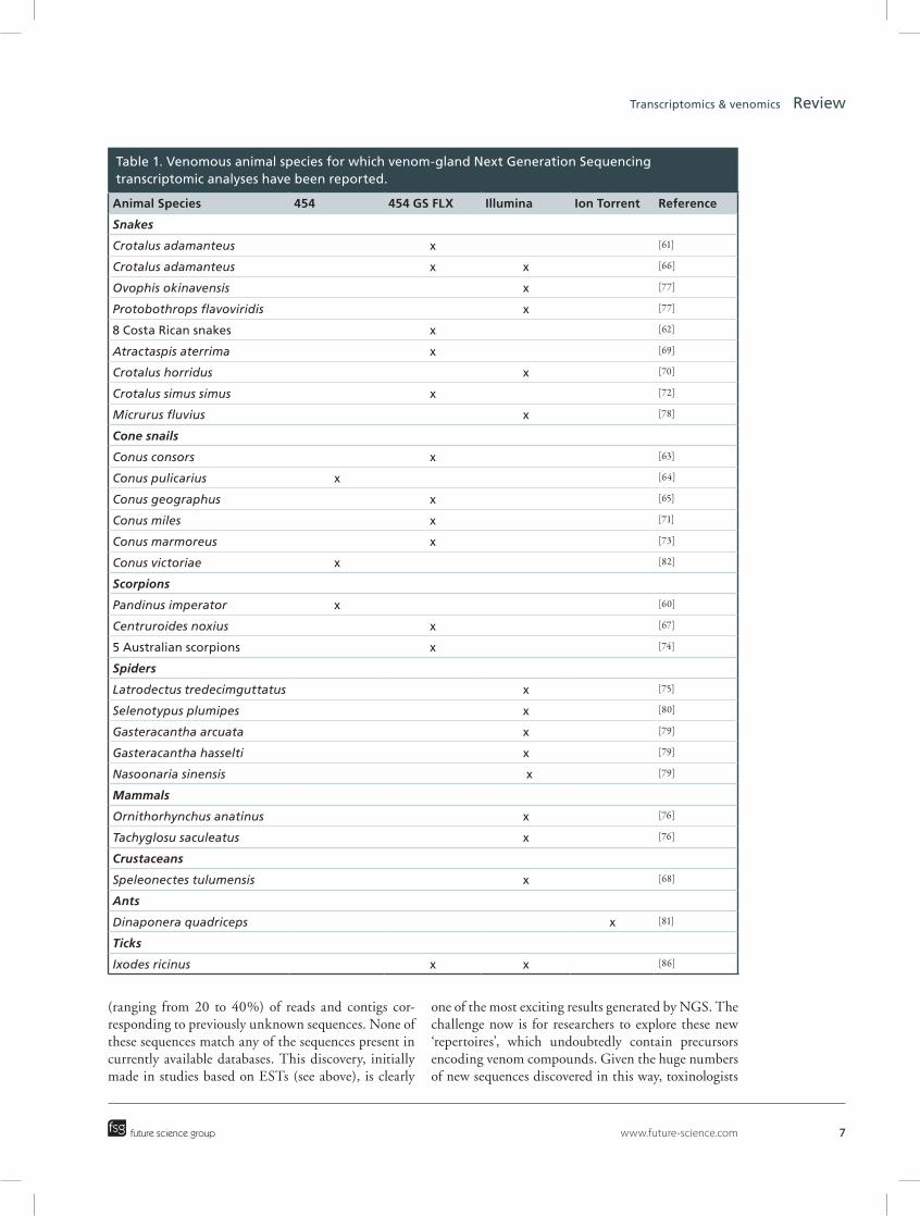
Unlike previously described transcriptomic approaches, NGS techniques deliver a much broader view of the cocktail of compounds present in venom, as demonstrated by the increasing number of recent stud-ies involving these approaches [60–82]. Over and above the compounds present in venom, these techniques can also be used to study the molecular machinery specifi-cally used by the venom-gland cells in the production of the venom arsenal and to elucidate the post-tran-scriptional mechanisms at work. In terms of candidate drug discovery, deep-transcriptomics studies based on NGS are likely to revolutionize this field in terms of the discovery of new isoforms, the identification of toxin-related genes suggesting convergent recruitment by the venom-gland tissues of compounds from diverse taxa [83], the discovery of totally new compounds/scaffolds and the identification of a molecular signature specific to products of the venom-gland cell.
NGS was first used to explore the transcriptomic activity of an animal venom apparatus in 2009. This study concerned the common emperor scorpion, Pan-dinus imperator [60]. Many other venom transcriptomes have since been determined, in snakes, cones snails, spi-ders, two venomous mammals, one venomous crusta-cean and one ant. Most of these studies involved pyro-sequencing with Roche 454 or 454 GS FLX machines, the Illumina GA/HiSeq System, the Illumina system together with 454 technology and, very recently, Ion-Torrent™ technology (see Table 1). As expected, these studies have resulted in deeper gene expression (tran-scriptomic) profiling of venom-gland cells. Research-ers now have access to a very large range of precursor representations, which was not the case with previous technologies. Thanks to these advances, it is now pos-sible to detect and sequence transcripts present at very low abundance (a few copies only). It has also become possible to generate exhaustive inventories of the molecular diversity characterizing families of known toxin peptides. For example, for Conus consors, we were able to show the predominance in the venom of three families of conopeptides: superfamilies A, O and M, with 132, 40 and 28 different isoforms, respectively [63]. Within the same transcriptome, precursors encod-ing conolysins, conantokins, contulakins, conotoxins P, S or T, conodipins and conopressins were detected
even when there were fewer than four copies of the precursor present.
Clearly, NGS technologies provide a more complete picture of the composition of the cocktail of chemicals in venom. They have shown that venom-gland cells synthesize an average of 200–400 different precursors, encoding peptides and proteins that are secreted into the lumen of the venom gland. Levels of transcriptomic activity may vary between studies. Intraspecific and interspecific variations are observed, but gene expres-sion profiles may also differ between specimens of the same defined species of venomous animal.
As pointed out above, venoms appear to have a much broader molecular content than initially suspected, raising questions about the origin of this diversity. Deep transcriptomic analyses, particularly for Conus, have suggested a highly dynamic process of sequence diversification. One interesting study on Conus miles [71] focused on conopeptides and led to the identifica-tion of more than 650 putative conopeptide precursors. Durban et al. [72] studied the process driving venom evolution in the snake Crotalus simus simus, reveal-ing the role played by certain populations of miRNAs (microRNAs) as modulators of the ontogenetic com-position of venoms.
NGS technologies are also used to shed some light on animals not considered truly venomous. Thus, sev-eral tick species produce highly paralytic and lethal cocktails of proteinaceous molecules in their salivary glands [84]. Detrimental effects of tick bites, such as paralysis, allergic reactions and pathogen transmis-sion, have been reported in both animals and humans. Tick saliva has clearly evolved to contain a complex cocktail of components counteracting the effects of the host immune system, including anticoagulants, pros-taglandins, immunosuppressants, antihistamines and prostacyclin and calreticulins [85]. Furthermore, fatal cases of human envenomation have been reported in Australia. A transcriptome analysis was recently car-ried out with a combination of 454 GS-FLX and Illu-mina NGS technologies [86], to improve our under-standing of tick saliva. This study established that the saliva-gland transcriptome of the Australian paralysis tick, Ixodes ricinus, contained housekeeping proteins (∼23%), secreted proteins (∼13%) and unknown compounds (∼60%). The secreted proteins included enzymes, protease inhibitors (basic tail and Kunitz domain families), lipocalins, ixostatins, antimicrobial peptides and 14 other different families of compounds. Together, these results confirm the huge diversity of molecules present in tick saliva, highlighting the importance of studies of this fluid.
Finally, a general feature of these recent NGS transcriptomes is the presence of a large proportion
www.future-science.com 7future science group
Transcriptomics & venomics Review
(ranging from 20 to 40%) of reads and contigs cor-responding to previously unknown sequences. None of these sequences match any of the sequences present in currently available databases. This discovery, initially made in studies based on ESTs (see above), is clearly
one of the most exciting results generated by NGS. The challenge now is for researchers to explore these new ‘repertoires’, which undoubtedly contain precursors encoding venom compounds. Given the huge numbers of new sequences discovered in this way, toxinologists
Table 1. Venomous animal species for which venom-gland Next Generation Sequencing transcriptomic analyses have been reported.
Animal Species 454 454 GS FLX Illumina Ion Torrent Reference
Snakes
Crotalus adamanteus x [61]
Crotalus adamanteus x x [66]
Ovophis okinavensis x [77]
Protobothrops flavoviridis x [77]
8 Costa Rican snakes x [62]
Atractaspis aterrima x [69]
Crotalus horridus x [70]
Crotalus simus simus x [72]
Micrurus fluvius x [78]
Cone snails
Conus consors x [63]
Conus pulicarius x [64]
Conus geographus x [65]
Conus miles x [71]
Conus marmoreus x [73]
Conus victoriae x [82]
Scorpions
Pandinus imperator x [60]
Centruroides noxius x [67]
5 Australian scorpions x [74]
Spiders
Latrodectus tredecimguttatus x [75]
Selenotypus plumipes x [80]
Gasteracantha arcuata x [79]
Gasteracantha hasselti x [79]
Nasoonaria sinensis x [79]
Mammals
Ornithorhynchus anatinus x [76]
Tachyglosu saculeatus x [76]
Crustaceans
Speleonectes tulumensis x [68]
Ants
Dinaponera quadriceps x [81]
Ticks
Ixodes ricinus x x [86]
8 Future Med. Chem. (2014) 6(15) future science group
Review Ducancel, Durban & Verdenaud
and bioinformaticians need to join forces, combining their expertise, knowledge and capacities in the devel-opment of new, efficient exploration algorithms. One possible strategy for identifying the precursors encod-ing venom compounds would involve making use of the molecular and structural features characteristic of venom-compound precursors (see above and Figure 2). This approach would involve searching for and iden-tifying signal peptides, cysteine-rich sequences and propeptide-like sequences. For example, the concomi-tant presence of cysteine residues and a signal sequence within a precursor, with or without N- and/or C-ter-minal putative propeptide sequences, should be con-sidered as strong indications that the precursor encodes a venom peptide or protein. Cross-referencing with proteomics data will be useful, to confirm this predic-tion and to identify the start sites of mature sequences more precisely. On the basis of such results, candidate sequences could then be synthesized chemically or pro-duced by recombinant technology, for assessments of their toxicity, biological activity and structure. This is the overall philosophy followed in the ongoing European project ‘VENOMICS’ presented below.
VENOMICS: an ambitious European project (2011–2015)VENOMICS is a European FP7-Health project dedi-cated to the exploration of biodiversity for public health (http://www.venomics.eu/). It aims to explore animal venom compounds, with a view to the identification and development of novel biotherapeutics.
Bioassay-guided approaches have classically been used for drug discovery in venoms. However, this low-throughput strategy requires large amounts of venom and focuses principally on compounds abun-dant in venom. VENOMICS makes use of an inno-vative ‘Omics’ workflow involving cutting-edge high-throughput transcriptomics, proteomics and peptide production technologies to decipher venom diversity (Figure 3).
The core objective of VENOMICS is to recre-ate in vitro collections of venom peptides that can be used as a resource for high-throughput screening, for the more efficient isolation of novel drug leads. This approach is akin to recreating ‘synthetic venoms’ in the laboratory. VENOMICS aims to reach this goal, by generating venoms and venom-gland biobanks, corre-sponding to 200 venomous animal species; sequenc-ing venom peptides by proteomics and transcriptomics (Illumina technology) approaches, to create a database of 50,000 sequences for mature venom proteins and peptides; high-throughput in vitro chemical synthesis or the recombinant expression of selected peptides, to generate a bank of several thousand peptides; and
pharmacological screening of the peptide bank against selected molecular targets and drug lead generation.
This strategy represents a new paradigm for venom-based drug discovery, differentiating the VENOMICS approach from the classical bioassay-guided process. VENOMICS focuses on disulfide bridge-rich venom peptides no longer than about 100 amino acids long. Candidate peptides of less than 40 amino acids in length are generated by chemical synthesis, whereas larger peptides are produced principally by bacteria, with recombinant technologies.
Venom-derived peptide libraries offer the advantage of containing only highly stable natural bioactive and bio-optimized molecules of high affinity and target subtype selectivity. They therefore have a much greater potential for drug discovery than the randomly gen-erated peptide libraries obtained by phage display or combinatorial chemistry techniques. The VENOM-ICS consortium consists of research laboratories and SMEs from Belgium, Denmark, France, Portugal and Spain.
Transcriptomics & molecular phylogenetic studiesPhylogenetics is used to study or trace the evolution-ary history underlying biological diversity in groups of organisms. It has been said that “Nothing in biology makes sense except in the light of evolution” (Theo-dosius Grygorovych Dobzhansky) [87]. Phylogenetics therefore plays a key role in various areas of biology, including population genetics, ecology and animal behaviour, but also in the clinical and medical con-texts, giving rise to what has been called ‘evolution-ary medicine’ [88]. Phylogeny has been used to deter-mine the origin and spread of a contagious disease from a molecular epidemiology standpoint [89,90] and to understand the adaptive evolution of viral patho-gens, to facilitate vaccine design [91]. However, one of the most interesting fields in which phylogenet-ics is proving increasingly valuable is pharmaceutical research for the identification of new drugs or natural products. Wang et al. [92] used this approach to iden-tify specific peptides from the tammar wallaby that effectively killed multidrug-resistant bacteria. Phylo-genetic analysis could also be used to find genes with a common phylogenetic profile involved in a similar biological pathway or sharing a similar biochemical function. Komatsu et al. [93] used this approach to determine which species of Panax were most closely related to other medicinal species and might therefore have similar medicinal qualities.
In this context, the FP7-Health project VENOM-ICS aims to explore venoms from non-model organ-isms, to identify new therapeutic compounds. As described above, the profiling of those organisms, for

Figure 3. General overview of the ‘Omics-based high-throughput lead generation’ process developed in the European VENOMICS project.
VenomVenom-gland
MS
de novo MSsequencing
Sequencebanks
TranscriptomecDNA libraries
Synthesisexpression
Peptidebanks
Screening Drug leads
mRNAcDNA
www.future-science.com 9future science group
Transcriptomics & venomics Review
which genome sequences are unavailable, has been based on transcriptomic data in particular, adding fuel to the controversy between morphologists and molecular biologists in the field of phylogenetic sys-tematics [94,95], since classical phylogenetic approaches used morphological data to determine taxonomic rela-tionships. Nevertheless, molecular data are becoming a valuable source of information and, although tax-onomy is still based largely on Linnaean principles and morphological characters, information from DNA and proteins has been used to call into ques-tion previous taxonomic classifications based purely on morphological traits.
Studies of the changes in gene expression underlying phenotypic divergence have successfully increased our understanding of transcriptome evolution in several organisms, including mammals [96], fishes [97], mos-quitoes [98], molluscs [99], turtles [100] and plants [101]. In systematics, information of this kind can be used to clarify unexpected evolutionary relationships, such
as those for Colubridae, Viperidae and Elapidae snake families. However, in the context of the VENOM-ICS project, drug discovery could benefit from phy-logenetic studies of the transcriptomes of venomous species [69], because venom toxins probably evolved from proteins with normal physiological function, and comparative phylogenetics might provide clues about disease-related proteins.
Analyses of transcriptome data for non-model organisms for phylogenetic purposes have been driven by the rapid development of sequencing technology. Several phylogenetic surveys [102–106] have been per-formed on ESTs. At this point, it should be pointed out that ESTs obtained from different taxa cannot contain overlapping genes, given the low probabil-ity of finding orthologous sequences in a reduced set of sequences. This makes it more difficult to attain comparative phylogenetics goals.
Since NGS technologies have become easily afford-able due to the drop of per base sequencing costs,

10 Future Med. Chem. (2014) 6(15) future science group
Review Ducancel, Durban & Verdenaud
RNA-Seq [107], i.e. the characterization of the complete set of transcripts by massive parallel sequencing pro-cesses, is becoming a highly valuable tool for exploring the complexity of organisms at the genome-wide scale, giving rise to the field of phylogenomics [108]. This field, as the intersection of evolution and genomics, involves the inference of the phylogenetic history of certain organisms from genome-wide data. Chan et al. [109] coined the term ‘next-generation phylogenomics’ for such large-scale phylogenetics sequencing projects, and Lin et al. [110] recently performed a phylogenomic analysis of subterranean mammals, using four de novo RNA-seq libraries.
Since the first analysis with 454 pyrosequencing methods for phylogenetic purposes performed by Roeding et al. [60], this technology has been applied for phylogeographic purposes by McCormack et al. [111] and by Rokyta et al. [112], in order to describe the positive selection (reflected by nonsynonymous substi-tutions) imposed on most venom proteins in Crota-lus adamanteus. Similarly, Dutertre et al. [113] recently showed that Conus geographus has defence-evoked and predation-evoked venoms, and that the conotoxins found in these two different types of venoms have evolved rapidly under positive Darwinian selection. However, improvements in the read length, cost per Mb sequenced, total throughput and speed of Illu-mina technology have made this platform the technol-ogy of choice and it has recently been used in several phylogenetic studies [110,114–115].
Given NGS transcriptomic data from a non-model organism, it would be interesting to identify the spe-cific features to be taken into account for phylogenetic surveys. Transcriptome sequences generated with high-throughput techniques provide a rich set of characters for phylogenetic studies in eukaryotes. However, the alignment of multiple partial sequences might result in an alignment with large numbers of gaps, potentially compromising the conclusions of any phylogenetic study [116]. It remains unclear to what extent the reli-ability of tree reconstruction is increased by maximiz-ing either the number of taxa or the number of charac-ters studied. McCormack et al. [111] recently concluded that the loci identified from 454 pyrosequencing data could be useful for phylogenetics, population genetics
or phylogeographic purposes, particularly for closely related species. Despite being incomplete, most of the 454 sequences were useful for determining which toxin protein sequence had been identified in snake-venom-gland transcriptomes. According to Wiens [117], miss-ing data are less critical than complete characters tied to other taxa in the tree for resolving phylogenetic relationships, and limited taxonomic sampling could be problematic in the downstream phylogenetic analy-sis. An increase in the number of taxa sampled could, therefore, increase confidence levels for the assignment of certain orthologues.
Several features should also be taken into account when carrying out phylogenetic analyses with tran-scriptome data:
• Downstream bioinformatics analyses of NGS data cannot currently identify potential pseudogene sequences, relics of the evolution that might lead to incorrect conclusions about phylogenetic relationships.
• Gene trees and species trees are not the same [118]. If there were duplications or polymorphic alleles, then phylogenies for genes will not match those for organisms. In such cases, the phylogenetic recon-struction of orthologous sequences could be useful, to generate the species tree.
• Conclusions for nuclear genes may conflict with those for mitochondrial genes. Moreover, it seems that the combined use of mitochondrial and nuclear sequences yields better results, without artifacts for nodes for which mitochondrial and nuclear gene datasets used separately generate con-flicting topologies [119].
Conclusion & future perspective: venom-apparatus transcriptomicsOur understanding of animal venoms is clearly evolv-ing, with the revelation that venoms are complex and diverse bio-optimized cocktails of compounds, mostly peptides and proteins, that we are only just beginning to explore. These changes in our perception are closely linked to technological advances and one of the cur-rent difficulties facing researchers is establishing ways to analyze, interpret and valorize the huge numbers of sequences being generated.
Efficient data analysis will require the use of bio-informatics knowhow and concepts, together with the development from scratch of new strategies and scripts for the exploration of ‘unknown’ fractions in particular. One of the difficulties is identifying, as pre-cisely as possible, the true N-terminus of the mature compounds encoded by the totally new precursor
Defined key terms
Pseudogenes: Pseudogenes are genomic ubiquitous and abundant nonfunctional DNA sequences similar to normal and active genes. They are identified during genome annotation process and contain different type of modifications (mutations/insertions/deletions, etc) that result in their nonfunctionality. It is recognized that some of them play essential role in gene regulation of their parent and functional genes.

www.future-science.com 11future science group
Transcriptomics & venomics Review
sequences emerging from NGS transcriptomic stud-ies. With this goal in mind, the combination of this approach with proteomic analyses of the correspond-ing venoms may facilitate identification, but only for compounds present at sufficiently high concentrations in the venom studied.
Valorization will require high-throughput strategies for the synthesis or production of compounds of inter-est in their native forms. This implies a knowledge of the exact amino-acid sequences of these molecules, the number and location of the disulfide bridges, and the nature and position of post-translational modifica-tions, when present. Again, the combination of these techniques with proteomics appears to be the most appropriate strategy. In the case of a totally new scaf-fold, one key issue is the native fold adopted by the molecule of interest. If present in sufficiently large amounts in the venom, the new compound could be extracted, purified and studied by X-ray diffraction or nuclear magnetic resonance. Compounds present at too low a concentration for this approach could be produced by recombinant technologies, by following a strategy allowing the formation of disulfide bridges in vivo before the initiation of 3D structure studies.
The development of more systematic and auto-mated strategies for screening for biological activity is also an important issue, for the identification of new biotherapeutic hits. In particular, the development of straightforward screening tools compatible with
high-throughput technologies is crucial, to increase the chances of successful exploration. Two principal approaches appear pertinent: concentrating the screen-ing of compound diversity on one target of interest or presenting the libraries of molecules to be tested to libraries of targets. The hits identified in this way then enter a phase of exhaustive exploration of their bio-chemical, functional, structural and biological activi-ties. These studies will, in many instances, require the mutation, engineering of labeling of compounds of interest.
In conclusion, although the techniques for explor-ing the resources provided by venomous animals are maturing, these analyses remain time-consuming and must be combined with complementary approaches. Nevertheless, the exploration of large and natu-rally bio-optimized libraries of compounds, such as those produced by the venom-gland systems, clearly constitutes a major advance in medicinal biochemistry.
Financial & competing interests disclosureThe authors have no relevant affiliations or financial involve-
ment with any organization or entity with a financial interest
in or financial conflict with the subject matter or materials
discussed in the manuscript. This includes employment, con-
sultancies, honoraria, stock ownership or options, expert tes-
timony, grants or patents received or pending, or royalties.
No writing assistance was utilized in the production of this
manuscript.
Executive summary
• Given the existence of several thousand of venomous animal species, the venom resource is huge, opening tremendous perspectives in the fields of fundamental research and development of therapeutics.
• Technological developments such as high-throughput proteomics and transcriptomics result in a more complex molecular vision of venoms that appear to be highly diverse cocktails of peptides and proteins mainly whose composition is susceptible to vary upon different criteria or stimuli.
• Resulting from a long process of molecular evolution and selection, venom peptides and proteins are naturally bio-optimized scaffolds.
• Due their functional activities, reduced sizes, low immunogenicity and their high stability, venom peptides constitute one of the most promising families of compounds for use in the diagnosis and treatment of human diseases.
• Venom peptide drugs are associated to significant developments occurring in the fields of pain, infection and cancer, when venom proteins target mainly the human cardiovascular system.
• The study of the transcriptomic activity of venom-gland cells has started in 1985 with individual cloning of toxin precursors, to reach today global descriptions of the high activity of synthesis of venom-gland cells.
• Expressed sequence tags and then Next Generation Sequencing strategies are responsible for that evolution.• Aside previously known families of venom peptides and proteins, these more global approaches have
revealed new groups of compounds among which several display totally new sequences whose activity and structure are unknown to date! Exploration of these ‘Unknown’ fractions require the development of new bioinformatics tools to tentatively identify precursors encoding venom components.
• VENOMICS is an ambitious international/European project that aims at applying high-throughput technologies to explore the molecular diversity of venoms with the objective of reproducing artificially a part of it for drug-candidate screening.
• Next Generation Sequencing data require new strategies of sequence alignments leading to next-generation phylogenomics.

12 Future Med. Chem. (2014) 6(15) future science group
Review Ducancel, Durban & Verdenaud
ReferencesPapersofspecialnotehavebeenhighlightedas:•ofinterest;••ofconsiderable interest
1 Casewell NR, Wüster W, Freek J et al. Complex cocktails: the evolutionary novelty of venoms. Trends Ecol. Evol. 28(4), 219–229 (2013).
•• Areviewthatillustrateshow,throughthedevelopmentof‘omic’technologies,ourperceptionofthevenomsystemsisevolving.
2 Fry BG, Scheib H, van der Weerd L et al. Evolution of an arsenal. Mol. Cell. Proteomics 7(2), 215–246 (2008).
•• Anillustrationofhowtoxinscaffoldshavebeenrecruitedandevolvedfromendogenousfamiliesofpeptidesorproteins.
3 Abdel-Rahman MA, Omran MA, Bdel-Nabi IM et al. Intraspecific variation in the Egyptian scorpion Scorpio mauruspalmatus venom collected from different biotopes. Toxicon 53, 349–359 (2009).
4 Creer S, Malhotra A, Thorpe RS et al. Genetic and ecological correlates of intra-specific variation in pitviper venom composition detected using matrix-assisted laser desorption time-of-flight mass spectrometry (MALDI-TOF-MS) and isoelectric focusing. J. Mol. Evolution 56, 317–329 (2003).
5 Dutertre S, Biass D, Stöcklin R et al. Dramatic intraspecimen variations within the injected venom of Conus consors: an unsuspected contribution to venom diversity. Toxicon 55, 1453–1462 (2010).
6 Kola I. The state of innovation in drug development. Clin. Pharmacol. Ther. 83(2), 227–230 (2008).
7 Rennert R, Neundorf I, Beck-Sickinger AG. Synthesis and application of peptides as drug carriers. Methods Mol. Biol. 535, 389–403 (2009).
8 Shaji J, Patole V. Protein and peptide drug delivery: oral approaches. Indian J. Pharm. Sci. 70(3), 269–277 (2008).
9 Hayashi MAF, Ducancel F, Konno K. Natural peptides with potential applications in drug development, diagnosis, and/or biotechnology. Int. J. Pept. doi: 10.1155/2012/757838 (2012) (Epub ahead of print).
10 Lewis RJ, Garcia ML. Therapeutic potential of venom peptides. Nat. Rev. Drug Discov. 2, 790–802 (2003).
•• Asurveyofthehugepharmacologydiversityofvenompeptidesandtheirtherapeuticpotentials.
11 Cossins D. From toxins to therapeutics. The Scientist March 19 (2013).
12 King GF. Venoms as a platform for human drugs: translating toxins into therapeutics. Expert Opin. Biol. Ther. 11(11), 1469–1484 (2011).
•• Anillustrationofthegreatinterestofusingselectedtoxinscaffoldstoidentifyanddeveloptherapeutics.
13 Calvete JJ. Snake venomics: from the inventory of toxins to biology. Toxicon 75, 44–62 (2013).
14 Escoubas P, Sollod B, King GF. Venom landscapes: mining the complexicity of spider venoms via a combined cDNA and mass spectrometric approach. Toxicon 47(6), 650–663 (2006).
15 Calvete JJ, Juarez P, Sanz L. Snake venomics. Strategy and applications. J. Mass Spectrom. 42(11), 1405–1414 (2007).
16 Escoubas P, King GF. Venomics as a drug discovery platform. Expert Rev. Proteomics 6(3), 221–224 (2009).
17 Calvete JJ. Venomics, what else? Toxicon 60(4), 427–433 (2012).
18 Prashanth JR, Lewis RJ, Dutertre S. Towards an integrated venomics approach for accelerated conopeptide discovery. Toxicon 60(4), 470–477 (2012).
19 Favreau P, Menin L, Michalet S et al. Mass spectrometry strategies for venom mapping and peptide sequencing from crude venoms: case applications with single arthropod specimen. Toxicon 47, 676–687 (2006).
20 Rodrigues RS, Boldrini-França J, Fonseca FPP et al. Combined snake venomics and venom gland transcriptomic analysis of Bothropoides pauloensis. J. Proteomics 75, 2707–2730 (2012).
21 Margres MJ, McGivern JJ, Wray KP et al. Linking the trascriptome and proteome to characterize the venom of the eastern diamondback rattlesnake (Crotalus adamanteus). J. Proteomics 96, 145–158 (2014).
22 Calvete JJ. Snake venomics: from the inventory of toxins to biology. Toxicon 75, 44–62 (2013).
23 Schuster SC. Next-generation sequencing transforms today’s biology. Nat. Methods 5, 16–18 (2008).
• Astatementofthedifferenttechnologicalevolutionsthatmaketranscriptomicstoenterintoanewage.
24 Tamiya T, Lamouroux A, Julien JF et al. Cloning and sequence analysis of the cDNA encoding a snake neurotoxin precursor. Biochimie 67(2), 185–189 (1985).
25 Guignery-Frelat G, Ducancel F, Ménez A et al. Sequence of a cDNA encoding a snake venom phospholipase A2. Nucleic Acids Res. 15(14), 5892 (1987).
26 Ducancel F. Endothelin-like peptides. Cell Mol. Life Sci. 62(23), 2828–2839 (2005).
27 Murayama N, Hayashi MA, Ohi H et al. Cloning and sequence analysis of a Bothrops jararaca cDNA encoding a precursor of seven bradykinin-potentiating peptides and a C-type natriuretic peptide. Proc. Natl. Acad. Sci. USA 94(4), 1189–1193 (1997).
28 Soares MR, Oliveira-Carvalho AL, Wermelinger LS et al. Identification of novel bradykinin-potentiating peptides and C-type natriuretic peptides from Lachesis muta muta venom. Toxicon 46(1), 31–38 (2005).
29 Kozlov SA, Vassilevski AA, Foefanov AV et al. Latarcins, antimicrobial and cytolytic peptides from the venom of the spider Lachesana tarabaevi (Zodariidae) that exemplify biomolecular diversity. J. Biol. Chem. 281(30), 20983–20992 (2006).
30 Honma T, Hasegawa Y, Ishida M et al. Isolation and molecular cloning of novel peptide toxins from the sea-anemone Antheopsis maculata. Toxicon 45(1), 33–41 (2005).
31 Conticello SG, Gilad Y, Avidan N et al. Mechanisms for evolving hypervariability: the case of conopeptides. Mol. Biol. Evol. 18(2), 120–131 (2001).
www.future-science.com 13future science group
Transcriptomics & venomics Review
32 Yang Y, Cun S, Xie X et al. EST analysis of gene expression in the tentacle of Cyanea capillata. FEBS Lett. 538(12), 183–191 (2003).
33 Kozlov S, Malyavka A, McCutchen B et al. A novel strategy for the identification of toxin-like structures in spider venom. Proteins 59(1), 131–140 (2005).
34 Jiang L, Peng L, Chen J et al. Molecular diversification based on analysis of expressed sequence tags from the venom glands of the Chinese bird spider Ornithoctonus huwena. Toxicon 51(8), 1479–1489 (2008).
35 Jiang L, Liu C, Duan Z et al. Transcriptome analysis of venom glands from a single fishing spider Dolomedes mizhoanus. Toxicon 73, 23–32 (2013).
36 Chen J, Zhao L, Jiang L et al. Transcriptome analysis revealed novel possible venom components and cellular processes of the tarantula Chilobrachys jingzhao venom gland. Toxicon 52(7), 794–806 (2008).
37 Tang X, Zhang Y, Hu W et al. Molecular diversification of peptide toxins from the tarantula Haplopelma hainanum (Ornithoctonus hainana) venom based on transcriptomic, peptidomic and genomic analyses. J. Proteome Res. 9(5), 2550–2564 (2010).
38 McCowan C, Garb JE. Recruitment and diversification of an ecdysozoan family of neuropeptide hormones for the black widow spider venom expression. Gene 536(2), 366–375 (2014).
39 Wagstaff SC, Harrison RA. Venom gland EST analysis of the saw-scaled viper Echis ocellatus, reveals novel alpha9beta1 integrin-binding motifs in venom metalloproteinases and a new group of putative toxins, renin-like aspartic proteases. Gene 377, 21–32 (2006).
40 Junqueira-de-Azevedo IL, Ching AT, Carvalho E et al. Lachesis muta (Viperidae) cDNAs reveal diverging pit viper molecules and scaffolds typical of cobra (Elapidae) venoms: implications for snake toxin repertoire evolution. Genetics 173(2), 877–889 (2006).
41 Cidade DA, Simão TA, Dávilla AM et al. Bothrops jararaca venom gland transcriptome: analysis of the gene expression pattern. Toxicon 48(4), 437–461 (2006).
42 Jia Y, Cantu BA, Sánchez EE et al. Complementary DNA sequencing and identification of mRNAs from the venomous gland of Agkistrodon piscivorus leucostoma. Toxicon 51(8), 1457–1466 (2008).
43 Jiang Y, Li Y, Lee W et al. Venom gland transcriptomes of two elapid snakes (Bungarus multicinctus and Naja atra) and evolution of toxin genes. BMC Genomics 12(1), doi: 10.1186/1471–2164–2112–2111 (2011) (Epub ahead of print).
44 Rodrigues RS, Boldrini-França J, Fonseca FPP et al. Combined snake venomics and venom gland transcriptomic analysis of Bothropoidespauloensis. J. Proteomics 75(9), 2707–2720 (2012).
45 Chatrath ST, Chapeurouge A, Lin Q et al. Identification of novel proteins from the venom of a cryptic snake Drysdalia coronoides by a combined transcriptomics and proteomics approaches. J. Proteome Res. 10(2), 739–750 (2011).
46 Zelanis A, Andrade-Silva D, Rocha MM et al. A transcriptomic view of the proteome variability of newborn
and adult Bothrops jararaca snake venoms. PLoS Negl. Trop. Dis. 6(3), e1554 (2012).
47 Ching ATC, Rocha MMT, PaesLeme AF et al. Some aspects of the venom proteome of the Colubridae snake Philodryas olfersii revealed from a Duvernoy’s (venom) gland transcriptome. FEBS Lett. 580(18), 4417–4422 (2006).
48 Ching ATC, PaesLeme AF, Zelanis A et al. Venomics profiling of Thamnodynastes strigatus unveils matrix metalloproteinases and other novel proteins recruited to toxin arsenal of rear-fanged snakes. J. Proteome Res. 11(2), 1152–1162 (2012).
49 Pi C, Liu J, Peng C et al. Diversity and evolution of conotoxins based on gene expression profiling of Conus litteratus. Genomics 88(6), 809–819 (2006).
50 Pi C, Liu J, Peng C et al. Analysis of expressed sequence tags from the venom ducts of Conus striatus: focusing on the expression profile of conotoxins. Biochimie 88(2), 131–140 (2006).
51 Magalhães GS, Junqueira-de-Azevedo IL, Lopes-Ferreira M et al. Transcriptome analysis of expressed sequence tags from the venom glands of the fish Thalassophryne nattereri. Biochimie 88(6), 693–699 (2006).
52 Baek JH, Lee SH. Identification and characterization of venom proteins of two solitary wasps, Eumenes pomiformis and Orancistrocerus drewseni. Toxicon 56(4), 554–562 (2010).
53 Abdel-Rahman MA, Quintero-Hernandez V, Possani LD. Venom proteomic and venomous glands transcriptomic analysis of the Egyptian scorpion Scorpion maurus palmatus (Arachnida: Scorpionidae). Toxicon 74, 193–207 (2013).
54 Liu ZC, Zhang R, Zhao F et al. Venomic and transcriptomic analysis of centipede Scolopendra subspinipes dehaani. J. Proteome Res. 11(12), 6197–6212 (2012).
55 Bouzid W, Klopp C, Verdenaud M et al. Profiling the venom gland transcriptome of Tetramorium bicarinatum (Hymenoptera: Formicidae): the first transcriptome analysis of an ant species. Toxicon 70, 70–81 (2013).
56 Margulies M, Egholm M, Altman WE et al. Genome sequencing in microfabricated high-density picolitre reactors. Nature 437(15), 376–380.
57 Liu L, Li Y, Li S et al. Comparison of next-generation sequencing systems. J. Biomed. Biotech. doi: 10.1155/2012/251364 (2012) (Epub ahead of print).
58 Mardis ER. Next-Generation Sequencing Platforms. Annu. Rev. Anal. Chem. 6, 287–303 (2013).
59 Sanger F, Nicklen S, Coulson AR. DNA sequencing with chain-terminating inhibitors. Proc. Natl. Acad. Sci. USA 74, 5463–5467 (1977).
60 Roeding F, Borner J, Kube M et al. A 454 sequencing approach for large-scale phylogenomic analysis of the common emperor scorpion (Pandinus imperator). Mol. Phylogen. Evol. 53, 826–834 (2009).
61 Rokyta DR, Wray KP, Lemmon AR et al. A high-troughput venom-gland transcriptome for the Eastern diamondback rattlesnake (Crotalus adamanteus) and evidence for pervasive positive selection across toxin classes. Toxicon 57, 657–671 (2011).

14 Future Med. Chem. (2014) 6(15) future science group
Review Ducancel, Durban & Verdenaud
62 Durban J, Juarez P, Angulo Y et al. Profiling the venom gland transcriptomes of Costa Rican snakes by 454 pyrosequencing. BMC Genomics 12, 259–275 (2011).
63 Terrat Y, Biass D, Dutertre S et al. High-resolution picture of a venom gland transcriptome: case study with the marine snail Conus consors. Toxicon, 59, 34–46 (2012).
64 Lluisma AO, Milash BA, Moore B et al. Novel venom peptides from the cone snail Conus pulicarius discovered through next-generation sequencing of its venom duct transcriptome. Mar. Genomics 5, 43–51 (2012).
65 Hu H, Bandyopadhyay PD, Olivera BM et al. Elucidation of the molecular envenomation strategy of the cone snail Conus geographus through transcriptome sequencing of its venom duct. BMC Genomics 13, 284–296 (2012).
66 Rokyta DR, Lemmon AR, Margres MJ et al. The venom-gland transcriptome of the eastern diamondback rattlesnake (Crotalus adamanteus). BMC Genomics 13, 312 (2012).
67 Rendón-Anaya M, Delaye L, Possani LD et al. Global transcriptome analysis of the scorpion Centruroides noxius: new toxin families and evolutionary insights from an ancestral scorpion species. PLoS ONE 7(8), e43331 (2012).
68 vonReumont BJ, Blanke A, Richter S et al. The first venomous crustacean revealed by transcriptomics and functional morphology: remipede venom glands express a unique toxin cocktail dominated by enzymes and a neurotoxin. Mol. Biol. Evol. 31(1), 48–58 (2014).
69 Terrat Y, Sunagar K, Fry BG et al. Atractaspis aterrima toxins: the first insight into the molecular evolution of venom in side-stabbers. Toxins (Basel) 5(11), 1948–1964. (2013).
70 Rokyta DR, Wray KP, Margres MJ. The genesis of an exceptionally lethal venom in the timber rattlesnake (Crotalus horridus) revealed through comparative venom-gland transcriptomics. BMC Genomics 14, 394 (2013).
71 Jin AH, Dutertre S, Kaas Q et al. Transcriptomic messiness in the venom duct of Conus miles contributes to conotoxin diversity. Mol. Cell. Proteomics 12(12), 3824–3833 (2013).
72 Durban J, Pérez A, Sanz L et al. Integrated ‘omics’ profiling indicates that miRNAs are modulators of the ontogenetic venom composition shift in the Central American rattlesnake, Crotalus simus simus. BMC Genomics 14, 234 (2013).
73 Dutertre S, Jin A, Kaas Q et al. Deep venomics reveals the mechanism for expanded peptide diversity in cone snail venom. Mol. Cell. Proteomics 12(2), 312–329. (2013).
74 Sunagar K, Undheim EAD, Chan AHC et al. Evolution stings: the origin and diversification of scorpion toxin peptides scaffolds. Toxins (Basel) 5(12), 2456–2487. (2013) (Epub ahead of print).
75 He Q, Duan Z, Yu Y et al. The venom gland transcriptome of Latrodectus tredecimguttatus revealed by deep sequencing and cDNA library analysis. PLoS ONE 8(11), e81357 (2013).
76 Wong ESW, Nicol S, Warren WC et al. Echidna venom gland trasncriptome provides insights into the evolution of monotreme venom. PLoS ONE 8(11), e79092 (2013).
77 Aird SD, Watanabe Y, Villar-Briones A et al. Quantitative high-throughput profiling of snake venom gland
transcriptomes and proteomes (Ovophis okinavensis and Protobothrops flavoviridis). BMC Genomics 14, 790 (2013).
78 Margres MJ, Aronow K, Loyacano J et al. The venom-gland transcriptome of the eastern coral snake (Micrurus fulvius) reveals high venom complexity in the intragenomic evolution of venoms. BMC Genomics 14, 531 (2013).
79 Zhao YJ, Zeng Y, Chen L et al. Analysis of transcriptomes of three orb-web spider species reveals gene profiles involved in silk and toxin. Insect Sci. doi: 10.1111/1747917.12068 (2013) (Epub ahead of print).
80 Wong ESW, Hardy MC, Wood D et al. SVM-based prediction of propeptide cleavage sites in the spider toxins identities toxin innovation in an Australian tarantula. PLoS ONE 8(7), e66279 (2013).
81 Torres AF, Huang C, Chong CM et al. Transcriptome analysis in venom gland of the predatory giant ant Dinoponera quadriceps: insights into the polypeptide toxin arsenal of Hymenopterans. PLoS ONE 9(1), e87556 (2014).
82 Robinson SD, Safavi-Hemami H, McIntosh LD et al. Diversity of conotoxin gene superfamilies in the venomous snail, Conus victoria. PLoS ONE 9(2), e87648 (2014).
83 Fry BG, Roelants K, Champagne DE et al. The toxicogenomic multiverse: convergent recruitment of proteins into animal venoms. Annu. Rev. Genomics Hum. Genet. 10, 483–511 (2009).
84 Mans BJ, Louw AI, Neitz AW. Biochemical perspectives on paralysis and other forms of toxicoses causes by ticks. Parasitology 129(Suppl.), S95–S111 (2004).
85 Steen NA, Barker SC, Alewood PF. Proteins in the saliva of the Ixodida (ticks): pharmacological features and biological significance. Toxicon 47, 1–20 (2006).
86 Schwarz A, von Reumont BM, Erhart J et al. De novo Ixodesricinus salivary gland transcriptome analysis using two next-generation sequencing methodologies. FASEB J. 27, 4745–4756 (2013).
87 Dobzhansky T. Nothing in biology makes sense except in the light of evolution. Am. Biol. Teach. 35, 125–129 (1973).
88 Abu-Asab M, Chaouchi M, Amri H. Evolutionary medicine: A meaningful connection between omics, disease, and treatment. Proteomics Clin. Appl. 2, 122–134 (2008).
89 Segovia M, Carrasco HJ, Martinez CE et al. Molecular epidemiologic source tracking of orally transmitted Chagas disease, Venezuela. Emerg. Infect. Dis. 19, 1098–1101 (2013).
90 Holmes EC. Molecular epidemiology and evolution of emerging infectious diseases. Br. Med. Bull. 54, 533–543 (1998).
91 Ojosnegros S, Beerenwinkel N. Models of RNA virus evolution and their roles in vaccine design. Immunome Res. 6, S5 (2010).
92 Wang J, Wong ES, Whitley JC et al. Ancient antimicrobial peptides kill antibiotic-resistant pathogens: Australian mammals provide new options. PLoS ONE 6, e24030 (2011).
93 Komatsu K, Zhu S, Fushimi H et al. Phylogenetic analysis based on 18S rRNA gene and matK gene sequences of Panaxvietnamensis and five related species. Planta Med. 67, 461–465 (2001).

www.future-science.com 15future science group
Transcriptomics & venomics Review
94 Patterson C. In Molecules and Morphology in Evolution: Conflict or Compromise? Colin P (Ed.) Cambridge University Press, NY USA ( 1987).
95 Crowe TM. Molecules vs morphology in phylogenetics: anon-controversy. Trans. R. Soc. South Afr. 46, 317–334 (1988).
96 Brawand D, Soumillon M, Necsulea A et al. The evolution of gene expression levels in mammalian organs. Nature 478, 343–348 (2011).
97 Goetz F, Rosauer D, Sitar S et al. A genetic basis for the phenotypic differentiation between siscowet and lean lake trout (Salvelinus namaycush). Mol. Ecol. 19, 176–196 (2010).
98 Hittinger CT, Johnston M, Tossberg JT et al. Leveraging skewed transcript abundance by RNA-Seq to increase the genomic depth of the tree of life. Proc. Natl. Acad. Sci. USA doi:10.1073/pnas.0910449107 (2010) (Epub ahead of print).
99 Smith SA, Wilson NG, Goetz FE et al. Resolving the evolutionary relationships of molluscs with phylogenomic tools. Nature 480, 364–367 (2011).
100 Chiari Y, Cahais V, Galtier N et al. Phylogenomic analyses support the position of turtles as the sister group of birds and crocodiles (Archosauria). BMC Biol. 10, 65 (2012).
101 Wen J, Xiong Z, Nie ZL et al. Transcriptome sequences resolve deep relationships of the grape family. PLoS ONE 8, e74394 (2013).
102 Bapteste E, Brinkmann H, Lee JA et al. The analysis of 100 genes supports the grouping of three highly divergent amoebae: Dictyostelium, Entamoeba, and Mastigamoeba. Proc. Natl. Acad. Sci. USA 99, 1414–1419 (2002).
103 Hughes J, Longhorn SJ, Papadopoulou A et al. Dense taxonomic EST sampling and its applications for molecular systematics of the Coleoptera (Beetles). Mol. Biol. Evol. 23, 268–278 (2006).
104 Bourlat SJ, Juliusdottir T, Lowe CJ et al. Deuterostome phylogeny reveals monophyletic chordates and the new phylum Xenoturbellida. Nature 444, 85–88 (2006).
105 Roeding F, Hagner-Holler S, Ruhberg H et al. EST sequencing of Onychophora and phylogenomic analysis of Metazoa. Mol. Phylogenet. Evol. 45, 942–951 (2007).
106 Sukkapan P, Jia Y, Nuchprayoon I et al. Phylogenetic analysis of serine proteases from Russell’s viper (Daboia russelli siamensis) and Agkistrodon piscivorus leucostoma venom. Toxicon58, 168–178 (2011).
107 Wang Z, Gerstein M, Snyder M. RNA-Seq: a revolutionary tool for transcriptomics. Nat. Rev. Genet. 10, 57–63 (2009).
108 Delsuc F, Brinkmann H, Philippe H. Phylogenomics and the reconstruction of the tree of life. Nat. Rev. Genet. 6, 361–375 (2005).
109 Chan CX, Ragan MA. Next-generation phylogenomics. Biol. Direct 8(3), 1745–6150 (2013).
•• Anargumentationofhownext-generationsequencingdatarequiresnewstrategiesofsequencealignmentsleadingtonext-generationphylogenomics.
110 Lin GH, Wang K, Deng XG et al. Transcriptome sequencing and phylogenomic resolution within Spalacidae (Rodentia). BMC Genomics 15, 32 (2014).
111 McCormack JE, Maley JM, Hird SM et al. Next-generation sequencing reveals phylogeographic structure and a species tree for recent bird divergences. Mol. Phylogenet. Evol. 62, 397–406 (2012).
112 Rokyta DR, Wray KP, Lemmon AR, Lemmon EM, Caudle SB. A high-throughput venom-gland transcriptome for the eastern diamondback rattlesnake (Crotalus adamanteus) and evidence for pervasive positive selection across toxin classes. Toxicon 57, 657–671 (2011).
113 Dutertre S, Jin AH, Vetter I et al. Evolution of separate predation- and defence-evoked venoms in carnivorous cone snails. Nat. Commun. 5, 3521 (2014)
• Amoleculardemonstrationofhowconesnailsareabletorapidlyswitchbetweendistinctvenomscocktailsinresponsetopredatoryordefensivestimuli.
114 Schwarz A, Cabezas-Cruz A, Kopecky J et al. Understanding the evolutionary structural variability and target specificity of tick salivary Kunitz peptides using next generation transcriptome data. BMC Evol. Biol. 14, 4 (2014).
115 Li P, Deng W, Li T et al. Illumina-xbased de novo transcriptome sequencing and analysis of Amanita exitialis basidiocarps. Gene 532, 63–71 (2013).
116 Hartmann S, Vision TJ. Using ESTs for phylogenomics: Can one accurately infer a phylogenetic tree from a gappy alignment? BMC Evol. Biol. 8, 95 (2008).
117 Wiens JJ. Missing data and the design of phylogenetic analyses. J. Biomed. Inform. 39, 34–42 (2006).
118 Nichols R. Gene trees and species trees are not the same. Trends Ecol. Evol. 16, 358–364 (2001).
119 Caravas J. Phylogenetic utility of mitochondrial and nuclear genes: a case study in the Diptera (true flies). Wayne State Univ. Diss. (2012). http://digitalcommons.wayne.edu/oa_dissertations/4