An Important Chinese Medicinal Formulation - MDPI
18
molecules Article Using HPLC–DAD and GC–MS Analysis Isolation and Identification of Anticandida Compounds from Gui Zhen Cao Herbs (Genus Bidens): An Important Chinese Medicinal Formulation Kulsoom Zahara 1 , Yamin Bibi 1 , Saadia Masood 2 , Sobia Nisa 3 , Abdul Qayyum 4, * , Muhammad Ishaque 1 , Khurram Shahzad 5 , Waseem Ahmed 6 , Zahid Hussain Shah 7 , Hameed Alsamadany 8 , Seung-Hwan Yang 9 and Gyuhwa Chung 9, * Citation: Zahara, K.; Bibi, Y.; Masood, S.; Nisa, S.; Qayyum, A.; Ishaque, M.; Shahzad, K.; Ahmed, W.; Shah, Z.H.; Alsamadany, H.; et al. Using HPLC–DAD and GC–MS Analysis Isolation and Identification of Anticandida Compounds from Gui Zhen Cao Herbs (Genus Bidens): An Important Chinese Medicinal Formulation. Molecules 2021, 26, 5820. https://doi.org/10.3390/ molecules26195820 Academic Editors: Mohamed El-Amir F. Hegazy, Paul W. Pare and Mahmoud A. A. Ibrahim Received: 13 August 2021 Accepted: 21 September 2021 Published: 25 September 2021 Publisher’s Note: MDPI stays neutral with regard to jurisdictional claims in published maps and institutional affil- iations. Copyright: © 2021 by the authors. Licensee MDPI, Basel, Switzerland. This article is an open access article distributed under the terms and conditions of the Creative Commons Attribution (CC BY) license (https:// creativecommons.org/licenses/by/ 4.0/). 1 Department of Botany, PMAS-Arid Agriculture University Rawalpindi, Rawalpindi 46300, Pakistan; [email protected] (K.Z.); [email protected] (Y.B.); [email protected] (M.I.) 2 Department of Statistics & Mathematics, PMAS-Arid Agriculture University Rawalpindi, Rawalpindi 46300, Pakistan; [email protected] 3 Department of Microbiology, The University of Haripur, Haripur 22620, Pakistan; [email protected] 4 Department of Agronomy, The University of Haripur, Haripur 22620, Pakistan 5 Department of Plant Breeding and Genetics, The University of Haripur, Haripur 22620, Pakistan; [email protected] 6 Department of Horticulture, The University of Haripur, Haripur 22620, Pakistan; [email protected] 7 Department of Plant Breeding and Genetics, PMAS Arid Agriculture University, Rawalpindi 46300, Pakistan; [email protected] 8 Department of Biological Sciences, King Abdul Aziz University, Jeddah 21589, Saudi Arabia; [email protected] 9 Department of Biotechnology, Chonnam National University, Gwangju 59626, Korea; [email protected] * Correspondence: [email protected] (A.Q.); [email protected] (G.C.) Abstract: Gui Zhen Cao is an herbal formulation that has been documented in Chinese traditional medicine as a remedy for diarrhea, dysentery, inflammation, and toxicity. The sources of this formulation (Bidens pilosa L., Bidens biternata (Lour.) Merr. & Sherff, Bidens bipinnata L.) are also listed in ethnomedicinal reports all over the world. In this study, all these plants are tested for in vitro anticandida activity. A quantitative evaluation of the phytochemicals in all these plants indicated that their vegetative parts are rich in tannins, saponins, oxalates, cyanogenic glycoside and lipids; moreover, the roots have high percentages of alkaloids, flavonoids, and phenols. The results indicated significant anticandida activity, especially for the hexane extract of B. bipinnata leaves which inhibited C. albicans (42.54%), C. glabrata (46.98%), C. tropicalis (50.89%), C. krusei (40.56%), and C. orthopsilosis (50.24%). The extract was subjected to silica gel chromatography and 220 fractions were obtained. Purification by High Performance Liquid Chromatography with Diode- Array Detection (HPLC–DAD) and Gas Chromatography tandem Mass Spectrometry (GC-MS/MS) analysis led to the identification of two anticandida compounds: dehydroabietic and linoleic acid having an inhibition of 85 and 92%, respectively. Keywords: Gui Zhen Cao; traditional Chinese medicine; linoleic acid; Bidens bipinnata 1. Introduction Since prehistoric times, human beings have used plants as a source of medicine; in fact, they were the only source of medicine until the arrival of iatrochemistry in the 16th century [1]. After that, there was a massive shift from natural to synthetic medicine; today, modern Western medicine is based on synthetic compounds. However, in several parts of Molecules 2021, 26, 5820. https://doi.org/10.3390/molecules26195820 https://www.mdpi.com/journal/molecules
-
Upload
khangminh22 -
Category
Documents
-
view
5 -
download
0
Transcript of An Important Chinese Medicinal Formulation - MDPI

molecules
Article
Using HPLC–DAD and GC–MS Analysis Isolation andIdentification of Anticandida Compounds from Gui ZhenCao Herbs (Genus Bidens): An Important ChineseMedicinal Formulation
Kulsoom Zahara 1, Yamin Bibi 1, Saadia Masood 2, Sobia Nisa 3 , Abdul Qayyum 4,* , Muhammad Ishaque 1,Khurram Shahzad 5 , Waseem Ahmed 6, Zahid Hussain Shah 7, Hameed Alsamadany 8 , Seung-Hwan Yang 9
and Gyuhwa Chung 9,*
�����������������
Citation: Zahara, K.; Bibi, Y.;
Masood, S.; Nisa, S.; Qayyum, A.;
Ishaque, M.; Shahzad, K.; Ahmed, W.;
Shah, Z.H.; Alsamadany, H.; et al.
Using HPLC–DAD and GC–MS
Analysis Isolation and Identification
of Anticandida Compounds from Gui
Zhen Cao Herbs (Genus Bidens): An
Important Chinese Medicinal
Formulation. Molecules 2021, 26, 5820.
https://doi.org/10.3390/
molecules26195820
Academic Editors: Mohamed El-Amir
F. Hegazy, Paul W. Pare and
Mahmoud A. A. Ibrahim
Received: 13 August 2021
Accepted: 21 September 2021
Published: 25 September 2021
Publisher’s Note: MDPI stays neutral
with regard to jurisdictional claims in
published maps and institutional affil-
iations.
Copyright: © 2021 by the authors.
Licensee MDPI, Basel, Switzerland.
This article is an open access article
distributed under the terms and
conditions of the Creative Commons
Attribution (CC BY) license (https://
creativecommons.org/licenses/by/
4.0/).
1 Department of Botany, PMAS-Arid Agriculture University Rawalpindi, Rawalpindi 46300, Pakistan;[email protected] (K.Z.); [email protected] (Y.B.); [email protected] (M.I.)
2 Department of Statistics & Mathematics, PMAS-Arid Agriculture University Rawalpindi,Rawalpindi 46300, Pakistan; [email protected]
3 Department of Microbiology, The University of Haripur, Haripur 22620, Pakistan; [email protected] Department of Agronomy, The University of Haripur, Haripur 22620, Pakistan5 Department of Plant Breeding and Genetics, The University of Haripur, Haripur 22620, Pakistan;
[email protected] Department of Horticulture, The University of Haripur, Haripur 22620, Pakistan;
[email protected] Department of Plant Breeding and Genetics, PMAS Arid Agriculture University, Rawalpindi 46300, Pakistan;
[email protected] Department of Biological Sciences, King Abdul Aziz University, Jeddah 21589, Saudi Arabia;
[email protected] Department of Biotechnology, Chonnam National University, Gwangju 59626, Korea;
[email protected]* Correspondence: [email protected] (A.Q.); [email protected] (G.C.)
Abstract: Gui Zhen Cao is an herbal formulation that has been documented in Chinese traditionalmedicine as a remedy for diarrhea, dysentery, inflammation, and toxicity. The sources of thisformulation (Bidens pilosa L., Bidens biternata (Lour.) Merr. & Sherff, Bidens bipinnata L.) are alsolisted in ethnomedicinal reports all over the world. In this study, all these plants are tested forin vitro anticandida activity. A quantitative evaluation of the phytochemicals in all these plantsindicated that their vegetative parts are rich in tannins, saponins, oxalates, cyanogenic glycosideand lipids; moreover, the roots have high percentages of alkaloids, flavonoids, and phenols. Theresults indicated significant anticandida activity, especially for the hexane extract of B. bipinnataleaves which inhibited C. albicans (42.54%), C. glabrata (46.98%), C. tropicalis (50.89%), C. krusei(40.56%), and C. orthopsilosis (50.24%). The extract was subjected to silica gel chromatography and220 fractions were obtained. Purification by High Performance Liquid Chromatography with Diode-Array Detection (HPLC–DAD) and Gas Chromatography tandem Mass Spectrometry (GC-MS/MS)analysis led to the identification of two anticandida compounds: dehydroabietic and linoleic acidhaving an inhibition of 85 and 92%, respectively.
Keywords: Gui Zhen Cao; traditional Chinese medicine; linoleic acid; Bidens bipinnata
1. Introduction
Since prehistoric times, human beings have used plants as a source of medicine; infact, they were the only source of medicine until the arrival of iatrochemistry in the 16thcentury [1]. After that, there was a massive shift from natural to synthetic medicine; today,modern Western medicine is based on synthetic compounds. However, in several parts of
Molecules 2021, 26, 5820. https://doi.org/10.3390/molecules26195820 https://www.mdpi.com/journal/molecules
Molecules 2021, 26, 5820 2 of 18
world, natural medicinal systems—Ayurveda, Unani Tib and Traditional Chinese Medicine(TCM)—also exist [2].
Gui Zhen Cao is a traditional Chinese herb that is described as having detoxifyingproperties. The Oriental Materia Medica says that it clears blood stagnation and winddampness [3]. In the book Thousand Formulas and Thousand Herbs of Traditional ChineseMedicine, it is reported to remove heat from the gastrointestinal tract and cure diarrhea,dysentery, and stomach ache of the heat type (Table 1). Wind-heat is a type of commoncold in Chinese medical language. Sore throat, feeling warm and/or agitated (whetheror not there is a fever), yellow or green-colored phlegm, and aversion to heat are someindications of wind-heat. It is also accompanied with Heat cramps and Damp Wind, bothresponsible for muscles fatigue.Heat cramps is a painful, brief muscle cramps that occurduring excessive work in a hot environment. Whereas Damp Wind has effects similar tothose of the common cold, with sore limbs, listlessness, nausea, anorexia, and diarrhea andcan cause diseases like arthritis.
Table 1. Evidence of Gui Zhen Cao medicinal potential In Chinese Traditional medicine.
Book Name Targets of Gui Zhen CaoCurative Properties Reference
Oriental Meteria Medica Wind-dampness, dispersing stagnantblood, and invigorating blood. [3]
Handbook of Chinese Medicinal HerbsDysentery, laryngalgia, dysphagia,
vomiting, cardiac spasm andesophageal dilatation.
[4]
Prescriptions Worth A Thousand Gold Blended with pig fat to cure finger cuts. -
Chinese-English Manual ofCommon-Used Herbs
Common cold of the wind-heat type,influenza; clear away heat and toxic
materials: sore throat, appendicitis, snakebite, and centipede bite.
[5]
Thousand Formulas and Thousand Herbsof Traditional Chinese Medicine
Heat from gastro-intestinal tract: fordiarrhea, dysentery and stomach ache of
heat type.[6]
In the Chinese-English Manual of Common-Used Herbs, this formulation is said to curethe common cold, sore throat, appendicitis, centipede bite and snake bite, and preventinfluenza [5]. The sources of Gui Zhen Cao are three herbs: i.e., Bidens pilosa L., Bidensbiternata (Lour.) Merr. & Sherff, Bidens bipinnata L. [5]. In the manual under Gui Zhen Cao,these plants are all widely reported to be used for treating different ailments all around theworld (Table 2).
Various ethnobotanical reports also indicated the use of these plants for skin andvaginal infections [7–10]. Studies also reported that these plants enhanced macrophageactivity against candida infection in mice [11]. The use of Bidens spp. for vaginitis isalso documented in the literature. For example, Borges et al. [12] reported the use of itsfresh juice.
Previously, the anticandida activity of B. pilosa was reported [11,13] but not the effectsof B. bipinnata or B. biternata. Therefore, we conducted a quantitative phytochemicalanalysis of the reproductive and vegetative parts of Gui Zhen Cao herbs to identify theactive anticandidal phytochemicals.
Molecules 2021, 26, 5820 3 of 18
Table 2. Ethnomedicinal uses of Gui Zhen Cao herbs.
Disease Plant Used Mode of Application Reference
Stomach acheB. pilosa Decoction of fresh leaves [14]
B. bipinnata Not stated [15]
DiarrheaB. pilosa Decoction and fresh leaves [16]
B. bipinnata Not stated [17]
Anti-inflammatoryB. pilosa Not stated [18]
B. biternata Poultice of leaf [19]
Dysentery
B. pilosa Decoction of whole plant [20]
B. biternata Not stated [21]
B. bipinnata Not stated [17]
HeadacheB. pilosa Decoction of whole plant [22]
B. biternata Bruised leaves on forehead [23]
ColdsB. pilosa Fresh leaves or decoction of whole plant [24]
B. biternata Decoction of whole plant [25]
Eye InfectionB. pilosa Juice of fresh leaves used as eye and ear drops [26]
B. biternata Same as above [27]
WoundsB. pilosa Crushed herb [28]
B. biternata Leaves rubbed as a hemostatic [29]
Snake biteB. pilosa Pulverized herb [30]
B. biternata Fresh roots paste is given as a drink [19]
Toothache B. biternata Roots are chewed [21]
CoughB. pilosa Decoction of whole plant is taken orally [31]
B. biternata Infusion is given [27]
Stomach ulcers B. pilosa Maceration or juice; taken orally [32]
Tuberculosis B. biternata Decoction or maceration; taken orally [29]
VaginitisB. pilosa Decoction of fresh leaves are applied [12]
B. bipinnata, B. pilosa Not stated [33]
Candidiasis Bidens biternata Essential oil [34]
Skin infectionsB. pilosa Ground leaves [35]
B. bipinnata Not stated [17]
2. Results
The quantitative determination of phytochemicals in B. pilosa, B. biternata and B.bipinnata is presented in Table 3. This study indicated that vegetative parts (leaves, stems,and roots) are higher in tannins, oxalates, cyanogenic glycoside, and lipids; however,alkaloids, flavonoids and phenols were higher in the reproductive parts (flowers andachenes). Alkaloids were the highest in B. biternata (0.499 mg/100 g) compared to theother species. B. biternata was also found to have a higher tannin content (1090 mg/100 g)followed by B. pilosa (1030 mg/100 g. The amount of saponins was the highest in B.bipinnata. A higher phenol content was also observed in vegetative and reproductive partsof all Bidens species B. bipinnata (4.04, 5.98), B. pilosa (3.58, 4.98) and B. biternata (3.14, 4.09).
Molecules 2021, 26, 5820 4 of 18
Table 3. Quantity of Phytochemicals found in Bidens species.
Plant Sample Tannin(mg/100 g)
Alkaloid(%)
Flavonoid(%)
Saponins(%)
Oxalate(%)
CyanogenicGlycoside(mg/100 g)
Phenols(mg/g)
Lipid(%)
Bidens biternataVegetative parts 1090 ± 1.4142 0.499 6.9 3.6 2.21 620 ± 0.836 3.14 ± 0.5468 8.6
Reproductive parts 956 ± 2.8280 0.234 9.2 6.6 2.08 501 ± 1.927 4.09 ± 0.5463 2.8
Bidens bipinnataVegetative parts 930 ± 4.7140 0.231 5.09 7.09 2.97 520 ± 0.275 4.04 ± 1.6750 6.4
Reproductive parts 865 ± 0.9428 0.201 8.2 9.8 1.76 465 ± 1.3568 5.98 ± 0.7979 5.76
Bidens pilosaVegetative parts 1030 ± 0.9436 0.357 6.98 8.32 3.05 500 ± 0.6571 3.58 ± 0.1454 7.2
Reproductive parts 827 ± 0.9428 0.214 7.6 9.8 2.09 487 ± 0.2468 4.98 ± 0.7564 6.54
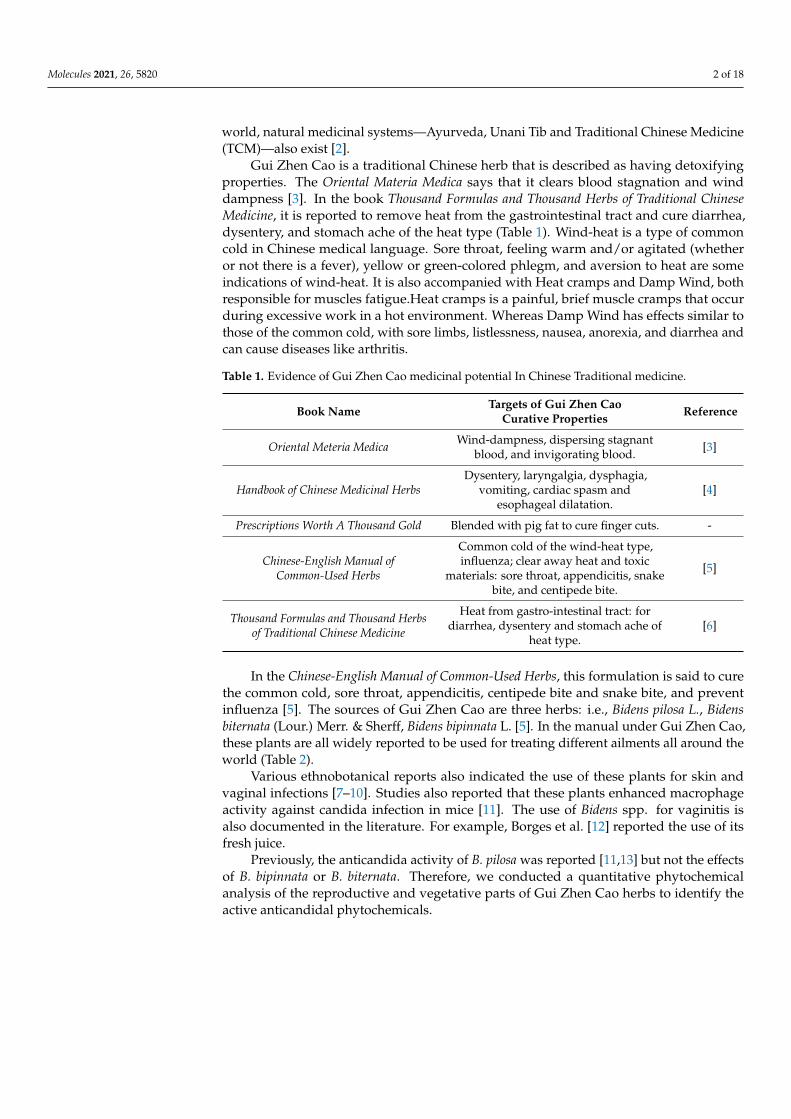
Candidiasis is a type of infection that mostly occurs in the mouth, throat, and vagina.It sometime effect organs like the kidney. In this study, three species of the genus Bidens(B. pilosa, B. bipinnata and B. biternata) were evaluated for their anticandida properties(Figure 1). The hexane extract of B. bipinnata appeared to be the most active with percentageinhibition of 63.09 (C. albicans), 60.68 (C. glabrata), 58.66 (C. tropicalis), 60.67 (C. krusei) and67.40 (C. orthopsilosis) (Figure 2). The hexane extract of different parts of B. bipinnata (leaves,stem, root, flower and achene’s) were again tested for anticandida activity. The leaves’hexane extract appeared to be the most active (Figure 3). Thus, for large scale extraction,the hexane extract of the leaves was selected. From 200 g of leaf powder in 1000 mL hexane,15 g of extract was obtained and run into a silica column. The extract was further separatedinto 220 fractions by silica gel chromatography.
Molecules 2021, 26, x FOR PEER REVIEW 12 of 17
Molecules 2021, 26, x. https://doi.org/10.3390/xxxxx www.mdpi.com/journal/molecules
Plant Sample Tannin (mg/100 g)
Alka-loid (%)
Flavonoid (%)
Saponins (%)
Oxalate (%)
Cyanogenic Glycoside (mg/100 g)
Phenols (mg/g)
Lipid (%)
Bidens biternata
Vegetative parts 1090 ± 1.4142 0.499 6.9 3.6 2.21 620 ± 0.836
3.14 ± 0.5468 8.6
Reproduc-tive parts
956 ± 2.8280 0.234 9.2 6.6 2.08 501 ± 1.927 4.09 ± 0.5463
2.8
Bidens bi-pinnata
Vegetative parts 930 ± 4.7140 0.231 5.09 7.09 2.97 520 ± 0.275 4.04 ±
1.6750 6.4
Reproduc-tive parts
865 ± 0.9428 0.201 8.2 9.8 1.76 465 ± 1.3568 5.98 ± 0.7979
5.76
Bidens pi-losa
Vegetative parts 1030 ± 0.9436 0.357 6.98 8.32 3.05 500 ± 0.6571
3.58 ± 0.1454 7.2
Reproduc-tive parts
827 ± 0.9428 0.214 7.6 9.8 2.09 487 ± 0.2468 4.98 ± 0.7564
6.54
Candidiasis is a type of infection that mostly occurs in the mouth, throat, and vagi-na. It sometime effect organs like the kidney. In this study, three species of the genus Bidens (B. pilosa, B. bipinnata and B. biternata) were evaluated for their anticandida prop-erties (Figure 1). The hexane extract of B. bipinnata appeared to be the most active with percentage inhibition of 63.09 (C. albicans), 60.68 (C. glabrata), 58.66 (C. tropicalis), 60.67 (C. krusei) and 67.40 (C. orthopsilosis) (Figure 2). The hexane extract of different parts of B. bi-pinnata (leaves, stem, root, flower and achene’s) were again tested for anticandida activ-ity. The leaves’ hexane extract appeared to be the most active (Figure 3). Thus, for large scale extraction, the hexane extract of the leaves was selected. From 200 g of leaf powder in 1000 mL hexane, 15 g of extract was obtained and run into a silica column. The extract was further separated into 220 fractions by silica gel chromatography.
Figure 1. Anticandida activity of Bidens bipinnata, Bidens pilosa and Bidens biternata.
0
20
40
60
80
B. biternata B. pilosa B. bipinnata
Perc
enta
ge i
nhib
ition
Tested plant species
C. albicans C. glabrata C. tropicalis C. krusei C. orthopsilosis
Figure 1. Anticandida activity of Bidens bipinnata, Bidens pilosa and Bidens biternata.
Molecules 2021, 26, 5820 5 of 18
Molecules 2021, 26, x FOR PEER REVIEW 13 of 17
Molecules 2021, 26, x. https://doi.org/10.3390/xxxxx www.mdpi.com/journal/molecules
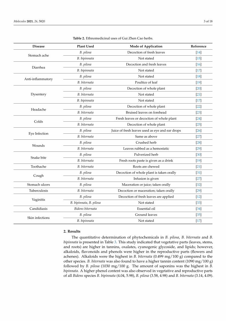
Figure 2. Anticandida activity of different extracts of Bidens bipinnata; positive control fluconazole.
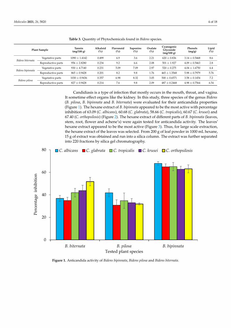
Figure 3. Anticandida activity of Hexane extract of different parts of Bidens bipinnata.
The fractions obtained from the silica column were tested against C. albicans and ac-tivity was observed in fractions 25–29 and 89–92. All active fractions were dried, weighted and again tested for anticandida activity using a serial dilution protocol and persistent activity was observed in fraction 25–29, indicating the presence of active anti-candida compounds (Figure 4). Fraction 27 was further separated using HPLC-DAD Analysis with water: acetonitrile mobile phase (starting at 15% acetonitrile for 5 min and linearly increasing to 100% over 35 min). The chromatogram recorded well-resolved peaks at 254 nm (Figure 5). All 60 one-minute fractions were collected and re-tested for anticandida activity.
0
20
40
60
80
C. albicans C. glabrata C. tropicalis C. krusei C. orthopsilosis
Perc
enta
ge i
nhib
ition
Tested candida strains
ETHANOL ACETONE HEXANE WATER
0
17
34
51
68
Flowers Fruit Root Leaves Stem
Perc
enta
ge i
nhib
ition
Tested plant parts of Bidens bipinnata
C. albicans C. glabrata C. tropicalis C. krusei C. orthopsilosis
Figure 2. Anticandida activity of different extracts of Bidens bipinnata; positive control fluconazole.
Molecules 2021, 26, x FOR PEER REVIEW 13 of 17
Molecules 2021, 26, x. https://doi.org/10.3390/xxxxx www.mdpi.com/journal/molecules
Figure 2. Anticandida activity of different extracts of Bidens bipinnata; positive control fluconazole.
Figure 3. Anticandida activity of Hexane extract of different parts of Bidens bipinnata.
The fractions obtained from the silica column were tested against C. albicans and ac-tivity was observed in fractions 25–29 and 89–92. All active fractions were dried, weighted and again tested for anticandida activity using a serial dilution protocol and persistent activity was observed in fraction 25–29, indicating the presence of active anti-candida compounds (Figure 4). Fraction 27 was further separated using HPLC-DAD Analysis with water: acetonitrile mobile phase (starting at 15% acetonitrile for 5 min and linearly increasing to 100% over 35 min). The chromatogram recorded well-resolved peaks at 254 nm (Figure 5). All 60 one-minute fractions were collected and re-tested for anticandida activity.
0
20
40
60
80
C. albicans C. glabrata C. tropicalis C. krusei C. orthopsilosis
Perc
enta
ge i
nhib
ition
Tested candida strains
ETHANOL ACETONE HEXANE WATER
0
17
34
51
68
Flowers Fruit Root Leaves Stem
Perc
enta
ge i
nhib
ition
Tested plant parts of Bidens bipinnata
C. albicans C. glabrata C. tropicalis C. krusei C. orthopsilosis
Figure 3. Anticandida activity of Hexane extract of different parts of Bidens bipinnata.
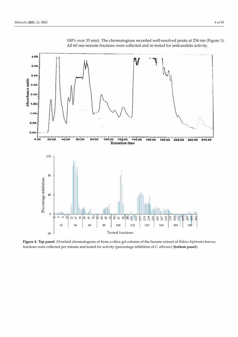
The fractions obtained from the silica column were tested against C. albicans and activ-ity was observed in fractions 25–29 and 89–92. All active fractions were dried, weighted andagain tested for anticandida activity using a serial dilution protocol and persistent activitywas observed in fraction 25–29, indicating the presence of active anticandida compounds(Figure 4). Fraction 27 was further separated using HPLC-DAD Analysis with water:acetonitrile mobile phase (starting at 15% acetonitrile for 5 min and linearly increasing to
Molecules 2021, 26, 5820 6 of 18
100% over 35 min). The chromatogram recorded well-resolved peaks at 254 nm (Figure 5).All 60 one-minute fractions were collected and re-tested for anticandida activity.Molecules 2021, 26, x FOR PEER REVIEW 14 of 17
Molecules 2021, 26, x. https://doi.org/10.3390/xxxxx www.mdpi.com/journal/molecules
Figure 4. Top panel: Overlaid chromatogram of from a silica gel column of the hexane extract of Bidens bipinnata leaves; frac-
tions were collected per minute and tested for activity (percentage inhibition of C. albicans) (bottom panel). Figure 4. Top panel: Overlaid chromatogram of from a silica gel column of the hexane extract of Bidens bipinnata leaves;fractions were collected per minute and tested for activity (percentage inhibition of C. albicans) (bottom panel).

Molecules 2021, 26, 5820 7 of 18
Molecules 2021, 26, x FOR PEER REVIEW 15 of 17
Molecules 2021, 26, x. https://doi.org/10.3390/xxxxx www.mdpi.com/journal/molecules
Figure 5. Top panel: HPLC chromatogram of fraction 49 of silica gel column; fractions were collected per minute and tested for activity (percentage inhibition of C. albicans) (bottom panel).
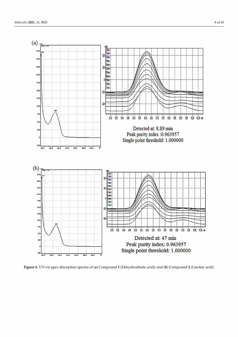
The purity of the active peaks was tested using TLC and purified active peaks were subjected to UV Vis Spectrophotometry and GCMS for identification (Figures 6 and 7). Compound 1 was identified as dehydroabietic acid with a purity and yield of 94.52 and 54.93%, respectively; compound 2 was identified as linoleic acid with a purity and yield of 92.12 and 65.81%, respectively, (Figure 8). The anticandida activity of both compounds was quantified using a microdilution test on C. albicans: linoleic acid had a stronger ac-tivity with inhibition of 92% compared to 85% for dehydroabietic acid.
Figure 5. Top panel: HPLC chromatogram of fraction 49 of silica gel column; fractions were collected per minute and testedfor activity (percentage inhibition of C. albicans) (bottom panel).
The purity of the active peaks was tested using TLC and purified active peaks weresubjected to UV Vis Spectrophotometry and GCMS for identification (Figures 6 and 7).Compound 1 was identified as dehydroabietic acid with a purity and yield of 94.52 and54.93%, respectively; compound 2 was identified as linoleic acid with a purity and yieldof 92.12 and 65.81%, respectively, (Figure 8). The anticandida activity of both compoundswas quantified using a microdilution test on C. albicans: linoleic acid had a stronger activitywith inhibition of 92% compared to 85% for dehydroabietic acid.
Molecules 2021, 26, 5820 8 of 18
Molecules 2021, 26, x FOR PEER REVIEW 16 of 17
Molecules 2021, 26, x. https://doi.org/10.3390/xxxxx www.mdpi.com/journal/molecules
Figure 6. UV-vis apex absorption spectra of (a) Compound 1 (Dehydroabietic acid); and (b) Compound 2 (Linoleic acid). Figure 6. UV-vis apex absorption spectra of (a) Compound 1 (Dehydroabietic acid); and (b) Compound 2 (Linoleic acid).
Molecules 2021, 26, 5820 9 of 18
Molecules 2021, 26, x FOR PEER REVIEW 15 of 17
Molecules 2021, 26, x. https://doi.org/10.3390/xxxxx www.mdpi.com/journal/molecules
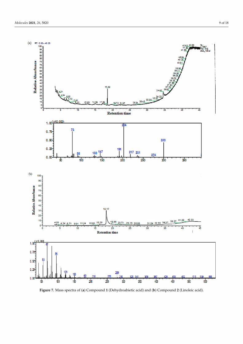
Figure 7. Mass spectra of (a) Compound 1 (Dehydroabietic acid) and (b) Compound 2 (Linoleic acid).Figure 7. Mass spectra of (a) Compound 1 (Dehydroabietic acid) and (b) Compound 2 (Linoleic acid).
Molecules 2021, 26, 5820 10 of 18
Molecules 2021, 26, x FOR PEER REVIEW 18 of 17
Molecules 2021, 26, x. https://doi.org/10.3390/xxxxx www.mdpi.com/journal/molecules
Dehydroabietic Acid Formula: C20H28O2
Molecular Weight: 300.4
Linoleic Acid Formula: C18H32O2
Molecular Weight: 280.4
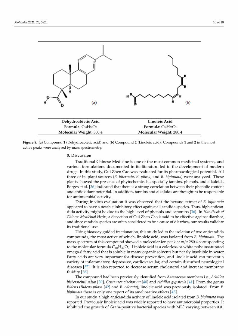
Figure 8. (a) Compound 1 (Dehydroabietic acid) and (b) Compound 2 (Linoleic acid). Compounds 1 and 2 in the most active peaks were analysed by mass spectrometry.
3. Discussion Traditional Chinese Medicine is one of the most common medicinal systems, and
various formulations documented in its literature led to the development of modern drugs. In this study, Gui Zhen Cao was evaluated for its pharmacological potential. All three of its plant sources (B. biternata, B. pilosa, and B. bipinnata) were analyzed. These plants showed the presence of phytochemicals, especially tannins, phenols, and alka-loids. Borges et al. [36] indicated that there is a strong correlation between their phenolic content and antioxidant potential. In addition, tannins and alkaloids are thought to be responsible for antimicrobial activity.
During in vitro evaluation it was observed that the hexane extract of B. bipinnata appeared to have a notable inhibitory effect against all candida species. Thus, high anti-candida activity might be due to the high level of phenols and saponins [36]. In Handbook of Chinese Medicinal Herbs, a decoction of Gui Zhen Cao is said to be effective against di-arrhea, and since candida species are often considered to be a cause of diarrhea, our re-sults validate its traditional use.
Using bioassay guided fractionation, this study led to the isolation of two antican-dida compounds, the most active of which, linoleic acid, was isolated from B. bipinnata. The mass spectrum of this compound showed a molecular ion peak at m/z 280.4 corre-sponding to the molecular formula C18H32O2. Linoleic acid is a colorless or white poly-unsaturated omega-6 fatty acid that is soluble in many organic solvents but nearly in-soluble in water. Fatty acids are very important for disease prevention, and linoleic acid can prevent a variety of inflammatory, depressive, cardiovascular, and certain disturbed neurological diseases [37]. It is also reported to decrease serum cholesterol and increase membrane fluidity [38].
The compound had been previously identified from Asteraceae members i.e., Achil-lea biebersteinii Afan [39], Centaurea vlachorum [40] and Achillea gypsicola [41]. From the genus Bidens (Bidens pilosa [42] and B. odorata), linoleic acid was previously isolated. From B. bipinnata there is only one report of its ameliorative effects [43].
In our study, a high anticandida activity of linoleic acid isolated from B. bipinnata was reported. Previously linoleic acid was widely reported to have antimicrobial prop-erties. It inhibited the growth of Gram-positive bacterial species with MIC varying be-tween 0.01 and 1.0 mg/mL [37]. In the case of fungal strains it successfully inhibited the growth of the pathogenic fungi Rhizoctonia solani, Pythium ultimum, Pyrenophora avenae and Crinipellis perniciosa [44]. Greenway and Dyke [45] reported the inhibitory effect of lino-
Figure 8. (a) Compound 1 (Dehydroabietic acid) and (b) Compound 2 (Linoleic acid). Compounds 1 and 2 in the mostactive peaks were analysed by mass spectrometry.
3. Discussion
Traditional Chinese Medicine is one of the most common medicinal systems, andvarious formulations documented in its literature led to the development of moderndrugs. In this study, Gui Zhen Cao was evaluated for its pharmacological potential. Allthree of its plant sources (B. biternata, B. pilosa, and B. bipinnata) were analyzed. Theseplants showed the presence of phytochemicals, especially tannins, phenols, and alkaloids.Borges et al. [36] indicated that there is a strong correlation between their phenolic contentand antioxidant potential. In addition, tannins and alkaloids are thought to be responsiblefor antimicrobial activity.
During in vitro evaluation it was observed that the hexane extract of B. bipinnataappeared to have a notable inhibitory effect against all candida species. Thus, high antican-dida activity might be due to the high level of phenols and saponins [36]. In Handbook ofChinese Medicinal Herbs, a decoction of Gui Zhen Cao is said to be effective against diarrhea,and since candida species are often considered to be a cause of diarrhea, our results validateits traditional use.
Using bioassay guided fractionation, this study led to the isolation of two anticandidacompounds, the most active of which, linoleic acid, was isolated from B. bipinnata. Themass spectrum of this compound showed a molecular ion peak at m/z 280.4 correspondingto the molecular formula C18H32O2. Linoleic acid is a colorless or white polyunsaturatedomega-6 fatty acid that is soluble in many organic solvents but nearly insoluble in water.Fatty acids are very important for disease prevention, and linoleic acid can prevent avariety of inflammatory, depressive, cardiovascular, and certain disturbed neurologicaldiseases [37]. It is also reported to decrease serum cholesterol and increase membranefluidity [38].
The compound had been previously identified from Asteraceae members i.e., Achilleabiebersteinii Afan [39], Centaurea vlachorum [40] and Achillea gypsicola [41]. From the genusBidens (Bidens pilosa [42] and B. odorata), linoleic acid was previously isolated. From B.bipinnata there is only one report of its ameliorative effects [43].
In our study, a high anticandida activity of linoleic acid isolated from B. bipinnata wasreported. Previously linoleic acid was widely reported to have antimicrobial properties. Itinhibited the growth of Gram-positive bacterial species with MIC varying between 0.01

Molecules 2021, 26, 5820 11 of 18
and 1.0 mg/mL [37]. In the case of fungal strains it successfully inhibited the growth ofthe pathogenic fungi Rhizoctonia solani, Pythium ultimum, Pyrenophora avenae and Crinipellisperniciosa [44]. Greenway and Dyke [45] reported the inhibitory effect of linoleic acid onthe growth of Staphylococcus aureus, probably by increasing the permeability of the bacterialmembrane as a result of its surfactant action. Another study conducted on the effect oflinolenic acid against Helicobacter pylori suggest that linoleic acid caused structural changesin the microbial cell membrane, thereby distressing its integrity and causing the seepage ofcytoplasmic contents [46].
The second-most active compound isolated was dehydroabietic acid with a molecularion peak at m/z 300, corresponding to molecular formula C20H28O2. It is a pyran-2,4-dionesubstituted at position 3 by an acetyl group and at position 6 by a methyl group. It isclassified as a pyrone derivative. Pyrones are class of heterocyclic compounds that containan unsaturated six-membered ring containing a ketone functional group and one oxygenatom. A variety of pyrones were previously isolated from plants: Gentiana pedicellata [47],Hyptis pectinate [48], Piper methysticum [49], Ravensara anisate [50] and Alpinia zerumbet [51].
Dehydroabietic acid was previously isolated from a variety of plant species: Abiesbalsamea (L.) Mill [52], Pinus elliottii [53], Pinus densiflora [54], Nicotiana tabacum and Catha-ranthus roseus [55]. From Asteraceae members, it was identified from Commiphora opobalsa-mum [56], Solidago altissima [57], and Egletes viscosa [58]. However, there was no report ofthis compound from genus Bidens.
Dehydroabietic acid is a chief representative of aromatic abietanes. Similar to diter-penoids, these abietans are typically recognized as chemical defense agents. Antiviral,antileishmanial, antifungal, cytotoxic, antitumor, antiulcer, anti-plasmodial, cardiovascular,antimicrobial, antioxidant and anti-inflammatory properties are the biological activities ofthis group described up to now [59]. Numerous studied have reported the antimicrobialactivity of dehydroabietic acid. Franich et al. [60] noticed antifungal activity of its deriva-tives against Dothistroma pini. Feio et al. [61] confirmed that dehydroabietic acid restrictedthe growth of Trametes versicolor (EC50 = 0.04).
In this study, dehydroabietic acid also revealed substantial anticandida activity: 85%.Previous studies indicated its antimicrobial effect [52,54,62] and suggested that dehydroabi-etic acid may cause a reduction in cell size disrupt cell membranes and cell walls. They alsoindicated that it decreased the proton gradient in microbial cells [55,56]. This occurrence isrelated with the disturbance of proton transport in the membrane-bound ATPase, causinguncoupling of the oxidative phosphorylation [63].
4. Materials and Methods4.1. Standard and Reagents
Solvents (Ethyl acetate, hexane, acetone, methanol, acetonitrile) of High-PerformanceLiquid Chromatography(HPLC)-grade purity from Sigma-Aldrich Co. (St. Louis, MO 63118,United States) deionized sterile water (Milli-Q Reagent Water System, St. Louis, MO, USA),YPD (yeast extract peptone dextrose) agar medium (Lab M Ltd., Lancashire, UK) and DMSOfrom Sigma-Aldrich (St. Louis, MO, United States) were used.
4.2. Collection and Identification of Plants
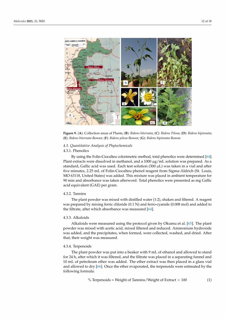
B. pilosa, B. biternata, B. bipinnata, was collected from Azad, Jammu and Kashmir,Pakistan, during spring 2017 to spring 2018 (Figure 9). All parts of these plants (root, stem,leaves, flowers and achene’s) were collected in sterile polyethylene bags labeled with thelocation, name and date of collection. Plant specimens were identified by a taxonomist,and voucher specimens were deposited in the herbarium of Quaid-i-Azam University,Islamabad, Pakistan, for future reference. All parts were thoroughly washed, and diseasedor unwanted plant parts were removed. All plant parts were dried at ambient temperatureand shade dried to maintain their volatile oils, if present. The plants were fully desiccatedto prevent microbial growth and ground into a fine powder, which was kept in in Pearl Petplastic jars.
Molecules 2021, 26, 5820 12 of 18
Molecules 2021, 26, x FOR PEER REVIEW 20 of 17
Molecules 2021, 26, x. https://doi.org/10.3390/xxxxx www.mdpi.com/journal/molecules
Figure 9. A: Collection areas of Plants; B: Bidens biternata; C: Bidens Pilosa; D: Bidens bipinnata; E: Bidens biternata flower; F: Bidens pilosa flower; G: Bidens bipinnata flower.
4.3. Quantitative Analysis of Phytochemicals 4.3.1. Phenolics
By using the Folin-Ciocalteu colorimetric method, total phenolics were determined [64]. Plant extracts were dissolved in methanol, and a 1000 μg/mL solution was prepared. As a standard, Gallic acid was used. Each test solution (300 μL) was taken in a vial and after five minutes, 2.25 mL of Folin-Ciocalteu phenol reagent from Sigma-Aldrich (St. Louis, MO 63118, United States) was added. This mixture was placed in ambient tem-perature for 90 min and absorbance was taken afterword. Total phenolics were presented as mg Gallic acid equivalent (GAE) per gram.
4.3.2. Tannins The plant powder was mixed with distilled water (1:2), shaken and filtered. A rea-
gent was prepared by mixing ferric chloride (0.1 N) and ferro-cyanide (0.008 mol) and added to the filtrate, after which absorbance was measured [64].
4.3.3. Alkaloids Alkaloids were measured using the protocol given by Okumu et al. [65]. The plant
powder was mixed with acetic acid, mixed filtered and reduced. Ammonium hydroxide was added, and the precipitates, when formed, were collected, washed, and dried. After that, their weight was measured.
4.3.4. Terpenoids The plant powder was put into a beaker with 9 mL of ethanol and allowed to stand
for 24 h, after which it was filtered, and the filtrate was placed in a separating funnel and 10 mL of petroleum ether was added. The ether extract was then placed in a glass vial and allowed to dry [66]. Once the ether evaporated, the terpenoids were estimated by the following formula:
% Terpenoids = Weight of Tannins / Weight of Extract × 100 (1)
4.3.5. Saponins Using the protocol reported by Ejikeme et al. [64], the plant powder (5 g) was mixed
with 20% aqueous ethanol (100 cm3) and heated at 55 °C for 4 h in a conical flask in a
Figure 9. (A): Collection areas of Plants; (B): Bidens biternata; (C): Bidens Pilosa; (D): Bidens bipinnata;(E): Bidens biternata flower; (F): Bidens pilosa flower; (G): Bidens bipinnata flower.
4.3. Quantitative Analysis of Phytochemicals4.3.1. Phenolics
By using the Folin-Ciocalteu colorimetric method, total phenolics were determined [64].Plant extracts were dissolved in methanol, and a 1000 µg/mL solution was prepared. As astandard, Gallic acid was used. Each test solution (300 µL) was taken in a vial and afterfive minutes, 2.25 mL of Folin-Ciocalteu phenol reagent from Sigma-Aldrich (St. Louis,MO 63118, United States) was added. This mixture was placed in ambient temperature for90 min and absorbance was taken afterword. Total phenolics were presented as mg Gallicacid equivalent (GAE) per gram.
4.3.2. Tannins
The plant powder was mixed with distilled water (1:2), shaken and filtered. A reagentwas prepared by mixing ferric chloride (0.1 N) and ferro-cyanide (0.008 mol) and added tothe filtrate, after which absorbance was measured [64].
4.3.3. Alkaloids
Alkaloids were measured using the protocol given by Okumu et al. [65]. The plantpowder was mixed with acetic acid, mixed filtered and reduced. Ammonium hydroxidewas added, and the precipitates, when formed, were collected, washed, and dried. Afterthat, their weight was measured.
4.3.4. Terpenoids
The plant powder was put into a beaker with 9 mL of ethanol and allowed to standfor 24 h, after which it was filtered, and the filtrate was placed in a separating funnel and10 mL of petroleum ether was added. The ether extract was then placed in a glass vialand allowed to dry [66]. Once the ether evaporated, the terpenoids were estimated by thefollowing formula:
% Terpenoids = Weight of Tannins/Weight of Extract × 100 (1)
Molecules 2021, 26, 5820 13 of 18
4.3.5. Saponins
Using the protocol reported by Ejikeme et al. [64], the plant powder (5 g) was mixedwith 20% aqueous ethanol (100 cm3) and heated at 55 ◦C for 4 h in a conical flask in a waterbath. The residue was again treated as in the previous step. In a separate funnel, the extractwas again treated with 20 cm3 of diethyl ether and shaken. Once layer formation occurred,the ether layer was discarded, and the aqueous layer was collected. The procedure wasrepeated with of n-butanol (60 cm3) and 5% sodium chloride (10 cm3). The n-butanol layerwas collected and heated in a water bath until dried. The Saponins were calculated as;
% Saponins = Weight of Saponins/Weight of Extract × 100 (2)
4.3.6. Oxalates
Oxalates were determined by using the method proposed by Ejikeme et al. [64]. A2.50 g plant sample was extracted with 0.3 M HCl (20 cm3) three times at 50 ◦C andconstantly stirred for 1 h. The extract was treated with 5 M ammonium hydroxide (1.0 cm3),3 drops glacial acetic acid, 2 drops phenolphthalein, and 5% calcium chloride (1.0 cm3).The solution was allowed to stand for 3 h and was centrifuged for 15 min at 3000 rpm.Precipitates were collected and washed with hot water three times. In the tube, 3 Mtetraoxosulphate acid (2.0 cm3) was added, and precipitates were dissolved by warming ina 70 ◦C water bath. The solution was titrated by 0.01 M potassium permanganate until itturned pink. The solution was left to stand until it again became colorless, after which itwas warmed at 70 ◦C for 3 min and again titrated until the pink colour reappeared andremained for at least 30 s. The oxalates were calculated as;
C2O2−4 + 8H+ + MnO2−
4 = 2CO2 +4H2O + Mn2+ (3)
Ratio of reacting ions = 1:1; From M1 V1 = M2 V2; M1was the molarity of the KMnO4;V1 was the volume of KMnO4; M2 was the molarity of the extract; V2 was volume of theextract; Molecular weight of CaCO3 = 100; Weight of oxalate in titre = M2 × molecularweight = Xg; Weight of oxalate in titrant 2 cm3 = (X/100) × 2 = Y; 100 cm3 of oxalateextract = (Y/2.5) × 100 g = W
% Oxalate composition g/100 g = (W/2.5) × (100/1) (4)
4.3.7. Cyanogenic Glycoside
In dry wood powder (1 g), distilled water (200 cm3) was added and allowed to standfor 2 h. In a conical flask, full distillation was done with 20 cm3 of 2.5% NaOH, and tannicacid was used as an antifoaming agent. Ammonium hydroxide 6 M (8 cm3) tested extract(100 cm3) and 5% potassium iodide was added. The mixture was then titrated with AgNO3(0.02 M). The end point was considered when turbidity occurred [64]. The cyanogenicglycoside was calculated as;
Cyanogenic glycoside (mg/100 g) = (Titrate value (cm3) × 1.08 × exact
volume/Aliquot Volume (cm3) × sample weight (g)) × 100(5)
4.3.8. Percentage Lipids
Plant powder (2.50 g) was added to a soxhlet extractor and connected to a condenser.Petroleum ether was added, and lipids were extracted by heating at 50 ◦C. Petroleum etherwas removed, and the lipids were recovered by cooling in a desiccator, after which theflask was reweighted [64]. The lipid contents were calculated as;
% Lipid = (Weight of Lipid/Weight of Sample) × 100 (6)

Molecules 2021, 26, 5820 14 of 18
4.4. In Vitro Anticandida Activity4.4.1. Fungal Strains
C. albicans, C. glabrata, C. parapsilosis, C. krusei, C. tropicalis, and C. orthopsilosis were used.
4.4.2. Inoculation
A YPD Agar medium (Sigma-Aldrich (St. Louis, MO, United States) as prepared, andcolonies of these strains were inoculated and allowed to grow over night at 37 ◦C. Afterthat, they were stored at 4 ◦C.
4.4.3. Pre-Culture
The YPD medium was prepared, placed in separate tubes, inoculated with a singlecolony of each fungal strain and incubated at 37 ◦C in a shaker incubator Sigma-Aldrich(St. Louis, MO, United States).
4.4.4. Microdilution Broth Protocol
In a 96 well-plate 10 µL of tested plant extract were taken. Ciprofloxacin was taken asa positive control and DMSO and water were taken as a blank control. Each well was theninoculated with 190 µL inoculum (OD = 0.003 at 620 nm). The plates were incubated at37 ◦C for 18 h and read on a Mithras LB 940 Multimode Microplate Reader Sigma-Aldrich(St. Louis, MO, United States) at 620 nm.
4.4.5. Small-Scale Extraction
The fully desiccated raw plant material was ground into a fine powder and small-scaleextractions were performed as described by Panda et al. [67]. One gram of plant powderwas placed into four 15 mL sterile Falcon tubes each with a different solvent: acetone,hexane, ethanol, and water. The tubes were placed in a sonicator bath for 1 h after every 4 hinterval. From each extract, 1 mL aliquots were dried in a Savant Speed Vac Concentrator200 H. The dried filtrate was re-dissolved in 200 mL DMSO for the organic solvent extracts,and in 200 mL water for the aqueous extract. All samples were stored at 4 ◦C until furthertesting [67].
4.4.6. Large-Scale Extraction
For large scale extraction, the dried raw botanical material (200 g of powder) was trans-ferred into a large container with 2000 mL of HPLC-grade n-hexane from Sigma–Aldrich(Hamburg, Germany). The container was left standing for 24 h at ambient temperature.During this time, the container was placed in a water bath sonicator 4 times for 60 mineach to maximize the extraction yield. After each sonication, there was an interval of atleast 6 h to let the suspension cool to ambient temperature. After the fourth sonication, thematerial was filtered using a VWR® Grade 313 with 5 µm filter paper (from Sigma–Aldrich(Hamburg, Germany) to obtain a dry residue. The filtrate was then evaporated in a rotaryevaporator (BUCHI rotavapor R-100, Sigma-Aldrich (St. Louis, MO 63118, United States)and the weight of the first dried extract was measured. The recovered hexane was againused for the second extraction using same procedure for 24 h, after which the filtrate wasagain evaporated with a rotary evaporator to obtain dried extract. The same procedurewas repeated to derive all the compounds from the plant powder. The final weight of thedried material was calculated and the plant extract was stored at 4 ◦C for further analysis.
The plant powder was dried again so it could be treated with a polar solvent to derivea polar compound with the same procedure.
4.4.7. Fractionation Procedure
The dried active extract was adsorbed onto silica gel (70–230 mesh) and loaded on asilica gel column (600 mm height × 55 mm diameter) Sigma–Aldrich (Hamburg, Germany)which was eluted (Waters Gradient Chromatography Calculator, model 600, Sigma–Aldrich(Hamburg, Germany) with a step gradient of hexane–dichloromethane that increased in

Molecules 2021, 26, 5820 15 of 18
polarity: 9.5:0.5, 9:1, 8.5:0.5, 8:2, 7:3; 6:4, 5:5, 4:6, 3:7, 2:8, 1:9, and 0:10. The column was theneluted with 100% dichloromethane, and then 100% ethyl acetate, followed by a mixture ofethyl acetate and methanol (9.5:0.5, 9:1, 8:2, 7:3, 6:4, 5:5, 4:6, 3:7, 2:8, and 1:9). Finally, thecolumn was eluted with 100% methanol. In each step, 10 tubes of 40 mL fractions werecollected, and the solvent evaporated. The whole separation was monitored by a Dual λmodel 2487 absorbance detector (Waters Milford, MA, USA) at 280 nm and 254 nm.
4.4.8. Thin Layer Chromatography
Aliquots (5 µL) of the 245 fractions from the ethyl acetate extract were spotted on largeTLC glass plates (Sigma-Aldrich, Hamburg Germany, dimensions 20 cm × 20 cm). Thespotted plates were developed in glass jars (20 cm × 10 cm × 20 cm), pre-saturated withthe chosen mobile phase at ambient temperature and dried in an oven (St. Louis, MO 63118,United States) at 90 ◦C for 5 min to remove the solvent. The plates were observed underultra-violet (UV) light (Sigma-Aldrich, Hamburg Germany) at 254 and 360 nm, followedby spraying with 5% sulfuric acid in ethanol, followed by heating at 100 ◦C for 5 min.Fractions with similar TLC patterns were pooled for biological activity testing. In total,22 pooled fractions were prepared and further tested for antimicrobial activity against C.albicans at a concentration of 1000 µg/mL.
4.4.9. HPLC-DAD Analysis
HPLC analyses were performed on a Shimadzu, LC-20AT system (model DGU 20A3,SHIMADZU corporation, Kyoto, Japan) equipped with LC-20AT quaternary pump, aDGU20A3/DGU-20A5 on-line degasser, an SPD-20A photodiode array detector, and aCBM-20A/20A interface. The data were acquired and processed using Lab Solution soft-ware. The active fraction was analyzed using a reverse-phase HPLC column SHIMADZUcorporation, Kyoto, Japan), SunfireTM prep C18 (10 mm × 250 mm, 5 µm) column (Waters,Ireland). The mobile phase was composed of 30% H2O with 0.1% TFA (ACROS ORGAN-ICS) and 70% acetonitrile (LC-MS CHROMASOLVR, Fluka) with 0.1% TFA. Prior to HPLCinjection, samples were filtered through a CHROMAFIL R Xtra H-PTFI filter (pore size0.45 µm, filter 13 mm, (MACHEREY-NAGEL, Düren, Germany). Two mL of sample wasinjected and run for 41 min at 20 ◦C with a flow rate of 4 mL/min [68].
4.4.10. Mass Spectrometry GC-MS
Collected peaks were analysed on a gas chromatograph (Thermo–Fisher ScientificTrace 1300 series, (Thermo Finnigan LLC, San Jose, CA, United States) coupled with a massspectrometer (Thermo–Fisher Scientific ISQ series MS, Thermo Finnigan LLC, San Jose, CA,United States, The column used was a Restek RXi-5sil MS 20 m with an internal diameterof 0.18 mm and a film thickness of 0.18 µm. Helium was used as a carrier gas at a constantflow rate of 0.9 mL/min. The initial temperature of 40 ◦C was held for 2 min, then increasedto 120 ◦C at a rate of 20 ◦C/min, to 200 ◦C at a rate of 10 ◦C/min, to 250 ◦C at a rate of7 ◦C/min, and finally to 350 ◦C at a rate of 5 ◦C/min, which was held for 4 min. Thespectrum corresponding to the largest peak in the chromatogram was searched against theNIST 14 MS library for identification. In addition, the identified compound was verifiedfor its retention index, which was calculated using an external C7 to C40 alkane ladder.
5. Conclusions
In conclusion, the hexane extracts of B. bipinnata leaves showed anticandida activitydue to the presence of linoleic acid and dehydroabietic acid. This supports the traditionaluse of Gui Zhen Cao. It is suggested that isolated compounds should be studied againstdifferent pure culture cell lines to identify their potential in vivo toxicity and also tounderstand their mode of action. In addition the activity of linoleic and dehydroabieticacids, with different combinations of compounds, must be studied to suggest possiblesynergistic effects.
Molecules 2021, 26, 5820 16 of 18
Author Contributions: Y.B. conceived of the idea. K.Z., M.I. and S.N. conducted the experimentand conducted the literature review. A.Q., W.A. and S.M. provided technical expertise. S.M., K.S.and Z.H.S. helped in statistical analysis. H.A., S.-H.Y. and G.C. proofread and provided intellectualguidance. All authors have read and agreed to the published version of the manuscript.
Funding: This work was carried out with the support of “Cooperative Research Program for Agri-culture Science and Technology Development (Project No. PJ01581201)” Rural Development Admin-istration, Republic of Korea.
Institutional Review Board Statement: Not applicable.
Informed Consent Statement: Not applicable.
Data Availability Statement: The data presented in this study are available upon fair request fromthe corresponding author.
Conflicts of Interest: The authors declare no conflict of interest.
Sample Availability: Not available.
References1. Romano, B.; Lucariello, G.; Capasso, R. Topical Collection “Pharmacology of Medicinal Plants”. Biomolecules 2021, 14, 101.
[CrossRef]2. Sharma, A.; Khanna, S.; Kaur, G.; Singh, I. Medicinal plants and their components for wound healing applications. Future J.
Pharm. Sci. 2021, 7, 53. [CrossRef]3. Hsu, H.; Chen, Y.; Hsu, C.; Shen, S.; Chen, C.; Chang, H. Oriental Materia Medica: A Concise Guide; Keats Publishing, Inc.:
New Canaan, CT, USA, 1986.4. Smith, F.; Stuart, G. Chinese Medicinal Herbs; Georgetown Press: San Francisco, CA, USA, 1973.5. To-Ming, L.A. Analysis on a class of Banach algebras with applications to harmonic analysis on locally compact groups and
semigroups. J. Math. Fundam. Sci. 1983, 118, 161–175. [CrossRef]6. Bingshan, H.; Yuxia, W. Thousand Formulas and Thousand Herbs of Traditional Chinese Medicine; Heilongjiang Education Press:
Harbin, China, 1993; Volume 1.7. Pinto, E.D.P.P.; Amorozo, M.C.D.M.; Furlan, A. Conhecimento popular sobre plantas medicinais em comunidades rurais de mata
atlântica-Itacaré, BA, Brasil. Acta Bot. Bras. 2006, 20, 751–762. [CrossRef]8. Agra, M.D.F.; Silva, K.N.; Basílio, I.J.L.D.; Freitas, P.F.D.; Barbosa-Filho, J.M. Survey of medicinal plants used in the region
Northeast of Brazil. Rev. Bras. Farmacogn. 2008, 18, 472–508. [CrossRef]9. Feijó, E.V.R.S.; Pereira, A.S.; Souza, L.R.; Silva, L.A.M.; Costa, L.C.B. Levantamento preliminar sobre plantas medicinais utilizadas
no bairro Salobrinho no município de Ilhéus, Bahia. Rev. Bras. Plantas Med. 2013, 15, 595–604. [CrossRef]10. Sanoussi, F.; Ahissou, H.; Dansi, M.; Hounkonnou, B.; Agre, P.; Dansi, A. Ethnobotanical investigation of three traditional leafy
vegetables [Alternanthera sessilis (L.) DC. Bidens pilosa L. Launaea taraxacifolia Willd.] widely consumed in southern and centralBenin. J. Biodivers. Environ. Sci. 2015, 6, 187–198.
11. Chung, C.Y.; Yang, W.C.; Liang, C.L.; Liu, H.Y.; Lai, S.K.; Chang, C.L. Cytopiloyne, a polyacetylenic glucoside from Bidens pilosa,acts as a novel anticandidal agent via regulation of macrophages. J. Ethnopharmacol. 2016, 184, 72–80. [CrossRef]
12. Borges, C.C.; Matos, T.F.; Moreira, J.; Rossato, A.E.; Zanette, V.C.; Amaral, P.A. Bidens pilosa L. (Asteraceae): Traditional use in acommunity of southern Brazil. Rev. Bras. Plantas Med. 2013, 15, 34–40. [CrossRef]
13. Linhares, M.V.; da Silva, R.O.; de Oliveira, F.F.; Costa, L.C.B.; Conceição, A.O.; de Oliveira, R.A. Avaliation anti-Candida ofessential oils from three medicinal plants species (Astereaceae). S. Afr. J. Bot. 2018, 115, 132–137. [CrossRef]
14. Oliveira, F.; Andrade-Neto, V.; Krettli, A.; Brandão, M. New evidences of antimalarial activity of Bidens pilosa roots extractcorrelated with polyacetylene and flavonoids. J. Ethnopharmacol. 2004, 93, 39–42. [CrossRef]
15. Ge, C. Cytologic study of Bidens bipinnata L. China J. Chin. Mater. Med. 1990, 15, 72–74.16. Tan, P.V.; Dimo, T.; Dongo, E. Effects of methanol, cyclohexane and methylene chloride extracts of Bidens pilosa on various gastric
ulcer models in rats. J. Ethnopharmacol. 2000, 73, 415–421. [CrossRef]17. Kil, J.S.; Son, Y.; Cheong, Y.K.; Kim, N.H.; Jeong, H.J.; Kwon, J.W.; Pae, H.O. Okanin, a chalcone found in the genus Bidens, and
3-penten-2-one inhibit inducible nitric oxide synthase expression via heme oxygenase-1 induction in RAW264. 7 macrophagesactivated with lipopolysaccharide. J. Clin. Biochem. Nutr. 2012, 50, 53–58. [CrossRef]
18. Dimo, T.; Azay, J.; Tan, P.V. Effects of the aqueous and methylene chloride extracts of Bidens pilosa leaf on fructose-hypertensiverats. J. Ethnopharmacol. 2001, 76, 215–221. [CrossRef]
19. Surywanshi, V.; Yadava, R. Isolation and characterization of new potential allelochemical from Bidens biternata (Lour.) Merrill &Sherff. J. Chem. Pharm. Res. 2015, 7, 175–179.
20. Geissberger, P.; Séquin, U. Constituents of Bidens pilosa L.: Do the components found so far explain the use of this plant intraditional medicine? Acta Trop. 1991, 48, 251–261. [CrossRef]
Molecules 2021, 26, 5820 17 of 18
21. Swapna, T.; Nair, A.; Mini, I.; Pradeesh, S. Free-radical scavenging activity of leaves of Bidens Biternata (Lour.) Merr. & Sherif.J. Pharm. Res. Dev. 2014, 6, 127–135.
22. Kumari, P.; Misra, K.; Sisodia, B.S.; Faridi, U.; Srivastava, S.; Luqman, S.; Singh, S.C. A promising anticancer and antimalarialcomponent from the leaves of Bidens pilosa. Planta Med. 2009, 75, 59. [CrossRef] [PubMed]
23. Sukumaran, P.; Nair, A.G.; Chinmayee, D.M.; Mini, I.; Sukumaran, S.T. Phytochemical investigation of Bidens biternata (Lour.)Merr. and Sheriff.—A nutrient-rich leafy vegetable from Western Ghats of India. Appl. Biochem. Biotechnol. 2012, 167, 1795–1801.[CrossRef] [PubMed]
24. Kviecinski, M.R.; Felipe, K.B.; Schoenfelder, T.; de Lemos Wiese, L.P.; Rossi, M.H.; Gonçalez, E.; Pedrosa, R.C. Study of the antitu-mor potential of Bidens pilosa (Asteraceae) used in Brazilian olk medicine. J. Ethnopharmacol. 2008, 117, 69–75. [CrossRef] [PubMed]
25. Sharma, A.; Bargali, K.; Pande, N. The allelopathic potential of bryophyte extract on seed germination and seedling growth ofBidens biternata. Nat. Sci. 2009, 7, 30–38.
26. Ashafa, A.; Afolayan, A. Screening the root extracts from Biden pilosa L. var. radiata (Asteraceae) for antimicrobial potentials.J. Med. Plant. Res. 2009, 3, 568–572.
27. Zahara, K.; Bibi, Y.; Tabassum, S.; Bashir, T.; Haider, S.; Araa, A.; Ajmal, M. A review on pharmacological properties of Bidensbiternata: A potential nutraceutical. Asian Pac. J. Trop. Dis. 2015, 5, 595–599. [CrossRef]
28. Chang, M.H.; Wang, G.J.; Kuo, Y.H.; Lee, C.K. The low polar constituents from Bidens pilosa L. var. minor (Van Sam, Baas, KEßLER,& Plants) Sherff. J. Chin. Chem. Soc. 2000, 47, 1131–1136.
29. Zahara, K.; Bibi, Y.; Qayyum, A.; Nisa, S. Investigation of Antimicrobial and Antioxidant Properties of Bidens biternata. Iran. J.Sci. Technol. Trans. A Sci. 2019, 43, 725–734. [CrossRef]
30. Sarg, T.; Ateya, A.; Farrag, N.; Abbas, F. Constituents and biological activity of Bidens pilosa L. grown in Egypt. Acta Pharm. Hung.1991, 61, 317–323.
31. Wang, R.; Wu, Q.X.; Shi, Y.P. Polyacetylenes and flavonoids from the aerial parts of Bidens pilosa. Planta Med. 2010, 76, 893–896.[CrossRef]
32. Hoffmann, B.; Hölzl, J. Chalcone glucosides from Bidens pilosa. Phytochemistr 2009, 28, 247–249. [CrossRef]33. Buhner, S.H. Herbal Antibiotics, Natural Alternatives for Treating Drug-Resistant Bacteria; Storey Publishing: North Adams, MA, USA;
pp. 127–140.34. Bseiso, E.A.; Nasr, M.; Sammour, O.; Gawad, N.A. Recent advances in topical formulation carriers of antifungal agents. Indian J.
Dermatol. Venereol. Leprol. 2015, 8, 457–463. [CrossRef]35. Arthur, G.D.; Naidoo, K.K.; Coopoosamy., R.M. Bidens pilosa L.: Agricultural and pharmaceutical importance. J. Med. Plant. Res.
2012, 6, 3282–3287. [CrossRef]36. Borges, A.; José, H.; Homem, V.; Simões, M. Comparison of Techniques and Solvents on the Antimicrobial and Antioxidant
Potential of Extracts from Acacia dealbata and Olea europaea. Antibiotics 2020, 9, 48. [CrossRef] [PubMed]37. Dilika, F.; Bremner, P.D.; Meyer, J.J.M. Antibacterial activity of linoleic and oleic acids isolated from Helichrysum pedunculatum:
A plant used during circumcision rites. Fitoterapia 2000, 71, 450–452. [CrossRef]38. Freitas, H.R.; Isaac, A.R.; Malcher-Lopes, R.; Diaz, B.L.; Trevenzoli, I.H.; de Melo Reis, R.A. Polyunsaturated fatty acids and
endocannabinoids in health and disease. Nutr. Neurosci. 2018, 21, 695–714. [CrossRef] [PubMed]39. Kazemi, M. Chemical composition and antimicrobial, antioxidant activities and anti-inflammatory potential of Achillea millefolium
L., Anethum graveolens L., and Carum copticum L. essential oils. J. Herb. Med. 2015, 5, 217–222. [CrossRef]40. Hodaj, E.; Tsiftsoglou, O.; Abazi, S.; Hadjipavlou-Litina, D.; Lazari, D. Lignans and indole alkaloids from the seeds of Centaurea
vlachorum Hartvig (Asteraceae), growing wild in Albania and their biological activity. Nat. Prod. Res. 2017, 31, 1195–1200.[CrossRef] [PubMed]
41. Kordali, S.; Cakir, A.; Akcin, T.A.; Mete, E.; Akcin, A.; Aydin, T.; Kilic, H. Antifungal and herbicidal properties of essential oilsand n-hexane extracts of Achillea gypsicola Hub-Mor. and Achillea biebersteinii Afan. (Asteraceae). Ind. Crop. Prod. 2009, 29,562–570. [CrossRef]
42. Deba, F.; Xuan, T.D.; Yasuda, M.; Tawata, S. Chemical composition and antioxidant, antibacterial and antifungal activities of theessential oils from Bidens pilosa Linn. var. Radiata. Food Control 2009, 19, 346–352. [CrossRef]
43. Wang, Y.-Q.; Li, S.J.; Man, Y.H.; Zhuang, G. Serum metabonomics coupled with HPLC-LTQ/orbitrap MS and multivariate dataanalysis on the ameliorative effects of Bidens bipinnata L. in hyperlipidemic rats. J. Ethnopharmacol. 2020, 262, 113196. [CrossRef]
44. Walters, D.; Raynor, L.; Mitchell, A.; Walker, R.; Walker, K. Antifungal activities of four fatty acids against plant pathogenic fungi.Mycopathologia 2004, 157, 87–90.
45. Greenway, D.L.A.; Dyke, K.G.H. Mechanism of the inhibitory action of linoleic acid on the growth of Staphylococcus aureus.Microbiology 1979, 115, 233–245. [CrossRef] [PubMed]
46. Jung, S.W.; Thamphiwatana, S.; Zhang, L.; Obonyo, M. Mechanism of antibacterial activity of liposomal linolenic acid againstHelicobacter pylori. PLoS ONE 2015, 10, e0116519. [CrossRef] [PubMed]
47. Ghosal, S.; Singh, A.A.K.; Biswas, K.J.P.M. New 6-aryl-2-pyrones from Gentiana pedicellata. Planta Med. 1983, 49, 240–243.[CrossRef] [PubMed]
48. Pereda-Miranda, R.; Hernández, L.; Villavicencio, M.J.; Novelo, M.; Ibarra, P.; Chai, H.; Pezzuto, J.M. Structure and stereochemistryof pectinolides AC, novel antimicrobial and cytotoxic 5, 6-dihydro-α-pyrones from Hyptis pectinate. J. Nat. Prod. 1993, 56,583–593. [CrossRef]
Molecules 2021, 26, 5820 18 of 18
49. Young, R.L.; Hylin, J.W.; Plucknett, D.L.; Kawano, Y.; Nakayama, R.T.J.P. Analysis for kawa pyrones in extracts of Pipermethysticum. Phytochemistry 1996, 5, 795–798. [CrossRef]
50. Andrianaivoravelona, J.O.; Sahpaz, S.; Terreaux, C.; Hostettmann, K.; Stoeckli-Evans, H.; Rasolondramanitra, J. Two 6-substituted5, 6-dihydro-α-pyrones from Ravensara anisata. Phytochemistry 1999, 52, 265–269. [CrossRef]
51. Mpalantinos, M.A.; Soares de Moura, R.; Parente, J.P.; Kuster, R.M. Biologically active flavonoids and kava pyrones from theaqueous extract of Alpinia zerumbet. Phytother. Res. 1998, 12, 442–444.
52. Nachar, A.; Saleem, A.; Arnason, J.T.; Haddad, P.S. Regulation of liver cell glucose homeostasis by dehydroabietic acid, abietic acidand squalene isolated from balsam fir (Abies balsamea (L.) Mill.) a plant of the Eastern James Bay Cree traditional pharmacopeia.Phytochemistry 2015, 117, 373–379. [CrossRef]
53. Leandro, L.F.; Cardoso, M.J.O.; Silva, S.D.C.; Souza, M.G.M.; Veneziani, R.C.S.; Ambrosio, S.R.; Martins, C.H.G. Antibacterialactivity of Pinus elliottii and its major compound, dehydroabietic acid, against multidrug-resistant strains. J. Med. Microbiol. 2014,63, 1649–1653. [CrossRef]
54. Park, J.; Kim, W.J.; Kim, W.; Park, C.; Choi, C.Y.; Cho, J.H.; Cheong, H. Antihypertensive Effects of Dehydroabietic and4-Epi-Trans-Communic Acid Isolated from Pinus densiflora. J. Med. Food 2021, 24, 50–58. [CrossRef]
55. Häkkinen, S.T.; Lackman, P.; Nygrén, H.; Oksman-Caldentey, K.M.; Maaheimo, H.; Rischer, H. Differential patterns of de-hydroabietic acid biotrans, formation by Nicotiana tabacum and Catharanthus roseus cells. J. Biotechnol. 2012, 157, 287–294.[CrossRef]
56. Gao, W.; Dong, X.; Xie, N.; Zhou, C.; Fan, Y.; Chen, G.; Zhu, D. Dehydroabietic acid isolated from Commiphora opobalsamumcauses endothelium-dependent relaxation of pulmonary artery via PI3K/Akt-eNOS signaling pathway. Molecules 2014, 19,8503–8517. [CrossRef]
57. Johnson, R.H.; Halitschke, R.; Kessler, A. Simultaneous analysis of tissue-and genotype-specific variation in Solidago altissima(Asteraceae) rhizome terpenoids, and the polyacetylene dehydromatricaria ester. Chemoecology 2010, 20, 255–264. [CrossRef]
58. Calou, I.B.F.; Sousa, D.I.M.; de Andrade Cunha, G.M.; de Castro Brito, G.A.; Silveira, E.R.; Rao, V.S.; Santos, F.A. Topically appliedditerpenoids from Egletes viscosa (Asteraceae) attenuate the dermal inflammation in mouse ear induced by tetradecanoylphorbol13-acetate-and oxazolone. Biol. Pharm. Bull. 2008, 31, 1511–1516. [CrossRef]
59. González, M.A. Aromatic abietane diterpenoids: Their biological activity and synthesis. Nat. Prod. Rep. 2015, 32, 684–704.[CrossRef] [PubMed]
60. Franich, R.A.; Gadgil, P.D.; Shain, L. Fungistatic effects of Pinus radiata needle epicuticular fatty and resin acids on Dothistromapini. Physiol. Plant. Pathol. 1983, 23, 183–195. [CrossRef]
61. Savluchinske-Feio, S.; Nunes, L.; Pereira, P.T.; Silva, A.M.; Roseiro, J.C.; Gigante, B.; Curto, M.J.M. Activity of dehydroabietic acidderivatives against wood contaminant fungi. J. Microbiol. Methods 2007, 70, 465–470. [CrossRef] [PubMed]
62. Savluchinske-Feio, S.; Curto, M.J.M.; Gigante, B.; Roseiro, J.C. Antimicrobial activity of resin acid derivatives. Appl. Microbiol.Biotechnol. 2006, 72, 430–436. [CrossRef] [PubMed]
63. Tapia, A.A.; Vallejo, M.D.; Gouiric, S.C.; Feresin, G.E.; Rossomando, P.C.; Bustos, D.A. Hydroxylation of dehydroabietic acid byFusarium species. Phytochemistry 1997, 46, 131–133. [CrossRef]
64. Ejikeme, C.; Ezeonu, C.S.; Eboatu, A.N. Determination of Physical and Phytochemical Constituents of some Tropical TimbersIndigenous to nigerdelta area of nigeria. Eur. Sci. J. 2014, 10, 247–270.
65. Okumu, M.O. Prophylactic Efficacy of Moringa Oleifera Leaf Extracts Against Liver Injury Induced by Artesunate-amodiaquineAntimalarial Combination. Ph.D. Thesis, University of Nairobi, Nairobi, Kenya, 2016.
66. Ezekaibeya, A.C.; Nnenna, A.O.; Kenechukwu, O.C. Proximate, phytochemical and vitamin compositions of Cucumis metuliferus(Horned Melon) rind. J. Complement. Altern. Med. Res. 2020, 9, 40–50. [CrossRef]
67. Panda, S.K.; Padhi, L.; Leyssen, P.; Liu, M.; Neyts, J.; Luyten, W. Antimicrobial, anthelmintic, and antiviral activity of plantstraditionally used for treating infectious disease in the Similipal Biosphere Reserve, Odisha, India. Front. Pharmacol. 2017, 8, 658.[CrossRef] [PubMed]
68. Kerkoub, N.; Panda, S.K.; Yang, M.R.; Lu, J.G.; Jiang, Z.H.; Nasri, H.; Luyten, W. Bioassay-guided isolation of anti-Candidabiofilm compounds from methanol extracts of the aerial parts of Salvia officinalis (Annaba, Algeria). Front. Pharmacol. 2018,9, 1418. [CrossRef] [PubMed]