Organ-specific expression of IGF-I during early development of bony fish as revealed in the tilapia,...
15
Cell Tissue Res (2006) 325: 287–301 DOI 10.1007/s00441-005-0133-9 REGULAR ARTICLE Giorgi Berishvili . Natallia Shved . Elisabeth Eppler . Frederic Clota . Jean-François Baroiller . Manfred Reinecke Organ-specific expression of IGF-I during early development of bony fish as revealed in the tilapia, Oreochromis niloticus, by in situ hybridization and immunohistochemistry: indication for the particular importance of local IGF-I Received: 8 September 2005 / Accepted: 23 November 2005 / Published online: 5 April 2006 # Springer-Verlag 2006 Abstract The cellular sites of insulin-like growth factor I (IGF-I) synthesis in the early developing tilapia (0-140 days post fertilization, DPF) were investigated. IGF-I mRNA and peptide appeared in liver as early as 4 DPF and in gastro-intestinal epithelial cells between 5-9 DPF. In exocrine pancreas, the expression of IGF-I started at 4 DPF and continued until 90 DPF. IGF-I production was detected in islets at 6 DPF in non-insulin cells and occurred throughout life. In renal tubules and ducts, IGF-I produc- tion started at 8 DPF. IGF-I production in chondrocytes had its onset at 4 DPF, was more pronounced in growing regions and was also found in adults. IGF-I mRNA and peptide appeared in the cytoplasm of skeletal muscle cells at 4 DPF. In gill chloride cells, IGF-I production started at 6 DPF. At 13 DPF, IGF-I was detected in cardiac myocytes. IGF-I-producing epidermal cells appeared at 5 DPF. In brain and ganglia, IGF-I was expressed in virtually all neurones from 6 to 29 DPF, their number decreasing with age. Neurosecretory IGF-I-immunoreactive axons were first seen in the neurohypophysis around 17 DPF. Endo- crine cells of the adenohypophysis exhibited IGF-I mRNA at 28 DPF and IGF-I immunoreactivity at 40 DPF. Thus, IGF-I appeared early (4-5 DPF), first in liver, the main source of endocrine IGF-I, and then in organs involved in growth or metabolism. The expression of IGF-I was more pronounced during development than in juvenile and adult life. Local IGF-I therefore seems to have a high functional impact in early growth, metabolism and organogenesis. Keywords Liver . Gastro-intestinal tract . Pancreas . Kidney . Muscle . Brain . Pituitary . Gills . Heart . Epidermis . Tilapia Introduction Autocrine, paracrine and endocrine signals co-ordinate the direction of differentiation of tissues during critical periods of development. The differentiation of organs thus involves a complex cascade and interaction of signals whose organizational action on tissue differentiation is dependent on being released at precise times, at correct sites (in the case of para- and autocrine factors) and within a specific dose range (Segner et al. 1994). Insulin-like growth factor I (IGF-I) plays a central role in the complex system that regulates growth, differentiation and reproduction (see review: Reinecke and Collet 1998). As in mammals, the major site of IGF-I gene expression in bony fish is liver but several extrahepatic sites also express IGF-I (for reviews, see Reinecke and Collet 1998; Duan 1998; Reinecke et al. 2005; Wood et al. 2005). Recently, by the use of real-time reverse transcription/ polymerase chain reaction (RT-PCR), the absolute amounts of IGF-I have been measured in adult tilapia, Oreochromis niloticus (Caelers et al. 2004), and significant levels of IGF-I mRNA have been measured not only in liver, but also in other organs, such as brain, gills, heart, intestine, kidney, skeletal muscle, spleen and testes. These results are consistent with the earlier immunohistochemical identifi- cation of parenchymal cells as local production sites in numerous organs of tilapia (Reinecke et al. 1997) and indicate paracrine/autocrine actions of local IGF-I involved in organ-specific functions of adult bony fish. Some studies in bony fish showing the presence of IGF-I and its receptor (IGF-1R) in fish embryos suggest a high This study was supported by the SNF (NRP 50, project 4050- 66580). F. Clota . J.-F. Baroiller CIRAD, UPR20 Aquaculture, Montpellier, France G. Berishvili . N. Shved . E. Eppler . M. Reinecke (*) Division of Neuroendocrinology, Institute of Anatomy, University of Zürich, Zürich, Switzerland e-mail: [email protected] Tel.: +41-44-6355370 Fax: +41-44-6355702
-
Upload
independent -
Category
Documents
-
view
0 -
download
0
Transcript of Organ-specific expression of IGF-I during early development of bony fish as revealed in the tilapia,...
Cell Tissue Res (2006) 325: 287–301DOI 10.1007/s00441-005-0133-9
REGULAR ARTICLE
Giorgi Berishvili . Natallia Shved . Elisabeth Eppler . Frederic Clota .Jean-François Baroiller . Manfred Reinecke
Organ-specific expression of IGF-I during early developmentof bony fish as revealed in the tilapia, Oreochromis niloticus,by in situ hybridization and immunohistochemistry: indicationfor the particular importance of local IGF-IReceived: 8 September 2005 / Accepted: 23 November 2005 / Published online: 5 April 2006# Springer-Verlag 2006
Abstract The cellular sites of insulin-like growth factor I(IGF-I) synthesis in the early developing tilapia (0-140days post fertilization, DPF) were investigated. IGF-ImRNA and peptide appeared in liver as early as 4 DPF andin gastro-intestinal epithelial cells between 5-9 DPF. Inexocrine pancreas, the expression of IGF-I started at 4 DPFand continued until 90 DPF. IGF-I production was detectedin islets at 6 DPF in non-insulin cells and occurredthroughout life. In renal tubules and ducts, IGF-I produc-tion started at 8 DPF. IGF-I production in chondrocytes hadits onset at 4 DPF, was more pronounced in growingregions and was also found in adults. IGF-I mRNA andpeptide appeared in the cytoplasm of skeletal muscle cellsat 4 DPF. In gill chloride cells, IGF-I production started at 6DPF. At 13 DPF, IGF-I was detected in cardiac myocytes.IGF-I-producing epidermal cells appeared at 5 DPF. Inbrain and ganglia, IGF-I was expressed in virtually allneurones from 6 to 29 DPF, their number decreasing withage. Neurosecretory IGF-I-immunoreactive axons werefirst seen in the neurohypophysis around 17 DPF. Endo-crine cells of the adenohypophysis exhibited IGF-I mRNAat 28 DPF and IGF-I immunoreactivity at 40 DPF. Thus,IGF-I appeared early (4-5 DPF), first in liver, the mainsource of endocrine IGF-I, and then in organs involved ingrowth or metabolism. The expression of IGF-I was more
pronounced during development than in juvenile and adultlife. Local IGF-I therefore seems to have a high functionalimpact in early growth, metabolism and organogenesis.
Keywords Liver . Gastro-intestinal tract . Pancreas .Kidney . Muscle . Brain . Pituitary . Gills . Heart .Epidermis . Tilapia
Introduction
Autocrine, paracrine and endocrine signals co-ordinate thedirection of differentiation of tissues during critical periodsof development. The differentiation of organs thus involvesa complex cascade and interaction of signals whoseorganizational action on tissue differentiation is dependenton being released at precise times, at correct sites (in thecase of para- and autocrine factors) and within a specificdose range (Segner et al. 1994). Insulin-like growth factor I(IGF-I) plays a central role in the complex system thatregulates growth, differentiation and reproduction (seereview: Reinecke and Collet 1998).
As in mammals, the major site of IGF-I gene expressionin bony fish is liver but several extrahepatic sites alsoexpress IGF-I (for reviews, see Reinecke and Collet 1998;Duan 1998; Reinecke et al. 2005; Wood et al. 2005).Recently, by the use of real-time reverse transcription/polymerase chain reaction (RT-PCR), the absolute amountsof IGF-I have been measured in adult tilapia, Oreochromisniloticus (Caelers et al. 2004), and significant levels ofIGF-I mRNA have been measured not only in liver, butalso in other organs, such as brain, gills, heart, intestine,kidney, skeletal muscle, spleen and testes. These results areconsistent with the earlier immunohistochemical identifi-cation of parenchymal cells as local production sites innumerous organs of tilapia (Reinecke et al. 1997) andindicate paracrine/autocrine actions of local IGF-I involvedin organ-specific functions of adult bony fish.
Some studies in bony fish showing the presence of IGF-Iand its receptor (IGF-1R) in fish embryos suggest a high
This study was supported by the SNF (NRP 50, project 4050-66580).
F. Clota . J.-F. BaroillerCIRAD, UPR20 Aquaculture,Montpellier, France
G. Berishvili . N. Shved . E. Eppler . M. Reinecke (*)Division of Neuroendocrinology, Institute of Anatomy,University of Zürich,Zürich, Switzerlande-mail: [email protected].: +41-44-6355370Fax: +41-44-6355702

organizational impact of IGF-I during ontogeny. Develop-mental and tissue-specific regulation of the steady-statemRNA levels of IGF-1R and polyadenylation have beendetected in rainbow trout (Greene and Chen 1999) andseabream (Perrot et al. 1999). Correspondingly, some RT-PCR studies on trout (Shamblott and Chen 1993; Duan etal. 1995; Greene and Chen 1997, 1999), seabream (Duguayet al. 1996; Perrot et al. 1999) and rabbitfish (Ayson etal. 2002) suggest that the expression patterns of IGF-ImRNA in several organs are also age-dependent. Thephysiological importance of IGF-I in early fish develop-ment is further underlined by the demonstration of themarked mitogenic effects of IGF-I on zebrafish embryoniccells (Pozios et al. 2001).
However, little information exists regarding the cellularsites of IGF-I synthesis, with only two studies in seabream(Perrot et al. 1999) and rabbitfish (Radaelli et al. 2003) andsome preliminary data in tilapia (Shved et al. 2005)reporting the localization of IGF-I during ontogeny. Thesereports involve the investigation of several organs atdifferent larval stages by the use of immunohistochemistry.However, no thorough study on the cellular IGF-Iproduction during early fish development has beenperformed and no other species have been examined.Furthermore, to date, in situ hybridization has not beenused to detect the cellular expression of IGF-I mRNAduring ontogeny. Therefore, we have investigated thepotential production sites of IGF-I in numerous organs,such as liver, gastro-intestinal tract, exo- and endocrinepancreas, kidney, skeletal muscle, cartilage, gills, skin,brain and pituitary, in early developmental stages, i.e. from0 day post fertilization (DPF) to 140 DPF, of tilapia(Oreochromis niloticus) and in adult individuals. We haveused immunohistochemistry to localize IGF-I peptide andin situ hybridization to detect the expression of IGF-ImRNA.
Materials and methods
Animals
The fry and adult Oreochromis niloticus used in this studyoriginated from the Aquaculture Experimental Facilities ofCIRAD (Montpellier). Fertilized eggs were obtainedthrough either natural or artificial fertilization. For naturalfertilization, 0. niloticus breeders (1 male and 3 females)were maintained in a spawning aquarium of 360 lmaintained at constant temperature (27°C) and photoperiod(12L:12D). Under natural photoperiods, reproductive ac-tivity occurs mainly from the afternoon until sunset in thesespecies (Baroiller et al. 1997). Consequently, we checkedevery morning for the presence of newly incubatingfemales, which were then isolated from the other fish. Onthe first day after fertilization, eggs were gently removedfrom the mouth of the female and incubated in 1-lMcDonald jars at 27±1°C (mean range). For artificialfertilization, the maturation of isolated females wascarefully recorded (i.e. uro-genital papilla development
and behaviour). When the near-breeding stage wasreached, both male and female breeders were stripedunder anaesthesia. Fertilization was performed accordingto Chourrout and Itskovitch (1983). After fertilization, eggswere incubated in 1-l McDonald jars, maintained at 27°C,to the free-swimming stage (around 10 DPF). Just beforethe completion of yolk sac absorption, fry were placed intoa 50-l tank in an indoor recirculating system at 27±1°C.They were fed with commercial salmonid food.
Fish larvae at various developmental stages, i.e. from0-140 DPF, and adults were sampled and anaesthetizedby the addition of 2-phenoxy-ethanol (Sigma, St. Louis,Mo., USA) to water (0.3 ml/l). For the earliest stages, thetrunk was cut to allow fixation solution to enter. If thelarvae had reached a sufficient size, the organs wereexcised. Tissue preparations were fixed with Bouin’ssolution without acetic acid for 4 h at room temperature.Specimens were dehydrated in an ascending series ofethanol and routinely embedded in Paraplast (58°C).
Generation of tilapia-specific probes for in situhybridization
The probes used were prepared as previously described(Schmid et al. 1999). In brief, total RNA from tilapia liverwas extracted by the phenol/chloroform method with anUltraspec Extraction Kit (ams, Lugano, Switzerland). ForcDNA synthesis, 5 μg RNAwere annealed with 1 μM poly(dT) primer (5′-CC TGAATTCTAGAGCTCAT(dT17)-3′)for 3 min at 70°C. The RNA/primer mix was incubated for1 h at 37°C with 15 mM dNTPs and 10 U AMV-reversetranscriptase (Pharmacia, Switzerland) in 1× reactionbuffer (50 mM TRIS-HCl pH 8.3, 40 mM KCl, 6 mMMgCl2). A 1-μl cDNA aliquot was incubated with 1 μM ofthe sense (5′-GTCTGTGGAGAGCGAGGCTTT-3′) andantisense primer (5′-AACCTTGGGTGCTCTTGGCATG-3′), corresponding to the B- and E- domains (Reinecke etal. 1997), 200 μM dNTPs, and 1 U Taq-polymerase(Pharmacia) in 1× incubation buffer (10 mM TRIS-HCl pH8, 50 mM KCl, 1.5 mM MgCl2, 0.001% gelatine). Theamplification program was optimized for a StratageneRoboCycler Gradient 40 as follows: one cycle of 10 min at94°C, 1 min at 59°C, 2 min at 72°C; 30 cycles of 1 min at94°C, 1 min at 59°C and 2 min at 72°C followed by a finalextension step of 5 min at 72°C. PCR fragments wereseparated on a 2% agarose gel and eluted by Gel ExtractionKit QIAquick (Qiagen, Switzerland). PCR products werecloned in a pCR-Script SK(+) cloning vector (Stratagene,Heidelberg, Germany). After propagation of Escherichiacoli containing the plasmids, purification of the plasmidswas performed with the Midi Purification Kit (Qiagen).Plasmids with the IGF-I fragment were sequenced (Micro-synth, Switzerland) and the sequences were compared withthose in a database. The plasmids containing the specificinserts of IGF-I (207 bp) were used as templates for thesynthesis of the digoxigenin (DIG)-labelled RNA probes.Linearization was performed with the restriction enzymeEcoRI for T3 polymerase-driven transcription and with
288

NotI for T7 polymerase-driven transcription. After ethanolprecipitation, linearization efficiency was assured on a1.2% agarose gel. The linearized plasmids (1 μg) weretranscribed in vitro by using a transcription kit (RocheDiagnostics, Germany) in the presence of DIG-UTP toobtain the antisense and sense probes. The IGF-I probesselectively detected tilapia IGF-I and not tilapia IGF-II, asshown previously (Schmid et al. 1999). The integrity of theprobes and the efficiency of the labelling were confirmedby gel electrophoresis, including blotting and incubationwith antibody (Ab), and by dot blot. For dot blot, 1 μlvarious dilutions (undiluted, 1:10, 1:100, 1:1,000) ofprobes and control RNAs were dropped onto a nylonmembrane (Roche Diagnostics) and fixed by UV cross-linking (254 nm, 125 mJ). The membrane was washed with1% blocking reagent (Roche-Diagnostics) in buffer P1 for30 min followed by treatment with alkaline phosphatase(AP)-coupled anti-DIG Ab (Roche Diagnostics) diluted at1:2,000 with 1% blocking reagent in P1 for 30 min. After arinse with P1, the membrane was equilibrated with bufferP3 and finally washed with 1:50 diluted nitroblue tetrazo-lium/5-bromo-4-chloro-3-indolyl-phosphate (NBT/BCIP)stock solution (Roche Diagnostics) until colour develop-
ment. For gel electrophoresis and blotting, probes (200 ng)were loaded onto a 1% denaturing formaldehyde agarosegel in 1× MOPS and run at 80 V for 1.5 h. After the run, geland membrane were shaken in diethylpyrocarbonate(DEPC)-H20 for 5 min. Capillary transfer was carried outwith 20× standard sodium citrate (SSC) overnight. Nucleicacids were fixed by UV-crosslinking. Subsequently, themembrane was incubated with the AP-labelled anti-DIGAb by applying the same procedure as for dot blot. Lengthsof the probes were compared with control RNA (760 nt,Roche Diagnostics).
In situ hybridization protocol
Sections (4 μm thick) were mounted on Super Frost Plusslides (Menzel-Gläser, Germany) and dried overnight at42°C. After being dewaxed and rehydrated, the sectionswere fixed with 4% paraformaldehyde and 0.1% glutaral-dehyde in 1× phosphate-buffered saline (PBS). Thefollowing steps were carried out with DEPC-treatedsolutions in a humified chamber. The sections weredigested with 0.02% proteinase K in 20 mM TRIS-HCl
Table 1 Semiquantitative grading of the number of parenchymal cells demonstrated by IGF-I in situ hybridization (ISH) and IGF-Iimmunohistochemistry (IHC)
Organ DPF4–5
DPF6–7
DPF8–12
DPF13–18
DPF19–29
DPF30–50
DPF51–70
DPF71–90
DPF91–140
Adult
ISH IHC ISH IHC ISH IHC ISH IHC ISH IHC ISH IHC ISH IHC ISH IHC ISH IHC ISH IHC
BrainNeurones - - + + ++ ++ +++ +++ +++ +++ + + + +/- + +/- + +/- + +/-Meninges + + + + + + + + + +/- + +/- +/- +/- +/- +/- +/- +/- +/- +/-Pituitaryanterior - - - - - - - - ++ - +++ +++ +++ +++ ++ ++ ++ ++ ++ ++posterior - - - - - - - ++ - ++ - + - +/- - +/- - +/- - +/-G-I tractStomach - - - - ++ ++ ++++ ++++ ++++ ++++ +++ +++ ++ ++ +/- +/- +/- +/- +/- +/-Intestine ++++ ++++ ++++ ++++ +++ ++++ ++++ ++++ ++++ ++++ ++ ++ + + +/- +/- +/- +/- +/- +/-Liver ++ - ++ + +++ + +++ + +++ ++ +++ ++ +++ + +++ +/- +++ - +++ -Pancreasexocrine + + ++ ++ ++ ++ ++ ++ + +/- +/- +/- +/- +/- +/- +/- - - - -endocrine - - + + + + + + ++ ++ ++ ++ ++ ++ ++ ++ ++ ++ ++ ++Gills - - +++ +++ +++ +++ +++ +++ +++ +++ +++ +++ +++ +++ +++ +++ +++ +++ +++ +++Kidney - - - - ++ ++ +++ +++ ++ ++ ++ ++ ++ ++ + + + + + +Heart - - - - - - + + +++ + ++ + + + + + - - - -CartilageChondrocytes ++ + +++ +++ +++ +++ +++ +++ +++ +++ +++ +++ +++ +++ ++ ++ + + + +Perichondrium + + ++ ++ ++ ++ ++ ++ ++ ++ + ++ + + + + + + + +Muscle + + ++ ++ +++ +++ +++ +++ ++++ ++++ ++ ++ ++ ++ ++ ++ + + + +Epidermis + + + + + + ++ ++ +++ +++ + + + + +/- +/- +/- +/- +/- +/-
- none, +/- very few, + some, ++ moderate, +++ majority, ++++ overall
289
pH 7.4, 2 mM CaCl2 for 10 min at 37°, treated with 1.5%triethanolamine and 0.25% acetic anhydride for 10 min atroom temperature and incubated with 50 μl prehybridiza-tion solution per section for 3-4 h at 54°C. Hybridizationwas carried out overnight at 54°C with 30 μl of hybrid-ization buffer containing 10 ng sense or antisense probepreviously denaturated for 5 min at 85°C. Slides werewashed for 15 min at room temperature in 2× SSC and for30 min at the specific hybridization temperature atdescending concentrations of SSC (2×, 1×, 0,5×, 0,2×).The anti-DIG AP-coupled Ab was diluted 1:4,000 in 1%blocking reagent (Roche-Diagnostics) in buffer P1 andapplied to the sections for 1 h at room temperature in thedark. After being washed twice in P1 for 15 min, thesections were treated with buffer P3, 5 mM levamisole andNBT/BCIP stock solution (Roche Diagnostics). Colourdevelopment was carried out overnight at room tempera-ture and stopped by rinsing in tap water for 15 min.
Immunohistochemistry
Sections were cut at 4 μm, mounted onto glass slides(Menzel-Gläser) and dried overnight at 42°C. After beingdewaxed and rehydrated, they were used for immunohis-tochemistry. To reduce unspecific binding, sections weretreated with PBS (pH 7.4) containing 2% bovine serumalbumin for 30 min at room temperature. Thereafter, thesections were incubated overnight with rabbit antiserum
116 raised against human IGF-I (Reinecke et al. 1997;Schmid et al. 1999) diluted at 1:400 and washedrepetitively in PBS. The IGF-I antiserum was detected byincubation with biotinylated goat anti-rabbit IgG (Bio-science Products, Emmenbrücke, Switzerland; 1:100) for30 min at room temperature. After repetitive rinses in PBS,the sections were incubated with streptavidin-fluorescein-isothiocyanate (Bioscience Products; 1:100) for 30 min atroom temperature in the dark. The specificity of thereactions obtained was tested by using the followingcontrols: (1) replacement of the primary antiserum by non-immune rabbit serum, (2) pre-absorption of the primaryantiserum with recombinant human (h) IGF-I, hIGF-II,porcine insulin (kind gift of Prof. J. Zapf, Zürich) or thepeptide used for immunization (40 μg or 400 μg peptide/mldiluted antiserum). Photomicroscopy was performed with aZeiss Axioscope and Axiovision software 3.1. (Zeiss,Zürich, Switzerland).
Results
General
During early development, IGF-I mRNA and peptide wereobserved in all organs investigated. However, the onset ofIGF-I expression and its duration differed considerablyamong the various tissues and organs (Table 1).
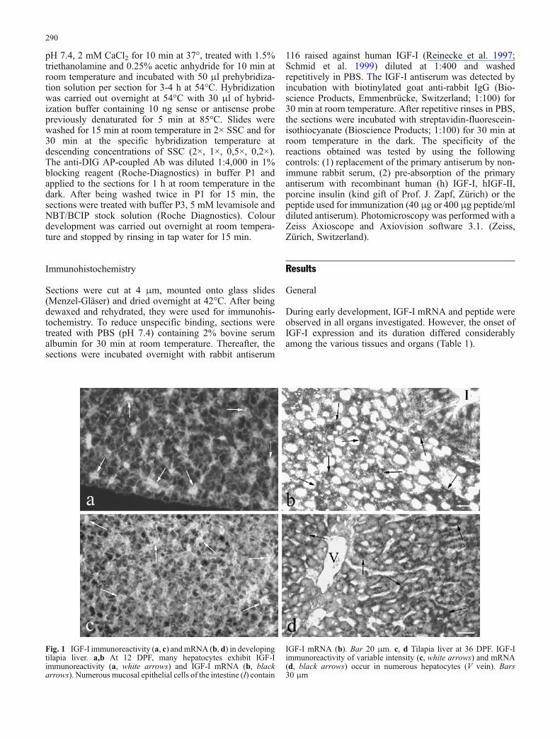
Fig. 1 IGF-I immunoreactivity (a, c) andmRNA (b, d) in developingtilapia liver. a,b At 12 DPF, many hepatocytes exhibit IGF-Iimmunoreactivity (a, white arrows) and IGF-I mRNA (b, blackarrows). Numerous mucosal epithelial cells of the intestine (I) contain
IGF-I mRNA (b). Bar 20 μm. c, d Tilapia liver at 36 DPF. IGF-Iimmunoreactivity of variable intensity (c, white arrows) and mRNA(d, black arrows) occur in numerous hepatocytes (V vein). Bars30 μm
290

Liver
At 4 DPF, several hepatocytes started to exbibit IGF-ImRNA followed by IGF-I immunoreactivity around 6-7DPF (Table 1). By 12 DPF, numerous hepatocytes showedIGF-I mRNA and some also contained IGF-I peptide(Fig. 1a,b). Around 36 DPF, a larger number of hepatocytescontained IGF-I mRNA (Fig. 1d) and about half of thehepatocytes also possessed IGF-I immunoreactivity atvariable intensity (Fig. 1c). Whereas the expression of IGF-I mRNA persisted in hepatocytes, the presence of IGF-Ipeptide decreased with age and could not be detected after90 DPF.
Gastro-intestinal tract
IGF-I mRNA and peptide appeared in cells of the intestinalmucosal epithelium at 5 DPF (Fig. 2a,b). At 9 DPF, theywere also detected in cells of the stomach epithelium.Virtually all epithelial cells of the gastro-intestinal tractcontained IGF-I mRNA and peptide. IGF-I-immunore-
active material was mainly located in the cell apex(Fig. 2a,c,d). The largest numbers of mucosal epithelialcells exhibiting IGF-I mRNA and peptide were found at 13-29 DPF (Fig. 2c–e, Table 1). Around 17 DPF, additionalIGF-I-containing cells were detected with the typicalappearance of endocrine cells (Fig. 2c). These IGF-I-immunoreactive cells occurred in all portions of the gastro-intestinal tract but their distribution and frequency variedlargely among different individuals. The cells persisteduntil the adult stage, whereas the presence of IGF-Iimmunoreactivity in the cell apex was limited to about 50DPF (Table 1).
Pancreas
In the exocrine pancreas, the first cells with IGF-I mRNAwere present as early as 4 DPF. Their number increased ataround 6 DPF (Fig. 2f) and persisted at a high level until 18DPF (Table 1). Thereafter, the number of IGF-I-containingacinar cells decreased until about 90 DPF and could not bedetected in adults.
Fig. 2 IGF-I immunoreactivity and mRNA in developing tilapiagastro-intestinal tract and pancreas. a, b At 5 DPF, most mucosalepithelial cells in the developing intestine show IGF-I immunore-activity in the apex (a) and also IGF-I mRNA (b) on consecutivesections. Bar 25 μm. c At 17 DPF, additional IGF-I-containing cells(arrows) are found with the typical appearance of endocrine cells.Bar 25 μm. d, e At 29 DPF, numerous cells in the mucosalepithelium of the stomach are IGF-I-immunoreactive (d) and contain
IGF-I mRNA (e). Bar 40 μm. f At 6 DPF, several endocrine cells inthe islet contain IGF-I mRNA (arrows). Numerous acinar cells ofthe surrounding exocrine pancreas (ex, arrows) also exhibit IGF-ImRNA. Bar 25 μm. g At 76 DPF, no IGF-I immunoreactivity isobserved in the exocrine pancreas but numerous cells in the islets areIGF-I-immunoreactive. As shown by double immunofluorescence,these cells constitute a subpopulation different from insulin-immunoreactive cells (h). Bar 25 μm
291
At 6 DPF, some islet cells appeared containing IGF-ImRNA (Fig. 2f) and peptide. Their number increased until18 DPF. The IGF-I-containing islets cells did not containinsulin (Fig. 2g,h).
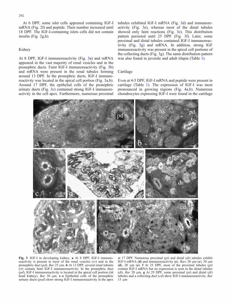
Kidney
At 8 DPF, IGF-I immunoreactivity (Fig. 3a) and mRNAappeared in the vast majority of renal vesicles and in thepronephric ducts. Faint IGF-I immunoreactivity (Fig. 3b)and mRNA were present in the renal tubules formingaround 13 DPF. In the pronephric ducts, IGF-I immuno-reactivity was located in the apical cell portion (Fig. 3a,b).Around 17 DPF, the epithelial cells of the pronephricurinary ducts (Fig. 3c) contained strong IGF-I immunore-activity in the cell apex. Furthermore, numerous proximal
tubules exhibited IGF-I mRNA (Fig. 3d) and immunore-activity (Fig. 3e), whereas most of the distal tubulesshowed only faint reactions (Fig. 3e). This distributionpattern persisted until 25 DPF (Fig. 3f). Later, someproximal and distal tubules contained IGF-I immunoreac-tivity (Fig. 3g) and mRNA. In addition, strong IGFimmunoreactivity was present in the apical cell portions ofthe collecting ducts (Fig. 3g). The same distribution patternwas also found in juvenile and adult tilapia (Table 1).
Cartilage
Even at 4-5 DPF, IGF-I mRNA and peptide were present incartilage (Table 1). The expression of IGF-I was mostpronounced in growing regions (Fig. 4a,b). Numerouschondrocytes expressing IGF-I were found in the cartilage
Fig. 3 IGF-I in developing kidney. a At 8 DPF, IGF-I immuno-reactivity is present in most of the renal vesicles (rv) and in thepronephric duct (pd). Bar 25 μm. b At 13 DPF, several renal tubules(rt) contain faint IGF-I immunoreactivity. In the pronephric duct(pd), IGF-I immunoreactivity is located in the apical cell portion (hkhead kidney). Bar 30 μm. c–e Epithelial cells of the pronephricurinary ducts (pud) show strong IGF-I immunoreactivity in the apex
at 17 DPF. Numerous proximal (pt) and distal (dt) tubules exhibitIGF-I mRNA (d) and immunoreactivity (e). Bars 20 μm (c), 30 μm(d), 20 μm (e). f At 25 DPF, most of the proximal tubules (pt)contain IGF-I mRNA but no expression is seen in the distal tubules(dt). Bar 20 μm. g At 29 DPF, some proximal (pt) and distal (dt)tubules and a collecting duct (cd) show IGF-I immunoreactivity. Bar15 μm
292

at various locations (Figs. 4c,d,g, 5a, 6a,d) throughoutdevelopment. Their number slowly decreased with timeand they were also found in adult life.
IGF-I was also detected by in situ hybridization andimmunohistochemistry in the perichondrium starting at 4DPF. The expression of IGF-I in the perichondrium wasmost pronounced between 6 and 50 DPF (Table 1) andpersisted throughout life at a moderate level.
Muscle
IGF-I mRNA and peptide appeared in skeletal musclefibers at 4 DPF (Table 1). Both were present in thecytoplasm (Fig. 4e,f). The highest expression of IGF-I inskeletal muscle was between 8 and 29 DPF (Table 1).Thereafter, it decreased (Fig. 4g) but persisted throughoutdevelopment and adult life.
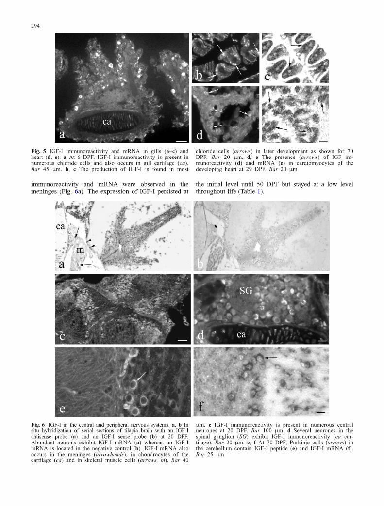
Gills
The production of IGF-I in gills had its onset at 6 DPF(Fig. 5a; Table 1). From the beginning, both IGF-I mRNAand peptide occurred in the majority of chloride cells. Thisdistribution pattern was found throughout development(Fig. 5b,c) and adult life.
Heart
At 13 DPF, IGF-I was first detected in the developing heart(Table 1). IGF-I was present both in atrial (Fig. 5d) andventricular (Fig. 5e) myocardiocytes. The highest amountof IGF-I-containing myocardiocytes was present from 19to 29 DPF. In general, the number of cells showing IGF-ImRNA was higher than that of cells exhibiting IGF-Iimmunoreactivity (Table 1). During late development andin adults, no IGF-I could be detected in the heart.
Brain
In brain, strong IGF-I mRNA signals were revealed innumerous neurones of the brain of larvae (Fig. 6a) startingaround 6 DPF (Table 1). Similarly, IGF-I immunoreactivitywas present in the vast majority of neurones (Fig. 6c). Inaddition, IGF-I was expressed in spinal (Fig. 6d) and indeveloping autonomic ganglia. This overall neuronalpresence of IGF-I persisted until about 29 DPF and slowlydecreased thereafter. In older larvae and adult individuals,IGF-I mRNA and peptide, although still found at all levelsof brain and spinal cord, were present in some neuronesonly. In the older fish, the number of neurones containingIGF-I mRNA exceeded that of the IGF-I-immunoreactiveneurones. Whereas in most areas of the brain, thedistribution patterns of IGF-I mRNA and peptide variedlargely among the individuals investigated, the Purkinjecells in the cerebellum always showed IGF-I peptide(Fig. 6e) and mRNA (Fig. 6f). Starting at 4 DPF, IGF-I
Fig. 4 IGF-I in developing cartilage (a–d) and skeletal muscle(e–g). a At 10 DPF, most chondrocytes and the perichondrium(pc) contain IGF-I immunoreactivity. b The expression of IGF-ImRNA is particularly pronounced in growing regions (largearrow) at 17 DPF. IGF-I mRNA further occurs in theperichondrium (pc). Bar 15 μm (a, b). IGF-I immunoreactivity
(c) and mRNA (d) are present in the majority of chondrocytesof spine and rib cartilage and in skeletal muscle (arrows) at 20DPF. Bar 30 μm. IGF-I immunoreactivity (e) and mRNA (f) arefound in the cytoplasm of all skeletal muscle fibers at 17 DPF.Bar 10 μm. g At 50 DPF, numerous muscle fibers still showIGF-I immunoreactivity (ca cartilage). Bar 50 μm
293
immunoreactivity and mRNA were observed in themeninges (Fig. 6a). The expression of IGF-I persisted at
the initial level until 50 DPF but stayed at a low levelthroughout life (Table 1).
Fig. 6 IGF-I in the central and peripheral nervous systems. a, b Insitu hybridization of serial sections of tilapia brain with an IGF-Iantisense probe (a) and an IGF-I sense probe (b) at 20 DPF.Abundant neurons exhibit IGF-I mRNA (a) whereas no IGF-ImRNA is located in the negative control (b). IGF-I mRNA alsooccurs in the meninges (arrowheads), in chondrocytes of thecartilage (ca) and in skeletal muscle cells (arrows, m). Bar 40
μm. c IGF-I immunoreactivity is present in numerous centralneurones at 20 DPF. Bar 100 μm. d Several neurones in thespinal ganglion (SG) exhibit IGF-I immunoreactivity (ca car-tilage). Bar 20 μm. e, f At 70 DPF, Purkinje cells (arrows) inthe cerebellum contain IGF-I peptide (e) and IGF-I mRNA (f).Bar 25 μm
Fig. 5 IGF-I immunoreactivity and mRNA in gills (a–c) andheart (d, e). a At 6 DPF, IGF-I immunoreactivity is present innumerous chloride cells and also occurs in gill cartilage (ca).Bar 45 μm. b, c The production of IGF-I is found in most
chloride cells (arrows) in later development as shown for 70DPF. Bar 20 μm. d, e The presence (arrows) of IGF im-munoreactivity (d) and mRNA (e) in cardiomyocytes of thedeveloping heart at 29 DPF. Bar 20 μm
294

Pituitary
IGF-I occurred in both portions of the tilapia pituitary butappeared later than in brain. The first neurosecretory axonscontaining IGF-I immunoreactivity (Fig. 7a,c) appeared inthe posterior (neuro-) pituitary around 17 DPF (Table 1).Their density decreased with age but they were stilldetected in adults. No IGF-I mRNA was detected in theposterior pituitary. IGF-I-immunoreactive neurons oc-curred in hypothalamus (Fig. 7c). Endocrine cells of theanterior (adeno-) pituitary first exhibited IGF-I mRNA at28 DPF (Fig. 7b) followed by IGF-I immunoreactivity at40 DPF (Fig. 7c). IGF-I immunoreactivity (Fig. 7c) andmRNA (Fig. 7d) in endocrine cells were present through-out development and in adults.
Skin
At 5 DPF, IGF-I immunoreactivity appeared in cells of theepidermis (Fig. 8a). First, the number of epidermal cellsexhibiting IGF peptide (Fig. 8b) and mRNA (Fig. 8c)increased with age to reach the highest level around 19-29
DPF (Table 1). In older larvae (Fig. 8d,e) and adults, somesuperficial epidermal cells (arrows) and occasionally alsocells in the basal epidermis (arrowhead) were IGF-I-immunoreactive or contained IGF-I mRNA (Fig. 8e).
Discussion
This study has revealed the appearance and distribution ofIGF-I mRNA and peptide during the early development ofthe tilapia, O. niloticus. Both IGF-I mRNA and peptidehave been observed in parenchymal cells of all organsinvestigated but the onset, intensity and duration of IGF-Iexpression differs considerably among the various tissuesand organs.
Both IGF-I mRNA and peptide appear in liver at 4 DPF.This early onset of IGF-I production in tilapia liver is inagreement with results obtained in shi drum in which IGF-Iimmunoreactivity has been detected in hepatocytes duringthe first week post hatching (Radaelli et al. 2003). Becauseliver is the main source of endocrine IGF-I, these resultssuggest a particular physiological impact of circulatingIGF-I in early development. Although high expression of
Fig. 7 IGF-I immunoreactivity and mRNA in the pituitary asrevealed in two pairs of consecutive sections (a, b and c, d)processed for immunofluorescence (a, c) and in situ hybridization(b, d). a, b At 28 DPF, numerous axons in the neuropituitary containIGF-I immunoreactivity (a) but no IGF-I mRNA is present (b). Inthe adenopituitary, most of the endocrine cells exhibit IGF-I mRNA(b), whereas they do not show IGF-I immunoreactivity (a). Most
neurons in the hypothalamus (H) exhibit IGF-I mRNA (b). Bar40 μm. c, d At 40 DPF, both IGF-I immunoreactivity (c) and mRNA(d) are detectable in endocrine cells of the anterior pituitary. IGF-I-immunoreactive axons are present in the posterior part. An IGF-I-immunoreactive neuron (c, arrow) is shown in the hypothalamus.Bar 70 μm
295

IGF-I mRNA has been detected throughout life in tilapialiver, IGF-I immunoreactivity decreases with age and ispresent only until about 70 DPF. In contrast to an earlierstudy on sea bream (Funkenstein et al. 1997), an intenseresponse by in situ hybridization for IGF-I has beenobtained in adult tilapia liver. In spite of the clearexpression of IGF-I mRNA in adult tilapia liver (Schmidet al. 1999), no IGF-I-immunoreactive hepatocytes havebeen identified in adult barramundi (Richardson etal. 1995) and tilapia (Reinecke et al. 1997; Schmid etal. 1999). Similarly, in rat liver, IGF-I immunoreactivity
has been localized only after pretreatment with colchicine(Hansson et al. 1988). Therefore, IGF-I might be releasedfrom fish liver into the circulation immediately aftersynthesis, as has previously been presumed in mammals(Hansson et al. 1988).
High-affinity hepatic-binding sites for growth hormone(GH) have been identified in the liver of several fishspecies, including tilapia (Ng et al. 1992). In accordance,there is strong evidence that GH stimulates IGF-I expres-sion in fish liver and its secretion into the circulation (seeReinecke et al. 2005). Injection or oral administration ofGH significantly enhances the IGF-I mRNA level in theliver of numerous species (e.g. Duan et al. 1993; Duguay etal. 1994, 1996; Moriyama 1995; Shamblott et al. 1995;Shepherd et al. 1997; Vong et al. 2003; Biga et al. 2004).The GH-induced increase in liver IGF-I mRNA expressionis accompanied by an increase in the level of circulatingIGF-I (Funkenstein et al. 1989; Niu et al. 1993; Moriyamaet al. 2000). In addition to these in vivo studies, some invitro investigations have also demonstrated the stimulatingeffect of GH on liver IGF-I mRNA expression, i.e. onprimary cultured hepatocytes of salmonids (Duan etal. 1993; Shamblott et al. 1995; Pierce et al. 2005) andtilapia (Schmid et al. 2000). In rainbow trout, GH mRNAhas been detected in early stage embryos (Yang et al. 1999).Thus, the production and release of liver IGF-I duringtilapia early development may be under the control of GH.
The early onset of the production of IGF-I in tilapiagastro-intestinal tract, i.e. at 5 DPF in intestine and at 9DPF in stomach, supports the results of earlier studies. Inthe intestinal epithelium of turbot (Berwert et al. 1995) andshi drum (Radaelli et al. 2003), the first IGF-I-immunore-active cells can be found as early as the first week posthatching. Furthermore, IGF-1R appears in the second weekpost hatching in shi drum gut (Radaelli et al. 2003). Thenumber of IGF-I-producing cells in the developing bonyfish gastro-intestinal tract by far exceeds that in the adultbecause, during ontogeny, almost all epithelial cellsthroughout the gastro-intestinal tract exhibit IGF-ImRNA and peptide. The IGF-I-immunoreactive materialis mainly located in the cell apex as has also been reportedin shi drum (Radaelli et al. 2003), possibly suggestingrelease into the lumen. Around 17 DPF, additionalinfrequent IGF-I-containing cells appear in the mucosalepithelium; these cells have the typical appearance ofendocrine cells as characterized for adult fish (Reinecke etal. 1997; Koppang et al. 1998). The distribution of theseIGF-I-immunoreactive cells in the various portions of theadult gastro-intestinal tract and their frequency vary largelyamong different individuals, as previously reported inturbot (Berwert et al. 1995) and Atlantic salmon (Koppanget al. 1998).
Although the early and overall appearance of IGF-I andits receptor strongly suggests a particular physiologicalimpact of IGF-I in the developing gastro-intestinal tract, wecan only hypothesize on the physiological meaning ofintestinal IGF-I in fish. IGF-I secreted from the IGF-I-immunoreactive epithelial cells may exert mitogenicfunctions by acting on neighbouring epithelial cells. This
Fig. 8 IGF-I immunoreactivity and mRNA in the skin. a At 5 DPF,several cells in the epidermis contain IGF-I immunoreactivity. Inthe dermis, a possible dendritic cell also shows IGF-I immunore-activity (arrow). Bar 40 μm. b, c At 25 DPF, numerous cells in allepidermal layers exhibit IGF-I peptide (b) and mRNA (c). Bar30 μm. d, e Frequent superficial epidermal cells (arrows) andinfrequent basal epidermal cells (arrowheads) exhibit IGF-Iimmunoreactivity (d) and mRNA (e) at 36 DPF. Bars 50 μm (d),40 μm (e)
296

hypothesis is supported by results obtained in mammals. Inpig, the amounts of intestinal IGF-I immunoreactivity andof the IGF-1R can be correlated with villous growth andmaturation (Schober et al. 1990) and, in adult rat, IGF-Ipotently stimulates crypt cell proliferation and villus celldensity (Steeb et al. 1994). The potential proliferativeaction of intestinal IGF-I during development and adult lifemay be regulated by GH, which markedly increases theamount of intestinal IGF-I mRNA in juvenile common carp(Vong et al. 2003) and adult rainbow trout (Biga etal. 2004).
In juvenile and adult bony fish, IGF-I released from theinfrequent IGF-I-immunoreactive mucosal epithelial cellsmay exert paracrine effects in response to altering localdemands, such as repair. In agreement with this hypothesis,the therapeutical potential of IGF-I has been discerned fornumerous acute human bowel disorders resulting inaccelerated intestinal repair and epithelial regrowth(Howarth 2003). Because, in addition to gill and kidney,the intestinal tract exerts important osmoregulatory func-tions in fish, intestinal IGF-I may also be involved inosmoregulation (Koppang et al. 1998; Reinecke andCollet 1998). This hypothesis is supported by experimentsin brown trout suggesting that IGF-I plays a role in theregulation of intestinal Na+,K+-ATPase activity (Seidelinand Madsen 1999).
IGF-I immunoreactivity has been localized in cells of theexocrine parenchyma of shi drum and seabream larvae(Funkenstein et al. 1997; Perrot et al. 1999; Radaelli etal. 2003); this is supported by the results of the presentstudy. As IGF-I production in pancreas is stimulated by GHin rat (Jevdjovic et al. 2004), IGF-I in fish pancreas mayalso be under the control of GH, although no results havebeen presented to date on the potential regulation of IGF-Iin the exocrine pancreas.
To date, indications for the presence of IGF-I in theendocrine pancreas have only been obtained by immuno-histochemical studies. These have shown that IGF-Iimmunoreactivity occurs in islet cells of several bonyfish species (Reinecke et al. 1993, 1997; Berwert etal. 1995; Richardson et al. 1995). The present studyprovides the additional information that IGF-I mRNA isalso present in islet cells. This is supported by molecularbiological studies that have shown IGF-I mRNA expres-sion in principal islets of the salmon O. gorbusha(Plisetskaya et al. 1993) and of Cottus scorpius (Loffing-Cueni et al. 1998). Islet IGF-I appears at 6 DPF in tilapia(this study) and day 10 post hatching in turbot (Berwert etal. 1995) suggesting a significant physiological role forislet-derived IGF-I during larval development. In bothspecies, from the onset of its expression, IGF-I occurs innon-insulin cells. This observation is in agreement withstudies on adult teleosts, such as eel, tilapia, goldfish,turbot and common carp (Reinecke et al. 1993, 1997;Berwert et al. 1995).
On the one hand, islet-derived IGF-I may be involved inthe paracrine regulation of insulin secretion from the β-cells, although the evidence in mammals is conflicting(Leahy and Vandekerkhove 1990; Van Schravendijk et
al. 1990; Jevdjovic et al. 2004). On the other hand, somestudies suggest that islet-derived IGF-I may act as anendocrine hormone. Hypophysectomy does not influencethe amount of sulphation activity in eel pancreas (Duan andHirano 1992) suggesting that islet IGF-I is not regulated byGH. In goby, islectomy leads to a decrease in 35SO4-incorporation in cartilage (Bern et al. 1991) but hepaticGH binding is unchanged (Kelley et al. 1993). Theobserved effects may have been augmented by insulin(Plisetskaya 1998). However, in addition to the GH-dependent liver IGF-I system, islet-derived IGF-I mayconstitute a further endocrine GH-independent IGF-Isystem involved in the regulation of fish growth.
No information is available on IGF-I in developing fishkidney. In adult bony fish, however, epithelial cells mainlyof the proximal tubules have been reported as sites of IGF-Iproduction in tilapia (Reinecke et al. 1997), as has similarlybeen described in adult mammals (see Reinecke andCollet 1998). In tilapia, IGF-I occurs in renal vesicles evenat early stages of development, i.e. at 5 DPF. Later, it ispresent in proximal and distal tubles and in collecting andurinary ducts. Whereas IGF-I in the tubular sytem isdetected throughout the cytoplasm, it occurs only in theapical cell part in the ducts possibly suggesting the releaseof IGF-I into the lumen. Apart from the gills, the kidney is amajor osmoregulatory organ in fish but the evidence for thepotential participation of renal IGF-I in osmoregulation isconflicting. In rainbow trout, transfer to seawater has beenreported to increase the IGF-I mRNA level in kidney(Sakamoto and Hirano 1993). In contrast, the sametreatment does not affect the Na+,K+-ATPase gene leveland activity in brown trout (Madsen et al. 1995) or the renalIGF-I mRNA level in the four-spine sculpin, Cottus kazika(Inoue et al. 2003). Thus, at present, we can only speculateabout the physiological role of IGF-I in bony fish kidney.In addition to its possible role in osmoregulation, renalIGF-I may be involved in several other parameters ofkidney function, such as the stimulation of kidney growthand differentiation, renal blood flow, glomerular filtrationrate and sodium absorption, as is likely in mammals(Hirschberg 1996).
In O. niloticus, IGF-I also appears early in tissues thatare highly involved in growth, such as cartilage andskeletal muscle. At 4 DPF, IGF-I mRNA and peptide arepresent in the chondrocytes of cartilage at variouslocations. Similarly, in shi drum, IGF-I immunoreactivityhas been observed in cartilage at day 11 post hatching(Radaelli et al. 2003). The expression of IGF-I in tilapia ismost pronounced in growing regions. The number of IGF-I-containing chondrocytes is higher during developmentthan in adult life, thus underlining the physiologicalimpact of local IGF-I during cartilage growth. Althoughthe GH/liver IGF-I axis is involved in the regulation offish growth as an endocrine system (Duan 1998; Reineckeet al. 2005), an influence of GH on growth via local auto/paracrine IGF-I, as indicated by the present results, is alsolikely. An early study on eel has determined that thestimulatory effect of GH on sulphate incorporation intocartilage is mediated by an IGF-like plasma factor (Duan
297

and Inui 1990). In agreement, the injection of GH intocoho salmon cartilage stimulates sulphate and thymidineincorporation (McCormick et al. 1992; Tsai et al. 1994).Thus, GH may have increased both uptakes via thestimulation of local IGF-I, because IGF-I peptide andmRNA have been shown in chondrocytes of developing(Perrot et al. 1999; Radaelli et al. 2003; this study) andadult (Funkenstein et al. 1997; Reinecke et al. 1997) bonyfish. The GH-dependent growth promoting effects of IGF-I on cartilage may therefore be exerted not only via theendocrine route, but also in an autocrine/paracrine mannerby IGF-I released from local chondrocytes, as has beenshown in rat (Reinecke et al. 2000).
In contrast to the skeletal muscle of young shi drumlarvae (Radaelli et al. 2003) in which no IGF-I immuno-reactivity has been detected, the onset of IGF-I productionin tilapia skeletal muscle occurs at 4 DPF, with IGF-Ishowing a maximum around 10-29 DPF, decreasingafterwards but persisting throughout life. In agreementwith an auto/paracrine function of muscle IGF-I, IGF-Ireceptor number and binding in trout skeletal muscle arehighest at 5 weeks and both parameters decrease with age(Mendez et al. 2001). Furthermore, a parallel age-relateddecline has been found for tyrosine kinase activity for IGF-1R (Mendez et al. 2001). In rainbow trout muscle in vitro,IGF-I has been shown to be highly effective in stimulatingglucose and alanine uptake into myosatellite cells, wherebythe degree of stimulation changes when cells differentiateto myotubes (Castillo et al. 2004). These and the presentresults showing that IGF-I in skeletal muscle appears asearly as 4 DPF and decreases with age indicate a key rolefor IGF-I in muscle development. IGF-I may be associatedin later life with metabolism and repair mechanisms. Inadult rainbow trout, the expression of IGF-I in skeletalmuscle is stimulated by GH (Biga et al. 2005). Thus, GHmay also regulate IGF-I production in larval musle.
Endocrine (liver-derived) IGF-I seems to have aphysiological impact in smoltification. In mummichog(Fundulus heteroclitus; Mancera and McCormick 1998)and trout (Seidelin et al. 1999), IGF-I improves adaptationto seawater in a dose-dependent manner. The osmoregu-latory effects of IGF-I seem to be exerted directly because,in preparations of Coho salmon gills in vitro, IGF-Istimulates Na+,K+-ATPase (Madsen and Bern 1993).Hypophysectomy in tilapia lowers the levels of gill Na+,K+-ATPase when compared with that of sham-operatedcontrols (Shepherd et al. 1997) indicating an involvementof the GH/liver IGF-I axis. In agreement, in a study onCottus kazika, the level of liver IGF-I mRNA has beenshown to be significantly higher in individuals reared inseawater than in those reared in freshwater (Inoue etal. 2003). In sharp contrast, a study on Coho salmon hasdemonstrated that IGF-I mRNA is not significantly alteredduring seawater adaptation in liver but markedly increasesin gills (Sakamoto and Hirano 1993). The chloride cells offilament epithelium have been shown not only to expressNa+,K+-ATPase in trout (McCormick 1996), but also IGF-I in tilapia (this study), in shi drum larvae (Radaelli etal. 2003) and in adult tilapia (Reinecke et al. 1997). GH
receptors have been detected in tilapia gills (Fryer 1979)and GH treatment increases IGF-I mRNA in gills ofsalmonids and carp (Vong et al. 2003; Biga et al. 2004).Thus, in addition to circulating IGF-I, GH-regulated localautocrine/paracrine IGF-I in the chloride cells seems toparticipate in the regulation of plasma osmolality and gillNa+,K+-ATPase activity. Because the expression of IGF-Iin tilapia gill chloride cells has been detected even around6-7 DPF, IGF-I may exert additional functions, such asinfluencing the growth and maintenance of the filamentepithelium.
In shi drum heart, IGF-I immunoreactivity has beendetected in 15-day larvae (Radaelli et al 2003). Likewise,IGF-I mRNA and peptide appear at 13 DPF in tilapia. Theexpression of IGF-I in cardiomyocytes is clearly detectableuntil 70 DPF but is absent from later stages and adultsindicating a particular physiological impact of IGF-I duringheart development and growth. In correlation, a significantincrease in IGF-1R mRNA levels (Gutiérrez et al. 1995)has been observed in the rapidly growing juvenile troutheart (Greene and Chen 1999). In fish heart, local IGF-Imay be even more important than circulating IGF-I. Inrainbow trout heart IGF-1R mRNA levels decrease afterapplication of GH (Biga et al. 2004) and the number ofIGF-I receptors is inversely related to the concentration ofcirculating IGF-I (Baños et al. 1997; Moon et al. 1996).
Screening studies have identified the potential sites ofIGF-I synthesis in the brains of barramundi, tilapia and seabream. IGF-I-immunoreactive neurones are present in adultbarramundi brainstem (Richardson et al. 1995) and at alllevels of adult tilapia brain (Reinecke et al. 1997).Correspondingly, IGF-I mRNA signals have been de-scribed throughout the brain of developing sea bream(Funkenstein et al. 1997). The functional impact of IGF-I inbrain is indicated by the presence of IGF-1R in adult carpand trout brain (Leibush et al. 1996) and by the appearanceof IGF-1R immunoreactivity in the developing brain of shidrum as early as hatching (Radaelli et al. 2003). In contrastto IGF-II (Caelers et al. 2003), the exact distribution ofIGF-I gene expression in fish brain is unknown. The IGF-I-immunoreactive neurones in barramundi and tilapia aresparsely scattered and their distribution exhibits pro-nounced inter-individual differences. In juvenile (thisstudy) and adult tilapia (Reinecke et al. 1997) and adultdaddy sculpin (Loffing-Cueni et al. 1998) brain, only IGF-I-producing Purkinje cells in cerebellum are constantlypresent. In agreement with the potential autocrine/paracrineaction of neuronal IGF-I in adult brown trout, Salmo trutta,IGF-I binding in the brain is highest in the cerebellum(Smith et al. 2005). The pronounced inter-individualvariations of IGF-I-producing neurones in adult fish brainmake a neurotransmitter or -modulator function of IGF-Iunlikely. Rather, neuronal IGF-I may support the survivalof neurones and glia cells, as is also likely for mammals(Cheng et al. 2001). During development, IGF-I maystimulate neurogenesis, dendritic growth and synaptogen-esis in an autocrine/paracrine manner, as has been shown inmammals (e.g. Zhou et al. 1999; Niblock et al. 2000;Kakizawa et al. 2003). This hypothesis is supported by the
298

observation that, in early developing tilapia larvae, mostneurones express IGF-I mRNA and that the number ofneurones containing IGF-I mRNA decreases with age.Whether the assumed actions of IGF-I are exerted under thecontrol of GH is unclear at present, since GH significantlyincreases brain IGF-I mRNA in rainbow trout (Biga etal. 2004), whereas no effect has been reported in commoncarp (Vong et al. 2003).
The present study is the first to show that IGF-I occurs inthe bony fish pituitary. IGF-I has been observed in both theneuro- and adenopituitary. In the posterior part, neurose-cretory axons containing IGF-I immunoreactivity appeararound 17 DPF, whereas no IGF-I mRNA is detected at thisstage. The likely source of IGF-I in the neuropituitary is theneuronal perikarya that lie within the hypothalamus andthat exhibit IGF-I immunoreactivity. In the adenopituitary,IGF-I mRNA is first detected at 20 DPF. IGF-I immuno-reactivity and mRNA in endocrine cells are presentthroughout development. To date, IGF-I immunoreactivityhas only been localized in the anterior pituitary in the frogXenopus laevis in which it occurs in co-existence withprolactin (David et al. 2000). Because IGF-I-binding siteshave also been found in X. laevis anterior pituitary, IGF-Ifrom the prolactin cells has been postulated to regulate GHin a paracrine manner (David et al. 2000). Whether thishypothesis also applies to bony fish remains to be clarified,as does the type of endocrine cells producing IGF-I.
At 5 DPF, IGF-I has been detected in cells of theepidermis of tilapia. The number of IGF-I-producingepidermal cells first increases with age, reaching its highestlevel around 19-29 DPF. In older larvae and adults, somesuperficial and basal epidermal cells exhibit IGF-I immu-noreactivity or IGF-I mRNA. In developing skin of shidrum, IGF-1R immunoreactivity is detectable even athatching (Radaelli et al. 2003). The early appearance ofboth IGF-I and its receptor suggests an important auto/paracrine function of IGF-I in skin development. Becausesome IGF-I producing cells still occur in the basal andsuperficial layers in juvenile and adult tilapia and as highlevels of IGF receptor mRNA have been found in adultgilthead seabream skin (Perrot et al. 1999), local IGF-I infish skin might play a role in later life. Skin IGF-I might beinvolved in repair mechanisms and wound healing, as hasbeen proposed in mammals (Edmondson et al. 2003).
In summary, the expression of IGF-I is more pronouncedduring ontogeny than in juvenile and adult life in theparenchymal cells of most of the organs investigated, suchas epithelial cells of the gastro-intestinal tract, acinar cellsof the exocrine pancreas, skeletal muscle cells, cardiomyo-cytes, renal tubular cells, neurones of the central andperipheral nervous system and skin cells. These resultssuggest a high functional impact of local IGF-I in early fishgrowth, metabolism and organogenesis by auto/paracrinemeans of regulation. Whether IGF-I expression in the earlydevelopment of fish is regulated by GH remains to beclarified (Perrot et al. 1999; Deane et al. 2003), althoughlow levels of GH mRNA have been detected as early asgastrulation in rainbow trout and intermediate amounts of
GH mRNA have been observed in early cleavage-stageembryos (Yang et al. 1999).
References
Ayson FG, Jesus EG de, Moriyama S, Hyodo S, Funkenstein B,Gertler A, Kawauchi H (2002) Differential expression ofinsulin-like growth factor I and II mRNAs during embryogen-esis and early larval development in rabbitfish, Siganusguttatus. Gen Comp Endocrinol 126:165–174
Baños N, Moon TW, Castejon C, Gutiérrez J, Navarro I (1997)Insulin and insulin-like growth factor-I (IGF-I) binding in fishred muscle: regulation by high insulin levels. Regul Pept68:181–187
Baroiller JF, Desprez D, Carteret Y, Tacon P, Hoareau MC,Mélard C, Jalabert B (1997) Influence of environmentaland social factors on the reproductive efficiency in threetilapia species, Oreochromis niloticus, O. aureus and thered tilapia (Red Florida strains). In: Fitzsimmons K (ed)Proceedings of the Fourth International Symposium onTilapia in Aquaculture, 9-12 November 1997, Orlando,Fla., USA. Northeast Regional Agriculture EngineeringService, New York, pp 238–252
Bern HA, McCormick SD, Kelley KM, Gray ES, Nishioka RS,Madsen SS, Tsai PL (1991) Insulin-like growth factors “underwater”: role in growth and function of fish and otherpoikilothermic vertebrates. In: Spencer EM (ed) Modernconcepts of insulin-like growth factors. Elsevier, New York,pp 85–96
Berwert L, Segner H, Reinecke M (1995) Ontogeny of IGF-1 andthe classical islet hormones in the turbot, Scophthalmusmaximus. Peptides 16:113–122
Biga PR, Schelling GT, Hardy RW, Cain KD, Overturf K, Ott TL(2004) The effects of recombinant bovine somatotropin (rbST)on tissue IGF-I, IGF-I receptor, and GH mRNA levels inrainbow trout, Oncorhynchus mykiss. Gen Comp Endocrinol135:324–323
Caelers A, Schmid AC, Hrusovsky A, Reinecke M (2003) Insulin-like growth factor II mRNA is expressed in neurones of thebrain of the bony fish Oreochromis mossambicus, the tilapia.Eur J Neurosci 18:355–363
Caelers A, Berishvili G, Meli ML, Eppler E, Reinecke M (2004)Establishment of a real-time RT-PCR for the determination ofabsolute amounts of IGF-I and IGF-II gene expression in liverand extrahepatic sites of the tilapia. Gen Comp Endocrinol137:196–204
Castillo J, Codina M, Martinez ML, Navarro I, Gutiérrez J (2004)Metabolic and mitogenic effects of IGF-I and insulin on musclecells of rainbow trout. Am J Physiol Regul Integr CompPhysiol 286:R935–R941
Cheng CM, Cohen M, Tseng V, Bondy CA (2001) EndogenousIGF1 enhances cell survival in the postnatal dentate gyrus.J Neurosci Res 64:341–347
Chourrout D, Itskovich J (1983) Three manipulations permittedby artificial insemination in tilapia: induced diploid gyno-genesis, production of all triploid population and intergenerichybridisation. In: Fishelson L, Yaron Z (eds) InternationalSymposium on Tilapia in Aquaculture, Nazareth, Israel. TelAviv University, Tel Aviv, pp 246–255
David I, Bosshard R, Kloas W, Reinecke M (2000) Insulin-likegrowth factor I in the anterior pituitary of the clawed frogXenopus laevis: immunocytochemical and autoradiographicindication for a paracrine action and corelease with prolactin.J Neuroendocrinol 12:415–420
Deane EE, Kelly SP, Collins PM, Woo NY (2003) Larvaldevelopment of silver sea bream (Sparus sarba): ontogeny ofRNA-DNA ratio, GH, IGF-I, and Na(+)-K(+)-ATPase. MarBiotechnol (NY) 5:79–91
Duan C (1998) Nutritional and developmental regulation of insulin-like growth factors in fish. J Nutr 128:306S–314S
299

Duan C, Hirano T (1992) Effects of insulin-like growth factor-I andinsulin on the in-vitro uptake of sulphate by eel branchialcartilage: evidence for the presence of independent hepatic andpancreatic sulphation factors. J Endocrinol 133:211–219
Duan C, Duguay SJ, Plisetskaya EM (1993) Insulin-like growthfactor I (IGF-I) mRNA expression in coho salmon, Onco-rhynchus kisutch: tissue distribution and effects of growthhormone/prolactin family protein. Fish Physiol Biochem11:371–379
Duan C, Plisetskaya EM, Dickhoff WW (1995) Expression of insulin-like growth factor I in normally and abnormally developing cohosalmon (Oncorhynchus kisutch). Endocrinology 136:446–452
Duan CM, Inui Y (1990) Effects of recombinant eel growthhormone on the uptake of [35S]sulfate by ceratobranchialcartilages of the Japanese eel, Anguilla japonica. Gen CompEndocrinol 79:320–325
Duguay SJ, Swanson P, Dickhoff WW (1994) Differential expres-sion and hormonal regulation of alternatively spliced IGF-ImRNA transcripts in salmon. J Mol Endocrinol 12:25–37
Duguay SJ, Lai-Zhang J, Steiner DF, Funkenstein B, Chan SJ(1996) Developmental and tissue-regulated expression of IGF-Iand IGF-II mRNAs in Sparus aurata. J Mol Endocrinol16:123–132
Edmondson SR, Thumiger SP, Werther GA, Wraight CJ (2003)Epidermal homeostasis: the role of the growth hormone andinsulin-like growth factor systems. Endocr Rev 24:737–764
Fryer JN (1979) A radioreceptor assay for purified teleost growthhormone. Gen Comp Endocrinol 39:123–130
Funkenstein B, Silbergeld A, Cavari B, Laron Z (1989) Growthhormone increases plasma levels of insulin-like growth factor(IGF-I) in a teleost, the gilthead seabream (Sparus aurata).J Endocrinol 120:R19–R21
Funkenstein B, Almuly R, Chan SJ (1997) Localization of IGF-I andIGF-I receptor mRNA in Sparus aurata larvae. Gen CompEndocrinol 107:291–303
Greene MW, Chen TT (1997) Temporal expression pattern ofinsulin-like growth factor mRNA during embryonic develop-ment in a teleost, rainbow trout (Onchorhynchus mykiss). MolMar Biol Biotechnol 6:144–151
Greene MW, Chen TT (1999) Quantitation of IGF-I, IGF-II, andmultiple insulin receptor family member messenger RNAsduring embryonic development in rainbow trout. Mol ReprodDev 54:348–356
Gutiérrez J, Parrizas M, Maestro MA, Navarro I, Plisetskaya EM(1995) Insulin and IGF-I binding and tyrosine kinase activity infish heart. J Endocrinol 146:35–44
Hansson H-A, Nilsson A, Isgaard J, Billig H, Isaksson O, SkottnerA, Andersson IK, Rozell B (1988) Immunohistochemicallocalization of insulin-like growth factor I in the adult rat.Histochemistry 89:403–410
Hirschberg R (1996) Insulin-like growth factor I in the kidney.Miner Electrolyte Metab 22:128–132
Howarth GS (2003) Insulin-like growth factor-I and the gastroin-testinal system: therapeutic indications and safety implications.J Nutr 133:2109–2112
Inoue K, Iwatani H, Takei Y (2003) Growth hormone and insulin-like growth factor I of a euryhaline fish Cottus kazika: cDNAcloning and expression after seawater acclimation. Gen CompEndocrinol 131:77–84
Jevdjovic T, Maake C, Eppler E, Zoidis E, Reinecke M, Zapf J(2004) Effects of insulin-like growth factor-I treatment on theendocrine pancreas of hypophysectomized rats: comparisonwith growth hormone replacement. Eur J Endocrinol 151:1–10
Kakizawa S, Yamada K, Iino M, Watanabe M, Kano M (2003)Effects of insulin-like growth factor I on climbing fibre synapseelimination during cerebellar development. Eur J Neurosci17:545–554
Kelley KM, Gray ES, Siharath K, Nicoll CS, Bern HA (1993)Experimental diabetes mellitus in a teleost fish. II. Roles ofinsulin, growth hormone (GH), insulin-like growth factor-I, andhepatic GH receptors in diabetic growth inhibition in the goby,Gillichthys mirabilis. Endocrinology 132:2696–2702
Koppang EO, Thomas GA, Ronningen K, Press CML (1998)Expression of insulin-like growth factor-I in the gastrointestinaltract of Atlantic salmon (Salmo salar L.). Fish Physiol Biochem18:167–175
Leahy JL, Vandekerkhove KM (1990) Insulin-like growth factor-I atphysiological concentrations is a potent inhibitor of insulinsecretion. Endocrinology 126:1593–1598
Leibush B, Parrizas M, Navarro I, Lappova Y, Maestro MA, EncinasM, Plisetskaya EM, Gutiérrez J (1996) Insulin and insulin-likegrowth factor-I receptors in fish brain. Peptides 61:155–161
Loffing-Cueni D, Schmid AC, Graf H, Reinecke M (1998) IGF-I inthe bony fish Cottus scorpius: cDNA, expression and differ-ential localization in brain and islets. Mol Cell Endocrinol141:187–194
Madsen SS, Bern HA (1993) In-vitro effects of insulin-like growthfactor-I on gill Na,K(+)-ATPase in coho salmon, Oncorhynchuskisutch. J Endocrinol 138:23–30
Madsen SS, Jensen MK, Nhr J, Kristiansen K (1995) Expression ofNa(+)-K(+)-ATPase in the brown trout, Salmo trutta: in vivomodulation by hormones and seawater. Am J Physiol 269:R1339–R1345
Mancera JM, McCormick SD (1998) Evidence for growth hormone/insulin-like growth factor I axis regulation of seawater accli-mation in the euryhaline teleost Fundulus heteroclitus. GenComp Endocrinol 111:103–112
McCormick SD (1996) Effects of growth hormone and insulin-likegrowth factor I on salinity tolerance and gill Na, K+-ATPase inAtlantic salmon (Salmo salar): interaction with cortisol. GenComp Endocrinol 101:3–11
McCormick SD, Tsai PI, Kelley KM, Nishioka RS, Bern HA (1992)Hormonal control of sulfate uptake by branchial cartilage ofcoho salmon: role of IGF-I. J Exp Zool 262:166–171
Mendez E, Smith A, Figueiredo-Garutti ML, Planas JV, Navarro I,Gutiérrez J (2001) Receptors for insulin-like growth factor-I(IGF-I) predominate over insulin receptors in skeletal musclethroughout the life cycle of brown trout, Salmo trutta. GenComp Endocrinol 122:148–157
Moon TW, Castejon C, Baños N, Maestro MA, Plisetskaya EM,Gutiérrez J, Navarro I (1996) Insulin and IGF-I binding inisolated trout cardiomyocytes. Gen Comp Endocrinol103:264–272
Moriyama S (1995) Increased plasma insulin-like growth factor-I(IGF-I) following oral and intraperitoneal administration ofgrowth hormone to rainbow trout, Oncorhynchus mykiss.Growth Regul 5:164–167
Moriyama S, Ayson FG, Kawauchi H (2000) Growth regulation byinsulin-like growth factor-I in fish. Biosci Biotechnol Biochem64:1553–1562
Ng TB, Leung TC, Cheng CHK, Woo NYS (1992) Growth hormonebinding sites in tilapia (Oreochromis mossambicus) liver. GenComp Endocrinol 86:111–118
Niblock MM, Brunso-Bechtold JK, Riddle DR (2000) Insulin-likegrowth factor I stimulates dendritic growth in primary somato-sensory cortex. J Neurosci 20:4165–4176
Niu PD, Pérez-Sanchez J, Le Bail PY (1993) Development of aprotein binding assay for teleost insulin-like growth factor(IGF-like): relationship between growth hormone (GH) andIGF-like in the blood of rainbow trout (Oncorrhynchus mykiss).Fish Physiol Biochem 11:381–391
Perrot V, Moiseeva EB, Gozes Y, Chan SJ, Ingleton, P, FunkensteinB (1999) Ontogeny of the insulin-like growth factor system(IGF-I, IGF-II, and IGF-1R) in gilthead seabream (Sparusaurata): expression and cellular localization. Gen CompEndocrinol 116:445–460
Pierce AL, Fukada H, Dickhoff WW (2005) Metabolic hormonesmodulate the effect of growth hormone (GH) on insulin-likegrowth factor-I (IGF-I) mRNA level in primary culture ofsalmon hepatocytes. J Endocrinol 184:341–349
Plisetskaya EM (1998) Some of my not so favorite things aboutinsulin and insulin-like growth factors in fish. Comp BiochemPhysiol [B] 121:3–11
300

Plisetskaya EM, Bondareva VM, Duan C, Duguay SJ (1993) Doessalmon brain produce insulin? Gen Comp Endocrinol 91:74–80
Pozios KC, Ding J, Degger B, Upton Z, Duan C (2001) IGFsstimulate zebrafish cell proliferation by activating MAP kinaseand PI3-kinase-signaling pathways. Am J Physiol Regul IntegrComp Physiol 280:R1230–R1239
Radaelli G, Domeneghini C, Arrighi S, Bosi G, Patruno M,Funkenstein B (2003) Localization of IGF-I, IGF-I receptor,and IGFBP-2 in developing Umbrina cirrosa (Pisces: Os-teichthyes). Gen Comp Endocrinol 130:232–244
Reinecke M, Collet C (1998) The phylogeny of the insulin-likegrowth factors. Int Rev Cytol 183:1–94
Reinecke M, Maake C, Falkmer S, Sara VR (1993) The branchingof insulin-like growth factor 1 and insulin: an immunohisto-chemical analysis during phylogeny. Regul Pept 48:65–76
Reinecke M, Schmid A, Ermatinger R, Loffing-Cueni D (1997)Insulin-like growth factor I in the teleost Oreochromismossambicus, the tilapia: gene sequence, tissue expression,and cellular localization. Endocrinology 138:3613–3619
Reinecke M, Schmid AC, Heyberger-Meyer B, Hunziker EB,Zapf J (2000) Effect of growth hormone and insulin-likegrowth factor I (IGF-I) on the expression of IGF-I mRNA andpeptide in rat tibial growth plate and articular chondrocytes invivo. Endocrinology 141:2847–2853
Reinecke M, Björnsson BT, Dickhoff WW, McCormick SD,Navarro I, Power DM, Gutiérrez J (2005) Growth hormoneand insulin-like growth factors in fish: where we are and whereto go. Gen Comp Endocrinol 142:20–24
Richardson NA, Anderson AJ, Rimmer MA, Sara VR (1995)Localization of insulin-like growth factor-I immunoreactivity inlarval and juvenile barramundi (Lates calcarifer). Gen CompEndocrinol 100:282–292
Sakamoto T, Hirano T (1993) Expression of insulin-like growthfactor I gene in osmoregulatory organs during seawateradaptation of the salmonid fish: possible mode of osmoregu-latory action of growth hormone. Proc Natl Acad Sci USA90:1912–1916
Schmid AC, Naf E, Kloas W, Reinecke M (1999) Insulin-likegrowth factor-I and -II in the ovary of a bony fish, Oreochromismossambicus, the tilapia: in situ hybridisation, immunohisto-chemical localisation, Northern blot and cDNA sequences. MolCell Endocrinol 156:141–149
Schmid AC, Reinecke M, Kloas W (2000) Primary culturedhepatocytes of the bony fish, Oreochromis mossambicus, thetilapia: a valid tool for physiological studies on IGF-Iexpression in liver. J Endocrinol 166:265–273
Schober DA, Simmen FA, Hadsell DL, Baumrucker CR (1990)Perinatal expression of type I IGF receptors in porcine smallintestine. Endocrinology 126:1125–1132
Segner H, Storch V, Reinecke M, Kloas W, Hanke W (1994) Thedevelopment of functional digestive and metabolic organs inturbot, Scophthalmus maximus. Mar Biol 119:471–486
Seidelin M, Madsen SS (1999) Endocrine control of Na,K+-ATPaseand chloride cell development in brown trout (Salmo trutta):interaction of insulin-like growth factor-I with prolactin andgrowth hormone. J Endocrinol 162:127–135
Seidelin M, Madsen SS, Byrialsen A, Kristiansen K (1999) Effectsof insulin-like growth factor-I and cortisol on Na, K+-ATPaseexpression in osmoregulatory tissues of brown trout (Salmotrutta). Gen Comp Endocrinol 113:331–342
Shamblott MJ, Chen TT (1993) Age-related and tissue-specificlevels of five forms of insulin-like growth factor mRNA in ateleost. Mol Mar Biol Biotechnol 2:351–361
Shamblott MJ, Cheng CM, Bolt D, Chen TT (1995) Appearance ofinsulin-like growth factor mRNA in the liver and pyloric cecaof a teleost in response to exogenous growth hormone. ProcNatl Acad Sci USA 92:6943–6946
Shepherd BS, Sakamoto T, Nishioka RS, Richman NH, Mori I,Madsen SS, Chen TT, Hirano T, Bern HA, Grau EG (1997)Somatotropic actions of the homologous growth hormone andprolactins in the euryhaline teleost, the tilapia, Oreochromismossambicus. Proc Natl Acad Sci USA 94:2068–2072
Shved N, Berishvili G, D’Cotta H, Baroiller JF, Eppler E, Segner H,Reinecke M (2005) A survey on the expression of IGF-I in theearly developing bony fish with special emphasis on the tilapia,Oreochromis niloticus. Ann N Y Acad Sci 1040:469–471
Smith A, Chan SJ, Gutierrez J (2005) Autoradiographic andimmunohistochemical localization of insulin-like growth fac-tor-I receptor binding sites in brain of the brown trout, Salmotrutta. Gen Comp Endocrinol 141:203–213
Steeb CB, Trahair JF, Tomas FM, Read LC (1994) Prolongedadministration of IGF peptides enhances growth of gastroin-testinal tissues in normal rats. Am J Physiol 266:G1090–G1098
Tsai PI, Madsen SS, McCormick SD, Bern HA (1994) Endocrinecontrol of cartilage in coho salmon: GH influence in vivo on theresponse to IGF-I in vitro. Zool Sci 11:299–303
Van Schravendijk CF, Heylen L, Van den Brande JL, Pipeleers DG(1990) Direct effect of insulin and insulin-like growth factor-Ion the secretory activity of rat pancreatic beta cells.Diabetologia 33:649–653
Vong QP, Chan KM, Cheng CH (2003) Quantification of commoncarp (Cyprinus carpio) IGF-I and IGF-II mRNA by real-timePCR: differential regulation of expression by GH. J Endocrinol178:513–521
Wood AW, Duan C, Bern HA (2005) Insulin-like growth factorsignaling in fish. Int Rev Cytol 243:215–285
Yang BY, Greene M, Chen TT (1999) Early embryonic expressionof the growth hormone family protein genes in thedeveloping rainbow trout, Oncorhynchus mykiss. Mol ReprodDev 53:127–134
Zhou X, Herman JP, Paden CM (1999) Evidence that IGF-I acts asan autocrine/paracrine growth factor in the magnocellularneurosecretory system: neuronal synthesis and induction ofaxonal sprouting. Exp Neurol 159:419–432
301